WO2023100944A1 - Peptide having framework sequence for random region placement and peptide library composed of said peptide - Google Patents
Peptide having framework sequence for random region placement and peptide library composed of said peptide Download PDFInfo
- Publication number
- WO2023100944A1 WO2023100944A1 PCT/JP2022/044235 JP2022044235W WO2023100944A1 WO 2023100944 A1 WO2023100944 A1 WO 2023100944A1 JP 2022044235 W JP2022044235 W JP 2022044235W WO 2023100944 A1 WO2023100944 A1 WO 2023100944A1
- Authority
- WO
- WIPO (PCT)
- Prior art keywords
- amino acid
- seq
- peptide
- acid sequence
- framework
- Prior art date
Links
- 108090000765 processed proteins & peptides Proteins 0.000 title claims abstract description 70
- 108010067902 Peptide Library Proteins 0.000 title claims abstract description 24
- 125000003275 alpha amino acid group Chemical group 0.000 claims abstract description 114
- 150000001413 amino acids Chemical class 0.000 claims abstract description 97
- 108010047041 Complementarity Determining Regions Proteins 0.000 claims abstract description 94
- 102000004196 processed proteins & peptides Human genes 0.000 claims description 30
- 108010003723 Single-Domain Antibodies Proteins 0.000 claims description 5
- FWMNVWWHGCHHJJ-SKKKGAJSSA-N 4-amino-1-[(2r)-6-amino-2-[[(2r)-2-[[(2r)-2-[[(2r)-2-amino-3-phenylpropanoyl]amino]-3-phenylpropanoyl]amino]-4-methylpentanoyl]amino]hexanoyl]piperidine-4-carboxylic acid Chemical compound C([C@H](C(=O)N[C@H](CC(C)C)C(=O)N[C@H](CCCCN)C(=O)N1CCC(N)(CC1)C(O)=O)NC(=O)[C@H](N)CC=1C=CC=CC=1)C1=CC=CC=C1 FWMNVWWHGCHHJJ-SKKKGAJSSA-N 0.000 abstract description 8
- 235000001014 amino acid Nutrition 0.000 description 91
- 229940024606 amino acid Drugs 0.000 description 91
- 102100035360 Cerebellar degeneration-related antigen 1 Human genes 0.000 description 59
- 239000000047 product Substances 0.000 description 43
- 101000737793 Homo sapiens Cerebellar degeneration-related antigen 1 Proteins 0.000 description 41
- 102100035361 Cerebellar degeneration-related protein 2 Human genes 0.000 description 39
- 101000737796 Homo sapiens Cerebellar degeneration-related protein 2 Proteins 0.000 description 39
- 238000006243 chemical reaction Methods 0.000 description 37
- 230000027455 binding Effects 0.000 description 35
- 238000000034 method Methods 0.000 description 34
- 239000012634 fragment Substances 0.000 description 32
- 108090000623 proteins and genes Proteins 0.000 description 31
- 108020004414 DNA Proteins 0.000 description 29
- 102000004169 proteins and genes Human genes 0.000 description 28
- 239000000243 solution Substances 0.000 description 28
- 108020004999 messenger RNA Proteins 0.000 description 27
- 239000000562 conjugate Substances 0.000 description 26
- 238000005259 measurement Methods 0.000 description 24
- 239000002299 complementary DNA Substances 0.000 description 21
- 235000018102 proteins Nutrition 0.000 description 21
- 125000000539 amino acid group Chemical group 0.000 description 20
- 239000000203 mixture Substances 0.000 description 19
- 239000000427 antigen Substances 0.000 description 18
- 108091007433 antigens Proteins 0.000 description 18
- 102000036639 antigens Human genes 0.000 description 18
- RXWNCPJZOCPEPQ-NVWDDTSBSA-N puromycin Chemical compound C1=CC(OC)=CC=C1C[C@H](N)C(=O)N[C@H]1[C@@H](O)[C@H](N2C3=NC=NC(=C3N=C2)N(C)C)O[C@@H]1CO RXWNCPJZOCPEPQ-NVWDDTSBSA-N 0.000 description 18
- 239000000523 sample Substances 0.000 description 18
- 239000011324 bead Substances 0.000 description 16
- 230000014616 translation Effects 0.000 description 15
- YBJHBAHKTGYVGT-ZKWXMUAHSA-N (+)-Biotin Chemical group N1C(=O)N[C@@H]2[C@H](CCCCC(=O)O)SC[C@@H]21 YBJHBAHKTGYVGT-ZKWXMUAHSA-N 0.000 description 14
- FAPWRFPIFSIZLT-UHFFFAOYSA-M Sodium chloride Chemical compound [Na+].[Cl-] FAPWRFPIFSIZLT-UHFFFAOYSA-M 0.000 description 14
- 238000004458 analytical method Methods 0.000 description 14
- 108091008146 restriction endonucleases Proteins 0.000 description 14
- 230000015572 biosynthetic process Effects 0.000 description 13
- 210000004027 cell Anatomy 0.000 description 13
- 238000013461 design Methods 0.000 description 12
- 238000000338 in vitro Methods 0.000 description 12
- 238000003786 synthesis reaction Methods 0.000 description 11
- 238000013519 translation Methods 0.000 description 11
- 239000000872 buffer Substances 0.000 description 10
- 238000004925 denaturation Methods 0.000 description 10
- 230000036425 denaturation Effects 0.000 description 10
- 239000007790 solid phase Substances 0.000 description 10
- XLYOFNOQVPJJNP-UHFFFAOYSA-N water Substances O XLYOFNOQVPJJNP-UHFFFAOYSA-N 0.000 description 10
- 239000003153 chemical reaction reagent Substances 0.000 description 9
- 229950010131 puromycin Drugs 0.000 description 9
- 229910021642 ultra pure water Inorganic materials 0.000 description 9
- 239000012498 ultrapure water Substances 0.000 description 9
- ROHFNLRQFUQHCH-YFKPBYRVSA-N L-leucine Chemical compound CC(C)C[C@H](N)C(O)=O ROHFNLRQFUQHCH-YFKPBYRVSA-N 0.000 description 8
- KZSNJWFQEVHDMF-BYPYZUCNSA-N L-valine Chemical compound CC(C)[C@H](N)C(O)=O KZSNJWFQEVHDMF-BYPYZUCNSA-N 0.000 description 8
- ROHFNLRQFUQHCH-UHFFFAOYSA-N Leucine Natural products CC(C)CC(N)C(O)=O ROHFNLRQFUQHCH-UHFFFAOYSA-N 0.000 description 8
- KZSNJWFQEVHDMF-UHFFFAOYSA-N Valine Natural products CC(C)C(N)C(O)=O KZSNJWFQEVHDMF-UHFFFAOYSA-N 0.000 description 8
- 238000000137 annealing Methods 0.000 description 8
- 238000010586 diagram Methods 0.000 description 8
- 238000012869 ethanol precipitation Methods 0.000 description 8
- 238000012216 screening Methods 0.000 description 8
- 239000004474 valine Substances 0.000 description 8
- 239000004475 Arginine Substances 0.000 description 7
- 238000012408 PCR amplification Methods 0.000 description 7
- ODKSFYDXXFIFQN-UHFFFAOYSA-N arginine Natural products OC(=O)C(N)CCCNC(N)=N ODKSFYDXXFIFQN-UHFFFAOYSA-N 0.000 description 7
- 229960002685 biotin Drugs 0.000 description 7
- 239000011616 biotin Substances 0.000 description 7
- 239000002773 nucleotide Substances 0.000 description 7
- 125000003729 nucleotide group Chemical group 0.000 description 7
- 239000011780 sodium chloride Substances 0.000 description 7
- 239000000758 substrate Substances 0.000 description 7
- LFQSCWFLJHTTHZ-UHFFFAOYSA-N Ethanol Chemical compound CCO LFQSCWFLJHTTHZ-UHFFFAOYSA-N 0.000 description 6
- DHMQDGOQFOQNFH-UHFFFAOYSA-N Glycine Chemical compound NCC(O)=O DHMQDGOQFOQNFH-UHFFFAOYSA-N 0.000 description 6
- XUJNEKJLAYXESH-REOHCLBHSA-N L-Cysteine Chemical compound SC[C@H](N)C(O)=O XUJNEKJLAYXESH-REOHCLBHSA-N 0.000 description 6
- QNAYBMKLOCPYGJ-REOHCLBHSA-N L-alanine Chemical compound C[C@H](N)C(O)=O QNAYBMKLOCPYGJ-REOHCLBHSA-N 0.000 description 6
- HEMHJVSKTPXQMS-UHFFFAOYSA-M Sodium hydroxide Chemical compound [OH-].[Na+] HEMHJVSKTPXQMS-UHFFFAOYSA-M 0.000 description 6
- PPBRXRYQALVLMV-UHFFFAOYSA-N Styrene Chemical compound C=CC1=CC=CC=C1 PPBRXRYQALVLMV-UHFFFAOYSA-N 0.000 description 6
- 241001416177 Vicugna pacos Species 0.000 description 6
- 235000004279 alanine Nutrition 0.000 description 6
- 235000020958 biotin Nutrition 0.000 description 6
- 239000012228 culture supernatant Substances 0.000 description 6
- 239000000499 gel Substances 0.000 description 6
- 238000004519 manufacturing process Methods 0.000 description 6
- 230000008569 process Effects 0.000 description 6
- 238000010839 reverse transcription Methods 0.000 description 6
- 108091032973 (ribonucleotides)n+m Proteins 0.000 description 5
- NFGXHKASABOEEW-UHFFFAOYSA-N 1-methylethyl 11-methoxy-3,7,11-trimethyl-2,4-dodecadienoate Chemical compound COC(C)(C)CCCC(C)CC=CC(C)=CC(=O)OC(C)C NFGXHKASABOEEW-UHFFFAOYSA-N 0.000 description 5
- QKNYBSVHEMOAJP-UHFFFAOYSA-N 2-amino-2-(hydroxymethyl)propane-1,3-diol;hydron;chloride Chemical compound Cl.OCC(N)(CO)CO QKNYBSVHEMOAJP-UHFFFAOYSA-N 0.000 description 5
- 241000588724 Escherichia coli Species 0.000 description 5
- 101001037140 Homo sapiens Immunoglobulin heavy variable 3-23 Proteins 0.000 description 5
- 101001012157 Homo sapiens Receptor tyrosine-protein kinase erbB-2 Proteins 0.000 description 5
- 102100040220 Immunoglobulin heavy variable 3-23 Human genes 0.000 description 5
- 102000003960 Ligases Human genes 0.000 description 5
- 108090000364 Ligases Proteins 0.000 description 5
- 102100030086 Receptor tyrosine-protein kinase erbB-2 Human genes 0.000 description 5
- 238000007796 conventional method Methods 0.000 description 5
- 230000000977 initiatory effect Effects 0.000 description 5
- 238000007857 nested PCR Methods 0.000 description 5
- 238000002360 preparation method Methods 0.000 description 5
- 238000005406 washing Methods 0.000 description 5
- 102000012410 DNA Ligases Human genes 0.000 description 4
- 108010061982 DNA Ligases Proteins 0.000 description 4
- KCXVZYZYPLLWCC-UHFFFAOYSA-N EDTA Chemical compound OC(=O)CN(CC(O)=O)CCN(CC(O)=O)CC(O)=O KCXVZYZYPLLWCC-UHFFFAOYSA-N 0.000 description 4
- 102100032530 Glypican-3 Human genes 0.000 description 4
- NYHBQMYGNKIUIF-UUOKFMHZSA-N Guanosine Chemical compound C1=NC=2C(=O)NC(N)=NC=2N1[C@@H]1O[C@H](CO)[C@@H](O)[C@H]1O NYHBQMYGNKIUIF-UUOKFMHZSA-N 0.000 description 4
- 101001014668 Homo sapiens Glypican-3 Proteins 0.000 description 4
- 102000008100 Human Serum Albumin Human genes 0.000 description 4
- 108091006905 Human Serum Albumin Proteins 0.000 description 4
- COLNVLDHVKWLRT-QMMMGPOBSA-N L-phenylalanine Chemical compound OC(=O)[C@@H](N)CC1=CC=CC=C1 COLNVLDHVKWLRT-QMMMGPOBSA-N 0.000 description 4
- 230000002776 aggregation Effects 0.000 description 4
- 238000004220 aggregation Methods 0.000 description 4
- 235000018417 cysteine Nutrition 0.000 description 4
- XUJNEKJLAYXESH-UHFFFAOYSA-N cysteine Natural products SCC(N)C(O)=O XUJNEKJLAYXESH-UHFFFAOYSA-N 0.000 description 4
- 238000007865 diluting Methods 0.000 description 4
- 238000010494 dissociation reaction Methods 0.000 description 4
- 230000005593 dissociations Effects 0.000 description 4
- 238000009826 distribution Methods 0.000 description 4
- 210000004602 germ cell Anatomy 0.000 description 4
- COLNVLDHVKWLRT-UHFFFAOYSA-N phenylalanine Natural products OC(=O)C(N)CC1=CC=CC=C1 COLNVLDHVKWLRT-UHFFFAOYSA-N 0.000 description 4
- XEBWQGVWTUSTLN-UHFFFAOYSA-M phenylmercury acetate Chemical compound CC(=O)O[Hg]C1=CC=CC=C1 XEBWQGVWTUSTLN-UHFFFAOYSA-M 0.000 description 4
- 238000001243 protein synthesis Methods 0.000 description 4
- 239000012264 purified product Substances 0.000 description 4
- 239000013598 vector Substances 0.000 description 4
- 102100031780 Endonuclease Human genes 0.000 description 3
- 239000004471 Glycine Substances 0.000 description 3
- 102100034190 Glypican-1 Human genes 0.000 description 3
- 101001070736 Homo sapiens Glypican-1 Proteins 0.000 description 3
- QIVBCDIJIAJPQS-VIFPVBQESA-N L-tryptophane Chemical compound C1=CC=C2C(C[C@H](N)C(O)=O)=CNC2=C1 QIVBCDIJIAJPQS-VIFPVBQESA-N 0.000 description 3
- OUYCCCASQSFEME-QMMMGPOBSA-N L-tyrosine Chemical compound OC(=O)[C@@H](N)CC1=CC=C(O)C=C1 OUYCCCASQSFEME-QMMMGPOBSA-N 0.000 description 3
- 239000004472 Lysine Substances 0.000 description 3
- KDXKERNSBIXSRK-UHFFFAOYSA-N Lysine Natural products NCCCCC(N)C(O)=O KDXKERNSBIXSRK-UHFFFAOYSA-N 0.000 description 3
- 108091028043 Nucleic acid sequence Proteins 0.000 description 3
- 238000012300 Sequence Analysis Methods 0.000 description 3
- QIVBCDIJIAJPQS-UHFFFAOYSA-N Tryptophan Natural products C1=CC=C2C(CC(N)C(O)=O)=CNC2=C1 QIVBCDIJIAJPQS-UHFFFAOYSA-N 0.000 description 3
- 229910052782 aluminium Inorganic materials 0.000 description 3
- XAGFODPZIPBFFR-UHFFFAOYSA-N aluminium Chemical compound [Al] XAGFODPZIPBFFR-UHFFFAOYSA-N 0.000 description 3
- 239000007864 aqueous solution Substances 0.000 description 3
- 238000003776 cleavage reaction Methods 0.000 description 3
- 238000010276 construction Methods 0.000 description 3
- 239000003814 drug Substances 0.000 description 3
- 239000012149 elution buffer Substances 0.000 description 3
- 238000011156 evaluation Methods 0.000 description 3
- 239000011521 glass Substances 0.000 description 3
- 238000010438 heat treatment Methods 0.000 description 3
- 238000009396 hybridization Methods 0.000 description 3
- RAXXELZNTBOGNW-UHFFFAOYSA-N imidazole Natural products C1=CNC=N1 RAXXELZNTBOGNW-UHFFFAOYSA-N 0.000 description 3
- 238000011068 loading method Methods 0.000 description 3
- 239000006166 lysate Substances 0.000 description 3
- 239000002609 medium Substances 0.000 description 3
- 229920003023 plastic Polymers 0.000 description 3
- 238000000746 purification Methods 0.000 description 3
- 238000003753 real-time PCR Methods 0.000 description 3
- 230000007017 scission Effects 0.000 description 3
- 239000000126 substance Substances 0.000 description 3
- OUYCCCASQSFEME-UHFFFAOYSA-N tyrosine Natural products OC(=O)C(N)CC1=CC=C(O)C=C1 OUYCCCASQSFEME-UHFFFAOYSA-N 0.000 description 3
- VLARLSIGSPVYHX-UHFFFAOYSA-N (2,5-dioxopyrrolidin-1-yl) 6-(2,5-dioxopyrrol-1-yl)hexanoate Chemical compound O=C1CCC(=O)N1OC(=O)CCCCCN1C(=O)C=CC1=O VLARLSIGSPVYHX-UHFFFAOYSA-N 0.000 description 2
- UHDGCWIWMRVCDJ-UHFFFAOYSA-N 1-beta-D-Xylofuranosyl-NH-Cytosine Natural products O=C1N=C(N)C=CN1C1C(O)C(O)C(CO)O1 UHDGCWIWMRVCDJ-UHFFFAOYSA-N 0.000 description 2
- IVLXQGJVBGMLRR-UHFFFAOYSA-N 2-aminoacetic acid;hydron;chloride Chemical compound Cl.NCC(O)=O IVLXQGJVBGMLRR-UHFFFAOYSA-N 0.000 description 2
- 108020005345 3' Untranslated Regions Proteins 0.000 description 2
- HDAVJPSXEPLOMF-UHFFFAOYSA-N 3-(9h-carbazol-3-yl)prop-2-enenitrile Chemical compound C1=CC=C2C3=CC(C=CC#N)=CC=C3NC2=C1 HDAVJPSXEPLOMF-UHFFFAOYSA-N 0.000 description 2
- 108020003589 5' Untranslated Regions Proteins 0.000 description 2
- DCXYFEDJOCDNAF-UHFFFAOYSA-N Asparagine Natural products OC(=O)C(N)CC(N)=O DCXYFEDJOCDNAF-UHFFFAOYSA-N 0.000 description 2
- 241000186226 Corynebacterium glutamicum Species 0.000 description 2
- MIKUYHXYGGJMLM-GIMIYPNGSA-N Crotonoside Natural products C1=NC2=C(N)NC(=O)N=C2N1[C@H]1O[C@@H](CO)[C@H](O)[C@@H]1O MIKUYHXYGGJMLM-GIMIYPNGSA-N 0.000 description 2
- UHDGCWIWMRVCDJ-PSQAKQOGSA-N Cytidine Natural products O=C1N=C(N)C=CN1[C@@H]1[C@@H](O)[C@@H](O)[C@H](CO)O1 UHDGCWIWMRVCDJ-PSQAKQOGSA-N 0.000 description 2
- NYHBQMYGNKIUIF-UHFFFAOYSA-N D-guanosine Natural products C1=2NC(N)=NC(=O)C=2N=CN1C1OC(CO)C(O)C1O NYHBQMYGNKIUIF-UHFFFAOYSA-N 0.000 description 2
- 102000010956 Glypican Human genes 0.000 description 2
- 108050001154 Glypican Proteins 0.000 description 2
- 101001024703 Homo sapiens Nck-associated protein 5 Proteins 0.000 description 2
- ONIBWKKTOPOVIA-BYPYZUCNSA-N L-Proline Chemical compound OC(=O)[C@@H]1CCCN1 ONIBWKKTOPOVIA-BYPYZUCNSA-N 0.000 description 2
- DCXYFEDJOCDNAF-REOHCLBHSA-N L-asparagine Chemical compound OC(=O)[C@@H](N)CC(N)=O DCXYFEDJOCDNAF-REOHCLBHSA-N 0.000 description 2
- TWRXJAOTZQYOKJ-UHFFFAOYSA-L Magnesium chloride Chemical compound [Mg+2].[Cl-].[Cl-] TWRXJAOTZQYOKJ-UHFFFAOYSA-L 0.000 description 2
- 241001465754 Metazoa Species 0.000 description 2
- 102100036946 Nck-associated protein 5 Human genes 0.000 description 2
- 108091034117 Oligonucleotide Proteins 0.000 description 2
- 239000002033 PVDF binder Substances 0.000 description 2
- 229920001213 Polysorbate 20 Polymers 0.000 description 2
- WCUXLLCKKVVCTQ-UHFFFAOYSA-M Potassium chloride Chemical compound [Cl-].[K+] WCUXLLCKKVVCTQ-UHFFFAOYSA-M 0.000 description 2
- ONIBWKKTOPOVIA-UHFFFAOYSA-N Proline Natural products OC(=O)C1CCCN1 ONIBWKKTOPOVIA-UHFFFAOYSA-N 0.000 description 2
- 108010092799 RNA-directed DNA polymerase Proteins 0.000 description 2
- 102000006382 Ribonucleases Human genes 0.000 description 2
- 108010083644 Ribonucleases Proteins 0.000 description 2
- 229920002684 Sepharose Polymers 0.000 description 2
- 108010090804 Streptavidin Proteins 0.000 description 2
- XSQUKJJJFZCRTK-UHFFFAOYSA-N Urea Chemical compound NC(N)=O XSQUKJJJFZCRTK-UHFFFAOYSA-N 0.000 description 2
- JLCPHMBAVCMARE-UHFFFAOYSA-N [3-[[3-[[3-[[3-[[3-[[3-[[3-[[3-[[3-[[3-[[3-[[5-(2-amino-6-oxo-1H-purin-9-yl)-3-[[3-[[3-[[3-[[3-[[3-[[5-(2-amino-6-oxo-1H-purin-9-yl)-3-[[5-(2-amino-6-oxo-1H-purin-9-yl)-3-hydroxyoxolan-2-yl]methoxy-hydroxyphosphoryl]oxyoxolan-2-yl]methoxy-hydroxyphosphoryl]oxy-5-(5-methyl-2,4-dioxopyrimidin-1-yl)oxolan-2-yl]methoxy-hydroxyphosphoryl]oxy-5-(6-aminopurin-9-yl)oxolan-2-yl]methoxy-hydroxyphosphoryl]oxy-5-(6-aminopurin-9-yl)oxolan-2-yl]methoxy-hydroxyphosphoryl]oxy-5-(6-aminopurin-9-yl)oxolan-2-yl]methoxy-hydroxyphosphoryl]oxy-5-(6-aminopurin-9-yl)oxolan-2-yl]methoxy-hydroxyphosphoryl]oxyoxolan-2-yl]methoxy-hydroxyphosphoryl]oxy-5-(5-methyl-2,4-dioxopyrimidin-1-yl)oxolan-2-yl]methoxy-hydroxyphosphoryl]oxy-5-(4-amino-2-oxopyrimidin-1-yl)oxolan-2-yl]methoxy-hydroxyphosphoryl]oxy-5-(5-methyl-2,4-dioxopyrimidin-1-yl)oxolan-2-yl]methoxy-hydroxyphosphoryl]oxy-5-(5-methyl-2,4-dioxopyrimidin-1-yl)oxolan-2-yl]methoxy-hydroxyphosphoryl]oxy-5-(6-aminopurin-9-yl)oxolan-2-yl]methoxy-hydroxyphosphoryl]oxy-5-(6-aminopurin-9-yl)oxolan-2-yl]methoxy-hydroxyphosphoryl]oxy-5-(4-amino-2-oxopyrimidin-1-yl)oxolan-2-yl]methoxy-hydroxyphosphoryl]oxy-5-(4-amino-2-oxopyrimidin-1-yl)oxolan-2-yl]methoxy-hydroxyphosphoryl]oxy-5-(4-amino-2-oxopyrimidin-1-yl)oxolan-2-yl]methoxy-hydroxyphosphoryl]oxy-5-(6-aminopurin-9-yl)oxolan-2-yl]methoxy-hydroxyphosphoryl]oxy-5-(4-amino-2-oxopyrimidin-1-yl)oxolan-2-yl]methyl [5-(6-aminopurin-9-yl)-2-(hydroxymethyl)oxolan-3-yl] hydrogen phosphate Polymers Cc1cn(C2CC(OP(O)(=O)OCC3OC(CC3OP(O)(=O)OCC3OC(CC3O)n3cnc4c3nc(N)[nH]c4=O)n3cnc4c3nc(N)[nH]c4=O)C(COP(O)(=O)OC3CC(OC3COP(O)(=O)OC3CC(OC3COP(O)(=O)OC3CC(OC3COP(O)(=O)OC3CC(OC3COP(O)(=O)OC3CC(OC3COP(O)(=O)OC3CC(OC3COP(O)(=O)OC3CC(OC3COP(O)(=O)OC3CC(OC3COP(O)(=O)OC3CC(OC3COP(O)(=O)OC3CC(OC3COP(O)(=O)OC3CC(OC3COP(O)(=O)OC3CC(OC3COP(O)(=O)OC3CC(OC3COP(O)(=O)OC3CC(OC3COP(O)(=O)OC3CC(OC3COP(O)(=O)OC3CC(OC3COP(O)(=O)OC3CC(OC3CO)n3cnc4c(N)ncnc34)n3ccc(N)nc3=O)n3cnc4c(N)ncnc34)n3ccc(N)nc3=O)n3ccc(N)nc3=O)n3ccc(N)nc3=O)n3cnc4c(N)ncnc34)n3cnc4c(N)ncnc34)n3cc(C)c(=O)[nH]c3=O)n3cc(C)c(=O)[nH]c3=O)n3ccc(N)nc3=O)n3cc(C)c(=O)[nH]c3=O)n3cnc4c3nc(N)[nH]c4=O)n3cnc4c(N)ncnc34)n3cnc4c(N)ncnc34)n3cnc4c(N)ncnc34)n3cnc4c(N)ncnc34)O2)c(=O)[nH]c1=O JLCPHMBAVCMARE-UHFFFAOYSA-N 0.000 description 2
- 230000003321 amplification Effects 0.000 description 2
- 235000009582 asparagine Nutrition 0.000 description 2
- 229960001230 asparagine Drugs 0.000 description 2
- 108010023376 caplacizumab Proteins 0.000 description 2
- 229950002176 caplacizumab Drugs 0.000 description 2
- 229920002301 cellulose acetate Polymers 0.000 description 2
- 238000005119 centrifugation Methods 0.000 description 2
- 230000008859 change Effects 0.000 description 2
- 238000012512 characterization method Methods 0.000 description 2
- UHDGCWIWMRVCDJ-ZAKLUEHWSA-N cytidine Chemical compound O=C1N=C(N)C=CN1[C@H]1[C@H](O)[C@@H](O)[C@H](CO)O1 UHDGCWIWMRVCDJ-ZAKLUEHWSA-N 0.000 description 2
- 230000007423 decrease Effects 0.000 description 2
- 230000008260 defense mechanism Effects 0.000 description 2
- 239000008367 deionised water Substances 0.000 description 2
- 229910021641 deionized water Inorganic materials 0.000 description 2
- BNIILDVGGAEEIG-UHFFFAOYSA-L disodium hydrogen phosphate Chemical compound [Na+].[Na+].OP([O-])([O-])=O BNIILDVGGAEEIG-UHFFFAOYSA-L 0.000 description 2
- 239000013613 expression plasmid Substances 0.000 description 2
- 238000000684 flow cytometry Methods 0.000 description 2
- 238000009472 formulation Methods 0.000 description 2
- 229940029575 guanosine Drugs 0.000 description 2
- 230000004727 humoral immunity Effects 0.000 description 2
- 238000001727 in vivo Methods 0.000 description 2
- 238000002824 mRNA display Methods 0.000 description 2
- 239000000463 material Substances 0.000 description 2
- 238000002156 mixing Methods 0.000 description 2
- 239000000178 monomer Substances 0.000 description 2
- 230000035772 mutation Effects 0.000 description 2
- 239000013642 negative control Substances 0.000 description 2
- 238000003199 nucleic acid amplification method Methods 0.000 description 2
- 108020004707 nucleic acids Proteins 0.000 description 2
- 102000039446 nucleic acids Human genes 0.000 description 2
- 150000007523 nucleic acids Chemical class 0.000 description 2
- 239000000863 peptide conjugate Substances 0.000 description 2
- 238000002823 phage display Methods 0.000 description 2
- 239000013612 plasmid Substances 0.000 description 2
- 238000002264 polyacrylamide gel electrophoresis Methods 0.000 description 2
- 239000000256 polyoxyethylene sorbitan monolaurate Substances 0.000 description 2
- 235000010486 polyoxyethylene sorbitan monolaurate Nutrition 0.000 description 2
- 229920002981 polyvinylidene fluoride Polymers 0.000 description 2
- 125000002924 primary amino group Chemical group [H]N([H])* 0.000 description 2
- 238000000159 protein binding assay Methods 0.000 description 2
- 239000010453 quartz Substances 0.000 description 2
- 239000011541 reaction mixture Substances 0.000 description 2
- 230000008929 regeneration Effects 0.000 description 2
- 238000011069 regeneration method Methods 0.000 description 2
- 238000011160 research Methods 0.000 description 2
- 239000012488 sample solution Substances 0.000 description 2
- VYPSYNLAJGMNEJ-UHFFFAOYSA-N silicon dioxide Inorganic materials O=[Si]=O VYPSYNLAJGMNEJ-UHFFFAOYSA-N 0.000 description 2
- 239000001488 sodium phosphate Substances 0.000 description 2
- 229910000162 sodium phosphate Inorganic materials 0.000 description 2
- 239000012064 sodium phosphate buffer Substances 0.000 description 2
- 125000006850 spacer group Chemical group 0.000 description 2
- 238000006467 substitution reaction Methods 0.000 description 2
- 239000006228 supernatant Substances 0.000 description 2
- 150000003573 thiols Chemical class 0.000 description 2
- 238000013518 transcription Methods 0.000 description 2
- 230000035897 transcription Effects 0.000 description 2
- 239000013638 trimer Substances 0.000 description 2
- RYFMWSXOAZQYPI-UHFFFAOYSA-K trisodium phosphate Chemical compound [Na+].[Na+].[Na+].[O-]P([O-])([O-])=O RYFMWSXOAZQYPI-UHFFFAOYSA-K 0.000 description 2
- 241000251468 Actinopterygii Species 0.000 description 1
- 229920000936 Agarose Polymers 0.000 description 1
- 239000012103 Alexa Fluor 488 Substances 0.000 description 1
- 102000002260 Alkaline Phosphatase Human genes 0.000 description 1
- 108020004774 Alkaline Phosphatase Proteins 0.000 description 1
- 108090001008 Avidin Proteins 0.000 description 1
- 241000894006 Bacteria Species 0.000 description 1
- 206010006187 Breast cancer Diseases 0.000 description 1
- 208000026310 Breast neoplasm Diseases 0.000 description 1
- 125000001433 C-terminal amino-acid group Chemical group 0.000 description 1
- 241000282832 Camelidae Species 0.000 description 1
- OKTJSMMVPCPJKN-UHFFFAOYSA-N Carbon Chemical compound [C] OKTJSMMVPCPJKN-UHFFFAOYSA-N 0.000 description 1
- 241000251730 Chondrichthyes Species 0.000 description 1
- 108020004635 Complementary DNA Proteins 0.000 description 1
- 102000053602 DNA Human genes 0.000 description 1
- 238000007702 DNA assembly Methods 0.000 description 1
- 102000016911 Deoxyribonucleases Human genes 0.000 description 1
- 108010053770 Deoxyribonucleases Proteins 0.000 description 1
- 101100012878 Drosophila melanogaster htl gene Proteins 0.000 description 1
- 108010042407 Endonucleases Proteins 0.000 description 1
- 102100030011 Endoribonuclease Human genes 0.000 description 1
- 108010093099 Endoribonucleases Proteins 0.000 description 1
- 102000004190 Enzymes Human genes 0.000 description 1
- 108090000790 Enzymes Proteins 0.000 description 1
- 108010053070 Glutathione Disulfide Proteins 0.000 description 1
- 108050007238 Glypican-1 Proteins 0.000 description 1
- 108050007237 Glypican-3 Proteins 0.000 description 1
- HTTJABKRGRZYRN-UHFFFAOYSA-N Heparin Chemical compound OC1C(NC(=O)C)C(O)OC(COS(O)(=O)=O)C1OC1C(OS(O)(=O)=O)C(O)C(OC2C(C(OS(O)(=O)=O)C(OC3C(C(O)C(O)C(O3)C(O)=O)OS(O)(=O)=O)C(CO)O2)NS(O)(=O)=O)C(C(O)=O)O1 HTTJABKRGRZYRN-UHFFFAOYSA-N 0.000 description 1
- 108060003951 Immunoglobulin Proteins 0.000 description 1
- ODKSFYDXXFIFQN-BYPYZUCNSA-P L-argininium(2+) Chemical compound NC(=[NH2+])NCCC[C@H]([NH3+])C(O)=O ODKSFYDXXFIFQN-BYPYZUCNSA-P 0.000 description 1
- FFEARJCKVFRZRR-BYPYZUCNSA-N L-methionine Chemical compound CSCC[C@H](N)C(O)=O FFEARJCKVFRZRR-BYPYZUCNSA-N 0.000 description 1
- 108091036060 Linker DNA Proteins 0.000 description 1
- 241000124008 Mammalia Species 0.000 description 1
- 239000000020 Nitrocellulose Substances 0.000 description 1
- 101710163270 Nuclease Proteins 0.000 description 1
- 108010079855 Peptide Aptamers Proteins 0.000 description 1
- 238000011529 RT qPCR Methods 0.000 description 1
- MTCFGRXMJLQNBG-UHFFFAOYSA-N Serine Natural products OCC(N)C(O)=O MTCFGRXMJLQNBG-UHFFFAOYSA-N 0.000 description 1
- XUIMIQQOPSSXEZ-UHFFFAOYSA-N Silicon Chemical compound [Si] XUIMIQQOPSSXEZ-UHFFFAOYSA-N 0.000 description 1
- 230000024932 T cell mediated immunity Effects 0.000 description 1
- 239000013504 Triton X-100 Substances 0.000 description 1
- 229920004890 Triton X-100 Polymers 0.000 description 1
- 238000000246 agarose gel electrophoresis Methods 0.000 description 1
- 210000003719 b-lymphocyte Anatomy 0.000 description 1
- 230000001580 bacterial effect Effects 0.000 description 1
- 238000012742 biochemical analysis Methods 0.000 description 1
- 229960000074 biopharmaceutical Drugs 0.000 description 1
- 239000007853 buffer solution Substances 0.000 description 1
- 210000004899 c-terminal region Anatomy 0.000 description 1
- 239000004202 carbamide Substances 0.000 description 1
- 229910052799 carbon Inorganic materials 0.000 description 1
- 229910002091 carbon monoxide Inorganic materials 0.000 description 1
- 230000000295 complement effect Effects 0.000 description 1
- 238000012790 confirmation Methods 0.000 description 1
- 125000000151 cysteine group Chemical group N[C@@H](CS)C(=O)* 0.000 description 1
- 238000004163 cytometry Methods 0.000 description 1
- 230000030609 dephosphorylation Effects 0.000 description 1
- 238000006209 dephosphorylation reaction Methods 0.000 description 1
- 239000003599 detergent Substances 0.000 description 1
- 238000011161 development Methods 0.000 description 1
- 230000018109 developmental process Effects 0.000 description 1
- 238000010790 dilution Methods 0.000 description 1
- 239000012895 dilution Substances 0.000 description 1
- 239000000539 dimer Substances 0.000 description 1
- 238000007876 drug discovery Methods 0.000 description 1
- 238000004520 electroporation Methods 0.000 description 1
- 229940096118 ella Drugs 0.000 description 1
- 238000002474 experimental method Methods 0.000 description 1
- 238000002189 fluorescence spectrum Methods 0.000 description 1
- 125000000524 functional group Chemical group 0.000 description 1
- 238000001502 gel electrophoresis Methods 0.000 description 1
- YPZRWBKMTBYPTK-BJDJZHNGSA-N glutathione disulfide Chemical compound OC(=O)[C@@H](N)CCC(=O)N[C@H](C(=O)NCC(O)=O)CSSC[C@@H](C(=O)NCC(O)=O)NC(=O)CC[C@H](N)C(O)=O YPZRWBKMTBYPTK-BJDJZHNGSA-N 0.000 description 1
- PCHJSUWPFVWCPO-UHFFFAOYSA-N gold Chemical compound [Au] PCHJSUWPFVWCPO-UHFFFAOYSA-N 0.000 description 1
- 239000001963 growth medium Substances 0.000 description 1
- 238000003505 heat denaturation Methods 0.000 description 1
- 229960002897 heparin Drugs 0.000 description 1
- 229920000669 heparin Polymers 0.000 description 1
- 206010073071 hepatocellular carcinoma Diseases 0.000 description 1
- 231100000844 hepatocellular carcinoma Toxicity 0.000 description 1
- 102000018358 immunoglobulin Human genes 0.000 description 1
- 238000003364 immunohistochemistry Methods 0.000 description 1
- 238000001114 immunoprecipitation Methods 0.000 description 1
- 230000001678 irradiating effect Effects 0.000 description 1
- 231100000518 lethal Toxicity 0.000 description 1
- 230000001665 lethal effect Effects 0.000 description 1
- 238000002898 library design Methods 0.000 description 1
- 239000003446 ligand Substances 0.000 description 1
- 239000007788 liquid Substances 0.000 description 1
- 229910001629 magnesium chloride Inorganic materials 0.000 description 1
- 239000012528 membrane Substances 0.000 description 1
- 229910052751 metal Inorganic materials 0.000 description 1
- 239000002184 metal Substances 0.000 description 1
- 150000002739 metals Chemical class 0.000 description 1
- 229930182817 methionine Natural products 0.000 description 1
- 238000012986 modification Methods 0.000 description 1
- 230000004048 modification Effects 0.000 description 1
- 210000004897 n-terminal region Anatomy 0.000 description 1
- 229920001220 nitrocellulos Polymers 0.000 description 1
- 230000009871 nonspecific binding Effects 0.000 description 1
- 238000012510 peptide mapping method Methods 0.000 description 1
- 239000008363 phosphate buffer Substances 0.000 description 1
- 239000013600 plasmid vector Substances 0.000 description 1
- 229920000136 polysorbate Polymers 0.000 description 1
- 239000001103 potassium chloride Substances 0.000 description 1
- 235000011164 potassium chloride Nutrition 0.000 description 1
- REQCZEXYDRLIBE-UHFFFAOYSA-N procainamide Chemical compound CCN(CC)CCNC(=O)C1=CC=C(N)C=C1 REQCZEXYDRLIBE-UHFFFAOYSA-N 0.000 description 1
- 239000012460 protein solution Substances 0.000 description 1
- 230000004044 response Effects 0.000 description 1
- 210000003705 ribosome Anatomy 0.000 description 1
- 238000000926 separation method Methods 0.000 description 1
- 239000010703 silicon Substances 0.000 description 1
- 229910052710 silicon Inorganic materials 0.000 description 1
- 238000010532 solid phase synthesis reaction Methods 0.000 description 1
- 238000010561 standard procedure Methods 0.000 description 1
- 239000012086 standard solution Substances 0.000 description 1
- 238000003756 stirring Methods 0.000 description 1
- 238000012916 structural analysis Methods 0.000 description 1
- 230000002194 synthesizing effect Effects 0.000 description 1
- 230000009897 systematic effect Effects 0.000 description 1
- 229940124597 therapeutic agent Drugs 0.000 description 1
- OOLLAFOLCSJHRE-ZHAKMVSLSA-N ulipristal acetate Chemical compound C1=CC(N(C)C)=CC=C1[C@@H]1C2=C3CCC(=O)C=C3CC[C@H]2[C@H](CC[C@]2(OC(C)=O)C(C)=O)[C@]2(C)C1 OOLLAFOLCSJHRE-ZHAKMVSLSA-N 0.000 description 1
- 229960005486 vaccine Drugs 0.000 description 1
- 238000003260 vortexing Methods 0.000 description 1
Images


Classifications
-
- C—CHEMISTRY; METALLURGY
- C07—ORGANIC CHEMISTRY
- C07K—PEPTIDES
- C07K16/00—Immunoglobulins [IGs], e.g. monoclonal or polyclonal antibodies
-
- C—CHEMISTRY; METALLURGY
- C40—COMBINATORIAL TECHNOLOGY
- C40B—COMBINATORIAL CHEMISTRY; LIBRARIES, e.g. CHEMICAL LIBRARIES
- C40B40/00—Libraries per se, e.g. arrays, mixtures
- C40B40/04—Libraries containing only organic compounds
- C40B40/10—Libraries containing peptides or polypeptides, or derivatives thereof
Definitions
- the present invention relates to peptides having framework sequences for loading random regions and peptide libraries composed of the peptides.
- the body has a biological defense mechanism that recognizes self and non-self, and that this biological defense mechanism involves cell-mediated immunity and humoral immunity.
- humoral immunity antibodies produced by B cells recognize foreign substances that have invaded the body and specifically bind to antigens (targets) present on the surface of the foreign substances. Then, the foreign matter is removed from the living body.
- the above antibody is a protein called “immunoglobulin” (hereinafter sometimes referred to as "Ig"), and is divided into IgA, IgD, IgE, IgG, and IgM.
- IgG is said to be a general antibody because of its high production amount, and is composed of heavy and light chains in many mammals. However, in camels, alpacas and other camelid animals, or sharks and other cartilaginous fish, it is composed only of heavy chains.
- Such an antibody composed of a single domain consisting of only a heavy chain is called a heavy chain antibody, and is also called a single domain antibody, Nanobody (registered trademark), or VHH because it can bind to an antigen only with the variable region.
- Each VHH has a strictly defined amino acid sequence and can be used as a tool for biochemical analysis such as immunoprecipitation or as a tool for immunohistochemistry, similar to conventional antibodies such as IgG. can do.
- the VHH is known to have a different antigen-binding site from the conventional antibody. Specifically, conventional antibodies bind to convex epitopes of antigens, but the VHHs are known to bind to epitopes forming gaps or crevices in antigens (Prior Art Document 1 See ⁇ 3.). Therefore, in order to develop therapeutic agents that are difficult to develop with conventional antibodies, it is necessary to search for candidate proteins having VHH-binding properties obtained from the above camelids.
- a peptide library can be defined as "a large collection of peptides with systematic combinations of amino acids".
- the above-mentioned peptides include naive peptides obtained from various animals and the like, semi-synthetic peptides modified from naive peptides, and synthetic peptides obtained by chemical synthesis.
- epitope peptide mapping creation of a peptide library for vaccine development, etc. can be performed by screening the synthetic peptide library.
- mRNA display method is an excellent technique in terms of library size.
- the mRNA display method has a problem that the target peptide cannot be stably obtained because the mRNA is easily degraded by RNase in an aqueous solution.
- Conventional technique 2 phage display method
- phage display method is an excellent technique in terms of display stability.
- a construct that expresses the DNA encoding the displayed peptide is prepared, and finally introduced into Escherichia coli or other bacteria for expression. It requires a cell-based translation system. For this reason, there is a problem that the size of the library that can be handled is small, and if the expression of the peptide is harmful or lethal to the host, it cannot be displayed.
- the tertiary structure of a protein or peptide can be predicted to some extent from the secondary structure, it is necessary to predict the binding mode with the target peptide, design such a structure as a binding site, and express it accordingly. There was a problem that it was not possible to
- the FR preferably contains the amino acid sequence represented by SEQ ID NO: 2 as framework 2 (hereinafter referred to as "FR2").
- X in SEQ ID NO: 2 represents an arbitrary amino acid, and is any amino acid selected from the group consisting of tyrosine, valine, phenylalanine, leucine, arginine, glycine and tryptophan from the viewpoint of tertiary structure stability. preferred from [SEQ ID NO: 2] WXRQAPGKGXEXVA
- the FR2 is more preferably any sequence selected from the group consisting of the amino acid sequences represented by SEQ ID NOs: 3 to 6 from the viewpoint of stability of the tertiary structure.
- the FR preferably contains the amino acid sequence represented by SEQ ID NO: 7 as framework 3 (hereinafter referred to as "FR3").
- X in SEQ ID NO: 7 represents any amino acid, preferably any amino acid selected from the group consisting of leucine, valine, alanine (A), arginine (R), and lysine (K). .
- the framework sequence 3 (hereinafter referred to as "FR3") is preferably any sequence selected from the group consisting of the amino acid sequences represented by SEQ ID NOs: 8 to 31 below.
- the FR preferably contains the amino acid sequence represented by SEQ ID NO: 32 in the sequence listing below as framework 4 (hereinafter referred to as "FR4").
- the above FR1-4 are preferably composed of 22-26 amino acids, 12-16 amino acids, 29-33 amino acids, and 9-13 amino acids, respectively. Further, it is more preferable that the above FR1-4 are composed of 25 amino acids, 14 amino acids, 32 amino acids and 11 amino acids, respectively.
- the peptide comprises a complementarity determining region 1 (hereinafter referred to as "CDR1”) composed of 8 to 12 amino acids and a complementarity determining region 2 (hereinafter referred to as “CDR2”) composed of 14 to 18 amino acids. and a complementarity determining region 3 (CDR3) composed of any amino acid sequence selected from the group consisting of 6, 12 and 15 amino acids. More preferably, the peptide comprises CDR1 consisting of 10 amino acids, CDR2 consisting of 16 or 17 amino acids, and CDR3 consisting of 6, 12 or 15 amino acids.
- a second aspect of the present invention is a peptide library composed of the above peptides.
- peptides constituting the peptide library are composed of three complementarity-determining regions and four framework regions, Framework 1 comprises the amino acid sequence shown in SEQ ID NO: 1, framework 2 comprises the amino acid sequence shown in SEQ ID NO: 2; framework 3 comprises the amino acid sequence shown in SEQ ID NO: 7; Framework 4 comprises the amino acid sequence shown in SEQ ID NO: 32;
- Complementarity determining region 1 is an amino acid sequence consisting of 10 amino acids
- Complementarity determining region 2 is an amino acid sequence consisting of 16 or 17 amino acids
- Complementarity determining region 3 is an amino acid sequence consisting of 6, 12, or 15 amino acids
- a peptide library is composed of six 12, or 15 amino acids.
- a particularly preferred peptide library is The peptides constituting the peptide library are composed of three complementarity determining regions and four framework regions, Framework 1 comprises the amino acid sequence shown in SEQ ID NO: 1, Framework 2 comprises an amino acid sequence set forth in any of SEQ ID NOs: 3-6; Framework 3 comprises an amino acid sequence set forth in any of SEQ ID NOs: 8-31; framework 4 comprises the amino acid sequence shown in SEQ ID NO: 32; Complementarity determining region 1 is an amino acid sequence consisting of 10 amino acids, Complementarity determining region 2 is an amino acid sequence consisting of 16 or 17 amino acids, Complementarity determining region 3 is an amino acid sequence consisting of 6, 12, or 15 amino acids, A peptide library.
- the library size of the peptide library is preferably about 10 12 to about 10 14 .
- the above-mentioned next library is preferably single-domain antibodies derived from the above-mentioned heavy-chain antibodies, or multivalent antibodies in which they are combined with each other.
- the polyvalent is any one selected from the group consisting of dimers to pentamers that are mutually linked via a linker.
- a framework sequence having a sequence capable of mounting CDRs of a desired length, and a peptide containing this can be obtained. Furthermore, it is possible to obtain a framework sequence that can be used for efficient screening of single domain antibodies and that can yield peptides having the designed structural properties. Furthermore, a peptide library composed of peptides as described above can be constructed.
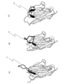
- FIG. 1 is a schematic diagram showing that these were classified into three groups based on the structure of the CDR3 region of the peptides.
- Fig. 1(A) shows an upright type
- Fig. 1(B) shows a half-roll type
- Fig. 1(C) shows a roll type.
- L1 to L3 indicate the CDR3 regions in each molecule.
- FIG. 2 shows the distance between C ⁇ atoms of the peptide (hereinafter sometimes referred to as “C ⁇ distance”) and the pseudo dihedral angle between C ⁇ atoms (hereinafter, “dihedral It is a schematic diagram showing the face angle.
- FIG. 3 is a diagram plotting the C ⁇ distance of VHH and the dihedral angle.
- FIG. 1 is a schematic diagram showing that these were classified into three groups based on the structure of the CDR3 region of the peptides.
- Fig. 1(A) shows an upright type
- Fig. 1(B) shows a half-
- FIG. 3(A) is a diagram showing the distribution of C ⁇ distances and dihedral angles by classifying VHHs into upright type, half-roll type, and roll type.
- FIG. 3(B) is a diagram showing the C ⁇ distance and dihedral angle between VHH and human VH in the same manner as above.
- FIG. 4 is a diagram comparing the amino acid sequences of the upright type and roll type shown in FIG.
- FIG. 5 is a graph showing the CDR3 length distribution of VHHs and human VHs according to the VHH types described above.
- FIG. 6 shows the design of the framework regions of the VHH sublibrary (upright or rolled). Alphabets shown below each amino acid sequence indicate the mutated amino acid and its position.
- FIG. 7 shows the procedure for constructing the DNA sequence of full-length VHHs.
- FIG. 8 is a diagram schematically showing the flow of screening when using the "The Month” platform.
- FIG. 9 shows the properties of anti-GPC3-VHH obtained by screening.
- FIG. 10 shows the properties of VHHs obtained by screening.
- FIG. 10(A) is a graph plotting the relationship between the thermal stability of the obtained VHH and the dissociation constant.
- FIG. 10(C) shows the result of actually measuring the dissociation constant.
- FIG. 10(B) is a graph showing the relationship between the denaturation midpoint temperature (Tm value) and the aggregation initiation temperature (Tagg value).
- FIG. 11 shows the results of flow cytometry analysis evaluating the binding ability of VHHs to target proteins expressed on the cell surface.
- the present invention is a peptide comprising (a) 3 CDRs and 4 FRs and (b) the specific sequences described below as FR1, FR2, FR3 and FR4.
- the peptides constituting FR1 to FR4 can be designed and produced as follows. First, the amino acid sequence of the human framework sequence IGHV3-23*01 DP-47 (FR1 to 4 are shown in Table 1 as SEQ ID NOs: 34 to 37) was used for antigenicity and homology with VHH. Selected as the basic sequence used to design the framework sequences of the invention. Then, taking into account the shape of the finally formed peptide, the FR whose design is to be changed is determined.
- the above FR1-4 are composed of 22-26 amino acids, 12-16 amino acids, 29-33 amino acids, and 9-13 amino acids, respectively, from the viewpoint of stability of the tertiary structure. Further, it is more preferable that the FR1 to FR4 are composed of 24 amino acids, 14 amino acids, 32 amino acids and 11 amino acids, respectively, for the reason of stability of the tertiary structure.
- the sequences of FR1 and FR4 are changed. Instead, the FR2 and FR3 sequences may be altered. In that case, from the viewpoint of the three-dimensional structure of CDR3, amino acids at desired sites in FR2 and FR3 can be substituted with other amino acids as described above.
- the C-terminal amino acid of FR2 of the human framework sequence IGHV3-23*01 DP-47 was changed from serine (S) to alanine (A).
- amino acids at other positions can be substituted with other amino acids.
- tyrosine (Y), valine (V), phenylalanine (F), leucine (L), arginine (R), glycine (G) and tryptophan (W) at the 2nd, 10th or 12th amino acid from the N-terminus ) may be substituted with any amino acid selected from the group consisting of
- FR2 containing the amino acid substituted at the above site can be shown as the amino acid sequence represented by SEQ ID NO: 2 below.
- SEQ ID NO: 2 WXRQAPGKGXEXVA
- the second X from the N-terminus (hereinafter referred to as "X2") is tyrosine, valine (V) and phenylalanine (F), and the tenth X (X10) is arginine (R) or leucine.
- the 12th X (X12) from the N-terminus may be any amino acid selected from the group consisting of tryptophan (W), phenylalanine (F) and glycine (G).
- the above FR2 preferably has a sequence represented by any amino acid sequence selected from the group consisting of SEQ ID NOS:3-6. This is because the three-dimensional structure of the peptide can be controlled to some extent by performing such a substitution.
- the human framework sequence IGHV3-23*01 DP-47 is involved in determining the structure of the VHH CDR3 region and is also a region that can be involved in antigen binding.
- amino acids at other positions can be substituted with other amino acids.
- the 13th, 31st or 32nd amino acids from the N-terminus, the group consisting of leucine (L), valine (V), alanine (A), asparagine (N), lysine (K), and arginine (R) may be substituted with any amino acid selected from
- FR3 containing the amino acid substituted at the above site can be shown as the amino acid sequence represented by SEQ ID NO:7.
- X13 is valine (V) and leucine (L)
- X31 is alanine (A), asparagine (N) or lysine (K)
- X32 is alanine (A), valine (V), arginine (R) and leucine
- It may be any amino acid selected from the group consisting of (L).
- the above FR3 preferably has a sequence represented by any amino acid sequence selected from the group consisting of SEQ ID NOS:8-31. This is because the three-dimensional structure of the peptide can be controlled to some extent by performing such a substitution.
- FR1 to FR4 may be chemically synthesized according to solid-phase synthesis or other conventional methods, or may be outsourced to a company.
- CDR1 to CDR3 (2-1) length of CDR1 to 3
- the peptide consists of three complementarity determining regions (hereinafter , abbreviated as “CDR”), the complementarity determining region 1 (hereinafter referred to as “CDR1”) is composed of 8 to 12 amino acids, and the complementarity determining region 2 (hereinafter referred to as “CDR2”) is composed of It may be composed of 14 to 18 amino acids, and the complementarity determining region 3 (CDR3) may be composed of 6, 12 and 15 amino acids. It is preferable that CDR1 is composed of 10 amino acids, CDR2 is composed of 16 and 17 amino acids, and CDR3 is composed of 6, 12 or 15 amino acids, in order to obtain peptides having the properties described below.
- CDR3 (2-2) Structure and Classification of CDR1-3
- the three structures of CDR3 are the C ⁇ atoms of the amino acid residues at specific positions contained in FR2 and the amino acid residues at specific positions contained in CDR3.
- the distance between the loops, the angle formed by the pseudo-diplane composed of the C ⁇ atoms of the amino acid residues at the end of the CDR3, and the structure of the loop itself are classified as follows ( Figures 1(A)-(B) and Figures 2(A)-(B)). The criteria for this classification are explained below.
- C ⁇ refers to the first carbon adjacent to the functional group. In this specification, the distance between two ⁇ -carbons is called C ⁇ -distance.
- the amino acid at the C-terminus of the CDR3 contained in the above peptide is "n"
- the amino acid residue C ⁇ (n-5) at the n-5th position and the 46th from the N-terminus of this peptide The distance to C ⁇ (46) in the amino acid residue located at is the C ⁇ distance.
- FIG. 1(E) is a diagram of the above peptide viewed from the XY plane.
- the first plane is, for example, when the 102nd amino acid residue from the N-terminus of this peptide is n (H102(n)), the peptide bond contained in H102(n) and its downstream side The plane containing the peptide bond contained in the adjacent amino acid residue (H103(n+1)).
- the second plane is the peptide bond contained in the amino acid residue (H101(n-1)) containing H101 and the amino acid residue (H100(n-2)) adjacent to the downstream side. It refers to the plane containing the peptide bonds involved.
- An angle formed by the first plane and the second plane is called a "dihedral angle".
- the shape of the CDR3 itself is, for example, one extending in the Y direction like L1 in FIG. 1(A), one in a rounded state like L3 in FIG. 1(C), It can be divided into three, such as L2 in FIG. 1(B), which is in the middle state. It should be noted that those with a CDR3 length of less than 6 amino acids are difficult to classify according to the above criteria, and may be conveniently classified as upright from the appearance of their structure.
- the upright shown in Fig. 1(A) has a C ⁇ distance longer than 15 ⁇ , a dihedral angle greater than 140 degrees, and a structure of CDR3 extended. defined as being in good condition.
- the roll shown in FIG. 1(C) is defined as having a C ⁇ distance shorter than 10 ⁇ , a dihedral angle smaller than 140 degrees, and a CDR3 structure in a rounded state.
- the half roll shown in FIG. 1(B) is defined as having an intermediate structure between the upright and the roll.
- VHH By combining the upright type or roll type FR2, FR3, CDR2 and CDR3 shown in Table 1, it is possible to prepare a VHH having a desired structure among the upright type, roll type and half roll type.
- the structures of human VHs can be classified in the same manner as for VHHs.
- the sub-library is a library prepared by selecting clones from the original DNA library under specific conditions according to the purpose of research or the like.
- Source Peptides constituting CDR1-3 can be produced by selecting target peptides.
- structural data of VHH and human VH are obtained from SAbDab database (http://opig.stats.ox.ac.uk/webapps/newsabdab/sabdab/) of Protein Data Bank (PDB).
- PDB Protein Data Bank
- selection criteria can be determined from the information on various VHHs obtained, and VHHs can be selected according to those criteria. Selection criteria for VHHs and human VHs include, for example, those shown in Table 2 below.
- the CDRs of VHHs selected according to the above criteria are determined, for example, by the Kabat method or other conventional methods. Such determinations can also be made automatically using commercially available tools, such as the ANARCI tool (http://opig.stats.ox.ac.uk/webapps/newsabdab/sabpred/anarci/). can be mentioned.
- the obtained amino acid residue information can be used to define variable regions and CDRs, and clustering is performed to group data based on the degree of similarity between variable region (domain) data.
- clustering can be performed using CD-HIT (http://weizhong-lab.ucsd.edu/cd-hit/) or other clustering programs. Redundancy can be removed from the resulting clusters by removing CDRs with 100% similarity (%id) representing perfect matches during clustering. In this specification, the operation of eliminating redundancy as described above is referred to as "curation".
- those containing an amino acid sequence with a sufficient length as an antigen-binding site can be selected. If the length of the resulting sequence is longer than the region determined by the Kabat method, such as CDR1, then the desired region prepared by the Chothia method, such as the sequence of CDR1, is combined with For example, it may be used as CDR1.
- the amino acid sequences of CDR1 and CDR2 can be designed by the following procedure.
- the data obtained by the next-generation sequencer of the naive shuffling library of camelids for example, alpaca (hereinafter sometimes abbreviated as "NGS data")
- NGS data alpaca
- the concentration ratio is not particularly limited, for example, about 0.001% or more is preferable for the reason of data reliability.
- enriched sequences amino acid sequences at the BM positions are used as an index for selecting amino acid sequence candidates for CDR1 and CDR2 from the sequences obtained by concentrating the NGS data. This is for extracting CDR1 and CDR2 of alpaca VHH, which has high structural stability.
- amino acid sequences that match the amino acid sequences at the BM positions from, for example, the VHH sequences of PDB data, and use them as candidate amino acid sequences for CDR1 and CDR2 for reasons of tertiary structural stability.
- the CDR1 and CDR2 amino acid sequence candidates selected as described above are compared with the amino acid sequence of the alpaca germline, for example, up to 2 mismatches for the CDR1 sequence and up to 3 mismatches for the CDR2 sequence. Sequence candidates can be narrowed down by setting criteria such as allowable. These can be used to prepare sets of CDR1 and CDR2.
- a library of desired lengths can be designed for CDR3 sequences based on, for example, CDR3 loop length distribution, correlation between CDR3 length and structural classification, and the like. .
- the CDR3 length may be changed between upright and roll, and may be appropriately selected to match the target peptide.
- uprights may have a CDR3 length of 6 amino acids, 12 amino acids, etc.
- rolls may have a CDR3 length of 12 amino acids, 15 amino acids, etc.
- 17 kinds of amino acids excluding amino acids such as Cys, Met, and Pro can be set to have an equal ratio. This is because these amino acids may cause heterogeneity in the process of formulation of biopharmaceuticals.
- the peptide of the present invention is a humanized camelid or other derived heavy chain antibody, it is necessary to humanize the framework portion.
- the backmutation (BM) positions of the peptide are determined based on the structural information available from databases such as those described above.
- a basic sequence is selected for humanizing the framework region.
- Such sequences can include DP-47, DP-51, DP-29, etc. of the human framework sequence IGHV3-23*01.
- the amino acid sequences of Caplacizumab, Ozoralizmab, Vobaralizmab, and other VHHs that are on the market or used in clinical trials can be referred to.
- a technique for designing a humanized framework sequence for example, the following techniques can be mentioned.
- Peptides from different sources are prepared for comparison of their amino acid sequences. Then, when these amino acid sequences are compared and the same amino acids are assigned to the same positions, it is preferable to assign them as they are to the framework sequence of the present invention because antigenicity is not increased. .
- the above four VHH amino acid sequences are selected for comparison, if the same amino acids are assigned to the same positions, they are also assigned to the same positions in the peptides of the present invention. It is preferable for the design of the humanized framework to determine the amino acids to be assigned by repeating this work.
- the amino acid to be assigned if it is not possible to determine the amino acid to be assigned, the correlation between the structural classification of the CDR3 loop described above and the sequence profile, structural knowledge, knowledge of the human framework sequence, etc. are combined to decide. is preferred.
- a DNA fragment containing each of the CDRs 1 to 3 designed as described above is synthesized by overlap extension PCR, and a library containing the desired full-length peptide can be constructed using restriction enzymes and ligase.
- Restriction enzymes used here include, for example, HphI, FokI, BsmBI, BtgZI and other Type IIS restriction enzymes (recognition and nuclease domains are separated, recognizing asymmetric nucleotide sequences, restriction enzymes that cleave sites), and ligases such as T4 DNA ligase and the like.
- FR1-4 amplification primers can be designed to contain the desired sequence, and the FR4 primer contains the linker hybridization sequence for cDNA display. design is preferred for using cDNA display methods. Desired sequences include, for example, a His tag region, 3'UTR, T7 promoter, SD sequence, 5'UTR, and the like.
- Primers for each sequence may be synthesized according to standard methods, or may be outsourced to a company. Such companies include, for example, Eurofins Genomics Inc., Sigma Aldrich, and the like. From the viewpoint of library diversity, it is preferable to synthesize a large number of CDR1 primers with different CDR1 region sequences and CDR2 primers with different CDR2 region sequences and use them in combination.
- CDR3 primers are preferably designed using trimer oligonucleotides. This is because the appearance frequency of amino acids can be controlled.
- overlap extension PCR is performed under desired conditions to synthesize DNA fragments containing each CDR.
- a reaction solution for overlap extension PCR a solution having the composition shown in Table 3 below can be used, and each synthesized DNA fragment can be purified using, for example, AMPureXP (Beckman).
- the CDR1 fragment and CDR2 fragment are treated with the above restriction enzymes, purified, and ligated together using the above ligase to obtain a ligation product (CDR1-2 fragment).
- the ligation product is purified, eg, by gel electrophoresis, and amplified using the outer primers. After purifying the amplified CDR1-2 fragment, it is treated with a restriction enzyme again.
- CDR3 is treated with restriction enzymes in the same manner as above, purified, and then ligated with the above CDR1-2 fragment with ligase.
- the resulting ligation product (CDR1-2-3 fragment) is purified in the same manner as above to prepare a library.
- the sequence of the CDR1-2-3 fragment obtained as described above is confirmed using, for example, a next-generation sequencer.
- a peptide library can be obtained as described above.
- the target peptide can be obtained by selecting the obtained peptide library by the cDNA display method using the following linker.
- cnvK Linker It is preferable from the viewpoint of reaction efficiency that the sequence of the biotin fragment that forms the main chain has the sequence shown in SEQ ID NO:33.
- BioTEG is bound to the 5' end of the main chain, and N at position 3 in the base sequence represents guanosine.
- This sequence is further bound by CCT via Amino C6-dT.
- Each N at the 12th position represents 3-cyanovinylcarbazole.
- the side chain puromycin segment can have the sequence 5'(5S)TcTCFCZZCC.
- the free terminal P in the side chain sequence represents puromycin as a protein binding site.
- (5S) represents 5' Thiol C6, c represents cytidine, F represents FITC-dT, and Z represents Spacer 18, respectively.
- the above chemical synthesis of the main chain and side chains may be entrusted to Euphofin Genomics, Tsukuba Oligo Service Co., Ltd., or the like.
- the buffer-exchanged reduced puromycin segment solution is mixed with the ethanol precipitation product of the EMCS-modified biotin fragment, and left overnight at 2-6°C. Subsequently, DTT is added to the above reaction solution to a final concentration of 25-74 mM, stirred at room temperature for 15-45 minutes, and ethanol precipitated in the same manner as above.
- the obtained ethanol precipitation product is dissolved in 50 to 150 ⁇ L of Nuclease-free water (manufactured by Nacalai Tesque, Inc.) to obtain a lysate.
- the above-described linker for producing the mRNA/cDNA-protein conjugate and the mRNA having a sequence complementary to the main chain of the linker are combined with T4 RNA at the mRNA ligation site. Ligation is performed by ligase to generate an mRNA-linker conjugate.
- a protein is synthesized from the mRNA in a cell-free translation system, and the synthesized protein is ligated to the protein ligation site in the mRNA-linker conjugate to form the mRNA-linker-protein conjugate.
- the mRNA-linker-protein conjugate is bound to the solid phase via the solid-phase binding site, and the solid phase bound with the mRNA-linker-protein conjugate is washed with the first buffer. Then, using the 3' end of the main chain as the reaction initiation point and the mRNA as a template, a reverse transcription reaction is performed to synthesize a cDNA chain to obtain an mRNA/cDNA-linker-protein conjugate. Then, the solid phase bound with the mRNA/cDNA-linker-protein conjugate is washed with the second buffer solution, and the cleavage site of the main chain is cleaved with the predetermined endoribonuclease.
- the ligation reaction is performed at 20 ⁇ L to 40 ⁇ L from the point of reaction efficiency, and the RNA:linker molar ratio is in the range of 3:1 to 1:6.
- 1:(1-2) (10 pmol to 20 pmol of linker for 10 pmol of mRNA) is recommended.
- the sample solution is warmed on an aluminum block at 90°C for 5 minutes, then transferred to an aluminum block at 70°C for 5 minutes, and then the photocrosslinking reaction between the mRNA and the linker is performed.
- the photocrosslinking reaction is carried out by irradiating UV light at 365 nm for 1 to 5 minutes in 10 mM to 250 mM Tris-HCl buffer (pH 7.0 to 8.0) containing 100 mM to 300 mM NaCl.
- E. coli-derived cell-free protein synthesis system it is preferable to use an E. coli-derived cell-free protein synthesis system as the primary cell-free translation system, and it is more preferable to use a reconstituted E. coli cell-free protein synthesis system.
- a translation reaction can be carried out by adding a reconstituted cell-free protein synthesis system of E. coli and the conjugate.
- the amount of the reconstituted cell-free protein synthesis system of Escherichia coli is about 8.5 ⁇ L to about 17 ⁇ L
- the amount of the conjugate is about 2.4 pmol to about 4 pmol
- the size of the reaction system is about 12.5 ⁇ L to about Make 50 ⁇ L and perform at about 20° C. to about 40° C. for about 10 minutes to about 30 minutes. Translation at about 37°C for about 30 minutes yields high production efficiency and working efficiency.
- the translation product protein and the mRNA-linker conjugate are treated in the presence of, for example, about 0.3 M to about 1.6 M potassium chloride and about 40 mM to about 170 mM magnesium chloride (all concentrations are final concentrations). , at about 27° C. to about 47° C. for about 30 minutes to about 1.5 hours, the protein can be efficiently bound to the conjugate.
- Examples of the solid phase on which the mRNA-linker-protein conjugate is immobilized include beads such as styrene beads, glass beads, agarose beads, sepharose beads, magnetic beads; glass substrates, silicon (quartz) substrates, plastic substrates, and metals. Substrates such as substrates (for example, gold foil substrates); containers such as glass containers and plastic containers; membranes made of materials such as nitrocellulose and polyvinylidene fluoride (PVDF); If the solid phase is composed of a plastic material such as styrene beads or a styrene substrate, a portion of the linker may be covalently bound directly to the solid phase using known techniques ( Qiagen, LiquiChip Applications Handbook, etc.). When biotin or an analogue thereof is bound to the conjugate, the conjugate can be easily bound to the solid phase by binding avidin to the solid phase.
- substrates for example, gold foil substrates
- containers such as glass containers and plastic containers
- membranes made of materials such
- Conjugates that were not bound to the solid phase were isolated from about 0.1 M to about 10 M sodium chloride, about 0.1 mM to about 10 mM EDTA, 1 mM to 100 mM Tris-HCl containing about 0.01% to about 1% detergent. Remove by washing with buffer (pH 7.0-9.0) or phosphate buffer (pH 7.0-9.0). Washing is efficient using 20 mM Tris-HCl buffer (pH 8.0) containing 2 M sodium chloride, 2 mM EDTA, 0.1% Triton X-100.
- a reverse transcription reaction is performed under predetermined conditions to synthesize a cDNA strand.
- an mRNA/cDNA-linker-protein conjugate is obtained.
- the reverse transcription reaction system can be arbitrarily selected, the above-mentioned mRNA-linker protein conjugate, dNTP Mix, DTT, reverse transcriptase, standard solution, and water from which RNase has been removed (hereinafter referred to as "RNase-free water”) are added to prepare a reaction system, and reverse transcription is preferably carried out in this system for 5 to 20 minutes at 30°C to 50°C.
- the linker of the present invention can be used to obtain not only various proteins but also cDNAs corresponding to those proteins.
- the obtained mRNA and linker DNA having puromycin are immobilized on streptavidin-modified magnetic beads using avidin-biotin binding, and the mRNA is converted into We synthesize proteins and synthesize cDNA from mRNA using reverse transcription. Therefore, the protein, which is the phenotype, and the DNA sequence information, which is the genotype, are in a stable one-to-one correspondence on the magnetic beads.
- the affinity of the obtained protein can be used to select mRNA/cDNA-linker-protein conjugates.
- a mutation is introduced into the nucleotide sequence of the selected concatenated product by PCR or the like to carry out an amplification reaction.
- the amplified product is ligated with a double-stranded DNA having a desired promoter sequence by a predetermined method to obtain a first-generation mutant mRNA (hereinafter abbreviated as "mRNA G1").
- mRNA G1 a first-generation mutant mRNA
- Example 1 Structural analysis of VHH CDR3
- the structural data of VHH and human VH were collected from the SAbDab database of the Protein Data Bank (PDB) (http://opig.stats.ox.ac.uk/webapps/newsabdab/ sabdab/).
- the VHHs obtained here were selected according to the criteria shown in Table 2 above, except for those with short CDR structures of less than 5 amino acids.
- CDR information of VHHs obtained from the database was determined by the conventional Kabat method.
- the ANARCI tool http://opig.stats.ox.ac.uk/webapps/newsabdab/sabpred/anarci/
- CD-HIT http://weizhong-lab.ucsd.edu/cd-hit/
- CDR2 H50-H65
- CDR3 H95-H102
- the actual CDR1 antigen-binding site was wider than the region (H31-H35) determined by the Kabat method, so in combination with the CDR1 (H26-H32) prepared by the Chothia method, CDR1 (H26-H35) and bottom.
- FIG. 1 (A) shows the amino acid residues in the vicinity of the antigen in black.
- the three types of loop structures of CDR3 were defined as follows. Those with a C ⁇ distance of 15 ⁇ or more and a dihedral angle of 140 degrees or more are upright type, those with a C ⁇ distance of 10 ⁇ or more and a dihedral angle of 140 degrees or less are roll types, and those with a C ⁇ distance of 15 ⁇ or more and If the dihedral angle was smaller than 140 degrees, or if the C ⁇ distance was larger than 10 ⁇ and the dihedral angle was smaller than 140 degrees, the half-roll type was used.
- FIG. 4 shows the relationship between CDR3 length and its taxonomic type.
- the upright CDR3 tends to include a framework region in the antigen-binding site.
- the role type increases and the inclusion of the framework region in the antigen-binding site decreases.
- the roll structure was stabilized by bringing the framework and CDR3 regions closer together.
- the proportion of CDR3 containing cysteine increases, and the cysteine forms a disulfide bond with the cysteine of CDR2, thereby forming a more stable CDR3.
- the structure of human VH was also classified in the same way as VHH.
- a two-dimensional plot of the C ⁇ distance and the dihedral angle is shown in FIG. 3(B).
- Most of the human VHs were of the half-roll type, with no roll type, and almost no upright type. This suggests that in human antibodies, the presence of the light chain is a steric hindrance and prevents the CDR3 from folding into the above-mentioned roll-type structure.
- Example 2 Construction of an artificial VHH library (2-1) Library design based on analysis data Based on the analysis results of the antigen-binding site, the number of amino acid residues, and the structure of CDR3 described above, Upright6, Upright12, Roll12, and We designed a rational artificial VHH library containing four sub-libraries called Roll15.
- CDR2 of the upright sub-library was composed of 16 amino acid residues
- CDR2 of the roll-type sub-library was composed of 17 amino acid residues. This is because a strong correlation has been confirmed between the number of amino acid residues (16 amino acid residues or 17 amino acid residues) constituting CDR2 and the formation of upright or roll type.
- the amino acid sequences of the CDRs consisted of 150 types of sequence sets for CDR1, 69 or 71 types of sequence sets for CDR2, and 17 types of amino acids excluding methionine, proline and cysteine for CDR3. Designed to be structured. CDR3s longer than 16 amino acids were not designed. This is because the formation of disulfide bonds is undesirable from the pharmaceutical standpoint. Finally, the CDRs 1 to 3 designed as described above were incorporated into the humanized FR as a set.
- VHH framework regions and CDRs contained in this library were defined as follows. FR1(H1-H25), CDR1(H26-H35), FR2(H36-H49), CDR2(H50-H65), FR3(H66-H94), CDR3(H95-H102), FR4(H103-H113). Based on the above definitions, we then designed the framework.
- the undetermined amino acid sequence was determined by combining the structural classification of the CDR3 loop, the correlation with the sequence profile, the structural knowledge, the knowledge of the human framework sequence, etc. (Fig. 6). Additionally, some amino acids were further edited during this humanization process to reduce the frequency of unfavorable amino acids in pharmaceuticals.
- the FR4 primer was designed to contain a His tag region and 3'UTR (including the linker hybridization sequence for cDNA display).
- Primers corresponding to FR1, CDR1, and CDR2 were commissioned to Sigma Aldrich for synthesis.
- the FR1 primer was designed to contain a 5'UTR (T7 promoter and SD sequence).
- Synthesize 150 types of CDR1 primers with different sequences in the CDR1 region, and 140 types of CDR2 primers with different sequences in the CDR2 region (Upright type: 71 types, Roll type: 69 types), and mix these primers. and used.
- Primers corresponding to CDR3 of each library were designed using trimer oligonucleotides to control the frequency of amino acid appearance, and synthesis was commissioned to Ella Biotech.
- the CDR1 fragment and CDR2 fragment were treated with restriction enzymes using BsmBI (NEB) at 55°C for 15 minutes.
- BsmBI BsmBI
- These restriction enzyme-treated DNA fragments were purified according to a conventional method and ligated at 16°C for 16 hours using T4 DNA ligase (Takara Bio Inc.) to obtain the CDR1-CDR2 fragment, which is a ligation product. Synthesized as a ligation product.
- This ligation product was electrophoresed on an 8 M urea PAGE-gel at 200 V for 40 minutes to purify the region of the target product, followed by 10 cycles of PCR amplification using outer primers at an annealing temperature of 62°C.
- the amplified CDR1-2 fragment was purified and then restricted with BtgZ I (NEB) at 60°C for 15 minutes.
- the CDR3 fragment was digested with BsmBI at 55°C for 15 minutes with a restriction enzyme and purified using AMPure XP to obtain a purified CDR3 fragment.
- the purified CDR1-2 fragment and the purified CDR3 fragment were ligated with T4 DNA ligase at 16°C for 16 hours to obtain a full-length VHH sequence.
- the ligation product was recovered by the 8 M urea-denaturing PAGE-gel purification described above, and a library was constructed.
- PCR amplification was performed for each of the above libraries.
- the composition of the reaction solution (25 ⁇ L total) was 12.5 ⁇ L PrimeSTAR MAX PreMIX (2 ⁇ ), 1 ⁇ L library sample, 0.5 ⁇ L each 20 ⁇ M forward/reverse primers (PL_prRd-N4_NL_FW, PL_prRd-N4_NL_RV), and 10.5 ⁇ L of ultrapure water.
- the reaction conditions were the same as in Table 3 above, except that the annealing temperature was 62°C, the extension time was 5 seconds, and the number of cycles for denaturation, etc. was 8 times.
- the resulting PCR product was purified using AMPureXP, and Index PCR was performed using the purified product as a template (the reaction conditions were the same as the above PCR conditions, except that the annealing temperature was changed to 52°C).
- the composition of the reaction solution (25 ⁇ L total) was 12.5 ⁇ L PrimeSTAR MAX PreMIX (2 ⁇ ), 1 ⁇ L PCR product, 2 ⁇ L each 5 ⁇ M forward/reverse primers (Nextera XT Index 1 Primers (N7XX), Nextera XT Index 1 Primers (S5XX)), 7.5 ⁇ L of ultrapure water.
- Index PCR products were purified using AMPure as above and DNA concentrations were measured using NanoPad DS-11. Then, after diluting the Index PCR purified products to a concentration of 10 nM, 5 ⁇ L of each product was collected and mixed in one tube.
- a PhiX library which is a control DNA library, was also prepared in the same manner as a 15 pM library. Finally, 570 ⁇ L of the 15 pM sample library and 30 ⁇ L of the 15 pM PhiX library were mixed, the final NGS sample library was loaded onto the reagent cartridge, the reagent cartridge was set on Miseq, and analysis was performed. Then, by analyzing the obtained data, it was confirmed that each library had the nucleotide sequence as designed.
- cnvK linkers for selection were prepared as follows.
- (1) Preparation of cnvK rC Linker The sequence of the biotin fragment that forms the main chain was the sequence shown in SEQ ID NO: 33 below.
- BioTEG is bound to the 5' end of the biotin fragment (main chain), and N at position 3 in the nucleotide sequence is guanosine, and N at position 12 is 3-cyanovinylcarbazole. respectively.
- CCT was bound to the 3' end of SEQ ID NO: 33 below via Amino C6-dT.
- the side chain puromycin segment has a 5' (5S) TcTCFCZZCCP sequence.
- the free terminal P in the side chain sequence represents puromycin as a protein binding site.
- (5S) represents 5' Thiol C6, c represents cytidine, F represents FITC-dT, and Z represents Spacer 18, respectively.
- the buffer-exchanged reduced puromycin segment solution was mixed with the ethanol precipitation product of the EMCS-modified biotin fragment, and left overnight at 4°C. Subsequently, DTT was added to the reaction solution so that the final concentration was 50 mM, and the mixture was stirred at room temperature for 30 minutes. Thereafter, ethanol precipitation was performed using Quick-Precip Plus Solution (manufactured by Edge BioSystems). The ethanol precipitation product was dissolved in 100 ⁇ L of Nuclease-free water (manufactured by Nacalai Tesque, Inc.).
- the lysate was separated by 12% polyacrylamide gel electrophoresis, and each portion of the cnvK rC Linker was excised.
- the excised gel was crushed using Biomasher II set (Nippi), 500 ⁇ L of Nuclease-free water was added, and the mixture was stirred overnight at 4°C to extract the cnvK rC Linker.
- the stirred solution was transferred to a Costar® Spin-X® centrifugal tube filter, 0.22 ⁇ m cellulose acetate (corning), and centrifuged at 16,000 ⁇ g for 15 minutes to separate the gel and the extract. Thereafter, ethanol precipitation was performed using Quick-Precip Plus Solution to obtain the desired cnvK rC Linker.
- the obtained cnvK rC Linker was dissolved in Nuclease-free water and stored at -20°C.
- VHHs that bind to various antigens can be obtained from the constructed PharmaLogical Library were verified by performing in vitro selection.
- the antigens used as targets this time are HSA (Human Serum Albumin), HER2 (Human Epidermal growth factor receptor 2), GPC3 (Glypican 3), and GPC1 (Glypican 1).
- the selection library used was a mixture of the above-described four types of sub-libraries. Three rounds (four rounds for GPC1) of selection were performed with a series of operations consisting of synthesis of mRNA-linker conjugate, synthesis of cDNA display molecule, in vitro selection, and PCR amplification as one round (FIG. 7).
- RNA concentration was measured using NanoPad DS-11, and the reagents were mixed so that the final concentration was 200 mM NaCl, 50 mM Tris-HCl (pH 7.5), 1 ⁇ M mRNA, and 1 ⁇ M cnvK linker, (s1 ) 90°C for 1 minute, (s2) 70°C for 1 minute, and (s3) 4°C for heat treatment to hybridize the cDNA display synthesis linker to the mRNA.
- the hybridization product of the mRNA and the linker was irradiated with UV at 365 nm using UVP closslinker cl-3000 so that the total energy amount was 406 mJ/cm 2 , thereby photocrosslinking the mRNA and the linker. to create an mRNA-linker conjugate.
- the composition of the reaction solution was 1,600 ⁇ L of Solution I, 160 ⁇ L of each of Solution II and Solution III, 800 ⁇ L of 1 pmol/ ⁇ L Ligation product, 160 ⁇ L of 4-fold diluted DsbC set, 160 ⁇ L of 20 mM GSSG, 160 ⁇ L of ⁇ L of 80 mM GSH was used.
- 1,920 ⁇ L of IVV formation Buffer was added, followed by reaction at 37°C for 30 minutes to covalently bond the translation product to the linker via puromycin.
- 1,280 ⁇ L of 0.5 M EDTA was added and reacted at 4° C. for 10 minutes to release ribosomes from nucleic acids.
- IVV product 6400 ⁇ L of IVV product (mRNA-linker-VHH conjugate) was obtained.
- the resulting IVV product was purified using 3,840 ⁇ L of Dynabeads Streptavidin MyOne C1 (manufactured by Thermo Scientific) according to the attached protocol.
- a reverse transcription reaction solution consisting of 1,280 ⁇ L of 5 ⁇ RT Buffer, 256 ⁇ L of 25 mM dNTPs, 128 ⁇ L of Reverse Transcriptase, and 4736 ⁇ L of ultrapure water was added to the IVV product-bound beads, and allowed to react at 42°C for 1 hour. to synthesize a cDNA display product (cDNA-mRNA-linker-peptide conjugate).
- the cDNA display product obtained as described above was eluted from the beads with 640 ⁇ L of elution buffer containing RNaseT1 (Thermo Scientific), and then cDNA was extracted using 200 ⁇ L of His-Mag Sepharose (Cytiva). The display product was His-tag purified to obtain 200 ⁇ L of cDNA display molecule (cDNA-linker-peptide conjugate).
- the immobilized beads (cDNA display-biotinylated target protein streptavidin-beads) were washed three times with 200 ⁇ L of PBS-T (the tube was exchanged for the third wash). After that, it was eluted twice with 100 ⁇ L of elution buffer, and only the nucleic acid region of the cDNA display molecule that showed binding ability to the target protein was recovered. The resulting eluted samples were purified using AMPure XP.
- PCR Amplification of Selected Sample DNA eluted in the selection was amplified by PCR using PrimeSTAR MAX (Takara Bio Inc.). Quantitative PCR was not performed in the 1st round, and PCR was performed using the entire amount of the eluted sample as a template.
- the composition of the reaction solution (250 ⁇ L total) was 125 ⁇ L PrimeSTAR MAX PreMIX (2 ⁇ ), 100 ⁇ L eluted DNA sample, 5 ⁇ L each 20 ⁇ M forward/reverse primer (PL_T7pro, NewYtag_cnvk-linker), 15 ⁇ L It was made into ultrapure water.
- the PCR program was the same as in Table 3 except that the annealing temperature was 60°C, the extension time was 5 seconds, and the number of cycles of denaturation, etc. was 15 times. PCR products were purified using AMPure XP.
- the composition of the solution used for quantitative PCR was 12.5 ⁇ L THUNDERBIRD qPCR Mix (Toyobo Co., Ltd.), 0.5 ⁇ L each of 20 ⁇ M forward/reverse primer (PL_T7pro, NewYtag_cnvk-linker), 1 ⁇ L of eluted Sample DNA and 10.5 ⁇ L of ultrapure water were used.
- the PCR program consisted of 35 cycles of initial denaturation at 98°C for 30 seconds (denaturation at 98°C for 5 seconds, annealing at 60°C for 10 seconds, extension at 72°C for 35 seconds).
- the composition of the reaction solution (100 ⁇ L total) was 50 ⁇ L PrimeSTAR MAX PreMIX (2 ⁇ ), 25 ⁇ L eluted DNA sample, 2 ⁇ L each 20 ⁇ M forward/reverse primer (PL_T7pro, NewYtag_cnvk-linker), 21 ⁇ L ultrapure water.
- the program was the same as the program shown in Table 3, except that the annealing temperature was 65°C, the extension time was 5 seconds, and the number of cycles for denaturation, etc. was 15 cycles.
- Example 4 Analysis of selection products by NGS (4-1) Preparation of sequence samples
- the reaction mixture (25 ⁇ L total) consisted of 12.5 ⁇ L PrimeSTAR MAX PreMIX (2 ⁇ ), 1 ⁇ L selection product, 0.5 ⁇ L each 20 ⁇ M forward/reverse primers (PL_prRd-N4_NL_FW, PL_prRd-N4_Ytag_RV), 10.5 ⁇ L It was made into ultrapure water.
- the PCR program was the same as in Table 3 except that the annealing temperature was 62°C, the extension time was 5 seconds, and the number of cycles for denaturation, etc. was 8 times.
- the obtained PCR product was purified using AMPure XP, and index PCR was performed using the purified product as a template.
- the composition of the reaction (25 ⁇ L total) was 12.5 ⁇ L PrimeSTAR MAX PreMIX (2 ⁇ ), 1 ⁇ L selection product, 0.5 ⁇ L each 5 ⁇ M forward/reverse primers (Nextera XT Index 1 Primers (N7XX), Nextera XT Index 2 Primers (S5XX)), 10.5 ⁇ L of ultrapure water.
- the PCR program was the same as in Table 3 except that the annealing temperature was 52°C, the extension time was 5 seconds, and the number of cycles for denaturation, etc. was 8 times.
- Index PCR products were similarly purified using AMPure XP and DNA concentrations were measured using NanoPad DS-11. Then, after diluting the Index PCR purified product to 10 nM, 5 ⁇ L of each product was collected and mixed in one tube.
- a 15 pM library was also prepared for the PhiX library, which is a control DNA library, in the same manner. Finally, 570 ⁇ L of 15 pM sample library and 30 ⁇ L of 15 pM PhiX library were mixed to make the final NGS sample library. The final NGS sample library was loaded onto the reagent cartridge, and the reagent cartridge was set on Miseq for analysis. The obtained NGS analysis data were processed to identify binding sequences for each target molecule.
- VHH primary binding evaluation (Example 5) Preparation of VHH expression plasmid library
- the VHH-encoding DNA library selected by the in vitro selection described above was cloned into a plasmid vector for VHH expression.
- a sequence for restriction enzyme treatment was added by PCR to a DNA library obtained by in vitro selection to obtain a PCR product.
- This PCR product and the C. glutamicum expression plasmid vector were treated with the restriction enzyme BamHI at 37°C for 1 hour, and then treated with the restriction enzyme SfiI at 50°C for 1 hour.
- a VHH DNA product to serve as an insert was purified with AMPure XP, and the resulting Vector DNA product was subjected to agarose gel electrophoresis at 100 V for 30 minutes for excision and purification.
- the Vector DNA purified as described above was dephosphorylated at 37°C for 1 hour using the dephosphorylation enzyme FastAP Thermosensitive Alkaline Phosphatase (manufactured by Thermo Scientific). After that, insert DNA and vector DNA were mixed at a molar ratio of 1:10, and ligation reaction was performed overnight at 16°C using Ligation high (Toyobo Co., Ltd.) to introduce the selected VHH library. A plasmid library was obtained.
- Baseline step Measurement for 30 seconds in PBS-T.
- Loading step Measurement for 100 seconds with VHH diluted 50-fold with PBS-T.
- Baseline step Measurement for 30 seconds in PBS-T.
- Association step Measurement for 100 seconds with each target molecule prepared by diluting with PBS-T.
- Example 6 Characterization of VHH clones in vitro (6-1) Obtainment of purified VHH Unique clones identified by the above sequence analysis were precultured overnight at 30°C. Thereafter, this preculture was subcultured in PM1S medium for VHH expression, cultured at 25° C. for 72 hours, and VHH (monomer) was secreted and expressed in the culture supernatant. The culture supernatant was collected by centrifugation to remove the cells from the supernatant.
- Baseline step Measurement for 30 seconds in PBS-T.
- Loading step Measurement for 120 seconds with VHH diluted 50-fold with PBS-T.
- Baseline step Measurement for 30 seconds in PBS-T.
- Association step Measurement for 120 seconds with each target molecule prepared by diluting with PBS-T.
- the denaturation midpoint temperature (Tm value) was calculated based on the decrease in fluorescence intensity from about 330 nm to 350 nm (due to heat denaturation of the protein) and the change in fluorescence spectrum.
- the aggregation initiation temperature (Tagg value) was calculated by measuring the increase in the average molecular weight (aggregation) of the solute due to heat and measuring the change in the intensity of the scattered light at 266 nm (Figs. 9 and 10(B)).
- Example 6 Evaluation of VHH binding ability by flow cytometry In vitro VHH characterization was performed in Example 5 above. However, in vitro binding capacity does not necessarily translate into in vivo binding capacity. Therefore, in order to more accurately evaluate the binding ability of VHHs, the binding ability of VHHs to target proteins (GPC3, HER2) expressed in cells was verified by flow cell cytometry (FCM).
- FCM flow cell cytometry
- hepG2 a hepatocellular carcinoma
- GPC3-expressing cell a hepatocellular carcinoma
- SK-BR-3 a breast cancer cell, as the HER2-expressing cell.
- These cells were cultured (37° C., in a 5% CO 2 incubator, protected from light) until 80% confluency, and harvested with PBS containing 5 mM EDTA.
- the recovered cell sample was washed with 1% BSA/PBS, added with a VHH solution diluted 100-fold with 1% BSA/PBS, and incubated at 4°C for 1 hour.
- the invention of the present application is useful in the field of drug discovery.
- SEQ ID NO: 1 shows the amino acid sequence of FR1.
- SEQ ID NO: 2 shows the amino acid sequence of FR2.
- SEQ ID NO: 3 shows the amino acid sequence of FR2 (Upright type).
- SEQ ID NO: 4 shows the amino acid sequence of FR2 (Upright type).
- SEQ ID NO: 5 shows the amino acid sequence of FR2 (Roll type).
- SEQ ID NO: 6 shows the amino acid sequence of FR2 (Roll type).
- SEQ ID NO: 7 shows the amino acid sequence of FR3 (Upright type).
- SEQ ID NO: 8 shows the amino acid sequence of FR3 (Upright type).
- SEQ ID NO: 9 shows the amino acid sequence of FR3 (Upright type).
- SEQ ID NO: 10 shows the amino acid sequence of FR3 (Upright type).
- SEQ ID NO: 11 shows the amino acid sequence of FR3 (Upright type).
- SEQ ID NO: 12 shows the amino acid sequence of FR3 (Upright type).
- SEQ ID NO: 13 shows the amino acid sequence of FR3 (Upright type).
- SEQ ID NO: 14 shows the amino acid sequence of FR3 (Upright type).
- SEQ ID NO: 16 shows the amino acid sequence of FR3 (Upright type).
- SEQ ID NO: 17 shows the amino acid sequence of FR3 (Upright type).
- SEQ ID NO: 18 shows the amino acid sequence of FR3 (Upright type).
- SEQ ID NO: 19 shows the amino acid sequence of FR3 (Upright type).
- SEQ ID NO: 20 shows the amino acid sequence of FR3 (Upright type).
- SEQ ID NO: 21 shows the amino acid sequence of FR3 (Upright type).
- SEQ ID NO: 22 shows the amino acid sequence of FR3 (Upright type).
- SEQ ID NO: 23 shows the amino acid sequence of FR3 (Upright type).
- SEQ ID NO: 25 shows the amino acid sequence of FR3 (Upright type).
- SEQ ID NO: 26 shows the amino acid sequence of FR3 (Upright type).
- SEQ ID NO: 27 shows the amino acid sequence of FR3 (Upright type).
- SEQ ID NO: 28 shows the amino acid sequence of FR3 (Upright type).
- SEQ ID NO: 29 shows the amino acid sequence of FR3 (Upright type).
- SEQ ID NO: 30 shows the amino acid sequence of FR3 (Roll/Upright type).
- SEQ ID NO: 31 shows the amino acid sequence of FR3 (Roll/Upright type).
- SEQ ID NO: 32 Shows the amino acid sequence of FR4.
- SEQ ID NO:33 shows the nucleotide sequence of the backbone of the cnvK rC linker
- SEQ ID NO:34 shows the amino acid sequence of FR1 of DP-47.
- SEQ ID NO: 35 Shows the amino acid sequence of FR2 of DP-47.
- SEQ ID NO: 36 Shows the amino acid sequence of FR3 of DP-47.
- SEQ ID NO: 37 Shows the amino acid sequence of FR4 of DP-47.
Abstract
Provided is a peptide composed of three complementarity determining regions and four framework regions. A framework 1 includes an amino acid sequence represented by SEQ ID NO: 1, a framework 2 includes an amino acid sequence represented by SEQ ID NO: 2, a framework 3 includes an amino acid sequence represented by SEQ ID NO: 7, and a framework 4 includes an amino acid sequence represented by SEQ ID NO: 32. A complementarity determining region 1 is an amino acid sequence composed of 10 amino acids, a complementarity determining region 2 is an amino acid sequence composed of 16 or 17 amino acids, and a complementarity determining region 3 is an amino acid sequence composed of 6, 12, or 15 amino acids. Also provided is a peptide library including said peptide.
Description
本発明は、ランダム領域搭載用フレームワーク配列を有するペプチド及びそのペプチドで構成されるペプチドライブラリに関する。
The present invention relates to peptides having framework sequences for loading random regions and peptide libraries composed of the peptides.
従来、生体には、自己と非自己とを認識する生体防御機構があること、そして、この生体防御機構には、細胞性免疫と液性免疫とが関わっていることが知られている。上記のうち、液性免疫では、B細胞が産生する抗体が、体内に侵入してきた異物を認識し、その異物上の表面に存在する抗原(標的)に特異的に結合する。そして、その異物を生体内から除去する。
Conventionally, it is known that the body has a biological defense mechanism that recognizes self and non-self, and that this biological defense mechanism involves cell-mediated immunity and humoral immunity. Among the above, in humoral immunity, antibodies produced by B cells recognize foreign substances that have invaded the body and specifically bind to antigens (targets) present on the surface of the foreign substances. Then, the foreign matter is removed from the living body.
上記抗体は、「免疫グロブリン」(以下、「Ig」ということがある。)と呼ばれるタンパク質であり、IgA、IgD、IgE、IgG、及びIgMに分けられている。これらのうち、IgGは産生量の多さから一般的な抗体と言われ、多くの哺乳類では重鎖と軽鎖とから構成されている。しかい、ラクダ、アルパカその他のラクダ科の動物又はサメその他の軟骨魚では重鎖のみで構成されている。こうした重鎖のみの単一ドメインで構成される抗体は重鎖抗体と呼ばれ、可変領域のみで抗原と結合できることから、シングルドメイン抗体、ナノボディ(登録商標)、またはVHHとも呼ばれている。
The above antibody is a protein called "immunoglobulin" (hereinafter sometimes referred to as "Ig"), and is divided into IgA, IgD, IgE, IgG, and IgM. Among these, IgG is said to be a general antibody because of its high production amount, and is composed of heavy and light chains in many mammals. However, in camels, alpacas and other camelid animals, or sharks and other cartilaginous fish, it is composed only of heavy chains. Such an antibody composed of a single domain consisting of only a heavy chain is called a heavy chain antibody, and is also called a single domain antibody, Nanobody (registered trademark), or VHH because it can bind to an antigen only with the variable region.
上記VHHは、それぞれのアミノ酸配列が厳密に定義されており、IgG等の従来型の抗体と同様に、免疫沈降法などの生化学的解析用ツールとして、または免疫組織化学で使用するツールとして利用することができる。
Each VHH has a strictly defined amino acid sequence and can be used as a tool for biochemical analysis such as immunoprecipitation or as a tool for immunohistochemistry, similar to conventional antibodies such as IgG. can do.
上記VHHは上記の従来型の抗体とは抗原との結合部位が異なることが知られている。具体的には、従来型の抗体は抗原の凸部のエピトープに結合するが、上記VHHは抗原の隙間又は割れ目等を形成しているエピトープに結合することが知られている(先行技術文献1~3参照。)。
このため、従来型の抗体では開発が困難な治療薬を開発する上では、上記ラクダ科動物から得られるVHHの結合特性を有する候補タンパク質の探索が必要である。 The VHH is known to have a different antigen-binding site from the conventional antibody. Specifically, conventional antibodies bind to convex epitopes of antigens, but the VHHs are known to bind to epitopes forming gaps or crevices in antigens (Prior Art Document 1 See ~3.).
Therefore, in order to develop therapeutic agents that are difficult to develop with conventional antibodies, it is necessary to search for candidate proteins having VHH-binding properties obtained from the above camelids.
このため、従来型の抗体では開発が困難な治療薬を開発する上では、上記ラクダ科動物から得られるVHHの結合特性を有する候補タンパク質の探索が必要である。 The VHH is known to have a different antigen-binding site from the conventional antibody. Specifically, conventional antibodies bind to convex epitopes of antigens, but the VHHs are known to bind to epitopes forming gaps or crevices in antigens (
Therefore, in order to develop therapeutic agents that are difficult to develop with conventional antibodies, it is necessary to search for candidate proteins having VHH-binding properties obtained from the above camelids.
上記のような探索は、ペプチドライブラリを、ファージディスプレイ法等のディスプレイ法等を用いてスクリーニングすることが一般的である(先行技術文献1参照)。ここで、ペプチドライブラリとは、「アミノ酸の系統的な組み合わせを有するペプチドの大きなコレクション」と定義することができる。上記ペプチドは、種々の動物等から得られたナイーブペプチド、それかを改変した半合成ペプチド、及び化学合成によって得られた合成ペプチドを含む。
The search as described above generally involves screening a peptide library using a display method such as a phage display method (see Prior Art Document 1). Here, a peptide library can be defined as "a large collection of peptides with systematic combinations of amino acids". The above-mentioned peptides include naive peptides obtained from various animals and the like, semi-synthetic peptides modified from naive peptides, and synthetic peptides obtained by chemical synthesis.
そして、上記合成ペプチドライブラリをスクリーニングすることによって、エピトープペプチドマッピング、ワクチン開発用のペプチドライブラリの作製などをおこなうことができることが知られている。
It is also known that epitope peptide mapping, creation of a peptide library for vaccine development, etc. can be performed by screening the synthetic peptide library.
探索によって多様な候補ペプチドを得るためには、ダイバーシティの大きなペプチドライブラリが必要である。そして、従来技術1(mRNAディスプレイ法)は、ライブラリサイズという点では優れた技術である。しかし、mRNAディスプレイ法では、mRNAは水溶液中ではRNase分解されやすいため、目的のペプチドが安定して得られないという問題があった。
In order to obtain diverse candidate peptides by searching, a peptide library with a large diversity is necessary. Conventional technique 1 (mRNA display method) is an excellent technique in terms of library size. However, the mRNA display method has a problem that the target peptide cannot be stably obtained because the mRNA is easily degraded by RNase in an aqueous solution.
従来技術2(ファージディスプレイ法)は、ディスプレイの安定性という点では優れた技術である。しかし、ファージにディスプレイさせたペプチドを大量に取得するためには、上記ディスプレイされたペプチドをコードするDNAを発現するコンストラクトを作製し、これを最終的には大腸菌その他の菌に導入して発現させる必要があり、細胞を用いた翻訳システムを必要とする。
このため、扱うことができるライブラリサイズが小さく、また、そのペプチドの発現が、宿主にとって有害又は致死的である場合には、ディスプレイ出来ないという問題点があった。 Conventional technique 2 (phage display method) is an excellent technique in terms of display stability. However, in order to obtain a large amount of a peptide displayed on a phage, a construct that expresses the DNA encoding the displayed peptide is prepared, and finally introduced into Escherichia coli or other bacteria for expression. It requires a cell-based translation system.
For this reason, there is a problem that the size of the library that can be handled is small, and if the expression of the peptide is harmful or lethal to the host, it cannot be displayed.
また、タンパク質又はペプチドの三次構造は、二次構造からある程度の予測はできるものの、標的ペプチドとの結合様式を予測し、そうした構造が結合部位となるように設計をし、その通りに発現させることができないという問題点があった。
In addition, although the tertiary structure of a protein or peptide can be predicted to some extent from the secondary structure, it is necessary to predict the binding mode with the target peptide, design such a structure as a binding site, and express it accordingly. There was a problem that it was not possible to
このため、所望の長さのCDRを搭載することができる配列を有するフレームワークに対する強い社会的要請があった。特に、ラクダ科動物から得られたVHH等のシングルドメイン抗体の効率の良いスクリーニングに供することができ、このスクリーニングで設計通りの構造的特性を有するペプチドを得ることができるようなフレームワーク配列に対する強い社会的要請があった。
さらに、上記のようなフレームワークを有するペプチドで構成されるペプチドライブラリについての強い社会的要請があった。 For this reason, there has been a strong social demand for frameworks with sequences capable of mounting CDRs of desired length. In particular, it is highly sensitive to framework sequences such that it can be subjected to efficient screening of single domain antibodies, such as VHHs from camelids, and this screening yields peptides with the designed structural properties. There was a social demand.
Furthermore, there has been a strong social demand for a peptide library composed of peptides having the above frameworks.
こうした状況の下で、本発明の発明者らは、鋭意研究を重ねて本願発明を完成したものである。
すなわち、本発明の第1の態様は、3つの相補性決定領域と4つのフレームワーク領域(以下、「FR」と略すことがある。)とで構成され、配列番号1で表される配列をフレームワーク1(以下、「FR1」という。)として含むペプチドである。 Under these circumstances, the inventors of the present invention completed the present invention through extensive research.
That is, in the first aspect of the present invention, the sequence represented by SEQ ID NO: 1, which is composed of three complementarity determining regions and four framework regions (hereinafter sometimes abbreviated as "FR"), is It is a peptide containing as framework 1 (hereinafter referred to as "FR1").
[配列番号1] EVQLVESGGGLVQPGGSLRLSCAAS
[SEQ ID NO: 1] EVQLVESGGGLVQPGGSLRLSCAAS
前記FRは、配列番号2で表されるアミノ酸配列をフレームワーク2(以下、「FR2」という。)として含むことが好ましい。ここで、配列番号2中のXは任意のアミノ酸を表し、チロシン、バリン、フェニルアラニン、ロイシン、アルギニン、グリシン及びトリプトファンからなる群から選ばれるいずれかのアミノ酸であることが三次構造の安定性の点から好ましい。
[配列番号2] WXRQAPGKGXEXVA The FR preferably contains the amino acid sequence represented by SEQ ID NO: 2 as framework 2 (hereinafter referred to as "FR2"). Here, X in SEQ ID NO: 2 represents an arbitrary amino acid, and is any amino acid selected from the group consisting of tyrosine, valine, phenylalanine, leucine, arginine, glycine and tryptophan from the viewpoint of tertiary structure stability. preferred from
[SEQ ID NO: 2] WXRQAPGKGXEXVA
また、前記FR2は、配列番号3~6で表されるアミノ酸配列からなる群から選ばれるいずれかの配列であることが三次構造の安定性の点からより好ましい。
In addition, the FR2 is more preferably any sequence selected from the group consisting of the amino acid sequences represented by SEQ ID NOs: 3 to 6 from the viewpoint of stability of the tertiary structure.
[配列番号3] WYRQAPGKGLEWVA
[配列番号4] WVRQAPGKGLEWVA
[配列番号5] WFRQAPGKGREFVA
[配列番号6] WFRQAPGKGREGVA [SEQ ID NO: 3] WYRQAPGKGLEWVA
[SEQ ID NO: 4] WVRQAPGKGLEWVA
[SEQ ID NO: 5] WFRQAPGKGREFVA
[SEQ ID NO: 6] WFRQAPGKGREGVA
[配列番号4] WVRQAPGKGLEWVA
[配列番号5] WFRQAPGKGREFVA
[配列番号6] WFRQAPGKGREGVA [SEQ ID NO: 3] WYRQAPGKGLEWVA
[SEQ ID NO: 4] WVRQAPGKGLEWVA
[SEQ ID NO: 5] WFRQAPGKGREFVA
[SEQ ID NO: 6] WFRQAPGKGREGVA
前記FRは、配列番号7で表されるアミノ酸配列をフレームワーク3(以下、「FR3」という。)として含むことが好ましい。ここで、配列番号7中のXは任意のアミノ酸を表し、ロイシン、バリン、アラニン(A)、アルギニン(R)、及びリジン(K)からなる群から選ばれるいずれかのアミノ酸であることが好ましい。
The FR preferably contains the amino acid sequence represented by SEQ ID NO: 7 as framework 3 (hereinafter referred to as "FR3"). Here, X in SEQ ID NO: 7 represents any amino acid, preferably any amino acid selected from the group consisting of leucine, valine, alanine (A), arginine (R), and lysine (K). .
[配列番号7] RFTISRDNSKNTXYLQMNSLRAEDTAVYYCXX
[SEQ ID NO: 7] RFTISRDNSKNTXYLQMNSLRAEDTAVYYCXX
前記フレームワーク配列3(以下、「FR3」という。)は、下記の配列番号8~31で表されるアミノ酸配列からなる群から選ばれるいずれかの配列であることが好ましい。
The framework sequence 3 (hereinafter referred to as "FR3") is preferably any sequence selected from the group consisting of the amino acid sequences represented by SEQ ID NOs: 8 to 31 below.
[配列番号8] RFTISRDNAKNTLYLQMNSLRAEDTAVYYCNA
[配列番号9] RFTISRDNAKNTLYLQMNSLRAEDTAVYYCNV
[配列番号10] RFTISRDNAKNTLYLQMNSLRAEDTAVYYCNR
[配列番号11] RFTISRDNAKNTLYLQMNSLRAEDTAVYYCNL
[配列番号12] RFTISRDNAKNTVYLQMNSLRAEDTAVYYCNA
[配列番号13] RFTISRDNAKNTVYLQMNSLRAEDTAVYYCNV
[配列番号14] RFTISRDNAKNTVYLQMNSLRAEDTAVYYCNR
[配列番号15] RFTISRDNAKNTVYLQMNSLRAEDTAVYYCNL
[配列番号16] RFTISRDNAKNTLYLQMNSLRAEDTAVYYCAV
[配列番号17] RFTISRDNAKNTLYLQMNSLRAEDTAVYYCAR
[配列番号18] RFTISRDNAKNTLYLQMNSLRAEDTAVYYCAL
[配列番号19] RFTISRDNAKNTVYLQMNSLRAEDTAVYYCAV
[配列番号20] RFTISRDNAKNTVYLQMNSLRAEDTAVYYCAR
[配列番号21] RFTISRDNAKNTVYLQMNSLRAEDTAVYYCAL
[配列番号22] RFTISRDNAKNTLYLQMNSLRAEDTAVYYCKA
[配列番号23] RFTISRDNAKNTLYLQMNSLRAEDTAVYYCKV
[配列番号24] RFTISRDNAKNTLYLQMNSLRAEDTAVYYCKR
[配列番号25] RFTISRDNAKNTLYLQMNSLRAEDTAVYYCKL
[配列番号26] RFTISRDNAKNTVYLQMNSLRAEDTAVYYCKA
[配列番号27] RFTISRDNAKNTVYLQMNSLRAEDTAVYYCKV
[配列番号28] RFTISRDNAKNTVYLQMNSLRAEDTAVYYCKR
[配列番号29] RFTISRDNAKNTVYLQMNSLRAEDTAVYYCKL
[配列番号30] RFTISRDNAKNTLYLQMNSLRAEDTAVYYCAA
[配列番号31] RFTISRDNAKNTVYLQMNSLRAEDTAVYYCAA [SEQ ID NO: 8] RFTISRDNAKNTLYLQMNSLRAEDTAVYYCNA
[SEQ ID NO: 9] RFTISRDNAKNTLYLQMNSLRAEDTAVYYCNV
[SEQ ID NO: 10] RFTISRDNAKNTLYLQMNSLRAEDTAVYYCNR
[SEQ ID NO: 11] RFTISRDNAKNTLYLQMNSLRAEDTAVYYCNL
[SEQ ID NO: 12] RFTISRDNAKNTVYLQMNSLRAEDTAVYYCNA
[SEQ ID NO: 13] RFTISRDNAKNTVYLQMNSLRAEDTAVYYCNV
[SEQ ID NO: 14] RFTISRDNAKNTVYLQMNSLRAEDTAVYYCNR
[SEQ ID NO: 15] RFTISRDNAKNTVYLQMNSLRAEDTAVYYCNL
[SEQ ID NO: 16] RFTISRDNAKNTLYLQMNSLRAEDTAVYYCAV
[SEQ ID NO: 17] RFTISRDNAKNTLYLQMNSLRAEDTAVYYCAR
[SEQ ID NO: 18] RFTISRDNAKNTLYLQMNSLRAEDTAVYYCAL
[SEQ ID NO: 19] RFTISRDNAKNTVYLQMNSLRAEDTAVYYCAV
[SEQ ID NO: 20] RFTISRDNAKNTVYLQMNSLRAEDTAVYYCAR
[SEQ ID NO: 21] RFTISRDNAKNTVYLQMNSLRAEDTAVYYCAL
[SEQ ID NO: 22] RFTISRDNAKNTLYLQMNSLRAEDTAVYYCKA
[SEQ ID NO: 23] RFTISRDNAKNTLYLQMNSLRAEDTAVYYCKV
[SEQ ID NO: 24] RFTISRDNAKNTLYLQMNSLRAEDTAVYYCKR
[SEQ ID NO: 25] RFTISRDNAKNTLYLQMNSLRAEDTAVYYCKL
[SEQ ID NO: 26] RFTISRDNAKNTVYLQMNSLRAEDTAVYYCKA
[SEQ ID NO: 27] RFTISRDNAKNTVYLQMNSLRAEDTAVYYCKV
[SEQ ID NO: 28] RFTISRDNAKNTVYLQMNSLRAEDTAVYYCKR
[SEQ ID NO: 29] RFTISRDNAKNTVYLQMNSLRAEDTAVYYCKL
[SEQ ID NO: 30] RFTISRDNAKNTLYLQMNSLRAEDTAVYYCAA
[SEQ ID NO: 31] RFTISRDNAKNTVYLQMNSLRAEDTAVYYCAA
前記FRは、下記の配列表の配列番号32で表されるアミノ酸配列をフレームワーク4(以下、「FR4」という。)として含むことが好ましい。
The FR preferably contains the amino acid sequence represented by SEQ ID NO: 32 in the sequence listing below as framework 4 (hereinafter referred to as "FR4").
[配列番号32] WGQGTLVTVSS
[Array number 32] WGQGTLVTVSS
上記FR1~4は、それぞれ22~26アミノ酸、12~16アミノ酸、29~33アミノ酸、及び9~13アミノ酸で構成されていることが好ましい。また、上記FR1~4は、それぞれ、25アミノ酸、14アミノ酸、32アミノ酸、及び11アミノ酸で構成されていることがさらに好ましい。
The above FR1-4 are preferably composed of 22-26 amino acids, 12-16 amino acids, 29-33 amino acids, and 9-13 amino acids, respectively. Further, it is more preferable that the above FR1-4 are composed of 25 amino acids, 14 amino acids, 32 amino acids and 11 amino acids, respectively.
前記ペプチドは、8~12アミノ酸で構成される相補性決定領域1(以下、「CDR1」という。)と、14~18アミノ酸で構成される相補性決定領域2(以下、「CDR2」という。)と、6,12及び15アミノ酸からなる群から選ばれるいずれかのアミノ酸配列で構成される相補性決定領域3(CDR3)とを含むことが好ましい。前記ペプチドは、10アミノ酸で構成されるCDR1と、16又は17アミノ酸で構成されるCDR2と、6、12又は15アミノ酸で構成されるCDR3とを含むものであることがさらに好ましい。
The peptide comprises a complementarity determining region 1 (hereinafter referred to as "CDR1") composed of 8 to 12 amino acids and a complementarity determining region 2 (hereinafter referred to as "CDR2") composed of 14 to 18 amino acids. and a complementarity determining region 3 (CDR3) composed of any amino acid sequence selected from the group consisting of 6, 12 and 15 amino acids. More preferably, the peptide comprises CDR1 consisting of 10 amino acids, CDR2 consisting of 16 or 17 amino acids, and CDR3 consisting of 6, 12 or 15 amino acids.
本発明の第2の態様は、上記のペプチドで構成されるペプチドライブラリである。好ましいペプチドライブラリは、ペプチドライブラリを構成するペプチドが、3つの相補性決定領域と4つのフレームワーク領域とで構成され、
フレームワーク1が、配列番号1に示されるアミノ酸配列を含み、
フレームワーク2が、配列番号2に示されるアミノ酸配列を含み、
フレームワーク3が、配列番号7に示されるアミノ酸配列を含み、
フレームワーク4が、配列番号32で示されるアミノ酸配列を含み、
相補性決定領域1が、10アミノ酸で構成されるアミノ酸配列であり、
相補性決定領域2が、16又は17アミノ酸で構成されるアミノ酸配列であり、
相補性決定領域3が、6、12、又は15アミノ酸で構成されるアミノ酸配列である、
ペプチドライブラリである。 A second aspect of the present invention is a peptide library composed of the above peptides. In a preferred peptide library, peptides constituting the peptide library are composed of three complementarity-determining regions and four framework regions,
Framework 1 comprises the amino acid sequence shown in SEQ ID NO: 1,
framework 2 comprises the amino acid sequence shown in SEQ ID NO: 2;
framework 3 comprises the amino acid sequence shown in SEQ ID NO: 7;
Framework 4 comprises the amino acid sequence shown in SEQ ID NO: 32;
Complementarity determining region 1 is an amino acid sequence consisting of 10 amino acids,
Complementarity determining region 2 is an amino acid sequence consisting of 16 or 17 amino acids,
Complementarity determining region 3 is an amino acid sequence consisting of 6, 12, or 15 amino acids,
A peptide library.
フレームワーク1が、配列番号1に示されるアミノ酸配列を含み、
フレームワーク2が、配列番号2に示されるアミノ酸配列を含み、
フレームワーク3が、配列番号7に示されるアミノ酸配列を含み、
フレームワーク4が、配列番号32で示されるアミノ酸配列を含み、
相補性決定領域1が、10アミノ酸で構成されるアミノ酸配列であり、
相補性決定領域2が、16又は17アミノ酸で構成されるアミノ酸配列であり、
相補性決定領域3が、6、12、又は15アミノ酸で構成されるアミノ酸配列である、
ペプチドライブラリである。 A second aspect of the present invention is a peptide library composed of the above peptides. In a preferred peptide library, peptides constituting the peptide library are composed of three complementarity-determining regions and four framework regions,
A peptide library.
特に好ましいペプチドライブラリは、
ペプチドライブラリを構成するペプチドが、3つの相補性決定領域と4つのフレームワーク領域とで構成され、
フレームワーク1が、配列番号1に示されるアミノ酸配列を含み、
フレームワーク2が、配列番号3~6のいずれかに示されるアミノ酸配列を含み、
フレームワーク3が、配列番号8~31のいずれかに示されるアミノ酸配列を含み、
フレームワーク4が、配列番号32に示されるアミノ酸配列を含み、
相補性決定領域1が、10アミノ酸で構成されるアミノ酸配列であり、
相補性決定領域2が、16又は17アミノ酸で構成されるアミノ酸配列であり、
相補性決定領域3が、6、12、又は15アミノ酸で構成されるアミノ酸配列である、
ペプチドライブラリである。
また、上記ペプチドライブラリのライブラリサイズは、約1012~約1014であることが好ましい。上記来ライブラリは、上述した重鎖抗体に由来するシングルドメイン抗体、またはそれらが相互に結合された多価体であることが好ましい。 A particularly preferred peptide library is
The peptides constituting the peptide library are composed of three complementarity determining regions and four framework regions,
Framework 1 comprises the amino acid sequence shown in SEQ ID NO: 1,
Framework 2 comprises an amino acid sequence set forth in any of SEQ ID NOs: 3-6;
Framework 3 comprises an amino acid sequence set forth in any of SEQ ID NOs: 8-31;
framework 4 comprises the amino acid sequence shown in SEQ ID NO: 32;
Complementarity determining region 1 is an amino acid sequence consisting of 10 amino acids,
Complementarity determining region 2 is an amino acid sequence consisting of 16 or 17 amino acids,
Complementarity determining region 3 is an amino acid sequence consisting of 6, 12, or 15 amino acids,
A peptide library.
Also, the library size of the peptide library is preferably about 10 12 to about 10 14 . The above-mentioned next library is preferably single-domain antibodies derived from the above-mentioned heavy-chain antibodies, or multivalent antibodies in which they are combined with each other.
ペプチドライブラリを構成するペプチドが、3つの相補性決定領域と4つのフレームワーク領域とで構成され、
フレームワーク1が、配列番号1に示されるアミノ酸配列を含み、
フレームワーク2が、配列番号3~6のいずれかに示されるアミノ酸配列を含み、
フレームワーク3が、配列番号8~31のいずれかに示されるアミノ酸配列を含み、
フレームワーク4が、配列番号32に示されるアミノ酸配列を含み、
相補性決定領域1が、10アミノ酸で構成されるアミノ酸配列であり、
相補性決定領域2が、16又は17アミノ酸で構成されるアミノ酸配列であり、
相補性決定領域3が、6、12、又は15アミノ酸で構成されるアミノ酸配列である、
ペプチドライブラリである。
また、上記ペプチドライブラリのライブラリサイズは、約1012~約1014であることが好ましい。上記来ライブラリは、上述した重鎖抗体に由来するシングルドメイン抗体、またはそれらが相互に結合された多価体であることが好ましい。 A particularly preferred peptide library is
The peptides constituting the peptide library are composed of three complementarity determining regions and four framework regions,
A peptide library.
Also, the library size of the peptide library is preferably about 10 12 to about 10 14 . The above-mentioned next library is preferably single-domain antibodies derived from the above-mentioned heavy-chain antibodies, or multivalent antibodies in which they are combined with each other.
また、前記多価体は、リンカーを介して相互に結合された2量体~5量体からなる群から選ばれるいずれかのものであることが好ましい。
In addition, it is preferable that the polyvalent is any one selected from the group consisting of dimers to pentamers that are mutually linked via a linker.
本発明によれば、所望の長さのCDRを搭載することができる配列を有するフレームワーク配列、及びこれを含むペプチドを得ることができる。さらに、シングルドメイン抗体の効率の良いスクリーニングに供することができるとともに、設計通りの構造的特性を有するペプチドを得ることができるようなフレームワーク配列を得ることができる。さらに、上記のようなペプチドで構成されるペプチドライブラリを構築することができる。
According to the present invention, a framework sequence having a sequence capable of mounting CDRs of a desired length, and a peptide containing this can be obtained. Furthermore, it is possible to obtain a framework sequence that can be used for efficient screening of single domain antibodies and that can yield peptides having the designed structural properties. Furthermore, a peptide library composed of peptides as described above can be constructed.
以下に本発明を、必要に応じて、添付の図面を参照して説明する。本明細書において使用される用語は、特に言及しない限り、当該分野で通常用いられる意味で用いられることが理解されるべきである。したがって、他に定義されない限り、本明細書中で使用される全ての専門用語および科学技術用語は、本発明の属する分野の当業者によって一般的に理解されるのと同じ意味を有する。本明細書では、単数形の表現は、特に言及しない限り、複数の場合をも含み得る。
The present invention will be described below with reference to the accompanying drawings as necessary. It should be understood that the terms used herein have the meanings commonly used in the art unless otherwise specified. Thus, unless defined otherwise, all technical and scientific terms used herein have the same meaning as commonly understood by one of ordinary skill in the art to which this invention belongs. In this specification, singular references may include plural references unless specifically stated otherwise.
本発明で用いられる配列は、下記表1に示すとおりである。
本発明は上述した通り、(a)3つのCDRと4つのFRとで構成され、(b)下記に説明する特定の配列をFR1、FR2、FR3及びFR4として含むペプチドである。 The sequences used in the present invention are as shown in Table 1 below.
As described above, the present invention is a peptide comprising (a) 3 CDRs and 4 FRs and (b) the specific sequences described below as FR1, FR2, FR3 and FR4.
(1)上記FR1~FR4を構成するペプチド
上記FR1~FR4を構成するペプチドは、以下のようにして設計し、作成することができる。まず、ヒトフレームワーク配列IGHV3-23*01 DP-47のアミノ酸配列(FR1~4を、配列番号34~37として表1に示す。)を、抗原性とVHHとの相同性の理由から、本発明のフレームワーク配列の設計に使用する基本的な配列として選択する。そして、最終的に形成されるペプチドの形状を勘案しつつ、設計を変更するFRを決定する。 (1) Peptides Constituting FR1 to FR4 The peptides constituting FR1 to FR4 can be designed and produced as follows. First, the amino acid sequence of the human framework sequence IGHV3-23*01 DP-47 (FR1 to 4 are shown in Table 1 as SEQ ID NOs: 34 to 37) was used for antigenicity and homology with VHH. Selected as the basic sequence used to design the framework sequences of the invention. Then, taking into account the shape of the finally formed peptide, the FR whose design is to be changed is determined.
上記FR1~FR4を構成するペプチドは、以下のようにして設計し、作成することができる。まず、ヒトフレームワーク配列IGHV3-23*01 DP-47のアミノ酸配列(FR1~4を、配列番号34~37として表1に示す。)を、抗原性とVHHとの相同性の理由から、本発明のフレームワーク配列の設計に使用する基本的な配列として選択する。そして、最終的に形成されるペプチドの形状を勘案しつつ、設計を変更するFRを決定する。 (1) Peptides Constituting FR1 to FR4 The peptides constituting FR1 to FR4 can be designed and produced as follows. First, the amino acid sequence of the human framework sequence IGHV3-23*01 DP-47 (FR1 to 4 are shown in Table 1 as SEQ ID NOs: 34 to 37) was used for antigenicity and homology with VHH. Selected as the basic sequence used to design the framework sequences of the invention. Then, taking into account the shape of the finally formed peptide, the FR whose design is to be changed is determined.
ここで、上記FR1~4は、それぞれ22~26アミノ酸、12~16アミノ酸、29~33アミノ酸、及び9~13アミノ酸で構成されていることが、三次構造の安定性の点から好ましい。また、上記FR1~4は、それぞれ、24アミノ酸、14アミノ酸、32アミノ酸、及び11アミノ酸で構成されていることが、三次構造の安定性の理由からさらに好ましい。
Here, it is preferable that the above FR1-4 are composed of 22-26 amino acids, 12-16 amino acids, 29-33 amino acids, and 9-13 amino acids, respectively, from the viewpoint of stability of the tertiary structure. Further, it is more preferable that the FR1 to FR4 are composed of 24 amino acids, 14 amino acids, 32 amino acids and 11 amino acids, respectively, for the reason of stability of the tertiary structure.
例えば、ペプチドの立体構造の安定性を維持する上で、そのN末端領域の配列及びC末端領域の配列を固定しておいた方が良いと考えられる場合には、FR1及びFR4の配列は変更しないこととし、FR2及びFR3の配列に変更を加えるようにしてもよい。その場合、CDR3の立体構造の観点から、上述したように、FR2及びFR3の所望の部位のアミノ酸を別のアミノ酸に置換することができる。
For example, in order to maintain the stability of the three-dimensional structure of the peptide, if it is considered better to fix the sequences of the N-terminal region and the C-terminal region, the sequences of FR1 and FR4 are changed. Instead, the FR2 and FR3 sequences may be altered. In that case, from the viewpoint of the three-dimensional structure of CDR3, amino acids at desired sites in FR2 and FR3 can be substituted with other amino acids as described above.
ここで、FR2の配列については、CDR2の三次元構造の安定性の点から、ヒトフレームワーク配列IGHV3-23*01 DP-47のFR2のC末端のアミノ酸をセリン(S)からアラニン(A)に変更することができる。また、他の位置にあるアミノ酸を別のアミノ酸に置換することもできる。例えば、N末端から2番目、10番目又は12番目のアミノ酸を、チロシン(Y)、バリン(V)、フェニルアラニン(F)、ロイシン(L)、アルギニン(R)、グリシン(G)及びトリプトファン(W)からなる群から選ばれるいずれかのアミノ酸に置換してもよい。
Here, regarding the FR2 sequence, from the viewpoint of the stability of the three-dimensional structure of CDR2, the C-terminal amino acid of FR2 of the human framework sequence IGHV3-23*01 DP-47 was changed from serine (S) to alanine (A). can be changed to Also, amino acids at other positions can be substituted with other amino acids. For example, tyrosine (Y), valine (V), phenylalanine (F), leucine (L), arginine (R), glycine (G) and tryptophan (W) at the 2nd, 10th or 12th amino acid from the N-terminus ) may be substituted with any amino acid selected from the group consisting of
この場合、上記の部位に置換されるアミノ酸を含むFR2は、下記配列番号2で表されるアミノ酸配列として示すことができる。
[配列番号2] WXRQAPGKGXEXVA
ここで、N末端から2番目のX(以下、「X2」のように表す。)はチロシン、バリン(V)及びフェニルアラニン(F)、同10番目のX(X10)はアルギニン(R)又はロイシン(L)、N末端から12番目のX(X12)は、トリプトファン(W)、フェニルアラニン(F)及びグリシン(G)からなる群から選ばれるいずれかのアミノ酸としてもよい。
例えば、上記FR2は、配列番号3~6からなる群から選ばれるいずれかのアミノ酸配列で表される配列を有するものであることが好ましい。このような置換を行うことにより、ペプチドの立体構造をある程度制御することができるからである。 In this case, FR2 containing the amino acid substituted at the above site can be shown as the amino acid sequence represented by SEQ ID NO: 2 below.
[SEQ ID NO: 2] WXRQAPGKGXEXVA
Here, the second X from the N-terminus (hereinafter referred to as "X2") is tyrosine, valine (V) and phenylalanine (F), and the tenth X (X10) is arginine (R) or leucine. (L), the 12th X (X12) from the N-terminus may be any amino acid selected from the group consisting of tryptophan (W), phenylalanine (F) and glycine (G).
For example, the above FR2 preferably has a sequence represented by any amino acid sequence selected from the group consisting of SEQ ID NOS:3-6. This is because the three-dimensional structure of the peptide can be controlled to some extent by performing such a substitution.

ここで、FR3の配列については、VHHのCDR3領域の構造の決定に関与し、また抗原との結合にも関与しうる領域でもあるという理由から、ヒトフレームワーク配列IGHV3-23*01 DP-47のFR3のC末端のアミノ酸を、アラニン(A)、バリン(V)、アルギニン(R)及びロイシン(L)に変更することができる。また、他の位置にあるアミノ酸を別のアミノ酸に置換することもできる。例えば、N末端から13番目、31番目または32番目のアミノ酸を、ロイシン(L)、バリン(V)、アラニン(A)、アスパラギン(N)、リシン(K)、及びアルギニン(R)からなる群から選ばれるいずれかのアミノ酸に置換してもよい。
Here, for the FR3 sequence, the human framework sequence IGHV3-23*01 DP-47 is involved in determining the structure of the VHH CDR3 region and is also a region that can be involved in antigen binding. can be changed to alanine (A), valine (V), arginine (R) and leucine (L). Also, amino acids at other positions can be substituted with other amino acids. For example, the 13th, 31st or 32nd amino acids from the N-terminus, the group consisting of leucine (L), valine (V), alanine (A), asparagine (N), lysine (K), and arginine (R) may be substituted with any amino acid selected from
この場合、上記の部位に置換されるアミノ酸を含むFR3は、配列番号7で表されるアミノ酸配列として示すことができる。ここで、X13はバリン(V)及びロイシン(L)、X31はアラニン(A)、アスパラギン(N)又はリシン(K)、X32はアラニン(A)、バリン(V)、アルギニン(R)及びロイシン(L)からなる群から選ばれるいずれかのアミノ酸としてもよい。
例えば、上記FR3は、配列番号8~31からなる群から選ばれるいずれかのアミノ酸配列で表される配列を有するものであることが好ましい。このような置換を行うことにより、ペプチドの立体構造をある程度制御することができるからである。 In this case, FR3 containing the amino acid substituted at the above site can be shown as the amino acid sequence represented by SEQ ID NO:7. where X13 is valine (V) and leucine (L), X31 is alanine (A), asparagine (N) or lysine (K), X32 is alanine (A), valine (V), arginine (R) and leucine It may be any amino acid selected from the group consisting of (L).
For example, the above FR3 preferably has a sequence represented by any amino acid sequence selected from the group consisting of SEQ ID NOS:8-31. This is because the three-dimensional structure of the peptide can be controlled to some extent by performing such a substitution.
FR1~FR4は、固相合成法その他の常法に従って化学合成してもよく、企業に合成を委託してもよい。
FR1 to FR4 may be chemically synthesized according to solid-phase synthesis or other conventional methods, or may be outsourced to a company.
(2)相補性決定領域1~相補性決定領域3(CDR1~CDR3)を構成するペプチド
(2-1)CDR1~3の長さ
上述したように、前記ペプチドは3つの相補性決定領域(以下、「CDR」と略す。)を有するが、相補性決定領域1(以下、「CDR1」という。)が8~12アミノ酸で構成され、相補性決定領域2(以下、「CDR2」という。)が14~18アミノ酸で構成され、相補性決定領域3(CDR3)が6,12及び15アミノ酸で構成されるようにしてもよい。CDR1が10アミノ酸で構成され、CDR2が16アミノ酸及び17アミノ酸で構成され、CDR3が6、12又は15アミノ酸で構成されることが、後述する特性を有するペプチドを得られることから好ましい。 (2) Peptides constitutingcomplementarity determining region 1 to complementarity determining region 3 (CDR1 to CDR3) (2-1) length of CDR1 to 3 As described above, the peptide consists of three complementarity determining regions (hereinafter , abbreviated as “CDR”), the complementarity determining region 1 (hereinafter referred to as “CDR1”) is composed of 8 to 12 amino acids, and the complementarity determining region 2 (hereinafter referred to as “CDR2”) is composed of It may be composed of 14 to 18 amino acids, and the complementarity determining region 3 (CDR3) may be composed of 6, 12 and 15 amino acids. It is preferable that CDR1 is composed of 10 amino acids, CDR2 is composed of 16 and 17 amino acids, and CDR3 is composed of 6, 12 or 15 amino acids, in order to obtain peptides having the properties described below.
(2-1)CDR1~3の長さ
上述したように、前記ペプチドは3つの相補性決定領域(以下、「CDR」と略す。)を有するが、相補性決定領域1(以下、「CDR1」という。)が8~12アミノ酸で構成され、相補性決定領域2(以下、「CDR2」という。)が14~18アミノ酸で構成され、相補性決定領域3(CDR3)が6,12及び15アミノ酸で構成されるようにしてもよい。CDR1が10アミノ酸で構成され、CDR2が16アミノ酸及び17アミノ酸で構成され、CDR3が6、12又は15アミノ酸で構成されることが、後述する特性を有するペプチドを得られることから好ましい。 (2) Peptides constituting
VHHは軽鎖を持たないため、FR2が抗原結合部位の形成に大きく関与する。CDR3は、特徴的なループ構造L1~L3によって3種類に大別される(図1(A)~(C))。このループ構造L1~L3は、CDR3に含まれるアミノ酸の数に依存して変化し、CDR2のアミノ酸残基数にも影響を与える。軽鎖及び重鎖で構成される従来の抗体の場合、FR2は軽鎖と接するが、VHHではその部位が露出している。前述した通り、VHHは軽鎖のみで構成されているからである。
Since VHH does not have a light chain, FR2 is greatly involved in the formation of the antigen-binding site. CDR3 is roughly divided into three types according to the characteristic loop structures L1-L3 (FIGS. 1(A)-(C)). This loop structure L1-L3 changes depending on the number of amino acids contained in CDR3, and also affects the number of amino acid residues in CDR2. In conventional antibodies composed of light and heavy chains, FR2 contacts the light chain, whereas in VHH the site is exposed. This is because, as mentioned above, VHHs are composed only of light chains.
(2-2)CDR1~3の構造と分類
CDR3の3種類の構造は、FR2中に含まれる特定の位置にあるアミノ酸残基とCDR3中に含まれる特定の位置にあるアミノ酸残基のCα原子同士の距離と、CDR3の末端の位置にあるアミノ酸残基のCα原子から構成される疑似的な二平面がなす角度と、ループ自体の構造という3つの要素によって、以下のように分類される(図1(A)~(B)及び図2(A)~(B))。以下にこの分類の基準を説明する。
第1に、Cαは官能基と隣接した1番目の炭素をいう。そして、本明細書中では、2つのα炭素同士の距離をCα距離という。例えば、上記ペプチドに含まれるCDR3のC末端にあるアミノ酸を「n」としたときに、n-5番目の位置にあるアミノ酸残基のCα(n-5)とこのペプチドのN末端から46番目に位置するアミノ酸残基中のCα(46)との距離がCα距離となる。 (2-2) Structure and Classification of CDR1-3 The three structures of CDR3 are the Cα atoms of the amino acid residues at specific positions contained in FR2 and the amino acid residues at specific positions contained in CDR3. The distance between the loops, the angle formed by the pseudo-diplane composed of the Cα atoms of the amino acid residues at the end of the CDR3, and the structure of the loop itself are classified as follows ( Figures 1(A)-(B) and Figures 2(A)-(B)). The criteria for this classification are explained below.
First, Cα refers to the first carbon adjacent to the functional group. In this specification, the distance between two α-carbons is called Cα-distance. For example, when the amino acid at the C-terminus of the CDR3 contained in the above peptide is "n", the amino acid residue Cα (n-5) at the n-5th position and the 46th from the N-terminus of this peptide The distance to Cα(46) in the amino acid residue located at is the Cα distance.
第2に、疑似的な二平面の角度(以下、「二面角」という。)は以下のように定義される(図2(B))。図1(E)は、上記ペプチドをX-Y平面から見たときの図である。ここで、第1の平面は、例えば、このペプチドのN末端から102番目のアミノ酸残基をnとしたときに(H102(n))、H102(n)に含まれるペプチド結合と、その下流側に隣接するアミノ酸残基(H103(n+1))に含まれるペプチド結合とが含まれる平面をいう。また、第2の平面は、同様に、H101の含まれるアミノ酸残基(H101(n-1))に含まれるペプチド結合とその下流側に隣接するアミノ酸残基(H100(n-2))に含まれるペプチド結合とを含む平面をいう。上記第1の平面と第2の平面とがなす角を「二面角」という。
Second, the pseudo dihedral angle (hereinafter referred to as "dihedral angle") is defined as follows (Fig. 2(B)). FIG. 1(E) is a diagram of the above peptide viewed from the XY plane. Here, the first plane is, for example, when the 102nd amino acid residue from the N-terminus of this peptide is n (H102(n)), the peptide bond contained in H102(n) and its downstream side The plane containing the peptide bond contained in the adjacent amino acid residue (H103(n+1)). Similarly, the second plane is the peptide bond contained in the amino acid residue (H101(n-1)) containing H101 and the amino acid residue (H100(n-2)) adjacent to the downstream side. It refers to the plane containing the peptide bonds involved. An angle formed by the first plane and the second plane is called a "dihedral angle".
第3に、CDR3自体の形状は、例えば、図1(A)のL1のように、Y方向に延びた状態にあるもの、同図1(C)のL3ように丸まった状態にあるもの、同図1(B)のL2のようにその中間の状態にあるものというように、3つに分けることができる。なお、CDR3の長さが6アミノ酸未満のものは上記基準での分類が困難である、その構造の見掛けから、便宜的にアップライトに分類してもよい。
Thirdly, the shape of the CDR3 itself is, for example, one extending in the Y direction like L1 in FIG. 1(A), one in a rounded state like L3 in FIG. 1(C), It can be divided into three, such as L2 in FIG. 1(B), which is in the middle state. It should be noted that those with a CDR3 length of less than 6 amino acids are difficult to classify according to the above criteria, and may be conveniently classified as upright from the appearance of their structure.
(2-3)CDR1~3の構造分類に基づくペプチドの分類
図1(A)に示すアップライトは、上記Cα距離が15Åより長く、上記二面角が140度より大きく、CDR3の構造が伸びた状態にあるものと定義される。次に、図1(C)に示すロールは、Cα距離が10Åより短く、上記二面角が140度より小さく、CDR3の構造が丸まった状態のものと定義される。次いで、図1(B)に示すハーフロールは、上記アップライトと上記ロールとの中間の構造のものと定義される。
表1に示すアップライト型又はロール型のFR2、FR3、CDR2及びCDR3を組み合わせることによって、アップライト型、ロール型又はハーフロール型のうち所望の構造を有するVHHを作成することが可能である。
なお、VHHと同様な方法で、ヒトVHについても構造を分類することができる。 (2-3) Classification of peptides based on structural classification of CDR1 to 3 The upright shown in Fig. 1(A) has a Cα distance longer than 15 Å, a dihedral angle greater than 140 degrees, and a structure of CDR3 extended. defined as being in good condition. Next, the roll shown in FIG. 1(C) is defined as having a Cα distance shorter than 10 Å, a dihedral angle smaller than 140 degrees, and a CDR3 structure in a rounded state. Next, the half roll shown in FIG. 1(B) is defined as having an intermediate structure between the upright and the roll.
By combining the upright type or roll type FR2, FR3, CDR2 and CDR3 shown in Table 1, it is possible to prepare a VHH having a desired structure among the upright type, roll type and half roll type.
The structures of human VHs can be classified in the same manner as for VHHs.
述した抗原結合部位、アミノ酸残基数及びCDR3の構造の解析に基づいて、上記のペプチドのサブライブラリを構築することができる。ここで、サブライブラリとは、元となるDNAライブラリから、研究等の目的に応じて、特定の条件の下でクローンを選別して作成したライブラリをいう。
Based on the analysis of the antigen-binding site, the number of amino acid residues, and the structure of CDR3 described above, a sub-library of the above peptides can be constructed. Here, the sub-library is a library prepared by selecting clones from the original DNA library under specific conditions according to the purpose of research or the like.
(2-4)CDR1~3を構成するペプチドの作製方法
(2-4-1)ソース
CDR1~3を構成するペプチドは、標的ペプチドを選択することにより作製することができる。例えば、VHH及びヒトVHの構造データをタンパク質データバンク(PDB)のSAbDabデータベース(http://opig.stats.ox.ac.uk/webapps/newsabdab/sabdab/)等から、VHHの情報を入手することができる。入手した種々のVHHの情報から、幾つかの選定基準を定めて、それらの基準に従ってVHHを選択することができる。VHH及びヒトVHの選択基準としては、例えば、下記表2に示すものを挙げることができる。 (2-4) Method for producing peptides constituting CDR1-3 (2-4-1) Source Peptides constituting CDR1-3 can be produced by selecting target peptides. For example, structural data of VHH and human VH are obtained from SAbDab database (http://opig.stats.ox.ac.uk/webapps/newsabdab/sabdab/) of Protein Data Bank (PDB). be able to. Several selection criteria can be determined from the information on various VHHs obtained, and VHHs can be selected according to those criteria. Selection criteria for VHHs and human VHs include, for example, those shown in Table 2 below.

以上の基準で選択したVHHのCDRを、例えば、Kabat法その他の従来法で決定する。こうした決定は市販のツールを用いて自動的に行うこともでき、こうしたツールとしては、ANARCIツール(http://opig.stats.ox.ac.uk/webapps/newsabdab/sabpred/anarci/)等を挙げることができる。
The CDRs of VHHs selected according to the above criteria are determined, for example, by the Kabat method or other conventional methods. Such determinations can also be made automatically using commercially available tools, such as the ANARCI tool (http://opig.stats.ox.ac.uk/webapps/newsabdab/sabpred/anarci/). can be mentioned.
得られたアミノ酸残基の情報は、可変領域及びCDRを定義するために使用することができ、可変領域(ドメイン)データ間の類似度にもとづいて、データをグループ分けするクラスタリングを行う。こうしたクラスタリングは、CD-HIT(http://weizhong-lab.ucsd.edu/cd-hit/)その他のクラスタリングを行うプログラムを用いて行うことができる。クラスタリング中に、完全一致を表す類似度(%id)が100%となるCDRを除去すると、得られたクラスターから冗長性を排除することができる。本明細書においては、以上のような冗長性を排除する操作を「キュレーション」という。
The obtained amino acid residue information can be used to define variable regions and CDRs, and clustering is performed to group data based on the degree of similarity between variable region (domain) data. Such clustering can be performed using CD-HIT (http://weizhong-lab.ucsd.edu/cd-hit/) or other clustering programs. Redundancy can be removed from the resulting clusters by removing CDRs with 100% similarity (%id) representing perfect matches during clustering. In this specification, the operation of eliminating redundancy as described above is referred to as "curation".
以上のようなキュレーションの結果得られたVHH及びヒトVHの中から、抗原結合部位として十分な長さのアミノ酸配列を含むものを選択することができる。Kabat法で決定された領域、例えば、CDR1よりも上記の結果得られた配列の長さが長い場合には、Chothia法で調製された所望の領域、例えば、CDR1の配列と組み合わせたものを、例えば、CDR1として使用しても良い。
From the VHHs and human VHs obtained as a result of the above curation, those containing an amino acid sequence with a sufficient length as an antigen-binding site can be selected. If the length of the resulting sequence is longer than the region determined by the Kabat method, such as CDR1, then the desired region prepared by the Chothia method, such as the sequence of CDR1, is combined with For example, it may be used as CDR1.
(2-4-2)CDR1及びCDR2のデザイン
CDR1及びCDR2は、アミノ酸部位特異的なランダム化ではなく、それぞれ重複しない配列セットを合成して混合する手法を用いた。自然界の抗体配列のgermlineの進化に沿ったものとするためである。 (2-4-2) Design of CDR1 and CDR2 For CDR1 and CDR2, a method of synthesizing and mixing non-overlapping sequence sets was used instead of amino acid site-specific randomization. This is to keep in line with the germline evolution of antibody sequences in nature.
CDR1及びCDR2は、アミノ酸部位特異的なランダム化ではなく、それぞれ重複しない配列セットを合成して混合する手法を用いた。自然界の抗体配列のgermlineの進化に沿ったものとするためである。 (2-4-2) Design of CDR1 and CDR2 For CDR1 and CDR2, a method of synthesizing and mixing non-overlapping sequence sets was used instead of amino acid site-specific randomization. This is to keep in line with the germline evolution of antibody sequences in nature.
以下の手順でCDR1及びCDR2のアミノ酸配列をデザインすることができる。
まず、ラクダ科動物、例えば、アルパカのナイーブ・シャッフリング・ライブラリの次世代シーケンサーで得られたデータ(以下、「NGS data」と略すころがある。)、一定以上に濃縮した配列を使用することが好ましい。濃縮比率は特に限定されないが、例えば、約0.001%以上とすることが、データの信頼性の理由から好ましい。
次いで、上記NGS dataを濃縮して得られた配列(以下、「濃縮配列」という。)からCDR1とCDR2のアミノ酸配列候補を選択する指標としては、それらのBM位置のアミノ酸配列を使用する。構造安定が高いアルパカVHHのCDR1とCDR2とを抽出するためである。 The amino acid sequences of CDR1 and CDR2 can be designed by the following procedure.
First, the data obtained by the next-generation sequencer of the naive shuffling library of camelids, for example, alpaca (hereinafter sometimes abbreviated as "NGS data"), it is possible to use sequences that are enriched above a certain level. preferable. Although the concentration ratio is not particularly limited, for example, about 0.001% or more is preferable for the reason of data reliability.
Next, as an index for selecting amino acid sequence candidates for CDR1 and CDR2 from the sequences obtained by concentrating the NGS data (hereinafter referred to as "enriched sequences"), the amino acid sequences at the BM positions are used. This is for extracting CDR1 and CDR2 of alpaca VHH, which has high structural stability.
引き続き、BM位置のアミノ酸配列が一致するアミノ酸配列を、例えば、PDBデータのVHH配列から選択し、CDR1及びCDR2のアミノ酸配列候補とすることが、三次構造安定性の理由から好ましい。
上記のようにして選択したCDR1及びCDR2のアミノ酸配列の候補と、アルパカのgermlineのアミノ酸配列と比較し、例えば、CDR1の配列についてはミスマッチを2つまで、CDR2の配列についてはミスマッチを3つまで許容する、というように基準を定めて、配列候補を絞り込むことができる。これらを用いて、CDR1及びCDR2のセットを準備することができる。 Subsequently, it is preferable to select amino acid sequences that match the amino acid sequences at the BM positions from, for example, the VHH sequences of PDB data, and use them as candidate amino acid sequences for CDR1 and CDR2 for reasons of tertiary structural stability.
The CDR1 and CDR2 amino acid sequence candidates selected as described above are compared with the amino acid sequence of the alpaca germline, for example, up to 2 mismatches for the CDR1 sequence and up to 3 mismatches for the CDR2 sequence. Sequence candidates can be narrowed down by setting criteria such as allowable. These can be used to prepare sets of CDR1 and CDR2.
(2-4-3)CDR3の配列のデザイン
CDR3の配列は、例えば、CDR3ループ長の分布、CDR3長と構造分類との相関等に基づいて、所望の長さのライブラリをデザインすることができる。CDR3長は、アップライトとロールとで変更してもよく、標的ペプチドに合しするものと適宜選択すればよい。例えば、アップライトとして、CDR3長が6アミノ酸、12アミノ酸、のようにしてもよく、ロールではCDR3長が12アミノ酸、15アミノ酸のようにしてもよい。また、必要に応じて、Cys, Met, Pro等のアミノ酸を排除した17種類のアミノ酸が等比率になるように設定することができる。これらのアミノ酸は、バイオ医薬品の製剤化の過程で不均一性を生じさせる可能性があるからである。 (2-4-3) Design of CDR3 sequences A library of desired lengths can be designed for CDR3 sequences based on, for example, CDR3 loop length distribution, correlation between CDR3 length and structural classification, and the like. . The CDR3 length may be changed between upright and roll, and may be appropriately selected to match the target peptide. For example, uprights may have a CDR3 length of 6 amino acids, 12 amino acids, etc., and rolls may have a CDR3 length of 12 amino acids, 15 amino acids, etc. In addition, if necessary, 17 kinds of amino acids excluding amino acids such as Cys, Met, and Pro can be set to have an equal ratio. This is because these amino acids may cause heterogeneity in the process of formulation of biopharmaceuticals.
CDR3の配列は、例えば、CDR3ループ長の分布、CDR3長と構造分類との相関等に基づいて、所望の長さのライブラリをデザインすることができる。CDR3長は、アップライトとロールとで変更してもよく、標的ペプチドに合しするものと適宜選択すればよい。例えば、アップライトとして、CDR3長が6アミノ酸、12アミノ酸、のようにしてもよく、ロールではCDR3長が12アミノ酸、15アミノ酸のようにしてもよい。また、必要に応じて、Cys, Met, Pro等のアミノ酸を排除した17種類のアミノ酸が等比率になるように設定することができる。これらのアミノ酸は、バイオ医薬品の製剤化の過程で不均一性を生じさせる可能性があるからである。 (2-4-3) Design of CDR3 sequences A library of desired lengths can be designed for CDR3 sequences based on, for example, CDR3 loop length distribution, correlation between CDR3 length and structural classification, and the like. . The CDR3 length may be changed between upright and roll, and may be appropriately selected to match the target peptide. For example, uprights may have a CDR3 length of 6 amino acids, 12 amino acids, etc., and rolls may have a CDR3 length of 12 amino acids, 15 amino acids, etc. In addition, if necessary, 17 kinds of amino acids excluding amino acids such as Cys, Met, and Pro can be set to have an equal ratio. This is because these amino acids may cause heterogeneity in the process of formulation of biopharmaceuticals.
(3)フレームワークのデザイン
本発明のペプチドは、ヒト化されたラクダ科動物その他由来の重鎖抗体であるため、フレームワーク部分をヒト化することが必要である。
このため、まず、上記のようなデータベースから入手可能な構造情報に基づいて、ペプチドの復帰突然変異(BM)位置を決定する。そして、フレームワーク領域のヒト化を行うに当たって、基本とする配列を選択する。こうした配列としては、ヒトフレームワーク配列IGHV3-23*01のDP-47、DP-51、DP-29等を挙げることができる。また、こうしたフレームワーク配列の改変の検討に当たっては、上市又は臨床試験に使用されている、Caplacizumab、Ozoralizmab、Vobaralizmabその他のVHH等のアミノ酸配列を参考にすることができる。 (3) Framework Design Since the peptide of the present invention is a humanized camelid or other derived heavy chain antibody, it is necessary to humanize the framework portion.
For this, first, the backmutation (BM) positions of the peptide are determined based on the structural information available from databases such as those described above. Then, a basic sequence is selected for humanizing the framework region. Such sequences can include DP-47, DP-51, DP-29, etc. of the human framework sequence IGHV3-23*01. In addition, when considering modification of such framework sequences, the amino acid sequences of Caplacizumab, Ozoralizmab, Vobaralizmab, and other VHHs that are on the market or used in clinical trials can be referred to.
ここで、ヒト化フレームワーク配列を設計するための手法として、例えば、以下のような手法を挙げることができる。ソースの異なる複数のペプチドをそれらのアミノ酸配列を比較するために準備する。そして、これらのアミノ酸配列を比較し、同一のアミノ酸が同一の位置に割り当てられている場合には、それらをそのまま本発明のフレームワーク配列においても割り当てることが、抗原性を上昇させないという理由から好ましい。
例えば、上記4つのVHHアミノ酸配列を比較のために選択した場合、同じ位置に同一のアミノ酸が割り当てられているならば、それらはそのまま本発明のペプチドにおいても同じ位置に割り当てる。この作業を繰り返して、割り当てるべきアミノ酸を決定することが、ヒト化フレームワークの設計上好ましい。 Here, as a technique for designing a humanized framework sequence, for example, the following techniques can be mentioned. Peptides from different sources are prepared for comparison of their amino acid sequences. Then, when these amino acid sequences are compared and the same amino acids are assigned to the same positions, it is preferable to assign them as they are to the framework sequence of the present invention because antigenicity is not increased. .
For example, when the above four VHH amino acid sequences are selected for comparison, if the same amino acids are assigned to the same positions, they are also assigned to the same positions in the peptides of the present invention. It is preferable for the design of the humanized framework to determine the amino acids to be assigned by repeating this work.
最終的に、割り当てるべきアミノ酸を決定することができない場合には、上述したCDR3ループの構造分類と配列プロファイルとの相関性、構造学的な知見、ヒトフレームワーク配列の知見等を組み合わせて決定することが好ましい。また、このヒト化フレームワークの作製過程において、いくつかのアミノ酸を置換し、例えば、システイン、プロリン等の出現頻度を低下させることが、医薬品として使用する上では好ましい。
Ultimately, if it is not possible to determine the amino acid to be assigned, the correlation between the structural classification of the CDR3 loop described above and the sequence profile, structural knowledge, knowledge of the human framework sequence, etc. are combined to decide. is preferred. In addition, in the process of preparing this humanized framework, it is preferable to substitute some amino acids to reduce the appearance frequency of, for example, cysteine, proline, and the like, for use as pharmaceuticals.
(4)ライブラリの構築
以上のようにして設計したCDR1~3それぞれを含むDNAフラグメントをoverlap extension PCRにより合成し、制限酵素とリガーゼとを用いて目的とする全長ペプチドを含むライブラリを構築することができる。ここで使用する制限酵素としては、例えば、HphI、FokI、BsmBI、BtgZIその他のType IIS制限酵素(認識とヌクレアーゼのドメインが分離しており、非対称な塩基配列を認識して、片側の少し離れた部位を切断する制限酵素)を、また、リガーゼとしてはT4 DNAリガーゼ等を挙げることができる。 (4) Library construction A DNA fragment containing each of theCDRs 1 to 3 designed as described above is synthesized by overlap extension PCR, and a library containing the desired full-length peptide can be constructed using restriction enzymes and ligase. can. Restriction enzymes used here include, for example, HphI, FokI, BsmBI, BtgZI and other Type IIS restriction enzymes (recognition and nuclease domains are separated, recognizing asymmetric nucleotide sequences, restriction enzymes that cleave sites), and ligases such as T4 DNA ligase and the like.
以上のようにして設計したCDR1~3それぞれを含むDNAフラグメントをoverlap extension PCRにより合成し、制限酵素とリガーゼとを用いて目的とする全長ペプチドを含むライブラリを構築することができる。ここで使用する制限酵素としては、例えば、HphI、FokI、BsmBI、BtgZIその他のType IIS制限酵素(認識とヌクレアーゼのドメインが分離しており、非対称な塩基配列を認識して、片側の少し離れた部位を切断する制限酵素)を、また、リガーゼとしてはT4 DNAリガーゼ等を挙げることができる。 (4) Library construction A DNA fragment containing each of the
FR1~4の増幅用プライマー(以下、「FR3プライマー」のようにいう。)は、所望の配列を含むように設計することができ、FR4プライマーがcDNA displayでのリンカーハイブリダイゼーション配列を含むように設計することが、cDNAディスプレイ法を使用する上で好ましい。所望の配列としては、例えば、Hisタグ領域、3’UTR、T7プロモーター、SD配列、5’UTR等を挙げることができる。
FR1-4 amplification primers (hereinafter referred to as "FR3 primers") can be designed to contain the desired sequence, and the FR4 primer contains the linker hybridization sequence for cDNA display. design is preferred for using cDNA display methods. Desired sequences include, for example, a His tag region, 3'UTR, T7 promoter, SD sequence, 5'UTR, and the like.
各配列に対するプライマーは、常法に従って合成してもよく、企業に合成を委託してもよい。こうした会社としては、例えば、ユーロフィンジェノミクス株式会社、Sigma Aldrich等を挙げることができる。CDR1領域の配列が異なるCDR1プライマー、CDR2領域の配列が異なるCDR2プライマーをそれぞれ多数合成し、混合して使用することがライブラリの多様性の理由から好ましい。CDR3プライマーには、トリマーオリゴヌクレオチドを用いて設計することが好ましい。アミノ酸の出現頻度の制御が可能となるからである。
Primers for each sequence may be synthesized according to standard methods, or may be outsourced to a company. Such companies include, for example, Eurofins Genomics Inc., Sigma Aldrich, and the like. From the viewpoint of library diversity, it is preferable to synthesize a large number of CDR1 primers with different CDR1 region sequences and CDR2 primers with different CDR2 region sequences and use them in combination. CDR3 primers are preferably designed using trimer oligonucleotides. This is because the appearance frequency of amino acids can be controlled.
まず、所望の条件の下で、overlap extension PCRを行い、各CDRを含むDNAフラグメントを合成する。overlap extension PCR用反応液としては、下記表3に示す組成の溶液を使用することができ、合成した各DNAフラグメントは、例えば、AMPureXP (Beckman)等を用いて精製することができる。
First, overlap extension PCR is performed under desired conditions to synthesize DNA fragments containing each CDR. As a reaction solution for overlap extension PCR, a solution having the composition shown in Table 3 below can be used, and each synthesized DNA fragment can be purified using, for example, AMPureXP (Beckman).
次にCDR1フラグメントとCDR2フラグメントとを上記制限酵素で処理し、精製後、上記リガーゼを用いて相互に連結し、ライゲーション産物(CDR1-2フラグメント)を得る。このライゲーション産物を、例えば、ゲル電気泳動によって精製し、アウタープライマーを用いて増幅させる。増幅されたCDR1-2フラグメントを精製後、再び制限酵素処理を行う。
Next, the CDR1 fragment and CDR2 fragment are treated with the above restriction enzymes, purified, and ligated together using the above ligase to obtain a ligation product (CDR1-2 fragment). The ligation product is purified, eg, by gel electrophoresis, and amplified using the outer primers. After purifying the amplified CDR1-2 fragment, it is treated with a restriction enzyme again.
CDR3は、上記と同様に制限酵素処理し、精製後に、上記CDR1-2フラグメントとリガーゼで連結する。得られたライゲーション産物(CDR1-2-3フラグメント)を上記と同様に精製し、ライブラリとする。以上のようにして得られたCDR1-2-3フラグメントの配列を、例えば、次世代シーケンサー等を用いて確認する。
CDR3 is treated with restriction enzymes in the same manner as above, purified, and then ligated with the above CDR1-2 fragment with ligase. The resulting ligation product (CDR1-2-3 fragment) is purified in the same manner as above to prepare a library. The sequence of the CDR1-2-3 fragment obtained as described above is confirmed using, for example, a next-generation sequencer.
以上のようにして、ペプチドライブラリを得ることができる。得られたペプチドライブラリを、下記のリンカーを用いたcDNAディスプレイ法でセレクションを行うことによって、目的ペプチドを得ることができる。
A peptide library can be obtained as described above. The target peptide can be obtained by selecting the obtained peptide library by the cDNA display method using the following linker.
(5)cnvK Linkerの調製
主鎖となるビオチンフラグメントの配列は配列番号33に記載された配列を有するようにすることが、反応効率の点から好ましい。ここで、上記主鎖の5'末端にはBioTEGが結合しており、また、塩基配列中の3位のNはグアノシンを表す。この配列には、Amino C6-dTを介してさらにCCTが結合されている。12位のNは3-シアノビニルカルバゾールをそれぞれ表す。 (5) Preparation of cnvK Linker It is preferable from the viewpoint of reaction efficiency that the sequence of the biotin fragment that forms the main chain has the sequence shown in SEQ ID NO:33. Here, BioTEG is bound to the 5' end of the main chain, and N atposition 3 in the base sequence represents guanosine. This sequence is further bound by CCT via Amino C6-dT. Each N at the 12th position represents 3-cyanovinylcarbazole.
主鎖となるビオチンフラグメントの配列は配列番号33に記載された配列を有するようにすることが、反応効率の点から好ましい。ここで、上記主鎖の5'末端にはBioTEGが結合しており、また、塩基配列中の3位のNはグアノシンを表す。この配列には、Amino C6-dTを介してさらにCCTが結合されている。12位のNは3-シアノビニルカルバゾールをそれぞれ表す。 (5) Preparation of cnvK Linker It is preferable from the viewpoint of reaction efficiency that the sequence of the biotin fragment that forms the main chain has the sequence shown in SEQ ID NO:33. Here, BioTEG is bound to the 5' end of the main chain, and N at
5'AANAATTTCCANGCCGCCCCCCG 3' (配列番号33)
5'AANAATTTCCANGCCGCCCCCCG 3' (sequence number 33)
また、側鎖となるピューロマイシンセグメントは、5' (5S)TcTCFCZZCCの配列を有するようにすることができる。上記側鎖配列中の遊離末端となるPは、タンパク質結合部位としてのピューロマイシンを表す。また、(5S)は5' Thiol C6を、cはシチジンを、FはFITC-dTを、そしてZは Spacer 18をそれぞれ表す。上記の主鎖及び側鎖の化学合成は、ユーフォフィンゲノミクス、つくばオリゴサービス(株)等に委託しても良い。
In addition, the side chain puromycin segment can have the sequence 5'(5S)TcTCFCZZCC. The free terminal P in the side chain sequence represents puromycin as a protein binding site. Also, (5S) represents 5' Thiol C6, c represents cytidine, F represents FITC-dT, and Z represents Spacer 18, respectively. The above chemical synthesis of the main chain and side chains may be entrusted to Euphofin Genomics, Tsukuba Oligo Service Co., Ltd., or the like.
まず、0.1~0.3Mのリン酸ナトリウム(pH 7.2)に、10~20 nmolのビオチンフラグメント(終濃度100~200μM)及びEMCS((株)同仁化学研究所製、終濃度13~19 mM)を加え、36~38℃で15~45分間インキュベートする。その後、エタノールを加えて、得られた産物を沈殿させる。この反応には、適当な市販のキット、例えば、Quick-Precip Plus Solution (Edge BioSystems社製)を用いてもよい。
First, 10-20 nmol of biotin fragment (final concentration 100-200 μM) and EMCS (manufactured by Dojindo Laboratories, final concentration 13-19 mM) were added to 0.1-0.3 M sodium phosphate (pH 7.2). Add and incubate at 36-38°C for 15-45 minutes. Ethanol is then added to precipitate the product obtained. For this reaction, a suitable commercially available kit such as Quick-Precip Plus Solution (manufactured by Edge BioSystems) may be used.
次に、35~40 nmol分のピューロマイシン-セグメントを、終濃度400~435μMとなるように、25~75 mMのDTTを含む0.5~1.5Mのリン酸水素二ナトリウム水溶液に溶解し、シェーカーを用いて室温で0.5~1.5時間撹拌する。次いで、0.02~0.04 MのNaClを含む0.01~0.03 Mのリン酸ナトリウム緩衝液 (pH 6.8~7.2)にバッファー交換を行う。この操作には、NAP5カラム(GEヘルスケアバイオサイエンス社製)等を使用することができる。
Next, 35-40 nmol of puromycin-segment is dissolved in 0.5-1.5 M disodium hydrogen phosphate aqueous solution containing 25-75 mM DTT to a final concentration of 400-435 μM, and the shaker is turned off. at room temperature for 0.5-1.5 hours. The buffer is then exchanged into 0.01-0.03 M sodium phosphate buffer (pH 6.8-7.2) containing 0.02-0.04 M NaCl. A NAP5 column (manufactured by GE Healthcare Bioscience) or the like can be used for this operation.
上記バッファー交換済みの還元ピューロマイシンセグメント溶液を、上記のEMCS修飾済みビオチンフラグメントのエタノール沈殿産物と混合し、2~6℃にて一晩放置する。引き続き、DTTを終濃度25~74 mMとなるように上記反応液に投入し、室温で15~45分間撹拌し、上記と同様にエタノール沈殿を行う。得られたエタノール沈殿産物を、50~150μLのNuclease-free water(ナカライテスク(株)製)に溶解し、溶解産物とする。
The buffer-exchanged reduced puromycin segment solution is mixed with the ethanol precipitation product of the EMCS-modified biotin fragment, and left overnight at 2-6°C. Subsequently, DTT is added to the above reaction solution to a final concentration of 25-74 mM, stirred at room temperature for 15-45 minutes, and ethanol precipitated in the same manner as above. The obtained ethanol precipitation product is dissolved in 50 to 150 μL of Nuclease-free water (manufactured by Nacalai Tesque, Inc.) to obtain a lysate.
上記の溶解産物を、例えば、10~15%ポリアクリルアミドゲル電気泳動により分離し、cnvK rG Linker各分を切り出す。バイオマッシャーII セット(ニッピ)等を用いて切り出したゲルを砕き、400~600 μLのNuclease-free waterを加え、2~6℃にて一晩撹拌し、cnvK rG Linkerを抽出するようにしてもよい。この撹拌溶液を、適当な遠心チューブフィルター、例えば、Costar(登録商標) Spin-X(登録商標) 遠心チューブフィルター(0.22μm セルロースアセテート(corning))等に移した後、10,000~20,000xgで10~20分遠心し、ゲルと抽出液とを分離する。その後、再度、上記と同様にエタノール沈殿を行ない、目的のcnvK Linkerを得ることができる。
Separate the above lysate by, for example, 10-15% polyacrylamide gel electrophoresis, and cut out each portion of the cnvK rG Linker. Crush the excised gel using Biomasher II set (Nippi), etc., add 400-600 μL of Nuclease-free water, stir overnight at 2-6°C, and extract the cnvK rG Linker. good. This stirred solution is transferred to a suitable centrifugal tube filter, such as Costar® Spin-X® centrifugal tube filter (0.22 μm cellulose acetate (corning)) and then centrifuged at 10,000-20,000 x g for 10-10 minutes. Centrifuge for 20 minutes to separate the gel and the extract. Thereafter, ethanol precipitation is performed again in the same manner as above to obtain the desired cnvK Linker.
mRNA/cDNA-リンカー-タンパク質連結体の生成に際しては、まず、上述したmRNA/cDNA-タンパク質連結体作製用リンカーと、前記リンカーの主鎖と相補的な配列を有するmRNAをmRNA連結部位でT4 RNAリガーゼによって連結して、mRNA-リンカー連結体を生成させる。ついで、前記mRNAからタンパク質を無細胞翻訳系で合成し、前記合成されたタンパク質が、前記mRNA-リンカー連結体中の前記タンパク質連結部位に連結して、mRNA-リンカー-タンパク質連結体生成させる。
In the production of the mRNA/cDNA-linker-protein conjugate, first, the above-described linker for producing the mRNA/cDNA-protein conjugate and the mRNA having a sequence complementary to the main chain of the linker are combined with T4 RNA at the mRNA ligation site. Ligation is performed by ligase to generate an mRNA-linker conjugate. Next, a protein is synthesized from the mRNA in a cell-free translation system, and the synthesized protein is ligated to the protein ligation site in the mRNA-linker conjugate to form the mRNA-linker-protein conjugate.
引き続き、前記mRNA-リンカー-タンパク質連結体を、前記固相結合部位を介して固相に結合させ、前記mRNA-リンカー-タンパク質連結体の結合した固相を第1の緩衝液にて洗浄する。その後、前記主鎖の3’末端を反応開始点とし、前記mRNAを鋳型として、逆転写反応を行ってcDNA鎖を合成し、mRNA/cDNA-リンカー-タンパク質連結体を得る。次いで、前記mRNA/cDNA-リンカー-タンパク質連結体の結合した固相を第2の緩衝液で洗浄し、前記主鎖の切断部位を前記所定のエンドリボヌクレアーゼで切断する。
Subsequently, the mRNA-linker-protein conjugate is bound to the solid phase via the solid-phase binding site, and the solid phase bound with the mRNA-linker-protein conjugate is washed with the first buffer. Then, using the 3' end of the main chain as the reaction initiation point and the mRNA as a template, a reverse transcription reaction is performed to synthesize a cDNA chain to obtain an mRNA/cDNA-linker-protein conjugate. Then, the solid phase bound with the mRNA/cDNA-linker-protein conjugate is washed with the second buffer solution, and the cleavage site of the main chain is cleaved with the predetermined endoribonuclease.
ライゲーション反応は、反応効率の点から20μL~40μLで行い、RNA:リンカーを、モル比で3:1~1:6の範囲で行う。反応効率を上げ、残余を最少化するためには、1:(1~2)(10pmolのmRNAに対し、10 pmol~20pmolのリンカー)とするとよい。
The ligation reaction is performed at 20 μL to 40 μL from the point of reaction efficiency, and the RNA:linker molar ratio is in the range of 3:1 to 1:6. In order to increase reaction efficiency and minimize residue, 1:(1-2) (10 pmol to 20 pmol of linker for 10 pmol of mRNA) is recommended.
次に、ヒートブロック、アルミブロック、ウォーターバスその他の加温用器具を用いて、約60℃~100℃にて約2分~約60分間、試料溶液を温めた後、室温で約2分~約60分間放置し、液温を穏やかに低下させる。その後、さらに、約-5℃~約10℃まで冷却する。具体的には、例えば、90℃で5分間アルミブロック上にて試料溶液を温め、次に、70℃のアルミブロック上に移して5分間置き、その後上記mRNAと上記リンカーとの光架橋反応を行なう。例えば、100mM~300mMのNaClを含む10mM~250mMのTris-塩酸緩衝液(pH 7.0~8.0)中で、365nmのUVを1~5分間照射することにより光架橋反応を行う。
Next, using a heating block, aluminum block, water bath, or other heating device, heat the sample solution at about 60°C to 100°C for about 2 minutes to about 60 minutes, then at room temperature for about 2 minutes to about 2 minutes. Allow to stand for about 60 minutes and gently lower the liquid temperature. Thereafter, it is further cooled to about -5°C to about 10°C. Specifically, for example, the sample solution is warmed on an aluminum block at 90°C for 5 minutes, then transferred to an aluminum block at 70°C for 5 minutes, and then the photocrosslinking reaction between the mRNA and the linker is performed. do For example, the photocrosslinking reaction is carried out by irradiating UV light at 365 nm for 1 to 5 minutes in 10 mM to 250 mM Tris-HCl buffer (pH 7.0 to 8.0) containing 100 mM to 300 mM NaCl.
前期無細胞翻訳系として大腸菌由来の無細胞タンパク質合成系を利用することが好ましく、さらに大腸菌の再構成型無細胞タンパク質合成系を利用することがより好ましい。
It is preferable to use an E. coli-derived cell-free protein synthesis system as the primary cell-free translation system, and it is more preferable to use a reconstituted E. coli cell-free protein synthesis system.
例えば、大腸菌の再構成型無細胞タンパク質合成系と上記連結体とを加えて、翻訳反応を行うことができる。
反応効率の点から、大腸菌の再構成型無細胞タンパク質合成系の量を約8.5μL~約17μL、上記連結体の量を約2.4pmol~約4pmolとし、反応系のサイズを約12.5μL~約50μLとして、約20℃~約40℃で約10分~約30分間行う。約37℃で約30分間翻訳を行うと、生成効率と作業効率が高い。 For example, a translation reaction can be carried out by adding a reconstituted cell-free protein synthesis system of E. coli and the conjugate.
From the viewpoint of reaction efficiency, the amount of the reconstituted cell-free protein synthesis system of Escherichia coli is about 8.5 μL to about 17 μL, the amount of the conjugate is about 2.4 pmol to about 4 pmol, and the size of the reaction system is about 12.5 μL to about Make 50 μL and perform at about 20° C. to about 40° C. for about 10 minutes to about 30 minutes. Translation at about 37°C for about 30 minutes yields high production efficiency and working efficiency.
翻訳反応後、翻訳産物であるタンパク質とmRNA-リンカー連結体とを、例えば、約0.3M~約1.6Mの塩化カリウム及び約40mM~約170mMの塩化マグネシウムの存在下(濃度はいずれも終濃度)、約27℃~約47℃で、約30分~約1.5時間反応させると、タンパク質を上記連結体と効率よく結合させることができる。
After the translation reaction, the translation product protein and the mRNA-linker conjugate are treated in the presence of, for example, about 0.3 M to about 1.6 M potassium chloride and about 40 mM to about 170 mM magnesium chloride (all concentrations are final concentrations). , at about 27° C. to about 47° C. for about 30 minutes to about 1.5 hours, the protein can be efficiently bound to the conjugate.
mRNA-リンカー-タンパク質連結体が固定される固相としては、例えば、スチレンビーズ、ガラスビーズ、アガロースビーズ、セファロースビーズ、磁性体ビーズ等のビーズ;ガラス基板、シリコン(石英)基板、プラスチック基板、金属基板(例えば、金箔基板)等の基板;ガラス容器、プラスチック容器等の容器;ニトロセルロース、ポリビニリデンフロリド(PVDF)等の材料からなるメンブレンなどが挙げられる。固相がスチレンビーズ、スチレン基板などのプラスチック材料で構成されている場合には、必要に応じて、公知の手法を用いてリンカーの一部を直接それらの固相に共有結合させてもよい(Qiagen社製、LiquiChip Applications Handbook等参照)。連結体にビオチン又はその類縁体が結合されている場合には、固相にアビジンを結合させておけば、上記連結体を容易に固相に結合させることができる
Examples of the solid phase on which the mRNA-linker-protein conjugate is immobilized include beads such as styrene beads, glass beads, agarose beads, sepharose beads, magnetic beads; glass substrates, silicon (quartz) substrates, plastic substrates, and metals. Substrates such as substrates (for example, gold foil substrates); containers such as glass containers and plastic containers; membranes made of materials such as nitrocellulose and polyvinylidene fluoride (PVDF); If the solid phase is composed of a plastic material such as styrene beads or a styrene substrate, a portion of the linker may be covalently bound directly to the solid phase using known techniques ( Qiagen, LiquiChip Applications Handbook, etc.). When biotin or an analogue thereof is bound to the conjugate, the conjugate can be easily bound to the solid phase by binding avidin to the solid phase.
固相に結合しなかった連結体は、約0.1M~約10Mの塩化ナトリウム、約0.1mM~約10mMのEDTA、約0.01%~約1%の界面活性剤を含む1mM~100mMのTris-塩酸緩衝液(pH 7.0~9.0)もしくはリン酸緩衝液(pH 7.0-9.0)等を用いて、洗浄して除去する。2Mの塩化ナトリウム、2mMのEDTA、0.1%のTriton X-100を含む20mMのTris-塩酸緩衝液(pH 8.0)を使用すると、洗浄効率がよい。
Conjugates that were not bound to the solid phase were isolated from about 0.1 M to about 10 M sodium chloride, about 0.1 mM to about 10 mM EDTA, 1 mM to 100 mM Tris-HCl containing about 0.01% to about 1% detergent. Remove by washing with buffer (pH 7.0-9.0) or phosphate buffer (pH 7.0-9.0). Washing is efficient using 20 mM Tris-HCl buffer (pH 8.0) containing 2 M sodium chloride, 2 mM EDTA, 0.1% Triton X-100.
前記主鎖の3’末端を反応開始点とし、前記mRNAを鋳型として、所定の条件の下で、逆転写反応を行ってcDNA鎖を合成する。その結果、mRNA/cDNA-リンカー-タンパク質連結体が得られる。逆転写反応系は任意に選択できるが、上記mRNA-リンカータンパク質連結体と、dNTP Mixと、DTTと、逆転写酵素と、標準溶液と、RNaseを除去した水(以下、「RNaseフリー水」という。)とを加えて反応系を調製し、この系中、5分~20分間、30℃~50℃の条件で逆転写を行わせることが好ましい。
Using the 3' end of the main chain as the reaction initiation point and using the mRNA as a template, a reverse transcription reaction is performed under predetermined conditions to synthesize a cDNA strand. As a result, an mRNA/cDNA-linker-protein conjugate is obtained. Although the reverse transcription reaction system can be arbitrarily selected, the above-mentioned mRNA-linker protein conjugate, dNTP Mix, DTT, reverse transcriptase, standard solution, and water from which RNase has been removed (hereinafter referred to as "RNase-free water") ) are added to prepare a reaction system, and reverse transcription is preferably carried out in this system for 5 to 20 minutes at 30°C to 50°C.
ついで、上記と同じ緩衝液で洗浄し、遊離の前記mRNA/cDNA-リンカー-タンパク質連結体を除去する。その後、前記主鎖の切断部位を、上述したエンドヌクレアーゼで切断する。以上のようにして、本発明のリンカーを用いて、種々のタンパク質を得るとともに、そのタンパク質に対応するcDNAをも得ることができる。
Then, the free mRNA/cDNA-linker-protein conjugate is removed by washing with the same buffer as above. After that, the cleavage site of the main chain is cleaved with the endonuclease described above. As described above, the linker of the present invention can be used to obtain not only various proteins but also cDNAs corresponding to those proteins.
以上の方法では、得られたmRNAとピューロマイシンを有するリンカーDNAとをアビジン-ビオチン結合を用いて、ストレプトアビジンで修飾された磁性体ビーズ上に固定し、無細胞翻訳系を用いて、mRNAよりタンパクを合成し、逆転写反応を用いてmRNAよりcDNAを合成している。このため、表現型であるタンパク質と遺伝型であるDNA配列情報とが、磁気ビーズ上で安定に1対1に対応付けられている。
In the above method, the obtained mRNA and linker DNA having puromycin are immobilized on streptavidin-modified magnetic beads using avidin-biotin binding, and the mRNA is converted into We synthesize proteins and synthesize cDNA from mRNA using reverse transcription. Therefore, the protein, which is the phenotype, and the DNA sequence information, which is the genotype, are in a stable one-to-one correspondence on the magnetic beads.
前記切断分離工程に引き続いて、得られたタンパク質のアフィニティー等を利用して、mRNA/cDNA-リンカー-タンパク質連結体を選別することができる。その後、選別された前記連結体中の塩基配列に、PCR法等を用いて変異を導入して増幅反応を行う。増幅産物を所望のプロモーター配列を有する二本鎖DNAと所定の方法で連結し、第1世代の変異型mRNA(以下、「mRNA G1」と略す。)を得る。次いで、mRNA G1を用いて、上述したcDNAディスプレイ法の各工程を繰り返すことによって、mRNA G2、mRNA G3等を得ることができ、以上の反応を繰り返しつつ、所望の突然変異等を導入することによって、種々の配列を有するペプチドアプタマーを得ることができる。
Following the cleavage and separation step, the affinity of the obtained protein can be used to select mRNA/cDNA-linker-protein conjugates. After that, a mutation is introduced into the nucleotide sequence of the selected concatenated product by PCR or the like to carry out an amplification reaction. The amplified product is ligated with a double-stranded DNA having a desired promoter sequence by a predetermined method to obtain a first-generation mutant mRNA (hereinafter abbreviated as "mRNA G1"). Next, using mRNA G1, by repeating each step of the above-described cDNA display method, mRNA G2, mRNA G3, etc. can be obtained, and by repeating the above reactions and introducing desired mutations, etc. , peptide aptamers with different sequences can be obtained.
(実施例1)VHH CDR3の構造解析
まず初めに、VHH及びヒトVHの構造データをタンパク質データバンク(PDB)のSAbDabデータベース(http://opig.stats.ox.ac.uk/webapps/newsabdab/sabdab/)から入手した。ここで入手したVHHは、CDRの構造が5アミノ酸未満の短いものを除き、上記表2に示す基準で選択した。 (Example 1) Structural analysis of VHH CDR3 First, the structural data of VHH and human VH were collected from the SAbDab database of the Protein Data Bank (PDB) (http://opig.stats.ox.ac.uk/webapps/newsabdab/ sabdab/). The VHHs obtained here were selected according to the criteria shown in Table 2 above, except for those with short CDR structures of less than 5 amino acids.
まず初めに、VHH及びヒトVHの構造データをタンパク質データバンク(PDB)のSAbDabデータベース(http://opig.stats.ox.ac.uk/webapps/newsabdab/sabdab/)から入手した。ここで入手したVHHは、CDRの構造が5アミノ酸未満の短いものを除き、上記表2に示す基準で選択した。 (Example 1) Structural analysis of VHH CDR3 First, the structural data of VHH and human VH were collected from the SAbDab database of the Protein Data Bank (PDB) (http://opig.stats.ox.ac.uk/webapps/newsabdab/ sabdab/). The VHHs obtained here were selected according to the criteria shown in Table 2 above, except for those with short CDR structures of less than 5 amino acids.
データベースから得たVHHのCDR情報を、従来法であるKabat法で決定した。本実施例では、ANARCIツール(http://opig.stats.ox.ac.uk/webapps/newsabdab/sabpred/anarci/)を用いて、自動的にKabat法によるCDR決定を行い、ナンバリングされたアミノ酸配列の情報を得た。得られた情報を、可変領域及びCDRを定義するために使用した。
上記情報に含まれるアミノ酸配列をもとに、CD-HIT(http://weizhong-lab.ucsd.edu/cd-hit/)を用いて可変ドメイン領域のクラスタリングを行い、%idが100%の完全一致な部分を削除することで冗長性を排除するキュレーションを行った。
上記キュレーションの結果、得られたVHHは330種類、ヒトVHは926種類であった。 CDR information of VHHs obtained from the database was determined by the conventional Kabat method. In this example, the ANARCI tool (http://opig.stats.ox.ac.uk/webapps/newsabdab/sabpred/anarci/) was used to automatically determine the CDRs by the Kabat method, and numbered amino acids I got the sequence information. The information obtained was used to define the variable regions and CDRs.
Based on the amino acid sequences contained in the above information, CD-HIT (http://weizhong-lab.ucsd.edu/cd-hit/) was used to cluster the variable domain regions, and the %id was 100%. We curated to eliminate redundancy by deleting the exact match.
As a result of the above curation, 330 VHHs and 926 human VHs were obtained.
上記の解析を行った結果、CDR2(H50-H65)及びCDR3(H95-H102)に十分な抗原結合部位が含まれていることが明らかになった。しかし、実際のCDR1抗原結合部位は、Kabat法で決定された領域(H31-H35)よりも広かったため、Chothia法で調製されたCDR1(H26-H32)と組み合わせて、CDR1(H26-H35)とした。
As a result of the above analysis, it was revealed that CDR2 (H50-H65) and CDR3 (H95-H102) contain sufficient antigen-binding sites. However, the actual CDR1 antigen-binding site was wider than the region (H31-H35) determined by the Kabat method, so in combination with the CDR1 (H26-H32) prepared by the Chothia method, CDR1 (H26-H35) and bottom.
上述したように、CDR3をその特徴的なループ構造に基づいて、大きく3種類に分類した結果(図1(A))、上記ループ構造はCDR3に含まれるアミノ酸の数に依存すること、及びCDR2のアミノ酸残基数にも影響を与えることが明らかになった。
従来の軽鎖及び重鎖で構成される抗体では、FR2は軽鎖と接しているが、VHHではFR2が軽鎖と接触する部位が露出していること、及びそれによってFR2が抗原結合部位の形成に大きく寄与することが明らかになった。図1(A)は抗原に近接するアミノ酸残基を黒色で示している。 As described above, CDR3 was roughly classified into three types based on its characteristic loop structure (Fig. 1 (A)). It became clear that it also affects the number of amino acid residues in
In conventional antibodies composed of light and heavy chains, FR2 is in contact with the light chain. It became clear that it greatly contributes to the formation. FIG. 1(A) shows the amino acid residues in the vicinity of the antigen in black.
CDR3の3種類のループ構造は、以下のように定義した。上記Cα距離が15Å以上かつ上記二面角が140度以上のものはアップライト型、上記Cα距離が10Å以上かつ上記二面角が140度以下のものはロール型、上記Cα距離が15Å以上かつ上記二面角が140度より小さいか、もしくは上記Cα距離が10Åより大きくかつ上記二面角が140度より小さいものはハーフロール型とした。図4に、CDR3の長さとその分類型との関係を示す。これによると、CDR3長が12アミノ酸より短い場合には上記アップライト型に分類される傾向が、また、12アミノ酸以上と長い場合には上記ロール型に分類される傾向が見られた。上記ハーフロール型は、CDR3長とは無関係であることもわかった。
The three types of loop structures of CDR3 were defined as follows. Those with a Cα distance of 15 Å or more and a dihedral angle of 140 degrees or more are upright type, those with a Cα distance of 10 Å or more and a dihedral angle of 140 degrees or less are roll types, and those with a Cα distance of 15 Å or more and If the dihedral angle was smaller than 140 degrees, or if the Cα distance was larger than 10 Å and the dihedral angle was smaller than 140 degrees, the half-roll type was used. FIG. 4 shows the relationship between CDR3 length and its taxonomic type. According to this, when the CDR3 length is shorter than 12 amino acids, it tends to be classified into the above-mentioned upright type, and when it is longer than 12 amino acids, it tends to be classified into the above-mentioned roll type. It was also found that the above half-roll type is independent of CDR3 length.
上記アップライト型となるCDR3は、抗原結合部位にフレームワーク領域を含む傾向があることが分かった。一方で、CDR3に含まれるアミノ酸数が14以上になると、ロール型が増えること、及びフレームワーク領域が抗原結合部位に含まれることが減少することも示された。さらに、ロール構造は、フレームワーク領域とCDR3領域とがお互いに近づくことで安定化されることが明らかになった。さらに、16以上のアミノ酸がCDR3に含まれる場合、システインを含むCDR3の割合が増加し、そのシステインがCDR2のシステインとの間でジスルフィド結合を形成することにより、さらに安定したCDR3が形成されるようになることがわかった。
It was found that the upright CDR3 tends to include a framework region in the antigen-binding site. On the other hand, it was also shown that when the number of amino acids contained in CDR3 is 14 or more, the role type increases and the inclusion of the framework region in the antigen-binding site decreases. Furthermore, we found that the roll structure was stabilized by bringing the framework and CDR3 regions closer together. Furthermore, when 16 or more amino acids are included in the CDR3, the proportion of CDR3 containing cysteine increases, and the cysteine forms a disulfide bond with the cysteine of CDR2, thereby forming a more stable CDR3. turned out to be
VHHと同様な方法で、ヒトVHについても構造の分類を行った。上記Cα距離と上記二平角との二次元プロットを図3(B)に示す。ヒトVHのほとんどがハーフロール型であり、上記ロール型は全くなく、また、上記アップライト型もほとんど存在していなかった。このことから、ヒト抗体では、軽鎖の存在が立体的な障害となり、CDR3が上記ロール型のような構造にフォールディングすることを防いでいると考えられた。
The structure of human VH was also classified in the same way as VHH. A two-dimensional plot of the Cα distance and the dihedral angle is shown in FIG. 3(B). Most of the human VHs were of the half-roll type, with no roll type, and almost no upright type. This suggests that in human antibodies, the presence of the light chain is a steric hindrance and prevents the CDR3 from folding into the above-mentioned roll-type structure.
(実施例2)人工VHHライブラリの構築
(2-1)解析データに基づくライブラリデザイン
上述した抗原結合部位、アミノ酸残基数、及びCDR3の構造の解析結果に基づいて、Upright6、Upright12、Roll12、そしてRoll15という4つのサブライブラリを含む、合理的な人工VHHライブラリをデザインした。アップライト型のサブライブラリのCDR2を16アミノ酸残基から構成し、ロール型のサブライブラリのCDR2を17アミノ酸残基から構成した。これは、CDR2を構成するアミノ酸残基の数(16アミノ酸残基又は17アミノ酸残基)とアップライト型又はロール型の形成に強い相関が確認されているためである。
CDRのアミノ酸配列は、CDR1は150種類の配列セットから構成され、CDR2は69又は71種類の配列セットから構成され、CDR3はメチオニン、プロリン及びシステインの3種類を除いた17種類のアミノ酸からランダムに構成されるように設計した。16アミノ酸長以上のCDR3は設計しなかった。ジスルフィド結合の形成が製薬の観点から望ましいものではないからである。最後に、以上のようにデザインした上記CDR1~3を1セットとして、ヒト化したFRに組み込んだ。 (Example 2) Construction of an artificial VHH library (2-1) Library design based on analysis data Based on the analysis results of the antigen-binding site, the number of amino acid residues, and the structure of CDR3 described above, Upright6, Upright12, Roll12, and We designed a rational artificial VHH library containing four sub-libraries called Roll15. CDR2 of the upright sub-library was composed of 16 amino acid residues, and CDR2 of the roll-type sub-library was composed of 17 amino acid residues. This is because a strong correlation has been confirmed between the number of amino acid residues (16 amino acid residues or 17 amino acid residues) constituting CDR2 and the formation of upright or roll type.
The amino acid sequences of the CDRs consisted of 150 types of sequence sets for CDR1, 69 or 71 types of sequence sets for CDR2, and 17 types of amino acids excluding methionine, proline and cysteine for CDR3. Designed to be structured. CDR3s longer than 16 amino acids were not designed. This is because the formation of disulfide bonds is undesirable from the pharmaceutical standpoint. Finally, theCDRs 1 to 3 designed as described above were incorporated into the humanized FR as a set.
(2-1)解析データに基づくライブラリデザイン
上述した抗原結合部位、アミノ酸残基数、及びCDR3の構造の解析結果に基づいて、Upright6、Upright12、Roll12、そしてRoll15という4つのサブライブラリを含む、合理的な人工VHHライブラリをデザインした。アップライト型のサブライブラリのCDR2を16アミノ酸残基から構成し、ロール型のサブライブラリのCDR2を17アミノ酸残基から構成した。これは、CDR2を構成するアミノ酸残基の数(16アミノ酸残基又は17アミノ酸残基)とアップライト型又はロール型の形成に強い相関が確認されているためである。
CDRのアミノ酸配列は、CDR1は150種類の配列セットから構成され、CDR2は69又は71種類の配列セットから構成され、CDR3はメチオニン、プロリン及びシステインの3種類を除いた17種類のアミノ酸からランダムに構成されるように設計した。16アミノ酸長以上のCDR3は設計しなかった。ジスルフィド結合の形成が製薬の観点から望ましいものではないからである。最後に、以上のようにデザインした上記CDR1~3を1セットとして、ヒト化したFRに組み込んだ。 (Example 2) Construction of an artificial VHH library (2-1) Library design based on analysis data Based on the analysis results of the antigen-binding site, the number of amino acid residues, and the structure of CDR3 described above, Upright6, Upright12, Roll12, and We designed a rational artificial VHH library containing four sub-libraries called Roll15. CDR2 of the upright sub-library was composed of 16 amino acid residues, and CDR2 of the roll-type sub-library was composed of 17 amino acid residues. This is because a strong correlation has been confirmed between the number of amino acid residues (16 amino acid residues or 17 amino acid residues) constituting CDR2 and the formation of upright or roll type.
The amino acid sequences of the CDRs consisted of 150 types of sequence sets for CDR1, 69 or 71 types of sequence sets for CDR2, and 17 types of amino acids excluding methionine, proline and cysteine for CDR3. Designed to be structured. CDR3s longer than 16 amino acids were not designed. This is because the formation of disulfide bonds is undesirable from the pharmaceutical standpoint. Finally, the
以上の解析結果から、本ライブラリに含まれるVHHのフレームワーク領域及びCDRは以下のように定義された。FR1(H1-H25)、CDR1(H26-H35)、FR2(H36-H49)、CDR2(H50-H65)、FR3(H66-H94)、CDR3(H95-H102)、FR4(H103-H113)。以上の定義を踏まえて、次にフレームワークをデザインした。
Based on the above analysis results, the VHH framework regions and CDRs contained in this library were defined as follows. FR1(H1-H25), CDR1(H26-H35), FR2(H36-H49), CDR2(H50-H65), FR3(H66-H94), CDR3(H95-H102), FR4(H103-H113). Based on the above definitions, we then designed the framework.
(2-2)フレームワークのデザイン
まず、構造情報に基づいて、VHHの復帰突然変異(BM)位置を決定した。そして、フレームワーク領域のヒト化を、ヒトフレームワーク配列IGHV3-23*01のDP-47を基本とし、上市又は臨床試験されている3つのVHH(Caplacizumab、Ozoralizmab、Vobaralizmab)のアミノ酸配列を参考にして行った。 (2-2) Design of Framework First, based on the structural information, the VHH backmutation (BM) positions were determined. Then, humanization of the framework region is based on DP-47 of the human framework sequence IGHV3-23*01, with reference to the amino acid sequences of three VHHs (Caplacizumab, Ozoralizmab, Vobaralizmab) that are on the market or in clinical trials. went.
まず、構造情報に基づいて、VHHの復帰突然変異(BM)位置を決定した。そして、フレームワーク領域のヒト化を、ヒトフレームワーク配列IGHV3-23*01のDP-47を基本とし、上市又は臨床試験されている3つのVHH(Caplacizumab、Ozoralizmab、Vobaralizmab)のアミノ酸配列を参考にして行った。 (2-2) Design of Framework First, based on the structural information, the VHH backmutation (BM) positions were determined. Then, humanization of the framework region is based on DP-47 of the human framework sequence IGHV3-23*01, with reference to the amino acid sequences of three VHHs (Caplacizumab, Ozoralizmab, Vobaralizmab) that are on the market or in clinical trials. went.
ここで、上記4つのVHHアミノ酸配列が同じアミノ酸を指定している場合はそのアミノ酸に決定した。未確定のアミノ酸配列は、CDR3ループの構造分類と配列プロファイルとの相関性、構造学的な知見、ヒトフレームワーク配列の知見などを組み合わせて決定した(図6)。また、このヒト化過程でいくつかのアミノ酸をさらに編集することで、製薬において好ましくないアミノ酸の出現頻度を低下させた。
Here, when the above four VHH amino acid sequences specified the same amino acid, that amino acid was determined. The undetermined amino acid sequence was determined by combining the structural classification of the CDR3 loop, the correlation with the sequence profile, the structural knowledge, the knowledge of the human framework sequence, etc. (Fig. 6). Additionally, some amino acids were further edited during this humanization process to reduce the frequency of unfavorable amino acids in pharmaceuticals.
(2-3)CDR1及びCDR2のデザイン
CDR1及びCDR2は、自然界において抗体配列のgermlineから大きく逸脱することは考えにくいことから、アミノ酸部位特異的なランダム化ではなく、重複のないCDR配列をそれぞれ合成し混合する手法を用いた。実際には、以下の手順でCDR1及びCDR2のアミノ酸配列をデザインした。
(p1)アルパカ由来のNaive shuffling LibraryのNGSデータから、一定以上濃縮した配列(ratio=0.001%以上、以下、「濃縮配列」という。)を、データとして活用した。
(p2)上記BM位置のアミノ酸配列が、上記(2-2)でデザインしたFR配列のBM位置のアミノ酸配列と一致するものを上記濃縮配列から選定し、CDR1及びCDR2のアミノ酸配列候補として列挙した。
(p3) BM位置のアミノ酸配列が、上記FR配列のBM位置のアミノ酸配列と一致するVHH配列をProtein data bank (PDB)に登録されているデータ(以下、「PDBデータ」ということがある。)の中から選定し、CDR1及びCDR2のアミノ酸配列候補として列挙した。
(p4)上記(p2)及び(p3)で列挙したCDR1及びCDR2配列をアルパカのgermline配列と比較し、CDR1の場合にはミスマッチを2つまで、CDR2の場合はミスマッチを3つまで許容するように配列候補を絞り込み、上記(p2)の配列セットに追加した。 (2-3) Design of CDR1 and CDR2 Since CDR1 and CDR2 are unlikely to deviate significantly from the germline of antibody sequences in nature, non-overlapping CDR sequences are synthesized instead of amino acid site-specific randomization. We used the method of mixing Actually, the amino acid sequences of CDR1 and CDR2 were designed by the following procedures.
(p1) From the NGS data of the alpaca-derived Naive shuffling Library, sequences enriched above a certain level (ratio = 0.001% or more, hereinafter referred to as "enriched sequences") were used as data.
(p2) Those whose amino acid sequence at the BM position matches the amino acid sequence at the BM position of the FR sequence designed in (2-2) above were selected from the enriched sequences and listed as candidate amino acid sequences for CDR1 and CDR2. .
(p3) Data registered in the protein data bank (PDB) for a VHH sequence whose amino acid sequence at the BM position matches the amino acid sequence at the BM position of the above FR sequence (hereinafter sometimes referred to as "PDB data"). and listed as candidate amino acid sequences for CDR1 and CDR2.
(p4) Compare the CDR1 and CDR2 sequences listed in (p2) and (p3) above with the alpaca germline sequence, allowing up to 2 mismatches for CDR1 and 3 mismatches for CDR2. The sequence candidates were narrowed down to , and added to the above (p2) sequence set.
(2-4)CDR3のデザイン
CDR3ループ長の分布、CDR3長及びCDR3の構造分類の相関から、今回は、アップライト型ではCDR3長が6アミノ酸又は12アミノ酸、ロール型ではCDR3長が12アミノ酸又は15アミノ酸の計4種類のサブライブラリをデザインした。製剤化の過程で不均一性を生じさせる可能性のある3つのアミノ酸 (Cys, Met, Pro) を排除した17種類のアミノ酸が等比率になるように設計した。 (2-4) Design of CDR3 From the correlation between the distribution of CDR3 loop length, CDR3 length and structural classification of CDR3, this time, the upright type has a CDR3 length of 6 or 12 amino acids, and the roll type has a CDR3 length of 12 amino acids or A total of 4 sub-libraries of 15 amino acids were designed. The 17 kinds of amino acids were designed to have an equal ratio, excluding 3 amino acids (Cys, Met, Pro) that may cause heterogeneity during the formulation process.
CDR3ループ長の分布、CDR3長及びCDR3の構造分類の相関から、今回は、アップライト型ではCDR3長が6アミノ酸又は12アミノ酸、ロール型ではCDR3長が12アミノ酸又は15アミノ酸の計4種類のサブライブラリをデザインした。製剤化の過程で不均一性を生じさせる可能性のある3つのアミノ酸 (Cys, Met, Pro) を排除した17種類のアミノ酸が等比率になるように設計した。 (2-4) Design of CDR3 From the correlation between the distribution of CDR3 loop length, CDR3 length and structural classification of CDR3, this time, the upright type has a CDR3 length of 6 or 12 amino acids, and the roll type has a CDR3 length of 12 amino acids or A total of 4 sub-libraries of 15 amino acids were designed. The 17 kinds of amino acids were designed to have an equal ratio, excluding 3 amino acids (Cys, Met, Pro) that may cause heterogeneity during the formulation process.
(2-5)DNA assemblyによるライブラリの構築
CDR1~CDR3のそれぞれを1つずつ含むDNAフラグメントをoverlap extension PCRにより合成し、Type IIS制限酵素とT4 DNA Ligaseとを用いて各フラグメントをライゲーションさせ、目的の全長VHH配列を含むライブラリを構築した(図6)。 (2-5) Library construction by DNA assembly A DNA fragment containing each of CDR1 to CDR3 is synthesized by overlap extension PCR, and each fragment is ligated using Type IIS restriction enzyme and T4 DNA ligase to obtain the desired product. A library containing the full-length VHH sequences of was constructed (Fig. 6).
CDR1~CDR3のそれぞれを1つずつ含むDNAフラグメントをoverlap extension PCRにより合成し、Type IIS制限酵素とT4 DNA Ligaseとを用いて各フラグメントをライゲーションさせ、目的の全長VHH配列を含むライブラリを構築した(図6)。 (2-5) Library construction by DNA assembly A DNA fragment containing each of CDR1 to CDR3 is synthesized by overlap extension PCR, and each fragment is ligated using Type IIS restriction enzyme and T4 DNA ligase to obtain the desired product. A library containing the full-length VHH sequences of was constructed (Fig. 6).
FR3、 4に対応するprimerはEurofins Genomics社に合成を依頼した。FR4 primerはHisタグ領域と3’UTR(cDNA displayでのリンカーハイブリダイゼーション配列を含む)を含むものを設計した。FR1とCDR1, CDR2に対応するprimerはSigma Aldrich社に合成を依頼した。FR1 primerは5’UTR(T7プロモーター、SD配列を含む)を含むものを設計した。CDR1 primerはCDR1 領域の配列が異なる150種類をそれぞれ合成し、CDR2 primerはCDR2領域の配列が異なる140種類(Upright型:71種類、Roll型:69種類)をそれぞれ合成し、これらのプライマーを混合して使用した。各ライブラリのCDR3に対応するprimerはトリマーオリゴヌクレオチドを用いてアミノ酸出現頻度をコントロールするように設計しElla Biotech社に合成を依頼した。
We asked Eurofins Genomics to synthesize the primers corresponding to FR3 and 4. The FR4 primer was designed to contain a His tag region and 3'UTR (including the linker hybridization sequence for cDNA display). Primers corresponding to FR1, CDR1, and CDR2 were commissioned to Sigma Aldrich for synthesis. The FR1 primer was designed to contain a 5'UTR (T7 promoter and SD sequence). Synthesize 150 types of CDR1 primers with different sequences in the CDR1 region, and 140 types of CDR2 primers with different sequences in the CDR2 region (Upright type: 71 types, Roll type: 69 types), and mix these primers. and used. Primers corresponding to CDR3 of each library were designed using trimer oligonucleotides to control the frequency of amino acid appearance, and synthesis was commissioned to Ella Biotech.
まず、各CDRを含むDNAフラグメントを合成するために、overlap extension PCRによりFR1 primerとCDR1 primerを用いてCDR1 fragmentを、FR2 primerとCDR2 primerを用いてCDR2 fragmentを、CDR3 primerとFR4 primerを用いてCDR3 fragmentを合成した。反応組成はPrimeSTAR MAX PreMIX (2×)(タカラバイオ(株)) 125 μL、10 μM forward/reverse primer 5 μLずつ、超純水 115 μLであった。ただし、CDR3 fragmentの合成では全て倍量とした。反応条件は表3に示した。合成した各DNA fragmentはAMPureXP (Beckman社製)を用いて精製を行った (取扱説明書のプロトコルに従った。以下、単に「精製」と言った場合はこの方法で行う。)。
First, to synthesize DNA fragments containing each CDR, overlap extension PCR was performed using FR1 primer and CDR1 primer to generate CDR1 fragment, FR2 primer and CDR2 primer to generate CDR2 fragment, and CDR3 primer and FR4 primer. A CDR3 fragment was synthesized. The reaction composition was PrimeSTAR MAX PreMIX (2×) (Takara Bio Inc.) 125 μL, 10 μM forward/reverse primer 5 μL each, and ultrapure water 115 μL. However, in the synthesis of the CDR3 fragment, the amount was doubled. Reaction conditions are shown in Table 3. Each synthesized DNA fragment was purified using AMPureXP (manufactured by Beckman) (according to the protocol in the instruction manual. Hereinafter, when simply referred to as "purification", this method is used).

次にCDR1フラグメントとCDR2フラグメントとをBsmB I(NEB)を用いて55℃にて15分間制限酵素処理した。制限酵素処理されたこれらのDNAフラグメントを常法に従って精製し、T4 DNAリガーゼ(タカラバイオ(株))を用いて16℃にて16時間でライゲーションし、これらの連結体であるCDR1-CDR2フラグメントをライゲーション産物として合成した。このライゲーション産物を8 M尿素PAGE-gelを用いて200 Vにて40分間泳動し目的産物領域を精製した後、アウタープライマーを用いてアニーリング温度62℃にて10サイクルPCR増幅を行った。増幅されたCDR1-2フラグメントを精製し、その後、BtgZ I(NEB)を用いて60℃にて15分間制限酵素処理した。
Next, the CDR1 fragment and CDR2 fragment were treated with restriction enzymes using BsmBI (NEB) at 55°C for 15 minutes. These restriction enzyme-treated DNA fragments were purified according to a conventional method and ligated at 16°C for 16 hours using T4 DNA ligase (Takara Bio Inc.) to obtain the CDR1-CDR2 fragment, which is a ligation product. Synthesized as a ligation product. This ligation product was electrophoresed on an 8 M urea PAGE-gel at 200 V for 40 minutes to purify the region of the target product, followed by 10 cycles of PCR amplification using outer primers at an annealing temperature of 62°C. The amplified CDR1-2 fragment was purified and then restricted with BtgZ I (NEB) at 60°C for 15 minutes.
CDR3フラグメントを、BsmBIを用いて55℃にて15分間制限酵素処理し、AMPure XPを用いて精製を行い、精製CDR3フラグメントを得た。次に、上記精製CDR1-2フラグメント及び上記精製CDR3フラグメントをT4 DNAリガーゼを用いて、16℃にて16時間ライゲーションし、全長VHH配列を得た。ライゲーション産物は上記8 M尿素変性PAGE-gel精製により回収し、ライブラリを構築した。
The CDR3 fragment was digested with BsmBI at 55°C for 15 minutes with a restriction enzyme and purified using AMPure XP to obtain a purified CDR3 fragment. Next, the purified CDR1-2 fragment and the purified CDR3 fragment were ligated with T4 DNA ligase at 16°C for 16 hours to obtain a full-length VHH sequence. The ligation product was recovered by the 8 M urea-denaturing PAGE-gel purification described above, and a library was constructed.
(2-5)作製したライブラリのヌクレオチド配列及びアミノ酸配列の確認
上記(2-4)で構築したライブラリが設計通りのヌクレオチド配列及びアミノ酸の出現頻度になっているのか確認するために、次世代シークエンサー(NGS)を用いてライブラリ中のヌクレオチド配列及びアミノ酸配列を解析した。 (2-5) Confirmation of the nucleotide sequence and amino acid sequence of the prepared library In order to confirm whether the library constructed in (2-4) above has the appearance frequency of the nucleotide sequence and amino acid as designed, use a next-generation sequencer. (NGS) was used to analyze the nucleotide and amino acid sequences in the library.
上記(2-4)で構築したライブラリが設計通りのヌクレオチド配列及びアミノ酸の出現頻度になっているのか確認するために、次世代シークエンサー(NGS)を用いてライブラリ中のヌクレオチド配列及びアミノ酸配列を解析した。 (2-5) Confirmation of the nucleotide sequence and amino acid sequence of the prepared library In order to confirm whether the library constructed in (2-4) above has the appearance frequency of the nucleotide sequence and amino acid as designed, use a next-generation sequencer. (NGS) was used to analyze the nucleotide and amino acid sequences in the library.
まず、上記の各ライブラリに対してPCR増幅を行った。反応溶液(合計25μL)の組成は、12.5 μLのPrimeSTAR MAX PreMIX(2×)、1 μLのライブラリサンプル、それぞれ0.5 μLの20 μM forward/reverse primer (PL_prRd-N4_NL_FW、PL_prRd-N4_NL_RV)、及び10.5 μLの超純水とした。反応条件は、アニーリング温度を62℃に、伸長時間を5秒に、及び変性等のサイクル数を8回にした以外は上記表3と同じとした。
First, PCR amplification was performed for each of the above libraries. The composition of the reaction solution (25 μL total) was 12.5 μL PrimeSTAR MAX PreMIX (2×), 1 μL library sample, 0.5 μL each 20 μM forward/reverse primers (PL_prRd-N4_NL_FW, PL_prRd-N4_NL_RV), and 10.5 μL of ultrapure water. The reaction conditions were the same as in Table 3 above, except that the annealing temperature was 62°C, the extension time was 5 seconds, and the number of cycles for denaturation, etc. was 8 times.
得られたPCR産物をAMPureXPを用いて精製し、精製産物を鋳型としてIndex PCR(反応条件は、アニーリング温度を52℃に変更した以外は上記PCRの条件と同じ。)を行った。反応溶液(合計25μL)の組成は、12.5 μLのPrimeSTAR MAX PreMIX (2×)、1 μLのPCR産物、それぞれ2μLの5 μM forward/reverse primer (Nextera XT Index 1 Primers (N7XX)、Nextera XT Index 1 Primers (S5XX))、7.5μLの超純水とした。Index PCR産物を、上記と同様にAMPureを用いて精製し、DNA濃度をNanoPad DS-11 を用いて測定した。その後、Index PCR精製産物を濃度が10 nMになるように希釈した後、各産物を5 μLずつ1つのチューブにまとめるように回収し混合した。
The resulting PCR product was purified using AMPureXP, and Index PCR was performed using the purified product as a template (the reaction conditions were the same as the above PCR conditions, except that the annealing temperature was changed to 52°C). The composition of the reaction solution (25 μL total) was 12.5 μL PrimeSTAR MAX PreMIX (2×), 1 μL PCR product, 2 μL each 5 μM forward/reverse primers (Nextera XT Index 1 Primers (N7XX), Nextera XT Index 1 Primers (S5XX)), 7.5 µL of ultrapure water. Index PCR products were purified using AMPure as above and DNA concentrations were measured using NanoPad DS-11. Then, after diluting the Index PCR purified products to a concentration of 10 nM, 5 μL of each product was collected and mixed in one tube.
今回のNGSでは、MiSeq Reagent Kit v3 (600-cycle) (illumina社)を用いて解析を行った。試薬カートリッジを脱イオン水で溶解した。10 nMのNGSサンプルライブラリをResuspension bufferで4 nM ライブラリに希釈した。5 μLの4 nM ライブラリに5 μLの0.2 M NaOHを加え、ボルテックスして5分間室温で反応させ、その後、氷冷HT1を990 μL加えて20 pMライブラリを調製した。この20 pMライブラリ450μLに対して、氷冷HT1を150 μL加えて15 pM サンプルライブラリを調製した。コントロールDNAライブラリであるPhiXライブラリも同様の方法で調製し、15 pMライブラリとした。最後に、15 pMサンプルライブラリ570 μLと15 pM PhiXライブラリ30 μLとを混合し、上記試薬カートリッジに最終NGSサンプルライブラリをローディングして、Miseqに試薬カートリッジをセットし、解析を行った。そして、得られたデータを解析し、各ライブラリが設計通りの塩基配列になっていることを確認した。
In this NGS, analysis was performed using the MiSeq Reagent Kit v3 (600-cycle) (Illumina). The reagent cartridge was dissolved with deionized water. 10 nM NGS sample library was diluted to 4 nM library with Resuspension buffer. Add 5 μL of 0.2 M NaOH to 5 μL of 4 nM library, vortex and react at room temperature for 5 minutes, then add 990 μL of ice-cold HT1 to prepare 20 pM library. A 15 pM sample library was prepared by adding 150 μL of ice-cold HT1 to 450 μL of this 20 pM library. A PhiX library, which is a control DNA library, was also prepared in the same manner as a 15 pM library. Finally, 570 µL of the 15 pM sample library and 30 µL of the 15 pM PhiX library were mixed, the final NGS sample library was loaded onto the reagent cartridge, the reagent cartridge was set on Miseq, and analysis was performed. Then, by analyzing the obtained data, it was confirmed that each library had the nucleotide sequence as designed.
(実施例3) PharmaLogical Libraryからの抗原結合性VHHクローンの取得
(3-1)In vitroセレクション用リンカーの調製
セレクション用のcnvKリンカーは以下のように調製した。
(1)cnvK rC Linkerの調製
主鎖となるビオチンフラグメントの配列は下記配列番号33に記載された配列とした。ここで、上記ビオチンフラグメント(主鎖)の5'末端にはBioTEGが結合しており、また、上記ヌクレオチド配列中の3位のNはグアノシンを、また、12位のNは3-シアノビニルカルバゾールをそれぞれ表す。下記の配列番号33の3’末端には、Amino C6-dTを介してCCTを結合させた。 (Example 3) Acquisition of antigen-binding VHH clones from PharmaLogical Library (3-1) Preparation of linkers for in vitro selection cnvK linkers for selection were prepared as follows.
(1) Preparation of cnvK rC Linker The sequence of the biotin fragment that forms the main chain was the sequence shown in SEQ ID NO: 33 below. Here, BioTEG is bound to the 5' end of the biotin fragment (main chain), and N atposition 3 in the nucleotide sequence is guanosine, and N at position 12 is 3-cyanovinylcarbazole. respectively. CCT was bound to the 3' end of SEQ ID NO: 33 below via Amino C6-dT.
(3-1)In vitroセレクション用リンカーの調製
セレクション用のcnvKリンカーは以下のように調製した。
(1)cnvK rC Linkerの調製
主鎖となるビオチンフラグメントの配列は下記配列番号33に記載された配列とした。ここで、上記ビオチンフラグメント(主鎖)の5'末端にはBioTEGが結合しており、また、上記ヌクレオチド配列中の3位のNはグアノシンを、また、12位のNは3-シアノビニルカルバゾールをそれぞれ表す。下記の配列番号33の3’末端には、Amino C6-dTを介してCCTを結合させた。 (Example 3) Acquisition of antigen-binding VHH clones from PharmaLogical Library (3-1) Preparation of linkers for in vitro selection cnvK linkers for selection were prepared as follows.
(1) Preparation of cnvK rC Linker The sequence of the biotin fragment that forms the main chain was the sequence shown in SEQ ID NO: 33 below. Here, BioTEG is bound to the 5' end of the biotin fragment (main chain), and N at
5'AANAATTTCCANGCCGCCCCCCG 3' (配列番号33)
5'AANAATTTCCANGCCGCCCCCCG 3' (sequence number 33)
また、側鎖となるピューロマイシンセグメントは、5' (5S)TcTCFCZZCCPの配列を有する。上記側鎖配列中の遊離末端となるPは、タンパク質結合部位としてのピューロマイシンを表す。また、(5S)は5' Thiol C6を、cはシチジンを、FはFITC-dTを、そしてZは Spacer 18をそれぞれ表す。上記の主鎖及び側鎖の化学合成は、つくばオリゴサービス(株)に委託した。
In addition, the side chain puromycin segment has a 5' (5S) TcTCFCZZCCP sequence. The free terminal P in the side chain sequence represents puromycin as a protein binding site. Also, (5S) represents 5' Thiol C6, c represents cytidine, F represents FITC-dT, and Z represents Spacer 18, respectively. The above chemical synthesis of the main chain and side chains was entrusted to Tsukuba Oligo Service Co., Ltd.
まず、0.2Mのリン酸ナトリウム(pH 7.2)に、15 nmolのビオチンフラグメント(終濃度150μM)及びEMCS((株)同仁化学研究所製、終濃度16.7 mM)を加え、37℃で30分インキュベートした。その後、Quick-Precip Plus Solution (Edge BioSystems社製)を用いてエタノール沈殿させた。
次に37.5 nmol分のピューロマイシン-セグメントを終濃度417μMとなるように、50 mMのDTTを含む1Mのリン酸水素二ナトリウム水溶液に溶解し、シェーカーを用いて室温で1時間撹拌した。次いで、NAP5カラム(GEヘルスケアバイオサイエンス社製)を用いて、0.03 MのNaClを含む0.02 Mのリン酸ナトリウム緩衝液 (pH 7.0)にバッファーを交換した。 First, 15 nmol of biotin fragment (final concentration: 150 μM) and EMCS (manufactured by Dojindo Laboratories, final concentration: 16.7 mM) were added to 0.2 M sodium phosphate (pH 7.2) and incubated at 37° C. for 30 minutes. bottom. Thereafter, ethanol precipitation was performed using Quick-Precip Plus Solution (manufactured by Edge BioSystems).
Next, 37.5 nmol of puromycin segment was dissolved in a 1 M disodium hydrogen phosphate aqueous solution containing 50 mM DTT to a final concentration of 417 μM, and the mixture was stirred at room temperature for 1 hour using a shaker. Then, using NAP5 column (manufactured by GE Healthcare Bioscience), the buffer was exchanged with 0.02 M sodium phosphate buffer (pH 7.0) containing 0.03 M NaCl.
上記バッファー交換済みの還元ピューロマイシンセグメント溶液を、上記のEMCS修飾済みビオチンフラグメントのエタノール沈殿産物と混合し、4℃で一晩放置した。続いて、DTTを終濃度50 mMとなるように上記反応液に投入し、室温で30分間撹拌した。その後、Quick-Precip Plus Solution (Edge BioSystems社製)を用いて、エタノール沈殿を行なった。エタノール沈殿産物を、100μLのNuclease-free water(ナカライテスク(株)製)に溶解した。
The buffer-exchanged reduced puromycin segment solution was mixed with the ethanol precipitation product of the EMCS-modified biotin fragment, and left overnight at 4°C. Subsequently, DTT was added to the reaction solution so that the final concentration was 50 mM, and the mixture was stirred at room temperature for 30 minutes. Thereafter, ethanol precipitation was performed using Quick-Precip Plus Solution (manufactured by Edge BioSystems). The ethanol precipitation product was dissolved in 100 µL of Nuclease-free water (manufactured by Nacalai Tesque, Inc.).
溶解産物を12%ポリアクリルアミドゲル電気泳動により分離し、cnvK rC Linker各分を切り出した。バイオマッシャーII セット(ニッピ)を用いて切り出したゲルを砕き、500 μLのNuclease-free waterを加え、4℃で一晩撹拌しcnvK rC Linkerを抽出した。撹拌した溶液をCostar(登録商標)Spin-X(登録商標)遠心チューブフィルター, 0.22μm セルロースアセテート(corning)に移した後、16,000xgで15分遠心し、ゲルと抽出した液を分離した。その後、Quick-Precip Plus Solutionを用いてエタノール沈殿を行ない、目的のcnvK rC Linkerを得た。得られたcnvK rC LinkerはNuclease-free waterに溶解して-20℃で保存した。
The lysate was separated by 12% polyacrylamide gel electrophoresis, and each portion of the cnvK rC Linker was excised. The excised gel was crushed using Biomasher II set (Nippi), 500 µL of Nuclease-free water was added, and the mixture was stirred overnight at 4°C to extract the cnvK rC Linker. The stirred solution was transferred to a Costar® Spin-X® centrifugal tube filter, 0.22 μm cellulose acetate (corning), and centrifuged at 16,000×g for 15 minutes to separate the gel and the extract. Thereafter, ethanol precipitation was performed using Quick-Precip Plus Solution to obtain the desired cnvK rC Linker. The obtained cnvK rC Linker was dissolved in Nuclease-free water and stored at -20°C.
(3-2)In vitroセレクション
構築したPharmaLogical Libraryから様々な抗原に結合するVHHの取得ができるのか否かについて、In vitroセレクションを行って検証した。
今回標的として使用した抗原は、HSA (Human Serum Albumin)、HER2 (Human Epidermal growth factor receptor 2)、GPC3 (Glypican 3)、GPC1 (Glypican 1)の4種類である。また、セレクション用ライブラリは、上述した4種類のサブライブラリを混合したものを使用した。mRNA-リンカー連結体の合成、cDNA display分子の合成、In vitroセレクション、PCR増幅という一連の操作を1ラウンドとし、3ラウンド(GPC1は4ラウンド)のセレクションを行った(図7)。 (3-2) In vitro selection Whether VHHs that bind to various antigens can be obtained from the constructed PharmaLogical Library was verified by performing in vitro selection.
The antigens used as targets this time are HSA (Human Serum Albumin), HER2 (Human Epidermal growth factor receptor 2), GPC3 (Glypican 3), and GPC1 (Glypican 1). Moreover, the selection library used was a mixture of the above-described four types of sub-libraries. Three rounds (four rounds for GPC1) of selection were performed with a series of operations consisting of synthesis of mRNA-linker conjugate, synthesis of cDNA display molecule, in vitro selection, and PCR amplification as one round (FIG. 7).
(3-2)mRNA-リンカー連結体の合成
最初のラウンドでは各サブライブラリのDNA library 10 pmolを鋳型として、T7 RiboMAX Express Large Scale RNA Production System (Promega社製) を用いて37℃、 30分間転写反応を行った。転写反応後、RQ1 RNase-free DNase (Promega社製) を加えて37℃、 15 分間反応させることでDNAを分解し、RNAclean XP (Beckman社製) を用いてRNAの精製を行った(取扱説明書のプロトコルに従った)。その後、RNA濃度をNanoPad DS-11 を用いて測定し、終濃度が200 mM NaCl、 50 mM Tris-HCl (pH7.5)、 1μM mRNA、 1μM cnvK linkerになるように試薬を混合し、(s1)90℃、1分、(s2)70℃、1分、(s3)4℃ のプログラムで熱処理をすることでmRNAに対してcDNA display合成用リンカーをハイブリダイゼーションした。次に、mRNAとリンカーとのハイブリダイゼーション産物に、UVP closslinker cl-3000を用いて総エネルギー量が406 mJ/cm2となるように365 nmのUVを照射することで、mRNAとリンカーを光架橋し、mRNA-リンカー連結体を作成した。 (3-2) Synthesis of mRNA-linker conjugate In the first round, 10 pmol of the DNA library of each sublibrary was used as a template, and transcription was performed at 37°C for 30 minutes using the T7 RiboMAX Express Large Scale RNA Production System (manufactured by Promega). reacted. After the transcription reaction, RQ1 RNase-free DNase (manufactured by Promega) was added and allowed to react at 37°C for 15 minutes to degrade the DNA, and RNA was purified using RNAclean XP (manufactured by Beckman) (instruction manual protocol). After that, the RNA concentration was measured using NanoPad DS-11, and the reagents were mixed so that the final concentration was 200 mM NaCl, 50 mM Tris-HCl (pH 7.5), 1 μM mRNA, and 1 μM cnvK linker, (s1 ) 90°C for 1 minute, (s2) 70°C for 1 minute, and (s3) 4°C for heat treatment to hybridize the cDNA display synthesis linker to the mRNA. Next, the hybridization product of the mRNA and the linker was irradiated with UV at 365 nm using UVP closslinker cl-3000 so that the total energy amount was 406 mJ/cm 2 , thereby photocrosslinking the mRNA and the linker. to create an mRNA-linker conjugate.
最初のラウンドでは各サブライブラリのDNA library 10 pmolを鋳型として、T7 RiboMAX Express Large Scale RNA Production System (Promega社製) を用いて37℃、 30分間転写反応を行った。転写反応後、RQ1 RNase-free DNase (Promega社製) を加えて37℃、 15 分間反応させることでDNAを分解し、RNAclean XP (Beckman社製) を用いてRNAの精製を行った(取扱説明書のプロトコルに従った)。その後、RNA濃度をNanoPad DS-11 を用いて測定し、終濃度が200 mM NaCl、 50 mM Tris-HCl (pH7.5)、 1μM mRNA、 1μM cnvK linkerになるように試薬を混合し、(s1)90℃、1分、(s2)70℃、1分、(s3)4℃ のプログラムで熱処理をすることでmRNAに対してcDNA display合成用リンカーをハイブリダイゼーションした。次に、mRNAとリンカーとのハイブリダイゼーション産物に、UVP closslinker cl-3000を用いて総エネルギー量が406 mJ/cm2となるように365 nmのUVを照射することで、mRNAとリンカーを光架橋し、mRNA-リンカー連結体を作成した。 (3-2) Synthesis of mRNA-linker conjugate In the first round, 10 pmol of the DNA library of each sublibrary was used as a template, and transcription was performed at 37°C for 30 minutes using the T7 RiboMAX Express Large Scale RNA Production System (manufactured by Promega). reacted. After the transcription reaction, RQ1 RNase-free DNase (manufactured by Promega) was added and allowed to react at 37°C for 15 minutes to degrade the DNA, and RNA was purified using RNAclean XP (manufactured by Beckman) (instruction manual protocol). After that, the RNA concentration was measured using NanoPad DS-11, and the reagents were mixed so that the final concentration was 200 mM NaCl, 50 mM Tris-HCl (pH 7.5), 1 μM mRNA, and 1 μM cnvK linker, (s1 ) 90°C for 1 minute, (s2) 70°C for 1 minute, and (s3) 4°C for heat treatment to hybridize the cDNA display synthesis linker to the mRNA. Next, the hybridization product of the mRNA and the linker was irradiated with UV at 365 nm using UVP closslinker cl-3000 so that the total energy amount was 406 mJ/cm 2 , thereby photocrosslinking the mRNA and the linker. to create an mRNA-linker conjugate.
(3-3)cDNA display分子の合成
各サブライブラリのmRNA-リンカー連結体を混合して混合物とし、この混合物を鋳型として、再構成系無細胞翻訳溶液Purefrex1.0(Gene Frontier(株))とSS結合補助因子であるDsbC set(Gene Frontier(株))とを用いて37℃、30分間無細胞翻訳反応し(以下「IVV」ということがある。)を行った。反応液の組成は、1,600 μLのSolution I、それぞれ160 μLのSolution II及びSolution III、800 μLの1 pmol/μL Ligation産物、4倍希釈した160 μLのDsbC set、160 μLの20 mM GSSG、160 μLの80 mM GSHとした。翻訳後、1,920 μLのIVV formation Bufferを加え、その後、37℃にて30分反応させて翻訳産物をピューロマイシンを介してリンカーに共有結合させた。引き続き、1,280 μLの0.5 M EDTAを加え、4℃にて10分間反応させ、リボソームを核酸から遊離させた。 (3-3) Synthesis of cDNA display molecule The mRNA-linker conjugates of each sublibrary were mixed to form a mixture, and this mixture was used as a template with a reconstituted cell-free translation solution, Purefrex 1.0 (Gene Frontier Co., Ltd.). A cell-free translation reaction (hereinafter sometimes referred to as “IVV”) was carried out at 37° C. for 30 minutes using DsbC set (Gene Frontier), which is an SS binding cofactor. The composition of the reaction solution was 1,600 μL of Solution I, 160 μL of each of Solution II and Solution III, 800 μL of 1 pmol/μL Ligation product, 160 μL of 4-fold diluted DsbC set, 160 μL of 20 mM GSSG, 160 μL of μL of 80 mM GSH was used. After translation, 1,920 µL of IVV formation Buffer was added, followed by reaction at 37°C for 30 minutes to covalently bond the translation product to the linker via puromycin. Subsequently, 1,280 μL of 0.5 M EDTA was added and reacted at 4° C. for 10 minutes to release ribosomes from nucleic acids.
各サブライブラリのmRNA-リンカー連結体を混合して混合物とし、この混合物を鋳型として、再構成系無細胞翻訳溶液Purefrex1.0(Gene Frontier(株))とSS結合補助因子であるDsbC set(Gene Frontier(株))とを用いて37℃、30分間無細胞翻訳反応し(以下「IVV」ということがある。)を行った。反応液の組成は、1,600 μLのSolution I、それぞれ160 μLのSolution II及びSolution III、800 μLの1 pmol/μL Ligation産物、4倍希釈した160 μLのDsbC set、160 μLの20 mM GSSG、160 μLの80 mM GSHとした。翻訳後、1,920 μLのIVV formation Bufferを加え、その後、37℃にて30分反応させて翻訳産物をピューロマイシンを介してリンカーに共有結合させた。引き続き、1,280 μLの0.5 M EDTAを加え、4℃にて10分間反応させ、リボソームを核酸から遊離させた。 (3-3) Synthesis of cDNA display molecule The mRNA-linker conjugates of each sublibrary were mixed to form a mixture, and this mixture was used as a template with a reconstituted cell-free translation solution, Purefrex 1.0 (Gene Frontier Co., Ltd.). A cell-free translation reaction (hereinafter sometimes referred to as “IVV”) was carried out at 37° C. for 30 minutes using DsbC set (Gene Frontier), which is an SS binding cofactor. The composition of the reaction solution was 1,600 μL of Solution I, 160 μL of each of Solution II and Solution III, 800 μL of 1 pmol/μL Ligation product, 160 μL of 4-fold diluted DsbC set, 160 μL of 20 mM GSSG, 160 μL of μL of 80 mM GSH was used. After translation, 1,920 µL of IVV formation Buffer was added, followed by reaction at 37°C for 30 minutes to covalently bond the translation product to the linker via puromycin. Subsequently, 1,280 μL of 0.5 M EDTA was added and reacted at 4° C. for 10 minutes to release ribosomes from nucleic acids.
以上の操作によって、6,400μLのIVV産物(mRNA-リンカー-VHH結合体)を取得した。得られたIVV産物は、3,840μLのDynabeads Streptavidin MyOne C1 (Thermo Scientific社製)を用いて、添付のプロトコルに従って精製した。IVV産物が結合したbeadsに対して、1,280μLの5xRT Buffer、256μLの25 mM dNTPs、128μLのReverse Transcriptase、及び4736μLの超純水から成る逆転写反応溶液を加え、42℃にて1時間反応させてcDNA display産物(cDNA-mRNA-リンカー-ペプチド結合体)を合成した。
Through the above operations, 6,400 μL of IVV product (mRNA-linker-VHH conjugate) was obtained. The resulting IVV product was purified using 3,840 μL of Dynabeads Streptavidin MyOne C1 (manufactured by Thermo Scientific) according to the attached protocol. A reverse transcription reaction solution consisting of 1,280 μL of 5×RT Buffer, 256 μL of 25 mM dNTPs, 128 μL of Reverse Transcriptase, and 4736 μL of ultrapure water was added to the IVV product-bound beads, and allowed to react at 42°C for 1 hour. to synthesize a cDNA display product (cDNA-mRNA-linker-peptide conjugate).
上記のようにして得られたcDNA display産物を、RNaseT1 (Thermo Scientific社製) を含む640μLの溶出バッファーでビーズから溶出させ、その後、200 μLのHis-Magセファロース(Cytiva社製)を用いてcDNA display産物をHis タグ精製し、200 μLのcDNA display分子(cDNA-リンカー-ペプチド連結体)を得た。
The cDNA display product obtained as described above was eluted from the beads with 640 μL of elution buffer containing RNaseT1 (Thermo Scientific), and then cDNA was extracted using 200 μL of His-Mag Sepharose (Cytiva). The display product was His-tag purified to obtain 200 μL of cDNA display molecule (cDNA-linker-peptide conjugate).
(3-4)試験管内淘汰セレクション
cDNA display分子とビオチン化標的タンパク (HSA、HER2、GPC3、GPC1)とを、下記表4に示す量で混合し、4℃にて30分間インキュベーションさせた。その後、この反応液を、洗浄済の120 μLのDynabeads Streptavidin MyOne C1と混合し、4℃にて30分間インキュベーションして、ビオチン化標的タンパクを介して、結合性cDNA display分子をこのビーズに固相化した。 (3-4) In vitro selective selection cDNA display molecules and biotinylated target proteins (HSA, HER2, GPC3, GPC1) were mixed in amounts shown in Table 4 below and incubated at 4°C for 30 minutes. The reaction mixture was then mixed with 120 μL of washed Dynabeads Streptavidin MyOne C1 and incubated at 4°C for 30 minutes to immobilize the binding cDNA display molecules onto the beads via the biotinylated target protein. turned into
cDNA display分子とビオチン化標的タンパク (HSA、HER2、GPC3、GPC1)とを、下記表4に示す量で混合し、4℃にて30分間インキュベーションさせた。その後、この反応液を、洗浄済の120 μLのDynabeads Streptavidin MyOne C1と混合し、4℃にて30分間インキュベーションして、ビオチン化標的タンパクを介して、結合性cDNA display分子をこのビーズに固相化した。 (3-4) In vitro selective selection cDNA display molecules and biotinylated target proteins (HSA, HER2, GPC3, GPC1) were mixed in amounts shown in Table 4 below and incubated at 4°C for 30 minutes. The reaction mixture was then mixed with 120 μL of washed Dynabeads Streptavidin MyOne C1 and incubated at 4°C for 30 minutes to immobilize the binding cDNA display molecules onto the beads via the biotinylated target protein. turned into
上記固相化ビーズ(cDNAディスプレイ-ビオチン化標的タンパクストレプトアビジン-ビーズ)を200 μLのPBS-Tで3回洗浄した(3回目の洗浄はチューブを交換した)。その後、100μLの溶出バッファーで2回溶出し、上記標的タンパクに結合能を示すcDNA display分子の核酸領域のみを回収した。得られた溶出サンプルはAMPure XPを用いて精製した。
The immobilized beads (cDNA display-biotinylated target protein streptavidin-beads) were washed three times with 200 μL of PBS-T (the tube was exchanged for the third wash). After that, it was eluted twice with 100 μL of elution buffer, and only the nucleic acid region of the cDNA display molecule that showed binding ability to the target protein was recovered. The resulting eluted samples were purified using AMPure XP.
本実施例では、In vitroセレクションを3又は4ラウンド行った。2ラウンド目と3ラウンド目とは、各標的タンパク濃度が100 nMになるように、20μLのcDNAディスプレイ溶液、10 μLの1 mMビオチン化標的タンパク、2.5μLの200 mg/mLヘパリン溶液、0.5μLの10% Tween、67μLの1xPBSの組成で混合した。また、HSA以外の上記標的タンパクを使用した場合には、非特異的結合分子を排除するために、BSA (富士フィルム和光純薬(株))を終濃度0.5%となるように添加した。HASを使用した場合には、BSAを添加せずにセレクションを進めた。2ラウンド目ではMyOne T1に、3ラウンド目ではMyOne C1にそれぞれ固相化した。以上のように各ラウンドで使用するビーズの表面特性を変更したのは、ビーズに非特異的に結合するcDNAディスプレイ分子の排除を狙ったものである(下記表4参照)。
In this example, 3 or 4 rounds of in vitro selection were performed. The second and third rounds consisted of 20 μL cDNA display solution, 10 μL 1 mM biotinylated target protein, 2.5 μL 200 mg/mL heparin solution, 0.5 μL so that each target protein concentration was 100 nM. of 10% Tween, 67 µL of 1xPBS. When the above target proteins other than HSA were used, BSA (Fujifilm Wako Pure Chemical Industries, Ltd.) was added to a final concentration of 0.5% to eliminate non-specific binding molecules. When HAS was used, selection proceeded without the addition of BSA. In the second round, MyOne T1 was immobilized, and in the third round, MyOne C1 was immobilized. The purpose of changing the surface properties of the beads used in each round as described above is to eliminate cDNA display molecules that non-specifically bind to the beads (see Table 4 below).

(3-4)セレクションしたサンプルのPCR増幅
セレクションで溶出させたDNAについて、PrimeSTAR MAX (タカラバイオ(株))を用いてPCR増幅を行った。1st Roundでは定量PCRは実施せず、溶出サンプル全量を鋳型としてPCRを行った。反応溶液の組成(合計250μL)は、125 μLのPrimeSTAR MAX PreMIX (2×)、100 μLの溶出したDNAサンプル、それぞれ5 μLの20 μM forward/reverse primer (PL_T7pro、NewYtag_cnvk-linker)、15 μLの超純水とした。PCRプログラムはアニーリング温度を60℃、伸長時間を5秒、変性等のサイクル数を15回にした以外は表3と同じとした。PCR産物はAMPure XPを用いて精製を行った。 (3-4) PCR Amplification of Selected Sample DNA eluted in the selection was amplified by PCR using PrimeSTAR MAX (Takara Bio Inc.). Quantitative PCR was not performed in the 1st round, and PCR was performed using the entire amount of the eluted sample as a template. The composition of the reaction solution (250 μL total) was 125 μL PrimeSTAR MAX PreMIX (2×), 100 μL eluted DNA sample, 5 μL each 20 μM forward/reverse primer (PL_T7pro, NewYtag_cnvk-linker), 15 μL It was made into ultrapure water. The PCR program was the same as in Table 3 except that the annealing temperature was 60°C, the extension time was 5 seconds, and the number of cycles of denaturation, etc. was 15 times. PCR products were purified using AMPure XP.
セレクションで溶出させたDNAについて、PrimeSTAR MAX (タカラバイオ(株))を用いてPCR増幅を行った。1st Roundでは定量PCRは実施せず、溶出サンプル全量を鋳型としてPCRを行った。反応溶液の組成(合計250μL)は、125 μLのPrimeSTAR MAX PreMIX (2×)、100 μLの溶出したDNAサンプル、それぞれ5 μLの20 μM forward/reverse primer (PL_T7pro、NewYtag_cnvk-linker)、15 μLの超純水とした。PCRプログラムはアニーリング温度を60℃、伸長時間を5秒、変性等のサイクル数を15回にした以外は表3と同じとした。PCR産物はAMPure XPを用いて精製を行った。 (3-4) PCR Amplification of Selected Sample DNA eluted in the selection was amplified by PCR using PrimeSTAR MAX (Takara Bio Inc.). Quantitative PCR was not performed in the 1st round, and PCR was performed using the entire amount of the eluted sample as a template. The composition of the reaction solution (250 μL total) was 125 μL PrimeSTAR MAX PreMIX (2×), 100 μL eluted DNA sample, 5 μL each 20 μM forward/reverse primer (PL_T7pro, NewYtag_cnvk-linker), 15 μL It was made into ultrapure water. The PCR program was the same as in Table 3 except that the annealing temperature was 60°C, the extension time was 5 seconds, and the number of cycles of denaturation, etc. was 15 times. PCR products were purified using AMPure XP.
2nd ラウンド、3rd ラウンドでは、溶出サンプルの一部を定量PCRに使用し、残りをPCR増幅用の鋳型として用いた。定量PCRに使用した溶液(合計25μL)の組成は、12.5 μLのTHUNDERBIRD qPCR Mix(東洋紡(株))、それぞれ0.5 μLの20 μM forward/reverse primer (PL_T7pro、NewYtag_cnvk-linker)、1 μLの溶出したサンプルDNA、及び10.5μLの超純水とした。PCRプログラムは、初期変性を98℃で30秒、(変性を98℃で5秒、アニーリングを60℃で10秒、伸長を72℃で35秒)のサイクルを35回とした。
In the 2nd and 3rd rounds, part of the eluted sample was used for quantitative PCR, and the rest was used as a template for PCR amplification. The composition of the solution used for quantitative PCR (25 μL in total) was 12.5 μL THUNDERBIRD qPCR Mix (Toyobo Co., Ltd.), 0.5 μL each of 20 μM forward/reverse primer (PL_T7pro, NewYtag_cnvk-linker), 1 μL of eluted Sample DNA and 10.5 μL of ultrapure water were used. The PCR program consisted of 35 cycles of initial denaturation at 98°C for 30 seconds (denaturation at 98°C for 5 seconds, annealing at 60°C for 10 seconds, extension at 72°C for 35 seconds).
引き続き、得られたデータであるCt値を参考としてPCR増幅を行った。反応溶液(合計100μL)の組成は、50 μLのPrimeSTAR MAX PreMIX (2×)、25 μLの溶出したDNAサンプル、それぞれ2μLの20 μM forward/reverse primer (PL_T7pro、NewYtag_cnvk-linker)、21μLの超純水とした。プログラムはアニーリング温度が65℃、伸長時間が5秒、変性等のサイクル数が15サイクルである以外は表3に記載のプログラムと同じとした。
Subsequently, PCR amplification was performed with reference to the Ct value obtained. The composition of the reaction solution (100 μL total) was 50 μL PrimeSTAR MAX PreMIX (2×), 25 μL eluted DNA sample, 2 μL each 20 μM forward/reverse primer (PL_T7pro, NewYtag_cnvk-linker), 21 μL ultrapure water. The program was the same as the program shown in Table 3, except that the annealing temperature was 65°C, the extension time was 5 seconds, and the number of cycles for denaturation, etc. was 15 cycles.
(実施例4)NGSによるセレクション産物の解析
(4-1)シークエンスサンプルの調製
まず、セレクションで得られた各標的分子の濃縮ライブラリを用いて、以下の条件でPCR増幅を行った。反応液(合計25μL)の組成は、12.5 μLのPrimeSTAR MAX PreMIX (2×)、1 μLのセレクション産物、それぞれ0.5μLの20 μM forward/reverse primer (PL_prRd-N4_NL_FW、PL_prRd-N4_Ytag_RV)、10.5 μLの超純水とした。PCRプログラムは、アニーリング温度を62℃、伸長時間を5秒、変性等のサイクル数を8回にした以外は表3と同じとした。 (Example 4) Analysis of selection products by NGS (4-1) Preparation of sequence samples First, PCR amplification was performed under the following conditions using enriched libraries of target molecules obtained by selection. The reaction mixture (25 μL total) consisted of 12.5 μL PrimeSTAR MAX PreMIX (2×), 1 μL selection product, 0.5 μL each 20 μM forward/reverse primers (PL_prRd-N4_NL_FW, PL_prRd-N4_Ytag_RV), 10.5 μL It was made into ultrapure water. The PCR program was the same as in Table 3 except that the annealing temperature was 62°C, the extension time was 5 seconds, and the number of cycles for denaturation, etc. was 8 times.
得られたPCR産物をAMPure XPを用いて精製し、精製産物を鋳型としてIndex PCRを行った。反応液(合計25μL)の組成は、12.5 μLのPrimeSTAR MAX PreMIX (2×)、1 μLのセレクション産物、それぞれ0.5μLの5 μM forward/reverse primer (Nextera XT Index 1 Primers (N7XX)、Nextera XT Index 2 Primers (S5XX))、10.5 μLの超純水とした。PCRプログラムはアニーリング温度を52℃、伸長時間を5秒、変性等のサイクル数を8回にした以外は表3と同じである。Index PCR産物を同様にAMPure XPを用いて精製し、DNA濃度をNanoPad DS-11 を用いて測定した。そして、Index PCR精製産物を10 nMになるように希釈した後、各産物を5 μLずつ1つのチューブにまとめるように回収し混合した。
The obtained PCR product was purified using AMPure XP, and index PCR was performed using the purified product as a template. The composition of the reaction (25 μL total) was 12.5 μL PrimeSTAR MAX PreMIX (2×), 1 μL selection product, 0.5 μL each 5 μM forward/reverse primers (Nextera XT Index 1 Primers (N7XX), Nextera XT Index 2 Primers (S5XX)), 10.5 µL of ultrapure water. The PCR program was the same as in Table 3 except that the annealing temperature was 52°C, the extension time was 5 seconds, and the number of cycles for denaturation, etc. was 8 times. Index PCR products were similarly purified using AMPure XP and DNA concentrations were measured using NanoPad DS-11. Then, after diluting the Index PCR purified product to 10 nM, 5 μL of each product was collected and mixed in one tube.
(4-2)NGSの実施
今回のNGSではMiSeq Reagent Kit v3 (600-cycle) (illumina社)を用いて解析を行った。試薬カートリッジを脱イオン水で溶解した。10 nM NGSサンプルライブラリをResuspension bufferで4 nM ライブラリに希釈した。5 μLの4 nMライブラリに5 μLの0.2 M NaOHを加えてボルテックスし、室温にて5分反応させた後、990 μLの氷冷HT1を加えて20 pMライブラリを調製した。450 μLの20 pMライブラリに対して氷冷HT1を150 μL加え、15 pM サンプルライブラリを調製した。コントロールDNAライブラリであるPhiXライブラリも同様の方法で15 pMライブラリを調製した。最後に、570 μLの15 pMサンプルライブラリと30μLの15 pM PhiXライブラリとを混合して最終NGSサンプルライブラリとした。試薬カートリッジに上記最終NGSサンプルライブラリをローディングし、Miseqに試薬カートリッジをセットして、解析を行った。得られたNGS解析データの処理を行い、各標的分子に対して結合配列の特定を行った。 (4-2) Implementation of NGS In this NGS, analysis was performed using MiSeq Reagent Kit v3 (600-cycle) (Illumina). The reagent cartridge was dissolved with deionized water. 10 nM NGS sample library was diluted to 4 nM library with Resuspension buffer. After adding 5 μL of 0.2 M NaOH to 5 μL of 4 nM library and vortexing, reacting at room temperature for 5 minutes, 990 μL of ice-cold HT1 was added to prepare a 20 pM library. A 15 pM sample library was prepared by adding 150 μL of ice-cold HT1 to 450 μL of the 20 pM library. A 15 pM library was also prepared for the PhiX library, which is a control DNA library, in the same manner. Finally, 570 μL of 15 pM sample library and 30 μL of 15 pM PhiX library were mixed to make the final NGS sample library. The final NGS sample library was loaded onto the reagent cartridge, and the reagent cartridge was set on Miseq for analysis. The obtained NGS analysis data were processed to identify binding sequences for each target molecule.
今回のNGSではMiSeq Reagent Kit v3 (600-cycle) (illumina社)を用いて解析を行った。試薬カートリッジを脱イオン水で溶解した。10 nM NGSサンプルライブラリをResuspension bufferで4 nM ライブラリに希釈した。5 μLの4 nMライブラリに5 μLの0.2 M NaOHを加えてボルテックスし、室温にて5分反応させた後、990 μLの氷冷HT1を加えて20 pMライブラリを調製した。450 μLの20 pMライブラリに対して氷冷HT1を150 μL加え、15 pM サンプルライブラリを調製した。コントロールDNAライブラリであるPhiXライブラリも同様の方法で15 pMライブラリを調製した。最後に、570 μLの15 pMサンプルライブラリと30μLの15 pM PhiXライブラリとを混合して最終NGSサンプルライブラリとした。試薬カートリッジに上記最終NGSサンプルライブラリをローディングし、Miseqに試薬カートリッジをセットして、解析を行った。得られたNGS解析データの処理を行い、各標的分子に対して結合配列の特定を行った。 (4-2) Implementation of NGS In this NGS, analysis was performed using MiSeq Reagent Kit v3 (600-cycle) (Illumina). The reagent cartridge was dissolved with deionized water. 10 nM NGS sample library was diluted to 4 nM library with Resuspension buffer. After adding 5 μL of 0.2 M NaOH to 5 μL of 4 nM library and vortexing, reacting at room temperature for 5 minutes, 990 μL of ice-cold HT1 was added to prepare a 20 pM library. A 15 pM sample library was prepared by adding 150 μL of ice-cold HT1 to 450 μL of the 20 pM library. A 15 pM library was also prepared for the PhiX library, which is a control DNA library, in the same manner. Finally, 570 μL of 15 pM sample library and 30 μL of 15 pM PhiX library were mixed to make the final NGS sample library. The final NGS sample library was loaded onto the reagent cartridge, and the reagent cartridge was set on Miseq for analysis. The obtained NGS analysis data were processed to identify binding sequences for each target molecule.
(実施例5)VHHの一次結合評価
(5-1)VHH発現プラスミドライブラリの作成
上記のIn vitroセレクションによって選抜したVHHをコードするDNAライブラリを、VHH発現用のプラスミドベクターにクローニングした。まず、In vitro セレクションによって取得したDNAライブラリに対して、制限酵素処理するための配列をPCRにより付加し、PCR産物を得た。このPCR産物とC. glutamicum発現プラスミドベクターとを、制限酵素BamHIを用いて、37℃にて1時間処理し、その後、制限酵素SfiIを用いて、50℃にて1時間処理した。InsertとなるVHH DNA産物をAMPure XPで精製し、得られたVector DNA産物を100 Vにて30分間アガロースゲル電気泳動に供して切り出し精製を行った。 (Example 5) VHH primary binding evaluation (5-1) Preparation of VHH expression plasmid library The VHH-encoding DNA library selected by the in vitro selection described above was cloned into a plasmid vector for VHH expression. First, a sequence for restriction enzyme treatment was added by PCR to a DNA library obtained by in vitro selection to obtain a PCR product. This PCR product and the C. glutamicum expression plasmid vector were treated with the restriction enzyme BamHI at 37°C for 1 hour, and then treated with the restriction enzyme SfiI at 50°C for 1 hour. A VHH DNA product to serve as an insert was purified with AMPure XP, and the resulting Vector DNA product was subjected to agarose gel electrophoresis at 100 V for 30 minutes for excision and purification.
以上のように精製したVector DNAを脱リン酸化酵素であるFastAP Thermosensitive Alkaline Phosphatase(Thermo Scientific社製)を用いて、37℃にて1時間脱リン酸化反応させた。その後、Insert DNAとVector DNAとのモル比が1:10になるように混合し、Ligation high (東洋紡(株))を用いて、16℃にて終夜ライゲーション反応を行い、選抜VHHライブラリが導入されたプラスミドライブラリを取得した。
The Vector DNA purified as described above was dephosphorylated at 37°C for 1 hour using the dephosphorylation enzyme FastAP Thermosensitive Alkaline Phosphatase (manufactured by Thermo Scientific). After that, insert DNA and vector DNA were mixed at a molar ratio of 1:10, and ligation reaction was performed overnight at 16°C using Ligation high (Toyobo Co., Ltd.) to introduce the selected VHH library. A plasmid library was obtained.
(5-2)VHH(培養上清)の産生
電気穿孔法により各プラスミドライブラリをC. glutamicumへ導入し、形質転換体とした。得られた形質転換体をCM2G培地に植菌し、30℃にて終夜培養を行った。その後、前記培養液をVHH発現用培地PM1S培地に継代し、25℃にて72時間培養を行って、培養上清中にVHH(単量体)の分泌発現を行った。培養上清の回収は3000xgで遠心操作し、さらに0.22 μmのフィルター処理により上清から菌体を除去した。 (5-2) Production of VHH (Culture Supernatant) Each plasmid library was introduced into C. glutamicum by electroporation to obtain a transformant. The resulting transformants were inoculated into CM2G medium and cultured overnight at 30°C. Thereafter, the culture medium was subcultured to PM1S medium for VHH expression and cultured at 25° C. for 72 hours to secretly express VHH (monomer) in the culture supernatant. The culture supernatant was collected by centrifugation at 3000 xg, and the supernatant was subjected to 0.22 µm filter treatment to remove bacterial cells.
電気穿孔法により各プラスミドライブラリをC. glutamicumへ導入し、形質転換体とした。得られた形質転換体をCM2G培地に植菌し、30℃にて終夜培養を行った。その後、前記培養液をVHH発現用培地PM1S培地に継代し、25℃にて72時間培養を行って、培養上清中にVHH(単量体)の分泌発現を行った。培養上清の回収は3000xgで遠心操作し、さらに0.22 μmのフィルター処理により上清から菌体を除去した。 (5-2) Production of VHH (Culture Supernatant) Each plasmid library was introduced into C. glutamicum by electroporation to obtain a transformant. The resulting transformants were inoculated into CM2G medium and cultured overnight at 30°C. Thereafter, the culture medium was subcultured to PM1S medium for VHH expression and cultured at 25° C. for 72 hours to secretly express VHH (monomer) in the culture supernatant. The culture supernatant was collected by centrifugation at 3000 xg, and the supernatant was subjected to 0.22 µm filter treatment to remove bacterial cells.
(5-3)Octetによるsingle point binding assay
Octet RED384 (Fortebio社製)を用いて、上記産生したVHH クローンをHis1Kセンサーチップに固相化して各ターゲット分子に対する結合活性を測定した。リガンドとして各標的分子の濃度を400 nMに調製し、70 μLの測定液を384ウェルプレートに添加して各標的分子の結合を測定した。測定前にDip and Read(商標) His1K Biosensors (Fortebio社製)の先端を200 μLのPBS-T(0.05% Tween20,pH7.4)に10分間浸漬させて、センサーチップを水和させた。ラン測定毎の測定順の条件は以下の通りとした。 (5-3) Single point binding assay by Octet
Using Octet RED384 (manufactured by Fortebio), the VHH clones produced above were immobilized on a His1K sensor chip, and the binding activity to each target molecule was measured. The concentration of each target molecule as a ligand was adjusted to 400 nM, and 70 μL of measurement solution was added to a 384-well plate to measure the binding of each target molecule. Before measurement, the tip of Dip and Read (trademark) His1K Biosensors (manufactured by Fortebio) was immersed in 200 μL of PBS-T (0.05% Tween 20, pH 7.4) for 10 minutes to hydrate the sensor chip. The measurement order conditions for each run measurement were as follows.
Octet RED384 (Fortebio社製)を用いて、上記産生したVHH クローンをHis1Kセンサーチップに固相化して各ターゲット分子に対する結合活性を測定した。リガンドとして各標的分子の濃度を400 nMに調製し、70 μLの測定液を384ウェルプレートに添加して各標的分子の結合を測定した。測定前にDip and Read(商標) His1K Biosensors (Fortebio社製)の先端を200 μLのPBS-T(0.05% Tween20,pH7.4)に10分間浸漬させて、センサーチップを水和させた。ラン測定毎の測定順の条件は以下の通りとした。 (5-3) Single point binding assay by Octet
Using Octet RED384 (manufactured by Fortebio), the VHH clones produced above were immobilized on a His1K sensor chip, and the binding activity to each target molecule was measured. The concentration of each target molecule as a ligand was adjusted to 400 nM, and 70 μL of measurement solution was added to a 384-well plate to measure the binding of each target molecule. Before measurement, the tip of Dip and Read (trademark) His1K Biosensors (manufactured by Fortebio) was immersed in 200 μL of PBS-T (0.05
1) Baseline step:PBS-T中で30秒間の測定。
2) Loading step:PBS-Tで50倍希釈したVHHで100秒間の測定。
3) Baseline step:PBS-T中で30秒間の測定。
4) Association step:PBS-Tで希釈し調製した各ターゲット分子で100秒間の測定。 1) Baseline step: Measurement for 30 seconds in PBS-T.
2) Loading step: Measurement for 100 seconds with VHH diluted 50-fold with PBS-T.
3) Baseline step: Measurement for 30 seconds in PBS-T.
4) Association step: Measurement for 100 seconds with each target molecule prepared by diluting with PBS-T.
5) Dissociation step:PBS-T中で100秒間の測定。
6) Regeneration step:Glycine-HCl (pH2.2)中で5秒間測定、PBS-T中で30秒間の測定。
この工程を3回繰り返す。得られたデータはOctetソフトウェアバージョン(1.2.1.5) (Molecular Devices)を用いて処理し、Binding Responseが0.1以上の値を示したクローンについてHit cloneとした。 5) Dissociation step: Measurement for 100 seconds in PBS-T.
6) Regeneration step: Measurement in Glycine-HCl (pH2.2) for 5 seconds, measurement in PBS-T for 30 seconds.
Repeat thisprocess 3 times. The obtained data were processed using Octet software version (1.2.1.5) (Molecular Devices), and clones showing a Binding Response of 0.1 or higher were defined as hit clones.
6) Regeneration step:Glycine-HCl (pH2.2)中で5秒間測定、PBS-T中で30秒間の測定。
この工程を3回繰り返す。得られたデータはOctetソフトウェアバージョン(1.2.1.5) (Molecular Devices)を用いて処理し、Binding Responseが0.1以上の値を示したクローンについてHit cloneとした。 5) Dissociation step: Measurement for 100 seconds in PBS-T.
6) Regeneration step: Measurement in Glycine-HCl (pH2.2) for 5 seconds, measurement in PBS-T for 30 seconds.
Repeat this
(5-3)Hit cloneの配列解析
Hit cloneについて各VHH遺伝子を配列解析により特定した。Hit cloneである形質転換体を37℃にて一晩培養を行い、この培養液についてコロニーPCRを行った。得られたPCR産物をAMPure XPを用いてDNA精製し、その後、各DNAの解析をEurofins genomicsにいらした。取得したHit cloneの中で配列が重複するものを除き、残ったクローンをUnique cloneとした。 (5-3) Sequence Analysis of Hit Clone Each VHH gene of the hit clone was identified by sequence analysis. The hit clone transformant was cultured overnight at 37°C, and the culture was subjected to colony PCR. The obtained PCR products were DNA-purified using AMPure XP, and then each DNA was analyzed by Eurofins genomics. Among the obtained hit clones, clones with overlapping sequences were excluded and the remaining clones were designated as unique clones.
Hit cloneについて各VHH遺伝子を配列解析により特定した。Hit cloneである形質転換体を37℃にて一晩培養を行い、この培養液についてコロニーPCRを行った。得られたPCR産物をAMPure XPを用いてDNA精製し、その後、各DNAの解析をEurofins genomicsにいらした。取得したHit cloneの中で配列が重複するものを除き、残ったクローンをUnique cloneとした。 (5-3) Sequence Analysis of Hit Clone Each VHH gene of the hit clone was identified by sequence analysis. The hit clone transformant was cultured overnight at 37°C, and the culture was subjected to colony PCR. The obtained PCR products were DNA-purified using AMPure XP, and then each DNA was analyzed by Eurofins genomics. Among the obtained hit clones, clones with overlapping sequences were excluded and the remaining clones were designated as unique clones.
(実施例6)VHHクローンのin vitroにおける特性解析
(6-1)精製VHHの取得
上記の配列解析によって特定したUniqueクローンについて、30℃にて一晩前培養を行った。その後、この前培養液をVHH発現用培地PM1S培地に継代し、25℃にて72時間培養を行って、培養上清中にVHH(単量体)の分泌発現を行った。培養上清の回収は遠心操作によって行い、上清からの菌体の除去を行った。 (Example 6) Characterization of VHH clones in vitro (6-1) Obtainment of purified VHH Unique clones identified by the above sequence analysis were precultured overnight at 30°C. Thereafter, this preculture was subcultured in PM1S medium for VHH expression, cultured at 25° C. for 72 hours, and VHH (monomer) was secreted and expressed in the culture supernatant. The culture supernatant was collected by centrifugation to remove the cells from the supernatant.
取得した培養上清より、His MultiTrap HP (Cytiva社製)を用いて当該製品説明書に従って上記サンプルを精製した。100 μLの溶出バッファー (300 mM NaCl、500 mMイミダゾール含有50 mM Tris-HCl (pH7.5))を添加し、4℃にて2分間、500xgで遠心した。溶出液を回収し、この溶出液を精製VHHクローンサンプルとした。
The above samples were purified from the obtained culture supernatant using His MultiTrap HP (manufactured by Cytiva) according to the product instructions. 100 μL of elution buffer (300 mM NaCl, 50 mM Tris-HCl (pH 7.5) containing 500 mM imidazole) was added and centrifuged at 500×g for 2 minutes at 4°C. The eluate was collected and used as a purified VHH clone sample.
(6-2)Octetによるmultipoint Binding assay
Octet RED384を用いて、上記サンプル中の精製したVHH クローンをHis1Kセンサーチップに固相化し、各標的分子に対する結合活性を測定した。各ターゲット分子の濃度は200 nMから2倍希釈系列で7点 (200 nM, 100 nM, 50 nM, 25 nM, 12.5 nM, 6.25 nM, 3.125 nM)と、ターゲット分子なしとの条件とし、70 μLの各測定液を384ウェルプレートに添加して測定した。測定前にDip and Read(商標)His1K Biosensorsの先端を200 μLのPBS-T(0.05% Tween20,pH7.4)に10分間浸漬させ、センサーチップを水和させた。ラン測定毎の測定順の条件は以下の通りである。 (6-2) Octet-based multipoint binding assay
Using Octet RED384, the purified VHH clones in the above samples were immobilized on His1K sensor chips, and the binding activity to each target molecule was measured. Concentration of each target molecule was 70 μL from 200 nM with 2-fold dilution series (200 nM, 100 nM, 50 nM, 25 nM, 12.5 nM, 6.25 nM, 3.125 nM) and no target molecule. Each measurement solution was added to a 384-well plate and measured. Prior to measurement, the tip of the Dip and Read™ His1K Biosensors was soaked in 200 μL of PBS-T (0.05% Tween 20, pH 7.4) for 10 minutes to hydrate the sensor tip. The measurement order conditions for each run measurement are as follows.
Octet RED384を用いて、上記サンプル中の精製したVHH クローンをHis1Kセンサーチップに固相化し、各標的分子に対する結合活性を測定した。各ターゲット分子の濃度は200 nMから2倍希釈系列で7点 (200 nM, 100 nM, 50 nM, 25 nM, 12.5 nM, 6.25 nM, 3.125 nM)と、ターゲット分子なしとの条件とし、70 μLの各測定液を384ウェルプレートに添加して測定した。測定前にDip and Read(商標)His1K Biosensorsの先端を200 μLのPBS-T(0.05% Tween20,pH7.4)に10分間浸漬させ、センサーチップを水和させた。ラン測定毎の測定順の条件は以下の通りである。 (6-2) Octet-based multipoint binding assay
Using Octet RED384, the purified VHH clones in the above samples were immobilized on His1K sensor chips, and the binding activity to each target molecule was measured. Concentration of each target molecule was 70 μL from 200 nM with 2-fold dilution series (200 nM, 100 nM, 50 nM, 25 nM, 12.5 nM, 6.25 nM, 3.125 nM) and no target molecule. Each measurement solution was added to a 384-well plate and measured. Prior to measurement, the tip of the Dip and Read™ His1K Biosensors was soaked in 200 μL of PBS-T (0.05
1) Baseline step:PBS-T中で30秒間の測定。
2) Loading step:PBS-Tで50倍希釈したVHHで120秒間の測定。
3) Baseline step:PBS-T中で30秒間の測定。
4) Association step:PBS-Tで希釈し調製した各ターゲット分子で120秒間の測定。 1) Baseline step: Measurement for 30 seconds in PBS-T.
2) Loading step: Measurement for 120 seconds with VHH diluted 50-fold with PBS-T.
3) Baseline step: Measurement for 30 seconds in PBS-T.
4) Association step: Measurement for 120 seconds with each target molecule prepared by diluting with PBS-T.
5) Dissociation step:PBS-T中で300秒間の測定。
6) Regeneration step:Glycine-HCl (pH2.2)中で5秒間測定、PBS-T中で30秒間の測定。
この工程を3回繰り返す。得られた解析データはOctetソフトウェアバージョン(1.2.1.5)を用いて、ターゲット濃度が0 nMの測定をReferenceに取り、3.15- 200 nM条件の測定結果に対してGlobal Fittingを行い、結合活性および解離速度を算出した(図9、10(A))。 5) Dissociation step: Measurement in PBS-T for 300 seconds.
6) Regeneration step: Measurement in Glycine-HCl (pH2.2) for 5 seconds, measurement in PBS-T for 30 seconds.
Repeat thisprocess 3 times. Using Octet software version (1.2.1.5), the obtained analysis data was measured at a target concentration of 0 nM as a reference. Velocities were calculated (Figs. 9, 10(A)).
6) Regeneration step:Glycine-HCl (pH2.2)中で5秒間測定、PBS-T中で30秒間の測定。
この工程を3回繰り返す。得られた解析データはOctetソフトウェアバージョン(1.2.1.5)を用いて、ターゲット濃度が0 nMの測定をReferenceに取り、3.15- 200 nM条件の測定結果に対してGlobal Fittingを行い、結合活性および解離速度を算出した(図9、10(A))。 5) Dissociation step: Measurement in PBS-T for 300 seconds.
6) Regeneration step: Measurement in Glycine-HCl (pH2.2) for 5 seconds, measurement in PBS-T for 30 seconds.
Repeat this
(6-3)UNcleによる熱安定性測定
タンパク質物性評価装置UNcleを用いて、上記精製したVHHクローンについて変性中点温度(Tm値)と凝集開始温度(Tagg値)とを導出した。
まず、精製VHHクローンを、16ウェル- 9 μL quartz cuvettesにアプライし、UNcleにキュベットをセットした後、測定を行った。測定条件は機器に初期設定されているテンプレート通り、開始温度25℃、終点温度95℃とし、1分間に0.3℃ずつ昇温させて、タンパク質溶液の蛍光及び散乱光の変化を測定した。変性中点温度(Tm値)は、約330 nmから350 nmの蛍光強度の減少(タンパク質の熱変性による)及び蛍光スペクトルの変化を基に算出した。凝集開始温度(Tagg値)は熱による溶質の平均分子量の上昇(凝集)を266 nmの散乱光の強度の変化により測定し、算出した(図9、10(B))。 (6-3) Thermostability measurement by UNcle Using the protein property evaluation device UNcle, the denaturation midpoint temperature (Tm value) and the aggregation initiation temperature (Tagg value) of the purified VHH clones were derived.
First, purified VHH clones were applied to 16-well 9 μL quartz cuvettes, and the cuvettes were set in UNcle, and then measured. Measurement conditions were set to a starting temperature of 25°C and an end point temperature of 95°C according to the template initially set in the instrument, and the temperature was raised by 0.3°C per minute to measure changes in fluorescence and scattered light of the protein solution. The denaturation midpoint temperature (Tm value) was calculated based on the decrease in fluorescence intensity from about 330 nm to 350 nm (due to heat denaturation of the protein) and the change in fluorescence spectrum. The aggregation initiation temperature (Tagg value) was calculated by measuring the increase in the average molecular weight (aggregation) of the solute due to heat and measuring the change in the intensity of the scattered light at 266 nm (Figs. 9 and 10(B)).
(実施例6)フローサイトメトリーによるVHHの結合能評価
上記実施例5にてin vitroでのVHH特性解析を行った。しかし、in vitroでの結合能が必ずしもin vivoでの結合能につながっているとは限らない。そこで、VHHの結合能をより正しく評価するために、細胞に発現した標的タンパク質(GPC3、HER2)に対するVHHの結合能を、フローセルサイトメトリー(FCM)にて検証した。 (Example 6) Evaluation of VHH binding ability by flow cytometry In vitro VHH characterization was performed in Example 5 above. However, in vitro binding capacity does not necessarily translate into in vivo binding capacity. Therefore, in order to more accurately evaluate the binding ability of VHHs, the binding ability of VHHs to target proteins (GPC3, HER2) expressed in cells was verified by flow cell cytometry (FCM).
上記実施例5にてin vitroでのVHH特性解析を行った。しかし、in vitroでの結合能が必ずしもin vivoでの結合能につながっているとは限らない。そこで、VHHの結合能をより正しく評価するために、細胞に発現した標的タンパク質(GPC3、HER2)に対するVHHの結合能を、フローセルサイトメトリー(FCM)にて検証した。 (Example 6) Evaluation of VHH binding ability by flow cytometry In vitro VHH characterization was performed in Example 5 above. However, in vitro binding capacity does not necessarily translate into in vivo binding capacity. Therefore, in order to more accurately evaluate the binding ability of VHHs, the binding ability of VHHs to target proteins (GPC3, HER2) expressed in cells was verified by flow cell cytometry (FCM).
この実験では、GPC3を発現する細胞として肝細胞癌であるhepG2、HER2を発現する細胞として乳癌細胞であるSK-BR-3を選択した。これらの細胞を80%コンフルになるまで培養(37℃、5% CO2インキュベータ中、遮光下)し、5mM EDTAを含むPBSで回収した。回収した細胞サンプルを1 % BSA/PBSで洗浄し、1% BSA/PBSで100倍希釈したVHH溶液を加えて4℃にて1時間インキュベーションした。1% BSA/PBS で3回洗浄した後に、1,000倍希釈したAlexaFluor 488結合の抗Hisタグ抗体(Medical & Biological Laboratory社製)を加えて4℃にて1時間インキュベーションした。1% BSA/PBSで洗浄した後に、1% BSA/PBSで再懸濁し、FCM 解析(SH800、ソニー(株))を行った。
In this experiment, hepG2, a hepatocellular carcinoma, was selected as the GPC3-expressing cell, and SK-BR-3, a breast cancer cell, as the HER2-expressing cell. These cells were cultured (37° C., in a 5% CO 2 incubator, protected from light) until 80% confluency, and harvested with PBS containing 5 mM EDTA. The recovered cell sample was washed with 1% BSA/PBS, added with a VHH solution diluted 100-fold with 1% BSA/PBS, and incubated at 4°C for 1 hour. After washing three times with 1% BSA/PBS, a 1,000-fold diluted AlexaFluor 488-conjugated anti-His tag antibody (manufactured by Medical & Biological Laboratory) was added and incubated at 4°C for 1 hour. After washing with 1% BSA/PBS, the cells were resuspended in 1% BSA/PBS and subjected to FCM analysis (SH800, Sony Corporation).
FCM解析の結果を図11に示す。どのVHHもネガティブコントロール(NC)より、左側にシフト、つまり標的タンパク質を発現した細胞に特異的に結合していることが示された。
以上より、本発明のフレームワークを有するDNAライブラリは、in vitro及びin vivoのいずれにおいても結合活性を示すことが確認された。 The results of FCM analysis are shown in FIG. All VHHs were shifted to the left from the negative control (NC), indicating that they specifically bound to cells expressing the target protein.
From the above, it was confirmed that the DNA library having the framework of the present invention exhibits binding activity both in vitro and in vivo.
本願発明は、創薬分野において有用である。
The invention of the present application is useful in the field of drug discovery.
配列番号1:FR1のアミノ酸配列を示す。
配列番号2:FR2のアミノ酸配列を示す。
配列番号3:FR2(Upright型)のアミノ酸配列を示す。
配列番号4:FR2(Upright型)のアミノ酸配列を示す。
配列番号5:FR2(Roll型)のアミノ酸配列を示す。
配列番号6:FR2(Roll型)のアミノ酸配列を示す。
配列番号7:FR3(Upright型)のアミノ酸配列を示す。
配列番号8:FR3(Upright型)のアミノ酸配列を示す。
配列番号9:FR3(Upright型)のアミノ酸配列を示す。
配列番号10:FR3(Upright型)のアミノ酸配列を示す。
配列番号11:FR3(Upright型)のアミノ酸配列を示す。
配列番号12:FR3(Upright型)のアミノ酸配列を示す。
配列番号13:FR3(Upright型)のアミノ酸配列を示す。
配列番号14:FR3(Upright型)のアミノ酸配列を示す。
配列番号15:FR3(Upright型)のアミノ酸配列を示す。
配列番号16:FR3(Upright型)のアミノ酸配列を示す。
配列番号17:FR3(Upright型)のアミノ酸配列を示す。
配列番号18:FR3(Upright型)のアミノ酸配列を示す。
配列番号19:FR3(Upright型)のアミノ酸配列を示す。
配列番号20:FR3(Upright型)のアミノ酸配列を示す。
配列番号21:FR3(Upright型)のアミノ酸配列を示す。
配列番号22:FR3(Upright型)のアミノ酸配列を示す。
配列番号23:FR3(Upright型)のアミノ酸配列を示す。
配列番号24:FR3(Upright型)のアミノ酸配列を示す。
配列番号25:FR3(Upright型)のアミノ酸配列を示す。
配列番号26:FR3(Upright型)のアミノ酸配列を示す。
配列番号27:FR3(Upright型)のアミノ酸配列を示す。
配列番号28:FR3(Upright型)のアミノ酸配列を示す。
配列番号29:FR3(Upright型)のアミノ酸配列を示す。
配列番号30:FR3(Roll/Upright型) のアミノ酸配列を示す。
配列番号31:FR3(Roll/Upright型) のアミノ酸配列を示す。
配列番号32:FR4のアミノ酸配列を示す。
配列番号33:cnvK rCリンカーの主鎖のヌクレオチド配列を示す
配列番号34:DP-47のFR1のアミノ酸配列を示す。
配列番号35:DP-47のFR2のアミノ酸配列を示す。
配列番号36:DP-47のFR3のアミノ酸配列を示す。
配列番号37:DP-47のFR4のアミノ酸配列を示す。 SEQ ID NO: 1: shows the amino acid sequence of FR1.
SEQ ID NO: 2: shows the amino acid sequence of FR2.
SEQ ID NO: 3: shows the amino acid sequence of FR2 (Upright type).
SEQ ID NO: 4: shows the amino acid sequence of FR2 (Upright type).
SEQ ID NO: 5: shows the amino acid sequence of FR2 (Roll type).
SEQ ID NO: 6: shows the amino acid sequence of FR2 (Roll type).
SEQ ID NO: 7: shows the amino acid sequence of FR3 (Upright type).
SEQ ID NO: 8: shows the amino acid sequence of FR3 (Upright type).
SEQ ID NO: 9: shows the amino acid sequence of FR3 (Upright type).
SEQ ID NO: 10: shows the amino acid sequence of FR3 (Upright type).
SEQ ID NO: 11: shows the amino acid sequence of FR3 (Upright type).
SEQ ID NO: 12: shows the amino acid sequence of FR3 (Upright type).
SEQ ID NO: 13: shows the amino acid sequence of FR3 (Upright type).
SEQ ID NO: 14: shows the amino acid sequence of FR3 (Upright type).
SEQ ID NO: 15: shows the amino acid sequence of FR3 (Upright type).
SEQ ID NO: 16: shows the amino acid sequence of FR3 (Upright type).
SEQ ID NO: 17: shows the amino acid sequence of FR3 (Upright type).
SEQ ID NO: 18: shows the amino acid sequence of FR3 (Upright type).
SEQ ID NO: 19: shows the amino acid sequence of FR3 (Upright type).
SEQ ID NO: 20: shows the amino acid sequence of FR3 (Upright type).
SEQ ID NO: 21: shows the amino acid sequence of FR3 (Upright type).
SEQ ID NO: 22: shows the amino acid sequence of FR3 (Upright type).
SEQ ID NO: 23: shows the amino acid sequence of FR3 (Upright type).
SEQ ID NO: 24: shows the amino acid sequence of FR3 (Upright type).
SEQ ID NO: 25: shows the amino acid sequence of FR3 (Upright type).
SEQ ID NO: 26: shows the amino acid sequence of FR3 (Upright type).
SEQ ID NO: 27: shows the amino acid sequence of FR3 (Upright type).
SEQ ID NO: 28: shows the amino acid sequence of FR3 (Upright type).
SEQ ID NO: 29: shows the amino acid sequence of FR3 (Upright type).
SEQ ID NO: 30: shows the amino acid sequence of FR3 (Roll/Upright type).
SEQ ID NO: 31: shows the amino acid sequence of FR3 (Roll/Upright type).
SEQ ID NO: 32: Shows the amino acid sequence of FR4.
SEQ ID NO:33 shows the nucleotide sequence of the backbone of the cnvK rC linker SEQ ID NO:34 shows the amino acid sequence of FR1 of DP-47.
SEQ ID NO: 35: Shows the amino acid sequence of FR2 of DP-47.
SEQ ID NO: 36: Shows the amino acid sequence of FR3 of DP-47.
SEQ ID NO: 37: Shows the amino acid sequence of FR4 of DP-47.
Claims (10)
- 3つの相補性決定領域と4つのフレームワーク領域とで構成され、配列番号1で表されるアミノ酸配列をフレームワーク1として含むペプチド。 A peptide composed of three complementarity determining regions and four framework regions and containing the amino acid sequence represented by SEQ ID NO: 1 as framework 1.
- 前記フレームワーク領域は、配列番号2で表されるアミノ酸配列をフレームワーク2として含む、請求項1に記載のペプチド。 The peptide according to claim 1, wherein the framework region comprises the amino acid sequence represented by SEQ ID NO:2 as framework 2.
- 前記フレームワーク2は、配列番号3~6で表されるアミノ酸配列からなる群から選ばれるいずれかの配列である、請求項2に記載のペプチド。 The peptide according to claim 2, wherein the framework 2 is any sequence selected from the group consisting of amino acid sequences represented by SEQ ID NOS: 3-6.
- 前記フレームワーク領域は、配列番号7で表されるアミノ酸配列をフレームワーク3として含む、請求項1に記載のペプチド。 The peptide according to claim 1, wherein the framework region comprises the amino acid sequence represented by SEQ ID NO: 7 as framework 3.
- 前記フレームワーク3は、配列番号8~31で表されるアミノ酸配列からなる群から選ばれるいずれかの配列である、請求項4に記載のペプチド。 The peptide according to claim 4, wherein the framework 3 is any sequence selected from the group consisting of amino acid sequences represented by SEQ ID NOs: 8-31.
- 前記フレームワーク領域は、配列番号32で表されるアミノ酸配列をフレームワーク4として含む、請求項1に記載のペプチド。 The peptide according to claim 1, wherein the framework region comprises the amino acid sequence represented by SEQ ID NO: 32 as framework 4.
- 前記ペプチドは、10アミノ酸で構成される相補性決定領域1と、16又は17アミノ酸で構成される相補性決定領域2と、6、12又は15アミノ酸のいずれかのアミノ酸で構成される相補性決定領域3とを含む、請求項1~6のいずれか一項に記載のペプチド。 The peptide comprises a complementarity determining region 1 composed of 10 amino acids, a complementarity determining region 2 composed of 16 or 17 amino acids, and a complementarity determining region 2 composed of any of 6, 12 or 15 amino acids. 7. The peptide of any one of claims 1-6, comprising region 3 and .
- 請求項7に記載のペプチドで構成されるペプチドライブラリ。 A peptide library composed of the peptides according to claim 7.
- ペプチドライブラリであって、前記ペプチドライブラリを構成するペプチドは、3つの相補性決定領域と4つのフレームワーク領域とで構成され、
フレームワーク1が、配列番号1に示されるアミノ酸配列を含み、
フレームワーク2が、配列番号3~6のいずれかに示されるアミノ酸配列を含み、
フレームワーク3が、配列番号8~31のいずれかに示されるアミノ酸配列を含み、
フレームワーク4が、配列番号32に示されるアミノ酸配列を含み、
相補性決定領域1が、10アミノ酸で構成されるアミノ酸配列であり、
相補性決定領域2が、16又は17アミノ酸で構成されるアミノ酸配列であり、
相補性決定領域3が、6、12、又は15アミノ酸で構成されるアミノ酸配列である、
ペプチドライブラリ。 A peptide library, wherein the peptides constituting the peptide library are composed of three complementarity-determining regions and four framework regions,
Framework 1 comprises the amino acid sequence shown in SEQ ID NO: 1,
Framework 2 comprises an amino acid sequence set forth in any of SEQ ID NOs: 3-6;
Framework 3 comprises an amino acid sequence set forth in any of SEQ ID NOs: 8-31;
framework 4 comprises the amino acid sequence shown in SEQ ID NO: 32;
Complementarity determining region 1 is an amino acid sequence consisting of 10 amino acids,
Complementarity determining region 2 is an amino acid sequence consisting of 16 or 17 amino acids,
Complementarity determining region 3 is an amino acid sequence consisting of 6, 12, or 15 amino acids,
peptide library. - 前記ペプチドは、重鎖抗体に由来するシングルドメイン抗体であることを特徴とする請求項7に記載のペプチド。 The peptide according to claim 7, wherein the peptide is a single domain antibody derived from a heavy chain antibody.
Applications Claiming Priority (2)
| Application Number | Priority Date | Filing Date | Title |
|---|---|---|---|
| JP2021-195792 | 2021-12-01 | ||
| JP2021195792 | 2021-12-01 |
Publications (1)
| Publication Number | Publication Date |
|---|---|
| WO2023100944A1 true WO2023100944A1 (en) | 2023-06-08 |
Family
ID=86612245
Family Applications (1)
| Application Number | Title | Priority Date | Filing Date |
|---|---|---|---|
| PCT/JP2022/044235 WO2023100944A1 (en) | 2021-12-01 | 2022-11-30 | Peptide having framework sequence for random region placement and peptide library composed of said peptide |
Country Status (1)
| Country | Link |
|---|---|
| WO (1) | WO2023100944A1 (en) |
Citations (5)
| Publication number | Priority date | Publication date | Assignee | Title |
|---|---|---|---|---|
| JP2008515446A (en) * | 2004-10-13 | 2008-05-15 | アブリンクス ナームローゼ フェンノートシャップ | Polypeptides comprising nanoantibodies against amyloid-β and nanoantibodies TM for the treatment of neurodegenerative diseases such as Alzheimer's disease |
| WO2012145469A1 (en) * | 2011-04-19 | 2012-10-26 | The United States Of America, As Represented By The Secretary, Department Of Health & Human Services | Human monoclonal antibodies specific for glypican-3 and use thereof |
| WO2020076970A1 (en) * | 2018-10-11 | 2020-04-16 | Inhibrx, Inc. | B7h3 single domain antibodies and therapeutic compositions thereof |
| JP2021510521A (en) * | 2018-01-15 | 2021-04-30 | ナンジン レジェンド バイオテック カンパニー,リミテッドNanjing Legend Biotech Co.,Ltd. | Single domain antibody against PD-1 and its variants |
| WO2022194153A1 (en) * | 2021-03-16 | 2022-09-22 | 中国科学院上海药物研究所 | Pd-l1 and tlr7 double-targeting nanobody coupling drug and use thereof in anti-tumor |
-
2022
- 2022-11-30 WO PCT/JP2022/044235 patent/WO2023100944A1/en active Application Filing
Patent Citations (5)
| Publication number | Priority date | Publication date | Assignee | Title |
|---|---|---|---|---|
| JP2008515446A (en) * | 2004-10-13 | 2008-05-15 | アブリンクス ナームローゼ フェンノートシャップ | Polypeptides comprising nanoantibodies against amyloid-β and nanoantibodies TM for the treatment of neurodegenerative diseases such as Alzheimer's disease |
| WO2012145469A1 (en) * | 2011-04-19 | 2012-10-26 | The United States Of America, As Represented By The Secretary, Department Of Health & Human Services | Human monoclonal antibodies specific for glypican-3 and use thereof |
| JP2021510521A (en) * | 2018-01-15 | 2021-04-30 | ナンジン レジェンド バイオテック カンパニー,リミテッドNanjing Legend Biotech Co.,Ltd. | Single domain antibody against PD-1 and its variants |
| WO2020076970A1 (en) * | 2018-10-11 | 2020-04-16 | Inhibrx, Inc. | B7h3 single domain antibodies and therapeutic compositions thereof |
| WO2022194153A1 (en) * | 2021-03-16 | 2022-09-22 | 中国科学院上海药物研究所 | Pd-l1 and tlr7 double-targeting nanobody coupling drug and use thereof in anti-tumor |
Similar Documents
| Publication | Publication Date | Title |
|---|---|---|
| Tiller et al. | A fully synthetic human Fab antibody library based on fixed VH/VL framework pairings with favorable biophysical properties | |
| EP2647704B1 (en) | Polynucleotide construct capable of presenting fab in acellular translation system, and method for manufacturing and screening fab using same | |
| US6620587B1 (en) | Ribosome complexes as selection particles for in vitro display and evolution of proteins | |
| Zhai et al. | Synthetic antibodies designed on natural sequence landscapes | |
| JP6073038B2 (en) | Stabilized immunoglobulin variable domain selection method and application of selected domains | |
| WO2016168755A1 (en) | Method for mass humanization of non-human antibodies | |
| JP2015531597A5 (en) | ||
| EP4130260A1 (en) | Construction method and application of antigen-specific binding polypeptide gene display vector | |
| Kanamori et al. | PURE ribosome display and its application in antibody technology | |
| CN117587524A (en) | Canine antibody library | |
| US20040005709A1 (en) | Hybridization control of sequence variation | |
| KR102194203B1 (en) | Method for producing antibody naive library, the library and its application(s) | |
| WO2023100944A1 (en) | Peptide having framework sequence for random region placement and peptide library composed of said peptide | |
| WO2007137616A1 (en) | Highly diversified antibody libraries | |
| JP7337850B2 (en) | ANTIBODY LIBRARY AND ANTIBODY SCREENING METHOD USING THE SAME | |
| CN111201239A (en) | Methods and compositions for developing antibodies specific for epitope post-translational modification states | |
| Cheng et al. | Construction and characterization of single-chain variable fragment antibody library derived from germline rearranged immunoglobulin variable genes | |
| JP6293829B2 (en) | Stabilized immunoglobulin variable domain selection method and application of selected domains | |
| KR20180104675A (en) | Methods for the production of synthetic antibody libraries, the library and its application (s) | |
| US20140206579A1 (en) | Dna libraries encoding frameworks with synthetic cdr regions | |
| US20210388063A1 (en) | Antibody Library and Method | |
| Douthwaite et al. | Accelerated protein evolution using ribosome display | |
| JP2023552397A (en) | Variable heavy chain-only library, method of its preparation, and use thereof | |
| WO2019110596A1 (en) | Method for screening the activity of expressed polypeptides | |
| Bernhard | Engineering intracellular antibody libraries |
Legal Events
| Date | Code | Title | Description |
|---|---|---|---|
| 121 | Ep: the epo has been informed by wipo that ep was designated in this application |
Ref document number: 22901376 Country of ref document: EP Kind code of ref document: A1 |
|
| WWE | Wipo information: entry into national phase |
Ref document number: 2023565059 Country of ref document: JP |