WO2023068362A1 - Use of redox nanoparticles for treatment of cells - Google Patents
Use of redox nanoparticles for treatment of cells Download PDFInfo
- Publication number
- WO2023068362A1 WO2023068362A1 PCT/JP2022/039300 JP2022039300W WO2023068362A1 WO 2023068362 A1 WO2023068362 A1 WO 2023068362A1 JP 2022039300 W JP2022039300 W JP 2022039300W WO 2023068362 A1 WO2023068362 A1 WO 2023068362A1
- Authority
- WO
- WIPO (PCT)
- Prior art keywords
- cells
- formula
- residue
- nanoparticles
- represented
- Prior art date
Links
- 239000002105 nanoparticle Substances 0.000 title claims abstract description 155
- 238000011282 treatment Methods 0.000 title description 27
- 210000004027 cell Anatomy 0.000 claims abstract description 214
- 210000004962 mammalian cell Anatomy 0.000 claims abstract description 70
- 229920001577 copolymer Polymers 0.000 claims abstract description 30
- 238000000338 in vitro Methods 0.000 claims abstract description 24
- 230000006378 damage Effects 0.000 claims abstract description 17
- 238000002360 preparation method Methods 0.000 claims abstract description 16
- 229920000642 polymer Polymers 0.000 claims abstract description 10
- 239000004480 active ingredient Substances 0.000 claims abstract description 5
- 238000002715 modification method Methods 0.000 claims abstract description 4
- 238000000034 method Methods 0.000 claims description 29
- 239000000203 mixture Substances 0.000 claims description 19
- 238000009472 formulation Methods 0.000 claims description 13
- 229910052760 oxygen Inorganic materials 0.000 claims description 12
- 238000012986 modification Methods 0.000 claims description 11
- 230000004048 modification Effects 0.000 claims description 11
- 238000012258 culturing Methods 0.000 claims description 10
- 125000002485 formyl group Chemical group [H]C(*)=O 0.000 claims description 8
- 125000005647 linker group Chemical group 0.000 claims description 8
- 125000001424 substituent group Chemical group 0.000 claims description 8
- 230000004069 differentiation Effects 0.000 claims description 7
- 241000124008 Mammalia Species 0.000 claims description 6
- 230000005779 cell damage Effects 0.000 claims description 5
- 125000002496 methyl group Chemical group [H]C([H])([H])* 0.000 claims description 5
- 125000001997 phenyl group Chemical group [H]C1=C([H])C([H])=C(*)C([H])=C1[H] 0.000 claims description 5
- 125000004642 (C1-C12) alkoxy group Chemical group 0.000 claims description 4
- 125000006273 (C1-C3) alkyl group Chemical group 0.000 claims description 4
- 125000000217 alkyl group Chemical group 0.000 claims description 4
- 125000001246 bromo group Chemical group Br* 0.000 claims description 4
- 208000037887 cell injury Diseases 0.000 claims description 4
- 125000001309 chloro group Chemical group Cl* 0.000 claims description 4
- 125000002887 hydroxy group Chemical group [H]O* 0.000 claims description 4
- 125000000956 methoxy group Chemical group [H]C([H])([H])O* 0.000 claims description 4
- GDOPTJXRTPNYNR-UHFFFAOYSA-N methyl-cyclopentane Natural products CC1CCCC1 GDOPTJXRTPNYNR-UHFFFAOYSA-N 0.000 claims description 4
- 238000002156 mixing Methods 0.000 claims description 4
- 150000001875 compounds Chemical class 0.000 claims 1
- -1 poly(ethylene glycol) Polymers 0.000 abstract description 16
- 229920001223 polyethylene glycol Polymers 0.000 abstract description 13
- 239000002609 medium Substances 0.000 description 63
- LOKCTEFSRHRXRJ-UHFFFAOYSA-I dipotassium trisodium dihydrogen phosphate hydrogen phosphate dichloride Chemical compound P(=O)(O)(O)[O-].[K+].P(=O)(O)([O-])[O-].[Na+].[Na+].[Cl-].[K+].[Cl-].[Na+] LOKCTEFSRHRXRJ-UHFFFAOYSA-I 0.000 description 36
- 238000002054 transplantation Methods 0.000 description 36
- 239000002953 phosphate buffered saline Substances 0.000 description 31
- RZVAJINKPMORJF-UHFFFAOYSA-N Acetaminophen Chemical compound CC(=O)NC1=CC=C(O)C=C1 RZVAJINKPMORJF-UHFFFAOYSA-N 0.000 description 24
- 238000002474 experimental method Methods 0.000 description 23
- 206010021143 Hypoxia Diseases 0.000 description 22
- 238000011156 evaluation Methods 0.000 description 22
- 230000010410 reperfusion Effects 0.000 description 22
- 230000035882 stress Effects 0.000 description 19
- 230000003078 antioxidant effect Effects 0.000 description 17
- 230000001146 hypoxic effect Effects 0.000 description 16
- 239000006144 Dulbecco’s modified Eagle's medium Substances 0.000 description 15
- 210000000653 nervous system Anatomy 0.000 description 15
- PYWVYCXTNDRMGF-UHFFFAOYSA-N rhodamine B Chemical compound [Cl-].C=12C=CC(=[N+](CC)CC)C=C2OC2=CC(N(CC)CC)=CC=C2C=1C1=CC=CC=C1C(O)=O PYWVYCXTNDRMGF-UHFFFAOYSA-N 0.000 description 15
- 230000003834 intracellular effect Effects 0.000 description 13
- 241000699666 Mus <mouse, genus> Species 0.000 description 12
- 210000002569 neuron Anatomy 0.000 description 12
- 230000036542 oxidative stress Effects 0.000 description 12
- 230000004083 survival effect Effects 0.000 description 12
- 238000012795 verification Methods 0.000 description 12
- 241000699670 Mus sp. Species 0.000 description 11
- 210000004556 brain Anatomy 0.000 description 11
- 239000001963 growth medium Substances 0.000 description 11
- 238000005259 measurement Methods 0.000 description 11
- 101000720704 Homo sapiens Neuronal migration protein doublecortin Proteins 0.000 description 10
- 102100025929 Neuronal migration protein doublecortin Human genes 0.000 description 10
- OUUQCZGPVNCOIJ-UHFFFAOYSA-M Superoxide Chemical compound [O-][O] OUUQCZGPVNCOIJ-UHFFFAOYSA-M 0.000 description 10
- 206010008118 cerebral infarction Diseases 0.000 description 10
- 208000026106 cerebrovascular disease Diseases 0.000 description 10
- 210000005258 dental pulp stem cell Anatomy 0.000 description 10
- 238000010586 diagram Methods 0.000 description 10
- 238000001727 in vivo Methods 0.000 description 9
- 230000003902 lesion Effects 0.000 description 9
- 239000002245 particle Substances 0.000 description 9
- 238000012360 testing method Methods 0.000 description 9
- 101000979001 Homo sapiens Methionine aminopeptidase 2 Proteins 0.000 description 8
- 101000969087 Homo sapiens Microtubule-associated protein 2 Proteins 0.000 description 8
- 102100021118 Microtubule-associated protein 2 Human genes 0.000 description 8
- 239000003963 antioxidant agent Substances 0.000 description 8
- 235000006708 antioxidants Nutrition 0.000 description 8
- 239000006285 cell suspension Substances 0.000 description 8
- 230000003833 cell viability Effects 0.000 description 8
- 238000012744 immunostaining Methods 0.000 description 8
- 238000011068 loading method Methods 0.000 description 8
- 230000035515 penetration Effects 0.000 description 8
- 230000035699 permeability Effects 0.000 description 8
- 239000000523 sample Substances 0.000 description 8
- 210000000130 stem cell Anatomy 0.000 description 8
- 238000004435 EPR spectroscopy Methods 0.000 description 7
- 210000004748 cultured cell Anatomy 0.000 description 7
- 238000009826 distribution Methods 0.000 description 7
- 230000000694 effects Effects 0.000 description 7
- 210000003061 neural cell Anatomy 0.000 description 7
- 230000001537 neural effect Effects 0.000 description 7
- FWBHETKCLVMNFS-UHFFFAOYSA-N 4',6-Diamino-2-phenylindol Chemical compound C1=CC(C(=N)N)=CC=C1C1=CC2=CC=C(C(N)=N)C=C2N1 FWBHETKCLVMNFS-UHFFFAOYSA-N 0.000 description 6
- ZMXDDKWLCZADIW-UHFFFAOYSA-N N,N-Dimethylformamide Chemical compound CN(C)C=O ZMXDDKWLCZADIW-UHFFFAOYSA-N 0.000 description 6
- 241000283973 Oryctolagus cuniculus Species 0.000 description 6
- 229930040373 Paraformaldehyde Natural products 0.000 description 6
- 206010063837 Reperfusion injury Diseases 0.000 description 6
- VYPSYNLAJGMNEJ-UHFFFAOYSA-N Silicium dioxide Chemical compound O=[Si]=O VYPSYNLAJGMNEJ-UHFFFAOYSA-N 0.000 description 6
- 239000003814 drug Substances 0.000 description 6
- 230000007954 hypoxia Effects 0.000 description 6
- 239000012120 mounting media Substances 0.000 description 6
- 229920002866 paraformaldehyde Polymers 0.000 description 6
- 239000003642 reactive oxygen metabolite Substances 0.000 description 6
- 239000000243 solution Substances 0.000 description 6
- 230000001225 therapeutic effect Effects 0.000 description 6
- 238000005406 washing Methods 0.000 description 6
- 102100039289 Glial fibrillary acidic protein Human genes 0.000 description 5
- 101710193519 Glial fibrillary acidic protein Proteins 0.000 description 5
- 206010061216 Infarction Diseases 0.000 description 5
- 108090001005 Interleukin-6 Proteins 0.000 description 5
- 108090001007 Interleukin-8 Proteins 0.000 description 5
- 102000008730 Nestin Human genes 0.000 description 5
- 108010088225 Nestin Proteins 0.000 description 5
- 230000009471 action Effects 0.000 description 5
- 230000006907 apoptotic process Effects 0.000 description 5
- QVGXLLKOCUKJST-UHFFFAOYSA-N atomic oxygen Chemical compound [O] QVGXLLKOCUKJST-UHFFFAOYSA-N 0.000 description 5
- 238000002659 cell therapy Methods 0.000 description 5
- 238000006243 chemical reaction Methods 0.000 description 5
- 239000012228 culture supernatant Substances 0.000 description 5
- 230000001419 dependent effect Effects 0.000 description 5
- 210000003194 forelimb Anatomy 0.000 description 5
- 210000005046 glial fibrillary acidic protein Anatomy 0.000 description 5
- 108010082117 matrigel Proteins 0.000 description 5
- 210000005055 nestin Anatomy 0.000 description 5
- 239000001301 oxygen Substances 0.000 description 5
- 230000003449 preventive effect Effects 0.000 description 5
- 230000001172 regenerating effect Effects 0.000 description 5
- 210000001519 tissue Anatomy 0.000 description 5
- XLYOFNOQVPJJNP-UHFFFAOYSA-N water Substances O XLYOFNOQVPJJNP-UHFFFAOYSA-N 0.000 description 5
- 206010033799 Paralysis Diseases 0.000 description 4
- 230000002411 adverse Effects 0.000 description 4
- 239000012736 aqueous medium Substances 0.000 description 4
- 230000003542 behavioural effect Effects 0.000 description 4
- 229920001400 block copolymer Polymers 0.000 description 4
- 210000000805 cytoplasm Anatomy 0.000 description 4
- 238000000502 dialysis Methods 0.000 description 4
- 238000002296 dynamic light scattering Methods 0.000 description 4
- 230000006870 function Effects 0.000 description 4
- 230000006872 improvement Effects 0.000 description 4
- 238000004519 manufacturing process Methods 0.000 description 4
- 238000010172 mouse model Methods 0.000 description 4
- 210000001178 neural stem cell Anatomy 0.000 description 4
- 238000010899 nucleation Methods 0.000 description 4
- 210000000056 organ Anatomy 0.000 description 4
- 238000003753 real-time PCR Methods 0.000 description 4
- 238000003757 reverse transcription PCR Methods 0.000 description 4
- XYJODUBPWNZLML-UHFFFAOYSA-N 5-ethyl-6-phenyl-6h-phenanthridine-3,8-diamine Chemical compound C12=CC(N)=CC=C2C2=CC=C(N)C=C2N(CC)C1C1=CC=CC=C1 XYJODUBPWNZLML-UHFFFAOYSA-N 0.000 description 3
- 206010008089 Cerebral artery occlusion Diseases 0.000 description 3
- 241001465754 Metazoa Species 0.000 description 3
- OKKJLVBELUTLKV-UHFFFAOYSA-N Methanol Chemical compound OC OKKJLVBELUTLKV-UHFFFAOYSA-N 0.000 description 3
- 241000209094 Oryza Species 0.000 description 3
- 235000007164 Oryza sativa Nutrition 0.000 description 3
- 239000000853 adhesive Substances 0.000 description 3
- 230000001070 adhesive effect Effects 0.000 description 3
- 238000004458 analytical method Methods 0.000 description 3
- 239000012620 biological material Substances 0.000 description 3
- 210000000988 bone and bone Anatomy 0.000 description 3
- 210000003855 cell nucleus Anatomy 0.000 description 3
- 230000008859 change Effects 0.000 description 3
- 239000002299 complementary DNA Substances 0.000 description 3
- 230000003247 decreasing effect Effects 0.000 description 3
- 239000003085 diluting agent Substances 0.000 description 3
- 239000012153 distilled water Substances 0.000 description 3
- 210000001671 embryonic stem cell Anatomy 0.000 description 3
- 210000003958 hematopoietic stem cell Anatomy 0.000 description 3
- 210000004263 induced pluripotent stem cell Anatomy 0.000 description 3
- 239000012528 membrane Substances 0.000 description 3
- 210000002901 mesenchymal stem cell Anatomy 0.000 description 3
- 201000007309 middle cerebral artery infarction Diseases 0.000 description 3
- 210000001778 pluripotent stem cell Anatomy 0.000 description 3
- 238000011084 recovery Methods 0.000 description 3
- 230000004044 response Effects 0.000 description 3
- 235000009566 rice Nutrition 0.000 description 3
- 239000000377 silicon dioxide Substances 0.000 description 3
- 230000000392 somatic effect Effects 0.000 description 3
- 239000000126 substance Substances 0.000 description 3
- AWDBHOZBRXWRKS-UHFFFAOYSA-N tetrapotassium;iron(6+);hexacyanide Chemical compound [K+].[K+].[K+].[K+].[Fe+6].N#[C-].N#[C-].N#[C-].N#[C-].N#[C-].N#[C-] AWDBHOZBRXWRKS-UHFFFAOYSA-N 0.000 description 3
- 238000013042 tunel staining Methods 0.000 description 3
- CIWBSHSKHKDKBQ-JLAZNSOCSA-N Ascorbic acid Chemical compound OC[C@H](O)[C@H]1OC(=O)C(O)=C1O CIWBSHSKHKDKBQ-JLAZNSOCSA-N 0.000 description 2
- 108090000695 Cytokines Proteins 0.000 description 2
- 102000004127 Cytokines Human genes 0.000 description 2
- IGXWBGJHJZYPQS-SSDOTTSWSA-N D-Luciferin Chemical compound OC(=O)[C@H]1CSC(C=2SC3=CC=C(O)C=C3N=2)=N1 IGXWBGJHJZYPQS-SSDOTTSWSA-N 0.000 description 2
- CYCGRDQQIOGCKX-UHFFFAOYSA-N Dehydro-luciferin Natural products OC(=O)C1=CSC(C=2SC3=CC(O)=CC=C3N=2)=N1 CYCGRDQQIOGCKX-UHFFFAOYSA-N 0.000 description 2
- BJGNCJDXODQBOB-UHFFFAOYSA-N Fivefly Luciferin Natural products OC(=O)C1CSC(C=2SC3=CC(O)=CC=C3N=2)=N1 BJGNCJDXODQBOB-UHFFFAOYSA-N 0.000 description 2
- WQZGKKKJIJFFOK-GASJEMHNSA-N Glucose Natural products OC[C@H]1OC(O)[C@H](O)[C@@H](O)[C@@H]1O WQZGKKKJIJFFOK-GASJEMHNSA-N 0.000 description 2
- 208000009329 Graft vs Host Disease Diseases 0.000 description 2
- DDWFXDSYGUXRAY-UHFFFAOYSA-N Luciferin Natural products CCc1c(C)c(CC2NC(=O)C(=C2C=C)C)[nH]c1Cc3[nH]c4C(=C5/NC(CC(=O)O)C(C)C5CC(=O)O)CC(=O)c4c3C DDWFXDSYGUXRAY-UHFFFAOYSA-N 0.000 description 2
- 208000002720 Malnutrition Diseases 0.000 description 2
- RJKFOVLPORLFTN-LEKSSAKUSA-N Progesterone Chemical compound C1CC2=CC(=O)CC[C@]2(C)[C@@H]2[C@@H]1[C@@H]1CC[C@H](C(=O)C)[C@@]1(C)CC2 RJKFOVLPORLFTN-LEKSSAKUSA-N 0.000 description 2
- 208000006011 Stroke Diseases 0.000 description 2
- PPBRXRYQALVLMV-UHFFFAOYSA-N Styrene Chemical compound C=CC1=CC=CC=C1 PPBRXRYQALVLMV-UHFFFAOYSA-N 0.000 description 2
- QYTDEUPAUMOIOP-UHFFFAOYSA-N TEMPO Chemical group CC1(C)CCCC(C)(C)N1[O] QYTDEUPAUMOIOP-UHFFFAOYSA-N 0.000 description 2
- SHGAZHPCJJPHSC-YCNIQYBTSA-N all-trans-retinoic acid Chemical compound OC(=O)\C=C(/C)\C=C\C=C(/C)\C=C\C1=C(C)CCCC1(C)C SHGAZHPCJJPHSC-YCNIQYBTSA-N 0.000 description 2
- 239000007864 aqueous solution Substances 0.000 description 2
- 230000007844 axonal damage Effects 0.000 description 2
- 210000004369 blood Anatomy 0.000 description 2
- 239000008280 blood Substances 0.000 description 2
- 210000004204 blood vessel Anatomy 0.000 description 2
- 238000004364 calculation method Methods 0.000 description 2
- 210000005056 cell body Anatomy 0.000 description 2
- 238000003570 cell viability assay Methods 0.000 description 2
- 239000003153 chemical reaction reagent Substances 0.000 description 2
- 238000010367 cloning Methods 0.000 description 2
- 239000011258 core-shell material Substances 0.000 description 2
- 210000001787 dendrite Anatomy 0.000 description 2
- 238000011161 development Methods 0.000 description 2
- 230000018109 developmental process Effects 0.000 description 2
- 235000021186 dishes Nutrition 0.000 description 2
- 229940079593 drug Drugs 0.000 description 2
- 238000005516 engineering process Methods 0.000 description 2
- 238000002073 fluorescence micrograph Methods 0.000 description 2
- 238000002695 general anesthesia Methods 0.000 description 2
- 239000008103 glucose Substances 0.000 description 2
- 230000007946 glucose deprivation Effects 0.000 description 2
- 208000024908 graft versus host disease Diseases 0.000 description 2
- 210000003128 head Anatomy 0.000 description 2
- 210000002216 heart Anatomy 0.000 description 2
- 229920001600 hydrophobic polymer Polymers 0.000 description 2
- 238000010191 image analysis Methods 0.000 description 2
- 238000003384 imaging method Methods 0.000 description 2
- 230000006698 induction Effects 0.000 description 2
- 230000001939 inductive effect Effects 0.000 description 2
- 230000002757 inflammatory effect Effects 0.000 description 2
- 239000007924 injection Substances 0.000 description 2
- 238000002347 injection Methods 0.000 description 2
- 208000028867 ischemia Diseases 0.000 description 2
- 210000003734 kidney Anatomy 0.000 description 2
- 230000000670 limiting effect Effects 0.000 description 2
- 210000004185 liver Anatomy 0.000 description 2
- 210000004072 lung Anatomy 0.000 description 2
- 238000012423 maintenance Methods 0.000 description 2
- FPYJFEHAWHCUMM-UHFFFAOYSA-N maleic anhydride Chemical compound O=C1OC(=O)C=C1 FPYJFEHAWHCUMM-UHFFFAOYSA-N 0.000 description 2
- 239000000693 micelle Substances 0.000 description 2
- 210000003470 mitochondria Anatomy 0.000 description 2
- 239000011259 mixed solution Substances 0.000 description 2
- 210000003205 muscle Anatomy 0.000 description 2
- 210000005036 nerve Anatomy 0.000 description 2
- 230000000324 neuroprotective effect Effects 0.000 description 2
- 210000000496 pancreas Anatomy 0.000 description 2
- 210000000578 peripheral nerve Anatomy 0.000 description 2
- 229920000334 poly[3-(3'-N,N,N-triethylamino-1-propyloxy)-4-methylthiophene-2,5-diyl hydrochloride] polymer Polymers 0.000 description 2
- 230000000069 prophylactic effect Effects 0.000 description 2
- 238000011002 quantification Methods 0.000 description 2
- 230000002829 reductive effect Effects 0.000 description 2
- 238000011160 research Methods 0.000 description 2
- 229930002330 retinoic acid Natural products 0.000 description 2
- 230000002000 scavenging effect Effects 0.000 description 2
- VSIVTUIKYVGDCX-UHFFFAOYSA-M sodium;4-[2-(2-methoxy-4-nitrophenyl)-3-(4-nitrophenyl)tetrazol-2-ium-5-yl]benzene-1,3-disulfonate Chemical compound [Na+].COC1=CC([N+]([O-])=O)=CC=C1[N+]1=NC(C=2C(=CC(=CC=2)S([O-])(=O)=O)S([O-])(=O)=O)=NN1C1=CC=C([N+]([O-])=O)C=C1 VSIVTUIKYVGDCX-UHFFFAOYSA-M 0.000 description 2
- 210000000278 spinal cord Anatomy 0.000 description 2
- UCSJYZPVAKXKNQ-HZYVHMACSA-N streptomycin Chemical compound CN[C@H]1[C@H](O)[C@@H](O)[C@H](CO)O[C@H]1O[C@@H]1[C@](C=O)(O)[C@H](C)O[C@H]1O[C@@H]1[C@@H](NC(N)=N)[C@H](O)[C@@H](NC(N)=N)[C@H](O)[C@H]1O UCSJYZPVAKXKNQ-HZYVHMACSA-N 0.000 description 2
- 239000006228 supernatant Substances 0.000 description 2
- 235000000112 undernutrition Nutrition 0.000 description 2
- 230000035899 viability Effects 0.000 description 2
- VOXZDWNPVJITMN-ZBRFXRBCSA-N 17β-estradiol Chemical compound OC1=CC=C2[C@H]3CC[C@](C)([C@H](CC4)O)[C@@H]4[C@@H]3CCC2=C1 VOXZDWNPVJITMN-ZBRFXRBCSA-N 0.000 description 1
- 206010001052 Acute respiratory distress syndrome Diseases 0.000 description 1
- HJCMDXDYPOUFDY-WHFBIAKZSA-N Ala-Gln Chemical compound C[C@H](N)C(=O)N[C@H](C(O)=O)CCC(N)=O HJCMDXDYPOUFDY-WHFBIAKZSA-N 0.000 description 1
- 206010002091 Anaesthesia Diseases 0.000 description 1
- 208000023275 Autoimmune disease Diseases 0.000 description 1
- 208000020084 Bone disease Diseases 0.000 description 1
- 208000031229 Cardiomyopathies Diseases 0.000 description 1
- 241000725101 Clea Species 0.000 description 1
- XUIIKFGFIJCVMT-GFCCVEGCSA-N D-thyroxine Chemical compound IC1=CC(C[C@@H](N)C(O)=O)=CC(I)=C1OC1=CC(I)=C(O)C(I)=C1 XUIIKFGFIJCVMT-GFCCVEGCSA-N 0.000 description 1
- KCXVZYZYPLLWCC-UHFFFAOYSA-N EDTA Chemical compound OC(=O)CN(CC(O)=O)CCN(CC(O)=O)CC(O)=O KCXVZYZYPLLWCC-UHFFFAOYSA-N 0.000 description 1
- 238000008157 ELISA kit Methods 0.000 description 1
- 239000012981 Hank's balanced salt solution Substances 0.000 description 1
- 206010019280 Heart failures Diseases 0.000 description 1
- 206010019663 Hepatic failure Diseases 0.000 description 1
- 206010061218 Inflammation Diseases 0.000 description 1
- 208000022559 Inflammatory bowel disease Diseases 0.000 description 1
- 108090000723 Insulin-Like Growth Factor I Proteins 0.000 description 1
- 102000004218 Insulin-Like Growth Factor I Human genes 0.000 description 1
- 208000032382 Ischaemic stroke Diseases 0.000 description 1
- 208000010038 Ischemic Optic Neuropathy Diseases 0.000 description 1
- YQEZLKZALYSWHR-UHFFFAOYSA-N Ketamine Chemical compound C=1C=CC=C(Cl)C=1C1(NC)CCCCC1=O YQEZLKZALYSWHR-UHFFFAOYSA-N 0.000 description 1
- 208000019693 Lung disease Diseases 0.000 description 1
- 101100460719 Mus musculus Noto gene Proteins 0.000 description 1
- 208000029578 Muscle disease Diseases 0.000 description 1
- 206010060860 Neurological symptom Diseases 0.000 description 1
- 206010030924 Optic ischaemic neuropathy Diseases 0.000 description 1
- 229930182555 Penicillin Natural products 0.000 description 1
- JGSARLDLIJGVTE-MBNYWOFBSA-N Penicillin G Chemical compound N([C@H]1[C@H]2SC([C@@H](N2C1=O)C(O)=O)(C)C)C(=O)CC1=CC=CC=C1 JGSARLDLIJGVTE-MBNYWOFBSA-N 0.000 description 1
- 229920002845 Poly(methacrylic acid) Polymers 0.000 description 1
- 238000011529 RT qPCR Methods 0.000 description 1
- 208000001647 Renal Insufficiency Diseases 0.000 description 1
- 208000013616 Respiratory Distress Syndrome Diseases 0.000 description 1
- 208000007014 Retinitis pigmentosa Diseases 0.000 description 1
- 206010040047 Sepsis Diseases 0.000 description 1
- 102000004142 Trypsin Human genes 0.000 description 1
- 108090000631 Trypsin Proteins 0.000 description 1
- 208000027418 Wounds and injury Diseases 0.000 description 1
- 238000002835 absorbance Methods 0.000 description 1
- 239000002253 acid Substances 0.000 description 1
- 201000000028 adult respiratory distress syndrome Diseases 0.000 description 1
- 239000011543 agarose gel Substances 0.000 description 1
- 150000001413 amino acids Chemical class 0.000 description 1
- APKFDSVGJQXUKY-INPOYWNPSA-N amphotericin B Chemical compound O[C@H]1[C@@H](N)[C@H](O)[C@@H](C)O[C@H]1O[C@H]1/C=C/C=C/C=C/C=C/C=C/C=C/C=C/[C@H](C)[C@@H](O)[C@@H](C)[C@H](C)OC(=O)C[C@H](O)C[C@H](O)CC[C@@H](O)[C@H](O)C[C@H](O)C[C@](O)(C[C@H](O)[C@H]2C(O)=O)O[C@H]2C1 APKFDSVGJQXUKY-INPOYWNPSA-N 0.000 description 1
- 230000037005 anaesthesia Effects 0.000 description 1
- 238000000540 analysis of variance Methods 0.000 description 1
- 210000004102 animal cell Anatomy 0.000 description 1
- 238000010171 animal model Methods 0.000 description 1
- 201000007058 anterior ischemic optic neuropathy Diseases 0.000 description 1
- 229960005070 ascorbic acid Drugs 0.000 description 1
- 235000010323 ascorbic acid Nutrition 0.000 description 1
- 239000011668 ascorbic acid Substances 0.000 description 1
- 230000000712 assembly Effects 0.000 description 1
- 238000000429 assembly Methods 0.000 description 1
- 210000001130 astrocyte Anatomy 0.000 description 1
- 230000003376 axonal effect Effects 0.000 description 1
- 229960000074 biopharmaceutical Drugs 0.000 description 1
- 230000000903 blocking effect Effects 0.000 description 1
- 230000036760 body temperature Effects 0.000 description 1
- 230000037396 body weight Effects 0.000 description 1
- 210000001715 carotid artery Anatomy 0.000 description 1
- 210000000845 cartilage Anatomy 0.000 description 1
- 208000015100 cartilage disease Diseases 0.000 description 1
- 210000000170 cell membrane Anatomy 0.000 description 1
- 230000001413 cellular effect Effects 0.000 description 1
- 238000005119 centrifugation Methods 0.000 description 1
- 210000001627 cerebral artery Anatomy 0.000 description 1
- 230000007657 cerebral ischemic lesion Effects 0.000 description 1
- 239000003795 chemical substances by application Substances 0.000 description 1
- 230000001112 coagulating effect Effects 0.000 description 1
- 230000000052 comparative effect Effects 0.000 description 1
- 239000006059 cover glass Substances 0.000 description 1
- 238000007428 craniotomy Methods 0.000 description 1
- 238000005520 cutting process Methods 0.000 description 1
- 230000001086 cytosolic effect Effects 0.000 description 1
- 230000007850 degeneration Effects 0.000 description 1
- 239000008367 deionised water Substances 0.000 description 1
- 229910021641 deionized water Inorganic materials 0.000 description 1
- 238000004925 denaturation Methods 0.000 description 1
- 230000036425 denaturation Effects 0.000 description 1
- 210000003074 dental pulp Anatomy 0.000 description 1
- UREBDLICKHMUKA-CXSFZGCWSA-N dexamethasone Chemical compound C1CC2=CC(=O)C=C[C@]2(C)[C@]2(F)[C@@H]1[C@@H]1C[C@@H](C)[C@@](C(=O)CO)(O)[C@@]1(C)C[C@@H]2O UREBDLICKHMUKA-CXSFZGCWSA-N 0.000 description 1
- 229960003957 dexamethasone Drugs 0.000 description 1
- 206010012601 diabetes mellitus Diseases 0.000 description 1
- 231100000676 disease causative agent Toxicity 0.000 description 1
- 239000006185 dispersion Substances 0.000 description 1
- 230000002900 effect on cell Effects 0.000 description 1
- 238000001362 electron spin resonance spectrum Methods 0.000 description 1
- 238000001962 electrophoresis Methods 0.000 description 1
- 238000000295 emission spectrum Methods 0.000 description 1
- 210000002472 endoplasmic reticulum Anatomy 0.000 description 1
- 230000006353 environmental stress Effects 0.000 description 1
- 229960005309 estradiol Drugs 0.000 description 1
- 229930182833 estradiol Natural products 0.000 description 1
- 210000001508 eye Anatomy 0.000 description 1
- 238000004108 freeze drying Methods 0.000 description 1
- 210000001035 gastrointestinal tract Anatomy 0.000 description 1
- 238000009650 gentamicin protection assay Methods 0.000 description 1
- 208000014951 hematologic disease Diseases 0.000 description 1
- 210000005260 human cell Anatomy 0.000 description 1
- 208000026278 immune system disease Diseases 0.000 description 1
- 238000002513 implantation Methods 0.000 description 1
- 208000015181 infectious disease Diseases 0.000 description 1
- 230000008595 infiltration Effects 0.000 description 1
- 238000001764 infiltration Methods 0.000 description 1
- 230000004054 inflammatory process Effects 0.000 description 1
- 239000004615 ingredient Substances 0.000 description 1
- 208000014674 injury Diseases 0.000 description 1
- 230000003993 interaction Effects 0.000 description 1
- 210000000936 intestine Anatomy 0.000 description 1
- 210000005061 intracellular organelle Anatomy 0.000 description 1
- 238000007918 intramuscular administration Methods 0.000 description 1
- 239000007928 intraperitoneal injection Substances 0.000 description 1
- 238000007913 intrathecal administration Methods 0.000 description 1
- 238000011835 investigation Methods 0.000 description 1
- 208000023569 ischemic bowel disease Diseases 0.000 description 1
- 229960003299 ketamine Drugs 0.000 description 1
- 201000006370 kidney failure Diseases 0.000 description 1
- 208000032839 leukemia Diseases 0.000 description 1
- 208000007903 liver failure Diseases 0.000 description 1
- 231100000835 liver failure Toxicity 0.000 description 1
- 238000004020 luminiscence type Methods 0.000 description 1
- 239000000463 material Substances 0.000 description 1
- 210000000274 microglia Anatomy 0.000 description 1
- 210000003657 middle cerebral artery Anatomy 0.000 description 1
- 208000010125 myocardial infarction Diseases 0.000 description 1
- 239000013642 negative control Substances 0.000 description 1
- 210000004498 neuroglial cell Anatomy 0.000 description 1
- 125000004433 nitrogen atom Chemical group N* 0.000 description 1
- 235000015097 nutrients Nutrition 0.000 description 1
- 210000000196 olfactory nerve Anatomy 0.000 description 1
- 210000004248 oligodendroglia Anatomy 0.000 description 1
- 239000003960 organic solvent Substances 0.000 description 1
- 230000003647 oxidation Effects 0.000 description 1
- 238000007254 oxidation reaction Methods 0.000 description 1
- 230000036961 partial effect Effects 0.000 description 1
- 229940049954 penicillin Drugs 0.000 description 1
- 239000000546 pharmaceutical excipient Substances 0.000 description 1
- 239000008363 phosphate buffer Substances 0.000 description 1
- 239000002504 physiological saline solution Substances 0.000 description 1
- 229920003023 plastic Polymers 0.000 description 1
- 229920001608 poly(methyl styrenes) Polymers 0.000 description 1
- 229920000447 polyanionic polymer Polymers 0.000 description 1
- 238000010149 post-hoc-test Methods 0.000 description 1
- 238000004321 preservation Methods 0.000 description 1
- 238000012545 processing Methods 0.000 description 1
- 229960003387 progesterone Drugs 0.000 description 1
- 239000000186 progesterone Substances 0.000 description 1
- 102000004169 proteins and genes Human genes 0.000 description 1
- 108090000623 proteins and genes Proteins 0.000 description 1
- 238000004451 qualitative analysis Methods 0.000 description 1
- 238000006479 redox reaction Methods 0.000 description 1
- 230000009467 reduction Effects 0.000 description 1
- 230000008929 regeneration Effects 0.000 description 1
- 238000011069 regeneration method Methods 0.000 description 1
- 238000010405 reoxidation reaction Methods 0.000 description 1
- 238000009774 resonance method Methods 0.000 description 1
- 210000001525 retina Anatomy 0.000 description 1
- 238000010839 reverse transcription Methods 0.000 description 1
- 150000003839 salts Chemical class 0.000 description 1
- 238000001338 self-assembly Methods 0.000 description 1
- 239000007787 solid Substances 0.000 description 1
- 230000000087 stabilizing effect Effects 0.000 description 1
- 238000010972 statistical evaluation Methods 0.000 description 1
- 238000007619 statistical method Methods 0.000 description 1
- 229960005322 streptomycin Drugs 0.000 description 1
- 238000007920 subcutaneous administration Methods 0.000 description 1
- 210000003582 temporal bone Anatomy 0.000 description 1
- 230000002123 temporal effect Effects 0.000 description 1
- 238000002560 therapeutic procedure Methods 0.000 description 1
- 210000004357 third molar Anatomy 0.000 description 1
- 229940034208 thyroxine Drugs 0.000 description 1
- XUIIKFGFIJCVMT-UHFFFAOYSA-N thyroxine-binding globulin Natural products IC1=CC(CC([NH3+])C([O-])=O)=CC(I)=C1OC1=CC(I)=C(O)C(I)=C1 XUIIKFGFIJCVMT-UHFFFAOYSA-N 0.000 description 1
- 201000010875 transient cerebral ischemia Diseases 0.000 description 1
- 125000005369 trialkoxysilyl group Chemical group 0.000 description 1
- 239000012588 trypsin Substances 0.000 description 1
- 210000001364 upper extremity Anatomy 0.000 description 1
- 238000003026 viability measurement method Methods 0.000 description 1
- 230000000007 visual effect Effects 0.000 description 1
- BPICBUSOMSTKRF-UHFFFAOYSA-N xylazine Chemical compound CC1=CC=CC(C)=C1NC1=NCCCS1 BPICBUSOMSTKRF-UHFFFAOYSA-N 0.000 description 1
- 229960001600 xylazine Drugs 0.000 description 1
Images


Classifications
-
- A—HUMAN NECESSITIES
- A61—MEDICAL OR VETERINARY SCIENCE; HYGIENE
- A61K—PREPARATIONS FOR MEDICAL, DENTAL OR TOILETRY PURPOSES
- A61K35/00—Medicinal preparations containing materials or reaction products thereof with undetermined constitution
- A61K35/12—Materials from mammals; Compositions comprising non-specified tissues or cells; Compositions comprising non-embryonic stem cells; Genetically modified cells
- A61K35/28—Bone marrow; Haematopoietic stem cells; Mesenchymal stem cells of any origin, e.g. adipose-derived stem cells
-
- A—HUMAN NECESSITIES
- A61—MEDICAL OR VETERINARY SCIENCE; HYGIENE
- A61K—PREPARATIONS FOR MEDICAL, DENTAL OR TOILETRY PURPOSES
- A61K35/00—Medicinal preparations containing materials or reaction products thereof with undetermined constitution
- A61K35/12—Materials from mammals; Compositions comprising non-specified tissues or cells; Compositions comprising non-embryonic stem cells; Genetically modified cells
- A61K35/30—Nerves; Brain; Eyes; Corneal cells; Cerebrospinal fluid; Neuronal stem cells; Neuronal precursor cells; Glial cells; Oligodendrocytes; Schwann cells; Astroglia; Astrocytes; Choroid plexus; Spinal cord tissue
-
- A—HUMAN NECESSITIES
- A61—MEDICAL OR VETERINARY SCIENCE; HYGIENE
- A61K—PREPARATIONS FOR MEDICAL, DENTAL OR TOILETRY PURPOSES
- A61K47/00—Medicinal preparations characterised by the non-active ingredients used, e.g. carriers or inert additives; Targeting or modifying agents chemically bound to the active ingredient
- A61K47/06—Organic compounds, e.g. natural or synthetic hydrocarbons, polyolefins, mineral oil, petrolatum or ozokerite
- A61K47/08—Organic compounds, e.g. natural or synthetic hydrocarbons, polyolefins, mineral oil, petrolatum or ozokerite containing oxygen, e.g. ethers, acetals, ketones, quinones, aldehydes, peroxides
- A61K47/10—Alcohols; Phenols; Salts thereof, e.g. glycerol; Polyethylene glycols [PEG]; Poloxamers; PEG/POE alkyl ethers
-
- A—HUMAN NECESSITIES
- A61—MEDICAL OR VETERINARY SCIENCE; HYGIENE
- A61K—PREPARATIONS FOR MEDICAL, DENTAL OR TOILETRY PURPOSES
- A61K9/00—Medicinal preparations characterised by special physical form
- A61K9/14—Particulate form, e.g. powders, Processes for size reducing of pure drugs or the resulting products, Pure drug nanoparticles
- A61K9/16—Agglomerates; Granulates; Microbeadlets ; Microspheres; Pellets; Solid products obtained by spray drying, spray freeze drying, spray congealing,(multiple) emulsion solvent evaporation or extraction
-
- A—HUMAN NECESSITIES
- A61—MEDICAL OR VETERINARY SCIENCE; HYGIENE
- A61P—SPECIFIC THERAPEUTIC ACTIVITY OF CHEMICAL COMPOUNDS OR MEDICINAL PREPARATIONS
- A61P19/00—Drugs for skeletal disorders
- A61P19/02—Drugs for skeletal disorders for joint disorders, e.g. arthritis, arthrosis
-
- A—HUMAN NECESSITIES
- A61—MEDICAL OR VETERINARY SCIENCE; HYGIENE
- A61P—SPECIFIC THERAPEUTIC ACTIVITY OF CHEMICAL COMPOUNDS OR MEDICINAL PREPARATIONS
- A61P25/00—Drugs for disorders of the nervous system
-
- A—HUMAN NECESSITIES
- A61—MEDICAL OR VETERINARY SCIENCE; HYGIENE
- A61P—SPECIFIC THERAPEUTIC ACTIVITY OF CHEMICAL COMPOUNDS OR MEDICINAL PREPARATIONS
- A61P39/00—General protective or antinoxious agents
- A61P39/06—Free radical scavengers or antioxidants
-
- A—HUMAN NECESSITIES
- A61—MEDICAL OR VETERINARY SCIENCE; HYGIENE
- A61P—SPECIFIC THERAPEUTIC ACTIVITY OF CHEMICAL COMPOUNDS OR MEDICINAL PREPARATIONS
- A61P43/00—Drugs for specific purposes, not provided for in groups A61P1/00-A61P41/00
-
- C—CHEMISTRY; METALLURGY
- C12—BIOCHEMISTRY; BEER; SPIRITS; WINE; VINEGAR; MICROBIOLOGY; ENZYMOLOGY; MUTATION OR GENETIC ENGINEERING
- C12N—MICROORGANISMS OR ENZYMES; COMPOSITIONS THEREOF; PROPAGATING, PRESERVING, OR MAINTAINING MICROORGANISMS; MUTATION OR GENETIC ENGINEERING; CULTURE MEDIA
- C12N1/00—Microorganisms, e.g. protozoa; Compositions thereof; Processes of propagating, maintaining or preserving microorganisms or compositions thereof; Processes of preparing or isolating a composition containing a microorganism; Culture media therefor
- C12N1/04—Preserving or maintaining viable microorganisms
-
- C—CHEMISTRY; METALLURGY
- C12—BIOCHEMISTRY; BEER; SPIRITS; WINE; VINEGAR; MICROBIOLOGY; ENZYMOLOGY; MUTATION OR GENETIC ENGINEERING
- C12N—MICROORGANISMS OR ENZYMES; COMPOSITIONS THEREOF; PROPAGATING, PRESERVING, OR MAINTAINING MICROORGANISMS; MUTATION OR GENETIC ENGINEERING; CULTURE MEDIA
- C12N5/00—Undifferentiated human, animal or plant cells, e.g. cell lines; Tissues; Cultivation or maintenance thereof; Culture media therefor
- C12N5/06—Animal cells or tissues; Human cells or tissues
-
- C—CHEMISTRY; METALLURGY
- C12—BIOCHEMISTRY; BEER; SPIRITS; WINE; VINEGAR; MICROBIOLOGY; ENZYMOLOGY; MUTATION OR GENETIC ENGINEERING
- C12N—MICROORGANISMS OR ENZYMES; COMPOSITIONS THEREOF; PROPAGATING, PRESERVING, OR MAINTAINING MICROORGANISMS; MUTATION OR GENETIC ENGINEERING; CULTURE MEDIA
- C12N15/00—Mutation or genetic engineering; DNA or RNA concerning genetic engineering, vectors, e.g. plasmids, or their isolation, preparation or purification; Use of hosts therefor
- C12N15/09—Recombinant DNA-technology
- C12N15/11—DNA or RNA fragments; Modified forms thereof; Non-coding nucleic acids having a biological activity
Definitions
- the present invention relates to the use of redox-active copolymer-based nanoparticles for the treatment or modification of mammalian cells, more specifically poly(ethylene glycol) (PEG) segments and cyclic nitroxide radicals. Novel application or use of nanoparticles of amphiphilic copolymers containing polymer chain segments having as pendants for cell processing.
- PEG poly(ethylene glycol)
- Non-Patent Document 1 Korean Cells
- Non-Patent Document 3 Non-Patent Document 3
- redox nanoparticles in particular block copolymers containing hydrophobic polymer segments with cyclic nitroxyl radicals in their side chains and poly(ethylene glycol) (PEG) segments, self-assemble from an aqueous solution.
- Polymer micellized nanoparticles are known as a preparation capable of scavenging free radicals generated by ischemia and inflammation (WO 2009/133647 A (hereinafter abbreviated as Patent Document 1), WO 2016 /052463 A (hereinafter abbreviated as Patent Document 2)).
- amphiphilic copolymers comprising polymer chain segments having PEG segments and cyclic nitroxide radicals as pendant (or side chains), specifically copolymers of maleic anhydride and styrene, wherein maleic anhydride Nanoparticles based on copolymers, to which PEG chains are grafted via units and covalently attached pendant residues of agents such as cyclic nitroxide radicals, have also been investigated in vitro or in vitro. It is known to exhibit a redox action caused by a cyclic nitroxide radical in vivo (Japanese Patent Application Laid-Open No. 2019-123773 (hereinafter abbreviated as Patent Document 3)).
- Non-Patent Document 5 it has been shown that when the former nanoparticles are administered to a mouse model of transient cerebral ischemia through the carotid artery, they are incorporated into the cytoplasm of neurons in cerebral ischemic lesions and exhibit neuroprotective effects (Mujagic A. , et al. Brain research, 2020; 1743: 146922, doi: 1016/j.braineres. , 2017, doi: 10. 1161/STROKAHA. 116. 016356 (hereinafter abbreviated as Non-Patent Document 5)).
- the combined use of cells and redox nanoparticles before transplantation can correct or improve the above-mentioned disadvantages of cell transplantation technology, it can be applied to a wide range of applications including regenerative medicine. It will contribute to the development of technical fields using such cells.
- an object of the present invention is to provide mammalian cells (hereinafter also referred to as transplanted cells or simply cells) for treating or preserving them or stabilizing them in a place or area where they may reside.
- the purpose of the present invention is to obtain a means that can be handled easily and efficiently, and can be administered directly to a predetermined site or region where the cell transplantation or the like is desired, although this is not a limitation.
- the present inventors have conducted research and found that specific redox nanoparticles based on the copolymers disclosed in Patent Document 1 or 2 or Patent Document 3 and mammalian cells or When combined or used together with cells for transplantation, the cells are modified, and stress such as oxidative stress in the external environment to the cells is suppressed under preserving or culturing conditions of the cells before transplantation. It has been found that the stress-induced cell damage caused by the stress is reversed. It has been found that cells modified in this way can significantly suppress oxidative stress caused by free radicals and reactive oxygen species on the cells even in a predetermined place or region where transplantation is desired after transplantation. Furthermore, it is understood that the redox nanoparticles are not limited to cells for transplantation, and similarly act or effect on a wide range of cells.
- A represents unsubstituted or substituted C 1 -C 12 alkyl and the substituent, if substituted, represents a formyl group, a group of formula R′R ′′ CH—, where R ′ and R ′′ are independently and C 1 -C 4 alkoxy or R ′ and R ′′ together represent —OCH 2 CH 2 O—, —O(CH 2 ) 3 O— or —O(CH 2 ) 4 O—, L 1 represents a direct bond or a divalent linking group, L 2 —R 1 is a group in which L 2 is —(CH 2 ) a —NH—(CH 2 ) a — or —(CH 2 ) a —O—(CH 2 ) a —, and R 1 is represented by the formula any of the cyclic nitroxide radical residues represented by R2 is chloro, bromo or hydroxyl; In the above, the repeating units in the polymer backbone with L 2
- R 1 or R 2 is a1: the following formula , where TEMPO is the following formula any of the cyclic nitroxide radical residues represented by any residue represented by a2: the following formula A residue represented by either a3: the following formula is represented by wherein R 3 is a C 1-3 alkyl group and r is an integer from 2 to 6, the residue A residue selected from the group consisting of the other is OH, or (b) R 1 and R 2 together represent —O— and form a cyclic anhydride residue, or (c) R 1 and R 2 are each OH represents However, in the repeating unit marked with x, (i) either one of R 1 or R 2 in (a) comprises the residue of a1, or (ii) either one of R 1 or R 2 in (
- the prepared mammalian cells may be damaged in advance by stress in an external environment, and when the stress is applied, the modification is caused by the stress. while the modification protects the cell from the stress when not subjected to the stress.
- the in vitro combining the nanoparticles and the mammalian cells comprises mixing the nanoparticles and the mammalian cells in a medium for culturing the mammalian cells.
- nanoparticles based on the copolymer represented by formula (I) or formula (II) can effectively modify mammalian cells.
- the nanoparticles can protect mammalian cells from stress in the external environment, including oxidative stress, or can recover mammalian cells from damage caused by the stress, thus facilitating the handling of the cells.
- the mammalian cell formulation that is combined in vitro with the nanoparticles is also free in vivo, where the modified mammalian cells can exist in a predetermined place or region in the living body in which they have been transplanted. Since oxidative stress caused by radicals and reactive oxygen species can be suppressed, cell survival and engraftment rate can be kept high even after transplantation. Therefore, the present invention facilitates in vitro handling of mammalian cells, enables effective use of mammalian cells, and can contribute to, for example, the development of regenerative medicine.
- FIG. 1 is a diagram showing the results of fluorescent immunostaining in 1-3-1 of Example 1.
- FIG. 1 is a diagram and graph showing the results of RT-PCR and real-time PCR in 1-3-2 of Example 1.
- FIG. 2 is a graph showing the results of cell viability assay in 2-2 of Example 2.
- FIG. 2 is a graph and photographs showing the evaluation results of apoptosis in 2-3 of Example 2.
- FIG. 2 is a graph and photographs showing evaluation results of superoxide in 2-3 of Example 2.
- FIG. 2 is a graph showing evaluation results of inflammatory cytokines in 2-4 of Example 2.
- FIG. 3 is a photograph of an electron spin resonance spectrum and fluorescence immunostaining showing the evaluation results of dynamics of redox nanoparticles in a culture environment in Example 3.
- FIG. 4 is a graph showing the results of transplantation of cells and redox nanoparticles for determining the optimum concentration of redox nanoparticles to be used in direct intracerebral transplantation of nervous system cells into the cerebral infarction mice of Example 5.
- FIG. 10 is a graph showing mouse behavior evaluation results in 5-2 of Example 5.
- FIG. 10 is a graph showing the results of a test of uptake of redox nanoparticles of Example 6 into nervous system cells.
- Fig. 10 is a graph showing the results of a test of uptake of the redox nanoparticles of Example 7 (related to the copolymer of formula (II)) into nervous system cells.
- FIG. 11 is a conceptual diagram related to the content of an experiment in Example 8(1);
- FIG. 11 is a conceptual diagram related to the content of an experiment in Example 8(2);
- FIG. 11 is a conceptual diagram related to the experimental content of Example 9(1).
- FIG. 10 is a conceptual diagram relating to the content of an experiment in Example 9(2); It is a fluorescence photographed image (the original is a color image) in Example 8 (1).
- FIG. 10 is a graph showing the results of intracellular RNP quantification in Example 8(1).
- Fig. 10 is a graph showing the measurement results of the intracellular residual amount of RNP in Example 8(1).
- Fig. 10 is a graph showing the measurement results of intracellular RNP 8 hours after OGD treatment in Example 8(2).
- FIG. 10 is a graphical representation of molar concentration-converted values of intracellular RNP measurement results in Example 8(2).
- FIG. Fig. 10 is a graph showing the measurement results of RNP-positive cells before and after OGD treatment in Example 9(1). It is a fluorescence photographed image (original is a color image) in Example 9 (1).
- Fig. 10 is a graph showing the measurement results of RNP-positive cells before and after OGD loading in Example 9 (1).
- FIG. 10 is a graph showing the measurement results of viable cells before and after OGD loading in Example 9(2).
- Fig. 10 is a graph showing the measurement results of viable cells before and after RNP and OGD treatment in Example 9(2).
- Fig. 10 is a graph showing the measurement results of viable cells before and after OGD loading (fold change) in Example 9(2).
- FIG. 10 is a graph showing the results of calculating the cell-level response to RNP administration in an experimental example preceding Example 8, based only on the average value of fluorescence intensity.
- Copolymer-based nanoparticles comprising poly(ethylene glycol) segments and polymer chain segments having pendant cyclic nitroxides of formula (I) or formula (II) disclosed herein are As long as the object of the present invention is met, not only nanoparticles composed only of these copolymers, but also copolymers represented by the formula (I), wherein L 2 is —(CH 2 ) a —NH—(CH 2 ) a —, interaction with polyanionic polymers such as poly(meth)acrylic acid, polysulfonic acid, etc., as well as through self-assembly of the copolymer.
- the block copolymer represented by formula (I) and nanoparticles thereof can be obtained by the methods disclosed in Patent Documents 1 and 2. Specifically, a plurality of block copolymers obtained by the methods described in these patent documents are added to an aqueous medium (containing water, if necessary, a phosphate buffer and / or salt, a water-soluble organic solvent can be contained.), the copolymer can self-assemble or associate to form the nanoparticles.
- an aqueous medium containing water, if necessary, a phosphate buffer and / or salt, a water-soluble organic solvent can be contained.
- such molecular assemblies are shelled with poly(ethylene glycol) (PEG) segments that are highly soluble and highly mobile in aqueous media, and consist primarily of L 2 -R 1 and It is understood to form core-shell nanoparticles with a region (core) formed from hydrophobic polymer segments with repeating units of R 3 .
- Nanoparticles are on the order of nanometers in size, and are contemplated to have sizes in the range of, but not limited to, 5 nm to 500 nm, 10 nm to 300 nm, 10 nm to 100 nm, or 10 nm to 60 nm. .
- the nanosize of nanoparticles means the average particle size that can be determined in an aqueous solution or homogeneous aqueous dispersion containing them by dynamic light scattering (DLS) analysis. Nano-sized particles formed in an aqueous medium can be obtained as a solid by, for example, freeze-drying, centrifugation, and the like. Such nanoparticles may be abbreviated herein as redox nanoparticles.
- the divalent linking group defined for L 1 is a poly(ethylene glycol) (hereinafter sometimes abbreviated as PEG) segment and a cyclic nitroxide radical as a side chain may not adversely affect the functionality of the attached poly(methylstyrene) segments, such as the aforementioned nanoparticle-forming ability, redox functionality due to cyclic nitroxide radicals.
- PEG poly(ethylene glycol)
- non-limiting divalent linking groups generally refer to groups containing up to 34, preferably 18, more preferably up to 10 carbons and optionally oxygen and nitrogen atoms. do. Specific examples of such a linking group include the following groups:
- “In vitro” means combinations, preparations, formulations of mammalian cells and said nanoparticles that are treated or prepared ex vivo, and in vivo combination or exclude anything that is prepared or formulated. Specifically, after the redox nanoparticles are administered in vivo, the particles are formed in vivo with cells, organs containing cells, organs or tissues, or parts thereof, or combined. are outside the scope of said preparations and formulations.
- Modification of mammalian cells means that the cells are changed or modified so as to meet the purpose of the present invention, specifically, suppressing stress under the external environment to mammalian cells , or to alter the cell so that it can be protected from the external environment.
- stress in the external environment means an action that may have some adverse effect on the functions originally maintained by cells in an environment in which cells can exist in vitro,
- the causative agent may be oxygen, which may be present in the air.
- redox nanoparticles when cells are cultured, it may be an action based on reactive oxygen species or free radicals that may be generated in the culture environment, which may adversely affect the survival of the cells.
- redox nanoparticles When redox nanoparticles are used together with cells for transplantation for cell transplantation or cell therapy, such effects may be due to the in vivo transplantation area or administration site of the transplanted cells or the lesion or lesion requiring treatment. Or it may be an effect caused by reactive oxygen species or free radicals in the vicinity thereof.
- Environmental stress may be oxidative stress, and also includes stress resulting from oxidation or, in some cases, reduction, where the aforementioned adverse effects on cells are primarily based on reactive oxygen species or free radicals, etc. can.
- “Protecting stressed cells in an external environment” includes suppressing the decrease in cell viability due to the aforementioned stress, and in some cases, improving cell viability.
- “recovering damage to mammalian cells caused by the stress” means that when mammalian cells are damaged in advance by the stress, the nanoparticles recover the damage afterwards. means to let Thus, a method comprising a step of combining redox nanoparticles and mammalian cells, a preparation comprising redox nanoparticles as an active ingredient can be used for the preservation of mammalian cells, and when the cells are transplanted or administered in vivo.
- the nanoparticles that may be contained in a medium in which animal cells can be cultured and the mammalian cells that may be contained in a diluent or in a medium for culturing mammalian cells are combined into one. means. These components are not limited to being integrally contained in a single container or one element of a kit, and even if they are contained in separate containers, they are also included in the configuration of the combination.
- the diluent may be an aqueous medium commonly used in the art (distilled water, deionized water, phosphate buffered saline (PBS), physiological saline, etc.), and the medium in which mammalian cells can be cultured is , known per se in the art and may be commercially available media.
- a specific example of such a combination is, for example, the formulation of aspect 6, wherein said nanoparticles and said mammalian cells are in coexistence in a medium in which the mammal is cultured. Further, in the combined form, the nanoparticles and the mammalian cells coexist in a culture medium for culturing a mammal, and the mammalian cells are permeated with the nanoparticles. can be mentioned.
- an “active ingredient” is an ingredient that helps exhibit the aforementioned actions or functions.
- a “preparation” is an entity made to perform such an action or function.
- “Mammalian cells” include, but are not limited to, embryonic stem cells, induced pluripotent stem cells, pluripotent stem cells, somatic (tissue) stem cells, hematopoietic stem cells, mesenchymal stem cells, and neural stem cells. , and nerve cells and glial cells (astrocytes, microglia, oligodendrocytes) differentiated from these cells, or brain, spinal cord, peripheral nerves, heart, liver, kidney, pancreas, lung, intestine, blood, blood vessels , Bone, muscle, and other target tissues, organs, and all progenitor cells and cells induced to differentiate into organs, for example, mammalian cells including human cells.
- the cells are embryonic stem cells, induced pluripotent stem cells, pluripotent stem cells, somatic (tissue) stem cells, hematopoietic stem cells, neural stem cells, intercellular stem cells.
- Leaf stem cells and cells induced to differentiate from these cells can be used.
- Formulations comprising said redox nanoparticles and said cells in vitro are also provided, wherein the redox nanoparticles and cells can be present in combination.
- Combined forms include, but are not limited to, contained independently in the same container or device, or contained together, e.g., to form a culture system, or different It may be contained individually in a container to form, for example, a cell preparation kit or the like.
- the redox nanoparticles and the cells can be a mixture containing, if necessary, nutrients and the like required for culturing cells known per se.
- Such a mixture may be in a state in which the redox nanoparticles and the cells are brought into contact or cultured together so that the particles are endocytosed into the cells.
- the formulation referred to herein may include redox nanoparticles that are incorporated into cells.
- Such formulations include embryonic stem cells, induced pluripotent stem cells, pluripotent stem cells, somatic (tissue) stem cells, hematopoietic stem cells, neural stem cells, mesenchymal stem cells, and induced differentiation from these cells.
- the target site or region for such transplantation is not limited as long as the object of the present invention is met, but includes brain, spinal cord, peripheral nerve, heart, liver, kidney, lung, pancreas, intestinal tract, blood, blood vessel, It can be part or all of bone, cartilage, muscle, and eye (retina).
- ischemic bowel disease blood diseases such as leukemia, immune diseases such as autoimmune diseases, Examples include sepsis, severe infections, graft-versus-host disease (GVHD), bone/cartilage diseases, muscle diseases, retinitis pigmentosa, ischemic optic neuropathy, and the like.
- GVHD graft-versus-host disease
- bone/cartilage diseases muscle diseases, retinitis pigmentosa, ischemic optic neuropathy, and the like.
- formulations can be injected or implanted directly into a lesion or lesion, but cells and redox nanoparticles are present individually, or redox nanoparticles are incorporated into cells, and may be administered intravenously, cerebral artery, or Administration, intrathecal administration, subcutaneous implantation, intramuscular administration, and the like can be used.
- Such formulations can also be prepared by including excipients and diluents commonly used in the art.
- 2,2,6,6-tetramethylpiperidine-1-oxylamine is sometimes abbreviated as TEMPOL.
- PEG-b-PMNT was dissolved in dimethylformamide (DMF) at a concentration of 150 mg/mL, and the solution was placed in a dialysis membrane, sealed, and dialyzed against distilled water to prepare redox nanoparticles (RNP). .
- Distilled water was replaced every 2, 4, 8 and 20 hours from the start of dialysis, and the solution in the dialysis membrane was collected after 24 hours.
- a 10-fold concentration of PBS was added to the solution in the dialysis membrane so that the final concentration was 1-fold of the PBS concentration.
- the particle size (Z-Ave) was 26.5 nm
- the polydispersity index (PDI) was 0.12
- the particle size distribution was uniform. .
- Minced pieces were seeded into 6 cm dishes with 3 ml of growth medium. After the spindle-shaped cells grown from the slices reached 80% confluence, they were subcultured at a ratio of 1:3 with 0.1% trypsin containing 0.2% EDTA. 1 ⁇ 10 4 cells were plated in growth medium in 10 cm dishes.
- the largest colony was recovered by colonial cloning using the filter paper method, and cultured in the same growth medium.
- the separated cells were dental pulp stem cells, and the material used for the medium was purchased from Thermo Fisher Science Co. Ltd.
- ⁇ 10 5 dental pulp stem cells were cultured in a 6 cm dish in the aforementioned growth medium. After the cells reached 80% confluence, they were cultured in a neural differentiation medium.
- the neural differentiation induction medium used had the following composition (DMEM/F12, 5% FBS supplemented with 15nM all-trans-retinoic acid (ATRA), 20nM progesterone, 20nM estradiol, 20nM NGF-1, 10ng/ml thyroxine, 10 nM dexamethasone, 50 ⁇ M ascorbic acid, and 20 ng/ml IGF-1).
- the neural differentiation induction medium was exchanged twice a week.
- 1-3 Evaluation of cultured cells 1-3-1. Fluorescent Immunostaining 2 ⁇ 10 5 dental pulp stem cells and 2 ⁇ 10 5 neural cells were cultured on a cover glass placed in a 35 mm dish. Cells were fixed with 99.8% methanol at ⁇ 30° C. for 15 minutes, washed with PBS three times, and treated with Blocking One Histo (Nacalai Tesque, Kyoto, Japan) for 10 minutes at room temperature. Cells were subjected to antigen-antibody reaction using the following primary antibodies overnight at 4°C, and then reacted with appropriate secondary antibodies for 60 minutes at room temperature in a dark room. After washing with PBS three times, the cells were mounted using a mounting medium containing DAPI (SCR-038448, dianova).
- DAPI SCR-038448, dianova
- Antibodies used rabbit anti-Nestin antibody (1:200; cat. No., N5413; Sigma-Aldrich), rabbit anti-Doublecortin (DCX) antibody (1:1000; cat. No., ab18723; Abcam), mouse anti-MAP2 Antibody (1:500; cat. No., M4403; Sigma-Aldrich), rabbit anti-GFAP antibody (1:1000; cat. No., ab7260; Abcam).
- RNA was extracted from cultured cells using Trizol reagent (Cat. No., 15596-018; Invitrogen). The specimens used were dental pulp stem cells induced from the pulp of another 3 individuals (n 3) and nervous system cells induced therefrom. cDNA was prepared from the extracted total RNA using a reverse transcription kit (Applied Biosystems, Cheshire, UK). For RT-PCR, cDNA was reacted using the following primers and a thermal cycler.
- cDNA was mixed with qPCR master mix (Cat. No., A15297; Applied Biosystems) and TaqMan probes (Nestin, DCX, MAP2, GFAP, Olig2) and reacted with QuantStudio5 (Thermo Fisher Scientific).
- PRL13A Hs03043887_gH was used as a control. Data were compared using the comparative C T ( ⁇ CT) method.
- Example 2 Examination of neuroprotective effect of redox nanoparticles under hypoxic culture + reperfusion environment 2-1.
- Method of hypoxic culture + reperfusion culture A hypoxic culture + reperfusion model was used in vitro as a pseudo model of peri-infarct foci (Abramov AY, et al., J Neurosci. 2007;27(5):1129- 38.).
- Hypoxic culture 1% oxygen, 37°C
- A TEMPOL
- B redox nanoparticles
- C TUNEL staining Red indicates TUNEL staining
- blue indicates cell nuclei
- A TEMPOL
- B redox nanoparticles
- C MitoSOX Red indicates superoxide that reacted with MitoSOX
- blue indicates cell nuclei
- Control ii
- TEMPOL iii
- Example 3 Evaluation of dynamics of redox nanoparticles in a culture environment (electron spin resonance method and fluorescence immunostaining) Rhodamine-labeled redox nanoparticles were used to assess the dynamics of redox nanoparticles in neurons (Hosoo H, et al., Stroke. 2017;48(8):2238-47). Electron spin resonance method was used to measure the structure and quantity of free radicals (Yoshitomi T, et al., Bioconjugate chemistry. 2009;20(9):1792-8, Vong LB, et al., Biomaterials. 2015 ;55:54-63).
- Nervous system cells were prepared by normal culture for 9 hours and hypoxia culture + reperfusion for 1 hour, and TEMPOL and rhodamine-labeled redox nanoparticles were administered to 200 ⁇ M as described above.
- Cell supernatants and cell suspensions were prepared for evaluation by electron spin resonance. The cell suspension was washed three times with Hanks solution to remove TEMPOL- or rhodamine-labeled redox nanoparticles adhering to the outside of the cells. The cell suspension was adjusted to 6.0 ⁇ 10 6 cells/400 ⁇ l, and 100 ⁇ l was measured by the electron spin resonance method.
- reduced radicals in the culture supernatant and cell suspension were measured by reoxidation using potassium hexacyanoferrate(III) (10 mM; Kanto Chemical Co., Japan).
- the ESR measurement conditions were Magnetic Field 335.5+/-7.5mT, Gain x790, Modulation Width 0.2mT, Time constant 1sec., Sweep time 2min.
- nervous system cells were prepared by normal culture for 9 hours and hypoxia culture + reperfusion for 1 hour, and rhodamine-labeled redox nanoparticles were administered to 200 ⁇ M. After fixing the cells with 4% paraformaldehyde, the cells were mounted with a DAPI-containing mounting medium and observed under a fluorescence microscope.
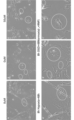
- the redox nanoparticles were disintegrated in the cells and existed in a polymer state, indicating that redox reactions occurred ((Yoshitomi T, et al., Biomacromolecules. 2009;10( 3):596-601.) Also, in the examination of the culture supernatant (Fig. 7.C), a sharp triplet signal was detected in the TEMPOL group, while a broad signal was detected in the rhodamine-labeled redox nanoparticle group. (See above.) Furthermore, even when potassium hexacyanoferrate(III) was added to the culture supernatant, a sharp triplet signal was detected in the TEMPOL group.
- FIG. 7. A, B Fluorescent immunostaining showed that rhodamine-labeled redox nanoparticles were in the cytoplasm both after normal culture and hypoxic culture + reperfusion. confirmed (Fig. 7. A, B).
- A electron spin resonance method and fluorescence immunostaining of normal culture
- B electron spin resonance method and fluorescence immunostaining of hypoxic culture + reperfusion
- C electron spin of culture supernatant of hypoxic culture + reperfusion Resonance method Red indicates rhodamine and blue indicates cell nuclei.
- Example 4 Creation of cerebral infarction model mice due to distal middle cerebral artery occlusion The animal experiment plan was approved by the University of Tsukuba Life Science Animal Resource Center (approval number: #20-132). All experiments were conducted in accordance with the "Guidelines for the Care and Use of Laboratory Animals.”
- mice Male, 7-8 weeks old, body weight 20-25 g were purchased from CLEA, Japan and used.
- a mouse distal middle cerebral artery occlusion model was prepared by the method reported by Taguchi et al. (See Taguchi A, et al., The Journal of clinical investigation. 2004;114(3):330-8.). Briefly, mice were anesthetized by intraperitoneal injection of a mixed solution of ketamine (70 mg/kg) and xylazine (14 mg/kg). A skin incision was made on the temporal region of the mouse and a small craniotomy was made in the temporal bone with a dental drill.
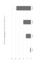
- Example 5 Method of direct intracerebral transplantation of nervous system cells into mice with cerebral infarction Four weeks after transplantation of cells and redox nanoparticles (0, 200, 500, 1000 ⁇ M) to determine the optimal concentration of redox nanoparticles in advance The mouse brain was taken out and the number of surviving transplanted cells was confirmed. A cell suspension of nervous system cells was prepared in advance and adjusted so that 1.0 ⁇ 10 5 cells would be transplanted. In addition, the redox nanoparticles were mixed with the cell suspension before cell transplantation to form a mixed solution. 500, 1000 ⁇ M showed more survival than 0, 200, and no difference was observed at 500, 1000 ⁇ M. See FIG.
- the transplanted cells or substances were divided into 4 groups: PBS, redox nanoparticles, cells + PBS, and cells + redox nanoparticles.
- PBS head fixator
- redox nanoparticles cells + PBS
- cells + redox nanoparticles Two days after the cerebral infarction treatment, the mice were fixed in a head fixator (NARISHIGE, Japan) after general anesthesia as in 3-6. A skin incision was made in the midline of the head, and Hamilton Syringe (Cat. No., 4025-11701, GLSciences) was used at the periinfarct site 1 mm anteriorly and 1.5 mm laterally from bregma to a depth of 2 mm from the surface of the brain. was injected into the brain by pointing the needle to The injection was performed slowly over 10 minutes, and the needle was slowly withdrawn after waiting 5 minutes after injection.
- mice transplanted with PBS and redox nanoparticles were compared. Two days after the cerebral infarction treatment, mice were given general anesthesia as described above, and PBS and redox nanoparticles were implanted into the peri-infarct foci. One hour after transplantation, 27 mg/kg of dihydroethidium (Cat. No., D1168, Invitrogen) (200 ⁇ l in total) was intraperitoneally administered twice at 30-minute intervals (Hu D, et al., The Journal of Neuroscience. 2006; 26(15):3933-41).
- dihydroethidium Cat. No., D1168, Invitrogen
- mice were placed in a transparent plastic cylinder with a diameter of 8 cm and a height of 12 cm, and the number of times they used their forelimbs was measured.
- the rate of forelimb use was calculated using the following formula [paralyzed forelimb/(paralyzed forelimb + non-paralyzed forelimb + both sides)] x 100 (Craft TK, et al., Stroke. 2005;36(9):2006- 11.).
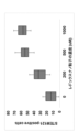
- FIG. 10 The results are shown in FIG. From FIG. 10, it was confirmed that cell therapy using cells + redox nanoparticles according to the present invention improved neurological symptoms in cerebral infarction model mice. Specifically, it is as follows. Adhesive removal test (Fig. 10. A) and Cylinder test (Fig. 10. B) behavioral evaluation of mice showed improvement in all four groups. Furthermore, the groups containing cells (cells + PBS and cells + redox nanoparticles group) showed better improvement in behavioral assessment from 1 week after transplantation compared to the groups without cells (PBS, redox nanoparticles group). rice field.
- mice were perfusion-fixed with 4% paraformaldehyde one week and six weeks after transplantation, and the mouse brains were excised and cryosections were prepared.
- Antigen-antibody treatment was performed overnight at 4°C using the following primary antibodies, followed by 60 minutes at room temperature in the dark using appropriate secondary antibodies.
- Mounting was performed using DAPI-containing mounting medium (SCR-038448, dianova). Observations were made using a fluorescence microscope (Leica Microsystems Wetzlar, Germany).
- the primary antibodies used were mouse anti-STEM121 antibody (1:1000, cat.
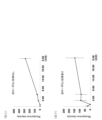
- FIG. 11.A One week after transplantation (Fig. 11.A), there was no difference in the survival rate of transplanted cells between the PBS and redox nanoparticle groups. However, at 6 weeks after transplantation (Fig. 11.B), the survival rate of transplanted cells was higher in the redox nanoparticles group than in the PBS group (P ⁇ 0.05). Fluorescent immunostaining showed positive cells for both Doublecortin, MAP2, and GFAP in both the PBS group (Fig. 11. A-i) and the redox nanoparticle group (Fig. 11. A-ii) one week after transplantation. Six weeks after transplantation, most of the cells in the PBS group (Fig. 11.A).
- Example 6 Uptake of Redox Nanoparticles (Related to the Copolymer of Formula (I)) into Neural Cells
- Neural cells were cultured in a 24-well plate at 6 ⁇ 10 4 cells/well.
- Rhodamine-labeled redox nanoparticles were separately administered to the culture solution at 100 ⁇ M and 500 ⁇ M, and normal culture was performed. After 15 minutes, 1 hour, 3 hours, 6 hours and 24 hours after administration, observation was performed using a fluorescence microscope. After that, each well was washed twice with Hanks, and the cells were fixed with 4% paraformaldehyde for 15 minutes.
- antigen-antibody reaction was performed overnight at 4°C using the following primary antibody, and then reaction was allowed to proceed for 60 minutes at room temperature in a dark room using an appropriate secondary antibody. After washing with Hanks, they were mounted using DAPI-containing mounting medium (SCR-038448, dianova). Observation was performed at the same time using a fluorescence microscope, fluorescence intensity was measured using ImageJ, and average values were compared.
- Antibody used mouse anti-rhodamine antibody (1:250; Cat. No., ab9093; Abcam).
- Example 7 Uptake of Redox Nanoparticles (Related to the Copolymer of Formula (II)) into Neural Cells
- Neural cells were cultured in a 24-well plate at 3.2 ⁇ 10 4 cells/well.
- Cy5-labeled nanoparticles prepared according to "Production Example 17: Preparation of nanoparticles of SMAPo TN (N564)" in Patent Document 3 (Japanese Patent Application Laid-Open No. 2019-123773) were administered into the culture medium at a concentration of 3.2 mM. Then, normal culture was performed. Observation was performed using a fluorescence microscope 1 hour, 3 hours, 6 hours, and 24 hours after administration. Fluorescence intensity was measured using ImageJ and average values were compared.
- Example 8 Verification of cell permeability of RNP (1) Establishment of RNP penetration time In order to evaluate the permeability of RNP over time by treatment time and establish the most effective concentration and treatment time, Based on 100 ⁇ M, which was determined to be present, the intracellular RNP amount was measured by fluorescence observation of Rhodamine in RNP for each treatment time.
- FIG. 14 shows a conceptual diagram of the contents of the experiment. Cells were seeded at a density of 10,000 cells/well on a 2.5 v/v % Matrigel-coated 24-well plate. NB27 medium, which is a nerve cell medium, was used as the medium. RNP treatment of the cells was carried out by normal culture at 37° C.
- the treatment time was set to 15 minutes, 1 hour, 3 hours, 6 hours and 12 hours, respectively, and untreated cells were cultured for the same time as a control group.
- each cell was washed twice with PBS(-), the medium was returned to the normal neuronal cell medium NB27 medium, and all-in-one fluorescence was used by Keyence. Photographs were taken with a microscope (BZ-X800). Photographs were taken immediately after each RNP treatment and after washing, and normal culture was performed for 48 hours after all conditions were completed.
- imageJ software (Ver.1.53q) was used to determine the total area of positive Rhodamine signals from fluorescence images and the total area of cells from phase contrast images. changed.
- the threshold for positive signal calculation was determined according to the ISO Data parameter of the software.
- FIG. 15 shows a conceptual diagram of the contents of the experiment.
- Cells were seeded at a density of 10,000 cells/well on a 2.5 v/v % Matrigel-coated 24-well plate.
- NB27 medium which is a nerve cell medium, was used as the medium.
- RNP treatment of the cells was carried out by normal culture at 37° C. under 5% CO 2 environment for 48 hours after seeding, and then the medium was changed to RNP:DMEM/LG medium adjusted to each concentration.
- the OGD load was applied immediately after the medium exchange, and culture was performed for 8 hours. After completion of OGD, each cell was washed twice with PBS(-), the medium was returned to normal neuronal cell medium NB27 medium, and photographed with a Keyence All-in-one microscope (BZ-X800). Imaging was performed after washing immediately after OGD.
- imageJ software (Ver.1.53q) was used to determine the total area of Rhodamine signal-positive from the fluorescence image and the total area of the entire cell from the phase contrast image, and quantify the positive area divided by the cell area. . Threshold for positive signal calculation was performed according to the ISOData parameter of the software.
- Example 9 Verification of antioxidant effect of RNP (1) Verification of antioxidant capacity of RNP against reperfusion damage
- RNP was applied to cells before and after OGD loading. It was set to show preventive and therapeutic effects, respectively.
- FIG. 16 shows a conceptual diagram of the contents of the experiment. Cells were seeded at a density of 10,000 cells/well on a 2.5 v/v % Matrigel-coated 24-well plate. NB27 medium, which is a nerve cell medium, was used as the medium. Cells were treated with RNP after seeding and normal culture at 37°C in a 5% CO2 environment for 48 hours. rice field.
- Samples (PreRNP) for prophylactic effect observation were cultured normally for 48 hours, then the medium was changed to RNP medium, and OGD was applied for 8 hours at 37°C in a 1% O 2 environment. After OGD, the medium was removed by aspiration, the cells were washed twice with PBS(-), and the neuron medium was replaced with NB27 medium, and simulated reperfusion damage was given at 37°C and 5% CO 2 for 48 hours. rice field.
- Samples (PostRNP) for observation of therapeutic effects were cultured for 48 hours, washed twice with PBS(-), replaced with DMEM/LG medium, and placed under OGD at 37°C and 1% O2 for 8 hours. processed. After OGD, the medium was replaced with RNP:DMEM/LG medium with the RNP concentration adjusted as described above, and cultured at 37°C under 5% CO 2 environment for 12 hours. After RNP treatment, the medium was removed by aspiration, the cells were washed twice with PBS(-), and the neuron medium was replaced with NB27 medium, and simulated reperfusion damage was given at 37°C in a 5% CO 2 environment. .
- FIG. 17 shows a conceptual diagram of the contents of the experiment.
- RNP was administered to the cells before and after OGD loading, and the prophylactic and therapeutic effects of each were determined.
- Cells were seeded at a density of 5,000 cells/well on 2.5 v/v % matrigel-coated 96-well plates.
- NB27 medium which is a nerve cell medium, was used as the medium.
- RNP treatment of the cells was carried out using DMEM/LG medium adjusted to RNP concentrations of 10, 50, 100 and 500 ⁇ M, respectively, after normal culture at 37° C. and 5% CO 2 for 48 hours after seeding.
- cell viability was measured using a 1:1 mixture of CellTiter Glo2.0 (Promega) reagent and DMEM/LG.
- the medium was removed, the cells were washed twice with PBS(-), replaced with CellTiter Glo2.0:DMEM/LG solution, the well plate was gently agitated for 2 minutes, and allowed to stand at room temperature for 10 minutes for reaction. After the reaction, the plate was set in a Varioskan LUX microplate reader (Thermo) and the yellow luminescence of Luciferin reacted with ATP in living cells was quantified by luminometric measurement.
- FIG. 23 shows the results converted to . From FIG. 23, it can be seen that there was no significant difference between 50 ⁇ M and 100 ⁇ M, but the RNP permeability increased at 500 ⁇ M when administered after OGD loading.
- FIG. 24 shows the results of photographing the cells in each experiment taken with a fluorescence microscope after the completion of each experiment in Example 9(1). Qualitative analysis of cell damage was performed by qualitatively comparing axonal damage (yellow circles) with dendrite and cell body damage (white circles) in FIG.
- the amount of RNP also increased in a concentration-dependent manner, but the efficiency was similarly high at the dose concentration-to-signal ratio of 50 ⁇ M and 100 ⁇ M, and the signal was the highest at 500 ⁇ M, but the efficiency was 100 ⁇ M. was about 40% lower than the From this result, while 100 ⁇ M obtained in the above experimental example seems to be an appropriate concentration, it was necessary to verify whether not only the penetration rate but also the therapeutic effect showed the same efficiency.
Abstract
Disclosed are: a mammalian cell modification method comprising a step for preparing nanoparticles each based on a copolymer that includes a polymer chain segment having a poly(ethylene glycol) segment and a cyclic nitroxide serves as a pendant and mammalian cells; and a step for combining the prepared nanoparticles with the prepared mammalian cells in vitro; a blended product comprising a combination of the nanoparticles and the mammalian cells; and a preparation containing the nanoparticles as an active ingredient. According to the above-mentioned subjects, for example, the damage of the provided mammalian cells by stress under an external environment is recovered when the cells are damaged by the stress previously, and the cells are protected from the stress when the cells are not subjected to the stress.
Description
本発明は、レドックス作用を有する共重合体をベースとするナノ粒子の哺乳動物細胞の処理又は改変に向けた用途に関し、より具体的には、ポリ(エチレングリコール)(PEG)セグメントと環状ニトロキシドラジカルをペンダントとして有するポリマー鎖セグメントを含む両親媒性共重合体のナノ粒子の細胞処理への新規用途又は使用に関する。
The present invention relates to the use of redox-active copolymer-based nanoparticles for the treatment or modification of mammalian cells, more specifically poly(ethylene glycol) (PEG) segments and cyclic nitroxide radicals. Novel application or use of nanoparticles of amphiphilic copolymers containing polymer chain segments having as pendants for cell processing.
脳梗塞病巣に間葉系幹細胞や神経幹細胞などを移植しても、移植後28日後の細胞生存率は1~8%と低いことが知られている(Nakagomi N, et al. Stem Cells (2009) 27 (9): 2185-2195, doi:10. 1002/stem. 161(以下、非特許文献1と略記する。)、George PM, et al. Biomaterials, 2018; 178: 63-72. Doi: 10, 1016/j. biomaterials, 2018. 06. 010(以下、非特許文献2と略記する。))。脳梗塞病巣は、虚血によりフリーラジカルや活性酸素種が過剰産生されて、移植細胞が生存し、生着するには非寛容な環境であることが一因である。また、虚血性脳卒中に対する細胞治療は必要とされているが、経動脈/静脈的に投与された細胞が脳実質で検出されるのは0.06~0.8%のみであり、神経再生療法の実現は困難であるとされている(Rosado-Castro PH, et al. Regenerative Medicine, 2013; 8(2): 145-155(以下、非特許文献3と略記する。))。しかし、直接投与は、細胞を脳実質内へ直接投与することができることからこのような投与系が使用できると再生医療に資するであろう。
It is known that even if mesenchymal stem cells or neural stem cells are transplanted into cerebral infarction lesions, the cell survival rate 28 days after transplantation is as low as 1-8% (Nakagomi N, et al. Stem Cells (2009). ) 27 (9): 2185-2195, doi: 10. 1002/stem. 161 (hereinafter abbreviated as Non-Patent Document 1.), George PM, et al. Biomaterials, 2018; 178: 63-72. Doi: 10, 1016/j. biomaterials, 2018. 06. 010 (hereinafter abbreviated as Non-Patent Document 2)). One of the causes of cerebral infarction lesions is the excessive production of free radicals and reactive oxygen species due to ischemia, creating an intolerant environment for the survival and engraftment of transplanted cells. In addition, cell therapy for ischemic stroke is needed, but only 0.06-0.8% of cells administered intraarterially/intravenously are detected in the brain parenchyma, making it difficult to realize nerve regeneration therapy. (Rosado-Castro PH, et al. Regenerative Medicine, 2013; 8(2): 145-155 (hereinafter abbreviated as Non-Patent Document 3)). However, since direct administration allows cells to be administered directly into the brain parenchyma, the use of such an administration system would contribute to regenerative medicine.
一方、レドックスナノ粒子、特に、環状ニトロキシルラジカルを側鎖に有する疎水性のポリマーセグメントとポリ(エチレングリコール)(PEG)セグメントを含むブロック共重合体の水溶液から当該共重合体が自己集積化することにより高分子ミセル化したナノ粒子は、虚血や炎症で生じるフリーラジカルを消去することができる製剤として知られている(WO 2009/133647 A(以下、特許文献1と略記する)、WO 2016/052463 A(以下、特許文献2と略記する。))。また、PEGセグメントと環状ニトロキシドラジカルをペンダント(又は側鎖)として有するポリマー鎖セグメントを含む両親媒性共重合体、具体的には、無水マレイン酸とスチレンの共重合体であって、無水マレイン酸単位を介してPEG鎖がグラフトして、かつ、環状ニトロキシドラジカル等の作用剤の残基がペンダントとして共有結合した共重合体、をベースとするナノ粒子もイン・ビトロ(in vitro)又はイン・ビボ(in vivo)で、環状ニトロキシドラジカルに起因するレドックス作用を示すことが知られている(特開2019‐123773号公報(以下、特許文献3と略記する。))。
On the other hand, redox nanoparticles, in particular block copolymers containing hydrophobic polymer segments with cyclic nitroxyl radicals in their side chains and poly(ethylene glycol) (PEG) segments, self-assemble from an aqueous solution. Polymer micellized nanoparticles are known as a preparation capable of scavenging free radicals generated by ischemia and inflammation (WO 2009/133647 A (hereinafter abbreviated as Patent Document 1), WO 2016 /052463 A (hereinafter abbreviated as Patent Document 2)). Also amphiphilic copolymers comprising polymer chain segments having PEG segments and cyclic nitroxide radicals as pendant (or side chains), specifically copolymers of maleic anhydride and styrene, wherein maleic anhydride Nanoparticles based on copolymers, to which PEG chains are grafted via units and covalently attached pendant residues of agents such as cyclic nitroxide radicals, have also been investigated in vitro or in vitro. It is known to exhibit a redox action caused by a cyclic nitroxide radical in vivo (Japanese Patent Application Laid-Open No. 2019-123773 (hereinafter abbreviated as Patent Document 3)).
例えば、前者のナノ粒子をマウス一過性脳虚血モデルに頸動脈投与すると、脳虚血病巣において神経細胞の細胞質に取り込まれ、神経保護効果を発揮することが明らかにされている(Mujagic A, et al. Brain research, 2020; 1743: 146922, doi: 1016/j. braineres. 2020. 146922(以下、非特許文献4と略記する。): Hosoo H, et al. Stroke. 48: 2238-2247, 2017, doi: 10. 1161/STROKAHA. 116. 016356(以下、非特許文献5と略記する。))。
For example, it has been shown that when the former nanoparticles are administered to a mouse model of transient cerebral ischemia through the carotid artery, they are incorporated into the cytoplasm of neurons in cerebral ischemic lesions and exhibit neuroprotective effects (Mujagic A. , et al. Brain research, 2020; 1743: 146922, doi: 1016/j.braineres. , 2017, doi: 10. 1161/STROKAHA. 116. 016356 (hereinafter abbreviated as Non-Patent Document 5)).
仮に、これらの技術の知見に照らし、例えば、移植前の細胞とレドックスナノ粒子の組み合わせ使用が、前述の細胞移植技術上の短所等を矯正又は改善できるのであれば、再生医療をはじめとする広範な細胞を用いる技術分野の進展に資するであろう。
If, in light of the findings of these technologies, for example, the combined use of cells and redox nanoparticles before transplantation can correct or improve the above-mentioned disadvantages of cell transplantation technology, it can be applied to a wide range of applications including regenerative medicine. It will contribute to the development of technical fields using such cells.
以上の背景技術の下、本発明の目的は、哺乳動物細胞(以下、移植細胞または単に、細胞とも称する。)を、当該細胞を処理若しくは保存する又は当該細胞が存在し得る場所又は領域で安定にかつ、効率よく取り扱うことができ、さらには、限定されるものでないが、当該細胞の移植等が望まれる所定の場所又は領域に直接投与することができる手段を手にすることにある。
本発明者らは、このような観点から、研究を進めてきたところ、特許文献1若しくは2、又は特許文献3に開示された共重合体をベースとする特定のレドックスナノ粒子と哺乳動物細胞又は移植用細胞とを組み合わせるか、或いは併用すると、当該細胞が改変され、移植前の当該細胞の保存若しくは培養条件下で当該細胞に対する、例えば、外的環境下の酸化ストレスをはじめとするストレスを抑制するか、若しく当該ストレスに起因する細胞の損傷を回復することを見出した。こうして改変された細胞は、移植後、移植を望む所定の場所又は領域においても、当該細胞に対するフリーラジカルや活性酸素種に起因する酸化ストレスを有意に抑制できることを見出した。さらにまた、前記レドックスナノ粒子は、移植用細胞に限定されることなく、広範な細胞に対しても同様に作用又は効果を奏するすものと理解される。 In view of the above background art, an object of the present invention is to provide mammalian cells (hereinafter also referred to as transplanted cells or simply cells) for treating or preserving them or stabilizing them in a place or area where they may reside. The purpose of the present invention is to obtain a means that can be handled easily and efficiently, and can be administered directly to a predetermined site or region where the cell transplantation or the like is desired, although this is not a limitation.
From this point of view, the present inventors have conducted research and found that specific redox nanoparticles based on the copolymers disclosed in Patent Document 1 or 2 or Patent Document 3 and mammalian cells or When combined or used together with cells for transplantation, the cells are modified, and stress such as oxidative stress in the external environment to the cells is suppressed under preserving or culturing conditions of the cells before transplantation. It has been found that the stress-induced cell damage caused by the stress is reversed. It has been found that cells modified in this way can significantly suppress oxidative stress caused by free radicals and reactive oxygen species on the cells even in a predetermined place or region where transplantation is desired after transplantation. Furthermore, it is understood that the redox nanoparticles are not limited to cells for transplantation, and similarly act or effect on a wide range of cells.
本発明者らは、このような観点から、研究を進めてきたところ、特許文献1若しくは2、又は特許文献3に開示された共重合体をベースとする特定のレドックスナノ粒子と哺乳動物細胞又は移植用細胞とを組み合わせるか、或いは併用すると、当該細胞が改変され、移植前の当該細胞の保存若しくは培養条件下で当該細胞に対する、例えば、外的環境下の酸化ストレスをはじめとするストレスを抑制するか、若しく当該ストレスに起因する細胞の損傷を回復することを見出した。こうして改変された細胞は、移植後、移植を望む所定の場所又は領域においても、当該細胞に対するフリーラジカルや活性酸素種に起因する酸化ストレスを有意に抑制できることを見出した。さらにまた、前記レドックスナノ粒子は、移植用細胞に限定されることなく、広範な細胞に対しても同様に作用又は効果を奏するすものと理解される。 In view of the above background art, an object of the present invention is to provide mammalian cells (hereinafter also referred to as transplanted cells or simply cells) for treating or preserving them or stabilizing them in a place or area where they may reside. The purpose of the present invention is to obtain a means that can be handled easily and efficiently, and can be administered directly to a predetermined site or region where the cell transplantation or the like is desired, although this is not a limitation.
From this point of view, the present inventors have conducted research and found that specific redox nanoparticles based on the copolymers disclosed in
したがって、本明細書では、限定されるものでないが次の態様の発明が提供される。
[態様1]
式(I)又は式(II)で表される共重合体をベースとするナノ粒子及び哺乳動物細胞を用意するステップと、
用意された前記ナノ粒子と用意された哺乳動物細胞をイン・ビトロ(in vitro)で組み合わせるステップ
を含んでなる哺乳動物細胞の改変方法。
式(I):
 上式中、
上式中、
Aは、非置換又は置換C1-C12アルキルを表し、置換されている場合の置換基は、ホルミル基、式R'R"CH-基を表し、ここで、R'及びR"は独立してC1-C4アルコキシ又はR'とR"は一緒になって-OCH2CH2O-、-O(CH2)3O-若しくは-O(CH2)4O-を表し、
L1は、直接結合又は二価の連結基を表し、
L2-R1は、L2が-(CH2)a-NH-(CH2)a-又は-(CH2)a-O-(CH2)a-であり、R1が、式
 で表される環状ニトロキシドラジカル残基のいずれかであり、ここで、R’はメチルであり、
R2は、クロロ、ブロモ又はヒドロキシルであり、
上記において、L2-R1とR2を有するポリマー主鎖中の反復単位(unit)はランダムに存在し、L2-R1を有する単位pは2~100の範囲内にあり、R2を有する単位qは存在しない(ゼロ)か、若しくは1~20の範囲内にあり、ただし、これらの単位の総数はnとなり、
ZはH、SH又はS(C=S)-Phであり、Phは1又は2個のメチルまたはメトキで置換されていてもよいフェニルを表し、
各aは、独立して0又は1~5の整数であり、
mは2~10,000の整数を表し、
nは3~100の整数を表す。
式(II):
 上式中、
上式中、
x+yは5~1400の整数であり、nは5~1400の整数であり、x+y:nは1:1~5の比率にあり、x:yは1~20:1の比率にあり、x:yは1~60:1の比率にあり、
(1)前記yの付された反復単位において、L-PEG-A中、Lは、O又はNHであり、PEGは次式で表され、
 ここで、pは1~6の整数であり、qは5~500の整数であり、
Aは、
A1:非置換若しくは置換C1-C12アルコキシ基を表し、置換されている場合の置換基は、ホルミル基、式RaRbCH-(ここで、Ra及びRbは独立して、C1-C4アルコキシまたはR1とR2は一緒になって-OCH2CH2O-、-O(CH2)3O-もしくは-O(CH2)4O-を表す。)の基、又は
A2:次式
 で表される基を表し、
当該反復単位は式(I)で表される共重合体の総単位の2%~15%を占め、
(2)下付き記号xの付された反復単位において、
(a)R1又はR2のいずれか一方は、
a1 :次式
 で表され、ここで、
TEMPOは、次式
 で表される環状ニトロキシドラジカル残基のいずれかであり、ここで、R’はメチルである、
で表されるいずれかの残基、
a2:次式
 のいずれかで表される残基、
a3:次式
 で表され、
で表され、
ここで、R3はC1-3のアルキル基であり、rは2~6の整数である、残基、
からなる群より選ばれる残基であり、
他方はOHであり、又は
(b)R1及びR2は、一緒になって-O-を表し、環式無水物残基を形成し、又は
(c)R1及びR2は、各OHを表す、
ただし、前記xの付された反復単位において、
(i)(a)のR1又はR2のいずれか一方は、a1の残基を含むか、又は
(ii)(a)のR1又はR2のいずれか一方は、a1の残基とa2の残基を含むか、又は
(iii)(a)のR1又はR2のいずれか一方は、a1の残基とa3の残基を含み、又は
(iv)(a)のR1又はR2のいずれか一方は、a1の残基とa2の残基とa3の残基を含み、又は
(v)前記xの付された反復単位は、上記(i)、(ii)乃至(iv)に定義する残基に加えて(b)若しくは(c)に定義する基を含んでいてもよく、
ここで、上記の各残基及び基を含む単位は独立してランダムに存在し、(a)に定義する残基を含む単位はxの付された反復単位の総数の15%~60%を占める。
[態様2]
態様1の改変方法であって、用意された哺乳動物細胞が予め外的環境下のストレスにより損傷を受けていてもよく、当該ストレスを受けているときは、前記改変は当該ストレスに起因する細胞の損傷を回復するものであり、他方、当該ストレスを受けていないときは、前記改変は当該ストレスから細胞を保護するものである、前記改変方法。
[態様3]
態様1の改変方法であって、前記ナノ粒子と前記哺乳動物細胞をイン・ビトロ(in vitro)で組み合わせるステップが、哺乳動物細胞を培養する培地において、前記ナノ粒子と前記哺乳動物細胞を混合するステップを含む、前記改変方法。
[態様4]
態様1の改変方法であって、前記ナノ粒子と前記哺乳動物細胞をイン・ビトロ(in vitro)で組み合わせるステップが、哺乳動物細胞を培養する培地において、前記ナノ粒子と前記哺乳動物細胞を混合することで当該哺乳動物細胞中に当該ナノ粒子を浸透させるステップを含む、前記改変方法。
[態様5]
態様1で定義された式(I)又は式(II)で表される共重合体をベースとするナノ粒子と哺乳動物細胞がイン・ビトロ(in vitro)で組み合わさった、配合物。
[態様6]
前記組み合わさった形態が、前記ナノ粒子と前記哺乳動物細胞が哺乳動物を培養する培地において共存する状態にある、態様5の配合物。
[態様7]
前記組み合わさった形態が、前記ナノ粒子と前記哺乳動物細胞が哺乳動物を培養する培地において共存する状態にあり、かつ、前記哺乳動物細胞に前記ナノ粒子が浸透した状態にある、態様5の配合物。
[態様8]
イン・ビトロ(in vitro)で、態様1で定義された下記式(I)又は式(II)で表される共重合体をベースとするナノ粒子を有効成分として含んでなる、哺乳動物細胞を改変するための調製物。
[態様9]
前記改変が、外的環境下のストレスから哺乳動物細胞を保護する又は当該ストレスにより受けた哺乳動物細胞の損傷を回復するものである、態様7の調製物。
[態様10]
前記哺乳動物細胞の保護が、細胞が移植された場合に、移植細胞の生着率の向上及び/又は目的とする細胞への分化の向上をもたらす、態様8の調製物。 Accordingly, the present specification provides the invention in the following non-limiting aspects.
[Aspect 1]
providing nanoparticles and mammalian cells based on a copolymer of formula (I) or formula (II);
A method of modifying mammalian cells comprising the step of combining said provided nanoparticles and provided mammalian cells in vitro.
Formula (I):
In the above formula,
A represents unsubstituted or substituted C 1 -C 12 alkyl and the substituent, if substituted, represents a formyl group, a group of formula R′R ″ CH—, where R ′ and R ″ are independently and C 1 -C 4 alkoxy or R ′ and R ″ together represent —OCH 2 CH 2 O—, —O(CH 2 ) 3 O— or —O(CH 2 ) 4 O—,
L 1 represents a direct bond or a divalent linking group,
L 2 —R 1 is a group in which L 2 is —(CH 2 ) a —NH—(CH 2 ) a — or —(CH 2 ) a —O—(CH 2 ) a —, and R 1 is represented by the formula
any of the cyclic nitroxide radical residues represented by
R2 is chloro, bromo or hydroxyl;
In the above, the repeating units in the polymer backbone with L 2 -R 1 and R 2 are randomly present, the unit p with L 2 -R 1 is in the range of 2 to 100, and R 2 is absent (zero) or is in therange 1 to 20, provided that the total number of these units is n,
Z is H, SH or S(C=S)-Ph, Ph represents phenyl optionally substituted with 1 or 2 methyl or methoxy;
each a is independently 0 or an integer from 1 to 5;
m represents an integer from 2 to 10,000,
n represents an integer of 3 to 100;
Formula (II):
In the above formula,
x+y is an integer from 5 to 1400, n is an integer from 5 to 1400, x+y:n is in a ratio of 1:1 to 5, x:y is in a ratio of 1 to 20:1, x: y is in a ratio of 1 to 60:1;
(1) In the repeating unit with y, in L-PEG-A, L is O or NH, and PEG is represented by the following formula,
wherein p is an integer from 1 to 6, q is an integer from 5 to 500,
A is
A1: represents an unsubstituted or substituted C 1 -C 12 alkoxy group, where the substituents when substituted are a formyl group, a formula R a R b CH— (wherein R a and R b are independently C 1 -C 4 alkoxy or R 1 and R 2 taken together represent —OCH 2 CH 2 O—, —O(CH 2 ) 3 O— or —O(CH 2 ) 4 O—. , or A2: the following formula
represents a group represented by
The repeating unit accounts for 2% to 15% of the total units of the copolymer represented by formula (I),
(2) in the repeating unit with the subscript x,
(a) either one of R 1 or R 2 is
a1: the following formula
, where
TEMPO is the following formula
any of the cyclic nitroxide radical residues represented by
any residue represented by
a2: the following formula
A residue represented by either
a3: the following formula
is represented by
wherein R 3 is a C 1-3 alkyl group and r is an integer from 2 to 6, the residue
A residue selected from the group consisting of
the other is OH, or (b) R 1 and R 2 together represent —O— and form a cyclic anhydride residue, or (c) R 1 and R 2 are each OH represents
However, in the repeating unit marked with x,
(i) either one of R 1 or R 2 in (a) comprises the residue of a1, or (ii) either one of R 1 or R 2 in (a) includes the residue of a1 or (iii) either one of R 1 or R 2 of (a) comprises a residue of a1 and a residue of a3, or (iv) R 1 of (a) or Either one of R 2 comprises residues a1, residues a2 and residues a3, or (v) the repeating units marked with x are the above (i), (ii) to (iv) ) may contain a group defined in (b) or (c) in addition to the residue defined in
Here, the units containing each of the above residues and groups are present independently and randomly, and the units containing the residues defined in (a) account for 15% to 60% of the total number of repeating units marked with x. occupy
[Aspect 2]
In the modification method ofaspect 1, the prepared mammalian cells may be damaged in advance by stress in an external environment, and when the stress is applied, the modification is caused by the stress. while the modification protects the cell from the stress when not subjected to the stress.
[Aspect 3]
The modified method ofaspect 1, wherein the in vitro combining the nanoparticles and the mammalian cells comprises mixing the nanoparticles and the mammalian cells in a medium for culturing the mammalian cells. Said modified method, comprising the steps of:
[Aspect 4]
The modified method ofaspect 1, wherein the in vitro combining the nanoparticles and the mammalian cells comprises mixing the nanoparticles and the mammalian cells in a medium for culturing the mammalian cells. permeating said nanoparticles into said mammalian cells.
[Aspect 5]
A formulation in which mammalian cells are combined in vitro with nanoparticles based on a copolymer of formula (I) or formula (II) as defined inaspect 1.
[Aspect 6]
6. The formulation ofaspect 5, wherein said combined form is such that said nanoparticles and said mammalian cells coexist in a medium in which a mammal is cultured.
[Aspect 7]
6. The formulation ofaspect 5, wherein said combined form is such that said nanoparticles and said mammalian cells coexist in a medium in which said mammal is cultured, and said mammalian cells are permeated with said nanoparticles. thing.
[Aspect 8]
In vitro mammalian cells comprising, as an active ingredient, nanoparticles based on the copolymer represented by formula (I) or (II) as defined inaspect 1 Preparations for modification.
[Aspect 9]
8. The preparation ofaspect 7, wherein said modification protects mammalian cells from stress in the external environment or restores damage to mammalian cells caused by said stress.
[Aspect 10]
9. The preparation ofaspect 8, wherein said protection of mammalian cells results in improved engraftment and/or improved differentiation of the transplanted cells into cells of interest when the cells are transplanted.
[態様1]
式(I)又は式(II)で表される共重合体をベースとするナノ粒子及び哺乳動物細胞を用意するステップと、
用意された前記ナノ粒子と用意された哺乳動物細胞をイン・ビトロ(in vitro)で組み合わせるステップ
を含んでなる哺乳動物細胞の改変方法。
式(I):
Aは、非置換又は置換C1-C12アルキルを表し、置換されている場合の置換基は、ホルミル基、式R'R"CH-基を表し、ここで、R'及びR"は独立してC1-C4アルコキシ又はR'とR"は一緒になって-OCH2CH2O-、-O(CH2)3O-若しくは-O(CH2)4O-を表し、
L1は、直接結合又は二価の連結基を表し、
L2-R1は、L2が-(CH2)a-NH-(CH2)a-又は-(CH2)a-O-(CH2)a-であり、R1が、式
R2は、クロロ、ブロモ又はヒドロキシルであり、
上記において、L2-R1とR2を有するポリマー主鎖中の反復単位(unit)はランダムに存在し、L2-R1を有する単位pは2~100の範囲内にあり、R2を有する単位qは存在しない(ゼロ)か、若しくは1~20の範囲内にあり、ただし、これらの単位の総数はnとなり、
ZはH、SH又はS(C=S)-Phであり、Phは1又は2個のメチルまたはメトキで置換されていてもよいフェニルを表し、
各aは、独立して0又は1~5の整数であり、
mは2~10,000の整数を表し、
nは3~100の整数を表す。
式(II):
x+yは5~1400の整数であり、nは5~1400の整数であり、x+y:nは1:1~5の比率にあり、x:yは1~20:1の比率にあり、x:yは1~60:1の比率にあり、
(1)前記yの付された反復単位において、L-PEG-A中、Lは、O又はNHであり、PEGは次式で表され、
Aは、
A1:非置換若しくは置換C1-C12アルコキシ基を表し、置換されている場合の置換基は、ホルミル基、式RaRbCH-(ここで、Ra及びRbは独立して、C1-C4アルコキシまたはR1とR2は一緒になって-OCH2CH2O-、-O(CH2)3O-もしくは-O(CH2)4O-を表す。)の基、又は
A2:次式
当該反復単位は式(I)で表される共重合体の総単位の2%~15%を占め、
(2)下付き記号xの付された反復単位において、
(a)R1又はR2のいずれか一方は、
a1 :次式
TEMPOは、次式
で表されるいずれかの残基、
a2:次式
a3:次式
ここで、R3はC1-3のアルキル基であり、rは2~6の整数である、残基、
からなる群より選ばれる残基であり、
他方はOHであり、又は
(b)R1及びR2は、一緒になって-O-を表し、環式無水物残基を形成し、又は
(c)R1及びR2は、各OHを表す、
ただし、前記xの付された反復単位において、
(i)(a)のR1又はR2のいずれか一方は、a1の残基を含むか、又は
(ii)(a)のR1又はR2のいずれか一方は、a1の残基とa2の残基を含むか、又は
(iii)(a)のR1又はR2のいずれか一方は、a1の残基とa3の残基を含み、又は
(iv)(a)のR1又はR2のいずれか一方は、a1の残基とa2の残基とa3の残基を含み、又は
(v)前記xの付された反復単位は、上記(i)、(ii)乃至(iv)に定義する残基に加えて(b)若しくは(c)に定義する基を含んでいてもよく、
ここで、上記の各残基及び基を含む単位は独立してランダムに存在し、(a)に定義する残基を含む単位はxの付された反復単位の総数の15%~60%を占める。
[態様2]
態様1の改変方法であって、用意された哺乳動物細胞が予め外的環境下のストレスにより損傷を受けていてもよく、当該ストレスを受けているときは、前記改変は当該ストレスに起因する細胞の損傷を回復するものであり、他方、当該ストレスを受けていないときは、前記改変は当該ストレスから細胞を保護するものである、前記改変方法。
[態様3]
態様1の改変方法であって、前記ナノ粒子と前記哺乳動物細胞をイン・ビトロ(in vitro)で組み合わせるステップが、哺乳動物細胞を培養する培地において、前記ナノ粒子と前記哺乳動物細胞を混合するステップを含む、前記改変方法。
[態様4]
態様1の改変方法であって、前記ナノ粒子と前記哺乳動物細胞をイン・ビトロ(in vitro)で組み合わせるステップが、哺乳動物細胞を培養する培地において、前記ナノ粒子と前記哺乳動物細胞を混合することで当該哺乳動物細胞中に当該ナノ粒子を浸透させるステップを含む、前記改変方法。
[態様5]
態様1で定義された式(I)又は式(II)で表される共重合体をベースとするナノ粒子と哺乳動物細胞がイン・ビトロ(in vitro)で組み合わさった、配合物。
[態様6]
前記組み合わさった形態が、前記ナノ粒子と前記哺乳動物細胞が哺乳動物を培養する培地において共存する状態にある、態様5の配合物。
[態様7]
前記組み合わさった形態が、前記ナノ粒子と前記哺乳動物細胞が哺乳動物を培養する培地において共存する状態にあり、かつ、前記哺乳動物細胞に前記ナノ粒子が浸透した状態にある、態様5の配合物。
[態様8]
イン・ビトロ(in vitro)で、態様1で定義された下記式(I)又は式(II)で表される共重合体をベースとするナノ粒子を有効成分として含んでなる、哺乳動物細胞を改変するための調製物。
[態様9]
前記改変が、外的環境下のストレスから哺乳動物細胞を保護する又は当該ストレスにより受けた哺乳動物細胞の損傷を回復するものである、態様7の調製物。
[態様10]
前記哺乳動物細胞の保護が、細胞が移植された場合に、移植細胞の生着率の向上及び/又は目的とする細胞への分化の向上をもたらす、態様8の調製物。 Accordingly, the present specification provides the invention in the following non-limiting aspects.
[Aspect 1]
providing nanoparticles and mammalian cells based on a copolymer of formula (I) or formula (II);
A method of modifying mammalian cells comprising the step of combining said provided nanoparticles and provided mammalian cells in vitro.
Formula (I):
A represents unsubstituted or substituted C 1 -C 12 alkyl and the substituent, if substituted, represents a formyl group, a group of formula R′R ″ CH—, where R ′ and R ″ are independently and C 1 -C 4 alkoxy or R ′ and R ″ together represent —OCH 2 CH 2 O—, —O(CH 2 ) 3 O— or —O(CH 2 ) 4 O—,
L 1 represents a direct bond or a divalent linking group,
L 2 —R 1 is a group in which L 2 is —(CH 2 ) a —NH—(CH 2 ) a — or —(CH 2 ) a —O—(CH 2 ) a —, and R 1 is represented by the formula
R2 is chloro, bromo or hydroxyl;
In the above, the repeating units in the polymer backbone with L 2 -R 1 and R 2 are randomly present, the unit p with L 2 -R 1 is in the range of 2 to 100, and R 2 is absent (zero) or is in the
Z is H, SH or S(C=S)-Ph, Ph represents phenyl optionally substituted with 1 or 2 methyl or methoxy;
each a is independently 0 or an integer from 1 to 5;
m represents an integer from 2 to 10,000,
n represents an integer of 3 to 100;
Formula (II):
x+y is an integer from 5 to 1400, n is an integer from 5 to 1400, x+y:n is in a ratio of 1:1 to 5, x:y is in a ratio of 1 to 20:1, x: y is in a ratio of 1 to 60:1;
(1) In the repeating unit with y, in L-PEG-A, L is O or NH, and PEG is represented by the following formula,
A is
A1: represents an unsubstituted or substituted C 1 -C 12 alkoxy group, where the substituents when substituted are a formyl group, a formula R a R b CH— (wherein R a and R b are independently C 1 -C 4 alkoxy or R 1 and R 2 taken together represent —OCH 2 CH 2 O—, —O(CH 2 ) 3 O— or —O(CH 2 ) 4 O—. , or A2: the following formula
The repeating unit accounts for 2% to 15% of the total units of the copolymer represented by formula (I),
(2) in the repeating unit with the subscript x,
(a) either one of R 1 or R 2 is
a1: the following formula
TEMPO is the following formula
any residue represented by
a2: the following formula
a3: the following formula
wherein R 3 is a C 1-3 alkyl group and r is an integer from 2 to 6, the residue
A residue selected from the group consisting of
the other is OH, or (b) R 1 and R 2 together represent —O— and form a cyclic anhydride residue, or (c) R 1 and R 2 are each OH represents
However, in the repeating unit marked with x,
(i) either one of R 1 or R 2 in (a) comprises the residue of a1, or (ii) either one of R 1 or R 2 in (a) includes the residue of a1 or (iii) either one of R 1 or R 2 of (a) comprises a residue of a1 and a residue of a3, or (iv) R 1 of (a) or Either one of R 2 comprises residues a1, residues a2 and residues a3, or (v) the repeating units marked with x are the above (i), (ii) to (iv) ) may contain a group defined in (b) or (c) in addition to the residue defined in
Here, the units containing each of the above residues and groups are present independently and randomly, and the units containing the residues defined in (a) account for 15% to 60% of the total number of repeating units marked with x. occupy
[Aspect 2]
In the modification method of
[Aspect 3]
The modified method of
[Aspect 4]
The modified method of
[Aspect 5]
A formulation in which mammalian cells are combined in vitro with nanoparticles based on a copolymer of formula (I) or formula (II) as defined in
[Aspect 6]
6. The formulation of
[Aspect 7]
6. The formulation of
[Aspect 8]
In vitro mammalian cells comprising, as an active ingredient, nanoparticles based on the copolymer represented by formula (I) or (II) as defined in
[Aspect 9]
8. The preparation of
[Aspect 10]
9. The preparation of
本発明によれば、式(I)又は式(II)で表される共重合体をベースとするナノ粒子が、哺乳動物細胞を効果的に改変できる。例えば、前記ナノ粒子は酸化ストレスをはじめとする外的環境下のストレスから哺乳動物細胞を保護し又は当該ストレスに起因する哺乳動物細胞の損傷を回復することができるので、当該細胞の取り扱いを容易にする。また、前記ナノ粒子とin vitroで組み合わさった哺乳動物細胞の配合物は、in vivoでも、前記改変された哺乳動物細胞が、移植された生体内の所定の場所又は領域において、存在し得るフリーラジカルや活性酸素種に起因する酸化ストレスを抑制できるので、移植後も細胞の生存及び生着率を高く保持できる。したがって、本発明は哺乳動物細胞のin vitroでの取り扱いを容易にし、哺乳動物細胞の有効利用を可能にし、例えば、再生医療の進展に寄与できる。
According to the present invention, nanoparticles based on the copolymer represented by formula (I) or formula (II) can effectively modify mammalian cells. For example, the nanoparticles can protect mammalian cells from stress in the external environment, including oxidative stress, or can recover mammalian cells from damage caused by the stress, thus facilitating the handling of the cells. to In addition, the mammalian cell formulation that is combined in vitro with the nanoparticles is also free in vivo, where the modified mammalian cells can exist in a predetermined place or region in the living body in which they have been transplanted. Since oxidative stress caused by radicals and reactive oxygen species can be suppressed, cell survival and engraftment rate can be kept high even after transplantation. Therefore, the present invention facilitates in vitro handling of mammalian cells, enables effective use of mammalian cells, and can contribute to, for example, the development of regenerative medicine.
本明細書中で使用する用語は、特記しない限り、当該技術分野で常用されている意味内容を有するものと解釈される。
Unless otherwise specified, the terms used in this specification shall be interpreted as having the meaning commonly used in the technical field.
本明細書で開示される 式(I)又は式(II)で表される、ポリ(エチレングリコール)セグメントと環状ニトロキシドをペンダントとして有するポリマー鎖セグメントを含む共重合体をベースとするナノ粒子は、これらの各共重合体のみからなるナノ粒子のみならず、本発明の目的に沿うものである限り、例えば、式(I)で表される共重合体であって、L2が-(CH2)a-NH-(CH2)a-である場合、当該共重合体の自己組織化によるものだけでなく、ポリアニオンポリマー、例えば、ポリ(メタ)アクリル酸、ポリスルホン酸、等との相互作用を介して形成されるそれ自体公知のナノ粒子若しくはシリカナノ粒子を介して形成される有機-無機ハイブリッドナノ粒子(例えば、WO 2013/118783 A参照。)、や式(II)で表される共重合体のa3として定義される残基中のトリアルコキシシリル基を介して形成されるシリカ含有コア-シェル型ミセル粒子(特許文献3参照。)であることもできる。
Copolymer-based nanoparticles comprising poly(ethylene glycol) segments and polymer chain segments having pendant cyclic nitroxides of formula (I) or formula (II) disclosed herein are As long as the object of the present invention is met, not only nanoparticles composed only of these copolymers, but also copolymers represented by the formula (I), wherein L 2 is —(CH 2 ) a —NH—(CH 2 ) a —, interaction with polyanionic polymers such as poly(meth)acrylic acid, polysulfonic acid, etc., as well as through self-assembly of the copolymer. Known per se nanoparticles formed via silica nanoparticles or organic-inorganic hybrid nanoparticles formed via silica nanoparticles (see, for example, WO 2013/118783 A), and copolymers represented by formula (II) It can also be a silica-containing core-shell type micelle particle formed via a trialkoxysilyl group in the residue defined as a3 of (see Patent Document 3).
式(I)で表されるブロック共重合体及びそのナノ粒子は、特許文献1又は2に開示された方法により得ることができる。具体的には、これらの特許文献に記載のされた方法で得られる複数のブロック共重合体を水性媒体(水を含有し、必要により、リン酸緩衝剤及び/又は食塩、水可溶性有機溶媒を含有できる。)中で可溶化処理することにより当該共重合体は自己組織化又は会合することで前記ナノ粒子を形成できる。このような分子集合体は、理論に拘束されるものでないが、水性媒体中で高い可溶性及び高い可動性を有するポリ(エチレングリコール)(PEG)セグメントをシェルとし、主として、L2-R1とR3の反復単位を有する疎水性のポリマーセグメントから形成される領域(コア)を備えた、コア-シェル型ナノ粒子を形成するものと理解される。ナノ粒子は、ナノメーターの桁のサイズにあり、限定されるものでないが、5nm~500nm、10nm~300nm、10nm~100nm、又は10nm~60nmの範囲内にあるサイズにあるもの熟視されている。ここで、ナノ粒子についていうナノサイズとは、それらを含む水性溶液または均質な水性分散体において、動的散乱光(DLS)による解析を行った場合に決定できる平均粒径意味する。水性媒体中で形成されたナノサイズの粒子は、例えば、凍結乾燥、遠心分離、等をすることにより、固形物として取得できる。本明細書では、このようなナノ粒子をレドックスナノ粒子と略称する場合がある。
The block copolymer represented by formula (I) and nanoparticles thereof can be obtained by the methods disclosed in Patent Documents 1 and 2. Specifically, a plurality of block copolymers obtained by the methods described in these patent documents are added to an aqueous medium (containing water, if necessary, a phosphate buffer and / or salt, a water-soluble organic solvent can be contained.), the copolymer can self-assemble or associate to form the nanoparticles. While not wishing to be bound by theory, such molecular assemblies are shelled with poly(ethylene glycol) (PEG) segments that are highly soluble and highly mobile in aqueous media, and consist primarily of L 2 -R 1 and It is understood to form core-shell nanoparticles with a region (core) formed from hydrophobic polymer segments with repeating units of R 3 . Nanoparticles are on the order of nanometers in size, and are contemplated to have sizes in the range of, but not limited to, 5 nm to 500 nm, 10 nm to 300 nm, 10 nm to 100 nm, or 10 nm to 60 nm. . As used herein, the nanosize of nanoparticles means the average particle size that can be determined in an aqueous solution or homogeneous aqueous dispersion containing them by dynamic light scattering (DLS) analysis. Nano-sized particles formed in an aqueous medium can be obtained as a solid by, for example, freeze-drying, centrifugation, and the like. Such nanoparticles may be abbreviated herein as redox nanoparticles.
式(I)で表される共重合体において、L1について定義する二価の連結基は、ポリ(エチレングリコール)(以下、PEGと略記する場合あり。)セグメントと側鎖としての環状ニトロキシドラジカルが結合したポリ(メチルスチレン)セグメントの機能、例えば、上記のナノ粒子形成能、環状ニトロキシドラジカルに起因するレドックス機能に悪影響を及ぼさないものであることができる。しかし、限定されるものでない二価の連結基は、一般的には、最大34個、好ましくは18個、より好ましくは最大10個の炭素、並びに任意に酸素及び窒素原子を含有する基を意味する。このような連結基としては、具体的には次の基を挙げることができる:下式
 In the copolymer represented by formula (I), the divalent linking group defined for L 1 is a poly(ethylene glycol) (hereinafter sometimes abbreviated as PEG) segment and a cyclic nitroxide radical as a side chain may not adversely affect the functionality of the attached poly(methylstyrene) segments, such as the aforementioned nanoparticle-forming ability, redox functionality due to cyclic nitroxide radicals. However, non-limiting divalent linking groups generally refer to groups containing up to 34, preferably 18, more preferably up to 10 carbons and optionally oxygen and nitrogen atoms. do. Specific examples of such a linking group include the following groups:
で表される基から選ばれるか、又は-(CH2)bS-、-CO(CH2)bS-、-(CH2)bNH-、-(CH2)bCO-、-CO-、-OCOO-、-CONH-から選ばれ、各bは独立して、1~5の整数である、で表される。このような連結基が、結合の方向性により異なる構造を有する場合には、ここに、記載されている方向性を以って式(I)中に組み込まれるものとする。
or —(CH 2 ) b S—, —CO( CH 2 ) b S— , —(CH 2 ) b NH—, —(CH 2 ) b CO—, —CO is selected from -, -OCOO-, and -CONH-, and each b is independently an integer of 1 to 5; If such a linking group has a different structure depending on the orientation of the bond, it shall be incorporated into formula (I) with the orientation described herein.
式(II)で表される共重合体及びナノ粒子は、特許文献3に記載されたものがそのまま利用できる。
As for the copolymer and nanoparticles represented by formula (II), those described in Patent Document 3 can be used as they are.
「イン・ビトロ(in vitro)で」とは、生体外で処理又は調製される哺乳動物細胞と前記ナノ粒子の組み合わせ、調製物、配合物を意味し、イン・インビボ(in vivo)での組み合わせ又は調製又は配合されるものを排除する。具体的には、レドックスナノ粒子が生体内に投与された後、生体内で当該粒子が生体内の細胞、細胞を含む臓器、器官若しくは組織又はそれらの一部分と一緒になり、又は組み合わさった形成物等は、前記調製物及び配合物の範疇外にある。
"In vitro" means combinations, preparations, formulations of mammalian cells and said nanoparticles that are treated or prepared ex vivo, and in vivo combination or exclude anything that is prepared or formulated. Specifically, after the redox nanoparticles are administered in vivo, the particles are formed in vivo with cells, organs containing cells, organs or tissues, or parts thereof, or combined. are outside the scope of said preparations and formulations.
「哺乳動物細胞の改変」とは、当該細胞が本発明の目的に沿うように変わり改める又はつくろい装うことを意味し、具体的には、哺乳動物細胞に対する外的環境下のストレスを抑制するか、或いは当該細胞を外的環境から保護できるように変わり改めることを意味する。他方、予め外的環境下で損傷を受けている当該細胞にあっては、前記損傷を回復することができるように、とりつくろわれていることを意味する。本発明の目的に沿う限り限定されないが、外的環境下のストレスはin vitroで細胞が存在し得る環境下で細胞が元来保持する機能に何らかの悪影響を及ぼす可能性のある作用を意味し、その原因物質は空気中に存在し得る酸素である場合がある。また、限定されるものでないが、細胞が培養される場合には、細胞の生存に悪影響を及ぼし得る、培養環境下で生じ得る活性酸素種又はフリーラジカル等に基づく作用であることができる。このような作用は、レドックスナノ粒子が細胞移植や細胞治療のため移植用細胞と共に使用される場合には、移植された細胞の生体内の移植領域又は投与部位又は治療の必要な病巣若しくは病変部又はその周辺における、活性酸素種又はフリーラジカル等に起因する作用である場合もある。
"Modification of mammalian cells" means that the cells are changed or modified so as to meet the purpose of the present invention, specifically, suppressing stress under the external environment to mammalian cells , or to alter the cell so that it can be protected from the external environment. On the other hand, it means that the cells, which have been damaged in the external environment in advance, are prepared so that they can recover from the damage. Although not limited as long as the purpose of the present invention is met, stress in the external environment means an action that may have some adverse effect on the functions originally maintained by cells in an environment in which cells can exist in vitro, The causative agent may be oxygen, which may be present in the air. In addition, but not limited to, when cells are cultured, it may be an action based on reactive oxygen species or free radicals that may be generated in the culture environment, which may adversely affect the survival of the cells. When redox nanoparticles are used together with cells for transplantation for cell transplantation or cell therapy, such effects may be due to the in vivo transplantation area or administration site of the transplanted cells or the lesion or lesion requiring treatment. Or it may be an effect caused by reactive oxygen species or free radicals in the vicinity thereof.
「外的環境下のストレス」は酸化ストレスである場合があり、前述の細胞への悪影響が、主として活性酸素種又はフリーラジカル等に基づく、酸化又は場合によっては、還元に起因するストレスをも包含し得る。
"Environmental stress" may be oxidative stress, and also includes stress resulting from oxidation or, in some cases, reduction, where the aforementioned adverse effects on cells are primarily based on reactive oxygen species or free radicals, etc. can.
「外的環境下のストレス細胞を保護する」とは、前述のストレスによる細胞の生存率の低下等を、抑制し、場合によっては、細胞の生存率を向上させることも包含する。また、「当該ストレスに起因する哺乳動物細胞の損傷を回復する」とは、予め、当該ストレスにより哺乳動物細胞に何らかの損傷が生じている場合に、事後的に、当該ナノ粒子が当該損傷を回復させることを意味する。
こうして、レドックスナノ粒子と哺乳動物細胞の組み合わせステップを含む方法、レドックスナノ粒子を有効成分として含んでなる調製物は、哺乳動物細胞の保存、また、当該細胞が生体内に移植又は投与される場合には、生体内での細胞の保存、例えば、生存率の低下を抑制又は向上させ、しかも、当該細胞本来の機能を維持又は保持することができる。さらに、何らかの損傷が生じている哺乳動物細胞は、レドックスナノ粒子が当該損傷を回復又は治癒できるので、当該損傷が軽減又は存在しない細胞を提供できる。加えて、このような細胞保護又は細胞の損傷を回復には、レドックスナノ粒子処理細胞が、細胞内に過剰産生されるフリーラジカルを消去し、例えば、移植細胞の病巣又は病変部における細胞の生存、生着率を高めることも包含される。
ナノ粒子と哺乳動物細胞をイン・ビトロ(in vitro)で組み合わせるとは、本発明の目的を達成できる限り、当該組み合わせの態様は何ら限定されるものでなく、一般的に、希釈剤中又は哺乳動物細胞を培養できる培地中に含まれていてもよい当該ナノ粒子と、希釈剤中又は哺乳動物細胞を培養する培地中に含まれていてもよい当該哺乳動物細胞を一つのまとまりにすることを意味する。これらの構成要素が、単一の容器又はキットの一要素中に一体として含まれている場合に限定されず、個別の容器にそれぞれ含まれていても、前記組み合わせの構成に包含される。希釈剤は当該技術分野で常用されている水性媒体(蒸留水、脱イオン水、リン酸緩衝溶液(PBS)、生理的食塩水、等)である場合があり、哺乳動物細胞を培養できる培地は、当該技術分野でそれ自体公知であり、市販されている培地である場合がある。
このような組み合わせの具体例としては、例えば、前記ナノ粒子と前記哺乳動物細胞が哺乳動物を培養する培地において共存する状態にある、態様6の配合物を挙げることができる。また、前記組み合わさった形態が、前記ナノ粒子と前記哺乳動物細胞が哺乳動物を培養する培地において共存する状態にあり、かつ、前記哺乳動物細胞に前記ナノ粒子が浸透した状態にある、態様7の配合物を挙げることができる。 “Protecting stressed cells in an external environment” includes suppressing the decrease in cell viability due to the aforementioned stress, and in some cases, improving cell viability. In addition, "recovering damage to mammalian cells caused by the stress" means that when mammalian cells are damaged in advance by the stress, the nanoparticles recover the damage afterwards. means to let
Thus, a method comprising a step of combining redox nanoparticles and mammalian cells, a preparation comprising redox nanoparticles as an active ingredient can be used for the preservation of mammalian cells, and when the cells are transplanted or administered in vivo. In particular, it is possible to preserve cells in vivo, for example, to suppress or improve the decrease in viability, and to maintain or maintain the original functions of the cells. Additionally, mammalian cells that have suffered any damage can provide cells with reduced or no damage, as the redox nanoparticles can restore or heal the damage. In addition, such cytoprotection or reversal of cellular damage may be achieved by redox nanoparticle-treated cells scavenging free radicals that are overproduced within cells, e.g., cell survival in transplanted cell foci or lesions. , to increase the engraftment rate.
Combining nanoparticles and mammalian cells in vitro is not limited to any aspect of the combination as long as the object of the present invention can be achieved. The nanoparticles that may be contained in a medium in which animal cells can be cultured and the mammalian cells that may be contained in a diluent or in a medium for culturing mammalian cells are combined into one. means. These components are not limited to being integrally contained in a single container or one element of a kit, and even if they are contained in separate containers, they are also included in the configuration of the combination. The diluent may be an aqueous medium commonly used in the art (distilled water, deionized water, phosphate buffered saline (PBS), physiological saline, etc.), and the medium in which mammalian cells can be cultured is , known per se in the art and may be commercially available media.
A specific example of such a combination is, for example, the formulation ofaspect 6, wherein said nanoparticles and said mammalian cells are in coexistence in a medium in which the mammal is cultured. Further, in the combined form, the nanoparticles and the mammalian cells coexist in a culture medium for culturing a mammal, and the mammalian cells are permeated with the nanoparticles. can be mentioned.
こうして、レドックスナノ粒子と哺乳動物細胞の組み合わせステップを含む方法、レドックスナノ粒子を有効成分として含んでなる調製物は、哺乳動物細胞の保存、また、当該細胞が生体内に移植又は投与される場合には、生体内での細胞の保存、例えば、生存率の低下を抑制又は向上させ、しかも、当該細胞本来の機能を維持又は保持することができる。さらに、何らかの損傷が生じている哺乳動物細胞は、レドックスナノ粒子が当該損傷を回復又は治癒できるので、当該損傷が軽減又は存在しない細胞を提供できる。加えて、このような細胞保護又は細胞の損傷を回復には、レドックスナノ粒子処理細胞が、細胞内に過剰産生されるフリーラジカルを消去し、例えば、移植細胞の病巣又は病変部における細胞の生存、生着率を高めることも包含される。
ナノ粒子と哺乳動物細胞をイン・ビトロ(in vitro)で組み合わせるとは、本発明の目的を達成できる限り、当該組み合わせの態様は何ら限定されるものでなく、一般的に、希釈剤中又は哺乳動物細胞を培養できる培地中に含まれていてもよい当該ナノ粒子と、希釈剤中又は哺乳動物細胞を培養する培地中に含まれていてもよい当該哺乳動物細胞を一つのまとまりにすることを意味する。これらの構成要素が、単一の容器又はキットの一要素中に一体として含まれている場合に限定されず、個別の容器にそれぞれ含まれていても、前記組み合わせの構成に包含される。希釈剤は当該技術分野で常用されている水性媒体(蒸留水、脱イオン水、リン酸緩衝溶液(PBS)、生理的食塩水、等)である場合があり、哺乳動物細胞を培養できる培地は、当該技術分野でそれ自体公知であり、市販されている培地である場合がある。
このような組み合わせの具体例としては、例えば、前記ナノ粒子と前記哺乳動物細胞が哺乳動物を培養する培地において共存する状態にある、態様6の配合物を挙げることができる。また、前記組み合わさった形態が、前記ナノ粒子と前記哺乳動物細胞が哺乳動物を培養する培地において共存する状態にあり、かつ、前記哺乳動物細胞に前記ナノ粒子が浸透した状態にある、態様7の配合物を挙げることができる。 “Protecting stressed cells in an external environment” includes suppressing the decrease in cell viability due to the aforementioned stress, and in some cases, improving cell viability. In addition, "recovering damage to mammalian cells caused by the stress" means that when mammalian cells are damaged in advance by the stress, the nanoparticles recover the damage afterwards. means to let
Thus, a method comprising a step of combining redox nanoparticles and mammalian cells, a preparation comprising redox nanoparticles as an active ingredient can be used for the preservation of mammalian cells, and when the cells are transplanted or administered in vivo. In particular, it is possible to preserve cells in vivo, for example, to suppress or improve the decrease in viability, and to maintain or maintain the original functions of the cells. Additionally, mammalian cells that have suffered any damage can provide cells with reduced or no damage, as the redox nanoparticles can restore or heal the damage. In addition, such cytoprotection or reversal of cellular damage may be achieved by redox nanoparticle-treated cells scavenging free radicals that are overproduced within cells, e.g., cell survival in transplanted cell foci or lesions. , to increase the engraftment rate.
Combining nanoparticles and mammalian cells in vitro is not limited to any aspect of the combination as long as the object of the present invention can be achieved. The nanoparticles that may be contained in a medium in which animal cells can be cultured and the mammalian cells that may be contained in a diluent or in a medium for culturing mammalian cells are combined into one. means. These components are not limited to being integrally contained in a single container or one element of a kit, and even if they are contained in separate containers, they are also included in the configuration of the combination. The diluent may be an aqueous medium commonly used in the art (distilled water, deionized water, phosphate buffered saline (PBS), physiological saline, etc.), and the medium in which mammalian cells can be cultured is , known per se in the art and may be commercially available media.
A specific example of such a combination is, for example, the formulation of
したがって、「有効成分」とは、前述した作用又は機能を発揮するのに役立つ成分である。「調製物」は、このような作用又は機能を発揮すべく作製した実在物である。
Therefore, an "active ingredient" is an ingredient that helps exhibit the aforementioned actions or functions. A "preparation" is an entity made to perform such an action or function.
「哺乳動物細胞」は、本発明の目的に沿う限り限定されないが、胚性幹細胞、人工多能性幹細胞、多能性幹細胞、体性(組織)幹細胞、造血幹細胞、間葉系幹細胞及び神経幹細胞、並びに、これらの細胞から分化誘導された神経細胞とグリア細胞(アストロサイト、ミクログリア、オリゴデンドロサイト)、あるいは脳、脊髄、末梢神経、心臓、肝臓、腎臓、膵臓、肺臓、腸管、血液、血管、骨、筋など目的とした組織、器官、臓器へ分化誘導された全ての前駆細胞、細胞を包含する場合があり、例えば、ヒト細胞をはじめとする哺乳動物細胞であることができる。これらの細胞が、例えば、移植や細胞治療に使用される場合には、細胞が胚性幹細胞、人工多能性幹細胞、多能性幹細胞、体性(組織)幹細胞、造血幹細胞、神経幹細胞、間葉系幹細胞、及びこれらの細胞から分化誘導された細胞であることができる。
"Mammalian cells" include, but are not limited to, embryonic stem cells, induced pluripotent stem cells, pluripotent stem cells, somatic (tissue) stem cells, hematopoietic stem cells, mesenchymal stem cells, and neural stem cells. , and nerve cells and glial cells (astrocytes, microglia, oligodendrocytes) differentiated from these cells, or brain, spinal cord, peripheral nerves, heart, liver, kidney, pancreas, lung, intestine, blood, blood vessels , Bone, muscle, and other target tissues, organs, and all progenitor cells and cells induced to differentiate into organs, for example, mammalian cells including human cells. When these cells are used, for example, in transplantation or cell therapy, the cells are embryonic stem cells, induced pluripotent stem cells, pluripotent stem cells, somatic (tissue) stem cells, hematopoietic stem cells, neural stem cells, intercellular stem cells. Leaf stem cells and cells induced to differentiate from these cells can be used.
イン・ビトロ(in vitro)で、前記レドックスナノ粒子と前記細胞と含む配合物も提供され、ここでは、レドックスナノ粒子と細胞が組み合わさって存在することができる。組み合わさった形態には、限定されるものでないが、同一の容器又は装置にそれぞれ独立に含まれるか、又は、例えば培養系を形成するように、一体となって含まれるか、又は、それぞれ異なる容器に個別に含まれて、例えば、細胞調製キット等を形成していてもよい。前述の培養系を形成する場合には、前記レドックスナノ粒子と前記細胞が、必要に応じ、それ自体公知の細胞の培養に必要な栄養物等と一緒に含まれる混合物であることもできる。このような混合物は、前記レドックスナノ粒子と前記細胞とが一緒に接触又は培養されることにより、前記粒子が前記細胞内にエンドサイト―スにより取り込まれた状態であってもよい。したがって、本明細書でいう、配合物には、レドックスナノ粒子が細胞内に取り込まれた状態で含まれる場合もある。
Formulations comprising said redox nanoparticles and said cells in vitro are also provided, wherein the redox nanoparticles and cells can be present in combination. Combined forms include, but are not limited to, contained independently in the same container or device, or contained together, e.g., to form a culture system, or different It may be contained individually in a container to form, for example, a cell preparation kit or the like. When the aforementioned culture system is formed, the redox nanoparticles and the cells can be a mixture containing, if necessary, nutrients and the like required for culturing cells known per se. Such a mixture may be in a state in which the redox nanoparticles and the cells are brought into contact or cultured together so that the particles are endocytosed into the cells. Accordingly, the formulation referred to herein may include redox nanoparticles that are incorporated into cells.
このような配合物は、細胞が胚性幹細胞、人工多能性幹細胞、多能性幹細胞、体性(組織)幹細胞、造血幹細胞、神経幹細胞、間葉系幹細胞、及びこれらの細胞から分化誘導された細胞であるとき、細胞移植又は細胞治療又は再生医療に使用されることができる。このような移植の対象となる部位又は領域は、本発明の目的に沿う限り限定されるものでないが、脳、脊髄、末梢神経、心臓、肝臓、腎臓、肺臓、膵臓、腸管、血液、血管、骨、軟骨、筋、眼(網膜)の一部又は全部であることができ、また、治療の対象となる疾患としては、脳梗塞、脳出血、外傷性脳損傷、脊髄損傷、脊髄疾患、末梢神経損傷、心筋梗塞、心不全、心筋症、肝不全、腎不全、肺障害、急性呼吸窮迫症候群、糖尿病、炎症性腸疾患、虚血性腸疾患、白血病等の血液疾患、自己免疫疾患等の免疫疾患、敗血症、重症感染症、移植片対宿主病(GVHD)、骨・軟骨疾患、筋疾患、網膜色素変性症、虚血性視神経症等を挙げることができる。これらの配合物は、病巣又は病変部に直接注入又は移植できるが、細胞とレドックスナノ粒子がそれぞれ個別に存在するか、又はレドックスナノ粒子が取り込まれた細胞にあっては、静脈投与、脳動脈投与、髄腔内投与、皮下移植、筋肉内投与、等を利用できる。
Such formulations include embryonic stem cells, induced pluripotent stem cells, pluripotent stem cells, somatic (tissue) stem cells, hematopoietic stem cells, neural stem cells, mesenchymal stem cells, and induced differentiation from these cells. When the cells are obtained, they can be used for cell transplantation or cell therapy or regenerative medicine. The target site or region for such transplantation is not limited as long as the object of the present invention is met, but includes brain, spinal cord, peripheral nerve, heart, liver, kidney, lung, pancreas, intestinal tract, blood, blood vessel, It can be part or all of bone, cartilage, muscle, and eye (retina). injury, myocardial infarction, heart failure, cardiomyopathy, liver failure, renal failure, lung disorder, acute respiratory distress syndrome, diabetes, inflammatory bowel disease, ischemic bowel disease, blood diseases such as leukemia, immune diseases such as autoimmune diseases, Examples include sepsis, severe infections, graft-versus-host disease (GVHD), bone/cartilage diseases, muscle diseases, retinitis pigmentosa, ischemic optic neuropathy, and the like. These formulations can be injected or implanted directly into a lesion or lesion, but cells and redox nanoparticles are present individually, or redox nanoparticles are incorporated into cells, and may be administered intravenously, cerebral artery, or Administration, intrathecal administration, subcutaneous implantation, intramuscular administration, and the like can be used.
かような配合物は、当該技術分野で常用されている賦形剤や希釈剤を含めることにより調製するもできる。
Such formulations can also be prepared by including excipients and diluents commonly used in the art.
以下、本発明について具体例を用いてされに説明するが、本発明はこれらの例に限定されるものでない。
The present invention will be further described below using specific examples, but the present invention is not limited to these examples.
以下の例で使用するレドックスナノ粒子は、特許文献2に記載の方法によって製造された式(I)で表され、式中の、Aがメチルであり、L1がパラキシリレンであり、mが約40あり、pが15であり、qが4であり、aが0であり、ZがS(C=S)-Phであり、Phが非置換フェニルであり、R1が上記に列挙された3種の環状ニトロキシラジカルの中の左端に示される残基:2,2,6,6-テトラメチルピペリジン-1-オキシル(2,2,6,6-tetramethylpiperidine-1-oxyl)である、ブロック共重合体(PEG-b-PMNT)から、例えば、次のように形成されたものである。また、2,2,6,6-テトラメチルピペリジン-1-オキシルアミン(2,2,6,6-tetramethylpiperidine-1-oxylamine)をTEMPOLと略称することもある。
PEG-b-PMNTをジメチルホルムアミド(DMF)に150mg/mLの濃度で溶かし、その溶液を透析膜に入れて密閉し、蒸留水に対して透析を行うことでレドックスナノ粒子(RNP)を作製した。蒸留水は透析開始から2、4、8、20時間ごとに交換し、24時間後に透析膜内の溶液を回収した。透析膜内の溶液に10倍の濃さを持つPBSを最終濃度が1倍のPBS濃度になるように加えた。動的光散乱法(DLS)により、粒径と粒径分布を測定した結果、粒径(Z-Ave)は26.5nm、多分散指数(PDI)は0.12となり粒径分布がそろったものである。 The redox nanoparticles used in the following examples are represented by formula (I) prepared by the method described inPatent Document 2, where A is methyl, L is para-xylylene, and m is about 40, p is 15, q is 4, a is 0, Z is S(C=S)-Ph, Ph is unsubstituted phenyl, and R 1 is listed above The leftmost residue among the three cyclic nitroxy radicals: 2,2,6,6-tetramethylpiperidine-1-oxyl (2,2,6,6-tetramethylpiperidine-1-oxyl) It is formed, for example, from a block copolymer (PEG-b-PMNT) as follows. 2,2,6,6-tetramethylpiperidine-1-oxylamine is sometimes abbreviated as TEMPOL.
PEG-b-PMNT was dissolved in dimethylformamide (DMF) at a concentration of 150 mg/mL, and the solution was placed in a dialysis membrane, sealed, and dialyzed against distilled water to prepare redox nanoparticles (RNP). . Distilled water was replaced every 2, 4, 8 and 20 hours from the start of dialysis, and the solution in the dialysis membrane was collected after 24 hours. A 10-fold concentration of PBS was added to the solution in the dialysis membrane so that the final concentration was 1-fold of the PBS concentration. As a result of measuring the particle size and particle size distribution by the dynamic light scattering method (DLS), the particle size (Z-Ave) was 26.5 nm, the polydispersity index (PDI) was 0.12, and the particle size distribution was uniform. .
PEG-b-PMNTをジメチルホルムアミド(DMF)に150mg/mLの濃度で溶かし、その溶液を透析膜に入れて密閉し、蒸留水に対して透析を行うことでレドックスナノ粒子(RNP)を作製した。蒸留水は透析開始から2、4、8、20時間ごとに交換し、24時間後に透析膜内の溶液を回収した。透析膜内の溶液に10倍の濃さを持つPBSを最終濃度が1倍のPBS濃度になるように加えた。動的光散乱法(DLS)により、粒径と粒径分布を測定した結果、粒径(Z-Ave)は26.5nm、多分散指数(PDI)は0.12となり粒径分布がそろったものである。 The redox nanoparticles used in the following examples are represented by formula (I) prepared by the method described in
PEG-b-PMNT was dissolved in dimethylformamide (DMF) at a concentration of 150 mg/mL, and the solution was placed in a dialysis membrane, sealed, and dialyzed against distilled water to prepare redox nanoparticles (RNP). . Distilled water was replaced every 2, 4, 8 and 20 hours from the start of dialysis, and the solution in the dialysis membrane was collected after 24 hours. A 10-fold concentration of PBS was added to the solution in the dialysis membrane so that the final concentration was 1-fold of the PBS concentration. As a result of measuring the particle size and particle size distribution by the dynamic light scattering method (DLS), the particle size (Z-Ave) was 26.5 nm, the polydispersity index (PDI) was 0.12, and the particle size distribution was uniform. .
また、式(II)の共重合体に関しては、特許文献3の「製造例17:SMAPoTN(N564)のナノ粒子の調製」に従って調製したナノ粒子を用いた。
例1:本実施例で使用する細胞の調製、他
1-1. 歯髄幹細胞誘導の方法
成人第3大臼歯から歯髄を採取してメスで細切片とし、6cmディッシュの増殖培地に入れた。増殖培地は以下の組成のものを使用した (DMEM/F12 supplemented with 10% FBS, 10μM MEM nonessential amino acids (Nacarai Tesque, Inc. Kyoto Japan), 2mM GlutaMAX, 50 μU / ml of penicillin, 50 μg / ml of streptomycin(Fujifilm Wako Pure Chemical Corporation), and 0.25 μg / ml of Fungizone (Hyclone Laboratories))。細切片を3mlの増殖培地と共に6cmのディシュに播種した。細切片から増殖した紡錘形細胞が80%コンフルエントに達したのち、0.2%EDTA含有0.1%トリプシンで1:3の割合で継代培養した。1x10⁴個の細胞を10cmディッシュに増殖培地で培養した。2,3週間後に一番大きなコロニーをろ紙法を用いたコロニアルクローニングで回収を行い、同様の増殖培地で培養を行った。分離された細胞が歯髄幹細胞であり、培地に使用した物質はThermo Fisher Science Co. Ltdから購入した。 As for the copolymer of formula (II), nanoparticles prepared according to "Production Example 17: Preparation of nanoparticles of SMAPo TN (N564)" inPatent Document 3 were used.
Example 1: Preparation of Cells Used in this Example, Others 1-1. Method for Inducing Dental Pulp Stem Cells Dental pulp was collected from an adult third molar, cut into small pieces with a scalpel, and placed in a growth medium in a 6 cm dish. The growth medium used had the following composition (DMEM/F12 supplemented with 10% FBS, 10 μM MEM nonessential amino acids (Nacarai Tesque, Inc. Kyoto Japan), 2 mM GlutaMAX, 50 μU / ml of penicillin, 50 μg / ml of streptomycin (Fujifilm Wako Pure Chemical Corporation), and 0.25 µg/ml of Fungizone (Hyclone Laboratories)). Minced pieces were seeded into 6 cm dishes with 3 ml of growth medium. After the spindle-shaped cells grown from the slices reached 80% confluence, they were subcultured at a ratio of 1:3 with 0.1% trypsin containing 0.2% EDTA. 1×10 4 cells were plated in growth medium in 10 cm dishes. Two or three weeks later, the largest colony was recovered by colonial cloning using the filter paper method, and cultured in the same growth medium. The separated cells were dental pulp stem cells, and the material used for the medium was purchased from Thermo Fisher Science Co. Ltd.
例1:本実施例で使用する細胞の調製、他
1-1. 歯髄幹細胞誘導の方法
成人第3大臼歯から歯髄を採取してメスで細切片とし、6cmディッシュの増殖培地に入れた。増殖培地は以下の組成のものを使用した (DMEM/F12 supplemented with 10% FBS, 10μM MEM nonessential amino acids (Nacarai Tesque, Inc. Kyoto Japan), 2mM GlutaMAX, 50 μU / ml of penicillin, 50 μg / ml of streptomycin(Fujifilm Wako Pure Chemical Corporation), and 0.25 μg / ml of Fungizone (Hyclone Laboratories))。細切片を3mlの増殖培地と共に6cmのディシュに播種した。細切片から増殖した紡錘形細胞が80%コンフルエントに達したのち、0.2%EDTA含有0.1%トリプシンで1:3の割合で継代培養した。1x10⁴個の細胞を10cmディッシュに増殖培地で培養した。2,3週間後に一番大きなコロニーをろ紙法を用いたコロニアルクローニングで回収を行い、同様の増殖培地で培養を行った。分離された細胞が歯髄幹細胞であり、培地に使用した物質はThermo Fisher Science Co. Ltdから購入した。 As for the copolymer of formula (II), nanoparticles prepared according to "Production Example 17: Preparation of nanoparticles of SMAPo TN (N564)" in
Example 1: Preparation of Cells Used in this Example, Others 1-1. Method for Inducing Dental Pulp Stem Cells Dental pulp was collected from an adult third molar, cut into small pieces with a scalpel, and placed in a growth medium in a 6 cm dish. The growth medium used had the following composition (DMEM/F12 supplemented with 10% FBS, 10 μM MEM nonessential amino acids (Nacarai Tesque, Inc. Kyoto Japan), 2 mM GlutaMAX, 50 μU / ml of penicillin, 50 μg / ml of streptomycin (Fujifilm Wako Pure Chemical Corporation), and 0.25 µg/ml of Fungizone (Hyclone Laboratories)). Minced pieces were seeded into 6 cm dishes with 3 ml of growth medium. After the spindle-shaped cells grown from the slices reached 80% confluence, they were subcultured at a ratio of 1:3 with 0.1% trypsin containing 0.2% EDTA. 1×10 4 cells were plated in growth medium in 10 cm dishes. Two or three weeks later, the largest colony was recovered by colonial cloning using the filter paper method, and cultured in the same growth medium. The separated cells were dental pulp stem cells, and the material used for the medium was purchased from Thermo Fisher Science Co. Ltd.
1-2.神経系細胞誘導の方法
1.0x10⁵個の歯髄幹細胞を6cmディッシュに前述の増殖培地で培養を行った。細胞が80%コンフルエントになったのち、神経分化誘導培地で培養を行った。神経分化誘導培地は下記の組成のものを使用した (DMEM/F12、5% FBS supplemented with 15nM all-trans-retinoic acid (ATRA), 20nM progesterone, 20nM estradiol, 20nM NGF-1, 10ng/ml thyroxine, 10nM dexamethasone, 50μM ascorbic acid, and 20ng/ml IGF-1)。神経分化誘導培地は1週間に2回交換をした。5,6週間後に神経系細胞のコロニーが歯髄幹細胞シート内に見られ、ろ紙法を用いたコロニアルクローニングで分離した。分離した細胞はマトリゲルコート (Becton Dickinson and Company) された35mmディッシュで神経維持培地 (N-GRO;DV Biologics, CA, USA) で培養した。神経維持培地は1週間に2回交換した (上記Takahashi H, et. al.参照。)。 1-2. Method for Inducing Nervous System Cells 1.0×10 5 dental pulp stem cells were cultured in a 6 cm dish in the aforementioned growth medium. After the cells reached 80% confluence, they were cultured in a neural differentiation medium. The neural differentiation induction medium used had the following composition (DMEM/F12, 5% FBS supplemented with 15nM all-trans-retinoic acid (ATRA), 20nM progesterone, 20nM estradiol, 20nM NGF-1, 10ng/ml thyroxine, 10 nM dexamethasone, 50 μM ascorbic acid, and 20 ng/ml IGF-1). The neural differentiation induction medium was exchanged twice a week. Five or six weeks later, colonies of nervous system cells were observed in the pulp stem cell sheets and isolated by colonial cloning using the filter paper method. The isolated cells were cultured in a matrigel-coated (Becton Dickinson and Company) 35 mm dish in neuronal maintenance medium (N-GRO; DV Biologics, Calif., USA). The nerve maintenance medium was changed twice a week (see Takahashi H, et. al. above).
1.0x10⁵個の歯髄幹細胞を6cmディッシュに前述の増殖培地で培養を行った。細胞が80%コンフルエントになったのち、神経分化誘導培地で培養を行った。神経分化誘導培地は下記の組成のものを使用した (DMEM/F12、5% FBS supplemented with 15nM all-trans-retinoic acid (ATRA), 20nM progesterone, 20nM estradiol, 20nM NGF-1, 10ng/ml thyroxine, 10nM dexamethasone, 50μM ascorbic acid, and 20ng/ml IGF-1)。神経分化誘導培地は1週間に2回交換をした。5,6週間後に神経系細胞のコロニーが歯髄幹細胞シート内に見られ、ろ紙法を用いたコロニアルクローニングで分離した。分離した細胞はマトリゲルコート (Becton Dickinson and Company) された35mmディッシュで神経維持培地 (N-GRO;DV Biologics, CA, USA) で培養した。神経維持培地は1週間に2回交換した (上記Takahashi H, et. al.参照。)。 1-2. Method for Inducing Nervous System Cells 1.0×10 5 dental pulp stem cells were cultured in a 6 cm dish in the aforementioned growth medium. After the cells reached 80% confluence, they were cultured in a neural differentiation medium. The neural differentiation induction medium used had the following composition (DMEM/F12, 5% FBS supplemented with 15nM all-trans-retinoic acid (ATRA), 20nM progesterone, 20nM estradiol, 20nM NGF-1, 10ng/ml thyroxine, 10 nM dexamethasone, 50 μM ascorbic acid, and 20 ng/ml IGF-1). The neural differentiation induction medium was exchanged twice a week. Five or six weeks later, colonies of nervous system cells were observed in the pulp stem cell sheets and isolated by colonial cloning using the filter paper method. The isolated cells were cultured in a matrigel-coated (Becton Dickinson and Company) 35 mm dish in neuronal maintenance medium (N-GRO; DV Biologics, Calif., USA). The nerve maintenance medium was changed twice a week (see Takahashi H, et. al. above).
1-3.培養細胞の評価
1-3-1.蛍光免疫染色
歯髄幹細胞、神経系細胞をそれぞれ2x10⁵個で35mmディッシュ内に敷いたカバーグラス上で培養した。-30℃、99.8%のメタノールで15分間細胞固定を行ったのち、PBSで3回洗浄、Blocking One Histo (Nacalai Tesque, Kyoto, Japan)で10分間室温処理した。細胞は下記の一次抗体を使用して4℃で一晩抗原抗体反応を行った後、適切な二次抗体を用いて60分間室温、暗室で反応させた。PBSで3回洗浄後、DAPI含有封入剤(SCR-038448, dianova)を用いて封入した。蛍光顕微鏡を用いて観察を行った(Leica Microsystems Wetzlar, Germany)。ネガティブコントロールとして一次抗体なしの標本も作成して確認をした。使用した抗体:ウサギ抗Nestin抗体 (1:200; cat. No., N5413; Sigma-Aldrich), ウサギ抗Doublecortin (DCX) 抗体 (1:1000; cat. No., ab18723; Abcam), マウス抗MAP2 抗体 (1:500; cat. No., M4403; Sigma-Aldrich), ウサギ抗GFAP抗体 (1:1000; cat. No., ab7260; Abcam) 。 1-3. Evaluation of cultured cells 1-3-1.Fluorescent Immunostaining 2×10 5 dental pulp stem cells and 2×10 5 neural cells were cultured on a cover glass placed in a 35 mm dish. Cells were fixed with 99.8% methanol at −30° C. for 15 minutes, washed with PBS three times, and treated with Blocking One Histo (Nacalai Tesque, Kyoto, Japan) for 10 minutes at room temperature. Cells were subjected to antigen-antibody reaction using the following primary antibodies overnight at 4°C, and then reacted with appropriate secondary antibodies for 60 minutes at room temperature in a dark room. After washing with PBS three times, the cells were mounted using a mounting medium containing DAPI (SCR-038448, dianova). Observations were made using a fluorescence microscope (Leica Microsystems Wetzlar, Germany). As a negative control, a sample without primary antibody was also prepared and confirmed. Antibodies used: rabbit anti-Nestin antibody (1:200; cat. No., N5413; Sigma-Aldrich), rabbit anti-Doublecortin (DCX) antibody (1:1000; cat. No., ab18723; Abcam), mouse anti-MAP2 Antibody (1:500; cat. No., M4403; Sigma-Aldrich), rabbit anti-GFAP antibody (1:1000; cat. No., ab7260; Abcam).
1-3-1.蛍光免疫染色
歯髄幹細胞、神経系細胞をそれぞれ2x10⁵個で35mmディッシュ内に敷いたカバーグラス上で培養した。-30℃、99.8%のメタノールで15分間細胞固定を行ったのち、PBSで3回洗浄、Blocking One Histo (Nacalai Tesque, Kyoto, Japan)で10分間室温処理した。細胞は下記の一次抗体を使用して4℃で一晩抗原抗体反応を行った後、適切な二次抗体を用いて60分間室温、暗室で反応させた。PBSで3回洗浄後、DAPI含有封入剤(SCR-038448, dianova)を用いて封入した。蛍光顕微鏡を用いて観察を行った(Leica Microsystems Wetzlar, Germany)。ネガティブコントロールとして一次抗体なしの標本も作成して確認をした。使用した抗体:ウサギ抗Nestin抗体 (1:200; cat. No., N5413; Sigma-Aldrich), ウサギ抗Doublecortin (DCX) 抗体 (1:1000; cat. No., ab18723; Abcam), マウス抗MAP2 抗体 (1:500; cat. No., M4403; Sigma-Aldrich), ウサギ抗GFAP抗体 (1:1000; cat. No., ab7260; Abcam) 。 1-3. Evaluation of cultured cells 1-3-1.
結果を図1に示す。この図から、歯髄幹細胞ではダブルコルチン(Doublecortin)やMAP2の発現は見られなかったのに対して神経系細胞にはDoublecortinやMAP2の発現が認められた。歯髄幹細胞と神経系細胞ともNESTINの発現は認められた。
図中、A:歯髄幹細胞、B:神経系細胞、Bar=50μm, DCX: Doublecortin The results are shown in Figure 1. From this figure, expression of Doublecortin and MAP2 was not observed in dental pulp stem cells, whereas expression of Doublecortin and MAP2 was observed in nervous system cells. Expression of NESTIN was observed in both dental pulp stem cells and nervous system cells.
In the figure, A: Dental pulp stem cells, B: Nervous system cells, Bar=50μm, DCX: Doublecortin
1-3-2.RT-PCR、リアルタイムPCR
培養細胞からTotal RNAをトライゾール試薬 (Cat. No., 15596-018; Invitrogen) を用いて抽出した。検体は別の3個体(n=3)の歯髄から誘導した歯髄幹細胞とそこから誘導した神経系細胞を使用した。抽出したTotal RNAから逆転写キット (Applied Biosystems, Cheshire, UK) を用いてcDNAを作成した。RT-PCRはcDNAを下記のプライマーを使用して、サーマルサイクラーを使用して反応させた。
プライマー:
Nestin (F: AACAGCGACGGAGGTCTCTA, R: TTCTCTTGTCCCGCAGACTT)
βIII-tubulin (F: TCCGCTCAGGGGCCTTTGGAC, R: GCTCCGCCCCCTCCGTGTAG)
MAP2 (F: AGTTCAGGCCCACTCTCCCTCC, R: GGAGCCAGAGCTGATTCCCCA)
GFAP (F: GGAAGATTGAGTCGCTGGAG, R: ATACTGCGTGCGGATCTCTT)
Olig2 (F: AGGACAAGAAGCAAATGACAG, R: TCCATGGCGATGTTGAGG)
PRL13A (F: GAAGGTGGTGGTCGTACGCT, R: TGCCGTCAAACACCTTGAGA)
2%アガロースゲルを作製して、電気泳動でバンドを確認した。
リアルタイムPCRはcDNAをqPCRマスターミックス (Cat. No., A15297; Applied Biosystems) とTaqManプローベ(Nestin, DCX, MAP2, GFAP, Olig2)と混合して、QuantStudio5 (Thermo Fisher Scientific)で反応させた。コントロールはPRL13A (Hs03043887_gH)を用いた。データはcomparative CT (ΔΔCT)法を用いて比較した。 1-3-2. RT-PCR, real-time PCR
Total RNA was extracted from cultured cells using Trizol reagent (Cat. No., 15596-018; Invitrogen). The specimens used were dental pulp stem cells induced from the pulp of another 3 individuals (n=3) and nervous system cells induced therefrom. cDNA was prepared from the extracted total RNA using a reverse transcription kit (Applied Biosystems, Cheshire, UK). For RT-PCR, cDNA was reacted using the following primers and a thermal cycler.
Primer:
Nestin (F: AACAGCGACGGAGGTCTCTA, R: TTCTCTTGTCCCGCAGACTT)
βIII-tubulin (F: TCCGCTCAGGGGCCTTTGGAC, R: GCTCCGCCCCCTCCGTGTAG)
MAP2 (F: AGTTCAGGCCCACTCTCCCTCC, R: GGAGCCAGAGCTGATTCCCCA)
GFAP (F: GGAAGATTGAGTCGCTGGAG, R: ATACTGCGTGCGGATCTCTT)
Olig2 (F: AGGACAAGAAGCAAATGACAG, R: TCCATGGCGATGTTGAGG)
PRL13A (F: GAAGGTGGTGGTCGTACGCT, R: TGCCGTCAAACACCTTGAGA)
A 2% agarose gel was prepared and the bands were confirmed by electrophoresis.
For real-time PCR, cDNA was mixed with qPCR master mix (Cat. No., A15297; Applied Biosystems) and TaqMan probes (Nestin, DCX, MAP2, GFAP, Olig2) and reacted with QuantStudio5 (Thermo Fisher Scientific). PRL13A (Hs03043887_gH) was used as a control. Data were compared using the comparative C T (ΔΔCT) method.
結果を図2に示す。この図から、神経系細胞にはDoublecortinやMAP2のみでなく、ネスチン(NESTIN)、GFPAやOlig2も歯髄幹細胞よりも発現が強く見られた。
図中、A: RT-PCR, B: リアルタイムPCR, (i) 歯髄幹細胞, (ii) 神経系細胞, コントロール: PRL13A n.d.: 非検出 The results are shown in FIG. From this figure, not only Doublecortin and MAP2 but also nestin (NESTIN), GFPA and Olig2 were strongly expressed in nervous system cells compared to dental pulp stem cells.
In the figure, A: RT-PCR, B: real-time PCR, (i) dental pulp stem cells, (ii) neural cells, control: PRL13A nd: not detected
例2:低酸素培養+再灌流環境下におけるレドックスナノ粒子の神経保護効果の検討
2-1.低酸素培養+再灌流培養の方法
In Vitroで脳梗塞周囲巣の疑似モデルとして低酸素培養+再灌流モデルを使用した(Abramov AY, et al. , J Neurosci. 2007;27(5):1129-38.参照。)。神経系細胞の培養液をDMEM/F12, グルコースなしに変更して低酸素培養(1%酸素、37℃)を行った。8時間後に培養皿を取り出し、培養液をグルコースありに変更して通常培養を行った。再灌流時間はそれぞれの実験で調整をした。 Example 2: Examination of neuroprotective effect of redox nanoparticles under hypoxic culture + reperfusion environment 2-1. Method of hypoxic culture + reperfusion culture A hypoxic culture + reperfusion model was used in vitro as a pseudo model of peri-infarct foci (Abramov AY, et al., J Neurosci. 2007;27(5):1129- 38.). Hypoxic culture (1% oxygen, 37°C) was performed by changing the culture medium of nervous system cells to DMEM/F12 without glucose. After 8 hours, the culture dish was taken out, the culture medium was changed to one containing glucose, and normal culture was performed. The reperfusion time was adjusted for each experiment.
2-2. 細胞生存アッセイ
神経系細胞を1.0x10⁵個/ウェルになるように24ウェルプレートに培養した。低酸素培養+再灌流24時間を行った。低酸素培養と再灌流を行う前に抗酸化剤としてTEMPOL、もしくはレドックスナノ粒子を培養液内に投与した。TEMPOLとレドックスナノ粒子はTEMOL濃度換算で0, 10, 25, 50, 100, 200μMになるように調整を行った。培養後にWST-8溶液 (KISHIDA CHEMICAL, Japan)を加え、37℃、4時間培養後に450nmの吸光度を測定した。各濃度4ウェルずつ作成してn=3で実験を行い、平均値を比較した。コントロールには通常培養した細胞を用いた。 2-2. Cell Survival Assay Neural cells were cultured in 24-well plates at 1.0×10 5 cells/well. Hypoxia culture + reperfusion for 24 hours was performed. Before hypoxic culture and reperfusion, TEMPOL or redox nanoparticles were administered into the culture medium as an antioxidant. TEMPOL and redox nanoparticles were adjusted to 0, 10, 25, 50, 100, and 200 μM in terms of TEMOL concentration. After culturing, a WST-8 solution (KISHIDA CHEMICAL, Japan) was added, and after culturing at 37°C for 4 hours, absorbance at 450 nm was measured. Four wells were prepared for each concentration, an experiment was performed with n=3, and average values were compared. Ordinary cultured cells were used as controls.
神経系細胞を1.0x10⁵個/ウェルになるように24ウェルプレートに培養した。低酸素培養+再灌流24時間を行った。低酸素培養と再灌流を行う前に抗酸化剤としてTEMPOL、もしくはレドックスナノ粒子を培養液内に投与した。TEMPOLとレドックスナノ粒子はTEMOL濃度換算で0, 10, 25, 50, 100, 200μMになるように調整を行った。培養後にWST-8溶液 (KISHIDA CHEMICAL, Japan)を加え、37℃、4時間培養後に450nmの吸光度を測定した。各濃度4ウェルずつ作成してn=3で実験を行い、平均値を比較した。コントロールには通常培養した細胞を用いた。 2-2. Cell Survival Assay Neural cells were cultured in 24-well plates at 1.0×10 5 cells/well. Hypoxia culture + reperfusion for 24 hours was performed. Before hypoxic culture and reperfusion, TEMPOL or redox nanoparticles were administered into the culture medium as an antioxidant. TEMPOL and redox nanoparticles were adjusted to 0, 10, 25, 50, 100, and 200 μM in terms of TEMOL concentration. After culturing, a WST-8 solution (KISHIDA CHEMICAL, Japan) was added, and after culturing at 37°C for 4 hours, absorbance at 450 nm was measured. Four wells were prepared for each concentration, an experiment was performed with n=3, and average values were compared. Ordinary cultured cells were used as controls.
結果を図3に示す。この図からWST-8を用いて細胞生存アッセイを行うと、レドックスナノ粒子はTEMPOLと比較して細胞生存率を向上させた。また、レドックスナノ粒子は濃度依存性に効果があることがIn Vitroで確認された。レドックスナノ粒子100または200μMはレドックスナノ粒子を使用をしない場合と比較して、P<0.05で有意差をもって細胞生存率を向上させた。一方でいずれの濃度でもTEMPOLは細胞生存率を向上させなかった。
図中、A: TEMPOL、B: レドックスナノ粒子、*p<0.05, **p<0.001, n.s.:有意差無し、OGD(oxygen glucose deprivation): 酸素 グルコース 欠乏、n=3 The results are shown in FIG. From this figure, redox nanoparticles improved cell viability compared to TEMPOL when performing a cell viability assay using WST-8. It was also confirmed in vitro that redox nanoparticles have a concentration-dependent effect. 100 or 200 μM redox nanoparticles significantly improved cell viability compared to no redox nanoparticles (P<0.05). On the other hand, TEMPOL did not improve cell viability at any concentration.
In the figure, A: TEMPOL, B: redox nanoparticles, *p<0.05, **p<0.001, ns: no significant difference, OGD (oxygen glucose deprivation): oxygen glucose deprivation, n=3
2-3. アポトーシスの評価
上記2-2と同様に低酸素培養+再灌流を行い、4%パラホルムアルデヒドで細胞固定を行った。TUNEL染色(Cat. No., 1 684 795; Sigma-Aldrich)を行い、蛍光顕微鏡で観察した。TUNEL陽性細胞数を400倍、ランダムに5視野観察をしてカウントした。n=3で実験を行った。コントロールには通常培養した細胞を用いた。 2-3. Evaluation of apoptosis Hypoxic culture and reperfusion were performed in the same manner as in 2-2 above, and cells were fixed with 4% paraformaldehyde. TUNEL staining (Cat. No., 1 684 795; Sigma-Aldrich) was performed and observed under a fluorescence microscope. The number of TUNEL-positive cells was counted by randomly observing 5 visual fields at 400x magnification. Experiments were performed with n=3. Ordinary cultured cells were used as controls.
上記2-2と同様に低酸素培養+再灌流を行い、4%パラホルムアルデヒドで細胞固定を行った。TUNEL染色(Cat. No., 1 684 795; Sigma-Aldrich)を行い、蛍光顕微鏡で観察した。TUNEL陽性細胞数を400倍、ランダムに5視野観察をしてカウントした。n=3で実験を行った。コントロールには通常培養した細胞を用いた。 2-3. Evaluation of apoptosis Hypoxic culture and reperfusion were performed in the same manner as in 2-2 above, and cells were fixed with 4% paraformaldehyde. TUNEL staining (Cat. No., 1 684 795; Sigma-Aldrich) was performed and observed under a fluorescence microscope. The number of TUNEL-positive cells was counted by randomly observing 5 visual fields at 400x magnification. Experiments were performed with n=3. Ordinary cultured cells were used as controls.
結果を図4に示す。この図から次のことが確認できる。レドックスナノ粒子はTEMPOLと比較して低酸素培養+再灌流条件において、神経系細胞のアポトーシスを抑制した。また、レドックスナノ粒子は濃度依存性に効果があることがIn Vitroで確認された。100または200μMのレドックスナノ粒子はレドックスナノ粒子を使用しない場合と比較して、P<0.001と有意差をもって神経系細胞のアポトーシスを抑制した。一方でいずれの濃度でもTEMPOLはアポトーシスを抑制しなかった。
図中、A: TEMPOL、B: レドックスナノ粒子、
C: TUNEL染色 赤がTUNEL染色、青が細胞の核を示す、また、(i) コントロール (ii) TEMPOL (iii) レドックスナノ粒子、Bar=50μm, *p<0.05, **p<0.001, n.s.: 有意差無し、OGD: 同上、n=3 The results are shown in FIG. The following can be confirmed from this figure. Compared to TEMPOL, redox nanoparticles inhibited apoptosis of neural cells under hypoxic culture + reperfusion conditions. It was also confirmed in vitro that redox nanoparticles have a concentration-dependent effect. Redox nanoparticles at 100 or 200 μM suppressed apoptosis of neural cells with a significant difference (P<0.001) compared to no redox nanoparticles. On the other hand, TEMPOL did not suppress apoptosis at any concentration.
In the figure, A: TEMPOL, B: redox nanoparticles,
C: TUNEL staining Red indicates TUNEL staining, blue indicates cell nuclei, and (i) control (ii) TEMPOL (iii) redox nanoparticles, Bar=50 μm, *p<0.05, **p<0.001, ns : No significant difference, OGD: Same as above, n=3
2-3.スーパーオキシドの評価
上記2-1と同様に低酸素培養+再灌流15分を行い、PBSで2回洗浄後に0.25μMにPBSで希釈したMitoSOX (Invitorogen)を2ml加えて37℃、暗室、10分間培養した。4%パラホルムアルデヒドで細胞固定後にDAPI含有封入剤で封入してミトコンドリア内のMitoSOXと反応したスーパーオキシドを蛍光顕微鏡で観察した。蛍光強度をImageJを用いて測定して平均値を比較した。n=3で実験を行った。コントロールには通常培養した細胞を用いた。 2-3. Evaluation of superoxide As in 2-1 above, hypoxic culture + reperfusion was performed for 15 minutes. After washing twice with PBS, 2 ml of MitoSOX (Invitrogen) diluted to 0.25 μM with PBS was added, and the cells were kept at 37°C in a dark room for 10 minutes. cultured. After cells were fixed with 4% paraformaldehyde, the cells were mounted with DAPI-containing mounting medium, and superoxide that reacted with MitoSOX in mitochondria was observed under a fluorescence microscope. Fluorescence intensity was measured using ImageJ and average values were compared. Experiments were performed with n=3. Ordinary cultured cells were used as controls.
上記2-1と同様に低酸素培養+再灌流15分を行い、PBSで2回洗浄後に0.25μMにPBSで希釈したMitoSOX (Invitorogen)を2ml加えて37℃、暗室、10分間培養した。4%パラホルムアルデヒドで細胞固定後にDAPI含有封入剤で封入してミトコンドリア内のMitoSOXと反応したスーパーオキシドを蛍光顕微鏡で観察した。蛍光強度をImageJを用いて測定して平均値を比較した。n=3で実験を行った。コントロールには通常培養した細胞を用いた。 2-3. Evaluation of superoxide As in 2-1 above, hypoxic culture + reperfusion was performed for 15 minutes. After washing twice with PBS, 2 ml of MitoSOX (Invitrogen) diluted to 0.25 μM with PBS was added, and the cells were kept at 37°C in a dark room for 10 minutes. cultured. After cells were fixed with 4% paraformaldehyde, the cells were mounted with DAPI-containing mounting medium, and superoxide that reacted with MitoSOX in mitochondria was observed under a fluorescence microscope. Fluorescence intensity was measured using ImageJ and average values were compared. Experiments were performed with n=3. Ordinary cultured cells were used as controls.
結果を図5に示す。この図から次のことが確認できる。レドックスナノ粒子はTEMPOLと比較してミトコンドリア内のスーパーオキシドを消去した。また、レドックスナノ粒子は濃度依存性に効果を示した。100または200μMのレドックスナノ粒子はレドックスナノ粒子を使用しない場合と比較して、P<0.05で有意差をもってスーパーオキシドを消去した。一方でいずれの濃度でもTEMPOLはスーパーオキシドを有意差をもって消去することはできなかった。
図中、A: TEMPOL、B: レドックスナノ粒子、C: MitoSOX 赤がMitoSOXと反応したスーパーオキシド、青が細胞の核を示す (i) コントロール (ii) TEMPOL (iii) レドックスナノ粒子、Bar=50μm, *p<0.05, OGD: 同上, n=3 The results are shown in FIG. The following can be confirmed from this figure. Redox nanoparticles scavenged superoxide in mitochondria compared to TEMPOL. In addition, redox nanoparticles showed a concentration-dependent effect. Redox nanoparticles at 100 or 200 μM significantly quenched superoxide compared to no redox nanoparticles at P<0.05. On the other hand, TEMPOL could not significantly scavenge superoxide at any concentration.
In the figure, A: TEMPOL, B: redox nanoparticles, C: MitoSOX Red indicates superoxide that reacted with MitoSOX, blue indicates cell nuclei (i) Control (ii) TEMPOL (iii) Redox nanoparticles, Bar=50 μm , *p<0.05, OGD: same as above, n=3
2-4.炎症性サイトカインの評価
上記2-1と同様に低酸素培養+再灌流12時間を行った。TEMPOLとレドックスナノ粒子はTEMPOL換算で100μMの濃度とした。低酸素培養+再灌流後に細胞上清を回収して、IL-6, IL-8をELISA kit (IL-6; Cat. No., D6050; R&D systems, IL-8; Cat. No., ab14030; Abcam) を用いて測定した。n=3で実験を行った。コントロールには通常培養した細胞を用いた。 2-4. Evaluation of Inflammatory Cytokine Hypoxic culture and reperfusion for 12 hours were performed in the same manner as in 2-1 above. TEMPOL and redox nanoparticles were adjusted to a concentration of 100 µM in terms of TEMPOL. After hypoxic culture and reperfusion, the cell supernatant was collected and IL-6 and IL-8 were analyzed using an ELISA kit (IL-6; Cat. No., D6050; R&D systems, IL-8; Cat. No., ab14030). ; Abcam). Experiments were performed with n=3. Ordinary cultured cells were used as controls.
上記2-1と同様に低酸素培養+再灌流12時間を行った。TEMPOLとレドックスナノ粒子はTEMPOL換算で100μMの濃度とした。低酸素培養+再灌流後に細胞上清を回収して、IL-6, IL-8をELISA kit (IL-6; Cat. No., D6050; R&D systems, IL-8; Cat. No., ab14030; Abcam) を用いて測定した。n=3で実験を行った。コントロールには通常培養した細胞を用いた。 2-4. Evaluation of Inflammatory Cytokine Hypoxic culture and reperfusion for 12 hours were performed in the same manner as in 2-1 above. TEMPOL and redox nanoparticles were adjusted to a concentration of 100 µM in terms of TEMPOL. After hypoxic culture and reperfusion, the cell supernatant was collected and IL-6 and IL-8 were analyzed using an ELISA kit (IL-6; Cat. No., D6050; R&D systems, IL-8; Cat. No., ab14030). ; Abcam). Experiments were performed with n=3. Ordinary cultured cells were used as controls.
結果を図6に示す。この図から次のことが確認できる。レドックスナノ粒子は低酸素培養+再灌流後の培養上清内のIL-6, IL-8の発生はレドックスナノ粒子を使用しない場合と比較して有意差をもって、抑制された。一方でTEMPOLでもIL-6, IL-8の減少は見られたが、レドックスナノ粒子より効果は乏しく、有意差はみられなかった。
図中、A: IL-6、B: IL-8、*p<0.05, **p<0.001, n.s.: 有意差無し, OGD: 同上、n=3 The results are shown in FIG. The following can be confirmed from this figure. Redox nanoparticles inhibited the generation of IL-6 and IL-8 in the culture supernatant after hypoxic culture + reperfusion with a significant difference compared to the case without redox nanoparticles. On the other hand, TEMPOL also decreased IL-6 and IL-8, but was less effective than redox nanoparticles, and no significant difference was observed.
In the figure, A: IL-6, B: IL-8, *p<0.05, **p<0.001, ns: no significant difference, OGD: same, n=3
例3: 培養環境におけるレドックスナノ粒子の動態評価(電子スピン共鳴法と蛍光免疫染色)
レドックスナノ粒子の神経細胞内の動態評価を行うために、ローダミン標識レドックスナノ粒子を使用した(Hosoo H, et al., Stroke. 2017;48(8):2238-47 )。フリーラジカルの構造や量を測定するために電子スピン共鳴法を使用した(Yoshitomi T, et al., Bioconjugate chemistry. 2009;20(9):1792-8, Vong LB, et al., Biomaterials. 2015;55:54-63 参照)。神経系細胞を通常培養9時間と低酸素培養+再灌流1時間で作成して、前述の通りTEMPOLとローダミン標識レドックスナノ粒子を200μMになるように投与した。電子スピン共鳴法で評価するため、細胞上清と細胞浮遊液を作成した。細胞浮遊液は細胞外に接着しているTEMPOLまたはローダミン標識レドックスナノ粒子を除去するためにHanks溶液で3回洗浄した。細胞浮遊液を6.0x10⁶個/400μlに調整して、100μlを電子スピン共鳴法で測定した。また、培養上清、細胞浮遊液内の還元型ラジカルはヘキサシアノ鉄(III)酸カリウム(10mM; Kanto Chemical Co., Japan) を使用して再酸化することで測定した。 Example 3: Evaluation of dynamics of redox nanoparticles in a culture environment (electron spin resonance method and fluorescence immunostaining)
Rhodamine-labeled redox nanoparticles were used to assess the dynamics of redox nanoparticles in neurons (Hosoo H, et al., Stroke. 2017;48(8):2238-47). Electron spin resonance method was used to measure the structure and quantity of free radicals (Yoshitomi T, et al., Bioconjugate chemistry. 2009;20(9):1792-8, Vong LB, et al., Biomaterials. 2015 ;55:54-63). Nervous system cells were prepared by normal culture for 9 hours and hypoxia culture + reperfusion for 1 hour, and TEMPOL and rhodamine-labeled redox nanoparticles were administered to 200 μM as described above. Cell supernatants and cell suspensions were prepared for evaluation by electron spin resonance. The cell suspension was washed three times with Hanks solution to remove TEMPOL- or rhodamine-labeled redox nanoparticles adhering to the outside of the cells. The cell suspension was adjusted to 6.0×10 6 cells/400 μl, and 100 μl was measured by the electron spin resonance method. In addition, reduced radicals in the culture supernatant and cell suspension were measured by reoxidation using potassium hexacyanoferrate(III) (10 mM; Kanto Chemical Co., Japan).
ESR測定条件はMagnetic Field 335.5+/-7.5mT, Gain x790, Modulation Width 0.2mT, Time constant 1sec., Sweep time 2min. で行った。
The ESR measurement conditions were Magnetic Field 335.5+/-7.5mT, Gain x790, Modulation Width 0.2mT, Time constant 1sec., Sweep time 2min.
また同様に神経系細胞を通常培養9時間と低酸素培養+再灌流1時間で作成して、ローダミン標識レドックスナノ粒子を200μMになるように投与した。4%パラホルムアルデヒドで細胞固定後にDAPI含有封入剤で封入して蛍光顕微鏡で観察を行った。
Similarly, nervous system cells were prepared by normal culture for 9 hours and hypoxia culture + reperfusion for 1 hour, and rhodamine-labeled redox nanoparticles were administered to 200 μM. After fixing the cells with 4% paraformaldehyde, the cells were mounted with a DAPI-containing mounting medium and observed under a fluorescence microscope.
結果を図7に示す。この図から次のことが確認できる。TEMPOL・レドックスナノ粒子ともに通常培養(図7. A)、低酸素培養+再灌流(図7. B)の両方において、細胞懸濁液内に電子スピン共鳴法でシグナルを確認できなかった。細胞懸濁液にヘキサシアノ鉄(III)酸カリウムを使用してシグナル測定を行うと、TEMPOL群ではシグナルを確認できなかった、つまり細胞内にTEMPOLが存在しなかった。一方で、ローダミン標識レドックスナノ粒子群ではシャープな3重線シグナルが検出された。つまり、レドックスナノ粒子は細胞内で粒子は崩壊してポリマーの状態で存在しており、酸化還元反応が行われたことを示していた((Yoshitomi T, et al., Biomacromolecules. 2009;10(3):596-601 参照。)。また培養上清(図7. C)の検討ではTEMPOL群ではシャープな3重線シグナルが検出された。一方で、ローダミン標識レドックスナノ粒子群ではブロードなシグナルが検出され、粒子の形態を保ったまま存在していた(同上、参照。)。さらに培養上清にヘキサシアノ鉄(III)酸カリウムを添加した場合においても、TEMPOL群のシャープな3重線シグナル、及びローダミン標識レドックスナノ粒子群のブロードなシグナルの形状に変化を認めなかった。蛍光免疫染色では通常培養、低酸素培養+再灌流後ともにローダミン標識レドックスナノ粒子は細胞質内に入っていることが確認された(図7. A, B)。
図中、A: 通常培養の電子スピン共鳴法と蛍光免疫染色、B: 低酸素培養+再灌流の電子スピン共鳴法と蛍光免疫染色、C: 低酸素培養+再灌流の培養上清の電子スピン共鳴法赤がローダミン、青が細胞の核を示す。 The results are shown in FIG. The following can be confirmed from this figure. In both TEMPOL and redox nanoparticles, normal culture (Fig. 7.A) and hypoxic culture + reperfusion (Fig. 7.B), no signal was confirmed in the cell suspension by electron spin resonance method. When the signal was measured using potassium hexacyanoferrate(III) in the cell suspension, no signal could be confirmed in the TEMPOL group, ie TEMPOL was not present in the cells. On the other hand, a sharp triplet signal was detected for rhodamine-labeled redox nanoparticles. In other words, the redox nanoparticles were disintegrated in the cells and existed in a polymer state, indicating that redox reactions occurred ((Yoshitomi T, et al., Biomacromolecules. 2009;10( 3):596-601.) Also, in the examination of the culture supernatant (Fig. 7.C), a sharp triplet signal was detected in the TEMPOL group, while a broad signal was detected in the rhodamine-labeled redox nanoparticle group. (See above.) Furthermore, even when potassium hexacyanoferrate(III) was added to the culture supernatant, a sharp triplet signal was detected in the TEMPOL group. Fluorescent immunostaining showed that rhodamine-labeled redox nanoparticles were in the cytoplasm both after normal culture and hypoxic culture + reperfusion. confirmed (Fig. 7. A, B).
In the figure, A: electron spin resonance method and fluorescence immunostaining of normal culture, B: electron spin resonance method and fluorescence immunostaining of hypoxic culture + reperfusion, C: electron spin of culture supernatant of hypoxic culture + reperfusion Resonance method Red indicates rhodamine and blue indicates cell nuclei.
In VIVO
以下の試験では、後述の例5にみられるとおりレドックスナノ粒子は500μMで使用された。
例4.遠位中大脳動脈閉塞による脳梗塞モデルマウスの作成
動物実験計画は、筑波大学生命科学動物資源センターから承認を得て実施した(承認番号:#20-132)。またすべての実験は、「実験動物の管理と使用に関する指針」に準拠して実施した。 In VIVO
In the studies below, redox nanoparticles were used at 500 μM as seen in Example 5 below.
Example 4. Creation of cerebral infarction model mice due to distal middle cerebral artery occlusion The animal experiment plan was approved by the University of Tsukuba Life Science Animal Resource Center (approval number: #20-132). All experiments were conducted in accordance with the "Guidelines for the Care and Use of Laboratory Animals."
C.B-17/Icr-scid/scidJcl mice (オス、7-8週齢, 体重20-25g)をCLEA, Japanより購入して使用した。マウス遠位中大脳動脈閉塞モデルはタグチらの報告した方法で作成した(Taguchi A, et al., The Journal of clinical investigation. 2004;114(3):330-8.参照)。簡潔に示すと、マウスをケタミン(70mg/kg)、キシラジン(14mg/kg)の混合溶液を腹腔内投与により全身麻酔をかけた。マウスの側頭部に皮膚切開をおき、側頭骨に小開頭を歯科用ドリルで作成した。嗅神経より遠位で中大脳動脈をバイポーラ-セッシで凝固後にマイクロ剪刀で離断して閉塞を確認した。手技中や麻酔覚醒時はヒートパットの上で体温が37℃に維持するように管理を行った。
C.B-17/Icr-scid/scidJcl mice (male, 7-8 weeks old, body weight 20-25 g) were purchased from CLEA, Japan and used. A mouse distal middle cerebral artery occlusion model was prepared by the method reported by Taguchi et al. (See Taguchi A, et al., The Journal of clinical investigation. 2004;114(3):330-8.). Briefly, mice were anesthetized by intraperitoneal injection of a mixed solution of ketamine (70 mg/kg) and xylazine (14 mg/kg). A skin incision was made on the temporal region of the mouse and a small craniotomy was made in the temporal bone with a dental drill. After coagulating the middle cerebral artery distal to the olfactory nerve with bipolar forceps, occlusion was confirmed by cutting with microscissors. Body temperature was maintained at 37°C on a heat pad during the procedure and when awaking from anesthesia.
例5.脳梗塞マウスへの神経系細胞の脳内直接移植の方法
事前に、最適なレドックスナノ粒子の濃度を決定するため細胞とレドックスナノ粒子(0, 200, 500, 1000μM)を移植して4週間後にマウス脳を取り出し、移植細胞の生存数を確認した。予め神経系細胞の細胞懸濁液を作成、1.0x10⁵個を移植数となるように調整をした。また細胞移植前にレドックスナノ粒子を細胞懸濁液と混ぜ、混合溶液とした。0, 200より500, 1000μMの方が生存数は多く、500, 1000μMでは違いが見られなかったことから以後の試験ではレドックスナノ粒子500μMで行うこととした。図8参照。 Example 5. Method of direct intracerebral transplantation of nervous system cells into mice with cerebral infarction Four weeks after transplantation of cells and redox nanoparticles (0, 200, 500, 1000 μM) to determine the optimal concentration of redox nanoparticles in advance The mouse brain was taken out and the number of surviving transplanted cells was confirmed. A cell suspension of nervous system cells was prepared in advance and adjusted so that 1.0×10 5 cells would be transplanted. In addition, the redox nanoparticles were mixed with the cell suspension before cell transplantation to form a mixed solution. 500, 1000 µM showed more survival than 0, 200, and no difference was observed at 500, 1000 µM. See FIG.
事前に、最適なレドックスナノ粒子の濃度を決定するため細胞とレドックスナノ粒子(0, 200, 500, 1000μM)を移植して4週間後にマウス脳を取り出し、移植細胞の生存数を確認した。予め神経系細胞の細胞懸濁液を作成、1.0x10⁵個を移植数となるように調整をした。また細胞移植前にレドックスナノ粒子を細胞懸濁液と混ぜ、混合溶液とした。0, 200より500, 1000μMの方が生存数は多く、500, 1000μMでは違いが見られなかったことから以後の試験ではレドックスナノ粒子500μMで行うこととした。図8参照。 Example 5. Method of direct intracerebral transplantation of nervous system cells into mice with cerebral infarction Four weeks after transplantation of cells and redox nanoparticles (0, 200, 500, 1000 μM) to determine the optimal concentration of redox nanoparticles in advance The mouse brain was taken out and the number of surviving transplanted cells was confirmed. A cell suspension of nervous system cells was prepared in advance and adjusted so that 1.0×10 5 cells would be transplanted. In addition, the redox nanoparticles were mixed with the cell suspension before cell transplantation to form a mixed solution. 500, 1000 µM showed more survival than 0, 200, and no difference was observed at 500, 1000 µM. See FIG.
本試験では移植する細胞または物質はPBS、レドックスナノ粒子、細胞+PBS、細胞+レドックスナノ粒子の4群とした。脳梗塞処置2日後に3-6.と同様に全身麻酔後にマウスを頭部固定機 (NARISHIGE, Japan) に固定した。頭部正中に皮膚切開をおき、ブレグマから前方1mm, 外側1.5mmの脳梗塞周囲巣の部位にHamilton Syringe (Cat. No., 4025-11701, GLSciences) を使用して脳表から2mmの深さに針を指し脳内に注入した。注入は10分かけて緩徐に行い、注入後5分待ってからゆっくりと針を抜いた。
In this test, the transplanted cells or substances were divided into 4 groups: PBS, redox nanoparticles, cells + PBS, and cells + redox nanoparticles. Two days after the cerebral infarction treatment, the mice were fixed in a head fixator (NARISHIGE, Japan) after general anesthesia as in 3-6. A skin incision was made in the midline of the head, and Hamilton Syringe (Cat. No., 4025-11701, GLSciences) was used at the periinfarct site 1 mm anteriorly and 1.5 mm laterally from bregma to a depth of 2 mm from the surface of the brain. was injected into the brain by pointing the needle to The injection was performed slowly over 10 minutes, and the needle was slowly withdrawn after waiting 5 minutes after injection.
5-1.レドックスによる移植環境改善の評価
レドックスナノ粒子により移植環境が改善されたことを示すために、PBS, レドックスナノ粒子の移植マウスを比較検討した。脳梗塞処置2日後のマウスに、前述の通り全身麻酔をかけPBSとレドックスナノ粒子を脳梗塞周囲巣に移植した。移植1時間後にジヒドロエチジウム(Cat. No., D1168, Invitrogen) を27mg/kg、計200μlを30分間隔で腹腔内に2回投与した(Hu D, et al., The Journal of Neuroscience. 2006;26(15):3933-41 参照。)。4時間後にマウスを4%パラホルムアルデヒドで還流固定後に脳を取り出し、凍結切片を作成した。DAPI含有封入剤で封入後に蛍光顕微鏡で観察をした。スーパーオキシドと反応したジヒドロエチジウムは518/606nmで蛍光顕微鏡を用いて観察が可能である。400倍でランダムに3箇所の脳梗塞周囲巣の観察を行った。蛍光強度はImageJを使用して測定して比較した。n=4で実施し、コントロールには脳梗塞処置をしていないマウス脳を使用した。 5-1. Evaluation of improvement of transplantation environment by redox In order to show that the transplantation environment was improved by redox nanoparticles, mice transplanted with PBS and redox nanoparticles were compared. Two days after the cerebral infarction treatment, mice were given general anesthesia as described above, and PBS and redox nanoparticles were implanted into the peri-infarct foci. One hour after transplantation, 27 mg/kg of dihydroethidium (Cat. No., D1168, Invitrogen) (200 μl in total) was intraperitoneally administered twice at 30-minute intervals (Hu D, et al., The Journal of Neuroscience. 2006; 26(15):3933-41). After 4 hours, the mice were perfusion-fixed with 4% paraformaldehyde, the brains were taken out, and cryosections were prepared. Observation was performed with a fluorescence microscope after mounting with DAPI-containing mounting medium. Dihydroethidium reacted with superoxide can be observed using a fluorescence microscope at 518/606 nm. Three peri-infarct lesions were observed randomly at 400x magnification. Fluorescence intensity was measured and compared using ImageJ. The experiment was performed with n=4, and the brain of a mouse not treated with cerebral infarction was used as a control.
レドックスナノ粒子により移植環境が改善されたことを示すために、PBS, レドックスナノ粒子の移植マウスを比較検討した。脳梗塞処置2日後のマウスに、前述の通り全身麻酔をかけPBSとレドックスナノ粒子を脳梗塞周囲巣に移植した。移植1時間後にジヒドロエチジウム(Cat. No., D1168, Invitrogen) を27mg/kg、計200μlを30分間隔で腹腔内に2回投与した(Hu D, et al., The Journal of Neuroscience. 2006;26(15):3933-41 参照。)。4時間後にマウスを4%パラホルムアルデヒドで還流固定後に脳を取り出し、凍結切片を作成した。DAPI含有封入剤で封入後に蛍光顕微鏡で観察をした。スーパーオキシドと反応したジヒドロエチジウムは518/606nmで蛍光顕微鏡を用いて観察が可能である。400倍でランダムに3箇所の脳梗塞周囲巣の観察を行った。蛍光強度はImageJを使用して測定して比較した。n=4で実施し、コントロールには脳梗塞処置をしていないマウス脳を使用した。 5-1. Evaluation of improvement of transplantation environment by redox In order to show that the transplantation environment was improved by redox nanoparticles, mice transplanted with PBS and redox nanoparticles were compared. Two days after the cerebral infarction treatment, mice were given general anesthesia as described above, and PBS and redox nanoparticles were implanted into the peri-infarct foci. One hour after transplantation, 27 mg/kg of dihydroethidium (Cat. No., D1168, Invitrogen) (200 μl in total) was intraperitoneally administered twice at 30-minute intervals (Hu D, et al., The Journal of Neuroscience. 2006; 26(15):3933-41). After 4 hours, the mice were perfusion-fixed with 4% paraformaldehyde, the brains were taken out, and cryosections were prepared. Observation was performed with a fluorescence microscope after mounting with DAPI-containing mounting medium. Dihydroethidium reacted with superoxide can be observed using a fluorescence microscope at 518/606 nm. Three peri-infarct lesions were observed randomly at 400x magnification. Fluorescence intensity was measured and compared using ImageJ. The experiment was performed with n=4, and the brain of a mouse not treated with cerebral infarction was used as a control.
結果を図9に示す。この図によると、脳梗塞周囲巣のジヒドロエチジウムに反応したスーパーオキシドのシグナルを蛍光顕微鏡で観察すると、PBS群(図9. ii)と比較してレドックスナノ粒子群(図9. iii)ではP<0.05 と有意差をもって脳梗塞周囲巣の細胞のスーパーオキシドの産生を抑制した。
図中、*p<0.05, Bar=50μm, n=4 The results are shown in FIG. According to this figure, when observing the signal of superoxide in response to dihydroethidium in the peri-infarct foci under a fluorescence microscope, the redox nanoparticle group (Fig. 9. iii) showed P in the PBS group (Fig. 9. ii). Superoxide production in periinfarct cells was suppressed with a significant difference of <0.05.
In the figure, *p<0.05, Bar=50μm, n=4
5-2.マウス行動評価
遠位中大脳動脈閉塞による脳梗塞マウスモデルの麻痺の評価のために、adhesive removal testとcylinder testで移植後1週間毎に評価を行った。比較はPBS、レドックスナノ粒子、細胞+PBS、細胞+レドックスナノ粒子の4群で行い、n=6で行った。簡潔に評価方法を示すと、adhesive removal testはマウス前肢の手掌に3x4mmのテープを貼り、マウスがテープを剥がすまでの時間を測定した(Bouet V, et al., Nat Protoc. 2009;4(10):1560-4 参照。)。Cylinder testは直径8cm、高さ12cmの透明なプラスチックの円柱にマウスを入れ、前肢の使用回数を測定、滑らせた場合は回数をカウントしなかった。前肢の使用率は以下の公式で計算をした [麻痺側前肢/(麻痺側前肢+非麻痺側前肢+両側)]x100 (Craft TK, et al., Stroke. 2005;36(9):2006-11 参照。)。 5-2. Mouse Behavioral Evaluation For the evaluation of paralysis in a mouse model of cerebral infarction due to distal middle cerebral artery occlusion, evaluation was performed every week after transplantation by adhesive removal test and cylinder test. Four groups of PBS, redox nanoparticles, cells + PBS, and cells + redox nanoparticles were compared (n=6). To briefly describe the evaluation method, the adhesive removal test attached a 3x4 mm tape to the palm of the mouse forelimb and measured the time it took for the mouse to remove the tape (Bouet V, et al., Nat Protoc. 2009;4(10 ):1560-4.). In the Cylinder test, mice were placed in a transparent plastic cylinder with a diameter of 8 cm and a height of 12 cm, and the number of times they used their forelimbs was measured. The rate of forelimb use was calculated using the following formula [paralyzed forelimb/(paralyzed forelimb + non-paralyzed forelimb + both sides)] x 100 (Craft TK, et al., Stroke. 2005;36(9):2006- 11.).
遠位中大脳動脈閉塞による脳梗塞マウスモデルの麻痺の評価のために、adhesive removal testとcylinder testで移植後1週間毎に評価を行った。比較はPBS、レドックスナノ粒子、細胞+PBS、細胞+レドックスナノ粒子の4群で行い、n=6で行った。簡潔に評価方法を示すと、adhesive removal testはマウス前肢の手掌に3x4mmのテープを貼り、マウスがテープを剥がすまでの時間を測定した(Bouet V, et al., Nat Protoc. 2009;4(10):1560-4 参照。)。Cylinder testは直径8cm、高さ12cmの透明なプラスチックの円柱にマウスを入れ、前肢の使用回数を測定、滑らせた場合は回数をカウントしなかった。前肢の使用率は以下の公式で計算をした [麻痺側前肢/(麻痺側前肢+非麻痺側前肢+両側)]x100 (Craft TK, et al., Stroke. 2005;36(9):2006-11 参照。)。 5-2. Mouse Behavioral Evaluation For the evaluation of paralysis in a mouse model of cerebral infarction due to distal middle cerebral artery occlusion, evaluation was performed every week after transplantation by adhesive removal test and cylinder test. Four groups of PBS, redox nanoparticles, cells + PBS, and cells + redox nanoparticles were compared (n=6). To briefly describe the evaluation method, the adhesive removal test attached a 3x4 mm tape to the palm of the mouse forelimb and measured the time it took for the mouse to remove the tape (Bouet V, et al., Nat Protoc. 2009;4(10 ):1560-4.). In the Cylinder test, mice were placed in a transparent plastic cylinder with a diameter of 8 cm and a height of 12 cm, and the number of times they used their forelimbs was measured. The rate of forelimb use was calculated using the following formula [paralyzed forelimb/(paralyzed forelimb + non-paralyzed forelimb + both sides)] x 100 (Craft TK, et al., Stroke. 2005;36(9):2006- 11.).
結果を図10に示す。図10から、本発明にしたがう、細胞+レドックスナノ粒子を用いる細胞治療は脳梗塞モデルマウスの神経症状を改善することが確認された。具体的には、次のとおりである。Adhesive removal test(図10. A)とCylinder test(図10. B)での行動評価は、4群すべてにおいてマウスの行動評価は改善を認めた。さらには、細胞の含有する群(細胞+PBSと細胞+レドックスナノ粒子群)は細胞を含有しない群(PBS、レドックスナノ粒子群)と比較して移植後1週間からよりよい行動評価の改善を認めた。細胞+PBS群と細胞+レドックスナノ粒子群の間で有意差を認めなかったが、移植後6週間の時点で細胞+レドックスナノ粒子群の方が行動評価を改善する傾向が見られた(P=0.059)。
図中、実線:PBS、一点鎖線:レドックスナノ粒子、破線:細胞+PBS、点線:細胞+レドックスナノ粒子、*p<0.05, n=6 The results are shown in FIG. From FIG. 10, it was confirmed that cell therapy using cells + redox nanoparticles according to the present invention improved neurological symptoms in cerebral infarction model mice. Specifically, it is as follows. Adhesive removal test (Fig. 10. A) and Cylinder test (Fig. 10. B) behavioral evaluation of mice showed improvement in all four groups. Furthermore, the groups containing cells (cells + PBS and cells + redox nanoparticles group) showed better improvement in behavioral assessment from 1 week after transplantation compared to the groups without cells (PBS, redox nanoparticles group). rice field. Although there was no significant difference between the cells + PBS group and the cells + redox nanoparticles group, the cells + redox nanoparticles group tended to improve behavioral assessment at 6 weeks after transplantation (P = 0.059).
In the figure, solid line: PBS, dashed line: redox nanoparticles, dashed line: cells + PBS, dotted line: cells + redox nanoparticles, *p<0.05, n=6
5-3.移植細胞の評価
移植細胞数と分化度の評価のために、移植後1週間と6週間でマウスを4%パラホルムアルデヒドで還流固定後にマウス脳を摘出して、凍結切片を作成した。下記の一次抗体を使用して4℃、一晩で抗原抗体処理を行った後、適切な二次抗体を用いて60分間室温、暗室処理をした。DAPI含有封入剤(SCR-038448, dianova)を用いて封入した。蛍光顕微鏡を用いて観察を行った(Leica Microsystems Wetzlar, Germany)。使用した一次抗体はマウス抗STEM121抗体 (1:1000, cat. No., Y40410; Cellartis-Takara Bio), ウサギ抗DCX抗体 (1:500, cat. No., ab18723; Abcam), ウサギ抗MAP2抗体 (1:500, cat. No., GTX133109; GeneTex Inc), ウサギ抗GFAP抗体 (1:500, cat. No., ab7260; Abcam)。移植細胞数は脳梗塞周囲巣をランダムに3箇所、1000倍で観察をして平均値を比較、n=6で行った。 5-3. Evaluation of Transplanted Cells To evaluate the number of transplanted cells and the degree of differentiation, mice were perfusion-fixed with 4% paraformaldehyde one week and six weeks after transplantation, and the mouse brains were excised and cryosections were prepared. Antigen-antibody treatment was performed overnight at 4°C using the following primary antibodies, followed by 60 minutes at room temperature in the dark using appropriate secondary antibodies. Mounting was performed using DAPI-containing mounting medium (SCR-038448, dianova). Observations were made using a fluorescence microscope (Leica Microsystems Wetzlar, Germany). The primary antibodies used were mouse anti-STEM121 antibody (1:1000, cat. No., Y40410; Cellartis-Takara Bio), rabbit anti-DCX antibody (1:500, cat. No., ab18723; Abcam), rabbit anti-MAP2 antibody. (1:500, cat. No., GTX133109; GeneTex Inc), rabbit anti-GFAP antibody (1:500, cat. No., ab7260; Abcam). The number of transplanted cells was determined by observing 3 random peri-infarction lesions at 1000x magnification and comparing the average values (n=6).
移植細胞数と分化度の評価のために、移植後1週間と6週間でマウスを4%パラホルムアルデヒドで還流固定後にマウス脳を摘出して、凍結切片を作成した。下記の一次抗体を使用して4℃、一晩で抗原抗体処理を行った後、適切な二次抗体を用いて60分間室温、暗室処理をした。DAPI含有封入剤(SCR-038448, dianova)を用いて封入した。蛍光顕微鏡を用いて観察を行った(Leica Microsystems Wetzlar, Germany)。使用した一次抗体はマウス抗STEM121抗体 (1:1000, cat. No., Y40410; Cellartis-Takara Bio), ウサギ抗DCX抗体 (1:500, cat. No., ab18723; Abcam), ウサギ抗MAP2抗体 (1:500, cat. No., GTX133109; GeneTex Inc), ウサギ抗GFAP抗体 (1:500, cat. No., ab7260; Abcam)。移植細胞数は脳梗塞周囲巣をランダムに3箇所、1000倍で観察をして平均値を比較、n=6で行った。 5-3. Evaluation of Transplanted Cells To evaluate the number of transplanted cells and the degree of differentiation, mice were perfusion-fixed with 4% paraformaldehyde one week and six weeks after transplantation, and the mouse brains were excised and cryosections were prepared. Antigen-antibody treatment was performed overnight at 4°C using the following primary antibodies, followed by 60 minutes at room temperature in the dark using appropriate secondary antibodies. Mounting was performed using DAPI-containing mounting medium (SCR-038448, dianova). Observations were made using a fluorescence microscope (Leica Microsystems Wetzlar, Germany). The primary antibodies used were mouse anti-STEM121 antibody (1:1000, cat. No., Y40410; Cellartis-Takara Bio), rabbit anti-DCX antibody (1:500, cat. No., ab18723; Abcam), rabbit anti-MAP2 antibody. (1:500, cat. No., GTX133109; GeneTex Inc), rabbit anti-GFAP antibody (1:500, cat. No., ab7260; Abcam). The number of transplanted cells was determined by observing 3 random peri-infarction lesions at 1000x magnification and comparing the average values (n=6).
結果を図11に示す。この図から次のことが確認できる。移植1週間後(図11. A)では移植細胞の生存率にPBSとレドックスナノ粒子群の間で差は見られなかった。しかし、移植6週間後(図11. B)では、レドックスナノ粒子群の方がPBS群と比較して、移植細胞の生存率は高かった(P<0.05)。蛍光免疫染色では、移植1週間後ではPBS群(図11. A - i)、レドックスナノ粒子群(図11. A - ii)ともにDoublecortin、MAP2、GFAPともに陽性細胞が見られた。移植6週間後では、PBS群(図11. B - i)の殆どの細胞はGFAP陽性であったが、ごく少数Doublecortin陽性であり、MAP2陽性細胞はみられなかった。一方でレドックスナノ粒子群(図11. B - ii)の細胞の多くはGFAP陽性細胞であり、一部にDoublecortinやMAP2陽性細胞が見られた。神経細胞はレドックスナノ粒子群のみに見られた。
図中、A: 移植7日後、B: 移植42日後、(i) PBS、(ii) レドックスナノ粒子、Bar=50μm, *p<0.05, n=6 The results are shown in FIG. The following can be confirmed from this figure. One week after transplantation (Fig. 11.A), there was no difference in the survival rate of transplanted cells between the PBS and redox nanoparticle groups. However, at 6 weeks after transplantation (Fig. 11.B), the survival rate of transplanted cells was higher in the redox nanoparticles group than in the PBS group (P<0.05). Fluorescent immunostaining showed positive cells for both Doublecortin, MAP2, and GFAP in both the PBS group (Fig. 11. A-i) and the redox nanoparticle group (Fig. 11. A-ii) one week after transplantation. Six weeks after transplantation, most of the cells in the PBS group (Fig. 11.B-i) were GFAP-positive, but only a few were Doublecortin-positive, and no MAP2-positive cells were observed. On the other hand, most of the cells in the redox nanoparticle group (Fig. 11.B-ii) were GFAP-positive cells, and some Doublecortin and MAP2-positive cells were observed. Neurons were found only in the redox nanoparticle group.
In the figure, A: 7 days after transplantation, B: 42 days after transplantation, (i) PBS, (ii) redox nanoparticles, Bar=50 μm, *p<0.05, n=6
<統計学的評価>
以上における統計学的解析は、マウス行動評価はシンタックスを使用した。複数群間比較にはANOVA, post-hoc testを使用した。解析にはSPSS (version 22; IBM Corp., Armonk, NY, USA) を使用して、p<0.05を有意差有りとした。以下、同じ。 <Statistical evaluation>
For the above statistical analysis, syntax was used for mouse behavior evaluation. ANOVA and post-hoc test were used for multiple group comparisons. SPSS (version 22; IBM Corp., Armonk, NY, USA) was used for analysis and p<0.05 was considered significant. same as below.
In VITRO:
例6:レドックスナノ粒子(式(I)の共重合体関連)の神経系細胞への取り込み
神経系細胞を6x10⁴個/ウェルになるように24ウェルプレートに培養した。ローダミン標識レドックスナノ粒子を100μM・500μMになるように別々に培養液内に投与して通常培養を行った。投与15分後、1時間後、3時間後、6時間、24時間後に蛍光顕微鏡を使用して観察を行った。その後、それぞれのウェルはHanksで2回洗浄後に4%パラホルムアルデヒドで15分細胞を固定した。Hanksで2回洗浄後に下記の一次抗体を使用して、4℃で一晩抗原抗体反応を行った後、適切な二次抗体を用いて60分間室温、暗室で反応させた。Hanksで洗
浄後、DAPI含有封入剤(SCR-038448, dianova)を用いて封入した。蛍光顕微鏡を用いて同時間で観察を行い、蛍光強度をImageJを用いて測定して平均値を比較した。 In VITRO:
Example 6: Uptake of Redox Nanoparticles (Related to the Copolymer of Formula (I)) into Neural Cells Neural cells were cultured in a 24-well plate at 6×10 4 cells/well. Rhodamine-labeled redox nanoparticles were separately administered to the culture solution at 100 µM and 500 µM, and normal culture was performed. After 15 minutes, 1 hour, 3 hours, 6 hours and 24 hours after administration, observation was performed using a fluorescence microscope. After that, each well was washed twice with Hanks, and the cells were fixed with 4% paraformaldehyde for 15 minutes. After washing twice with Hanks, antigen-antibody reaction was performed overnight at 4°C using the following primary antibody, and then reaction was allowed to proceed for 60 minutes at room temperature in a dark room using an appropriate secondary antibody. After washing with Hanks, they were mounted using DAPI-containing mounting medium (SCR-038448, dianova). Observation was performed at the same time using a fluorescence microscope, fluorescence intensity was measured using ImageJ, and average values were compared.
使用した抗体:マウス抗ローダミン抗体 (1:250; Cat. No., ab9093; Abcam)。
Antibody used: mouse anti-rhodamine antibody (1:250; Cat. No., ab9093; Abcam).
結果を図12(図上、図下)に示す。ローダミン標識レドックスナノ粒子を投与後15分、1時間では細胞内にレドックスナノ粒子は確認できなかったが、3時間後から細胞内に取り込まれていた(図上)。抗ローダミン抗体を用いた場合でも同様の結果であった(図下)。
The results are shown in Figure 12 (upper and lower figures). 15 minutes and 1 hour after administration of rhodamine-labeled redox nanoparticles, no redox nanoparticles were observed in the cells, but they were taken up into the cells after 3 hours (upper figure). Similar results were obtained when an anti-rhodamine antibody was used (lower figure).
例7:レドックスナノ粒子(式(II)の共重合体関連)の神経系細胞への取り込み
神経系細胞を3.2x10⁴個/ウェルになるように24ウェルプレートに培養した。Cy5で標識した特許文献3(特開2019‐123773号公報)の「製造例17:SMAPoTN(N564)のナノ粒子の調製」に従って調製したナノ粒子を3.2mMになるように培養液内に投与して通常培養を行った。投与1時間後、3時間後、6時間、24時間後に蛍光顕微鏡を使用して観察を行った。蛍光強度をImageJを用いて測定して平均値を比較した。 Example 7: Uptake of Redox Nanoparticles (Related to the Copolymer of Formula (II)) into Neural Cells Neural cells were cultured in a 24-well plate at 3.2×10 4 cells/well. Cy5-labeled nanoparticles prepared according to "Production Example 17: Preparation of nanoparticles of SMAPo TN (N564)" in Patent Document 3 (Japanese Patent Application Laid-Open No. 2019-123773) were administered into the culture medium at a concentration of 3.2 mM. Then, normal culture was performed. Observation was performed using afluorescence microscope 1 hour, 3 hours, 6 hours, and 24 hours after administration. Fluorescence intensity was measured using ImageJ and average values were compared.
神経系細胞を3.2x10⁴個/ウェルになるように24ウェルプレートに培養した。Cy5で標識した特許文献3(特開2019‐123773号公報)の「製造例17:SMAPoTN(N564)のナノ粒子の調製」に従って調製したナノ粒子を3.2mMになるように培養液内に投与して通常培養を行った。投与1時間後、3時間後、6時間、24時間後に蛍光顕微鏡を使用して観察を行った。蛍光強度をImageJを用いて測定して平均値を比較した。 Example 7: Uptake of Redox Nanoparticles (Related to the Copolymer of Formula (II)) into Neural Cells Neural cells were cultured in a 24-well plate at 3.2×10 4 cells/well. Cy5-labeled nanoparticles prepared according to "Production Example 17: Preparation of nanoparticles of SMAPo TN (N564)" in Patent Document 3 (Japanese Patent Application Laid-Open No. 2019-123773) were administered into the culture medium at a concentration of 3.2 mM. Then, normal culture was performed. Observation was performed using a
結果を図13に示す。Cy5標識ナノ粒子を投与後1時間後より細胞内に確認され、6時間、24時間後には細胞内取り込みが増加した。
以上の例は、上述の実験例という。 The results are shown in FIG. Cy5-labeled nanoparticles were confirmed in cells from 1 hour after administration, and intracellular uptake increased 6 hours and 24 hours after administration.
The above examples are referred to as the above experimental examples.
以上の例は、上述の実験例という。 The results are shown in FIG. Cy5-labeled nanoparticles were confirmed in cells from 1 hour after administration, and intracellular uptake increased 6 hours and 24 hours after administration.
The above examples are referred to as the above experimental examples.
以下、RNPの細胞浸透率と細胞内分布、残存量、低酸素・低栄養(OGD)に対する、さらなる抗酸化効果のin vitroの検証を行う。
In the following, we will perform in vitro verification of further antioxidant effects on RNP's cell permeability, intracellular distribution, residual amount, and hypoxia/undernutrition (OGD).
上述の実験例では、RNPの細胞浸透性は100μM前後の濃度でおよそ1から3時間の浸透時間を要する可能性があることが解る。ここでは、経時的変化の定量や、低酸素・低栄養による酸化ストレス(OGD)への抵抗性の更なる検証を行う。これまでにRNPの抗酸化能は細胞実験においてはOGD前の処理による予防的効果の観察に留まっており、発生した損傷に対する回復効果の検証が不足していたため、ここでは細胞内に取り込まれたRNP量をより具体的に定量化し、RNPによる抗酸化効果の詳細を明かすことを目的とする。
In the above experimental example, it can be seen that the cell penetration of RNP at a concentration of around 100 μM may require about 1 to 3 hours of penetration time. Here, we will quantify changes over time and further verify resistance to oxidative stress (OGD) caused by hypoxia and undernutrition. Until now, the antioxidant capacity of RNP was limited to the observation of the preventive effect by treatment before OGD in cell experiments, and verification of the recovery effect against the damage that occurred was insufficient. We aim to quantify the amount of RNP more specifically and clarify the details of the antioxidant effect of RNP.
例8: RNPの細胞浸透性検証
(1) RNP浸透時間確立
RNPの浸透性を処理時間によって経時的に評価し、最も有効な濃度と処理時間を確立するため、上述の実験例で有効濃度であると判断された100μMを基準に、処理時間ごとに細胞内のRNP量をRNP内のローダミン(Rhodamine)の蛍光観察で測った。実験内容に関する概念図を図14に示す。細胞は2.5v/v%マトリゲルコート済みの24ウェルプレートに、10,000 cells/well密度で播種した。培地は神経細胞培地のNB27培地を用いた。細胞のRNP処理は播種から37℃、5% CO2環境下で48時間通常培養を行った後、100μM 濃度のRNP:DMEM/LG培地に培地交換を行った。処理時間はそれぞれ15分、1時間、3時間、6時間、12時間と設定し、比較群として未処理の細胞を同時間培養した。 Example 8: Verification of cell permeability of RNP (1) Establishment of RNP penetration time In order to evaluate the permeability of RNP over time by treatment time and establish the most effective concentration and treatment time, Based on 100 μM, which was determined to be present, the intracellular RNP amount was measured by fluorescence observation of Rhodamine in RNP for each treatment time. FIG. 14 shows a conceptual diagram of the contents of the experiment. Cells were seeded at a density of 10,000 cells/well on a 2.5 v/v % Matrigel-coated 24-well plate. NB27 medium, which is a nerve cell medium, was used as the medium. RNP treatment of the cells was carried out by normal culture at 37° C. in a 5% CO 2 environment for 48 hours after seeding, and then the medium was changed to 100 μM RNP:DMEM/LG medium. The treatment time was set to 15 minutes, 1 hour, 3 hours, 6 hours and 12 hours, respectively, and untreated cells were cultured for the same time as a control group.
画像の撮影は各RNP処理時間終了後に、それぞれの細胞をPBS(-)で2回洗い、培地を通常の神経細胞培地NB27培地へ戻し、キーエンス(Keyence)のオールインワン(All-in-one)蛍光顕微鏡(BZ-X800)で撮影を行った。撮影は各RNP処理直後の洗浄後と、すべての条件が終了してから48時間通常培養を行い、細胞内に残存する量を同定するため、同一条件下で再撮影を行った。
After each RNP treatment time, each cell was washed twice with PBS(-), the medium was returned to the normal neuronal cell medium NB27 medium, and all-in-one fluorescence was used by Keyence. Photographs were taken with a microscope (BZ-X800). Photographs were taken immediately after each RNP treatment and after washing, and normal culture was performed for 48 hours after all conditions were completed.
画像の解析はimageJソフトウェア(Ver.1.53q)を用いて蛍光撮影画像からRhodamineシグナル陽性の総面積と、位相コントラスト(Phase contrast)画像から細胞全体の総面積を求め、陽性面積÷細胞面積で定量化を行った。陽性シグナル算出のためのスレショルド(Threshold)はソフトウェアのISO Dataパラメーターを基準に従い行った。
For image analysis, imageJ software (Ver.1.53q) was used to determine the total area of positive Rhodamine signals from fluorescence images and the total area of cells from phase contrast images. changed. The threshold for positive signal calculation was determined according to the ISO Data parameter of the software.
(2) RNP最適濃度再検証
RNP投与時の最適濃度を再検証すべく、上述の実験例を参考に10、50、100、500μM濃度のRNP:DMEM/LG培地で細胞への浸透性を探索した。実験内容に関する概念図を図15に示す。細胞は2.5v/v%マトリゲルコート済みの24ウェルプレートに、10,000 cells/well密度で播種した。培地は神経細胞培地のNB27培地を用いた。細胞のRNP処理は播種から37℃、5% CO2環境下で48時間通常培養を行った後、各濃度に調整したRNP:DMEM/LG培地に培地交換を行った。処理時間は培地交換直後からOGD負荷をかけ、8時間培養を行った。OGD終了後に、それぞれの細胞をPBS(-)で2回洗い、培地を通常の神経細胞培地NB27培地へ戻し、KeyenceのAll-in-one顕微鏡(BZ-X800)で撮影を行った。撮影はOGD直後の洗浄後に行った。 (2) Re-verification of optimal concentration of RNP In order to re-verify the optimal concentration for RNP administration, explore the permeability to cells with RNP:DMEM/LG medium at concentrations of 10, 50, 100, and 500 µM, referring to the above experimental example. bottom. FIG. 15 shows a conceptual diagram of the contents of the experiment. Cells were seeded at a density of 10,000 cells/well on a 2.5 v/v % Matrigel-coated 24-well plate. NB27 medium, which is a nerve cell medium, was used as the medium. RNP treatment of the cells was carried out by normal culture at 37° C. under 5% CO 2 environment for 48 hours after seeding, and then the medium was changed to RNP:DMEM/LG medium adjusted to each concentration. As for the treatment time, the OGD load was applied immediately after the medium exchange, and culture was performed for 8 hours. After completion of OGD, each cell was washed twice with PBS(-), the medium was returned to normal neuronal cell medium NB27 medium, and photographed with a Keyence All-in-one microscope (BZ-X800). Imaging was performed after washing immediately after OGD.
RNP投与時の最適濃度を再検証すべく、上述の実験例を参考に10、50、100、500μM濃度のRNP:DMEM/LG培地で細胞への浸透性を探索した。実験内容に関する概念図を図15に示す。細胞は2.5v/v%マトリゲルコート済みの24ウェルプレートに、10,000 cells/well密度で播種した。培地は神経細胞培地のNB27培地を用いた。細胞のRNP処理は播種から37℃、5% CO2環境下で48時間通常培養を行った後、各濃度に調整したRNP:DMEM/LG培地に培地交換を行った。処理時間は培地交換直後からOGD負荷をかけ、8時間培養を行った。OGD終了後に、それぞれの細胞をPBS(-)で2回洗い、培地を通常の神経細胞培地NB27培地へ戻し、KeyenceのAll-in-one顕微鏡(BZ-X800)で撮影を行った。撮影はOGD直後の洗浄後に行った。 (2) Re-verification of optimal concentration of RNP In order to re-verify the optimal concentration for RNP administration, explore the permeability to cells with RNP:DMEM/LG medium at concentrations of 10, 50, 100, and 500 µM, referring to the above experimental example. bottom. FIG. 15 shows a conceptual diagram of the contents of the experiment. Cells were seeded at a density of 10,000 cells/well on a 2.5 v/v % Matrigel-coated 24-well plate. NB27 medium, which is a nerve cell medium, was used as the medium. RNP treatment of the cells was carried out by normal culture at 37° C. under 5% CO 2 environment for 48 hours after seeding, and then the medium was changed to RNP:DMEM/LG medium adjusted to each concentration. As for the treatment time, the OGD load was applied immediately after the medium exchange, and culture was performed for 8 hours. After completion of OGD, each cell was washed twice with PBS(-), the medium was returned to normal neuronal cell medium NB27 medium, and photographed with a Keyence All-in-one microscope (BZ-X800). Imaging was performed after washing immediately after OGD.
画像の解析はimageJソフトウェア(Ver.1.53q)を用いて蛍光撮影画像からRhodamineシグナル陽性の総面積と、Phase contrast画像から細胞全体の総面積を求め、陽性面積÷細胞面積で定量化を行った。陽性シグナル算出のためのThresholdはソフトウェアのISODataパラメーターを基準に従い行った。
For image analysis, imageJ software (Ver.1.53q) was used to determine the total area of Rhodamine signal-positive from the fluorescence image and the total area of the entire cell from the phase contrast image, and quantify the positive area divided by the cell area. . Threshold for positive signal calculation was performed according to the ISOData parameter of the software.
例9: RNPの抗酸化効果検証
(1) RNPの再灌流ダメージに対する抗酸化能検証
神経細胞の酸化ストレス後の再灌流時の抗酸化効果を検証するため、OGD負荷の前後にRNPを細胞に投与し、それぞれ予防的・治療的効果が示せるように設定した。実験内容に関する概念図を図16に示す。細胞は2.5v/v%マトリゲルコート済みの24ウェルプレートに、10,000 cells/well密度で播種した。培地は神経細胞培地のNB27培地を用いた。細胞のRNP処理は播種から37℃、5% CO2環境下で48時間通常培養を行った後、RNP濃度をそれぞれ10、50、100、500μMで調整したRNP:DMEM/LG培地を用いて行った。 Example 9: Verification of antioxidant effect of RNP (1) Verification of antioxidant capacity of RNP against reperfusion damage In order to verify the antioxidant effect of reperfusion after neuronal oxidative stress, RNP was applied to cells before and after OGD loading. It was set to show preventive and therapeutic effects, respectively. FIG. 16 shows a conceptual diagram of the contents of the experiment. Cells were seeded at a density of 10,000 cells/well on a 2.5 v/v % Matrigel-coated 24-well plate. NB27 medium, which is a nerve cell medium, was used as the medium. Cells were treated with RNP after seeding and normal culture at 37°C in a 5% CO2 environment for 48 hours. rice field.
予防的効果観察のためのサンプル(PreRNP)は48時間通常培養後、培地をRNP培地に交換し、37℃、1% O2環境下で8時間OGD負荷をかけた。OGD後、培地吸引除去し、細胞をPBS(-)で2回洗浄し、神経細胞培地のNB27培地に替えて37℃、5% CO2環境下で48時間、疑似的に再灌流ダメージを与えた。
Samples (PreRNP) for prophylactic effect observation were cultured normally for 48 hours, then the medium was changed to RNP medium, and OGD was applied for 8 hours at 37°C in a 1% O 2 environment. After OGD, the medium was removed by aspiration, the cells were washed twice with PBS(-), and the neuron medium was replaced with NB27 medium, and simulated reperfusion damage was given at 37°C and 5% CO 2 for 48 hours. rice field.
治療的効果観察のためのサンプル(PostRNP)は、48時間通常培養後、PBS(-)で2回洗浄し、培地をDMEM/LG培地に交換し37℃、1% O2環境下で8時間OGD処理を行った。OGD後、RNP濃度を前述の通りに調整したRNP:DMEM/LG培地に交換し、37℃、5% CO2環境下で12時間培養を行った。RNP処理後、培地を吸引除去し、細胞をPBS(-)で2回洗浄し、神経細胞培地のNB27培地に替えて37℃、5% CO2環境下で疑似的に再灌流ダメージを与えた。
Samples (PostRNP) for observation of therapeutic effects were cultured for 48 hours, washed twice with PBS(-), replaced with DMEM/LG medium, and placed under OGD at 37°C and 1% O2 for 8 hours. processed. After OGD, the medium was replaced with RNP:DMEM/LG medium with the RNP concentration adjusted as described above, and cultured at 37°C under 5% CO 2 environment for 12 hours. After RNP treatment, the medium was removed by aspiration, the cells were washed twice with PBS(-), and the neuron medium was replaced with NB27 medium, and simulated reperfusion damage was given at 37°C in a 5% CO 2 environment. .
画像の撮影は各実験終了後に、それぞれの細胞をPBS(-)で2回洗い、培地をDMEM/LGへ交換し、KeyenceのAll-in-one顕微鏡(BZ-X800)で撮影を行った。
After each experiment, each cell was washed twice with PBS(-), the medium was replaced with DMEM/LG, and images were taken with a Keyence all-in-one microscope (BZ-X800).
(2)RNPの抗酸化能定量
RNPによるOGD負荷時の酸化ストレスに対する抗酸化能定量は細胞の生存率測定で行った。実験内容に関する概念図を図17に示す。細胞はOGD負荷の前後に分けてRNPを投与し、それぞれ予防的・治療的効果を分かるように設定した。細胞は2.5v/v%マトリゲルコート済みの96ウェルプレートに、5,000 cells/well密度で播種した。培地は神経細胞培地のNB27培地を用いた。細胞のRNP処理は播種から37℃、5% CO2環境下で48時間通常培養を行った後、RNP濃度をそれぞれ10、50、100、500μMで調整したDMEM/LG培地を用いて行った。 (2) Quantitation of antioxidant capacity of RNP Antioxidant capacity of RNP against oxidative stress during OGD loading was measured by cell viability measurement. FIG. 17 shows a conceptual diagram of the contents of the experiment. RNP was administered to the cells before and after OGD loading, and the prophylactic and therapeutic effects of each were determined. Cells were seeded at a density of 5,000 cells/well on 2.5 v/v % matrigel-coated 96-well plates. NB27 medium, which is a nerve cell medium, was used as the medium. RNP treatment of the cells was carried out using DMEM/LG medium adjusted to RNP concentrations of 10, 50, 100 and 500 μM, respectively, after normal culture at 37° C. and 5% CO 2 for 48 hours after seeding.
RNPによるOGD負荷時の酸化ストレスに対する抗酸化能定量は細胞の生存率測定で行った。実験内容に関する概念図を図17に示す。細胞はOGD負荷の前後に分けてRNPを投与し、それぞれ予防的・治療的効果を分かるように設定した。細胞は2.5v/v%マトリゲルコート済みの96ウェルプレートに、5,000 cells/well密度で播種した。培地は神経細胞培地のNB27培地を用いた。細胞のRNP処理は播種から37℃、5% CO2環境下で48時間通常培養を行った後、RNP濃度をそれぞれ10、50、100、500μMで調整したDMEM/LG培地を用いて行った。 (2) Quantitation of antioxidant capacity of RNP Antioxidant capacity of RNP against oxidative stress during OGD loading was measured by cell viability measurement. FIG. 17 shows a conceptual diagram of the contents of the experiment. RNP was administered to the cells before and after OGD loading, and the prophylactic and therapeutic effects of each were determined. Cells were seeded at a density of 5,000 cells/well on 2.5 v/v % matrigel-coated 96-well plates. NB27 medium, which is a nerve cell medium, was used as the medium. RNP treatment of the cells was carried out using DMEM/LG medium adjusted to RNP concentrations of 10, 50, 100 and 500 μM, respectively, after normal culture at 37° C. and 5% CO 2 for 48 hours after seeding.
予防的効果観察のためのサンプルは48時間通常培養後、培地をRNP:DMEM/LG培地に交換し、37℃、1% O2環境下で16時間OGD負荷をかけた。OGD後、培地吸引除去し、細胞をPBS(-)で2回洗浄し、神経細胞培地のNB27培地に替えて37℃、5% CO2環境下で2時間、再灌流ダメージを与えた。
Samples for observation of preventive effect were cultured normally for 48 hours, then the medium was changed to RNP:DMEM/LG medium, and OGD was applied for 16 hours at 37°C in a 1% O 2 environment. After OGD, the medium was removed by aspiration, the cells were washed twice with PBS(-), replaced with NB27 medium, which is a neuronal medium, and reperfusion damage was given for 2 hours at 37°C in a 5% CO 2 environment.
治療的効果観察のためのサンプルは、48時間通常培養後、細胞をPBS(-)で2回洗浄し、培地をDMEM/LG培地に交換し37℃、1% O2環境下で16時間OGD処理を行った。OGD後、RNP濃度を前述の通りに調整したDMEM/LG培地に交換し、37℃、5% CO2環境下で1時間浸透させた。浸透培養後、培地を神経細胞培地のNB27培地に交換し、1時間再灌流ダメージを与えた。
After 48 hours of normal culture, cells were washed twice with PBS(-), the medium was replaced with DMEM/LG medium, and OGD was performed for 16 hours at 37°C under 1% O 2 environment. processed. After OGD, the RNP concentration was replaced with the DMEM/LG medium adjusted as described above, and permeated for 1 hour at 37°C in a 5% CO 2 environment. After infiltration culture, the medium was replaced with NB27 medium, which is a neuronal medium, and reperfusion damage was given for 1 hour.
細胞生存率の測定は一連のプロトコールが全て終了した直後、CellTiter Glo2.0(Promega)試薬をDMEM/LGと1:1で混合した溶液を用いて行った。培地を除去し、細胞をPBS(-)で2回洗浄し、CellTiter Glo2.0:DMEM/LG溶液に交換し、ウェルプレートを2分間ゆっくり撹拌し、室温で10分間静置し反応させた。反応後、Varioskan LUXマイクロプレートリーダー(Thermo)にプレートを設定し発光度測定で生細胞のATPと反応したLuciferinの黄色発光を定量した。
Immediately after completing a series of protocols, cell viability was measured using a 1:1 mixture of CellTiter Glo2.0 (Promega) reagent and DMEM/LG. The medium was removed, the cells were washed twice with PBS(-), replaced with CellTiter Glo2.0:DMEM/LG solution, the well plate was gently agitated for 2 minutes, and allowed to stand at room temperature for 10 minutes for reaction. After the reaction, the plate was set in a Varioskan LUX microplate reader (Thermo) and the yellow luminescence of Luciferin reacted with ATP in living cells was quantified by luminometric measurement.
例8における RNPの細胞浸透性検証の結果
(1)RNP浸透時間確立について
例8(1)の蛍光撮影画像(オリジナルはカラー)を図18に示す。細胞内のRNPの定量は前記写真の細胞面積対RNP陽性シグナル面積の比で求めた。結果を図19に示す。RNP処理直後の細胞内RNP量は処理時間と比例し上昇する結果が得られた。1時間でも有意に細胞内で広く分布することが判明し、3時間までは同程度、6時間以降で更に量が増えることは確認されたが、定数比例はしなかった。 Results of Verification of Cell Permeability of RNP in Example 8 (1) Regarding Establishment of RNP Permeation Time FIG. Intracellular RNP was quantified by the ratio of the cell area to the RNP-positive signal area in the photograph. The results are shown in FIG. The amount of intracellular RNP immediately after RNP treatment increased in proportion to the treatment time. It was found that it was distributed significantly and widely in the cells even for 1 hour, it was confirmed that the amount was almost the same up to 3 hours, and that the amount further increased after 6 hours, but there was no constant proportionality.
また、全ての処理が終了した後、細胞を37℃インキュベーターに戻し通常培養を4日間行った後のRNPの細胞内残存量測定を行った。結果を図20に示す。図20から、RNP処理15分のサンプルでも一定のRNP量が確認され、24時間までのサンプルとは有意差がないことが解る。この結果から、RNPの細胞内分布は投与後でも一定期間は経時的に広がっており、数日程度では薬剤が細胞から抜けることはないことが判明した。しかし、投与直後では1時間以降のサンプルと比べ著しく弱いシグナルが確認されているため、有効なRNPの処理時間の下限を1時間と設定し今後の実験を行えると判断した。
In addition, after all treatments were completed, the cells were returned to the 37°C incubator and normal culture was performed for 4 days, after which the intracellular residual amount of RNP was measured. The results are shown in FIG. From FIG. 20, it can be seen that a constant amount of RNP was confirmed even in the sample treated with RNP for 15 minutes, and there was no significant difference from the sample treated with RNP for up to 24 hours. From these results, it was found that the intracellular distribution of RNP spreads over time for a certain period of time even after administration, and that the drug does not escape from the cells within a few days. However, since a significantly weaker signal was confirmed immediately after administration than in the sample after 1 hour, it was determined that the lower limit of the effective RNP treatment time was set to 1 hour and future experiments could be performed.
(2) RNP最適濃度再検証について
RNPの最適濃度検討は上記と同様に面積比で細胞内の浸透性を比較し行った。結果を図21及び22にそれぞれ示す。図21によると、更に、患部を想定したことからOGD負荷時に投与した結果、500μMで最も高い値が見られるが、モル濃度対浸透率で換算する、図22によると50μMと100μMに比べ2倍以上低い浸透効率を示している。 (2) Re-verification of optimal RNP concentration The optimal concentration of RNP was examined by comparing the intracellular permeability in terms of area ratio in the same manner as described above. The results are shown in Figures 21 and 22, respectively. According to Fig. 21, the highest value was observed at 500 μM as a result of administration at the time of OGD load, assuming the affected area, but converted by molar concentration vs. penetration rate. It shows a lower permeation efficiency.
RNPの最適濃度検討は上記と同様に面積比で細胞内の浸透性を比較し行った。結果を図21及び22にそれぞれ示す。図21によると、更に、患部を想定したことからOGD負荷時に投与した結果、500μMで最も高い値が見られるが、モル濃度対浸透率で換算する、図22によると50μMと100μMに比べ2倍以上低い浸透効率を示している。 (2) Re-verification of optimal RNP concentration The optimal concentration of RNP was examined by comparing the intracellular permeability in terms of area ratio in the same manner as described above. The results are shown in Figures 21 and 22, respectively. According to Fig. 21, the highest value was observed at 500 μM as a result of administration at the time of OGD load, assuming the affected area, but converted by molar concentration vs. penetration rate. It shows a lower permeation efficiency.
例9によるRNPの抗酸化効果検証の結果
(1) RNPの再灌流ダメージに対する抗酸化能検証について
例9(1)実験において、OGD負荷の前後における細胞とRNP陽性細胞の面積比を前述のように換算した結果を図23に示す。図23から、50μMと100μMでは有意差を見せなかったが、500μMではOGD負荷後の投与時にRNPの透過率が上昇していることが解る。また、例9(1)における各実験終了後に蛍光顕微鏡で撮影した各実験の細胞を撮影した結果を図24に示す。細胞ダメージの定性解析は、図24における軸索の損傷(黄丸)と樹状突起及び細胞体の損傷(白丸)を定性的に比較することにより行った。その結果、RNP処理を行ったOGD負荷後の細胞では低酸素培養と同程度の部分的損傷のみ現れたが、RNP未処理のOGD負荷後の細胞では細胞体の崩壊や軸索の変性・損傷が多く見られた。また、細胞の再灌流時の回復効果を調べるために細胞とRNP陽性細胞の面積比を算出した。結果を図25に示す。図25に関して、同一ウェル内を撮影していることからRNP陽性シグナルの面積比が減少していることは細胞がダメージから回復し、正常に広がっていることを意味する。 Results of verification of antioxidant effect of RNP by Example 9 (1) Verification of antioxidant capacity of RNP against reperfusion damage FIG. 23 shows the results converted to . From FIG. 23, it can be seen that there was no significant difference between 50 μM and 100 μM, but the RNP permeability increased at 500 μM when administered after OGD loading. FIG. 24 shows the results of photographing the cells in each experiment taken with a fluorescence microscope after the completion of each experiment in Example 9(1). Qualitative analysis of cell damage was performed by qualitatively comparing axonal damage (yellow circles) with dendrite and cell body damage (white circles) in FIG. As a result, RNP-treated OGD-loaded cells showed only partial damage similar to hypoxic culture, but RNP-untreated OGD-loaded cells showed cell body collapse and axonal degeneration/damage. were seen a lot. In addition, the area ratio of cells to RNP-positive cells was calculated in order to investigate the recovery effect of cells upon reperfusion. The results are shown in FIG. As for FIG. 25, since the images were taken in the same well, a decrease in the area ratio of the RNP-positive signal means that the cells recovered from the damage and spread normally.
(2)RNPの抗酸化能定量について
RNPの酸化ストレスに対する抗酸化効果が酸化ストレスの後でも有効に働いていることを定量するため、RNPをOGD処理の前後でそれぞれの細胞に投与し、細胞の生存率の比較を行った(実験内容については図17参照)。結果を図26及び27にそれぞれ示す。これらの図から、細胞の生存率はRNPの濃度に比例して上昇することが認められたが、50μMから500μMでは有意差が無いことが解る。OGD前投与(PreRNP)と後投与(PostRNP)でコントロールとなる0μMから値に差が生まれたため、各条件のコントロール値を1と設定した倍率変化(Fold Change)で再構成したところ、10μM以下と比べ50μM以上で生存率が有意に上昇しており、投薬のタイミングよりもRNPの濃度で生存率が左右されることが解る(図28参照)。 (2) Quantification of antioxidant capacity of RNP In order to quantify that the antioxidant effect of RNP against oxidative stress is effective even after oxidative stress, RNP was administered to each cell before and after OGD treatment, and the were compared (see FIG. 17 for details of the experiment). The results are shown in Figures 26 and 27, respectively. From these figures, it was found that the survival rate of cells increased in proportion to the concentration of RNP, but there was no significant difference between 50 µM and 500 µM. OGD pre-administration (PreRNP) and post-administration (PostRNP) caused a difference in the value from the control value of 0 μM, so when the control value for each condition was reconstituted with a fold change (Fold Change) set to 1, it was 10 μM or less. At 50 µM or more, the survival rate was significantly increased, indicating that the survival rate depends more on the concentration of RNP than on the timing of administration (see Fig. 28).
RNPの酸化ストレスに対する抗酸化効果が酸化ストレスの後でも有効に働いていることを定量するため、RNPをOGD処理の前後でそれぞれの細胞に投与し、細胞の生存率の比較を行った(実験内容については図17参照)。結果を図26及び27にそれぞれ示す。これらの図から、細胞の生存率はRNPの濃度に比例して上昇することが認められたが、50μMから500μMでは有意差が無いことが解る。OGD前投与(PreRNP)と後投与(PostRNP)でコントロールとなる0μMから値に差が生まれたため、各条件のコントロール値を1と設定した倍率変化(Fold Change)で再構成したところ、10μM以下と比べ50μM以上で生存率が有意に上昇しており、投薬のタイミングよりもRNPの濃度で生存率が左右されることが解る(図28参照)。 (2) Quantification of antioxidant capacity of RNP In order to quantify that the antioxidant effect of RNP against oxidative stress is effective even after oxidative stress, RNP was administered to each cell before and after OGD treatment, and the were compared (see FIG. 17 for details of the experiment). The results are shown in Figures 26 and 27, respectively. From these figures, it was found that the survival rate of cells increased in proportion to the concentration of RNP, but there was no significant difference between 50 µM and 500 µM. OGD pre-administration (PreRNP) and post-administration (PostRNP) caused a difference in the value from the control value of 0 μM, so when the control value for each condition was reconstituted with a fold change (Fold Change) set to 1, it was 10 μM or less. At 50 µM or more, the survival rate was significantly increased, indicating that the survival rate depends more on the concentration of RNP than on the timing of administration (see Fig. 28).
500μMでは最も高い生存率は見られたが、これはRNP内のRhodamineのシグナルが細胞の生存率測定に使われるLuciferinの発光スペクトルと部分的に重なっており、濃すぎるRNP濃度が結果に影響している可能性も否定できないため、100μMと50μMを有効濃度に設定した。
The highest viability was observed at 500 μM, but this was because the Rhodamine signal in the RNP partially overlapped with the emission spectrum of Luciferin, which is used to measure cell viability, and too high an RNP concentration affected the results. 100 μM and 50 μM were set as effective concentrations.
<考察>
RNPを投薬するにあたり、細胞レベルの反応を経時的に観察しその挙動と効果をより詳しく解析するため今回の実験を行った。 <Discussion>
In administering RNP, this experiment was conducted to observe the response at the cellular level over time and to analyze its behavior and effects in more detail.
RNPを投薬するにあたり、細胞レベルの反応を経時的に観察しその挙動と効果をより詳しく解析するため今回の実験を行った。 <Discussion>
In administering RNP, this experiment was conducted to observe the response at the cellular level over time and to analyze its behavior and effects in more detail.
上述の実験例では蛍光強度(Fluorescence Intensity)の平均値のみを算出し比較していたが、これは極小量でも凝集し強発現するなどの定量化の妨げになるシグナルが検出されたことがあったことから(図29参照)、実質的に細胞に影響を与えられる細胞内分布量をその面積比で定量化した。
In the above experimental example, only the average value of fluorescence intensity was calculated and compared. Based on this (see FIG. 29), the intracellular distribution amount that substantially affects cells was quantified by the area ratio.
RNPの最適な投与時間検討では投与直後の解析では1時間から6時間で同程度のシグナルを見せており、最も高い値を見せた24時間は移植時には物理的な課題が多いことから、時間対浸透効率が最も高かった1時間が最適と判断した。以上を以ってRNPの細胞浸透性はMicelleの構造的特性上、非常に速いことが証明された。ただし、15分処理のサンプルでも4日間の継続培養後で更にシグナル比が上昇し、24時間処理のサンプルとも変わらないほどの分布を見せたが、これは細胞内でRNPのミセル(Micelle)構造が解離し、中のRhodamineが細胞質へ流れたのが原因と思われ、それに要する時間が数日必要だったと考えられる。
In the study of the optimal administration time of RNP, the analysis immediately after administration showed a similar signal at 1 to 6 hours, and the highest value was shown at 24 hours, because there were many physical problems at the time of transplantation. One hour, when the penetration efficiency was the highest, was judged to be optimal. From the above, it was proved that the cell penetration of RNP is very rapid due to the structural characteristics of Micelle. However, even in the sample treated for 15 minutes, the signal ratio increased further after 4 days of continuous culture, and the distribution was almost the same as in the sample treated for 24 hours. was dissociated and the Rhodamine inside flowed into the cytoplasm, and it was thought that several days were necessary for this to occur.
RNPの最適濃度検証では濃度依存的にRNP量も上昇したが、投与濃度対シグナル比は50μMと100μMで同程度に効率が高く、500μMはシグナルこそ最も高い値を示したが、効率としては100μMと比べおよそ40%程度と下がっていた。この結果から、上述の実験例で得られた100μMは妥当な濃度と思われる一方で、浸透率のみならず治療効果でも同じ効率を見せるか検証する必要があった。
In the verification of the optimal concentration of RNP, the amount of RNP also increased in a concentration-dependent manner, but the efficiency was similarly high at the dose concentration-to-signal ratio of 50 μM and 100 μM, and the signal was the highest at 500 μM, but the efficiency was 100 μM. was about 40% lower than the From this result, while 100 μM obtained in the above experimental example seems to be an appropriate concentration, it was necessary to verify whether not only the penetration rate but also the therapeutic effect showed the same efficiency.
RNPの抗酸化能検証では先ず酸化ストレス時の抗酸化効果と細胞浸透率を定性・定量的に解析した。抗酸化効果の定性評価ではOGD処理前にRNPを投与し、OGDによる酸化ストレスを与えた後、更に再灌流ストレスを与え細胞のダメージを極大化した。結果、コントロール群の低酸素/NB27培養の細胞の様子と比べ、RNP未投与のOGD後の細胞では細胞体の細胞質崩壊と樹状突起、軸索の損傷が全視野から確認され、細胞のダメージが大きいことが判明した。しかし、50μM以上では細胞の形が保たれたまま、損傷個所もRNPが局在している部分で留まっていることも確認された。更に、同サンプルのOGD直後と再灌流後の撮影で面積比が経時的に下がったことは、例8(1)の実験で4日程度ではRNPが漏洩しないと確認されたことと細胞の形態比較から細胞が回復し細胞質全体の面積が広がったことで一定量を保っているRNPの面積比が相対的に低下したことを示唆している。また、OGD負荷によるRNPの細胞浸透率測定ではRNP陽性シグナルがOGD前処理よりも後処理の方で薄い10μMと濃い500μMで著しく上昇していることが確認されたが、これは酸化ストレスにより膜タンパク質の変性と細胞膜のゆるみが生じたことによる差ではないかと考えられる。なお、RNPの分布形態から、一度取り込まれたRNPは細胞内のオルガネラ、特に小胞体に隣接し接着していることが示唆され、神経細胞特有のリボソーム集合体であるニッスル小体に重なる様子が確認された。
In the verification of the antioxidant capacity of RNP, we first qualitatively and quantitatively analyzed the antioxidant effect and cell penetration rate during oxidative stress. In the qualitative evaluation of antioxidant effect, RNP was administered before OGD treatment, and after applying OGD-induced oxidative stress, further reperfusion stress was applied to maximize cell damage. As a result, compared with the hypoxia/NB27 cultured cells in the control group, the cells after OGD without RNP administration showed cytoplasmic collapse and dendrite and axonal damage in all fields of view. was found to be large. However, it was also confirmed that at 50 μM or more, the cell shape was maintained and the damaged area remained at the area where the RNP was localized. Furthermore, the fact that the area ratio decreased over time in imaging of the same sample immediately after OGD and after reperfusion was confirmed by the experiment in Example 8 (1) that RNP did not leak in about 4 days, and that the cell morphology From the comparison, it is suggested that the area ratio of the constant amount of RNP was relatively decreased due to the recovery of the cells and the expansion of the area of the entire cytoplasm. In addition, in the measurement of the cell permeability of RNP with OGD loading, it was confirmed that the RNP-positive signal increased markedly at 10 μM and 500 μM after OGD pretreatment compared to OGD pretreatment. The difference is thought to be due to protein denaturation and cell membrane loosening. The distribution of RNPs suggests that the RNPs once taken up are adjacent to and adhere to intracellular organelles, especially the endoplasmic reticulum. confirmed.
抗酸化効果の定量ではOGDの前後に分けてRNPを投与し、予防的効果と治療効果の両面を特定できるよう条件を設定した。結果、細胞の生存率はRNPの投与時点よりも濃度依存的に上昇することが明らかとなり、50μMから500μMまで有意差は生まれなかった。このことからRNPの適正濃度は50μMから100μMであることが再度立証され、また予防的効果だけでなく、既に発生した損傷に対しても有効な治療効果を有することが示唆された。
In quantifying the antioxidant effect, RNP was administered before and after OGD, and the conditions were set so that both preventive and therapeutic effects could be identified. As a result, it became clear that the survival rate of cells increased in a concentration-dependent manner after administration of RNP, and no significant difference was observed from 50 μM to 500 μM. This again proves that the appropriate concentration of RNP is 50 μM to 100 μM, and suggests that it has not only a preventive effect but also an effective therapeutic effect on damage that has already occurred.
Claims (10)
- 式(I)又は式(II)で表される共重合体をベースとするナノ粒子及び哺乳動物細胞を用意するステップと、
用意された前記ナノ粒子と用意された哺乳動物細胞をイン・ビトロ(in vitro)で組み合わせるステップ
を含んでなる哺乳動物細胞の改変方法。
式(I):
上式中、
Aは、非置換又は置換C1-C12アルキルを表し、置換されている場合の置換基は、ホルミル基、式R'R"CH-基を表し、ここで、R'及びR"は独立してC1-C4アルコキシ又はR'とR"は一緒になって-OCH2CH2O-、-O(CH2)3O-若しくは-O(CH2)4O-を表し、
L1は、直接結合又は二価の連結基を表し、
L2-R1は、L2が-(CH2)a-NH-(CH2)a-又は-(CH2)a-O-(CH2)a-であり、R1が、式
で表される環状ニトロキシドラジカル残基のいずれかであり、ここで、R’はメチルであり、
R2は、クロロ、ブロモ又はヒドロキシルであり、
上記において、L2-R1とR2を有するポリマー主鎖中の反復単位(unit)はランダムに存在し、L2-R1を有する単位pは2~100の範囲内にあり、R2を有する単位qは存在しない(ゼロ)か、若しくは1~20の範囲内にあり、ただし、これらの単位の総数はnとなり、
ZはH、SH又はS(C=S)-Phであり、Phは1又は2個のメチルまたはメトキで置換されていてもよいフェニルを表し、
各aは、独立して0又は1~5の整数であり、
mは2~10,000の整数を表し、
nは3~100の整数を表す。
式(II):
上式中、
x+yは5~1400の整数であり、nは5~1400の整数であり、x+y:nは1:1~5の比率にあり、x:yは1~20:1の比率にあり、x:yは1~60:1の比率にあり、
(1)前記yの付された反復単位において、L-PEG-A中、Lは、O又はNHであり、PEGは次式で表され、
ここで、pは1~6の整数であり、qは5~500の整数であり、
Aは、
A1:非置換若しくは置換C1-C12アルコキシ基を表し、置換されている場合の置換基は、ホルミル基、式RaRbCH-(ここで、Ra及びRbは独立して、C1-C4アルコキシまたはR1とR2は一緒になって-OCH2CH2O-、-O(CH2)3O-もしくは-O(CH2)4O-を表す。)の基、又は
A2:次式
で表される基を表し、
当該反復単位は式(I)で表される共重合体の総単位の2%~15%を占め、
(2)下付き記号xの付された反復単位において、
(a)R1又はR2のいずれか一方は、
a1 :次式
で表され、ここで、
TEMPOは、次式
で表される環状ニトロキシドラジカル残基のいずれかであり、ここで、R’はメチルである、
で表されるいずれかの残基、
a2:次式
のいずれかで表される残基、
a3:次式
で表され、
ここで、R3はC1-3のアルキル基であり、rは2~6の整数である、残基、
からなる群より選ばれる残基であり、
他方はOHであり、又は
(b)R1及びR2は、一緒になって-O-を表し、環式無水物残基を形成し、又は
(c)R1及びR2は、各OHを表す、
ただし、前記xの付された反復単位において、
(i)(a)のR1又はR2のいずれか一方は、a1の残基を含むか、又は
(ii)(a)のR1又はR2のいずれか一方は、a1の残基とa2の残基を含むか、又は
(iii)(a)のR1又はR2のいずれか一方は、a1の残基とa3の残基を含み、又は
(iv)(a)のR1又はR2のいずれか一方は、a1の残基とa2の残基とa3の残基を含み、又は
(v)前記xの付された反復単位は、上記(i)、(ii)乃至(iv)に定義する残基に加えて(b)若しくは(c)に定義する基を含んでいてもよく、
ここで、上記の各残基及び基を含む単位は独立してランダムに存在し、(a)に定義する残基を含む単位はxの付された反復単位の総数の15%~60%を占める。 providing nanoparticles and mammalian cells based on a copolymer of formula (I) or formula (II);
A method of modifying mammalian cells comprising the step of combining said provided nanoparticles and provided mammalian cells in vitro.
Formula (I):
In the above formula,
A represents unsubstituted or substituted C 1 -C 12 alkyl and the substituent, if substituted, represents a formyl group, a group of formula R′R ″ CH—, where R ′ and R ″ are independently and C 1 -C 4 alkoxy or R ′ and R ″ together represent —OCH 2 CH 2 O—, —O(CH 2 ) 3 O— or —O(CH 2 ) 4 O—,
L 1 represents a direct bond or a divalent linking group,
L 2 —R 1 is a group in which L 2 is —(CH 2 ) a —NH—(CH 2 ) a — or —(CH 2 ) a —O—(CH 2 ) a —, and R 1 is represented by the formula
any of the cyclic nitroxide radical residues represented by
R2 is chloro, bromo or hydroxyl;
In the above, the repeating units in the polymer backbone with L 2 -R 1 and R 2 are randomly present, the unit p with L 2 -R 1 is in the range of 2 to 100, and R 2 is absent (zero) or is in the range 1 to 20, provided that the total number of these units is n,
Z is H, SH or S(C=S)-Ph, Ph represents phenyl optionally substituted with 1 or 2 methyl or methoxy;
each a is independently 0 or an integer from 1 to 5;
m represents an integer from 2 to 10,000,
n represents an integer of 3 to 100;
Formula (II):
In the above formula,
x+y is an integer from 5 to 1400, n is an integer from 5 to 1400, x+y:n is in a ratio of 1:1 to 5, x:y is in a ratio of 1 to 20:1, x: y is in a ratio of 1 to 60:1;
(1) In the repeating unit with y, in L-PEG-A, L is O or NH, and PEG is represented by the following formula,
wherein p is an integer from 1 to 6, q is an integer from 5 to 500,
A is
A1: represents an unsubstituted or substituted C 1 -C 12 alkoxy group, where the substituents when substituted are a formyl group, a formula R a R b CH— (wherein R a and R b are independently C 1 -C 4 alkoxy or R 1 and R 2 taken together represent —OCH 2 CH 2 O—, —O(CH 2 ) 3 O— or —O(CH 2 ) 4 O—. , or A2: the following formula
represents a group represented by
The repeating unit accounts for 2% to 15% of the total units of the copolymer represented by formula (I),
(2) in the repeating unit with the subscript x,
(a) either one of R 1 or R 2 is
a1: the following formula
, where
TEMPO is the following formula
any of the cyclic nitroxide radical residues represented by
any residue represented by
a2: the following formula
A residue represented by either
a3: the following formula
is represented by
wherein R 3 is a C 1-3 alkyl group and r is an integer from 2 to 6, the residue
A residue selected from the group consisting of
the other is OH, or (b) R 1 and R 2 together represent —O— and form a cyclic anhydride residue, or (c) R 1 and R 2 are each OH represents
However, in the repeating unit marked with x,
(i) either one of R 1 or R 2 in (a) comprises the residue of a1, or (ii) either one of R 1 or R 2 in (a) includes the residue of a1 or (iii) either one of R 1 or R 2 of (a) comprises a residue of a1 and a residue of a3, or (iv) R 1 of (a) or Either one of R 2 comprises residues a1, residues a2 and residues a3, or (v) the repeating units marked with x are the above (i), (ii) to (iv) ) may contain a group defined in (b) or (c) in addition to the residue defined in
Here, the units containing each of the above residues and groups are present independently and randomly, and the units containing the residues defined in (a) account for 15% to 60% of the total number of repeating units marked with x. occupy - 請求項1の改変方法であって、用意された哺乳動物細胞が予め外的環境下のストレスにより損傷を受けていてもよく、当該ストレスを受けているときは、前記改変は当該ストレスに起因する細胞の損傷を回復するものであり、他方、当該ストレスを受けていないときは、前記改変は当該ストレスから細胞を保護するものである、前記改変方法。 2. The modification method according to claim 1, wherein the prepared mammalian cells may be damaged in advance by stress in an external environment, and when the stress is applied, the modification is caused by the stress. Said modification method, wherein said modification recovers cells from damage, while said modification protects cells from said stress when not subjected to said stress.
- 請求項1の改変方法であって、前記ナノ粒子と前記哺乳動物細胞をイン・ビトロ(in vitro)で組み合わせるステップが、哺乳動物細胞を培養する培地において、前記ナノ粒子と前記哺乳動物細胞を混合するステップを含む、前記改変方法。 2. The modified method of claim 1, wherein combining the nanoparticles and the mammalian cells in vitro comprises mixing the nanoparticles and the mammalian cells in a medium for culturing the mammalian cells. said method of modification, comprising the step of:
- 請求項1の改変方法であって、前記ナノ粒子と前記哺乳動物細胞をイン・ビトロ(in vitro)で組み合わせるステップが、哺乳動物細胞を培養する培地において、前記ナノ粒子と前記哺乳動物細胞を混合することで当該哺乳動物細胞中に当該ナノ粒子を浸透させるステップを含む、前記改変方法。 2. The modified method of claim 1, wherein combining the nanoparticles and the mammalian cells in vitro comprises mixing the nanoparticles and the mammalian cells in a medium for culturing the mammalian cells. permeating said nanoparticles into said mammalian cells by doing so.
- 式(I)又は式(II)で表される共重合体をベースとするナノ粒子と哺乳動物細胞がイン・ビトロ(in vitro)で組み合わさった、配合物。
式(I):
上式中、
Aは、非置換又は置換C1-C12アルキルを表し、置換されている場合の置換基は、ホルミル基、式R'R"CH-基を表し、ここで、R'及びR"は独立してC1-C4アルコキシ又はR'とR"は一緒になって-OCH2CH2O-、-O(CH2)3O-若しくは-O(CH2)4O-を表し、
L1は、直接結合又は二価の連結基を表し、
L2-R1は、L2が-(CH2)a-NH-(CH2)a-又は-(CH2)a-O-(CH2)a-であり、R1が、式
で表される環状ニトロキシドラジカル残基のいずれかであり、ここで、R’はメチルであり、
R2は、クロロ、ブロモ又はヒドロキシルであり、
上記において、L2-R1とR2を有するポリマー主鎖中の反復単位(unit)はランダムに存在し、L2-R1を有する単位pは2~100の範囲内にあり、R2を有する単位qは存在しない(ゼロ)か、若しくは1~20の範囲内にあり、ただし、これらの単位の総数はnとなり、
ZはH、SH又はS(C=S)-Phであり、Phは1又は2個のメチルまたはメトキで置換されていてもよいフェニルを表し、
各aは、独立して0又は1~5の整数であり、
mは2~10,000の整数を表し、
nは3~100の整数を表す。
式(II):
上式中、
x+yは5~1400の整数であり、nは5~1400の整数であり、x+y:nは1:1~5の比率にあり、x:yは1~20:1の比率にあり、x:yは1~60:1の比率にあり、
(1)前記yの付された反復単位において、L-PEG-A中、Lは、O又はNHであり、PEGは次式で表され、
ここで、pは1~6の整数であり、qは5~500の整数であり、
Aは、
A1:非置換若しくは置換C1-C12アルコキシ基を表し、置換されている場合の置換基は、ホルミル基、式RaRbCH-(ここで、Ra及びRbは独立して、C1-C4アルコキシまたはR1とR2は一緒になって-OCH2CH2O-、-O(CH2)3O-もしくは-O(CH2)4O-を表す。)の基、又は
A2:次式
で表される基を表し、
当該反復単位は式(I)で表される共重合体の総単位の2%~15%を占め、
(2)下付き記号xの付された反復単位において、
(a)R1又はR2のいずれか一方は、
a1 :次式
で表され、ここで、
TEMPOは、次式
で表される環状ニトロキシドラジカル残基のいずれかであり、ここで、R’はメチルである、
で表されるいずれかの残基、
a2:次式
のいずれかで表される残基、
a3:次式
で表され、
ここで、R3はC1-3のアルキル基であり、rは2~6の整数である、残基、
からなる群より選ばれる残基であり、
他方はOHであり、又は
(b)R1及びR2は、一緒になって-O-を表し、環式無水物残基を形成し、又は
(c)R1及びR2は、各OHを表す、
ただし、前記xの付された反復単位において、
(i)(a)のR1又はR2のいずれか一方は、a1の残基を含むか、又は
(ii)(a)のR1又はR2のいずれか一方は、a1の残基とa2の残基を含むか、又は
(iii)(a)のR1又はR2のいずれか一方は、a1の残基とa3の残基を含み、又は
(iv)(a)のR1又はR2のいずれか一方は、a1の残基とa2の残基とa3の残基を含み、又は
(v)前記xの付された反復単位は、上記(i)、(ii)乃至(iv)に定義する残基に加えて(b)若しくは(c)に定義する基を含んでいてもよく、
ここで、上記の各残基及び基を含む単位は独立してランダムに存在し、(a)に定義する残基を含む単位はxの付された反復単位の総数の15%~60%を占める。 An in vitro combination of nanoparticles based on copolymers of formula (I) or formula (II) and mammalian cells.
Formula (I):
In the above formula,
A represents unsubstituted or substituted C 1 -C 12 alkyl and the substituent, if substituted, represents a formyl group, a group of formula R′R ″ CH—, where R ′ and R ″ are independently and C 1 -C 4 alkoxy or R ′ and R ″ together represent —OCH 2 CH 2 O—, —O(CH 2 ) 3 O— or —O(CH 2 ) 4 O—,
L 1 represents a direct bond or a divalent linking group,
L 2 —R 1 is a group in which L 2 is —(CH 2 ) a —NH—(CH 2 ) a — or —(CH 2 ) a —O—(CH 2 ) a —, and R 1 is represented by the formula
any of the cyclic nitroxide radical residues represented by
R2 is chloro, bromo or hydroxyl;
In the above, the repeating units in the polymer backbone with L 2 -R 1 and R 2 are randomly present, the unit p with L 2 -R 1 is in the range of 2 to 100, and R 2 is absent (zero) or is in the range 1 to 20, provided that the total number of these units is n,
Z is H, SH or S(C=S)-Ph, Ph represents phenyl optionally substituted with 1 or 2 methyl or methoxy;
each a is independently 0 or an integer from 1 to 5;
m represents an integer from 2 to 10,000,
n represents an integer of 3 to 100;
Formula (II):
In the above formula,
x+y is an integer from 5 to 1400, n is an integer from 5 to 1400, x+y:n is in a ratio of 1:1 to 5, x:y is in a ratio of 1 to 20:1, x: y is in a ratio of 1 to 60:1;
(1) In the repeating unit with y, in L-PEG-A, L is O or NH, and PEG is represented by the following formula,
wherein p is an integer from 1 to 6, q is an integer from 5 to 500,
A is
A1: represents an unsubstituted or substituted C 1 -C 12 alkoxy group, where the substituents when substituted are a formyl group, a formula R a R b CH— (wherein R a and R b are independently C 1 -C 4 alkoxy or R 1 and R 2 taken together represent —OCH 2 CH 2 O—, —O(CH 2 ) 3 O— or —O(CH 2 ) 4 O—. , or A2: the following formula
represents a group represented by
The repeating unit accounts for 2% to 15% of the total units of the copolymer represented by formula (I),
(2) in the repeating unit with the subscript x,
(a) either one of R 1 or R 2 is
a1: the following formula
, where
TEMPO is the following formula
any of the cyclic nitroxide radical residues represented by
any residue represented by
a2: the following formula
A residue represented by either
a3: the following formula
is represented by
wherein R 3 is a C 1-3 alkyl group and r is an integer from 2 to 6, the residue
A residue selected from the group consisting of
the other is OH, or (b) R 1 and R 2 together represent —O— and form a cyclic anhydride residue, or (c) R 1 and R 2 are each OH represents
However, in the repeating unit marked with x,
(i) either one of R 1 or R 2 in (a) comprises the residue of a1, or (ii) either one of R 1 or R 2 in (a) includes the residue of a1 or (iii) either one of R 1 or R 2 of (a) comprises a residue of a1 and a residue of a3, or (iv) R 1 of (a) or Either one of R 2 comprises residues a1, residues a2 and residues a3, or (v) the repeating units marked with x are the above (i), (ii) to (iv) ) may contain a group defined in (b) or (c) in addition to the residue defined in
Here, the units containing each of the above residues and groups are present independently and randomly, and the units containing the residues defined in (a) account for 15% to 60% of the total number of repeating units marked with x. occupy - 前記組み合わさった形態が、前記ナノ粒子と前記哺乳動物細胞が哺乳動物を培養する培地において共存する状態にある、請求項5の配合物。 6. The formulation of claim 5, wherein said combined form is such that said nanoparticles and said mammalian cells coexist in a medium in which a mammal is cultured.
- 前記組み合わさった形態が、前記ナノ粒子と前記哺乳動物細胞が哺乳動物を培養する培地において共存する状態にあり、かつ、前記哺乳動物細胞に前記ナノ粒子が浸透した状態にある、請求項5の配合物。 6. The method of claim 5, wherein said combined form is such that said nanoparticles and said mammalian cells coexist in a medium in which said mammal is cultured, and said mammalian cells are permeated with said nanoparticles. compound.
- イン・ビトロ(in vitro)で、式(I)又は式(II)で表される共重合体をベースとするナノ粒子を有効成分として含んでなる、哺乳動物細胞を改変するための調製物。
式(I):
上式中、
Aは、非置換又は置換C1-C12アルキルを表し、置換されている場合の置換基は、ホルミル基、式R'R"CH-基を表し、ここで、R'及びR"は独立してC1-C4アルコキシ又はR'とR"は一緒になって-OCH2CH2O-、-O(CH2)3O-若しくは-O(CH2)4O-を表し、
L1は、直接結合又は二価の連結基を表し、
L2-R1は、L2が-(CH2)a-NH-(CH2)a-又は-(CH2)a-O-(CH2)a-であり、R1が、式
で表される環状ニトロキシドラジカル残基のいずれかであり、ここで、R’はメチルであり、
R2は、クロロ、ブロモ又はヒドロキシルであり、
上記において、L2-R1とR2を有するポリマー主鎖中の反復単位(unit)はランダムに存在し、L2-R1を有する単位pは2~100の範囲内にあり、R2を有する単位qは存在しない(ゼロ)か、若しくは1~20の範囲内にあり、ただし、これらの単位の総数はnとなり、
ZはH、SH又はS(C=S)-Phであり、Phは1又は2個のメチルまたはメトキで置換されていてもよいフェニルを表し、
各aは、独立して0又は1~5の整数であり、
mは2~10,000の整数を表し、
nは3~100の整数を表す。
式(II):
上式中、
x+yは5~1400の整数であり、nは5~1400の整数であり、x+y:nは1:1~5の比率にあり、x:yは1~20:1の比率にあり、x:yは1~60:1の比率にあり、
(1)前記yの付された反復単位において、L-PEG-A中、Lは、O又はNHであり、PEGは次式で表され、
ここで、pは1~6の整数であり、qは5~500の整数であり、
Aは、
A1:非置換若しくは置換C1-C12アルコキシ基を表し、置換されている場合の置換基は、ホルミル基、式RaRbCH-(ここで、Ra及びRbは独立して、C1-C4アルコキシまたはR1とR2は一緒になって-OCH2CH2O-、-O(CH2)3O-もしくは-O(CH2)4O-を表す。)の基、又は
A2:次式
で表される基を表し、
当該反復単位は式(I)で表される共重合体の総単位の2%~15%を占め、
(2)下付き記号xの付された反復単位において、
(a)R1又はR2のいずれか一方は、
a1 :次式
で表され、ここで、
TEMPOは、次式
で表される環状ニトロキシドラジカル残基のいずれかであり、ここで、R’はメチルである、
で表されるいずれかの残基、
a2:次式
のいずれかで表される残基、
a3:次式
で表され、
ここで、R3はC1-3のアルキル基であり、rは2~6の整数である、残基、
からなる群より選ばれる残基であり、
他方はOHであり、又は
(b)R1及びR2は、一緒になって-O-を表し、環式無水物残基を形成し、又は
(c)R1及びR2は、各OHを表す、
ただし、前記xの付された反復単位において、
(i)(a)のR1又はR2のいずれか一方は、a1の残基を含むか、又は
(ii)(a)のR1又はR2のいずれか一方は、a1の残基とa2の残基を含むか、又は
(iii)(a)のR1又はR2のいずれか一方は、a1の残基とa3の残基を含み、又は
(iv)(a)のR1又はR2のいずれか一方は、a1の残基とa2の残基とa3の残基を含み、又は
(v)前記xの付された反復単位は、上記(i)、(ii)乃至(iv)に定義する残基に加えて(b)若しくは(c)に定義する基を含んでいてもよく、
ここで、上記の各残基及び基を含む単位は独立してランダムに存在し、(a)に定義する残基を含む単位はxの付された反復単位の総数の15%~60%を占める。 A preparation for modifying mammalian cells in vitro, comprising nanoparticles based on a copolymer of formula (I) or formula (II) as an active ingredient.
Formula (I):
In the above formula,
A represents unsubstituted or substituted C 1 -C 12 alkyl and the substituent, if substituted, represents a formyl group, a group of formula R′R ″ CH—, where R ′ and R ″ are independently and C 1 -C 4 alkoxy or R ′ and R ″ together represent —OCH 2 CH 2 O—, —O(CH 2 ) 3 O— or —O(CH 2 ) 4 O—,
L 1 represents a direct bond or a divalent linking group,
L 2 —R 1 is a group in which L 2 is —(CH 2 ) a —NH—(CH 2 ) a — or —(CH 2 ) a —O—(CH 2 ) a —, and R 1 is represented by the formula
any of the cyclic nitroxide radical residues represented by
R2 is chloro, bromo or hydroxyl;
In the above, the repeating units in the polymer backbone with L 2 -R 1 and R 2 are randomly present, the unit p with L 2 -R 1 is in the range of 2 to 100, and R 2 is absent (zero) or is in the range 1 to 20, provided that the total number of these units is n,
Z is H, SH or S(C=S)-Ph, Ph represents phenyl optionally substituted with 1 or 2 methyl or methoxy;
each a is independently 0 or an integer from 1 to 5;
m represents an integer from 2 to 10,000,
n represents an integer of 3 to 100;
Formula (II):
In the above formula,
x+y is an integer from 5 to 1400, n is an integer from 5 to 1400, x+y:n is in a ratio of 1:1 to 5, x:y is in a ratio of 1 to 20:1, x: y is in a ratio of 1 to 60:1;
(1) In the repeating unit with y, in L-PEG-A, L is O or NH, and PEG is represented by the following formula,
wherein p is an integer from 1 to 6, q is an integer from 5 to 500,
A is
A1: represents an unsubstituted or substituted C 1 -C 12 alkoxy group, where the substituents when substituted are a formyl group, a formula R a R b CH— (wherein R a and R b are independently C 1 -C 4 alkoxy or R 1 and R 2 taken together represent —OCH 2 CH 2 O—, —O(CH 2 ) 3 O— or —O(CH 2 ) 4 O—. , or A2: the following formula
represents a group represented by
The repeating unit accounts for 2% to 15% of the total units of the copolymer represented by formula (I),
(2) in the repeating unit with the subscript x,
(a) either one of R 1 or R 2 is
a1: the following formula
, where
TEMPO is the following formula
any of the cyclic nitroxide radical residues represented by
any residue represented by
a2: the following formula
A residue represented by either
a3: the following formula
is represented by
wherein R 3 is a C 1-3 alkyl group and r is an integer from 2 to 6, the residue
A residue selected from the group consisting of
the other is OH, or (b) R 1 and R 2 together represent —O— and form a cyclic anhydride residue, or (c) R 1 and R 2 are each OH represents
However, in the repeating unit marked with x,
(i) either one of R 1 or R 2 in (a) comprises the residue of a1, or (ii) either one of R 1 or R 2 in (a) includes the residue of a1 or (iii) either one of R 1 or R 2 of (a) comprises a residue of a1 and a residue of a3, or (iv) R 1 of (a) or Either one of R 2 comprises residues a1, residues a2 and residues a3, or (v) the repeating units marked with x are the above (i), (ii) to (iv) ) may contain a group defined in (b) or (c) in addition to the residue defined in
Here, the units containing each of the above residues and groups are present independently and randomly, and the units containing the residues defined in (a) account for 15% to 60% of the total number of repeating units marked with x. occupy - 前記改変が、外的環境下のストレスから哺乳動物細胞を保護する又は当該ストレスにより受けた哺乳動物細胞の損傷を回復するものである、請求項8の調製物。 9. The preparation of claim 8, wherein said modification protects mammalian cells from stress in the external environment or restores mammalian cell damage caused by said stress.
- 前記哺乳動物細胞の保護が、細胞が移植された場合に、移植細胞の生着率の向上及び/又は目的とする細胞への分化の向上をもたらす、請求項8の調製物。 9. The preparation of claim 8, wherein said protection of mammalian cells results in improved engraftment and/or improved differentiation of the transplanted cells into cells of interest when the cells are transplanted.
Applications Claiming Priority (2)
| Application Number | Priority Date | Filing Date | Title |
|---|---|---|---|
| JP2021-172795 | 2021-10-22 | ||
| JP2021172795 | 2021-10-22 |
Publications (1)
| Publication Number | Publication Date |
|---|---|
| WO2023068362A1 true WO2023068362A1 (en) | 2023-04-27 |
Family
ID=86058332
Family Applications (1)
| Application Number | Title | Priority Date | Filing Date |
|---|---|---|---|
| PCT/JP2022/039300 WO2023068362A1 (en) | 2021-10-22 | 2022-10-21 | Use of redox nanoparticles for treatment of cells |
Country Status (1)
| Country | Link |
|---|---|
| WO (1) | WO2023068362A1 (en) |
Citations (2)
| Publication number | Priority date | Publication date | Assignee | Title |
|---|---|---|---|---|
| JP2019123773A (en) * | 2018-01-12 | 2019-07-25 | 国立大学法人 筑波大学 | Modified styrene-maleic anhydride copolymer and use thereof |
| JP2020186181A (en) * | 2019-05-10 | 2020-11-19 | 国立大学法人 筑波大学 | Preparation for improved exercise capacity |
-
2022
- 2022-10-21 WO PCT/JP2022/039300 patent/WO2023068362A1/en active Application Filing
Patent Citations (2)
| Publication number | Priority date | Publication date | Assignee | Title |
|---|---|---|---|---|
| JP2019123773A (en) * | 2018-01-12 | 2019-07-25 | 国立大学法人 筑波大学 | Modified styrene-maleic anhydride copolymer and use thereof |
| JP2020186181A (en) * | 2019-05-10 | 2020-11-19 | 国立大学法人 筑波大学 | Preparation for improved exercise capacity |
Similar Documents
| Publication | Publication Date | Title |
|---|---|---|
| CN101815522B (en) | Diagnosis, prevent or treat and the compositions of the relevant disease of cell of expression IL 8 or GRO α | |
| Estrada et al. | Long-lasting significant functional improvement in chronic severe spinal cord injury following scar resection and polyethylene glycol implantation | |
| Itosaka et al. | Fibrin matrix provides a suitable scaffold for bone marrow stromal cells transplanted into injured spinal cord: a novel material for CNS tissue engineering | |
| Shang et al. | NT-3-secreting human umbilical cord mesenchymal stromal cell transplantation for the treatment of acute spinal cord injury in rats | |
| KR102081666B1 (en) | Phamaceutical composition for treating cancer | |
| Luo et al. | Physical and biological engineering of polymer scaffolds to potentiate repair of spinal cord injury | |
| Gao et al. | An anti-inflammatory and neuroprotective biomimetic nanoplatform for repairing spinal cord injury | |
| BR112014009753B1 (en) | BIOCOMPATIBLE NANOPARTICLE FUNCTIONED AND RESPECTIVE USE | |
| CN104105493A (en) | Mammalian fetal pulmonary cells and therapeutic use of same | |
| Xiong et al. | Transplantation of hematopoietic stem cells promotes functional improvement associated with NT-3-MEK-1 activation in spinal cord-transected rats | |
| DE102017002458A1 (en) | STRUCTURAL STEM CELLS FROM ADIPOSIVE FABRICS FOR USE IN THE TREATMENT OF TREATMENT-RESISTANT COMPLEX PERIANAL FISTLES AT MORBUS CROHN | |
| Violatto et al. | Longitudinal tracking of triple labeled umbilical cord derived mesenchymal stromal cells in a mouse model of amyotrophic lateral sclerosis | |
| Ko et al. | Transplantation of neuron‐inducing grafts embedding positively charged gold nanoparticles for the treatment of spinal cord injury | |
| CN107988141A (en) | liver fibrosis model and its construction method and application | |
| KR20190018420A (en) | A statin-enriched nanoparticle preparation for enhancing the function of stem cells for the treatment of inflammatory diseases, and a functional staple stem cell for treating inflammatory diseases containing the same | |
| CN109152799A (en) | Pancreatic stem cells and application thereof | |
| WO2023068362A1 (en) | Use of redox nanoparticles for treatment of cells | |
| US20180280446A1 (en) | Agent for Treating Urinary Incontinence Including Stem Cells Derived from Amniotic Fluid | |
| KR101635148B1 (en) | Pharmaceutical composition for promoting treatment of parkinson's disease comprising neural cell and elistin like polypeptide | |
| Li et al. | ROS-responsive EPO nanoparticles ameliorate ionizing radiation-induced hematopoietic injury | |
| WO2004045666A1 (en) | Method of organ regeneration | |
| JP2009007321A (en) | Treatment of refractory enteritis with mesenchymal stem cell | |
| CN110121362A (en) | Poly- (lysine isophtalamide) (PLP) polymer with hydrophobic side chain | |
| CN113499318A (en) | Erythrocyte membrane coated drug-loaded nanoparticle/probe and application thereof in diagnosis and treatment of glioblastoma multiforme | |
| Crisanti et al. | Novel methods for delivery of cell-based therapies |
Legal Events
| Date | Code | Title | Description |
|---|---|---|---|
| 121 | Ep: the epo has been informed by wipo that ep was designated in this application |
Ref document number: 22879639 Country of ref document: EP Kind code of ref document: A1 |
|
| WWE | Wipo information: entry into national phase |
Ref document number: 2023554758 Country of ref document: JP |