WO2023063026A1 - Antibody or fragment thereof binding specifically to mertk, and anti-tumor agent - Google Patents
Antibody or fragment thereof binding specifically to mertk, and anti-tumor agent Download PDFInfo
- Publication number
- WO2023063026A1 WO2023063026A1 PCT/JP2022/034801 JP2022034801W WO2023063026A1 WO 2023063026 A1 WO2023063026 A1 WO 2023063026A1 JP 2022034801 W JP2022034801 W JP 2022034801W WO 2023063026 A1 WO2023063026 A1 WO 2023063026A1
- Authority
- WO
- WIPO (PCT)
- Prior art keywords
- amino acid
- acid sequence
- seq
- antibody
- sequence represented
- Prior art date
Links
- 101150082854 Mertk gene Proteins 0.000 title claims abstract description 88
- 239000012634 fragment Substances 0.000 title claims abstract description 58
- 239000002246 antineoplastic agent Substances 0.000 title claims description 19
- 230000027455 binding Effects 0.000 title description 34
- 125000003275 alpha amino acid group Chemical group 0.000 claims abstract 32
- 229940076838 Immune checkpoint inhibitor Drugs 0.000 claims description 4
- 102000037984 Inhibitory immune checkpoint proteins Human genes 0.000 claims description 4
- 108091008026 Inhibitory immune checkpoint proteins Proteins 0.000 claims description 4
- 239000012274 immune-checkpoint protein inhibitor Substances 0.000 claims description 4
- 206010028980 Neoplasm Diseases 0.000 abstract description 41
- 206010061137 Ocular toxicity Diseases 0.000 abstract description 20
- 206010044245 Toxic optic neuropathy Diseases 0.000 abstract description 20
- 231100000327 ocular toxicity Toxicity 0.000 abstract description 20
- 230000000694 effects Effects 0.000 abstract description 19
- 150000001413 amino acids Chemical group 0.000 description 76
- 241000282414 Homo sapiens Species 0.000 description 36
- 241000699666 Mus <mouse, genus> Species 0.000 description 34
- 102100035360 Cerebellar degeneration-related antigen 1 Human genes 0.000 description 32
- 210000004027 cell Anatomy 0.000 description 31
- 238000000034 method Methods 0.000 description 27
- 230000000259 anti-tumor effect Effects 0.000 description 20
- 238000012360 testing method Methods 0.000 description 20
- 108090000623 proteins and genes Proteins 0.000 description 19
- 239000000427 antigen Substances 0.000 description 17
- 108091007433 antigens Proteins 0.000 description 17
- 102000036639 antigens Human genes 0.000 description 17
- 101150022345 GAS6 gene Proteins 0.000 description 15
- 239000013604 expression vector Substances 0.000 description 15
- 101000772173 Homo sapiens Tubby-related protein 1 Proteins 0.000 description 14
- 101100290803 Mus musculus Mertk gene Proteins 0.000 description 14
- 102100029293 Tubby-related protein 1 Human genes 0.000 description 14
- 230000002401 inhibitory effect Effects 0.000 description 13
- 238000002965 ELISA Methods 0.000 description 12
- 238000011156 evaluation Methods 0.000 description 12
- 239000003446 ligand Substances 0.000 description 12
- 235000018102 proteins Nutrition 0.000 description 11
- 102000004169 proteins and genes Human genes 0.000 description 11
- 238000006243 chemical reaction Methods 0.000 description 9
- 238000010586 diagram Methods 0.000 description 9
- 238000010186 staining Methods 0.000 description 9
- 230000003053 immunization Effects 0.000 description 8
- 238000002649 immunization Methods 0.000 description 8
- 241000699670 Mus sp. Species 0.000 description 7
- 230000014509 gene expression Effects 0.000 description 7
- 241000282567 Macaca fascicularis Species 0.000 description 6
- 241001465754 Metazoa Species 0.000 description 6
- 238000010367 cloning Methods 0.000 description 6
- 238000000684 flow cytometry Methods 0.000 description 6
- 210000004408 hybridoma Anatomy 0.000 description 6
- 238000004519 manufacturing process Methods 0.000 description 6
- 230000026731 phosphorylation Effects 0.000 description 6
- 238000006366 phosphorylation reaction Methods 0.000 description 6
- 230000009257 reactivity Effects 0.000 description 6
- 108091006020 Fc-tagged proteins Proteins 0.000 description 5
- 102100040678 Programmed cell death protein 1 Human genes 0.000 description 5
- 101710089372 Programmed cell death protein 1 Proteins 0.000 description 5
- 238000004458 analytical method Methods 0.000 description 5
- 201000011510 cancer Diseases 0.000 description 5
- 239000012228 culture supernatant Substances 0.000 description 5
- 239000003814 drug Substances 0.000 description 5
- 239000008187 granular material Substances 0.000 description 5
- 238000002513 implantation Methods 0.000 description 5
- 239000004615 ingredient Substances 0.000 description 5
- 239000002609 medium Substances 0.000 description 5
- 230000004048 modification Effects 0.000 description 5
- 238000012986 modification Methods 0.000 description 5
- 238000000746 purification Methods 0.000 description 5
- 238000005406 washing Methods 0.000 description 5
- YBJHBAHKTGYVGT-ZKWXMUAHSA-N (+)-Biotin Chemical compound N1C(=O)N[C@@H]2[C@H](CCCCC(=O)O)SC[C@@H]21 YBJHBAHKTGYVGT-ZKWXMUAHSA-N 0.000 description 4
- 206010009944 Colon cancer Diseases 0.000 description 4
- QAOWNCQODCNURD-UHFFFAOYSA-N Sulfuric acid Chemical compound OS(O)(=O)=O QAOWNCQODCNURD-UHFFFAOYSA-N 0.000 description 4
- 230000002159 abnormal effect Effects 0.000 description 4
- 230000000903 blocking effect Effects 0.000 description 4
- 208000029742 colonic neoplasm Diseases 0.000 description 4
- 238000010276 construction Methods 0.000 description 4
- 229940079593 drug Drugs 0.000 description 4
- 238000002523 gelfiltration Methods 0.000 description 4
- HNDVDQJCIGZPNO-UHFFFAOYSA-N histidine Natural products OC(=O)C(N)CC1=CN=CN1 HNDVDQJCIGZPNO-UHFFFAOYSA-N 0.000 description 4
- 230000005764 inhibitory process Effects 0.000 description 4
- 239000013612 plasmid Substances 0.000 description 4
- 230000010474 transient expression Effects 0.000 description 4
- 210000003171 tumor-infiltrating lymphocyte Anatomy 0.000 description 4
- 206010003694 Atrophy Diseases 0.000 description 3
- 108020004414 DNA Proteins 0.000 description 3
- 101000973901 Homo sapiens Tyrosine-protein kinase Mer Proteins 0.000 description 3
- 108091028043 Nucleic acid sequence Proteins 0.000 description 3
- 241000700159 Rattus Species 0.000 description 3
- 230000037444 atrophy Effects 0.000 description 3
- 230000015572 biosynthetic process Effects 0.000 description 3
- 239000002299 complementary DNA Substances 0.000 description 3
- 230000009260 cross reactivity Effects 0.000 description 3
- 238000012258 culturing Methods 0.000 description 3
- 102000045684 human MERTK Human genes 0.000 description 3
- 238000001727 in vivo Methods 0.000 description 3
- 239000003112 inhibitor Substances 0.000 description 3
- 239000000203 mixture Substances 0.000 description 3
- 210000003583 retinal pigment epithelium Anatomy 0.000 description 3
- 239000002904 solvent Substances 0.000 description 3
- 239000013598 vector Substances 0.000 description 3
- 102000004407 Lactalbumin Human genes 0.000 description 2
- 108090000942 Lactalbumin Proteins 0.000 description 2
- 229920001213 Polysorbate 20 Polymers 0.000 description 2
- 108010076504 Protein Sorting Signals Proteins 0.000 description 2
- 102000004022 Protein-Tyrosine Kinases Human genes 0.000 description 2
- 108090000412 Protein-Tyrosine Kinases Proteins 0.000 description 2
- 229920005654 Sephadex Polymers 0.000 description 2
- 239000012507 Sephadex™ Substances 0.000 description 2
- 238000012300 Sequence Analysis Methods 0.000 description 2
- 238000002835 absorbance Methods 0.000 description 2
- 239000002671 adjuvant Substances 0.000 description 2
- 238000001261 affinity purification Methods 0.000 description 2
- 229960002685 biotin Drugs 0.000 description 2
- 235000020958 biotin Nutrition 0.000 description 2
- 239000011616 biotin Substances 0.000 description 2
- 230000007910 cell fusion Effects 0.000 description 2
- 239000013592 cell lysate Substances 0.000 description 2
- 239000003795 chemical substances by application Substances 0.000 description 2
- 238000004040 coloring Methods 0.000 description 2
- 230000002596 correlated effect Effects 0.000 description 2
- 230000003247 decreasing effect Effects 0.000 description 2
- 238000013461 design Methods 0.000 description 2
- 239000006185 dispersion Substances 0.000 description 2
- 238000010828 elution Methods 0.000 description 2
- 230000001506 immunosuppresive effect Effects 0.000 description 2
- 238000002372 labelling Methods 0.000 description 2
- 239000007788 liquid Substances 0.000 description 2
- 210000001165 lymph node Anatomy 0.000 description 2
- 210000002540 macrophage Anatomy 0.000 description 2
- 108091008695 photoreceptors Proteins 0.000 description 2
- 235000010486 polyoxyethylene sorbitan monolaurate Nutrition 0.000 description 2
- 239000000256 polyoxyethylene sorbitan monolaurate Substances 0.000 description 2
- 239000013641 positive control Substances 0.000 description 2
- 238000011160 research Methods 0.000 description 2
- 239000011347 resin Substances 0.000 description 2
- 229920005989 resin Polymers 0.000 description 2
- 238000012216 screening Methods 0.000 description 2
- 238000010517 secondary reaction Methods 0.000 description 2
- 238000002415 sodium dodecyl sulfate polyacrylamide gel electrophoresis Methods 0.000 description 2
- 239000007790 solid phase Substances 0.000 description 2
- 239000000758 substrate Substances 0.000 description 2
- 229950003937 tolonium Drugs 0.000 description 2
- HNONEKILPDHFOL-UHFFFAOYSA-M tolonium chloride Chemical compound [Cl-].C1=C(C)C(N)=CC2=[S+]C3=CC(N(C)C)=CC=C3N=C21 HNONEKILPDHFOL-UHFFFAOYSA-M 0.000 description 2
- 231100000419 toxicity Toxicity 0.000 description 2
- 230000001988 toxicity Effects 0.000 description 2
- 230000004614 tumor growth Effects 0.000 description 2
- 210000003462 vein Anatomy 0.000 description 2
- 238000012795 verification Methods 0.000 description 2
- 230000004304 visual acuity Effects 0.000 description 2
- 235000021241 α-lactalbumin Nutrition 0.000 description 2
- 108091032973 (ribonucleotides)n+m Proteins 0.000 description 1
- JYCQQPHGFMYQCF-UHFFFAOYSA-N 4-tert-Octylphenol monoethoxylate Chemical compound CC(C)(C)CC(C)(C)C1=CC=C(OCCO)C=C1 JYCQQPHGFMYQCF-UHFFFAOYSA-N 0.000 description 1
- 208000024893 Acute lymphoblastic leukemia Diseases 0.000 description 1
- 208000014697 Acute lymphocytic leukaemia Diseases 0.000 description 1
- 206010006187 Breast cancer Diseases 0.000 description 1
- 208000026310 Breast neoplasm Diseases 0.000 description 1
- 241000282693 Cercopithecidae Species 0.000 description 1
- 108020004705 Codon Proteins 0.000 description 1
- 108010047041 Complementarity Determining Regions Proteins 0.000 description 1
- 239000006144 Dulbecco’s modified Eagle's medium Substances 0.000 description 1
- 102000004190 Enzymes Human genes 0.000 description 1
- 108090000790 Enzymes Proteins 0.000 description 1
- 108050009401 Fibronectin type III Proteins 0.000 description 1
- 102000002090 Fibronectin type III Human genes 0.000 description 1
- 102100031487 Growth arrest-specific protein 6 Human genes 0.000 description 1
- 241000282412 Homo Species 0.000 description 1
- 101000807561 Homo sapiens Tyrosine-protein kinase receptor UFO Proteins 0.000 description 1
- 108060003951 Immunoglobulin Proteins 0.000 description 1
- 108010021625 Immunoglobulin Fragments Proteins 0.000 description 1
- 102000008394 Immunoglobulin Fragments Human genes 0.000 description 1
- 108700005091 Immunoglobulin Genes Proteins 0.000 description 1
- 108050006430 Immunoglobulin-like domains Proteins 0.000 description 1
- 102000016844 Immunoglobulin-like domains Human genes 0.000 description 1
- 206010062016 Immunosuppression Diseases 0.000 description 1
- 206010058467 Lung neoplasm malignant Diseases 0.000 description 1
- 206010025323 Lymphomas Diseases 0.000 description 1
- 241000124008 Mammalia Species 0.000 description 1
- 239000012901 Milli-Q water Substances 0.000 description 1
- 241001529936 Murinae Species 0.000 description 1
- 125000001429 N-terminal alpha-amino-acid group Chemical group 0.000 description 1
- 108091005804 Peptidases Proteins 0.000 description 1
- 229940122907 Phosphatase inhibitor Drugs 0.000 description 1
- 206010035226 Plasma cell myeloma Diseases 0.000 description 1
- 208000006664 Precursor Cell Lymphoblastic Leukemia-Lymphoma Diseases 0.000 description 1
- 239000004365 Protease Substances 0.000 description 1
- 229940124158 Protease/peptidase inhibitor Drugs 0.000 description 1
- 238000011530 RNeasy Mini Kit Methods 0.000 description 1
- 101100290804 Rattus norvegicus Mertk gene Proteins 0.000 description 1
- 208000007014 Retinitis pigmentosa Diseases 0.000 description 1
- 102100037486 Reverse transcriptase/ribonuclease H Human genes 0.000 description 1
- 108010090804 Streptavidin Proteins 0.000 description 1
- 101710120037 Toxin CcdB Proteins 0.000 description 1
- 101710147826 Tubby-related protein 1 Proteins 0.000 description 1
- 101150098329 Tyro3 gene Proteins 0.000 description 1
- 230000004913 activation Effects 0.000 description 1
- 239000004480 active ingredient Substances 0.000 description 1
- 230000004075 alteration Effects 0.000 description 1
- 238000003277 amino acid sequence analysis Methods 0.000 description 1
- 229940125644 antibody drug Drugs 0.000 description 1
- 229940034982 antineoplastic agent Drugs 0.000 description 1
- 230000025194 apoptotic cell clearance Effects 0.000 description 1
- 210000003719 b-lymphocyte Anatomy 0.000 description 1
- 230000009702 cancer cell proliferation Effects 0.000 description 1
- 230000012292 cell migration Effects 0.000 description 1
- 239000006285 cell suspension Substances 0.000 description 1
- 238000005119 centrifugation Methods 0.000 description 1
- 238000003776 cleavage reaction Methods 0.000 description 1
- 238000013329 compounding Methods 0.000 description 1
- 238000006481 deamination reaction Methods 0.000 description 1
- 230000002950 deficient Effects 0.000 description 1
- 230000007850 degeneration Effects 0.000 description 1
- 230000001419 dependent effect Effects 0.000 description 1
- 238000010494 dissociation reaction Methods 0.000 description 1
- 230000005593 dissociations Effects 0.000 description 1
- 230000001747 exhibiting effect Effects 0.000 description 1
- 208000030533 eye disease Diseases 0.000 description 1
- 238000001943 fluorescence-activated cell sorting Methods 0.000 description 1
- 239000006481 glucose medium Substances 0.000 description 1
- 230000012010 growth Effects 0.000 description 1
- 108010004351 growth arrest-specific protein 6 Proteins 0.000 description 1
- 230000036541 health Effects 0.000 description 1
- 102000047236 human TULP1 Human genes 0.000 description 1
- 230000001900 immune effect Effects 0.000 description 1
- 230000036039 immunity Effects 0.000 description 1
- 102000018358 immunoglobulin Human genes 0.000 description 1
- 238000011534 incubation Methods 0.000 description 1
- 230000002757 inflammatory effect Effects 0.000 description 1
- 238000007912 intraperitoneal administration Methods 0.000 description 1
- 238000001990 intravenous administration Methods 0.000 description 1
- 201000005202 lung cancer Diseases 0.000 description 1
- 208000020816 lung neoplasm Diseases 0.000 description 1
- 239000006166 lysate Substances 0.000 description 1
- 238000005259 measurement Methods 0.000 description 1
- 201000001441 melanoma Diseases 0.000 description 1
- 238000013508 migration Methods 0.000 description 1
- 230000035772 mutation Effects 0.000 description 1
- 201000000050 myeloid neoplasm Diseases 0.000 description 1
- 210000002569 neuron Anatomy 0.000 description 1
- 238000010899 nucleation Methods 0.000 description 1
- 229920001184 polypeptide Polymers 0.000 description 1
- 238000002360 preparation method Methods 0.000 description 1
- 108090000765 processed proteins & peptides Proteins 0.000 description 1
- 102000004196 processed proteins & peptides Human genes 0.000 description 1
- 230000002250 progressing effect Effects 0.000 description 1
- 230000035755 proliferation Effects 0.000 description 1
- 108091008598 receptor tyrosine kinases Proteins 0.000 description 1
- 102000027426 receptor tyrosine kinases Human genes 0.000 description 1
- 238000012163 sequencing technique Methods 0.000 description 1
- 239000012679 serum free medium Substances 0.000 description 1
- 150000003384 small molecules Chemical class 0.000 description 1
- 241000894007 species Species 0.000 description 1
- 238000010561 standard procedure Methods 0.000 description 1
- 210000000130 stem cell Anatomy 0.000 description 1
- 238000007920 subcutaneous administration Methods 0.000 description 1
- 239000013589 supplement Substances 0.000 description 1
- 239000000725 suspension Substances 0.000 description 1
- 238000003786 synthesis reaction Methods 0.000 description 1
- 238000012546 transfer Methods 0.000 description 1
- 238000013519 translation Methods 0.000 description 1
- 230000014621 translational initiation Effects 0.000 description 1
- 108091005703 transmembrane proteins Proteins 0.000 description 1
- 102000035160 transmembrane proteins Human genes 0.000 description 1
- 238000002054 transplantation Methods 0.000 description 1
- 230000001875 tumorinhibitory effect Effects 0.000 description 1
- 238000011144 upstream manufacturing Methods 0.000 description 1
- 230000000007 visual effect Effects 0.000 description 1
- 230000003442 weekly effect Effects 0.000 description 1
Images


Classifications
-
- A—HUMAN NECESSITIES
- A61—MEDICAL OR VETERINARY SCIENCE; HYGIENE
- A61K—PREPARATIONS FOR MEDICAL, DENTAL OR TOILETRY PURPOSES
- A61K39/00—Medicinal preparations containing antigens or antibodies
- A61K39/395—Antibodies; Immunoglobulins; Immune serum, e.g. antilymphocytic serum
-
- A—HUMAN NECESSITIES
- A61—MEDICAL OR VETERINARY SCIENCE; HYGIENE
- A61K—PREPARATIONS FOR MEDICAL, DENTAL OR TOILETRY PURPOSES
- A61K45/00—Medicinal preparations containing active ingredients not provided for in groups A61K31/00 - A61K41/00
-
- A—HUMAN NECESSITIES
- A61—MEDICAL OR VETERINARY SCIENCE; HYGIENE
- A61P—SPECIFIC THERAPEUTIC ACTIVITY OF CHEMICAL COMPOUNDS OR MEDICINAL PREPARATIONS
- A61P35/00—Antineoplastic agents
-
- A—HUMAN NECESSITIES
- A61—MEDICAL OR VETERINARY SCIENCE; HYGIENE
- A61P—SPECIFIC THERAPEUTIC ACTIVITY OF CHEMICAL COMPOUNDS OR MEDICINAL PREPARATIONS
- A61P43/00—Drugs for specific purposes, not provided for in groups A61P1/00-A61P41/00
-
- C—CHEMISTRY; METALLURGY
- C07—ORGANIC CHEMISTRY
- C07K—PEPTIDES
- C07K16/00—Immunoglobulins [IGs], e.g. monoclonal or polyclonal antibodies
-
- C—CHEMISTRY; METALLURGY
- C07—ORGANIC CHEMISTRY
- C07K—PEPTIDES
- C07K16/00—Immunoglobulins [IGs], e.g. monoclonal or polyclonal antibodies
- C07K16/40—Immunoglobulins [IGs], e.g. monoclonal or polyclonal antibodies against enzymes
Definitions
- the present invention relates to antibodies or fragments thereof that specifically bind to MerTK, and antitumor agents.
- MerTK Myeloid-Epithelial-Reproductive Tyrosine Kinase
- MerTK is known to have various functions. For example, it has been reported that activation of MerTK expressed in cancer cells induces cancer cell proliferation and migration, and that MerTK in the tumor microenvironment contributes to immunosuppression (see, for example, Non-Patent Documents 1, Non-Patent Document 2, etc.). Therefore, MerTK has attracted attention as a new target molecule in the cancer immunology field.
- Patent Document 1 discloses that an anti-MerTK antibody that inhibits the binding of MerTK to the MerTK ligand "Growth arrest-specific protein 6" (hereinafter also referred to as "Gas6”) is effective in treating cancer. It is disclosed that
- Non-Patent Document 3 reports that administration of a MerTK low-molecular-weight inhibitor (UNC569) to mice increased toluidine blue-positive granules and empty cells in the photoreceptor layer, resulting in ocular toxicity.
- Non-Patent Document 4 reports that an anti-MerTK antibody with efferocytosis inhibitory ability and anti-tumor effect caused ocular toxicity in cynomolgus monkeys.
- the present invention has been made in view of the above circumstances, and aims to provide an anti-MerTK antibody that has a tumor inhibitory effect and is less likely to cause ocular toxicity.
- the present invention provides:
- Heavy chain CDR1 is the amino acid sequence represented by SEQ ID NO:2.
- Heavy chain CDR2 is the amino acid sequence represented by SEQ ID NO:3.
- Heavy chain CDR3 is the amino acid sequence represented by SEQ ID NO:4.
- Light chain CDR1 is the amino acid sequence represented by SEQ ID NO:6.
- Light chain CDR2 is the amino acid sequence represented by SEQ ID NO:7.
- Light chain CDR3 is the amino acid sequence represented by SEQ ID NO:8.
- the antibody or fragment thereof according to (1) which further satisfies all of [requirement B] below.
- the heavy chain variable region is the amino acid sequence represented by SEQ ID NO:1.
- the light chain variable region is the amino acid sequence represented by SEQ ID NO:5.
- Heavy chain CDR1 is the amino acid sequence represented by SEQ ID NO:10.
- Heavy chain CDR2 is the amino acid sequence represented by SEQ ID NO:11.
- Heavy chain CDR3 is the amino acid sequence represented by SEQ ID NO:12.
- Light chain CDR1 is the amino acid sequence represented by SEQ ID NO:14.
- Light chain CDR2 is the amino acid sequence represented by SEQ ID NO:15.
- Light chain CDR3 is the amino acid sequence represented by SEQ ID NO:16.
- the antibody or fragment thereof according to (3) which further satisfies either [requirement D] or [requirement E] below.
- the heavy chain variable region is the amino acid sequence represented by SEQ ID NO:9.
- the light chain variable region is the amino acid sequence represented by SEQ ID NO:13.
- the heavy chain variable region is the amino acid sequence represented by SEQ ID NO:23.
- the light chain variable region is the amino acid sequence represented by SEQ ID NO:24.
- Heavy chain CDR1 is the amino acid sequence represented by SEQ ID NO:2.
- Heavy chain CDR2 is the amino acid sequence represented by SEQ ID NO:18.
- Heavy chain CDR3 is the amino acid sequence represented by SEQ ID NO:4.
- Light chain CDR1 is the amino acid sequence represented by SEQ ID NO:6.
- Light chain CDR2 is the amino acid sequence represented by SEQ ID NO:20.
- Light chain CDR3 is the amino acid sequence represented by SEQ ID NO:8.
- the antibody or fragment thereof according to (5) which further satisfies either [Requirement G] or [Requirement H] below.
- the heavy chain variable region is the amino acid sequence represented by SEQ ID NO:17.
- the light chain variable region is the amino acid sequence represented by SEQ ID NO:19.
- the heavy chain variable region is the amino acid sequence represented by SEQ ID NO:21.
- the light chain variable region is the amino acid sequence represented by SEQ ID NO:22.
- An anti-tumor agent comprising the antibody or fragment thereof according to any one of (1) to (6).
- an anti-MerTK antibody that has a tumor-suppressing effect and is less likely to cause ocular toxicity is provided.
- FIG. 4 shows the results of a phosphorylation inhibition test in Examples.
- FIG. 2 shows the amino acid sequences of the H-chain variable region and the L-chain variable region of antibodies produced in Examples, and the sites where CDR mutations were introduced into the sequences.
- FIG. 3 shows the ELISA reactivity of the antibodies produced in Examples to MerTKs derived from various animals.
- FIG. 4 is a diagram showing the tumor suppressing effect of the antibodies prepared in Examples.
- FIG. 4 is a diagram showing the tumor suppressing effect of the antibodies prepared in Examples.
- FIG. 4 is a diagram showing the M1/M2 ratio in TIL analysis in Examples.
- FIG. 4 is a diagram showing the tumor suppressing effect of the antibodies prepared in Examples.
- FIG. 4 is a diagram showing the tumor suppressing effect of the antibodies prepared in Examples.
- FIG. 4 is a diagram showing the tumor suppressing effect of the antibodies prepared in Examples.
- FIG. 4 is a diagram showing the tumor suppressing effect of the antibodies prepared in Examples.
- FIG. 4 is a diagram showing the tumor suppressing effect of the antibodies prepared in Examples.
- FIG. 4 is a diagram showing the tumor suppressing effect of the antibodies prepared in Examples.
- FIG. 4 is a diagram showing the tumor suppressing effect of the antibodies prepared in Examples.
- FIG. 4 shows the ability of various anti-MerTK antibodies to inhibit binding between MerTK and Gas6.
- FIG. 4 shows the ability of various anti-MerTK antibodies to inhibit binding between MerTK and TULP1.
- FIG. 4 shows the ability of various anti-MerTK antibodies to inhibit binding between MerTK and Gas6.
- FIG. 4 shows the ability of various anti-MerTK antibodies to inhibit binding between MerTK and TULP1.
- FIG. 4 shows the ability of various anti-MerTK antibodies to inhibit binding between MerTK and TULP1.
- FIG. 4 shows the results of TB staining of the retinal pigment epithelium layer after administration of various drugs.
- FIG. 4 shows the results of TB staining of the outer nuclear layer after administration of various drugs.
- FIG. 4 shows the results of TB staining of the retinal pigment epithelium layer after administration of various drugs.
- FIG. 4 shows the results of TB staining of the outer nuclear layer after administration of various drugs.
- FIG. 4 shows the reactivity of human chimeric and humanized anti-MerTK antibodies in ELISA.
- Antibodies or fragments thereof that specifically binds to MerTK include three aspects as described below.
- the antibody or fragment thereof of the present invention is an anti-MerTK antibody or fragment thereof.
- anti-MerTK antibodies have been known to exhibit antitumor effects by inhibiting binding between MerTK and ligands of MerTK.
- Anti-MerTK antibodies for example, inhibit the binding of MerTK to Gas6, which is a ligand of MerTK, thereby altering the tumor microenvironment and exerting an antitumor effect.
- the "Tubby-related protein 1" (hereinafter also referred to as "TULP1”) gene is responsible for the eye disease retinitis pigmentosa. Furthermore, it is known that knocking out TULP1 causes photoreceptor degeneration (Investigative Ophthalmology & Visual Science, August 2001, Vol. 42, No. 9). In addition, it is known that when MerTK is knocked out, atrophy of the outer nuclear layer, which is one of the abnormal findings that are correlated with decreased visual acuity, is observed (Neuron. 2012 Dec 20; 76 (6) : 1123-32.). Therefore, even an anti-MerTK antibody that exhibits an antitumor effect may cause ocular toxicity if it inhibits the binding of MerTK and TULP1.
- the antibody of the present invention or a fragment thereof was successfully created. It was confirmed that it does not inhibit binding, has an anti-tumor effect, and is less likely to cause ocular toxicity.
- the phrase "specifically binds to MerTK” means that the antibody or fragment thereof of the present invention specifically binds to MerTK protein and has little or no binding to other proteins. Whether or not an antibody or fragment thereof specifically binds to MerTK is determined by an Enzyme-Linked Immuno Sorbent Assay (ELISA method).
- ELISA method Enzyme-Linked Immuno Sorbent Assay
- antibody means a protein with a Y-shaped four-chain structure, which is also called an immunoglobulin.
- the term "antibody fragment” means a protein having the antibody complementarity determining region (hereinafter also referred to as "CDR") or variable region structure among the structures of the antibody of the present invention.
- the fragment of the antibody of the present invention is an antigen-binding fragment that specifically binds to MerTK and satisfies at least any one of requirements A to C described below.
- the amino acid sequences of CDR1 to 3 of the heavy and light chains satisfy all of [Requirement A] below.
- Heavy chain CDR1 is the amino acid sequence represented by SEQ ID NO:2.
- Heavy chain CDR2 is the amino acid sequence represented by SEQ ID NO:3.
- Heavy chain CDR3 is the amino acid sequence represented by SEQ ID NO:4.
- Light chain CDR1 is the amino acid sequence represented by SEQ ID NO:6.
- Light chain CDR2 is the amino acid sequence represented by SEQ ID NO:7.
- Light chain CDR3 is the amino acid sequence represented by SEQ ID NO:8.
- the antibody or fragment thereof according to the first aspect preferably satisfies all of the following [requirement B] from the viewpoint that the effects of the present invention are likely to be exhibited.
- the heavy chain variable region is the amino acid sequence represented by SEQ ID NO:1.
- the light chain variable region is the amino acid sequence represented by SEQ ID NO:5.
- the antibody or fragment thereof according to the first aspect includes each antibody referred to as "20A77”, “20A77_mIgG1", and “20A77_mIgG2a" in the Examples.
- the amino acid sequences of CDR1 to 3 of the heavy and light chains satisfy all of [Requirement C] below.
- Heavy chain CDR1 is the amino acid sequence represented by SEQ ID NO:10.
- Heavy chain CDR2 is the amino acid sequence represented by SEQ ID NO:11.
- Heavy chain CDR3 is the amino acid sequence represented by SEQ ID NO:12.
- Light chain CDR1 is the amino acid sequence represented by SEQ ID NO:14.
- Light chain CDR2 is the amino acid sequence represented by SEQ ID NO:15.
- Light chain CDR3 is the amino acid sequence represented by SEQ ID NO:16.
- the antibody or fragment thereof according to the second aspect preferably satisfies either [requirement D] or [requirement E] below, from the viewpoint that the effects of the present invention are more likely to be exhibited.
- the heavy chain variable region is the amino acid sequence represented by SEQ ID NO:9.
- the light chain variable region is the amino acid sequence represented by SEQ ID NO:13.
- the heavy chain variable region is the amino acid sequence represented by SEQ ID NO:23.
- the light chain variable region is the amino acid sequence represented by SEQ ID NO:24.
- Antibodies or fragments thereof according to the second aspect include, in the examples, antibodies designated as "24A1”, “24A1_mIgG1", “24A1_mIgG2a”, “24A1_hIgG1LALA", "hz24A1", and "hz24A1_hIgG1LALA". Both “hz24A1” and “hz24A1_hIgG1LALA” are humanized antibodies of "24A1".
- the amino acid sequences of CDR1 to 3 of the heavy and light chains satisfy all of [Requirement F] below.
- Heavy chain CDR1 is the amino acid sequence represented by SEQ ID NO:2.
- Heavy chain CDR2 is the amino acid sequence represented by SEQ ID NO:18.
- Heavy chain CDR3 is the amino acid sequence represented by SEQ ID NO:4.
- Light chain CDR1 is the amino acid sequence represented by SEQ ID NO:6.
- Light chain CDR2 is the amino acid sequence represented by SEQ ID NO:20.
- Light chain CDR3 is the amino acid sequence represented by SEQ ID NO:8.
- the antibody or fragment thereof according to the third aspect preferably satisfies either [requirement G] or [requirement H] below, from the viewpoint that the effects of the present invention are more likely to be exhibited.
- the heavy chain variable region is the amino acid sequence represented by SEQ ID NO:17.
- the light chain variable region is the amino acid sequence represented by SEQ ID NO:19.
- the heavy chain variable region is the amino acid sequence represented by SEQ ID NO:21.
- the light chain variable region is the amino acid sequence represented by SEQ ID NO:22.
- Antibodies or fragments thereof according to the third aspect include, in the Examples, antibodies designated as “m20A77”, “m20A77_mIgG1”, “m20A77_hIgG1LALA”, “hz20A77”, “hz20A77_hIgG1LALA”. "hz20A77” and “hz20A77_hIgG1LALA” are both humanized antibodies of "m20A77”.
- Method for producing the antibody of the present invention is not particularly limited, conventionally known methods for producing various antibodies (monoclonal antibodies, polyclonal antibodies, etc.) and various polypeptides can be employed.
- the antibody of the present invention is preferably produced as a monoclonal antibody.
- the antibody or fragment thereof of the present invention may be purified as necessary.
- an antibody or fragment thereof is the antibody or fragment thereof of the present invention can be identified by any method for identifying amino acid sequences (N-terminal amino acid sequence analysis, etc.).
- the present invention includes genes encoding the antibodies of the present invention or fragments thereof, expression vectors containing the genes, and the like. These genes and expression vectors can be produced by conventionally known methods.
- Antineoplastic agent of the present invention Since the antibody or fragment thereof of the present invention exhibits good antitumor effects, it can be preferably used as an antitumor agent.
- the antitumor agent of the present invention can be effective in treating cancer (lung cancer, breast cancer, colon cancer, acute lymphocytic leukemia, etc.), for example.
- the antitumor agent of the present invention can be administered by the same method as conventionally known antibody drugs.
- the antitumor agent of the present invention can be administered by oral administration, intravenous administration, subcutaneous administration, and the like.
- the amount of the antibody of the present invention or its fragment in the antitumor agent of the present invention is not particularly limited, and can be appropriately set according to the condition of the subject of administration, the effect to be obtained, and the like.
- the subject of administration of the antitumor agent of the present invention is not particularly limited, and includes mammals (humans and non-human animals).
- Non-human animals include monkeys and the like.
- the antitumor agent of the present invention may contain any ingredient known as a compounding ingredient for pharmaceuticals, other than the antibody of the present invention or a fragment thereof, as long as it does not inhibit the effects of the present invention.
- the amounts and types of such components can be appropriately set according to the effects to be obtained.
- the anti-tumor agent of the present invention may or may not contain a medicinal ingredient other than the antibody or fragment thereof of the present invention.
- the anti-tumor agent of the present invention may be used in combination with active ingredients other than the antibody or fragment thereof of the present invention.
- Such a medicinal ingredient may be administered as a single agent formulated with other than the antibody or fragment thereof of the present invention, or may be prepared as a separate agent from other than the antibody or fragment thereof of the present invention, may be administered in combination with an anti-tumor agent.
- Immune checkpoint inhibitors include anti-PD-1 antibodies, anti-PD-L1 antibodies, anti-CTLA4 antibodies and the like.
- the present invention includes methods for treating cancer using the antitumor agent of the present invention.
- ⁇ Test 1 Establishment of monoclonal antibody against MerTK> A monoclonal antibody against MerTK was obtained according to the following method.
- mouse MerTK mouse Fc fusion protein As mouse MerTK mouse Fc fusion protein (hereinafter also referred to as “mMerTKECD-mFc”), 1st to 497th mouse MerTK protein (UniProt No. Q60805) A protein was designed that fused amino acids with amino acids 114 to 330 of the mouse IgG2a heavy chain (UniProt No. P01863). Based on this amino acid sequence, the protein was expressed and purified in the same manner as in the method for preparing hMerTKECD-mFc described above to obtain mMerTKECD-mFc.
- Human MerTK stably expressing cells (hMerTK-EL4) were cultured in "DMEM high glucose medium" (Thermo Fisher Scientific) containing 10% FBS. After culturing, the cells were washed with PBS, suspended in serum-free medium, seeded in 96 wells at 1 ⁇ 10 6 cells/well, and incubated at 37° C. for 3 hours. After incubation, each clone was added, reacted for 1 hour, stimulated with 200 nM human Gas6 (R&D systems) for 10 minutes, and hMerTK-EL4 was collected by centrifugation.
- DMEM high glucose medium Thermo Fisher Scientific
- the recovered hMerTK-EL4 was dissolved in a cell lysate, and the amount of phosphorylated MerTK protein in the resulting lysate was measured using "Human Phospho-Mer DuoSet IC ELISA"(R&D Systems).
- As the cell lysate 1% “IGEPAL CA-630" (SIGMA) and 1x concentration of "Halt Protease and Phosphatase Inhibitor Single-Use Inhibitor Cocktail” (Thermo Fisher Scientific) were added to Milli-Q water. used things.
- the resulting DNA fragment was cloned into the vector attached to the "Zeroblunt TOPO TA Cloning and Sequencing kit" (ThermoFisher Scientific) and subjected to DNA sequence analysis.
- the obtained antibody sequences were subjected to region determination according to the method of Kabat et al. (Sequences of Proteins of Immunological Interests, Fifth edition, NIH Publication No. 91-3242, U.S. Department of Health and Human Services, CDR 91).
- the affinity-purified eluate was concentrated and subjected to gel filtration purification using "Sephadex 200 pg 16/60" (Cytiva) using a histidine buffer (1% Histidine, pH 6.0) as a buffer.
- the band was confirmed by the SDS-PAGE method for the elution fraction of the gel filtration purification. Fractions in which bands could be confirmed were collected, and mouse IgG1 type antibodies and mouse IgG2a type antibodies of "20A77” and "24A1" were obtained (respectively, "20A77_mIgG1", “20A77_mIgG2a", “24A1_mIgG1", “24A1_mIgG2a”). .).
- cynomolgus monkey MerTKECD-hFc As cynomolgus monkey MerTK and rat MerTK antigens, cynomolgus monkey MerTKECD-hFc (hereinafter also referred to as “cynoMerTKECD-hFc”) and rat MerTKECD-hFc (hereinafter, " ratMerTKECD-hFc”) was used.
- cynoMerTKECD-hFc cynoMerTKECD-hFc
- ratMerTKECD-hFc rat MerTKECD-hFc
- the ELISA method was performed by the following method. First, each antigen was added to an ELISA plate (Nunc) at a concentration of 5 ⁇ g/ml and immobilized overnight at room temperature. After removing the solid phase liquid, 1% BSA/PBS was added as a blocking reaction, and the mixture was allowed to stand at room temperature for 1 hour. After blocking, the cells were washed with a 0.05% Tween20/PBS solution, and the antibody solution was reacted at room temperature for 1 hour as a primary reaction. After washing, an HRP-labeled antibody (GE Healthcare) was reacted for 1 hour as a secondary reaction. After washing, TMB (Dako) was added as a coloring substrate solution to develop color, 1N sulfuric acid was added to stop the reaction after 10 minutes, and the absorbance at 450 nm was measured.
- TMB Dako
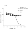
- FIG. 3 shows the ELISA reactivity of “m20A77_mIgG1” and “m24A1_mIgG1” to MerTKs derived from various animals. As shown in FIG. 3, both '20A77' and '24A1' showed similar reactivities to human, mouse, cynomolgus monkey, and rat antigens. From these results, both "20A77” and "24A1" were found to have high species cross-reactivity.
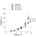
- ⁇ Test 3 Antitumor effect of the antibody of the present invention> Based on the following method, the antitumor effect of "20A77_mIgG2a" was evaluated. A syngeneic tumor model using the murine colon cancer cell line CT26 (ATCC) was used during the evaluation.
- a syngeneic tumor model using the murine colon cancer cell line CT26 (ATCC) was used during the evaluation.
- Figures 4 and 5 show the evaluation results. As shown by these results, "20A77_mIgG2a" significantly suppressed tumor compared to the mIgG2a administration group.
- tumors were harvested 19 days after tumor implantation, and tumor infiltrating lymphocyte (TIL) analysis was performed.
- TIL tumor infiltrating lymphocyte
- the tumor mass was added to "gentleMACS C Tubes" (Miltenyi Biotech) along with the enzyme solution derived from “Tumor Dissociation kit, mouse” (Miltenyi Biotech), and the “gentleMACS Octo Dissociator” was added. (Miltenyi Biotech) was used to perform tumor dispersion. After tumor dispersion, the cells were washed with 1% FBS/PBS, and stained using the flow cytometry (FCM) analysis antibody shown in Table 9. After staining, FCM analysis was performed using FACS calibur (BD).
- FCM flow cytometry
- M1 indicates inflammatory macrophages and M2 indicates immunosuppressive macrophages.
- the M1/M2 ratio is an indicator of characteristic changes in the tumor microenvironment due to MerTK inhibition. As shown in FIG. 6, administration of “20A77_mIgG2a” increased the M1/M2 ratio, indicating that the tumor microenvironment was changing to an anti-tumor environment.
- ⁇ Test 4 Effective dose of the antibody of the present invention> Based on the following method, the anti-tumor effects of "20A77_mIgG1" and “24A1_mIgG1” according to the dosage were evaluated. For the evaluation, a syngeneic tumor model using mouse colon cancer cell line CT26 (ATCC) was used as in "Test 3" above.
- a vehicle (1% Histidine buffer, pH 6.0) and "20A77_mIgG1" were intraperitoneally administered twice a week at a dose of 3, 10, or 30 mg/kg.
- a vehicle (1% histidine buffer, pH 6.0) and "24A1_mIgG1” were intraperitoneally administered twice a week at a dose of 3, 5, or 30 mg/kg. Tumor size was measured twice a week, and was completed 19 days after tumor implantation in the "20A77_mIgG1" administration group and 22 days after tumor implantation in the "24A1_mIgG1" administration group.
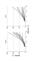
- FIGS. 7 to 10 show the evaluation results.
- an inhibitory effect on tumor growth was observed at any concentration compared to the solvent control group.
- a significant inhibitory effect was observed in the 3 mg/kg and 30 mg/kg administration groups.
- the "24A1_mIgG1" administration group significantly suppressed tumor growth, particularly in the 5 mg/kg and 30 mg/kg administration groups compared to the solvent control group.
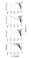
- ⁇ Test 5 Antitumor effect by combined administration of the antibody of the present invention and anti-PD-1 antibody> Based on the following method, the anti-tumor effects of "20A77_mIgG1" and “24A1_mIgG1" in combination with anti-PD-1 antibody were evaluated. For the evaluation, a syngeneic tumor model using mouse colon cancer cell line CT26 (ATCC) was used as in "Test 3" above. Anti-PD-1 antibodies are known to have anti-tumor effects.
- 11 and 12 show the evaluation results.
- "20A77_mIgG1” tended to enhance its anti-tumor effect when used in combination with the PD-1 antibody.
- the anti-tumor effect of "24A1_mIgG1” was enhanced when used in combination with the PD-1 antibody. From the above, it was found that the anti-tumor effect of the antibody of the present invention is enhanced when used in combination with an anti-PD-1 antibody.
- ⁇ Test 6 Evaluation of ability to inhibit binding of various ligands> Using '20A77' and '24A1', the ability of MerTK to inhibit binding of various ligands was evaluated.
- Human Gas6 (R&D systems) and human TULP1 (abcam) were used as MerTK ligands, and biotin labeling was carried out using "Biotin Labeling Kit-NH" (Dojindo).
- Gas6 is a ligand involved in antitumor effects, and antibodies having binding inhibitory activity between MerTK and Gas6 can exhibit antitumor effects.
- TULP1 is a ligand related to ocular toxicity, and antibodies having binding inhibitory activity between MerTK and TULP1 may have ocular toxicity. Therefore, an antibody with high Gas6-binding inhibitory activity and low TULP1-binding inhibitory activity can be effective as an antitumor agent with low ocular toxicity.
- a ligand binding inhibition test was performed as follows. First, human MerTK (R&D systems) was added to an ELISA plate (Nunc) at a concentration of 5 ⁇ g/ml and immobilized overnight at room temperature. After removing the solid phase liquid, 1% BSA/PBS was added as a blocking reaction, and the mixture was allowed to stand at room temperature for 1 hour. After blocking, the cells were washed with a 0.05% Tween20/PBS solution, and the antibody solution was reacted at room temperature for 1 hour as a primary reaction.
- biotinylated Gas6 or biotinylated TULP1 was added at a concentration of 10 nM or 40 nM, respectively, and allowed to react at room temperature for 1 hour.
- HRP-labeled streptavidin (Thermo Fisher Scientific) was reacted for 30 minutes as a tertiary reaction.
- TMB Dako
- 1N sulfuric acid was added to stop the reaction after 20 minutes, and the absorbance at 450 nm was measured.
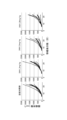
- 13 and 14 show the evaluation results.
- the positive control anti-goat MerTK polyclonal antibody
- “20A77” and “24A1” inhibited the binding between Gas6 and MerTK, but did not inhibit the binding between TULP1 and MerTK. From the above, it was found that the antibody of the present invention can be an antibody with good antitumor effect and low ocular toxicity.
- Test 7 Verification of ocular toxicity by in vivo ocular toxicity test> Based on the results confirmed in "Test 6" above, the in vivo ocular toxicity of the antibody of the present invention was verified based on the following method.
- mice Ocular toxicity studies were performed using mice (Balb/c). Each mouse was intravenously administered twice weekly with "20A77_mIgG1", “24A1_mIgG1", or an isotype control antibody (mIgG1) at a dose of 10 mg/kg.
- mIgG1 As a positive control for ocular toxicity, UNC569, a MerTK small molecule inhibitor, was administered orally at a dose of 150 mg/kg for 10 days.
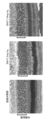
- the eyes On the 29th day after the first administration, the eyes were excised, Karnofsky fixation was performed, entrusted to Applied Medical Research, resin embedding, sectioning, and toluidine blue staining (hereinafter also referred to as "TB staining").
- TB staining An increase in TB-positive granules means that abnormal findings of the eye are progressing.
- FIGS. 17 Photographs of sections after TB staining are shown in FIGS. As shown in FIG. 17, in the UNC569-administered group, an increase in TB-positive granules was observed in the retinal pigment epithelium layer (arrow area). In contrast, no increase in TB-positive granules was observed in the group administered with "20A77_mIgG1" or "24A1_mIgG1".
- the antibody of the present invention is an antibody that exerts an antitumor effect and is less concerned about ocular toxicity.
- Test 8 Verification of toxicity capacity by in vivo ocular toxicity test> Based on the results confirmed in "Test 7" above, the toxicity capacity of the antibody of the present invention was verified based on the following method.
- mice Ocular toxicity studies were performed using mice (Balb/c). Each mouse was intravenously administered vehicle or "m20A77_mIgG1" at a dose of 3 or 10 mg/kg twice a week. On the 30th day after the first administration, the eyes were excised, fixed by Karnofsky, entrusted to Applied Medical Research, embedded in resin, sectioned, and stained with TB.
- FIGS. Photographs of sections after TB staining are shown in FIGS. As shown in FIG. 19, no increase in TB-positive granules was observed in any administration group. On the other hand, as shown in FIG. 20, no atrophy of the outer nuclear layer, which is an abnormal finding, was observed in the solvent control group, the 3 mg/kg administration group, and the 10 mg/kg administration group.
- ⁇ Test 9 Production of humanized antibody>
- chimeric antibodies hereinafter also referred to as "human chimeric antibodies”
- mouse heavy and light chain variable regions and human IgG1 heavy and ⁇ light chain constant regions were prepared.
- a human chimeric antibody was produced in the same manner as in "(3) Production of recombinant mouse antibody and modification of antibody sequence" above.
- the obtained human chimeric antibody was subjected to humanization of the antibody sequence based on the following method.
- the sequence of each antibody variable region was humanized by the CDR grafting method.
- the humanized sequences were designed with reference to the method of Tsurushita et al. (2005. Design of humanized antibodies: From anti-Tac to Zenapax. Methods 36:69-83.).
- a three-dimensional molecular model of mouse antibody was created by a standard method. Based on this molecular model, in the amino acid sequence of the framework region, residues thought to be important for CDR structure formation and residues thought to be essential for reaction with antigen were deduced.
- cDNA sequence databases of human antibody heavy and light chain variable regions were searched for sequences highly homologous to the heavy and light chain variable regions of each antibody.
- a sequence is designed by linking the sequence of the framework portion of the searched human antibody sequence and the CDR sequence of each antibody, and residues thought to be essential for CDR structure formation or reaction with the antigen are added thereto.
- the sequences were further grafted to design the humanized antibody sequences “hz20A77” and “hz24A1” shown in Table 10.
- the CDR sequences possessed by the humanized antibody sequence are the same as those of the original mouse antibody.
- the designed antibody was produced as follows.
- the DNA sequence encoding the amino acid sequence of the heavy chain variable region was pcDNA3.4 together with a human IgG1 constant region (hereinafter also referred to as "hIgG1LALA") into which an ⁇ -lactalbumin signal peptide was connected and an LALA sequence was introduced.
- the vector was cloned using "In-Fusion HD Cloning Kit” (Takara Bio).
- the DNA sequence encoding the amino acid sequence of the light chain variable region was connected to the ⁇ -lactalbumin signal peptide in the same manner as the heavy chain variable region, and transferred to the pcDNA3.4 vector together with the human Ig ⁇ constant region by "In-Fusion HD Cloning Kit” (Takara Bio) was used for cloning.
- Antibodies were then produced in the same manner as in "(3) Production of recombinant mouse antibodies and antibody sequence modification" above.
Abstract
Description
例えば、がん細胞に発現するMerTKの活性化ががん細胞の増殖や遊走を誘導することや、腫瘍微小環境におけるMerTKが免疫抑制に寄与すること等が報告されている(例えば、非特許文献1、非特許文献2等)。
そのため、MerTKは、癌免疫分野における新たな標的分子として注目されている。 MerTK is known to have various functions.
For example, it has been reported that activation of MerTK expressed in cancer cells induces cancer cell proliferation and migration, and that MerTK in the tumor microenvironment contributes to immunosuppression (see, for example, Non-Patent
Therefore, MerTK has attracted attention as a new target molecule in the cancer immunology field.
例えば、非特許文献3には、マウスへのMerTK低分子阻害剤(UNC569)の投与により、視細胞層においてトルイジンブルー陽性顆粒や空砲の増加が認められ、眼毒性が生じたことが報告されている。
非特許文献4には、エフェロサイトーシス阻害能や抗腫瘍効果を有する抗MerTK抗体が、カニクイザルに眼毒性をもたらしたことが報告されている。 On the other hand, inhibition of MerTK with its ligand can produce side effects.
For example, Non-Patent
Non-Patent Document 4 reports that an anti-MerTK antibody with efferocytosis inhibitory ability and anti-tumor effect caused ocular toxicity in cynomolgus monkeys.
[要件A]
重鎖CDR1が、配列番号2で表されるアミノ酸配列である。
重鎖CDR2が、配列番号3で表されるアミノ酸配列である。
重鎖CDR3が、配列番号4で表されるアミノ酸配列である。
軽鎖CDR1が、配列番号6で表されるアミノ酸配列である。
軽鎖CDR2が、配列番号7で表されるアミノ酸配列である。
軽鎖CDR3が、配列番号8で表されるアミノ酸配列である。 (1) An antibody or fragment thereof that specifically binds to MerTK, wherein the amino acid sequences of CDR1 to 3 of the heavy and light chains satisfy all of the following [requirement A].
[Requirement A]
Heavy chain CDR1 is the amino acid sequence represented by SEQ ID NO:2.
Heavy chain CDR2 is the amino acid sequence represented by SEQ ID NO:3.
Heavy chain CDR3 is the amino acid sequence represented by SEQ ID NO:4.
Light chain CDR1 is the amino acid sequence represented by SEQ ID NO:6.
Light chain CDR2 is the amino acid sequence represented by SEQ ID NO:7.
Light chain CDR3 is the amino acid sequence represented by SEQ ID NO:8.
[要件B]
重鎖可変領域が、配列番号1で表されるアミノ酸配列である。
軽鎖可変領域が、配列番号5で表されるアミノ酸配列である。 (2) The antibody or fragment thereof according to (1), which further satisfies all of [requirement B] below.
[Requirement B]
The heavy chain variable region is the amino acid sequence represented by SEQ ID NO:1.
The light chain variable region is the amino acid sequence represented by SEQ ID NO:5.
[要件C]
重鎖CDR1が、配列番号10で表されるアミノ酸配列である。
重鎖CDR2が、配列番号11で表されるアミノ酸配列である。
重鎖CDR3が、配列番号12で表されるアミノ酸配列である。
軽鎖CDR1が、配列番号14で表されるアミノ酸配列である。
軽鎖CDR2が、配列番号15で表されるアミノ酸配列である。
軽鎖CDR3が、配列番号16で表されるアミノ酸配列である。 (3) An antibody or a fragment thereof that specifically binds to MerTK, wherein the amino acid sequences of CDR1 to 3 of the heavy and light chains satisfy all of [Requirement C] below.
[Requirement C]
Heavy chain CDR1 is the amino acid sequence represented by SEQ ID NO:10.
Heavy chain CDR2 is the amino acid sequence represented by SEQ ID NO:11.
Heavy chain CDR3 is the amino acid sequence represented by SEQ ID NO:12.
Light chain CDR1 is the amino acid sequence represented by SEQ ID NO:14.
Light chain CDR2 is the amino acid sequence represented by SEQ ID NO:15.
Light chain CDR3 is the amino acid sequence represented by SEQ ID NO:16.
[要件D]
重鎖可変領域が、配列番号9で表されるアミノ酸配列である。
軽鎖可変領域が、配列番号13で表されるアミノ酸配列である。
[要件E]
重鎖可変領域が、配列番号23で表されるアミノ酸配列である。
軽鎖可変領域が、配列番号24で表されるアミノ酸配列である。 (4) The antibody or fragment thereof according to (3), which further satisfies either [requirement D] or [requirement E] below.
[Requirement D]
The heavy chain variable region is the amino acid sequence represented by SEQ ID NO:9.
The light chain variable region is the amino acid sequence represented by SEQ ID NO:13.
[Requirement E]
The heavy chain variable region is the amino acid sequence represented by SEQ ID NO:23.
The light chain variable region is the amino acid sequence represented by SEQ ID NO:24.
[要件F]
重鎖CDR1が、配列番号2で表されるアミノ酸配列である。
重鎖CDR2が、配列番号18で表されるアミノ酸配列である。
重鎖CDR3が、配列番号4で表されるアミノ酸配列である。
軽鎖CDR1が、配列番号6で表されるアミノ酸配列である。
軽鎖CDR2が、配列番号20で表されるアミノ酸配列である。
軽鎖CDR3が、配列番号8で表されるアミノ酸配列である。 (5) An antibody or a fragment thereof that specifically binds to MerTK, wherein the amino acid sequences of CDR1 to 3 of the heavy and light chains satisfy all of [Requirement F] below.
[Requirement F]
Heavy chain CDR1 is the amino acid sequence represented by SEQ ID NO:2.
Heavy chain CDR2 is the amino acid sequence represented by SEQ ID NO:18.
Heavy chain CDR3 is the amino acid sequence represented by SEQ ID NO:4.
Light chain CDR1 is the amino acid sequence represented by SEQ ID NO:6.
Light chain CDR2 is the amino acid sequence represented by SEQ ID NO:20.
Light chain CDR3 is the amino acid sequence represented by SEQ ID NO:8.
[要件G]
重鎖可変領域が、配列番号17で表されるアミノ酸配列である。
軽鎖可変領域が、配列番号19で表されるアミノ酸配列である。
[要件H]
重鎖可変領域が、配列番号21で表されるアミノ酸配列である。
軽鎖可変領域が、配列番号22で表されるアミノ酸配列である。 (6) The antibody or fragment thereof according to (5), which further satisfies either [Requirement G] or [Requirement H] below.
[Requirement G]
The heavy chain variable region is the amino acid sequence represented by SEQ ID NO:17.
The light chain variable region is the amino acid sequence represented by SEQ ID NO:19.
[Requirement H]
The heavy chain variable region is the amino acid sequence represented by SEQ ID NO:21.
The light chain variable region is the amino acid sequence represented by SEQ ID NO:22.
本発明に係るMerTKに特異的に結合する抗体又はその断片(以下、「本発明の抗体又はその断片」ともいう。)は、後述のとおり3つの態様を包含する。 <Antibody or fragment thereof that specifically binds to MerTK>
Antibodies or fragments thereof that specifically bind to MerTK according to the present invention (hereinafter also referred to as "antibodies or fragments thereof of the present invention") include three aspects as described below.
従来、抗MerTK抗体は、MerTKと、MerTKのリガンドとの結合阻害により抗腫瘍効果を奏することが知られていた。
抗MerTK抗体は、例えば、MerTKと、MerTKのリガンドのうちGas6との結合を阻害する結果、腫瘍微小環境を変化させ、抗腫瘍効果を奏する。 The antibody or fragment thereof of the present invention is an anti-MerTK antibody or fragment thereof.
Conventionally, anti-MerTK antibodies have been known to exhibit antitumor effects by inhibiting binding between MerTK and ligands of MerTK.
Anti-MerTK antibodies, for example, inhibit the binding of MerTK to Gas6, which is a ligand of MerTK, thereby altering the tumor microenvironment and exerting an antitumor effect.
抗体又はその断片がMerTKに特異的に結合するかどうかは、Enzyme-Linked Immuno Sorbent Assay(ELISA法)により特定する。 In the present invention, the phrase "specifically binds to MerTK" means that the antibody or fragment thereof of the present invention specifically binds to MerTK protein and has little or no binding to other proteins.
Whether or not an antibody or fragment thereof specifically binds to MerTK is determined by an Enzyme-Linked Immuno Sorbent Assay (ELISA method).
本発明の抗体又はその断片のうち、第1の態様に係る抗体又はその断片は、重鎖及び軽鎖のCDR1乃至3のアミノ酸配列が、下記[要件A]を全て満たす。
[要件A]
重鎖CDR1が、配列番号2で表されるアミノ酸配列である。
重鎖CDR2が、配列番号3で表されるアミノ酸配列である。
重鎖CDR3が、配列番号4で表されるアミノ酸配列である。
軽鎖CDR1が、配列番号6で表されるアミノ酸配列である。
軽鎖CDR2が、配列番号7で表されるアミノ酸配列である。
軽鎖CDR3が、配列番号8で表されるアミノ酸配列である。 (Antibody or fragment thereof according to the first aspect)
Among the antibodies or fragments thereof of the present invention, in the antibody or fragment thereof according to the first aspect, the amino acid sequences of CDR1 to 3 of the heavy and light chains satisfy all of [Requirement A] below.
[Requirement A]
Heavy chain CDR1 is the amino acid sequence represented by SEQ ID NO:2.
Heavy chain CDR2 is the amino acid sequence represented by SEQ ID NO:3.
Heavy chain CDR3 is the amino acid sequence represented by SEQ ID NO:4.
Light chain CDR1 is the amino acid sequence represented by SEQ ID NO:6.
Light chain CDR2 is the amino acid sequence represented by SEQ ID NO:7.
Light chain CDR3 is the amino acid sequence represented by SEQ ID NO:8.

[要件B]
重鎖可変領域が、配列番号1で表されるアミノ酸配列である。
軽鎖可変領域が、配列番号5で表されるアミノ酸配列である。 The antibody or fragment thereof according to the first aspect preferably satisfies all of the following [requirement B] from the viewpoint that the effects of the present invention are likely to be exhibited.
[Requirement B]
The heavy chain variable region is the amino acid sequence represented by SEQ ID NO:1.
The light chain variable region is the amino acid sequence represented by SEQ ID NO:5.

本発明の抗体又はその断片のうち、第2の態様に係る抗体又はその断片は、重鎖及び軽鎖のCDR1乃至3のアミノ酸配列が、下記[要件C]を全て満たす。
[要件C]
重鎖CDR1が、配列番号10で表されるアミノ酸配列である。
重鎖CDR2が、配列番号11で表されるアミノ酸配列である。
重鎖CDR3が、配列番号12で表されるアミノ酸配列である。
軽鎖CDR1が、配列番号14で表されるアミノ酸配列である。
軽鎖CDR2が、配列番号15で表されるアミノ酸配列である。
軽鎖CDR3が、配列番号16で表されるアミノ酸配列である。 (Antibody or fragment thereof according to the second aspect)
Among the antibodies or fragments thereof of the present invention, in the antibody or fragment thereof according to the second aspect, the amino acid sequences of CDR1 to 3 of the heavy and light chains satisfy all of [Requirement C] below.
[Requirement C]
Heavy chain CDR1 is the amino acid sequence represented by SEQ ID NO:10.
Heavy chain CDR2 is the amino acid sequence represented by SEQ ID NO:11.
Heavy chain CDR3 is the amino acid sequence represented by SEQ ID NO:12.
Light chain CDR1 is the amino acid sequence represented by SEQ ID NO:14.
Light chain CDR2 is the amino acid sequence represented by SEQ ID NO:15.
Light chain CDR3 is the amino acid sequence represented by SEQ ID NO:16.

[要件D]
重鎖可変領域が、配列番号9で表されるアミノ酸配列である。
軽鎖可変領域が、配列番号13で表されるアミノ酸配列である。
[要件E]
重鎖可変領域が、配列番号23で表されるアミノ酸配列である。
軽鎖可変領域が、配列番号24で表されるアミノ酸配列である。 The antibody or fragment thereof according to the second aspect preferably satisfies either [requirement D] or [requirement E] below, from the viewpoint that the effects of the present invention are more likely to be exhibited.
[Requirement D]
The heavy chain variable region is the amino acid sequence represented by SEQ ID NO:9.
The light chain variable region is the amino acid sequence represented by SEQ ID NO:13.
[Requirement E]
The heavy chain variable region is the amino acid sequence represented by SEQ ID NO:23.
The light chain variable region is the amino acid sequence represented by SEQ ID NO:24.

なお、「hz24A1」、「hz24A1_hIgG1LALA」は、いずれも、「24A1」をヒト化した抗体である。 Antibodies or fragments thereof according to the second aspect include, in the examples, antibodies designated as "24A1", "24A1_mIgG1", "24A1_mIgG2a", "24A1_hIgG1LALA", "hz24A1", and "hz24A1_hIgG1LALA".
Both "hz24A1" and "hz24A1_hIgG1LALA" are humanized antibodies of "24A1".
本発明の抗体又はその断片のうち、第3の態様に係る抗体又はその断片は、重鎖及び軽鎖のCDR1乃至3のアミノ酸配列が、下記[要件F]を全て満たす。
[要件F]
重鎖CDR1が、配列番号2で表されるアミノ酸配列である。
重鎖CDR2が、配列番号18で表されるアミノ酸配列である。
重鎖CDR3が、配列番号4で表されるアミノ酸配列である。
軽鎖CDR1が、配列番号6で表されるアミノ酸配列である。
軽鎖CDR2が、配列番号20で表されるアミノ酸配列である。
軽鎖CDR3が、配列番号8で表されるアミノ酸配列である。 (Antibody or fragment thereof according to the third aspect)
Among the antibodies or fragments thereof of the present invention, in the antibody or fragment thereof according to the third aspect, the amino acid sequences of CDR1 to 3 of the heavy and light chains satisfy all of [Requirement F] below.
[Requirement F]
Heavy chain CDR1 is the amino acid sequence represented by SEQ ID NO:2.
Heavy chain CDR2 is the amino acid sequence represented by SEQ ID NO:18.
Heavy chain CDR3 is the amino acid sequence represented by SEQ ID NO:4.
Light chain CDR1 is the amino acid sequence represented by SEQ ID NO:6.
Light chain CDR2 is the amino acid sequence represented by SEQ ID NO:20.
Light chain CDR3 is the amino acid sequence represented by SEQ ID NO:8.

[要件G]
重鎖可変領域が、配列番号17で表されるアミノ酸配列である。
軽鎖可変領域が、配列番号19で表されるアミノ酸配列である。
[要件H]
重鎖可変領域が、配列番号21で表されるアミノ酸配列である。
軽鎖可変領域が、配列番号22で表されるアミノ酸配列である。 The antibody or fragment thereof according to the third aspect preferably satisfies either [requirement G] or [requirement H] below, from the viewpoint that the effects of the present invention are more likely to be exhibited.
[Requirement G]
The heavy chain variable region is the amino acid sequence represented by SEQ ID NO:17.
The light chain variable region is the amino acid sequence represented by SEQ ID NO:19.
[Requirement H]
The heavy chain variable region is the amino acid sequence represented by SEQ ID NO:21.
The light chain variable region is the amino acid sequence represented by SEQ ID NO:22.

なお、「hz20A77」、「hz20A77_hIgG1LALA」は、いずれも、「m20A77」をヒト化した抗体である。 Antibodies or fragments thereof according to the third aspect include, in the Examples, antibodies designated as “m20A77”, “m20A77_mIgG1”, “m20A77_hIgG1LALA”, “hz20A77”, “hz20A77_hIgG1LALA”.
"hz20A77" and "hz20A77_hIgG1LALA" are both humanized antibodies of "m20A77".
本発明の抗体又はその断片の製造方法は特に限定されないが、各種抗体(モノクローナル抗体、ポリクローナル抗体等)や、各種ポリペプチドの製造方法として従来知られる方法を採用できる。 <Method for producing the antibody of the present invention or a fragment thereof>
Although the method for producing the antibody or fragment thereof of the present invention is not particularly limited, conventionally known methods for producing various antibodies (monoclonal antibodies, polyclonal antibodies, etc.) and various polypeptides can be employed.
これらの遺伝子、発現ベクターは、従来知られる方法によって作製できる。 The present invention includes genes encoding the antibodies of the present invention or fragments thereof, expression vectors containing the genes, and the like.
These genes and expression vectors can be produced by conventionally known methods.
本発明の抗体又はその断片は、良好な抗腫瘍効果を奏するため、抗腫瘍剤として好ましく使用できる。 <Antineoplastic agent of the present invention>
Since the antibody or fragment thereof of the present invention exhibits good antitumor effects, it can be preferably used as an antitumor agent.
本発明の抗腫瘍剤は、本発明の抗体又はその断片以外の薬効成分と併用して用いてもよい。このような薬効成分は、本発明の抗体又はその断片以外とともに配合された単一の剤として投与してもよいし、本発明の抗体又はその断片以外とは別の剤として調製し、本発明の抗腫瘍剤と併用投与してもよい。 The anti-tumor agent of the present invention may or may not contain a medicinal ingredient other than the antibody or fragment thereof of the present invention.
The anti-tumor agent of the present invention may be used in combination with active ingredients other than the antibody or fragment thereof of the present invention. Such a medicinal ingredient may be administered as a single agent formulated with other than the antibody or fragment thereof of the present invention, or may be prepared as a separate agent from other than the antibody or fragment thereof of the present invention, may be administered in combination with an anti-tumor agent.
免疫チェックポイント阻害剤としては、抗PD-1抗体、抗PD-L1抗体、抗CTLA4抗体等が挙げられる。 Preferred medicinal ingredients that can be used in combination with the antibody or fragment thereof of the present invention include immune checkpoint inhibitors.
Immune checkpoint inhibitors include anti-PD-1 antibodies, anti-PD-L1 antibodies, anti-CTLA4 antibodies and the like.
以下の方法に基づき、MerTKに対するモノクローナル抗体を取得した。 <Test 1: Establishment of monoclonal antibody against MerTK>
A monoclonal antibody against MerTK was obtained according to the following method.
ヒトMerTKタンパク質のアミノ酸配列(UniProt番号:Q12866)に基づき、5’末端にKozak翻訳開始配列、3’側に翻訳終止コドンを挿入したDNAを遺伝子合成した(GENEWIZ社のサービスを利用)。
「In-Fusion HD Cloning Kit」(タカラバイオ)を用いて、合成したDNAを「pcDNA3.4」(サーモフィッシャーサイエンティフィック)のクローニングサイトに挿入し、ヒトMerTK発現ベクターを得た。 (1-1) Construction of human MerTK expression vector Based on the amino acid sequence of the human MerTK protein (UniProt number: Q12866), DNA was gene-synthesized by inserting a Kozak translation initiation sequence at the 5' end and a translation stop codon at the 3' end. (Using the service of GENEWIZ).
Using "In-Fusion HD Cloning Kit" (Takara Bio), the synthesized DNA was inserted into the cloning site of "pcDNA3.4" (Thermo Fisher Scientific) to obtain a human MerTK expression vector.
マウスMerTKタンパク質のアミノ酸配列(UniProt番号:Q60805)に基づき、上記ヒトMerTK発現ベクターと同様の方法によって、マウスMerTK発現ベクターを得た。 (1-2) Construction of mouse MerTK expression vector Based on the amino acid sequence of mouse MerTK protein (UniProt No.: Q60805), a mouse MerTK expression vector was obtained in the same manner as for the human MerTK expression vector.
ヒトMerTKマウスFc融合タンパク質(以下、「hMerTKECD-mFc」ともいう。)として、ヒトMerTKタンパク質(UniProt番号Q12866)の1番目から505番目のアミノ酸と、マウスIgG2a重鎖(UniProt番号P01863)の114番目から330番目のアミノ酸と、を融合させたタンパク質を設計した。このアミノ酸配列を元に発現ベクターを構築し、hMerTKECD-mFc発現ベクターを得た。
次いで、hMerTKECD-mFc発現ベクターを導入プラスミドとして、「Expi293 expression system」(サーモフィッシャーサイエンティフィック社)を用いて一過性発現を行った。
一過性発現後、培養上清を回収し、「HiTrap ProteinG HPカラム」(サイティバ)を用いたアフィニティ精製を行った。
アフィニティ精製した溶出液を濃縮し、「Sephadex 200pg 16/60」(サイティバ)を用いたゲルろ過精製を行った。
ゲルろ過精製の溶出画分を、SDS-PAGE法にてバンドを確認し、バンドが確認できたフラクションを回収し、hMerTKECD-mFcとして使用した。 (2-1) Expression and purification of human MerTK mouse Fc fusion protein As a human MerTK mouse Fc fusion protein (hereinafter also referred to as "hMerTKECD-mFc"), the first to 505th human MerTK protein (UniProt No. Q12866) A protein was designed that fused amino acids with amino acids 114 to 330 of the mouse IgG2a heavy chain (UniProt No. P01863). An expression vector was constructed based on this amino acid sequence to obtain the hMerTKECD-mFc expression vector.
Then, transient expression was performed using the "Expi293 expression system" (Thermo Fisher Scientific) using the hMerTKECD-mFc expression vector as an introduction plasmid.
After transient expression, the culture supernatant was collected and subjected to affinity purification using "HiTrap Protein G HP column" (Cytiva).
The affinity-purified eluate was concentrated and purified by gel filtration using "Sephadex 200pg 16/60" (Cytiva).
Elution fractions of gel filtration purification were confirmed for bands by SDS-PAGE, and fractions in which bands were confirmed were collected and used as hMerTKECD-mFc.
マウスMerTKマウスFc融合タンパク質(以下、「mMerTKECD-mFc」ともいう。)として、マウスMerTKタンパク質(UniProt番号Q60805)の1番目から497番目のアミノ酸と、マウスIgG2a重鎖(UniProt番号P01863)の114番目から330番目のアミノ酸と、を融合させたタンパク質を設計した。このアミノ酸配列を元に、上記のhMerTKECD-mFc調製方法と同様の方法でタンパク質の発現及び精製を行い、mMerTKECD-mFcを得た。 (2-2) Expression and purification of mouse MerTK mouse Fc fusion protein As mouse MerTK mouse Fc fusion protein (hereinafter also referred to as “mMerTKECD-mFc”), 1st to 497th mouse MerTK protein (UniProt No. Q60805) A protein was designed that fused amino acids with amino acids 114 to 330 of the mouse IgG2a heavy chain (UniProt No. P01863). Based on this amino acid sequence, the protein was expressed and purified in the same manner as in the method for preparing hMerTKECD-mFc described above to obtain mMerTKECD-mFc.
ヒトMerTK発現ベクターを導入プラスミドとして、「Nucleofector」(Lonza)を用いて、マウスリンパ腫細胞株EL4に遺伝子導入を行った。
遺伝子導入後、培地に「G418」(ナカライテスク)を添加し、細胞を96ウェルプレートに播種し培養を行った。
培養後、G418耐性的な増殖が見られた細胞について、抗ヒトMerTK抗体(R&D systems, MAB8912)を用いたフローサイトメトリーにより、ヒトMerTKの発現を確認した。最終的に単クローン由来の細胞株を選択し、これをヒトMerTK安定発現EL4細胞(以下、「hMerTK-EL4」ともいう。)として以下の試験に使用した。 (3-1) Construction of EL4 Cells Stably Expressing Human MerTK A human MerTK expression vector was used as an introduction plasmid, and gene transfer was performed into a mouse lymphoma cell line EL4 using “Nucleofector” (Lonza).
After gene introduction, "G418" (Nacalai Tesque) was added to the medium, and the cells were seeded in a 96-well plate and cultured.
After culturing, expression of human MerTK was confirmed by flow cytometry using an anti-human MerTK antibody (R&D systems, MAB8912) for cells in which G418-resistant proliferation was observed. Finally, a cell line derived from a single clone was selected and used in the following tests as human MerTK stably expressing EL4 cells (hereinafter also referred to as "hMerTK-EL4").
マウスMerTK発現ベクターを導入プラスミドとして、「Nucleofector」(Lonza)を用いて、マウスpro-B細胞株Ba/F3に遺伝子導入を行った。
遺伝子導入後、培地に「G418」(ナカライテスク)を添加し、細胞を96ウェルプレートに播種し培養を行った。
培養後、G418耐性的な増殖が見られた細胞について、抗マウスMerTK抗体(R&D systems, AF591)を用いたフローサイトメトリーにより、マウスMerTKの発現を確認した。最終的に単クローン由来の細胞株を選択し、これをマウスMerTK安定発現BaF/3細胞(以下、「mMerTK-Ba/F3」ともいう。)として、以下の試験に使用した。 (3-2) Construction of BaF/3 Cells Stably Expressing Mouse MerTK Using the mouse MerTK expression vector as an introduction plasmid, the mouse pro-B cell line Ba/F3 was transfected using “Nucleofector” (Lonza).
After gene introduction, "G418" (Nacalai Tesque) was added to the medium, and the cells were seeded in a 96-well plate and cultured.
After culturing, expression of mouse MerTK was confirmed by flow cytometry using an anti-mouse MerTK antibody (R&D systems, AF591) for cells in which G418-resistant growth was observed. Finally, a cell line derived from a single clone was selected and used in the following tests as mouse MerTK stably expressing BaF/3 cells (hereinafter also referred to as "mMerTK-Ba/F3").
以下の2通りの方法に基づき、MerTK精製タンパク質、又はMerTK安定発現細胞をMerTK遺伝子欠損マウス(The Jackson Laboratory, B6;129-MerTK<tm1G4l>/J)に免疫した。
免疫後、単離したリンパ節細胞を用いてMerTKに対するモノクローナル抗体を作製した。 (4) Preparation of monoclonal antibodies against MerTK Immunize MerTK gene-deficient mice (The Jackson Laboratory, B6; 129-MerTK<tm1G4l>/J) with MerTK purified protein or MerTK stably expressing cells based on the following two methods. bottom.
After immunization, isolated lymph node cells were used to raise monoclonal antibodies against MerTK.
抗原として、hMerTKECD-mFc、及びmMerTKECD-mFc(いずれもMerTK精製タンパク質)を用いた。
各抗原を、アジュバント「TiterMaxGold」(TiterMax)と混合し、マウス1匹あたりの抗原投与量が100μgとなるように足底静脈投与を行った。投与後、7日後及び10日後に、マウス1匹あたりの抗原投与量を10μgに設定した点以外は上記同様に追加免疫を行った。
免疫終了後、マウスから膝窩リンパ節を採取して細胞懸濁液を調製したのち、これをSP2/0-Ag14ミエローマ細胞と混合し、電気式細胞融合装置(「ECFG21」、ネッパジーン)による電気式細胞融合を行った。
融合後の細胞を、「ClonaCellTM-HY Medium D」(「ST-03804」、STEMCELL)に懸濁し、プラスチックシャーレに播種した。
播種後、8~10日後に形成されたコロニーを、ハイブリドーマ用培地を分注した96ウェルプラスチックプレート中に単離し、その培養上清を下記評価に用いた。
なお、ハイブリドーマ用培地としては、RPMI1640(「A1049101」、サーモフィッシャーサイエンティフィック)に対し、1/50量の「Nutridom-CS」(「11363743001」、メルク)、及び、1/50量の「HAT Supplement」(「21060017」、サーモフィッシャーサイエンティフィック)を添加したものを用いた。 (Immunization method-1)
hMerTKECD-mFc and mMerTKECD-mFc (both MerTK purified proteins) were used as antigens.
Each antigen was mixed with an adjuvant "TiterMaxGold" (TiterMax) and administered to the plantar vein so that the dose of the antigen per mouse was 100 µg. 7 days and 10 days after administration, booster immunization was performed in the same manner as above, except that the dose of antigen per mouse was set at 10 μg.
After completion of immunization, popliteal lymph nodes were collected from the mice to prepare a cell suspension, which was then mixed with SP2/0-Ag14 myeloma cells and electrolyzed using an electric cell fusion device (“ECFG21”, Nepagene). expression cell fusion was performed.
The fused cells were suspended in “ClonaCell™-HY Medium D” (“ST-03804”, STEMCELL) and seeded in a plastic petri dish.
Colonies formed 8 to 10 days after seeding were isolated in a 96-well plastic plate into which a hybridoma medium was dispensed, and the culture supernatant was used for the following evaluations.
As a hybridoma medium, 1/50 amount of "Nutridom-CS"("11363743001", Merck) and 1/50 amount of "HAT Supplement"("21060017", Thermo Fisher Scientific) was used.
抗原として、hMerTK-EL4、及びmMerTK-Ba/F3(いずれもMerTK安定発現細胞)を用いた。これらの細胞はPBSの懸濁溶液として調製した。
各抗原を、アジュバント「TiterMaxGold」(TiterMax)と混合し、マウス1匹あたりの抗原投与量が1×10⁷個(細胞数)となるように足底静脈投与を行った。投与後、7日後及び10日後に、マウス1匹あたりの抗原投与量を1×10⁵個に設定し、腹腔内投与した点以外は上記同様に追加免疫を行った。
免疫終了後、上記(免疫方法-1)と同様の方法で培養上清を得て、下記評価に用いた。 (Immunization method-2)
As antigens, hMerTK-EL4 and mMerTK-Ba/F3 (both stably expressing MerTK cells) were used. These cells were prepared as suspensions in PBS.
Each antigen was mixed with an adjuvant “TiterMaxGold” (TiterMax) and administered to the plantar vein so that the dose of the antigen per mouse was 1×10 7 cells (number of cells). 7 days and 10 days after the administration, a booster immunization was performed in the same manner as described above, except that the dose of the antigen per mouse was set at 1×10 5 and intraperitoneal administration was performed.
After completion of immunization, the culture supernatant was obtained in the same manner as described above (immunization method-1) and used for the following evaluations.
上記で得られた各培養上清を用いて、ヒト及びマウスのMerTKに結合能を持つハイブリドーマクローンをスクリーニングした。
具体的には、ヒトMerTKを内在的に発現するヒトメラノーマ株G361、及びmMerTK-Ba/F3を用いて、フローサイトメトリーによる抗体結合性の評価を行った。
約15,000クローンをスクリーニングした結果、ヒト及びマウスMerTKに対する結合能を有するハイブリドーマクローンが76クローン得られた。 (5) Antibody Screening Each culture supernatant obtained above was used to screen for hybridoma clones capable of binding to human and mouse MerTK.
Specifically, human melanoma strain G361, which endogenously expresses human MerTK, and mMerTK-Ba/F3 were used to evaluate antibody binding properties by flow cytometry.
As a result of screening about 15,000 clones, 76 hybridoma clones having binding ability to human and mouse MerTK were obtained.
上記で得られたハイブリドーマクローンを以下の各評価に供し、MerTKに対するモノクローナル抗体としての機能を確認した。 <Test 2: Antibody evaluation>
The hybridoma clones obtained above were subjected to the following evaluations to confirm their function as monoclonal antibodies against MerTK.
以下の方法に基づき、各クローンについて、MerTKリン酸化阻害活性を評価した。該活性が高いほど、MerTKへの結合能が高いことを意味する。 (1) Phosphorylation inhibitory activity Each clone was evaluated for MerTK phosphorylation inhibitory activity based on the following method. A higher activity means a higher binding ability to MerTK.
ヒトMerTK安定発現細胞(hMerTK-EL4)を10%FBSを含む「DMEM high glucose培地」(Thermo Fisher Scientific)で培養した。
培養後、細胞をPBSで洗浄し、無血清培地に懸濁して、1×10⁶ cells/wellとなるように96ウェルに播種し、37℃に3時間インキュベートした。
インキュベート後、各クローンを添加し、1時間反応させ、200nM ヒトGas6(R&D systems)で10分間刺激し、hMerTK-EL4を遠心分離し回収した。
回収したhMerTK-EL4を細胞溶解液に溶解し、得られた溶解液中のリン酸化MerTKタンパク質の量を、「Human Phospho-Mer DuoSet IC ELISA」(R&D systems)を用いて測定した。
なお、細胞溶解液としては、ミリQ水に、1%の「IGEPAL CA-630」(SIGMA)、1x濃度分の「Halt Protease and Phosphatase Inhibitor Single-Use Inhibitor Cocktail」(Thermo Fisher Scientific)を添加したものを用いた。 (Method)
Human MerTK stably expressing cells (hMerTK-EL4) were cultured in "DMEM high glucose medium" (Thermo Fisher Scientific) containing 10% FBS.
After culturing, the cells were washed with PBS, suspended in serum-free medium, seeded in 96 wells at 1×10 6 cells/well, and incubated at 37° C. for 3 hours.
After incubation, each clone was added, reacted for 1 hour, stimulated with 200 nM human Gas6 (R&D systems) for 10 minutes, and hMerTK-EL4 was collected by centrifugation.
The recovered hMerTK-EL4 was dissolved in a cell lysate, and the amount of phosphorylated MerTK protein in the resulting lysate was measured using "Human Phospho-Mer DuoSet IC ELISA"(R&D Systems).
As the cell lysate, 1% "IGEPAL CA-630" (SIGMA) and 1x concentration of "Halt Protease and Phosphatase Inhibitor Single-Use Inhibitor Cocktail" (Thermo Fisher Scientific) were added to Milli-Q water. used things.
76クローンのうち、14クローンにおいてリン酸化阻害活性が見られた。
該14クローンのうち、特に高い阻害活性を示したクローンとして「20A77」及び「24A1」を選抜し、以下の試験に供した。
これらのクローンのリン酸化阻害活性を図1に示す。図1に示されるとおり、「20A77」及び「24A1」は、それぞれ濃度依存的に、Gas6とMerTKとの結合、及びMerTKのリン酸化を阻害し、MerTKへの結合能が高いことがわかった。 (result)
Phosphorylation inhibitory activity was observed in 14 clones out of 76 clones.
Among the 14 clones, "20A77" and "24A1" were selected as clones exhibiting particularly high inhibitory activity and subjected to the following tests.
Phosphorylation inhibitory activity of these clones is shown in FIG. As shown in FIG. 1, it was found that "20A77" and "24A1" each inhibited the binding of Gas6 to MerTK and phosphorylation of MerTK in a concentration-dependent manner, and had high binding ability to MerTK.
以下の方法に基づき、「20A77」及び「24A1」のそれぞれについて、その免疫グロブリン遺伝子可変領域の配列の解析を行った。
ハイブリドーマ細胞を拡大培養し、「RNeasy mini kit」(Qiagen)によりRNAを抽出し、「SuperScript III First-Strand Synthesis Super mix」(Invitrogen)によるcDNA合成を行った。
合成したcDNAをテンプレートに用い、PCR法により抗体可変領域配列を増幅した。なお、重鎖、及び軽鎖ともに可変領域上流、及び定常領域下流を認識するプライマーを使用し増幅した。
得られたDNA断片を、「Zeroblunt TOPO TA Cloning and Sequencing kit」(ThermoFisher Scientific)のキットに添付のベクターにクローニングを行い、DNA配列解析を行った。
得られた抗体配列について、Kabatらの方法(Sequences of Proteins of Immunological Interests, Fifth edition, NIH Publication No. 91-3242, U.S. Department of Health and Human Services, 1991)に従ってCDR領域を決定した。 (2) Antibody sequence analysis Based on the following method, the sequences of the immunoglobulin gene variable regions of "20A77" and "24A1" were analyzed.
The hybridoma cells were expanded and cultured, RNA was extracted using "RNeasy mini kit" (Qiagen), and cDNA was synthesized using "SuperScript III First-Strand Synthesis Super mix" (Invitrogen).
Using the synthesized cDNA as a template, the antibody variable region sequence was amplified by PCR. Both heavy and light chains were amplified using primers recognizing the upstream of the variable region and the downstream of the constant region.
The resulting DNA fragment was cloned into the vector attached to the "Zeroblunt TOPO TA Cloning and Sequencing kit" (ThermoFisher Scientific) and subjected to DNA sequence analysis.
The obtained antibody sequences were subjected to region determination according to the method of Kabat et al. (Sequences of Proteins of Immunological Interests, Fifth edition, NIH Publication No. 91-3242, U.S. Department of Health and Human Services, CDR 91).
なお、以下、表中の略号は以下の意味を有する。
VH: 重鎖可変領域
CDRH: 重鎖CDR
VL: 軽鎖可変領域
CDRL: 軽鎖CDR A summary of the analyzed antibody sequences and their CDR region sequences is shown in Table 7 below.
The abbreviations in the tables below have the following meanings.
VH: heavy chain variable region CDRH: heavy chain CDR
VL: light chain variable region CDRL: light chain CDR

表7に示された各抗体配列と、マウスIgG1重鎖及びκ軽鎖又はマウスIgG2a重鎖及びκ軽鎖と、を連結し、マウスIgG1、及びマウスIgG2a抗体遺伝子配列を設計した。設計した抗体配列を元に、発現ベクターを構築し、抗体発現ベクターを得た。
抗体発現ベクターを導入プラスミドとして、「Expi293 expression system」(Thermo Fisher Scientific)を用いて一過性発現を行った。
一過性発現を行った培養上清を回収し、「Ab-Capcher ExTraカラム」(プロテノバ)を用いたアフィニティ精製を行った。
アフィニティ精製した溶出液を濃縮し、ヒスチジンバッファー(1% Histidine、pH6.0)を緩衝液に使用し、「Sephadex 200pg 16/60」(サイティバ)を用いたゲルろ過精製を行った。
ゲルろ過精製の溶出画分について、SDS-PAGE法によってバンドを確認した。バンドが確認できたフラクションを回収し、「20A77」及び「24A1」のマウスIgG1型抗体及びマウスIgG2a型抗体を得た(それぞれ、「20A77_mIgG1」、「20A77_mIgG2a」、「24A1_mIgG1」、「24A1_mIgG2a」ともいう。)。 (3) Production of recombinant mouse antibody and modification of antibody sequence Each antibody sequence shown in Table 7 is ligated with mouse IgG1 heavy chain and κ light chain or mouse IgG2a heavy chain and κ light chain, mouse IgG1, and mouse IgG2a antibody gene sequences were designed. Based on the designed antibody sequence, an expression vector was constructed to obtain an antibody expression vector.
Transient expression was carried out using the antibody expression vector as an introduction plasmid and the "Expi293 expression system" (Thermo Fisher Scientific).
The culture supernatant after transient expression was collected and affinity purification was performed using "Ab-Capcher ExTra column" (Protenova).
The affinity-purified eluate was concentrated and subjected to gel filtration purification using "Sephadex 200 pg 16/60" (Cytiva) using a histidine buffer (1% Histidine, pH 6.0) as a buffer.
The band was confirmed by the SDS-PAGE method for the elution fraction of the gel filtration purification. Fractions in which bands could be confirmed were collected, and mouse IgG1 type antibodies and mouse IgG2a type antibodies of "20A77" and "24A1" were obtained (respectively, "20A77_mIgG1", "20A77_mIgG2a", "24A1_mIgG1", "24A1_mIgG2a"). .).
また、「m20A77」についても上記同様の方法により、マウスIgG1型抗体(「m20A77_mIgG1」ともいう。)を得た。 Furthermore, amino acid alterations in the CDR regions were attempted. The introduction sites for sequence modification are as shown in FIG. 2, and modifications were performed on sequences that are highly likely to undergo cleavage reaction and deamination reaction in the CDR region. Antibodies with modified CDR sequences are also referred to as "m20A77." The sequence information is shown in Table 8 below.
Further, with respect to "m20A77", a mouse IgG1 type antibody (also referred to as "m20A77_mIgG1") was obtained by the same method as above.

各種動物(ヒト、マウス、カニクイザル、ラット)に由来するMerTKに対する、上記で得られた抗体クローンの交差反応性を、ELISA法によって確認した。
ELISA法においては、各種動物(ヒト、マウス、カニクイザル、ラット)に由来するMerTKのヒトFc融合タンパク質を抗原として用いた。具体的には、ヒト、マウスMerTK抗原としては、「Recombinant Human Mer Fc Chimera Protein」(R&D systems)、及び「Recombinant Mouse Mer Fc Chimera Protein」(R&D systems)を用いた。カニクイザルMerTK、及びラットMerTK抗原としては、hMerTKECD-mFcやmMerTKECD-mFcと同様の手法で調製した、カニクイザルMerTKECD-hFc(以下、「cynoMerTKECD-hFc」ともいう。)、ラットMerTKECD-hFc(以下、「ratMerTKECD-hFc」ともいう。)を用いた。 (4) Cross-reactivity to MerTK derived from various animals The cross-reactivity of the antibody clones obtained above to MerTK derived from various animals (human, mouse, cynomolgus monkey, rat) was confirmed by ELISA.
In the ELISA method, human Fc fusion proteins of MerTK derived from various animals (human, mouse, cynomolgus monkey, rat) were used as antigens. Specifically, "Recombinant Human Mer Fc Chimera Protein"(R&D systems) and "Recombinant Mouse Mer Fc Chimera Protein"(R&D systems) were used as human and mouse MerTK antigens. As cynomolgus monkey MerTK and rat MerTK antigens, cynomolgus monkey MerTKECD-hFc (hereinafter also referred to as "cynoMerTKECD-hFc") and rat MerTKECD-hFc (hereinafter, " ratMerTKECD-hFc”) was used.
まず、各抗原をそれぞれELISAプレート(Nunc)に5μg/mlの濃度で添加し、室温で一晩固相した。
固相液を除去後、ブロッキング反応として1% BSA/PBSを添加し、室温で1時間静置した。
ブロッキング後、0.05% Tween20/PBS液で洗浄し、一次反応として抗体液を室温で1時間反応させた。
洗浄後、二次反応としてHRP標識抗体(GE Healthcare)を1時間反応させた。洗浄後、発色基質液としてTMB(Dako)を加え発色させ、10分後、1N硫酸を加え反応を停止し、450nmの吸光度を測定した。 The ELISA method was performed by the following method.
First, each antigen was added to an ELISA plate (Nunc) at a concentration of 5 μg/ml and immobilized overnight at room temperature.
After removing the solid phase liquid, 1% BSA/PBS was added as a blocking reaction, and the mixture was allowed to stand at room temperature for 1 hour.
After blocking, the cells were washed with a 0.05% Tween20/PBS solution, and the antibody solution was reacted at room temperature for 1 hour as a primary reaction.
After washing, an HRP-labeled antibody (GE Healthcare) was reacted for 1 hour as a secondary reaction. After washing, TMB (Dako) was added as a coloring substrate solution to develop color, 1N sulfuric acid was added to stop the reaction after 10 minutes, and the absorbance at 450 nm was measured.
図3に示すように、「20A77」及び「24A1」のいずれも、ヒト、マウス、カニクイザル、及びラットの抗原にほぼ同様の反応性を示した。これらの結果から、「20A77」及び「24A1」のいずれも、高い種交差反応性を有することがわかった。 FIG. 3 shows the ELISA reactivity of “m20A77_mIgG1” and “m24A1_mIgG1” to MerTKs derived from various animals.
As shown in FIG. 3, both '20A77' and '24A1' showed similar reactivities to human, mouse, cynomolgus monkey, and rat antigens. From these results, both "20A77" and "24A1" were found to have high species cross-reactivity.
以下の方法に基づき、「20A77_mIgG2a」の抗腫瘍効果を評価した。評価の際には、マウス大腸がん細胞株CT26(ATCC)を用いた同系腫瘍モデルを使用した。 <Test 3: Antitumor effect of the antibody of the present invention>
Based on the following method, the antitumor effect of "20A77_mIgG2a" was evaluated. A syngeneic tumor model using the murine colon cancer cell line CT26 (ATCC) was used during the evaluation.
腫瘍移植後5日目に群分けし、アイソタイプコントロール抗体であるmIgG2a、及び「20A77_mIgG2a」を、腹腔内に10mg/kgの容量で1週間に2回の頻度で投与した。
投与後、腫瘍サイズを週に2回の頻度で計測を開始し、腫瘍移植後19日目に終了した。なお、腫瘍サイズは、ノギスを用いて腫瘍の短径と長径をそれぞれ計測し、以下の式から算出した。
腫瘍サイズ=(短径(mm))2×(長径(mm))×3.14/6 Mice (Balb/c) were subcutaneously implanted with 5×10 6 cells of CT26 per mouse right flank.
Five days after tumor transplantation, the mice were divided into groups, and isotype control antibodies mIgG2a and "20A77_mIgG2a" were intraperitoneally administered at a dose of 10 mg/kg twice a week.
After administration, tumor size measurement was started twice a week and ended 19 days after tumor implantation. The tumor size was calculated from the following formula by measuring the minor axis and major axis of the tumor using vernier calipers.
Tumor size = (minor diameter (mm)) 2 × (major diameter (mm)) × 3.14/6
TIL解析に際しては、まず、腫瘍採取後、「Tumor Dissociation kit,mouse」(ミルテニーバイオテク)由来の酵素液とともに、「gentleMACS C Tubes」(ミルテニーバイオテク)に腫瘍塊を加え、「gentleMACS Octo Dissociator」(ミルテニーバイオテク)を用いて、腫瘍分散を行った。
腫瘍分散後、1% FBS/PBSで洗浄後、表9記載のフローサイトメトリー(FCM)解析用抗体を用いて染色を行った。染色後、FACS calibur(BD)を用いてFCM解析を行った。
図6に、「M1/M2比」を示す。M1は炎症性マクロファージを示し、M2は免疫抑制性のマクロファージを示す。M1/M2比は、MerTK阻害による腫瘍微小環境の特徴的な変化を示す指標である。図6に示されるとおり、「20A77_mIgG2a」の投与によって、M1/M2比が増加し、腫瘍微小環境が抗腫瘍の環境に変化していることが示された。 Furthermore, tumors were harvested 19 days after tumor implantation, and tumor infiltrating lymphocyte (TIL) analysis was performed.
For the TIL analysis, first, after collecting the tumor, the tumor mass was added to "gentleMACS C Tubes" (Miltenyi Biotech) along with the enzyme solution derived from "Tumor Dissociation kit, mouse" (Miltenyi Biotech), and the "gentleMACS Octo Dissociator" was added. (Miltenyi Biotech) was used to perform tumor dispersion.
After tumor dispersion, the cells were washed with 1% FBS/PBS, and stained using the flow cytometry (FCM) analysis antibody shown in Table 9. After staining, FCM analysis was performed using FACS calibur (BD).
FIG. 6 shows the "M1/M2 ratio". M1 indicates inflammatory macrophages and M2 indicates immunosuppressive macrophages. The M1/M2 ratio is an indicator of characteristic changes in the tumor microenvironment due to MerTK inhibition. As shown in FIG. 6, administration of “20A77_mIgG2a” increased the M1/M2 ratio, indicating that the tumor microenvironment was changing to an anti-tumor environment.

以下の方法に基づき、「20A77_mIgG1」及び「24A1_mIgG1」の投与量に応じた抗腫瘍効果を評価した。評価の際には、上記「試験3」同様に、マウス大腸がん細胞株CT26(ATCC)を用いた同系腫瘍モデルを使用した。 <Test 4: Effective dose of the antibody of the present invention>
Based on the following method, the anti-tumor effects of "20A77_mIgG1" and "24A1_mIgG1" according to the dosage were evaluated. For the evaluation, a syngeneic tumor model using mouse colon cancer cell line CT26 (ATCC) was used as in "
「24A1_mIgG1」投与群では、溶媒(1% Histidine buffer、pH6.0)及び「24A1_mIgG1」を、腹腔内に3、5、又は30mg/kgの容量で1週間に2回の頻度で投与した。
腫瘍サイズは週に2回の頻度で計測し、「20A77_mIgG1」投与群では腫瘍移植後19日目に、「24A1_mIgG1」投与群では腫瘍移植後22日目に終了した。 In the "20A77_mIgG1" administration group, a vehicle (1% Histidine buffer, pH 6.0) and "20A77_mIgG1" were intraperitoneally administered twice a week at a dose of 3, 10, or 30 mg/kg.
In the "24A1_mIgG1" administration group, a vehicle (1% histidine buffer, pH 6.0) and "24A1_mIgG1" were intraperitoneally administered twice a week at a dose of 3, 5, or 30 mg/kg.
Tumor size was measured twice a week, and was completed 19 days after tumor implantation in the "20A77_mIgG1" administration group and 22 days after tumor implantation in the "24A1_mIgG1" administration group.
図7及び8に示されるとおり、「20A77_mIgG1」投与群では、いずれの濃度においても、溶媒対照群に比較して腫瘍増殖の抑制効果が認められた。
特に、3mg/kg、及び30mg/kg投与群において有意な抑制効果が認められた。
図9及び10に示されるとおり、「24A1_mIgG1」投与群では、溶媒対照群に比較して、特に、5mg/kg、及び30mg/kg投与群において有意に腫瘍増殖を抑制した。 7 to 10 show the evaluation results.
As shown in FIGS. 7 and 8, in the "20A77_mIgG1" administration group, an inhibitory effect on tumor growth was observed at any concentration compared to the solvent control group.
In particular, a significant inhibitory effect was observed in the 3 mg/kg and 30 mg/kg administration groups.
As shown in Figures 9 and 10, the "24A1_mIgG1" administration group significantly suppressed tumor growth, particularly in the 5 mg/kg and 30 mg/kg administration groups compared to the solvent control group.
以下の方法に基づき、「20A77_mIgG1」及び「24A1_mIgG1」と、抗PD-1抗体との併用による抗腫瘍効果を評価した。評価の際には、上記「試験3」同様に、マウス大腸がん細胞株CT26(ATCC)を用いた同系腫瘍モデルを使用した。
抗PD-1抗体は、抗腫瘍効果を有することが知られる。 <Test 5: Antitumor effect by combined administration of the antibody of the present invention and anti-PD-1 antibody>
Based on the following method, the anti-tumor effects of "20A77_mIgG1" and "24A1_mIgG1" in combination with anti-PD-1 antibody were evaluated. For the evaluation, a syngeneic tumor model using mouse colon cancer cell line CT26 (ATCC) was used as in "
Anti-PD-1 antibodies are known to have anti-tumor effects.
「mIgG1」:「mIgG1」を5mg/kg
「m20A77」:「20A77_mIgG1」を3mg/kg
「24A1」:「24A1_mIgG1」を5mg/kg
「αPD-1」:抗マウスPD-1抗体(「29F.1A12」、BioXcell)を0.3mg/kg
「m20A77+αPD-1」:「m20A77_mIgG1」と抗マウスPD-1抗体とをそれぞれ3mg/kg、0.3mg/kg
「24A1+αPD-1」:「24A1_mIgG1」と抗マウスPD-1抗体とをそれぞれ5mg/kg、0.3mg/kg The following antibodies were administered to each administration group twice a week. Dosing continued until day 19 after tumor implantation.
"mIgG1": 5 mg/kg of "mIgG1"
"m20A77": "20A77_mIgG1" at 3 mg/kg
"24A1": "24A1_mIgG1" at 5 mg/kg
"αPD-1": 0.3 mg/kg of anti-mouse PD-1 antibody ("29F.1A12", BioXcell)
"m20A77 + αPD-1": "m20A77_mIgG1" and anti-mouse PD-1 antibody at 3 mg/kg and 0.3 mg/kg, respectively
"24A1 + αPD-1": "24A1_mIgG1" and anti-mouse PD-1 antibody at 5 mg/kg and 0.3 mg/kg, respectively
これらの図に示されるとおり、「20A77_mIgG1」は、PD-1抗体との併用によって抗腫瘍効果が増強される傾向にあった。
また、「24A1_mIgG1」は、PD-1抗体との併用によって抗腫瘍効果が増強された。
以上から、本発明の抗体は、抗PD-1抗体と併用することで、抗腫瘍効果が増強することがわかった。 11 and 12 show the evaluation results.
As shown in these figures, "20A77_mIgG1" tended to enhance its anti-tumor effect when used in combination with the PD-1 antibody.
In addition, the anti-tumor effect of "24A1_mIgG1" was enhanced when used in combination with the PD-1 antibody.
From the above, it was found that the anti-tumor effect of the antibody of the present invention is enhanced when used in combination with an anti-PD-1 antibody.
「20A77」及び「24A1」を用いて、MerTKの各種リガンド結合阻害能を評価した。
MerTKのリガンドとしては、ヒトGas6(R&D systems)及びヒトTULP1(abcam)を用い、それぞれ「Biotin Labeling Kit-NH」(Dojindo)を用いてビオチン標識を行った。
Gas6は抗腫瘍効果に関係するリガンドであり、MerTKとGas6との結合阻害活性を有する抗体は抗腫瘍効果を奏し得る。
TULP1は、眼毒性に関係するリガンドであり、MerTKとTULP1との結合阻害活性を有する抗体は眼毒性を有し得る。
したがって、Gas6との結合阻害活性が高く、かつ、TULP1との結合阻害活性が低い抗体は、眼毒性の低い抗腫瘍剤として有効であり得る。 <Test 6: Evaluation of ability to inhibit binding of various ligands>
Using '20A77' and '24A1', the ability of MerTK to inhibit binding of various ligands was evaluated.
Human Gas6 (R&D systems) and human TULP1 (abcam) were used as MerTK ligands, and biotin labeling was carried out using "Biotin Labeling Kit-NH" (Dojindo).
Gas6 is a ligand involved in antitumor effects, and antibodies having binding inhibitory activity between MerTK and Gas6 can exhibit antitumor effects.
TULP1 is a ligand related to ocular toxicity, and antibodies having binding inhibitory activity between MerTK and TULP1 may have ocular toxicity.
Therefore, an antibody with high Gas6-binding inhibitory activity and low TULP1-binding inhibitory activity can be effective as an antitumor agent with low ocular toxicity.
まず、ELISA用プレート(Nunc)に、ヒトMerTK(R&D systems)を5μg/mlの濃度で添加し、室温で一晩固相した。固相液を除去後、ブロッキング反応として1% BSA/PBSを添加し、室温で1時間静置した。
ブロッキング後、0.05% Tween20/PBS液で洗浄し、一次反応として抗体液を室温で1時間反応させた。
洗浄後、二次反応として、ビオチン化Gas6又はビオチン化TULP1を、それぞれ10nM又は40nMの濃度で添加し、室温で1時間反応させた。
洗浄後、三次反応としてHRP標識ストレプトアビジン(サーモフィッシャーサイエンティフィック)を30分間反応させた。
洗浄後、発色基質液としてTMB(Dako)を加え発色させ、20分後、1N硫酸を加え反応を停止し、450nmの吸光度を測定した。 A ligand binding inhibition test was performed as follows.
First, human MerTK (R&D systems) was added to an ELISA plate (Nunc) at a concentration of 5 μg/ml and immobilized overnight at room temperature. After removing the solid phase liquid, 1% BSA/PBS was added as a blocking reaction, and the mixture was allowed to stand at room temperature for 1 hour.
After blocking, the cells were washed with a 0.05% Tween20/PBS solution, and the antibody solution was reacted at room temperature for 1 hour as a primary reaction.
After washing, as a secondary reaction, biotinylated Gas6 or biotinylated TULP1 was added at a concentration of 10 nM or 40 nM, respectively, and allowed to react at room temperature for 1 hour.
After washing, HRP-labeled streptavidin (Thermo Fisher Scientific) was reacted for 30 minutes as a tertiary reaction.
After washing, TMB (Dako) was added as a coloring substrate solution to develop color, 1N sulfuric acid was added to stop the reaction after 20 minutes, and the absorbance at 450 nm was measured.
これらの図に示されるとおり、陽性対照(抗ヤギMerTKポリクローナル抗体)は、Gas6及びTULP1のいずれについても、MerTKとの結合を阻害した。
これに対し、「20A77」及び「24A1」は、Gas6とMerTKとの結合は阻害したものの、TULP1とMerTKとの結合は阻害しなかった。
以上から、本発明の抗体は、良好な抗腫瘍効果を有しつつ、眼毒性が低い抗体であり得ることがわかった。 13 and 14 show the evaluation results.
As shown in these figures, the positive control (anti-goat MerTK polyclonal antibody) inhibited binding of both Gas6 and TULP1 to MerTK.
In contrast, "20A77" and "24A1" inhibited the binding between Gas6 and MerTK, but did not inhibit the binding between TULP1 and MerTK.
From the above, it was found that the antibody of the present invention can be an antibody with good antitumor effect and low ocular toxicity.
しかし、ほぼ全ての抗体が、Gas6とMerTKとの結合、及びTULP1とMerTKとの結合の両方を阻害した。
Gas6とMerTKとの結合を強く阻害し、かつ、TULP1とMerTKとの結合を阻害しない抗体は、「20A77」及び「24A1」のみだった。 A large number of anti-MerTK antibodies were evaluated for their ability to inhibit ligand binding in the same manner as described above. Some of the results are shown in FIGS.
However, almost all antibodies inhibited both the binding of Gas6 to MerTK and the binding of TULP1 to MerTK.
Only "20A77" and "24A1" strongly inhibited the binding of Gas6 to MerTK and did not inhibit the binding of TULP1 to MerTK.
上記「試験6」で確認された結果を踏まえ、以下の方法に基づき、本発明の抗体によるin vivoでの眼毒性について検証した。 <Test 7: Verification of ocular toxicity by in vivo ocular toxicity test>
Based on the results confirmed in "Test 6" above, the in vivo ocular toxicity of the antibody of the present invention was verified based on the following method.
各マウスに、「20A77_mIgG1」、「24A1_mIgG1」、又はアイソタイプコントロール抗体(mIgG1)を、10mg/kgの容量で、1週間に2回の頻度で静脈内投与した。
眼毒性の陽性対照として、MerTK低分子阻害剤であるUNC569を150mg/kgの容量で10日間経口投与した。
初回投与後29日目に眼を摘出し、カルノフスキー固定を行い、アプライドメディカルリサーチに委託し、樹脂包埋及び切片作製、トルイジンブルー染色(以下、「TB染色」ともいう。)を行った。TB陽性顆粒の増加は、目の異常所見が進行していることを意味する。 Ocular toxicity studies were performed using mice (Balb/c).
Each mouse was intravenously administered twice weekly with "20A77_mIgG1", "24A1_mIgG1", or an isotype control antibody (mIgG1) at a dose of 10 mg/kg.
As a positive control for ocular toxicity, UNC569, a MerTK small molecule inhibitor, was administered orally at a dose of 150 mg/kg for 10 days.
On the 29th day after the first administration, the eyes were excised, Karnofsky fixation was performed, entrusted to Applied Medical Research, resin embedding, sectioning, and toluidine blue staining (hereinafter also referred to as "TB staining"). An increase in TB-positive granules means that abnormal findings of the eye are progressing.
図17のとおり、UNC569投与群では、網膜色素上皮層にTB陽性顆粒の増加が認められた(矢印部位)。
これに対し、「20A77_mIgG1」又は「24A1_mIgG1」を投与した群では、TB陽性顆粒の増加は認められなかった。 Photographs of sections after TB staining are shown in FIGS.
As shown in FIG. 17, in the UNC569-administered group, an increase in TB-positive granules was observed in the retinal pigment epithelium layer (arrow area).
In contrast, no increase in TB-positive granules was observed in the group administered with "20A77_mIgG1" or "24A1_mIgG1".
以上から、本発明の抗体は、抗腫瘍効果を奏する一方で眼毒性への懸念が少ない抗体であることが示唆された。 As shown in FIG. 18, no atrophy of the outer nuclear layer (abnormal findings correlated with decreased visual acuity) was observed in any administration group.
From the above, it was suggested that the antibody of the present invention is an antibody that exerts an antitumor effect and is less concerned about ocular toxicity.
上記「試験7」で確認された結果を踏まえ、以下の方法に基づき、本発明の抗体の毒性容量について検証した。 <Test 8: Verification of toxicity capacity by in vivo ocular toxicity test>
Based on the results confirmed in "Test 7" above, the toxicity capacity of the antibody of the present invention was verified based on the following method.
各マウスに、溶媒、又は「m20A77_mIgG1」を3、又は10mg/kgの容量で、1週間に2回の頻度で静脈内投与した。
初回投与後30日目に眼を摘出し、カルノフスキー固定を行い、アプライドメディカルリサーチに委託し、樹脂包埋及び切片作製、TB染色を行った。 Ocular toxicity studies were performed using mice (Balb/c).
Each mouse was intravenously administered vehicle or "m20A77_mIgG1" at a dose of 3 or 10 mg/kg twice a week.
On the 30th day after the first administration, the eyes were excised, fixed by Karnofsky, entrusted to Applied Medical Research, embedded in resin, sectioned, and stained with TB.
図19のとおり、いずれの投与群においても、TB陽性顆粒の増加は認められなかった。一方で、図20のとおり、溶媒対照群、3mg/kg、10mg/kgいずれの投与群においても、異常所見である外顆粒層の萎縮は認められなかった。 Photographs of sections after TB staining are shown in FIGS.
As shown in FIG. 19, no increase in TB-positive granules was observed in any administration group. On the other hand, as shown in FIG. 20, no atrophy of the outer nuclear layer, which is an abnormal finding, was observed in the solvent control group, the 3 mg/kg administration group, and the 10 mg/kg administration group.
「m20A77」及び「24A1」について、マウス重鎖及び軽鎖可変領域と、ヒトIgG1重鎖及びκ軽鎖定常領域のキメラ抗体(以下、「ヒトキメラ抗体」ともいう。)を作製した。ヒトキメラ抗体は、上記「(3)組換えマウス抗体の作製及び抗体配列改変」と同様の方法で作製した。 <Test 9: Production of humanized antibody>
For "m20A77" and "24A1", chimeric antibodies (hereinafter also referred to as "human chimeric antibodies") of mouse heavy and light chain variable regions and human IgG1 heavy and κ light chain constant regions were prepared. A human chimeric antibody was produced in the same manner as in "(3) Production of recombinant mouse antibody and modification of antibody sequence" above.
まず、各抗体可変領域の配列について、CDR移植法によるヒト化を行った。ヒト化配列の設計は、Tsurushitaら(2005. Design of humanized antibodies: From anti-Tac to Zenapax. Methods 36:69-83.)の方法を参考に実施した。
次いで、マウス抗体の3次元分子モデルを定法により作成した。この分子モデルを元に、フレームワーク領域のアミノ酸配列の中で、CDRの構造形成に重要と考えられる残基、また抗原との反応に必須であると考えられた残基を推定した。
この作業と並行して、ヒト抗体重鎖可変領域及び軽鎖可変領域のcDNA配列データベースの中から、各抗体の重鎖可変領域、軽鎖可変領域に相同性の高い配列を検索した。
検索したヒト抗体配列のフレームワーク部分の配列と、各抗体のCDR配列と、を連結した配列を設計し、そこへ、CDR構造形成又は抗原との反応に必須であると考えられた残基の配列をさらに移植して、表10に示すヒト化抗体配列「hz20A77」及び「hz24A1」を設計した。
なお、ヒト化抗体配列の持つCDR配列は元となったマウス抗体のものと同一である。 The obtained human chimeric antibody was subjected to humanization of the antibody sequence based on the following method.
First, the sequence of each antibody variable region was humanized by the CDR grafting method. The humanized sequences were designed with reference to the method of Tsurushita et al. (2005. Design of humanized antibodies: From anti-Tac to Zenapax. Methods 36:69-83.).
Next, a three-dimensional molecular model of mouse antibody was created by a standard method. Based on this molecular model, in the amino acid sequence of the framework region, residues thought to be important for CDR structure formation and residues thought to be essential for reaction with antigen were deduced.
In parallel with this work, cDNA sequence databases of human antibody heavy and light chain variable regions were searched for sequences highly homologous to the heavy and light chain variable regions of each antibody.
A sequence is designed by linking the sequence of the framework portion of the searched human antibody sequence and the CDR sequence of each antibody, and residues thought to be essential for CDR structure formation or reaction with the antigen are added thereto. The sequences were further grafted to design the humanized antibody sequences “hz20A77” and “hz24A1” shown in Table 10.
The CDR sequences possessed by the humanized antibody sequence are the same as those of the original mouse antibody.

重鎖可変領域のアミノ酸配列をコードするDNA配列は、α-ラクトアルブミンシグナルペプチドを接続したうえで、LALA配列を導入したヒトIgG1定常領域(以下、「hIgG1LALA」ともいう。)とともに、pcDNA3.4ベクターに、「In-Fusion HD Cloning Kit」(タカラバイオ)を用いてクローニングした。
軽鎖可変領域のアミノ酸配列をコードするDNA配列は、重鎖可変領域と同様にα-ラクトアルブミンシグナルペプチドを接続したうえで、ヒトIgκ定常領域とともにpcDNA3.4ベクターに、「In-Fusion HD Cloning Kit」(タカラバイオ)を用いてクローニングした。
次いで、抗体を上記「(3)組換えマウス抗体の作製及び抗体配列改変」と同様の方法で作製した。 The designed antibody was produced as follows.
The DNA sequence encoding the amino acid sequence of the heavy chain variable region was pcDNA3.4 together with a human IgG1 constant region (hereinafter also referred to as "hIgG1LALA") into which an α-lactalbumin signal peptide was connected and an LALA sequence was introduced. The vector was cloned using "In-Fusion HD Cloning Kit" (Takara Bio).
The DNA sequence encoding the amino acid sequence of the light chain variable region was connected to the α-lactalbumin signal peptide in the same manner as the heavy chain variable region, and transferred to the pcDNA3.4 vector together with the human Igκ constant region by "In-Fusion HD Cloning Kit" (Takara Bio) was used for cloning.
Antibodies were then produced in the same manner as in "(3) Production of recombinant mouse antibodies and antibody sequence modification" above.
図21に示されるとおり、得られたヒト化抗体(「hz20A77_hIgG1LALA」、及び「hz24A1_hIgG1LALA」)は、いずれも、元となったヒトキメラ抗体(「m20A77_hIgG1LALA」、及び「24A1_hIgG1LALA」)と同程度又はそれ以上の反応性を示した。
この結果から、作製されたヒト化抗体群は、ヒトMerTKに対する反応性を十分に有することが示された。 Antigen-binding activity of the prepared antibodies was confirmed by ELISA. The results are shown in FIG.
As shown in FIG. 21 , the resulting humanized antibodies (“hz20A77_hIgG1LALA” and “hz24A1_hIgG1LALA”) were both comparable to or greater than the original human chimeric antibodies (“m20A77_hIgG1LALA” and “24A1_hIgG1LALA”). showed reactivity.
These results indicated that the produced humanized antibody group had sufficient reactivity to human MerTK.
Claims (8)
- 重鎖及び軽鎖のCDR1乃至3のアミノ酸配列が、下記[要件A]を全て満たす、MerTKに特異的に結合する抗体又はその断片。
[要件A]
重鎖CDR1が、配列番号2で表されるアミノ酸配列である。
重鎖CDR2が、配列番号3で表されるアミノ酸配列である。
重鎖CDR3が、配列番号4で表されるアミノ酸配列である。
軽鎖CDR1が、配列番号6で表されるアミノ酸配列である。
軽鎖CDR2が、配列番号7で表されるアミノ酸配列である。
軽鎖CDR3が、配列番号8で表されるアミノ酸配列である。 An antibody or fragment thereof that specifically binds to MerTK, wherein the amino acid sequences of heavy and light chain CDRs 1 to 3 satisfy all of [Requirement A] below.
[Requirement A]
Heavy chain CDR1 is the amino acid sequence represented by SEQ ID NO:2.
Heavy chain CDR2 is the amino acid sequence represented by SEQ ID NO:3.
Heavy chain CDR3 is the amino acid sequence represented by SEQ ID NO:4.
Light chain CDR1 is the amino acid sequence represented by SEQ ID NO:6.
Light chain CDR2 is the amino acid sequence represented by SEQ ID NO:7.
Light chain CDR3 is the amino acid sequence represented by SEQ ID NO:8. - さらに、下記[要件B]を全て満たす、請求項1に記載の抗体又はその断片。
[要件B]
重鎖可変領域が、配列番号1で表されるアミノ酸配列である。
軽鎖可変領域が、配列番号5で表されるアミノ酸配列である。 The antibody or fragment thereof according to claim 1, which further satisfies all of the following [requirement B].
[Requirement B]
The heavy chain variable region is the amino acid sequence represented by SEQ ID NO:1.
The light chain variable region is the amino acid sequence represented by SEQ ID NO:5. - 重鎖及び軽鎖のCDR1乃至3のアミノ酸配列が、下記[要件C]を全て満たす、MerTKに特異的に結合する抗体又はその断片。
[要件C]
重鎖CDR1が、配列番号10で表されるアミノ酸配列である。
重鎖CDR2が、配列番号11で表されるアミノ酸配列である。
重鎖CDR3が、配列番号12で表されるアミノ酸配列である。
軽鎖CDR1が、配列番号14で表されるアミノ酸配列である。
軽鎖CDR2が、配列番号15で表されるアミノ酸配列である。
軽鎖CDR3が、配列番号16で表されるアミノ酸配列である。 An antibody or fragment thereof that specifically binds to MerTK, wherein the amino acid sequences of heavy and light chain CDRs 1 to 3 satisfy all of [Requirement C] below.
[Requirement C]
Heavy chain CDR1 is the amino acid sequence represented by SEQ ID NO:10.
Heavy chain CDR2 is the amino acid sequence represented by SEQ ID NO:11.
Heavy chain CDR3 is the amino acid sequence represented by SEQ ID NO:12.
Light chain CDR1 is the amino acid sequence represented by SEQ ID NO:14.
Light chain CDR2 is the amino acid sequence represented by SEQ ID NO:15.
Light chain CDR3 is the amino acid sequence represented by SEQ ID NO:16. - さらに、下記[要件D]又は[要件E]のいずれかを全て満たす、請求項3に記載の抗体又はその断片。
[要件D]
重鎖可変領域が、配列番号9で表されるアミノ酸配列である。
軽鎖可変領域が、配列番号13で表されるアミノ酸配列である。
[要件E]
重鎖可変領域が、配列番号23で表されるアミノ酸配列である。
軽鎖可変領域が、配列番号24で表されるアミノ酸配列である。 4. The antibody or fragment thereof according to claim 3, which further satisfies either [requirement D] or [requirement E] below.
[Requirement D]
The heavy chain variable region is the amino acid sequence represented by SEQ ID NO:9.
The light chain variable region is the amino acid sequence represented by SEQ ID NO:13.
[Requirement E]
The heavy chain variable region is the amino acid sequence represented by SEQ ID NO:23.
The light chain variable region is the amino acid sequence represented by SEQ ID NO:24. - 重鎖及び軽鎖のCDR1乃至3のアミノ酸配列が、下記[要件F]を全て満たす、MerTKに特異的に結合する抗体又はその断片。
[要件F]
重鎖CDR1が、配列番号2で表されるアミノ酸配列である。
重鎖CDR2が、配列番号18で表されるアミノ酸配列である。
重鎖CDR3が、配列番号4で表されるアミノ酸配列である。
軽鎖CDR1が、配列番号6で表されるアミノ酸配列である。
軽鎖CDR2が、配列番号20で表されるアミノ酸配列である。
軽鎖CDR3が、配列番号8で表されるアミノ酸配列である。 An antibody or fragment thereof that specifically binds to MerTK, wherein the amino acid sequences of heavy and light chain CDRs 1 to 3 satisfy all of [Requirement F] below.
[Requirement F]
Heavy chain CDR1 is the amino acid sequence represented by SEQ ID NO:2.
Heavy chain CDR2 is the amino acid sequence represented by SEQ ID NO:18.
Heavy chain CDR3 is the amino acid sequence represented by SEQ ID NO:4.
Light chain CDR1 is the amino acid sequence represented by SEQ ID NO:6.
Light chain CDR2 is the amino acid sequence represented by SEQ ID NO:20.
Light chain CDR3 is the amino acid sequence represented by SEQ ID NO:8. - さらに、下記[要件G]又は[要件H]のいずれかを全て満たす、請求項5に記載の抗体又はその断片。
[要件G]
重鎖可変領域が、配列番号17で表されるアミノ酸配列である。
軽鎖可変領域が、配列番号19で表されるアミノ酸配列である。
[要件H]
重鎖可変領域が、配列番号21で表されるアミノ酸配列である。
軽鎖可変領域が、配列番号22で表されるアミノ酸配列である。 6. The antibody or fragment thereof according to claim 5, which further satisfies either [requirement G] or [requirement H] below.
[Requirement G]
The heavy chain variable region is the amino acid sequence represented by SEQ ID NO:17.
The light chain variable region is the amino acid sequence represented by SEQ ID NO:19.
[Requirement H]
The heavy chain variable region is the amino acid sequence represented by SEQ ID NO:21.
The light chain variable region is the amino acid sequence represented by SEQ ID NO:22. - 請求項1から6のいずれか1項に記載の抗体又はその断片を含む、抗腫瘍剤。 An antitumor agent comprising the antibody or fragment thereof according to any one of claims 1 to 6.
- さらに、免疫チェックポイント阻害剤を含む請求項7に記載の抗腫瘍剤。 The antitumor agent according to claim 7, further comprising an immune checkpoint inhibitor.
Priority Applications (2)
| Application Number | Priority Date | Filing Date | Title |
|---|---|---|---|
| AU2022368026A AU2022368026A1 (en) | 2021-10-15 | 2022-09-16 | Antibody or fragment thereof binding specifically to mertk, and anti-tumor agent |
| CA3233968A CA3233968A1 (en) | 2021-10-15 | 2022-09-16 | Antibody or fragment thereof binding specifically to mertk, and anti-tumor agent |
Applications Claiming Priority (2)
| Application Number | Priority Date | Filing Date | Title |
|---|---|---|---|
| JP2021169289 | 2021-10-15 | ||
| JP2021-169289 | 2021-10-15 |
Publications (1)
| Publication Number | Publication Date |
|---|---|
| WO2023063026A1 true WO2023063026A1 (en) | 2023-04-20 |
Family
ID=85987671
Family Applications (1)
| Application Number | Title | Priority Date | Filing Date |
|---|---|---|---|
| PCT/JP2022/034801 WO2023063026A1 (en) | 2021-10-15 | 2022-09-16 | Antibody or fragment thereof binding specifically to mertk, and anti-tumor agent |
Country Status (3)
| Country | Link |
|---|---|
| AU (1) | AU2022368026A1 (en) |
| CA (1) | CA3233968A1 (en) |
| WO (1) | WO2023063026A1 (en) |
Citations (2)
| Publication number | Priority date | Publication date | Assignee | Title |
|---|---|---|---|---|
| WO2020076799A1 (en) * | 2018-10-09 | 2020-04-16 | Bristol-Myers Squibb Company | Anti-mertk antibodies for treating cancer |
| WO2021119508A1 (en) * | 2019-12-13 | 2021-06-17 | Alector Llc | Anti-mertk antibodies and methods of use thereof |
-
2022
- 2022-09-16 WO PCT/JP2022/034801 patent/WO2023063026A1/en active Application Filing
- 2022-09-16 AU AU2022368026A patent/AU2022368026A1/en active Pending
- 2022-09-16 CA CA3233968A patent/CA3233968A1/en active Pending
Patent Citations (2)
| Publication number | Priority date | Publication date | Assignee | Title |
|---|---|---|---|---|
| WO2020076799A1 (en) * | 2018-10-09 | 2020-04-16 | Bristol-Myers Squibb Company | Anti-mertk antibodies for treating cancer |
| WO2021119508A1 (en) * | 2019-12-13 | 2021-06-17 | Alector Llc | Anti-mertk antibodies and methods of use thereof |
Non-Patent Citations (7)
| Title |
|---|
| IMMUNITY, vol. 52, no. 2, 18 February 2020 (2020-02-18), pages 357 - 373 |
| INVESTIGATIVE OPHTHALMOLOGY & VISUAL SCIENCE, vol. 42, no. 9, August 2001 (2001-08-01) |
| KABAT ET AL.: "Sequences of Proteins of Immunological Interests", 1991, U.S. DEPARTMENT OF HEALTH AND HUMAN SERVICES |
| NEURON, vol. 76, no. 6, 20 December 2012 (2012-12-20), pages 1123 - 32 |
| PHARMACOL THER, vol. 225, 10 March 2021 (2021-03-10), pages 107822 |
| TOXICOL PATHOL, vol. 46, no. 2, February 2018 (2018-02-01), pages 193 - 201 |
| TSURUSHITA ET AL.: "Design of humanized antibodies: From anti-Tac to Zenapax", METHODS, vol. 36, 2005, pages 69 - 83, XP004852554, DOI: 10.1016/j.ymeth.2005.01.007 |
Also Published As
| Publication number | Publication date |
|---|---|
| CA3233968A1 (en) | 2023-04-20 |
| AU2022368026A1 (en) | 2024-05-02 |
Similar Documents
| Publication | Publication Date | Title |
|---|---|---|
| TWI816729B (en) | Anti-tigit antibodies and their use as therapeutics and diagnostics | |
| CA3080120C (en) | Anti-galectin-9 antibodies and uses thereof | |
| CN112166123B (en) | Anti-claudin 18.2 antibodies | |
| KR102601298B1 (en) | Antibodies to Signal-Regulatory Protein Alpha and Methods of Use | |
| JP6847037B2 (en) | Concomitant therapeutic agents containing anti-CD73 antibody and A2A receptor inhibitor and their use | |
| KR102362609B1 (en) | Anti-garp protein and uses thereof | |
| CN108124445B (en) | CTLA4 antibodies, pharmaceutical compositions thereof, and uses thereof | |
| JP6998857B2 (en) | Antibody molecule that binds to CD79 | |
| JP2021153590A (en) | Antibody molecules which bind cd22 | |
| CA3056972A1 (en) | Anti-ox40 antibody and use thereof | |
| WO2020177733A1 (en) | Bifunctional fusion protein and pharmaceutical use thereof | |
| WO2021180205A1 (en) | Pvrig binding protein and its medical uses | |
| CN117964759A (en) | Antibodies that modulate biological activity expressed by cells | |
| JP2022515487A (en) | Claudin 18.2 coupling part and its utilization | |
| ES2737307T3 (en) | Non-antagonistic antibodies directed against the alpha chain of the extracellular domain of the IL7 receptor and use thereof in the treatment of cancer | |
| JP2022514179A (en) | New agonist anti-TNFR2 antibody molecule | |
| WO2021013142A1 (en) | Anti-4-1bb antibody, antigen-binding fragment thereof, and bispecific antibody | |
| CN114981303A (en) | Humanized anti-Claudin 18.2(CLDN18.2) antibodies | |
| KR20220070201A (en) | Anti-PD-1 antibodies and uses thereof | |
| WO2017026331A1 (en) | Antibody | |
| WO2019022187A1 (en) | Anti-cd147 antibody | |
| JP2022542037A (en) | Humanized anti-VEGF monoclonal antibody | |
| CN114805571B (en) | anti-CLDN 18.2 antibodies and uses thereof | |
| WO2023063026A1 (en) | Antibody or fragment thereof binding specifically to mertk, and anti-tumor agent | |
| EP4151655A1 (en) | Anti-cd25 antibodies, antigen-binding fragments thereof, and medical uses thereof |
Legal Events
| Date | Code | Title | Description |
|---|---|---|---|
| 121 | Ep: the epo has been informed by wipo that ep was designated in this application |
Ref document number: 22880722 Country of ref document: EP Kind code of ref document: A1 |
|
| WWE | Wipo information: entry into national phase |
Ref document number: 2023555052 Country of ref document: JP |
|
| WWE | Wipo information: entry into national phase |
Ref document number: 311952 Country of ref document: IL Ref document number: 3233968 Country of ref document: CA |
|
| WWE | Wipo information: entry into national phase |
Ref document number: AU2022368026 Country of ref document: AU |
|
| ENP | Entry into the national phase |
Ref document number: 2022368026 Country of ref document: AU Date of ref document: 20220916 Kind code of ref document: A |