WO2023060167A1 - Targeted catalytic complement-activating molecules and methods of use thereof - Google Patents
Targeted catalytic complement-activating molecules and methods of use thereof Download PDFInfo
- Publication number
- WO2023060167A1 WO2023060167A1 PCT/US2022/077663 US2022077663W WO2023060167A1 WO 2023060167 A1 WO2023060167 A1 WO 2023060167A1 US 2022077663 W US2022077663 W US 2022077663W WO 2023060167 A1 WO2023060167 A1 WO 2023060167A1
- Authority
- WO
- WIPO (PCT)
- Prior art keywords
- molecule
- domain comprises
- target
- fragment
- binding
- Prior art date
Links
- 238000000034 method Methods 0.000 title claims abstract description 48
- 230000003197 catalytic effect Effects 0.000 title description 2
- 230000027455 binding Effects 0.000 claims abstract description 709
- 102000012479 Serine Proteases Human genes 0.000 claims abstract description 397
- 108010022999 Serine Proteases Proteins 0.000 claims abstract description 397
- 239000012634 fragment Substances 0.000 claims abstract description 396
- 239000012636 effector Substances 0.000 claims abstract description 372
- 239000000427 antigen Substances 0.000 claims abstract description 262
- 102000036639 antigens Human genes 0.000 claims abstract description 261
- 108091007433 antigens Proteins 0.000 claims abstract description 261
- 206010028980 Neoplasm Diseases 0.000 claims abstract description 49
- 201000011510 cancer Diseases 0.000 claims abstract description 42
- 239000000203 mixture Substances 0.000 claims abstract description 25
- 208000015181 infectious disease Diseases 0.000 claims abstract description 15
- 208000023275 Autoimmune disease Diseases 0.000 claims abstract description 11
- 230000000813 microbial effect Effects 0.000 claims abstract description 11
- 208000035143 Bacterial infection Diseases 0.000 claims abstract description 5
- 206010017533 Fungal infection Diseases 0.000 claims abstract description 5
- 208000031888 Mycoses Diseases 0.000 claims abstract description 5
- 208000030852 Parasitic disease Diseases 0.000 claims abstract description 5
- 208000036142 Viral infection Diseases 0.000 claims abstract description 5
- 208000022362 bacterial infectious disease Diseases 0.000 claims abstract description 5
- 102000037865 fusion proteins Human genes 0.000 claims description 326
- 108020001507 fusion proteins Proteins 0.000 claims description 326
- 210000004027 cell Anatomy 0.000 claims description 156
- 102100026061 Mannan-binding lectin serine protease 1 Human genes 0.000 claims description 74
- 108090000623 proteins and genes Proteins 0.000 claims description 72
- 229960004641 rituximab Drugs 0.000 claims description 64
- 230000000295 complement effect Effects 0.000 claims description 62
- 102000004169 proteins and genes Human genes 0.000 claims description 50
- 125000003275 alpha amino acid group Chemical group 0.000 claims description 46
- 101001055956 Homo sapiens Mannan-binding lectin serine protease 1 Proteins 0.000 claims description 38
- 230000024203 complement activation Effects 0.000 claims description 38
- 102000040430 polynucleotide Human genes 0.000 claims description 38
- 102100026046 Mannan-binding lectin serine protease 2 Human genes 0.000 claims description 37
- 101710117460 Mannan-binding lectin serine protease 2 Proteins 0.000 claims description 37
- 108091033319 polynucleotide Proteins 0.000 claims description 37
- 101710117390 Mannan-binding lectin serine protease 1 Proteins 0.000 claims description 36
- 239000002157 polynucleotide Substances 0.000 claims description 36
- 230000000694 effects Effects 0.000 claims description 34
- 230000037361 pathway Effects 0.000 claims description 34
- 230000004913 activation Effects 0.000 claims description 29
- 230000008021 deposition Effects 0.000 claims description 29
- 230000014509 gene expression Effects 0.000 claims description 27
- 229960000548 alemtuzumab Drugs 0.000 claims description 26
- 229960002204 daratumumab Drugs 0.000 claims description 26
- 244000052769 pathogen Species 0.000 claims description 25
- 241000588650 Neisseria meningitidis Species 0.000 claims description 23
- 230000004540 complement-dependent cytotoxicity Effects 0.000 claims description 22
- 230000001419 dependent effect Effects 0.000 claims description 17
- 101710085938 Matrix protein Proteins 0.000 claims description 16
- 101710127721 Membrane protein Proteins 0.000 claims description 16
- 230000035772 mutation Effects 0.000 claims description 16
- 241000222122 Candida albicans Species 0.000 claims description 15
- 230000001717 pathogenic effect Effects 0.000 claims description 14
- 102000004856 Lectins Human genes 0.000 claims description 13
- 108090001090 Lectins Proteins 0.000 claims description 13
- 239000002523 lectin Substances 0.000 claims description 13
- 102100031673 Corneodesmosin Human genes 0.000 claims description 11
- 101710139375 Corneodesmosin Proteins 0.000 claims description 11
- 244000052616 bacterial pathogen Species 0.000 claims description 11
- 102100022005 B-lymphocyte antigen CD20 Human genes 0.000 claims description 10
- 101000897405 Homo sapiens B-lymphocyte antigen CD20 Proteins 0.000 claims description 10
- 241000713772 Human immunodeficiency virus 1 Species 0.000 claims description 10
- 230000003213 activating effect Effects 0.000 claims description 10
- 102100031585 ADP-ribosyl cyclase/cyclic ADP-ribose hydrolase 1 Human genes 0.000 claims description 8
- 241000228212 Aspergillus Species 0.000 claims description 8
- 102100024217 CAMPATH-1 antigen Human genes 0.000 claims description 8
- 108010065524 CD52 Antigen Proteins 0.000 claims description 8
- 102000010911 Enzyme Precursors Human genes 0.000 claims description 8
- 108010062466 Enzyme Precursors Proteins 0.000 claims description 8
- 101000777636 Homo sapiens ADP-ribosyl cyclase/cyclic ADP-ribose hydrolase 1 Proteins 0.000 claims description 8
- 229920000057 Mannan Polymers 0.000 claims description 8
- 230000003071 parasitic effect Effects 0.000 claims description 8
- 244000052613 viral pathogen Species 0.000 claims description 8
- 231100000023 Cell-mediated cytotoxicity Toxicity 0.000 claims description 7
- 206010057250 Cell-mediated cytotoxicity Diseases 0.000 claims description 7
- 241000223960 Plasmodium falciparum Species 0.000 claims description 7
- 230000005890 cell-mediated cytotoxicity Effects 0.000 claims description 7
- 244000053095 fungal pathogen Species 0.000 claims description 7
- 230000001404 mediated effect Effects 0.000 claims description 7
- 241000193998 Streptococcus pneumoniae Species 0.000 claims description 6
- 125000000539 amino acid group Chemical group 0.000 claims description 6
- 229940095731 candida albicans Drugs 0.000 claims description 6
- 230000005887 cellular phagocytosis Effects 0.000 claims description 6
- 239000003112 inhibitor Substances 0.000 claims description 6
- 244000000010 microbial pathogen Species 0.000 claims description 6
- 229940031000 streptococcus pneumoniae Drugs 0.000 claims description 6
- 108010003723 Single-Domain Antibodies Proteins 0.000 claims description 5
- 241000191967 Staphylococcus aureus Species 0.000 claims description 5
- 230000030833 cell death Effects 0.000 claims description 5
- 241001529453 unidentified herpesvirus Species 0.000 claims description 5
- 241001678559 COVID-19 virus Species 0.000 claims description 4
- 241000711573 Coronaviridae Species 0.000 claims description 4
- 241000701022 Cytomegalovirus Species 0.000 claims description 4
- 241000701044 Human gammaherpesvirus 4 Species 0.000 claims description 4
- 241000223104 Trypanosoma Species 0.000 claims description 4
- 241000712461 unidentified influenza virus Species 0.000 claims description 4
- 241000589969 Borreliella burgdorferi Species 0.000 claims description 3
- 241000589876 Campylobacter Species 0.000 claims description 3
- 241000193403 Clostridium Species 0.000 claims description 3
- 241000588724 Escherichia coli Species 0.000 claims description 3
- 241000606768 Haemophilus influenzae Species 0.000 claims description 3
- 241000589989 Helicobacter Species 0.000 claims description 3
- 241000588747 Klebsiella pneumoniae Species 0.000 claims description 3
- 241000186781 Listeria Species 0.000 claims description 3
- 241000187479 Mycobacterium tuberculosis Species 0.000 claims description 3
- 241000588652 Neisseria gonorrhoeae Species 0.000 claims description 3
- 241000607142 Salmonella Species 0.000 claims description 3
- 241000242680 Schistosoma mansoni Species 0.000 claims description 3
- 241000607720 Serratia Species 0.000 claims description 3
- 241000607768 Shigella Species 0.000 claims description 3
- 241000589884 Treponema pallidum Species 0.000 claims description 3
- 241000710886 West Nile virus Species 0.000 claims description 3
- 230000000843 anti-fungal effect Effects 0.000 claims description 3
- 229940047650 haemophilus influenzae Drugs 0.000 claims description 3
- 230000005764 inhibitory process Effects 0.000 claims description 3
- 230000017854 proteolysis Effects 0.000 claims description 3
- 230000009385 viral infection Effects 0.000 claims description 3
- 102000008847 Serpin Human genes 0.000 claims description 2
- 108050000761 Serpin Proteins 0.000 claims description 2
- 238000012258 culturing Methods 0.000 claims description 2
- 230000013595 glycosylation Effects 0.000 claims description 2
- 238000006206 glycosylation reaction Methods 0.000 claims description 2
- 230000002489 hematologic effect Effects 0.000 claims description 2
- 238000004519 manufacturing process Methods 0.000 claims description 2
- 239000003001 serine protease inhibitor Substances 0.000 claims description 2
- 230000001032 anti-candidal effect Effects 0.000 claims 9
- FWMNVWWHGCHHJJ-SKKKGAJSSA-N 4-amino-1-[(2r)-6-amino-2-[[(2r)-2-[[(2r)-2-[[(2r)-2-amino-3-phenylpropanoyl]amino]-3-phenylpropanoyl]amino]-4-methylpentanoyl]amino]hexanoyl]piperidine-4-carboxylic acid Chemical compound C([C@H](C(=O)N[C@H](CC(C)C)C(=O)N[C@H](CCCCN)C(=O)N1CCC(N)(CC1)C(O)=O)NC(=O)[C@H](N)CC=1C=CC=CC=1)C1=CC=CC=C1 FWMNVWWHGCHHJJ-SKKKGAJSSA-N 0.000 claims 8
- 239000013599 cloning vector Substances 0.000 claims 5
- 229940121375 antifungal agent Drugs 0.000 claims 2
- 230000001939 inductive effect Effects 0.000 claims 2
- 101001092135 Plasmodium falciparum (isolate 3D7) Reticulocyte-binding protein homolog 5 Proteins 0.000 claims 1
- 230000000941 anti-staphylcoccal effect Effects 0.000 claims 1
- 239000003814 drug Substances 0.000 claims 1
- 239000000546 pharmaceutical excipient Substances 0.000 claims 1
- 230000001580 bacterial effect Effects 0.000 abstract description 18
- 230000002538 fungal effect Effects 0.000 abstract description 6
- 230000003612 virological effect Effects 0.000 abstract description 6
- 239000000872 buffer Substances 0.000 description 68
- 108090000765 processed proteins & peptides Proteins 0.000 description 67
- 150000007523 nucleic acids Chemical class 0.000 description 65
- 102000039446 nucleic acids Human genes 0.000 description 63
- 108020004707 nucleic acids Proteins 0.000 description 63
- 102000004196 processed proteins & peptides Human genes 0.000 description 56
- 229920001184 polypeptide Polymers 0.000 description 54
- 235000001014 amino acid Nutrition 0.000 description 47
- 229940024606 amino acid Drugs 0.000 description 44
- 150000001413 amino acids Chemical class 0.000 description 44
- 238000003556 assay Methods 0.000 description 44
- 235000018102 proteins Nutrition 0.000 description 44
- 102100022133 Complement C3 Human genes 0.000 description 40
- 238000002965 ELISA Methods 0.000 description 40
- 101000901154 Homo sapiens Complement C3 Proteins 0.000 description 39
- 239000013598 vector Substances 0.000 description 39
- FAPWRFPIFSIZLT-UHFFFAOYSA-M Sodium chloride Chemical compound [Na+].[Cl-] FAPWRFPIFSIZLT-UHFFFAOYSA-M 0.000 description 30
- 239000002953 phosphate buffered saline Substances 0.000 description 30
- 230000006870 function Effects 0.000 description 24
- 102000003706 Complement factor D Human genes 0.000 description 23
- FTOAOBMCPZCFFF-UHFFFAOYSA-N 5,5-diethylbarbituric acid Chemical compound CCC1(CC)C(=O)NC(=O)NC1=O FTOAOBMCPZCFFF-UHFFFAOYSA-N 0.000 description 22
- 108090000059 Complement factor D Proteins 0.000 description 22
- 210000002966 serum Anatomy 0.000 description 22
- 238000007792 addition Methods 0.000 description 21
- 238000006243 chemical reaction Methods 0.000 description 20
- 108010047041 Complementarity Determining Regions Proteins 0.000 description 19
- 108060003951 Immunoglobulin Proteins 0.000 description 18
- 241000699670 Mus sp. Species 0.000 description 18
- 102000018358 immunoglobulin Human genes 0.000 description 18
- 241000283707 Capra Species 0.000 description 17
- 241000283973 Oryctolagus cuniculus Species 0.000 description 17
- 239000012131 assay buffer Substances 0.000 description 17
- 108010001336 Horseradish Peroxidase Proteins 0.000 description 16
- 229920001213 Polysorbate 20 Polymers 0.000 description 16
- 239000000256 polyoxyethylene sorbitan monolaurate Substances 0.000 description 16
- 235000010486 polyoxyethylene sorbitan monolaurate Nutrition 0.000 description 16
- 108010067641 Complement C3-C5 Convertases Proteins 0.000 description 15
- 102000016574 Complement C3-C5 Convertases Human genes 0.000 description 15
- 238000011534 incubation Methods 0.000 description 15
- 239000011780 sodium chloride Substances 0.000 description 15
- 239000004793 Polystyrene Substances 0.000 description 14
- 229920002223 polystyrene Polymers 0.000 description 14
- 235000020183 skimmed milk Nutrition 0.000 description 14
- 239000000243 solution Substances 0.000 description 14
- 206010041823 squamous cell carcinoma Diseases 0.000 description 14
- 108010054477 Immunoglobulin Fab Fragments Proteins 0.000 description 13
- 102000001706 Immunoglobulin Fab Fragments Human genes 0.000 description 13
- QAOWNCQODCNURD-UHFFFAOYSA-N Sulfuric acid Chemical compound OS(O)(=O)=O QAOWNCQODCNURD-UHFFFAOYSA-N 0.000 description 13
- 208000009956 adenocarcinoma Diseases 0.000 description 13
- 239000011248 coating agent Substances 0.000 description 13
- 238000000576 coating method Methods 0.000 description 13
- 230000004154 complement system Effects 0.000 description 13
- 238000001514 detection method Methods 0.000 description 13
- 230000003287 optical effect Effects 0.000 description 13
- 235000011149 sulphuric acid Nutrition 0.000 description 13
- 208000037265 diseases, disorders, signs and symptoms Diseases 0.000 description 12
- 238000006467 substitution reaction Methods 0.000 description 12
- 239000013603 viral vector Substances 0.000 description 12
- 241000700605 Viruses Species 0.000 description 11
- 229960002319 barbital Drugs 0.000 description 11
- 238000003776 cleavage reaction Methods 0.000 description 11
- 150000001875 compounds Chemical class 0.000 description 11
- 230000007017 scission Effects 0.000 description 11
- 230000008685 targeting Effects 0.000 description 11
- XLYOFNOQVPJJNP-UHFFFAOYSA-N water Chemical compound O XLYOFNOQVPJJNP-UHFFFAOYSA-N 0.000 description 11
- 241000894006 Bacteria Species 0.000 description 10
- 108020004414 DNA Proteins 0.000 description 10
- 102000053602 DNA Human genes 0.000 description 10
- 102000008394 Immunoglobulin Fragments Human genes 0.000 description 10
- 108010021625 Immunoglobulin Fragments Proteins 0.000 description 10
- 101710186862 Factor H binding protein Proteins 0.000 description 9
- 201000010099 disease Diseases 0.000 description 9
- 229920002477 rna polymer Polymers 0.000 description 9
- 238000013207 serial dilution Methods 0.000 description 9
- 230000001960 triggered effect Effects 0.000 description 9
- 241000699666 Mus <mouse, genus> Species 0.000 description 8
- 206010039491 Sarcoma Diseases 0.000 description 8
- 238000004458 analytical method Methods 0.000 description 8
- 230000015572 biosynthetic process Effects 0.000 description 8
- 239000000499 gel Substances 0.000 description 8
- 230000004048 modification Effects 0.000 description 8
- 238000012986 modification Methods 0.000 description 8
- 108010040473 pneumococcal surface protein A Proteins 0.000 description 8
- 238000002415 sodium dodecyl sulfate polyacrylamide gel electrophoresis Methods 0.000 description 8
- 102000009109 Fc receptors Human genes 0.000 description 7
- 108010087819 Fc receptors Proteins 0.000 description 7
- DHMQDGOQFOQNFH-UHFFFAOYSA-N Glycine Chemical compound NCC(O)=O DHMQDGOQFOQNFH-UHFFFAOYSA-N 0.000 description 7
- 208000017604 Hodgkin disease Diseases 0.000 description 7
- 206010025323 Lymphomas Diseases 0.000 description 7
- 102000034356 gene-regulatory proteins Human genes 0.000 description 7
- 108091006104 gene-regulatory proteins Proteins 0.000 description 7
- 230000007246 mechanism Effects 0.000 description 7
- 239000000126 substance Substances 0.000 description 7
- 238000011282 treatment Methods 0.000 description 7
- 102000014914 Carrier Proteins Human genes 0.000 description 6
- 108010078015 Complement C3b Proteins 0.000 description 6
- 101000941598 Homo sapiens Complement C5 Proteins 0.000 description 6
- DCXYFEDJOCDNAF-REOHCLBHSA-N L-asparagine Chemical compound OC(=O)[C@@H](N)CC(N)=O DCXYFEDJOCDNAF-REOHCLBHSA-N 0.000 description 6
- 206010033128 Ovarian cancer Diseases 0.000 description 6
- 108091008324 binding proteins Proteins 0.000 description 6
- 210000004899 c-terminal region Anatomy 0.000 description 6
- -1 cofactors Proteins 0.000 description 6
- 230000009827 complement-dependent cellular cytotoxicity Effects 0.000 description 6
- 239000013642 negative control Substances 0.000 description 6
- 239000013612 plasmid Substances 0.000 description 6
- 229920000642 polymer Polymers 0.000 description 6
- 230000001105 regulatory effect Effects 0.000 description 6
- 230000004083 survival effect Effects 0.000 description 6
- 230000001225 therapeutic effect Effects 0.000 description 6
- 102100022002 CD59 glycoprotein Human genes 0.000 description 5
- 102100035360 Cerebellar degeneration-related antigen 1 Human genes 0.000 description 5
- 108010034753 Complement Membrane Attack Complex Proteins 0.000 description 5
- WHUUTDBJXJRKMK-UHFFFAOYSA-N Glutamic acid Natural products OC(=O)C(N)CCC(O)=O WHUUTDBJXJRKMK-UHFFFAOYSA-N 0.000 description 5
- CKLJMWTZIZZHCS-REOHCLBHSA-N L-aspartic acid Chemical compound OC(=O)[C@@H](N)CC(O)=O CKLJMWTZIZZHCS-REOHCLBHSA-N 0.000 description 5
- WHUUTDBJXJRKMK-VKHMYHEASA-N L-glutamic acid Chemical compound OC(=O)[C@@H](N)CCC(O)=O WHUUTDBJXJRKMK-VKHMYHEASA-N 0.000 description 5
- AYFVYJQAPQTCCC-GBXIJSLDSA-N L-threonine Chemical compound C[C@@H](O)[C@H](N)C(O)=O AYFVYJQAPQTCCC-GBXIJSLDSA-N 0.000 description 5
- 108091007491 NSP3 Papain-like protease domains Proteins 0.000 description 5
- 206010061535 Ovarian neoplasm Diseases 0.000 description 5
- 102000003992 Peroxidases Human genes 0.000 description 5
- 230000010056 antibody-dependent cellular cytotoxicity Effects 0.000 description 5
- 230000008901 benefit Effects 0.000 description 5
- 208000002458 carcinoid tumor Diseases 0.000 description 5
- 210000004408 hybridoma Anatomy 0.000 description 5
- 201000001441 melanoma Diseases 0.000 description 5
- 229940072417 peroxidase Drugs 0.000 description 5
- 108040007629 peroxidase activity proteins Proteins 0.000 description 5
- 230000010076 replication Effects 0.000 description 5
- 239000000758 substrate Substances 0.000 description 5
- MTCFGRXMJLQNBG-REOHCLBHSA-N (2S)-2-Amino-3-hydroxypropansäure Chemical compound OC[C@H](N)C(O)=O MTCFGRXMJLQNBG-REOHCLBHSA-N 0.000 description 4
- KDCGOANMDULRCW-UHFFFAOYSA-N 7H-purine Chemical compound N1=CNC2=NC=NC2=C1 KDCGOANMDULRCW-UHFFFAOYSA-N 0.000 description 4
- 241001225321 Aspergillus fumigatus Species 0.000 description 4
- 206010004146 Basal cell carcinoma Diseases 0.000 description 4
- 208000003174 Brain Neoplasms Diseases 0.000 description 4
- 201000009030 Carcinoma Diseases 0.000 description 4
- 201000009047 Chordoma Diseases 0.000 description 4
- 108091026890 Coding region Proteins 0.000 description 4
- 102100025680 Complement decay-accelerating factor Human genes 0.000 description 4
- 102000004190 Enzymes Human genes 0.000 description 4
- 108090000790 Enzymes Proteins 0.000 description 4
- 101000897400 Homo sapiens CD59 glycoprotein Proteins 0.000 description 4
- 101000856022 Homo sapiens Complement decay-accelerating factor Proteins 0.000 description 4
- ROHFNLRQFUQHCH-YFKPBYRVSA-N L-leucine Chemical compound CC(C)C[C@H](N)C(O)=O ROHFNLRQFUQHCH-YFKPBYRVSA-N 0.000 description 4
- COLNVLDHVKWLRT-QMMMGPOBSA-N L-phenylalanine Chemical compound OC(=O)[C@@H](N)CC1=CC=CC=C1 COLNVLDHVKWLRT-QMMMGPOBSA-N 0.000 description 4
- KDXKERNSBIXSRK-UHFFFAOYSA-N Lysine Natural products NCCCCC(N)C(O)=O KDXKERNSBIXSRK-UHFFFAOYSA-N 0.000 description 4
- 241001465754 Metazoa Species 0.000 description 4
- 208000015914 Non-Hodgkin lymphomas Diseases 0.000 description 4
- 108091028043 Nucleic acid sequence Proteins 0.000 description 4
- 208000007913 Pituitary Neoplasms Diseases 0.000 description 4
- 206010041925 Staphylococcal infections Diseases 0.000 description 4
- 239000004480 active ingredient Substances 0.000 description 4
- 125000001931 aliphatic group Chemical group 0.000 description 4
- 229940091771 aspergillus fumigatus Drugs 0.000 description 4
- OWMVSZAMULFTJU-UHFFFAOYSA-N bis-tris Chemical compound OCCN(CCO)C(CO)(CO)CO OWMVSZAMULFTJU-UHFFFAOYSA-N 0.000 description 4
- 239000002981 blocking agent Substances 0.000 description 4
- 210000004369 blood Anatomy 0.000 description 4
- 239000008280 blood Substances 0.000 description 4
- 230000015556 catabolic process Effects 0.000 description 4
- 230000008859 change Effects 0.000 description 4
- 230000003013 cytotoxicity Effects 0.000 description 4
- 231100000135 cytotoxicity Toxicity 0.000 description 4
- 238000006731 degradation reaction Methods 0.000 description 4
- 229940088598 enzyme Drugs 0.000 description 4
- 210000002865 immune cell Anatomy 0.000 description 4
- 229940072221 immunoglobulins Drugs 0.000 description 4
- 230000001976 improved effect Effects 0.000 description 4
- 239000003550 marker Substances 0.000 description 4
- 239000000463 material Substances 0.000 description 4
- 108020004999 messenger RNA Proteins 0.000 description 4
- 208000015688 methicillin-resistant staphylococcus aureus infectious disease Diseases 0.000 description 4
- 229940126619 mouse monoclonal antibody Drugs 0.000 description 4
- 239000013600 plasmid vector Substances 0.000 description 4
- 229920002401 polyacrylamide Polymers 0.000 description 4
- 241000894007 species Species 0.000 description 4
- 239000000725 suspension Substances 0.000 description 4
- 208000024891 symptom Diseases 0.000 description 4
- 210000001519 tissue Anatomy 0.000 description 4
- 238000013518 transcription Methods 0.000 description 4
- 230000035897 transcription Effects 0.000 description 4
- 230000009466 transformation Effects 0.000 description 4
- 239000011534 wash buffer Substances 0.000 description 4
- 206010073478 Anaplastic large-cell lymphoma Diseases 0.000 description 3
- 206010003571 Astrocytoma Diseases 0.000 description 3
- 206010004593 Bile duct cancer Diseases 0.000 description 3
- 206010005003 Bladder cancer Diseases 0.000 description 3
- 206010006187 Breast cancer Diseases 0.000 description 3
- 208000026310 Breast neoplasm Diseases 0.000 description 3
- 208000005243 Chondrosarcoma Diseases 0.000 description 3
- 108010053085 Complement Factor H Proteins 0.000 description 3
- 102000016550 Complement Factor H Human genes 0.000 description 3
- 208000021519 Hodgkin lymphoma Diseases 0.000 description 3
- 208000010747 Hodgkins lymphoma Diseases 0.000 description 3
- 208000008839 Kidney Neoplasms Diseases 0.000 description 3
- OUYCCCASQSFEME-QMMMGPOBSA-N L-tyrosine Chemical compound OC(=O)[C@@H](N)CC1=CC=C(O)C=C1 OUYCCCASQSFEME-QMMMGPOBSA-N 0.000 description 3
- 208000032004 Large-Cell Anaplastic Lymphoma Diseases 0.000 description 3
- 102100039373 Membrane cofactor protein Human genes 0.000 description 3
- 206010057249 Phagocytosis Diseases 0.000 description 3
- 208000006664 Precursor Cell Lymphoblastic Leukemia-Lymphoma Diseases 0.000 description 3
- 206010038389 Renal cancer Diseases 0.000 description 3
- 101000629318 Severe acute respiratory syndrome coronavirus 2 Spike glycoprotein Proteins 0.000 description 3
- 241000700584 Simplexvirus Species 0.000 description 3
- 208000007097 Urinary Bladder Neoplasms Diseases 0.000 description 3
- 230000000712 assembly Effects 0.000 description 3
- 238000000429 assembly Methods 0.000 description 3
- 239000006161 blood agar Substances 0.000 description 3
- 230000006037 cell lysis Effects 0.000 description 3
- 208000006990 cholangiocarcinoma Diseases 0.000 description 3
- 230000002759 chromosomal effect Effects 0.000 description 3
- 102000006834 complement receptors Human genes 0.000 description 3
- 108010047295 complement receptors Proteins 0.000 description 3
- 235000018417 cysteine Nutrition 0.000 description 3
- 230000037430 deletion Effects 0.000 description 3
- 238000012217 deletion Methods 0.000 description 3
- 208000035475 disorder Diseases 0.000 description 3
- 238000009826 distribution Methods 0.000 description 3
- 230000002255 enzymatic effect Effects 0.000 description 3
- 230000004077 genetic alteration Effects 0.000 description 3
- 230000002068 genetic effect Effects 0.000 description 3
- 230000007124 immune defense Effects 0.000 description 3
- 230000037451 immune surveillance Effects 0.000 description 3
- 210000000987 immune system Anatomy 0.000 description 3
- 201000010982 kidney cancer Diseases 0.000 description 3
- 208000032839 leukemia Diseases 0.000 description 3
- 239000003446 ligand Substances 0.000 description 3
- 230000000670 limiting effect Effects 0.000 description 3
- 201000007270 liver cancer Diseases 0.000 description 3
- 208000014018 liver neoplasm Diseases 0.000 description 3
- 238000004020 luminiscence type Methods 0.000 description 3
- 230000003211 malignant effect Effects 0.000 description 3
- 206010027191 meningioma Diseases 0.000 description 3
- 244000005700 microbiome Species 0.000 description 3
- 210000000822 natural killer cell Anatomy 0.000 description 3
- 230000009871 nonspecific binding Effects 0.000 description 3
- 201000008968 osteosarcoma Diseases 0.000 description 3
- 244000045947 parasite Species 0.000 description 3
- 239000002245 particle Substances 0.000 description 3
- 238000003909 pattern recognition Methods 0.000 description 3
- 230000008782 phagocytosis Effects 0.000 description 3
- 230000001124 posttranscriptional effect Effects 0.000 description 3
- 230000008569 process Effects 0.000 description 3
- XJMOSONTPMZWPB-UHFFFAOYSA-M propidium iodide Chemical compound [I-].[I-].C12=CC(N)=CC=C2C2=CC=C(N)C=C2[N+](CCC[N+](C)(CC)CC)=C1C1=CC=CC=C1 XJMOSONTPMZWPB-UHFFFAOYSA-M 0.000 description 3
- 102000005962 receptors Human genes 0.000 description 3
- 108020003175 receptors Proteins 0.000 description 3
- 238000003259 recombinant expression Methods 0.000 description 3
- 230000009467 reduction Effects 0.000 description 3
- 239000000523 sample Substances 0.000 description 3
- 230000009261 transgenic effect Effects 0.000 description 3
- 238000013519 translation Methods 0.000 description 3
- 241000701161 unidentified adenovirus Species 0.000 description 3
- 201000005112 urinary bladder cancer Diseases 0.000 description 3
- HKZAAJSTFUZYTO-LURJTMIESA-N (2s)-2-[[2-[[2-[[2-[(2-aminoacetyl)amino]acetyl]amino]acetyl]amino]acetyl]amino]-3-hydroxypropanoic acid Chemical compound NCC(=O)NCC(=O)NCC(=O)NCC(=O)N[C@@H](CO)C(O)=O HKZAAJSTFUZYTO-LURJTMIESA-N 0.000 description 2
- JKMHFZQWWAIEOD-UHFFFAOYSA-N 2-[4-(2-hydroxyethyl)piperazin-1-yl]ethanesulfonic acid Chemical compound OCC[NH+]1CCN(CCS([O-])(=O)=O)CC1 JKMHFZQWWAIEOD-UHFFFAOYSA-N 0.000 description 2
- LRFVTYWOQMYALW-UHFFFAOYSA-N 9H-xanthine Chemical compound O=C1NC(=O)NC2=C1NC=N2 LRFVTYWOQMYALW-UHFFFAOYSA-N 0.000 description 2
- 239000012099 Alexa Fluor family Substances 0.000 description 2
- 201000003076 Angiosarcoma Diseases 0.000 description 2
- 208000036170 B-Cell Marginal Zone Lymphoma Diseases 0.000 description 2
- 208000003950 B-cell lymphoma Diseases 0.000 description 2
- 230000003844 B-cell-activation Effects 0.000 description 2
- 208000011691 Burkitt lymphomas Diseases 0.000 description 2
- OYPRJOBELJOOCE-UHFFFAOYSA-N Calcium Chemical compound [Ca] OYPRJOBELJOOCE-UHFFFAOYSA-N 0.000 description 2
- 206010007275 Carcinoid tumour Diseases 0.000 description 2
- 206010008342 Cervix carcinoma Diseases 0.000 description 2
- 206010009944 Colon cancer Diseases 0.000 description 2
- 108090000056 Complement factor B Proteins 0.000 description 2
- 102000003712 Complement factor B Human genes 0.000 description 2
- 206010053138 Congenital aplastic anaemia Diseases 0.000 description 2
- 208000009798 Craniopharyngioma Diseases 0.000 description 2
- 241000702421 Dependoparvovirus Species 0.000 description 2
- 208000008743 Desmoplastic Small Round Cell Tumor Diseases 0.000 description 2
- 206010064581 Desmoplastic small round cell tumour Diseases 0.000 description 2
- 206010061818 Disease progression Diseases 0.000 description 2
- 201000009051 Embryonal Carcinoma Diseases 0.000 description 2
- 208000005431 Endometrioid Carcinoma Diseases 0.000 description 2
- 206010014967 Ependymoma Diseases 0.000 description 2
- 208000000461 Esophageal Neoplasms Diseases 0.000 description 2
- 208000006168 Ewing Sarcoma Diseases 0.000 description 2
- 201000001342 Fallopian tube cancer Diseases 0.000 description 2
- 208000013452 Fallopian tube neoplasm Diseases 0.000 description 2
- 238000012413 Fluorescence activated cell sorting analysis Methods 0.000 description 2
- ZHNUHDYFZUAESO-UHFFFAOYSA-N Formamide Chemical compound NC=O ZHNUHDYFZUAESO-UHFFFAOYSA-N 0.000 description 2
- 241000233866 Fungi Species 0.000 description 2
- 239000007995 HEPES buffer Substances 0.000 description 2
- 208000001258 Hemangiosarcoma Diseases 0.000 description 2
- 102000007625 Hirudins Human genes 0.000 description 2
- 108010007267 Hirudins Proteins 0.000 description 2
- 241000282412 Homo Species 0.000 description 2
- 101000840258 Homo sapiens Immunoglobulin J chain Proteins 0.000 description 2
- 102100029571 Immunoglobulin J chain Human genes 0.000 description 2
- 108010067060 Immunoglobulin Variable Region Proteins 0.000 description 2
- 102000017727 Immunoglobulin Variable Region Human genes 0.000 description 2
- 108020004684 Internal Ribosome Entry Sites Proteins 0.000 description 2
- 108091092195 Intron Proteins 0.000 description 2
- ZDXPYRJPNDTMRX-VKHMYHEASA-N L-glutamine Chemical compound OC(=O)[C@@H](N)CCC(N)=O ZDXPYRJPNDTMRX-VKHMYHEASA-N 0.000 description 2
- FFEARJCKVFRZRR-BYPYZUCNSA-N L-methionine Chemical group CSCC[C@H](N)C(O)=O FFEARJCKVFRZRR-BYPYZUCNSA-N 0.000 description 2
- LRQKBLKVPFOOQJ-YFKPBYRVSA-N L-norleucine Chemical group CCCC[C@H]([NH3+])C([O-])=O LRQKBLKVPFOOQJ-YFKPBYRVSA-N 0.000 description 2
- 125000000510 L-tryptophano group Chemical group [H]C1=C([H])C([H])=C2N([H])C([H])=C(C([H])([H])[C@@]([H])(C(O[H])=O)N([H])[*])C2=C1[H] 0.000 description 2
- 206010058467 Lung neoplasm malignant Diseases 0.000 description 2
- 201000003791 MALT lymphoma Diseases 0.000 description 2
- 239000006091 Macor Substances 0.000 description 2
- FYYHWMGAXLPEAU-UHFFFAOYSA-N Magnesium Chemical compound [Mg] FYYHWMGAXLPEAU-UHFFFAOYSA-N 0.000 description 2
- 241000124008 Mammalia Species 0.000 description 2
- 108010087870 Mannose-Binding Lectin Proteins 0.000 description 2
- 108010042484 Mannose-Binding Protein-Associated Serine Proteases Proteins 0.000 description 2
- 102000004528 Mannose-Binding Protein-Associated Serine Proteases Human genes 0.000 description 2
- 102100026553 Mannose-binding protein C Human genes 0.000 description 2
- 102000018697 Membrane Proteins Human genes 0.000 description 2
- 108010052285 Membrane Proteins Proteins 0.000 description 2
- 206010027406 Mesothelioma Diseases 0.000 description 2
- YNAVUWVOSKDBBP-UHFFFAOYSA-N Morpholine Chemical group C1COCCN1 YNAVUWVOSKDBBP-UHFFFAOYSA-N 0.000 description 2
- 201000003793 Myelodysplastic syndrome Diseases 0.000 description 2
- 208000014767 Myeloproliferative disease Diseases 0.000 description 2
- 208000034176 Neoplasms, Germ Cell and Embryonal Diseases 0.000 description 2
- 206010029260 Neuroblastoma Diseases 0.000 description 2
- 240000007019 Oxalis corniculata Species 0.000 description 2
- 206010061902 Pancreatic neoplasm Diseases 0.000 description 2
- 208000007641 Pinealoma Diseases 0.000 description 2
- 206010035226 Plasma cell myeloma Diseases 0.000 description 2
- 206010060862 Prostate cancer Diseases 0.000 description 2
- 208000000236 Prostatic Neoplasms Diseases 0.000 description 2
- CZPWVGJYEJSRLH-UHFFFAOYSA-N Pyrimidine Chemical compound C1=CN=CN=C1 CZPWVGJYEJSRLH-UHFFFAOYSA-N 0.000 description 2
- 201000000582 Retinoblastoma Diseases 0.000 description 2
- 241000283984 Rodentia Species 0.000 description 2
- 101000953880 Severe acute respiratory syndrome coronavirus 2 Membrane protein Proteins 0.000 description 2
- 208000000453 Skin Neoplasms Diseases 0.000 description 2
- 241000130810 Streptococcus pneumoniae D39 Species 0.000 description 2
- 101000815632 Streptococcus suis (strain 05ZYH33) Rqc2 homolog RqcH Proteins 0.000 description 2
- NINIDFKCEFEMDL-UHFFFAOYSA-N Sulfur Chemical compound [S] NINIDFKCEFEMDL-UHFFFAOYSA-N 0.000 description 2
- 210000001744 T-lymphocyte Anatomy 0.000 description 2
- 206010043276 Teratoma Diseases 0.000 description 2
- 101710120037 Toxin CcdB Proteins 0.000 description 2
- ISAKRJDGNUQOIC-UHFFFAOYSA-N Uracil Chemical compound O=C1C=CNC(=O)N1 ISAKRJDGNUQOIC-UHFFFAOYSA-N 0.000 description 2
- 208000006105 Uterine Cervical Neoplasms Diseases 0.000 description 2
- 208000002495 Uterine Neoplasms Diseases 0.000 description 2
- 208000014070 Vestibular schwannoma Diseases 0.000 description 2
- 238000002835 absorbance Methods 0.000 description 2
- 230000002378 acidificating effect Effects 0.000 description 2
- 208000004064 acoustic neuroma Diseases 0.000 description 2
- 230000009471 action Effects 0.000 description 2
- 230000021917 activation of membrane attack complex Effects 0.000 description 2
- 230000001154 acute effect Effects 0.000 description 2
- 230000033289 adaptive immune response Effects 0.000 description 2
- 230000004721 adaptive immunity Effects 0.000 description 2
- 208000002517 adenoid cystic carcinoma Diseases 0.000 description 2
- 230000003321 amplification Effects 0.000 description 2
- 230000000844 anti-bacterial effect Effects 0.000 description 2
- 230000001640 apoptogenic effect Effects 0.000 description 2
- 208000021780 appendiceal neoplasm Diseases 0.000 description 2
- 125000003118 aryl group Chemical group 0.000 description 2
- 210000003719 b-lymphocyte Anatomy 0.000 description 2
- 208000026900 bile duct neoplasm Diseases 0.000 description 2
- 238000012575 bio-layer interferometry Methods 0.000 description 2
- 230000004071 biological effect Effects 0.000 description 2
- 239000012472 biological sample Substances 0.000 description 2
- 239000011575 calcium Substances 0.000 description 2
- 229910052791 calcium Inorganic materials 0.000 description 2
- 238000004422 calculation algorithm Methods 0.000 description 2
- 239000006285 cell suspension Substances 0.000 description 2
- 201000010881 cervical cancer Diseases 0.000 description 2
- 210000000349 chromosome Anatomy 0.000 description 2
- 150000001945 cysteines Chemical class 0.000 description 2
- OPTASPLRGRRNAP-UHFFFAOYSA-N cytosine Chemical compound NC=1C=CNC(=O)N=1 OPTASPLRGRRNAP-UHFFFAOYSA-N 0.000 description 2
- 230000006378 damage Effects 0.000 description 2
- 230000003247 decreasing effect Effects 0.000 description 2
- 230000007123 defense Effects 0.000 description 2
- 239000007857 degradation product Substances 0.000 description 2
- 238000010586 diagram Methods 0.000 description 2
- 241001493065 dsRNA viruses Species 0.000 description 2
- 238000004520 electroporation Methods 0.000 description 2
- 208000001991 endodermal sinus tumor Diseases 0.000 description 2
- 201000003908 endometrial adenocarcinoma Diseases 0.000 description 2
- 208000028730 endometrioid adenocarcinoma Diseases 0.000 description 2
- 239000013604 expression vector Substances 0.000 description 2
- 102000036072 fibronectin binding proteins Human genes 0.000 description 2
- 238000000684 flow cytometry Methods 0.000 description 2
- MHMNJMPURVTYEJ-UHFFFAOYSA-N fluorescein-5-isothiocyanate Chemical compound O1C(=O)C2=CC(N=C=S)=CC=C2C21C1=CC=C(O)C=C1OC1=CC(O)=CC=C21 MHMNJMPURVTYEJ-UHFFFAOYSA-N 0.000 description 2
- BTCSSZJGUNDROE-UHFFFAOYSA-N gamma-aminobutyric acid Chemical compound NCCCC(O)=O BTCSSZJGUNDROE-UHFFFAOYSA-N 0.000 description 2
- 231100000118 genetic alteration Toxicity 0.000 description 2
- UYTPUPDQBNUYGX-UHFFFAOYSA-N guanine Chemical compound O=C1NC(N)=NC2=C1N=CN2 UYTPUPDQBNUYGX-UHFFFAOYSA-N 0.000 description 2
- 230000036541 health Effects 0.000 description 2
- 208000006359 hepatoblastoma Diseases 0.000 description 2
- 206010073071 hepatocellular carcinoma Diseases 0.000 description 2
- 231100000844 hepatocellular carcinoma Toxicity 0.000 description 2
- 229940006607 hirudin Drugs 0.000 description 2
- WQPDUTSPKFMPDP-OUMQNGNKSA-N hirudin Chemical compound C([C@@H](C(=O)N[C@@H](CCC(O)=O)C(=O)N[C@@H](CCC(O)=O)C(=O)N[C@@H]([C@@H](C)CC)C(=O)N1[C@@H](CCC1)C(=O)N[C@@H](CCC(O)=O)C(=O)N[C@@H](CCC(O)=O)C(=O)N[C@@H](CC=1C=CC(OS(O)(=O)=O)=CC=1)C(=O)N[C@@H](CC(C)C)C(=O)N[C@@H](CCC(N)=O)C(O)=O)NC(=O)[C@H](CC(O)=O)NC(=O)CNC(=O)[C@H](CC(O)=O)NC(=O)[C@H](CC(N)=O)NC(=O)[C@H](CC=1NC=NC=1)NC(=O)[C@H](CO)NC(=O)[C@H](CCC(N)=O)NC(=O)[C@H]1N(CCC1)C(=O)[C@H](CCCCN)NC(=O)[C@H]1N(CCC1)C(=O)[C@@H](NC(=O)CNC(=O)[C@H](CCC(O)=O)NC(=O)CNC(=O)[C@@H](NC(=O)[C@@H](NC(=O)[C@H]1NC(=O)[C@H](CCC(N)=O)NC(=O)[C@H](CC(N)=O)NC(=O)[C@H](CCCCN)NC(=O)[C@H](CCC(O)=O)NC(=O)CNC(=O)[C@H](CC(O)=O)NC(=O)[C@H](CO)NC(=O)CNC(=O)[C@H](CC(C)C)NC(=O)[C@H]([C@@H](C)CC)NC(=O)[C@@H]2CSSC[C@@H](C(=O)N[C@@H](CCC(O)=O)C(=O)NCC(=O)N[C@@H](CO)C(=O)N[C@@H](CC(N)=O)C(=O)N[C@H](C(=O)N[C@H](C(NCC(=O)N[C@@H](CCC(N)=O)C(=O)NCC(=O)N[C@@H](CC(N)=O)C(=O)N[C@@H](CCCCN)C(=O)N2)=O)CSSC1)C(C)C)NC(=O)[C@H](CC(C)C)NC(=O)[C@H]1NC(=O)[C@H](CC(C)C)NC(=O)[C@H](CC(N)=O)NC(=O)[C@H](CCC(N)=O)NC(=O)CNC(=O)[C@H](CO)NC(=O)[C@H](CCC(O)=O)NC(=O)[C@H]([C@@H](C)O)NC(=O)[C@@H](NC(=O)[C@H](CC(O)=O)NC(=O)[C@@H](NC(=O)[C@H](CC=2C=CC(O)=CC=2)NC(=O)[C@@H](NC(=O)[C@@H](N)C(C)C)C(C)C)[C@@H](C)O)CSSC1)C(C)C)[C@@H](C)O)[C@@H](C)O)C1=CC=CC=C1 WQPDUTSPKFMPDP-OUMQNGNKSA-N 0.000 description 2
- 230000002209 hydrophobic effect Effects 0.000 description 2
- FDGQSTZJBFJUBT-UHFFFAOYSA-N hypoxanthine Chemical compound O=C1NC=NC2=C1NC=N2 FDGQSTZJBFJUBT-UHFFFAOYSA-N 0.000 description 2
- 230000001900 immune effect Effects 0.000 description 2
- 230000028993 immune response Effects 0.000 description 2
- 230000002163 immunogen Effects 0.000 description 2
- 230000006872 improvement Effects 0.000 description 2
- 238000010348 incorporation Methods 0.000 description 2
- 239000012678 infectious agent Substances 0.000 description 2
- 239000004615 ingredient Substances 0.000 description 2
- 230000002401 inhibitory effect Effects 0.000 description 2
- MVZXTUSAYBWAAM-UHFFFAOYSA-N iron;sulfuric acid Chemical compound [Fe].OS(O)(=O)=O MVZXTUSAYBWAAM-UHFFFAOYSA-N 0.000 description 2
- 201000005202 lung cancer Diseases 0.000 description 2
- 208000020816 lung neoplasm Diseases 0.000 description 2
- 201000011649 lymphoblastic lymphoma Diseases 0.000 description 2
- 239000011777 magnesium Substances 0.000 description 2
- 229910052749 magnesium Inorganic materials 0.000 description 2
- 208000015486 malignant pancreatic neoplasm Diseases 0.000 description 2
- 210000004379 membrane Anatomy 0.000 description 2
- 239000012528 membrane Substances 0.000 description 2
- 229930182817 methionine Chemical group 0.000 description 2
- 201000003731 mucosal melanoma Diseases 0.000 description 2
- 206010051747 multiple endocrine neoplasia Diseases 0.000 description 2
- 201000002120 neuroendocrine carcinoma Diseases 0.000 description 2
- 238000003199 nucleic acid amplification method Methods 0.000 description 2
- 239000002773 nucleotide Substances 0.000 description 2
- 125000003729 nucleotide group Chemical group 0.000 description 2
- 229960003347 obinutuzumab Drugs 0.000 description 2
- 230000014207 opsonization Effects 0.000 description 2
- 230000002018 overexpression Effects 0.000 description 2
- 201000002528 pancreatic cancer Diseases 0.000 description 2
- 208000008443 pancreatic carcinoma Diseases 0.000 description 2
- 230000036961 partial effect Effects 0.000 description 2
- 102000007863 pattern recognition receptors Human genes 0.000 description 2
- 108010089193 pattern recognition receptors Proteins 0.000 description 2
- 238000007747 plating Methods 0.000 description 2
- 201000009395 primary hyperaldosteronism Diseases 0.000 description 2
- 208000029340 primitive neuroectodermal tumor Diseases 0.000 description 2
- 238000012545 processing Methods 0.000 description 2
- 239000000047 product Substances 0.000 description 2
- 230000002035 prolonged effect Effects 0.000 description 2
- 230000000069 prophylactic effect Effects 0.000 description 2
- 230000004952 protein activity Effects 0.000 description 2
- 230000012846 protein folding Effects 0.000 description 2
- 230000005180 public health Effects 0.000 description 2
- 206010067959 refractory cytopenia with multilineage dysplasia Diseases 0.000 description 2
- 210000001995 reticulocyte Anatomy 0.000 description 2
- 201000006845 reticulosarcoma Diseases 0.000 description 2
- 208000029922 reticulum cell sarcoma Diseases 0.000 description 2
- 230000001177 retroviral effect Effects 0.000 description 2
- 201000007416 salivary gland adenoid cystic carcinoma Diseases 0.000 description 2
- 201000000849 skin cancer Diseases 0.000 description 2
- 239000001509 sodium citrate Substances 0.000 description 2
- NLJMYIDDQXHKNR-UHFFFAOYSA-K sodium citrate Chemical compound O.O.[Na+].[Na+].[Na+].[O-]C(=O)CC(O)(CC([O-])=O)C([O-])=O NLJMYIDDQXHKNR-UHFFFAOYSA-K 0.000 description 2
- 208000037959 spinal tumor Diseases 0.000 description 2
- 230000006641 stabilisation Effects 0.000 description 2
- 238000011105 stabilization Methods 0.000 description 2
- 239000011593 sulfur Substances 0.000 description 2
- 229910052717 sulfur Inorganic materials 0.000 description 2
- 239000006228 supernatant Substances 0.000 description 2
- 238000002560 therapeutic procedure Methods 0.000 description 2
- RWQNBRDOKXIBIV-UHFFFAOYSA-N thymine Chemical compound CC1=CNC(=O)NC1=O RWQNBRDOKXIBIV-UHFFFAOYSA-N 0.000 description 2
- 238000001890 transfection Methods 0.000 description 2
- 206010044412 transitional cell carcinoma Diseases 0.000 description 2
- 241001430294 unidentified retrovirus Species 0.000 description 2
- 206010046766 uterine cancer Diseases 0.000 description 2
- DQJCDTNMLBYVAY-ZXXIYAEKSA-N (2S,5R,10R,13R)-16-{[(2R,3S,4R,5R)-3-{[(2S,3R,4R,5S,6R)-3-acetamido-4,5-dihydroxy-6-(hydroxymethyl)oxan-2-yl]oxy}-5-(ethylamino)-6-hydroxy-2-(hydroxymethyl)oxan-4-yl]oxy}-5-(4-aminobutyl)-10-carbamoyl-2,13-dimethyl-4,7,12,15-tetraoxo-3,6,11,14-tetraazaheptadecan-1-oic acid Chemical compound NCCCC[C@H](C(=O)N[C@@H](C)C(O)=O)NC(=O)CC[C@H](C(N)=O)NC(=O)[C@@H](C)NC(=O)C(C)O[C@@H]1[C@@H](NCC)C(O)O[C@H](CO)[C@H]1O[C@H]1[C@H](NC(C)=O)[C@@H](O)[C@H](O)[C@@H](CO)O1 DQJCDTNMLBYVAY-ZXXIYAEKSA-N 0.000 description 1
- UKAUYVFTDYCKQA-UHFFFAOYSA-N -2-Amino-4-hydroxybutanoic acid Natural products OC(=O)C(N)CCO UKAUYVFTDYCKQA-UHFFFAOYSA-N 0.000 description 1
- NFGXHKASABOEEW-UHFFFAOYSA-N 1-methylethyl 11-methoxy-3,7,11-trimethyl-2,4-dodecadienoate Chemical compound COC(C)(C)CCCC(C)CC=CC(C)=CC(=O)OC(C)C NFGXHKASABOEEW-UHFFFAOYSA-N 0.000 description 1
- QKNYBSVHEMOAJP-UHFFFAOYSA-N 2-amino-2-(hydroxymethyl)propane-1,3-diol;hydron;chloride Chemical compound Cl.OCC(N)(CO)CO QKNYBSVHEMOAJP-UHFFFAOYSA-N 0.000 description 1
- 108020005345 3' Untranslated Regions Proteins 0.000 description 1
- 102100033051 40S ribosomal protein S19 Human genes 0.000 description 1
- 102100033400 4F2 cell-surface antigen heavy chain Human genes 0.000 description 1
- 101800001401 Activation peptide Proteins 0.000 description 1
- 102400000069 Activation peptide Human genes 0.000 description 1
- 208000031261 Acute myeloid leukaemia Diseases 0.000 description 1
- 229930024421 Adenine Natural products 0.000 description 1
- GFFGJBXGBJISGV-UHFFFAOYSA-N Adenine Chemical compound NC1=NC=NC2=C1N=CN2 GFFGJBXGBJISGV-UHFFFAOYSA-N 0.000 description 1
- 208000003200 Adenoma Diseases 0.000 description 1
- 206010001233 Adenoma benign Diseases 0.000 description 1
- HJCMDXDYPOUFDY-WHFBIAKZSA-N Ala-Gln Chemical compound C[C@H](N)C(=O)N[C@H](C(O)=O)CCC(N)=O HJCMDXDYPOUFDY-WHFBIAKZSA-N 0.000 description 1
- 239000012114 Alexa Fluor 647 Substances 0.000 description 1
- 241000710929 Alphavirus Species 0.000 description 1
- 108091093088 Amplicon Proteins 0.000 description 1
- 206010061424 Anal cancer Diseases 0.000 description 1
- 208000001446 Anaplastic Thyroid Carcinoma Diseases 0.000 description 1
- 206010002240 Anaplastic thyroid cancer Diseases 0.000 description 1
- 241000024188 Andala Species 0.000 description 1
- 208000008884 Aneurysmal Bone Cysts Diseases 0.000 description 1
- 108010083359 Antigen Receptors Proteins 0.000 description 1
- 102000006306 Antigen Receptors Human genes 0.000 description 1
- 208000007860 Anus Neoplasms Diseases 0.000 description 1
- 208000032467 Aplastic anaemia Diseases 0.000 description 1
- 206010073360 Appendix cancer Diseases 0.000 description 1
- 239000004475 Arginine Substances 0.000 description 1
- DCXYFEDJOCDNAF-UHFFFAOYSA-N Asparagine Natural products OC(=O)C(N)CC(N)=O DCXYFEDJOCDNAF-UHFFFAOYSA-N 0.000 description 1
- 206010065869 Astrocytoma, low grade Diseases 0.000 description 1
- 208000030767 Autoimmune encephalitis Diseases 0.000 description 1
- 208000032791 BCR-ABL1 positive chronic myelogenous leukemia Diseases 0.000 description 1
- 231100000699 Bacterial toxin Toxicity 0.000 description 1
- 206010073062 Biphasic mesothelioma Diseases 0.000 description 1
- 208000033932 Blackfan-Diamond anemia Diseases 0.000 description 1
- 206010005949 Bone cancer Diseases 0.000 description 1
- 208000018084 Bone neoplasm Diseases 0.000 description 1
- 241001174990 Boros Species 0.000 description 1
- 241000283690 Bos taurus Species 0.000 description 1
- 238000011740 C57BL/6 mouse Methods 0.000 description 1
- 108010055167 CD59 Antigens Proteins 0.000 description 1
- 102100029968 Calreticulin Human genes 0.000 description 1
- 108090000549 Calreticulin Proteins 0.000 description 1
- 241000282472 Canis lupus familiaris Species 0.000 description 1
- BVKZGUZCCUSVTD-UHFFFAOYSA-L Carbonate Chemical compound [O-]C([O-])=O BVKZGUZCCUSVTD-UHFFFAOYSA-L 0.000 description 1
- 208000009458 Carcinoma in Situ Diseases 0.000 description 1
- 102000000844 Cell Surface Receptors Human genes 0.000 description 1
- 108010001857 Cell Surface Receptors Proteins 0.000 description 1
- 201000005262 Chondroma Diseases 0.000 description 1
- 208000006332 Choriocarcinoma Diseases 0.000 description 1
- 208000004139 Choroid Plexus Neoplasms Diseases 0.000 description 1
- 208000010833 Chronic myeloid leukaemia Diseases 0.000 description 1
- 108020004638 Circular DNA Proteins 0.000 description 1
- 206010053567 Coagulopathies Diseases 0.000 description 1
- 108091033380 Coding strand Proteins 0.000 description 1
- 102100023661 Coiled-coil domain-containing protein 115 Human genes 0.000 description 1
- 101710155594 Coiled-coil domain-containing protein 115 Proteins 0.000 description 1
- 102100024206 Collectin-10 Human genes 0.000 description 1
- 101710194645 Collectin-10 Proteins 0.000 description 1
- 102100024331 Collectin-11 Human genes 0.000 description 1
- 101710194644 Collectin-11 Proteins 0.000 description 1
- 208000001333 Colorectal Neoplasms Diseases 0.000 description 1
- 108010078095 Complement C2a Proteins 0.000 description 1
- 108010028780 Complement C3 Proteins 0.000 description 1
- 102000000989 Complement System Proteins Human genes 0.000 description 1
- 108010069112 Complement System Proteins Proteins 0.000 description 1
- 108091035707 Consensus sequence Proteins 0.000 description 1
- 241000494545 Cordyline virus 2 Species 0.000 description 1
- 241000557626 Corvus corax Species 0.000 description 1
- 208000014311 Cushing syndrome Diseases 0.000 description 1
- 241000450599 DNA viruses Species 0.000 description 1
- 208000001154 Dermoid Cyst Diseases 0.000 description 1
- 229920002307 Dextran Polymers 0.000 description 1
- 201000004449 Diamond-Blackfan anemia Diseases 0.000 description 1
- BWGNESOTFCXPMA-UHFFFAOYSA-N Dihydrogen disulfide Chemical compound SS BWGNESOTFCXPMA-UHFFFAOYSA-N 0.000 description 1
- KCXVZYZYPLLWCC-UHFFFAOYSA-N EDTA Chemical compound OC(=O)CN(CC(O)=O)CCN(CC(O)=O)CC(O)=O KCXVZYZYPLLWCC-UHFFFAOYSA-N 0.000 description 1
- 208000034715 Enchondroma Diseases 0.000 description 1
- 102100038132 Endogenous retrovirus group K member 6 Pro protein Human genes 0.000 description 1
- 101710121417 Envelope glycoprotein Proteins 0.000 description 1
- 241000283086 Equidae Species 0.000 description 1
- 208000032027 Essential Thrombocythemia Diseases 0.000 description 1
- 108700024394 Exon Proteins 0.000 description 1
- 208000016803 Extraskeletal Ewing sarcoma Diseases 0.000 description 1
- 201000006107 Familial adenomatous polyposis Diseases 0.000 description 1
- 201000004939 Fanconi anemia Diseases 0.000 description 1
- 241000282326 Felis catus Species 0.000 description 1
- 201000008808 Fibrosarcoma Diseases 0.000 description 1
- 241000710831 Flavivirus Species 0.000 description 1
- 206010016935 Follicular thyroid cancer Diseases 0.000 description 1
- 208000000666 Fowlpox Diseases 0.000 description 1
- 102000004961 Furin Human genes 0.000 description 1
- 108090001126 Furin Proteins 0.000 description 1
- 208000022072 Gallbladder Neoplasms Diseases 0.000 description 1
- 201000004066 Ganglioglioma Diseases 0.000 description 1
- 206010017993 Gastrointestinal neoplasms Diseases 0.000 description 1
- 206010051066 Gastrointestinal stromal tumour Diseases 0.000 description 1
- 108700028146 Genetic Enhancer Elements Proteins 0.000 description 1
- 208000000527 Germinoma Diseases 0.000 description 1
- 208000007569 Giant Cell Tumors Diseases 0.000 description 1
- 208000032612 Glial tumor Diseases 0.000 description 1
- 206010018338 Glioma Diseases 0.000 description 1
- 201000005618 Glomus Tumor Diseases 0.000 description 1
- 239000004471 Glycine Substances 0.000 description 1
- 108010015899 Glycopeptides Proteins 0.000 description 1
- 102000002068 Glycopeptides Human genes 0.000 description 1
- 101800000342 Glycoprotein C Proteins 0.000 description 1
- 102000003886 Glycoproteins Human genes 0.000 description 1
- 108090000288 Glycoproteins Proteins 0.000 description 1
- 241000941423 Grom virus Species 0.000 description 1
- 206010051922 Hereditary non-polyposis colorectal cancer syndrome Diseases 0.000 description 1
- 241000238631 Hexapoda Species 0.000 description 1
- 101000800023 Homo sapiens 4F2 cell-surface antigen heavy chain Proteins 0.000 description 1
- 101000961414 Homo sapiens Membrane cofactor protein Proteins 0.000 description 1
- PMMYEEVYMWASQN-DMTCNVIQSA-N Hydroxyproline Chemical compound O[C@H]1CN[C@H](C(O)=O)C1 PMMYEEVYMWASQN-DMTCNVIQSA-N 0.000 description 1
- 206010020571 Hyperaldosteronism Diseases 0.000 description 1
- 206010048643 Hypereosinophilic syndrome Diseases 0.000 description 1
- 201000002980 Hyperparathyroidism Diseases 0.000 description 1
- 208000000038 Hypoparathyroidism Diseases 0.000 description 1
- UGQMRVRMYYASKQ-UHFFFAOYSA-N Hypoxanthine nucleoside Natural products OC1C(O)C(CO)OC1N1C(NC=NC2=O)=C2N=C1 UGQMRVRMYYASKQ-UHFFFAOYSA-N 0.000 description 1
- 210000005131 Hürthle cell Anatomy 0.000 description 1
- 206010052210 Infantile genetic agranulocytosis Diseases 0.000 description 1
- 206010061218 Inflammation Diseases 0.000 description 1
- 208000005726 Inflammatory Breast Neoplasms Diseases 0.000 description 1
- 206010065973 Iron Overload Diseases 0.000 description 1
- 208000007766 Kaposi sarcoma Diseases 0.000 description 1
- QNAYBMKLOCPYGJ-REOHCLBHSA-N L-alanine Chemical compound C[C@H](N)C(O)=O QNAYBMKLOCPYGJ-REOHCLBHSA-N 0.000 description 1
- 125000000998 L-alanino group Chemical group [H]N([*])[C@](C([H])([H])[H])([H])C(=O)O[H] 0.000 description 1
- 229930182816 L-glutamine Natural products 0.000 description 1
- UKAUYVFTDYCKQA-VKHMYHEASA-N L-homoserine Chemical group OC(=O)[C@@H](N)CCO UKAUYVFTDYCKQA-VKHMYHEASA-N 0.000 description 1
- AGPKZVBTJJNPAG-WHFBIAKZSA-N L-isoleucine Chemical compound CC[C@H](C)[C@H](N)C(O)=O AGPKZVBTJJNPAG-WHFBIAKZSA-N 0.000 description 1
- QEFRNWWLZKMPFJ-ZXPFJRLXSA-N L-methionine (R)-S-oxide Chemical group C[S@@](=O)CC[C@H]([NH3+])C([O-])=O QEFRNWWLZKMPFJ-ZXPFJRLXSA-N 0.000 description 1
- QEFRNWWLZKMPFJ-UHFFFAOYSA-N L-methionine sulphoxide Chemical group CS(=O)CCC(N)C(O)=O QEFRNWWLZKMPFJ-UHFFFAOYSA-N 0.000 description 1
- 125000000393 L-methionino group Chemical group [H]OC(=O)[C@@]([H])(N([H])[*])C([H])([H])C(SC([H])([H])[H])([H])[H] 0.000 description 1
- 125000000174 L-prolyl group Chemical group [H]N1C([H])([H])C([H])([H])C([H])([H])[C@@]1([H])C(*)=O 0.000 description 1
- 125000000773 L-serino group Chemical group [H]OC(=O)[C@@]([H])(N([H])*)C([H])([H])O[H] 0.000 description 1
- QIVBCDIJIAJPQS-VIFPVBQESA-N L-tryptophane Chemical compound C1=CC=C2C(C[C@H](N)C(O)=O)=CNC2=C1 QIVBCDIJIAJPQS-VIFPVBQESA-N 0.000 description 1
- KZSNJWFQEVHDMF-BYPYZUCNSA-N L-valine Chemical compound CC(C)[C@H](N)C(O)=O KZSNJWFQEVHDMF-BYPYZUCNSA-N 0.000 description 1
- 206010069698 Langerhans' cell histiocytosis Diseases 0.000 description 1
- 208000031671 Large B-Cell Diffuse Lymphoma Diseases 0.000 description 1
- 108091026898 Leader sequence (mRNA) Proteins 0.000 description 1
- 208000018142 Leiomyosarcoma Diseases 0.000 description 1
- 206010024218 Lentigo maligna Diseases 0.000 description 1
- 241000713666 Lentivirus Species 0.000 description 1
- 206010024264 Lethargy Diseases 0.000 description 1
- ROHFNLRQFUQHCH-UHFFFAOYSA-N Leucine Natural products CC(C)CC(N)C(O)=O ROHFNLRQFUQHCH-UHFFFAOYSA-N 0.000 description 1
- 201000004462 Leydig Cell Tumor Diseases 0.000 description 1
- 208000000265 Lobular Carcinoma Diseases 0.000 description 1
- 208000031422 Lymphocytic Chronic B-Cell Leukemia Diseases 0.000 description 1
- 239000004472 Lysine Substances 0.000 description 1
- 101710141452 Major surface glycoprotein G Proteins 0.000 description 1
- 208000006644 Malignant Fibrous Histiocytoma Diseases 0.000 description 1
- 208000032271 Malignant tumor of penis Diseases 0.000 description 1
- 208000025205 Mantle-Cell Lymphoma Diseases 0.000 description 1
- 201000005505 Measles Diseases 0.000 description 1
- 208000009018 Medullary thyroid cancer Diseases 0.000 description 1
- 208000000172 Medulloblastoma Diseases 0.000 description 1
- 102000012750 Membrane Glycoproteins Human genes 0.000 description 1
- 108010090054 Membrane Glycoproteins Proteins 0.000 description 1
- 101710146216 Membrane cofactor protein Proteins 0.000 description 1
- 206010059282 Metastases to central nervous system Diseases 0.000 description 1
- 206010027458 Metastases to lung Diseases 0.000 description 1
- 206010027480 Metastatic malignant melanoma Diseases 0.000 description 1
- 108700011259 MicroRNAs Proteins 0.000 description 1
- 108020005196 Mitochondrial DNA Proteins 0.000 description 1
- 208000003445 Mouth Neoplasms Diseases 0.000 description 1
- 206010073150 Multiple endocrine neoplasia Type 1 Diseases 0.000 description 1
- 206010073149 Multiple endocrine neoplasia Type 2 Diseases 0.000 description 1
- 206010073148 Multiple endocrine neoplasia type 2A Diseases 0.000 description 1
- 208000034578 Multiple myelomas Diseases 0.000 description 1
- 241001529936 Murinae Species 0.000 description 1
- 208000033761 Myelogenous Chronic BCR-ABL Positive Leukemia Diseases 0.000 description 1
- 208000033776 Myeloid Acute Leukemia Diseases 0.000 description 1
- 208000033383 Neuroendocrine tumor of pancreas Diseases 0.000 description 1
- 208000009905 Neurofibromatoses Diseases 0.000 description 1
- 208000003019 Neurofibromatosis 1 Diseases 0.000 description 1
- 208000024834 Neurofibromatosis type 1 Diseases 0.000 description 1
- 102000007517 Neurofibromin 2 Human genes 0.000 description 1
- 108010085839 Neurofibromin 2 Proteins 0.000 description 1
- 206010029488 Nodular melanoma Diseases 0.000 description 1
- 241000714209 Norwalk virus Species 0.000 description 1
- 206010030155 Oesophageal carcinoma Diseases 0.000 description 1
- 201000010133 Oligodendroglioma Diseases 0.000 description 1
- 108091034117 Oligonucleotide Proteins 0.000 description 1
- 108700022034 Opsonin Proteins Proteins 0.000 description 1
- 239000012124 Opti-MEM Substances 0.000 description 1
- 241000702244 Orthoreovirus Species 0.000 description 1
- 208000001715 Osteoblastoma Diseases 0.000 description 1
- 101710160107 Outer membrane protein A Proteins 0.000 description 1
- 208000007571 Ovarian Epithelial Carcinoma Diseases 0.000 description 1
- 206010033701 Papillary thyroid cancer Diseases 0.000 description 1
- 206010061332 Paraganglion neoplasm Diseases 0.000 description 1
- 208000000821 Parathyroid Neoplasms Diseases 0.000 description 1
- 208000013612 Parathyroid disease Diseases 0.000 description 1
- 241001494479 Pecora Species 0.000 description 1
- 206010061336 Pelvic neoplasm Diseases 0.000 description 1
- 201000011152 Pemphigus Diseases 0.000 description 1
- 208000002471 Penile Neoplasms Diseases 0.000 description 1
- 206010034299 Penile cancer Diseases 0.000 description 1
- 102000057297 Pepsin A Human genes 0.000 description 1
- 108090000284 Pepsin A Proteins 0.000 description 1
- 102000035195 Peptidases Human genes 0.000 description 1
- 108091005804 Peptidases Proteins 0.000 description 1
- 206010050487 Pinealoblastoma Diseases 0.000 description 1
- 201000005746 Pituitary adenoma Diseases 0.000 description 1
- 206010061538 Pituitary tumour benign Diseases 0.000 description 1
- 208000007452 Plasmacytoma Diseases 0.000 description 1
- 201000007288 Pleomorphic xanthoastrocytoma Diseases 0.000 description 1
- 208000008601 Polycythemia Diseases 0.000 description 1
- 239000004698 Polyethylene Substances 0.000 description 1
- 241000288906 Primates Species 0.000 description 1
- 208000037276 Primitive Peripheral Neuroectodermal Tumors Diseases 0.000 description 1
- 102100038567 Properdin Human genes 0.000 description 1
- 108010005642 Properdin Proteins 0.000 description 1
- 208000004965 Prostatic Intraepithelial Neoplasia Diseases 0.000 description 1
- 206010071019 Prostatic dysplasia Diseases 0.000 description 1
- 239000004365 Protease Substances 0.000 description 1
- 241000125945 Protoparvovirus Species 0.000 description 1
- 208000006930 Pseudomyxoma Peritonei Diseases 0.000 description 1
- 239000012980 RPMI-1640 medium Substances 0.000 description 1
- 206010037742 Rabies Diseases 0.000 description 1
- 241000711798 Rabies lyssavirus Species 0.000 description 1
- 241000700159 Rattus Species 0.000 description 1
- 108020004511 Recombinant DNA Proteins 0.000 description 1
- 102000007056 Recombinant Fusion Proteins Human genes 0.000 description 1
- 108010008281 Recombinant Fusion Proteins Proteins 0.000 description 1
- 208000015634 Rectal Neoplasms Diseases 0.000 description 1
- 206010038272 Refractory anaemia with ringed sideroblasts Diseases 0.000 description 1
- 208000009527 Refractory anemia Diseases 0.000 description 1
- 208000033501 Refractory anemia with excess blasts Diseases 0.000 description 1
- 206010072684 Refractory cytopenia with unilineage dysplasia Diseases 0.000 description 1
- 208000006265 Renal cell carcinoma Diseases 0.000 description 1
- 241000712907 Retroviridae Species 0.000 description 1
- 206010061934 Salivary gland cancer Diseases 0.000 description 1
- 208000006938 Schwannomatosis Diseases 0.000 description 1
- 201000010208 Seminoma Diseases 0.000 description 1
- MTCFGRXMJLQNBG-UHFFFAOYSA-N Serine Natural products OCC(N)C(O)=O MTCFGRXMJLQNBG-UHFFFAOYSA-N 0.000 description 1
- 208000003274 Sertoli cell tumor Diseases 0.000 description 1
- 201000004283 Shwachman-Diamond syndrome Diseases 0.000 description 1
- 208000021386 Sjogren Syndrome Diseases 0.000 description 1
- 206010041067 Small cell lung cancer Diseases 0.000 description 1
- 108020004459 Small interfering RNA Proteins 0.000 description 1
- 241000713675 Spumavirus Species 0.000 description 1
- 208000005718 Stomach Neoplasms Diseases 0.000 description 1
- 238000000692 Student's t-test Methods 0.000 description 1
- 241000282887 Suidae Species 0.000 description 1
- 206010042553 Superficial spreading melanoma stage unspecified Diseases 0.000 description 1
- 201000008736 Systemic mastocytosis Diseases 0.000 description 1
- 230000024932 T cell mediated immunity Effects 0.000 description 1
- 206010042971 T-cell lymphoma Diseases 0.000 description 1
- 208000027585 T-cell non-Hodgkin lymphoma Diseases 0.000 description 1
- 208000024313 Testicular Neoplasms Diseases 0.000 description 1
- 206010057644 Testis cancer Diseases 0.000 description 1
- AYFVYJQAPQTCCC-UHFFFAOYSA-N Threonine Natural products CC(O)C(N)C(O)=O AYFVYJQAPQTCCC-UHFFFAOYSA-N 0.000 description 1
- 239000004473 Threonine Substances 0.000 description 1
- 206010043515 Throat cancer Diseases 0.000 description 1
- 208000024770 Thyroid neoplasm Diseases 0.000 description 1
- 108700019146 Transgenes Proteins 0.000 description 1
- 208000003721 Triple Negative Breast Neoplasms Diseases 0.000 description 1
- QIVBCDIJIAJPQS-UHFFFAOYSA-N Tryptophan Natural products C1=CC=C2C(CC(N)C(O)=O)=CNC2=C1 QIVBCDIJIAJPQS-UHFFFAOYSA-N 0.000 description 1
- 206010067584 Type 1 diabetes mellitus Diseases 0.000 description 1
- 208000015778 Undifferentiated pleomorphic sarcoma Diseases 0.000 description 1
- 108091023045 Untranslated Region Proteins 0.000 description 1
- 208000023915 Ureteral Neoplasms Diseases 0.000 description 1
- 201000005969 Uveal melanoma Diseases 0.000 description 1
- 206010046865 Vaccinia virus infection Diseases 0.000 description 1
- KZSNJWFQEVHDMF-UHFFFAOYSA-N Valine Natural products CC(C)C(N)C(O)=O KZSNJWFQEVHDMF-UHFFFAOYSA-N 0.000 description 1
- 206010047115 Vasculitis Diseases 0.000 description 1
- 241000251539 Vertebrata <Metazoa> Species 0.000 description 1
- 241000711975 Vesicular stomatitis virus Species 0.000 description 1
- 206010047741 Vulval cancer Diseases 0.000 description 1
- 208000004354 Vulvar Neoplasms Diseases 0.000 description 1
- 208000033559 Waldenström macroglobulinemia Diseases 0.000 description 1
- 208000008383 Wilms tumor Diseases 0.000 description 1
- 230000001133 acceleration Effects 0.000 description 1
- 239000002253 acid Substances 0.000 description 1
- 208000013685 acquired idiopathic sideroblastic anemia Diseases 0.000 description 1
- 208000017733 acquired polycythemia vera Diseases 0.000 description 1
- 206010000583 acral lentiginous melanoma Diseases 0.000 description 1
- 208000009621 actinic keratosis Diseases 0.000 description 1
- 239000012190 activator Substances 0.000 description 1
- 229960000643 adenine Drugs 0.000 description 1
- 208000024447 adrenal gland neoplasm Diseases 0.000 description 1
- 235000004279 alanine Nutrition 0.000 description 1
- 230000004075 alteration Effects 0.000 description 1
- 150000001408 amides Chemical class 0.000 description 1
- 125000003277 amino group Chemical group 0.000 description 1
- 206010002224 anaplastic astrocytoma Diseases 0.000 description 1
- 230000033115 angiogenesis Effects 0.000 description 1
- 210000004102 animal cell Anatomy 0.000 description 1
- 230000003302 anti-idiotype Effects 0.000 description 1
- 230000002924 anti-infective effect Effects 0.000 description 1
- 230000000692 anti-sense effect Effects 0.000 description 1
- 230000000890 antigenic effect Effects 0.000 description 1
- 201000011165 anus cancer Diseases 0.000 description 1
- ODKSFYDXXFIFQN-UHFFFAOYSA-N arginine Natural products OC(=O)C(N)CCCNC(N)=N ODKSFYDXXFIFQN-UHFFFAOYSA-N 0.000 description 1
- 235000009582 asparagine Nutrition 0.000 description 1
- 229960001230 asparagine Drugs 0.000 description 1
- 235000003704 aspartic acid Nutrition 0.000 description 1
- 125000004429 atom Chemical group 0.000 description 1
- 230000002238 attenuated effect Effects 0.000 description 1
- 230000001363 autoimmune Effects 0.000 description 1
- 208000004668 avian leukosis Diseases 0.000 description 1
- 239000000688 bacterial toxin Substances 0.000 description 1
- 230000004888 barrier function Effects 0.000 description 1
- 230000009286 beneficial effect Effects 0.000 description 1
- OQFSQFPPLPISGP-UHFFFAOYSA-N beta-carboxyaspartic acid Natural products OC(=O)C(N)C(C(O)=O)C(O)=O OQFSQFPPLPISGP-UHFFFAOYSA-N 0.000 description 1
- 201000008900 bilateral retinoblastoma Diseases 0.000 description 1
- 239000013060 biological fluid Substances 0.000 description 1
- 239000012620 biological material Substances 0.000 description 1
- 230000000903 blocking effect Effects 0.000 description 1
- 210000000988 bone and bone Anatomy 0.000 description 1
- 201000003714 breast lobular carcinoma Diseases 0.000 description 1
- 239000001506 calcium phosphate Substances 0.000 description 1
- 229910000389 calcium phosphate Inorganic materials 0.000 description 1
- 235000011010 calcium phosphates Nutrition 0.000 description 1
- 238000004364 calculation method Methods 0.000 description 1
- 229910052799 carbon Inorganic materials 0.000 description 1
- 125000003178 carboxy group Chemical group [H]OC(*)=O 0.000 description 1
- 231100000504 carcinogenesis Toxicity 0.000 description 1
- 208000011825 carcinoma of the ampulla of vater Diseases 0.000 description 1
- 208000011892 carcinosarcoma of the corpus uteri Diseases 0.000 description 1
- 238000004113 cell culture Methods 0.000 description 1
- 239000006143 cell culture medium Substances 0.000 description 1
- 239000013592 cell lysate Substances 0.000 description 1
- 210000000170 cell membrane Anatomy 0.000 description 1
- 238000012412 chemical coupling Methods 0.000 description 1
- 239000003153 chemical reaction reagent Substances 0.000 description 1
- 239000003795 chemical substances by application Substances 0.000 description 1
- 230000035605 chemotaxis Effects 0.000 description 1
- 208000028190 childhood germ cell tumor Diseases 0.000 description 1
- 201000002797 childhood leukemia Diseases 0.000 description 1
- 208000015576 childhood malignant melanoma Diseases 0.000 description 1
- 208000013056 classic Hodgkin lymphoma Diseases 0.000 description 1
- 208000029664 classic familial adenomatous polyposis Diseases 0.000 description 1
- 238000010367 cloning Methods 0.000 description 1
- 230000035602 clotting Effects 0.000 description 1
- 208000029742 colonic neoplasm Diseases 0.000 description 1
- 230000001332 colony forming effect Effects 0.000 description 1
- 108010052926 complement C3d,g Proteins 0.000 description 1
- 239000002299 complementary DNA Substances 0.000 description 1
- 238000004590 computer program Methods 0.000 description 1
- 230000021615 conjugation Effects 0.000 description 1
- 238000004132 cross linking Methods 0.000 description 1
- 208000030381 cutaneous melanoma Diseases 0.000 description 1
- XUJNEKJLAYXESH-UHFFFAOYSA-N cysteine Natural products SCC(N)C(O)=O XUJNEKJLAYXESH-UHFFFAOYSA-N 0.000 description 1
- 125000000151 cysteine group Chemical group N[C@@H](CS)C(=O)* 0.000 description 1
- 230000009089 cytolysis Effects 0.000 description 1
- 229940104302 cytosine Drugs 0.000 description 1
- 230000001086 cytosolic effect Effects 0.000 description 1
- 210000001151 cytotoxic T lymphocyte Anatomy 0.000 description 1
- 230000001472 cytotoxic effect Effects 0.000 description 1
- 238000013461 design Methods 0.000 description 1
- 238000011161 development Methods 0.000 description 1
- 230000018109 developmental process Effects 0.000 description 1
- 238000010790 dilution Methods 0.000 description 1
- 239000012895 dilution Substances 0.000 description 1
- 239000000539 dimer Substances 0.000 description 1
- 229940042399 direct acting antivirals protease inhibitors Drugs 0.000 description 1
- 238000000375 direct analysis in real time Methods 0.000 description 1
- 230000006806 disease prevention Effects 0.000 description 1
- 230000005750 disease progression Effects 0.000 description 1
- 238000010494 dissociation reaction Methods 0.000 description 1
- 230000005593 dissociations Effects 0.000 description 1
- 201000009777 distal biliary tract carcinoma Diseases 0.000 description 1
- PMMYEEVYMWASQN-UHFFFAOYSA-N dl-hydroxyproline Natural products OC1C[NH2+]C(C([O-])=O)C1 PMMYEEVYMWASQN-UHFFFAOYSA-N 0.000 description 1
- 230000003828 downregulation Effects 0.000 description 1
- 230000009977 dual effect Effects 0.000 description 1
- 238000012063 dual-affinity re-targeting Methods 0.000 description 1
- 238000005516 engineering process Methods 0.000 description 1
- 208000003401 eosinophilic granuloma Diseases 0.000 description 1
- 208000013773 epidural spinal canal neoplasm Diseases 0.000 description 1
- 230000004076 epigenetic alteration Effects 0.000 description 1
- 201000004101 esophageal cancer Diseases 0.000 description 1
- DEFVIWRASFVYLL-UHFFFAOYSA-N ethylene glycol bis(2-aminoethyl)tetraacetic acid Chemical compound OC(=O)CN(CC(O)=O)CCOCCOCCN(CC(O)=O)CC(O)=O DEFVIWRASFVYLL-UHFFFAOYSA-N 0.000 description 1
- 210000003527 eukaryotic cell Anatomy 0.000 description 1
- 239000013613 expression plasmid Substances 0.000 description 1
- 201000008819 extrahepatic bile duct carcinoma Diseases 0.000 description 1
- 201000006544 extraosseous Ewings sarcoma-primitive neuroepithelial tumor Diseases 0.000 description 1
- 208000024519 eye neoplasm Diseases 0.000 description 1
- 208000011061 eyelid melanoma Diseases 0.000 description 1
- 201000004098 fibrolamellar carcinoma Diseases 0.000 description 1
- 201000003444 follicular lymphoma Diseases 0.000 description 1
- 125000000524 functional group Chemical group 0.000 description 1
- 208000028270 functioning endocrine neoplasm Diseases 0.000 description 1
- 230000004927 fusion Effects 0.000 description 1
- 201000010175 gallbladder cancer Diseases 0.000 description 1
- 206010017758 gastric cancer Diseases 0.000 description 1
- 201000011243 gastrointestinal stromal tumor Diseases 0.000 description 1
- 238000012239 gene modification Methods 0.000 description 1
- 238000001415 gene therapy Methods 0.000 description 1
- 238000010353 genetic engineering Methods 0.000 description 1
- 230000005017 genetic modification Effects 0.000 description 1
- 235000013617 genetically modified food Nutrition 0.000 description 1
- 210000004602 germ cell Anatomy 0.000 description 1
- 201000003115 germ cell cancer Diseases 0.000 description 1
- 208000005017 glioblastoma Diseases 0.000 description 1
- 235000013922 glutamic acid Nutrition 0.000 description 1
- 239000004220 glutamic acid Substances 0.000 description 1
- ZDXPYRJPNDTMRX-UHFFFAOYSA-N glutamine Natural products OC(=O)C(N)CCC(N)=O ZDXPYRJPNDTMRX-UHFFFAOYSA-N 0.000 description 1
- 150000002337 glycosamines Chemical group 0.000 description 1
- 210000003128 head Anatomy 0.000 description 1
- 201000002222 hemangioblastoma Diseases 0.000 description 1
- 210000003958 hematopoietic stem cell Anatomy 0.000 description 1
- 208000034737 hemoglobinopathy Diseases 0.000 description 1
- 208000031169 hemorrhagic disease Diseases 0.000 description 1
- 208000006454 hepatitis Diseases 0.000 description 1
- 231100000283 hepatitis Toxicity 0.000 description 1
- HNDVDQJCIGZPNO-UHFFFAOYSA-N histidine Natural products OC(=O)C(N)CC1=CN=CN1 HNDVDQJCIGZPNO-UHFFFAOYSA-N 0.000 description 1
- 210000005260 human cell Anatomy 0.000 description 1
- 238000011577 humanized mouse model Methods 0.000 description 1
- 238000009396 hybridization Methods 0.000 description 1
- 239000001257 hydrogen Substances 0.000 description 1
- 229910052739 hydrogen Inorganic materials 0.000 description 1
- 125000004435 hydrogen atom Chemical group [H]* 0.000 description 1
- 230000007062 hydrolysis Effects 0.000 description 1
- 238000006460 hydrolysis reaction Methods 0.000 description 1
- 229960002591 hydroxyproline Drugs 0.000 description 1
- 230000002631 hypothermal effect Effects 0.000 description 1
- 230000005934 immune activation Effects 0.000 description 1
- 239000012642 immune effector Substances 0.000 description 1
- 230000005847 immunogenicity Effects 0.000 description 1
- 230000016784 immunoglobulin production Effects 0.000 description 1
- 229940121354 immunomodulator Drugs 0.000 description 1
- 239000007943 implant Substances 0.000 description 1
- 201000004933 in situ carcinoma Diseases 0.000 description 1
- 238000000338 in vitro Methods 0.000 description 1
- 238000001727 in vivo Methods 0.000 description 1
- 230000002757 inflammatory effect Effects 0.000 description 1
- 230000004054 inflammatory process Effects 0.000 description 1
- 208000018337 inherited hemoglobinopathy Diseases 0.000 description 1
- 230000000977 initiatory effect Effects 0.000 description 1
- 230000015788 innate immune response Effects 0.000 description 1
- 210000005007 innate immune system Anatomy 0.000 description 1
- 239000002054 inoculum Substances 0.000 description 1
- 238000003780 insertion Methods 0.000 description 1
- 230000037431 insertion Effects 0.000 description 1
- 208000014647 intradural extramedullary spinal canal neoplasm Diseases 0.000 description 1
- 208000014899 intrahepatic bile duct cancer Diseases 0.000 description 1
- 230000009545 invasion Effects 0.000 description 1
- 206010073096 invasive lobular breast carcinoma Diseases 0.000 description 1
- 229960000310 isoleucine Drugs 0.000 description 1
- AGPKZVBTJJNPAG-UHFFFAOYSA-N isoleucine Natural products CCC(C)C(N)C(O)=O AGPKZVBTJJNPAG-UHFFFAOYSA-N 0.000 description 1
- 230000000366 juvenile effect Effects 0.000 description 1
- 210000003734 kidney Anatomy 0.000 description 1
- 230000002147 killing effect Effects 0.000 description 1
- 210000004561 lacrimal apparatus Anatomy 0.000 description 1
- 208000003849 large cell carcinoma Diseases 0.000 description 1
- 208000011080 lentigo maligna melanoma Diseases 0.000 description 1
- 208000012987 lip and oral cavity carcinoma Diseases 0.000 description 1
- 150000002632 lipids Chemical class 0.000 description 1
- 238000011068 loading method Methods 0.000 description 1
- 230000033001 locomotion Effects 0.000 description 1
- 238000001325 log-rank test Methods 0.000 description 1
- 210000004698 lymphocyte Anatomy 0.000 description 1
- 208000037652 lymphocytic-histiocytic predominance Hodgkin lymphoma Diseases 0.000 description 1
- 208000003747 lymphoid leukemia Diseases 0.000 description 1
- 210000002540 macrophage Anatomy 0.000 description 1
- 201000010117 malignant biphasic mesothelioma Diseases 0.000 description 1
- 201000002576 malignant conjunctival melanoma Diseases 0.000 description 1
- 208000026045 malignant tumor of parathyroid gland Diseases 0.000 description 1
- 210000004962 mammalian cell Anatomy 0.000 description 1
- 238000007726 management method Methods 0.000 description 1
- 201000007924 marginal zone B-cell lymphoma Diseases 0.000 description 1
- 208000021937 marginal zone lymphoma Diseases 0.000 description 1
- 208000023356 medullary thyroid gland carcinoma Diseases 0.000 description 1
- 208000021039 metastatic melanoma Diseases 0.000 description 1
- LSDPWZHWYPCBBB-UHFFFAOYSA-O methylsulfide anion Chemical compound [SH2+]C LSDPWZHWYPCBBB-UHFFFAOYSA-O 0.000 description 1
- 239000002679 microRNA Substances 0.000 description 1
- 238000000520 microinjection Methods 0.000 description 1
- 208000037524 mixed cellularity Hodgkin lymphoma Diseases 0.000 description 1
- 201000004058 mixed glioma Diseases 0.000 description 1
- 108091005601 modified peptides Proteins 0.000 description 1
- 239000003607 modifier Substances 0.000 description 1
- 238000010369 molecular cloning Methods 0.000 description 1
- 201000006417 multiple sclerosis Diseases 0.000 description 1
- 206010028417 myasthenia gravis Diseases 0.000 description 1
- 208000016586 myelodysplastic syndrome with excess blasts Diseases 0.000 description 1
- 206010028537 myelofibrosis Diseases 0.000 description 1
- 208000025113 myeloid leukemia Diseases 0.000 description 1
- 201000000050 myeloid neoplasm Diseases 0.000 description 1
- 230000010309 neoplastic transformation Effects 0.000 description 1
- 201000009494 neurilemmomatosis Diseases 0.000 description 1
- 201000004931 neurofibromatosis Diseases 0.000 description 1
- 208000002761 neurofibromatosis 2 Diseases 0.000 description 1
- 208000022032 neurofibromatosis type 2 Diseases 0.000 description 1
- 208000014500 neuronal tumor Diseases 0.000 description 1
- 230000007935 neutral effect Effects 0.000 description 1
- 230000003472 neutralizing effect Effects 0.000 description 1
- 208000004235 neutropenia Diseases 0.000 description 1
- 201000000032 nodular malignant melanoma Diseases 0.000 description 1
- 208000026529 non-functioning endocrine neoplasm Diseases 0.000 description 1
- 208000002154 non-small cell lung carcinoma Diseases 0.000 description 1
- 230000036963 noncompetitive effect Effects 0.000 description 1
- 201000008106 ocular cancer Diseases 0.000 description 1
- 201000002575 ocular melanoma Diseases 0.000 description 1
- 230000003571 opsonizing effect Effects 0.000 description 1
- 210000001328 optic nerve Anatomy 0.000 description 1
- 208000025303 orbit neoplasm Diseases 0.000 description 1
- 201000006098 orbit sarcoma Diseases 0.000 description 1
- 201000000890 orbital cancer Diseases 0.000 description 1
- 238000004806 packaging method and process Methods 0.000 description 1
- 201000002530 pancreatic endocrine carcinoma Diseases 0.000 description 1
- 208000002820 pancreatoblastoma Diseases 0.000 description 1
- 208000007312 paraganglioma Diseases 0.000 description 1
- 208000022560 parathyroid gland disease Diseases 0.000 description 1
- 230000007170 pathology Effects 0.000 description 1
- 201000009612 pediatric lymphoma Diseases 0.000 description 1
- 201000008785 pediatric osteosarcoma Diseases 0.000 description 1
- 210000004197 pelvis Anatomy 0.000 description 1
- 201000001976 pemphigus vulgaris Diseases 0.000 description 1
- 229940111202 pepsin Drugs 0.000 description 1
- 239000000137 peptide hydrolase inhibitor Substances 0.000 description 1
- 238000010647 peptide synthesis reaction Methods 0.000 description 1
- COLNVLDHVKWLRT-UHFFFAOYSA-N phenylalanine Natural products OC(=O)C(N)CC1=CC=CC=C1 COLNVLDHVKWLRT-UHFFFAOYSA-N 0.000 description 1
- BZQFBWGGLXLEPQ-REOHCLBHSA-N phosphoserine Chemical compound OC(=O)[C@@H](N)COP(O)(O)=O BZQFBWGGLXLEPQ-REOHCLBHSA-N 0.000 description 1
- 230000035790 physiological processes and functions Effects 0.000 description 1
- 201000003113 pineoblastoma Diseases 0.000 description 1
- 201000002511 pituitary cancer Diseases 0.000 description 1
- 208000021310 pituitary gland adenoma Diseases 0.000 description 1
- 208000010916 pituitary tumor Diseases 0.000 description 1
- 230000036470 plasma concentration Effects 0.000 description 1
- 210000002706 plastid Anatomy 0.000 description 1
- 208000004333 pleomorphic adenoma Diseases 0.000 description 1
- 230000008488 polyadenylation Effects 0.000 description 1
- 208000037244 polycythemia vera Diseases 0.000 description 1
- 208000024246 polyembryoma Diseases 0.000 description 1
- 238000006116 polymerization reaction Methods 0.000 description 1
- 229920001282 polysaccharide Polymers 0.000 description 1
- 239000005017 polysaccharide Substances 0.000 description 1
- 150000004804 polysaccharides Chemical class 0.000 description 1
- 239000011148 porous material Substances 0.000 description 1
- 230000006555 post-translational control Effects 0.000 description 1
- 230000004481 post-translational protein modification Effects 0.000 description 1
- 238000001556 precipitation Methods 0.000 description 1
- 230000003449 preventive effect Effects 0.000 description 1
- 208000003476 primary myelofibrosis Diseases 0.000 description 1
- 230000000770 proinflammatory effect Effects 0.000 description 1
- 230000008741 proinflammatory signaling process Effects 0.000 description 1
- 210000001236 prokaryotic cell Anatomy 0.000 description 1
- 208000021046 prostate intraepithelial neoplasia Diseases 0.000 description 1
- 238000003614 protease activity assay Methods 0.000 description 1
- 238000000159 protein binding assay Methods 0.000 description 1
- 108020001580 protein domains Proteins 0.000 description 1
- 230000002797 proteolythic effect Effects 0.000 description 1
- 238000010188 recombinant method Methods 0.000 description 1
- 206010038038 rectal cancer Diseases 0.000 description 1
- 201000001275 rectum cancer Diseases 0.000 description 1
- 230000000306 recurrent effect Effects 0.000 description 1
- 230000022532 regulation of transcription, DNA-dependent Effects 0.000 description 1
- 231100000205 reproductive and developmental toxicity Toxicity 0.000 description 1
- 230000004044 response Effects 0.000 description 1
- 238000012552 review Methods 0.000 description 1
- 201000009410 rhabdomyosarcoma Diseases 0.000 description 1
- 206010039073 rheumatoid arthritis Diseases 0.000 description 1
- 201000003804 salivary gland carcinoma Diseases 0.000 description 1
- 206010073065 sarcomatoid mesothelioma Diseases 0.000 description 1
- 238000012216 screening Methods 0.000 description 1
- 201000008407 sebaceous adenocarcinoma Diseases 0.000 description 1
- 201000007321 sebaceous carcinoma Diseases 0.000 description 1
- 230000028327 secretion Effects 0.000 description 1
- 238000002864 sequence alignment Methods 0.000 description 1
- 230000009919 sequestration Effects 0.000 description 1
- 208000019694 serous adenocarcinoma Diseases 0.000 description 1
- 208000004548 serous cystadenocarcinoma Diseases 0.000 description 1
- 201000003708 skin melanoma Diseases 0.000 description 1
- 208000015093 skull base neoplasm Diseases 0.000 description 1
- 208000000587 small cell lung carcinoma Diseases 0.000 description 1
- 239000007787 solid Substances 0.000 description 1
- 201000011102 spinal canal intradural extramedullary neoplasm Diseases 0.000 description 1
- 230000002269 spontaneous effect Effects 0.000 description 1
- 230000000087 stabilizing effect Effects 0.000 description 1
- 238000010561 standard procedure Methods 0.000 description 1
- 201000011549 stomach cancer Diseases 0.000 description 1
- 208000030457 superficial spreading melanoma Diseases 0.000 description 1
- 239000013595 supernatant sample Substances 0.000 description 1
- 230000001629 suppression Effects 0.000 description 1
- 201000000596 systemic lupus erythematosus Diseases 0.000 description 1
- 201000003120 testicular cancer Diseases 0.000 description 1
- 125000003396 thiol group Chemical group [H]S* 0.000 description 1
- 206010043554 thrombocytopenia Diseases 0.000 description 1
- 229940113082 thymine Drugs 0.000 description 1
- 201000002510 thyroid cancer Diseases 0.000 description 1
- 208000030901 thyroid gland follicular carcinoma Diseases 0.000 description 1
- 208000030045 thyroid gland papillary carcinoma Diseases 0.000 description 1
- 208000019179 thyroid gland undifferentiated (anaplastic) carcinoma Diseases 0.000 description 1
- 230000017423 tissue regeneration Effects 0.000 description 1
- FGMPLJWBKKVCDB-UHFFFAOYSA-N trans-L-hydroxy-proline Natural products ON1CCCC1C(O)=O FGMPLJWBKKVCDB-UHFFFAOYSA-N 0.000 description 1
- 230000005026 transcription initiation Effects 0.000 description 1
- 230000002463 transducing effect Effects 0.000 description 1
- 238000010361 transduction Methods 0.000 description 1
- 230000026683 transduction Effects 0.000 description 1
- 230000014616 translation Effects 0.000 description 1
- 238000011269 treatment regimen Methods 0.000 description 1
- QORWJWZARLRLPR-UHFFFAOYSA-H tricalcium bis(phosphate) Chemical compound [Ca+2].[Ca+2].[Ca+2].[O-]P([O-])([O-])=O.[O-]P([O-])([O-])=O QORWJWZARLRLPR-UHFFFAOYSA-H 0.000 description 1
- 208000022679 triple-negative breast carcinoma Diseases 0.000 description 1
- 210000004881 tumor cell Anatomy 0.000 description 1
- 208000029729 tumor suppressor gene on chromosome 11 Diseases 0.000 description 1
- 230000005760 tumorsuppression Effects 0.000 description 1
- OUYCCCASQSFEME-UHFFFAOYSA-N tyrosine Natural products OC(=O)C(N)CC1=CC=C(O)C=C1 OUYCCCASQSFEME-UHFFFAOYSA-N 0.000 description 1
- 241000701447 unidentified baculovirus Species 0.000 description 1
- 201000008903 unilateral retinoblastoma Diseases 0.000 description 1
- 229940035893 uracil Drugs 0.000 description 1
- 210000000626 ureter Anatomy 0.000 description 1
- 201000005290 uterine carcinosarcoma Diseases 0.000 description 1
- 208000037965 uterine sarcoma Diseases 0.000 description 1
- 208000007089 vaccinia Diseases 0.000 description 1
- 206010046885 vaginal cancer Diseases 0.000 description 1
- 208000013139 vaginal neoplasm Diseases 0.000 description 1
- 239000004474 valine Substances 0.000 description 1
- 210000002845 virion Anatomy 0.000 description 1
- 239000000304 virulence factor Substances 0.000 description 1
- 208000006542 von Hippel-Lindau disease Diseases 0.000 description 1
- 201000005102 vulva cancer Diseases 0.000 description 1
- 229940075420 xanthine Drugs 0.000 description 1
- 210000005253 yeast cell Anatomy 0.000 description 1
Classifications
-
- A—HUMAN NECESSITIES
- A61—MEDICAL OR VETERINARY SCIENCE; HYGIENE
- A61K—PREPARATIONS FOR MEDICAL, DENTAL OR TOILETRY PURPOSES
- A61K47/00—Medicinal preparations characterised by the non-active ingredients used, e.g. carriers or inert additives; Targeting or modifying agents chemically bound to the active ingredient
- A61K47/50—Medicinal preparations characterised by the non-active ingredients used, e.g. carriers or inert additives; Targeting or modifying agents chemically bound to the active ingredient the non-active ingredient being chemically bound to the active ingredient, e.g. polymer-drug conjugates
- A61K47/51—Medicinal preparations characterised by the non-active ingredients used, e.g. carriers or inert additives; Targeting or modifying agents chemically bound to the active ingredient the non-active ingredient being chemically bound to the active ingredient, e.g. polymer-drug conjugates the non-active ingredient being a modifying agent
- A61K47/68—Medicinal preparations characterised by the non-active ingredients used, e.g. carriers or inert additives; Targeting or modifying agents chemically bound to the active ingredient the non-active ingredient being chemically bound to the active ingredient, e.g. polymer-drug conjugates the non-active ingredient being a modifying agent the modifying agent being an antibody, an immunoglobulin or a fragment thereof, e.g. an Fc-fragment
- A61K47/6835—Medicinal preparations characterised by the non-active ingredients used, e.g. carriers or inert additives; Targeting or modifying agents chemically bound to the active ingredient the non-active ingredient being chemically bound to the active ingredient, e.g. polymer-drug conjugates the non-active ingredient being a modifying agent the modifying agent being an antibody, an immunoglobulin or a fragment thereof, e.g. an Fc-fragment the modifying agent being an antibody or an immunoglobulin bearing at least one antigen-binding site
- A61K47/6849—Medicinal preparations characterised by the non-active ingredients used, e.g. carriers or inert additives; Targeting or modifying agents chemically bound to the active ingredient the non-active ingredient being chemically bound to the active ingredient, e.g. polymer-drug conjugates the non-active ingredient being a modifying agent the modifying agent being an antibody, an immunoglobulin or a fragment thereof, e.g. an Fc-fragment the modifying agent being an antibody or an immunoglobulin bearing at least one antigen-binding site the antibody targeting a receptor, a cell surface antigen or a cell surface determinant
-
- A—HUMAN NECESSITIES
- A61—MEDICAL OR VETERINARY SCIENCE; HYGIENE
- A61P—SPECIFIC THERAPEUTIC ACTIVITY OF CHEMICAL COMPOUNDS OR MEDICINAL PREPARATIONS
- A61P35/00—Antineoplastic agents
-
- A—HUMAN NECESSITIES
- A61—MEDICAL OR VETERINARY SCIENCE; HYGIENE
- A61K—PREPARATIONS FOR MEDICAL, DENTAL OR TOILETRY PURPOSES
- A61K47/00—Medicinal preparations characterised by the non-active ingredients used, e.g. carriers or inert additives; Targeting or modifying agents chemically bound to the active ingredient
- A61K47/50—Medicinal preparations characterised by the non-active ingredients used, e.g. carriers or inert additives; Targeting or modifying agents chemically bound to the active ingredient the non-active ingredient being chemically bound to the active ingredient, e.g. polymer-drug conjugates
- A61K47/51—Medicinal preparations characterised by the non-active ingredients used, e.g. carriers or inert additives; Targeting or modifying agents chemically bound to the active ingredient the non-active ingredient being chemically bound to the active ingredient, e.g. polymer-drug conjugates the non-active ingredient being a modifying agent
- A61K47/68—Medicinal preparations characterised by the non-active ingredients used, e.g. carriers or inert additives; Targeting or modifying agents chemically bound to the active ingredient the non-active ingredient being chemically bound to the active ingredient, e.g. polymer-drug conjugates the non-active ingredient being a modifying agent the modifying agent being an antibody, an immunoglobulin or a fragment thereof, e.g. an Fc-fragment
- A61K47/6801—Drug-antibody or immunoglobulin conjugates defined by the pharmacologically or therapeutically active agent
- A61K47/6803—Drugs conjugated to an antibody or immunoglobulin, e.g. cisplatin-antibody conjugates
- A61K47/6811—Drugs conjugated to an antibody or immunoglobulin, e.g. cisplatin-antibody conjugates the drug being a protein or peptide, e.g. transferrin or bleomycin
- A61K47/6815—Enzymes
-
- A—HUMAN NECESSITIES
- A61—MEDICAL OR VETERINARY SCIENCE; HYGIENE
- A61K—PREPARATIONS FOR MEDICAL, DENTAL OR TOILETRY PURPOSES
- A61K47/00—Medicinal preparations characterised by the non-active ingredients used, e.g. carriers or inert additives; Targeting or modifying agents chemically bound to the active ingredient
- A61K47/50—Medicinal preparations characterised by the non-active ingredients used, e.g. carriers or inert additives; Targeting or modifying agents chemically bound to the active ingredient the non-active ingredient being chemically bound to the active ingredient, e.g. polymer-drug conjugates
- A61K47/51—Medicinal preparations characterised by the non-active ingredients used, e.g. carriers or inert additives; Targeting or modifying agents chemically bound to the active ingredient the non-active ingredient being chemically bound to the active ingredient, e.g. polymer-drug conjugates the non-active ingredient being a modifying agent
- A61K47/68—Medicinal preparations characterised by the non-active ingredients used, e.g. carriers or inert additives; Targeting or modifying agents chemically bound to the active ingredient the non-active ingredient being chemically bound to the active ingredient, e.g. polymer-drug conjugates the non-active ingredient being a modifying agent the modifying agent being an antibody, an immunoglobulin or a fragment thereof, e.g. an Fc-fragment
- A61K47/6835—Medicinal preparations characterised by the non-active ingredients used, e.g. carriers or inert additives; Targeting or modifying agents chemically bound to the active ingredient the non-active ingredient being chemically bound to the active ingredient, e.g. polymer-drug conjugates the non-active ingredient being a modifying agent the modifying agent being an antibody, an immunoglobulin or a fragment thereof, e.g. an Fc-fragment the modifying agent being an antibody or an immunoglobulin bearing at least one antigen-binding site
- A61K47/6839—Medicinal preparations characterised by the non-active ingredients used, e.g. carriers or inert additives; Targeting or modifying agents chemically bound to the active ingredient the non-active ingredient being chemically bound to the active ingredient, e.g. polymer-drug conjugates the non-active ingredient being a modifying agent the modifying agent being an antibody, an immunoglobulin or a fragment thereof, e.g. an Fc-fragment the modifying agent being an antibody or an immunoglobulin bearing at least one antigen-binding site the antibody targeting material from viruses
- A61K47/6841—Medicinal preparations characterised by the non-active ingredients used, e.g. carriers or inert additives; Targeting or modifying agents chemically bound to the active ingredient the non-active ingredient being chemically bound to the active ingredient, e.g. polymer-drug conjugates the non-active ingredient being a modifying agent the modifying agent being an antibody, an immunoglobulin or a fragment thereof, e.g. an Fc-fragment the modifying agent being an antibody or an immunoglobulin bearing at least one antigen-binding site the antibody targeting material from viruses the antibody targeting a RNA virus
-
- A—HUMAN NECESSITIES
- A61—MEDICAL OR VETERINARY SCIENCE; HYGIENE
- A61P—SPECIFIC THERAPEUTIC ACTIVITY OF CHEMICAL COMPOUNDS OR MEDICINAL PREPARATIONS
- A61P31/00—Antiinfectives, i.e. antibiotics, antiseptics, chemotherapeutics
- A61P31/04—Antibacterial agents
-
- A—HUMAN NECESSITIES
- A61—MEDICAL OR VETERINARY SCIENCE; HYGIENE
- A61P—SPECIFIC THERAPEUTIC ACTIVITY OF CHEMICAL COMPOUNDS OR MEDICINAL PREPARATIONS
- A61P31/00—Antiinfectives, i.e. antibiotics, antiseptics, chemotherapeutics
- A61P31/10—Antimycotics
-
- A—HUMAN NECESSITIES
- A61—MEDICAL OR VETERINARY SCIENCE; HYGIENE
- A61P—SPECIFIC THERAPEUTIC ACTIVITY OF CHEMICAL COMPOUNDS OR MEDICINAL PREPARATIONS
- A61P37/00—Drugs for immunological or allergic disorders
-
- C—CHEMISTRY; METALLURGY
- C07—ORGANIC CHEMISTRY
- C07K—PEPTIDES
- C07K14/00—Peptides having more than 20 amino acids; Gastrins; Somatostatins; Melanotropins; Derivatives thereof
- C07K14/435—Peptides having more than 20 amino acids; Gastrins; Somatostatins; Melanotropins; Derivatives thereof from animals; from humans
- C07K14/46—Peptides having more than 20 amino acids; Gastrins; Somatostatins; Melanotropins; Derivatives thereof from animals; from humans from vertebrates
- C07K14/47—Peptides having more than 20 amino acids; Gastrins; Somatostatins; Melanotropins; Derivatives thereof from animals; from humans from vertebrates from mammals
- C07K14/4701—Peptides having more than 20 amino acids; Gastrins; Somatostatins; Melanotropins; Derivatives thereof from animals; from humans from vertebrates from mammals not used
- C07K14/472—Complement proteins, e.g. anaphylatoxin, C3a, C5a
-
- C—CHEMISTRY; METALLURGY
- C12—BIOCHEMISTRY; BEER; SPIRITS; WINE; VINEGAR; MICROBIOLOGY; ENZYMOLOGY; MUTATION OR GENETIC ENGINEERING
- C12N—MICROORGANISMS OR ENZYMES; COMPOSITIONS THEREOF; PROPAGATING, PRESERVING, OR MAINTAINING MICROORGANISMS; MUTATION OR GENETIC ENGINEERING; CULTURE MEDIA
- C12N9/00—Enzymes; Proenzymes; Compositions thereof; Processes for preparing, activating, inhibiting, separating or purifying enzymes
- C12N9/14—Hydrolases (3)
- C12N9/48—Hydrolases (3) acting on peptide bonds (3.4)
- C12N9/50—Proteinases, e.g. Endopeptidases (3.4.21-3.4.25)
- C12N9/64—Proteinases, e.g. Endopeptidases (3.4.21-3.4.25) derived from animal tissue
- C12N9/6421—Proteinases, e.g. Endopeptidases (3.4.21-3.4.25) derived from animal tissue from mammals
- C12N9/6424—Serine endopeptidases (3.4.21)
-
- C—CHEMISTRY; METALLURGY
- C12—BIOCHEMISTRY; BEER; SPIRITS; WINE; VINEGAR; MICROBIOLOGY; ENZYMOLOGY; MUTATION OR GENETIC ENGINEERING
- C12Y—ENZYMES
- C12Y304/00—Hydrolases acting on peptide bonds, i.e. peptidases (3.4)
- C12Y304/21—Serine endopeptidases (3.4.21)
-
- C—CHEMISTRY; METALLURGY
- C12—BIOCHEMISTRY; BEER; SPIRITS; WINE; VINEGAR; MICROBIOLOGY; ENZYMOLOGY; MUTATION OR GENETIC ENGINEERING
- C12Y—ENZYMES
- C12Y304/00—Hydrolases acting on peptide bonds, i.e. peptidases (3.4)
- C12Y304/21—Serine endopeptidases (3.4.21)
- C12Y304/21104—Mannan-binding lectin-associated serine protease-2 (3.4.21.104)
-
- G—PHYSICS
- G01—MEASURING; TESTING
- G01N—INVESTIGATING OR ANALYSING MATERIALS BY DETERMINING THEIR CHEMICAL OR PHYSICAL PROPERTIES
- G01N33/00—Investigating or analysing materials by specific methods not covered by groups G01N1/00 - G01N31/00
- G01N33/48—Biological material, e.g. blood, urine; Haemocytometers
- G01N33/50—Chemical analysis of biological material, e.g. blood, urine; Testing involving biospecific ligand binding methods; Immunological testing
- G01N33/53—Immunoassay; Biospecific binding assay; Materials therefor
- G01N33/543—Immunoassay; Biospecific binding assay; Materials therefor with an insoluble carrier for immobilising immunochemicals
- G01N33/54366—Apparatus specially adapted for solid-phase testing
- G01N33/54386—Analytical elements
-
- G—PHYSICS
- G01—MEASURING; TESTING
- G01N—INVESTIGATING OR ANALYSING MATERIALS BY DETERMINING THEIR CHEMICAL OR PHYSICAL PROPERTIES
- G01N33/00—Investigating or analysing materials by specific methods not covered by groups G01N1/00 - G01N31/00
- G01N33/48—Biological material, e.g. blood, urine; Haemocytometers
- G01N33/50—Chemical analysis of biological material, e.g. blood, urine; Testing involving biospecific ligand binding methods; Immunological testing
- G01N33/53—Immunoassay; Biospecific binding assay; Materials therefor
- G01N33/569—Immunoassay; Biospecific binding assay; Materials therefor for microorganisms, e.g. protozoa, bacteria, viruses
- G01N33/56911—Bacteria
-
- G—PHYSICS
- G01—MEASURING; TESTING
- G01N—INVESTIGATING OR ANALYSING MATERIALS BY DETERMINING THEIR CHEMICAL OR PHYSICAL PROPERTIES
- G01N33/00—Investigating or analysing materials by specific methods not covered by groups G01N1/00 - G01N31/00
- G01N33/48—Biological material, e.g. blood, urine; Haemocytometers
- G01N33/50—Chemical analysis of biological material, e.g. blood, urine; Testing involving biospecific ligand binding methods; Immunological testing
- G01N33/53—Immunoassay; Biospecific binding assay; Materials therefor
- G01N33/569—Immunoassay; Biospecific binding assay; Materials therefor for microorganisms, e.g. protozoa, bacteria, viruses
- G01N33/56983—Viruses
-
- A—HUMAN NECESSITIES
- A61—MEDICAL OR VETERINARY SCIENCE; HYGIENE
- A61K—PREPARATIONS FOR MEDICAL, DENTAL OR TOILETRY PURPOSES
- A61K39/00—Medicinal preparations containing antigens or antibodies
- A61K2039/505—Medicinal preparations containing antigens or antibodies comprising antibodies
-
- C—CHEMISTRY; METALLURGY
- C07—ORGANIC CHEMISTRY
- C07K—PEPTIDES
- C07K16/00—Immunoglobulins [IGs], e.g. monoclonal or polyclonal antibodies
- C07K16/08—Immunoglobulins [IGs], e.g. monoclonal or polyclonal antibodies against material from viruses
- C07K16/10—Immunoglobulins [IGs], e.g. monoclonal or polyclonal antibodies against material from viruses from RNA viruses
- C07K16/1002—Coronaviridae
- C07K16/1003—Severe acute respiratory syndrome coronavirus 2 [SARS‐CoV‐2 or Covid-19]
-
- C—CHEMISTRY; METALLURGY
- C07—ORGANIC CHEMISTRY
- C07K—PEPTIDES
- C07K16/00—Immunoglobulins [IGs], e.g. monoclonal or polyclonal antibodies
- C07K16/12—Immunoglobulins [IGs], e.g. monoclonal or polyclonal antibodies against material from bacteria
- C07K16/1203—Immunoglobulins [IGs], e.g. monoclonal or polyclonal antibodies against material from bacteria from Gram-negative bacteria
- C07K16/1217—Immunoglobulins [IGs], e.g. monoclonal or polyclonal antibodies against material from bacteria from Gram-negative bacteria from Neisseriaceae (F)
-
- C—CHEMISTRY; METALLURGY
- C07—ORGANIC CHEMISTRY
- C07K—PEPTIDES
- C07K16/00—Immunoglobulins [IGs], e.g. monoclonal or polyclonal antibodies
- C07K16/12—Immunoglobulins [IGs], e.g. monoclonal or polyclonal antibodies against material from bacteria
- C07K16/1267—Immunoglobulins [IGs], e.g. monoclonal or polyclonal antibodies against material from bacteria from Gram-positive bacteria
- C07K16/1275—Immunoglobulins [IGs], e.g. monoclonal or polyclonal antibodies against material from bacteria from Gram-positive bacteria from Streptococcus (G)
-
- C—CHEMISTRY; METALLURGY
- C07—ORGANIC CHEMISTRY
- C07K—PEPTIDES
- C07K16/00—Immunoglobulins [IGs], e.g. monoclonal or polyclonal antibodies
- C07K16/14—Immunoglobulins [IGs], e.g. monoclonal or polyclonal antibodies against material from fungi, algea or lichens
-
- C—CHEMISTRY; METALLURGY
- C07—ORGANIC CHEMISTRY
- C07K—PEPTIDES
- C07K16/00—Immunoglobulins [IGs], e.g. monoclonal or polyclonal antibodies
- C07K16/18—Immunoglobulins [IGs], e.g. monoclonal or polyclonal antibodies against material from animals or humans
- C07K16/20—Immunoglobulins [IGs], e.g. monoclonal or polyclonal antibodies against material from animals or humans from protozoa
- C07K16/205—Plasmodium
-
- C—CHEMISTRY; METALLURGY
- C07—ORGANIC CHEMISTRY
- C07K—PEPTIDES
- C07K16/00—Immunoglobulins [IGs], e.g. monoclonal or polyclonal antibodies
- C07K16/18—Immunoglobulins [IGs], e.g. monoclonal or polyclonal antibodies against material from animals or humans
- C07K16/28—Immunoglobulins [IGs], e.g. monoclonal or polyclonal antibodies against material from animals or humans against receptors, cell surface antigens or cell surface determinants
- C07K16/2887—Immunoglobulins [IGs], e.g. monoclonal or polyclonal antibodies against material from animals or humans against receptors, cell surface antigens or cell surface determinants against CD20
-
- C—CHEMISTRY; METALLURGY
- C07—ORGANIC CHEMISTRY
- C07K—PEPTIDES
- C07K16/00—Immunoglobulins [IGs], e.g. monoclonal or polyclonal antibodies
- C07K16/18—Immunoglobulins [IGs], e.g. monoclonal or polyclonal antibodies against material from animals or humans
- C07K16/28—Immunoglobulins [IGs], e.g. monoclonal or polyclonal antibodies against material from animals or humans against receptors, cell surface antigens or cell surface determinants
- C07K16/2893—Immunoglobulins [IGs], e.g. monoclonal or polyclonal antibodies against material from animals or humans against receptors, cell surface antigens or cell surface determinants against CD52
-
- C—CHEMISTRY; METALLURGY
- C07—ORGANIC CHEMISTRY
- C07K—PEPTIDES
- C07K16/00—Immunoglobulins [IGs], e.g. monoclonal or polyclonal antibodies
- C07K16/18—Immunoglobulins [IGs], e.g. monoclonal or polyclonal antibodies against material from animals or humans
- C07K16/28—Immunoglobulins [IGs], e.g. monoclonal or polyclonal antibodies against material from animals or humans against receptors, cell surface antigens or cell surface determinants
- C07K16/2896—Immunoglobulins [IGs], e.g. monoclonal or polyclonal antibodies against material from animals or humans against receptors, cell surface antigens or cell surface determinants against molecules with a "CD"-designation, not provided for elsewhere
-
- C—CHEMISTRY; METALLURGY
- C07—ORGANIC CHEMISTRY
- C07K—PEPTIDES
- C07K2317/00—Immunoglobulins specific features
- C07K2317/70—Immunoglobulins specific features characterized by effect upon binding to a cell or to an antigen
- C07K2317/73—Inducing cell death, e.g. apoptosis, necrosis or inhibition of cell proliferation
- C07K2317/734—Complement-dependent cytotoxicity [CDC]
-
- C—CHEMISTRY; METALLURGY
- C07—ORGANIC CHEMISTRY
- C07K—PEPTIDES
- C07K2317/00—Immunoglobulins specific features
- C07K2317/90—Immunoglobulins specific features characterized by (pharmaco)kinetic aspects or by stability of the immunoglobulin
- C07K2317/92—Affinity (KD), association rate (Ka), dissociation rate (Kd) or EC50 value
-
- C—CHEMISTRY; METALLURGY
- C07—ORGANIC CHEMISTRY
- C07K—PEPTIDES
- C07K2319/00—Fusion polypeptide
-
- C—CHEMISTRY; METALLURGY
- C07—ORGANIC CHEMISTRY
- C07K—PEPTIDES
- C07K2319/00—Fusion polypeptide
- C07K2319/33—Fusion polypeptide fusions for targeting to specific cell types, e.g. tissue specific targeting, targeting of a bacterial subspecies
-
- G—PHYSICS
- G01—MEASURING; TESTING
- G01N—INVESTIGATING OR ANALYSING MATERIALS BY DETERMINING THEIR CHEMICAL OR PHYSICAL PROPERTIES
- G01N2333/00—Assays involving biological materials from specific organisms or of a specific nature
- G01N2333/90—Enzymes; Proenzymes
- G01N2333/914—Hydrolases (3)
- G01N2333/948—Hydrolases (3) acting on peptide bonds (3.4)
- G01N2333/95—Proteinases, i.e. endopeptidases (3.4.21-3.4.99)
-
- Y—GENERAL TAGGING OF NEW TECHNOLOGICAL DEVELOPMENTS; GENERAL TAGGING OF CROSS-SECTIONAL TECHNOLOGIES SPANNING OVER SEVERAL SECTIONS OF THE IPC; TECHNICAL SUBJECTS COVERED BY FORMER USPC CROSS-REFERENCE ART COLLECTIONS [XRACs] AND DIGESTS
- Y02—TECHNOLOGIES OR APPLICATIONS FOR MITIGATION OR ADAPTATION AGAINST CLIMATE CHANGE
- Y02A—TECHNOLOGIES FOR ADAPTATION TO CLIMATE CHANGE
- Y02A50/00—TECHNOLOGIES FOR ADAPTATION TO CLIMATE CHANGE in human health protection, e.g. against extreme weather
- Y02A50/30—Against vector-borne diseases, e.g. mosquito-borne, fly-borne, tick-borne or waterborne diseases whose impact is exacerbated by climate change
Definitions
- the present invention relates to targeted complement-activating molecules comprising a targeting domain and a serine protease domain for use in targeting complement activation, and related compositions and methods.
- sequence listing associated with this application is provided in text format in lieu of a paper copy and is hereby incorporated by reference into the specification.
- the name of the .xml file containing the sequence listing is MP_l_0303_US2_Sequence_Listing_20221003_ST26.xml, the file is 216 KB, was created on October 3, 2022; and is being submitted via the Patent Center with the filing of the specification.
- the complement system supports innate host defense against pathogens and other acute insults (M.K. Liszewski and J.P. Atkinson, 1993, in Fundamental Immunology, Third Edition, edited by W.E. Paul, Raven Press, Ltd., New York), and also has a role in immune surveillance against cancer (P. Macor, et al., Front. Immunol., 9:2203, 2018). More than 30 fluid-phase and membrane-bound glycoproteins, cofactors, receptors, and regulatory proteins are involved in the complement system (S. Meyer, et al., mAbs, 6: 1133, 2014). Many of them are serine proteases, which form a highly regulated cascade of activation events.
- the complement system responds rapidly to molecular stress signals through a cascade of sequential proteolytic reactions initiated by the binding of pattern recognition receptors (PRRs) to distinct structures on damaged cells, biomaterial surfaces, or microbial intruders (Reis et al., Nat. Rev. Immunol., 18:5, 2018).
- PRRs pattern recognition receptors
- Activation of the complement cascade induces diverse immune effector functions, such as cell lysis, phagocytosis, chemotaxis, and immune activation (S. Meyer, et al., 2014).
- the complement system also acts as a bridge between the innate immune response and the subsequent activation of adaptive immunity.
- the complement system is also involved in the clearance of immune complexes and apoptotic cells, tissue regeneration, mobilization of hematopoietic progenitor cells, and angiogenesis (T.M. Pierpont et al., Front. Oncol., 8:163, 2018).
- the complement system can be activated through three distinct pathways: the classical pathway, the alternative pathway, and the lectin pathway. See FIGURE 1.
- Activation of the classical pathway is triggered by a conformational change of the classical pathway initiation complex Cl, composed of Clq, a hexamer of trimeric chains, and a heterotetramer of the Clq-associated serine proteases Clr and Cis, as detailed below.
- Clq chemical vapor phase
- a foreign particle i.e., an antigen
- the classical pathway (CP) is primarily initiated by antibody-antigen complexes.
- Antibodies of subclasses IgM and IgG bind to an antigen on the surface of a pathogen or a target cell and recruit the Cl complex, which is composed of the multimolecular recognition subcomponent Clq (composed of six heterotrimers of the Clq A-chain, B-chain, and C- chain) and the Clq-associated serine proteases Clr and Cis.
- the serine protease Clr Upon binding of Clq to the Fc- region of either an IgM bound to an antigen or to at least two IgG antibodies bound to their antigens, the serine protease Clr is converted from its zymogen form into its enzymatically active form and subsequently cleaves and activates its substrate Cis. Once activated, Cis cleaves C4 into its fragments C4a and C4b.
- C4b binds to complement component C2 and this complex, C4bC2, is cleaved by Cis in a second cleavage step to release C2b, forming the complement C3 converting enzyme complex C4bC2a, a so-called C3 convertase, which cleaves the abundant plasma complement component C3 into C3a and C3b.
- the lectin pathway is triggered by the binding of pattern recognition molecules, such as mannose-binding lectin (MBL), fi colins or collectin-11 and collectin-10, to pathogen- associated molecular patterns (PAMPs) or apoptotic or distressed host cells.
- MBL mannose-binding lectin
- PAMPs pathogen- associated molecular patterns
- the recognition molecules form a complex with the MBL-associated serine proteases, MASP-1 and MASP-2, and activate them upon binding, which results in the cleavage of C2 and C4 and the formation of the C3 convertase (C4bC2a).
- the alternative pathway (AP) is initiated by spontaneous hydrolysis of C3 (“tickover”) to C3(H2O), which binds to factor B (fB).
- Bb is also a serine protease and participates in the formation of alternative C3 convertase C3(H2O)Bb , which cleaves C3 into C3a and C3b.
- the alternative pathway is constitutively active at low levels.
- the AP amplification loop is formed when freshly generated C3b, formed either by C3(H2O)Bb or by the classical and lectin pathway C3 convertase C4bC2a, binds to the target surfaces and sequesters fB to form C3bfB complexes that, upon cleavage by matCFD, create another C3 convertase complex C3bBb.
- This convertase can be further stabilized by properdin, which prevents decay of the complex and conversion of C3b by factor H and factor I.
- C3bBb is the functional convertase of the alternative pathway.
- the three pathways converge after formation of the C3 convertases C4bC2a and C3bBb.
- the C3 cleavage fragment C3a is an anaphylatoxin which promotes inflammation.
- C3b functions as an opsonin by binding covalently through a thioester bond on the surface of target cells, marking them for circulating complement receptor (CR)-displaying effector cells, such as NK cells and macrophages, which contribute to complement-dependent cellular cytotoxicity (CDCC) and complement-dependent cellular phagocytosis (CDCP), respectively.
- CR complement receptor
- C3b also binds to the C3 convertase (either C4bC2a or C3bBb) to form a C5 convertase (C4bC2a(C3b)n or C3bBb(C3b)n, respectively), which leads to MAC formation and subsequent CDC. Additionally, C3b’s cell-bound degradation fragments, iC3b and C3dg, can promote complement-receptor-mediated cytotoxicity (CDCC and CDCP) as well as adaptive immune response through B cell activation (M.C. Carroll, Nat. Immunol., 5:981, 2004).
- C5 convertase leads to the cleavage of C5 into C5a and C5b.
- C5a is another anaphylatoxin.
- C5b recruits C6-9 to form the membrane attack complex (MAC, or C5b-9 complex).
- the MAC causes pore formation resulting in membrane destruction of the target cell and cell lysis (so called complement-dependent cytotoxicity, CDC).
- CDC complement-dependent cytotoxicity
- Direct cell lysis through the MAC formation has been traditionally recognized as a terminal effector mechanism of the complement system, however, C3b mediated opsonization and pro- inflammatory signaling as well as the anaphylatoxin function of C3a are thought to play a significant role in the mediation of complement dependent inflammatory pathology.
- CRPs Complement regulatory proteins
- Clinh binds to and inactivates Clr, Cis, and two of the MBL-associated serine proteases, MASP-1 and MASP-2; hence it is the primary inhibitor for the classical and lectin pathway.
- Other sCRPs include C4 binding protein (C4BP), and factors H and I (P.F. Zipfel and C. Skerka, 2009).
- CD46 membrane cofactor protein
- MCP membrane cofactor protein
- DAF decay acceleration factor
- CD59 prevents assembly of the MAC by inhibiting the polymerization of C9 and its subsequent binding to C5b-8, thus inhibiting all three pathways.
- the first line of defense provided by complement is sufficient to prevent infection and preserve the integrity of the host organism.
- Pathogens are micro-organisms that have acquired ways to undermine the host’s immune system, break through the barriers that protect the host against microbial invasion, and establish an infection. Pathogens have developed various ways to undermine the host’s immune defense.
- the bacterium Neisseria meningitidis has a surface protein called Factor IT- binding protein that sequesters and binds the host’s negative complement regulatory component factor H (fH) to the bacterial surface.
- the complement system is also involved in suppression of cancer.
- Neoplastic transformation leads to several genetic and epigenetic alterations which change the morphology and composition of the cell membrane.
- normal cells express tumor-specific markers and produce pro-inflammatory signals that are recognized by the cancer immunosurveillance network.
- Complement is considered a part of the cancer immune surveillance network (Pio et al., 2014). It has been demonstrated that all three complement pathways are activated in malignant tumors (Macor, Capolla and Tedesco, 2018).
- Complement proteins, C3 degradation products, and complement activation products i.e., C5a, C3a, and C5b-9) have been detected in several types of cancer (Afshar-Kharghan, 2017).
- CRPs have also been found in cancer.
- mCRPs and sCRPs are overexpressed on cancer cells among different cancer types (Meyer, Leusen and Boross, 2014).
- the complement system is a host mechanism against cancer and cancer cells may resist complement attack by overexpressing CRPs.
- Complement central role in multiple physiological processes requires that complement activation be tightly regulated.
- Pathogens P.F. Zipfel and C. Skerka, 2009
- cancer cells A. Geller and J. Yan, Front. Immunol. 10:1, 2019
- therapies that enhance complement activity against pathogens or dysfunctional self cells (e.g., cancer cells or autoimmune cells), such as by targeting complement activation to the pathogens or dysfunctional cells, or to tissues where the pathogens or dysfunctional cells are present.
- the present disclosure provides targeted complement-activating molecules comprising a targeting or target-binding domain and a complement-activating serine protease effector domain.
- the target-binding domain is derived from an antibody.
- the target-binding domain comprises an antigenbinding fragment of an antibody.
- the complement-activating serine protease effector domain is catalytically active, while in other embodiments the complementactivating serine protease effector domain is in a zymogen form.
- the target-binding domain binds to an antigen present on a cell, such as CD20, CD38, or CD52.
- the target-binding domain binds to an antigen present on a microbial pathogen, such as a bacterial pathogen, a viral pathogen, a fungal pathogen, or a parasitic pathogen.
- the targeted complement-activating molecules comprise a fusion protein comprising the N-terminus of the serine protease effector domain fused to the C-terminus of the antibody heavy chain or fragment thereof or to the C-terminus of the antibody light chain or fragment thereof.
- the fusion protein comprises the C-terminus of the serine protease effector domain fused to the N-terminus of the antibody heavy chain or fragment thereof or to the N-terminus of the antibody light chain or fragment thereof.
- the targeted complement-activating molecules comprise such a fusion protein and a second antibody chain, which is a light chain or fragment thereof if the fusion protein comprises a heavy chain or fragment thereof and which is a heavy chain or fragment thereof if the fusion protein comprises a light chain or fragment thereof.
- the targeted complement-activating molecules comprise a fusion protein comprising the N-terminus of the serine protease effector domain fused to the C-terminus of a single-chain antibody or fragment thereof, or a fusion protein comprising the C-terminus of the serine protease effector domain fused to the N-terminus of a single-chain antibody or fragment thereof.
- the serine protease effector domain comprises complement factor D or a fragment thereof, Clr or a fragment thereof, Cis or a fragment thereof, MASP-2 or a fragment thereof, MASP-3 or a fragment thereof, MASP-1 or a fragment thereof, C2a or a fragment thereof, or Bb or a fragment thereof.
- polynucleotides encoding targeted complement-activating molecules or portions thereof, and cloning vectors or expression cassettes comprising such polynucleotides.
- host cells expressing targeted complement-activating molecules and methods of producing targeted complement-activating molecules comprising culturing the host cells under conditions allowing for expression of the molecules and isolating the molecules.
- the targeted complement-activating molecules may be used to induce complement dependent cytotoxicity (CDC), complement-dependent cellular cytotoxicity (CDCC), or complement-dependent cellular phagocytosis (CDCP) in a target cell.
- the targeted complement-activating molecules may be used to treat cancer, autoimmune disease, or a microbial infection, such as a bacterial infection, a viral infection, a fungal infection, or a parasitic infection.
- FIGURE 1 is a diagram illustrating the classical, lectin, and alternative complement pathways.
- FIGURE 2 graphically illustrates the surface levels of CD20, CD55, and CD59 on
- CD20+ cancer cell lines Ramos, Kasumi-2, and SU-DHL-8 Cells were stained with primary antibody targeted against CD20 (RTX), CD55 (CBL511, Millipore) and CD59 (MAB1759, Millipore) and the secondary antibody, an anti -human IgG Fc Ab conjugated with fluorophore (Biolegend). Controls are shown as light grey lines. Expression levels are shown as dark grey lines. Fluorescence was measured by FACS.
- FIGURE 3 is a diagram illustrating certain formats for the targeted complementactivating molecules described herein.
- Such molecules may comprise a targeting domain derived from an antibody and a serine protease effector domain fused to either the heavy chain or light chain of the antibody. Shown are an unmodified antibody (far left) and targeted complement-activating molecules comprising a serine protease effector domain fused to: the C-terminus of the heavy chain (second from left), the N-terminus of the heavy chain (center), the C-terminus of the light chain (second from right), or the N-terminus of the light chain (far right).
- FIGURE 4 shows the results of SDS-PAGE analysis of certain targeted complementactivating molecules comprising a serine protease effector domain derived from MASP-1, MASP-2, or MASP-3, as described herein.
- Polyacrylamide gel with a gradient concentration of 4-12% (NuPAGE Bis-Tris gel, Invitrogen) was used to separate subunits of each fusion protein, and polypeptide sizes were estimated using molecular weight marker (SeeBlue Plus 2, Invitrogen).
- the abbreviations for the various molecules analyzed are provided in Table 1.
- rituximab (RTX) was used as a control.
- FIGURE 5 shows the results of SDS-PAGE analysis of certain targeted complementactivating molecules comprising a serine protease effector domain derived from Clr, Cis, C2a, Bb, or complement factor D (CFD), as described herein.
- Polyacrylamide gel with a gradient concentration of 4-12% (NuPAGE Bis-Tris gel, Invitrogen) was used to separate subunits of each fusion protein, and polypeptide sizes were estimated using molecular weight marker (SeeBlue Plus 2, Invitrogen).
- the abbreviations for the various molecules analyzed are provided in Table 1.
- Rituximab (RTX) was used as a control.
- FIGURE 6 shows SDS-PAGE analysis of the activation of certain targeted complement-activating molecules comprising a serine protease effector domain derived from MASP-3, as described herein.
- Zymogen RTX(H) K -M3 (2pM) was diluted in 10 mM HEPES, pH 7.4, 140 mM NaCl, 0.1 mM EDTA buffer and was incubated at 37 °C alone (negative control) or with the addition of MASP-2 (CCP1/2SP) (91nM) in various timepoints (0, 10, 20, 40, 60, 90, 120, 150 and 190 minutes). The samples were removed in each timepoint and placed at -20 °C to stop the reaction. SDS-PAGE analysis with reducing conditions was performed to verify the cleavage of the MASP-3 fusion protein. The bands corresponding to the zymogen and the active form of RTX-MASP-3 are shown.
- FIGURE 7 shows SDS-PAGE analysis of potential degradation-resistant variants of certain targeted complement-activating molecules, as described herein.
- Polyacrylamide gel with a gradient concentration of 4-12% (NuPAGE Bis-Tris gel, Invitrogen) was used to separate subunits of each fusion protein, and polypeptide sizes were estimated using molecular weight marker (SeeBlue Plus 2, Invitrogen).
- molecular weight marker SeeBlue Plus 2, Invitrogen.
- the abbreviations for the various molecules analyzed are provided in Table 1.
- Rituximab (RTX) was used as a control.
- FIGURE 8 shows SDS-PAGE analysis of additional potential degradation-resistant variants of certain targeted complement-activating molecules comprising a serine protease effector domain derived from Clr or Cis, as described herein.
- Polyacrylamide gel with a gradient concentration of 4-12% (NuPAGE Bis-Tris gel, Invitrogen) was used to separate subunits of each fusion protein, and polypeptide sizes were estimated using molecular weight marker (SeeBlue Plus 2, Invitrogen).
- SeeBlue Plus 2 SeeBlue Plus 2, Invitrogen
- the targeted complement-activating molecules shown in lanes 6 and 8 were incubated with Clr for one or three hours to convert the serine protease effector domain to the active form, followed by SDS-PAGE analysis to check for degradation (right panel).
- FIGURE 9 shows binding of certain targeted complement-activating molecules described herein to target cells.
- B cell line Ramos cells (ATCC) (0.5x10 6 cells) were stained with rituximab or with one of twelve targeted compliment-activating molecules comprising a targeting domain derived from rituximab as the primary antibody.
- Anti-human IgG Fc Ab conjugated with fluorophore (BioLegend) was used as the secondary antibody. Unstained cells and cells stained only with secondary Ab were included as controls. Fluorescence was measured by FACS. Controls are shown as light grey lines.
- Rituximab binding is shown as dark grey lines. Binding of the targeted complement-activating molecules is shown as a solid dark grey area.
- FIGURE 10 shows the kinetics of binding to CD20 for certain targeted complementactivating molecules described herein, as measured by biolayer interferometry (BLI).
- the binding assay was performed with AHC biosensors. 69 nM of targeted complementactivating molecule diluted in Kinetic Buffer (PBS, 0.02% Tween 20, 1% BSA, 0.05% DDM, 0.01% CHS) was loaded (loading phase) and antigen CD20 diluted in Kinetic Buffer was added in two-fold series starting at 0, 6.25, 12.5, 25, 50, 100, and 200 nM (association phase).
- the assay was performed with Octet RED96 system (ForteBio Inc.) and analyzed by Octet CFR Software (ForteBio Inc.). noisysy and smooth lines distinguish measured data from the global fit. Data for anti-CD20 antibodies rituximab (RTX) and obinutuzumab (OBZ) is shown for comparison.
- RTX rituximab
- OBZ
- FIGURES 11 A and 1 IB show the results of serine protease activity assays for certain targeted complement-activating molecules described herein.
- Substrate C4 (FIGURE 11 A) or C3 (FIGURE 1 IB) was diluted in PBS (IX), pH 7.4, and incubated at 37°C alone (“none”) or with the addition of the indicated targeted complement-activating molecules at an enzyme/substrate ratio of 1:20. Samples were removed after 3 hours to stop the reaction. SDS-PAGE analysis under reducing conditions was performed to verify the cleavage of C4 or C3. Cleavage products C4b and C3b are indicated by an arrow.
- FIGURES 12A and 12B show the results of C4 deposition assays for certain targeted complement-activating molecules described herein.
- ELISA plates were coated with 100 pl of mannan (50 pg/mL) and either 215 nM (FIGURE 12 A) or 69 nM (FIGURE 12B) of rituximab or the indicated targeted complement-activating molecule suspended in coating buffer. Plates were incubated at 4°C overnight. Remaining protein binding sites were then blocked by the addition of 250 pl of 1% BSA in PBS buffer to each well and two hours incubation at room temperature. The plates were washed three times with PBS containing 0.05% Tween 20.
- Hirudin plasma from MASP-2 knockout (KO) mice (left panel of FIGURE 12A) or wild-type (WT) mice (right panel of FIGURE 12A) was diluted with PBS (no calcium, no magnesium) to obtain a final concentration of 10%.
- Normal human serum (NHS) (FIGURE 12B) was diluted with PBS (no calcium, no magnesium) to a final concentration of 1%.
- the plates were incubated with the plasma for 15 minutes at 4°C.
- a C4 (0.2 pg/mL) antibody diluted in wash buffer was added to the plates and incubated 30 minutes at 37°C and 200 rpm.
- a secondary antibody (0.043 pg/mL) diluted in wash buffer was added to the plates and incubated 30 minutes at room temperature.
- FIGURE 13 shows the results of C3 deposition assays for certain targeted complement-activating molecules described herein.
- ELISA plates were coated with 215 nM rituximab or the indicated targeted complement-activating molecule suspended in coating buffer. Plates were incubated at 4°C overnight. Remaining protein binding sites were then blocked by the addition of 250 pl of 1% BSA in PBS buffer to each well and two hours incubation at room temperature. The plates were washed three times with PBS containing 0.05% Tween 20.
- Hirudin plasma from MASP-1/3 knockout (KO) mice (left panel) or wildtype (WT) mice (second panel from the left) was diluted with MgEGTA buffer (10 mM EGTA, 5 mM MgCh, 5 mM Barbital, 145 mM NaCl [pH 7.4]) to obtain a final concentration of 10%.
- MgEGTA buffer 10 mM EGTA, 5 mM MgCh, 5 mM Barbital, 145 mM NaCl [pH 7.4]
- Normal human serum (NHS) was diluted with MgEGTA to a final concentration of 3% (third panel from the left) or 10% (right panel).
- the plates were incubated with the plasma for 20 minutes (mouse plasma and 3% NHS) or 25 minutes (10% NHS) at 37°C.
- a C3 antibody (2.4 pg/mL) diluted in wash buffer was added to the plates and incubated 30 minutes at 37°C and 200 rpm.
- a secondary antibody (0.043 pg/mL) diluted in wash buffer was added to the plates and incubated 30 minutes at room temperature. Absorbance at 450 nm was measured following addition of colorimetric substrate TMB.
- FIGURES 14A and 14B show detection of complement factors C3b (FIGURE 14A) or MAC (FIGURE 14B) deposited on Kasumi-2 target cells after treatment with certain targeted complement-activating molecules described herein.
- Rituximab or the indicated targeted complement-activating molecule was diluted in Assay Buffer to a concentration of 12.5 nM.
- Normal human serum (NHS) was diluted into Assay Buffer to obtain a final concentration of 15%.
- Kasumi-2 cells were resuspended into Assay Buffer to a final concentration of 300,000 cells/ml and were transferred to a 6-well assay plate. The diluted proteins and NHS were added to the wells. Plates were incubated at 37°C in a humidified incubator for two hours.
- the cells were then resuspended into FACS buffer, blocked to prevent non-specific binding, and stained with primary antibodies (rabbit anti -human C3c or monoclonal mouse anti-human C5b-9). After 20 minutes incubation in ice, the cells were washed twice and resuspended in FACS buffer containing secondary antibody (APC antirabbit IgG or PE anti-mouse IgG). The cells were incubated a further 20 minutes on ice, then washed three times and resuspended in FACS buffer. The stained cell samples were analyzed by FACS (FACSCalibur).
- FIGURE 15 shows detection of CD52 or CD38 using anti-CD52 or anti-CD38 antibodies or certain targeted complement-activating molecules described herein (columns labeled “CD52” and “CD38”), and detection of complement factor C3b deposited on HT target cells after treatment with certain targeted complement-activating molecules described herein (columns labeled “C3b”).
- CD52 or CD38 about 500,000 cells of human B cell lymphoma line HT (ATCC) were harvested and resuspended in FACS buffer. To prevent non-specific binding, 5 pl of blocking solution was added to 100 pl of cell suspension, which was then incubated 15 minutes at room temperature.
- Antibodies alemtuzumab (targeting CD52) and daratumumab (targeting CD38) or the indicated targeted complement-activating molecules were added to the cell suspension and incubated 20 minutes on ice. The cells were then washed twice and resuspended in FACS buffer containing secondary antibody (mouse anti-human IgGl conjugated with Alexa Fluor 647). The cells were incubated on ice for 20 minutes, then washed three times and resuspended in FACS buffer. The stained cell samples were analyzed by FACS (FACSCalibur).
- HT cells were resuspended into Assay Buffer to a final concentration of 300,000 cells/ml and were transferred to a 6-well assay plate. The diluted proteins and NHS were added to the wells. Plates were incubated at 37°C in a humidified incubator for two hours. The cells were then resuspended into FACS buffer, blocked to prevent non-specific binding, and stained with primary antibody (rabbit anti-human C3c).
- FACSCalibur After 20 minutes incubation in ice, the cells were washed twice and resuspended in FACS buffer containing secondary antibody (APC anti-rabbit IgG). The cells were incubated a further 20 minutes on ice, then washed three times and resuspended in FACS buffer. The stained cell samples were analyzed by FACS (FACSCalibur).
- FIGURE 16 shows the results of complement dependent cytotoxicity (CDC) assays using rituximab or certain targeted complement-activating molecules as described herein.
- Ramos cells ATCC
- Assay Buffer to a final concentration of 10,000 cells per well and were transferred to a 96-well plate.
- NHS to each well were added 15% NHS and 12.5 nM rituximab or the indicated targeted complement-activating molecule.
- Controls with no antibody or targeted complement-activating molecule and with no cells were included.
- the plates were incubated two hours at 37°C, following which CytoTox-Glo (Promega) was added. After a further 15 minutes incubation at room temperature, luminescence was measured using a Luminoskan plate reader.
- FIGURE 17 shows the results of complement dependent cytotoxicity (CDC) assays using rituximab or certain targeted complement-activating molecules as described herein.
- Ramos cells ATCC
- Assay Buffer to a final concentration of 10,000 cells per well and were transferred to a 96-well plate.
- To each well were added 15% NHS and 12.5 nM rituximab or the indicated targeted complement-activating molecule (left panel) or the concentration of rituximab or targeted complement-activating molecule shown on the x-axis (right panel). Controls with no antibody or targeted complement-activating molecule and with no cells were included.
- the plates were incubated two hours at 37°C, following which CytoTox-Glo (Promega) was added. After a further 15 minutes incubation at room temperature, luminescence was measured using a Luminoskan plate reader.
- FIGURE 18 shows the results of complement dependent cytotoxicity (CDC) assays using rituximab or certain targeted complement-activating molecules as described herein.
- Ramos cells ATCC
- Assay Buffer to a final concentration of 10,000 cells per well and were transferred to a 96-well plate.
- NHS to each well were added 15% NHS and 37.5 nM rituximab or the indicated targeted complement-activating molecule.
- Controls with no antibody or targeted complement-activating molecule and with no cells were included.
- the plates were incubated two hours (left panel) or three hours (right panel) at 37°C, following which CytoTox-Glo (Promega) was added. After a further 15 minutes incubation at room temperature, luminescence was measured using a Luminoskan plate reader.
- FIGURES 19A and 19B show the results of complement dependent cytotoxicity (CDC) assays using rituximab or certain targeted complement-activating molecules as described herein.
- Assay Buffer Opti-MEM cell culture medium
- Normal human serum (NHS) was diluted into Assay Buffer to obtain a final concentration of 10%.
- Ramos cells were washed with PBS, resuspended with Assay Buffer to a final concentration of 150,000 cells per well, and transferred to a 96-well assay plate. The diluted proteins and human serum were added to the wells. The plates were incubated at 37°C in a humidified incubator for two hours.
- FIGURE 19A shows the results of assays using rituximab (left panel) or MatCFD-RTX (right panel) at several different concentrations.
- FIGURE 19B shows a comparison of the results of assays using rituximab or MATCFD-RTX at 112.5 nM.
- FIGURE 20 shows the results of complement dependent cytotoxicity (CDC) assays using rituximab or certain targeted complement-activating molecules in the presence of antibodies against one or both of the complement regulatory proteins (CRPs) CD55 (clone BRIC 216, Sigma- Aldrich) and CD59 (clone BRIC 229, IBGRL).
- CRPs complement regulatory proteins
- CD55 clone BRIC 216, Sigma- Aldrich
- CD59 clone BRIC 229, IBGRL
- Monoclonal antibodies rituximab (RTX) and a modified version of rituximab (RTX N297G ) were tested, as were targeted complement activating molecules comprising mature Factor D (MatCFD) and either RTX or RTX N297G .
- RTX antibodies, RTX N297G antibodies, or the targeted complement activating molecules were prepared in Assay Buffer (RPMI 1640 medium [-] L-glutamine, 5% FBS (heat-inactivated), 100X GlutaMax and 25 mM HEPES) to a final concentration of 337.5 nM.
- Anti-CD55 antibody was prepared with Assay Buffer to a final concentration of 10 pg/mL.
- Anti-CD59 antibody was prepared with Assay Buffer to a final concentration of 2 pg/mL.
- Normal human serum (NHS) was diluted into Assay Buffer to obtain a final concentration of 15%.
- Ramos cells were resuspended with Assay Buffer to a final concentration of 300,000 cells per well and transferred to a 96-well assay plate.
- the diluted proteins and NHS were added to the wells.
- the plates were incubated at 37°C in a humidified incubator for two hours. Propidium iodide (5pL, Invitrogen) was added and the stained cells were immediately analyzed by flow cytometry (FACSCalibur).
- Cells treated with NHS and RTX antibodies, RTX N297G antibodies, or the targeted complement activating molecules but without the addition of anti-CD55 or anti-CD59 (no inh) were included as controls.
- FIGURE 21 shows the binding of three different mouse monoclonal antibodies to factor H binding protein (fHbP) of Neisseria meningitidis (N. meningitidis).
- the left panel shows binding of each of the three antibodies to recombinant fHbP on the surface of an ELISA plate.
- the right panel shows binding of each of the three antibodies to N. meningitidis on the surface of an ELISA plate.
- FIGURE 22 shows the binding of mouse-human chimera versions of the three mouse monoclonal antibodies to N. meningitidis on the surface of an ELISA plate.
- FIGURE 23 shows the binding of certain targeted complement-activating molecules described herein to N. meningitidis on the surface of an ELISA plate.
- the targeted complement activating molecules tested are Clone 19-Clr, which comprises a binding domain derived from a chimeric mouse monoclonal antibody to N. meningitidis fHbP and a serine protease effector domain derived from Clr, and Clone 19-Cls, which comprises a binding domain derived from a chimeric mouse monoclonal antibody to N. meningitidis fHbP and a serine protease effector domain derived from Cis. Binding of chimeric antibody Clone 19 is shown for comparison.
- FIGURE 24 shows the assessment of antibody titer against N. meningitidis serotype B (MC58) in a variety of human sera.
- ELISA plates were coated with N. meningitidis and incubated with different serum dilutions from twelve different human sera.
- Antibodies against N. meningitidis were detected using horseradish peroxidase (HRP)-conjugated antihuman IgG antibody.
- HRP horseradish peroxidase
- FIGURE 25 shows the detection of complement factor C5b-9 (also referred to as MAC) deposited on N. meningitidis cells after treatment with certain complement-activating molecules described herein.
- a mixture containing 10 pg/mL of targeted complementactivating molecule in 5% normal human serum (NHS) previously identified as having low titer of anti-Neisseria antibodies was incubated for the times shown on the x-axis.
- Controls used NHS alone or with Clone 19 anti-fHbP antibody. Detection was carried out using monoclonal antibody against MAC.
- FIGURES 26A and 26B show the results of an N. meningitidis serum bactericidal assay using serum samples from four different individuals. Bacteria were incubated with buffer alone (BBS) or with 2.5% normal human serum (NHS) alone or in the presence of 10 pg/mL of anti-fHbp antibody Clone 19 or targeted complement-activating molecules Clone 19-Clr or Clone 19-Cls. Samples were taken at predetermined time points and plated on blood agar plates overnight at 37°C and 5% CO2. Serum bactericidal activity was calculated by measuring the decrease in the viable bacterial count recovered compared to the original bacterial count at zero time point and heat inactivated serum. Results are shown as colonyforming units (cfu)Zml. Each row shows results for a different serum sample. For each serum sample, the left chart shows the results after 30 minutes and the right chart shows the results after 60 minutes.
- BBS buffer alone
- NHS normal human serum
- FIGURES 27A-27C show the results of complement component deposition assays using serum from four different individuals. Complement component deposition was assayed in a similar manner as described for FIGURES 26A and 26B, but with varied serum concentrations.
- FIGURE 27A shows C3b deposition
- FIGURE 27B shows C4b deposition
- FIGURE 27C shows C5b deposition.
- FIGURE 28 shows the results of a complement C3b deposition assay using anti-fHbp antibody Clone 19 and targeted complement-activating molecules comprising chimeric antibody Clone 19 and one of Clr, Cis, MASP-2, MASP-3, and Factor D.
- a Maxisorp polystyrene microtiter ELISA plate was coated with N. meningitidis antigen MC58 overnight at 4°C. The next day, the residual binding sites were blocked using 5% skimmed milk. Wild-type mouse serum (5%) with 150 nM of Clone 19 antibody, targeted complementactivating molecule, or isotype control antibody was added to the plate at different time points at room temperature. After incubation, the ELISA plate was washed and complement C3b deposition was detected using rabbit anti-C3b antibodies followed by goat anti-rabbit HRP conjugated antibodies.
- FIGURE 29 shows the results of assays of binding of monoclonal antibodies against Streptococcus pneumoniae antigen PspA to S. pneumoniae.
- Anti-PspA antibodies 5C6.1 and RX1MI005 were tested.
- S. pneumoniae strain D39 was incubated with either 5C6.1 or RX1MI005 at a concentration of 10 pg/mL for 30 minutes at room temperature, then washed and incubated with Alexa Fluor goat anti -human IgG for 30 minutes. Binding was measured by FACS analysis.
- FIGURE 30 shows the results of assays of binding of chimeric anti-PspA antibody RX1MI005 and targeted complement-activating molecules comprising RX1MI005 and either Clr or Cis to S. pneumoniae.
- An ELISA plate was coated with S. pneumoniae strain D39 in coating buffer and blocked with 5% skimmed milk. Serial dilutions of antibody or targeted complement-activating molecules were added to the plate and incubated for 30 minutes at room temperature then washed. Bound antibodies and targeted complement-activating molecules were detected using HRP conjugated anti-human IgG. An unrelated isotype antibody was included as a control.
- FIGURE 31 shows the results of assays of complement C3b deposition using anti- PspA antibody RX1MI005, targeted complement-activating molecules comprising RX1MI005 and either Clr or Cis, and an isotype control antibody.
- S. pneumoniae bacteria were washed twice with TBS buffer and resuspended in BBS ++ buffer (4 mM barbital, 145 mM NaCl, 2 mM CaCh, 1 mM MgCh, pH 7.4) buffer to a final concentration of 10 6 cfu in 100 mL.
- the bacterial suspension (100 pL) was opsonized with 1 % (vol/vol) NHS for 15 minutes at room temperature with antibody or targeted complement-activating molecules.
- Nonopsonized bacteria served as a negative control. After opsonization, the bacterial samples were washed twice with TBS buffer, and bound C3b was detected using FITC- conjugated rabbit anti-human C3c (Dako). Fluorescence intensity was measured with a FACSCalibur cell analyzer (BD Biosciences).
- FIGURE 32 shows the results of assays of binding of antibody and a targeted complement-activating molecule to Candida albicans.
- Antibody 1 A2 which binds a fungal mannan epitope present on C. albicans, was used along with a targeted complementactivating molecule comprising antibody 1A2 and Clr.
- An unrelated isotype antibody was used as a control.
- An ELISA plate was coated with C. albicans in coating buffer and blocked with 5% skimmed milk. Serial dilutions of antibody 1A2 and the targeted complementactivating molecule were added to the plate and incubated for 30 minutes at room temperature, then washed. Bound antibodies were detected using HRP conjugated antihuman IgG.
- FIGURE 33 shows the results of assays of binding of antibody and a targeted complement-activating molecule to Candida albicans.
- Fungal cells were incubated with antibody 1A2 or a targeted complement-activating molecule comprising antibody 1A2 and Clr for 30 minutes at room temperature, then washed and incubated with Alexa Fluor goat anti-human IgG for 30 minutes. Binding was measured by FACS analysis. An unrelated isotype antibody was used as a control.
- FIGURE 34 shows the results of assays measuring C3b deposition triggered by certain antibodies and targeted complement-activating molecules on the surface of C. albicans.
- the left panel shows the assessment of antibody titer against C. albicans in a variety of human sera.
- ELISA plates were coated with C. albicans and incubated with sera from five different individuals.
- Antibodies against C. albicans were detected using horseradish peroxidase (HRP)-conjugated anti-human IgG antibody.
- HRP horseradish peroxidase
- FIGURE 35 shows the results of assays measuring binding of 11 different anti-Fnbp antibodies to Staphylococcus aureus.
- Antibodies were raised by injecting mice with S. aureus antigen fibronectin binding protein (Fnbp). Hybridomas were formed and supernatant samples were taken for screening using ELISA, which resulted in identification of 11 candidate antibodies.
- An ELISA plate was coated with S. aureus and residual binding sites were blocked using 5% skimmed milk.
- Fc receptors of S. aureus were blocked with Fc blocking agent.
- Serial concentrations of purified monoclonal antibodies were incubated with the ELISA plates for one hour at room temperature. Binding of antibodies and was detected using rabbit anti-mouse HRP conjugated antibodies. Clone G was identified as showing the best binding to S. aureus.
- FIGURE 36 shows the results of assays measuring binding of anti-FnbpB antibody Clone G to S. aureus strain MSSA.
- S. aureus MSSA bacteria were washed twice with TBS buffer and resuspended in BBS++ buffer (4mM barbital, 145mM NaCl, 2mM CaC12, 1 mM MgC12, pH 7.4) buffer to a final concentration of 10 7 cfu/mL.
- Fc receptors of S. aureus were blocked with Fc blocking agent.
- Bacterial suspension 100 pL was incubated with 150nM of mouse monospecific antibody 30 minutes at room temperature. Bacteria opsonized with an isotype control antibody were used as a negative control.
- FIGURE 37 shows the results of assays measuring binding of anti-Fnbp antibody Clone G to three different S. aureus MRSA isolates.
- S. aureus MRSA bacteria of each of three different isolates were washed twice with TBS buffer and resuspended in BBS ++ buffer (4 mM barbital, 145 mM NaCl, 2 mM CaCh, 1 mM MgCh, pH 7.4) buffer to a final concentration of 10 7 cfu/mL.
- Fc receptors of S. aureus were blocked with Fc blocking agent.
- Bacterial suspension 100 pL was incubated with 150nM of antibodies for 30 minutes at room temperature.
- Bacteria opsonized with an isotype control antibody were used as a negative control.
- FIGURES 38A and 38B show the results of assays measuring binding of antibody and targeted complement-activating molecules to S. aureus.
- Chimeric monoclonal anti-FnbpB antibody Clone G was tested, along with targeted complement-activating molecules comprising Clone G and Clr or Cis.
- ELISA plates were coated with either recombinant FnbpB (FIGURE 38 A) or with S. aureus (MRSA strain) (FIGURE 38B), then residual binding sites were blocked using 5% skimmed milk. Fc receptors of S. aureus were blocked with Fc blocking agent.
- FIGURE 39 shows the results of an assay measuring binding of certain antibodies and targeted complement-activating molecules to an antigen from Plasmodium falciparum.
- the antigen used is P. falciparum reticulocyte binding protein homologue 5 (PfRH5).
- Anti- PfRH5 antibodies R5.004 and R5.016 were tested, as were targeted complement-activating molecules comprising R5.004 and Clr, R5.004 and Cis, R5.016 and Clr, or R5.016 and Cis.
- Monoclonal antibody rituximab was used as a negative control. Maxisorp polystyrene microtiter ELISA plates were coated with 50 pL per well of cell supernatant from cells transfected with PfRH5.
- the plate was washed and 100 pL of 1-step Ultra TMB Solution (Thermo Fisher Scientific) was added to each well and incubated for two minutes at room temperature. The reaction was stopped by the addition of 2M H2SO4 and the optical density at 450 nm was immediately measured.
- 1-step Ultra TMB Solution Thermo Fisher Scientific
- FIGURE 40 shows the results of an assay measuring complement C3b deposition triggered by certain antibodies and targeted complement-activating molecules on the surface of PfRH5 coated wells.
- Anti-PfRH5 antibodies R5.004 and R5.016 were tested, as were targeted complement-activating molecules comprising R5.004 and Clr, R5.004 and Cis, R5.016 and Clr, or R5.016 and Cis.
- Monoclonal antibody rituximab was used as a negative control.
- Maxisorp polystyrene microtiter ELISA plates were coated with 50 pL per well of cell supernatant from cells transfected with PfRH5.
- C3b deposition was detected using rabbit anti-human C3c (Dako) followed by peroxi dase-conjugated goat anti-rabbit IgG (Southern Biotech). After one hour, the plate was washed three times and 100 pL of 1-Step Ultra TMB Solution (Thermo Fisher Scientific) was added to each well and incubated for two minutes at room temperature. The reaction was stopped by the addition of 2 M H2SO4 and the optical density at 450 nm was immediately measured.
- 1-Step Ultra TMB Solution Thermo Fisher Scientific
- FIGURE 41 shows bacterial load in blood samples from mice infected with Neisseria meningitidis. 12-week-old female C57BL/6 wild-type mice (Charles River Laboratory) were used in this study. Mice were injected intraperitoneally (i.p.) with iron dextran (400 mg/kg; Sigma-Aldrich) 12 hours before infection. The next day, mice were injected i.p with 100 pL of passaged N. meningitidis B-MC58 suspension containing 5*10 6 cfu in PBS and with iron dextran (400 mg/kg). Monoclonal antibody Clone 19 or a targeted complement-activating molecule comprising Clone 19 and Clr or Cis were injected i.p. at 18 hours before infection.
- mice treated with an isotype control antibody served as a control.
- the inoculum dose was confirmed by viable count after plating on blood agar with 5% (vol/vol). Blood samples were obtained at pre-determined time points, and viable counts were calculated after serial dilution in PBS and plating out on blood agar plates. Mice treated with targeted complementactivating molecules comprising Clone 19 and Clr showed a significantly lower bacterial load in blood compared to mice that received Clone 19 antibody. Results are means ⁇ SEM. *P ⁇ .05 and **P ⁇ 01 by the Student t test.
- FIGURE 43 shows the results of assays measuring binding of antibodies and certain targeted complement-activating molecules to HIV-1 envelope glycoprotein GP120.
- Antibody PGT121 was used, along with targeted complement-activating molecules comprising PGT121 and Clr or Cis.
- An unrelated isotype antibody was used as a control.
- Maxisorp polystyrene microtiter ELISA plates were coated with 2 pg/mL of recombinant GP120 in coating buffer. The next day, wells were blocked with 5% skimmed milk in PBS for two hours, then washed with TBS buffer containing 0.05% (v/v) Tween 20.
- Two-fold serial dilutions of antibodies and targeted complement-activating molecules were prepared in TBS buffer starting from 15 pg/mL. Samples of 100 pL were transferred to the ELISA plate and were incubated at room temperature. After one hour, the plate was washed and 100 pL of HRP-conjugated goat anti -human IgG detection antibody was added to the plate and incubated 30 minutes at room temperature. The plate was washed and 100 pL of 1-Step Ultra TMB Solution (Thermo Fisher Scientific) was added to each well and incubated for two minutes at room temperature. The reaction was stopped by the addition of 2 M H2SO4 and the optical density at 450 nm was immediately measured.
- 1-Step Ultra TMB Solution Thermo Fisher Scientific
- FIGURE 44 shows the results of assays measuring C3b deposition triggered by antibody PGT121 and targeted complement-activating molecules comprising PGT121 and Clr or Cis on the surface of GP120-coated ELISA wells.
- Maxisorp polystyrene microtiter ELISA plates were coated with 2 pg/mL of recombinant GP120 in coating buffer. The next day, the wells were blocked with 5% skimmed milk in PBS for two hours, then washed with TBS buffer containing 0.05% (v/v) Tween 20.
- NHS containing 7.5 pg of antibodies or targeted complement-activating molecules was diluted in BBS++ buffer (4 mM barbital, 145 mM NaCl, 2 mM CaCh, 1 mM MgCh, pH 7.4) to a concentration of 2.5%, added to the plate, and incubated for 5, 10, 15, 20 and 25, and 25 minutes at room temperature then washed three times.
- C3b deposition was detected by using rabbit anti-human C3c (Dako) followed by peroxi dase-conjugated goat anti-rabbit IgG (Southern Biotech). After one hour, the plate was washed three times and 100 pL of 1-Step Ultra TMB Solution (Thermo Fisher Scientific) was added to each well at room temperature. The reaction was stopped by the addition of 2M H2SO4 and the optical density at 450 nm was immediately measured.
- BBS++ buffer 4 mM barbital, 145 mM NaCl, 2 mM CaCh, 1 mM MgCh, pH 7.
- FIGURE 45 shows the results of assays measuring binding of anti-FnbpB antibody Clone G and targeted complement-activating molecules comprising Clone G and Clr or Cis to Fnbp-coated ELISA plates.
- Maxisorp polystyrene microtiter ELISA plates were coated with 2 pg/mL of recombinant S. aureus FnbpB in coating buffer. The next day, wells were blocked with 5% skimmed milk in PBS for two hours, then washed with TBS buffer containing 0.05% (v/v) Tween 20. Two-fold serial dilutions of antibodies and targeted complement-activating molecules were prepared in TBS buffer starting from 15 pg/mL.
- FIGURE 46 shows the results of assays measuring C3b deposition triggered by anti- FnbpB antibody Clone G and targeted complement-activating molecules comprising Clone G and Clr or Cis on the surface of FnbpB-coated ELISA wells.
- Maxisorp polystyrene microtiter ELISA plates were coated with 2 pg/mL of recombinant FnbpB in coating buffer. The next day, wells were blocked with 5% skimmed milk in PBS for two hours, then washed with TBS buffer containing 0.05% (v/v) Tween 20.
- NHS containing 7.5 pg of antibody or targeted complement-activating molecules was diluted in BBS ++ buffer (4 mM barbital, 145 mM NaCl, 2 mM CaCh, 1 mM MgCh, pH 7.4) to a concentration of 2.5%, added to the plate, and incubated for 5, 10, 15, 20 and 25, and 25 minutes at room temperature then washed three times.
- C3b deposition was detected by using rabbit anti-human C3c (Dako) followed by peroxi dase-conjugated goat anti-rabbit IgG (Southern Biotech). After one hour, the plate was washed three times and 100 pL of 1-Step Ultra TMB Solution (Thermo Fisher Scientific) was added to each well at room temperature. The reaction was stopped by the addition of 2M H2SO4 and the optical density at 450 nm was immediately measured.
- FIGURE 47 shows the results of assays measuring binding of anti-S protein antibody bebtelovimab and targeted complement-activating molecules comprising bebtelovimab and Clr or Cis to SARS-CoV-2 S protein.
- Maxisorp polystyrene microtiter ELISA plates were coated with 2 pg/mL of recombinant SARS-CoV-2 S protein in coating buffer. The next day, wells were blocked with 5% skimmed milk in PBS for 2 hours then washed with TBS buffer containing 0.05% (v/v) Tween 20. Two-fold serial dilutions of antibodies or targeted complement-activating molecules were prepared in TBS buffer starting from 15 pg/mL.
- FIGURE 48 shows the results of assays measuring C3b deposition triggered by bebtelovimab or targeted complement-activating molecules comprising bebtelovimab and Clr or Cis on the surface of S protein-coated ELISA plates. Maxisorp polystyrene microtiter ELISA plates were coated with 2 pg/mL of recombinant S protein in coating buffer. The next day, wells were blocked with 5% skimmed milk in PBS for 2 hours then washed with TBS buffer containing 0.05% (v/v) Tween 20.
- FIGURE 49 shows the results of assays measuring binding of anti-M protein antibodies RB572 and RB574, along with targeted complement-activating molecules comprising either BR572 or RB574 and one of Clr and Cis.
- Maxisorp polystyrene microtiter ELISA plates were coated with 2 pg/mL SARS-CoV-2 M protein in PBS (IX). The next day, wells were blocked with 1% BS A in PBS (IX) for 2 hours then washed with PBS buffer containing 0.05% (v/v) Tween 20. Two-fold serial dilutions of antibodies and targeted complement-activating molecules were prepared in buffer containing 0.1% BSA in PBS (IX) with highest concentration 400 nM.
- FIGURE 50 shows the results of assays measuring C3b deposition triggered by anti- M protein antibody RB574 or targeted complement-activating molecules comprising RB574 and Clr.
- Maxisorp polystyrene microtiter ELISA plates were coated with 2 ug/mL M protein of SARS-CoV-2. The next day, wells were blocked with 1% BSA in PBS (IX) for 2 hours then washed with PBS buffer containing 0.05% (v/v) Tween 20.
- FIGURE 51 shows the results of assays measuring binding of anti-Aspergillus antibody hJF5 or targeted complement-activating molecules comprising hJF5 and Clr or Cis to Aspergillus fumigatus.
- Maxisorp polystyrene microtiter ELISA plates were coated with Aspergillus fumigatus in coating buffer. The next day, wells were blocked with 5% skimmed milk in PBS for 2 hours then washed with TBS buffer containing 0.05% (v/v) Tween 20. Two-fold serial dilutions of antibodies and targeted complement-activating molecules were prepared in TBS buffer starting from 15 pg/mL. 100 pL of samples were transferred to the ELISA plate and were incubated at room temperature.
- the plate was washed and 100 pL of goat anti -human HRP detection antibody was added to the plate followed by 30 minutes incubation at room temperature. The plate was washed and 100 pL of 1 -Step Ultra TMB Solution (Thermo Fisher Scientific) was added to each well and incubated for 2 minutes at room temperature. The reaction was stopped by the addition of 2M H2SO4 and the optical density at 450 nm was immediately measured.
- FIGURE 52 shows the results of assays measuring C3b deposition triggered by anti- Aspergillus antibody hJF5 or targeted complement-activating molecules comprising hJF5 and Clr or Cis on the surface of Aspergillus fumigatus.
- Maxisorp polystyrene microtiter ELISA plates were coated with Aspergillus fumigatus in coating buffer. The next day, wells were blocked with 5% skimmed milk in PBS for 2 hours then washed with TBS buffer containing 0.05% (v/v) Tween 20.
- any concentration range, percentage range, ratio range, or integer range is to be understood to include the value of any integer within the recited range and, when appropriate, fractions thereof (such as one tenth and one hundredth of an integer), unless otherwise indicated or evident from the context.
- Any number range recited herein relating to any physical feature, such as polymer subunits, size, or thickness, is to be understood to include any integer within the recited range and, when appropriate, fractions thereof, unless otherwise indicated or evident from the context.
- the term “about” is meant to specify that the range or value provided may vary by ⁇ 10% of the indicated range or value, unless otherwise indicated.
- a protein domain, region, or module e.g., a binding domain
- a protein "consists essentially of' a particular amino acid sequence when the amino acid sequence of a domain, region, module, or protein includes extensions, deletions, mutations, or a combination thereof (e.g., amino acids at the amino- or carboxy -terminus or between domains) that, in combination, contribute to at most 20% (e.g., at most 15%, 10%, 8%, 6%, 5%, 4%, 3%, 2% or 1%) of the length of a domain, region, module, or protein and do not substantially affect (i.e., do not reduce the activity by more than 50%, such as no more than 40%, 30%, 25%, 20%, 15%, 10%, 5%, or 1%) the activity of the domain(s), region(s), module(s), or protein (e.g., the target-binding affinity of a binding protein).
- extensions, deletions, mutations, or a combination thereof e.g., amino acids at the amino- or
- the terms “treat”, “treatment”, or “ameliorate” refer to medical management of a disease, disorder, or condition of a subject.
- an appropriate dose or treatment regimen comprising a targeted complement-activating molecule or composition of the present disclosure is administered in an amount sufficient to elicit a therapeutic or prophylactic benefit.
- Therapeutic or prophylactic/preventive benefit includes improved clinical outcome; lessening or alleviation of symptoms associated with a disease; decreased occurrence of symptoms; improved quality of life; longer disease-free status; diminishment of extent of disease, stabilization of disease state; delay or prevention of disease progression; remission; survival; prolonged survival; or any combination thereof.
- a “therapeutically effective amount” or “effective amount” of a targeted complementactivating molecule, polynucleotide, vector, host cell, or composition of this disclosure refers to an amount of the composition or molecule sufficient to result in a therapeutic effect, including improved clinical outcome; lessening or alleviation of symptoms associated with a disease; decreased occurrence of symptoms; improved quality of life; longer disease-free status; diminishment of extent of disease, stabilization of disease state; delay of disease progression; remission; survival; or prolonged survival in a statistically significant manner.
- a therapeutically effective amount refers to the effects of that ingredient or a cell expressing that ingredient alone.
- a therapeutically effective amount refers to the combined amounts of active ingredients or combined adjunctive active ingredient with a cell expressing an active ingredient that results in a therapeutic effect, whether administered serially, sequentially, or simultaneously.
- a subject includes all mammals, including without limitation humans, non-human primates, dogs, cats, horses, sheep, goats, cows, rabbits, pigs, and rodents.
- a subject may be male or female, and can be any suitable age, including infant, juvenile, adolescent, adult, and geriatric subjects.
- amino acid refers to naturally occurring and synthetic amino acids, as well as amino acid analogs and amino acid mimetics that function in a manner similar to the naturally occurring amino acids.
- Naturally occurring amino acids are those encoded by the genetic code, as well as those amino acids that are later modified, e.g., hydroxyproline, y- carboxyglutamate, and O-phosphoserine.
- Amino acid analogs refer to compounds that have the same basic chemical structure as a naturally occurring amino acid, i.e., an a-carbon that is bound to a hydrogen, a carboxyl group, an amino group, and an R group, e.g., homoserine, norleucine, methionine sulfoxide, methionine methyl sulfonium. Such analogs have modified R groups (e.g., norleucine) or modified peptide backbones, but retain the same basic chemical structure as a naturally occurring amino acid.
- Amino acid mimetics refer to chemical compounds that have a structure that is different from the general chemical structure of an amino acid, but that functions in a manner similar to a naturally occurring amino acid.
- mutation refers to a change in the sequence of a nucleic acid molecule or polypeptide molecule as compared to a reference or wild-type nucleic acid molecule or polypeptide molecule, respectively.
- a mutation can result in several different types of change in sequence, including substitution, insertion or deletion of nucleotide(s) or amino acid(s).
- amino acids can be divided into groups based on the chemical characteristic of the side chain of the respective amino acids.
- hydrophobic amino acid is meant either He, Leu, Met, Phe, Trp, Tyr, Vai, Ala, Cys or Pro.
- hydrophilic amino acid is meant either Gly, Asn, Gin, Ser, Thr, Asp, Glu, Lys, Arg or His.
- a “conservative substitution” refers to amino acid substitutions that do not significantly affect or alter binding characteristics of a particular protein. Generally, conservative substitutions are ones in which a substituted amino acid residue is replaced with an amino acid residue having a similar side chain. Conservative substitutions include a substitution found in one of the following groups: Group 1 : Alanine (Ala or A), Glycine (Gly or G), Serine (Ser or S), Threonine (Thr or T); Group 2: Aspartic acid (Asp or D), Glutamic acid (Glu or Z); Group 3: Asparagine (Asn or N), Glutamine (Gin or Q); Group 4: Arginine (Arg or R), Lysine (Lys or K), Histidine (His or H); Group 5: Isoleucine (He or I), Leucine (Leu or L), Methionine (Met or M), Valine (Vai or V); and Group 6: Phenylalanine (Phe or F), Tyrosine (Tyr or
- amino acids can be grouped into conservative substitution groups by similar function, chemical structure, or composition (e.g., acidic, basic, aliphatic, aromatic, or sulfur-containing).
- an aliphatic grouping may include, for purposes of substitution, Gly, Ala, Vai, Leu, and He.
- Other conservative substitutions groups include: sulfur-containing: Met and Cysteine (Cys or C); acidic: Asp, Glu, Asn, and Gin; small aliphatic, nonpolar or slightly polar residues: Ala, Ser, Thr, Pro, and Gly; polar, negatively charged residues and their amides: Asp, Asn, Glu, and Gin; polar, positively charged residues: His, Arg, and Lys; large aliphatic, nonpolar residues: Met, Leu, He, Vai, and Cys; and large aromatic residues: Phe, Tyr, and Trp. Additional information can be found in Creighton (1984) Proteins, W.H. Freeman and Company.
- protein or “peptide” or “polypeptide” refers to a polymer of amino acid residues. Proteins apply to naturally occurring amino acid polymers, as well as to amino acid polymers in which one or more amino acid residue is an artificial chemical mimetic of a corresponding naturally occurring amino acid, and non-naturally occurring amino acid polymers. Variants of proteins, peptides, and polypeptides of this disclosure are also contemplated.
- variant proteins, peptides, and polypeptides comprise or consist of an amino acid sequence that is at least 70%, 75%, 80%, 85%, 90%, 91%, 92%, 93%, 94%, 95%, 96%, 97%, 98%, 99%, or 99.9% identical to an amino acid sequence of a defined or reference amino acid sequence as described herein.
- Nucleic acid molecule or “oligonucleotide” or “polynucleotide” or “polynucleic acid” refers to an oligomeric or polymeric compound including covalently linked nucleotides, which can be made up of natural subunits (e.g., purine or pyrimidine bases) or non-natural subunits (e.g., morpholine ring).
- Purine bases include adenine, guanine, hypoxanthine, and xanthine
- pyrimidine bases include uracil, thymine, and cytosine.
- Nucleic acid molecules include polyribonucleic acid (RNA), which includes, for example, mRNA, microRNA, siRNA, viral genomic RNA, and synthetic RNA, and poly deoxyribonucleic acid (DNA), which includes, for example, cDNA, genomic DNA, and synthetic DNA. Both RNA and DNA may be single or double stranded. If single-stranded, the nucleic acid molecule may be the coding strand or non-coding (anti-sense) strand.
- a nucleic acid molecule encoding an amino acid sequence includes all nucleotide sequences that encode the same amino acid sequence.
- nucleotide sequences may also include intron(s) to the extent that the intron(s) would be removed through co- or post-transcriptional mechanisms.
- different nucleotide sequences may encode the same amino acid sequence as the result of the redundancy or degeneracy of the genetic code, or by splicing.
- Variants of nucleic acid molecules of this disclosure are also contemplated. Variant nucleic acid molecules are at least 70%, 75%, 80%, 85%, 90%, and are preferably 95%, 96%, 97%, 98%, 99%, or 99.9% identical a nucleic acid molecule of a defined or reference polynucleotide as described herein, or that hybridize to a polynucleotide under stringent hybridization conditions of 0.015M sodium chloride, 0.0015M sodium citrate at about 65- 68°C or 0.015M sodium chloride, 0.0015M sodium citrate, and 50% formamide at about 42°C. Nucleic acid molecule variants retain the capacity to encode a binding domain thereof having a functionality described herein, such as binding a target molecule.
- Percent sequence identity refers to a relationship between two or more sequences, as determined by comparing the sequences. Preferred methods to determine sequence identity are designed to give the best match between the sequences being compared. For example, the sequences are aligned for optimal comparison purposes (e.g., gaps can be introduced in one or both of a first and a second amino acid or nucleic acid sequence for optimal alignment). Further, non-homologous sequences may be disregarded for comparison purposes. The percent sequence identity referenced herein is calculated over the length of the reference sequence, unless indicated otherwise. Methods to determine sequence identity and similarity can be found in publicly available computer programs.
- Sequence alignments and percent identity calculations may be performed using a BLAST program (e.g., BLAST 2.0, BLASTP, BLASTN, or BLASTX), or Megalign (DNASTAR) software.
- the mathematical algorithm used in the BLAST programs can be found in Altschul et al., Nucleic Acids Res. 25:3389-3402, 1997. Appropriate parameters for measuring alignment, including any algorithms needed to achieve maximal alignment over the full-length of the sequences being compared, can be determined by known methods.
- isolated means that the material is removed from its original environment (e.g., the natural environment if it is naturally occurring).
- nucleic acid or polypeptide present in a living animal is not isolated, but the same nucleic acid or polypeptide, separated from some or all of the co-existing materials in the natural system, is isolated.
- a nucleic acid could be part of a vector and/or such a nucleic acid or polypeptide could be part of a composition (e.g., a cell lysate), and still be isolated in that such vector or composition is not part of the natural environment for the nucleic acid or polypeptide.
- isolated can, in some embodiments, also describe an antibody, antigenbinding fragment, polynucleotide, vector, host cell, or composition that is outside of a human body.
- gene means the segment of DNA or RNA involved in producing a polypeptide chain; in certain contexts, it includes regions preceding and following the coding region (e.g., 5’ untranslated region (UTR) and 3’ UTR) as well as intervening sequences (introns) between individual coding segments (exons).
- regions preceding and following the coding region e.g., 5’ untranslated region (UTR) and 3’ UTR
- intervening sequences introns between individual coding segments (exons).
- a “functional variant” refers to a polypeptide or polynucleotide that is structurally similar or substantially structurally similar to a parent or reference compound of this disclosure, but differs slightly in composition (e.g., one or more base, atom or functional group is different, added, or removed), such that the polypeptide or encoded polypeptide is capable of performing at least one function of the parent polypeptide with at least 50% efficiency, preferably at least 55%, 60%, 70%, 75%, 80%, 85%, 90%, 95%, 96%, 97%, 98%, 99%, 99.9%, 100% level of activity of the parent polypeptide, or a level of activity greater than that of the parent polypeptide.
- a functional variant of a polypeptide or encoded polypeptide of this disclosure has "similar binding,” “similar affinity” or “similar activity” when the functional variant displays an improvement in performance, or no more than a 50% reduction in performance, in a selected assay as compared to the parent or reference polypeptide, such as an assay for measuring enzymatic activity or binding affinity.
- a “functional portion” or “functional fragment” refers to a polypeptide or polynucleotide that comprises only a domain, portion or fragment of a parent or reference compound, and the polypeptide or encoded polypeptide retains at least 50% activity associated with the domain, portion or fragment of the parent or reference compound, preferably at least 55%, 60%, 70%, 75%, 80%, 85%, 90%, 95%, 96%, 97%, 98%, 99%, 99.9%, 100% level of activity of the parent polypeptide, or a level of activity greater than that of the parent polypeptide, or provides a biological benefit (e.g., effector function).
- a biological benefit e.g., effector function
- a “functional portion” or “functional fragment” of a polypeptide or encoded polypeptide of this disclosure has “similar binding” or “similar activity” when the functional portion or fragment displays an improvement in performance, or no more than a 50% reduction in performance, in a selected assay as compared to the parent or reference polypeptide (preferably no more than 20% or 10% reduction, or no more than a log difference as compared to the parent or reference with regard to affinity).
- the term "engineered,” “recombinant,” or “non-natural” refers to an organism, microorganism, cell, protein, polypeptide, nucleic acid molecule, or vector that includes at least one genetic alteration or has been modified by introduction of an exogenous or heterologous nucleic acid molecule, wherein such alterations or modifications are introduced by genetic engineering (i.e., human intervention).
- Genetic alterations include, for example, modifications introducing expressible nucleic acid molecules encoding functional RNA, proteins, fusion proteins or enzymes, or other nucleic acid molecule additions, deletions, substitutions, or other functional disruption of a cell’s genetic material. Additional modifications include, for example, non-coding regulatory regions in which the modifications alter expression of a polynucleotide, gene, or operon.
- heterologous or non-endogenous or exogenous refers to any gene, protein, compound, nucleic acid molecule, or activity that is not native to a host cell or a subject, or any gene, protein, compound, nucleic acid molecule, or activity native to a host cell or a subject that has been altered.
- Heterologous, non-endogenous, or exogenous includes genes, proteins, compounds, or nucleic acid molecules that have been mutated or otherwise altered such that the structure, activity, or both is different as between the native and altered genes, proteins, compounds, or nucleic acid molecules.
- heterologous, non-endogenous, or exogenous genes, proteins, or nucleic acid molecules may not be endogenous to a host cell or a subject, but instead nucleic acids encoding such genes, proteins, or nucleic acid molecules may have been added to a host cell by conjugation, transformation, transfection, electroporation, or the like, wherein the added nucleic acid molecule may integrate into a host cell genome or can exist as extra- chromosomal genetic material (e.g., as a plasmid or other self-replicating vector).
- homologous or homolog refers to a gene, protein, compound, nucleic acid molecule, or activity found in or derived from a host cell, species, or strain.
- a heterologous or exogenous polynucleotide or gene encoding a polypeptide may be homologous to a native polynucleotide or gene and encode a homologous polypeptide or activity, but the polynucleotide or polypeptide may have an altered structure, sequence, expression level, or any combination thereof.
- a non-endogenous polynucleotide or gene, as well as the encoded polypeptide or activity may be from the same species, a different species, or a combination thereof.
- a nucleic acid molecule or portion thereof native to a host cell will be considered heterologous to the host cell if it has been altered or mutated, or a nucleic acid molecule native to a host cell may be considered heterologous if it has been altered with a heterologous expression control sequence or has been altered with an endogenous expression control sequence not normally associated with the nucleic acid molecule native to a host cell.
- heterologous can refer to a biological activity that is different, altered, or not endogenous to a host cell.
- heterologous nucleic acid molecule can be introduced into a host cell as separate nucleic acid molecules, as a plurality of individually controlled genes, as a polycistronic nucleic acid molecule, as a single nucleic acid molecule encoding an antibody or antigenbinding fragment (or other polypeptide), or any combination thereof.
- endogenous or “native” refers to a polynucleotide, gene, protein, compound, molecule, or activity that is normally present in a host cell or a subject.
- expression refers to the process by which a polypeptide is produced based on the encoding sequence of a nucleic acid molecule, such as a gene.
- the process may include transcription, post-transcriptional control, post-transcriptional modification, translation, post-translational control, posttranslational modification, or any combination thereof.
- An expressed nucleic acid molecule is typically operably linked to an expression control sequence (e.g., a promoter).
- operably linked refers to the association of two or more nucleic acid molecules on a single nucleic acid fragment so that the function of one is affected by the other.
- a promoter is operably linked with a coding sequence when it is capable of affecting the expression of that coding sequence (i. e. , the coding sequence is under the transcriptional control of the promoter).
- Unlinked means that the associated genetic elements are not closely associated with one another and the function of one does not affect the other.
- more than one heterologous nucleic acid molecule can be introduced into a host cell as separate nucleic acid molecules, as a plurality of individually controlled genes, as a polycistronic nucleic acid molecule, as a single nucleic acid molecule encoding a protein (e.g., a heavy chain of an antibody), or any combination thereof.
- a protein e.g., a heavy chain of an antibody
- two or more heterologous nucleic acid molecules can be introduced as a single nucleic acid molecule (e.g., on a single vector), on separate vectors, integrated into the host chromosome at a single site or multiple sites, or any combination thereof.
- the number of referenced heterologous nucleic acid molecules or protein activities refers to the number of different encoding nucleic acid molecules or the number of different protein activities, not the number of separate nucleic acid molecules introduced into a host cell.
- construct refers to any polynucleotide that contains a recombinant nucleic acid molecule (or, when the context clearly indicates, a fusion protein of the present disclosure).
- a (polynucleotide) construct may be present in a vector (e.g., a bacterial vector, a viral vector) or may be integrated into a genome.
- a "vector” is a nucleic acid molecule that is capable of transporting another nucleic acid molecule. Vectors may be, for example, plasmids, cosmids, viruses, an RNA vector or a linear or circular DNA or RNA molecule that may include chromosomal, non-chromosomal, semi-synthetic or synthetic nucleic acid molecules.
- Vectors of the present disclosure also include transposon systems (e.g., Sleeping Beauty, see, e.g., Geurts et al.,Afo/. Ther. 8:108, 2003: Mates et al., Nat. Genet. 41:753, 2009).
- exemplary vectors are those capable of autonomous replication (episomal vector), capable of delivering a polynucleotide to a cell genome (e.g., viral vector), or capable of expressing nucleic acid molecules to which they are linked (expression vectors).
- expression vector refers to a DNA construct containing a nucleic acid molecule that is operably linked to a suitable control sequence capable of effecting the expression of the nucleic acid molecule in a suitable host.
- control sequences typically include a promoter to effect transcription, an optional operator sequence to control such transcription, a sequence encoding suitable mRNA ribosome binding sites, and sequences which control termination of transcription and translation.
- the vector may be a plasmid, a phage particle, a virus, or simply a potential genomic insert.
- the vector may replicate and function independently of the host genome, or may, in some instances, integrate into the genome itself or deliver the polynucleotide contained in the vector into the genome without the vector sequence.
- plasmid "expression plasmid,” “virus,” and “vector” are often used interchangeably.
- the term "introduced” in the context of inserting a nucleic acid molecule into a cell means “transfection", “transformation,” or “transduction” and includes reference to the incorporation of a nucleic acid molecule into a eukaryotic or prokaryotic cell wherein the nucleic acid molecule may be incorporated into the genome of a cell (e.g., chromosome, plasmid, plastid, or mitochondrial DNA), converted into an autonomous replicon, or transiently expressed (e.g., transfected mRNA).
- a cell e.g., chromosome, plasmid, plastid, or mitochondrial DNA
- transiently expressed e.g., transfected mRNA
- polynucleotides of the present disclosure may be operatively linked to certain elements of a vector.
- polynucleotide sequences that are needed to affect the expression and processing of coding sequences to which they are ligated may be operatively linked.
- Expression control sequences may include appropriate transcription initiation, termination, promoter, and enhancer sequences; efficient RNA processing signals such as splicing and polyadenylation signals; sequences that stabilize cytoplasmic mRNA; sequences that enhance translation efficiency (i.e. , Kozak consensus sequences); sequences that enhance protein stability; and possibly sequences that enhance protein secretion.
- Expression control sequences may be operatively linked if they are contiguous with the gene of interest and expression control sequences that act in trans or at a distance to control the gene of interest may also be considered operatively linked.
- the vector comprises a plasmid vector or a viral vector (e.g., a lentiviral vector or a y-retroviral vector).
- Viral vectors include retrovirus, adenovirus, parvovirus (e.g., adeno-associated viruses), coronavirus, negative strand RNA viruses such as ortho-myxovirus (e.g., influenza virus), rhabdovirus (e.g., rabies and vesicular stomatitis virus), paramyxovirus (e.g., measles and Sendai), positive strand RNA viruses such as picomavirus and alphavirus, and double-stranded DNA viruses including adenovirus, herpesvirus (e.g., Herpes Simplex virus types 1 and 2, Epstein-Barr virus, cytomegalovirus), and poxvirus (e.g., vaccinia, fowlpox, and canarypox).
- herpesvirus e.
- viruses include, for example, Norwalk virus, togavirus, flavivirus, reoviruses, papovavirus, hepadnavirus, and hepatitis virus.
- retroviruses include avian leukosis-sarcoma, mammalian C-type, B-type viruses, D type viruses, HTLV-BLV group, lenti virus, spumavirus (Coffin, J. M., Retroviridae: The viruses and their replication, In Fundamental Virology, Third Edition, B. N. Fields et al., Eds., Lippincott-Raven Publishers, Philadelphia, 1996).
- Retroviral and lentiviral vector constructs and expression systems are also commercially available.
- Other viral vectors also can be used for polynucleotide delivery including DNA viral vectors, including, for example adenovirus-based vectors and adeno- associated virus (AAV)-based vectors; vectors derived from herpes simplex viruses (HSVs), including amplicon vectors, replication-defective HSV and attenuated HSV (Krisky et al., Gene Ther. 5:1517, 1998).
- HSVs herpes simplex viruses
- compositions and methods of this disclosure include those derived from baculoviruses and a- viruses. (Jolly, D J. 1999. Emerging Viral Vectors, pp 209-40 in Friedmann T. ed. The Development of Human Gene Therapy. New York: Cold Spring Harbor Lab), or plasmid vectors (such as Sleeping Beauty or other transposon vectors).
- the viral vector may also comprise additional sequences between the two (or more) transcripts allowing for bicistronic or multicistronic expression.
- sequences used in viral vectors include internal ribosome entry sites (IRES), furin cleavage sites, viral 2A peptide, or any combination thereof.
- Plasmid vectors including DNA-based plasmid vectors for expression of one or more proteins in vitro or for direct administration to a subject, are also known in the art. Such vectors may comprise a bacterial origin of replication, a viral origin of replication, genes encoding components required for plasmid replication, and/or one or more selection markers, and may also contain additional sequences allowing for bicistronic or multicistronic expression.
- the term "host” refers to a cell or microorganism targeted for genetic modification with a heterologous nucleic acid molecule to produce a polypeptide of interest (e.g., an antibody of the present disclosure).
- a host cell may include any individual cell or cell culture which may receive a vector or the incorporation of nucleic acids or express proteins. The term also encompasses progeny of the host cell, whether genetically or phenotypically the same or different. Suitable host cells may depend on the vector and may include mammalian cells, animal cells, human cells, simian cells, insect cells, yeast cells, and bacterial cells. These cells may be induced to incorporate the vector or other material by use of a viral vector, transformation via calcium phosphate precipitation, DEAE-dextran, electroporation, microinjection, or other methods. See, for example, Sambrook et al., Molecular Cloning: A Laboratory Manual 2d ed. (Cold Spring Harbor Laboratory, 1989).
- complement-activating refers to a molecule that is capable of participating in one or more complement pathways in such a manner as to lead to deposition of complement components on a target cell surface and, optionally, to target cell death.
- the precise sequence of events that results from complement activation depends on the complement pathway activated (i. e. , classical, lectin, or alternative) and the role of the specific complement-activating molecule within that pathway.
- each of the complement pathways entails the sequential activation of a series of serine proteases.
- the serine proteases of the complement pathway such as mannan-binding lectin- associated serine proteases (MASP) MASP-1, MASP-2, and MASP-3, Clr, Cis, C2a, complement factor D (CFD), and complement factor Bb, are examples of complementactivating molecules.
- Antigen refers to an immunogenic molecule that provokes an immune response. This immune response may involve antibody production, activation of specific immunologically competent cells, activation of complement, antibody dependent cytotoxicity, or any combination thereof.
- An antigen immunogenic molecule
- An antigen may be, for example, a peptide, glycopeptide, polypeptide, glycopolypeptide, polynucleotide, polysaccharide, lipid, or the like. It is readily apparent that an antigen can be synthesized, produced recombinantly, or derived from a biological sample. Exemplary biological samples that can contain one or more antigens include tissue samples, stool samples, cells, biological fluids, or combinations thereof.
- Antigens can be produced by cells that have been modified or genetically engineered to express an antigen. Antigens can also be present in or on an infectious agent, such as present in a virion, or expressed or presented on the surface of a cell infected by infectious agent.
- epitope includes any molecule, structure, amino acid sequence, or protein determinant that is recognized and specifically bound by a cognate binding molecule, such as an immunoglobulin, or other binding molecule, domain, or protein.
- Epitopic determinants generally contain chemically active surface groupings of molecules, such as amino acids or sugar side chains, and can have specific three-dimensional structural characteristics, as well as specific charge characteristics.
- the epitope can be comprised of consecutive amino acids (e.g., a linear epitope), or can be comprised of amino acids from different parts or regions of the protein that are brought into proximity by protein folding (e.g., a discontinuous or conformational epitope), or non-contiguous amino acids that are in close proximity irrespective of protein folding.
- antibody refers to an immunoglobulin molecule consisting of one or more polypeptides that specifically binds an antigen through at least one epitope recognition site.
- antibody encompasses an intact antibody comprising at least two heavy chains and two light chains connected by disulfide bonds, as well as any antigenbinding portion or fragment of an intact antibody that has or retains the ability to bind to the antigen target molecule recognized by the intact antibody, such as an scFv, Fab, or Fab’2 fragment.
- the term also encompasses full-length or fragments of antibodies of any class or sub-class, including IgG and sub-classes thereof (such as IgGl, IgG2, IgG3, and IgG4), IgM, IgE, IgA, and IgD.
- antibody is used herein in the broadest sense, encompassing antibodies and antibody fragments thereof, derived from any antibody-producing mammal (e.g., mouse, rat, rabbit, and primate including human), or from a hybridoma, phage selection, recombinant expression, or transgenic animals (or other methods of producing antibodies or antibody fragments). It is not intended that the term “antibody” be limited as regards to the source of the antibody or manner in which it is made (e.g., by hybridoma, phage selection, recombinant expression, transgenic animal, peptide synthesis, etc.).
- Exemplary antibodies include polyclonal, monoclonal and recombinant antibodies; multispecific antibodies (e.g., bispecific antibodies); humanized antibodies; fully human antibodies, murine antibodies; chimeric, mouse-human, mouse-primate, primate-human monoclonal antibodies; and anti-idiotype antibodies, and may be any intact molecule or fragment thereof.
- antibody encompasses not only intact polyclonal or monoclonal antibodies, but also fragments thereof (such as dAb, Fab, Fab', F(ab')2, Fv), single-chain (such as ScFv), synthetic variants thereof, naturally occurring variants, fusion proteins comprising an antibody portion with an antigen-binding fragment of the required specificity, humanized antibodies, chimeric antibodies, and any other modified configuration of the immunoglobulin molecule that comprises an antigen-binding site or fragment (epitope recognition site) of the required specificity.
- immunoglobulins such as intrabodies, peptibodies, diabodies, triabodies, tetrabodies, tandem di-scFv, tandem tri-scFv, and the like, including antigen-binding fragments thereof.
- VH and VL refer to the variable binding regions from an antibody heavy chain and an antibody light chain, respectively.
- a VL may be a kappa class chain or a lambda class chain.
- the variable binding regions comprise discrete, well-defined sub-regions known as complementarity determining regions (CDRs) and framework regions (FRs).
- CDRs complementarity determining regions
- FRs framework regions
- the CDRs are located within a hypervariable region (HVR) of the antibody and refer to sequences of amino acids within antibody variable regions which, in general, together confer the antigen specificity and/or binding affinity of the antibody.
- Consecutive CDRs i.e., CDR1 and CDR2, and CDR2 and CDR3 are separated from one another in primary structure by a framework region.
- a "chimeric antibody” is a recombinant protein that contains the variable domains and complementarity determining regions derived from a non-human species (e.g., rodent) antibody, while the remainder of the antibody molecule is derived from a human antibody.
- a chimeric antibody is comprised of an antigenbinding domain of one antibody operably linked or otherwise fused to heterologous constant regions of a different antibody.
- a mouse-human chimeric antibody may comprise an antigen-binding domain of a mouse antibody fused to a constant region derived from a human antibody.
- the heterologous constant region may be from a different Ig class from the parent antibody, including IgA (including subclasses IgAl and IgA2), IgD, IgE, IgG (including subclasses IgGl, IgG2, IgG3 and IgG4) and IgM.
- IgA including subclasses IgAl and IgA2
- IgD including subclasses IgAl and IgA2
- IgG including subclasses IgGl, IgG2, IgG3 and IgG4
- IgM IgM
- a “humanized antibody” is a molecule, generally prepared using recombinant techniques, having an antigen-binding site derived from an immunoglobulin from a non-human species and the remaining immunoglobulin structure of the molecule based upon the structure and/or sequence of a human immunoglobulin.
- a humanized antibody differs from a chimeric antibody in that typically only the CDRs from the non- human species are used, grafted onto appropriate framework regions in a human variable domain.
- Antigen binding sites may be wild-type or may be modified by one or more amino acid substitutions.
- humanized antibodies preserve all CDR sequences (for example, a humanized mouse antibody which contains all six CDRs from the mouse antibodies).
- humanized antibodies have one or more CDRs (one, two, three, four, five, six) which are altered with respect to the original antibody, which are also termed one or more CDRs “derived from” one or more CDRs from the original antibody.
- antibody fragment refers to a portion derived from or related to a full-length antibody, generally including the antigen-binding or variable region thereof.
- Illustrative examples of antibody fragments include Fab, Fab', F(ab)2, F(ab')2 and Fv fragments, scFv fragments, diabodies, linear antibodies, single-chain antibody molecules and multispecific antibodies formed from antibody fragments.
- antigen-binding fragment refers to a polypeptide fragment that contains at least one CDR of an immunoglobulin heavy and/or light chains that specifically binds to the antigen to which the antibody was raised.
- An antigen-binding fragment may comprise 1, 2, 3, 4, 5, or all 6 CDRs of a VH and VL sequence from an antibody.
- a “Fab” fragment antigen binding is the part of an antibody that binds to antigens and includes the variable region and CHI of the heavy chain linked to the light chain via an inter-chain disulfide bond. Each Fab fragment is monovalent with respect to antigen binding, i.e., it has a single antigen-binding site. Pepsin treatment of an antibody yields a single large F(ab')2 fragment that roughly corresponds to two disulfide-linked Fab fragments having divalent antigen-binding activity and is still capable of cross-linking antigen.
- Both the Fab and F(ab’)2 are examples of "antigen-binding fragments.”
- Fab' fragments differ from Fab fragments by having a few additional residues at the carboxy terminus of the CHI domain including one or more cysteines from the antibody hinge region.
- Fab'-SH is the designation herein for Fab' in which the cysteine residue(s) of the constant domains bear a free thiol group.
- F(ab')2 antibody fragments are often produced as pairs of Fab' fragments that have hinge cysteines between them. Other chemical couplings of antibody fragments are also known.
- Fab fragments may be joined, e.g., by a peptide linker, to form a single-chain Fab, also referred to herein as "scFab.”
- a single-chain Fab also referred to herein as "scFab.”
- an inter-chain disulfide bond that is present in a native Fab may not be present, and the linker serves in full or in part to link or connect the Fab fragments in a single polypeptide chain.
- a heavy -chain derived Fab fragment e.g., comprising, consisting of, or consisting essentially of VH + CHI, or "Fd
- a light chain-derived Fab fragment e.g., comprising, consisting of, or consisting essentially of VL + CL
- a scFab may be arranged, in N-terminal to C-terminal direction, according to (heavy chain Fab fragment - linker - light chain Fab fragment) or (light chain Fab fragment - linker - heavy chain Fab fragment).
- Fv is a small antibody fragment that contains a complete antigen-recognition and antigen-binding site. This fragment generally consists of a dimer of one heavy- and one light-chain variable region domain in tight, non-covalent association. However, even a single variable domain (or half of an Fv comprising only three CDRs specific for an antigen) has the ability to recognize and bind antigen, although typically at a lower affinity than the entire binding site.
- Single-chain Fv also abbreviated as “sFv” or “scFv”
- sFv single-chain Fv
- the scFv polypeptide may comprise a polypeptide linker disposed between and linking the VH and VL domains that enables the scFv to retain or form the desired structure for antigen binding, although a linker is not always required.
- a linker can be incorporated into a fusion polypeptide using standard techniques well known in the art.
- Fv can have a disulfide bond formed between and stabilizing the VH and the VL.
- the antibody or antigen-binding fragment comprises a scFv comprising a VH domain, a VL domain, and a peptide linker linking the VH domain to the VL domain.
- a scFv comprises a VH domain linked to a VL domain by a peptide linker, which can be in a VH-linker-VL orientation or in a VL linker- VH orientation.
- Any scFv of the present disclosure may be engineered so that the C-terminal end of the VL domain is linked by a short peptide sequence to the N-terminal end of the VH domain, or vice versa (i.e. , (N)VL(C)-linker-(N)VH(C) or (N)VH(C)-linker-(N)VL(C)).
- a linker may be linked to an N-terminal portion or end of the VH domain, the VL domain, or both.
- Peptide linker sequences for use in scFv or in other fusion proteins, such as the targeted complement-activating molecules described herein, may be chosen, for example, based on: (1) their ability to adopt a flexible extended conformation; (2) their inability or lack of ability to adopt a secondary structure that could interact with functional epitopes on the first and second polypeptides and/or on a target molecule; and/or (3) the lack or relative lack of hydrophobic or charged residues that might react with the polypeptides and/or target molecule.
- linker design e.g., length
- linker design can include the conformation or range of conformations in which the VH and VL can form a functional antigen-binding site.
- peptide linker sequences contain, for example, Gly, Asn and Ser residues. Other near neutral amino acids, such as Thr and Ala, may also be included in a linker sequence. Other amino acid sequences which may be usefully employed as linker include those disclosed in Maratea et al., Gene 40:3946(1985); Murphy et al., Proc. Natl. Acad. Sci. USA 83:8258 8262 (1986); U.S. Pat. No.4,935,233, and U.S. Pat. No. 4,751,180.
- linkers may include, for example, the pentamer Gly-Gly-Gly-Gly-Ser (SEQ ID NO:99) when present in a single iteration or repeated one to five times or more, and may begin or end in a partial iteration; see, e.g., SEQ ID NO: 100.
- Any suitable linker may be used, and in general can be about 3, 4, 5, 6, 7, 8, 9, 10, 11, 12, 13, 14, 15, 16, 17, 18, 19, 20, 21, 22, 15 23, 24, 25, 26, 27, 28, 29, 30, 40, 50, 60, 70, 80, 90, 100 amino acids in length, or less than about 200 amino acids in length, and will preferably comprise a flexible structure (can provide flexibility and room for conformational movement between two regions, domains, motifs, fragments, or modules connected by the linker), and will preferably be biologically inert and/or have a low risk of immunogenicity in a human.
- Antibodies may be monospecific (e.g., binding to a single epitope) or multispecific (e.g., binding to multiple epitopes and/or target molecules).
- a bispecific or multispecific antibody or antigen-binding fragment may, in some embodiments, comprise one, two, or more antigen-binding domains (e.g., a VH and a VL). Two or more binding domains may be present that bind to the same or different epitopes, and a bispecific or multispecific antibody or antigen-binding fragment as provided herein can, in some embodiments, two or more binding domains, that bind to different antigens or pathogens altogether.
- Antibodies and antigen-binding fragments may be constructed in various formats.
- FIT-Ig e.g., PCT 5 Publication No.
- An antibody or antigen-binding fragment may comprise two or more VH domains, two or more VL domains, or both (i.e., two or more VH domains and two or more VL domains).
- an antigen-binding fragment comprises the format (N- terminal to C-terminal direction) VH-linker-VL-linker-VH-linker-VL, wherein the two VH sequences can be the same or different and the two VL sequences can be the same or different.
- Such linked scFvs can include any combination of VH and VL domains arranged to bind to a given target, and in formats comprising two or more VH and/or two or more VL, one, two, or more different epitopes or antigens may be bound. It will be appreciated that formats incorporating multiple antigen-binding domains may include VH and/or VL sequences in any combination or orientation.
- the antigen-binding fragment can comprise the format VL-linker-VH-linker-VL-linker-VH, VH-linker-VL-linker-VL-linker- VH, or VL-linker-VH-linker-VH-linker-VL.
- the modifier "monoclonal" indicates the character of the antibody as being obtained from a substantially homogenous population of antibodies and is not intended to be limited as regards the source of the antibody or the manner in which it is made (e.g., by hybridoma, phage selection, recombinant expression, transgenic animals, etc.).
- monoclonal antibody encompasses not only intact monoclonal antibodies and full-length monoclonal antibodies, but also fragments thereof (such as Fab, Fab', F(ab')2, Fv), singlechain variants thereof, fusion proteins comprising an antigen-binding portion, humanized monoclonal antibodies, chimeric monoclonal antibodies, and any other modified configuration of the immunoglobulin molecule that comprises an antigen-binding fragment (epitope recognition site) of the required specificity and the ability to bind to an epitope.
- fragments thereof such as Fab, Fab', F(ab')2, Fv
- fusion proteins comprising an antigen-binding portion
- humanized monoclonal antibodies chimeric monoclonal antibodies
- any other modified configuration of the immunoglobulin molecule that comprises an antigen-binding fragment (epitope recognition site) of the required specificity and the ability to bind to an epitope.
- Monoclonal antibodies can be obtained using any technique that provides for the production of antibody molecules by continuous cell lines in culture, such as the hybridoma method described by Kohler, G., et al., Nature 256:495, 1975, or they may be made by recombinant DNA methods (see, e.g., U.S. Patent No. 4,816,567 to Cabilly). Monoclonal antibodies may also be isolated from phage antibody libraries using the techniques described in Clackson, T., et al., Nature 352:624-628, 1991, and Marks, J.D., et al., J. Mol. Biol. 222:581-597, 1991. Such antibodies can be of any immunoglobulin class including IgG, IgM, IgE, IgA, IgD and any subclass thereof.
- the recognized immunoglobulin polypeptides include the kappa and lambda light chains and the alpha, gamma (IgGl, IgG2, IgG3, IgG4), delta, epsilon and mu heavy chains, or equivalents in other species.
- Full-length immunoglobulin "light chains" (of about 25 kDa or about 214 amino acids) comprise a variable region of about 110 amino acids at the NFE-terminus and a kappa or lambda constant region at the COOH-terminus.
- Full-length immunoglobulin "heavy chains” (of about 50 kDa or about 446 amino acids) similarly comprise a variable region (of about 116 amino acids) and one of the aforementioned heavy chain constant regions, e.g., gamma (of about 330 amino acids).
- the basic four-chain antibody unit is a heterotetrameric glycoprotein composed of two identical light (L) chains and two identical heavy (H) chains.
- An IgM antibody differs from this plan in that it consists of five of the basic heterotetramer units along with an additional polypeptide called the J chain, and therefore contains 10 antigen-binding sites.
- Secreted IgA antibodies also differ from the basic structure in that they can polymerize to form polyvalent assemblages comprising two to five of the basic four-chain units along with a J chain.
- Each L chain is linked to an H chain by one covalent disulfide bond, while the two H chains are linked to each other by one or more by one or more disulfide bonds, depending on the H chain isotype.
- Each H and L chain also has regularly spaced intrachain disulfide bridges. The pairing of a VH and VL together forms a single antigen-binding site.
- Each H chain has, at the N-terminus, a variable domain (VH) followed by three constant domains (CHI, CH2, CH3), in the case of alpha, gamma, and delta chains, or four CH domains (CHI, CH2, CH3, CH4), in the case of mu and epsilon chains.
- Each L chain has, at the N-terminus, a variable domain (VL) followed by a constant domain (CL) at its other end.
- VL variable domain
- CL constant domain
- the VL is aligned with the VH
- the CL is aligned with the first constant domain of the heavy chain (CHI).
- K kappa
- X lambda
- immunoglobulins can be assigned to different classes or isotypes. There are five classes of immunoglobulins: IgA, IgD, IgE, IgG and IgM, having heavy chains designated alpha (a), delta (6), epsilon (s), gamma (y) and mu (p), respectively.
- the y and a classes are further divided into subclasses on the basis of minor differences in CH sequence and function, for example, humans express the following subclasses: IgGl, IgG2, IgG3, IgG4, IgAl and IgA2.
- variable refers to that fact that certain segments of the V domains differ extensively in sequence among antibodies.
- the V domain mediates antigen binding and defines specificity of a particular antibody for its particular antigen.
- variability is not evenly distributed across the 110 amino acid span of the variable domains. Rather, the V regions consist of relatively invariant stretches called framework regions (FRs) of 15-30 amino acids separated by shorter regions of extreme variability called “hypervariable regions” that are each 9-12 amino acids long.
- FRs framework regions
- hypervariable regions that are each 9-12 amino acids long.
- the variable domains of native heavy and light chains each comprise four FRs, largely adopting a beta-sheet configuration, connected by three hypervariable regions, which form loops connecting, and in some cases forming part of, the n-sheet structure.
- the hypervariable regions in each chain are held together in close proximity by the FRs and, with the hypervariable regions from the other chain, contribute to the formation of the antigen-binding site of antibodies (see Kabat, et al., Sequences of Proteins of Immunological Interest, 5th Ed. Public Health Service, National Institutes of Health, Bethesda, Md (1991)).
- the constant domains are not involved directly in binding an antibody to an antigen, but exhibit various effector functions.
- effector functions refer to those biological activities attributable to the Fc region of an antibody.
- antibody effector functions include participation in antibody-dependent cellular cytotoxicity (ADCC), Clq binding and complementdependent cytotoxicity, Fc receptor binding, phagocytosis, down-regulation of cell surface receptors, and B cell activation. Modifications such as amino acid substitutions may be made to an Fc domain in order to modify (e.g., enhance or reduce) one or more functions of an Fc- containing polypeptide.
- Such functions include, for example, Fc receptor binding, antibody half-life modulation, ADCC function, protein A binding, protein G binding, and complement binding.
- Amino acid modifications that modify Fc functions include, for example, T250Q/M428L, M252Y/S254T/T256E, H433K/N434F, M428L/N434S, E233P/L234V/L235A/G236A/A327G/A330S/P331S, E333A, S239D/A330L/I332E, P257I/Q311, K326W/E333S, S239D/I332E/G236A, N297Q, K322A, S228P, L235E/E318A/K320A/K322A, L234A/L235A, and L234A/L235A/P329G mutations.
- Other Fc modifications and their effect on Fc function are known in the art.
- hypervariable region refers to the amino acid residues of an antibody that are responsible for antigen binding.
- the hypervariable region contains several “complementarity determining regions” (CDRs).
- the heavy chain comprises three CDR sequences (CDRH1, CDRH2, and CDRH3) and the light chain comprises three CDR sequences (CDRL1, CDRL2, and CDRL3).
- CDRH1, CDRH2, and CDRH3 CDR sequences
- CDRL1, CDRL2, and CDRL3 CDR sequences
- the hypervariable region generally comprises CDRs at around about residues 24-34 (LI), 50-56 (L2) and 89-97 (L3) in the light chain variable domain, and at around about 31-35 (Hl), 50-65 (H2) and 95-102 (H3) in the heavy chain variable domain when numbering in accordance with the Kabat numbering system as described in Kabat, et al., Sequences of Proteins of Immunological Interest, 5th Ed.
- telomere binding affinity As used herein, “specifically binds” refers to an antibody or antigen-binding fragment that binds to an antigen with a particular affinity, while not significantly associating or uniting with any other molecules or components in a sample. Affinity may be defined as an equilibrium association constant (Ka), calculated as the ratio of kon/koff, with units of 1/M or as an equilibrium dissociation constant Kd), calculated as the ratio of k O ff/k O n with units of M.
- Ka equilibrium association constant
- Kd equilibrium dissociation constant
- antibody and antigen-binding fragments may be described with reference to affinity and/or to avidity for antigen.
- avidity refers to the total binding strength of an antibody or antigen-binding fragment thereof to antigen, and reflects binding affinity, valency of the antibody or antigen-binding fragment (e.g., whether the antibody or antigen-binding fragment comprises one, two, three, four, five, six, seven, eight, nine, ten, or more binding sites), and, for example, whether another agent is present that can affect the binding (e.g., a non-competitive inhibitor of the antibody or antigenbinding fragment).
- the present disclosure provides compositions and methods for the targeted activation of the complement pathway.
- therapeutic antibodies have been explored as a treatment for cancer, as it is well known that cell-mediated immunity, through the action of natural killer cells and cytotoxic T lymphocytes, plays a critical role in tumor suppression.
- cell-mediated immunity through the action of natural killer cells and cytotoxic T lymphocytes, plays a critical role in tumor suppression.
- recent studies have shown that the complement system has also an important role in immune surveillance against cancer.
- Deposits of activated complement components have been reported in several human tumors, along with overexpression of complement-regulatory proteins (CRPs) (Macor et al, Front. Immunol. 9:2203 (2016)).
- CRPs complement-regulatory proteins
- ADCC antibodydependent cytotoxicity
- ADCP antibody-dependent phagocytosis
- CDC complement-dependent cytotoxicity
- DCP complement-dependent cell-mediated cytotoxicity
- DCP complement-dependent cellular phagocytosis
- mAbs monoclonal antibodies
- CPPs complement regulatory proteins
- an antibody of the IgM subclass must bind to the antigen or at least two IgG subclass antibodies must bind to antigens in a manner that allows at least two of the six C-terminal immunoglobulin Fc region-binding “globular head” domains of Clq to bind the IgG immune complexes. This requires a stoichiometrically suitable distribution of antigen-binding immune complexes on the surface of target cells.
- Immune complexes that bind too distantly from each other fail to activate the classical pathway since they fail to form a pattern that allows the Clq recognition component to bind to at least two IgG immune complexes in close proximity to each other.
- the distribution of immune complexes on the activating surfaces is dependent on the distribution of antibody ligands, which therefore determines whether or not IgG complexes can trigger classical pathway activation. This is the reason why many monoclonal antibodies of the IgG immunoglobulin class fail to activate complement.
- the present disclosure provides a novel, broadly applicable mAb platform called ‘targeted complement activation therapy’ (T-CAT) that exploits the full potential of complement to maximize the activity of therapeutic mAbs.
- T-CAT targeted complement activation therapy
- T-CAT platform is appropriate not just for targeting cancer cells, but may also be used to target complement activity to any cell expressing an antigen to which antibody can be generated.
- the T-CAT platform may be used for a wide variety of applications, including treatment of cancers, autoimmune disorders, and pathogenic infections, including bacterial, viral, fungal, and parasitic infections.
- Targeted complement-activating molecules which comprise fusion proteins having both a targeting domain derived from an antibody and a serine protease effector domain capable of activating one or more complement pathways, deliver targeted complement activation activity to the location of the antigen targeted by the antibody.
- the cells or tissues targeted are determined by the antigen-binding domain selected for use in the fusion protein.
- the T-CAT platform supports the host’s natural immune defense by combining the specificity of host immunoglobulins against microbial surface components with the ability to initial complement activation directly on microbial target surface without relying on the tightly controlled and complex pattern recognition-dependent activation pathways that can be undermined by pathogens’ escape mechanisms.
- the T-CAT platform supports the immune system’s attack on malignant cells by activation of complement on the surface of the malignant cells despite their overexpression of negative regulatory complement components.
- the T-CAT technology overcomes the steric requirements of antibodies to drive complement activation, since none of the pattern recognition molecules of the classical and lectin pathways are required to initiate the activation of complement. Rather, single targeted complement-activating molecules can activate complement to target the activator surface that expresses single ligands/antigens targeted by the antigen-binding site present in the targeted complement-activating molecule.
- T-CAT platform Another advantage of the T-CAT platform is that the targeted complement-activating molecules do not require a plasma recognition complex such as the Cl complex.
- Both the classical and the lectin pathways ordinarily require the formation of immune complexes bound to activating surfaces within a defined distance of each other in order to trigger the conformational changes that initiate conversion of serine proteases to their enzymatically active form and drive the cascade of cleavage events that result in complement activation.
- Such activation elicits the innate immune defense that targets pathogens or foreign cells, including single-cell or multi-cellular parasites and host cells that have become transformed, malignant, oxygen-deprived, hypothermic, virally infected, MHC mismatched, or otherwise injured.
- complement-activating molecule As a single targeted complement-activating molecule can initiate complement activation on the target surface, the ability of injured, mutant or virally infected host cells, parasitic foreign cells, or pathogenic bacteria to interfere with the highly regulated activation of the host’s complement system may be overcome.
- examples of such strategies are molecular mimicry (a cell or pathogen coating itself with negative complement regulatory proteins) or having evolved pathogenicity factors that facilitate infectivity, such as glycoprotein C of the Herpes viruses or calreticulin on the surface of Trypanosoma species cells.
- targeted complement-activating molecules comprising a) a targetbinding domain and b) a complement-activating serine protease effector domain.
- Such molecules have the ability to deliver targeted complement activation activity to a cell surface, thereby leading to complement-mediated lysis of the targeted cell.
- the complement activation activity may be delivered to individual cells expressing the target antigen, or to tissues within which the target antigen is expressed.
- the complement-activating serine protease effector domain of the targeted complement-activating molecules are derived from components of the complement system.
- the complement-activating serine protease effector domain comprises MASP-1, MASP-2, MASP-3, Clr, Cis, complement factor D (CFD), C2a, or factor Bb.
- the complement-activating serine protease effector domain comprises a fragment of any of the aforementioned proteases having serine protease activity.
- the serine protease domain may comprise the CCP1-CCP2- SP domains of MASP-1, MASP-2, MASP-3, Clr, or Cis.
- the complement-activating serine protease effector domain comprises a serine protease effector domain of MASP-1 (SEQ ID NO:67), MASP-2 (SEQ ID NO:57), MASP-3 (SEQ ID NO:66), Clr (SEQ ID NO:69), Cis (SEQ ID NO:76), C2a (SEQ ID NO:88), Bb (SEQ ID NO:89), mature CFD (SEQ ID NO:90), or pro-CFD (SEQ ID NO:92).
- the complement-activating serine protease effector domain is in an inactive, zymogen form that requires activation in order to form an active serine protease. Such activation may be provided by other molecules comprising the same serine protease effector domain, by molecules comprising a different serine protease effector domain, or by any other chemical or enzymatic means.
- the complement-activating serine protease effector domain is in a catalytically active form.
- a complement-activating serine protease effector domain in zymogen form is pro- CFD, which is converted to the active form, mature CFD, by removal of a 6 amino acid activation peptide.
- Many other complement-activating serine proteases, including MASP-1, MASP-2, MASP-3, Clr, and Cis also have both active and zymogen forms.
- the complement-activating serine protease effector domain comprises one or more mutations relative to a wild-type serine protease. Any number of mutations may be present in the complement-activating serine protease effector domain, provided that it retains some level of serine protease activity. Accordingly, in some embodiments, the complement-activating serine protease effector domain comprises a sequence having at least 80%, at least 85%, at least 90%, at least 91%, at least 92%, at least 93%, at least 94%, at least 95%, at least 96%, at least 97%, at least 98% or at least 99% identity with the wild-type sequence of the corresponding serine protease effector domain.
- the complement-activating serine protease effector domain comprises one or more mutations relative to a wild-type serine protease, such as MASP- Cls K353Q (SEQ ID NO:84), Cls D456W (SEQ ID NO:85), Cls N457W (SEQ ID NO:86), and Cls P458W (SEQ ID NO:87).
- the target-binding domain of the targeted complementactivating molecule is derived from an antibody.
- the antibody may be a naturally occurring antibody of any class or sub-class, or any type of engineered antibody.
- the target-binding domain may be derived from an antibody Fab fragment, F(ab’)2 fragment, Fab’ fragment, Fv fragment, a single-chain antibody fragment, a single-chain variable fragment (scFv), a single-domain antibody (e.g., sdAb, sdFv, or nanobody) or a fragment thereof, or an intrabody, peptibody, chimeric antibody, humanized antibody, multispecific antibody, or a fragment thereof.
- the target-binding domain of the targeted complement-activating molecule comprises an antibody or an antigen-binding fragment thereof. In some embodiments, the target-binding domain comprises an antibody VH and/or VL. In some embodiments, the target-binding domain comprises from one to six CDRs of an antibody.
- the target-binding domain comprises an Fc region, or fragment thereof.
- the Fc region comprises one or more mutations that modify (e.g., enhance or reduce) one or more functions of an Fc-containing polypeptide.
- functions include, for example, Fc receptor binding, antibody half-life modulation, ADCC function, protein A binding, protein G binding, and complement binding.
- the target-binding domain binds to an antigen present on a cell.
- the antigen is present on a cancer cell.
- the cancer is a solid tumor cancer or a hematological cancer.
- the cancer may be brain cancer, bladder cancer, breast cancer, cervical cancer, colorectal cancer, esophageal cancer, gastrointestinal cancer, liver cancer, kidney cancer, lymphoma, leukemia, lung cancer, melanoma, metastatic melanoma, mesothelioma, myeloma, neuroblastoma, ovarian cancer, prostate cancer, pancreatic cancer, renal cancer, skin cancer, or uterine cancer.
- the cancer is acoustic neuroma, anal cancer (including carcinoma in situ), squamous cell carcinoma, adrenal tumor (including adenoma, hyperaldosteronism, adrenalcortical cancer), Cushing's syndrome, benign paraganglioma, appendix cancer (including pseudomyxoma peritonei, carcinoid tumors, non-carcinoid appendix tumors), bile duct cancer (including intrahepatic bile duct cancer, extrahepatic bile duct cancer, perihilar bile duct cancer, distal bile duct cancer), gallbladder cancer, bone cancer (including chondrosarcoma, osteosarcoma, malignant fibrous histiocytoma, fibrosarcoma, chordoma), brain tumor (including craniopharyngioma, dermoid cysts, epidermoid tumors, glioma, astrocytoma, low-grade astrocytoma
- the target-binding domain binds to a cell surface antigen on an immune cell that causes an autoimmune disease.
- the immune cell is a B or T cell.
- Some cancer-associated antigens are also target antigens for autoimmune diseases, for example, CD20, CD38, and CD52. Examples of autoimmune diseases include rheumatoid arthritis, systemic lupus erythematosus, multiple sclerosis, autoimmune diabetes, autoimmune encephalitis, pemphigus vulgaris, vasculitis, Sjogren syndrome, and myasthenia gravis. Additional autoimmune diseases are known in the art.
- the target-binding domain binds to an antigen present on a microbial pathogen.
- the pathogen may be a bacterial pathogen, a viral pathogen, a fungal pathogen, or a parasitic pathogen.
- bacterial pathogens include Neisseria meningitidis, Staphylococcus aureus, Borrelia burgdorferi, Escherichia coli, Klebsiella pneumoniae, Streptococcus pneumoniae, Serratia marcenscens, Haemophilus influenzae, Mycobacterium tuberculosis, Treponema pallidum, Neisseria gonorrhea, Clostridium pere, Salmonella species, Helicobacter species, Shigella species, Campylobacter species, and Listeria species.
- the antigen is expressed on the surface of a microbial pathogen or on the surface of a cell infected by a microbial pathogen.
- the antigen may be N.
- meningitidis factor H binding protein fHbP
- S. pneumoniae pneumococcal surface protein A PspA
- S. aureus protein A S. aureus fibronectin-binding protein
- HIV-1 surface glycoprotein 120 SARS-CoV-2 S or M protein
- P. falciparum reticulocyte binding protein homologue 5 or a mannan epitope on the surface of a fungal organism such as C. albicans.
- the target-binding domain comprises an anti-CD20 antibody or antigen-binding fragment thereof, or an anti-CD38 antibody or antigen-binding fragment thereof, or an anti-CD52 antibody or antigen-binding fragment thereof, or an anti-fHbP antibody or antigen-binding fragment thereof, or an anti-PspA antibody or antigen-binding fragment thereof, or an anti-Fnbp antibody or antigen-binding fragment thereof, or an anti- PfRH5 antibody or antigen-binding fragment thereof, or anti -HIV- 1 GP120 or an antigenbinding fragment thereof, or anti-SARS-CoV-2 S protein or antigen-binding fragment thereof, or anti-SARS-CoV-2 M protein or antigen-binding fragment thereof, or an anti-C.
- the target-binding domain comprises rituximab or an antigen-binding fragment thereof, alemtuzumab or an antigen-binding fragment thereof, daratumumab or an antigenbinding fragment thereof, or anti-fHbP antibody clone 19 or antigen-binding fragment thereof, or anti-PspA antibody RX1MI005 or an antigen-binding fragment thereof, or anti- Fnbp antibody Clone G or antigen-binding fragment thereof, or anti-PfRH5 antibody R5.004 or antigen-binding fragment thereof, or anti-PfRH5 antibody R5.016 or antigen-binding fragment thereof, or anti-GP120 antibody PGT121 or antigen-binding fragment thereof, or bebtelovimab or antigen-binding fragment thereof, or anti-fungal mannan antibody 1 A2 or antigen-binding fragment thereof.
- the target-binding domain comprises a rituximab heavy chain (SEQ ID NO: 1) and/or a rituximab light chain (SEQ ID NO:2); an alemtuzumab heavy chain (SEQ ID NO:93) and/or an alemtuzumab light chain (SEQ ID NO:94); a daratumumab heavy chain (SEQ ID NO:95) and/or a daratumumab light chain (SEQ ID NO:96); an anti-fHbP clone 19 heavy chain (SEQ ID NO: 103) and/or an anti- fHbP clone 19 light chain (SEQ ID NO: 104); an RX1MI005 heavy chain (SEQ ID NO: 120) and/or an RX1MI005 light chain (SEQ ID NO: 121); a Clone G heavy chain (SEQ ID NO: 124) and/or a Clone G light chain (SEQ ID NO: 125); an R5.004
- the targetbinding domain comprises one or more mutations relative to the wild-type sequence of the corresponding antibody domain.
- the target-binding domain may comprise mutations that inhibit protein degradation, inhibit glycosylation, enhance or reduce binding affinity or avidity, or increase in vivo half-life of the targeted complement-activating molecule.
- the target-binding domain comprises a sequence having at least 80%, at least 85%, at least 90%, at least 91%, at least 92%, at least 93%, at least 94%, at least 95%, at least 96%, at least 97%, at least 98% or at least 99% identity with the wild-type sequence of the corresponding antibody domain.
- the targeted complement-activating molecule comprises a fusion protein.
- the fusion protein comprises a complement-activating serine protease effector domain fused to a target-binding domain.
- the fusion protein may have any of several configurations: a) the N-terminus of the complement-activating serine protease effector domain fused to the C-terminus of an antibody heavy chain or fragment thereof, b) the C-terminus of the complement-activating serine protease effector domain fused to the N- terminus of an antibody heavy chain or fragment thereof, c) the N-terminus of the complement-activating serine protease effector domain fused to the C-terminus of an antibody light chain or fragment thereof, d) the C-terminus of the complement-activating serine protease effector domain fused to the N-terminus of an antibody light chain or fragment thereof, e) the N-terminus of the complement-activating serine protease effector domain fused to the C-terminus of
- the target-binding domain and the serine protease effector domain within the fusion protein are connected by a linker.
- Any suitable linker may be used.
- An example of one such linker is the pentamer Gly-Gly-Gly-Gly-Ser (SEQ ID NO:99), which may be present in a single iteration or repeated one to five times or more, and may begin or end in a partial iteration; see, e.g., SEQ ID NO: 100.
- the fusion protein comprises a target-binding domain derived from rituximab and a serine protease effector domain derived from MASP-2.
- the fusion protein comprises a target-binding domain comprising any one of SEQ ID NOs:l, 2, 3, 20, and 54-56 and a serine protease effector domain comprising any one of SEQ ID NOs:57, 58, and 61-65.
- the fusion protein comprises the sequence set forth as SEQ ID NO:4, 5, 6, 7, 8, 9, 10, 33, 34, 35, 36, 37, or 38.
- the fusion protein comprises a target-binding domain derived from rituximab and a serine protease effector domain derived from MASP-3. In some embodiments, the fusion protein comprises a target-binding domain comprising any one of SEQ ID NOs: 1, 2, 3, 20, and 54-56 and a serine protease effector domain comprising SEQ ID NO:66. In some embodiments, the fusion protein comprises the sequence set forth as SEQ ID NO: 12, 13, 14, or 15. In some embodiments, the fusion protein comprises a target-binding domain derived from rituximab and a serine protease effector domain derived from MASP-1.
- the fusion protein comprises a target-binding domain comprising any one of SEQ ID NOs:l, 2, 3, 20, and 54-56 and a serine protease effector domain comprising any one of SEQ ID NOs: 67 and 68.
- the fusion protein comprises the sequence set forth as SEQ ID NO: 16 or 17.
- the fusion protein comprises a target-binding domain derived from rituximab and a serine protease effector domain derived from Clr.
- the fusion protein comprises a target-binding domain comprising any one of SEQ ID NOs: 1, 2, 3, 20, and 54-56 and a serine protease effector domain comprising any one of SEQ ID NOs:69-74.
- the fusion protein comprises the sequence set forth as SEQ ID NO: 18, 21, 39, 40, 48, 49, or 50.
- the fusion protein comprises a target-binding domain derived from rituximab and a serine protease effector domain derived from Cis.
- the fusion protein comprises a target-binding domain comprising any one of SEQ ID NOs: 1, 2, 3, 20, and 54-56 and a serine protease effector domain comprising any one of SEQ ID NOs:76 and 78-87.
- the fusion protein comprises the sequence set forth as SEQ ID NO: 19, 23, 41, 42, 43, 44, 45, 46, 47, 51, 52, or 53.
- the fusion protein comprises a target-binding domain derived from rituximab and a serine protease effector domain derived from CFD.
- the fusion protein comprises a targetbinding domain comprising any one of SEQ ID NOs: 1, 2, 3, 20, and 54-56 and a serine protease effector domain comprising SEQ ID NO:90 or 92.
- the fusion protein comprises the sequence set forth as SEQ ID NO:27, 28, 29, 30, or 32.
- the fusion protein comprises a target-binding domain derived from rituximab and a serine protease effector domain derived from C2a.
- the fusion protein comprises a target-binding domain comprising any one of SEQ ID NOs: 1, 2, 3, 20, and 54-56 and a serine protease effector domain comprising SEQ ID NO: 88.
- the fusion protein comprises the sequence set forth as SEQ ID NO:25. In some embodiments, the fusion protein comprises a target-binding domain derived from rituximab and a serine protease effector domain derived from Bb. In some embodiments, the fusion protein comprises a target-binding domain comprising any one of SEQ ID NOs: 1, 2, 3, 20, and 54-56 and a serine protease effector domain comprising SEQ ID NO: 89. In some embodiments, the fusion protein comprises the sequence set forth as SEQ ID NO:26.
- the fusion protein comprises a target-binding domain derived from alemtuzumab and a serine protease effector domain derived from MASP-2. In some embodiments, the fusion protein comprises a target-binding domain comprising SEQ ID NO:93 or 94 and a serine protease effector domain comprising any one of SEQ ID NOs: 57, 58, and 61-65. In some embodiments, the fusion protein comprises a target-binding domain derived from alemtuzumab and a serine protease effector domain derived from MASP-3.
- the fusion protein comprises a target-binding domain comprising SEQ ID NO:93 or 94 and a serine protease effector domain comprising SEQ ID NO:66. In some embodiments, the fusion protein comprises a target-binding domain derived from alemtuzumab and a serine protease effector domain derived from MASP-1. In some embodiments, the fusion protein comprises a target-binding domain comprising SEQ ID NO:93 or 94 and a serine protease effector domain comprising SEQ ID NO:67 or 68. In some embodiments, the fusion protein comprises a target-binding domain derived from alemtuzumab and a serine protease effector domain derived from Clr.
- the fusion protein comprises a target-binding domain comprising SEQ ID NO:93 or 94 and a serine protease effector domain comprising any one of SEQ ID NOs:69- 74. In some embodiments, the fusion protein comprises a target-binding domain derived from alemtuzumab and a serine protease effector domain derived from Cis. In some embodiments, the fusion protein comprises a target-binding domain comprising SEQ ID NO:93 or 94 and a serine protease effector domain comprising any one of SEQ ID NOs:77- 87.
- the fusion protein comprises a target-binding domain derived from alemtuzumab and a serine protease effector domain derived from CFD. In some embodiments, the fusion protein comprises a target-binding domain comprising any one of SEQ ID NOs:93 and 94 and a serine protease effector domain comprising SEQ ID NO:90or 92. In some embodiments, the fusion protein comprises the sequence set forth as SEQ ID NO: 97. In some embodiments, the fusion protein comprises a target-binding domain derived from alemtuzumab and a serine protease effector domain derived from C2a.
- the fusion protein comprises a target-binding domain comprising SEQ ID NO: 93 or 94 and a serine protease effector domain comprising SEQ ID NO: 88. In some embodiments, the fusion protein comprises a target-binding domain derived from alemtuzumab and a serine protease effector domain derived from Bb. In some embodiments, the fusion protein comprises a target-binding domain comprising SEQ ID NO:93 or 94 and a serine protease effector domain comprising SEQ ID NO: 89.
- the fusion protein comprises a target-binding domain derived from daratumumab and a serine protease effector domain derived from MASP-2. In some embodiments, the fusion protein comprises a target-binding domain comprising SEQ ID NO:95 or 96 and a serine protease effector domain comprising any one of SEQ ID NOs: 57, 58, and 61-65. In some embodiments, the fusion protein comprises a target-binding domain derived from daratumumab and a serine protease effector domain derived from MASP-3.
- the fusion protein comprises a target-binding domain comprising SEQ ID NO:95 or 96 and a serine protease effector domain comprising SEQ ID NO:66. In some embodiments, the fusion protein comprises a target-binding domain derived from daratumumab and a serine protease effector domain derived from MASP-1. In some embodiments, the fusion protein comprises a target-binding domain comprising SEQ ID NO:95 or 96 and a serine protease effector domain comprising SEQ ID NO:67 or 68. In some embodiments, the fusion protein comprises a target-binding domain derived from daratumumab and a serine protease effector domain derived from Clr.
- the fusion protein comprises a target-binding domain comprising SEQ ID NO:95 or 96 and a serine protease effector domain comprising any one of SEQ ID NOs:69- 74. In some embodiments, the fusion protein comprises a target-binding domain derived from daratumumab and a serine protease effector domain derived from Cis. In some embodiments, the fusion protein comprises a target-binding domain comprising SEQ ID NO:95 or 96 and a serine protease effector domain comprising any one of SEQ ID NOs:77- 87.
- the fusion protein comprises a target-binding domain derived from daratumumab and a serine protease effector domain derived from CFD. In some embodiments, the fusion protein comprises a target-binding domain comprising any one of SEQ ID NOs:95 and 96 and a serine protease effector domain comprising SEQ ID NO:90 or 92. In some embodiments, the fusion protein comprises the sequence set forth as SEQ ID NO: 98. In some embodiments, the fusion protein comprises a target-binding domain derived from daratumumab and a serine protease effector domain derived from C2a.
- the fusion protein comprises a target-binding domain comprising SEQ ID NO: 95 or 96 and a serine protease effector domain comprising SEQ ID NO: 88. In some embodiments, the fusion protein comprises a target-binding domain derived from daratumumab and a serine protease effector domain derived from Bb. In some embodiments, the fusion protein comprises a target-binding domain comprising SEQ ID NO:95 or 96 and a serine protease effector domain comprising SEQ ID NO: 89.
- the fusion protein comprises a target-binding domain derived from anti-fHbP clone 19 and a serine protease effector domain derived from MASP-3. In some embodiments, the fusion protein comprises a target-binding domain comprising SEQ ID NO: 103, 104, or 114 and a serine protease effector domain comprising SEQ ID NO: 66. In some embodiments, the fusion protein comprises the sequence sets forth as SEQ ID NO: 117. In some embodiments, the fusion protein comprises a target-binding domain derived from anti-fHbP clone 19 and a serine protease effector domain derived from MASP- 2.
- the fusion protein comprises a target-binding domain comprising SEQ ID NO: 103, 104, or 114 and a serine protease effector domain comprising any one of SEQ ID NOs:57-65.
- the fusion protein comprises the sequence set forth as SEQ ID NO: 116.
- the fusion protein comprises a target- binding domain derived from anti-fHbP clone 19 and a serine protease effector domain derived from MASP-1.
- the fusion protein comprises a target-binding domain comprising SEQ ID NO: 103, 104, or 114 and a serine protease effector domain comprising SEQ ID NO:67 or 68.
- the fusion protein comprises a target-binding domain derived from anti-fHbP clone 19 and a serine protease effector domain derived from Clr. In some embodiments, the fusion protein comprises a target-binding domain comprising SEQ ID NO: 103, 104, or 114 and a serine protease effector domain comprising any one of SEQ ID NOs:69-74. In some embodiments, the fusion protein comprises the sequence set forth as SEQ ID NO: 108. In some embodiments, the fusion protein comprises a target-binding domain derived from anti-fHbP clone 19 and a serine protease effector domain derived from Cis.
- the fusion protein comprises a target-binding domain comprising SEQ ID NO: 103, 104, or 114 and a serine protease effector domain comprising any one of SEQ ID NOs:76-87.
- the fusion protein comprises the sequence set forth as SEQ ID NO: 111.
- the fusion protein comprises a target-binding domain derived from anti-fHbP clone 19 and a serine protease effector domain derived from CFD.
- the fusion protein comprises a target-binding domain comprising SEQ ID NO: 103, 104, or 114 and a serine protease effector domain comprising SEQ ID NO:90 or 92.
- the fusion protein comprises the sequence set forth as SEQ ID NO: 118 or 119. In some embodiments, the fusion protein comprises a target-binding domain derived from anti-fHbP clone 19 and a serine protease effector domain derived from C2a. In some embodiments, the fusion protein comprises a target-binding domain comprising SEQ ID NO: 103, 104, or 114 and a serine protease effector domain comprising SEQ ID NO:88. In some embodiments, the fusion protein comprises a target-binding domain derived from anti- fHbP clone 19 and a serine protease effector domain derived from Bb. In some embodiments, the fusion protein comprises a target-binding domain comprising SEQ ID NO: 103, 104, or 114 and a serine protease effector domain comprising SEQ ID NO:89.
- the fusion protein comprises a target-binding domain derived from anti-PspA RX1MI005 and a serine protease effector domain derived from MASP-3. In some embodiments, the fusion protein comprises a target-binding domain comprising SEQ ID NO: 120 or 121 and a serine protease effector domain comprising SEQ ID NO: 66. In some embodiments, the fusion protein comprises a target-binding domain derived from anti- PspA RX1MI005 and a serine protease effector domain derived from MASP-2.
- the fusion protein comprises a target-binding domain comprising SEQ ID NO: 120 or 121 and a serine protease effector domain comprising any one of SEQ ID NOs:57-65. In some embodiments, the fusion protein comprises a target-binding domain derived from anti-PspA RX1MI005 and a serine protease effector domain derived from MASP-1. In some embodiments, the fusion protein comprises a target-binding domain comprising SEQ ID NO: 120 or 121 and a serine protease effector domain comprising SEQ ID NO:67 or 68.
- the fusion protein comprises a target-binding domain derived from anti- PspA RX1MI005 and a serine protease effector domain derived from Clr. In some embodiments, the fusion protein comprises a target-binding domain comprising SEQ ID NO: 120 or 121 and a serine protease effector domain comprising any one of SEQ ID NOs: 69-74. In some embodiments, the fusion protein comprises the sequence set forth as SEQ ID NO: 122. In some embodiments, the fusion protein comprises a target-binding domain derived from anti-PspA RX1MI005 and a serine protease effector domain derived from Cis.
- the fusion protein comprises a target-binding domain comprising SEQ ID NO: 120 or 121 and a serine protease effector domain comprising any one of SEQ ID NOs:76-87. In some embodiments, the fusion protein comprises the sequence set forth as SEQ ID NO: 123. In some embodiments, the fusion protein comprises a targetbinding domain derived from anti-PspA and a serine protease effector domain derived from CFD. In some embodiments, the fusion protein comprises a target-binding domain comprising SEQ ID NO: 120 or 121 and a serine protease effector domain comprising SEQ ID NO:90 or 92.
- the fusion protein comprises a target-binding domain derived from anti-PspA and a serine protease effector domain derived from C2a. In some embodiments, the fusion protein comprises a target-binding domain comprising SEQ ID NO: 120 or 121 and a serine protease effector domain comprising SEQ ID NO:88. In some embodiments, the fusion protein comprises a target-binding domain derived from anti- PspA and a serine protease effector domain derived from Bb. In some embodiments, the fusion protein comprises a target-binding domain comprising SEQ ID NO: 120 or 121 and a serine protease effector domain comprising SEQ ID NO: 89.
- the fusion protein comprises a target-binding domain derived from anti-Fnbp clone G and a serine protease effector domain derived from MASP-3. In some embodiments, the fusion protein comprises a target-binding domain comprising SEQ ID NO: 124 or 125 and a serine protease effector domain comprising SEQ ID NO: 66. In some embodiments, the fusion protein comprises a target-binding domain derived from anti- Fnbp clone G and a serine protease effector domain derived from MASP-2.
- the fusion protein comprises a target-binding domain comprising SEQ ID NO: 124 or 125 and a serine protease effector domain comprising any one of SEQ ID NOs:57-65. In some embodiments, the fusion protein comprises a target-binding domain derived from anti-Fnbp clone G and a serine protease effector domain derived from MASP-1. In some embodiments, the fusion protein comprises a target-binding domain comprising SEQ ID NO: 124 or 125 and a serine protease effector domain comprising SEQ ID NO:67 or 68.
- the fusion protein comprises a target-binding domain derived from anti- Fnbp clone G and a serine protease effector domain derived from Clr. In some embodiments, the fusion protein comprises a target-binding domain comprising SEQ ID NO: 124 or 125 and a serine protease effector domain comprising any one of SEQ ID NOs: 69-74. In some embodiments, the fusion protein comprises the sequence set forth as SEQ ID NO: 126. In some embodiments, the fusion protein comprises a target-binding domain derived from anti-Fnbp clone G and a serine protease effector domain derived from Cis.
- the fusion protein comprises a target-binding domain comprising SEQ ID NO: 124 or 125 and a serine protease effector domain comprising any one of SEQ ID NOs:76-87. In some embodiments, the fusion protein comprises the sequence set forth as SEQ ID NO: 127. In some embodiments, the fusion protein comprises a target-binding domain derived from anti-Fnbp clone G and a serine protease effector domain derived from CFD. In some embodiments, the fusion protein comprises a target-binding domain comprising SEQ ID NO: 124 or 125 and a serine protease effector domain comprising SEQ ID NO:90 or 92.
- the fusion protein comprises a target-binding domain derived from anti-Fnbp clone G and a serine protease effector domain derived from C2a. In some embodiments, the fusion protein comprises a target-binding domain comprising SEQ ID NO: 124 or 125 and a serine protease effector domain comprising SEQ ID NO: 88. In some embodiments, the fusion protein comprises a target-binding domain derived from anti-Fnbp clone G and a serine protease effector domain derived from Bb. In some embodiments, the fusion protein comprises a target-binding domain comprising SEQ ID NO: 124 or 125 and a serine protease effector domain comprising SEQ ID NO:89.
- the fusion protein comprises a target-binding domain derived from anti-C. albicans antibody 1A2 and a serine protease effector domain derived from MASP-3. In some embodiments, the fusion protein comprises a target-binding domain comprising SEQ ID NO: 128 or 129 and a serine protease effector domain comprising SEQ ID NO: 66. In some embodiments, the fusion protein comprises a target-binding domain derived from anti-C. albicans antibody 1A2 and a serine protease effector domain derived from MASP-2.
- the fusion protein comprises a target-binding domain comprising SEQ ID NO: 128 or 129 and a serine protease effector domain comprising any one of SEQ ID NOs:57-65. In some embodiments, the fusion protein comprises a targetbinding domain derived from anti-C. albicans antibody 1A2 and a serine protease effector domain derived from MASP-1. In some embodiments, the fusion protein comprises a targetbinding domain comprising SEQ ID NO: 128 or 129 and a serine protease effector domain comprising SEQ ID NO:67 or 68. In some embodiments, the fusion protein comprises a target-binding domain derived from anti-C.
- the fusion protein comprises a target-binding domain comprising SEQ ID NO: 128 or 129 and a serine protease effector domain comprising any one of SEQ ID NOs:69-74.
- the fusion protein comprises the sequence set forth as SEQ ID NO: 130.
- the fusion protein comprises a target-binding domain derived from anti-C. albicans antibody 1 A2 and a serine protease effector domain derived from Cis.
- the fusion protein comprises a target-binding domain comprising SEQ ID NO: 128 or 129 and a serine protease effector domain comprising any one of SEQ ID NOs:76-87.
- the fusion protein comprises the sequence set forth as SEQ ID NO:131.
- the fusion protein comprises a target-binding domain derived from anti-C. albicans antibody 1A2 and a serine protease effector domain derived from CFD.
- the fusion protein comprises a target-binding domain comprising SEQ ID NO: 128 or 129 and a serine protease effector domain comprising SEQ ID NO:90 92.
- the fusion protein comprises a target-binding domain derived from anti-C. albicans antibody 1 A2 and a serine protease effector domain derived from C2a. In some embodiments, the fusion protein comprises a target-binding domain comprising SEQ ID NO: 128 or 129 and a serine protease effector domain comprising SEQ ID NO: 88. In some embodiments, the fusion protein comprises a target-binding domain derived from anti-C. albicans antibody and a serine protease effector domain derived from Bb. In some embodiments, the fusion protein comprises a target-binding domain comprising SEQ ID NO: 128 and 129 and a serine protease effector domain comprising SEQ ID NO: 89.
- the fusion protein comprises a target-binding domain derived from anti-PfRH5 antibody R5.004 and a serine protease effector domain derived from MASP-3. In some embodiments, the fusion protein comprises a target-binding domain comprising SEQ ID NO: 136 or 137 and a serine protease effector domain comprising SEQ ID NO: 66. In some embodiments, the fusion protein comprises a target-binding domain derived from anti-PfRH5 antibody R5.004 and a serine protease effector domain derived from MASP-2.
- the fusion protein comprises a target-binding domain comprising SEQ ID NO: 136 or 137 and a serine protease effector domain comprising any one of SEQ ID NOs:57-65. In some embodiments, the fusion protein comprises a targetbinding domain derived from anti-PfRH5 antibody R5.004 and a serine protease effector domain derived from MASP-1. In some embodiments, the fusion protein comprises a targetbinding domain comprising SEQ ID NO: 136 or 137 and a serine protease effector domain comprising SEQ ID NO:67 or 68.
- the fusion protein comprises a target-binding domain derived from anti-PfRH5 antibody R5.004 and a serine protease effector domain derived from Clr. In some embodiments, the fusion protein comprises a target-binding domain comprising SEQ ID NO: 136 or 137 and a serine protease effector domain comprising any one of SEQ ID NOs:69-74. In some embodiments, the fusion protein comprises the sequence set forth as SEQ ID NO: 138. In some embodiments, the fusion protein comprises a target-binding domain derived from anti-PfRH5 antibody R5.004 and a serine protease effector domain derived from Cis.
- the fusion protein comprises a target-binding domain comprising SEQ ID NO: 136 or 137 and a serine protease effector domain comprising any one of SEQ ID NOs:76-87. In some embodiments, the fusion protein comprises the sequence set forth as SEQ ID NO: 139. In some embodiments, the fusion protein comprises a target-binding domain derived from anti-PfRH5 antibody R5.004 and a serine protease effector domain derived from CFD. In some embodiments, the fusion protein comprises a target-binding domain comprising SEQ ID NO: 136 or 137 and a serine protease effector domain comprising SEQ ID NO:90 or 92.
- the fusion protein comprises a target-binding domain derived from anti-PfHR5 antibody R5.004 and a serine protease effector domain derived from C2a. In some embodiments, the fusion protein comprises a target-binding domain comprising SEQ ID NO: 136 or 137 and a serine protease effector domain comprising SEQ ID NO: 88. In some embodiments, the fusion protein comprises a target-binding domain derived from anti-PfHR5 antibody R5.004 and a serine protease effector domain derived from Bb.
- the fusion protein comprises a target-binding domain comprising SEQ ID NO: 136 or 137 and a serine protease effector domain comprising SEQ ID NO: 89. In some embodiments, the fusion protein comprises a target-binding domain derived from anti-PfHR5 antibody R5.016 and a serine protease effector domain derived from MASP-3. In some embodiments, the fusion protein comprises a target-binding domain comprising SEQ ID NO: 140 or 141 and a serine protease effector domain comprising SEQ ID NO: 66.
- the fusion protein comprises a target-binding domain derived from anti-PfHR5 antibody R5.016 and a serine protease effector domain derived from MASP-2. In some embodiments, the fusion protein comprises a target-binding domain comprising SEQ ID NO: 140 or 141 and a serine protease effector domain comprising any one of SEQ ID NOs:57-65. In some embodiments, the fusion protein comprises a targetbinding domain derived from anti-PfHR5 antibody R5.016 and a serine protease effector domain derived from MASP-1.
- the fusion protein comprises a targetbinding domain comprising SEQ ID NO: 140 or 141 and a serine protease effector domain comprising SEQ ID NO:67 or 68. In some embodiments, the fusion protein comprises a target-binding domain derived from anti-PfRH5 antibody R5.016 and a serine protease effector domain derived from Clr. In some embodiments, the fusion protein comprises a target-binding domain comprising SEQ ID NO: 140 or 141 and a serine protease effector domain comprising any one of SEQ ID NOs:69-74. In some embodiments, the fusion protein comprises the sequence set forth as SEQ ID NO: 142.
- the fusion protein comprises a target-binding domain derived from anti-PfRH5 antibody R5.016 and a serine protease effector domain derived from Cis. In some embodiments, the fusion protein comprises a target-binding domain comprising SEQ ID NO: 140 or 141 and a serine protease effector domain comprising any one of SEQ ID NOs:76-87. In some embodiments, the fusion protein comprises the sequence set forth as SEQ ID NO: 143. In some embodiments, the fusion protein comprises a target-binding domain derived from anti-PfRH5 antibody R5.016 and a serine protease effector domain derived from CFD.
- the fusion protein comprises a target-binding domain comprising SEQ ID NO: 140 or 141 and a serine protease effector domain comprising SEQ ID NO:90 or 92. In some embodiments, the fusion protein comprises a target-binding domain derived from anti-PfRH5 antibody R5.016 and a serine protease effector domain derived from C2a. In some embodiments, the fusion protein comprises a target-binding domain comprising SEQ ID NO: 140 or 141 and a serine protease effector domain comprising SEQ ID NO: 88.
- the fusion protein comprises a target-binding domain derived from anti-PfRH5 antibody R5.016 and a serine protease effector domain derived from Bb. In some embodiments, the fusion protein comprises a target-binding domain comprising SEQ ID NO: 140 or 141 and a serine protease effector domain comprising SEQ ID NO: 89.
- the fusion protein comprises a target-binding domain derived from anti-GP120 antibody PGT121 and a serine protease effector domain derived from MASP-3. In some embodiments, the fusion protein comprises a target-binding domain comprising SEQ ID NO: 144 or 145 and a serine protease effector domain comprising SEQ ID NO: 66. In some embodiments, the fusion protein comprises a target-binding domain derived from anti-GP120 antibody PGT121 and a serine protease effector domain derived from MASP-2.
- the fusion protein comprises a target-binding domain comprising SEQ ID NO: 144 or 145 and a serine protease effector domain comprising any one of SEQ ID NOs:57-65. In some embodiments, the fusion protein comprises a targetbinding domain derived from anti-GP120 antibody PGT121 and a serine protease effector domain derived from MASP-1. In some embodiments, the fusion protein comprises a targetbinding domain comprising SEQ ID NO: 144 or 145 and a serine protease effector domain comprising SEQ ID NO:67 or 68.
- the fusion protein comprises a target-binding domain derived from anti-GP120 antibody PGT121 and a serine protease effector domain derived from Clr. In some embodiments, the fusion protein comprises a target-binding domain comprising SEQ ID NO: 144 or 145 and a serine protease effector domain comprising any one of SEQ ID NOs:69-74. In some embodiments, the fusion protein comprises the sequence set forth as SEQ ID NO: 147. In some embodiments, the fusion protein comprises a target-binding domain derived from anti-GP120 antibody PGT121 and a serine protease effector domain derived from Cis.
- the fusion protein comprises a target-binding domain comprising SEQ ID NO: 144 or 145 and a serine protease effector domain comprising any one of SEQ ID NOs: 76-87. In some embodiments, the fusion protein comprises the sequence set forth as SEQ ID NO: 146. In some embodiments, the fusion protein comprises a target-binding domain derived from anti-GP120 antibody PGT121 and a serine protease effector domain derived from CFD. In some embodiments, the fusion protein comprises a target-binding domain comprising SEQ ID NO: 144 or 145 and a serine protease effector domain comprising SEQ ID NO:90 or 92.
- the fusion protein comprises a target-binding domain derived from anti-GP120 antibody PGT121 and a serine protease effector domain derived from C2a. In some embodiments, the fusion protein comprises a target-binding domain comprising SEQ ID NO: 144 or 145 and a serine protease effector domain comprising SEQ ID NO: 88. In some embodiments, the fusion protein comprises a target-binding domain derived from anti-GP120 antibody PGT121 and a serine protease effector domain derived from Bb. In some embodiments, the fusion protein comprises a target-binding domain comprising SEQ ID NO: 144 or 145 and a serine protease effector domain comprising SEQ ID NO: 89.
- the fusion protein comprises a target-binding domain derived from bebtelovimab and a serine protease effector domain derived from MASP-3. In some embodiments, the fusion protein comprises a target-binding domain comprising SEQ ID NO: 148 or 149 and a serine protease effector domain comprising SEQ ID NO: 66. In some embodiments, the fusion protein comprises a target-binding domain derived from bebtelovimab and a serine protease effector domain derived from MASP-2.
- the fusion protein comprises a target-binding domain comprising SEQ ID NO: 148 or 149 and a serine protease effector domain comprising any one of SEQ ID NOs:57-65. In some embodiments, the fusion protein comprises a target-binding domain derived from bebtelovimab and a serine protease effector domain derived from MASP-1. In some embodiments, the fusion protein comprises a target-binding domain comprising SEQ ID NO: 148 or 149 and a serine protease effector domain comprising SEQ ID NO:67 or 68.
- the fusion protein comprises a target-binding domain derived from bebtelovimab and a serine protease effector domain derived from Clr. In some embodiments, the fusion protein comprises a target-binding domain comprising SEQ ID NO: 148 or 149 and a serine protease effector domain comprising any one of SEQ ID NOs: 69-74. In some embodiments, the fusion protein comprises the sequence set forth as SEQ ID NO: 150. In some embodiments, the fusion protein comprises a target-binding domain derived from bebtelovimab and a serine protease effector domain derived from Cis.
- the fusion protein comprises a target-binding domain comprising SEQ ID NO: 148 or 149 and a serine protease effector domain comprising any one of SEQ ID NOs:76-87. In some embodiments, the fusion protein comprises the sequence set forth as SEQ ID NO: 151. In some embodiments, the fusion protein comprises a target-binding domain derived from bebtelovimab and a serine protease effector domain derived from CFD. In some embodiments, the fusion protein comprises a target-binding domain comprising SEQ ID NO: 148 or 149 and a serine protease effector domain comprising SEQ ID NO:90 or 92.
- the fusion protein comprises a target-binding domain derived from bebtelovimab and a serine protease effector domain derived from C2a. In some embodiments, the fusion protein comprises a target-binding domain comprising SEQ ID NO: 148 or 149 and a serine protease effector domain comprising SEQ ID NO: 88. In some embodiments, the fusion protein comprises a target-binding domain derived from bebtelovimab and a serine protease effector domain derived from Bb. In some embodiments, the fusion protein comprises a target-binding domain comprising SEQ ID NO: 148 or 149 and a serine protease effector domain comprising SEQ ID NO: 89.
- the fusion protein comprises a target-binding domain derived from anti- Aspergillus antibody hJF5 and a serine protease effector domain derived from MASP-3. In some embodiments, the fusion protein comprises a target-binding domain comprising SEQ ID NO: 132 or 133 and a serine protease effector domain comprising SEQ ID NO: 66. In some embodiments, the fusion protein comprises a target-binding domain derived from anti-Aspergillus antibody hJF5 and a serine protease effector domain derived from MASP-2.
- the fusion protein comprises a target-binding domain comprising SEQ ID NO: 1132 or 133 and a serine protease effector domain comprising any one of SEQ ID NOs:57-65.
- the fusion protein comprises a targetbinding domain derived from anti-Aspergillus antibody hJF5 and a serine protease effector domain derived from MASP-1.
- the fusion protein comprises a targetbinding domain comprising SEQ ID NO: 132 or 133 and a serine protease effector domain comprising SEQ ID NO:67 or 68.
- the fusion protein comprises a target-binding domain derived from anti-Aspergillus antibody hJF5 and a serine protease effector domain derived from Clr. In some embodiments, the fusion protein comprises a target-binding domain comprising SEQ ID NO: 132 or 133 and a serine protease effector domain comprising any one of SEQ ID NOs:69-74. In some embodiments, the fusion protein comprises the sequence set forth as SEQ ID NO: 134. In some embodiments, the fusion protein comprises a target-binding domain derived from anti-Aspergillus antibody hJF5 and a serine protease effector domain derived from Cis.
- the fusion protein comprises a target-binding domain comprising SEQ ID NO: 132 or 133 and a serine protease effector domain comprising any one of SEQ ID NOs:76-87. In some embodiments, the fusion protein comprises the sequence set forth as SEQ ID NO: 135. In some embodiments, the fusion protein comprises a target-binding domain derived from anti-Aspergillus antibody hJF5 and a serine protease effector domain derived from CFD. In some embodiments, the fusion protein comprises a target-binding domain comprising SEQ ID NO: 132 or 133 and a serine protease effector domain comprising SEQ ID NO:90 or 92.
- the fusion protein comprises a target-binding domain derived from anti-Aspergillus antibody hJF5 and a serine protease effector domain derived from C2a. In some embodiments, the fusion protein comprises a target-binding domain comprising SEQ ID NO: 132 or 133 and a serine protease effector domain comprising SEQ ID NO: 88. In some embodiments, the fusion protein comprises a target-binding domain derived from anti-Aspergillus antibody hJF5 and a serine protease effector domain derived from Bb. In some embodiments, the fusion protein comprises a target-binding domain comprising SEQ ID NO: 132 or 133 and a serine protease effector domain comprising SEQ ID NO: 89.
- fusion proteins listed above are provided as examples of antibody binding domain and serine protease effector domain fusion proteins, it is contemplated that other antibody binding domains could be used in place of those listed.
- Alternative antibody binding domains include those derived from other antibodies to the listed antigens, those derived from antibodies that bind other antigens present on the targets listed above (e.g., cancer cells, immune cells, bacteria, fungi, viruses, and parasites), and those derived from antibodies that bind antigens present on any other appropriate targets, including other types of cancer cells, immune cells, bacteria, fungi, viruses, and parasites.
- the present disclosure contemplates targeted complement-activating molecules comprising target-binding domains derived from such antibodies and a serine protease effector domain as described herein.
- the fusion protein comprises a target-binding domain derived from an antibody heavy chain or an antibody light chain.
- the targeted complement-activating molecule may comprise an additional polypeptide that enhances antigen binding, effector function, stability, etc. of the molecule.
- the targeted complement-activating molecule comprises: a) a fusion protein comprising a targetbinding domain derived from an antibody heavy chain and b) an antibody light chain or fragment thereof.
- the targeted complement-activating molecule comprises: a) a fusion protein comprising a target-binding domain derived from an antibody light chain and b) and antibody heavy chain or fragment thereof.
- the antibody heavy chain and the antibody light chain are derived from the same antibody.
- the targeted complement-activating molecule may comprise a) a fusion protein comprising a target-binding domain derived from an antibody heavy chain and b) a fusion protein comprising a target-binding domain derived from an antibody light chain.
- the targeted complement-activating molecule comprises a) a fusion protein comprising a target-binding domain derived from a rituximab heavy chain and b) a rituximab light chain or fragment thereof.
- the targeted complement-activating molecule comprises a fusion protein comprising the sequence set forth as any one of SEQ ID NOs:4-6, 9, 12, 13, 16, 18, 19, 21, 23, 25-28, 32-47, and 48-53 and an antibody light chain comprising the sequence set forth as SEQ ID NO:2.
- the targeted complement-activating molecule comprises a) a fusion protein comprising a target-binding domain derived from a rituximab light chain and b) a rituximab heavy chain or fragment thereof.
- the targeted complement-activating molecule comprises a fusion protein comprising the sequence set forth as any one of SEQ ID NOs:7, 8, 14, 15, 17, 29, and 30 and an antibody heavy chain comprising the sequence set forth as any one of SEQ ID NOs:l, 3, 20, and 54-56.
- the targeted complement-activating molecule comprises a) a fusion protein comprising a target-binding domain derived from an alemtuzumab heavy chain and b) an alemtuzumab light chain or fragment thereof.
- the targeted complement-activating molecule comprises a fusion protein comprising the sequence set forth as SEQ ID NO:97 and an antibody light chain comprising the sequence set forth as SEQ ID NO: 94.
- the targeted complement-activating molecule comprises a) a fusion protein comprising a target-binding domain derived from an alemtuzumab light chain and b) an alemtuzumab heavy chain or fragment thereof.
- the targeted complement-activating molecule comprises a) a fusion protein comprising a target-binding domain derived from a daratumumab heavy chain and b) a daratumumab light chain or fragment thereof.
- the targeted complement-activating molecule comprises a fusion protein comprising the sequence set forth as SEQ ID NO:98 and an antibody light chain comprising the sequence set forth as SEQ ID NO: 96.
- the targeted complement-activating molecule comprises a) a fusion protein comprising a target-binding domain derived from a daratumumab light chain and b) a daratumumab heavy chain or fragment thereof.
- the targeted complement-activating molecule comprises a) a fusion protein comprising a target-binding domain derived from an anti-fHbP clone 19 heavy chain and b) an anti-fHbP clone light chain or fragment thereof.
- the targeted complement-activating molecule comprises a fusion protein comprising the sequence set forth as SEQ ID NO: 108, 111, 116, 117, 118, or 119 and an antibody light chain comprising the sequence set forth as SEQ ID NO: 104.
- the targeted complement-activating molecule comprises a) a fusion protein comprising a target-binding domain derived from an anti-fHbP clone light chain and b) an anti-fHbP clone heavy chain or fragment thereof.
- the targeted complement-activating molecule comprises a) a fusion protein comprising a target-binding domain derived from a RX1MI005 heavy chain and b) a RX1MI005 light chain or fragment thereof.
- the targeted complement-activating molecule comprises a fusion protein comprising the sequence set forth as SEQ ID NO: 122 or 123 and an antibody light chain comprising the sequence set forth as SEQ ID NO: 121.
- the targeted complement-activating molecule comprises a) a fusion protein comprising a target-binding domain derived from a RX1MI005 light chain and b) a RX1MI005 heavy chain or fragment thereof.
- the targeted complement-activating molecule comprises a) a fusion protein comprising a target-binding domain derived from a Clone G heavy chain and b) a Clone G light chain or fragment thereof. In some embodiments, the targeted complement-activating molecule comprises a fusion protein comprising the sequence set forth as SEQ ID NO: 126 or 127 and an antibody light chain comprising the sequence set forth as SEQ ID NO: 125. In some embodiments, the targeted complement-activating molecule comprises a) a fusion protein comprising a target-binding domain derived from a Clone G light chain and b) a Clone G heavy chain or fragment thereof.
- the targeted complement-activating molecule comprises a) a fusion protein comprising a target-binding domain derived from a 1 A2 heavy chain and b) a 1 A2 light chain or fragment thereof. In some embodiments, the targeted complementactivating molecule comprises a fusion protein comprising the sequence set forth as SEQ ID NO: 130 or 131 and an antibody light chain comprising the sequence set forth as SEQ ID NO: 129. In some embodiments, the targeted complement-activating molecule comprises a) a fusion protein comprising a target-binding domain derived from a 1 A2 light chain and b) a 1A2 heavy chain or fragment thereof.
- the targeted complement-activating molecule comprises a) a fusion protein comprising a target-binding domain derived from a R5.004 heavy chain and b) a R5.004 light chain or fragment thereof.
- the targeted complementactivating molecule comprises a fusion protein comprising the sequence set forth as SEQ ID NO: 138 or 139 and an antibody light chain comprising the sequence set forth as SEQ ID NO: 137.
- the targeted complement-activating molecule comprises a) a fusion protein comprising a target-binding domain derived from a R5.004 light chain and b) a R5.004 heavy chain or fragment thereof.
- the targeted complement-activating molecule comprises a) a fusion protein comprising a target-binding domain derived from a R5.016 heavy chain and b) a R5.016 light chain or fragment thereof.
- the targeted complementactivating molecule comprises a fusion protein comprising the sequence set forth as SEQ ID NO: 142 or 143 and an antibody light chain comprising the sequence set forth as SEQ ID NO: 141.
- the targeted complement-activating molecule comprises a) a fusion protein comprising a target-binding domain derived from a R5.016 light chain and b) a R5.016 heavy chain or fragment thereof.
- the targeted complement-activating molecule comprises a) a fusion protein comprising a target-binding domain derived from a PGT121 heavy chain and b) a PGT121 light chain or fragment thereof. In some embodiments, the targeted complement-activating molecule comprises a fusion protein comprising the sequence set forth as SEQ ID NO: 146 or 147 and an antibody light chain comprising the sequence set forth as SEQ ID NO: 145. In some embodiments, the targeted complement-activating molecule comprises a) a fusion protein comprising a target-binding domain derived from a PGT121 light chain and b) a PGT121 heavy chain or fragment thereof.
- the targeted complement-activating molecule comprises a) a fusion protein comprising a target-binding domain derived from a bebtelovimab heavy chain and b) a bebtelovimab light chain or fragment thereof.
- the targeted complement-activating molecule comprises a fusion protein comprising the sequence set forth as SEQ ID NO: 150 or 151 and an antibody light chain comprising the sequence set forth as SEQ ID NO: 149.
- the targeted complement-activating molecule comprises a) a fusion protein comprising a target-binding domain derived from a bebtelovimab light chain and b) a bebtelovimab heavy chain or fragment thereof.
- the targeted complement-activating molecule comprises a) a fusion protein comprising a target-binding domain derived from an anti-SARS-CoV-2 M protein antibody heavy chain and b) a light chain from an anti-SARS-CoV-2 M protein antibody or fragment thereof. In some embodiments, the targeted complement-activating molecule comprises a) a fusion protein comprising a target-binding domain derived from an anti-SARS-CoV-2 M protein antibody light chain and b) a heavy chain from an anti-SARS- CoV-2 M protein antibody or fragment thereof.
- the targeted complement-activating molecule comprises a) a fusion protein comprising a target-binding domain derived from a hJF5 heavy chain and b) a hJF5 light chain or fragment thereof.
- the targeted complementactivating molecule comprises a fusion protein comprising the sequence set forth as SEQ ID NO: 134 or 135 and an antibody light chain comprising the sequence set forth as SEQ ID NO: 133.
- the targeted complement-activating molecule comprises a) a fusion protein comprising a target-binding domain derived from a hJF5 light chain and b) a hJF5 heavy chain or fragment thereof.
- polynucleotides that encode any of the presently disclosed targeted complement-activating molecules or a portion thereof (e.g., fusion protein, antibody heavy chain or fragment thereof, or antibody light chain or fragment thereof).
- the polynucleotide is codon-optimized for expression in a host cell.
- codon optimization can be performed using known techniques and tools, such as the GenScript® OptimumGeneTM tool or the ThermoFisher Scientific® GeneArt GeneOptimizerTM. Codon-optimized sequences include sequences that are partially codon optimized, having one or more codons optimized for expression in the host cell, and those that are fully codon-optimized. It will also be appreciated that polynucleotides encoding targeted complement-activating molecules and portions thereof may possess different nucleotide sequences while still encoding the same protein due to the degeneracy of the genetic code, splicing, etc.
- a polynucleotide encoding a targeted complement-activating molecule or portion thereof may be comprised in a polynucleotide that includes other sequences and/or features.
- a polynucleotide may include one or more sequences useful for control or expression of the encoding proteins, such as promoter sequence(s), polyadenylation sequence(s), sequence(s) encoding signal peptides, etc.
- the polynucleotide may comprise deoxyribonucleic acid (DNA) or ribonucleic acid (RNA).
- vectors comprising or containing a polynucleotide that encodes any of the presently disclosed targeted complement-activating molecules or a portion thereof.
- a vector comprises a polynucleotide that encodes both a fusion protein and the corresponding antibody heavy chain or light chain that together make up a targeted complement-activating molecule.
- the sequence encoding the fusion protein and the sequence encoding the antibody heavy chain or light chain may be contained within a single open reading frame, in which case they may optionally be separated by a polynucleotide encoding a protease cleavage site and/or a polynucleotide encoding a self-cleaving peptide.
- sequence encoding the fusion protein and the sequence encoding the antibody heavy chain or light chain may be contained within separate open reading frames on a single vector.
- sequence encoding the fusion protein and the sequence encoding the antibody heavy chain or light chain are present on two different vectors, such that a first vector encodes the fusion protein and a second vector encodes the antibody heavy chain or light chain.
- the present disclosure also provides a host cell comprising a polynucleotide or vector disclosed herein.
- a host cell comprising a polynucleotide or vector disclosed herein.
- Any appropriate cell into which such a polynucleotide or vector may be introduced may be used. Examples of such cells include eukaryotic cells, including yeast cells, animal cells, insect cells, mammalian cells, and plant cells, and prokaryotic cells, including bacterial cells such as E. coli.
- the host cell is a mammalian cell.
- the host cell is an immortalized mammalian cell line. Cells appropriate for use in producing and expressing polynucleotides and vectors are known in the art.
- the cell may be transfected with a polynucleotide or vector disclosed herein.
- transfection encompasses any method known to one of skill in the art for introducing nucleic acid molecules into cells. Such methods include, for example, electroporation, lipofection, nanoparticle-based transfection, virus-based transfection, etc.
- Host cells may be transfected stably or transiently.
- the host cell expresses the targeted complement-activating molecule or portion thereof encoded by the polynucleotide or vector.
- Such expression may include post-translational modifications such as removal of signal sequence, glycosylation, and other such modifications.
- the present disclosure provides methods for producing targeted complement-activating molecules or portions thereof, which methods comprise culturing a host cell for a sufficient time under conditions allowing for expression of the molecules and isolating the molecules.
- Methods useful for isolating and purifying recombinantly produced proteins include, for example, obtaining supernatant from suitable host cells that secrete the proteins into culture medium, concentrating the medium, and purifying the protein by passing the concentrate through a suitable purification matrix or series of matrices. Methods for purification of proteins are well known in the art.
- compositions that comprise a therapeutic agent selected from any one or more of the presently disclosed targeted complement-activating molecules, polynucleotides, vectors, or host cells, singly or in any combination, and may also include other selected therapeutic agents.
- Such compositions may further comprise one or more pharmaceutically acceptable carriers, excipients, or diluents.
- a pharmaceutically acceptable carrier is non-toxic, biocompatible and is selected so as not to detrimentally affect the biological activity of the therapeutic agent (and any other therapeutic agents combined therewith).
- pharmaceutically acceptable carriers for peptides are described in U.S. Patent No. 5,211,657 to Yamada.
- the therapeutic agents described herein may be formulated into preparations in solid, semi solid, gel, liquid, or gaseous forms such as tablets, capsules, powders, granules, ointments, solutions, depositories, inhalants, and injections allowing for oral, parenteral, or surgical administration. Local administration of the compositions by coating medical devices and the like is also contemplated.
- Suitable carriers for parenteral delivery via injectable, infusion or irrigation and topical delivery include distilled water, physiological phosphate buffered saline, normal or lactated Ringer's solutions, dextrose solution, Hank's solution, or propanediol.
- sterile, fixed oils may be employed as a solvent or suspending medium.
- any biocompatible oil may be employed including synthetic mono- or di-glycerides.
- fatty acids such as oleic acid find use in the preparation of injectables.
- the carrier and agent may be compounded as a liquid, suspension, polymerizable or non-polymerizable gel, paste or salve.
- the carrier may also comprise a delivery vehicle to sustain (i.e., extend, delay, or regulate) the delivery of the agent(s) or to enhance the delivery, uptake, stability, or pharmacokinetics of the therapeutic agent(s).
- a delivery vehicle may include, by way of non-limiting example, microparticles, microspheres, nanospheres or nanoparticles composed of proteins, liposomes, carbohydrates, synthetic organic compounds, inorganic compounds, polymeric or copolymeric hydrogels and polymeric micelles.
- Suitable hydrogel and micelle delivery systems include the PEO:PHB:PEO copolymers and copolymer/cyclodextrin complexes disclosed in WO 2004/009664 A2 and the PEO and PEO/cyclodextrin complexes disclosed in U.S. Patent Application Publication No. 2002/0019369 Al.
- Such hydrogels may be injected locally at the site of intended action, or subcutaneously or intramuscularly to form a sustained release depot.
- compositions of the present invention may be formulated for delivery by any appropriate method including, without limitation, oral, topical, transdermal, sublingual, buccal, subcutaneously, intra-muscularly, intravenously, intra-arterially or as an inhalant.
- compositions of the present invention may also include biocompatible excipients, such as dispersing or wetting agents, suspending agents, diluents, buffers, penetration enhancers, emulsifiers, binders, thickeners, flavoring agents (for oral administration).
- biocompatible excipients such as dispersing or wetting agents, suspending agents, diluents, buffers, penetration enhancers, emulsifiers, binders, thickeners, flavoring agents (for oral administration).
- compositions according to certain embodiments of the present invention are formulated so as to allow the active ingredients contained therein to be bioavailable upon administration of the composition to a patient.
- Compositions that will be administered to a subject may take the form of one or more dosage units, and a container of a herein described therapeutic agent may hold a plurality of dosage units. Actual methods of preparing such dosage forms are known, or will be apparent, to those skilled in this art; for example, see Remington: The Science and Practice of Pharmacy, 20th Edition (Philadelphia College of Pharmacy and Science, 2000).
- the composition to be administered will, in any event, contain an effective amount of therapeutic agent or composition of the present disclosure, for treatment of a disease or condition of interest in accordance with teachings herein.
- a composition may be in the form of a solid or liquid.
- the carrier(s) are particulate, so that the compositions are, for example, in tablet or powder form.
- the carrier(s) may be liquid, with the compositions being, for example, an oral oil, injectable liquid, or an aerosol, which is useful in, for example, inhalatory administration.
- the pharmaceutical composition is preferably in either solid or liquid form, where semi-solid, semi-liquid, suspension and gel forms are included within the forms considered herein as either solid or liquid.
- the pharmaceutical composition may be formulated into a powder, granule, compressed tablet, pill, capsule, chewing gum, wafer, or the like.
- a solid composition will typically contain one or more inert fillers or diluents such as sucrose, com starch, or cellulose.
- binders such as carboxymethylcellulose, ethyl cellulose, microcrystalline cellulose, gum tragacanth or gelatin; excipients such as starch, lactose or dextrins, disintegrating agents such as alginic acid, sodium alginate, Primogel, com starch and the like; lubricants such as magnesium stearate or Sterotex; glidants such as colloidal silicon dioxide; sweetening agents such as sucrose or saccharin; a flavoring agent such as peppermint, methyl salicylate or orange flavoring; and a coloring agent.
- a liquid carrier such as polyethylene glycol or oil.
- the composition may be in the form of a liquid, for example, an elixir, syrup, solution, emulsion, or suspension.
- the liquid may be for oral administration or for delivery by injection, as two examples.
- preferred compositions contain, in addition to the present compounds, one or more of a sweetening agent, preservative, dye/colorant and flavor enhancer.
- a surfactant, preservative, wetting agent, dispersing agent, suspending agent, buffer, stabilizer, and isotonic agent may be included.
- Liquid pharmaceutical compositions may include one or more of the following excipients: sterile diluents such as water for injection, saline solution, preferably physiological saline, Ringer’s solution, isotonic sodium chloride, fixed oils such as synthetic mono or diglycerides which may serve as the solvent or suspending medium, polyethylene glycols, glycerin, propylene glycol or other solvents; antibacterial agents such as benzyl alcohol or methyl paraben; antioxidants such as ascorbic acid or sodium bisulfite; chelating agents such as ethylenediaminetetraacetic acid; buffers such as acetates, citrates or phosphates and agents for the adjustment of tonicity such as sodium chloride or dextrose.
- the parenteral preparation can be enclosed in ampoules, disposable syringes or multiple dose vials made of glass or plastic.
- Physiological saline is a preferred excipient.
- a liquid composition intended for either parenteral or oral administration should contain an amount of a therapeutic agent as described herein such that a suitable dosage will be obtained.
- parenteral includes subcutaneous, intravenous, intramuscular, intrastemal, or intra-arterial injection or infusion.
- the therapeutic agent is at least 0.01% of the composition. When intended for oral administration, this amount may be varied to be between about 0.1% and about 70% of the weight of the composition.
- Certain oral pharmaceutical compositions contain between about 4% and about 75% therapeutic agent.
- the composition may be intended for topical administration, in which case the carrier may suitably comprise a solution, emulsion, ointment or gel base.
- the base may comprise one or more of the following: petrolatum, lanolin, polyethylene glycols, bee wax, mineral oil, diluents such as water and alcohol, and emulsifiers and stabilizers. Thickening agents may be present in a composition for topical administration. If intended for transdermal administration, the composition may include a transdermal patch or iontophoresis device.
- the pharmaceutical composition may be intended for rectal administration, in the form, for example, of a suppository, which will melt in the rectum and release the drug.
- the composition for rectal administration may contain an oleaginous base as a suitable nonirritating excipient.
- bases include, without limitation, lanolin, cocoa butter, and polyethylene glycol.
- a composition may include various materials which modify the physical form of a solid or liquid dosage unit.
- the composition may include materials that form a coating shell around the active ingredients.
- the materials that form the coating shell are typically inert, and may be selected from, for example, sugar, shellac, and other enteric coating agents.
- the active ingredients may be encased in a gelatin capsule.
- the composition in solid or liquid form may include an agent that binds to the therapeutic agent(s) of the disclosure and thereby assists in the delivery of the compound. Suitable agents that may act in this capacity include one or more proteins or a liposome.
- the composition may consist essentially of dosage units that can be administered as an aerosol.
- aerosol is used to denote a variety of systems ranging from those of colloidal nature to systems consisting of pressurized packages. Delivery may be by a liquefied or compressed gas or by a suitable pump system that dispenses the active ingredients. Aerosols may be delivered in single phase, bi-phasic, or tri-phasic system in order to deliver the active ingredient(s). Delivery of the aerosol includes the necessary container, activators, valves, sub-containers, and the like, which together may form a kit. One of ordinary skill in the art, without undue experimentation, may determine preferred aerosols.
- compositions of the present disclosure also encompass carrier molecules for polynucleotides, as described herein (e.g., lipid nanoparticles, nanoscale delivery platforms, and the like).
- compositions may be prepared by methodology well known in the pharmaceutical art.
- a composition intended to be administered by injection can be prepared by combining a composition that comprises therapeutic agent as described herein and optionally, one or more of salts, buffers and/or stabilizers, with sterile, distilled water so as to form a solution.
- a surfactant may be added to facilitate the formation of a homogeneous solution or suspension.
- Surfactants are compounds that non-covalently interact with the composition so as to facilitate dissolution or homogeneous suspension in the aqueous delivery system.
- a targeted complement-activating molecule, polynucleotide, vector, host cell, or composition of the present disclosure in activating one or more complement pathways in a mammalian subject.
- the complement classical pathway, complement lectin pathway, or complement alternative pathway are activated.
- any two or all three of the complement pathways are activated.
- the targeted complementactivating molecule, polynucleotide, vector, host cell, or composition of the present disclosure may be used to induce complement-dependent cell death (CDC), complementdependent cell-mediated cytotoxicity (CDCC), or complement-dependent cellular phagocytosis (CDCP) of a target cell.
- Such methods comprise contacting a target cell with the targeted complement-activating molecule or a composition comprising the targeted complement-activating molecule, wherein said contacting results in complement deposition on the target cell, thereby leading to complement-mediated cell death.
- methods of treating cancer, autoimmune disease, or a microbial infection in a subject comprising administering a therapeutically effective amount of a targeted complement-activating molecule or composition comprising the targeted complement-activating molecule to the subject.
- cancer is treated using a targeted complement-activating molecule comprising a targeting domain that binds a cancer antigen.
- the cancer is a solid tumor cancer or a hematological cancer.
- the cancer may be brain cancer, bladder cancer, breast cancer, cervical cancer, colorectal cancer, esophageal cancer, gastrointestinal cancer, liver cancer, kidney cancer, lymphoma, leukemia, lung cancer, melanoma, metastatic melanoma, mesothelioma, myeloma, neuroblastoma, ovarian cancer, prostate cancer, pancreatic cancer, renal cancer, skin cancer, or uterine cancer.
- an autoimmune disease is treated using a targeted complement-activating molecule that comprises a targeting domain that binds a cell surface antigen on an immune cell that causes autoimmune disease.
- the immune cell is a B cell or a T cell.
- the autoimmune disease is rheumatoid arthritis, systemic lupus erythematosus, multiple sclerosis, autoimmune diabetes, autoimmune encephalitis, pemphigus vulgaris, vasculitis, Sjogren syndrome, or myasthenia gravis.
- a microbial infection is treated using a targeted complement-activating molecule comprising a targeting domain that binds an antigen present of the surface of a microbial pathogen or on the surface of a cell infected with a microbial pathogen.
- the infection is a bacterial infection, a viral infection, a fungal infection, or a parasitic infection.
- the bacterial pathogen is Neisseria meningitidis, Staphylococcus aureus, Borrelia burgdorferi, Escherichia coli, Klebsiella pneumoniae, Streptococcus pneumoniae, Serratia marcenscens, Haemophilus influenzae, Mycobacterium tuberculosis, Treponema pallidum, Neisseria gonorrhea, Clostridium pere, a Salmonella species, a Helicobacter species, a Shigella species, a Campylobacter species, or a Listeria species.
- the viral pathogen is an Epstein-Barr virus, a Human Immunodeficiency Virus 1 (HIV-1), a Herpesvirus, an Influenza virus, a West Nile virus, or a Cytomegalovirus.
- the fungal pathogen is Candida albicans or an Aspergillus species.
- the parasitic pathogen is Schistosoma mansoni, Plasmodium falciparum, or Trypanosoma cruzei.
- the targeted complement-activating molecule for use in treatment of cancer comprises a targeting domain that binds a cancer antigen.
- the cancer is a solid tumor cancer or a hematological cancer.
- the cancer may be brain cancer, bladder cancer, breast cancer, cervical cancer, colorectal cancer, esophageal cancer, gastrointestinal cancer, liver cancer, kidney cancer, lymphoma, leukemia, lung cancer, melanoma, metastatic melanoma, mesothelioma, myeloma, neuroblastoma, ovarian cancer, prostate cancer, pancreatic cancer, renal cancer, skin cancer, or uterine cancer.
- the targeted complementactivating molecule for use in treatment of an autoimmune disease comprises a targeting domain that binds an autoimmune-related antigen.
- the autoimmune disease is rheumatoid arthritis, systemic lupus erythematosus, multiple sclerosis, autoimmune diabetes, autoimmune encephalitis, pemphigus vulgaris, vasculitis, Sjogren syndrome, or myasthenia gravis.
- the targeted complement-activating molecule for use in treatment of a microbial infection comprises a targeting domain that binds an antigen present of the surface of a microbial pathogen or on the surface of a cell infected with a microbial pathogen.
- the infection is a bacterial infection, a viral infection, a fungal infection, or a parasitic infection.
- the bacterial pathogen is Neisseria meningitidis, Staphylococcus aureus, Borrelia burgdorferi, Escherichia coli, Klebsiella pneumoniae, Streptococcus pneumoniae, Serratia marcenscens, Haemophilus influenzae, Mycobacterium tuberculosis, Treponema pallidum, Neisseria gonorrhea, Clostridium pere, a Salmonella species, a Helicobacter species, a Shigella species, a Campylobacter species, or a Listeria species.
- the viral pathogen is an Epstein-Barr virus, a Human Immunodeficiency Virus 1 (HIV-1), a Herpesvirus, an Influenza virus, a West Nile virus, or a Cytomegalovirus.
- the fungal pathogen is Candida albicans or an Aspergillus species.
- the parasitic pathogen is Schistosoma mansoni, Plasmodium falciparum, or Trypanosoma cruzei.
- a targeted complement-activating molecule, polynucleotide, vector, host cell, or composition of the present disclosure for use in the manufacture of a medicament for treating cancer, autoimmune disease, or a microbial infection.
- the medicament for treating cancer comprises a targeted complement-activating molecule comprising a targeting domain that binds a cancer antigen.
- the cancer is a solid tumor cancer or a hematological cancer.
- the cancer may be brain cancer, bladder cancer, breast cancer, cervical cancer, colorectal cancer, esophageal cancer, gastrointestinal cancer, liver cancer, kidney cancer, lymphoma, leukemia, lung cancer, melanoma, metastatic melanoma, mesothelioma, myeloma, neuroblastoma, ovarian cancer, prostate cancer, pancreatic cancer, renal cancer, skin cancer, or uterine cancer.
- the medicament for treating an autoimmune disease comprises a targeted complement-activating molecule comprising a targeting domain that binds an autoimmune- related antigen.
- the autoimmune disease is rheumatoid arthritis, systemic lupus erythematosus, multiple sclerosis, autoimmune diabetes, autoimmune encephalitis, pemphigus vulgaris, vasculitis, Sjogren syndrome, or myasthenia gravis.
- the medicament for treating a microbial infection comprises a targeted complement-activating molecule comprising a targeting domain that binds an antigen present of the surface of a microbial pathogen or on the surface of a cell infected with a microbial pathogen.
- the microbial infection is a bacterial infection, a viral infection, a fungal infection, or a parasitic infection.
- the bacterial pathogen is Neisseria meningitidis, Staphylococcus aureus, Borrelia burgdorferi, Escherichia coli, Klebsiella pneumoniae, Streptococcus pneumoniae, Serratia marcenscens, Haemophilus influenzae, Mycobacterium tuberculosis, Treponema pallidum, Neisseria gonorrhea, Clostridium pere, a Salmonella species, a Helicobacter species, a Shigella species, a Campylobacter species, or a Listeria species.
- the viral pathogen is an Epstein-Barr virus, a Human Immunodeficiency Virus 1 (HIV-1), a Herpesvirus, an Influenza virus, a West Nile virus, or a Cytomegalovirus.
- the fungal pathogen is Candida albicans or an Aspergillus species.
- the parasitic pathogen is Schistosoma mansoni, Plasmodium falciparum, or Trypanosoma cruzei.
- Administration of the targeted complement-activating molecules or compositions of the present disclosure may be by any appropriate route, including oral, topical, transdermal, sublingual, buccal, subcutaneously, intra-muscularly, intravenously, intra-arterially or as an inhalant.
- the targeted complement-activating molecules or compositions of the present disclosure are administered in a therapeutically effective amount, which amount will vary depending upon a variety of factors including the specific molecules employed, the metabolic stability and length of action of the molecules, the age, sex, body weight, general health, and diet of the subject, the mode and time of administration, the rate of excretion, any additional therapeutic agents administered to the subject in the same time frame, the severity of the particular disorder or disease, and the genetic and epigenetic makeup of the subject.
- the targeted complement-activating molecules or compositions may be administered to the subject 1, 2, 3, 4, 5, 6, 7, 8, 9, or 10 times, or more. Successive administration may be carried out at any interval, including about 6, about 12, about 24, about 36, about 48, about 74, about 96, or about 108 hours apart, or more.
- the targeted complement-activating molecules, polynucleotides, vectors, host cells, or compositions of the present disclosure are used in combination with other therapeutic agents.
- Such combination therapy may include administration of a single pharmaceutical dosage formulation that contains targeted complement-activating molecules or compositions of the present disclosure together with one or more additional therapeutic agents, or the targeted complement-activating molecules or compositions of the present disclosure and the additional therapeutic agents may each be administered as a separate dosage formulation.
- the targeted complement-activating molecules or compositions of the present disclosure and the additional therapeutic agents may be administered at essentially the same time, i.e., concurrently, or at separate times, i.e., sequentially in any order.
- a combination therapy may comprise administration of two or more different targeted complement-activating molecules of the present disclosure, or two or more compositions each comprising a different targeted complement-activating molecule of the present disclosure.
- the vector is a modified version of pD2610-vl (ATUM; originally from (Miyazaki et al., 1989)) and contains the characteristic CMV and chicken beta-actin hybrid promoter, and a kanamycin resistance marker.
- the fusion positions were either at the C- or N-terminus of the antibody’s heavy chain (HC-CCP1/2SP or CCP1/2SP-HC, respectively) or at the C- or N-terminus of the antibody’s light chain (LC-CCP1/2SP or CCP1/2SP-LC, respectively), which resulted in the following various constructs: M3-RTX(H) (SEQ ID NO: 13), RTX(L)-M3 (SEQ ID NO: 14), M3- RTX(L) (SEQ ID NO: 15), RTX(H)-M2 (SEQ ID NO:4), M2-RTX(H) (SEQ ID NO:6), RTX(L)-M2 (SEQ ID NO: 7), and M2-RTX(L) (SEQ ID NO: 8).
- the MASP-1 fusions include fusions with a single substitution in the serine protease domain as compared to the wild-type serine protease sequence that was intended to improve stability, and one MASP-1 fusion incorporates a rituximab heavy chain with a deletion of the lysine (K) from the C- terminus: RTX(H) AK -M1 R504Q (SEQ ID NO: 16), and Ml R504Q -RTX(L) (SEQ ID NO: 17).
- mutant forms of the constructs RTX(H)-M2 and RTX(H)-M3 were generated which were altered by a deletion of the single amino acid, lysine (K), from the C-terminus of rituximab’s heavy chain, resulting in constructs RTX(H) AK -M2 (SEQ ID NO:5) and RTX(H) AK -M3 (SEQ ID NO: 12).
- a catalytically inactive version of the MASP-2 fusion was generated by introducing a single substitution into the serine protease domain: RTX(H) AK - M2 S633A (SEQ ID NO: 11).
- RTX(H) AK -M2 R444Q SEQ ID NO: 10
- RTX(H) AK -M2 R444K SEQ ID NO: 9
- RTX(H) AK -M2 R444K SEQ ID NO:9
- RTX(H) AK,K121Q_ M2 R444K SE Q ID N0:33 ) 5 RTX(H) AK -M2 K317Q - R444K (SEQ ID NO:34)
- RTX ⁇ ) ⁇ - M2 K321Q,R444K SE Q ID N0:35
- RTX(H) AK -M2 K342Q ’ R444K (SEQ ID NO: 36)
- RTXfH ⁇ - M2 K350Q,R444K
- SE Q ID NO:37 5 and RTX(H) AK -M2 R356Q ’
- R444K SEQ ID NO:38
- the Clr and Cis serine protease effector domains were fused to RTX and expressed similar to the MASP fusions; the C-terminal catalytic fragment of Clr and Cis (CCP1-CCP2- SP) was fused with RTX at the C-terminus of the antibody’s heavy chain (HC).
- each complement component Three constructs of each complement component were generated, including a “wildtype” form, an aglycosylated form having a single substitution in the antibody’s Fc region, and a catalytically inactive fusion having a single substitution in the serine protease domain: RTX(H) AK -C l r (SEQ ID NO: 18), RTX(H) N297G AK -Clr (SEQ ID NO:21), RTX(H) N297G AK - Clr S654A (SEQ ID NO:22), RTX(H) AK -C1S (SEQ ID NO: 19), RTX(H) N297G AK -C1S (SEQ ID NO:23), and RTX(H) N297 AK -C1S S632A (SEQ ID NO:24).
- Additional molecules were produced with one of several single amino acid substitutions in the serine protease domain: RTX(H) N297G AK -Clr K374Q (SEQ ID NO:39), RTX(H) N297G AK -Clr R380Q (SEQ ID NO: 40), RTX(H) N297G AK -C1S K308Q (SEQ ID NO:41), RTX(H) N297G AK -C1S K310Q (SEQ ID NO:42), RTX(H) N297G AK -C1S R314Q (SEQ ID NO:43), as part of an effort to identify molecules with increased stability /reduced degradation.
- the catalytic segment of each of complement factors C2 (C2a), B (Bb) and D was fused to RTX and expressed by using the expression vector pCAG.
- CFD was fused either to the C- or N- terminus of the antibody’s heavy or light chain, resulting in the following constructs: RTX(H) AK -MatCFD (SEQ ID NO:27), MatCFD-RTX(H) (SEQ ID NO:28), RTX(L)-MatCFD (SEQ ID NO:29), MatCFD-RTX(L) (SEQ ID NO:30) confront and ProCFD-RTX(H) (SEQ ID NO:32).
- a catalytically inactive version having a single point mutation in the serine protease domain was also generated: MatCFD S208A -RTX(H) (SEQ ID NO:31).
- the vector pCAG was linearized for cloning by digestion with the restriction enzyme SapI (NEB).
- SapI restriction enzyme
- 3 pg of the vector, 2 pL of the endonuclease and 5 pL of CutSmart Buffer (NEB) were used.
- the restriction digestion was performed at 37°C for approximately two hours.
- MASP-3 MASP-2, MASP-1, Clr, Cis, CFD, C2a and Bb fusions, the following constructs were made:
- PCR of inserts was carried out with specific primers, 5X Phusion HF Buffer (NEB), dNTPs, and Phusion DNA polymerase using the following thermocycling conditions:
- the PCR was performed in 30 cycles. Amplification of the inserts was followed by gel electrophoresis to confirm the PCR reactions and the products were purified using NucleoSpin Purification Kit (Macherey -Nagel). Next, a 5 pL cloning reaction with the linearized vector, the purified inserts, and 5X In-Fusion HD Enzyme Premix (Takara Bio) was performed at 50°C for 15 minutes.
- the expression constructs used for making the recombinant fusion proteins were transformed into competent E. coli cells (One Shot Maehl T1 Phage-Resistant Chemically Component E. coli, Invitrogen) and selected on Kanamycin LB agar plates. 10 ng of the plasmid was added into a vial of competent cells and the cells were incubated on ice for 30 minutes. The mixture was then heat-shocked at 42°C for 30 seconds followed by incubation on ice for 2 minutes. 250 pL of S.O.C. medium was added to the vial and the cells were incubated at 37°C for 1 hour at 225 rpm in a shaking incubator.
- plasmid DNA was extracted from part of the cultures using the Qiagen plasmid miniprep kit; the other part of the ImL culture was stored in 4°C for future use.
- the plasmid DNA was sequenced to ensure the success of the cloning and the selected clone was cultured in a 37°C shaker for 1 hour and inoculated in 120 mL broth containing Kanamycin (Teknova). After overnight culture at 37°C, plasmid DNA were extracted using the Qiagen plasmid maxiprep kit.
- Expi293 cells were cultured in Expi293 medium (Gibco) and prepared for transfection with a cell density of 2.9xl0 6 cells/mL. Concentration of the purified plasmid DNA was measured, and the DNA was prepared in a ratio of 1 :2 of HC:LC for transfection. For a transfection of 500 mL, a total amount of 500 pg DNA was used. DNA was suspended into OptiMEM (Gibco) and mixed with Lipofectamine (Invitrogen). After 20 minutes incubation at room temperature, the mixture of DNA and Lipofectamine was added to the cells. The transfected cells were cultured in a 37°C shaker at 125 rpm for 4-5 days.
- Enhancer- 1 and 25 mL of Enhancer-2 were used.
- the transfected cells were cultured at 37°C. A lower temperature (at 32.5°C) during culture was tested for the fusion proteins rituximab-MASP-3 (LC-CCP12SP) and MASP-3-rituximab (CCP12SP-LC) to see if the lower temperature would reduce aggregation of the proteins.
- LC-CCP12SP rituximab-MASP-3
- CCP12SP-LC MASP-3-rituximab
- the purification was completed by concentration and buffer-exchange to PBS or Histidine-Buffer (20mM Histidine, 150mM NaCl) (storage buffer) using centrifugal filter units (MilliporeSigma Amicon Ultra centrifugal Units).
- SDS-PAGE was performed to assess the protein integrity. Polyacrylamide gel with a gradient concentration of 4-12% (NuPAGE Bis-Tris gel, Invitrogen) was used to separate subunits of each protein, and polypeptide sizes were estimated using molecular weight marker (SeeBlue Plus 2, Invitrogen). SDS-PAGE was performed under both reducing and non-reducing conditions. After the gel was run in running buffer (MOPS SDS Running Buffer, NuPage) at 120V for 40 minutes, it was stained with staining solution (SimplyBlue SafeStain, Invitrogen) on a shaking rotator for 1 hour followed by de-staining overnight.
- MOPS SDS Running Buffer, NuPage was stained with staining solution (SimplyBlue SafeStain, Invitrogen) on a shaking rotator for 1 hour followed by de-staining overnight.
- the SDS-PAGE analysis was performed with reducing and non-reducing conditions and revealed no degradation of the MASP-3 fusion proteins and no difference among the proteins with different culture conditions (data not shown).
- the Ab-Clr and Ab-Cls fusions were also found to have minor degradation products, whereas this was not the case for the Ab-C2a, Ab-Bb, or Ab-CFD fusions, which showed an intact protein band upon SDS-PAGE analysis. See FIGURE 5.
- RTX-M3 proteins Activation of RTX-M3 proteins was performed with the use of a truncated MASP-2 (CCP1/2SP) which was followed by SDS-PAGE analysis with reducing conditions to verify the conversion of the MASP-3 fusion protein from zymogen to active cleaved form.
- the activation ofMASP-3 fusion was performed based to the published report by Oroszlan et al., 2017.
- Zymogen RTX-M3 (2pM) was diluted in 140 mM NaCl, 10 mM HEPES, pH 7.4, 0.1 mM EDTA buffer and was incubated at 37 °C alone (negative control) or with the addition of MASP-2 (CCP1/2SP) (91nM) in various timepoints (0, 10, 20, 40, 60, 90, 120, 150 and 190 minutes). The samples were removed in each timepoint at place on -20 °C to stop the reaction. Samples were analyzed by SDS-PAGE under reducing conditions.
- a zymogen RTX-M3 fusion polypeptide runs at about 92 kDa, while the active form gives two bands, the RTX- M3(CCPl/2) and the cleaved SP domain. See FIGURE 6.
- Size exclusion chromatography was performed to assess aggregation of the recombinant MASP-3 fusion proteins using AKTA (GE Healthcare). 200 pg of the sample diluted in His-Buffer (20 mM Histidine, 150mM NaCl) was injected and was run through a column (Superdex 200 Increase 5/150 GL column), to separate the proteins by size.
- the proteins that were analyzed were rituximab heavy chain fusions rituximab-MASP-3 (RTX(H) AK -M3) and MASP-3-rituximab (M3-RTX(H)), and rituximab light chain fusions rituximab-MASP-3 (RTX(L)-M3) and MASP-3-rituximab (M3-RTX(L)).
- the latter two proteins were expressed at two different temperatures (37 and 32.5°C).
- the analysis showed more than 10% aggregation in RTX(H) AK -M3 and M3-RTX(H) cultured at 37°C.
- RTX(L)-M3 and M3-RTX(L) cultured at 37°C or at 32.5°C showed about a two-fold higher aggregation level and about a three-fold decrease in expression yield at the lower culture temperature (data not shown).
- Binding of certain targeted complement-activating molecules to CD20 expressed on a cell surface was assessed using flow cytometry.
- expression levels of CD20 were examined on three cell lines, the human Burkitt lymphoma line Ramos (ATCC), the large cell lymphoma line SU-DHL-8 (ATCC) and the acute lymphoblastic leukemia line Kasumi-2 (DSMZ). All cell lines were maintained at 37°C in RPMI 1640 medium [-] L-Glutamine supplemented with 10% heat inactivated FBS and GlutaMax (Gibco). About a half million cells were harvested and resuspended in FACS buffer (PBS with 2% FBS and 0.05% Sodium azide).
- the human Burkitt lymphoma line Ramos was selected for assay of CD20 binding by targeted complement-activating molecules comprising a rituximab binding domain.
- Cells were maintained at 37°C in RPMI 1640 medium [-] L-Glutamine supplemented with 10% heat inactivated FBS and GlutaMax (Gibco). About a half million cells were harvested and resuspended in FACS buffer (PBS with 2% FBS and 0.05% Sodium azide). To prevent non-specific binding, 5 pL of blocking solution (Fc binding inhibitor, eBioscience) was added to 100 pL of cell suspension, followed by 15 minutes incubation at room temperature. Primary antibodies targeted against CD20 were added to the cell suspension.
- rituximab anti-CD20 mAb
- the cells were washed twice and were resuspended in FACS buffer containing the secondary antibody, an anti-human IgG Fc Ab conjugated with Alexa Fluor 647 (Clone HP6017, Biolegend). The cells were incubated on ice for 20 minutes and then were washed three times and resuspended in FACS buffer. Finally, the stained cell samples were analyzed on FACSCalibur.
- Biolayer interferometry was carried out to analyze binding kinetics of the recombinant targeted complement-activating molecules against the target CD20 (Aero Biosystems), using the Octet RED96 system (ForteBio Inc.). Anti-hlgG Fc (AHC) (ForteBio Inc.) was used to load the recombinant targeted complement-activating molecule RTXIH) ⁇ - Clr, RTX(H) AK -C I s.
- ProCFD-RTX(H), RTX(H) AK -M2 R444K , or RTX(H) AK -M3 which was diluted in Kinetic Buffer (PBS, 0.02% Tween 20, 0.1% BSA, 0.05%DDM, 0.01% CHS) at a concentration of 69 nM and was added into one column of the assay plate.
- Kinetic Buffer PBS, 0.02% Tween 20, 0.1% BSA, 0.05%DDM, 0.01% CHS
- the antigen was diluted in Kinetic Buffer with a 2-fold serial dilution, with starting concentration of 200nM and was added into one column. To dissociate the proteins from the biosensors in order to load the next test sample, a regeneration step is required.
- Regeneration Buffer (10 mM Glycine pH 1.6) was added into one column of the assay plate and for neutralization Kinetic Buffer was added into another column.
- the binding kinetics of the proteins were analyzed by using Octet CFR Software (ForteBio Inc.).
- a substrate cleavage activation assay of various targeted complement-activating molecules was performed in which the complement components C4 and C3 were used as substrates.
- the substrates were diluted in PBS (IX), pH 7.4, and incubated at 37°C alone or with the addition of one of RTX(H) AK -C I r.
- proCFD- RTX(H), RTX(H) AK -M2 R444K , RTX(H) AK -C2a or RTX(H) AK -Bb in an enzyme/substrate ratio of 1 :20.
- Recombinant fusion proteins were incubated in assay buffer (for fluorometric assay, 20 mM HEPES pH 7.4, 140 mM NaCl, 0.1% Tween 20; for colorimetric assay, 50 mM Tris pH 7.5, 1 M NaCl) at room temperature with an appropriate synthetic peptide substrate (5-200 pM depending on the enzymes). Changes in fluorescence or absorbance were monitored for 20 min, and the enzyme activity was calculated from initial rates of the changes and expressed as RU/min/pmol of enzyme catalytic site to allow comparison to purified enzyme controls. Results are shown in Table 5.
- C4 deposition assays In the lectin pathway of the complement system, MASP-2, a serine protease, activates the complement cascade by cleaving the proteins C4 and C2. The activation of C4 leads to C4b deposition onto the surface of the target cell. This cleavage activity is shared with the Cl complex of the classical pathway. Therefore, functional activity of the MASP-2, Clr, and Cis proteins was studied through a C4 deposition assay. In the absence of MASP-2, the lectin pathway is not functional, as was shown in plasma from MASP-2-depleted human serum (Moller-Kristensen et al., 2007) and in plasma from MASP-2 knockout mice (Schwaeble et al., 2011).
- the plasma that was used were collected from MASP-2 knockout mice.
- Activity in normal human serum was tested as well, and those tested include serum-only control, aglycosylated antibody domain (to prevent Clq binding to the Fc region and initiating classical pathway activity), and negative controls with catalytically inactive molecules.
- Nunc Maxisorp microtiter ELISA plates were coated with 100 pL of coating buffer (15 mM Na2COs, 35 mM NaHCOs) containing mannan (50pg/mL; Sigma-Aldrich, M7504) and/or the recombinant targeted complement-activating molecules (215 nM) and incubated at 4°C overnight. The next day, 250 pL of PBS buffer containing 1% BSA (Sigma- Aldrich, A3294) was added to each well and incubated at room temperature for 2 hours to block the remaining protein binding surface. The plates were washed 3 times with PBS containing 0.05% Tween 20 (wash buffer). Hirudin mouse plasma was diluted with PBS (no calcium, no magnesium) and added to the wells. The plates were incubated for 15 minutes at 4°C and washed three times.
- coating buffer 15 mM Na2COs, 35 mM NaHCOs
- mannan 50pg/mL
- C4b was detected with a rat anti-C4 monoclonal antibody (16D2, Santa Cruz Biotechnology). 100 pL/well, diluted in wash buffer at a final concentration of 0.2 pg/mL, were added and the plates were agitated for 30 minutes at 37°C at 200 rpm. The plates were washed three times and 100 pL of a secondary antibody were added.
- the secondary antibody that was used was a goat anti-rat IgG(H+L) (Cat#3051-05, Southern Biotech) conjugated with an alkaline phosphatase (AP) which was diluted in wash buffer at a final concentration of 0.043 pg/mL.
- Nunc Maxisorp microtiter ELISA plates were coated with 100 pL of coating buffer (15 mM Na2COs, 35 mM NaHCOs) containing the recombinant fusion proteins (69nM) and incubated at 4°C overnight. The next day, 250 pL of PBS buffer containing 1% BSA (Sigma- Aldrich, A3294) was added to each well and incubated at room temperature for 2 hours to block the remaining protein binding surface. The plates were washed 3 times with PBS containing 0.05% Tween 20 (wash buffer). Normal Human Serum (NHS) was diluted with PBS (no calcium, no magnesium) and added to the wells. The plates were incubated for 10 minutes at 4°C and washed three times.
- coating buffer 15 mM Na2COs, 35 mM NaHCOs
- BSA Sigma- Aldrich, A3294
- C4b was detected with a C4c polyclonal rabbit anti-human (Q0369, Dako). 100 pL/well, diluted in wash buffer at a final concentration of 0.88 pg/mL, were added and the plates were agitated for 30 minutes at 37°C at 200 rpm. The plates were washed three times and 100 pL of a secondary antibody were added.
- the secondary antibody that was used was a goat anti-rabbit IgG (H+L) (Lot# G0710- V488D, Southern Biotech) conjugated with an alkaline phosphatase (AP) which was diluted in wash buffer at a final concentration of 0.043 pg/mL.
- the plates were incubated for 30 minutes at room temperature. Finally, 100 pL of a colorimetric substrate TMB (l-step Ultra TMB-ELIS, Thermo-Scientific, 34029) were added to the plates. The reaction was stopped by adding 100 pL of 0.1 N sulfuric acid (BDH7230-1) and the absorbance was measured at 450 nm using a plate reader.
- TMB colorimetric substrate
- CFD complement factor D
- FB Factor B
- C3bBb The C3 convertase cleaves C3 and generates C3b molecules, which bind covalently onto the cell surface.
- Formation of the C4bC2a convertase involves the participation of complement components of the classical and lectin pathways.
- the complement component C2 undergoes cleavage by MASP-1, MASP-2, Clr and Cis ensuing binding to C4b, that results to the C3 convertase of the classical and lectin pathway.
- the C3 convertase subsequently cleaves C3 and generates C3b molecules, which bound covalently onto the surface (FIGURE 1). Therefore, the functional activity of all these serine proteases described above can be assessed with a C3 deposition assay.
- plasma from MASP- 1/3 knockout mice was used.
- Nunc Maxisorp microtiter enzyme-linked immunosorbent assay plates were coated with 100 pL zymosan (lOpg/mL) and/or the recombinant targeted complement-activating molecules (215 nM) suspended in coating buffer (15 mM Na2COs, 35 mM NaHCOs) followed by overnight incubation at 4°C. The next day, 250 pL of 1% BSA (Sigma- Aldrich, A3294) in PBS buffer were added to each well and the plate was incubated for 2 hours at room temperature, followed by washes with wash buffer.
- Hirudin mouse plasma was diluted with MgEGTA buffer (10 mM EGTA, 5 mM MgCh, 5 mM Barbital, 145 mM NaCl [pH 7.4]) and added to the wells. The plates were incubated for 50 minutes at 37°C and washed three times.
- MgEGTA buffer (10 mM EGTA, 5 mM MgCh, 5 mM Barbital, 145 mM NaCl [pH 7.4]
- a rabbit anti-human C3c antibody (Dako, Lot# B298875) was used. This C3c antibody is able to recognize C3b.
- 100 pL/well ((2.4 pg/mL)) diluted in wash buffer were added and the plates were incubated for 30 minutes at 37°C at 200 rpm.
- the plates were washed three times and 100 pL of a secondary antibody Goat AntiRabbit (Southern Biotech) conjugated with an alkaline phosphatase (AP) and diluted in wash buffer (0.043 pg/mL) was added to the plates. The plates were incubated for 30 minutes at room temperature.
- Nunc Maxisorp microtiter enzyme-linked immunosorbent assay plates were coated with the recombinant fusion proteins (250 nM) suspended in coating buffer (15 mM Na2COs, 35 mM NaHCOs) followed by overnight incubation at 4°C. The next day, 250 pL of 1% BSA (Sigma-Aldrich, A3294) in PBS buffer were added to each well and the plate was incubated for 2 hours at room temperature, followed by washes with wash buffer. NHS was diluted with MgEGTA buffer (10 mM EGTA, 5 mM MgCh, 5 mM Barbital, 145 mM NaCl [pH 7.4]) and added to the wells. The plates were incubated for 25 minutes at 37°C and washed three times. C3b detection was carried out as for the mouse plasma assays.
- the targeted complement-activating molecules consist of two domains; the targetbinding domain, which binds to the target via the variable regions (Fv) of the Fab fragment of an antibody, and the serine protease effector domain from a complement-activating serine protease.
- a complement component deposition assay on CD20 positive cells was performed to assess the effect of the targeted complement-activating molecule as a whole.
- the acute lymphoblastic leukemia line Kasumi-2 purchased from DSMZ was used to examine complement deposition on the cell surface after treatment with targeted complementactivating molecules ProCFD-RTX(H) and MatCFD-RTX(H).
- the cells were resuspended into FACS buffer (PBS with 2% FBS and 0.05% sodium azide), blocked to prevent non-specific binding with blocking solution (Human TruStain FcX, Biolegend) (5pL/100pL), and were stained with primary antibodies against the complement components C3 or C5b-9 (MAC).
- the primary antibodies (5pg/mL) that were used are the rabbit anti-human C3c (A0062, Dako) and the monoclonal mouse anti-human C5b-9 (M0777, Dako).
- the Ramos B cell line expresses high levels of CD20, to which the antibody rituximab binds. Increased density of the antigen plays a role in the initiation of the complement cascade through antibody binding that initiates the classical pathway.
- the active Clr and Cis serine proteases are activators of the classical pathway.
- the catalytic domains of MASP-1 and MASP-2 activate the lectin pathway and the catalytic domains of MASP-3 and CFD activate the alternative pathway.
- C2 is an activator of the classical and lectin pathways, and Factor B activates the alternative pathway.
- CDC Complement-dependent cytotoxicity assays were performed using a CytoTox- Glo Cytotoxicity assay kit (Promega). Serial dilutions of rituximab and targeted complement-activating molecules (highest concentration: 12.5 nM) were prepared with Assay Buffer (RPMI 1640, 5% heat-inactivated FBS, GlutaMax, 25 mM HEPES). Normal Human Serum (NHS) was also diluted into the Assay Buffer to obtain a final concentration of 15%. CD20+ cells were resuspended into the Assay Buffer to a final concentration of 10,000 cells/well and were transferred to the 96-well assay plate followed by addition of the diluted proteins and the human serum.
- Assay Buffer RPMI 1640, 5% heat-inactivated FBS, GlutaMax, 25 mM HEPES.
- Normal Human Serum NHS was also diluted into the Assay Buffer to obtain a final concentration of 15%.
- CD20+ cells were resuspended into
- the assay plates were incubated at 37°C in a humidified incubator for 2 hours. After cooling down at room temperature for 15 minutes, the CytoTox- Glo reagent (Promega) was added and incubated for additional 10 minutes. Finally, the luminescence was measured using a microplate luminometer (Luminoskan Labsystems). In some cases, CDC assays were performed using propidium iodide. Serial dilutions of rituximab and targeted complement-activating molecules were prepared with Assay Buffer (Opti-MEM cell culture medium, Gibco). Normal Human Serum (NHS) was also diluted into Assay Buffer to obtain a final concentration at 10%.
- Assay Buffer Opti-MEM cell culture medium, Gibco.
- NHS Normal Human Serum
- CD20+ cells were washed with PBS, resuspended with the Assay Buffer to a final concentration of 150,000 cells/well and transferred to the 96-well assay plate followed by addition of the diluted proteins and the human serum.
- the assay plates were incubated at 37°C in a humidified incubator for 2 hours. After incubation, 5 pL of propidium iodide (Invitrogen, cat# 00-6990-50) were added and the stained cells were immediately analyzed by flow cytometry using a FACSCalibur.
- CRPs complement regulatory proteins
- rituximab rituximab
- modified rituximab RTX N297G
- targeted complementactivating molecule MatCFD-RTX and targeted complement-activating molecule MatCFD- RTX N297G
- Anti-CD55 antibody was prepared with Assay Buffer to a final concentration 10 pg/mL and anti-CD59 to a final concentration of 2 pg/mL.
- Normal Human Serum (NHS) was also diluted into Assay Buffer to obtain a final concentration of 15%.
- Ramos cells were resuspended with Assay Buffer to a final concentration of 300,000 cells/ well and transferred to the 96-well assay plate followed by addition of the anti-CRPs, the diluted RTX and targeted complementactivating molecules and the human serum.
- the assay plate was incubated at 37°C in a humidified incubator for 2 hours. After incubation, 5 pL of propidium iodide (Invitrogen, cat# 00-6990-50) were added and cells were immediately analyzed by flow cytometry using FACSCalibur. Samples with no anti-CRP antibodies were also run as controls (no inh).
- MatCFD- RTX N297G induced significantly higher CDC on Ramos cells as compared to RTX N297G when one or both CRPs were inhibited. Glycosylated RTX had already high CDC, thereby no further enhancement was observed with MatCFD-RTX(H). Results are shown in FIGURE 20.
- Mature complement factor D was fused with anti-CD52 antibody alemtuzumab (ALM) or with anti-CD38 antibody daratumumab (DARA), and the fusion proteins were expressed using the expression vector pCAG.
- the vector is a modified version of pD2610-vl (ATUM; originally from (Miyazaki et al., 1989)) and contains the characteristic CMV and chicken beta-actin hybrid promoter, and a kanamycin resistance marker.
- MatCFD domains were fused to the N-terminus of the antibody’s heavy chain and resulted in the following constructs: MatCFD-ALM(H) (SEQ ID NO:97) and MatCFD- DARA(H) (SEQ ID NO:98).
- Plasmid preparation, cloning, protein expression, and purification was carried out as described in Example 1.
- Binding of targeted complement-activating molecules having an alemtuzumab- derived binding domain to CD52 expressed on a cell surface and binding of targeted complement-activating molecules having a daratumumab-derived binding domain to CD38 expressed on a cell surface were assessed using flow cytometry. About 500,000 cells of human B cell lymphoma line HT (ATCC) were harvested and resuspended in FACS buffer. To prevent non-specific binding, 5 pl of blocking solution was added to 100 pl of cell suspension, which was then incubated 15 minutes at room temperature.
- Antibodies alemtuzumab (targeting CD52) and daratumumab (targeting CD38) or one of targeted complement-activating molecules MatCFD-ALM(H) or MatCFD-DARA(H) were added to the cell suspension and incubated 20 minutes on ice. The cells were then washed twice and resuspended in FACS buffer containing secondary antibody (mouse anti-human IgGl conjugated with Alexa Fluor 647). The cells were incubated on ice for 20 minutes, then washed three times and resuspended in FACS buffer. The stained cell samples were analyzed by FACS (FACSCalibur).
- MatCFD- ALM(H) and MatCFD-DARA(H) were assessed by measurement of C3b deposition on HT target cells.
- Normal human serum (NHS) was diluted into Assay Buffer to obtain a final concentration of 15%.
- HT cells were resuspended into Assay Buffer to a final concentration of 300,000 cells/ml and were transferred to a 6-well assay plate.
- the diluted proteins and NHS were added to the wells. Plates were incubated at 37°C in a humidified incubator for two hours. The cells were then resuspended into FACS buffer, blocked to prevent nonspecific binding, and stained with primary antibody (rabbit anti-human C3c).
- FACSCalibur After 20 minutes incubation in ice, the cells were washed twice and resuspended in FACS buffer containing secondary antibody (APC anti-rabbit IgG). The cells were incubated a further 20 minutes on ice, then washed three times and resuspended in FACS buffer. The stained cell samples were analyzed by FACS (FACSCalibur).
- fHbP factor H binding protein
- N. meningitidis Neisseria meningitidis
- Three different mouse monoclonal antibodies were identified: anti-fHbP clone 5 (aN5), anti-fHbP clone 7 (aN7), and anti-fHbP clone 19 (aN19).
- the binding of each of these three antibodies to fHbP on the surface of an ELISA plate was tested. All three antibodies showed binding to fHbP under these conditions. See FIGURE 21, left panel.
- the binding of each of the three antibodies to N. meningitidis on the surface of an ELISA plate was then tested. Clone 19 showed binding to N. meningitidis under these conditions. See FIGURE 21, right panel.
- Each of clones 5, 7, and 19 was sequenced and expressed as a recombinant mousehuman chimera. Binding of the chimeric versions to N. meningitidis on the surface of an ELISA plate was tested. Clone 19 showed binding to N. meningitidis under these conditions. See FIGURE 22.
- the Clr and Cis serine protease effector domains were fused to one of three monoclonal antibodies that bind Neisseria meningitidis factor H binding protein (fHbP).
- the fusion proteins were expressed using the expression vector pCAG similarly to the proteins described in Example 1.
- the C-terminal catalytic fragment of Clr and Cis (CCP1-CCP2- SP) was fused with anti-fHbP clone 5 (aN5), clone 7 (aN7), or clone 19 (aN19) at the C- terminus of the antibody’s heavy chain (HC), which was altered by a deletion of the single amino acid lysine (K) from the C-terminus.
- Plasmid preparation, cloning, protein expression, and purification was carried out as described in Example 1.
- Binding of Anti-fHbP-Derived Targeted Complement-Activating Molecules to N. meningitidis Binding to N. meningitidis was tested for each of the targeted complement-activating molecules comprising a clone 19 binding domain.
- Targeted complement-activating molecules aN19(H) AK -Clr (also referred to as clonel9-Clr) and aNl 9(H) AK -C I s (also referred to as clonel9-Cls) were tested, along with monoclonal antibody clone 19. All three molecules showed binding to N. meningitidis. See FIGURE 23.
- Results are shown in FIGURE 24.
- Deposition of C5b-9 (MAC) by targeted complementactivating molecules clonel9-Cls and clonel9-Clr was assayed using serum from individual “6,” which showed the lowest titer of fHbP antibodies.
- Maxisorp polystyrene microtiter plates were coated with 100 pL of 10 pg/mL mannan or an immune complex in carbonate buffer (15mM Na2COs, 35 mM NaHCOs, pH 9.6).
- One percent BSA (w/v) in TBS buffer (lOmM Tris-HCl, 140mM NaCl, pH7.4) was used to block the residual binding sites of ELISA plates for 2 hours.
- ELISA plates were then washed with TBS containing 0.05% (v/v) Tween 20 and 5 mM CaCh. Human serum from individual “NL” was diluted in BBS and added to the plates, which were then incubated for Ih at 37°C. Deposition of C5b-9 was detected using anti C5b-9 (Abeam), followed by peroxidase-conjugated goat anti-rabbit IgG. After 1 hour, wells were washed and lOOuL of 1-Step Ultra TMB Solution (Thermo fisher scientific) was added to each well and incubated for 5 min at room temperature. The reaction was stopped by the addition of 2M H2SO4 and the optical density at 450 nm was immediately measured.
- 1-Step Ultra TMB Solution Thermo fisher scientific
- C5b-9 deposition by monoclonal antibody clone 19 and by serum alone were also measured as controls. Both clonel9-Cls and clone 19-Clr targeted complement-activating molecules showed enhanced C5b-9 deposition as compared to the controls. See FIGURE 25.
- C3b, C4b, and C5b by targeted complement-activating molecule clonel9-Clr was assessed using serum from individuals “1”, “2”, “5”, and “Y”
- Complement component deposition was assayed as described above for C5b, using varied serum concentrations.
- Deposition of C3b and C4b were assayed similarly, using rabbit anti- C3c (Dako) or rabbit anti-C4c (Dako), respectively, as detection antibody. Results are shown in FIGURE 27A (C3b deposition), FIGURE 27B (C4b deposition), and FIGURE 27C (C5b deposition).
- Additional targeted complement-activating molecules were prepared that comprise a Clone 19 binding domain and a domain from MASP-2, MASP-3, or Factor D. These targeted complement-activating molecules were named aN19(H) AK -M2 R444K (also referred to as anti- fHbp-MASP-2), aNl QT ⁇ -MS (also referred to as anti-fHbp-MASP-3), and MatCFD- aN19(H) (also referred to as anti-fHbp-fD), respectively. These targeted complementactivating molecules were assayed for C3b deposition on the surface of N.
- meningiditis bacteria along with the clone!9-Clr (also referred to as anti-fHbp-Clr) and clone!9-Cls (also referred to as anti-fHbp-Cls) targeted complement-activating molecules.
- Neisseria meningitidis serotype B (MC58) were grown on a blood agar plate at 37°C and 5% CO2. Next day, cells were scraped and suspended in BBS (4mM barbital, 145mM NaCl, 2mM CaCh, ImM MgCh, pH 7.4).
- Targeted complement-activating molecule clone 19-Clr showed a significant reduction in viable bacterial count as compared to controls or to clonel9 monoclonal antibody in the Serum 1 assay at the 30-minute timepoint and in the Serum 2 assay at the 60-minute time point. See FIGURES 26A and 26B.
- mice were injected intraperitoneally (i.p.) with iron dextran (400 mg/kg; Sigma-Aldrich) 12 hours before infection. The next day, mice were injected i.p with 100 pL of passaged N. meningitidis B- MC58 suspension containing 5xl0 6 cfu in PBS and with iron dextran (400 mg/kg). Monoclonal antibodies or targeted complement-activating molecules were injected i.p. 18 hours before infection.
- mice treated with an isotype control antibody served as a control. Each group consisted of 12 mice. The inoculum dose was confirmed by viable count after plating on blood agar with 5% (vol/vol). Mice were monitored for progression of clinical signs and euthanized when they became lethargic. Blood samples were obtained at predetermined time points, and viable counts were calculated after serial dilution in PBS and plating out on blood agar plates.
- mice were treated with monoclonal antibody Clone 19 or with targeted complementactivating molecules Clone 19-Clr or Clone 19-Cls.
- Bacterial load in blood samples collected at 8 hours and 24 hours after infection is shown in FIGURE 43. Bacterial load at both time points was significantly lower in mice treated with Clone 19-Clr as compared to mice treated with antibody Clone 19. Survival of mice after infection is shown in FIGURE 44. Survival, i.e., mice not requiring euthanization, was significantly improved for mice treated with Clone 19-Clr as compared to mice treated with antibody Clone 19. For both FIGURE 43 and FIGURE 44, *p ⁇ 0.05 and **p ⁇ 0.01 using the Mantel-Coxlog-rank test.
- PspA pneumococcal surface protein A
- Streptococcus pneumoniae Monoclonal antibodies to pneumococcal surface protein A (PspA) of Streptococcus pneumoniae were produced using mouse hybridomas kindly provided by Dr. David Briles and Dr. W. Edward Swords at the University of Alabama at Birmingham.
- Anti-PspA antibodies 5C6.1 and RX1MI005 were described in Vaccine (2013); 32(l):39-47 and mSphere (2019) 4:e00589-19, respectively. The binding of each of these antibodies to S. pneumoniae was tested. S.
- pneumoniae strain D39 was incubated with either 5C6.1 or RX1MI005 at a concentration of 10 pg/mL for 30 minutes at room temperature, then washed and incubated with Alexa Fluor goat anti -human IgG for 30 minutes. Binding was measured by FACS analysis. Results are shown in FIGURE 29. Antibody RX1MI005 was observed to bind better than 5C6.1, and was therefore selected for further use.
- Antibody RX1MI005 was sequenced, and the sequence used to create targeted complement-activating molecules comprising an RX1MI005 binding domain and a fragment of either Clr or Cis.
- the C-terminal catalytic fragment of Clr or Cis was fused with anti- PspA antibody RX1MI005 at the C-terminus of the antibody’s heavy chain (HC), which was altered by a deletion of the single amino acid lysine (K) from the C-terminus.
- This process resulted in the following constructs: RX1 MI005(H) K -C lr_HC (SEQ ID NO: 122) and RX I MI005(H) K -C l s_HC (SEQ ID NO: 123).
- Plasmid preparation, cloning, protein expression, and purification was carried out as described in Example 1.
- Binding to S. pneumoniae was tested for each of the targeted complement-activating molecules comprising a RX1MI005 binding domain.
- Targeted complement-activating molecules RX I MI005(H)' K -C I r also referred to as anti-PspA-Clr or MI005-Clr
- RX I MI005(H) K -C Is also referred to as anti-PspA-Cls or MI005-Cls
- An ELISA plate was coated with S. pneumoniae strain D39 in coating buffer and blocked with 5% skimmed milk.
- C3b by anti-PspA antibody RX1MI005 and RXlMI005-derived targeted complement-activating molecules on the surface of S. pneumoniae was assessed.
- S. pneumoniae bacteria were washed twice with TBS buffer and resuspended in BBS ++ buffer (4mM barbital, 145mM NaCl, 2mM CaCh, 1 mM MgCh, pH 7.4) buffer to a final concentration of 10 6 cfu/mL.
- the bacterial suspension (100 pL) was opsonized with 1 % (vol/vol) NHS or 5% (vol/vol) wild-type mouse serum for 15 minutes at room temperature with antibody or targeted complement-activating molecules.
- Nonopsonized bacteria served as a negative control.
- Monoclonal antibody 1 A2 binds to a fungal mannan epitope on the surface of Candida albicans. This antibody was described in PCT patent application publication WO 2014/174293. The sequence of antibody 1A2 used to create targeted complement-activating molecules comprising a 1A2 binding domain and a fragment of either Clr or Cis. The C- terminal catalytic fragment of Clr or Cis was fused with antibody 1A2 at the C-terminus of the antibody’s heavy chain (HC), which was altered by a deletion of the single amino acid lysine (K) from the C-terminus. This process resulted in the following constructs: 1A2(H) AK - Clr HC (SEQ ID NO: 130) and 1A2(H) AK -C1S_HC (SEQ ID NO: 131).
- Plasmid preparation, cloning, protein expression, and purification was carried out as described in Example 1.
- binding to C albicans was tested for targeted complement-activating molecule 1A2(H) AK -Clr (also referred to as 1A2-Clr) and monoclonal antibody 1A2.
- An ELISA plate was coated with C. albicans in coating buffer and blocked with 5% skimmed milk. Serial dilutions of antibody 1 A2 and the targeted complement-activating molecule were added to the plate and incubated for 30 minutes at room temperature, then washed. Bound antibodies were detected using HRP conjugated anti -human IgG. Both antibody 1A2 and targeted complement-activating molecule 1A2-Clr showed binding to C. albicans. An unrelated isotype antibody was used as a control. See FIGURE 32.
- Binding to C. albicans was also tested using an alternative assay. Fungal cells were incubated with antibody 1A2 or targeted complement-activating molecule 1A2-Clr for 30 minutes at room temperature, then washed and incubated with Alexa Fluor goat anti-human IgG for 30 minutes. Binding was measured by FACS analysis. An unrelated isotype antibody was used as a control. Both antibody 1A2 and targeted complement-activating molecule 1 A2-Clr showed binding to C. albicans. See FIGURE 33.
- Complement deposition activity Deposition of C3b by antibody 1 A2 and targeted complement-activating molecule 1A2-Clr on the surface of C. albicans was assessed.
- a human serum with minimal natural antibodies to C. albicans was identified by screening sera from five different individuals. ELISA plates were coated with C. albicans and incubated with serum from each of the individuals. Antibodies against C. albicans were detected using horseradish peroxidase (HRP)-conjugated anti-human IgG antibody.
- HRP horseradish peroxidase
- fibronectin binding protein Fnbp
- Mouse spleen cells were mixed with NS0 myeloma cells in a ratio 1 :4 in RPMI SFM (Sigma) and pelleted at 1200 xg for 5 minutes. After centrifugation, the supernatant was completely removed. Splenocytes and NS0 cells were then fused together by addition of 0.8 mL of polyethylene glycol 1500 (Roche) through a period of 1 minute with gentle stirring. After that, 10 mL of RPMI-SFM was added stepwise with gentle stirring over a period of 5 minutes.
- RPMI SFM polyethylene glycol 1500
- Fused cells were then pelleted and re-suspended into 50 mL of RPMI medium supplemented with 15% FCS (Sigma), 200 u/mL Penicillin/Streptomycin (Sigma), 1 mM pyruvic acid (Sigma), 0.05 pM P-mercaptoethanol (Sigma), 0.5 pg/mL hydrocortisone (Sigma) and 0.4 mM L-glutamine (Sigma).
- Hybridoma cells were finally plated into 96 well plates and incubated at 37 °C and 5 % CO2. As a negative control, NS0 myeloma cells were added to the last two rows of each plate.
- hypoxanthine and azaserine (Sigma) were added to each well at a final concentration of 100 pM hypoxanthine and 5.7 pM azaserine.
- the hybridomas were fed every 3 days by removing 100 pL of the old medium and replacing it with fresh RPMI medium containing 15% FCS. When the hybridomas reached 30-50% confluence, supernatant samples were taken for screening using ELISA. Positive clones were selected and transferred into 24 well plates and finally into 25 cm 2 flasks.
- Binding of antibodies was detected using peroxidase-conjugated rabbit anti -human IgG. After 1 hour, wells were washed and 100 pL of 1-Step Ultra TMB Solution (Thermo Fisher Scientific) was then added to each well and incubated for 5 minutes at room temperature. The reaction was stopped by the addition of 2M H2SO4 and the optical density at 450 nm was immediately measured. Antibody Clone G was identified as showing the best binding to S. aureus. See FIGURE 35.
- Antibody Clone G was tested for binding to S. aureus strain MS SA.
- S. aureus MS SA bacteria were washed twice with TBS buffer and resuspended in BBS ++ buffer (4 mM barbital, 145 mM NaCl, 2 mM CaCh, 1 mM MgCh, pH 7.4) buffer to a final concentration of 10 6 cfu/mL.
- Bacterial suspension 100 pL was incubated with 150 nM of antibody Clone G for 30 minutes at room temperature. Bacteria opsonized with an isotype control antibody were used as a negative control.
- Antibody Clone G was also tested for binding to several different isolates of S. aureus strain MRS A.
- S. aureus MRS A bacteria from one of two clinical isolates and a lab strain isolate were washed twice with TBS buffer and resuspended in BBS++ buffer (4mM barbital, 145mM NaCl, 2mM CaC12, 1 mM MgC12, pH 7.4) buffer to a final concentration of 106 cfu/mL.
- Bacterial suspension 100 pL was incubated with 150nM of antibody Clone G for 30 minutes at room temperature.
- Bacteria opsonized with an isotype control antibody were used as a negative control.
- Antibody Clone G was sequenced, and the sequence used to create targeted complement-activating molecules comprising a Clone G binding domain and a fragment of either Clr or Cis.
- the C-terminal catalytic fragment of Clr or Cis was fused with anti-Fnbp antibody Clone G at the C-terminus of the antibody’s heavy chain (HC), which was altered by a deletion of the single amino acid lysine (K) from the C-terminus.
- This process resulted in the following constructs: Cl.GCH ⁇ -Clr HC (SEQ ID NO: 126) and Cl.GCH ⁇ -ClS-HC (SEQ ID NO: 127).
- Plasmid preparation, cloning, protein expression, and purification was carried out as described in Example 1.
- Binding to Fnbp was tested for each of the targeted complement-activating molecules comprising a Clone G binding domain.
- Targeted complement-activating molecules Cl.GfHy ⁇ -Clr also referred to as Clone G-Clr
- Cl.C ⁇ H ⁇ -Cls also referred to as Clone G-Cls
- MAsorp polystyrene microtiter ELISA plates were coated with 2 pg/mL of recombinant S. aureus FnbpB in coating buffer.
- the plate was washed and 100 pL of 1 -Step Ultra TMB Solution (Thermo Fisher Scientific) was added to each well and incubated for two minutes at room temperature. The reaction was stopped by the addition of 2M H2SO4 and the optical density at 450 nm was immediately measured. See FIGURE 38 A.
- Binding to S. aureus was also tested for each of the targeted complement-activating molecules comprising a Clone G binding domain.
- Targeted complement-activating molecules Clone G-Clr and Clone G-Cls were tested, along with monoclonal antibody Clone G.
- An ELISA plate was coated with S. aureus in coating buffer and blocked with 5% skimmed milk. Serial dilutions of antibody or targeted complement-activating molecules were added to the plate and incubated for 30 minutes at room temperature then washed. Bound antibodies and targeted complement-activating molecules were detected using HRP conjugated anti-human IgG. An unrelated isotype antibody was included as a control. All three molecules showed binding to S. aureus. See FIGURE 38B.
- NHS containing 7.5 pg of antibody or targeted complement-activating molecules was diluted in BBS ++ buffer (4 mM barbital, 145 mM NaCl, 2 mM CaCh, 1 mM MgCh, pH 7.4) to a concentration of 2.5%, added to the plate, and incubated for 5, 10, 15, 20 and 25minutes at room temperature then washed three times.
- C3b deposition was detected by using rabbit anti-human C3c (Dako) followed by peroxi dase-conjugated goat anti-rabbit IgG (Southern Biotech). After one hour, the plate was washed three times and 100 pL of 1-Step Ultra TMB Solution (Thermo Fisher Scientific) was added to each well at room temperature. The reaction was stopped by the addition of 2M H2SO4 and the optical density at 450 nm was immediately measured. Results are shown in FIGURE 46.
- PfRH5 Plasmodium falciparum antigen reticulocyte binding protein homologue 5
- the sequences of anti-PfRH5 antibodies R5.004 and R5.016 were used to create targeted complementactivating molecules comprising and antibody binding domain and a fragment of either Clr or Cis.
- the C-terminal catalytic fragment of Clr or Cis was fused with the antibody at the C-terminus of the antibody’s heavy chain (HC), which was altered by a deletion of the single amino acid lysine (K) from the C-terminus.
- Plasmid preparation, cloning, protein expression, and purification was carried out as described in Example 1.
- Binding to P. falciparum was tested for each of the targeted complement-activating molecules comprising an anti-PfRH5 binding domain.
- Maxisorp polystyrene microtiter ELISA plates were coated with 50 pL per well of cell supernatant from cells transfected with PfRH5. The next day, the wells were blocked with 1% BSA in PBS (IX) for two hours, then washed with PBS buffer containing 0.05% (v/v) Tween 20. Two-fold serial dilutions of antibodies and targeted complement-activating molecules were prepared in buffer containing 0.1% BSA in PBS (IX) with the highest concentration being 13.9 nM.
- C3b deposition was detected using rabbit anti-human C3c (Dako) followed by peroxi dase-conjugated goat anti-rabbit IgG (Southern Biotech). After one hour, the plate was washed three times and 100 pL of 1-Step Ultra TMB Solution (Thermo Fisher Scientific) was added to each well and incubated for two minutes at room temperature. The reaction was stopped by the addition of 2M H2SO4 and the optical density at 450 nm was immediately measured. An unrelated antibody (RTX) was used as a control. Results are shown in FIGURE 42.
- Antibodies to HIV-1 envelope glycoprotein GP120 were described by Julien et al. (PLoS Pathog (2013) 9:el003342).
- the sequence of anti-GP120 antibody PGT121 was used to create targeted complement-activating molecules comprising and antibody binding domain and a fragment of either Clr or Cis.
- the C-terminal catalytic fragment of Clr or Cis was fused with the antibody at the C-terminus of the antibody’s heavy chain (HC), which was altered by a deletion of the single amino acid lysine (K) from the C-terminus.
- This process resulted in the following constructs: PGT121(H) AK -Clr_HC (SEQ ID NO: 146) and PGTmCH ⁇ -Cls HC (SEQ ID NO: 147).
- Plasmid preparation, cloning, protein expression, and purification was carried out as described in Example 1.
- Binding to GP120 was tested for each of the targeted complement-activating molecules PGT ⁇ lQTj ⁇ -Clr (also referred to as PGT121-Clr) and PGT ⁇ Hj ⁇ -Cls (also referred to as PGT121-Cls), along with monoclonal antibody PGT121.
- An unrelated isotype antibody was used as a control.
- Maxisorp polystyrene microtiter ELISA plates were coated with 2 pg/mL of recombinant GP120 in coating buffer. The next day, wells were blocked with 5% skimmed milk in PBS for two hours, then washed with PBS buffer containing 0.05% (v/v) Tween 20.
- NHS containing 7.5 pg of antibodies or targeted complement-activating molecules was diluted in BBS ++ buffer (4 mM barbital, 145 mM NaCl, 2 mM CaCh, 1 mM MgCh, pH 7.4) to a concentration of 2.5%, added to the plate, and incubated for 5, 10, 15, 20 and 25, and 25 minutes at room temperature then washed three times.
- C3b deposition was detected by using rabbit antihuman C3c (Dako) followed by peroxi dase-conjugated goat anti-rabbit IgG (Southern Biotech). After one hour, the plate was washed three times and 100 pL of 1 -Step Ultra TMB Solution (Thermo Fisher Scientific) was added to each well at room temperature. The reaction was stopped by the addition of 2M H2SO4 and the optical density at 450 nm was immediately measured. An unrelated isotype antibody was used as a control. Results are shown in FIGURE 46.
- Antibody to SARS-CoV-2 S protein (also referred to as spike protein) was described by Westemdorf et al. (Cell Rep (2022) 39: 110812).
- the sequence of anti-S protein antibody bebtelovimab was used to create targeted complement-activating molecules comprising an antibody binding domain and a fragment of either Clr or Cis.
- the C-terminal catalytic fragment of Clr or Cis was fused with the antibody at the C-terminus of the antibody’s heavy chain (HC), which was altered by a deletion of the single amino acid lysine (K) from the C-terminus.
- This process resulted in the following constructs: bebtelovimabQT) ⁇ - Clr HC (SEQ ID NO: 150) and bebtelovima ⁇ H ⁇ -Cls HC (SEQ ID NO: 151).
- Binding to S protein was tested for each of the targeted complement-activating molecules bebtelovimab(H) K -C I r (also referred to as bebtelovimab-Clr) and bebtelovimab(H) K -C Is (also referred to as bebtelovimab-Cls), along with monoclonal antibody bebtelovimab.
- An unrelated isotype antibody was used as a control.
- Maxisorp polystyrene microtiter ELISA plates were coated with 2 pg/mL of recombinant SARS-CoV-2 S protein in coating buffer.
- NHS containing 7.5 pg of antibodies or targeted complement-activating molecules was diluted in BBS ++ buffer (4 mM barbital, 145 mM NaCl, 2 mM CaCh, 1 mM MgCh, pH 7.4) to a concentration of 2.5%, added to the plate, and incubated for 5, 10, 15, 20, 25, and 30 minutes at room temperature then washed three times.
- C3b deposition was detected by using rabbit anti -human C3c (Dako) followed by peroxidase- conjugated goat anti-rabbit IgG (Southern Biotech). After one hour, the plate was washed three times and 100 pL of 1-Step Ultra TMB Solution (Thermo Fisher Scientific) was added to each well at room temperature. The reaction was stopped by the addition of 2M
- Antibodies to SARS-CoV-2 M protein (also referred to as membrane protein) were described by Hammel and Zenhausem (see Antib. Rep. (2020) 3(4):e230).
- the sequences of anti-M protein antibodies RB572 and RB574 were kindly provided by the Geneva Antibody Facility, University of Geneva, and used to create targeted complement-activating molecules comprising an antibody binding domain from either RB572 or RB574 and a fragment of either Clr or Cis.
- the C-terminal catalytic fragment of Clr or Cis was fused with the antibody at the C-terminus of the antibody’s heavy chain (HC), resulting in targeted complement-activating molecules RB572-Clr and RB574-Clr.
- Plasmid preparation, cloning, protein expression, and purification was carried out as described in Example 1.
- Binding to M protein was tested for each of the targeted complement-activating molecules RB572-Clr and RB574-Clr, along with antibodies RB572 and RB574.
- An unrelated isotype antibody (RTX) was used as a control.
- Maxisorp polystyrene microtiter ELISA plates were coated with 2 pg/mL of recombinant SARS-CoV-2 M protein (Trenzyme) in coating buffer. The next day, wells were blocked with 5% skimmed milk in PBS for two hours, then washed with PBS buffer containing 0.05% (v/v) Tween 20.
- NHS containing 200 nM of antibodies or targeted complement-activating molecules was diluted in BBS ++ buffer (4 mM barbital, 145 mM NaCl, 2 mM CaCh, 1 mM MgCh, pH 7.4) to a concentration of 3%, added to the plate, and incubated for 5, 10, 15, 20, 25, and 30 minutes at room temperature then washed three times.
- C3b deposition was detected by using rabbit anti -human C3c (Dako) followed by HRP- conjugated goat anti-rabbit IgG (Southern Biotech). After one hour, the plate was washed three times and 100 pL of 1-Step Ultra TMB Solution (Thermo Fisher Scientific) was added to each well at room temperature. The reaction was stopped by the addition of 2M H2SO4 and the optical density at 450 nm was immediately measured. An unrelated isotype antibody (RTX) was used as a control. Results are shown in FIGURE 50.
- Plasmid preparation, cloning, protein expression, and purification was carried out as described in Example 1.
- Binding to Aspergillus was tested for targeted complement-activating molecules hJF5(H)' K -C I r (also referred to as hJF5-Clr) and hJF5(H)' K -C Is (also referred to as hJF5- Cls), along with monoclonal antibody hJF5.
- Aspergillus fumigatus spores were subcultured on Sabouraud dextrose agar at 37°C for 5-7 days then harvested using sterile physiological saline (PBS) containing 0.05% Tween-20. The fungus suspension was centrifuged at 3000 g for 10 minutes then washed with sterile PBS to remove any remaining detergent.
- PBS sterile physiological saline
- NHS containing 7.5 pg of antibodies or targeted complement-activating molecules was diluted in BBS ++ buffer (4 mM barbital, 145 mM NaCl, 2 mM CaCh, 1 mM MgCh, pH 7.4) to a concentration of 2.5%, added to the plate, and incubated for 5, 10, 15, 20, 25, and 30 minutes at room temperature then washed three times.
- C3b deposition was detected by using rabbit anti-human C3c (Dako) followed by HRP-conjugated goat anti-rabbit IgG (Southern Biotech). After one hour, the plate was washed three times and 100 pL of 1 -Step Ultra TMB Solution (Thermo Fisher Scientific) was added to each well at room temperature. The reaction was stopped by the addition of 2M H2SO4 and the optical density at 450 nm was immediately measured. An unrelated isotype antibody was used as a control. Results are shown in FIGURE 52.
- a targeted complement-activating molecule comprising:
- the complement-activating serine protease effector domain comprises MASP-1 or a fragment thereof, MASP-2 or a fragment thereof, MASP-3 or a fragment thereof, Clr or a fragment thereof, Cis or a fragment thereof, factor D or a fragment thereof, C2a or a fragment thereof, or factor Bb or a fragment thereof.
- the bacterial pathogen is Neisseria meningitidis, Staphylococcus aureus, Borrelia burgdorferi, Escherichia coli, Klebsiella pneumoniae, Streptococcus pneumoniae, Serratia marcenscens, Haemophilus influenzae, Mycobacterium tuberculosis, Treponema pallidum, Neisseria gonorrhea, Clostridium pere, a Salmonella species, a Helicobacter species, a Shigella species, a Campylobacter species, or a Listeria species.
- the viral pathogen is an Epstein-Barr virus, a Human Immunodeficiency Virus 1 (HIV-1), a Herpesvirus, an Influenza virus, a West Nile virus, a Cytomegalovirus, or a Coronavirus.
- HIV-1 Human Immunodeficiency Virus 1
- Herpesvirus an Influenza virus
- West Nile virus a West Nile virus
- Cytomegalovirus a Coronavirus
- the target-binding domain comprises an anti-CD20 antibody or an antigen-binding fragment thereof, an anti-CD38 antibody or an antigen-binding fragment thereof, or an anti-CD52 antibody or an antigen-binding fragment thereof.
- the target-binding domain comprises rituximab or an antigen-binding fragment thereof, alemtuzumab or an antigen-binding fragment thereof, or daratumumab or an antigen-binding fragment thereof.
- the target-binding domain comprises an antibody that binds an antigen present on a microbial pathogen.
- the target-binding domain comprises an anti-Neisseria antibody or an antigen-binding fragment thereof.
- the target-binding domain comprises an anti-fHbP antibody or an antigen-binding fragment thereof.
- target-binding domain comprises an anti-Streptococcus antibody or an antigen-binding fragment thereof.
- the target-binding domain comprises an anti-PspA antibody or an antigen-binding fragment thereof.
- target-binding domain comprises anti- PspA antibody RX1MI005 or an antigen-binding fragment thereof.
- target-binding domain comprises an anti-Staphylococcus antibody or an antigen-binding fragment thereof.
- the target-binding domain comprises an anti-Fnbp antibody or an antigen-binding fragment thereof.
- the target-binding domain comprises anti- Fnbp antibody clone G or an antigen-binding fragment thereof.
- the target-binding domain comprises an anti-Candida antibody or an antigen-binding fragment thereof.
- the target-binding domain comprises an anti-fungal mannan antibody or an antigen-binding fragment thereof.
- target-binding domain comprises antifungal mannan antibody 1 A2 or an antigen-binding fragment thereof.
- target-binding domain comprises an anti-Plasmodium antibody or an antigen-binding fragment thereof.
- the target-binding domain comprises an anti-PfRH5 antibody or an antigen-binding fragment thereof.
- target-binding domain comprises anti- PfHR5 antibody R5.004 or an antigen-binding fragment thereof.
- target-binding domain comprises anti- PfHR5 antibody R5.016 or an antigen-binding fragment thereof.
- the target-binding domain comprises an anti -HIV- 1 antibody or an antigen-binding fragment thereof.
- the target-binding domain comprises an anti-GP120 antibody or an antigen-binding fragment thereof.
- the target-binding domain comprises anti- GP120 antibody PGT121 or an antigen-binding fragment thereof.
- the target-binding domain comprises an anti-SARS-CoV-2 antibody or an antigen-binding fragment thereof.
- the target-binding domain comprises an anti-S protein antibody or an antigen-binding fragment thereof.
- target-binding domain comprises anti- S protein antibody bebtelovimab or an antigen-binding fragment thereof.
- the target-binding domain comprises an anti-M protein antibody or an antigen-binding fragment thereof.
- the target-binding domain comprises an anti-Aspergillus antibody or an antigen-binding fragment thereof.
- target-binding domain comprises anti- Aspergillus antibody hJF5 or an antigen-binding fragment thereof.
- a) a fusion protein comprising: i) the N-terminus of the complement-activating serine protease effector domain fused to the C-terminus of the antibody heavy chain or fragment thereof; or ii) the C-terminus of the complement-activating serine protease effector domain fused to the N-terminus of the antibody heavy chain or fragment thereof; and an antibody light chain or fragment thereof; or b) a fusion protein comprising: i) the N-terminus of the complement-activating serine protease effector domain fused to the C-terminus of the antibody light chain or fragment thereof; or ii) the C-terminus of the complement-activating serine protease effector domain fused to the N-terminus of the antibody light chain or fragment thereof; and an antibody heavy chain or fragment thereof.
- molecule of any one of paragraphs 14-47 wherein the molecule comprises a fusion protein comprising: a) the N-terminus of the complement-activating serine protease effector domain fused to the C-terminus of a single-chain antibody or fragment thereof or single-domain antibody or fragment thereof; or b) the C-terminus of the complement-activating serine protease effector domain fused to the N-terminus of a single-chain antibody or fragment thereof or single-domain antibody or fragment thereof.
- the target-binding domain comprises rituximab or an antigen-binding fragment thereof and the serine protease effector domain comprises MASP-2 or a fragment thereof.
- the target-binding domain comprises rituximab or an antigen-binding fragment thereof and the serine protease effector domain comprises factor Bb or a fragment thereof.
- the target-binding domain comprises alemtuzumab or an antigen-binding fragment thereof and the serine protease effector domain comprises factor D or a fragment thereof.
- the target-binding domain comprises alemtuzumab or an antigen-binding fragment thereof and the serine protease effector domain comprises MASP-3 or a fragment thereof.
- the target-binding domain comprises alemtuzumab or an antigen-binding fragment thereof and the serine protease effector domain comprises MASP-1 or a fragment thereof.
- the target-binding domain comprises alemtuzumab or an antigen-binding fragment thereof and the serine protease effector domain comprises C2a or a fragment thereof.
- the target-binding domain comprises alemtuzumab or an antigen-binding fragment thereof and the serine protease effector domain comprises factor Bb or a fragment thereof.
- the target-binding domain comprises daratumumab or an antigen-binding fragment thereof and the serine protease effector domain comprises factor D or a fragment thereof.
- the target-binding domain comprises daratumumab or an antigen-binding fragment thereof and the serine protease effector domain comprises Cis or a fragment thereof.
- the target-binding domain comprises daratumumab or an antigen-binding fragment thereof and the serine protease effector domain comprises factor Bb or a fragment thereof.
- the target-binding domain comprises anti-fHbP antibody clone 19 or an antigen-binding fragment thereof and the serine protease effector domain comprises factor D or a fragment thereof.
- the target-binding domain comprises anti-fHbP antibody clone 19 or an antigen-binding fragment thereof and the serine protease effector domain comprises MASP-3 or a fragment thereof.
- the target-binding domain comprises anti-fHbP antibody clone 19 or an antigen-binding fragment thereof and the serine protease effector domain comprises MASP-1 or a fragment thereof.
- the target-binding domain comprises anti-fHbP antibody clone 19 or an antigen-binding fragment thereof and the serine protease effector domain comprises C2a or a fragment thereof.
- the target-binding domain comprises anti-fHbP antibody clone 19 or an antigen-binding fragment thereof and the serine protease effector domain comprises factor Bb or a fragment thereof.
- the target-binding domain comprises anti-PspA antibody RX1MI005 or an antigen-binding fragment thereof and the serine protease effector domain comprises factor D or a fragment thereof.
- target-binding domain comprises anti-PspA antibody RX1MI005 or an antigen-binding fragment thereof and the serine protease effector domain comprises Clr or a fragment thereof.
- target-binding domain comprises anti-PspA antibody RX1MI005 or an antigen-binding fragment thereof and the serine protease effector domain comprises Cis or a fragment thereof.
- target-binding domain comprises anti-PspA antibody RX1MI005 or an antigen-binding fragment thereof and the serine protease effector domain comprises MASP-2 or a fragment thereof.
- target-binding domain comprises anti-PspA antibody RX1MI005 or an antigen-binding fragment thereof and the serine protease effector domain comprises MASP-1 or a fragment thereof.
- target-binding domain comprises anti-PspA antibody RX1MI005 or an antigen-binding fragment thereof and the serine protease effector domain comprises C2a or a fragment thereof.
- the target-binding domain comprises anti-PspA antibody RX1MI005 or an antigen-binding fragment thereof and the serine protease effector domain comprises factor Bb or a fragment thereof.
- the target-binding domain comprises anti-Fnbp antibody clone G or an antigen-binding fragment thereof and the serine protease effector domain comprises factor D or a fragment thereof.
- the target-binding domain comprises anti-Fnbp antibody clone G or an antigen-binding fragment thereof and the serine protease effector domain comprises MASP-2 or a fragment thereof.
- the target-binding domain comprises anti-Fnbp antibody clone G or an antigen-binding fragment thereof and the serine protease effector domain comprises factor Bb or a fragment thereof.
- the target-binding domain comprises anti-Candida antibody 1 A2 or an antigen-binding fragment thereof and the serine protease effector domain comprises factor D or a fragment thereof.
- the target-binding domain comprises anti-Candida antibody 1 A2 or an antigen-binding fragment thereof and the serine protease effector domain comprises Clr or a fragment thereof.
- the target-binding domain comprises anti-Candida antibody 1 A2 or an antigen-binding fragment thereof and the serine protease effector domain comprises MASP-2 or a fragment thereof.
- the target-binding domain comprises anti-Candida antibody 1 A2 or an antigen-binding fragment thereof and the serine protease effector domain comprises MASP-1 or a fragment thereof.
- the target-binding domain comprises anti-Candida antibody 1 A2 or an antigen-binding fragment thereof and the serine protease effector domain comprises factor Bb or a fragment thereof.
- the target-binding domain comprises anti-PfRH5 antibody R5.004 or an antigen-binding fragment thereof and the serine protease effector domain comprises factor D or a fragment thereof.
- target-binding domain comprises anti-PfRH5 antibody R5.004 or an antigen-binding fragment thereof and the serine protease effector domain comprises Cis or a fragment thereof.
- the target-binding domain comprises anti-PfRH5 antibody R5.004 or an antigen-binding fragment thereof and the serine protease effector domain comprises MASP-2 or a fragment thereof.
- the target-binding domain comprises anti-PfRH5 antibody R5.004 or an antigen-binding fragment thereof and the serine protease effector domain comprises MASP-3 or a fragment thereof.
- the target-binding domain comprises anti-PfRH5 antibody R5.004 or an antigen-binding fragment thereof and the serine protease effector domain comprises MASP-1 or a fragment thereof.
- the target-binding domain comprises anti-PfRH5 antibody R5.004 or an antigen-binding fragment thereof and the serine protease effector domain comprises C2a or a fragment thereof.
- the target-binding domain comprises anti-PfRH5 antibody R5.004 or an antigen-binding fragment thereof and the serine protease effector domain comprises factor Bb or a fragment thereof.
- the target-binding domain comprises anti-PfRH5 antibody R5.016 or an antigen-binding fragment thereof and the serine protease effector domain comprises factor D or a fragment thereof.
- target-binding domain comprises anti-PfRH5 antibody R5.016 or an antigen-binding fragment thereof and the serine protease effector domain comprises MASP-3 or a fragment thereof.
- the target-binding domain comprises anti-PfRH5 antibody R5.016 or an antigen-binding fragment thereof and the serine protease effector domain comprises C2a or a fragment thereof.
- the target-binding domain comprises anti-PfRH5 antibody R5.016 or an antigen-binding fragment thereof and the serine protease effector domain comprises factor Bb or a fragment thereof.
- the target-binding domain comprises anti-GP120 antibody PGT121 or an antigen-binding fragment thereof and the serine protease effector domain comprises factor D or a fragment thereof.
- target-binding domain comprises anti-GP120 antibody PGT121 or an antigen-binding fragment thereof and the serine protease effector domain comprises Clr or a fragment thereof.
- target-binding domain comprises anti-GP120 antibody PGT121 or an antigen-binding fragment thereof and the serine protease effector domain comprises C2a or a fragment thereof.
- the target-binding domain comprises anti-GP120 antibody PGT121 or an antigen-binding fragment thereof and the serine protease effector domain comprises factor Bb or a fragment thereof.
- target-binding domain comprises anti-SARS-CoV-2 S protein antibody bebtelovimab or an antigen-binding fragment thereof and the serine protease effector domain comprises factor D or a fragment thereof.
- target-binding domain comprises anti-SARS-CoV-2 S protein antibody bebtelovimab or an antigen-binding fragment thereof and the serine protease effector domain comprises Clr or a fragment thereof.
- target-binding domain comprises anti-SARS-CoV-2 S protein antibody bebtelovimab or an antigen-binding fragment thereof and the serine protease effector domain comprises Cis or a fragment thereof.
- target-binding domain comprises anti-SARS-CoV-2 S protein antibody bebtelovimab or an antigen-binding fragment thereof and the serine protease effector domain comprises MASP-2 or a fragment thereof.
- target-binding domain comprises anti-SARS-CoV-2 S protein antibody bebtelovimab or an antigen-binding fragment thereof and the serine protease effector domain comprises MASP-3 or a fragment thereof.
- target-binding domain comprises anti-SARS-CoV-2 S protein antibody bebtelovimab or an antigen-binding fragment thereof and the serine protease effector domain comprises MASP-1 or a fragment thereof.
- the target-binding domain comprises anti-SARS-CoV-2 S protein antibody bebtelovimab or an antigen-binding fragment thereof and the serine protease effector domain comprises C2a or a fragment thereof.
- the target-binding domain comprises anti-SARS-CoV-2 S protein antibody bebtelovimab or an antigen-binding fragment thereof and the serine protease effector domain comprises factor Bb or a fragment thereof.
- target-binding domain comprises anti-SARS-CoV-2 M protein antibody RB572 or RB574 or an antigen-binding fragment thereof and the serine protease effector domain comprises factor D or a fragment thereof.
- target-binding domain comprises anti-SARS-CoV-2 M protein antibody RB572 or RB574 or an antigen-binding fragment thereof and the serine protease effector domain comprises Clr or a fragment thereof.
- target-binding domain comprises anti-SARS-CoV-2 M protein antibody RB572 or RB574 or an antigen-binding fragment thereof and the serine protease effector domain comprises Cis or a fragment thereof.
- target-binding domain comprises anti-SARS-CoV-2 M protein antibody RB572 or RB574 or an antigen-binding fragment thereof and the serine protease effector domain comprises MASP-2 or a fragment thereof.
- target-binding domain comprises anti-SARS-CoV-2 M protein antibody RB572 or RB574 or an antigen-binding fragment thereof and the serine protease effector domain comprises MASP-3 or a fragment thereof.
- the target-binding domain comprises anti-SARS-CoV-2 M protein antibody RB572 or RB574 or an antigen-binding fragment thereof and the serine protease effector domain comprises MASP-1 or a fragment thereof.
- the target-binding domain comprises anti-SARS-CoV-2 M protein antibody RB572 or RB574 or an antigen-binding fragment thereof and the serine protease effector domain comprises C2a or a fragment thereof.
- the target-binding domain comprises anti-SARS-CoV-2 M protein antibody RB572 or RB574 or an antigen-binding fragment thereof and the serine protease effector domain comprises factor Bb or a fragment thereof.
- the target-binding domain comprises anti -Aspergillus antibody hJF5 or an antigen-binding fragment thereof and the serine protease effector domain comprises factor D or a fragment thereof.
- target-binding domain comprises anti -Aspergillus antibody hJF5 or an antigen-binding fragment thereof and the serine protease effector domain comprises Cis or a fragment thereof.
- the target-binding domain comprises anti -Aspergillus antibody hJF5 or an antigen-binding fragment thereof and the serine protease effector domain comprises MASP-2 or a fragment thereof.
- target-binding domain comprises anti -Aspergillus antibody hJF5 or an antigen-binding fragment thereof and the serine protease effector domain comprises MASP-3 or a fragment thereof.
- the target-binding domain comprises anti -Aspergillus antibody hJF5 or an antigen-binding fragment thereof and the serine protease effector domain comprises MASP-1 or a fragment thereof.
- the target-binding domain comprises anti -Aspergillus antibody hJF5 or an antigen-binding fragment thereof and the serine protease effector domain comprises C2a or a fragment thereof.
- the target-binding domain comprises anti -Aspergillus antibody hJF5 or an antigen-binding fragment thereof and the serine protease effector domain comprises factor Bb or a fragment thereof.
- the target-binding domain comprises a heavy chain as set forth in any one of SEQ ID NOs: 1, 3, 20, and 54-56 and a light chain as set forth in SEQ ID NO:2.
- the target-binding domain comprises a heavy chain as set forth in SEQ ID NO:93 or 97 and a light chain as set forth in SEQ ID NO:94.
- the target-binding domain comprises a heavy chain as set forth in SEQ ID NO: 122 or 123.
- the target-binding domain comprises a heavy chain as set forth in SEQ ID NO: 130 or 131 and a light chain as set forth in SEQ ID NO: 129.
- the target-binding domain comprises a heavy chain as set forth in SEQ ID NO: 138 or 139 and a light chain as set forth in SEQ ID NO: 137.
- the molecule of any one of paragraphs 48-51, wherein the target-binding domain comprises a heavy chain as set forth in SEQ ID NO: 142 or 143.
- the target-binding domain comprises a heavy chain as set forth in SEQ ID NO: 142 or 143 and a light chain as set forth in SEQ ID NO: 141.
- the target-binding domain comprises a heavy chain as set forth in SEQ ID NO: 134 or 135 and a light chain as set forth in SEQ ID NO: 133.
- fusion protein comprises an amino acid sequence set forth in any one of SEQ ID NOs:4-6, 9, and 33-38.
- the molecule of paragraph 212 further comprising a heavy chain as set forth in any one of SEQ ID NOs: 1, 3, 20, and 54-56.
- the molecule of paragraph 214 further comprising a light chain as set forth in SEQ ID NO:2.
- the fusion protein comprises an amino acid sequence set forth in SEQ ID NO: 14 or SEQ ID NO: 15.
- the molecule of paragraph 220 further comprising a heavy chain as set forth in any one of SEQ ID NOs: 1, 3, 20, and 54-56.
- fusion protein comprises an amino acid sequence set forth in any one of SEQ ID NOs:27, 28, 31, and 32.
- CDC complement dependent cytotoxicity
- CDCC complement-dependent cell-mediated cytotoxicity
- DCP complement-dependent cellular phagocytosis
- a cloning vector or expression cassette comprising the polynucleotide of paragraphs 263 or 264.
- a cloning vector or expression cassette comprising a first polynucleotide encoding the fusion protein of any one of paragraphs 26, 27, and 210-253 and a second polynucleotide; wherein the second polynucleotide encodes an antibody heavy chain or fragment thereof if the fusion protein comprises an antibody light chain or fragment thereof, and the second polynucleotide encodes an antibody light chain or fragment thereof if the fusion protein comprises an antibody heavy chain or fragment thereof.
- a first cloning vector or expression cassette comprising a first polynucleotide encoding the fusion protein of any one of paragraphs 26, 27, and 210-253 and a second cloning vector or expression cassette comprising a second polynucleotide; wherein the second polynucleotide encodes an antibody heavy chain or fragment thereof if the fusion protein comprises an antibody light chain or fragment thereof, and the second polynucleotide encodes an antibody light chain or fragment thereof if the fusion protein comprises an antibody heavy chain or fragment thereof.
- a host cell expressing the molecule of any one of paragraphs 1-262, or comprising the cloning vector(s) or expression cassette(s) of any one of paragraphs 265-267.
- a method of producing a molecule comprising:
- activation of the at least one complement pathway comprises: a) activation of the complement classical pathway; b) activation of the complement lectin pathway; c) activation of the complement alternative pathway; or d) two or more of (a)-(c).
- paragraph 275 The use of the molecule of any one of paragraphs 1-262 to treat a microbial infection in a mammalian subject. 276. The use of paragraph 275, wherein the infection is a bacterial infection, a viral infection, a fungal infection, or a parasitic infection.
- composition comprising the molecule of any one of paragraphs 1-262 and one or more excipients.
- a method of activating at least one complement pathway in a mammalian subject by administering the molecule of any one of paragraphs 1-262 or the composition of paragraph 239.
- activation of the at least one complement pathway comprises: a) activation of the complement classical pathway; b) activation of the complement lectin pathway; c) activation of the complement alternative pathway; or d) two or more of (a)-(c).
- a method of inducing complement dependent cell death (CDC) in a target cell comprising contacting the target cell with the molecule of any one of paragraphs 1-262 or the composition of paragraph 277, wherein said contacting results in complement deposition on the target cell, thereby leading to complement-mediated cell death.
- CDC complement dependent cell death
- a method of inducing complement-dependent cell-mediated cytotoxicity (CDCC) or complement-dependent cellular phagocytosis (CDCP) toward a target cell comprising contacting the target cell with the molecule of any one of paragraphs 1-262 or the composition of paragraph 277, wherein said contacting results in complement deposition on the target cell, thereby leading to complement-mediated cell death.
- a method of treating cancer comprising administering the molecule of any one of paragraphs 1-262 or the composition of paragraph 277 to a mammalian subject in need thereof.
- a method of treating an autoimmune disease comprising administering the molecule of any one of paragraphs 1-262 or the composition of paragraph 277 to a mammalian subject in need thereof.
- a method of treating a microbial infection in a mammalian subject comprising administering the molecule of any one of paragraphs 1-262 or the composition of paragraph 277 to the subject.
- the bacterial pathogen is Neisseria meningitidis, Staphylococcus aureus, Borrelia burgdorferi, Escherichia coli, Klebsiella pneumoniae, Streptococcus pneumoniae, Serratia marcenscens, Haemophilus influenzae, Mycobacterium tuberculosis, Treponema pallidum, Neisseria gonorrhea, Clostridium pere, a Salmonella species, a Helicobacter species, a Shigella species, a Campylobacter species, or a Listeria species.
- the bacterial pathogen is Neisseria meningitidis, Staphylococcus aureus, Borrelia burgdorferi, Escherichia coli, Klebsiella pneumoniae, Streptococcus pneumoniae, Serratia marcenscens, Haemophilus influenzae, Mycobacterium tuberculosis, Treponema pallidum,
- the viral pathogen is an Epstein-Barr virus, a Human Immunodeficiency Virus 1 (HIV-1), a Herpesvirus, an Influenza virus, a West Nile virus, a Cytomegalovirus, or a Coronavirus.
- HIV-1 Human Immunodeficiency Virus 1
- Herpesvirus an Influenza virus
- West Nile virus a West Nile virus
- Cytomegalovirus a Coronavirus
- composition of paragraph 277 for use in treating cancer, autoimmune disease, or a microbial infection is provided.
Landscapes
- Health & Medical Sciences (AREA)
- Life Sciences & Earth Sciences (AREA)
- Chemical & Material Sciences (AREA)
- Engineering & Computer Science (AREA)
- Immunology (AREA)
- General Health & Medical Sciences (AREA)
- Medicinal Chemistry (AREA)
- Organic Chemistry (AREA)
- Bioinformatics & Cheminformatics (AREA)
- Molecular Biology (AREA)
- Biomedical Technology (AREA)
- Veterinary Medicine (AREA)
- Animal Behavior & Ethology (AREA)
- Public Health (AREA)
- Pharmacology & Pharmacy (AREA)
- Biochemistry (AREA)
- Hematology (AREA)
- Urology & Nephrology (AREA)
- Virology (AREA)
- Genetics & Genomics (AREA)
- Zoology (AREA)
- Cell Biology (AREA)
- Biotechnology (AREA)
- Microbiology (AREA)
- General Chemical & Material Sciences (AREA)
- Chemical Kinetics & Catalysis (AREA)
- Nuclear Medicine, Radiotherapy & Molecular Imaging (AREA)
- Wood Science & Technology (AREA)
- Analytical Chemistry (AREA)
- General Physics & Mathematics (AREA)
- Physics & Mathematics (AREA)
- Pathology (AREA)
- Epidemiology (AREA)
- Food Science & Technology (AREA)
- General Engineering & Computer Science (AREA)
- Oncology (AREA)
- Communicable Diseases (AREA)
- Tropical Medicine & Parasitology (AREA)
- Proteomics, Peptides & Aminoacids (AREA)
- Biophysics (AREA)
Abstract
Description


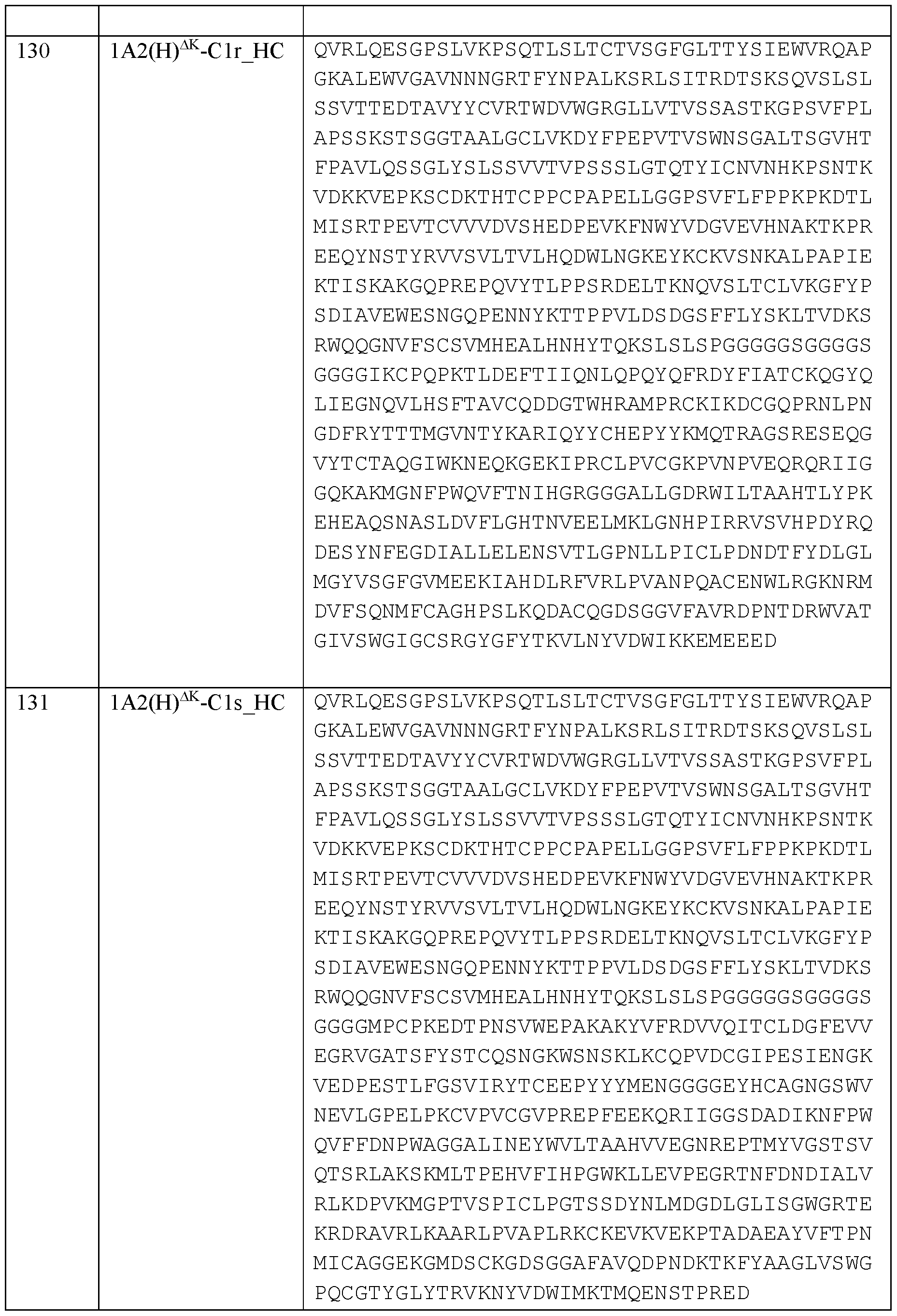


Claims
Priority Applications (2)
| Application Number | Priority Date | Filing Date | Title |
|---|---|---|---|
| AU2022361048A AU2022361048A1 (en) | 2021-10-07 | 2022-10-06 | Targeted catalytic complement-activating molecules and methods of use thereof |
| CA3233732A CA3233732A1 (en) | 2021-10-07 | 2022-10-06 | Targeted catalytic complement-activating molecules and methods of use thereof |
Applications Claiming Priority (2)
| Application Number | Priority Date | Filing Date | Title |
|---|---|---|---|
| US202163253211P | 2021-10-07 | 2021-10-07 | |
| US63/253,211 | 2021-10-07 |
Publications (1)
| Publication Number | Publication Date |
|---|---|
| WO2023060167A1 true WO2023060167A1 (en) | 2023-04-13 |
Family
ID=85803730
Family Applications (1)
| Application Number | Title | Priority Date | Filing Date |
|---|---|---|---|
| PCT/US2022/077663 WO2023060167A1 (en) | 2021-10-07 | 2022-10-06 | Targeted catalytic complement-activating molecules and methods of use thereof |
Country Status (5)
| Country | Link |
|---|---|
| US (1) | US20230173091A1 (en) |
| AU (1) | AU2022361048A1 (en) |
| CA (1) | CA3233732A1 (en) |
| TW (1) | TW202323279A (en) |
| WO (1) | WO2023060167A1 (en) |
Cited By (1)
| Publication number | Priority date | Publication date | Assignee | Title |
|---|---|---|---|---|
| WO2024008877A1 (en) * | 2022-07-07 | 2024-01-11 | Rigshospitalet | Tailored anti-fungal drugs and tools to diagnose invasive fungal infections |
Citations (2)
| Publication number | Priority date | Publication date | Assignee | Title |
|---|---|---|---|---|
| US20060188963A1 (en) * | 2002-09-10 | 2006-08-24 | Natlmmune A/S | Collectin-complement activating protein chimeras |
| US20190016824A1 (en) * | 2011-04-08 | 2019-01-17 | Omeros Corporation | Methods for Treating Conditions Associated with MASP-2 Dependent Complement Activation |
-
2022
- 2022-10-06 US US17/938,421 patent/US20230173091A1/en active Pending
- 2022-10-06 TW TW111138094A patent/TW202323279A/en unknown
- 2022-10-06 AU AU2022361048A patent/AU2022361048A1/en active Pending
- 2022-10-06 WO PCT/US2022/077663 patent/WO2023060167A1/en active Application Filing
- 2022-10-06 CA CA3233732A patent/CA3233732A1/en active Pending
Patent Citations (2)
| Publication number | Priority date | Publication date | Assignee | Title |
|---|---|---|---|---|
| US20060188963A1 (en) * | 2002-09-10 | 2006-08-24 | Natlmmune A/S | Collectin-complement activating protein chimeras |
| US20190016824A1 (en) * | 2011-04-08 | 2019-01-17 | Omeros Corporation | Methods for Treating Conditions Associated with MASP-2 Dependent Complement Activation |
Cited By (1)
| Publication number | Priority date | Publication date | Assignee | Title |
|---|---|---|---|---|
| WO2024008877A1 (en) * | 2022-07-07 | 2024-01-11 | Rigshospitalet | Tailored anti-fungal drugs and tools to diagnose invasive fungal infections |
Also Published As
| Publication number | Publication date |
|---|---|
| US20230173091A1 (en) | 2023-06-08 |
| CA3233732A1 (en) | 2023-04-13 |
| TW202323279A (en) | 2023-06-16 |
| AU2022361048A1 (en) | 2024-05-02 |
Similar Documents
| Publication | Publication Date | Title |
|---|---|---|
| CN111138544B (en) | Homodimer type bispecific antibody aiming at Her2 and CD3 and application thereof | |
| JP6529552B2 (en) | Antibody that binds to CSF1R | |
| CN107921122B (en) | Antibody therapeutics that bind to CD38 | |
| JP2020529864A (en) | Multispecific antibody and its preparation and usage | |
| TW201617369A (en) | Bi-specific monovalent diabodies that are capable of binding CD19 and CD3, and uses thereof | |
| US11780919B2 (en) | Bispecific antigen binding molecules targeting OX40 and FAP | |
| US20220280643A1 (en) | Anti-pvrig antibodies formulations and uses thereof | |
| US20220089737A1 (en) | Multi-specific immune targeting molecules and uses thereof | |
| US20220378742A1 (en) | Combination therapy with anti-pvrig antibodies formulations and anti-pd-1 antibodies | |
| CN111094339B (en) | Application of anti-PD-1 antibody and anti-LAG-3 antibody in preparation of medicine for treating tumor | |
| JP2023527583A (en) | Antibodies that bind to LAG3 and uses thereof | |
| AU2018228719A1 (en) | Tandem diabody for CD16A-directed NK-cell engagement | |
| JP2024028761A (en) | Antitumor antagonist composed of mutated TGFβ1-RII extracellular domain and immunoglobulin scaffold | |
| JP2022553922A (en) | Antibody that binds to 4-1BB and use thereof | |
| KR20190121716A (en) | Switch Molecule And Switchable Chimeric Antigen Receptor | |
| US20230173091A1 (en) | Targeted catalytic complement-activating molecules and methods of use thereof | |
| IL301254A (en) | Methods and compositions for modulating beta chain mediated immunity | |
| US11667712B2 (en) | Materials and methods for modulating t cell mediated immunity | |
| KR20240069776A (en) | Targeted catalytic complement activation molecules and methods of their use | |
| JP2024506449A (en) | Antibodies that bind to PD-L1 and uses thereof | |
| US20220125947A1 (en) | Compositions and methods for modulating delta gamma chain mediated immunity | |
| US20220056148A1 (en) | Novel polypeptides | |
| CA3163865A1 (en) | Method of treating a tumor with a combination of il-7 protein and a bispecific antibody | |
| US20230416406A1 (en) | Masp-2 and masp-3 inhibitors, and related compositions and methods, for treatment of sickle cell disease | |
| US20210284730A1 (en) | Materials and methods for modulating delta chain mediated immunity |
Legal Events
| Date | Code | Title | Description |
|---|---|---|---|
| 121 | Ep: the epo has been informed by wipo that ep was designated in this application |
Ref document number: 22879479 Country of ref document: EP Kind code of ref document: A1 |
|
| WWE | Wipo information: entry into national phase |
Ref document number: 3233732 Country of ref document: CA |
|
| WWE | Wipo information: entry into national phase |
Ref document number: 311914 Country of ref document: IL |
|
| WWE | Wipo information: entry into national phase |
Ref document number: 809916 Country of ref document: NZ |
|
| REG | Reference to national code |
Ref country code: BR Ref legal event code: B01A Ref document number: 112024006211 Country of ref document: BR |
|
| WWE | Wipo information: entry into national phase |
Ref document number: AU2022361048 Country of ref document: AU |
|
| ENP | Entry into the national phase |
Ref document number: 20247013055 Country of ref document: KR Kind code of ref document: A |
|
| ENP | Entry into the national phase |
Ref document number: 2022361048 Country of ref document: AU Date of ref document: 20221006 Kind code of ref document: A |
|
| WWE | Wipo information: entry into national phase |
Ref document number: 2022879479 Country of ref document: EP |
|
| ENP | Entry into the national phase |
Ref document number: 2022879479 Country of ref document: EP Effective date: 20240507 |