WO2023058746A1 - 画像診断装置、画像診断装置の作動方法及びプログラム - Google Patents
画像診断装置、画像診断装置の作動方法及びプログラム Download PDFInfo
- Publication number
- WO2023058746A1 WO2023058746A1 PCT/JP2022/037568 JP2022037568W WO2023058746A1 WO 2023058746 A1 WO2023058746 A1 WO 2023058746A1 JP 2022037568 W JP2022037568 W JP 2022037568W WO 2023058746 A1 WO2023058746 A1 WO 2023058746A1
- Authority
- WO
- WIPO (PCT)
- Prior art keywords
- image
- region
- unit
- pixel
- diagnostic imaging
- Prior art date
- Legal status (The legal status is an assumption and is not a legal conclusion. Google has not performed a legal analysis and makes no representation as to the accuracy of the status listed.)
- Ceased
Links
Images









Classifications
-
- A—HUMAN NECESSITIES
- A61—MEDICAL OR VETERINARY SCIENCE; HYGIENE
- A61B—DIAGNOSIS; SURGERY; IDENTIFICATION
- A61B5/00—Measuring for diagnostic purposes; Identification of persons
- A61B5/05—Detecting, measuring or recording for diagnosis by means of electric currents or magnetic fields; Measuring using microwaves or radio waves
- A61B5/055—Detecting, measuring or recording for diagnosis by means of electric currents or magnetic fields; Measuring using microwaves or radio waves involving electronic [EMR] or nuclear [NMR] magnetic resonance, e.g. magnetic resonance imaging
-
- G—PHYSICS
- G06—COMPUTING OR CALCULATING; COUNTING
- G06T—IMAGE DATA PROCESSING OR GENERATION, IN GENERAL
- G06T7/00—Image analysis
- G06T7/0002—Inspection of images, e.g. flaw detection
- G06T7/0012—Biomedical image inspection
-
- G—PHYSICS
- G16—INFORMATION AND COMMUNICATION TECHNOLOGY [ICT] SPECIALLY ADAPTED FOR SPECIFIC APPLICATION FIELDS
- G16H—HEALTHCARE INFORMATICS, i.e. INFORMATION AND COMMUNICATION TECHNOLOGY [ICT] SPECIALLY ADAPTED FOR THE HANDLING OR PROCESSING OF MEDICAL OR HEALTHCARE DATA
- G16H30/00—ICT specially adapted for the handling or processing of medical images
- G16H30/20—ICT specially adapted for the handling or processing of medical images for handling medical images, e.g. DICOM, HL7 or PACS
-
- G—PHYSICS
- G16—INFORMATION AND COMMUNICATION TECHNOLOGY [ICT] SPECIALLY ADAPTED FOR SPECIFIC APPLICATION FIELDS
- G16H—HEALTHCARE INFORMATICS, i.e. INFORMATION AND COMMUNICATION TECHNOLOGY [ICT] SPECIALLY ADAPTED FOR THE HANDLING OR PROCESSING OF MEDICAL OR HEALTHCARE DATA
- G16H50/00—ICT specially adapted for medical diagnosis, medical simulation or medical data mining; ICT specially adapted for detecting, monitoring or modelling epidemics or pandemics
- G16H50/20—ICT specially adapted for medical diagnosis, medical simulation or medical data mining; ICT specially adapted for detecting, monitoring or modelling epidemics or pandemics for computer-aided diagnosis, e.g. based on medical expert systems
-
- G—PHYSICS
- G06—COMPUTING OR CALCULATING; COUNTING
- G06T—IMAGE DATA PROCESSING OR GENERATION, IN GENERAL
- G06T2207/00—Indexing scheme for image analysis or image enhancement
- G06T2207/10—Image acquisition modality
- G06T2207/10072—Tomographic images
-
- G—PHYSICS
- G06—COMPUTING OR CALCULATING; COUNTING
- G06T—IMAGE DATA PROCESSING OR GENERATION, IN GENERAL
- G06T2207/00—Indexing scheme for image analysis or image enhancement
- G06T2207/10—Image acquisition modality
- G06T2207/10072—Tomographic images
- G06T2207/10081—Computed x-ray tomography [CT]
-
- G—PHYSICS
- G06—COMPUTING OR CALCULATING; COUNTING
- G06T—IMAGE DATA PROCESSING OR GENERATION, IN GENERAL
- G06T2207/00—Indexing scheme for image analysis or image enhancement
- G06T2207/10—Image acquisition modality
- G06T2207/10072—Tomographic images
- G06T2207/10088—Magnetic resonance imaging [MRI]
-
- G—PHYSICS
- G06—COMPUTING OR CALCULATING; COUNTING
- G06T—IMAGE DATA PROCESSING OR GENERATION, IN GENERAL
- G06T2207/00—Indexing scheme for image analysis or image enhancement
- G06T2207/10—Image acquisition modality
- G06T2207/10132—Ultrasound image
- G06T2207/10136—3D ultrasound image
-
- G—PHYSICS
- G06—COMPUTING OR CALCULATING; COUNTING
- G06T—IMAGE DATA PROCESSING OR GENERATION, IN GENERAL
- G06T2207/00—Indexing scheme for image analysis or image enhancement
- G06T2207/20—Special algorithmic details
- G06T2207/20081—Training; Learning
-
- G—PHYSICS
- G06—COMPUTING OR CALCULATING; COUNTING
- G06T—IMAGE DATA PROCESSING OR GENERATION, IN GENERAL
- G06T2207/00—Indexing scheme for image analysis or image enhancement
- G06T2207/20—Special algorithmic details
- G06T2207/20084—Artificial neural networks [ANN]
-
- G—PHYSICS
- G06—COMPUTING OR CALCULATING; COUNTING
- G06T—IMAGE DATA PROCESSING OR GENERATION, IN GENERAL
- G06T2207/00—Indexing scheme for image analysis or image enhancement
- G06T2207/30—Subject of image; Context of image processing
- G06T2207/30004—Biomedical image processing
- G06T2207/30016—Brain
-
- G—PHYSICS
- G06—COMPUTING OR CALCULATING; COUNTING
- G06T—IMAGE DATA PROCESSING OR GENERATION, IN GENERAL
- G06T2207/00—Indexing scheme for image analysis or image enhancement
- G06T2207/30—Subject of image; Context of image processing
- G06T2207/30004—Biomedical image processing
- G06T2207/30096—Tumor; Lesion
Definitions
- the present invention relates to a diagnostic imaging apparatus, an operating method of the diagnostic imaging apparatus, and a program.
- Patent Document 1 a trained model obtained by machine learning is used to determine which type of tissue a brain tumor in an image is, specifically glioblastoma or meningioma. A method for automatic diagnosis of brain tumors is disclosed.
- Patent Document 1 only determines what type of brain tumor the existence of which is known.
- a brain tumor is adjacent to a normal structure near the brain surface, it is difficult to detect the existence of a brain tumor in the first place.
- Image diagnosis of brain tumors is a heavy burden for inexperienced diagnosticians and doctors of different specialties, and practical application of a method that supports simple and accurate diagnosis of the presence or absence of brain tumors is desired.
- Such problems are not limited to diagnosing the presence or absence of brain tumors, but also exist when diagnosing the presence or absence of other lesions.
- the present invention has been made based on such a background, and aims to provide a diagnostic imaging apparatus, an operating method of the diagnostic imaging apparatus, and a program for supporting simple and accurate diagnosis of the presence or absence of a lesion in a diagnostic target site. aim.
- the diagnostic imaging apparatus comprises: an acquisition unit that acquires a tomographic image including a diagnosis target region of a subject; a rendering unit that renders a label image containing the diagnosis target region classified into classes indicating a lesion region, a normal tissue region, and a background region based on the tomographic image acquired by the acquisition unit; Prepare.
- the normal tissue area may be divided into a cavity area, a soft tissue area and a bone area.
- the lesion area is a tumor area in the brain
- the tomographic image is a cross-sectional image obtained by slicing the subject's brain in a cross-sectional direction at multiple locations
- the rendering unit may render a label image corresponding to each tomographic image acquired by the acquisition unit.
- the tumor region may be a region where a metastatic brain tumor has developed.
- the rendering unit outputs the pixel value of each pixel of the label image from the tomographic image acquired by the acquisition unit based on a model that outputs the pixel value of each pixel of the label image in response to the input of the pixel value of each pixel of the tomographic image. Pixel values may be estimated.
- the diagnostic imaging apparatus further includes a learning unit that generates the model by machine learning,
- the rendering unit may estimate the pixel value of each pixel of the label image from the pixel value of each pixel of the tomographic image acquired by the acquisition unit, based on the model generated by the learning unit.
- the learning unit uses the pixel value of each pixel of the tomographic image as input data, and is created based on the tomographic image, and the weight of each class is adjusted based on the pixel count number of each class.
- the model may be generated by using learning data having pixel values as output data.
- the learning unit may generate the model using learning data generated based on a plurality of tomographic images having cross sections obtained by slicing brains of a plurality of subjects at a plurality of locations in a cross-sectional direction. .
- the diagnostic imaging apparatus may further include an output unit that color-codes the label images rendered by the rendering unit by class.
- a method for operating a diagnostic imaging apparatus comprises: A method of operating a diagnostic imaging apparatus comprising an acquisition unit and an imaging unit, comprising: a step in which the acquisition unit acquires a tomographic image including a diagnosis target region of the subject; a step in which the rendering unit renders a labeled image including the diagnosis target region classified into classes indicating a lesion region, a normal tissue region, and a background region, based on the tomographic image acquired by the acquisition unit; including.
- the computer Acquisition means for acquiring a tomographic image including a diagnosis target region of a subject; Rendering means for rendering a label image containing the diagnosis target region classified into classes indicating a lesion region, a normal tissue region, and a background region, based on the tomographic image acquired by the acquisition means; function as
- the present invention it is possible to provide a diagnostic imaging apparatus, an operating method of the diagnostic imaging apparatus, and a program that support simple and accurate diagnosis of the presence or absence of a lesion in a diagnostic target site.
- FIG. 1 is a schematic diagram showing the configuration of a diagnostic imaging system according to an embodiment of the present invention
- FIG. 1 is a block diagram showing the hardware configuration of an image diagnostic apparatus according to an embodiment of the present invention
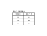
- FIG. It is a figure which shows an example of the data table of the image data storage part which concerns on embodiment of this invention.
- FIG. 4 is a diagram for explaining a set of learning data according to the embodiment of the present invention.
- FIG. FIG. 2 is a conceptual diagram of a neural network used for drawing label images by the diagnostic imaging apparatus according to the embodiment of the present invention;
- FIG. 10 is a diagram showing a procedure for optimizing weights between layers of a neural network by the diagnostic imaging apparatus according to the embodiment of the present invention
- 4 is a flowchart showing the flow of learning processing according to the embodiment of the present invention
- 4 is a flowchart showing the flow of weighting factor optimization processing according to the embodiment of the present invention
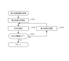
- 4 is a flow chart showing the flow of diagnostic processing according to the embodiment of the present invention
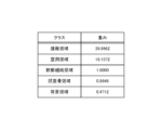
- 4 is a graph showing the pixel count number of each label in the label image in the example. It is a figure which shows an example of the data table which shows the weight of each label in an Example.
- FIG. 10 is a diagram showing how a small single lesion is detected in Example.
- FIG. 10 is a diagram showing how lesions with low pixel values are detected in the example.
- FIG. 10 is a diagram showing how a very small lesion is detected in the example.
- FIG. 10 is a diagram showing how multiple lesions are detected in the example.
- a diagnostic imaging apparatus, an operating method of the diagnostic imaging apparatus, and a program according to an embodiment of the present invention will be described below in detail with reference to the drawings.
- symbol is attached
- MRI Magnetic Resonance Imaging
- a case where an MRI (Magnetic Resonance Imaging) image is used as a tomographic image will be described as an example. It may be a tomographic image.
- FIG. 1 is a schematic diagram showing the configuration of a diagnostic imaging system 1 according to an embodiment.
- the diagnostic imaging system 1 includes a diagnostic imaging apparatus 100 and an MRI apparatus 200 .
- the MRI apparatus 200 is an example of an image capturing apparatus that captures an image of the brain, which is a diagnosis target part of a subject, and obtains a tomographic image including the brain of the subject.
- the diagnostic imaging apparatus 100 and the MRI apparatus 200 are communicably connected via a wired or wireless communication line, and the diagnostic imaging apparatus 100 receives image data transmitted from the MRI apparatus 200 .
- the diagnostic imaging apparatus 100 is a device that performs image processing on an MRI image of the brain to diagnose whether a brain tumor exists in the brain of the subject.
- An MRI image of the brain is a cross-sectional image obtained by slicing the subject's brain in the transverse direction.
- the MRI image of the brain is preferably an image of the brain of the same subject acquired at a plurality of locations separated in the longitudinal direction of the subject, for example, at a constant pitch in the longitudinal direction of the subject.
- the MRI image is preferably a T1-weighted image (T1W1) taken while a gadolinium contrast agent (Gd contrast agent) is administered to the subject.
- T1W1 T1-weighted image
- Gd contrast agent gadolinium contrast agent
- Brain tumors to be diagnosed include both primary brain tumors and metastatic brain tumors.
- Primary brain tumors include benign brain tumors such as meningioma, pituitary adenoma, and schwannoma, and malignant brain tumors such as glioma called glioma and central nervous system malignant lymphoma. Glioma includes extremely malignant glioblastoma.
- Brain tumors also include cerebrospinal fluid dissemination in which tumor cells are suspended in the cerebrospinal fluid.
- FIG. 2A is a block diagram showing the hardware configuration of the diagnostic imaging apparatus 100 according to the embodiment.
- the diagnostic imaging apparatus 100 is, for example, a general-purpose computer.
- the diagnostic imaging apparatus 100 includes an operation unit 110 , a display unit 120 , a communication unit 130 , a storage unit 140 and a control unit 150 .
- Each unit of the image diagnostic apparatus 100 is interconnected via an internal bus (not shown).
- the operation unit 110 receives an instruction from a user, for example, a doctor, and supplies an operation signal corresponding to the received operation to the control unit 150 .
- the operating unit 110 includes, for example, a mouse and a keyboard.
- the display unit 120 displays various images for the user based on the image data supplied from the control unit 150 .
- the display unit 120 displays, for example, an MRI image of the subject captured by the MRI apparatus 200 .
- the operation unit 110 and the display unit 120 may be configured by touch panels.
- the touch panel displays an operation screen for receiving a predetermined operation, and supplies the control unit 150 with an operation signal corresponding to a position touched by the subject on the operation screen.
- the communication unit 130 is, for example, an interface that can be connected to a communication network such as the Internet.
- the communication unit 130 receives, for example, image data related to MRI images from the MRI apparatus 200 .
- the storage unit 140 includes, for example, RAM (Random Access Memory), ROM (Read Only Memory), flash memory, and hard disk.
- the storage unit 140 stores programs executed by the control unit 150 and various data.
- the storage unit 140 also functions as a work memory for temporarily storing various types of information and for the control unit 150 to execute processing.
- the storage unit 140 includes a learning data storage unit 141 , a learned model storage unit 142 , and an image data storage unit 143 .
- the learning data storage unit 141 stores learning data used as teacher data for machine learning.
- the learning data includes a plurality of data sets, and each data set includes an MRI image obtained from a subject and a label image created based on the MRI image by a person skilled in image diagnosis, for example, a doctor. and one each. Label images are classified by classes set on the MRI images.
- a class is a region assigned a unique pixel value for each tissue of a subject.
- FIG. 3 is a diagram for explaining a set of learning data according to the embodiment.
- the MRI and label images are, for example, grayscale images with pixel values (pixel values) 0 to 255 (256 gradations), and have the same number of pixels and aspect ratio.
- the label image is segmented by class for all pixels in the MRI image. Classes are divided into tumor regions, cavity regions, soft tissue regions, skull regions, and background regions, and each pixel in the MRI image is assigned a pixel value corresponding to one of the classes.
- a person skilled in image diagnosis refers to the original MRI image, and some pixels correspond to the background region, so the pixel value is A, and another pixel corresponds to the skull region, so the pixel value is B. , and so on.
- a tumor area is an area of a tumor that has developed in the brain and is an example of a lesion area.
- a plurality of tumor regions may exist depending on the number of tumors that have developed.
- a tumor region may consist only of a single type of tumor, eg, a metastatic brain tumor, or may contain different types of tumors.
- the cavity region is a cavity region that exists inside the brain tissue or between the brain tissue and the skull, and is filled with cerebrospinal fluid.
- the soft tissue region is normal tissue in brain tissue where no tumor has developed.
- the pia mater, arachnoid mater and dura mater associated with brain tissue are also included in the soft tissue region unless a tumor has developed there.
- the skull area is an example of a bone area, and includes not only the skull but also the skin tissue covering the skull.
- the background region is the non-body region surrounding the skull region.
- class weighting When creating label images to be used as learning data, it is advisable to use class weighting to set pixel values for each class in the label image. This is because the classes occupying a large area in the image, such as the background area and the skull area, or in other words, classes with high pixel counts are biased, which may adversely affect the results of the learning process. A workaround for this is to count the number of pixels per class in the label image, set the class weights based on the pixel counts for each label, and based on the set per-class weights Just adjust the pixel values for each class.
- the weight of the class with a large number of counted pixels is set to be small, and the weight of the class with a small number of counted pixels is set to be large.
- the weight of a class whose number of counted pixels is the median frequency value is set to 1
- the weight of a class whose number of counted pixels is greater than the median frequency value is set to be smaller
- the weight of a class whose number of counted pixels is smaller than the median frequency value is set to be greater.
- the learned model storage unit 142 stores a learned model generated by machine learning based on the learning data stored in the learning data storage unit 141.
- the trained model is a model that outputs the pixel value of each pixel of the label image in response to the input of the pixel value of each pixel of the MRI image.
- a trained model uses, for example, a neural network.
- FIG. 4 is a conceptual diagram of a neural network used for rendering label images by the diagnostic imaging apparatus 100 according to the embodiment.
- a neural network comprises an input layer to which input data is input, an output layer to which output data is output, and at least one intermediate layer arranged between the input layer and the output layer. Arrows between neurons in each layer represent parameter connections between the input layer and the output layer.
- the input layer has multiple input neurons.
- the number of input neurons in the input layer corresponds to the number of input data.
- the input data is the pixel value of each pixel in the MRI image. can be expressed as Input data input to input neurons in the input layer are input to intermediate neurons in the intermediate layer.
- Each intermediate layer has multiple intermediate neurons.
- each neuron in the intermediate layer receives the input value I from each neuron in the previous stage, it calculates the product I ⁇ W of the input value I and the weighting factor W, and sums the product I ⁇ W calculated for each neuron in the previous stage. A value is calculated and an output value is output by substituting the total value of the product I ⁇ W into the activation function.
- An activation function is a function that expresses a nonlinear relationship between input and output in a certain neuron, and is, for example, a sigmoid function, a max function, or a Gaussian function.
- the output layer has multiple output neurons.
- the number of output neurons corresponds to the number of output data.
- each neuron in the output layer receives an input value I from each neuron in the intermediate layer located at the rearmost, similarly to each neuron in the intermediate layer, it calculates the product IW of the input value I and the weighting factor W, A total value of the product I ⁇ W calculated for each neuron in the preceding stage is calculated, and an output value is output by substituting the total value of the product I ⁇ W into an activation function.
- the output data is the pixel value of each pixel in the label image. can be expressed as
- the image data storage unit 143 stores the image data acquired from the MRI apparatus 200 in association with the subject ID (Identification) assigned to each subject.
- control section 150 includes a processor such as a CPU (Central Processing Unit) and controls each section of the diagnostic imaging apparatus 100 .
- the control unit 150 executes the programs stored in the storage unit 140 to perform the learning process in FIG. 6, the weighting factor optimization process in FIG. 7, and the diagnosis process in FIG.
- the control unit 150 functionally includes a learning unit 151 , an acquisition unit 152 , a rendering unit 153 and an output unit 154 .
- the learning unit 151 refers to the learning data stored in the learning data storage unit 141 to generate a learned model by machine learning, and stores the generated learned model in the learned model storage unit 142.
- the learning unit 151 generates a trained model by performing supervised learning using a plurality of data sets included in the learning data as teacher data.
- the learning unit 151 uses a plurality of data sets included in the learning data as teacher data to adjust weight coefficients that indicate the connection state of each layer in the neural network. More specifically, the learning unit 151 inputs the pixel value of each pixel of the MRI image to the input layer as input data, and converts the pixel value of each pixel of the label image output as output data in the output layer to Compare with the pixel value of each pixel in the label image of training data. Then, the learning unit 151 optimizes the weighting factor so that the difference between the pixel value of each pixel of the label image output in the output layer and the pixel value of each pixel of the label image of the teacher data is minimized. For example, the error backpropagation method is used for optimizing the weight coefficients.
- MSE mean squared error
- the optimized weighting factor when this MSE is the minimum value is expressed as W opt .
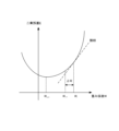
- gradient descent may be used.
- the gradient descent method is a method of obtaining an optimized weighting factor W opt by reducing the MSE value by repeating the procedure of calculating the slope of the MSE and updating the weighting factor W in the direction opposite to the magnitude of the slope. is. Expressing the current weighting factor as W i , the updated weighting factor as W i+1 , and the amount of change in the weighting factor as ⁇ W i , these satisfy the following equation (2).
- W i +1 W i + ⁇ W i (2)
- the acquisition unit 152 acquires the image data of the subject transmitted from the MRI apparatus 200, and stores it in the image data storage unit 143 in association with the subject ID. Acquisition of image data by the acquisition unit 152 includes both receiving image data from an external device and reading data stored in the storage unit 140 .
- the drawing unit 153 draws a label image corresponding to the image data of the subject acquired by the acquiring unit 152 based on the learned model generated by the learning unit 151 and stored in the learned model storage unit 142. .
- the output unit 154 outputs the label image rendered by the rendering unit 153 to the outside.
- the output unit 154 transmits image data representing the label image obtained by the rendering unit 153 to the display unit 120, and causes the display unit 120 to display the label image.
- the output unit 154 may classify the label images by color so that the user can easily understand them, and output the color-coded label images.
- the output unit 154 may cause the display unit 120 to display the label image rendered by the rendering unit 153 side by side with the original MRI image.
- the above is the configuration of the image diagnostic apparatus 100 .
- the flow of the learning process executed by the control unit 150 of the diagnostic imaging apparatus 100 will be described below with reference to the flowchart of FIG.
- the learning process is a process of generating a trained model that renders a labeled image indicating the presence or absence of a brain tumor based on learning data composed of MRI images and manually created labeled images. After the learning data is generated and stored in the learning data storage unit 141, the learning process is started when the user operates the operation unit 110 to instruct the start of the learning process.
- the acquisition unit 152 acquires learning data stored in the learning data storage unit 141 (step S11).
- the learning unit 151 executes weighting factor optimization processing for optimizing the weighting factors contained in the learning data (step S12).
- the flow of weighting factor optimization processing will be described below with reference to FIG.
- the learning unit 151 initializes weight coefficients between all neurons in each layer of the neural network (step S121).
- the initial value of the weighting factor between neurons may be given as a random number from -0.1 to +0.1, for example.
- the learning unit 151 inputs the pixel value (input data) of each pixel of the MRI image to the input neuron of the input layer, so that the pixel of each pixel of the label image output from the output neuron of the output layer is A value (predicted value of the output data) is output, and the MSE between the predicted value of the output data and the pixel value (set value of the output data) of each pixel of the label image manually labeled by the medical specialist is calculated (step S122). ).
- the learning unit 151 determines whether the MSE is equal to or less than the threshold (step S123). If it is determined that the MSE is equal to or less than the threshold (step S123; Yes), the process is returned. On the other hand, if it is determined that the MSE is not equal to or less than the threshold (step S123; No), the learning unit 151 calculates the change amount ⁇ W i of the weighting factor calculated by Equation (3) for each weighting factor W i By adding, the value of each weighting factor is updated to Wi +1 (step S124), and the process is returned to step S122. The above is the flow of the weighting factor optimization process.
- the learning unit 151 causes the learned model storage unit 142 to store the finally obtained updated weighting coefficients W i+1 as optimized weighting coefficients W opt (step S13). exit.
- the above is the flow of the learning process.
- the flow of diagnostic processing executed by the control unit 150 of the diagnostic imaging apparatus 100 will be described below with reference to the flowchart of FIG.
- the diagnostic process is a process of drawing a labeled image indicating the presence or absence of a brain tumor based on the MRI image of the subject.
- the diagnosis process is started when the user operates the operation unit 110 to instruct the start of the diagnosis process after the learned model is generated by the learning process.
- the acquisition unit 152 acquires an MRI image corresponding to the subject ID to be diagnosed from the image data storage unit 143 (step S21).
- the rendering unit 153 reads the learned model stored in the learned model storage unit 142, and uses the learned model to extract a label image corresponding to the MRI image from the MRI image acquired in the process of step S21.
- Draw step S22. Specifically, using the optimized weighting factor W opt between all neurons in each layer of the neural network, arithmetic processing is performed sequentially from the intermediate layer to the output layer, and the pixel value of each pixel in the label image is estimated. do.
- the output unit 154 outputs the image data related to the label image rendered in the process of step S22 to the outside (step S23).
- the output unit 154 for example, transmits the label image rendered in the process of step S22 to the display unit 120, and causes the display unit 120 to display the label image colored for each class.
- the above is the flow of the diagnostic processing.
- the image diagnostic apparatus 100 includes the acquiring unit 152 that acquires an MRI image of the subject's brain, and based on the MRI image acquired by the acquiring unit 152, the tumor region, and a rendering unit 153 that renders label images classified by classes indicating a cavity region, a soft tissue region, a skull region, and a background region. Therefore, even an unskilled person in image diagnosis can easily and accurately diagnose a brain tumor of a subject.
- the diagnostic imaging apparatus 100 also includes a learning unit 151 that generates a model for estimating the pixel value of each pixel of the label image from the pixel value of each pixel of the MRI image by machine learning. Therefore, a brain tumor of a subject can be more accurately diagnosed using a trained model generated by machine learning.
- a cross-sectional image obtained by slicing a cross-section of the subject's brain is used, but the present invention is not limited to this.
- a cross-sectional image obtained by slicing the subject's brain in the frontal plane or sagittal plane may be used.
- an MRI image obtained by slicing a cross section of the subject's brain is used as the tomographic image of the subject's brain, but the present invention is not limited to this.
- the medical tomographic image may be a CT image, an X-ray image, or an ultrasound image obtained by imaging a cross section of the subject's brain.
- the MRI apparatus 200 may be replaced with another medical imaging apparatus such as a CT apparatus, an X-ray apparatus, an ultrasonic tomography apparatus, and connected to the image diagnostic apparatus 100 so as to be communicable.
- labeled images are classified into five classes: tumor region, cavity region, soft tissue region, skull region, and background region, but the present invention is not limited to this.
- the number of classes of label images may be four or less.
- the labeled images may be classified into tumor regions, cavity regions, normal tissue regions including soft tissue and skull, and background regions, including tumor regions, cavities, soft tissue and skull.
- a classification may be made into normal tissue regions and background regions.
- other classes may be added in addition to the above five classes, for example, the soft tissue region may be classified into the cerebral region, the cerebellar region, and the brainstem region, and the cerebral region may be classified into the cerebral cortex region, It may be classified into gray matter areas and white matter areas.
- both the MRI image and the label image are grayscale images of 256 gradations (8 bits), but the present invention is not limited to this.
- the MRI images and label images may be, for example, 16-bit grayscale images or color images.
- a color image is, for example, a 24-bit image in which RGB (Red-Green-Blue) elements of one pixel are each represented by 8 bits.
- a label image of a color image may be rendered using a trained model from an MRI image that is a grayscale image.
- the learning unit 151 performs machine learning using a neural network, but the present invention is not limited to this.
- Machine learning techniques other than neural networks for example, regression analysis using a support vector machine (support vector regression) may be used.
- a learning model defines the relationship between input data and output data, and any method other than machine learning can be used as long as it can supply data to the input layer and obtain output data from the output layer. may be constructed
- the diagnostic imaging apparatus 100 has the function of the learning unit 151 in the above embodiment, the present invention is not limited to this.
- an external device other than the diagnostic imaging apparatus 100 may have the function of the learning unit 151 .
- the external device generates a trained model by learning the relationship between the MRI image and the label image based on the learning data, and the diagnostic imaging apparatus 100 generates the trained model generated by the external device. may be obtained through communication by the communication unit 130 .
- the optimized weighting coefficients obtained by the learning process using the learning data are stored as they are in the learned model storage unit 142, but the present invention is not limited to this.
- the validity of the optimized weighting factor is evaluated using the rest of the learning data, and the weighting factor is evaluated as a valid weighting factor.
- the weighting factor may be stored in the learned model storage unit 142 when the weighting factor is determined.
- the label image is drawn using a trained model based on the MRI image of the subject taken by the MRI apparatus 200, but the present invention is not limited to this.
- the image is captured by the computer with the camera, and the computer performs diagnostic processing using a trained model.
- the label image may be rendered by executing
- the camera-equipped computer is, for example, a smart phone or a tablet terminal, and an application for executing diagnostic processing based on captured images may be installed in advance.
- the output unit 154 causes the display unit 120 to display the label image colored for each class, but the present invention is not limited to this.
- the control unit 150 of the image diagnostic apparatus 100 includes a determination unit that determines the presence or absence of a brain tumor based on the labeled image rendered by the rendering unit 153, and the determination result of the determination unit is output from the output unit 154. good too.
- the determining unit may determine that the subject has a brain tumor when a pixel having a pixel value corresponding to the tumor region exists in the labeled image.
- the output unit 154 may notify the user of a warning that the subject has a brain tumor.
- a warning screen may be displayed on the display unit 120, or a warning sound may be generated from the speaker of the imaging diagnostic apparatus 100.
- the output unit 154 may cause the display unit 120 to display the label image side by side with the original MRI image, and may enlarge or emphasize the portion corresponding to the tumor region in the MRI image.
- the diagnosis target region is the brain, and the diagnostic imaging apparatus 100 diagnoses the presence or absence of a brain tumor in the subject's brain, but the present invention is not limited to this.
- the presence or absence of tumors occurring in other organs such as the subject's stomach, lung, liver, spleen, large intestine, bladder, prostate, breast, and uterus may be diagnosed.
- the label image may be divided into five classes, for example, tumor region, cavity region, soft tissue region, bone region, and background region.
- the diagnosis target of the image diagnostic apparatus 100 is not limited to tumors generated in organs, and may be lesions having some image characteristics such as inflammation, scarring, and fibrosis.
- the present invention is not limited to this.
- all or part of various data may be stored in an external control device or computer via a communication network.
- the diagnostic imaging apparatus 100 operates based on the programs stored in the storage unit 140, but the present invention is not limited to this.
- a functional configuration implemented by a program may be implemented by hardware.
- the diagnostic imaging apparatus 100 is, for example, a general-purpose computer, but the present invention is not limited to this.
- the diagnostic imaging apparatus 100 may be realized by a computer provided on the cloud.
- the processing executed by the diagnostic imaging apparatus 100 is realized by executing the program stored in the storage unit 140 by the apparatus having the physical configuration described above. , or as a storage medium in which the program is recorded.
- the program for executing the above processing operations can be read by a computer such as a flexible disk, CD-ROM (Compact Disk Read-Only Memory), DVD (Digital Versatile Disk), MO (Magneto-Optical Disk).
- a computer such as a flexible disk, CD-ROM (Compact Disk Read-Only Memory), DVD (Digital Versatile Disk), MO (Magneto-Optical Disk).
- Example 2 machine learning was performed using MRI images of brain tumor patients, and the presence or absence of a brain tumor in the patient's brain was determined using a learned model created by machine learning.
- MRI images were obtained from 51 patients who were diagnosed with metastatic brain tumors for the first time. These patients were confirmed free of carcinomatous meningitis, glioblastoma, and bone metastases.
- the MRI image is a T1-weighted image (T1W1) taken with a gadolinium contrast agent (Gd contrast agent) administered.
- the slice pitch is 3 mm to 5 mm, and the number of slices per patient is 60 to 100. All slices were used for 10 cases, and only slices with contrast-enhancing lesions were used for the other 41 cases.
- the total number of target images was 1507.
- MRI images acquired from 51 patients were randomly distributed at a ratio of 80% training images and 20% test images. The result was 1206 training images and 301 test images.
- a trained model was created by performing supervised learning based on learning data consisting of training images and label images.
- MATLAB registered trademark
- a labeled image was drawn from each test image, and it was confirmed whether or not a brain tumor could be detected in each drawn labeled image.
- test results are shown below.
- small single lesions and lesions with low pixel values were successfully detected.
- detection of very small lesions and multiple lesions was also successful.
- the number of tumors was 326
- the number of false negatives was 13
- the number of false positives was 76, of which 64 were intracranial false positives.
- the diagnostic rate was 96%, which indicates the proportion of those who were not misdiagnosed among those whose tumors were detected by the trained model.
- the number of false positives per image was successfully reduced to 0.25 and intracranial to 0.21.
- the diagnostic imaging apparatus, operating method, and program for the diagnostic imaging apparatus of the present invention are useful because they support simple and accurate diagnosis of the presence or absence of lesions in diagnostic target regions.
- diagnostic imaging system 100 diagnostic imaging apparatus 110 operation unit 120 display unit 130 communication unit 140 storage unit 141 learning data storage unit 142 trained model storage unit 143 image data storage unit 150 control unit 151 learning unit 152 acquisition unit 153 rendering unit 154 Output unit 200 MRI apparatus
Landscapes
- Engineering & Computer Science (AREA)
- Health & Medical Sciences (AREA)
- Medical Informatics (AREA)
- General Health & Medical Sciences (AREA)
- Nuclear Medicine, Radiotherapy & Molecular Imaging (AREA)
- Public Health (AREA)
- Radiology & Medical Imaging (AREA)
- Physics & Mathematics (AREA)
- Biomedical Technology (AREA)
- Life Sciences & Earth Sciences (AREA)
- Epidemiology (AREA)
- Primary Health Care (AREA)
- Computer Vision & Pattern Recognition (AREA)
- Pathology (AREA)
- General Physics & Mathematics (AREA)
- Theoretical Computer Science (AREA)
- Quality & Reliability (AREA)
- Data Mining & Analysis (AREA)
- Databases & Information Systems (AREA)
- Heart & Thoracic Surgery (AREA)
- Biophysics (AREA)
- High Energy & Nuclear Physics (AREA)
- Molecular Biology (AREA)
- Surgery (AREA)
- Animal Behavior & Ethology (AREA)
- Veterinary Medicine (AREA)
- Magnetic Resonance Imaging Apparatus (AREA)
- Image Analysis (AREA)
- Image Processing (AREA)
Priority Applications (3)
| Application Number | Priority Date | Filing Date | Title |
|---|---|---|---|
| US18/699,133 US20240428406A1 (en) | 2021-10-08 | 2022-10-07 | Image diagnosis apparatus, method for operating image diagnosis apparatus, and program |
| EP22878613.3A EP4413921A4 (en) | 2021-10-08 | 2022-10-07 | IMAGE DIAGNOSTIC APPARATUS, METHOD FOR OPERATING AN IMAGE DIAGNOSTIC APPARATUS, AND PROGRAM |
| JP2023552958A JPWO2023058746A1 (https=) | 2021-10-08 | 2022-10-07 |
Applications Claiming Priority (2)
| Application Number | Priority Date | Filing Date | Title |
|---|---|---|---|
| JP2021-166163 | 2021-10-08 | ||
| JP2021166163 | 2021-10-08 |
Publications (1)
| Publication Number | Publication Date |
|---|---|
| WO2023058746A1 true WO2023058746A1 (ja) | 2023-04-13 |
Family
ID=85804355
Family Applications (1)
| Application Number | Title | Priority Date | Filing Date |
|---|---|---|---|
| PCT/JP2022/037568 Ceased WO2023058746A1 (ja) | 2021-10-08 | 2022-10-07 | 画像診断装置、画像診断装置の作動方法及びプログラム |
Country Status (4)
| Country | Link |
|---|---|
| US (1) | US20240428406A1 (https=) |
| EP (1) | EP4413921A4 (https=) |
| JP (1) | JPWO2023058746A1 (https=) |
| WO (1) | WO2023058746A1 (https=) |
Citations (2)
| Publication number | Priority date | Publication date | Assignee | Title |
|---|---|---|---|---|
| JP2018515164A (ja) | 2015-03-27 | 2018-06-14 | シーメンス アクチエンゲゼルシヤフトSiemens Aktiengesellschaft | 画像分類を用いた脳腫瘍自動診断方法および脳腫瘍自動診断システム |
| JP2021166163A (ja) | 2020-04-08 | 2021-10-14 | 国立研究開発法人物質・材料研究機構 | アノード電極材料、その製造方法、および、それを用いたリチウムイオン二次電池 |
Family Cites Families (6)
| Publication number | Priority date | Publication date | Assignee | Title |
|---|---|---|---|---|
| WO2006114003A1 (en) * | 2005-04-27 | 2006-11-02 | The Governors Of The University Of Alberta | A method and system for automatic detection and segmentation of tumors and associated edema (swelling) in magnetic resonance (mri) images |
| US7738683B2 (en) * | 2005-07-22 | 2010-06-15 | Carestream Health, Inc. | Abnormality detection in medical images |
| EP3514756A1 (en) * | 2018-01-18 | 2019-07-24 | Koninklijke Philips N.V. | Medical analysis method for predicting metastases in a test tissue sample |
| EP3977149A1 (en) * | 2019-05-28 | 2022-04-06 | Cornell University Center for Technology Licensing | System and method of perceptive quantitative mapping of physical properties |
| US11227683B2 (en) * | 2020-01-23 | 2022-01-18 | GE Precision Healthcare LLC | Methods and systems for characterizing anatomical features in medical images |
| JP7760515B2 (ja) * | 2020-03-13 | 2025-10-27 | ジェネンテック, インコーポレイテッド | 画像処理に基づく腫瘍の自動検出 |
-
2022
- 2022-10-07 EP EP22878613.3A patent/EP4413921A4/en active Pending
- 2022-10-07 WO PCT/JP2022/037568 patent/WO2023058746A1/ja not_active Ceased
- 2022-10-07 US US18/699,133 patent/US20240428406A1/en active Pending
- 2022-10-07 JP JP2023552958A patent/JPWO2023058746A1/ja active Pending
Patent Citations (2)
| Publication number | Priority date | Publication date | Assignee | Title |
|---|---|---|---|---|
| JP2018515164A (ja) | 2015-03-27 | 2018-06-14 | シーメンス アクチエンゲゼルシヤフトSiemens Aktiengesellschaft | 画像分類を用いた脳腫瘍自動診断方法および脳腫瘍自動診断システム |
| JP2021166163A (ja) | 2020-04-08 | 2021-10-14 | 国立研究開発法人物質・材料研究機構 | アノード電極材料、その製造方法、および、それを用いたリチウムイオン二次電池 |
Non-Patent Citations (2)
| Title |
|---|
| DESPOTOVIć IVANA, GOOSSENS BART, PHILIPS WILFRIED: "MRI Segmentation of the Human Brain: Challenges, Methods, and Applications", COMPUTATIONAL AND MATHEMATICAL METHODS IN MEDICINE, TAYLOR & FRANCIS, vol. 2015, 1 January 2015 (2015-01-01), pages 1 - 23, XP055790821, ISSN: 1748-670X, DOI: 10.1155/2015/450341 * |
| See also references of EP4413921A4 |
Also Published As
| Publication number | Publication date |
|---|---|
| EP4413921A1 (en) | 2024-08-14 |
| US20240428406A1 (en) | 2024-12-26 |
| JPWO2023058746A1 (https=) | 2023-04-13 |
| EP4413921A4 (en) | 2025-10-22 |
Similar Documents
| Publication | Publication Date | Title |
|---|---|---|
| EP3043318B1 (en) | Analysis of medical images and creation of a report | |
| JP6371544B2 (ja) | 画像処理装置、画像処理方法、及び画像処理プログラム | |
| JP5123954B2 (ja) | 医療画像における病変部の特定及び分析 | |
| CN110858399B (zh) | 用于提供虚拟断层扫描中风后续检查图像的方法和装置 | |
| CN112237435B (zh) | 用于在计算机断层扫描中成像的方法和装置 | |
| CN106056126A (zh) | 医学成像中的斑块易损性评定 | |
| JP2020010805A (ja) | 特定装置、プログラム、特定方法、情報処理装置及び特定器 | |
| CN110944586A (zh) | 来自x射线图像数据的炎症估计 | |
| JP7645614B2 (ja) | データ処理装置及びデータ処理方法 | |
| EP4116992A1 (en) | Method and system for predicting expression of biomarker from medical image | |
| JP7512664B2 (ja) | 医療診断支援装置、医療診断支援プログラム、および医療診断支援方法 | |
| Mannil et al. | Texture analysis of myocardial infarction in CT: comparison with visual analysis and impact of iterative reconstruction | |
| EP3923810A1 (en) | Prediction of coronary microvascular dysfunction from coronary computed tomography | |
| WO2020179234A1 (ja) | 画像診断支援装置および画像処理方法 | |
| JP2013010009A (ja) | 診断支援装置、診断支援装置の制御方法、およびそのプログラム | |
| Habibollahi et al. | Hyperechoic renal masses: differentiation of angiomyolipomas from renal cell carcinomas using tumor size and ultrasound radiomics | |
| KR102767794B1 (ko) | 의료 영상을 이용하여 침윤암을 예측 진단하기 위한 라디오믹스 기반의 머신러닝 모델 | |
| US20220280124A1 (en) | Diagnosis support system | |
| Wang et al. | Rib suppression-based radiomics for diagnosis of neonatal respiratory distress syndrome in chest X-rays | |
| CN117242362A (zh) | 使用动态对比度增强的磁共振断层扫描表征肝脏病变 | |
| WO2023058746A1 (ja) | 画像診断装置、画像診断装置の作動方法及びプログラム | |
| US11923072B2 (en) | Image diagnosis supporting device and image processing method | |
| US20230274424A1 (en) | Appartus and method for quantifying lesion in biometric image | |
| Santhiyakumari et al. | Medical decision-making system of ultrasound carotid artery intima–media thickness using neural networks | |
| JP2024018562A (ja) | 画像処理装置、方法およびプログラム |
Legal Events
| Date | Code | Title | Description |
|---|---|---|---|
| 121 | Ep: the epo has been informed by wipo that ep was designated in this application |
Ref document number: 22878613 Country of ref document: EP Kind code of ref document: A1 |
|
| DPE1 | Request for preliminary examination filed after expiration of 19th month from priority date (pct application filed from 20040101) | ||
| ENP | Entry into the national phase |
Ref document number: 2023552958 Country of ref document: JP Kind code of ref document: A |
|
| WWE | Wipo information: entry into national phase |
Ref document number: 18699133 Country of ref document: US |
|
| WWE | Wipo information: entry into national phase |
Ref document number: 2022878613 Country of ref document: EP |
|
| NENP | Non-entry into the national phase |
Ref country code: DE |
|
| ENP | Entry into the national phase |
Ref document number: 2022878613 Country of ref document: EP Effective date: 20240508 |