WO2023038004A1 - Endoscope system, medical information processing device, medical information processing method, medical information processing program, and storage medium - Google Patents
Endoscope system, medical information processing device, medical information processing method, medical information processing program, and storage medium Download PDFInfo
- Publication number
- WO2023038004A1 WO2023038004A1 PCT/JP2022/033260 JP2022033260W WO2023038004A1 WO 2023038004 A1 WO2023038004 A1 WO 2023038004A1 JP 2022033260 W JP2022033260 W JP 2022033260W WO 2023038004 A1 WO2023038004 A1 WO 2023038004A1
- Authority
- WO
- WIPO (PCT)
- Prior art keywords
- input
- processor
- speech recognition
- image
- voice
- Prior art date
Links
- 230000010365 information processing Effects 0.000 title claims abstract description 55
- 238000003672 processing method Methods 0.000 title claims abstract description 16
- 238000003384 imaging method Methods 0.000 claims abstract description 19
- 230000003902 lesion Effects 0.000 claims description 41
- 239000002874 hemostatic agent Substances 0.000 claims description 12
- 230000023597 hemostasis Effects 0.000 claims description 7
- 238000010801 machine learning Methods 0.000 claims description 4
- 238000012545 processing Methods 0.000 description 50
- 238000001514 detection method Methods 0.000 description 42
- 238000010586 diagram Methods 0.000 description 34
- 230000006870 function Effects 0.000 description 16
- 230000015654 memory Effects 0.000 description 16
- 238000003780 insertion Methods 0.000 description 10
- 230000037431 insertion Effects 0.000 description 10
- 238000003745 diagnosis Methods 0.000 description 8
- XLYOFNOQVPJJNP-UHFFFAOYSA-N water Substances O XLYOFNOQVPJJNP-UHFFFAOYSA-N 0.000 description 8
- 238000005286 illumination Methods 0.000 description 7
- 238000005452 bending Methods 0.000 description 6
- 230000003044 adaptive effect Effects 0.000 description 5
- 238000004891 communication Methods 0.000 description 5
- 238000005259 measurement Methods 0.000 description 5
- 210000000056 organ Anatomy 0.000 description 5
- 239000004065 semiconductor Substances 0.000 description 5
- 210000001072 colon Anatomy 0.000 description 3
- 238000001839 endoscopy Methods 0.000 description 3
- 230000006872 improvement Effects 0.000 description 3
- 210000002429 large intestine Anatomy 0.000 description 3
- 238000000034 method Methods 0.000 description 3
- 230000001613 neoplastic effect Effects 0.000 description 3
- 208000037062 Polyps Diseases 0.000 description 2
- 238000013528 artificial neural network Methods 0.000 description 2
- 238000001574 biopsy Methods 0.000 description 2
- 238000004140 cleaning Methods 0.000 description 2
- 238000013527 convolutional neural network Methods 0.000 description 2
- 230000004069 differentiation Effects 0.000 description 2
- 230000008569 process Effects 0.000 description 2
- 210000003384 transverse colon Anatomy 0.000 description 2
- 206010058314 Dysplasia Diseases 0.000 description 1
- 206010028980 Neoplasm Diseases 0.000 description 1
- 238000013473 artificial intelligence Methods 0.000 description 1
- 210000001815 ascending colon Anatomy 0.000 description 1
- 210000001124 body fluid Anatomy 0.000 description 1
- 239000010839 body fluid Substances 0.000 description 1
- 238000004422 calculation algorithm Methods 0.000 description 1
- 238000006243 chemical reaction Methods 0.000 description 1
- 230000000295 complement effect Effects 0.000 description 1
- 238000004195 computer-aided diagnosis Methods 0.000 description 1
- 238000012790 confirmation Methods 0.000 description 1
- 238000007796 conventional method Methods 0.000 description 1
- 230000002950 deficient Effects 0.000 description 1
- 230000003111 delayed effect Effects 0.000 description 1
- 210000001731 descending colon Anatomy 0.000 description 1
- 238000001035 drying Methods 0.000 description 1
- 238000005516 engineering process Methods 0.000 description 1
- 230000002496 gastric effect Effects 0.000 description 1
- 230000002439 hemostatic effect Effects 0.000 description 1
- 206010020718 hyperplasia Diseases 0.000 description 1
- 230000002390 hyperplastic effect Effects 0.000 description 1
- 238000002347 injection Methods 0.000 description 1
- 239000007924 injection Substances 0.000 description 1
- 238000007689 inspection Methods 0.000 description 1
- 239000007788 liquid Substances 0.000 description 1
- 210000004185 liver Anatomy 0.000 description 1
- 210000003750 lower gastrointestinal tract Anatomy 0.000 description 1
- 229910044991 metal oxide Inorganic materials 0.000 description 1
- 150000004706 metal oxides Chemical class 0.000 description 1
- 238000012986 modification Methods 0.000 description 1
- 230000004048 modification Effects 0.000 description 1
- 230000003287 optical effect Effects 0.000 description 1
- 210000004798 organs belonging to the digestive system Anatomy 0.000 description 1
- 230000001902 propagating effect Effects 0.000 description 1
- 238000007637 random forest analysis Methods 0.000 description 1
- 230000035945 sensitivity Effects 0.000 description 1
- 239000007787 solid Substances 0.000 description 1
- 238000012360 testing method Methods 0.000 description 1
- 230000001960 triggered effect Effects 0.000 description 1
- 210000002438 upper gastrointestinal tract Anatomy 0.000 description 1
Images


Classifications
-
- A—HUMAN NECESSITIES
- A61—MEDICAL OR VETERINARY SCIENCE; HYGIENE
- A61B—DIAGNOSIS; SURGERY; IDENTIFICATION
- A61B1/00—Instruments for performing medical examinations of the interior of cavities or tubes of the body by visual or photographical inspection, e.g. endoscopes; Illuminating arrangements therefor
- A61B1/04—Instruments for performing medical examinations of the interior of cavities or tubes of the body by visual or photographical inspection, e.g. endoscopes; Illuminating arrangements therefor combined with photographic or television appliances
- A61B1/045—Control thereof
Definitions
- the present invention relates to an endoscope system that performs voice input and voice recognition, a medical information processing device, a medical information processing method, a medical information processing program, and a recording medium.
- Patent Literature 1 describes operating an endoscope by voice input.
- Japanese Patent Application Laid-Open No. 2002-200002 describes that voice input for creating a report is performed.
- the present invention has been made in view of such circumstances, and provides an endoscope system, a medical information processing apparatus, a medical information processing method, a medical information processing program, and a recording that can improve the accuracy of speech recognition for medical images.
- the purpose is to provide a medium.
- an endoscope system is an endoscope system comprising a voice input device, an image sensor for capturing an image of a subject, and a processor, wherein the processor acquires multiple medical images obtained by capturing images of the subject in chronological order by the image sensor, accepts input of a voice input trigger while capturing multiple medical images, and when the voice input trigger is input, a speech recognition dictionary corresponding to the speech input trigger is set, and speech input to the speech input device after the setting is made is recognized using the set speech recognition dictionary.
- a speech recognition dictionary is set according to a speech input trigger, and speech recognition is performed using the set speech recognition dictionary. Recognition accuracy can be improved.
- the processor recognizes only registered words registered in a set speech recognition dictionary in speech recognition, and recognizes the result of speech recognition for the registered words. to the output device. According to the second aspect, since only the registered words registered in the set speech recognition dictionary are recognized as voices, the recognition accuracy can be improved.
- the processor recognizes registered words and specific words registered in a set speech recognition dictionary, and among the recognized words
- the output device outputs the result of speech recognition for the registered word.
- An example of the "specific word” is a wake word for the voice input device, but the "specific word” is not limited to this.
- the processor determines by image recognition whether the plurality of medical images includes a specific subject, A determination result indicating that a subject is included is accepted as an audio input trigger.
- the processor determines by image recognition whether a specific subject is included in the plurality of medical images, When it is determined that the object is included, the specific object is identified, and the output of the identification result for the specific object is accepted as an audio input trigger.
- the processor determines whether or not the plurality of types of specific subjects are included in the plurality of medical images to the plurality of types of specific subjects. Determined by a plurality of corresponding image recognitions, and corresponding to the type of a specific subject determined to be included in a plurality of medical images by any of the plurality of image recognitions among the plurality of types of specific subjects Set the speech recognition dictionary.
- the processor determines whether or not the plurality of specific subjects are included in the plurality of medical images by image recognition, and among the plurality of specific subjects, A speech recognition dictionary is set up corresponding to a particular object determined to be contained in a plurality of medical images.
- the processor performs image recognition using an image recognizer configured by machine learning.
- the endoscope system is characterized in that the processor includes a medical image determined to include a specific subject among the plurality of medical images, and a specific The result of determination by image recognition of the subject and the result of voice recognition are associated with each other and recorded in a recording device.
- the processor comprises at least a lesion, a lesion candidate region, a landmark, a post-treatment region, a treatment tool, or a hemostat.
- a lesion candidate region a landmark
- a post-treatment region a treatment tool
- a hemostat a hemostat
- the endoscope system according to the eleventh aspect is configured such that the processor performs speech recognition using the set speech recognition dictionary under a predetermined condition after the setting is made. Execute during the period that satisfies
- the processor sets the period for each image recognizer that has performed image recognition.
- the processor sets the period according to the type of the voice input trigger.
- the processor causes the display device to display the remaining time of the period.
- the processor performs voice recognition of the region information, finding information, treatment information, and hemostasis information.
- the endoscope system is characterized in that the processor instructs the start of imaging of the plurality of medical images, outputs image recognition results for the plurality of medical images, distinguishes It is determined that an audio input trigger has been input when any of mode switching operation, operation to an operation device connected to the endoscope system, and input of a wake word to the audio input device is performed.
- the processor causes the display device to display the speech recognition result.
- a medical information processing apparatus is a medical information processing apparatus including a processor, wherein the processor captures images of a subject in time series by an image sensor. acquires a plurality of medical images, receives input of a voice input trigger while inputting a plurality of medical images, sets a voice recognition dictionary according to the voice input trigger when the voice input trigger is input, and sets Voice recognition is performed using the set voice recognition dictionary for the voice input to the voice input device after the voice recognition.
- the processor captures images of a subject in time series by an image sensor. acquires a plurality of medical images, receives input of a voice input trigger while inputting a plurality of medical images, sets a voice recognition dictionary according to the voice input trigger when the voice input trigger is input, and sets Voice recognition is performed using the set voice recognition dictionary for the voice input to the voice input device after the voice recognition.
- a medical information processing method provides a medical information processing method performed by an endoscope system including a voice input device, an image sensor for capturing an image of a subject, and a processor.
- An information processing method wherein a processor acquires a plurality of medical images obtained by capturing images of a subject in time series by an image sensor, receives an input of a voice input trigger during capturing of the plurality of medical images, and receives a voice input trigger.
- a voice recognition dictionary is set according to the voice input trigger, and voice input to the voice input device after the setting is performed is recognized using the set voice recognition dictionary.
- a medical information processing program provides a medical information processing method for an endoscope system including a voice input device, an image sensor for capturing an image of a subject, and a processor.
- the processor acquires a plurality of medical images obtained by capturing images of a subject in time series by an image sensor, and during capturing of the plurality of medical images , and when the voice input trigger is input, the voice recognition dictionary corresponding to the voice input trigger is set, and the voice input to the voice input device after the setting is set speech recognition using a speech recognition dictionary.
- the twentieth aspect similar to the first, eighteenth, and nineteenth aspects, it is possible to improve the accuracy of speech recognition for medical images.
- the medical information processing method executed by the medical information processing program according to the twentieth aspect may have the same configuration as the second to seventeenth aspects.
- a recording medium is a non-transitory and tangible recording medium, comprising computer-readable code for a medical information processing program according to the twentieth aspect. is a recording medium on which is recorded.
- examples of the "non-transitory and tangible recording medium” include various magneto-optical recording devices and semiconductor memories. This "non-transitory and tangible recording medium” does not include non-tangible recording media such as the carrier signal itself and the propagating signal itself.
- the medical information processing program performs the same processing as in the second to seventeenth aspects. It may be executed.
- the medical information processing apparatus According to the endoscope system, the medical information processing apparatus, the medical information processing method, the medical information processing program, and the recording medium according to the present invention, it is possible to improve the accuracy of speech recognition regarding medical images.
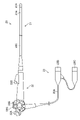
- FIG. 1 is a diagram showing a schematic configuration of an endoscopic image diagnostic system according to the first embodiment.
- FIG. 2 is a diagram showing a schematic configuration of an endoscope system.
- FIG. 3 is a diagram showing a schematic configuration of an endoscope.
- FIG. 4 is a diagram showing an example of the configuration of the end surface of the tip portion.
- FIG. 5 is a block diagram showing main functions of the endoscopic image generating device.
- FIG. 6 is a block diagram showing main functions of the endoscope image processing apparatus.
- FIG. 7 is a block diagram showing main functions of the image recognition processing section.
- FIG. 8 is a diagram showing an example of a screen display during examination.
- FIG. 9 is a diagram showing an outline of speech recognition.
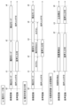
- FIG. 10 is a diagram showing settings of the speech recognition dictionary.
- FIG. 10 is a diagram showing settings of the speech recognition dictionary.
- FIG. 11 is another diagram showing setting of the speech recognition dictionary.
- FIG. 12 is a time chart for voice recognition dictionary setting.
- 13A and 13B are diagrams showing how notifications are made by displaying icons on the screen.
- FIG. 14 is a diagram showing how voice input is performed in a specific period.
- FIG. 15 is another diagram showing how voice input is performed in a specific period.
- FIG. 16 is a diagram showing an example of screen display for displaying the remaining voice recognition period.
- FIG. 17 is a diagram showing a screen display example of speech recognition candidates.
- FIG. 18 is a diagram showing a screen display example of a speech recognition result.
- FIG. 19 is a diagram showing how processing is performed according to the quality of image recognition.
- An endoscopic image diagnosis support system is a system that supports detection and differentiation of lesions and the like in endoscopy.
- an example of application to an endoscopic image diagnosis support system that supports detection and differentiation of lesions and the like in lower gastrointestinal endoscopy (colon examination) will be described.
- FIG. 1 is a block diagram showing the schematic configuration of the endoscopic image diagnosis support system.
- an endoscopic image diagnosis support system 1 (endoscopic system) according to the present embodiment includes an endoscopic system 10 (endoscopic system, medical information processing apparatus), endoscopic information management, It has a system 100 and a user terminal 200 .
- FIG. 2 is a block diagram showing a schematic configuration of the endoscope system 10. As shown in FIG.
- the endoscope system 10 of the present embodiment is configured as a system capable of observation using special light (special light observation) in addition to observation using white light (white light observation).
- Special light viewing includes narrowband light viewing.
- Narrowband light observation includes BLI observation (Blue laser imaging observation), NBI observation (Narrowband imaging observation; NBI is a registered trademark), LCI observation (Linked Color Imaging observation), and the like. Note that the special light observation itself is a well-known technique, so detailed description thereof will be omitted.
- the endoscope system 10 of the present embodiment includes an endoscope 20, a light source device 30, an endoscope image generation device 40, an endoscope image processing device 60, a display device 70 (output device , display device), a recording device 75 (recording device), an input device 50, and the like.
- the endoscopic image generation device 40 and the endoscopic image processing device 60 constitute a medical information processing device 80 (medical information processing device).
- FIG. 3 is a diagram showing a schematic configuration of the endoscope 20. As shown in FIG.
- the endoscope 20 of this embodiment is an endoscope for lower digestive organs. As shown in FIG. 3 , the endoscope 20 is a flexible endoscope (electronic endoscope) and has an insertion section 21 , an operation section 22 and a connection section 23 .
- the insertion portion 21 is a portion that is inserted into a hollow organ (in this embodiment, the large intestine).
- the insertion portion 21 is composed of a distal end portion 21A, a curved portion 21B, and a flexible portion 21C in order from the distal end side.
- FIG. 4 is a diagram showing an example of the configuration of the end surface of the tip.
- the end surface of the distal end portion 21A is provided with an observation window 21a, an illumination window 21b, an air/water nozzle 21c, a forceps outlet 21d, and the like.
- the observation window 21a is a window for observation. The inside of the hollow organ is photographed through the observation window 21a. Photographing is performed via an optical system such as a lens and an image sensor (not shown) built in the distal end portion 21A (the portion of the observation window 21a).
- the image sensor is, for example, a CMOS image sensor (Complementary Metal Oxide Semiconductor image sensor), a CCD image sensor (Charge Coupled Device image sensor), or the like.
- the illumination window 21b is a window for illumination.
- Illumination light is irradiated into the hollow organ through the illumination window 21b.
- the air/water nozzle 21c is a cleaning nozzle.
- a cleaning liquid and a drying gas are jetted from the air/water nozzle 21c toward the observation window 21a.
- a forceps outlet 21d is an outlet for treatment tools such as forceps.
- the forceps outlet 21d also functions as a suction port for sucking body fluids and the like.
- the bending portion 21B is a portion that bends according to the operation of the angle knob 22A provided on the operating portion 22.
- the bending portion 21B bends in four directions of up, down, left, and right.
- the flexible portion 21C is an elongated portion provided between the bending portion 21B and the operating portion 22.
- the flexible portion 21C has flexibility.
- the operation part 22 is a part that is held by the operator to perform various operations.
- the operation unit 22 is provided with various operation members.
- the operation unit 22 includes an angle knob 22A for bending the bending portion 21B, an air/water supply button 22B for performing an air/water supply operation, and a suction button 22C for performing a suction operation.
- the operation unit 22 includes an operation member (shutter button) for capturing a still image, an operation member for switching observation modes, an operation member for switching ON/OFF of various support functions, and the like.
- the operation portion 22 is provided with a forceps insertion opening 22D for inserting a treatment tool such as forceps.
- the treatment instrument inserted from the forceps insertion port 22D is delivered from the forceps outlet 21d (see FIG. 4) at the distal end of the insertion portion 21.
- the treatment instrument includes biopsy forceps, a snare, and the like.
- the connection part 23 is a part for connecting the endoscope 20 to the light source device 30, the endoscope image generation device 40, and the like.
- the connecting portion 23 includes a cord 23A extending from the operating portion 22, and a light guide connector 23B and a video connector 23C provided at the tip of the cord 23A.
- the light guide connector 23B is a connector for connecting to the light source device 30 .
- the video connector 23C is a connector for connecting to the endoscopic image generating device 40 .
- the light source device 30 generates illumination light.
- the endoscope system 10 of the present embodiment is configured as a system capable of special light observation in addition to normal white light observation. Therefore, the light source device 30 is configured to be capable of generating light (for example, narrowband light) corresponding to special light observation in addition to normal white light.
- the special light observation itself is a known technology, and therefore the description of the generation of the light and the like will be omitted.
- the endoscopic image generation device 40 (processor) collectively controls the operation of the entire endoscope system 10 together with the endoscopic image processing device 60 (processor).
- the endoscopic image generation device 40 includes a processor, a main memory (memory), an auxiliary memory (memory), a communication section, and the like as its hardware configuration. That is, the endoscopic image generation device 40 has a so-called computer configuration as its hardware configuration.
- the processor includes, for example, a CPU (Central Processing Unit), GPU (Graphics Processing Unit), FPGA (Field Programmable Gate Array), PLD (Programmable Logic Device), and the like.
- the main storage unit is composed of, for example, a RAM (Random Access Memory) or the like.
- the auxiliary storage unit is composed of, for example, a non-temporary and tangible recording medium such as a flash memory, and records the medical information processing program according to the present invention or part of the computer-readable code and other data. be able to.
- the auxiliary memory section may include various magneto-optical recording devices, semiconductor memories, etc. in addition to or in place of the flash memory.
- FIG. 5 is a block diagram showing the main functions of the endoscopic image generating device 40. As shown in FIG.
- the endoscope image generation device 40 has functions such as an endoscope control section 41, a light source control section 42, an image generation section 43, an input control section 44, an output control section 45, and the like.
- Various programs executed by the processor (which may include the medical information processing program according to the present invention or a part thereof) and various data necessary for control are stored in the auxiliary storage unit described above, and the endoscopic image is stored.
- Each function of the generating device 40 is realized by the processor executing those programs.
- the processor of the endoscopic image generation device 40 is an example of the processor in the endoscopic system and medical information processing device according to the present invention.
- the endoscope control unit 41 controls the endoscope 20.
- Control of the endoscope 20 includes image sensor drive control, air/water supply control, suction control, and the like.
- the light source controller 42 controls the light source device 30 .
- the control of the light source device 30 includes light emission control of the light source and the like.
- the image generator 43 generates a captured image (endoscopic image) based on the signal output from the image sensor of the endoscope 20.
- the image generator 43 can generate a still image and/or a moving image (a plurality of medical images obtained by the image sensor 25 capturing images of the subject in time series) as captured images.
- the image generator 43 may apply various image processing to the generated image.
- the input control unit 44 receives operation inputs and various information inputs via the input device 50 .
- the output control unit 45 controls output of information to the endoscope image processing device 60 .
- the information output to the endoscope image processing device 60 includes various kinds of operation information input from the input device 50 in addition to the endoscope image obtained by imaging.
- the input device 50 constitutes a user interface in the endoscope system 10 together with the display device 70 .
- the input device 50 includes a microphone 51 (voice input device) and a foot switch 52 (operation device).
- a microphone 51 is an input device for voice recognition, which will be described later.
- the foot switch 52 is an operation device that is placed at the feet of the operator and is operated with the foot. By stepping on the pedal, an operation signal (for example, a signal indicating a voice input trigger or a candidate for voice recognition is selected. signal) is output.
- the microphone 51 and the foot switch 52 are controlled by the input control unit 44 of the endoscope image generation device 40.
- the present invention is not limited to this embodiment, and the endoscope image processing device 60 and the display device
- the microphone 51 and foot switch 52 may be controlled via 70 or the like.
- an operation device (button, switch, etc.) having the same function as the foot switch 52 may be provided in the operation section 22 of the endoscope 20 .
- the input device 50 can include known input devices such as a keyboard, mouse, touch panel, line-of-sight input device, etc. as operation devices.
- the endoscope image processing apparatus 60 includes a processor, a main storage section, an auxiliary storage section, a communication section, etc. as its hardware configuration. That is, the endoscope image processing apparatus 60 has a so-called computer configuration as its hardware configuration.
- the processor includes, for example, a CPU, GPU (Graphics Processing Unit), FPGA (Field Programmable Gate Array), PLD (Programmable Logic Device), and the like.
- the processor of the endoscope image processing device 60 is an example of the processor in the endoscope system and medical information processing device according to the present invention.
- the processor of the endoscopic image generating device 40 and the processor of the endoscopic image processing device 60 may share the function of the processor in the endoscopic system and medical information processing device according to the present invention.
- the endoscopic image generating device 40 mainly has the function of an "endoscopic processor” for generating endoscopic images
- the endoscopic image processing device 60 mainly performs image processing on endoscopic images as a "CAD box". (CAD: Computer Aided Diagnosis)" can be employed.
- CAD Computer Aided Diagnosis
- a mode different from such division of functions may be employed.
- the main storage unit is composed of memory such as RAM, for example.
- the auxiliary storage unit is composed of, for example, a non-temporary and tangible recording medium (memory) such as a flash memory, and various programs executed by the processor (including the medical information processing program according to the present invention or a part thereof) good) computer readable code and various data required for control and the like are stored.
- the auxiliary memory section may include various magneto-optical recording devices, semiconductor memories, etc. in addition to or in place of the flash memory.
- the communication unit is composed of, for example, a communication interface connectable to a network.
- the endoscope image processing apparatus 60 is communicably connected to the endoscope information management system 100 via a communication unit.
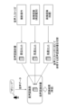
- FIG. 6 is a block diagram showing the main functions of the endoscope image processing device 60. As shown in FIG.
- the endoscopic image processing apparatus 60 mainly includes an endoscopic image acquisition unit 61, an input information acquisition unit 62, an image recognition processing unit 63, a voice input trigger reception unit 64, a display control unit 65, and an examination information output control unit 66 and the like. These functions are realized by the processor executing a program (which may include the medical information processing program according to the present invention or part thereof) stored in an auxiliary storage unit or the like.
- Endoscopic image acquisition unit acquires an endoscopic image from the endoscopic image generation device 40 .
- Image acquisition can be done in real time. That is, it is possible to sequentially acquire (sequentially input) in real time a plurality of medical images obtained by the image sensor 25 (image sensor) photographing the subject in time series.
- the input information acquisition unit 62 acquires information input via the input device 50 and the endoscope 20 .
- the input information acquisition unit 62 mainly includes an information acquisition unit 62A that acquires input information other than voice information, a voice recognition unit 62B that acquires voice information and recognizes voice input to the microphone 51, and a voice recognition unit 62B that is used for voice recognition. and a speech recognition dictionary 62C.
- the voice recognition dictionary 62C may include a plurality of dictionaries with different contents (for example, dictionaries relating to site information, finding information, treatment information, and hemostasis information).
- Information input to the input information acquisition unit 62 via the input device 50 includes information input via the microphone 51, the foot switch 52, or a keyboard or mouse (not shown) (for example, voice information, voice input trigger, etc.). , candidate selection operation information, etc.).
- Information input via the endoscope 20 includes information such as an instruction to start capturing an endoscopic image (moving image) and an instruction to capture a still image. As will be described later, in this embodiment, the user can input a voice input trigger, select a voice recognition candidate, etc. via the microphone 51 and/or the foot switch 52 .
- the input information acquisition unit 62 acquires operation information of the foot switch 52 via the endoscope image generation device 40 .
- the image recognition processing unit 63 (processor) performs image recognition on the endoscopic image acquired by the endoscopic image acquisition unit 61 .
- the image recognition processing unit 63 can perform image recognition in real time.
- FIG. 7 is a block diagram showing the main functions of the image recognition processing section 63.
- the image recognition processing unit 63 has functions such as a lesion detection unit 63A, a discrimination unit 63B, a specific region detection unit 63C, a treatment tool detection unit 63D, a hemostat detection unit 63E, and a measurement unit 63F. have.
- Each of these units can be used for judgment or judgment as to whether or not the endoscopic image includes a specific subject.
- the “specific subject” may differ depending on each section of the image recognition processing section 63, as described below.
- the lesion detection unit 63A detects a lesion such as a polyp (lesion; an example of a "specific subject") from an endoscopic image.
- Processing for detecting lesions includes processing for detecting portions that are definite to be lesions, as well as processing for detecting portions that may be lesions (benign tumors or dysplasia, etc.; lesion candidate regions). , areas after lesions have been treated (post-treatment areas), and areas with features (such as redness) that may be directly or indirectly associated with lesions.
- the discrimination unit 63B performs discrimination processing on the lesion detected by the lesion detection unit 63A when the lesion detection unit 63A determines that the endoscopic image includes a lesion (specific subject). I do.
- the discrimination section 63B performs a neoplastic (NEOPLASTIC) or non-neoplastic (HYPERPLASTIC) discrimination process on a lesion such as a polyp detected by the lesion detection section 63A.
- NEOPLASTIC neoplastic
- HYPERPLASTIC non-neoplastic
- the discrimination section 63B can be configured to output a discrimination result when a predetermined criterion is satisfied.
- Predetermined criteria include, for example, “reliability of discrimination results (depending on conditions such as endoscopic image exposure, degree of focus, blurring, etc.) and their statistical values (maximum or minimum, average, etc.) is greater than or equal to a threshold", but other criteria may be used.
- the specific area detection unit 63C performs processing for detecting specific areas (landmarks) within the hollow organ from the endoscopic image. For example, processing for detecting the ileocecal region of the large intestine is performed.
- the large intestine is an example of a hollow organ
- the ileocecal region is an example of a specific region.
- the specific region detection unit 63C may detect, for example, the liver flexure (right colon), the splenic flexure (left colon), the rectal sigmoid, and the like. Further, the specific area detection section 63C may detect a plurality of specific areas.
- the treatment instrument detection unit 63D detects the treatment instrument appearing in the endoscopic image and performs processing for determining the type of the treatment instrument.
- the treatment instrument detector 63D can be configured to detect a plurality of types of treatment instruments such as biopsy forceps and snares.
- the hemostat detection unit 63E detects a hemostat such as a hemostatic clip and performs processing for determining the type of the hemostat.
- the treatment instrument detection section 63D and the hemostat detection section 63E may be configured by one image recognizer.
- the measurement unit 63F measures lesions, lesion candidate regions, specific regions, post-treatment regions, etc. (measurements of shapes, dimensions, etc.).
- Each unit of the image recognition processing unit 63 (lesion detection unit 63A, discrimination unit 63B, specific area detection unit 63C, treatment instrument detection unit 63D, hemostat detection unit 63E, measurement unit 63F, etc.) is configured by machine learning. It can be configured using an image recognizer (trained model). Specifically, each part described above learns using machine learning algorithms such as Neural Network (NN), Convolutional Neural Network (CNN), AdaBoost, and Random Forest. It can be configured with a trained image recognizer (trained model). In addition, as described above for the discrimination unit 63B, each of these units adjusts the reliability of the final output (discrimination result, type of treatment instrument, etc.) by setting the layer configuration of the network as necessary. can be output as Further, each of the above-described units may perform image recognition on all frames of the endoscopic image, or may intermittently perform image recognition on some frames.
- NN Neural Network
- CNN Convolutional Neural Network
- AdaBoost AdaBoost
- Random Forest It
- the output of the recognition result of the endoscopic image from each of these units or the output of the recognition result that satisfies a predetermined criterion can be triggered by a voice input trigger.
- the period during which these outputs are performed may be set as the period during which speech recognition is performed.
- a feature amount is calculated from an endoscopic image, and the calculated feature amount is used for detection. It is also possible to have a configuration for performing such as.
- the voice input trigger reception unit 64 receives an input of a voice input trigger during capturing (inputting) of an endoscopic image, and sets the voice recognition dictionary 62C according to the input voice input trigger.
- the voice input trigger in the present embodiment is, for example, a determination result (detection result) indicating that a specific subject is included in the endoscopic image.
- the output of the lesion detection unit 63A is used as the determination result. be able to.
- Another example of the voice input trigger is the output of discrimination results for a specific subject. In this case, the output of the discrimination section 63B can be used as the discrimination results.
- voice input triggers include an instruction to start imaging a plurality of medical images, input of a wake word to the microphone 51 (audio input device), operation of the foot switch 52, and other operation devices connected to the endoscope system. (For example, a colonoscope shape measuring device, etc.) can also be used.
- a colonoscope shape measuring device can also be used. The setting of the speech recognition dictionary and speech recognition according to these speech input triggers will be described later in detail.
- the display control unit 65 controls the display of the display device 70 .
- Main display control performed by the display control unit 65 will be described below.
- FIG. 8 is a diagram showing an example of a screen display during examination. As shown in the figure, an endoscopic image I (live view) is displayed in a main display area A1 set within the screen 70A. A secondary display area A2 is further set on the screen 70A, and various information related to the examination is displayed.
- the example shown in FIG. 8 shows an example in which patient-related information Ip and a still image Is of an endoscopic image taken during an examination are displayed in the sub-display area A2.
- the still images Is are displayed, for example, in the order in which they were shot from top to bottom on the screen 70A.
- the display control unit 65 displays an icon 300 indicating the state of voice recognition, an icon 320 indicating the site being imaged, the site to be imaged (ascending colon, transverse colon, descending colon, etc.) and the result of voice recognition in real time ( A display area 340 for displaying characters (without time delay) can be displayed on the screen 70A.
- the display control unit 65 performs image recognition from an endoscopic image, input by a user via an operation device, and display of a part by an external device (for example, an endoscope insertion shape observation device) connected to the endoscope system 10, or the like. Information can be obtained.
- the display control unit 65 can display (output) the speech recognition result on the display device 70 (output device, display device).
- the examination information output control section 66 outputs examination information to the recording device 75 and/or the endoscope information management system 100 .
- the examination information includes, for example, an endoscopic image taken during the examination, the result of judgment on a specific subject, the result of voice recognition, the information of the site input during the examination, and the information of the treatment name input during the examination. , contains information on the treatment tools detected during the examination.
- Examination information is output, for example, for each lesion or sample collection. At this time, each piece of information is output in association with each other. For example, an endoscopic image obtained by imaging a lesion or the like is output in association with information on the selected site.
- the information of the selected treatment name and the information of the detected treatment tool are output in association with the endoscopic image and the information of the region.
- endoscopic images captured separately from lesions and the like are output to the recording device 75 and/or the endoscopic information management system 100 at appropriate times.
- the endoscopic image is output with the information of the photographing date added.
- a recording device 75 includes various types of magneto-optical recording devices, semiconductor memories, and their control devices, and stores endoscopic images (moving images and still images), image recognition results, voice recognition results, and examination information. , report creation support information, etc. can be recorded. These pieces of information may be recorded in the sub-storage unit of the endoscopic image generation device 40 and the endoscopic image processing device 60, or in a recording device included in the endoscopic information management system 100.
- FIG. 9 is a diagram showing an outline of speech recognition.
- the medical information processing apparatus 80 (processor) accepts an input of a voice input trigger during endoscopic image capturing (during sequential input), and when the voice input trigger is input, the voice input is performed.
- a voice recognition dictionary is set according to the trigger, and voice input to the microphone 51 (voice input device) after the voice recognition dictionary is set is recognized using the set voice recognition dictionary.
- the medical information processing apparatus 80 outputs detection results from the lesion detection unit 63A, outputs discrimination results from the discrimination unit 63B, instructs the start of imaging of a plurality of medical images, and switches from the detection mode to the discrimination mode. , wake word input to the microphone 51 (voice input device), foot switch 52 operation, operation input to the operation device connected to the endoscope system, etc. perform recognition.
- start of speech recognition may be delayed with respect to the setting of the speech recognition dictionary, it is preferable to start speech recognition immediately after setting the speech recognition dictionary (zero delay time).
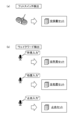
- FIG. 10 is a diagram showing settings of the speech recognition dictionary.
- the left side of the arrow indicates the voice input trigger
- the right side of the arrow indicates the example of the voice recognition dictionary and registered words set according to the voice input trigger.
- the voice recognition section 62B sets the voice recognition dictionary 62C according to the voice input trigger.
- the speech recognition section 62B sets "finding set A" as the speech recognition dictionary.
- FIG. 11 is another diagram showing the setting of the speech recognition dictionary.
- the voice recognition unit 62B sets "all dictionary set” when the operation of the foot switch 52 (operation device) is accepted as a voice input trigger.
- a voice recognition dictionary is set according to the contents of the wake word.
- a "wake word” or a “wakeup word” is, for example, "a predetermined word or phrase for causing the voice recognition unit 62B to set a voice recognition dictionary and start voice recognition”. can be stipulated.
- the above-mentioned wake words can be divided into two types. They are “wake word for report input” and “wake word for shooting mode control”.
- the "wake words related to report input” are, for example, "finding input” and "treatment input”.
- the result of speech recognition is output when Speech recognition results can be associated with images and used in reports. Linking with an image and use in a report are one aspect of "output" of the result of speech recognition, and the display device 70, the recording device 75, the storage unit of the medical information processing device 80, or the endoscope information management system 100
- a recording device such as a recording device is one aspect of an “output device”.
- the other "wake words related to shooting mode control” are, for example, “shooting settings” and “settings.” ”, “BLI”, etc.), and turn on/off lesion detection by endoscope AI (a recognizer using artificial intelligence) (e.g., “detection on”, “detection off”). It is possible to set a dictionary to be used for speech recognition of words such as Note that "output” and “output device” are the same as those described above for "wake word for report input”.
- FIG. 12 is a time chart for voice recognition dictionary setting. Note that FIG. 12 does not specifically describe the words and phrases input by voice and the recognition results thereof.
- Part (a) of FIG. 12 shows the types of voice input triggers. In the example shown in the same part, the voice input trigger is the output of the image recognition result of the endoscopic image, the input of the wake word to the microphone 51, the signal by the operation of the foot switch 52 (operation device), and the start of imaging of the endoscopic image. It is an instruction.
- Part (b) of FIG. 12 shows a voice recognition dictionary that is set according to a voice input trigger.
- the voice recognition unit 62B sets different voice recognition dictionaries according to the flow of examination (start of imaging, detection of a lesion or lesion candidate, input of findings, insertion and treatment of treatment instrument, hemostasis).
- each section of the image recognition processing section 63 recognizes a plurality of types of "specific subjects” (specifically, lesions, treatment instruments, hemostats, etc. described above) to be determined (recognized). (a plurality of image recognitions as a whole) can be performed, and the voice recognition unit 62B determines that "included in the endoscopic image" by any of the image recognitions by these units.
- a voice recognition dictionary corresponding to the type of "specific subject" can be set.
- each unit determines whether or not a plurality of "specific subjects" are included in the endoscopic image, and the speech recognition unit 62B determines whether " It is also possible to set a speech recognition dictionary corresponding to a specific subject determined to be "included in the endoscopic image". Examples of cases where an endoscopic image includes multiple "specific subjects” include, for example, multiple lesions, multiple treatment tools, and multiple hemostats. may be included.
- a speech recognition dictionary corresponding to the type of "specific subject” may be set for some of the multiple image recognitions performed by the above units.
- the speech recognition unit 62B uses the set speech recognition dictionary to recognize speech input to the microphone 51 (speech input device) after the speech recognition dictionary is set (not shown in FIG. 12). ). It is preferable that the display control unit 65 causes the display device 70 to display the speech recognition result.
- the speech recognition unit 62B can perform speech recognition on part information, findings information, treatment information, and hemostasis information. If there are multiple lesions, etc., a series of processes (acceptance of voice input trigger in the cycle from imaging start to hemostasis, voice recognition dictionary setting, and voice recognition) can be repeated for each lesion.
- the speech recognition unit 62B and the display control unit 65 recognize only registered words registered in the set speech recognition dictionary in speech recognition, and perform speech recognition of the registered words.
- the result can be displayed (output) on the display device 70 (output device, display device) (adaptive speech recognition).
- the registered words in the speech recognition dictionary may be set so as not to recognize the wake word, or the registered words may be set including the wake word.
- the speech recognition unit 62B and the display control unit 65 recognize and recognize registered words and specific words registered in the set speech recognition dictionary in speech recognition. It is also possible to display (output) the results of speech recognition of registered words among words on the display device 70 (display device, output device) (non-adaptive speech recognition).
- An example of the "specific word” is a wake word for the voice input device, but the "specific word” is not limited to this.
- the endoscope system 10 which of the above modes (adaptive voice recognition, non-adaptive voice recognition) is used for voice recognition and result display is determined by a user's instruction via the input device 50, the operation unit 22, or the like. Can be set based on input.
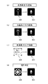
- the display control unit 65 notifies the user that the speech recognition dictionary is set (set fact and which dictionary is set) and that speech recognition is possible. Notification is preferred. As shown in FIG. 13, the display control unit 65 can perform notification by switching icons displayed on the screen. In the example shown in FIG. 13, the display control unit 65 causes the screen 70A or the like to display an icon indicating the image recognizer that is operating (or displays the recognition result on the screen) among the units of the image recognition processing unit 63. When the image recognizer recognizes a specific subject (audio input trigger) and enters the voice recognition period, the display is switched to a microphone-like icon to notify the user (see FIGS. 8 and 16 to 18).
- parts (a) and (b) of FIG. 13 are states in which the treatment instrument detection unit 63D is operating, but the specific objects to be recognized are different (forceps, snare). , the display control unit 65 displays different icons 360 and 362, and when the forceps or snare is actually recognized, switches to the microphone-like icon 300 to inform the user that voice recognition is now possible.
- the states shown in parts (c) and (d) of FIG. 13 are states in which the hemostat detection unit 63E and the discrimination unit 63B are operating, respectively, and the display control unit 65 displays icons 364 and 366. However, when a hemostat or lesion is recognized, the icon is switched to a microphone-like icon 300 to inform the user that voice recognition is now possible.
- the display control unit 65 may display and switch icons according to not only the operation status of each part of the image recognition processing unit 63 but also the operation status and input status of the microphone 51 and/or the foot switch 52 .
- the speech recognition unit 62B (processor) can execute speech recognition using the set speech recognition dictionary during a specific period (a period that satisfies a predetermined condition) after the setting.
- the "predetermined condition" may be the output of the recognition result from the image recognizer, the condition for the content of the output, or the execution time itself for speech recognition (3 seconds, 5 seconds, etc.). good too.
- specifying the execution time it is possible to specify the elapsed time from the setting of the dictionary or the elapsed time from notifying the user that voice input is possible.
- FIG. 14 is a diagram showing how speech recognition is performed during a specific period.
- the speech recognition section 62B performs speech recognition only during the discrimination mode period (the period during which the discrimination section 63B is operating; time t1 to time t2).
- speech recognition is performed only during the period (time t2 to time t3) in which the discrimination section 63B outputs the discrimination result (discrimination determination result).
- the discrimination section 63B can be configured to output when the reliability of the discrimination result or its statistical value is equal to or greater than a threshold value.
- the speech recognition unit 62B detects the period (time t1 to time t2) during which the treatment instrument detection unit 63D detects the treatment instrument and the hemostat detection unit 63E detects the hemostat. is detected (time t3 to time t4), speech recognition is performed.
- the reception of the voice input trigger and the setting of the voice recognition dictionary are omitted.
- the speech recognition unit 62B may set the speech recognition period for each image recognizer, or may set it according to the type of speech input trigger. Further, the speech recognition section 62B may set the “predetermined condition” and the “execution time of speech recognition” based on the instruction input by the user via the input device 50, the operation section 22, or the like.
- FIG. 15 is another diagram showing how speech recognition is performed during a specific period.
- Part (a) of FIG. 15 shows an example in which setting of the speech recognition dictionary and speech recognition are performed during a certain period of time (time t1 to t2 and time t3 to t4 in this part) after manual operation.
- the voice recognition unit 62B can perform voice recognition by regarding the user's operation on the input device 50, the operation unit 22, etc. as a "manual operation".
- the "manual operation” may be operation of the various operation devices described above, input of a wake word via the microphone 51, operation of the foot switch 52, and operation of the endoscopic image (moving image, still image).
- a switching operation from the detection mode (the state in which the lesion detection unit 63A outputs the results) to the discrimination mode (the state in which the discrimination unit 63B outputs the results), and the operation device connected to the endoscope system 10. may be an operation for
- part (b) of FIG. 15 shows an example of processing when the period of speech recognition based on image recognition and the above-described "fixed time after manual operation" overlap. Specifically, from time t1 to time t3, the speech recognition unit 62B prioritizes speech recognition associated with manual operation over speech recognition according to the discrimination result output from the discrimination unit 63B. to perform voice recognition.
- the period of voice recognition based on image recognition may be continuous with the period of voice recognition associated with manual operation.
- the speech recognition unit 62B uses the discrimination result of the discrimination unit 63B during the time t3 to time t4 following the voice recognition period (time t1 to time t2) by manual operation. set a speech recognition dictionary based on it, and perform speech recognition.
- the voice recognition section 62B does not set the voice recognition dictionary and does not perform voice recognition.
- the speech recognition unit 62B performs speech recognition by setting a speech recognition dictionary based on manual operation from time t5 to time t6, and does not perform speech recognition after time t6 when this speech recognition period ends.
- the voice recognition unit 62B and the display control unit 65 may display the remaining time of the voice recognition period on the display device 70.
- FIG. 16 is a diagram showing an example of screen display of remaining time. Part (a) of FIG. 16 is an example of the display on the screen 70A, and the remaining time meter 350 is displayed. Part (b) of the figure is an enlarged view of the remaining time meter 350 . In the remaining time meter 350, the shaded area 352 expands over time and the solid area 354 shrinks over time.
- a frame 356 composed of a black background area 356A and a white background area 356B rotates around these areas to attract the user's attention.
- the voice recognition unit 62B and the display control unit 65 may rotate and display the frame 356 when detecting that voice is being input.
- the speech recognition unit 62B and the display control unit 65 may set different periods as the period for speech recognition depending on the speech input trigger and the speech recognition dictionary. Alternatively, the period may be set according to the user's operation via the input device 50 .
- the voice recognition unit 62B and the display control unit 65 may output the remaining time in numbers or voice.
- the remaining time is zero.
- FIG. 17 is a diagram showing a screen display example of speech recognition candidates and speech recognition results (the region of interest ROI and frame F are also displayed in FIG. 17).
- FIG. 17 shows a state in which the discrimination section 63B outputs the discrimination result, and the content of the speech recognition dictionary "finding set A" (see FIG. 10) corresponding to the output of the discrimination result is displayed in the area 370 of the screen 70A.
- the speech recognition unit 62B can confirm conversion (selection of words) according to the user's selection operation via the microphone 51, the foot switch 52, or other operation devices. Note that the speech recognition unit 62B and the display control unit 65 can use the input of a speech input trigger or the setting of the speech recognition dictionary as a trigger for displaying candidates.
- FIG. 18 is a diagram showing a screen display example of speech recognition results.
- the display control unit 65 can display the word selected by the user (“JNET TYPE 2A” in the example of FIG. 18) on the screen (area 372).
- the display mode of the speech recognition result is not limited to the mode illustrated in FIG. 18 and the like.
- the speech recognition unit 62B and the display control unit 65 display the result of speech recognition in characters in real time in the display area 340 (see FIG. 8) or the like, and display the finalized result in the area shown in FIG. 372 may be displayed.
- the voice recognition unit 62B and the display control unit 65 superimpose the selected or confirmed voice recognition result on the display area of the moving image (for example, the endoscopic image I shown in FIGS. 8 and 18). (In the example shown in FIG. 18, "JNET TYPE 2A" can be displayed near the attention area ROI and frame F).
- the voice recognition unit 62B and the display control unit 65 may set the display position of the voice recognition selection result and confirmation result according to the voice recognition result or the type of the recognized subject.
- the voice recognition unit 62B and the display control unit 65 for example, superimpose the voice recognition result of “finding” near the attention area (for example, the attention area ROI in FIG. 18) of the moving image, and ” can be displayed outside the moving image display area (for example, near the icon 300 or the icon 320, or the remaining time meter 350).
- the speech recognition unit 62B performs the image recognition processing unit 63 (see FIG. 7) as described below with reference to FIG.
- the speech recognition dictionary 62C may be switched according to the quality of image recognition performed by .
- the period during which the discrimination unit 63B outputs the discrimination result is the voice recognition period (similar to part (a) of FIG. 14). Under such circumstances, as shown in part (a) of FIG. shall be defective. Poor observation quality may be caused by, for example, inappropriate exposure or focus, or obstruction of the field of view by residue.
- the speech recognition unit 62B performs speech recognition from time t1 to time t2 when speech recognition is normally not performed (if the image quality is good), and performs image quality improvement operation. accepts commands from The speech recognition unit 62B can perform speech recognition by setting, as the speech recognition dictionary 62C, an "image quality improvement set" in which words such as "gas injection, lighting on, sensor sensitivity 'high'" are registered.
- the speech recognition section 62B performs speech recognition using the speech recognition dictionary "finding set" as usual.
- the speech recognition unit 62B Since the detection mode is set from time t4 to time t9, the speech recognition unit 62B normally does not perform speech recognition. to perform voice recognition. However, it is assumed that the observation quality is poor from time t6 to time t7. During this period (time t6 to time t7), the voice recognition section 62B can also accept a command for an image quality improvement operation in the same manner as during time t1 to time t2.
- the examination information output control unit 66 (processor) associates the endoscopic image (time-series medical image) with the result of speech recognition, and stores them in the recording device 75 and the medical information processing device 80. It can be recorded in a recording device such as the endoscope information management system 100 or the like.
- the examination information output control unit 66 may associate and record an endoscopic image showing a specific subject and the result of determination by image recognition (that the specific subject is shown in the image).
- the test information output control unit 66 may record according to the user's operation on the operation device, or may automatically record without depending on the user's operation. In endoscopy system 10, such records can assist the user in generating an examination report.
Abstract
One embodiment of the invention of the present disclosure provides an endoscope system, medical information processing device, medical information processing method, medical information processing program, and storage medium, capable of improving recognition accuracy of input sound. The endoscope system according to one aspect of the present invention comprises a sound input device, an image sensor for imaging an object, and a processor. The processor causes the image sensor to image an object over time and thereby acquires a plurality of medical images, receives input from a sound input trigger while imaging the plurality of medical images, sets a sound recognition dictionary according to the sound input trigger when the sound input trigger is input, and recognizes, using the set sound recognition dictionary, the sound input into the sound input device after setting the sound recognition dictionary.
Description
本発明は、音声入力及び音声認識を行う内視鏡システム、医療情報処理装置、医療情報処理方法、医療情報処理プログラム、及び記録媒体に関する。
The present invention relates to an endoscope system that performs voice input and voice recognition, a medical information processing device, a medical information processing method, a medical information processing program, and a recording medium.
医療画像を用いた検査や診断支援を行う技術分野では、ユーザが入力した音声を認識し、認識結果に基づく処理を行うことが知られている。例えば、特許文献1には、内視鏡を音声入力で操作することが記載されている。また、特許文献2には、レポート作成用の音声入力を行うことが記載されている。
In the technical field of examination and diagnosis support using medical images, it is known to recognize the voice input by the user and perform processing based on the recognition results. For example, Patent Literature 1 describes operating an endoscope by voice input. Further, Japanese Patent Application Laid-Open No. 2002-200002 describes that voice input for creating a report is performed.
医療画像を用いた検査中に音声入力を行う場合、シーンによらずに全ての単語を認識可能とすると単語間の相互誤認識が増えて操作性が低下するおそれがある。しかしながら、上述した特許文献1,2のような従来の技術は、このような課題を十分考慮したものではなかった。
When voice input is performed during an examination using medical images, if all words can be recognized regardless of the scene, there is a risk that mutual misrecognition between words will increase and operability will decrease. However, conventional techniques such as those disclosed in Patent Literatures 1 and 2 described above do not sufficiently consider such problems.
本発明はこのような事情に鑑みてなされたもので、医療画像に関する音声認識の精度を向上させることができる内視鏡システム、医療情報処理装置、医療情報処理方法、医療情報処理プログラム、及び記録媒体を提供することを目的とする。
The present invention has been made in view of such circumstances, and provides an endoscope system, a medical information processing apparatus, a medical information processing method, a medical information processing program, and a recording that can improve the accuracy of speech recognition for medical images. The purpose is to provide a medium.
上述した目的を達成するため、本発明の第1の態様に係る内視鏡システムは、音声入力装置と、被写体を撮影するイメージセンサと、プロセッサと、を備える内視鏡システムであって、プロセッサは、イメージセンサが被写体を時系列に撮影することで得られた複数の医療画像を取得し、複数の医療画像の撮影中に音声入力トリガの入力を受け付け、音声入力トリガが入力された場合に、音声入力トリガに応じた音声認識辞書を設定し、設定がされた以降に音声入力装置に入力された音声を、設定された音声認識辞書を用いて音声認識する。第1の態様では、音声入力トリガに応じた音声認識辞書を設定し、設定された音声認識辞書を用いて音声認識するので、音声認識のシーンに合わせた音声認識辞書を用い、医療画像に関する音声認識の精度を向上させることができる。
In order to achieve the object described above, an endoscope system according to a first aspect of the present invention is an endoscope system comprising a voice input device, an image sensor for capturing an image of a subject, and a processor, wherein the processor acquires multiple medical images obtained by capturing images of the subject in chronological order by the image sensor, accepts input of a voice input trigger while capturing multiple medical images, and when the voice input trigger is input, a speech recognition dictionary corresponding to the speech input trigger is set, and speech input to the speech input device after the setting is made is recognized using the set speech recognition dictionary. In the first aspect, a speech recognition dictionary is set according to a speech input trigger, and speech recognition is performed using the set speech recognition dictionary. Recognition accuracy can be improved.
第2の態様に係る内視鏡システムは第1の態様において、プロセッサは、音声認識において、設定された音声認識辞書に登録されている登録単語のみを認識し、登録単語についての音声認識の結果を出力装置に出力させる。第2の態様によれば、設定された音声認識辞書に登録されている登録単語のみを音声認識するので、認識精度を高めることができる。
In the first aspect of the endoscope system according to the second aspect, the processor recognizes only registered words registered in a set speech recognition dictionary in speech recognition, and recognizes the result of speech recognition for the registered words. to the output device. According to the second aspect, since only the registered words registered in the set speech recognition dictionary are recognized as voices, the recognition accuracy can be improved.
第3の態様に係る内視鏡システムは第1の態様において、プロセッサは、音声認識において、設定された音声認識辞書に登録されている登録単語及び特定の単語を認識し、認識した単語のうち登録単語についての音声認識の結果を出力装置に出力させる。なお、「特定の単語」の例としては音声入力装置に対するウェイクワードを挙げることができるが、「特定の単語」はこれに限定されるものではない。
In the first aspect of the endoscope system according to the third aspect, in the speech recognition, the processor recognizes registered words and specific words registered in a set speech recognition dictionary, and among the recognized words The output device outputs the result of speech recognition for the registered word. An example of the "specific word" is a wake word for the voice input device, but the "specific word" is not limited to this.
第4の態様に係る内視鏡システムは第1から第3の態様のいずれか1つにおいて、プロセッサは、複数の医療画像に特定の被写体が含まれているかを画像認識により判定し、特定の被写体が含まれていることを示す判定結果を音声入力トリガとして受け付ける。
In any one of the first to third aspects of the endoscope system according to a fourth aspect, the processor determines by image recognition whether the plurality of medical images includes a specific subject, A determination result indicating that a subject is included is accepted as an audio input trigger.
第5の態様に係る内視鏡システムは第1から第4の態様のいずれか1つにおいて、プロセッサは、複数の医療画像に特定の被写体が含まれているかを画像認識により判定し、特定の被写体が含まれていると判定した場合に、特定の被写体を鑑別し、特定の被写体に対する鑑別結果の出力を音声入力トリガとして受け付ける。
In any one of the first to fourth aspects of the endoscope system according to a fifth aspect, the processor determines by image recognition whether a specific subject is included in the plurality of medical images, When it is determined that the object is included, the specific object is identified, and the output of the identification result for the specific object is accepted as an audio input trigger.
第6の態様に係る内視鏡システムは第4または第5の態様において、プロセッサは、複数の医療画像に複数の種類の特定の被写体が含まれているかを、複数の種類の特定の被写体にそれぞれ対応する複数の画像認識により判定し、複数の種類の特定の被写体のうち、複数の画像認識のいずれかにより複数の医療画像に含まれていると判定された特定の被写体の種類に対応する音声認識辞書を設定する。
In the endoscope system according to the sixth aspect, in the fourth or fifth aspect, the processor determines whether or not the plurality of types of specific subjects are included in the plurality of medical images to the plurality of types of specific subjects. Determined by a plurality of corresponding image recognitions, and corresponding to the type of a specific subject determined to be included in a plurality of medical images by any of the plurality of image recognitions among the plurality of types of specific subjects Set the speech recognition dictionary.
第7の態様に係る内視鏡システムは第6の態様において、プロセッサは、複数の医療画像に複数の特定の被写体が含まれているかを画像認識により判定し、複数の特定の被写体のうち、複数の医療画像に含まれていると判定された特定の被写体に対応する音声認識辞書を設定する。
In the sixth aspect of the endoscope system according to the seventh aspect, the processor determines whether or not the plurality of specific subjects are included in the plurality of medical images by image recognition, and among the plurality of specific subjects, A speech recognition dictionary is set up corresponding to a particular object determined to be contained in a plurality of medical images.
第8の態様に係る内視鏡システムは第4から第7の態様のいずれか1つにおいて、プロセッサは、機械学習により構成された画像認識器を用いて画像認識を行う。
In any one of the fourth to seventh aspects of the endoscope system according to the eighth aspect, the processor performs image recognition using an image recognizer configured by machine learning.
第9の態様に係る内視鏡システムは第4から第8の態様のいずれか1つにおいて、プロセッサは、複数の医療画像のうち特定の被写体が写っていると判断された医療画像と、特定の被写体の画像認識による判定の結果と、音声認識の結果と、を関連付けて記録装置に記録させる。
In any one of the fourth to eighth aspects, the endoscope system according to the ninth aspect is characterized in that the processor includes a medical image determined to include a specific subject among the plurality of medical images, and a specific The result of determination by image recognition of the subject and the result of voice recognition are associated with each other and recorded in a recording device.
第10の態様に係る内視鏡システムは第4から第9の態様のいずれか1つにおいて、プロセッサは、病変、病変候補領域、ランドマーク、処置後領域、処置具、または止血具のうち少なくとも1つを特定の被写体と判断する。
In any one of the fourth to ninth aspects of the endoscope system according to the tenth aspect, the processor comprises at least a lesion, a lesion candidate region, a landmark, a post-treatment region, a treatment tool, or a hemostat. One is determined as a specific subject.
第11の態様に係る内視鏡システムは第4から第10の態様のいずれか1つにおいて、プロセッサは、設定された音声認識辞書による音声認識を、設定がされた以降のあらかじめ決められた条件を満たす期間において実行する。
In any one of the fourth to tenth aspects, the endoscope system according to the eleventh aspect is configured such that the processor performs speech recognition using the set speech recognition dictionary under a predetermined condition after the setting is made. Execute during the period that satisfies
第12の態様に係る内視鏡システムは第11の態様において、プロセッサは、画像認識を行った画像認識器ごとに期間を設定する。
In the eleventh aspect of the endoscope system according to the twelfth aspect, the processor sets the period for each image recognizer that has performed image recognition.
第13の態様に係る内視鏡システムは第11または第12の態様において、プロセッサは、音声入力トリガの種類に応じて期間を設定する。
In the eleventh or twelfth aspect of the endoscope system according to the thirteenth aspect, the processor sets the period according to the type of the voice input trigger.
第14の態様に係る内視鏡システムは第11から第13の態様のいずれか1つにおいて、プロセッサは、期間の残り時間を表示装置に画面表示させる。
In any one of the eleventh to thirteenth aspects of the endoscope system according to the fourteenth aspect, the processor causes the display device to display the remaining time of the period.
第15の態様に係る内視鏡システムは第1から第14の態様のいずれか1つにおいて、プロセッサは、部位情報、所見情報、処置情報、及び止血情報について音声認識を行う。
In any one of the first to fourteenth aspects of the endoscope system according to the fifteenth aspect, the processor performs voice recognition of the region information, finding information, treatment information, and hemostasis information.
第16の態様に係る内視鏡システムは第1から第15の態様のいずれか1つにおいて、プロセッサは、複数の医療画像の撮影開始指示、複数の医療画像に対する画像認識の結果の出力、鑑別モードへの切替操作、内視鏡システムに接続された操作デバイスに対する操作、音声入力装置に対するウェイクワードの入力のいずれかがなされた場合に音声入力トリガが入力されたと判断する。
In any one of the first to fifteenth aspects, the endoscope system according to the sixteenth aspect is characterized in that the processor instructs the start of imaging of the plurality of medical images, outputs image recognition results for the plurality of medical images, distinguishes It is determined that an audio input trigger has been input when any of mode switching operation, operation to an operation device connected to the endoscope system, and input of a wake word to the audio input device is performed.
第17の態様に係る内視鏡システムは第1から第16の態様のいずれか1つにおいて、プロセッサは、音声認識の結果を表示装置に表示させる。
In any one of the first to sixteenth aspects of the endoscope system according to the seventeenth aspect, the processor causes the display device to display the speech recognition result.
上述した目的を達成するため、本発明の第18の態様に係る医療情報処理装置は、プロセッサを備える医療情報処理装置であって、プロセッサは、イメージセンサが被写体を時系列に撮影することで得られた複数の医療画像を取得し、複数の医療画像の入力中に音声入力トリガの入力を受け付け、音声入力トリガが入力された場合に、音声入力トリガに応じた音声認識辞書を設定し、設定がされた以降に音声入力装置に入力された音声を、設定された音声認識辞書を用いて音声認識する。第18の態様によれば、第1の態様と同様に医療画像に関する音声認識の精度を向上させることができる。
To achieve the above object, a medical information processing apparatus according to an eighteenth aspect of the present invention is a medical information processing apparatus including a processor, wherein the processor captures images of a subject in time series by an image sensor. acquires a plurality of medical images, receives input of a voice input trigger while inputting a plurality of medical images, sets a voice recognition dictionary according to the voice input trigger when the voice input trigger is input, and sets Voice recognition is performed using the set voice recognition dictionary for the voice input to the voice input device after the voice recognition. According to the eighteenth aspect, it is possible to improve the accuracy of speech recognition for medical images, as in the first aspect.
上述した目的を達成するため、本発明の第19の態様に係る医療情報処理方法は、音声入力装置と、被写体を撮影するイメージセンサと、プロセッサと、を備える内視鏡システムにより実行される医療情報処理方法であって、プロセッサが、イメージセンサが被写体を時系列に撮影することで得られた複数の医療画像を取得し、複数の医療画像の撮影中に音声入力トリガの入力を受け付け、音声入力トリガが入力された場合に、音声入力トリガに応じた音声認識辞書を設定し、設定がされた以降に音声入力装置に入力された音声を、設定された音声認識辞書を用いて音声認識する。第19の態様によれば、第1,第18の態様と同様に、医療画像に関する音声入力の認識精度を向上させることができる。第19の態様において、第2から第17の態様と同様の構成を有していてもよい。
To achieve the above-described object, a medical information processing method according to a nineteenth aspect of the present invention provides a medical information processing method performed by an endoscope system including a voice input device, an image sensor for capturing an image of a subject, and a processor. An information processing method, wherein a processor acquires a plurality of medical images obtained by capturing images of a subject in time series by an image sensor, receives an input of a voice input trigger during capturing of the plurality of medical images, and receives a voice input trigger. When an input trigger is input, a voice recognition dictionary is set according to the voice input trigger, and voice input to the voice input device after the setting is performed is recognized using the set voice recognition dictionary. . According to the nineteenth aspect, similar to the first and eighteenth aspects, it is possible to improve recognition accuracy of voice input regarding medical images. The nineteenth aspect may have the same configuration as the second to seventeenth aspects.
上述した目的を達成するため、本発明の第20の態様に係る医療情報処理プログラムは、音声入力装置と、被写体を撮影するイメージセンサと、プロセッサと、を備える内視鏡システムに医療情報処理方法を実行させる医療情報処理プログラムであって、医療情報処理方法において、プロセッサは、イメージセンサが被写体を時系列に撮影することで得られた複数の医療画像を取得し、複数の医療画像の撮影中に音声入力トリガの入力を受け付け、音声入力トリガが入力された場合に、音声入力トリガに応じた音声認識辞書を設定し、設定がされた以降に音声入力装置に入力された音声を、設定された音声認識辞書を用いて音声認識する。第20の態様によれば、第1,第18,第19の態様と同様に、医療画像に関する音声認識の精度を向上させることができる。
To achieve the above-described object, a medical information processing program according to a twentieth aspect of the present invention provides a medical information processing method for an endoscope system including a voice input device, an image sensor for capturing an image of a subject, and a processor. In the medical information processing method, the processor acquires a plurality of medical images obtained by capturing images of a subject in time series by an image sensor, and during capturing of the plurality of medical images , and when the voice input trigger is input, the voice recognition dictionary corresponding to the voice input trigger is set, and the voice input to the voice input device after the setting is set speech recognition using a speech recognition dictionary. According to the twentieth aspect, similar to the first, eighteenth, and nineteenth aspects, it is possible to improve the accuracy of speech recognition for medical images.
第20の態様に係る医療情報処理プログラムが実行させる医療情報処理方法は、第2~第17の態様と同様の構成を備えていてもよい。
The medical information processing method executed by the medical information processing program according to the twentieth aspect may have the same configuration as the second to seventeenth aspects.
上述した目的を達成するため、本発明の第21の態様に係る記録媒体は、非一時的かつ有体の記録媒体であって、第20の態様に係る医療情報処理プログラムのコンピュータ読み取り可能なコードが記録された記録媒体である。第21の態様において、「非一時的(non-transitory)かつ有体(tangible)の記録媒体」の例としては、各種の光磁気記録装置及び半導体メモリを挙げることができる。この「非一時的かつ有体の記録媒体」は、搬送波信号そのもの、及び伝播信号そのもののような非有体の記録媒体を含まない。
To achieve the above object, a recording medium according to a twenty-first aspect of the present invention is a non-transitory and tangible recording medium, comprising computer-readable code for a medical information processing program according to the twentieth aspect. is a recording medium on which is recorded. In the twenty-first aspect, examples of the "non-transitory and tangible recording medium" include various magneto-optical recording devices and semiconductor memories. This "non-transitory and tangible recording medium" does not include non-tangible recording media such as the carrier signal itself and the propagating signal itself.
なお、第21の態様において、記録媒体にコードが記録される医療情報処理プログラムは、第2~第17の態様と同様の処理を行う医療情報処理プログラムを内視鏡システムまたは医療情報処理装置に実行させるものでもよい。
In the twenty-first aspect, the medical information processing program, the code of which is recorded on the recording medium, performs the same processing as in the second to seventeenth aspects. It may be executed.
本発明に係る内視鏡システム、医療情報処理装置、医療情報処理方法、医療情報処理プログラム、及び記録媒体によれば、医療画像に関する音声認識の精度を向上させることができる。
According to the endoscope system, the medical information processing apparatus, the medical information processing method, the medical information processing program, and the recording medium according to the present invention, it is possible to improve the accuracy of speech recognition regarding medical images.
本発明に係る内視鏡システム、医療情報処理装置、医療情報処理方法、医療情報処理プログラム、及び記録媒体の実施形態について説明する。説明においては、必要に応じて添付図面が参照される。なお、添付図面において、説明の便宜上一部の構成要素の記載を省略する場合がある。
Embodiments of an endoscope system, a medical information processing apparatus, a medical information processing method, a medical information processing program, and a recording medium according to the present invention will be described. In the description, reference is made to the accompanying drawings as necessary. In addition, in the accompanying drawings, description of some components may be omitted for convenience of explanation.
[第1の実施形態]
[内視鏡画像診断支援システム]
ここでは、本発明を内視鏡画像診断支援システムに適用した場合を例に説明する。内視鏡画像診断支援システムは、内視鏡検査における病変等の検出及び鑑別をサポートするシステムである。以下においては、下部消化管内視鏡検査(大腸検査)における病変等の検出及び鑑別をサポートする内視鏡画像診断支援システムに適用した場合を例に説明する。 [First embodiment]
[Endoscopic Image Diagnosis Support System]
Here, a case where the present invention is applied to an endoscopic image diagnosis support system will be described as an example. An endoscopic image diagnosis support system is a system that supports detection and differentiation of lesions and the like in endoscopy. In the following, an example of application to an endoscopic image diagnosis support system that supports detection and differentiation of lesions and the like in lower gastrointestinal endoscopy (colon examination) will be described.
図1は、内視鏡画像診断支援システムの概略構成を示すブロック図である。
FIG. 1 is a block diagram showing the schematic configuration of the endoscopic image diagnosis support system.
図1に示すように、本実施の形態の内視鏡画像診断支援システム1(内視鏡システム)は、内視鏡システム10(内視鏡システム、医療情報処理装置)、内視鏡情報管理システム100及びユーザ端末200を有する。
As shown in FIG. 1, an endoscopic image diagnosis support system 1 (endoscopic system) according to the present embodiment includes an endoscopic system 10 (endoscopic system, medical information processing apparatus), endoscopic information management, It has a system 100 and a user terminal 200 .
[内視鏡システム]
図2は、内視鏡システム10の概略構成を示すブロック図である。 [Endoscope system]
FIG. 2 is a block diagram showing a schematic configuration of theendoscope system 10. As shown in FIG.
図2は、内視鏡システム10の概略構成を示すブロック図である。 [Endoscope system]
FIG. 2 is a block diagram showing a schematic configuration of the
本実施形態の内視鏡システム10は、白色光を用いた観察(白色光観察)の他、特殊光を用いた観察(特殊光観察)が可能なシステムとして構成される。特殊光観察には、狭帯域光観察が含まれる。狭帯域光観察には、BLI観察(Blue laser imaging観察)、NBI観察(Narrow band imaging観察;NBIは登録商標)、LCI観察(Linked Color Imaging観察)等が含まれる。なお、特殊光観察自体は、公知の技術であるので、その詳細についての説明は省略する。
The endoscope system 10 of the present embodiment is configured as a system capable of observation using special light (special light observation) in addition to observation using white light (white light observation). Special light viewing includes narrowband light viewing. Narrowband light observation includes BLI observation (Blue laser imaging observation), NBI observation (Narrowband imaging observation; NBI is a registered trademark), LCI observation (Linked Color Imaging observation), and the like. Note that the special light observation itself is a well-known technique, so detailed description thereof will be omitted.
図2に示すように、本実施の形態の内視鏡システム10は、内視鏡20、光源装置30、内視鏡画像生成装置40、内視鏡画像処理装置60、表示装置70(出力装置、表示装置)、記録装置75(記録装置)、及び入力装置50等を有する。内視鏡画像生成装置40及び内視鏡画像処理装置60は、医療情報処理装置80(医療情報処理装置)を構成する。
As shown in FIG. 2, the endoscope system 10 of the present embodiment includes an endoscope 20, a light source device 30, an endoscope image generation device 40, an endoscope image processing device 60, a display device 70 (output device , display device), a recording device 75 (recording device), an input device 50, and the like. The endoscopic image generation device 40 and the endoscopic image processing device 60 constitute a medical information processing device 80 (medical information processing device).
[内視鏡]
図3は、内視鏡20の概略構成を示す図である。 [Endoscope]
FIG. 3 is a diagram showing a schematic configuration of theendoscope 20. As shown in FIG.
図3は、内視鏡20の概略構成を示す図である。 [Endoscope]
FIG. 3 is a diagram showing a schematic configuration of the
本実施形態の内視鏡20は、下部消化器官用の内視鏡である。図3に示すように、内視鏡20は軟性鏡(電子内視鏡)であり、挿入部21、操作部22及び接続部23を有する。
The endoscope 20 of this embodiment is an endoscope for lower digestive organs. As shown in FIG. 3 , the endoscope 20 is a flexible endoscope (electronic endoscope) and has an insertion section 21 , an operation section 22 and a connection section 23 .
挿入部21は、管腔臓器(本実施の形態では大腸)に挿入される部位である。挿入部21は、先端側から順に先端部21A、湾曲部21B、及び軟性部21Cで構成される。
The insertion portion 21 is a portion that is inserted into a hollow organ (in this embodiment, the large intestine). The insertion portion 21 is composed of a distal end portion 21A, a curved portion 21B, and a flexible portion 21C in order from the distal end side.
図4は、先端部の端面の構成の一例を示す図である。
FIG. 4 is a diagram showing an example of the configuration of the end surface of the tip.
同図に示すように、先端部21Aの端面には、観察窓21a、照明窓21b、送気送水ノズル21c及び鉗子出口21d等が備えられる。観察窓21aは観察用の窓である。観察窓21aを介して管腔臓器内が撮影される。撮影は、先端部21A(観察窓21aの部分)に内蔵されたレンズ等の光学系及びイメージセンサ(不図示)を介して行われる。イメージセンサには、たとば、CMOSイメージセンサ(Complementary Metal Oxide Semiconductor image sensor)、CCDイメージセンサ(Charge Coupled Device image sensor)等が使用される。照明窓21bは、照明用の窓である。照明窓21bを介して管腔臓器内に照明光が照射される。送気送水ノズル21cは、洗浄用のノズルである。送気送水ノズル21cから観察窓21aに向けて洗浄用の液体及び乾燥用の気体が噴射される。鉗子出口21d、鉗子等の処置具の出口である。鉗子出口21dは、体液等を吸引する吸引口としても機能する。
As shown in the figure, the end surface of the distal end portion 21A is provided with an observation window 21a, an illumination window 21b, an air/water nozzle 21c, a forceps outlet 21d, and the like. The observation window 21a is a window for observation. The inside of the hollow organ is photographed through the observation window 21a. Photographing is performed via an optical system such as a lens and an image sensor (not shown) built in the distal end portion 21A (the portion of the observation window 21a). The image sensor is, for example, a CMOS image sensor (Complementary Metal Oxide Semiconductor image sensor), a CCD image sensor (Charge Coupled Device image sensor), or the like. The illumination window 21b is a window for illumination. Illumination light is irradiated into the hollow organ through the illumination window 21b. The air/water nozzle 21c is a cleaning nozzle. A cleaning liquid and a drying gas are jetted from the air/water nozzle 21c toward the observation window 21a. A forceps outlet 21d is an outlet for treatment tools such as forceps. The forceps outlet 21d also functions as a suction port for sucking body fluids and the like.
湾曲部21Bは、操作部22に備えられたアングルノブ22Aの操作に応じて湾曲する部位である。湾曲部21Bは、上下左右の4方向に湾曲する。
The bending portion 21B is a portion that bends according to the operation of the angle knob 22A provided on the operating portion 22. The bending portion 21B bends in four directions of up, down, left, and right.
軟性部21Cは、湾曲部21Bと操作部22との間に備えられる長尺な部位である。軟性部21Cは、可撓性を有する。
The flexible portion 21C is an elongated portion provided between the bending portion 21B and the operating portion 22. The flexible portion 21C has flexibility.
操作部22は、術者が把持して各種操作を行う部位である。操作部22には、各種操作部材が備えられる。一例として、操作部22には、湾曲部21Bを湾曲操作するためのアングルノブ22A、送気送水の操作を行うための送気送水ボタン22B、吸引操作を行うための吸引ボタン22Cが備えられる。この他、操作部22には、静止画像を撮影するための操作部材(シャッタボタン)、観察モードを切り替えるための操作部材、各種支援機能のON、OFFを切り替えるための操作部材等が備えられる。また、操作部22には、鉗子等の処置具を挿入するための鉗子挿入口22Dが備えられる。鉗子挿入口22Dから挿入された処置具は、挿入部21の先端の鉗子出口21d(図4参照)から繰り出される。一例として、処置具には、生検鉗子、スネア等が含まれる。
The operation part 22 is a part that is held by the operator to perform various operations. The operation unit 22 is provided with various operation members. As an example, the operation unit 22 includes an angle knob 22A for bending the bending portion 21B, an air/water supply button 22B for performing an air/water supply operation, and a suction button 22C for performing a suction operation. In addition, the operation unit 22 includes an operation member (shutter button) for capturing a still image, an operation member for switching observation modes, an operation member for switching ON/OFF of various support functions, and the like. Further, the operation portion 22 is provided with a forceps insertion opening 22D for inserting a treatment tool such as forceps. The treatment instrument inserted from the forceps insertion port 22D is delivered from the forceps outlet 21d (see FIG. 4) at the distal end of the insertion portion 21. As shown in FIG. As an example, the treatment instrument includes biopsy forceps, a snare, and the like.
接続部23は、内視鏡20を光源装置30及び内視鏡画像生成装置40等に接続するための部位である。接続部23は、操作部22から延びるコード23Aと、そのコード23Aの先端に備えられるライトガイドコネクタ23B及びビデオコネクタ23C等とで構成される。ライトガイドコネクタ23Bは、光源装置30に接続するためのコネクタである。ビデオコネクタ23Cは、内視鏡画像生成装置40に接続するためのコネクタである。
The connection part 23 is a part for connecting the endoscope 20 to the light source device 30, the endoscope image generation device 40, and the like. The connecting portion 23 includes a cord 23A extending from the operating portion 22, and a light guide connector 23B and a video connector 23C provided at the tip of the cord 23A. The light guide connector 23B is a connector for connecting to the light source device 30 . The video connector 23C is a connector for connecting to the endoscopic image generating device 40 .
[光源装置]
光源装置30は、照明光を生成する。上記のように、本実施の形態の内視鏡システム10は、通常の白色光観察の他に特殊光観察が可能なシステムとして構成される。このため、光源装置30は、通常の白色光の他、特殊光観察に対応した光(たとえば、狭帯域光)を生成可能に構成される。なお、上記のように、特殊光観察自体は、公知の技術であるので、その光の生成等についての説明は省略する。 [Light source device]
Thelight source device 30 generates illumination light. As described above, the endoscope system 10 of the present embodiment is configured as a system capable of special light observation in addition to normal white light observation. Therefore, the light source device 30 is configured to be capable of generating light (for example, narrowband light) corresponding to special light observation in addition to normal white light. Note that, as described above, the special light observation itself is a known technology, and therefore the description of the generation of the light and the like will be omitted.
光源装置30は、照明光を生成する。上記のように、本実施の形態の内視鏡システム10は、通常の白色光観察の他に特殊光観察が可能なシステムとして構成される。このため、光源装置30は、通常の白色光の他、特殊光観察に対応した光(たとえば、狭帯域光)を生成可能に構成される。なお、上記のように、特殊光観察自体は、公知の技術であるので、その光の生成等についての説明は省略する。 [Light source device]
The
[医療情報処理装置]
[内視鏡画像生成装置]
内視鏡画像生成装置40(プロセッサ)は、内視鏡画像処理装置60(プロセッサ)と共に、内視鏡システム10全体の動作を統括制御する。内視鏡画像生成装置40は、そのハードウェア構成として、プロセッサ、主記憶部(メモリ)、補助記憶部(メモリ)及び通信部等を備える。すなわち、内視鏡画像生成装置40は、そのハードウェア構成として、いわゆるコンピュータの構成を有する。プロセッサは、例えば、CPU(Central Processing Unit)、GPU(Graphics Processing Unit)、FPGA(Field Programmable Gate Array)、PLD(Programmable Logic Device)等で構成される。主記憶部は、たとえば、RAM(Random Access Memory)等で構成される。補助記憶部は、たとえば、フラッシュメモリ等の非一時的かつ有体の記録媒体で構成され、本発明に係る医療情報処理プログラムまたはその一部のコンピュータ読み取り可能なコード、及びその他のデータを記録することができる。また、補助記憶部は、フラッシュメモリに加えて、またはこれに代えて、各種の光磁気記録装置、半導体メモリ等を含んでいてよい。 [Medical information processing equipment]
[Endoscopic Image Generating Device]
The endoscopic image generation device 40 (processor) collectively controls the operation of theentire endoscope system 10 together with the endoscopic image processing device 60 (processor). The endoscopic image generation device 40 includes a processor, a main memory (memory), an auxiliary memory (memory), a communication section, and the like as its hardware configuration. That is, the endoscopic image generation device 40 has a so-called computer configuration as its hardware configuration. The processor includes, for example, a CPU (Central Processing Unit), GPU (Graphics Processing Unit), FPGA (Field Programmable Gate Array), PLD (Programmable Logic Device), and the like. The main storage unit is composed of, for example, a RAM (Random Access Memory) or the like. The auxiliary storage unit is composed of, for example, a non-temporary and tangible recording medium such as a flash memory, and records the medical information processing program according to the present invention or part of the computer-readable code and other data. be able to. Also, the auxiliary memory section may include various magneto-optical recording devices, semiconductor memories, etc. in addition to or in place of the flash memory.
[内視鏡画像生成装置]
内視鏡画像生成装置40(プロセッサ)は、内視鏡画像処理装置60(プロセッサ)と共に、内視鏡システム10全体の動作を統括制御する。内視鏡画像生成装置40は、そのハードウェア構成として、プロセッサ、主記憶部(メモリ)、補助記憶部(メモリ)及び通信部等を備える。すなわち、内視鏡画像生成装置40は、そのハードウェア構成として、いわゆるコンピュータの構成を有する。プロセッサは、例えば、CPU(Central Processing Unit)、GPU(Graphics Processing Unit)、FPGA(Field Programmable Gate Array)、PLD(Programmable Logic Device)等で構成される。主記憶部は、たとえば、RAM(Random Access Memory)等で構成される。補助記憶部は、たとえば、フラッシュメモリ等の非一時的かつ有体の記録媒体で構成され、本発明に係る医療情報処理プログラムまたはその一部のコンピュータ読み取り可能なコード、及びその他のデータを記録することができる。また、補助記憶部は、フラッシュメモリに加えて、またはこれに代えて、各種の光磁気記録装置、半導体メモリ等を含んでいてよい。 [Medical information processing equipment]
[Endoscopic Image Generating Device]
The endoscopic image generation device 40 (processor) collectively controls the operation of the
図5は、内視鏡画像生成装置40の主な機能を示すブロック図である。
FIG. 5 is a block diagram showing the main functions of the endoscopic image generating device 40. As shown in FIG.
同図に示すように、内視鏡画像生成装置40は、内視鏡制御部41、光源制御部42、画像生成部43、入力制御部44及び出力制御部45等の機能を有する。プロセッサが実行する各種プログラム(本発明に係る医療情報処理プログラムまたはその一部を含んでいてよい)、及び、制御等に必要な各種データ等が上述した補助記憶部に格納され、内視鏡画像生成装置40の各機能は、プロセッサがそれらのプログラムを実行することにより実現される。内視鏡画像生成装置40のプロセッサは、本発明に係る内視鏡システム、医療情報処理装置におけるプロセッサの一例である。
As shown in the figure, the endoscope image generation device 40 has functions such as an endoscope control section 41, a light source control section 42, an image generation section 43, an input control section 44, an output control section 45, and the like. Various programs executed by the processor (which may include the medical information processing program according to the present invention or a part thereof) and various data necessary for control are stored in the auxiliary storage unit described above, and the endoscopic image is stored. Each function of the generating device 40 is realized by the processor executing those programs. The processor of the endoscopic image generation device 40 is an example of the processor in the endoscopic system and medical information processing device according to the present invention.
内視鏡制御部41は、内視鏡20を制御する。内視鏡20の制御には、イメージセンサの駆動制御、送気送水の制御、吸引の制御等が含まれる。
The endoscope control unit 41 controls the endoscope 20. Control of the endoscope 20 includes image sensor drive control, air/water supply control, suction control, and the like.
光源制御部42は、光源装置30を制御する。光源装置30の制御には、光源の発光制御等が含まれる。
The light source controller 42 controls the light source device 30 . The control of the light source device 30 includes light emission control of the light source and the like.
画像生成部43は、内視鏡20のイメージセンサから出力される信号に基づいて撮影画像(内視鏡画像)を生成する。画像生成部43は、撮影画像として静止画像及び/または動画像(イメージセンサ25が被写体を時系列に撮影することで得られた複数の医療画像)を生成することができる。画像生成部43は、生成した画像に各種画像処理を施してもおい。
The image generator 43 generates a captured image (endoscopic image) based on the signal output from the image sensor of the endoscope 20. The image generator 43 can generate a still image and/or a moving image (a plurality of medical images obtained by the image sensor 25 capturing images of the subject in time series) as captured images. The image generator 43 may apply various image processing to the generated image.
入力制御部44は、入力装置50を介した操作の入力及び各種情報の入力を受け付ける。
The input control unit 44 receives operation inputs and various information inputs via the input device 50 .
出力制御部45は、内視鏡画像処理装置60への情報の出力を制御する。内視鏡画像処理装置60に出力する情報には、撮影により得られた内視鏡画像の他、入力装置50から入力された各種操作情報等が含まれる。
The output control unit 45 controls output of information to the endoscope image processing device 60 . The information output to the endoscope image processing device 60 includes various kinds of operation information input from the input device 50 in addition to the endoscope image obtained by imaging.
[入力装置]
入力装置50は、表示装置70と共に内視鏡システム10におけるユーザインタフェース(user interface)を構成する。入力装置50には、マイク51(音声入力装置)及びフットスイッチ52(操作デバイス)が含まれる。マイク51は後述する音声認識を行うための入力デバイスである。フットスイッチ52は、術者の足元に置かれて、足で操作される操作デバイスであり、ペダルを踏み込むことで、操作信号(例えば、音声入力トリガを示す信号や、音声認識の候補を選択する信号)が出力される。なお、本態様ではマイク51及びフットスイッチ52は内視鏡画像生成装置40の入力制御部44により制御されるが、本発明ではこのような態様に限らず内視鏡画像処理装置60や表示装置70等を介してマイク51及びフットスイッチ52を制御してもよい。また、内視鏡20の操作部22において、フットスイッチ52と同等の機能を有する操作デバイス(ボタン、スイッチ等)を設けてもよい。 [Input device]
Theinput device 50 constitutes a user interface in the endoscope system 10 together with the display device 70 . The input device 50 includes a microphone 51 (voice input device) and a foot switch 52 (operation device). A microphone 51 is an input device for voice recognition, which will be described later. The foot switch 52 is an operation device that is placed at the feet of the operator and is operated with the foot. By stepping on the pedal, an operation signal (for example, a signal indicating a voice input trigger or a candidate for voice recognition is selected. signal) is output. In this embodiment, the microphone 51 and the foot switch 52 are controlled by the input control unit 44 of the endoscope image generation device 40. However, the present invention is not limited to this embodiment, and the endoscope image processing device 60 and the display device The microphone 51 and foot switch 52 may be controlled via 70 or the like. Further, an operation device (button, switch, etc.) having the same function as the foot switch 52 may be provided in the operation section 22 of the endoscope 20 .
入力装置50は、表示装置70と共に内視鏡システム10におけるユーザインタフェース(user interface)を構成する。入力装置50には、マイク51(音声入力装置)及びフットスイッチ52(操作デバイス)が含まれる。マイク51は後述する音声認識を行うための入力デバイスである。フットスイッチ52は、術者の足元に置かれて、足で操作される操作デバイスであり、ペダルを踏み込むことで、操作信号(例えば、音声入力トリガを示す信号や、音声認識の候補を選択する信号)が出力される。なお、本態様ではマイク51及びフットスイッチ52は内視鏡画像生成装置40の入力制御部44により制御されるが、本発明ではこのような態様に限らず内視鏡画像処理装置60や表示装置70等を介してマイク51及びフットスイッチ52を制御してもよい。また、内視鏡20の操作部22において、フットスイッチ52と同等の機能を有する操作デバイス(ボタン、スイッチ等)を設けてもよい。 [Input device]
The
この他、入力装置50には、操作デバイスとしてキーボード、マウス、タッチパネル、視線入力装置等の公知の入力デバイスを含めることができる。
In addition, the input device 50 can include known input devices such as a keyboard, mouse, touch panel, line-of-sight input device, etc. as operation devices.
[内視鏡画像処理装置]
内視鏡画像処理装置60は、そのハードウェア構成として、プロセッサ、主記憶部、補助記憶部、通信部等を備える。すなわち、内視鏡画像処理装置60は、そのハードウェア構成として、いわゆるコンピュータの構成を有する。プロセッサは、たとえば、CPU、GPU(Graphics Processing Unit)、FPGA(Field Programmable Gate Array)、PLD(Programmable Logic Device)等で構成される。内視鏡画像処理装置60のプロセッサは、本発明に係る内視鏡システム、医療情報処理装置におけるプロセッサの一例である。内視鏡画像生成装置40のプロセッサと内視鏡画像処理装置60のプロセッサとで、本発明に係る内視鏡システムや医療情報処理装置におけるプロセッサの機能を分担してもよい。例えば、内視鏡画像生成装置40は主として内視鏡画像を生成する「内視鏡プロセッサ」の機能を備え、内視鏡画像処理装置60は主として内視鏡画像に画像処理を施す「CADボックス(CAD:Computer Aided Diagnosis)」としての機能を備える態様を採用することができる。しかしながら、本発明では、このような機能の分担と異なる態様を採用してもよい。 [Endoscope image processing device]
The endoscopeimage processing apparatus 60 includes a processor, a main storage section, an auxiliary storage section, a communication section, etc. as its hardware configuration. That is, the endoscope image processing apparatus 60 has a so-called computer configuration as its hardware configuration. The processor includes, for example, a CPU, GPU (Graphics Processing Unit), FPGA (Field Programmable Gate Array), PLD (Programmable Logic Device), and the like. The processor of the endoscope image processing device 60 is an example of the processor in the endoscope system and medical information processing device according to the present invention. The processor of the endoscopic image generating device 40 and the processor of the endoscopic image processing device 60 may share the function of the processor in the endoscopic system and medical information processing device according to the present invention. For example, the endoscopic image generating device 40 mainly has the function of an "endoscopic processor" for generating endoscopic images, and the endoscopic image processing device 60 mainly performs image processing on endoscopic images as a "CAD box". (CAD: Computer Aided Diagnosis)" can be employed. However, in the present invention, a mode different from such division of functions may be employed.
内視鏡画像処理装置60は、そのハードウェア構成として、プロセッサ、主記憶部、補助記憶部、通信部等を備える。すなわち、内視鏡画像処理装置60は、そのハードウェア構成として、いわゆるコンピュータの構成を有する。プロセッサは、たとえば、CPU、GPU(Graphics Processing Unit)、FPGA(Field Programmable Gate Array)、PLD(Programmable Logic Device)等で構成される。内視鏡画像処理装置60のプロセッサは、本発明に係る内視鏡システム、医療情報処理装置におけるプロセッサの一例である。内視鏡画像生成装置40のプロセッサと内視鏡画像処理装置60のプロセッサとで、本発明に係る内視鏡システムや医療情報処理装置におけるプロセッサの機能を分担してもよい。例えば、内視鏡画像生成装置40は主として内視鏡画像を生成する「内視鏡プロセッサ」の機能を備え、内視鏡画像処理装置60は主として内視鏡画像に画像処理を施す「CADボックス(CAD:Computer Aided Diagnosis)」としての機能を備える態様を採用することができる。しかしながら、本発明では、このような機能の分担と異なる態様を採用してもよい。 [Endoscope image processing device]
The endoscope
主記憶部は、たとえば、RAM等のメモリで構成される。補助記憶部は、たとえば、フラッシュメモリ等の非一時的かつ有体の記録媒体(メモリ)で構成され、プロセッサが実行する各種プログラム(本発明に係る医療情報処理プログラムまたはその一部を含んでいてよい)のコンピュータ読み取り可能なコード、及び、制御等に必要な各種データ等が格納される。また、補助記憶部は、フラッシュメモリに加えて、またはこれに代えて、各種の光磁気記録装置、半導体メモリ等を含んでいてよい。通信部は、たとえば、ネットワークに接続可能な通信インターフェースで構成される。内視鏡画像処理装置60は、通信部を介して内視鏡情報管理システム100と通信可能に接続される。
The main storage unit is composed of memory such as RAM, for example. The auxiliary storage unit is composed of, for example, a non-temporary and tangible recording medium (memory) such as a flash memory, and various programs executed by the processor (including the medical information processing program according to the present invention or a part thereof) good) computer readable code and various data required for control and the like are stored. Also, the auxiliary memory section may include various magneto-optical recording devices, semiconductor memories, etc. in addition to or in place of the flash memory. The communication unit is composed of, for example, a communication interface connectable to a network. The endoscope image processing apparatus 60 is communicably connected to the endoscope information management system 100 via a communication unit.
図6は、内視鏡画像処理装置60の主な機能を示すブロック図である。
FIG. 6 is a block diagram showing the main functions of the endoscope image processing device 60. As shown in FIG.
同図に示すように、内視鏡画像処理装置60は、主として、内視鏡画像取得部61、入力情報取得部62、画像認識処理部63、音声入力トリガ受付部64、表示制御部65、及び検査情報出力制御部66等の機能を有する。これらの機能は、プロセッサが補助記憶部等に格納されたプログラム(本発明に係る医療情報処理プログラムまたはその一部を含んでいてよい)を実行することにより実現される。
As shown in the figure, the endoscopic image processing apparatus 60 mainly includes an endoscopic image acquisition unit 61, an input information acquisition unit 62, an image recognition processing unit 63, a voice input trigger reception unit 64, a display control unit 65, and an examination information output control unit 66 and the like. These functions are realized by the processor executing a program (which may include the medical information processing program according to the present invention or part thereof) stored in an auxiliary storage unit or the like.
[内視鏡画像取得部]
内視鏡画像取得部61は、内視鏡画像生成装置40から内視鏡画像を取得する。画像の取得は、リアルタイムに行うことができる。すなわち、イメージセンサ25(イメージセンサ)が被写体を時系列に撮影することで得られた複数の医療画像をリアルタイムに順次取得(順次入力)することができる。 [Endoscope image acquisition unit]
The endoscopicimage acquisition unit 61 acquires an endoscopic image from the endoscopic image generation device 40 . Image acquisition can be done in real time. That is, it is possible to sequentially acquire (sequentially input) in real time a plurality of medical images obtained by the image sensor 25 (image sensor) photographing the subject in time series.
内視鏡画像取得部61は、内視鏡画像生成装置40から内視鏡画像を取得する。画像の取得は、リアルタイムに行うことができる。すなわち、イメージセンサ25(イメージセンサ)が被写体を時系列に撮影することで得られた複数の医療画像をリアルタイムに順次取得(順次入力)することができる。 [Endoscope image acquisition unit]
The endoscopic
[入力情報取得部]
入力情報取得部62(プロセッサ)は、入力装置50及び内視鏡20を介して入力された情報を取得する。入力情報取得部62は、主として音声情報以外の入力情報を取得する情報取得部62Aと、音声情報を取得すると共にマイク51に入力された音声を認識する音声認識部62Bと、音声認識に用いられる音声認識辞書62Cと、を備える。音声認識辞書62Cは、内容が異なる複数の辞書(例えば、部位情報、所見情報、処置情報、及び止血情報に関する辞書)を含んでいてもよい。 [Input information acquisition part]
The input information acquisition unit 62 (processor) acquires information input via theinput device 50 and the endoscope 20 . The input information acquisition unit 62 mainly includes an information acquisition unit 62A that acquires input information other than voice information, a voice recognition unit 62B that acquires voice information and recognizes voice input to the microphone 51, and a voice recognition unit 62B that is used for voice recognition. and a speech recognition dictionary 62C. The voice recognition dictionary 62C may include a plurality of dictionaries with different contents (for example, dictionaries relating to site information, finding information, treatment information, and hemostasis information).
入力情報取得部62(プロセッサ)は、入力装置50及び内視鏡20を介して入力された情報を取得する。入力情報取得部62は、主として音声情報以外の入力情報を取得する情報取得部62Aと、音声情報を取得すると共にマイク51に入力された音声を認識する音声認識部62Bと、音声認識に用いられる音声認識辞書62Cと、を備える。音声認識辞書62Cは、内容が異なる複数の辞書(例えば、部位情報、所見情報、処置情報、及び止血情報に関する辞書)を含んでいてもよい。 [Input information acquisition part]
The input information acquisition unit 62 (processor) acquires information input via the
入力装置50を介して入力情報取得部62に入力される情報には、マイク51、フットスイッチ52、あるいは図示せぬキーボードやマウス等を介して入力される情報(例えば、音声情報、音声入力トリガ、候補選択操作の情報等)が含まれる。また、内視鏡20を介して入力される情報には、内視鏡画像(動画像)の撮影開始指示、静止画像の撮影指示等の情報が含まれる。後述するように、本実施形態において、ユーザはマイク51及び/またはフットスイッチ52を介して音声入力トリガの入力や、音声認識候補の選択操作等を行うことができる。入力情報取得部62は、内視鏡画像生成装置40を介して、フットスイッチ52の操作情報を取得する。
Information input to the input information acquisition unit 62 via the input device 50 includes information input via the microphone 51, the foot switch 52, or a keyboard or mouse (not shown) (for example, voice information, voice input trigger, etc.). , candidate selection operation information, etc.). Information input via the endoscope 20 includes information such as an instruction to start capturing an endoscopic image (moving image) and an instruction to capture a still image. As will be described later, in this embodiment, the user can input a voice input trigger, select a voice recognition candidate, etc. via the microphone 51 and/or the foot switch 52 . The input information acquisition unit 62 acquires operation information of the foot switch 52 via the endoscope image generation device 40 .
[画像認識処理部]
画像認識処理部63(プロセッサ)は、内視鏡画像取得部61で取得される内視鏡画像に対し、画像認識を行う。画像認識処理部63は、リアルタイムに画像認識を行うことができる。 [Image recognition processing unit]
The image recognition processing unit 63 (processor) performs image recognition on the endoscopic image acquired by the endoscopicimage acquisition unit 61 . The image recognition processing unit 63 can perform image recognition in real time.
画像認識処理部63(プロセッサ)は、内視鏡画像取得部61で取得される内視鏡画像に対し、画像認識を行う。画像認識処理部63は、リアルタイムに画像認識を行うことができる。 [Image recognition processing unit]
The image recognition processing unit 63 (processor) performs image recognition on the endoscopic image acquired by the endoscopic
図7は、画像認識処理部63の主な機能を示すブロック図である。同図に示すように、画像認識処理部63は、病変部検出部63A、鑑別部63B、特定領域検出部63C、処置具検出部63D、止血具検出部63E、及び計測部63F等の機能を有する。これら各部は、「内視鏡画像に特定の被写体が含まれているか」の判断あるいは判定に用いることができる。「特定の被写体」は、以下に説明するように、画像認識処理部63の各部によって違っていてもよい。
FIG. 7 is a block diagram showing the main functions of the image recognition processing section 63. As shown in FIG. As shown in the figure, the image recognition processing unit 63 has functions such as a lesion detection unit 63A, a discrimination unit 63B, a specific region detection unit 63C, a treatment tool detection unit 63D, a hemostat detection unit 63E, and a measurement unit 63F. have. Each of these units can be used for judgment or judgment as to whether or not the endoscopic image includes a specific subject. The “specific subject” may differ depending on each section of the image recognition processing section 63, as described below.
病変部検出部63Aは、内視鏡画像からポリープ等の病変部(病変;「特定の被写体」の一例)を検出する。病変部を検出する処理には、病変部であることが確定的な部分を検出する処理の他、病変の可能性がある部分(良性の腫瘍または異形成等;病変候補領域)を検出する処理、病変を処置した後の領域(処置後領域)、及び、直接的または間接的に病変に関連する可能性がある特徴を有する部分(発赤等)を認識する処理等が含まれる。
The lesion detection unit 63A detects a lesion such as a polyp (lesion; an example of a "specific subject") from an endoscopic image. Processing for detecting lesions includes processing for detecting portions that are definite to be lesions, as well as processing for detecting portions that may be lesions (benign tumors or dysplasia, etc.; lesion candidate regions). , areas after lesions have been treated (post-treatment areas), and areas with features (such as redness) that may be directly or indirectly associated with lesions.
鑑別部63Bは、病変部検出部63Aが「内視鏡画像に病変部(特定の被写体)が含まれている」と判定した場合に、病変部検出部63Aで検出された病変部について鑑別処理を行う。本実施形態において、鑑別部63Bは、病変部検出部63Aで検出されたポリープ等の病変部について、腫瘍性(NEOPLASTIC)もしくは非腫瘍性(HYPERPLASTIC)の鑑別処理を行う。なお、鑑別部63Bは、あらかじめ決められた基準を満たす場合に鑑別結果を出力するように構成することができる。「あらかじめ決められた基準」として、例えば、「鑑別結果の信頼度(内視鏡画像の露出、合焦度合い、ぶれ等の条件に依存する)やその統計値(決められた期間内での最大または最小、平均等)がしきい値以上である場合」を採用することができるが、他の基準を用いてもよい。
The discrimination unit 63B performs discrimination processing on the lesion detected by the lesion detection unit 63A when the lesion detection unit 63A determines that the endoscopic image includes a lesion (specific subject). I do. In the present embodiment, the discrimination section 63B performs a neoplastic (NEOPLASTIC) or non-neoplastic (HYPERPLASTIC) discrimination process on a lesion such as a polyp detected by the lesion detection section 63A. Note that the discrimination section 63B can be configured to output a discrimination result when a predetermined criterion is satisfied. "Predetermined criteria" include, for example, "reliability of discrimination results (depending on conditions such as endoscopic image exposure, degree of focus, blurring, etc.) and their statistical values (maximum or minimum, average, etc.) is greater than or equal to a threshold", but other criteria may be used.
特定領域検出部63Cは、内視鏡画像から管腔臓器内の特定領域(ランドマーク)を検出する処理を行う。たとえば、大腸の回盲部を検出する処理等を行う。大腸は管腔臓器の一例であり、回盲部は特定領域の一例である。特定領域検出部63Cは、例えば、肝湾曲部(右結腸部)、脾湾曲部(左結腸部)、直腸S状部等を検出してもよい。また、特定領域検出部63Cは、複数の特定領域を検出してもよい。
The specific area detection unit 63C performs processing for detecting specific areas (landmarks) within the hollow organ from the endoscopic image. For example, processing for detecting the ileocecal region of the large intestine is performed. The large intestine is an example of a hollow organ, and the ileocecal region is an example of a specific region. The specific region detection unit 63C may detect, for example, the liver flexure (right colon), the splenic flexure (left colon), the rectal sigmoid, and the like. Further, the specific area detection section 63C may detect a plurality of specific areas.
処置具検出部63Dは、内視鏡画像から画像内に現れる処置具を検出し、その種類を判別する処理を行う。処置具検出部63Dは、生検鉗子、スネア等、複数の種類の処置具を検出するように構成することができる。同様に、止血具検出部63Eは、止血クリップ等の止血具を検出し、その種類を判別する処理を行う。処置具検出部63Dと止血具検出部63Eを1つの画像認識器で構成してもよい。
The treatment instrument detection unit 63D detects the treatment instrument appearing in the endoscopic image and performs processing for determining the type of the treatment instrument. The treatment instrument detector 63D can be configured to detect a plurality of types of treatment instruments such as biopsy forceps and snares. Similarly, the hemostat detection unit 63E detects a hemostat such as a hemostatic clip and performs processing for determining the type of the hemostat. The treatment instrument detection section 63D and the hemostat detection section 63E may be configured by one image recognizer.
計測部63Fは、病変、病変候補領域、特定領域、処置後領域等の計測(形状、寸法等の測定)を行う。
The measurement unit 63F measures lesions, lesion candidate regions, specific regions, post-treatment regions, etc. (measurements of shapes, dimensions, etc.).
画像認識処理部63の各部(病変部検出部63A、鑑別部63B、特定領域検出部63C、処置具検出部63D、止血具検出部63E、及び計測部63F等)は、機械学習により構成された画像認識器(学習済みモデル)を用いて構成することができる。具体的には、上述の各部は、ニューラルネットワーク(Neural Network:NN)、畳み込みニューラルネットワーク(Convolutional Neural Network:CNN)、アダブースト(AdaBoost)、ランダムフォレスト(Random Forest)等の機械学習アルゴリズムを用いて学習した画像認識器(学習済みモデル)で構成することができる。また、鑑別部63Bについて上述したように、これらの各部は、必要に応じてネットワークの層構成を設定すること等により、最終的な出力(鑑別結果や処置具の種類等)の信頼度を合わせて出力することができる。また、上述の各部は、内視鏡画像の全フレームについて画像認識を行ってもよいし、一部のフレームについて間欠的に画像認識を行ってもよい。
Each unit of the image recognition processing unit 63 (lesion detection unit 63A, discrimination unit 63B, specific area detection unit 63C, treatment instrument detection unit 63D, hemostat detection unit 63E, measurement unit 63F, etc.) is configured by machine learning. It can be configured using an image recognizer (trained model). Specifically, each part described above learns using machine learning algorithms such as Neural Network (NN), Convolutional Neural Network (CNN), AdaBoost, and Random Forest. It can be configured with a trained image recognizer (trained model). In addition, as described above for the discrimination unit 63B, each of these units adjusts the reliability of the final output (discrimination result, type of treatment instrument, etc.) by setting the layer configuration of the network as necessary. can be output as Further, each of the above-described units may perform image recognition on all frames of the endoscopic image, or may intermittently perform image recognition on some frames.
後述するように、これらの各部から内視鏡画像の認識結果が出力されることや、あらかじめ決められた基準(信頼度のしきい値等)を満たす認識結果が出力されることを音声入力トリガとしてもよいし、それらの出力がされる期間を、音声認識を実行する期間としてもよい。
As will be described later, the output of the recognition result of the endoscopic image from each of these units or the output of the recognition result that satisfies a predetermined criterion (reliability threshold value, etc.) can be triggered by a voice input trigger. Alternatively, the period during which these outputs are performed may be set as the period during which speech recognition is performed.
また、画像認識処理部63を構成する各部の一部または全部を画像認識器(学習済みモデル)で構成する代わりに、内視鏡画像から特徴量を算出し、算出した特徴量を用いて検出等を行う構成とすることもできる。
Further, instead of using an image recognizer (learned model) for part or all of the components that make up the image recognition processing unit 63, a feature amount is calculated from an endoscopic image, and the calculated feature amount is used for detection. It is also possible to have a configuration for performing such as.
[音声入力トリガ受付部]
音声入力トリガ受付部64(プロセッサ)は、内視鏡画像の撮影中(入力中)に音声入力トリガの入力を受け付け、入力された音声入力トリガに応じた音声認識辞書62Cを設定する。本実施形態における音声入力トリガは、例えば内視鏡画像に特定の被写体が含まれていることを示す判定結果(検出結果)であり、この場合、判定結果として病変部検出部63Aの出力を用いることができる。また、音声入力トリガの他の例は特定の被写体に対する鑑別結果の出力であり、この場合、鑑別結果として鑑別部63Bの出力を用いることができる。音声入力トリガのさらに他の例として、複数の医療画像の撮影開始指示、マイク51(音声入力装置)に対するウェイクワードの入力、フットスイッチ52の操作、内視鏡システムに接続された他の操作デバイス(例えば、大腸内視鏡形状測定装置等)に対する操作等を用いることもできる。これら音声入力トリガに応じた音声認識辞書の設定及び音声認識については、詳細を後述する。 [Voice Input Trigger Acceptor]
The voice input trigger reception unit 64 (processor) receives an input of a voice input trigger during capturing (inputting) of an endoscopic image, and sets thevoice recognition dictionary 62C according to the input voice input trigger. The voice input trigger in the present embodiment is, for example, a determination result (detection result) indicating that a specific subject is included in the endoscopic image. In this case, the output of the lesion detection unit 63A is used as the determination result. be able to. Another example of the voice input trigger is the output of discrimination results for a specific subject. In this case, the output of the discrimination section 63B can be used as the discrimination results. Still other examples of voice input triggers include an instruction to start imaging a plurality of medical images, input of a wake word to the microphone 51 (audio input device), operation of the foot switch 52, and other operation devices connected to the endoscope system. (For example, a colonoscope shape measuring device, etc.) can also be used. The setting of the speech recognition dictionary and speech recognition according to these speech input triggers will be described later in detail.
音声入力トリガ受付部64(プロセッサ)は、内視鏡画像の撮影中(入力中)に音声入力トリガの入力を受け付け、入力された音声入力トリガに応じた音声認識辞書62Cを設定する。本実施形態における音声入力トリガは、例えば内視鏡画像に特定の被写体が含まれていることを示す判定結果(検出結果)であり、この場合、判定結果として病変部検出部63Aの出力を用いることができる。また、音声入力トリガの他の例は特定の被写体に対する鑑別結果の出力であり、この場合、鑑別結果として鑑別部63Bの出力を用いることができる。音声入力トリガのさらに他の例として、複数の医療画像の撮影開始指示、マイク51(音声入力装置)に対するウェイクワードの入力、フットスイッチ52の操作、内視鏡システムに接続された他の操作デバイス(例えば、大腸内視鏡形状測定装置等)に対する操作等を用いることもできる。これら音声入力トリガに応じた音声認識辞書の設定及び音声認識については、詳細を後述する。 [Voice Input Trigger Acceptor]
The voice input trigger reception unit 64 (processor) receives an input of a voice input trigger during capturing (inputting) of an endoscopic image, and sets the
[表示制御部]
表示制御部65(プロセッサ)は、表示装置70の表示を制御する。以下、表示制御部65が行う主な表示制御について説明する。 [Display control part]
The display control unit 65 (processor) controls the display of thedisplay device 70 . Main display control performed by the display control unit 65 will be described below.
表示制御部65(プロセッサ)は、表示装置70の表示を制御する。以下、表示制御部65が行う主な表示制御について説明する。 [Display control part]
The display control unit 65 (processor) controls the display of the
表示制御部65は、検査中(撮影中)、内視鏡20で撮影された画像(内視鏡画像)を表示装置70にリアルタイムに表示させる。図8は、検査中の画面表示の一例を示す図である。同図に示すように、画面70A内に設定された主表示領域A1に内視鏡画像I(ライブビュー)が表示される。画面70Aには、更に副表示領域A2が設定され、検査に関する各種情報が表示される。図8に示す例では、患者に関する情報Ip、及び、検査中に撮影された内視鏡画像の静止画像Isを副表示領域A2に表示した場合の例を示している。静止画像Isは、たとえば、画面70Aの上から下に向かって撮影された順に表示される。
The display control unit 65 causes the display device 70 to display an image (endoscopic image) captured by the endoscope 20 in real time during an examination (imaging). FIG. 8 is a diagram showing an example of a screen display during examination. As shown in the figure, an endoscopic image I (live view) is displayed in a main display area A1 set within the screen 70A. A secondary display area A2 is further set on the screen 70A, and various information related to the examination is displayed. The example shown in FIG. 8 shows an example in which patient-related information Ip and a still image Is of an endoscopic image taken during an examination are displayed in the sub-display area A2. The still images Is are displayed, for example, in the order in which they were shot from top to bottom on the screen 70A.
また、表示制御部65は、音声認識の状態を示すアイコン300、撮影中の部位を示すアイコン320、撮影対象の部位(上行結腸、横行結腸、下行結腸等)及び音声認識の結果をリアルタイムに(時間遅れなしに)文字表示する表示領域340を画面70Aに表示させることができる。表示制御部65は、内視鏡画像からの画像認識、ユーザによる操作デバイスを介した入力、内視鏡システム10に接続された外部装置(例えば、内視鏡挿入形状観測装置)等により部位の情報を取得することができる。
In addition, the display control unit 65 displays an icon 300 indicating the state of voice recognition, an icon 320 indicating the site being imaged, the site to be imaged (ascending colon, transverse colon, descending colon, etc.) and the result of voice recognition in real time ( A display area 340 for displaying characters (without time delay) can be displayed on the screen 70A. The display control unit 65 performs image recognition from an endoscopic image, input by a user via an operation device, and display of a part by an external device (for example, an endoscope insertion shape observation device) connected to the endoscope system 10, or the like. Information can be obtained.
また、後述するように、表示制御部65は、音声認識の結果を表示装置70(出力装置、表示装置)に表示(出力)させることができる。
Further, as will be described later, the display control unit 65 can display (output) the speech recognition result on the display device 70 (output device, display device).
[検査情報出力制御部]
検査情報出力制御部66は、検査情報を記録装置75及び/または内視鏡情報管理システム100に出力する。検査情報は、例えば検査中に撮影された内視鏡画像、特定の被写体についての判定の結果、音声認識の結果、検査中に入力された部位の情報、検査中に入力された処置名の情報、検査中に検出された処置具の情報を含む。検査情報は、たとえば、病変ないし検体採取ごとに出力される。この際、各情報が、互いに関連付けられて出力される。たとえば、病変部等を撮影した内視鏡画像に対し、選択中の部位の情報が関連付けられて出力される。また、処置が行われた場合には、選択された処置名の情報及び検出された処置具の情報が、内視鏡画像及び部位の情報に関連付けられて出力される。また、病変部等とは別に撮影された内視鏡画像については、適時、記録装置75及び/または内視鏡情報管理システム100に出力される。内視鏡画像は、撮影日時の情報が付加されて出力される。 [Examination information output control part]
The examination informationoutput control section 66 outputs examination information to the recording device 75 and/or the endoscope information management system 100 . The examination information includes, for example, an endoscopic image taken during the examination, the result of judgment on a specific subject, the result of voice recognition, the information of the site input during the examination, and the information of the treatment name input during the examination. , contains information on the treatment tools detected during the examination. Examination information is output, for example, for each lesion or sample collection. At this time, each piece of information is output in association with each other. For example, an endoscopic image obtained by imaging a lesion or the like is output in association with information on the selected site. Further, when the treatment is performed, the information of the selected treatment name and the information of the detected treatment tool are output in association with the endoscopic image and the information of the region. In addition, endoscopic images captured separately from lesions and the like are output to the recording device 75 and/or the endoscopic information management system 100 at appropriate times. The endoscopic image is output with the information of the photographing date added.
検査情報出力制御部66は、検査情報を記録装置75及び/または内視鏡情報管理システム100に出力する。検査情報は、例えば検査中に撮影された内視鏡画像、特定の被写体についての判定の結果、音声認識の結果、検査中に入力された部位の情報、検査中に入力された処置名の情報、検査中に検出された処置具の情報を含む。検査情報は、たとえば、病変ないし検体採取ごとに出力される。この際、各情報が、互いに関連付けられて出力される。たとえば、病変部等を撮影した内視鏡画像に対し、選択中の部位の情報が関連付けられて出力される。また、処置が行われた場合には、選択された処置名の情報及び検出された処置具の情報が、内視鏡画像及び部位の情報に関連付けられて出力される。また、病変部等とは別に撮影された内視鏡画像については、適時、記録装置75及び/または内視鏡情報管理システム100に出力される。内視鏡画像は、撮影日時の情報が付加されて出力される。 [Examination information output control part]
The examination information
[記録装置]
記録装置75(記録装置)は、各種の光磁気記録装置や半導体メモリ、及びその制御装置を備え、内視鏡画像(動画像、静止画像)、画像認識の結果、音声認識の結果、検査情報、レポート作成支援情報等を記録することができる。これらの情報は、内視鏡画像生成装置40や内視鏡画像処理装置60の副記憶部、あるいは内視鏡情報管理システム100が備える記録装置に記録してもよい。 [Recording device]
A recording device 75 (recording device) includes various types of magneto-optical recording devices, semiconductor memories, and their control devices, and stores endoscopic images (moving images and still images), image recognition results, voice recognition results, and examination information. , report creation support information, etc. can be recorded. These pieces of information may be recorded in the sub-storage unit of the endoscopicimage generation device 40 and the endoscopic image processing device 60, or in a recording device included in the endoscopic information management system 100. FIG.
記録装置75(記録装置)は、各種の光磁気記録装置や半導体メモリ、及びその制御装置を備え、内視鏡画像(動画像、静止画像)、画像認識の結果、音声認識の結果、検査情報、レポート作成支援情報等を記録することができる。これらの情報は、内視鏡画像生成装置40や内視鏡画像処理装置60の副記憶部、あるいは内視鏡情報管理システム100が備える記録装置に記録してもよい。 [Recording device]
A recording device 75 (recording device) includes various types of magneto-optical recording devices, semiconductor memories, and their control devices, and stores endoscopic images (moving images and still images), image recognition results, voice recognition results, and examination information. , report creation support information, etc. can be recorded. These pieces of information may be recorded in the sub-storage unit of the endoscopic
[内視鏡システムにおける音声認識]
上述した構成の内視鏡システム10における音声認識について、以下に説明する。 [Voice Recognition in Endoscope System]
Speech recognition in theendoscope system 10 configured as described above will be described below.
上述した構成の内視鏡システム10における音声認識について、以下に説明する。 [Voice Recognition in Endoscope System]
Speech recognition in the
[音声認識の概要]
図9は、音声認識の概要を示す図である。同図に示すように、医療情報処理装置80(プロセッサ)は、内視鏡画像の撮影中(順次入力中)に音声入力トリガの入力を受け付け、音声入力トリガが入力された場合に、音声入力トリガに応じた音声認識辞書を設定し、音声認識辞書の設定がされた以降にマイク51(音声入力装置)に入力された音声を、設定された音声認識辞書を用いて音声認識する。上述のように、医療情報処理装置80は、病変部検出部63Aによる検出結果の出力、鑑別部63Bの鑑別結果の出力、複数の医療画像の撮影開始指示、検出モードから鑑別モードへの切替操作、マイク51(音声入力装置)に対するウェイクワードの入力、フットスイッチ52の操作、内視鏡システムに接続された操作デバイスに対する操作の入力等を「音声入力トリガが入力された」と判断して音声認識を行う。 [Outline of speech recognition]
FIG. 9 is a diagram showing an outline of speech recognition. As shown in the figure, the medical information processing apparatus 80 (processor) accepts an input of a voice input trigger during endoscopic image capturing (during sequential input), and when the voice input trigger is input, the voice input is performed. A voice recognition dictionary is set according to the trigger, and voice input to the microphone 51 (voice input device) after the voice recognition dictionary is set is recognized using the set voice recognition dictionary. As described above, the medicalinformation processing apparatus 80 outputs detection results from the lesion detection unit 63A, outputs discrimination results from the discrimination unit 63B, instructs the start of imaging of a plurality of medical images, and switches from the detection mode to the discrimination mode. , wake word input to the microphone 51 (voice input device), foot switch 52 operation, operation input to the operation device connected to the endoscope system, etc. perform recognition.
図9は、音声認識の概要を示す図である。同図に示すように、医療情報処理装置80(プロセッサ)は、内視鏡画像の撮影中(順次入力中)に音声入力トリガの入力を受け付け、音声入力トリガが入力された場合に、音声入力トリガに応じた音声認識辞書を設定し、音声認識辞書の設定がされた以降にマイク51(音声入力装置)に入力された音声を、設定された音声認識辞書を用いて音声認識する。上述のように、医療情報処理装置80は、病変部検出部63Aによる検出結果の出力、鑑別部63Bの鑑別結果の出力、複数の医療画像の撮影開始指示、検出モードから鑑別モードへの切替操作、マイク51(音声入力装置)に対するウェイクワードの入力、フットスイッチ52の操作、内視鏡システムに接続された操作デバイスに対する操作の入力等を「音声入力トリガが入力された」と判断して音声認識を行う。 [Outline of speech recognition]
FIG. 9 is a diagram showing an outline of speech recognition. As shown in the figure, the medical information processing apparatus 80 (processor) accepts an input of a voice input trigger during endoscopic image capturing (during sequential input), and when the voice input trigger is input, the voice input is performed. A voice recognition dictionary is set according to the trigger, and voice input to the microphone 51 (voice input device) after the voice recognition dictionary is set is recognized using the set voice recognition dictionary. As described above, the medical
なお、音声認識の開始は音声認識辞書の設定に対し遅延していてもよいが、音声認識辞書が設定されたら即座に音声認識を開始する(遅延時間ゼロ)ことが好ましい。
Although the start of speech recognition may be delayed with respect to the setting of the speech recognition dictionary, it is preferable to start speech recognition immediately after setting the speech recognition dictionary (zero delay time).
[音声認識辞書の設定]
図10は、音声認識辞書の設定を示す図である。同図の(a)部分~(e)部分において、矢印の左側は音声入力トリガを示し、矢印の右側は音声入力トリガに応じて設定される音声認識辞書及び登録ワードの例を示す。図10の各部に示すように、音声入力トリガが入力された場合、音声認識部62Bは、音声入力トリガに応じた音声認識辞書62Cを設定する。例えば、鑑別部63Bが鑑別結果を出力した場合、音声認識部62Bは、音声認識辞書として「所見セットA」を設定する。 [Voice Recognition Dictionary Settings]
FIG. 10 is a diagram showing settings of the speech recognition dictionary. In parts (a) to (e) of the figure, the left side of the arrow indicates the voice input trigger, and the right side of the arrow indicates the example of the voice recognition dictionary and registered words set according to the voice input trigger. As shown in each section in FIG. 10, when a voice input trigger is input, thevoice recognition section 62B sets the voice recognition dictionary 62C according to the voice input trigger. For example, when the discrimination section 63B outputs a discrimination result, the speech recognition section 62B sets "finding set A" as the speech recognition dictionary.
図10は、音声認識辞書の設定を示す図である。同図の(a)部分~(e)部分において、矢印の左側は音声入力トリガを示し、矢印の右側は音声入力トリガに応じて設定される音声認識辞書及び登録ワードの例を示す。図10の各部に示すように、音声入力トリガが入力された場合、音声認識部62Bは、音声入力トリガに応じた音声認識辞書62Cを設定する。例えば、鑑別部63Bが鑑別結果を出力した場合、音声認識部62Bは、音声認識辞書として「所見セットA」を設定する。 [Voice Recognition Dictionary Settings]
FIG. 10 is a diagram showing settings of the speech recognition dictionary. In parts (a) to (e) of the figure, the left side of the arrow indicates the voice input trigger, and the right side of the arrow indicates the example of the voice recognition dictionary and registered words set according to the voice input trigger. As shown in each section in FIG. 10, when a voice input trigger is input, the
図11は、音声認識辞書の設定を示す他の図である。同図の(a)部分及び(b)部分に示すように、音声認識部62Bは、フットスイッチ52(操作デバイス)への操作を音声入力トリガとして受け付けた場合は「全辞書セット」を設定し、マイク51(音声入力装置)へのウェイクワードの入力を音声入力トリガとして受け付けた場合は、ウェイクワードの内容に応じた音声認識辞書を設定する。なお「ウェイクワード(wake word)」あるいは「ウェイクアップワード(wakeup word)」とは、例えば「音声認識部62Bに音声認識辞書の設定及び音声認識を開始させるための、あらかじめ決められた語句」と規定することができる。
FIG. 11 is another diagram showing the setting of the speech recognition dictionary. As shown in parts (a) and (b) of the figure, the voice recognition unit 62B sets "all dictionary set" when the operation of the foot switch 52 (operation device) is accepted as a voice input trigger. , when a wake word input to the microphone 51 (voice input device) is received as a voice input trigger, a voice recognition dictionary is set according to the contents of the wake word. A "wake word" or a "wakeup word" is, for example, "a predetermined word or phrase for causing the voice recognition unit 62B to set a voice recognition dictionary and start voice recognition". can be stipulated.
上述のウェイクワード(ウェイクアップワード)は、2種類に分けることができる。「レポート入力に関するウェイクワード」と「撮影モード制御に関するウェイクワード」である。「レポート入力に関するウェイクワード」は例えば「所見入力」、「処置入力」であり、このようなウェイクワード認識後に、「所見」、「処置」用の音声認識辞書が設定され、辞書のワードが認識された場合に、音声認識の結果が出力される。音声認識の結果は画像と紐付けたり、レポートで使用したりすることができる。画像との紐付け、レポートでの使用は音声認識の結果の「出力」の一態様であり、表示装置70、記録装置75、医療情報処理装置80の記憶部、あるいは内視鏡情報管理システム100等の記録装置は「出力装置」の一態様である。
The above-mentioned wake words (wake-up words) can be divided into two types. They are "wake word for report input" and "wake word for shooting mode control". The "wake words related to report input" are, for example, "finding input" and "treatment input". The result of speech recognition is output when Speech recognition results can be associated with images and used in reports. Linking with an image and use in a report are one aspect of "output" of the result of speech recognition, and the display device 70, the recording device 75, the storage unit of the medical information processing device 80, or the endoscope information management system 100 A recording device such as a recording device is one aspect of an “output device”.
もう一方の「撮影モード制御に関するウェイクワード」は例えば「撮影設定」、「設定」であり、このようなウェイクワードの認識後に、音声で光源のON/OFFあるいは切替え(例えば「ホワイト」、「LCI」、「BLI」等の単語を音声認識することによる)を行ったり、内視鏡AI(人工知能を用いた認識器)による病変検出のON/OFF(例えば「検出オン」、「検出オフ」等の単語を音声認識することによる)に用いる辞書を設定したりすることができる。なお、「出力」や「出力装置」に関しては「レポート入力に関するウェイクワード」について上述したのと同様である。
The other "wake words related to shooting mode control" are, for example, "shooting settings" and "settings." ”, “BLI”, etc.), and turn on/off lesion detection by endoscope AI (a recognizer using artificial intelligence) (e.g., “detection on”, “detection off”). It is possible to set a dictionary to be used for speech recognition of words such as Note that "output" and "output device" are the same as those described above for "wake word for report input".
[音声認識辞書設定のタイムチャート]
図12は、音声認識辞書設定のタイムチャートである。なお、図12では具体的に音声入力される語句及びその認識結果は記載していない。図12の(a)部分は、音声入力トリガの種類を示す。同部分に示す例では、音声入力トリガは内視鏡画像の画像認識の結果の出力、マイク51に対するウェイクワードの入力、フットスイッチ52(操作デバイス)の操作による信号、内視鏡画像の撮影開始指示である。また、図12の(b)部分は、音声入力トリガに応じて設定される音声認識辞書を示す。音声認識部62Bは、検査の流れ(撮影開始、病変または病変候補の発見、所見入力、処置具挿入及び処置、止血)に従って、異なる音声認識辞書を設定する。 [Time chart of voice recognition dictionary settings]
FIG. 12 is a time chart for voice recognition dictionary setting. Note that FIG. 12 does not specifically describe the words and phrases input by voice and the recognition results thereof. Part (a) of FIG. 12 shows the types of voice input triggers. In the example shown in the same part, the voice input trigger is the output of the image recognition result of the endoscopic image, the input of the wake word to themicrophone 51, the signal by the operation of the foot switch 52 (operation device), and the start of imaging of the endoscopic image. It is an instruction. Part (b) of FIG. 12 shows a voice recognition dictionary that is set according to a voice input trigger. The voice recognition unit 62B sets different voice recognition dictionaries according to the flow of examination (start of imaging, detection of a lesion or lesion candidate, input of findings, insertion and treatment of treatment instrument, hemostasis).
図12は、音声認識辞書設定のタイムチャートである。なお、図12では具体的に音声入力される語句及びその認識結果は記載していない。図12の(a)部分は、音声入力トリガの種類を示す。同部分に示す例では、音声入力トリガは内視鏡画像の画像認識の結果の出力、マイク51に対するウェイクワードの入力、フットスイッチ52(操作デバイス)の操作による信号、内視鏡画像の撮影開始指示である。また、図12の(b)部分は、音声入力トリガに応じて設定される音声認識辞書を示す。音声認識部62Bは、検査の流れ(撮影開始、病変または病変候補の発見、所見入力、処置具挿入及び処置、止血)に従って、異なる音声認識辞書を設定する。 [Time chart of voice recognition dictionary settings]
FIG. 12 is a time chart for voice recognition dictionary setting. Note that FIG. 12 does not specifically describe the words and phrases input by voice and the recognition results thereof. Part (a) of FIG. 12 shows the types of voice input triggers. In the example shown in the same part, the voice input trigger is the output of the image recognition result of the endoscopic image, the input of the wake word to the
内視鏡システム10では、画像認識処理部63の各部により、判定(認識)の対象となる複数の種類の「特定の被写体」(具体的には、上述した病変、処置具、止血具等)にそれぞれ対応する画像認識(全体として、複数の画像認識)を行うことができ、音声認識部62Bは、これら各部による画像認識のいずれかにより「内視鏡画像に含まれている」と判定された「特定の被写体」の種類に対応する音声認識辞書を設定することができる。
In the endoscope system 10, each section of the image recognition processing section 63 recognizes a plurality of types of "specific subjects" (specifically, lesions, treatment instruments, hemostats, etc. described above) to be determined (recognized). (a plurality of image recognitions as a whole) can be performed, and the voice recognition unit 62B determines that "included in the endoscopic image" by any of the image recognitions by these units. A voice recognition dictionary corresponding to the type of "specific subject" can be set.
また、内視鏡システム10では、内視鏡画像に複数の「特定の被写体」が含まれているかをこれら各部により判定し、音声認識部62Bが、複数の「特定の被写体」のうち、「内視鏡画像に含まれている」と判定された特定の被写体に対応する音声認識辞書を設定することもできる。内視鏡画像に複数の「特定の被写体」が含まれているケースとしては、例えば複数の病変部が含まれている場合や、複数の処置具が含まれている場合、複数の止血具が含まれている場合が考えられる。
In addition, in the endoscope system 10, each unit determines whether or not a plurality of "specific subjects" are included in the endoscopic image, and the speech recognition unit 62B determines whether " It is also possible to set a speech recognition dictionary corresponding to a specific subject determined to be "included in the endoscopic image". Examples of cases where an endoscopic image includes multiple "specific subjects" include, for example, multiple lesions, multiple treatment tools, and multiple hemostats. may be included.
なお、上記各部による複数の画像認識のうち一部の画像認識について、「特定の被写体」の種類に応じた音声認識辞書を設定してもよい。
It should be noted that a speech recognition dictionary corresponding to the type of "specific subject" may be set for some of the multiple image recognitions performed by the above units.
[音声認識]
音声認識部62Bは、音声認識辞書の設定がされた以降にマイク51(音声入力装置)に入力された音声を、設定された音声認識辞書を用いて音声認識する(図12では図示を省略する)。表示制御部65は、音声認識の結果を表示装置70に表示させることが好ましい。 [voice recognition]
Thespeech recognition unit 62B uses the set speech recognition dictionary to recognize speech input to the microphone 51 (speech input device) after the speech recognition dictionary is set (not shown in FIG. 12). ). It is preferable that the display control unit 65 causes the display device 70 to display the speech recognition result.
音声認識部62Bは、音声認識辞書の設定がされた以降にマイク51(音声入力装置)に入力された音声を、設定された音声認識辞書を用いて音声認識する(図12では図示を省略する)。表示制御部65は、音声認識の結果を表示装置70に表示させることが好ましい。 [voice recognition]
The
本実施形態では、音声認識部62Bは、部位情報、所見情報、処置情報、及び止血情報について音声認識を行うことができる。なお、病変等が複数存在する場合は、一連の処理(撮影開始~止血のサイクルにおける音声入力トリガの受付、音声認識辞書の設定、及び音声認識)を、病変等ごとに繰り返すことができる。
In the present embodiment, the speech recognition unit 62B can perform speech recognition on part information, findings information, treatment information, and hemostasis information. If there are multiple lesions, etc., a series of processes (acceptance of voice input trigger in the cycle from imaging start to hemostasis, voice recognition dictionary setting, and voice recognition) can be repeated for each lesion.
[音声認識及び結果表示する単語]
内視鏡システム10では、音声認識部62B及び表示制御部65(プロセッサ)は、音声認識において、設定された音声認識辞書に登録されている登録単語のみを認識し、登録単語についての音声認識の結果を表示装置70(出力装置、表示装置)に表示(出力)させることができる(adaptiveな音声認識)。このような態様によれば、設定された音声認識辞書に登録されている登録単語のみを音声認識するので、認識精度を高めることができる。なお、このようなadaptiveな音声認識では、ウェイクワードを認識しないように音声認識辞書の登録単語を設定してもよいし、ウェイクワードも含めて登録単語を設定してもよい。 [Voice recognition and result display words]
In theendoscope system 10, the speech recognition unit 62B and the display control unit 65 (processor) recognize only registered words registered in the set speech recognition dictionary in speech recognition, and perform speech recognition of the registered words. The result can be displayed (output) on the display device 70 (output device, display device) (adaptive speech recognition). According to this aspect, since only the registered words registered in the set speech recognition dictionary are recognized as voices, the recognition accuracy can be improved. In such adaptive speech recognition, the registered words in the speech recognition dictionary may be set so as not to recognize the wake word, or the registered words may be set including the wake word.
内視鏡システム10では、音声認識部62B及び表示制御部65(プロセッサ)は、音声認識において、設定された音声認識辞書に登録されている登録単語のみを認識し、登録単語についての音声認識の結果を表示装置70(出力装置、表示装置)に表示(出力)させることができる(adaptiveな音声認識)。このような態様によれば、設定された音声認識辞書に登録されている登録単語のみを音声認識するので、認識精度を高めることができる。なお、このようなadaptiveな音声認識では、ウェイクワードを認識しないように音声認識辞書の登録単語を設定してもよいし、ウェイクワードも含めて登録単語を設定してもよい。 [Voice recognition and result display words]
In the
また、内視鏡システム10では、音声認識部62B及び表示制御部65(プロセッサ)は、音声認識において、設定された音声認識辞書に登録されている登録単語及び特定の単語を認識し、認識した単語のうち登録単語についての音声認識の結果を表示装置70(表示装置、出力装置)に表示(出力)させることもできる(adaptiveでない音声認識)。なお、「特定の単語」の例としては音声入力装置に対するウェイクワードを挙げることができるが、「特定の単語」はこれに限定されるものではない。
In addition, in the endoscope system 10, the speech recognition unit 62B and the display control unit 65 (processor) recognize and recognize registered words and specific words registered in the set speech recognition dictionary in speech recognition. It is also possible to display (output) the results of speech recognition of registered words among words on the display device 70 (display device, output device) (non-adaptive speech recognition). An example of the "specific word" is a wake word for the voice input device, but the "specific word" is not limited to this.
なお、内視鏡システム10において、上述の態様(adaptiveな音声認識、adaptiveでない音声認識)のいずれにより音声認識及び結果表示を行うかは、入力装置50や操作部22等を介したユーザの指示入力に基づいて設定することができる。
In the endoscope system 10, which of the above modes (adaptive voice recognition, non-adaptive voice recognition) is used for voice recognition and result display is determined by a user's instruction via the input device 50, the operation unit 22, or the like. Can be set based on input.
[アイコンの切替表示によるユーザへの報知]
なお、内視鏡システム10において、表示制御部65(プロセッサ)は、音声認識辞書の設定(設定された事実、及びいずれの辞書が設定されたか)や音声認識が可能である旨を、ユーザに報知することが好ましい。表示制御部65は、図13に示すように、画面表示されるアイコンを切り替えることにより報知を行うことができる。図13に示す例では、表示制御部65が、画像認識処理部63の各部のうち動作している(あるいは認識結果を画面表示させている)画像認識器を示すアイコンを画面70A等に画面表示させ、その画像認識器が特定の被写体(音声入力トリガ)を認識してする音声認識期間になると表示をマイク状のアイコンに切り替えて、ユーザに報知する(図8,16~18を参照)。 [Notification to user by icon switching display]
In theendoscope system 10, the display control unit 65 (processor) notifies the user that the speech recognition dictionary is set (set fact and which dictionary is set) and that speech recognition is possible. Notification is preferred. As shown in FIG. 13, the display control unit 65 can perform notification by switching icons displayed on the screen. In the example shown in FIG. 13, the display control unit 65 causes the screen 70A or the like to display an icon indicating the image recognizer that is operating (or displays the recognition result on the screen) among the units of the image recognition processing unit 63. When the image recognizer recognizes a specific subject (audio input trigger) and enters the voice recognition period, the display is switched to a microphone-like icon to notify the user (see FIGS. 8 and 16 to 18).
なお、内視鏡システム10において、表示制御部65(プロセッサ)は、音声認識辞書の設定(設定された事実、及びいずれの辞書が設定されたか)や音声認識が可能である旨を、ユーザに報知することが好ましい。表示制御部65は、図13に示すように、画面表示されるアイコンを切り替えることにより報知を行うことができる。図13に示す例では、表示制御部65が、画像認識処理部63の各部のうち動作している(あるいは認識結果を画面表示させている)画像認識器を示すアイコンを画面70A等に画面表示させ、その画像認識器が特定の被写体(音声入力トリガ)を認識してする音声認識期間になると表示をマイク状のアイコンに切り替えて、ユーザに報知する(図8,16~18を参照)。 [Notification to user by icon switching display]
In the
具体的には、図13の(a)部分及び(b)部分は処置具検出部63Dが動作している状態であるが、認識対象である特定の被写体が異なる(鉗子、スネア)であるため、表示制御部65は異なるアイコン360,362を表示させ、実際に鉗子あるいはスネアが認識されるとマイク状のアイコン300に切り替えて、音声認識が可能になった旨をユーザに報知する。同様に、図13の(c)部分、(d)部分に示す状態はそれぞれ止血具検出部63E、鑑別部63Bが動作している状態であり、表示制御部65はアイコン364、アイコン366を表示させているが、止血具、病変が認識されると、マイク状のアイコン300に切り替えて、音声認識が可能になった旨をユーザに報知する。
Specifically, parts (a) and (b) of FIG. 13 are states in which the treatment instrument detection unit 63D is operating, but the specific objects to be recognized are different (forceps, snare). , the display control unit 65 displays different icons 360 and 362, and when the forceps or snare is actually recognized, switches to the microphone-like icon 300 to inform the user that voice recognition is now possible. Similarly, the states shown in parts (c) and (d) of FIG. 13 are states in which the hemostat detection unit 63E and the discrimination unit 63B are operating, respectively, and the display control unit 65 displays icons 364 and 366. However, when a hemostat or lesion is recognized, the icon is switched to a microphone-like icon 300 to inform the user that voice recognition is now possible.
このような報知により、ユーザは、特定の画像認識器が動作していることや音声認識が可能な期間であることを容易に把握することができる。なお、表示制御部65は、画像認識処理部63の各部の動作状況だけでなく、マイク51及び/またはフットスイッチ52の動作状況や入力状況に応じてアイコンを表示及び切り替えしてもよい。
With such notification, the user can easily understand that a specific image recognizer is operating and that it is a period during which speech recognition is possible. Note that the display control unit 65 may display and switch icons according to not only the operation status of each part of the image recognition processing unit 63 but also the operation status and input status of the microphone 51 and/or the foot switch 52 .
[特定の期間における音声認識の実行]
音声認識部62B(プロセッサ)は、設定された音声認識辞書による音声認識を、設定がされた以降の特定の期間(あらかじめ決められた条件を満たす期間)において実行することができる。「あらかじめ決められた条件」は画像認識器から認識結果が出力されることでもよいし、出力の内容に対する条件でもよいし、音声認識の実行時間そのもの(3秒、5秒等)を規定してもよい。実行時間を規定する場合、辞書設定からの経過時間や、音声入力可能である旨をユーザに報知してからの経過時間を規定する事ができる。 [Execution of speech recognition during a specific period]
Thespeech recognition unit 62B (processor) can execute speech recognition using the set speech recognition dictionary during a specific period (a period that satisfies a predetermined condition) after the setting. The "predetermined condition" may be the output of the recognition result from the image recognizer, the condition for the content of the output, or the execution time itself for speech recognition (3 seconds, 5 seconds, etc.). good too. When specifying the execution time, it is possible to specify the elapsed time from the setting of the dictionary or the elapsed time from notifying the user that voice input is possible.
音声認識部62B(プロセッサ)は、設定された音声認識辞書による音声認識を、設定がされた以降の特定の期間(あらかじめ決められた条件を満たす期間)において実行することができる。「あらかじめ決められた条件」は画像認識器から認識結果が出力されることでもよいし、出力の内容に対する条件でもよいし、音声認識の実行時間そのもの(3秒、5秒等)を規定してもよい。実行時間を規定する場合、辞書設定からの経過時間や、音声入力可能である旨をユーザに報知してからの経過時間を規定する事ができる。 [Execution of speech recognition during a specific period]
The
図14は、特定の期間において音声認識を実行する様子を示す図である。図14の(a)部分に示す例では、音声認識部62Bは、鑑別モードである期間(鑑別部63Bが動作している期間;時刻t1~時刻t2)のみ音声認識を行う。また、図14の(b)部分に示す例では、鑑別部63Bが鑑別結果(鑑別判定結果)を出力する期間(時刻t2~時刻t3)のみ、音声認識を行う。上述のように、鑑別部63Bは、鑑別結果の信頼度やその統計値がしきい値以上である場合等に出力を行うように構成することができる。また、図14の(c)部分に示す例では、音声認識部62Bは、処置具検出部63Dが処置具を検出している期間(時刻t1~時刻t2)及び止血具検出部63Eが止血具を検出している期間(時刻t3~時刻t4)のみ、音声認識を行う。なお、図14及び以下の図15において、音声入力トリガの受付及び音声認識辞書の設定は図示を省略している。
FIG. 14 is a diagram showing how speech recognition is performed during a specific period. In the example shown in part (a) of FIG. 14, the speech recognition section 62B performs speech recognition only during the discrimination mode period (the period during which the discrimination section 63B is operating; time t1 to time t2). Further, in the example shown in part (b) of FIG. 14, speech recognition is performed only during the period (time t2 to time t3) in which the discrimination section 63B outputs the discrimination result (discrimination determination result). As described above, the discrimination section 63B can be configured to output when the reliability of the discrimination result or its statistical value is equal to or greater than a threshold value. Further, in the example shown in part (c) of FIG. 14, the speech recognition unit 62B detects the period (time t1 to time t2) during which the treatment instrument detection unit 63D detects the treatment instrument and the hemostat detection unit 63E detects the hemostat. is detected (time t3 to time t4), speech recognition is performed. In FIG. 14 and FIG. 15 below, the reception of the voice input trigger and the setting of the voice recognition dictionary are omitted.
このように、特定の期間において音声認識を実行することで、不要な認識や誤認識のおそれを低減し、検査を円滑に行うことができる。
In this way, by executing speech recognition during a specific period, the risk of unnecessary recognition or misrecognition can be reduced, and the inspection can be performed smoothly.
なお、音声認識部62Bは音声認識の期間を画像認識器ごとに設定してもよいし、音声入力トリガの種類に応じて設定してもよい。また、音声認識部62Bは、「あらかじめ決められた条件」や「音声認識の実行時間」を、入力装置50や操作部22等を介したユーザの指示入力に基づいて設定してもよい。
Note that the speech recognition unit 62B may set the speech recognition period for each image recognizer, or may set it according to the type of speech input trigger. Further, the speech recognition section 62B may set the “predetermined condition” and the “execution time of speech recognition” based on the instruction input by the user via the input device 50, the operation section 22, or the like.
[手動操作後の音声認識]
図15は、特定の期間において音声認識を実行する様子を示す他の図である。図15の(a)部分は、手動操作後の一定時間(同部分では時刻t1~t2、及び時刻t3~t4)に音声認識辞書の設定及び音声認識を実行する例を示す。音声認識部62Bは、入力装置50や操作部22等に対するユーザの操作を「手動操作」として音声認識を行うことができる。具体的には、「手動操作」は上述した各種の操作デバイスに対する操作や、マイク51を介したウェイクワードの入力、フットスイッチ52の操作であってよく、内視鏡画像(動画像、静止画像)の撮影指示、検出モード(病変部検出部63Aが結果を出力する状態)から鑑別モード(鑑別部63Bが結果を出力する状態)への切替操作、内視鏡システム10に接続された操作デバイスに対する操作であってもよい。 [Voice recognition after manual operation]
FIG. 15 is another diagram showing how speech recognition is performed during a specific period. Part (a) of FIG. 15 shows an example in which setting of the speech recognition dictionary and speech recognition are performed during a certain period of time (time t1 to t2 and time t3 to t4 in this part) after manual operation. Thevoice recognition unit 62B can perform voice recognition by regarding the user's operation on the input device 50, the operation unit 22, etc. as a "manual operation". Specifically, the "manual operation" may be operation of the various operation devices described above, input of a wake word via the microphone 51, operation of the foot switch 52, and operation of the endoscopic image (moving image, still image). ), a switching operation from the detection mode (the state in which the lesion detection unit 63A outputs the results) to the discrimination mode (the state in which the discrimination unit 63B outputs the results), and the operation device connected to the endoscope system 10. may be an operation for
図15は、特定の期間において音声認識を実行する様子を示す他の図である。図15の(a)部分は、手動操作後の一定時間(同部分では時刻t1~t2、及び時刻t3~t4)に音声認識辞書の設定及び音声認識を実行する例を示す。音声認識部62Bは、入力装置50や操作部22等に対するユーザの操作を「手動操作」として音声認識を行うことができる。具体的には、「手動操作」は上述した各種の操作デバイスに対する操作や、マイク51を介したウェイクワードの入力、フットスイッチ52の操作であってよく、内視鏡画像(動画像、静止画像)の撮影指示、検出モード(病変部検出部63Aが結果を出力する状態)から鑑別モード(鑑別部63Bが結果を出力する状態)への切替操作、内視鏡システム10に接続された操作デバイスに対する操作であってもよい。 [Voice recognition after manual operation]
FIG. 15 is another diagram showing how speech recognition is performed during a specific period. Part (a) of FIG. 15 shows an example in which setting of the speech recognition dictionary and speech recognition are performed during a certain period of time (time t1 to t2 and time t3 to t4 in this part) after manual operation. The
また、図15の(b)部分は、画像認識に基づく音声認識の期間と、上述した「手動操作後の一定時間」とがオーバーラップする場合の処理の例を示す。具体的には、音声認識部62Bは、時刻t1~時刻t3において、鑑別部63Bからの鑑別結果出力に応じた音声認識よりも手動操作に伴う音声認識を優先し、手動操作に基づく音声認識辞書を設定して音声認識を行う。
In addition, part (b) of FIG. 15 shows an example of processing when the period of speech recognition based on image recognition and the above-described "fixed time after manual operation" overlap. Specifically, from time t1 to time t3, the speech recognition unit 62B prioritizes speech recognition associated with manual operation over speech recognition according to the discrimination result output from the discrimination unit 63B. to perform voice recognition.
このように手動操作に基づく音声認識を優先する場合、画像認識に基づく音声認識の期間が手動操作に伴う音声認識の期間と連続していてもよい。例えば、図15の(b)部分に示す例では、音声認識部62Bは、手動操作による音声認識期間(時刻t1~時刻t2)に続く時刻t3~時刻t4においては、鑑別部63Bの鑑別結果に基づく音声認識辞書を設定し、音声認識を行う。一方、時刻t4~時刻t5においては、手動操作による音声認識期間が終了しているので、音声認識部62Bは音声認識辞書を設定せず、音声認識を行わない。同様に、音声認識部62Bは、時刻t5~時刻t6においては手動操作に基づく音声認識辞書を設定して音声認識を行い、この音声認識期間が終了した時刻t6以降は、音声認識を行わない。
When prioritizing voice recognition based on manual operation in this way, the period of voice recognition based on image recognition may be continuous with the period of voice recognition associated with manual operation. For example, in the example shown in part (b) of FIG. 15, the speech recognition unit 62B uses the discrimination result of the discrimination unit 63B during the time t3 to time t4 following the voice recognition period (time t1 to time t2) by manual operation. set a speech recognition dictionary based on it, and perform speech recognition. On the other hand, from time t4 to time t5, the voice recognition period by manual operation has ended, so the voice recognition section 62B does not set the voice recognition dictionary and does not perform voice recognition. Similarly, the speech recognition unit 62B performs speech recognition by setting a speech recognition dictionary based on manual operation from time t5 to time t6, and does not perform speech recognition after time t6 when this speech recognition period ends.
[残り時間の画面表示]
音声認識部62B及び表示制御部65は、音声認識期間の残り時間を表示装置70に画面表示してもよい。即ち、音声認識部62B及び表示制御部65は、音声認識辞書が設定された以降の決められた期間において、音声認識を行ってもよい。図16は、残り時間の画面表示の例を示す図である。図16の(a)部分は画面70Aにおける表示の例であり、残時間メーター350が表示されている。また、同図の(b)部分は残時間メーター350の拡大図である。残時間メーター350において、斜線で示す領域352が時間の経過につれて伸び、無地で示す領域354が時間の経過につれて縮んでいく。また、これら領域の周辺では黒地領域356Aと白地領域356Bとから構成される枠356が回転し、ユーザの注意を喚起する。音声認識部62B及び表示制御部65は、音声が入力されていることを検出した場合に枠356を回転表示させてもよい。 [Screen display of remaining time]
Thevoice recognition unit 62B and the display control unit 65 may display the remaining time of the voice recognition period on the display device 70. FIG. That is, the speech recognition unit 62B and the display control unit 65 may perform speech recognition during a predetermined period after the speech recognition dictionary is set. FIG. 16 is a diagram showing an example of screen display of remaining time. Part (a) of FIG. 16 is an example of the display on the screen 70A, and the remaining time meter 350 is displayed. Part (b) of the figure is an enlarged view of the remaining time meter 350 . In the remaining time meter 350, the shaded area 352 expands over time and the solid area 354 shrinks over time. In addition, a frame 356 composed of a black background area 356A and a white background area 356B rotates around these areas to attract the user's attention. The voice recognition unit 62B and the display control unit 65 may rotate and display the frame 356 when detecting that voice is being input.
音声認識部62B及び表示制御部65は、音声認識期間の残り時間を表示装置70に画面表示してもよい。即ち、音声認識部62B及び表示制御部65は、音声認識辞書が設定された以降の決められた期間において、音声認識を行ってもよい。図16は、残り時間の画面表示の例を示す図である。図16の(a)部分は画面70Aにおける表示の例であり、残時間メーター350が表示されている。また、同図の(b)部分は残時間メーター350の拡大図である。残時間メーター350において、斜線で示す領域352が時間の経過につれて伸び、無地で示す領域354が時間の経過につれて縮んでいく。また、これら領域の周辺では黒地領域356Aと白地領域356Bとから構成される枠356が回転し、ユーザの注意を喚起する。音声認識部62B及び表示制御部65は、音声が入力されていることを検出した場合に枠356を回転表示させてもよい。 [Screen display of remaining time]
The
なお、音声認識部62B及び表示制御部65は、音声認識を行う期間として、音声入力トリガや音声認識辞書によって異なる期間を設定してもよい。また、入力装置50を介したユーザの操作に応じて期間を設定してもよい。
It should be noted that the speech recognition unit 62B and the display control unit 65 may set different periods as the period for speech recognition depending on the speech input trigger and the speech recognition dictionary. Alternatively, the period may be set according to the user's operation via the input device 50 .
なお、音声認識部62B及び表示制御部65は、残り時間を数字や音声で出力してもよい。なお、マイク状のアイコン300(図8,16~18参照)の画面表示が消えたら残り時間ゼロである。
Note that the voice recognition unit 62B and the display control unit 65 may output the remaining time in numbers or voice. When the screen display of the microphone-shaped icon 300 (see FIGS. 8 and 16 to 18) disappears, the remaining time is zero.
[音声認識候補/選択結果の表示]
音声認識部62B及び表示制御部65は、音声認識の候補を画面表示してその候補をユーザに選択させてもよい。また、音声認識結果を表示装置70に画面表示させてもよい。図17は音声認識の候補及び音声認識結果の画面表示例を示す図である(同図では、注目領域ROI及びフレームFも表示されている)。図17は鑑別部63Bが鑑別結果を出力し、鑑別結果の出力に対応する音声認識辞書「所見セットA」(図10参照)の内容が画面70Aの領域370に表示された状態を示す。音声認識部62Bは、マイク51やフットスイッチ52、その他の操作デバイスを介したユーザの選択操作に応じて、変換(単語の選択)を確定することができる。なお、音声認識部62B及び表示制御部65は、音声入力トリガの入力、あるいは音声認識辞書の設定を候補表示のトリガとすることができる。 [Voice Recognition Candidate/Selection Result Display]
Thevoice recognition unit 62B and the display control unit 65 may display voice recognition candidates on the screen and allow the user to select the candidates. Also, the speech recognition result may be displayed on the display device 70 . FIG. 17 is a diagram showing a screen display example of speech recognition candidates and speech recognition results (the region of interest ROI and frame F are also displayed in FIG. 17). FIG. 17 shows a state in which the discrimination section 63B outputs the discrimination result, and the content of the speech recognition dictionary "finding set A" (see FIG. 10) corresponding to the output of the discrimination result is displayed in the area 370 of the screen 70A. The speech recognition unit 62B can confirm conversion (selection of words) according to the user's selection operation via the microphone 51, the foot switch 52, or other operation devices. Note that the speech recognition unit 62B and the display control unit 65 can use the input of a speech input trigger or the setting of the speech recognition dictionary as a trigger for displaying candidates.
音声認識部62B及び表示制御部65は、音声認識の候補を画面表示してその候補をユーザに選択させてもよい。また、音声認識結果を表示装置70に画面表示させてもよい。図17は音声認識の候補及び音声認識結果の画面表示例を示す図である(同図では、注目領域ROI及びフレームFも表示されている)。図17は鑑別部63Bが鑑別結果を出力し、鑑別結果の出力に対応する音声認識辞書「所見セットA」(図10参照)の内容が画面70Aの領域370に表示された状態を示す。音声認識部62Bは、マイク51やフットスイッチ52、その他の操作デバイスを介したユーザの選択操作に応じて、変換(単語の選択)を確定することができる。なお、音声認識部62B及び表示制御部65は、音声入力トリガの入力、あるいは音声認識辞書の設定を候補表示のトリガとすることができる。 [Voice Recognition Candidate/Selection Result Display]
The
図18は音声認識結果の画面表示例を示す図である。表示制御部65は、図18に示すように、ユーザが選択した単語(同図の例では“JNET TYPE 2A”)を画面表示(領域372)することができる。
FIG. 18 is a diagram showing a screen display example of speech recognition results. As shown in FIG. 18, the display control unit 65 can display the word selected by the user (“JNET TYPE 2A” in the example of FIG. 18) on the screen (area 372).
[音声認識結果の表示のバリエーション]
本発明において、音声認識結果の表示態様は図18等に例示する態様に限定されるものではない。上述した態様の他に、音声認識部62B及び表示制御部65は、音声認識の結果を表示領域340(図8参照)等にリアルタイムに文字表示させ、その後確定した結果を図18のように領域372に表示してもよい。また、音声認識部62B及び表示制御部65は、選択された、あるいは確定した音声認識の結果を、動画像(例えば、図8,18に示す内視鏡画像I)の表示領域に重畳表示してもよい(図18に示す例では、注目領域ROIやフレームFの近傍に“JNET TYPE 2A”と表示することができる)。 [Variation of speech recognition result display]
In the present invention, the display mode of the speech recognition result is not limited to the mode illustrated in FIG. 18 and the like. In addition to the aspects described above, thespeech recognition unit 62B and the display control unit 65 display the result of speech recognition in characters in real time in the display area 340 (see FIG. 8) or the like, and display the finalized result in the area shown in FIG. 372 may be displayed. Further, the voice recognition unit 62B and the display control unit 65 superimpose the selected or confirmed voice recognition result on the display area of the moving image (for example, the endoscopic image I shown in FIGS. 8 and 18). (In the example shown in FIG. 18, "JNET TYPE 2A" can be displayed near the attention area ROI and frame F).
本発明において、音声認識結果の表示態様は図18等に例示する態様に限定されるものではない。上述した態様の他に、音声認識部62B及び表示制御部65は、音声認識の結果を表示領域340(図8参照)等にリアルタイムに文字表示させ、その後確定した結果を図18のように領域372に表示してもよい。また、音声認識部62B及び表示制御部65は、選択された、あるいは確定した音声認識の結果を、動画像(例えば、図8,18に示す内視鏡画像I)の表示領域に重畳表示してもよい(図18に示す例では、注目領域ROIやフレームFの近傍に“JNET TYPE 2A”と表示することができる)。 [Variation of speech recognition result display]
In the present invention, the display mode of the speech recognition result is not limited to the mode illustrated in FIG. 18 and the like. In addition to the aspects described above, the
音声認識部62B及び表示制御部65は、音声認識結果あるいは認識した被写体の種類等に応じて、音声認識の選択結果や確定結果の表示位置を設定してもよい。音声認識部62B及び表示制御部65は、例えば、「所見」についての音声認識結果を動画像の注目領域(例えば、図18の注目領域ROI)の近傍に重畳表示し、「処置」や「止血」についての音声認識結果を動画像の表示領域の外(例えば、アイコン300やアイコン320、あるいは残時間メーター350の近傍)に表示することができる。
The voice recognition unit 62B and the display control unit 65 may set the display position of the voice recognition selection result and confirmation result according to the voice recognition result or the type of the recognized subject. The voice recognition unit 62B and the display control unit 65, for example, superimpose the voice recognition result of “finding” near the attention area (for example, the attention area ROI in FIG. 18) of the moving image, and ” can be displayed outside the moving image display area (for example, near the icon 300 or the icon 320, or the remaining time meter 350).
[画像認識の品質に応じた音声認識辞書の切替]
上述した音声認識において、音声認識部62Bは、図19(画像認識の品質に応じた処理の様子を示す図)を参照して以下に説明するように、画像認識処理部63(図7参照)が実行する画像認識の品質に応じて音声認識辞書62Cを切り替えてもよい。 [Switch voice recognition dictionary according to image recognition quality]
In the speech recognition described above, thespeech recognition unit 62B performs the image recognition processing unit 63 (see FIG. 7) as described below with reference to FIG. The speech recognition dictionary 62C may be switched according to the quality of image recognition performed by .
上述した音声認識において、音声認識部62Bは、図19(画像認識の品質に応じた処理の様子を示す図)を参照して以下に説明するように、画像認識処理部63(図7参照)が実行する画像認識の品質に応じて音声認識辞書62Cを切り替えてもよい。 [Switch voice recognition dictionary according to image recognition quality]
In the speech recognition described above, the
内視鏡画像に病変候補(特定の被写体)が含まれている場合、鑑別部63Bが鑑別結果を出力する期間が音声認識期間となる(図14の(a)部分と同様)。このような状況で、図19の(a)部分に示すように、時刻t1~時刻t2(検出モード;病変部検出部63Aが結果を出力する)において観察品質(内視鏡画像の画質)が不良であるものとする。観察品質が不良である原因としては、例えば、露出や合焦状態が不適切である、残渣で視野が遮られている、等が考えられる。
When a lesion candidate (a specific subject) is included in the endoscopic image, the period during which the discrimination unit 63B outputs the discrimination result is the voice recognition period (similar to part (a) of FIG. 14). Under such circumstances, as shown in part (a) of FIG. shall be defective. Poor observation quality may be caused by, for example, inappropriate exposure or focus, or obstruction of the field of view by residue.
この場合、音声認識部62Bは、図19の(b)部分に示すように、通常ならば(画質が良好ならば)音声認識を行わない時刻t1~時刻t2で音声認識を行い、画質改善操作のコマンドを受け付ける。音声認識部62Bは、例えば「ガス注入、照明オン、センサ感度『高』」等の単語を登録した「画質改善セット」を音声認識辞書62Cとして設定して、音声認識を行うことができる。
In this case, as shown in part (b) of FIG. 19, the speech recognition unit 62B performs speech recognition from time t1 to time t2 when speech recognition is normally not performed (if the image quality is good), and performs image quality improvement operation. accepts commands from The speech recognition unit 62B can perform speech recognition by setting, as the speech recognition dictionary 62C, an "image quality improvement set" in which words such as "gas injection, lighting on, sensor sensitivity 'high'" are registered.
時刻t3~時刻t4(鑑別モード:鑑別部63Bが結果を出力)では、音声認識部62Bは、通常通り音声認識辞書「所見セット」により音声認識を行う。
From time t3 to time t4 (discrimination mode: the discrimination section 63B outputs the result), the speech recognition section 62B performs speech recognition using the speech recognition dictionary "finding set" as usual.
また、時刻t4~時刻t9では検出モードなので、音声認識部62Bは通常ならば音声認識を行わず、時刻t5~時刻t8では、処置具が検出されているので音声認識辞書62Cとして「処置セット」を設定して音声認識を行う。しかしながら、時刻t6~時刻t7で観察品質が不良であるものとする。音声認識部62Bはこの期間(時刻t6~時刻t7)においても、時刻t1~時刻t2と同様に画質改善操作のコマンドを受け付けることができる。
Since the detection mode is set from time t4 to time t9, the speech recognition unit 62B normally does not perform speech recognition. to perform voice recognition. However, it is assumed that the observation quality is poor from time t6 to time t7. During this period (time t6 to time t7), the voice recognition section 62B can also accept a command for an image quality improvement operation in the same manner as during time t1 to time t2.
このように、内視鏡システム10では、観察品質に応じて臨機応変に音声認識辞書を設定し、適切な音声認識を行うことができる。
Thus, in the endoscope system 10, it is possible to flexibly set the speech recognition dictionary according to the observation quality and perform appropriate speech recognition.
[レポート作成支援情報の記録]
音声認識が行われたら、検査情報出力制御部66(プロセッサ)は、内視鏡画像(時系列の医療画像)と音声認識の結果とを関連付けて、記録装置75、医療情報処理装置80の記憶部、内視鏡情報管理システム100等の記録装置に記録させることができる。検査情報出力制御部66は、特定の被写体が写った内視鏡画像と画像認識による判定の結果(その画像に特定の被写体が写っていること)とを関連付けて記録させてもよい。検査情報出力制御部66は、操作デバイスに対するユーザの操作に応じて記録を行ってもよいし、ユーザの操作によらずに自動的に記録を行ってもよい。内視鏡システム10では、このような記録により、ユーザが検査レポートを作成するのを支援することができる。 [Recording report creation support information]
After speech recognition is performed, the examination information output control unit 66 (processor) associates the endoscopic image (time-series medical image) with the result of speech recognition, and stores them in therecording device 75 and the medical information processing device 80. It can be recorded in a recording device such as the endoscope information management system 100 or the like. The examination information output control unit 66 may associate and record an endoscopic image showing a specific subject and the result of determination by image recognition (that the specific subject is shown in the image). The test information output control unit 66 may record according to the user's operation on the operation device, or may automatically record without depending on the user's operation. In endoscopy system 10, such records can assist the user in generating an examination report.
音声認識が行われたら、検査情報出力制御部66(プロセッサ)は、内視鏡画像(時系列の医療画像)と音声認識の結果とを関連付けて、記録装置75、医療情報処理装置80の記憶部、内視鏡情報管理システム100等の記録装置に記録させることができる。検査情報出力制御部66は、特定の被写体が写った内視鏡画像と画像認識による判定の結果(その画像に特定の被写体が写っていること)とを関連付けて記録させてもよい。検査情報出力制御部66は、操作デバイスに対するユーザの操作に応じて記録を行ってもよいし、ユーザの操作によらずに自動的に記録を行ってもよい。内視鏡システム10では、このような記録により、ユーザが検査レポートを作成するのを支援することができる。 [Recording report creation support information]
After speech recognition is performed, the examination information output control unit 66 (processor) associates the endoscopic image (time-series medical image) with the result of speech recognition, and stores them in the
[その他]
上述の実施形態では、本発明を下部消化管用の内視鏡システムに適用した場合について説明したが、本発明は上部消化管用内視鏡にも適用することができる。 [others]
In the above-described embodiment, the case where the present invention is applied to the endoscope system for the lower gastrointestinal tract has been described, but the present invention can also be applied to an endoscope for the upper gastrointestinal tract.
以上で本発明の実施形態について説明してきたが、本発明は上述した態様に限定されず、本発明の精神を逸脱しない範囲で種々の変形が可能である。
Although the embodiments of the present invention have been described above, the present invention is not limited to the aspects described above, and various modifications are possible without departing from the spirit of the present invention.
1 内視鏡画像診断支援システム
10 内視鏡システム
20 内視鏡
21 挿入部
21A 先端部
21B 湾曲部
21C 軟性部
21a 観察窓
21b 照明窓
21c 送気送水ノズル
21d 鉗子出口
22 操作部
22A アングルノブ
22B 送気送水ボタン
22C 吸引ボタン
22D 鉗子挿入口
23 接続部
23A コード
23B ライトガイドコネクタ
23C ビデオコネクタ
30 光源装置
40 内視鏡画像生成装置
41 内視鏡制御部
42 光源制御部
43 画像生成部
44 入力制御部
45 出力制御部
50 入力装置
51 マイク
52 フットスイッチ
60 内視鏡画像処理装置
61 内視鏡画像取得部
62 入力情報取得部
62A 情報取得部
62B 音声認識部
62C 音声認識辞書
63 画像認識処理部
63A 病変部検出部
63B 鑑別部
63C 特定領域検出部
63D 処置具検出部
63E 止血具検出部
63F 計測部
64 音声入力トリガ受付部
65 表示制御部
66 検査情報出力制御部
70 表示装置
70A 画面
75 記録装置
80 医療情報処理装置
100 内視鏡情報管理システム
200 ユーザ端末
300 アイコン
320 アイコン
340 表示領域
350 残時間メーター
352 領域
354 領域
356 枠
356A 黒地領域
356B 白地領域
360 アイコン
362 アイコン
364 アイコン
366 アイコン
370 領域
372 領域
A1 主表示領域
A2 副表示領域
F フレーム
I 内視鏡画像
Ip 情報
Is 静止画像
ROI 注目領域 1 Endoscope Image Diagnosis Support System 10 Endoscope System 20 Endoscope 21 Insertion Portion 21A Tip Portion 21B Bending Portion 21C Flexible Portion 21a Observation Window 21b Illumination Window 21c Air/Water Supply Nozzle 21d Forceps Outlet 22 Operation Portion 22A Angle Knob 22B Air supply/water supply button 22C Suction button 22D Forceps insertion port 23 Connection part 23A Cord 23B Light guide connector 23C Video connector 30 Light source device 40 Endoscope image generation device 41 Endoscope control unit 42 Light source control unit 43 Image generation unit 44 Input control Unit 45 Output control unit 50 Input device 51 Microphone 52 Foot switch 60 Endoscope image processing device 61 Endoscope image acquisition unit 62 Input information acquisition unit 62A Information acquisition unit 62B Voice recognition unit 62C Voice recognition dictionary 63 Image recognition processing unit 63A Lesion detection unit 63B Discrimination unit 63C Specific region detection unit 63D Treatment instrument detection unit 63E Hemostasis detection unit 63F Measurement unit 64 Voice input trigger reception unit 65 Display control unit 66 Examination information output control unit 70 Display device 70A Screen 75 Recording device 80 Medical information processing apparatus 100 Endoscope information management system 200 User terminal 300 Icon 320 Icon 340 Display area 350 Remaining time meter 352 Area 354 Area 356 Frame 356A Black area 356B White area 360 Icon 362 Icon 364 Icon 366 Icon 370 Area 372 Area A1 Main display area A2 Sub-display area F Frame I Endoscope image Ip Information Is Still image ROI Region of interest
Claims (21)
- 音声入力装置と、
被写体を撮影するイメージセンサと、
プロセッサと、
を備える内視鏡システムであって、
前記プロセッサは、
前記イメージセンサが前記被写体を時系列に撮影することで得られた複数の医療画像を取得し、
前記複数の医療画像の撮影中に音声入力トリガの入力を受け付け、
前記音声入力トリガが入力された場合に、前記音声入力トリガに応じた音声認識辞書を設定し、
前記設定がされた以降に前記音声入力装置に入力された音声を、前記設定された音声認識辞書を用いて音声認識する、
内視鏡システム。 a voice input device;
an image sensor that captures an object;
a processor;
An endoscope system comprising:
The processor
Acquiring a plurality of medical images obtained by the image sensor photographing the subject in time series,
Receiving an input of an audio input trigger during imaging of the plurality of medical images;
setting a voice recognition dictionary according to the voice input trigger when the voice input trigger is input;
speech recognition of speech input to the speech input device after the setting is made, using the set speech recognition dictionary;
endoscope system. - 前記プロセッサは、前記音声認識において、前記設定された前記音声認識辞書に登録されている登録単語のみを認識し、前記登録単語についての前記音声認識の結果を出力装置に出力させる請求項1に記載の内視鏡システム。 2. The processor according to claim 1, wherein in said speech recognition, said processor recognizes only registered words registered in said set speech recognition dictionary, and causes an output device to output a result of said speech recognition for said registered words. endoscopic system.
- 前記プロセッサは、前記音声認識において、前記設定された前記音声認識辞書に登録されている登録単語及び特定の単語を認識し、認識した単語のうち前記登録単語についての前記音声認識の結果を出力装置に出力させる請求項1に記載の内視鏡システム。 The processor recognizes registered words registered in the set speech recognition dictionary and specific words in the speech recognition, and outputs the result of the speech recognition for the registered words among the recognized words. 2. The endoscope system according to claim 1, wherein the output is made to .
- 前記プロセッサは、
前記複数の医療画像に特定の被写体が含まれているかを画像認識により判定し、
前記特定の被写体が含まれていることを示す判定結果を前記音声入力トリガとして受け付ける請求項1から3のいずれか1項に記載の内視鏡システム。 The processor
determining by image recognition whether a specific subject is included in the plurality of medical images;
The endoscope system according to any one of claims 1 to 3, wherein a determination result indicating that the specific subject is included is received as the voice input trigger. - 前記プロセッサは、
前記複数の医療画像に特定の被写体が含まれているかを画像認識により判定し、
前記特定の被写体が含まれていると判定した場合に、前記特定の被写体を鑑別し、
前記特定の被写体に対する鑑別結果の出力を前記音声入力トリガとして受け付ける請求項1から4のいずれか1項に記載の内視鏡システム。 The processor
determining by image recognition whether a specific subject is included in the plurality of medical images;
When it is determined that the specific subject is included, identifying the specific subject,
5. The endoscope system according to any one of claims 1 to 4, wherein an output of a discrimination result for said specific subject is received as said voice input trigger. - 前記プロセッサは、
前記複数の医療画像に複数の種類の前記特定の被写体が含まれているかを、前記複数の種類の特定の被写体にそれぞれ対応する複数の画像認識により判定し、
前記複数の種類の前記特定の被写体のうち、前記複数の画像認識のいずれかにより前記複数の医療画像に含まれていると判定された特定の被写体の種類に対応する前記音声認識辞書を設定する請求項4または5に記載の内視鏡システム。 The processor
Determining whether the plurality of medical images include the plurality of types of the specific subject by a plurality of image recognition corresponding to the plurality of types of the specific subject,
setting the voice recognition dictionary corresponding to the type of the specific subject determined to be included in the plurality of medical images by one of the plurality of image recognitions, among the plurality of types of the specific subject; The endoscope system according to claim 4 or 5. - 前記プロセッサは、
前記複数の医療画像に複数の特定の被写体が含まれているかを画像認識により判定し、
前記複数の特定の被写体のうち、前記複数の医療画像に含まれていると判定された前記特定の被写体に対応する前記音声認識辞書を設定する請求項6に記載の内視鏡システム。 The processor
determining by image recognition whether the plurality of medical images contain a plurality of specific subjects;
7. The endoscope system according to claim 6, wherein said speech recognition dictionary corresponding to said specific subject determined to be included in said plurality of medical images among said plurality of specific subjects is set. - 前記プロセッサは、機械学習により構成された画像認識器を用いて前記画像認識を行う請求項4から7のいずれか1項に記載の内視鏡システム。 The endoscope system according to any one of claims 4 to 7, wherein the processor performs the image recognition using an image recognizer configured by machine learning.
- 前記プロセッサは、前記複数の医療画像のうち前記特定の被写体が写っていると判断された医療画像と、前記特定の被写体の前記画像認識による判定の結果と、前記音声認識の結果と、を関連付けて記録装置に記録させる請求項4から8のいずれか1項に記載の内視鏡システム。 The processor associates the medical image determined to include the specific subject among the plurality of medical images, the result of determination by the image recognition of the specific subject, and the result of the voice recognition. 9. The endoscope system according to any one of claims 4 to 8, wherein the recording is performed by a recording device.
- 前記プロセッサは、病変、病変候補領域、ランドマーク、処置後領域、処置具、または止血具のうち少なくとも1つを前記特定の被写体と判断する請求項4から9のいずれか1項に記載の内視鏡システム。 10. The endoscope according to any one of claims 4 to 9, wherein the processor determines at least one of a lesion, a lesion candidate region, a landmark, a post-treatment region, a treatment tool, or a hemostat as the specific subject. optic system.
- 前記プロセッサは、前記設定された前記音声認識辞書による前記音声認識を、前記設定がされた以降のあらかじめ決められた条件を満たす期間において実行する請求項4から10のいずれか1項に記載の内視鏡システム。 11. The internal system according to any one of claims 4 to 10, wherein said processor executes said speech recognition using said set speech recognition dictionary during a period after said setting is made and a predetermined condition is satisfied. optic system.
- 前記プロセッサは、前記画像認識を行った画像認識器ごとに前記期間を設定する請求項11に記載の内視鏡システム。 The endoscope system according to claim 11, wherein the processor sets the period for each image recognizer that has performed the image recognition.
- 前記プロセッサは、前記音声入力トリガの種類に応じて前記期間を設定する請求項11または12に記載の内視鏡システム。 The endoscope system according to claim 11 or 12, wherein the processor sets the period according to the type of the audio input trigger.
- 前記プロセッサは、前記期間の残り時間を表示装置に画面表示させる請求項11から13のいずれか1項に記載の内視鏡システム。 The endoscope system according to any one of claims 11 to 13, wherein the processor displays the remaining time of the period on a display device.
- 前記プロセッサは、部位情報、所見情報、処置情報、及び止血情報について前記音声認識を行う請求項1から14のいずれか1項に記載の内視鏡システム。 The endoscope system according to any one of claims 1 to 14, wherein the processor performs the speech recognition on part information, findings information, treatment information, and hemostasis information.
- 前記プロセッサは、前記複数の医療画像の撮影開始指示、前記複数の医療画像に対する画像認識の結果の出力、鑑別モードへの切替操作、前記内視鏡システムに接続された操作デバイスに対する操作、前記音声入力装置に対するウェイクワードの入力のいずれかがなされた場合に前記音声入力トリガが入力されたと判断する請求項1から15のいずれか1項に記載の内視鏡システム。 The processor provides an instruction to start imaging the plurality of medical images, output of image recognition results for the plurality of medical images, an operation to switch to a discrimination mode, an operation to an operation device connected to the endoscope system, and the voice. 16. The endoscope system according to any one of claims 1 to 15, wherein it is determined that the voice input trigger is input when any wake word is input to the input device.
- 前記プロセッサは、前記音声認識の結果を表示装置に表示させる請求項1から16のいずれか1項に記載の内視鏡システム。 The endoscope system according to any one of claims 1 to 16, wherein the processor causes a display device to display the result of the speech recognition.
- プロセッサを備える医療情報処理装置であって、
前記プロセッサは、
イメージセンサが被写体を時系列に撮影することで得られた複数の医療画像を取得し、
前記複数の医療画像の入力中に音声入力トリガの入力を受け付け、
前記音声入力トリガが入力された場合に、前記音声入力トリガに応じた音声認識辞書を設定し、
前記設定がされた以降に音声入力装置に入力された音声を、前記設定された音声認識辞書を用いて音声認識する、
医療情報処理装置。 A medical information processing device comprising a processor,
The processor
The image sensor acquires multiple medical images obtained by photographing the subject in chronological order,
Receiving input of an audio input trigger during input of the plurality of medical images;
setting a voice recognition dictionary according to the voice input trigger when the voice input trigger is input;
speech recognition of speech input to the speech input device after the setting is made, using the set speech recognition dictionary;
Medical information processing equipment. - 音声入力装置と、被写体を撮影するイメージセンサと、プロセッサと、を備える内視鏡システムにより実行される医療情報処理方法であって、
前記プロセッサが、
前記イメージセンサが前記被写体を時系列に撮影することで得られた複数の医療画像を取得し、
前記複数の医療画像の撮影中に音声入力トリガの入力を受け付け、
前記音声入力トリガが入力された場合に、前記音声入力トリガに応じた音声認識辞書を設定し、
前記設定がされた以降に前記音声入力装置に入力された音声を、前記設定された音声認識辞書を用いて音声認識する、
医療情報処理方法。 A medical information processing method executed by an endoscope system comprising a voice input device, an image sensor for capturing an image of a subject, and a processor,
the processor
Acquiring a plurality of medical images obtained by the image sensor photographing the subject in time series,
Receiving an input of an audio input trigger during imaging of the plurality of medical images;
setting a voice recognition dictionary according to the voice input trigger when the voice input trigger is input;
speech recognition of speech input to the speech input device after the setting is made, using the set speech recognition dictionary;
Medical information processing method. - 音声入力装置と、被写体を撮影するイメージセンサと、プロセッサと、を備える内視鏡システムに医療情報処理方法を実行させる医療情報処理プログラムであって、
前記医療情報処理方法において、前記プロセッサは、
前記イメージセンサが前記被写体を時系列に撮影することで得られた複数の医療画像を取得し、
前記複数の医療画像の撮影中に音声入力トリガの入力を受け付け、
前記音声入力トリガが入力された場合に、前記音声入力トリガに応じた音声認識辞書を設定し、
前記設定がされた以降に前記音声入力装置に入力された音声を、前記設定された音声認識辞書を用いて音声認識する、
医療情報処理プログラム。 A medical information processing program for causing an endoscope system comprising a voice input device, an image sensor for imaging a subject, and a processor to execute a medical information processing method,
In the medical information processing method, the processor
Acquiring a plurality of medical images obtained by the image sensor photographing the subject in time series,
Receiving an input of an audio input trigger during imaging of the plurality of medical images;
setting a voice recognition dictionary according to the voice input trigger when the voice input trigger is input;
speech recognition of speech input to the speech input device after the setting is made, using the set speech recognition dictionary;
Medical Information Processing Program. - 非一時的かつ有体の記録媒体であって、請求項20に記載の医療情報処理プログラムのコンピュータ読み取り可能なコードが記録された記録媒体。 A non-temporary and tangible recording medium in which the computer-readable code of the medical information processing program according to claim 20 is recorded.
Priority Applications (1)
| Application Number | Priority Date | Filing Date | Title |
|---|---|---|---|
| CN202280057885.1A CN117881330A (en) | 2021-09-08 | 2022-09-05 | Endoscope system, medical information processing device, medical information processing method, medical information processing program, and recording medium |
Applications Claiming Priority (2)
| Application Number | Priority Date | Filing Date | Title |
|---|---|---|---|
| JP2021146308 | 2021-09-08 | ||
| JP2021-146308 | 2021-09-08 |
Publications (1)
| Publication Number | Publication Date |
|---|---|
| WO2023038004A1 true WO2023038004A1 (en) | 2023-03-16 |
Family
ID=85507618
Family Applications (1)
| Application Number | Title | Priority Date | Filing Date |
|---|---|---|---|
| PCT/JP2022/033260 WO2023038004A1 (en) | 2021-09-08 | 2022-09-05 | Endoscope system, medical information processing device, medical information processing method, medical information processing program, and storage medium |
Country Status (2)
| Country | Link |
|---|---|
| CN (1) | CN117881330A (en) |
| WO (1) | WO2023038004A1 (en) |
Citations (5)
| Publication number | Priority date | Publication date | Assignee | Title |
|---|---|---|---|---|
| JP2006136385A (en) * | 2004-11-10 | 2006-06-01 | Pentax Corp | Endoscope |
| JP2008136646A (en) * | 2006-12-01 | 2008-06-19 | Toshiba Corp | Medical supporting device |
| WO2017187676A1 (en) * | 2016-04-28 | 2017-11-02 | ソニー株式会社 | Control device, control method, program, and sound output system |
| JP2017221486A (en) * | 2016-06-16 | 2017-12-21 | ソニー株式会社 | Information processing device, information processing method, program, and medical observation system |
| WO2019078102A1 (en) * | 2017-10-20 | 2019-04-25 | 富士フイルム株式会社 | Medical image processing apparatus |
-
2022
- 2022-09-05 CN CN202280057885.1A patent/CN117881330A/en active Pending
- 2022-09-05 WO PCT/JP2022/033260 patent/WO2023038004A1/en active Application Filing
Patent Citations (5)
| Publication number | Priority date | Publication date | Assignee | Title |
|---|---|---|---|---|
| JP2006136385A (en) * | 2004-11-10 | 2006-06-01 | Pentax Corp | Endoscope |
| JP2008136646A (en) * | 2006-12-01 | 2008-06-19 | Toshiba Corp | Medical supporting device |
| WO2017187676A1 (en) * | 2016-04-28 | 2017-11-02 | ソニー株式会社 | Control device, control method, program, and sound output system |
| JP2017221486A (en) * | 2016-06-16 | 2017-12-21 | ソニー株式会社 | Information processing device, information processing method, program, and medical observation system |
| WO2019078102A1 (en) * | 2017-10-20 | 2019-04-25 | 富士フイルム株式会社 | Medical image processing apparatus |
Non-Patent Citations (1)
| Title |
|---|
| NAKAMURA, KENICHI: "Real-time observation input system "Voice Capture" with voice recognition for endoscopy", EIZO JOHO INDUSTRIAL, SANGYO KAIHATSU KIKO KABUSHIKI GAISHA, SANGYO KAIHATSUKIKO INC., JP, vol. 48, no. 1, 1 January 2016 (2016-01-01), JP , pages 67 - 71, XP009544360, ISSN: 1346-1362 * |
Also Published As
| Publication number | Publication date |
|---|---|
| CN117881330A (en) | 2024-04-12 |
Similar Documents
| Publication | Publication Date | Title |
|---|---|---|
| WO2019198808A1 (en) | Endoscope observation assistance device, endoscope observation assistance method, and program | |
| CN111295127B (en) | Examination support device, endoscope device, and recording medium | |
| JP5542021B2 (en) | ENDOSCOPE SYSTEM, ENDOSCOPE SYSTEM OPERATING METHOD, AND PROGRAM | |
| JP7345023B2 (en) | endoscope system | |
| JPWO2020170791A1 (en) | Medical image processing equipment and methods | |
| WO2020054543A1 (en) | Medical image processing device and method, endoscope system, processor device, diagnosis assistance device and program | |
| JPWO2020165978A1 (en) | Image recorder, image recording method and image recording program | |
| US20230360221A1 (en) | Medical image processing apparatus, medical image processing method, and medical image processing program | |
| WO2023038004A1 (en) | Endoscope system, medical information processing device, medical information processing method, medical information processing program, and storage medium | |
| WO2023038005A1 (en) | Endoscopic system, medical information processing device, medical information processing method, medical information processing program, and recording medium | |
| US20220361739A1 (en) | Image processing apparatus, image processing method, and endoscope apparatus | |
| JPWO2019039252A1 (en) | Medical image processing apparatus and medical image processing method | |
| JPWO2019087969A1 (en) | Endoscopic systems, notification methods, and programs | |
| WO2023139985A1 (en) | Endoscope system, medical information processing method, and medical information processing program | |
| US20210201080A1 (en) | Learning data creation apparatus, method, program, and medical image recognition apparatus | |
| US20230410304A1 (en) | Medical image processing apparatus, medical image processing method, and program | |
| JP7264407B2 (en) | Colonoscopy observation support device for training, operation method, and program | |
| WO2023282143A1 (en) | Information processing device, information processing method, endoscopic system, and report creation assistance device | |
| WO2023282144A1 (en) | Information processing device, information processing method, endoscope system, and report preparation assistance device | |
| US20240074638A1 (en) | Medical image processing apparatus, medical image processing method, and program | |
| US20240005500A1 (en) | Medical image processing apparatus, medical image processing method, and program | |
| WO2024042895A1 (en) | Image processing device, endoscope, image processing method, and program | |
| US20220375089A1 (en) | Endoscope apparatus, information processing method, and storage medium | |
| JP2023107919A (en) | Large intestine endoscope observation support apparatus, operation method and program | |
| WO2023013080A1 (en) | Annotation assistance method, annotation assistance program, and annotation assistance device |
Legal Events
| Date | Code | Title | Description |
|---|---|---|---|
| 121 | Ep: the epo has been informed by wipo that ep was designated in this application |
Ref document number: 22867326 Country of ref document: EP Kind code of ref document: A1 |
|
| WWE | Wipo information: entry into national phase |
Ref document number: 2023546932 Country of ref document: JP |