WO2023034232A1 - A small molecule therapeutic for friedreich's ataxia and tauopathy - Google Patents
A small molecule therapeutic for friedreich's ataxia and tauopathy Download PDFInfo
- Publication number
- WO2023034232A1 WO2023034232A1 PCT/US2022/041930 US2022041930W WO2023034232A1 WO 2023034232 A1 WO2023034232 A1 WO 2023034232A1 US 2022041930 W US2022041930 W US 2022041930W WO 2023034232 A1 WO2023034232 A1 WO 2023034232A1
- Authority
- WO
- WIPO (PCT)
- Prior art keywords
- mirol
- dmic60
- mitochondrial
- flies
- dmiro
- Prior art date
- Legal status (The legal status is an assumption and is not a legal conclusion. Google has not performed a legal analysis and makes no representation as to the accuracy of the status listed.)
- Ceased
Links
Classifications
-
- A—HUMAN NECESSITIES
- A61—MEDICAL OR VETERINARY SCIENCE; HYGIENE
- A61K—PREPARATIONS FOR MEDICAL, DENTAL OR TOILETRY PURPOSES
- A61K31/00—Medicinal preparations containing organic active ingredients
- A61K31/33—Heterocyclic compounds
- A61K31/395—Heterocyclic compounds having nitrogen as a ring hetero atom, e.g. guanethidine or rifamycins
- A61K31/495—Heterocyclic compounds having nitrogen as a ring hetero atom, e.g. guanethidine or rifamycins having six-membered rings with two or more nitrogen atoms as the only ring heteroatoms, e.g. piperazine or tetrazines
- A61K31/505—Pyrimidines; Hydrogenated pyrimidines, e.g. trimethoprim
- A61K31/506—Pyrimidines; Hydrogenated pyrimidines, e.g. trimethoprim not condensed and containing further heterocyclic rings
Definitions
- Mitochondria are a vital organelle to support neuronal function and survival. Emerging evidence has revealed mitochondrial malfunction in a broad spectrum of neurological disorders.
- One such neurological condition is tauopathy, which is shared by multiple neurodegenerative diseases such as Alzheimer’s disease (AD), progressive supranuclear palsy (PSP), frontal temporal lobar degeneration (FTLD), and parkinsonism, tauopathy is featured with intracellular neurofibrillary tangles.
- the main constituent of those neurofilaments is tau protein, which is encoded by MAPT gene. Mutations in MAPT gene result in production of abnormal tau protein and promote tangle formation. Therefore, pathogenic MAPT mutations are detrimental to neuronal integrity and function.
- Mitochondria are vital organelles to support cellular functions and survival, and their activities decline with age and in diseases. Mitochondrial biochemical reactions are compartmentalized by the extraordinarly organized double membranes.
- the inner mitochondrial membranes (IMM) protrude into the matrix to form cristae, which house key protein complexes of the electron transport chain (ETC) for ATP synthesis.
- ETC electron transport chain
- a recently described mitochondrial contact site and cristae organizing system (MICOS) in the inner mitochondrial membrane is crucial for the formation and maintenance of cristae structure.
- ROS reactive oxygen species
- the ideal protein targets of intramitochondrial ROS modification for delivering redox signals to the cytosol are mitochondrial membrane proteins.
- the two membranes of mitochondria are closely apposed and separated by the narrow intermembrane space (IMS).
- IMS intermembrane space
- OMM outer mitochondrial membranes
- Methods and compositions are provided for treatment of Friedreich’s Ataxia (FA).
- FA Friedreich’s Ataxia
- a method is provided for treating FA by disrupting the MIC60-Miro interaction, or reducing undesirable levels of Miro or MICOS components, e.g. MIC60, in a cell having depolarized or otherwise damaged mitochondria.
- the cell is in vivo, e.g. in an animal model for FA, in an individual diagnosed with FA, in a clinical trial for treatment of FA, and the like.
- an effective dose of an agent herein termed a “Mirol reducer” is administered to an individual, or contacted with a cell population.
- a Mirol reducer is determined to have activity in a Mirol assay as described herein.
- methods and compositions are provided for monitoring, treatment, and drug screening for tauopathies, including without limitation tauopathy caused by a mutation in the MAPT gene. It is shown herein that that there are impairments in several mitochondrial behaviors in cells from MAPT patients. In particular the mitochondrial protein Mirol provides a disease marker and a target for pathogenic MAPT patients. Mutations in MAPT gene cause multiple neurological disorders, including frontal temporal lobar degeneration and parkinsonism. Using MAPT patients’ fibroblasts, it is shown that disease-causing MAPT mutations compromise early events of mitophagy.
- a failure to relocate LRRK2 or Parkin to damaged mitochondria disrupts the following removal of Mirol or Mitofusin2.
- Mirol ratio (mean Mirol intensity with depolarization divided by that with control) was significantly higher in MAPT patients than in healthy controls. The data indicate that mitochondrial depolarization causes the separation of mitochondria and ER contact, which is essential for the following mitophagy. Blocking their dissociation, as seen in MAPT fibroblasts can lead to mitophagy impairment.
- a novel signal transduction pathway of intramitochondrial oxidation is disclosed herein.
- the redox status of the matrix is transmitted through the oxidative structural change of a protein complex bridging the mitochondrial double membranes, which ultimately elicits adverse cellular responses.
- Miro receives cellular instructions from the inside of the mitochondria rather than from the outside. This mechanism places mitochondrial membrane complexes at the central intersection to relay commands from both internal and external mitochondria to guide mitochondrial behaviors.
- Redox-dependent regulation is shown for the protein MIC60, which interacts with MIRO.
- MIC60 itself interacts with a myriad of proteins in the IMS and on the OMM, and associates in multiple complexes.
- the present disclosure provides targets and approaches to antagonize the unfavorable effect by ROS on the MIC60-Miro complex during FA, without disrupting the favorable functions of ROS, by disrupting the MIC60- Miro interaction, or reducing undesirable levels of Miro or MICOS components, e.g. MIC60.
- aspects of the methods of the disclosure may comprise administering an effective dose of an agent that reduces levels of Miro 1 , herein termed a Mirol reducer, to a subject having Friedreich’s Ataxia or tauopathy, e.g. associated with a MAPT mutation.
- an agent that reduces levels of Miro 1 herein termed a Mirol reducer
- a pharmaceutical composition comprising an effective dose of Mirol reducer is provided, which dose may be sufficient to achieve a therapeutic level of Mirol of at least 1 p.M, at least 5 p.M, at least 10
- a unit dose may be, for example, at least 1 pig/kg, at least 10 pig/kg, at least 100 pig/kg, at least 500 pig/kg, up to 1 mg/ kg, up to 5 mg/kg, up to 10 mg/kg, up to 50 mg/kg, up to 100 mg/kg, or more.
- the Mirol reducer is administered to the midbrain and/or putamen of the individual.
- a Mirol reducer is a compound of structure I: a salt thereof, a prodrug thereof, or a derivative thereof, wherein:
- A is an imidazole group or a dimethylamine group
- R is H or methyl; n is 1 or 2; each X is independently selected from methyl, F, Cl, Br, and I; and
- the Mirol reducer has the structure of MR3, MR4, or MR5:
- the Mirol reducer is a compound of formula (I) or a salt thereof.
- the Mirol reducer is a compound of formula (I) or a prodrug thereof.
- the Mirol reducer is a compound of formula (II):
- X is F, Cl, Br, or I.
- a Mirol reducer is provided in a prodrug form.
- “Prodrug” refers to a derivative of an active agent that requires a transformation within the body to release the active agent. In certain embodiments, the transformation is an enzymatic transformation. Prodrugs are frequently, although not necessarily, pharmacologically inactive until converted to the active agent. “Promoiety” refers to a form of protecting group that, when used to mask a functional group within an active agent, converts the active agent into a prodrug. In some cases, the promoiety will be attached to the drug via bond(s) that are cleaved by enzymatic or non- enzymatic means in vivo.
- any convenient prodrug forms of the subject compounds can be prepared, e.g., according to the strategies and methods described by Rautio et al. (“Prodrugs: design and clinical applications”, Nature Reviews Drug Discovery 7, 255-270 (February 2008)).
- the promoiety is attached to the carboxylic acid group of the subject compounds.
- the promoiety is an acyl or substituted acyl group.
- the promoiety is an alkyl or substituted alkyl group, e.g., that forms an ester when attached to the carboxylic acid group of the subject compounds.
- a Mirol reducer, prodrugs, stereoisomers or salts thereof are provided in the form of a solvate (e.g., a hydrate).
- solvate refers to a complex or aggregate formed by one or more molecules of a solute, e.g. a prodrug or a pharmaceutically-acceptable salt thereof, and one or more molecules of a solvent.
- Such solvates are typically crystalline solids having a substantially fixed molar ratio of solute and solvent.
- Representative solvents include by way of example, water, methanol, ethanol, isopropanol, acetic acid, and the like. When the solvent is water, the solvate formed is a hydrate.
- aspects of the methods include administering a Mirol reducer to a subject having tauopathy. Also provided are companion diagnostic assays to determine if a subject is suitable for treatment with a Mirol reducer due to a Mirol phenotype associated with tauopathy.
- a Mirol assay is performed by contacting a population of cells with an agent that damages mitochondria.
- the agent is a mitochondria-specific uncoupler, e.g. a protonophore.
- Agents suitable for this purpose include FCCP, CCCP, DNP, BAM15, etc., as known in the art. The cells are incubated for a period of time sufficient to depolarize mitochondria and initiate clearance, e.g.
- the cells are lysed and assessed for levels of Mirol .
- the cells are lysed and an affinity assay is performed on the lysate to detect levels of Mirol protein.
- affinity agent specific for Mirol e.g. a Mirol specific antibody, is utilized as a capture agent or as a detection agent, or both.
- Suitable formats include, without limitation, immunoassays such as ELISA, RIA, EIA, FRET, etc.
- ELISA immunoassay
- RIA RIA
- EIA EIA
- FRET FRET
- the presence of Mirol protein following mitochondrial depolarization is indicative of a tauopathy phenotype, while control cells degrade Mirol under these conditions.
- a Mirol assay as described above is utilized for the diagnosis and clinical monitoring of movement disorders, which diseases include, without limitation, tauopathy.
- the methods of the invention are used in determining the efficacy of a therapy for treatment of a movement disorder, e.g. in vitro, such as drug screening assays and the like; at an individual level; in the analysis of a group of patients, e.g. in a clinical trial format; etc.
- Clinical trial embodiments may involve the comparison of two or more time points for a patient or group of patients. The patient status is expected to differ between the two time points as the result of administration of a therapeutic agent, therapeutic regimen, or challenge with a diseaseinducing agent to a patient undergoing treatment.
- the response of a patient with a movement disorder to therapy is assessed by detecting the ability of a cell sample from a patient, including without limitation a fibroblast sample, to degrade Mirol after mitochondria damage, e.g. mitochondrial depolarization.
- Mirol degradation may be monitored in a variety of ways. Conveniently, the removal of Mirol is detected in a patient sample by an immunoassay, such as ELISA or other high throughput affinity assays.
- FIGS. 1A-1 M dMIC60 Is Oxidized and Binds to Miro in a Redox-Dependent Manner, (a- f) Wild-type flies (w , , ,s ) (a-e), or flies with indicated genotypes (f), were treated with the reducing agent DTT, the non-reducing agent PEG-MAL (PEG) or AMS, or by the indirect thiol trapping assay, and blotted with anti-dMIC60. H 2 O 2 was fed at day 1 for 24 hrs.
- the FRET images are color-coded colocalized FRET.
- colocalized FRET index is calculated from 68 and 88 neurons from 17 and 22 brain regions from 5 and 6 brains for H 2 Oand H 2 O 2 , respectively. Mann-Whitney Test. * p ⁇ 0.05. Scale bars: 20
- FIGS. 2A-2E Mitochondrial Changes with Age.
- 3-actin on the same blot, n.s.: not significant. n 4.
- Genotype: Elav-GS>UAS-DMiro RNAl uninduced (RU-). n 5 (young) and 7 (old) fly brains. Scale bar: 200
- FIGS. 3A-3I Dissociating the DMiro-dMIC60 Interaction Benefits Fly Lifespan and Health-Span.
- (e) Representative confocal images of TMRM staining (magenta) of fly brains expressing Mito-GFP (green) in DA neurons driven by TH-GAL4, with or without DMiro RNAi. The fluorescence intensity of TMRM is normalized to that of Mito- GFP within the same neuron cell body. n 20-38 neurons from 3-4 brains.
- FIGS. 5A-5B DMiro RNAi Restores Cellular Respiration and Mitophagy.
- (a) Seahorse analysis on ex vivo fly brains (Elav-GS>UAS-DMiro RNAl ) of different ages, with or without RU induction, as indicated. “RU-” flies were fed with the same volume of the vehicle, ethanol. n 7 brains.
- Black arrows show the times of oligomycin and rotenone/antimycin injections, (b) Representative confocal single-section images, overlaying Mito-GFP (green), ubiquitin (red), and p62 (blue), of fly brains expressing Mito-GFP in DA neurons driven by TH-GAL4, with or without DMiro RNAi. Yellow arrow heads show Mito-GFP puncta that colocalize with p62, ubiquitin, or both. The colocalization coefficient is quantified for Mito-GFP versus ubiquitin, and Mito-GFP versus p62, respectively.
- n 31 , 24, 19, 25 neurons for ubiquitin, and 32, 24, 19, 23 for p62 (from left to right) from 6 brains. Scale bar: 10 pm.
- FIGS. 6A-6H a-Syn Expression or Frataxin Deficiency Impacts the DMiro-dMIC60 Complex
- (a, g) Representative confocal stack images of MitoSox staining of fly brains as indicated.
- n 4 independent experiments.
- (e) The DA neuron number in the PPL1 clusters is quantified.
- n 6 ( TH>dMIC60-WT), 8 (JH>dMIC60-CS), 10 (JH ⁇ SNCA A53T , dMIC60-WT), and 7 (TH>SNCA A53T , dMIC60-CS) brains,
- Locomotor ability shown as Performance Index of 42-day- old flies. n 25 (TH>dMIC60-WT), 40 (TH>dMIC60-CS), 45 (JH ⁇ SNCA A53T , dMIC60-WT), and 33 (TH>SNCA A53T , dMIC60-CS).
- the band intensity of dMIC60 in IP is normalized to that of DMiro.
- n 4.
- FIGS. 7A-7G MR3 and MR5 Benefit Normal Flies, and Fly Models of PD and FA.
- DMSO solvent
- Drug administration started from day 2.
- Locomotor ability shown as Performance Index at indicated dates.
- n 40, 42, 40, 46 flies (from left to right), 3 independent experiments,
- n 43, 41 , 44, 44, 44, 42 flies (from left to right), 3 independent experiments. Data is compared at the same age.
- n 126 (1 pM MR3), 131 (2.5 pM MR5), 132 (2.5 pM MR3), 157 (DMSO) flies,
- g ATP levels of wild-type flies fed with either 2.5 or 10 pM MR5 from adulthood for 20 days.
- FIGS. 8A-8C Schematic Representation of a Redox-Dependent Regulation of dMIC60-DMiro.
- dMIC60 shows a weak interaction with DMiro.
- ROS upregulation causes an increase in the amount of dMIC60 bound to DMiro, leading to stabilization of the complex, which may consequently exacerbate ROS accumulation by slowing mitophagy and disrupting cellular respiration
- DMiro-dMIC60 complex extends lifespan and health-span, promotes mitophagy, restores cellular respiration, and rescues phenotypes of PD and FA models.
- FIGS. 9A-9J Miro Binds to dMIC60 in a Redox-Dependent Manner, (a) Two-day-old flies expressing Myc-tagged UAS-dMIC60-WT or dMIC60-CS driven by Actin-GAL4 in dMIC60 null background, were lysed and immunoblotted. Both anti-dMIC60 and anti-Myc recognize transgenic dMIC60 protein in dMIC60 null background (no endogenous dMIC60).
- Anti-dMIC60 recognizes endogenous dMIC60 in flies with Actin-GAL4 alone in wild-type background, (b) Whole-body lysates of wild-type flies (w ,, ,s ) were IPed with anti-DMiro or IgG, and immunoblotted (IB) as indicated, (c) Immunostaining of dMIC60 and ATP5
- FIGS. 10A-10D DMiro Protein Levels in MICOS Mutants, (a) Lysates of 2 nd instar larvae (dMIC19 nul1 ), pupae (dMIC60 nul1 ), or 14-day-old adults (RNAi) were immunoblotted as indicated. The band intensity of each marker is normalized to that of ATP5
- 3-actin from the same blot and graphed as relative change compared to control. Wild-type: w 1118 . n 3 (Top panel: anti-DMiro for WT and Middle panel) and 4 (the rest) independent experiments.
- FIGS. 11A-11 F DMiro Protein Levels in Mitochondrial Mutants
- FIGS. 12A-12H The Role of DMiro in Lifespan.
- “RU-” flies were fed with the same volume of the vehicle, ethanol. All flies were female,
- n 141 flies.
- p 0.46 by Log-Rank Test
- FIGS. 13A-13D dMIC60 or dMIC19 RNAi Does not Affect Mitochondrial Structure,
- im of mitochondrial circumference. n 30 mitochondria from 3 male larvae,
- (c) Quantification of the mitochondrial size. n 19 mitochondria from 3 male larvae,
- d) Quantification of the aspect ratio. n 20 mitochondria from 3 male larvae.
- CJ crista junction
- n 142 (RU+) and 143 (RU-) flies.
- p 0.99 by Log-Rank Test, (d)
- Adult-onset downregulation of dMIC60 in intestine enterocytes induced in the presence of RU does not alter lifespan, compared to un-induced controls (RU-, black line).
- n 140 (RU+) and 147 (RU-) flies.
- FIGS. 15A-15H a-Syn Neurotoxicity in Flies
- (a) Validation of an oc-syn fly model where wild-type human oc-syn transgene downstream of a modified UAS that significantly increases gene expression is expressed using the inducible pan-neuronal driver Elav-GS-GAL4 (“ Elav-GS>UAS- SNCA-WT, RU+”) through adulthood. Un-induced controls are “Elav-GS>UAS-SNCA-WT, RU-”. Head lysates of induced and uninduced flies were immunoblotted as indicated. The band intensity of a-syn is normalized to that of p-actin. n 3 independent experiments.
- FIGS. 16A-16D Miro Reducers Benefit PD Models
- FIGS 17A-17E Mitochondrial Protein Changes Following CCCP Treatment in Fibroblasts. Mitochondrial Protein Responses to CCCP in Fibroblasts.
- A Schematic representation of our readouts.
- B Examples of the readouts using Healthy-1 and MAPT-1.
- C Demographic and genetic information of all cell lines used in this study.
- D Heat maps show relative mitochondrial protein levels. The intensity of each band in the mitochondrial fraction is normalized to that of the mitochondrial loading control VDAC from the same blot and expressed as a fraction of the mean of Healthy-1 with DMSO treatment; this control was included in every experiment. Mean values are imported into heat maps.
- FIGS. 18A-18I Ultrastructural Changes of Mitochondria in Fibroblasts.
- A Representative TEM images of Healthy-6 (WT) and MAPT-1 (MAPT-N279K) with and without CCCP treatment. Scale bar: 500 nm.
- B-l Quantifications from images as in (A).
- B Quantification of mitochondrial size (minor x major diameter).
- C Quantification of mitochondrial perimeter.
- D Quantification of cristae junction number normalized to mitochondrial perimeter.
- E Quantification of aspect ratio (minor/major diameter).
- n 57 (WT, DMSO), 51 (WT, CCCP), 53 (MAPT-N279K, DMSO), and 64 (MAPT-N279K, CCCP) mitochondria from 15 different images from 3 independent cultures. Two-Way ANOVA Post Hoc Tukey Test.
- FIGS 19A-19C Phenotypes of Cytoskeleton and Other Organelles in Fibroblasts.
- A Representative TEM images of Healthy-6 (WT) and MAPT-1 (MAPT-N279K) with and without CCCP treatment, showing cytoskeleton tracks (Tracks) next to mitochondria.
- C Representative TEM images of Healthy-6 (WT) and MAPT-1 (MAPT-N279K) with and without CCCP treatment, showing vacuole-like structures (Vac), structures similar to multi-vesicular bodies (MV), and structures similar to lamellar bodies (LB). Scale bars: 500 nm.
- FIGS 20A-20F The Mitochondrial Network in Fibroblasts, Tau Interaction with Mirol , and Effect of Mirol Reducer.
- B-C Co-IP with anti-GFP from HEK cells transfected as indicated.
- D HEK cells transfected with different tau and Mirol constructs were lysed and blotted.
- FIGS 21A-21C Control Experiments.
- A Validation of our mitochondrial purification method. “Mito” and “Cyto” fractions from healthy fibroblasts were blotted as indicated. ATP5b is a mitochondrial marker. Calreticulin is an ER protein.
- B Intra-plate variability of ELISA shown in Figure 1 F-G, 4E, measured by running the same fibroblast sample 4 times in the same plate.
- C An IP similar to Figure 4C is shown. A negative control (empty vector) is included. All lanes are from the same blot.
- Methods and compositions are provided for the assessment and treatment of Friedreich’s Ataxia in an individual. Aspects of the methods include administering a MIRO1 reducer to a subject having Friedreich’s Ataxia. Also provided are reagents and kits for practicing the subject methods.
- Hereditary ataxias may be autosomal recessive or autosomal dominant.
- Autosomal recessive ataxias include Friedreich ataxia (the most prevalent), ataxia-telangiectasia, abetalipoproteinemia, ataxia with isolated vitamin E deficiency, and cerebrotendinous xanthomatosis.
- Friedreich ataxia results from a gene mutation causing abnormal repetition of the DNA sequence GAA in the FXN gene on the long arm of chromosome 9; the EXTV gene codes for the mitochondrial protein frataxin.
- the GAA sequence is repeated 5 to 38 times within the FXN ene in people who do not have Friedreich ataxia; however, in people with Friedreich ataxia, the GAA sequence may be repeated 70 to > 1000 times. Inheritance is autosomal recessive.
- an individual may be diagnosed with FA prior to treatment.
- diagnosis of an individual comprise genotyping the frataxin gene, for example by PCR analysis of the 1 st intron. See, for example, Jama et al.
- Alleles can be quantified as being between 5 and 200 GAA repeats, or expanded alleles beyond 200 repeats.
- a PCR assay can correctly genotype and classify normal, mutable normal, borderline, and expanded alleles in both homozygous and heterozygous states.
- the rate of progression varies from person to person. Generally, within 10 to 20 years after the appearance of the first symptoms the person is confined to a wheelchair. Individuals may become completely incapacitated in later stages of the disease. Friedreich ataxia can shorten life expectancy, and heart disease is the most common cause of death. However, some people with less severe features of FA live into their sixties or older.
- Assessment of motor skills can be used in initial clinical evaluation, and to determine the efficacy of treatment. See, for example, Hohenfeld et al. (2019) Cerebellum 18(5):896-909, herein specifically incorporated by reference.
- Assessment tests include, for example, quantitative motor assessments of the Q-Motor battery, including lift (manumotography), finger tapping (digitomotography) and pronate/supinate (dysdiadochomotography) tasks.
- Indicia for ataxia may include, for example, International Cooperative Ataxia Rating Scale (ICARS): developed to determine the level of impairment from ataxia related to genetics; Scale for the Assessment and Rating of Ataxia (SARA): similar scale to the ICARS to assess ataxia but shorter to administer; Friedreich’s Ataxia Rating Scale (FARA): assessment for ataxia specific to FA.
- ICARS International Cooperative Ataxia Rating Scale
- SARA Scale for the Assessment and Rating of Ataxia
- FARA Ataxia Rating Scale
- Gait 6 Minute Walk Test: assesses aerobic capacity and gait
- Timed Up and Go (TUG) assesses fall risk, balance and gait
- Goal Attainment Scale individualized outcome measure to assess the extent the patient meets their various goals.
- Additional indicia may include Functional Independence Measure: measure of physical, psychological and social function; Goal Attainment Scale: individualized outcome measure to assess the extent the patient meets their various goals; Child Occupational Self-Assessment (v 2.2): a self-report measure for how competent children and adolescents feel completing every day activities and how much value they place on these activities, ages 6-17; Depression Anxiety Stress Scale: can be used in later stages of the disease to assess potential symptoms of depression, anxiety and stress.
- the methods disclosed herein can stabilize an individual’s assessed level of disability by one or more of the indicia indicated above for a period of time, e.g. during administration of an agent disclosed herein.
- the methods disclosed herein can reduce an individual’s assessed level of disability following administration of an agent disclosed here, e.g. a reduction of at least about 5%, at least about 10%, at least about 15%, at least about 20%, at least about 25%, at least about 30%, at least about 40%, at least about 50% or more, as assessed by one or more of the indicia indicated above.
- Tauopathies are a heterogeneous group of neurodegenerative disorders, pathologically characterized by neuronal and/or glial inclusions of the microtubule-binding protein, tau. Heterogeneity spans many domains from the clinical presentation, anatomical localization, genetic variations, and radiological and pathological signs.
- tauopathies present as movement disorders, dementia, and motor neuron disease, either in isolation or in varied combinations, based on the vulnerable anatomical structures being affected by the pathological protein accumulation.
- MAPT gene containing N terminal domain (N1 , N2) and microtubule binding domain (R1 , R2, R3, R4), on chromosome 17q21 encodes the protein tau. Due to alternative splicing of the MAPT gene, three repeat (2N3R, 1 N3R, 0N3R) or four repeat (2N4R, 1 N4R, 0N4R) tau isoforms are formed. Depending upon numerous single nucleotide polymorphisms (SNPs) and a 900kb inversion, H2 and H1 haplotypes of MAPT gene are formed and have impact on the phenotypic presentation.
- SNPs single nucleotide polymorphisms
- H2 and H1 haplotypes of MAPT gene are formed and have impact on the phenotypic presentation.
- Tauopathies have been conventionally classified from a pathological perspective into two groups — (A) Primary tauopathies where tau is the predominant pathology including three repeat (3R-) and four repeat (4R-) tauopathies, (B) Secondary tauopathies where additional etiologies (e.g., amyloid, trauma, and autoimmune) are involved for tau deposition.
- additional etiologies e.g., amyloid, trauma, and autoimmune
- tauopathies are geographically isolated like Guadeloupean parkinsonism, Western pacific amyotrophic lateral sclerosis and parkinsonism-dementia complex (ALS/PDC), and Nodding syndrome of northern Kenya, where environmental impact may be relevant.
- Cognitive syndromes include behavioral variant of frontotemporal dementia (bvFTD), nonfluent agrammatic variant of primary progressive aphasia (nfavPPA), semantic variant of primaryprogressive aphasia (svPPA), and amnestic syndrome of hippocampal type (AS).
- bvFTD behavioral variant of frontotemporal dementia
- nfavPPA nonfluent agrammatic variant of primary progressive aphasia
- svPPA semantic variant of primaryprogressive aphasia
- A amnestic syndrome of hippocampal type
- Motor syndromes Richardson syndrome (RS), Parkinson syndrome (P), corticobasal syndrome (CBS), primary gait freezing (PGF), cerebellar syndrome (C), and primary lateral sclerosis (PLS).
- RS Richardson syndrome
- P Parkinson syndrome
- CBS corticobasal syndrome
- PPF primary gait freezing
- C cerebellar syndrome
- PLS primary lateral sclerosis
- tauopathies can be classified based on the etiology like genetic (e.g., MAPT related), autoimmune (e.g., anti lgLON5 related), traumatic (e.g., chronic traumatic encephalopathy), etc.
- MAPT related e.g., MAPT related
- autoimmune e.g., anti lgLON5 related
- traumatic e.g., chronic traumatic encephalopathy
- frontal cortex e.g., behavioral variant frontotemporal dementia/bvFTD, progressive supranuclear palsy-frontal variant/PSP-F
- parietal cortex e.g., corticobasal syndrome/CBS
- peri-sylvian e.g., progressive nonfluent aphasia/PNFA
- limbic e.g., argyrophilic grain disease/AGD
- brainstem e.g., progressive supranuclear palsy-Richardson's type/PSP-RS, anti lgLON5 related
- cerebellum e.g., PSP-C
- Parkinsonism associated with familial FTD and MAPT mutation varies from mild to the aggressive form in severity and can occur early or late in this spectrum.
- Chromosome 17 carries another gene named progranulin (PGRN), that is also linked with the spectrum of frontotemporal dementia-parkinsonism, but with TAR DNA binding protein 43 (TDP43) inclusions instead of tau.
- PGRN progranulin
- TDP43 TAR DNA binding protein 43
- parkinsonism in familial FTD can also be liked with chromosome 9 open reading frame 72 (C9orf72) gene where overlap with motor neuron disease (FTD-MND) is commonly seen.
- MAPT and PGRN both can present with akinetic-rigid variant of parkinsonism.
- MAPT commonly presents with PSP like phenotype with symmetric motor involvement while PGRN presents with corticobasal syndrome (CBS) like phenotype with asymmetric involvement and parietal lobe signs like apraxia, dyscalculia, visuoperceptual, and visuospatial dysfunction.
- CBS corticobasal syndrome
- Penetrance is 100% in MAPT, while it is age dependent in PGRN and reaches about 90% at the age of 70. Progression of disease is relatively faster in PGRN and hallucinations are more common.
- Specific mutations in the MAPT gene can present with subtle phenotypic differences.
- the inner mitochondrial membrane has the highest density of protein in the cell and is differentiated into three distinct interconnected domains: the boundary region, which are flattened membranes that lie in close apposition to the outer membrane; cristae membranes, which are lamellar invaginations with highly curved edges; and cristae junctions, which are relatively narrow tubules that connect cristae to the boundary membrane and may act as a physical partitioning mechanism that prevents and/or regulates the intermixing of proteins between cristae and the boundary domains.
- the boundary region which are flattened membranes that lie in close apposition to the outer membrane
- cristae membranes which are lamellar invaginations with highly curved edges
- cristae junctions which are relatively narrow tubules that connect cristae to the boundary membrane and may act as a physical partitioning mechanism that prevents and/or regulates the intermixing of proteins between cristae and the boundary domains.
- Cristae membranes house assembled electron transport chain protein complexes and ATP synthase, which function together to synthesize ATP via oxidative phosphorylation. Specific combinations of electron transport chain complexes further assemble into large mega-Dalton supercomplexes in cristae in a manner dependent on the mitochondrial lipid cardiolipin and act to facilitate electron transport, and likely as diffusion traps to promote their sorting into cristae.
- the MICOS complex has been proposed to act as a master regulator/integrator of mitochondrial inner membrane shape and organization. Consistently, MICOS interacts both physically and functionally with cardiolipin, import machinery, and respiratory complexes.
- the MICOS complex is also embedded in the inner membrane with domains facing the intermembrane space that mediate the formation of heterologous structures localized to the inner boundary membrane. It is comprised of six core subunits in yeast: Mic60, Mic10, Mid 9, Mic27, Mic26, and Mid 2, that, with the exception of Mid 2, have mammalian homologs.
- Single MICOS subunit deletion causes a characteristic mitochondrial inner membrane morphological defect in cells, consisting of extended, stacked, lamellar inner membranes and a reduction of the number of cristae junctions, with a consequent lamellar mitochondrial shape defect.
- the common cellular phenotypes of single MICOS subunit deletions indicate that they perform a shared function.
- expression analysis indicates that Mic60 and Mid 0 function uniquely as ‘core components’ that direct a hierarchal MICOS assembly as MIC60 deletion causes Mid 9 instability and MIC10 deletion causes Mic27 instability.
- specific pairwise combinations of MICOS subunit deletions can produce either positive or negative genetic interactions, indicating that although MICOS subunits act cooperatively, they also perform non- redundant roles within mitochondria.
- mitochondria transport protein refers to any protein that is involved in the transport of mitochondria in a cell, e.g., a neuron, fibroblast, etc.
- Mitochondrial transport factors include, but are not limited to, Mitochondrial Rho (Miro) proteins, trafficking kinesin (TRAK) proteins, kinesin, dynein motors, and myosin motors.
- Mitochondrial Rho refers to a member of the mitochondrial Rho protein family of Rho GTPases. Miro family members have tandem GTP-binding domains, two EF hand domains that bind calcium and are larger than classical small GTPases.
- Miro family members include, but are not limited to, Mirol (also known as “Arhtl ” and “mitochondrial Rho GTPase 1 ”, “mitochondrial Rho 1 ”, “ras homolog family member T1 ”, and “Rhotl ”, the sequence for which can found at GenBank Accession Numbers NP_001028738.1 and NM_001033566.1 ) and Miro2 (also known as “Arht2”, “mitochondrial Rho GTPase 2”, “mitochondrial Rho 2” and “ras homolog family member T2”, and “Rhot2”, GenBank Accession Numbers NP 00620124.1 and NM_138769.2).
- Mirol also known as “Arhtl ” and “mitochondrial Rho GTPase 1 ”, “mitochondrial Rho 1 ”, “ras homolog family member T1 ”, and “Rhotl ”
- Miro2 also known as
- sample refers to a sample from an animal, most preferably a human, seeking assessment or treatment of a disease, e.g. an movement disorder such as FA or tauopathy.
- Samples of the present invention include, without limitation, samples comprising cells, conveniently blood, biopsy, tissue scraping, hair roots, and the like, such as a biopsy specimen or tissue cultures or cells derived therefrom and the progeny thereof.
- the term encompasses samples that have been manipulated in any way after their procurement, such as by treatment with reagents, cell cultures, or enrichment for certain components.
- the term encompasses a clinical sample, and also includes cells in cell culture, cell supernatants, cell lysates, serum, plasma, biological fluids, and tissue samples. Samples containing fibroblasts are convenient for the assays described herein, and can be obtained by a superficial punch skin biopsy under a local anesthetic. Peripheral blood is another useful source of cells.
- treatment generally mean obtaining a desired pharmacologic and/or physiologic effect.
- the effect may be prophylactic in terms of completely or partially preventing a disease or symptom thereof and/or may be therapeutic in terms of a partial or complete cure for a disease and/or adverse effect attributable to the disease.
- Treatment may also refer to experimental procedures in which a cell or animal model is exposed to a regimen, e.g. a drug candidate.
- T reatment includes: (a) preventing the disease from occurring in a subject which may be predisposed to the disease but has not yet been diagnosed as having it; (b) inhibiting the disease, i.e., arresting its development; or (c) relieving the disease, i.e., causing regression of the disease.
- the therapeutic agent may be administered before, during or after the onset of disease or injury.
- the treatment of ongoing disease, where the treatment stabilizes or reduces the undesirable clinical symptoms of the patient, is of particular interest. Such treatment is desirably performed prior to complete loss of function in the affected tissues.
- the subject therapy can be administered during early symptomatic stage of the disease, and in some cases during symptomatic stages of the disease.
- the terms "individual,” “subject,” “host,” and “patient,” are used to refer any mammalian subject for whom diagnosis, treatment, or therapy is desired, particularly humans.
- susceptibility means primarily increased susceptibility.
- the evaluation of a sample from an individual that shows decreased degradation of Mirol on damaged or depolarized mitochondria is characteristic of increased susceptibility to development of Friedreich’s Ataxia, as characterized by a relative risk of greater than one.
- the Mirol reducer is provided in a pharmaceutical composition, wherein the pharmaceutical composition comprises a therapeutically effective amount of a compound of Formula I or a pharmaceutically acceptable salt or derivative therefrom:
- a Mirol reducer will inhibit the level or biological activity of Mirol by 10% or more, by 20% or more, for example, 30% or more, 40% or more, or 50% or more, sometimes 60% or more, 70% or more, or 80% or more, e.g. 90%, 95%, or 100%, relative to an untreated control not contacted with the reducer.
- a reducer may be validated as such by any convenient method in the art for detecting the level and/or activity of Mirol in the presence versus absence of the Mirol reducer.
- a reducer does not inhibit basal levels of Mirol , but reduces the level or activity of Mirol following damage to mitochondria, e.g. uncoupling, deprotonation, etc.
- the reducer described herein may be administered alone or in combination with any pharmaceutically acceptable carrier or salt known in the art and as described below.
- a pharmaceutical composition comprising an effective dose of Mirol reducer is provided, which dose may be sufficient to achieve a therapeutic level of Mirol of at least 1 p.M, at least 5 p.M, at least 10 p.M, at least 20 p.M, up to about 1 mM, up to about 500
- a unit dose may be, for example, at least 1
- a unit dose may be, for example, at least 1 pig/kg, at least 10 pig/kg, at least 100 pig/kg, at least 500 pig/kg , up to 1 mg/ kg, up to 5 mg/kg, up to 10 mg/kg, up to 50 mg/kg, up to 100 mg/kg, or more.
- carrier or “vehicle” refers to a diluent, adjuvant, excipient, or vehicle with which the Mirol reducer is administered.
- Such pharmaceutical carriers can be, for example, lipids, e.g. liposomes, e.g. liposome dendrimers; sterile liquids, such as saline solutions in water and oils, including those of petroleum, animal, vegetable or synthetic origin, such as peanut oil, soybean oil, mineral oil, sesame oil and the like.
- a saline solution is a preferred carrier when the pharmaceutical composition is administered intravenously.
- Saline solutions and aqueous dextrose and glycerol solutions can also be employed as liquid carriers, particularly for injectable solutions.
- Suitable pharmaceutical excipients include starch, glucose, lactose, sucrose, gelatin, malt, rice, flour, chalk, silica gel, sodium stearate, glycerol monostearate, talc, sodium chloride, dried skim milk, glycerol, propylene glycol, water, ethanol and the like.
- the composition if desired, can also contain minor amounts of wetting or emulsifying agents, or pH buffering agents.
- compositions can take the form of solutions, suspensions, emulsion, tablets, pills, capsules, powders, sustained-release formulations and the like.
- the composition can be formulated as a suppository, with traditional binders and carriers such as triglycerides.
- the reducer can be formulated as neutral or salt forms.
- Pharmaceutically acceptable salts include those formed with free amino groups such as those derived from hydrochloric, phosphoric, acetic, oxalic, tartaric acids, etc., and those formed with free carboxyl groups such as those derived from sodium, potassium, ammonium, calcium, ferric hydroxides, isopropylamine, triethylamine, 2-ethylamino ethanol, histidine, procaine, etc.
- compositions will contain a therapeutically effective amount of the Mirol reducer, preferably in purified form, together with a suitable amount of carrier so as to provide the form for proper administration to the patient.
- suitable pharmaceutical carriers are described in "Remington's Pharmaceutical Sciences” by E. W. Martin, hereby incorporated by reference herein in its entirety.
- Such compositions will contain a therapeutically effective amount of the Mirol reducer, preferably in purified form, together with a suitable amount of carrier so as to provide the form for proper administration to the patient.
- the formulation should suit the mode of administration.
- the pharmaceutical composition can also include any of a variety of stabilizing agents, such as an antioxidant for example.
- the pharmaceutical composition includes a polypeptide
- the polypeptide can be complexed with various well-known compounds that enhance the in vivo stability of the polypeptide, or otherwise enhance its pharmacological properties (e.g., increase the half-life of the polypeptide, reduce its toxicity, enhance solubility or uptake). Examples of such modifications or complexing agents include sulfate, gluconate, citrate and phosphate.
- the polypeptides of a composition can also be complexed with molecules that enhance their in vivo attributes. Such molecules include, for example, carbohydrates, polyamines, amino acids, other peptides, ions (e.g., sodium, potassium, calcium, magnesium, manganese), and lipids.
- compositions intended for in vivo use are usually sterile. To the extent that a given compound must be synthesized prior to use, the resulting product is typically substantially free of any potentially toxic agents, particularly any endotoxins, which may be present during the synthesis or purification process.
- potentially harmful contaminants e.g., at least National Food (NF) grade, generally at least analytical grade, and more typically at least pharmaceutical grade.
- compositions intended for in vivo use are usually sterile.
- the resulting product is typically substantially free of any potentially toxic agents, particularly any endotoxins, which may be present during the synthesis or purification process.
- the subject pharmaceutical composition is typically sterile. Sterility is readily accomplished by filtration through sterile filtration membranes (e.g., 0.2 pm membranes). Therapeutic compositions generally are placed into a container having a sterile access port, for example, an intravenous solution bag or vial having a stopper pierceable by a hypodermic injection needle.
- the pharmaceutical composition may be stored in unit or multi-dose containers, for example, sealed ampules or vials, as an aqueous solution or as a lyophilized formulation for reconstitution.
- a lyophilized formulation 10-mL vials are filled with 5 ml of sterile- filtered 1% (w/v) aqueous solution of compound, and the resulting mixture is lyophilized.
- the pharmaceutical composition comprising the lyophilized Mirol reducer is prepared by reconstituting the lyophilized compound, for example, by using bacteriostatic Water-for- Injection.
- the pharmaceutical composition can be formulated for intravenous, oral, via implant, transmucosal, transdermal, intramuscular, intrathecal, or subcutaneous administration.
- the pharmaceutical composition is formulated for intravenous administration.
- the pharmaceutical composition is formulated for subcutaneous administration.
- the following delivery systems, which employ a number of routinely used pharmaceutical carriers, are only representative of the many embodiments envisioned for administering the instant compositions.
- Injectable drug delivery systems include solutions, suspensions, gels, microspheres and polymeric injectables, and can comprise excipients such as solubility-altering agents (e.g., ethanol, propylene glycol and sucrose) and polymers (e.g., polycaprylactones and PLGAs).
- Implantable systems include rods and discs, and can contain excipients such as PLGA and polycaprylactone.
- Oral delivery systems include tablets and capsules. These can contain excipients such as binders (e.g., hydroxypropylmethylcellulose, polyvinyl pyrilodone, other cellulosic materials and starch), diluents (e.g., lactose and other sugars, starch, dicalcium phosphate and cellulosic materials), disintegrating agents (e.g., starch polymers and cellulosic materials) and lubricating agents (e.g., stearates and talc).
- excipients such as binders (e.g., hydroxypropylmethylcellulose, polyvinyl pyrilodone, other cellulosic materials and starch), diluents (e.g., lactose and other sugars, starch, dicalcium phosphate and cellulosic materials), disintegrating agents (e.g., starch polymers and cellulosic materials) and lubricating agents (e.
- Transmucosal delivery systems include patches, tablets, suppositories, pessaries, gels and creams, and can contain excipients such as solubilizers and enhancers (e.g., propylene glycol, bile salts and amino acids), and other vehicles (e.g., polyethylene glycol, fatty acid esters and derivatives, and hydrophilic polymers such as hydroxypropylmethylcellulose and hyaluronic acid).
- solubilizers and enhancers e.g., propylene glycol, bile salts and amino acids
- other vehicles e.g., polyethylene glycol, fatty acid esters and derivatives, and hydrophilic polymers such as hydroxypropylmethylcellulose and hyaluronic acid.
- Dermal delivery systems include, for example, aqueous and nonaqueous gels, creams, multiple emulsions, microemulsions, liposomes, ointments, aqueous and nonaqueous solutions, lotions, aerosols, hydrocarbon bases and powders, and can contain excipients such as solubilizers, permeation enhancers (e.g., fatty acids, fatty acid esters, fatty alcohols and amino acids), and hydrophilic polymers (e.g., polycarbophil and polyvinylpyrolidone).
- the pharmaceutically acceptable carrier is a liposome or a transdermal enhancer.
- the subject pharmaceutical composition is formulated to cross the blood brain barrier (BBB).
- BBB blood brain barrier
- One strategy for drug delivery through the blood brain barrier (BBB) entails disruption of the BBB, either by osmotic means such as mannitol or leukotrienes, or biochemically by the use of vasoactive substances such as bradykinin.
- a BBB disrupting agent can be co-administered with the therapeutic compositions when the compositions are administered by intravascular injection.
- ND pharmaceutical composition behind the BBB may be by local delivery, for example by intrathecal delivery, e.g., through an Ommaya reservoir (see, e.g., US Patent Nos.
- compositions of the pharmaceutical composition can be supplied either separately or mixed together in unit dosage form, for example, as a dry lyophilized powder or water free concentrate. Where the composition is to be administered by infusion, it can be dispensed with an infusion bottle containing sterile pharmaceutical grade water or saline. Where the composition is administered by injection, an ample of sterile water for injection or saline can be provided so that the ingredients may be mixed prior to administration.
- the pharmaceutical composition is supplied as a dry sterilized lyophilized powder that is capable of being reconstituted to the appropriate concentration for administration to a subject. In some embodiments, the pharmaceutical composition is supplied as a water free concentrate.
- the pharmaceutical composition is supplied as a dry sterile lyophilized powder at a unit dosage of at least 0.5 mg, at least 1 mg, at least 2 mg, at least 3 mg, at least 5 mg, at least 1 0 mg, at least 15 mg, at least 25 mg, at least 30 mg, at least 35 mg, at least 45 mg, at least 50 mg, at least 60 mg, or at least 75 mg.
- Solutions, suspensions and powders for reconstitutable delivery systems include vehicles such as suspending agents (e.g., gums, xanthans, cellulosics and sugars), humectants (e.g., sorbitol), solubilizers (e.g., ethanol, water, PEG and propylene glycol), surfactants (e.g., sodium lauryl sulfate, Spans, Tweens, and cetyl pyridine), preservatives and antioxidants (e.g., parabens, vitamins E and C, and ascorbic acid), anti-caking agents, coating agents, and chelating agents (e.g., EDTA).
- suspending agents e.g., gums, xanthans, cellulosics and sugars
- humectants e.g., sorbitol
- solubilizers e.g., ethanol, water, PEG and propylene glycol
- the pharmaceutical composition is formulated as a salt form.
- Pharmaceutically acceptable salts include those formed with anions such as those derived from hydrochloric, phosphoric, acetic, oxalic, tartaric acids, etc., and those formed with cations such as those derived from sodium, potassium, ammonium, calcium, ferric hydroxides, isopropylamine, triethylamine, 2-ethylamino ethanol, histidine, procaine, etc.
- methods are provided for the treatment or amelioration of Friedreich’s Ataxia or tauopathy in a subject, the method comprising administering to the subject a therapeutically effective amount of Mirol reducer or pharmaceutical composition thereof as described herein.
- cells of the subject e.g. neuronal cells
- Cells in vivo may be contacted with Mirol reducer by any convenient method for the administration of polypeptides, and nucleic acids, or small molecules to a subject, e.g. as described herein or known in the art.
- the subject is a mammal.
- Mammalian species that may be treated with the present methods include canines; felines; equines; bovines; ovines; etc. and primates, particularly humans.
- the method is for the treatment of a human.
- Animal models, particularly small mammals, e.g., murine, lagomorpha, etc. may be used for experimental investigations
- a “therapeutically effective amount” of the Mirol reducer it is meant an amount that is required to stabilize, or reduce the severity and/or the symptoms of the Friedreich’s Ataxia or tauopathy, e.g. as described herein or as known in the art.
- the therapeutically effective amount may slow the rate of progression of the disease and the increase of severity of clinical symptoms, may halt the progression of the disease and the clinical symptoms, or may cause a regression of the disease and the clinical symptoms.
- the method further comprises the step of measuring one or more of the clinical symptoms of the Friedreich’s Ataxia, e.g. motor symptoms, neuronal symptoms, etc., e.g. as described herein or known in the art before and/or after treatment with the Mirol reducer and determining that the one or more symptoms have been reduced.
- the calculation of the effective amount or effective dose of the Mirol reducer or pharmaceutical composition to be administered is within the skill of one of ordinary skill in the art, and will be routine to those persons skilled in the art by using assays known in the art, e.g. as described herein.
- the effective amount of Mirol reducer pharmaceutical composition to be given to a particular patient will depend on a variety of factors, several of which will differ from patient to patient.
- a competent clinician will be able to determine an effective amount of a therapeutic agent to administer to a patient to halt or reverse the progression the disease condition as required.
- the effective amount may be dependent upon the route of administration and the seriousness of the Friedreich’s Ataxia, and should be decided according to the judgment of the practitioner and each human patient's circumstances.
- Determining a therapeutically effective amount of the Mirol reducer can be done based on animal data using routine computational methods. For example, effective amounts may be extrapolated from dose-response curves derived from preclinical protocols either in vitro (e.g., dopaminergic neuron cultures, such as the ones described below, treated with rotenone or MPP+ for 24h, or with Epoxymicin for 48h) or using any of the in vivo Friedreich’s Ataxia animal models known in the art. Utilizing LD50 animal data, and other information available for the agent, a clinician can determine the maximum safe dose for an individual, depending on the route of administration.
- dose-response curves derived from preclinical protocols either in vitro (e.g., dopaminergic neuron cultures, such as the ones described below, treated with rotenone or MPP+ for 24h, or with Epoxymicin for 48h) or using any of the in vivo Friedreich’s Ataxia animal models known in the art. Utiliz
- an intravenously administered dose may be more than an intrathecally administered dose, given the greater body of fluid into which the therapeutic composition is being administered.
- compositions which are rapidly cleared from the body may be administered at higher doses, or in repeated doses, in order to maintain a therapeutic concentration.
- the competent clinician will be able to optimize the dosage of a particular therapeutic in the course of routine clinical trials.
- the effective amount of the pharmaceutical composition provided herein is between about 0.025 mg/kg and about 1000 mg/kg body weight of a human subject.
- the pharmaceutical composition is administered to a human subject at an amount of about 1000 mg/kg body weight or less, about 950 mg/kg body weight or less, about 900 mg/kg body weight or less, about 850 mg/kg body weight or less, about 800 mg/kg body weight or less, about 750 mg/kg body weight or less, about 700 mg/kg body weight or less, about 650 mg/kg body weight or less, about 600 mg/kg body weight or less, about 550 mg/kg body weight or less, about 500 mg/kg body weight or less, about 450 mg/kg body weight or less, about 400 mg/kg body weight or less, about 350 mg/kg body weight or less, about 300 mg/kg body weight or less, about 250 mg/kg body weight or less, about 200 mg/kg body weight or less, about 150 mg/kg body weight or less, about 100 mg/kg body weight
- the effective amount of the pharmaceutical composition provided herein is between about 0.025 mg/kg and about 60 mg/kg body weight of a human subject.
- the effective amount of an antibody of the pharmaceutical composition provided herein is about 0.025 mg/kg or less, about 0.05 mg/kg or less, about 0.10 mg/kg or less, about 0.20 mglkg or less, about 0.40 mg/kg or less, about 0.80 mg/kg or less, about 1 .0 mg/kg or less, about 1 .5 mg/kg or less, about 3 mg/kg or less, about 5 mg/kg or less, about 1 0 mg/kg or less, about 15 mg/kg or less, about 20 mg/kg or less, about 25 mg/kg or less, about 30 mg/kg or less, about 35 mglkg or less, about 40 mg/kg or less, about 45 mg/kg or less, about 50 mg/kg or about 60 mg/kg or less.
- the Mirol reducer can be administered for prophylactic and/or therapeutic treatments. Toxicity and therapeutic efficacy of the active ingredient can be determined according to standard pharmaceutical procedures in cell cultures and/or experimental animals, including, for example, determining the LD 5 o (the dose lethal to 50% of the population) and the ED 5 o (the dose therapeutically effective in 50% of the population). The dose ratio between toxic and therapeutic effects is the therapeutic index and it can be expressed as the ratio LD 5 o/ED 5 o. Compounds that exhibit large therapeutic indices are preferred.
- the data obtained from cell culture and/or animal studies can be used in formulating a range of dosages for humans.
- the dosage of the active ingredient typically lines within a range of circulating concentrations that include the ED 5 o with low toxicity.
- the dosage can vary within this range depending upon the dosage form employed and the route of administration utilized.
- the Mirol reducer can be administered daily, semi-weekly, weekly, semi-monthly, monthly, etc., at a dose of from about 0.01 mg, from about 0.1 mg, from about 1 mg, from about 5 mg, from about 10 mg, from about 100 mg or more per kilogram of body weight when administered systemically. Smaller doses may be utilized in localized administration, e.g., in direct administration to ocular nerves, etc.
- Administration of the Mirol reducer can be achieved in various ways, including oral, buccal, rectal, parenteral, intraperitoneal, intradermal, transdermal, intracheal, intracranial, intraventricular, intracerebral, etc., administration.
- the pharmaceutical composition comprising the Mirol reducer may be systemic after administration or may be localized by the use of regional administration, intramural administration, or use of an implant that acts to retain the active dose at the site of implantation.
- the pharmaceutical composition comprising the Mirol reducer may be formulated for immediate activity or they may be formulated for sustained release.
- the Mirol reducer or pharmaceutical composition thereof is administered in combination with a second therapeutic agent for the treatment or amelioration of the Friedreich’s Ataxia or tauopathy.
- метод ⁇ н ⁇ е те ⁇ ированн ⁇ е те ⁇ унк ⁇ ионент ⁇ мо ⁇ ет ⁇ ⁇ ⁇ ⁇ ⁇ ⁇ ⁇ ⁇ ⁇ ⁇ ⁇ ⁇ ⁇ ⁇ ⁇ ⁇ ⁇ ⁇ ⁇ ⁇ ⁇ ⁇ ⁇ ⁇ ⁇ ⁇ ⁇ ⁇ ⁇ ⁇ ⁇ ⁇ ⁇ ⁇ ⁇ ⁇ ⁇ ⁇ ⁇ ⁇ ⁇ ⁇ ⁇ ⁇ ⁇ ⁇ ⁇ ⁇ ⁇ ⁇ ⁇ ⁇ ⁇ ⁇ ⁇ ⁇ ⁇ ⁇ ⁇ ⁇ ⁇ ⁇ ⁇ ⁇ ⁇ ⁇ ⁇ ⁇ ⁇ ⁇ ⁇ ⁇ ⁇ ⁇ ⁇ ⁇ ⁇ ⁇ ⁇ ⁇ ⁇ ⁇ ⁇ ⁇ ⁇ ⁇ ⁇ ⁇ ⁇ ⁇ ⁇ ⁇ ⁇ ⁇ ⁇ ⁇ ⁇ ⁇ ⁇ ⁇ ⁇ ⁇ ⁇ ⁇ ⁇ ⁇ ⁇ ⁇ ⁇ ⁇
- Test agents can be obtained using any of the numerous approaches in combinatorial library methods known in the art, including: biological libraries; spatially addressable parallel solid phase or solution phase libraries; synthetic library methods requiring deconvolution; the 'one- bead one-compound' library method; and synthetic library methods using affinity chromatography selection.
- a cell e.g. a viable cell or population of cells
- the protein can be contacted directly with the agent to be tested.
- the level (amount) of activity is assessed either directly or indirectly, and is compared with the level of activity in a control, e.g. in the absence of the agent to be tested. If the level of the activity in the presence of the agent differs, by an amount that is statistically significant, from the level of the activity in the absence of the agent, then the agent is an agent that alters the activity.
- This invention further pertains to novel agents identified by the above-described screening assays. Accordingly, it is within the scope of this invention to further use an agent identified as described herein in the methods of treatment described herein. For example, an agent identified as described herein can be used to alter Mirol degradation with the agent identified as described herein.
- cells expressing the mitochondrial transport protein of interest are contacted with a candidate agent of interest and the effect of the candidate agent on the expression or function of the mitochondrial transport protein is assessed by monitoring one or more mitochondria-associated parameters.
- the activity of a candidate agent can be assessed by determining the level of Mirol following mitochondria depolarization.
- Parameters are quantifiable components of cells, particularly components that can be accurately measured, desirably in a high throughput system.
- a parameter can be any cell component or cell product including cell surface determinant, receptor, protein or conformational or posttranslational modification thereof, lipid, carbohydrate, organic or inorganic molecule, nucleic acid, e.g. mRNA, DNA, etc. or a portion derived from such a cell component or combinations thereof. While most parameters will provide a quantitative readout, in some instances a semi-quantitative or qualitative result will be acceptable. Readouts may include a single determined value, or may include mean, median value or the variance, etc.
- one such method may comprise contacting a cell that expresses mitochondrial transport proteins with a candidate agent; and comparing the mitochondria-associated parameter to the mitochondria-associated parameter in a cell that expresses the mitochondrial transport proteins but was not contacted with the candidate agent, wherein a difference in the parameter in the cell contacted with the candidate agent indicates that the candidate agent will treat the tauopathy.
- Another example of a parameter would be the state of ubiquitination of a Miro protein, a TRAK protein, or Khc, by, for example, immunoprecipitation with mitochondrial transport protein-specific antibodies followed by Western blotting with ubiquitin-specific antibodies, where an increase in ubiquitination following contact with the candidate agent indicates that the agent will treat tauopathy.
- All cells comprise mitochondria, and thus, any cell may be used in the subject screening methods.
- the cell is a cell type that is typically affected by tauopathy, e.g. a a neuron, e.g. a motor neuron.
- the cell comprises a genetic mutation, i.e. a mutation in MAPT which is associated with tauopathy.
- the cell may be acutely cultured from an individual that has tauopathy.
- Candidate agents of interest are biologically active agents that encompass numerous chemical classes, primarily organic molecules, which may include organometallic molecules, inorganic molecules, genetic sequences, etc.
- An important aspect of the invention is to evaluate candidate drugs, select therapeutic antibodies and protein-based therapeutics, with preferred biological response functions.
- Candidate agents comprise functional groups necessary for structural interaction with proteins, particularly hydrogen bonding, and typically include at least an amine, carbonyl, hydroxyl or carboxyl group, frequently at least two of the functional chemical groups.
- the candidate agents often comprise cyclical carbon or heterocyclic structures and/or aromatic or polyaromatic structures substituted with one or more of the above functional groups.
- Candidate agents are also found among biomolecules, including peptides, polynucleotides, saccharides, fatty acids, steroids, purines, pyrimidines, derivatives, structural analogs or combinations thereof.
- pharmacologically active drugs include chemotherapeutic agents, anti-inflammatory agents, hormones or hormone antagonists, ion channel modifiers, and neuroactive agents.
- chemotherapeutic agents include chemotherapeutic agents, anti-inflammatory agents, hormones or hormone antagonists, ion channel modifiers, and neuroactive agents.
- exemplary of pharmaceutical agents suitable for this invention are those described in, "The Pharmacological Basis of Therapeutics," Goodman and Gilman, McGraw-Hill, New York, N.Y., (1996), Ninth edition, under the sections: Drugs Acting at Synaptic and Neuroeffector Junctional Sites; Drugs Acting on the Central Nervous System; Autacoids: Drug Therapy of Inflammation; Water, Salts and Ions; Drugs Affecting Renal Function and Electrolyte Metabolism; Cardiovascular Drugs; Drugs Affecting Gastrointestinal Function; Drugs Affecting Uterine Motility; Chemotherapy of Parasitic Infections; Chemotherapy of Microbial
- Test compounds include all of the classes of molecules described above, and may further comprise samples of unknown content. Of interest are complex mixtures of naturally occurring compounds derived from natural sources such as plants. While many samples will comprise compounds in solution, solid samples that can be dissolved in a suitable solvent may also be assayed. Samples of interest include environmental samples, e.g. ground water, sea water, mining waste, etc.; biological samples, e.g. lysates prepared from crops, tissue samples, etc.; manufacturing samples, e.g. time course during preparation of pharmaceuticals; as well as libraries of compounds prepared for analysis; and the like. Samples of interest include compounds being assessed for potential therapeutic value, i.e. drug candidates.
- sample also includes the fluids described above to which additional components have been added, for example components that affect the ionic strength, pH, total protein concentration, etc.
- samples may be treated to achieve at least partial fractionation or concentration.
- Biological samples may be stored if care is taken to reduce degradation of the compound, e.g. under nitrogen, frozen, or a combination thereof.
- the volume of sample used is sufficient to allow for measurable detection, usually from about 0.1 :1 to 1 ml of a biological sample is sufficient.
- Compounds, including candidate agents are obtained from a wide variety of sources including libraries of synthetic or natural compounds. For example, numerous means are available for random and directed synthesis of a wide variety of organic compounds, including biomolecules, including expression of randomized oligonucleotides and oligopeptides. Alternatively, libraries of natural compounds in the form of bacterial, fungal, plant and animal extracts are available or readily produced. Additionally, natural or synthetically produced libraries and compounds are readily modified through conventional chemical, physical and biochemical means, and may be used to produce combinatorial libraries. Known pharmacological agents may be subjected to directed or random chemical modifications, such as acylation, alkylation, esterification, amidification, etc. to produce structural analogs.
- the term "genetic agent” refers to polynucleotides and analogs thereof, which agents are tested in the screening assays of the invention by addition of the genetic agent to a cell.
- the introduction of the genetic agent results in an alteration of the total genetic composition of the cell.
- Genetic agents such as DNA can result in an experimentally introduced change in the genome of a cell, generally through the integration of the sequence into a chromosome. Genetic changes can also be transient, where the exogenous sequence is not integrated but is maintained as an episomal agents.
- Genetic agents, such as antisense oligonucleotides can also affect the expression of proteins without changing the cell's genotype, by interfering with the transcription or translation of mRNA. The effect of a genetic agent is to increase or decrease expression of one or more gene products in the cell.
- Agents are screened for biological activity by adding the agent to at least one and usually a plurality of cell samples.
- the change in parameter readout in response to the agent is measured, desirably normalized, and the resulting data may then be evaluated by comparison to reference datasets.
- the reference datasets may include basal readouts in the presence and absence of a decoupling agent, etc.
- Agents of interest for analysis include any biologically active molecule with the capability of modulating, directly or indirectly, the Mirol phenotype of interest of a cell of interest.
- the agents are conveniently added in solution, or readily soluble form, to the medium of cells in culture.
- the agents may be added in a flow-through system, as a stream, intermittent or continuous, or alternatively, adding a bolus of the compound, singly or incrementally, to an otherwise static solution.
- a flow-through system two fluids are used, where one is a physiologically neutral solution, and the other is the same solution with the test compound added. The first fluid is passed over the cells, followed by the second.
- a bolus of the test compound is added to the volume of medium surrounding the cells. The overall concentrations of the components of the culture medium should not change significantly with the addition of the bolus, or between the two solutions in a flow through method.
- Agent formulations preferably essentially of a biologically active compound and a physiologically acceptable carrier, e.g. water, ethanol, DMSO, etc.
- a physiologically acceptable carrier e.g. water, ethanol, DMSO, etc.
- the formulation may consist essentially of the compound itself.
- a plurality of assays may be run in parallel with different agent concentrations to obtain a differential response to the various concentrations.
- determining the effective concentration of an agent typically uses a range of concentrations resulting from 1 :10, or other log scale, dilutions.
- the concentrations may be further refined with a second series of dilutions, if necessary.
- one of these concentrations serves as a negative control, i.e. at zero concentration or below the level of detection of the agent or at or below the concentration of agent that does not give a detectable change in the phenotype.
- Mitochondria are the main site for generating reactive oxygen species, which are key players in diverse biological processes.
- the molecular pathways of redox signal transduction from the matrix to the cytosol are poorly defined.
- Cysteine oxidation of MIC60 an inner mitochondrial membrane protein, triggers the formation of disulfide bonds and the physical association of MIC60 with Miro, an outer mitochondrial membrane protein.
- the oxidative structural change of this membrane-crossing complex ultimately elicits cellular responses that delay mitophagy, impair cellular respiration, and cause oxidative stress.
- Our discovery provides a molecular basis for common treatment strategies against oxidative stress.
- MICOS mitochondrial contact site and cristae organizing system
- MIC19/CHCHD3 cristae organizing system
- Miro plays crucial roles in initiating mitophagy-a mitochondria-specific autophagy that involves recruitment of cytosolic autophagosomes and fusion with lysosomes, and likely in organizing mitochondria-ER contacts and balancing intracellular ion, metabolite, and nucleoid homeostasis.
- a connection between the IMM protein MIC60 and the OMM protein Miro could allow signal transduction of the intramitochondrial oxidation status to the cytosol.
- We tested this hypothesis by characterizing the nature and regulatory signals of the MIC60-Miro interaction.
- MIC60 Protein Undergoes an Oxidative Conformational Change It is known that human MIC19 switches to an oxidative conformation by forming two disulfide bonds within the twin cysteine-x9-cysteine motifs in the CHCH domain. We hypothesized that cysteine oxidation might also cause conformational changes of MIC60 protein.
- Fly MIC60 (dMIC60) has four cysteines, of which two (AAs 10 and 12) are located in the putative mitochondrial targeting signal sequence (MTS) sticking into the matrix and are likely cleaved during maturation, and two (AAs 302 and 550) are present in the part of dMIC60 that remains in the IMS (Fig. 1 a).
- a disulfide bond between cysteines 302 and 550 could cause folding of dMIC60 protein.
- DTT the reducing agent
- PEG-MAL the non-reducing agent
- dMIC60 protein migrated faster under the non-reducing condition (PEG- MAL) than the reducing condition (DTT) in SDS-PAGE (Fig. 1 a), indicating that dMIC60 contains oxidized cysteines and undergoes oxidative conformational changes.
- anti-dMIC60 recognizes the regions close to both cysteines 302 and 550.
- PEG-MAL a smaller thiol-reacting agent, AMS, which adds 0.5 kDa to each cysteine, in the indirect thiol trapping assay.
- AMS thiol-reacting agent
- dMIC60-WT wild-type dMIC60
- dMIC60-CS mutant dMIC60 where both cysteines were converted to serines
- Oxidized MIC60 Promotes Its Physical Coupling with Miro.
- endogenous human Mirol binds to MIC60 in HEK293T cells.
- endogenous Drosophila Miro DMiro
- dMIC60 resided in the same complex in vivo by coimmunoprecipitation (co-IP) from 10-day-old flies (Fig. 1 g).
- fly MIC19 dMIC19 was also in the same complex (Fig. 1g), but not fly Mitofusin (Mart), VDAC, or ATP5
- dMIC60-CS lost the ability to bind to DMiro in young flies fed with H 2 O 2 (Fig. 1 k).
- recombinant wild-type dMIC60 also associated with human GST-Miro1 , and dMIC60- CS consistently eliminated this interaction (FIG. 9e-g).
- Adding a strong reducing agent (2 mM - mecaptoethanol) to the regular non-reducing buffer (Non) similarly abolished the binding between recombinant human Mirol and dMIC60 in vitro, but not between human Mirol and Parkin (FIG. 9h-i), a known Miro-interactor.
- MICOS Functions Upstream of Miro to Stabilize Miro.
- qPCR quantitative real-time PCR
- the protein levels of dMIC60 and DMiro were also mildly upregulated in older flies, detected by both Western blotting (Fig. 2d; Input) and mass spectrometry (Fig. 2c), although the mRNA expression of DMiro did not change over age as detected by qPCR (FIG. 1 1 e); the protein levels of dMIC60 and DMiro were lowered by AD4 feeding (Fig. 2e; Input).
- RNAi RNAi
- RNAi fly lines of dMIC60 and dMIC19-vjh ch did not affect mitochondrial cristae confirmed by transmission electron microscopy (TEM) (FIG. 13), but reduced DMiro levels (FIG. 10b)-and the GS system to bypass the development.
- TEM transmission electron microscopy
- FIG. 10b reduced DMiro levels
- adult-onset reduction of dMIC19 or dMIC60 in pan-neurons by Elav- GS-GAL4 but not in intestine enterocytes by 5966-GS-GAL4, significantly extended lifespan (Fig. 3c-d, FIG. 14a-d), like DMiro RNAi.
- Targeting the MIC60-Miro Complex Improves Mitochondrial Function and Cellular Respiration. Having demonstrated the significance of this redox-dependent regulation to fly physiology at the organismal level, we next sought to identify the underlying cellular mechanisms. If elevation of mitochondrial ROS with age damages a specific downstream cellular function via the dMIC60-DMiro complex, targeting the complex should restore this function. To this end, we examined several parameters of cellular metabolism. We first measured ATP levels to determine the energy homeostasis. We found that flies with the reduced mutant, dMIC60-CS, had higher total ATP levels than those with dMIC60-WT (Fig. 4a).
- DMiro RNAi in pan-neurons from adulthood significantly elevated both total and head ATP levels (Fig. 4b).
- flies with dMIC60-CS had lower ROS levels than those with dMIC60-WT (Fig. 4c)
- DMiro RNAi in pan-neurons greatly lowered ROS levels in older brains (Fig. 4d), showing that oxidative stress is alleviated.
- mitochondrial membrane potential which is a driving force for mitochondrial ATP synthesis, in dopaminergic (DA) neurons of fly brains by TMRM staining.
- TMRM The intensity of TMRM was normalized to that of Mito-GFP in the same DA neurons driven by TH-GAL4.
- DMiro RNAi considerably improved it (Fig. 4e).
- Fig. 4e We also evaluated whether cellular respiration was affected by aging and whether DMiro RNAi could normalize it.
- Cells depend on a series of biochemical reactions both in the cytosol (glycolysis) and in the mitochondria (oxidative phosphorylation) to produce ATP.
- a-Syn is the main constituent of Lewy bodies, a pathological feature of Parkinson’s disease (PD) which is characterized by a selective loss of DA neurons in the substantia nigra.
- PD Parkinson’s disease
- the a-syn protein pathology is a recurring theme in many other neurodegenerative diseases including Alzheimer’s disease, Lewy Body Dementia, and Multiple System Atrophy. It has been well-documented that a-syn accumulation enhances mitochondrial ROS production and impairs mitochondrial function. Indeed, we observed increased ROS levels detected by MitoSox staining in fly brains expressing human a-syn-A53T (Fig. 6a), consistent with a previous study.
- the Membrane-Crossing Redox Sensor Acts in Flies with Frataxin Deficiency Acts in Flies with Frataxin Deficiency.
- Flies with Frataxin Deficiency Now that we have demonstrated how the dMIC60-DMiro pathway is activated during both normal aging and a-syn toxicity in flies, we knew whether this mechanism also underlay childhood-onset mitochondrial diseases, many of which are characterized by oxidative stress due to mitochondrial dysfunction.
- Friedreich’s Ataxia (FA) is an early-onset autosomal recessive genetic disease, caused by the extension of GAA repeats in the FTX gene, which encodes a mitochondria- localized protein called frataxin. Disease mutations reduce frataxin and disrupt mitochondrial function. High-energy-demanding neurons and heart cells are particularly affected.
- oxidative stress is a compelling theory underpinning the disease pathology and anti-oxidation strategies have been successful in multiple FA models.
- Flies deficient in the frataxin ortholog (Fh) have been established as useful models for understanding FA pathogenic mechanisms.
- RNAi against Fh mitochondrial ROS levels in fly brains were elevated (Fig. 6g).
- dMIC60 bound to DMiro and both proteins were mildly upregulated in whole-body lysates of these Fh RNAi flies (Fig. 6h).
- MR5 treatment significantly rescued the age-dependent DA neuron loss and locomotor decline of oc-syn-A53T- expressing flies (Fig. 7b-c).
- iPSCs induced pluripotent stem cells
- SNCA encodes oc-syn
- TH tyrosine hydroxylase
- iPSC-derived DA neurons from PD patients are more vulnerable to stress than those from healthy controls.
- MR5 treatment at 5 p.M for 30 hrs completely rescued the stress-induced degeneration of DA neurons derived from the PD patient (Fig. 7d, FIG. 16c), just like MR3.
- cysteine oxidation switches protein structures to allow for stability of macromolecular complexes.
- MIC19 another component of the MICOS complex, stabilizes the MICOS assembly via the oxidation of the cysteines in its CHCH domain. It will be of importance to determine whether MIC19 oxidation regulates the interaction between MIC60 and Miro.
- MIC60 itself interacts with a myriad of proteins in the IMS and on the OMM, and associates in multiple complexes. These proteins include other MICOS components, PINK1 , the TOM complex, DISC1 , OPA1 , Sam50, Metaxin, and HSP70 etc. MIC60 cysteine oxidation may also affect those additional MIC60-associated protein complexes and consequently regulate their respective functions.
- a systemic understanding of how MIC60 oxidation affects a network of mitochondrial proteins will reveal further mechanistic details and molecular pathways of MIC60 oxidation beyond Miro, and help us understand MIC60’s larger role in dictating multiple mitochondrial activities.
- a previous paper using mutant EMS Fh alleles shows that ROS are not elevated in third instar larval ventral nerve cords (VNC).
- VNC larval ventral nerve cords
- Fig. 6g mitochondrial ventral nerve cords
- the discrepancy might be caused by the different developmental stages examined. ROS elevation may be more evident in adult brains as compared to larval VNC.
- mitochondrial membrane proteins are crucial for tailoring mitochondrial behaviors. Their unique membrane-bridging location makes these proteins a core unit to tunnel information in and out of the mitochondria, and to allow cells to swiftly respond to stressors and stimuli in their environments.
- a better understanding of the regulations underlying mitochondrial membrane complexes on a precise temporal and spatial scale will help us direct strategies to intervene in pathological processes and prolong health-span and lifespan.
- fly stocks The following fly stocks were used: w 1118 , Actin-GAL4 (Bloomington Drosophila Stock Center-BDSC), Elav-GAL4 (BDSC), TH-GAL4 (BDSC), UAS-mito-GFP (BDSC), UAS- GeneSwitch-GAL4, DMiro RNAl (#106683, Vienna Drosophila Resource Center-VDRC), DMiro RNAl (RNAi 2; #330334, VDRC), dMIC60 RNAi (#47615, VDRC), dMIC19 RNAl (#52251 , VDRC), Sam50 RNAi (#33642, VDRC), dMIC26 RNAl (#31098, VDRC), white RNAl (#30033, VDRC), and UAS-T7-DMiro.
- UAS-dMIC60 WT -Myc and UAS-dMIC60 cs -Myc were generated using PhiC31 integrase-mediated transgenesis, with an insertion at an estimated position of 25C6 at the attP40 site (BestGene, Inc.)-
- UAS-SNCA flies were generated by injecting pJFRC8-40xllAS-SNCA into 25C6 at the attP40 site at the Fly Facility at Department of Genetics of University of Cambridge.
- pJFRC8-40x UAS-SNCA was generated by PCR amplifying the human SNCA cDNA from the pCMV6-XL5 vector (#SC119919, OriGene), engineered with the unique Xhol/Xbal restriction sites at either side, and cloning it into a pJFRC8-40xUAS-IVS-mCD8::GFP plasmid (a gift from Gerald Rubin, #26221 , Addgene).
- PCR primers were: Forward- “GCGCCTCGAGATGGATGTATTCA”; Reverse-“GCGCTCTAGATTAGGCTTCAGGT”.
- pcDNA3.1 -dMIC60-Flag was purchased from Genescript (OFa05646), and pcDNA3.1 -dMIC60- Flag with the C302S and C550S mutations was custom-made by Genescript.
- pUASTAttB- dMIC60-CS C302S, C550S
- pET101 -TGPG-dMIC60-CS AAs 92-739; C302S, C550S)-His-V5
- pA1 -T7-DMiroA11 were custom-made by Synbio.
- RNA Total RNA (5 pg) was then subjected to DNA digestion using DNase I (Ambion), immediately followed by reverse transcription using the iScript Reverse Transcription Supermix (1708841 , BIO-RAD). qPCR was performed using the StepOnePlusTM instrument (Thermo Fisher Scientific) and SYBR® Green Supermix (172-5270, BIO-RAD) by following the manufacturer’s instructions. qPCR was analyzed by the Step OneTM Software (Version 2.2.2), and relative expression level was presented by the ratio of target gene to the internal standard gene, RP49. Each sample was analyzed in duplicate. Four independent biological repeats were obtained. The following primers were used:
- DMiro reverse 5’-GCCTCAGGTGAGGAAACGC -3’ dMIC19 forward: 5’-CGACGATGTGGTCAAGCGACT -3’ dMIC19 reverse: 5’-ACTTTCGGAGCAGGAGAAGC -3’ dMIC26 forward: 5’-CAGCTCCTGACCACTTCGAG -3’ dMIC26 reverse: 5’-TGGCTTGGGTTCTGTCTTGC -3’ Sam50 forward: 5’-GGTTGGAGTGGATTTGACGC -3’ Sam50 reverse: 5’-CAAAGAGGCCAATTTGGGGC -3’ RP49 forward: 5’-GCTAAGCTGTCGCACAAA -3’ RP49 reverse: 5’-TCCGGTGGGCAGCATGTG -3’
- nitrocellulose membranes (16201 15, Bio-Rad) were used in semi-dry transfer with Bjerrum Schafer-Nielsen buffer (48 mM Tris, 39 mM glycine, 20% Methanol (v/v), pH 9.2).
- Transferred membranes were first blocked in TBST (TBS with 0.05% Tween-20) with 5% milk for 1 hr at room temperature, and then immunoblotted with the following primary antibodies in TBST with 5% milk at 4°C overnight: guinea pig anti-DMiro (GP5) at 1 :20,000, rabbit anti-dMIC60 at 1 :3,000, rabbit anti-dMIC19 at 1 :3,000, mouse anti-ATP5p (ab14730, Abeam) at 1 :5,000, mouse anti-oc-tubulin (T6199, T5168, Sigma; 62204, Invitrogen) at 1 :3,000, mouse anti-p-actin (ab8224, Abeam) at 1 :5,000, mouse anti-VDAC (ab14734, Abeam) at 1 :3,000, rabbit anti-marf at 1 :1 ,000, mouse anti-oc-synuclein (ab27766, Abeam) at 1 :3,000, mouse anti-Mi
- IP and Cell Culture Ten whole flies were homogenized in 300 pl lysis buffer (50 mM Tris pH7.5, 1 % Triton, 300 mM NaCI, 5 mM EDTA, 1 :1000 Protease Inhibitor Cocktail III— #539134, Millipore), followed by centrifugation at 13,000 rpm for 10 min at 4°C. Supernatant (30 pl) was reserved as “Input”.
- the remaining 270 pl was incubated with 2 pl anti-DMiro or normal guinea pig IgG (#5051291 , Thermo Fisher) for 2 hrs on a nutator at 4°C, and then combined with 60 pl washed protein A-Sepharose beads (Amersham) for 1 hr at 4°C. Or lysates were mixed with 18 pl glutathione beads (GE Healthcare) and 1 pg GST-Miro1 (H00055288-P01 , Abnova). Beads were then washed 5 times with lysis buffer. Residual buffer was removed from the last wash and the beads were mixed with 50 pl 2xlaemmli buffer and loaded into SDS-PAGE.
- Mitochondrial Isolation Mitochondrial Isolation. Mitochondria fractions were isolated as described previously with minor modifications. Briefly, forty 20-day-old whole flies were mechanically homogenized with a glass Dounce homogenizer in 1000 pl cold mitochondrial isolation buffer (70 mM sucrose, 21 OmM Mannitol, 50mM Tris-HCI pH7.5, 10 mM EDTA/TRIS pH 7.4, 1 :1000 Protease Inhibitor Cocktail III). After first centrifugation at 600 g for 10 min at 4°C to remove debris, crude supernatant was spun at 7,000 g for 10 min at 4°C to pellet intact mitochondria. After this step, supernatant was referred to “cytosolic fraction (Cyto)”.
- 1000 pl cold mitochondrial isolation buffer 70 mM sucrose, 21 OmM Mannitol, 50mM Tris-HCI pH7.5, 10 mM EDTA/TRIS pH 7.4, 1 :1000 Protease Inhibitor Cocktail III
- ATP Assay [00161 ] ATP Assay. ATP concentrations were determined using the Roche ATP Bioluminescence Assay Kit HS II (#11699709001 , Sigma). Briefly, 1 whole fly or 8 fly heads were homogenized in 150 pl ice-cold lysis buffer using a Kontes pellet pestle. Lysate was then boiled for 5 min and centrifuged at 20,000 g at 4°C for 1 min. Cleared lysate was diluted 1 :200 in dilution buffer and loaded with 10 pl luciferase. Luminescence was immediately measured using a FlexStation 3 (Molecular Devices). Total protein amount was measured using the bicinchoninic acid protein (BCA) assay (Thermo Fisher). The ATP level in each sample was normalized to the total protein amount.
- BCA bicinchoninic acid protein
- Images were acquired using a Leica SPE laser scanning confocal microscope equipped with a 10x/N.A.0.40 objective or 20x/N.A.0.60 oil Plan-Apochromat objective as Z stacks, with identical imaging parameters among different genotypes in a blinded fashion.
- the total intensity of each individual brain was measured with Imaged (Ver. 1.48, NIH), and normalized to the background intensity.
- TMRM tetramethylrhodamine methylester
- T668 Molecular Probes
- TMRM was present in the imaging media (25 nM TMRM in HBSS) during the live-imaging experiment.
- TMRM fluorescence was excited at 561 nm and Mito-GFP at 488 nm using a Leica SPE laser scanning confocal microscope. Confocal images were obtained with a 63x/N.A.1 .30 oil Plan-Apochromat objective. Images were collected from the same neuron cluster. The mean intensity of TMRM is normalized to that of matrix-localized Mito-GFP within the same neuron cell body.
- iPSC-derived neurons For immunostaining on iPSC-derived neurons, neurons were fixed in 4% paraformaldehyde (Electron Microscopy Sciences, Hatfield, PA) for 15 min, then washed twice in PBS (5 min each), and rinsed in deionized water. Alexa Fluor 594 picolyl azide based TUNEL assays were performed according to the manufacturer's instruction (C10618, Invitrogen). Coverslips were incubated with TdT reaction buffer at 37°C for 10 min, followed by TdT reaction mixture at 37°C for 60 min in a humidified chamber.
- Neurons were blocked in PBS with 3% BSA for 60 min, and then immunostained with rabbit anti- TH (AB-152, EMD Millipore) at 1 :500 at 4°C overnight, followed by Alexa Fluor 488 fluorochrome conjugated goat anti-rabbit IgG (A1 1008, Invitrogen) at room temperature for 1 hr.
- rabbit anti- TH AB-152, EMD Millipore
- Alexa Fluor 488 fluorochrome conjugated goat anti-rabbit IgG A1 1008, Invitrogen
- Samples were imaged at room temperature with a 20x/N.A.0.60 oil Plan-Apochromat objective on a Leica SPE laser scanning confocal microscope, with identical imaging parameters among different genotypes in a blinded fashion. Images were processed with Imaged (Ver. 1 .48, NIH) using only linear adjustments of contrast and color.
- iPSCs Neuronal Derivation from iPSCs.
- the iPSC work was approved by Stanford Stem Cell Oversight Committee.
- iPSCs were purchased under an MTA from the NINDS human and cell repository, which is in a partnership with multiple institutions that deposited iPSCs, approved study protocols, ensured consent from donors, and explained the conditions for donating materials for research. All iPSC lines in this study are summarized below and have been fully characterized by our previous study and the NINDS human and cell repository.
- iPSCs were derived to midbrain dopaminergic neurons as previously described with minor modifications. Briefly, neurons were generated using an adaptation of the dual-smad inhibition method with the use of smad inhibitors dorsormorphin (P5499, Sigma-Aldrich) and SB431542 (1614, Tokris), and the addition of GSK3P inhibitor CHIR99021 (04-0004, Stemgent) and smoothened agonist SAG (566661 , CalBioChem).
- rosette-forming neuroectodermal cells were manually lifted and detached en bloc, and then cultured in suspension in a low-attachment dish (430589, Corning Inc.) with N2 medium with 20 ng/ml BDNF (450-02, Peprotech), 200 pM Ascorbic Acid (A5960, Sigma-Aldrich), 500 nM SAG, and 100 ng/ml FGF8a (4745-F8-050, R&D systems).
- neurons were transferred onto poly-ornithine and laminin-coated glass coverslips in a 24-well plate.
- N2 medium supplemented with 20 ng/ml BDNF, 200 pM Ascorbic Acid, 20 ng/ml GDNF (450-10, Peprotech), 1 ng/ml TGFP3 (AF-100-36E, Peprotech), and 500 pM Dibutyryl-cAMP (D0627, Sigma-Aldrich) for maturation of dopaminergic neurons.
- Neurons were used at day 21 -22 after neuronal induction, when about 80-90% of total cells expressed the neuronal marker TUJ-1 , and 17.08%-19.25% of total cells expressed TH and markers consistent with ventral midbrain neuronal subtypes.
- Antimycin A (A8674, Sigma-Aldrich) was applied to neurons at 10 pM.
- pET101 -TGPG-dMIC60 (WT or CS)-His-V5 was transformed in BL21 DE3 non- pathogenic E. Coli by standard transformation methods.
- a 3 ml culture suspended in a culture tube was incubated overnight at 37 e C and shook at 250 rpm in a shaking incubator. The following day, the culture was added to 200 ml of growth media (YT media/1% glucose/Amp+), and then incubated at 37 e C, 250 rpm in a shaking incubator for at least 2 hrs or until it reached an O.D. of 0.6.
- IPTG (67-93-1 , Millipore Sigma) was added at a concentration of 0.1 mM, and the culture was allowed to incubate for an additional 2.5-3 hrs at 25°C, 180 rpm for optimum expression. Bacteria were then pelleted by centrifugation at 5,000 rpm for 15 min. After discarding the supernatant and resuspending the pellet in IxPBS with Protease Inhibitor Cocktail 111 (1 :1000), bacteria were sonicated on ice with a sonicator at 50% power, 32% amplitude for 6-7 min or until the solution changed color (generally going from a darker consistency to a lighter one).
- pellets were lysed by Bugbuster (70922-3, 10 ml per 200 ml culture, Sigma) and Lysozyme (L3790, 2.5 ku per 1 ml Bugbuster, Sigma) with Protease Inhibitor Cocktail III (1 :1000). Cellular debris was removed by centrifugation at 12,000 rpm for 20 min. All subsequent steps were performed at 4 e C or on ice. Ni-NTA beads were washed and resuspended 3-4 times in 1xPBS with Protease Inhibitor Cocktail III (1 :1000) to a final solution of 50% Ni-NTA beads. Generally, 500 pil of beads was used for every 200 ml of bacteria.
- This step was repeated along an imidazole concentration gradient with wash buffers consisting of 100 mM imidazole, 200 mM imidazole, and finally with an elution buffer containing 300 mM imidazole. Elution buffer was run over beads at least twice until no more protein of interest was eluted. Protein was concentrated to 2-4x and exchanged into NETN buffer (0.2 mM PMSF, 150 mM NaCI, 20 mM Tris-HCI pH 8.0, 0.5% NP-40, Protease Inhibitor Cocktail III) using a 2-6 ml PierceTM protein concentrator with a molecular weight cutoff (MWCO) of 30 KDa.
- NETN buffer 0.2 mM PMSF, 150 mM NaCI, 20 mM Tris-HCI pH 8.0, 0.5% NP-40, Protease Inhibitor Cocktail III
- Or flies were lysed in lysis buffer containing 1 mg/ml methoxypolyethylene glycol maleimide 5000 (PEG-MAL; 63187, Sigma), followed by centrifugation, and Laemmeli buffer was added to supernatant at 37 e C for 30 min. Or flies were lysed in lysis buffer containing 10 mM tris(2-carboxyethyl)phosphine (TCEP; 51805-45-9, Sigma), followed by centrifugation.
- PEG-MAL methoxypolyethylene glycol maleimide 5000
- TCEP tris(2-carboxyethyl)phosphine
- mitochondrial fraction was treated with 10 mM TCEP at 37 e C for 30 min, pelleted via centrifugation and resuspended again, then Laemmeli buffer and 15 mM AMS were added, and the sample was incubated at 37 e C for 10 min.
- Supernatant was batch-bound to IgG sepharose (GE Healthcare, Chicago, IL, USA) and washed with lysis buffer and then with wash buffer (30 mM HEPES pH 7.4, 250 mM KAc, 2 mM MgAc, 3 mM CaCI 2 , 20 pM GTP, 5% glycerol). Protein was eluted from sepharose by cleavage with Tobacco Etch Virus (TEV) protease in elution buffer (30 mM HEPES pH 7.4, 150 mM KAc, 2 mM MgAc, 3 mM CaCI 2 , 20 pM GTP, 5% glycerol) at 16 e C for 2 hrs.
- TSV Tobacco Etch Virus
- the eluate was concentrated in 10 KDa-MWCO spin filters (Millipore, Burlington, MA, USA). TEV and residual affinity tag was removed by passage of the protein through a Superdex 200 Increase 10/300 GL (GE Healthcare) in SEC buffer (30 mM HEPES pH 7.4, 150 mM KAc, 2 mM MgAc, 3 mM CaCI 2 , 20 pM GTP).
- Mirol protein (0.4 mg/ml starting concentration) was combined with Sypro Orange protein stain (Sigma, St. Louis, MO, USA) at a final concentration of 2x(manufacturer’s assignment) in PBS. Protein was diluted in a 2-fold series in a final volume of 10 pl.
- MR3 was diluted to 150 pM in PBS and then diluted in a 2-fold series. Diluted MR3 was added to wells of a 384-well plate, followed by protein and dye in equal proportion to yield the peak well-concentration of MR3 at 100 pM. Plate was sealed with optical grade plastic, spun for 1 min at 500xg to mix, and incubated at room temperature for 10 min prior to initiation of the melt program. Dilution values were measured in quadruplicate with PBS as the control. A separate row of MR3 plus dye alone confirmed there was no fluorescence in the absence of Mirol protein. The plate was read in a QuantFlex RT-PCR and a thermal melt curve from 25-95°C was applied at 0.03°C/s. Dye fluorescence was read with QuantStudio and analyzed with Thermal Shift Analyzer software.
- the collected mass spectra were analyzed using Proteome Discoverer 2.2 (Thermo Scientific) against the Uniprot Drosophila Melanogaster database. Data were searched using Byonic v3.7.13 (Protein Metrics) as the peptide identification node. The precursor ion tolerance was set to 12 ppm. The fragment ion tolerance was set to 0.4 Da and 12 ppm for MS2 and MS3 scans, respectively. Trypsin was set as the enzyme, and up to two missed cleavages were allowed. N-termini and lysine residues modified by TMTI Oplex and cysteine modified with propionamide were set as fixed modifications in the search. Oxidation on methionine and deamidation on asparagine and glutamine were set as variable modifications. Data were validated using the standard reverse-decoy technique at a 1% false discovery rate.
- TEM Third instar male larvae were filleted in 1 xCa 2+ free saline (0.128 M NaCI, 2 mM KCI, 5 mM EGTA, 4 mM MgCl2, 5 mM HEPES, 0.0355 M sucrose) and fixed in Karnovsky’s fixative (0.1 M sodium cacodylate buffer pH 7.4, 2% glutaraldehyde, and 4% paraformaldehyde) at room temperature (22°C) for 30 min and then kept at 4°C overnight.
- 1 xCa 2+ free saline 0.128 M NaCI, 2 mM KCI, 5 mM EGTA, 4 mM MgCl2, 5 mM HEPES, 0.0355 M sucrose
- Karnovsky’s fixative 0.1 M sodium cacodylate buffer pH 7.4, 2% glutaraldehyde, and 4% paraformaldehyde
- Specimens were postfixed in cold/aqueous 1% osmium tetroxide for 1 hr, warmed to room temperature for 2 hrs, and then rinsed 3 times in ultra-filtered H 2 O. Specimens were subsequently stained en bloc with 1% uranyl acetate at room temperature for 2 hrs, dehydrated in a graded ethanol series, immersed in propylene oxide (PO) for 15 min, infiltrated with a graded series of PO and EMbed-812 resin, and then embedded in EMbed-812 resin at 65°C overnight. Sections were cut to a thickness of 75-90 nm and laid on formvar/carbon-coated slot Cu grids.
- PO propylene oxide
- Sections were stained for 40 sec with 3.5% uranyl acetate in 50% acetone followed by Sato’s Lead Citrate for 2 min. Sections were observed using a JEM-1400 120kV (Joel, Japan) and images were taken using an Orius 832 4k X 2.6k digital camera with 9
- Black arrows in the Figure represent the times of oligomycin and rotenone/antimycin injections.
- a total of at least three OCR and pH measurements were taken after each compound was administered.
- Basal respiration was calculated as follows: (average(initial three measurements))-(average(last three measurements)) from the O 2 pmol/min readings.
- ECAR was calculated as the average of the initial three measurements from the mpH/min readings.
- Fibroblasts were obtained under an MTA from the National Institute of Neurological Disorders and Stroke (NINDS) human and cell repository or the Parkinson’s Progression Markers Initiative (PPMI), which is in a partnership with multiple institutions that approved study protocols, ensured consent from donors, and deposited fibroblasts. All available lines were acquired from NINDS at the time of purchase. Fibroblast and HEK cell culture, immunoprecipitation (IP), and mitochondrial purification were described in (Hsieh et al., 2016).
- NINDS National Institute of Neurological Disorders and Stroke
- PPMI Parkinson’s Progression Markers Initiative
- CCCP in DMSO or the same volume of DMSO treated fibroblasts were lifted by a cell scraper, and mechanically homogenized with a Dounce homogenizer in 750 pl isolation buffer (200 mM sucrose, 10 mM TRIS/MOPS, pH 7.4). After centrifugation at 500 g for 10 min, crude supernatant was spun at 10,000 g for 10 min to pellet intact mitochondria. Mitochondrial pellet was washed twice with isolation buffer.
- cytosolic fraction (Cyto)
- pellet was resuspended in 50 pl lysis buffer (50 mM Tris pH 8.0, 150 mM NaCI, and 1% Triton X-100-T8787, Sigma-Aldrich) with 0.25 mM phenylmethanesulfonylfluoride (P7626, Sigma-Aldrich) and protease inhibitors (Roche) named “mitochondrial fraction (Mito).”
- pRK5-EGFP-tau (#46904, addgene), pRK5-EGFP-tau-P301 L, pRK5-EGFP-tau-N279K, and pRK5-EGFP-tau-R406W (the latter three were custom-made by Synbio Technology) were used for transfection by the calcium phosphate transfection protocol (Wang and Schwarz, 2009).
- Transferred membranes were first blocked overnight in phosphate-buffered saline (PBS) containing 5% fat-free milk and 0.1% tween-20 at 4°C, and then incubated with the following primary antibodies: mouse anti-Miro1 (WH0055288M1 , Sigma-Aldrich) at 1 :1 ,000, rabbit anti-Miro1 (HPA010687, Sigma-Aldrich) at 1 :1 ,000, rabbit anti-VDAC (4661 S, Cell Signaling Technology) at 1 :1 ,000, mouse anti-Mitofusin2 (H00009927-M01 , Abnova) at 1 :1 ,000, mouse anti-Parkin (sc32282, Santa Cruz Biotechnology) at 1 :500, rabbit anti-LRRK2 (NB300-268, Novus Biologicals) at 1 :500, rabbit anti-GAPDH (5174S, Cell Signaling Technology) at 1 :3,000, rabbit anti- -actin (4967S
- Enzyme-Linked Immunosorbent Assay (ELISA). All experiments were performed as blinded tests. 40 pM CCCP in DMSO or the same volume of DMSO alone was applied to fibroblasts for 6 hrs, and then cells were lysed in lysis buffer (100 mM Tris, 150 mM NaCI, 1 mM EGTA, 1 mM EDTA, 1 % Triton X-100, 0.5% Sodium deoxycholate) with protease inhibitor cocktail (539134, Calbiochem). Cell debris was removed by centrifugation at 17,000 g for 10 min at 4°C.
- lysis buffer 100 mM Tris, 150 mM NaCI, 1 mM EGTA, 1 mM EDTA, 1 % Triton X-100, 0.5% Sodium deoxycholate
- TEM Transmission Electron Microscopy
- wash buffer 0.05% Tween 20 in PBS, pH 7.3
- FIG. 17F-G, 4E, Fig. 5B details are similar as in (Nguyen et aL, 2021 ).
- the Rhotl ELISA kit (EKL54911 , Biomatik) was used according to the manufacturer’s instructions. The specificity and stability were validated by Biomatik. The dynamic detection range, sensitivity (lower limit of detection), and precision (inter- and intra-assay) were determined by both Biomatik and us. Briefly, 50 pl of cell lysate prepared from above, or serial dilutions of the standard (0-40 ng/ml) were added and incubated for 2 hrs at 37°C.
- the LRRK2 and PINK1-Parkin Pathways are Affected in Pathogenic MAPT Fibroblasts.
- the OMM protein Mitofusin2 is a target of the PINK1 -Parkin pathway, but not of LRRK2, for depolarization-triggered degradation.
- At-risk subjects are younger asymptomatic family members of probands and carry the same genetic mutations.
- 4 MAPT lines failed to significantly recruit LRRK2 to mitochondria, and 2 lines failed to recruit Parkin (FIG. 17A-C).
- One line MAT-7, R406W
- Basal levels of LRRK2 and Parkin were comparable among all lines (P>0.8).
- FIG. 19A-B we saw mitochondria frequently juxtaposed with cytoskeleton (actin or microtubule) tracks in healthy control fibroblasts and this association was significantly reduced following CCCP treatment (FIG. 19A-B), showing that damaged mitochondria are sequestered from cytoskeleton networks.
- MAPT fibroblasts failed to exhibit this response (FIG. 19A- B).
- MAPT fibroblasts displayed the widespread presence of structures resembling lamellar bodies (LB) and multivesicular bodies (MV) under both basal and depolarized conditions (FIG. 19C), indicating possible imbalance in proteostasis and lipid homeostasis.
- MAPT Mutations Disrupt Tau Interaction with Mirol Disrupt Tau Interaction with Mirol.
- MAPT MAPT Mutations Disrupt Tau Interaction with Mirol.
- Miro can localize to the ER-mitochondrial contact sites and anchors mitochondria to microtubule and actin tracks.
- Miro is quickly removed from depolarized mitochondria, and our earlier results showed a unifying impairment in removing Mirol from damaged mitochondria in all 7 MAPT lines (FIG. 17), suggesting that Mirol and tau may coordinate to ensure an efficient mitophagy process.
- Mirol Reducer Rescues the Phenotype of Mirol Retention in Pathogenic MAPT Fibroblasts.
- the failure to remove Mirol from damaged mitochondria in fibroblasts of MAPT patients (FIG. 17) is reminiscent of that observed in fibroblasts of PD patients.
- MR3 small molecule
- Mirol Reducer could also promote Mirol degradation in /WAFT fibroblasts.
- tau may conduct this role via Miro.
- wild-type tau physically interacts with Mirol and mutant tau disrupts this interaction.
- Both tau and Miro can reside at the ER-mitochondrial contact sites.
- Miro is quickly detached from the OMM of depolarized mitochondria to facilitate the following mitophagy. Because the classical role of Miro is to anchor mitochondria to microtubule motors to mediate mitochondrial transport, eliminating Miro stops damaged mitochondrial motility and limits the spread of damage.
- Nix is critical to two distinct phases of mitophagy, reactive oxygen species-mediated autophagy induction and Parkin-ubiquitin-p62-mediated mitochondrial priming.
- Disrupted-in-schizophrenia 1 plays essential roles in mitochondria in collaboration with Mitofilin. Proceedings of the National Academy of Sciences of the United States of America 107, 17785-17790, doi:10.1073/pnas.1004361107 (2010).
Landscapes
- Health & Medical Sciences (AREA)
- Chemical & Material Sciences (AREA)
- Medicinal Chemistry (AREA)
- Pharmacology & Pharmacy (AREA)
- Epidemiology (AREA)
- Life Sciences & Earth Sciences (AREA)
- Animal Behavior & Ethology (AREA)
- General Health & Medical Sciences (AREA)
- Public Health (AREA)
- Veterinary Medicine (AREA)
- Medicines That Contain Protein Lipid Enzymes And Other Medicines (AREA)
- Pharmaceuticals Containing Other Organic And Inorganic Compounds (AREA)
Abstract
Methods and compositions are provided for the treatment of Friedreich's Ataxia.
Description
A SMALL MOLECULE THERAPEUTIC FOR FRIEDREICH’S ATAXIA AND TAUOPATHY
CROSS-REFERENCE
[0001] This application claims the benefit of U.S. Provisional Patent Application No. 63/238,980, filed August 31 , 2021 , and U.S. Provisional Patent Application No. 63/255,266, filed October 13, 2021 , which applications are incorporated herein by reference in its entirety.
BACKGROUND
[0002] Mitochondria are a vital organelle to support neuronal function and survival. Emerging evidence has revealed mitochondrial malfunction in a broad spectrum of neurological disorders. One such neurological condition is tauopathy, which is shared by multiple neurodegenerative diseases such as Alzheimer’s disease (AD), progressive supranuclear palsy (PSP), frontal temporal lobar degeneration (FTLD), and parkinsonism, tauopathy is featured with intracellular neurofibrillary tangles. The main constituent of those neurofilaments is tau protein, which is encoded by MAPT gene. Mutations in MAPT gene result in production of abnormal tau protein and promote tangle formation. Therefore, pathogenic MAPT mutations are detrimental to neuronal integrity and function. In several mouse models, overexpression of tau affects mitochondrial distribution, transport, and clearance. Recent reports have shown that mutant tau impairs mitophagy, a mitochondria-specific autophagy process, likely by inhibiting Parkin recruitment to damaged mitochondria. Further characterization of mitophagy steps affected by pathogenic MAPT would help us understand better the underlying mechanisms and potential implications for disease pathogenesis.
[0003] Mitochondria are vital organelles to support cellular functions and survival, and their activities decline with age and in diseases. Mitochondrial biochemical reactions are compartmentalized by the exquisitely organized double membranes. The inner mitochondrial membranes (IMM) protrude into the matrix to form cristae, which house key protein complexes of the electron transport chain (ETC) for ATP synthesis. A recently described mitochondrial contact site and cristae organizing system (MICOS) in the inner mitochondrial membrane is crucial for the formation and maintenance of cristae structure.
[0004] A major byproduct from generating ATP, are reactive oxygen species (ROS). Impairments of mitochondrial function cause elevation of ROS leading to oxidative stress, a common feature shared by various physiological and pathological conditions, including aging, age-dependent
neurodegeneration, and mitochondrial diseases. While ROS are a well-known source of oxidative damage, they also play crucial roles in regulating tissue-specific physiology and signaling pathways, and certain ROS generated at unique locations in the ETC are beneficial for longevity. However, the molecular pathways of redox signal transduction from the matrix to the cytosol remain elusive.
[0005] One key process through which ROS regulate biological processes is reversible covalent modification of protein cysteine residues. Tissue-specific cysteine oxidation networks are remodeled by aging and age-related diseases in mice. Oxidized cysteines can form disulfide bonds within the same protein or between two neighboring proteins, causing oxidative conformational changes of protein complexes. The redox-dependent switch of protein structures has been shown to allow for proper assembly, maturation, and stability of macromolecular complexes. However, whether protein cysteine oxidation mediates redox signal transduction from the inside to the outside of the mitochondria is largely unexplored.
[0006] The ideal protein targets of intramitochondrial ROS modification for delivering redox signals to the cytosol are mitochondrial membrane proteins. The two membranes of mitochondria are closely apposed and separated by the narrow intermembrane space (IMS). Protein complexes located on the IMM and the outer mitochondrial membranes (OMM) can form super-complexes across the double membranes.
SUMMARY
[0007] Methods and compositions are provided for treatment of Friedreich’s Ataxia (FA). Disrupting the MIC60-Miro interaction or reducing undesirable Miro or MICOS components, e.g. by pharmacological approaches, protects neurons, and alleviates symptoms of Friedreich’s Ataxia. In some embodiments, a method is provided for treating FA by disrupting the MIC60-Miro interaction, or reducing undesirable levels of Miro or MICOS components, e.g. MIC60, in a cell having depolarized or otherwise damaged mitochondria. In some embodiments the cell is in vivo, e.g. in an animal model for FA, in an individual diagnosed with FA, in a clinical trial for treatment of FA, and the like. In such embodiments, an effective dose of an agent, herein termed a “Mirol reducer” is administered to an individual, or contacted with a cell population. In some embodiments, a Mirol reducer is determined to have activity in a Mirol assay as described herein.
[0008] In other embodiments, methods and compositions are provided for monitoring, treatment, and drug screening for tauopathies, including without limitation tauopathy caused by a mutation in the MAPT gene. It is shown herein that that there are impairments in several mitochondrial behaviors in cells from MAPT patients. In particular the mitochondrial protein Mirol provides a
disease marker and a target for pathogenic MAPT patients. Mutations in MAPT gene cause multiple neurological disorders, including frontal temporal lobar degeneration and parkinsonism. Using MAPT patients’ fibroblasts, it is shown that disease-causing MAPT mutations compromise early events of mitophagy. By employing biochemical and mitochondrial assays it is shown that upon mitochondrial depolarization, recruitment of LRRK2 and Parkin to mitochondria and degradation of the outer mitochondrial membrane protein Mirol are disrupted. The contact of mitochondrial membranes with ER and cytoskeleton tracks is dissociated following mitochondrial damage. This membrane dissociation is blocked by a pathogenic MAPT mutation. Tau protein, which is encoded by MAPT gene, interacts with Mirol protein, and this interaction is abolished by pathogenic MAPT mutations. Treating MAPT patients’ fibroblasts with a Mirol reducer promotes Mirol degradation following depolarization. Mirol is resistant to removal from depolarized mitochondria in MAPT patients. A failure to relocate LRRK2 or Parkin to damaged mitochondria disrupts the following removal of Mirol or Mitofusin2. There is a unifying impairment in removing Mirol from the mitochondrial fractions after depolarization in MAPT lines, including fibroblast lines, but Mitofusin2 is significantly degraded, just as in control cells. Mirol ratio (mean Mirol intensity with depolarization divided by that with control) was significantly higher in MAPT patients than in healthy controls. The data indicate that mitochondrial depolarization causes the separation of mitochondria and ER contact, which is essential for the following mitophagy. Blocking their dissociation, as seen in MAPT fibroblasts can lead to mitophagy impairment.
[0009] A novel signal transduction pathway of intramitochondrial oxidation is disclosed herein. The redox status of the matrix is transmitted through the oxidative structural change of a protein complex bridging the mitochondrial double membranes, which ultimately elicits adverse cellular responses. It is also demonstrated that Miro receives cellular instructions from the inside of the mitochondria rather than from the outside. This mechanism places mitochondrial membrane complexes at the central intersection to relay commands from both internal and external mitochondria to guide mitochondrial behaviors. Redox-dependent regulation is shown for the protein MIC60, which interacts with MIRO. In addition, MIC60 itself interacts with a myriad of proteins in the IMS and on the OMM, and associates in multiple complexes. These proteins include other MICOS components, PINK1 , the TOM complex, DISC1 , OPA1 , Sam50, Metaxin, and HSP70 etc. Remarkably, blocking the responses to MIC60 oxidation by various ways restores mitophagy and cellular respiration, improves organismal health, and extends lifespan.
[0010] The findings disclosed herein corroborate the multifaceted and complex role of ROS in FA. Elevation of ROS may first increase the oxidation of MIC60 and subsequently stabilize the
MIC60-Miro complex, which consequently elicits detrimental cellular responses that delay mitophagy and impair cellular respiration. As a result, oxidative stress is further exacerbated, forming a vicious feedback cycle (see, for example, Fig. 8). In addition to the pathological impacts, ROS play crucial roles in normal tissue-specific physiology and longevity. The present disclosure provides targets and approaches to antagonize the unfavorable effect by ROS on the MIC60-Miro complex during FA, without disrupting the favorable functions of ROS, by disrupting the MIC60- Miro interaction, or reducing undesirable levels of Miro or MICOS components, e.g. MIC60.
[0011] Aspects of the methods of the disclosure may comprise administering an effective dose of an agent that reduces levels of Miro 1 , herein termed a Mirol reducer, to a subject having Friedreich’s Ataxia or tauopathy, e.g. associated with a MAPT mutation. In some embodiments, a pharmaceutical composition comprising an effective dose of Mirol reducer is provided, which dose may be sufficient to achieve a therapeutic level of Mirol of at least 1 p.M, at least 5 p.M, at least 10 |xM, at least 20 p.M, up to about 1 mM, up to about 500 p.M, up to about 250 p.M, up to about 100 |xM, up to about 75 p.M, up to about 50 p.M. A unit dose may be, for example, at least 1 pig/kg, at least 10 pig/kg, at least 100 pig/kg, at least 500 pig/kg, up to 1 mg/ kg, up to 5 mg/kg, up to 10 mg/kg, up to 50 mg/kg, up to 100 mg/kg, or more. In certain such embodiments, the Mirol reducer is administered to the midbrain and/or putamen of the individual.
[0012] In some embodiments a Mirol reducer is a compound of structure I:
 a salt thereof, a prodrug thereof, or a derivative thereof, wherein:
A is an imidazole group or a dimethylamine group;
R is H or methyl; n is 1 or 2; each X is independently selected from methyl, F, Cl, Br, and I; and
Y is N and Z is CH, or Y is CH and Z is N;
[0013] For example, in some cases the Mirol reducer has the structure of MR3, MR4, or MR5:

MR5
[0014] In some embodiments, the Mirol reducer is a compound of formula (I) or a salt thereof.
In some cases the Mirol reducer is a compound of formula (I) or a prodrug thereof.
[0015] In some embodiments, the Mirol reducer is a compound of formula (II):
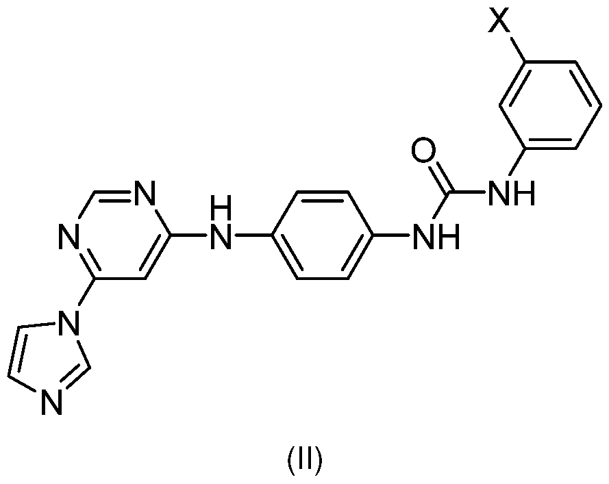
[0016] In some embodiments, a Mirol reducer is provided in a prodrug form. “Prodrug” refers to a derivative of an active agent that requires a transformation within the body to release the active agent. In certain embodiments, the transformation is an enzymatic transformation. Prodrugs are frequently, although not necessarily, pharmacologically inactive until converted to the active agent. “Promoiety” refers to a form of protecting group that, when used to mask a functional group within an active agent, converts the active agent into a prodrug. In some cases, the promoiety will be attached to the drug via bond(s) that are cleaved by enzymatic or non- enzymatic means in vivo. Any convenient prodrug forms of the subject compounds can be prepared, e.g., according to the strategies and methods described by Rautio et al. (“Prodrugs: design and clinical applications”, Nature Reviews Drug Discovery 7, 255-270 (February 2008)). In some cases, the promoiety is attached to the carboxylic acid group of the subject compounds. In certain cases, the promoiety is an acyl or substituted acyl group. In certain cases, the promoiety is an alkyl or substituted alkyl group, e.g., that forms an ester when attached to the carboxylic acid group of the subject compounds.
[0017] In some embodiments, a Mirol reducer, prodrugs, stereoisomers or salts thereof are provided in the form of a solvate (e.g., a hydrate). The term "solvate" as used herein refers to a complex or aggregate formed by one or more molecules of a solute, e.g. a prodrug or a pharmaceutically-acceptable salt thereof, and one or more molecules of a solvent. Such solvates are typically crystalline solids having a substantially fixed molar ratio of solute and solvent. Representative solvents include by way of example, water, methanol, ethanol, isopropanol, acetic acid, and the like. When the solvent is water, the solvate formed is a hydrate.
[0018] Aspects of the methods include administering a Mirol reducer to a subject having tauopathy. Also provided are companion diagnostic assays to determine if a subject is suitable
for treatment with a Mirol reducer due to a Mirol phenotype associated with tauopathy. In some embodiments, a Mirol assay is performed by contacting a population of cells with an agent that damages mitochondria. In some embodiments the agent is a mitochondria-specific uncoupler, e.g. a protonophore. Agents suitable for this purpose include FCCP, CCCP, DNP, BAM15, etc., as known in the art. The cells are incubated for a period of time sufficient to depolarize mitochondria and initiate clearance, e.g. at least about 1 hour, at least about 2 hours, and usually not more than about 24 hours, and may be from about 1 to 24 hours, from about 2 to 20 hours, from about 3 to 18 hours, from about 4 to 14 hours, from about 5 to 10 hours, and may be from about 4 to 8 hours. Following mitochondrial depolarization, the cells are lysed and assessed for levels of Mirol . In some embodiments the cells are lysed and an affinity assay is performed on the lysate to detect levels of Mirol protein. The formats for such assays are well known in the art. Generally an affinity agent specific for Mirol , e.g. a Mirol specific antibody, is utilized as a capture agent or as a detection agent, or both. Suitable formats include, without limitation, immunoassays such as ELISA, RIA, EIA, FRET, etc. The presence of Mirol protein following mitochondrial depolarization is indicative of a tauopathy phenotype, while control cells degrade Mirol under these conditions.
[0019] In some embodiments a Mirol assay as described above is utilized for the diagnosis and clinical monitoring of movement disorders, which diseases include, without limitation, tauopathy. In some embodiments, the methods of the invention are used in determining the efficacy of a therapy for treatment of a movement disorder, e.g. in vitro, such as drug screening assays and the like; at an individual level; in the analysis of a group of patients, e.g. in a clinical trial format; etc. Clinical trial embodiments may involve the comparison of two or more time points for a patient or group of patients. The patient status is expected to differ between the two time points as the result of administration of a therapeutic agent, therapeutic regimen, or challenge with a diseaseinducing agent to a patient undergoing treatment. The response of a patient with a movement disorder to therapy is assessed by detecting the ability of a cell sample from a patient, including without limitation a fibroblast sample, to degrade Mirol after mitochondria damage, e.g. mitochondrial depolarization. Mirol degradation may be monitored in a variety of ways. Conveniently, the removal of Mirol is detected in a patient sample by an immunoassay, such as ELISA or other high throughput affinity assays.
BRIEF DESCRIPTION OF THE DRAWINGS
[0020] The invention is best understood from the following detailed description when read in conjunction with the accompanying drawings. It is emphasized that, according to common practice, the various features of the drawings are not to-scale. On the contrary, the dimensions of the various features are arbitrarily expanded or reduced for clarity. Included in the drawings are the following figures.
[0021] FIGS. 1A-1 M. dMIC60 Is Oxidized and Binds to Miro in a Redox-Dependent Manner, (a- f) Wild-type flies (w, , ,s) (a-e), or flies with indicated genotypes (f), were treated with the reducing agent DTT, the non-reducing agent PEG-MAL (PEG) or AMS, or by the indirect thiol trapping assay, and blotted with anti-dMIC60. H2O2was fed at day 1 for 24 hrs. (g-h) Whole-body lysates of wild-type flies (w,, ,s) were IPed with anti-DMiro or IgG, and immunoblotted (IB) as indicated, (h) Flies were fed with or without H2O2 at day 1 for 24 hrs. (i) GST pull-down using recombinant human full-length GST-Miro1 , and whole-body lysates of flies expressing UAS-dMIC60-WT drven by Actin-GAL4, fed with or without H2O2 at day 7 for 24 hrs. (j) Confocal single-section images overlaying dMIC60 (green) and T7-DMiro (magenta) of fly brains of Elav-GAL4>T7-DMiro, fed with or without H2O2 at day 7 for 24 hrs. The FRET images are color-coded colocalized FRET. From FRET images, colocalized FRET index is calculated from 68 and 88 neurons from 17 and 22 brain regions from 5 and 6 brains for H2Oand H2O2, respectively. Mann-Whitney Test. * p<0.05. Scale bars: 20 |xm. (k) Flies with indicated genotypes were fed with H2O2 at day 1 for 24 hrs, IPed with anti-DMiro, and blotted. (I) Left: The topology of DMiro and dMIC60. Right: HEK cells expressing indicated constructs were treated with H202for 10 min, IPed, and blotted as indicated. FL: full length, (a-i, k, I) Same results were reproduced more than 3 times, (m) Schematic summarization of our results showing that Miro binds to dMIC60 in a redox-dependent manner.
[0022] FIGS. 2A-2E. Mitochondrial Changes with Age. (a) Flies with indicated genotypes were fed with or without H2O2 at day 1 for 24 hrs, lysed, and blotted. The band intensity is normalized to |3-actin on the same blot, n.s.: not significant. n=4. (b) Representative confocal stack images of MitoSox staining of fly brains. The total MitoSox intensity is measured for each fly brain and normalized to the background intensity. Genotype: Elav-GS>UAS-DMiroRNAl uninduced (RU-). n=5 (young) and 7 (old) fly brains. Scale bar: 200 |xm. (c) The heatmap shows relative protein abundances in selective clusters of protein groups, (d) Upper: Whole-body fly lysates of young (day 5) and old (day 50) wild-type flies (w,, ,s) were IPed with anti-DMiro and blotted as indicated. The band intensity of dMIC60 in IP is normalized to that of DMiro in IP. Lower: The band intensity of each protein in lysate is normalized to that of (3-actin from the same blot, (e) Similar to (d), old
(day 50) wild-type flies, fed with or without AD4 from day 30-50, were IPed. (d-e) n=4 independent experiments, (a) Two-Way ANOVA Fisher’s LSD Test, (b, d-e) Mann-Whitney Test. * p<0.05, ** p<0.01 , *** p<0.001.
[0023] FIGS. 3A-3I. Dissociating the DMiro-dMIC60 Interaction Benefits Fly Lifespan and Health-Span. For all panels, “RU-” flies were fed with the same volume of the vehicle, ethanol. All flies were female, (a-d) Survival plots of flies with genotypes indicated, (a) n=81 (WT) and 89 (CS). (b-d) Blue lines (RU+): Adult-onset neuronal downregulation using Elav-GS-GAL4. Black lines (RU-): Un-induced controls, (b) n=144-145 flies, (c) n=122-142 flies, (d) n=67-74 flies, (e) The climbing ability shown as Performance Index, or (f) the survival ability in response to 20 mM paraquat (starting from day 4), of flies with ubiquitous expression of UAS-dMIC60-WT or UAS- dMIC60-CSby Actin-GAL4 in dMIC60 null background, (e) n=19 (WT) and 30 (CS), 6 independent experiments, (f) n=29 (WT) and 34 (CS). p=1.4 xi o-12. (g) The climbing ability shown as Performance Index of flies with indicated genotypes. n=59 (RU-) and 60 (RU+) flies, 3 independent experiments, (h-i) Survival plots, (h) Starvation (p=1 ,17x10-22) and (i) 20 mM Paraquat (p=5.12 xi o-9) started after 14 days of adult-onset neuronal knockdown of DMiro. n=148-151 flies, (a-d, f, h, i) Log-Rank Test, (e) Mann Whitney Test, (g) Two-Way ANOVA Tu key Test. * p<0.05, “ p<0.01.
[0024] FIGS. 4A-4E. Dissociating the DMiro-dMIC60 Interaction Improves Mitochondrial Function, (a-b) ATP levels were measured in flies with indicated genotypes, (a) Day 2. n= 8. (b) Blue bar (RU+): Adult-onset neuronal knockdown of DMiro (Elav-GS>UAS-DMiroRNAl) for 20 days. Gray bar (RU-): Un-induced control flies. n=6 (RU+) and 7 (RU-) independent experiments, (c-d) Representative confocal stack images of MitoSox staining of fly brains with indicated genotypes. The total MitoSox intensity is measured for each fly brain and normalized to the background intensity, (c) Day 12. n=5 (WT) and 6 (CS) fly brains, (d) n=5, 5, 7, 6 fly brains (from left to right). Uninduced data (RU-) are the same as in Fig. 2b. (e) Representative confocal images of TMRM staining (magenta) of fly brains expressing Mito-GFP (green) in DA neurons driven by TH-GAL4, with or without DMiro RNAi. The fluorescence intensity of TMRM is normalized to that of Mito- GFP within the same neuron cell body. n=20-38 neurons from 3-4 brains. Scale bars: (c) 100 |xm; (d) 200 |xm; (e) 25 |xm. (a-c) Mann-Whitney Test, (d-e) One-Way ANOVA Tukey Test. * p<0.05, ** p<0.01 , *** p<0.001.
[0025] FIGS. 5A-5B. DMiro RNAi Restores Cellular Respiration and Mitophagy. (a) Seahorse analysis on ex vivo fly brains (Elav-GS>UAS-DMiroRNAl) of different ages, with or without RU induction, as indicated. “RU-” flies were fed with the same volume of the vehicle, ethanol. n=7
brains. Black arrows show the times of oligomycin and rotenone/antimycin injections, (b) Representative confocal single-section images, overlaying Mito-GFP (green), ubiquitin (red), and p62 (blue), of fly brains expressing Mito-GFP in DA neurons driven by TH-GAL4, with or without DMiro RNAi. Yellow arrow heads show Mito-GFP puncta that colocalize with p62, ubiquitin, or both. The colocalization coefficient is quantified for Mito-GFP versus ubiquitin, and Mito-GFP versus p62, respectively. n=31 , 24, 19, 25 neurons for ubiquitin, and 32, 24, 19, 23 for p62 (from left to right) from 6 brains. Scale bar: 10 pm. (a) Two-Way ANOVA Tukey Test, (b) One-Way ANOVA Tukey Test. * p<0.05, *** p<0.001 .
[0026] FIGS. 6A-6H. a-Syn Expression or Frataxin Deficiency Impacts the DMiro-dMIC60 Complex, (a, g) Representative confocal stack images of MitoSox staining of fly brains as indicated. The total MitoSox intensity is measured for each fly brain and normalized to the background intensity, (a) n=4 (Elav-GAL4) and 5 (Elav>SNCA-A53T) fly brains, (g) n=5 (Actin>Fh RNAi) and 7 (Actin-GAL4) fly brains, (b) Whole-body lysates of 5-day-old oc-syn-A53T-expressing flies (Actin>UAS-SNCAA53T) and controls (Actin-GAL4) were IPed with anti-DMiro and blotted as indicated. The band intensity of dMIC60 in IP is normalized to that of DMiro. n=4 independent experiments, (c) Whole-body lysates of 15-day-old flies were immunoblotted. The DMiro band intensity is normalized to that of p-actin from the same blot. n=4. (d) Confocal stack images show DA neurons immunostained with anti-TH in the PPL1 clusters of 40-day-old flies. The DA neuron number in the PPM1/2 and PPL1 clusters is quantified. n=14, 12, 13, 15 brains for PPL1 , and 19, 12, 12, 20 for PPM1/2 (from left to right), (e) The DA neuron number in the PPL1 clusters is quantified. n=6 ( TH>dMIC60-WT), 8 (JH>dMIC60-CS), 10 (JH<SNCAA53T, dMIC60-WT), and 7 (TH>SNCAA53T, dMIC60-CS) brains, (f) Locomotor ability shown as Performance Index of 42-day- old flies. n=25 (TH>dMIC60-WT), 40 (TH>dMIC60-CS), 45 (JH<SNCAA53T, dMIC60-WT), and 33 (TH>SNCAA53T, dMIC60-CS). (h) Upper: Whole-body lysates of 30-day-old flies were IPed with anti-DMiro and blotted as indicated. The band intensity of dMIC60 in IP is normalized to that of DMiro. Lower: The DMiro or dMIC60 band intensity in Input is normalized to that of p-actin from the same blot. n=4. Scale bars: (a, g) 100 pm; (d) 27.5 pm. (a-c, g-h) Mann-Whitney Test, (d-f) One-Way ANOVA Tukey Test. * p<0.05, ** p<0.01 , *** p<0.001 .
[0027] FIGS. 7A-7G. MR3 and MR5 Benefit Normal Flies, and Fly Models of PD and FA. For (b-c, e-g), when MR3/5 was not added, the same volume of the solvent, DMSO, was supplied in fly food. Drug administration started from day 2. (a) Chemical properties of MR3-5. (b) Confocal stack images show DA neurons in the PPL1 clusters of 40-day-old brains immunostained with anti-TH. Quantification of the DA neuron number in the PPL1 clusters is shown. n=7, 10, 9, 1 1
brains (from left to right), (c) Locomotor ability shown as Performance Index at indicated dates. n=40, 42, 40, 46 flies (from left to right), 3 independent experiments, (d) Both Antimycin A and MR5 were dissolved in ethanol. Neurons were pretreated with MR5 24 hrs before the application of Antimycin A for another 6 hrs. The same volume of ethanol was applied at the same time in negative controls. The density of TH-positive neurons is calculated in each condition under 20x, from 19 (PD, MR5) or 20 fields (the rest) from 4 experiments. The neuron density without Antimycin A is not significantly different among all conditions (p=0.4543). Comparisons with “Wildtype, no treatment” except otherwise indicated, (e) Locomotor ability shown as Performance Index was measured in flies as indicated at different days. n=43, 41 , 44, 44, 44, 42 flies (from left to right), 3 independent experiments. Data is compared at the same age. (f) Survival plots of flies orally administed with MR3 or MR5 from adulthood (colored lines) and controls fed with DMSO (black line). n=126 (1 pM MR3), 131 (2.5 pM MR5), 132 (2.5 pM MR3), 157 (DMSO) flies, (g) ATP levels of wild-type flies fed with either 2.5 or 10 pM MR5 from adulthood for 20 days. n=8 independent experiments, (b, d) One-Way ANOVA Tukey Test, (c, e) One-Way ANOVA Fisher LSD Test, (f) Log-Rank Test, (g) Mann-Whitney Test. Scale bars: (b) 50 pm; (d) 100 pm. * p<0.05, ** p<0.01 , *** p<0.001.
[0028] FIGS. 8A-8C. Schematic Representation of a Redox-Dependent Regulation of dMIC60-DMiro. (a) In young flies (low ROS), dMIC60 shows a weak interaction with DMiro. MT: microtubules, (b) ROS upregulation (aging, PD, FA) causes an increase in the amount of dMIC60 bound to DMiro, leading to stabilization of the complex, which may consequently exacerbate ROS accumulation by slowing mitophagy and disrupting cellular respiration, (c) Genetically or pharmacologically targeting the DMiro-dMIC60 complex extends lifespan and health-span, promotes mitophagy, restores cellular respiration, and rescues phenotypes of PD and FA models.
[0029] FIGS. 9A-9J. Miro Binds to dMIC60 in a Redox-Dependent Manner, (a) Two-day-old flies expressing Myc-tagged UAS-dMIC60-WT or dMIC60-CS driven by Actin-GAL4 in dMIC60 null background, were lysed and immunoblotted. Both anti-dMIC60 and anti-Myc recognize transgenic dMIC60 protein in dMIC60 null background (no endogenous dMIC60). Anti-dMIC60 recognizes endogenous dMIC60 in flies with Actin-GAL4 alone in wild-type background, (b) Whole-body lysates of wild-type flies (w,, ,s) were IPed with anti-DMiro or IgG, and immunoblotted (IB) as indicated, (c) Immunostaining of dMIC60 and ATP5|3 in third instar larval muscles. Scale bar: 5 pm. (d) Immunostaining of T7 in adult fly brains (day 7). Scale bar: 20 pm. (c-d) Confocal images were obtained using the same settings, (e) In Vitro GST pull-down using full-length GST- Mirol or GST, together with recombinant dMIC60 (AAs 92-739). (f) In Vitro GST pull-down using
full-length GST-Miro1 , together with recombinant dMIC60 (AAs 92-739), either wild-type (WT) or mutant (CS). (g) Coomassie-stained gels. Arrowhead indicates the dMIC60 band, (h-i) In Vitro GST pull-down using full-length GST-Miro1 or GST, together with recombinant dMIC60 (AAs 92- 739) (h) or Parkin (i). (a-i) Similar results were seen more than three times, (j) Lysates of wildtype flies fed with H2O2, paraquat, or vehicle (water) at day 7 for 24 hrs were immunoblotted as indicated. The band intensity of each marker is normalized to that of |3— actin from the same blot and graphed as relative change compared to vehicle. n=4 independent experiments. Two-sided Student T Test. p=0.0336 (DMiro, H2O2), 0.0325 (dMIC60, H2O2), 0.0176 (DMiro, paraquat), 0.0383 (dMIC60, paraquat). Boxes show 25th/75th percentiles, whiskers are the minimum and maximum, and middle line is median.
[0030] FIGS. 10A-10D. DMiro Protein Levels in MICOS Mutants, (a) Lysates of 2nd instar larvae (dMIC19nul1), pupae (dMIC60nul1), or 14-day-old adults (RNAi) were immunoblotted as indicated. The band intensity of each marker is normalized to that of ATP5|3 or |3-actin from the same blot and graphed as relative change compared to control. Wild-type: w1118. n=3 (Top panel: anti-DMiro for WT and Middle panel) and 4 (the rest) independent experiments. Two-sided Mann-Whitney Test (Lower panel) and T Test with Welch’s correction (Top and Middle), n.s.: not significant. p=0.0009, 0.0005, 0.0096 for dMIC19 null, 0.0319, 0.0351 , 0.0173 for dMIC60 null. The rest of the precise p values are in Source Data, (b) Mitochondrial (Mito) and cytosolic (Cyto) fractions from 20-day-old flies were immunoblotted as indicated. The DMiro band intensity is normalized to that of VDAC from the same blot. n=3. Two-sided T Test with Welch’s correction. p=0.0283, 0.0102. (c) Whole-body lysates of 20-day-old flies were immunoblotted as indicated. The band intensity is normalized to that of p-actin from the same blot and graphed as relative change compared to control (Actin-GAL4, gray bar) within the same experiment. n=5 flies per experiment, 4 independent experiments. p=0.0286. (d) The mRNA levels of DMiro were determined by qPCR in 20-day-old flies, normalized to RP49 from the same experiment, and graphed as relative change compared to respective controls (white boxes) within the same experiment. n=4 independent experiments. p=0.0286. (c-d) Two-sided Mann-Whitney Test, (a, d) Boxes show 25th/75th percentiles, whiskers are the minimum and maximum, and middle line is median, (a, b, c) Data are presented as Mean±S.E.M. with dots.
[0031] FIGS. 11A-11 F. DMiro Protein Levels in Mitochondrial Mutants, (a) The mRNA levels of indicated genes were determined by qPCR in 20-day-old flies, normalized to RP49 from the same experiment, and graphed as relative change compared to respective controls (white boxes) within the same experiment. n=4 independent experiments. p=0.0286. (b) Whole-body lysates of
20-day-old flies were immunoblotted as indicated. The band intensity of DMiro is normalized to that of p-actin from the same blot and graphed as relative change compared to control (Actin- GAL4, gray bar). n=4 independent experiments, (c) Lysates of 2-day-old CHCHD2nu" and control (w,, ,s) were immunoblotted as indicated. The band intensity of DMiro is normalized to that of - actin from the same blot and graphed as relative change compared to wild-type control (w, , ,s). n=4 independent experiments, (d) Lysates of 3rd instar larvae were immunoblotted. The band intensity of each marker is normalized to that of ATP5P from the same blot and graphed as relative change compared to wild-type control (w,, ,s). n=7 larvae per experiment, 3 (anti-dMIC19 for W7) and 4 (the rest) independent experiments. p=0.0286. (e) qPCR results show no significant differences in DMiro mRNA expression normalized to RP49 between young (day 3) and old (day 40) wild-type (w, , ,s) flies. n=4 replicates per genotype, (f) Lysates of 3rd instar larvae and 2-day- old adult flies were immunoblotted with antibodies against DMiro and the loading control p-actin. The band intensity of DMiro is normalized to that of p-actin from the same blot. n=4 independent experiments. Please note that DMiro is upregulated in 2-day-old flies compared to third instar larvae. p=0.0072, 0.0047. The rest of the precise p values are in Source Data, (a) One-sided Mann-Whitney Test (dMIC19). (a-e) Two-sided Mann-Whitney Test, (f) One-Way ANOVA Post Hoc Tukey Test, (a) Boxes show 25th/75th percentiles, whiskers are the minimum and maximum, and middle line is median, (b-f) Data are presented as Mean±S.E.M. with dots.
[0032] FIGS. 12A-12H. The Role of DMiro in Lifespan. For all panels, “RU-” flies were fed with the same volume of the vehicle, ethanol. All flies were female, (a) Survival curves of wild-type flies (w1118) fed with or without 200 p.M RU. n=141 flies. p=0.46 by Log-Rank Test, (b) Later lifeonset neuronal downregulation of DMiro using Elav-GS-GAL4 induced in the presence of RU from day 30 (Elav-GS>UAS-DMiroRNAl, RU+, blue line) extends lifespan, compared to un-induced controls (RU-, black line). n=106 (RU+) and 127 (RU-) flies. Median lifespan was increased by 10%, p=1.3x10⁻⁷ by Log-Rank Test, (c) Adult-onset downregulation of DMiro using the intestine enterocytes driver 5966-GS-GAL4 induced in the presence of RU (5966-GS>UAS-DMiroRNAl, RU+, red line) shortens lifespan, compared to un-induced controls (RU-, black line). n=142 (RU+) and 151 (RU-) flies. p=0.0007 by Log-Rank Test, (d) Later life-onset downregulation of DMiro in intestine enterocytes using 5966-GS-GAL4 induced in the presence of RU from day 30 (5966- GS>UAS-DMiroRNAl, RU+, red line) slightly extends lifespan, compared to un-induced controls (RU-, black line). n=135 (RU-) and 145 (RU+) flies. p=0.03 by Log-Rank Test, (e) Adult-onset ubiquitous knockdown of DMiro using Da-GS-GAL4 in the presence of RU (Da-GS>UAS- DMiroRNAl, RU+, red line) shortens lifespan, compared to un-induced controls (RU-, black line).
n=143 (RU+) and 144 (RU-) flies. p=2.07x10⁻⁹ by Log-Rank Test, (f) Later life-onset ubiquitous knockdown of DMiro using Da-GS-GAL4 in the presence of RU from day 30 (Da-GS>UAS- DMiroRNAl, RU+, red line) does not alter lifespan, compared to un-induced controls (RU-, black line). n=141 (RU-) and 146 (RU+) flies. p=0.74 by Log-Rank Test, (g) Adult-onset downregulation of DMiro using the muscle driver MHC-GS-GAL4 induced in the presence of RU (MHC-GS>UAS- DMiroRNAl, RU+, red line) shortens lifespan, compared to un-induced controls (RU-, black line). n=146 (RU-) and 149 (RU+) flies. p=1.68x10⁻¹⁶ by Log-Rank Test, (h) Adult-onset neuronal downregulation of white using Elav-GS-GAL4 induced in the presence of RU (Elav-GS>UAS- whiteRNAl, RU+, red line) slightly shortens lifespan, compared to un-induced controls (RU-, black line). n=139 flies. p=0.01 by Log-Rank Test.
[0033] FIGS. 13A-13D dMIC60 or dMIC19 RNAi Does not Affect Mitochondrial Structure, (a) Representative TEM images of thin sections on body wall muscles of late third instar larvae. Scale bars: 500 nm. (b) Quantification of the crista junction (CJ) number per |im of mitochondrial circumference. n=30 mitochondria from 3 male larvae, (c) Quantification of the mitochondrial size. n=19 mitochondria from 3 male larvae, (d) Quantification of the aspect ratio. n=20 mitochondria from 3 male larvae. One-Way Anova Post Hoc Tukey Test.
[0034] FIGS. 14A-14F. The Role of MICOS in Lifespan, (a) Later life-onset downregulation of dMIC19 using the intestine enterocytes driver 5966-GS-GAL4 induced in the presence of RU from day 30 (5966-GS>UAS-dMIC19RNAi, RU+, red line) does not alter lifespan, compared to un- induced controls (RU-, black line). n=143 (RU+) and 146 (RU-) flies. p=0.84 by Log-Rank Test, (b) Adult-onset downregulation of dMIC19 in intestine enterocytes induced in the presence of RU (5966-GS>UAS-dMIC19RNAi, RU+, red line) does not alter lifespan, compared to un-induced controls (RU-, black line). n=144 (RU+) and 150 (RU-) flies. p=0.59 by Log-Rank Test, (c) Later life-onset downregulation of dMIC60 in intestine enterocytes induced in the presence of RU from day 30 (5966-GS>UAS-dMIC60RNAi, RU+, red line) does not alter lifespan, compared to un- induced controls (RU-, black line). n=142 (RU+) and 143 (RU-) flies. p=0.99 by Log-Rank Test, (d) Adult-onset downregulation of dMIC60 in intestine enterocytes induced in the presence of RU (5966-GS>UAS-dMIC60RNAi, RU+, red line) does not alter lifespan, compared to un-induced controls (RU-, black line). n=140 (RU+) and 147 (RU-) flies. p=0.94 by Log-Rank Test, (e) The survival ability of female flies with a second DMiro RNAi line driven by Elav-GS-GAL4 induced by RU since adulthood (Elav-GS>UAS-DMiroRNAl 2, blue line, RU+) or of un-induced control flies (black line, RU-), in response to 20 mM paraquat (starting from day 7). n=14 (RU+) and 56 (RU-) flies. p=0.016 by Log-Rank Test, (f) Fly heads were lysed and blotted as indicated. The band
intensity is normalized to that of oc-tubulin from the same blot. n=4. One-Way Anova Post Hoc Tukey Test (p=0.6915 for ubiquitin, p=0.5884 for p62). Boxes show 25th/75th percentiles, whiskers are the minimum and maximum, and middle line is median.
[0035] FIGS. 15A-15H. a-Syn Neurotoxicity in Flies, (a) Validation of an oc-syn fly model where wild-type human oc-syn transgene downstream of a modified UAS that significantly increases gene expression is expressed using the inducible pan-neuronal driver Elav-GS-GAL4 (“ Elav-GS>UAS- SNCA-WT, RU+”) through adulthood. Un-induced controls are “Elav-GS>UAS-SNCA-WT, RU-”. Head lysates of induced and uninduced flies were immunoblotted as indicated. The band intensity of a-syn is normalized to that of p-actin. n=3 independent experiments. Two-sided T Test with Welch’s correction. p=0.0238. (b) Climbing ability shown as Performance Index of flies with adultonset induction and of un-induced controls at different ages. n=60 (RU-) and 61 (RU+) flies per genotype, 3 independent experiments, (c) The DA neuron number in the PPM1/2, PPL1 , or PPL2 clusters per brain from flies as indicated at day 40 is counted and compared. n=6 fly brains per genotype. Two-sided Mann-Whitney Test. p=0.0022, 0.0065. The rest of the precise p values are in Source Data, (d) Head lysates of 40-day-old flies were immunoblotted with antibodies against DMiro and the loading control p-actin. The band intensity of DMiro is normalized to that of p-actin from the same blot. n=3 independent experiments. Two-sided Student T Test. p=0.0138. (e) Climbing time for 30-day-old flies. n=27, 26, 57, 26 flies (from left to right). One-Way Anova Post Hoc Tukey Test. p<0.0001 . (f) Flying ability of 30-day-old flies. The percentage of flies scored as a “1 ” (able to fly) is shown in solid black bars, and the percentage of flies scored as a “0” (unable to fly) is shown in white bars. n=100. Chi-Square Test. p<0.0001. (e-f) Comparisons with “TH- GAL4”. (g-h) HEK293T cells transfected as indicated were lysed and immunoblotted. One-Way Anova Post Hoc Tukey Test, (g) The band intensity of Mirol is normalized to that of GAPDH from the same blot. Data from each genotype are compared to those of “Control RNAi, EGFP” (the left box) and asterisk is given directly on top of the box if it is significant. Data between “Control RNAi, EGFP-SNCA” and “MIC60 RNAi, EGFP-SNCA” (the right 2 boxes) are compared and asterisk is given on top of a line. n=5 transfections. p=0.0004, <0.0001 . The rest of the precise p values are in Source Data, (h) The band intensity of Myc-Miro1 is normalized to that of p-actin from the same blot. Data from each genotype are compared to those of “Myc-Miro1 , dMIC60-WT, EGFP” (the left box) and asterisk is given directly on top of the box if it is significant. Data between “Myc- Mirol , dMIC60-WT, EGFP-SNCA” and “Myc-Miro1 , dMIC60-CS, EGFP-SNCA” (the right 2 boxes) are compared and asterisk is given on top of a line. n=5 transfections. p=0.013, 0.0006. The rest of the precise p values are in Source Data, (a, d) Data are presented as Mean±S.E.M. with dots.
(c, e, g, h) Boxes show 25th/75th percentiles, whiskers are the minimum and maximum, and middle line is median.
[0036] FIGS. 16A-16D. Miro Reducers Benefit PD Models, (a) Upper: Typical unfolding curves of human Mirol protein in the absence (0 pM) and the presence of MR3 are shown. Lower: Quantification of melting temperatures of Mirol protein in the absence (0 pM) and the presence of MR3 (100 pM). n=4 independent experiments. Two-sided Student T Test. p=0.0000424. (b) oc- Syn-A53T-expressing flies (Elav-GAL4>UAS-SNCAA53T) were fed with 10 pM MR5 for 15 days and then their heads were lysed for immunoblotting. The band intensity of DMiro is normalized to that of the mitochondrial loading control VDAC from the same blot. n=4 independent experiments. Two-sided Mann-Whitney Test. p=0.0286. (c) Both Antimycin A and MR5 were dissolved in ethanol. Neurons were pretreated with MR5 24 hrs before the application of Antimycin A for another 6 hrs. The same volume of ethanol was applied at the same time in negative controls. Confocal images overlay triple immunostainings of TH (DA neurons), TUNEL (indicator of cell death), and Dapi (nuclei). The percentage of TUNEL-positive cells out of total cells (Dapi-positive) is calculated in each condition under 20x. n=19 (PD, MR5) or 20 fields (the rest) from 4 independent experiments. p<0.0001. The rest of the precise p values are in Source Data, (d) From images such as in (c), the percentage of TH-positive neurons out of total cells (Dapi-positive) without Antimycin A treatment ranges from 17.08%-19.25%, and is not significantly different among all conditions (p=0.9532), consistent with previous studies from ours and others 20'67. n=19 (PD, MR5) or 20 fields (the rest) from 4 independent experiments, (c-d) Comparisons with “Wildtype, no treatment” except otherwise indicated. One-Way Anova Post Hoc Tukey Test. Scale bar: (c) 100 pm. (a) Data are presented as Mean ± S.D. with dots, (b-d) Boxes show 25th/75th percentiles, whiskers are the minimum and maximum, and middle line is median.
[0037] FIGS 17A-17E 1. Mitochondrial Protein Changes Following CCCP Treatment in Fibroblasts. Mitochondrial Protein Responses to CCCP in Fibroblasts. (A) Schematic representation of our readouts. (B) Examples of the readouts using Healthy-1 and MAPT-1. (C) Demographic and genetic information of all cell lines used in this study. (D) Heat maps show relative mitochondrial protein levels. The intensity of each band in the mitochondrial fraction is normalized to that of the mitochondrial loading control VDAC from the same blot and expressed as a fraction of the mean of Healthy-1 with DMSO treatment; this control was included in every experiment. Mean values are imported into heat maps. Mann-Whitney L/ Test is performed for comparing normalized band intensities within the same subject (DMSO v.s. CCCP), and P value is noted for each subject. n=3-35. (E) ELISA of Mirol protein. n=4 with duplicates each time.
Comparison within the same subject. Mann-Whitney [/ Test. * P<0.05, ** P<0.01 , * P<0.001 for all figures.
[0038] FIGS. 18A-18I. Ultrastructural Changes of Mitochondria in Fibroblasts. (A) Representative TEM images of Healthy-6 (WT) and MAPT-1 (MAPT-N279K) with and without CCCP treatment. Scale bar: 500 nm. (B-l) Quantifications from images as in (A). (B) Quantification of mitochondrial size (minor x major diameter). (C) Quantification of mitochondrial perimeter. (D) Quantification of cristae junction number normalized to mitochondrial perimeter. (E) Quantification of aspect ratio (minor/major diameter). (F) Quantification of ER-mitochondrial contact number normalized to mitochondrial size (contact is defined where the distance between ER and mitochondrial membranes is less than 10 nm). (G) Quantification of total ER abundance (the total number of ER-mitochondrial contact divided by the total number of mitochondria per image). (H) Quantification of rough ER (RER) abundance. Similar to (G) but only RER is counted. (I) Quantification of smooth ER (SER) abundance. Similar to (G) but only SER is counted. n=57 (WT, DMSO), 51 (WT, CCCP), 53 (MAPT-N279K, DMSO), and 64 (MAPT-N279K, CCCP) mitochondria from 15 different images from 3 independent cultures. Two-Way ANOVA Post Hoc Tukey Test.
[0039] FIGS 19A-19C. Phenotypes of Cytoskeleton and Other Organelles in Fibroblasts. (A) Representative TEM images of Healthy-6 (WT) and MAPT-1 (MAPT-N279K) with and without CCCP treatment, showing cytoskeleton tracks (Tracks) next to mitochondria. (B) From images as in (A), the percentage of total mitochondria with or without adjacent cytoskeleton tracks is counted. n=30 (WT, DMSO), 28 (WT, CCCP), 31 (MAPT-N279K, DMSO), and 36 (MAPT-N279K, CCCP) mitochondria from 15 images from 3 independent cultures. Chi Square Test. (C) Representative TEM images of Healthy-6 (WT) and MAPT-1 (MAPT-N279K) with and without CCCP treatment, showing vacuole-like structures (Vac), structures similar to multi-vesicular bodies (MV), and structures similar to lamellar bodies (LB). Scale bars: 500 nm.
[0040] FIGS 20A-20F. The Mitochondrial Network in Fibroblasts, Tau Interaction with Mirol , and Effect of Mirol Reducer. (A) Confocal images show mitochondria stained with TMRM in fibroblasts. The mean TMRM intensity of each cell is normalized to the background intensity and quantified. n= 43 (Healthy-6), 46 (Healthy-7), and 42 (MAPT-1 ) cells from 4-5 images per coverslip from 3 coverslips. One-Way ANOVA Post Hoc Tukey Test. Scale bar: 10 mM. (B-C) Co-IP with anti-GFP from HEK cells transfected as indicated. (D) HEK cells transfected with different tau and Mirol constructs were lysed and blotted. (E) ELISA of Mirol protein in MAPT-1 fibroblasts treated as indicated. n=4. Mann-Whitney U Test.
[0041] FIGS 21A-21C. Control Experiments. (A) Validation of our mitochondrial purification method. “Mito” and “Cyto” fractions from healthy fibroblasts were blotted as indicated. ATP5b is a mitochondrial marker. Calreticulin is an ER protein. (B) Intra-plate variability of ELISA shown in Figure 1 F-G, 4E, measured by running the same fibroblast sample 4 times in the same plate. (C) An IP similar to Figure 4C is shown. A negative control (empty vector) is included. All lanes are from the same blot.
DETAILED DESCRIPTION OF THE INVENTION
[0042] Methods and compositions are provided for the assessment and treatment of Friedreich’s Ataxia in an individual. Aspects of the methods include administering a MIRO1 reducer to a subject having Friedreich’s Ataxia. Also provided are reagents and kits for practicing the subject methods. These and other objects, advantages and features of the invention will become apparent to those persons skilled in the art upon reading the details of the compositions and methods as more fully described below.
[0043] Before the present methods and compositions are described, it is to be understood that aspects of the invention are not limited to particular method or composition described, as such may, of course, vary. It is also to be understood that the terminology used herein is for the purpose of describing particular embodiments only, and is not intended to be limiting, since the scope of the present invention will be limited only by the appended claims.
[0044] Where a range of values is provided, it is understood that each intervening value, to the tenth of the unit of the lower limit unless the context clearly dictates otherwise, between the upper and lower limits of that range is also specifically disclosed. Each smaller range between any stated value or intervening value in a stated range and any other stated or intervening value in that stated range is encompassed within the invention. The upper and lower limits of these smaller ranges may independently be included or excluded in the range, and each range where either, neither or both limits are included in the smaller ranges is also encompassed within the invention, subject to any specifically excluded limit in the stated range. Where the stated range includes one or both of the limits, ranges excluding either or both of those included limits are also included in the invention.
[0045] Unless defined otherwise, all technical and scientific terms used herein have the same meaning as commonly understood by one of ordinary skill in the art to which this invention belongs. Although any methods and materials similar or equivalent to those described herein can be used in the practice or testing of the present invention, some potential and preferred methods
and materials are now described. All publications mentioned herein are incorporated herein by reference to disclose and describe the methods and/or materials in connection with which the publications are cited. It is understood that the present disclosure supercedes any disclosure of an incorporated publication to the extent there is a contradiction.
[0046] As will be apparent to those of skill in the art upon reading this disclosure, each of the individual embodiments described and illustrated herein has discrete components and features which may be readily separated from or combined with the features of any of the other several embodiments without departing from the scope or spirit of the present invention. Any recited method can be carried out in the order of events recited or in any other order which is logically possible.
[0047] It must be noted that as used herein and in the appended claims, the singular forms "a", "an", and "the" include plural referents unless the context clearly dictates otherwise. Thus, for example, reference to "a cell" includes a plurality of such cells and reference to "the peptide" includes reference to one or more peptides and equivalents thereof, e.g. polypeptides, known to those skilled in the art, and so forth.
[0048] The publications discussed herein are provided solely for their disclosure prior to the filing date of the present application. Nothing herein is to be construed as an admission that the present invention is not entitled to antedate such publication by virtue of prior invention. Further, the dates of publication provided may be different from the actual publication dates which may need to be independently confirmed.
[0049] Hereditary ataxias may be autosomal recessive or autosomal dominant. Autosomal recessive ataxias include Friedreich ataxia (the most prevalent), ataxia-telangiectasia, abetalipoproteinemia, ataxia with isolated vitamin E deficiency, and cerebrotendinous xanthomatosis.
[0050] Friedreich ataxia results from a gene mutation causing abnormal repetition of the DNA sequence GAA in the FXN gene on the long arm of chromosome 9; the EXTV gene codes for the mitochondrial protein frataxin. The GAA sequence is repeated 5 to 38 times within the FXN ene in people who do not have Friedreich ataxia; however, in people with Friedreich ataxia, the GAA sequence may be repeated 70 to > 1000 times. Inheritance is autosomal recessive. In some embodiments, an individual may be diagnosed with FA prior to treatment. In some embodiments diagnosis of an individual comprise genotyping the frataxin gene, for example by PCR analysis of the 1 st intron. See, for example, Jama et al. (2022) Journal of Molecular Diagnostics 24(8):915-
923, herein specifically incorporated by reference. Alleles can be quantified as being between 5 and 200 GAA repeats, or expanded alleles beyond 200 repeats. A PCR assay can correctly genotype and classify normal, mutable normal, borderline, and expanded alleles in both homozygous and heterozygous states.
[0051] The triplet repeat expansion greatly disrupts the normal production of frataxin. Frataxin is found in mitochondria. Research suggests that without a normal level of frataxin, certain cells in the body (especially peripheral nerve, spinal cord, brain and heart muscle cells) produce energy less effectively and have been hypothesized to have a buildup of toxic byproducts leading to oxidative stress. Lack of normal levels of frataxin also may lead to increased levels of iron in the mitochondria. When the excess iron reacts with oxygen, free radicals can be produced.
[0052] In Friedreich ataxia, gait unsteadiness begins between ages 5 and 15; it is followed by upper-extremity ataxia, dysarthria, and paresis, particularly of the lower extremities. Mental function often declines. Tremor, if present, is slight. Reflexes and vibration and position senses are lost. Talipes equinovarus (clubfoot), scoliosis, and progressive cardiomyopathy are common. By their late 20s, patients may be confined to a wheelchair. Death, often due to arrhythmia or heart failure, usually occurs by middle age.
[0053] In Friedreich ataxia nerve fibers in the spinal cord and peripheral nerves degenerate, becoming thinner. The cerebellum, part of the brain that coordinates balance and movement, also degenerates to a lesser extent. This damage results in awkward, unsteady movements and impaired sensory functions. The disorder also causes problems in the heart (in as many as one- third of affected individuals) and spine, and some people with the condition will also develop diabetes. The disorder does not affect thinking and reasoning abilities (cognitive functions).
[0054] The rate of progression varies from person to person. Generally, within 10 to 20 years after the appearance of the first symptoms the person is confined to a wheelchair. Individuals may become completely incapacitated in later stages of the disease. Friedreich ataxia can shorten life expectancy, and heart disease is the most common cause of death. However, some people with less severe features of FA live into their sixties or older.
[0055] Assessment of motor skills can be used in initial clinical evaluation, and to determine the efficacy of treatment. See, for example, Hohenfeld et al. (2019) Cerebellum 18(5):896-909, herein specifically incorporated by reference. Assessment tests include, for example, quantitative motor assessments of the Q-Motor battery, including lift (manumotography), finger tapping (digitomotography) and pronate/supinate (dysdiadochomotography) tasks. Indicia for ataxia may include, for example, International Cooperative Ataxia Rating Scale (ICARS): developed to
determine the level of impairment from ataxia related to genetics; Scale for the Assessment and Rating of Ataxia (SARA): similar scale to the ICARS to assess ataxia but shorter to administer; Friedreich’s Ataxia Rating Scale (FARA): assessment for ataxia specific to FA. For Gait: 6 Minute Walk Test: assesses aerobic capacity and gait; Timed Up and Go (TUG): assesses fall risk, balance and gait; Goal Attainment Scale: individualized outcome measure to assess the extent the patient meets their various goals. For Balance: Berg Balance Scale: useful in early stages when individual is not yet using a wheelchair; Timed Up and Go (TUG) : assesses fall risk, balance and gait; Pediatric Balance Scale: used to assess balance in everyday tasks of adolescents specifically; Goal Attainment Scale: individualized outcome measure to assess the extent the patient meets their various goals. Additional indicia may include Functional Independence Measure: measure of physical, psychological and social function; Goal Attainment Scale: individualized outcome measure to assess the extent the patient meets their various goals; Child Occupational Self-Assessment (v 2.2): a self-report measure for how competent children and adolescents feel completing every day activities and how much value they place on these activities, ages 6-17; Depression Anxiety Stress Scale: can be used in later stages of the disease to assess potential symptoms of depression, anxiety and stress.
[0056] In some embodiments, the methods disclosed herein can stabilize an individual’s assessed level of disability by one or more of the indicia indicated above for a period of time, e.g. during administration of an agent disclosed herein. In some embodiments, the methods disclosed herein can reduce an individual’s assessed level of disability following administration of an agent disclosed here, e.g. a reduction of at least about 5%, at least about 10%, at least about 15%, at least about 20%, at least about 25%, at least about 30%, at least about 40%, at least about 50% or more, as assessed by one or more of the indicia indicated above.
[0057] Tauopathies are a heterogeneous group of neurodegenerative disorders, pathologically characterized by neuronal and/or glial inclusions of the microtubule-binding protein, tau. Heterogeneity spans many domains from the clinical presentation, anatomical localization, genetic variations, and radiological and pathological signs.
[0058] Clinically, tauopathies present as movement disorders, dementia, and motor neuron disease, either in isolation or in varied combinations, based on the vulnerable anatomical structures being affected by the pathological protein accumulation. In terms of genetics, MAPT gene containing N terminal domain (N1 , N2) and microtubule binding domain (R1 , R2, R3, R4), on chromosome 17q21 encodes the protein tau. Due to alternative splicing of the MAPT gene,
three repeat (2N3R, 1 N3R, 0N3R) or four repeat (2N4R, 1 N4R, 0N4R) tau isoforms are formed. Depending upon numerous single nucleotide polymorphisms (SNPs) and a 900kb inversion, H2 and H1 haplotypes of MAPT gene are formed and have impact on the phenotypic presentation.
[0059] Pathological confirmation of the diagnosis of tauopathy mostly depends on autopsy findings. However, pathological diagnosis is often confounded by the presence of multiple other proteins and thus it becomes difficult to determine whether the accumulated tau is pathological or an innocent bystander. Non-invasive diagnostic assays are of great interest, and are provided herein.
[0060] Tauopathies have been conventionally classified from a pathological perspective into two groups — (A) Primary tauopathies where tau is the predominant pathology including three repeat (3R-) and four repeat (4R-) tauopathies, (B) Secondary tauopathies where additional etiologies (e.g., amyloid, trauma, and autoimmune) are involved for tau deposition. However, some tauopathies are geographically isolated like Guadeloupean parkinsonism, Western pacific amyotrophic lateral sclerosis and parkinsonism-dementia complex (ALS/PDC), and Nodding syndrome of northern Uganda, where environmental impact may be relevant.
[0061] Cognitive syndromes include behavioral variant of frontotemporal dementia (bvFTD), nonfluent agrammatic variant of primary progressive aphasia (nfavPPA), semantic variant of primaryprogressive aphasia (svPPA), and amnestic syndrome of hippocampal type (AS).
[0062] Motor syndromes: Richardson syndrome (RS), Parkinson syndrome (P), corticobasal syndrome (CBS), primary gait freezing (PGF), cerebellar syndrome (C), and primary lateral sclerosis (PLS).
[0063] Apart from these, tauopathies can be classified based on the etiology like genetic (e.g., MAPT related), autoimmune (e.g., anti lgLON5 related), traumatic (e.g., chronic traumatic encephalopathy), etc. It can also be classified based on the area of brain predominantly involved like frontal cortex (e.g., behavioral variant frontotemporal dementia/bvFTD, progressive supranuclear palsy-frontal variant/PSP-F), parietal cortex (e.g., corticobasal syndrome/CBS), peri-sylvian (e.g., progressive nonfluent aphasia/PNFA), limbic (e.g., argyrophilic grain disease/AGD), brainstem (e.g., progressive supranuclear palsy-Richardson's type/PSP-RS, anti lgLON5 related), or cerebellum (e.g., PSP-C).
[0064] Parkinsonism associated with familial FTD and MAPT mutation varies from mild to the aggressive form in severity and can occur early or late in this spectrum. Chromosome 17 carries another gene named progranulin (PGRN), that is also linked with the spectrum of frontotemporal dementia-parkinsonism, but with TAR DNA binding protein 43 (TDP43) inclusions instead of tau.
Apart from these two common genetic associations (MAPT and PGRN), parkinsonism in familial FTD can also be liked with chromosome 9 open reading frame 72 (C9orf72) gene where overlap with motor neuron disease (FTD-MND) is commonly seen.
[0065] Phenotypically, MAPT and PGRN both can present with akinetic-rigid variant of parkinsonism. But MAPT commonly presents with PSP like phenotype with symmetric motor involvement while PGRN presents with corticobasal syndrome (CBS) like phenotype with asymmetric involvement and parietal lobe signs like apraxia, dyscalculia, visuoperceptual, and visuospatial dysfunction. Penetrance is 100% in MAPT, while it is age dependent in PGRN and reaches about 90% at the age of 70. Progression of disease is relatively faster in PGRN and hallucinations are more common. Specific mutations in the MAPT gene can present with subtle phenotypic differences.
[0066] A hallmark feature of mitochondrial diseases is aberrant mitochondrial ultrastructure and shape. The inner mitochondrial membrane has the highest density of protein in the cell and is differentiated into three distinct interconnected domains: the boundary region, which are flattened membranes that lie in close apposition to the outer membrane; cristae membranes, which are lamellar invaginations with highly curved edges; and cristae junctions, which are relatively narrow tubules that connect cristae to the boundary membrane and may act as a physical partitioning mechanism that prevents and/or regulates the intermixing of proteins between cristae and the boundary domains. Cristae membranes house assembled electron transport chain protein complexes and ATP synthase, which function together to synthesize ATP via oxidative phosphorylation. Specific combinations of electron transport chain complexes further assemble into large mega-Dalton supercomplexes in cristae in a manner dependent on the mitochondrial lipid cardiolipin and act to facilitate electron transport, and likely as diffusion traps to promote their sorting into cristae.
[0067] The MICOS complex has been proposed to act as a master regulator/integrator of mitochondrial inner membrane shape and organization. Consistently, MICOS interacts both physically and functionally with cardiolipin, import machinery, and respiratory complexes. The MICOS complex is also embedded in the inner membrane with domains facing the intermembrane space that mediate the formation of heterologous structures localized to the inner boundary membrane. It is comprised of six core subunits in yeast: Mic60, Mic10, Mid 9, Mic27, Mic26, and Mid 2, that, with the exception of Mid 2, have mammalian homologs.
[0068] Single MICOS subunit deletion causes a characteristic mitochondrial inner membrane morphological defect in cells, consisting of extended, stacked, lamellar inner membranes and a reduction of the number of cristae junctions, with a consequent lamellar mitochondrial shape defect. The common cellular phenotypes of single MICOS subunit deletions indicate that they perform a shared function. However, expression analysis indicates that Mic60 and Mid 0 function uniquely as ‘core components’ that direct a hierarchal MICOS assembly as MIC60 deletion causes Mid 9 instability and MIC10 deletion causes Mic27 instability. In addition, specific pairwise combinations of MICOS subunit deletions can produce either positive or negative genetic interactions, indicating that although MICOS subunits act cooperatively, they also perform non- redundant roles within mitochondria.
[0069] As used herein, the term “mitochondrial transport protein” refers to any protein that is involved in the transport of mitochondria in a cell, e.g., a neuron, fibroblast, etc. Mitochondrial transport factors include, but are not limited to, Mitochondrial Rho (Miro) proteins, trafficking kinesin (TRAK) proteins, kinesin, dynein motors, and myosin motors.
[0070] As used herein, the terms “Mitochondrial Rho”, “Miro” and “Miro protein” all refer to a member of the mitochondrial Rho protein family of Rho GTPases. Miro family members have tandem GTP-binding domains, two EF hand domains that bind calcium and are larger than classical small GTPases. Examples of Miro family members include, but are not limited to, Mirol (also known as “Arhtl ” and “mitochondrial Rho GTPase 1 ”, “mitochondrial Rho 1 ”, “ras homolog family member T1 ”, and “Rhotl ”, the sequence for which can found at GenBank Accession Numbers NP_001028738.1 and NM_001033566.1 ) and Miro2 (also known as “Arht2”, “mitochondrial Rho GTPase 2”, “mitochondrial Rho 2” and “ras homolog family member T2”, and “Rhot2”, GenBank Accession Numbers NP 00620124.1 and NM_138769.2).
[0071] The term "patient sample" or "sample" as used herein refers to a sample from an animal, most preferably a human, seeking assessment or treatment of a disease, e.g. an movement disorder such as FA or tauopathy. Samples of the present invention include, without limitation, samples comprising cells, conveniently blood, biopsy, tissue scraping, hair roots, and the like, such as a biopsy specimen or tissue cultures or cells derived therefrom and the progeny thereof. The term encompasses samples that have been manipulated in any way after their procurement, such as by treatment with reagents, cell cultures, or enrichment for certain components. The term encompasses a clinical sample, and also includes cells in cell culture, cell supernatants, cell
lysates, serum, plasma, biological fluids, and tissue samples. Samples containing fibroblasts are convenient for the assays described herein, and can be obtained by a superficial punch skin biopsy under a local anesthetic. Peripheral blood is another useful source of cells.
[0072] The terms "treatment", "treating" and the like are used herein to generally mean obtaining a desired pharmacologic and/or physiologic effect. The effect may be prophylactic in terms of completely or partially preventing a disease or symptom thereof and/or may be therapeutic in terms of a partial or complete cure for a disease and/or adverse effect attributable to the disease. Treatment may also refer to experimental procedures in which a cell or animal model is exposed to a regimen, e.g. a drug candidate.
[0073] "T reatment" includes: (a) preventing the disease from occurring in a subject which may be predisposed to the disease but has not yet been diagnosed as having it; (b) inhibiting the disease, i.e., arresting its development; or (c) relieving the disease, i.e., causing regression of the disease. The therapeutic agent may be administered before, during or after the onset of disease or injury. The treatment of ongoing disease, where the treatment stabilizes or reduces the undesirable clinical symptoms of the patient, is of particular interest. Such treatment is desirably performed prior to complete loss of function in the affected tissues. The subject therapy can be administered during early symptomatic stage of the disease, and in some cases during symptomatic stages of the disease.
[0074] The terms "individual," "subject," "host," and "patient," are used to refer any mammalian subject for whom diagnosis, treatment, or therapy is desired, particularly humans.
[0075] The term "susceptibility", as described herein, means primarily increased susceptibility. Thus, the evaluation of a sample from an individual that shows decreased degradation of Mirol on damaged or depolarized mitochondria is characteristic of increased susceptibility to development of Friedreich’s Ataxia, as characterized by a relative risk of greater than one.
COMPOSITIONS AND METHODS
[0076] Provided herein are Mirol reducers, which reduce the presence or activity of oxidized MICOS, or Mirol in a cell, for the treatment of Friedreich’s Ataxia or tauopathy. In some aspects of the invention, the Mirol reducer is provided in a pharmaceutical composition, wherein the
pharmaceutical composition comprises a therapeutically effective amount of a compound of Formula I or a pharmaceutically acceptable salt or derivative therefrom:
[0077] A Mirol reducer will inhibit the level or biological activity of Mirol by 10% or more, by 20% or more, for example, 30% or more, 40% or more, or 50% or more, sometimes 60% or more, 70% or more, or 80% or more, e.g. 90%, 95%, or 100%, relative to an untreated control not contacted with the reducer. A reducer may be validated as such by any convenient method in the art for detecting the level and/or activity of Mirol in the presence versus absence of the Mirol reducer. In some embodiments a reducer does not inhibit basal levels of Mirol , but reduces the level or activity of Mirol following damage to mitochondria, e.g. uncoupling, deprotonation, etc.
[0078] The reducer described herein may be administered alone or in combination with any pharmaceutically acceptable carrier or salt known in the art and as described below.
Pharmaceutical Compositions
[0079] In some embodiments, a pharmaceutical composition comprising an effective dose of Mirol reducer is provided, which dose may be sufficient to achieve a therapeutic level of Mirol of at least 1 p.M, at least 5 p.M, at least 10 p.M, at least 20 p.M, up to about 1 mM, up to about 500 |xM, up to about 250 |xM, up to about 100 |xM, up to about 75 |xM, up to about 50 p.M. A unit dose may be, for example, at least 1 |xM, at least 5 |xM, at least 10 |xM, at least 20 |xM, up to about 1 mM, up to about 500 p.M, up to about 250 p.M, up to about 100 p.M, up to about 75 p.M, up to about 50 |xM. A unit dose may be, for example, at least 1 pig/kg, at least 10 pig/kg, at least 100 pig/kg, at least 500 pig/kg , up to 1 mg/ kg, up to 5 mg/kg, up to 10 mg/kg, up to 50 mg/kg, up to 100 mg/kg, or more.
[0080] The term "pharmaceutically acceptable" means approved by a regulatory agency of the Federal or a state government or listed in the U.S. Pharmacopeia or other generally recognized foreign pharmacopeia for use in animals, and more particularly in humans. The term "carrier" or “vehicle” refers to a diluent, adjuvant, excipient, or vehicle with which the Mirol reducer is administered. Such pharmaceutical carriers can be, for example, lipids, e.g. liposomes, e.g. liposome dendrimers; sterile liquids, such as saline solutions in water and oils, including those of petroleum, animal, vegetable or synthetic origin, such as peanut oil, soybean oil, mineral oil, sesame oil and the like. A saline solution is a preferred carrier when the pharmaceutical composition is administered intravenously. Saline solutions and aqueous dextrose and glycerol solutions can also be employed as liquid carriers, particularly for injectable solutions. Suitable pharmaceutical excipients include starch, glucose, lactose, sucrose, gelatin, malt, rice, flour,
chalk, silica gel, sodium stearate, glycerol monostearate, talc, sodium chloride, dried skim milk, glycerol, propylene glycol, water, ethanol and the like. The composition, if desired, can also contain minor amounts of wetting or emulsifying agents, or pH buffering agents. These compositions can take the form of solutions, suspensions, emulsion, tablets, pills, capsules, powders, sustained-release formulations and the like. The composition can be formulated as a suppository, with traditional binders and carriers such as triglycerides. The reducer can be formulated as neutral or salt forms. Pharmaceutically acceptable salts include those formed with free amino groups such as those derived from hydrochloric, phosphoric, acetic, oxalic, tartaric acids, etc., and those formed with free carboxyl groups such as those derived from sodium, potassium, ammonium, calcium, ferric hydroxides, isopropylamine, triethylamine, 2-ethylamino ethanol, histidine, procaine, etc. Examples of suitable pharmaceutical carriers are described in "Remington's Pharmaceutical Sciences" by E. W. Martin, hereby incorporated by reference herein in its entirety. Such compositions will contain a therapeutically effective amount of the Mirol reducer, preferably in purified form, together with a suitable amount of carrier so as to provide the form for proper administration to the patient. The formulation should suit the mode of administration.
[0081] The pharmaceutical composition can also include any of a variety of stabilizing agents, such as an antioxidant for example. When the pharmaceutical composition includes a polypeptide, the polypeptide can be complexed with various well-known compounds that enhance the in vivo stability of the polypeptide, or otherwise enhance its pharmacological properties (e.g., increase the half-life of the polypeptide, reduce its toxicity, enhance solubility or uptake). Examples of such modifications or complexing agents include sulfate, gluconate, citrate and phosphate. The polypeptides of a composition can also be complexed with molecules that enhance their in vivo attributes. Such molecules include, for example, carbohydrates, polyamines, amino acids, other peptides, ions (e.g., sodium, potassium, calcium, magnesium, manganese), and lipids.
[0082] Further guidance regarding formulations that are suitable for various types of administration can be found in Remington's Pharmaceutical Sciences, Mace Publishing Company, Philadelphia, Pa., 17th ed. (1985). For a brief review of methods for drug delivery, see, Langer, Science 249:1527-1533 (1990).
[0083] The components used to formulate the pharmaceutical compositions are preferably of high purity and are substantially free of potentially harmful contaminants (e.g., at least National Food (NF) grade, generally at least analytical grade, and more typically at least pharmaceutical grade).
Moreover, compositions intended for in vivo use are usually sterile. To the extent that a given compound must be synthesized prior to use, the resulting product is typically substantially free of any potentially toxic agents, particularly any endotoxins, which may be present during the synthesis or purification process.
[0084] The subject pharmaceutical composition is typically sterile. Sterility is readily accomplished by filtration through sterile filtration membranes (e.g., 0.2 pm membranes). Therapeutic compositions generally are placed into a container having a sterile access port, for example, an intravenous solution bag or vial having a stopper pierceable by a hypodermic injection needle. The pharmaceutical composition may be stored in unit or multi-dose containers, for example, sealed ampules or vials, as an aqueous solution or as a lyophilized formulation for reconstitution. As an example of a lyophilized formulation, 10-mL vials are filled with 5 ml of sterile- filtered 1% (w/v) aqueous solution of compound, and the resulting mixture is lyophilized. The pharmaceutical composition comprising the lyophilized Mirol reducer is prepared by reconstituting the lyophilized compound, for example, by using bacteriostatic Water-for- Injection.
[0085] The pharmaceutical composition can be formulated for intravenous, oral, via implant, transmucosal, transdermal, intramuscular, intrathecal, or subcutaneous administration. In some embodiments, the pharmaceutical composition is formulated for intravenous administration. In other embodiments, the pharmaceutical composition is formulated for subcutaneous administration. The following delivery systems, which employ a number of routinely used pharmaceutical carriers, are only representative of the many embodiments envisioned for administering the instant compositions.
[0086] Injectable drug delivery systems include solutions, suspensions, gels, microspheres and polymeric injectables, and can comprise excipients such as solubility-altering agents (e.g., ethanol, propylene glycol and sucrose) and polymers (e.g., polycaprylactones and PLGAs). Implantable systems include rods and discs, and can contain excipients such as PLGA and polycaprylactone.
[0087] Oral delivery systems include tablets and capsules. These can contain excipients such as binders (e.g., hydroxypropylmethylcellulose, polyvinyl pyrilodone, other cellulosic materials and starch), diluents (e.g., lactose and other sugars, starch, dicalcium phosphate and cellulosic materials), disintegrating agents (e.g., starch polymers and cellulosic materials) and lubricating agents (e.g., stearates and talc).
[0088] Transmucosal delivery systems include patches, tablets, suppositories, pessaries, gels and creams, and can contain excipients such as solubilizers and enhancers (e.g., propylene
glycol, bile salts and amino acids), and other vehicles (e.g., polyethylene glycol, fatty acid esters and derivatives, and hydrophilic polymers such as hydroxypropylmethylcellulose and hyaluronic acid).
[0089] Dermal delivery systems include, for example, aqueous and nonaqueous gels, creams, multiple emulsions, microemulsions, liposomes, ointments, aqueous and nonaqueous solutions, lotions, aerosols, hydrocarbon bases and powders, and can contain excipients such as solubilizers, permeation enhancers (e.g., fatty acids, fatty acid esters, fatty alcohols and amino acids), and hydrophilic polymers (e.g., polycarbophil and polyvinylpyrolidone). In one embodiment, the pharmaceutically acceptable carrier is a liposome or a transdermal enhancer.
[0090] In certain embodiments, the subject pharmaceutical composition is formulated to cross the blood brain barrier (BBB). One strategy for drug delivery through the blood brain barrier (BBB) entails disruption of the BBB, either by osmotic means such as mannitol or leukotrienes, or biochemically by the use of vasoactive substances such as bradykinin. A BBB disrupting agent can be co-administered with the therapeutic compositions when the compositions are administered by intravascular injection. Other strategies to go through the BBB may entail the use of endogenous transport systems, including caveoil- 1 mediated transcytosis, carrier-mediated transporters such as glucose and amino acid carriers, receptor- mediated transcytosis for insulin or transferrin, and active efflux transporters such as p-glycoprotein. Active transport moieties may also be conjugated to the therapeutic compounds for use in the invention to facilitate transport across the endothelial wall of the blood vessel. Alternatively, drug delivery of the ND pharmaceutical composition behind the BBB may be by local delivery, for example by intrathecal delivery, e.g., through an Ommaya reservoir (see, e.g., US Patent Nos. 5,222,982 and 5385582, incorporated herein by reference); by bolus injection, e.g., by a syringe, e.g., intravitreally or intracranially; by continuous infusion, e.g., by cannulation, e.g., with convection (see, e.g., US Application No. 20070254842, incorporated here by reference); or by implanting a device upon which the Mirol reducer pharmaceutical composition has been reversably affixed (see e.g., US Application Nos. 20080081064 and 20090196903, incorporated herein by reference).
[0091] Components of the pharmaceutical composition can be supplied either separately or mixed together in unit dosage form, for example, as a dry lyophilized powder or water free concentrate. Where the composition is to be administered by infusion, it can be dispensed with an infusion bottle containing sterile pharmaceutical grade water or saline. Where the composition is administered by injection, an ample of sterile water for injection or saline can be provided so that the ingredients may be mixed prior to administration.
[0092] In some embodiments, the pharmaceutical composition is supplied as a dry sterilized lyophilized powder that is capable of being reconstituted to the appropriate concentration for administration to a subject. In some embodiments, the pharmaceutical composition is supplied as a water free concentrate. In some embodiments, the pharmaceutical composition is supplied as a dry sterile lyophilized powder at a unit dosage of at least 0.5 mg, at least 1 mg, at least 2 mg, at least 3 mg, at least 5 mg, at least 1 0 mg, at least 15 mg, at least 25 mg, at least 30 mg, at least 35 mg, at least 45 mg, at least 50 mg, at least 60 mg, or at least 75 mg.
[0093] Solutions, suspensions and powders for reconstitutable delivery systems include vehicles such as suspending agents (e.g., gums, xanthans, cellulosics and sugars), humectants (e.g., sorbitol), solubilizers (e.g., ethanol, water, PEG and propylene glycol), surfactants (e.g., sodium lauryl sulfate, Spans, Tweens, and cetyl pyridine), preservatives and antioxidants (e.g., parabens, vitamins E and C, and ascorbic acid), anti-caking agents, coating agents, and chelating agents (e.g., EDTA).
[0094] In some embodiments, the pharmaceutical composition is formulated as a salt form. Pharmaceutically acceptable salts include those formed with anions such as those derived from hydrochloric, phosphoric, acetic, oxalic, tartaric acids, etc., and those formed with cations such as those derived from sodium, potassium, ammonium, calcium, ferric hydroxides, isopropylamine, triethylamine, 2-ethylamino ethanol, histidine, procaine, etc.
METHODS OF TREATMENT
[0095] In some aspects of the invention, methods are provided for the treatment or amelioration of Friedreich’s Ataxia or tauopathy in a subject, the method comprising administering to the subject a therapeutically effective amount of Mirol reducer or pharmaceutical composition thereof as described herein. In other words, cells of the subject, e.g. neuronal cells, are contacted in vivo with Mirol reducer. Cells in vivo may be contacted with Mirol reducer by any convenient method for the administration of polypeptides, and nucleic acids, or small molecules to a subject, e.g. as described herein or known in the art. Often, the subject is a mammal. Mammalian species that may be treated with the present methods include canines; felines; equines; bovines; ovines; etc. and primates, particularly humans. In some embodiments, the method is for the treatment of a human. Animal models, particularly small mammals, e.g., murine, lagomorpha, etc. may be used for experimental investigations
[0096] By a “therapeutically effective amount” of the Mirol reducer it is meant an amount that is required to stabilize, or reduce the severity and/or the symptoms of the Friedreich’s Ataxia or
tauopathy, e.g. as described herein or as known in the art. For example, the therapeutically effective amount may slow the rate of progression of the disease and the increase of severity of clinical symptoms, may halt the progression of the disease and the clinical symptoms, or may cause a regression of the disease and the clinical symptoms. In some instances, the method further comprises the step of measuring one or more of the clinical symptoms of the Friedreich’s Ataxia, e.g. motor symptoms, neuronal symptoms, etc., e.g. as described herein or known in the art before and/or after treatment with the Mirol reducer and determining that the one or more symptoms have been reduced.
[0097] The calculation of the effective amount or effective dose of the Mirol reducer or pharmaceutical composition to be administered is within the skill of one of ordinary skill in the art, and will be routine to those persons skilled in the art by using assays known in the art, e.g. as described herein. The effective amount of Mirol reducer pharmaceutical composition to be given to a particular patient will depend on a variety of factors, several of which will differ from patient to patient. A competent clinician will be able to determine an effective amount of a therapeutic agent to administer to a patient to halt or reverse the progression the disease condition as required. For example, the effective amount may be dependent upon the route of administration and the seriousness of the Friedreich’s Ataxia, and should be decided according to the judgment of the practitioner and each human patient's circumstances.
[0098] Determining a therapeutically effective amount of the Mirol reducer can be done based on animal data using routine computational methods. For example, effective amounts may be extrapolated from dose-response curves derived from preclinical protocols either in vitro (e.g., dopaminergic neuron cultures, such as the ones described below, treated with rotenone or MPP+ for 24h, or with Epoxymicin for 48h) or using any of the in vivo Friedreich’s Ataxia animal models known in the art. Utilizing LD50 animal data, and other information available for the agent, a clinician can determine the maximum safe dose for an individual, depending on the route of administration. For instance, an intravenously administered dose may be more than an intrathecally administered dose, given the greater body of fluid into which the therapeutic composition is being administered. Similarly, compositions which are rapidly cleared from the body may be administered at higher doses, or in repeated doses, in order to maintain a therapeutic concentration. Utilizing ordinary skill, the competent clinician will be able to optimize the dosage of a particular therapeutic in the course of routine clinical trials.
[0099] In some embodiments, the effective amount of the pharmaceutical composition provided herein is between about 0.025 mg/kg and about 1000 mg/kg body weight of a human subject. In
certain embodiments, the pharmaceutical composition is administered to a human subject at an amount of about 1000 mg/kg body weight or less, about 950 mg/kg body weight or less, about 900 mg/kg body weight or less, about 850 mg/kg body weight or less, about 800 mg/kg body weight or less, about 750 mg/kg body weight or less, about 700 mg/kg body weight or less, about 650 mg/kg body weight or less, about 600 mg/kg body weight or less, about 550 mg/kg body weight or less, about 500 mg/kg body weight or less, about 450 mg/kg body weight or less, about 400 mg/kg body weight or less, about 350 mg/kg body weight or less, about 300 mg/kg body weight or less, about 250 mg/kg body weight or less, about 200 mg/kg body weight or less, about 150 mg/kg body weight or less, about 100 mg/kg body weight or less, about 95 mg/kg body weight or less, about 90 mg/kg body weight or less, about 85 mg/kg body weight or less, about 80 mg/kg body weight or less, about 7 5 mg/kg body weight or less, about 70 mg/kg body weight or less, or about 65 mg/kg body weight or less.
[00100] In some embodiments, the effective amount of the pharmaceutical composition provided herein is between about 0.025 mg/kg and about 60 mg/kg body weight of a human subject. In some embodiments, the effective amount of an antibody of the pharmaceutical composition provided herein is about 0.025 mg/kg or less, about 0.05 mg/kg or less, about 0.10 mg/kg or less, about 0.20 mglkg or less, about 0.40 mg/kg or less, about 0.80 mg/kg or less, about 1 .0 mg/kg or less, about 1 .5 mg/kg or less, about 3 mg/kg or less, about 5 mg/kg or less, about 1 0 mg/kg or less, about 15 mg/kg or less, about 20 mg/kg or less, about 25 mg/kg or less, about 30 mg/kg or less, about 35 mglkg or less, about 40 mg/kg or less, about 45 mg/kg or less, about 50 mg/kg or about 60 mg/kg or less.
[00101] The Mirol reducer can be administered for prophylactic and/or therapeutic treatments. Toxicity and therapeutic efficacy of the active ingredient can be determined according to standard pharmaceutical procedures in cell cultures and/or experimental animals, including, for example, determining the LD5o (the dose lethal to 50% of the population) and the ED5o (the dose therapeutically effective in 50% of the population). The dose ratio between toxic and therapeutic effects is the therapeutic index and it can be expressed as the ratio LD5o/ED5o. Compounds that exhibit large therapeutic indices are preferred.
[00102] The data obtained from cell culture and/or animal studies can be used in formulating a range of dosages for humans. The dosage of the active ingredient typically lines within a range of circulating concentrations that include the ED5o with low toxicity. The dosage can vary within this range depending upon the dosage form employed and the route of administration utilized. The Mirol reducer can be administered daily, semi-weekly, weekly, semi-monthly, monthly, etc.,
at a dose of from about 0.01 mg, from about 0.1 mg, from about 1 mg, from about 5 mg, from about 10 mg, from about 100 mg or more per kilogram of body weight when administered systemically. Smaller doses may be utilized in localized administration, e.g., in direct administration to ocular nerves, etc.
[00103] Administration of the Mirol reducer can be achieved in various ways, including oral, buccal, rectal, parenteral, intraperitoneal, intradermal, transdermal, intracheal, intracranial, intraventricular, intracerebral, etc., administration. The pharmaceutical composition comprising the Mirol reducer may be systemic after administration or may be localized by the use of regional administration, intramural administration, or use of an implant that acts to retain the active dose at the site of implantation. As discussed above, the pharmaceutical composition comprising the Mirol reducer may be formulated for immediate activity or they may be formulated for sustained release.
[00104] In certain embodiments, the Mirol reducer or pharmaceutical composition thereof is administered in combination with a second therapeutic agent for the treatment or amelioration of the Friedreich’s Ataxia or tauopathy.
Screening Methods
[00105] In some aspects of the invention, methods are provided for screening candidate agents for activity in treating tauopathy or FA. To that end, it has been shown that mitochondrial transport proteins, for example, Miro proteins, promote the development or progression of tauopathy or the symptoms thereof. Accordingly, screening for candidate agents that inhibit the expression or activity of mitochondrial transport proteins in cells can identify agents that will be useful in treating tauopathy in patients. In particular agents are tested for the ability to reduce levels of Mirol following mitochondrial depolarization, and preferably without a substantial reduction in basal Mirol levels.
[00106] Test agents can be obtained using any of the numerous approaches in combinatorial library methods known in the art, including: biological libraries; spatially addressable parallel solid phase or solution phase libraries; synthetic library methods requiring deconvolution; the 'one- bead one-compound' library method; and synthetic library methods using affinity chromatography selection.
[00107] In one embodiment, to identify agents that alter Mirol degradation; a cell, e.g. a viable cell or population of cells, is contacted with an agent to be tested; alternatively, the protein can be contacted directly with the agent to be tested. The level (amount) of activity is assessed either
directly or indirectly, and is compared with the level of activity in a control, e.g. in the absence of the agent to be tested. If the level of the activity in the presence of the agent differs, by an amount that is statistically significant, from the level of the activity in the absence of the agent, then the agent is an agent that alters the activity.
[00108] This invention further pertains to novel agents identified by the above-described screening assays. Accordingly, it is within the scope of this invention to further use an agent identified as described herein in the methods of treatment described herein. For example, an agent identified as described herein can be used to alter Mirol degradation with the agent identified as described herein.
[00109] For example, in screening assays for biologically active agents, cells expressing the mitochondrial transport protein of interest are contacted with a candidate agent of interest and the effect of the candidate agent on the expression or function of the mitochondrial transport protein is assessed by monitoring one or more mitochondria-associated parameters. In particular the activity of a candidate agent can be assessed by determining the level of Mirol following mitochondria depolarization.
[00110] Parameters are quantifiable components of cells, particularly components that can be accurately measured, desirably in a high throughput system. A parameter can be any cell component or cell product including cell surface determinant, receptor, protein or conformational or posttranslational modification thereof, lipid, carbohydrate, organic or inorganic molecule, nucleic acid, e.g. mRNA, DNA, etc. or a portion derived from such a cell component or combinations thereof. While most parameters will provide a quantitative readout, in some instances a semi-quantitative or qualitative result will be acceptable. Readouts may include a single determined value, or may include mean, median value or the variance, etc. Characteristically a range of parameter readout values will be obtained for each parameter from a multiplicity of the same assays. Variability is expected and a range of values for each of the set of test parameters will be obtained using standard statistical methods with a common statistical method used to provide single values. Thus, for example, one such method may comprise contacting a cell that expresses mitochondrial transport proteins with a candidate agent; and comparing the mitochondria-associated parameter to the mitochondria-associated parameter in a cell that expresses the mitochondrial transport proteins but was not contacted with the candidate agent, wherein a difference in the parameter in the cell contacted with the candidate agent indicates that the candidate agent will treat the tauopathy.
[00111] One example of a mitochondria-associated parameter that may be quantified when screening for agents that will treat tauopathy would be the phosphorylation state of a Miro protein (Ser156, Thr298 or Thr299 of Mirol and Miro2), a TRAK protein, or Khc (Ser residues, see, e.g., Lee and Hollenbeck J Biol Chem. 1995 270(10):5600-5), by for example, immunoprecipitation with mitochondrial transport protein-specific antibodies followed by Western blotting with phospho-specific antibodies where an increase in phosphorylation of Miro proteins or a decrease in phosphorylation of Khc following contact with the candidate agent indicates that the candidate agent may treat tauopathy. Another example of a parameter would be the state of ubiquitination of a Miro protein, a TRAK protein, or Khc, by, for example, immunoprecipitation with mitochondrial transport protein-specific antibodies followed by Western blotting with ubiquitin-specific antibodies, where an increase in ubiquitination following contact with the candidate agent indicates that the agent will treat tauopathy.
[00112] All cells comprise mitochondria, and thus, any cell may be used in the subject screening methods. In some instances, the cell is a cell type that is typically affected by tauopathy, e.g. a a neuron, e.g. a motor neuron. In certain instances, the cell comprises a genetic mutation, i.e. a mutation in MAPT which is associated with tauopathy. In some instances, the cell may be acutely cultured from an individual that has tauopathy.
[00113] Candidate agents of interest are biologically active agents that encompass numerous chemical classes, primarily organic molecules, which may include organometallic molecules, inorganic molecules, genetic sequences, etc. An important aspect of the invention is to evaluate candidate drugs, select therapeutic antibodies and protein-based therapeutics, with preferred biological response functions. Candidate agents comprise functional groups necessary for structural interaction with proteins, particularly hydrogen bonding, and typically include at least an amine, carbonyl, hydroxyl or carboxyl group, frequently at least two of the functional chemical groups. The candidate agents often comprise cyclical carbon or heterocyclic structures and/or aromatic or polyaromatic structures substituted with one or more of the above functional groups. Candidate agents are also found among biomolecules, including peptides, polynucleotides, saccharides, fatty acids, steroids, purines, pyrimidines, derivatives, structural analogs or combinations thereof.
[00114] Included are pharmacologically active drugs, genetically active molecules, etc. Compounds of interest include chemotherapeutic agents, anti-inflammatory agents, hormones or hormone antagonists, ion channel modifiers, and neuroactive agents. Exemplary of pharmaceutical agents suitable for this invention are those described in, "The Pharmacological
Basis of Therapeutics," Goodman and Gilman, McGraw-Hill, New York, N.Y., (1996), Ninth edition, under the sections: Drugs Acting at Synaptic and Neuroeffector Junctional Sites; Drugs Acting on the Central Nervous System; Autacoids: Drug Therapy of Inflammation; Water, Salts and Ions; Drugs Affecting Renal Function and Electrolyte Metabolism; Cardiovascular Drugs; Drugs Affecting Gastrointestinal Function; Drugs Affecting Uterine Motility; Chemotherapy of Parasitic Infections; Chemotherapy of Microbial Diseases; Chemotherapy of Neoplastic Diseases; Drugs Used for Immunosuppression; Drugs Acting on Blood-Forming organs; Hormones and Hormone Antagonists; Vitamins, Dermatology; and Toxicology, all incorporated herein by reference.
[00115] Test compounds include all of the classes of molecules described above, and may further comprise samples of unknown content. Of interest are complex mixtures of naturally occurring compounds derived from natural sources such as plants. While many samples will comprise compounds in solution, solid samples that can be dissolved in a suitable solvent may also be assayed. Samples of interest include environmental samples, e.g. ground water, sea water, mining waste, etc.; biological samples, e.g. lysates prepared from crops, tissue samples, etc.; manufacturing samples, e.g. time course during preparation of pharmaceuticals; as well as libraries of compounds prepared for analysis; and the like. Samples of interest include compounds being assessed for potential therapeutic value, i.e. drug candidates.
[00116] The term sample also includes the fluids described above to which additional components have been added, for example components that affect the ionic strength, pH, total protein concentration, etc. In addition, the samples may be treated to achieve at least partial fractionation or concentration. Biological samples may be stored if care is taken to reduce degradation of the compound, e.g. under nitrogen, frozen, or a combination thereof. The volume of sample used is sufficient to allow for measurable detection, usually from about 0.1 :1 to 1 ml of a biological sample is sufficient.
[00117] Compounds, including candidate agents, are obtained from a wide variety of sources including libraries of synthetic or natural compounds. For example, numerous means are available for random and directed synthesis of a wide variety of organic compounds, including biomolecules, including expression of randomized oligonucleotides and oligopeptides. Alternatively, libraries of natural compounds in the form of bacterial, fungal, plant and animal extracts are available or readily produced. Additionally, natural or synthetically produced libraries and compounds are readily modified through conventional chemical, physical and biochemical means, and may be used to produce combinatorial libraries. Known pharmacological agents may
be subjected to directed or random chemical modifications, such as acylation, alkylation, esterification, amidification, etc. to produce structural analogs.
[00118] As used herein, the term "genetic agent" refers to polynucleotides and analogs thereof, which agents are tested in the screening assays of the invention by addition of the genetic agent to a cell. The introduction of the genetic agent results in an alteration of the total genetic composition of the cell. Genetic agents such as DNA can result in an experimentally introduced change in the genome of a cell, generally through the integration of the sequence into a chromosome. Genetic changes can also be transient, where the exogenous sequence is not integrated but is maintained as an episomal agents. Genetic agents, such as antisense oligonucleotides, can also affect the expression of proteins without changing the cell's genotype, by interfering with the transcription or translation of mRNA. The effect of a genetic agent is to increase or decrease expression of one or more gene products in the cell.
[00119] Agents are screened for biological activity by adding the agent to at least one and usually a plurality of cell samples. The change in parameter readout in response to the agent is measured, desirably normalized, and the resulting data may then be evaluated by comparison to reference datasets. The reference datasets may include basal readouts in the presence and absence of a decoupling agent, etc. Agents of interest for analysis include any biologically active molecule with the capability of modulating, directly or indirectly, the Mirol phenotype of interest of a cell of interest.
[00120] The agents are conveniently added in solution, or readily soluble form, to the medium of cells in culture. The agents may be added in a flow-through system, as a stream, intermittent or continuous, or alternatively, adding a bolus of the compound, singly or incrementally, to an otherwise static solution. In a flow-through system, two fluids are used, where one is a physiologically neutral solution, and the other is the same solution with the test compound added. The first fluid is passed over the cells, followed by the second. In a single solution method, a bolus of the test compound is added to the volume of medium surrounding the cells. The overall concentrations of the components of the culture medium should not change significantly with the addition of the bolus, or between the two solutions in a flow through method.
[00121] Agent formulations preferably essentially of a biologically active compound and a physiologically acceptable carrier, e.g. water, ethanol, DMSO, etc. However, if a compound is liquid without a solvent, the formulation may consist essentially of the compound itself.
[00122] A plurality of assays may be run in parallel with different agent concentrations to obtain a differential response to the various concentrations. As known in the art, determining the effective
concentration of an agent typically uses a range of concentrations resulting from 1 :10, or other log scale, dilutions. The concentrations may be further refined with a second series of dilutions, if necessary. Typically, one of these concentrations serves as a negative control, i.e. at zero concentration or below the level of detection of the agent or at or below the concentration of agent that does not give a detectable change in the phenotype.
EXAMPLES
[00123] The following examples are put forth so as to provide those of ordinary skill in the art with a complete disclosure and description of how to make and use the present invention, and are not intended to limit the scope of what the inventors regard as their invention nor are they intended to represent that the experiments below are all or the only experiments performed. Efforts have been made to ensure accuracy with respect to numbers used (e.g. amounts, temperature, etc.) but some experimental errors and deviations should be accounted for. Unless indicated otherwise, parts are parts by weight, molecular weight is weight average molecular weight, temperature is in degrees Centigrade, and pressure is at or near atmospheric.
[00124] General methods in molecular and cellular biochemistry can be found in such standard textbooks as Molecular Cloning: A Laboratory Manual, 3rd Ed. (Sambrook et aL, HaRBor Laboratory Press 2001 ); Short Protocols in Molecular Biology, 4th Ed. (Ausubel et al. eds., John Wiley & Sons 1999); Protein Methods (Bollag et aL, John Wiley & Sons 1996); Nonviral Vectors for Gene Therapy (Wagner et al. eds., Academic Press 1999); Viral Vectors (Kaplift & Loewy eds., Academic Press 1995); Immunology Methods Manual (I. Lefkovits ed., Academic Press 1997); and Cell and Tissue Culture: Laboratory Procedures in Biotechnology (Doyle & Griffiths, John Wiley & Sons 1998), the disclosures of which are incorporated herein by reference. Reagents, cloning vectors, and kits for genetic manipulation referred to in this disclosure are available from commercial vendors such as BioRad, Stratagene, Invitrogen, Sigma-Aldrich, and ClonTech.
EXAMPLES
[00125] The following examples are put forth so as to provide those of ordinary skill in the art with a complete disclosure and description of how to make and use the present invention, and are not intended to limit the scope of what the inventors regard as their invention nor are they intended to represent that the experiments below are all or the only experiments performed. Efforts have been made to ensure accuracy with respect to numbers used (e.g. amounts, temperature, etc.) but some experimental errors and deviations should be accounted for. Unless indicated
otherwise, parts are parts by weight, molecular weight is weight average molecular weight, temperature is in degrees Centigrade, and pressure is at or near atmospheric.
[00126] General methods in molecular and cellular biochemistry can be found in such standard textbooks as Molecular Cloning: A Laboratory Manual, 3rd Ed. (Sambrook et aL, HaRBor Laboratory Press 2001 ); Short Protocols in Molecular Biology, 4th Ed. (Ausubel et al. eds., John Wiley & Sons 1999); Protein Methods (Bollag et aL, John Wiley & Sons 1996); Nonviral Vectors for Gene Therapy (Wagner et al. eds., Academic Press 1999); Viral Vectors (Kaplift & Loewy eds., Academic Press 1995); Immunology Methods Manual (I. Lefkovits ed., Academic Press 1997); and Cell and Tissue Culture: Laboratory Procedures in Biotechnology (Doyle & Griffiths, John Wiley & Sons 1998), the disclosures of which are incorporated herein by reference. Reagents, cloning vectors, and kits for genetic manipulation referred to in this disclosure are available from commercial vendors such as BioRad, Stratagene, Invitrogen, Sigma-Aldrich, and ClonTech.
Example 1
A Mitochondrial Membrane-Bridging Machinery Mediates Signal Transduction of Intramitochondrial Oxidation
[00127] Mitochondria are the main site for generating reactive oxygen species, which are key players in diverse biological processes. However, the molecular pathways of redox signal transduction from the matrix to the cytosol are poorly defined. Here we report an inside-out redox signal of mitochondria. Cysteine oxidation of MIC60, an inner mitochondrial membrane protein, triggers the formation of disulfide bonds and the physical association of MIC60 with Miro, an outer mitochondrial membrane protein. The oxidative structural change of this membrane-crossing complex ultimately elicits cellular responses that delay mitophagy, impair cellular respiration, and cause oxidative stress. Blocking the MIC60-Miro interaction or reducing either protein, genetically or pharmacologically, extends lifespan and health-span of healthy fruit flies, and benefits multiple models of Parkinson’s disease and Friedreich’s Ataxia. Our discovery provides a molecular basis for common treatment strategies against oxidative stress.
[00128] A component in the mitochondrial contact site and cristae organizing system (MICOS) complex, MIC19/CHCHD3, has been reported to regulate stability of the MICOS complex in a redox-dependent manner. MICOS is localized on the IMM facing the IMS, and it is crucial for maintaining crista junctions and contact sites. Another MICOS component, MIC60/Mitofilin, associates in the same complex with the OMM protein Miro. Functioning as a motor-adaptor, Miro is best-known to recruit cytosolic motors to the mitochondrial surface and is regulated by signals
at the cytosolic face of the mitochondria to mediate their motility. In addition, Miro plays crucial roles in initiating mitophagy-a mitochondria-specific autophagy that involves recruitment of cytosolic autophagosomes and fusion with lysosomes, and likely in organizing mitochondria-ER contacts and balancing intracellular ion, metabolite, and nucleoid homeostasis. A connection between the IMM protein MIC60 and the OMM protein Miro could allow signal transduction of the intramitochondrial oxidation status to the cytosol. In this study, we tested this hypothesis by characterizing the nature and regulatory signals of the MIC60-Miro interaction. We discovered that Mitochondrial Rho (Miro) GTPase bound to MIC60 in a redox-dependent manner. This physical coupling permitted the MIC60-Miro complex to function as a redox sensor for intramitochondrial ROS. Blocking their coupling benefited multiple oxidative stressed conditions.
[00129] MIC60 Protein Undergoes an Oxidative Conformational Change. It is known that human MIC19 switches to an oxidative conformation by forming two disulfide bonds within the twin cysteine-x9-cysteine motifs in the CHCH domain. We hypothesized that cysteine oxidation might also cause conformational changes of MIC60 protein. Fly MIC60 (dMIC60) has four cysteines, of which two (AAs 10 and 12) are located in the putative mitochondrial targeting signal sequence (MTS) sticking into the matrix and are likely cleaved during maturation, and two (AAs 302 and 550) are present in the part of dMIC60 that remains in the IMS (Fig. 1 a). A disulfide bond between cysteines 302 and 550 could cause folding of dMIC60 protein. To explore this possibility, we treated 15-day-old fly whole-body lysates with either the reducing agent, DTT, which opens disulfide bonds and linearizes proteins, or the non-reducing agent, PEG-MAL, which reacts to and tags only free cysteines leading to an upward mass shift of 5 kDa per cysteine but cannot access to oxidized cysteines 27. dMIC60 protein migrated faster under the non-reducing condition (PEG- MAL) than the reducing condition (DTT) in SDS-PAGE (Fig. 1 a), indicating that dMIC60 contains oxidized cysteines and undergoes oxidative conformational changes. Similarly, when we fed 2- day-old flies with an oxidizing agent, H2O2, the dMIC60 band ran faster in the non-reducing condition (PEG-MAL) than the reducing condition (DTT) (Fig. 1 b), demonstrating that dMIC60 is oxidized.
[00130] To confirm that oxidized cysteines of dMIC60 formed disulfide bonds, we performed an indirect thiol trapping assay, which can detect disulfide bonds of native proteins. We added TCEP to fly lysates, which reduced disulfide bonds, and then treated the lysates with PEG-MAL, which subsequently bond to cysteine residues that formed disulfide bonds in their native form. Intriguingly, the dMIC60 band vanished in this condition (Fig. 1 a; right lane), suggesting that the
binding of PEG-MAL, which is a long-chained molecule, to cysteines might have blocked the detection by anti-dMIC60. Notably, anti-dMIC60 recognizes the regions close to both cysteines 302 and 550. To circumvent the problem that the detection of dMIC60 protein was masked by PEG-MAL, we replaced PEG-MAL with a smaller thiol-reacting agent, AMS, which adds 0.5 kDa to each cysteine, in the indirect thiol trapping assay. This time, we observed a clear upward shift of the dMIC60 protein band in 15-day-old flies (Fig. 1c) or 2-day-old flies fed with H2O2 (Fig. 1 d), compared to controls, confirming that dMIC60 forms disulfide bonds.
[00131] We hypothesized that dMIC60 oxidation was age-dependent and thus very young flies without H2O2 feeding might have free cysteines. To explore this possibility, we treated 2-day-old flies with the thiol-reacting agent, AMS, and this treatment resulted in a slower migrating band of dMIC60 compared to that treated with DTT (Fig. 1 e). This finding shows that 2-day-old flies contain free cysteines. By contrast, the dMIC60 band ran faster in older flies (15-day-old) or 2- day-old flies fed with H2O2 when the cysteine residue-reacting agent (AMS or PEG-MAL) was present (Fig. 1 a-c), because dMIC60 formed disulfide bonds (Fig. 1c-d) and was inert to AMS or PEG-MAL. Collectively, these data demonstrate that dMIC60 is oxidized with age.
[00132] To confirm that a disulfide bond formed between cysteines 302 and 550, we generated transgenic flies carrying either wild-type dMIC60 (“dMIC60-WT”), or mutant dMIC60 where both cysteines were converted to serines (“dMIC60-CS”) which cannot be oxidized nor form disulfide bonds. Both transgenes were inserted into the same genomic region using the Ph iC31 integrase- mediated transgenesis system. We employed the UAS/GAL4 approach to express UAS-dMIC60 (WT or CS) transgenes ubiquitously by Actin-GAL4, and their expression levels were comparable (FIG. 9a). We fed these flies with H202to maximize oxidation and then examined the migration of dMIC60 protein under the non-reducing condition (PEG-MAL). As expected, oxidized dMIC60- WT migrated faster than dMIC60-CS (Fig. 1f). As a control, we treated flies harboring dMIC60- WT with TCEP and then AMS to trap cysteines that formed disulfide bonds, and this treatment consistently caused an upward shift of the dMIC60-WT band (Fig. 1f; right lane). Taken together, our results prove that cysteines 302 and 550 of dMIC60 form a disulfide bond.
[00133] Oxidized MIC60 Promotes Its Physical Coupling with Miro. We have previously shown that endogenous human Mirol binds to MIC60 in HEK293T cells. Here, we confirmed that endogenous Drosophila Miro (DMiro) and dMIC60 resided in the same complex in vivo by coimmunoprecipitation (co-IP) from 10-day-old flies (Fig. 1 g). We found that fly MIC19 (dMIC19) was also in the same complex (Fig. 1g), but not fly Mitofusin (Mart), VDAC, or ATP5|3 (FIG. 9b). In addition, the association of Miro with MIC60 has been demonstrated in COS7 cells, Hela cells,
and mouse brains. We investigated whether the redox status of dMIC60 regulated its interaction with Miro. We discovered that in very young flies (day 2) when dMIC60 was not substantially oxidized (Fig. 1 e), little dMIC60 co-IPed with DMiro (Fig. 1 h; IP, left lane), indicating a weak interaction. By contrast, when we fed these flies with an oxidant (H2O2), significantly more native dMIC60 in fly lysates formed a complex with endogenous DMiro and with recombinant human full-length GST-Miro1 (Fig. 1 h-i). Hence, the dMIC60 and Miro interaction is strengthened in an oxidizing environment. We corroborated this finding by a fluorescent resonance energy transfer (FRET)-based binding assay. Anti-dMIC60 and anti-T7, a tag fused to DMiro transgene, were validated for immunostaining using dMIC60 null mutants and non-transgenic flies, respectively (FIG. 9c-d). Feeding flies with H2O2 significantly increased the relative FRET between dMIC60 and T7-DMiro in fly brains (Fig. 1 j), indicating enhanced association between these two proteins. We next determined whether blocking dMIC60 oxidation inhibited its interaction with Miro. Indeed, the reduced mutant, dMIC60-CS, lost the ability to bind to DMiro in young flies fed with H2O2 (Fig. 1 k). In Vitro, recombinant wild-type dMIC60 also associated with human GST-Miro1 , and dMIC60- CS consistently eliminated this interaction (FIG. 9e-g). Adding a strong reducing agent (2 mM - mecaptoethanol) to the regular non-reducing buffer (Non) similarly abolished the binding between recombinant human Mirol and dMIC60 in vitro, but not between human Mirol and Parkin (FIG. 9h-i), a known Miro-interactor. These results suggest that the dMIC60 and Miro interaction is inhibited in a reducing environment. Given the known topology of DMiro (Fig. 11), we predicted that the short segment of 1 1 amino acids at the C-terminus, which pokes into the IMS, was required for DMiro to directly interact with proteins in the IMS. As expected, we found that only full-length DMiro, but not DMirol A11 (truncated DMiro lacking the last 11 amino acids), interacted with dMIC60 protein (Fig. 11). Therefore, the C-terminal IMS section of DMiro is essential for binding to dMIC60. Collectively, our results demonstrate that dMIC60 couples with Miro in a redox-dependent manner (Fig. 1 m).
[00134] From the in vivo experimental settings, we also found increased protein levels of endogenous DMiro and dMIC60 in whole-body lysates of flies fed with one of the two oxidants- paraquat and H2O2 (FIG. 9j). This oxidation-induced increase in their protein levels was blocked when the oxidation-resistant mutant, dMIC60-CS, was introduced in flies (Fig. 2a). Together, our results support our model that the oxidation of dMIC60 facilitates its interaction with Miro and stabilizes the dMIC60-DMiro complex.
[00135] MICOS Functions Upstream of Miro to Stabilize Miro. We discovered that ablating multiple MICOS components in flies, including dMIC60, dMIC19, or dMIC26 (orthologue of MIC26/27)
using their null or RNAi mutants, lowered DMiro protein levels, but not DMiro mRNA expression detected by quantitative real-time PCR (qPCR) (FIG. 10, 11 a). This phenotype of DMiro downregulation was specific to MICOS mutants, because we did not observe it in the null or RNAi mutants of CHCHD2/10 or Sam50 (FIG. 11 a-c). The downregulation of DMiro protein by the lack of dMIC60, dMIC26, or dMIC19 was not caused by a general mitochondrial destruction or mitophagy because other mitochondrial proteins such as Mart, VDAC, and ATP5|3 were not affected (FIG. 10a-c). Importantly, complete loss of dMIC60 abolished dMIC19 and likewise, complete loss of dMIC19 eliminated dMIC60 (FIG. 10a), consistent with previous findings in yeast and human cells. This demonstrates that both components are essential for MICOS complex stability. However, DMiro null mutant did not affect either dMIC60 or dMIC19 (FIG. 1 1d). Hence, integrity of the MICOS complex functions upstream of stability of DMiro protein.
[00136] The Membrane-Crossing Redox Sensor Is at Play during Biological Aging. Oxidative stress is a hallmark of normal aging. Because we observed dMIC60 oxidation with age (Fig. 1 a, c, e), we hypothesized that the dMIC60-DMiro complex functioned as a redox sensor during fly aging. We confirmed that mitochondrial ROS were elevated in old fly brains compared to young brains by MitoSox staining (Fig. 2b), consistent with a previous study. Our proteomic evaluation of protein abundances in whole bodies of young (day 5) and old (day 50) flies, using tandem mass tag (TMT) isotopic labeling quantitative mass spectrometry, revealed molecular signatures of oxidative stress (upregulation of anti-oxidation enzymes and stress response proteins) with age (Fig. 2c). Furthermore, by performing co-IP experiments from fly lysates, we discovered that fly biological aging (day 50 compared with day 5) led to a significant increase in the amount of dMIC60 bound to DMiro (Fig. 2d; IP). This increase in their binding was eliminated when flies were fed with an antioxidant, AD4 (Fig. 2e; IP). The protein levels of dMIC60 and DMiro were also mildly upregulated in older flies, detected by both Western blotting (Fig. 2d; Input) and mass spectrometry (Fig. 2c), although the mRNA expression of DMiro did not change over age as detected by qPCR (FIG. 1 1 e); the protein levels of dMIC60 and DMiro were lowered by AD4 feeding (Fig. 2e; Input). These data demonstrate that in older flies dMIC60 is more oxidized, resulting in more dMIC60 to interact with DMiro and stabilization of the dMIC60-DMiro complex.
[00137] Dissociation of the MIC60-Miro Complex Extends Lifespan and Health-Span. We reasoned that the intramitochondrial redox status with age could be transmitted through the oxidation of dMIC60 and resultant stabilization of the dMIC60-DMiro interaction across the mitochondrial membranes, which would ultimately lead to cellular responses and subsequent changes in organismal performance and survival. To explore this possibility, we examined
whether disrupting the dMIC60-DMiro interaction or reducing either protein extended lifespan. We first ubiquitously expressed either wild-type dMIC60 or the oxidation-resistant mutant, dMIC60- CS, which fails to bind to Miro (Fig. 1 k, FIG. 9f), in dMIC60 null background. Loss of dMIC60 causes pupal lethality. Expression of either dMIC60-WT or dMIC60-CS allowed dMIC60 null flies to survive to adulthood; yet flies with dMIC60-CS lived significantly longer than those with dMIC60-WT (Fig. 3a). We next reduced DMiro levels by RNAi (FIG. 11 f), in a tissue-specific way by employing the GeneSwitch (GS) system-in pan-neurons driven by Elav-GS-GAL4, in intestine enterocytes by 5966-GS-GAL4, in muscles by MHC-GS-GAL4, or ubiquitously by Da-GS-GAL4. We switched on DMiro RNAi either throughout adulthood or later in life from day 30 by feeding flies with RU486. All control flies (RU486 negative) were fed with the vehicle (ethanol). The GS system also enables RNAi manipulations to bypass the embryonic, larval, and pupal stages, thus preventing any impact by reducing DMiro on the development. For all lifespan experiments, we cultured female flies on the standard sugar-yeast-agar diet (I .OSYA). We confirmed that simply feeding wild-type flies with the highest dose of RU486 we used (200 p.M) did not affect lifespan (FIG. 12a), verifying the feasibility of our approach. We discovered that lowering DMiro levels by RNAi in neurons-but not in intestine enterocytes, muscles, or ubiquitously— significantly extended lifespan (Fig. 3b, FIG. 12b-g). Switching on DMiro RNAi either early or late in life was equally beneficial (Fig. 3b, FIG. 12b). Knocking down white, the gene which determines the eye color, by RNAi using our GS system did not extend lifespan (FIG. 12h), excluding the possibility of any artifact caused by our system. Furthermore, we reduced dMIC60 or dMIC19 levels which should lower the protein levels of dMIC60 and DMiro (FIG. 10a-b). We used mild RNAi fly lines of dMIC60 and dMIC19-vjh ch did not affect mitochondrial cristae confirmed by transmission electron microscopy (TEM) (FIG. 13), but reduced DMiro levels (FIG. 10b)-and the GS system to bypass the development. Notably, adult-onset reduction of dMIC19 or dMIC60 in pan-neurons by Elav- GS-GAL4, but not in intestine enterocytes by 5966-GS-GAL4, significantly extended lifespan (Fig. 3c-d, FIG. 14a-d), like DMiro RNAi. Collectively, these results show that the dMIC60-DMiro complex can be targeted for extending lifespan in flies.
[00138] We then determined whether weakening the dMIC60-DMiro interaction extended fly health-span. The abilities to perform locomotor activity and to resist environmental stressors are hallmarks of an organism’s health and decline with age. We found that flies with dMIC60-CS had much better locomotor ability and resistance to the oxidative stressor, paraquat, than flies with dMIC60-WT (Fig. 3e-f). Consistently, DMiro RNAi in neurons also significantly ameliorated the age-dependent locomotor decline (Fig. 3g) and improved the abilities of flies to withstand different
environmental stressors including starvation and paraquat (Fig. 3h-i). A second independent DMiro RNAi line (DMiro RNAi 2) was similarly beneficial (FIG. 14e). Therefore, our results demonstrate that dissociating the dMIC60-DMiro complex extends fly health-span and lifespan.
[00139] Targeting the MIC60-Miro Complex Improves Mitochondrial Function and Cellular Respiration. Having demonstrated the significance of this redox-dependent regulation to fly physiology at the organismal level, we next sought to identify the underlying cellular mechanisms. If elevation of mitochondrial ROS with age damages a specific downstream cellular function via the dMIC60-DMiro complex, targeting the complex should restore this function. To this end, we examined several parameters of cellular metabolism. We first measured ATP levels to determine the energy homeostasis. We found that flies with the reduced mutant, dMIC60-CS, had higher total ATP levels than those with dMIC60-WT (Fig. 4a). Similarly, DMiro RNAi in pan-neurons from adulthood significantly elevated both total and head ATP levels (Fig. 4b). We then detected mitochondrial ROS levels in the brain by MitoSox staining. Again, flies with dMIC60-CS had lower ROS levels than those with dMIC60-WT (Fig. 4c), and DMiro RNAi in pan-neurons greatly lowered ROS levels in older brains (Fig. 4d), showing that oxidative stress is alleviated. We next determined the mitochondrial membrane potential, which is a driving force for mitochondrial ATP synthesis, in dopaminergic (DA) neurons of fly brains by TMRM staining. The intensity of TMRM was normalized to that of Mito-GFP in the same DA neurons driven by TH-GAL4. In line with our other observations, we found that the membrane potential was lowered in old brains, suggesting compromised mitochondrial function with age; DMiro RNAi considerably improved it (Fig. 4e). We also evaluated whether cellular respiration was affected by aging and whether DMiro RNAi could normalize it. Cells depend on a series of biochemical reactions both in the cytosol (glycolysis) and in the mitochondria (oxidative phosphorylation) to produce ATP. We conducted the seahorse analysis on ex vivo intact fly brains and found that aging significantly decreased both the basal oxygen consumption rate (OCR) and the extracellular acidification rate (ECAR) (Fig. 5a), indicating both aerobic and anaerobic respiration is compromised with age. Interestingly, our mass spectrometry study revealed upregulation of multiple enzymes in the glycolysis pathway in old flies (Fig. 2c), which could be a feedback response to the impairments of glycolysis and oxidative phosphorylation. Remarkably, DMiro RNAi in pan-neurons by Elav-GS-GAL4 fully restored both the OCR and ECAR to the levels of young flies (Fig. 5a). Lastly, we assessed the key features of the initiation of mitophagy-the sequestration of the ubiquitin aggregates and the autophagy adaptor p62 from the cytosol to the mitochondria, because stabilization of the dMIC60- Miro complex may delay Miro removal from damaged mitochondria and consequently prolong the
initiation of mitophagy. We expressed Mito-GFP in DA neurons driven by TH-GAL4, and then immunostained ubiquitin and p62. Interestingly, we observed a decrease in the colocalization of Mito-GFP puncta with p62 in older brains compared to younger flies (Fig. 5b), indicating that the efficiency in recruiting the early autophagy machinery to the mitochondria is lowered with age. Consistent with this observation, our proteomic results showed a striking accumulation of mitochondrial matrix proteins in old flies compared to young flies (Fig. 2c), which could be a result of impaired mitophagy. Notably, DMiro RNAi significantly increased the colocalization of Mito- GFP puncta with both p62 and ubiquitin in old flies (Fig. 5b), suggesting an improvement in the mitophagy efficiency. We did not observe overall protein level changes of ubiquitin and p62 with age or DMiro RNAi by Western blotting (FIG. 14f). Therefore, dissociating the dMIC60-DMiro complex promotes cellular respiration, improves mitochondrial function, and alleviates oxidative stress.
[00140] The Membrane-Crossing Redox Sensor Responds to a-Synuclein Toxicity. We explored whether this redox signal transduction pathway was associated with pathological conditions afflicted by ROS. We have previously shown that upregulation of either wild-type a-synuclein (a- syn) or pathogenic a-syn with the A53T mutation causes an elevation of Miro protein in multiple model systems including flies (FIG. 15a-d), HEK293T cells, and human neurons, which delays Miro removal and slows mitophagy. This increase in Miro protein is not a result of increased mRNA synthesis. a-Syn is the main constituent of Lewy bodies, a pathological feature of Parkinson’s disease (PD) which is characterized by a selective loss of DA neurons in the substantia nigra. In addition, the a-syn protein pathology is a recurring theme in many other neurodegenerative diseases including Alzheimer’s disease, Lewy Body Dementia, and Multiple System Atrophy. It has been well-documented that a-syn accumulation enhances mitochondrial ROS production and impairs mitochondrial function. Indeed, we observed increased ROS levels detected by MitoSox staining in fly brains expressing human a-syn-A53T (Fig. 6a), consistent with a previous study. Therefore, it is likely that the dMIC60-DMiro-mediated redox signal pathway is activated by a-syn toxicity. To explore this possibility, we performed co-IP experiments from fly lysates and discovered that ubiquitous expression of a-syn-A53T in young flies caused significantly more dMIC60 to interact with DMiro (Fig. 6b; IP), just as in normal aging flies (Fig. 2d), suggesting that the dMIC60-DMiro redox sensor responds to a-syn toxicity in flies.
[00141] Importantly, we found a temporal relation of these phenotypes in a-syn-A53T-expressing flies. The increase in the binding of DMiro and dMIC60 occurred first (day 5) (Fig. 6b), followed by DMiro protein upregulation (day 15) (Fig. 6c) and subsequent DA neuron loss (day 40) (Fig.
6d). The elevation of DMiro protein levels could be a result of increased stability of the complex. Because the phenotypes of the DMiro-dMIC60 complex preceded DA neurodegeneration, it suggests that the molecular changes may have predisposed these neurons to cell death. We have previously shown that knocking down DMiro by RNAi alleviates age-dependent locomotor decline and protects DA neurons in fly a-syn models of PD, supporting our hypothesis. Similarly, when we knocked down dMIC60 using the mild dMIC60 RNAi fly strain that did not affect crista structure (FIG. 13) but reduced DMiro levels (FIG. 10b), the behavioral deficits in climbing and flying and DA neurodegeneration were fully rescued in a-syn-A53T-expressing flies (Fig. 6d, FIG. 15e-f). Consistently, expression of the reduced mutant, dMIC60-CS, in flies with a-syn-A53T restored the DA neuron number and climbing ability (Fig. 6e-f). Taken together, these results demonstrate that a-syn elevation causes PD-relevant phenotypes at least in part via the dMIC60- DMiro pathway.
[00142] We substantiated the discovery we made in flies in the human HEK293T cell line. We knocked down endogenous MIC60 by RNAi. This significantly reduced endogenous Mirol protein levels (FIG. 15g), consistent with the observation in flies (FIG. 10a-b), suggesting that the regulation of Miro by MIC60 is conserved between flies and human cell lines. EGFP-a-syn expression upregulated Mirol levels (FIG. 15g), just as in flies (Fig. 6c, FIG. 15d); however, EGFP-oc-syn no longer caused Mirol elevation in the presence of MIC60 RNAi (FIG. 15g), indicating that MIC60 is required for a-syn to upregulate Mirol . We next expressed dMIC60-WT and oxidation-resistant dMIC60-CS in these cells and found dMIC60-CS similarly eliminated the phenotype of Mirol upregulation caused by EGFP-a-syn expression (FIG. 15h). These results corroborate our conclusion that dMIC60 oxidation underlies the impact that a-syn has on Miro in both flies and human cells.
[00143] The Membrane-Crossing Redox Sensor Acts in Flies with Frataxin Deficiency. Now that we have demonstrated how the dMIC60-DMiro pathway is activated during both normal aging and a-syn toxicity in flies, we wondered whether this mechanism also underlay childhood-onset mitochondrial diseases, many of which are characterized by oxidative stress due to mitochondrial dysfunction. Friedreich’s Ataxia (FA) is an early-onset autosomal recessive genetic disease, caused by the extension of GAA repeats in the FTX gene, which encodes a mitochondria- localized protein called frataxin. Disease mutations reduce frataxin and disrupt mitochondrial function. High-energy-demanding neurons and heart cells are particularly affected. Therefore, oxidative stress is a compelling theory underpinning the disease pathology and anti-oxidation strategies have been successful in multiple FA models. Flies deficient in the frataxin ortholog (Fh)
have been established as useful models for understanding FA pathogenic mechanisms. We found that in one such fly model (RNAi against Fh), mitochondrial ROS levels in fly brains were elevated (Fig. 6g). In addition, more dMIC60 bound to DMiro and both proteins were mildly upregulated in whole-body lysates of these Fh RNAi flies (Fig. 6h). These results demonstrate that the redoxdependent dMIC60-DMiro mechanism is at play during frataxin deficiency. Furthermore, we discovered that expressing dMIC60-WT transgene, but not dMIC60-CS, in Fh RNAi flies ubiquitously caused adult lethality (0% survival rate, compared to -100% of dMIC60-CS), showing a genetic interaction between dMIC60 oxidation and frataxin deficiency. Together, these results reveal a third oxidative condition associated with the activation of this redox pathway.
[00144] Miro Reducers Benefit Healthy Flies, and Flies with a-Synuclein Toxicity and Frataxin Deficiency. Because the dMIC60-DMiro pathway underlies multiple oxidative conditions including biological aging, PD, and FA in flies (Fig. 2-6), pharmacological intervention in this pathway might provide a common therapeutic strategy. We have previously discovered small molecules that are predicted to bind to one of the two GTPase domains of human Mirol through an in-silico screen. Feeding wild-type flies with each of the top 4 hits (Mirol Reducer 3, 4, 5, 6 or MR3, 4, 5, 6) partially lowers DMiro protein levels. Importantly, 3 out of these 4 hits, MR3, 4, 5, share structural similarity (Fig. 7a), suggesting that these drugs belong to a compound series. To confirm these small molecules were true Mirol -binders, we performed a thermal shift assay using human Mirol protein and MR3 in vitro. We found that MR3 caused a dose-dependent decrease in the melting temperature of Mirol protein (FIG. 16a). This result demonstrates that MR3 directly binds to Mirol and this binding triggers Mirol protein unfolding and destabilization, consistent with the observation in flies in vivo. Therefore, this compound series provides a pharmacological approach to deactivate this redox pathway.
[00145] We first examined this series in PD models. We have previously shown that MR3 rescues Parkinson’s relevant phenotypes in human neuron and fly models of PD, without affecting MiroTs overall GTPase activity or other mitochondrial proteins including Miro2, Mitofusin, OPA1 , VDAC, and ATP5|3. If MR4 and MR5 function similarly as MR3, those compounds should also exhibit protection against PD. To explore this possibility, we fed MR5 to a fly model of PD with oc-syn- A53T expression driven by Elav-GAL4. We confirmed that adult-onset administration of MR5 in those PD flies for 15 days partially reduced DMiro protein levels (FIG. 16b), similar to MR3. We then examined the DA neuron number in the brain and the climbing ability. MR5 treatment significantly rescued the age-dependent DA neuron loss and locomotor decline of oc-syn-A53T- expressing flies (Fig. 7b-c). We next tested MR5 in a human neuron model of PD. We
differentiated induced pluripotent stem cells (iPSCs) from one familial patient with the A53T mutation in SNCA (encodes oc-syn) and the corresponding isogenic wild-type control, to neurons expressing tyrosine hydroxylase (TH), the rate-limiting enzyme for dopamine synthesis as previously described (FIG. 16c-d). We identified DA neurons by TH-immunostaining and cell death by terminal deoxynucleotidyl transferase dllTP nick end labeling (TUNEL). iPSC-derived DA neurons from PD patients are more vulnerable to stress than those from healthy controls. The treatment with Antimycin A, the complex III inhibitor, for 6 hrs, caused acute neuronal cell death leading to the loss of TH and the increase of TUNEL signals in PD patient-derived neurons, but not in healthy controls (Fig. 7d, FIG. 16c). Notably, MR5 treatment at 5 p.M for 30 hrs completely rescued the stress-induced degeneration of DA neurons derived from the PD patient (Fig. 7d, FIG. 16c), just like MR3. These data support that MR3 and MR5 belong to the same compound series.
[00146] We hypothesized that Mirol Reducers were also beneficial for normal aging and frataxin deficiency. Similar to PD flies, Fh RNAi flies exhibit age-dependent locomotor decline. Notably, both MR3 and MR5 improved the locomotor ability of Fh RNAi flies (Fig. 7e), similar to the effect in PD flies (Fig. 7c), indicating the possibility of a common treatment method for both PD and FA. We next fed MR3 or MR5 to wild-type flies, and found that administration of either compound from adulthood significantly extended their lifespan (Fig. 7f). MR5 also improved the overall ATP production of wild-type flies (Fig. 7g). Taken together, we have discovered 2 pilot molecules that alleviate multiple oxidative stressed conditions.
[00147] In this study, we have discovered a novel signal transduction pathway of intramitochondrial oxidation. The redox status of the matrix is transmitted through the oxidative structural change of a protein complex bridging the mitochondrial double membranes, which ultimately elicits adverse cellular responses. Our work also presents a new means whereby Miro receives cellular instructions: from the inside of the mitochondria rather than from the outside. This mechanism places mitochondrial membrane complexes at the central intersection to relay commands from both internal and external mitochondria to guide mitochondrial behaviors.
[00148] Our work sheds light on a new biological process regulated by reversible covalent modification of protein cysteine residues. ROS released from the ETC oxidize the IMM protein dMIC60 at cysteines 302 and 550 leading to the formation of a disulfide bond, which consequently causes folding of dMIC60 protein in the IMS. This oxidative structure of dMIC60 stabilizes its physical coupling with the OMM protein Miro. Interestingly, a recent proteomic study shows that
among the 7 cysteine residues in mouse MIC60, the 3 cysteines localized in the IMS (AAs 172, 602, 696) are all oxidized in a fraction of total MIC60 protein, similar to our finding of dMIC60, suggesting that the redox-dependent regulation of MIC60 may be conserved across species. There are other examples where cysteine oxidation switches protein structures to allow for stability of macromolecular complexes. MIC19, another component of the MICOS complex, stabilizes the MICOS assembly via the oxidation of the cysteines in its CHCH domain. It will be of importance to determine whether MIC19 oxidation regulates the interaction between MIC60 and Miro. In addition, MIC60 itself interacts with a myriad of proteins in the IMS and on the OMM, and associates in multiple complexes. These proteins include other MICOS components, PINK1 , the TOM complex, DISC1 , OPA1 , Sam50, Metaxin, and HSP70 etc. MIC60 cysteine oxidation may also affect those additional MIC60-associated protein complexes and consequently regulate their respective functions. A systemic understanding of how MIC60 oxidation affects a network of mitochondrial proteins will reveal further mechanistic details and molecular pathways of MIC60 oxidation beyond Miro, and help us understand MIC60’s larger role in dictating multiple mitochondrial activities.
[00149] Because Miro anchors mitochondria to microtubule tracks for movement, stabilization of Miro on the OMM could enhance mitochondrial motility. However, long-range mitochondrial transport also relies on motor activities and coupling efficiencies with microtubules. Recent reports have revealed a decline in motor activities with age in flies. Our proteomic study consistently showed downregulation of both kinesin heavy chain (KHC) and dynein heavy chain (DHC) in old flies (Fig. 2c). These data explain the observation of decreased mitochondrial motility with age in an adult-wing-neuron system. Therefore, stabilization of the dMIC60-DMiro interaction with age does not seem to enhance mitochondrial motility. Nevertheless, the redox-dependent regulation of this complex does elicit other cellular responses, including the impairments in mitophagy and cellular respiration. Our results highlight the importance of mitochondrial quality control in biological aging and neurodegenerative diseases, which are in line with the existing compelling evidence across model systems and diseases.
[00150] Remarkably, blocking the responses to dMIC60 oxidation by various ways restores mitophagy and cellular respiration, improves organismal health, and extends fly lifespan. We have verified our genetic approaches by pharmacological methods. Flies are a superb model for testing potential human treatments with the ease of supplying compounds in fly food. We have discovered that adult-onset, oral administration of small molecules that lower DMiro protein levels, prolongs lifespan. Thus, Miro-targeted reducers hold promise to form a new class of anti-aging
compounds in vivo. It is important to note while mild adult-onset reduction of DMiro, dMIC60, or dMIC19 by RNAi, or adult administration of Miro reducers is beneficial for longevity, completely removing these genes from embryogenesis is detrimental to surviva. Therefore, our data provide key guidance for future drug targeting.
[00151] Our findings corroborate the multifaceted and complex role of ROS in aging. In our model, elevation of ROS may first increase the oxidation of dMIC60 and subsequently stabilize the dMIC60-DMiro complex, which consequently elicits detrimental cellular responses that delay mitophagy and impair cellular respiration. As a result, oxidative stress is further exacerbated, forming a vicious feedback cycle (Fig. 8). In addition to the pathological impacts, ROS play crucial roles in normal tissue-specific physiology and longevity. Our study provides alternative targets and approaches to antagonize the unfavorable effect by ROS on the dMIC60-DMiro complex during aging, without disrupting the favorable functions of ROS. Finding ways to prevent ROS generated at unique sites or downstream oxidative damage is a promising focus for aging therapeutic studies.
[00152] Excitingly, the same approaches that dissociate the dMIC60-DMiro complex, may be useful for multiple oxidative stressed pathological conditions. We have tested two diseases that affect different patient populations but share similar features of oxidative damage. The very drugs that extend lifespan in normal flies, also protect DA neurons in fly and human neuron models of PD and improve locomotor abilities in fly models of both PD and FA. These results suggest that these distinct diseases may share a common ground and benefit from targeting a mutual molecular pathway to block oxidative stress.
[00153] A previous paper using mutant EMS Fh alleles shows that ROS are not elevated in third instar larval ventral nerve cords (VNC). However, we have found ROS levels are upregulated in 14-day-old fly brains with Fh RNAi by MitoSox staining (Fig. 6g). The discrepancy might be caused by the different developmental stages examined. ROS elevation may be more evident in adult brains as compared to larval VNC.
[00154] Collectively, our study demonstrates that mitochondrial membrane proteins are crucial for tailoring mitochondrial behaviors. Their unique membrane-bridging location makes these proteins a core unit to tunnel information in and out of the mitochondria, and to allow cells to swiftly respond to stressors and stimuli in their environments. A better understanding of the regulations underlying mitochondrial membrane complexes on a precise temporal and spatial scale will help us direct strategies to intervene in pathological processes and prolong health-span and lifespan.
METHODS
[00155] Fly Stocks The following fly stocks were used: w1118, Actin-GAL4 (Bloomington Drosophila Stock Center-BDSC), Elav-GAL4 (BDSC), TH-GAL4 (BDSC), UAS-mito-GFP (BDSC), UAS-
 GeneSwitch-GAL4, DMiroRNAl (#106683, Vienna Drosophila Resource Center-VDRC), DMiroRNAl (RNAi 2; #330334, VDRC), dMIC60RNAi (#47615, VDRC), dMIC19RNAl (#52251 , VDRC), Sam50RNAi (#33642, VDRC), dMIC26RNAl (#31098, VDRC), whiteRNAl (#30033, VDRC), and UAS-T7-DMiro. UAS-dMIC60WT-Myc and UAS-dMIC60cs-Myc were generated using PhiC31 integrase-mediated transgenesis, with an insertion at an estimated position of 25C6 at the attP40 site (BestGene, Inc.)- Similarly, UAS-SNCA flies were generated by injecting pJFRC8-40xllAS-SNCA into 25C6 at the attP40 site at the Fly Facility at Department of Genetics of University of Cambridge. All fly stocks and experiments were kept at 25°C with a 12:12 hrs light:dark cycle and constant humidity (65%) on standard sugar-yeast-agar (SYA) medium (15 g/l agar, 50 g/l sugar, 100 g/l autolyzed yeast, 6 g/l nipagin, and 3 ml/l propionic acid). Flies were raised at standard density in 200 ml bottles unless otherwise stated. All fly lines were backcrossed 6 generations into a w1118 background to ensure a homogeneous genetic background. The ages and developmental stages of flies for each experiment were stated in Figure Legends and Method Details. All behavioral and imaging experiments were carried out on mated females, except Fig. 6d and FIG. 15e-f which used male and female flies (close to equal number).
[00156] Constructs. pJFRC8-40x UAS-SNCA was generated by PCR amplifying the human SNCA cDNA from the pCMV6-XL5 vector (#SC119919, OriGene), engineered with the unique Xhol/Xbal restriction sites at either side, and cloning it into a pJFRC8-40xUAS-IVS-mCD8::GFP plasmid (a gift from Gerald Rubin, #26221 , Addgene). PCR primers were: Forward- “GCGCCTCGAGATGGATGTATTCA”; Reverse-“GCGCTCTAGATTAGGCTTCAGGT”. pcDNA3.1 -dMIC60-Flag was purchased from Genescript (OFa05646), and pcDNA3.1 -dMIC60- Flag with the C302S and C550S mutations was custom-made by Genescript. pUASTAttB- dMIC60-CS (C302S, C550S), pET101 -TGPG-dMIC60-CS (AAs 92-739; C302S, C550S)-His-V5, and pA1 -T7-DMiroA11 were custom-made by Synbio. pUASTAttB-dMIC60-WT, pCMV-EGFP, pEGFP-SNCA (a gift from David Rubinsztein, #40822, Addgene) (Furlong, 2000), pET101 - TOPO-dMIC60 (AAs 92-739)-His-V5, pA1 -T7-DMiro, and pRK5-Myc-Miro1 were used.
[00157] qPCR. Total RNA was extracted from at least 10 whole flies each experiment using TRIzol® (GIBCO) according to the manufacturer’s instructions. Concentrations of total RNA were measured using a Nano Drop. Total RNA (5 pg) was then subjected to DNA digestion using DNase I (Ambion), immediately followed by reverse transcription using the iScript Reverse Transcription Supermix (1708841 , BIO-RAD). qPCR was performed using the StepOnePlus™ instrument (Thermo Fisher Scientific) and SYBR® Green Supermix (172-5270, BIO-RAD) by following the manufacturer’s instructions. qPCR was analyzed by the Step One™ Software (Version 2.2.2), and relative expression level was presented by the ratio of target gene to the internal standard gene, RP49. Each sample was analyzed in duplicate. Four independent biological repeats were obtained. The following primers were used:
DMiro forward: 5’-GCGGAGAGCAAGATTCCAGT -3’
DMiro reverse: 5’-GCCTCAGGTGAGGAAACGC -3’ dMIC19 forward: 5’-CGACGATGTGGTCAAGCGACT -3’ dMIC19 reverse: 5’-ACTTTCGGAGCAGGAGAAGC -3’ dMIC26 forward: 5’-CAGCTCCTGACCACTTCGAG -3’ dMIC26 reverse: 5’-TGGCTTGGGTTCTGTCTTGC -3’ Sam50 forward: 5’-GGTTGGAGTGGATTTGACGC -3’ Sam50 reverse: 5’-CAAAGAGGCCAATTTGGGGC -3’ RP49 forward: 5’-GCTAAGCTGTCGCACAAA -3’ RP49 reverse: 5’-TCCGGTGGGCAGCATGTG -3’
[00158] Western Blotting. Five to 10 whole flies, 15 fly heads, 5 pupae, or 5 Larvae were homogenized in 2xLaemmli loading buffer (100 mM Tris 6.8, 20% glycerol, 4% SDS) containing 5% p-mercaptoethanol, and then boiled for 5 min prior to being loaded into SDS-PAGE. Pre-cast polyacrylamide gels (10%) and Tris-Glycine-SDS buffer (24.8 mM Tris, 192 mM glycine, 0.1% SDS) or manually made gels were used for electrophoresis. After electrophoresis, nitrocellulose membranes (16201 15, Bio-Rad) were used in semi-dry transfer with Bjerrum Schafer-Nielsen buffer (48 mM Tris, 39 mM glycine, 20% Methanol (v/v), pH 9.2). Transferred membranes were first blocked in TBST (TBS with 0.05% Tween-20) with 5% milk for 1 hr at room temperature, and then immunoblotted with the following primary antibodies in TBST with 5% milk at 4°C overnight: guinea pig anti-DMiro (GP5) at 1 :20,000, rabbit anti-dMIC60 at 1 :3,000, rabbit anti-dMIC19 at 1 :3,000, mouse anti-ATP5p (ab14730, Abeam) at 1 :5,000, mouse anti-oc-tubulin (T6199, T5168, Sigma; 62204, Invitrogen) at 1 :3,000, mouse anti-p-actin (ab8224, Abeam) at 1 :5,000, mouse
anti-VDAC (ab14734, Abeam) at 1 :3,000, rabbit anti-marf at 1 :1 ,000, mouse anti-oc-synuclein (ab27766, Abeam) at 1 :3,000, mouse anti-Miro1 (WH0055288M1 , Sigma) at 1 :1 ,000, rabbit anti- Mirol (HPA010687, Sigma) at 1 :1 ,000, rabbit anti-GAPDH (5174S, Cell Signaling Technology) at 1 :3,000, mouse anti-MIC60 (ab1 10329, Abeam) at 1 :750, mouse anti-Parkin (sc-32282, Santa Cruz) at 1 :3,000, mouse anti-GST (8-326, Thermo Fisher) at 1 :3,000, rabbit anti-GFP (A1 1122, Invitrogen) at 1 :1 ,000-3,000, mouse anti-Myc (sc-40, Santa Cruz) at 1 :200, mouse anti-T7 (69522, Sigma) at 1 :1 ,000, rabbit anti-p62/ref(2)P (ab178440, Abeam) at 1 :1 ,000, mouse anti FK2 (mono- and polyubiquitinated conjugates, BML-PW8810, Enzo) at 1 :1 ,000, or rabbit anti-Flag (F7425, Sigma) at 1 :1 ,000. Secondary antibodies used were HRP-conjugated goat anti-guinea pig, mouse, or rabbit IgG (Jackson ImmunoResearch Laboratories) at 1 :3-10,000 for 1 hr at room temperature. In IP when detecting the dMIC19 band, VeriBlot for IP Detection Reagent (HRP) (ab131366, Abeam) was used at 1 :1000. Luminata Crescendo (Millipore) and West Dura ECL Reagents (34075, GE Healthcare) were used for chemiluminescence. Membranes were exposed by a Konica Minolta SRX-101A developer or scanned using a Biorad ChemiDoc XRS system. Quantification was carried out with ImageJ. Experiments were repeated for more than 3 times.
[00159] IP and Cell Culture. Ten whole flies were homogenized in 300 pl lysis buffer (50 mM Tris pH7.5, 1 % Triton, 300 mM NaCI, 5 mM EDTA, 1 :1000 Protease Inhibitor Cocktail III— #539134, Millipore), followed by centrifugation at 13,000 rpm for 10 min at 4°C. Supernatant (30 pl) was reserved as “Input”. The remaining 270 pl was incubated with 2 pl anti-DMiro or normal guinea pig IgG (#5051291 , Thermo Fisher) for 2 hrs on a nutator at 4°C, and then combined with 60 pl washed protein A-Sepharose beads (Amersham) for 1 hr at 4°C. Or lysates were mixed with 18 pl glutathione beads (GE Healthcare) and 1 pg GST-Miro1 (H00055288-P01 , Abnova). Beads were then washed 5 times with lysis buffer. Residual buffer was removed from the last wash and the beads were mixed with 50 pl 2xlaemmli buffer and loaded into SDS-PAGE. For each gel running, approximate 10% of total lysates (Input) and 40% of total IPed proteins were loaded. For HEK293T cell transfection, cells (100,000 cells/well in 6-well plate) were plated, and when 30- 50% confluent co-transfected with (1 ) a combination of dMIC60-Flag (WT or CS), Myc-Miro1, EGFP-SNCA, and EGFP; (2) a combination of MIC60 dsRNA, control dsRNA, EGFP-SNCA, and EGFP; or (3) T7-DMiro (FL or A11 ) and dMIC60-Flag, using the calcium phosphate transfection protocol. After 48 hrs of transfection, cells were lysed with 250 pl of T riton X-100 lysis buffer (300 mM NaCI, 50 mM Tris PH 7.5, 1 % Triton X-100, 5 mM EDTA, 0.2 mM PMSF, Protease Inhibitor Cocktail III). The following Western blotting was described as above. For IP in HEK293T cells, 10 mM H2O2 was applied to cells for 10 min right before cells were lysed. For each preparation, cells
from 2 wells were pooled and lysed in 200 pl Triton X-100 lysis buffer (300 mM NaCI, 50 mM Tris PH 7.5, 1% Triton X-100, 5 mM EDTA, 0.2 mM PMSF, Protease Inhibitor Cocktail III). Cell debris was removed by centrifugation at 13,000 rpm for 10 min at 4°C. 50 pl cell supernatant was collected as “Input”. The rest of supernatant was first incubated with 3 pl mouse anti-T7 (69522, Sigma) for 2 hrs at 4°C, and then was mixed with 60 pl washed protein A-Sepharose beads (Amersham) for 1 hr at 4°C. Beads were then washed four times with lysis buffer. After washes, beads were resuspended in 60 pl 2xSDS loading buffer and boiled for 10 min prior to being loaded into an SDS-PAGE.
[00160] Mitochondrial Isolation. Mitochondria fractions were isolated as described previously with minor modifications. Briefly, forty 20-day-old whole flies were mechanically homogenized with a glass Dounce homogenizer in 1000 pl cold mitochondrial isolation buffer (70 mM sucrose, 21 OmM Mannitol, 50mM Tris-HCI pH7.5, 10 mM EDTA/TRIS pH 7.4, 1 :1000 Protease Inhibitor Cocktail III). After first centrifugation at 600 g for 10 min at 4°C to remove debris, crude supernatant was spun at 7,000 g for 10 min at 4°C to pellet intact mitochondria. After this step, supernatant was referred to “cytosolic fraction (Cyto)”. Mitochondrial pellet referred to “mitochondrial fraction (Mito)” was washed 3 times with mitochondrial isolation buffer and resuspended in 100 pl 2xlaemmli buffer. “Cyto” fraction was mixed 1 :1 with 2xlaemmli buffer and boiled for 5 min prior to being loaded together with “Mito” (Mito:Cyto=10:1 ) into SDS-PAGE.
[00161 ] ATP Assay. ATP concentrations were determined using the Roche ATP Bioluminescence Assay Kit HS II (#11699709001 , Sigma). Briefly, 1 whole fly or 8 fly heads were homogenized in 150 pl ice-cold lysis buffer using a Kontes pellet pestle. Lysate was then boiled for 5 min and centrifuged at 20,000 g at 4°C for 1 min. Cleared lysate was diluted 1 :200 in dilution buffer and loaded with 10 pl luciferase. Luminescence was immediately measured using a FlexStation 3 (Molecular Devices). Total protein amount was measured using the bicinchoninic acid protein (BCA) assay (Thermo Fisher). The ATP level in each sample was normalized to the total protein amount.
[00162] Live ROS Detection. Fly brains were dissected in HBSS buffer (156 mM NaCI, 3 mM KCI, 2 mM MgSO4, 1.25 mM KH2PO4, 2 mM CaCl2 10 mM glucose and 10 mM HEPES at pH 7.35), incubated with 30 pM MitoSox (M36008, Thermo Fisher) for 10 min, washed 3 times with HBSS, and then imaged immediately. Images were acquired using a Leica SPE laser scanning confocal microscope equipped with a 10x/N.A.0.40 objective or 20x/N.A.0.60 oil Plan-Apochromat objective as Z stacks, with identical imaging parameters among different genotypes in a blinded
fashion. The total intensity of each individual brain was measured with Imaged (Ver. 1.48, NIH), and normalized to the background intensity.
[00163] Live Membrane Potential Imaging. Fly brains were dissected in HBSS buffer and stained with 25 nM tetramethylrhodamine methylester (TMRM) (T668, Molecular Probes) in HBSS for 40 min at room temperature. TMRM was present in the imaging media (25 nM TMRM in HBSS) during the live-imaging experiment. TMRM fluorescence was excited at 561 nm and Mito-GFP at 488 nm using a Leica SPE laser scanning confocal microscope. Confocal images were obtained with a 63x/N.A.1 .30 oil Plan-Apochromat objective. Images were collected from the same neuron cluster. The mean intensity of TMRM is normalized to that of matrix-localized Mito-GFP within the same neuron cell body.
[00164] Immunocytochemistry and Confocal Microscopy. Adult fly brains and larval muscles were dissected in PBST (0.3% Triton X-100 in PBS), and incubated with fixative solution (4% formaldehyde in PBST) for 15 min. Fixed samples were immunostained with rabbit anti-TH (AB- 152, EMD Millipore) at 1 :200, mouse anti-FK2 (mono- and polyubiquitinylated conjugates, BML- PW8810, Enzo) at 1 :200, rabbit anti-p62/Ref(2)P (ab178440, Abeam) at 1 :200, rabbit anti-dMIC60 at 1 :500, or mouse anti-ATP5|3 (ab14730; AbCam) at 1 :100. Colocalization coefficient was calculated using the Coloc 2 function of Imaged for individual neuron cell bodies from the same neuron cluster.
[00165] For sensitized emission FRET, brain dissection and fixation were as described above. Fixed samples were immunostained with rabbit anti-dMIC60 at 1 :200 and Alexa Fluor 488 donkey anti-rabbit (ab150073, Abeam) at 1 :250, and mouse anti-T7 (69522, Sigma) at 1 :200 and Cy3 donkey anti-mouse (NC9606617, Fisher scientific) at 1 :250. All samples were prepared at the same time with the same handling and all images were obtained from a single acquisition session with identical imaging settings. Donor (dMIC60) was excited at 488 nm with 18% laser power and emitted at 510-540 nm. Acceptor (T7-DMiro) was excited at 561 nm with 19% laser power and emitted at 580-700 nm. FRET was excited at 488 nm and emitted at 580-700 nm. Images were taken from the same brain area for all brains. The donor/acceptor expression ratio was fixed at close to 1 (0.90-1.18). Colocalized FRET index was calculated for neuron cell bodies using the FRET analyzer (colocalization FRET index) plugin of Imaged, with the correction of donor/acceptor bleed through, and normalized to the DMiro intensity from the same neurons.
[00166] For immunostaining on iPSC-derived neurons, neurons were fixed in 4% paraformaldehyde (Electron Microscopy Sciences, Hatfield, PA) for 15 min, then washed twice in PBS (5 min each), and rinsed in deionized water. Alexa Fluor 594 picolyl azide based TUNEL
assays were performed according to the manufacturer's instruction (C10618, Invitrogen). Coverslips were incubated with TdT reaction buffer at 37°C for 10 min, followed by TdT reaction mixture at 37°C for 60 min in a humidified chamber. Coverslips were then rinsed with deionized water, washed with 3% BSA and 0.1 % Triton X-100 in PBS for 5 min, followed by incubation with Click-iT plus TUNEL reaction cocktail at 37°C for 30 min, and then washed with 3% BSA and 0.1 % Triton X-100 in PBS for 5 min. The following procedures were performed in the dark. Neurons were blocked in PBS with 3% BSA for 60 min, and then immunostained with rabbit anti- TH (AB-152, EMD Millipore) at 1 :500 at 4°C overnight, followed by Alexa Fluor 488 fluorochrome conjugated goat anti-rabbit IgG (A1 1008, Invitrogen) at room temperature for 1 hr. Next, 4’, 6- Diamidino-2-phenylindole (Dapi; D9542, Sigma) at 0.5 pg/ml was applied at room temperature for 10 min. Coverslips were washed with PBS, and then mounted on slides with Fluoromount-G mounting medium (SouthernBiotech). Samples were imaged at room temperature with a 20x/N.A.0.60 oil Plan-Apochromat objective on a Leica SPE laser scanning confocal microscope, with identical imaging parameters among different genotypes in a blinded fashion. Images were processed with Imaged (Ver. 1 .48, NIH) using only linear adjustments of contrast and color.
[00167] Lifespan. Flies were raised at a standard density in 200 ml bottles. After eclosion, flies were allowed to mate for 24-48 hrs in the bottles. Mated females were then split into vials containing SYA medium with or without compounds, with 15 flies in each vial. MR3/5 was dissolved in DMSO at 100 mM and supplemented into SYA medium when applicable. Control flies were fed with the same volume of the solvent of MR3/5, DMSO. Adult-onset, tissue-specific RNAi was achieved as previously described. Briefly, 24-28 hrs after eclosion, female flies carrying a heterozygous copy of a GeneSwitch driver and at least one UAS-RNAi were fed on SYA medium supplemented with ethanol or with 200 pM mifepristone (RU486) to induce UAS expression from adulthood. Alternatively, flies were initially fed on normal SYA medium for 30 days and then switched to 200 pM RU486-supplemented food. For Fig. 3d, RU concentration supplemented in SYA medium was 25 pM. For all un-induced control food, the same volume of the solvent, ethanol, was added. Flies were tipped onto fresh food and fly deaths were scored 3 times a week. The final n was the total number of dead flies. Lifespan experiments were performed at 25eC unless otherwise stated. Data are presented as cumulative survival curves, and survival rates are compared using Log-Rank Test.
[00168] Stress and Antioxidant Assays. Paraquat (#856177, Sigma) was dissolved in water and then added into SYA medium at a final concentration of 20 mM. For starvation, female flies were maintained on pure 1 .5% agar medium. 30% H2O2 (#H1009, Sigma) was added into SYA medium
at a final concentration of 5%. AD4 (HY-1 10256, MedChem Express) was added into SYA medium at a final concentration of 40 pg/ml.
[00169] Fly Behavior Assays. We performed two assays to measure fly locomotor abilities. The average Performance Index (PI) (negative geotaxis) was evaluated as previously described. Briefly, adult flies were gently tapped to the base of a modified 25 ml climbing tube and their climbing progress was recorded after 45 sec. Three to six populations of flies were assessed, and for each population, flies were examined 3 times per experiment. The recorded values were used to calculate the average PI. The second measurement, the climbing time. Briefly, the climbing ability was quantified by time it took adult flies to climb 8 cm.
[00170] To assess flying ability, flies were tapped one at a time out of a vial held upside down 1 foot above a benchtop. If the fly flew away, it was recorded as “1 ”, and if it was unable to fly, it was recorded as “0”.
[00171] Neuronal Derivation from iPSCs. The iPSC work was approved by Stanford Stem Cell Oversight Committee. iPSCs were purchased under an MTA from the NINDS human and cell repository, which is in a partnership with multiple institutions that deposited iPSCs, approved study protocols, ensured consent from donors, and explained the conditions for donating materials for research. All iPSC lines in this study are summarized below and have been fully characterized by our previous study and the NINDS human and cell repository.

[00172] iPSCs were derived to midbrain dopaminergic neurons as previously described with minor modifications. Briefly, neurons were generated using an adaptation of the dual-smad inhibition method with the use of smad inhibitors dorsormorphin (P5499, Sigma-Aldrich) and SB431542 (1614, Tokris), and the addition of GSK3P inhibitor CHIR99021 (04-0004, Stemgent) and smoothened agonist SAG (566661 , CalBioChem). To gain a higher purity of neural precursor cells, 12 days after neural induction, rosette-forming neuroectodermal cells were manually lifted and detached en bloc, and then cultured in suspension in a low-attachment dish (430589, Corning Inc.) with N2 medium with 20 ng/ml BDNF (450-02, Peprotech), 200 pM Ascorbic Acid (A5960, Sigma-Aldrich), 500 nM SAG, and 100 ng/ml FGF8a (4745-F8-050, R&D systems). On day 17,
neurons were transferred onto poly-ornithine and laminin-coated glass coverslips in a 24-well plate. On day 18, medium was switched to N2 medium supplemented with 20 ng/ml BDNF, 200 pM Ascorbic Acid, 20 ng/ml GDNF (450-10, Peprotech), 1 ng/ml TGFP3 (AF-100-36E, Peprotech), and 500 pM Dibutyryl-cAMP (D0627, Sigma-Aldrich) for maturation of dopaminergic neurons. Neurons were used at day 21 -22 after neuronal induction, when about 80-90% of total cells expressed the neuronal marker TUJ-1 , and 17.08%-19.25% of total cells expressed TH and markers consistent with ventral midbrain neuronal subtypes. Antimycin A (A8674, Sigma-Aldrich) was applied to neurons at 10 pM.
[00173] In Vitro Binding Assay. For expression and purification of dMIC60 protein (containing AAs 92-739), pET101 -TGPG-dMIC60 (WT or CS)-His-V5 was transformed in BL21 DE3 non- pathogenic E. Coli by standard transformation methods. A 3 ml culture suspended in a culture tube was incubated overnight at 37eC and shook at 250 rpm in a shaking incubator. The following day, the culture was added to 200 ml of growth media (YT media/1% glucose/Amp+), and then incubated at 37eC, 250 rpm in a shaking incubator for at least 2 hrs or until it reached an O.D. of 0.6. Next, IPTG (367-93-1 , Millipore Sigma) was added at a concentration of 0.1 mM, and the culture was allowed to incubate for an additional 2.5-3 hrs at 25°C, 180 rpm for optimum expression. Bacteria were then pelleted by centrifugation at 5,000 rpm for 15 min. After discarding the supernatant and resuspending the pellet in IxPBS with Protease Inhibitor Cocktail 111 (1 :1000), bacteria were sonicated on ice with a sonicator at 50% power, 32% amplitude for 6-7 min or until the solution changed color (generally going from a darker consistency to a lighter one). Alternatively, pellets were lysed by Bugbuster (70922-3, 10 ml per 200 ml culture, Sigma) and Lysozyme (L3790, 2.5 ku per 1 ml Bugbuster, Sigma) with Protease Inhibitor Cocktail III (1 :1000). Cellular debris was removed by centrifugation at 12,000 rpm for 20 min. All subsequent steps were performed at 4eC or on ice. Ni-NTA beads were washed and resuspended 3-4 times in 1xPBS with Protease Inhibitor Cocktail III (1 :1000) to a final solution of 50% Ni-NTA beads. Generally, 500 pil of beads was used for every 200 ml of bacteria. Beads were added to the supernatant of the post-sonication step and were incubated in a cold room at 4eC for 2 hrs. Next, Ni-NTA beads and protein supernatant were added to a Gravitrap column. After the flow-through was discarded, 2 column volumes (CVs) of a weak wash buffer (1xPBS, 30 mM Imidazole, Protease Inhibitor Cocktail III at 1 :1000) was added to the beads and allowed to incubate for 5 min. The aqueous phase was drained and collected for further analysis. This step was repeated along an imidazole concentration gradient with wash buffers consisting of 100 mM imidazole, 200 mM imidazole, and finally with an elution buffer containing 300 mM imidazole. Elution buffer was
run over beads at least twice until no more protein of interest was eluted. Protein was concentrated to 2-4x and exchanged into NETN buffer (0.2 mM PMSF, 150 mM NaCI, 20 mM Tris-HCI pH 8.0, 0.5% NP-40, Protease Inhibitor Cocktail III) using a 2-6 ml Pierce™ protein concentrator with a molecular weight cutoff (MWCO) of 30 KDa.
[00174] GST-tagged human full-length Mirol was purchased from Abnova (H00055288-P01 ). Untagged Parkin was purchased from Boston Biochem (E3-160).
[00175] For in vitro GST pull-down, glutathione beads (GE Healthcare) were washed and transferred to NETN buffer. Glutathione beads (18 pl/3 pg) were mixed with a GST-tagged protein and a non-GST-tagged protein suspended in NETN buffer to a final volume of 150 pl. For specific pull-down experiments, 1 -3 pg of full-length GST-Miro1 (H00055288-P01 , Abnova) and 5-10 pg of dMIC60 (as described above), or 1 pg of full-length GST-Miro1 and 3 pg of Parkin (E3-160, Boston Biochem) were used. For all assays, 0.25-3 pg of GST (ab81793, Abeam) was used as a negative control. All reactions were incubated at 4eC for 3.5 hrs. Beads were washed 3-4 times with reaction buffer to eliminate all unbound proteins, and then mixed with 4xLammeli buffer and run in 10% SDS-PAGE. Reducing buffers consisted of NETN buffer and 2 mM |3-mercaptoethanol (Sigma). Western blotting was described as above.
[00176] Indirect Thiol Trapping Assay. Flies were lysed in lysis buffer (50 mM Tris pH7.5, 1% Triton, 300 mM NaCI, 5 mM EDTA, 1 :1000 Protease Inhibitor Cocktail I II— #539134, Millipore), and debris was removed by centrifugation at 13,000 rpm. Laemmeli buffer and 50 mM DTT were added to supernatant and the sample was boiled at 95eC for 5 min. Or flies were lysed in lysis buffer containing 1 mg/ml methoxypolyethylene glycol maleimide 5000 (PEG-MAL; 63187, Sigma), followed by centrifugation, and Laemmeli buffer was added to supernatant at 37eC for 30 min. Or flies were lysed in lysis buffer containing 10 mM tris(2-carboxyethyl)phosphine (TCEP; 51805-45-9, Sigma), followed by centrifugation. Supernatant was incubated at 37eC for 30 min, then Laemmeli buffer together with 15 mM 4-Acetamido-4'-Maleimidylstilbene-2,2'-Disulfonic Acid, Disodium Salt (AMS; A485, Sigma) or 1 mg/ml PEG-MAL were added, and the sample was incubated at 37eC for 10 min. Alternatively, mitochondrial pellet isolated from flies was suspended in mitochondrial isolation buffer, and treated with 50 mM DTT or 15 mM AMS at 37eC for 10 min. Or mitochondrial fraction was treated with 10 mM TCEP at 37eC for 30 min, pelleted via centrifugation and resuspended again, then Laemmeli buffer and 15 mM AMS were added, and the sample was incubated at 37eC for 10 min.
[00177] Thermal Shift Assay Human Mirol ATM (AAs 1 -592) was prepared from Sf9 cell pellets collected three days post-infection. Frozen cell pellets were resuspended in lysis buffer (30 mM
HEPES pH 7.4, 150 mM KAc, 2 mM MgAc, 3 mM CaCI2, 20 pM GTP, 0.1% Triton X-100, 5% glycerol, 1 xprotease inhibitor cocktail-Sigma) and lysed by a dounce homogenizer. Supernatant was batch-bound to IgG sepharose (GE Healthcare, Chicago, IL, USA) and washed with lysis buffer and then with wash buffer (30 mM HEPES pH 7.4, 250 mM KAc, 2 mM MgAc, 3 mM CaCI2, 20 pM GTP, 5% glycerol). Protein was eluted from sepharose by cleavage with Tobacco Etch Virus (TEV) protease in elution buffer (30 mM HEPES pH 7.4, 150 mM KAc, 2 mM MgAc, 3 mM CaCI2, 20 pM GTP, 5% glycerol) at 16eC for 2 hrs. The eluate was concentrated in 10 KDa-MWCO spin filters (Millipore, Burlington, MA, USA). TEV and residual affinity tag was removed by passage of the protein through a Superdex 200 Increase 10/300 GL (GE Healthcare) in SEC buffer (30 mM HEPES pH 7.4, 150 mM KAc, 2 mM MgAc, 3 mM CaCI2, 20 pM GTP). Mirol protein (0.4 mg/ml starting concentration) was combined with Sypro Orange protein stain (Sigma, St. Louis, MO, USA) at a final concentration of 2x(manufacturer’s assignment) in PBS. Protein was diluted in a 2-fold series in a final volume of 10 pl. MR3 was diluted to 150 pM in PBS and then diluted in a 2-fold series. Diluted MR3 was added to wells of a 384-well plate, followed by protein and dye in equal proportion to yield the peak well-concentration of MR3 at 100 pM. Plate was sealed with optical grade plastic, spun for 1 min at 500xg to mix, and incubated at room temperature for 10 min prior to initiation of the melt program. Dilution values were measured in quadruplicate with PBS as the control. A separate row of MR3 plus dye alone confirmed there was no fluorescence in the absence of Mirol protein. The plate was read in a QuantFlex RT-PCR and a thermal melt curve from 25-95°C was applied at 0.03°C/s. Dye fluorescence was read with QuantStudio and analyzed with Thermal Shift Analyzer software.
[00178] Mass Spectrometry. Ten flies (day 5 and 50) in each group were lysed in RIPA buffer. The protein samples were mixed with 4xvolume of ice cold (-80°C) acetone and incubated at -80°C overnight. Samples were centrifuged at 10,000 rpm for 10 min to separate the supernatant acetone from the precipitated protein pellet. Air-dried protein pellets were reconstituted in 100 mM triethylammonium bicarbonate buffer, sonicated, and vortexed to solubilize proteins. The samples were then reduced with 10 mM DTT at 55°C for 30 min followed by alkylation with 30 mM acrylamide for 30 min at room temperature. 1 pg of Trypsin/LysC protease (Promega) was added to each sample for digestion and the samples were incubated overnight at 37°C. 10 pl aliquots of each sample were used for a peptide quantification with the Pierce Quantitative Fluorometric Pepitde Assay kit (Thermo Fisher Scientific). Following peptide quantification, 10 pg aliquots of each sample of digested peptides were used for TMT labelling. Eight channels (126-130N) of a TMT 10plex Isobaric Label Reagent set (#90110, Thermo Fisher) were used to label individual
samples. The 130C channel was left as an unused channel, and the 131 channel was used as a pool channel for all eight samples. Post-labelling, the samples were quenched with 5% hydroxylamine, combined into one series, and de-salted with C18 Monospin Reverse Phase Columns (GL Sciences). The combined sample was dried by speed vac before dissolution into 200 pl of reconstitution buffer (2% acetonitrile with 0.1 % formic acid). 2 pl of the sample was injected into the instrument. Mass spectrometry was performed using an Orbitrap Fusion Tribrid mass spectrometer (Thermo Scientific, San Jose, CA) attached to an Acquity M-Class UPLC system (Waters Corporation, Milford, MA) by an analytical column. The column was prepared inhouse with an I. D. of 100 microns pulled to a nanospray emitter using a P2000 laser puller (Sutter Instrument, Novato, CA). The column was packed using C18 reprosil Pur 1.8 micron stationary phase (Dr. Maisch) to a length of ~25 cm. The UPLC system was set to a flow rate of 450 nL/min, where mobile phase A was 0.2% formic acid in water and mobile phase B was 0.2% formic acid in acetonitrile. Labelled peptides were directly injected into the column with a gradient of 2-45% mobile phase B, followed by a high-B wash over a total 180 minutes. The mass spectrometer was operated in a data-dependent mode using MS3 HCD and CID fragmentation for spectral generation. The collected mass spectra were analyzed using Proteome Discoverer 2.2 (Thermo Scientific) against the Uniprot Drosophila Melanogaster database. Data were searched using Byonic v3.7.13 (Protein Metrics) as the peptide identification node. The precursor ion tolerance was set to 12 ppm. The fragment ion tolerance was set to 0.4 Da and 12 ppm for MS2 and MS3 scans, respectively. Trypsin was set as the enzyme, and up to two missed cleavages were allowed. N-termini and lysine residues modified by TMTI Oplex and cysteine modified with propionamide were set as fixed modifications in the search. Oxidation on methionine and deamidation on asparagine and glutamine were set as variable modifications. Data were validated using the standard reverse-decoy technique at a 1% false discovery rate.
[00179] TEM. Third instar male larvae were filleted in 1 xCa2+ free saline (0.128 M NaCI, 2 mM KCI, 5 mM EGTA, 4 mM MgCl2, 5 mM HEPES, 0.0355 M sucrose) and fixed in Karnovsky’s fixative (0.1 M sodium cacodylate buffer pH 7.4, 2% glutaraldehyde, and 4% paraformaldehyde) at room temperature (22°C) for 30 min and then kept at 4°C overnight. Specimens were postfixed in cold/aqueous 1% osmium tetroxide for 1 hr, warmed to room temperature for 2 hrs, and then rinsed 3 times in ultra-filtered H2O. Specimens were subsequently stained en bloc with 1% uranyl acetate at room temperature for 2 hrs, dehydrated in a graded ethanol series, immersed in propylene oxide (PO) for 15 min, infiltrated with a graded series of PO and EMbed-812 resin, and then embedded in EMbed-812 resin at 65°C overnight. Sections were cut to a thickness of 75-90
nm and laid on formvar/carbon-coated slot Cu grids. Sections were stained for 40 sec with 3.5% uranyl acetate in 50% acetone followed by Sato’s Lead Citrate for 2 min. Sections were observed using a JEM-1400 120kV (Joel, Japan) and images were taken using an Orius 832 4k X 2.6k digital camera with 9 |xm pixel (Gatan, CA). Muscles 6/7 were selected for imaging. Images were processed with Photoshop CS6.
[00180] Seahorse Analysis. Fly brains were dissected in Agilent seahorse basal media (Agilent Technologies, Santa Clara, CA, USA) supplemented with glucose (10 mM), pyruvate (1 mM), and glutamine (2 mM) pH 7.4, and placed at the bottom of XF24 cell culture plates containing 500 pil Agilent basal media. Fly brains were then placed into a Seahorse XFe24 Analyzer (Agilent Technologies) with temperature set at 25eC. For OCR and ECAR experiments, samples were treated with 10 pM oligomycin (complex V inhibitor) and 5 pM rotenone/antimycin (complex I & III inhibitor). Black arrows in the Figure represent the times of oligomycin and rotenone/antimycin injections. A total of at least three OCR and pH measurements were taken after each compound was administered. Basal respiration was calculated as follows: (average(initial three measurements))-(average(last three measurements)) from the O2 pmol/min readings. ECAR was calculated as the average of the initial three measurements from the mpH/min readings.
QUANTIFICATION AND STATISTICAL ANALYSIS
[00181 ] Throughout the paper, the distribution of data points is expressed as box-whisker, dot-plot with Mean±SEM, or survival plot, except otherwise stated. Box center line is median and box limits are upper and lower quartiles. Two-sided One-Way ANOVA Post-Hoc Tukey, Fisher LSD, or Two-Way ANOVA Post-Hoc Tukey Test, was performed for comparing multiple groups. Mann- Whitney U or T Test by Welch’s correction was performed for comparing two groups. Chi-Square Test was performed for flying ability analysis because the data was categorical. Statistical analyses were performed using the Prism software (ver. 8.01 , GraphPad). For all experiments, between 3 and 151 animals or independent experiments were used or performed. The number of animals and experimental replications (n) can be found in Figure Legends and Method Details. No statistical methods were used to predetermine sample sizes, but the number of flies, experiments, and biological replicates were chosen based on the nature of the experiments (it is usually difficult to assess an outcome that follows a normal distribution in our experiments), degree of variations, and published papers describing similar experiments. We did not exclude any data. * P<0.05, ** P<0.01 , *** P<0.001 , for all Figures.
Example 2 Mitochondrial Defects in Fibroblasts of Pathogenic MAPT Patients
[00182] Mutations in MAPT gene cause multiple neurological disorders, including frontal temporal lobar degeneration and parkinsonism. Increasing evidence indicates impaired mitochondrial homeostasis and mitophagy in patients and disease models of pathogenic MAPT. Here using MAPT patients’ fibroblasts as a model, we report that disease-causing MAPT mutations compromise early events of mitophagy. By employing biochemical and mitochondrial assays we discover that upon mitochondrial depolarization, the recruitment of LRRK2 and Parkin to mitochondria and degradation of the outer mitochondrial membrane protein Mirol are disrupted. Using high resolution electron microscopy, we reveal that the contact of mitochondrial membranes with ER and cytoskeleton tracks is dissociated following mitochondrial damage. This membrane dissociation is blocked by a pathogenic MAPT mutation. Furthermore, we provide evidence showing that tau protein, which is encoded by MAPT gene, interacts with Mirol protein, and this interaction is abolished by pathogenic MAPT mutations. Lastly, treating fibroblasts of a MAPT patient with a small molecule promotes Mirol degradation following depolarization. Altogether, our results show molecular defects in a peripheral tissue of patients and suggest that targeting mitochondrial quality control may have a broad application for future therapeutic intervention.
Materials and Methods
[00183] Cell Culture and Biochemistry. Fibroblasts were obtained under an MTA from the National Institute of Neurological Disorders and Stroke (NINDS) human and cell repository or the Parkinson’s Progression Markers Initiative (PPMI), which is in a partnership with multiple institutions that approved study protocols, ensured consent from donors, and deposited fibroblasts. All available lines were acquired from NINDS at the time of purchase. Fibroblast and HEK cell culture, immunoprecipitation (IP), and mitochondrial purification were described in (Hsieh et al., 2016). Briefly, CCCP in DMSO or the same volume of DMSO treated fibroblasts were lifted by a cell scraper, and mechanically homogenized with a Dounce homogenizer in 750 pl isolation buffer (200 mM sucrose, 10 mM TRIS/MOPS, pH 7.4). After centrifugation at 500 g for 10 min, crude supernatant was spun at 10,000 g for 10 min to pellet intact mitochondria. Mitochondrial pellet was washed twice with isolation buffer. After this step, supernatant was named “cytosolic fraction (Cyto)”, and pellet was resuspended in 50 pl lysis buffer (50 mM Tris pH 8.0, 150 mM NaCI, and 1% Triton X-100-T8787, Sigma-Aldrich) with 0.25 mM phenylmethanesulfonylfluoride (P7626, Sigma-Aldrich) and protease inhibitors (Roche) named
“mitochondrial fraction (Mito).” Samples were mixed 1 :1 with 2xlaemmli buffer (4% SDS, 20% Glycerol, 120 mM Tris-HCI, 0.02% bromophenol blue, 700 mM 2-mercaptoethanol) and boiled for 5 min prior to being loaded (Mito:Cyto=25:1 ) into an SDS-PAGE. pRK5-EGFP-tau (#46904, addgene), pRK5-EGFP-tau-P301 L, pRK5-EGFP-tau-N279K, and pRK5-EGFP-tau-R406W (the latter three were custom-made by Synbio Technology) were used for transfection by the calcium phosphate transfection protocol (Wang and Schwarz, 2009). Transferred membranes were first blocked overnight in phosphate-buffered saline (PBS) containing 5% fat-free milk and 0.1% tween-20 at 4°C, and then incubated with the following primary antibodies: mouse anti-Miro1 (WH0055288M1 , Sigma-Aldrich) at 1 :1 ,000, rabbit anti-Miro1 (HPA010687, Sigma-Aldrich) at 1 :1 ,000, rabbit anti-VDAC (4661 S, Cell Signaling Technology) at 1 :1 ,000, mouse anti-Mitofusin2 (H00009927-M01 , Abnova) at 1 :1 ,000, mouse anti-Parkin (sc32282, Santa Cruz Biotechnology) at 1 :500, rabbit anti-LRRK2 (NB300-268, Novus Biologicals) at 1 :500, rabbit anti-GAPDH (5174S, Cell Signaling Technology) at 1 :3,000, rabbit anti- -actin (4967S, Cell Signaling Technology) at 1 :1 ,000, rabbit anti-GFP (A11 122, Invitrogen) at 1 :750, mouse anti-ATP5p (ab14730, Abeam) at 1 :100, rabbit anti-Calreticulin (2891 , Cell Signaling Technology) at 1 :1 ,000, mouse anti-Golgi (NB600-412, Novus Biologicals) at 1 :100, or mouse anti-Myc (9E10/sc-40, Santa Cruz Biotechnology) at 1 :100, at 4°C overnight in blocking buffer. HRP-conjugated goat anti-mouse or rabbit IgG (Jackson ImmunoResearch Laboratories) were used at 1 :5,000-10,000. West Dura ECL Reagents (34075, GE Healthcare) were used for ECL immunoblotting. Membranes were exposed to UltraCruz autoradiography films (Santa Cruz Biotechnology) and developed on a Konica Minolta SRX-101 A developer or scanned using a Biorad ChemiDoc XRS system.
[00184] All experiments were performed in a blinded format, and the identities of the lines were un-blinded after the experiments. For FIG. 17C, the intensities of protein bands were measured by ImageJ (ver. 1.48V, NIH). The intensity of each band in the mitochondrial fraction was normalized to that of the mitochondrial loading control VDAC from the same blot, and expressed as a fraction of Mean of Healthy- 1 with DMSO treatment; this control was included in every independent experiment. Mean of Mirol , Mitofusin2, LRRK2, or Parkin was imported into heat maps where color intensity correlated with band intensity. The band intensities of VDAC were not significantly different among all fibroblast lines and conditions (p>0.8, One-Way ANOVA Post- Hoc Tu key Test).
[00185] Enzyme-Linked Immunosorbent Assay (ELISA). All experiments were performed as blinded tests. 40 pM CCCP in DMSO or the same volume of DMSO alone was applied to fibroblasts for 6 hrs, and then cells were lysed in lysis buffer (100 mM Tris, 150 mM NaCI, 1 mM
EGTA, 1 mM EDTA, 1 % Triton X-100, 0.5% Sodium deoxycholate) with protease inhibitor cocktail (539134, Calbiochem). Cell debris was removed by centrifugation at 17,000 g for 10 min at 4°C.
[00186] Transmission Electron Microscopy (TEM). Cells were grown on ~10 mm Aclar discs in 37°C incubator with 5% CO2 till 95-100% confluent. Each Aclar disc was then transferred separately into an Eppendorf tube with 1 ml of fixative solution (2% glutaraldehyde and 4% paraformaldehyde in 0.1 M sodium cacodylate buffer) and incubated for an hour. These cells were kept on ice until processed for imaging. Fixative was removed and 1% OsO4 in ddH2O was added. Samples were then gently shaken and incubated for 1 hr at 4°C. Incubated samples were washed three times with cold ddH2O for 5 min each. Samples were then stained with 1% uranyl acetate for 2 hrs at 4°C. Stained samples were dehydrated, first with 50% EtOH for 10 min, next 70% EtOH for 10 min, and lastly 95% EtOH for 10 min. Samples were allowed to warm up to room temperature before two more dehydration procedures with 100% EtOH for 10 min each. The final dehydration was performed with acetonitrile for 15 min. The dehydration mixture was then replaced with Embed 812 medium (44% Embed 812, 35% DDSA, 18% NMA, 3% BDMA) and acetonitrile (1 :1 ratio) for 1 hr. The ratio was then increased to 2:1 (Embed 812 medium to acetonitrile) for overnight incubation. Next, the mixture was replaced with 100% Embed 812 medium and incubated for 2 hrs before samples were placed in molds for overnight settling. Settled samples were polymerized in a 65°C oven for 24 hrs before being sectioned for imaging. Imaging was performed using a Jeol TEM 1400 microscope, and images were acquired at 5,000x. Images were saved as .dc3 files. Analysis of .dc3 images was performed using Imaged.
[00187] Statistics. Throughout the paper, the distribution of data points was expressed as boxwhisker, or dot-plot with Mean±SEM, except otherwise stated. Box center line is median and box limits are upper and lower quartiles. One-Way or Two-Way ANOVA Post-Hoc Tukey Test was performed for comparing multiple groups. Mann-Whitney U or T Test was performed for comparing two groups. Chi-Square Test was performed for FIG. 19B because the data was categorical. Statistical analyses were performed using the Prism software (ver. 8.01 , GraphPad) or Excel (ver. 16.51 ). The number of experimental replications (n) can be found in Figure Legends. No statistical methods were used to predetermine sample sizes, but the number of experiments and biological replicates was chosen based on the nature of the experiments (it is usually difficult to assess an outcome that follows a normal distribution in our experiments), degree of variations, and published papers describing similar experiments. We did not exclude any data. * P<0.05, ** P<0.01 , *** P<0.001 , for all Figures.
[00188] ELISA. For FIG. 17E, details are similar as in (Hsieh et aL, 2019). Microplates (MaxiSorp, NUNC) were coated with mouse anti-Miro1 (clone 4H4, WH0055288M1 , Sigma-Aldrich) at 1 :1 ,000, or chicken anti-p-actin (LS-C82919, LifeSpan BioSciences) at 1 :750 in 0.1 M sodium carbonate-bicarbonate buffer (3:7, pH=9.6) overnight at room temperature with cover to avoid evaporation. After plates were washed in wash buffer (0.05% Tween 20 in PBS, pH 7.3), nonspecific binding sites were blocked in PBS with 2% BSA (BP-1600-100, Fisher scientific) for 1 hr. Next, 50 pl of cell lysate prepared from above, or purified full-length Mirol protein (0-900 ng/ml, ab163047, Abeam) was added and incubated at room temperature for 2 hrs. After washes, plates were incubated with biotinylated rabbit anti-Miro1 (ARP44818_P050, Aviva Systems Biology) at 1 :1000, or biotinylated rabbit anti-p-actin (#5057S, Cell Signaling Technology) at 1 :500, in 100 pl diluent (1% BSA in PBS, pH=7.3) for 2 hrs. Next, plates were washed and incubated with horseradish peroxidase-conjugated streptavidin (21 130, Thermo Scientific) at 1 :2000 in 100 pl diluent for 20 min. Plates were washed again, and 100 pl of the tetramethylbenzidine liquid substrate (SB01 , Life Technologies) was added and incubated for another 20 min. The colorimetric reactions were stopped by 50 pl 1 M H2SO4 and absorbance was read at 450 nm by a microplate reader (FlexStation 3, Molecular devices). Mirol value (ng/ml) was normalized to p-actin (OD450). The same assay was used for analyzing 12 healthy subjects in (Hsieh et aL, 2019).
[00189] For FIG. 17F-G, 4E, Fig. 5B, details are similar as in (Nguyen et aL, 2021 ). The Rhotl ELISA kit (EKL54911 , Biomatik) was used according to the manufacturer’s instructions. The specificity and stability were validated by Biomatik. The dynamic detection range, sensitivity (lower limit of detection), and precision (inter- and intra-assay) were determined by both Biomatik and us. Briefly, 50 pl of cell lysate prepared from above, or serial dilutions of the standard (0-40 ng/ml) were added and incubated for 2 hrs at 37°C. Each well was then incubated with 100 pl of Detection Reagent A for 1 hr at 37°C. Next, plates were washed, and each well was incubated with 100 pl of Detection Reagent B for 1 hr at 37°C. Plates were washed again, and 90 pl of Substrate Solution was added to each well for 15-25 min at 37°C. The colorimetric reactions were stopped by 50 pl of Stop Solution. Absorbance was read at 450 nm by a microplate reader (Infinite 200 Pro, Tecan). Mirol value (ng/ml) was normalized to total protein concentration (100 pg/ml) measured by BCA. For all experiments, wells for generating the standard curve were included in each plate. Mann- Whitney U test was performed for comparing Mirol signals within the same subject (DMSO v.s. CCCP).
[00190]
[00191] Results
[00192] The LRRK2 and PINK1-Parkin Pathways Are Affected in Pathogenic MAPT Fibroblasts. We have previously identified two parallel molecular pathways, both of which are essential for removing Miro from the OMM of depolarized mitochondria-LRRK2 and the PINK1 -Parkin axis. In addition, the OMM protein Mitofusin2 is a target of the PINK1 -Parkin pathway, but not of LRRK2, for depolarization-triggered degradation. We have found that in wild-type control fibroblasts, CCCP treatment, which depolarizes the mitochondrial membrane potential (A^-Pm), for only 1 hr triggers the recruitment of a small fraction of cytosolic LRRK2 and Parkin to mitochondria, prior to Mirol and Mitofusin2 removal at 6 hrs detected by Western blotting (FIG. 17A-C, FIG. 5A). Antibodies against LRRK2, Parkin, and Mirol have been validated in human cells lacking the corresponding genes. We then examined whether LRRK2 and Parkin recruitment to damaged mitochondria was impaired in 7 fibroblast lines of FTLD and parkinsonism patients or at-risk individuals with MAPT mutations, obtained from the NINDS human and cell repository (FIG. 17B). At-risk subjects are younger asymptomatic family members of probands and carry the same genetic mutations. In contrast to healthy controls, 4 MAPT lines failed to significantly recruit LRRK2 to mitochondria, and 2 lines failed to recruit Parkin (FIG. 17A-C). One line (MAPT-7, R406W) failed to recruit both proteins (FIG. 17A-C). Basal levels of LRRK2 and Parkin were comparable among all lines (P>0.8). Collectively, our results provide evidence indicating that the LRRK2 and PINK1 -Parkin pathways are affected in selective MAPT patients’ fibroblast lines.
[00193] Mirol Is Resistant to Removal from Depolarized Mitochondria in Fibroblasts of MAPT Patients. Because the failure to relocate LRRK2 or Parkin to damaged mitochondria disrupts the following removal of Mirol or Mitofusin2, we next examined Mirol and Mitofusin2 protein levels in the mitochondrial fractions of these cells upon depolarization. In all wild-type healthy controls at 6 hrs following CCCP treatment, both Mirol and Mitofusin2 were removed from damaged mitochondria detected by Western blotting (FIG. 17A-C); this time point is earlier than the completion of mitophagy when multiple mitochondrial markers are degraded (FIG. 17A). Notably, we discovered a unifying impairment in removing Mirol from the mitochondrial fractions at 6 hrs after CCCP treatment in all 7 MAPT lines (FIG. 17A-C). By contrast, Mitofusin2 was significantly degraded in the mitochondrial fractions after CCCP treatment in all MAPT lines, just as in control cells (FIG. 17A-C). Basal protein levels of Mirol and Mitofusin2 were largely comparable among all lines (FIG. 17C; P>0.09). Mirol ratio (mean Mirol intensity with CCCP divided by that with DMSO) was significantly higher in MAPT patients than in healthy controls (FIG. 17D). For all experiments, the cell passaging numbers were within the range of 5-19 which had no influence
on the phenotypes. We next validated the result from Western blotting by an alternative assay: ELISA. We tested 3 /WAFTIines and consistently observed the resistance of Mirol to degradation upon CCCP treatment (FIG. 17E-G, FIG. 21 B). Taken together, our results show that the failure to remove Mirol from damaged mitochondria is a common molecular defect associated with pathogenic MAPT mutations.
[00194] The Dissociation of Mitochondria from ER and Cytoskeleton Tracks Is Impaired in Pathogenic MAPT Fibroblasts. In order to examine mitochondrial changes at the ultrastructural level, we performed TEM on a healthy and a MAPT fibroblast line used in FIG. 17, with or without CCCP treatment for 6 hrs (FIG. 18A). We found that the mitochondrial size, perimeter, and crista junction number were indistinguishable among all conditions (FIG. 18B-D), whereas the aspect ratio (minor to major diameter) was increased in healthy control with CCCP treatment (FIG. 18E), indicating mitochondrial rounding in this condition. Strikingly, we found that following CCCP treatment the ER-mitochondrial contact number, particularly for rough ER (RER), was significantly reduced in healthy fibroblasts; however, it was not altered at the same time point in MAPT fibroblasts (FIG. 18A, F-l). This result suggests that mitochondrial depolarization causes the separation of mitochondria and ER contact. This study also implicates that the mitochondrial and ER separation is essential for the following mitophagy, and blocking their dissociation, as seen in MAPT fibroblasts (FIG. 18), could lead to mitophagy impairment, observed in other tau models as well. In addition, we saw mitochondria frequently juxtaposed with cytoskeleton (actin or microtubule) tracks in healthy control fibroblasts and this association was significantly reduced following CCCP treatment (FIG. 19A-B), showing that damaged mitochondria are sequestered from cytoskeleton networks. However, MAPT fibroblasts failed to exhibit this response (FIG. 19A- B). Furthermore, MAPT fibroblasts displayed the widespread presence of structures resembling lamellar bodies (LB) and multivesicular bodies (MV) under both basal and depolarized conditions (FIG. 19C), indicating possible imbalance in proteostasis and lipid homeostasis. The global mitochondrial network or A^-Pm visualized by TMRM staining was indistinguishable among healthy and MAPT fibroblasts at baseline (FIG. 20A). Together, our results show that the dissociation of damaged mitochondria from ER and cytoskeleton networks is blocked in fibroblasts of a MAPT patient.
[00195] MAPT Mutations Disrupt Tau Interaction with Mirol. One hypothesis that could explain our TEM observations is that normal MAPTis essential to facilitate the separation of mitochondria from ER and cytoskeleton at the early stage of mitophagy. Interestingly, Miro can localize to the ER-mitochondrial contact sites and anchors mitochondria to microtubule and actin tracks.
Notably, Miro is quickly removed from depolarized mitochondria, and our earlier results showed a unifying impairment in removing Mirol from damaged mitochondria in all 7 MAPT lines (FIG. 17), suggesting that Mirol and tau may coordinate to ensure an efficient mitophagy process. We next determined whether tau could physically interact with Mirol . By performing coimmunoprecipitation (co-IP) from HEK cells transfected with tau and Mirol constructs, we found that wild-type tau, but not tau-P301 L or tau-R406W, interacted with Mirol (FIG. 20B-C, FIG. 21 C). Tau-N279K protein was not well-expressed in HEK cells (FIG. 20D) though the same amount of DNA was transfected as tau-P301 L or tau-R406W, which did not allow reliable co-IP experiments. Therefore, mutant tau compromises its ability to bind to Mirol .
[00196] Mirol Reducer Rescues the Phenotype of Mirol Retention in Pathogenic MAPT Fibroblasts. The failure to remove Mirol from damaged mitochondria in fibroblasts of MAPT patients (FIG. 17) is reminiscent of that observed in fibroblasts of PD patients. We have previously discovered a small molecule (named Mirol Reducer or MR3) that binds to and destabilizes human Mirol protein. Treating fibroblasts of PD patients with Mirol Reducer rescues the phenotype of Mirol accumulation on damaged mitochondria, and applying Mirol Reducer to human neuron and fly models of PD ameliorates Parkinson’s relevant phenotypes, without affecting MiroTs overall GTPase activity or other mitochondrial proteins including Miro2, Mitofusin, OPA1 , VDAC, and ATP5|3. We examined whether Mirol Reducer could also promote Mirol degradation in /WAFT fibroblasts. We administered Mirol Reducer to MAPT-1 line which exhibited the phenotype of MiroTs resistance to degradation upon CCCP treatment, shown earlier (FIG. 17). We found that Mirol Reducer treatment caused significant Mirol degradation following CCCP treatment detected by ELISA (FIG. 20E), just like the response of Mirol in healthy control fibroblasts (FIG. 17). These results suggest that pharmacologically targeting Mirol could be tested as a therapeutic approach in tauopathy models.
[00197]
[00198] In this study, we have revealed mitochondrial molecular and membrane dynamic defects upon depolarization in fibroblasts of pathogenic MAPT patients. The failure to recruit LRRK2 or Parkin to damaged mitochondria and to remove Mirol from the OMM could slow or impair the following mitophagy leading to an accumulation of damaged mitochondria. In addition, the failure to separate mitochondria from ER and cytoskeleton networks could also hinder the mitophagy process. The discovery of these mitochondrial defects in a peripheral tissue of patients provides biomarker development, and sheds light on pathogenic processes in tauopathy.
[00199] Our work points to a crucial role for tau in maintaining dynamics of ER-mitochondrial contact sites. Consistently, a previous study has shown that mutant tau stabilizes ER- mitochondrial tethering in mouse models. Mechanistically, tau may conduct this role via Miro. Here we have shown that wild-type tau physically interacts with Mirol and mutant tau disrupts this interaction. Both tau and Miro can reside at the ER-mitochondrial contact sites. Importantly, Miro is quickly detached from the OMM of depolarized mitochondria to facilitate the following mitophagy. Because the classical role of Miro is to anchor mitochondria to microtubule motors to mediate mitochondrial transport, eliminating Miro stops damaged mitochondrial motility and limits the spread of damage. It is highly possible that in addition to halting mitochondrial transport, removing Miro from damaged mitochondria helps the dissociation of ER and mitochondrial tethering, which is similarly essential to ensure efficient mitophagy. This hypothesis is in line with our previous observations that Miro degradation is required for all mitochondria, both motile and static, to undergo mitophagy. Therefore, our studies show that Miro may have multiple roles in mitophagy: on one hand ablating Miro uncouples damaged mitochondria from microtubules, and on the other hand deleting Miro separates damaged mitochondria from ER, resulting in the quarantine of mitochondrial damage. Wild-type tau might help remove Miro from these mitochondrial contact sites and mutant tau might lose this function.
[00200] Our results have shown that LRRK2 or Parkin relocation to damaged mitochondria is compromised in selective fibroblast lines of MAPT (FIG. 17). However, this defect seems to be cell line-specific, rather than mutation-specific. For example, LRRK2 recruitment is impaired in two M APT Wnes with the N279K mutation, but normal in a third MAPTWne with the same mutation (FIG. 17C). In addition, MAPT-1 or MAPT-6 cell line is able to recruit both LRRK2 and Parkin, but still fails to degrade Mirol (FIG. 17C). Normal tau may be essential for the enzymatic activities of LRRK2 and Parkin, rather than their relocation to damaged mitochondria, or LRRK2 and Parkin function in parallel to tau to mediate Miro removal.
[00201] Given the wide involvement of these mitophagy players including LRRK2, Parkin, tau, and Mirol in age-dependent neurodegenerative diseases, it would be imperative to examine whether the failure to disconnect damaged mitochondria from ER and cytoskeleton tracks underlies additional diseases including AD and PSP, e.g. by testing Mirol reducers in human neuron and in vivo models of tauopathy. Together, our results support a convergent role of mitochondrial quality control in age-dependent neurodegeneration, and indicate that targeting mitophagy has a broad application for disease intervention.
[00202]
Reference
1 Scialo, F. et al. Mitochondrial ROS Produced via Reverse Electron Transport Extend Animal Lifespan. Cell metabolism 23, 725-734, doi:10.1016/j.cmet.2016.03.009 (2016).
2 Xiao, H. et al. A Quantitative Tissue-Specific Landscape of Protein Redox Regulation during Aging. Cell 180, 968-983 e924, doi:10.1016/j.cell.2020.02.012 (2020).
3 Reczek, C. R. & Chandel, N. S. ROS-dependent signal transduction. Current opinion in cell biology 33, 8-13, doi:10.1016/j.ceb.2O14.09.010 (2015).
4 Sakowska, P. et al. The Oxidation Status of Mid 9 Regulates MICOS Assembly. Mol Cell Biol 35, 4222-4237, doi:10.1128/MCB.00578-15 (2015).
5 Alvarez-Zaldiernas, C. et al. Cellular Redox Systems Impact the Aggregation of Cu,Zn Superoxide Dismutase Linked to Familial Amyotrophic Lateral Sclerosis. The Journal of biological chemistry 291 , 17197-17208, doi:10.1074/jbc.M115.708230 (2016).
6 Pfanner, N., Warscheid, B. & Wiedemann, N. Mitochondrial proteins: from biogenesis to functional networks. Nature reviews. Molecular cell biology 20, 267-284, doi:10.1038/s41580- 018-0092-0 (2019).
7 Hessenberger, M. et al. Regulated membrane remodeling by Mic60 controls formation of mitochondrial crista junctions. Nature communications 8, 15258, doi:10.1038/ncomms15258 (2017).
8 John, G. B. et al. The mitochondrial inner membrane protein mitofilin controls cristae morphology. Molecular biology of the cell 16, 1543-1554, doi:10.1091/mbc.E04-08-0697 (2005).
9 Rabi, R. et al. Formation of cristae and crista junctions in mitochondria depends on antagonism between Fcj1 and Su e/g. The Journal of cell biology 185, 1047-1063, doi : 10.1083/jcb.200811099 (2009).
10 Tarasenko, D. et al. The MICOS component Mic60 displays a conserved membranebending activity that is necessary for normal cristae morphology. The Journal of cell biology 216, 889-899, doi:10.1083/jcb.201609046 (2017).
11 Barbot, M. et al. Mic10 oligomerizes to bend mitochondrial inner membranes at cristae junctions. Cell metabolism 21 , 756-763, doi:10.1016/j.cmet.2O15.04.006 (2015).
12 Bohnert, M. et al. Central role of Mid O in the mitochondrial contact site and cristae organizing system. Cell metabolism 21 , 747-755, doi:10.1016/j.cmet.2O15.04.007 (2015).
13 Tsai, P. I., Papakyrikos, A. M., Hsieh, C. H. & Wang, X. Drosophila M IC60/mitofilin conducts dual roles in mitochondrial motility and crista structure. Molecular biology of the cell 28, 3471 -3479, doi:10.1091/mbc.E17-03-0177 (2017).
14 Glater, E. E., Megeath, L. J., Stowers, R. S. & Schwarz, T. L. Axonal transport of mitochondria requires milton to recruit kinesin heavy chain and is light chain independent. The Journal of cell biology 173, 545-557, doi:10.1083/jcb.200601067 (2006).
15 Stowers, R. S., Megeath, L. J., Gorska-Andrzejak, J., Meinertzhagen, I. A. & Schwarz, T. L. Axonal transport of mitochondria to synapses depends on milton, a novel Drosophila protein. Neuron 36, 1063-1077 (2002).
16 Wang, X. & Schwarz, T. L. The mechanism of Ca2+ -dependent regulation of kinesin- mediated mitochondrial motility. Cell 136, 163-174, doi:10.1016/j. cell.2008.11.046 (2009).
17 Wang, X. et al. PINK1 and Parkin target Miro for phosphorylation and degradation to arrest mitochondrial motility. Cell 147, 893-906, doi:10.1016/j. cell.2011 .10.018 (2011).
18 Hsieh, C. H. et al. Mirol Marks Parkinson's Disease Subset and Mirol Reducer Rescues Neuron Loss in Parkinson's Models. Cell metabolism 30, 1131-1140 e1137, doi : 10.1016/j.cmet.2O19.08.023 (2019).
19 Hsieh, C. H. et al. Functional Impairment in Miro Degradation and Mitophagy Is a Shared Feature in Familial and Sporadic Parkinson's Disease. Cell stem cell 19, 709-724, doi:10.1016/j.stem.2O16.08.002 (2016).
20 Shaltouki, A., Hsieh, C. H., Kim, M. J. & Wang, X. Alpha-synuclein delays mitophagy and targeting Miro rescues neuron loss in Parkinson's models. Acta Neuropathol 136, 607-620, doi : 10.1007/S00401 -018-1873-4 (2018).
21 Niescier, R. F., Hong, K., Park, D. & Min, K. T. MCU Interacts with Mirol to Modulate Mitochondrial Functions in Neurons. The Journal of neuroscience : the official journal of the Society for Neuroscience 38, 4666-4677, doi:10.1523/JNEUROSCI.0504-18.2018 (2018).
22 Woolums, B. M. et al. TRPV4 disrupts mitochondrial transport and causes axonal degeneration via a CaMKII-dependent elevation of intracellular Ca(2). Nature communications 11 , 2679, doi:10.1038/S41467-020-16411-5 (2020).
23 Kornmann, B., Osman, C. & Walter, P. The conserved GTPase Gem1 regulates endoplasmic reticulum-mitochondria connections. Proceedings of the National Academy of Sciences of the United States of America 108, 14151 -14156, doi:10.1073/pnas.1111314108 (2011).
24 Lee, K. S. et al. Altered ER-mitochondria contact impacts mitochondria calcium homeostasis and contributes to neurodegeneration in vivo in disease models. Proceedings of the National Academy of Sciences of the United States of America 115, E8844-E8853, doi : 10.1073/pnas.1721136115 (2018).
25 Qin, J. et al. ER-mitochondria contacts promote mtDNA nucleoids active transportation via mitochondrial dynamic tabulation. Nature communications 1 1 , 4471 , doi : 10.1038/s41467-020- 18202-4 (2020).
26 Guillen-Samander, A. et al. VPS13D bridges the ER to mitochondria and peroxisomes via Miro. The Journal of cell biology 220, doi:10.1083/jcb.202010004 (2021 ).
27 Lee, Y. J. & Chang, G. D. Quantitative display of the redox status of proteins with maleimide-polyethylene glycol tagging. Electrophoresis 40, 491 -498, doi : 10.1002/elps.201800335 (2019).
28 Tsai, P. I. et al. PINK1 Phosphorylates MIC60/Mitofilin to Control Structural Plasticity of
Mitochondrial Crista Junctions. Molecular cell 69, 744-756 e746, doi:10.1016/j.molcel.2018.01 .026 (2018).
29 Markstein, M., Pitsouli, C., Villalta, C., Celniker, S. E. & Perrimon, N. Exploiting position effects and the gypsy retrovirus insulator to engineer precisely expressed transgenes. Nature genetics 40, 476-483, doi:10.1038/ng.101 (2008).
30 Osterwalder, T., Yoon, K. S., White, B. H. & Keshishian, H. A conditional tissue-specific transgene expression system using inducible GAL4. Proceedings of the National Academy of Sciences of the United States of America 98, 12596-12601 , doi : 10.1073/pnas.221303298 (2001 ).
31 Modi, S. et al. Miro clusters regulate ER-mitochondria contact sites and link cristae organization to the mitochondrial transport machinery. Nature communications 10, 4399, doi : 10.1038/S41467-019-12382-4 (2019).
32 Tsai, P. I. et al. PINK1 -mediated phosphorylation of Miro inhibits synaptic growth and protects dopaminergic neurons in Drosophila. Scientific reports 4, 6962, doi:10.1038/srep06962 (2014).
33 Deng, Q. et al. Cross-Talk Between Mitochondrial Fusion and the Hippo Pathway in Controlling Cell Proliferation During Drosophila Development. Genetics 203, 1777-1788, doi:10.1534/genetics.1 15.186445 (2016).
34 Meng, H. et al. Loss of Parkinson's disease-associated protein CHCHD2 affects mitochondrial crista structure and destabilizes cytochrome c. Nature communications 8, 15500, doi:10.1038/ncomms15500 (2017).
35 Lopez-Otin, C., Blasco, M. A., Partridge, L., Serrano, M. & Kroemer, G. The hallmarks of aging. Cell 153, 1194-1217, doi:10.1016/j.cell.2O13.05.039 (2013).
36 Sofola, O. et al. Inhibition of GSK-3 ameliorates Abeta pathology in an adult-onset Drosophila model of Alzheimer's disease. PLoS genetics 6, e1001087, doi:10.1371/journal.pgen.1001087 (2010).
37 Ding, W. X. et al. Nix is critical to two distinct phases of mitophagy, reactive oxygen species-mediated autophagy induction and Parkin-ubiquitin-p62-mediated mitochondrial priming. The Journal of biological chemistry 285, 27879-27890, doi:10.1074/jbc.M110.1 19537 (2010).
38 Narendra, D., Kane, L. A., Hauser, D. N., Fearnley, I. M. & Youle, R. J. p62/SQSTM 1 is required for Parkin-induced mitochondrial clustering but not mitophagy; VDAC1 is dispensable for both. Autophagy 6, 1090-1106, doi : 10.4161/auto.6.8.13426 (2010).
39 Kinghorn, K. J. et al. A Drosophila Model of Neuronopathic Gaucher Disease Demonstrates Lysosomal-Autophagic Defects and Altered mTOR Signalling and Is Functionally Rescued by Rapamycin. The Journal of neuroscience : the official journal of the Society for Neuroscience 36, 11654-1 1670, doi : 10.1523/JNEUROSCI.4527-15.2016 (2016).
40 Lee, V. M. & Trojanowski, J. Q. Mechanisms of Parkinson's disease linked to pathological alpha-synuclein: new targets for drug discovery. Neuron 52, 33-38, doi:10.1016/j. neuron.2006.09.026 (2006).
41 Di Maio, R. et al. alpha-Synuclein binds to TOM20 and inhibits mitochondrial protein import in Parkinson's disease. Science translational medicine 8, 342ra378, doi:10.1126/scitranslmed.aaf3634 (2016).
42 Ganjam, G. K. et al. Mitochondrial damage by alpha-synuclein causes cell death in human dopaminergic neurons. Cell Death Dis 10, 865, doi:10.1038/s41419-019-2091 -2 (2019).
43 Trinh, K. et al. Induction of the phase II detoxification pathway suppresses neuron loss in Drosophila models of Parkinson's disease. The Journal of neuroscience : the official journal of the Society for Neuroscience 28, 465-472, doi:10.1523/JNEUROSCI.4778-07.2008 (2008).
44 Ordonez, D. G., Lee, M. K. & Feany, M. B. alpha-synuclein Induces Mitochondrial Dysfunction through Spectrin and the Actin Cytoskeleton. Neuron 97, 108-124 e106, doi:10.1016/j. neuron.2017.1 1 .036 (2018).
45 Monnier, V., Llorens, J. V. & Navarro, J. A. Impact of Drosophila Models in the Study and Treatment of Friedreich's Ataxia. Int J Mol Sci 19, doi:10.3390/ijms19071989 (2018).
46 Gottesfeld, J. M. Molecular Mechanisms and Therapeutics for the GAA.TTC Expansion Disease Friedreich Ataxia. Neurotherapeutics : the journal of the American Society for Experimental NeuroTherapeutics 16, 1032-1049, doi:10.1007/s13311 -019-00764-x (2019).
47 Edenharter, O., Schneuwly, S. & Navarro, J. A. Mitofusin-Dependent ER Stress Triggers Glial Dysfunction and Nervous System Degeneration in a Drosophila Model of Friedreich's Ataxia. Front Mol Neurosci 1 1 , 38, doi : 10.3389/fnmol.2018.00038 (2018).
48 Chen, K. et al. Loss of Frataxin induces iron toxicity, sphingolipid synthesis, and Pdk1/Mef2 activation, leading to neurodegeneration. eLife 5, doi:10.7554/eLife.16043 (2016).
49 Nguyen, H. N. et al. LRRK2 mutant iPSC-derived DA neurons demonstrate increased susceptibility to oxidative stress. Cell stem cell 8, 267-280, doi:10.1016/j. stem.201 1.01.013 (201 1 ).
50 Ding, C. et al. Mitofilin and CHCHD6 physically interact with Sam50 to sustain cristae structure. Scientific reports 5, 16064, doi:10.1038/srep16064 (2015).
51 von der Malsburg, K. et al. Dual role of mitofilin in mitochondrial membrane organization and protein biogenesis. Developmental cell 21 , 694-707, doi:10.1016/j.devceL2011 .08.026 (201 1 ).
52 Park, Y. U. et al. Disrupted-in-schizophrenia 1 (DISC1 ) plays essential roles in mitochondria in collaboration with Mitofilin. Proceedings of the National Academy of Sciences of the United States of America 107, 17785-17790, doi:10.1073/pnas.1004361107 (2010).
53 Banerjee, S. & Chinthapalli, B. A proteomic screen with Drosophila Opal -like identifies Hsc70-5/Mortalin as a regulator of mitochondrial morphology and cellular homeostasis. Int J Biochem Cell Biol 54, 36-48, doi:10.1016/j.biocel.2014.05.041 (2014).
54 Korner, C. et al. The C-terminal domain of Fcj 1 is required for formation of crista junctions and interacts with the TOB/SAM complex in mitochondria. Molecular biology of the cell 23, 2143- 2155, doi:10.1091/mbc.E1 1 -10-0831 (2012).
55 Vagnoni, A. & Bullock, S. L. A cAMP/PKA/Kinesin-1 Axis Promotes the Axonal Transport of Mitochondria in Aging Drosophila Neurons. Current biology : CB 28, 1265-1272 e1264, doi : 10.1016/j. cub.2018.02.048 (2018).
56 Yang, L. et al. Multidimensional Proteomics Identifies Declines in Protein Homeostasis and Mitochondria as Early Signals for Normal Aging and Age-associated Disease in Drosophila. Mol Cell Proteomics 18, 2078-2088, doi:10.1074/mcp.RA119.001621 (2019).
57 Menzies, F. M., Fleming, A. & Rubinsztein, D. C. Compromised autophagy and neurodegenerative diseases. Nature reviews. Neuroscience 16, 345-357, doi:10.1038/nrn3961 (2015).
58 Lou, G. et al. Mitophagy and Neuroprotection. Trends in molecular medicine 26, 8-20, doi:10.1016/j. molmed.2019.07.002 (2020).
59 Fang, E. F. et al. Mitophagy inhibits amyloid-beta and tau pathology and reverses cognitive deficits in models of Alzheimer's disease. Nature neuroscience 22, 401 -412, doi : 10.1038/S41593-018-0332-9 (2019).
60 Guo, X. et al. The GTPase dMiro is required for axonal transport of mitochondria to Drosophila synapses. Neuron 47, 379-393, doi:10.1016/j. neuron.2005.06.027 (2005).
61 Schuldiner, O. et al. piggyBac-based mosaic screen identifies a postmitotic function for cohesin in regulating developmental axon pruning. Developmental cell 14, 227-238, doi : 10.1016/j.devcel.2007.1 1 .001 (2008).
62 Tricoire, H. et al. The steroid hormone receptor EcR finely modulates Drosophila lifespan during adulthood in a sex-specific manner. Meeh Ageing Dev 130, 547-552, doi : 10.1016/j. mad.2009.05.004 (2009).
63 Castillo-Quan, J. I. et al. Lithium Promotes Longevity through GSK3/NRF2-Dependent Hormesis. Cell Rep 15, 638-650, doi:10.1016/j.celrep.2O16.03.041 (2016).
64 Pfeiffer, B. D. et al. Refinement of tools for targeted gene expression in Drosophila. Genetics 186, 735-755, doi:10.1534/genetics.110.119917 (2010).
65 Ziviani, E., Tao, R. N. & Whitworth, A. J. Drosophila parkin requires PINK1 for mitochondrial translocation and ubiquitinates mitofusin. Proceedings of the National Academy of Sciences of the United States of America 107, 5018-5023, doi:10.1073/pnas.0913485107 (2010).
66 Castillo-Quan, J. I. et al. A triple drug combination targeting components of the nutrientsensing network maximizes longevity. Proc Natl Acad Sci U S A 116, 20817-20819, doi:10.1073/pnas.1913212116 (2019).
67 Rival, T. et al. Decreasing glutamate buffering capacity triggers oxidative stress and neuropil degeneration in the Drosophila brain. Current biology : CB 14, 599-605, doi : 10.1016/j. cub.2004.03.039 (2004).
68 Kinghorn, K. J. et al. Loss of PLA2G6 leads to elevated mitochondrial lipid peroxidation and mitochondrial dysfunction. Brain a journal of neurology 138, 1801 -1816, doi:10.1093/brain/awv132 (2015).
69 Byers, B. et al. SNCA triplication Parkinson's patient's iPSC-derived DA neurons accumulate alpha-synuclein and are susceptible to oxidative stress. PloS one 6, e26159, doi:10.1371/journal.pone.0026159 (2011 ).
70 Kriks, S. et al. Dopamine neurons derived from human ES cells efficiently engraft in animal models of Parkinson's disease. Nature 480, 547-551 , doi:10.1038/nature10648 (201 1 ).
71 Wichterle, H., Lieberam, I., Porter, J. A. & Jessell, T. M. Directed differentiation of embryonic stem cells into motor neurons. Cell 110, 385-397 (2002).
72 Sanders, L. H. et al. LRRK2 mutations cause mitochondrial DNA damage in iPSC-derived neural cells from Parkinson's disease patients: reversal by gene correction. Neurobiology of disease 62,
381 -386, doi:10.1016/j.nbd.2O13.10.013 (2014).
[00203] The preceding merely illustrates the principles of the invention. It will be appreciated that those skilled in the art will be able to devise various arrangements which, although not explicitly described or shown herein, embody the principles of the invention and are included within its spirit and scope. Furthermore, all examples and conditional language recited herein are principally intended to aid the reader in understanding the principles of the invention and the concepts contributed by the inventors to furthering the art, and are to be construed as being without limitation to such specifically recited examples and conditions. Moreover, all statements herein reciting principles, aspects, and embodiments of the invention as well as specific examples thereof, are intended to encompass both structural and functional equivalents thereof. Additionally, it is intended that such equivalents include both currently known equivalents and equivalents developed in the future, i.e. , any elements developed that perform the same function, regardless of structure. The scope of the present invention, therefore, is not intended to be limited to the exemplary embodiments shown and described herein. Rather, the scope and spirit of the present invention is embodied by the appended claims.
Claims
What is claimed is:
1 . A method of treating Friedreich’s Ataxia or tauopathy in a subject, comprising: administering a therapeutically effective amount of an agent that disrupts the interaction of Mitochondrial Rho GTPase 1 (Mirol) and mitochondrial contact site and cristae organizing system 60 (MIC60), or reduces the level of Miro or MIC60 in the cell.
2. The method of claim 1 , wherein the agent is a Mirol reducer that decreases levels of Mirol in the cell.
3. The method of claim 2, wherein the Mirol reducer is a compound of formula I:
 a salt thereof, a prodrug thereof, or a derivative thereof, wherein:
A is an imidazole group or a dimethylamine group;
R is H or methyl; n is 1 or 2; each X is independently selected from methyl, F, Cl, Br, and I; and Y is N and Z is CH, or Y is CH and Z is N.
4. The method of claim 3, wherein the Mirol reducer has the structure of MR3:
79

MR3 Mirol reducer has the structure of MR4:

MR4 Mirol reducer has the structure of MR5:

MR5 Mirol reducer has the structure of Formula II:
80

X is F, Cl, Br, or I.
8. The method of any of claims 1-7, wherein the cell is a neural cell.
9. The method of any of claim 1-8, wherein the dose of agent stabilizes the subject’s assessed level of disability.
10. The method of any of claim 1-9, wherein the dose of agent reduces the subject’s assessed level of disability.
11. The method of any of claims 2-10, wherein the Mirol reducer inhibits the level or biological activity of Mirol by 10% or more following damage to mitochondria, relative to an untreated control not contacted with the reducer.
12. The method of any of claims 1 -11 , wherein the effective dose of agent is sufficient to achieve a therapeutic level of Mirol of at least 1 p.M.
13. The method of any of claims 1 -12, wherein the effective dose of agent is between about 0.025 mg/kg and about 1000 mg/kg body weight of a human subject.
14. The method of any of claims 1-13, wherein the subject is a human.
15. A pharmaceutical composition for use in the methods of any of claims 1-14.
16. A method for diagnosing a tauopathy or selecting a subject for treatment with a therapeutic agent for tauopathy, comprising:
(a) collecting cells from the subject and evaluating a first control portion of the cells for the pre-depolarization Miro level in the cells;
(b) contacting a second test portion of the cells with a depolarizing agent; and
(c) evaluating the post-depolarization Miro level in the second test portion of cells contacted with the depolarizing agent and comparing the Miro level to the pre-depolarization Miro level in the first control portion of cells; wherein when the post-depolarization Miro level in the second test portion of cells is reduced by 40% or less relative to the pre-depolarization Miro level in the first control portion of cells, the subject is treated with a therapeutic agent for neurodegenerative disorder.
82
Applications Claiming Priority (4)
| Application Number | Priority Date | Filing Date | Title |
|---|---|---|---|
| US202163238980P | 2021-08-31 | 2021-08-31 | |
| US63/238,980 | 2021-08-31 | ||
| US202163255266P | 2021-10-13 | 2021-10-13 | |
| US63/255,266 | 2021-10-13 |
Publications (1)
| Publication Number | Publication Date |
|---|---|
| WO2023034232A1 true WO2023034232A1 (en) | 2023-03-09 |
Family
ID=85413003
Family Applications (1)
| Application Number | Title | Priority Date | Filing Date |
|---|---|---|---|
| PCT/US2022/041930 Ceased WO2023034232A1 (en) | 2021-08-31 | 2022-08-29 | A small molecule therapeutic for friedreich's ataxia and tauopathy |
Country Status (1)
| Country | Link |
|---|---|
| WO (1) | WO2023034232A1 (en) |
Citations (3)
| Publication number | Priority date | Publication date | Assignee | Title |
|---|---|---|---|---|
| US20140256786A1 (en) * | 2013-02-26 | 2014-09-11 | The Board Of Trustees Of The Leland Stanford Junior University | Compositions and methods for treatment of mitochondrial diseases |
| WO2021046322A1 (en) * | 2019-09-05 | 2021-03-11 | The Board Of Trustees Of The Leland Stanford Junior University | A small molecule therapeutic for parkinson's disease paired with a biomarker of therapeutic activity |
| WO2022056111A1 (en) * | 2020-09-10 | 2022-03-17 | Duke University | Compositions and methods for the treatment of neurological disorders |
-
2022
- 2022-08-29 WO PCT/US2022/041930 patent/WO2023034232A1/en not_active Ceased
Patent Citations (3)
| Publication number | Priority date | Publication date | Assignee | Title |
|---|---|---|---|---|
| US20140256786A1 (en) * | 2013-02-26 | 2014-09-11 | The Board Of Trustees Of The Leland Stanford Junior University | Compositions and methods for treatment of mitochondrial diseases |
| WO2021046322A1 (en) * | 2019-09-05 | 2021-03-11 | The Board Of Trustees Of The Leland Stanford Junior University | A small molecule therapeutic for parkinson's disease paired with a biomarker of therapeutic activity |
| WO2022056111A1 (en) * | 2020-09-10 | 2022-03-17 | Duke University | Compositions and methods for the treatment of neurological disorders |
Non-Patent Citations (1)
| Title |
|---|
| KAY LAURA; PIENAAR ILSE S.; COORAY RUWINI; BLACK GARY; SOUNDARARAJAN MEERA: "Understanding Miro GTPases: Implications in the Treatment of Neurodegenerative Disorders", MOLECULAR NEUROBIOLOGY, SPRINGER US, NEW YORK, vol. 55, no. 9, 6 February 2018 (2018-02-06), New York, pages 7352 - 7365, XP036561057, ISSN: 0893-7648, DOI: 10.1007/s12035-018-0927-x * |
Similar Documents
| Publication | Publication Date | Title |
|---|---|---|
| Li et al. | A mitochondrial membrane-bridging machinery mediates signal transduction of intramitochondrial oxidation | |
| Hu et al. | Alpha-synuclein suppresses mitochondrial protease ClpP to trigger mitochondrial oxidative damage and neurotoxicity | |
| Dagda et al. | Beyond the mitochondrion: cytosolic PINK 1 remodels dendrites through Protein Kinase A | |
| Ottolini et al. | Alpha-synuclein at the intracellular and the extracellular side: functional and dysfunctional implications | |
| Ferreiro et al. | Mitochondrial‐and endoplasmic reticulum‐associated oxidative stress in Alzheimer′ s disease: from pathogenesis to biomarkers | |
| Walls et al. | Swedish Alzheimer mutation induces mitochondrial dysfunction mediated by HSP60 mislocalization of amyloid precursor protein (APP) and beta-amyloid | |
| Currais et al. | Diabetes exacerbates amyloid and neurovascular pathology in aging‐accelerated mice | |
| Jeong et al. | Broad activation of the Parkin pathway induces synaptic mitochondrial deficits in early tauopathy | |
| Xue et al. | Enhanced TARP-γ8-PSD-95 coupling in excitatory neurons contributes to the rapid antidepressant-like action of ketamine in male mice | |
| KR20120024529A (en) | Leptin compositions and methods for treating progressive cognitive function disorders resulting from accumulation of neurofibrillary tangles and amlyoid beta | |
| Uytterhoeven et al. | Synaptic sabotage: How Tau and α-Synuclein undermine synaptic health | |
| US12544375B2 (en) | Small molecule therapeutic for parkinson's disease paired with a biomarker of therapeutic activity | |
| Tawara et al. | Muscle-dominant wild-type TDP-43 expression induces myopathological changes featuring tubular aggregates and TDP-43-positive inclusions | |
| Zalon et al. | α-Synuclein: Multiple pathogenic roles in trafficking and proteostasis pathways in Parkinson’s disease | |
| Song et al. | N-acetyl-L-leucine lowers α-synuclein levels and improves synaptic function in Parkinson’s disease models | |
| Lima et al. | The dopamine receptor agonist apomorphine stabilizes neurotoxic α‐synuclein oligomers | |
| Tran et al. | Neuroinflammation Involving Endothelin-1 and Platelet-Activating Factor Receptors Contributes To Self-Injurious Behaviors Induced by Bay k-8644 in Adolescent Mice | |
| US20260061027A1 (en) | Nasal delivery of apigenin and cyclic peptide inhibitors of mitochondrial fission | |
| JP4857071B2 (en) | Method for detecting amyotrophic side sclerosis (ALS) | |
| Simmons | 5-Ht1f receptor agonism for the treatment of spinal cord injury | |
| Chauhan | Muscarinic receptor antagonism activates TRPM3 channels to augment mitochondrial function and provide neuroprotection | |
| Dematteis | Ca2+ related mechanisms of cellular dysfunction in neurodegeneration | |
| Hu | Targeting mitochondria as a potential therapeutic strategy in neurodegenerative disease | |
| Hu et al. | A small-molecule stabilizer of the calpastatin–calpain-2 complex restores mitochondrial function and mitigates neurodegeneration | |
| Reich | Investigation of a Tau Aggregation Inhibitor Peptide and Peptide-Conjugated Liposomes as a Potential Treatment For Alzheimer’s Disease |
Legal Events
| Date | Code | Title | Description |
|---|---|---|---|
| 121 | Ep: the epo has been informed by wipo that ep was designated in this application |
Ref document number: 22865390 Country of ref document: EP Kind code of ref document: A1 |
|
| NENP | Non-entry into the national phase |
Ref country code: DE |
|
| 122 | Ep: pct application non-entry in european phase |
Ref document number: 22865390 Country of ref document: EP Kind code of ref document: A1 |