WO2022272292A2 - Engineered cells for therapy - Google Patents
Engineered cells for therapy Download PDFInfo
- Publication number
- WO2022272292A2 WO2022272292A2 PCT/US2022/073126 US2022073126W WO2022272292A2 WO 2022272292 A2 WO2022272292 A2 WO 2022272292A2 US 2022073126 W US2022073126 W US 2022073126W WO 2022272292 A2 WO2022272292 A2 WO 2022272292A2
- Authority
- WO
- WIPO (PCT)
- Prior art keywords
- cells
- cell
- population
- gene
- hla
- Prior art date
Links
- 238000002560 therapeutic procedure Methods 0.000 title description 4
- 210000004027 cell Anatomy 0.000 claims description 782
- 108700039887 Essential Genes Proteins 0.000 claims description 259
- 108090000623 proteins and genes Proteins 0.000 claims description 224
- 108091026890 Coding region Proteins 0.000 claims description 222
- 108020005004 Guide RNA Proteins 0.000 claims description 214
- 101710163270 Nuclease Proteins 0.000 claims description 212
- 238000000034 method Methods 0.000 claims description 152
- 210000004263 induced pluripotent stem cell Anatomy 0.000 claims description 114
- 102000015736 beta 2-Microglobulin Human genes 0.000 claims description 112
- 108010081355 beta 2-Microglobulin Proteins 0.000 claims description 112
- 150000007523 nucleic acids Chemical class 0.000 claims description 93
- 150000001413 amino acids Chemical group 0.000 claims description 87
- 102000039446 nucleic acids Human genes 0.000 claims description 83
- 108020004707 nucleic acids Proteins 0.000 claims description 83
- 108090000765 processed proteins & peptides Proteins 0.000 claims description 79
- 230000036961 partial effect Effects 0.000 claims description 75
- 125000003729 nucleotide group Chemical group 0.000 claims description 70
- 239000002773 nucleotide Substances 0.000 claims description 66
- 210000000822 natural killer cell Anatomy 0.000 claims description 60
- 238000010362 genome editing Methods 0.000 claims description 58
- 210000001778 pluripotent stem cell Anatomy 0.000 claims description 58
- 230000008685 targeting Effects 0.000 claims description 58
- 206010028980 Neoplasm Diseases 0.000 claims description 53
- 102000004196 processed proteins & peptides Human genes 0.000 claims description 52
- 230000006870 function Effects 0.000 claims description 50
- 210000000130 stem cell Anatomy 0.000 claims description 50
- 208000037265 diseases, disorders, signs and symptoms Diseases 0.000 claims description 49
- 102100032218 Cytokine-inducible SH2-containing protein Human genes 0.000 claims description 46
- 108010012154 cytokine inducible SH2-containing protein Proteins 0.000 claims description 46
- 229920001184 polypeptide Polymers 0.000 claims description 41
- 102000006602 glyceraldehyde-3-phosphate dehydrogenase Human genes 0.000 claims description 35
- 108020004445 glyceraldehyde-3-phosphate dehydrogenase Proteins 0.000 claims description 35
- 201000011510 cancer Diseases 0.000 claims description 33
- 201000010099 disease Diseases 0.000 claims description 30
- 230000034431 double-strand break repair via homologous recombination Effects 0.000 claims description 27
- 108010076504 Protein Sorting Signals Proteins 0.000 claims description 25
- 230000004913 activation Effects 0.000 claims description 19
- ZRKFYGHZFMAOKI-QMGMOQQFSA-N tgfbeta Chemical compound C([C@H](NC(=O)[C@H](C(C)C)NC(=O)CNC(=O)[C@H](CCC(O)=O)NC(=O)[C@H](CCCNC(N)=N)NC(=O)[C@H](CC(N)=O)NC(=O)[C@H](CC(C)C)NC(=O)[C@H]([C@@H](C)O)NC(=O)[C@H](CCC(O)=O)NC(=O)[C@H]([C@@H](C)O)NC(=O)[C@H](CC(C)C)NC(=O)CNC(=O)[C@H](C)NC(=O)[C@H](CO)NC(=O)[C@H](CCC(N)=O)NC(=O)[C@@H](NC(=O)[C@H](C)NC(=O)[C@H](C)NC(=O)[C@@H](NC(=O)[C@H](CC(C)C)NC(=O)[C@@H](N)CCSC)C(C)C)[C@@H](C)CC)C(=O)N[C@@H]([C@@H](C)O)C(=O)N[C@@H](C(C)C)C(=O)N[C@@H](CC=1C=CC=CC=1)C(=O)N[C@@H](C)C(=O)N1[C@@H](CCC1)C(=O)N[C@@H]([C@@H](C)O)C(=O)N[C@@H](CC(N)=O)C(=O)N[C@@H](CCC(O)=O)C(=O)N[C@@H](C)C(=O)N[C@@H](CC=1C=CC=CC=1)C(=O)N[C@@H](CCCNC(N)=N)C(=O)N[C@@H](C)C(=O)N[C@@H](CC(C)C)C(=O)N1[C@@H](CCC1)C(=O)N1[C@@H](CCC1)C(=O)N[C@@H](CCCNC(N)=N)C(=O)N[C@@H](CCC(O)=O)C(=O)N[C@@H](CCCNC(N)=N)C(=O)N[C@@H](CO)C(=O)N[C@@H](CCCNC(N)=N)C(=O)N[C@@H](CC(C)C)C(=O)N[C@@H](CC(C)C)C(O)=O)C1=CC=C(O)C=C1 ZRKFYGHZFMAOKI-QMGMOQQFSA-N 0.000 claims description 19
- 208000035475 disorder Diseases 0.000 claims description 18
- 238000010453 CRISPR/Cas method Methods 0.000 claims description 17
- 239000000556 agonist Substances 0.000 claims description 15
- 101000983747 Homo sapiens MHC class II transactivator Proteins 0.000 claims description 14
- 102100026371 MHC class II transactivator Human genes 0.000 claims description 14
- 239000008194 pharmaceutical composition Substances 0.000 claims description 12
- 230000002147 killing effect Effects 0.000 claims description 11
- 238000011282 treatment Methods 0.000 claims description 11
- 210000004698 lymphocyte Anatomy 0.000 claims description 8
- 210000005259 peripheral blood Anatomy 0.000 claims description 8
- 239000011886 peripheral blood Substances 0.000 claims description 8
- 102000007471 Adenosine A2A receptor Human genes 0.000 claims description 7
- 108010085277 Adenosine A2A receptor Proteins 0.000 claims description 7
- 102100028970 HLA class I histocompatibility antigen, alpha chain E Human genes 0.000 claims description 7
- 230000030833 cell death Effects 0.000 claims description 7
- 230000003247 decreasing effect Effects 0.000 claims description 7
- 230000019491 signal transduction Effects 0.000 claims description 7
- 102100028967 HLA class I histocompatibility antigen, alpha chain G Human genes 0.000 claims description 5
- 230000000735 allogeneic effect Effects 0.000 claims description 4
- 230000006037 cell lysis Effects 0.000 claims description 4
- 238000004519 manufacturing process Methods 0.000 claims description 4
- 239000003814 drug Substances 0.000 claims description 3
- 239000003937 drug carrier Substances 0.000 claims description 3
- 210000002865 immune cell Anatomy 0.000 claims description 2
- 101000986085 Homo sapiens HLA class I histocompatibility antigen, alpha chain E Proteins 0.000 claims 5
- 108010024164 HLA-G Antigens Proteins 0.000 claims 4
- 230000002829 reductive effect Effects 0.000 abstract description 7
- 230000002688 persistence Effects 0.000 abstract description 4
- 230000001976 improved effect Effects 0.000 abstract description 3
- 102000004169 proteins and genes Human genes 0.000 description 104
- 125000003275 alpha amino acid group Chemical group 0.000 description 98
- 235000018102 proteins Nutrition 0.000 description 98
- 230000014509 gene expression Effects 0.000 description 93
- 235000001014 amino acid Nutrition 0.000 description 91
- 108091033409 CRISPR Proteins 0.000 description 86
- 230000004069 differentiation Effects 0.000 description 66
- 210000001744 T-lymphocyte Anatomy 0.000 description 65
- 108020004705 Codon Proteins 0.000 description 59
- 238000012986 modification Methods 0.000 description 59
- 230000004048 modification Effects 0.000 description 58
- 239000000047 product Substances 0.000 description 52
- 239000000976 ink Substances 0.000 description 51
- -1 ADORA2A Proteins 0.000 description 42
- 108010059616 Activins Proteins 0.000 description 42
- 239000000488 activin Substances 0.000 description 42
- 125000005647 linker group Chemical group 0.000 description 41
- 102100026818 Inhibin beta E chain Human genes 0.000 description 38
- 230000035772 mutation Effects 0.000 description 37
- 102000003812 Interleukin-15 Human genes 0.000 description 36
- 108090000172 Interleukin-15 Proteins 0.000 description 36
- 210000004900 c-terminal fragment Anatomy 0.000 description 35
- 210000004898 n-terminal fragment Anatomy 0.000 description 35
- 229920002477 rna polymer Polymers 0.000 description 35
- 230000009261 transgenic effect Effects 0.000 description 35
- 108010019670 Chimeric Antigen Receptors Proteins 0.000 description 30
- 239000002609 medium Substances 0.000 description 30
- 238000010354 CRISPR gene editing Methods 0.000 description 29
- 230000005782 double-strand break Effects 0.000 description 27
- 108091028043 Nucleic acid sequence Proteins 0.000 description 25
- 241000702421 Dependoparvovirus Species 0.000 description 24
- 102100032913 Leukocyte surface antigen CD47 Human genes 0.000 description 24
- 101000716102 Homo sapiens T-cell surface glycoprotein CD4 Proteins 0.000 description 23
- 102100036011 T-cell surface glycoprotein CD4 Human genes 0.000 description 23
- 230000000694 effects Effects 0.000 description 23
- 101000868279 Homo sapiens Leukocyte surface antigen CD47 Proteins 0.000 description 22
- 238000000684 flow cytometry Methods 0.000 description 21
- 239000003550 marker Substances 0.000 description 21
- 230000008672 reprogramming Effects 0.000 description 21
- 101000581981 Homo sapiens Neural cell adhesion molecule 1 Proteins 0.000 description 20
- 102100027347 Neural cell adhesion molecule 1 Human genes 0.000 description 20
- 108010081734 Ribonucleoproteins Proteins 0.000 description 20
- 102000004389 Ribonucleoproteins Human genes 0.000 description 20
- 108700019146 Transgenes Proteins 0.000 description 20
- 230000004083 survival effect Effects 0.000 description 20
- 108010017535 Interleukin-15 Receptors Proteins 0.000 description 19
- 102000004556 Interleukin-15 Receptors Human genes 0.000 description 19
- 238000003501 co-culture Methods 0.000 description 19
- 238000006467 substitution reaction Methods 0.000 description 19
- 125000000539 amino acid group Chemical group 0.000 description 18
- 238000003780 insertion Methods 0.000 description 18
- 230000037431 insertion Effects 0.000 description 18
- 102000004127 Cytokines Human genes 0.000 description 17
- 108090000695 Cytokines Proteins 0.000 description 17
- 230000011664 signaling Effects 0.000 description 17
- 102000053602 DNA Human genes 0.000 description 16
- 108020004414 DNA Proteins 0.000 description 16
- 238000003776 cleavage reaction Methods 0.000 description 16
- 238000002474 experimental method Methods 0.000 description 16
- 230000007017 scission Effects 0.000 description 16
- 241000972680 Adeno-associated virus - 6 Species 0.000 description 15
- 108010023082 activin A Proteins 0.000 description 15
- 230000015572 biosynthetic process Effects 0.000 description 15
- 210000000234 capsid Anatomy 0.000 description 15
- 239000012634 fragment Substances 0.000 description 15
- 230000035755 proliferation Effects 0.000 description 15
- 239000003981 vehicle Substances 0.000 description 15
- 102100031573 Hematopoietic progenitor cell antigen CD34 Human genes 0.000 description 14
- 101000777663 Homo sapiens Hematopoietic progenitor cell antigen CD34 Proteins 0.000 description 14
- 239000000427 antigen Substances 0.000 description 14
- 108091007433 antigens Proteins 0.000 description 14
- 102000036639 antigens Human genes 0.000 description 14
- 239000000203 mixture Substances 0.000 description 14
- 108010008532 Deoxyribonuclease I Proteins 0.000 description 13
- 102000007260 Deoxyribonuclease I Human genes 0.000 description 13
- 238000003556 assay Methods 0.000 description 13
- 210000001671 embryonic stem cell Anatomy 0.000 description 13
- 230000010354 integration Effects 0.000 description 13
- 230000001404 mediated effect Effects 0.000 description 13
- 108091079001 CRISPR RNA Proteins 0.000 description 12
- 108700024394 Exon Proteins 0.000 description 12
- 101001023379 Homo sapiens Lysosome-associated membrane glycoprotein 1 Proteins 0.000 description 12
- 102100035133 Lysosome-associated membrane glycoprotein 1 Human genes 0.000 description 12
- 210000003819 peripheral blood mononuclear cell Anatomy 0.000 description 12
- 210000004881 tumor cell Anatomy 0.000 description 12
- 108700028369 Alleles Proteins 0.000 description 11
- HMFHBZSHGGEWLO-SOOFDHNKSA-N D-ribofuranose Chemical compound OC[C@H]1OC(O)[C@H](O)[C@@H]1O HMFHBZSHGGEWLO-SOOFDHNKSA-N 0.000 description 11
- 108010002350 Interleukin-2 Proteins 0.000 description 11
- 102000000588 Interleukin-2 Human genes 0.000 description 11
- 108020004684 Internal Ribosome Entry Sites Proteins 0.000 description 11
- PYMYPHUHKUWMLA-LMVFSUKVSA-N Ribose Natural products OC[C@@H](O)[C@@H](O)[C@@H](O)C=O PYMYPHUHKUWMLA-LMVFSUKVSA-N 0.000 description 11
- HMFHBZSHGGEWLO-UHFFFAOYSA-N alpha-D-Furanose-Ribose Natural products OCC1OC(O)C(O)C1O HMFHBZSHGGEWLO-UHFFFAOYSA-N 0.000 description 11
- 230000000295 complement effect Effects 0.000 description 11
- 210000003958 hematopoietic stem cell Anatomy 0.000 description 11
- 230000001965 increasing effect Effects 0.000 description 11
- 230000036515 potency Effects 0.000 description 11
- 230000008569 process Effects 0.000 description 11
- 230000000392 somatic effect Effects 0.000 description 11
- 238000010361 transduction Methods 0.000 description 11
- 238000011144 upstream manufacturing Methods 0.000 description 11
- 102100024222 B-lymphocyte antigen CD19 Human genes 0.000 description 10
- 101000980825 Homo sapiens B-lymphocyte antigen CD19 Proteins 0.000 description 10
- 102000004560 Interleukin-12 Receptors Human genes 0.000 description 10
- 108010017515 Interleukin-12 Receptors Proteins 0.000 description 10
- OIRDTQYFTABQOQ-KQYNXXCUSA-N adenosine Chemical compound C1=NC=2C(N)=NC=NC=2N1[C@@H]1O[C@H](CO)[C@@H](O)[C@H]1O OIRDTQYFTABQOQ-KQYNXXCUSA-N 0.000 description 10
- 230000000875 corresponding effect Effects 0.000 description 10
- 108020004999 messenger RNA Proteins 0.000 description 10
- 208000024891 symptom Diseases 0.000 description 10
- 201000009030 Carcinoma Diseases 0.000 description 9
- 108010065805 Interleukin-12 Proteins 0.000 description 9
- 102000013462 Interleukin-12 Human genes 0.000 description 9
- 102100024834 T-cell immunoreceptor with Ig and ITIM domains Human genes 0.000 description 9
- 108091028113 Trans-activating crRNA Proteins 0.000 description 9
- 238000011161 development Methods 0.000 description 9
- 230000018109 developmental process Effects 0.000 description 9
- 238000012239 gene modification Methods 0.000 description 9
- 230000005017 genetic modification Effects 0.000 description 9
- 235000013617 genetically modified food Nutrition 0.000 description 9
- 210000001654 germ layer Anatomy 0.000 description 9
- 229940117681 interleukin-12 Drugs 0.000 description 9
- 125000002924 primary amino group Chemical group [H]N([H])* 0.000 description 9
- 102000005962 receptors Human genes 0.000 description 9
- 108020003175 receptors Proteins 0.000 description 9
- 230000005783 single-strand break Effects 0.000 description 9
- 238000001890 transfection Methods 0.000 description 9
- DHMQDGOQFOQNFH-UHFFFAOYSA-N Glycine Chemical compound NCC(O)=O DHMQDGOQFOQNFH-UHFFFAOYSA-N 0.000 description 8
- 101000831007 Homo sapiens T-cell immunoreceptor with Ig and ITIM domains Proteins 0.000 description 8
- 102100040678 Programmed cell death protein 1 Human genes 0.000 description 8
- 241000193996 Streptococcus pyogenes Species 0.000 description 8
- 238000010459 TALEN Methods 0.000 description 8
- 108010017070 Zinc Finger Nucleases Proteins 0.000 description 8
- 239000011324 bead Substances 0.000 description 8
- 239000006143 cell culture medium Substances 0.000 description 8
- NAGJZTKCGNOGPW-UHFFFAOYSA-K dioxido-sulfanylidene-sulfido-$l^{5}-phosphane Chemical compound [O-]P([O-])([S-])=S NAGJZTKCGNOGPW-UHFFFAOYSA-K 0.000 description 8
- 239000012636 effector Substances 0.000 description 8
- 210000002889 endothelial cell Anatomy 0.000 description 8
- 238000000338 in vitro Methods 0.000 description 8
- 230000000670 limiting effect Effects 0.000 description 8
- 210000002894 multi-fate stem cell Anatomy 0.000 description 8
- 210000003643 myeloid progenitor cell Anatomy 0.000 description 8
- 230000001105 regulatory effect Effects 0.000 description 8
- 230000004044 response Effects 0.000 description 8
- 210000001519 tissue Anatomy 0.000 description 8
- 230000026683 transduction Effects 0.000 description 8
- 239000013598 vector Substances 0.000 description 8
- IVOMOUWHDPKRLL-KQYNXXCUSA-N Cyclic adenosine monophosphate Chemical compound C([C@H]1O2)OP(O)(=O)O[C@H]1[C@@H](O)[C@@H]2N1C(N=CN=C2N)=C2N=C1 IVOMOUWHDPKRLL-KQYNXXCUSA-N 0.000 description 7
- 101001040800 Homo sapiens Integral membrane protein GPR180 Proteins 0.000 description 7
- 102100021244 Integral membrane protein GPR180 Human genes 0.000 description 7
- 241000699670 Mus sp. Species 0.000 description 7
- 102000004887 Transforming Growth Factor beta Human genes 0.000 description 7
- 108090001012 Transforming Growth Factor beta Proteins 0.000 description 7
- IVOMOUWHDPKRLL-UHFFFAOYSA-N UNPD107823 Natural products O1C2COP(O)(=O)OC2C(O)C1N1C(N=CN=C2N)=C2N=C1 IVOMOUWHDPKRLL-UHFFFAOYSA-N 0.000 description 7
- 238000004458 analytical method Methods 0.000 description 7
- 230000027455 binding Effects 0.000 description 7
- 229940095074 cyclic amp Drugs 0.000 description 7
- 238000001727 in vivo Methods 0.000 description 7
- 230000001939 inductive effect Effects 0.000 description 7
- 239000013612 plasmid Substances 0.000 description 7
- 210000001082 somatic cell Anatomy 0.000 description 7
- 241000894007 species Species 0.000 description 7
- 230000003612 virological effect Effects 0.000 description 7
- GFFGJBXGBJISGV-UHFFFAOYSA-N Adenine Chemical compound NC1=NC=NC2=C1N=CN2 GFFGJBXGBJISGV-UHFFFAOYSA-N 0.000 description 6
- 101150076800 B2M gene Proteins 0.000 description 6
- 102000018713 Histocompatibility Antigens Class II Human genes 0.000 description 6
- 101001012157 Homo sapiens Receptor tyrosine-protein kinase erbB-2 Proteins 0.000 description 6
- 101000946843 Homo sapiens T-cell surface glycoprotein CD8 alpha chain Proteins 0.000 description 6
- 102100035423 POU domain, class 5, transcription factor 1 Human genes 0.000 description 6
- 101710126211 POU domain, class 5, transcription factor 1 Proteins 0.000 description 6
- 102100030086 Receptor tyrosine-protein kinase erbB-2 Human genes 0.000 description 6
- 102100034922 T-cell surface glycoprotein CD8 alpha chain Human genes 0.000 description 6
- ISAKRJDGNUQOIC-UHFFFAOYSA-N Uracil Chemical compound O=C1C=CNC(=O)N1 ISAKRJDGNUQOIC-UHFFFAOYSA-N 0.000 description 6
- 238000000540 analysis of variance Methods 0.000 description 6
- 238000012512 characterization method Methods 0.000 description 6
- 238000012136 culture method Methods 0.000 description 6
- 238000012258 culturing Methods 0.000 description 6
- 230000003013 cytotoxicity Effects 0.000 description 6
- 231100000135 cytotoxicity Toxicity 0.000 description 6
- 238000012217 deletion Methods 0.000 description 6
- 230000037430 deletion Effects 0.000 description 6
- 210000003981 ectoderm Anatomy 0.000 description 6
- 210000001900 endoderm Anatomy 0.000 description 6
- 210000002950 fibroblast Anatomy 0.000 description 6
- 230000002068 genetic effect Effects 0.000 description 6
- 210000001161 mammalian embryo Anatomy 0.000 description 6
- 210000003071 memory t lymphocyte Anatomy 0.000 description 6
- 210000003716 mesoderm Anatomy 0.000 description 6
- 102000040430 polynucleotide Human genes 0.000 description 6
- 108091033319 polynucleotide Proteins 0.000 description 6
- 239000002157 polynucleotide Substances 0.000 description 6
- 239000002243 precursor Substances 0.000 description 6
- 210000002966 serum Anatomy 0.000 description 6
- 239000000126 substance Substances 0.000 description 6
- RWQNBRDOKXIBIV-UHFFFAOYSA-N thymine Chemical compound CC1=CNC(=O)NC1=O RWQNBRDOKXIBIV-UHFFFAOYSA-N 0.000 description 6
- 239000013603 viral vector Substances 0.000 description 6
- 229930024421 Adenine Natural products 0.000 description 5
- 239000002126 C01EB10 - Adenosine Substances 0.000 description 5
- 101150112014 Gapdh gene Proteins 0.000 description 5
- NYHBQMYGNKIUIF-UUOKFMHZSA-N Guanosine Chemical class C1=NC=2C(=O)NC(N)=NC=2N1[C@@H]1O[C@H](CO)[C@@H](O)[C@H]1O NYHBQMYGNKIUIF-UUOKFMHZSA-N 0.000 description 5
- 108010027412 Histocompatibility Antigens Class II Proteins 0.000 description 5
- 101001109503 Homo sapiens NKG2-C type II integral membrane protein Proteins 0.000 description 5
- 108700018351 Major Histocompatibility Complex Proteins 0.000 description 5
- JADDQZYHOWSFJD-FLNNQWSLSA-N N-ethyl-5'-carboxamidoadenosine Chemical compound O[C@@H]1[C@H](O)[C@@H](C(=O)NCC)O[C@H]1N1C2=NC=NC(N)=C2N=C1 JADDQZYHOWSFJD-FLNNQWSLSA-N 0.000 description 5
- 102100022683 NKG2-C type II integral membrane protein Human genes 0.000 description 5
- 102100021462 Natural killer cells antigen CD94 Human genes 0.000 description 5
- 102100030569 Nuclear receptor corepressor 2 Human genes 0.000 description 5
- 101710153660 Nuclear receptor corepressor 2 Proteins 0.000 description 5
- 206010043276 Teratoma Diseases 0.000 description 5
- IQFYYKKMVGJFEH-XLPZGREQSA-N Thymidine Chemical compound O=C1NC(=O)C(C)=CN1[C@@H]1O[C@H](CO)[C@@H](O)C1 IQFYYKKMVGJFEH-XLPZGREQSA-N 0.000 description 5
- 229960000643 adenine Drugs 0.000 description 5
- 229960005305 adenosine Drugs 0.000 description 5
- 210000003719 b-lymphocyte Anatomy 0.000 description 5
- 239000011230 binding agent Substances 0.000 description 5
- 238000002659 cell therapy Methods 0.000 description 5
- 230000001413 cellular effect Effects 0.000 description 5
- 230000009089 cytolysis Effects 0.000 description 5
- 230000006378 damage Effects 0.000 description 5
- 238000009795 derivation Methods 0.000 description 5
- 238000005516 engineering process Methods 0.000 description 5
- 230000002489 hematologic effect Effects 0.000 description 5
- 238000002826 magnetic-activated cell sorting Methods 0.000 description 5
- 210000000135 megakaryocyte-erythroid progenitor cell Anatomy 0.000 description 5
- 238000007799 mixed lymphocyte reaction assay Methods 0.000 description 5
- 239000002245 particle Substances 0.000 description 5
- 239000000523 sample Substances 0.000 description 5
- 238000003786 synthesis reaction Methods 0.000 description 5
- 230000001225 therapeutic effect Effects 0.000 description 5
- 230000005909 tumor killing Effects 0.000 description 5
- UBWXUGDQUBIEIZ-UHFFFAOYSA-N (13-methyl-3-oxo-2,6,7,8,9,10,11,12,14,15,16,17-dodecahydro-1h-cyclopenta[a]phenanthren-17-yl) 3-phenylpropanoate Chemical compound CC12CCC(C3CCC(=O)C=C3CC3)C3C1CCC2OC(=O)CCC1=CC=CC=C1 UBWXUGDQUBIEIZ-UHFFFAOYSA-N 0.000 description 4
- 102000005606 Activins Human genes 0.000 description 4
- 241000702423 Adeno-associated virus - 2 Species 0.000 description 4
- 101150084532 CD47 gene Proteins 0.000 description 4
- 210000001266 CD8-positive T-lymphocyte Anatomy 0.000 description 4
- 108010077544 Chromatin Proteins 0.000 description 4
- 239000004471 Glycine Substances 0.000 description 4
- 241000282412 Homo Species 0.000 description 4
- 101000589305 Homo sapiens Natural cytotoxicity triggering receptor 2 Proteins 0.000 description 4
- 101000610551 Homo sapiens Prominin-1 Proteins 0.000 description 4
- 101000800116 Homo sapiens Thy-1 membrane glycoprotein Proteins 0.000 description 4
- 108060003951 Immunoglobulin Proteins 0.000 description 4
- 108020003285 Isocitrate lyase Proteins 0.000 description 4
- 102100030301 MHC class I polypeptide-related sequence A Human genes 0.000 description 4
- 241000124008 Mammalia Species 0.000 description 4
- 102100032870 Natural cytotoxicity triggering receptor 1 Human genes 0.000 description 4
- 102100032851 Natural cytotoxicity triggering receptor 2 Human genes 0.000 description 4
- 102100032852 Natural cytotoxicity triggering receptor 3 Human genes 0.000 description 4
- 102100040120 Prominin-1 Human genes 0.000 description 4
- 101800001494 Protease 2A Proteins 0.000 description 4
- 101800001066 Protein 2A Proteins 0.000 description 4
- 101100247004 Rattus norvegicus Qsox1 gene Proteins 0.000 description 4
- 108091081024 Start codon Proteins 0.000 description 4
- 102100033523 Thy-1 membrane glycoprotein Human genes 0.000 description 4
- 108010043645 Transcription Activator-Like Effector Nucleases Proteins 0.000 description 4
- DRTQHJPVMGBUCF-XVFCMESISA-N Uridine Chemical class O[C@@H]1[C@H](O)[C@@H](CO)O[C@H]1N1C(=O)NC(=O)C=C1 DRTQHJPVMGBUCF-XVFCMESISA-N 0.000 description 4
- 230000004075 alteration Effects 0.000 description 4
- 102220354910 c.4C>G Human genes 0.000 description 4
- 238000004113 cell culture Methods 0.000 description 4
- 230000004663 cell proliferation Effects 0.000 description 4
- 108091092356 cellular DNA Proteins 0.000 description 4
- 238000007385 chemical modification Methods 0.000 description 4
- 210000003483 chromatin Anatomy 0.000 description 4
- OPTASPLRGRRNAP-UHFFFAOYSA-N cytosine Chemical compound NC=1C=CNC(=O)N=1 OPTASPLRGRRNAP-UHFFFAOYSA-N 0.000 description 4
- 238000013461 design Methods 0.000 description 4
- 108700015053 epidermal growth factor receptor activity proteins Proteins 0.000 description 4
- 102000052116 epidermal growth factor receptor activity proteins Human genes 0.000 description 4
- 210000002919 epithelial cell Anatomy 0.000 description 4
- 210000002360 granulocyte-macrophage progenitor cell Anatomy 0.000 description 4
- UYTPUPDQBNUYGX-UHFFFAOYSA-N guanine Chemical compound O=C1NC(N)=NC2=C1N=CN2 UYTPUPDQBNUYGX-UHFFFAOYSA-N 0.000 description 4
- 125000002887 hydroxy group Chemical group [H]O* 0.000 description 4
- 102000018358 immunoglobulin Human genes 0.000 description 4
- 230000003834 intracellular effect Effects 0.000 description 4
- PGHMRUGBZOYCAA-UHFFFAOYSA-N ionomycin Natural products O1C(CC(O)C(C)C(O)C(C)C=CCC(C)CC(C)C(O)=CC(=O)C(C)CC(C)CC(CCC(O)=O)C)CCC1(C)C1OC(C)(C(C)O)CC1 PGHMRUGBZOYCAA-UHFFFAOYSA-N 0.000 description 4
- PGHMRUGBZOYCAA-ADZNBVRBSA-N ionomycin Chemical compound O1[C@H](C[C@H](O)[C@H](C)[C@H](O)[C@H](C)/C=C/C[C@@H](C)C[C@@H](C)C(/O)=C/C(=O)[C@@H](C)C[C@@H](C)C[C@@H](CCC(O)=O)C)CC[C@@]1(C)[C@@H]1O[C@](C)([C@@H](C)O)CC1 PGHMRUGBZOYCAA-ADZNBVRBSA-N 0.000 description 4
- 210000000265 leukocyte Anatomy 0.000 description 4
- 150000002632 lipids Chemical class 0.000 description 4
- 210000003738 lymphoid progenitor cell Anatomy 0.000 description 4
- 210000004379 membrane Anatomy 0.000 description 4
- 239000012528 membrane Substances 0.000 description 4
- YOHYSYJDKVYCJI-UHFFFAOYSA-N n-[3-[[6-[3-(trifluoromethyl)anilino]pyrimidin-4-yl]amino]phenyl]cyclopropanecarboxamide Chemical compound FC(F)(F)C1=CC=CC(NC=2N=CN=C(NC=3C=C(NC(=O)C4CC4)C=CC=3)C=2)=C1 YOHYSYJDKVYCJI-UHFFFAOYSA-N 0.000 description 4
- PHEDXBVPIONUQT-RGYGYFBISA-N phorbol 13-acetate 12-myristate Chemical compound C([C@]1(O)C(=O)C(C)=C[C@H]1[C@@]1(O)[C@H](C)[C@H]2OC(=O)CCCCCCCCCCCCC)C(CO)=C[C@H]1[C@H]1[C@]2(OC(C)=O)C1(C)C PHEDXBVPIONUQT-RGYGYFBISA-N 0.000 description 4
- 235000004400 serine Nutrition 0.000 description 4
- 125000006850 spacer group Chemical group 0.000 description 4
- 230000020382 suppression by virus of host antigen processing and presentation of peptide antigen via MHC class I Effects 0.000 description 4
- 125000003396 thiol group Chemical group [H]S* 0.000 description 4
- IUCJMVBFZDHPDX-UHFFFAOYSA-N tretamine Chemical compound C1CN1C1=NC(N2CC2)=NC(N2CC2)=N1 IUCJMVBFZDHPDX-UHFFFAOYSA-N 0.000 description 4
- 241000093740 Acidaminococcus sp. Species 0.000 description 3
- 102000017420 CD3 protein, epsilon/gamma/delta subunit Human genes 0.000 description 3
- 108050005493 CD3 protein, epsilon/gamma/delta subunit Proteins 0.000 description 3
- BVKZGUZCCUSVTD-UHFFFAOYSA-L Carbonate Chemical compound [O-]C([O-])=O BVKZGUZCCUSVTD-UHFFFAOYSA-L 0.000 description 3
- 102100024423 Carbonic anhydrase 9 Human genes 0.000 description 3
- 108010022366 Carcinoembryonic Antigen Proteins 0.000 description 3
- 102100025475 Carcinoembryonic antigen-related cell adhesion molecule 5 Human genes 0.000 description 3
- 102100031437 Cell cycle checkpoint protein RAD1 Human genes 0.000 description 3
- 239000004971 Cross linker Substances 0.000 description 3
- 102100031780 Endonuclease Human genes 0.000 description 3
- 108091006027 G proteins Proteins 0.000 description 3
- 108091093094 Glycol nucleic acid Proteins 0.000 description 3
- 101150074628 HLA-E gene Proteins 0.000 description 3
- 101001130384 Homo sapiens Cell cycle checkpoint protein RAD1 Proteins 0.000 description 3
- 101000738771 Homo sapiens Receptor-type tyrosine-protein phosphatase C Proteins 0.000 description 3
- 101000687905 Homo sapiens Transcription factor SOX-2 Proteins 0.000 description 3
- 241001465754 Metazoa Species 0.000 description 3
- 241000699666 Mus <mouse, genus> Species 0.000 description 3
- 108010004217 Natural Cytotoxicity Triggering Receptor 1 Proteins 0.000 description 3
- 108010004222 Natural Cytotoxicity Triggering Receptor 3 Proteins 0.000 description 3
- 108010077850 Nuclear Localization Signals Proteins 0.000 description 3
- 229910019142 PO4 Inorganic materials 0.000 description 3
- 102100037422 Receptor-type tyrosine-protein phosphatase C Human genes 0.000 description 3
- 108700008625 Reporter Genes Proteins 0.000 description 3
- MTCFGRXMJLQNBG-UHFFFAOYSA-N Serine Natural products OCC(N)C(O)=O MTCFGRXMJLQNBG-UHFFFAOYSA-N 0.000 description 3
- 102100024270 Transcription factor SOX-2 Human genes 0.000 description 3
- 108010009583 Transforming Growth Factors Proteins 0.000 description 3
- 102000009618 Transforming Growth Factors Human genes 0.000 description 3
- 108010053099 Vascular Endothelial Growth Factor Receptor-2 Proteins 0.000 description 3
- 102100033177 Vascular endothelial growth factor receptor 2 Human genes 0.000 description 3
- 241000700605 Viruses Species 0.000 description 3
- 102100022748 Wilms tumor protein Human genes 0.000 description 3
- 208000027418 Wounds and injury Diseases 0.000 description 3
- 238000007792 addition Methods 0.000 description 3
- 150000003838 adenosines Chemical class 0.000 description 3
- 235000004279 alanine Nutrition 0.000 description 3
- 150000001336 alkenes Chemical class 0.000 description 3
- 125000003282 alkyl amino group Chemical group 0.000 description 3
- 150000001412 amines Chemical class 0.000 description 3
- 125000001769 aryl amino group Chemical group 0.000 description 3
- 230000008901 benefit Effects 0.000 description 3
- 230000001588 bifunctional effect Effects 0.000 description 3
- 230000033228 biological regulation Effects 0.000 description 3
- 210000000601 blood cell Anatomy 0.000 description 3
- 210000000481 breast Anatomy 0.000 description 3
- 230000024245 cell differentiation Effects 0.000 description 3
- 238000005520 cutting process Methods 0.000 description 3
- 125000004663 dialkyl amino group Chemical group 0.000 description 3
- 125000004986 diarylamino group Chemical group 0.000 description 3
- 125000005240 diheteroarylamino group Chemical group 0.000 description 3
- 230000011559 double-strand break repair via nonhomologous end joining Effects 0.000 description 3
- 210000003013 erythroid precursor cell Anatomy 0.000 description 3
- 150000002148 esters Chemical class 0.000 description 3
- 230000001605 fetal effect Effects 0.000 description 3
- 125000000524 functional group Chemical group 0.000 description 3
- 230000004927 fusion Effects 0.000 description 3
- 230000012010 growth Effects 0.000 description 3
- 230000003394 haemopoietic effect Effects 0.000 description 3
- 125000005241 heteroarylamino group Chemical group 0.000 description 3
- 125000000623 heterocyclic group Chemical group 0.000 description 3
- 230000003463 hyperproliferative effect Effects 0.000 description 3
- 238000011534 incubation Methods 0.000 description 3
- 208000014674 injury Diseases 0.000 description 3
- 239000003446 ligand Substances 0.000 description 3
- 210000004185 liver Anatomy 0.000 description 3
- 210000004072 lung Anatomy 0.000 description 3
- 230000036210 malignancy Effects 0.000 description 3
- 210000002901 mesenchymal stem cell Anatomy 0.000 description 3
- 239000010445 mica Substances 0.000 description 3
- 229910052618 mica group Inorganic materials 0.000 description 3
- 230000000877 morphologic effect Effects 0.000 description 3
- 238000007481 next generation sequencing Methods 0.000 description 3
- 210000004940 nucleus Anatomy 0.000 description 3
- 230000030648 nucleus localization Effects 0.000 description 3
- 230000009437 off-target effect Effects 0.000 description 3
- 238000005457 optimization Methods 0.000 description 3
- 230000037361 pathway Effects 0.000 description 3
- 239000010452 phosphate Substances 0.000 description 3
- 230000003169 placental effect Effects 0.000 description 3
- 229920000642 polymer Polymers 0.000 description 3
- 230000002265 prevention Effects 0.000 description 3
- 230000002062 proliferating effect Effects 0.000 description 3
- 102000005912 ran GTP Binding Protein Human genes 0.000 description 3
- 210000001995 reticulocyte Anatomy 0.000 description 3
- 229920006395 saturated elastomer Polymers 0.000 description 3
- 239000011669 selenium Substances 0.000 description 3
- 230000037432 silent mutation Effects 0.000 description 3
- RYYWUUFWQRZTIU-UHFFFAOYSA-K thiophosphate Chemical compound [O-]P([O-])([O-])=S RYYWUUFWQRZTIU-UHFFFAOYSA-K 0.000 description 3
- 125000000341 threoninyl group Chemical class [H]OC([H])(C([H])([H])[H])C([H])(N([H])[H])C(*)=O 0.000 description 3
- 229940113082 thymine Drugs 0.000 description 3
- 238000013518 transcription Methods 0.000 description 3
- 230000035897 transcription Effects 0.000 description 3
- 230000004614 tumor growth Effects 0.000 description 3
- 229940035893 uracil Drugs 0.000 description 3
- MTCFGRXMJLQNBG-REOHCLBHSA-N (2S)-2-Amino-3-hydroxypropansäure Chemical compound OC[C@H](N)C(O)=O MTCFGRXMJLQNBG-REOHCLBHSA-N 0.000 description 2
- 108020005345 3' Untranslated Regions Proteins 0.000 description 2
- 102100031585 ADP-ribosyl cyclase/cyclic ADP-ribose hydrolase 1 Human genes 0.000 description 2
- 102000002260 Alkaline Phosphatase Human genes 0.000 description 2
- 108020004774 Alkaline Phosphatase Proteins 0.000 description 2
- 101710189683 Alkaline protease 1 Proteins 0.000 description 2
- 101710154562 Alkaline proteinase Proteins 0.000 description 2
- 101710170876 Antileukoproteinase Proteins 0.000 description 2
- IJGRMHOSHXDMSA-UHFFFAOYSA-N Atomic nitrogen Chemical compound N#N IJGRMHOSHXDMSA-UHFFFAOYSA-N 0.000 description 2
- DWRXFEITVBNRMK-UHFFFAOYSA-N Beta-D-1-Arabinofuranosylthymine Natural products O=C1NC(=O)C(C)=CN1C1C(O)C(O)C(CO)O1 DWRXFEITVBNRMK-UHFFFAOYSA-N 0.000 description 2
- 108010007726 Bone Morphogenetic Proteins Proteins 0.000 description 2
- 102000007350 Bone Morphogenetic Proteins Human genes 0.000 description 2
- 241000283690 Bos taurus Species 0.000 description 2
- 208000026310 Breast neoplasm Diseases 0.000 description 2
- 101710112538 C-C motif chemokine 27 Proteins 0.000 description 2
- 108700012439 CA9 Proteins 0.000 description 2
- 108050007957 Cadherin Proteins 0.000 description 2
- 102000000905 Cadherin Human genes 0.000 description 2
- 241000283707 Capra Species 0.000 description 2
- OKTJSMMVPCPJKN-UHFFFAOYSA-N Carbon Chemical compound [C] OKTJSMMVPCPJKN-UHFFFAOYSA-N 0.000 description 2
- 108010009685 Cholinergic Receptors Proteins 0.000 description 2
- 102100031611 Collagen alpha-1(III) chain Human genes 0.000 description 2
- 241000701022 Cytomegalovirus Species 0.000 description 2
- 238000010442 DNA editing Methods 0.000 description 2
- 230000033616 DNA repair Effects 0.000 description 2
- 102100033934 DNA repair protein RAD51 homolog 2 Human genes 0.000 description 2
- RTZKZFJDLAIYFH-UHFFFAOYSA-N Diethyl ether Chemical compound CCOCC RTZKZFJDLAIYFH-UHFFFAOYSA-N 0.000 description 2
- 102100025137 Early activation antigen CD69 Human genes 0.000 description 2
- 102100037241 Endoglin Human genes 0.000 description 2
- 108010042407 Endonucleases Proteins 0.000 description 2
- PIICEJLVQHRZGT-UHFFFAOYSA-N Ethylenediamine Chemical compound NCCN PIICEJLVQHRZGT-UHFFFAOYSA-N 0.000 description 2
- 102000030782 GTP binding Human genes 0.000 description 2
- 108091000058 GTP-Binding Proteins 0.000 description 2
- 101710197873 HLA class I histocompatibility antigen, alpha chain E Proteins 0.000 description 2
- 102100036242 HLA class II histocompatibility antigen, DQ alpha 2 chain Human genes 0.000 description 2
- 108010050568 HLA-DM antigens Proteins 0.000 description 2
- 101150024418 HLA-G gene Proteins 0.000 description 2
- 102100038720 Histone deacetylase 9 Human genes 0.000 description 2
- 101000777636 Homo sapiens ADP-ribosyl cyclase/cyclic ADP-ribose hydrolase 1 Proteins 0.000 description 2
- 101000993285 Homo sapiens Collagen alpha-1(III) chain Proteins 0.000 description 2
- 101001132307 Homo sapiens DNA repair protein RAD51 homolog 2 Proteins 0.000 description 2
- 101000934374 Homo sapiens Early activation antigen CD69 Proteins 0.000 description 2
- 101000881679 Homo sapiens Endoglin Proteins 0.000 description 2
- 101001057504 Homo sapiens Interferon-stimulated gene 20 kDa protein Proteins 0.000 description 2
- 101001055144 Homo sapiens Interleukin-2 receptor subunit alpha Proteins 0.000 description 2
- 101000917824 Homo sapiens Low affinity immunoglobulin gamma Fc region receptor II-b Proteins 0.000 description 2
- 101001005728 Homo sapiens Melanoma-associated antigen 1 Proteins 0.000 description 2
- 101000971513 Homo sapiens Natural killer cells antigen CD94 Proteins 0.000 description 2
- 101000655352 Homo sapiens Telomerase reverse transcriptase Proteins 0.000 description 2
- 101000895882 Homo sapiens Transcription factor E2F4 Proteins 0.000 description 2
- 101000851376 Homo sapiens Tumor necrosis factor receptor superfamily member 8 Proteins 0.000 description 2
- 101000863873 Homo sapiens Tyrosine-protein phosphatase non-receptor type substrate 1 Proteins 0.000 description 2
- 101150106931 IFNG gene Proteins 0.000 description 2
- 102100027268 Interferon-stimulated gene 20 kDa protein Human genes 0.000 description 2
- 108010066719 Interleukin Receptor Common gamma Subunit Proteins 0.000 description 2
- 102000018682 Interleukin Receptor Common gamma Subunit Human genes 0.000 description 2
- 102100020793 Interleukin-13 receptor subunit alpha-2 Human genes 0.000 description 2
- 101710112634 Interleukin-13 receptor subunit alpha-2 Proteins 0.000 description 2
- 108010038453 Interleukin-2 Receptors Proteins 0.000 description 2
- 108010043610 KIR Receptors Proteins 0.000 description 2
- 102000002698 KIR Receptors Human genes 0.000 description 2
- QNAYBMKLOCPYGJ-REOHCLBHSA-N L-alanine Chemical compound C[C@H](N)C(O)=O QNAYBMKLOCPYGJ-REOHCLBHSA-N 0.000 description 2
- 108091026898 Leader sequence (mRNA) Proteins 0.000 description 2
- 102100029205 Low affinity immunoglobulin gamma Fc region receptor II-b Human genes 0.000 description 2
- 201000009635 MHC class II deficiency Diseases 0.000 description 2
- 102100025050 Melanoma-associated antigen 1 Human genes 0.000 description 2
- 102000003735 Mesothelin Human genes 0.000 description 2
- 108090000015 Mesothelin Proteins 0.000 description 2
- 102100038895 Myc proto-oncogene protein Human genes 0.000 description 2
- 101800000597 N-terminal peptide Proteins 0.000 description 2
- 102400000108 N-terminal peptide Human genes 0.000 description 2
- 108091008877 NK cell receptors Proteins 0.000 description 2
- 108010069196 Neural Cell Adhesion Molecules Proteins 0.000 description 2
- 108091034117 Oligonucleotide Proteins 0.000 description 2
- 241000283973 Oryctolagus cuniculus Species 0.000 description 2
- 102100029740 Poliovirus receptor Human genes 0.000 description 2
- 239000002202 Polyethylene glycol Substances 0.000 description 2
- 239000004372 Polyvinyl alcohol Substances 0.000 description 2
- ONIBWKKTOPOVIA-UHFFFAOYSA-N Proline Natural products OC(=O)C1CCCN1 ONIBWKKTOPOVIA-UHFFFAOYSA-N 0.000 description 2
- 206010060862 Prostate cancer Diseases 0.000 description 2
- 108010029485 Protein Isoforms Proteins 0.000 description 2
- 102000001708 Protein Isoforms Human genes 0.000 description 2
- 108020005067 RNA Splice Sites Proteins 0.000 description 2
- 108010092799 RNA-directed DNA polymerase Proteins 0.000 description 2
- 241000700159 Rattus Species 0.000 description 2
- 101001039269 Rattus norvegicus Glycine N-methyltransferase Proteins 0.000 description 2
- 108091081062 Repeated sequence (DNA) Proteins 0.000 description 2
- 101710126859 Single-stranded DNA-binding protein Proteins 0.000 description 2
- 108091008874 T cell receptors Proteins 0.000 description 2
- 102000016266 T-Cell Antigen Receptors Human genes 0.000 description 2
- 108010046722 Thrombospondin 1 Proteins 0.000 description 2
- 102100036034 Thrombospondin-1 Human genes 0.000 description 2
- 102100021783 Transcription factor E2F4 Human genes 0.000 description 2
- 102100040247 Tumor necrosis factor Human genes 0.000 description 2
- 102100036857 Tumor necrosis factor receptor superfamily member 8 Human genes 0.000 description 2
- 102100029948 Tyrosine-protein phosphatase non-receptor type substrate 1 Human genes 0.000 description 2
- XSQUKJJJFZCRTK-UHFFFAOYSA-N Urea Chemical compound NC(N)=O XSQUKJJJFZCRTK-UHFFFAOYSA-N 0.000 description 2
- 208000036142 Viral infection Diseases 0.000 description 2
- 101710127857 Wilms tumor protein Proteins 0.000 description 2
- 102000034337 acetylcholine receptors Human genes 0.000 description 2
- 230000003213 activating effect Effects 0.000 description 2
- 230000001154 acute effect Effects 0.000 description 2
- 208000009956 adenocarcinoma Diseases 0.000 description 2
- 125000000217 alkyl group Chemical group 0.000 description 2
- 125000002947 alkylene group Chemical group 0.000 description 2
- 210000004381 amniotic fluid Anatomy 0.000 description 2
- 239000003242 anti bacterial agent Substances 0.000 description 2
- 238000013459 approach Methods 0.000 description 2
- 125000003710 aryl alkyl group Chemical group 0.000 description 2
- 125000003118 aryl group Chemical group 0.000 description 2
- QVGXLLKOCUKJST-UHFFFAOYSA-N atomic oxygen Chemical compound [O] QVGXLLKOCUKJST-UHFFFAOYSA-N 0.000 description 2
- 230000005784 autoimmunity Effects 0.000 description 2
- IQFYYKKMVGJFEH-UHFFFAOYSA-N beta-L-thymidine Natural products O=C1NC(=O)C(C)=CN1C1OC(CO)C(O)C1 IQFYYKKMVGJFEH-UHFFFAOYSA-N 0.000 description 2
- 230000003115 biocidal effect Effects 0.000 description 2
- 210000001109 blastomere Anatomy 0.000 description 2
- 210000004369 blood Anatomy 0.000 description 2
- 239000008280 blood Substances 0.000 description 2
- 210000002798 bone marrow cell Anatomy 0.000 description 2
- 229940112869 bone morphogenetic protein Drugs 0.000 description 2
- 210000004899 c-terminal region Anatomy 0.000 description 2
- 229910052799 carbon Inorganic materials 0.000 description 2
- 125000003178 carboxy group Chemical group [H]OC(*)=O 0.000 description 2
- 210000004413 cardiac myocyte Anatomy 0.000 description 2
- 230000022131 cell cycle Effects 0.000 description 2
- 230000011712 cell development Effects 0.000 description 2
- 230000022534 cell killing Effects 0.000 description 2
- 210000003679 cervix uteri Anatomy 0.000 description 2
- 230000008859 change Effects 0.000 description 2
- 239000003795 chemical substances by application Substances 0.000 description 2
- 210000001072 colon Anatomy 0.000 description 2
- 239000013078 crystal Substances 0.000 description 2
- 125000004122 cyclic group Chemical group 0.000 description 2
- 125000000753 cycloalkyl group Chemical group 0.000 description 2
- 125000000596 cyclohexenyl group Chemical group C1(=CCCCC1)* 0.000 description 2
- 229940104302 cytosine Drugs 0.000 description 2
- 210000001151 cytotoxic T lymphocyte Anatomy 0.000 description 2
- 230000034994 death Effects 0.000 description 2
- 230000007423 decrease Effects 0.000 description 2
- 230000032459 dedifferentiation Effects 0.000 description 2
- 230000002950 deficient Effects 0.000 description 2
- 210000002242 embryoid body Anatomy 0.000 description 2
- 230000002255 enzymatic effect Effects 0.000 description 2
- 108010087914 epidermal growth factor receptor VIII Proteins 0.000 description 2
- 230000001973 epigenetic effect Effects 0.000 description 2
- 150000002118 epoxides Chemical class 0.000 description 2
- 230000002496 gastric effect Effects 0.000 description 2
- 230000000762 glandular Effects 0.000 description 2
- 239000003102 growth factor Substances 0.000 description 2
- 210000003128 head Anatomy 0.000 description 2
- 210000002443 helper t lymphocyte Anatomy 0.000 description 2
- 125000001072 heteroaryl group Chemical group 0.000 description 2
- 239000000833 heterodimer Substances 0.000 description 2
- 210000005260 human cell Anatomy 0.000 description 2
- FDGQSTZJBFJUBT-UHFFFAOYSA-N hypoxanthine Chemical compound O=C1NC=NC2=C1NC=N2 FDGQSTZJBFJUBT-UHFFFAOYSA-N 0.000 description 2
- RAXXELZNTBOGNW-UHFFFAOYSA-N imidazole Natural products C1=CNC=N1 RAXXELZNTBOGNW-UHFFFAOYSA-N 0.000 description 2
- 230000036039 immunity Effects 0.000 description 2
- 238000009169 immunotherapy Methods 0.000 description 2
- 238000002513 implantation Methods 0.000 description 2
- NOESYZHRGYRDHS-UHFFFAOYSA-N insulin Chemical compound N1C(=O)C(NC(=O)C(CCC(N)=O)NC(=O)C(CCC(O)=O)NC(=O)C(C(C)C)NC(=O)C(NC(=O)CN)C(C)CC)CSSCC(C(NC(CO)C(=O)NC(CC(C)C)C(=O)NC(CC=2C=CC(O)=CC=2)C(=O)NC(CCC(N)=O)C(=O)NC(CC(C)C)C(=O)NC(CCC(O)=O)C(=O)NC(CC(N)=O)C(=O)NC(CC=2C=CC(O)=CC=2)C(=O)NC(CSSCC(NC(=O)C(C(C)C)NC(=O)C(CC(C)C)NC(=O)C(CC=2C=CC(O)=CC=2)NC(=O)C(CC(C)C)NC(=O)C(C)NC(=O)C(CCC(O)=O)NC(=O)C(C(C)C)NC(=O)C(CC(C)C)NC(=O)C(CC=2NC=NC=2)NC(=O)C(CO)NC(=O)CNC2=O)C(=O)NCC(=O)NC(CCC(O)=O)C(=O)NC(CCCNC(N)=N)C(=O)NCC(=O)NC(CC=3C=CC=CC=3)C(=O)NC(CC=3C=CC=CC=3)C(=O)NC(CC=3C=CC(O)=CC=3)C(=O)NC(C(C)O)C(=O)N3C(CCC3)C(=O)NC(CCCCN)C(=O)NC(C)C(O)=O)C(=O)NC(CC(N)=O)C(O)=O)=O)NC(=O)C(C(C)CC)NC(=O)C(CO)NC(=O)C(C(C)O)NC(=O)C1CSSCC2NC(=O)C(CC(C)C)NC(=O)C(NC(=O)C(CCC(N)=O)NC(=O)C(CC(N)=O)NC(=O)C(NC(=O)C(N)CC=1C=CC=CC=1)C(C)C)CC1=CN=CN1 NOESYZHRGYRDHS-UHFFFAOYSA-N 0.000 description 2
- 102000006495 integrins Human genes 0.000 description 2
- 108010044426 integrins Proteins 0.000 description 2
- 230000007774 longterm Effects 0.000 description 2
- 210000002540 macrophage Anatomy 0.000 description 2
- 210000004962 mammalian cell Anatomy 0.000 description 2
- 230000007246 mechanism Effects 0.000 description 2
- 239000002105 nanoparticle Substances 0.000 description 2
- 210000003739 neck Anatomy 0.000 description 2
- 230000001537 neural effect Effects 0.000 description 2
- 210000002569 neuron Anatomy 0.000 description 2
- 208000002154 non-small cell lung carcinoma Diseases 0.000 description 2
- 239000002777 nucleoside Substances 0.000 description 2
- 238000001543 one-way ANOVA Methods 0.000 description 2
- 210000001672 ovary Anatomy 0.000 description 2
- 229910052760 oxygen Inorganic materials 0.000 description 2
- 239000001301 oxygen Substances 0.000 description 2
- 238000007427 paired t-test Methods 0.000 description 2
- 230000008186 parthenogenesis Effects 0.000 description 2
- 230000001575 pathological effect Effects 0.000 description 2
- HXITXNWTGFUOAU-UHFFFAOYSA-N phenylboronic acid Chemical compound OB(O)C1=CC=CC=C1 HXITXNWTGFUOAU-UHFFFAOYSA-N 0.000 description 2
- NBIIXXVUZAFLBC-UHFFFAOYSA-K phosphate Chemical compound [O-]P([O-])([O-])=O NBIIXXVUZAFLBC-UHFFFAOYSA-K 0.000 description 2
- 125000002467 phosphate group Chemical group [H]OP(=O)(O[H])O[*] 0.000 description 2
- 108010048507 poliovirus receptor Proteins 0.000 description 2
- 229920000747 poly(lactic acid) Polymers 0.000 description 2
- 229920002401 polyacrylamide Polymers 0.000 description 2
- 230000008488 polyadenylation Effects 0.000 description 2
- 229920001610 polycaprolactone Polymers 0.000 description 2
- 239000004632 polycaprolactone Substances 0.000 description 2
- 229920001223 polyethylene glycol Polymers 0.000 description 2
- 229920002451 polyvinyl alcohol Polymers 0.000 description 2
- 210000002307 prostate Anatomy 0.000 description 2
- 230000002685 pulmonary effect Effects 0.000 description 2
- 239000003379 purinergic P1 receptor agonist Substances 0.000 description 2
- 230000008707 rearrangement Effects 0.000 description 2
- 230000009467 reduction Effects 0.000 description 2
- 230000008439 repair process Effects 0.000 description 2
- 230000002207 retinal effect Effects 0.000 description 2
- 230000001177 retroviral effect Effects 0.000 description 2
- 230000002441 reversible effect Effects 0.000 description 2
- 125000003607 serino group Chemical class [H]N([H])[C@]([H])(C(=O)[*])C(O[H])([H])[H] 0.000 description 2
- 210000001988 somatic stem cell Anatomy 0.000 description 2
- 230000000638 stimulation Effects 0.000 description 2
- 235000019527 sweetened beverage Nutrition 0.000 description 2
- 238000012360 testing method Methods 0.000 description 2
- 229940104230 thymidine Drugs 0.000 description 2
- 108010078373 tisagenlecleucel Proteins 0.000 description 2
- 238000012546 transfer Methods 0.000 description 2
- 238000013519 translation Methods 0.000 description 2
- 102000035160 transmembrane proteins Human genes 0.000 description 2
- 108091005703 transmembrane proteins Proteins 0.000 description 2
- 239000013638 trimer Substances 0.000 description 2
- 125000002264 triphosphate group Chemical group [H]OP(=O)(O[H])OP(=O)(O[H])OP(=O)(O[H])O* 0.000 description 2
- 210000003954 umbilical cord Anatomy 0.000 description 2
- 241001430294 unidentified retrovirus Species 0.000 description 2
- 238000010200 validation analysis Methods 0.000 description 2
- 230000009385 viral infection Effects 0.000 description 2
- GZEFTKHSACGIBG-UGKPPGOTSA-N 1-[(2r,3r,4s,5r)-3,4-dihydroxy-5-(hydroxymethyl)-2-propyloxolan-2-yl]pyrimidine-2,4-dione Chemical compound C1=CC(=O)NC(=O)N1[C@]1(CCC)O[C@H](CO)[C@@H](O)[C@H]1O GZEFTKHSACGIBG-UGKPPGOTSA-N 0.000 description 1
- JNQJJVRYTGYFSJ-UHFFFAOYSA-N 1-azacyclohepta-2,4,6,7-tetraene Chemical compound C1=CC=C=NC=C1 JNQJJVRYTGYFSJ-UHFFFAOYSA-N 0.000 description 1
- UHDGCWIWMRVCDJ-UHFFFAOYSA-N 1-beta-D-Xylofuranosyl-NH-Cytosine Natural products O=C1N=C(N)C=CN1C1C(O)C(O)C(CO)O1 UHDGCWIWMRVCDJ-UHFFFAOYSA-N 0.000 description 1
- PZNPLUBHRSSFHT-RRHRGVEJSA-N 1-hexadecanoyl-2-octadecanoyl-sn-glycero-3-phosphocholine Chemical compound CCCCCCCCCCCCCCCCCC(=O)O[C@@H](COP([O-])(=O)OCC[N+](C)(C)C)COC(=O)CCCCCCCCCCCCCCC PZNPLUBHRSSFHT-RRHRGVEJSA-N 0.000 description 1
- 101150040471 19 gene Proteins 0.000 description 1
- IEJPPSMHUUQABK-UHFFFAOYSA-N 2,4-diphenyl-4h-1,3-oxazol-5-one Chemical compound O=C1OC(C=2C=CC=CC=2)=NC1C1=CC=CC=C1 IEJPPSMHUUQABK-UHFFFAOYSA-N 0.000 description 1
- JKMHFZQWWAIEOD-UHFFFAOYSA-N 2-[4-(2-hydroxyethyl)piperazin-1-yl]ethanesulfonic acid Chemical compound OCC[NH+]1CCN(CCS([O-])(=O)=O)CC1 JKMHFZQWWAIEOD-UHFFFAOYSA-N 0.000 description 1
- BGFTWECWAICPDG-UHFFFAOYSA-N 2-[bis(4-chlorophenyl)methyl]-4-n-[3-[bis(4-chlorophenyl)methyl]-4-(dimethylamino)phenyl]-1-n,1-n-dimethylbenzene-1,4-diamine Chemical compound C1=C(C(C=2C=CC(Cl)=CC=2)C=2C=CC(Cl)=CC=2)C(N(C)C)=CC=C1NC(C=1)=CC=C(N(C)C)C=1C(C=1C=CC(Cl)=CC=1)C1=CC=C(Cl)C=C1 BGFTWECWAICPDG-UHFFFAOYSA-N 0.000 description 1
- GOJUJUVQIVIZAV-UHFFFAOYSA-N 2-amino-4,6-dichloropyrimidine-5-carbaldehyde Chemical group NC1=NC(Cl)=C(C=O)C(Cl)=N1 GOJUJUVQIVIZAV-UHFFFAOYSA-N 0.000 description 1
- FZIIBDOXPQOKBP-UHFFFAOYSA-N 2-methyloxetane Chemical compound CC1CCO1 FZIIBDOXPQOKBP-UHFFFAOYSA-N 0.000 description 1
- KIUMMUBSPKGMOY-UHFFFAOYSA-N 3,3'-Dithiobis(6-nitrobenzoic acid) Chemical compound C1=C([N+]([O-])=O)C(C(=O)O)=CC(SSC=2C=C(C(=CC=2)[N+]([O-])=O)C(O)=O)=C1 KIUMMUBSPKGMOY-UHFFFAOYSA-N 0.000 description 1
- SOYQBNBKURMRAQ-UHFFFAOYSA-N 4-amino-2-oxo-1h-pyrimidine-6-sulfonic acid Chemical class NC=1C=C(S(O)(=O)=O)NC(=O)N=1 SOYQBNBKURMRAQ-UHFFFAOYSA-N 0.000 description 1
- 108020003589 5' Untranslated Regions Proteins 0.000 description 1
- 102100022464 5'-nucleotidase Human genes 0.000 description 1
- ZAYHVCMSTBRABG-UHFFFAOYSA-N 5-Methylcytidine Natural products O=C1N=C(N)C(C)=CN1C1C(O)C(O)C(CO)O1 ZAYHVCMSTBRABG-UHFFFAOYSA-N 0.000 description 1
- AGFIRQJZCNVMCW-UAKXSSHOSA-N 5-bromouridine Chemical compound O[C@@H]1[C@H](O)[C@@H](CO)O[C@H]1N1C(=O)NC(=O)C(Br)=C1 AGFIRQJZCNVMCW-UAKXSSHOSA-N 0.000 description 1
- ZAYHVCMSTBRABG-JXOAFFINSA-N 5-methylcytidine Chemical group O=C1N=C(N)C(C)=CN1[C@H]1[C@H](O)[C@H](O)[C@@H](CO)O1 ZAYHVCMSTBRABG-JXOAFFINSA-N 0.000 description 1
- ASUCSHXLTWZYBA-UMMCILCDSA-N 8-Bromoguanosine Chemical compound C1=2NC(N)=NC(=O)C=2N=C(Br)N1[C@@H]1O[C@H](CO)[C@@H](O)[C@H]1O ASUCSHXLTWZYBA-UMMCILCDSA-N 0.000 description 1
- HDZZVAMISRMYHH-UHFFFAOYSA-N 9beta-Ribofuranosyl-7-deazaadenin Natural products C1=CC=2C(N)=NC=NC=2N1C1OC(CO)C(O)C1O HDZZVAMISRMYHH-UHFFFAOYSA-N 0.000 description 1
- 239000013607 AAV vector Substances 0.000 description 1
- 108010005465 AC133 Antigen Proteins 0.000 description 1
- 102000005908 AC133 Antigen Human genes 0.000 description 1
- 206010000830 Acute leukaemia Diseases 0.000 description 1
- 241001164825 Adeno-associated virus - 8 Species 0.000 description 1
- 108050000203 Adenosine receptors Proteins 0.000 description 1
- 102000009346 Adenosine receptors Human genes 0.000 description 1
- 108091023043 Alu Element Proteins 0.000 description 1
- 101001094887 Ambrosia artemisiifolia Pectate lyase 1 Proteins 0.000 description 1
- 101001123576 Ambrosia artemisiifolia Pectate lyase 2 Proteins 0.000 description 1
- 101001123572 Ambrosia artemisiifolia Pectate lyase 3 Proteins 0.000 description 1
- 101000573177 Ambrosia artemisiifolia Pectate lyase 5 Proteins 0.000 description 1
- 241000272525 Anas platyrhynchos Species 0.000 description 1
- 102100032187 Androgen receptor Human genes 0.000 description 1
- 102100036013 Antigen-presenting glycoprotein CD1d Human genes 0.000 description 1
- 101100420868 Anuroctonus phaiodactylus phtx gene Proteins 0.000 description 1
- 102100030942 Apolipoprotein A-II Human genes 0.000 description 1
- 229940088872 Apoptosis inhibitor Drugs 0.000 description 1
- 102100021569 Apoptosis regulator Bcl-2 Human genes 0.000 description 1
- 241000203069 Archaea Species 0.000 description 1
- 239000004475 Arginine Substances 0.000 description 1
- 244000186140 Asperula odorata Species 0.000 description 1
- NOWKCMXCCJGMRR-UHFFFAOYSA-N Aziridine Chemical compound C1CN1 NOWKCMXCCJGMRR-UHFFFAOYSA-N 0.000 description 1
- 108010008014 B-Cell Maturation Antigen Proteins 0.000 description 1
- 102000006942 B-Cell Maturation Antigen Human genes 0.000 description 1
- 208000003950 B-cell lymphoma Diseases 0.000 description 1
- 102100038080 B-cell receptor CD22 Human genes 0.000 description 1
- 102100022005 B-lymphocyte antigen CD20 Human genes 0.000 description 1
- 108091012583 BCL2 Proteins 0.000 description 1
- 241000894006 Bacteria Species 0.000 description 1
- 102100027522 Baculoviral IAP repeat-containing protein 7 Human genes 0.000 description 1
- 101710177963 Baculoviral IAP repeat-containing protein 7 Proteins 0.000 description 1
- 102100026596 Bcl-2-like protein 1 Human genes 0.000 description 1
- 101150008012 Bcl2l1 gene Proteins 0.000 description 1
- 102000004506 Blood Proteins Human genes 0.000 description 1
- 108010017384 Blood Proteins Proteins 0.000 description 1
- 102100024506 Bone morphogenetic protein 2 Human genes 0.000 description 1
- 206010006187 Breast cancer Diseases 0.000 description 1
- 102100031172 C-C chemokine receptor type 1 Human genes 0.000 description 1
- 101710149814 C-C chemokine receptor type 1 Proteins 0.000 description 1
- 101710149863 C-C chemokine receptor type 4 Proteins 0.000 description 1
- 108090000342 C-Type Lectins Proteins 0.000 description 1
- 102000003930 C-Type Lectins Human genes 0.000 description 1
- 102100031650 C-X-C chemokine receptor type 4 Human genes 0.000 description 1
- 102100036150 C-X-C motif chemokine 5 Human genes 0.000 description 1
- 102100026094 C-type lectin domain family 12 member A Human genes 0.000 description 1
- 238000011357 CAR T-cell therapy Methods 0.000 description 1
- 101150005393 CBF1 gene Proteins 0.000 description 1
- 102100032976 CCR4-NOT transcription complex subunit 6 Human genes 0.000 description 1
- 102100038077 CD226 antigen Human genes 0.000 description 1
- 102100032912 CD44 antigen Human genes 0.000 description 1
- 102100025221 CD70 antigen Human genes 0.000 description 1
- 108060001253 CD99 Proteins 0.000 description 1
- 102000024905 CD99 Human genes 0.000 description 1
- 108010083123 CDX2 Transcription Factor Proteins 0.000 description 1
- 102000006277 CDX2 Transcription Factor Human genes 0.000 description 1
- 101150043532 CISH gene Proteins 0.000 description 1
- 102100028226 COUP transcription factor 2 Human genes 0.000 description 1
- 238000010356 CRISPR-Cas9 genome editing Methods 0.000 description 1
- 102100024155 Cadherin-11 Human genes 0.000 description 1
- 101100161935 Caenorhabditis elegans act-4 gene Proteins 0.000 description 1
- OYPRJOBELJOOCE-UHFFFAOYSA-N Calcium Chemical compound [Ca] OYPRJOBELJOOCE-UHFFFAOYSA-N 0.000 description 1
- KXDHJXZQYSOELW-UHFFFAOYSA-M Carbamate Chemical compound NC([O-])=O KXDHJXZQYSOELW-UHFFFAOYSA-M 0.000 description 1
- 102000003846 Carbonic anhydrases Human genes 0.000 description 1
- 108090000209 Carbonic anhydrases Proteins 0.000 description 1
- 208000017897 Carcinoma of esophagus Diseases 0.000 description 1
- 201000000274 Carcinosarcoma Diseases 0.000 description 1
- 241000700198 Cavia Species 0.000 description 1
- 241000700199 Cavia porcellus Species 0.000 description 1
- 102000016289 Cell Adhesion Molecules Human genes 0.000 description 1
- 108010067225 Cell Adhesion Molecules Proteins 0.000 description 1
- 102100025745 Cerberus Human genes 0.000 description 1
- 102100031699 Choline transporter-like protein 1 Human genes 0.000 description 1
- 206010010099 Combined immunodeficiency Diseases 0.000 description 1
- 102100030886 Complement receptor type 1 Human genes 0.000 description 1
- 108020004635 Complementary DNA Proteins 0.000 description 1
- 101100329224 Coprinopsis cinerea (strain Okayama-7 / 130 / ATCC MYA-4618 / FGSC 9003) cpf1 gene Proteins 0.000 description 1
- 108010043471 Core Binding Factor Alpha 2 Subunit Proteins 0.000 description 1
- 241000699800 Cricetinae Species 0.000 description 1
- MIKUYHXYGGJMLM-GIMIYPNGSA-N Crotonoside Natural products C1=NC2=C(N)NC(=O)N=C2N1[C@H]1O[C@@H](CO)[C@H](O)[C@@H]1O MIKUYHXYGGJMLM-GIMIYPNGSA-N 0.000 description 1
- XFXPMWWXUTWYJX-UHFFFAOYSA-N Cyanide Chemical compound N#[C-] XFXPMWWXUTWYJX-UHFFFAOYSA-N 0.000 description 1
- 108050006400 Cyclin Proteins 0.000 description 1
- 102000016736 Cyclin Human genes 0.000 description 1
- PMPVIKIVABFJJI-UHFFFAOYSA-N Cyclobutane Chemical compound C1CCC1 PMPVIKIVABFJJI-UHFFFAOYSA-N 0.000 description 1
- UHDGCWIWMRVCDJ-PSQAKQOGSA-N Cytidine Natural products O=C1N=C(N)C=CN1[C@@H]1[C@@H](O)[C@@H](O)[C@H](CO)O1 UHDGCWIWMRVCDJ-PSQAKQOGSA-N 0.000 description 1
- 102100026846 Cytidine deaminase Human genes 0.000 description 1
- 108010031325 Cytidine deaminase Proteins 0.000 description 1
- 108010020070 Cytochrome P-450 CYP2B6 Proteins 0.000 description 1
- 102000009666 Cytochrome P-450 CYP2B6 Human genes 0.000 description 1
- 102220605874 Cytosolic arginine sensor for mTORC1 subunit 2_D10A_mutation Human genes 0.000 description 1
- FBPFZTCFMRRESA-KVTDHHQDSA-N D-Mannitol Chemical compound OC[C@@H](O)[C@@H](O)[C@H](O)[C@H](O)CO FBPFZTCFMRRESA-KVTDHHQDSA-N 0.000 description 1
- FBPFZTCFMRRESA-KAZBKCHUSA-N D-altritol Chemical compound OC[C@@H](O)[C@H](O)[C@H](O)[C@H](O)CO FBPFZTCFMRRESA-KAZBKCHUSA-N 0.000 description 1
- NYHBQMYGNKIUIF-UHFFFAOYSA-N D-guanosine Natural products C1=2NC(N)=NC(=O)C=2N=CN1C1OC(CO)C(O)C1O NYHBQMYGNKIUIF-UHFFFAOYSA-N 0.000 description 1
- 108010014790 DAX-1 Orphan Nuclear Receptor Proteins 0.000 description 1
- 230000007018 DNA scission Effects 0.000 description 1
- 102000004163 DNA-directed RNA polymerases Human genes 0.000 description 1
- 108090000626 DNA-directed RNA polymerases Proteins 0.000 description 1
- 102100030074 Dickkopf-related protein 1 Human genes 0.000 description 1
- 102100025012 Dipeptidyl peptidase 4 Human genes 0.000 description 1
- 102100027274 Dual specificity protein phosphatase 6 Human genes 0.000 description 1
- 239000006144 Dulbecco’s modified Eagle's medium Substances 0.000 description 1
- 108050002772 E3 ubiquitin-protein ligase Mdm2 Proteins 0.000 description 1
- 102000012199 E3 ubiquitin-protein ligase Mdm2 Human genes 0.000 description 1
- 101150059079 EBNA1 gene Proteins 0.000 description 1
- 102000001301 EGF receptor Human genes 0.000 description 1
- 102100023226 Early growth response protein 1 Human genes 0.000 description 1
- 102000004533 Endonucleases Human genes 0.000 description 1
- 102100031785 Endothelial transcription factor GATA-2 Human genes 0.000 description 1
- 241000991587 Enterovirus C Species 0.000 description 1
- 102000004190 Enzymes Human genes 0.000 description 1
- 108090000790 Enzymes Proteins 0.000 description 1
- 102100030751 Eomesodermin homolog Human genes 0.000 description 1
- 101800003838 Epidermal growth factor Proteins 0.000 description 1
- 108010066687 Epithelial Cell Adhesion Molecule Proteins 0.000 description 1
- 102000018651 Epithelial Cell Adhesion Molecule Human genes 0.000 description 1
- 102100031690 Erythroid transcription factor Human genes 0.000 description 1
- 241000588724 Escherichia coli Species 0.000 description 1
- 208000000461 Esophageal Neoplasms Diseases 0.000 description 1
- VGGSQFUCUMXWEO-UHFFFAOYSA-N Ethene Chemical compound C=C VGGSQFUCUMXWEO-UHFFFAOYSA-N 0.000 description 1
- 239000005977 Ethylene Substances 0.000 description 1
- 101150084286 FCGR2B gene Proteins 0.000 description 1
- 101150026630 FOXG1 gene Proteins 0.000 description 1
- 201000006107 Familial adenomatous polyposis Diseases 0.000 description 1
- 241000282326 Felis catus Species 0.000 description 1
- 102100028073 Fibroblast growth factor 5 Human genes 0.000 description 1
- 206010016654 Fibrosis Diseases 0.000 description 1
- 102000012673 Follicle Stimulating Hormone Human genes 0.000 description 1
- 108010079345 Follicle Stimulating Hormone Proteins 0.000 description 1
- 102100020871 Forkhead box protein G1 Human genes 0.000 description 1
- 102100027570 Forkhead box protein Q1 Human genes 0.000 description 1
- 241000589599 Francisella tularensis subsp. novicida Species 0.000 description 1
- 102000034286 G proteins Human genes 0.000 description 1
- 102000003688 G-Protein-Coupled Receptors Human genes 0.000 description 1
- 108090000045 G-Protein-Coupled Receptors Proteins 0.000 description 1
- 102100021197 G-protein coupled receptor family C group 5 member D Human genes 0.000 description 1
- 235000008526 Galium odoratum Nutrition 0.000 description 1
- 241000287828 Gallus gallus Species 0.000 description 1
- 208000034951 Genetic Translocation Diseases 0.000 description 1
- 229930182566 Gentamicin Natural products 0.000 description 1
- CEAZRRDELHUEMR-URQXQFDESA-N Gentamicin Chemical compound O1[C@H](C(C)NC)CC[C@@H](N)[C@H]1O[C@H]1[C@H](O)[C@@H](O[C@@H]2[C@@H]([C@@H](NC)[C@@](C)(O)CO2)O)[C@H](N)C[C@@H]1N CEAZRRDELHUEMR-URQXQFDESA-N 0.000 description 1
- 102100030479 Germinal center-associated signaling and motility protein Human genes 0.000 description 1
- WQZGKKKJIJFFOK-GASJEMHNSA-N Glucose Natural products OC[C@H]1OC(O)[C@H](O)[C@@H](O)[C@@H]1O WQZGKKKJIJFFOK-GASJEMHNSA-N 0.000 description 1
- 102100041003 Glutamate carboxypeptidase 2 Human genes 0.000 description 1
- SXRSQZLOMIGNAQ-UHFFFAOYSA-N Glutaraldehyde Chemical compound O=CCCCC=O SXRSQZLOMIGNAQ-UHFFFAOYSA-N 0.000 description 1
- 102000003886 Glycoproteins Human genes 0.000 description 1
- 108090000288 Glycoproteins Proteins 0.000 description 1
- 108010017080 Granulocyte Colony-Stimulating Factor Proteins 0.000 description 1
- 102100039619 Granulocyte colony-stimulating factor Human genes 0.000 description 1
- 102100039939 Growth/differentiation factor 8 Human genes 0.000 description 1
- 102100032610 Guanine nucleotide-binding protein G(s) subunit alpha isoforms XLas Human genes 0.000 description 1
- 239000007995 HEPES buffer Substances 0.000 description 1
- 101710197836 HLA class I histocompatibility antigen, alpha chain G Proteins 0.000 description 1
- 102100033079 HLA class II histocompatibility antigen, DM alpha chain Human genes 0.000 description 1
- 102100031258 HLA class II histocompatibility antigen, DM beta chain Human genes 0.000 description 1
- 102100031546 HLA class II histocompatibility antigen, DO beta chain Human genes 0.000 description 1
- 102100029966 HLA class II histocompatibility antigen, DP alpha 1 chain Human genes 0.000 description 1
- 102100031618 HLA class II histocompatibility antigen, DP beta 1 chain Human genes 0.000 description 1
- 102100036241 HLA class II histocompatibility antigen, DQ beta 1 chain Human genes 0.000 description 1
- 102100036117 HLA class II histocompatibility antigen, DQ beta 2 chain Human genes 0.000 description 1
- 102100040505 HLA class II histocompatibility antigen, DR alpha chain Human genes 0.000 description 1
- 102100040482 HLA class II histocompatibility antigen, DR beta 3 chain Human genes 0.000 description 1
- 102100028636 HLA class II histocompatibility antigen, DR beta 4 chain Human genes 0.000 description 1
- 102100028640 HLA class II histocompatibility antigen, DR beta 5 chain Human genes 0.000 description 1
- 102100040485 HLA class II histocompatibility antigen, DRB1 beta chain Human genes 0.000 description 1
- 108010093061 HLA-DPA1 antigen Proteins 0.000 description 1
- 108010045483 HLA-DPB1 antigen Proteins 0.000 description 1
- 108010086786 HLA-DQA1 antigen Proteins 0.000 description 1
- 108010081606 HLA-DQA2 antigen Proteins 0.000 description 1
- 108010065026 HLA-DQB1 antigen Proteins 0.000 description 1
- 108010067802 HLA-DR alpha-Chains Proteins 0.000 description 1
- 108010039343 HLA-DRB1 Chains Proteins 0.000 description 1
- 108010061311 HLA-DRB3 Chains Proteins 0.000 description 1
- 108010040960 HLA-DRB4 Chains Proteins 0.000 description 1
- 108010016996 HLA-DRB5 Chains Proteins 0.000 description 1
- 208000002250 Hematologic Neoplasms Diseases 0.000 description 1
- 102100034458 Hepatitis A virus cellular receptor 2 Human genes 0.000 description 1
- 101710083479 Hepatitis A virus cellular receptor 2 homolog Proteins 0.000 description 1
- 102100029283 Hepatocyte nuclear factor 3-alpha Human genes 0.000 description 1
- 102100029284 Hepatocyte nuclear factor 3-beta Human genes 0.000 description 1
- 102100031188 Hephaestin Human genes 0.000 description 1
- 102000008949 Histocompatibility Antigens Class I Human genes 0.000 description 1
- 108010088652 Histocompatibility Antigens Class I Proteins 0.000 description 1
- 102100021454 Histone deacetylase 4 Human genes 0.000 description 1
- 102100021453 Histone deacetylase 5 Human genes 0.000 description 1
- 208000017604 Hodgkin disease Diseases 0.000 description 1
- 208000021519 Hodgkin lymphoma Diseases 0.000 description 1
- 208000010747 Hodgkins lymphoma Diseases 0.000 description 1
- 102100022599 Homeobox protein Hox-C6 Human genes 0.000 description 1
- 102100030634 Homeobox protein OTX2 Human genes 0.000 description 1
- 102100038146 Homeobox protein goosecoid Human genes 0.000 description 1
- 101000678236 Homo sapiens 5'-nucleotidase Proteins 0.000 description 1
- 101000834898 Homo sapiens Alpha-synuclein Proteins 0.000 description 1
- 101000716121 Homo sapiens Antigen-presenting glycoprotein CD1d Proteins 0.000 description 1
- 101000793406 Homo sapiens Apolipoprotein A-II Proteins 0.000 description 1
- 101000884305 Homo sapiens B-cell receptor CD22 Proteins 0.000 description 1
- 101000897405 Homo sapiens B-lymphocyte antigen CD20 Proteins 0.000 description 1
- 101000762366 Homo sapiens Bone morphogenetic protein 2 Proteins 0.000 description 1
- 101000922348 Homo sapiens C-X-C chemokine receptor type 4 Proteins 0.000 description 1
- 101000947186 Homo sapiens C-X-C motif chemokine 5 Proteins 0.000 description 1
- 101000912622 Homo sapiens C-type lectin domain family 12 member A Proteins 0.000 description 1
- 101000884298 Homo sapiens CD226 antigen Proteins 0.000 description 1
- 101000868273 Homo sapiens CD44 antigen Proteins 0.000 description 1
- 101000934356 Homo sapiens CD70 antigen Proteins 0.000 description 1
- 101100005863 Homo sapiens CEP290 gene Proteins 0.000 description 1
- 101000860860 Homo sapiens COUP transcription factor 2 Proteins 0.000 description 1
- 101000762236 Homo sapiens Cadherin-11 Proteins 0.000 description 1
- 101000910338 Homo sapiens Carbonic anhydrase 9 Proteins 0.000 description 1
- 101000914195 Homo sapiens Cerberus Proteins 0.000 description 1
- 101000940912 Homo sapiens Choline transporter-like protein 1 Proteins 0.000 description 1
- 101000727061 Homo sapiens Complement receptor type 1 Proteins 0.000 description 1
- 101000864646 Homo sapiens Dickkopf-related protein 1 Proteins 0.000 description 1
- 101000908391 Homo sapiens Dipeptidyl peptidase 4 Proteins 0.000 description 1
- 101001057587 Homo sapiens Dual specificity protein phosphatase 6 Proteins 0.000 description 1
- 101001049697 Homo sapiens Early growth response protein 1 Proteins 0.000 description 1
- 101001066265 Homo sapiens Endothelial transcription factor GATA-2 Proteins 0.000 description 1
- 101001064167 Homo sapiens Eomesodermin homolog Proteins 0.000 description 1
- 101000851181 Homo sapiens Epidermal growth factor receptor Proteins 0.000 description 1
- 101001066268 Homo sapiens Erythroid transcription factor Proteins 0.000 description 1
- 101001060267 Homo sapiens Fibroblast growth factor 5 Proteins 0.000 description 1
- 101000861406 Homo sapiens Forkhead box protein Q1 Proteins 0.000 description 1
- 101001040713 Homo sapiens G-protein coupled receptor family C group 5 member D Proteins 0.000 description 1
- 101000860415 Homo sapiens Galanin peptides Proteins 0.000 description 1
- 101000892862 Homo sapiens Glutamate carboxypeptidase 2 Proteins 0.000 description 1
- 101001002170 Homo sapiens Glutamine amidotransferase-like class 1 domain-containing protein 3, mitochondrial Proteins 0.000 description 1
- 101001014590 Homo sapiens Guanine nucleotide-binding protein G(s) subunit alpha isoforms XLas Proteins 0.000 description 1
- 101001014594 Homo sapiens Guanine nucleotide-binding protein G(s) subunit alpha isoforms short Proteins 0.000 description 1
- 101000866281 Homo sapiens HLA class II histocompatibility antigen, DO beta chain Proteins 0.000 description 1
- 101000930799 Homo sapiens HLA class II histocompatibility antigen, DQ beta 2 chain Proteins 0.000 description 1
- 101001062353 Homo sapiens Hepatocyte nuclear factor 3-alpha Proteins 0.000 description 1
- 101001062347 Homo sapiens Hepatocyte nuclear factor 3-beta Proteins 0.000 description 1
- 101000993183 Homo sapiens Hephaestin Proteins 0.000 description 1
- 101000899259 Homo sapiens Histone deacetylase 4 Proteins 0.000 description 1
- 101000899255 Homo sapiens Histone deacetylase 5 Proteins 0.000 description 1
- 101001032113 Homo sapiens Histone deacetylase 7 Proteins 0.000 description 1
- 101001032092 Homo sapiens Histone deacetylase 9 Proteins 0.000 description 1
- 101001045154 Homo sapiens Homeobox protein Hox-C6 Proteins 0.000 description 1
- 101000584400 Homo sapiens Homeobox protein OTX2 Proteins 0.000 description 1
- 101001032602 Homo sapiens Homeobox protein goosecoid Proteins 0.000 description 1
- 101000967820 Homo sapiens Inactive dipeptidyl peptidase 10 Proteins 0.000 description 1
- 101001008896 Homo sapiens Inactive histone-lysine N-methyltransferase 2E Proteins 0.000 description 1
- 101001076604 Homo sapiens Inhibin alpha chain Proteins 0.000 description 1
- 101000994365 Homo sapiens Integrin alpha-6 Proteins 0.000 description 1
- 101001078143 Homo sapiens Integrin alpha-IIb Proteins 0.000 description 1
- 101000935043 Homo sapiens Integrin beta-1 Proteins 0.000 description 1
- 101000599862 Homo sapiens Intercellular adhesion molecule 3 Proteins 0.000 description 1
- 101000998120 Homo sapiens Interleukin-3 receptor subunit alpha Proteins 0.000 description 1
- 101000945331 Homo sapiens Killer cell immunoglobulin-like receptor 2DL4 Proteins 0.000 description 1
- 101001008953 Homo sapiens Kinesin-like protein KIF11 Proteins 0.000 description 1
- 101000967918 Homo sapiens Left-right determination factor 2 Proteins 0.000 description 1
- 101000984189 Homo sapiens Leukocyte immunoglobulin-like receptor subfamily B member 2 Proteins 0.000 description 1
- 101000608935 Homo sapiens Leukosialin Proteins 0.000 description 1
- 101000878605 Homo sapiens Low affinity immunoglobulin epsilon Fc receptor Proteins 0.000 description 1
- 101000991061 Homo sapiens MHC class I polypeptide-related sequence B Proteins 0.000 description 1
- 101000576802 Homo sapiens Mesothelin Proteins 0.000 description 1
- 101000955275 Homo sapiens Multiple epidermal growth factor-like domains protein 10 Proteins 0.000 description 1
- 101001030211 Homo sapiens Myc proto-oncogene protein Proteins 0.000 description 1
- 101000934338 Homo sapiens Myeloid cell surface antigen CD33 Proteins 0.000 description 1
- 101001109508 Homo sapiens NKG2-A/NKG2-B type II integral membrane protein Proteins 0.000 description 1
- 101000589301 Homo sapiens Natural cytotoxicity triggering receptor 1 Proteins 0.000 description 1
- 101000589307 Homo sapiens Natural cytotoxicity triggering receptor 3 Proteins 0.000 description 1
- 101001014610 Homo sapiens Neuroendocrine secretory protein 55 Proteins 0.000 description 1
- 101001024120 Homo sapiens Nipped-B-like protein Proteins 0.000 description 1
- 101001103036 Homo sapiens Nuclear receptor ROR-alpha Proteins 0.000 description 1
- 101000601724 Homo sapiens Paired box protein Pax-5 Proteins 0.000 description 1
- 101001126417 Homo sapiens Platelet-derived growth factor receptor alpha Proteins 0.000 description 1
- 101000933173 Homo sapiens Pro-cathepsin H Proteins 0.000 description 1
- 101001117317 Homo sapiens Programmed cell death 1 ligand 1 Proteins 0.000 description 1
- 101000611936 Homo sapiens Programmed cell death protein 1 Proteins 0.000 description 1
- 101001136592 Homo sapiens Prostate stem cell antigen Proteins 0.000 description 1
- 101001105486 Homo sapiens Proteasome subunit alpha type-7 Proteins 0.000 description 1
- 101000797903 Homo sapiens Protein ALEX Proteins 0.000 description 1
- 101000984042 Homo sapiens Protein lin-28 homolog A Proteins 0.000 description 1
- 101001072247 Homo sapiens Protocadherin-10 Proteins 0.000 description 1
- 101001072420 Homo sapiens Protocadherin-20 Proteins 0.000 description 1
- 101000735377 Homo sapiens Protocadherin-7 Proteins 0.000 description 1
- 101000932478 Homo sapiens Receptor-type tyrosine-protein kinase FLT3 Proteins 0.000 description 1
- 101000606506 Homo sapiens Receptor-type tyrosine-protein phosphatase eta Proteins 0.000 description 1
- 101000584743 Homo sapiens Recombining binding protein suppressor of hairless Proteins 0.000 description 1
- 101000884271 Homo sapiens Signal transducer CD24 Proteins 0.000 description 1
- 101000633780 Homo sapiens Signaling lymphocytic activation molecule Proteins 0.000 description 1
- 101000652359 Homo sapiens Spermatogenesis-associated protein 2 Proteins 0.000 description 1
- 101000692109 Homo sapiens Syndecan-2 Proteins 0.000 description 1
- 101000662902 Homo sapiens T cell receptor beta constant 2 Proteins 0.000 description 1
- 101000934341 Homo sapiens T-cell surface glycoprotein CD5 Proteins 0.000 description 1
- 101000633629 Homo sapiens Teashirt homolog 1 Proteins 0.000 description 1
- 101000648265 Homo sapiens Thymocyte selection-associated high mobility group box protein TOX Proteins 0.000 description 1
- 101000976959 Homo sapiens Transcription factor 4 Proteins 0.000 description 1
- 101000596771 Homo sapiens Transcription factor 7-like 2 Proteins 0.000 description 1
- 101000819074 Homo sapiens Transcription factor GATA-4 Proteins 0.000 description 1
- 101000819088 Homo sapiens Transcription factor GATA-6 Proteins 0.000 description 1
- 101000843556 Homo sapiens Transcription factor HES-1 Proteins 0.000 description 1
- 101000652324 Homo sapiens Transcription factor SOX-17 Proteins 0.000 description 1
- 101000687911 Homo sapiens Transcription factor SOX-3 Proteins 0.000 description 1
- 101000894428 Homo sapiens Transcriptional repressor CTCFL Proteins 0.000 description 1
- 101000830603 Homo sapiens Tumor necrosis factor ligand superfamily member 11 Proteins 0.000 description 1
- 101001047681 Homo sapiens Tyrosine-protein kinase Lck Proteins 0.000 description 1
- 101000976622 Homo sapiens Zinc finger protein 42 homolog Proteins 0.000 description 1
- 101000976653 Homo sapiens Zinc finger protein ZIC 1 Proteins 0.000 description 1
- 101000976642 Homo sapiens Zinc finger protein ZIC 4 Proteins 0.000 description 1
- 206010020751 Hypersensitivity Diseases 0.000 description 1
- UGQMRVRMYYASKQ-UHFFFAOYSA-N Hypoxanthine nucleoside Natural products OC1C(O)C(CO)OC1N1C(NC=NC2=O)=C2N=C1 UGQMRVRMYYASKQ-UHFFFAOYSA-N 0.000 description 1
- 102000037982 Immune checkpoint proteins Human genes 0.000 description 1
- 108091008036 Immune checkpoint proteins Proteins 0.000 description 1
- 206010061598 Immunodeficiency Diseases 0.000 description 1
- 208000034174 Immunodeficiency by defective expression of MHC class II Diseases 0.000 description 1
- 102100040449 Inactive dipeptidyl peptidase 10 Human genes 0.000 description 1
- 102100027767 Inactive histone-lysine N-methyltransferase 2E Human genes 0.000 description 1
- 102100025885 Inhibin alpha chain Human genes 0.000 description 1
- 108010004250 Inhibins Proteins 0.000 description 1
- 102000002746 Inhibins Human genes 0.000 description 1
- 102100023915 Insulin Human genes 0.000 description 1
- 108090001061 Insulin Proteins 0.000 description 1
- 102100034343 Integrase Human genes 0.000 description 1
- 108010061833 Integrases Proteins 0.000 description 1
- 102100032816 Integrin alpha-6 Human genes 0.000 description 1
- 102100025306 Integrin alpha-IIb Human genes 0.000 description 1
- 102100025304 Integrin beta-1 Human genes 0.000 description 1
- 108010064593 Intercellular Adhesion Molecule-1 Proteins 0.000 description 1
- 102100037877 Intercellular adhesion molecule 1 Human genes 0.000 description 1
- 102100037871 Intercellular adhesion molecule 3 Human genes 0.000 description 1
- 102000003814 Interleukin-10 Human genes 0.000 description 1
- 108090000174 Interleukin-10 Proteins 0.000 description 1
- 102000003816 Interleukin-13 Human genes 0.000 description 1
- 108090000176 Interleukin-13 Proteins 0.000 description 1
- 102100020789 Interleukin-15 receptor subunit alpha Human genes 0.000 description 1
- 101710107699 Interleukin-15 receptor subunit alpha Proteins 0.000 description 1
- 102100033493 Interleukin-3 receptor subunit alpha Human genes 0.000 description 1
- 102000015696 Interleukins Human genes 0.000 description 1
- 108010063738 Interleukins Proteins 0.000 description 1
- 102100024319 Intestinal-type alkaline phosphatase Human genes 0.000 description 1
- 101710184243 Intestinal-type alkaline phosphatase Proteins 0.000 description 1
- 108091092195 Intron Proteins 0.000 description 1
- 230000004163 JAK-STAT signaling pathway Effects 0.000 description 1
- 102100033633 Killer cell immunoglobulin-like receptor 2DL4 Human genes 0.000 description 1
- 102100027629 Kinesin-like protein KIF11 Human genes 0.000 description 1
- 108700021430 Kruppel-Like Factor 4 Proteins 0.000 description 1
- ZDXPYRJPNDTMRX-VKHMYHEASA-N L-glutamine Chemical compound OC(=O)[C@@H](N)CCC(N)=O ZDXPYRJPNDTMRX-VKHMYHEASA-N 0.000 description 1
- COLNVLDHVKWLRT-QMMMGPOBSA-N L-phenylalanine Chemical compound OC(=O)[C@@H](N)CC1=CC=CC=C1 COLNVLDHVKWLRT-QMMMGPOBSA-N 0.000 description 1
- 102100040511 Left-right determination factor 2 Human genes 0.000 description 1
- 241000713666 Lentivirus Species 0.000 description 1
- 102100025583 Leukocyte immunoglobulin-like receptor subfamily B member 2 Human genes 0.000 description 1
- 101710098610 Leukocyte surface antigen CD47 Proteins 0.000 description 1
- 102100039564 Leukosialin Human genes 0.000 description 1
- 102100038007 Low affinity immunoglobulin epsilon Fc receptor Human genes 0.000 description 1
- 206010025323 Lymphomas Diseases 0.000 description 1
- 239000004472 Lysine Substances 0.000 description 1
- KDXKERNSBIXSRK-UHFFFAOYSA-N Lysine Natural products NCCCCC(N)C(O)=O KDXKERNSBIXSRK-UHFFFAOYSA-N 0.000 description 1
- 108700005092 MHC Class II Genes Proteins 0.000 description 1
- 102100030300 MHC class I polypeptide-related sequence B Human genes 0.000 description 1
- FYYHWMGAXLPEAU-UHFFFAOYSA-N Magnesium Chemical compound [Mg] FYYHWMGAXLPEAU-UHFFFAOYSA-N 0.000 description 1
- PEEHTFAAVSWFBL-UHFFFAOYSA-N Maleimide Chemical compound O=C1NC(=O)C=C1 PEEHTFAAVSWFBL-UHFFFAOYSA-N 0.000 description 1
- 229930195725 Mannitol Natural products 0.000 description 1
- 102100025096 Mesothelin Human genes 0.000 description 1
- 206010027406 Mesothelioma Diseases 0.000 description 1
- 241000713869 Moloney murine leukemia virus Species 0.000 description 1
- 102100030590 Mothers against decapentaplegic homolog 6 Human genes 0.000 description 1
- 101710143114 Mothers against decapentaplegic homolog 6 Proteins 0.000 description 1
- 102100039007 Multiple epidermal growth factor-like domains protein 10 Human genes 0.000 description 1
- 208000034578 Multiple myelomas Diseases 0.000 description 1
- 101710135898 Myc proto-oncogene protein Proteins 0.000 description 1
- 201000003793 Myelodysplastic syndrome Diseases 0.000 description 1
- 102100025243 Myeloid cell surface antigen CD33 Human genes 0.000 description 1
- 108010056852 Myostatin Proteins 0.000 description 1
- VQAYFKKCNSOZKM-IOSLPCCCSA-N N(6)-methyladenosine Chemical compound C1=NC=2C(NC)=NC=NC=2N1[C@@H]1O[C@H](CO)[C@@H](O)[C@H]1O VQAYFKKCNSOZKM-IOSLPCCCSA-N 0.000 description 1
- PQBAWAQIRZIWIV-UHFFFAOYSA-N N-methylpyridinium Chemical compound C[N+]1=CC=CC=C1 PQBAWAQIRZIWIV-UHFFFAOYSA-N 0.000 description 1
- 108010001605 NK Cell Lectin-Like Receptor Subfamily D Proteins 0.000 description 1
- 108010039435 NK Cell Lectin-Like Receptors Proteins 0.000 description 1
- 102000015223 NK Cell Lectin-Like Receptors Human genes 0.000 description 1
- 108091008043 NK cell inhibitory receptors Proteins 0.000 description 1
- 102100022682 NKG2-A/NKG2-B type II integral membrane protein Human genes 0.000 description 1
- VQAYFKKCNSOZKM-UHFFFAOYSA-N NSC 29409 Natural products C1=NC=2C(NC)=NC=NC=2N1C1OC(CO)C(O)C1O VQAYFKKCNSOZKM-UHFFFAOYSA-N 0.000 description 1
- 102000003729 Neprilysin Human genes 0.000 description 1
- 108090000028 Neprilysin Proteins 0.000 description 1
- 208000009869 Neu-Laxova syndrome Diseases 0.000 description 1
- 102000001068 Neural Cell Adhesion Molecules Human genes 0.000 description 1
- 102100024964 Neural cell adhesion molecule L1 Human genes 0.000 description 1
- 102100023616 Neural cell adhesion molecule L1-like protein Human genes 0.000 description 1
- 102100025246 Neurogenic locus notch homolog protein 2 Human genes 0.000 description 1
- 102100025254 Neurogenic locus notch homolog protein 4 Human genes 0.000 description 1
- 102100035377 Nipped-B-like protein Human genes 0.000 description 1
- 208000015914 Non-Hodgkin lymphomas Diseases 0.000 description 1
- 108020004485 Nonsense Codon Proteins 0.000 description 1
- 102000001759 Notch1 Receptor Human genes 0.000 description 1
- 108010029755 Notch1 Receptor Proteins 0.000 description 1
- 108010029751 Notch2 Receptor Proteins 0.000 description 1
- 108010029741 Notch4 Receptor Proteins 0.000 description 1
- 102100039614 Nuclear receptor ROR-alpha Human genes 0.000 description 1
- 102100039019 Nuclear receptor subfamily 0 group B member 1 Human genes 0.000 description 1
- 108091005461 Nucleic proteins Proteins 0.000 description 1
- 108060006580 PRAME Proteins 0.000 description 1
- 102000036673 PRAME Human genes 0.000 description 1
- 102100037504 Paired box protein Pax-5 Human genes 0.000 description 1
- 241001494479 Pecora Species 0.000 description 1
- 102000004160 Phosphoric Monoester Hydrolases Human genes 0.000 description 1
- 108090000608 Phosphoric Monoester Hydrolases Proteins 0.000 description 1
- 206010035226 Plasma cell myeloma Diseases 0.000 description 1
- 102100024616 Platelet endothelial cell adhesion molecule Human genes 0.000 description 1
- 102100030485 Platelet-derived growth factor receptor alpha Human genes 0.000 description 1
- 101710124239 Poly(A) polymerase Proteins 0.000 description 1
- 239000004698 Polyethylene Substances 0.000 description 1
- 239000004743 Polypropylene Substances 0.000 description 1
- 241000605861 Prevotella Species 0.000 description 1
- 102100025974 Pro-cathepsin H Human genes 0.000 description 1
- 102100033237 Pro-epidermal growth factor Human genes 0.000 description 1
- 102100024216 Programmed cell death 1 ligand 1 Human genes 0.000 description 1
- 101710089372 Programmed cell death protein 1 Proteins 0.000 description 1
- 102100036735 Prostate stem cell antigen Human genes 0.000 description 1
- 208000000236 Prostatic Neoplasms Diseases 0.000 description 1
- 102100021201 Proteasome subunit alpha type-7 Human genes 0.000 description 1
- 102100025460 Protein lin-28 homolog A Human genes 0.000 description 1
- 102100036386 Protocadherin-10 Human genes 0.000 description 1
- 102100036739 Protocadherin-20 Human genes 0.000 description 1
- 102100034941 Protocadherin-7 Human genes 0.000 description 1
- 229930185560 Pseudouridine Chemical group 0.000 description 1
- PTJWIQPHWPFNBW-UHFFFAOYSA-N Pseudouridine C Chemical group OC1C(O)C(CO)OC1C1=CNC(=O)NC1=O PTJWIQPHWPFNBW-UHFFFAOYSA-N 0.000 description 1
- 238000011529 RT qPCR Methods 0.000 description 1
- 102100029981 Receptor tyrosine-protein kinase erbB-4 Human genes 0.000 description 1
- 101710100963 Receptor tyrosine-protein kinase erbB-4 Proteins 0.000 description 1
- 102100020718 Receptor-type tyrosine-protein kinase FLT3 Human genes 0.000 description 1
- 102100039808 Receptor-type tyrosine-protein phosphatase eta Human genes 0.000 description 1
- 108010008281 Recombinant Fusion Proteins Proteins 0.000 description 1
- 102000007056 Recombinant Fusion Proteins Human genes 0.000 description 1
- 102100030000 Recombining binding protein suppressor of hairless Human genes 0.000 description 1
- 208000006265 Renal cell carcinoma Diseases 0.000 description 1
- 241000283984 Rodentia Species 0.000 description 1
- 102100025373 Runt-related transcription factor 1 Human genes 0.000 description 1
- 101150086694 SLC22A3 gene Proteins 0.000 description 1
- 108010044012 STAT1 Transcription Factor Proteins 0.000 description 1
- 108010017324 STAT3 Transcription Factor Proteins 0.000 description 1
- 206010039491 Sarcoma Diseases 0.000 description 1
- BUGBHKTXTAQXES-UHFFFAOYSA-N Selenium Chemical compound [Se] BUGBHKTXTAQXES-UHFFFAOYSA-N 0.000 description 1
- 102100038081 Signal transducer CD24 Human genes 0.000 description 1
- 102100029904 Signal transducer and activator of transcription 1-alpha/beta Human genes 0.000 description 1
- 102100024040 Signal transducer and activator of transcription 3 Human genes 0.000 description 1
- 102100029215 Signaling lymphocytic activation molecule Human genes 0.000 description 1
- 101000910035 Streptococcus pyogenes serotype M1 CRISPR-associated endonuclease Cas9/Csn1 Proteins 0.000 description 1
- 108091027544 Subgenomic mRNA Proteins 0.000 description 1
- 241000282887 Suidae Species 0.000 description 1
- NINIDFKCEFEMDL-UHFFFAOYSA-N Sulfur Chemical compound [S] NINIDFKCEFEMDL-UHFFFAOYSA-N 0.000 description 1
- 102100026087 Syndecan-2 Human genes 0.000 description 1
- 102100037298 T cell receptor beta constant 2 Human genes 0.000 description 1
- 101150050863 T gene Proteins 0.000 description 1
- 229940126547 T-cell immunoglobulin mucin-3 Drugs 0.000 description 1
- 101710090983 T-cell immunoreceptor with Ig and ITIM domains Proteins 0.000 description 1
- 102100025244 T-cell surface glycoprotein CD5 Human genes 0.000 description 1
- 108700012920 TNF Proteins 0.000 description 1
- 108091007178 TNFRSF10A Proteins 0.000 description 1
- 102100029223 Teashirt homolog 1 Human genes 0.000 description 1
- 208000024313 Testicular Neoplasms Diseases 0.000 description 1
- 108091036066 Three prime untranslated region Proteins 0.000 description 1
- 108091046915 Threose nucleic acid Proteins 0.000 description 1
- 102100028788 Thymocyte selection-associated high mobility group box protein TOX Human genes 0.000 description 1
- 102220480882 Thymocyte selection-associated high mobility group box protein TOX_S64A_mutation Human genes 0.000 description 1
- 102100023489 Transcription factor 4 Human genes 0.000 description 1
- 102100021380 Transcription factor GATA-4 Human genes 0.000 description 1
- 102100021382 Transcription factor GATA-6 Human genes 0.000 description 1
- 102100030798 Transcription factor HES-1 Human genes 0.000 description 1
- 102100030243 Transcription factor SOX-17 Human genes 0.000 description 1
- 102100024276 Transcription factor SOX-3 Human genes 0.000 description 1
- 101710150448 Transcriptional regulator Myc Proteins 0.000 description 1
- 102100021393 Transcriptional repressor CTCFL Human genes 0.000 description 1
- 102000004338 Transferrin Human genes 0.000 description 1
- 108090000901 Transferrin Proteins 0.000 description 1
- 101800000385 Transmembrane protein Proteins 0.000 description 1
- 101800005109 Triakontatetraneuropeptide Proteins 0.000 description 1
- 108060008682 Tumor Necrosis Factor Proteins 0.000 description 1
- 102100024568 Tumor necrosis factor ligand superfamily member 11 Human genes 0.000 description 1
- 102100040113 Tumor necrosis factor receptor superfamily member 10A Human genes 0.000 description 1
- 101800000716 Tumor necrosis factor, membrane form Proteins 0.000 description 1
- 102400000700 Tumor necrosis factor, membrane form Human genes 0.000 description 1
- 102100024036 Tyrosine-protein kinase Lck Human genes 0.000 description 1
- 108010073929 Vascular Endothelial Growth Factor A Proteins 0.000 description 1
- 102000005789 Vascular Endothelial Growth Factors Human genes 0.000 description 1
- 108010019530 Vascular Endothelial Growth Factors Proteins 0.000 description 1
- 241000251539 Vertebrata <Metazoa> Species 0.000 description 1
- 108700020467 WT1 Proteins 0.000 description 1
- 101150084041 WT1 gene Proteins 0.000 description 1
- 108091007416 X-inactive specific transcript Proteins 0.000 description 1
- 108091035715 XIST (gene) Proteins 0.000 description 1
- 241000269368 Xenopus laevis Species 0.000 description 1
- 102100023550 Zinc finger protein 42 homolog Human genes 0.000 description 1
- 102100023497 Zinc finger protein ZIC 1 Human genes 0.000 description 1
- 102100023493 Zinc finger protein ZIC 4 Human genes 0.000 description 1
- FHHZHGZBHYYWTG-INFSMZHSSA-N [(2r,3s,4r,5r)-5-(2-amino-7-methyl-6-oxo-3h-purin-9-ium-9-yl)-3,4-dihydroxyoxolan-2-yl]methyl [[[(2r,3s,4r,5r)-5-(2-amino-6-oxo-3h-purin-9-yl)-3,4-dihydroxyoxolan-2-yl]methoxy-hydroxyphosphoryl]oxy-hydroxyphosphoryl] phosphate Chemical compound N1C(N)=NC(=O)C2=C1[N+]([C@H]1[C@@H]([C@H](O)[C@@H](COP([O-])(=O)OP(O)(=O)OP(O)(=O)OC[C@@H]3[C@H]([C@@H](O)[C@@H](O3)N3C4=C(C(N=C(N)N4)=O)N=C3)O)O1)O)=CN2C FHHZHGZBHYYWTG-INFSMZHSSA-N 0.000 description 1
- 230000002159 abnormal effect Effects 0.000 description 1
- 108010023079 activin B Proteins 0.000 description 1
- 210000001789 adipocyte Anatomy 0.000 description 1
- 210000000577 adipose tissue Anatomy 0.000 description 1
- 208000020990 adrenal cortex carcinoma Diseases 0.000 description 1
- 210000004504 adult stem cell Anatomy 0.000 description 1
- 238000013019 agitation Methods 0.000 description 1
- 150000001295 alanines Chemical class 0.000 description 1
- 125000003172 aldehyde group Chemical group 0.000 description 1
- 150000001299 aldehydes Chemical class 0.000 description 1
- 150000001350 alkyl halides Chemical class 0.000 description 1
- 208000026935 allergic disease Diseases 0.000 description 1
- 230000007815 allergy Effects 0.000 description 1
- 210000002821 alveolar epithelial cell Anatomy 0.000 description 1
- 125000002431 aminoalkoxy group Chemical group 0.000 description 1
- 108010080146 androgen receptors Proteins 0.000 description 1
- 150000008064 anhydrides Chemical class 0.000 description 1
- 210000004102 animal cell Anatomy 0.000 description 1
- PYKYMHQGRFAEBM-UHFFFAOYSA-N anthraquinone Natural products CCC(=O)c1c(O)c2C(=O)C3C(C=CC=C3O)C(=O)c2cc1CC(=O)OC PYKYMHQGRFAEBM-UHFFFAOYSA-N 0.000 description 1
- 150000004056 anthraquinones Chemical class 0.000 description 1
- 230000000692 anti-sense effect Effects 0.000 description 1
- 229940088710 antibiotic agent Drugs 0.000 description 1
- 239000000158 apoptosis inhibitor Substances 0.000 description 1
- ODKSFYDXXFIFQN-UHFFFAOYSA-N arginine Natural products OC(=O)C(N)CCCNC(N)=N ODKSFYDXXFIFQN-UHFFFAOYSA-N 0.000 description 1
- 210000004618 arterial endothelial cell Anatomy 0.000 description 1
- 150000001502 aryl halides Chemical class 0.000 description 1
- 210000003030 auditory receptor cell Anatomy 0.000 description 1
- 229950009579 axicabtagene ciloleucel Drugs 0.000 description 1
- 150000001540 azides Chemical class 0.000 description 1
- 210000003651 basophil Anatomy 0.000 description 1
- 210000000227 basophil cell of anterior lobe of hypophysis Anatomy 0.000 description 1
- 108700000711 bcl-X Proteins 0.000 description 1
- RWCCWEUUXYIKHB-UHFFFAOYSA-N benzophenone Chemical compound C=1C=CC=CC=1C(=O)C1=CC=CC=C1 RWCCWEUUXYIKHB-UHFFFAOYSA-N 0.000 description 1
- 239000012965 benzophenone Substances 0.000 description 1
- DRTQHJPVMGBUCF-PSQAKQOGSA-N beta-L-uridine Natural products O[C@H]1[C@@H](O)[C@H](CO)O[C@@H]1N1C(=O)NC(=O)C=C1 DRTQHJPVMGBUCF-PSQAKQOGSA-N 0.000 description 1
- WGDUUQDYDIIBKT-UHFFFAOYSA-N beta-Pseudouridine Chemical group OC1OC(CN2C=CC(=O)NC2=O)C(O)C1O WGDUUQDYDIIBKT-UHFFFAOYSA-N 0.000 description 1
- 239000012472 biological sample Substances 0.000 description 1
- 230000029918 bioluminescence Effects 0.000 description 1
- 238000005415 bioluminescence Methods 0.000 description 1
- ACBQROXDOHKANW-UHFFFAOYSA-N bis(4-nitrophenyl) carbonate Chemical compound C1=CC([N+](=O)[O-])=CC=C1OC(=O)OC1=CC=C([N+]([O-])=O)C=C1 ACBQROXDOHKANW-UHFFFAOYSA-N 0.000 description 1
- 210000002459 blastocyst Anatomy 0.000 description 1
- 210000004703 blastocyst inner cell mass Anatomy 0.000 description 1
- 210000000988 bone and bone Anatomy 0.000 description 1
- 210000004556 brain Anatomy 0.000 description 1
- 210000000424 bronchial epithelial cell Anatomy 0.000 description 1
- 239000000872 buffer Substances 0.000 description 1
- 239000011575 calcium Substances 0.000 description 1
- 229910052791 calcium Inorganic materials 0.000 description 1
- 244000309466 calf Species 0.000 description 1
- 238000002619 cancer immunotherapy Methods 0.000 description 1
- 101150058049 car gene Proteins 0.000 description 1
- KDJVUTSOHYQCDQ-UHFFFAOYSA-N carbamic acid;1h-imidazole Chemical compound NC([O-])=O.[NH2+]1C=CN=C1 KDJVUTSOHYQCDQ-UHFFFAOYSA-N 0.000 description 1
- 239000004202 carbamide Substances 0.000 description 1
- 150000001720 carbohydrates Chemical class 0.000 description 1
- 235000014633 carbohydrates Nutrition 0.000 description 1
- 150000007942 carboxylates Chemical class 0.000 description 1
- 101150059443 cas12a gene Proteins 0.000 description 1
- 230000015556 catabolic process Effects 0.000 description 1
- 210000005056 cell body Anatomy 0.000 description 1
- 230000032823 cell division Effects 0.000 description 1
- 230000010261 cell growth Effects 0.000 description 1
- 230000011748 cell maturation Effects 0.000 description 1
- 210000000170 cell membrane Anatomy 0.000 description 1
- 230000035605 chemotaxis Effects 0.000 description 1
- 238000002512 chemotherapy Methods 0.000 description 1
- 230000001684 chronic effect Effects 0.000 description 1
- 208000024207 chronic leukemia Diseases 0.000 description 1
- 208000029664 classic familial adenomatous polyposis Diseases 0.000 description 1
- 208000029742 colonic neoplasm Diseases 0.000 description 1
- 230000000052 comparative effect Effects 0.000 description 1
- 238000012790 confirmation Methods 0.000 description 1
- 229920001577 copolymer Polymers 0.000 description 1
- 238000004132 cross linking Methods 0.000 description 1
- 239000003431 cross linking reagent Substances 0.000 description 1
- 239000004643 cyanate ester Substances 0.000 description 1
- 125000004093 cyano group Chemical group *C#N 0.000 description 1
- MGNCLNQXLYJVJD-UHFFFAOYSA-N cyanuric chloride Chemical compound ClC1=NC(Cl)=NC(Cl)=N1 MGNCLNQXLYJVJD-UHFFFAOYSA-N 0.000 description 1
- 125000002433 cyclopentenyl group Chemical group C1(=CCCC1)* 0.000 description 1
- UHDGCWIWMRVCDJ-ZAKLUEHWSA-N cytidine Chemical compound O=C1N=C(N)C=CN1[C@H]1[C@H](O)[C@@H](O)[C@H](CO)O1 UHDGCWIWMRVCDJ-ZAKLUEHWSA-N 0.000 description 1
- 238000006731 degradation reaction Methods 0.000 description 1
- 229940124447 delivery agent Drugs 0.000 description 1
- 210000004443 dendritic cell Anatomy 0.000 description 1
- 230000001419 dependent effect Effects 0.000 description 1
- 238000010586 diagram Methods 0.000 description 1
- 150000004845 diazirines Chemical class 0.000 description 1
- 125000000664 diazo group Chemical group [N-]=[N+]=[*] 0.000 description 1
- 239000012954 diazonium Substances 0.000 description 1
- IJGRMHOSHXDMSA-UHFFFAOYSA-O diazynium Chemical compound [NH+]#N IJGRMHOSHXDMSA-UHFFFAOYSA-O 0.000 description 1
- 208000018554 digestive system carcinoma Diseases 0.000 description 1
- 238000010790 dilution Methods 0.000 description 1
- 239000012895 dilution Substances 0.000 description 1
- AFOSIXZFDONLBT-UHFFFAOYSA-N divinyl sulfone Chemical compound C=CS(=O)(=O)C=C AFOSIXZFDONLBT-UHFFFAOYSA-N 0.000 description 1
- 230000012361 double-strand break repair Effects 0.000 description 1
- 230000002222 downregulating effect Effects 0.000 description 1
- 230000009977 dual effect Effects 0.000 description 1
- 230000002500 effect on skin Effects 0.000 description 1
- 238000004520 electroporation Methods 0.000 description 1
- 230000013020 embryo development Effects 0.000 description 1
- 210000002308 embryonic cell Anatomy 0.000 description 1
- 239000008393 encapsulating agent Substances 0.000 description 1
- 230000002124 endocrine Effects 0.000 description 1
- 210000000750 endocrine system Anatomy 0.000 description 1
- 230000003511 endothelial effect Effects 0.000 description 1
- 210000003979 eosinophil Anatomy 0.000 description 1
- 229940116977 epidermal growth factor Drugs 0.000 description 1
- 210000000981 epithelium Anatomy 0.000 description 1
- 210000000267 erythroid cell Anatomy 0.000 description 1
- 210000003238 esophagus Anatomy 0.000 description 1
- HQPMKSGTIOYHJT-UHFFFAOYSA-N ethane-1,2-diol;propane-1,2-diol Chemical compound OCCO.CC(O)CO HQPMKSGTIOYHJT-UHFFFAOYSA-N 0.000 description 1
- 210000003527 eukaryotic cell Anatomy 0.000 description 1
- 210000002907 exocrine cell Anatomy 0.000 description 1
- 210000002219 extraembryonic membrane Anatomy 0.000 description 1
- 230000004720 fertilization Effects 0.000 description 1
- 210000004700 fetal blood Anatomy 0.000 description 1
- 210000003754 fetus Anatomy 0.000 description 1
- 230000004761 fibrosis Effects 0.000 description 1
- 229940014144 folate Drugs 0.000 description 1
- OVBPIULPVIDEAO-LBPRGKRZSA-N folic acid Chemical compound C=1N=C2NC(N)=NC(=O)C2=NC=1CNC1=CC=C(C(=O)N[C@@H](CCC(O)=O)C(O)=O)C=C1 OVBPIULPVIDEAO-LBPRGKRZSA-N 0.000 description 1
- 235000019152 folic acid Nutrition 0.000 description 1
- 239000011724 folic acid Substances 0.000 description 1
- 229940028334 follicle stimulating hormone Drugs 0.000 description 1
- 230000003325 follicular Effects 0.000 description 1
- 230000037433 frameshift Effects 0.000 description 1
- 235000021588 free fatty acids Nutrition 0.000 description 1
- 150000002270 gangliosides Chemical class 0.000 description 1
- 230000004547 gene signature Effects 0.000 description 1
- 238000010353 genetic engineering Methods 0.000 description 1
- 210000004602 germ cell Anatomy 0.000 description 1
- 210000001368 germline stem cell Anatomy 0.000 description 1
- 239000008103 glucose Substances 0.000 description 1
- ZDXPYRJPNDTMRX-UHFFFAOYSA-N glutamine Natural products OC(=O)C(N)CCC(N)=O ZDXPYRJPNDTMRX-UHFFFAOYSA-N 0.000 description 1
- 150000002333 glycines Chemical class 0.000 description 1
- 125000003827 glycol group Chemical group 0.000 description 1
- 125000003630 glycyl group Chemical group [H]N([H])C([H])([H])C(*)=O 0.000 description 1
- 230000002710 gonadal effect Effects 0.000 description 1
- 239000001963 growth medium Substances 0.000 description 1
- 229940029575 guanosine Drugs 0.000 description 1
- 210000002768 hair cell Anatomy 0.000 description 1
- 125000005843 halogen group Chemical group 0.000 description 1
- 238000003306 harvesting Methods 0.000 description 1
- 210000000777 hematopoietic system Anatomy 0.000 description 1
- 208000034737 hemoglobinopathy Diseases 0.000 description 1
- 210000003494 hepatocyte Anatomy 0.000 description 1
- 125000005842 heteroatom Chemical group 0.000 description 1
- 210000003630 histaminocyte Anatomy 0.000 description 1
- 230000003118 histopathologic effect Effects 0.000 description 1
- 230000006801 homologous recombination Effects 0.000 description 1
- 238000002744 homologous recombination Methods 0.000 description 1
- 229940088597 hormone Drugs 0.000 description 1
- 239000005556 hormone Substances 0.000 description 1
- 238000009396 hybridization Methods 0.000 description 1
- NPZTUJOABDZTLV-UHFFFAOYSA-N hydroxybenzotriazole Substances O=C1C=CC=C2NNN=C12 NPZTUJOABDZTLV-UHFFFAOYSA-N 0.000 description 1
- 150000002463 imidates Chemical class 0.000 description 1
- 210000000987 immune system Anatomy 0.000 description 1
- 230000000899 immune system response Effects 0.000 description 1
- 229940027941 immunoglobulin g Drugs 0.000 description 1
- 230000001024 immunotherapeutic effect Effects 0.000 description 1
- 230000006872 improvement Effects 0.000 description 1
- 230000002779 inactivation Effects 0.000 description 1
- 238000010348 incorporation Methods 0.000 description 1
- 208000015181 infectious disease Diseases 0.000 description 1
- 230000002757 inflammatory effect Effects 0.000 description 1
- 208000018337 inherited hemoglobinopathy Diseases 0.000 description 1
- 239000000893 inhibin Substances 0.000 description 1
- 230000000977 initiatory effect Effects 0.000 description 1
- 210000005007 innate immune system Anatomy 0.000 description 1
- 210000000067 inner hair cell Anatomy 0.000 description 1
- 150000002484 inorganic compounds Chemical class 0.000 description 1
- 229910010272 inorganic material Inorganic materials 0.000 description 1
- 229940125396 insulin Drugs 0.000 description 1
- 230000002452 interceptive effect Effects 0.000 description 1
- 210000004966 intestinal stem cell Anatomy 0.000 description 1
- 210000004153 islets of langerhan Anatomy 0.000 description 1
- 239000012948 isocyanate Substances 0.000 description 1
- 150000002513 isocyanates Chemical class 0.000 description 1
- 235000014705 isoleucine Nutrition 0.000 description 1
- 150000002520 isoleucines Chemical class 0.000 description 1
- 150000002540 isothiocyanates Chemical class 0.000 description 1
- 210000003734 kidney Anatomy 0.000 description 1
- 210000001865 kupffer cell Anatomy 0.000 description 1
- 229940045426 kymriah Drugs 0.000 description 1
- 210000000867 larynx Anatomy 0.000 description 1
- 235000005772 leucine Nutrition 0.000 description 1
- 150000002614 leucines Chemical class 0.000 description 1
- 239000002502 liposome Substances 0.000 description 1
- 239000007788 liquid Substances 0.000 description 1
- 244000144972 livestock Species 0.000 description 1
- 230000033001 locomotion Effects 0.000 description 1
- 239000012139 lysis buffer Substances 0.000 description 1
- 239000011777 magnesium Substances 0.000 description 1
- 229910052749 magnesium Inorganic materials 0.000 description 1
- 239000000594 mannitol Substances 0.000 description 1
- 235000010355 mannitol Nutrition 0.000 description 1
- 210000001939 mature NK cell Anatomy 0.000 description 1
- 210000003593 megakaryocyte Anatomy 0.000 description 1
- 201000001441 melanoma Diseases 0.000 description 1
- 210000001806 memory b lymphocyte Anatomy 0.000 description 1
- 230000001394 metastastic effect Effects 0.000 description 1
- 206010061289 metastatic neoplasm Diseases 0.000 description 1
- RMAHPRNLQIRHIJ-UHFFFAOYSA-N methyl carbamimidate Chemical compound COC(N)=N RMAHPRNLQIRHIJ-UHFFFAOYSA-N 0.000 description 1
- 230000011987 methylation Effects 0.000 description 1
- 238000007069 methylation reaction Methods 0.000 description 1
- 125000000325 methylidene group Chemical group [H]C([H])=* 0.000 description 1
- 239000000693 micelle Substances 0.000 description 1
- 239000011859 microparticle Substances 0.000 description 1
- 238000000386 microscopy Methods 0.000 description 1
- 230000003278 mimic effect Effects 0.000 description 1
- 210000004980 monocyte derived macrophage Anatomy 0.000 description 1
- 210000002864 mononuclear phagocyte Anatomy 0.000 description 1
- 125000004573 morpholin-4-yl group Chemical group N1(CCOCC1)* 0.000 description 1
- 238000011201 multiple comparisons test Methods 0.000 description 1
- 210000003205 muscle Anatomy 0.000 description 1
- 210000000663 muscle cell Anatomy 0.000 description 1
- 230000000869 mutational effect Effects 0.000 description 1
- 210000000066 myeloid cell Anatomy 0.000 description 1
- 208000025113 myeloid leukemia Diseases 0.000 description 1
- 208000010125 myocardial infarction Diseases 0.000 description 1
- 210000000581 natural killer T-cell Anatomy 0.000 description 1
- 230000001613 neoplastic effect Effects 0.000 description 1
- 210000001178 neural stem cell Anatomy 0.000 description 1
- 210000000440 neutrophil Anatomy 0.000 description 1
- 229910052757 nitrogen Inorganic materials 0.000 description 1
- 210000003924 normoblast Anatomy 0.000 description 1
- 150000003833 nucleoside derivatives Chemical class 0.000 description 1
- 125000003835 nucleoside group Chemical group 0.000 description 1
- 235000015097 nutrients Nutrition 0.000 description 1
- 210000004248 oligodendroglia Anatomy 0.000 description 1
- 231100000590 oncogenic Toxicity 0.000 description 1
- 230000002246 oncogenic effect Effects 0.000 description 1
- 210000000056 organ Anatomy 0.000 description 1
- 210000004789 organ system Anatomy 0.000 description 1
- 230000008520 organization Effects 0.000 description 1
- 210000000963 osteoblast Anatomy 0.000 description 1
- 210000002997 osteoclast Anatomy 0.000 description 1
- 230000002611 ovarian Effects 0.000 description 1
- AHHWIHXENZJRFG-UHFFFAOYSA-N oxetane Chemical compound C1COC1 AHHWIHXENZJRFG-UHFFFAOYSA-N 0.000 description 1
- 238000002888 pairwise sequence alignment Methods 0.000 description 1
- 210000000496 pancreas Anatomy 0.000 description 1
- 244000052769 pathogen Species 0.000 description 1
- 230000001717 pathogenic effect Effects 0.000 description 1
- 150000002978 peroxides Chemical class 0.000 description 1
- 239000000546 pharmaceutical excipient Substances 0.000 description 1
- 238000009520 phase I clinical trial Methods 0.000 description 1
- COLNVLDHVKWLRT-UHFFFAOYSA-N phenylalanine Natural products OC(=O)C(N)CC1=CC=CC=C1 COLNVLDHVKWLRT-UHFFFAOYSA-N 0.000 description 1
- RDBMUARQWLPMNW-UHFFFAOYSA-N phosphanylmethanol Chemical compound OCP RDBMUARQWLPMNW-UHFFFAOYSA-N 0.000 description 1
- PTMHPRAIXMAOOB-UHFFFAOYSA-L phosphoramidate Chemical group NP([O-])([O-])=O PTMHPRAIXMAOOB-UHFFFAOYSA-L 0.000 description 1
- 125000005642 phosphothioate group Chemical group 0.000 description 1
- 210000000608 photoreceptor cell Anatomy 0.000 description 1
- 230000001817 pituitary effect Effects 0.000 description 1
- 210000002826 placenta Anatomy 0.000 description 1
- 239000013600 plasmid vector Substances 0.000 description 1
- YIQPUIGJQJDJOS-UHFFFAOYSA-N plerixafor Chemical compound C=1C=C(CN2CCNCCCNCCNCCC2)C=CC=1CN1CCCNCCNCCCNCC1 YIQPUIGJQJDJOS-UHFFFAOYSA-N 0.000 description 1
- 229960002169 plerixafor Drugs 0.000 description 1
- 229920001983 poloxamer Polymers 0.000 description 1
- 229920001993 poloxamer 188 Polymers 0.000 description 1
- 229920001481 poly(stearyl methacrylate) Polymers 0.000 description 1
- 229920000573 polyethylene Polymers 0.000 description 1
- 229920002643 polyglutamic acid Polymers 0.000 description 1
- 229920002704 polyhistidine Polymers 0.000 description 1
- 229920005862 polyol Polymers 0.000 description 1
- 150000003077 polyols Chemical class 0.000 description 1
- 229920001155 polypropylene Polymers 0.000 description 1
- 229920001289 polyvinyl ether Polymers 0.000 description 1
- 244000144977 poultry Species 0.000 description 1
- 210000000229 preadipocyte Anatomy 0.000 description 1
- 210000001948 pro-b lymphocyte Anatomy 0.000 description 1
- 230000001737 promoting effect Effects 0.000 description 1
- 239000003223 protective agent Substances 0.000 description 1
- PTJWIQPHWPFNBW-GBNDHIKLSA-N pseudouridine Chemical group O[C@@H]1[C@H](O)[C@@H](CO)O[C@H]1C1=CNC(=O)NC1=O PTJWIQPHWPFNBW-GBNDHIKLSA-N 0.000 description 1
- ZCCUUQDIBDJBTK-UHFFFAOYSA-N psoralen Chemical class C1=C2OC(=O)C=CC2=CC2=C1OC=C2 ZCCUUQDIBDJBTK-UHFFFAOYSA-N 0.000 description 1
- 238000000746 purification Methods 0.000 description 1
- 238000011002 quantification Methods 0.000 description 1
- 238000003762 quantitative reverse transcription PCR Methods 0.000 description 1
- 102000027426 receptor tyrosine kinases Human genes 0.000 description 1
- 108091008598 receptor tyrosine kinases Proteins 0.000 description 1
- 230000006798 recombination Effects 0.000 description 1
- 238000005215 recombination Methods 0.000 description 1
- 210000000664 rectum Anatomy 0.000 description 1
- 210000003289 regulatory T cell Anatomy 0.000 description 1
- 238000007634 remodeling Methods 0.000 description 1
- 230000003362 replicative effect Effects 0.000 description 1
- 210000004994 reproductive system Anatomy 0.000 description 1
- 238000011160 research Methods 0.000 description 1
- 210000002345 respiratory system Anatomy 0.000 description 1
- 230000004043 responsiveness Effects 0.000 description 1
- 230000000717 retained effect Effects 0.000 description 1
- 238000012552 review Methods 0.000 description 1
- 206010039073 rheumatoid arthritis Diseases 0.000 description 1
- 125000002652 ribonucleotide group Chemical group 0.000 description 1
- 125000000548 ribosyl group Chemical group C1([C@H](O)[C@H](O)[C@H](O1)CO)* 0.000 description 1
- 238000007157 ring contraction reaction Methods 0.000 description 1
- 238000006049 ring expansion reaction Methods 0.000 description 1
- 150000003839 salts Chemical class 0.000 description 1
- 210000004116 schwann cell Anatomy 0.000 description 1
- 238000012216 screening Methods 0.000 description 1
- 230000028327 secretion Effects 0.000 description 1
- 238000010187 selection method Methods 0.000 description 1
- 229910052711 selenium Inorganic materials 0.000 description 1
- 238000000926 separation method Methods 0.000 description 1
- QFJCIRLUMZQUOT-HPLJOQBZSA-N sirolimus Chemical compound C1C[C@@H](O)[C@H](OC)C[C@@H]1C[C@@H](C)[C@H]1OC(=O)[C@@H]2CCCCN2C(=O)C(=O)[C@](O)(O2)[C@H](C)CC[C@H]2C[C@H](OC)/C(C)=C/C=C/C=C/[C@@H](C)C[C@@H](C)C(=O)[C@H](OC)[C@H](O)/C(C)=C/[C@@H](C)C(=O)C1 QFJCIRLUMZQUOT-HPLJOQBZSA-N 0.000 description 1
- 210000003491 skin Anatomy 0.000 description 1
- 210000000813 small intestine Anatomy 0.000 description 1
- 150000003384 small molecules Chemical class 0.000 description 1
- 210000001057 smooth muscle myoblast Anatomy 0.000 description 1
- 239000007787 solid Substances 0.000 description 1
- 239000000243 solution Substances 0.000 description 1
- 238000010374 somatic cell nuclear transfer Methods 0.000 description 1
- 210000002325 somatostatin-secreting cell Anatomy 0.000 description 1
- 230000002269 spontaneous effect Effects 0.000 description 1
- 210000004500 stellate cell Anatomy 0.000 description 1
- 230000023895 stem cell maintenance Effects 0.000 description 1
- 210000002784 stomach Anatomy 0.000 description 1
- 210000002948 striated muscle cell Anatomy 0.000 description 1
- 230000004960 subcellular localization Effects 0.000 description 1
- 229910052717 sulfur Inorganic materials 0.000 description 1
- 239000011593 sulfur Substances 0.000 description 1
- YBBRCQOCSYXUOC-UHFFFAOYSA-N sulfuryl dichloride Chemical compound ClS(Cl)(=O)=O YBBRCQOCSYXUOC-UHFFFAOYSA-N 0.000 description 1
- 230000000153 supplemental effect Effects 0.000 description 1
- 230000001629 suppression Effects 0.000 description 1
- 208000015055 susceptibility to multiple sclerosis Diseases 0.000 description 1
- 230000002123 temporal effect Effects 0.000 description 1
- 230000002381 testicular Effects 0.000 description 1
- 210000001550 testis Anatomy 0.000 description 1
- 229940124597 therapeutic agent Drugs 0.000 description 1
- 235000008521 threonine Nutrition 0.000 description 1
- 230000002992 thymic effect Effects 0.000 description 1
- 210000001685 thyroid gland Anatomy 0.000 description 1
- 229950007137 tisagenlecleucel Drugs 0.000 description 1
- 230000000451 tissue damage Effects 0.000 description 1
- 231100000827 tissue damage Toxicity 0.000 description 1
- 208000037816 tissue injury Diseases 0.000 description 1
- 230000017423 tissue regeneration Effects 0.000 description 1
- 230000003614 tolerogenic effect Effects 0.000 description 1
- 210000001585 trabecular meshwork Anatomy 0.000 description 1
- 239000011573 trace mineral Substances 0.000 description 1
- 235000013619 trace mineral Nutrition 0.000 description 1
- 239000012581 transferrin Substances 0.000 description 1
- 230000009466 transformation Effects 0.000 description 1
- 230000010474 transient expression Effects 0.000 description 1
- 230000032258 transport Effects 0.000 description 1
- 210000002993 trophoblast Anatomy 0.000 description 1
- NMEHNETUFHBYEG-IHKSMFQHSA-N tttn Chemical compound C([C@@H](C(=O)N[C@@H]([C@@H](C)CC)C(=O)N[C@@H](CC=1C=CC(O)=CC=1)C(=O)N[C@@H](CO)C(=O)N[C@@H](CC=1NC=NC=1)C(=O)N[C@@H](CC=1C=CC=CC=1)C(=O)N[C@@H](CCCCN)C(=O)N[C@@H](CCC(N)=O)C(=O)N[C@@H](C)C(=O)N[C@@H]([C@@H](C)O)C(=O)N[C@@H](C(C)C)C(=O)NCC(=O)N[C@@H](CC(O)=O)C(=O)N[C@@H](C(C)C)C(=O)N[C@@H](CC(N)=O)C(=O)N[C@@H]([C@@H](C)O)C(=O)N[C@@H](CC(O)=O)C(=O)N[C@@H](CCCNC(N)=N)C(=O)N1[C@@H](CCC1)C(=O)NCC(=O)N[C@@H](CC(C)C)C(=O)N[C@@H](CC(C)C)C(=O)N[C@@H](CC(O)=O)C(=O)N[C@@H](CC(C)C)C(=O)N[C@@H](CCCCN)C(O)=O)NC(=O)[C@H](CC(C)C)NC(=O)[C@H](CCSC)NC(=O)[C@H](CCC(O)=O)NC(=O)[C@H](CCC(O)=O)NC(=O)[C@H](CC(O)=O)NC(=O)[C@@H](NC(=O)[C@H]1N(CCC1)C(=O)[C@H](CCC(N)=O)NC(=O)[C@@H](N)[C@@H](C)O)[C@@H](C)O)C1=CC=CC=C1 NMEHNETUFHBYEG-IHKSMFQHSA-N 0.000 description 1
- HDZZVAMISRMYHH-KCGFPETGSA-N tubercidin Chemical compound C1=CC=2C(N)=NC=NC=2N1[C@@H]1O[C@H](CO)[C@@H](O)[C@H]1O HDZZVAMISRMYHH-KCGFPETGSA-N 0.000 description 1
- 208000029729 tumor suppressor gene on chromosome 11 Diseases 0.000 description 1
- 238000007492 two-way ANOVA Methods 0.000 description 1
- 241000701161 unidentified adenovirus Species 0.000 description 1
- DRTQHJPVMGBUCF-UHFFFAOYSA-N uracil arabinoside Natural products OC1C(O)C(CO)OC1N1C(=O)NC(=O)C=C1 DRTQHJPVMGBUCF-UHFFFAOYSA-N 0.000 description 1
- 229940045145 uridine Drugs 0.000 description 1
- 210000003932 urinary bladder Anatomy 0.000 description 1
- VBEQCZHXXJYVRD-GACYYNSASA-N uroanthelone Chemical compound C([C@@H](C(=O)N[C@H](C(=O)N[C@@H](CS)C(=O)N[C@@H](CC(N)=O)C(=O)N[C@@H](CS)C(=O)N[C@H](C(=O)N[C@@H]([C@@H](C)CC)C(=O)NCC(=O)N[C@@H](CC=1C=CC(O)=CC=1)C(=O)N[C@@H](CO)C(=O)NCC(=O)N[C@@H](CC(O)=O)C(=O)N[C@@H](CCCNC(N)=N)C(=O)N[C@@H](CS)C(=O)N[C@@H](CCC(N)=O)C(=O)N[C@@H]([C@@H](C)O)C(=O)N[C@@H](CCCNC(N)=N)C(=O)N[C@@H](CC(O)=O)C(=O)N[C@@H](CC(C)C)C(=O)N[C@@H](CCCNC(N)=N)C(=O)N[C@@H](CC=1C2=CC=CC=C2NC=1)C(=O)N[C@@H](CC=1C2=CC=CC=C2NC=1)C(=O)N[C@@H](CCC(O)=O)C(=O)N[C@@H](CC(C)C)C(=O)N[C@@H](CCCNC(N)=N)C(O)=O)C(C)C)[C@@H](C)O)NC(=O)[C@H](CO)NC(=O)[C@H](CC(O)=O)NC(=O)[C@H](CC(C)C)NC(=O)[C@H](CO)NC(=O)[C@H](CCC(O)=O)NC(=O)[C@@H](NC(=O)[C@H](CC=1NC=NC=1)NC(=O)[C@H](CCSC)NC(=O)[C@H](CS)NC(=O)[C@@H](NC(=O)CNC(=O)CNC(=O)[C@H](CC(N)=O)NC(=O)[C@H](CC(C)C)NC(=O)[C@H](CS)NC(=O)[C@H](CC=1C=CC(O)=CC=1)NC(=O)CNC(=O)[C@H](CC(O)=O)NC(=O)[C@H](CC=1C=CC(O)=CC=1)NC(=O)[C@H](CO)NC(=O)[C@H](CO)NC(=O)[C@H]1N(CCC1)C(=O)[C@H](CS)NC(=O)CNC(=O)[C@H]1N(CCC1)C(=O)[C@H](CC=1C=CC(O)=CC=1)NC(=O)[C@H](CO)NC(=O)[C@@H](N)CC(N)=O)C(C)C)[C@@H](C)CC)C1=CC=C(O)C=C1 VBEQCZHXXJYVRD-GACYYNSASA-N 0.000 description 1
- 210000002229 urogenital system Anatomy 0.000 description 1
- 210000004291 uterus Anatomy 0.000 description 1
- 239000011782 vitamin Substances 0.000 description 1
- 235000013343 vitamin Nutrition 0.000 description 1
- 229930003231 vitamin Natural products 0.000 description 1
- 229940088594 vitamin Drugs 0.000 description 1
- 230000037314 wound repair Effects 0.000 description 1
- 229940045208 yescarta Drugs 0.000 description 1
Classifications
-
- C—CHEMISTRY; METALLURGY
- C12—BIOCHEMISTRY; BEER; SPIRITS; WINE; VINEGAR; MICROBIOLOGY; ENZYMOLOGY; MUTATION OR GENETIC ENGINEERING
- C12N—MICROORGANISMS OR ENZYMES; COMPOSITIONS THEREOF; PROPAGATING, PRESERVING, OR MAINTAINING MICROORGANISMS; MUTATION OR GENETIC ENGINEERING; CULTURE MEDIA
- C12N5/00—Undifferentiated human, animal or plant cells, e.g. cell lines; Tissues; Cultivation or maintenance thereof; Culture media therefor
- C12N5/06—Animal cells or tissues; Human cells or tissues
- C12N5/0602—Vertebrate cells
- C12N5/0634—Cells from the blood or the immune system
- C12N5/0646—Natural killers cells [NK], NKT cells
-
- A—HUMAN NECESSITIES
- A61—MEDICAL OR VETERINARY SCIENCE; HYGIENE
- A61K—PREPARATIONS FOR MEDICAL, DENTAL OR TOILETRY PURPOSES
- A61K35/00—Medicinal preparations containing materials or reaction products thereof with undetermined constitution
- A61K35/12—Materials from mammals; Compositions comprising non-specified tissues or cells; Compositions comprising non-embryonic stem cells; Genetically modified cells
- A61K35/48—Reproductive organs
- A61K35/54—Ovaries; Ova; Ovules; Embryos; Foetal cells; Germ cells
- A61K35/545—Embryonic stem cells; Pluripotent stem cells; Induced pluripotent stem cells; Uncharacterised stem cells
-
- A—HUMAN NECESSITIES
- A61—MEDICAL OR VETERINARY SCIENCE; HYGIENE
- A61K—PREPARATIONS FOR MEDICAL, DENTAL OR TOILETRY PURPOSES
- A61K39/00—Medicinal preparations containing antigens or antibodies
- A61K39/46—Cellular immunotherapy
- A61K39/461—Cellular immunotherapy characterised by the cell type used
- A61K39/4613—Natural-killer cells [NK or NK-T]
-
- A—HUMAN NECESSITIES
- A61—MEDICAL OR VETERINARY SCIENCE; HYGIENE
- A61K—PREPARATIONS FOR MEDICAL, DENTAL OR TOILETRY PURPOSES
- A61K39/00—Medicinal preparations containing antigens or antibodies
- A61K39/46—Cellular immunotherapy
- A61K39/463—Cellular immunotherapy characterised by recombinant expression
- A61K39/4631—Chimeric Antigen Receptors [CAR]
-
- A—HUMAN NECESSITIES
- A61—MEDICAL OR VETERINARY SCIENCE; HYGIENE
- A61K—PREPARATIONS FOR MEDICAL, DENTAL OR TOILETRY PURPOSES
- A61K39/00—Medicinal preparations containing antigens or antibodies
- A61K39/46—Cellular immunotherapy
- A61K39/464—Cellular immunotherapy characterised by the antigen targeted or presented
- A61K39/4643—Vertebrate antigens
- A61K39/4644—Cancer antigens
- A61K39/464402—Receptors, cell surface antigens or cell surface determinants
- A61K39/464411—Immunoglobulin superfamily
- A61K39/464412—CD19 or B4
-
- A—HUMAN NECESSITIES
- A61—MEDICAL OR VETERINARY SCIENCE; HYGIENE
- A61K—PREPARATIONS FOR MEDICAL, DENTAL OR TOILETRY PURPOSES
- A61K2239/00—Indexing codes associated with cellular immunotherapy of group A61K39/46
- A61K2239/31—Indexing codes associated with cellular immunotherapy of group A61K39/46 characterized by the route of administration
-
- A—HUMAN NECESSITIES
- A61—MEDICAL OR VETERINARY SCIENCE; HYGIENE
- A61K—PREPARATIONS FOR MEDICAL, DENTAL OR TOILETRY PURPOSES
- A61K2239/00—Indexing codes associated with cellular immunotherapy of group A61K39/46
- A61K2239/38—Indexing codes associated with cellular immunotherapy of group A61K39/46 characterised by the dose, timing or administration schedule
-
- C—CHEMISTRY; METALLURGY
- C12—BIOCHEMISTRY; BEER; SPIRITS; WINE; VINEGAR; MICROBIOLOGY; ENZYMOLOGY; MUTATION OR GENETIC ENGINEERING
- C12N—MICROORGANISMS OR ENZYMES; COMPOSITIONS THEREOF; PROPAGATING, PRESERVING, OR MAINTAINING MICROORGANISMS; MUTATION OR GENETIC ENGINEERING; CULTURE MEDIA
- C12N2501/00—Active agents used in cell culture processes, e.g. differentation
- C12N2501/10—Growth factors
- C12N2501/16—Activin; Inhibin; Mullerian inhibiting substance
-
- C—CHEMISTRY; METALLURGY
- C12—BIOCHEMISTRY; BEER; SPIRITS; WINE; VINEGAR; MICROBIOLOGY; ENZYMOLOGY; MUTATION OR GENETIC ENGINEERING
- C12N—MICROORGANISMS OR ENZYMES; COMPOSITIONS THEREOF; PROPAGATING, PRESERVING, OR MAINTAINING MICROORGANISMS; MUTATION OR GENETIC ENGINEERING; CULTURE MEDIA
- C12N2506/00—Differentiation of animal cells from one lineage to another; Differentiation of pluripotent cells
- C12N2506/45—Differentiation of animal cells from one lineage to another; Differentiation of pluripotent cells from artificially induced pluripotent stem cells
-
- C—CHEMISTRY; METALLURGY
- C12—BIOCHEMISTRY; BEER; SPIRITS; WINE; VINEGAR; MICROBIOLOGY; ENZYMOLOGY; MUTATION OR GENETIC ENGINEERING
- C12N—MICROORGANISMS OR ENZYMES; COMPOSITIONS THEREOF; PROPAGATING, PRESERVING, OR MAINTAINING MICROORGANISMS; MUTATION OR GENETIC ENGINEERING; CULTURE MEDIA
- C12N2510/00—Genetically modified cells
Definitions
- engineered cells for therapeutic interventions such as engineered embryonic stem cells and/or engineered induced pluripotent cells, and/or progeny of, or cells differentiated from, such engineered cells (e.g., iNK cells), with a reduced level of immune rejection and/or improved persistence.
- Some aspects of the present disclosure are based, at least in part, on methods and systems for genetically modifying NK cells and/or pluripotent stem cells (e.g., iPSCs) that are, e.g., differentiated into modified iNK cells, to include one or more gain-of-function modifications (e.g., one or more gain-of-function modifications described herein), and to include one or more loss-of-function modifications (e.g., one or more loss-of-function modifications described herein), as well as modified NK cells and/or modified pluripotent stem cells (e.g., iPSCs) that are, e.g., differentiated into modified iNK cells (and compositions of such cells) that include one or more gain-of-function modifications (e.g., one or more gain-of-function modifications described herein), and that include one or more loss- of-function modifications (e.g., one or more loss-of-function modifications described herein).
- iPSCs pluripotent stem
- modified NK cells and/or modified pluripotent stem cells that are, e.g., differentiated into modified iNK cells, include at least one gain-of-function modification within a coding region of an essential gene (e.g., an essential gene described herein).
- the disclosure features a pluripotent stem cell (e.g., an iPSC cell), a primary cell (e.g., a Natural Killer (NK) cell), an iNK cell, a progeny or daughter cell of such cell, or a population of such cells, wherein the cell comprises: (i) a genomic edit that results in loss of function of Beta-2-Microglobulin (B2M), and (ii) a genome comprising an exogenous nucleic acid comprising a nucleotide sequence encoding an HLA-E polypeptide.
- a pluripotent stem cell e.g., an iPSC cell
- a primary cell e.g., a Natural Killer (NK) cell
- iNK cell e.g., a progeny or daughter cell of such cell
- the cell comprises: (i) a genomic edit that results in loss of function of Beta-2-Microglobulin (B2M), and (ii) a genome comprising
- the exogenous nucleic acid comprises a nucleotide sequence encoding a portion of a B2M polypeptide. In some embodiments, the exogenous nucleic acid comprises a nucleotide sequence encoding peptide (e.g., an HLA-G signal peptide). In some embodiments, the peptide comprises the amino acid sequence of RIIPRHLQL (SEQ ID NO: 1234), VMAPRTLFL (SEQ ID NO: 1235), VMAPRTLIL (SEQ ID NO: 1236), VMAPRTVLL (SEQ ID NO: 1237), and/or VMAPRTLVL (SEQ ID NO: 1238).
- RIIPRHLQL SEQ ID NO: 1234
- VMAPRTLFL SEQ ID NO: 1235
- VMAPRTLIL SEQ ID NO: 1236
- VMAPRTVLL SEQ ID NO: 1237
- VMAPRTLVL SEQ ID NO: 1238
- the exogenous nucleic acid comprises, from 5’ to 3’, the nucleotide sequence encoding the peptide (e.g., HLA-G signal peptide), the nucleotide sequence encoding the portion of the B2M polypeptide, and the nucleotide sequence encoding the HLA-E polypeptide.
- the peptide e.g., HLA-G signal peptide
- the nucleotide sequence encoding the portion of the B2M polypeptide e.g., HLA-G signal peptide
- the nucleotide sequence encoding the HLA-E polypeptide e.g., HLA-G signal peptide
- the exogenous nucleic acid comprises a first linker sequence between the nucleotide sequence encoding the peptide (e.g., the HLA-G signal peptide) and the nucleotide sequence encoding the portion of the B2M polypeptide, and a second linker sequence between the nucleotide sequence encoding the portion of the B2M polypeptide and the nucleotide sequence encoding the HLA-E polypeptide.
- the peptide e.g., the HLA-G signal peptide
- the exogenous nucleic acid consists of or comprises the nucleotide sequence of SEQ ID NO: 1181 or 1230. In some embodiments, the exogenous nucleic acid encodes a polypeptide that consists of or comprises the amino acid sequence of SEQ ID NO: 1182, 1231, 1243, 1244, 1245, or 1246.
- the cell comprises a genomic edit that results in a loss of function of an agonist of the TGF beta signaling pathway, a genomic edit that results in loss of function of Cytokine Inducible SH2 Containing Protein (CISH), a genomic edit that results in loss of function of class II, major histocompatibility complex, transactivator (CIITA), and/or a genomic edit that results in a loss of function of adenosine A2a receptor (ADORA2A).
- CISH Cytokine Inducible SH2 Containing Protein
- CIITA major histocompatibility complex
- ADORA2A adenosine A2a receptor
- the exogenous nucleic acid is in frame with and downstream (3 ') of an exogenous coding sequence or partial coding sequence of an essential gene.
- the essential gene is a housekeeping gene, e.g., a gene listed in Table 13.
- the essential gene encodes glyceraldehyde 3-phosphate dehydrogenase (GAPDH).
- the genome comprising the exogenous nucleic acid is produced by contacting a pluripotent stem cell with (i) a nuclease that causes a break within the endogenous coding sequence of the essential gene, and (ii) a donor template that comprises a knock-in cassette comprising the exogenous nucleic acid in frame with and downstream (3 ') of an exogenous coding sequence or partial coding sequence of the essential gene, wherein the knock-in cassette is integrated into the genome of the cell by homology- directed repair (HDR) of the break.
- HDR homology- directed repair
- the cell is an induced pluripotent stem cell (iPSC). In some embodiments, the cell is a daughter cell of the iPSC. In some embodiments, the cell is a differentiated cell from the iPSC. In some embodiments, the differentiated cell is an immune cell. In some embodiments, the differentiated cell is a lymphocyte. In some embodiments, the differentiated cell is an induced Natural Killer (iNK) cell. In some embodiments, the cell is a progeny or daughter cell of such differentiated cell (e.g., an iNK cell).
- the cell or differentiated cell is for use as a medicament. In some embodiments, the cell or differentiated cell is for use in the treatment of a disease, disorder, or condition, e.g., a tumor and/or a cancer.
- a disease, disorder, or condition e.g., a tumor and/or a cancer.
- the population of cells comprises such pluripotent stem cell, differentiated cell, or progeny or daughter cell.
- the population of cells comprises an iNK cell described herein (e.g., comprising: (i) the genomic edit that results in loss of function of Beta-2- Microglobulin (B2M), and (ii) the genome comprising the exogenous nucleic acid comprising a nucleotide sequence encoding an HLA-E polypeptide).
- the population of cells is characterized in that, when contacted with natural killer (NK) cells, a level of activation of NK cells is decreased (e.g., by at least about 10%, 20%, 40%, 60%,
- the population of cells is characterized in that, when contacted with NK cells, a level of degranulation of NK cells is decreased (e.g., by at least about 10%, 20%, 40%, 60%, 80%, or 100%) relative to a reference level of degranulation of NK cells when contacted with a reference population of cells (as determined using, e.g., a method described herein).
- the population of cells is characterized in that, when contacted with NK cells, a level of cell death and/or lysis of the population of cells is decreased (e.g., by at least about 10%, 20%, 40%, 60%, 80%, or 100%) relative to a reference level of cell death and/or lysis of a reference population of cells when contacted with NK cells (as determined using, e.g., a method described herein).
- the NK cells are human donor NK cells and/or peripheral blood NK cells.
- the reference population of cells does not comprise iNK cells comprising a genome comprising the exogenous nucleic acid. In some embodiments, the reference population of cells does not comprise iNK cells comprising the genomic edit that results in loss of function of B2M. In some embodiments, the reference population of cells comprises iNK cells that are the same as the population of genomically edited iNK cells, but whose genomes do not comprise the exogenous nucleic acid (e.g., encoding the HLA-E polypeptide) and whose genomes do not comprise the genomic edit that results in loss of function of B2M.
- the disclosure features a composition, e.g., a pharmaceutical composition, comprising a pluripotent stem cell, differentiated cell, progeny or daughter cell, or population of cells described herein.
- a pharmaceutical composition comprising a pluripotent stem cell, differentiated cell, progeny or daughter cell, or population of cells described herein.
- the pharmaceutical composition comprises a pharmaceutically acceptable carrier.
- the disclosure features a method of treating a condition, disorder, and/or disease, comprising administering to a subject suffering therefrom a pluripotent stem cell, differentiated cell, progeny or daughter cell, or population of cells described herein, or a pharmaceutical composition described herein.
- the subject is suffering from a tumor, e.g., a solid tumor.
- the subject is suffering from a cancer.
- the pluripotent stem cell, the differentiated cell, the progeny or daughter cell, or the population of cells is allogeneic to the subject.
- the subject is a human.
- the disclosure features a method, comprising administering to a subject a pluripotent stem cell, differentiated cell, progeny or daughter cell, or population of cells described herein, or a pharmaceutical composition described herein.
- the subject is suffering from a tumor, e.g., a solid tumor.
- the subject is suffering from a cancer.
- the pluripotent stem cell, the differentiated cell, the progeny or daughter cell, or the population of cells is allogeneic to the subject.
- the subject is a human.
- the disclosure features a method of manufacturing a cell.
- the method comprises: (a) knocking-out a gene of the cell, wherein the gene encodes Beta-2-Microglobulin (B2M); and (b) knocking-in to the genome of the cell an exogenous nucleic acid comprising a nucleotide sequence encoding an HLA-E polypeptide, wherein the exogenous nucleic acid is knocked-in in frame and downstream (3’) of an essential gene.
- B2M Beta-2-Microglobulin
- knocking-out comprises contacting the cell with an
- RNP complex comprising: (i) an RNA-guided nuclease, and (ii) a guide RNA comprising a targeting domain sequence comprising a nucleotide sequence selected from the group consisting of SEQ ID NO: 365-576.
- the guide RNA comprises a targeting domain sequence comprising the nucleotide sequence of SEQ ID NO: 412.
- knocking-in comprises contacting the cell with: (i) a nuclease that causes a break within an endogenous coding sequence of the essential gene, and (ii) a donor template that comprises a knock-in cassette comprising the exogenous nucleic acid in frame with and downstream (3 ') of an exogenous coding sequence or partial coding sequence of the essential gene, wherein the knock-in cassette is integrated into the genome of the cell by homology-directed repair (HDR) of the break.
- HDR homology-directed repair
- the nuclease is an RNA-guided nuclease.
- the RNA-guided nuclease comprises Cas9, Casl2a, Casl2b, Casl2c, Casl2e, CasX, or Cas ⁇ E> (Casl2j), or a variant thereof, e.g., a variant capable of editing about 60% to 100% of cells in a population of cells.
- the RNA-guided nuclease is a Casl2a variant.
- the Casl2a variant comprises one or more amino acid substitutions selected from M537R, F870L, and H800A.
- the Casl2a variant comprises amino acid substitutions M537R, F870L, and H800A. In some embodiments, the Casl2a variant comprises an amino acid sequence having 90%, 95%, or 100% identity to SEQ ID NO: 1148. In some embodiments, knocking-in further comprises contacting the cell with a guide RNA for the RNA-guided nuclease. In some embodiments, the guide RNA comprises a targeting domain sequence comprising or consisting of a nucleotide sequence that is identical to, or differs by no more than 1, 2, or 3 nucleotides from, SEQ ID NO: 1178.
- the cell is a pluripotent stem cell, e.g., an induced pluripotent stem cell (iPSC).
- iPSC induced pluripotent stem cell
- the cell is a differentiated cell.
- the cell is an induced NK (iNK) cell.
- the essential gene is a housekeeping gene, e.g., a gene listed in Table 13.
- the essential gene encodes glyceraldehyde 3- phosphate dehydrogenase (GAPDH).
- the method further comprises knocking-out one or more genes of the cell, wherein the one or more genes encode an agonist of the TGF beta signaling pathway, Cytokine Inducible SH2 Containing Protein (CISH), class II, major histocompatibility complex, transactivator (CUT A), and/or adenosine A2a receptor (ADORA2A), or any combination of two or more thereof.
- CISH Cytokine Inducible SH2 Containing Protein
- CUT A major histocompatibility complex
- ADORA2A adenosine A2a receptor
- the disclosure features a method of reducing a level of killing of a population of cells by NK cells, the method comprising: (a) knocking-out a gene of cells of the population, wherein the gene encodes Beta-2-Microglobulin (B2M); and (b) knocking-in to the genome of the cells of the population an exogenous nucleic acid comprising a nucleotide sequence encoding an HLA-E polypeptide, wherein the exogenous nucleic acid is knocked-in in frame and downstream (3’) of an essential gene; thereby reducing the level of killing of the population of cells when contacted with NK cells (e.g., by at least about 10%, 20%, 40%, 60%, 80%, or 100%) relative to a reference level of killing of a reference population of cells when contacted with NK cells (as determined using, e.g., a method described herein).
- the NK cells are human donor NK cells and/or peripheral blood
- the reference population of cells does not comprise iNK cells comprising a genome comprising the exogenous nucleic acid. In some embodiments, the reference population of cells does not comprise iNK cells comprising the genomic edit that results in loss of function of B2M. In some embodiments, the reference population of cells comprises iNK cells that are the same as the population of genomically edited iNK cells, but whose genomes do not comprise the exogenous nucleic acid (e.g., encoding the HLA-E polypeptide) and whose genomes do not comprise the genomic edit that results in loss of function of B2M.
- knocking-out comprises contacting the population of cells with an RNP complex comprising: (i) an RNA-guided nuclease, and (ii) a guide RNA comprising a targeting domain sequence comprising a nucleotide sequence selected from the group consisting of SEQ ID NO: 365-576.
- the guide RNA comprises a targeting domain sequence comprising the nucleotide sequence of SEQ ID NO: 412.
- knocking-in comprises contacting the population of cells with: (i) a nuclease that causes a break within an endogenous coding sequence of the essential gene, and (ii) a donor template that comprises a knock-in cassette comprising the exogenous nucleic acid in frame with and downstream (3 ') of an exogenous coding sequence or partial coding sequence of the essential gene, wherein the knock-in cassette is integrated into the genome of cells of the population by homology-directed repair (HDR) of the break.
- HDR homology-directed repair
- the nuclease is an RNA-guided nuclease.
- the RNA-guided nuclease comprises Cas9, Casl2a, Casl2b, Casl2c, Casl2e, CasX, or Cas ⁇ E> (Casl2j), or a variant thereof, e.g., a variant capable of editing about 60% to 100% of cells in a population of cells.
- the RNA-guided nuclease is a Casl2a variant.
- the Casl2a variant comprises one or more amino acid substitutions selected from M537R, F870L, and H800A.
- the Casl2a variant comprises amino acid substitutions M537R, F870L, and H800A. In some embodiments, the Casl2a variant comprises an amino acid sequence having 90%, 95%, or 100% identity to SEQ ID NO: 1148. In some embodiments, knocking-in further comprises contacting the population of cells with a guide RNA for the RNA-guided nuclease. In some embodiments, the guide RNA comprises a targeting domain sequence comprising or consisting of a nucleotide sequence that is identical to, or differs by no more than 1, 2, or 3 nucleotides from, SEQ ID NO: 1178.
- the population of cells comprises pluripotent stem cells, e.g., induced pluripotent stem cells (iPSCs).
- the population of cells comprises differentiated cells.
- the population of cells comprises induced NK (iNK) cells.
- the essential gene is a housekeeping gene, e.g., a gene listed in Table 13.
- the essential gene encodes glyceraldehyde 3- phosphate dehydrogenase (GAPDH).
- the method further comprises knocking-out one or more genes of cells of the population, wherein the one or more genes encode an agonist of the TGF beta signaling pathway, Cytokine Inducible SH2 Containing Protein (CISH), class II, major histocompatibility complex, transactivator (CUT A), and/or adenosine A2a receptor (ADORA2A), or any combination of two or more thereof.
- CISH Cytokine Inducible SH2 Containing Protein
- CUT A major histocompatibility complex
- ADORA2A adenosine A2a receptor
- FIG. 1 shows microscopy of cell morphology and flow cytometry of pluripotency markers of human induced pluripotent stem cells (hiPSCs) grown in various media in the absence or presence of Activin A (1 ng/ml or 4 ng/ml ActA).
- hiPSCs human induced pluripotent stem cells
- FIG. 2 shows morphology of TGFpRII knockout hiPSCs (clone 7) or
- CISH/TGFpRII DKO hiPSCs (clone 7) cultured in media with or without Activin A (1 ng/mL, 2 ng/mL, 4 ng/mL, or 10 ng/mL).
- FIG. 3 shows morphology of TGFpRII knockout hiPSCs (clone 9) cultured in media with or without Activin A (1 ng/mL, 2 ng/mL, 4 ng/mL, or 10 ng/mL).
- FIG. 4A shows the bulk editing rates at the CISH and TGFpRII loci for single knockout and double knockout hiPSCs.
- FIG. 4B shows expression of Oct4 and SSEA4 in TGFpRII knockout hiPSCs,
- FIG. 5 shows expression of Nanog and Tra-1-60 in TGFpRII knockout hiPSCs, CISH knockout hiPSCs, and double knockout hiPSCs cultured in Activin A.
- FIG. 6 is a schematic of the procedure related to the STEMdiffTM Trilineage
- FIG. 7A shows expression of differentiation markers of TGFpRII knockout hiPSCs, CISH knockout hiPSCs, and double knockout hiPSCs cultured in Activin A.
- FIG. 7B shows karyotypes of TGFpRII / CISH double knockout hiPSCs cultured in Activin A.
- FIG. 7C shows an expanded Activin A concentration curve performed on an unedited parental PSC line, an edited TGFpRII KO clone (C7), and an additional representative (unedited) cell line designated RUCDR.
- the minimum concentration of Activin A required to maintain each line varied slightly with the TGFpRII KO clone requiring a higher baseline amount of Activin A as compared to the parental control (0.5 ng/ml vs 0.1 ng/ml).
- FIG. 7D shows the sternness marker expression in an unedited parental PSC line, an edited TGFpRII KO clone (C7), and an unedited RUCDR cell line, when cultured with the base medias alone (no supplemental Activin A).
- the TGFpRII KO iPSCs did not maintain sternness marker expression while the two unedited lines were able to maintain sternness marker expression in E8.
- FIG. 8A is a schematic representation of an exemplary method for creating edited iPSC clones, followed by the differentiation to and characterization of enhanced CD56+ iNK cells.
- FIG. 8B is a schematic of an iNK cell differentiation process utilizing
- FIG. 8C is a schematic of an iNK cell differentiation process utilizing NK-
- FIG. 8D shows the fold-expansion of unedited PCS-derived iNK cells and the percentage of iNK cells expressing CD45 and CD56 at day 39 of differentiation when differentiated using NK-MACS or Apel2 methods as depicted in FIG 8C and FIG. 8B respectively.
- FIG. 8E shows in the upper panel a heat map of the surface expression phenotypes (measured as a percentage of the population) of differentiated iNK cells derived from unedited PCS iPSCs when differentiated using NK-MACS or APEL2 methods as depicted in FIG 8C and FIG. 8B respectively.
- the bottom panel displays representative histogram plots to illustrate the differences in the iNKs generated by the two methods.
- FIG. 8F shows a heat map of the surface expression phenotypes (measured as a percentage of the population) of differentiated edited iNKs (TGFpRII knockout, CISH knockout, and double knockout (DKO)) and unedited parental iPSCs (WT) when differentiated using NK-MACS or APEL2 methods as depicted in FIG 8C and FIG. 8B respectively.
- FIG. 8G shows unedited iNK cell effector function when differentiated using
- NK-MACS or APEL2 methods as depicted in FIG 8C and FIG. 8B respectively.
- FIG. 9 shows differentiation phenotypes of edited clones (TGFpRII knockout
- FIG. 10 shows surface expression phenotype of edited iNKs (TGFpRII knockout, CISH knockout, and double knockout) as compared to parental clone iNKs and wild type cells.
- FIG. 11 A shows surface expression phenotype of edited iNKs (TGFpRII knockout, CISH knockout, and double knockout) as compared to parental clone iNKs (“WT”) and peripheral blood-derived natural killer cells.
- FIG. 1 IB is a flow cytometry histogram plot that shows the surface expression phenotype of edited iNK cells (TGFpRII/CISH double knockout) as compared to parental clone iNK cells (“unedited iNK cells”).
- FIG. 11C shows surface expression phenotypes (measured as a percentage of the population) of edited iNK cells (TGFpRII/CISH double knockout) as compared to parental clone iNK cells (“unedited iNK cells”) at day 25, day 32, and day 39 post-hiPSC differentiation (average values from at least 5 separate differentiations).
- FIG. 1 ID shows pSTAT3 expression phenotypes (measured as a percentage of the population) of edited CD56+ iNK cells (“CISH KO iNKs”) as compared to parental clone CD56+ iNK cells (“unedited iNKs”) at 10 minutes and 120 minutes following IL-15 induced activation.
- CISH KO iNKs edited CD56+ iNK cells
- parental clone CD56+ iNK cells “unedited iNKs”
- FIG. 1 IE shows pSMAD2/3 expression phenotypes (measured as a percentage of the population) of edited CD56+ iNK cells (TGFpRII/CISH double knockout, “DKO iNKs”) as compared to parental clone CD56+ iNK cells (“unedited iNK cells”) at 10 minutes and 120 minutes following IL-15 and TGF-b induced activation
- DKO iNKs edited CD56+ iNK cells
- parental iNK cells parental clone CD56+ iNK cells
- the cells were fixed immediately at the end of the time point, stained for CD56 followed by an intracellular stain.
- the cells were processed on a NovoCyte Quanteon and the data was analyzed in FlowJo. Data shown is a representative experiment of >3 experiments performed.
- FIG. 1 IF shows IFN-g expression phenotypes (measured as a percentage of the population) of edited CD56+ iNK cells (TOEbKP/OKH double knockout, “DKO IFNg”) as compared to parental clone CD56+ iNK cells (unedited iNKs, “WT IFNg”) with or without phorbol myristate acetate (PMA) and ionomycin (IMN) stimulation.
- PMA phorbol myristate acetate
- IFN ionomycin
- FIG. 11G shows TNF-a expression phenotypes (measured as a percentage of the population) of edited CD56+ iNK cells (TGFpRIFCISH double knockout, “DKO TNF a”) as compared to parental clone CD56+ iNK cells (unedited iNK cells, “WT TNFa”) with or without Phorbol myristate acetate (PMA) and Ionomycin (IMN) stimulation.
- PMA Phorbol myristate acetate
- IFN Ionomycin
- FIG. 12A is a schematic representation of an exemplary solid tumor cell killing assay, depicting the use of edited iNK cells (TGFpRII/CISH double knockout) to kill SK-OV-3 ovarian cells in the presence or absence of IL-15 and TGF-b.
- edited iNK cells TGFpRII/CISH double knockout
- FIG. 12B shows the results of a solid tumor killing assay as described in FIG.
- iNK cells function to reduce tumor cell spheroid size.
- Certain edited iNK cells CISH single knockout, “CISH_2, 4, 5, and 8” were not significantly different from the parental clone iNK cells (“WT_2”), while certain edited iNK cells (TGFpRII single knockout, “TGFpRII_7”, and TGFpRII/CISH double knockout “DKO”) functioned significantly better at effector-target (E:T) ratios of 1 or greater when measured in the presence of TGF- b as compared to parental clone iNK cells (“WT_2”).
- E:T effector-target
- FIG. 12C shows edited iNK cell effector function as compared to unedited iNK cells.
- FIG. 13 shows the results of an in-vitro serial killing assay, where iNK cells are serially challenged with hematological cancer cells (e.g., Nalm6 cells) in the presence of 10 ng/ml of IL-15 and 10 ng/ml of TGF-b; the X axis represents time, with tumor cells being added every 48 hours, while the Y axis represents killing efficacy as measured by normalized total red object area (e.g., presence of tumor cells).
- the data shows that edited iNK cells (T ⁇ RbKIROKH double knockout) continue to kill hematological cancer cells while unedited iNK cells lose this function at equivalent time points.
- FIG. 13 shows the results of an in-vitro serial killing assay, where iNK cells are serially challenged with hematological cancer cells (e.g., Nalm6 cells) in the presence of 10 ng/ml of IL-15 and 10 ng/ml of TGF-b; the X axis represents
- CISH single knockout “CISH_C2, C4, C5, and C8”, TGFpRII single knockout “TGFpRII-C7”, and TGFpRII/CISH double knockout “DKO-CF’ surface expression phenotypes (measured as a percentage of the population) of certain edited iNK clonal cells (CISH single knockout “CISH_C2, C4, C5, and C8”, TGFpRII single knockout “TGFpRII-C7”, and TGFpRII/CISH double knockout “DKO-CF’) as compared to parental clone iNK cells (“WT”) at day 25, day 32, and day 39 post-hiPSC differentiation when cultured in the presence of 1 ng/mL or 10 ng/mL IL-15.
- WT parental clone iNK cells
- FIG. 15A is a schematic of an in-vivo tumor killing assay. Mice were intraperitoneally inoculated with 1 x 10 6 SKOV3-luc cells, mice are randomized, and 4 days later, 20 x 10 6 iNK cells were introduced intraperitoneally. Mice were followed for up to 60 days post-tumor implantation.
- the X axis represents time since implantation, while the Y axis represents killing efficacy as measured by total bioluminescence (p/s).
- FIG. 15B shows the results of an in-vivo tumor killing assay as described in
- FIG. 15 A An individual mouse is represented by each horizontal line.
- the data show that both unedited iNK cells (“unedited iNK”) and DKO edited iNK cells (TGFpRII/CISH double knockout) prevent tumor growth better than vehicle, while edited iNK cells kill tumor cells significantly better than vehicle in-vivo.
- Each experimental group had 9 animals each. ***p ⁇ 0.001, ****p ⁇ 0.0001 by a 2-way ANOVA analysis.
- FIG. 15C shows the averaged results with standard error of the mean of the in- vivo tumor killing assay described in FIG 15B. Populations of mice are represented by each horizontal line. The data show that DKO edited iNK cells (TGFpRII/CISH double knockout) prevent tumor growth and kill tumor cells significantly better than vehicle or unedited iNK cells in-vivo. ***p ⁇ 0.001, ****p ⁇ 0.0001 by a 2-way ANOVA analysis.
- FIG. 16A shows surface expression phenotypes (measured as a percentage of the population) of bulk edited iNK cells (left panel - ADORA2A single knockout) or certain edited iNK clonal cells (right panel - ADORA2A single knockout) as compared to parental clone iNK cells (“PCS_WT”) at day 25, day 32, and day 39 or at day 28, day 36, and day 39 post-hiPSC differentiation. Representative data from multiple differentiations.
- FIG. 16B shows cyclic AMP (cAMP) concentration phenotypes following 5'-
- NECA N-Ethylcarboxamidoadenosine activation for edited iNK clonal cells (ADORA2A single knockout) as compared to parental clone iNK cells (“unedited iNKs”).
- the Y axis represents average cAMP concentration in nM (a proxy for ADORA2A activation), while the X axis represents NECA concentration in nM.
- FIG. 16C shows the results of an in-vitro serial killing assay, where iNK cells are serially challenged with hematological cancer cells (e.g., Nalm6 cells) in the presence of IOOmM NECA, and 10 ng/ml of IL-15; the X axis represents time, with tumor cells being added every 48 hours, while the Y axis represents killing efficacy as measured by total red object area (e.g., presence of tumor cells).
- the data shows that edited iNK cells (“ADORA2A KO iNK”) kill hematological cancer cells more effectively than unedited iNK cells (“Ctrl iNK”) under conditions that mimic adenosine suppression.
- FIG. 17A shows surface expression phenotypes (measured as a percentage of the population) of certain edited iNK clonal cells (TGFpRII/CISH/ADORA2A triple knockout, “CRA_6” and “CR+A_8”) as compared to parental clone iNK cells (“WT_2”) at day 25, day 32, and day 39 post-hiPSC differentiation. Data is representative of multiple differentiations.
- FIG. 17B shows cyclic AMP (cAMP) concentration phenotypes following
- NECA adenosine agonist
- TKO iNKs TNFpRII/CISH/ADORA2A triple knockout, “TKO iNKs” as compared to parental clone iNK cells (“unedited iNKs”).
- the Y axis represents average cAMP concentration in nM (a proxy for ADORA2A activation), while the X axis represents NECA concentration in nM.
- FIG. 17C shows the results of a solid tumor killing assay as described in FIG. 17C
- iNK cells function to reduce tumor cell spheroid size.
- the Y axis measures total integrated red object (e.g., presence of tumor cells), while the X axis represents the effector to target (E:T) cell ratio.
- the edited iNK cells (ADORA2A single knockout “ADORA2A”, TGFpRII/CTSH double knockout “DKO”, or TGFpRII/CISH/ADORA2A triple knockout “TKO”) had lower EC50 rates when measured in the presence of TGF- b as compared to parental clone iNK cells (“Control”) (average values from at least 3 separate differentiations).
- FIG. 18 shows the results of guide RNA selection assays for the loci TGFpRII
- FIG. 19A shows an exemplary integration strategy that targets an essential gene according to certain embodiments of the present disclosure.
- CRISPR gene editing e.g., by Casl2a or Cas9
- a terminal exon e.g., within about 500 bp upstream (5') of the stop codon of the essential gene
- administering a donor plasmid with homology arms designed to mediate homology directed repair (HDR) at the cleavage site results in a population of viable cells carrying a cargo of interest integrated at the essential gene locus.
- HDR mediate homology directed repair
- FIG. 19B shows an exemplary integration strategy that targets the GAPDH gene according to certain embodiments of the present disclosure.
- Fig. 19B shows a strategy wherein the GAPDH gene is modified in an induced pluripotent stem cell (iPSC), this strategy can be applied to a variety of cell types, including primary cells, stem cells, and cells differentiated from iPSCs.
- iPSC induced pluripotent stem cell
- FIG. 19C shows an exemplary integration strategy that targets the GAPDH gene according to certain embodiments of the present disclosure.
- the diagram shows that the only cells that should survive over time are those cells that underwent targeted integration of a cassette that restores the GAPDH locus and includes a cargo of interest, as well as unedited cells.
- the population of unedited cells following CRISPR editing should be small if the nuclease and guide RNA are highly effective at cleaving the essential gene target site and introduce indels that significantly reduce the function of the essential gene product.
- FIG. 19D shows an exemplary integration strategy that targets an essential gene according to certain embodiments of the present disclosure.
- introducing a double strand break using CRISPR gene editing e.g., by Casl2a or Cas9 to target a 5' exon (e.g., within about 500 bp downstream (3') of a start codon of the essential gene) and administering a donor plasmid with homology arms designed to mediate homology directed repair (HDR) at the cleavage site, results in a population of viable cells carrying a cargo of interest integrated at the essential gene locus.
- FIG. 19E shows the efficiency of integration of a knock-in cassette, comprising a GFP protein encoding “cargo” sequence, into the GAPDH locus of iPSCs, measured 7 days following transfection.
- FIG. 20A depicts a schematic representation of a bicistronic knock-in cassette
- the leading GAPDH Exon 9 coding region and exogenous sequences encoding proteins of interest are separated by linker sequences, and the second GAPDH allele can comprise a target knock-in cassette insertion, indels, or is wild type (WT).
- FIG. 20B depicts a schematic representation of bi-allelic knock-in cassettes for insertion into the GAPDH locus.
- Exogenous “cargo” sequences encoding proteins of interest are located on different knock-in cassettes.
- the leading GAPDH Exon 9 coding region is separated from an exogenous sequence encoding a protein of interest by a linker sequence.
- FIG. 20C depicts a schematic representation of a bicistronic knock-in cassette for insertion into the GAPDH locus, with the leading GAPDH Exon 9 coding region and exogenous sequences encoding GFP and mCherry separated by linker sequences P2A, T2A, and/or IRES.
- FIG. 20D depicts expression quantification (Y axis) of exemplary “cargo” molecules GFP and mCherry from various bicistronic molecules comprising the described linker pairs (X axis).
- mCherry as a sole “cargo” protein was utilized as a relative control.
- iPSCs were quantified by flow-cytometry nine days following nucleofection of RNPs comprising RSQ22337 (SEQ ID NO: 1178) targeting GAPDH and Casl2a (SEQ ID NO: 1148) and a bicistronic knock-in cassette comprising “cargo” sequence encoding GFP and mCherry molecules inserted at the GAPDH locus.
- iPSCs comprising exemplary “cargo” molecules PLA1582 (data not shown) with linkers P2A and T2A, PLA1583 (data not shown) with linkers T2A and P2A, and PLA1584 (data not shown) with linkers T2A and IRES are shown. Results show that at least two different cargos can be inserted in a bicistronic manner and expression is detectable irrespective of linker type used.
- FIG. 20E are histograms depicting exemplary flow cytometry analysis data for bi-allelic GFP and mCherry knock-in at the GAPDH gene.
- Cells were nucleofected with 0.5 mM RNPs comprising Casl2a (SEQ ID NO: 1148) and RSQ22337 (SEQ ID NO: 1178), and 2.5 pg (5 trials) or 5 pg (1 trial) GFP and mCherry donor templates.
- FIG. 21 A depicts exemplary flow cytometry data for GFP expression in iPSCs seven days after being transfected with a gRNA and an appropriate donor template comprising a knock-in cassette with a “cargo” sequence encoding GFP that was recombined into various loci.
- FIG. 2 IB depicts the percentage of cells having editing events as measured by
- FIG. 22 depicts the percentage of WT iNK cells or B2M KO iNK cells undergoing specific lysis (y-axis, top panel) or the percentage of live iNK cells (y axis, bottom panel) following in-vitro overnight (16 hour) co-culture exposure to Human Derived Natural Killer (HDNK) cells at various E:T ratios (x axis, both panels); representative data from two HDNK donors and two independent experiments.
- the data show B2M KO iNKs are more susceptible to HDNK cytotoxicity.
- FIG. 23 depicts the percentage of HDNKs expressing degranulation marker
- CD107a (y-axis) following overnight 1:1 (E:T) co-culture with the noted cell type (x-axis).
- the myelogenous leukemia cell line, K562 potently activates HDNKs.
- FIG. 24A depicts K562 cell expression of CD47 isoform 2 (WT or S64A; represented by SEQ ID NO: 1183) driven by an EFla promoter and introduced via lentiviral mediated transduction.
- K562 cells were transduced with an MOI of 10 using spinfection, stained 48 hours post-transduction, and expression was measured using flow-cytometry (Geometric Mean Fluorescence Intensity (gMFI)).
- FIG. 24B depicts K562 cell expression of an HLA-E trimer (represented by
- SEQ ID NO: 1181 driven by an EFla promoter and introduced via lentiviral mediated transduction.
- K562 cells were transduced with an MOI of 10 using spinfection, stained 48 hours post-transduction, and expression was measured using flow-cytometry (Geometric Mean Fluorescence Intensity (gMFI)).
- FIG. 24C depicts K562 cell expression of an HLA-G trimer (represented by
- K562 cells were transduced with an MOI of 10 using spinfection, stained 48 hours post-transduction, and expression was measured using flow-cytometry (Geometric Mean Fluorescence Intensity (gMFI)).
- FIG. 25A depicts the percentage of HDNKs expressing degranulation marker
- CD107a (y-axis) following overnight 1:1 (E:T) co-culture with vehicle (NK alone), K562 cells, or K562 cells expressing CD47 (transduced as described in Figure 24A).
- FIG. 25B depicts the percentage of HDNKs expressing degranulation marker
- CD107a (y-axis) following overnight 1:1 (E:T) co-culture with vehicle (NK alone), K562 cells, or K562 cells expressing HLA-G (transduced as described in Figure 24B).
- FIG. 25C depicts the percentage of HDNKs expressing degranulation marker
- CD107a (y-axis) following overnight 1:1 (E:T) co-culture with vehicle (NK alone), K562 cells, or K562 cells expressing HLA-E (transduced as described in Figure 24C); representative data shown, 3 donor HDNK cells, ***p ⁇ 0.001, by ANOVA. These data indicate that expression of HLA-E can effectively shield K562 cells from activating HDNKs, reducing the percentage of HDNKs expressing CD107a.
- FIG. 25D depicts the percentage of HDNK cells expressing degranulation marker CD107a (y-axis) in response to overnight 1:1 (E:T) co-culture with vehicle (NK alone), WT K562 cells, or HLA-E expressing K562 cells as a function of HDNK cell NKG2A and/or NKG2C expression status (x-axis).
- HDNK cell populations labeled NKG2A+ are NKG2C-
- HDNK cell populations labeled NKG2C+ are NKG2A-
- HDNK cell populations labeled NKG2A+ NKG2C+ represent double positive populations for these markers.
- FIG. 25E depicts the percentage of HDNK cells expressing degranulation marker CD107a (y-axis) in response to overnight 1:1 (E:T) co-culture with WT K562 cells or HLA-E expressing K562 cells.
- HDNK cell populations were either NKG2A+ or NKG2A- as indicated. These data indicate that transgenic HLA-E expression (SEQ ID NO: 1181) in K562 cells can effectively inhibit NKG2A+ mediated HDNK degranulation.
- FIG. 26A depicts the percentage of dead (y-axis) WT K562 cells or CD47 expressing K562 cells following overnight incubation with HDNKs at noted E:T ratios (x- axis); representative data shown, 3 donor HDNK cells.
- FIG. 26B depicts the percentage of dead (y-axis) WT K562 cells or HLA-G expressing K562 cells following overnight incubation with HDNKs at noted E:T ratios (x- axis); representative data shown, 3 donor HDNK cells.
- FIG. 26C depicts the percentage of dead (y-axis) WT K562 cells or HLA-E expressing K562 cells following overnight incubation with HDNKs at noted E:T ratios (x- axis); representative data shown, 3 donor HDNK cells. These data indicate that transgenic HLA-E protects K562 cells from HDNK cytotoxicity.
- FIG. 27A depicts CD56 or MHC class 1 (HLA-1) surface expression in WT iPSCs at day 47 of differentiation to iNK cells; the percentage of cells expressing CD56 was -92%, and the percentage of cells expressing HLA-1 was -85%; representative data from 2 independent experiments, measured using flow cytometry.
- HLA-1 MHC class 1
- FIG. 27B depicts CD56 or MHC class 1 (HLA-1) surface expression in B2M
- KO iPSCs at day 47 of differentiation to iNK cells the percentage of cells expressing CD56 was -95%, and the percentage of cells expressing HLA-1 was -3%; representative data from 2 independent experiments, measured using flow cytometry.
- FIG. 28A depicts the percentages of CD4+ T cells that have proliferated (y- axis) following Mixed Lymphocyte Reaction (MLR) experiments comprising PBMC responders AphlO, Aphl 1, Aphl3, or CEL346 (x-axis) that have undergone overnight co culture at a 2:1 (E:T) ratio (100K PBMC to 50K iNK) with the noted stimulators (vehicle (cytokine only), B2M KO iNKs, WT iNKs, or activation beads).
- MLR Mixed Lymphocyte Reaction
- FIG. 28B depicts the percentages of CD8+ T cells that have proliferated (y- axis) following MLR experiments comprising PBMC responders AphlO, Aphll, Aphl3, or CEL346 (x-axis) that have undergone overnight co-culture at a 2:1 (E:T) ratio (100K PBMC to 50K iNK) with the noted stimulators (vehicle (cytokine only), B2M KO iNKs, WT iNKs, or activation beads).
- FIG. 29A depicts the percentages of CD4+ T cells that have proliferated (y- axis) following MLR experiments comprising PBMC responders AphlO, Aphll, Aphl3, or
- CEL346 (x-axis) that have undergone overnight co-culture at a 2:1 (E:T) ratio (100K PBMC to 50K iNK) with the noted stimulators (vehicle (cytokine only), B2M KO iNKs Clone 5
- the four bars above representing % Proliferated of CD4+ T cells correspond, in order from left to right, to “+ Vehicle (cytokine only)”, “+ B2M KO iPSC iNK, C5”, “+ B2M KO iPSC iNK, Cll”, “+
- FIG. 29B depicts the percentages of CD8+ T cells that have proliferated (y- axis) following MLR experiments comprising PBMC responders AphlO, Aphll, Aphl3, or CEL346 (x-axis) that have undergone overnight co-culture at a 2:1 (E:T) ratio (100K PBMC to 50K iNK) with the noted stimulators (vehicle (cytokine only), B2M KO iNKs Clone 5 (C5), B2M KO iNKs Clone 11 (Cll), B2M/CIITA DKO iNKs Clone 10 (CIO), WT iNKs, or activation beads).
- PBMC responders AphlO, Aphll, Aphl3, or CEL346 x-axis
- the four bars above representing % Proliferated of CD4+ T cells correspond, in order from left to right, to “+ Vehicle (cytokine only)”, “+ B2M KO iPSC iNK, C5”, “+ B2M KO iPSC iNK, Cll”, “+ B2M/CIITA DKO iPSC iNK, CIO”, “+ WT iPSC iNK”, and “+ Activation Beads”.
- FIG. 29C is a representative flow cytometry plot depicting MHC-1 expression
- FIG. 29D is a representative flow cytometry plot depicting MHC-1 expression
- FIG. 29E is a representative flow cytometry plot depicting MHC-1 expression
- FIG. 30A depicts percentages of cell populations positive (y-axis) for transgenic markers determined by flow cytometry for various B2M KO iPSC clonal cell lines (x-axis) with transgenic CD47 expression (Clones 10 and 12), transgenic HLA-E expression (Clones 2 and 18), or transgenic HLA-G expression (Clones 1 and 16) pre-differentiation (left panel) or at day 31 post-differentiation to iNKs (right panel). A high percentage of C18 derived iNKs expressed HLA-E.
- FIG. 30B depicts RT-qPCR ddCT values (y-axis) for various B2M KO iPSC derived iNKs expressing transgenic CD47 expression (Clones 10 and 12), transgenic HLA-E expression (Clones 2 and 18), or transgenic HLA-G expression (Clone 1) at day 31 post differentiation to iNKs (x-axis).
- the majority of C18 derived iNKs robustly expressed HLA- E mRNA relative to wild type iNKs.
- FIG. 31 A depicts the percentage of HDNKs expressing degranulation marker
- CD107a (y-axis) following overnight 1:1 (E:T) co-culture with WT iPSC derived iNKs (WT), B2M KO iPSC derived iNKs (B2M KO), or B2M KO iPSC derived iNKs expressing transgenic HLA-E (B2M KO + HLA-E).
- WT WT
- B2M KO B2M KO iPSC derived iNKs
- B2M KO + HLA-E B2M KO + HLA-E
- FIG. 3 IB depicts the percentage of HDNK cells expressing degranulation marker CD107a (y-axis) in response to overnight 1:1 (E:T) co-culture with WT iPSC derived iNKs (WT), B2M KO iPSC derived iNKs (B2M KO), or B2M KO iPSC derived iNKs expressing transgenic HLA-E (B2M KO + HLA-E).
- HDNK cell populations labeled NKG2A+ are NKG2C-
- HDNK cell populations labeled NKG2C+ are NKG2A-
- HDNK cell populations labeled NKG2A+ NKG2C+ represent double positive populations for these markers.
- FIG. 32A depicts HLA-E surface expression in T cells modified as described herein.
- Left panel depicts HLA-E surface expression in T cells transduced with AAV6 comprising a B2M-HLA-E cargo targeted for knock-in at GAPDH at 5E4 MOI and transformed with 1 mM of RNPs comprising Casl2a (SEQ ID NO: 1148) with RSQ22337 (SEQ ID NO: 1178), compared to mock transduced control cells (no AAV6 transduction).
- Right panel depicts expansion data for T cells comprising knock-in of the B2M-HLA-E cargo at GAPDH and expansion data for the mock transduced control T cells. Cells were stained with PE anti-human HLA-E antibody clone: 3D12 (1:100 dilution).
- FIG. 32B depicts HLA-E or MHC1 surface expression in T cells modified as described herein.
- Left panel depicts HLA-E surface expression in T cells transduced with AAV6 comprising a B2M-HLA-E cargo targeted for knock-in at GAPDH at 5E4 MOI and transformed with a B2M targeting RNP and with 1 mM of RNPs comprising Casl2a (SEQ ID NO: 1148) with RSQ22337 (SEQ ID NO: 1178), compared to mock transduced control cells exposed to AAV6 only, without RNPs.
- Right panel depicts MHC1 surface expression in T cells transduced with AAV6 comprising a B2M-HLA-E cargo targeted for knock-in at GAPDH at 5E4 MOI and transformed with a B2M targeting RNP and with 1 mM of RNPs comprising Casl2a (SEQ ID NO: 1148) with RSQ22337 (SEQ ID NO: 1178), compared to mock transduced control cells exposed to AAV6 only without RNPs, or B2M KO control T cells.
- FIG. 32C are representative flow cytometry plots depicting HLA-E expression
- FIG. 1178 depicts exemplary data from B2M KO control T cells.
- FIG. 1178 depicts exemplary data from T cells transduced with AAV6 comprising a B2M-HLA-E cargo targeted for knock-in at GAPDH at 5E4 MOI and transformed with a B2M-targeting RNP and with 1 mM of RNPs comprising Casl2a (SEQ ID NO: 1148) with RSQ22337 (SEQ ID NO: 1178).
- Each panel depicts exemplary data from T cells transformed with a donor template comprising CD19 CAR (SEQ ID NO: 1232) and B2M-HLA-E (NK Shield) (SEQ ID NO: 1230) separated by a P2A linker cargo targeted for knock-in at GAPDH, RNP comprising Casl2a (SEQ ID NO: 1148) with RSQ22337 (SEQ ID NO: 1178), and a B2M-targeting RNP.
- a donor template comprising CD19 CAR (SEQ ID NO: 1232) and B2M-HLA-E (NK Shield) (SEQ ID NO: 1230) separated by a P2A linker cargo targeted for knock-in at GAPDH, RNP comprising Casl2a (SEQ ID NO: 1148) with RSQ22337 (SEQ ID NO: 1178), and a B2M-targeting RNP.
- FIG. 34A depicts multiplexed knock-out and knock-in efficiency in T cells as measured by a combination of next- generation sequencing (NGS) and flow cytometry (for phenotypic confirmation).
- NGS next- generation sequencing
- TRAC TCR
- MHC-I B2M
- CD 19 CAR or GFP were knocked in by transformation with a corresponding donor template targeted for knock-in at GAPDH and a RNP comprising Casl2 (SEQ ID NO: 1148) with RSQ22337 (SEQ ID NO: 1178).
- FIG. 34B depicts the results of in vitro tumor cell killing assay, where T cells comprising CD 19 CAR or GFP knock-in at the GAPDH gene (SLEEK KI) in combination with knock-out of TRAC, B2M, and OITA (Triple KO) were challenged with hematological cancer cells (Nalm6 cells). Unedited T cells or T cells comprising CD19 CAR knock-in at the GAPDH alone were also tested. Significantly greater cytotoxicity was observed with T cells comprising CD 19 CAR KI than T cells comprising GFP KI or unedited T cells as assessed by BATDA release following 24 hours of co-culture at an E:T of 1.
- FIG. 35A depicts the mean percentage of PBNKs expressing degranulation marker CD107a (Y axis) following overnight co-culture at an E:T ratio of 1:1 with wild-type iNK cells (“+ WT”), B2M KO iNK cells (“+ B2M KO”), or B2M KO iNK cells expressing transgenic HLA-E with a fused HLA-G signal peptide sequence comprising VMAPRTLIL (SEQ ID NO: 1236) (“+ 1737”) or VMAPRTLVL (SEQ ID NO: 1238) (“+ 1738”).
- PBNKs cultured alone (PBNK alone) were included as a control.
- HLA-E expression protects B2M KO iNK cells from PBNK cytotoxicity.
- Representative data collated from 3 donors in duplicate (N 6); error bars represent standard deviation (SD); *p ⁇ 0.05, ***p ⁇ 0.001, ****p ⁇ 0.0001 by one-way ANOVA.
- FIG. 35C depicts the mean percent lysis of B2M KO iNK cells or B2M KO /
- HLA-E KI iNK cells (“1737”) (Y axis) following overnight co-culture with PBNKs across various E:T ratios (X axis).
- FIG. 35D depicts the mean percent lysis of B2M KO iNK cells or B2M KO /
- HLA-E KI iNK cells (“1738”) (Y axis) following overnight co-culture with PBNKs across various E:T ratios (X axis).
- genomically edited cells e.g., pluripotent stem cells, e.g., cells differentiated from edited pluripotent stem cells and/or progeny of such cells
- present disclosure encompasses such genomically edited cells, compositions comprising such genomically edited cells, as well as methods of manufacturing and methods of using such genomically edited cells (e.g., to treat one or more disorder described herein).
- cancer also used interchangeably with the terms
- hypoproliferative and “neoplastic”) refers to cells having the capacity for autonomous growth, i.e., an abnormal state or condition characterized by rapidly proliferating cell growth.
- Cancerous disease states may be categorized as pathologic, i.e., characterizing or constituting a disease state, e.g., malignant tumor growth, or may be categorized as non- pathologic, i.e., a deviation from normal but not associated with a disease state, e.g., cell proliferation associated with wound repair.
- the term is meant to include all types of cancerous growths or oncogenic processes, metastatic tissues or malignantly transformed cells, tissues, or organs, irrespective of histopathologic type or stage of invasiveness.
- cancer includes malignancies of or affecting various organ systems, such as lung, breast, thyroid, lymphoid, gastrointestinal, and genito-urinary tract.
- cancer includes adenocarcinomas which include malignancies such as most colon cancers, renal-cell carcinoma, prostate cancer and/or testicular tumors, non-small cell carcinoma of the lung, cancer of the small intestine and/or cancer of the esophagus.
- carcinoma refers to malignancies of epithelial or endocrine tissues including respiratory system carcinomas, gastrointestinal system carcinomas, genitourinary system carcinomas, testicular carcinomas, breast carcinomas, prostatic carcinomas, endocrine system carcinomas, and melanomas.
- carcinoma as used herein, is well-recognized in the art. Exemplary carcinomas include those forming from tissue of the cervix, lung, prostate, breast, head and neck, colon and ovary. In some embodiments, carcinoma also includes carcinosarcomas, e.g., which include malignant tumors composed of carcinomatous and sarcomatous tissues.
- an “adenocarcinoma” is a carcinoma derived from glandular tissue or in which the tumor cells form recognizable glandular structures.
- a “sarcoma” is art recognized and refers to malignant tumors of mesenchymal derivation.
- CRISPR/Cas nuclease refer to any CRISPR/Cas protein with DNA nuclease activity, e.g., a Cas9 or a Casl2 protein that exhibits specific association (or “targeting”) to a DNA target site, e.g., within a genomic sequence in a cell in the presence of a guide molecule.
- the strategies, systems, and methods disclosed herein can use any combination of CRISPR/Cas nuclease disclosed herein, or known to those of ordinary skill in the art.
- Those of ordinary skill in the art will be aware of additional CRISPR/Cas nucleases and variants suitable for use in the context of the present disclosure, and it will be understood that the present disclosure is not limited in this respect.
- differentiated is the process by which an unspecialized ("uncommitted") or less specialized cell acquires the features of a specialized cell such as, for example, a blood cell or a muscle cell.
- a differentiated or differentiation-induced cell is one that has taken on a more specialized ("committed") position within the lineage of a cell.
- an iPSC can be differentiated into various more differentiated cell types, for example, a neural or a hematopoietic stem cell, a lymphocyte, a cardiomyocyte, and other cell types, upon treatment with suitable differentiation factors in the cell culture medium.
- suitable methods, differentiation factors, and cell culture media for the differentiation of pluri- and multipotent cell types into more differentiated cell types are well known to those of skill in the art.
- the term "committed”, is applied to the process of differentiation to refer to a cell that has proceeded through a differentiation pathway to a point where, under normal circumstances, it would or will continue to differentiate into a specific cell type or subset of cell types, and cannot, under normal circumstances, differentiate into a different cell type (other than a specific cell type or subset of cell types) nor revert to a less differentiated cell type.
- differentiation marker genes include, but are not limited to, the following genes: CD34, CD4, CD8, CD3, CD56 (NCAM), CD49, CD45, NK cell receptor (cluster of differentiation 16 (CD16)), natural killer group-2 member D (NKG2D), CD69, NKp30, NKp44, NKp46, CD158b, FOXA2, FGF5, SOX17, XIST, NODAL, COL3A1, OTX2, DUSP6, EOMES, NR2F2, NR0B1, CXCR4, CYP2B6, GAT A3, GATA4, ERBB4, GATA6, HOXC6, INHA, SMAD6, RORA, NIPBL, TNFSF11, CDH11, ZIC4, GAL, SOX3, RGGC2, APOA2, CXCL5, CER1, FOXQ
- differentiation marker gene profile or “differentiation gene profile,” “differentiation gene expression profile,” “differentiation gene expression signature,” “differentiation gene expression panel,” “differentiation gene panel,” or “differentiation gene signature” as used herein refer to expression or levels of expression of a plurality of differentiation marker genes.
- the term “edited iNK cell” as used herein refers to an induced pluripotent stem cell (iPSC)-derived natural killer (iNK) cell which has been modified to change at least one expression product of at least one gene at some point in the development of the cell.
- a modification can be introduced using, e.g., gene editing techniques such as CRISPR-Cas or, e.g., dominant-negative constructs.
- an iNK cell is edited at a time point before it has differentiated into an iNK cell, e.g., at a precursor stage, at a stem cell stage, etc.
- an edited iNK cell is compared to a non-edited iNK cell (an NK cell produced by differentiating an iPSC cell, which iPSC cell and/or iNK cell do not have modifications, e.g., genetic modifications).
- embryonic stem cell refers to pluripotent stem cells derived from the inner cell mass of the embryonic blastocyst.
- embryonic stem cells are pluripotent and give rise during development to all derivatives of the three primary germ layers: ectoderm, endoderm and mesoderm.
- embryonic stem cells do not contribute to the extra-embryonic membranes or the placenta, i.e., are not totipotent.
- nucleic acids e.g ., genes, protein-encoding genomic regions, promoters
- endogenous refers to a native nucleic acid or protein in its natural location, e.g., within the genome of a cell.
- essential gene refers to a gene that encodes at least one gene product that is required for survival, proliferation, development, and/or differentiation of the cell.
- An essential gene can be a housekeeping gene that is essential for survival of all cell types or a gene that is required to be expressed in a specific cell type for survival, proliferation, and development under particular culture conditions, e.g., for proper differentiation of iPS or ES cells or expansion of iPS- or ES- derived cells.
- Loss of function of an essential gene results, in some embodiments, in a significant reduction of cell survival, e.g., of the time a cell characterized by a loss of function of an essential gene survives as compared to a cell of the same cell type but without a loss of function of the same essential gene. In some embodiments, loss of function of an essential gene results in the death of the affected cell. In some embodiments, loss of function of an essential gene results in a significant reduction of cell proliferation, e.g., in the ability of a cell to divide, which can manifest in a significant time period the cell requires to complete a cell cycle, or, in some preferred embodiments, in a loss of a cell’s ability to complete a cell cycle, and thus to proliferate at all.
- exogenous refers to nucleic acids that have artificially been introduced into the genome of a cell using, for example, gene-editing or genetic engineering techniques, e.g., CRISPR-based editing techniques.
- genomic editing system refers to any system having DNA editing activity, e.g., RNA-guided DNA editing activity.
- guide RNA and “gRNA” refer to any nucleic acid that promotes the specific association (or “targeting”) of an RNA-guided nuclease such as a Cas9 or a Cpfl (Casl2a) to a target sequence such as a genomic or episomal sequence in a cell.
- RNA-guided nuclease such as a Cas9 or a Cpfl (Casl2a)
- target sequence such as a genomic or episomal sequence in a cell.
- hematopoietic stem cell or “definitive hematopoietic stem cell” as used herein, refer to CD34-positive stem cells.
- CD34-positive stem cells are capable of giving rise to mature myeloid and/or lymphoid cell types.
- the myeloid and/or lymphoid cell types include, for example, T cells, natural killer cells and/or B cells.
- iPSC induced pluripotent stem cell
- differentiated somatic e.g., adult, neonatal, or fetal
- reprogramming e.g., dedifferentiation
- reprogrammed cells are capable of differentiating into tissues of all three germ or dermal layers: mesoderm, endoderm, and ectoderm. iPSCs are not found in nature.
- multipotent stem cell refers to a cell that has the developmental potential to differentiate into cells of one or more germ layers (ectoderm, mesoderm and endoderm), but not all three germ layers. Thus, in some embodiments, a multipotent cell may also be termed a “partially differentiated cell.” Multipotent cells are well-known in the art, and examples of multipotent cells include adult stem cells, such as for example, hematopoietic stem cells and neural stem cells. In some embodiments,
- multipotent indicates that a cell may form many types of cells in a given lineage, but not cells of other lineages.
- a multipotent hematopoietic cell can form the many different types of blood cells (red, white, platelets, etc.), but it cannot form neurons.
- multipotency refers to a state of a cell with a degree of developmental potential that is less than totipotent and pluripotent.
- nuclease refers to any protein that catalyzes the cleavage of phosphodiester bonds.
- the nuclease is a DNA nuclease.
- nuclease is a “nickase” which causes a single-strand break when it cleaves double- stranded DNA, e.g., genomic DNA in a cell.
- nuclease causes a double-strand break when it cleaves double-stranded DNA, e.g., genomic
- the nuclease binds a specific target site within the double-stranded DNA that overlaps with or is adjacent to the location of the resulting break.
- the nuclease causes a double-strand break that contains overhangs ranging from 0 (blunt ends) to 22 nucleotides in both 3' and 5' orientations.
- CRISPR/Cas nucleases zinc finger nucleases (ZFNs), transcription activator-like effector nucleases (TALENs) and meganucleases are exemplary nucleases that can be used in accordance with the strategies, systems, and methods of the present disclosure.
- the term "pluripotent” as used herein refers to ability of a cell to form all lineages of the body or soma (i.e., the embryo proper) or a given organism (e.g., human).
- embryonic stem cells are a type of pluripotent stem cells that are able to form cells from each of the three germ layers, the ectoderm, the mesoderm, and the endoderm.
- pluripotency may be described as a continuum of developmental potencies ranging from an incompletely or partially pluripotent cell (e.g., an epiblast stem cell or EpiSC), which is unable to give rise to a complete organism to the more primitive, more pluripotent cell, which is able to give rise to a complete organism (e.g., an embryonic stem cell or an induced pluripotent stem cell).
- an incompletely or partially pluripotent cell e.g., an epiblast stem cell or EpiSC
- EpiSC epiblast stem cell
- a complete organism e.g., an embryonic stem cell or an induced pluripotent stem cell
- pluripotency refers to a cell that has the developmental potential to differentiate into cells of all three germ layers (ectoderm, mesoderm, and endoderm). In some embodiments, pluripotency can be determined, in part, by assessing pluripotency characteristics of the cells.
- pluripotency characteristics include, but are not limited to: (i) pluripotent stem cell morphology; (ii) the potential for unlimited self-renewal; (iii) expression of pluripotent stem cell markers including, but not limited to SSEA1 (mouse only), SSEA3/4, SSEA5, TRA1- 60/81, TRA1- 85, TRA2-54, GCTM-2, TG343, TG30, CD9, CD29, CD133/prominin, CD140a, CD56, CD73, CD90, CD105, OCT4, NANOG, SOX2, CD30 and/or CD50; (iv) ability to differentiate to all three somatic lineages (ectoderm, mesoderm and endoderm); (v) teratoma formation consisting of the three somatic lineages; and (vi) formation of embryoid bodies consisting of cells from the three somatic lineages.
- pluripotent stem cell morphology refers to the classical morphological features of an embryonic stem cell.
- normal embryonic stem cell morphology is characterized as small and round in shape, with a high nucleus-to-cytoplasm ratio, the notable presence of nucleoli, and typical intercell spacing.
- polynucleotide including, but not limited to “nucleotide sequence”, “nucleic acid”, “nucleic acid molecule”, “nucleic acid sequence”, and
- oligonucleotide refers to a series of nucleotide bases (also called
- nucleotides in DNA and RNA, and means any chain of two or more nucleotides.
- polynucleotides, nucleotide sequences, nucleic acids etc. can be chimeric mixtures or derivatives or modified versions thereof, single-stranded or double-stranded. In some such embodiments, modifications can occur at the base moiety, sugar moiety, or phosphate backbone, for example, to improve stability of the molecule, its hybridization parameters, etc.
- a nucleotide sequence typically carries genetic information, including, but not limited to, the information used by cellular machinery to make proteins and enzymes.
- a nucleotide sequence and/or genetic information comprises double- or single- stranded genomic DNA, RNA, any synthetic and genetically manipulated polynucleotide, and/or sense and/or antisense polynucleotides.
- nucleic acids contain modified bases.
- IUPAC nucleic acid notation [0149]
- the terms "potency” or “developmental potency” as used herein refers to the sum of all developmental options accessible to the cell (i.e., the developmental potency), particularly, for example in the context of cellular developmental potential.
- the continuum of cell potency includes, but is not limited to, totipotent cells, pluripotent cells, multipotent cells, oligopotent cells, unipotent cells, and terminally differentiated cells.
- prevent refers to the prevention of the disease in a mammal, e.g., in a human, including (a) avoiding or precluding the disease; (b) affecting the predisposition toward the disease; or (c) preventing or delaying the onset of at least one symptom of the disease.
- protein protein
- peptide and “polypeptide” as used herein are used interchangeably to refer to a sequential chain of amino acids linked together via peptide bonds.
- the terms include individual proteins, groups or complexes of proteins that associate together, as well as fragments or portions, variants, derivatives and analogs of such proteins.
- peptide sequences are presented herein using conventional notation, beginning with the amino or N-terminus on the left, and proceeding to the carboxyl or C-terminus on the right. Standard one-letter or three-letter abbreviations can be used.
- reprogramming or “dedifferentiation” or “increasing cell potency” or “increasing developmental potency” as used herein refer to a method of increasing potency of a cell or dedifferentiating a cell to a less differentiated state.
- a cell that has an increased cell potency has more developmental plasticity (i.e., can differentiate into more cell types) compared to the same cell in the non- reprogrammed state. That is, in some embodiments, a reprogrammed cell is one that is in a less differentiated state than the same cell in a non- reprogrammed state.
- reprogramming refers to de-differentiating a somatic cell, or a multipotent stem cell, into a pluripotent stem cell, also referred to as an induced pluripotent stem cell, or iPSC.
- iPSC induced pluripotent stem cell
- RNA-guided nuclease and “RNA-guided nuclease molecule” are used interchangeably herein.
- the RNA-guided nuclease is a RNA- guided DNA endonuclease enzyme.
- the RNA-guided nuclease is a CRISPR nuclease.
- Non-limiting examples of RNA-guided nucleases are listed in Table 2 below, and the methods and compositions disclosed herein can use any combination of RNA- guided nucleases disclosed herein, or known to those of ordinary skill in the art. Those of ordinary skill in the art will be aware of additional nucleases and nuclease variants suitable for use in the context of the present disclosure, and it will be understood that the present disclosure is not limited in this respect.
- RNA-guided nucleases e.g., Cas9 and Casl2 nucleases
- a suitable nuclease is a Cas9 or Cpfl (Casl2a) nuclease.
- the disclosure also embraces nuclease variants, e.g., Cas9 or Cpfl nuclease variants.
- a nuclease is a nuclease variant, which refers to a nuclease comprising an amino acid sequence characterized by one or more amino acid substitutions, deletions, or additions as compared to the wild type amino acid sequence of the nuclease.
- a suitable nuclease and/or nuclease variant may also include purification tags (e.g., polyhistidine tags) and/or signaling peptides, e.g., comprising or consisting of a nuclear localization signal sequence.
- purification tags e.g., polyhistidine tags
- signaling peptides e.g., comprising or consisting of a nuclear localization signal sequence.
- RNA-guided nuclease is an Acidaminococcus sp. Cpfl variant (AsCpfl variant).
- suitable RNA-guided nuclease is an Acidaminococcus sp. Cpfl variant (AsCpfl variant).
- suitable RNA-guided nuclease is an Acidaminococcus sp. Cpfl variant (AsCpfl variant).
- Cpfl nuclease variants including suitable AsCpfl variants will be known or apparent to those of ordinary skill in the art based on the present disclosure, and include, but are not limited to, the Cpfl variants disclosed herein or otherwise known in the art.
- the RNA-guided nuclease is a Acidaminococcus sp. Cpfl RR variant (AsCpfl-RR).
- the RNA-guided nuclease is a Cpfl RVR variant.
- suitable Cpfl variants include those having an M537R substitution, an H800A substitution, and/or an F870L substitution, or any combination thereof (numbering scheme according to AsCpfl wild-type sequence).
- a human subject means a human or non-human animal.
- a human subject can be any age (e.g ., a fetus, infant, child, young adult, or adult).
- a human subject may be at risk of or suffer from a disease, or may be in need of alteration of a gene or a combination of specific genes.
- a subject may be a non-human animal, which may include, but is not limited to, a mammal.
- a non-human animal is a non-human primate, a rodent (e.g., a mouse, rat, hamster, guinea pig, etc.), a rabbit, a dog, a cat, and so on.
- the non-human animal subject is livestock, e.g., a cow, a horse, a sheep, a goat, etc.
- the non-human animal subject is poultry, e.g., a chicken, a turkey, a duck, etc.
- treatment refers to a clinical intervention aimed to reverse, alleviate, delay the onset of, or inhibit the progress, ameliorate, reduce severity of, prevent or delay the recurrence of a disease, disorder, or condition or one or more symptoms thereof, and/or improve one or more symptoms of a disease, disorder, or condition as described herein.
- a condition includes an injury.
- an injury may be acute or chronic (e.g., tissue damage from an underlying disease or disorder that causes, e.g., secondary damage such as tissue injury).
- treatment e.g., in the form of a modified NK cell or a population of modified NK cells as described herein, may be administered to a subject after one or more symptoms have developed and/or after a disease has been diagnosed.
- Treatment may be administered in the absence of symptoms, e.g., to prevent or delay onset of a symptom or inhibit onset or progression of a disease.
- treatment may be administered to a susceptible individual prior to the onset of symptoms (e.g., in light of genetic or other susceptibility factors).
- treatment may also be continued after symptoms have resolved, for example to prevent or delay their recurrence.
- treatment results in improvement and/or resolution of one or more symptoms of a disease, disorder or condition.
- variant refers to an entity such as a polypeptide, polynucleotide or small molecule that shows significant structural identity with a reference entity but differs structurally from the reference entity in the presence or level of one or more chemical moieties as compared with the reference entity.
- a variant also differs functionally from its reference entity. In general, whether a particular entity is properly considered to be a “variant” of a reference entity is based on its degree of structural identity with the reference entity.
- the term “functional variant” refers to a variant that confers the same function as the reference entity, e.g., a functional variant of a gene product of an essential gene is a variant that promotes the survival and/or proliferation of a cell. It is to be understood that a functional variant need not be functionally equivalent to the reference entity as long as it confers the same function as the reference entity.
- Stem cells are typically cells that have the capacity to produce unaltered daughter cells (self-renewal; cell division produces at least one daughter cell that is identical to the parent cell) and to give rise to specialized cell types (potency).
- Stem cells include, but are not limited to, embryonic stem (ES) cells, embryonic germ (EG) cells, germline stem (GS) cells, human mesenchymal stem cells (hMSCs), adipose tissue-derived stem cells (ADSCs), multipotent adult progenitor cells (MAPCs), multipotent adult germline stem cells (maGSCs) and unrestricted somatic stem cells (USSCs).
- ES embryonic stem
- EG embryonic germ
- GS germline stem
- ADSCs adipose tissue-derived stem cells
- MMCs multipotent adult progenitor cells
- maGSCs multipotent adult germline stem cells
- USSCs unrestricted somatic stem cells
- the stem cell may remain as a stem cell, become a precursor cell, or proceed to terminal differentiation.
- a precursor cell is a cell that can generate a fully differentiated functional cell of at least one given cell type. Generally, precursor cells can divide. After division, a precursor cell can remain a precursor cell, or may proceed to terminal differentiation.
- pluripotent stem cells are generally known in the art.
- the present disclosure provides, in part, technologies (e.g., systems, compositions, methods, etc.) related to pluripotent stem cells.
- pluripotent stem cells are stem cells that: (a) are capable of inducing teratomas when transplanted in immunodeficient (SCID) mice; (b) are capable of differentiating to cell types of all three germ layers (e.g., can differentiate to ectodermal, mesodermal, and endodermal cell types); and/or (c) express one or more markers of embryonic stem cells (e.g., human embryonic stem cells express Oct 4, alkaline phosphatase, SSEA-3 surface antigen, SSEA-4 surface antigen, nanog, TRA-1-60, TRA-1- 81, SOX2, REX1, etc.).
- SCID immunodeficient
- human pluripotent stem cells do not show expression of differentiation markers.
- ES cells and/or iPSCs cultured using methods of the disclosure maintain their pluripotency (e.g., (a) are capable of inducing teratomas when transplanted in immunodeficient (SCID) mice; (b) are capable of differentiating to cell types of all three germ layers (e.g., can differentiate to ectodermal, mesodermal, and endodermal cell types); and/or (c) express one or more markers of embryonic stem cells).
- pluripotency e.g., (a) are capable of inducing teratomas when transplanted in immunodeficient (SCID) mice; (b) are capable of differentiating to cell types of all three germ layers (e.g., can differentiate to ectodermal, mesodermal, and endodermal cell types); and/or (c) express one or more markers of embryonic stem cells).
- ES cells e.g., human ES cells
- ES cells can be derived from the inner cell mass of blastocysts or morulae.
- ES cells can be isolated from one or more blastomeres of an embryo, e.g., without destroying the remainder of the embryo.
- ES cells can be produced by somatic cell nuclear transfer.
- ES cells can be derived from fertilization of an egg cell with sperm or DNA, nuclear transfer, parthenogenesis, or by means to generate ES cells, e.g., with homozygosity in the HLA region.
- human ES cells can be produced or derived from a zygote, blastomeres, or blastocyst- staged mammalian embryo produced by the fusion of a sperm and egg cell, nuclear transfer, parthenogenesis, or the reprogramming of chromatin and subsequent incorporation of the reprogrammed chromatin into a plasma membrane to produce an embryonic cell.
- Exemplary human ES cells are known in the art and include, but are not limited to, MAOl, MA09, ACT-4, No. 3, HI, H7, H9, H14 and ACT30 ES cells.
- human ES cells regardless of their source or the particular method used to produce them, can be identified based on, e.g., (i) the ability to differentiate into cells of all three germ layers, (ii) expression of at least Oct-4 and alkaline phosphatase, and/or (iii) ability to produce teratomas when transplanted into immunocompromised animals.
- ES cells have been serially passaged as cell lines. iPSCs
- Induced pluripotent stem cells are a type of pluripotent stem cell artificially derived from a non-pluripotent cell, such as an adult somatic cell (e.g., a fibroblast cell or other suitable somatic cell), by inducing expression of certain genes.
- iPSCs can be derived from any organism, such as a mammal. In some embodiments, iPSCs are produced from mice, rats, rabbits, guinea pigs, goats, pigs, cows, non-human primates or humans.
- iPSCs are similar to ES cells in many respects, such as the expression of certain stem cell genes and proteins, chromatin methylation patterns, doubling time, embryoid body formation, teratoma formation, viable chimera formation, potency and/or differentiability.
- Various suitable methods for producing iPSCs are known in the art.
- iPSCs can be derived by transfection of certain stem cell-associated genes (such asOct-3/4 (Pouf51) and Sox2) into non-pluripotent cells, such as adult fibroblasts. Transfection can be achieved through viral vectors, such as retroviruses, lentiviruses, or adenoviruses.
- Additional suitable reprogramming methods include the use of vectors that do not integrate into the genome of the host cell, e.g., episomal vectors, or the delivery of reprogramming factors directly via encoding RNA or as proteins has also been described.
- cells can be transfected with Oct3/4, Sox2, Klf4, and/or c-Myc using a retroviral system or with OCT4, SOX2, NANOG, and/or LIN28 using a lentiviral system. After 3-4 weeks, small numbers of transfected cells begin to become morphologically and biochemically similar to pluripotent stem cells, and can be isolated through morphological selection, doubling time, or through a reporter gene and antibiotic selection.
- iPSCs from adult human cells are generated by the method described by Yu et al. (Science 318(5854): 1224 (2007)) or Takahashi et al. (Cell 131:861-72 (2007)).
- iPSCs are generated by a commercial source.
- iPSCs are generated by a vendor.
- iPSCs are generated by a contract research organization. Numerous suitable methods for reprogramming are known to those of skill in the art, and the present disclosure is not limited in this respect.
- a stem cell e.g., iPSC
- a stem cell described herein is genetically engineered to introduce a disruption in one or more targets described herein.
- a stem cell e.g., iPSC
- a stem cell e.g., iPSC
- a gene-editing system e.g., as described herein.
- a gene-editing system may be or comprise a CRISPR system, a zinc finger nuclease system, a TALEN, and/or a meganuclease.
- the disclosure provides a genetically engineered stem cell, and/or progeny cell comprising a disruption in TGF signaling, e.g., TGF beta signaling.
- TGF signaling e.g., TGF beta signaling.
- TGF beta signaling inhibits or decreases the survival and/or activity of some differentiated cell types that are useful for therapeutic applications, e.g., TGF beta signaling is a negative regulator of natural killer cells, which can be used in immunotherapeutic applications.
- TGF beta signaling is a negative regulator of natural killer cells, which can be used in immunotherapeutic applications.
- Modifying the stem cell instead of the differentiated cell has, among others, the advantage of allowing for clonal derivation, characterization, and/or expansion of a specific genotype, e.g., a specific stem cell clone harboring a specific genetic modification (e.g., a targeted disruption of TGFpRII in the absence of any undesired (e.g., off-target) modifications).
- the stem cell e.g., the human iPSC, is genetically engineered not to express one or more TGFP receptor, e.g., TGFpRII, or to express a dominant negative variant of a TGFP receptor, e.g., a dominant negative TGFpRII variant.
- TGFpRII Exemplary sequences of TGFpRII are set forth in KR710923.1, NM_001024847.2, and NM_003242.5.
- An exemplary dominant negative TGFpRII is disclosed in Immunity. 2000 Feb; 12(2): 171-81.
- the disclosure provides a genetically engineered stem cell, and/or progeny cell, that additionally or alternatively comprises a disruption in interleukin signaling, e.g., IF- 15 signaling.
- IF- 15 is a cytokine with structural similarity to Interleukin-2 (IL-2), which binds to and signals through a complex composed of IL-2/IL-15 receptor beta chain (CD122) and the common gamma chain (gamma-C, CD132).
- IL-2 Interleukin-2
- CD122 IL-2/IL-15 receptor beta chain
- gamma-C common gamma chain
- Exemplary sequences of IL-15 are provided in NG_029605.2.
- IL-15 signaling may be useful, for example, in circumstances where it is desirable to generate a differentiated cell from a pluripotent stem cell, but with certain signaling pathways (e.g., IL-15) disrupted in the differentiated cell.
- IL-15 signaling can inhibit or decrease survival and/or activity of some types of differentiated cells, such as cells that may be useful for therapeutic applications.
- IL-15 signaling is a negative regulator of natural killer (NK) cells.
- CISH encoded by the CISH gene
- CISH is downstream of the IL-15 receptor and can act as a negative regulator of IL-15 signaling in NK cells.
- CISH Cytokine Inducible SH2
- disruption of CISH regulation may increase activation of Jak/STAT pathways, leading to increased survival, proliferation and/or effector functions of NK cells.
- genetically engineered NK cells e.g., iNK cells, e.g., generated from genetically engineered hiPSCs comprising a disruption of CISH regulation
- genetically engineered NK cells exhibit greater effector function relative to non-genetically engineered NK cells.
- a genetically engineered stem cell and/or progeny cell additionally or alternatively, comprises a disruption and/or loss of function in one or more of B2M, NKG2A, PD1, TIGIT, ADORA2a, CIITA, HLA class II histocompatibility antigen alpha chain genes, HLA class II histocompatibility antigen beta chain genes, CD32B, or TRAC.
- B2M b2 microglobulin
- MHC major histocompatibility complex
- NKG2A natural killer group 2A refers to a protein belonging to the killer cell lectin-like receptor family, also called NKG2 family, which is a group of transmembrane proteins preferentially expressed in NK cells. This family of proteins is characterized by the type II membrane orientation and the presence of a C-type lectin domain. See, e.g., Kamiya-T et ah, J Clin Invest 2019 https://doi.Org/10.l 172/JCI123955. Exemplary sequences for NKG2A are set forth as AF461812.1.
- PD1 Programmed cell death protein 1
- CD279 cluster of differentiation 279
- PD1 is an immune checkpoint and guards against autoimmunity.
- Exemplary sequences for PD1 are set forth as NM_005018.3.
- TIGIT T cell immunoreceptor with Ig and ITIM domains
- PVR poliovirus receptor
- ADORA2A refers to the adenosine A2a receptor, a member of the guanine nucleotide-binding protein (G protein)-coupled receptor (GPCR) superfamily, which is subdivided into classes and subtypes.
- G protein guanine nucleotide-binding protein
- GPCR guanine nucleotide-binding protein-coupled receptor
- This protein an adenosine receptor of A2A subtype, uses adenosine as the preferred endogenous agonist and preferentially interacts with the G(s) and G(olf) family of G proteins to increase intracellular cAMP levels.
- Exemplary sequences of ADORA2a are provided in NG_052804.1.
- CUT A refers to the protein located in the nucleus that acts as a positive regulator of class II major histocompatibility complex gene transcription, and is referred to as the “master control factor” for the expression of these genes.
- the protein also binds GTP and uses GTP binding to facilitate its own transport into the nucleus. Mutations in this gene have been associated with bare lymphocyte syndrome type II (also known as hereditary MHC class II deficiency or HLA class II-deficient combined immunodeficiency), increased susceptibility to rheumatoid arthritis, multiple sclerosis, and possibly myocardial infarction.
- two or more HLA class II histocompatibility antigen alpha chain genes and/or two or more HLA class II histocompatibility antigen beta chain genes are disrupted, e.g., knocked out, e.g., by genomic editing.
- two or more HLA class II histocompatibility antigen alpha chain genes selected from HLA-DQA1, HLA-DRA, HLA-DPA1, HLA-DMA, HLA-DQA2, and HLA- DOA are disrupted, e.g., knocked out.
- two or more HLA class II histocompatibility antigen beta chain genes selected from HLA-DMB, HLA-DOB, HLA-DPB1, HLA-DQB1, HLA-DQB3, HLA-DQB2, HLA-DRB1, HLA-DRB3, HLA-DRB4, and HLA-DRB5 are disrupted, e.g., knocked out. See, e.g., Crivello et al., J Immunol January 2019, jil800257; DOI: https://doi.org/10.4049/jimmunol.1800257, the entire contents of which are incorporated herein by reference.
- CD32B cluster of differentiation 32B refers to a low affinity immunoglobulin gamma Fc region receptor Il-b protein that, in humans, is encoded by the FCGR2B gene. See, e.g., Rankin-CT et al., Blood 2006 108(7):2384-91, the entire contents of which are incorporated herein by reference.
- T-cell receptor alpha subunit [0176] As used herein, the term “TRAC” refers to the T-cell receptor alpha subunit
- a target cell described herein e.g., a stem cell (e.g., iPSC) described herein
- a target cell described herein can additionally be genetically engineered to comprise a genetic modification that leads to expression of one or more gene products of interest described herein using, e.g., a gene-editing system, e.g., as described herein.
- a gene-editing system may be or comprise a CRISPR system, a zinc finger nuclease system, a TALEN, and/or a meganuclease.
- a cell is produced by a method that comprises contacting the cell with a nuclease that causes a break within an endogenous coding sequence of an essential gene in the cell wherein the essential gene encodes at least one gene product that is required for survival and/or proliferation of the cell.
- the cell is also contacted with a donor template that comprises a knock-in cassette comprising an exogenous coding sequence for a gene product of interest in frame with and downstream (3') or upstream (5') of an exogenous coding sequence or partial coding sequence of the essential gene.
- the knock-in cassette is integrated into the genome of the cell by homology-directed repair (HDR) of the break, resulting in a genome-edited cell that expresses the gene product of interest and the gene product encoded by the essential gene that is required for survival and/or proliferation of the cell, or a functional variant thereof (e.g., as is illustrated in Fig. 19A-19D).
- HDR homology-directed repair
- the cell comprises a genome with an exogenous coding sequence for a gene product of interest in frame with and downstream (3') of a coding sequence of an essential gene, wherein the essential gene encodes a gene product that is required for survival and/or proliferation of the cell.
- the cell comprises a genome with an exogenous coding sequence for a gene product of interest in frame with and upstream (5') of a coding sequence of an essential gene, wherein the essential gene encodes a gene product that is required for survival and/or proliferation of the cell.
- the cell comprises a genomic modification, wherein the genomic modification comprises an insertion of an exogenous knock-in cassette within an endogenous coding sequence of an essential gene in the cell’s genome, wherein the essential gene encodes a gene product that is required for survival and/or proliferation of the cell, wherein the knock-in cassette comprises an exogenous coding sequence for a gene product of interest in frame with and downstream (3') of an exogenous coding sequence or partial coding sequence encoding the gene product of the essential gene, or a functional variant thereof, and wherein the cell expresses the gene product of interest and the gene product encoded by the essential gene that is required for survival and/or proliferation of the cell, or a functional variant thereof.
- the gene product of interest and the gene product encoded by the essential gene are expressed from the endogenous promoter of the essential gene.
- the present disclosure provides methods of editing the genome of a cell.
- the method comprises contacting the cell with a nuclease that causes a break within an endogenous coding sequence of an essential gene in the cell wherein the essential gene encodes at least one gene product that is required for survival, proliferation, and/or development of the cell.
- the cell is also contacted with (i) a donor template that comprises a knock-in cassette comprising an exogenous coding sequence for a gene product of interest in frame with and downstream (3') of an exogenous coding sequence or partial coding sequence of the essential gene (Fig.
- a donor template that comprises a knock-in cassette comprising an exogenous coding sequence for a gene product of interest in frame with and upstream (5') of an exogenous coding sequence or partial coding sequence of the essential gene (Fig. 19D).
- the knock-in cassette is integrated into the genome of the cell by homology-directed repair (HDR) of the break, resulting in a genome-edited cell that expresses the gene product of interest and the gene product encoded by the essential gene that is required for survival, proliferation, and/or development of the cell, or a functional variant thereof.
- HDR homology-directed repair
- the genetically modified “knock-in” cell survives and proliferates to produce progeny cells with genomes that also include the exogenous coding sequence for the gene product of interest. This is illustrated in Fig. 19A for an exemplary method.
- knock-in cassette is not properly integrated into the genome of the cell, undesired editing events that result from the break, e.g., NHEJ-mediated creation of indels, may produce a non-functional, e.g., out of frame, version of the essential gene.
- this produces a “knock-out” cell when the editing efficiency of the nuclease is high enough to disrupt one allele. Without sufficient functional copies of the essential gene these “knock-out” cells are unable to survive and do not produce any progeny cells.
- the method automatically selects for the “knock-in” cells when it is applied to a population of starting cells.
- the method does not require high knock- in efficiencies because of this automatic selection aspect. It is therefore particularly suitable for methods where the donor template is a dsDNA (e.g., a plasmid) where knock-in efficiencies are often below 5%.
- the donor template is a dsDNA (e.g., a plasmid) where knock-in efficiencies are often below 5%.
- some of the cells in the population of starting cells may remain unedited, i.e., unaffected by the nuclease.
- nuclease editing efficiency is high, e.g., about 60-90%, or higher the percentage of unedited cells will be relatively low as compared to the percentage of genetically modified cells.
- high nuclease editing efficiencies e.g., greater than 65%, greater than 70%, greater than 75%, greater than 80%, greater than 85%, greater than 90%, or greater than 95%) facilitates efficient population wide transgene integration, as the percentage of unedited cells will be relatively low as compared to the percentage of genetically modified cells.
- At least about 65% of the cells are edited by a nuclease, e.g., but not limited to, a Casl2a or Cas9.
- an RNP containing a CRISPR nuclease e.g., Casl2a, Cas9, Casl2b, Casl2c, Casl2e, CasX, or Cas ⁇ E> (Casl2j), or a variant thereof (e.g., a variant with a high editing efficiency), but not limited to) and a guide are capable of cleaving the locus of an essential gene (e.g., a terminal exon in the locus of any essential gene provided in Table 13) in at least 65% of the cells in a population of cells (e.g., at least 65%, at least 70%, at least 75%, at least 80%, at least 85%, at least 90%, at least 91%, at least 92%, at least 93%, at least 94%, at least 95%, at least 96%, at least 97%, at least 98%, or at least 99% of the cells in a population of cells).
- a CRISPR nuclease e.g
- an RNP containing a CRISPR nuclease e.g., Casl2a, Cas9, Casl2b, Casl2c, Casl2e, CasX, or Cas ⁇ E> (Casl2j), or a variant thereof (e.g., a variant with a high editing efficiency), but not limited to) and a guide are capable of inducing transgene integration at a locus of an essential gene (e.g., a terminal exon in the locus of any essential gene provided in Table 13) in at least 65% of the cells in a population of cells (e.g., at least 65%, at least 70%, at least 75%, at least 80%, at least 85%, at least 90%, at least 91%, at least 92%, at least 93%, at least 94%, at least 95%, at least 96%, at least 97%, at least 98%, or at least 99% of the cells in a population of cells), e.g., at least 65%,
- At least about 65% of the cells comprise an integrated transgene following editing, e.g., at between 4 and 10 days (e.g., 4 days, 5 days, 6 days, 7 days, 8 days, 9 days or 10 days) after the cells in the population of cells is contacted with the RNP containing a CRISPR nuclease and/or at least about 65% of the cells (e.g., about 70%, about 75%, about 80%, about 85%, about 90%, about 91%, about 92%, about 93%, about 94%, about 95%, about 96%, about 97%, about 98%, or about 99% of the cells) comprise a genomic edit that results in loss of function of a gene following editing, e.g., at between 4 and
- editing efficiency is determined prior to target cell die off, e.g., at day 1 and/or day 2 post transfection or transduction.
- editing efficiency measured at day 1 and/or day 2 post transfection or transduction may not capture the complete proportion of cells for which editing occurred, as in some embodiments, certain editing events may result in near immediate and/or swift cell death.
- near immediate and/or swift cell death may be any period of time less than 48 hours post transfection or transduction, for example, less than 48 hours, less than 44 hours, less than 40 hours, less than 36 hours, less than 32 hours, less than 28 hours, less than 24 hours, less than 20 hours, less than 16 hours, less than 15 hours, less than 14 hours, less than 13 hours, less than 12 hours, less than 11 hours, less than 10 hours, less than 9 hours, less than 8 hours, less than 7 hours, less than 6 hours, less than 5 hours, less than 4 hours, less than 3 hours, less than 2 hours, or less than 1 hour after transfection or transduction.
- the nuclease causes a double-strand break.
- the nuclease causes a single-strand break, e.g., in some embodiments the nuclease is a nickase.
- the nuclease is a prime editor which comprises a nickase domain fused to a reverse transcriptase domain.
- the nuclease is an RNA-guided prime editor and the gRNA comprises the donor template.
- a dual-nickase system is used which causes a double-strand break via two single-strand breaks on opposing strands of a double- stranded DNA, e.g., genomic DNA of the cell.
- the present disclosure provides methods suitable for high-efficiency knock-in (e.g., a high proportion of a cell population comprises a knock-in allele), overcoming a major manufacturing challenge.
- high-efficiency knock-in e.g., a high proportion of a cell population comprises a knock-in allele
- gene of interest knock-in using plasmid vectors results in efficiencies typically between 0.1 and 5% (see e.g., Zhu et ah, CRISPR/Cas-Mediated Selection-free Knockin Strategy in Human Embryonic Stem Cells. Stem Cell Reports. 2015;4(6): 1103-1111).
- This low knock-in efficiency can result in a need for extensive time and resources devoted to screening potentially edited clones.
- a gene of interest e.g., a gene capable of bestowing a gain-of-function modification
- a gene of interest e.g., a gene capable of bestowing a gain-of-function modification
- a gene of interest knocked into a cell may have a role in effector function, specificity, stealth, persistence, homing/chemotaxis, and/or resistance to certain chemicals (see for example, Saetersmoen et ah, Seminars in Immunopathology, 2019).
- the present disclosure provides methods for creation of knock-in cells that maintain high levels of expression regardless of age, differentiation status, and/or exogenous conditions.
- an integrated cargo is expressed at an optimal level with a desired subcellular localization as a function of an insertion site.
- the present disclosure provides such cells.
- a genetically engineered stem cell and/or progeny cell additionally or alternatively, comprises a genetic modification that leads to expression of human leukocyte antigen G (HLA-G) and/or human leukocyte antigen E (HLA-E).
- HLA-G human leukocyte antigen G
- HLA-E human leukocyte antigen E
- a genetically engineered stem cell and/or progeny cell additionally or alternatively, comprises a genetic modification that leads to expression one or more of a CAR; a non-naturally occurring variant of FcyRIII (CD16); interleukin 15 (IL-15); an IL-15 receptor (IL-15R) agonist, or a constitutively active variant of an IL-15 receptor; interleukin 12 (IL-12); an IL-12 receptor (IL-12R) agonist, or a constitutively active variant of an IL-12 receptor; and/or leukocyte surface antigen cluster of differentiation CD47 (CD47).
- CD16 non-naturally occurring variant of FcyRIII
- IL-15 interleukin 15
- IL-15R IL-15 receptor
- IL-12 interleukin 12
- IL-12R IL-12 receptor
- a constitutively active variant of an IL-12 receptor a constitutively active variant of an IL-12 receptor
- CD47 leukocyte surface antigen cluster of differentiation CD47
- HLA-G refers to the HLA non-classical class I heavy chain paralogues. This class I molecule is a heterodimer consisting of a heavy chain and a light chain (beta-2 microglobulin). The heavy chain is anchored in the membrane. HLA-G is expressed on fetal derived placental cells. HLA-G is a ligand for NK cell inhibitory receptor KIR2DL4, and therefore expression of this HLA by the trophoblast defends it against NK cell-mediated death.
- HLA-G See e.g., Favier et al., Tolerogenic Function of Dimeric Forms of HLA-G Recombinant Proteins: A Comparative Study In Vivo PLOS One 2011, the entire contents of which are incorporated herein by reference.
- Exemplary sequences of HLA-G are provided in NG_029039.1 and set forth as SEQ ID NO: 1242.
- HLA-G gene may be fused to one or more non-HLA-G gene derived coding sequences.
- an HLA-G nucleic acid coding sequence is fused directly or indirectly to a B2M gene derived nucleic acid coding sequence.
- an HLA-G nucleic acid coding sequence is fused directly or indirectly to a peptide coding sequence.
- an HLA-G nucleic acid coding sequence is fused directly or indirectly to a linker sequence.
- an HLA-G nucleic acid coding sequence is comprised within a trimeric construct.
- a trimeric HLA-G comprising construct comprises (in N to C terminal order) one or more N-terminal peptides, a linker sequence, a B2M gene derived sequence, a linker sequence, and an HLA-G sequence (see e.g., Gomalusse et al., Nature Biotech 2017).
- a peptide encoding sequence, a B2M gene derived coding sequence, and/or an HLA-G coding sequence may be codon-optimized .
- a transgenic gene may additionally encode a linker sequence.
- Linker sequences are generally known in the art. Exemplary linker lengths are, e.g., between 1 and 200 amino acid residues, e.g., 1-5, 6-10, 11-15, 16-20, 21-25, 26-30, 31- 35, 36-40, 41-45, 46-50, 51-55, 56-60, 61-65, 66-70, 71-75, 76-80, 81-85, 86-90, 91-95, 96- 100, 101-110, 111-120, 121-130, 131-140, 141-150, 151-160, 161-170, 171-180, 181-190, or 191-200 amino acid residues.
- a linker comprises about 1 to about 20 amino acid residues (e.g., about 5, 6, 7, 8, 9, 10, 11, 12, 13, 14, 15, 16, 17, 18, 19 or 20 amino acid residues). In some embodiments, a linker comprises about 5 to about 30 amino acids in length, e.g., between 10 and 20 amino acids in length, e.g., between 12 and 18 amino acids in length, e.g., 15 amino acids in length. In some embodiments, linkers can include or consist of flexible portions, e.g., regions without significant fixed secondary or tertiary structure.
- a linker has an increased content of small amino acids, in particular of glycines, alanines, serines, threonines, leucines and/or isoleucines.
- a linker may comprise at least 50%, at least 60%, at least 65%, at least 70%, at least 75%, at least 80%, at least 85%, at least 90%, at least 95%, or more glycine, serine, alanine, and/or threonine residues.
- Linkers may be glycine -rich linkers, e.g., comprising at least 50%, at least 60%, at least 65%, at least 70%, at least 75%, at least 80%, at least 85%, at least 90%, at least 95%, or more glycine residues.
- Tankers may be serine-rich linkers, e.g., comprising at least 50%, at least 60%, at least 65%, at least 70%, at least 75%, at least 80%, at least 85%, at least 90%, at least 95%, or more serine residues.
- a linker comprises at least 80%, at least 85%, at least 90%, at least 95%, or more glycine, serine, alanine, and/or threonine residues, and the remaining residues, if any, are glutamine, phenylalanine, and/lysine.
- a linker sequence comprises or consists of the amino acid sequence of SEQ ID NO: 1247 (or an amino acid sequence at least 90%, 95%, 98%, or more identical to SEQ ID NO: 1247). In some embodiments, a linker sequence comprises or consists of the amino acid sequence of SEQ ID NO: 1248 (or an amino acid sequence at least 90%, 95%, 98%, or more identical to SEQ ID NO: 1248).
- a peptide-B2M-HLA-G transgene comprises or is SEQ
- a peptide-B2M-HLA-G transgene comprises a coding sequence that is 60%, 65%, 70%, 75%, 80%, 85%, 90%, 95%, or 99% identical to SEQ ID NO: 1179.
- a peptide-B2M-HLA-G transgenic amino acid sequence comprises or is SEQ ID NO: 1180.
- a peptide-B2M-HLA-G amino acid sequence comprises a coding sequence that is 60%, 65%, 70%, 75%, 80%, 85%, 90%, 95%, or 99% identical to SEQ ID NO: 1180.
- a transgenic amino acid sequence comprises or is a functional variant of SEQ ID NO: 1180.
- a transgenic amino acid sequence comprises or is an amino acid sequence comprising 1, 2, 3, 4, 5, 6, 7, 8, 9, or 10 or more mutations (e.g., amino acid substitutions, insertions, and/or deletions) as compared to SEQ ID NO: 1180.
- a peptide-B2M-HLA-G transgenic amino acid comprises or consists of an amino acid sequence of SEQ ID NO: 1180 lacking about 1 to about 25 amino acids at the N-terminus (e.g., lacking about 1-24, about 1-23, about 1-22, about 1-21, about 1-20, about 1-19, about 1-18, about 1- 17, about 1-16, about 1-15, about 2-24, about 2-23, about 2-22, about 2-21, about 2-20, about 2-19, about 2-18, about 2-17, about 2-16, or about 2-15 of the amino acids at the N-terminus of SEQ ID NO: 1180).
- SEQ ID NO: 1180 lacking about 1-24, about 1-23, about 1-22, about 1-21, about 1-20, about 1-19, about 1-18, about 1- 17, about 1-16, about 1-15, about 2-24, about 2-23, about 2-22, about 2-21, about 2-20, about 2-19, about 2-18, about 2-17, about 2-16, or about 2-15 of the amino acids at the N-terminus of
- HLA-E refers to the HLA class I histocompatibility antigen, alpha chain E, also sometimes referred to as MHC class I antigen E.
- the HLA-E protein in humans is encoded by the HLA-E gene.
- the human HLA-E is a non-classical MHC class I molecule that is characterized by a limited polymorphism and a lower cell surface expression than its classical paralogues.
- This class I molecule is a heterodimer consisting of a heavy chain and a light chain (beta-2 micro globulin). The heavy chain is anchored in the membrane.
- HLA-E binds a restricted subset of peptides derived from the leader peptides of other class I molecules.
- HLA-E expressing cells may escape allogeneic responses and lysis by NK cells. See e.g., Geomalusse-G et al.,
- HLA-E protein exemplary sequences of the HLA-E protein are provided in NM_005516.6 and set forth as SEQ ID NO: 1240.
- HLA-E gene may be fused to one or more non-HLA-E gene derived coding sequences.
- an HLA-E nucleic acid coding sequence is fused directly or indirectly to a B2M gene derived nucleic acid coding sequence.
- an HLA-E nucleic acid coding sequence is fused directly or indirectly to a peptide ( e.g ., an HLA-G signal peptide) coding sequence.
- an HLA-E nucleic acid coding sequence is fused directly or indirectly to a linker sequence.
- an HLA-E nucleic acid coding sequence is comprised within a trimeric construct.
- a trimeric HLA-E comprising construct comprises (in N to C terminal order) one or more N- terminal peptides (e.g., HLA-G signal peptides), a linker sequence, a B2M gene derived sequence, a linker sequence, and an HLA-E sequence (see e.g., Gomalusse et ah, Nature Biotech 2017).
- a peptide e.g., an HLA-G signal peptide
- a B2M gene derived coding sequence e.g., an HLA-E coding sequence
- an HLA-E coding sequence may be codon-optimized .
- an HLA-G signal peptide-B2M-HLA-E transgene comprises or is SEQ ID NO: 1181 or 1230.
- an HLA-G signal peptide- B2M-HLA-E transgene comprises a coding sequence that is 60%, 65%, 70%, 75%, 80%, 85%, 90%, 95%, or 99% identical to SEQ ID NO: 1181 or 1230.
- an HLA-G signal peptide-B2M-HLA-E transgenic amino acid sequence comprises or is SEQ ID NO: 1182, 1231, 1243, 1244, or 1245.
- an HLA-G signal peptide-B2M-HLA-E amino acid sequence comprises a coding sequence that is 60%, 65%, 70%, 75%, 80%, 85%, 90%, 95%, or 99% identical to SEQ ID NO: 1182, 1231, 1243, 1244, or 1245.
- a transgenic amino acid sequence comprises or is a functional variant of SEQ ID NO: 1182, 1231, 1243, 1244, or 1245.
- a transgenic amino acid sequence comprises or is an amino acid sequence comprising 1, 2, 3, 4, 5, 6, 7, 8, 9, or 10 or more mutations (e.g., substitutions, insertions, and/or deletions) as compared to SEQ ID NO: 1182, 1231, 1243, 1244, or 1245.
- an HLA-G signal peptide-B2M-HLA-E transgenic amino acid comprises or consists of an amino acid sequence of SEQ ID NO: 1182, 1231, 1243, 1244, or 1245, and lacking about 1 to about 25 amino acids at the N-terminus (e.g., lacking about 1- 24, about 1-23, about 1-22, about 1-21, about 1-20, about 1-19, about 1-18, about 1-17, about 1-16, about 1-15, about 2-24, about 2-23, about 2-22, about 2-21, about 2-20, about 2-19, about 2-18, about 2-17, about 2-16, or about 2-15 of the amino acids at the N-terminus of SEQ ID NO: 1182, 1231, 1243, 1244, or 1245).
- an HLA-E transgenic amino acid sequence comprises or is SEQ ID NO: 1246.
- an HLA-E transgenic amino acid sequence amino acid sequence comprises a coding sequence that is 60%, 65%, 70%, 75%, 80%, 85%, 90%, 95%, or 99% identical to SEQ ID NO: 1246.
- a transgenic amino acid sequence comprises or is a functional variant of SEQ ID NO: 1246.
- a transgenic amino acid sequence comprises or is an amino acid sequence comprising 1, 2, 3, 4, 5, 6, 7, 8, 9, or 10 or more mutations (e.g., substitutions, insertions, and/or deletions) as compared to SEQ ID NO: 1246.
- a transgenic amino acid comprises or consists of an amino acid sequence of SEQ ID NO: 1246, and lacking about 1 to about 25 amino acids at the N-terminus (e.g., lacking about 1-24, about 1- 23, about 1-22, about 1-21, about 1-20, about 1-19, about 1-18, about 1-17, about 1-16, about 1-15, about 2-24, about 2-23, about 2-22, about 2-21, about 2-20, about 2-19, about 2-18, about 2-17, about 2-16, or about 2-15 of the amino acids at the N-terminus of SEQ ID NO: 1246).
- SEQ ID NO: 1246 Trimeric peptide-B2M-HLA-E amino acid sequence (residues 21-29 correspond to peptide, residues 1-20 and 45-143 correspond to B2M, residues 164-500 correspond to HLA-E)
- an HLA-E transgene encodes an HLA-E polypeptide
- SEQ ID NO: 1251 e.g., an amino acid sequence having about 80%, 85%, 90%, 91%, 92%, 93%, 94%, 95%, 96%, 97%, 98%, 99%, or 100% identity to SEQ ID NO: 1251; or an amino acid sequence having 80%, 85%, 90%, 91%, 92%, 93%, 94%, 95%, 96%, 97%, 98%, 99%, or 100% identity to a portion of SEQ ID NO: 1251 (e.g., lacking 1, 2, 3, 4, or 5 amino acid residues from the N and/or C terminus of SEQ ID NO: 1251)).
- an HLA-E transgene encodes a B2M polypeptide (e.g., an amino acid sequence having about 80%, 85%, 90%, 91%, 92%, 93%, 94%, 95%, 96%, 97%, 98%, 99%, or 100% identity to SEQ ID NO: 1250; or an amino acid sequence having 80%, 85%, 90%, 91%, 92%, 93%, 94%, 95%, 96%, 97%, 98%, 99%, or 100% identity to a portion of SEQ ID NO: 1250 (e.g., lacking 1, 2, 3, 4, or 5 amino acid residues from the N and/or C terminus of SEQ ID NO: 1250)).
- a B2M polypeptide e.g., an amino acid sequence having about 80%, 85%, 90%, 91%, 92%, 93%, 94%, 95%, 96%, 97%, 98%, 99%, or 100% identity to SEQ ID NO: 1250; or an amino acid sequence having 80%, 85%, 90%, 91%
- an HLA-E transgene encodes a peptide, e.g., an HLA-
- an HLA-E transgene encodes a peptide, e.g., a peptide comprising an amino acid sequence having 80%, 85%, 90%, 91%, 92%, 93%, 94%, 95%, 96%, 97%, 98%, 99%, or 100% identity to RIIPRHLQL (SEQ ID NO: 1234), VMAPRTLFL (SEQ ID NO: 1235), VMAPRTLIL (SEQ ID NO: 1236), VMAPRTVLL (SEQ ID NO: 1237), and/or VMAPRTLVL (SEQ ID NO: 1238)).
- an HLA-E transgene encodes (i) a B2M polypeptide
- an HLA-E transgene encodes (i) a peptide, e.g., an
- HLA-G signal peptide e.g., an amino acid sequence having 80%, 85%, 90%, 91%, 92%, 93%, 94%, 95%, 96%, 97%, 98%, 99%, or 100% identity to VMAPRTLFL (SEQ ID NO: 1235), VMAPRTLIL (SEQ ID NO: 1236), VMAPRTVLL (SEQ ID NO: 1237), and/or VMAPRTLVL (SEQ ID NO: 1238));
- a B2M polypeptide e.g., an amino acid sequence having about 80%, 85%, 90%, 91%, 92%, 93%, 94%, 95%, 96%, 97%, 98%, 99%, or 100% identity to SEQ ID NO: 1250; or an amino acid sequence having 80%, 85%, 90%, 91%, 92%, 93%, 94%, 95%, 96%, 97%, 98%, 99%, or 100% identity to a portion of SEQ ID NO: 1250 (e.g., lacking 1, 2, 3,
- an HLA-E transgene encodes (i) a peptide comprising an amino acid sequence having 80%, 85%, 90%, 91%, 92%, 93%, 94%, 95%, 96%, 97%, 98%, 99%, or 100% identity to SEQ ID NO: 1234; (ii) a B2M polypeptide (e.g., an amino acid sequence having about 80%, 85%, 90%, 91%, 92%, 93%, 94%, 95%, 96%, 97%, 98%, 99%, or 100% identity to SEQ ID NO: 1250; or an amino acid sequence having 80%, 85%, 90%, 91%, 92%, 93%, 94%, 95%, 96%, 97%, 98%, 99%, or 100% identity to a portion of SEQ ID NO: 1250 (e.g., lacking 1, 2, 3, 4, or 5 amino acid residues from the N and/or C terminus of SEQ ID NO: 1250)); and (iii) an HLA-E polypeptide (
- an HLA-E transgene encodes (i) a signal sequence
- an HLA-E transgene encodes (i) a signal sequence (e.g., an amino acid sequence having at least 80%, 85%, 90%, 91%, 92%, 93%, 94%, 95%, 96%, 97%, 98%, 99%, or 100% identity to SEQ ID NO: 1249; or an amino acid sequence having 80%, 85%, 90%, 91%, 92%, 93%, 94%, 95%, 96%, 97%, 98%, 99%, or 100% identity to a portion of SEQ ID NO: 1249 (e.g., lacking 1, 2, 3, 4, or 5 amino acid residues from the N and/or C terminus of SEQ ID NO: 1249)); (ii) a peptide comprising an amino acid sequence having 80%, 85%, 90%, 91%, 92%, 93%, 94%, 95%, 96%, 97%, 98%, 99%, or 100% identity to SEQ ID NO: 1234; (iii) a B2M polypeptide (e.g.
- a genetically engineered stem cell and/or progeny cell additionally or alternatively, comprises a genetic modification that leads to expression one or more of a CAR; a non-naturally occurring variant of FcyRIII (CD16); interleukin 15 (IL-15); an IL-15 receptor (IL-15R) agonist, or a constitutively active variant of an IL-15 receptor; interleukin 12 (IL-12); an IL-12 receptor (IL-12R) agonist, or a constitutively active variant of an IL-12 receptor; and/or leukocyte surface antigen cluster of differentiation CD47 (CD47).
- CD16 non-naturally occurring variant of FcyRIII
- IL-15 interleukin 15
- IL-15R IL-15 receptor
- IL-12 interleukin 12
- IL-12R IL-12 receptor
- a constitutively active variant of an IL-12 receptor a constitutively active variant of an IL-12 receptor
- CD47 leukocyte surface antigen cluster of differentiation CD47
- chimeric antigen receptor refers to a receptor protein that has been modified to give cells expressing the CAR the new ability to target a specific protein.
- a cell modified to comprise a CAR may be used for immunotherapy to target and destroy cells associated with a disease or disorder, e.g., cancer cells.
- the CAR can bind to any antigen of interest.
- CARs of interest include, but are not limited to, a CAR targeting mesothelin,
- CAR T-cell therapy has shown early evidence of efficacy in a phase I clinical trial of subjects having mesothelioma, non small cell lung cancer, and breast cancer (NCT02414269).
- CARs targeting EGFR, HER2 and MICA/B have shown promise in early studies (see, e.g., Li et al. (2016), Cell Death & Disease, 9(177); Han et al. (2016) Am. J. Cancer Res., 8(1): 106-119; and Demoulin (2017) Future Oncology, 13(8); the entire contents of each of which are expressly incorporated herein by reference in their entireties).
- CARs are well-known to those of ordinary skill in the art and include those described in, for example: WO13/063419 (mesothelin), W015/164594 (EGFR), WO13/063419 (HER2), and W016/154585 (MICA and MICB), the entire contents of each of which are expressly incorporated herein by reference in their entireties.
- Any suitable CAR, NK-CAR, or other binder that targets a cell, e.g., an NK cell, to a target cell, e.g., a cell associated with a disease or disorder, may be expressed in the modified NK cells provided herein.
- Exemplary CARs, and binders include, but are not limited to, bi-specific antigen binding CARs, switchable CARs, dimerizable CARs, split CARs, multi-chain CARs, inducible CARs, CARs and binders that bind BCMA, CD19, CD22, CD20, CD33, CD123, androgen receptor, PSMA, PSCA, Mucl, HPV viral peptides (e.g., E7), EBV viral peptides, CD70, WT1, CEA, EGFR, EGFRvIII, IL13Ra2, GD2, CA125, CD7, EpCAM, Mucl6, carbonic anhydrase IX (CAIX), CCR1, CCR4, carcinoembryonic antigen (CEA), CD3, CD5, CD10, CD23, CD24, CD26, CD30, CD34, CD35, CD38, CD41, CD44, CD44V6, CD49f, CD56, CD92, CD99, CD133
- Additional suitable CARs and binders for use in the modified NK cells provided herein will be apparent to those of skill in the art based on the present disclosure and the general knowledge in the art.
- Such additional suitable CARs include those described in Figure 3 of Davies and Maher, Adoptive T-cell Immunotherapy of Cancer Using Chimeric Antigen Receptor-Grafted T Cells, Archivum Immunologiae et Therapiae Experimentalis 58(3): 165-78 (2010), the entire contents of which are incorporated herein by reference.
- CARs suitable for methods described herein include: CD171-specific CARs (Park et ah, Mol Ther (2007) 15(4):825-833), EGFRvIII-specific CARs (Morgan et al, Hum Gene Ther (2012) 23(10): 1043-1053), EGF-R-specific CARs (Kobold et al, J Natl Cancer Inst (2014) 107(1):364), carbonic anhydrase K-specific CARs (Lamers et al., Biochem Soc Trans (2016) 44(3):951-959), FR-a-specific CARs (Kershaw et al., Clin Cancer Res (2006) 12(20):6106-6015), HER2-specific CARs (Ahmed et al., J Clin Oncol (2015) 33(15)1688- 1696; Nakazawa et al., Mol Ther (2011) 19( 12):2133-2143 ; Ahmed et al., Mol Ther (2009) 17(10): 1779-1787; Lu
- CD16 refers to a receptor (FcyRIII) for the Fc portion of immunoglobulin G, and it is involved in the removal of antigen- antibody complexes from the circulation, as well as other antibody-dependent responses.
- IL-15/IL15RA Interleukin- 15
- IL-15 refers to a cytokine with structural similarity to Interleukin-2 (IL-2). Like IL-2, IL-15 binds to and signals through a complex composed of IL-2/IL-15 receptor beta chain (CD 122) and the common gamma chain (gamma-C, CD132). IL-15 is secreted by mononuclear phagocytes (and some other cells) following infection by virus(es). This cytokine induces cell proliferation of natural killer cells; cells of the innate immune system whose principal role is to kill virally infected cells.
- IL-15 Receptor alpha specifically binds IL-15 with very high affinity, and is capable of binding IL-15 independently of other subunits. It is suggested that this property allows IL-15 to be produced by one cell, endocytosed by another cell, and then presented to a third party cell.
- IL15RA is reported to enhance cell proliferation and expression of apoptosis inhibitor BCL2L1/BCL2-XL and BCL2.
- Exemplary sequences of IL-15 are provided in NG_029605.2, and exemplary sequences of IL-15RA are provided in NM_002189.4.
- the IL-15R variant is a constitutively active IL-15R variant.
- the constitutively active IL-15R variant is a fusion between IL-15R and an IL-15R agonist, e.g., an IL-15 protein or IL-15R-binding fragment thereof.
- the IL-15R agonist is IL-15, or an IL-15R-binding variant thereof.
- Exemplary suitable IL-15R variants include, without limitation, those described, e.g., in Mortier E et al, 2006; The Journal of Biological Chemistry 2006281: 1612-1619; or in Bessard-A et al., Mol Cancer Ther. 2009 Sep;8(9):2736-45, the entire contents of each of which are incorporated by reference herein.
- IL-12 refers to interleukin- 12, a cytokine that acts on T and natural killer cells.
- a genetically engineered stem cell and/or progeny cell comprises a genetic modification that leads to expression of one or more of an interleukin 12 (IL12) pathway agonist, e.g., IL-12, interleukin 12 receptor (IL-12R) or a variant thereof (e.g., a constitutively active variant of IL-12R, e.g., an IL-12R fused to an IL- 12R agonist (IL-12RA)).
- IL12 interleukin 12
- IL-12R interleukin 12 receptor
- IL-12RA IL- 12 receptor agonist
- CD47 also sometimes referred to as “integrin associated protein” (IAP) refers to a transmembrane protein that in humans is encoded by the CD47 gene.
- CD47 belongs to the immunoglobulin superfamily, partners with membrane integrins, and also binds the ligands thrombospondin- 1 (TSP-1) and signal-regulatory protein alpha (SIRPa).
- TSP-1 thrombospondin- 1
- SIRPa signal-regulatory protein alpha
- CD47 acts as a signal to macrophages that allows CD47-expressing cells to escape macrophage attack. See, e.g., Deuse-T, et al., Nature Biotechnology 2019 37: 252- 258, the entire contents of which are incorporated herein by reference.
- a CD47 gene comprises on or more mutations known to alter CD47 function.
- CD47 gene may be fused to one or more non-CD47 gene derived coding sequences.
- a CD47 coding sequence may be codon-optimized.
- a CD47 transgene comprises or is SEQ ID NO: 1183.
- a CD47 transgene comprises a coding sequence that is 60%, 65%, 70%, 75%, 80%, 85%, 90%, 95%, or 99% identical to SEQ ID NO: 1183.
- a CD47 transgenic amino acid sequence comprises or is SEQ ID NO: 1184.
- a CD47 amino acid sequence comprises a coding sequence that is 60%, 65%, 70%, 75%, 80%, 85%, 90%, 95%, or 99% identical to SEQ ID NO: 1184.
- a CD 19 CAR nucleic acid sequence encoding a transgenic CD 19 gene may be fused to one or more non-CD 19 CAR gene derived coding sequences.
- a CD19 CAR coding sequence may be codon-optimized.
- a CD19 CAR transgene comprises or is SEQ ID NO:
- a CD19 CAR transgene comprises a coding sequence that is 60%, 65%, 70%, 75%, 80%, 85%, 90%, 95%, or 99% identical to SEQ ID NO: 1232.
- a CD 19 CAR transgenic amino acid sequence comprises or is SEQ ID NO: 1233.
- a CD19 CAR amino acid sequence comprises a coding sequence that is 60%, 65%, 70%, 75%, 80%, 85%, 90%, 95%, or 99% identical to SEQ ID NO: 1233.
- the present disclosure provides a donor template comprising a knock-in cassette with an exogenous coding sequence for a gene product of interest in frame with and downstream (3') of an exogenous coding sequence or partial coding sequence of an essential gene, wherein the essential gene encodes a gene product that is required for survival, proliferation, and/or development of the cell.
- the present disclosure provides an impetus for designing donor templates comprising a knock-in cassette with an exogenous coding sequence for a gene product of interest in frame with and upstream (5') of an exogenous coding sequence or partial coding sequence of an essential gene, wherein the essential gene encodes a gene product that is required for survival, proliferation, and/or development of the cell; see e.g., Fig. 19D.
- the donor template is for use in editing the genome of a cell by homology-directed repair (HDR).
- Donor templates can be single- stranded or double-stranded and can be used to facilitate HDR-based repair of double-strand breaks (DSBs), and are particularly useful for inserting a new sequence into the target sequence, or replacing the target sequence altogether.
- the donor template is a donor DNA template.
- the donor DNA template is double-stranded.
- donor templates generally include regions that are homologous to regions of DNA within or near (e.g., flanking or adjoining) a target sequence to be cleaved. These homologous regions are referred to herein as “homology arms,” and are illustrated schematically below relative to the knock-in cassette (which may be separated from one or both of the homology arms by additional spacer sequences that are not shown):
- the homology arms can have any suitable length (including 0 nucleotides if only one homology arm is used), and 5' and 3' homology arms can have the same length, or can differ in length.
- the selection of appropriate homology arm lengths can be influenced by a variety of factors, such as the desire to avoid homologies or microhomologies with certain sequences such as Alu repeats or other very common elements.
- a 5' homology arm can be shortened to avoid a sequence repeat element.
- a 3' homology arm can be shortened to avoid a sequence repeat element.
- both the 5' and the 3' homology arms can be shortened to avoid including certain sequence repeat elements.
- a donor template can be a nucleic acid vector, such as a viral genome or circular double-stranded DNA, e.g., a plasmid.
- Nucleic acid vectors comprising donor templates can include other coding or non-coding elements.
- a donor template nucleic acid can be delivered as part of a viral genome (e.g., in an AAV, adenoviral, Sendai vims, or lentiviral genome) that includes certain genomic backbone elements (e.g., inverted terminal repeats, in the case of an AAV genome).
- a donor template is comprised in a plasmid that has not been linearized.
- a donor template is comprised in a plasmid that has been linearized. In some embodiments, a donor template is comprised within a linear dsDNA fragment.
- a donor template nucleic acid can be delivered as part of an AAV genome. In some embodiments, a donor template nucleic acid can be delivered as a single stranded oligo donor (ssODN), for example, as a long multi-kb ssODN derived from ml3 phage synthesis, or alternatively, short ssODNs, e.g., that comprise small genes of interest, tags, and/or probes.
- ssODN single stranded oligo donor
- a donor template nucleic acid can be delivered as a DoggyboneTM DNA (dbDNATM) template. In some embodiments, a donor template nucleic acid can be delivered as a DNA minicircle. In some embodiments, a donor template nucleic acid can be delivered as an Integration-deficient Lentiviral Particle (IDLV). In some embodiments, a donor template nucleic acid can be delivered as a MMLV-derived retrovirus. In some embodiments, a donor template nucleic acid can be delivered as a piggyBacTM sequence. In some embodiments, a donor template nucleic acid can be delivered as a replicating EBNA1 episome.
- IDLV Integration-deficient Lentiviral Particle
- the 5' homology arm may be about 25 to about 1,000 base pairs in length, e.g., at least about 100, 200, 400, 600, or 800 base pairs in length. In certain embodiments, the 5' homology arm comprises about 50 to 800 base pairs, e.g., 100 to 800, 200 to 800, 400 to 800, 400 to 600, or 600 to 800 base pairs. In certain embodiments, the 3' homology arm may be about 25 to about 1,000 base pairs in length, e.g., at least about 100, 200, 400, 600, or 800 base pairs in length.
- the 3' homology arm comprises about 50 to 800 base pairs, e.g., 100 to 800, 200 to 800, 400 to 800, 400 to 600, or 600 to 800 base pairs.
- the 5' and 3' homology arms are symmetrical in length. In certain embodiments, the 5' and 3' homology arms are asymmetrical in length.
- a 5' homology arm is less than about 3,000 base pairs, less than about 2,900 base pairs, less than about 2,800 base pairs, less than about 2,700 base pairs, less than about 2,600 base pairs, less than about 2,500 base pairs, less than about 2,400 base pairs, less than about 2,300 base pairs, less than about 2,200 base pairs, less than about 2,100 base pairs, less than about 2,000 base pairs, less than about 1,900 base pairs, less than about 1,800 base pairs, less than about 1,700 base pairs, less than about 1,600 base pairs, less than about 1,500 base pairs, less than about 1,400 base pairs, less than about 1,300 base pairs, less than about 1,200 base pairs, less than about 1,100 base pairs, less than about 1,000 base pairs, less than about 900 base pairs, less than about 800 base pairs, less than about 700 base pairs, less than about 600 base pairs, less than about 500 base pairs, or less than about 400 base pairs.
- a 5' homology arm is less than about 1,000 base pairs, less than about 900 base pairs, less than about 800 base pairs, is less than about 700 base pairs, less than about 600 base pairs, less than about 500 base pairs, less than about 400 base pairs, or less than about 300 base pairs.
- a 5' homology arm is about 400-600 base pairs, e.g., about 500 base pairs.
- a 3' homology arm is less than about 3,000 base pairs, less than about 2,900 base pairs, less than about 2,800 base pairs, less than about 2,700 base pairs, less than about 2,600 base pairs, less than about 2,500 base pairs, less than about 2,400 base pairs, less than about 2,300 base pairs, less than about 2,200 base pairs, less than about 2,100 base pairs, less than about 2,000 base pairs, less than about 1,900 base pairs, less than about 1,800 base pairs, less than about 1,700 base pairs, less than about 1,600 base pairs, less than about 1,500 base pairs, less than about 1,400 base pairs, less than about 1,300 base pairs, less than about 1,200 base pairs, less than about 1,100 base pairs, less than 1,000 base pairs, less than about 900 base pairs, less than about 800 base pairs, less than about 700 base pairs, less than about 600 base pairs, less than about 500 base pairs, or less than about 400 base pairs.
- a 3' homology arm is less than about 1,000 base pairs, less than about 900 base pairs, less than about 800 base pairs, less than about 700 base pairs, less than about 600 base pairs, less than about 500 base pairs, less than about 400 base pairs, or less than about 300 base pairs. In certain embodiments, e.g., where a viral vector is utilized to introduce a knock-in cassette through a method described herein, a 3' homology arm is about 400-600 base pairs, e.g., about 500 base pairs.
- the 5' and 3' homology arms flank the break and are less than 100, 75, 50, 25, 15, 10 or 5 base pairs away from an edge of the break. In certain embodiments, the 5' and 3' homology arms flank an endogenous stop codon. In certain embodiments, the 5' and 3' homology arms flank a break located within about 500 base pairs (e.g., about 500 base pairs, about 450 base pairs, about 400 base pairs, about 350 base pairs, about 300 base pairs, about 250 base pairs, about 200 base pairs, about 150 base pairs, about 100 base pairs, about 50 base pairs, or about 25 base pairs) upstream (5') of an endogenous stop codon, e.g., the stop codon of an essential gene. In certain embodiments, the 5' homology arm encompasses an edge of the break.
- An essential gene can be any gene that is essential for the survival, the proliferation, and/or the development of the cell.
- an essential gene is a housekeeping gene that is essential for survival of all cell types, e.g., a gene listed in Table 13. See also other housekeeping genes discussed in Eisenberg, Trends in Gen. 2014;
- the essential gene is GAP DEI and the DNA nuclease causes a break in exon 9, e.g., a double-strand break.
- the essential gene is TBP and the DNA nuclease causes a break in exon 7, or exon 8, e.g., a double-strand break.
- the essential gene is E2F4 and the DNA nuclease causes a break in exon 10, e.g., a double-strand break.
- the essential gene is G6PD and the DNA nuclease causes a break in exon 13, e.g., a double-strand break.
- the essential gene is KJF11 and the DNA nuclease causes a break in exon 22, e.g., a double-strand break.
- HGNC Naming Committee
- genes provided herein are non-limiting examples of essential genes.
- a particular essential gene can be selected by analysis of potential off-target sites elsewhere in the genome.
- only essential genes with one or more gRNA target sites that are unique in the human genome are selected for methods described herein.
- only essential genes with one or more gRNA target sites that are found in only one other locus in the human genome are selected for methods described herein.
- only essential genes with one or more gRNA target sites found in only two other loci in the human genome are selected for methods described herein.
- Table 13 Exemplary housekeeping genes
- a knock-in cassette within the donor template comprises an exogenous coding sequence for the gene product of interest in frame with and downstream (3') of an exogenous coding sequence or partial coding sequence of the essential gene.
- a knock-in cassette within a donor template comprises an exogenous coding sequence for the gene product of interest in frame with and upstream (5') of an exogenous coding sequence or partial coding sequence of an essential gene.
- the knock-in cassette is a polycistronic knock-in cassette.
- the knock-in cassette is a bicistronic knock-in cassette.
- the knock-in cassette does not comprise a reporter gene, e.g., a fluorescent reporter gene or an antibiotic resistance gene.
- a single essential gene locus will be targeted by two knock-in cassettes comprising different “cargo” sequences.
- one allele will incorporate one knock-in cassette, while the other allele will incorporate the other knock- in cassette.
- a gRNA utilized to generate an appropriate DNA break may be the same for each of the two different knock-in cassettes.
- gRNAs utilized to generate appropriate DNA breaks for each of the two different knock-in cassettes may be different, such that the “cargo” sequence is incorporated at a different position for each allele. In some embodiments, such a different position for each allele may still be within the ultimate exon’s coding region.
- such a different position for each allele may be within the penultimate exon (second to last), and/or ultimate (last) exon’s coding region. In some embodiments, such a different position for at least one of the alleles may be within the first exon. In some embodiments, such a different position for at least one of the alleles may be within the first or second exon.
- the knock-in cassette does not need to comprise an exogenous coding sequence that corresponds to the entire coding sequence of the essential gene.
- a knock-in cassette that comprises a partial coding sequence of the essential gene, e.g., that corresponds to a portion of the endogenous coding sequence of the essential gene that spans the break and the entire region downstream of the break (minus the stop codon), and/or that corresponds to a portion of the endogenous coding sequence of the essential gene that spans the break and the entire region upstream of the break (up to and optionally including the start codon).
- a base pair’s location in a coding sequence may be defined 3'-to-5' from an endogenous translational stop signal (e.g., a stop codon).
- an “endogenous coding sequence” can include both exonic and intronic base pairs, and refers to gene sequence occurring 5' to an endogenous functional translational stop signal.
- a break within an endogenous coding sequence comprises a break within one DNA strand.
- a break within an endogenous coding sequence comprises a break within both DNA strands. In some embodiments, a break is located within the last 1000 base pairs of the endogenous coding sequence. In some embodiments, a break is located within the last 750 base pairs of the endogenous coding sequence. In some embodiments, a break is located within the last 600 base pairs of the endogenous coding sequence. In some embodiments, a break is located within the last 500 base pairs of the endogenous coding sequence. In some embodiments, a break is located within the last 400 base pairs of the endogenous coding sequence. In some embodiments, a break is located within the last 300 base pairs of the endogenous coding sequence.
- a break is located within the last 250 base pairs of the endogenous coding sequence. In some embodiments, a break is located within the last 200 base pairs of the endogenous coding sequence. In some embodiments, a break is located within the last 150 base pairs of the endogenous coding sequence. In some embodiments, a break is located within the last 100 base pairs of the endogenous coding sequence. In some embodiments, a break is located within the last 75 base pairs of the endogenous coding sequence. In some embodiments, a break is located within the last 50 base pairs of the endogenous coding sequence. In some embodiments, a break is located within the last 21 base pairs of the endogenous coding sequence.
- the exogenous partial coding sequence of the essential gene in the knock-in cassette encodes a C-terminal fragment of a protein encoded by the essential gene, e.g., a fragment that is less than 500, 250, 150, 125, 100, 75, 50, 25, 20, 15 or 10 amino acids in length.
- the exogenous partial coding sequence of the essential gene in the knock-in cassette is codon optimized.
- the exogenous partial coding sequence of the essential gene in the knock-in cassette is codon optimized to eliminate at least one PAM site.
- the exogenous partial coding sequence of the essential gene in the knock-in cassette is codon optimized to eliminate more than one PAM site.
- the exogenous partial coding sequence of the essential gene in the knock-in cassette is codon optimized to eliminate all relevant nuclease specific PAM sites.
- a C-terminal fragment of a protein encoded by the essential gene is about 140 amino acids in length.
- a C-terminal fragment of a protein encoded by the essential gene is about 130 amino acids in length.
- a C-terminal fragment of a protein encoded by the essential gene is about 120 amino acids in length.
- the C-terminal fragment includes an amino acid sequence that is encoded by a region of the endogenous coding sequence of the essential gene that spans the break.
- a C-terminal fragment includes an amino acid sequence that is encoded by a region of the endogenous coding sequence within 1 exon of the essential gene. In some embodiments, a C-terminal fragment includes an amino acid sequence that is encoded by a region of the endogenous coding sequence within 2 exons of the essential gene. In some embodiments, a C-terminal fragment includes an amino acid sequence that is encoded by a region of the endogenous coding sequence within 3 exons of the essential gene. In some embodiments, a C-terminal fragment includes an amino acid sequence that is encoded by a region of the endogenous coding sequence within 4 exons of the essential gene. In some embodiments, a C-terminal fragment includes an amino acid sequence that is encoded by a region of the endogenous coding sequence within 5 exons of the essential gene.
- the exogenous partial coding sequence of an essential gene in a knock-in cassette encodes a C-terminal fragment of a protein encoded by an essential gene, e.g., a fragment that is less than 500, 250, 150, 125, 100, 75, 50, 25, 20, 19,
- the exogenous partial coding sequence of an essential gene in a knock-in cassette encodes a 20 amino acid C-terminal fragment of a protein encoded by an essential gene. In some embodiments, the exogenous partial coding sequence of an essential gene in a knock-in cassette encodes a 19 amino acid C-terminal fragment of a protein encoded by an essential gene. In some embodiments, the exogenous partial coding sequence of an essential gene in a knock-in cassette encodes an 18 amino acid C-terminal fragment of a protein encoded by an essential gene.
- the exogenous partial coding sequence of an essential gene in a knock-in cassette encodes a 17 amino acid C-terminal fragment of a protein encoded by an essential gene. In some embodiments, the exogenous partial coding sequence of an essential gene in a knock-in cassette encodes a 16 amino acid C-terminal fragment of a protein encoded by an essential gene. In some embodiments, the exogenous partial coding sequence of an essential gene in a knock-in cassette encodes a 1 amino acid C-terminal fragment of a protein encoded by an essential gene.
- the break within the last exon of the essential gene. In some embodiments, e.g., when the essential gene includes many exons as shown in the exemplary method of Fig. 19A, it may be advantageous to have the break within the penultimate exon of the essential gene. It is to be understood however that the present disclosure is not limited to any particular location for the break and that the available positions will vary depending on the nature and length of the essential gene and the length of the exogenous coding sequence for the gene product of interest. For example, for essential genes that include a few exons or when the gene product of interest is small it may be possible to locate the break in an upstream exon.
- an “endogenous coding sequence” can include both exonic and intronic base pairs, and refers to gene sequence occurring 3' to an endogenous functional translational start signal.
- a break within an endogenous coding sequence comprises a break within one DNA strand. In some embodiments, a break within an endogenous coding sequence comprises a break within both DNA strands. In some embodiments, a break is located within the first 1000 base pairs of the endogenous coding sequence. In some embodiments, a break is located within the first 750 base pairs of the endogenous coding sequence. In some embodiments, a break is located within the first 600 base pairs of the endogenous coding sequence. In some embodiments, a break is located within the first 500 base pairs of the endogenous coding sequence. In some embodiments, a break is located within the first 400 base pairs of the endogenous coding sequence.
- a break is located within the first 300 base pairs of the endogenous coding sequence. In some embodiments, a break is located within the first 250 base pairs of the endogenous coding sequence. In some embodiments, a break is located within the first 200 base pairs of the endogenous coding sequence. In some embodiments, a break is located within the first 150 base pairs of the endogenous coding sequence. In some embodiments, a break is located within the first 100 base pairs of the endogenous coding sequence. In some embodiments, a break is located within the first 75 base pairs of the endogenous coding sequence. In some embodiments, a break is located within the first 50 base pairs of the endogenous coding sequence. In some embodiments, a break is located within the first 21 base pairs of the endogenous coding sequence.
- the exogenous partial coding sequence of the essential gene in the knock-in cassette encodes an N-terminal fragment of a protein encoded by the essential gene, e.g., a fragment that is less than 500, 250, 150, 125, 100, 75, 50, 25, 20, 15 or 10 amino acids in length. In some embodiments, an N-terminal fragment of a protein encoded by the essential gene is about 140 amino acids in length. In some embodiments, an N- terminal fragment of a protein encoded by the essential gene is about 130 amino acids in length. In some embodiments, an N-terminal fragment of a protein encoded by the essential gene is about 120 amino acids in length.
- an N-terminal fragment includes an amino acid sequence that is encoded by a region of the endogenous coding sequence of the essential gene that spans the break. In some embodiments, an N-terminal fragment includes an amino acid sequence that is encoded by a region of the endogenous coding sequence within 1 exon of the essential gene. In some embodiments, an N-terminal fragment includes an amino acid sequence that is encoded by a region of the endogenous coding sequence within 2 exons of the essential gene. In some embodiments, an N-terminal fragment includes an amino acid sequence that is encoded by a region of the endogenous coding sequence within 3 exons of the essential gene.
- an N-terminal fragment includes an amino acid sequence that is encoded by a region of the endogenous coding sequence within 4 exons of the essential gene. In some embodiments, an N-terminal fragment includes an amino acid sequence that is encoded by a region of the endogenous coding sequence within 5 exons of the essential gene.
- the exogenous partial coding sequence of an essential gene in a knock-in cassette encodes an N-terminal fragment of a protein encoded by an essential gene, e.g., a fragment that is less than 500, 250, 150, 125, 100, 75, 50, 25, 20, 19,
- the exogenous partial coding sequence of an essential gene in a knock-in cassette encodes a 20 amino acid N-terminal fragment of a protein encoded by an essential gene. In some embodiments, the exogenous partial coding sequence of an essential gene in a knock-in cassette encodes a 19 amino acid N-terminal fragment of a protein encoded by an essential gene. In some embodiments, the exogenous partial coding sequence of an essential gene in a knock-in cassette encodes an 18 amino acid N-terminal fragment of a protein encoded by an essential gene.
- the exogenous partial coding sequence of an essential gene in a knock-in cassette encodes a 17 amino acid N-terminal fragment of a protein encoded by an essential gene. In some embodiments, the exogenous partial coding sequence of an essential gene in a knock-in cassette encodes a 16 amino acid N-terminal fragment of a protein encoded by an essential gene. In some embodiments, the exogenous partial coding sequence of an essential gene in a knock-in cassette encodes a 1 amino acid N-terminal fragment of a protein encoded by an essential gene.
- the exogenous coding sequence or partial coding sequence of the essential gene in the knock-in cassette is less than 100% identical to the corresponding endogenous coding sequence of the essential gene of the cell, e.g., less than 99%, 98%, 97%, 96%, 95%, 94%, 93%, 92%, 91%, 90%, 85%, 80%, 75%, 70%, 65%, 60%, 55% or less than 50% (i.e., when the two sequences are aligned using a standard pairwise sequence alignment tool that maximizes the alignment between the corresponding sequences).
- the exogenous coding sequence or partial coding sequence of the essential gene in the knock-in cassette is codon optimized relative to the corresponding endogenous coding sequence of the essential gene of the cell, e.g., to prevent further binding of a nuclease to the target site.
- it may be codon optimized to reduce the likelihood of recombination after integration of the knock-in cassette into the genome of the cell and/or to increase expression of the gene product of the essential gene and/or the gene product of interest after integration of the knock-in cassette into the genome of the cell.
- a knock-in cassette comprises one or more nucleotides or base pairs that differ (e.g., are mutations) relative to an endogenous knock-in site.
- such mutations in a knock-in cassette provide resistance to cutting by a nuclease.
- such mutations in a knock-in cassette prevent a nuclease from cutting the target loci following homologous recombination.
- such mutations in a knock-in cassette occur within one or more coding and/or non-coding regions of a target gene.
- such mutations in a knock-in cassette are silent mutations.
- such mutations in a knock-in cassette are silent and/or missense mutations.
- such mutations in a knock-in cassette occur within a target protospacer motif and/or a target protospacer adjacent motif (PAM) site.
- a knock-in cassette includes a target protospacer motif and/or a PAM site that are saturated with silent mutations.
- a knock-in cassette includes a target protospacer motif and/or a PAM site that are approximately 30%, 40%, 50%, 60%, 70%, 80%, or 90% saturated with silent mutations.
- a knock-in cassette includes a target protospacer motif and/or a PAM site that are saturated with silent and/or missense mutations.
- a knock-in cassette includes a target protospacer motif and/or a PAM site that comprise at least one mutation, at least 2 mutations, at least 3 mutations, at least 4 mutations, at least 5 mutations, at least 6 mutations, at least 7 mutations, at least 8 mutations, at least 9 mutations, at least 10 mutations, at least 11 mutations, at least 12 mutations, at least 13 mutations, at least 14 mutations, or at least 15 mutations.
- certain codons encoding certain amino acids in a target site cannot be mutated through codon-optimization without losing some portion of an endogenous proteins natural function. In some embodiments, certain codons encoding certain amino acids in a target site cannot be mutated through codon-optimization.
- the knock-in cassette is codon optimized in only a portion of the coding sequence.
- a knock-in cassette encodes a C-terminal fragment of a protein encoded by an essential gene, e.g., a fragment that is less than 500, 250, 150, 125, 100, 75, 50, 25, 20, 19, 18, 17, 16, 15, 14, 13, 12, 11, 10, 9, 8, or 7 amino acids in length.
- the exogenous partial coding sequence of an essential gene in a knock-in cassette that has been codon optimized encodes a 20 amino acid C-terminal fragment of a protein encoded by an essential gene.
- the exogenous partial coding sequence of an essential gene in a knock-in cassette that has been codon optimized encodes a 19 amino acid C-terminal fragment of a protein encoded by an essential gene. In some embodiments, the exogenous partial coding sequence of an essential gene in a knock-in cassette that has been codon optimized encodes an 18 amino acid C-terminal fragment of a protein encoded by an essential gene. In some embodiments, the exogenous partial coding sequence of an essential gene in a knock-in cassette that has been codon optimized encodes a 17 amino acid C-terminal fragment of a protein encoded by an essential gene.
- the exogenous partial coding sequence of an essential gene in a knock-in cassette that has been codon optimized encodes a 16 amino acid C- terminal fragment of a protein encoded by an essential gene. In some embodiments, the exogenous partial coding sequence of an essential gene in a knock-in cassette that has been codon optimized encodes a 15 amino acid C-terminal fragment of a protein encoded by an essential gene. In some embodiments, the exogenous partial coding sequence of an essential gene in a knock-in cassette that has been codon optimized encodes a 14 amino acid C- terminal fragment of a protein encoded by an essential gene.
- the exogenous partial coding sequence of an essential gene in a knock-in cassette that has been codon optimized encodes a 13 amino acid C-terminal fragment of a protein encoded by an essential gene. In some embodiments, the exogenous partial coding sequence of an essential gene in a knock-in cassette that has been codon optimized encodes a 12 amino acid C- terminal fragment of a protein encoded by an essential gene. In some embodiments, the exogenous partial coding sequence of an essential gene in a knock-in cassette that has been codon optimized encodes an 11 amino acid C-terminal fragment of a protein encoded by an essential gene.
- the exogenous partial coding sequence of an essential gene in a knock-in cassette that has been codon optimized encodes a 10 amino acid C- terminal fragment of a protein encoded by an essential gene. In some embodiments, the exogenous partial coding sequence of an essential gene in a knock-in cassette that has been codon optimized encodes a 9 amino acid C-terminal fragment of a protein encoded by an essential gene. In some embodiments, the exogenous partial coding sequence of an essential gene in a knock-in cassette that has been codon optimized encodes an 8 amino acid C- terminal fragment of a protein encoded by an essential gene.
- the exogenous partial coding sequence of an essential gene in a knock-in cassette that has been codon optimized encodes a 7 amino acid C-terminal fragment of a protein encoded by an essential gene. In some embodiments, the exogenous partial coding sequence of an essential gene in a knock-in cassette that has been codon optimized encodes a 6 amino acid C-terminal fragment of a protein encoded by an essential gene. In some embodiments, the exogenous partial coding sequence of an essential gene in a knock-in cassette that has been codon optimized encodes a 5 amino acid C-terminal fragment of a protein encoded by an essential gene. In some embodiments, the exogenous partial coding sequence of an essential gene in a knock-in cassette that has been codon optimized encodes an amino acid C-terminal fragment that is less than 5 amino acids of a protein encoded by an essential gene.
- the knock-in cassette is codon optimized in only a portion of the coding sequence.
- a knock-in cassette encodes an N-terminal fragment of a protein encoded by an essential gene, e.g., a fragment that is less than 500, 250, 150, 125, 100, 75, 50, 25, 20, 19, 18, 17, 16, 15, 14, 13, 12, 11, 10, 9, 8, or 7 amino acids in length.
- the exogenous partial coding sequence of an essential gene in a knock-in cassette that has been codon optimized encodes a 20 amino acid N-terminal fragment of a protein encoded by an essential gene.
- the exogenous partial coding sequence of an essential gene in a knock-in cassette that has been codon optimized encodes a 19 amino acid N-terminal fragment of a protein encoded by an essential gene. In some embodiments, the exogenous partial coding sequence of an essential gene in a knock-in cassette that has been codon optimized encodes an 18 amino acid N-terminal fragment of a protein encoded by an essential gene. In some embodiments, the exogenous partial coding sequence of an essential gene in a knock-in cassette that has been codon optimized encodes a 17 amino acid N-terminal fragment of a protein encoded by an essential gene.
- the exogenous partial coding sequence of an essential gene in a knock-in cassette that has been codon optimized encodes a 16 amino acid N-terminal fragment of a protein encoded by an essential gene. In some embodiments, the exogenous partial coding sequence of an essential gene in a knock-in cassette that has been codon optimized encodes a 15 amino acid N-terminal fragment of a protein encoded by an essential gene. In some embodiments, the exogenous partial coding sequence of an essential gene in a knock-in cassette that has been codon optimized encodes a 14 amino acid N-terminal fragment of a protein encoded by an essential gene.
- the exogenous partial coding sequence of an essential gene in a knock-in cassette that has been codon optimized encodes a 13 amino acid N-terminal fragment of a protein encoded by an essential gene. In some embodiments, the exogenous partial coding sequence of an essential gene in a knock-in cassette that has been codon optimized encodes a 12 amino acid N-terminal fragment of a protein encoded by an essential gene. In some embodiments, the exogenous partial coding sequence of an essential gene in a knock-in cassette that has been codon optimized encodes an 11 amino acid N-terminal fragment of a protein encoded by an essential gene.
- the exogenous partial coding sequence of an essential gene in a knock-in cassette that has been codon optimized encodes a 10 amino acid N-terminal fragment of a protein encoded by an essential gene. In some embodiments, the exogenous partial coding sequence of an essential gene in a knock-in cassette that has been codon optimized encodes a 9 amino acid N-terminal fragment of a protein encoded by an essential gene. In some embodiments, the exogenous partial coding sequence of an essential gene in a knock-in cassette that has been codon optimized encodes an 8 amino acid N-terminal fragment of a protein encoded by an essential gene.
- the exogenous partial coding sequence of an essential gene in a knock-in cassette that has been codon optimized encodes a 7 amino acid N-terminal fragment of a protein encoded by an essential gene. In some embodiments, the exogenous partial coding sequence of an essential gene in a knock-in cassette that has been codon optimized encodes a 6 amino acid N-terminal fragment of a protein encoded by an essential gene. In some embodiments, the exogenous partial coding sequence of an essential gene in a knock-in cassette that has been codon optimized encodes a 5 amino acid N-terminal fragment of a protein encoded by an essential gene. In some embodiments, the exogenous partial coding sequence of an essential gene in a knock-in cassette that has been codon optimized encodes an amino acid N-terminal fragment that is less than 5 amino acids of a protein encoded by an essential gene.
- the knock-in cassette comprises one or more sequences encoding a linker peptide, e.g., between an exogenous coding sequence or partial coding sequence of the essential gene and a “cargo” sequence and/or a regulatory element described herein.
- linker peptides are known in the art, any of which can be included in a knock-in cassette described herein.
- the linker peptide comprises the amino acid sequence GSG.
- the knock-in cassette comprises other regulatory elements such as a polyadenylation sequence, and optionally a 3 ' UTR sequence, downstream of the exogenous coding sequence for the gene product of interest. If a 3'UTR sequence is present, the 3'UTR sequence is positioned 3' of the exogenous coding sequence and 5' of the polyadenylation sequence.
- the knock-in cassette comprises other regulatory elements such as a 5' UTR and a start codon, upstream of the exogenous coding sequence for the gene product of interest. If a 5 'UTR sequence is present, the 5 'UTR sequence is positioned 5' of the “cargo” sequence and/or exogenous coding sequence.
- knock-in cassettes are also described in, e.g., WO2021/226151.
- the knock-in cassette comprises a regulatory element that enables expression of the gene product encoded by the essential gene and the gene product of interest as separate gene products, e.g., an IRES or 2A element located between the exogenous coding sequence or partial coding sequence of the essential gene and the exogenous coding sequence for the gene product of interest.
- a regulatory element that enables expression of the gene product encoded by the essential gene and the gene product of interest as separate gene products, e.g., an IRES or 2A element located between the exogenous coding sequence or partial coding sequence of the essential gene and the exogenous coding sequence for the gene product of interest.
- a knock-in cassette may comprise multiple gene products of interest (e.g., at least two gene products of interest).
- gene products of interest may be separated by a regulatory element that enables expression of the at least two gene products of interest as more than one gene product, e.g., an IRES or 2A element located between the at least two coding sequences, facilitating creation of at least two peptide products.
- IRES elements are one type of regulatory element that are commonly used for this purpose. As is well known in the art, IRES elements allow for initiation of translation from an internal region of the mRNA and hence expression of two separate proteins from the same mRNA transcript. IRES was originally discovered in poliovirus RNA, where it promotes translation of the viral genome in eukaryotic cells. Since then, a variety of IRES sequences have been discovered - many from viruses, but also some from cellular mRNAs, e.g., see Mokrejs et ah, Nucleic Acids Res. 2006; 34(Database issue):D125-D130.
- 2A elements are another type of regulatory element that are commonly used for this purpose. These 2A elements encode so-called “self-cleaving” 2A peptides which are short peptides (about 20 amino acids) that were first discovered in picomaviruses. The term “self-cleaving” is not entirely accurate, as these peptides are thought to function by making the ribosome skip the synthesis of a peptide bond at the C-terminus of a 2A element, leading to separation between the end of the 2A sequence and the next peptide downstream.
- the “cleavage” occurs between the Glycine (G) and Proline (P) residues found on the C-terminus meaning the upstream cistron, i.e., protein encoded by the essential gene will have a few additional residues from the 2A peptide added to the end, while the downstream cistron, i.e., gene product of interest will start with the Proline (P).
- Table 14 below lists the four commonly used 2A peptides (an optional GSG sequence is sometimes added to the N-terminal end of the peptide to improve cleavage efficiency).
- 2 A peptides that may be suitable for methods and compositions described herein (see e.g., Luke et al., Occurrence, function and evolutionary origins of ‘2A-like’ sequences in virus genomes. J Gen Virol. 2008).
- Those skilled in the art know that the choice of specific 2A peptide for a particular knock-in cassette will ultimately depend on a number of factors such as cell type or experimental conditions.
- nucleotide sequences encoding specific 2A peptides can vary while still encoding a peptide suitable for inducing a desired cleavage event.
- HA Homology Arms
- a donor template comprises a 5' and/or 3' homology arm homologous to region of a GAPDH locus.
- a donor template comprises a 5' homology arm comprising or consisting of the sequence of SEQ ID NO: 1194.
- a 5' homology arm comprises or consists of a sequence that is at least 85%, 90%, 95%, 98% or 99% identical to the sequence of SEQ ID NO: 1194.
- a donor template comprises a 3' homology arm comprising or consisting of the sequence of SEQ ID NO: 1195.
- a 3' homology arm comprises or consists of a sequence that is at least 85%, 90%, 95%, 98% or 99% identical to the sequence of SEQ ID NO: 1195.
- a donor template comprises a 5' homology arm comprising SEQ ID NO: 1194, and a 3' homology arm comprising SEQ ID NO: 1195.
- a stretch of sequence flanking a nuclease cleavage site may be duplicated in both a 5' and 3' homology arm.
- such a duplication is designed to optimize HDR efficiency.
- one of the duplicated sequences may be codon optimized, while the other sequence is not codon optimized.
- both of the duplicated sequences may be codon optimized.
- codon optimization may remove a target PAM site.
- a duplicated sequence may be no more than: 100 bp in length, 90 bp in length, 80 bp in length, 70 bp in length, 60 bp in length, 50 bp in length, 40 bp in length, 30 bp in length, or 20 bp in length.
- a donor template comprises a 5' and/or 3' homology arm homologous to a region of a TBP locus.
- a donor template comprises a 5' homology arm comprising or consisting of the sequence of SEQ ID NO: 1196.
- a 5' homology arm comprises or consists of a sequence that is at least 85%, 90%, 95%, 98% or 99% identical to the sequence of SEQ ID NO: 1196.
- a donor template comprises a 3' homology arm comprising or consisting of the sequence of SEQ ID NO: 1197.
- a 3' homology arm comprises or consists of a sequence that is at least 85%, 90%, 95%, 98% or 99% identical to the sequence of SEQ ID NO: 1197.
- a donor template comprises a 5' homology arm comprising SEQ ID NO: 1196, and a 3' homology arm comprising SEQ ID NO: 1197.
- a donor template comprises a 5' and/or 3' homology arm homologous to a region of a G6PD locus. In some embodiments, a donor template comprises a 5' and/or 3' homology arm homologous to a region of a E2F4 locus. In some embodiments, a donor template comprises a 5' and/or 3' homology arm homologous to a region of a KIF11 locus.
- the present disclosure provides one or more polynucleotide constructs (e.g., donor templates) packaged into an AAV capsid.
- an AAV capsid is from or derived from an AAV capsid of an AAV2, 3, 4, 5, 6, 7, 8, 9, or 10 serotype or one or more hybrids thereof.
- an AAV capsid is from an AAV ancestral serotype.
- an AAV capsid is an ancestral (Anc) AAV capsid.
- An Anc capsid is created from a construct sequence that is constructed using evolutionary probabilities and evolutionary modeling to determine a probable ancestral sequence.
- an AAV capsid has been modified in a manner known in the art (see e.g., Biining and Srivastava, Capsid modifications for targeting and improving the efficacy of AAV vectors, Mol Ther Methods Clin Dev. 2019)
- any combination of AAV capsids and AAV constructs may be used in recombinant AAV (rAAV) particles of the present disclosure.
- an AAV ITR is from or derived from an AAV ITR of AAV2, 3, 4, 5, 6, 7, 8, 9, or 10.
- an AAV particle is wholly comprised of AAV6 components (e.g., capsid and ITRs are AAV6 serotype).
- an AAV particle is an AAV6/2, AAV6/8 or AAV6/9 particle (e.g., an AAV2, AAV8 or AAV9 capsid with an AAV construct having AAV6 ITRs).
- the present disclosure provides methods of generating iNK cells (e.g., genetically modified iNK cells) that are derived from stem cells described herein.
- genetic modifications e.g., genomic edits
- an iNK cell of the present disclosure can be made at any stage during the reprogramming process from donor cell to iPSC, during the iPSC stage, and/or at any stage of the process of differentiating the iPSC to an iNK state, e.g., at an intermediary state, such as, for example, an iPSC-derived HSC state, or even up to or at the final iNK cell state.
- one or more genomic edits present in an edited iNK cell of the present disclosure may be made at one or more different cell stages (e.g., reprogramming from donor to iPSC, differentiation of iPSC to iNK).
- one or more genomic edits present in modified genetically modified iNK cell provided herein is made before reprogramming a donor cell to an iPSC state.
- all edits present in a genetically modified iNK cell provided herein are made at the same time, in close temporal proximity, and/or at the same cell stage of the reprogramming/differentiation process, e.g., at the donor cell stage, during the reprogramming process, at the iPSC stage, or during the differentiation process, e.g., from iPSC to iNK.
- two or more edits present in a genetically modified iNK cell provided herein are made at different times and/or at different cell stages of the reprogramming/differentiation process from donor cell to iPSC to iNK.
- a first edit is made at the donor cell stage and a second (different) edit is made at the iPSC stage.
- a first edit is made at the reprogramming stage (e.g., donor to iPSC) and a second (different) edit is made at the iPSC stage.
- a variety of cell types can be used as a donor cell that can be subjected to reprogramming, differentiation, and/or genomic editing strategies described herein.
- the donor cell can be a pluripotent stem cell or a differentiated cell, e.g., a somatic cell, such as, for example, a fibroblast or a T lymphocyte.
- donor cells are manipulated (e.g., subjected to reprogramming, differentiation, and/or genomic editing) to generate iNK cells described herein.
- a donor cell can be from any suitable organism.
- the donor cell is a mammalian cell, e.g., a human cell or a non-human primate cell.
- the donor cell is a somatic cell.
- the donor cell is a stem cell or progenitor cell.
- the donor cell is not or was not part of a human embryo and its derivation does not involve destruction of a human embryo.
- an edited iNK cell is derived from an iPSC, which in turn is derived from a somatic donor cell. Any suitable somatic cell can be used in the generation of iPSCs, and in turn, the generation of iNK cells. Suitable strategies for deriving iPSCs from various somatic donor cell types have been described and are known in the art.
- a somatic donor cell is a fibroblast cell. In some embodiments, a somatic donor cell is a mature T cell.
- a somatic donor cell from which an iPSC, and subsequently an iNK cell is derived, is a developmentally mature T cell (a T cell that has undergone thymic selection).
- developmentally mature T cells a T cell that has undergone thymic selection.
- One hallmark of developmentally mature T cells is a rearranged T cell receptor locus.
- the TCR locus undergoes V(D)J rearrangements to generate complete V-domain exons. These rearrangements are retained throughout reprogramming of a T cells to an iPSC, and throughout differentiation of the resulting iPSC to a somatic cell.
- a somatic donor cell is a CD8 + T cell, a CD8 + naive T cell, a CD4 + central memory T cell, a CD8 + central memory T cell, a CD4 + effector memory T cell, a CD4 + effector memory T cell, a CD4 + T cell, a CD4 + stem cell memory T cell, a CD8 + stem cell memory T cell, a CD4 + helper T cell, a regulatory T cell, a cytotoxic T cell, a natural killer T cell, a CD4+ naive T cell, a TH17 CD4 + T cell, a TH1 CD4 + T cell, a TH2 CD4 + T cell, a TH9 CD4 + T cell, a CD4 + Foxp3 + T cell, a CD4 + CD25 + CD12T T cell, or a CD4 + CD25 + CD 127 Foxp3 + T cell.
- T cells can be advantageous for the generation of iPSCs.
- T cells can be edited with relative ease, e.g., by CRISPR-based methods or other gene-editing methods.
- the rearranged TCR locus allows for genetic tracking of individual cells and their daughter cells. For example, if the reprogramming, expansion, culture, and/or differentiation strategies involved in the generation of NK cells a clonal expansion of a single cell, the rearranged TCR locus can be used as a genetic marker unambiguously identifying a cell and its daughter cells. This, in turn, allows for the characterization of a cell population as truly clonal, or for the identification of mixed populations, or contaminating cells in a clonal population.
- T cells in generating iNK cells carrying multiple edits
- certain karyotypic aberrations associated with chromosomal translocations are selected against in T cell culture. Such aberrations can pose a concern when editing cells by CRISPR technology, and in particular when generating cells carrying multiple edits.
- T cell derived iPSCs as a starting point for the derivation of therapeutic lymphocytes can allow for the expression of a pre-screened TCR in the lymphocytes, e.g., via selecting the T cells for binding activity against a specific antigen, e.g., a tumor antigen, reprogramming the selected T cells to iPSCs, and then deriving lymphocytes from these iPSCs that express the TCR (e.g., T cells).
- This strategy can allow for activating the TCR in other cell types, e.g., by genetic or epigenetic strategies.
- T cells retain at least part of their "epigenetic memory" throughout the reprogramming process, and thus subsequent differentiation of the same or a closely related cell type, such as iNK cells can be more efficient and/or result in higher quality cell populations as compared to approaches using non-related cells, such as fibroblasts, as a starting point for iNK derivation.
- a donor cell being manipulated is one or more of a long-term hematopoietic stem cell, a short term hematopoietic stem cell, a multipotent progenitor cell, a lineage restricted progenitor cell, a lymphoid progenitor cell, a myeloid progenitor cell, a common myeloid progenitor cell, an erythroid progenitor cell, a megakaryocyte erythroid progenitor cell, a retinal cell, a photoreceptor cell, a rod cell, a cone cell, a retinal pigmented epithelium cell, a trabecular meshwork cell, a cochlear hair cell, an outer hair cell, an inner hair cell, a pulmonary epithelial cell, a bronchial epithelial cell, an alveolar epithelial cell
- a donor cell is one or more of a circulating blood cell, e.g., a reticulocyte, megakaryocyte erythroid progenitor (MEP) cell, myeloid progenitor cell (CMP/GMP), lymphoid progenitor (LP) cell, hematopoietic stem/progenitor cell (HSC), or endothelial cell (EC).
- a circulating blood cell e.g., a reticulocyte, megakaryocyte erythroid progenitor (MEP) cell, myeloid progenitor cell (CMP/GMP), lymphoid progenitor (LP) cell, hematopoietic stem/progenitor cell (HSC), or endothelial cell (EC).
- a donor cell is one or more of a bone marrow cell (e.g ., a reticulocyte, an erythroid cell (e.g., erythroblast), an MEP cell, myeloid progenitor cell (CMP/GMP), LP cell, erythroid progenitor (EP) cell, HSC, multipotent progenitor (MPP) cell, endothelial cell (EC), hemogenic endothelial (HE) cell, or mesenchymal stem cell).
- a bone marrow cell e.g a reticulocyte, an erythroid cell (e.g., erythroblast), an MEP cell, myeloid progenitor cell (CMP/GMP), LP cell, erythroid progenitor (EP) cell, HSC, multipotent progenitor (MPP) cell, endothelial cell (EC), hemogenic endothelial (HE) cell, or mesenchymal stem cell).
- a donor cell is one or more of a myeloid progenitor cell (e.g., a common myeloid progenitor (CMP) cell or granulocyte macrophage progenitor (GMP) cell).
- a donor cell is one or more of a lymphoid progenitor cell, e.g., a common lymphoid progenitor (CLP) cell.
- a donor cell is one or more of an erythroid progenitor cell (e.g., an MEP cell).
- a donor cell is one or more of a hematopoietic stem/progenitor cell (e.g., a long term HSC (LT-HSC), short term HSC (ST-HSC), MPP cell, or lineage restricted progenitor (LRP) cell).
- the donor cell is a CD34 + cell, CD34 + CD90 + cell, CD34 + CD38 cell, CD34 + CD90 + CD49CCD38 CD45RA- cell, CD105 + cell, CD31 + , or CD133 + cell, or a CD34 + CD90 + CD133 + cell.
- a donor cell is one or more of an umbilical cord blood CD34 + HSPC, umbilical cord venous endothelial cell, umbilical cord arterial endothelial cell, amniotic fluid CD34 + cell, amniotic fluid endothelial cell, placental endothelial cell, or placental hematopoietic CD34 + cell.
- a donor cell is one or more of a mobilized peripheral blood hematopoietic CD34 + cell (after the patient is treated with a mobilization agent, e.g., G-CSF or Plerixafor).
- a donor cell is a peripheral blood endothelial cell.
- a donor cell is a peripheral blood natural killer cell.
- a donor cell is a dividing cell. In some embodiments, a donor cell is a non-dividing cell.
- a genetically modified (e.g., edited) iNK cell resulting from one or more methods and/or strategies described herein are administered to a subject in need thereof, e.g., in the context of an immuno-oncology therapeutic approach.
- donor cells, or any cells of any stage of the reprogramming, differentiating, and/or editing strategies provided herein can be maintained in culture or stored (e.g., frozen in liquid nitrogen) using any suitable method known in the art, e.g., for subsequent characterization or administration to a subject in need thereof.
- Genome editing systems can be maintained in culture or stored (e.g., frozen in liquid nitrogen) using any suitable method known in the art, e.g., for subsequent characterization or administration to a subject in need thereof.
- Genome editing systems of the present disclosure may be used, for example, to edit stem cells.
- genome editing systems of the present disclosure include at least two components adapted from naturally occurring CRISPR systems: a guide RNA (gRNA) and an RNA-guided nuclease. These two components form a complex that is capable of associating with a specific nucleic acid sequence and editing the DNA in or around that nucleic acid sequence, for instance by making one or more of a single-strand break (an SSB or nick), a double-strand break (a DSB) and/or a point mutation.
- gRNA guide RNA
- RNA-guided nuclease RNA-guided nuclease
- Naturally occurring CRISPR systems are organized evolutionarily into two classes and five types (Makarova et al. Nat Rev Microbiol. 2011 Jun; 9(6): 467-477 (“Makarova”)), and while genome editing systems of the present disclosure may adapt components of any type or class of naturally occurring CRISPR system, the embodiments presented herein are generally adapted from Class 2, and type II or V CRISPR systems.
- Class 2 systems which encompass types II and V, are characterized by relatively large, multidomain RNA-guided nuclease proteins (e.g., Cas9 or Cpfl) and one or more guide RNAs (e.g., a crRNA and, optionally, a tracrRNA) that form ribonucleoprotein (RNP) complexes that associate with (i.e., target) and cleave specific loci complementary to a targeting (or spacer) sequence of the crRNA.
- RNP ribonucleoprotein
- Genome editing systems similarly target and edit cellular DNA sequences, but differ significantly from CRISPR systems occurring in nature.
- the unimolecular guide RNAs described herein do not occur in nature, and both guide RNAs and RNA-guided nucleases according to this disclosure may incorporate any number of non-naturally occurring modifications.
- Genome editing systems can be implemented (e.g., administered or delivered to a cell or a subject) in a variety of ways, and different implementations may be suitable for distinct applications.
- a genome editing system is implemented, in certain embodiments, as a protein/RNA complex (a ribonucleoprotein, or RNP), which can be included in a pharmaceutical composition that optionally includes a pharmaceutically acceptable carrier and/or an encapsulating agent, such as a lipid or polymer micro- or nano particle, micelle, liposome, etc.
- a genome editing system is implemented as one or more nucleic acids encoding the RNA-guided nuclease and guide RNA components described above (optionally with one or more additional components); in certain embodiments, the genome editing system is implemented as one or more vectors comprising such nucleic acids, for instance a viral vector such as an adeno-associated virus; and in certain embodiments, the genome editing system is implemented as a combination of any of the foregoing. Additional or modified implementations that operate according to the principles set forth herein will be apparent to the skilled artisan and are within the scope of this disclosure.
- the genome editing systems of the present disclosure can be targeted to a single specific nucleotide sequence, or may be targeted to — and capable of editing in parallel — two or more specific nucleotide sequences through the use of two or more guide RNAs.
- the use of multiple gRNAs is referred to as “multiplexing” throughout this disclosure, and can be employed to target multiple, unrelated target sequences of interest, or to form multiple SSBs or DSBs within a single target domain and, in some cases, to generate specific edits within such target domain.
- multiplexing can be employed to target multiple, unrelated target sequences of interest, or to form multiple SSBs or DSBs within a single target domain and, in some cases, to generate specific edits within such target domain.
- Maeder describes a genome editing system for correcting a point mutation (C.2991+1655A to G) in the human CEP290 gene that results in the creation of a cryptic splice site, which in turn reduces or eliminates the function of the gene.
- the genome editing system of Maeder utilizes two guide RNAs targeted to sequences on either side of (i.e., flanking) the point mutation, and forms DSBs that flank the mutation. This, in turn, promotes deletion of the intervening sequence, including the mutation, thereby eliminating the cryptic splice site and restoring normal gene function.
- Ramusino describes a genome editing system that utilizes two gRNAs in combination with a Cas9 nickase (a Cas9 that makes a single strand nick such as S. pyogenes D10A), an arrangement termed a “dual-nickase system.”
- the dual-nickase system of Cotta-Ramusino is configured to make two nicks on opposite strands of a sequence of interest that are offset by one or more nucleotides, which nicks combine to create a double strand break having an overhang (5' in the case of Cotta-Ramusino, though 3' overhangs are also possible).
- the overhang in turn, can facilitate homology directed repair events in some circumstances.
- WO 2015/070083 by Palestrant et al. (“Palestrant”) describes a gRNA targeted to a nucleotide sequence encoding Cas9 (referred to as a “governing RNA”), which can be included in a genome editing system comprising one or more additional gRNAs to permit transient expression of a Cas9 that might otherwise be constitutively expressed, for example in some virally transduced cells.
- governing RNA nucleotide sequence encoding Cas9
- These multiplexing applications are intended to be exemplary, rather than limiting, and the skilled artisan will appreciate that other applications of multiplexing are generally compatible with the genome editing systems described here.
- Genome editing systems can, in some instances, form double strand breaks that are repaired by cellular DNA double-strand break mechanisms such as NHEJ or HDR. These mechanisms are described throughout the literature, for example by Davis & Maizels, PNAS, lll(10):E924-932, March 11, 2014 (“Davis”) (describing Alt-HDR); Frit et al. DNA Repair 17(2014) 81-97 (“Frit”) (describing Alt-NHEJ); and Iyama and Wilson III, DNA Repair (Amst.) 2013-Aug; 12(8): 620-636 (“Iyama”) (describing canonical HDR and NHEJ pathways generally).
- genome editing systems operate by forming DSBs
- such systems optionally include one or more components that promote or facilitate a particular mode of double-strand break repair or a particular repair outcome.
- Cotta-Ramusino also describes genome editing systems in which a single stranded oligonucleotide “donor template” is added; the donor template is incorporated into a target region of cellular DNA that is cleaved by the genome editing system, and can result in a change in the target sequence.
- genome editing systems modify a target sequence, or modify expression of a target gene in or near the target sequence, without causing single- or double-strand breaks.
- a genome editing system may include an RNA-guided nuclease fused to a functional domain that acts on DNA, thereby modifying the target sequence or its expression.
- an RNA-guided nuclease can be connected to (e.g., fused to) a cytidine deaminase functional domain, and may operate by generating targeted C-to-A substitutions.
- Exemplary nuclease/deaminase fusions are described in Komor et al. Nature 533, 420-424 (19 May 2016) (“ Komor”).
- a genome editing system may utilize a cleavage-inactivated (i.e., a “dead”) nuclease, such as a dead Cas9 (dCas9), and may operate by forming stable complexes on one or more targeted regions of cellular DNA, thereby interfering with functions involving the targeted region(s) including, without limitation, mRNA transcription, chromatin remodeling, etc.
- a cleavage-inactivated nuclease such as a dead Cas9 (dCas9)
- gRNA Guide RNA
- gRNAs Guide RNAs of the present disclosure may be uni molecular
- RNA molecules comprising a single RNA molecule, and referred to alternatively as chimeric
- modular comprising more than one, and typically two, separate RNA molecules, such as a crRNA and a tracrRNA, which are usually associated with one another, for instance by duplexing.
- gRNAs and their component parts are described throughout the literature, for instance in Briner et al. (Molecular Cell 56(2), 333-339, October 23, 2014 (“Briner”)), and in Cotta- Ramusino.
- type II CRISPR systems generally comprise an RNA- guided nuclease protein such as Cas9, a CRISPR RNA (crRNA) that includes a 5' region that is complementary to a foreign sequence, and a trans-activating crRNA (tracrRNA) that includes a 5' region that is complementary to, and forms a duplex with, a 3' region of the crRNA. While not intending to be bound by any theory, it is thought that this duplex facilitates the formation of — and is necessary for the activity of — the Cas9/gRNA complex.
- Cas9 CRISPR RNA
- tracrRNA trans-activating crRNA
- Guide RNAs include a “targeting domain” that is fully or partially complementary to a target domain within a target sequence, such as a DNA sequence in the genome of a cell where editing is desired.
- Targeting domains are referred to by various names in the literature, including without limitation “guide sequences” (Hsu et al., Nat Biotechnol. 2013 Sep; 31(9): 827-832, (“Hsu”)), “complementarity regions” (Cotta-Ramusino), “spacers” (Briner) and generically as “crRNAs” (Jiang).
- targeting domains are typically 10-30 nucleotides in length, and in certain embodiments are 16-24 nucleotides in length (for instance, 16, 17, 18, 19, 20, 21, 22, 23 or 24 nucleotides in length), and are at or near the 5' terminus of in the case of a Cas9 gRNA, and at or near the 3' terminus in the case of a Cpfl gRNA.
- gRNAs typically (but not necessarily, as discussed below) include a plurality of domains that may influence the formation or activity of gRNA/Cas9 complexes.
- the duplexed structure formed by first and secondary complementarity domains of a gRNA interacts with the recognition (REC) lobe of Cas9 and can mediate the formation of Cas9/gRNA complexes.
- a gRNA also referred to as a repeahanti- repeat duplex
- the recognition (REC) lobe of Cas9 interacts with the recognition (REC) lobe of Cas9 and can mediate the formation of Cas9/gRNA complexes.
- REC recognition
- the first and/or second complementarity domains may contain one or more poly-A tracts, which can be recognized by RNA polymerases as a termination signal.
- first and second complementarity domains are, therefore, optionally modified to eliminate these tracts and promote the complete in vitro transcription of gRNAs, for instance through the use of A-G swaps as described in Briner, or A-U swaps. These and other similar modifications to the first and second complementarity domains are within the scope of the present disclosure.
- Cas9 gRNAs typically include two or more additional duplexed regions that are involved in nuclease activity in vivo but not necessarily in vitro.
- a first stem-loop near the 3' portion of the second complementarity domain is referred to variously as the “proximal domain,” (Cotta-Ramusino) “stem loop 1” (Nishimasu 2014 and 2015) and the “nexus” (Briner).
- One or more additional stem loop structures are generally present near the 3' end of the gRNA, with the number varying by species: s.
- pyogenes gRNAs typically include two 3' stem loops (for a total of four stem loop structures including the repeat: anti-repeat duplex), while S. aureus and other species have only one (for a total of three stem loop structures).
- a description of conserved stem loop structures (and gRNA structures more generally) organized by species is provided in Briner.
- Cpfl CRISPR from Prevotella and Franciscella 1
- Zetsche I RNA- guided nuclease that does not require a tracrRNA to function.
- a gRNA for use in a Cpfl genome editing system generally includes a targeting domain and a complementarity domain (alternately referred to as a “handle”).
- the targeting domain is usually present at or near the 3' end, rather than the 5' end as described above in connection with Cas9 gRNAs (the handle is at or near the 5' end of a Cpfl gRNA).
- gRNAs can be defined, in broad terms, by their targeting domain sequences, and skilled artisans will appreciate that a given targeting domain sequence can be incorporated in any suitable gRNA, including a unimolecular or chimeric gRNA, or a gRNA that includes one or more chemical modifications and/or sequential modifications (substitutions, additional nucleotides, truncations, etc.). Thus, for economy of presentation in this disclosure, gRNAs may be described solely in terms of their targeting domain sequences.
- gRNA should be understood to encompass any suitable gRNA that can be used with any RNA-guided nuclease, and not only those gRNAs that are compatible with a particular species of Cas9 or Cpfl.
- the term gRNA can, in certain embodiments, include a gRNA for use with any RNA-guided nuclease occurring in a Class 2 CRISPR system, such as a type II or type V or CRISPR system, or an RNA-guided nuclease derived or adapted therefrom.
- gRNA design may involve the use of a software tool to optimize the choice of potential target sequences corresponding to a user’s target sequence, e.g., to minimize total off-target activity across the genome. While off-target activity is not limited to cleavage, the cleavage efficiency at each off-target sequence can be predicted, e.g., using an experimentally-derived weighting scheme. These and other guide selection methods are described in detail in Maeder and Cotta-Ramusino.
- cas-offinder Bos-offinder
- Cas-offinder is a tool that can quickly identify all sequences in a genome that have up to a specified number of mismatches to a guide sequence.
- An exemplary score includes a Cutting Frequency Determination (CFD) score, as described by Doench JG, Fusi N, Sullender M, Hegde M, Vaimberg EW, Donovan KF, et al. Optimized sgRNA design to maximize activity and minimize off-target effects of CRISPR-Cas9. Nat Biotechnol. 2016;34:184-91. gRNA modifications
- gRNAs as used herein may be modified or unmodified gRNAs.
- a gRNA may include one or more modifications.
- the one or more modifications may include a phosphorothioate linkage modification, a phosphorodithioate (PS2) linkage modification, a 2’-0-methyl modification, or combinations thereof.
- the one or more modifications may be at the 5' end of the gRNA, at the 3' end of the gRNA, or combinations thereof.
- a gRNA modification may comprise one or more phosphorodithioate (PS2) linkage modifications.
- PS2 phosphorodithioate
- a gRNA used herein includes one or more or a stretch of deoxyribonucleic acid (DNA) bases, also referred to herein as a “DNA extension.”
- a gRNA used herein includes a DNA extension at the 5' end of the gRNA, the 3' end of the gRNA, or a combination thereof.
- the DNA extension may be 1, 2, 3, 4, 5, 6, 7, 8, 9, 10, 11, 12, 13, 14, 15, 16, 17, 18, 19, 20, 21, 22, 23,
- the DNA extension may be 1, 2, 3, 4, 5, 10, 15, 20, or 25 DNA bases long.
- the DNA extension may include one or more DNA bases selected from adenine (A), guanine (G), cytosine (C), or thymine (T).
- the DNA extension includes the same DNA bases.
- the DNA extension may include a stretch of adenine (A) bases.
- the DNA extension may include a stretch of thymine (T) bases.
- the DNA extension includes a combination of different DNA bases.
- a DNA extension may comprise a sequence set forth in Table 3.
- a gRNA used herein includes a DNA extension as well as a chemical modification, e.g., one or more phosphorothioate linkage modifications, one or more phosphorodithioate (PS2) linkage modifications, one or more 2’ -O-methyl modifications, or one or more additional suitable chemical gRNA modification disclosed herein, or combinations thereof.
- the one or more modifications may be at the 5' end of the gRNA, at the 3' end of the gRNA, or combinations thereof.
- any DNA extension may be used with any gRNA disclosed herein, so long as it does not hybridize to the target nucleic acid being targeted by the gRNA and it also exhibits an increase in editing at the target nucleic acid site relative to a gRNA which does not include such a DNA extension.
- a gRNA used herein includes one or more or a stretch of ribonucleic acid (RNA) bases, also referred to herein as an “RNA extension.”
- RNA extension includes an RNA extension at the 5' end of the gRNA, the 3' end of the gRNA, or a combination thereof.
- the RNA extension may be 1, 2, 3, 4, 5, 6, 7, 8, 9, 10, 11, 12, 13, 14, 15, 16, 17, 18, 19, 20, 21, 22, 23, 24, 25, 26,
- the RNA extension may be 1, 2, 3, 4, 5, 10, 15, 20, or 25 RNA bases long.
- the RNA extension may include one or more RNA bases selected from adenine (rA), guanine (rG), cytosine (rC), or uracil (rU), in which the “r” represents RNA, T -hydroxy.
- the RNA extension includes the same RNA bases.
- the RNA extension may include a stretch of adenine (rA) bases.
- the RNA extension includes a combination of different RNA bases.
- a gRNA used herein includes an RNA extension as well as one or more phosphorothioate linkage modifications, one or more phosphorodithioate (PS2) linkage modifications, one or more 2’ -O-methyl modifications, one or more additional suitable gRNA modification, e.g., chemical modification, disclosed herein, or combinations thereof.
- the one or more modifications may be at the 5' end of the gRNA, at the 3' end of the gRNA, or combinations thereof.
- a gRNA including a RNA extension may comprise a sequence set forth herein.
- gRNAs used herein may also include an RNA extension and a DNA extension.
- the RNA extension and DNA extension may both be at the 5' end of the gRNA, the 3' end of the gRNA, or a combination thereof.
- the RNA extension is at the 5' end of the gRNA and the DNA extension is at the 3' end of the gRNA.
- the RNA extension is at the 3' end of the gRNA and the DNA extension is at the 5' end of the gRNA.
- a gRNA which includes a modification, e.g., a DNA extension at the 5' end and/or a chemical modification as disclosed herein, is complexed with a RNA-guided nuclease, e.g., an AsCpfl nuclease, to form an RNP, which is then employed to edit a target cell, e.g., a pluripotent stem cell or a daughter cell thereof.
- a target cell e.g., a pluripotent stem cell or a daughter cell thereof.
- Suitable gRNA modifications include, for example, those described in PCT application PCT/US 2018/054027, filed on October 2, 2018, and entitled “ MODIFIED CPF1 GUIDE RNA ” in PCT application PCT/US2015/000143, filed on December 3, 2015, and entitled “ GUIDE RNA WITH CHEMICAL MODIFICATIONS in PCT application PCT/US2016/026028, filed April 5, 2016, and entitled "CHEMICALLY MODIFIED GUIDE RN AS FOR CRISPR/CAS-MEDIA TED GENE REGULATION and in PCT application PCT/US2016/053344, filed on September 23, 2016, and entitled “ NUCLEASE-MEDIATED GENOME EDITING OF PRIMARY CELLS AND ENRICHMENT THEREOF the entire contents of each of which are incorporated herein by reference.
- Certain exemplary modifications discussed in this section can be included at any position within a gRNA sequence including, without limitation at or near the 5' end (e.g., within 1-10, 1-5, or 1-2 nucleotides of the 5' end) and/or at or near the 3' end (e.g., within 1- 10, 1-5, or 1-2 nucleotides of the 3' end).
- modifications are positioned within functional motifs, such as the repeat-anti-repeat duplex of a Cas9 gRNA, a stem loop structure of a Cas9 or Cpfl gRNA, and/or a targeting domain of a gRNA.
- the 5' end of a gRNA can include a eukaryotic mRNA cap structure or cap analog (e.g., a G(5')ppp(5')G cap analog, a m7G(5')ppp(5')G cap analog, or a 3'-0-Me-m7G(5')ppp(5')G anti reverse cap analog (ARCA)), as shown below:
- a eukaryotic mRNA cap structure or cap analog e.g., a G(5')ppp(5')G cap analog, a m7G(5')ppp(5')G cap analog, or a 3'-0-Me-m7G(5')ppp(5')G anti reverse cap analog (ARCA)
- the cap or cap analog can be included during either chemical or enzymatic synthesis of the gRNA.
- the 5' end of the gRNA can lack a 5' triphosphate group.
- in vitro transcribed gRNAs can be phosphatase-treated (e.g., using calf intestinal alkaline phosphatase) to remove a 5' triphosphate group.
- polyA tract can be added to a gRNA during chemical or enzymatic synthesis, using a polyadenosine polymerase (e.g., E. coli Poly(A)Polymerase).
- a polyadenosine polymerase e.g., E. coli Poly(A)Polymerase
- Guide RNAs can be modified at a 3' terminal U ribose.
- the two terminal hydroxyl groups of the U ribose can be oxidized to aldehyde groups and a concomitant opening of the ribose ring to afford a modified nucleoside as shown below: wherein “U” can be an unmodified or modified uridine.
- the 3' terminal U ribose can be modified with a 2’ 3' cyclic phosphate as shown below: wherein “U” can be an unmodified or modified uridine.
- Guide RNAs can contain 3' nucleotides that can be stabilized against degradation, e.g., by incorporating one or more of the modified nucleotides described herein.
- uridines can be replaced with modified uridines, e.g., 5-(2- amino)propyl uridine, and 5-bromo uridine, or with any of the modified uridines described herein;
- adenosines and guanosines can be replaced with modified adenosines and guanosines, e.g., with modifications at the 8-position, e.g., 8-bromo guanosine, or with any of the modified adenosines or guanosines described herein.
- sugar-modified ribonucleotides can be incorporated into a gRNA, e.g., wherein the 2’ OH-group is replaced by a group selected from H, -OR, -R (wherein R can be, e.g., alkyl, cycloalkyl, aryl, aralkyl, heteroaryl or sugar), halo, -SH, -SR (wherein R can be, e.g., alkyl, cycloalkyl, aryl, aralkyl, heteroaryl or sugar), amino (wherein amino can be, e.g., Nth, alkylamino, dialkylamino, heterocyclyl, arylamino, diarylamino, heteroarylamino, diheteroarylamino, or amino acid); or cyano (-CN).
- R can be, e.g., alkyl, cycloalkyl, aryl, aralkyl, heteroaryl
- the phosphate backbone can be modified as described herein, e.g., with a phosphothioate (PhTx) group.
- one or more of the nucleotides of the gRNA can each independently be a modified or unmodified nucleotide including, but not limited to 2’-sugar modified, such as, 2’-0-methyl, 2’-0-methoxyethyl, or 2’-Fluoro modified including, e.g., 2’-F or 2’-0-methyl, adenosine (A), 2’-F or 2’-0-methyl, cytidine (C), 2’-F or 2’-0-methyl, uridine (U), 2’-F or 2’-0-methyl, thymidine (T), 2’-F or 2’-0- methyl, guanosine (G), 2’-0-methoxyethyl-5-methyluridine (Teo), 2’-0- methoxyeth
- Guide RNAs can also include “locked” nucleic acids (LNA) in which the 2’
- OH-group can be connected, e.g., by a Cl-6 alkylene or Cl-6 heteroalkylene bridge, to the 4’ carbon of the same ribose sugar.
- Any suitable moiety can be used to provide such bridges, including without limitation methylene, propylene, ether, or amino bridges; O-amino (wherein amino can be, e.g., NFh, alkylamino, dialkylamino, heterocyclyl, arylamino, diarylamino, heteroarylamino, or diheteroarylamino, ethylenediamine, or polyamino) and aminoalkoxy or 0(CH 2 ) n -amino (wherein amino can be, e.g., NFh, alkylamino, dialkylamino, heterocyclyl, arylamino, diarylamino, heteroarylamino, or diheteroarylamino, ethylenediamine, or polyamino).
- a gRNA can include a modified nucleotide which is multicyclic (e.g., tricyclo; and “unlocked” forms, such as glycol nucleic acid (GNA) (e.g., R- GNA or S-GNA, where ribose is replaced by glycol units attached to phosphodiester bonds), or threose nucleic acid (TNA, where ribose is replaced with a-L-threofuranosyl-(3' 2’)).
- GNA glycol nucleic acid
- TAA threose nucleic acid
- gRNAs include the sugar group ribose, which is a 5-membered ring having an oxygen.
- exemplary modified gRNAs can include, without limitation, replacement of the oxygen in ribose (e.g., with sulfur (S), selenium (Se), or alkylene, such as, e.g., methylene or ethylene); addition of a double bond (e.g., to replace ribose with cyclopentenyl or cyclohexenyl); ring contraction of ribose (e.g., to form a 4-membered ring of cyclobutane or oxetane); ring expansion of ribose (e.g., to form a 6- or 7-membered ring having an additional carbon or heteroatom, such as for example, anhydrohexitol, altritol, mannitol, cyclohexanyl, cyclohexenyl, and morph
- a gRNA comprises a 4’-S, 4’-Se or a 4’-C-aminomethyl-2’-0-Me modification.
- deaza nucleotides e.g., 7-deaza- adenosine
- O- and N-alkylated nucleotides e.g., N6-methyl adenosine
- one or more or all of the nucleotides in a gRNA are deoxynucleotides.
- Guide RNAs can also include one or more cross-links between complementary regions of the crRNA (at its 3' end) and the tracrRNA (at its 5' end) (e.g., within a “tetraloop” structure and/or positioned in any stem loop structure occurring within a gRNA).
- linkers are suitable for use.
- guide RNAs can include common linking moieties including, without limitation, polyvinylether, polyethylene, polypropylene, polyethylene glycol (PEG), polyvinyl alcohol (PVA), polyglycolide (PGA), polylactide (PLA), polycaprolactone (PCL), and copolymers thereof.
- a bifunctional cross-linker is used to link a 5' end of a first gRNA fragment and a 3' end of a second gRNA fragment, and the 3' or 5' ends of the gRNA fragments to be linked are modified with functional groups that react with the reactive groups of the cross-linker.
- these modifications comprise one or more of amine, sulfhydryl, carboxyl, hydroxyl, alkene (e.g., a terminal alkene), azide and/or another suitable functional group.
- Multifunctional e.g.
- bifunctional cross-linkers are also generally known in the art, and may be either heterofunctional or homofunctional, and may include any suitable functional group, including without limitation isothiocyanate, isocyanate, acyl azide, an NHS ester, sulfonyl chloride, tosyl ester, tresyl ester, aldehyde, amine, epoxide, carbonate
- aryl halide e.g., Bis(p-nitrophenyl) carbonate
- aryl halide alkyl halide, imido ester, carboxylate, alkyl phosphate, anhydride, fluorophenyl ester, HOBt ester, hydroxymethyl phosphine, O- methylisourea, DSC, NHS carbamate, glutaraldehyde, activated double bond, cyclic hemiacetal, NHS carbonate, imidazole carbamate, acyl imidazole, methylpyridinium ether, azlactone, cyanate ester, cyclic imidocarbonate, chlorotriazine, dehydroazepine, 6-sulfo- cytosine derivatives, maleimide, aziridine, TNB thiol, Ellman’s reagent, peroxide, vinylsulfone, phenylthioester, diazoalkanes, diazoacety
- a first gRNA fragment comprises a first reactive group and the second gRNA fragment comprises a second reactive group.
- the first and second reactive groups can each comprise an amine moiety, which are crosslinked with a carbonate-containing bifunctional crosslinking reagent to form a urea linkage.
- the first reactive group comprises a bromoacetyl moiety and the second reactive group comprises a sulfhydryl moiety
- the first reactive group comprises a sulfhydryl moiety and the second reactive group comprises a bromoacetyl moiety, which are crosslinked by reacting the bromoacetyl moiety with the sulfhydryl moiety to form a bromoacetyl-thiol linkage.
- Non-limiting examples of guide RNAs suitable for certain embodiments embraced by the present disclosure are provided herein, for example, in the Tables below.
- suitable guide RNA sequences for a specific nuclease e.g., a Cas9 or Cpf-1 nuclease
- a guide RNA comprising a targeting sequence consisting of RNA nucleotides would include the RNA sequence corresponding to the targeting domain sequence provided as a DNA sequence, and thus contain uracil instead of thymidine nucleotides.
- a guide RNA comprising a targeting domain sequence consisting of RNA nucleotides, and described by the DNA sequence TCTGCAGAAATGTTCCCCGT (SEQ ID NO: 24) would have a targeting domain of the corresponding RNA sequence UCUGCAGAAAUGUUCCCCGU (SEQ ID NO: 25).
- a targeting sequence would be linked to a suitable guide RNA scaffold, e.g., a crRNA scaffold sequence or a chimeric crRNA/tracrRNA scaffold sequence.
- Suitable gRNA scaffold sequences are known to those of ordinary skill in the art.
- a suitable scaffold sequence comprises the sequence UAAUUUCUACUCUUGUAGAU (SEQ ID NO: 26) added to the 5'- terminus of the targeting domain. In the example above, this would result in a Cpfl guide RNA of the sequence UAAUUUCUACUCUUGUAGAUUCUGCAGAAAUGUUCCCCGU (SEQ ID NO: 27).
- RNA extension e.g., in the example above, adding a 25-mer DNA extension as described herein would result, for example, in a guide RNA of the sequence ATGTGTTTTTGTCAAAAGACCTTTTrUrArArUrUrUrCrUrArCrUrUrGrUrArGrArU rUrCrUrGrArArArUrGrArArArGrUrUrCrCrCrGrU) (SEQ ID NO: 28).
- the gRNA for use in the disclosure is a gRNA targeting
- TGFpRII TGFpRII gRNA
- the gRNA targeting TGFpRII is one or more of the gRNAs described in Table 4.
- the gRNA for use in the disclosure is a gRNA targeting
- CISH CISH gRNA
- the gRNA targeting CISH is one or more of the gRNAs described in Table 5.
- Table 5 Exemplary CISH gRNAs
- the gRNA for use in the disclosure is a gRNA targeting
- B2M B2M gRNA
- the gRNA targeting B2M is one or more of the gRNAs described in Table 6.
- the gRNA for use in the disclosure is a gRNA targeting
- gRNAs targeting B2M and PD1 for use in the disclosure are further described in WO2015161276 and W02017152015 by Welstead et ah; both incorporated in their entirety herein by reference.
- the gRNA for use in the disclosure is a gRNA targeting
- NKG2A NKG2A
- the gRNA targeting NKG2A is one or more of the gRNAs described in Table 7.
- the gRNA for use in the disclosure is a gRNA targeting
- TIGIT TIGIT gRNA
- the gRNA targeting TIGIT is one or more of the gRNAs described in Table 8.
- Table 8 Exemplary TIGIT gRNAs
- the gRNA for use in the disclosure is a gRNA targeting
- ADORA2a (ADORA2a gRNA).
- the gRNA targeting ADORA2a is one or more of the gRNAs described in Table 9.
- nuclease that causes a break within an endogenous coding sequence of an essential gene of the cell can be used in the methods of the present disclosure.
- the nuclease is a DNA nuclease.
- the nuclease causes a single-strand break (SSB) within an endogenous coding sequence of an essential gene of the cell, e.g., in a “prime editing” system.
- the nuclease causes a double strand break (DSB) within an endogenous coding sequence of an essential gene of the cell.
- SSB single-strand break
- DSB double strand break
- the double-strand break is caused by a single nuclease. In some embodiments the double-strand break is caused by two nucleases that each cause a single strand break on opposing strands, e.g., a dual “nickase” system.
- the nuclease is a CRISPR/Cas nuclease and the method further comprises contacting the cell with one or more guide molecules for the CRISPR/Cas nuclease. Exemplary CRISPR/Cas nucleases and guide molecules are described in more detail herein.
- the nuclease (including a nickase) is not limited in any manner and can also be a zinc finger nuclease (ZFN), a transcription activator-like effector nuclease (TALEN), a meganuclease, or other nuclease known in the art (or a combination thereof).
- ZFNs zinc finger nucleases
- TALEN transcription activator-like effector nuclease
- meganuclease or other nuclease known in the art (or a combination thereof).
- TALENs transcription activator-like effector nucleases
- Methods for designing meganucleases are also well known in the art, e.g., see Silva et ah, Curr. Gene Ther. 2011; 11(1): 11-27 and Redel and Prather, Toxicol. Pathol. 2016; 44(3):428-433.
- a nuclease suitable for methods described herein can have an editing efficiency that is greater than about 50%. In some embodiments, a nuclease suitable for methods described herein can have an editing efficiency that is greater than about 55%. In some embodiments, a nuclease suitable for methods described herein can have an editing efficiency that is greater than about 60%. In some embodiments, a nuclease suitable for methods described herein can have an editing efficiency that is greater than about 65%. In some embodiments, a nuclease suitable for methods described herein can have an editing efficiency that is greater than about 70%. In some embodiments, a nuclease suitable for methods described herein can have an editing efficiency that is greater than about 75%.
- a nuclease suitable for methods described herein can have an editing efficiency that is greater than about 80%. In some embodiments, a nuclease suitable for methods described herein can have an editing efficiency that is greater than about 85%. In some embodiments, a nuclease suitable for methods described herein can have an editing efficiency that is greater than about 90%. In some embodiments, a nuclease suitable for methods described herein can have an editing efficiency that is greater than about 95%. In some embodiments, a nuclease suitable for methods described herein can have an editing efficiency that is greater than about 96%. In some embodiments, a nuclease suitable for methods described herein can have an editing efficiency that is greater than about 97%. In some embodiments, a nuclease suitable for methods described herein can have an editing efficiency that is greater than about 98%. In some embodiments, a nuclease suitable for methods described herein can have an editing efficiency that is greater than about 99%.
- the nuclease can be delivered to the cell as a protein or a nucleic acid encoding the protein, e.g., a DNA molecule or mRNA molecule.
- the protein or nucleic acid can be combined with other delivery agents, e.g., lipids or polymers in a lipid or polymer nanoparticle and targeting agents such as antibodies or other binding agents with specificity for the cell.
- the DNA molecule can be a nucleic acid vector, such as a viral genome or circular double-stranded DNA, e.g., a plasmid.
- Nucleic acid vectors encoding a nuclease can include other coding or non-coding elements.
- a nuclease can be delivered as part of a viral genome (e.g., in an AAV, adenoviral or lentiviral genome) that includes certain genomic backbone elements (e.g., inverted terminal repeats, in the case of an AAV genome).
- a CRISPR/Cas nuclease can be delivered to the cell as a protein or a nucleic acid encoding the protein, e.g., a DNA molecule or mRNA molecule.
- the guide molecule can be delivered as an RNA molecule or encoded by a DNA molecule.
- a CRISPR/Cas nuclease can also be delivered with a guide molecule as a ribonucleoprotein (RNP) and introduced into the cell via nucleofection (electroporation).
- RNP ribonucleoprotein
- RNA-guided nucleases include, but are not limited to, naturally-occurring Class 2 CRISPR nucleases such as Cas9, and Cpfl (Casl2a), as well as other nucleases derived or obtained therefrom.
- RNA-guided nucleases are defined as those nucleases that: (a) interact with (e.g., complex with) a gRNA; and (b) together with the gRNA, associate with, and optionally cleave or modify, a target region of a DNA that includes (i) a sequence complementary to the targeting domain of the gRNA and, optionally, (ii) an additional sequence referred to as a “protospacer adjacent motif,” or “PAM,” which is described in greater detail below.
- PAM protospacer adjacent motif
- RNA-guided nucleases can be defined, in broad terms, by their PAM specificity and cleavage activity, even though variations may exist between individual RNA- guided nucleases that share the same PAM specificity or cleavage activity.
- Skilled artisans will appreciate that some aspects of the present disclosure relate to systems, methods and compositions that can be implemented using any suitable RNA-guided nuclease having a certain PAM specificity and/or cleavage activity.
- the term RNA-guided nuclease should be understood as a generic term, and not limited to any particular type (e.g., Cas9 vs. Cpfl), species (e.g., S.
- RNA-guided nuclease pyogenes vs. S. aureus ) or variation (e.g., full-length vs. truncated or split; naturally-occurring PAM specificity vs. engineered PAM specificity, etc.) of RNA-guided nuclease.
- the PAM sequence takes its name from its sequential relationship to the
- PAM sequence that is complementary to gRNA targeting domains (or “spacers”). Together with protospacer sequences, PAM sequences define target regions or sequences for specific RNA-guided nuclease / gRNA combinations.
- RNA-guided nucleases may require different sequential relationships between PAMs and protospacers.
- Cas9s recognize PAM sequences that are 3' of the protospacer.
- Cpfl on the other hand, generally recognizes PAM sequences that are 5' of the protospacer.
- RNA-guided nucleases can also recognize specific PAM sequences.
- S. aureus Cas9 for instance, recognizes a PAM sequence of NNGRRT or NNGRRV, wherein the N residues are immediately 3' of the region recognized by the gRNA targeting domain.
- S. pyogenes Cas9 recognizes NGG PAM sequences.
- F. novicida Cpfl recognizes a TTN PAM sequence.
- engineered RNA-guided nucleases can have PAM specificities that differ from the PAM specificities of reference molecules (for instance, in the case of an engineered RNA-guided nuclease, the reference molecule may be the naturally occurring variant from which the RNA-guided nuclease is derived, or the naturally occurring variant having the greatest amino acid sequence homology to the engineered RNA-guided nuclease).
- RNA-guided nucleases can be characterized by their DNA cleavage activity: naturally-occurring RNA-guided nucleases typically form DSBs in target nucleic acids, but engineered variants have been produced that generate only SSBs (discussed above) Ran & Hsu, et al., Cell 154(6), 1380-1389, September 12, 2013 (“Ran”)), or that that do not cut at all.
- a naturally occurring Cas9 protein comprises two lobes: a recognition (REC) lobe and a nuclease (NUC) lobe; each of which comprise particular structural and/or functional domains.
- the REC lobe comprises an arginine-rich bridge helix (BH) domain, and at least one REC domain (e.g., a REC1 domain and, optionally, a REC2 domain).
- the REC lobe does not share structural similarity with other known proteins, indicating that it is a unique functional domain. While not wishing to be bound by any theory, mutational analyses suggest specific functional roles for the BH and REC domains: the BH domain appears to play a role in gRNA:DNA recognition, while the REC domain is thought to interact with the repeat: anti-repeat duplex of the gRNA and to mediate the formation of the Cas9/gRNA complex.
- the NUC lobe comprises a RuvC domain, an HNH domain, and a PAM- interacting (PI) domain.
- the RuvC domain shares structural similarity to retroviral integrase superfamily members and cleaves the non-complementary (i.e., bottom) strand of the target nucleic acid. It may be formed from two or more split RuvC motifs (such as RuvC I, RuvCII, and RuvCIII in S. pyogenes and S. aureus ).
- the HNH domain meanwhile, is structurally similar to HNN endonuclease motifs, and cleaves the complementary (i.e., top) strand of the target nucleic acid.
- the PI domain as its name suggests, contributes to PAM specificity.
- Cas9 While certain functions of Cas9 are linked to (but not necessarily fully determined by) the specific domains set forth above, these and other functions may be mediated or influenced by other Cas9 domains, or by multiple domains on either lobe.
- the repeat: antirepeat duplex of the gRNA falls into a groove between the REC and NUC lobes, and nucleotides in the duplex interact with amino acids in the BH, PI, and REC domains.
- Some nucleotides in the first stem loop structure also interact with amino acids in multiple domains (PI, BH and REC1), as do some nucleotides in the second and third stem loops (RuvC and PI domains).
- Cpfl like Cas9, has two lobes: a REC (recognition) lobe, and a NUC (nuclease) lobe.
- the REC lobe includes REC1 and REC2 domains, which lack similarity to any known protein structures.
- the NUC lobe includes three RuvC domains (RuvC-I, -II and -III) and a BH domain.
- the Cpfl REC lobe lacks an HNH domain, and includes other domains that also lack similarity to known protein structures: a structurally unique PI domain, three Wedge (WED) domains (WED-I, -II and -III), and a nuclease (Nuc) domain.
- Cpfl While Cas9 and Cpfl share similarities in structure and function, it should be appreciated that certain Cpfl activities are mediated by structural domains that are not analogous to any Cas9 domains. For instance, cleavage of the complementary strand of the target DNA appears to be mediated by the Nuc domain, which differs sequentially and spatially from the HNH domain of Cas9. Additionally, the non-targeting portion of Cpfl gRNA (the handle) adopts a pseudoknot structure, rather than a stem loop structure formed by the repeat: antirepeat duplex in Cas9 gRNAs.
- RNA-guided nucleases described herein have activities and properties that can be useful in a variety of applications, but the skilled artisan will appreciate that RNA- guided nucleases can also be modified in certain instances, to alter cleavage activity, PAM specificity, or other structural or functional features.
- nickase domains In general, mutations that reduce or eliminate activity in one of the two nuclease domains result in RNA-guided nucleases with nickase activity, but it should be noted that the type of nickase activity varies depending on which domain is inactivated. As one example, inactivation of a RuvC domain or of a Cas9 HNH domain results in a nickase.
- Exemplary nickase variants include Cas9 DI0A and Cas9 H840A (numbering scheme according to SpCas9 wild-type sequence). Additional suitable nickase variants, including Casl2a variants, will be apparent to the skilled artisan based on the present disclosure and the knowledge in the art. The present disclosure is not limited in this respect.
- a nickase may be fused to a reverse transcriptase to produce a prime editor (PE), e.g., as described in Anzalone et al., Nature 2019; 576:149-157, the entire contents of which are incorporated herein by reference.
- PE prime editor
- RNA-guided nucleases have been split into two or more parts, as described by
- RNA-guided nucleases can be, in certain embodiments, size-optimized or truncated, for instance via one or more deletions that reduce the size of the nuclease while still retaining gRNA association, target and PAM recognition, and cleavage activities.
- RNA guided nucleases are bound, covalently or non-covalently, to another polypeptide, nucleotide, or other structure, optionally by means of a linker. Exemplary bound nucleases and linkers are described by Guilinger et al, Nature Biotechnology 32, 577-582 (2014), which is incorporated by reference herein.
- RNA-guided nucleases also optionally include a tag, such as, but not limited to, a nuclear localization signal, to facilitate movement of RNA-guided nuclease protein into the nucleus.
- a tag such as, but not limited to, a nuclear localization signal
- the RNA-guided nuclease can incorporate C- and/or N- terminal nuclear localization signals. Nuclear localization sequences are known in the art and are described in Maeder and elsewhere.
- Exemplary suitable nuclease variants include, but are not limited to, AsCpfl variants comprising an M537R substitution, an H800A substitution, and/or an F870L substitution, or any combination thereof (numbering scheme according to AsCpfl wild-type sequence).
- an ASCpfl variant comprises an M537R substitution, an H800A substitution, and an F870L substitution.
- Other suitable modifications of the AsCpfl amino acid sequence are known to those of ordinary skill in the art.
- nucleases and nuclease variants will be apparent to the skilled artisan based on the present disclosure in view of the knowledge in the art.
- nucleases may include, but are not limited to, those provided in Table 2 herein.
- Nucleic acids encoding RNA-guided nucleases may include, but are not limited to, those provided in Table 2 herein.
- Nucleic acids encoding RNA-guided nucleases e.g., Cas9, Cpfl or functional fragments thereof, are provided herein. Exemplary nucleic acids encoding RNA-guided nucleases have been described previously (see, e.g., Cong 2013; Wang 2013; Mali 2013; Jinek 2012).
- a nucleic acid encoding an RNA-guided nuclease can be a synthetic nucleic acid sequence.
- the synthetic nucleic acid molecule can be chemically modified.
- an mRNA encoding an RNA-guided nuclease will have one or more (e.g., all) of the following properties: it can be capped; polyadenylated; and substituted with 5-methylcytidine and/or pseudouridine.
- Synthetic nucleic acid sequences can also be codon optimized, e.g., at least one non-common codon or less-common codon has been replaced by a common codon.
- the synthetic nucleic acid can direct the synthesis of an optimized messenger mRNA, e.g., optimized for expression in a mammalian expression system, e.g., described herein. Examples of codon optimized Cas9 coding sequences are presented in Cotta- Ramusino.
- a nucleic acid encoding an RNA-guided nuclease may comprise a nuclear localization sequence (NFS).
- NFS nuclear localization sequences are known in the art.
- nucleic acid sequence for Cpfl variant 4 is set forth below as SEQ ID NO: 1177
- the TGF-b superfamily consists of more than 45 members including activins, inhibins, myostatin, bone morphogenetic proteins (BMPs), growth and differentiation factors (GDFs) and nodal (see, e.g., Morianos et ah, Journal of Autoimmunity 104:102314 (2019)).
- Activins are found either as homodimers or heterodimers of bA or/and bB subunits linked with disulfide bonds.
- Activin-A is a cytokine of approximately 25 kDa and represents the most extensively investigated protein among the family of activins.
- Activin-A was initially identified as a gonadal protein that induces the biosynthesis and secretion of the follicle-stimulating hormone from the pituitary (Hedger et al., Cytokine Growth Factor Rev. 24:285-295 (2013)). It is highly conserved among vertebrates, reaching up to 95% homology between species. Activin-A regulates fundamental biologic processes, such as, haematopoiesis, embryonic development, stem cell maintenance and pluripotency, tissue repair and fibrosis (Kariyawasam et al., Clin. Exp. Allergy 41:1505-1514 (2011)).
- Activin e.g., Activin A
- Activin A is well known and commercially available (from, e.g.,
- an ES cell e.g., an ES cell genetically engineered not to express one or more T ⁇ Rb receptor, e.g., TOEbRII
- an ES cell can be cultured to maintain pluripotency by culturing such ES cells in media that contains activin, e.g., a particular, effective level of activin (e.g., during one or more stages of culture).
- ES cells described herein are cultured (e.g., at one or more stages of culture) in a medium that includes activin, e.g., an elevated level of activin, to maintain pluripotency of the cells.
- a level of one or more ES markers in a sample of cells from the culture is increased relative to the corresponding level(s) in a sample of cells cultured using the same medium that does not include activin, e.g., an elevated level of activin.
- the increased level of one or more ES marker is higher than the corresponding level(s) by at least about 10%, 20%, 30%, 40%, 50%, 60%, 70%, 80%, 90%, 100%, 150%, 200%, 250%, 300%, 350%, 400%, 450%, 500%, or more, of the corresponding level.
- an “elevated level of activin” means a higher concentration of activin than is present in a standard medium, a starting medium, a medium used at one or more stages of culture, and/or in a medium in which ES cells are cultured. In some embodiments, activin is not present in a standard and/or starting medium, a medium used at one or more other stages of culture, and/or in a medium in which ES cells are cultured, and an “elevated level” is any amount of activin.
- a medium can include an elevated level of activin initially (i.e., at the start of a culture), and/or medium can be supplemented with activin to achieve an elevated level of activin at a particular time or times (e.g., at one or more stages) during culturing.
- an elevated level of activin is an increase of at least about 10%, 20%, 30%, 40%, 50%, 60%, 70%, 80%, 90%, 100%, 150%, 200%, 250%, 300%, 350%, 400%, 450%, 500%, 550%, 600%, 650%, 700%, 750%, 800%, 850%, 900%, 950%, 1000% or more, relative to a level of activin in a standard medium, a starting medium, a medium during one or more stages of culture, and/or in a medium in which ES cells are cultured.
- an elevated level of activin is about 0.5 ng/mL, 1 ng/mL, 2 ng/mL, 3 ng/mL, 4 ng/mL, 5 ng/mL, 10 ng/mL, 15 ng/mL, 20 ng/mL, 25 ng/mL, 30 ng/mL, 35 ng/mL, 40 ng/mL, 45 ng/mL, 50 ng/mL, 60 ng/mL, 70 ng/mL, 80 ng/mL, 90 ng/mL, 100 ng/mL, or more, activin.
- an elevated level of activin is about 0.5 ng/mL to about 20 ng/mL activin, about 0.5 ng/mL to about 10 ng/mL activin, about 4 ng/mL to about 10 ng/mL activin.
- Cells can be cultured in a variety of cell culture media known in the art, which are modified according to the disclosure to include activin as described herein.
- Cell culture medium is understood by those of skill in the art to refer to a nutrient solution in which cells, such as animal or mammalian cells, are grown.
- a cell culture medium generally includes one or more of the following components: an energy source (e.g., a carbohydrate such as glucose); amino acids; vitamins; lipids or free fatty acids; and trace elements, e.g., inorganic compounds or naturally occurring elements in the micromolar range.
- an energy source e.g., a carbohydrate such as glucose
- amino acids e.g., amino acids
- vitamins e.g., amino acids
- vitamins lipids or free fatty acids
- trace elements e.g., inorganic compounds or naturally occurring elements in the micromolar range.
- Cell culture medium can also contain additional components, such as hormones and other growth factors (e.g., insulin, transferrin, epidermal growth factor, serum, and the like); signaling factors (e.g., interleukin 15 (IL-15), transforming growth factor beta (TGF-b), and the like); salts (e.g., calcium, magnesium and phosphate); buffers (e.g., HEPES); nucleosides and bases (e.g., adenosine, thymidine, hypoxanthine); antibiotics (e.g., gentamycin); and cell protective agents (e.g., a Pluronic polyol (Pluronic F68)).
- hormones and other growth factors e.g., insulin, transferrin, epidermal growth factor, serum, and the like
- signaling factors e.g., interleukin 15 (IL-15), transforming growth factor beta (TGF-b), and the like
- salts e.g., calcium, magnesium and
- a culture medium is an E8 medium described in, e.g., Chen et ak, Nat. Methods 8:424-429 (2011)).
- a cell culture medium includes activin but lacks TGFp.
- Cell culture conditions including pH, O2, CO2, agitation rate and temperature
- suitable for ES cells are those that are known in the art, such as described in Schwartz et ak, Methods Mol. Biol. 767:107-123 (2011) and Chen et ah, Nat. Methods 8:424-429 (2011).
- cells are cultured in one or more stages, and cells can be cultured in medium having an elevated level of activin in one or more stages.
- a culture method can include a first stage (e.g., using a medium having a reduced level of or no activin) and a second stage (e.g., using a medium having an elevated level of activin).
- a culture method can include a first stage (e.g., using a medium having an elevated level of activin) and a second stage (e.g., using a medium having a reduced level of activin).
- a culture method includes more than two stages, e.g., 3, 4, 5, 6, or more stages, and any stage can include medium having an elevated level of activin or a reduced level of activin.
- the length of culture is not limiting.
- a culture method can be 5, 6, 7, 8, 9, 10, 11, 12, 13, 14, 15, 16, 17, 18, 19, 20, or more days.
- a culture method includes at least two stages.
- a first stage can include culturing cells in medium having a reduced level of activin (e.g., for about 1, 2, 3, 4, 5, 6, 7, 8, 9, 10, or more days), and a second stage can include culturing cells in medium having an elevated level of activin (e.g., for about 1, 2, 3, 4, 5, 6, 7, 8, 9, 10, or more days).
- a first stage can include culturing cells in medium having an elevated level of activin (e.g., for about 1, 2, 3, 4, 5, 6, 7, 8, 9, 10, or more days)
- a second stage can include culturing cells in medium having a reduced level of activin (e.g., for about 1, 2, 3, 4, 5, 6, 7, 8, 9, 10, or more days).
- levels of one or more ES marker e.g., SSEA-3, SSEA-
- TRA-1-60, TRA-1-81, TRA-2-49/6E, ALP, Sox2, E-cadherin,UTF-l, Oct4, Rexl, and/or Nanog) expressed in a sample of cells from a cell culture are monitored during one or more times (e.g., one or more stages) of cell culture, thereby allowing adjustment (e.g., increasing or decreasing the amount of activin in the culture) stopping the culture, and/or harvesting the cells from the culture.
- Methods of characterizing cells including characterizing cellular phenotype are known to those of skill in the art.
- one or more such methods may include, but not be limited to, for example, morphological analyses and flow cytometry.
- Cellular lineage and identity markers are known to those of skill in the art.
- One or more such markers may be combined with one or more characterization methods to determine a composition of a cell population or phenotypic identity of one or more cells.
- cells of a particular population will be characterized using flow cytometry.
- a sample of a population of cells will be evaluated for presence and proportion of one or more cell surface markers and/or one or more intracellular markers.
- pluripotent cells may be identified by one or more of any number of markers known to be associated with such cells, such as, for example, CD34.
- cells may be identified by markers that indicate some degree of differentiation.
- markers of differentiated cells may include those associated with differentiated hematopoietic cells such as, e.g., CD43, CD45 (differentiated hematopoietic cells).
- markers of differentiated cells may be associated with NK cell phenotypes such as, e.g., CD56 (also known as neural cell adhesion molecule), NK cell receptor immunoglobulin gamma Fc region receptor III (FcyRIII, cluster of differentiation 16 (CD16), natural killer group-2 member A (NKG2A), natural killer group-2 member D (NKG2D), CD69, a natural cytotoxicity receptor (e.g., NCR1, NCR2, NCR3, NKp30, NKp44, NKp46, and/or CD 158b), killer immunoglobulin-like receptor (KIR), and CD94 (also known as killer cell lectin-like receptor subfamily D, member 1 (KLRD1)) etc.
- markers may be T cell markers (e.g., CD3, CD4, CD8, etc.).
- a disease, disorder and/or condition may be treated by introducing modified cells as described herein (e.g., edited iNK cells) to a subject.
- modified cells as described herein e.g., edited iNK cells
- diseases include, but not limited to, cancer, e.g., solid tumors, e.g., of the brain, prostate, breast, lung, colon, uterus, skin, liver, bone, pancreas, ovary, testes, bladder, kidney, head, neck, stomach, cervix, rectum, larynx, or esophagus; and hematological malignancies, e.g., acute and chronic leukemias, lymphomas, e.g., B-cell lymphomas including Hodgkin’s and non-Hodgkin lymphomas , multiple myeloma and myelodysplastic syndromes.
- cancer e.g., solid tumors, e.g., of the brain, prostate, breast, lung, colon, uterus, skin, liver, bone, pancreas, ovary, testes, bladder, kidney, head, neck, stomach, cervix, rectum, larynx, or esophagus
- the present disclosure provides methods of treating a subject in need thereof by administering to the subject a composition comprising any of the cells described herein.
- a therapeutic agent or composition may be administered before, during, or after the onset of a disease, disorder, or condition (including, e.g., an injury).
- the subject has a disease, disorder, or condition, that can be treated by a cell therapy.
- a subject in need of cell therapy is a subject with a disease, disorder and/or condition, whereby a cell therapy, e.g., a therapy in which a composition comprising a cell described herein, is administered to the subject, whereby the cell therapy treats at least one symptom associated with the disease, disorder, and/or condition.
- a subject in need of cell therapy includes, but is not limited to, a candidate for bone marrow or stem cell transplant, a subject who has received chemotherapy or irradiation therapy, a subject who has or is at risk of having a hyperproliferative disorder or a cancer, e.g., a hyperproliferative disorder or a cancer of hematopoietic system, a subject having or at risk of developing a tumor, e.g., a solid tumor, and/or a subject who has or is at risk of having a viral infection or a disease associated with a viral infection.
- the present disclosure provides pharmaceutical compositions comprising one or more genetically modified cells described herein, e.g., an edited iNK cell described herein.
- a pharmaceutical composition further comprises a pharmaceutically acceptable excipient.
- a pharmaceutical composition comprises isolated pluripotent stem cell-derived hematopoietic lineage cells comprising at least 50%, 60%, 70%, 80%, 90%, 95%, 98%, or 99% T cells, NK cells, NKT cells, CD34+ HE cells or HSCs, e.g., genetically modified (e.g., edited) T cells, NK cells, NKT cells, CD34+ HE cells or HSCs.
- a pharmaceutical composition comprises isolated pluripotent stem cell-derived hematopoietic lineage cells comprising about 95% to about 100% T cells, NK cells, NKT cells, CD34+ HE cells or HSCs, e.g., genetically modified (e.g., edited) T cells, NK cells, NKT cells, CD34+ HE cells or HSCs.
- a pharmaceutical composition of the present disclosure comprises an isolated population of pluripotent stem cell-derived hematopoietic lineage cells, wherein the isolated population has less than about 0.1%, 0.5%, 1%, 2%, 5%, 10%, 15%,
- an isolated population of pluripotent stem cell-derived hematopoietic lineage cells has more than about 0.1%, 0.5%, 1%, 2%, 5%, 10%, 15%, 20%, 25%, or 30% T cells,
- NK cells, NKT cells, CD34+ HE cells or HSCs e.g., genetically modified (e.g., edited) T cells, NK cells, NKT cells, CD34+ HE cells or HSCs.
- an isolated population of pluripotent stem cell-derived hematopoietic lineage cells has about 0.1% to about 1%, about 1% to about 3%, about 3% to about 5%, about 10%- about 15%, about 15%- 20%, about 20%-25%, about 25%-30%, about 30%-35%, about 35%-40%, about 40%-45%, about 45%-50%, about 60%-70%, about 70%-80%, about 80%-90%, about 90%-95%, or about 95% to about 100% T cells, NK cells, NKT cells, CD34+ HE cells or HSCs, e.g., genetically modified (e.g., edited) T cells, NK cells, NKT cells, CD34+ HE cells or HSCs.
- an isolated population of pluripotent stem cell-derived hematopoietic lineage cells comprises about 0.1%, about 1%, about 3%, about 5%, about 10%, about 15%, about 20%, about 25%, about 30%, about 35%, about 40%, about 45%, about 50%, about 60%, about 70%, about 80%, about 90%, about 95%, about 98% , about 99%, or about 100% T cells, NK cells, NKT cells, CD34+ HE cells or HSCs, e.g., genetically modified (e.g., edited) T cells, NK cells, NKT cells, CD34+ HE cells or HSCs.
- both autologous and allogeneic cells can be used in adoptive cell therapies.
- Autologous cell therapies generally have reduced infection, low probability for GVHD, and rapid immune reconstitution relative to other cell therapies.
- Allogeneic cell therapies generally have an immune mediated graft- versus-malignancy (GVM) effect, and low rate of relapse relative to other cell therapies.
- GVM immune mediated graft- versus-malignancy
- a pharmaceutical composition comprises pluripotent stem cell-derived hematopoietic lineage cells that are allogeneic to a subject.
- a pharmaceutical composition comprises pluripotent stem cell-derived hematopoietic lineage cells that are autologous to a subject.
- the isolated population of pluripotent stem cell-derived hematopoietic lineage cells can be either a complete or partial HLA-match with patient subject.
- the pluripotent stem cell-derived hematopoietic lineage cells are not HLA-matched to a subject.
- pluripotent stem cell-derived hematopoietic lineage cells can be administered to a subject without being expanded ex vivo or in vitro prior to administration.
- an isolated population of derived hematopoietic lineage cells is modulated and treated ex vivo using one or more agents to obtain immune cells with improved therapeutic potential.
- the modulated population of derived hematopoietic lineage cells can be washed to remove the treatment agent(s), and the improved population can be administered to a subject without further expansion of the population in vitro.
- an isolated population of derived hematopoietic lineage cells is expanded prior to modulating the isolated population with one or more agents.
- an isolated population of derived hematopoietic lineage cells can be genetically modified (e.g., by recombinant methods) to express TCR, CAR or other proteins.
- genetically engineered derived hematopoietic lineage cells that express recombinant TCR or CAR whether prior to or after genetic modification of the cells, the cells can be activated and expanded using methods as described, for example, in U.S. Pat. Nos.
- Any cancer can be treated using a cell or pharmaceutical composition described herein.
- Exemplary therapeutic targets of the present disclosure include cancer cells from the bladder, blood, bone, bone marrow, brain, breast, colon, esophagus, eye, gastrointestinal system, gum, head, kidney, liver, lung, nasopharynx, neck, ovary, prostate, skin, stomach, testis, tongue, or uterus.
- a cancer may specifically be of the following non-limiting histological type: neoplasm, malignant; carcinoma; carcinoma, undifferentiated; giant and spindle cell carcinoma; small cell carcinoma; papillary carcinoma; squamous cell carcinoma; lymphoepithelial carcinoma; basal cell carcinoma; pilomatrix carcinoma; transitional cell carcinoma; papillary transitional cell carcinoma; adenocarcinoma; gastrinoma, malignant; cholangiocarcinoma; hepatocellular carcinoma; combined hepatocellular carcinoma and cholangiocarcinoma; trabecular adenocarcinoma; adenoid cystic carcinoma; adenocarcinoma in adenomatous polyp; adenocarcinoma, familial polyposis coli; solid carcinoma; carcinoid tumor, malignant; branchiolo-alveolar adenocarcinoma; papillary adenocarcinoma; chromophobe carcinoma; acidophil carcinoma;
- the cancer is a breast cancer.
- the cancer is colorectal cancer (e.g., colon cancer).
- the cancer is gastric cancer.
- the cancer is RCC.
- the cancer is non-small cell lung cancer (NSCLC).
- the cancer is head and neck cancer.
- solid cancer indications that can be treated with iNK cells include: bladder cancer, hepatocellular carcinoma, prostate cancer, ovarian/uterine cancer, pancreatic cancer, mesothelioma, melanoma, glioblastoma, HPV-associated and/or HPV-positive cancers such as cervical and HPV+ head and neck cancer, oral cavity cancer, cancer of the pharynx, thyroid cancer, gallbladder cancer, and soft tissue sarcomas.
- hematological cancer indications that can be treated with the iNK cells (e.g., genetically modified iNK cells, e.g., edited iNK cells) provided herein, either alone or in combination with one or more additional cancer treatment modalities, include: ALL, CLL, NHL, DLBCL, AML, CML, and multiple myeloma (MM).
- iNK cells e.g., genetically modified iNK cells, e.g., edited iNK cells
- additional cancer treatment modalities include: ALL, CLL, NHL, DLBCL, AML, CML, and multiple myeloma (MM).
- Examples of cellular proliferative and/or differentiative disorders of the lung include, but are not limited to, tumors such as bronchogenic carcinoma, including paraneoplastic syndromes, bronchioloalveolar carcinoma, neuroendocrine tumors, such as bronchial carcinoid, miscellaneous tumors, metastatic tumors, and pleural tumors, including solitary fibrous tumors (pleural fibroma) and malignant mesothelioma.
- tumors such as bronchogenic carcinoma, including paraneoplastic syndromes, bronchioloalveolar carcinoma, neuroendocrine tumors, such as bronchial carcinoid, miscellaneous tumors, metastatic tumors, and pleural tumors, including solitary fibrous tumors (pleural fibroma) and malignant mesothelioma.
- proliferative breast disease including, e.g., epithelial hyperplasia, sclerosing adenosis, and small duct papillomas
- tumors e.g., stromal tumors such as fibroadenoma, phyllodes tumor, and sarcomas, and epithelial tumors such as large duct papilloma
- carcinoma of the breast including in situ (noninvasive) carcinoma that includes ductal carcinoma in situ (including Paget's disease) and lobular carcinoma in situ, and invasive (infiltrating) carcinoma including, but not limited to, invasive ductal carcinoma, invasive lobular carcinoma, medullary carcinoma, colloid (mucinous) carcinoma, tubular carcinoma, and invasive papillary carcinoma, and miscellaneous malignant neoplasms.
- disorders in the male breast include, but are not limited to,
- Examples of cellular proliferative and/or differentiative disorders involving the colon include, but are not limited to, tumors of the colon, such as non-neoplastic polyps, adenomas, familial syndromes, colorectal carcinogenesis, colorectal carcinoma, and carcinoid tumors.
- cancers or neoplastic conditions include, but are not limited to, a fibrosarcoma, myosarcoma, liposarcoma, chondrosarcoma, osteogenic sarcoma, chordoma, angiosarcoma, endotheliosarcoma, lymphangio sarcoma, lymphangioendotheliosarcoma, synovioma, mesothelioma, Ewing's tumor, leiomyosarcoma, rhabdomyosarcoma, gastric cancer, esophageal cancer, rectal cancer, pancreatic cancer, ovarian cancer, prostate cancer, uterine cancer, cancer of the head and neck, skin cancer, brain cancer, squamous cell carcinoma, sebaceous gland carcinoma, papillary carcinoma, papillary adenocarcinoma, cystadenocarcinoma, medullary carcinoma, bronchogenic carcinoma,
- chemotherapeutic agents include alkylating agents such as thiotepa and CYTOXAN® cyclosphosphamide; alkyl sulfonates such as busulfan, improsulfan and piposulfan; aziridines such as benzodopa, carboquone, meturedopa, and uredopa; ethylenimines and methylamelamines including altretamine, triethylenemelamine, trietylenephosphoramide, triethiylenethiophosphoramide and trimethylolomelamine; acetogenins (especially bullatacin and bullatacinone); delta-9-tetrahydrocannabinol (dronabinol, MARINOL®); beta-lapachone; lapachol; colchicines; betulinic acid; a camptothecin (including the synthetic analogue topotecan (
- dynemicin including dynemicin A; an esperamicin; as well as neocarzinostatin chromophore and related chromoprotein enediyne antiobiotic chromophores), aclacinomysins, actinomycin, authramycin, azaserine, bleomycins, cactinomycin, carabicin, caminomycin, carzinophilin, chromomycinis, dactinomycin, daunombicin, detorubicin, 6-diazo-5-oxo-L-norleucine, doxorubicin (including ADRIAMYCIN®, morpholino-doxorubicin, cyanomorpholino-doxorubicin, 2-pyrrolino- doxorubicin, doxorubicin HC1 liposome injection (DOXIL®) and de
- anti HGF monoclonal antibodies . e.g ., AV299 from Aveo, AMG102, from Amgen
- truncated mTOR variants e.g., CGEN241 from Compugen
- protein kinase inhibitors that block mTOR induced pathways e.g., ARQ197 from Arqule, XL880 from Exelexis, SGX523 from SGX Pharmaceuticals, MP470 from Supergen, PF2341066 from Pfizer
- vaccines such as THERATOPE® vaccine and gene therapy vaccines, for example, ALLOVECTIN® vaccine, LEUVECTIN® vaccine, and VAXID® vaccine
- topoisomerase 1 inhibitor e.g., LURTOTECAN®
- rmRH e.g., ABARELIX®
- lapatinib ditosylate an ErbB-2 and EGFR dual tyrosine kina
- cells described herein are used in combination with one or more cancer treatment modalities that facilitate the induction of antibody dependent cellular cytotoxicity (ADCC) (see e.g., Janeway’ s Immunobiology by K. Murphy and C. weaver).
- ADCC antibody dependent cellular cytotoxicity
- such a cancer treatment modality is an antibody, e.g., an antibody described herein.
- cells described herein are used in combination with one or more cancer treatment modalities that facilitate the induction of antibody dependent cellular cytotoxicity (ADCC), wherein the cancer treatment modality is an antibody or appropriate fragment thereof targeting CD20, TNFa, HER2, CD52, IgE, EGFR, VEGF-A, ITGA4, CTLA-4, CD30, VEGFR2, a4b7 integrin, CD 19, CD3, PD-1,
- ADCC antibody dependent cellular cytotoxicity
- such an antibody is Trastuzumab.
- such an antibody is Rituximab.
- such an antibody is Rituximab, Palivizumab, Infliximab, Trastuzumab, Alemtuzumab, Adalimumab, Ibritumomab tiuxetan, Omalizumab, Cetuximab, Bevacizumab, Natalizumab, Panitumumab, Ranibizumab, Certolizumab pegol, Ustekinumab, Canakinumab, Golimumab, Ofatumumab, Tocilizumab, Denosumab, Belimumab, Ipilimumab, Brentuximab vedotin, Pertuzumab, Trastuzumab emtansine, Obinutuzumab, Siltuximab, Ramucirumab
- cells described herein are utilized in combination with checkpoint inhibitors.
- suitable combination therapy checkpoint inhibitors include, but are not limited to, antagonists of PD-1 (Pdcdl, CD279), PDL-1 (CD274), TIM-3 (Havcr2), TIGIT (WUCAM and Vstm3), LAG-3 (Lag3, CD223), CTLA-4 (Ctla4, CD152), 2B4 (CD244), 4-1BB (CD137), 4-1BBL (CD137L), A2aR, BATE, BTLA, CD39 (Entpdl), CD47, CD73 (NT5E), CD94, CD96, CD160, CD200, CD200R, CD274, CEACAM1, CSF- 1R, Foxpl, GARP, HVEM, IDO, EDO, TDO, LAIR-1, MICA/B, NR4A2, MAFB, OCT-2 (Pou2f2), retinoic acid receptor alpha (Rara), T
- the antagonist inhibiting any of the above checkpoint molecules is an antibody.
- the checkpoint inhibitory antibodies may be murine antibodies, human antibodies, humanized antibodies, a camel Ig, a shark heavychain- only antibody (VNAR), Ig NAR, chimeric antibodies, recombinant antibodies, or antibody fragments thereof.
- Non-limiting examples of antibody fragments include Fab, Fab', F(ab)'2, F(ab)'3, Fv, single chain antigen binding fragments (scFv), (scFv)2, disulfide stabilized Fv (dsFv), minibody, diabody, triabody, tetrabody, single-domain antigen binding fragments (sdAb, Nanobody), recombinant heavy-chain-only antibody (VHH), and other antibody fragments that maintain the binding specificity of the whole antibody, which may be more cost-effective to produce, more easily used, or more sensitive than the whole antibody.
- scFv single chain antigen binding fragments
- dsFv disulfide stabilized Fv
- minibody diabody, triabody, tetrabody, single-domain antigen binding fragments (sdAb, Nanobody), recombinant heavy-chain-only antibody (VHH), and other antibody fragments that maintain the binding specificity of the whole antibody, which may be more cost-effective
- the one, or two, or three, or more checkpoint inhibitors comprise at least one of atezolizumab (anti-PDLl mAb), avelumab (anti-PDLl mAb), durvalumab (anti-PDLl mAb), tremelimumab (anti-CTLA4 mAb), ipilimumab (anti-CTLA4 mAb), IPH4102 (anti- KIR), IPH43 (anti-MICA), IPH33 (anti-TLR3), lirimumab (anti-KIR), monalizumab (anti- NKG2A), nivolumab (anti-PDl mAb), pembrolizumab (anti -PD 1 mAb), and any derivatives, functional equivalents, or biosimilars thereof.
- atezolizumab anti-PDLl mAb
- avelumab anti-PDLl mAb
- durvalumab anti-PDLl mAb
- the antagonist inhibiting any of the above checkpoint molecules is microRNA-based, as many miRNAs are found as regulators that control the expression of immune checkpoints (Dragomir et ah, Cancer Biol Med. 2018, 15(2): 103-115).
- the checkpoint antagonistic miRNAs include, but are not limited to, miR-28, miR-15/16, miR-138, miR-342, miR-20b, miR-21, miR-130b, miR-34a, miR-197, miR- 200c, miR-200, miR-17-5p, miR-570, miR-424, miR-155, miR-574-3p, miR-513, miR-29c, and/or any suitable combination thereof.
- cells described herein are used in combination with one or more cancer treatment modalities such as exogenous interleukin (IL) dosing.
- IL interleukin
- an exogenous IL provided to a patient is IL-15.
- systemic IL-15 dosing when used in combination with cells described herein is reduced when compared to standard dosing concentrations (see e.g., Waldmann et ah, IL-15 in the Combination Immunotherapy of Cancer. Front. Immunology, 2020).
- Example 1 Generating edited iPSC cells using Casl2a and testing effect of Activin A on pluripotency
- iPSC induced pluripotent stem cell
- PCS-201 This line was generated by reprogramming adult male human primary dermal fibroblasts purchased from ATCC (ATCC® PCS-201-012) using a commercially available non-modified RNA reprogramming kit (Stemgent/Reprocell, USA).
- the reprogramming kit contains non- modified reprogramming mRNAs (OCT4, SOX2, KLF4, cMYC, NANOG, and LIN28) with immune evasion mRNAs (E3, K3, and B18R) and double- stranded microRNAs (miRNAs) from the 302/367 clusters.
- Fibroblasts were seeded in fibroblast expansion medium (DMEM/F12 with 10% FBS). The next day, media was switched to Nutristem medium and daily overnight transfections were performed for 4 days (day 1 to 4). Primary iPSC colonies appeared on day 7 and were picked on day 10-14. Picked colonies were expanded clonally to achieve a sufficient number of cells to establish a master cell bank.
- the parental line chosen from this process and used for the subsequent experiments passed standard quality controls, including confirmation of sternness marker expression, normal karyotype and pluripotency.
- PCS-201 (PCS) cells were electroporated with a Casl2a RNP designed to cut at the target gene of interest. Briefly, the cells were treated 24 hours prior to transfection with a ROCK inhibitor (Y27632). On the day of transfection, a single cell solution was generated using accutase and 500,000 PCS iPS cells were resuspended in the appropriate electroporation buffer and Casl2a RNP at a final concentration of 2mM. When two RNPs were added simultaneously, the total RNP concentration was 4 mM (2+2). This solution was electroporated using a Lonza 4D electroporator system.
- the cells were plated in 6-well plates in mTESR media containing CloneR (Stemcell Technologies). The cells were allowed to grow for 3-5 days with daily media changes, and the CloneR was removed from the media by 48 hours post electroporation.
- the expanded cells were plated at a low density in 10 cm plates after resuspending them in a single cell suspension.
- Rock inhibitor was used to support the cells during single cell plating for 3-5 days post plating depending on the size of the colonies on the plate. After 7-10 days, sufficiently sized colonies with acceptable morphology were picked and plated into 24- well plates. The picked colonies were expanded to sufficient numbers to allow harvesting of genomic DNA for subsequent analysis and for cell line cryopreservation. Editing was confirmed by NGS and selected clones were expanded further and banked. Ultimately, karyotyping, sternness flow, and differentiation assays were performed on a subset of selected clones.
- TGFpRII Two target genes of interest were CISH and TGFpRII, both of which were hypothesized to enhance natural killer cell function.
- TGFP:TGFPRII pathway is believed to be involved in the maintenance of pluripotency, it was hypothesized that a functional deletion of TGFpRII in iPSCs could lead to differentiation and prevent generation of TGFpRII edited iPSCs. Due to the convergence of Activin receptor signaling and TGFpRII signaling in regulating SMAD2/3 and other intracellular molecules, it was hypothesized that Activin A could replace TGFP in commercially available pluripotent stem cell medias to generate edited lines.
- E6 Essential 6TM Medium, #A1516401, ThermoFisher
- E7 which was E6 supplemented with 100 ng/ml of bFGF (Peprotech, #100- 18B)
- E8 Essential 8TM Medium, #A1517001, ThermoFisher
- E7 + ActA which was E6 supplemented with 100 ng/ml of bFGF and varying concentrations of Activin A (Peprotech #120- 14P).
- E6 and E7 medias are typically insufficient to maintain the sternness and pluripotency of PSCs over multiple passages in culture.
- PCS iPSCs were plated on a LaminStemTM 521 (Biological Industries) coated 6-well plate and cultured in E6, E7, E8 or E7+ActA (with Activin A at two different concentrations - 1 ng/ml and 4 ng/ml). After 2 passages, the cells were assessed for morphology and sternness marker expression. Morphology was assessed using a standard phase contrast setting on an inverted microscope. Colonies with defined edges and non-differentiated cells typical of iPSC colonies, were deemed to be stem like.
- iPS cell sternness markers was measured using intracellular flow cytometry. Briefly, cells were dissociated, stained for extracellular markers, and then fixed overnight and permeabilized using the reagents and standard protocol from the Foxp3/Transcription Factor Staining Buffer Set (eBioscienceTM). Cells were stained for flow cytometric analysis with anti-human TRA-1-60- R_AF®488 (Biolegend®; Clone TRA-1-60-R), anti-Human Nanog_AF®647 (BD PharmingenTM; Clone N31-355), and anti-Oct4 (Oct3)_PE (Biolegend®; Clone 3A2A20).
- TGFpRII knockout (“KO”) iPSCs CISH KO iPSCs, and TGFpRIECISH double knockout (“DKO”) iPSC lines were generated.
- iPSCs were edited using an RNP having an engineered Casl2a with three amino acid substitutions (M537R, F870L, and H800A (SEQ ID NO: 1148)) and a gRNA specific for CISH or TGFpRII.
- RNP having an engineered Casl2a with three amino acid substitutions (M537R, F870L, and H800A (SEQ ID NO: 1148)) and a gRNA specific for CISH or TGFpRII.
- CISH/TGFpRII DKO iPSCs were treated with an RNP targeting CISH and an RNP targeting TGFpRII simultaneously.
- the particular guide RNA sequences of Table 10 were used for editing of CISH and TGFpRII. Both guides were generated with a targeting domain consisting of RNA, an AsCpfl scaffold of the sequence UAAUUUCUACUCUUGUAGAU (SEQ ID NO: 1153) located 5' of the targeting domain, and a 25-mer DNA extension of the sequence ATGTGTTTTTGTCAAAAGACCTTTT (SEQ ID NO: 1154) at the 5' terminus of the scaffold sequence.
- Table 10 Guide RNA sequences
- the edited clones were generated as described above with a minor modification for the cells treated with TGFpRII RNPs. Briefly, TGFpRII-edited PCS iPSCs and TGFpRII/CISH edited PCS iPSCs were plated after electroporation at the 6-well stage in the mTESR supplemented with 10 ng/ml of Activin A in order to support the generation of edited clones. The cells were cultured with 10 ng/ml of Activin A through the cell colony picking and early expansion stages. Colonies assessed as having the correct single KO (CISH KO or TGFpRII KO) or double KO (CISH/TGFpRII DKO) were picked and expanded (clonal selection).
- KO and TGFpRII/CISH DKO iPSCs a slightly expanded concentration curve was tested as shown Figure 2. Similar to the assessment performed previously, the iPSCs were cultured in a Matrigel-treated 6-well plate with concentrations of 1 ng/ml, 2 ng/ml, 4 ng/ml and 10 ng/ml Activin A. As shown in Figure 2, TGFpRII KO or CISH/TGFpRII DKO cells cultured in E7 medium supplemented with 4 ng/mL Activin A for 19 days (over 5 passages) maintained a wild type morphology. Figure 3 shows the morphology of TGFpRII KO PCS-201 hiPSC Clone 9.
- the KO cell lines (CISH KO iPSCs, TGFpRII KO iPSCs, and CISH/TGFpRII DKO iPSCs) were subsequently assessed for the presence of pluripotency markers Oct4, SSEA4, Nanog, and Tra-1-60 after culturing in the presence of supplemental Activin A. As shown in Figures 4B and 5, culturing the KO cell lines in Activin A maintained expression of these pluripotency markers.
- KO iPSC lines cultured in Activin A were next assessed for their capacity to differentiate using the STEMdiffTM Trilineage Differentiation Kit assay (from STEMCELL Technologies Inc., Vancouver, BC, CA) as depicted schematically in Figure 6.
- culturing the single KO (TGFpRII KO iPSCs or CISH KO iPSCs) and DKO (TCFpRII/CISH DKO iPSCs) cell lines in media with supplemental Activin A maintained their ability to differentiate into early progenitors of all 3 germ layers, as shown by expression of ectoderm (OTX2), mesoderm (brachyury), and endoderm (GATA4) markers ( Figure 7A).
- OTX2 ectoderm
- mesoderm brachyury
- GATA4 endoderm
- the edited iPSCs were next karyotyped to determine whether the Casl2a editing caused large genetic abnormalities, such as translocations. As shown in Figure 7B, the cells had normal karyotypes with no translocation between the cut sites.
Abstract
Description


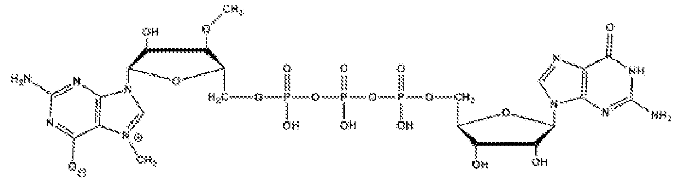


Claims
Priority Applications (3)
| Application Number | Priority Date | Filing Date | Title |
|---|---|---|---|
| AU2022299551A AU2022299551A1 (en) | 2021-06-23 | 2022-06-23 | Engineered cells for therapy |
| EP22829518.4A EP4359541A2 (en) | 2021-06-23 | 2022-06-23 | Engineered cells for therapy |
| CA3225138A CA3225138A1 (en) | 2021-06-23 | 2022-06-23 | Engineered cells for therapy |
Applications Claiming Priority (6)
| Application Number | Priority Date | Filing Date | Title |
|---|---|---|---|
| US202163214157P | 2021-06-23 | 2021-06-23 | |
| US63/214,157 | 2021-06-23 | ||
| US202163233695P | 2021-08-16 | 2021-08-16 | |
| US63/233,695 | 2021-08-16 | ||
| US202263340225P | 2022-05-10 | 2022-05-10 | |
| US63/340,225 | 2022-05-10 |
Publications (2)
| Publication Number | Publication Date |
|---|---|
| WO2022272292A2 true WO2022272292A2 (en) | 2022-12-29 |
| WO2022272292A3 WO2022272292A3 (en) | 2023-04-06 |
Family
ID=84545981
Family Applications (1)
| Application Number | Title | Priority Date | Filing Date |
|---|---|---|---|
| PCT/US2022/073126 WO2022272292A2 (en) | 2021-06-23 | 2022-06-23 | Engineered cells for therapy |
Country Status (4)
| Country | Link |
|---|---|
| EP (1) | EP4359541A2 (en) |
| AU (1) | AU2022299551A1 (en) |
| CA (1) | CA3225138A1 (en) |
| WO (1) | WO2022272292A2 (en) |
Cited By (1)
| Publication number | Priority date | Publication date | Assignee | Title |
|---|---|---|---|---|
| WO2023220207A3 (en) * | 2022-05-10 | 2024-02-29 | Editas Medicine, Inc. | Genome editing of cells |
Family Cites Families (5)
| Publication number | Priority date | Publication date | Assignee | Title |
|---|---|---|---|---|
| JP7049830B2 (en) * | 2014-10-27 | 2022-04-07 | ユニバーシティ オブ セントラル フロリダ リサーチ ファウンデーション,インコーポレイテッド | Methods and compositions for natural killer cells |
| BR112017020750A2 (en) * | 2015-03-27 | 2018-06-26 | Harvard College | modified t-cells and methods of producing and using them |
| WO2018195129A1 (en) * | 2017-04-17 | 2018-10-25 | University Of Maryland, College Park | Embryonic cell cultures and methods of using the same |
| US20210139557A1 (en) * | 2017-12-20 | 2021-05-13 | Poseida Therapeutics, Inc. | Vcar compositions and methods for use |
| BR112021016046A2 (en) * | 2019-02-15 | 2021-11-09 | Editas Medicine Inc | Engineered natural killer (nk) cells for immunotherapy |
-
2022
- 2022-06-23 EP EP22829518.4A patent/EP4359541A2/en active Pending
- 2022-06-23 WO PCT/US2022/073126 patent/WO2022272292A2/en active Application Filing
- 2022-06-23 CA CA3225138A patent/CA3225138A1/en active Pending
- 2022-06-23 AU AU2022299551A patent/AU2022299551A1/en active Pending
Cited By (1)
| Publication number | Priority date | Publication date | Assignee | Title |
|---|---|---|---|---|
| WO2023220207A3 (en) * | 2022-05-10 | 2024-02-29 | Editas Medicine, Inc. | Genome editing of cells |
Also Published As
| Publication number | Publication date |
|---|---|
| WO2022272292A3 (en) | 2023-04-06 |
| CA3225138A1 (en) | 2022-12-29 |
| AU2022299551A1 (en) | 2024-01-18 |
| EP4359541A2 (en) | 2024-05-01 |
Similar Documents
| Publication | Publication Date | Title |
|---|---|---|
| US20220143084A1 (en) | Modified natural killer (nk) cells for immunotherapy | |
| CN108368520B (en) | Genome engineering of pluripotent cells | |
| US20230053028A1 (en) | Engineered cells for therapy | |
| US20240117383A1 (en) | Selection by essential-gene knock-in | |
| EP4359541A2 (en) | Engineered cells for therapy | |
| AU2021369476A9 (en) | Methods of inducing antibody-dependent cellular cytotoxicity (adcc) using modified natural killer (nk) cells | |
| CN116406373A (en) | Engineered ipscs and durable immune effector cells | |
| EP4346877A2 (en) | Engineered cells for therapy | |
| WO2023220207A2 (en) | Genome editing of cells | |
| WO2023220206A2 (en) | Genome editing of b cells | |
| JP2024517864A (en) | Therapeutic engineered cells | |
| CN116848234A (en) | Methods of inducing antibody-dependent cell-mediated cytotoxicity (ADCC) using modified Natural Killer (NK) cells | |
| WO2023279112A1 (en) | Protected effector cells and use thereof for allogeneic adoptive cell therapies |
Legal Events
| Date | Code | Title | Description |
|---|---|---|---|
| 121 | Ep: the epo has been informed by wipo that ep was designated in this application |
Ref document number: 22829518 Country of ref document: EP Kind code of ref document: A2 |
|
| ENP | Entry into the national phase |
Ref document number: 3225138 Country of ref document: CA |
|
| WWE | Wipo information: entry into national phase |
Ref document number: 2022299551 Country of ref document: AU Ref document number: 806877 Country of ref document: NZ Ref document number: AU2022299551 Country of ref document: AU |
|
| ENP | Entry into the national phase |
Ref document number: 2022299551 Country of ref document: AU Date of ref document: 20220623 Kind code of ref document: A |
|
| WWE | Wipo information: entry into national phase |
Ref document number: 2022829518 Country of ref document: EP |
|
| NENP | Non-entry into the national phase |
Ref country code: DE |
|
| ENP | Entry into the national phase |
Ref document number: 2022829518 Country of ref document: EP Effective date: 20240123 |
|
| 121 | Ep: the epo has been informed by wipo that ep was designated in this application |
Ref document number: 22829518 Country of ref document: EP Kind code of ref document: A2 |