WO2022202024A1 - 医用画像処理装置、医用画像処理システム、医用画像処理方法及びプログラム - Google Patents
医用画像処理装置、医用画像処理システム、医用画像処理方法及びプログラム Download PDFInfo
- Publication number
- WO2022202024A1 WO2022202024A1 PCT/JP2022/006750 JP2022006750W WO2022202024A1 WO 2022202024 A1 WO2022202024 A1 WO 2022202024A1 JP 2022006750 W JP2022006750 W JP 2022006750W WO 2022202024 A1 WO2022202024 A1 WO 2022202024A1
- Authority
- WO
- WIPO (PCT)
- Prior art keywords
- image processing
- medical image
- medical
- automatically executed
- processor
- Prior art date
- Legal status (The legal status is an assumption and is not a legal conclusion. Google has not performed a legal analysis and makes no representation as to the accuracy of the status listed.)
- Ceased
Links
Images




Classifications
-
- G—PHYSICS
- G16—INFORMATION AND COMMUNICATION TECHNOLOGY [ICT] SPECIALLY ADAPTED FOR SPECIFIC APPLICATION FIELDS
- G16H—HEALTHCARE INFORMATICS, i.e. INFORMATION AND COMMUNICATION TECHNOLOGY [ICT] SPECIALLY ADAPTED FOR THE HANDLING OR PROCESSING OF MEDICAL OR HEALTHCARE DATA
- G16H40/00—ICT specially adapted for the management or administration of healthcare resources or facilities; ICT specially adapted for the management or operation of medical equipment or devices
- G16H40/20—ICT specially adapted for the management or administration of healthcare resources or facilities; ICT specially adapted for the management or operation of medical equipment or devices for the management or administration of healthcare resources or facilities, e.g. managing hospital staff or surgery rooms
-
- A—HUMAN NECESSITIES
- A61—MEDICAL OR VETERINARY SCIENCE; HYGIENE
- A61B—DIAGNOSIS; SURGERY; IDENTIFICATION
- A61B5/00—Measuring for diagnostic purposes; Identification of persons
- A61B5/05—Detecting, measuring or recording for diagnosis by means of electric currents or magnetic fields; Measuring using microwaves or radio waves
- A61B5/055—Detecting, measuring or recording for diagnosis by means of electric currents or magnetic fields; Measuring using microwaves or radio waves involving electronic [EMR] or nuclear [NMR] magnetic resonance, e.g. magnetic resonance imaging
-
- G—PHYSICS
- G16—INFORMATION AND COMMUNICATION TECHNOLOGY [ICT] SPECIALLY ADAPTED FOR SPECIFIC APPLICATION FIELDS
- G16H—HEALTHCARE INFORMATICS, i.e. INFORMATION AND COMMUNICATION TECHNOLOGY [ICT] SPECIALLY ADAPTED FOR THE HANDLING OR PROCESSING OF MEDICAL OR HEALTHCARE DATA
- G16H30/00—ICT specially adapted for the handling or processing of medical images
- G16H30/20—ICT specially adapted for the handling or processing of medical images for handling medical images, e.g. DICOM, HL7 or PACS
-
- G—PHYSICS
- G16—INFORMATION AND COMMUNICATION TECHNOLOGY [ICT] SPECIALLY ADAPTED FOR SPECIFIC APPLICATION FIELDS
- G16H—HEALTHCARE INFORMATICS, i.e. INFORMATION AND COMMUNICATION TECHNOLOGY [ICT] SPECIALLY ADAPTED FOR THE HANDLING OR PROCESSING OF MEDICAL OR HEALTHCARE DATA
- G16H30/00—ICT specially adapted for the handling or processing of medical images
- G16H30/40—ICT specially adapted for the handling or processing of medical images for processing medical images, e.g. editing
-
- A—HUMAN NECESSITIES
- A61—MEDICAL OR VETERINARY SCIENCE; HYGIENE
- A61B—DIAGNOSIS; SURGERY; IDENTIFICATION
- A61B2576/00—Medical imaging apparatus involving image processing or analysis
-
- G—PHYSICS
- G16—INFORMATION AND COMMUNICATION TECHNOLOGY [ICT] SPECIALLY ADAPTED FOR SPECIFIC APPLICATION FIELDS
- G16H—HEALTHCARE INFORMATICS, i.e. INFORMATION AND COMMUNICATION TECHNOLOGY [ICT] SPECIALLY ADAPTED FOR THE HANDLING OR PROCESSING OF MEDICAL OR HEALTHCARE DATA
- G16H50/00—ICT specially adapted for medical diagnosis, medical simulation or medical data mining; ICT specially adapted for detecting, monitoring or modelling epidemics or pandemics
- G16H50/20—ICT specially adapted for medical diagnosis, medical simulation or medical data mining; ICT specially adapted for detecting, monitoring or modelling epidemics or pandemics for computer-aided diagnosis, e.g. based on medical expert systems
Definitions
- the present invention relates to a medical image processing apparatus, a medical image processing system, a medical image processing method, and a program, and more particularly to technology for presenting the usage status of automatically executed image processing.
- Patent Literature 1 discloses a cloud server that provides advanced image processing services for medical images to clients such as doctors, instructors, students, agents of insurance companies, patients, and medical researchers of medical institutions.
- the present invention has been made in view of such circumstances, and aims to provide a medical image processing apparatus, a medical image processing system, a medical image processing method, and a program that present the usage status of image processing that can be set for automatic execution. aim.
- One aspect of a medical image processing apparatus for achieving the above object comprises at least one processor and at least one memory storing instructions for causing the at least one processor to execute, the at least one processor comprising: , use information by the user of the result of the first image processing which is the first image processing for supporting the diagnosis of medical images and which can be set as automatically executed image processing automatically executed on an input medical image; , and accompanying information of a medical image that is the target of first image processing, and associates the usage information with the accompanying information and displays them on a display.
- the usage information includes whether or not the user has referred to the result of the first image processing, whether or not the result of the first image processing has contributed to preventing overlooking of a disease, and the reading time of the result of the first image processing. Including at least one of the amount of reduction, whether or not the measurement result of the first image processing contributed to the accuracy improvement, and whether or not the user instructed execution of the first image processing other than automatic execution. is preferred.
- the target of the first image processing is a tomographic image, and the associated information preferably includes at least one of the slice thickness and the number of slices.
- At least one processor accepts at least one of deleting the first image processing from the setting of the automatically executed image processing and setting the first image processing as the automatically executed image processing.
- At least one processor preferably proposes an automatic execution condition for automatically executing the first image processing based on the usage information and the accompanying information.
- At least one processor preferably proposes automatic execution conditions in consideration of external factors.
- the external factors preferably include the seasonality of the disease targeted by the first image processing.
- At least one processor preferably executes image processing that is set to automatically execute image processing.
- One aspect of a medical image processing system for achieving the above object is a medical image processing system comprising the above medical image processing apparatus and a server that executes image processing set as automatically executed image processing.
- One aspect of the medical image processing method for achieving the above object is first image processing that supports diagnosis of medical images, and automatically executed image processing that is automatically executed on an input medical image.
- User information of a result of the first image processing that can be set and incidental information of the medical image subjected to the first image processing are collected, and the usage information and the incidental information are displayed in association with each other on a display.
- This is a medical image processing method that allows According to this aspect, it is possible to present the usage status of image processing that can be set as automatically executed image processing to be automatically executed. You can decide whether or not
- One aspect of the program for achieving the above object is a program for causing a computer to execute the above medical image processing method.
- a computer-readable non-transitory storage medium in which this program is recorded may also be included in this embodiment. According to this aspect, it is possible to present the usage status of image processing that can be set as automatically executed image processing to be automatically executed. You can decide whether or not
- the user can determine whether or not to set image processing as automatically executed image processing based on the presented usage status.
- FIG. 1 is an overall configuration diagram of a medical image processing system.
- FIG. 2 is a diagram showing the functional configuration and processing process of the medical image processing system.
- FIG. 3 is a flow chart showing a medical image processing method using the medical image processing system.
- FIG. 4 is an example of a recommended condition/cost benefit proposal screen.
- FIG. 5 is an example of a display screen of automatic execution conditions for "CAD (1)”.
- FIG. 6 is an example of a log totaling result screen.
- FIG. 1 is an overall configuration diagram of a medical image processing system 10.
- the medical image processing system 10 captures an image of an object (patient) to be inspected, and automatically performs image processing that is set as automatically executed image processing for supporting medical image diagnosis on the captured image. It is a system that executes.
- the medical image processing system 10 includes a medical image inspection device 12, a medical image database 14, an image processing evaluation device 16, a user terminal 20, and a cloud server 26. .
- the medical image inspection equipment 12, the medical image database 14, the image processing evaluation device 16, and the user terminal 20 are provided in a medical institution such as a hospital, and are connected via an intra-hospital network 22 so that data can be sent and received.
- a LAN Local Area Network
- the intra-hospital network 22 may be wired or wireless.
- the hospital network 22 is connected to the Internet 24 via a router (not shown).
- the intra-hospital network 22 and the cloud server 26 are connected via the Internet 24 so as to be able to transmit and receive data.
- the medical image inspection equipment 12 is an imaging device that captures an image of an inspection target part of an inspection target and generates a medical image.
- the medical image inspection equipment 12 includes, for example, an X-ray imaging device, a CT (Computed Tomography) device, an MRI (Magnetic Resonance Imaging) device, a PET (Positron Emission Tomography) device, an ultrasonic device, and a CR using a flat X-ray detector. (Computed Radiography) equipment.
- the medical image database 14 is a database that manages medical images captured by the medical image inspection equipment 12.
- a computer with a large-capacity storage device can be applied to the medical image database 14 .
- the computer is embedded with software that provides the functionality of a database management system.
- Dicom Digital imaging and communications in medicine
- the medical image may be added with Dicom tag information defined in the Dicom standard.
- image in this specification can include the meaning of image data, which is a signal representing an image, in addition to the meaning of the image itself such as a photograph.
- the image processing evaluation device 16 is an example of a medical image processing device, collects the usage status of image processing executed in the cloud server 26, and evaluates the image processing based on the usage status.
- a personal computer or a workstation can be applied to the image processing evaluation device 16 .
- the image processing evaluation device 16 includes a processor 16A and a memory 16B. Processor 16A executes instructions stored in memory 16B.
- the hardware structure of the processor 16A is various processors as shown below.
- Various processors include a CPU (Central Processing Unit), which is a general-purpose processor that executes software (programs) and acts as various functional units, a GPU (Graphics Processing Unit), which is a processor specialized for image processing, A circuit specially designed to execute specific processing such as PLD (Programmable Logic Device), which is a processor whose circuit configuration can be changed after manufacturing such as FPGA (Field Programmable Gate Array), ASIC (Application Specific Integrated Circuit), etc. Also included are dedicated electrical circuits, which are processors with configuration, and the like.
- One processing unit may be composed of one of these various processors, or two or more processors of the same or different type (for example, a plurality of FPGAs, a combination of CPU and FPGA, or a combination of CPU and GPU).
- a plurality of functional units may be configured by one processor.
- a single processor is configured by combining one or more CPUs and software.
- a processor acts as a plurality of functional units.
- SoC System On Chip
- various functional units are configured using one or more of the above various processors as a hardware structure.
- the hardware structure of these various processors is, more specifically, an electrical circuit that combines circuit elements such as semiconductor elements.
- the memory 16B stores instructions to be executed by the processor 16A.
- the memory 16B includes RAM (Random Access Memory) and ROM (Read Only Memory) (not shown).
- the processor 16A uses the RAM as a work area, executes software using various programs and parameters including a medical image processing program stored in the ROM, and uses the parameters stored in the ROM or the like to generate an image. Various processes of the process evaluation device 16 are executed.
- the user terminal 20 is a terminal device used by a user such as a doctor, and for example, a known image viewer for interpretation is applied.
- the user terminal 20 may be a personal computer, workstation, or tablet terminal.
- the user terminal 20 includes an input device 20A and a display 20B.
- the display 20B displays a medical image captured by the medical image inspection device 12 and the result of image processing. Further, the display 20B displays information about image processing presented by the image processing evaluation device 16.
- FIG. A user can input instructions to the medical image processing system 10 using the input device 20A.
- a server computer, a personal computer, or a workstation can be applied to the cloud server 26, for example.
- the cloud server 26 performs image processing including at least one of lesion extraction and disease name determination processing on medical images to be examined.
- the cloud server 26 can be accessed from the intra-hospital networks 22 of multiple hospitals via the Internet 24 .
- the processing performed by the cloud server 26 is a pay-as-you-go cloud service.
- the processing performed by the cloud server 26 may be a flat-rate cloud service.
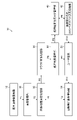
- FIG. 2 is a diagram showing the functional configuration and processing process of the medical image processing system 10. As shown in FIG. The processing process will be described later.
- the medical image processing system 10 includes, in addition to the medical image inspection equipment 12 and the user terminal 20 shown in FIG. It comprises a condition management unit 34 , a cloud image processing unit 36 , a processing result management unit 38 , a processing result usage log management unit 40 , and a recommended execution condition/cost-benefit determination processing unit 42 .
- the function of the image management unit 30 is realized by the medical image database 14.
- the functions of the image processing determination processing unit 32, the automatic execution condition management unit 34, the processing result management unit 38, the processing result use log management unit 40, and the recommended execution condition/cost benefit determination processing unit 42 are realized by the image processing evaluation device 16. be done.
- Functions of the cloud image processing unit 36 are implemented by the cloud server 26 .
- the image management unit 30 manages medical images captured by the medical image inspection equipment 12 .
- the image processing determination processing unit 32 determines automatically executed image processing to be automatically executed on medical images captured by the medical image inspection equipment 12 .
- the automatic execution condition management unit 34 holds automatic execution conditions for automatic execution image processing.
- the cloud image processing unit 36 performs image processing on medical images captured by the medical image inspection equipment 12 .
- the image processing performed by the cloud image processing unit 36 is not limited as long as it is image processing for medical images.
- computer-aided diagnostic imaging CAD: Computer-Aided Diagnosis
- segmentation processing for sites and lesions segmentation processing for sites and lesions
- labeling processing for site names and disease names similar image search, and similar case search including.
- the processing result management unit 38 acquires the execution result of the image processing executed by the cloud image processing unit 36.
- the processing result usage log management unit 40 saves, as a log, usage information of the image processing result by the user and accompanying information of the medical image subjected to the image processing. That is, the processing result use log management unit 40 saves, as a log, usage information indicating the usage status of the result of image processing by the user and accompanying information of the medical image subjected to the image processing.
- a recommended execution condition/cost-benefit determination processing unit 42 determines a recommended execution condition of image processing and its cost-benefit based on the log accumulated in the processing result usage log management unit 40 .
- FIG. 3 is a flowchart showing a medical image processing method using the medical image processing system 10.
- the medical image processing method is implemented by the processor 16A executing a medical image processing program stored in the memory 16B.
- the medical image processing program may be provided by a computer-readable non-transitory storage medium.
- the image processing evaluation device 16 may read the medical image processing program from the non-temporary storage medium and store it in the memory 16B.
- step S1 the medical image inspection equipment 12 captures a medical image of the patient and stores the captured medical image in the image management unit 30 (process P1).
- a doctor uses a CT apparatus (not shown) as the medical image inspection device 12 to take a CT image (an example of a "tomographic image") of a patient.
- CT image an example of a "tomographic image”
- the CT apparatus saves the captured CT image in the image manager 30 .
- step S2 the image management unit 30 notifies the image processing decision processing unit 32 that the medical image has been saved by the medical image inspection equipment 12 (process P2).
- the image manager 30 notifies that the CT image has been imported from the CT apparatus.
- step S3 the image processing determination processing unit 32 acquires automatic execution conditions for automatically executed image processing to be automatically executed from the automatic execution condition management unit 34 (process P3).
- the image processing set as automatically executed image processing is "CAD (1) , CAD(2), Segmentation(1), Labeling(1), and Labeling(2).
- the image processing determination processing unit 32 acquires automatic execution conditions for each of the five types of image processing.
- the cloud image processing unit 36 executes automatic execution image processing on images that satisfy automatic execution conditions.
- the automatic execution conditions include, for example, at least one of the CT image slice thickness and the number of slices.
- the automatic execution condition is set by the slice thickness and the number of slices will be described.
- parameters of the automatic execution conditions are not limited to the slice thickness of the tomographic image and the number of slices.
- parameters for automatic execution conditions are FOV (Field of view), Spacing, processing of past images, ImageType (Primary image or Secondary image), Description, equipment information (manufacturer), patient information (gender, age), contrast information , exposure information, reconstruction information, and the like may be used.
- step S4 the image processing determination processing unit 32 determines image processing to be performed on the CT images saved in step S1 according to the automatic execution conditions acquired in step S3 (process P4).
- the CT images saved in step S1 have a slice thickness of 1 mm and a number of slices of 1000 sheets.
- the conditions for automatic execution of "CAD(1)" are that the slice thickness is 0.5 mm or more and the number of slices is 100 or more
- the CT image saved in step S1 is automatically executed for "CAD(1)". meet the conditions. Therefore, the image processing determination processing unit 32 selects "CAD(1)" as the image processing to be executed.
- the slice thickness is 5 mm or more and the number of slices is 1000 or more in the automatic execution condition of "CAD(2)”
- the CT image saved in step S1 satisfies the automatic execution condition of "CAD(2)". do not have. Therefore, the image processing determination processing unit 32 does not select "CAD(2)" as image processing to be executed.
- step S5 the image processing determination processing unit 32 requests the cloud image processing unit 36 to execute the image processing determined in step S4 (process P5).
- step S6 the cloud image processing unit 36 acquires the CT image saved in the image management unit 30 in step S1, and executes the image processing requested in step S5 on the CT image. Further, the processing result management unit 38 acquires the execution result of the image processing from the cloud image processing unit 36 (process P6).
- step S7 the user terminal 20 acquires the execution result of the image processing from the processing result management unit 38, and displays the execution result on the display 20B according to the user's operation (process P7).
- step S8 the processing result use log management unit 40 saves as a log the usage information that is the information that the user used the execution result in step S7 and the accompanying information of the CT image on which the image processing was performed (process P8).
- the usage information includes whether or not the user referred to the results of image processing, whether or not the results of image processing contributed to preventing disease from being overlooked, the amount of reduction in reading time due to the results of image processing, and the measurement results of image processing. contributes to the improvement of accuracy, and whether or not the user has instructed execution of image processing other than automatic execution.
- the accompanying information includes at least one of the slice thickness and the number of slices of the CT image.
- the processing result use log management unit 40 stores logs of image processing that is automatically executed, but also stores logs of image processing that is manually instructed by the user in addition to the automatic execution.
- step S9 an example of a "collection step”
- the recommended execution condition/cost-benefit determination processing unit 42 acquires the accumulated logs (process P9).
- the recommended execution condition/cost-benefit determination processing unit 42 calculates the evaluation value of image processing based on the accumulated logs, and determines the recommended automatic execution condition and its effect. Instead of using all the accumulated logs, the evaluation value may be calculated using the latest logs for a certain period (for example, one week's worth). The log accumulation period used for calculation may be settable for each hospital.
- the recommended execution condition/cost-benefit determination processing unit 42 displays the determined recommended condition and its effect on the display 20B to make a proposal (process P10).
- step S11 the user confirms the recommended conditions displayed on the display 20B of the user terminal 20 and their effects, and sets automatic execution conditions using the input device 20A.
- the user may set the displayed recommended conditions as they are as the automatic execution conditions, or may refer to the displayed recommended conditions to set desired automatic execution conditions.
- the user terminal 20 stores the set automatic execution condition in the automatic execution condition management unit 34 (process P11). Thereafter, the image processing determination processing unit 32 acquires the newly set automatic execution conditions.
- FIG. 4 is an example of a recommended condition/cost benefit suggestion screen displayed on the display 20B of the user terminal 20.
- CAD (1) "CAD (2)”
- segmentation (1) "labeling (1)”
- labeling (2) which are image processes automatically executed in the cloud image processing unit 36
- the pie chart shows the percentage of the cost for the latest fixed period of time, and the pie chart shows the amount of each cost.
- Each area of the pie chart can be selected by the input device 20A.
- it indicates that "CAD (1)" surrounded by a thick frame is being selected.
- FIG. 5 is an example of a display screen of automatic execution conditions for "CAD (1)" displayed when "CAD (1)” is selected in the pie chart of FIG. 4 displayed on the display 20B.
- the automatic execution conditions are the slice thickness and the number of slices
- the display of the automatic execution conditions is a scroll bar 100 representing the automatic execution condition of the slice thickness and the scroll bar 110 representing the automatic execution condition of the number of slices. ,including.
- the pie chart of FIG. 4 and the slider bar of FIG. 5 may be displayed simultaneously on the same screen.
- the scroll bar 100 indicates a slice thickness range from 0.1 mm to 5 mm.
- a triangular mark 102 displayed above the scroll bar 100 indicates the lower limit value of the currently set automatic execution condition. Therefore, the image on the right side of the position of the mark 102 (the slice thickness greater than the value indicated by the mark 102) is the image to be automatically executed.
- the star-shaped mark 104 indicates the lower limit value of the automatic execution condition recommended by the recommended execution condition/cost-benefit determination processing unit 42, and the image on the right side of the position of the mark 104 is the recommended execution condition. This is the target image.
- the currently set automatic execution condition for slice thickness is 0.5 mm or more, and the recommended automatic execution condition is 2 mm or more.
- the upper limit value of the slice thickness may be set.
- the scroll bar 100 also has a knob 106 that can move within the range of the scroll bar 100 .
- the user can change the automatic execution condition by changing the position of the knob 106 by dragging the knob 106 with the input device 20A.
- the image on the right side of the position of the knob 106 is the image to be automatically executed image processing.
- the slice thickness automatic execution condition is changed from 0.5 mm or more to 2 mm or more. Due to this change in the automatic execution conditions, tomographic images with a slice thickness of 0.5 mm to 2 mm will not be automatically executed after that, and it is expected that the cost will be reduced accordingly in the pay-as-you-go cloud service.
- the recommended execution condition/cost-benefit determination processing unit 42 determines the latest fixed period of "CAD (1)" when the automatic execution condition of the slice thickness of "CAD (1)" is changed from 0.5 mm or more to 2 mm or more. are recalculated and reflected in the pie chart shown in FIG.
- the scroll bar 110 indicates a range from 10 to 1000 slices.
- a triangular mark 112 displayed above the scroll bar 110 indicates the lower limit value of the currently set automatic execution condition. Therefore, the image on the right side of the position of the mark 112 (the number of slices larger than the value indicated by the mark 112) is the image to be automatically executed.
- a star-shaped mark 114 indicates the lower limit value of the automatic execution condition recommended by the recommended execution condition/cost-benefit determination processing unit 42, and the image on the right side of the position of the mark 114 is the recommended execution condition. This is the target image.
- the currently set automatic execution condition for the number of slices is 100 or more, and the recommended automatic execution condition is 200 or more.
- an upper limit value for the number of slices may be set.
- the scroll bar 110 also has a knob 116 that can move within the range of the scroll bar 110 .
- the user can change the automatic execution condition by changing the position of the knob 116 by dragging the knob 116 with the input device 20A.
- the image on the right side of the position of the knob 116 is the image to be automatically executed image processing.
- the automatic execution condition for the number of slices is changed from 100 slices or more to 200 slices or more.
- the recommended execution condition/cost-benefit determination processing unit 42 determines the latest fixed period of "CAD (1)" when the automatic execution condition for the number of slices of "CAD (1)" is changed from 100 or more to 200 or more. are recalculated and reflected in the pie chart shown in FIG.
- the automatic execution condition management unit 34 may accept deletion of automatically executed image processing from the input device 20A. Further, the automatic execution condition management unit 34 may receive addition of image processing to be automatically executed from the input device 20A. For example, the user can use the input device 20A to add similar image retrieval and similar case retrieval processing to image processing that automatically executes.
- the recommended execution condition/cost-benefit determination processing unit 42 may propose automatic execution conditions in consideration of external factors. External factors include the seasonality of the disease targeted by image processing.
- the recommended execution condition/cost-benefit determination processing unit 42 calculates an evaluation value considering external factors of image processing using, for example, the following formula.
- the number of processing is the number of case images for which image processing has been performed in the cloud image processing unit 36
- the number of uses is the number of execution results confirmed by the user on the user terminal 20 (the number displayed on the display 20B).
- the number of missed lesions is the number of lesions that were missed by the user but were found by image processing.
- the user performs a diagnosis without using the execution result of image processing, displays the execution result on the display 20B after the diagnosis, and counts the number of lesions not included in the user's diagnosis among the displayed lesions. do.
- the seasonality evaluation of image processing is a value that indicates the presence or absence of seasonality in the occurrence of diseases targeted by image processing.
- the seasonal coefficient is a coefficient that takes on a relatively large value in a season in which the occurrence of diseases targeted for image processing is relatively frequent. For example, in the case of a disease that occurs relatively frequently in winter, the seasonal coefficient of image processing for that disease has a relatively large value in winter.
- the image processing is maintained as automatically executed image processing, and if it is less than the threshold, the image processing is deleted from the automatically executed image processing. Note that if any one of the evaluation values a1, a2, and a3 is equal to or greater than the threshold, the image processing may be maintained as automatically executed image processing.
- the evaluation value may use the calculation function of the health economic index (QALY: Quality-adjusted life year).
- the recommended execution condition/cost-benefit determination processing unit 42 may set or propose deletion as an automatic execution condition in consideration of other evaluation items such as improvement of measurement accuracy as usage information.
- the doctor determines the volume of the lesion to be X 1 [cm 3 ] without checking the result of the image processing, and the volume measurement result of the image processing executed by the cloud image processing unit 36 is X 1 [cm 3 ]. 3 ] was different from X 2 [cm 3 ].
- the processing result use log management unit 40 saves the log "the doctor corrected the interpretation report after confirming the volume measurement result of the image processing executed by the cloud image processing unit 36" as the use information.
- the recommended execution condition/cost-benefit determination processing unit 42 judges that "the volume measurement of image processing is better than that doctor's, and contributed to the improvement of measurement accuracy", and the image processing that performed the volume measurement is recommended as a setting for auto-execution image processing.
- the processing result use log management unit 40 saves a log stating that "the doctor did not modify the interpretation report even after confirming the result of the volume measurement of the image processing executed by the cloud image processing unit 36" as the use information. In this case, it is determined that ⁇ the doctor's volume measurement was superior to the image processing and did not contribute to the improvement of accuracy'', and the image processing that performed the volume measurement is deleted from the settings of the automatic execution image processing. recommended.
- the recommended execution condition/cost-benefit determination processing unit 42 can propose setting or deletion of an automatic execution condition according to the degree of contribution of each evaluation item related to interpretation of medical images, without being limited to improving measurement accuracy.
- the medical image processing system 10 may associate the collected usage information with the associated information and display them on the display 20B (an example of a “presentation step”).
- FIG. 6 is an example of a tabulation result screen of logs accumulated by the processing result use log management unit 40 displayed on the display 20B.
- usage information for each incidental information for the log of "CAD (1)" is shown in a frequency line graph. ], and the oversight prevention number [unit: location].
- the number of processed sheets is 1000, but the number of used sheets is 0. ) is 0.
- the number of processing is 2000, the number of uses is 1600, and the number of oversight prevention is 2000.
- the slice thickness is 3 to 5 mm (3 mm or more and less than 5 mm)
- the number of processing is 1000
- the number of used is 0, and the number of overlook prevention is 0.
- the slice thickness is 5 mm to (5 mm or more)
- the number of processed sheets is 2000, the number of used sheets is 1800, and the number of oversight prevention is 1000 points.
- the medical image processing system 10 causes the display 20B to display the aggregated results of such logs, so that the user can confirm the conditions suitable for the automatic execution conditions of the automatic execution image processing. It can also be used to determine the introduction cost of cloud services and the selection of a plan to first subscribe to cloud services.
- the recommended execution condition/cost-benefit determination processing unit 42 not only displays the aggregated result of the log, but also displays the slice thickness of 1 to 3 mm and the slice thickness of 5 mm or more as the recommended conditions of the automatic execution condition. You can present the case.
- One aspect of a medical image processing apparatus includes at least one processor and at least one memory that stores instructions to cause the at least one processor to execute, and the at least one processor processes medical images.
- first image processing for assisting diagnosis which is settable as automatically executed image processing automatically executed on an input medical image;
- a medical image processing apparatus that collects incidental information of a medical image that is a target of image processing, associates usage information with the incidental information, and displays the information on a display. That is, at least one processor and at least one memory storing instructions for causing the at least one processor to execute are provided, and the at least one processor is first image processing that supports diagnosis of medical images.
- a medical image processing apparatus that collects accompanying information of a medical image that is the target of first image processing, associates the usage information with the accompanying information, and displays them on a display. According to this aspect, it is possible to present the usage status of image processing that can be set as automatically executed image processing to be automatically executed. You can decide whether or not (2) The usage information depends on whether or not the user referred to the result of the first image processing, whether or not the result of the first image processing contributed to preventing overlooking of the disease, and whether or not the result of the first image processing was performed.
- At least one of an amount of reduction in interpretation time, whether or not the measurement result of the first image processing contributed to the improvement of accuracy, and whether or not the user instructed execution of the first image processing other than automatic execution. preferably include one. This makes it possible to collect usage information that reflects the degree of contribution of image processing.
- the object of the first image processing is a tomographic image, and the associated information preferably includes at least one of the slice thickness and the number of slices. Thereby, at least one of the slice thickness and the number of slices of the tomographic image can be acquired.
- the at least one processor preferably accepts at least one of deleting the first image processing from the setting of the automatically executed image processing and setting the first image processing as the automatically executed image processing.
- At least one processor proposes an automatic execution condition for automatically executing the first image processing based on the usage information and the accompanying information. This allows the user to know appropriate automatic execution conditions.
- At least one processor preferably proposes an automatic execution condition considering external factors. This makes it possible to propose an appropriate automatic execution condition.
- the external factors preferably include the seasonality of the disease targeted by the first image processing. This makes it possible to propose an appropriate automatic execution condition.
- the at least one processor executes image processing set to automatically execute image processing. Thereby, the set image processing can be automatically executed.
- One aspect of a medical image processing system is a medical image processing system comprising the medical image processing apparatus of (1) to (8) and a server that executes image processing set as automatically executed image processing. be. According to this aspect, it is possible to present the usage status of image processing that can be set as automatically executed image processing to be automatically executed. It is possible to determine whether or not, and to automatically execute image processing set as automatic execution image processing.
- One aspect of the medical image processing method is first image processing for supporting diagnosis of medical images, which can be set as automatically executed image processing automatically executed on an input medical image.
- Medical image processing that collects information used by a user as a result of the first image processing and accompanying information of the medical image that is the target of the first image processing, associates the information with the accompanying information, and displays the information on a display.
- the method is the first image processing that supports the diagnosis of medical images, and the first image processing that can be set as automatically executed image processing that is automatically executed on an input medical image.
- 1 image processing in which usage information indicating the usage status of the result of the first image processing by the user and incidental information of the medical image subjected to the first image processing are collected, and the usage information and This is a medical image processing method for displaying on a display in association with incidental information.
- the usage status of image processing that can be set as automatically executed image processing to be automatically executed You can decide whether or not (11)
- One aspect of the program for achieving the above object is a program for causing a computer to execute the above medical image processing method.
- a computer-readable non-transitory storage medium in which this program is recorded may also be included in this embodiment.
- Medical image processing system 12 Medical image inspection equipment 14 Medical image database 16 Image processing evaluation device 16A Processor 16B Memory 20 User terminal 20A Input device 20B Display 22 Hospital network 24 Internet 26 Cloud server 30 Image management unit 32 Image processing determination processing unit 34 Automatic execution condition management unit 36 Cloud image processing unit 38 Processing result management unit 40 Processing result use log management unit 42 Cost benefit determination processing unit 100 Scroll bar 102 Mark 104 Mark 106 Knob 110 Scroll bar 112 Mark 114 Mark 116 Knobs P1 to P11 Processes S1 to S11 of the medical image processing apparatus Steps of the medical image processing method
Landscapes
- Health & Medical Sciences (AREA)
- Engineering & Computer Science (AREA)
- Public Health (AREA)
- General Health & Medical Sciences (AREA)
- Medical Informatics (AREA)
- Nuclear Medicine, Radiotherapy & Molecular Imaging (AREA)
- Primary Health Care (AREA)
- Epidemiology (AREA)
- Radiology & Medical Imaging (AREA)
- Life Sciences & Earth Sciences (AREA)
- Business, Economics & Management (AREA)
- General Business, Economics & Management (AREA)
- Biomedical Technology (AREA)
- Physics & Mathematics (AREA)
- Biophysics (AREA)
- Animal Behavior & Ethology (AREA)
- Surgery (AREA)
- Veterinary Medicine (AREA)
- Molecular Biology (AREA)
- Heart & Thoracic Surgery (AREA)
- Pathology (AREA)
- High Energy & Nuclear Physics (AREA)
- Medical Treatment And Welfare Office Work (AREA)
- Measuring And Recording Apparatus For Diagnosis (AREA)
Abstract
自動実行に設定可能な画像処理の利用状況を提示する医用画像処理装置、医用画像処理システム、医用画像処理方法及びプログラムを提供することを目的とする。少なくとも1つのプロセッサと、少なくとも1つのプロセッサに実行させるための命令を記憶する少なくとも1つのメモリと、を備え、少なくとも1つのプロセッサは、医用画像の診断を支援する第1の画像処理であって、入力された医用画像に対して自動実行される自動実行画像処理として設定可能な第1の画像処理の結果のユーザによる利用情報と、第1の画像処理の対象となった医用画像の付随情報と、を収集し、利用情報と付随情報とを関連付けてディスプレイに表示させる。
Description
本発明は医用画像処理装置、医用画像処理システム、医用画像処理方法及びプログラムに係り、特に自動実行される画像処理の利用状況を提示する技術に関する。
画像処理サービスを提供するクラウドサービスが知られている。例えば、特許文献1には、医療機関の医師、インストラクタ、学生、保険会社の代理人、患者、医学研究者などのクライアントに医用画像の高度画像処理サービスを提供するクラウドサーバが開示されている。
クラウドサービス等でみられる従量課金制の計算処理システムでは、処理数がユーザの支払い額に直結する。従量課金制の計算処理システムのうち、自動的にタグ情報、及び部位情報等の画像情報から画像処理を選択し自動実行するシステムの場合には、処理結果のうち一定数は結果が活用されないにも関わらず課金対象となることが想定される。また、定額制の計算処理システムの場合には、結果が活用されない分は計算コストとなる。そのため、ユーザにとって経費を削減するために、使用しない画像処理が実行されないような実行条件を設定する必要がある。しかしながら、ユーザが実行条件を最適設定するためには使用していない画像処理と画像の膨大なデータセットを精査する必要があり、負荷が高いという問題点があった。
本発明はこのような事情に鑑みてなされたもので、自動実行に設定可能な画像処理の利用状況を提示する医用画像処理装置、医用画像処理システム、医用画像処理方法及びプログラムを提供することを目的とする。
上記目的を達成するための医用画像処理装置の一の態様は、少なくとも1つのプロセッサと、少なくとも1つのプロセッサに実行させるための命令を記憶する少なくとも1つのメモリと、を備え、少なくとも1つのプロセッサは、医用画像の診断を支援する第1の画像処理であって、入力された医用画像に対して自動実行される自動実行画像処理として設定可能な第1の画像処理の結果のユーザによる利用情報と、第1の画像処理の対象となった医用画像の付随情報と、を収集し、利用情報と付随情報とを関連付けてディスプレイに表示させる、医用画像処理装置である。本態様によれば、自動実行される自動実行画像処理として設定可能な画像処理の利用状況を提示することができるので、ユーザは提示された利用状況から画像処理を自動実行画像処理として設定するか否かの判断をすることができる。
利用情報は、第1の画像処理の結果をユーザが参照したか否か、第1の画像処理の結果が疾患の見逃し防止に貢献したか否か、第1の画像処理の結果による読影時間の削減量、第1の画像処理の計測結果が精度向上に貢献したか否か、及び自動実行以外にユーザが第1の画像処理の実行を指示したか否か、のうちの少なくとも1つを含むことが好ましい。
第1の画像処理の対象は断層画像であり、付随情報は、スライス厚とスライス枚数との少なくとも一方を含むことが好ましい。
少なくとも1つのプロセッサは、自動実行画像処理の設定から第1の画像処理を削除、及び自動実行画像処理として第1の画像処理を設定、のうちの少なくとも一方を受け付けることが好ましい。
少なくとも1つのプロセッサは、利用情報と付随情報とに基づいて、第1の画像処理が自動実行されるための自動実行条件を提案することが好ましい。
少なくとも1つのプロセッサは、外的要因を考慮して自動実行条件を提案することが好ましい。また、外的要因は、第1の画像処理が対象とする疾患の季節性を含むことが好ましい。
少なくとも1つのプロセッサは、自動実行画像処理に設定された画像処理を実行することが好ましい。
上記目的を達成するための医用画像処理システムの一の態様は、上記の医用画像処理装置と、自動実行画像処理として設定された画像処理を実行するサーバと、を備える医用画像処理システムである。
上記目的を達成するための医用画像処理方法の一の態様は、医用画像の診断を支援する第1の画像処理であって、入力された医用画像に対して自動実行される自動実行画像処理として設定可能な第1の画像処理の結果のユーザによる利用情報と、第1の画像処理の対象となった医用画像の付随情報と、を収集し、利用情報と付随情報とを関連付けてディスプレイに表示させる医用画像処理方法である。本態様によれば、自動実行される自動実行画像処理として設定可能な画像処理の利用状況を提示することができるので、ユーザは提示された利用状況から画像処理を自動実行画像処理として設定するか否かの判断をすることができる。
上記目的を達成するためのプログラムの一の態様は、上記の医用画像処理方法をコンピュータに実行させるためのプログラムである。このプログラムが記録された、コンピュータが読み取り可能な非一時的記憶媒体も本態様に含んでよい。本態様によれば、自動実行される自動実行画像処理として設定可能な画像処理の利用状況を提示することができるので、ユーザは提示された利用状況から画像処理を自動実行画像処理として設定するか否かの判断をすることができる。
本発明によれば、ユーザは提示された利用状況から画像処理を自動実行画像処理として設定するか否かの判断をすることができる。
以下、添付図面に従って本発明の好ましい実施形態について詳説する。
〔医用画像処理システムの全体構成〕
図1は、医用画像処理システム10の全体構成図である。医用画像処理システム10は、検査対象(患者)の画像を撮影し、撮影された画像に対して医用画像の診断を支援する画像処理であって、自動実行画像処理として設定された画像処理を自動実行するシステムである。図1に示すように、医用画像処理システム10は、医用画像検査機器12と、医用画像データベース14と、画像処理評価装置16と、ユーザ端末20と、クラウドサーバ26と、を備えて構成される。
図1は、医用画像処理システム10の全体構成図である。医用画像処理システム10は、検査対象(患者)の画像を撮影し、撮影された画像に対して医用画像の診断を支援する画像処理であって、自動実行画像処理として設定された画像処理を自動実行するシステムである。図1に示すように、医用画像処理システム10は、医用画像検査機器12と、医用画像データベース14と、画像処理評価装置16と、ユーザ端末20と、クラウドサーバ26と、を備えて構成される。
医用画像検査機器12、医用画像データベース14、画像処理評価装置16、及びユーザ端末20は、病院などの医療機関内に設けられ、それぞれ病院内ネットワーク22を介してデータ送受信可能に接続される。病院内ネットワーク22は、LAN(Local Area Network)を適用可能である。病院内ネットワーク22は、有線でもよいし、無線でもよい。
病院内ネットワーク22は不図示のルータを介してインターネット24と接続される。病院内ネットワーク22、及びクラウドサーバ26は、インターネット24を介してそれぞれデータを送受信可能に接続される。
医用画像検査機器12は、検査対象の検査対象部位を撮像し、医用画像を生成する撮像装置である。医用画像検査機器12は、例えばX線撮像装置、CT(Computed Tomography)装置、MRI(Magnetic Resonance Imaging)装置、PET(Positron Emission Tomography)装置、超音波装置、及び平面X線検出器を用いたCR(Computed Radiography)装置のうちの少なくとも1つを含む。
医用画像データベース14は、医用画像検査機器12によって撮影された医用画像を管理するデータベースである。医用画像データベース14は、大容量ストレージ装置を備えるコンピュータを適用可能である。コンピュータはデータベース管理システムの機能を提供するソフトウェアが組み込まれる。
医用画像のフォーマットは、Dicom(Digital imaging and communications in medicine)規格を適用可能である。医用画像は、Dicom規格において規定されたDicomタグ情報が付加されてもよい。なお、本明細書における画像という用語には、写真等の画像自身の意味の他に、画像を表す信号である画像データの意味が含まれ得る。
画像処理評価装置16は、医用画像処理装置の一例であり、クラウドサーバ26において実行される画像処理の利用状況を収集し、利用状況から画像処理を評価する。画像処理評価装置16は、パーソナルコンピュータ、又はワークステーションを適用可能である。画像処理評価装置16は、プロセッサ16Aと、メモリ16Bと、を備える。プロセッサ16Aは、メモリ16Bに記憶された命令を実行する。
プロセッサ16Aのハードウェア的な構造は、次に示すような各種のプロセッサ(processor)である。各種のプロセッサには、ソフトウェア(プログラム)を実行して各種の機能部として作用する汎用的なプロセッサであるCPU(Central Processing Unit)、画像処理に特化したプロセッサであるGPU(Graphics Processing Unit)、FPGA(Field Programmable Gate Array)等の製造後に回路構成を変更可能なプロセッサであるPLD(Programmable Logic Device)、ASIC(Application Specific Integrated Circuit)等の特定の処理を実行させるために専用に設計された回路構成を有するプロセッサである専用電気回路等が含まれる。
1つの処理部は、これら各種のプロセッサのうちの1つで構成されていてもよいし、同種又は異種の2つ以上のプロセッサ(例えば、複数のFPGA、又はCPUとFPGAの組み合わせ、あるいはCPUとGPUの組み合わせ)で構成されてもよい。また、複数の機能部を1つのプロセッサで構成してもよい。複数の機能部を1つのプロセッサで構成する例としては、第1に、クライアント又はサーバ等のコンピュータに代表されるように、1つ以上のCPUとソフトウェアの組合せで1つのプロセッサを構成し、このプロセッサが複数の機能部として作用させる形態がある。第2に、SoC(System On Chip)等に代表されるように、複数の機能部を含むシステム全体の機能を1つのIC(Integrated Circuit)チップで実現するプロセッサを使用する形態がある。このように、各種の機能部は、ハードウェア的な構造として、上記各種のプロセッサを1つ以上用いて構成される。
さらに、これらの各種のプロセッサのハードウェア的な構造は、より具体的には、半導体素子等の回路素子を組み合わせた電気回路(circuitry)である。
メモリ16Bは、プロセッサ16Aに実行させるための命令を記憶する。メモリ16Bは、不図示のRAM(Random Access Memory)、及びROM(Read Only Memory)を含む。プロセッサ16Aは、RAMを作業領域とし、ROMに記憶された医用画像処理プログラムを含む各種のプログラム及びパラメータを使用してソフトウェアを実行し、かつROM等に記憶されたパラメータを使用することで、画像処理評価装置16の各種の処理を実行する。
ユーザ端末20は、医師等のユーザが使用する端末機器であり、例えば公知の読影用画像ビューワが適用される。ユーザ端末20は、パーソナルコンピュータ、ワークステーション、又はタブレット端末であってもよい。ユーザ端末20は、入力装置20Aと、ディスプレイ20Bとを備える。ディスプレイ20Bには、医用画像検査機器12において撮影された医用画像、及び画像処理の結果が表示される。また、ディスプレイ20Bには、画像処理評価装置16において提示される画像処理に関する情報が表示させる。ユーザは、入力装置20Aを使用して医用画像処理システム10への指示を入力することができる。
クラウドサーバ26は、例えばサーバコンピュータ、パーソナルコンピュータ、又はワークステーションを適用可能である。クラウドサーバ26は、検査対象の医用画像について、病変抽出、及び病名判定処理のうちの少なくとも一方を含む画像処理を行う。クラウドサーバ26は、インターネット24を介して複数の病院の病院内ネットワーク22からアクセスが可能である。本実施形態では、クラウドサーバ26で行う処理は、従量課金制のクラウドサービスである。クラウドサーバ26で行う処理は、定額制のクラウドサービスであってもよい。
〔医用画像処理システムの機能構成〕
図2は、医用画像処理システム10の機能構成、及び処理プロセスを示す図である。なお、処理プロセスについては後述する。図2に示すように、医用画像処理システム10は、図1に示した医用画像検査機器12と、ユーザ端末20と、に加え、画像管理部30と、画像処理決定処理部32と、自動実行条件管理部34と、クラウド画像処理部36と、処理結果管理部38と、処理結果使用ログ管理部40と、推奨実行条件/コストベネフィット決定処理部42と、を備えて構成される。
図2は、医用画像処理システム10の機能構成、及び処理プロセスを示す図である。なお、処理プロセスについては後述する。図2に示すように、医用画像処理システム10は、図1に示した医用画像検査機器12と、ユーザ端末20と、に加え、画像管理部30と、画像処理決定処理部32と、自動実行条件管理部34と、クラウド画像処理部36と、処理結果管理部38と、処理結果使用ログ管理部40と、推奨実行条件/コストベネフィット決定処理部42と、を備えて構成される。
画像管理部30の機能は、医用画像データベース14によって実現される。画像処理決定処理部32、自動実行条件管理部34、処理結果管理部38、処理結果使用ログ管理部40、及び推奨実行条件/コストベネフィット決定処理部42の機能は、画像処理評価装置16によって実現される。クラウド画像処理部36の機能は、クラウドサーバ26によって実現される。
画像管理部30は、医用画像検査機器12によって撮影された医用画像を管理する。画像処理決定処理部32は、医用画像検査機器12によって撮影された医用画像に対して自動実行する自動実行画像処理を決定する。自動実行条件管理部34は、自動実行画像処理の自動実行条件を保持する。クラウド画像処理部36は、医用画像検査機器12によって撮影された医用画像に対して画像処理を実行する。
クラウド画像処理部36において実行される画像処理は、医用画像に対する画像処理であれば限定されない。例えば、医用画像中の病変等を抽出するコンピュータ支援画像診断(CAD:Computer-Aided Diagnosis)、部位及び病変等のセグメンテーション処理、部位名及び疾患名等のラベリング処理、類似画像検索、及び類似症例検索を含む。
処理結果管理部38は、クラウド画像処理部36が実行した画像処理の実行結果を取得する。処理結果使用ログ管理部40は、画像処理の結果のユーザによる利用情報、及び画像処理の対象となった医用画像の付随情報をログとして保存する。すなわち、処理結果使用ログ管理部40は、ユーザによる画像処理の結果の利用状況を示す利用情報、及び画像処理の対象となった医用画像の付随情報をログとして保存する。推奨実行条件/コストベネフィット決定処理部42は、処理結果使用ログ管理部40に蓄積されたログに基づいて画像処理の推奨実行条件、及びそのコストベネフィットを決定する。
〔医用画像処理方法〕
図3は、医用画像処理システム10を用いた医用画像処理方法を示すフローチャートである。医用画像処理方法は、プロセッサ16Aがメモリ16Bに記憶された医用画像処理プログラムを実行することで実現される。医用画像処理プログラムは、コンピュータが読み取り可能な非一時的記憶媒体によって提供されてもよい。この場合、画像処理評価装置16は、非一時的記憶媒体から医用画像処理プログラムを読み取り、メモリ16Bに記憶させてもよい。以下において、図2、及び図3を用いて、医用画像処理システム10の処理プロセス、及び医用画像処理方法を説明する。
図3は、医用画像処理システム10を用いた医用画像処理方法を示すフローチャートである。医用画像処理方法は、プロセッサ16Aがメモリ16Bに記憶された医用画像処理プログラムを実行することで実現される。医用画像処理プログラムは、コンピュータが読み取り可能な非一時的記憶媒体によって提供されてもよい。この場合、画像処理評価装置16は、非一時的記憶媒体から医用画像処理プログラムを読み取り、メモリ16Bに記憶させてもよい。以下において、図2、及び図3を用いて、医用画像処理システム10の処理プロセス、及び医用画像処理方法を説明する。
ステップS1では、医用画像検査機器12は、患者の医用画像を撮影し、撮影した医用画像を画像管理部30に保存する(プロセスP1)。例えば、医師が医用画像検査機器12として不図示のCT装置を用いて、患者のCT画像(「断層画像」の一例)を撮影する。ここでは、CT装置は、スライス厚が1mm、スライス枚数が1000枚のCT画像を撮影したものとする。CT装置は、撮影したCT画像を画像管理部30に保存する。
ステップS2では、画像管理部30は、医用画像検査機器12によって医用画像が保存されたことを画像処理決定処理部32に通知する(プロセスP2)。ここでは、画像管理部30は、CT装置からCT画像がインポートされたことを通知する。
ステップS3では、画像処理決定処理部32は、自動実行条件管理部34から自動実行される自動実行画像処理の自動実行条件を取得する(プロセスP3)。
例えば、クラウド画像処理部36において自動実行画像処理として設定可能な複数の画像処理(第1の画像処理」の一例)のうち、自動実行画像処理として設定されている画像処理が「CAD(1)」、「CAD(2)」、「セグメンテーション(1)」「ラベリング(1)」、及び「ラベリング(2)」であるとする。画像処理決定処理部32は、5種類の画像処理のそれぞれの自動実行条件を取得する。クラウド画像処理部36は、自動実行条件を満たす画像について自動実行画像処理を実行する。自動実行条件は、例えばCT画像のスライス厚、及びスライス枚数の少なくとも一方を含む。ここでは、自動実行条件がスライス厚、及びスライス枚数で設定されている例を説明する。
なお、自動実行条件のパラメータは断層画像のスライス厚、及びスライス枚数に限定されない。例えば、自動実行条件のパラメータとして、FOV(Field of view)、Spacing、過去画像に対する処理、ImageType(Primary image or Secondary image)、Description、機器情報(manufacturer)、患者情報(性別、年齢)、造影情報、被ばく情報、再構成情報などを用いてもよい。
ステップS4では、画像処理決定処理部32は、ステップS3で取得した自動実行条件に従って、ステップS1で保存したCT画像に対して実行する画像処理を決定する(プロセスP4)。
ここでは、前述したようにステップS1で保存したCT画像はスライス厚が1mm、スライス枚数が1000枚である。例えば、「CAD(1)」の自動実行条件が、スライス厚が0.5mm以上、かつスライス枚数が100枚以上である場合、ステップS1で保存したCT画像は「CAD(1)」の自動実行条件を満たす。したがって、画像処理決定処理部32は、「CAD(1)」を実行する画像処理として選択する。また、「CAD(2)」の自動実行条件のスライス厚が5mm以上、かつスライス枚数が1000枚以上である場合、ステップS1で保存したCT画像は「CAD(2)」の自動実行条件を満たさない。したがって、画像処理決定処理部32は、「CAD(2)」は実行する画像処理として選択しない。
ステップS5では、画像処理決定処理部32は、ステップS4で決定した画像処理をクラウド画像処理部36に対して実行要求する(プロセスP5)。
ステップS6では、クラウド画像処理部36は、ステップS1で画像管理部30に保存されたCT画像を取得し、そのCT画像に対してステップS5で要求された画像処理を実行する。また、処理結果管理部38は、クラウド画像処理部36から画像処理の実行結果を取得する(プロセスP6)。
ステップS7では、ユーザ端末20は、処理結果管理部38から画像処理の実行結果を取得し、ユーザの操作に従い実行結果をディスプレイ20Bに表示させる(プロセスP7)。
ステップS8では、処理結果使用ログ管理部40は、ステップS7においてユーザが実行結果を使用した情報である利用情報、及び画像処理が実行されたCT画像の付随情報をログとして保存する(プロセスP8)。利用情報は、画像処理の結果をユーザが参照したか否か、画像処理の結果が疾患の見逃がし防止に貢献したか否か、画像処理の結果による読影時間の削減量、画像処理の計測結果が精度向上に貢献したか否か、及びユーザが自動実行以外に画像処理の実行を指示したか否か、のうちの少なくとも1つを含む。付随情報は、CT画像のスライス厚とスライス枚数との少なくとも一方を含む。
ここでは、処理結果使用ログ管理部40は、自動実行された画像処理のログを保存しているが、自動実行以外にユーザから手動で指示されて実行された画像処理のログも保存する。
医用画像処理システム10がステップS1~S8の処理を繰り返すことで、処理結果使用ログ管理部40は、ログを蓄積する。ステップS9(「収集工程」の一例)では、推奨実行条件/コストベネフィット決定処理部42は、蓄積されたログを取得する(プロセスP9)。
ステップS10では、推奨実行条件/コストベネフィット決定処理部42は、蓄積されたログに基づいて画像処理の評価値を計算し、自動実行条件の推奨条件、及びその効果を決定する。蓄積された全てのログを使用するのではなく、最新の一定期間(例えば1週間分)のログを用いて評価値を計算してもよい。計算に使用するログの蓄積期間は、病院ごとに設定可能であってもよい。
さらに、推奨実行条件/コストベネフィット決定処理部42は、決定した推奨条件、及びその効果をディスプレイ20Bに表示させて提案する(プロセスP10)。
推奨条件の表示は、例えば「CAD(1)」の評価値が低い場合には、「「CAD(1)」は不要と考えます」のように表示する。「CAD(1)」のスライス厚が1~3mmの場合の評価値が低い場合は、「スライス厚1~3mmのCT画像は、「CAD(1)」は不要」のように表示してもよい。
ステップS11では、ユーザは、ユーザ端末20のディスプレイ20Bに表示された推奨条件、及びその効果を確認し、入力装置20Aを用いて自動実行条件を設定する。ユーザは、表示された推奨条件をそのまま自動実行条件として設定してもよいし、表示された推奨条件を参考にして所望の自動実行条件を設定してもよい。最後に、ユーザ端末20は、設定された自動実行条件を自動実行条件管理部34に記憶させる(プロセスP11)。
以後、画像処理決定処理部32は、新たに設定された自動実行条件を取得する。
以後、画像処理決定処理部32は、新たに設定された自動実行条件を取得する。
〔推奨条件/コストベネフィットの提案の画面〕
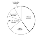
図4は、ユーザ端末20のディスプレイ20Bに表示される推奨条件/コストベネフィットの提案画面の一例である。ここでは、クラウド画像処理部36において自動実行される画像処理である「CAD(1)」、「CAD(2)」、「セグメンテーション(1)」「ラベリング(1)」、及び「ラベリング(2)」のそれぞれの最新の一定期間の費用の割合を円グラフで示しており、円グラフにはそれぞれの費用額が表示されている。
図4は、ユーザ端末20のディスプレイ20Bに表示される推奨条件/コストベネフィットの提案画面の一例である。ここでは、クラウド画像処理部36において自動実行される画像処理である「CAD(1)」、「CAD(2)」、「セグメンテーション(1)」「ラベリング(1)」、及び「ラベリング(2)」のそれぞれの最新の一定期間の費用の割合を円グラフで示しており、円グラフにはそれぞれの費用額が表示されている。
円グラフのそれぞれの領域は入力装置20Aにより選択可能となっている。ここでは、太枠で囲まれた「CAD(1)」が選択中であることを示している。
図5は、ディスプレイ20Bに表示された図4の円グラフにおいて「CAD(1)」が選択された場合に表示される、「CAD(1)」の自動実行条件の表示画面の一例である。本実施形態では、自動実行条件はスライス厚、及びスライス枚数であり、自動実行条件の表示は、スライス厚の自動実行条件を表すスクロールバー100と、スライス枚数の自動実行条件を表すスクロールバー110と、を含む。図4の円グラフ、及び図5のスライダーバーは、同じ画面に同時に表示されてもよい。
スクロールバー100は、スライス厚が0.1mmから5mmまでの範囲を示している。スクロールバー100の上部に表示される三角形のマーク102は、現在設定されている自動実行条件の下限の値を示している。したがって、マーク102の位置より右側(マーク102の示す値より大きいスライス厚)の画像が自動実行の対象となっている画像である。また、星形のマーク104は、推奨実行条件/コストベネフィット決定処理部42によって推奨される自動実行条件の下限の値を示しており、マーク104の位置より右側の画像が推奨される実行条件の対象となる画像である。
例えば、現在設定されているスライス厚の自動実行条件は0.5mm以上であり、推奨される自動実行条件は2mm以上である。なお、自動実行条件として、スライス厚の上限の値を設定できてもよい。
また、スクロールバー100は、スクロールバー100の範囲を移動可能なノブ106を有する。ユーザは、入力装置20Aによりノブ106をドラッグ操作してノブ106の位置を変更することで、自動実行条件を変更することができる。ここでは、ノブ106の位置より右側の画像が自動実行画像処理の対象となる画像となる。
例えば、ノブ106が0.5mmの位置から2mmの位置に移動されると、スライス厚の自動実行条件は0.5mm以上から2mm以上に変更される。この自動実行条件の変更により、その後はスライス厚が0.5mm~2mmの断層画像は自動実行されないことになり、従量制のクラウドサービスではその分だけ費用が安くなることが予想される。推奨実行条件/コストベネフィット決定処理部42は、「CAD(1)」のスライス厚の自動実行条件が0.5mm以上から2mm以上に変更された場合の「CAD(1)」の最新の一定期間の費用を再計算し、図4に示した円グラフに反映させる。
一方、スクロールバー110は、スライス枚数が10枚から1000枚までの範囲を示している。スクロールバー110の上部に表示される三角形のマーク112は、現在設定されている自動実行条件の下限の値を示している。したがって、マーク112の位置より右側(マーク112の示す値より多いスライス枚数)の画像が自動実行の対象となっている画像である。また、星形のマーク114は、推奨実行条件/コストベネフィット決定処理部42によって推奨される自動実行条件の下限の値を示しており、マーク114の位置より右側の画像が推奨される実行条件の対象となる画像である。
例えば、現在設定されているスライス枚数の自動実行条件は100枚以上であり、推奨される自動実行条件は200枚以上である。なお、自動実行条件として、スライス枚数の上限の値を設定できてもよい。
また、スクロールバー110は、スクロールバー110の範囲を移動可能なノブ116を有する。ユーザは、入力装置20Aによりノブ116をドラッグ操作してノブ116の位置を変更することで、自動実行条件を変更することができる。ここでは、ノブ116の位置より右側の画像が自動実行画像処理の対象となる画像となる。
例えば、ノブ116が100枚の位置から200枚の位置に移動されると、スライス枚数の自動実行条件は100枚以上から200枚以上に変更される。推奨実行条件/コストベネフィット決定処理部42は、「CAD(1)」のスライス枚数の自動実行条件が100枚以上から200枚以上に変更された場合の「CAD(1)」の最新の一定期間の費用を再計算し、図4に示した円グラフに反映させる。
このように、ユーザは自動実行条件を変更した場合のコストベネフィットを確認することができる。
ここでは、「CAD(1)」の自動実行条件の変更について説明したが、自動実行条件は、画像処理ごとに設定可能である。本実施形態では、「CAD(1)」の他に、「CAD(2)」、「セグメンテーション(1)」「ラベリング(1)」、及び「ラベリング(2)」の自動実行条件をそれぞれ設定することができる。
図4に示した画面において、自動実行条件管理部34は、自動実行する画像処理の削除を入力装置20Aから受け付けてもよい。また、自動実行条件管理部34は、自動実行する画像処理の追加を入力装置20Aから受け付けてもよい。例えば、ユーザは、入力装置20Aを用いて類似画像検索、及び類似症例検索の処理を自動実行する画像処理に追加することができる。
〔評価値の計算〕
推奨実行条件/コストベネフィット決定処理部42は、外的要因を考慮して自動実行条件を提案してもよい。外的要因は、画像処理が対象とする疾患の季節性を含む。推奨実行条件/コストベネフィット決定処理部42は、例えば以下の式を用いて画像処理の外的要因を考慮した評価値を計算する。
推奨実行条件/コストベネフィット決定処理部42は、外的要因を考慮して自動実行条件を提案してもよい。外的要因は、画像処理が対象とする疾患の季節性を含む。推奨実行条件/コストベネフィット決定処理部42は、例えば以下の式を用いて画像処理の外的要因を考慮した評価値を計算する。
評価値a1=(使用数)÷(処理数)
評価値a2=(見逃し防止数)÷(処理数)
評価値a3=(画像処理の季節性評価)×(季節係数)
最終評価値a4=a1+a2+a3
ここで、処理数はクラウド画像処理部36において画像処理が行われた症例撮影数であり、使用数はユーザがユーザ端末20において実行結果を確認した数(ディスプレイ20Bに表示された数)であり、見逃し防止数はユーザが見逃したが画像処理によって発見された病変の数である。見逃し防止数は、例えばユーザが画像処理の実行結果を用いずに診断を行い、診断後に実行結果をディスプレイ20Bに表示させ、表示された病変のうちユーザの診断に含まれない病変の数をカウントする。
評価値a2=(見逃し防止数)÷(処理数)
評価値a3=(画像処理の季節性評価)×(季節係数)
最終評価値a4=a1+a2+a3
ここで、処理数はクラウド画像処理部36において画像処理が行われた症例撮影数であり、使用数はユーザがユーザ端末20において実行結果を確認した数(ディスプレイ20Bに表示された数)であり、見逃し防止数はユーザが見逃したが画像処理によって発見された病変の数である。見逃し防止数は、例えばユーザが画像処理の実行結果を用いずに診断を行い、診断後に実行結果をディスプレイ20Bに表示させ、表示された病変のうちユーザの診断に含まれない病変の数をカウントする。
画像処理の季節性評価は、画像処理が対象とする疾患の発生の季節性の有無を示す値である。季節係数は、画像処理が対象とする疾患の発生が相対的に多い季節ほど相対的に大きい値となる係数である。例えば、冬に相対的に多く発生する疾患の場合は、その疾患の画像処理の季節係数は、冬ほど相対的に大きい値となる。
最終評価値a4があらかじめ定められた閾値以上であればその画像処理は自動実行画像処理として維持し、閾値未満であればその画像処理を自動実行画像処理から削除することを提案する。なお、評価値a1、a2、及びa3のうちのいずれか1つが閾値以上であればその画像処理を自動実行画像処理として維持してもよい。
評価値は、医療経済指標(QALY:Quality-adjusted life year)の計算機能を用いてもよい。
〔時間短縮/見逃し防止以外の評価項目〕
推奨実行条件/コストベネフィット決定処理部42は、利用情報として計測精度の向上等、その他の評価項目を考慮して自動実行条件として設定、又は削除を提案してもよい。
推奨実行条件/コストベネフィット決定処理部42は、利用情報として計測精度の向上等、その他の評価項目を考慮して自動実行条件として設定、又は削除を提案してもよい。
例えば、「医師が、画像処理の結果を確認せずに病変の体積をX1[cm3]と判断し、クラウド画像処理部36によって実行された画像処理の体積計測の結果がX1[cm3]とは異なるX2[cm3]であった」とする。
これに対し、「クラウド画像処理部36によって実行された画像処理の体積計測の結果を確認して医師が読影レポートを修正した」というログを利用情報として処理結果使用ログ管理部40が保存した場合、推奨実行条件/コストベネフィット決定処理部42は、「画像処理の体積計測の方がその医師よりも優れており、計測精度の向上に貢献した」と判断し、その体積計測を行った画像処理を自動実行画像処理の設定として推奨する。
一方、「クラウド画像処理部36によって実行された画像処理の体積計測の結果を確認しても医師は読影レポートを修正しなかった」というログを利用情報として処理結果使用ログ管理部40が保存した場合、「医師の体積計測の方がその画像処理よりも優れており、精度の向上に貢献しなかった」と判断し、その体積計測を行った画像処理を自動実行画像処理の設定から削除することを推奨する。
このように、体積計測等の計測精度の向上に貢献した画像処理を自動実行画像処理として推奨することで、読影の計測精度を向上させることができる。計測精度の向上に限定されず、推奨実行条件/コストベネフィット決定処理部42は、医用画像の読影に関する各評価項目の貢献度に応じて自動実行条件として設定、又は削除を提案することができる。
〔利用情報及び付随情報の関連付けた表示〕
医用画像処理システム10は、収集した利用情報と付随情報とを関連付けてディスプレイ20Bに表示させてもよい(「提示工程」の一例)。
医用画像処理システム10は、収集した利用情報と付随情報とを関連付けてディスプレイ20Bに表示させてもよい(「提示工程」の一例)。
図6は、ディスプレイ20Bに表示される、処理結果使用ログ管理部40が蓄積したログの集計結果画面の一例である。この例では、「CAD(1)」のログについて付随情報ごとの利用情報を度数折れ線グラフで示しており、具体的にはスライス厚ごとの処理数[単位:枚]、使用数[単位:枚]、及び見逃し防止数[単位:箇所]を示している。
スライス厚が0.1~1mm(0.1mm以上かつ1mm未満)の場合は、処理数が1000枚であるのに対し、使用数が0枚であり、したがって見逃し防止数も(母集団が0であるため)0箇所である。スライス厚が1~3mm(1mm以上かつ3mm未満)の場合は、処理数が2000枚、使用数が1600枚、見逃し防止数が2000箇所である。スライス厚が3~5mm(3mm以上かつ5mm未満)の場合は、処理数が1000枚、使用数が0枚、見逃し防止数が0箇所である。スライス厚が5mm~(5mm以上)の場合は、処理数が2000枚、使用数が1800枚、見逃し防止数が1000箇所である。
このグラフから、スライス厚が0.1~1mmの場合、及び3~5mmの場合は、画像処理の結果が使用されていないため、自動実行する必要性がなく、自動実行条件として適切でないと考えられる。一方、スライス厚が1~3mmの場合、及び5mm以上の場合は、見逃し防止に役立っており、自動実行条件として適切であることがわかる。
ここでは、スライス厚ごとの処理数、使用数、及び見逃し防止数を示したが、スライス枚数についても同様のログの集計結果を表示させてもよい。
医用画像処理システム10は、このようなログの集計結果をディスプレイ20Bに表示させることで、ユーザは自動実行画像処理の自動実行条件に適した条件を確認することができる。また、クラウドサービスの導入コスト、及びクラウドサービスに最初に契約すべきプランの選択に使用することも可能である。
なお、この例の場合では、推奨実行条件/コストベネフィット決定処理部42は、ログの集計結果の表示だけでなく、自動実行条件の推奨条件としてスライス厚が1~3mmの場合、及び5mm以上の場合を提示してもよい。
上記の実施形態には、以下の(1)~(11)が記載されている。
(1)医用画像処理装置の一の態様は、少なくとも1つのプロセッサと、少なくとも1つのプロセッサに実行させるための命令を記憶する少なくとも1つのメモリと、を備え、少なくとも1つのプロセッサは、医用画像の診断を支援する第1の画像処理であって、入力された医用画像に対して自動実行される自動実行画像処理として設定可能な第1の画像処理の結果のユーザによる利用情報と、第1の画像処理の対象となった医用画像の付随情報と、を収集し、利用情報と付随情報とを関連付けてディスプレイに表示させる、医用画像処理装置である。すなわち、少なくとも1つのプロセッサと、少なくとも1つのプロセッサに実行させるための命令を記憶する少なくとも1つのメモリと、を備え、少なくとも1つのプロセッサは、医用画像の診断を支援する第1の画像処理であり、かつ、入力された医用画像に対して自動実行される自動実行画像処理として設定可能な第1の画像処理であって、ユーザによる第1の画像処理の結果の利用状況を示す利用情報と、第1の画像処理の対象となった医用画像の付随情報と、を収集し、利用情報と付随情報とを関連付けてディスプレイに表示させる、医用画像処理装置である。本態様によれば、自動実行される自動実行画像処理として設定可能な画像処理の利用状況を提示することができるので、ユーザは提示された利用状況から画像処理を自動実行画像処理として設定するか否かの判断をすることができる。
(2)利用情報は、第1の画像処理の結果をユーザが参照したか否か、第1の画像処理の結果が疾患の見逃し防止に貢献したか否か、第1の画像処理の結果による読影時間の削減量、第1の画像処理の計測結果が精度向上に貢献したか否か、及び自動実行以外にユーザが第1の画像処理の実行を指示したか否か、のうちの少なくとも1つを含むことが好ましい。これにより、画像処理の貢献度を反映した利用情報を収集することができる。
(3)第1の画像処理の対象は断層画像であり、付随情報は、スライス厚とスライス枚数との少なくとも一方を含むことが好ましい。これにより、断層画像のスライス厚とスライス枚数との少なくとも一方を収集することができる。
(4)少なくとも1つのプロセッサは、自動実行画像処理の設定から第1の画像処理を削除、及び自動実行画像処理として第1の画像処理を設定、のうちの少なくとも一方を受け付けることが好ましい。これにより、自動実行画像処理として不要な第1の画像処理の削除、及び必要な第1の画像処理の設定を行うことができる。
(5)少なくとも1つのプロセッサは、利用情報と付随情報とに基づいて、第1の画像処理が自動実行されるための自動実行条件を提案することが好ましい。これにより、ユーザは適切な自動実行条件を知ることができる。
(6)少なくとも1つのプロセッサは、外的要因を考慮して自動実行条件を提案することが好ましい。これにより、適切な自動実行条件を提案することができる。
(7)また、外的要因は、第1の画像処理が対象とする疾患の季節性を含むことが好ましい。これにより、適切な自動実行条件を提案することができる。
(8)少なくとも1つのプロセッサは、自動実行画像処理に設定された画像処理を実行することが好ましい。これにより、設定された画像処理を自動実行することができる。
(9)医用画像処理システムの一の態様は、(1)~(8)の医用画像処理装置と、自動実行画像処理として設定された画像処理を実行するサーバと、を備える医用画像処理システムである。本態様によれば、自動実行される自動実行画像処理として設定可能な画像処理の利用状況を提示することができるので、ユーザは提示された利用状況から画像処理を自動実行画像処理として設定するか否かの判断をすることができ、自動実行画像処理として設定した画像処理を自動実行させることができる。
(10)医用画像処理方法の一の態様は、医用画像の診断を支援する第1の画像処理であって、入力された医用画像に対して自動実行される自動実行画像処理として設定可能な第1の画像処理の結果のユーザによる利用情報と、第1の画像処理の対象となった医用画像の付随情報と、を収集し、利用情報と付随情報とを関連付けてディスプレイに表示させる医用画像処理方法である。すなわち、医用画像処理方法の一の態様は、医用画像の診断を支援する第1の画像処理であり、かつ、入力された医用画像に対して自動実行される自動実行画像処理として設定可能な第1の画像処理であって、ユーザによる第1の画像処理の結果の利用状況を示す利用情報と、第1の画像処理の対象となった医用画像の付随情報と、を収集し、利用情報と付随情報とを関連付けてディスプレイに表示させる医用画像処理方法である。
本態様によれば、自動実行される自動実行画像処理として設定可能な画像処理の利用状況を提示することができるので、ユーザは提示された利用状況から画像処理を自動実行画像処理として設定するか否かの判断をすることができる。
(11)上記目的を達成するためのプログラムの一の態様は、上記の医用画像処理方法をコンピュータに実行させるためのプログラムである。このプログラムが記録された、コンピュータが読み取り可能な非一時的記憶媒体も本態様に含んでよい。本態様によれば、自動実行される自動実行画像処理として設定可能な画像処理の利用状況を提示することができるので、ユーザは提示された利用状況から画像処理を自動実行画像処理として設定するか否かの判断をすることができる。
(1)医用画像処理装置の一の態様は、少なくとも1つのプロセッサと、少なくとも1つのプロセッサに実行させるための命令を記憶する少なくとも1つのメモリと、を備え、少なくとも1つのプロセッサは、医用画像の診断を支援する第1の画像処理であって、入力された医用画像に対して自動実行される自動実行画像処理として設定可能な第1の画像処理の結果のユーザによる利用情報と、第1の画像処理の対象となった医用画像の付随情報と、を収集し、利用情報と付随情報とを関連付けてディスプレイに表示させる、医用画像処理装置である。すなわち、少なくとも1つのプロセッサと、少なくとも1つのプロセッサに実行させるための命令を記憶する少なくとも1つのメモリと、を備え、少なくとも1つのプロセッサは、医用画像の診断を支援する第1の画像処理であり、かつ、入力された医用画像に対して自動実行される自動実行画像処理として設定可能な第1の画像処理であって、ユーザによる第1の画像処理の結果の利用状況を示す利用情報と、第1の画像処理の対象となった医用画像の付随情報と、を収集し、利用情報と付随情報とを関連付けてディスプレイに表示させる、医用画像処理装置である。本態様によれば、自動実行される自動実行画像処理として設定可能な画像処理の利用状況を提示することができるので、ユーザは提示された利用状況から画像処理を自動実行画像処理として設定するか否かの判断をすることができる。
(2)利用情報は、第1の画像処理の結果をユーザが参照したか否か、第1の画像処理の結果が疾患の見逃し防止に貢献したか否か、第1の画像処理の結果による読影時間の削減量、第1の画像処理の計測結果が精度向上に貢献したか否か、及び自動実行以外にユーザが第1の画像処理の実行を指示したか否か、のうちの少なくとも1つを含むことが好ましい。これにより、画像処理の貢献度を反映した利用情報を収集することができる。
(3)第1の画像処理の対象は断層画像であり、付随情報は、スライス厚とスライス枚数との少なくとも一方を含むことが好ましい。これにより、断層画像のスライス厚とスライス枚数との少なくとも一方を収集することができる。
(4)少なくとも1つのプロセッサは、自動実行画像処理の設定から第1の画像処理を削除、及び自動実行画像処理として第1の画像処理を設定、のうちの少なくとも一方を受け付けることが好ましい。これにより、自動実行画像処理として不要な第1の画像処理の削除、及び必要な第1の画像処理の設定を行うことができる。
(5)少なくとも1つのプロセッサは、利用情報と付随情報とに基づいて、第1の画像処理が自動実行されるための自動実行条件を提案することが好ましい。これにより、ユーザは適切な自動実行条件を知ることができる。
(6)少なくとも1つのプロセッサは、外的要因を考慮して自動実行条件を提案することが好ましい。これにより、適切な自動実行条件を提案することができる。
(7)また、外的要因は、第1の画像処理が対象とする疾患の季節性を含むことが好ましい。これにより、適切な自動実行条件を提案することができる。
(8)少なくとも1つのプロセッサは、自動実行画像処理に設定された画像処理を実行することが好ましい。これにより、設定された画像処理を自動実行することができる。
(9)医用画像処理システムの一の態様は、(1)~(8)の医用画像処理装置と、自動実行画像処理として設定された画像処理を実行するサーバと、を備える医用画像処理システムである。本態様によれば、自動実行される自動実行画像処理として設定可能な画像処理の利用状況を提示することができるので、ユーザは提示された利用状況から画像処理を自動実行画像処理として設定するか否かの判断をすることができ、自動実行画像処理として設定した画像処理を自動実行させることができる。
(10)医用画像処理方法の一の態様は、医用画像の診断を支援する第1の画像処理であって、入力された医用画像に対して自動実行される自動実行画像処理として設定可能な第1の画像処理の結果のユーザによる利用情報と、第1の画像処理の対象となった医用画像の付随情報と、を収集し、利用情報と付随情報とを関連付けてディスプレイに表示させる医用画像処理方法である。すなわち、医用画像処理方法の一の態様は、医用画像の診断を支援する第1の画像処理であり、かつ、入力された医用画像に対して自動実行される自動実行画像処理として設定可能な第1の画像処理であって、ユーザによる第1の画像処理の結果の利用状況を示す利用情報と、第1の画像処理の対象となった医用画像の付随情報と、を収集し、利用情報と付随情報とを関連付けてディスプレイに表示させる医用画像処理方法である。
本態様によれば、自動実行される自動実行画像処理として設定可能な画像処理の利用状況を提示することができるので、ユーザは提示された利用状況から画像処理を自動実行画像処理として設定するか否かの判断をすることができる。
(11)上記目的を達成するためのプログラムの一の態様は、上記の医用画像処理方法をコンピュータに実行させるためのプログラムである。このプログラムが記録された、コンピュータが読み取り可能な非一時的記憶媒体も本態様に含んでよい。本態様によれば、自動実行される自動実行画像処理として設定可能な画像処理の利用状況を提示することができるので、ユーザは提示された利用状況から画像処理を自動実行画像処理として設定するか否かの判断をすることができる。
〔その他〕
本発明の技術的範囲は、上記の実施形態に記載の範囲には限定されない。各実施形態における構成等は、本発明の趣旨を逸脱しない範囲で、各実施形態間で適宜組み合わせることができる。
本発明の技術的範囲は、上記の実施形態に記載の範囲には限定されない。各実施形態における構成等は、本発明の趣旨を逸脱しない範囲で、各実施形態間で適宜組み合わせることができる。
10…医用画像処理システム
12…医用画像検査機器
14…医用画像データベース
16…画像処理評価装置
16A…プロセッサ
16B…メモリ
20…ユーザ端末
20A…入力装置
20B…ディスプレイ
22…病院内ネットワーク
24…インターネット
26…クラウドサーバ
30…画像管理部
32…画像処理決定処理部
34…自動実行条件管理部
36…クラウド画像処理部
38…処理結果管理部
40…処理結果使用ログ管理部
42…コストベネフィット決定処理部
100…スクロールバー
102…マーク
104…マーク
106…ノブ
110…スクロールバー
112…マーク
114…マーク
116…ノブ
P1~P11…医用画像処理装置のプロセス
S1~S11…医用画像処理方法のステップ
12…医用画像検査機器
14…医用画像データベース
16…画像処理評価装置
16A…プロセッサ
16B…メモリ
20…ユーザ端末
20A…入力装置
20B…ディスプレイ
22…病院内ネットワーク
24…インターネット
26…クラウドサーバ
30…画像管理部
32…画像処理決定処理部
34…自動実行条件管理部
36…クラウド画像処理部
38…処理結果管理部
40…処理結果使用ログ管理部
42…コストベネフィット決定処理部
100…スクロールバー
102…マーク
104…マーク
106…ノブ
110…スクロールバー
112…マーク
114…マーク
116…ノブ
P1~P11…医用画像処理装置のプロセス
S1~S11…医用画像処理方法のステップ
Claims (11)
- 少なくとも1つのプロセッサと、
前記少なくとも1つのプロセッサに実行させるための命令を記憶する少なくとも1つのメモリと、
を備え、
前記少なくとも1つのプロセッサは、
医用画像の診断を支援する第1の画像処理であって、入力された前記医用画像に対して自動実行される自動実行画像処理として設定可能な前記第1の画像処理の結果のユーザによる利用情報と、前記第1の画像処理の対象となった医用画像の付随情報と、を収集し、
前記利用情報と前記付随情報とを関連付けてディスプレイに表示させる、
医用画像処理装置。 - 前記利用情報は、前記第1の画像処理の結果をユーザが参照したか否か、前記第1の画像処理の結果が疾患の見逃し防止に貢献したか否か、前記第1の画像処理の結果による読影時間の削減量、第1の画像処理の計測結果が精度向上に貢献したか否か、及び前記自動実行以外にユーザが前記第1の画像処理の実行を指示したか否か、のうちの少なくとも1つを含む、
請求項1に記載の医用画像処理装置。 - 前記第1の画像処理の対象は断層画像であり、
前記付随情報は、スライス厚とスライス枚数との少なくとも一方を含む、
請求項1又は2に記載の医用画像処理装置。 - 前記少なくとも1つのプロセッサは、
前記自動実行画像処理の設定から前記第1の画像処理を削除、及び前記自動実行画像処理として前記第1の画像処理を設定、のうちの少なくとも一方を受け付ける、
請求項1から3のいずれか1項に記載の医用画像処理装置。 - 前記少なくとも1つのプロセッサは、
前記利用情報と前記付随情報とに基づいて、前記第1の画像処理が前記自動実行されるための自動実行条件を提案する、
請求項1から4のいずれか1項に記載の医用画像処理装置。 - 前記少なくとも1つのプロセッサは、
外的要因を考慮して前記自動実行条件を提案する、
請求項5に記載の医用画像処理装置。 - 前記外的要因は、前記第1の画像処理が対象とする疾患の季節性を含む、
請求項6に記載の医用画像処理装置。 - 前記少なくとも1つのプロセッサは、
前記自動実行画像処理に設定された画像処理を実行する、
請求項1から7のいずれか1項に記載の医用画像処理装置。 - 請求項1から7のいずれか1項に記載の医用画像処理装置と、
前記自動実行画像処理として設定された画像処理を実行するサーバと、
を備える医用画像処理システム。 - 医用画像の診断を支援する第1の画像処理であって、入力された前記医用画像に対して自動実行される自動実行画像処理として設定可能な前記第1の画像処理の結果のユーザによる利用情報と、前記第1の画像処理の対象となった医用画像の付随情報と、を収集し、
前記利用情報と前記付随情報とを関連付けてディスプレイに表示させる、
医用画像処理方法。 - 請求項10に記載の医用画像処理方法をコンピュータに実行させるためのプログラム。
Priority Applications (3)
| Application Number | Priority Date | Filing Date | Title |
|---|---|---|---|
| DE112022000895.4T DE112022000895T5 (de) | 2021-03-24 | 2022-02-18 | Verarbeitungsvorrichtung für medizinische bilder, verarbeitungssystem für medizinische bilder, verarbeitungsverfahren für medizinische bilder und programm |
| JP2023508802A JP7712353B2 (ja) | 2021-03-24 | 2022-02-18 | 医用画像処理装置、医用画像処理システム、医用画像処理方法及びプログラム |
| US18/460,579 US20230410994A1 (en) | 2021-03-24 | 2023-09-03 | Medical image processing device, medical image processing system, medical image processing method, and program |
Applications Claiming Priority (2)
| Application Number | Priority Date | Filing Date | Title |
|---|---|---|---|
| JP2021050336 | 2021-03-24 | ||
| JP2021-050336 | 2021-03-24 |
Related Child Applications (1)
| Application Number | Title | Priority Date | Filing Date |
|---|---|---|---|
| US18/460,579 Continuation US20230410994A1 (en) | 2021-03-24 | 2023-09-03 | Medical image processing device, medical image processing system, medical image processing method, and program |
Publications (1)
| Publication Number | Publication Date |
|---|---|
| WO2022202024A1 true WO2022202024A1 (ja) | 2022-09-29 |
Family
ID=83396984
Family Applications (1)
| Application Number | Title | Priority Date | Filing Date |
|---|---|---|---|
| PCT/JP2022/006750 Ceased WO2022202024A1 (ja) | 2021-03-24 | 2022-02-18 | 医用画像処理装置、医用画像処理システム、医用画像処理方法及びプログラム |
Country Status (4)
| Country | Link |
|---|---|
| US (1) | US20230410994A1 (ja) |
| JP (1) | JP7712353B2 (ja) |
| DE (1) | DE112022000895T5 (ja) |
| WO (1) | WO2022202024A1 (ja) |
Citations (3)
| Publication number | Priority date | Publication date | Assignee | Title |
|---|---|---|---|---|
| JP2004049615A (ja) * | 2002-07-22 | 2004-02-19 | Hitachi Medical Corp | 医用画像診断装置の撮影条件設定装置 |
| JP2014193193A (ja) * | 2013-03-28 | 2014-10-09 | Toshiba Corp | 条件設定支援装置 |
| JP6230811B2 (ja) * | 2013-04-26 | 2017-11-15 | 東芝メディカルシステムズ株式会社 | 磁気共鳴イメージング装置 |
Family Cites Families (3)
| Publication number | Priority date | Publication date | Assignee | Title |
|---|---|---|---|---|
| IN2014MN01458A (ja) * | 2011-12-23 | 2015-04-17 | Materialise Nv | |
| US10025479B2 (en) * | 2013-09-25 | 2018-07-17 | Terarecon, Inc. | Advanced medical image processing wizard |
| JP7106314B2 (ja) * | 2018-03-19 | 2022-07-26 | キヤノンメディカルシステムズ株式会社 | X線コンピュータ断層撮影装置、及び画像生成装置 |
-
2022
- 2022-02-18 JP JP2023508802A patent/JP7712353B2/ja active Active
- 2022-02-18 DE DE112022000895.4T patent/DE112022000895T5/de active Pending
- 2022-02-18 WO PCT/JP2022/006750 patent/WO2022202024A1/ja not_active Ceased
-
2023
- 2023-09-03 US US18/460,579 patent/US20230410994A1/en active Pending
Patent Citations (3)
| Publication number | Priority date | Publication date | Assignee | Title |
|---|---|---|---|---|
| JP2004049615A (ja) * | 2002-07-22 | 2004-02-19 | Hitachi Medical Corp | 医用画像診断装置の撮影条件設定装置 |
| JP2014193193A (ja) * | 2013-03-28 | 2014-10-09 | Toshiba Corp | 条件設定支援装置 |
| JP6230811B2 (ja) * | 2013-04-26 | 2017-11-15 | 東芝メディカルシステムズ株式会社 | 磁気共鳴イメージング装置 |
Also Published As
| Publication number | Publication date |
|---|---|
| US20230410994A1 (en) | 2023-12-21 |
| JP7712353B2 (ja) | 2025-07-23 |
| DE112022000895T5 (de) | 2023-11-16 |
| JPWO2022202024A1 (ja) | 2022-09-29 |
Similar Documents
| Publication | Publication Date | Title |
|---|---|---|
| Lin et al. | Efficacy of a checklist-style structured radiology reporting template in reducing resident misses on cervical spine computed tomography examinations | |
| US20200342990A1 (en) | Medical image processing apparatus, method, and program, and diagnosis support apparatus, method, and program | |
| US11779295B2 (en) | Method and system of vertebral compression fracture detection | |
| US20090087049A1 (en) | Medical reporting system, apparatus and method | |
| US20170154162A1 (en) | Systems and methods for managing adverse reactions in contrast media-based medical procedures | |
| US8200507B2 (en) | Examination information management apparatus | |
| DE202020006022U1 (de) | Systeme zur Automatisierung von Entscheidungen im klinischen Arbeitsablauf und zur Erzeugung eines Prioritätsauswertungsdikators | |
| US11468659B2 (en) | Learning support device, learning support method, learning support program, region-of-interest discrimination device, region-of-interest discrimination method, region-of-interest discrimination program, and learned model | |
| Vano | ICRP recommendations on ‘Managing patient dose in digital radiology’ | |
| US11669960B2 (en) | Learning system, method, and program | |
| US20050108060A1 (en) | Medical image information management system | |
| Damaskos et al. | Extra-and intra-cranial arterial calcifications in adults depicted as incidental findings on cone beam CT images | |
| US20160253468A1 (en) | Measurement value management apparatus, method for operating measurement value management apparatus, and measurement value management system | |
| JP2008200373A (ja) | 類似症例検索装置、方法、およびプログラム、ならびに、類似症例データベース登録装置、方法、およびプログラム | |
| Ma et al. | Risk factors for root resorption of second molars with impacted third molars: a meta-analysis of CBCT studies | |
| US20200020434A1 (en) | Information collection processing apparatus, information collection processing method, and recording medium | |
| Newell Jr | Quantitative computed tomography of lung parenchyma in chronic obstructive pulmonary disease: an overview | |
| WO2014155918A1 (ja) | グラフ表示装置およびグラフ表示装置の作動方法並びにグラフ表示プログラム | |
| Zhong et al. | Trends in incidence of pneumothorax in England before, during and after the COVID-19 pandemic (2017–2023): a population-based observational study | |
| US20130254187A1 (en) | Device, method, and non-transitory computer-readable medium for searching database | |
| Kristjánsdóttir et al. | Ultra-low dose computed tomography of the chest in an emergency setting: a prospective agreement study | |
| US20090125555A1 (en) | Method and apparatus for flexible archiving | |
| JP7712353B2 (ja) | 医用画像処理装置、医用画像処理システム、医用画像処理方法及びプログラム | |
| Runde et al. | Computed tomography utilization rates after the placement of a scanner in an emergency department: a single-center experience | |
| Menoch et al. | Imaging for suspected appendicitis: variation between academic and private practice models |
Legal Events
| Date | Code | Title | Description |
|---|---|---|---|
| 121 | Ep: the epo has been informed by wipo that ep was designated in this application |
Ref document number: 22774824 Country of ref document: EP Kind code of ref document: A1 |
|
| WWE | Wipo information: entry into national phase |
Ref document number: 2023508802 Country of ref document: JP |
|
| WWE | Wipo information: entry into national phase |
Ref document number: 112022000895 Country of ref document: DE |
|
| 122 | Ep: pct application non-entry in european phase |
Ref document number: 22774824 Country of ref document: EP Kind code of ref document: A1 |