WO2022180911A1 - 血球貪食症候群の治療に用いる細胞調製物 - Google Patents
血球貪食症候群の治療に用いる細胞調製物 Download PDFInfo
- Publication number
- WO2022180911A1 WO2022180911A1 PCT/JP2021/035958 JP2021035958W WO2022180911A1 WO 2022180911 A1 WO2022180911 A1 WO 2022180911A1 JP 2021035958 W JP2021035958 W JP 2021035958W WO 2022180911 A1 WO2022180911 A1 WO 2022180911A1
- Authority
- WO
- WIPO (PCT)
- Prior art keywords
- umbilical cord
- cells
- cell preparation
- derived cells
- derived
- Prior art date
- Legal status (The legal status is an assumption and is not a legal conclusion. Google has not performed a legal analysis and makes no representation as to the accuracy of the status listed.)
- Ceased
Links
Images






Classifications
-
- A—HUMAN NECESSITIES
- A61—MEDICAL OR VETERINARY SCIENCE; HYGIENE
- A61K—PREPARATIONS FOR MEDICAL, DENTAL OR TOILETRY PURPOSES
- A61K35/00—Medicinal preparations containing materials or reaction products thereof with undetermined constitution
- A61K35/12—Materials from mammals; Compositions comprising non-specified tissues or cells; Compositions comprising non-embryonic stem cells; Genetically modified cells
- A61K35/28—Bone marrow; Haematopoietic stem cells; Mesenchymal stem cells of any origin, e.g. adipose-derived stem cells
-
- A—HUMAN NECESSITIES
- A61—MEDICAL OR VETERINARY SCIENCE; HYGIENE
- A61K—PREPARATIONS FOR MEDICAL, DENTAL OR TOILETRY PURPOSES
- A61K35/00—Medicinal preparations containing materials or reaction products thereof with undetermined constitution
- A61K35/12—Materials from mammals; Compositions comprising non-specified tissues or cells; Compositions comprising non-embryonic stem cells; Genetically modified cells
- A61K35/48—Reproductive organs
- A61K35/51—Umbilical cord; Umbilical cord blood; Umbilical stem cells
-
- A—HUMAN NECESSITIES
- A61—MEDICAL OR VETERINARY SCIENCE; HYGIENE
- A61P—SPECIFIC THERAPEUTIC ACTIVITY OF CHEMICAL COMPOUNDS OR MEDICINAL PREPARATIONS
- A61P7/00—Drugs for disorders of the blood or the extracellular fluid
-
- A—HUMAN NECESSITIES
- A61—MEDICAL OR VETERINARY SCIENCE; HYGIENE
- A61P—SPECIFIC THERAPEUTIC ACTIVITY OF CHEMICAL COMPOUNDS OR MEDICINAL PREPARATIONS
- A61P7/00—Drugs for disorders of the blood or the extracellular fluid
- A61P7/06—Antianaemics
-
- C—CHEMISTRY; METALLURGY
- C12—BIOCHEMISTRY; BEER; SPIRITS; WINE; VINEGAR; MICROBIOLOGY; ENZYMOLOGY; MUTATION OR GENETIC ENGINEERING
- C12N—MICROORGANISMS OR ENZYMES; COMPOSITIONS THEREOF; PROPAGATING, PRESERVING, OR MAINTAINING MICROORGANISMS; MUTATION OR GENETIC ENGINEERING; CULTURE MEDIA
- C12N5/00—Undifferentiated human, animal or plant cells, e.g. cell lines; Tissues; Cultivation or maintenance thereof; Culture media therefor
- C12N5/06—Animal cells or tissues; Human cells or tissues
Definitions
- the present invention relates to cell preparations used for treating hemophagocytic syndrome.
- HPS Hemophagocytic syndrome
- HHLH hemophagocytic lymphohistiocytosis
- HPS is a disease characterized by hemophagocytosis by histiocytes and macrophages in the reticuloendothelial system such as bone marrow and lymph nodes.
- HPS is roughly classified into primary (primary) and secondary (reactive) caused by an underlying disease (Non-Patent Document 1).
- HPS patients with HPS present with symptoms such as hyperthermia, hepatosplenomegaly (enlarged hepatospleen and enlarged spleen), hypertriglyceridemia, hemophagocytosis in the bone marrow, spleen or lymph nodes, and hyperferritinemia.
- combination therapy with steroids such as dexamethasone and anti-inflammatory agents such as cyclosporine is performed, but there are patients who repeatedly relapse, etc., and patients who are steroid-refractory, and there is still no effective treatment method. . For this reason, HPS is designated as an intractable disease.
- the present invention aims to provide a cell preparation that can treat hemophagocytic syndrome.
- the present invention provides a cell preparation (hereinafter also referred to as "cell preparation") used for treatment of hemophagocytic syndrome,
- the cell preparation contains umbilical cord-derived cells.
- hemophagocytic syndrome can be treated.
- FIG. 1 is a diagram showing the method for preparing an HPS model and the phenotypes of HPS model mice in Example 1.
- FIG. 2 is a photograph showing staining of the liver of the HPS model mouse in Example 1.
- FIG. 3 is a photograph showing staining of the blood of the HPS model mouse in Example 1.
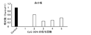
- FIG. 4 is a graph showing blood platelet counts of HPS model mice in Example 1.
- FIG. 5 is a diagram showing administration conditions for HPS model mice, liver staining diagrams, and the number of macrophages in Example 2.
- FIG. 6 is a graph showing the number of migrated umbilical cord-derived cells in the MLR in Example 3.
- FIG. 7 is a graph showing the number of migrated umbilical cord-derived cells in the MLR in Example 3.
- FIG. 1 is a diagram showing the method for preparing an HPS model and the phenotypes of HPS model mice in Example 1.
- FIG. 2 is a photograph showing staining of the liver of the HPS model mouse
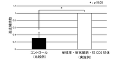
- FIG. 8 is a graph showing the number of migrated cells in Example 3.
- FIG. 9 is a graph showing the number of migrated umbilical cord-derived cells when FBS was added in Example 3.
- FIG. 10 is a graph showing the concentration of MCP-1 in Example 3.
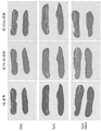
- FIG. 11 is a photograph showing a stained image of spleen in Example 4.
- FIG. 12 is a graph showing the area ratio of white pulp of the spleen in Example 4.
- hemophagocytic syndrome is a disease characterized by hemophagocytosis by histiocytes/macrophages in the reticuloendothelial system such as bone marrow and lymph nodes, and includes fever, lymphadenopathy, hepatosplenomegaly, pancytopenia, coagulation It means a disease presenting various symptoms such as abnormality, liver dysfunction, hyperLDHemia, hypertriglyceridemia, and/or hyperferritinemia. Said HPS can also be referred to, for example, as hemophagocytic lymphohistiocytosis.
- the "hemophagocytic syndrome” may be, for example, primary (primary) hemophagocytic syndrome or secondary (reactive) HPS.
- the primary HPS includes familial hemophagocytic lymphohistiocytosis (FHL).
- the secondary HPS is HPS that develops as a result of infection, malignant disease, or autoimmune disease, for example, virus-associated hemophagocytic syndrome (VAHS), Bacteria-associated hemophagocytic syndrome (BAHS), Infection-associated hemophagocytic syndrome (IAHS), including hemophagocytic syndrome caused by fungi or parasites; Lymphoma-associated syndrome Malignancy-associated hemophagocytic syndrome (MAHS) such as hemophagocytic syndrome (LAHS); Autoimmune-associated hemophagocytic syndrome (AAHS) and the like.
- VAHS virus-associated hemophagocytic syndrome
- BAHS Bacteria-associated hemophagocytic syndrome
- IAHS Infection-associated hemophagocytic syndrome
- LAHS hemophagocytic syndrome
- AAHS Autoimmune-associated hemophagocytic syndrome
- HPS includes HPS that occurs after hematopoietic stem cell transplantation, and in this case, hematopoietic stem cell engraftment failure may occur.
- the HPS is diagnosed, for example, according to the following diagnostic criteria.
- HPS diagnostic criteria Revised Diagnostic Guidelines for HLH
- HPS HLH if either 1 or 2 below is satisfied. 1.
- PRF1, MUNC13-4, STX11 genetic abnormality
- treatment means suppression, prevention, deterrence, prophylaxis, or delay of onset of a target disease, stopping, suppression, suppression, or delay of progression of a target disease that has developed or its symptoms, and amelioration or Any meaning of remission may be used.
- the “treatment” may be, for example, treatment of a patient who develops the target disease, or treatment of a model animal of the target disease. Said therapy may also be referred to as treatment.
- a "cell preparation” means a cell population containing desired cells or a composition containing desired cells. Therefore, in the present invention, a cell preparation can also be referred to as, for example, a cell population or composition.
- the ratio of desired cells to all cells also referred to as “purity”
- the purity is, for example, the percentage in viable cells. The purity can be measured, for example, by flow cytometry, immunohistochemistry, in situ hybridization, and the like.
- the purity of the cell preparation is, for example, 50% or higher, 55% or higher, 60% or higher, 65% or higher, 70% or higher, 75% or higher, 80% or higher, 85% or higher, 90% or higher, 91% or more, 92% or more, 93% or more, 94% or more, 95% or more, 96% or more, 97% or more, 98% or more, or 99% or more.
- the "umbilical cord” is a white tubular tissue that connects the fetus and the placenta, and means tissue that does not contain the placenta and cord blood.
- the origin of the "umbilical cord” is not particularly limited. , an umbilical cord of a primate mammal, more preferably a human umbilical cord.
- macrophage means, for example, cells with phagocytic ability derived from CD14-positive CD11b-positive monocytes.
- the macrophages are also generally defined as large cells produced in the bone marrow, monocytes that entered the peripheral blood and exited to extravascular tissue.
- the macrophages take in foreign substances such as bacteria, viruses, and dead cells (phagocytosis), and the phagosomes are fused with lysosomes and decomposed, presenting them as antigens, and releasing interferon and various cytokines to help T cells.
- the macrophages may be called by different names depending on the organ or tissue to which they belong, in the liver they may be called Kupffer cells, in the lung they may be called alveolar macrophages, and in the brain they may be glial cells or microglia.
- the present invention provides cell preparations that can be used to treat hemophagocytic syndrome.
- the present invention provides a cell preparation for use in treating hemophagocytic syndrome, wherein the cell preparation comprises umbilical cord-derived cells.
- the cell preparation of the present invention is characterized by containing umbilical cord-derived cells, and other configurations and conditions are not particularly limited.
- the umbilical cord-derived cells are presumed to suppress the symptoms of hemophagocytic syndrome by suppressing accumulation of macrophages in tissues. Therefore, the cell preparation of the present invention can treat hemophagocytic syndrome by containing the umbilical cord-derived cells.
- the "umbilical cord” may be an umbilical cord collected from an administration, treatment, or treatment subject (hereinafter collectively referred to as "administration subject"), or an umbilical cord collected from a subject other than the administration subject. may be It is preferable to use an umbilical cord collected from a person other than the subject of administration from the viewpoint of not being subject to restrictions during preparation.
- the umbilical cord-derived cells of the present invention may be umbilical cord-derived cells (autologous cells) collected from an administration subject, or umbilical cord-derived cells collected from a non-administration subject, as shown in Examples described later. (allogeneic cells). It has been confirmed that the umbilical cord-derived cells have therapeutic effects without being eliminated by immune rejection or the like, for example.
- the umbilical cord can be collected by appropriately removing the placenta from, for example, postpartum tissue containing the placenta and/or umbilical cord delivered by vaginal delivery or cesarean section.
- the umbilical cord may be obtained by removing umbilical cord blood from the collected umbilical cord, or may be subjected to aseptic or bacteriostatic treatment. Removal of the cord blood can be performed, for example, by rinsing or perfusion with a solution containing an anticoagulant such as heparin.
- Said aseptic or bactericidal treatment is not particularly limited, for example, application of disinfectants such as povidone-iodine; addition of antibiotics such as penicillin, streptomycin, amphotericin B, gentamicin, and/or nystatin, and/or antimycotics immersion in a medium or buffer;
- the umbilical cord may also selectively lyse red blood cells, for example, if desired.
- methods well known in the art can be used, such as incubation in hypertonic or hypotonic medium by lysis with ammonium chloride.
- the "umbilical cord-derived cells” mean a cell population prepared using umbilical cord as a raw material.
- the "umbilical cord-derived cells” may be composed of a single type of cell, or may be composed of a plurality of types of cells.
- the umbilical cord-derived cells preferably contain umbilical cord-derived mesenchymal cells.
- the umbilical cord-derived cells may be a cell population composed of multiple types of cells including the umbilical cord-derived mesenchymal cells.
- the umbilical cord-derived cells of the present invention may be, for example, a cell population having one or more of the following characteristics (a) to (c), preferably a cell population having all characteristics.
- (a) exhibits adhesion to plastic in culture in the presence of medium;
- (b) CD105, CD73, CD90, CD44, HLA-classI, HLA-G5 and PD-L (Programmed cell death 1 ligand) 2 positive, CD45, CD34, CD11b, CD19 and HLA-ClassII negative ;
- (c) expression of IDO (indoleamine 2,3-dioxygenase), PGE 2 (prostaglandin E2) and PD-L1 genes and/or proteins is induced under inflammatory conditions;
- negative means that a signal or the like equivalent to or lower than that in a negative control reaction using negative control cells that do not express the antigen or an antibody that does not react with the antigen is detected. do.
- the "HLA-class I” means HLA-A, B, or C.
- the "HLA-Class II” means HLA-DR, DQ, or DP.
- the "under inflammatory conditions” refers to conditions in which inflammatory cytokines such as interferon ⁇ are contacted or added.
- the umbilical cord-derived cells may be extracts and/or secretions of the umbilical cord-derived cells.
- the umbilical cord-derived cell extract can be obtained by, for example, subjecting the umbilical cord-derived cells to concentration treatment, centrifugation treatment, drying treatment, freeze-drying treatment, solvent treatment, surfactant treatment, protease, glycolytic enzyme, or the like. processed products obtained by enzymatic treatment, protein extraction treatment, ultrasonic treatment and/or grinding treatment, or processed products obtained by a combination of these treatments, and the like.
- the secretions of the umbilical cord-derived cells include exosomes and cell culture supernatants of umbilical cord-derived cells.
- the cell preparation preferably exhibits an action of suppressing accumulation of macrophages in tissues.
- Said macrophages are, for example, F4/80-positive macrophages.
- Examples of the tissue include lung, liver, and the like.
- the action of suppressing the accumulation of macrophages can be evaluated using, for example, the number of macrophages per unit area in the tissue as an index according to Example 1 described later.
- Said accumulation means, for example, that the desired cells gather and accumulate in or around a particular tissue, organ, or organ.
- the accumulation can also be referred to as aggregation, accumulation, or the like, for example.
- the cell preparation reduces the number of macrophages accumulating in the tissue by 5% or more, 10% or more, 15% or more, 20% or more, 30% or more, compared with a group to which the cell preparation is not administered, 35% or more, 40% or more, 45% or more, 50% or more, 55% or more, 60% or more, 65% or more, 70% or more, 75% or more, 80% or more, 85% or more, 90% or more, 95% 96% or more, 97% or more, 98% or more, or 99% or more.
- the method for producing (preparing) the umbilical cord-derived cells includes, for example, a step of isolating cells from the umbilical cord, and optionally a step of subculturing the isolated cells.
- the preparation method includes, for example, (1) cutting the umbilical cord, (2) culturing the umbilical cord segment, and (3) subculturing.
- the preparation method includes, for example, (A) a step of cutting the umbilical cord, a step of enzymatic treatment, or a step of dissociating the tissue by both, (B) a step of culturing the umbilical cord tissue, and ( C) including the step of passaging.
- the umbilical cord-derived cells may be a uniform cell population or a heterogeneous cell population.
- the umbilical cord-derived cells are prepared by the method including the steps (1) to (3) or the method including the steps (A) to (C), an example can be carried out as follows.
- the method for preparing the umbilical cord-derived cells is not limited to the following example.
- the method including the steps (1) to (3) will be described.
- the umbilical cord obtained by the above method is subjected to mechanical force (shredding force or shear force) in a state containing amniotic membrane, blood vessels, perivascular tissue and/or Walton's jelly. It can be implemented by cutting.
- the size of the umbilical cord segment obtained by cutting is not particularly limited, and examples thereof include 1 to 10 mm 3 , 1 to 5 mm 3 , 1 to 4 mm 3 , 1 to 3 mm 3 and 1 to 2 mm 3 .
- the cut umbilical cord segment is seeded in an incubator such as a Petri dish, dish, or flask, and cultured in a culture medium suitable for umbilical cord-derived cells. .
- the umbilical cord segment is not treated with a digestive enzyme.
- the "incubator” may be, for example, an incubator having a solid surface.
- an incubator used for culturing cells, tissues, and/or organs can be used.
- Said "solid surface” means, for example, any material capable of binding with said umbilical cord-derived cells.
- the material includes, for example, a plastic material that has been treated (eg, hydrophilicity-enhancing treatment) to promote binding of mammalian cells to its surface.
- the type of culture vessel having the solid surface is not particularly limited, and examples thereof include petri dishes, dishes, flasks, and the like.
- the "culture solution suitable for umbilical cord-derived cells" can be prepared, for example, by adding additives such as serum to the basal medium.
- additives are, for example, serum and/or one or more of albumin, transferrin, fatty acids, insulin, sodium selenite, cholesterol, collagen precursors, trace elements, 2-mercaptoethanol, 3′-thiolglycerol, etc. of serum replacement.
- the culture solution further contains lipids, amino acids, proteins, polysaccharides, vitamins, growth factors, low-molecular-weight compounds, antibiotics, antifungal agents, antioxidants, pyruvic acid, buffers, and inorganic salts. You may add substances, such as.
- the basal medium is not particularly limited.
- Ham's F10 Medium F10
- Ham's F-12 Medium F12
- Iscove's Modified Dulbecco's (IMDM) Medium Fischer's Medium
- Mesenchymal Stem Cell Growth Medium MSCGM
- DMEM/F12 RPMI 1640
- CELL-GRO-FREE CELL-GRO-FREE
- serum examples include animal-derived serum such as human serum, fetal bovine serum (FBS), bovine serum, calf serum, goat serum, horse serum, pig serum, sheep serum, rabbit serum and rat serum.
- the amount of serum added to the basal medium is, for example, 5 v/v % to 15 v/v %, preferably about 10 v/v %.
- the fatty acid is not particularly limited, and examples thereof include linoleic acid, oleic acid, linoleic acid, arachidonic acid, myristic acid, palmitoic acid, palmitic acid, and stearic acid.
- the lipid is not particularly limited, and examples thereof include phosphatidylserine, phosphatidylethanolamine, phosphatidylcholine and the like.
- the amino acid is not particularly limited, and examples thereof include amino acids such as L-alanine, L-arginine, L-aspartic acid, L-asparagine, L-cysteine, L-cystine, L-glutamic acid, L-glutamine, and L-glycine.
- the protein is not particularly limited, and examples thereof include ecotin, reduced glutathione, fibronectin, ⁇ 2-microglobulin and the like.
- the polysaccharide is not particularly limited, and examples thereof include glycosaminoglycans such as hyaluronic acid and heparan sulfate.
- the growth factor is not particularly limited, and examples include platelet-derived growth factor (PDGF), epidermal growth factor (EGF), fibroblast growth factor (FGF), vascular endothelial growth factor (VEGF), insulin-like growth factor-1 (IGF-1), leukocyte inhibitory factor (LIF), basic fibroblast growth factor (bFGF), transforming growth factor beta (TGF- ⁇ ), hepatocyte growth factor (HGF), connective tissue growth factor (CTGF) , erythropoietin (EPO) and the like.
- the antibiotic and/or antifungal agent is not particularly limited, and examples thereof include penicillin G, streptomycin sulfate, amphotericin B, gentamicin, nystatin, and mixtures thereof.
- step (2) in order to prevent the seeded umbilical cord segments from floating in the culture medium, it is preferable to hold down the umbilical cord segments using a plate or the like during the culture period.
- the plate include plates described in JP-A-2015-70824.
- culture conditions are not particularly limited, and for example, general culture conditions for cells, tissues, organs, etc. can be referred to.
- the CO 2 concentration in the step (2) is, for example, 0 to 5%.
- the O 2 concentration in the step (2) is, for example, 2 to 25%, preferably 5 to 20%.
- the culture temperature in the step (2) is, for example, 25 to 40°C, preferably about 37°C (35 to 39°C).
- the culture period is not particularly limited. It is preferable to culture until the cells become confluent.
- the cells are washed and treated with a solution containing a chelating agent such as EDTA, trypsin, collagenase, dispase, or the like. protease, sugar chain degrading enzyme such as hyaluronidase, or a stripping agent containing a mixture thereof.
- a chelating agent such as EDTA, trypsin, collagenase, dispase, or the like.
- protease sugar chain degrading enzyme such as hyaluronidase, or a stripping agent containing a mixture thereof.
- the step (2) for example, by filtering the detachment solution containing the cells and the umbilical cord segment using a cell strainer or the like, only cells can be obtained as umbilical cord-derived cells.
- the obtained umbilical cord-derived cells can be seeded, for example, in the culture vessel described above and cultured using the culture medium described above.
- the umbilical cord-derived cells can be appropriately grown to the required number by subculturing.
- the cells in the subculture, the cells may be detached with the detachment agent, seeded at an appropriate cell density in a separately prepared culture vessel, and culture continued.
- the cell density (seeding density) when seeding the cells is, for example, 1 ⁇ 10 2 to 1 ⁇ 10 5 cells/cm 2 , 5 ⁇ 10 2 to 5 ⁇ 10 4 cells/cm 2 , 1 ⁇ 10 3 1 ⁇ 10 4 cells/cm 2 , 2 ⁇ 10 3 to 1 ⁇ 10 4 cells/cm 2 and the like, preferably 2 ⁇ 10 3 to 1 ⁇ 10 4 cells/cm 2 .
- the seeding density is preferably adjusted, for example, so that the period to reach a suitable confluency is 3 to 7 days.
- the medium may be changed as appropriate, if necessary.
- the number of passages in the step (3) is not particularly limited, and may be performed, for example, until senescence, when cell division stops.
- the subculture is preferably 3 to 25 times, more preferably 4 to 12 times.
- the umbilical cord obtained by the method described above is enzymatically treated in a state containing amniotic membrane, blood vessels, perivascular tissue and/or Walton's Jelly to dissociate the tissues.
- the enzyme used for the enzymatic treatment is not particularly limited, and examples thereof include proteases such as collagenase and dispase; glycolytic enzymes such as hyaluronidase; and the like.
- step of (B) culturing the umbilical cord tissue and the step of (C) subculturing are, for example, the same as the step of (2) culturing the umbilical cord tissue and the step of (3) subculturing, respectively. can be implemented as
- the umbilical cord-derived cells can be obtained after the step (3) or (C).
- the cells obtained by the method for preparing umbilical cord-derived cells are umbilical cord-derived cells
- surface antigens and the like may be analyzed by conventional methods using flow cytometry and the like.
- whether or not the cells obtained by the method for preparing umbilical cord-derived cells are umbilical cord-derived cells may be evaluated by measuring the amounts of various proteins produced from the cells.
- the cells obtained by the method for preparing umbilical cord-derived cells may be prepared for treatment as they are, or may be cryopreserved.
- the cryopreservation is performed, for example, by suspending the cells in a cryopreservation solution capable of preserving the umbilical cord-derived cells and storing the cells at -80°C to -180°C.
- the cryopreservation solution is not particularly limited, and examples thereof include an aqueous solution containing a cryoprotectant and glucose.
- the antifreeze agent examples include dimethyl sulfoxide (hereinafter also referred to as "DMSO"), dextran, glycerol, propylene glycol, and 1-methyl-2-pyrrolidone, preferably DMSO and/or propylene glycol. Yes, more preferably DMSO.
- the cryoprotectant is contained, for example, in the cryopreservation solution at 1 to 15 w/v%, preferably 5 to 15 w/v%, more preferably 5 to 12 w/v%, More preferably, the content is 8 to 11 w/v%.
- Glucose contained in the cryopreservation solution is, for example, 0.5 to 10 w/v%, preferably 1 to 10 w/v%, more preferably 2 to 10 w/v% in the cryopreservation solution. 8 w/v %, more preferably 2 to 5 w/v %.
- the cryopreservation solution may further contain other components.
- Other components include, for example, pH adjusters and thickeners.
- the pH adjuster include sodium hydrogen carbonate, HEPES, and phosphate buffer.
- BSS basic stock solution
- a phosphate buffer is preferably used as the pH adjuster.
- the pH adjuster is preferably used to adjust the pH in the cryopreservation solution to, for example, about 6.5-9, preferably 7-8.5.
- the "phosphate buffer” is, for example, sodium chloride, monosodium phosphate (anhydrous), monopotassium phosphate (anhydrous), disodium phosphate (anhydrous), trisodium phosphate (anhydrous ), potassium chloride, and potassium dihydrogen phosphate (anhydrous), etc., especially containing sodium chloride, monosodium phosphate (anhydrous), potassium chloride, or potassium dihydrogen phosphate (anhydrous) Buffers are preferred.
- the pH adjuster is contained, for example, in the cryopreservation solution at 0.01 to 1 w/v%, preferably 0.05 to 0.5 w/v%.
- the cryopreservation solution may or may not contain natural animal-derived components.
- the natural animal-derived components include the aforementioned serum and basal medium.
- the cryopreservation solution does not contain natural animal-derived components.
- the cryopreservation solution that does not contain the natural animal-derived components does not cause quality differences between lots of natural animal-derived components, and components such as various cytokines, growth factors, and hormones contained in serum. It is possible to suppress the possibility of changes in the properties of the cells in the umbilical cord tissue caused by , and also to suppress the influence of components of unknown origin contained in the basal medium. Therefore, cryopreservation solutions free of said natural animal-derived components are very useful, especially in clinical use.
- the cryopreservation solution may further contain a thickening agent.
- the thickening agent is not particularly limited, and examples thereof include those capable of constituting a cryopreservation solution capable of sufficiently preserving the umbilical cord tissue.
- the thickener include carboxymethyl cellulose (hereinafter also referred to as "CMC"), carboxymethyl cellulose sodium (hereinafter also referred to as "CMC-Na”), organic acid polymers, propylene glycol alginate, sodium alginate and the like.
- CMC and CMC-Na are preferred, and CMC-Na is particularly preferred.
- the organic acid polymer is preferably sodium polyacrylate.
- the thickening agent is contained, for example, in the cryopreservation solution at 0.1 to 1 w/v%, preferably 0.1 to 0.5 w/v%, more preferably 0.2 Contains ⁇ 0.4 w/v%.
- the cryopreservation solution is preferably an aqueous solution.
- the osmotic pressure of the cryopreservation solution is, for example, preferably 1000 mOsm or more, more preferably 1000 to 2700 mOsm, in order to maintain performance as a preservation solution.
- the cryopreservation solution is preferably an aqueous solution that contains a thickener, a cryoprotectant, and glucose, and does not contain natural animal-derived components.
- the cryopreservation solution is more preferably an aqueous solution containing CMC-Na, DMSO, and glucose and free of natural animal-derived components.
- the cryopreservation solution more preferably contains 0.1 to 1 w/v% of CMC-Na, 1.0 to 15 w/v% of DMSO, and 0.5 to 10 w/v% of glucose, It is an aqueous solution that does not contain natural animal-derived components.
- the cells obtained by the method for preparing umbilical cord-derived cells may be used as cell preparations for various applications, for example, by mixing with infusion preparations.
- the cryopreserved umbilical cord-derived cells may be suspended in the cryopreservation solution, and after thawing, may be used directly as a cell preparation for various purposes, or may be used after thawing. It may be mixed with an infusion formulation and the resulting mixture used as a cell preparation for various uses.
- the culture medium or cryopreservation solution in which the umbilical cord-derived cells are suspended may be mixed with the infusion preparation or the like, and the culture medium or cryopreservation solution may be separated by centrifugation or the like. After separating the cells from the solvent, the cells alone may be mixed with the infusion formulation.
- the preparation method for example, in order to avoid the complexity of the procedure, the step of culturing the frozen cells after thawing is not included, or the cryopreservation solution in which the thawed cells are suspended is directly used as an infusion preparation. Mixing is preferred.
- the above-mentioned “infusion preparation” includes, for example, solutions such as infusion solutions used in the treatment of humans. No. 1 liquid, No. 2 liquid, No. 3 liquid, No. 4 liquid, and the like.
- the cell preparation of the present invention may be a kit containing the infusion preparation in addition to the umbilical cord-derived cells.
- the cell preparation of the present invention may contain a pharmaceutically acceptable carrier in addition to or instead of the infusion preparation.
- the carrier includes suspending agents, solubilizers, stabilizers, tonicity agents, preservatives, antiadsorption agents, surfactants, diluents, vehicles, pH adjusters, Examples include soothing agents, buffering agents, sulfur-containing reducing agents, antioxidants, and the like, and can be appropriately added within a range that does not impair the effects of the present invention.
- the suspending agent is not particularly limited, and examples thereof include methylcellulose, polysorbate 80, hydroxyethylcellulose, gum arabic (gum arabic), tragacanth powder, carboxymethylcellulose sodium, polyoxyethylene sorbitan monolaurate, and the like.
- the solution adjuvant is not particularly limited, and examples thereof include polyoxyethylene hydrogenated castor oil, polysorbate 80, nicotinic acid amide, polyoxyethylene sorbitan monolaurate, macrogol, castor oil fatty acid ethyl ester, and the like.
- the stabilizer is not particularly limited, and examples include dextran 40, methylcellulose, gelatin, sodium sulfite, sodium metasulfate, and the like.
- the tonicity agent is not particularly limited, and examples thereof include D-mannitol and sorbitol.
- the preservative is not particularly limited, and examples thereof include methyl paraoxybenzoate, ethyl parahydroxybenzoate, sorbic acid, phenol, cresol, and chlorocresol.
- the antiadsorption agent is not particularly limited, and examples thereof include human serum albumin, lecithin, dextran, ethylene oxide propylene oxide copolymer, hydroxypropyl cellulose, methyl cellulose, hydrogenated castor oil, and polyethylene glycol.
- the sulfur-containing reducing agent is not particularly limited. Those having a sulfhydryl group such as sodium sulfate, glutathione, and thioalkanoic acids having 1 to 7 carbon atoms are included.
- the antioxidant is not particularly limited, and examples thereof include erythorbic acid, dibutylhydroxytoluene, butylhydroxyanisole, ⁇ -tocopherol, tocopherol acetate, L-ascorbic acid and its salts, L-ascorbic acid palmitate, L-ascorbic acid stear. sodium bisulfite, sodium sulfite, triamyl gallate, propyl gallate or sodium ethylenediaminetetraacetate (EDTA), sodium pyrophosphate, sodium metaphosphate and the like.
- erythorbic acid dibutylhydroxytoluene, butylhydroxyanisole, ⁇ -tocopherol, tocopherol acetate, L-ascorbic acid and its salts, L-ascorbic acid palmitate, L-ascorbic acid stear.
- the cell preparations of the present invention may further contain inorganic salts such as sodium chloride, potassium chloride, calcium chloride, sodium phosphate, potassium phosphate, sodium bicarbonate; organic salts such as sodium citrate, potassium citrate, sodium acetate; Saccharides such as glucose; and other commonly added components may be added as appropriate.
- inorganic salts such as sodium chloride, potassium chloride, calcium chloride, sodium phosphate, potassium phosphate, sodium bicarbonate
- organic salts such as sodium citrate, potassium citrate, sodium acetate
- Saccharides such as glucose
- an anticoagulant and/or pH adjuster for example, ACD-A solution (composition of sodium citrate hydrate, citric acid hydrate, glucose, etc.) may be added.
- the cell preparations of the present invention may be mixed, for example, for topical administration with organic substances such as biopolymers; inorganic substances such as hydroxyapatite; It may be mixed with glycol polymers or copolymers and chemical derivatives thereof.
- a cell preparation of the invention may be used, for example, in vitro or in vivo .
- the cell preparations of the invention can be used, for example, as research reagents and as pharmaceuticals.
- the administration target of the cell preparation of the present invention is not particularly limited.
- the administration subject includes, for example, humans and non-human animals other than humans.
- non-human animals include mammals such as mice, rats, rabbits, dogs, cats, cows, horses, pigs, monkeys, dolphins, and sea lions; birds; fish;
- the subject of administration includes, for example, cells, tissues, organs, and the like, and the cells include, for example, cells collected from living organisms, cultured cells, and the like.
- the tissue or organ includes, for example, a tissue (living tissue) or organ collected from a living body. Examples of the cells include muscle cells, iPS cells (induced pluripotent stem cells), stem cells, and the like.
- the administration subject includes, for example, a subject diagnosed as being consistent with hereditary HLH, a subject diagnosed with HPS, a subject suspected of having HPS, and the like.
- the hereditary HLH is, for example, HPS caused by perforin gene (FHL2), MUNC13-4 gene (FHL3), syntaxin11 gene (FHL4), Munc18-2 gene (FHL5), and the like.
- the human perforin gene is, for example, a gene registered under Gene ID: 5551 in the NCBI gene database (https://www.ncbi.nlm.nih.gov/gene/).
- the human MUNC13-4 gene is, for example, a gene registered under Gene ID: 201294 in NCBI.
- the human syntaxin11 gene is, for example, a gene registered under Gene ID: 8676 in NCBI.
- the human Munc18-2 gene is, for example, a gene registered under Gene ID: 6813 in NCBI.
- Reference 1 below can be referred to for the diagnosis of primary HPS among the HPS.
- a comprehensive diagnosis of adult HPS among the above HPS can be found, for example, in Reference 3 below.
- the usage conditions (administration conditions) of the cell preparation of the present invention are not particularly limited, and for example, the dosage form, administration period, dosage, etc. can be appropriately set according to the type of administration subject.
- Examples of administration methods of the cell preparation of the present invention include intracerebral administration, intrathecal administration, intramuscular administration, subcutaneous administration, and intravenous administration. Intravenous administration is preferred because it can be administered rapidly and stably.
- the dosage of the cell preparation of the present invention is such that, when administered to a subject, the therapeutic effect of HPS or the effect of suppressing the accumulation of macrophages in tissues against disease can be obtained as compared to subjects who have not been administered cells.
- amount therapeutically effective amount
- the dose can be appropriately determined according to, for example, the age, body weight, symptoms, etc. of the subject.
- the dose is, for example, 10 4 to 10 9 cells/kg body weight, 10 4 to 10 8 cells/kg body weight, 10 4 to 10 7 cells/kg as the number of umbilical cord-derived cells per administration. body weight, preferably 10 4 to 10 8 cells/kg body weight, 10 4 to 10 7 cells/kg body weight.
- the administration frequency of the cell preparation of the present invention is one or more times.
- the plurality of times is, for example, 2 times, 3 times, 4 times, 5 times or more.
- the frequency of administration may be appropriately determined while confirming the therapeutic effect of the subject.
- the administration interval can be appropriately determined while confirming the therapeutic effect of the subject, for example, once a day, once a week, once every two weeks, once a month, once every three months. , once every six months, etc.
- the cell preparation of the present invention may be used in combination with, for example, other agents and/or methods used for HPS.
- drugs used for HPS include steroids such as dexamethasone; immunosuppressants such as cyclosporine; anticancer agents such as etoposide (VP-16); gamma globulin preparations such as immunoglobulin;
- the cell preparation of the present invention may be used in combination with hematopoietic stem cell transplantation.
- the cell preparation of the present invention can treat hemophagocytic syndrome, as described above.
- the present invention may include methods of treating patients with hemophagocytic syndrome.
- the present invention is a method of treating a patient with hemophagocytic syndrome, wherein the subject uses the cell preparation of the present invention.
- the method of treating a hemophagocytic syndrome patient of the present invention comprises, for example, administering the cell preparation of the present invention to the subject.
- the above description can be used for the administration conditions in the administration step.
- the present invention provides a cell preparation used for suppressing accumulation of macrophages in tissue in hemophagocytic syndrome or a method for suppressing accumulation of macrophages in tissue in hemophagocytic syndrome.
- the present invention is a cell preparation used for suppressing accumulation of macrophages in tissues in hemophagocytic syndrome, wherein the cell preparation contains umbilical cord-derived cells.
- the present invention also provides a method for suppressing accumulation of macrophages in tissues of a subject with hemophagocytic syndrome, wherein the cell preparation used for suppressing accumulation of macrophages in tissues in hemophagocytic syndrome of the present invention is used as the subject.
- the present invention can refer to the description of the cell preparation of the present invention.
- the present invention is a cell preparation for use in treating hemophagocytic syndrome and/or suppressing the accumulation of macrophages in tissues in hemophagocytic syndrome, wherein the cell preparation contains umbilical cord-derived cells.
- the present invention is the use of umbilical cord-derived cells for the manufacture of cell preparations for use in treating hemophagocytic syndrome and/or for inhibiting accumulation of macrophages in tissues in hemophagocytic syndrome.
- the present invention can refer to the description of the cell preparation of the present invention.
- Example 1 A mouse model of hemophagocytic syndrome was produced, and it was confirmed that the cell preparation of the present invention could suppress accumulation of macrophages in tissues in hemophagocytic syndrome.
- HPS model mice are TLR9 ligands for the albino strain of B6 strain background mice (B6N-Tyr c-Brd /BrdCrCrl, manufactured by Charles River Japan), unmethylated CpG It was produced by administering ODN.
- the unmethylated CpG was induced by administering ODN 1826 (5′-tccatgacgttcctgacgtt-3′: SEQ ID NO: 1), which is a class B CpG oligonucleotide. Specifically, as shown in FIG.
- FIG. 1(B) shows photographs of the spleen of each mouse, the upper row is the spleen derived from the control mouse, and the lower row is the spleen of the CpG ODN-administered mouse.
- FIG. 1(C) is a graph showing the relative weight of the spleen of each mouse.
- the horizontal axis indicates the dose and the number of times of administration
- the vertical axis indicates the relative weight.
- FIGS. 1(B) and (C) when CpG ODN was administered, spleen hypertrophy was observed at any number of times, and the weight increased compared to the control. Therefore, it was found that administration of CpG ODN, which is a TLR9 ligand, to albino strains causes symptoms similar to splenomegaly, which is one of the symptoms of HPS.
- tissue sections were stained by hematoxylin-eosin staining (HE staining). That is, after staining the nuclei with a Harris Hematoxylin solution for 10 minutes, the cells were washed with water, and then the cytoplasm was stained with an Eosin solution. The resulting tissue sections were then dehydrated with alcohol and dried. After drying, a cover glass was sealed on the slide glass with balsam. Then, the tissue section after staining was observed with an optical microscope.
- Fig. 2 is a photograph showing a tissue section.
- the results of the control five times administration
- the results of the mice to which the CpG ODN was administered twice the results of the mice to which the CpG ODN was administered five times are shown.
- no formation of vacuolar structures in the liver and no accumulation of fibrin and leukocytes were observed in controls.
- mice administered CpG ODN showed formation of vacuolar structures in the liver and accumulation of fibrin and leukocytes. The same was true for mice to which CpG ODN was administered 1, 3, or 4 times.
- CpG ODN which is a TLR9 ligand
- Fig. 3 is a photograph showing a staining diagram of blood. As shown in FIG. 3, almost no reticulocytes were found in the blood of the control. On the other hand, mice receiving 5 doses of CpG ODN showed a large number of reticulocytes in the blood. The same was true for mice to which CpG ODN was administered 1 to 4 times.
- FIG. 4 is a graph showing the platelet count. As shown in FIG. 4, in mice to which CpG ODN was administered 1 to 5 times, platelet counts in the blood were significantly decreased compared to controls.
- the albino strain can be used as an HPS model mouse by administering CpG ODN.
- the CpG ODN is composed of DNA
- the HPS of Example 1 can be suitably used as a model of HPS (VAHS) caused by DNA viruses such as EB virus.
- Umbilical cord-derived cells were collected by the method described in Cytotherapy, 18, 229-241, 2016. Specifically, with the approval of the ethics committee of the Institute of Medical Science, the University of Tokyo, all tissue elements of the umbilical cord (amniotic membrane, blood vessels, perivascular tissue, and Walton's jelly) collected with the consent of the donor. ) were chopped into 1-2 mm 3 pieces and seeded onto culture dishes. Then, the umbilical cord-derived cells were cultured in ⁇ -minimal essential medium ( ⁇ MEM) supplemented with 10% fetal bovine serum (FBS) and antibiotics after covering with cell amigo (manufactured by Tsubakimoto Chain Co., Ltd.). got The properties of the cells are plastic-adherent.
- ⁇ MEM ⁇ -minimal essential medium
- FBS fetal bovine serum
- the umbilical cord-derived cells highly express the HGF (Hepatic Growth Factor) gene under normal conditions, and under inflammatory conditions (IFN- ⁇ 100 ng/ml) produce IDO (Indoleamine 2,3-dioxygenase). Induction of gene expression was confirmed by Realtime PCR. In particular, HGF expression was found to be higher in umbilical cord-derived cells than in bone marrow-derived mesenchymal stem cells. In addition, it was confirmed by ELISA that secretion of PGE2 was induced by co-culturing umbilical cord-derived cells with MLR (mixed allogeneic lymphocyte reaction).
- HGF Hepatic Growth Factor
- CpG ODN or PBS was administered on day 0, 3, 7, 10 or 14, and umbilical cord-derived cells (MSC) (150 ⁇ l) or umbilical cord-derived cells
- MSC umbilical cord-derived cells
- DBA-D cryoprotectant in cryopreservation
- the liver was embedded in paraffin, and the resulting paraffin-embedded tissue pieces were sliced to prepare tissue sections.
- the resulting tissue sections were stained with anti-mouse F4/80 antibody and then conjugated with sheep anti-mouse IgG HRP. Then, the tissue sections were stained for F4/80-positive macrophages by peroxidase staining. Then, the tissue section after staining was observed with an optical microscope. Then, after randomly capturing 10 fields of view with a 20x objective lens (200x magnification) over the entire tissue section, 10 photographs obtained (unit area 6.74 mm 2 ⁇ 10 fields of view) The number of positive macrophages was counted. These results are shown in FIGS. 5(B) and (C).
- FIG. 5(B) shows a photograph of the liver of each mouse.
- the black area in the figure indicates the area where macrophages are present.
- FIG. 5(C) is a graph showing the number of F4/80-positive macrophages in the liver of each mouse.
- the horizontal axis indicates the dose
- the vertical axis indicates the number of macrophages.
- umbilical cord-derived cells can suppress accumulation of hepatic macrophages in HPS model mice.
- HPS causes accumulation of macrophages in the liver, followed by phagocytosis of blood cells, resulting in a decrease in blood cells.
- umbilical cord-derived cells can suppress accumulation of macrophages in the liver, it is thought that subsequent hemophagocytosis can also be suppressed. From the above, it was found that the cell preparation of the present invention can treat hemophagocytic syndrome.
- Umbilical cord-derived cells were prepared in the same manner as in Example 1(2) above.
- human marrow-derived mesenchymal cells (BM-MSC, manufactured by LONZA)
- human adipose tissue-derived mesenchymal stem cells AD-MSC, manufactured by LONZA
- MLR Mixed Lymphocyte Reaction Mixed lymphocyte reaction
- an insert was placed in each well, and umbilical cord-derived cells were seeded in the insert at a concentration of 5 ⁇ 10 3 cells/200 ⁇ l/well. Then, the cells were cultured for 17 hours at 37° C., 5% CO 2 in a humid environment. After the culture, the insert was removed, the medium was removed, fixed with 10% formalin, and washed with PBS. After washing, the cells were stained with 4',6-diamidino-2-phenylindole (DAPI: manufactured by PromoCell GmbH) to stain the nuclei of umbilical cord-derived cells that had migrated to the back side (bottom side) of the insert.
- DAPI 4',6-diamidino-2-phenylindole
- FIG. 6 is a graph showing the number of migrated umbilical cord-derived cells in the MLR.
- the horizontal axis indicates the type of sample, and the vertical axis indicates the relative number of migrated cells.
- the number of migrating cells of umbilical cord-derived cells increased, that is, they migrated in the direction of inflammation.
- each inhibitor is added to the bottom well of Example 2 (2) at a predetermined concentration (AG1296: 0, 2.5, 5, or 10 ⁇ mol / l, PPP: 0, 0.5, 1, or 2 ⁇ mol/l, GM6001: 0, 10, 20, or 40 ⁇ mol/l), but the number of migrated cells was counted in the same manner except that each inhibitor was added. Then, the relative value of the number of migrated cells was calculated by setting the number of migrated cells in the sample with a concentration of each inhibitor of 0 ⁇ mol/l as 1.
- FIG. 7 is a graph showing the number of migrated umbilical cord-derived cells in the MLR.
- the horizontal axis indicates the type and concentration of the inhibitor, and the vertical axis indicates the relative number of migrated cells.
- the migratory ability of umbilical cord-derived cells decreased in a concentration-dependent manner when any of the inhibitors was used.
- PDGF platelet growth factor
- IGF-1 insulin-like growth factor-1
- MMPs are involved in migration of umbilical cord-derived cells to inflammatory sites.
- Example 2(2) in addition to the sample in which the umbilical cord-derived cells were seeded in the insert, a sample in which BM-MSCs or AD-MSCs were seeded in the insert was prepared in the same manner.
- Negative controls were performed in the same manner except that MLR was not performed in the bottom wells, that is, culture medium was added to the bottom wells instead of the mononuclear cells, dendritic cells and anti-CD3 antibodies (comparison example).
- FIG. 8 is a graph showing the number of migrated cells.
- the horizontal axis indicates the type of cells, and the vertical axis indicates the number of migrated cells per well.
- no migration was observed for any of the cells in the negative control (culture medium).
- migration was observed for all cells in the positive control (FBS).
- FBS positive control
- MLR-induced inflammation occurred, the number of migrating cells increased in umbilical cord-derived cells, whereas the number of migrating cells did not increase in BM-MSCs and AD-MSCs. That is, it was found that umbilical cord-derived cells migrate to inflammatory sites, whereas BM-MSCs and AD-MSCs do not migrate to inflammatory sites.
- Example 2(4) Comparison of Migration Mechanism with Other Cells
- BM-MSCs and AD-MSCs have different tropisms to inflammatory sites. Therefore, the difference in directivity was examined.
- Example 2(3) in addition to the sample in which the umbilical cord-derived cells were seeded in the insert, BM-MSCs or AD-MSCs were seeded in the insert, The procedure was carried out in the same manner, except that 10% FBS-containing ⁇ MEM was added instead of the dendritic cells and anti-CD3 antibody (Examples and Reference Examples). Then, the relative value of the number of migrated cells was calculated by setting the number of migrated cells in the sample with a concentration of each inhibitor of 0 ⁇ mol/l as 1. These results are shown in FIG.
- FIG. 9 is a graph showing the number of umbilical cord-derived cells that migrated when FBS was added.
- (A) shows the results of umbilical cord-derived cells
- (B) shows the results of BM-MSCs
- (C) shows the results of AD-MSCs.
- the horizontal axis indicates the type of inhibitor and its concentration
- the vertical axis indicates the relative number of migrated cells.
- the migratory ability of umbilical cord-derived cells decreased in a concentration-dependent manner when any of the inhibitors was used.
- BM-MSCs and AD-MSCs showed reduced migratory ability when AG1296 and PPP were added, but no decrease in migratory ability was observed when GM6001 was added.
- chemokine-producing ability Some chemokines act on autocrine. Therefore, the chemokine-producing ability of umbilical cord-derived cells, BM-MSCs, and AD-MSCs was examined. Specifically, the cells were cultured in the same manner as in Example 2(4). After the culture, the culture medium was collected from each well, and the concentration of MCP-1 in the culture medium was measured by flow cytometry (BD (trademark) Cytometric Bead Array, manufactured by BD Bioscience). Control 1 was also measured in the same manner (MLR), except that the insert was not seeded with umbilical cord-derived cells. For Control 2, the concentration of MCP-1 was measured in FBS instead of the culture medium (FBS). These results are shown in FIG.
- FIG. 10 is a graph showing the concentration of MCP-1.
- the horizontal axis indicates the type of sample, and the vertical axis indicates the concentration of MCP-1.
- the umbilical cord-derived cell culture group had a higher MCP rate than the BM-MSC and AD-MSC culture groups.
- -1 concentration was greatly increased, ie, umbilical cord-derived cells produced higher amounts of MCP-1 compared to BM-MSCs and AD-MSCs.
- the umbilical cord-derived cells may migrate to inflamed tissues and suppress inflammation through MMP-related pathways.
- the umbilical cord-derived cells are superior to BM-MSCs and AD-MSCs in their ability to migrate to inflamed tissue.
- Example 4 A mouse model of hemophagocytic syndrome was produced, and it was confirmed that the cell preparation of the present invention can suppress splenomegaly and disruption of the white pulp that occur in hemophagocytic syndrome model mice.
- Hemophagocytic syndrome model mice were induced in the same manner as in Example 1(1) except that CpG ODN was administered on days 0, 3 and 7.
- CpG ODN was administered on days 0, 3 and 7.
- a PBS solution containing an inhibitor (CpG) was administered.
- a negative control (PBS) was performed in the same manner, except that PBS solution was administered instead of the CpG ODN and cell preparation.
- the spleen was collected from each mouse, and the tissue section was performed in the same manner as in Example 1 (5) except that the spleen was used instead of the liver. was prepared.
- the obtained tissue section was subjected to HE staining. Additional tissue sections were also stained with anti-mouse F4/80 antibody or anti-mouse CD8a antibody and then conjugated with sheep anti-mouse IgG HRP. Then, the tissue sections were stained with peroxidase for F4/80-positive macrophages or CD8a-positive T cells. Then, the stained tissue section was observed with an optical microscope in the same manner as in Example 1 (5).
- FIG. 11 is a photograph showing an HE-stained image or an immunohistochemically-stained image of the spleen.
- each photograph shows, from the left, an HE-stained image, an immunohistochemical staining image using an anti-F4/80 antibody, and an immunohistochemical staining image using an anti-CD8a antibody.
- each photograph shows, from the top, the negative control (PBS), the control (CpG), and the MSC-administered group (CpG+MSC).
- PBS negative control
- CpG control
- CpG+MSC MSC-administered group
- FIG. 12 is a graph showing the ratio of the white pulp area to the total spleen.
- the horizontal axis indicates the type of sample, and the vertical axis indicates the ratio (S w /S A ). indicates
- the proportion of white pulp in the spleen decreased as the white pulp collapsed.
- a single dose of the cell preparation inhibited the decrease in the ratio (S w /S A ), consistent with the disruption of the white pulp.
- the cell preparation of the present invention can suppress splenomegaly and collapse of the white pulp that occur in hemophagocytic syndrome model mice.
- Appendix 1 A cell preparation for the treatment of hemophagocytic syndrome, comprising: A cell preparation, wherein the cell preparation comprises umbilical cord-derived cells.
- Appendix 2 The cell preparation of paragraph 1, wherein the umbilical cord-derived cells comprise umbilical cord-derived mesenchymal cells.
- Appendix 3 The umbilical cord-derived cells are (i) positive for CD105, CD73, CD90, CD44, HLA-class I, HLA-G5 and PD-L2; 2. Cell preparation according to 2.
- the umbilical cord-derived cells are (iii) A cell preparation according to any one of Appendices 1 to 3, wherein the expression of the gene and/or protein of any one of IDO, PGE2, PD-L1 is induced under inflammatory conditions. (Appendix 5) 5. A cell preparation according to any one of Appendices 1 to 4, wherein the umbilical cord-derived cells are cells prepared from umbilical cord tissue comprising amnion, blood vessels, perivascular tissue and/or Walton's Jelly. (Appendix 6) 6. The cell preparation of any one of Appendices 1 to 5, wherein said cell preparation comprises 1 ⁇ 10 6 to 1 ⁇ 10 9 umbilical cord-derived cells. (Appendix 7) 7.
- Appendix 8 A cell preparation according to any one of Appendices 1 to 7, which is for intravenous administration.
- Appendix 9 A cell preparation according to any one of Appendices 1 to 8 for treating hemophagocytic syndrome associated with thrombocytopenia.
- Appendix 10 10. The cell preparation according to any one of Appendices 1 to 9, which exhibits an inhibitory effect on accumulation of macrophages in tissue.
- Appendix 11 11.
- the cell preparation of paragraph 10 wherein said tissue is liver.
- Appendix 12 12.
- Appendix 13 A cell preparation for use in suppressing accumulation of macrophages in tissues in hemophagocytic syndrome, the cell preparation comprising umbilical cord-derived cells.
- Appendix 14 A method of treating a patient with hemophagocytic syndrome, comprising: 13. A method of treatment, wherein a cell preparation according to any one of appendices 1 to 12 is administered to said hemophagocytic syndrome patient.
- (Appendix 15) A method for suppressing accumulation of macrophages in a target tissue of hemophagocytic syndrome, comprising: A method of suppression, wherein the cell preparation used for suppression of accumulation of macrophages in tissue in hemophagocytic syndrome according to Appendix 13 is used in a subject.
- (Appendix 16) A cell preparation for use in the treatment of hemophagocytic syndrome, A cell preparation, wherein the cell preparation comprises umbilical cord-derived cells.
- (Appendix 17) A cell preparation for use in suppressing accumulation of macrophages in a tissue subject to hemophagocytic syndrome, A cell preparation, wherein the cell preparation comprises umbilical cord-derived cells.
- the present invention As described above, according to the present invention, accumulation of macrophages in hemophagocytic syndrome can be suppressed. Therefore, it can be said that the present invention can treat hemophagocytic syndrome. Therefore, the present invention is extremely useful in, for example, the medical field.
Landscapes
- Health & Medical Sciences (AREA)
- Life Sciences & Earth Sciences (AREA)
- Engineering & Computer Science (AREA)
- Chemical & Material Sciences (AREA)
- Biomedical Technology (AREA)
- Cell Biology (AREA)
- General Health & Medical Sciences (AREA)
- Animal Behavior & Ethology (AREA)
- Bioinformatics & Cheminformatics (AREA)
- Organic Chemistry (AREA)
- Biotechnology (AREA)
- Zoology (AREA)
- Veterinary Medicine (AREA)
- Medicinal Chemistry (AREA)
- Pharmacology & Pharmacy (AREA)
- Public Health (AREA)
- Hematology (AREA)
- Developmental Biology & Embryology (AREA)
- Immunology (AREA)
- Nuclear Medicine, Radiotherapy & Molecular Imaging (AREA)
- Virology (AREA)
- Diabetes (AREA)
- Chemical Kinetics & Catalysis (AREA)
- General Chemical & Material Sciences (AREA)
- Epidemiology (AREA)
- Genetics & Genomics (AREA)
- Wood Science & Technology (AREA)
- Reproductive Health (AREA)
- Microbiology (AREA)
- Biochemistry (AREA)
- General Engineering & Computer Science (AREA)
- Medicines Containing Material From Animals Or Micro-Organisms (AREA)
Priority Applications (1)
| Application Number | Priority Date | Filing Date | Title |
|---|---|---|---|
| JP2023502052A JPWO2022180911A1 (https=) | 2021-02-26 | 2021-09-29 |
Applications Claiming Priority (2)
| Application Number | Priority Date | Filing Date | Title |
|---|---|---|---|
| JP2021-030311 | 2021-02-26 | ||
| JP2021030311 | 2021-02-26 |
Publications (1)
| Publication Number | Publication Date |
|---|---|
| WO2022180911A1 true WO2022180911A1 (ja) | 2022-09-01 |
Family
ID=83047910
Family Applications (1)
| Application Number | Title | Priority Date | Filing Date |
|---|---|---|---|
| PCT/JP2021/035958 Ceased WO2022180911A1 (ja) | 2021-02-26 | 2021-09-29 | 血球貪食症候群の治療に用いる細胞調製物 |
Country Status (2)
| Country | Link |
|---|---|
| JP (1) | JPWO2022180911A1 (https=) |
| WO (1) | WO2022180911A1 (https=) |
Citations (1)
| Publication number | Priority date | Publication date | Assignee | Title |
|---|---|---|---|---|
| WO2017204231A1 (ja) * | 2016-05-24 | 2017-11-30 | 国立大学法人 東京大学 | 臍帯由来細胞を含む脳障害の治療剤 |
-
2021
- 2021-09-29 JP JP2023502052A patent/JPWO2022180911A1/ja active Pending
- 2021-09-29 WO PCT/JP2021/035958 patent/WO2022180911A1/ja not_active Ceased
Patent Citations (1)
| Publication number | Priority date | Publication date | Assignee | Title |
|---|---|---|---|---|
| WO2017204231A1 (ja) * | 2016-05-24 | 2017-11-30 | 国立大学法人 東京大学 | 臍帯由来細胞を含む脳障害の治療剤 |
Non-Patent Citations (5)
| Title |
|---|
| HIROFUMI SHODA: "Systemic lupus erythematosus and macrophages", JOURNAL OF CLINICAL AND EXPERIMENTAL MEDICINE, BIOMEDICAL DRUGS PUBLICATION, JP, vol. 259, no. 5, 29 October 2016 (2016-10-29), JP , pages 539 - 542, XP009539263, ISSN: 0039-2359 * |
| KOUCHI, NOBUYUKI, HIBIKI SATOSHI: "Hemophagocytic syndrome", JOURNAL OF CLINICAL AND EXPERIMENTAL MEDICINE, BIOMEDICAL DRUGS PUBLICATION, JP, vol. 259, no. 5, 1 January 2016 (2016-01-01), JP , pages 503 - 510, XP009539261, ISSN: 0039-2359 * |
| TOKIKO NAGAMURA-INOUE: "Umbilical cord-derived mesenchymal stem cells: Their advantages and potential clinical utility", WORLD JOURNAL OF STEM CELLS, BEIJING BAISHIDENG BIOMED SCIENTIFIC CO., LTD, CN, vol. 6, no. 2, 1 January 2014 (2014-01-01), CN , pages 195, XP055511386, ISSN: 1948-0210, DOI: 10.4252/wjsc.v6.i2.195 * |
| YANG XIAOYANG, ZHANG SHUFANG, HUANG DENGGAO, WANG ZHIMING, CHEN XIAOXIA, LUO XIANSHENG, LEI MEIQING, YU FENG, CHEN XIAOBO, HUANG P: "Treatment of refractory secondary hemophagocytic lymphohistiocytosis with umbilical cord mesenchymal stem cells", JOURNAL OF INTERNATIONAL MEDICAL RESEARCH, CAMBRIDGE MEDICAL PUBLICATIONS LTD., GB, vol. 47, no. 5, 1 May 2019 (2019-05-01), GB , pages 2135 - 2144, XP055962504, ISSN: 0300-0605, DOI: 10.1177/0300060519836533 * |
| ZHANG ZHUOYA, NIU LINGYING, TANG XIAOJUN, FENG RUIHAI, YAO GENHONG, CHEN WEIWEI, LI WENCHAO, FENG XUEBING, CHEN HONGWEI, SUN LINGY: "Mesenchymal stem cells prevent podocyte injury in lupus-prone B6.MRL- Faslpr mice via polarizing macrophage into an anti-inflammatory phenotype", NEPHROLOGY DIALYSIS TRANSPLANTATION, OXFORD UNIVERSITY PRESS, GB, vol. 34, no. 4, 1 April 2019 (2019-04-01), GB , pages 597 - 605, XP055962507, ISSN: 0931-0509, DOI: 10.1093/ndt/gfy195 * |
Also Published As
| Publication number | Publication date |
|---|---|
| JPWO2022180911A1 (https=) | 2022-09-01 |
Similar Documents
| Publication | Publication Date | Title |
|---|---|---|
| US11173182B1 (en) | Placenta-derived adherent cell exosomes and uses thereof | |
| KR101378874B1 (ko) | 태반 줄기세포를 이용한 면역 조절 | |
| JP7126703B2 (ja) | 臍帯由来細胞を含む脳障害の治療剤 | |
| KR20090109575A (ko) | 태반 줄기세포를 이용한 염증 질환의 치료 | |
| US11696930B2 (en) | Use of placental stem cells in treatment of acute kidney injury | |
| US20230012590A1 (en) | Mesenchymal stem cells for use in the treatment of chronic gingivostomatitis | |
| EP3381458B1 (en) | Therapeutic agent for liver disease containing stromal cells derived from adipose tissue and method for producing the same | |
| US12329782B2 (en) | Mesenchymal stem cells for use in the treatment of atopic dermatitis | |
| EP4091618A1 (fr) | Cellules souches mesenchymateuses issues de la gelée de wharton pour le traitement du sepsis | |
| WO2022180911A1 (ja) | 血球貪食症候群の治療に用いる細胞調製物 | |
| WO2023054317A1 (ja) | 放射線による神経障害の処置に用いる細胞調製物 | |
| CN116590227A (zh) | 间充质干细胞和肝疾病治疗剂 | |
| JP7803543B2 (ja) | 筋肉量の低下抑制に用いる細胞調製物 | |
| WO2025100424A1 (ja) | 急性脳症治療剤及び急性脳症の治療方法 | |
| US20240189364A1 (en) | Villi stromal cells compositions and uses thereof | |
| WO2025046491A1 (en) | Third line treatment for gvhd | |
| KR20250050447A (ko) | 태반 유래 중간엽 줄기세포를 포함하는 면역 매개성 용혈성 빈혈 예방 또는 치료용 약학적 조성물 | |
| HK40097609A (en) | Cell preparation to be used for preventing decrease in muscle mass | |
| KR20260061190A (ko) | GvHD에 대한 3차 치료 | |
| HK40005859B (en) | Mesenchymal stem cells and therapeutic agent for liver disease | |
| HK1220135B (en) | Use of placental stem cells in treatment of acute kidney injury |
Legal Events
| Date | Code | Title | Description |
|---|---|---|---|
| 121 | Ep: the epo has been informed by wipo that ep was designated in this application |
Ref document number: 21928006 Country of ref document: EP Kind code of ref document: A1 |
|
| WWE | Wipo information: entry into national phase |
Ref document number: 2023502052 Country of ref document: JP |
|
| NENP | Non-entry into the national phase |
Ref country code: DE |
|
| 122 | Ep: pct application non-entry in european phase |
Ref document number: 21928006 Country of ref document: EP Kind code of ref document: A1 |