WO2022075439A1 - Method for determining sensitivity or medicinal effect of anti-transferrin receptor antibody - Google Patents
Method for determining sensitivity or medicinal effect of anti-transferrin receptor antibody Download PDFInfo
- Publication number
- WO2022075439A1 WO2022075439A1 PCT/JP2021/037296 JP2021037296W WO2022075439A1 WO 2022075439 A1 WO2022075439 A1 WO 2022075439A1 JP 2021037296 W JP2021037296 W JP 2021037296W WO 2022075439 A1 WO2022075439 A1 WO 2022075439A1
- Authority
- WO
- WIPO (PCT)
- Prior art keywords
- antibody
- tfr436
- transferrin receptor
- human
- cells
- Prior art date
Links
- 238000000034 method Methods 0.000 title claims abstract description 56
- 230000035945 sensitivity Effects 0.000 title claims abstract description 12
- 230000007721 medicinal effect Effects 0.000 title claims abstract description 7
- 239000012581 transferrin Substances 0.000 title abstract description 24
- 102000005962 receptors Human genes 0.000 title abstract description 17
- 108020003175 receptors Proteins 0.000 title abstract description 17
- XEEYBQQBJWHFJM-UHFFFAOYSA-N Iron Chemical compound [Fe] XEEYBQQBJWHFJM-UHFFFAOYSA-N 0.000 claims abstract description 148
- 229910052742 iron Inorganic materials 0.000 claims abstract description 73
- 230000003834 intracellular effect Effects 0.000 claims abstract description 50
- 102000007238 Transferrin Receptors Human genes 0.000 claims abstract description 47
- 108010033576 Transferrin Receptors Proteins 0.000 claims abstract description 39
- 230000027455 binding Effects 0.000 claims abstract description 24
- 101000766306 Homo sapiens Serotransferrin Proteins 0.000 claims abstract description 12
- XNSAINXGIQZQOO-SRVKXCTJSA-N protirelin Chemical compound NC(=O)[C@@H]1CCCN1C(=O)[C@@H](NC(=O)[C@H]1NC(=O)CC1)CC1=CN=CN1 XNSAINXGIQZQOO-SRVKXCTJSA-N 0.000 claims abstract description 12
- 230000002401 inhibitory effect Effects 0.000 claims abstract description 9
- 108010047041 Complementarity Determining Regions Proteins 0.000 claims description 43
- 206010028980 Neoplasm Diseases 0.000 claims description 40
- 208000031261 Acute myeloid leukaemia Diseases 0.000 claims description 19
- 201000011510 cancer Diseases 0.000 claims description 15
- 150000001413 amino acids Chemical class 0.000 claims description 14
- 229940079593 drug Drugs 0.000 claims description 9
- 239000003814 drug Substances 0.000 claims description 9
- 201000005787 hematologic cancer Diseases 0.000 claims description 5
- 208000024200 hematopoietic and lymphoid system neoplasm Diseases 0.000 claims description 5
- 239000013074 reference sample Substances 0.000 claims description 5
- 230000009471 action Effects 0.000 claims description 3
- 230000000694 effects Effects 0.000 abstract description 19
- 210000004027 cell Anatomy 0.000 description 97
- 108090000623 proteins and genes Proteins 0.000 description 33
- 230000009422 growth inhibiting effect Effects 0.000 description 23
- 238000002054 transplantation Methods 0.000 description 20
- 239000000523 sample Substances 0.000 description 19
- 239000012634 fragment Substances 0.000 description 18
- 108020004414 DNA Proteins 0.000 description 17
- 208000033776 Myeloid Acute Leukemia Diseases 0.000 description 16
- 239000013598 vector Substances 0.000 description 16
- 235000001014 amino acid Nutrition 0.000 description 14
- 241000699670 Mus sp. Species 0.000 description 13
- 125000003275 alpha amino acid group Chemical group 0.000 description 11
- 239000013604 expression vector Substances 0.000 description 11
- 238000012360 testing method Methods 0.000 description 11
- 239000012980 RPMI-1640 medium Substances 0.000 description 10
- 239000000427 antigen Substances 0.000 description 10
- 108091007433 antigens Proteins 0.000 description 10
- 102000036639 antigens Human genes 0.000 description 10
- 239000002609 medium Substances 0.000 description 10
- 238000005259 measurement Methods 0.000 description 9
- 102000004169 proteins and genes Human genes 0.000 description 9
- 230000000259 anti-tumor effect Effects 0.000 description 8
- 239000000203 mixture Substances 0.000 description 8
- 239000000243 solution Substances 0.000 description 8
- 108060003951 Immunoglobulin Proteins 0.000 description 7
- 241001465754 Metazoa Species 0.000 description 7
- 102000004338 Transferrin Human genes 0.000 description 7
- 108090000901 Transferrin Proteins 0.000 description 7
- 102000018358 immunoglobulin Human genes 0.000 description 7
- 230000035772 mutation Effects 0.000 description 7
- 239000013642 negative control Substances 0.000 description 7
- 235000018102 proteins Nutrition 0.000 description 7
- 238000011002 quantification Methods 0.000 description 7
- 230000009257 reactivity Effects 0.000 description 7
- 101000835093 Homo sapiens Transferrin receptor protein 1 Proteins 0.000 description 6
- 108010076504 Protein Sorting Signals Proteins 0.000 description 6
- 239000000872 buffer Substances 0.000 description 6
- 238000005119 centrifugation Methods 0.000 description 6
- KRKNYBCHXYNGOX-UHFFFAOYSA-N citric acid Chemical compound OC(=O)CC(O)(C(O)=O)CC(O)=O KRKNYBCHXYNGOX-UHFFFAOYSA-N 0.000 description 6
- RAXXELZNTBOGNW-UHFFFAOYSA-N imidazole Natural products C1=CNC=N1 RAXXELZNTBOGNW-UHFFFAOYSA-N 0.000 description 6
- 230000000144 pharmacologic effect Effects 0.000 description 6
- 238000007920 subcutaneous administration Methods 0.000 description 6
- 239000006228 supernatant Substances 0.000 description 6
- 108010021625 Immunoglobulin Fragments Proteins 0.000 description 5
- 102000008394 Immunoglobulin Fragments Human genes 0.000 description 5
- 241000699666 Mus <mouse, genus> Species 0.000 description 5
- 239000003153 chemical reaction reagent Substances 0.000 description 5
- 230000014509 gene expression Effects 0.000 description 5
- 239000003550 marker Substances 0.000 description 5
- 239000008188 pellet Substances 0.000 description 5
- 239000002953 phosphate buffered saline Substances 0.000 description 5
- 230000004481 post-translational protein modification Effects 0.000 description 5
- 238000007619 statistical method Methods 0.000 description 5
- 210000003462 vein Anatomy 0.000 description 5
- 238000002965 ELISA Methods 0.000 description 4
- 238000002835 absorbance Methods 0.000 description 4
- 210000000170 cell membrane Anatomy 0.000 description 4
- 230000000052 comparative effect Effects 0.000 description 4
- 238000012258 culturing Methods 0.000 description 4
- 238000002474 experimental method Methods 0.000 description 4
- OZFAFGSSMRRTDW-UHFFFAOYSA-N (2,4-dichlorophenyl) benzenesulfonate Chemical compound ClC1=CC(Cl)=CC=C1OS(=O)(=O)C1=CC=CC=C1 OZFAFGSSMRRTDW-UHFFFAOYSA-N 0.000 description 3
- 239000012591 Dulbecco’s Phosphate Buffered Saline Substances 0.000 description 3
- 102000057297 Pepsin A Human genes 0.000 description 3
- 108090000284 Pepsin A Proteins 0.000 description 3
- 102100024952 Protein CBFA2T1 Human genes 0.000 description 3
- 241000700159 Rattus Species 0.000 description 3
- 238000001793 Wilcoxon signed-rank test Methods 0.000 description 3
- 210000004102 animal cell Anatomy 0.000 description 3
- 239000012472 biological sample Substances 0.000 description 3
- 230000037396 body weight Effects 0.000 description 3
- 238000006243 chemical reaction Methods 0.000 description 3
- 230000003247 decreasing effect Effects 0.000 description 3
- 238000010790 dilution Methods 0.000 description 3
- 239000012895 dilution Substances 0.000 description 3
- 238000005516 engineering process Methods 0.000 description 3
- 230000012010 growth Effects 0.000 description 3
- 229940111202 pepsin Drugs 0.000 description 3
- 238000002823 phage display Methods 0.000 description 3
- 238000002360 preparation method Methods 0.000 description 3
- 108090000765 processed proteins & peptides Proteins 0.000 description 3
- 238000002741 site-directed mutagenesis Methods 0.000 description 3
- 230000004083 survival effect Effects 0.000 description 3
- YBJHBAHKTGYVGT-ZKWXMUAHSA-N (+)-Biotin Chemical compound N1C(=O)N[C@@H]2[C@H](CCCCC(=O)O)SC[C@@H]21 YBJHBAHKTGYVGT-ZKWXMUAHSA-N 0.000 description 2
- 108700028369 Alleles Proteins 0.000 description 2
- 241000699800 Cricetinae Species 0.000 description 2
- 101150074155 DHFR gene Proteins 0.000 description 2
- 102000053602 DNA Human genes 0.000 description 2
- 241000196324 Embryophyta Species 0.000 description 2
- VTLYFUHAOXGGBS-UHFFFAOYSA-N Fe3+ Chemical compound [Fe+3] VTLYFUHAOXGGBS-UHFFFAOYSA-N 0.000 description 2
- 241000282412 Homo Species 0.000 description 2
- FBOZXECLQNJBKD-ZDUSSCGKSA-N L-methotrexate Chemical compound C=1N=C2N=C(N)N=C(N)C2=NC=1CN(C)C1=CC=C(C(=O)N[C@@H](CCC(O)=O)C(O)=O)C=C1 FBOZXECLQNJBKD-ZDUSSCGKSA-N 0.000 description 2
- 102100021867 Natural resistance-associated macrophage protein 2 Human genes 0.000 description 2
- 101710171645 Natural resistance-associated macrophage protein 2 Proteins 0.000 description 2
- 240000007594 Oryza sativa Species 0.000 description 2
- 235000007164 Oryza sativa Nutrition 0.000 description 2
- 108090000526 Papain Proteins 0.000 description 2
- 239000004365 Protease Substances 0.000 description 2
- FAPWRFPIFSIZLT-UHFFFAOYSA-M Sodium chloride Chemical compound [Na+].[Cl-] FAPWRFPIFSIZLT-UHFFFAOYSA-M 0.000 description 2
- 230000015572 biosynthetic process Effects 0.000 description 2
- 230000036760 body temperature Effects 0.000 description 2
- 230000008859 change Effects 0.000 description 2
- 238000010276 construction Methods 0.000 description 2
- 238000007796 conventional method Methods 0.000 description 2
- 238000001816 cooling Methods 0.000 description 2
- 239000012228 culture supernatant Substances 0.000 description 2
- 235000018417 cysteine Nutrition 0.000 description 2
- 230000009089 cytolysis Effects 0.000 description 2
- 230000034994 death Effects 0.000 description 2
- 230000029087 digestion Effects 0.000 description 2
- 231100000673 dose–response relationship Toxicity 0.000 description 2
- 210000001163 endosome Anatomy 0.000 description 2
- 239000012530 fluid Substances 0.000 description 2
- 238000001943 fluorescence-activated cell sorting Methods 0.000 description 2
- 230000036541 health Effects 0.000 description 2
- 210000005260 human cell Anatomy 0.000 description 2
- 210000004408 hybridoma Anatomy 0.000 description 2
- 230000001900 immune effect Effects 0.000 description 2
- 230000005847 immunogenicity Effects 0.000 description 2
- 230000005764 inhibitory process Effects 0.000 description 2
- 238000002347 injection Methods 0.000 description 2
- 239000007924 injection Substances 0.000 description 2
- JEIPFZHSYJVQDO-UHFFFAOYSA-N iron(III) oxide Inorganic materials O=[Fe]O[Fe]=O JEIPFZHSYJVQDO-UHFFFAOYSA-N 0.000 description 2
- 238000004519 manufacturing process Methods 0.000 description 2
- 229960000485 methotrexate Drugs 0.000 description 2
- 102000039446 nucleic acids Human genes 0.000 description 2
- 108020004707 nucleic acids Proteins 0.000 description 2
- 150000007523 nucleic acids Chemical class 0.000 description 2
- 229940055729 papain Drugs 0.000 description 2
- 235000019834 papain Nutrition 0.000 description 2
- 239000013612 plasmid Substances 0.000 description 2
- 238000000746 purification Methods 0.000 description 2
- 230000002829 reductive effect Effects 0.000 description 2
- 230000010076 replication Effects 0.000 description 2
- 235000009566 rice Nutrition 0.000 description 2
- 238000005185 salting out Methods 0.000 description 2
- 239000011780 sodium chloride Substances 0.000 description 2
- 239000007790 solid phase Substances 0.000 description 2
- 238000012409 standard PCR amplification Methods 0.000 description 2
- 238000013517 stratification Methods 0.000 description 2
- 239000000758 substrate Substances 0.000 description 2
- 238000002198 surface plasmon resonance spectroscopy Methods 0.000 description 2
- 230000001225 therapeutic effect Effects 0.000 description 2
- 238000000108 ultra-filtration Methods 0.000 description 2
- 238000001262 western blot Methods 0.000 description 2
- HDTRYLNUVZCQOY-UHFFFAOYSA-N α-D-glucopyranosyl-α-D-glucopyranoside Natural products OC1C(O)C(O)C(CO)OC1OC1C(O)C(O)C(O)C(CO)O1 HDTRYLNUVZCQOY-UHFFFAOYSA-N 0.000 description 1
- JKMHFZQWWAIEOD-UHFFFAOYSA-N 2-[4-(2-hydroxyethyl)piperazin-1-yl]ethanesulfonic acid Chemical compound OCC[NH+]1CCN(CCS([O-])(=O)=O)CC1 JKMHFZQWWAIEOD-UHFFFAOYSA-N 0.000 description 1
- QKNYBSVHEMOAJP-UHFFFAOYSA-N 2-amino-2-(hydroxymethyl)propane-1,3-diol;hydron;chloride Chemical compound Cl.OCC(N)(CO)CO QKNYBSVHEMOAJP-UHFFFAOYSA-N 0.000 description 1
- IVLXQGJVBGMLRR-UHFFFAOYSA-N 2-aminoacetic acid;hydron;chloride Chemical compound Cl.NCC(O)=O IVLXQGJVBGMLRR-UHFFFAOYSA-N 0.000 description 1
- FWMNVWWHGCHHJJ-SKKKGAJSSA-N 4-amino-1-[(2r)-6-amino-2-[[(2r)-2-[[(2r)-2-[[(2r)-2-amino-3-phenylpropanoyl]amino]-3-phenylpropanoyl]amino]-4-methylpentanoyl]amino]hexanoyl]piperidine-4-carboxylic acid Chemical compound C([C@H](C(=O)N[C@H](CC(C)C)C(=O)N[C@H](CCCCN)C(=O)N1CCC(N)(CC1)C(O)=O)NC(=O)[C@H](N)CC=1C=CC=CC=1)C1=CC=CC=C1 FWMNVWWHGCHHJJ-SKKKGAJSSA-N 0.000 description 1
- 208000036762 Acute promyelocytic leukaemia Diseases 0.000 description 1
- 108090001008 Avidin Proteins 0.000 description 1
- 241000894006 Bacteria Species 0.000 description 1
- 206010005003 Bladder cancer Diseases 0.000 description 1
- 206010006187 Breast cancer Diseases 0.000 description 1
- 208000026310 Breast neoplasm Diseases 0.000 description 1
- 108010001017 CD71 antigen Proteins 0.000 description 1
- 241000701489 Cauliflower mosaic virus Species 0.000 description 1
- 238000003734 CellTiter-Glo Luminescent Cell Viability Assay Methods 0.000 description 1
- 241000282693 Cercopithecidae Species 0.000 description 1
- 206010008342 Cervix carcinoma Diseases 0.000 description 1
- 108091026890 Coding region Proteins 0.000 description 1
- 206010009944 Colon cancer Diseases 0.000 description 1
- 235000005956 Cosmos caudatus Nutrition 0.000 description 1
- 230000033616 DNA repair Effects 0.000 description 1
- 230000006820 DNA synthesis Effects 0.000 description 1
- 241000702421 Dependoparvovirus Species 0.000 description 1
- 229920002307 Dextran Polymers 0.000 description 1
- YQYJSBFKSSDGFO-UHFFFAOYSA-N Epihygromycin Natural products OC1C(O)C(C(=O)C)OC1OC(C(=C1)O)=CC=C1C=C(C)C(=O)NC1C(O)C(O)C2OCOC2C1O YQYJSBFKSSDGFO-UHFFFAOYSA-N 0.000 description 1
- 241000588724 Escherichia coli Species 0.000 description 1
- 206010064571 Gene mutation Diseases 0.000 description 1
- WHUUTDBJXJRKMK-UHFFFAOYSA-N Glutamic acid Natural products OC(=O)C(N)CCC(O)=O WHUUTDBJXJRKMK-UHFFFAOYSA-N 0.000 description 1
- 239000007995 HEPES buffer Substances 0.000 description 1
- 241000238631 Hexapoda Species 0.000 description 1
- 108010067060 Immunoglobulin Variable Region Proteins 0.000 description 1
- 102000017727 Immunoglobulin Variable Region Human genes 0.000 description 1
- 108010025815 Kanamycin Kinase Proteins 0.000 description 1
- 206010058467 Lung neoplasm malignant Diseases 0.000 description 1
- 206010025323 Lymphomas Diseases 0.000 description 1
- 102000018697 Membrane Proteins Human genes 0.000 description 1
- 108010052285 Membrane Proteins Proteins 0.000 description 1
- 102100037653 Metalloreductase STEAP3 Human genes 0.000 description 1
- 101710147238 Metalloreductase STEAP3 Proteins 0.000 description 1
- 101000835089 Mus musculus Transferrin receptor protein 1 Proteins 0.000 description 1
- 229930193140 Neomycin Natural products 0.000 description 1
- 108091028043 Nucleic acid sequence Proteins 0.000 description 1
- 206010033128 Ovarian cancer Diseases 0.000 description 1
- 206010061535 Ovarian neoplasm Diseases 0.000 description 1
- 108090000854 Oxidoreductases Proteins 0.000 description 1
- 102000004316 Oxidoreductases Human genes 0.000 description 1
- 206010061902 Pancreatic neoplasm Diseases 0.000 description 1
- 206010035226 Plasma cell myeloma Diseases 0.000 description 1
- 229920001213 Polysorbate 20 Polymers 0.000 description 1
- 208000009052 Precursor T-Cell Lymphoblastic Leukemia-Lymphoma Diseases 0.000 description 1
- 208000033826 Promyelocytic Acute Leukemia Diseases 0.000 description 1
- 206010060862 Prostate cancer Diseases 0.000 description 1
- 208000000236 Prostatic Neoplasms Diseases 0.000 description 1
- 108020004511 Recombinant DNA Proteins 0.000 description 1
- 108091006976 SLC40A1 Proteins 0.000 description 1
- 240000004808 Saccharomyces cerevisiae Species 0.000 description 1
- 208000000453 Skin Neoplasms Diseases 0.000 description 1
- 208000005718 Stomach Neoplasms Diseases 0.000 description 1
- 108010090804 Streptavidin Proteins 0.000 description 1
- 241000723873 Tobacco mosaic virus Species 0.000 description 1
- HDTRYLNUVZCQOY-WSWWMNSNSA-N Trehalose Natural products O[C@@H]1[C@@H](O)[C@@H](O)[C@@H](CO)O[C@@H]1O[C@@H]1[C@H](O)[C@@H](O)[C@@H](O)[C@@H](CO)O1 HDTRYLNUVZCQOY-WSWWMNSNSA-N 0.000 description 1
- 208000007097 Urinary Bladder Neoplasms Diseases 0.000 description 1
- 208000006105 Uterine Cervical Neoplasms Diseases 0.000 description 1
- 208000002495 Uterine Neoplasms Diseases 0.000 description 1
- 241000700605 Viruses Species 0.000 description 1
- HMNZFMSWFCAGGW-XPWSMXQVSA-N [3-[hydroxy(2-hydroxyethoxy)phosphoryl]oxy-2-[(e)-octadec-9-enoyl]oxypropyl] (e)-octadec-9-enoate Chemical compound CCCCCCCC\C=C\CCCCCCCC(=O)OCC(COP(O)(=O)OCCO)OC(=O)CCCCCCC\C=C\CCCCCCCC HMNZFMSWFCAGGW-XPWSMXQVSA-N 0.000 description 1
- 239000008186 active pharmaceutical agent Substances 0.000 description 1
- 239000000654 additive Substances 0.000 description 1
- 238000001042 affinity chromatography Methods 0.000 description 1
- HDTRYLNUVZCQOY-LIZSDCNHSA-N alpha,alpha-trehalose Chemical compound O[C@@H]1[C@@H](O)[C@H](O)[C@@H](CO)O[C@@H]1O[C@@H]1[C@H](O)[C@@H](O)[C@H](O)[C@@H](CO)O1 HDTRYLNUVZCQOY-LIZSDCNHSA-N 0.000 description 1
- 125000000539 amino acid group Chemical group 0.000 description 1
- 230000003321 amplification Effects 0.000 description 1
- 238000013459 approach Methods 0.000 description 1
- 238000011888 autopsy Methods 0.000 description 1
- 210000003719 b-lymphocyte Anatomy 0.000 description 1
- 210000000270 basal cell Anatomy 0.000 description 1
- 239000012148 binding buffer Substances 0.000 description 1
- 239000000090 biomarker Substances 0.000 description 1
- 238000001574 biopsy Methods 0.000 description 1
- 229960002685 biotin Drugs 0.000 description 1
- 235000020958 biotin Nutrition 0.000 description 1
- 239000011616 biotin Substances 0.000 description 1
- 230000000903 blocking effect Effects 0.000 description 1
- 210000001185 bone marrow Anatomy 0.000 description 1
- 238000009395 breeding Methods 0.000 description 1
- 230000001488 breeding effect Effects 0.000 description 1
- 210000004899 c-terminal region Anatomy 0.000 description 1
- 239000001506 calcium phosphate Substances 0.000 description 1
- 229910000389 calcium phosphate Inorganic materials 0.000 description 1
- 235000011010 calcium phosphates Nutrition 0.000 description 1
- 239000006143 cell culture medium Substances 0.000 description 1
- 230000004663 cell proliferation Effects 0.000 description 1
- 238000001516 cell proliferation assay Methods 0.000 description 1
- 239000006285 cell suspension Substances 0.000 description 1
- 201000010881 cervical cancer Diseases 0.000 description 1
- 239000003638 chemical reducing agent Substances 0.000 description 1
- 210000004978 chinese hamster ovary cell Anatomy 0.000 description 1
- 230000002759 chromosomal effect Effects 0.000 description 1
- 238000010367 cloning Methods 0.000 description 1
- 210000001072 colon Anatomy 0.000 description 1
- 208000029742 colonic neoplasm Diseases 0.000 description 1
- 238000004040 coloring Methods 0.000 description 1
- 230000000295 complement effect Effects 0.000 description 1
- 239000002299 complementary DNA Substances 0.000 description 1
- 239000013068 control sample Substances 0.000 description 1
- 230000009260 cross reactivity Effects 0.000 description 1
- XUJNEKJLAYXESH-UHFFFAOYSA-N cysteine Natural products SCC(N)C(O)=O XUJNEKJLAYXESH-UHFFFAOYSA-N 0.000 description 1
- 150000001945 cysteines Chemical class 0.000 description 1
- 210000000805 cytoplasm Anatomy 0.000 description 1
- 230000007547 defect Effects 0.000 description 1
- LOKCTEFSRHRXRJ-UHFFFAOYSA-I dipotassium trisodium dihydrogen phosphate hydrogen phosphate dichloride Chemical compound P(=O)(O)(O)[O-].[K+].P(=O)(O)([O-])[O-].[Na+].[Na+].[Cl-].[K+].[Cl-].[Na+] LOKCTEFSRHRXRJ-UHFFFAOYSA-I 0.000 description 1
- 201000010099 disease Diseases 0.000 description 1
- 208000037265 diseases, disorders, signs and symptoms Diseases 0.000 description 1
- 238000004520 electroporation Methods 0.000 description 1
- 239000012149 elution buffer Substances 0.000 description 1
- 230000003203 everyday effect Effects 0.000 description 1
- 230000002349 favourable effect Effects 0.000 description 1
- MHMNJMPURVTYEJ-UHFFFAOYSA-N fluorescein-5-isothiocyanate Chemical compound O1C(=O)C2=CC(N=C=S)=CC=C2C21C1=CC=C(O)C=C1OC1=CC(O)=CC=C21 MHMNJMPURVTYEJ-UHFFFAOYSA-N 0.000 description 1
- 206010017758 gastric cancer Diseases 0.000 description 1
- 238000001641 gel filtration chromatography Methods 0.000 description 1
- 235000013922 glutamic acid Nutrition 0.000 description 1
- 239000004220 glutamic acid Substances 0.000 description 1
- 229960001269 glycine hydrochloride Drugs 0.000 description 1
- 230000013595 glycosylation Effects 0.000 description 1
- 238000006206 glycosylation reaction Methods 0.000 description 1
- 150000003278 haem Chemical class 0.000 description 1
- 210000003917 human chromosome Anatomy 0.000 description 1
- 238000009396 hybridization Methods 0.000 description 1
- 238000001114 immunoprecipitation Methods 0.000 description 1
- 238000012744 immunostaining Methods 0.000 description 1
- 239000003112 inhibitor Substances 0.000 description 1
- 238000003780 insertion Methods 0.000 description 1
- 230000037431 insertion Effects 0.000 description 1
- 210000002490 intestinal epithelial cell Anatomy 0.000 description 1
- 238000001990 intravenous administration Methods 0.000 description 1
- 238000004255 ion exchange chromatography Methods 0.000 description 1
- 208000032839 leukemia Diseases 0.000 description 1
- 239000003446 ligand Substances 0.000 description 1
- 201000007270 liver cancer Diseases 0.000 description 1
- 208000014018 liver neoplasm Diseases 0.000 description 1
- 238000001325 log-rank test Methods 0.000 description 1
- 201000005202 lung cancer Diseases 0.000 description 1
- 208000020816 lung neoplasm Diseases 0.000 description 1
- 208000015486 malignant pancreatic neoplasm Diseases 0.000 description 1
- 108010082117 matrigel Proteins 0.000 description 1
- 108020004999 messenger RNA Proteins 0.000 description 1
- 125000001360 methionine group Chemical group N[C@@H](CCSC)C(=O)* 0.000 description 1
- 238000000302 molecular modelling Methods 0.000 description 1
- 239000000178 monomer Substances 0.000 description 1
- 210000005087 mononuclear cell Anatomy 0.000 description 1
- 201000000050 myeloid neoplasm Diseases 0.000 description 1
- 229960004927 neomycin Drugs 0.000 description 1
- 210000003924 normoblast Anatomy 0.000 description 1
- 238000003199 nucleic acid amplification method Methods 0.000 description 1
- 201000002528 pancreatic cancer Diseases 0.000 description 1
- 208000008443 pancreatic carcinoma Diseases 0.000 description 1
- 230000036961 partial effect Effects 0.000 description 1
- 210000005259 peripheral blood Anatomy 0.000 description 1
- 239000011886 peripheral blood Substances 0.000 description 1
- 230000026731 phosphorylation Effects 0.000 description 1
- 238000006366 phosphorylation reaction Methods 0.000 description 1
- 230000003169 placental effect Effects 0.000 description 1
- 230000008488 polyadenylation Effects 0.000 description 1
- 239000000256 polyoxyethylene sorbitan monolaurate Substances 0.000 description 1
- 235000010486 polyoxyethylene sorbitan monolaurate Nutrition 0.000 description 1
- 239000013641 positive control Substances 0.000 description 1
- 125000002924 primary amino group Chemical group [H]N([H])* 0.000 description 1
- 210000001236 prokaryotic cell Anatomy 0.000 description 1
- 230000035755 proliferation Effects 0.000 description 1
- 238000003259 recombinant expression Methods 0.000 description 1
- 230000001105 regulatory effect Effects 0.000 description 1
- 238000011160 research Methods 0.000 description 1
- 238000012216 screening Methods 0.000 description 1
- 230000028327 secretion Effects 0.000 description 1
- 238000000926 separation method Methods 0.000 description 1
- 210000002966 serum Anatomy 0.000 description 1
- 239000012679 serum free medium Substances 0.000 description 1
- 201000000849 skin cancer Diseases 0.000 description 1
- 239000007787 solid Substances 0.000 description 1
- 230000009870 specific binding Effects 0.000 description 1
- 230000002269 spontaneous effect Effects 0.000 description 1
- 238000010561 standard procedure Methods 0.000 description 1
- 210000000130 stem cell Anatomy 0.000 description 1
- 201000011549 stomach cancer Diseases 0.000 description 1
- 238000003860 storage Methods 0.000 description 1
- 239000000126 substance Substances 0.000 description 1
- 238000006467 substitution reaction Methods 0.000 description 1
- 238000003786 synthesis reaction Methods 0.000 description 1
- 208000008732 thymoma Diseases 0.000 description 1
- 210000001519 tissue Anatomy 0.000 description 1
- 230000005030 transcription termination Effects 0.000 description 1
- 230000009466 transformation Effects 0.000 description 1
- 238000013519 translation Methods 0.000 description 1
- 108091005703 transmembrane proteins Proteins 0.000 description 1
- 102000035160 transmembrane proteins Human genes 0.000 description 1
- QORWJWZARLRLPR-UHFFFAOYSA-H tricalcium bis(phosphate) Chemical compound [Ca+2].[Ca+2].[Ca+2].[O-]P([O-])([O-])=O.[O-]P([O-])([O-])=O QORWJWZARLRLPR-UHFFFAOYSA-H 0.000 description 1
- 210000002993 trophoblast Anatomy 0.000 description 1
- 241000701161 unidentified adenovirus Species 0.000 description 1
- 241001430294 unidentified retrovirus Species 0.000 description 1
- 201000005112 urinary bladder cancer Diseases 0.000 description 1
- 206010046766 uterine cancer Diseases 0.000 description 1
- 238000005406 washing Methods 0.000 description 1
- 230000004580 weight loss Effects 0.000 description 1
Images


Classifications
-
- C—CHEMISTRY; METALLURGY
- C07—ORGANIC CHEMISTRY
- C07K—PEPTIDES
- C07K16/00—Immunoglobulins [IGs], e.g. monoclonal or polyclonal antibodies
- C07K16/18—Immunoglobulins [IGs], e.g. monoclonal or polyclonal antibodies against material from animals or humans
- C07K16/28—Immunoglobulins [IGs], e.g. monoclonal or polyclonal antibodies against material from animals or humans against receptors, cell surface antigens or cell surface determinants
- C07K16/2881—Immunoglobulins [IGs], e.g. monoclonal or polyclonal antibodies against material from animals or humans against receptors, cell surface antigens or cell surface determinants against CD71
-
- A—HUMAN NECESSITIES
- A61—MEDICAL OR VETERINARY SCIENCE; HYGIENE
- A61P—SPECIFIC THERAPEUTIC ACTIVITY OF CHEMICAL COMPOUNDS OR MEDICINAL PREPARATIONS
- A61P35/00—Antineoplastic agents
- A61P35/02—Antineoplastic agents specific for leukemia
-
- C—CHEMISTRY; METALLURGY
- C07—ORGANIC CHEMISTRY
- C07K—PEPTIDES
- C07K14/00—Peptides having more than 20 amino acids; Gastrins; Somatostatins; Melanotropins; Derivatives thereof
- C07K14/435—Peptides having more than 20 amino acids; Gastrins; Somatostatins; Melanotropins; Derivatives thereof from animals; from humans
- C07K14/705—Receptors; Cell surface antigens; Cell surface determinants
- C07K14/70582—CD71
-
- C—CHEMISTRY; METALLURGY
- C07—ORGANIC CHEMISTRY
- C07K—PEPTIDES
- C07K14/00—Peptides having more than 20 amino acids; Gastrins; Somatostatins; Melanotropins; Derivatives thereof
- C07K14/435—Peptides having more than 20 amino acids; Gastrins; Somatostatins; Melanotropins; Derivatives thereof from animals; from humans
- C07K14/79—Transferrins, e.g. lactoferrins, ovotransferrins
-
- G—PHYSICS
- G01—MEASURING; TESTING
- G01N—INVESTIGATING OR ANALYSING MATERIALS BY DETERMINING THEIR CHEMICAL OR PHYSICAL PROPERTIES
- G01N33/00—Investigating or analysing materials by specific methods not covered by groups G01N1/00 - G01N31/00
- G01N33/48—Biological material, e.g. blood, urine; Haemocytometers
- G01N33/50—Chemical analysis of biological material, e.g. blood, urine; Testing involving biospecific ligand binding methods; Immunological testing
- G01N33/84—Chemical analysis of biological material, e.g. blood, urine; Testing involving biospecific ligand binding methods; Immunological testing involving inorganic compounds or pH
-
- A—HUMAN NECESSITIES
- A61—MEDICAL OR VETERINARY SCIENCE; HYGIENE
- A61K—PREPARATIONS FOR MEDICAL, DENTAL OR TOILETRY PURPOSES
- A61K39/00—Medicinal preparations containing antigens or antibodies
- A61K2039/505—Medicinal preparations containing antigens or antibodies comprising antibodies
-
- C—CHEMISTRY; METALLURGY
- C07—ORGANIC CHEMISTRY
- C07K—PEPTIDES
- C07K2317/00—Immunoglobulins specific features
- C07K2317/30—Immunoglobulins specific features characterized by aspects of specificity or valency
- C07K2317/34—Identification of a linear epitope shorter than 20 amino acid residues or of a conformational epitope defined by amino acid residues
-
- C—CHEMISTRY; METALLURGY
- C07—ORGANIC CHEMISTRY
- C07K—PEPTIDES
- C07K2317/00—Immunoglobulins specific features
- C07K2317/70—Immunoglobulins specific features characterized by effect upon binding to a cell or to an antigen
- C07K2317/76—Antagonist effect on antigen, e.g. neutralization or inhibition of binding
-
- G—PHYSICS
- G01—MEASURING; TESTING
- G01N—INVESTIGATING OR ANALYSING MATERIALS BY DETERMINING THEIR CHEMICAL OR PHYSICAL PROPERTIES
- G01N2333/00—Assays involving biological materials from specific organisms or of a specific nature
- G01N2333/435—Assays involving biological materials from specific organisms or of a specific nature from animals; from humans
- G01N2333/705—Assays involving receptors, cell surface antigens or cell surface determinants
- G01N2333/70582—CD71
-
- G—PHYSICS
- G01—MEASURING; TESTING
- G01N—INVESTIGATING OR ANALYSING MATERIALS BY DETERMINING THEIR CHEMICAL OR PHYSICAL PROPERTIES
- G01N2333/00—Assays involving biological materials from specific organisms or of a specific nature
- G01N2333/435—Assays involving biological materials from specific organisms or of a specific nature from animals; from humans
- G01N2333/79—Transferrins, e.g. lactoferrins, ovotransferrins
-
- G—PHYSICS
- G01—MEASURING; TESTING
- G01N—INVESTIGATING OR ANALYSING MATERIALS BY DETERMINING THEIR CHEMICAL OR PHYSICAL PROPERTIES
- G01N33/00—Investigating or analysing materials by specific methods not covered by groups G01N1/00 - G01N31/00
- G01N33/48—Biological material, e.g. blood, urine; Haemocytometers
- G01N33/50—Chemical analysis of biological material, e.g. blood, urine; Testing involving biospecific ligand binding methods; Immunological testing
- G01N33/53—Immunoassay; Biospecific binding assay; Materials therefor
- G01N33/574—Immunoassay; Biospecific binding assay; Materials therefor for cancer
- G01N33/57407—Specifically defined cancers
- G01N33/57426—Specifically defined cancers leukemia
Definitions
- the present invention relates to a method for determining the efficacy or susceptibility of an anti-transferrin receptor antibody.
- the transferrin receptor (TfR) is a Type II membrane protein expressed on the cell surface. By binding to the ligand transferrin (TF), TfR carries iron into the cell and maintains cell survival and division. It has been reported that TfR is lowly expressed in many normal cells, and is lowly expressed in some cells such as cutaneous epidermal basal cells and small intestinal epithelial cells (Non-Patent Documents 1 to 3). However, since the demand for iron uptake is high in erythroblast cells and placental trophoblast cells, TfR is highly expressed (Non-Patent Documents 4 and 5).
- Patent Document 1 describes that the anti-transferrin receptor antibody is useful for the treatment of cancers such as blood cancer. Further, Patent Document 2 describes an inhibitor of iron uptake into cells, including an anti-transferrin receptor antibody.
- the anti-transferrin receptor antibody has a therapeutic effect on diseases such as cancer, but the knowledge about determining its drug efficacy or susceptibility has not been known.
- the present invention has been set as a problem to be solved to provide a method for determining the efficacy or susceptibility of an anti-transferrin receptor antibody.
- the intracellular iron amount can be used as an index (biomarker) for determining the drug efficacy or sensitivity of the anti-transferrin receptor antibody, and completed the present invention. I came to do it.
- ⁇ 1> A method for determining the efficacy or susceptibility of an anti-human transferrin receptor antibody having an action of inhibiting the binding between human transferrin and a human transferrin receptor using the intracellular iron amount as an index.
- ⁇ 2> The method according to ⁇ 1>, which is a method for determining drug efficacy or susceptibility utilizing the correlation between the intracellular iron amount and the drug efficacy of the antibody.
- ⁇ 3> The method according to ⁇ 1> or ⁇ 2>, wherein the efficacy or susceptibility of the antibody is determined based on a relative value with the amount of iron in the reference sample.
- ⁇ 4> The method according to any one of ⁇ 1> to ⁇ 3>, wherein when the intracellular iron amount is lower than the reference value, it is determined that the medicinal effect or sensitivity of the antibody is high.
- ⁇ 5> The method according to any one of ⁇ 1> to ⁇ 4>, which determines the efficacy or susceptibility of the antibody to cancer.
- ⁇ 6> The method according to ⁇ 5>, wherein the cancer is blood cancer.
- ⁇ 7> The method according to ⁇ 5> or ⁇ 6>, wherein the cancer is acute myeloid leukemia (AML).
- AML acute myeloid leukemia
- the antibody has the heavy chain first complementarity determining region (VH CDR1), the heavy chain second complementarity determining region (VH CDR2), and the heavy chain third complementarity determining region (VH CDR3), respectively, with SEQ ID NO: 1. , 2, 3 and the light chain first complementarity determining regions (VL CDR1), light chain second complementarity determining regions (VL CDR2), and light chain third complementarity determining regions (VL CDR3) are SEQ ID NOs: respectively.
- the method according to any one of ⁇ 1> to ⁇ 8> which is an antibody that is 4, 5, or 6.
- ⁇ 10> The method according to any one of ⁇ 1> to ⁇ 9>, wherein the antibody is an antibody having a heavy chain having SEQ ID NO: 7 and a light chain having SEQ ID NO: 8.
- FIG. 1 shows a site where a point mutation of each TfR mutant fragment is performed.
- FIG. 2 shows the reactivity of TfR436 with soluble wild type TfR (sTfR) and TfR mutant fragment.
- FIG. 3 shows the Tf-TfR binding inhibitory activity of TfR436.
- FIG. 4 shows the relationship between the intracellular iron amount and the GI50 value in the AML clinical sample.
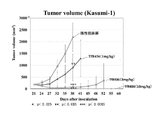
- FIG. 5 shows the antitumor effect of the TfR436 antibody in the Kasumi-1 subcutaneous transplant model.
- FIG. 6 shows the antitumor effect of the TfR436 antibody in the HL60 subcutaneous transplant model.
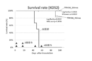
- FIG. 7 shows the life-prolonging effect of the TfR436 antibody in the KO52 tail vein transplantation model.
- FIG. 8 shows the life-prolonging effect of the TfR436 antibody in the CCRF-CEM intravenous transplant model.
- the method for determining the efficacy or susceptibility of an anti-human transferrin receptor antibody having an action of inhibiting the binding between human transferrin and a human transferrin receptor according to the present invention is a method using the intracellular iron amount as an index.
- the anti-human transferrin receptor antibody is preferably an antibody that recognizes the 629th to 633th amino acids of the human transferrin receptor. Specifically, when the intracellular iron amount is lower than the reference value, it can be determined that the medicinal effect or sensitivity of the antibody is high.
- the method of the present invention is preferably a method for determining drug efficacy or susceptibility by utilizing the fact that the amount of intracellular iron correlates with the drug efficacy of the antibody. More preferably, the efficacy or susceptibility of the antibody can be determined by a value relative to the amount of iron in the reference sample.
- a reference sample a reference cell such as K562 or a control sample containing a reference amount of iron can be used.
- the intracellular iron amount in the measurement target cell as a relative value (comparative value) with respect to the intracellular iron amount in the K562 cells is preferably 1 or less. When it is 8 or less, more preferably 0.7 or less, and even more preferably 0.6 or less, it can be determined that the efficacy or sensitivity of the antibody is high.
- Serum trivalent iron is transported in a state of being bound to transferrin. Transferrin bound to trivalent iron is taken up by intracellular endosomes when it binds to the transferrin receptor on the cell surface. The ferric iron taken up into cells together with transferrin is reduced to ferric iron by the metalloreductase STEAP3 in endosomes. Divalent iron is transported to the cytoplasm by DMT1 (divalent metal transporter 1) and pooled (also referred to as Labile iron pool). Labile iron pool divalent iron is used for DNA synthesis and DNA repair, used for energy generation in midcondria, heme synthesis and Fe-S cluster biosynthesis, or stored as stored iron. To.
- DMT1 divalent metal transporter 1
- pooled also referred to as Labile iron pool
- intracellular divalent iron is transported from the inside to the outside of the cell via ferroportin present in the cell membrane.
- intracellular iron is involved in intracellular iron utilization.
- the intracellular iron amount as an index, specifically, when the intracellular iron amount is lower than the reference value, it can be determined that the medicinal effect or sensitivity of the antibody is high. ..
- the intracellular iron amount in the biological sample (sample) derived from the subject is measured, and the measurement result is measured at the control level (reference). It can be done by comparing with the value).
- the subject is a subject who requires treatment with an anti-transferrin receptor antibody, and an example thereof includes a subject having cancer.
- the biological sample include a biopsy sample (in the case where the subject is a cancer patient, a sample containing cancer cells, etc.).
- the intracellular iron amount can be measured by a conventional method, or can be performed by using a commercially available measuring kit.
- a commercially available measuring kit for example, according to the metallogenic iron measurement LS (Metallogenics # FE31M), iron (Fe 2+ , Fe 3+ ) in a sample can be rapidly quantified by a microplate reader (96-well well).
- the intracellular iron amount in the biological sample derived from the subject may be measured before the administration of the anti-transferrin receptor antibody.
- the intracellular iron amount When the intracellular iron amount is determined to be lower than the predetermined reference value, it can be determined that the subject has a high efficacy of the anti-transferrin receptor antibody, that is, is sensitive to the anti-transferrin receptor antibody. Therefore, the intracellular iron amount can be used as a marker for positively continuing the treatment for a patient who can expect a therapeutic effect.
- the intracellular iron amount when it is determined that the intracellular iron amount is higher than a predetermined reference value, it can be determined that the subject has a low efficacy of the anti-transferrin receptor antibody, that is, is not sensitive to the anti-transferrin receptor antibody.
- the determination is not limited to a determination based on a specific numerical value, but may be determined based on a criterion other than a numerical value that can be visually grasped, such as a shade of color.
- Cancers include solid cancers (eg lung cancer, colon cancer, stomach cancer, bladder cancer, pancreatic cancer, prostate cancer, liver cancer, cervical cancer, uterine cancer, ovarian cancer, breast cancer, head and neck). Either partial cancer, skin cancer, etc.) or blood cancer (eg, leukemia, lymphoma, myeloma, etc.), but blood cancer is preferred. Particularly preferably, the cancer is acute myeloid leukemia (AML).
- AML acute myeloid leukemia
- TfR human transferrin receptor
- SEQ ID NO: 9 human chromosome 3
- This protein also known as the CD71 antigen, is thought to be involved in cell iron uptake and cell proliferation.
- the TfR of the present invention is not particularly limited in structure, and the monomer, multimer, intact form expressed on the cell membrane, solubilized form composed in the extracellular region, translated form, gene mutation, and the like. , Mutation form due to defects, etc., and post-translational modification due to phosphorylation, etc., all mean human TfR.
- Reacting and Reactive mean the same thing unless otherwise specified. That is, the antibody recognizes the antigen.
- This antigen may be an antigen TfR expressed on the cell membrane, a truncated form, or a solubilized form. Further, TfR having a three-dimensional structure may be used, or TfR may be modified.
- flow cytometer FACS
- enzyme-linked immunosorbent assay ELISA
- western-blot As means for examining reactivity, flow cytometer (FACS), enzyme-linked immunosorbent assay (ELISA), western-blot, fluorescence micromeasurement technology (FMAT), surface plasmon resonance (BIAcore), immunostaining, immunoprecipitation, etc. Can be mentioned.
- the antibody used for the flow cytometer may be an antibody labeled with a fluorescent substance such as FITC, biotin, or the like, or an unlabeled antibody.
- fluorescent-labeled avidin, fluorescent-labeled anti-human immunoglobulin antibody, etc. are used depending on the presence or absence of the antibody labeled and the type thereof. Reactivity should be evaluated by adding a sufficient amount of anti-TfR antibody (usually the final concentration is 0.01 to 10 ⁇ g / mL) to the sample and comparing it with the reactivity of the negative control antibody and the positive control antibody. Can be done.
- Antibodies The following abbreviations (in parentheses) are used herein as is customary. Heavy chains (H chains), light chains (L chains), heavy chain variable regions (VH), light chain variable regions (VL), complementarity determining regions (CDRs), first complementarity determining regions (CDR1), second. Complementarity determining regions (CDR2), third complementarity determining regions (CDR3) heavy chain first complementarity determining regions (VH CDR1), heavy chain second complementarity determining regions (VH CDR2), heavy chain third.
- VH CDR3s Complementarity determining regions
- VL CDR1 of the light chains The first complementarity determining regions (VL CDR1) of the light chains, the second complementarity determining regions (VL CDR2) of the light chains, and the third complementarity determining regions (VL CDR3) of the light chains.
- antibody is synonymous with immunoglobulin and should be understood as is commonly known in the art. Specifically, the term antibody is not limited to any particular method of making an antibody. For example, the term antibody includes, but is not limited to, recombinant antibodies, monoclonal antibodies, and polyclonal antibodies.
- human antibody means any antibody in which the sequences of the variable and constant regions are human sequences.
- the term has sequences derived from human genes, but has been altered to, for example, reduce possible immunogenicity, increase affinity, remove cysteines that can cause unwanted folding, and so on. Includes antibodies.
- the term also includes such antibodies made recombinantly in non-human cells that can undergo glycosylation that is not unique to human cells. These antibodies can be prepared in various forms.
- humanized antibody refers to an antibody of non-human origin, where the amino acid residues characteristic of the antibody sequence of the non-human species are replaced with the residues found at the corresponding positions of the human antibody. Has been done. It is believed that this "humanization” step reduces the immunogenicity of the resulting antibody in humans. It will be appreciated that non-human derived antibodies can be humanized using techniques well known in the art. For example, Winter et al., Immunol. See Today 14: 43-46 (1993). Antibodies of interest can be engineered by recombinant DNA technology that replaces CH1, CH2, CH3, hinge domains, and / or framework domains with the corresponding human sequences. For example, WO 92/02190, and US Pat.
- humanized antibody includes, to the extent its meaning, chimeric human antibody and CDR transplanted antibody.
- the sequence of the framework region (FR) in the variable region of the antibody is not particularly limited as long as it does not substantially affect the specific binding property to the corresponding antigen. It is preferable to use the FR region of a human antibody, but it is also possible to use the FR region of a non-human animal species (for example, mouse or rat).
- the antibody contains a constant region in addition to the variable region (for example, IgG type antibody).
- the arrangement of the constant region is not particularly limited.
- known constant regions of human antibodies can be used.
- the heavy chain constant region (CH) of the human antibody may be any as long as it belongs to human immunoglobulin (hereinafter referred to as hIgG), but hIgG class ones are preferable, and hIgG1 belonging to the hIgG class, Any of the subclasses such as hIgG2, hIgG3 and hIgG4 can be used.
- the light chain constant region (CL) any one belonging to hIg may be used, and those of ⁇ class or ⁇ class can be used. It is also possible to use constant regions of animal species other than humans (eg, mice and rats).
- the term "modified” or “modified antibody” means that one or more amino acids are substituted, deleted, added and / or in the amino acid sequence of the variable region (CDR sequence and / or FR sequence) of the parent antibody. It is inserted.
- the "parent antibody” is a TfR436 antibody having the amino acid sequence represented by SEQ ID NO: 7 for VH and SEQ ID NO: 8 for VL.
- amino acid sequence one or several (eg, 1 to 8, preferably 1 to 5, more preferably 1 to 3, particularly preferably 1 or 2) amino acids are deleted, added, substituted and /. Or it has been inserted.
- a method of introducing a mutation into a protein is known.
- a site-directed mutagenesis method (Hashimoto-Gotoh, T, Mizuno, T, Ogasahara, Y, anDNAkagawa, M. 152, 271-275, Zoller, MJ, and Smith, M. (1983) Oligonucleotide-directed mutagenesis of DNA fragments cloned into M13 vectors.Methods Enzymol.
- a modified antibody functionally equivalent to the antibody having a binding activity to TfR can be prepared.
- an antibody in which one or several amino acids are mutated in the variable region or constant region of the antibody and has a binding activity to TfR can also be used.
- “activity is equivalent to that of the parent antibody” means that the binding activity to human TfR is equivalent. “Equivalent” does not necessarily mean that the activity is of the same level, and the activity may be enhanced or may be decreased as long as the activity is maintained. Examples of the antibody having reduced activity include an activity of 30% or more, preferably 50% or more, more preferably 80% or more, and further preferably 90% or more activity as compared with the original antibody. Particularly preferably, an antibody having an activity of 95% or more can be mentioned.
- Binding activity means recognizing an antigen.
- This antigen may be an antigen TfR expressed on the cell membrane, a truncated form, or a solubilized form. Further, TfR having a three-dimensional structure may be used, or TfR may be modified.
- means for examining the binding activity include flow cytometer (FACS), enzyme-linked immunosorbent assay (ELISA), western-blot, fluorescence micromeasurement technique (FMAT), surface plasmon resonance (BIAcore), and the like.
- the Tf-TfR binding inhibitory activity of the antibody can be measured as follows.
- the TfR solution is dispensed onto a substrate (96-well plate, etc.), allowed to stand, solidified, and blocked.
- the HRP-labeled Tf solution is dispensed, an antibody is further added, and the reaction is carried out at room temperature.
- the substrate is washed, a coloring reagent (TMB or the like) is added and reacted, and the absorbance is measured with a plate reader.
- TMB coloring reagent
- the antibody is not limited to its origin, and may be any animal-derived antibody such as a human antibody, a mouse antibody, or a rat antibody. Further, a chimeric antibody, a humanized antibody, or the like may be used. One of the preferred embodiments of the antibody in the present invention is a human antibody.
- the antibody may differ in amino acid sequence, molecular weight, isoelectric point, presence / absence and morphology of sugar chain, etc., depending on the cell producing the antibody described later, the host, or the purification method. For example, a case where the amino acid sequence described in the present invention is modified after translation is also included in the present invention. Further, post-translational modifications for sites other than known post-translational modifications are also included in the present invention. When the antibody is expressed in prokaryotic cells, for example, Escherichia coli, a methionine residue is added to the N-terminal of the amino acid sequence of the original antibody. In the present invention, such an antibody may be used. Post-translational modifications to sites other than known post-translational modifications are also included in the invention.
- scFv that reacts with an antigen by a phage display library
- Acquisition of an antibody can be prepared according to some methods known in the art.
- phage display technology can be used to provide a library containing a repertoire of antibodies with varying affinities for TfR. These libraries can then be screened to identify and isolate antibodies against TfR.
- the phage library is a scFv phage display library produced using human VL and VH cDNA prepared from mRNA isolated from human B cells. Methods of preparing and screening such libraries are known in the art. Genetic material is recovered from a phage clone showing reactivity screened using human TfR as an antigen.
- the DNA sequences of VH and VL encoding the variable regions of the human antibody that bind to the antigen can be determined.
- Human antibodies can be obtained by IgGing scFv using this scFv sequence.
- a complete long heavy chain gene can be obtained by ligating a DNA encoding VH with another DNA molecule encoding a heavy chain constant region (CH1, CH2 and CH3).
- a heavy chain constant region CH1, CH2 and CH3
- the sequences of human heavy chain constant region genes are known in the art (eg, Kabat, EA et al., (1991) Sequences of Proteins of Immunological Interest, 5th Edition, US Department of Health and Human). Services, NIH Publication No. 91-3242), DNA fragments containing these regions can be obtained by standard PCR amplification.
- the heavy chain constant region may be an IgG1, IgG2, IgG3, IgG4, IgA, IgE, IgM or IgD constant region, but most preferably an IgG1 or IgG2 constant region.
- the IgG1 constant region sequence may be any various alleles or allotypes known to occur between different individuals, such as Gm (1), Gm (2), Gm (3) and Gm (17). These allotypes correspond to naturally occurring amino acid substitutions in the IgG1 constant region.
- a full-length L-chain gene (as well as a Fab light chain gene) can be obtained by linking the VL-encoding DNA with another DNA molecule encoding the light chain constant region CL.
- the sequence of the human light chain constant region gene is known in the art (eg, Kabat, EA et al., (1991) Sequences of Proteins of Immunological Interest, 5th Edition, US Department of Health and. Human Services, NIH Publication No. 91-3242), DNA fragments containing these regions can be obtained by standard PCR amplification.
- the light chain constant region may be a constant region of ⁇ or ⁇ .
- the ⁇ constant region may be any various alleles known to occur between different individuals, such as Inv (1), Inv (2) and Inv (3).
- the ⁇ constant region may be derived from any of the three ⁇ genes.
- An expression vector is prepared by inserting the DNA encoding the H chain or L chain obtained as described above into an expression vector, expressed in a host cell, and a human antibody is obtained by collecting and purifying the secreted supernatant. get.
- Expression vectors include plasmids, retroviruses, adenoviruses, adeno-associated viruses (AAV), plant viruses such as cauliflower mosaic virus and tobacco mosaic virus, cosmid, YAC, EBV-derived episomes and the like.
- Expression vectors and expression regulatory sequences are selected to be compatible with the expression host cells used.
- the antibody light chain gene and the antibody heavy chain gene can be inserted into separate vectors, or both genes can be inserted into the same expression vector.
- the antibody gene is inserted into the expression vector by standard methods (eg, ligation of the vector with complementary restriction sites on the antibody gene fragment, or blunt-ended ligation in the absence of restriction sites).
- the favorable vector is a functionally complete human CH or CL with suitable restriction sites engineered to allow easy insertion and expression of any VH or VL sequence as described above. It encodes an immunoglobulin sequence.
- splicing usually occurs between the splice-donating site in the inserted J region and the splice receiving site preceding the human C domain, and also in the splice region present within the human CH exon. Polyadenylation and transcription termination occur at natural chromosomal sites downstream of the coding region.
- the recombinant expression vector can also encode a signal peptide that promotes the secretion of antibody chains derived from the host cell.
- the antibody chain gene can be cloned into the vector such that the signal peptide is linked in-frame to the amino terminus of the immunoglobulin chain.
- the signal peptide may be an immunoglobulin signal peptide or a heterologous signal peptide (ie, a signal peptide derived from a non-immunoglobulin protein).
- the antibody expression vector may have, in addition to the antibody gene and control sequence, additional sequences such as a sequence that controls the replication of the vector in the host cell (for example, an origin of replication) or a selectable marker gene.
- additional sequences such as a sequence that controls the replication of the vector in the host cell (for example, an origin of replication) or a selectable marker gene.
- the selectable marker gene facilitates the selection of the host cell into which the vector has been introduced.
- the selectable marker gene usually confer resistance to drugs such as G418, hygromycin and methotrexate on host cells into which the vector has been introduced.
- Preferred selectable marker genes include the dehydrofolate reductase (DHFR) gene (used with methotrexate selection / amplification in dhfr-host cells), the neomycin phosphotransferase gene (for G418 selection), and the glutamic acid synthase gene.
- DHFR dehydrofolate reductase
- the host cell may be any cell such as a bacterium, a yeast, an animal cell, an insect cell, or a plant cell as long as it can produce an antibody, but an animal cell is preferable.
- animal cells Chinese hamster ovary cells CHO / dhfr (-) cells CHO / DG44 cells, monkey-derived cells COS cells (A.Wright & S.L.Morrison, J.Immunol.160, 3393-3402 (1998)), SP2 / O cells (Mouth Mieroma) (K. Motmans et al., Eur.J. Cancer Prev. 5, 512-5199 (1996), R.P.
- the human antibody After culturing the transformant, the human antibody is separated from the intracellular or culture solution of the transformant.
- methods such as centrifugation, salting out, salting out, ultrafiltration, affinity chromatography, ion exchange chromatography, and gel filtration chromatography can be appropriately combined and used.
- Antibody fragment can be prepared based on an antibody or based on the sequence information of a gene encoding an antibody.
- Examples of the antibody fragment include Fab, Fab', F (ab') 2 , scFv, and dsFv antibodies.
- Fab is a fragment having a molecular weight of about 50,000, which is obtained by papain digestion of IgG in the presence of cysteine, and is composed of an L chain and an H chain variable region, and an H chain fragment consisting of a CH1 domain and a part of a hinge portion. be. In the present invention, it can be obtained by digesting the above antibody with papain. Further, a Fab can be prepared from a transformant transformed by incorporating a part of the H chain and the DNA encoding the L chain of the antibody into an appropriate vector.
- Fab' is a fragment having a molecular weight of about 50,000 obtained by cleaving the disulfide bond between the H chains of F (ab') 2 described later. In the present invention, it is obtained by digesting the above antibody with pepsin and cleaving a disulfide bond with a reducing agent. Further, as with Fab, it can also be genetically engineered using DNA encoding Fab'.
- F (ab') 2 is a fragment (Fab') obtained by pepsin digestion of IgG, which is composed of an L chain and an H chain variable region, and an H chain fragment consisting of a CH1 domain and a part of a hinge portion. Is a fragment having a molecular weight of about 100,000 bonded by a disulfide bond. In the present invention, it is obtained by digesting the above antibody with pepsin. Further, similarly to Fab, it can also be genetically engineered using DNA encoding F (ab') 2 .
- the scFv is an antibody fragment obtained by linking an Fv composed of an H chain variable region and an L chain variable region with the C-terminal of one chain and the N-terminal of the other with an appropriate peptide linker to form a single chain.
- the peptide linker for example, highly flexible (GGGGS) 3 or the like can be used.
- GGGGS highly flexible
- a DNA encoding an scFv antibody is constructed using a DNA encoding an H chain variable region and an L chain variable region of the antibody and a DNA encoding a peptide linker, and this is incorporated into an appropriate vector, and the vector is used.
- ScFv can be prepared from the transformed transformant.
- DsFv is an Fv fragment in which a Cys residue is introduced at an appropriate position in the H chain variable region and the L chain variable region, and the H chain variable region and the L chain variable region are stabilized by a disulfide bond.
- the introduction position of the Cys residue in each chain can be determined based on the three-dimensional structure predicted by molecular modeling.
- the three-dimensional structure is predicted from the amino acid sequences of the H chain variable region and the L chain variable region of the above antibody, and based on this prediction, DNA encoding the H chain variable region and the L chain variable region into which the mutation is introduced is constructed. Then, this can be incorporated into a suitable vector, and dsFv can be prepared from the transformant transformed with the vector.
- the antibody fragment can be multimerized by ligating the scFv antibody, dcFv antibody, etc. using an appropriate linker, or fusing with streptavidin.
- VH CDR1 SYGMH (SEQ ID NO: 1)
- VH CDR2 VISYDGSNKYYADSVKG (SEQ ID NO: 2)
- VH CDR3 DSNFWSGYYSPVDV (SEQ ID NO: 3)
- VL CDR1 TRSSGSIASNSVQ (SEQ ID NO: 4)
- VL CDR2 YEDTQRPS (SEQ ID NO: 5)
- VL CDR3 QSYDSAYHWV (SEQ ID NO: 6)
- TfR436 VH (SEQ ID NO: 7) DVQLVQSGGVVQPGRSLRLSCAASGFPFKSYGMHWVRQAPGKGLEWVAVISYDGSNKYADSVKGRFTISRDNSKNTLYLQMNSLRGEDTAVYYCARDSNFWSVGYSPVDTVG
- TfR436 VL (SEQ ID NO: 8) NFMLTQPHSVSESPGKTVTISCTRSSGSIASSNSVQWYQQRPGSAPITVIYEDTQRPSGVPDRFSGSIDSSSNSASLSGLLQTEDEADYYCQSSYDSAYHWVFGGGTKLALL
- Example 1 Identification of the binding site of the TfR436 antibody
- the TfR436 antibody did not cross-react with mouse TfR, but showed cross-reactivity with hamster TfR.
- the amino acid sequences of the Transferrin (TF) binding site (amino acids 569 to 760) in TfR were aligned.
- the same amino acids as hamsters and different from mice were picked up.
- the picked up amino acids were point-mutated as shown in FIG. 1 to prepare a soluble TfR mutant fragment.
- Expi293 cells (Thermo Fisher scientific) were transfected with the above plasmids using Expectamine (Thermo Fisher scientific) and cultured at 37 ° C., 8% CO 2 , 135 rpm for 5 days.
- the culture supernatant is collected by centrifugation, a HisTrappH (Cytiva) column is connected to the AKTA prime (Cytiva), and sTfR or MF1 to MF7 is purified using 20 mM imidazole / DPBS as the binding buffer and 500 mM imidazole / DPBS as the elution buffer. bottom.
- the eluted protein was buffered to 30 mM HEPES, 5% trehalose, pH 7.2 using Zeba spin colon (Thermo Scientific).
- the cells were washed 5 times with PBST Buffer, TfR436 antibody (1 ⁇ g / mL) was dispensed into 100 ⁇ L / well, placed on a shaker, and reacted at room temperature for 1 hour. Then, the cells were washed 5 times with PBS-T Buffer, diluted 50,000 times, and dispensed into 100 ⁇ L / well of secondary antibody F (ab') 2 Fragment Anti-Human IgG Fc ⁇ (Jackson Immuno Research) and reacted at room temperature for 1 hour. rice field.
- TfR436 antibody (1 ⁇ g / mL
- TMB Soluble Reagent High Sensitivity (Scy Tek Laboratories) was dispensed into 100 ⁇ L / well, reacted at room temperature in a dark place for 3 minutes, and then TMB StopBuffer (Scy Talk Labor) / Well was added, and the mixture was shaken with a shaker for 1 minute, and the absorbance at 450 nm (ref. 620 nm) was measured with a plate reader.
- the TfR436 antibody showed a decrease in reactivity with TfR mutant fragment MF5, but no decrease in reactivity with other mutant fragments was observed. That is, if the 629th, 630th, and 633th amino acids of TfR are replaced with other amino acids, the TfR436 antibody cannot be recognized as TfR. It was suggested that amino acids 629-633 are recognition epitopes of the TfR436 antibody.
- Example 2 Comparison of TfR436 antibody and comparative antibody in Tf-TfR binding inhibition (1) Preparation of comparative antibody A24 Patent Document US2008 / 0193453 describes an A24 antibody against human TfR. To compare this antibody with the TfR436 antibody, the deposited Hybridoma was obtained and the antibody was produced. Specifically, Hybridoma was sprinkled in RPMI 1640 (Thermo Fisher Scientific), FBS 10% medium so that the cell concentration was 1 to 2 ⁇ 10 5 / mL, and cultured in a 5% CO 2 incubator at 37 ° C.
- RPMI 1640 Thermo Fisher Scientific
- the cells were collected by centrifugation, washed twice with PBS, and then expanded and cultured in serum-free medium cosmediaum 005 (Cosmo Bio) and 0.5% Nutridoma-CS (Roche) until the volume became 550 mL.
- the culture supernatant was collected by centrifugation 5 days after the cells became confluent.
- the recovered supernatant is applied to a protein A carrier (Ab-Capcher ExTra: Protenova), and the antibody bound to protein A is eluted with 0.1 M glycine hydrochloride buffer (pH 2.7) immediately. Neutralized with 1M Tris hydrochloric acid buffer (pH 8.5). After that, the buffer was exchanged with PBS using an ultracell ultrafiltration disk (Merck Millipore).
- the TfR436 antibody completely inhibited the binding of Tf-TfR at a much lower dose (100 ng / mL).
- the A24 antibody could not completely inhibit the binding of Tf-TfR even at a dose of 10 ⁇ g / mL, and could inhibit the binding of Tf-TfR by only 50%. It was suggested that the TfR436 antibody was superior in inhibiting the binding of Tf-TfR.
- Example 3 Comparison of intracellular iron amount in AML clinical sample and growth inhibitory effect (GI50) by TfR436 antibody In order to verify the relationship between intracellular iron amount in AML clinical sample and growth inhibitory effect by TfR436 antibody exposure, the following Test was carried out.
- AML clinical sample by TfR436 antibody The growth-suppressing effect (GI50 value) by TfR436 antibody exposure was calculated using the AML clinical sample. Specifically, AML clinical specimens (bone marrow or peripheral blood) were isolated from mononuclear cells, and the obtained cells were suspended in MethoCult H4534 Classic without EPO (STEMCELL # ST-04534) so as to be 5000 cells / 90 ⁇ L. Seeded on 96-well plates.
- the TfR436 antibody was diluted with RPMI-1640 Medium (Thermo Fisher # 11875119) and derived from AML clinical specimens to a final concentration of 5000, 1000, 500, 100, 50, 10, 5, 1 ng / mL, and 0 (no additives). 10 ⁇ L each was added to the cells. After culturing in a 5% CO 2 incubator at 37 ° C. for 7 days, 100 ⁇ L of CellTiter-Glo Luminescent Cell Viability Assay (Promega # G7572) was added to and mixed with each well, and fluorescence was detected with a luminometer 10 minutes later. The GI50 value was calculated using XLfit after calculating the growth rate at each antibody concentration when the average value in the TfR436 antibody-free well was set to 100% with the well to which only the medium was added as a blank.
- the intracellular iron amount was measured using the AML clinical sample. Specifically, 1.0x10⁷ cells were collected from AML clinical specimens and then washed 3 times with DPBS. 250 ⁇ L of Lysis M Reagent (Roche # 04719 965001) and 2.5 ⁇ L of 6N-HCl were added to and mixed with the cell pellet, and the cells were allowed to stand at room temperature for 1 hour. After centrifugation, the collected supernatant was used for iron quantification. By the same operation, a K562 cell line was prepared as a standard sample and a frozen stock was prepared. Iron quantification was performed according to the metallogenic iron measurement LS (ferrodine method) (Metallogenics # FE31M). In addition, the intracellular iron quantification of the K562 cell line was also performed for each intracellular iron quantification of the AML clinical sample, and the comparative value was calculated.
- Lysis M Reagent Roche # 04719 965001
- 6N-HCl 6N-HCl
- FIG. 4 shows the relationship between the intracellular iron amount in each AML clinical sample and the growth inhibitory effect (GI50 value) of the TfR436 antibody.
- Example 4 Comparison of intracellular iron amount in cell line and growth inhibitory effect (GI50) by TfR436 antibody In order to demonstrate the relationship between the intracellular iron amount verified in Example 3 and the growth inhibitory effect by exposure to TfR436 antibody. The following tests were performed using the cell line.
- Negative control monoclonal antibody was added to a final concentration of 5 ⁇ g / mL and cultured in a 5% CO 2 incubator at 37 ° C. After 96 hours, the cells were collected, washed with PBS, and then 250 ⁇ L of Lysis M Reagent and 2.5 ⁇ L of 6N-HCl were added and mixed per 1.0 ⁇ 107 cells of each cell line, and the cells were allowed to stand at room temperature for 1 hour. Placed. After centrifugation, the collected supernatant was used for iron quantification.
- the supernatant of the K562 (ATCC # CCL-243) cell line prepared by culturing and collecting in RPMI1640 + 10% FBS medium in the same manner as other cell lines was used for iron quantification.
- Iron quantification was performed according to the metalloassay iron measurement LS (ferrodine method). The measured amount of iron was divided by the number of cells used as a sample to obtain the amount of intracellular iron per cell, and then the comparison value of the amount of intracellular iron in each cell line with the amount of intracellular iron in the K562 cell line was calculated. bottom.
- TfR436 antibody The proliferation inhibitory effect (GI50 value) by TfR436 antibody exposure was calculated using Kasumi-1, HL60, KO52, and CCRF-CEM. Specifically, each cell line was suspended in various optimal cell culture media so as to be 10,000 cells / 50 ⁇ L, and seeded on a 96-well plate. The TfR436 antibody and the negative control monoclonal antibody were diluted using the same medium in 9 steps from the final concentration of 5000 ng / mL to a common ratio of 3.16 times. 50 ⁇ L of each of these antibody-added medium and antibody-free medium was added to each cell line. After culturing in a 5% CO 2 incubator at 37 ° C.
- Table 1 shows the intracellular iron amount in each cell line and the growth inhibitory effect (GI50 value) by TfR436 antibody.
- GI50 value the growth inhibitory effect
- Example 5 Antitumor effect of TfR436 antibody in Kasumi-1 subcutaneous transplantation model Subcutaneous transplantation of Kasumi-1 verified in Example 4 to verify the pharmacological effect of TfR436 antibody in a cell line-developed xenograft (CDX) model. The following tests were performed using the model. Kasumi-1, an acute myeloid leukemia cell line, was cultured in RPMI-1640, 10% FBS, 1% P / S. Enlarged cells were centrifuged at 1,000 rpm for 5 minutes to collect pellets, suspended in antibiotic- and serum-free RPMI-1640 medium to prepare 2 ⁇ 108 / mL, and equal volumes of Matrigel under ice-cooling. Was added and mixed by inversion, and 100 ⁇ L of the mixture was transplanted subcutaneously to the right ventral side of the mouse. The number of transplanted cells is 1 ⁇ 107 / animal.
- the average tumor volume of the transplanted individual was measured over time, and 21 days after the transplantation, when an increasing tendency of the tumor volume was observed, the tumor volume using biological experiment data statistical analysis EXSUS (Version 8.1, EP Croix Co., Ltd.) Randomized allocation by stratification was carried out.
- the negative antibody group (10 mg / kg) was administered as a control group at a dose of 10 mL / kg, and the TfR436 antibody was administered once a week at a dose of 1,3,10 mg / kg twice in total to the administration group. After administration, tumor measurement with calipers was performed twice a week to determine the tumor volume of each group. The antitumor effect was determined by the tumor volume.
- Tumor volume (minor diameter) 2 x major diameter x 0.5
- Figure 5 shows the change over time in the average tumor volume.
- the tumors of all the individuals disappeared, and no tumor re-growth was confirmed even on the end day of the test (60 days after transplantation).
- Example 6 Anti-tumor effect of TfR436 antibody in HL60 subcutaneous transplant model
- CDX cell line-derivated xenograft
- the average tumor volume of the transplanted individual was measured over time, and 9 days after the transplantation, when an increasing tendency of the tumor volume was observed, the tumor volume using biological experiment data statistical analysis EXSUS (Version 8.1, EP Croix Co., Ltd.) Randomized allocation by stratification was carried out.
- the dose was 10 mL / kg, citric acid was administered to the negative control group, and TfR436 antibody was administered to the administration group at a dose of 0.3, 1, 3, 10 mg / kg once a week for a total of 4 times. After administration, tumor measurement with calipers was performed twice a week to determine the tumor volume of each group. The antitumor effect was determined by the tumor volume.
- Tumor volume (minor diameter) 2 x major diameter x 0.5
- Figure 6 shows the change over time in the average tumor volume.
- a significant dose-dependent antitumor effect was confirmed at a concentration of TfR436 antibody of 1 mg / kg or more.
- the tumors of all the individuals disappeared, and no tumor re-growth was observed 49 days after the end of administration (79 days after transplantation).
- Example 7 Life-prolonging effect of TfR436 antibody in KO52 tail vein transplantation model
- CDX cell line-developed xenograft
- the prepared cell suspension was brought into a breeding room by storage in ice, filled in a 29G myjector for each animal, and 200 ⁇ L thereof was transplanted from the tail vein of a mouse.
- the number of transplanted cells is 5 ⁇ 106 / animal.
- the dose was 10 mL / kg, citric acid was administered to the negative control group, and TfR436 antibody was administered to the administration group at a dose of 10 mg / kg once every two weeks, for a total of two doses.
- One of the two administration groups was administered once again every two weeks at the fourth week after the second administration, for a total of four administrations.
- mice were observed during the test period to determine whether they were alive or dead.
- the test was completed 104 days after transplantation, and statistical analysis was performed based on the survival time of mice.
- Fig. 7 As a result of autopsy, one death case in the 4-dose group was determined not to be affected by KO52 transplantation but to death due to spontaneous thymoma peculiar to scid mice.
- Example 8 Life-prolonging effect of TfR436 antibody in CCRF-CEM venous transplantation model
- the T-cell acute leukemia cell line CCRF-CEM was cultured in RPMI-1640, 10% FBS, 1% P / S.
- the expanded-cultured cells were centrifuged at 1,000 rpm for 5 minutes to collect pellets, suspended in saline for injection to prepare 2.5 ⁇ 107 / mL, and 200 ⁇ L thereof was transplanted from the tail vein of mice.
- the number of transplanted cells is 5 ⁇ 106 / animal.
- the initial administration was 3 days after transplantation and 7 days after transplantation on the day of grouping, and the schedule was as shown in Table 2 for a total of 5 times for each group.
- Mice were weighed twice a week until the end of the study.
- the general condition of the mice was observed twice a week until 30 days after transplantation, and every day after 31 days until 97 days after transplantation at the end of the study.
- Mice were weighed twice a week and the ratio to the body weight at the time of grouping was calculated. When the weight loss rate exceeded 20%, it was regarded as a humanitarian endpoint and the mice were euthanized. In addition, if signs such as weakness or decreased body temperature were confirmed by observing the general condition, it was also used as a humanitarian endpoint.
Abstract
Description
<1> 細胞内鉄量を指標とする、ヒトトランスフェリンとヒトトランスフェリンレセプターとの結合を阻害する作用を有する抗ヒトトランスフェリンレセプター抗体の薬効又は感受性の判定方法。
<2> 細胞内鉄量が前記抗体の薬効と相関することを利用した薬効又は感受性の判定方法である、<1>に記載の方法。
<3> 基準サンプルの鉄量との相対値により前記抗体の薬効または感受性を判定する、<1>又は<2>に記載の方法。
<4> 細胞内鉄量が基準値より低い場合に、前記抗体の薬効又は感受性が高いと判定する、<1>から<3>の何れか一に記載の方法。
<5> 前記抗体のがんに対する薬効又は感受性を判定する、<1>から<4>の何れか一に記載の方法。
<6> がんが、血液がんである、<5>に記載の方法。
<7> がんが、急性骨髄性白血病(AML)である、<5>又は<6>に記載の方法。
<8> 前記抗体が、ヒトトランスフェリンレセプターの629番目~633番目のアミノ酸を認識する抗体である、<1>から<7>の何れか一に記載の方法。
<9> 前記抗体が、重鎖第1相補性決定領域(VH CDR1)、重鎖第2相補性決定領域(VH CDR2)、重鎖第3相補性決定領域(VH CDR3)がそれぞれ配列番号1、2、3であり、及び軽鎖第1相補性決定領域(VL CDR1)、軽鎖第2相補性決定領域(VL CDR2)、軽鎖第3相補性決定領域(VL CDR3)がそれぞれ配列番号4、5、6である抗体である、<1>から<8>の何れか一に記載の方法。
<10> 前記抗体が、重鎖が配列番号7を有し、軽鎖が配列番号8を有する抗体である、<1>から<9>の何れか一に記載の方法。 That is, according to the present invention, the following invention is provided.
<1> A method for determining the efficacy or susceptibility of an anti-human transferrin receptor antibody having an action of inhibiting the binding between human transferrin and a human transferrin receptor using the intracellular iron amount as an index.
<2> The method according to <1>, which is a method for determining drug efficacy or susceptibility utilizing the correlation between the intracellular iron amount and the drug efficacy of the antibody.
<3> The method according to <1> or <2>, wherein the efficacy or susceptibility of the antibody is determined based on a relative value with the amount of iron in the reference sample.
<4> The method according to any one of <1> to <3>, wherein when the intracellular iron amount is lower than the reference value, it is determined that the medicinal effect or sensitivity of the antibody is high.
<5> The method according to any one of <1> to <4>, which determines the efficacy or susceptibility of the antibody to cancer.
<6> The method according to <5>, wherein the cancer is blood cancer.
<7> The method according to <5> or <6>, wherein the cancer is acute myeloid leukemia (AML).
<8> The method according to any one of <1> to <7>, wherein the antibody is an antibody that recognizes the 629th to 633th amino acids of the human transferrin receptor.
<9> The antibody has the heavy chain first complementarity determining region (VH CDR1), the heavy chain second complementarity determining region (VH CDR2), and the heavy chain third complementarity determining region (VH CDR3), respectively, with SEQ ID NO: 1. , 2, 3 and the light chain first complementarity determining regions (VL CDR1), light chain second complementarity determining regions (VL CDR2), and light chain third complementarity determining regions (VL CDR3) are SEQ ID NOs: respectively. The method according to any one of <1> to <8>, which is an antibody that is 4, 5, or 6.
<10> The method according to any one of <1> to <9>, wherein the antibody is an antibody having a heavy chain having SEQ ID NO: 7 and a light chain having SEQ ID NO: 8.
定義および一般的技術
本明細書において別段定義しない限り、本発明に関して使用した科学技術用語は、当業者に通常理解されている意味を含むものとする。一般に、本明細書に記載の細胞および組織培養、分子生物学、免疫学、微生物学、遺伝学、タンパク質および核酸化学ならびにハイブリダイゼーションに関して使用した命名法およびその技術は、当技術分野で周知のものであり、通常使用されている。 Next, the present invention will be described in more detail.
Definitions and General Techniques Unless defined otherwise herein, scientific and technological terms used with respect to the present invention shall have meanings commonly understood by those of skill in the art. In general, the nomenclature and techniques used for cell and tissue culture, molecular biology, immunology, microbiology, genetics, protein and nucleic acid chemistry and hybridization described herein are well known in the art. And is normally used.
本発明によるヒトトランスフェリンとヒトトランスフェリンレセプターとの結合を阻害する作用を有する抗ヒトトランスフェリンレセプター抗体の薬効又は感受性の判定方法は、細胞内鉄量を指標とする方法である。抗ヒトトランスフェリンレセプター抗体は、ヒトトランスフェリンレセプターの629番目~633番目のアミノ酸を認識する抗体が好ましい。具体的には、細胞内鉄量が基準値より低い場合に、抗体の薬効又は感受性が高いと判定することができる。 <Method of the present invention>
The method for determining the efficacy or susceptibility of an anti-human transferrin receptor antibody having an action of inhibiting the binding between human transferrin and a human transferrin receptor according to the present invention is a method using the intracellular iron amount as an index. The anti-human transferrin receptor antibody is preferably an antibody that recognizes the 629th to 633th amino acids of the human transferrin receptor. Specifically, when the intracellular iron amount is lower than the reference value, it can be determined that the medicinal effect or sensitivity of the antibody is high.
生体試料としては、例えば生検検体(被験者ががん患者である場合には、がん細胞を含む検体など)が挙げられる。 The subject is a subject who requires treatment with an anti-transferrin receptor antibody, and an example thereof includes a subject having cancer.
Examples of the biological sample include a biopsy sample (in the case where the subject is a cancer patient, a sample containing cancer cells, etc.).
TfR
ヒトトランスフェリン受容体(TfR)はヒト三番染色体にコードされる760アミノ酸から構成される一回膜貫通型タンパクである(配列番号9)。このタンパクはCD71抗原としても知られ、細胞の鉄の取り込みに関与し、細胞の増殖に関与すると考えられている。本発明のTfRは、構造上特別に限定せず、単量体、多量体、細胞膜に発現しているintact form、細胞外領域に構成された可溶化form、truncated form、また、遺伝子の変異や、欠損などによりmutation form、リン酸化などにより翻訳後修飾を受けたformなども含め、すべてヒトTfRを意味する。 <About antibodies>
TfR
The human transferrin receptor (TfR) is a single transmembrane protein composed of 760 amino acids encoded by human chromosome 3 (SEQ ID NO: 9). This protein, also known as the CD71 antigen, is thought to be involved in cell iron uptake and cell proliferation. The TfR of the present invention is not particularly limited in structure, and the monomer, multimer, intact form expressed on the cell membrane, solubilized form composed in the extracellular region, translated form, gene mutation, and the like. , Mutation form due to defects, etc., and post-translational modification due to phosphorylation, etc., all mean human TfR.
本明細書において、「反応する」と「反応性」は特別に示さない限り、同じことを意味する。すなわち、抗体が抗原を認識すること。この抗原は、細胞膜に発現するintact TfRでもよいし、truncated formや、可溶化formでも良い。また、立体構造を保ったTfRでもよいし、変性したTfRでもよい。反応性を検討する手段として、フローサイトメーター(FACS)、酵素結合免疫吸着検定法(ELISA)、western-blot、蛍光微量測定技術(FMAT)、表面プラズモン共鳴(BIAcore)、免疫染色、免疫沈降などが挙げられる。 Reacting and Reactive In the present specification, "reacting" and "reactive" mean the same thing unless otherwise specified. That is, the antibody recognizes the antigen. This antigen may be an antigen TfR expressed on the cell membrane, a truncated form, or a solubilized form. Further, TfR having a three-dimensional structure may be used, or TfR may be modified. As means for examining reactivity, flow cytometer (FACS), enzyme-linked immunosorbent assay (ELISA), western-blot, fluorescence micromeasurement technology (FMAT), surface plasmon resonance (BIAcore), immunostaining, immunoprecipitation, etc. Can be mentioned.
本明細書では必要に応じて、慣習に従い以下の略号(括弧内)を使用する。
重鎖(H鎖)、軽鎖(L鎖)、重鎖可変領域(VH)、軽鎖可変領域(VL)、相補性決定領域(CDR)、第1相補性決定領域(CDR1)、第2相補性決定領域(CDR2)、第3相補性決定領域(CDR3)重鎖の第1相補性決定領域(VH CDR1)、重鎖の第2相補性決定領域(VH CDR2)、重鎖の第3相補性決定領域(VH CDR3)軽鎖の第1相補性決定領域(VL CDR1)、軽鎖の第2相補性決定領域(VL CDR2)、軽鎖の第3相補性決定領域(VL CDR3)。 Antibodies The following abbreviations (in parentheses) are used herein as is customary.
Heavy chains (H chains), light chains (L chains), heavy chain variable regions (VH), light chain variable regions (VL), complementarity determining regions (CDRs), first complementarity determining regions (CDR1), second. Complementarity determining regions (CDR2), third complementarity determining regions (CDR3) heavy chain first complementarity determining regions (VH CDR1), heavy chain second complementarity determining regions (VH CDR2), heavy chain third. Complementarity determining regions (VH CDR3s) The first complementarity determining regions (VL CDR1) of the light chains, the second complementarity determining regions (VL CDR2) of the light chains, and the third complementarity determining regions (VL CDR3) of the light chains.
本発明において、「親抗体」とは、VHは配列番号7、VLは配列番号8で示されたアミノ酸配列を有するTfR436抗体である。アミノ酸配列において、1または数個(例えば、1から8個、好ましくは1から5個、より好ましくは1から3個、特に好ましくは1または2個)のアミノ酸が欠失、付加、置換及び/または挿入されている。TfRに対する結合活性を有する抗体のアミノ酸配列を調製するための、当業者によく知られた方法としては、タンパク質に変異を導入する方法が知られている。例えば、当業者であれば、部位特異的変異誘発法(Hashimoto-Gotoh, T, Mizuno, T, Ogasahara, Y, anDNAkagawa, M. (1995) An oligodeoxyribonucleotide-directed dual amber method for site-directed mutagenesis. Gene 152, 271-275、Zoller, MJ, and Smith, M.(1983) Oligonucleotide-directed mutagenesis of DNA fragments cloned into M13 vectors.Methods Enzymol. 100, 468-500、Kramer,W, Drutsa,V,Jansen,HW, Kramer,B, Pflugfelder,M, and Fritz,HJ(1984) The gapped duplex DNA approach to oligonucleotide-directed mutation construction. Nucleic Acids Res. 12,9441-9456、Kramer W, and Fritz HJ(1987) Oligonucleotide-directed construction of mutations via gapped duplex DNA Methods. Enzymol. 154, 350-367、Kunkel,TA(1985) Rapid and efficient site-specific mutagenesis without phenotypic selection.Proc Natl Acad Sci U S A. 82, 488-492)などを用いて、TfRに対する結合活性を有する抗体のアミノ酸配列に適宜変異を導入することにより、TfRに対する結合活性を有する抗体と機能的に同等な改変抗体を調製することができる。このように、抗体の可変領域又は定常領域において、1または数個のアミノ酸が変異しており、TfRに対する結合活性を有する抗体を使用することもできる。 As used herein, the term "modified" or "modified antibody" means that one or more amino acids are substituted, deleted, added and / or in the amino acid sequence of the variable region (CDR sequence and / or FR sequence) of the parent antibody. It is inserted.
In the present invention, the "parent antibody" is a TfR436 antibody having the amino acid sequence represented by SEQ ID NO: 7 for VH and SEQ ID NO: 8 for VL. In the amino acid sequence, one or several (eg, 1 to 8, preferably 1 to 5, more preferably 1 to 3, particularly preferably 1 or 2) amino acids are deleted, added, substituted and /. Or it has been inserted. As a method well known to those skilled in the art for preparing an amino acid sequence of an antibody having binding activity to TfR, a method of introducing a mutation into a protein is known. For example, if you are a person skilled in the art, a site-directed mutagenesis method (Hashimoto-Gotoh, T, Mizuno, T, Ogasahara, Y, anDNAkagawa, M. 152, 271-275, Zoller, MJ, and Smith, M. (1983) Oligonucleotide-directed mutagenesis of DNA fragments cloned into M13 vectors.Methods Enzymol. 100, 468-500, Kramer, W, Drutsa, V, Jansen, HW , Kramer, B, Pflugfelder, M, and Fritz, HJ (1984) The gapped duplex DNA approach to oligonucleotide-directed mutation construction. Nucleic Acids Res. 12,9441-9456, Kramer W, and Fritz HJ (1987) Oligonucleotide-directed Using construction of mutations via gapped duplex DNA Methods. Enzymol. 154, 350-367, Kunkel, TA (1985) Rapid and efficient site-specific mutagenesis without phenotypic selection. Proc Natl Acad Sci U S A. 82, 488-492) Therefore, by appropriately introducing a mutation into the amino acid sequence of an antibody having a binding activity to TfR, a modified antibody functionally equivalent to the antibody having a binding activity to TfR can be prepared. As described above, an antibody in which one or several amino acids are mutated in the variable region or constant region of the antibody and has a binding activity to TfR can also be used.
(1)ファージディスプレイライブラリーにより抗原と反応するscFvの取得
抗体の取得は、当技術分野で知られているいくつかの方法に従って調製することができる。例えば、ファージディスプレイ技術を使用して、TfRに対する親和性が様々である抗体のレパートリーを含むライブラリーを提供することができる。次いで、これらのライブラリーをスクリーニングして、TfRに対する抗体を同定し単離することができる。好ましくは、ファージライブラリーは、ヒトB細胞から単離されたmRNAから調製されたヒトVLおよびVHcDNAを使用して生成されるscFvファージディスプレイライブラリーである。そのようなライブラリーを調製しスクリーニングする方法は当技術分野で知られている。ヒトTfRを抗原としてスクリーニングした反応性を示すファージクローンから遺伝物質を回収する。選択されたファージの遺伝子を解析すれば、抗原に結合するヒト抗体の可変領域をコードするVH及びVLのDNA配列を決定することができる。このscFvの配列を用いて、scFvをIgG化すれば、ヒト抗体を取得することができる。 Preparation of antibody (1) Acquisition of scFv that reacts with an antigen by a phage display library Acquisition of an antibody can be prepared according to some methods known in the art. For example, phage display technology can be used to provide a library containing a repertoire of antibodies with varying affinities for TfR. These libraries can then be screened to identify and isolate antibodies against TfR. Preferably, the phage library is a scFv phage display library produced using human VL and VH cDNA prepared from mRNA isolated from human B cells. Methods of preparing and screening such libraries are known in the art. Genetic material is recovered from a phage clone showing reactivity screened using human TfR as an antigen. By analyzing the genes of the selected phage, the DNA sequences of VH and VL encoding the variable regions of the human antibody that bind to the antigen can be determined. Human antibodies can be obtained by IgGing scFv using this scFv sequence.
H鎖またL鎖の発現ベクターを作製し、宿主細胞に発現させ、分泌した上清の回収・精製によりヒト抗体を取得する。また、VH及びVLを同一ベクターに発現すること(タンデム型)によりヒト抗体の取得もできる。これらの方法は周知であり、WO92/01047、WO92/20791、WO93/06213、WO93/11236、W093/19172、WO95/01438、WO95/15388、WO97/10354などを参考にすることができる。 (2) IgG conversion of scFv (production of human antibody)
An expression vector of H chain or L chain is prepared, expressed in a host cell, and a human antibody is obtained by collecting and purifying the secreted supernatant. In addition, human antibodies can also be obtained by expressing VH and VL in the same vector (tandem type). These methods are well known, and WO92 / 01047, WO92 / 20791, WO93 / 06213, WO93 / 11236, W093 / 19172, WO95 / 01438, WO95 / 15388, WO97 / 10354 and the like can be referred to.
抗体を基にして又は抗体をコードする遺伝子の配列情報を基にして抗体断片を作製することができる。抗体断片としてはFab、Fab'、F(ab')2、scFv、dsFv抗体が挙げられる。 Antibody fragment An antibody fragment can be prepared based on an antibody or based on the sequence information of a gene encoding an antibody. Examples of the antibody fragment include Fab, Fab', F (ab') 2 , scFv, and dsFv antibodies.
VH CDR1:SYGMH(配列番号1)
VH CDR2:VISYDGSNKYYADSVKG(配列番号2)
VH CDR3:DSNFWSGYYSPVDV(配列番号3)
VL CDR1:TRSSGSIASNSVQ(配列番号4)
VL CDR2:YEDTQRPS(配列番号5)
VL CDR3:QSYDSAYHWV(配列番号6) The CDR sequences of the TfR436 antibody are shown below.
VH CDR1: SYGMH (SEQ ID NO: 1)
VH CDR2: VISYDGSNKYYADSVKG (SEQ ID NO: 2)
VH CDR3: DSNFWSGYYSPVDV (SEQ ID NO: 3)
VL CDR1: TRSSGSIASNSVQ (SEQ ID NO: 4)
VL CDR2: YEDTQRPS (SEQ ID NO: 5)
VL CDR3: QSYDSAYHWV (SEQ ID NO: 6)
TfR436 VH(配列番号7)
DVQLVQSGGGVVQPGRSLRLSCAASGFPFKSYGMHWVRQAPGKGLEWVAVISYDGSNKYYADSVKGRFTISRDNSKNTLYLQMNSLRGEDTAVYYCARDSNFWSGYYSPVDVWGQGTTVTVSS The VH and VL sequences of the TfR436 antibody are shown below.
TfR436 VH (SEQ ID NO: 7)
DVQLVQSGGVVQPGRSLRLSCAASGFPFKSYGMHWVRQAPGKGLEWVAVISYDGSNKYADSVKGRFTISRDNSKNTLYLQMNSLRGEDTAVYYCARDSNFWSVGYSPVDTVG
NFMLTQPHSVSESPGKTVTISCTRSSGSIASNSVQWYQQRPGSAPITVIYEDTQRPSGVPDRFSGSIDSSSNSASLTISGLQTEDEADYYCQSYDSAYHWVFGGGTKLAVL TfR436 VL (SEQ ID NO: 8)
NFMLTQPHSVSESPGKTVTISCTRSSGSIASSNSVQWYQQRPGSAPITVIYEDTQRPSGVPDRFSGSIDSSSNSASLSGLLQTEDEADYYCQSSYDSAYHWVFGGGTKLALL
TfR436抗体はマウスTfRと交差反応しないが、ハムスターTfRと交差反応性を示した。TfRにおけるTransferrin(TF)結合部位(569~760番目のアミノ酸)のアミノ酸配列のアライメントを行った。ヒトTfR配列において、ハムスターと同じ、マウスと異なるアミノ酸をピックアップした。ピックアップされたアミノ酸を図1に示されたようにpoint mutationを行い、可溶性TfR mutant fragmentを作製した。 Example 1: Identification of the binding site of the TfR436 antibody The TfR436 antibody did not cross-react with mouse TfR, but showed cross-reactivity with hamster TfR. The amino acid sequences of the Transferrin (TF) binding site (
ヒトTfR細胞外domain(89~760番目のアミノ酸)、あるいは図1に示されたそれぞれTfR mutant fragment(MF1~MF7)とAAARGGPEQKLISEEDLNSAVDHHHHHH(配列番号10)をコードする塩基配列を全合成した。発現vector pCAGGS(非特許文献2.:Niwa et al. 1991)にネオマイシン耐性遺伝子とDHFR遺伝子を組み込んだベクターのマルチクローニングサイトにそれぞれ合成した遺伝子を挿入し、pCAGGS-Neo-DHFR-sTFR-myc-his発現plasmidを作製した。Expifectamine(Thermo Fisher scientific)を用いてExpi293細胞(Thermo Fisher scientific)に上記plasmidをトランスフェクションし、37℃、8%CO2、135rpmで5日間培養した。その後遠心により培養上清を回収し、AKTA prime(Cytiva)にHisTrapHP(Cytiva)カラムを接続し、結合バッファーに20mM Imidazole/DPBS、溶出バッファーに500mM Imidazole/DPBSを用いてsTfRあるいはMF1~MF7を精製した。溶出したタンパク質はZeba spin column (Thermo scientific)を用いて30mM HEPES、5%trehalose、pH7.2にバッファー交換した。 (1) Preparation of soluble wild type TfR (sTfR) and TfR mutant fragment (MF1 to MF7) Human TfR extracellular domain (amino acid 89 to 760) or TfR mutant fragment (MF1 to M7) shown in FIG. 1, respectively. ) And AAARGGPEQKLISEEDLNSAVDHHHHHH (SEQ ID NO: 10) were totally synthesized. The synthesized gene was inserted into the multi-cloning site of the vector in which the neomycin resistance gene and the DHFR gene were incorporated into the expression vector pCAGGS (Non-Patent Document 2: Niwa et al. 1991), and pCAGGS-Neo-DHFR-sTFR-myc- A his expression plus mid was prepared. Expi293 cells (Thermo Fisher scientific) were transfected with the above plasmids using Expectamine (Thermo Fisher scientific) and cultured at 37 ° C., 8% CO 2 , 135 rpm for 5 days. Then, the culture supernatant is collected by centrifugation, a HisTrappH (Cytiva) column is connected to the AKTA prime (Cytiva), and sTfR or MF1 to MF7 is purified using 20 mM imidazole / DPBS as the binding buffer and 500 mM imidazole / DPBS as the elution buffer. bottom. The eluted protein was buffered to 30 mM HEPES, 5% trehalose, pH 7.2 using Zeba spin colon (Thermo Scientific).
上記精製したsTfRあるいはMF1~MF7をPBST(Phosphate Buffered Saline with Tween20,TaKaRa) にて希釈し、600ng/mLから3倍希釈で7段階に調製した。その後Ni-NTA HisSorb Strips 96-well plate(QIAGEN)に希釈液を100μL/wellに分注し、シェーカーに乗せ、室温で反応させた。1時間後PBST Bufferで5回洗浄し、TfR436抗体(1μg/mL)を100μL/wellに分注し、シェーカーに乗せ、室温で1時間反応させた。その後、PBS-T Bufferで5回洗浄し、50,000倍希釈した二次抗体F(ab’)2Fragment Anti-Human IgG Fcγ(Jackson Immuno Research)100μL/wellに分注し室温で1時間反応させた。PBST Bufferで5回洗浄後TMB Soluble Reagent(High Sensitivity)(Scy Tek Laboratories)を100μL/wellに分注し、室温、暗所で3分間反応させた後、TMB Stop Buffer(Scy Tek Laboratories)を100μL/well添加し、1分間シェーカーで振とうさせ、プレートリーダーで450nm(ref.620nm)の吸光度を測定した。 (2) Identification of binding site of TfR436 antibody The purified sTfR or MF1 to MF7 was diluted with PBST (Phosphate Buffered Saline with Tween20, TaKaRa) and prepared in 7 steps from 600 ng / mL by 3-fold dilution. Then, the diluted solution was dispensed into 100 μL / well in Ni-NTA HisSorb Strips 96-well plate (QIAGEN), placed on a shaker, and reacted at room temperature. After 1 hour, the cells were washed 5 times with PBST Buffer, TfR436 antibody (1 μg / mL) was dispensed into 100 μL / well, placed on a shaker, and reacted at room temperature for 1 hour. Then, the cells were washed 5 times with PBS-T Buffer, diluted 50,000 times, and dispensed into 100 μL / well of secondary antibody F (ab') 2 Fragment Anti-Human IgG Fcγ (Jackson Immuno Research) and reacted at room temperature for 1 hour. rice field. After washing 5 times with PBST Buffer, TMB Soluble Reagent (High Sensitivity) (Scy Tek Laboratories) was dispensed into 100 μL / well, reacted at room temperature in a dark place for 3 minutes, and then TMB StopBuffer (Scy Talk Labor) / Well was added, and the mixture was shaken with a shaker for 1 minute, and the absorbance at 450 nm (ref. 620 nm) was measured with a plate reader.
(1)比較用抗体A24の作製
特許文献US2008/0193453にヒトTfRに対するA24抗体が記載されている。TfR436抗体とこの抗体を比較するため、寄託されたHybridomaを入手し、抗体を生産した。具体的には、Hybridoma をRPMI1640(Thermo Fisher Scientific)、FBS10%の培地に細胞濃度が1~2×10⁵/mLになるようにまきこみ、37℃の5%CO2インキュベーターで培養した。拡大培養後、遠心により細胞を回収し、PBSで2回洗浄後無血清培地cosmedium005(コスモバイオ)、0.5% Nutridoma-CS(Roche)にてさらに550mLになるまで拡大培養した。細胞がコンフルエントになってから5日後に遠心により培養上清を回収した。 Example 2: Comparison of TfR436 antibody and comparative antibody in Tf-TfR binding inhibition (1) Preparation of comparative antibody A24 Patent Document US2008 / 0193453 describes an A24 antibody against human TfR. To compare this antibody with the TfR436 antibody, the deposited Hybridoma was obtained and the antibody was produced. Specifically, Hybridoma was sprinkled in RPMI 1640 (Thermo Fisher Scientific),
実施例1に記載されたsTfRをPBSTにて5.0μg/mLになるように調整し、MaxiSorp 96-well plate(Nunc)に希釈液を100μL/wellに分注して4℃で一晩静置し固相化した。翌日に固相液を捨て、100%ブロックエース(DS Pharma Biomedical)200μL/wellで室温静置しブロッキングした。1時間後PBST Bufferで5回洗浄後、HRP標識したTf(2μg/mL)50μL/wellを分注し、更にTfR436抗体、A24抗体(10μg/mLから2倍希釈系列)またはholo-Tf(Sigma)300μg/mLから2倍希釈系列)を50μL/wellで添加した。室温で1時間反応させた後、PBST Bufferで5回洗浄し、TMB Soluble Reagent(High Sensitivity)を100μL/wellに分注し、室温暗所で反応させた。25分間後、TMB Stop Bufferを100μL/well添加し、1分間シェーカーで震とうさせた。その後、プレートリーダーで450nm(ref.620nm)の吸光度を測定した。 (2) Comparison of TfR436 antibody and another company's antibody in inhibition of Tf-TfR binding The sTfR described in Example 1 was adjusted to 5.0 μg / mL with PBST and diluted to MaxiSorp 96-well plate (Nunc). The solution was dispensed into 100 μL / well and allowed to stand at 4 ° C. overnight to solidify. The next day, the solid phase solution was discarded, and the solid phase solution was allowed to stand at room temperature at 200 μL / well of 100% Block Ace (DS Pharma Biomedical) for blocking. After 1 hour, wash with PBST Buffer 5 times, then dispense HRP-labeled Tf (2 μg / mL) 50 μL / well, and further TfR436 antibody, A24 antibody (10 μg / mL to 2-fold dilution series) or holo-Tf (Sigma). ) 300 μg / mL to 2-fold dilution series) was added at 50 μL / well. After reacting at room temperature for 1 hour, the cells were washed 5 times with PBST Buffer, TMB Soluble Reagent (High Sensitivity) was dispensed to 100 μL / well, and the reaction was carried out in a dark place at room temperature. After 25 minutes, 100 μL / well of TMB Stop Buffer was added and shaken with a shaker for 1 minute. Then, the absorbance at 450 nm (ref. 620 nm) was measured with a plate reader.
AML臨床検体における細胞内鉄量と、TfR436抗体暴露による増殖抑制効果の関係性を検証するため、以下の試験を実施した。 Example 3: Comparison of intracellular iron amount in AML clinical sample and growth inhibitory effect (GI50) by TfR436 antibody In order to verify the relationship between intracellular iron amount in AML clinical sample and growth inhibitory effect by TfR436 antibody exposure, the following Test was carried out.
AML臨床検体を用いてTfR436抗体暴露による増殖抑制効果(GI50値)を算出した。具体的には、AML臨床検体(骨髄または末梢血)を単核球分離し、得られた細胞を5000個/90μLとなるようにMethoCult H4534 Classic without EPO(STEMCELL #ST-04534)に懸濁し、96ウェルプレートに播種した。TfR436抗体をRPMI-1640 Medium(ThermoFisher #11875119)で希釈し、最終濃度5000、1000、500、100、50、10、5、1ng/mL、および0(無添加)になるよう、AML臨床検体由来細胞に10μLずつ添加した。37℃の5%CO2インキュベーター内で7日間培養後、各ウェルに、100μLのCellTiter-Glo Luminescent Cell Viability Assay(Promega #G7572)を添加・混和し、10分後にルミノメーターにて蛍光検出した。培地のみ添加したウェルをブランクとして、TfR436抗体無添加ウェルにおける平均値を100%とした時の、各抗体濃度における増殖率を算出した後、XLfitを用いてGI50値を算出した。 (1) Growth-suppressing effect of AML clinical sample by TfR436 antibody The growth-suppressing effect (GI50 value) by TfR436 antibody exposure was calculated using the AML clinical sample. Specifically, AML clinical specimens (bone marrow or peripheral blood) were isolated from mononuclear cells, and the obtained cells were suspended in MethoCult H4534 Classic without EPO (STEMCELL # ST-04534) so as to be 5000 cells / 90 μL. Seeded on 96-well plates. The TfR436 antibody was diluted with RPMI-1640 Medium (Thermo Fisher # 11875119) and derived from AML clinical specimens to a final concentration of 5000, 1000, 500, 100, 50, 10, 5, 1 ng / mL, and 0 (no additives). 10 μL each was added to the cells. After culturing in a 5% CO 2 incubator at 37 ° C. for 7 days, 100 μL of CellTiter-Glo Luminescent Cell Viability Assay (Promega # G7572) was added to and mixed with each well, and fluorescence was detected with a
AML臨床検体を用いて細胞内鉄量を測定した。具体的には、AML臨床検体から1.0x10⁷cellsを採取した後、DPBSにて3回洗浄した。細胞ペレットに250μLのLysisM Reagent(Roche #04719 956001)と2.5μLの6N-HClを添加・混和し、室温にて1時間静置した。遠心後、回収した上清を鉄定量に用いた。同様の操作により、標準サンプルとしてK562細胞株を調製し、凍結ストックを作成した。鉄定量は、メタロアッセイ鉄測定LS(フェロジン法)(Metallogenics #FE31M)に従い実施した。なお、AML臨床検体の細胞内鉄定量毎に、K562細胞株の細胞内鉄定量も実施し、その比較値を算出した。 (2) Measurement of intracellular iron amount in AML clinical sample The intracellular iron amount was measured using the AML clinical sample. Specifically, 1.0x10⁷ cells were collected from AML clinical specimens and then washed 3 times with DPBS. 250 μL of Lysis M Reagent (Roche # 04719 965001) and 2.5 μL of 6N-HCl were added to and mixed with the cell pellet, and the cells were allowed to stand at room temperature for 1 hour. After centrifugation, the collected supernatant was used for iron quantification. By the same operation, a K562 cell line was prepared as a standard sample and a frozen stock was prepared. Iron quantification was performed according to the metallogenic iron measurement LS (ferrodine method) (Metallogenics # FE31M). In addition, the intracellular iron quantification of the K562 cell line was also performed for each intracellular iron quantification of the AML clinical sample, and the comparative value was calculated.
各AML臨床検体における細胞内鉄量と、TfR436抗体による増殖抑制効果(GI50値)の関係を図4に示した。
その結果、AML臨床検体の細胞内鉄量とGI50値は相関性を示し、細胞内鉄量が低値ならばGI50値も低値となる傾向を示した。このことから、AML臨床検体において細胞内鉄量が低値ならば、TfR436抗体により十分な薬理効果を示す可能性が示唆された。 (3) Relationship between the intracellular iron amount and the growth inhibitory effect of the TfR436 antibody FIG. 4 shows the relationship between the intracellular iron amount in each AML clinical sample and the growth inhibitory effect (GI50 value) of the TfR436 antibody.
As a result, the intracellular iron amount and the GI50 value of the AML clinical sample showed a correlation, and when the intracellular iron amount was low, the GI50 value also tended to be low. This suggests that if the intracellular iron level is low in the AML clinical sample, the TfR436 antibody may show a sufficient pharmacological effect.
実施例3にて検証した細胞内鉄量とTfR436抗体暴露による増殖抑制効果の関係性を実証するため、細胞株を用いて以下の試験を実施した。 Example 4: Comparison of intracellular iron amount in cell line and growth inhibitory effect (GI50) by TfR436 antibody In order to demonstrate the relationship between the intracellular iron amount verified in Example 3 and the growth inhibitory effect by exposure to TfR436 antibody. The following tests were performed using the cell line.
Kasumi-1(American Type Culture Collection, ATCC #CRL-2724)、HL60(JCRB細胞バンク #JCRB0085)、KO52(JCRB細胞バンク #JCRB0123)、CCRF-CEM(ATCC #CCL-119)を用いて細胞内鉄量を測定した。具体的には、各細胞株が1.0×10⁵cells/mLとなるように、Kasumi-1およびHL60はRPMI1640+20%FBS培地に、KO52はAlpha-MEM+10%FBS培地に、CCRF-CEMはRPMI1640+10%FBS培地にそれぞれ懸濁し、T75フラスコに20mL播種した。ネガティブコントロールモノクローナル抗体を最終濃度5μg/mLとなるように添加し、37℃の5%CO2インキュベーター内で培養した。96時間後に細胞を回収し、PBSで洗浄した後、各細胞株1.0×107cellsあたり、250μLのLysisM Reagentと2.5μLの6N-HClを添加・混和し、室温にて1時間静置した。遠心後、回収した上清を鉄定量に用いた。標準サンプルとして、RPMI1640+10%FBS培地で他の細胞株と同様に培養して回収し、調製したK562(ATCC #CCL-243)細胞株の上清を鉄定量に用いた。鉄定量は、メタロアッセイ鉄測定LS(フェロジン法)に従い実施した。測定した鉄量は、試料として使用した細胞数で除し、1細胞あたりの細胞内鉄量とした後、K562細胞株の細胞内鉄量に対する各細胞株の細胞内鉄量の比較値を算出した。 (1) Measurement of intracellular iron content in cell lines Kasumi-1 (American Type Culture Collection, ATCC # CRL-2724), HL60 (JCRB cell bank # JCRB0083), KO52 (JCRB cell bank # JCRB0123), CCRF-CEM ( The amount of intracellular iron was measured using ATCC # CCL-119). Specifically, Kasumi-1 and HL60 in RPMI1640 + 20% FBS medium, KO52 in Alpha-MEM + 10% FBS medium, and CCRF-CEM in RPMI1640 + 10 so that each cell line has 1.0 × 10 5 cells / mL. Each was suspended in% FBS medium and 20 mL was seeded in a T75 flask. Negative control monoclonal antibody was added to a final concentration of 5 μg / mL and cultured in a 5% CO 2 incubator at 37 ° C. After 96 hours, the cells were collected, washed with PBS, and then 250 μL of Lysis M Reagent and 2.5 μL of 6N-HCl were added and mixed per 1.0 × 10⁷ cells of each cell line, and the cells were allowed to stand at room temperature for 1 hour. Placed. After centrifugation, the collected supernatant was used for iron quantification. As a standard sample, the supernatant of the K562 (ATCC # CCL-243) cell line prepared by culturing and collecting in RPMI1640 + 10% FBS medium in the same manner as other cell lines was used for iron quantification. Iron quantification was performed according to the metalloassay iron measurement LS (ferrodine method). The measured amount of iron was divided by the number of cells used as a sample to obtain the amount of intracellular iron per cell, and then the comparison value of the amount of intracellular iron in each cell line with the amount of intracellular iron in the K562 cell line was calculated. bottom.
Kasumi-1、HL60、KO52、CCRF-CEMを用いてTfR436抗体暴露による増殖抑制効果(GI50値)を算出した。具体的には、各細胞株を10000個/50μLとなるように各種最適な細胞培養用培地に懸濁し、96ウェルプレートに播種した。TfR436抗体及びネガティブコントロールモノクローナル抗体を同様の培地を用いて、最終濃度5000ng/mLから公比3.16倍となるようにそれぞれ9段階に希釈した。これらの抗体添加培地と抗体無添加培地を、各細胞株に50μLずつ添加した。37℃の5%CO2インキュベーター内で96時間培養後、各ウェルに、20μLのCellTiter 96 AQueous One Solution Cell Proliferation Assay(Promega #G3580)を添加・混和し、37℃の5%CO2インキュベーター内で4時間静置後、505nmの吸光度を検出した。培地のみ添加したウェルをブランクとして、抗体無添加のウェルにおける平均値を増殖率100%として、各抗体濃度における増殖率を算出した後、MasterPlex ReaderFitを用いてGI50値を算出した。 (2) Cell line growth inhibitory effect by TfR436 antibody The proliferation inhibitory effect (GI50 value) by TfR436 antibody exposure was calculated using Kasumi-1, HL60, KO52, and CCRF-CEM. Specifically, each cell line was suspended in various optimal cell culture media so as to be 10,000 cells / 50 μL, and seeded on a 96-well plate. The TfR436 antibody and the negative control monoclonal antibody were diluted using the same medium in 9 steps from the final concentration of 5000 ng / mL to a common ratio of 3.16 times. 50 μL of each of these antibody-added medium and antibody-free medium was added to each cell line. After culturing in a 5% CO 2 incubator at 37 ° C. for 96 hours, 20 μL of CellTiter 96 AQueuous One Solution Cell Proliferation Assay (Promega # G3580) was added to and mixed in each well, and the mixture was added and mixed in a 5% CO 2 incubator at 37 ° C. After standing for 4 hours, the absorbance at 505 nm was detected. The growth rate at each antibody concentration was calculated with the wells to which only the medium was added as blanks and the average value in the wells to which no antibody was added as the growth rate of 100%, and then the GI50 value was calculated using MasterPlex ReaderFit.
各細胞株における細胞内鉄量とTfR436抗体による増殖抑制効果(GI50値)を表1に示した。
その結果、実施例3の図4における細胞内鉄量とTfR436抗体による増殖抑制効果(GI50値)の関係性に則り、検証した細胞株はいずれも細胞内鉄量が低値であり、GI50値は低値を示した。TfR436抗体は、これらの細胞内鉄量が低値の細胞株に対して十分な薬理効果を示す可能性が示唆された。 (3) Intracellular iron amount in each cell line and growth inhibitory effect by TfR436 antibody Table 1 shows the intracellular iron amount in each cell line and the growth inhibitory effect (GI50 value) by TfR436 antibody.
As a result, according to the relationship between the intracellular iron amount in FIG. 4 of Example 3 and the growth inhibitory effect (GI50 value) of the TfR436 antibody, all of the verified cell lines had a low intracellular iron amount and a GI50 value. Showed a low value. It was suggested that the TfR436 antibody may show a sufficient pharmacological effect on cell lines with low intracellular iron levels.

cell line-derived xenograft(CDX)モデルにおけるTfR436抗体の薬理効果を検証するため、実施例4にて検証したKasumi-1の皮下移植モデルを用いて以下の試験を実施した。
急性骨髄性白血病細胞株であるKasumi-1をRPMI-1640,10%FBS,1%P/Sで培養した。拡大培養した細胞を1,000rpm、5分遠心してペレットを回収し、抗生物質および血清不含RPMI-1640培地に懸濁して2×108/mLに調製し、氷冷下で等量のMatrigelを添加、転倒混和し、その100μLをマウスの右腹側部皮下に移植した。移植細胞数は1×107/匹である。 Example 5: Antitumor effect of TfR436 antibody in Kasumi-1 subcutaneous transplantation model Subcutaneous transplantation of Kasumi-1 verified in Example 4 to verify the pharmacological effect of TfR436 antibody in a cell line-developed xenograft (CDX) model. The following tests were performed using the model.
Kasumi-1, an acute myeloid leukemia cell line, was cultured in RPMI-1640, 10% FBS, 1% P / S. Enlarged cells were centrifuged at 1,000 rpm for 5 minutes to collect pellets, suspended in antibiotic- and serum-free RPMI-1640 medium to prepare 2 × 10⁸ / mL, and equal volumes of Matrigel under ice-cooling. Was added and mixed by inversion, and 100 μL of the mixture was transplanted subcutaneously to the right ventral side of the mouse. The number of transplanted cells is 1 × 10⁷ / animal.
投与量を10mL/kgとして陰性抗体群(10mg/kg)を対照群として、投与群には、TfR436抗体を1,3,10mg/kgの用量で週1回、計2回投与を実施した。投与後、週2回のノギスによる腫瘍測定行い、各群の腫瘍体積を求めた。抗腫瘍効果の判定は腫瘍体積により判定した。 The average tumor volume of the transplanted individual was measured over time, and 21 days after the transplantation, when an increasing tendency of the tumor volume was observed, the tumor volume using biological experiment data statistical analysis EXSUS (Version 8.1, EP Croix Co., Ltd.) Randomized allocation by stratification was carried out. The group composition is n = 8, 4 groups.
The negative antibody group (10 mg / kg) was administered as a control group at a dose of 10 mL / kg, and the TfR436 antibody was administered once a week at a dose of 1,3,10 mg / kg twice in total to the administration group. After administration, tumor measurement with calipers was performed twice a week to determine the tumor volume of each group. The antitumor effect was determined by the tumor volume.
腫瘍体積=(短径)2 × 長径 × 0.5 The tumor volume was calculated by the following formula.
Tumor volume = (minor diameter) 2 x major diameter x 0.5
cell line-derived xenograft(CDX)モデルにおけるTfR436抗体の薬理効果を検証するため、実施例4にて検証したHL60の皮下移植モデルを用いて以下の試験を実施した。
急性前骨髄性白血病細胞株HL60を RPMI-1640,10%FBS,1%P/Sで培養した。拡大培養した細胞を1,000 rpm 5分遠心してペレットを回収し、抗生物質および血清不含RPMI-1640培地に懸濁して2×10⁸/mLに調製し、氷冷下で等量のMatrigelを添加、転倒混和し、その100μLをマウスの右腹側部皮下に移植した。移植細胞数は1×107/匹である。 Example 6: Anti-tumor effect of TfR436 antibody in HL60 subcutaneous transplant model In order to verify the pharmacological effect of TfR436 antibody in cell line-derivated xenograft (CDX) model, the subcutaneous transplant model of HL60 verified in Example 4 was used. The following tests were performed.
Acute promyelocytic leukemia cell line HL60 was cultured in RPMI-1640, 10% FBS, 1% P / S. Enlarged cells are centrifuged at 1,000 rpm for 5 minutes to collect pellets, suspended in antibiotic- and serum-free RPMI-1640 medium to prepare 2 × 10⁸ / mL, and equal volumes of Matrixel under ice-cooling. Was added and mixed by inversion, and 100 μL of the mixture was transplanted subcutaneously to the right ventral side of the mouse. The number of transplanted cells is 1 × 10⁷ / animal.
投与量を10mL/kgとして陰性対照群にCitrate、投与群には、TfR436抗体を0.3,1,3,10mg/kgの用量で週1回、計4回投与を実施した。投与後、週2回のノギスによる腫瘍測定行い、各群の腫瘍体積を求めた。抗腫瘍効果の判定は腫瘍体積により判定した。 The average tumor volume of the transplanted individual was measured over time, and 9 days after the transplantation, when an increasing tendency of the tumor volume was observed, the tumor volume using biological experiment data statistical analysis EXSUS (Version 8.1, EP Croix Co., Ltd.) Randomized allocation by stratification was carried out. The group composition is n = 5, 5 groups.
The dose was 10 mL / kg, citric acid was administered to the negative control group, and TfR436 antibody was administered to the administration group at a dose of 0.3, 1, 3, 10 mg / kg once a week for a total of 4 times. After administration, tumor measurement with calipers was performed twice a week to determine the tumor volume of each group. The antitumor effect was determined by the tumor volume.
腫瘍体積=(短径)2 × 長径 × 0.5 The tumor volume was calculated by the following formula.
Tumor volume = (minor diameter) 2 x major diameter x 0.5
cell line-derived xenograft(CDX)モデルにおけるTfR436抗体の薬理効果を検証するため、実施例4にて検証したKO52の尾静脈移植モデルを用いて以下の試験を実施した。
急性骨髄芽球性白血病細胞株KO52をAlpha-MEM,10%FBS,1%P/Sで培養した。拡大培養をした細胞を1,000 rpm 10分遠心してペレットを回収し、注射用生理食塩水に懸濁して2.5×10⁷/mLに調製した。調製した細胞懸濁液を氷中保存で飼育室に持ち込み、29G マイジェクターに1匹分ずつ充填し、その200μLをマウス尾静脈より移植した。移植細胞数は5×10⁶/匹である。 Example 7: Life-prolonging effect of TfR436 antibody in KO52 tail vein transplantation model In order to verify the pharmacological effect of TfR436 antibody in cell line-developed xenograft (CDX) model, the tail vein transplantation model of KO52 verified in Example 4 was used. The following tests were conducted.
Acute myeloblastic leukemia cell line KO52 was cultured in Alpha-MEM, 10% FBS, 1% P / S. The expanded-cultured cells were centrifuged at 1,000 rpm for 10 minutes to collect pellets and suspended in saline for injection to prepare 2.5 × 10 7 / mL. The prepared cell suspension was brought into a breeding room by storage in ice, filled in a 29G myjector for each animal, and 200 μL thereof was transplanted from the tail vein of a mouse. The number of transplanted cells is 5 × 10⁶ / animal.
投与量を10mL/kgとして陰性対照群にCitrate、投与群にはTfR436抗体を10mg/kgの用量で2週1回、計2回投与を実施した。2つの投与群の1群には、2回目の投与後、4週目で再び2週1回投与を行い、計4回の投与を実施した。 Three days after transplanting the cells, stratified randomization by body weight was performed using biological experiment data statistical analysis EXSUS (Version 8.1, EP Croix Co., Ltd.). Individuals with problems such as cell fluid leakage during cell transplantation were excluded from the grouping in advance. The group composition is n = 6, 3 groups. After grouping, mice were observed 2-3 times a week, and if signs of dying such as weakness or decreased body temperature were confirmed by observation of the general condition, they were euthanized as a humanitarian endpoint.
The dose was 10 mL / kg, citric acid was administered to the negative control group, and TfR436 antibody was administered to the administration group at a dose of 10 mg / kg once every two weeks, for a total of two doses. One of the two administration groups was administered once again every two weeks at the fourth week after the second administration, for a total of four administrations.
cell line-derived xenograft(CDX)モデルにおけるTfR436抗体の薬理効果を検証するため、実施例4にて検証したCCRF-CEMの静脈移植モデルを用いて以下の試験を実施した。
T細胞性急性白血病細胞株CCRF-CEMをRPMI-1640,10%FBS,1%P/Sで培養した。拡大培養した細胞を1,000rpm、5分遠心してペレットを回収し、注射用生理食塩水に懸濁して2.5×107/mLに調製し、その200μLをマウス尾静脈より移植した。移植細胞数は5×106/匹である。 Example 8: Life-prolonging effect of TfR436 antibody in CCRF-CEM venous transplantation model In order to verify the pharmacological effect of TfR436 antibody in the cell line-developed xenograft (CDX) model, the venous transplantation model of CCRF-CEM verified in Example 4 The following tests were carried out using.
The T-cell acute leukemia cell line CCRF-CEM was cultured in RPMI-1640, 10% FBS, 1% P / S. The expanded-cultured cells were centrifuged at 1,000 rpm for 5 minutes to collect pellets, suspended in saline for injection to prepare 2.5 × 10⁷ / mL, and 200 μL thereof was transplanted from the tail vein of mice. The number of transplanted cells is 5 × 10⁶ / animal.


Claims (10)
- 細胞内鉄量を指標とする、ヒトトランスフェリンとヒトトランスフェリンレセプターとの結合を阻害する作用を有する抗ヒトトランスフェリンレセプター抗体の薬効又は感受性の判定方法。 A method for determining the efficacy or susceptibility of an anti-human transferrin receptor antibody having an action of inhibiting the binding between human transferrin and a human transferrin receptor using the intracellular iron amount as an index.
- 細胞内鉄量が前記抗体の薬効と相関することを利用した薬効又は感受性の判定方法である、請求項1に記載の方法。 The method according to claim 1, which is a method for determining drug efficacy or susceptibility utilizing the fact that the intracellular iron amount correlates with the drug efficacy of the antibody.
- 基準サンプルの鉄量との相対値により前記抗体の薬効または感受性を判定する、請求項1又は2に記載の方法。 The method according to claim 1 or 2, wherein the efficacy or susceptibility of the antibody is determined by a value relative to the amount of iron in the reference sample.
- 細胞内鉄量が基準値より低い場合に、前記抗体の薬効又は感受性が高いと判定する、請求項1から3の何れか一項に記載の方法。 The method according to any one of claims 1 to 3, wherein when the intracellular iron amount is lower than the reference value, it is determined that the medicinal effect or sensitivity of the antibody is high.
- 前記抗体のがんに対する薬効又は感受性を判定する、請求項1から4の何れか一項に記載の方法。 The method according to any one of claims 1 to 4, wherein the efficacy or susceptibility of the antibody to cancer is determined.
- がんが、血液がんである、請求項5に記載の方法。 The method of claim 5, wherein the cancer is blood cancer.
- がんが、急性骨髄性白血病(AML)である、請求項5又は6に記載の方法。 The method of claim 5 or 6, wherein the cancer is acute myeloid leukemia (AML).
- 前記抗体が、ヒトトランスフェリンレセプターの629番目~633番目のアミノ酸を認識する抗体である、請求項1から7の何れか一項に記載の方法。 The method according to any one of claims 1 to 7, wherein the antibody is an antibody that recognizes the 629th to 633th amino acids of the human transferrin receptor.
- 前記抗体が、重鎖第1相補性決定領域(VH CDR1)、重鎖第2相補性決定領域(VH CDR2)、重鎖第3相補性決定領域(VH CDR3)がそれぞれ配列番号1、2、3であり、及び軽鎖第1相補性決定領域(VL CDR1)、軽鎖第2相補性決定領域(VL CDR2)、軽鎖第3相補性決定領域(VL CDR3)がそれぞれ配列番号4、5、6である抗体である、請求項1から8の何れか一項に記載の方法。 The antibodies have heavy chain first complementarity determining regions (VHCDR1), heavy chain second complementarity determining regions (VHCDR2), and heavy chain third complementarity determining regions (VHCDR3), respectively. 3 and the light chain first complementarity determining regions (VLCDR1), light chain second complementarity determining regions (VLCDR2), and light chain third complementarity determining regions (VLCDR3) are SEQ ID NOs: 4, 5 respectively. , 6, the method according to any one of claims 1 to 8, which is an antibody of 6.
- 前記抗体が、重鎖が配列番号7を有し、軽鎖が配列番号8を有する抗体である、請求項1から9の何れか一項に記載の方法。 The method according to any one of claims 1 to 9, wherein the antibody is an antibody having a heavy chain having SEQ ID NO: 7 and a light chain having SEQ ID NO: 8.
Priority Applications (3)
| Application Number | Priority Date | Filing Date | Title |
|---|---|---|---|
| JP2022555581A JPWO2022075439A1 (en) | 2020-10-08 | 2021-10-08 | |
| CN202180069033.XA CN116472282A (en) | 2020-10-08 | 2021-10-08 | Method for determining efficacy or sensitivity of anti-transferrin receptor antibody |
| EP21877738.1A EP4227686A1 (en) | 2020-10-08 | 2021-10-08 | Method for determining sensitivity or medicinal effect of anti-transferrin receptor antibody |
Applications Claiming Priority (2)
| Application Number | Priority Date | Filing Date | Title |
|---|---|---|---|
| JP2020170478 | 2020-10-08 | ||
| JP2020-170478 | 2020-10-08 |
Publications (1)
| Publication Number | Publication Date |
|---|---|
| WO2022075439A1 true WO2022075439A1 (en) | 2022-04-14 |
Family
ID=81126520
Family Applications (1)
| Application Number | Title | Priority Date | Filing Date |
|---|---|---|---|
| PCT/JP2021/037296 WO2022075439A1 (en) | 2020-10-08 | 2021-10-08 | Method for determining sensitivity or medicinal effect of anti-transferrin receptor antibody |
Country Status (4)
| Country | Link |
|---|---|
| EP (1) | EP4227686A1 (en) |
| JP (1) | JPWO2022075439A1 (en) |
| CN (1) | CN116472282A (en) |
| WO (1) | WO2022075439A1 (en) |
Citations (23)
| Publication number | Priority date | Publication date | Assignee | Title |
|---|---|---|---|---|
| WO1992001047A1 (en) | 1990-07-10 | 1992-01-23 | Cambridge Antibody Technology Limited | Methods for producing members of specific binding pairs |
| WO1992002190A1 (en) | 1990-08-09 | 1992-02-20 | Silverman Harvey N | Method and system for whitening teeth |
| WO1992020791A1 (en) | 1990-07-10 | 1992-11-26 | Cambridge Antibody Technology Limited | Methods for producing members of specific binding pairs |
| WO1993006213A1 (en) | 1991-09-23 | 1993-04-01 | Medical Research Council | Production of chimeric antibodies - a combinatorial approach |
| WO1993011236A1 (en) | 1991-12-02 | 1993-06-10 | Medical Research Council | Production of anti-self antibodies from antibody segment repertoires and displayed on phage |
| WO1993019172A1 (en) | 1992-03-24 | 1993-09-30 | Cambridge Antibody Technology Limited | Methods for producing members of specific binding pairs |
| WO1995001438A1 (en) | 1993-06-30 | 1995-01-12 | Medical Research Council | Sbp members with a chemical moiety covalently bound within the binding site; production and selection thereof |
| WO1995015388A1 (en) | 1993-12-03 | 1995-06-08 | Medical Research Council | Recombinant binding proteins and peptides |
| US5530101A (en) | 1988-12-28 | 1996-06-25 | Protein Design Labs, Inc. | Humanized immunoglobulins |
| WO1997010354A1 (en) | 1995-09-11 | 1997-03-20 | Kyowa Hakko Kogyo Co., Ltd. | ANTIBODY AGAINTS α-CHAIN OF HUMAN INTERLEUKIN 5 RECEPTOR |
| US5693792A (en) | 1992-02-18 | 1997-12-02 | Otsuga Kagaku Kabushiki Kaisha | β-lactam and cephem compounds and processes for their production |
| US5714350A (en) | 1992-03-09 | 1998-02-03 | Protein Design Labs, Inc. | Increasing antibody affinity by altering glycosylation in the immunoglobulin variable region |
| US5777085A (en) | 1991-12-20 | 1998-07-07 | Protein Design Labs, Inc. | Humanized antibodies reactive with GPIIB/IIIA |
| JP2006517032A (en) * | 2003-01-28 | 2006-07-13 | ジョージタウン・ユニバーシティ | Methods for assessing the effectiveness of certain cancer treatments |
| JP2008508904A (en) * | 2004-06-07 | 2008-03-27 | レイヴェン バイオテクノロジーズ, インコーポレイテッド | Transferrin receptor antibody |
| US20080193453A1 (en) | 2004-04-30 | 2008-08-14 | Inserm Institut National De La Sante Et De La Recherche Medicale | Anti Tfr Antibody |
| JP2008303168A (en) * | 2007-06-07 | 2008-12-18 | Japan Health Science Foundation | Osteoclast-controlling agent and method for screening the same |
| US20130171061A1 (en) * | 2011-12-29 | 2013-07-04 | Ming-Hua Yang | Anti-human transferrin receptor antibody and uses thereof |
| WO2014073641A1 (en) | 2012-11-08 | 2014-05-15 | 国立大学法人 宮崎大学 | Antibody capable of specifically recognizing transferrin receptor |
| JP2014093958A (en) * | 2012-11-08 | 2014-05-22 | Univ Of Miyazaki | Antibody capable of specifically recognizing transferrin receptor |
| WO2015108172A1 (en) * | 2014-01-17 | 2015-07-23 | 岐阜市 | Iron(ii) ion detection agent and detection method using same |
| US20190101549A1 (en) * | 2016-03-09 | 2019-04-04 | Institut National De La Sante Et De La Recherche Medicale (Inserm) | Total cellular iron as a marker of cancer stem cells and uses thereof |
| WO2020105621A1 (en) | 2018-11-20 | 2020-05-28 | 株式会社ペルセウスプロテオミクス | Inhibitor of cellular iron uptake |
-
2021
- 2021-10-08 WO PCT/JP2021/037296 patent/WO2022075439A1/en active Application Filing
- 2021-10-08 CN CN202180069033.XA patent/CN116472282A/en active Pending
- 2021-10-08 JP JP2022555581A patent/JPWO2022075439A1/ja active Pending
- 2021-10-08 EP EP21877738.1A patent/EP4227686A1/en active Pending
Patent Citations (25)
| Publication number | Priority date | Publication date | Assignee | Title |
|---|---|---|---|---|
| US5693761A (en) | 1988-12-28 | 1997-12-02 | Protein Design Labs, Inc. | Polynucleotides encoding improved humanized immunoglobulins |
| US5530101A (en) | 1988-12-28 | 1996-06-25 | Protein Design Labs, Inc. | Humanized immunoglobulins |
| US5585089A (en) | 1988-12-28 | 1996-12-17 | Protein Design Labs, Inc. | Humanized immunoglobulins |
| WO1992020791A1 (en) | 1990-07-10 | 1992-11-26 | Cambridge Antibody Technology Limited | Methods for producing members of specific binding pairs |
| WO1992001047A1 (en) | 1990-07-10 | 1992-01-23 | Cambridge Antibody Technology Limited | Methods for producing members of specific binding pairs |
| WO1992002190A1 (en) | 1990-08-09 | 1992-02-20 | Silverman Harvey N | Method and system for whitening teeth |
| WO1993006213A1 (en) | 1991-09-23 | 1993-04-01 | Medical Research Council | Production of chimeric antibodies - a combinatorial approach |
| WO1993011236A1 (en) | 1991-12-02 | 1993-06-10 | Medical Research Council | Production of anti-self antibodies from antibody segment repertoires and displayed on phage |
| US5777085A (en) | 1991-12-20 | 1998-07-07 | Protein Design Labs, Inc. | Humanized antibodies reactive with GPIIB/IIIA |
| US5693792A (en) | 1992-02-18 | 1997-12-02 | Otsuga Kagaku Kabushiki Kaisha | β-lactam and cephem compounds and processes for their production |
| US5714350A (en) | 1992-03-09 | 1998-02-03 | Protein Design Labs, Inc. | Increasing antibody affinity by altering glycosylation in the immunoglobulin variable region |
| WO1993019172A1 (en) | 1992-03-24 | 1993-09-30 | Cambridge Antibody Technology Limited | Methods for producing members of specific binding pairs |
| WO1995001438A1 (en) | 1993-06-30 | 1995-01-12 | Medical Research Council | Sbp members with a chemical moiety covalently bound within the binding site; production and selection thereof |
| WO1995015388A1 (en) | 1993-12-03 | 1995-06-08 | Medical Research Council | Recombinant binding proteins and peptides |
| WO1997010354A1 (en) | 1995-09-11 | 1997-03-20 | Kyowa Hakko Kogyo Co., Ltd. | ANTIBODY AGAINTS α-CHAIN OF HUMAN INTERLEUKIN 5 RECEPTOR |
| JP2006517032A (en) * | 2003-01-28 | 2006-07-13 | ジョージタウン・ユニバーシティ | Methods for assessing the effectiveness of certain cancer treatments |
| US20080193453A1 (en) | 2004-04-30 | 2008-08-14 | Inserm Institut National De La Sante Et De La Recherche Medicale | Anti Tfr Antibody |
| JP2008508904A (en) * | 2004-06-07 | 2008-03-27 | レイヴェン バイオテクノロジーズ, インコーポレイテッド | Transferrin receptor antibody |
| JP2008303168A (en) * | 2007-06-07 | 2008-12-18 | Japan Health Science Foundation | Osteoclast-controlling agent and method for screening the same |
| US20130171061A1 (en) * | 2011-12-29 | 2013-07-04 | Ming-Hua Yang | Anti-human transferrin receptor antibody and uses thereof |
| WO2014073641A1 (en) | 2012-11-08 | 2014-05-15 | 国立大学法人 宮崎大学 | Antibody capable of specifically recognizing transferrin receptor |
| JP2014093958A (en) * | 2012-11-08 | 2014-05-22 | Univ Of Miyazaki | Antibody capable of specifically recognizing transferrin receptor |
| WO2015108172A1 (en) * | 2014-01-17 | 2015-07-23 | 岐阜市 | Iron(ii) ion detection agent and detection method using same |
| US20190101549A1 (en) * | 2016-03-09 | 2019-04-04 | Institut National De La Sante Et De La Recherche Medicale (Inserm) | Total cellular iron as a marker of cancer stem cells and uses thereof |
| WO2020105621A1 (en) | 2018-11-20 | 2020-05-28 | 株式会社ペルセウスプロテオミクス | Inhibitor of cellular iron uptake |
Non-Patent Citations (22)
| Title |
|---|
| A. WRIGHTS. L. MORRISON, J. IMMUNOL., vol. 160, 1998, pages 3393 - 3402 |
| CREPIN RONAN ET AL: "Development of Human Single-Chain Antibodies to the Transferrin Receptor that Effectively Antagonize the Growth of Leukemias and Lymphomas", CANCER RESEARCH, AMERICAN ASSOCIATION FOR CANCER RESEARCH, vol. 70, no. 13, 1 July 2010 (2010-07-01), US , pages 5497 - 5506, XP002599841, ISSN: 0008-5472, DOI: 10.1158/0008-5472.CAN-10-0938 * |
| D. R. RICHARDSONP. PONKA: "The molecular mechanisms of the metabolism and transport of iron in normal and neoplastic cells", BIOCHIM. BIOPHYS. ACTA, vol. 1331, 1997, pages 1 - 40, XP004281709, DOI: 10.1016/S0304-4157(96)00014-7 |
| DANIELS TRDELGADO TRODRIGUEZ JAHELGUERA GPENICHET ML: "The transferrin receptor part I: Biology and targeting with cytotoxic antibodies for the treatment of cancer", CLINICAL IMMUNOLOGY, vol. 121, 2006, pages 144 - 158, XP024941315, DOI: 10.1016/j.clim.2006.06.010 |
| F. L. GRAHAMA. J. VAN DER EB, VIROLOGY, vol. 52, 1973, pages 456 - 467 |
| HASHIMOTO-GOTOH, TMIZUNO, TOGASAHARA, Y: "An oligodeoxyribonucleotide-directed dual amber method for site-directed mutagenesis", DNA KAGAWA, M, vol. 152, 1995, pages 271 - 275, XP004042690, DOI: 10.1016/0378-1119(94)00750-M |
| J. E. LEVYO. JINY. FUJIWARAF. KUON. C. ANDREWS: "Transferrin receptor is necessary for development of erythrocytes and the nervous system", NAT. GENET., vol. 21, 1999, pages 396 - 399 |
| K. MOTMANS ET AL., EUR. J. CANCER PREV., vol. 5, 1996, pages 512 - 5199 |
| KABAT, E. A. ET AL.: "Sequences of Proteins of Immunological Interest", 1991, U. S. DEPARTMENT OF HEALTH AND HUMAN SERVICES |
| KHATUN RWU YKANENISHI KUENO MTANAKA SHATA TSAKAMOTO H.: "Immunohistochemical study of transferrin receptor expression in the placenta of pre-eclamptic pregnancy.", PLACENTA, vol. 24, no. 8-9, 2003, pages 870 - 6 |
| KRAMER WFRITZ HJ: "Oligonucleotide-directed construction of mutations via gapped duplex DNA Methods", ENZYMOL, vol. 154, 1987, pages 350 - 367 |
| KRAMER, WDRUTSA, VJANSEN, HWKRAMER, BPFLUGFELDER, MFRITZ, HJ: "The gapped duplex DNA approach to oligonucleotide-directed mutation construction", NUCLEIC ACIDS RES., vol. 12, 1984, pages 9441 - 9456, XP002026371 |
| KUNKEL, TA: "Rapid and efficient site-specific mutagenesis without phenotypic selection", PROC NATL ACAD SCI USA., vol. 82, 1985, pages 488 - 492, XP002052322, DOI: 10.1073/pnas.82.2.488 |
| MADELINE NEIVEYANS, RANA MELHEM, CHRISTOPHE ARNOULT, THOMAS BOURQUARD, MARTA JARLIER, MURIEL BUSSON, ADRIEN LAROCHE, MARTINE CERUT: "A recycling anti-transferrin receptor-1 monoclonal antibody as an efficient therapy for erythroleukemia through target up-regulation and antibody-dependent cytotoxic effector functions", MABS, vol. 11, no. 3, 3 April 2019 (2019-04-03), US , pages 593 - 605, XP055585372, ISSN: 1942-0862, DOI: 10.1080/19420862.2018.1564510 * |
| P. L. FELGNER ET AL., PROC. NATL. ACAD. SCI. USA, vol. 84, 1987, pages 7413 |
| P. PONKAC. N. LOK: "The transferrin receptor: role in health and disease", INT. J. BIOCHEM. CELL BIOL., vol. 31, 1999, pages 1111 - 1137 |
| R. P. JUNGHANS ET AL., CANCER RES., vol. 50, 1990, pages 1495 - 1502 |
| R. W. MALONE ET AL., PROC. NATL. ACAD. SCI. USA, vol. 86, 1989, pages 6007 |
| RUDOLPH NATALIE S.: "Regulation of K562 Cell Transferrin Receptors by Exogenous Iron", JOURNAL OF CELLULAR PHYSIOLOGY, vol. 122, no. 3, 1 March 1985 (1985-03-01), pages 441 - 450, XP055919699 * |
| SHIMOSAKI SHUNSUKE, NAKAHATA SHINGO, ICHIKAWA TOMONAGA, KITANAKA AKIRA, KAMEDA TAKURO, HIDAKA TOMONORI, KUBUKI YOKO, KUROSAWA GENE: "Development of a complete human IgG monoclonal antibody to transferrin receptor 1 targeted for adult T-cell leukemia/lymphoma", BIOCHEMICAL AND BIOPHYSICAL RESEARCH COMMUNICATIONS, vol. 485, no. 1, 1 March 2017 (2017-03-01), Amsterdam NL , pages 144 - 151, XP055919870, ISSN: 0006-291X, DOI: 10.1016/j.bbrc.2017.02.039 * |
| WINTER ET AL., IMMUNOL. TODAY, vol. 14, 1993, pages 43 - 46 |
| ZOLLER, MJSMITH, M: "Oligonucleotide-directed mutagenesis of DNA fragments cloned into M13 vectors", METHODS ENZYMOL., vol. 100, 1983, pages 468 - 500, XP001088749, DOI: 10.1016/0076-6879(83)00074-9 |
Also Published As
| Publication number | Publication date |
|---|---|
| EP4227686A1 (en) | 2023-08-16 |
| JPWO2022075439A1 (en) | 2022-04-14 |
| CN116472282A (en) | 2023-07-21 |
Similar Documents
| Publication | Publication Date | Title |
|---|---|---|
| JP5980202B2 (en) | Antibodies that can specifically recognize transferrin receptor | |
| DK2918603T3 (en) | ANTIBODY STANDING TO SPECIFICALLY RECOGNIZE TRANSFERRIN RECEPTOR | |
| WO2005028515A1 (en) | Recombinant antibody against human insulin-like growth factor | |
| JP6940732B2 (en) | Cancer cell-specific anti-podocalyxin antibody and its manufacturing method | |
| CN109988240B (en) | anti-GPC-3 antibodies and uses thereof | |
| JP6758388B2 (en) | Anti-CD43 antibody and its cancer therapeutic use | |
| JP7410051B2 (en) | Inhibitor of iron uptake into cells | |
| JP2021509105A (en) | EGFRvIII antibody and its complex, manufacturing method and use | |
| US10030071B2 (en) | Antibody which specifically reacts with human integrin A6B4 | |
| WO2022075439A1 (en) | Method for determining sensitivity or medicinal effect of anti-transferrin receptor antibody | |
| CN111662385B (en) | Fully human anti-human GPC3 monoclonal antibody and application thereof | |
| WO2021107082A1 (en) | Therapeutic agent for carcinomatous peritonitis | |
| IL299942A (en) | Anti-lilrb1 antibody and uses thereof | |
| WO2023204181A1 (en) | Therapeutic agent for nk cell tumor | |
| WO2021045184A1 (en) | Therapeutic agent for polycythemia | |
| WO2023027164A1 (en) | Reactive oxygen species (ros) production promoter | |
| JP2014093958A (en) | Antibody capable of specifically recognizing transferrin receptor | |
| WO2023103962A1 (en) | Tnfr2 binding molecule and use thereof | |
| KR20240049363A (en) | ROS (reactive oxygen species) production enhancer | |
| JP6440968B2 (en) | Antibody capable of specifically recognizing IgSF4 / TSLC1 / CADM1 | |
| BR112021009768A2 (en) | AGENT FOR INHIBITION OF IRON ABSORPTION IN CELLS |
Legal Events
| Date | Code | Title | Description |
|---|---|---|---|
| 121 | Ep: the epo has been informed by wipo that ep was designated in this application |
Ref document number: 21877738 Country of ref document: EP Kind code of ref document: A1 |
|
| ENP | Entry into the national phase |
Ref document number: 2022555581 Country of ref document: JP Kind code of ref document: A |
|
| WWE | Wipo information: entry into national phase |
Ref document number: 202180069033.X Country of ref document: CN |
|
| NENP | Non-entry into the national phase |
Ref country code: DE |
|
| ENP | Entry into the national phase |
Ref document number: 2021877738 Country of ref document: EP Effective date: 20230508 |