WO2022065061A1 - Image processing device, image processing device operation method, and image processing device operation program - Google Patents
Image processing device, image processing device operation method, and image processing device operation program Download PDFInfo
- Publication number
- WO2022065061A1 WO2022065061A1 PCT/JP2021/033190 JP2021033190W WO2022065061A1 WO 2022065061 A1 WO2022065061 A1 WO 2022065061A1 JP 2021033190 W JP2021033190 W JP 2021033190W WO 2022065061 A1 WO2022065061 A1 WO 2022065061A1
- Authority
- WO
- WIPO (PCT)
- Prior art keywords
- image
- medical image
- medical
- new
- test result
- Prior art date
Links
- 238000012545 processing Methods 0.000 title claims abstract description 93
- 238000000034 method Methods 0.000 title claims description 69
- 230000013016 learning Effects 0.000 claims abstract description 65
- 238000010801 machine learning Methods 0.000 claims abstract description 17
- 238000012360 testing method Methods 0.000 claims description 129
- 238000007689 inspection Methods 0.000 claims description 76
- 206010012289 Dementia Diseases 0.000 claims description 70
- 238000009826 distribution Methods 0.000 claims description 55
- 230000008569 process Effects 0.000 claims description 30
- 238000010606 normalization Methods 0.000 claims description 26
- 238000004364 calculation method Methods 0.000 claims description 25
- 238000010339 medical test Methods 0.000 claims description 18
- 230000001131 transforming effect Effects 0.000 claims description 2
- 238000012549 training Methods 0.000 abstract description 10
- 238000002595 magnetic resonance imaging Methods 0.000 description 53
- 238000009795 derivation Methods 0.000 description 32
- 238000003860 storage Methods 0.000 description 22
- 230000006870 function Effects 0.000 description 10
- 208000024827 Alzheimer disease Diseases 0.000 description 7
- 238000005516 engineering process Methods 0.000 description 7
- 238000012937 correction Methods 0.000 description 5
- 210000003484 anatomy Anatomy 0.000 description 4
- 238000009534 blood test Methods 0.000 description 4
- 208000010877 cognitive disease Diseases 0.000 description 4
- 238000004891 communication Methods 0.000 description 4
- 238000013434 data augmentation Methods 0.000 description 4
- 230000000694 effects Effects 0.000 description 4
- 208000027061 mild cognitive impairment Diseases 0.000 description 4
- 238000012935 Averaging Methods 0.000 description 3
- 230000008859 change Effects 0.000 description 3
- 201000010099 disease Diseases 0.000 description 3
- 208000037265 diseases, disorders, signs and symptoms Diseases 0.000 description 3
- 230000009471 action Effects 0.000 description 2
- 210000001175 cerebrospinal fluid Anatomy 0.000 description 2
- 238000002591 computed tomography Methods 0.000 description 2
- 238000002405 diagnostic procedure Methods 0.000 description 2
- 238000010586 diagram Methods 0.000 description 2
- 238000005259 measurement Methods 0.000 description 2
- 238000002600 positron emission tomography Methods 0.000 description 2
- 238000002562 urinalysis Methods 0.000 description 2
- 102000013455 Amyloid beta-Peptides Human genes 0.000 description 1
- 108010090849 Amyloid beta-Peptides Proteins 0.000 description 1
- 206010003694 Atrophy Diseases 0.000 description 1
- 102000000989 Complement System Proteins Human genes 0.000 description 1
- 108010069112 Complement System Proteins Proteins 0.000 description 1
- 208000009829 Lewy Body Disease Diseases 0.000 description 1
- 201000002832 Lewy body dementia Diseases 0.000 description 1
- 206010035664 Pneumonia Diseases 0.000 description 1
- 108010071690 Prealbumin Proteins 0.000 description 1
- 102000009190 Transthyretin Human genes 0.000 description 1
- 201000004810 Vascular dementia Diseases 0.000 description 1
- 210000001015 abdomen Anatomy 0.000 description 1
- 230000032683 aging Effects 0.000 description 1
- 210000004727 amygdala Anatomy 0.000 description 1
- 238000013473 artificial intelligence Methods 0.000 description 1
- 238000013528 artificial neural network Methods 0.000 description 1
- 230000037444 atrophy Effects 0.000 description 1
- 230000006399 behavior Effects 0.000 description 1
- 230000003542 behavioural effect Effects 0.000 description 1
- 230000005540 biological transmission Effects 0.000 description 1
- 210000004556 brain Anatomy 0.000 description 1
- 238000011161 development Methods 0.000 description 1
- 230000018109 developmental process Effects 0.000 description 1
- 239000000284 extract Substances 0.000 description 1
- 208000019622 heart disease Diseases 0.000 description 1
- 210000001320 hippocampus Anatomy 0.000 description 1
- 208000019423 liver disease Diseases 0.000 description 1
- 230000005976 liver dysfunction Effects 0.000 description 1
- 238000004519 manufacturing process Methods 0.000 description 1
- 235000019988 mead Nutrition 0.000 description 1
- 239000000203 mixture Substances 0.000 description 1
- 238000012986 modification Methods 0.000 description 1
- 230000004048 modification Effects 0.000 description 1
- 238000011017 operating method Methods 0.000 description 1
- 210000001769 parahippocampal gyrus Anatomy 0.000 description 1
- 230000009467 reduction Effects 0.000 description 1
- 239000004065 semiconductor Substances 0.000 description 1
- 239000007787 solid Substances 0.000 description 1
- 238000012706 support-vector machine Methods 0.000 description 1
- 230000008685 targeting Effects 0.000 description 1
- 102000013498 tau Proteins Human genes 0.000 description 1
- 108010026424 tau Proteins Proteins 0.000 description 1
- 238000003325 tomography Methods 0.000 description 1
- 238000012546 transfer Methods 0.000 description 1
- 230000009466 transformation Effects 0.000 description 1
- 238000013519 translation Methods 0.000 description 1
- 238000002604 ultrasonography Methods 0.000 description 1
Images


Classifications
-
- A—HUMAN NECESSITIES
- A61—MEDICAL OR VETERINARY SCIENCE; HYGIENE
- A61B—DIAGNOSIS; SURGERY; IDENTIFICATION
- A61B5/00—Measuring for diagnostic purposes; Identification of persons
- A61B5/0033—Features or image-related aspects of imaging apparatus classified in A61B5/00, e.g. for MRI, optical tomography or impedance tomography apparatus; arrangements of imaging apparatus in a room
- A61B5/004—Features or image-related aspects of imaging apparatus classified in A61B5/00, e.g. for MRI, optical tomography or impedance tomography apparatus; arrangements of imaging apparatus in a room adapted for image acquisition of a particular organ or body part
- A61B5/0042—Features or image-related aspects of imaging apparatus classified in A61B5/00, e.g. for MRI, optical tomography or impedance tomography apparatus; arrangements of imaging apparatus in a room adapted for image acquisition of a particular organ or body part for the brain
-
- G—PHYSICS
- G06—COMPUTING; CALCULATING OR COUNTING
- G06T—IMAGE DATA PROCESSING OR GENERATION, IN GENERAL
- G06T7/00—Image analysis
- G06T7/0002—Inspection of images, e.g. flaw detection
- G06T7/0012—Biomedical image inspection
-
- A—HUMAN NECESSITIES
- A61—MEDICAL OR VETERINARY SCIENCE; HYGIENE
- A61B—DIAGNOSIS; SURGERY; IDENTIFICATION
- A61B5/00—Measuring for diagnostic purposes; Identification of persons
- A61B5/05—Detecting, measuring or recording for diagnosis by means of electric currents or magnetic fields; Measuring using microwaves or radio waves
- A61B5/055—Detecting, measuring or recording for diagnosis by means of electric currents or magnetic fields; Measuring using microwaves or radio waves involving electronic [EMR] or nuclear [NMR] magnetic resonance, e.g. magnetic resonance imaging
-
- A—HUMAN NECESSITIES
- A61—MEDICAL OR VETERINARY SCIENCE; HYGIENE
- A61B—DIAGNOSIS; SURGERY; IDENTIFICATION
- A61B5/00—Measuring for diagnostic purposes; Identification of persons
- A61B5/40—Detecting, measuring or recording for evaluating the nervous system
- A61B5/4076—Diagnosing or monitoring particular conditions of the nervous system
- A61B5/4088—Diagnosing of monitoring cognitive diseases, e.g. Alzheimer, prion diseases or dementia
-
- A—HUMAN NECESSITIES
- A61—MEDICAL OR VETERINARY SCIENCE; HYGIENE
- A61B—DIAGNOSIS; SURGERY; IDENTIFICATION
- A61B5/00—Measuring for diagnostic purposes; Identification of persons
- A61B5/72—Signal processing specially adapted for physiological signals or for diagnostic purposes
- A61B5/7235—Details of waveform analysis
- A61B5/7264—Classification of physiological signals or data, e.g. using neural networks, statistical classifiers, expert systems or fuzzy systems
- A61B5/7267—Classification of physiological signals or data, e.g. using neural networks, statistical classifiers, expert systems or fuzzy systems involving training the classification device
-
- G—PHYSICS
- G06—COMPUTING; CALCULATING OR COUNTING
- G06T—IMAGE DATA PROCESSING OR GENERATION, IN GENERAL
- G06T7/00—Image analysis
- G06T7/30—Determination of transform parameters for the alignment of images, i.e. image registration
-
- G—PHYSICS
- G06—COMPUTING; CALCULATING OR COUNTING
- G06V—IMAGE OR VIDEO RECOGNITION OR UNDERSTANDING
- G06V10/00—Arrangements for image or video recognition or understanding
- G06V10/20—Image preprocessing
- G06V10/24—Aligning, centring, orientation detection or correction of the image
- G06V10/243—Aligning, centring, orientation detection or correction of the image by compensating for image skew or non-uniform image deformations
-
- G—PHYSICS
- G06—COMPUTING; CALCULATING OR COUNTING
- G06V—IMAGE OR VIDEO RECOGNITION OR UNDERSTANDING
- G06V10/00—Arrangements for image or video recognition or understanding
- G06V10/20—Image preprocessing
- G06V10/32—Normalisation of the pattern dimensions
-
- G—PHYSICS
- G06—COMPUTING; CALCULATING OR COUNTING
- G06V—IMAGE OR VIDEO RECOGNITION OR UNDERSTANDING
- G06V10/00—Arrangements for image or video recognition or understanding
- G06V10/70—Arrangements for image or video recognition or understanding using pattern recognition or machine learning
- G06V10/74—Image or video pattern matching; Proximity measures in feature spaces
- G06V10/761—Proximity, similarity or dissimilarity measures
-
- G—PHYSICS
- G06—COMPUTING; CALCULATING OR COUNTING
- G06V—IMAGE OR VIDEO RECOGNITION OR UNDERSTANDING
- G06V10/00—Arrangements for image or video recognition or understanding
- G06V10/70—Arrangements for image or video recognition or understanding using pattern recognition or machine learning
- G06V10/764—Arrangements for image or video recognition or understanding using pattern recognition or machine learning using classification, e.g. of video objects
-
- G—PHYSICS
- G06—COMPUTING; CALCULATING OR COUNTING
- G06V—IMAGE OR VIDEO RECOGNITION OR UNDERSTANDING
- G06V10/00—Arrangements for image or video recognition or understanding
- G06V10/70—Arrangements for image or video recognition or understanding using pattern recognition or machine learning
- G06V10/77—Processing image or video features in feature spaces; using data integration or data reduction, e.g. principal component analysis [PCA] or independent component analysis [ICA] or self-organising maps [SOM]; Blind source separation
- G06V10/774—Generating sets of training patterns; Bootstrap methods, e.g. bagging or boosting
-
- G—PHYSICS
- G06—COMPUTING; CALCULATING OR COUNTING
- G06T—IMAGE DATA PROCESSING OR GENERATION, IN GENERAL
- G06T2207/00—Indexing scheme for image analysis or image enhancement
- G06T2207/10—Image acquisition modality
- G06T2207/10072—Tomographic images
- G06T2207/10081—Computed x-ray tomography [CT]
-
- G—PHYSICS
- G06—COMPUTING; CALCULATING OR COUNTING
- G06T—IMAGE DATA PROCESSING OR GENERATION, IN GENERAL
- G06T2207/00—Indexing scheme for image analysis or image enhancement
- G06T2207/10—Image acquisition modality
- G06T2207/10072—Tomographic images
- G06T2207/10088—Magnetic resonance imaging [MRI]
-
- G—PHYSICS
- G06—COMPUTING; CALCULATING OR COUNTING
- G06T—IMAGE DATA PROCESSING OR GENERATION, IN GENERAL
- G06T2207/00—Indexing scheme for image analysis or image enhancement
- G06T2207/10—Image acquisition modality
- G06T2207/10072—Tomographic images
- G06T2207/10104—Positron emission tomography [PET]
-
- G—PHYSICS
- G06—COMPUTING; CALCULATING OR COUNTING
- G06T—IMAGE DATA PROCESSING OR GENERATION, IN GENERAL
- G06T2207/00—Indexing scheme for image analysis or image enhancement
- G06T2207/10—Image acquisition modality
- G06T2207/10072—Tomographic images
- G06T2207/10108—Single photon emission computed tomography [SPECT]
-
- G—PHYSICS
- G06—COMPUTING; CALCULATING OR COUNTING
- G06T—IMAGE DATA PROCESSING OR GENERATION, IN GENERAL
- G06T2207/00—Indexing scheme for image analysis or image enhancement
- G06T2207/20—Special algorithmic details
- G06T2207/20076—Probabilistic image processing
-
- G—PHYSICS
- G06—COMPUTING; CALCULATING OR COUNTING
- G06T—IMAGE DATA PROCESSING OR GENERATION, IN GENERAL
- G06T2207/00—Indexing scheme for image analysis or image enhancement
- G06T2207/20—Special algorithmic details
- G06T2207/20081—Training; Learning
-
- G—PHYSICS
- G06—COMPUTING; CALCULATING OR COUNTING
- G06T—IMAGE DATA PROCESSING OR GENERATION, IN GENERAL
- G06T2207/00—Indexing scheme for image analysis or image enhancement
- G06T2207/20—Special algorithmic details
- G06T2207/20084—Artificial neural networks [ANN]
-
- G—PHYSICS
- G06—COMPUTING; CALCULATING OR COUNTING
- G06T—IMAGE DATA PROCESSING OR GENERATION, IN GENERAL
- G06T2207/00—Indexing scheme for image analysis or image enhancement
- G06T2207/30—Subject of image; Context of image processing
- G06T2207/30004—Biomedical image processing
- G06T2207/30016—Brain
-
- G—PHYSICS
- G06—COMPUTING; CALCULATING OR COUNTING
- G06V—IMAGE OR VIDEO RECOGNITION OR UNDERSTANDING
- G06V2201/00—Indexing scheme relating to image or video recognition or understanding
- G06V2201/03—Recognition of patterns in medical or anatomical images
- G06V2201/031—Recognition of patterns in medical or anatomical images of internal organs
Definitions
- the technology of the present disclosure relates to an image processing device, an operation method of the image processing device, and an operation program of the image processing device.
- ⁇ C. Shorten, T.I. M As described in Khoshgofttar: A survive on Image Data Augmentation, Journal of Big Data, 2019>, various processes such as translation, rotation, enlargement / reduction, inversion, clipping, and noise addition are performed on one image. The method of applying and generating a new image is famous. However, this ⁇ C. Shorten, T.I. M. With the method described in Khoshgofttar: A survive on Image Data Augmentation, Journal of Big Data, 2019>, only an image similar to one target image could be generated, and the variation of the learning image did not increase. Therefore, ⁇ Y. Tokozumi, Y. Ushiku, T.K.
- Harada As described in Been-class Learning for Image Classification, CVPR, 2018>, a method of mixing two different images to generate a new image has been proposed.
- the new image in this case is, for example, an image obtained by averaging the pixel values of each pixel of two different images.
- One embodiment of the technique of the present disclosure comprises an image processing apparatus capable of generating test results of a new consistent medical examination, an operating method of the image processing apparatus, and an operating program of the image processing apparatus. offer.
- the image processing apparatus of the present disclosure includes a processor and a memory connected to or built in the processor, and the processor is used as training data for training a machine learning model for medical images and test results of medical examinations.
- a new medical image is generated from the first medical image and the second medical image among the plurality of medical images according to the generation conditions, and the first test result of the medical test corresponding to the first medical image and the second medical image are used.
- a new test result is generated by performing an operation based on the generation conditions for the second test result of the medical test corresponding to the image.
- the processor performs a non-linear alignment process on the first medical image and the second medical image, and transforms the first medical image and the second medical image under the generation conditions according to the result of the non-linear alignment process.
- 1 New medical image and 2nd new medical image are generated, and the 1st new test result and the 2nd new medical image corresponding to the 1st new medical image are generated from the 1st test result and the 2nd test result by using the calculation formula based on the generation condition. It is preferable to generate a second new test result corresponding to the new medical image.
- the processor applies the correction deformation amount ⁇ T_12 obtained by multiplying the deformation amount T_12 from the first medical image to the second medical image by the deformation coefficient ⁇ in the non-linear alignment process to the first medical image to obtain the first medical image.
- a first arithmetic expression that is a new medical image and includes a deformation coefficient ⁇ or a value similar to the deformation coefficient ⁇
- the first test result and the second test result are converted into the first new test result and are non-linear.
- the corrected deformation amount ⁇ T_21 obtained by multiplying the deformation amount T_21 from the second medical image to the first medical image in the alignment process by the deformation coefficient ⁇ the second medical image can be converted into the second new medical image. It is preferable to convert the first inspection result and the second inspection result into the second new inspection result by calculating the second arithmetic expression including the deformation coefficient ⁇ or the value similar to the deformation coefficient ⁇ .
- the medical images are classified, and the processor uses the values of the deformation coefficients ⁇ and ⁇ , and the values and deformations similar to the deformation coefficients ⁇ , depending on whether the first medical image and the second medical image have the same class or different classes. It is preferable to change at least one of the values similar to the coefficient ⁇ .
- the processor has the first new medical image in the same class as the first medical image and the second new medical image in the same class as the second medical image.
- the deformation coefficients ⁇ and ⁇ are preferably random numbers according to a normal distribution.
- the average of the normal distribution when the classes of the first medical image and the second medical image are different is smaller than the average of the normal distribution when the classes of the first medical image and the second medical image are the same.
- the processor generates new medical images and new test results according to the tendency of the test results of the patient population handled by the machine learning model.
- the processor performs a normalization process for matching the first medical image and the second medical image with the standard medical image prior to the generation of the new medical image.
- the medical image is an image showing the head of the patient
- the machine learning model is a model that outputs the findings of dementia for the patient.
- the method of operating the image processing apparatus of the present disclosure is a first medical image and a second medical image among a plurality of medical images as training data used for learning a machine learning model for medical images and test results of medical examinations.
- To generate a new medical image according to the generation conditions and to generate the first test result of the medical test corresponding to the first medical image and the second test result of the medical test corresponding to the second medical image. Includes performing conditional calculations and generating new test results.
- the operation program of the image processing apparatus of the present disclosure is a first medical image and a second medical image among a plurality of medical images as training data used for learning a machine learning model for medical images and test results of medical examinations.
- To generate a new medical image according to the generation conditions and to generate the first test result of the medical test corresponding to the first medical image and the second test result of the medical test corresponding to the second medical image.
- an image processing device capable of generating test results of a new consistent medical examination, an operation method of the image processing device, and an operation program of the image processing device. can.
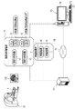
- the image processing device 10 is connected to the medical information database server 11 via a network 12.
- the medical information database server 11 stores and manages various medical information uploaded from a plurality of medical facilities via the network 12.
- the medical information includes a head MRI (Magnetic Resonance Imaging) image 13 and a medical examination result 14.
- the network 12 is a WAN (Wide Area Network) such as the Internet or a public communication network.
- WAN Wide Area Network
- HTTPS Hypertext Transfer Protocol
- the image processing device 10 receives the head MRI image group 15 and the test result group 16 delivered from the medical information database server 11.
- the head MRI image group 15 is a head MRI image 13 approved to be provided by a medical facility, and is, for example, a plurality of head MRI images 13 taken at a plurality of medical facilities from 10 years ago to 2 years ago. including.
- the test result group 16 is a test result 14 approved to be provided by the medical facility, and includes a plurality of test results 14 of the same patient P as the head MRI image 13 of the head MRI image group 15.
- the head MRI image 13 is obtained by photographing the head of the patient P with the MRI device 17.
- the head MRI image 13 is voxel data representing the three-dimensional shape of the head of the patient P.
- FIG. 1 shows a head MRI image 13S of a sagittal cross section.
- the head MRI image 13 is an example of a "medical image” and an "image showing the patient's head” according to the technique of the present disclosure.
- Class 18 is associated with the head MRI image 13, whereby the head MRI image 13 is classified.
- "A” is registered if the patient P of the head MRI image 13 develops dementia two years later
- "B” is registered if the patient P does not develop dementia two years later. To. Registration for class 18 is performed by the attending physician of patient P.
- the image processing device 10 is, for example, a desktop personal computer, and includes a display 19 and an input device 20.
- the input device 20 is a keyboard, a mouse, a touch panel, a microphone, or the like.
- the operator of the image processing device 10 operates the input device 20 to transmit a distribution request for the head MRI image group 15 and the test result group 16 to the medical information database server 11.
- the medical information database server 11 searches for the head MRI image group 15 and the test result group 16 requested for distribution and distributes them to the image processing device 10.
- the head MRI image 13 and the examination result 14 are associated with each other by, for example, a patient ID (Identification Data) 22 for uniquely identifying the patient P.
- the test result 14 is composed of a blood test result, a cerebrospinal fluid test result, a dementia test score, and the like.
- the test result of the blood test includes, for example, an apolypoprotein measured value, a complement protein measured value, a transsiletin measured value, and the like.
- the test result of the cerebrospinal fluid test includes, for example, an amyloid ⁇ measurement value, a tau protein measurement value, and the like.
- the scores of the dementia test are, for example, the Hasegawa dementia scale (HDS-R; Revised Hasegawa's Dementia Scale) score, the Mini-Mental State Examination (MMSE; Mini-Mental State Examination) score, and the River Mead Behavior Memory Test (RBMT). Rivermead Behavioral Memory Test (Score), Clinical Dementia Rating Scale (CDR), Daily Life Activity (ADL), Actives of Daily Living, and Alzheimer's Disease Rating (Alzheimer's Disease) Includes Assistance Scale-cognitive subscale) and the like.
- HDS-R Revised Hasegawa's Dementia Scale
- MMSE Mini-Mental State Examination
- RBMT River Mead Behavior Memory Test
- the computer constituting the image processing device 10 includes a storage 25, a memory 26, a CPU (Central Processing Unit) 27, and a communication unit 28 in addition to the display 19 and the input device 20 described above. I have. These are interconnected via a bus line 29.
- the CPU 27 is an example of a "processor" according to the technique of the present disclosure.
- the storage 25 is a hard disk drive built in the computer constituting the image processing device 10 or connected via a cable or a network. Alternatively, the storage 25 is a disk array in which a plurality of hard disk drives are connected. The storage 25 stores control programs such as an operating system, various application programs, and various data associated with these programs. A solid state drive may be used instead of the hard disk drive.
- the memory 26 is a work memory for the CPU 27 to execute a process.
- the CPU 27 loads the program stored in the storage 25 into the memory 26 and executes the process according to the program.
- the communication unit 28 controls transmission of various information with an external device such as the medical information database server 11.
- the memory 26 may be built in the CPU 27.
- the operation program 30 is stored in the storage 25 of the image processing device 10.
- the operation program 30 is an application program for operating the computer as the image processing device 10. That is, the operation program 30 is an example of the "operation program of the image processing device" according to the technique of the present disclosure.
- the storage 25 also stores a standard head MRI image (hereinafter abbreviated as a standard image) 35 and a learning data group 36.
- the learning data group 36 includes a plurality of learning head MRI images (hereinafter abbreviated as learning images) 81 (see FIG. 11) used for learning the dementia finding derivation model 80 (see FIG. 11) and a plurality of learning data groups 36. It is a set of inspection results 82 (see FIG. 11).
- the learning data group 36 includes a normalized head MRI image group (hereinafter abbreviated as a normalized image group) 37, a new head MRI image group (hereinafter abbreviated as a new image group) 38, a test result group 16, and a new one. Includes test result group 39.
- the normalized image group 37 is composed of a plurality of normalized head MRI images (hereinafter, abbreviated as normalized images) 40.
- the new image group 38 is composed of a plurality of first new head MRI images (hereinafter abbreviated as first new image) 41_1 and a plurality of second new head MRI images (hereinafter abbreviated as second new image) 41_2. (See FIG. 11).
- the new test result group 39 is composed of a plurality of first new test results 42_1 and a plurality of second new test results 42_1 (see FIG. 11).
- the first new image 41_1 and the second new image 41_2 are examples of "new medical images" according to the technique of the present disclosure.
- the first new inspection result 42_1 and the second new inspection result 42_2 are examples of "new inspection results" according to the technique of the present disclosure.
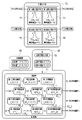
- the CPU 27 of the computer constituting the image processing device 10 cooperates with the memory 26 and the like to read / write (hereinafter abbreviated as RW (Read Write)) control unit 45 and normalization unit. It functions as 46, a non-linear alignment unit 47, and a generation unit 48.
- RW Read Write
- the RW control unit 45 controls the storage of various data in the storage 25 and the reading of various data in the storage 25. For example, the RW control unit 45 reads the standard image 35 from the storage 25 and outputs the read standard image 35 to the normalization unit 46. Further, the RW control unit 45 receives the test result group 16 from the medical information database server 11, and stores the received test result group 16 in the storage 25 as a part of the learning data group 36. Further, the RW control unit 45 receives the normalized image group 37 from the normalized unit 46, and stores the received normalized image group 37 in the storage 25 as a part of the learning data group 36.
- the RW control unit 45 reads out the first inspection result 14_1 and the second inspection result 14_2, which are two inspection results 14 out of the plurality of inspection results 14 of the inspection result group 16, from the storage 25, and reads out the first inspection result 14_1. And the second inspection result 14_2 is output to the generation unit 48. Further, the RW control unit 45 is two normalized images 40 out of a plurality of normalized images 40 of the normalized image group 37, and is a first normalized image corresponding to the first inspection result 14_1 and the second inspection result 14_2. The normalized image 40_1 and the second normalized image 40_1 are read from the storage 25, and the read first normalized image 40_1 and the second normalized image 40_2 are output to the nonlinear alignment unit 47 and the generation unit 48.
- the first test result 14_1 and the second test result 14_2, the first normalized image 40_1, and the second normalized image 40_1 are the test result 14 and the normalized image 40 of two patients P having the same attributes such as gender and age. ..
- the normalized image 40 is associated with a class 18 that follows the original head MRI image 13 (see FIGS. 9 and 10).
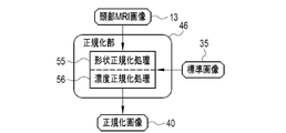
- the normalization unit 46 performs a normalization process for matching the head MRI image 13 with the standard image 35, and sets the head MRI image 13 as the normalized image 40.
- the normalization unit 46 performs normalization processing on all of the plurality of head MRI images 13 constituting the head MRI image group 15. As a result, a plurality of normalized images 40 corresponding to the plurality of head MRI images 13 constituting the head MRI image group 15 are generated.
- the normalization unit 46 outputs a normalized image group 37 composed of a plurality of normalized images 40 to the RW control unit 45.
- the standard image 35 is a head MRI image showing the brain of a standard shape, size, and density (pixel value).
- the standard image 35 is, for example, an image generated by averaging the head MRI images 13 of a plurality of healthy subjects, or an image generated by computer graphics.
- the standard image 35 is an example of a “standard medical image” according to the technique of the present disclosure.
- the non-linear alignment unit 47 performs a non-linear alignment process on the first normalized image 40_1 and the second normalized image 40_1.
- the non-linear alignment unit 47 outputs the processing result 50, which is the result of the non-linear alignment processing, to the generation unit 48.
- the generation unit 48 generates the first new image 41_1 from the first normalized image 40_1 by transforming the first normalized image 40_1 and the second normalized image 40_1 under the generation conditions according to the processing result 50.
- the second new image 41_2 is generated from the second normalized image 40_2.
- the generation unit 48 generates the first new inspection result 42_1 corresponding to the first new image 41_1 from the first inspection result 14_1, and the second new inspection result corresponding to the second inspection result 14_2 to the second new image 41_2.
- the generation unit 48 outputs the first new image 41_1, the first new inspection result 42_1, the second new image 41_2, and the second new inspection result 42_2 to the RW control unit 45.
- the RW control unit 45 stores the first new image 41_1 and the second new image 41_2 in the storage 25 as a part of the new image group 38 and eventually the learning data group 36. Further, the RW control unit 45 stores the first new inspection result 42_1 and the second new inspection result 42_1 in the storage 25 as a part of the new inspection result group 39 and the learning data group 36.
- the first new image 41_1 is an example of the "first new medical image” according to the technique of the present disclosure. Further, the second new image 41_2 is an example of the "second new medical image” according to the technique of the present disclosure.
- the normalization unit 46 performs shape normalization processing 55 and density normalization processing 56 as normalization processing on the head MRI image 13.
- the shape normalization process 55 extracts, for example, a landmark that serves as a reference for alignment from the head MRI image 13 and the standard image 35, and correlates the landmark of the head MRI image 13 with the landmark of the standard image 35.
- the density normalization process 56 is, for example, a process of correcting the density histogram of the head MRI image 13 according to the density histogram of the standard image 35.
- the non-linear alignment unit 47 uses the first normalized image 40_1 (denoted as I_1 (X, Y, Z)) as the second normalized image 40_1 (I_1 (X, Y, Z)).
- a plurality of control points 60 arranged at equal intervals in a grid pattern are set in the first normalized image 40_1 when the non-linear alignment is performed. Then, each control point 60 is moved to a position where the local similarity between the first normalized image 40_1 and the second normalized image 40_1 is increased.
- the non-linear alignment unit 47 aligns the first normalized image 40_1 with the second normalized image 40_1 from the movement amount of each control point 60 by using an approximation curve for interpolation such as a B-Spline curve.
- the amount of pixel deformation T_12 (X, Y, Z) is derived.
- the non-linear alignment unit 47 outputs the derived deformation amount T_12 (X, Y, Z) as the processing result 50.
- the non-linear alignment unit 47 sets the control point 63 similar to the control point 60 to the second normalized image 40_1 when the second normalized image 40_1 is non-linearly aligned with the first normalized image 40_1. Set to the normalized image 40_2. Then, each control point 63 is moved in the same manner as in the case of FIG. Similar to the case of FIG. 6, the non-linear alignment unit 47 has a deformation amount T_21 (deformation amount T_1) of each pixel when the second normalized image 40_1 is aligned with the first normalized image 40_1 from the movement amount of each control point 63. X, Y, Z) is derived. The non-linear alignment unit 47 outputs the derived deformation amount T_21 (X, Y, Z) as the processing result 50. In the following, (X, Y, Z) may be omitted.
- the deformation amount T_12 (X, Y, Z) and the deformation amount T_21 (X, Y, Z) have an inverse function relationship
- the deformation amount T_12 (X, Y, Z) and the deformation amount T_21 (X, Y) , Z) may be derived by the method using the above control points 60 or 63, and then the other may be derived by obtaining the inverse function of one of them.
- the generation unit 48 has a deformation coefficient generation unit 70 and a calculation unit 71.
- the deformation coefficient generation unit 70 generates deformation coefficients ⁇ (X, Y, Z) and ⁇ (X, Y, Z) based on the normal distribution group 72.
- the deformation coefficient generation unit 70 outputs the generated deformation coefficients ⁇ (X, Y, Z) and ⁇ (X, Y, Z) to the calculation unit 71.
- the deformation coefficients ⁇ (X, Y, Z) and ⁇ (X, Y, Z) are values larger than 0 and less than 1 (0 ⁇ , ⁇ ⁇ 1).
- the normal distribution group 72 includes a normal distribution 73 for the first ⁇ , a normal distribution 74 for the first ⁇ , a normal distribution 75 for the second ⁇ , and a normal distribution 76 for the second ⁇ .
- the deformation coefficient generation unit 70 generates a random number according to any one of the normal distribution 73 for the first ⁇ , the normal distribution 74 for the first ⁇ , the normal distribution 75 for the second ⁇ , and the normal distribution 76 for the second ⁇ . It is output as deformation coefficients ⁇ (X, Y, Z) and ⁇ (X, Y, Z).
- the frequency of occurrence of random numbers is, for example, for each pixel.
- the normal distribution 73 for the first ⁇ and the normal distribution 74 for the first ⁇ are used when the class 18 of the first normalized image 40_1 and the second normalized image 40_2 is the same.
- the normal distribution 75 for the second ⁇ and the normal distribution 76 for the second ⁇ are used when the class 18 of the first normalized image 40_1 and the second normalized image 40_2 are different.
- the average ⁇ of the 2nd ⁇ normal distribution 75 and the 2nd ⁇ normal distribution 76 is smaller than the average ⁇ of the 1st ⁇ normal distribution 73 and the 1st ⁇ normal distribution 74. Therefore, the values of the deformation coefficients ⁇ (X, Y, Z) and ⁇ (X, Y, Z) are different depending on whether the class 18 of the first normalized image 40_1 and the second normalized image 40_1 are the same or different. Be changed.
- the 1st ⁇ normal distribution 73 and the 1st ⁇ normal distribution 74 are examples of the “normal distribution when the classes of the first medical image and the second medical image are the same” according to the technique of the present disclosure. Further, the normal distribution 75 for the second ⁇ and the normal distribution 76 for the second ⁇ are examples of the “normal distribution when the classes of the first medical image and the second medical image are different” according to the technique of the present disclosure.
- the calculation unit 71 multiplies the deformation coefficient T_1 (X, Y, Z) from the first normalized image 40_1 to the second normalized image 40_1 by the deformation coefficient ⁇ (X, Y, Z), and corrects the deformed amount ⁇ T_12. Let it be (X, Y, Z). Further, the calculation unit 71 multiplies the deformation coefficient ⁇ (X, Y, Z) by the transformation amount T_1 (X, Y, Z) from the second normalized image 40_1 to the first normalized image 40_1 to correct and transform. The amount is ⁇ T_21 (X, Y, Z).
- the calculation unit 71 applies the correction deformation amount ⁇ T_1 (X, Y, Z) to the first normalized image 40_1 to change the first normalized image 40_1 to the first new image 41_1 (I_1N (X, Y, Z)). Notation). Further, the calculation unit 71 applies the correction deformation amount ⁇ T_21 (X, Y, Z) to the second normalized image 40_2 to change the second normalized image 40_2 to the second new image 41_2 (I_2N (X, Y,). Notated as Z)).
- the corrected deformation amount ⁇ T_12 (X, Y, Z) and the corrected deformation amount ⁇ T_21 (X, Y, Z) are examples of the “generation conditions” according to the technique of the present disclosure.
- the calculation unit 71 calculates the following equation (1), which is a weighted average of the first inspection result 14_1 (denoted as MTR_1) and the second inspection result 14_2 (denoted as MTR_1), which is a weighted average by the coefficient ⁇ F. Then, the first inspection result 14_1 and the second inspection result 14_2 are converted into the first new inspection result 42_1 (denoted as MTR_1N).
- the coefficient ⁇ F is a fixed value and is an example of a “value similar to the deformation coefficient ⁇ ” according to the technique of the present disclosure.
- the coefficient ⁇ F is 0, which is the same as the average ⁇ of the normal distribution 73 for the first ⁇ , for example. It is .5.
- the coefficient ⁇ F is 0.2, which is the same as the average ⁇ of the second ⁇ normal distribution 75, for example, when the class 18 of the first normalized image 40_1 and the second normalized image 40_1 are different and the second ⁇ normal distribution 75 is used.
- the calculation unit 71 calculates the following equation (2), which is a weighted average of the first inspection result 14_1 and the second inspection result 14_2, which is a weighted average by the coefficient ⁇ F, to obtain the first inspection result 14_1 and the second inspection result 14_1.
- the second inspection result 14_2 is converted into the second new inspection result 42_2 (denoted as MTR_2N).
- the coefficient ⁇ F is also a fixed value, and is an example of a “value similar to the deformation coefficient ⁇ ” according to the technique of the present disclosure.
- the coefficient ⁇ F is 0, which is the same as the mean ⁇ of the normal distribution 74 for the first ⁇ , for example. It is .5.
- the coefficient ⁇ F is 0.2, which is the same as the average ⁇ of the normal distribution 76 for the second ⁇ , for example. be.
- the formula (2) is an example of the "calculation formula” and the "second calculation formula" according to the technique of the present disclosure.
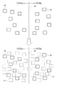
- the generation unit 48 sets the first new image 41_1 to the same class 18 as the first normalized image 40_1. More specifically, when the class 18 of the first normalized image 40_1 is “A” as shown in (A), the class 18 of the first new image 41_1 is also set to “A”. Further, as shown in (B), when the class 18 of the first normalized image 40_1 is “B”, the class 18 of the first new image 41_1 is also set to “B”.
- the generation unit 48 sets the second new image 41_2 to the same class 18 as the second normalized image 40_2. More specifically, when the class 18 of the second normalized image 40_2 is "A" as shown in (A), the class 18 of the second new image 41_2 is also set to "A”. Further, as shown in (B), when the class 18 of the second normalized image 40_2 is “B”, the class 18 of the second new image 41_2 is also set to “B”.
- An image of one of the new images 41_2 is provided as a learning image 81 of the dementia finding derivation model 80.
- one of a plurality of test results 14 constituting the test result group 16, a plurality of first new test results 42_1 and a plurality of second new test results 42_1 constituting the new test result group 39 is obtained. It is provided as a learning test result 82 of the dementia finding derivation model 80.
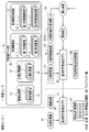
- the dementia finding derivation model 80 is machine learning using the normalized image 40 and the test result 14 as input data and the dementia finding information 83 which is information representing the dementia findings of the patient P of the normalized image 40 as output data. It is a model.
- the dementia finding information 83 is one of normal (NC; Normal Control), mild cognitive impairment (MCI; Mild Cognitive Impairment), and Alzheimer's disease (AD; Alzheimer's Disease).
- the dementia finding derivation model 80 is constructed by, for example, a neural network, a support vector machine, or a boosting method.
- the dementia finding derivation model 80 is given learning data 84, which is a set of a learning image 81, a learning test result 82, and correct dementia finding information 83CA.
- the learning image 81 and the learning test result 82 are the normalized image 40 and the test result 14
- the correct dementia finding information 83CA is actually obtained by the attending physician for the patient P of the normalized image 40 and the test result 14.
- the correct answer dementia finding information 83CA is obtained by the requested doctor referring to the new image 41 and the new test result 42. It is a finding of dementia actually made.
- the learning image 81 and the learning test result 82 are input to the dementia finding derivation model 80.
- the dementia finding derivation model 80 outputs learning dementia finding information 83L for the learning image 81 and the learning test result 82.
- the loss calculation of the dementia finding derivation model 80 is performed.
- various coefficients of the dementia finding derivation model 80 are updated according to the result of the loss calculation, and the dementia finding derivation model 80 is updated according to the update setting.
- the dementia finding derivation model 80 In the learning phase, input of the learning image 81 and the learning test result 82 to the dementia finding derivation model 80, output of the learning dementia finding information 83L from the dementia finding derivation model 80, loss calculation, update setting, And the above series of processes for updating the dementia finding derivation model 80 are repeated while the learning data 84 is exchanged.
- the repetition of the above series of processes ends when the prediction accuracy of the learning dementia finding information 83L with respect to the correct dementia finding information 83CA reaches a predetermined set level.
- the dementia finding derivation model 80 whose prediction accuracy has reached the set level is used as a trained model in the operation phase.
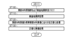
- the operation program 30 when the operation program 30 is started in the image processing device 10, as shown in FIG. 4, the CPU 27 of the image processing device 10 has a RW control unit 45, a normalization unit 46, a nonlinear alignment unit 47, and a generation. It functions as a unit 48. As shown in FIG. 8, the generation unit 48 includes a deformation coefficient generation unit 70 and a calculation unit 71.
- the normalization unit 46 receives the head MRI image group 15 from the medical information database server 11. Further, the RW control unit 45 receives the test result group 16 from the medical information database server 11 (step ST100). The inspection result group 16 is stored in the storage 25 by the RW control unit 45 (step ST110).
- a normalization process for matching the head MRI image 13 with the standard image 35 is performed (step ST120).
- the head MRI image 13 becomes a normalized image 40.
- the normalized image 40 is output from the normalized unit 46 to the RW control unit 45 and stored in the storage 25 by the RW control unit 45 (step ST130).
- the second inspection result 14_2 is read from the storage 25 (step ST200).
- the first normalized image 40_1 and the second normalized image 40_1 are output from the RW control unit 45 to the nonlinear alignment unit 47 and the generation unit 48. Further, the first inspection result 14_1 and the second inspection result 14_2 are output from the RW control unit 45 to the generation unit 48.
- a non-linear alignment process for aligning the first normalized image 40_1 with the second normalized image 40_1 is performed in the non-linear alignment unit 47. Further, as shown in FIG. 7, in the non-linear alignment unit 47, a non-linear alignment process for aligning the second normalized image 40_1 with the first normalized image 40_1 is performed (step ST210).
- the deformation amounts T_12 and T_21 derived by these non-linear alignment processings are output from the non-linear alignment unit 47 to the generation unit 48 as the processing result 50.
- the correction deformation amount ⁇ T_1 obtained by multiplying the deformation amount T_1 by the deformation coefficient ⁇ is applied to the first normalized image 40_1, and the first normalized image 40_1 to the first new image 41_1 Is generated.
- the correction deformation amount ⁇ T_21 obtained by multiplying the deformation amount T_21 by the deformation coefficient ⁇ is applied to the second normalized image 40_2, and the second new image 41_2 is generated from the second normalized image 40_2.
- the first new inspection result 42_1 corresponding to the first new image 41_1 is generated by using the above equation (1), that is, the first arithmetic expression.
- the second new inspection result 42_2 corresponding to the second new image 41_2 is generated by using the above equation (2), that is, the second arithmetic expression (step ST220).
- the first new image 41_1 and the first new inspection result 42_1 and the second new image 41_2 and the second new inspection result 42_1 are output from the generation unit 48 to the RW control unit 45 and stored in the storage 25 by the RW control unit 45.
- Step ST230 In the processing of these series of steps ST200 to ST230, the total number of images and test results in the training data group 36, that is, the number of training images 81 and training test results 82 does not reach the target number (in step ST240). NO) is repeated while changing the combination of the first normalized image 40_1 and the first inspection result 14_1 and the second normalized image 40_1 and the second inspection result 14_2.
- the generation unit 48 of the CPU 27 of the image processing device 10 uses a plurality of normalized images as training data 84 used for learning the dementia finding derivation model 80 for the normalized image 40 and the test result 14. From the first normalized image 40_1 and the second normalized image 40_2 out of 40, the first new image 41_1 and the second new image 41_2 are generated according to the generation conditions. In addition, the generation unit 48, as learning data 84, with respect to the first test result 14_1 of the medical test corresponding to the first normalized image 40_1 and the second test result 14_2 of the medical test corresponding to the second normalized image 40_1. The first new inspection result 42_1 and the second new inspection result 42_1 are generated by performing an operation based on the generation conditions. Therefore, it is possible to generate test results of new medical tests that are consistent.
- the non-linear alignment unit 47 performs a non-linear alignment process on the first normalized image 40_1 and the second normalized image 40_1.
- the generation unit 48 deforms the first normalized image 40_1 and the second normalized image 40_1 under the generation conditions according to the processing result 50 of the nonlinear alignment processing, so that the first new image 41_1 and the second new image 41_2 are transformed. To generate. Further, the generation unit 48 generates the first new inspection result 42_1 corresponding to the first new image 41_1 from the first inspection result 14_1 by using the equations (1) and (2) based on the generation conditions, and second. The second new inspection result 42_2 corresponding to the second new image 41_2 is generated from the inspection result 14_2.
- the first new image 41_1 and the second new image 41_2 are based on the first normalized image 40_1 and the second normalized image 40_2. Therefore, the learning image 81 is generated from one image ⁇ C. Shorten, T.I. M. Compared with the method described in Khoshgofttar: A survive on Image Data Augmentation, Journal of Big Data, 2019>, the variation of the learning image 81 can be increased. Further, since the first new image 41_1 and the second new image 41_2 are not a mixture of the pixel values of the first normalized image 40_1 and the second normalized image 40_2, ⁇ Y. Tokozumi, Y. Ushiku, T.K.
- Harada The morphology of the anatomical structure is not blurred as in the method described in Been-class Learning for Image Classification, CVPR, 2018>. Therefore, it is possible to generate a comprehensive learning image 81 in which the morphology of the anatomical structure is maintained. In addition, it is possible to generate the first new inspection result 42_1 and the second new inspection result 42_1 with more consistency.
- the generation unit 48 applies the corrected deformation amount ⁇ T_1 obtained by multiplying the deformation amount T_1 from the first normalized image 40_1 to the second normalized image 40_1 by the deformation coefficient ⁇ in the non-linear alignment process to the first normalized image 40_1. Then, the first normalized image 40_1 is referred to as the first new image 41_1. Further, the generation unit 48 applies the corrected deformation amount ⁇ T_21 obtained by multiplying the deformation amount T_1 from the second normalized image 40_2 to the first normalized image 40_1 by the deformation coefficient ⁇ in the nonlinear alignment process to the second normalized image 40_2. By doing so, the second normalized image 40_2 becomes the second new image 41_2.
- the desired first new image 41_1 and second new image 41_2 can be obtained. For example, if the deformation coefficient ⁇ is set to a value close to 0, a first new image 41_1 relatively similar to the first normalized image 40_1 can be obtained. On the contrary, if the deformation coefficient ⁇ is set to a value close to 1, a first new image 41_1 relatively similar to the second normalized image 40_2 can be obtained.
- the generation unit 48 converts the first inspection result 14_1 and the second inspection result 14_1 into the first new inspection result 42_1 by calculating the equation (1) including the coefficient ⁇ F. Further, the generation unit 48 converts the first inspection result 14_1 and the second inspection result 14_2 into the second new inspection result 42_2 by calculating the equation (2) including the coefficient ⁇ F. Therefore, by setting the coefficients ⁇ F and ⁇ F to appropriate values, the desired first new test result 42_1 and second new test result 42_1 can be obtained. For example, if the coefficient ⁇ F is set to a value close to 0, a first new test result 42_1 relatively similar to the first test result 14_1 can be obtained. On the contrary, if the deformation coefficient ⁇ is set to a value close to 1, the first new inspection result 42_1 relatively similar to the second inspection result 14_2 can be obtained.
- the head MRI image 13 and eventually the normalized image 40 are classified, and the generation unit 48 is deformed depending on whether the class 18 of the first normalized image 40_1 and the second normalized image 40_2 are the same or different.
- the values of the coefficients ⁇ and ⁇ and the values of the coefficients ⁇ F and ⁇ F are changed. Therefore, the first new image 41_1, the first new inspection result 42_1, and the second new image 41_2 are suitable for each of the cases where the class 18 of the first normalized image 40_1 and the second normalized image 40_1 are the same and different.
- the second new test result 42_2 can be generated.
- the generation unit 48 makes the first new image 41_1 the same class 18 as the first normalized image 40_1, and the second new image 41_2 the same class 18 as the second normalized image 40_2. And. Therefore, it is possible to associate the class 18 corresponding to the origin of each of the first new image 41_1 and the second new image 41_2. Further, as schematically shown in FIG. 14, the data blanks of each class 18 of "A” and "B” that could not be filled only by the normalized image 40 can be filled with the new image 41. In addition, the data gap near the boundary 90 of each class 18 of "A” and "B” can be filled with the new image 41.
- the deformation coefficients ⁇ and ⁇ are random numbers according to any one of the normal distributions 73 to 76. Therefore, the first new image 41_1 and the second new image 41_2, which cannot be predicted from the first normalized image 40_1 and the second normalized image 40_2, can be generated, and as a result, the variation of the learning image 81 is increased. be able to.
- the average ⁇ of the normal distribution 75 for the second ⁇ and the normal distribution 76 for the second ⁇ is the first normalized image. It is smaller than the average ⁇ of the normal distribution 73 for the first ⁇ and the normal distribution 74 for the first ⁇ when the classes of 40_1 and the second normalized image 40_2 are the same. Therefore, when the classes of the first normalized image 40_1 and the second normalized image 40_2 are different, the first normalized image is compared with the case where the first normalized image 40_1 and the second normalized image 40_2 have the same class.
- first new image 41_1 that is relatively similar to 40_1 and a second new image 41_2 that is relatively similar to the second normalized image 40_1. Therefore, even if the first new image 41_1 is in the same class 18 as the first normalized image 40_1 and the second new image 41_2 is in the same class 18 as the second normalized image 40_1, there is no sense of discomfort.
- the normalization unit 46 performs a normalization process for matching the head MRI image 13 with the standard image 35 prior to the generation of the new image 41. Therefore, it is possible to perform the subsequent processing after substantially eliminating the individual difference of the patient P and the device difference of the MRI device 17, and as a result, the reliability of the dementia finding information 83 can be improved.
- this embodiment in which the medical image is the head MRI image 13 and the machine learning model for the medical image is the dementia finding derivation model 80 that outputs the dementia finding information 83, is a current social problem. It can be said that it is a matched form.
- the morphology of the anatomical structure such as the hippocampus, parahippocampal gyrus, and the degree of atrophy of the amygdala holds a particularly important key to the findings of dementia, a learning image 81 in which the morphology of the anatomical structure is maintained is generated. The effect of being able to do it can be more exerted.
- the dementia finding information 83 is not limited to the content exemplified in FIG. 11 (normal / mild cognitive impairment / Alzheimer's disease).
- the degree of progression of dementia one year after the patient P may be fast or slow.
- the type of dementia may be any of Alzheimer's disease, Lewy body dementia, and vascular dementia.
- first new image 41_1 and the first new inspection result 42_1 and the second new image 41_2 and the second new inspection result 42_1 are generated has been exemplified, but the present invention is not limited to this.
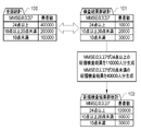
- a new image 41 and a new test result 42 are generated according to the tendency of the test result 14 of the population of the patient P treated by the dementia finding derivation model 80.
- the generation unit 48 generates 110,000 new test results 42 with an MMSE score of 24 points or more in order to match the statistics 102 of the new test result group 39 with the national statistics 100 (and the new image 41 is also 110,000).
- the national statistic 100 is an example of the "population test result tendency" according to the technique of the present disclosure.
- the head MRI image 13 of the head MRI image group 15 and the test result 14 of the test result group 16 are, for example, those of the patient P in the United States, and the patient P handled by the dementia finding derivation model 80 is Japanese, etc.
- the attributes of patient P in the head MRI image 13 and the test result 14, and the attributes of patient P treated in the dementia finding derivation model 80 may be different. Further, even if the attributes of the patient P of the head MRI image 13 and the test result 14 and the attributes of the patient P handled by the dementia finding derivation model 80 are the same, the tendency of the test result 14 of the test result group 16 is the dementia finding derivation. It may be different from the tendency of the test result 14 of the population of the patient P treated in the model 80.
- a new image 41 and a new test result 42 are generated without correcting the tendency of the test result 14 of the test result group 16, and a dementia finding derivation model 80 is generated based on the learning data group 36 prepared as a result.
- the dementia finding derivation model 80 may deviate from the tendency of the population of patient P.
- the new image 41 and the new test result 42 are generated according to the tendency of the test result 14 of the population of the patient P treated by the dementia finding derivation model 80. Therefore, it is possible to generate a dementia finding derivation model 80 that matches the tendency of the population of the patient P.
- the two new images 41 may be subjected to non-linear alignment processing, and the first new image 41_1 and the second new image 41_2 may be generated based on the processing result 50. Similarly, the first new image 41_1 and the second new image 41_2 may be generated from the normalized image 40 and the new image 41.
- Class 18 is not limited to whether or not dementia developed two years after the example. For example, as with the dementia finding information 83, any of normal / mild cognitive impairment / Alzheimer's disease may be registered as class 18.
- the deformation coefficients ⁇ and ⁇ may be fixed values instead of random numbers according to the normal distribution.
- the average ⁇ of the normal distribution 73 for the first ⁇ and the normal distribution 74 for the first ⁇ when the classes of the first normalized image 40_1 and the second normalized image 40_2 are the same is not limited to 0.5 in the example. For example, it may be 0.4 or 0.6.
- the coefficients ⁇ F and ⁇ F may also be, for example, 0.4 or 0.6.
- the average ⁇ of the normal distribution 75 for the second ⁇ and the normal distribution 76 for the second ⁇ when the classes of the first normalized image 40_1 and the second normalized image 40_2 are different is not limited to 0.2 in the example, and is, for example, 0. It may be 0.1 or 0.3.
- the coefficients ⁇ F and ⁇ F may also be, for example, 0.1 or 0.3.
- the standard deviation ⁇ is also not limited to 0.2 in the example.
- the learning of the dementia finding derivation model 80 shown in FIG. 11 may be performed by the image processing device 10 or may be performed by a device other than the image processing device 10.
- the image processing apparatus 10 can actually input the normalized image 40 and the test result 14 into the dementia finding derivation model 80 and output the dementia finding information 83 from the dementia finding derivation model 80.
- it may be performed by an apparatus other than the image processing apparatus 10.
- the learning of the dementia finding derivation model 80 may be continued even after the operation.
- the medical information database server 11 may be responsible for some or all of the functions of the processing units 45 to 48.
- the normalization unit 46 may be constructed in the CPU of the medical information database server 11
- the non-linear alignment unit 47 and the generation unit 48 may be constructed in the CPU of the image processing device 10.
- the medical image is not limited to the illustrated head MRI image 13.
- PET Positron Emission Tomography
- SPECT Single Photon Emission Tomography
- CT Computed Tomography
- the subject is not limited to the illustrated head, but may be the chest, abdomen, etc.
- the disease is not limited to the exemplified dementia, and may be heart disease, pneumonia, liver dysfunction, or the like.
- a processing unit that executes various processes such as a RW control unit 45, a normalization unit 46, a nonlinear alignment unit 47, a generation unit 48, a deformation coefficient generation unit 70, and a calculation unit 71.
- various processors shown below can be used.
- the CPU 27 which is a general-purpose processor that executes software (operation program 30) and functions as various processing units, after manufacturing an FPGA (Field Programmable Gate Array) or the like.
- Dedicated processor with a circuit configuration specially designed to execute specific processing such as programmable logic device (PLD), ASIC (Application Specific Integrated Circuit), which is a processor whose circuit configuration can be changed. Includes electrical circuits and the like.
- One processing unit may be composed of one of these various processors, or may be a combination of two or more processors of the same type or different types (for example, a combination of a plurality of FPGAs and / or a CPU). It may be configured in combination with FPGA). Further, a plurality of processing units may be configured by one processor.
- one processor is configured by a combination of one or more CPUs and software, as represented by a computer such as a client and a server.
- the processor functions as a plurality of processing units.
- SoC System On Chip
- SoC system On Chip
- the various processing units are configured by using one or more of the above-mentioned various processors as a hardware-like structure.
- an electric circuit in which circuit elements such as semiconductor elements are combined can be used.
- the technique of the present disclosure can be appropriately combined with the various embodiments described above and / or various modifications. Further, it is of course not limited to each of the above embodiments, and various configurations can be adopted as long as they do not deviate from the gist. Further, the technique of the present disclosure extends to a storage medium for storing the program non-temporarily in addition to the program.
- a and / or B is synonymous with "at least one of A and B". That is, “A and / or B” means that it may be A alone, B alone, or a combination of A and B. Further, in the present specification, when three or more matters are connected and expressed by "and / or", the same concept as “A and / or B" is applied.
Landscapes
- Engineering & Computer Science (AREA)
- Health & Medical Sciences (AREA)
- Physics & Mathematics (AREA)
- Theoretical Computer Science (AREA)
- Life Sciences & Earth Sciences (AREA)
- General Health & Medical Sciences (AREA)
- Medical Informatics (AREA)
- Computer Vision & Pattern Recognition (AREA)
- General Physics & Mathematics (AREA)
- Artificial Intelligence (AREA)
- Multimedia (AREA)
- Nuclear Medicine, Radiotherapy & Molecular Imaging (AREA)
- Evolutionary Computation (AREA)
- Radiology & Medical Imaging (AREA)
- Computing Systems (AREA)
- Databases & Information Systems (AREA)
- Software Systems (AREA)
- Pathology (AREA)
- Neurology (AREA)
- Biophysics (AREA)
- Biomedical Technology (AREA)
- Heart & Thoracic Surgery (AREA)
- Molecular Biology (AREA)
- Surgery (AREA)
- Animal Behavior & Ethology (AREA)
- Public Health (AREA)
- Veterinary Medicine (AREA)
- Quality & Reliability (AREA)
- Physiology (AREA)
- Psychiatry (AREA)
- Child & Adolescent Psychology (AREA)
- Hospice & Palliative Care (AREA)
- Developmental Disabilities (AREA)
- Psychology (AREA)
- Neurosurgery (AREA)
- High Energy & Nuclear Physics (AREA)
- Fuzzy Systems (AREA)
- Mathematical Physics (AREA)
- Signal Processing (AREA)
- Measuring And Recording Apparatus For Diagnosis (AREA)
Abstract
Description
一例として図1に示すように、画像処理装置10は、医療情報データベースサーバ11にネットワーク12を介して接続されている。医療情報データベースサーバ11は、複数の医療施設からネットワーク12経由でアップロードされた様々な医療情報を記憶し、管理する。医療情報には、頭部MRI(Magnetic Resonance Imaging)画像13および医療検査の検査結果14が含まれる。ネットワーク12は、例えばインターネットまたは公衆通信網等のWAN(Wide Area Network)である。なお、WANを利用する場合には、情報セキュリティを考慮して、VPN(Virtual Private Network)を構築したり、HTTPS(Hypertext Transfer Protocol Secure)等のセキュリティレベルの高い通信プロトコルを使用することが好ましい。 [First Embodiment]
As an example, as shown in FIG. 1, the
(1-αF)×MTR_1+αF×MTR_2=MTR_1N・・・(1) The
(1-αF) × MTR_1 + αF × MTR_2 = MTR_1N ... (1)
βF×MTR_1+(1-βF)×MTR_2=MTR_2N・・・(2) Further, the
βF × MTR_1 + (1-βF) × MTR_2 = MTR_2N ... (2)
図17に示す第2実施形態では、認知症所見導出モデル80で扱う患者Pの母集団の検査結果14の傾向に合わせて、新規画像41および新規検査結果42を生成する。 [Second Embodiment]
In the second embodiment shown in FIG. 17, a
0.5×MTR_1+0.5×MTR_2=MTR_N・・・(3) <Y. Tokozumi, Y.M. Ushiku, T.K. Harada: The
0.5 × MTR_1 + 0.5 × MTR_2 = MTR_N ... (3)
Claims (12)
- プロセッサと、
前記プロセッサに接続または内蔵されたメモリと、を備え、
前記プロセッサは、
医用画像および医療検査の検査結果を対象とした機械学習モデルの学習に用いる学習データとして、
複数の前記医用画像のうちの第1医用画像と第2医用画像から、生成条件にしたがって新規医用画像を生成し、
かつ、前記第1医用画像に対応する前記医療検査の第1検査結果、および前記第2医用画像に対応する前記医療検査の第2検査結果に対して前記生成条件に基づく演算を行い、新規検査結果を生成する、
画像処理装置。 With the processor
With memory connected to or built into the processor,
The processor
As learning data used for learning machine learning models for medical images and test results of medical examinations
A new medical image is generated from the first medical image and the second medical image among the plurality of medical images according to the generation conditions.
In addition, a calculation based on the generation conditions is performed on the first test result of the medical test corresponding to the first medical image and the second test result of the medical test corresponding to the second medical image, and a new test is performed. Produce results,
Image processing device. - 前記プロセッサは、
前記第1医用画像と前記第2医用画像とに非線形位置合わせ処理を行い、
前記非線形位置合わせ処理の結果に応じた前記生成条件にて前記第1医用画像と前記第2医用画像を変形することで、第1新規医用画像と第2新規医用画像を生成し、
前記生成条件に基づく演算式を用いて、前記第1検査結果と前記第2検査結果から前記第1新規医用画像に対応する第1新規検査結果と前記第2新規医用画像に対応する第2新規検査結果を生成する請求項1に記載の画像処理装置。 The processor
Non-linear alignment processing was performed on the first medical image and the second medical image, and then
By transforming the first medical image and the second medical image under the generation conditions according to the result of the non-linear alignment process, a first new medical image and a second new medical image are generated.
Using the calculation formula based on the generation condition, the first new test result corresponding to the first new medical image and the second new corresponding to the second new medical image from the first test result and the second test result. The image processing apparatus according to claim 1, which generates an inspection result. - 前記プロセッサは、
前記非線形位置合わせ処理における前記第1医用画像から前記第2医用画像への変形量T_12に変形係数αを乗算した補正変形量αT_12を前記第1医用画像に適用することで、前記第1医用画像を前記第1新規医用画像とし、かつ、前記変形係数αまたは前記変形係数αに類する値を含む第1演算式を演算することで、前記第1検査結果および前記第2検査結果を前記第1新規検査結果に換算し、
前記非線形位置合わせ処理における前記第2医用画像から前記第1医用画像への変形量T_21に変形係数βを乗算した補正変形量βT_21を前記第2医用画像に適用することで、前記第2医用画像を前記第2新規医用画像とし、かつ、前記変形係数βまたは前記変形係数βに類する値を含む第2演算式を演算することで、前記第1検査結果および前記第2検査結果を前記第2新規検査結果に換算する請求項2に記載の画像処理装置。 The processor
The first medical image is obtained by applying the corrected deformation amount αT_12 obtained by multiplying the deformation amount T_12 from the first medical image to the second medical image by the deformation coefficient α in the non-linear alignment process to the first medical image. Is the first new medical image, and the first test result and the second test result are obtained by calculating the first arithmetic expression including the deformation coefficient α or a value similar to the deformation coefficient α. Converted to new inspection results,
The second medical image is obtained by applying the corrected deformation amount βT_21 obtained by multiplying the deformation amount T_21 from the second medical image to the first medical image by the deformation coefficient β in the non-linear alignment process to the second medical image. Is the second new medical image, and the first inspection result and the second inspection result are obtained by calculating the second arithmetic expression including the deformation coefficient β or the value similar to the deformation coefficient β. The image processing apparatus according to claim 2, which is converted into a new inspection result. - 前記医用画像はクラス分けがなされており、
前記プロセッサは、
前記第1医用画像と前記第2医用画像のクラスが同じ場合と異なる場合とで、前記変形係数αおよびβの値と、前記変形係数αに類する値および前記変形係数βに類する値とのうちの少なくともいずれかを変更する請求項3に記載の画像処理装置。 The medical images are classified and classified.
The processor
Of the values of the deformation coefficients α and β, the values similar to the deformation coefficient α, and the values similar to the deformation coefficient β, depending on whether the first medical image and the second medical image have the same class or different classes. The image processing apparatus according to claim 3, wherein at least one of the above is changed. - 前記プロセッサは、
前記第1新規医用画像を前記第1医用画像と同じクラスとし、
前記第2新規医用画像を前記第2医用画像と同じクラスとする請求項4に記載の画像処理装置。 The processor
The first new medical image is in the same class as the first medical image.
The image processing apparatus according to claim 4, wherein the second new medical image is in the same class as the second medical image. - 前記変形係数αおよびβは、正規分布にしたがう乱数である請求項3から請求項5のいずれか1項に記載の画像処理装置。 The image processing apparatus according to any one of claims 3 to 5, wherein the deformation coefficients α and β are random numbers according to a normal distribution.
- 請求項4または請求項5を引用する請求項6に記載の画像処理装置において、
前記第1医用画像と前記第2医用画像のクラスが異なる場合の正規分布の平均は、前記第1医用画像と前記第2医用画像のクラスが同じ場合の正規分布の平均よりも小さい画像処理装置。 In the image processing apparatus according to claim 6, which cites claim 4 or claim 5.
The average of the normal distribution when the classes of the first medical image and the second medical image are different is smaller than the average of the normal distribution when the classes of the first medical image and the second medical image are the same. .. - 前記プロセッサは、
前記機械学習モデルで扱う患者の母集団の前記検査結果の傾向に合わせて、前記新規医用画像および前記新規検査結果を生成する請求項1から請求項7のいずれか1項に記載の画像処理装置。 The processor
The image processing apparatus according to any one of claims 1 to 7, which generates the new medical image and the new test result according to the tendency of the test result of the population of patients treated by the machine learning model. .. - 前記プロセッサは、
前記新規医用画像の生成に先立ち、前記第1医用画像と前記第2医用画像を標準医用画像に合わせる正規化処理を行う請求項1から請求項8のいずれか1項に記載の画像処理装置。 The processor
The image processing apparatus according to any one of claims 1 to 8, wherein a normalization process for matching the first medical image and the second medical image with a standard medical image is performed prior to the generation of the new medical image. - 前記医用画像は、患者の頭部を写した画像であり、
前記機械学習モデルは、前記患者に対する認知症の所見を出力するモデルである請求項1から請求項9のいずれか1項に記載の画像処理装置。 The medical image is an image of the patient's head.
The image processing apparatus according to any one of claims 1 to 9, wherein the machine learning model is a model that outputs findings of dementia for the patient. - 医用画像および医療検査の検査結果を対象とした機械学習モデルの学習に用いる学習データとして、
複数の前記医用画像のうちの第1医用画像と第2医用画像から、生成条件にしたがって新規医用画像を生成すること、および、
前記第1医用画像に対応する前記医療検査の第1検査結果、および前記第2医用画像に対応する前記医療検査の第2検査結果に対して前記生成条件に基づく演算を行い、新規検査結果を生成すること、
を含む画像処理装置の作動方法。 As learning data used for learning machine learning models for medical images and test results of medical examinations
A new medical image is generated from the first medical image and the second medical image among the plurality of medical images according to the generation conditions, and
Calculations based on the generation conditions are performed on the first test result of the medical test corresponding to the first medical image and the second test result of the medical test corresponding to the second medical image, and a new test result is obtained. To generate,
How to operate an image processing device, including. - 医用画像および医療検査の検査結果を対象とした機械学習モデルの学習に用いる学習データとして、
複数の前記医用画像のうちの第1医用画像と第2医用画像から、生成条件にしたがって新規医用画像を生成すること、および、
前記第1医用画像に対応する前記医療検査の第1検査結果、および前記第2医用画像に対応する前記医療検査の第2検査結果に対して前記生成条件に基づく演算を行い、新規検査結果を生成すること、
を含む処理をコンピュータに実行させる画像処理装置の作動プログラム。 As learning data used for learning machine learning models for medical images and test results of medical examinations
A new medical image is generated from the first medical image and the second medical image among the plurality of medical images according to the generation conditions, and
Calculations based on the generation conditions are performed on the first test result of the medical test corresponding to the first medical image and the second test result of the medical test corresponding to the second medical image, and a new test result is obtained. To generate,
An operation program of an image processing device that causes a computer to perform processing including.
Priority Applications (4)
| Application Number | Priority Date | Filing Date | Title |
|---|---|---|---|
| CN202180064618.2A CN116209396A (en) | 2020-09-28 | 2021-09-09 | Image processing device, method for operating image processing device, and program for operating image processing device |
| JP2022551872A JP7440655B2 (en) | 2020-09-28 | 2021-09-09 | Image processing device, image processing device operating method, image processing device operating program |
| EP21872192.6A EP4218604A4 (en) | 2020-09-28 | 2021-09-09 | Image processing device, image processing device operation method, and image processing device operation program |
| US18/187,649 US20230222656A1 (en) | 2020-09-28 | 2023-03-21 | Image processing apparatus, operation method of image processing apparatus, and operation program of image processing apparatus |
Applications Claiming Priority (2)
| Application Number | Priority Date | Filing Date | Title |
|---|---|---|---|
| JP2020-162679 | 2020-09-28 | ||
| JP2020162679 | 2020-09-28 |
Related Child Applications (1)
| Application Number | Title | Priority Date | Filing Date |
|---|---|---|---|
| US18/187,649 Continuation US20230222656A1 (en) | 2020-09-28 | 2023-03-21 | Image processing apparatus, operation method of image processing apparatus, and operation program of image processing apparatus |
Publications (1)
| Publication Number | Publication Date |
|---|---|
| WO2022065061A1 true WO2022065061A1 (en) | 2022-03-31 |
Family
ID=80846522
Family Applications (1)
| Application Number | Title | Priority Date | Filing Date |
|---|---|---|---|
| PCT/JP2021/033190 WO2022065061A1 (en) | 2020-09-28 | 2021-09-09 | Image processing device, image processing device operation method, and image processing device operation program |
Country Status (5)
| Country | Link |
|---|---|
| US (1) | US20230222656A1 (en) |
| EP (1) | EP4218604A4 (en) |
| JP (1) | JP7440655B2 (en) |
| CN (1) | CN116209396A (en) |
| WO (1) | WO2022065061A1 (en) |
Citations (8)
| Publication number | Priority date | Publication date | Assignee | Title |
|---|---|---|---|---|
| JPH07284090A (en) | 1994-04-08 | 1995-10-27 | Olympus Optical Co Ltd | Picture classifying device |
| JP2008048880A (en) * | 2006-08-24 | 2008-03-06 | Ge Medical Systems Global Technology Co Llc | Image forming method |
| JP2017029461A (en) * | 2015-08-03 | 2017-02-09 | 株式会社日立製作所 | Medical image processor and medical image processing method |
| WO2019172181A1 (en) * | 2018-03-09 | 2019-09-12 | 日本メジフィジックス株式会社 | Diagnosis support device, program, learned model, and learning device |
| JP2019198376A (en) * | 2018-05-14 | 2019-11-21 | キヤノンメディカルシステムズ株式会社 | Medical image processor, medical image processing method, and medical image processing system |
| WO2020054803A1 (en) * | 2018-09-12 | 2020-03-19 | 株式会社Splink | Diagnosis assistance system and method |
| US20200226752A1 (en) * | 2019-01-16 | 2020-07-16 | Samsung Electronics Co., Ltd. | Apparatus and method for processing medical image |
| JP2020144700A (en) * | 2019-03-07 | 2020-09-10 | 株式会社日立製作所 | Diagnostic imaging apparatus, image processing method and program |
-
2021
- 2021-09-09 EP EP21872192.6A patent/EP4218604A4/en active Pending
- 2021-09-09 JP JP2022551872A patent/JP7440655B2/en active Active
- 2021-09-09 WO PCT/JP2021/033190 patent/WO2022065061A1/en active Application Filing
- 2021-09-09 CN CN202180064618.2A patent/CN116209396A/en active Pending
-
2023
- 2023-03-21 US US18/187,649 patent/US20230222656A1/en active Pending
Patent Citations (8)
| Publication number | Priority date | Publication date | Assignee | Title |
|---|---|---|---|---|
| JPH07284090A (en) | 1994-04-08 | 1995-10-27 | Olympus Optical Co Ltd | Picture classifying device |
| JP2008048880A (en) * | 2006-08-24 | 2008-03-06 | Ge Medical Systems Global Technology Co Llc | Image forming method |
| JP2017029461A (en) * | 2015-08-03 | 2017-02-09 | 株式会社日立製作所 | Medical image processor and medical image processing method |
| WO2019172181A1 (en) * | 2018-03-09 | 2019-09-12 | 日本メジフィジックス株式会社 | Diagnosis support device, program, learned model, and learning device |
| JP2019198376A (en) * | 2018-05-14 | 2019-11-21 | キヤノンメディカルシステムズ株式会社 | Medical image processor, medical image processing method, and medical image processing system |
| WO2020054803A1 (en) * | 2018-09-12 | 2020-03-19 | 株式会社Splink | Diagnosis assistance system and method |
| US20200226752A1 (en) * | 2019-01-16 | 2020-07-16 | Samsung Electronics Co., Ltd. | Apparatus and method for processing medical image |
| JP2020144700A (en) * | 2019-03-07 | 2020-09-10 | 株式会社日立製作所 | Diagnostic imaging apparatus, image processing method and program |
Non-Patent Citations (3)
| Title |
|---|
| C. SHORTEN, T. M. KHOSHGOFTTAR: "A survey on Image Data Augmentation", JOURNAL OF BIG DATA, 2019 |
| See also references of EP4218604A4 |
| Y. TOKOZUMEY. USHIKUT. HARADA: "Between-class Learning for Image Classification", CVPR, 2018 |
Also Published As
| Publication number | Publication date |
|---|---|
| JP7440655B2 (en) | 2024-02-28 |
| US20230222656A1 (en) | 2023-07-13 |
| EP4218604A4 (en) | 2024-04-10 |
| CN116209396A (en) | 2023-06-02 |
| EP4218604A1 (en) | 2023-08-02 |
| JPWO2022065061A1 (en) | 2022-03-31 |
Similar Documents
| Publication | Publication Date | Title |
|---|---|---|
| Torosdagli et al. | Deep geodesic learning for segmentation and anatomical landmarking | |
| Klein et al. | Automatic bone segmentation in whole-body CT images | |
| JP7245364B2 (en) | sCT Imaging Using CycleGAN with Deformable Layers | |
| US11080895B2 (en) | Generating simulated body parts for images | |
| Diller et al. | Denoising and artefact removal for transthoracic echocardiographic imaging in congenital heart disease: utility of diagnosis specific deep learning algorithms | |
| Tmenova et al. | CycleGAN for style transfer in X-ray angiography | |
| Rivail et al. | Modeling disease progression in retinal OCTs with longitudinal self-supervised learning | |
| Bindushree et al. | Artificial intelligence: In modern dentistry | |
| US11978208B2 (en) | Trained model, learning method, learning program, medical information acquisition device, medical information acquisition method, and medical information acquisition program | |
| Kläser et al. | Deep boosted regression for MR to CT synthesis | |
| US20210393229A1 (en) | Single or a few views computed tomography imaging with deep neural network | |
| WO2022065061A1 (en) | Image processing device, image processing device operation method, and image processing device operation program | |
| Strittmatter et al. | Deep learning-based affine medical image registration for multimodal minimal-invasive image-guided interventions–A comparative study on generalizability | |
| Nechyporenko et al. | Deep learning based image segmentation for detection of odontogenic maxillary sinusitis | |
| Mangalagiri et al. | Toward generating synthetic CT volumes using a 3D-conditional generative adversarial network | |
| WO2022065060A1 (en) | Image processing device, operation method for image processing device, operation program for image processing device, and trained model | |
| Karacan et al. | A deep learning model with attention mechanism for dental image segmentation | |
| Zhao et al. | Bayesian conditional GAN for MRI brain image synthesis | |
| Tu et al. | Self pre-training with single-scale adapter for left atrial segmentation | |
| WO2020126993A1 (en) | Data augmentation | |
| WO2022065062A1 (en) | Diagnosis assistance apparatus, diagnosis assistance apparatus operation method, and diagnosis assistance apparatus operation program | |
| CN113850710B (en) | Cross-modal medical image accurate conversion method | |
| Kaliyugarasan et al. | Multi-center CNN-based spine segmentation from T2w MRI using small amounts of data | |
| US20240037739A1 (en) | Image processing apparatus, image processing method, and image processing program | |
| WO2022163401A1 (en) | Learning device, learning method, learned model, and program |
Legal Events
| Date | Code | Title | Description |
|---|---|---|---|
| 121 | Ep: the epo has been informed by wipo that ep was designated in this application |
Ref document number: 21872192 Country of ref document: EP Kind code of ref document: A1 |
|
| ENP | Entry into the national phase |
Ref document number: 2022551872 Country of ref document: JP Kind code of ref document: A |
|
| WWE | Wipo information: entry into national phase |
Ref document number: 2021872192 Country of ref document: EP |
|
| NENP | Non-entry into the national phase |
Ref country code: DE |
|
| ENP | Entry into the national phase |
Ref document number: 2021872192 Country of ref document: EP Effective date: 20230428 |