WO2022055893A1 - Pharmaceutical combination and tumor treatment - Google Patents
Pharmaceutical combination and tumor treatment Download PDFInfo
- Publication number
- WO2022055893A1 WO2022055893A1 PCT/US2021/049310 US2021049310W WO2022055893A1 WO 2022055893 A1 WO2022055893 A1 WO 2022055893A1 US 2021049310 W US2021049310 W US 2021049310W WO 2022055893 A1 WO2022055893 A1 WO 2022055893A1
- Authority
- WO
- WIPO (PCT)
- Prior art keywords
- group
- alkyl
- alkoxy
- optionally
- independently selected
- Prior art date
Links
- 206010028980 Neoplasm Diseases 0.000 title claims abstract description 288
- 238000011282 treatment Methods 0.000 title claims abstract description 54
- 239000003112 inhibitor Substances 0.000 claims abstract description 234
- 238000000034 method Methods 0.000 claims abstract description 231
- 101000857888 Homo sapiens Guanine nucleotide-binding protein G(q) subunit alpha Proteins 0.000 claims abstract description 199
- 102100025334 Guanine nucleotide-binding protein G(q) subunit alpha Human genes 0.000 claims abstract description 196
- 201000005969 Uveal melanoma Diseases 0.000 claims abstract description 176
- 229940123924 Protein kinase C inhibitor Drugs 0.000 claims abstract description 153
- 239000003881 protein kinase C inhibitor Substances 0.000 claims abstract description 153
- 101710175516 14 kDa zinc-binding protein Proteins 0.000 claims abstract description 150
- 206010061289 metastatic neoplasm Diseases 0.000 claims abstract description 137
- 230000001394 metastastic effect Effects 0.000 claims abstract description 123
- 230000035772 mutation Effects 0.000 claims abstract description 120
- 101001072407 Homo sapiens Guanine nucleotide-binding protein subunit alpha-11 Proteins 0.000 claims abstract description 62
- 102100036738 Guanine nucleotide-binding protein subunit alpha-11 Human genes 0.000 claims abstract description 60
- 239000000825 pharmaceutical preparation Substances 0.000 claims abstract description 41
- 229940127557 pharmaceutical product Drugs 0.000 claims abstract description 39
- 238000002648 combination therapy Methods 0.000 claims abstract description 28
- 125000006274 (C1-C3)alkoxy group Chemical group 0.000 claims description 197
- 229910052736 halogen Inorganic materials 0.000 claims description 177
- 150000002367 halogens Chemical class 0.000 claims description 177
- 125000006273 (C1-C3) alkyl group Chemical group 0.000 claims description 170
- 238000001574 biopsy Methods 0.000 claims description 146
- 125000002887 hydroxy group Chemical group [H]O* 0.000 claims description 115
- 125000001424 substituent group Chemical group 0.000 claims description 87
- 229910052739 hydrogen Inorganic materials 0.000 claims description 84
- PXGOKWXKJXAPGV-UHFFFAOYSA-N Fluorine Chemical compound FF PXGOKWXKJXAPGV-UHFFFAOYSA-N 0.000 claims description 64
- 229910052731 fluorine Inorganic materials 0.000 claims description 64
- 239000011737 fluorine Substances 0.000 claims description 64
- 125000000217 alkyl group Chemical group 0.000 claims description 51
- 229910052757 nitrogen Inorganic materials 0.000 claims description 50
- 125000000623 heterocyclic group Chemical group 0.000 claims description 48
- 230000000694 effects Effects 0.000 claims description 44
- 239000002146 L01XE16 - Crizotinib Substances 0.000 claims description 43
- KTEIFNKAUNYNJU-GFCCVEGCSA-N crizotinib Chemical group O([C@H](C)C=1C(=C(F)C=CC=1Cl)Cl)C(C(=NC=1)N)=CC=1C(=C1)C=NN1C1CCNCC1 KTEIFNKAUNYNJU-GFCCVEGCSA-N 0.000 claims description 43
- 229960005061 crizotinib Drugs 0.000 claims description 43
- 150000003839 salts Chemical class 0.000 claims description 43
- 125000005842 heteroatom Chemical group 0.000 claims description 41
- 125000006272 (C3-C7) cycloalkyl group Chemical group 0.000 claims description 37
- 229910052760 oxygen Inorganic materials 0.000 claims description 37
- 229910052717 sulfur Inorganic materials 0.000 claims description 37
- 125000002618 bicyclic heterocycle group Chemical group 0.000 claims description 33
- LIOLIMKSCNQPLV-UHFFFAOYSA-N 2-fluoro-n-methyl-4-[7-(quinolin-6-ylmethyl)imidazo[1,2-b][1,2,4]triazin-2-yl]benzamide Chemical compound C1=C(F)C(C(=O)NC)=CC=C1C1=NN2C(CC=3C=C4C=CC=NC4=CC=3)=CN=C2N=C1 LIOLIMKSCNQPLV-UHFFFAOYSA-N 0.000 claims description 31
- 229950005852 capmatinib Drugs 0.000 claims description 31
- 239000008194 pharmaceutical composition Substances 0.000 claims description 31
- 125000002496 methyl group Chemical group [H]C([H])([H])* 0.000 claims description 29
- 239000003937 drug carrier Substances 0.000 claims description 27
- 125000001072 heteroaryl group Chemical group 0.000 claims description 25
- 125000002023 trifluoromethyl group Chemical group FC(F)(F)* 0.000 claims description 20
- 239000000523 sample Substances 0.000 claims description 17
- VGGSQFUCUMXWEO-UHFFFAOYSA-N Ethene Chemical group C=C VGGSQFUCUMXWEO-UHFFFAOYSA-N 0.000 claims description 16
- 239000005977 Ethylene Chemical group 0.000 claims description 16
- IYABWNGZIDDRAK-UHFFFAOYSA-N allene Chemical group C=C=C IYABWNGZIDDRAK-UHFFFAOYSA-N 0.000 claims description 16
- 125000000325 methylidene group Chemical group [H]C([H])=* 0.000 claims description 16
- 238000001262 western blot Methods 0.000 claims description 13
- 238000002965 ELISA Methods 0.000 claims description 6
- 210000002784 stomach Anatomy 0.000 claims description 6
- 208000010507 Adenocarcinoma of Lung Diseases 0.000 claims description 5
- 239000002176 L01XE26 - Cabozantinib Substances 0.000 claims description 5
- UCEQXRCJXIVODC-PMACEKPBSA-N LSM-1131 Chemical compound C1CCC2=CC=CC3=C2N1C=C3[C@@H]1C(=O)NC(=O)[C@H]1C1=CNC2=CC=CC=C12 UCEQXRCJXIVODC-PMACEKPBSA-N 0.000 claims description 5
- ONIQOQHATWINJY-UHFFFAOYSA-N cabozantinib Chemical compound C=12C=C(OC)C(OC)=CC2=NC=CC=1OC(C=C1)=CC=C1NC(=O)C1(C(=O)NC=2C=CC(F)=CC=2)CC1 ONIQOQHATWINJY-UHFFFAOYSA-N 0.000 claims description 5
- 229960001292 cabozantinib Drugs 0.000 claims description 5
- 208000030381 cutaneous melanoma Diseases 0.000 claims description 5
- 238000000684 flow cytometry Methods 0.000 claims description 5
- 201000005249 lung adenocarcinoma Diseases 0.000 claims description 5
- 201000003708 skin melanoma Diseases 0.000 claims description 5
- 229950005976 tivantinib Drugs 0.000 claims description 5
- 210000000481 breast Anatomy 0.000 claims description 4
- 238000010166 immunofluorescence Methods 0.000 claims description 4
- 238000001114 immunoprecipitation Methods 0.000 claims description 4
- 206010073071 hepatocellular carcinoma Diseases 0.000 claims description 3
- 231100000844 hepatocellular carcinoma Toxicity 0.000 claims description 3
- 238000003365 immunocytochemistry Methods 0.000 claims description 3
- 238000012083 mass cytometry Methods 0.000 claims description 3
- 210000003128 head Anatomy 0.000 claims description 2
- 210000003739 neck Anatomy 0.000 claims description 2
- 210000002307 prostate Anatomy 0.000 claims description 2
- 238000011222 transcriptome analysis Methods 0.000 claims 1
- 108090000315 Protein Kinase C Proteins 0.000 abstract description 165
- 102000003923 Protein Kinase C Human genes 0.000 abstract description 165
- 208000037265 diseases, disorders, signs and symptoms Diseases 0.000 abstract description 52
- 201000010099 disease Diseases 0.000 abstract description 49
- 230000002062 proliferating effect Effects 0.000 abstract description 14
- 210000004027 cell Anatomy 0.000 description 233
- 102000003745 Hepatocyte Growth Factor Human genes 0.000 description 117
- 108090000100 Hepatocyte Growth Factor Proteins 0.000 description 116
- 210000001519 tissue Anatomy 0.000 description 85
- 150000001875 compounds Chemical class 0.000 description 69
- 108010029485 Protein Isoforms Proteins 0.000 description 64
- 102000001708 Protein Isoforms Human genes 0.000 description 64
- 239000000203 mixture Substances 0.000 description 56
- XXJXHXJWQSCNPX-UHFFFAOYSA-N NC=1C(=NC(=CN=1)C1=NC=CC=C1C(F)(F)F)C(=O)NC1=NC=CC=C1N1CCC(CC1)(C)N Chemical compound NC=1C(=NC(=CN=1)C1=NC=CC=C1C(F)(F)F)C(=O)NC1=NC=CC=C1N1CCC(CC1)(C)N XXJXHXJWQSCNPX-UHFFFAOYSA-N 0.000 description 55
- 230000014509 gene expression Effects 0.000 description 52
- 101000627861 Homo sapiens Matrix metalloproteinase-28 Proteins 0.000 description 50
- 102100026799 Matrix metalloproteinase-28 Human genes 0.000 description 50
- 229940126062 Compound A Drugs 0.000 description 44
- NLDMNSXOCDLTTB-UHFFFAOYSA-N Heterophylliin A Natural products O1C2COC(=O)C3=CC(O)=C(O)C(O)=C3C3=C(O)C(O)=C(O)C=C3C(=O)OC2C(OC(=O)C=2C=C(O)C(O)=C(O)C=2)C(O)C1OC(=O)C1=CC(O)=C(O)C(O)=C1 NLDMNSXOCDLTTB-UHFFFAOYSA-N 0.000 description 44
- -1 kits Substances 0.000 description 43
- 108090000623 proteins and genes Proteins 0.000 description 43
- 102100021496 Insulin-degrading enzyme Human genes 0.000 description 37
- 235000002639 sodium chloride Nutrition 0.000 description 35
- 230000003833 cell viability Effects 0.000 description 27
- 102000043136 MAP kinase family Human genes 0.000 description 26
- 108091054455 MAP kinase family Proteins 0.000 description 26
- 239000003446 ligand Substances 0.000 description 26
- 239000003814 drug Substances 0.000 description 24
- 210000004881 tumor cell Anatomy 0.000 description 24
- 230000012010 growth Effects 0.000 description 23
- 230000000683 nonmetastatic effect Effects 0.000 description 23
- 230000019491 signal transduction Effects 0.000 description 23
- 230000008901 benefit Effects 0.000 description 22
- 102000004169 proteins and genes Human genes 0.000 description 21
- 229940079593 drug Drugs 0.000 description 20
- 238000002360 preparation method Methods 0.000 description 20
- 230000004044 response Effects 0.000 description 20
- 241000282414 Homo sapiens Species 0.000 description 19
- 108091007960 PI3Ks Proteins 0.000 description 19
- 102000038030 PI3Ks Human genes 0.000 description 19
- 230000004913 activation Effects 0.000 description 19
- 230000035755 proliferation Effects 0.000 description 19
- 238000004458 analytical method Methods 0.000 description 18
- 208000037821 progressive disease Diseases 0.000 description 18
- 108091032973 (ribonucleotides)n+m Proteins 0.000 description 17
- 230000008485 antagonism Effects 0.000 description 17
- 201000011510 cancer Diseases 0.000 description 17
- 210000004185 liver Anatomy 0.000 description 17
- 239000003795 chemical substances by application Substances 0.000 description 16
- 125000005843 halogen group Chemical group 0.000 description 16
- 238000005649 metathesis reaction Methods 0.000 description 16
- 230000011664 signaling Effects 0.000 description 16
- 230000001028 anti-proliverative effect Effects 0.000 description 15
- 239000002552 dosage form Substances 0.000 description 15
- 239000011159 matrix material Substances 0.000 description 15
- 238000006467 substitution reaction Methods 0.000 description 15
- 230000035899 viability Effects 0.000 description 14
- PEDCQBHIVMGVHV-UHFFFAOYSA-N Glycerine Chemical compound OCC(O)CO PEDCQBHIVMGVHV-UHFFFAOYSA-N 0.000 description 13
- 230000002401 inhibitory effect Effects 0.000 description 13
- 230000001225 therapeutic effect Effects 0.000 description 13
- 125000006583 (C1-C3) haloalkyl group Chemical group 0.000 description 12
- 239000004480 active ingredient Substances 0.000 description 12
- 230000003042 antagnostic effect Effects 0.000 description 12
- 125000003118 aryl group Chemical group 0.000 description 12
- 230000003285 pharmacodynamic effect Effects 0.000 description 12
- 238000010186 staining Methods 0.000 description 12
- 208000024891 symptom Diseases 0.000 description 12
- 239000003826 tablet Substances 0.000 description 12
- 101000984753 Homo sapiens Serine/threonine-protein kinase B-raf Proteins 0.000 description 11
- 241000282320 Panthera leo Species 0.000 description 11
- 231100000673 dose–response relationship Toxicity 0.000 description 11
- 238000003364 immunohistochemistry Methods 0.000 description 11
- 239000007788 liquid Substances 0.000 description 11
- 230000037361 pathway Effects 0.000 description 11
- 238000009097 single-agent therapy Methods 0.000 description 11
- 108020004705 Codon Proteins 0.000 description 10
- 102100027103 Serine/threonine-protein kinase B-raf Human genes 0.000 description 10
- 108010017842 Telomerase Proteins 0.000 description 10
- 102100037587 Ubiquitin carboxyl-terminal hydrolase BAP1 Human genes 0.000 description 10
- 125000004438 haloalkoxy group Chemical group 0.000 description 10
- 239000000126 substance Substances 0.000 description 10
- 101001095815 Homo sapiens E3 ubiquitin-protein ligase RING2 Proteins 0.000 description 9
- 101001057193 Homo sapiens Membrane-associated guanylate kinase, WW and PDZ domain-containing protein 1 Proteins 0.000 description 9
- 101000740048 Homo sapiens Ubiquitin carboxyl-terminal hydrolase BAP1 Proteins 0.000 description 9
- 101000740049 Latilactobacillus curvatus Bioactive peptide 1 Proteins 0.000 description 9
- DNIAPMSPPWPWGF-UHFFFAOYSA-N Propylene glycol Chemical compound CC(O)CO DNIAPMSPPWPWGF-UHFFFAOYSA-N 0.000 description 9
- 239000006146 Roswell Park Memorial Institute medium Substances 0.000 description 9
- 230000003213 activating effect Effects 0.000 description 9
- 231100000433 cytotoxic Toxicity 0.000 description 9
- 230000001472 cytotoxic effect Effects 0.000 description 9
- 108020004999 messenger RNA Proteins 0.000 description 9
- 239000000546 pharmaceutical excipient Substances 0.000 description 9
- 239000000243 solution Substances 0.000 description 9
- 102000042846 PKC family Human genes 0.000 description 8
- 108091082203 PKC family Proteins 0.000 description 8
- 102100032938 Telomerase reverse transcriptase Human genes 0.000 description 8
- 239000002775 capsule Substances 0.000 description 8
- 239000000969 carrier Substances 0.000 description 8
- 125000001495 ethyl group Chemical group [H]C([H])([H])C([H])([H])* 0.000 description 8
- 230000006870 function Effects 0.000 description 8
- 238000001727 in vivo Methods 0.000 description 8
- 239000002502 liposome Substances 0.000 description 8
- 239000012528 membrane Substances 0.000 description 8
- 210000004379 membrane Anatomy 0.000 description 8
- 239000002773 nucleotide Substances 0.000 description 8
- 125000003729 nucleotide group Chemical group 0.000 description 8
- 229920001223 polyethylene glycol Polymers 0.000 description 8
- 239000000843 powder Substances 0.000 description 8
- 239000002904 solvent Substances 0.000 description 8
- XLYOFNOQVPJJNP-UHFFFAOYSA-N water Substances O XLYOFNOQVPJJNP-UHFFFAOYSA-N 0.000 description 8
- LFQSCWFLJHTTHZ-UHFFFAOYSA-N Ethanol Chemical compound CCO LFQSCWFLJHTTHZ-UHFFFAOYSA-N 0.000 description 7
- 102100039788 GTPase NRas Human genes 0.000 description 7
- 241000699670 Mus sp. Species 0.000 description 7
- 238000003559 RNA-seq method Methods 0.000 description 7
- 239000000654 additive Substances 0.000 description 7
- 230000000996 additive effect Effects 0.000 description 7
- 238000011260 co-administration Methods 0.000 description 7
- 238000002474 experimental method Methods 0.000 description 7
- 238000009472 formulation Methods 0.000 description 7
- 230000003834 intracellular effect Effects 0.000 description 7
- 239000006187 pill Substances 0.000 description 7
- 239000003755 preservative agent Substances 0.000 description 7
- 239000000725 suspension Substances 0.000 description 7
- 230000002195 synergetic effect Effects 0.000 description 7
- 108020004414 DNA Proteins 0.000 description 6
- 102100030708 GTPase KRas Human genes 0.000 description 6
- 101000744505 Homo sapiens GTPase NRas Proteins 0.000 description 6
- 206010027476 Metastases Diseases 0.000 description 6
- 102100024193 Mitogen-activated protein kinase 1 Human genes 0.000 description 6
- 108091000080 Phosphotransferase Proteins 0.000 description 6
- 102100031711 Splicing factor 3B subunit 1 Human genes 0.000 description 6
- CZMRCDWAGMRECN-UGDNZRGBSA-N Sucrose Chemical compound O[C@H]1[C@H](O)[C@@H](CO)O[C@@]1(CO)O[C@@H]1[C@H](O)[C@@H](O)[C@H](O)[C@@H](CO)O1 CZMRCDWAGMRECN-UGDNZRGBSA-N 0.000 description 6
- 229930006000 Sucrose Natural products 0.000 description 6
- 150000001413 amino acids Chemical group 0.000 description 6
- 238000013459 approach Methods 0.000 description 6
- 239000011575 calcium Substances 0.000 description 6
- 230000002596 correlated effect Effects 0.000 description 6
- 230000001747 exhibiting effect Effects 0.000 description 6
- 239000000499 gel Substances 0.000 description 6
- 239000006166 lysate Substances 0.000 description 6
- 230000009401 metastasis Effects 0.000 description 6
- 150000007523 nucleic acids Chemical class 0.000 description 6
- 108020004707 nucleic acids Proteins 0.000 description 6
- 102000039446 nucleic acids Human genes 0.000 description 6
- 102000020233 phosphotransferase Human genes 0.000 description 6
- 239000000047 product Substances 0.000 description 6
- 239000005720 sucrose Substances 0.000 description 6
- 238000002560 therapeutic procedure Methods 0.000 description 6
- 208000024444 uvea neoplasm Diseases 0.000 description 6
- 201000008073 uveal cancer Diseases 0.000 description 6
- 108700028369 Alleles Proteins 0.000 description 5
- GUBGYTABKSRVRQ-XLOQQCSPSA-N Alpha-Lactose Chemical compound O[C@@H]1[C@@H](O)[C@@H](O)[C@@H](CO)O[C@H]1O[C@@H]1[C@@H](CO)O[C@H](O)[C@H](O)[C@H]1O GUBGYTABKSRVRQ-XLOQQCSPSA-N 0.000 description 5
- 239000004475 Arginine Substances 0.000 description 5
- OYPRJOBELJOOCE-UHFFFAOYSA-N Calcium Chemical compound [Ca] OYPRJOBELJOOCE-UHFFFAOYSA-N 0.000 description 5
- 108010010803 Gelatin Proteins 0.000 description 5
- 101000898034 Homo sapiens Hepatocyte growth factor Proteins 0.000 description 5
- 101000707567 Homo sapiens Splicing factor 3B subunit 1 Proteins 0.000 description 5
- GUBGYTABKSRVRQ-QKKXKWKRSA-N Lactose Natural products OC[C@H]1O[C@@H](O[C@H]2[C@H](O)[C@@H](O)C(O)O[C@@H]2CO)[C@H](O)[C@@H](O)[C@H]1O GUBGYTABKSRVRQ-QKKXKWKRSA-N 0.000 description 5
- 229920002472 Starch Polymers 0.000 description 5
- 238000010521 absorption reaction Methods 0.000 description 5
- 239000013543 active substance Substances 0.000 description 5
- ODKSFYDXXFIFQN-UHFFFAOYSA-N arginine Natural products OC(=O)C(N)CCCNC(N)=N ODKSFYDXXFIFQN-UHFFFAOYSA-N 0.000 description 5
- 125000000637 arginyl group Chemical class N[C@@H](CCCNC(N)=N)C(=O)* 0.000 description 5
- 210000004369 blood Anatomy 0.000 description 5
- 239000008280 blood Substances 0.000 description 5
- 229910052791 calcium Inorganic materials 0.000 description 5
- 230000005750 disease progression Effects 0.000 description 5
- 239000008273 gelatin Substances 0.000 description 5
- 229920000159 gelatin Polymers 0.000 description 5
- 235000019322 gelatine Nutrition 0.000 description 5
- 235000011852 gelatine desserts Nutrition 0.000 description 5
- ZDXPYRJPNDTMRX-UHFFFAOYSA-N glutamine Natural products OC(=O)C(N)CCC(N)=O ZDXPYRJPNDTMRX-UHFFFAOYSA-N 0.000 description 5
- 235000011187 glycerol Nutrition 0.000 description 5
- 239000008187 granular material Substances 0.000 description 5
- 239000003701 inert diluent Substances 0.000 description 5
- 239000007924 injection Substances 0.000 description 5
- 238000002347 injection Methods 0.000 description 5
- 239000008101 lactose Substances 0.000 description 5
- 210000004072 lung Anatomy 0.000 description 5
- 239000000463 material Substances 0.000 description 5
- 230000000869 mutational effect Effects 0.000 description 5
- 231100000252 nontoxic Toxicity 0.000 description 5
- 230000003000 nontoxic effect Effects 0.000 description 5
- 239000003921 oil Substances 0.000 description 5
- 230000036961 partial effect Effects 0.000 description 5
- 239000007921 spray Substances 0.000 description 5
- 235000019698 starch Nutrition 0.000 description 5
- 238000007920 subcutaneous administration Methods 0.000 description 5
- 239000000829 suppository Substances 0.000 description 5
- 241000416162 Astragalus gummifer Species 0.000 description 4
- 208000005443 Circulating Neoplastic Cells Diseases 0.000 description 4
- 102000004190 Enzymes Human genes 0.000 description 4
- 108090000790 Enzymes Proteins 0.000 description 4
- 206010058467 Lung neoplasm malignant Diseases 0.000 description 4
- 101100297651 Mus musculus Pim2 gene Proteins 0.000 description 4
- MUBZPKHOEPUJKR-UHFFFAOYSA-N Oxalic acid Chemical compound OC(=O)C(O)=O MUBZPKHOEPUJKR-UHFFFAOYSA-N 0.000 description 4
- 239000002202 Polyethylene glycol Substances 0.000 description 4
- FAPWRFPIFSIZLT-UHFFFAOYSA-M Sodium chloride Chemical compound [Na+].[Cl-] FAPWRFPIFSIZLT-UHFFFAOYSA-M 0.000 description 4
- 229920001615 Tragacanth Polymers 0.000 description 4
- 239000002253 acid Substances 0.000 description 4
- 239000000443 aerosol Substances 0.000 description 4
- 235000010443 alginic acid Nutrition 0.000 description 4
- 229920000615 alginic acid Polymers 0.000 description 4
- 239000002585 base Substances 0.000 description 4
- 230000037396 body weight Effects 0.000 description 4
- 239000000872 buffer Substances 0.000 description 4
- 125000004432 carbon atom Chemical group C* 0.000 description 4
- 230000004663 cell proliferation Effects 0.000 description 4
- 230000001413 cellular effect Effects 0.000 description 4
- 238000000576 coating method Methods 0.000 description 4
- 230000037430 deletion Effects 0.000 description 4
- 238000012217 deletion Methods 0.000 description 4
- UQLDLKMNUJERMK-UHFFFAOYSA-L di(octadecanoyloxy)lead Chemical compound [Pb+2].CCCCCCCCCCCCCCCCCC([O-])=O.CCCCCCCCCCCCCCCCCC([O-])=O UQLDLKMNUJERMK-UHFFFAOYSA-L 0.000 description 4
- 235000019441 ethanol Nutrition 0.000 description 4
- 125000000404 glutamine group Chemical class N[C@@H](CCC(N)=O)C(=O)* 0.000 description 4
- 102000057308 human HGF Human genes 0.000 description 4
- 230000005764 inhibitory process Effects 0.000 description 4
- 239000000314 lubricant Substances 0.000 description 4
- 201000005202 lung cancer Diseases 0.000 description 4
- 208000020816 lung neoplasm Diseases 0.000 description 4
- HQKMJHAJHXVSDF-UHFFFAOYSA-L magnesium stearate Chemical compound [Mg+2].CCCCCCCCCCCCCCCCCC([O-])=O.CCCCCCCCCCCCCCCCCC([O-])=O HQKMJHAJHXVSDF-UHFFFAOYSA-L 0.000 description 4
- 238000007481 next generation sequencing Methods 0.000 description 4
- 235000019198 oils Nutrition 0.000 description 4
- 239000013610 patient sample Substances 0.000 description 4
- 125000001997 phenyl group Chemical group [H]C1=C([H])C([H])=C(*)C([H])=C1[H] 0.000 description 4
- 125000004193 piperazinyl group Chemical group 0.000 description 4
- 229920000642 polymer Polymers 0.000 description 4
- 230000008569 process Effects 0.000 description 4
- 108090000765 processed proteins & peptides Proteins 0.000 description 4
- 230000009467 reduction Effects 0.000 description 4
- 238000002271 resection Methods 0.000 description 4
- 230000035945 sensitivity Effects 0.000 description 4
- 238000012163 sequencing technique Methods 0.000 description 4
- 150000003384 small molecules Chemical class 0.000 description 4
- 239000007787 solid Substances 0.000 description 4
- 239000007909 solid dosage form Substances 0.000 description 4
- 239000003381 stabilizer Substances 0.000 description 4
- 235000000346 sugar Nutrition 0.000 description 4
- 150000008163 sugars Chemical class 0.000 description 4
- 239000000375 suspending agent Substances 0.000 description 4
- 239000000454 talc Substances 0.000 description 4
- 229910052623 talc Inorganic materials 0.000 description 4
- 235000012222 talc Nutrition 0.000 description 4
- 231100001274 therapeutic index Toxicity 0.000 description 4
- 235000010487 tragacanth Nutrition 0.000 description 4
- 239000000196 tragacanth Substances 0.000 description 4
- 229940116362 tragacanth Drugs 0.000 description 4
- 238000012549 training Methods 0.000 description 4
- 239000001993 wax Substances 0.000 description 4
- 239000000080 wetting agent Substances 0.000 description 4
- JNYAEWCLZODPBN-JGWLITMVSA-N (2r,3r,4s)-2-[(1r)-1,2-dihydroxyethyl]oxolane-3,4-diol Chemical compound OC[C@@H](O)[C@H]1OC[C@H](O)[C@H]1O JNYAEWCLZODPBN-JGWLITMVSA-N 0.000 description 3
- 229920001817 Agar Polymers 0.000 description 3
- IAZDPXIOMUYVGZ-UHFFFAOYSA-N Dimethylsulphoxide Chemical compound CS(C)=O IAZDPXIOMUYVGZ-UHFFFAOYSA-N 0.000 description 3
- 206010061818 Disease progression Diseases 0.000 description 3
- XEKOWRVHYACXOJ-UHFFFAOYSA-N Ethyl acetate Chemical compound CCOC(C)=O XEKOWRVHYACXOJ-UHFFFAOYSA-N 0.000 description 3
- 241000282412 Homo Species 0.000 description 3
- VEXZGXHMUGYJMC-UHFFFAOYSA-N Hydrochloric acid Chemical compound Cl VEXZGXHMUGYJMC-UHFFFAOYSA-N 0.000 description 3
- KFZMGEQAYNKOFK-UHFFFAOYSA-N Isopropanol Chemical compound CC(C)O KFZMGEQAYNKOFK-UHFFFAOYSA-N 0.000 description 3
- 235000010643 Leucaena leucocephala Nutrition 0.000 description 3
- 240000007472 Leucaena leucocephala Species 0.000 description 3
- 241001465754 Metazoa Species 0.000 description 3
- AFVFQIVMOAPDHO-UHFFFAOYSA-N Methanesulfonic acid Chemical compound CS(O)(=O)=O AFVFQIVMOAPDHO-UHFFFAOYSA-N 0.000 description 3
- 102000015695 Myristoylated Alanine-Rich C Kinase Substrate Human genes 0.000 description 3
- 108010063737 Myristoylated Alanine-Rich C Kinase Substrate Proteins 0.000 description 3
- 101150076031 RAS1 gene Proteins 0.000 description 3
- DBMJMQXJHONAFJ-UHFFFAOYSA-M Sodium laurylsulphate Chemical compound [Na+].CCCCCCCCCCCCOS([O-])(=O)=O DBMJMQXJHONAFJ-UHFFFAOYSA-M 0.000 description 3
- ZMANZCXQSJIPKH-UHFFFAOYSA-N Triethylamine Chemical compound CCN(CC)CC ZMANZCXQSJIPKH-UHFFFAOYSA-N 0.000 description 3
- 208000007097 Urinary Bladder Neoplasms Diseases 0.000 description 3
- QTBSBXVTEAMEQO-UHFFFAOYSA-N acetic acid Substances CC(O)=O QTBSBXVTEAMEQO-UHFFFAOYSA-N 0.000 description 3
- 235000010419 agar Nutrition 0.000 description 3
- 239000003513 alkali Substances 0.000 description 3
- 125000003545 alkoxy group Chemical group 0.000 description 3
- 230000004075 alteration Effects 0.000 description 3
- 239000007864 aqueous solution Substances 0.000 description 3
- 235000012216 bentonite Nutrition 0.000 description 3
- 239000011230 binding agent Substances 0.000 description 3
- 239000000090 biomarker Substances 0.000 description 3
- 239000006172 buffering agent Substances 0.000 description 3
- 229910052799 carbon Inorganic materials 0.000 description 3
- 238000004113 cell culture Methods 0.000 description 3
- 210000000349 chromosome Anatomy 0.000 description 3
- KRKNYBCHXYNGOX-UHFFFAOYSA-N citric acid Chemical compound OC(=O)CC(O)(C(O)=O)CC(O)=O KRKNYBCHXYNGOX-UHFFFAOYSA-N 0.000 description 3
- 230000000875 corresponding effect Effects 0.000 description 3
- 239000006071 cream Substances 0.000 description 3
- 125000006165 cyclic alkyl group Chemical group 0.000 description 3
- 125000000753 cycloalkyl group Chemical group 0.000 description 3
- 238000003745 diagnosis Methods 0.000 description 3
- 208000035475 disorder Diseases 0.000 description 3
- 239000002270 dispersing agent Substances 0.000 description 3
- 239000006185 dispersion Substances 0.000 description 3
- 239000003995 emulsifying agent Substances 0.000 description 3
- 239000000839 emulsion Substances 0.000 description 3
- 239000006274 endogenous ligand Substances 0.000 description 3
- 239000001257 hydrogen Substances 0.000 description 3
- 238000003018 immunoassay Methods 0.000 description 3
- 238000011534 incubation Methods 0.000 description 3
- 238000001802 infusion Methods 0.000 description 3
- 238000003780 insertion Methods 0.000 description 3
- 230000037431 insertion Effects 0.000 description 3
- 238000007918 intramuscular administration Methods 0.000 description 3
- 238000007912 intraperitoneal administration Methods 0.000 description 3
- 238000001990 intravenous administration Methods 0.000 description 3
- 125000001449 isopropyl group Chemical group [H]C([H])([H])C([H])(*)C([H])([H])[H] 0.000 description 3
- 235000010445 lecithin Nutrition 0.000 description 3
- 239000000787 lecithin Substances 0.000 description 3
- 150000002632 lipids Chemical class 0.000 description 3
- 230000003211 malignant effect Effects 0.000 description 3
- 238000005259 measurement Methods 0.000 description 3
- 239000002609 medium Substances 0.000 description 3
- 201000001441 melanoma Diseases 0.000 description 3
- 238000010172 mouse model Methods 0.000 description 3
- 238000013188 needle biopsy Methods 0.000 description 3
- 239000002674 ointment Substances 0.000 description 3
- 239000004006 olive oil Substances 0.000 description 3
- 238000007911 parenteral administration Methods 0.000 description 3
- 239000006072 paste Substances 0.000 description 3
- 239000003380 propellant Substances 0.000 description 3
- 125000001436 propyl group Chemical group [H]C([*])([H])C([H])([H])C([H])([H])[H] 0.000 description 3
- 229960004063 propylene glycol Drugs 0.000 description 3
- IPEHBUMCGVEMRF-UHFFFAOYSA-N pyrazinecarboxamide Chemical compound NC(=O)C1=CN=CC=N1 IPEHBUMCGVEMRF-UHFFFAOYSA-N 0.000 description 3
- 230000002829 reductive effect Effects 0.000 description 3
- 125000006413 ring segment Chemical group 0.000 description 3
- RMAQACBXLXPBSY-UHFFFAOYSA-N silicic acid Chemical compound O[Si](O)(O)O RMAQACBXLXPBSY-UHFFFAOYSA-N 0.000 description 3
- 235000012239 silicon dioxide Nutrition 0.000 description 3
- 235000019333 sodium laurylsulphate Nutrition 0.000 description 3
- OAVGBZOFDPFGPJ-UHFFFAOYSA-N sotrastaurin Chemical compound C1CN(C)CCN1C1=NC(C=2C(NC(=O)C=2C=2C3=CC=CC=C3NC=2)=O)=C(C=CC=C2)C2=N1 OAVGBZOFDPFGPJ-UHFFFAOYSA-N 0.000 description 3
- 229950005814 sotrastaurin Drugs 0.000 description 3
- 239000008107 starch Substances 0.000 description 3
- 229940032147 starch Drugs 0.000 description 3
- 239000004094 surface-active agent Substances 0.000 description 3
- 230000004083 survival effect Effects 0.000 description 3
- 239000006188 syrup Substances 0.000 description 3
- 235000020357 syrup Nutrition 0.000 description 3
- 238000012360 testing method Methods 0.000 description 3
- 101150037438 tpm gene Proteins 0.000 description 3
- VZCYOOQTPOCHFL-UHFFFAOYSA-N trans-butenedioic acid Natural products OC(=O)C=CC(O)=O VZCYOOQTPOCHFL-UHFFFAOYSA-N 0.000 description 3
- 125000000876 trifluoromethoxy group Chemical group FC(F)(F)O* 0.000 description 3
- YBJHBAHKTGYVGT-ZKWXMUAHSA-N (+)-Biotin Chemical compound N1C(=O)N[C@@H]2[C@H](CCCCC(=O)O)SC[C@@H]21 YBJHBAHKTGYVGT-ZKWXMUAHSA-N 0.000 description 2
- PUPZLCDOIYMWBV-UHFFFAOYSA-N (+/-)-1,3-Butanediol Chemical compound CC(O)CCO PUPZLCDOIYMWBV-UHFFFAOYSA-N 0.000 description 2
- 125000006559 (C1-C3) alkylamino group Chemical group 0.000 description 2
- 125000006592 (C2-C3) alkenyl group Chemical group 0.000 description 2
- 125000006593 (C2-C3) alkynyl group Chemical group 0.000 description 2
- WRIDQFICGBMAFQ-UHFFFAOYSA-N (E)-8-Octadecenoic acid Natural products CCCCCCCCCC=CCCCCCCC(O)=O WRIDQFICGBMAFQ-UHFFFAOYSA-N 0.000 description 2
- VBICKXHEKHSIBG-UHFFFAOYSA-N 1-monostearoylglycerol Chemical compound CCCCCCCCCCCCCCCCCC(=O)OCC(O)CO VBICKXHEKHSIBG-UHFFFAOYSA-N 0.000 description 2
- IIZPXYDJLKNOIY-JXPKJXOSSA-N 1-palmitoyl-2-arachidonoyl-sn-glycero-3-phosphocholine Chemical compound CCCCCCCCCCCCCCCC(=O)OC[C@H](COP([O-])(=O)OCC[N+](C)(C)C)OC(=O)CCC\C=C/C\C=C/C\C=C/C\C=C/CCCCC IIZPXYDJLKNOIY-JXPKJXOSSA-N 0.000 description 2
- HZAXFHJVJLSVMW-UHFFFAOYSA-N 2-Aminoethan-1-ol Chemical compound NCCO HZAXFHJVJLSVMW-UHFFFAOYSA-N 0.000 description 2
- LQJBNNIYVWPHFW-UHFFFAOYSA-N 20:1omega9c fatty acid Natural products CCCCCCCCCCC=CCCCCCCCC(O)=O LQJBNNIYVWPHFW-UHFFFAOYSA-N 0.000 description 2
- AXRCEOKUDYDWLF-UHFFFAOYSA-N 3-(1-methyl-3-indolyl)-4-[1-[1-(2-pyridinylmethyl)-4-piperidinyl]-3-indolyl]pyrrole-2,5-dione Chemical compound C12=CC=CC=C2N(C)C=C1C(C(NC1=O)=O)=C1C(C1=CC=CC=C11)=CN1C(CC1)CCN1CC1=CC=CC=N1 AXRCEOKUDYDWLF-UHFFFAOYSA-N 0.000 description 2
- QSBYPNXLFMSGKH-UHFFFAOYSA-N 9-Heptadecensaeure Natural products CCCCCCCC=CCCCCCCCC(O)=O QSBYPNXLFMSGKH-UHFFFAOYSA-N 0.000 description 2
- QGZKDVFQNNGYKY-UHFFFAOYSA-N Ammonia Chemical compound N QGZKDVFQNNGYKY-UHFFFAOYSA-N 0.000 description 2
- QGZKDVFQNNGYKY-UHFFFAOYSA-O Ammonium Chemical compound [NH4+] QGZKDVFQNNGYKY-UHFFFAOYSA-O 0.000 description 2
- 229940122531 Anaplastic lymphoma kinase inhibitor Drugs 0.000 description 2
- IJGRMHOSHXDMSA-UHFFFAOYSA-N Atomic nitrogen Chemical compound N#N IJGRMHOSHXDMSA-UHFFFAOYSA-N 0.000 description 2
- 206010005003 Bladder cancer Diseases 0.000 description 2
- 206010006187 Breast cancer Diseases 0.000 description 2
- 208000026310 Breast neoplasm Diseases 0.000 description 2
- VTYYLEPIZMXCLO-UHFFFAOYSA-L Calcium carbonate Chemical compound [Ca+2].[O-]C([O-])=O VTYYLEPIZMXCLO-UHFFFAOYSA-L 0.000 description 2
- OKTJSMMVPCPJKN-UHFFFAOYSA-N Carbon Chemical compound [C] OKTJSMMVPCPJKN-UHFFFAOYSA-N 0.000 description 2
- 201000009030 Carcinoma Diseases 0.000 description 2
- 208000001333 Colorectal Neoplasms Diseases 0.000 description 2
- 108010009392 Cyclin-Dependent Kinase Inhibitor p16 Proteins 0.000 description 2
- 229920000858 Cyclodextrin Polymers 0.000 description 2
- FBPFZTCFMRRESA-FSIIMWSLSA-N D-Glucitol Natural products OC[C@H](O)[C@H](O)[C@@H](O)[C@H](O)CO FBPFZTCFMRRESA-FSIIMWSLSA-N 0.000 description 2
- FBPFZTCFMRRESA-KVTDHHQDSA-N D-Mannitol Chemical compound OC[C@@H](O)[C@@H](O)[C@H](O)[C@H](O)CO FBPFZTCFMRRESA-KVTDHHQDSA-N 0.000 description 2
- FBPFZTCFMRRESA-JGWLITMVSA-N D-glucitol Chemical compound OC[C@H](O)[C@@H](O)[C@H](O)[C@H](O)CO FBPFZTCFMRRESA-JGWLITMVSA-N 0.000 description 2
- ROSDSFDQCJNGOL-UHFFFAOYSA-N Dimethylamine Chemical compound CNC ROSDSFDQCJNGOL-UHFFFAOYSA-N 0.000 description 2
- LVGKNOAMLMIIKO-UHFFFAOYSA-N Elaidinsaeure-aethylester Natural products CCCCCCCCC=CCCCCCCCC(=O)OCC LVGKNOAMLMIIKO-UHFFFAOYSA-N 0.000 description 2
- 108700039887 Essential Genes Proteins 0.000 description 2
- QUSNBJAOOMFDIB-UHFFFAOYSA-N Ethylamine Chemical compound CCN QUSNBJAOOMFDIB-UHFFFAOYSA-N 0.000 description 2
- 102100039408 Eukaryotic translation initiation factor 1A, X-chromosomal Human genes 0.000 description 2
- WSFSSNUMVMOOMR-UHFFFAOYSA-N Formaldehyde Chemical compound O=C WSFSSNUMVMOOMR-UHFFFAOYSA-N 0.000 description 2
- 241000206672 Gelidium Species 0.000 description 2
- WQZGKKKJIJFFOK-GASJEMHNSA-N Glucose Natural products OC[C@H]1OC(O)[C@H](O)[C@@H](O)[C@@H]1O WQZGKKKJIJFFOK-GASJEMHNSA-N 0.000 description 2
- DHMQDGOQFOQNFH-UHFFFAOYSA-N Glycine Chemical compound NCC(O)=O DHMQDGOQFOQNFH-UHFFFAOYSA-N 0.000 description 2
- WZUVPPKBWHMQCE-UHFFFAOYSA-N Haematoxylin Chemical compound C12=CC(O)=C(O)C=C2CC2(O)C1C1=CC=C(O)C(O)=C1OC2 WZUVPPKBWHMQCE-UHFFFAOYSA-N 0.000 description 2
- 101001036349 Homo sapiens Eukaryotic translation initiation factor 1A, X-chromosomal Proteins 0.000 description 2
- 101000584612 Homo sapiens GTPase KRas Proteins 0.000 description 2
- UFHFLCQGNIYNRP-UHFFFAOYSA-N Hydrogen Chemical compound [H][H] UFHFLCQGNIYNRP-UHFFFAOYSA-N 0.000 description 2
- 102000008394 Immunoglobulin Fragments Human genes 0.000 description 2
- 108010021625 Immunoglobulin Fragments Proteins 0.000 description 2
- 108010044467 Isoenzymes Proteins 0.000 description 2
- 229930195725 Mannitol Natural products 0.000 description 2
- BAVYZALUXZFZLV-UHFFFAOYSA-N Methylamine Chemical compound NC BAVYZALUXZFZLV-UHFFFAOYSA-N 0.000 description 2
- 108090000744 Mitogen-Activated Protein Kinase Kinases Proteins 0.000 description 2
- 102000004232 Mitogen-Activated Protein Kinase Kinases Human genes 0.000 description 2
- 241000699666 Mus <mouse, genus> Species 0.000 description 2
- GXCLVBGFBYZDAG-UHFFFAOYSA-N N-[2-(1H-indol-3-yl)ethyl]-N-methylprop-2-en-1-amine Chemical compound CN(CCC1=CNC2=C1C=CC=C2)CC=C GXCLVBGFBYZDAG-UHFFFAOYSA-N 0.000 description 2
- 239000000020 Nitrocellulose Substances 0.000 description 2
- 239000005642 Oleic acid Substances 0.000 description 2
- ZQPPMHVWECSIRJ-UHFFFAOYSA-N Oleic acid Natural products CCCCCCCCC=CCCCCCCCC(O)=O ZQPPMHVWECSIRJ-UHFFFAOYSA-N 0.000 description 2
- 108700020796 Oncogene Proteins 0.000 description 2
- 206010061902 Pancreatic neoplasm Diseases 0.000 description 2
- 102000035195 Peptidases Human genes 0.000 description 2
- 108091005804 Peptidases Proteins 0.000 description 2
- ISWSIDIOOBJBQZ-UHFFFAOYSA-N Phenol Chemical compound OC1=CC=CC=C1 ISWSIDIOOBJBQZ-UHFFFAOYSA-N 0.000 description 2
- 102000004160 Phosphoric Monoester Hydrolases Human genes 0.000 description 2
- 108090000608 Phosphoric Monoester Hydrolases Proteins 0.000 description 2
- NBIIXXVUZAFLBC-UHFFFAOYSA-N Phosphoric acid Chemical compound OP(O)(O)=O NBIIXXVUZAFLBC-UHFFFAOYSA-N 0.000 description 2
- GLUUGHFHXGJENI-UHFFFAOYSA-N Piperazine Chemical compound C1CNCCN1 GLUUGHFHXGJENI-UHFFFAOYSA-N 0.000 description 2
- ATUOYWHBWRKTHZ-UHFFFAOYSA-N Propane Chemical compound CCC ATUOYWHBWRKTHZ-UHFFFAOYSA-N 0.000 description 2
- 239000004365 Protease Substances 0.000 description 2
- 102000001253 Protein Kinase Human genes 0.000 description 2
- 239000012083 RIPA buffer Substances 0.000 description 2
- 101150045048 Ras85D gene Proteins 0.000 description 2
- 229910006074 SO2NH2 Inorganic materials 0.000 description 2
- CDBYLPFSWZWCQE-UHFFFAOYSA-L Sodium Carbonate Chemical compound [Na+].[Na+].[O-]C([O-])=O CDBYLPFSWZWCQE-UHFFFAOYSA-L 0.000 description 2
- 238000002105 Southern blotting Methods 0.000 description 2
- QAOWNCQODCNURD-UHFFFAOYSA-L Sulfate Chemical compound [O-]S([O-])(=O)=O QAOWNCQODCNURD-UHFFFAOYSA-L 0.000 description 2
- QAOWNCQODCNURD-UHFFFAOYSA-N Sulfuric acid Chemical compound OS(O)(=O)=O QAOWNCQODCNURD-UHFFFAOYSA-N 0.000 description 2
- 102100033254 Tumor suppressor ARF Human genes 0.000 description 2
- XLOMVQKBTHCTTD-UHFFFAOYSA-N Zinc monoxide Chemical compound [Zn]=O XLOMVQKBTHCTTD-UHFFFAOYSA-N 0.000 description 2
- 230000005856 abnormality Effects 0.000 description 2
- 239000002671 adjuvant Substances 0.000 description 2
- 239000000783 alginic acid Substances 0.000 description 2
- 229960001126 alginic acid Drugs 0.000 description 2
- 150000004781 alginic acids Chemical class 0.000 description 2
- 229910052784 alkaline earth metal Inorganic materials 0.000 description 2
- 208000026935 allergic disease Diseases 0.000 description 2
- WNROFYMDJYEPJX-UHFFFAOYSA-K aluminium hydroxide Chemical compound [OH-].[OH-].[OH-].[Al+3] WNROFYMDJYEPJX-UHFFFAOYSA-K 0.000 description 2
- 150000001412 amines Chemical class 0.000 description 2
- 125000000539 amino acid group Chemical group 0.000 description 2
- 238000000540 analysis of variance Methods 0.000 description 2
- 239000003242 anti bacterial agent Substances 0.000 description 2
- 239000003429 antifungal agent Substances 0.000 description 2
- 229940121375 antifungal agent Drugs 0.000 description 2
- 125000002393 azetidinyl group Chemical group 0.000 description 2
- 239000000440 bentonite Substances 0.000 description 2
- 229910000278 bentonite Inorganic materials 0.000 description 2
- SVPXDRXYRYOSEX-UHFFFAOYSA-N bentoquatam Chemical compound O.O=[Si]=O.O=[Al]O[Al]=O SVPXDRXYRYOSEX-UHFFFAOYSA-N 0.000 description 2
- 125000003785 benzimidazolyl group Chemical group N1=C(NC2=C1C=CC=C2)* 0.000 description 2
- 125000001164 benzothiazolyl group Chemical group S1C(=NC2=C1C=CC=C2)* 0.000 description 2
- 125000004541 benzoxazolyl group Chemical group O1C(=NC2=C1C=CC=C2)* 0.000 description 2
- SESFRYSPDFLNCH-UHFFFAOYSA-N benzyl benzoate Chemical compound C=1C=CC=CC=1C(=O)OCC1=CC=CC=C1 SESFRYSPDFLNCH-UHFFFAOYSA-N 0.000 description 2
- WQZGKKKJIJFFOK-VFUOTHLCSA-N beta-D-glucose Chemical compound OC[C@H]1O[C@@H](O)[C@H](O)[C@@H](O)[C@@H]1O WQZGKKKJIJFFOK-VFUOTHLCSA-N 0.000 description 2
- 229920002988 biodegradable polymer Polymers 0.000 description 2
- 239000004621 biodegradable polymer Substances 0.000 description 2
- 230000004071 biological effect Effects 0.000 description 2
- 230000033228 biological regulation Effects 0.000 description 2
- OWMVSZAMULFTJU-UHFFFAOYSA-N bis-tris Chemical compound OCCN(CCO)C(CO)(CO)CO OWMVSZAMULFTJU-UHFFFAOYSA-N 0.000 description 2
- 150000001649 bromium compounds Chemical class 0.000 description 2
- 125000003917 carbamoyl group Chemical group [H]N([H])C(*)=O 0.000 description 2
- 125000002837 carbocyclic group Chemical group 0.000 description 2
- 239000001768 carboxy methyl cellulose Substances 0.000 description 2
- 150000001768 cations Chemical class 0.000 description 2
- 239000013592 cell lysate Substances 0.000 description 2
- 235000010980 cellulose Nutrition 0.000 description 2
- 229920002678 cellulose Polymers 0.000 description 2
- 239000001913 cellulose Substances 0.000 description 2
- OSASVXMJTNOKOY-UHFFFAOYSA-N chlorobutanol Chemical compound CC(C)(O)C(Cl)(Cl)Cl OSASVXMJTNOKOY-UHFFFAOYSA-N 0.000 description 2
- 229940110456 cocoa butter Drugs 0.000 description 2
- 235000019868 cocoa butter Nutrition 0.000 description 2
- 239000003086 colorant Substances 0.000 description 2
- 230000001276 controlling effect Effects 0.000 description 2
- 235000012343 cottonseed oil Nutrition 0.000 description 2
- 230000009260 cross reactivity Effects 0.000 description 2
- 230000001086 cytosolic effect Effects 0.000 description 2
- 125000002704 decyl group Chemical group [H]C([H])([H])C([H])([H])C([H])([H])C([H])([H])C([H])([H])C([H])([H])C([H])([H])C([H])([H])C([H])([H])C([H])([H])* 0.000 description 2
- 230000003111 delayed effect Effects 0.000 description 2
- 230000001419 dependent effect Effects 0.000 description 2
- 206010012601 diabetes mellitus Diseases 0.000 description 2
- 235000014113 dietary fatty acids Nutrition 0.000 description 2
- 239000003085 diluting agent Substances 0.000 description 2
- 125000003438 dodecyl group Chemical group [H]C([H])([H])C([H])([H])C([H])([H])C([H])([H])C([H])([H])C([H])([H])C([H])([H])C([H])([H])C([H])([H])C([H])([H])C([H])([H])C([H])([H])* 0.000 description 2
- 230000007783 downstream signaling Effects 0.000 description 2
- 239000008298 dragée Substances 0.000 description 2
- 239000006196 drop Substances 0.000 description 2
- 238000001378 electrochemiluminescence detection Methods 0.000 description 2
- 239000002702 enteric coating Substances 0.000 description 2
- 238000009505 enteric coating Methods 0.000 description 2
- 230000007159 enucleation Effects 0.000 description 2
- 229950002189 enzastaurin Drugs 0.000 description 2
- MMXKVMNBHPAILY-UHFFFAOYSA-N ethyl laurate Chemical compound CCCCCCCCCCCC(=O)OCC MMXKVMNBHPAILY-UHFFFAOYSA-N 0.000 description 2
- LVGKNOAMLMIIKO-QXMHVHEDSA-N ethyl oleate Chemical compound CCCCCCCC\C=C/CCCCCCCC(=O)OCC LVGKNOAMLMIIKO-QXMHVHEDSA-N 0.000 description 2
- 229940093471 ethyl oleate Drugs 0.000 description 2
- 238000011156 evaluation Methods 0.000 description 2
- 239000000194 fatty acid Substances 0.000 description 2
- 229930195729 fatty acid Natural products 0.000 description 2
- 239000000945 filler Substances 0.000 description 2
- 239000000796 flavoring agent Substances 0.000 description 2
- 238000002509 fluorescent in situ hybridization Methods 0.000 description 2
- 125000001153 fluoro group Chemical group F* 0.000 description 2
- 230000004907 flux Effects 0.000 description 2
- 239000012634 fragment Substances 0.000 description 2
- 230000002068 genetic effect Effects 0.000 description 2
- 208000005017 glioblastoma Diseases 0.000 description 2
- 239000008103 glucose Substances 0.000 description 2
- 208000014829 head and neck neoplasm Diseases 0.000 description 2
- 230000036541 health Effects 0.000 description 2
- 230000002489 hematologic effect Effects 0.000 description 2
- 230000002440 hepatic effect Effects 0.000 description 2
- 125000000592 heterocycloalkyl group Chemical group 0.000 description 2
- 238000012165 high-throughput sequencing Methods 0.000 description 2
- 102000045768 human GNA11 Human genes 0.000 description 2
- 102000045769 human GNAQ Human genes 0.000 description 2
- 235000010979 hydroxypropyl methyl cellulose Nutrition 0.000 description 2
- 239000001866 hydroxypropyl methyl cellulose Substances 0.000 description 2
- 229920003088 hydroxypropyl methyl cellulose Polymers 0.000 description 2
- UFVKGYZPFZQRLF-UHFFFAOYSA-N hydroxypropyl methyl cellulose Chemical compound OC1C(O)C(OC)OC(CO)C1OC1C(O)C(O)C(OC2C(C(O)C(OC3C(C(O)C(O)C(CO)O3)O)C(CO)O2)O)C(CO)O1 UFVKGYZPFZQRLF-UHFFFAOYSA-N 0.000 description 2
- 238000012309 immunohistochemistry technique Methods 0.000 description 2
- 230000001976 improved effect Effects 0.000 description 2
- 230000006872 improvement Effects 0.000 description 2
- 238000000338 in vitro Methods 0.000 description 2
- 238000011065 in-situ storage Methods 0.000 description 2
- 239000004615 ingredient Substances 0.000 description 2
- 230000003993 interaction Effects 0.000 description 2
- 238000007913 intrathecal administration Methods 0.000 description 2
- 150000004694 iodide salts Chemical class 0.000 description 2
- 230000007794 irritation Effects 0.000 description 2
- 238000002955 isolation Methods 0.000 description 2
- QXJSBBXBKPUZAA-UHFFFAOYSA-N isooleic acid Natural products CCCCCCCC=CCCCCCCCCC(O)=O QXJSBBXBKPUZAA-UHFFFAOYSA-N 0.000 description 2
- 230000000155 isotopic effect Effects 0.000 description 2
- 210000003734 kidney Anatomy 0.000 description 2
- 238000002372 labelling Methods 0.000 description 2
- 229940067606 lecithin Drugs 0.000 description 2
- 238000011528 liquid biopsy Methods 0.000 description 2
- 239000008297 liquid dosage form Substances 0.000 description 2
- 208000014018 liver neoplasm Diseases 0.000 description 2
- 230000004777 loss-of-function mutation Effects 0.000 description 2
- 238000004020 luminiscence type Methods 0.000 description 2
- 230000002934 lysing effect Effects 0.000 description 2
- 235000019359 magnesium stearate Nutrition 0.000 description 2
- VZCYOOQTPOCHFL-UPHRSURJSA-N maleic acid Chemical compound OC(=O)\C=C/C(O)=O VZCYOOQTPOCHFL-UPHRSURJSA-N 0.000 description 2
- 239000000594 mannitol Substances 0.000 description 2
- 235000010355 mannitol Nutrition 0.000 description 2
- 230000001404 mediated effect Effects 0.000 description 2
- 229910052751 metal Inorganic materials 0.000 description 2
- 239000002184 metal Substances 0.000 description 2
- 239000004530 micro-emulsion Substances 0.000 description 2
- 150000007522 mineralic acids Chemical class 0.000 description 2
- 230000004879 molecular function Effects 0.000 description 2
- 230000004001 molecular interaction Effects 0.000 description 2
- 238000012544 monitoring process Methods 0.000 description 2
- 238000000465 moulding Methods 0.000 description 2
- 229920001220 nitrocellulos Polymers 0.000 description 2
- 239000000346 nonvolatile oil Substances 0.000 description 2
- ZQPPMHVWECSIRJ-KTKRTIGZSA-N oleic acid Chemical compound CCCCCCCC\C=C/CCCCCCCC(O)=O ZQPPMHVWECSIRJ-KTKRTIGZSA-N 0.000 description 2
- 235000008390 olive oil Nutrition 0.000 description 2
- 150000007524 organic acids Chemical class 0.000 description 2
- 201000002528 pancreatic cancer Diseases 0.000 description 2
- 239000012188 paraffin wax Substances 0.000 description 2
- 239000002245 particle Substances 0.000 description 2
- 238000002823 phage display Methods 0.000 description 2
- 150000003904 phospholipids Chemical class 0.000 description 2
- 108091033319 polynucleotide Proteins 0.000 description 2
- 102000040430 polynucleotide Human genes 0.000 description 2
- 239000002157 polynucleotide Substances 0.000 description 2
- 229920005862 polyol Polymers 0.000 description 2
- 150000003077 polyols Chemical class 0.000 description 2
- 229920001184 polypeptide Polymers 0.000 description 2
- 229910052700 potassium Inorganic materials 0.000 description 2
- 230000003389 potentiating effect Effects 0.000 description 2
- 125000002924 primary amino group Chemical group [H]N([H])* 0.000 description 2
- 102000004196 processed proteins & peptides Human genes 0.000 description 2
- 239000003531 protein hydrolysate Substances 0.000 description 2
- 108060006633 protein kinase Proteins 0.000 description 2
- 238000000746 purification Methods 0.000 description 2
- 230000002285 radioactive effect Effects 0.000 description 2
- 102000016914 ras Proteins Human genes 0.000 description 2
- 238000003753 real-time PCR Methods 0.000 description 2
- 102000005962 receptors Human genes 0.000 description 2
- 108020003175 receptors Proteins 0.000 description 2
- 239000013074 reference sample Substances 0.000 description 2
- 238000003757 reverse transcription PCR Methods 0.000 description 2
- 229920006395 saturated elastomer Polymers 0.000 description 2
- 210000002966 serum Anatomy 0.000 description 2
- 239000008159 sesame oil Substances 0.000 description 2
- 235000011803 sesame oil Nutrition 0.000 description 2
- 239000008247 solid mixture Substances 0.000 description 2
- 235000010356 sorbitol Nutrition 0.000 description 2
- 239000000600 sorbitol Substances 0.000 description 2
- KDYFGRWQOYBRFD-UHFFFAOYSA-N succinic acid Chemical compound OC(=O)CCC(O)=O KDYFGRWQOYBRFD-UHFFFAOYSA-N 0.000 description 2
- 125000000565 sulfonamide group Chemical group 0.000 description 2
- 239000003765 sweetening agent Substances 0.000 description 2
- 229940124597 therapeutic agent Drugs 0.000 description 2
- 125000000335 thiazolyl group Chemical group 0.000 description 2
- 238000004448 titration Methods 0.000 description 2
- 230000001988 toxicity Effects 0.000 description 2
- 231100000419 toxicity Toxicity 0.000 description 2
- 230000009261 transgenic effect Effects 0.000 description 2
- 125000001425 triazolyl group Chemical group 0.000 description 2
- GETQZCLCWQTVFV-UHFFFAOYSA-N trimethylamine Chemical compound CN(C)C GETQZCLCWQTVFV-UHFFFAOYSA-N 0.000 description 2
- 201000005112 urinary bladder cancer Diseases 0.000 description 2
- 239000003981 vehicle Substances 0.000 description 2
- DNIAPMSPPWPWGF-VKHMYHEASA-N (+)-propylene glycol Chemical compound C[C@H](O)CO DNIAPMSPPWPWGF-VKHMYHEASA-N 0.000 description 1
- LSPHULWDVZXLIL-UHFFFAOYSA-N (+/-)-Camphoric acid Chemical compound CC1(C)C(C(O)=O)CCC1(C)C(O)=O LSPHULWDVZXLIL-UHFFFAOYSA-N 0.000 description 1
- 125000004400 (C1-C12) alkyl group Chemical group 0.000 description 1
- 125000004169 (C1-C6) alkyl group Chemical group 0.000 description 1
- 229940058015 1,3-butylene glycol Drugs 0.000 description 1
- YPFDHNVEDLHUCE-UHFFFAOYSA-N 1,3-propanediol Substances OCCCO YPFDHNVEDLHUCE-UHFFFAOYSA-N 0.000 description 1
- VFWCMGCRMGJXDK-UHFFFAOYSA-N 1-chlorobutane Chemical compound CCCCCl VFWCMGCRMGJXDK-UHFFFAOYSA-N 0.000 description 1
- VUQPJRPDRDVQMN-UHFFFAOYSA-N 1-chlorooctadecane Chemical class CCCCCCCCCCCCCCCCCCCl VUQPJRPDRDVQMN-UHFFFAOYSA-N 0.000 description 1
- 102100021408 14-3-3 protein beta/alpha Human genes 0.000 description 1
- JNODDICFTDYODH-UHFFFAOYSA-N 2-hydroxytetrahydrofuran Chemical compound OC1CCCO1 JNODDICFTDYODH-UHFFFAOYSA-N 0.000 description 1
- 229940080296 2-naphthalenesulfonate Drugs 0.000 description 1
- 125000004638 2-oxopiperazinyl group Chemical group O=C1N(CCNC1)* 0.000 description 1
- 125000004637 2-oxopiperidinyl group Chemical group O=C1N(CCCC1)* 0.000 description 1
- WMPPDTMATNBGJN-UHFFFAOYSA-N 2-phenylethylbromide Chemical class BrCCC1=CC=CC=C1 WMPPDTMATNBGJN-UHFFFAOYSA-N 0.000 description 1
- 125000004105 2-pyridyl group Chemical group N1=C([*])C([H])=C([H])C([H])=C1[H] 0.000 description 1
- ZRPLANDPDWYOMZ-UHFFFAOYSA-N 3-cyclopentylpropionic acid Chemical compound OC(=O)CCC1CCCC1 ZRPLANDPDWYOMZ-UHFFFAOYSA-N 0.000 description 1
- FJKROLUGYXJWQN-UHFFFAOYSA-N 4-hydroxybenzoic acid Chemical compound OC(=O)C1=CC=C(O)C=C1 FJKROLUGYXJWQN-UHFFFAOYSA-N 0.000 description 1
- 125000004920 4-methyl-2-pentyl group Chemical group CC(CC(C)*)C 0.000 description 1
- 125000002373 5 membered heterocyclic group Chemical group 0.000 description 1
- 125000004070 6 membered heterocyclic group Chemical group 0.000 description 1
- FHVDTGUDJYJELY-UHFFFAOYSA-N 6-{[2-carboxy-4,5-dihydroxy-6-(phosphanyloxy)oxan-3-yl]oxy}-4,5-dihydroxy-3-phosphanyloxane-2-carboxylic acid Chemical compound O1C(C(O)=O)C(P)C(O)C(O)C1OC1C(C(O)=O)OC(OP)C(O)C1O FHVDTGUDJYJELY-UHFFFAOYSA-N 0.000 description 1
- QTBSBXVTEAMEQO-UHFFFAOYSA-M Acetate Chemical compound CC([O-])=O QTBSBXVTEAMEQO-UHFFFAOYSA-M 0.000 description 1
- 206010052747 Adenocarcinoma pancreas Diseases 0.000 description 1
- 102000002260 Alkaline Phosphatase Human genes 0.000 description 1
- 108020004774 Alkaline Phosphatase Proteins 0.000 description 1
- 239000005995 Aluminium silicate Substances 0.000 description 1
- 235000003276 Apios tuberosa Nutrition 0.000 description 1
- 244000105624 Arachis hypogaea Species 0.000 description 1
- 235000010777 Arachis hypogaea Nutrition 0.000 description 1
- 235000010744 Arachis villosulicarpa Nutrition 0.000 description 1
- BVKZGUZCCUSVTD-UHFFFAOYSA-M Bicarbonate Chemical compound OC([O-])=O BVKZGUZCCUSVTD-UHFFFAOYSA-M 0.000 description 1
- 208000003174 Brain Neoplasms Diseases 0.000 description 1
- FERIUCNNQQJTOY-UHFFFAOYSA-M Butyrate Chemical compound CCCC([O-])=O FERIUCNNQQJTOY-UHFFFAOYSA-M 0.000 description 1
- FERIUCNNQQJTOY-UHFFFAOYSA-N Butyric acid Natural products CCCC(O)=O FERIUCNNQQJTOY-UHFFFAOYSA-N 0.000 description 1
- GAWIXWVDTYZWAW-UHFFFAOYSA-N C[CH]O Chemical group C[CH]O GAWIXWVDTYZWAW-UHFFFAOYSA-N 0.000 description 1
- BVKZGUZCCUSVTD-UHFFFAOYSA-L Carbonate Chemical compound [O-]C([O-])=O BVKZGUZCCUSVTD-UHFFFAOYSA-L 0.000 description 1
- 229920002134 Carboxymethyl cellulose Polymers 0.000 description 1
- 102000000844 Cell Surface Receptors Human genes 0.000 description 1
- 108010001857 Cell Surface Receptors Proteins 0.000 description 1
- 206010008342 Cervix carcinoma Diseases 0.000 description 1
- KRKNYBCHXYNGOX-UHFFFAOYSA-K Citrate Chemical compound [O-]C(=O)CC(O)(CC([O-])=O)C([O-])=O KRKNYBCHXYNGOX-UHFFFAOYSA-K 0.000 description 1
- 108091026890 Coding region Proteins 0.000 description 1
- 206010009944 Colon cancer Diseases 0.000 description 1
- 229920002261 Corn starch Polymers 0.000 description 1
- 229920002785 Croscarmellose sodium Polymers 0.000 description 1
- 102000004127 Cytokines Human genes 0.000 description 1
- 108090000695 Cytokines Proteins 0.000 description 1
- 238000001712 DNA sequencing Methods 0.000 description 1
- YZCKVEUIGOORGS-OUBTZVSYSA-N Deuterium Chemical compound [2H] YZCKVEUIGOORGS-OUBTZVSYSA-N 0.000 description 1
- FEWJPZIEWOKRBE-JCYAYHJZSA-N Dextrotartaric acid Chemical compound OC(=O)[C@H](O)[C@@H](O)C(O)=O FEWJPZIEWOKRBE-JCYAYHJZSA-N 0.000 description 1
- 235000019739 Dicalciumphosphate Nutrition 0.000 description 1
- SNRUBQQJIBEYMU-UHFFFAOYSA-N Dodecane Natural products CCCCCCCCCCCC SNRUBQQJIBEYMU-UHFFFAOYSA-N 0.000 description 1
- 101100181139 Drosophila melanogaster Pkcdelta gene Proteins 0.000 description 1
- 238000008157 ELISA kit Methods 0.000 description 1
- 239000001856 Ethyl cellulose Substances 0.000 description 1
- ZZSNKZQZMQGXPY-UHFFFAOYSA-N Ethyl cellulose Chemical compound CCOCC1OC(OC)C(OCC)C(OCC)C1OC1C(O)C(O)C(OC)C(CO)O1 ZZSNKZQZMQGXPY-UHFFFAOYSA-N 0.000 description 1
- PIICEJLVQHRZGT-UHFFFAOYSA-N Ethylenediamine Chemical compound NCCN PIICEJLVQHRZGT-UHFFFAOYSA-N 0.000 description 1
- 101710092062 Eukaryotic translation initiation factor 1A Proteins 0.000 description 1
- 239000004606 Fillers/Extenders Substances 0.000 description 1
- VZCYOOQTPOCHFL-OWOJBTEDSA-N Fumaric acid Chemical compound OC(=O)\C=C\C(O)=O VZCYOOQTPOCHFL-OWOJBTEDSA-N 0.000 description 1
- 108091006027 G proteins Proteins 0.000 description 1
- 102000034286 G proteins Human genes 0.000 description 1
- 102100029974 GTPase HRas Human genes 0.000 description 1
- 101710204378 GTPase NRas Proteins 0.000 description 1
- 108010001498 Galectin 1 Proteins 0.000 description 1
- 102100021736 Galectin-1 Human genes 0.000 description 1
- 206010061968 Gastric neoplasm Diseases 0.000 description 1
- 108700039691 Genetic Promoter Regions Proteins 0.000 description 1
- 102100031181 Glyceraldehyde-3-phosphate dehydrogenase Human genes 0.000 description 1
- 239000004471 Glycine Substances 0.000 description 1
- 108010051975 Glycogen Synthase Kinase 3 beta Proteins 0.000 description 1
- 102100038104 Glycogen synthase kinase-3 beta Human genes 0.000 description 1
- AEMRFAOFKBGASW-UHFFFAOYSA-N Glycolic acid Polymers OCC(O)=O AEMRFAOFKBGASW-UHFFFAOYSA-N 0.000 description 1
- 101710099690 Guanine nucleotide-binding protein G(q) subunit alpha Proteins 0.000 description 1
- 206010019695 Hepatic neoplasm Diseases 0.000 description 1
- 239000004705 High-molecular-weight polyethylene Substances 0.000 description 1
- 101000818893 Homo sapiens 14-3-3 protein beta/alpha Proteins 0.000 description 1
- 101000584633 Homo sapiens GTPase HRas Proteins 0.000 description 1
- 101000779418 Homo sapiens RAC-alpha serine/threonine-protein kinase Proteins 0.000 description 1
- 101001059454 Homo sapiens Serine/threonine-protein kinase MARK2 Proteins 0.000 description 1
- 108010001336 Horseradish Peroxidase Proteins 0.000 description 1
- CPELXLSAUQHCOX-UHFFFAOYSA-N Hydrogen bromide Chemical compound Br CPELXLSAUQHCOX-UHFFFAOYSA-N 0.000 description 1
- DGAQECJNVWCQMB-PUAWFVPOSA-M Ilexoside XXIX Chemical compound C[C@@H]1CC[C@@]2(CC[C@@]3(C(=CC[C@H]4[C@]3(CC[C@@H]5[C@@]4(CC[C@@H](C5(C)C)OS(=O)(=O)[O-])C)C)[C@@H]2[C@]1(C)O)C)C(=O)O[C@H]6[C@@H]([C@H]([C@@H]([C@H](O6)CO)O)O)O.[Na+] DGAQECJNVWCQMB-PUAWFVPOSA-M 0.000 description 1
- 206010061598 Immunodeficiency Diseases 0.000 description 1
- 102000012214 Immunoproteins Human genes 0.000 description 1
- 108010036650 Immunoproteins Proteins 0.000 description 1
- 101000668058 Infectious salmon anemia virus (isolate Atlantic salmon/Norway/810/9/99) RNA-directed RNA polymerase catalytic subunit Proteins 0.000 description 1
- CKLJMWTZIZZHCS-REOHCLBHSA-N L-aspartic acid Chemical compound OC(=O)[C@@H](N)CC(O)=O CKLJMWTZIZZHCS-REOHCLBHSA-N 0.000 description 1
- 239000005551 L01XE03 - Erlotinib Substances 0.000 description 1
- JVTAAEKCZFNVCJ-UHFFFAOYSA-M Lactate Chemical compound CC(O)C([O-])=O JVTAAEKCZFNVCJ-UHFFFAOYSA-M 0.000 description 1
- 101710173438 Late L2 mu core protein Proteins 0.000 description 1
- WHXSMMKQMYFTQS-UHFFFAOYSA-N Lithium Chemical compound [Li] WHXSMMKQMYFTQS-UHFFFAOYSA-N 0.000 description 1
- 102000019149 MAP kinase activity proteins Human genes 0.000 description 1
- 108040008097 MAP kinase activity proteins Proteins 0.000 description 1
- 101150105382 MET gene Proteins 0.000 description 1
- FYYHWMGAXLPEAU-UHFFFAOYSA-N Magnesium Chemical compound [Mg] FYYHWMGAXLPEAU-UHFFFAOYSA-N 0.000 description 1
- 240000003183 Manihot esculenta Species 0.000 description 1
- 235000016735 Manihot esculenta subsp esculenta Nutrition 0.000 description 1
- 108091027974 Mature messenger RNA Proteins 0.000 description 1
- 229920000168 Microcrystalline cellulose Polymers 0.000 description 1
- 241001529936 Murinae Species 0.000 description 1
- 101100283447 Mus musculus Gna11 gene Proteins 0.000 description 1
- 101100230981 Mus musculus Hgf gene Proteins 0.000 description 1
- 101000969137 Mus musculus Metallothionein-1 Proteins 0.000 description 1
- 101100523539 Mus musculus Raf1 gene Proteins 0.000 description 1
- SSAAQMIJVVUWDZ-UHFFFAOYSA-N NC=1C(=NC(=CN=1)C1=NC=CC(=C1C#N)OC)C(=O)NC1=NC=CC=C1N1CCC(CC1)(C)N Chemical compound NC=1C(=NC(=CN=1)C1=NC=CC(=C1C#N)OC)C(=O)NC1=NC=CC=C1N1CCC(CC1)(C)N SSAAQMIJVVUWDZ-UHFFFAOYSA-N 0.000 description 1
- FBYHLMJXXHAYDR-UHFFFAOYSA-N NC=1C(=NC(=CN=1)C1=NC=CC(=C1C(F)(F)F)C#N)C(=O)NC1=NC=CC=C1N1CCC(CC1)(C)N Chemical compound NC=1C(=NC(=CN=1)C1=NC=CC(=C1C(F)(F)F)C#N)C(=O)NC1=NC=CC=C1N1CCC(CC1)(C)N FBYHLMJXXHAYDR-UHFFFAOYSA-N 0.000 description 1
- NSFBPGFZYOFNAV-UHFFFAOYSA-N NC=1C(=NC(=CN=1)C1=NC=CC(=C1C(F)(F)F)CO)C(=O)NC1=NC=CC=C1N1CCC(CC1)(C)N Chemical compound NC=1C(=NC(=CN=1)C1=NC=CC(=C1C(F)(F)F)CO)C(=O)NC1=NC=CC=C1N1CCC(CC1)(C)N NSFBPGFZYOFNAV-UHFFFAOYSA-N 0.000 description 1
- YPXYBPVNHTYOHY-UHFFFAOYSA-N NC=1C(=NC(=CN=1)C1=NC=CC(=C1C(F)(F)F)COC)C(=O)NC1=NC=CC=C1N1CCC(CC1)(C)N Chemical compound NC=1C(=NC(=CN=1)C1=NC=CC(=C1C(F)(F)F)COC)C(=O)NC1=NC=CC=C1N1CCC(CC1)(C)N YPXYBPVNHTYOHY-UHFFFAOYSA-N 0.000 description 1
- UJKQZOOHKSDLBL-UHFFFAOYSA-N NC=1C(=NC(=CN=1)C1=NC=CC(=C1C(F)(F)F)OC)C(=O)NC1=NC=CC=C1N1CCC(CC1)(C)N Chemical compound NC=1C(=NC(=CN=1)C1=NC=CC(=C1C(F)(F)F)OC)C(=O)NC1=NC=CC=C1N1CCC(CC1)(C)N UJKQZOOHKSDLBL-UHFFFAOYSA-N 0.000 description 1
- ZYKISFVIHSEGIE-UHFFFAOYSA-N NC=1C(=NC(=CN=1)C1=NC=CC(=C1C(F)(F)F)OCC)C(=O)NC1=NC=CC=C1N1CCC(CC1)(C)N Chemical compound NC=1C(=NC(=CN=1)C1=NC=CC(=C1C(F)(F)F)OCC)C(=O)NC1=NC=CC=C1N1CCC(CC1)(C)N ZYKISFVIHSEGIE-UHFFFAOYSA-N 0.000 description 1
- BBBHEOUFBLBBFQ-UHFFFAOYSA-N NC=1C(=NC(=CN=1)C1=NC=CC(=C1F)C#N)C(=O)NC1=NC=CC=C1N1CCC(CC1)(C)N Chemical compound NC=1C(=NC(=CN=1)C1=NC=CC(=C1F)C#N)C(=O)NC1=NC=CC=C1N1CCC(CC1)(C)N BBBHEOUFBLBBFQ-UHFFFAOYSA-N 0.000 description 1
- LDOVNQRBIRULSI-UHFFFAOYSA-N NC=1C(=NC(=CN=1)C1=NC=CC(=C1F)C)C(=O)NC1=NC=CC=C1N1CCC(CC1)(C)N Chemical compound NC=1C(=NC(=CN=1)C1=NC=CC(=C1F)C)C(=O)NC1=NC=CC=C1N1CCC(CC1)(C)N LDOVNQRBIRULSI-UHFFFAOYSA-N 0.000 description 1
- MZHAAXPSUZZVJO-UHFFFAOYSA-N NC=1C(=NC(=CN=1)C1=NC=CC(=C1F)OC)C(=O)NC1=NC=CC=C1N1CCC(CC1)(C)N Chemical compound NC=1C(=NC(=CN=1)C1=NC=CC(=C1F)OC)C(=O)NC1=NC=CC=C1N1CCC(CC1)(C)N MZHAAXPSUZZVJO-UHFFFAOYSA-N 0.000 description 1
- GAOWZWXUASHKMF-UHFFFAOYSA-N NC=1C(=NC(=CN=1)C1=NC=CC(=C1F)OCC)C(=O)NC1=NC=CC=C1N1CCC(CC1)(C)N Chemical compound NC=1C(=NC(=CN=1)C1=NC=CC(=C1F)OCC)C(=O)NC1=NC=CC=C1N1CCC(CC1)(C)N GAOWZWXUASHKMF-UHFFFAOYSA-N 0.000 description 1
- NQESDTBKBPAEPS-UHFFFAOYSA-N NC=1C(=NC(=CN=1)C1=NC=CC=C1C#N)C(=O)NC1=NC=CC=C1N1CCC(CC1)(C)N Chemical compound NC=1C(=NC(=CN=1)C1=NC=CC=C1C#N)C(=O)NC1=NC=CC=C1N1CCC(CC1)(C)N NQESDTBKBPAEPS-UHFFFAOYSA-N 0.000 description 1
- OJCFURIPWIIKNL-UHFFFAOYSA-N NC=1C(=NC(=CN=1)C1=NC=CC=C1C(F)(F)F)C(=O)NC1=NC(=CC=C1N1CCC(CC1)N)C Chemical compound NC=1C(=NC(=CN=1)C1=NC=CC=C1C(F)(F)F)C(=O)NC1=NC(=CC=C1N1CCC(CC1)N)C OJCFURIPWIIKNL-UHFFFAOYSA-N 0.000 description 1
- SKUHDUHJJACTNK-UHFFFAOYSA-N NC=1C(=NC(=CN=1)C1=NC=CC=C1C(F)(F)F)C(=O)NC1=NC=CC=C1N1CCC(CC1)(CC)N Chemical compound NC=1C(=NC(=CN=1)C1=NC=CC=C1C(F)(F)F)C(=O)NC1=NC=CC=C1N1CCC(CC1)(CC)N SKUHDUHJJACTNK-UHFFFAOYSA-N 0.000 description 1
- NPIKPVAKTCZWAG-UHFFFAOYSA-N NC=1C(=NC(=CN=1)C1=NC=CC=C1C(F)(F)F)C(=O)NC1=NC=CC=C1N1CCC(CC1)(CCO)N Chemical compound NC=1C(=NC(=CN=1)C1=NC=CC=C1C(F)(F)F)C(=O)NC1=NC=CC=C1N1CCC(CC1)(CCO)N NPIKPVAKTCZWAG-UHFFFAOYSA-N 0.000 description 1
- OCJFSDQZBJFGTN-UHFFFAOYSA-N NC=1C(=NC(=CN=1)C1=NC=CC=C1C(F)(F)F)C(=O)NC1=NC=CC=C1N1CCC(CC1)(CCOC)N Chemical compound NC=1C(=NC(=CN=1)C1=NC=CC=C1C(F)(F)F)C(=O)NC1=NC=CC=C1N1CCC(CC1)(CCOC)N OCJFSDQZBJFGTN-UHFFFAOYSA-N 0.000 description 1
- ORIFTCPILCBZCQ-UHFFFAOYSA-N NC=1C(=NC(=CN=1)C1=NC=CC=C1C(F)(F)F)C(=O)NC1=NC=CC=C1N1CCC(CC1)(CO)N Chemical compound NC=1C(=NC(=CN=1)C1=NC=CC=C1C(F)(F)F)C(=O)NC1=NC=CC=C1N1CCC(CC1)(CO)N ORIFTCPILCBZCQ-UHFFFAOYSA-N 0.000 description 1
- RKEQTICDODWVQT-UHFFFAOYSA-N NC=1C(=NC(=CN=1)C1=NC=CC=C1C(F)(F)F)C(=O)NC1=NC=CC=C1N1CCC(CC1)(COC)N Chemical compound NC=1C(=NC(=CN=1)C1=NC=CC=C1C(F)(F)F)C(=O)NC1=NC=CC=C1N1CCC(CC1)(COC)N RKEQTICDODWVQT-UHFFFAOYSA-N 0.000 description 1
- KYJCVYZGCYEQEA-UHFFFAOYSA-N NC=1C(=NC(=CN=1)C1=NC=CC=C1C(F)(F)F)C(=O)NC1=NC=CC=C1N1CCC(CC1)N Chemical compound NC=1C(=NC(=CN=1)C1=NC=CC=C1C(F)(F)F)C(=O)NC1=NC=CC=C1N1CCC(CC1)N KYJCVYZGCYEQEA-UHFFFAOYSA-N 0.000 description 1
- MFDKBLCTDGSTAK-UHFFFAOYSA-N NC=1C(=NC(=CN=1)C1=NC=CC=C1Cl)C(=O)NC1=NC=CC=C1N1CCC(CC1)(C)N Chemical compound NC=1C(=NC(=CN=1)C1=NC=CC=C1Cl)C(=O)NC1=NC=CC=C1N1CCC(CC1)(C)N MFDKBLCTDGSTAK-UHFFFAOYSA-N 0.000 description 1
- CKQYXXGKHKORSG-UHFFFAOYSA-N NC=1C(=NC(=CN=1)C1=NC=CC=C1F)C(=O)NC1=NC=CC=C1N1CCC(CC1)(C)N Chemical compound NC=1C(=NC(=CN=1)C1=NC=CC=C1F)C(=O)NC1=NC=CC=C1N1CCC(CC1)(C)N CKQYXXGKHKORSG-UHFFFAOYSA-N 0.000 description 1
- MNLOLQHZWGLJBO-UHFFFAOYSA-N NC=1C(=NC(=CN=1)C1=NC=CC=C1F)C(=O)NC1=NC=CC=C1N1CCC(CC1)(CC)N Chemical compound NC=1C(=NC(=CN=1)C1=NC=CC=C1F)C(=O)NC1=NC=CC=C1N1CCC(CC1)(CC)N MNLOLQHZWGLJBO-UHFFFAOYSA-N 0.000 description 1
- ZZMMPNUJLCNYOP-UHFFFAOYSA-N NC=1C(=NC(=CN=1)C1=NC=CC=C1F)C(=O)NC1=NC=CC=C1N1CCC(CC1)(CCO)N Chemical compound NC=1C(=NC(=CN=1)C1=NC=CC=C1F)C(=O)NC1=NC=CC=C1N1CCC(CC1)(CCO)N ZZMMPNUJLCNYOP-UHFFFAOYSA-N 0.000 description 1
- UZGZCPNOEILBMK-UHFFFAOYSA-N NC=1C(=NC(=CN=1)C1=NC=CC=C1F)C(=O)NC1=NC=CC=C1N1CCC(CC1)(COC)N Chemical compound NC=1C(=NC(=CN=1)C1=NC=CC=C1F)C(=O)NC1=NC=CC=C1N1CCC(CC1)(COC)N UZGZCPNOEILBMK-UHFFFAOYSA-N 0.000 description 1
- CFGWNLDQFLAEHP-UHFFFAOYSA-N NC=1C(=NC(=CN=1)C1=NC=CC=C1F)C(=O)NC1=NC=CC=C1N1CCC(CC1)N Chemical compound NC=1C(=NC(=CN=1)C1=NC=CC=C1F)C(=O)NC1=NC=CC=C1N1CCC(CC1)N CFGWNLDQFLAEHP-UHFFFAOYSA-N 0.000 description 1
- ZMVZPSWBKRRAPA-UHFFFAOYSA-N NC=1C(=NC(=CN=1)C1=NC=CC=C1OC(F)(F)F)C(=O)NC1=NC(=CC=C1N1CCC(CC1)N)C Chemical compound NC=1C(=NC(=CN=1)C1=NC=CC=C1OC(F)(F)F)C(=O)NC1=NC(=CC=C1N1CCC(CC1)N)C ZMVZPSWBKRRAPA-UHFFFAOYSA-N 0.000 description 1
- MLWUDVHJKVMILP-UHFFFAOYSA-N NC=1C(=NC(=CN=1)C1=NC=CC=C1OC(F)(F)F)C(=O)NC1=NC=CC=C1N1CC(C(CC1)N)OC Chemical compound NC=1C(=NC(=CN=1)C1=NC=CC=C1OC(F)(F)F)C(=O)NC1=NC=CC=C1N1CC(C(CC1)N)OC MLWUDVHJKVMILP-UHFFFAOYSA-N 0.000 description 1
- XNMSYXSCPKOHED-UHFFFAOYSA-N NC=1C(=NC(=CN=1)C1=NC=CC=C1OC(F)(F)F)C(=O)NC1=NC=CC=C1N1CCC(CC1)(C)N Chemical compound NC=1C(=NC(=CN=1)C1=NC=CC=C1OC(F)(F)F)C(=O)NC1=NC=CC=C1N1CCC(CC1)(C)N XNMSYXSCPKOHED-UHFFFAOYSA-N 0.000 description 1
- TUJXMCOKEYNEHL-UHFFFAOYSA-N NC=1C(=NC(=CN=1)C1=NC=CC=C1OC(F)(F)F)C(=O)NC1=NC=CC=C1N1CCC(CC1)(CC)N Chemical compound NC=1C(=NC(=CN=1)C1=NC=CC=C1OC(F)(F)F)C(=O)NC1=NC=CC=C1N1CCC(CC1)(CC)N TUJXMCOKEYNEHL-UHFFFAOYSA-N 0.000 description 1
- AAKDVUMRVXYBST-UHFFFAOYSA-N NC=1C(=NC(=CN=1)C1=NC=CC=C1OC(F)(F)F)C(=O)NC1=NC=CC=C1N1CCC(CC1)(CO)N Chemical compound NC=1C(=NC(=CN=1)C1=NC=CC=C1OC(F)(F)F)C(=O)NC1=NC=CC=C1N1CCC(CC1)(CO)N AAKDVUMRVXYBST-UHFFFAOYSA-N 0.000 description 1
- RETKZRRHASOPND-UHFFFAOYSA-N NC=1C(=NC(=CN=1)C1=NC=CC=C1OC(F)(F)F)C(=O)NC1=NC=CC=C1N1CCC(CC1)(COC)N Chemical compound NC=1C(=NC(=CN=1)C1=NC=CC=C1OC(F)(F)F)C(=O)NC1=NC=CC=C1N1CCC(CC1)(COC)N RETKZRRHASOPND-UHFFFAOYSA-N 0.000 description 1
- SUKUYCBPKLCSMU-NWDGAFQWSA-N NC=1C(=NC(=CN=1)C1=NC=CC=C1OC(F)(F)F)C(=O)NC1=NC=CC=C1N1C[C@@H]([C@@H](CC1)N)F Chemical compound NC=1C(=NC(=CN=1)C1=NC=CC=C1OC(F)(F)F)C(=O)NC1=NC=CC=C1N1C[C@@H]([C@@H](CC1)N)F SUKUYCBPKLCSMU-NWDGAFQWSA-N 0.000 description 1
- SUKUYCBPKLCSMU-NEPJUHHUSA-N NC=1C(=NC(=CN=1)C1=NC=CC=C1OC(F)(F)F)C(=O)NC1=NC=CC=C1N1C[C@H]([C@H](CC1)N)F Chemical compound NC=1C(=NC(=CN=1)C1=NC=CC=C1OC(F)(F)F)C(=O)NC1=NC=CC=C1N1C[C@H]([C@H](CC1)N)F SUKUYCBPKLCSMU-NEPJUHHUSA-N 0.000 description 1
- 102000048238 Neuregulin-1 Human genes 0.000 description 1
- 108090000556 Neuregulin-1 Proteins 0.000 description 1
- PVNIIMVLHYAWGP-UHFFFAOYSA-N Niacin Chemical compound OC(=O)C1=CC=CN=C1 PVNIIMVLHYAWGP-UHFFFAOYSA-N 0.000 description 1
- 108091028043 Nucleic acid sequence Proteins 0.000 description 1
- 102000011931 Nucleoproteins Human genes 0.000 description 1
- 108010061100 Nucleoproteins Proteins 0.000 description 1
- 240000008881 Oenanthe javanica Species 0.000 description 1
- 240000007817 Olea europaea Species 0.000 description 1
- 102000015636 Oligopeptides Human genes 0.000 description 1
- 108010038807 Oligopeptides Proteins 0.000 description 1
- 241000283973 Oryctolagus cuniculus Species 0.000 description 1
- 235000019483 Peanut oil Nutrition 0.000 description 1
- 101710189137 Phospholipase C B Proteins 0.000 description 1
- 229920003171 Poly (ethylene oxide) Polymers 0.000 description 1
- 239000004952 Polyamide Substances 0.000 description 1
- 229920002732 Polyanhydride Polymers 0.000 description 1
- 229920000954 Polyglycolide Polymers 0.000 description 1
- 229920001710 Polyorthoester Polymers 0.000 description 1
- ZLMJMSJWJFRBEC-UHFFFAOYSA-N Potassium Chemical compound [K] ZLMJMSJWJFRBEC-UHFFFAOYSA-N 0.000 description 1
- OFOBLEOULBTSOW-UHFFFAOYSA-N Propanedioic acid Natural products OC(=O)CC(O)=O OFOBLEOULBTSOW-UHFFFAOYSA-N 0.000 description 1
- XBDQKXXYIPTUBI-UHFFFAOYSA-M Propionate Chemical compound CCC([O-])=O XBDQKXXYIPTUBI-UHFFFAOYSA-M 0.000 description 1
- XBDQKXXYIPTUBI-UHFFFAOYSA-N Propionic acid Substances CCC(O)=O XBDQKXXYIPTUBI-UHFFFAOYSA-N 0.000 description 1
- 101710188315 Protein X Proteins 0.000 description 1
- 102100033810 RAC-alpha serine/threonine-protein kinase Human genes 0.000 description 1
- 101150040459 RAS gene Proteins 0.000 description 1
- 108010039259 RNA Splicing Factors Proteins 0.000 description 1
- 102000015097 RNA Splicing Factors Human genes 0.000 description 1
- 235000004443 Ricinus communis Nutrition 0.000 description 1
- 101000744436 Saccharomyces cerevisiae (strain ATCC 204508 / S288c) Trans-acting factor D Proteins 0.000 description 1
- 235000019485 Safflower oil Nutrition 0.000 description 1
- MTCFGRXMJLQNBG-UHFFFAOYSA-N Serine Natural products OCC(N)C(O)=O MTCFGRXMJLQNBG-UHFFFAOYSA-N 0.000 description 1
- 102100028904 Serine/threonine-protein kinase MARK2 Human genes 0.000 description 1
- 102000007562 Serum Albumin Human genes 0.000 description 1
- 108010071390 Serum Albumin Proteins 0.000 description 1
- 235000002595 Solanum tuberosum Nutrition 0.000 description 1
- 244000061456 Solanum tuberosum Species 0.000 description 1
- 101710190353 Splicing factor 3B subunit 1 Proteins 0.000 description 1
- 208000005718 Stomach Neoplasms Diseases 0.000 description 1
- NINIDFKCEFEMDL-UHFFFAOYSA-N Sulfur Chemical compound [S] NINIDFKCEFEMDL-UHFFFAOYSA-N 0.000 description 1
- ZMZDMBWJUHKJPS-UHFFFAOYSA-M Thiocyanate anion Chemical compound [S-]C#N ZMZDMBWJUHKJPS-UHFFFAOYSA-M 0.000 description 1
- AYFVYJQAPQTCCC-UHFFFAOYSA-N Threonine Natural products CC(O)C(N)C(O)=O AYFVYJQAPQTCCC-UHFFFAOYSA-N 0.000 description 1
- 239000004473 Threonine Substances 0.000 description 1
- 208000024770 Thyroid neoplasm Diseases 0.000 description 1
- 108700019146 Transgenes Proteins 0.000 description 1
- 108010040002 Tumor Suppressor Proteins Proteins 0.000 description 1
- 102000001742 Tumor Suppressor Proteins Human genes 0.000 description 1
- 108010005656 Ubiquitin Thiolesterase Proteins 0.000 description 1
- 208000006105 Uterine Cervical Neoplasms Diseases 0.000 description 1
- 102100025093 Zinc fingers and homeoboxes protein 2 Human genes 0.000 description 1
- SXEHKFHPFVVDIR-UHFFFAOYSA-N [4-(4-hydrazinylphenyl)phenyl]hydrazine Chemical compound C1=CC(NN)=CC=C1C1=CC=C(NN)C=C1 SXEHKFHPFVVDIR-UHFFFAOYSA-N 0.000 description 1
- 230000002159 abnormal effect Effects 0.000 description 1
- 239000002250 absorbent Substances 0.000 description 1
- 230000002745 absorbent Effects 0.000 description 1
- 239000003655 absorption accelerator Substances 0.000 description 1
- 239000003070 absorption delaying agent Substances 0.000 description 1
- DPXJVFZANSGRMM-UHFFFAOYSA-N acetic acid;2,3,4,5,6-pentahydroxyhexanal;sodium Chemical compound [Na].CC(O)=O.OCC(O)C(O)C(O)C(O)C=O DPXJVFZANSGRMM-UHFFFAOYSA-N 0.000 description 1
- 150000007513 acids Chemical class 0.000 description 1
- 230000009471 action Effects 0.000 description 1
- 239000008186 active pharmaceutical agent Substances 0.000 description 1
- 125000004945 acylaminoalkyl group Chemical group 0.000 description 1
- WNLRTRBMVRJNCN-UHFFFAOYSA-L adipate(2-) Chemical compound [O-]C(=O)CCCCC([O-])=O WNLRTRBMVRJNCN-UHFFFAOYSA-L 0.000 description 1
- 230000002411 adverse Effects 0.000 description 1
- 239000008272 agar Substances 0.000 description 1
- 229940072056 alginate Drugs 0.000 description 1
- 150000001342 alkaline earth metals Chemical class 0.000 description 1
- 125000005094 alkyl carbonyl amino alkyl group Chemical group 0.000 description 1
- 150000001350 alkyl halides Chemical class 0.000 description 1
- 125000005277 alkyl imino group Chemical group 0.000 description 1
- AWUCVROLDVIAJX-UHFFFAOYSA-N alpha-glycerophosphate Natural products OCC(O)COP(O)(O)=O AWUCVROLDVIAJX-UHFFFAOYSA-N 0.000 description 1
- AZDRQVAHHNSJOQ-UHFFFAOYSA-N alumane Chemical class [AlH3] AZDRQVAHHNSJOQ-UHFFFAOYSA-N 0.000 description 1
- 229910052782 aluminium Inorganic materials 0.000 description 1
- XAGFODPZIPBFFR-UHFFFAOYSA-N aluminium Chemical compound [Al] XAGFODPZIPBFFR-UHFFFAOYSA-N 0.000 description 1
- 229910000147 aluminium phosphate Inorganic materials 0.000 description 1
- 235000012211 aluminium silicate Nutrition 0.000 description 1
- 229940024606 amino acid Drugs 0.000 description 1
- 125000004103 aminoalkyl group Chemical group 0.000 description 1
- 229910021529 ammonia Inorganic materials 0.000 description 1
- 239000004410 anthocyanin Substances 0.000 description 1
- 230000000844 anti-bacterial effect Effects 0.000 description 1
- 239000000427 antigen Substances 0.000 description 1
- 108091007433 antigens Proteins 0.000 description 1
- 102000036639 antigens Human genes 0.000 description 1
- 239000003963 antioxidant agent Substances 0.000 description 1
- 239000012736 aqueous medium Substances 0.000 description 1
- 239000007900 aqueous suspension Substances 0.000 description 1
- 125000003710 aryl alkyl group Chemical group 0.000 description 1
- 125000005128 aryl amino alkyl group Chemical group 0.000 description 1
- 125000001769 aryl amino group Chemical group 0.000 description 1
- 229940009098 aspartate Drugs 0.000 description 1
- 238000003556 assay Methods 0.000 description 1
- QVGXLLKOCUKJST-UHFFFAOYSA-N atomic oxygen Chemical compound [O] QVGXLLKOCUKJST-UHFFFAOYSA-N 0.000 description 1
- 230000009286 beneficial effect Effects 0.000 description 1
- 229940077388 benzenesulfonate Drugs 0.000 description 1
- SRSXLGNVWSONIS-UHFFFAOYSA-M benzenesulfonate Chemical compound [O-]S(=O)(=O)C1=CC=CC=C1 SRSXLGNVWSONIS-UHFFFAOYSA-M 0.000 description 1
- 229940050390 benzoate Drugs 0.000 description 1
- WPYMKLBDIGXBTP-UHFFFAOYSA-N benzoic acid Chemical compound OC(=O)C1=CC=CC=C1 WPYMKLBDIGXBTP-UHFFFAOYSA-N 0.000 description 1
- 229960002903 benzyl benzoate Drugs 0.000 description 1
- 125000001797 benzyl group Chemical group [H]C1=C([H])C([H])=C(C([H])=C1[H])C([H])([H])* 0.000 description 1
- 125000002619 bicyclic group Chemical group 0.000 description 1
- 150000001602 bicycloalkyls Chemical group 0.000 description 1
- 238000002306 biochemical method Methods 0.000 description 1
- 230000015572 biosynthetic process Effects 0.000 description 1
- 229960002685 biotin Drugs 0.000 description 1
- 235000020958 biotin Nutrition 0.000 description 1
- 239000011616 biotin Substances 0.000 description 1
- 210000001124 body fluid Anatomy 0.000 description 1
- 239000010839 body fluid Substances 0.000 description 1
- 230000036760 body temperature Effects 0.000 description 1
- 210000000988 bone and bone Anatomy 0.000 description 1
- 125000001246 bromo group Chemical group Br* 0.000 description 1
- 239000001273 butane Substances 0.000 description 1
- 235000019437 butane-1,3-diol Nutrition 0.000 description 1
- 125000000484 butyl group Chemical group [H]C([*])([H])C([H])([H])C([H])([H])C([H])([H])[H] 0.000 description 1
- 235000010216 calcium carbonate Nutrition 0.000 description 1
- 229910000019 calcium carbonate Inorganic materials 0.000 description 1
- 239000001506 calcium phosphate Substances 0.000 description 1
- 235000012241 calcium silicate Nutrition 0.000 description 1
- CJZGTCYPCWQAJB-UHFFFAOYSA-L calcium stearate Chemical compound [Ca+2].CCCCCCCCCCCCCCCCCC([O-])=O.CCCCCCCCCCCCCCCCCC([O-])=O CJZGTCYPCWQAJB-UHFFFAOYSA-L 0.000 description 1
- 239000008116 calcium stearate Substances 0.000 description 1
- 235000013539 calcium stearate Nutrition 0.000 description 1
- MIOPJNTWMNEORI-UHFFFAOYSA-N camphorsulfonic acid Chemical compound C1CC2(CS(O)(=O)=O)C(=O)CC1C2(C)C MIOPJNTWMNEORI-UHFFFAOYSA-N 0.000 description 1
- 235000010948 carboxy methyl cellulose Nutrition 0.000 description 1
- 150000001732 carboxylic acid derivatives Chemical group 0.000 description 1
- 239000008112 carboxymethyl-cellulose Substances 0.000 description 1
- 239000012876 carrier material Substances 0.000 description 1
- 239000004359 castor oil Substances 0.000 description 1
- 230000015556 catabolic process Effects 0.000 description 1
- 230000022131 cell cycle Effects 0.000 description 1
- 230000024245 cell differentiation Effects 0.000 description 1
- 230000010261 cell growth Effects 0.000 description 1
- 210000000170 cell membrane Anatomy 0.000 description 1
- 230000012292 cell migration Effects 0.000 description 1
- 238000012200 cell viability kit Methods 0.000 description 1
- 230000030570 cellular localization Effects 0.000 description 1
- 230000005754 cellular signaling Effects 0.000 description 1
- 229920002301 cellulose acetate Polymers 0.000 description 1
- 208000025997 central nervous system neoplasm Diseases 0.000 description 1
- 201000010881 cervical cancer Diseases 0.000 description 1
- 208000019065 cervical carcinoma Diseases 0.000 description 1
- 230000008859 change Effects 0.000 description 1
- 150000005829 chemical entities Chemical class 0.000 description 1
- 238000006243 chemical reaction Methods 0.000 description 1
- 125000001309 chloro group Chemical group Cl* 0.000 description 1
- 229960004926 chlorobutanol Drugs 0.000 description 1
- 125000002603 chloroethyl group Chemical group [H]C([*])([H])C([H])([H])Cl 0.000 description 1
- 150000005827 chlorofluoro hydrocarbons Chemical class 0.000 description 1
- 125000004218 chloromethyl group Chemical group [H]C([H])(Cl)* 0.000 description 1
- 210000003040 circulating cell Anatomy 0.000 description 1
- 229940001468 citrate Drugs 0.000 description 1
- 239000004927 clay Substances 0.000 description 1
- 239000011248 coating agent Substances 0.000 description 1
- 210000001072 colon Anatomy 0.000 description 1
- 230000000112 colonic effect Effects 0.000 description 1
- 238000004040 coloring Methods 0.000 description 1
- 239000013066 combination product Substances 0.000 description 1
- 229940127555 combination product Drugs 0.000 description 1
- 238000011284 combination treatment Methods 0.000 description 1
- 230000000052 comparative effect Effects 0.000 description 1
- 239000002131 composite material Substances 0.000 description 1
- 239000007891 compressed tablet Substances 0.000 description 1
- 238000007906 compression Methods 0.000 description 1
- 230000006835 compression Effects 0.000 description 1
- 238000002591 computed tomography Methods 0.000 description 1
- 230000006552 constitutive activation Effects 0.000 description 1
- 230000037011 constitutive activity Effects 0.000 description 1
- 238000013270 controlled release Methods 0.000 description 1
- 230000002079 cooperative effect Effects 0.000 description 1
- 235000005687 corn oil Nutrition 0.000 description 1
- 239000002285 corn oil Substances 0.000 description 1
- 239000008120 corn starch Substances 0.000 description 1
- 239000002385 cottonseed oil Substances 0.000 description 1
- 235000010947 crosslinked sodium carboxy methyl cellulose Nutrition 0.000 description 1
- 239000001767 crosslinked sodium carboxy methyl cellulose Substances 0.000 description 1
- 210000004748 cultured cell Anatomy 0.000 description 1
- 125000004093 cyano group Chemical group *C#N 0.000 description 1
- 125000004122 cyclic group Chemical group 0.000 description 1
- 125000001995 cyclobutyl group Chemical group [H]C1([H])C([H])([H])C([H])(*)C1([H])[H] 0.000 description 1
- 229940097362 cyclodextrins Drugs 0.000 description 1
- 125000000582 cycloheptyl group Chemical group [H]C1([H])C([H])([H])C([H])([H])C([H])([H])C([H])(*)C([H])([H])C1([H])[H] 0.000 description 1
- 125000000113 cyclohexyl group Chemical group [H]C1([H])C([H])([H])C([H])([H])C([H])(*)C([H])([H])C1([H])[H] 0.000 description 1
- 125000000640 cyclooctyl group Chemical group [H]C1([H])C([H])([H])C([H])([H])C([H])([H])C([H])(*)C([H])([H])C([H])([H])C1([H])[H] 0.000 description 1
- 125000001511 cyclopentyl group Chemical group [H]C1([H])C([H])([H])C([H])([H])C([H])(*)C1([H])[H] 0.000 description 1
- 125000001559 cyclopropyl group Chemical group [H]C1([H])C([H])([H])C1([H])* 0.000 description 1
- 210000000805 cytoplasm Anatomy 0.000 description 1
- 230000002498 deadly effect Effects 0.000 description 1
- 230000003247 decreasing effect Effects 0.000 description 1
- 238000006731 degradation reaction Methods 0.000 description 1
- 229910052805 deuterium Inorganic materials 0.000 description 1
- 238000011161 development Methods 0.000 description 1
- 230000018109 developmental process Effects 0.000 description 1
- 125000004663 dialkyl amino group Chemical group 0.000 description 1
- 150000008050 dialkyl sulfates Chemical class 0.000 description 1
- NEFBYIFKOOEVPA-UHFFFAOYSA-K dicalcium phosphate Chemical compound [Ca+2].[Ca+2].[O-]P([O-])([O-])=O NEFBYIFKOOEVPA-UHFFFAOYSA-K 0.000 description 1
- 229940038472 dicalcium phosphate Drugs 0.000 description 1
- 229910000390 dicalcium phosphate Inorganic materials 0.000 description 1
- ZBCBWPMODOFKDW-UHFFFAOYSA-N diethanolamine Chemical compound OCCNCCO ZBCBWPMODOFKDW-UHFFFAOYSA-N 0.000 description 1
- 125000004177 diethyl group Chemical group [H]C([H])([H])C([H])([H])* 0.000 description 1
- HPNMFZURTQLUMO-UHFFFAOYSA-N diethylamine Chemical compound CCNCC HPNMFZURTQLUMO-UHFFFAOYSA-N 0.000 description 1
- 230000009699 differential effect Effects 0.000 description 1
- UGMCXQCYOVCMTB-UHFFFAOYSA-K dihydroxy(stearato)aluminium Chemical compound CCCCCCCCCCCCCCCCCC(=O)O[Al](O)O UGMCXQCYOVCMTB-UHFFFAOYSA-K 0.000 description 1
- 125000000118 dimethyl group Chemical group [H]C([H])([H])* 0.000 description 1
- GAFRWLVTHPVQGK-UHFFFAOYSA-N dipentyl sulfate Chemical class CCCCCOS(=O)(=O)OCCCCC GAFRWLVTHPVQGK-UHFFFAOYSA-N 0.000 description 1
- 239000007884 disintegrant Substances 0.000 description 1
- 239000002612 dispersion medium Substances 0.000 description 1
- 238000009826 distribution Methods 0.000 description 1
- MOTZDAYCYVMXPC-UHFFFAOYSA-N dodecyl hydrogen sulfate Chemical compound CCCCCCCCCCCCOS(O)(=O)=O MOTZDAYCYVMXPC-UHFFFAOYSA-N 0.000 description 1
- 229940043264 dodecyl sulfate Drugs 0.000 description 1
- 239000000975 dye Substances 0.000 description 1
- 230000002500 effect on skin Effects 0.000 description 1
- 230000001804 emulsifying effect Effects 0.000 description 1
- 239000003623 enhancer Substances 0.000 description 1
- 230000009483 enzymatic pathway Effects 0.000 description 1
- 229960001433 erlotinib Drugs 0.000 description 1
- AAKJLRGGTJKAMG-UHFFFAOYSA-N erlotinib Chemical compound C=12C=C(OCCOC)C(OCCOC)=CC2=NC=NC=1NC1=CC=CC(C#C)=C1 AAKJLRGGTJKAMG-UHFFFAOYSA-N 0.000 description 1
- 150000002148 esters Chemical class 0.000 description 1
- CCIVGXIOQKPBKL-UHFFFAOYSA-M ethanesulfonate Chemical compound CCS([O-])(=O)=O CCIVGXIOQKPBKL-UHFFFAOYSA-M 0.000 description 1
- BEFDCLMNVWHSGT-UHFFFAOYSA-N ethenylcyclopentane Chemical compound C=CC1CCCC1 BEFDCLMNVWHSGT-UHFFFAOYSA-N 0.000 description 1
- 229940093499 ethyl acetate Drugs 0.000 description 1
- 235000019325 ethyl cellulose Nutrition 0.000 description 1
- 229920001249 ethyl cellulose Polymers 0.000 description 1
- 239000003889 eye drop Substances 0.000 description 1
- 229940012356 eye drops Drugs 0.000 description 1
- 208000024519 eye neoplasm Diseases 0.000 description 1
- 150000004665 fatty acids Chemical class 0.000 description 1
- 230000002349 favourable effect Effects 0.000 description 1
- 238000001914 filtration Methods 0.000 description 1
- 108091006047 fluorescent proteins Proteins 0.000 description 1
- 102000034287 fluorescent proteins Human genes 0.000 description 1
- 125000003784 fluoroethyl group Chemical group [H]C([H])(F)C([H])([H])* 0.000 description 1
- NBVXSUQYWXRMNV-UHFFFAOYSA-N fluoromethane Chemical compound FC NBVXSUQYWXRMNV-UHFFFAOYSA-N 0.000 description 1
- 125000004216 fluoromethyl group Chemical group [H]C([H])(F)* 0.000 description 1
- 239000006260 foam Substances 0.000 description 1
- 235000013355 food flavoring agent Nutrition 0.000 description 1
- 235000003599 food sweetener Nutrition 0.000 description 1
- 125000002541 furyl group Chemical group 0.000 description 1
- 201000006585 gastric adenocarcinoma Diseases 0.000 description 1
- 206010017758 gastric cancer Diseases 0.000 description 1
- 230000002496 gastric effect Effects 0.000 description 1
- 210000001035 gastrointestinal tract Anatomy 0.000 description 1
- 239000007903 gelatin capsule Substances 0.000 description 1
- 210000004602 germ cell Anatomy 0.000 description 1
- 108020004445 glyceraldehyde-3-phosphate dehydrogenase Proteins 0.000 description 1
- YQEMORVAKMFKLG-UHFFFAOYSA-N glycerine monostearate Natural products CCCCCCCCCCCCCCCCCC(=O)OC(CO)CO YQEMORVAKMFKLG-UHFFFAOYSA-N 0.000 description 1
- SVUQHVRAGMNPLW-UHFFFAOYSA-N glycerol monostearate Natural products CCCCCCCCCCCCCCCCC(=O)OCC(O)CO SVUQHVRAGMNPLW-UHFFFAOYSA-N 0.000 description 1
- 150000002334 glycols Chemical class 0.000 description 1
- 101150100121 gna1 gene Proteins 0.000 description 1
- 230000009036 growth inhibition Effects 0.000 description 1
- 150000004820 halides Chemical class 0.000 description 1
- 125000004994 halo alkoxy alkyl group Chemical group 0.000 description 1
- 125000001188 haloalkyl group Chemical group 0.000 description 1
- 125000001475 halogen functional group Chemical group 0.000 description 1
- 201000010536 head and neck cancer Diseases 0.000 description 1
- MNWFXJYAOYHMED-UHFFFAOYSA-N heptanoic acid Chemical compound CCCCCCC(O)=O MNWFXJYAOYHMED-UHFFFAOYSA-N 0.000 description 1
- 125000003187 heptyl group Chemical group [H]C([*])([H])C([H])([H])C([H])([H])C([H])([H])C([H])([H])C([H])([H])C([H])([H])[H] 0.000 description 1
- FUZZWVXGSFPDMH-UHFFFAOYSA-N hexanoic acid Chemical compound CCCCCC(O)=O FUZZWVXGSFPDMH-UHFFFAOYSA-N 0.000 description 1
- 125000004051 hexyl group Chemical group [H]C([H])([H])C([H])([H])C([H])([H])C([H])([H])C([H])([H])C([H])([H])* 0.000 description 1
- 102000050152 human BRAF Human genes 0.000 description 1
- 102000057421 human MET Human genes 0.000 description 1
- 239000003906 humectant Substances 0.000 description 1
- 238000009396 hybridization Methods 0.000 description 1
- 229930195733 hydrocarbon Natural products 0.000 description 1
- 150000002430 hydrocarbons Chemical class 0.000 description 1
- 150000002431 hydrogen Chemical group 0.000 description 1
- XMBWDFGMSWQBCA-UHFFFAOYSA-N hydrogen iodide Chemical compound I XMBWDFGMSWQBCA-UHFFFAOYSA-N 0.000 description 1
- ZMZDMBWJUHKJPS-UHFFFAOYSA-N hydrogen thiocyanate Natural products SC#N ZMZDMBWJUHKJPS-UHFFFAOYSA-N 0.000 description 1
- QAOWNCQODCNURD-UHFFFAOYSA-M hydrogensulfate Chemical compound OS([O-])(=O)=O QAOWNCQODCNURD-UHFFFAOYSA-M 0.000 description 1
- 230000003301 hydrolyzing effect Effects 0.000 description 1
- 230000002209 hydrophobic effect Effects 0.000 description 1
- XLYOFNOQVPJJNP-UHFFFAOYSA-M hydroxide Chemical compound [OH-] XLYOFNOQVPJJNP-UHFFFAOYSA-M 0.000 description 1
- 125000002768 hydroxyalkyl group Chemical group 0.000 description 1
- 125000004029 hydroxymethyl group Chemical group [H]OC([H])([H])* 0.000 description 1
- 125000002632 imidazolidinyl group Chemical group 0.000 description 1
- 125000002636 imidazolinyl group Chemical group 0.000 description 1
- 125000002883 imidazolyl group Chemical group 0.000 description 1
- 238000012744 immunostaining Methods 0.000 description 1
- 230000003308 immunostimulating effect Effects 0.000 description 1
- 238000002513 implantation Methods 0.000 description 1
- 230000000415 inactivating effect Effects 0.000 description 1
- 125000001041 indolyl group Chemical group 0.000 description 1
- 239000007972 injectable composition Substances 0.000 description 1
- 229940102223 injectable solution Drugs 0.000 description 1
- 229940102213 injectable suspension Drugs 0.000 description 1
- 230000000968 intestinal effect Effects 0.000 description 1
- 238000001361 intraarterial administration Methods 0.000 description 1
- 238000010255 intramuscular injection Methods 0.000 description 1
- 239000007927 intramuscular injection Substances 0.000 description 1
- 125000002346 iodo group Chemical group I* 0.000 description 1
- SUMDYPCJJOFFON-UHFFFAOYSA-N isethionic acid Chemical compound OCCS(O)(=O)=O SUMDYPCJJOFFON-UHFFFAOYSA-N 0.000 description 1
- 125000002183 isoquinolinyl group Chemical group C1(=NC=CC2=CC=CC=C12)* 0.000 description 1
- 125000004628 isothiazolidinyl group Chemical group S1N(CCC1)* 0.000 description 1
- 125000001786 isothiazolyl group Chemical group 0.000 description 1
- 239000000644 isotonic solution Substances 0.000 description 1
- 239000007951 isotonicity adjuster Substances 0.000 description 1
- 125000000842 isoxazolyl group Chemical group 0.000 description 1
- NLYAJNPCOHFWQQ-UHFFFAOYSA-N kaolin Chemical compound O.O.O=[Al]O[Si](=O)O[Si](=O)O[Al]=O NLYAJNPCOHFWQQ-UHFFFAOYSA-N 0.000 description 1
- 229940043355 kinase inhibitor Drugs 0.000 description 1
- 230000000670 limiting effect Effects 0.000 description 1
- 238000012417 linear regression Methods 0.000 description 1
- 239000004973 liquid crystal related substance Substances 0.000 description 1
- 229910052744 lithium Inorganic materials 0.000 description 1
- 201000007270 liver cancer Diseases 0.000 description 1
- 208000019423 liver disease Diseases 0.000 description 1
- 230000004807 localization Effects 0.000 description 1
- 239000006210 lotion Substances 0.000 description 1
- 239000007937 lozenge Substances 0.000 description 1
- 210000002751 lymph Anatomy 0.000 description 1
- 230000001926 lymphatic effect Effects 0.000 description 1
- 108010066733 mRNA (guanine(N7))-methyltransferase Proteins 0.000 description 1
- 229910052749 magnesium Inorganic materials 0.000 description 1
- 239000011777 magnesium Substances 0.000 description 1
- VTHJTEIRLNZDEV-UHFFFAOYSA-L magnesium dihydroxide Chemical compound [OH-].[OH-].[Mg+2] VTHJTEIRLNZDEV-UHFFFAOYSA-L 0.000 description 1
- 239000000347 magnesium hydroxide Substances 0.000 description 1
- 229910001862 magnesium hydroxide Inorganic materials 0.000 description 1
- 238000012423 maintenance Methods 0.000 description 1
- 239000011976 maleic acid Substances 0.000 description 1
- 208000015486 malignant pancreatic neoplasm Diseases 0.000 description 1
- 210000001161 mammalian embryo Anatomy 0.000 description 1
- 230000007246 mechanism Effects 0.000 description 1
- 230000003340 mental effect Effects 0.000 description 1
- 230000004060 metabolic process Effects 0.000 description 1
- 229940098779 methanesulfonic acid Drugs 0.000 description 1
- 244000005700 microbiome Species 0.000 description 1
- 229940016286 microcrystalline cellulose Drugs 0.000 description 1
- 235000019813 microcrystalline cellulose Nutrition 0.000 description 1
- 239000008108 microcrystalline cellulose Substances 0.000 description 1
- 239000004005 microsphere Substances 0.000 description 1
- 235000013336 milk Nutrition 0.000 description 1
- 239000008267 milk Substances 0.000 description 1
- 210000004080 milk Anatomy 0.000 description 1
- 239000002829 mitogen activated protein kinase inhibitor Substances 0.000 description 1
- 238000002156 mixing Methods 0.000 description 1
- 239000007932 molded tablet Substances 0.000 description 1
- CQDGTJPVBWZJAZ-UHFFFAOYSA-N monoethyl carbonate Chemical compound CCOC(O)=O CQDGTJPVBWZJAZ-UHFFFAOYSA-N 0.000 description 1
- 125000002757 morpholinyl group Chemical group 0.000 description 1
- 239000002324 mouth wash Substances 0.000 description 1
- 210000004400 mucous membrane Anatomy 0.000 description 1
- 125000001421 myristyl group Chemical group [H]C([*])([H])C([H])([H])C([H])([H])C([H])([H])C([H])([H])C([H])([H])C([H])([H])C([H])([H])C([H])([H])C([H])([H])C([H])([H])C([H])([H])C([H])([H])C([H])([H])[H] 0.000 description 1
- IJDNQMDRQITEOD-UHFFFAOYSA-N n-butane Chemical compound CCCC IJDNQMDRQITEOD-UHFFFAOYSA-N 0.000 description 1
- 125000004108 n-butyl group Chemical group [H]C([H])([H])C([H])([H])C([H])([H])C([H])([H])* 0.000 description 1
- OFBQJSOFQDEBGM-UHFFFAOYSA-N n-pentane Natural products CCCCC OFBQJSOFQDEBGM-UHFFFAOYSA-N 0.000 description 1
- KVBGVZZKJNLNJU-UHFFFAOYSA-M naphthalene-2-sulfonate Chemical compound C1=CC=CC2=CC(S(=O)(=O)[O-])=CC=C21 KVBGVZZKJNLNJU-UHFFFAOYSA-M 0.000 description 1
- 125000001624 naphthyl group Chemical group 0.000 description 1
- 125000001971 neopentyl group Chemical group [H]C([*])([H])C(C([H])([H])[H])(C([H])([H])[H])C([H])([H])[H] 0.000 description 1
- 235000001968 nicotinic acid Nutrition 0.000 description 1
- 239000011664 nicotinic acid Substances 0.000 description 1
- 125000000449 nitro group Chemical group [O-][N+](*)=O 0.000 description 1
- QJGQUHMNIGDVPM-UHFFFAOYSA-N nitrogen group Chemical group [N] QJGQUHMNIGDVPM-UHFFFAOYSA-N 0.000 description 1
- 231100000344 non-irritating Toxicity 0.000 description 1
- 208000002154 non-small cell lung carcinoma Diseases 0.000 description 1
- 239000012457 nonaqueous media Substances 0.000 description 1
- 239000002736 nonionic surfactant Substances 0.000 description 1
- 125000001400 nonyl group Chemical group [H]C([*])([H])C([H])([H])C([H])([H])C([H])([H])C([H])([H])C([H])([H])C([H])([H])C([H])([H])C([H])([H])[H] 0.000 description 1
- 238000010899 nucleation Methods 0.000 description 1
- 125000002347 octyl group Chemical group [H]C([*])([H])C([H])([H])C([H])([H])C([H])([H])C([H])([H])C([H])([H])C([H])([H])C([H])([H])[H] 0.000 description 1
- 201000008106 ocular cancer Diseases 0.000 description 1
- 229950000846 onartuzumab Drugs 0.000 description 1
- 231100000590 oncogenic Toxicity 0.000 description 1
- 230000002246 oncogenic effect Effects 0.000 description 1
- 235000005985 organic acids Nutrition 0.000 description 1
- 150000002895 organic esters Chemical class 0.000 description 1
- 125000000962 organic group Chemical group 0.000 description 1
- 230000001151 other effect Effects 0.000 description 1
- 235000006408 oxalic acid Nutrition 0.000 description 1
- OZQGLZFAWYKKLQ-UHFFFAOYSA-N oxazinane Chemical compound C1CCONC1 OZQGLZFAWYKKLQ-UHFFFAOYSA-N 0.000 description 1
- 125000000160 oxazolidinyl group Chemical group 0.000 description 1
- 125000002971 oxazolyl group Chemical group 0.000 description 1
- 125000004043 oxo group Chemical group O=* 0.000 description 1
- 239000001301 oxygen Substances 0.000 description 1
- 210000000496 pancreas Anatomy 0.000 description 1
- 201000002094 pancreatic adenocarcinoma Diseases 0.000 description 1
- 208000008443 pancreatic carcinoma Diseases 0.000 description 1
- 206010033675 panniculitis Diseases 0.000 description 1
- 230000003076 paracrine Effects 0.000 description 1
- 239000003182 parenteral nutrition solution Substances 0.000 description 1
- 235000010603 pastilles Nutrition 0.000 description 1
- 230000008506 pathogenesis Effects 0.000 description 1
- 230000007170 pathology Effects 0.000 description 1
- 239000000312 peanut oil Substances 0.000 description 1
- 125000006340 pentafluoro ethyl group Chemical group FC(F)(F)C(F)(F)* 0.000 description 1
- 125000001147 pentyl group Chemical group C(CCCC)* 0.000 description 1
- 239000002304 perfume Substances 0.000 description 1
- 229940021222 peritoneal dialysis isotonic solution Drugs 0.000 description 1
- JRKICGRDRMAZLK-UHFFFAOYSA-L peroxydisulfate Chemical compound [O-]S(=O)(=O)OOS([O-])(=O)=O JRKICGRDRMAZLK-UHFFFAOYSA-L 0.000 description 1
- WVDDGKGOMKODPV-ZQBYOMGUSA-N phenyl(114C)methanol Chemical compound O[14CH2]C1=CC=CC=C1 WVDDGKGOMKODPV-ZQBYOMGUSA-N 0.000 description 1
- 239000008363 phosphate buffer Substances 0.000 description 1
- 150000008105 phosphatidylcholines Chemical class 0.000 description 1
- 229910052698 phosphorus Inorganic materials 0.000 description 1
- 230000026731 phosphorylation Effects 0.000 description 1
- 238000006366 phosphorylation reaction Methods 0.000 description 1
- 239000003757 phosphotransferase inhibitor Substances 0.000 description 1
- 230000004962 physiological condition Effects 0.000 description 1
- 229940075930 picrate Drugs 0.000 description 1
- OXNIZHLAWKMVMX-UHFFFAOYSA-M picrate anion Chemical compound [O-]C1=C([N+]([O-])=O)C=C([N+]([O-])=O)C=C1[N+]([O-])=O OXNIZHLAWKMVMX-UHFFFAOYSA-M 0.000 description 1
- 125000003386 piperidinyl group Chemical group 0.000 description 1
- 229950010765 pivalate Drugs 0.000 description 1
- IUGYQRQAERSCNH-UHFFFAOYSA-N pivalic acid Chemical compound CC(C)(C)C(O)=O IUGYQRQAERSCNH-UHFFFAOYSA-N 0.000 description 1
- 210000002381 plasma Anatomy 0.000 description 1
- 229920001983 poloxamer Polymers 0.000 description 1
- 229920002647 polyamide Polymers 0.000 description 1
- 125000003367 polycyclic group Chemical group 0.000 description 1
- 229920001296 polysiloxane Polymers 0.000 description 1
- 229920000136 polysorbate Polymers 0.000 description 1
- 229920000166 polytrimethylene carbonate Polymers 0.000 description 1
- 235000013855 polyvinylpyrrolidone Nutrition 0.000 description 1
- 239000001267 polyvinylpyrrolidone Substances 0.000 description 1
- 229920000036 polyvinylpyrrolidone Polymers 0.000 description 1
- 239000011591 potassium Substances 0.000 description 1
- 229920001592 potato starch Polymers 0.000 description 1
- 239000002243 precursor Substances 0.000 description 1
- 230000002335 preservative effect Effects 0.000 description 1
- 230000002265 prevention Effects 0.000 description 1
- 230000002035 prolonged effect Effects 0.000 description 1
- 239000001294 propane Substances 0.000 description 1
- 235000013772 propylene glycol Nutrition 0.000 description 1
- 208000023958 prostate neoplasm Diseases 0.000 description 1
- 238000001243 protein synthesis Methods 0.000 description 1
- 125000003373 pyrazinyl group Chemical group 0.000 description 1
- 125000003072 pyrazolidinyl group Chemical group 0.000 description 1
- 125000002755 pyrazolinyl group Chemical group 0.000 description 1
- 125000003226 pyrazolyl group Chemical group 0.000 description 1
- 125000002098 pyridazinyl group Chemical group 0.000 description 1
- 125000004076 pyridyl group Chemical group 0.000 description 1
- 125000000714 pyrimidinyl group Chemical group 0.000 description 1
- 239000002510 pyrogen Substances 0.000 description 1
- 125000000719 pyrrolidinyl group Chemical group 0.000 description 1
- 125000001422 pyrrolinyl group Chemical group 0.000 description 1
- 150000003856 quaternary ammonium compounds Chemical class 0.000 description 1
- 125000001453 quaternary ammonium group Chemical group 0.000 description 1
- 125000002943 quinolinyl group Chemical group N1=C(C=CC2=CC=CC=C12)* 0.000 description 1
- 230000005855 radiation Effects 0.000 description 1
- 150000003254 radicals Chemical class 0.000 description 1
- 108010077182 raf Kinases Proteins 0.000 description 1
- 102000009929 raf Kinases Human genes 0.000 description 1
- 108010014186 ras Proteins Proteins 0.000 description 1
- 102000027426 receptor tyrosine kinases Human genes 0.000 description 1
- 108091008598 receptor tyrosine kinases Proteins 0.000 description 1
- 210000000664 rectum Anatomy 0.000 description 1
- 108010054624 red fluorescent protein Proteins 0.000 description 1
- 230000025915 regulation of apoptotic process Effects 0.000 description 1
- 230000001105 regulatory effect Effects 0.000 description 1
- 230000028617 response to DNA damage stimulus Effects 0.000 description 1
- 230000004043 responsiveness Effects 0.000 description 1
- 239000003340 retarding agent Substances 0.000 description 1
- 102200006532 rs112445441 Human genes 0.000 description 1
- 102220014333 rs112445441 Human genes 0.000 description 1
- 102220197833 rs112445441 Human genes 0.000 description 1
- 102220053950 rs121913238 Human genes 0.000 description 1
- 102200006520 rs121913240 Human genes 0.000 description 1
- 102200006539 rs121913529 Human genes 0.000 description 1
- 102200006540 rs121913530 Human genes 0.000 description 1
- 102200006541 rs121913530 Human genes 0.000 description 1
- 102220014328 rs121913535 Human genes 0.000 description 1
- 235000005713 safflower oil Nutrition 0.000 description 1
- 239000003813 safflower oil Substances 0.000 description 1
- YGSDEFSMJLZEOE-UHFFFAOYSA-M salicylate Chemical compound OC1=CC=CC=C1C([O-])=O YGSDEFSMJLZEOE-UHFFFAOYSA-M 0.000 description 1
- 229960001860 salicylate Drugs 0.000 description 1
- 230000009758 senescence Effects 0.000 description 1
- 208000002491 severe combined immunodeficiency Diseases 0.000 description 1
- 230000007781 signaling event Effects 0.000 description 1
- 150000004760 silicates Chemical class 0.000 description 1
- AWUCVROLDVIAJX-GSVOUGTGSA-N sn-glycerol 3-phosphate Chemical compound OC[C@@H](O)COP(O)(O)=O AWUCVROLDVIAJX-GSVOUGTGSA-N 0.000 description 1
- 229910052708 sodium Inorganic materials 0.000 description 1
- 239000011734 sodium Substances 0.000 description 1
- 229910000029 sodium carbonate Inorganic materials 0.000 description 1
- 235000019812 sodium carboxymethyl cellulose Nutrition 0.000 description 1
- 229920001027 sodium carboxymethylcellulose Polymers 0.000 description 1
- 239000011780 sodium chloride Substances 0.000 description 1
- 239000001509 sodium citrate Substances 0.000 description 1
- NLJMYIDDQXHKNR-UHFFFAOYSA-K sodium citrate Chemical compound O.O.[Na+].[Na+].[Na+].[O-]C(=O)CC(O)(CC([O-])=O)C([O-])=O NLJMYIDDQXHKNR-UHFFFAOYSA-K 0.000 description 1
- 239000008109 sodium starch glycolate Substances 0.000 description 1
- 229940079832 sodium starch glycolate Drugs 0.000 description 1
- 229920003109 sodium starch glycolate Polymers 0.000 description 1
- GEHJYWRUCIMESM-UHFFFAOYSA-L sodium sulphite Substances [Na+].[Na+].[O-]S([O-])=O GEHJYWRUCIMESM-UHFFFAOYSA-L 0.000 description 1
- 230000000392 somatic effect Effects 0.000 description 1
- 235000010199 sorbic acid Nutrition 0.000 description 1
- 239000004334 sorbic acid Substances 0.000 description 1
- 229940075582 sorbic acid Drugs 0.000 description 1
- 239000003549 soybean oil Substances 0.000 description 1
- 235000012424 soybean oil Nutrition 0.000 description 1
- 241000894007 species Species 0.000 description 1
- 230000002269 spontaneous effect Effects 0.000 description 1
- 239000008223 sterile water Substances 0.000 description 1
- 239000003206 sterilizing agent Substances 0.000 description 1
- 230000004936 stimulating effect Effects 0.000 description 1
- 201000011549 stomach cancer Diseases 0.000 description 1
- 238000010254 subcutaneous injection Methods 0.000 description 1
- 239000007929 subcutaneous injection Substances 0.000 description 1
- 210000004304 subcutaneous tissue Anatomy 0.000 description 1
- KDYFGRWQOYBRFD-UHFFFAOYSA-L succinate(2-) Chemical compound [O-]C(=O)CCC([O-])=O KDYFGRWQOYBRFD-UHFFFAOYSA-L 0.000 description 1
- 239000001384 succinic acid Substances 0.000 description 1
- 150000005846 sugar alcohols Chemical class 0.000 description 1
- 239000011593 sulfur Substances 0.000 description 1
- 208000011580 syndromic disease Diseases 0.000 description 1
- 238000002626 targeted therapy Methods 0.000 description 1
- 229940095064 tartrate Drugs 0.000 description 1
- LMBFAGIMSUYTBN-MPZNNTNKSA-N teixobactin Chemical compound C([C@H](C(=O)N[C@@H]([C@@H](C)CC)C(=O)N[C@@H](CO)C(=O)N[C@H](CCC(N)=O)C(=O)N[C@H]([C@@H](C)CC)C(=O)N[C@@H]([C@@H](C)CC)C(=O)N[C@@H](CO)C(=O)N[C@H]1C(N[C@@H](C)C(=O)N[C@@H](C[C@@H]2NC(=N)NC2)C(=O)N[C@H](C(=O)O[C@H]1C)[C@@H](C)CC)=O)NC)C1=CC=CC=C1 LMBFAGIMSUYTBN-MPZNNTNKSA-N 0.000 description 1
- 108091035539 telomere Proteins 0.000 description 1
- 102000055501 telomere Human genes 0.000 description 1
- 210000003411 telomere Anatomy 0.000 description 1
- 230000002123 temporal effect Effects 0.000 description 1
- 239000004250 tert-Butylhydroquinone Substances 0.000 description 1
- CBXCPBUEXACCNR-UHFFFAOYSA-N tetraethylammonium Chemical compound CC[N+](CC)(CC)CC CBXCPBUEXACCNR-UHFFFAOYSA-N 0.000 description 1
- QEMXHQIAXOOASZ-UHFFFAOYSA-N tetramethylammonium Chemical compound C[N+](C)(C)C QEMXHQIAXOOASZ-UHFFFAOYSA-N 0.000 description 1
- 230000004797 therapeutic response Effects 0.000 description 1
- 125000001984 thiazolidinyl group Chemical group 0.000 description 1
- 239000002562 thickening agent Substances 0.000 description 1
- 125000001544 thienyl group Chemical group 0.000 description 1
- 210000001541 thymus gland Anatomy 0.000 description 1
- 201000002510 thyroid cancer Diseases 0.000 description 1
- JOXIMZWYDAKGHI-UHFFFAOYSA-N toluene-4-sulfonic acid Chemical compound CC1=CC=C(S(O)(=O)=O)C=C1 JOXIMZWYDAKGHI-UHFFFAOYSA-N 0.000 description 1
- 238000011200 topical administration Methods 0.000 description 1
- 230000000699 topical effect Effects 0.000 description 1
- 231100000331 toxic Toxicity 0.000 description 1
- 230000002588 toxic effect Effects 0.000 description 1
- 230000002103 transcriptional effect Effects 0.000 description 1
- 230000014616 translation Effects 0.000 description 1
- 230000032258 transport Effects 0.000 description 1
- 125000003866 trichloromethyl group Chemical group ClC(Cl)(Cl)* 0.000 description 1
- 208000025421 tumor of uterus Diseases 0.000 description 1
- 208000029729 tumor suppressor gene on chromosome 11 Diseases 0.000 description 1
- ZDPHROOEEOARMN-UHFFFAOYSA-N undecanoic acid Chemical compound CCCCCCCCCCC(O)=O ZDPHROOEEOARMN-UHFFFAOYSA-N 0.000 description 1
- 125000002948 undecyl group Chemical group [H]C([*])([H])C([H])([H])C([H])([H])C([H])([H])C([H])([H])C([H])([H])C([H])([H])C([H])([H])C([H])([H])C([H])([H])C([H])([H])[H] 0.000 description 1
- 206010046766 uterine cancer Diseases 0.000 description 1
- 208000024719 uterine cervix neoplasm Diseases 0.000 description 1
- 235000015112 vegetable and seed oil Nutrition 0.000 description 1
- 235000019871 vegetable fat Nutrition 0.000 description 1
- 239000008158 vegetable oil Substances 0.000 description 1
- 238000003026 viability measurement method Methods 0.000 description 1
- 238000012447 xenograft mouse model Methods 0.000 description 1
- 239000011787 zinc oxide Substances 0.000 description 1
- 235000014692 zinc oxide Nutrition 0.000 description 1
Classifications
-
- A—HUMAN NECESSITIES
- A61—MEDICAL OR VETERINARY SCIENCE; HYGIENE
- A61K—PREPARATIONS FOR MEDICAL, DENTAL OR TOILETRY PURPOSES
- A61K31/00—Medicinal preparations containing organic active ingredients
- A61K31/33—Heterocyclic compounds
- A61K31/395—Heterocyclic compounds having nitrogen as a ring hetero atom, e.g. guanethidine or rifamycins
- A61K31/495—Heterocyclic compounds having nitrogen as a ring hetero atom, e.g. guanethidine or rifamycins having six-membered rings with two or more nitrogen atoms as the only ring heteroatoms, e.g. piperazine or tetrazines
- A61K31/505—Pyrimidines; Hydrogenated pyrimidines, e.g. trimethoprim
- A61K31/506—Pyrimidines; Hydrogenated pyrimidines, e.g. trimethoprim not condensed and containing further heterocyclic rings
Definitions
- Uveal melanoma is the most common primary intraocular tumor in adults. While relatively rare, the cancer can be exceedingly deadly. Radiation, resection, and enucleation are widely used as first line treatments, yet these treatments are only modestly effective. Even after enucleation, the cancer can return — and spread — in up to half of patients. Common sites of metastasis include predominantly liver, and also lung, bone, subcutaneous tissue, and lymph. For patients overall, the five-year survival rate is about 15%. For patients with metastatic disease, the median survival is about ten months. There is currently no approved pharmaceutical therapy for treatment of metastatic uveal melanoma. Metastatic uveal melanoma has remained an uncured disease. See, Croce et al., Targeted Therapy of Uveal Melanoma: Recent Failures and New Perspectives, Cancers 2019, 11(6), 846.
- Protein Kinase C is being investigated as a target for treating metastatic uveal melanoma.
- the PKC family includes a number of different isoforms, each protein isoform conferring differing regulatory and intracellular localization properties.
- Human PKC isoforms include: “classical” calcium-dependent PKCs alpha (a), beta-1 (pi), beta-2 (
- Sotrastaurin is a maleimide-type PKC inhibitor that exhibits non-selective or “pan- selective” activity across the family of PKC isoforms, as well as showing activity against some kinases outside the PKC family.
- Enzastaurin another maleimide-type PKC inhibitor, is a PKC inhibitor targets the classical, calcium-dependent PKC isoforms, specifically PKC isoform beta-1. See, US 2010/0267742 Al . Neither compound has been approved for therapeutic use in humans.
- IDE196 represents a new class of PKC inhibitor, displays high potency against novel and classical PKC isoforms, is more active for the novel, calcium -independent PKC isoforms than against classic PKC isoforms, and delivers significant therapy for primary uveal melanoma. See WO 2019/053595. However, metastatic uveal melanoma seems to resist developing therapies.
- the present invention is directed to pharmaceutical products, compositions, methods, and kits useful for treating metastatic uveal melanoma, tumors harboring GNAQ or GNA11 mutations and other proliferative diseases.
- the present invention involves a combination therapy comprising a protein kinase C inhibitor and a cMET inhibitor.
- an aspect of the invention is directed to methods of cancer treatment.
- a patient having metastatic uveal melanoma or a tumor having a GNAQ or GNA11 mutation QGNAQ/11 tumor”) is selected, and a cMET inhibitor and a protein kinase C inhibitor are co-administered to the selected patient.
- the protein kinase C inhibitor can be represented by Formula II: or a pharmaceutically acceptable salt thereof, wherein:
- X is N or CR
- R, R 2 , R 3 and R 4 are each independently selected from the group consisting of H, 2 H, halogen, hydroxyl, C 1-3 alkoxy and C 1-3 alkyl; wherein C 1-3 alkoxy may optionally be substituted by one, two, three or more halogens; and wherein C 1-3 alkyl may optionally be substituted by one, two, three or more substituents, each independently selected from the group consisting of hydroxyl, halogen and C 1-3 alkoxy (optionally substituted by one or more halogens);
- R 5 is selected from the group consisting of H, 2 H, -CH3, -CH2F, -CHF2, -CF3, - CH2OH and C 2-3 alkyl; wherein C 2-3 alkyl may optionally be substituted by one, two, three or more substituents, each independently selected from the group consisting of fluorine, hydroxyl and C 1-3 alkoxy (optionally substituted by one or more halogens);
- R 5a and R 5b ' are each independently selected from the group consisting of H, 2 H and C 1-3 alkyl; wherein C 1-3 alkyl may optionally be substituted by one, two, three or more substituents, each independently selected from the group consisting of fluorine, hydroxyl and C 1-3 alkoxy; or R 5a and R 5b ' are taken together to form a methylene or ethylene bridging group; R 5c and R 5d are each independently selected from the group consisting of H, 2 H, fluorine, hydroxyl, C 1-3 alkoxy and C 1-3 alkyl; wherein C 1-3 alkyl may optionally be substituted by one, two, three or more substituents, each independently selected from the group consisting of fluorine, hydroxyl and C 1-3 alkoxy; or R 5c and R 5d taken together form a methylene, ethylene or -CH2-O- bridging group;
- R 6 , R 7 and R 8 are each independently selected from the group consisting of H, 2 H, halogen, C 1-3 alkyl, C 1-3 alkoxy, C 3-7 cycloalkyl and a 4-7 membered heterocyclyl having one, two or three heteroatoms each independently selected from the group consisting of N, O and S; wherein C 1-3 alkoxy may optionally be substituted by one, two, three or more halogens; and wherein C 1-3 alkyl may optionally be substituted by one, two, three or more substituents, each independently selected from the group consisting of hydroxyl, halogen and C 1-3 alkoxy (optionally substituted by one or more halogens);or wherein R 6 and R 8 optionally forms a partially unsaturated carbobicyclic or heterobicyclic ring with the heteroaryl ring to which they are attached, wherein the carbobicyclic or heterobicyclic ring may optionally be substituted by one, two or three groups, each independently selected from the
- the method can involve treating cancer in a patient having metastatic uveal melanoma exhibiting elevated cMET presence, which can be determined by assessing a biopsy of the metastatic uveal melanoma.
- the patient having such a form of metastatic uveal melanoma can be treated by co-administration of a cMET inhibitor and a protein kinase C inhibitor, which can be represented by Formula II as set forth above.
- the method can involve treating cancer in a patient having a tumor with a GNAQ or GNA11 mutation QGNAQ/11 tumor”), and which exhibits elevated cMET presence.
- the GNAQ/11 tumor can be a primary GNAQ/11 tumor or a metastatic GNAQ/11 tumor. Elevated cMET presence in the GNAQ/11 tumor can be determined by assessing a biopsy.
- Such a patient can be treated by co-administration of a cMET inhibitor and a protein kinase C inhibitor, which can be represented by Formula II as set forth above.
- the present invention also provides pharmaceutical products and kits useful for selecting and treating a patient having a proliferative disease such as metastatic uveal melanoma or a GNAQ/11 tumor.
- Various products, kits, and compositions described herein incorporate a pharmaceutical combination comprising a PKC inhibitor and a cMET inhibitor.
- a preferred example of the PKC inhibitor is 3-amino-N-(3-(4-amino-4- m ethylpiperi din- 1 -yl)pyri din-2 -yl)-6- (3-(trifluoromethyl)pyridin-2-yl)pyrazine-2- carboxamide (Compound A).
- Preferred examples of the cMET inhibitor include crizotinib, capmatinib, cabozantinib, tivantinib, and any combination thereof.
- FIG. 1A provides charts illustrating the antagonistic effect of HGF on IDE 196 (Compound A defined below) in MEL-202 primary uveal melanoma cells.
- FIG. IB provides western blots illustrating the pharmacodynamic effects of IDE 196 (Compound A) and HGF, separately and together, in MEL-202 primary uveal melanoma cell lines.
- FIG. 2A provides charts illustrating the antagonistic effect of HGF on IDE 196 (Compound A) in 92.1primary uveal melanoma cells.
- FIG. 2B provides western blots illustrating the pharmacodynamic effects of IDE 196 (Compound A) and HGF, separately and together, in 92.1 primary uveal melanoma cell lines.
- FIG. 3 A provides charts illustrating the antagonistic effect of HGF on IDE 196 (Compound A) in MM28 metastatic uveal melanoma cells.
- FIG. 3B provides western blots illustrating the pharmacodynamic effects of IDE 196 (Compound A) and HGF, separately and together, in MM28 metastatic uveal melanoma cell lines.
- FIG. 4 provides a western blot comparing the relative presence of cMET in MEL- 202, 92.1, and MM28 uveal melanoma cell lines.
- T-MET corresponds to total cMET concentration and P-MET corresponds to phosphorylated cMET concentration.
- GAPDH is a reference standard.
- FIG. 5 A provides charts illustrating the effect of a PKC inhibitor, e.g., IDE 196 (Compound A) and a cMET inhibitor (crizotinib, which is also abbreviated in the figures as “crizo”) on the viability of MEL-202 primary uveal melanoma cells in the presence of HGF.
- a PKC inhibitor e.g., IDE 196 (Compound A)
- a cMET inhibitor crizotinib, which is also abbreviated in the figures as “crizo”
- FIG. 5B provides charts illustrating the effect of a PKC inhibitor, e.g., IDE 196 (Compound A) and a cMET inhibitor (capmatinib, which is also abbreviated in the figures as “cap”) on the viability of MEL-202 primary uveal melanoma cells in the presence of HGF.
- a PKC inhibitor e.g., IDE 196 (Compound A)
- a cMET inhibitor capmatinib, which is also abbreviated in the figures as “cap”
- FIG. 5C provides western blots illustrating the pharmacodynamic effects of drug treatments on analytes of cMET, MAPK, PI3K and PKC signaling pathways.
- FIG. 6 A provides charts illustrating the effect of a PKC inhibitor, e.g., IDE 196 (Compound A) and a cMET inhibitor (crizotinib) on the viability of 92.1 primary uveal melanoma cells in the presence of HGF.
- a PKC inhibitor e.g., IDE 196 (Compound A)
- a cMET inhibitor crizotinib
- FIG. 6B provides charts illustrating the effect of a PKC inhibitor, e.g., IDE 196 (Compound A) and a cMET inhibitor (capmatinib) on the viability of 92.1 primary uveal melanoma cells in the presence of HGF.
- a PKC inhibitor e.g., IDE 196 (Compound A)
- a cMET inhibitor capmatinib
- FIG. 6C provides western blots illustrating the pharmacodynamic effects of drug treatments on analytes of cMET, MAPK, PI3K and PKC signaling pathways.
- FIG. 7 A provides charts illustrating the effect of a PKC inhibitor, e.g., IDE 196 (Compound A) and a cMET inhibitor (crizotinib) on the viability of MM28 metastatic uveal melanoma cells in the presence of HGF.
- a PKC inhibitor e.g., IDE 196 (Compound A)
- a cMET inhibitor crizotinib
- FIG. 7B provides charts illustrating the effect of a PKC inhibitor, e.g., IDE 196 (Compound A) and a cMET inhibitor (capmatinib) on the viability of MM28 metastatic uveal melanoma cells in the presence of HGF.
- a PKC inhibitor e.g., IDE 196 (Compound A)
- a cMET inhibitor capmatinib
- FIG. 7C provides western blots illustrating the pharmacodynamic effects of drug treatments on analytes of cMET, MAPK, PI3K and PKC signaling pathways.
- FIG. 8A and 8B provides bar graphs illustrating cMET expression/MET activation (MET signature) across metastatic uveal melanoma patients enrolled in an IDE196 (Compound A) monotherapy trial (NCT02601378) grouped by response type.
- the patient groups include those having a clinical outcome determined to be progressive disease (metastatic uveal melanoma), those determined to have stable metastatic uveal melanoma for less than six months, those determined to have stable metastatic uveal melanoma for more than six months and those determined to have a partial response to treatment (i.e., > 30% reduction in metastatic uveal melanoma tumor size by RESIST criteria).
- the acts can be carried out in any order without departing from the principles of the invention, except when a temporal or operational sequence is explicitly recited. Furthermore, specified acts can be carried out concurrently unless explicit claim language recites that they be carried out separately. For example, a claimed act of doing X and a claimed act of doing Y can be conducted simultaneously within a single operation and can also be conducted sequentially in any order, and the resulting process will fall within the literal scope of the claimed process.
- “about X” includes a range of values that are ⁇ 25%, ⁇ 20%, ⁇ 10%, ⁇ 5%, ⁇ 2%, ⁇ 1%, ⁇ 0.5%, ⁇ 0.2%, or ⁇ 0. 1% of X, where X is a numerical value.
- the term “about” refers to a range of values which are 25% more or less than the specified value.
- the term “about” refers to a range of values which are 20% more or less than the specified value.
- the term “about” refers to a range of values which are 10% more or less than the specified value. Preferably, the term “about” refers to a range of values which are 5% more or less than the specified value.
- all numbers expressing quantities of ingredients, reaction conditions, and so forth used in the specification and claims are to be understood as being modified in all instances by the term “about.” Accordingly, unless indicated to the contrary, the numerical parameters set forth in this specification and attached claims are approximations that may vary depending upon the desired properties sought to be obtained by the present disclosure.
- alkyl refers to alkyl groups that do not contain heteroatoms.
- the phrase includes straight chain alkyl groups, such as methyl, ethyl, propyl, butyl, pentyl, hexyl, heptyl, octyl, nonyl, decyl, undecyl, dodecyl and the like.
- the phrase also includes branched chain isomers of straight chain alkyl groups, including but not limited to, the following which are provided by way of example: — CH(CH 3 ) 2 , — CH(CH 3 )(CH 2 CH 3 ), — CH(CH 2 CH 3 ) 2 , — C(CH 3 ) 3 , — C(CH 2 CH 3 ) 3 , — CH 2 CH(CH 3 ) 2 , — CH 2 CH(CH 3 )(CH 2 CH 3 ), — CH 2 CH(CH 2 CH 3 ) 2 , — CH 2 C(CH 3 ) 3 ,— CH 2 C(CH 2 CH 3 ) 3 , — CH(CH 3 )- CH(CH 3 )(CH 2 CH 3 ), — CH 2 CH 2 -CH(CH 3 ) 2 , — CH 2 CH 2 CH(CH 3 )(CH 2 CH 3 ), — CH 2 CH 2 C(CH 3 ) 2 , — CH 2 CH 2 CH(CH 3
- the phrase also includes cyclic alkyl groups such as cyclopropyl, cyclobutyl, cyclopentyl, cyclohexyl, cycloheptyl, and cyclooctyl, and such rings substituted with straight and branched chain alkyl groups as defined above.
- C 1-12 alkyl group includes primary alkyl groups, secondary alkyl groups, and TERTiary alkyl groups.
- Alkyl groups include straight and branched chain alkyl groups and cyclic alkyl groups having 1 to 12 carbon atoms with cyclic alkyl groups having at least 3 carbon atoms.
- C 1-6 alkyl includes both substituted or unsubstituted straight or branched chain alkyl groups having from 1 to 6 carbon atoms.
- Representative Ci-6 alkyl groups include, for example, C 1-3 alkyl, C 2-3 alkyl, methyl, ethyl, propyl, isopropyl, n-butyl, TERT ’-butyl, neopentyl, trifluoromethyl, pentafluoroethyl and the like.
- Ci-6 alkyl groups may be substituted, such as with halo, hydroxy, amino, nitro and/or cyano groups, and the like.
- C 1-3 haloalkyl and C 1-3 hydroxyalkyl include chloromethyl, trichloromethyl, trifluoromethyl, fluoromethyl, fluoroethyl, chloroethyl, hydroxymethyl, hydroxyethyl, and the like.
- Other suitable substituted C 1-3 alkyl moieties include, for example, arylalkyl, aminoalkyl, aminoaralkyl, carbonylaminoalkyl, alkylcarbonylaminoalkyl, arylcarbonylaminoalkyl, arylalkylcarbonylaminoalkyl, aminoalkoxyalkyl and arylaminoalkyl, unless stated otherwise.
- Ci-6 alkoxy refers to the radical RO — , wherein R is Ci-6 alkyl.
- Representative examples of Ci-6 alkoxy groups include C 1-3 alkoxy, methoxy, ethoxy, t-butoxy, trifluoromethoxy and the like.
- halogen refers to chloro, bromo, fluoro and iodo groups.
- Haloalkyl refers to a C 1-3 alkyl radical substituted with one or more halogen atoms.
- haloalkoxy refers to a C 1-3 alkoxy radical substituted with one or more halogen atoms.
- Hydroxy or “Hydroxyl” refers to the group — OH.
- Amino refers herein to the group — NH2.
- aryl refers herein to a 6-10 member fully unsaturated, conjugated carbocyclic ring system such as but not limited to phenyl or naphthyl.
- the aryl ring or rings may be unsubstituted or substituted by one or more halo, C 2-3 alkynyl, C 2-3 alkenyl, CN, C 1-3 alkyl, C 1-3 alkoxy, C 1-3 haloalkyl, C 1-3 haloalkoxy, C 3-7 cycloalkyl, CONH2, CONHC 1-3 alkyl, CONHCe-io aryl, SO2NH2, SO2NHC 1-3 alkyl, SO2NHC6-10 aryl, a heteroaryl group and/or a 4- 7 membered heterocyclyl ring having 1 to 3 heteroatoms selected from N, O and S, said heterocyclyl ring optionally substituted one or two substituents each independently selected from the group consisting of: H, 2 H,
- the terms “carbocycloalkyl and carbobicyclic and carbobicyclyl” refer to C3-16 cycloalkyl and bicycloalkyl groups in which all ring atoms are carbon.
- polycyclic refers herein to fused and nonfused cyclic alkyl structures.
- carrier or carbobicyclyl refers to a saturated, or partially unsaturated carbocyclic ring fused to another carbocyclic ring, aryl ring, heterocyclic ring or heteroaryl ring. The cycloalkyl group is unsubstituted or substituted.
- C 1-3 alkylamino refers herein to the group — NRR' where R and R' are each independently selected from hydrogen or a C 1-3 alkyl provided at least one of R and R' is C 1-3 alkyl.
- arylamino refers herein to the group — NRR' where R is Ce-io aryl, including phenyl, and R' is hydrogen, a C 1-3 alkyl, or Ce-io aryl, including phenyl.
- heterocycloalkyl, heterocyclyl, heterocycle and heterocyclic are synonymous and refer herein to carbon and heteroatom rings which may be partially unsaturated or may be fully saturated and may have from 1 to 5, and more typically from 1 to 4 heteroatoms in the ring structure. Suitable heteroatoms employed in such rings are nitrogen, oxygen, and sulfur.
- Representative heterocycloalkyl/heterocyclyl moieties include, for example, morpholino, piperazinyl, piperidinyl, 1,2-oxazinane, 2-oxopiperazinyl, 2- oxopiperidinyl, N-methyl piperazinyl, and morpholinyl, each optionally substituted, and the like.
- heterocyclic refers to a bicyclic group in which heterocyclic ring is fused to a benzene ring or another 5- or 6-membered heterocyclic ring.
- Heterocyclic groups may be attached at various positions as will be apparent to those having skill in the organic and medicinal chemistry arts in conjunction with the herein.
- heteroaryl refers to 5-10 membered unsaturated, conjugated heterocyclic ring system, including fused ring systems, having 1 to 4 heteroatoms each independently selected from the group consisting of: O, N and S.
- the heteroaryl group may be optionally substituted with one or two substituents.
- a subset of the “heteroaryl” system is an aromatic Ce-io heteroaryl group having from 1 to 4 heteroatoms as ring atoms in an aromatic ring with the remainder of the ring atoms being carbon atoms.
- substituents include, but are not limited to: halo, CN, C 1-3 alkyl, C 1.3 alkoxy, C 1-3 haloalkyl, C 1-3 haloalkoxy, C 3-7 cycloalkyl, and 4-7 membered heterocyclyl having 1 or 2 heteroatoms selected from N, O and S, said heterocyclyl optionally substituted with 1 to 3 substituents each independently selected from the group consisting of: halo, CN, C 1-3 alkyl, C 1-3 alkoxy, C 1-3 haloalkyl, and C 1-3 haloalkoxy.
- Representative heteroaryl groups include, for example, those shown below.
- heteroaryls include, for example, imidazolyl, pyridinyl (also referred to aspyridyl), pyrazinyl, azetidinyl, thiazolyl, triazolyl, benzimidazolyl, benzothiazolyl, thiazolyl, thiazolidinyl, isothiazolyl, isothiazolidinyl, indolyl, quinolinyl, isoquinolinyl, azetidinyl, N-methylazetidinyl, pyrimidinyl, pyridazinyl, oxazolyl, oxazolidinyl, isoxazolyl, isoazolidinyl, benzimidazolyl, benzothiazolyl, benzoxazolyl, furyl, thienyl, triazolyl, benzothienyl diazapinyl, pyrryl, pyrrolinyl, pyrrol
- the heteroaryl is unsubstituted or substituted with 1 to 3 substituents each independently selected from the group consisting of: H, 2 H, halo, C 2-3 alkynyl, C 2-3 alkenyl, CN, C 1-3 alkyl, C 1-3 alkoxy, C 1-3 haloalkyl, C 1-3 haloalkoxy, C 3-7 cycloalkyl, CONH2, CONHC 1-3 alkyl, CONHC 6- 10 aryl, SO2NH2, SO2NHC 1-3 alkyl, SO2NHC6-10 aryl and 4-7 membered heterocyclyl having 1 to 3 heteroatoms selected from N, O and S, said heterocyclyl optionally substituted one or two substituents each independently selected from the group consisting of: H, 2 H, halo, CN, C 1-3 alkyl, C 1-3 alkoxy, C 1-3 haloalkyl, and C 1-3 haloalkoxy.
- 2 H refers to a heavy isotope of hydrogen that is also referred to as deuterium (D).
- organic groups are not intended to include impermissible substitution patterns (e.g., methyl substituted with five fluoro groups or a halogen atom substituted with another halogen atom).
- ligand refers to a substance that is capable for forming a covalent, complex, electrostatic, hydrophobic, hydrophilic, lipophilic, polar, steric, and/or similar molecular interaction with another substance.
- a ligand may be a small molecule, a charged organic or inorganic moiety, a peptide, an oligopeptide, a DNA or RNA fragment, an immunoprotein, an antibody fragment, a polyclonal antibody, a monoclonal antibody, a humanized monoclonal antibody, and/or similar chemical entities.
- the ligand may, but not necessarily, carry a signaling group such as a radioactive, fluorescent or phosphorescent group so that when forming a molecular interaction with another substance, the combination may be detected.
- the ligand preferably may be a peptide, an antibody of any kind or an antibody fragment of any kind.
- a subset of ligand may include two ligands, the first of which interacts with another substance and the second of which carries a signaling group and interacts with the first ligand.
- the term “PKC” refers to protein kinase C.
- the PKC family of serine/threonine kinases is composed of at least ten isoforms, pivotal in various cellular differentiation processes with distinctive means of regulation.
- the term PKC can refer to the entire family of isoforms or a particular isoform.
- the PKC family includes conventional (classic) isoforms alpha, beta-1, beta-2, and gamma; novel isoforms delta, epsilon, eta, and theta; and atypical isoforms zeta and iota.
- protein kinase C inhibitor” or “PKC inhibitor” refers to a protein kinase C inhibitor that may be pan (multi-subtype) or selective to one or more PKC isozymes.
- inhibitor refers to modulatory molecules or compounds that, e.g., partially or totally block activity, decrease, prevent, delay activation, inactivate, desensitize, or down regulate biological activity or expression of a protein, a kinase, or other biological structure, e.g., PKC or cMET.
- inhibitors can reversibly, or irreversibly, bind to an enzyme in a competitively, uncompetitively, non-competitively, or a combination thereof.
- composition refers to an active agent formulated with one or more pharmaceutically acceptable carriers.
- pharmaceutical compositions include a parenteral solution, a tablet is employed herein to refer to those compounds, materials, compositions, and/or dosage forms which are, within the scope of sound medical judgment, suitable for use in contact with the tissues of human beings without excessive toxicity, irritation, allergic response, or other problem or complication commensurate with a reasonable benefit/risk ratio.
- phrases "pharmaceutically acceptable” is employed herein to refer to those compounds, materials, compositions, excipients and/or dosage forms which are, within the scope of sound medical judgment, suitable for use in contact with the tissues of human beings without excessive toxicity, irritation, allergic response, or other problem or complication commensurate with a reasonable benefit/risk ratio.
- treat refers to methods of alleviating, abating or ameliorating a disease, e.g., a tumor, or condition, or symptoms, preventing appearance of additional symptoms, ameliorating the underlying causes of symptoms, inhibiting the disease or condition, arresting or reducing the development or progression of the disease or condition, relieving the disease or condition, causing regression of the disease or condition, relieving a condition caused by the disease or condition, or stopping the symptoms of the disease or condition.
- a disease e.g., metastatic disease, or condition, or symptoms.
- the term “patient” refers to a human, especially but not necessarily a human who is under the care of a physician.
- the patient can be a human suffering or diagnosed with cancer.
- the patient can be an individual diagnosed with a high risk of developing cancer, tumor, or metastatic disease.
- the patient can have been previously diagnosed and treated for uveal melanoma and is now diagnosed with a high risk of recurrence or progression to metastatic disease.
- the term “patient population” refers to a plurality of patients having one or more shared disease characteristic.
- the patient population can correspond to patients suffering from a tumor of the same tissue type.
- a patient population can be a plurality of patients with a tumor of the same tissue type, which has a mutation in GNAQ or GNA11.
- the patient population can be patients having uveal melanoma.
- the patient population can be patients having metastatic uveal melanoma.
- a patient population can be 2 or more patients, for example, 2, 3, 4, 5, 6, 7, 8, 9, 10, 15, 20, 50, 100, 500, 1000, 2000, 5000, 7000, or more patients.
- patient population can be defined to provide a representative population suitable for evaluating the relative severity of a patient’s disease.
- patient populations can further be defined to include characteristics beyond disease characteristics, for example, demographic, phenotypic, or genotypic characteristics, with the goal of providing an even more accurate assessment and evaluation of patients.
- the patient population can alternatively refer to a “healthy population” or general population having patients that are both healthy and suffering from a disease.
- administer refers to contacting a patient with the specified compound or composition to treating or preventing a disease or symptom thereof.
- administering includes contacting the patient’s tumor, or contacting the diseased tissue in which the tumor is located, with the specified compound or composition.
- Disease is intended to be generally synonymous, and is used interchangeably with, the terms “disorder,” “syndrome,” and “condition” (as in medical condition), in that all reflect an abnormal condition of the human or animal body or of one of its parts that impairs normal functioning, is typically manifested by distinguishing signs and symptoms, and causes the human to have a reduced duration and/or quality of life.
- a “proliferative disease” is a disease characterized by a malignant proliferation of cells. As an example, the disease can be a cancer in which protein kinase C mediates or otherwise plays a role in the pathogenesis of the cancer.
- the disease can be a tumor, such as a solid tumor, a metastatic tumor, or both.
- a tumor such as a solid tumor, a metastatic tumor, or both.
- proliferative disease generally, include pancreatic cancer, stomach cancer, colorectal cancer, cervical cancer, lung cancer, bladder cancer, liver cancer, breast cancer, head and neck cancer, eye cancer, and brain cancer, or a tumors thereof.
- a patient “in need of treatment,” as used herein, refers to a judgment made by a physician or other caregiver that a patient requires or will benefit from treatment.
- a patient “diagnosed with” a specified disease refers to a judgment made by a physician or other caregiver that a patient is afflicted with a disease.
- a patient “suffering from” a specified disease refers a patient afflicted with that disease, which can be evidenced based on disease progression or a judgment made by a physician or other caregiver that a patient is afflicted with a disease. Such judgments can be made based on a variety of factors within the realm of a physician’s or caregiver's expertise.
- the presently described treatment is provided to a patient in need of treatment for, diagnosed with, or suffering from, a disease described herein.
- the term ‘tumor’ includes solid tumors, liquid tumors, or a combination thereof.
- the tumor can be primary uveal melanoma, metastatic uveal melanoma, a tumor having one or more mutation in GNAQ or GNA17, a tumor other than uveal melanoma, or a metastatic tumor in the liver.
- the tumor can be metastatic uveal melanoma in the liver, or a primary uveal melanoma having one or more mutation in GNAQ or GNA11.
- the tumor is a malignant (cancerous) solid tumor.
- the tumor is a liquid tumor.
- Mutation can refer to one or more changes in a polynucleotide sequence. Mutations can arise in tumor cells or they can arise in cells prior to appearance or diagnosis of the tumor. Mutations can be acquired, or they can be germline (inherited) mutations. Mutations can represent a deviation from healthy functioning of the polynucleotide sequence and derived proteins. Mutations can be nucleotide substitutions, such as single nucleotide substitutions, insertions, or deletions. As an example, GNAQ and GNA11 mutations are typically activating mutations that lead to constitutive activation of the a subunit.
- the constitutive activity results from a lack of the GTP -hydrolase activity in the mutant GNAQ or GNA11 protein.
- Activating mutations can also refer to mutations that result in a loss or decrease of GTP hydrolyzing activity of a Got subunit.
- Mutations in GNAQ and GNA11 include a substitution of arginine in codon R183 or substitution of glutamine in codon Q209, or may be other mutations.
- mutations in GNAQ and/or GNA11 can be selected from group comprising of: Q209P, Q209L, Q209H, Q209K, Q209Y, Q209R, Q209H, R183Q, R183, for example, GNAQ Q209 may be mutated to either P or L as well as to R or H; GNAQ R183 may be mutated to Q; GNA11 Q209 may be mutated to L as well as to P or K; GNAQ R183 may mutate to C or H.
- Genetic mutations can include any of the following: insertion mutations, substitution mutations, deletion mutations, gain of function mutations, loss of function mutations, and non-synonymous mutations.
- a gain of function mutation results in an altered gene product that possesses a new molecular function or a new pattern of gene expression. In contrast, a loss of function mutation produces an altered gene product that lacks the molecular function of the equivalent wild-type gene.
- GNAQ refers to Guanine Nucleotide-Binding Protein Alpha-Q gene that encodes the Gq alpha subunit (Gaq)
- Gaq Guanine Nucleotide-Binding Protein Alpha 11 genes that encodes the G11 alpha subunit (Gal 1) subunit.
- GNAQ/11 refers to GNAQ and/or GNA11. The term encompasses nucleic acid and polymorphic variants, alleles, mutants, and fragments of GNAQ and GNA11. GNAQ and GNA11 sequences are well known in the art.
- Examples of human GNAQ sequences are available under the reference sequences NM_002072 in the NCBI nucleotide database (nucleotide sequence) and accession number NP 002063.2 (polypeptide sequence). Human GNAQ has been localized to chromosome 9q21. Examples of human GNA11 sequences are available under the reference sequences NM_002067 in the NCBI nucleotide database (nucleotide sequence) and accession number NP 002058.2 (polypeptide sequence). Human GNA11 is localized to chromosome region 19p 13.3.
- mutantational load refers to the level, e.g., number, of an alteration (e.g., one or more alterations, e.g., one or more somatic alterations) per a preselected unit (e.g., per megabase) in a predetermined set of genes or all analyzed genes (e.g., in the coding regions of the predetermined set of genes).
- Mutation load can be measured, e.g., on a whole genome or exome basis, or on the basis of a subset of genome or exome. In certain aspects, the mutation load measured on the basis of a subset of genome or exome can be extrapolated to determine a whole genome or exome mutation load.
- mutation load In the context of a tumor, a mutational load is also referred to herein as “tumor mutational burden,” “tumor mutation burden,” or “TMB.
- BAP1 refers to BRCA /-associated protein-1 gene (ubiquitin carboxy-terminal hydrolase; BAP1).
- BAP1 The nucleic acid and amino acid sequences of BAP1 are known and publicly available (Genbank NM_004656.2, Genbank NP_004647.1).
- BAP1 has been functionally implicated in the DNA damage response as well as in the regulation of apoptosis, senescence and the cell cycle. Deletions and inactivating mutations in BAP1 have been previously associated with tumors of the breast and lung and, consistent with BAPl's role as tumor suppressor, restoration of BAP1 function has been shown to suppress cell growth and tumorigenicity in a BAP1 -mutant lung cancer cell line.
- SF3B1 refers to a gene that encodes Splicing Factor 3b Subunit 1.
- the nucleic acid and amino acid sequences of SF3B1 are known and publicly available (NM 012433.3, Genbank NP 036565.2).
- Subunit 1 of the splicing factor 3b protein complex plays a number of critical roles in the splicing mechanism of the cell. Mutations in SF3B1 affect the ability of a cell to convert pre-mRNA, which contains intronic sequence, into mature mRNA.
- E1F1AX refers to a gene that encodes for the protein X-linked eukaryotic translation initiation factor 1 A, which plays a role in protein synthesis.
- the nucleic acid and amino acid sequences of E1F1AX are known and publicly available (NM_001412.4, Genbank NP 001403.1). El Fl AX commonly mutated in uveal melanoma.
- TERT refers to either the gene encoding the enzyme Telomerase Reverse Transcriptase (TERT) or to the enzyme (i.e., protein) itself.
- TERT refers to the nucleoprotein, or enzyme, portion of telomerase.
- TERT genes have also been called “Ever Shorter Telomeres” or “EST” genes. Mutations in the promoter region of ZEEZhave been associated with cancers including, but not limited to, thyroid cancer, bladder cancer and glioblastoma.
- the nucleic acid and amino acid sequences of TERT are known and publicly available (NM_198253.2, Genbank NP_937983.2).
- NRAS or “neuroblastoma RAS viral oncogene homolog” refer to a small GTPase Ras family protein encoded on chromosome 1. The nucleic acid and amino acid sequences of NRAS are known and publicly available (NM_002524, Genbank NP_002515).
- BRAF or “v-Raf murine sarcoma viral oncogene homolog B” refer to a Raf kinase family serine/threonine-specific protein kinase that interacts with AKT1; CRaf, HRAS, and YWHAB.
- NRAS and/or BRAF sequence variation(s) can be point mutations.
- a NRAS point mutation can be a point mutation resulting in one of the following amino acid residue changes: G12D; G12S; G13A; G13C; G13D; G12R; G13V; Q61H1; Q61K; Q61L; Q61R1; and Q61R2.
- a BRAF point mutation can be a point mutation resulting in one of the following amino acid residue changes: V600D TG/AT; V600E T/A; V600E TG/AA; and V600K GT/AA.
- Embodiments of the invention provide, among other things, a combination therapy useful for treating metastatic uveal melanoma, tumors harboring GNAQ or GNA11 mutations, and other proliferative diseases (hereinafter target tumors).
- the GNAQ/11 tumor can be a primary GNAQ/11 tumor or a metastatic GNAQ/11 tumor.
- examples of other proliferative diseases include diseases mediated by tissue abnormalities in tissue having one or more mutation in GNAQ or GNA 11 which activate the PKC signaling pathway in such tissue.
- the combination therapy can be provided by a pharmaceutical product, a method of treatment, or a kit, and involves use of a protein kinase C inhibitor and a cMET inhibitor.
- Embodiments of the invention also provide methods and kits useful for selecting patients or directing a course of treatment.
- embodiments of the present invention provide a way to identify whether to administer to a patient a combination of a protein kinase C inhibitor and a cMET inhibitor.
- the present invention is based at least in part on the discovery that the antiproliferative activity of the present embodiments of PKC inhibitors, acting on PKC isoforms present in target tumors, is modulated by elevated presence of cMET. Functioning and downstream signaling of the various PKC isoforms is complex and can vary based on cellular environment.
- the present embodiments of PKC inhibitors are highly potent against both novel PKC isoforms delta (6), epsilon (a), eta ( ⁇ ), and theta (9), and classical PKC isoforms alpha (a), beta-1 (pi), beta-2 ( ⁇ II), and gamma (y).
- the PKC inhibitors display greater activity against the novel PKC isoforms than displayed against the classical PKC isoforms.
- the present embodiments of PKC inhibitors are selective for PKC isoforms relative to off target kinases and other off target phosphotransferases and exhibit better therapeutic index and PKC activity compared with other known pan-PKC inhibitors and/or PKC inhibitors acting on the classical PKC isoforms.
- the anti-proliferative activity of the present embodiments of the PKC inhibitors is antagonized in target tumors exhibiting elevated presence of cMET.
- cMET elevation in target tumors can vary, for example, when they are contacted with equivalent amounts of exogenous hepatocyte growth factor (HGF), the endogenous ligand for the extracellular cMET receptor.
- HGF hepatocyte growth factor
- the antiproliferative effect of IDE 196 upon each of uveal melanoma cell lines, 92.1, MM28 and MEL-202 is antagonized to a different degree even though each of the cell lines is experimentally stimulated by the same concentration of HGF.
- This antagonistic effect of cMET is most strongly observed in the MEL-202 and MM28 cell lines and is weaker in the 92.1 cell line. This correlates with the level of expression of cMET in these cell lines which is elevated in each of the MEL-202 or MM28 cell lines relative to the 92.1 cell line.
- the combination of the present embodiments of the PKC inhibitor and a cMET inhibitor provides an advantageous treatment of patients having metastatic uveal melanoma and of patients having tumors harboring GNAQ or GNA11 mutations, including mutations activating the PKC signaling pathway. It is known to control cMET expression or cMET activation through use of cMET inhibitors. Significantly, however, cMET inhibitors, alone, do not affect uveal melanoma cell viability in any of the cell lines tested. Notably, the magnitude of synergy for the cMET inhibitor / PKC inhibitor combination correlates with the level of cMET expression of the target tumor cells.
- Use of such combination can further involve a method for patient selection and a criteria for determining whether to administer the PKC inhibitor as a combination therapy with a cMET inhibitor, or whether to administer the PKC inhibitor without a cMET inhibitor, such as a monotherapy or in combination with other therapeutic agents. Accordingly, in various aspects, the present invention solves the problems associated with treating difficult- to-treat proliferative diseases, such as metastatic uveal melanoma and/or tumors having a GNAQ or GNA11 mutation.
- each additional therapeutic agent administered to a patient introduces risk of adverse effects and can reduce therapeutic index of the prescribed treatment.
- the present invention solves the problem of identifying which patients are likely to benefit from the combination, and which are not, thus offering a further advantage of providing treatment having an improved therapeutic index.
- the present invention provides, among other things, a combination therapy involving a protein kinase C inhibitor and a cMET inhibitor, which is useful for treating metastatic uveal melanoma, tumors having a GNAQ or GNA11 mutation QGNAQ/11 tumor”), preferably a GNAQ or GNA11 mutation activating the PKC signaling pathway, and other proliferative diseases, such as diseases mediated by tissue abnormalities in tissue having one or more mutation in GNAQ or GNA11 which activate the PKC signaling pathway in such tissue.
- the GNAQ/11 tumor can be a primary GNAQ/11 tumor or a metastatic GNAQ/11 tumor.
- the combination therapy can be provided by a pharmaceutical product, a pharmaceutical composition, a method of treatment, or a kit, each of which can involve a protein kinase C inhibitor and a cMET inhibitor.
- the combination therapy can involve treating a patient having metastatic uveal melanoma or having a GNAQ/11 tumor by simultaneously, or sequentially co-administering to the patient a cMET inhibitor and a protein kinase C inhibitor.
- the protein kinase C inhibitor can have a structure represented by Formula II:
- X is N or CR
- R, R 2 , R 3 and R 4 are each independently selected from the group consisting of H, 2 H, halogen, hydroxyl, C 1-3 alkoxy and Ci-salkyl; wherein C 1-3 alkoxy may optionally be substituted by one, two, three or more halogens; and wherein C 1-3 alkyl may optionally be substituted by one, two, three or more substituents, each independently selected from the group consisting of hydroxyl, halogen and C 1-3 alkoxy (optionally substituted by one or more halogens);
- R 5 is selected from the group consisting of H, 2 H, -CH3, -CH2F, -CHF2, -CF3, - CH2OH and C 2-3 alkyl; wherein C 2-3 alkyl may optionally be substituted by one, two, three or more substituents, each independently selected from the group consisting of fluorine, hydroxyl and C 1-3 alkoxy (optionally substituted by one or more halogens);
- R 5a and R 5b ' are each independently selected from the group consisting of H, 2 H and C 1-3 alkyl; wherein C 1-3 alkyl may optionally be substituted by one, two, three or more substituents, each independently selected from the group consisting of fluorine, hydroxyl and C 1-3 alkoxy; or R 5a and R 5b ' are taken together to form a methylene or ethylene bridging group; R 5c and R 5d are each independently selected from the group consisting of H, 2 H, fluorine, hydroxyl, C 1-3 alkoxy and C 1-3 alkyl; wherein C 1-3 alkyl may optionally be substituted by one, two, three or more substituents, each independently selected from the group consisting of fluorine, hydroxyl and C 1-3 alkoxy; or R 5c and R 5 ⁇ / taken together form a methylene, ethylene or -CH2-O- bridging group;
- R 6 , R 7 and R 8 are each independently selected from the group consisting of H, 2 H, halogen, C 1-3 alkyl, C 1-3 alkoxy, C 3-7 cycloalkyl and a 4-7 membered heterocyclyl having one, two or three heteroatoms each independently selected from the group consisting of N, O and S; wherein C 1-3 alkoxy may optionally be substituted by one, two, three or more halogens; and wherein C 1-3 al kyl may optionally be substituted by one, two, three or more substituents, each independently selected from the group consisting of hydroxyl, halogen and C 1-3 alkoxy (optionally substituted by one or more halogens); or wherein R 6 and R 8 optionally forms a partially unsaturated carbobicyclic or heterobicyclic ring with the heteroaryl ring to which they are attached, wherein the carbobicyclic or heterobicyclic ring may optionally be substituted by one, two or three groups, each independently selected from
- the compound of Formula II can have one or more, or each, of the following: X is N; R 2 , R 3 and R 4 are each independently H or halo; R 5 is H or CH3; or R 6 and R 7 are each independently selected from H, halo, C 1-3 haloalkyl, C 1-3 haloalkoxy, C3- 7 cycloalkyl, morpholino, piperidinyl and piperazinyl.
- R 2 , R 3 and R 4 can be H.
- R 6 and R 7 is C 1-3 haloalkyl, C 1-3 haloalkoxy, C 3-7 cycloalkyl, morpholino, piperidinyl and piperazinyl, and one of R 6 and R 7 is H.
- one of R 6 and R 7 is trifluoromethyl or trifluoromethoxy and one of R 6 and R 7 is H.
- R 5a , R 5b , R 5c and R 5d are each H.
- the protein kinase C inhibitor can have the structure:
- the protein kinase C inhibitor can be: 3-amino-N-(3-(4- aminopiperi din- 1 -yl)pyri din-2 -yl)-6-(3 -(tri fluoromethyl)pyri din-2 -yl)pyrazine-2- carboxamide; 3-amino-N-(3-(4-aminopiperidin-l-yl)pyridin-2-yl)-6-(3-
- the protein kinase C inhibitor can be 3-amino-N-(3- (4-amino-4-methylpiperidin-l-yl)pyridin-2-yl)-6- (3-(trifluoromethyl)pyridin-2-yl)pyrazine- 2-carboxamide (Compound A), or a pharmaceutically acceptable salt thereof.
- the protein kinase C inhibitor can have the structure: pharmaceutically acceptable salt thereof.
- the presently described protein kinase C inhibitors represent only a subset of available protein kinase C inhibitors.
- the combination therapy described herein involves use of a protein kinase C inhibitor having significant potency against PKC isoforms and selectivity for the PKC family as compared to other kinases.
- Many existing kinase inhibitors are not particularly selective to their target kinome; yet, in some cases, that same crossreactivity with other non-PKC kinases can be responsible for a biological benefit.
- sotrastaurin is a PKC inhibitor, but also inhibits GSK-3 beta, which is involved a wide variety of diseases and can play a role in survival of malignant cells.
- Sotrastaurin is also a pan-selective PKC inhibitor which exhibits high potency across the entire family of PKC isoforms and greater inhibitory activity against classical PKC isoforms than it does against novel PKC isoforms. It has become recognized that differential inhibition of certain PKC isoforms provides differing biological effects, yet the divergent properties of each isoform are not fully understood in all biological systems.
- enzastaurin is selective for classical PKC isoforms, namely, the beta isoform, and it has been investigated as a cancer treatment based on this selectivity.
- PKC inhibitors are highly potent against novel and classic PKC isoforms and display greater activity against the novel isoforms compared with their activity against classic isoforms. Additional protein kinase C inhibitors are described in U.S. Patent No. 9,845,309, which is incorporated by reference herewith in its entirety.
- GNAQ or GNA11 can further activate PLC-B and can hydrolyze plasma membrane releasing various PKC signaling agents.
- the PKC family plays a role in controlling activation of various other enzymatic pathways, including the MAPK pathway, having an activation cascade through RAS, RAF, MEK, and then ERK. Inhibition of PKC can reduce downstream activation of MAPK kinases, resulting in reduced ERK phosphorylation.
- the anti-proliferative activity of the presently described PKC inhibitors may arise as a result of this reduced MAPK signaling and inhibition of ERK activity.
- Activation of PKC also phosphorylates MARCKS, which is a more proximal pharmacodynamic biomarker for PKC activation.
- PKC inhibitors described herein are highly selective for inhibiting PKC family proteins, without having significant cross-reactivity of other non-PKC kinases.
- the PKC inhibitors are more active against novel PKC isoforms over the classical PKC isoforms.
- the various selective PKC inhibitors described herein are not thought to act upon the cMET pathway, it has been found that hepatocyte growth factor — the endogenous ligand for cMET — can trigger highly antagonistic effects on the presently described PKC compounds in certain tumor cells. This antagonism is discovered to be the result of competing downstream signaling arising as a result of elevated cMET concentration and use of the presently described PKC inhibitors.
- the combination therapy described herein can involve use of the presently described protein kinase C inhibitor that highly active against one or more novel PKC isoforms, e.g., delta, epsilon, eta, or theta.
- the protein kinase C inhibitor can have an IC50 of less than or about 5, 10, 15, 20, 25, 30, 35, 40, 45, 40, 50, 60, 70, 80, 90, or 100 nM for one or more, or each, of novel PKC isoforms delta, epsilon, eta, or theta.
- the present embodiments of the protein kinase C inhibitor are more active against one or more of novel PKC isoforms delta, epsilon, eta, or theta relative to other PKC isoforms, e.g., relative to non-novel PKC isoforms alpha, beta 1, beta 2, gamma, zeta, and iota.
- the present embodiments of the protein kinase C inhibitor have a greater inhibitory activity against each of protein kinase C isoform delta, epsilon, eta, and theta than against classic protein kinase C isoform alpha, beta 1, beta 2, or gamma.
- the present embodiments of the protein kinase C inhibitor can exhibit at least a 1- fold, 2-fold, 3-fold, 4-fold, 5-fold, 10-fold, 20-fold, 30-fold, 40-fold, 50-fold, or 100-fold fold lower inhibitory activity against protein kinase C isoforms alpha, beta 1, beta 2, or gamma than against protein kinase C isoform delta, epsilon, eta, or theta.
- the present embodiments of the protein kinase C inhibitor exhibits at least a 1-fold, 2-fold, 3 -fold, 4-fold, 5-fold, 10-fold, 20-fold, 30-fold, 40-fold, 50-fold, or 100-fold fold lower inhibitory activity against protein kinase C isoform beta 1 than against protein kinase C isoform theta.
- the present embodiments of the protein kinase C inhibitor can have an IC50 of less than or about 5, 10, 15, 20, 25, 30, 35, 40, 45, 40, or 50 nM for one or more, or each, of novel PKC isoforms delta, epsilon, eta, or theta, and an IC50 of greater than about 20, 25, 30, 35, 40, 45, 50, 60, 70, 80, 90, or 100 nM for one or more, or each, of classical PKC isoforms alpha, beta 1, beta 2, or gamma.
- the present embodiments of the protein kinase C inhibitor are selective for all PKC isoforms relative to other kinases outside the protein kinase C kinome.
- the present embodiments of the protein kinase C inhibitor can have at least a 100-fold, 200-fold, 300-fold, 400-fold, 500-fold, 1000-fold, 2000-fold, 2500-fold, or 5000- fold lower inhibitory activity against a non-PKC kinase than against one or more, or each novel or classical protein kinase C isoform.
- the present embodiments of the PKC inhibitor can have at least a 100-fold, 200-fold, 300-fold, 400-fold, 500-fold, 1000- fold, 2000-fold, 2500-fold, or 5000-fold lower inhibitory activity against a PIM 2, GSK3P, or both, than against one or more, or each, novel or classical protein kinase C isoform.
- the present embodiments of the protein kinase C inhibitor can have an IC50 of greater than, or about, 1000, 2000, 3000, 4000, 5000, 6000, 7000, 8000, 9000, or 10,000 nM against PIM 2, GSK3P, PKC zeta, PKZ iota, or a combination thereof.
- the cMET Inhibitor can be a small molecule inhibitor of cMET.
- the cMET inhibitor can be an inhibitor of intracellular ATP-cMET ligand-receptor binding.
- Examples of the cMET inhibitor include crizotinib, capmatinib, cabozantinib, tivantinib, or a combination thereof.
- the cMET inhibitor also has activity as an ALK inhibitor, a RAS1 inhibitor, or both.
- the cMET inhibitor can be crizotinib.
- the cMET inhibitor may have one of the following structures:
- the cMET inhibitor can be: , or a pharmaceutically acceptable salt thereof.
- the cMET inhibitor can be: , or a pharmaceutically acceptable salt thereof.
- the present invention provides a method for treating a patient having metastatic uveal melanoma, or having a tumor having a GNAQ or GNA11 mutation ⁇ GNAQ/11 tumor”), including in particular such a GNAQ or GNA11 mutation that activates the PKC signaling pathway, in each case by providing a combination therapy involving a cMET inhibitor and a PKC inhibitor.
- the method can involve co-administering such as either simultaneously or sequentially administering to the patient the present embodiments of the protein kinase C inhibitor and the cMET inhibitor, including, in particular, one or more of crizotinib, capmatinib, cabozantinib, or tivantinib.
- the PKC inhibitor can be a compound according to Formula II, as set forth hereinabove.
- co-administration is intended to encompass the administration of two or more compounds described herein, e.g., a protein kinase C inhibitor and a cMET inhibitor, to a single patient so that the therapeutic benefits of each of the two or more compounds at least partially overlap.
- the PKC inhibitor and the cMET inhibitor can be administered to the patient simultaneously as a single composition, simultaneously as separate compositions, or sequentially as separate compositions.
- the PKC and cMET inhibitors may be formulated as a single combined composition, such as a tablet, according to the prescribing label regulations for each compound or according to pharmacokinetic and pharmacodynamic studies associated with simultaneous oral administration of the two compounds.
- a healthcare provider may instruct a patient to take two separate compositions according to label instruction associated with each of the protein kinase C inhibitor and the cMET inhibitor.
- the healthcare provider may instruct a patient to take two separate compositions of a PKC inhibitor and a cMET inhibitor simultaneously or at approximately the same time, e.g., during the same patient care visit, or within 0.5, 1, 2, 3, 4, 5, 10, 15, 20, 30, 60. 120 or 180 minutes or otherwise provide instructions to take separate compositions within a time period indicated by the pharmacodynamic / pharmacokinetic profile, such as the half- lives of the two compositions.
- the pharmacodynamic / pharmacokinetic profiles of the two inhibitors will enable the healthcare provider to assure that the patient has exposure to detectable amounts, and preferably clinically relevant amounts, of active pharmaceutical ingredient of each of the PKC inhibitor and the cMET inhibitor.
- the PKC inhibitor may be administered before the cMET inhibitor, while in other examples the cMET inhibitor may be administered before the PKC inhibitor.
- the co-administration whether simultaneous or sequential, results in the combination of the two or more compounds providing an additive, more than additive, or preferably a synergistic benefit to the patient.
- the simultaneous or sequential administration of separate doses of the PKC inhibitor and the cMET inhibitor preferably, but not necessarily, may follow the same route of administration.
- co-administration can include simultaneous, or sequential administration of the protein kinase C inhibitor by an oral route or iv route and administration of the cMET inhibitor by a suppository or iv or ip route.
- the attending physician may determine the most appropriate route of administration for separate doses of the two inhibitors. Choice may depend upon several factors including the general health and condition of the patient, the ability of the patient to swallow, the ambulatory state of the patent as well as other physical and mental conditions of the patient that may be assessed according to the wisdom of the attending physician.
- Administration of separate doses of the PKC and cMET inhibitors may follow a sequential procedures such that the later-administered compound is administered to the patient while the earlier-administered compound has an in vivo concentration of at least or about 0.9, 0.8, 0.7, 0.6, 0.5, 0.4, 0.3, 0.2, 0.1, 0.01, 0.001, 0.0001, or 0.00001 of its Cmax in the patient.
- Sequential administration of the later-administered compound may be accomplished prior to the Tmax of the earlier-administered compound, at about the in vivo Tmax of the earlier-administered compound, or within about 5x, lOx, 20x, 50x, lOOx, or about lOOOx the in vivo Tmax of the earlier-administered compound.
- the sequential administration of the later-administered compound may be accomplished within a half-life of the earlier-administered compound, at about one-half life of the earlier-administered compound, or within about 2, 3, 4, 5, 6, 7, 8, 9, or 10 half-lives of the earlier-administered compound.
- the protein kinase C inhibitor can be administered to the patient at or within about 1, 2, 3, 4, 5, 6, 7, 8, 9, 10, 11, 21, 24, 36 42, or 48 hours following administration of crizotinib, which has a half-life of about 42 hours.
- the protein kinase C inhibitor can be administered to the patient at or within about 0.5, 1, 2, 3, 4, 5, 6, 6.5, 7, 8, 9, 10, 11, 12 or , 13 hours following administration of capmatinib, which has a half-life of about 6.5 hours.
- the sequential administration preferably follows a protocol in which the administered PKC and cMET inhibitors provide an additive, more than additive, or synergistic effect, such as an anti-proliferative effect.
- the presently described methods of treating a patient can additionally involve a patient selection step, or a step for determining which course of treatment to provide to a patient.
- the method involves selecting a patient having metastatic uveal melanoma, or having a tumor having a GNAQ or GNA11 mutation QGNAQ/11 tumor”), preferably a GNAQ/11 tumor having a mutation that activates the PKC signaling pathway.
- the GNAQ/11 tumor can be a primary GNAQ/11 tumor or a metastatic GNAQ/11 tumor. Selecting the patient can represent a selection from among a population of patients or can represent selecting a patient at a point of time, or disease state, over the individual patient’s course of treatment or disease progression.
- patient selection can identify not only which patients should receive the presently described combination therapy, but also when the patients should receive it.
- patient selection provides the advantage of an improved therapeutic index, for example, by reducing risk, or increasing benefit, to a given patient or patient population.
- the present invention provides method of patient selection and treatment, comprising selecting a patient having metastatic uveal melanoma or having a tumor having a GNAQ or GNA11 mutation QGNAQ/11 tumor”), preferably a GNAQ/11 tumor having a mutation that activates the PKC signaling pathway, and co-administering to the patient a cMET inhibitor and a protein kinase C inhibitor represented by Formula II, as described hereinabove.
- the GNAQ/11 tumor can be a primary GNAQ/11 tumor or a metastatic GNAQ/11 tumor.
- the method can involve selecting a patient with metastatic uveal melanoma having an elevated cMET presence determined by a biopsy of the uveal melanoma; and co-administering to the patient a cMET inhibitor and the protein kinase C inhibitor represented by Formula II.
- the method involves selecting a patient with a tumor with a GNAQ or GNA11 mutation QGNAQ/11 tumor”) having an elevated cMET presence determined by a biopsy of the GNAQ/11 tumor; and coadministering to the patient a cMET inhibitor and the protein kinase C inhibitor represented by Formula II.
- the GNAQ/11 tumor can be a primary GNAQ/11 tumor or a metastatic GNAQ/11 tumor.
- the patient selection can comprise each of (i) selecting a patient having metastatic uveal melanoma or a patient having a GNAQ/11 tumor and (ii) selecting a patient having elevated cMET presence determined by a biopsy of the metastatic uveal melanoma or the GNAQ/11 tumor, respectively.
- Selection steps can also serve to identify which patients are likely to benefit from the combination therapy and which are not.
- the selection step can identify which patients should receive the protein kinase C inhibitor as a combination therapy together with the cMET inhibitor, and which patients should receive the protein kinase C inhibitor without a cMET inhibitor, including for example a PKC inhibitor alone or with another combination agent.
- the selection step can also identify when a given patient should receive the combination therapy or when he or she should receive monotherapy. As such, the selection steps can also serve to identify a course of treatment for a patient.
- the present invention also provides a method of treating a patient having metastatic uveal melanoma or having a tumor with a GNAQ or GNA11 mutation QGNAQ/11 tumor”), the method comprising: assessing cMET presence, in a biopsy of the metastatic uveal melanoma or the GNAQ/ 11 tumor; if the cMET presence is elevated in the metastatic uveal melanoma or the GNAQ/11 tumor, then co-administering to the patient a cMET inhibitor and a protein kinase C inhibitor; and if cMET presence is not elevated in the metastatic uveal melanoma or the GNAQ/11 tumor, then administering to the patient the protein kinase C inhibitor without the cMET inhibitor, wherein the protein kinase C inhibitor is represented by Formula II, as described hereinabove.
- cMET is a receptor tyrosine kinase involved in a number of signaling pathways, including the MAPK pathway and PI3K pathway.
- the natural, endogenous ligand for this receptor is hepatocyte growth factor (HGF).
- HGF hepatocyte growth factor
- the complex machinery managing cMET and PKC pathways and ligand/messenger intersections rather than HGF are the significant factors in determining whether or not elevated cMET presence occurs.
- the present invention is based at least in part on this discovery, that elevated cMET presence corresponds to a tumor subtype that can be advantageously treated with the presently described combination of a protein kinase C inhibitor and a cMET inhibitor.
- the present invention provides method of treating a patient having metastatic uveal melanoma, or a tumor with a GNAQ or GNA11 mutation QGNAQ/11 tumor”), by co-administering a cMET inhibitor and a PKC inhibitor to the patient having an elevated cMET presence as determined by a biopsy of the metastatic uveal melanoma or the GNAQ/11 tumor.
- the method can involve determining that cMET presence is elevated in a patient’s biopsy, for example, to select which patient should receive the combination therapy or to confirm that the combination therapy is suitable in the patient.
- the present invention provides method of treating a patient having metastatic uveal melanoma.
- the method comprises determining the presence of an elevated cMET presence in a biopsy of the metastatic uveal melanoma; and co-administering to the patient a cMET inhibitor and a protein kinase C inhibitor represented by Formula II, as described hereinabove.
- the present invention provides method of treating a patient having a tumor with a GNAQ or GNA11 mutation QGNAQ/11 tumor”), preferably a GNAQ/11 tumor having a mutation that activates the PKC signaling pathway.
- the method comprises determining the presence of an elevated cMET presence in a biopsy of the GNAQ/11 tumor; and co-administering to the patient a cMET inhibitor and a protein kinase C inhibitor represented by Formula II, as described hereinabove.
- the GNAQ/11 tumor can be a primary GNAQ/11 tumor or a metastatic GNAQ/11 tumor.
- Presence refers, qualitatively or quantitatively, to an amount, e.g., concentration or number, of cMET in a given tumor, tissue, whole cell, cell lysate, cell fraction, or cell homogenate. Presence can be determined directly, e.g., by measuring the cMET protein itself such as via IHC, immunoassay or flow cytometry, or indirectly, e.g., by measuring expression levels of cMET via RNA-seq other transcriptome profiling.
- Presence will refer to cMET mRNA expression levels, or presence will refer to cMET protein levels, or in yet further examples, cMET mRNA expression levels may be used as a proxy for cMET protein levels due to the correlation between mRNA expression and protein presence. Presence can be described in an absolute sense, e.g., in terms of molar concentration, ng/ml, number, density per cell, or expression number, RNA expression level or it can be described in a relative sense comparative to a baseline, such as a calibration standard.
- RNA expression level may be compared to RNA expression of a stable “housekeeping gene” also present in the normal and/or melanoma cells of the biopsy and/or present in normal (non-melanoma) cells of a similar type taken from another location within the patient.
- preence determined by cMET protein level may be compared to a baseline cMET protein level taken from healthy tissue of the same type taken from the patient or from similar patients or from a primary uveal melanoma cell culture exhibiting low cMET protein level, or a standard baseline cMET protein level established from a patient population.
- More particular aspects for assessing cMET presence can be practiced through techniques such as ELISA, western blotting, IHC-F, IHC-P, immunocytochemistry, immunofluorescence, flow cytometry, mass cytometry, immunoprecipitation. Such techniques can involve use of an anti-cMET antibody.
- Anti-cMET antibodies suitable for use in ELISA, IHC, immunoprecipitation, western blot, flow cytometry, immunofluorescence, and other techniques are available at Thermo Fisher Scientific (Waltham, MA).
- the expression level of cMET RNA can be assessed using a cMET RNA-seq transcriptome technique, Nanostring, quantitative PCR or other similar method which would measure the relative levels of cMET mRNA. Such a technique is described in Example 5 below.
- cMET presence can be understood as relative to a typical baseline level determined by a pathologist, physician, or other healthcare provider having familiarity or training in understanding cMET levels in the patient population having a tumor of the same tissue type.
- the baseline can be defined by a reference sample, a reference value, a prior patient state, or a standard.
- cMET presence can be determined based on tissue, whole cells, or the lysate, fraction, or homogenate thereof. In each case, where a relative cMET is described, it is in a comparison between cMET assessments of the same type of sample.
- cMET is typically located in a transmembrane state, having both an extracellular and intracellular portion, or it can be entirely intracellular, or it can be extracellular, such as in case of a cell lysate. In some samples, cMET may be present as a mixture of such states, but one state will typically be apparent as most relevant for measurement. In the various aspects, presence of cMET may involve determining transmembrane cMET, intracellular cMET, extracellular cMET, or a combination thereof. Unless otherwise indicated, cMET presence in tissue or whole cells refers to transmembrane cMET, i.e., which functions as a cell surface receptor, and cMET presence in a lysate refers to extracellular cMET.
- cMET presence refers to an cMET presence greater than baseline.
- cMET presence can be elevated relative to any, or all, of a healthy state, a prior patient state, a reference sample, a reference value, or a standard.
- elevated cMET can be determined by the opinion or diagnosis of a pathologist, physician, or other healthcare provider having familiarity or training in understanding cMET levels in the patient population having a tumor of the same tissue type.
- Assessing cMET presence in the patient’s metastatic uveal melanoma, or in the patient’s GNAQ/11 tumor is typically achieved by assessing cMET presence in a biopsy obtained from such patient’s metastatic uveal melanoma or GNAQ/11 tumor, respectively.
- the cMET levels in the biopsy can be assessed by performing measurements on the biopsy itself, or simply by referring to information or a report indicating levels of cMET presence, e.g., cMET expression levels, RNA copy number or cMET concentration, in the biopsy.
- the level of cMET presence can be determined any number of ways, including by applying conventional biochemical techniques. While cMET presence as used herein can correspond to total cMET, various techniques used for determining total cMET do not necessarily require direct measurement of total cMET concentration. For example, certain techniques permit measuring RNA expression levels, modified forms of cMET, or biomarkers, in order to determine total cMET concentration.
- the method further involves taking obtaining, e.g., extracting or taking, the biopsy from the patient.
- the biopsy can be obtained by surgical resection, by needle biopsy, such as core needle biopsy, or other methods or approaches known in the art.
- the biopsy typically can be a solid tumor biopsy.
- the cells of the biopsy can be isolated, cultured, lysed, fractionated, or homogenized, or a combination thereof.
- the method can involve taking a single biopsy or taking multiple biopsies.
- the method can involve obtaining multiple biopsies, or obtaining information about cMET presence in multiple biopsies, taken at separate points of time. Taking multiple biopsies over time can be useful for determining how a patient’s disease has progressed and can serve to identify when combination therapy is suitable for a given patient.
- assessing cMET presence as a protein or as RNA transcriptome involves contacting the biopsy material with a combination of a ligand specific to cMET and a reporter ligand (labeled ligand) that interacts with the ligand specific for cMET.
- ligand refers to a compound or biological molecule that has a binding interaction with cMET.
- the ligand can be an antibody specific to cMET or may be an RNA sequence that selectively binds with cMET RNA.
- the term cMET presence in the context of this section means the cMET protein and/or cMET RNA.
- the ligand can also be a small molecule that binds to cMET.
- the ligand or antibody can be conjugated to a label or a second ligand capable of associating with the primary ligand may carry a label.
- labels include chromogenic, fluorescent, radiologic, or isotopic labels.
- Anti-cMET antibodies are commercially available from Thermo Fisher Scientific (Waltham, MA) in conjugated, labeled form, and also unconjugated form, which can be labeled using conventional labeling techniques prior to use or can be detected with commercially available reporter ligands.
- Antibodies can be detected by direct labeling of the antibodies themselves, for example, with radioactive labels, fluorescent labels, hapten labels such as, biotin, or an enzyme such as horse radish peroxidase, alkaline phosphatase, or by use of a second antibody for the primary antibody.
- an unlabeled primary antibody, specific to cMET is used in conjunction with a labeled secondary antibody specific for the primary antibody.
- cMET RNA expression can be measured in the biopsy using RNA-seq, Nanostring, quantitative PCR or other similar method which would measure the relative levels of cMET mRNA. Thresholds and cutoffs of RNA expression are obtained from low and high expressing cMET tissues to determine whether a particular uveal melanoma tissue has high or low cMET RNA expression.
- the anti-cMET antibody can be specific to the phosphorylated form of cMET, specific to the unphosphorylated form of cMET, or it can be non-specific regarding phosphorylated or unphosphorylated cMET.
- the antibody can be specific to human cMET. Because the phosphorylated, unphosphorylated and total cMET proteins are correlated, each will correspondingly yield the appropriate concentration of elevated cMET presence.
- cMET presence can be determined from a biopsy prepared in the form of tissue sections, which may be fixed and paraffin-embedded, or which may be frozen. Such tissue sections can be contacted with a labeled antibody to assess presence of cMET in the tissue. When determining relative cMET presence, the tissue sections can be compared to a tissue section from a healthy tissue of the same type from which the biopsy was derived, in either the same patient or a representative patient. The tissue sections can also be compared to a tissue section from an earlier biopsy obtained from the same tumor.
- cMET presence can be assessed by obtaining an initial biopsy from the metastatic uveal melanoma or the GNAQ/11 tumor of the patient and preparing a first tissue section therefrom; obtaining a subsequent biopsy from the metastatic uveal melanoma or the GNAQ/11 tumor of same site in the patient and preparing a second tissue section therefrom; contacting a labeled cMET antibody with the first tissue section; contacting a labeled cMET antibody with the second tissue section; and determining elevated cMET presence in the second tissue section compared to the first tissue section.
- the biopsy preparations can be compared, or scored, according to cMET presence.
- Such scoring system can be devised relative to earlier levels of cMET in the same tumor in the same patient, relative to cMET levels based on reference samples, or using values that represent a range of cMET levels expected in the given patient population. In this manner, elevated cMET presence can be determined.
- the scoring system can be based, for example, on a scale of 0 to 4, wherein a score of 0 corresponds to no expression of cMET in the tissue, wherein a score of 1 may correspond to cMET expression in the healthy tissue, wherein tissue exhibiting elevated cMET corresponds to a score of 2 or higher, or a score of 3 or 4.
- the tissue sections can be evaluated using H4C (immunohistochemistry technique) , scored on a scale of 0 to 3 or 4, wherein a score of 0 corresponds to no expression of cMET in the tissue, while a score of 1 may correspond to cMET presence in healthy tissue of the type from which the tumor is derived, and scores of 2, or 3 or 4 corresponds to reference samples having increased cMET presence.
- a score of 1 may refer to a standard cMET level derived from a uveal melanoma tumor in which the presently described combination therapy did not have a synergistic anti-proliferative effect, i.e., a low cMET score.
- a score of 2 or 3 or 4 may refer to a standard cMET level derived from a uveal melanoma tumor in which the combination did exhibit a synergistic anti-proliferative effect, as determined by a Bliss, HSA, or Loewe Synergism/ Antagonism model.
- Other immunoassay techniques are equally applicable and can employ similar scoring systems.
- Such immunoassay techniques are described in Example 6 of the Experimental Section herein, generally and in particular are preferred approaches for determining the presence of elevated cMET according to each of the various aspects, embodiments and sub-embodiments of the present invention. See also the immunohistochemistry techniques described in Wikipedia at en.wikipedia.org/wiki/immunohistochemistry (2020); and the references cited therein such as J. A. Ramos-Vara, “Technical Aspects of Immunohistochemistry, Veterinary Pathology, 42 (4);405-426 (2005). RNA transcriptome techniques may be used alternatively to determine cMET presence. Such techniques are described in Example 5 of the Experimental Section herein, generally and in particular are preferred approaches for determining the presence of elevated cMET according to each of the various aspects, embodiments and sub-embodiments of the present invention.
- cMET presence can be determined from tumor cells obtained by a biopsy.
- Tumor cells from the biopsy can be optionally isolated, cultured, lysed, fractionated, or homogenized, or a combination thereof.
- the cells, or the lysate, fraction, or homogenate thereof can be contacted with a labeled antibody to assess presence of cMET in the cells.
- the tested cells, lysate, fraction, or homogenate can be compared to a cell sample from a healthy cells of the same type from which the biopsy was derived, in either the same patient or a representative patient.
- the cells can also be compared to cells from an earlier biopsy obtained from the same tumor.
- the cells are compared to a reference cell that may be a healthy cell or a primary uveal melanoma cell (e.g., a cell line that does not contain elevated cMET).
- a reference cell that may be a healthy cell or a primary uveal melanoma cell (e.g., a cell line that does not contain elevated cMET).
- assessing cMET presence with respect to reference cells can involve first obtaining the biopsy from the metastatic uveal melanoma or GNAQ/11 tumor of the patient and preparing a cell preparation therefrom in the form of a cell isolate, lysate, homogenate, fraction, or a combination thereof.
- reference cells such as but not limited to 92.1 uveal melanoma cells or healthy cells of the same tissue type as the biopsy are obtained and prepared in the same form the biopsy cell preparation.
- a labeled cMET antibody used to assess the level of cMET in the biopsy cell preparation and in the reference cell preparation.
- the levels of cMET are compared to determine whether the biopsy cell preparation contains a level of cMET greater to significantly greater than the cMET level in the reference cell preparation.
- An IHC technique as cited above may be used to determine this assessment.
- the cMET presence is elevated in the biopsy by at least or about 25%, 50%, 75%, 100%, 125%, 150%, 200%, 300%, 400%, 500%, 1000%, 2500%, or at least or about 5000% relative to a predetermined level.
- the predetermined level can correspond to the cMET presence in a healthy uveal cell or healthy cell of the type from which the biopsy is derived in the same patient, relative to an average cMET presence in healthy uveal cells or healthy cells of the type from which the biopsy is derived in the same patient population, relative to a cMET presence in a non-metastatic uveal melanoma cell or non-metastatic GNAQ/11 tumor cell of the type from which the biopsy is derived in the same patient or patient population, relative to an average cMET presence in a non-metastatic uveal melanoma cell or non-metastatic GNAQ/11 tumor cell of the type from which the biopsy is derived in the same patient population, relative to a prior assessed level of cMET presence in an earlier biopsy of the same cell type from the same patient.
- the presently described methods can involve treating, or selecting, a patient having a proliferative disease, such as metastatic uveal melanoma, or a tumor having a genetic mutation in GNAQ or GNA11 (a “GNAQ/11 tumor”), preferably a GNAQ or GNA11 mutation activating the PKC signaling pathway.
- a proliferative disease such as metastatic uveal melanoma
- a tumor having a genetic mutation in GNAQ or GNA11 a “GNAQ/11 tumor”
- the GNAQ/11 tumor can be a primary GNAQ/11 tumor or a metastatic GNAQ/11 tumor.
- the patient can have metastatic uveal melanoma as determined or diagnosed by a physician, or other healthcare provider, having experience or training in identifying metastatic uveal melanoma.
- the patient can have a GNAQ/11 tumor, as determined based on assessing a biopsy from the patient.
- the GNAQ/11 tumor can be a primary GNAQ/11 tumor or a metastatic GNAQ/11 tumor.
- the assessment of a patient biopsy can include DNA sequencing (e.g., next generation sequencing (NGS)) analysis of a solid tumor, or of a liquid biopsy (e.g., a blood biopsy) detecting cell free circulating tumor DNA (cfDNA) or detecting DNA in circulating tumor cells (CTC).
- DNA sequencing e.g., next generation sequencing (NGS)
- NGS next generation sequencing
- cfDNA cell free circulating tumor DNA
- CTC circulating tumor cells
- the GNAQ/11 tumor can include one or more of a number of mutations, including a substitution mutation, an insertion mutation, and/or a deletion mutation.
- the GNAQ or GNA11 mutation is a gain of function mutation.
- the GNAQ or GNA11 mutation activates the PKC signaling pathway.
- the GNAQ or GNA11 mutation can be the substitution of glutamine in codon 209 (Q209) and/or a substitution of arginine in codon 183 (R183).
- the GNAQ or GNA11 mutation can be a substitution other than glutamine in codon 209 (Q209), other than a substitution of arginine in codon 183 (R183), or other than both.
- the GNAQ mutation is one of Q209P, Q209L, Q209H, Q209K, or Q209Y, or the GNA11 mutation is one of Q209P, Q209L, Q209K or Q209H.
- the GNAQ mutation can be R183Q, or the GNA11 mutation can be R183C or R183H.
- the GNAQ or GNA11 mutation is at one or more of R256, L279, R166, A168, R210, R213, R166, A231, A342, D333, G171, R147, R73, T47, E191, E221, R149, T175, T379, T85, A86, E163, D195, E319, E191, E280, E49, P293, R300, R338, R60, D155, D205, D321, 1226, R37, or V240.
- the GNAQ/11 tumor can comprise one or more of a Q209P, Q209L, Q209H, Q209K, Q209Y, or R183Q mutation in GNAQ, or the GNAQ/11 tumor can comprise one or more of a Q209P, Q209L, Q209H, or Q209K mutation in GNA11. Additional examples of mutations in GNAQ or GNA11 are described in WO 2020/146355, which is incorporated by reference herewith in its entirety.
- the GNAQ/11 tumor is primary uveal melanoma or metastatic uveal melanoma.
- the GNAQ/11 tumor can be a non-uveal tumor, for example a pancreatic tumor, stomach tumor, colorectal tumor, uterine tumor, cervical tumor, bladder tumor, hepatocellular carcinoma, head and neck tumor, prostate tumor, breast tumor, lung adenocarcinoma, or cutaneous melanoma.
- the tumor is a tumor of the central nervous system, such as a glioblastoma.
- the GNAQ/11 tumor is a non- uveal tumor, a non-melanocytic tumor, or a non-uveal, non-melanocytic tumor.
- the tumor is a carcinoma.
- the tumor can be a pancreatic adenocarcinoma, stomach adenocarcinoma, cervical carcinoma, and lung adenocarcinoma.
- the aforementioned tumors can be metastatic, or they can be non-metastatic.
- the tumor is a non-metastatic, non-uveal tumor.
- the tumor can be other than metastatic uveal melanoma.
- the tumor is a metastatic tumor.
- the GNAQ/11 tumor can be a solid tumor that is metastasized to a secondary site, such as the liver.
- the GNAQ/11 tumor can be metastatic uveal melanoma that is metastasized to the liver.
- the GNAQ/11 tumor is at high risk of recurring, or metastasizing, e.g., as determined by a gene expression profile such as DecisionDx (R) as available from Castle Biosciences (Friendswood, TX).
- the method involves selecting a patient having a high risk of uveal melanoma recurrence or metastasis, e.g., as determined by a pathologist, physician, or other healthcare provider having familiarity or training in understanding risk of recurrence or metastasis of the type of tumor, such as uveal melanoma or a GNAQ/11 tumor.
- the GNAQ/11 tumor is metastatic and metastasized to a secondary site that has an HGF concentration of from about 0.1 ng/ml to about 1,000 ng/ml.
- the patient can have metastatic uveal melanoma that is metastasized to, or at risk of metastasizing to, a secondary site having an HGF concentration of from about 0.1 ng/ml to about 1,000 ng/ml.
- Such HGF concentrations can be, for example, 0.1 ng/ml to about 500 ng/ml, 1 ng/ml to about 500 ng/ml, 1 ng/ml to about 100 ng/ml, 1.5 ng/ml to about 100 ng/ml, 3 ng/ml to about 100 ng/ml, 5 ng/ml to about 100 ng/ml, 7 ng/ml to about 100 ng/ml, 10 ng/ml to about 100 ng/ml, 15 ng/ml to about 100 ng/ml, 20 ng/ml to about 100 ng/ml, 1 ng/ml to about 30 ng/ml, 1.5 ng/ml to about 30 ng/ml, 3 ng/ml to about 30 ng/ml, 5 ng/ml to about 30 ng/ml, 7 ng/ml to about 30 ng/ml, 10 ng/ml to about 30 ng/ml, 15
- the HGF concentration can be about, or at least, 0.5, 1.0, 1.5, 2.0, 3.0, 4.0, 5.0, 6.0, 7.0, 8.0, 9.0, 10.0, 11.0, 12.0, 13.0, 14.0, 15.0, 16.0, 17.0, 18.0, 19.0, 20.0, 25.0, 30.0, 35.0, 40.0, 50.0, 60.0, 70.0, 80.0, 90.0, or about or at least 100.0 ng/ml.
- the metastatic uveal melanoma or the GNAQ/11 tumor can be a metastatic tumor with a situs in the liver - i.e., a liver metastasis of a prior primary tumor, e.g., histologically originating from another tissue.
- the GNAQ/11 tumor can lack one or more activating mutations in BRAF, KRAS, or EGRF, or the GNAQ/11 tumor can have a low load of mutations in one or more of BAP1, SF3B1, EIF1AX, TERT, BRAF, CDKN2A, NRAS, KRAS, or EGRF.
- aspects of the present invention can involve assessing whether a patient, tumor, or biopsy, exhibits a genetic mutation in GNAQ or GNA11.
- Such an assessment may involve identifying GNAQ or GNA11 mutations in the patient, for example in a solid tumor biopsy, or in a liquid biopsy (e.g., blood biopsy), for example by identifying circulating cell free tumor DNA (cfDNA) or DNA in circulating tumor cells (CTC’s) in the patient, which can be acquired for example by obtaining a liquid sample from the patient.
- the liquid sample can be a hematologic sample.
- Genetic mutation can also be assessed from the patient’s solid tumor, which would typically involve obtaining a tissue biopsy from the tumor.
- Determining whether a patient, tumor, or biopsy has a specific mutation, for example, a GNAQ or GNA11 genetic mutation can include collecting and/or analyzing a patient sample.
- Patient samples of interest include, for example, carcinoma tissue, cancer tissue, solid tumor tissue, tumor tissue, body tissue, blood, serum, plasma, or body fluid, for example, circulating blood containing tumor DNA, obtained from the patient.
- Patient samples used in a method described herein can also include tissue samples such as, but not limited to, gastrointestinal, mucosal, submucosal, intestinal, esophageal, ileal, rectal, cervical, colonic, epidermal, lung, thymus, pancreatic, stomach, rectal, cutaneous, subcutaneous, or lymphatic samples.
- Samples may also include cellular samples, for examples, cutaneous cell samples.
- the presence of a genetic mutation of interest in a sample from a patient may be determined using various assays.
- a genetic mutation may be determined by nucleotide analysis, for example, genetic sequencing, Southern blotting, FISH, high-throughput sequencing, phage display, shotgun sequencing, PCR, or RT-PCR.
- Genetic mutation can also be determined by analyzing mRNA, protein levels, or nucleotide levels, or performing dynamic allele-specific hybridization techniques. Suitable techniques for assessing genetic mutation are described in WO 2020/146355, which is incorporated by reference herewith in its entirety.
- the method can involve assessing hepatocyte growth factor (HGF) levels in the patient’s tumor, a biopsy of the tumor, or in non-tumor tissue in which the tumor is located.
- HGF hepatocyte growth factor
- Some types of tumors cells can secret HGF which can trigger cMET antagonistic activity against the presently described protein kinase C inhibitor in certain types of tumors.
- HGF is secreted by the tumor cells, while in other aspects, the HGF is secreted by tumor tissue, or non-tumor tissue, in which the tumor cells are located.
- Such assessments of HGF can be performed, for example, by enzyme-linked immunosorbent assay (ELISA).
- ELISA enzyme-linked immunosorbent assay
- an HGF ELISA kit is available from Thermo Fisher Scientific (Waltham, MA).
- the patient has a metastatic uveal melanoma, or a GNAQ/11 tumor, having an HGF concentration in the biopsy of about 0.1 ng/ml to about 1,000 ng/ml.
- the biopsy can be a tissue biopsy or a surgical resection, or a combination of both.
- the method further involves selecting a patient having metastatic uveal melanoma, or a GNAQ/11 tumor, that has an HGF concentration of about 0.1 ng/ml to about 1,000 ng/ml as determined by assessing a biopsy of the metastatic uveal melanoma, or the GNAQ/11 tumor.
- Such HGF concentrations can be, for example, 0.1 ng/ml to about 500 ng/ml, 1 ng/ml to about 500 ng/ml, 1 ng/ml to about 100 ng/ml, 1.5 ng/ml to about 100 ng/ml, 3 ng/ml to about 100 ng/ml, 5 ng/ml to about 100 ng/ml, 7 ng/ml to about 100 ng/ml, 10 ng/ml to about 100 ng/ml, 15 ng/ml to about 100 ng/ml, 20 ng/ml to about 100 ng/ml, 1 ng/ml to about 30 ng/ml, 1.5 ng/ml to about 30 ng/ml, 3 ng/ml to about 30 ng/ml, 5 ng/ml to about 30 ng/ml, 7 ng/ml to about 30 ng/ml, 10 ng/ml to about 30 ng/ml, 15
- the HGF concentration can be about, or at least, 0.5, 1.0, 1.5, 2.0, 3.0, 4.0, 5.0, 6.0, 7.0, 8.0, 9.0, 10.0, 11.0, 12.0, 13.0, 14.0, 15.0, 16.0, 17.0, 18.0, 19.0, 20.0, 25.0, 30.0, 35.0, 40.0, 50.0, 60.0, 70.0, 80.0, 90.0, or about or at least 100.0 ng/ml.
- aspects of the present invention further provide a pharmaceutical product comprising a protein kinase C inhibitor, a cMET inhibitor, and a pharmaceutically acceptable carrier.
- the pharmaceutical product can include one or more pharmaceutical compositions, in which the protein kinase C inhibitor and the cMET inhibitor can be formulated together or separately.
- the pharmaceutical product can include a single pharmaceutical composition which comprises the protein kinase C inhibitor, the cMET inhibitor, and a pharmaceutically acceptable carrier.
- the pharmaceutical product can be represented by a single dosage form comprising the protein kinase C inhibitor, the cMET inhibitor, and a pharmaceutically acceptable carrier.
- the pharmaceutical product can contain a first pharmaceutical composition comprises the protein kinase C inhibitor, and a second pharmaceutical composition comprising the cMET inhibitor, each independently having a pharmaceutically acceptable carrier.
- the pharmaceutical product can include “fixed combinations” and “non-fixed combinations” of two or more active ingredients, such as the protein kinase C inhibitor and the cMET inhibitor.
- the term "fixed combination” means that the active ingredients, e.g., the protein kinase C inhibitor and the cMET inhibitor, are formulated as a single pharmaceutical composition.
- a fixed combination can have the protein kinase C inhibitor and the cMET inhibitor in the same unit dosage form (e.g., capsule, tablet, or sachet).
- non-fixed combination refers to two or more active ingredients, e.g., the protein kinase C inhibitor and the cMET inhibitor, that are formulated independently as separate pharmaceutical compositions, and which can be administered to the patient separately.
- each active ingredient of a non-fixed combination is administered within a time period that allows the protein kinase C inhibitor and the cMET inhibitor to show a cooperative effect, e.g., an additive, more than additive, or synergistic effect.
- a non-fixed combination can also include use of a single agent together with one or more fixed combination products, where each independent formulation has distinct amounts of the active ingredients contained therein.
- the pharmaceutical products includes examples where the active ingredients, e.g., the protein kinase C inhibitor and the cMET inhibitor, will be administered as entirely separate pharmaceutical dosage forms.
- the protein kinase C inhibitor and the cMET inhibitor can each be in independent pharmaceutical compositions that are sold independently of each other.
- the independent parts of the pharmaceutical product can be administered simultaneously or chronologically staggered, that is the individual parts of the pharmaceutical product can each be administered at the same, or different, time points and at the same, or different, time intervals between administration of any given component.
- the time intervals for the dosing can be chosen such that the effect on the treated disease with the combined use of the protein kinase C inhibitor and the cMET inhibitor is larger/greater than the effect obtained by use of only one of the protein kinase C inhibitor or the cMET inhibitor.
- the pharmaceutical product comprises a protein kinase C inhibitor, a cMET inhibitor, and a pharmaceutically acceptable carrier, wherein the protein kinase C inhibitor is represented by Formula II, as described hereinabove.
- the pharmaceutical product includes the protein kinase C inhibitor and the cMET inhibitor formulated together in a single unit dosage form comprising one or more pharmaceutically acceptable carrier.
- the pharmaceutical product includes the protein kinase C inhibitor and the cMET inhibitor formulated into two separate unit dosage forms, each having an independently selected pharmaceutically acceptable carrier.
- the pharmaceutical product can include one or more pharmaceutically acceptable carrier.
- the pharmaceutically acceptable carrier can be a carrier is useful in preparing a pharmaceutical composition that is generally safe, non-toxic and neither biologically nor otherwise undesirable.
- the carrier can be acceptable for human pharmaceutical use.
- a product or composition containing a pharmaceutically acceptable carrier can include one or more of such carriers.
- Examples of pharmaceutically acceptable carriers include solid carriers, solvents, non-solvent liquid carriers, dispersion media, coatings, surfactants, antioxidants, preservatives (e.g., antibacterial agents, antifungal agents), isotonic agents, absorption delaying agents, salts, preservatives, drugs, drug stabilizers, binders, excipients, disintegration agents, lubricants, sweetening agents, flavoring agents, dyes, combinations thereof, or other carriers.
- preservatives e.g., antibacterial agents, antifungal agents
- isotonic agents e.g., absorption delaying agents, salts, preservatives, drugs, drug stabilizers, binders, excipients, disintegration agents, lubricants, sweetening agents, flavoring agents, dyes, combinations thereof, or other carriers.
- materials which can serve as pharmaceutically acceptable carriers include: (1) sugars, such as lactose, glucose, and sucrose; (2) starches, such as corn starch, potato starch, and substituted or unsubstituted (3 -cyclodextrin; (3) cellulose, and its derivatives, such as sodium carboxymethyl cellulose, ethyl cellulose, and cellulose acetate; (4) powdered tragacanth; (5) malt; (6) gelatin; (7) talc; (8) excipients, such as cocoa butter and suppository waxes; (9) oils, such as peanut oil, cottonseed oil, safflower oil, sesame oil, olive oil, corn oil, and soybean oil; (10) glycols, such as propylene glycol; (11) polyols, such as glycerin, sorbitol, mannitol, and polyethylene glycol; (12) esters, such as ethyl oleate and ethyl laurate;
- the various disclosed methods, pharmaceutical product, and kit may include the PKC inhibitor and the cMET inhibitor as part of a pharmaceutical composition.
- Such composition may be administering as a dosage unit form.
- a pharmaceutical composition should be formulated to be compatible with its intended route of administration.
- the presently described compounds and compositions may be administered by various administration routes, for example, orally or parenterally or rectally or vaginally. Administration can be via injection, for example subcutaneous, intrapancreatic, intravenous, intramuscular, intraperitoneal, intrathecal, intraperitoneal, intraocular, or intrastemal injection. Further examples include infusion techniques, inhalation spray, sublingual, dermal, rectal, or ophthalmic administration, e.g., in the form of eye drops.
- Injectable preparations for example, sterile injectable aqueous or oleaginous suspensions may be formulated according to the known art using suitable dispersing or wetting agents and suspending agents.
- the sterile injectable preparation may also be a sterile injectable solution or suspension in a nontoxic parenterally acceptable diluent or solvent, for example, as a solution in 1,3-propanediol.
- acceptable vehicles and solvents that may be employed are water, Ringer's solution, and isotonic sodium chloride solution.
- sterile, fixed oils are conventionally employed as a solvent or suspending medium.
- any bland fixed oil may be employed including synthetic mono- or di-glycerides.
- Formulations suitable for oral administration may be in the form of capsules, cachets, pills, tablets, lozenges (using a flavored basis, usually sucrose and acacia or tragacanth), powders, granules, or as a solution or a suspension in an aqueous or nonaqueous liquid, or as an oil-in-water or water-in-oil liquid emulsion, or as an elixir or syrup, or as pastilles (using an inert matrix, such as gelatin and glycerin, or sucrose and acacia) and/or as mouthwashes, and the like, each containing a predetermined amount of a compound of the invention as an active ingredient.
- a composition may also be administered as a bolus, electuary, or paste.
- Solid dosage forms for oral administration may include capsules, tablets, pills, powders, and granules.
- the active agent may be admixed with at least one inert diluent such as sucrose lactose or starch.
- Such dosage forms may also comprise, as is normal practice, additional substances other than inert diluents, e.g., lubricating agents such as magnesium stearate.
- the dosage forms may also comprise buffering agents. Tablets and pills can additionally be prepared with enteric coatings.
- a compound of the invention is mixed with one or more pharmaceutically acceptable carriers, such as sodium citrate or dicalcium phosphate, and/or any of the following:
- fillers or extenders such as starches, cyclodextrins, lactose, sucrose, glucose, mannitol, and/or silicic acid;
- binders such as, for example, carboxymethylcellulose, alginates, gelatin, polyvinyl pyrrolidone, sucrose, and/or acacia;
- humectants such as glycerol
- disintegrating agents such as agar-agar, calcium carbonate, potato or tapioca starch, alginic acid, certain silicates, and sodium carbonate;
- absorption accelerators such as quaternary ammonium compounds
- wetting agents such as, for example, acetyl alcohol and glycerol monostearate
- absorbents such as kaolin and bentonite clay
- lubricants such as a talc, calcium stearate, magnesium stearate, solid polyethylene glycols, sodium lauryl sulfate, and mixtures thereof;
- a tablet may be made by compression or molding, optionally with one or more accessory ingredients.
- Compressed tablets may be prepared using binder (for example, gelatin or hydroxypropylmethyl cellulose), lubricant, inert diluent, preservative, disintegrant (for example, sodium starch glycolate or cross-linked sodium carboxymethyl cellulose), surface-active or dispersing agent.
- Molded tablets may be made by molding in a suitable machine a mixture of the powdered inhibitor(s) moistened with an inert liquid diluent.
- Tablets, and other solid dosage forms may optionally be scored or prepared with coatings and shells, such as enteric coatings and other coatings well known in the pharmaceutical-formulating art. They may also be formulated so as to provide slow or controlled release of the active ingredient therein using, for example, hydroxypropylmethyl cellulose in varying proportions to provide the desired release profile, other polymer matrices, liposomes, and/or microspheres.
- compositions may be sterilized by, for example, filtration through a bacteria-retaining filter, or by incorporating sterilizing agents in the form of sterile solid compositions which can be dissolved in sterile water, or some other sterile injectable medium immediately before use.
- These compositions may also optionally contain opacifying agents and may be of a composition that they release the active ingredient(s) only, or preferentially, in a certain portion of the gastrointestinal tract, optionally, in a delayed manner.
- embedding compositions which can be used include polymeric substances and waxes.
- a compound of the invention can also be in micro-encapsulated form, if appropriate, with one or more of the above-described excipients.
- Liquid dosage forms for oral administration include pharmaceutically acceptable emulsions, microemulsions, solutions, suspensions, syrups, and elixirs.
- the liquid dosage forms may contain inert diluents commonly used in the art, such as, for example, water or other solvents, solubilizing agents, and emulsifiers such as ethyl alcohol, isopropyl alcohol, ethyl carbonate, ethyl acetate, benzyl alcohol, benzyl benzoate, propylene glycol, 1,3-butylene glycol, oils (in particular, cottonseed, groundnut, com, germ, olive, castor, and sesame oils), glycerol, tetrahydrofuryl alcohol, polyethylene glycols, and fatty acid esters of sorbitan, and mixtures thereof.
- inert diluents commonly used in the art, such as, for example, water or other solvents, solubilizing agents, and
- the oral compositions can also include adjuvants such as wetting agents, emulsifying and suspending agents, sweetening, flavoring, coloring, perfuming, and preservative agents.
- Suspensions in addition to the active inhibitor(s) may contain suspending agents as, for example, ethoxylated isostearyl alcohols, polyoxyethylene sorbitol and sorbitan esters, microcrystalline cellulose, aluminum metahydroxide, bentonite, agar-agar and tragacanth, and mixtures thereof.
- the active agent can also be administered in the form of liposomes.
- liposomes are generally derived from phospholipids or other lipid substances. Liposomes are formed by mono- or multi-lamellar hydrated liquid crystals that are dispersed in an aqueous medium. Any non-toxic, physiologically acceptable and metabolizable lipid capable of forming liposomes can be used.
- the present compositions in liposome form can contain, in addition to a compound of the present invention, stabilizers, preservatives, excipients, and the like. Examples lipids are phospholipids and phosphatidyl cholines (lecithins), both natural and synthetic. Methods to form liposomes are known in the art. See, for example, Prescott, Ed., Methods in Cell Biology, Volume XIV, Academic Press, New York, N.W., p. 33 et seq. (1976).
- Formulations for rectal or vaginal administration may be presented as a suppository, which may be prepared by mixing one or more inhibitor(s) with one or more suitable nonirritating excipients or carriers comprising, for example, cocoa butter, polyethylene glycol, a suppository wax or a salicylate, which is solid at room temperature, but liquid at body temperature and, therefore, will melt in the rectum or vaginal cavity and release the active agent.
- suitable nonirritating excipients or carriers comprising, for example, cocoa butter, polyethylene glycol, a suppository wax or a salicylate, which is solid at room temperature, but liquid at body temperature and, therefore, will melt in the rectum or vaginal cavity and release the active agent.
- Formulations which are suitable for vaginal administration also include pessaries, tampons, creams, gels, pastes, foams, or spray formulations containing such carriers as are known in the art to be appropriate.
- Dosage forms for the topical or transdermal administration of an inhibitor(s) include powders, sprays, ointments, pastes, creams, lotions, gels, solutions, patches, and inhalants.
- the active component may be mixed under sterile conditions with a pharmaceutically acceptable carrier, and with any preservatives, buffers, or propellants which may be required.
- the ointments, pastes, creams, and gels may contain, in addition to a compound of the invention, excipients, such as animal and vegetable fats, oils, waxes, paraffins, starch, tragacanth, cellulose derivatives, polyethylene glycols, silicones, bentonites, silicic acid, talc, and zinc oxide, or mixtures thereof.
- Powders and sprays can contain, in addition to a compound of the invention, excipients such as lactose, talc, silicic acid, aluminum hydroxide, calcium silicates, and polyamide powder, or mixtures of these substances.
- Sprays can additionally contain customary propellants, such as chlorofluorohydrocarbons and volatile unsubstituted hydrocarbons, such as butane and propane.
- a compound useful for application of methods of the invention can be alternatively administered by aerosol. This is accomplished by preparing an aqueous aerosol, liposomal preparation, or solid particles containing the composition. A nonaqueous (e.g., fluorocarbon propellant) suspension could be used. Sonic nebulizers are preferred because they minimize exposing the agent to shear, which can result in degradation of the compound.
- an aqueous aerosol is made by formulating an aqueous solution or suspension of a compound of the invention together with conventional pharmaceutically acceptable carriers and stabilizers.
- the carriers and stabilizers vary with the requirements of the particular composition, but typically include nonionic surfactants (Tweens, Pluronics, sorbitan esters, lecithin, Cremophors), pharmaceutically acceptable co-solvents such as polyethylene glycol, innocuous proteins like serum albumin, oleic acid, amino acids such as glycine, buffers, salts, sugars, or sugar alcohols. Aerosols generally are prepared from isotonic solutions.
- nonionic surfactants Teweens, Pluronics, sorbitan esters, lecithin, Cremophors
- pharmaceutically acceptable co-solvents such as polyethylene glycol, innocuous proteins like serum albumin, oleic acid, amino acids such as glycine, buffers, salts, sugars, or sugar alcohols.
- Aerosols generally are prepared from isotonic solutions.
- Transdermal patches have the added advantage of providing controlled delivery of a compound of the invention to the body.
- dosage forms can be made by dissolving or dispersing the agent in the proper medium.
- Absorption enhancers can also be used to increase the flux of the inhibitor(s) across the skin. The rate of such flux can be controlled by either providing a rate controlling membrane or dispersing the inhibitor(s) in a polymer matrix or gel.
- Injectable depot forms are made by forming microencapsule matrices of inhibitor(s) in biodegradable polymers such as polylactide-polyglycolide. Depending on the ratio of drug to polymer, and the nature of the particular polymer employed, the rate of drug release can be controlled. Examples of other biodegradable polymers include poly(orthoesters) and poly(anhydrides). Depot injectable formulations are also prepared by entrapping the drug in liposomes or microemulsions which are compatible with body tissue.
- biodegradable polymers such as polylactide-polyglycolide.
- Depot injectable formulations are also prepared by entrapping the drug in liposomes or microemulsions which are compatible with body tissue.
- the pharmaceutical compounds and compositions may be "systemically” or “peripherally” administered meaning administration such that it enters the patient's system in a manner such that it is subject to metabolism and other like processes, for example, subcutaneous administration.
- administered parenterally means modes of administration other than enteral and topical administration, usually by injection, and includes, without limitation, intravenous, intramuscular, intraarterial, intrathecal, intracapsular, intraorbital, intracardiac, intradermal, intraperitoneal, transtracheal, subcutaneous, subcuticular, intraarticular, subcapsular, subarachnoid, intraspinal and intrastemal injection, and infusion.
- compositions of this invention suitable for parenteral administration comprise one or more compounds of the invention in combination with one or more pharmaceutically acceptable sterile aqueous or nonaqueous solutions, dispersions, suspensions or emulsions, or sterile powders which may be reconstituted into sterile injectable solutions or dispersions just prior to isotonic with the blood of the intended recipient or suspending or thickening agents.
- aqueous and nonaqueous carriers examples include water, ethanol, polyols (such as glycerol, propylene glycol, polyethylene glycol, and the like), and suitable mixtures thereof, vegetable oils, such as olive oil, and injectable organic esters, such as ethyl oleate.
- polyols such as glycerol, propylene glycol, polyethylene glycol, and the like
- vegetable oils such as olive oil
- injectable organic esters such as ethyl oleate.
- Proper fluidity can be maintained, for example, by the use of coating materials, such as lecithin, by the maintenance of the required particle size in the case of dispersions, and by the use of surfactants.
- compositions may also contain adjuvants such as preservatives, wetting agents, emulsifying agents, and dispersing agents. Prevention of the action of microorganisms may be ensured by the inclusion of various antibacterial and antifungal agents, for example, paraben, chlorobutanol, phenol sorbic acid, and the like. It may also be desirable to include tonicity-adjusting agents, such as sugars, sodium chloride, and the like into the compositions. In addition, prolonged absorption of the injectable pharmaceutical form may be brought about by the inclusion of agents which delay absorption such as aluminum monostearate and gelatin.
- adjuvants such as preservatives, wetting agents, emulsifying agents, and dispersing agents.
- Prevention of the action of microorganisms may be ensured by the inclusion of various antibacterial and antifungal agents, for example, paraben, chlorobutanol, phenol sorbic acid, and the like. It may also be desirable to include tonicity-adjusting agents, such as sugars
- the presently described compounds can be in the form of pharmaceutically acceptable salts.
- Such salts can be acid or metal salts, e.g., alkali or alkali earth salts. Salts can be prepared in situ during the final isolation and purification of the compound by separately reacting the base or acid functions in the compound with a suitable organic or inorganic acid or base, respectively.
- Representative salts include, but are not limited to, acetate, adipate, alginate, citrate, aspartate, benzoate, benzenesulfonate, bisulfate, butyrate, camphorate, camphorsulfonate, digluconate, cyclopentanepropionate, dodecyl sulfate, ethanesulfonate, glucoheptanoate, glycerophosphate, hemi sulfate, heptanoate, hexanoate, fumarate, hydrochloride, hydrobromide, hydroiodide, 2-hydroxyethanesulfonate, lactate, maleate, methanesulfonate, nicotinate, 2-naphthalene-sulfonate, oxalate, pamoate, pectinate, persulfate, 3-phenylproionate, picrate, pivalate, propionate, succinate, sul
- the basic nitrogencontaining groups can be quaternized with such agents as alkyl halides, such as methyl, ethyl, propyl, and butyl chloride, bromides, and iodides; dialkyl sulfates like dimethyl, diethyl, dibutyl, and diamyl sulfates, long chain halides such as decyl, lauryl, myristyl and stearyl chlorides, bromides and iodides, aralkyl halides like benzyl and phenethyl bromides, and others.
- alkyl halides such as methyl, ethyl, propyl, and butyl chloride, bromides, and iodides
- dialkyl sulfates like dimethyl, diethyl, dibutyl, and diamyl sulfates
- long chain halides such as decyl, lauryl, myristyl and ste
- acids which may be employed to form pharmaceutically acceptable acid addition salts include such inorganic acids as hydrochloric acid, sulfuric acid and phosphoric acid and such organic acids as oxalic acid, maleic acid, methanesulfonic acid, succinic acid and citric acid.
- Basic addition salts can be prepared in situ during the final isolation and purification of compound of the present disclosure by reacting carboxylic acid moieties with a suitable base such as the hydroxide, carbonate or bicarbonate of a pharmaceutically acceptable metal cation or with ammonia, or an organic primary, secondary or ZEEZiary amine.
- Pharmaceutically acceptable salts include, but are not limited to, cations based on the alkali and alkaline earth metals, such as sodium, lithium, potassium, calcium, magnesium, aluminum salts and the like, as well as nontoxic ammonium, quaternary ammonium, and amine cations, including, but not limited to ammonium, tetramethylammonium, tetraethylammonium, methylamine, dimethyl-amine, trimethylamine, triethylamine, ethylamine, and the like.
- Other representative organic amines useful for the formation of base addition salts include di ethylamine, ethylenediamine, ethanolamine, diethanolamine, piperazine and the like.
- kits comprising the protein kinase C inhibitor, the cMET inhibitor, and instructions for using the protein kinase C inhibitor and the cMET inhibitor together in a combination therapy.
- the kit can include a pharmaceutical composition comprising the protein kinase C inhibitor and a pharmaceutically acceptable carrier, together with a probe, or reference standard, for assessing cMET presence.
- Such kit may further contain a cMET inhibitor.
- the kit can include the presently described pharmaceutical product, the protein kinase C inhibitor and the cMET inhibitor, together with instructions for their use in combination therapy, or together with instructions for assessing cMET presence.
- the kit can comprise a protein kinase C inhibitor and a cMET inhibitor, which are formulated together, or separately, into one or more pharmaceutical compositions, each of which comprises a pharmaceutically acceptable carrier.
- the kit can further comprise instructions for using the protein kinase C inhibitor and the cMET inhibitor together in a combination therapy.
- the protein kinase C inhibitor is represented by Formula II, as described hereinabove.
- the present disclosure also provides a kit having a probe or a reference standard for assessing cMET presence.
- the probe can be labeled antibody for cMET.
- the reference standard for assessing cMET presence can comprise an amount of cMET, e.g., as derived from normal uveal cells.
- the kit can comprise a pharmaceutical composition comprising a protein kinase C inhibitor and a pharmaceutically acceptable carrier, and a probe, or reference standard, for assessing cMET presence.
- the protein kinase C inhibitor can be represented by Formula II, as described hereinabove.
- the present disclosure also provides various uses of the pharmaceutical products, kits, or compositions described herein, such as use of the combination of a protein kinase C inhibitor and a cMET inhibitor for the preparation of a medicament for treating, preventing, or otherwise alleviating a symptom of a proliferative disease described herein, such as metastatic uveal melanoma or a tumor having a mutation in GNAQ or GNA11.
- the invention also provides use of the pharmaceutical product, kit, or various described compositions for treating, preventing, or alleviating a symptom of a proliferative disease described herein, such as metastatic uveal melanoma or a tumor having a mutation in GNAQ or GNA11.
- a pharmaceutical product, kit, or composition comprising a protein kinase C inhibitor and a cMET inhibitor, for reducing proliferation, reducing growth, or reducing disease progression, of metastatic uveal melanoma or a tumor having a mutation in GNAQ or GNA11.
- the present invention also provides the combination of a protein kinase C inhibitor and a cMET inhibitor, e.g., as a pharmaceutical product, kit, or composition, for treating a proliferative disease described herein, such as metastatic uveal melanoma or a tumor having a mutation in GNAQ or GNA11.
- a proliferative disease described herein such as metastatic uveal melanoma or a tumor having a mutation in GNAQ or GNA11.
- compositions can be administered according to the methods described herein through use of various forms, depending on the disorder to be treated and the age, condition, and body weight of the patient, as is well known in the art. As is consistent, recommended and required by medical authorities and the governmental registration authority for pharmaceuticals, administration is ultimately provided under the guidance and prescription of an attending physician whose wisdom, experience and knowledge control patient treatment.
- the compounds can be formulated in a form suitable for the intended mode of administration.
- they may be formulated as tablets, capsules, granules, powders, or syrups; or for parenteral administration, they may be formulated as injections (intravenous, intramuscular, or subcutaneous), drop infusion preparations, or suppositories.
- injections intravenous, intramuscular, or subcutaneous
- drop infusion preparations or suppositories.
- suppositories for application by the ophthalmic mucous membrane route or other similar transmucosal route, they may be formulated as drops or ointments.
- the dosage can vary depending on the symptoms, age and body weight of the patient, the gender of the patient, the nature and severity of the disorder to be treated or prevented, the route of administration and the form of the drug, in general, a daily dosage of from 0.0001 to 2000 mg, preferably 0.001 to 1000 mg, more preferably 0.001 to 500 mg, especially more preferably 0.001 to 250 mg, most preferably 0.001 to 150 mg of the compound is recommended for an adult human patient, and this may be administered in a single dose or in divided doses.
- a daily dose can be given according to body weight such as 1 nanogram/kg (ng/kg) to 200 mg/kg, preferably 10 ng/kg to 100 mg/kg, more preferably 10 ng/kg to 10 mg/kg, most preferably 10 ng/kg to 1 mg/kg.
- the amount of active ingredient which can be combined with a carrier material to produce a single dosage form will generally be that amount of the compound which produces a therapeutic effect.
- the precise time of administration and/or amount of the composition that will yield the most effective results in terms of efficacy of treatment in a given patient will depend upon the activity, pharmacokinetics, and bioavailability of a particular compound, physiological condition of the patient (including age, sex, disease type and stage, general physical condition, responsiveness to a given dosage, and type of medication), route of administration, etc.
- physiological condition of the patient including age, sex, disease type and stage, general physical condition, responsiveness to a given dosage, and type of medication
- route of administration etc.
- the above guidelines can be used as the basis for fine-tuning the treatment, e.g., determining the optimum time and/or amount of administration, which will require no more than routine experimentation consisting of monitoring the patient and adjusting the dosage and/or timing.
- concentration of a compound useful for application of methods of the invention in a pharmaceutically acceptable mixture will vary depending on several factors, including the dosage of the compound to be administered, the pharmacokinetic characteristics of the compound(s) employed, and the route of administration.
- compositions useful for application of methods of this invention may be provided in an aqueous solution containing about 0.1-10% w/v of a compound disclosed herein, among other substances, for parenteral administration.
- Typical dose ranges are those given above and may preferably be from about 0.001 to about 500 mg/kg of body weight per day, given in 1-4 divided doses.
- Each divided dose may contain the same or different compounds of the invention.
- the dosage will be an effective amount depending on several factors including the overall health of a patient, and the formulation and route of administration of the selected compound(s).
- the protein kinase C inhibitor can be administered, or present in the pharmaceutical composition, product, or kit, at a dose of about 1, 25, 50, 100, 150, 200, 250, 300, 400, 450, 500, 600, 700, or 800 mg.
- the protein kinase C inhibitor can be administered once, twice, or three times per day, or once every two days.
- the protein kinase C inhibitor can be administered twice a day (“BID”).
- the protein kinase C inhibitor is administered according to a dosing regimen comprising a dosing cycle comprising a first dosing series, followed by a second dosing series, wherein: (a) the first dosing series comprises a dose of about 200 mg BID of compound (I), or a pharmaceutically acceptable salt thereof, and (b) the second dosing series comprises a dose of about 400 mg BID of compound (I), or a pharmaceutically acceptable salt thereof.
- the length of the first dosing series is 5 to 10 days, and the length of the second dosing series is 18 to 23 days; and the length of first dosing cycle comprising first dosing series and second dosing series is 28 days. Additional example dosages and dosing regimens are described in
- the protein kinase C inhibitor can be 3-amino-N-(3-(4-amino-4- m ethylpiperi din- 1 -yl)pyri din-2 -yl)-6- (3-(trifluoromethyl)pyridin-2-yl)pyrazine-2- carboxamide administered at a dose of about 50 mg BID, 100 mg BID, 150 mg BID, 200 mg BID, 250 mg BID, 300 mg BID, 350 mg BID, or 400 mg BID.
- the cMET inhibitor can be administered, or present in the pharmaceutical composition, product, or kit, at a dose of about 1, 10, 50, 100, 200, 300, 400, 500, 600, 700, or 800 mg.
- the cMET inhibitor can be administered once, twice, or three times per day, or every two days.
- the cMET inhibitor can be administered at 200, 250, 300, 350, or 400 mg twice per day.
- the cMET inhibitor can be capmatinib administered at 400 mg orally twice per day.
- the cMET inhibitor can be crizotinib administered at 200 mg, or 250 mg, orally twice per day.
- the weight or dosage referred to herein for a particular compound of the disclosure is the weight or dosage of the compound itself, not that of a salt thereof, which can be different to achieve the intended therapeutic effect.
- the protein kinase C inhibitor and the cMET inhibitor can be present at a relative weight ratio of about 10: 1, 9: 1, 8: 1, 7: 1, 6: 1, 5: 1, 4: 1, 3: 1, 2: 1, 1 : 1, 1 :2, 1 :3, 1 :4, 1 :5, 1 :6, 1 :7, 1 :8, 1 :9, or about 1 : 10.
- the patient has metastatic uveal melanoma or a GNAQ/11 tumor, and the protein kinase C inhibitor is administered in an amount that when taken together with the cMET inhibitor provides a therapeutic benefit greater than that achieved by either compound alone at the same amounts.
- Such therapeutic benefit may be more than additive compared to that which is achieved by either compound alone at the same amounts.
- use of the combination achieves a therapeutic benefit whereas use of either compound alone provides no therapeutic benefit.
- the presently described methods, pharmaceutical products, and kits can provide various therapeutic benefits in a patient having metastatic uveal melanoma or having a GNAQ/11 tumor.
- co-administration of the PKC inhibitor and the cMET inhibitor can be effective to reduce cell proliferation, or reduce growth, of a GNAQ/11 tumor in a patient.
- co-administration of the PKC inhibitor and the cMET inhibitor can be effective to reduce cell proliferation, reduce growth, or reduce progression of, metastatic uveal melanoma.
- the presently described combination therapy can be effective to treat metastatic disease or prevent metastatic disease progression.
- the protein kinase C inhibitor can be administered in an amount that when taken together with the cMET inhibitor reduces proliferation, growth, or metathesis of the GNAQ/11 tumor, whereas the protein kinase C inhibitor administered at the same amount alone does not reduce proliferation, growth, or metathesis of the GNAQ/11 tumor in the patient.
- the protein kinase C inhibitor can be administered in an amount that when taken together with the cMET inhibitor reduces proliferation, growth, or metathesis of the metastatic uveal melanoma, whereas the protein kinase C inhibitor administered at the same amount alone does not reduce proliferation, growth, or metathesis of the metastatic uveal melanoma in the patient.
- the patient has GNAQ/11 tumor
- the cMET inhibitor is administered in an amount that when taken together with the protein kinase C inhibitor reduces proliferation, growth, or metathesis of the GNAQ/11 tumor, whereas the cMET inhibitor administered at the same amount alone does not reduce proliferation, growth, or metathesis of the GNAQ/11 tumor in the patient.
- the patient has metastatic uveal melanoma
- the cMET inhibitor is administered in an amount that when taken together with the protein kinase C inhibitor reduces proliferation, growth, or metathesis of the metastatic uveal melanoma, whereas the cMET inhibitor administered at the same amount alone does not reduce proliferation, growth, or metathesis of the metastatic uveal melanoma in the patient.
- the protein kinase C inhibitor, the cMET inhibitor, or both are administered to the patient without activating BRAE, KRAS, ERK, GSK3-beta, PIM 2, or EGRF.
- the presently described co-administered PKC inhibitor and cMET inhibitor are provided to the patient in therapeutically effective amounts to treat, prevent, or ameliorate a symptom of, the disease.
- the terms "effective amount” or “therapeutically effective amount,” as used herein, refer to an amount of a compound described herein, e.g., Compound (A) or a pharmaceutical composition comprising a compound described herein, being administered in an amount sufficient to treat the disease.
- An appropriate "effective" amount in any individual case may be determined using techniques, such as a dose escalation study.
- an "effective amount” indicates an amount that results in a beneficial effect for patients, such as an improvement of symptoms, a cure, a reduction in disease load, reduction in tumor mass or cell numbers, extension of life, improvement in quality of life, or other effect generally recognized as positive by medical doctors familiar with treating the particular type of disease or condition.
- a combination product e.g., a fixed combination dosage form comprising two or more compounds, has an effective amount corresponding to an amount of the total combined formulation, and each individual component also has an effective amount corresponding to the individual component, used alone or in the combination.
- Crizotinib can be obtained from Sigma-Aldrich (St. Louis, MO). Capmatinib can be obtained from Novartis International AG (Basel, Switzerland). Compound A, which corresponds to 3-amino-N-(3-(4-amino-4-methylpiperidin-l-yl)pyridin-2-yl)-6- (3- (trifluoromethyl)pyridin-2-yl)pyrazine-2-carboxamide, can be obtained from IDEAYA Biosciences (South San Francisco, CA). Compound A is depicted at ⁇
- MEL-202 cells and 92.1 cells can be purchased from Sigma- Aldrich (St. Louis, MO).
- MM28 cells can be obtained from the American Type Culture Collection (ATCC) (Manassas, VA) (Accession No. CRL-3295).
- the 92.1 cell line is derived from a primary uveal melanoma
- the MEL-202 cell line is derived from a uveal melanoma that recurred in the eye after prior irradiation
- MM28 is derived from metastatic uveal melanoma cells.
- Each tested cell line corresponds to a GNAQ/11 tumor, i.e., each has one or more mutation in GNAQ or GNA11.
- Cell viability measurements For all experiments, cell viability was measured using the Cell Titer-Gio Luminescent Cell Viability Assay kit (Promega) on day 3 for MEL- 202 and 92.1 cells, and at day 5 for MM28 cells. Luminescence was read using a plate reader (TECAN). Average luminescence of cells treated with DMSO was set to 100% and the % viable cells were calculated accordingly. Synergy/antagonism was analyzed using Combenefit software. Single agent HGF or crizotinib graphs were analyzed using GraphPad. Interactions between various combinations were evaluated using Bliss, HSA, and Loewe synergy/antagonism models.
- Uveal melanoma cells (92.1, MEL-202, and MM28) were treated in quadruplicate with a matrix of hepatocyte growth factor (HGF) and Compound A, each at various titrations, to determine their synergy, additivity, or antagonism using Bliss, HSA, and Loewe models. Cell viability was measured by CTG three days/five days after treatment.
- HGF hepatocyte growth factor
- MM28 and 92.1 cells were seeded 24h before treatment at 2500-5000 cells/well in white 96- well with clear bottom plates. After 24hrs, the media was changed to 2%FBS/RPMI media containing 1.23 ng/ml HGF. Crizotinib or Capmatinib and IDE196 were immediately dispensed using a TECAN digital dispenser at increasing doses according to Table 2.
- Protein samples (15ug/well) were separated on 4-20% Bis-Tris SDS gels and electroblotted to nitrocellulose membranes using iBlot (Invitrogen). The membranes were incubated for Ih in Superblock buffer at RT, followed by overnight incubation with primary antibody at 4°C.
- PD analysis cMET was activated by high concentrations of HGF in all three cells lines and was not decreased by IDE196 in the presence of HGF (Fig IB, 2B and 3B). A dose-dependent decrease in pMARCKS and pPKC-delta was observed after IDE196 treatment alone. The addition of HGF did not significantly change pMARCKS and pPKC- delta alone or in combination with IDE196. In the absence of HGF, ⁇ ERK was inhibited by IDE196 only at doses above 400nM in all three cell lines.
- HGF HGF induced pERK
- pAKT and pPRAS40 PI3K signaling
- Fig IB shows PD analysis showing the effects of drug and HGF treatment on analytes of cMET, MAPK, PI3K and PKC signaling pathways.
- HGF modestly antagonizes the effect of IDE196 on the viability of 92.1 uveal melanoma cells.
- Fig 2B shows a PD analysis showing the effects of drug and HGF treatment on analytes of cMET, MAPK, PI3K and PKC signaling pathways. These data show that exogenous HGF does not activate cMET and MAPK and PI3K signaling pathways and only modestly antagonizes IDE 196 antiproliferation activity in 92.1 uveal melanoma cells. The more modest antagonism observed in this cell line is attributed to the relatively lower expression of cMET in the 92.1 cell line compared to MEL-202 and MM28 cell lines (Fig. 4).
- HGF strongly antagonizes the effect of IDE196 on the viability of MM28 uveal melanoma cells.
- Fig 3B shows PD analysis showing the effects of drug and HGF treatment on analytes of cMET, MAPK, PI3K and PKC signaling pathways. These data show that cMET, experimentally induced by exogenous HGF, activates cMET, MAPK and PI3K signaling pathways, and strongly antagonizes IDE196 antiproliferation activity in MM28 cells.
- cMET counteracts the effect of IDE 196 (Compound A) on MAPK signaling.
- MM28 and MEL-202 cells show similar signaling profiles in response to IDE 196 (Compound A), and HGF, despite differing cell viability profiles in response to the same agents.
- HGF exhibited a relatively flat dose response curve in 92.1 cells compared to MEL-202 and MM28 cells.
- the response of IDE 196 (Compound A) on viability of 92.1 cells was relatively flat across HGF titrations; whereas in MEL-202 and MM28 cells, the greater antagonistic of HGF rose sharply at HGF concentrations that can be physiologically relevant for certain tumors.
- Physiological concentrations of HGF in the liver can range from 0.3 ng/ml (normal liver) to 1-16 ng/ml, or higher, in liver diseases. Moreover, certain uveal melanoma cells may mutate to secrete HGF. As such, the stimulatory effect HGF represents a relevant effect in tumors that have metastasized to HGF rich tissues, particularly the liver, and also a relevant effect in other tumors, such as those that can secrete HGF. Moreover, the differing antagonistic effect on IDE 196 (Compound A) in different cell lines indicates a need for different therapeutic approaches depending on the relative presence of cMET, which may vary by the kind of tumor cells being treated.
- capmatinib-IDE196 combination showed high synergy across all doses of capmatinib starting at doses of 80nM IDE196 in MEL-202 (Fig. 5B).
- 92.1 cells modest synergy was observed at crizotinib concentrations from 1-3 uM and IDE196 concentrations from 0.8-10uM (Fig 6A).
- capmatinib and IDE196 did not synergize at any dose combination in 92.1 cells (Fig. 6B).
- MM28 high synergy was observed at crizotinib concentrations from 0.123-10uM and IDE196 concentrations from 0.8-10uM (Fig 7A).
- Capmatinib-IDE196 combination showed high synergy across all the doses in MM28 (Fig. 7B). The synergy was seen across all algorithms (HSA, Loewe and Bliss) used in the combenefit analysis.
- PD analysis HGF induced pMET, pERK levels (MAPK pathway) and pAKT and pPRAS40 (PI3K pathway) levels in MEL-202 (Fig 5C) and MM28 (Fig 6C). Crizotinib alone or in combination with IDE196 strongly inhibited the activation of all these analytes in these cell lines. In 92.1 cells (Fig 7C), basal levels of pMET were low compared to MEL-202 and MM28 however a modest decrease in pERK, pAKT and pPRAS40 was observed at higher concentrations of crizotinib alone or in combination with IDE196.
- IDE 196 In the presence of HGF, IDE 196 alone did not inhibit pERK even at the highest concentrations and there was no effect on pAKT or pPRAS40 in all cell lines. A dose-dependent decrease in pMARCKS or pPKC6 was observed with IDE196 alone and in combination with crizotinib in the presence of HGF. These results demonstrate that the combination of crizotinib and IDE196 inhibits HGF- induced cMET activation and signaling through MAPK and PI3K pathways. This data corresponds well with the high synergy observed in MEL-202 and MM28. In 92.1, the reduction in signaling through MAPK and PI3K pathways was modest which correlated with the modest synergy observed between crizotinib and IDE196.
- Figures 5A, 5B and 5C show the synergy of crizotinib/capmatinib with IDE196 on the viability of MEL-202 cells and associated PD analysis for the crizotinib-IDE196 combination.
- Fig 5C shows a PD analysis showing the effects of drug treatments on analytes of cMET, MAPK, PI3K and PKC signaling pathways.
- Figures 6A, 6B and 6C show the extent of synergy, if any, of crizotinib/capmatinib with IDE196 on the viability of 92.1 cells and associated PD analysis for crizotinib-IDE196 combinations
- Fig. 6C shows a PD analysis showing the effects of drug treatments on analytes of cMET, MAPK, PI3K and PKC signaling pathways.
- Figures 7A, 7B and 7C show synergy of crizotinib/capmatinib with IDE196 on the viability of MM28 cells and associated PD analysis for crizotinib-IDE196 combination.
- Fig 7C shows a PD analysis showing the effects of drug treatments on analytes of cMET, MAPK, PI3K and PKC signaling pathways.
- synergy/antagonism results for each plate in the dose matrix of Compound A and crizotinib in MEL-202 cells with HGF; a summary of cell viability data across the same matrix, and dose response curves for each of Compound A and crizotinib;
- synergy/antagonism results for each plate in the dose matrix of Compound A and capmatinib in MEL-202 cells with HGF; a summary of cell viability data across the same matrix, and dose response curves for each of Compound A and capmatinib; c.
- synergy/antagonism results for each plate in the dose matrix of Compound A and crizotinib in 92.1 cells with HGF; a summary of cell viability data across the same matrix, and dose response curves for each of Compound A and crizotinib; d. synergy/antagonism results (HSA, Bliss, and Loewe) for each plate in the dose matrix of Compound A and capmatinib in 92.1 cells with HGF; a summary of cell viability data across the same matrix, and dose response curves for each of Compound A and capmatinib; e.
- synergy/antagonism results for each plate in the dose matrix of Compound A and crizotinib in MM28 cells with HGF; a summary of cell viability data across the same matrix, and dose response curves for each of Compound A and crizotinib; f. synergy/antagonism results (HSA, Bliss, and Loewe) for each plate in the dose matrix of Compound A and capmatinib in MM28 cells with HGF; a summary of cell viability data across the same matrix, and dose response curves for each of Compound A and capmatinib; g. provides pharmacodynamic results for the combination of Compound A and crizotinib at varying concentrations in 92.1 (with exogenous HGF), MEL-202 (with exogenous HGF) and MM28 cells (with exogenous HGF).
- IDE196 was clinically tested in humans in a monotherapy trial (NCT0260138). Pre- and post-dose biopsies were obtained from patients on this trial and whole transcriptome RNA-seq was performed on these biopsies to determine transcriptional changes upon treatment. To assess MET expression/activity in baseline patient samples, the RNA-seq data was aligned to GrCh38 with STAR, following which transcripts per million (TPMs) were quantified using RSEM. cMET expression was then plotted across response categories including patients with progressive disease (PD); stable disease (SD) for either ⁇ or > than 6 months or a partial response (PR). See Figure 8A.
- PD progressive disease
- SD stable disease
- PR partial response
- a MET signature score was also applied to these samples as shown in Fig. 8B.
- the score was calculated from RNA expression of genes correlated with cMET. Firstly, correlations between gene-level TPMs for each gene with cMET were calculated. After examining the distribution of correlations, the genes that had the highest subset of correlations: those with a correlation with cMET >0.7 or ⁇ -0.7 were characterized as ‘module’ genes. Correlations between all of these module genes with one another were calculated; the module genes were then split into two modules: 1) an ‘up’ module which contains all module genes positively correlated with cMET, and 2) a ‘down’ module which contains all genes negatively correlated with cMET.
- the box plots show that cMET expression is highest in MUM (metastatic uveal melanoma) patients, with progressive disease that did not respond to the PKC monotherapy treatment and lowest in patients who had a partial response to monotherapy treatment, i.e., PR patients.
- cMET activation was highest in patients with progressive disease and lowest in patients that had a partial response in this human clinical trial, substantiating the discovery that elevated cMET activation/expression is a prognostic indicator of a less favorable response to IDE196 monotherapy, for example, the patients that did not respond to PKC monotherapy generally had a MET signature score of 2 or greater, with most scores clustering around 3 to 5, e.g., around 3, 3.5, 4, and 4.5, relative to the test patient population.
- RNA transcripts that provided the MET signature score data shown in FIG. 8B correspond to the genes set forth in Table 5, below.
- CONFIRM anti-total MET rabbit monoclonal primary antibody (Ventana Medical Systems, Inc.; cat no. 790-4430), according to the manufacturer’s instructions, which can include preparing tissue samples by fixing in 10% buffered formalin, embedding in paraffin and then made into continuous 4 pm tissue sections for IHC examination. Staining is performed on a Ventana Benchmark XT instrument using CC1 standard antigen retrieval from Ventana Medical Systems (Oro Valley, AZ). Incubation with primary antibody is performed for 16 minutes at 37°C using a 9.75 pg/mL concentration of primary antibody.
- Specifically bound primary antibody is detected using ultraView methodology with diaminobenzidine (Ventana Medical Systems); sections are counterstained with hematoxylin. Staining intensities are evaluated by two pathologists blinded to the diagnosis of individual patients.
- c-MET is localized primarily in the cytoplasm and membrane. Intensity is scored according to a four-tier systems: 0, no staining; 1+, weak; 2+, moderate; and 3+, strong.
- the scoring system represents composite scoring that is devised to evaluate both staining intensity (negative, weak, moderate, and strong), and the proportion of tumor cells that exhibited the respective staining intensity).
- H-score assessment is determined by a protocol corresponding to Xu et al., BMC Cancer. 2015; 15 :6, which is incorporated by reference here with in its entirety.
- staining intensity (0-3) and the percentage of positive cells (0-100%).
- Each individual intensity level is multiplied by the percentage of cells and all values are added to obtain a final H4C score, ranging from 0 to 300.
- the final score is calculated from the scores of assessment at membranous and cytoplasmic expression. Scores for a panel of tissues from a population of patients having uveal melanoma are assessed and a median value is determined, which defines the cutoff value for elevated and non-elevated cMET.
- an H-score of 20 is determined as a cutoff for the tumor having elevated cMET presence, where the cutoff is determined by an analysis of tissue samples from representative patient population. Subsequently, scoring can be determined by comparison to representative stained tissue samples for scores of 0, 1, 2, or 3.
- a second scoring system is based on a scoring criteria (positively defined as having > 50% of tumor cells positive for membranous or cytoplasmic and/or c-Met immunostaining with moderate or strong intensity, i.e. > 2+) adapted from Spigel et al., Clin Lung Cancer. 2012; 13(6):500, which is incorporated by reference here with in its entirety.
- a scoring criteria positively defined as having > 50% of tumor cells positive for membranous or cytoplasmic and/or c-Met immunostaining with moderate or strong intensity, i.e. > 2+
- a third scoring system is based on the following table, in which “weak” staining is scaled to correspond to a prior biopsy from the same tumor site in the same patient, and elevated cMET corresponds to a score of 2 or greater.
- “weak” staining corresponds to the level of staining seen in 92.1 cells
- “moderate” staining corresponds to the intensity level of staining seen in MEL-202 cells and MM28 cells
- elevated cMET corresponds to a score of 2 or greater.
- IDE 196 and crizotinib will be assessed in vivo using potentially two different humanized HGF liver orthotopic MUM models (see details below). These models enable implantation of the uveal melanoma tumor cells directly into the liver microenvironment where they form tumors which can be measured in response to drug treatments. This approach has been used for both uveal melanoma PDX models and cells lines. See Sugase et al; Kageyama et al; Cheng.
- mice As murine HGF is unable to activate human MET present in the tumor cells, both mouse models exhibit elevated hHGF driven by a transgenic promoter (first model) or by the introduction of a hHGF knock-in allele (second model ).
- the first model utilizes the hHGF- -Tg SCED mouse which is homozygous for the severe combined immunodeficiency spontaneous mutation (Prkdc scid commonly referred to as scid) and hemizygous for the hHGF-Tg transgene (Tg(Mtl-HGF)#Gvw; mouse metallothionein I promoter which drives the expression of the human HGF.
- mice have elevated serum hHGF titer which is known to enhance the growth of MET expressing human tumor xenografts derived from lung, breast, kidney; colon, stomach and pancreas. See Zhang YW, Su Y, Lanning N, et al. Enhanced growth of human met-expressing xenografts in a new strain of immunocompromised mice transgenic for human hepatocyte growth factor/ scatter factor. Oncogene. 2005;24(l): 101-106. doi : 10.1038/sj. one.1208181.
- the second model utilizes the NSG-hHGFki mice are ("NSG") animals with the "humanized” knock-in allele (hHGFki).
- NSG NSG-hHGFki mice
- Mice homozygous for hHGFki express only the human form of HGF.
- These mice express human hepatocyte growth factor (HGF) in place of the endogenous mouse HGF as the endogenous mouse promoter drives expression of human HGF.
- HGF expression from the knock-in allele is observed in developing embryo, as well as adult liver, kidney and lung See Serreze DV, Chapman HD, Post CM, Johnson EA, Suarez-Pinzon WL, Rabinovitch A.
- Embodiment 1 provides a method of treatment, comprising: selecting a patient having metastatic uveal melanoma or having a tumor having a GNAQ or GNA11 mutation ⁇ GNAQ/11 tumor”); co-administering to the patient a cMET inhibitor and a protein kinase C inhibitor, wherein the protein kinase C inhibitor is represented by Formula II: or a pharmaceutically acceptable salt thereof, wherein:
- X is N or CR
- R, R 2 , R 3 and R 4 are each independently selected from the group consisting of H, 2 H, halogen, hydroxyl, C 1-3 alkoxy and C 1-3 alkyl; wherein C 1-3 alkoxy may optionally be substituted by one, two, three or more halogens; and wherein C 1-3 alkyl may optionally be substituted by one, two, three or more substituents, each independently selected from the group consisting of hydroxyl, halogen and C 1-3 alkoxy (optionally substituted by one or more halogens);
- R 5 is selected from the group consisting of H, 2 H, -CH3, -CH2F, -CHF2, -CF3, - CH2OH and C 2-3 alkyl; wherein C 2-3 alkyl may optionally be substituted by one, two, three or more substituents, each independently selected from the group consisting of fluorine, hydroxyl and C 1-3 alkoxy (optionally substituted by one or more halogens);
- R 5a and R 5b ' are each independently selected from the group consisting of H, 2 H and C 1-3 alkyl; wherein C 1-3 alkyl may optionally be substituted by one, two, three or more substituents, each independently selected from the group consisting of fluorine, hydroxyl and C 1-3 alkoxy; or R 5a and R 5b ' are taken together to form a methylene or ethylene bridging group;
- R 5c and R 5d are each independently selected from the group consisting of H, 2 H, fluorine, hydroxyl, C 1-3 alkoxy and C 1-3 alkyl; wherein C 1-3 alkyl may optionally be substituted by one, two, three or more substituents, each independently selected from the group consisting of fluorine, hydroxyl and C 1-3 alkoxy; or R 5c and R 5d taken together form a methylene, ethylene or -CH2-O- bridging group;
- R 6 , R 7 and R 8 are each independently selected from the group consisting of H, 2 H, halogen, C 1-3 alkyl, C 1-3 alkoxy, Cs-vcycloalkyl and a 4-7 membered heterocyclyl having one, two or three heteroatoms each independently selected from the group consisting of N, O and S; wherein C 1-3 alkoxy may optionally be substituted by one, two, three or more halogens; and wherein C 1-3 alkyl may optionally be substituted by one, two, three or more substituents, each independently selected from the group consisting of hydroxyl, halogen and C 1-3 alkoxy (optionally substituted by one or more halogens); or wherein R 6 and R 8 optionally forms a partially unsaturated carbobicyclic or heterobicyclic ring with the heteroaryl ring to which they are attached, wherein the carbobicyclic or heterobicyclic ring may optionally be substituted by one, two or three groups, each independently selected from the
- Embodiment 2 provides a method of treating a patient having metastatic uveal melanoma, the method comprising: selecting a patient with metastatic uveal melanoma having an elevated cMET presence determined by assessing a biopsy of the metastatic uveal melanoma; and co-administering to the patient a cMET inhibitor and a protein kinase C inhibitor, wherein the protein kinase C inhibitor is represented by Formula II: or a pharmaceutically acceptable salt thereof, wherein:
- X is N or CR
- R, R 2 , R 3 and R 4 are each independently selected from the group consisting of H, 2 H, halogen, hydroxyl, C 1-3 alkoxy and C 1-3 alkyl; wherein C 1-3 alkoxy may optionally be substituted by one, two, three or more halogens; and wherein C 1-3 alkyl may optionally be substituted by one, two, three or more substituents, each independently selected from the group consisting of hydroxyl, halogen and C 1-3 alkoxy (optionally substituted by one or more halogens);
- R 5 is selected from the group consisting of H, 2 H, -CH3, -CH2F, -CHF2, -CF3, - CH2OH and C 2-3 alkyl; wherein C 2-3 alkyl may optionally be substituted by one, two, three or more substituents, each independently selected from the group consisting of fluorine, hydroxyl and C 1-3 alkoxy (optionally substituted by one or more halogens);
- R 5a and R 5b ' are each independently selected from the group consisting of H, 2 H and C 1-3 alkyl; wherein C 1-3 alkyl may optionally be substituted by one, two, three or more substituents, each independently selected from the group consisting of fluorine, hydroxyl and C 1-3 alkoxy; or R 5a and R 5b ' are taken together to form a methylene or ethylene bridging group; R 5c and R 5d are each independently selected from the group consisting of H, 2 H, fluorine, hydroxyl, C 1-3 alkoxy and C 1-3 alkyl; wherein C 1-3 alkyl may optionally be substituted by one, two, three or more substituents, each independently selected from the group consisting of fluorine, hydroxyl and C 1-3 alkoxy; or R 5c and R 5d taken together form a methylene, ethylene or -CH2-O- bridging group;
- R 6 , R 7 and R 8 are each independently selected from the group consisting of H, 2 H, halogen, C 1-3 alkyl, C 1-3 alkoxy, C 3-7 cycloalkyl and a 4-7 membered heterocyclyl having one, two or three heteroatoms each independently selected from the group consisting of N, O and S; wherein C 1-3 alkoxy may optionally be substituted by one, two, three or more halogens; and wherein C 1-3 alkyl may optionally be substituted by one, two, three or more substituents, each independently selected from the group consisting of hydroxyl, halogen and C 1-3 alkoxy (optionally substituted by one or more halogens); or wherein R 6 and R 8 optionally forms a partially unsaturated carbobicyclic or heterobicyclic ring with the heteroaryl ring to which they are attached, wherein the carbobicyclic or heterobicyclic ring may optionally be substituted by one, two or three groups, each independently selected from the
- Embodiment 3 provides method of treating a patient having a tumor with a GNAQ or GNA11 mutation (GNAQ/11 tumor”), the method comprising: selecting a patient with a GNAQ/11 tumor having an elevated cMET presence determined by assessing a biopsy of the GNAQ/11 tumor; and co-administering to the patient a cMET inhibitor and a protein kinase C inhibitor, wherein the protein kinase C inhibitor is represented by Formula II: or a pharmaceutically acceptable salt thereof, wherein:
- X is N or CR
- R, R 2 , R 3 and R 4 are each independently selected from the group consisting of H, 2 H, halogen, hydroxyl, C 1-3 alkoxy and C 1-3 alkyl; wherein C 1-3 alkoxy may optionally be substituted by one, two, three or more halogens; and wherein C 1-3 alkyl may optionally be substituted by one, two, three or more substituents, each independently selected from the group consisting of hydroxyl, halogen and C 1-3 alkoxy (optionally substituted by one or more halogens);
- R 5 is selected from the group consisting of H, 2 H, -CH3, -CH2F, -CHF2, -CF3, - CH2OH and C 2-3 alkyl; wherein C 2-3 alkyl may optionally be substituted by one, two, three or more substituents, each independently selected from the group consisting of fluorine, hydroxyl and C 1-3 alkoxy (optionally substituted by one or more halogens);
- R 5a and R 5b ' are each independently selected from the group consisting of H, 2 H and C 1-3 alkyl; wherein C 1-3 alkyl may optionally be substituted by one, two, three or more substituents, each independently selected from the group consisting of fluorine, hydroxyl and C 1-3 alkoxy; or R 5a and R 5b ' are taken together to form a methylene or ethylene bridging group;
- R 5c and R 5d are each independently selected from the group consisting of H, 2 H, fluorine, hydroxyl, C 1-3 alkoxy and C 1-3 alkyl; wherein C 1-3 alkyl may optionally be substituted by one, two, three or more substituents, each independently selected from the group consisting of fluorine, hydroxyl and C 1-3 alkoxy; or R 5c and R 5d taken together form a methylene, ethylene or -CH2-O- bridging group;
- R 6 , R 7 and R 8 are each independently selected from the group consisting of H, 2 H, halogen, C 1-3 alkyl, C 1-3 alkoxy, C 3-7 cycloalkyl and a 4-7 membered heterocyclyl having one, two or three heteroatoms each independently selected from the group consisting of N, O and S; wherein C 1-3 alkoxy may optionally be substituted by one, two, three or more halogens; and wherein C 1-3 alkyl may optionally be substituted by one, two, three or more substituents, each independently selected from the group consisting of hydroxyl, halogen and C 1-3 alkoxy (optionally substituted by one or more halogens); or wherein R 6 and R 8 optionally forms a partially unsaturated carbobicyclic or heterobicyclic ring with the heteroaryl ring to which they are attached, wherein the carbobicyclic or heterobicyclic ring may optionally be substituted by one, two or three groups, each independently selected from the
- Embodiment 4 provides the method of Embodiment 1, further comprising obtaining a biopsy, or obtaining information about a biopsy, of the metastatic uveal melanoma.
- Embodiment 5 provides the method Embodiment 1, further comprising obtaining a biopsy, or obtaining information about a biopsy, of the GNAQ/11 tumor.
- Embodiment 6 provides the method of Embodiment 4 or 5, further comprising assessing cMET presence in the biopsy.
- Embodiment 7 provides the method of Embodiment 6, further comprising selecting the patient having an elevated cMET presence as determined by assessing the biopsy.
- Embodiment 8 provides the method of any one of Embodiments 2-7, further comprising obtaining the biopsy from the patient.
- Embodiment 9 provides the method of any one of Embodiments 2-8, further comprising obtaining multiple biopsies, or obtaining information about cMET presence in multiple biopsies, in each case the multiple biopsies being taken at separate points of time.
- Embodiment 10 provides the method of any one of Embodiments 2-9, wherein the biopsy is a tumor biopsy.
- Embodiment 11 provides the method of any one of Embodiments 2-10, wherein the biopsy is a tissue biopsy, a surgical resection, or both.
- Embodiment 12 provides the method of any one of Embodiments 2-11, wherein the biopsy is obtained by core needle biopsy.
- Embodiment 13 provides the method of any one of Embodiments 2-12, comprising qualitatively, or quantitatively, measuring total cMET, phosphorylated cMET, nonphosphorylated cMET, or a combination thereof, in the biopsy.
- Embodiment 14 provides the method of any one of Embodiments 2-13, comprising contacting an anti-cMET antibody with the biopsy.
- Embodiment 15 provides the method of Embodiment 14, wherein the anti-cMET antibody is phospho-specific.
- Embodiment 16 provides the method of Embodiment 14, wherein the anti-cMET antibody is not phospho-specific.
- Embodiment 17 provides the method of any one of Embodiments 14-16, wherein the anti-cMET antibody detects total cMET (T-MET).
- Embodiment 18 provides the method of any one of Embodiments 14-17, wherein the anti-cMET antibody is optionally labeled or includes a second reporter antibody that is capable of detecting the anti-cMET antibody.
- Embodiment 19 provides the method of any one of Embodiments 2-13, comprising contacting the biopsy with a ligand for cMET.
- Embodiment 20 provides the method of Embodiment 19, wherein the ligand is a ligand of an extracellular domain of cMET.
- Embodiment 21 provides the method of Embodiment 19, wherein the ligand is a ligand of an intracellular domain of cMET.
- Embodiment 22 provides the method of any one of Embodiments 19-21, wherein the ligand is an antibody.
- Embodiment 23 provides the method of any one of Embodiments 18-22, wherein the ligand optionally carries or can be detected by a separate chromogenic, fluorescent, radiologic, or isotopic label.
- Embodiment 24 provides the method of any one of Embodiments 2-23, comprising determining elevated cMET presence in the biopsy by performing ELISA, western blotting, IHC-F, IHC-P, immunocytochemistry, immunofluorescence, flow cytometry, mass cytometry, or immunoprecipitation.
- Embodiment 25 provides the method of any one of Embodiments 2-24, wherein the biopsy is prepared in the form of tissue sections.
- Embodiment 26 provides the method of Embodiment 25, wherein the tissue sections are fixed and paraffin-embedded.
- Embodiment 27 provides the method of any one of Embodiment 2-24, wherein the biopsy is frozen.
- Embodiment 28 provides the method of any one of Embodiments 2-24, wherein the biopsy is prepared in the form of isolated cells, lysed cells, homogenate, cell fraction, or a combination thereof.
- Embodiment 29 provides the method of Embodiment 28, wherein the biopsy is cultured cells.
- Embodiment 30 provides the method of any one of Embodiments 2-29, comprising monitoring the patient over a duration of time during which cMET presence is periodically assessed in multiple biopsies over the duration of time.
- Embodiment 31 provides the method of any one of Embodiments 2-30, further comprising: obtaining the biopsy from the patient; determining a concentration level of cMET in the biopsy; and evaluating whether the determined level of cMET in the biopsy is equal to or greater than a predetermined level of cMET.
- Embodiment 32 provides the method of any one of Embodiments 2-31, further comprising: obtaining an initial biopsy from the metastatic uveal melanoma or the GNAQ/11 tumor of the patient and preparing a first tissue section therefrom; obtaining a second biopsy from healthy tissue of the patient that is the same type as the tissue including the metastatic uveal melanoma or the GNAQ/11 tumor and preparing a second tissue section therefrom; contacting an optionally labeled cMET antibody with the first tissue section; contacting an optionally labeled cMET antibody with the second tissue section; determining elevated cMET presence in the second tissue section compared to the first tissue section.
- Embodiment 33 provides the method of any one of Embodiments 2-31, comprising: obtaining the biopsy from the metastatic uveal melanoma or GNAQ/11 tumor of the patient and preparing a cell preparation therefrom in the form of a cell isolate, lysate, homogenate, fraction, or a combination thereof; obtaining healthy cells of the same type and in the same form as the biopsy cell preparation; contacting an optionally labeled cMET antibody with the biopsy cell preparation; contacting an optionally labeled cMET antibody with the healthy cell preparation; determining elevated cMET presence in the biopsy cell preparation compared to the healthy cell preparation.
- Embodiment 34 provides the method of any one of Embodiments 2-33, comprising assessing HGF concentration in the biopsy.
- Embodiment 35 provides the method of Embodiment 31 or 32 wherein the optionally labeled cMET antibody comprises a primary cMET antibody and a secondary labeled reporter antibody that binds with the primary cMET antibody.
- Embodiment 36 provides the method of any one of Embodiments 2-34, wherein the biopsy has an HGF concentration of from about 0.1 ng/ml to about 1,000 ng/ml.
- Embodiment 37 provides the method of any one of Embodiments 2-35, wherein the biopsy has an HGF concentration of about 1.0 ng/ml or greater.
- Embodiment 38 provides the method of any one of Embodiments 1-36, comprising assessing genetic mutation in the patient.
- Embodiment 39 provides the method of any one of Embodiments 1-37, comprising assessing genetic mutation in cfDNA of the patient.
- Embodiment 40 provides the method of any one of Embodiments 1-38, comprising obtaining a sample containing cfDNA.
- Embodiment 41 provides the method of any one of Embodiments 1-39, wherein the sample is a liquid sample.
- Embodiment 42 provides the method of any one of Embodiments 1-40, wherein the liquid sample is a hematologic sample.
- Embodiment 43 provides the method of any one of Embodiments 2-36, comprising assessing genetic mutation in the biopsy.
- Embodiment 44 provides the method of Embodiment 37-42, wherein genetic mutation is assessed by genetic sequencing, Southern blotting, FISH, high-throughput sequencing, phage display, shotgun sequencing, PCR, or RT-PCR.
- Embodiment 45 provides the method of any one of Embodiments 1 or 3-43, wherein the GNAQ or GNA11 mutation is a gain of function mutation.
- Embodiment 46 provides the method of any one of Embodiments 1 or 3-44, wherein the GNAQ or GNA11 mutation is a substitution of glutamine in codon 209 (Q209), a substitution of arginine in codon 183 (R183), or both.
- Embodiment 47 provides the method of any one of Embodiments 1 or 3-45, wherein the GNAQ or GNA11 mutation is other than a substitution of glutamine in codon 209 (Q209) or other than a substitution of arginine in codon 183 (R183), respectively.
- Embodiment 48 provides the method of any one of Embodiments 1 or 3-46, wherein the GNAQ/11 tumor comprises one or more of a Q209P, Q209L, Q209H, Q209K, Q209Y, or R183Q mutation in GNAQ.
- Embodiment 49 provides the method of any one of Embodiments 1 or 3-47, wherein the GNAQ/11 tumor comprises one or more of a Q209P, Q209L, Q209H, or Q209K mutation in GNA11.
- Embodiment 50 provides the method of any one of Embodiments 1 or 3-48, wherein the GNAQ/11 tumor is a solid tumor.
- Embodiment 51 provides the method of any one of Embodiments 1 or 3-49, wherein the GNAQ/11 tumor is pancreatic, stomach, colorectal, uterine, cervical, bladder, hepatocellular carcinoma, head and neck, prostate, breast, lung adenocarcinoma, or cutaneous melanoma.
- Embodiment 52 provides the method of any one of Embodiments 1 or 3-50, wherein the GNAQ/11 tumor is pancreatic, stomach, colorectal, cervical, bladder, lung adenocarcinoma, or cutaneous melanoma.
- Embodiment 53 provides the method of any one of Embodiments 1 or 3-51, wherein the GNAQ/11 tumor is cutaneous melanoma.
- Embodiment 54 provides the method of any one of Embodiments 1 or 3-52, wherein the GNAQ/11 tumor is colorectal.
- Embodiment 55 provides the method of any one of Embodiments 1 or 3-53, wherein the GNAQ/11 tumor is pancreatic.
- Embodiment 56 provides the method of any one of Embodiments 1 or 3-54, wherein the GNAQ/11 tumor is in the liver.
- Embodiment 57 provides the method of any one of Embodiments 1 or 3-55, wherein the GNAQ/11 tumor is a metastatic tumor.
- Embodiment 58 provides the method of any one of Embodiments 1 or 3-56, wherein the GNAQ/11 tumor is a metastatic tumor other than metastatic uveal melanoma.
- Embodiment 59 provides the method of any one of Embodiments 1 or 3-57, wherein the GNAQ/11 tumor is metastatic and metastasized to a secondary site that has an HGF concentration of from about 0.1 ng/ml to about 1,000 ng/ml.
- Embodiment 60 provides the method of Embodiment 58, wherein the secondary site has an HGF concentration of about 1.0 ng/ml or greater.
- Embodiment 61 provides the method of any Embodiment 58 or 59, wherein the secondary site is the liver.
- Embodiment 62 provides the method of any one of Embodiments 1 or 3-49, wherein the GNAQ/11 tumor is metastatic uveal melanoma.
- Embodiment 63 provides the method of any one of Embodiments 1-49 or 61, wherein the patient has uveal melanoma that is metastasized to, or at risk of metastasizing to, a secondary site that has an HGF concentration from about 0.1 ng/ml to about 1,000 ng/ml.
- Embodiment 64 provides the method of Embodiment 62, wherein the secondary site has an HGF concentration of about 1.0 ng/ml or greater.
- Embodiment 65 provides the method of Embodiment 62 or 63, wherein the secondary site is the liver.
- Embodiment 66 provides the method of any one of Embodiments 1 or 3-55, wherein the GNAQ/11 tumor is non-metastatic.
- Embodiment 67 provides the method of any one of Embodiments 1 or 3-57, wherein the GNAQ/11 tumor is a non-uveal tumor.
- Embodiment 68 provides the method of any one of Embodiments 1 or 3-51, 53-57, wherein the GNAQ/11 tumor is a non-melanocytic tumor.
- Embodiment 69 provides the method of any one of Embodiments 1 or 3-49, wherein in the GNAQ/11 is primary uveal melanoma.
- Embodiment 70 provides the method of any one of Embodiments 1 or 3-68, wherein in the GNAQ/11 is a recurred tumor.
- Embodiment 71 provides the method of any one of Embodiments 1 or 3-69, wherein the GNAQ/11 tumor lacks one or more activating mutations in BRAF, KRAS, or EGRF.
- Embodiment 72 provides the method of any one of Embodiments 1 or 3-70, wherein the GNAQ/11 tumor has a low load of mutations in one or more of BAP1, SF3B1, EIF1AX, TERT, BRAF, CDKN2A, NRAS, KRAS, or EGRF.
- Embodiment 73 provides the method of any one of Embodiments 1-71, wherein the protein kinase C inhibitor, the cMET inhibitor, or both, are administered to the patient without activating BRAF, KRAS, ERK, GSK3-beta, PIM 2, or EGRF.
- Embodiment 74 provides the method of any one of Embodiments 2-72, wherein elevated cMET presence is relative to a predetermined level.
- Embodiment 75 provides the method of any one of Embodiments 2-73, wherein elevated cMET presence is relative to a cMET presence in a healthy uveal cell, or healthy cell of the type from which the biopsy is derived, in the same patient.
- Embodiment 76 provides the method of any one of Embodiments 2-73, wherein elevated cMET presence is relative to an average cMET presence in healthy uveal cells, or healthy cells of the type from which the biopsy is derived, in the patient’s patient population having a tumor of the same tissue type.
- Embodiment 77 provides the method of any one of Embodiments 2-73, wherein elevated cMET presence is relative to a cMET presence in a non-metastatic uveal melanoma cell, or non-metastatic GNAQ/11 tumor cell of the type from which the biopsy is derived, in the same patient.
- Embodiment 78 provides the method of any one of Embodiments 2-73, wherein elevated cMET presence is relative to an average cMET presence in a non-metastatic uveal melanoma cell, or non-metastatic GNAQ/11 tumor cell of the type from which the biopsy is derived, in the patient’s patient population having a tumor of the same tissue type.
- Embodiment 79 provides the method of any one of Embodiments 2-77, wherein elevated cMET presence is relative to a prior assessed level of cMET presence in an earlier biopsy of the same cell type from the same patient.
- Embodiment 80 provides the method of any one of Embodiments 2-78, wherein elevated cMET presence is relative to a cMET presence in a standard primary uveal melanoma cell of a primary uveal melanoma cell culture
- Embodiment 81 provides the method of any one of Embodiments 2-79, wherein cMET presence is elevated in the biopsy as determined by a histology score of at least 2 according to an IHC scale, or as determined by a measured cMET presence at least 25% greater relative to a cMET presence in a healthy uveal cell or healthy cell of the type from which the biopsy is derived in the same patient, relative to an average cMET presence in healthy uveal cells or healthy cells of the type from which the biopsy is derived in the patient’s patient population having a tumor of the same tissue type, relative to a cMET presence in a non-metastatic uveal melanoma cell or non-metastatic GNAQ/11 tumor cell of the type from which the biopsy is derived in the same patient, relative to an average cMET presence in a non-metastatic uveal melanoma cell or non-metastatic GNAQ/11 tumor cell of the type from which the biopsy is derived in the patient’
- Embodiment 82 provides the method of any one of Embodiments 2-80, wherein cMET presence is elevated in the biopsy as determined by a histology score of at least 2 according to an IHC scale, or as determined by a measured cMET presence at least 50% greater relative to a cMET presence in a healthy uveal cell or healthy cell of the type from which the biopsy is derived in the same patient, relative to an average cMET presence in healthy uveal cells or healthy cells of the type from which the biopsy is derived in the patient’s patient population having a tumor of the same tissue type, relative to a cMET presence in a non-metastatic uveal melanoma cell or non-metastatic GNAQ/11 tumor cell of the type from which the biopsy is derived in the same patient, relative to an average cMET presence in a non-metastatic uveal melanoma cell or non-metastatic GNAQ/11 tumor cell of the type from which the biopsy is derived in the patient’
- Embodiment 83 provides the method of any one of Embodiments 2-81, wherein cMET presence is elevated in the biopsy as determined by a histology score of at least 3 according to an IHC scale, or as determined by a measured cMET presence at least 100% greater relative to a cMET presence in a healthy uveal cell or healthy cell of the type from which the biopsy is derived in the same patient, relative to an average cMET presence in healthy uveal cells or healthy cells of the type from which the biopsy is derived in the patient’s patient population having a tumor of the same tissue type, relative to a cMET presence in a non-metastatic uveal melanoma cell or non-metastatic GNAQ/11 tumor cell of the type from which the biopsy is derived in the same patient, relative to an average cMET presence in a non-metastatic uveal melanoma cell or non-metastatic GNAQ/11 tumor cell of the type from which the biopsy is derived in the patient’
- Embodiment 84 provides the method of any one of Embodiments 2-83, comprising determining elevated cMET presence in the biopsy by performing evaluating cMET expression using next-generation sequencing (NGS).
- NGS next-generation sequencing
- Embodiment 85 provides the method of any one of Embodiments 2-84, comprising determining elevated cMET presence in the biopsy by performing RNA-seq.
- Embodiment 86 provides the method of any one of Embodiments 2-85, wherein elevated cMET presence is determined by measuring cMET mRNA expression levels relative to expression levels healthy uveal cell or healthy cell of the type from which the biopsy is derived in the same patient, relative to an average cMET presence in healthy uveal cells or healthy cells of the type from which the biopsy is derived in the patient’s patient population having a tumor of the same tissue type.
- Embodiment 87 provides the method of any one of Embodiments 2-86, wherein elevated cMET presence is determined by measuring cMET mRNA expression levels, standardized with an internal expression reference.
- Embodiment 88 provides the method of Embodiment 87, wherein the internal expression reference is a house-keeping gene.
- Embodiment 89 provides the method of any one of Embodiments 1, 2, or 4-88 for reducing cell proliferation of metastatic uveal melanoma.
- Embodiment 90 provides the method of any one of Embodiments 1 or 3-89 for reducing cell proliferation of GNAQ/11 tumor.
- Embodiment 91 provides the method of any one of Embodiments 1, 2, or 4-90 for reducing growth of metastatic uveal melanoma.
- Embodiment 92 provides the method of any one of Embodiments 1 or 3-91 for reducing growth of GNAQ/11 tumor.
- Embodiment 93 provides the method of any one of Embodiments 1-92 for treating metastatic disease or preventing metastatic disease progression.
- Embodiment 94 provides the method of any one of Embodiments 1 or 3-93, wherein the patient has a GNAQ/11 tumor, and the protein kinase C inhibitor is administered in an amount that when taken together with the cMET inhibitor reduces proliferation, growth, or metathesis of the GNAQ/11 tumor, whereas the protein kinase C inhibitor administered at the same amount alone does not reduce proliferation, growth, or metathesis of the GNAQ/11 tumor in the patient.
- Embodiment 95 provides the method of any one of Embodiments 1, 2, or 4-94, wherein the patient has metastatic uveal melanoma, and the protein kinase C inhibitor is administered in an amount that when taken together with the cMET inhibitor reduces proliferation, growth, or metathesis of the metastatic uveal melanoma, whereas the protein kinase C inhibitor administered at the same amount alone does not reduce proliferation, growth, or metathesis of the metastatic uveal melanoma in the patient.
- Embodiment 96 provides the method of any one of Embodiments 1 or 3-95, wherein the patient has GNAQ/11 tumor, and the cMET inhibitor is administered in an amount that when taken together with the protein kinase C inhibitor reduces proliferation, growth, or metathesis of the GNAQ/11 tumor, whereas the cMET inhibitor administered at the same amount alone does not reduce proliferation, growth, or metathesis of the GNAQ/11 tumor in the patient.
- Embodiment 97 provides the method of any one of Embodiments 1, 2, or 4-96, wherein the patient has metastatic uveal melanoma, and the cMET inhibitor is administered in an amount that when taken together with the protein kinase C inhibitor reduces proliferation, growth, or metathesis of the metastatic uveal melanoma, whereas the cMET inhibitor administered at the same amount alone does not reduce proliferation, growth, or metathesis of the metastatic uveal melanoma in the patient.
- Embodiment 98 provides the method of any one of Embodiments 2-97, wherein the biopsy has elevated cMET presence, and the protein kinase C inhibitor and the cMET inhibitor are each administered to the patient in amounts that when taken together provide a therapeutic benefit whereas each agent administered at the same amount alone does not provide the therapeutic benefit.
- Embodiment 99 provides the method of any one of Embodiments 1-98, wherein the PKC inhibitor and the cMET inhibitor are simultaneously administered to the patient as a single composition or separate compositions.
- Embodiment 100 provides the method of any one of Embodiments 1-99, wherein the PKC inhibitor and the cMET inhibitor are administered to the patient as a single composition.
- Embodiment 101 provides the method of any one of Embodiments 1-100, wherein the PKC inhibitor and the cMET inhibitor are administered to the patient at different times as separate compositions.
- Embodiment 102 provides the method of Embodiment 101, wherein the PKC inhibitor and cMET inhibitor are sequentially administered, in any order, such that at least some portion of the first half-life of each inhibitor overlaps.
- Embodiment 103 provides the method of Embodiment 102, wherein the PKC inhibitor is administered first, and the cMET inhibitor is administered second within the period of one half-life of the PKC inhibitor.
- Embodiment 104 provides the method of Embodiment 102, wherein the cMET inhibitor is administered first, and the PKC inhibitor is administered second within the period of one half-life of the cMET inhibitor.
- Embodiment 105 provides a method of treating a patient having metastatic uveal melanoma or having a tumor with a GNAQ or GNA11 mutation QGNAQ/11 tumor”), the method comprising: assessing cMET presence, or activity, in a biopsy of the metastatic uveal melanoma or the GNAQ/11 tumor; if the cMET presence, or activity, is elevated in the metastatic uveal melanoma or the GNAQ/11 tumor, then co-administering to the patient a cMET inhibitor and a protein kinase C inhibitor; and if cMET presence is not elevated in the metastatic uveal melanoma or the GNAQ/11 tumor, then administering to the patient the protein kina
- X is N or CR
- R, R 2 , R 3 and R 4 are each independently selected from the group consisting of H, 2 H, halogen, hydroxyl, C 1-3 alkoxy and C 1-3 alkyl; wherein C 1-3 alkoxy may optionally be substituted by one, two, three or more halogens; and wherein C 1-3 alkyl may optionally be substituted by one, two, three or more substituents, each independently selected from the group consisting of hydroxyl, halogen and C 1-3 alkoxy (optionally substituted by one or more halogens);
- R 5 is selected from the group consisting of H, 2 H, -CH3, -CH2F, -CHF2, -CF3, - CH2OH and C 2-3 alkyl; wherein C 2-3 alkyl may optionally be substituted by one, two, three or more substituents, each independently selected from the group consisting of fluorine, hydroxyl and C 1-3 alkoxy (optionally substituted by one or more halogens);
- R 5a and R 5b ' are each independently selected from the group consisting of H, 2 H and C 1-3 alkyl; wherein C 1-3 alkyl may optionally be substituted by one, two, three or more substituents, each independently selected from the group consisting of fluorine, hydroxyl and C 1-3 alkoxy; or R 5a and R 5b ' are taken together to form a methylene or ethylene bridging group; R 5c and R 5d are each independently selected from the group consisting of H, 2 H, fluorine, hydroxyl, C 1-3 alkoxy and C 1-3 alkyl; wherein C 1-3 alkyl may optionally be substituted by one, two, three or more substituents, each independently selected from the group consisting of fluorine, hydroxyl and C 1-3 alkoxy; or R 5c and R 5d taken together form a methylene, ethylene or -CH2-O- bridging group;
- R 6 , R 7 and R 8 are each independently selected from the group consisting of H, 2 H, halogen, C 1-3 alkyl, C 1-3 alkoxy, C 3-7 cycloalkyl and a 4-7 membered heterocyclyl having one, two or three heteroatoms each independently selected from the group consisting of N, O and S; wherein C 1-3 alkoxy may optionally be substituted by one, two, three or more halogens; and wherein C 1-3 alkyl may optionally be substituted by one, two, three or more substituents, each independently selected from the group consisting of hydroxyl, halogen and C 1-3 alkoxy (optionally substituted by one or more halogens); or wherein R 6 and R 8 optionally forms a partially unsaturated carbobicyclic or heterobicyclic ring with the heteroaryl ring to which they are attached, wherein the carbobicyclic or heterobicyclic ring may optionally be substituted by one, two or three groups, each independently selected from the
- Embodiment 106 provides the method of any one of Embodiments 1-105, wherein X is N and R 2 , R 3 and R 4 are each independently H or halo.
- Embodiment 107 provides the method of any one of Embodiments 1-106, wherein R 2 , R 3 and R 4 are each H.
- Embodiment 108 provides the method of any one of Embodiments 1-107, wherein R 5 is H or CH 3 .
- Embodiment 109 provides the method of any one of Embodiments 1-108, wherein R 5 is CH 3 .
- Embodiment 110 provides the method of any one of Embodiments 1-109, wherein R 6 and R 7 are each independently selected from H, halo, C 1-3 haloalkyl, C 1-3 haloalkoxy, C3- 7 cycloalkyl, morpholino, piperidinyl and piperazinyl.
- Embodiment 111 provides the method of any one of Embodiments 1-110, wherein one of R 6 and R 7 is C 1-3 haloalkyl, C 1-3 haloalkoxy, C 3-7 cycloalkyl, morpholino, piperidinyl and piperazinyl, and one of R 6 and R 7 is H.
- Embodiment 112 provides the method of any one of Embodiments 1-111, wherein one of R 6 and R 7 is trifluoromethyl or trifluoromethoxy and one of R 6 and R 7 is H.
- Embodiment 113 provides the method of any one of Embodiments 1-112, wherein R 5a , R 5b , R 5c and R 5d are each H.
- Embodiment 114 provides the method of any one of Embodiments 1-113, wherein the protein kinase C inhibitor is: or a pharmaceutically acceptable salt thereof.
- Embodiment 115 provides the method of any one of Embodiments 1-114, wherein the protein kinase C inhibitor is: pharmaceutically acceptable salt thereof.
- Embodiment 116 provides the method of any one of Embodiments 1-115, wherein the cMET inhibitor is an inhibitor of intracellular ATP-cMET ligand-receptor binding.
- Embodiment 117 provides the method of any one of Embodiments 1-116, wherein the cMET inhibitor is an ATP-competitive small molecule cMET inhibitor.
- Embodiment 118 provides the method of any one of Embodiments 1-117, wherein the cMET inhibitor is selected from the group consisting of crizotinib, capmatinib, cabozantinib, tivantinib, and any combination thereof.
- Embodiment 119 provides the method of any one of Embodiments 1-118, wherein the cMET inhibitor is capmatinib.
- Embodiment 120 provides the method of any one of Embodiments 1-118, wherein the cMET inhibitor also has activity as an ALK inhibitor, a RAS1 inhibitor, or both.
- Embodiment 121 provides the method of any one of Embodiments 1-118, wherein the cMET inhibitor is crizotinib.
- Embodiment 122 provides the method of any one of Embodiments 1-121, wherein the protein kinase C inhibitor is administered at a dose of from about 100 mg to about 1000 mg daily.
- Embodiment 123 provides the method of any one of Embodiments 1-122, wherein the protein kinase C inhibitor is administered at a dose of from about 10 mg to about 400 mg BID.
- Embodiment 124 provides the method of any one of Embodiments 1-123, wherein the protein kinase C inhibitor is administered at a dose of about 50 mg BID, 100 mg BID, 150 mg BID, 200 mg BID, 250 mg BID, 300 mg BID, 350 mg BID, or 400 mg BID.
- Embodiment 125 provides the method of any one of Embodiments 1-124, wherein the protein kinase C inhibitor is administered according to a dosing regimen comprising a dosing cycle comprising a first dosing series, followed by a second dosing series, wherein:
- the first dosing series comprises a dose of about 200 mg BID of compound (I), or a pharmaceutically acceptable salt thereof, and
- the second dosing series comprises a dose of about 400 mg BID of compound (I), or a pharmaceutically acceptable salt thereof.
- Embodiment 126 provides the method of Embodiment 125, wherein the length of the first dosing series is 5 to 10 days, and the length of the second dosing series is 18 to 23 days; and the length of first dosing cycle comprising first dosing series and second dosing series is 28 days.
- Embodiment 127 provides a pharmaceutical product comprising a protein kinase C inhibitor, a cMET inhibitor, and a pharmaceutically-acceptable carrier, wherein the protein kinase C inhibitor is represented by Formula II: or a pharmaceutically acceptable salt thereof, wherein:
- X is N or CR
- R, R 2 , R 3 and R 4 are each independently selected from the group consisting of H, 2 H, halogen, hydroxyl, C 1-3 alkoxy and C 1-3 alkyl; wherein C 1-3 alkoxy may optionally be substituted by one, two, three or more halogens; and wherein C 1-3 alkyl may optionally be substituted by one, two, three or more substituents, each independently selected from the group consisting of hydroxyl, halogen and C 1-3 alkoxy (optionally substituted by one or more halogens);
- R 5 is selected from the group consisting of H, 2 H, -CH3, -CH2F, -CHF2, -CF3, - CH2OH and C 2-3 alkyl; wherein C 2-3 alkyl may optionally be substituted by one, two, three or more substituents, each independently selected from the group consisting of fluorine, hydroxyl and C 1-3 alkoxy (optionally substituted by one or more halogens);
- R 5a and R 5b ' are each independently selected from the group consisting of H, 2 H and C 1-3 alkyl; wherein C 1-3 alkyl may optionally be substituted by one, two, three or more substituents, each independently selected from the group consisting of fluorine, hydroxyl and C 1-3 alkoxy; or R 5a and R 5b ' are taken together to form a methylene or ethylene bridging group;
- R 5c and R 5d are each independently selected from the group consisting of H, 2 H, fluorine, hydroxyl, C 1-3 alkoxy and C 1-3 alkyl; wherein C 1-3 alkyl may optionally be substituted by one, two, three or more substituents, each independently selected from the group consisting of fluorine, hydroxyl and C 1-3 alkoxy; or R 5c and R 5d taken together form a methylene, ethylene or -CH2-O- bridging group;
- R 6 , R 7 and R 8 are each independently selected from the group consisting of H, 2 H, halogen, C 1-3 alkyl, C 1-3 alkoxy, C 3-7 cycloalkyl and a 4-7 membered heterocyclyl having one, two or three heteroatoms each independently selected from the group consisting of N, O and S; wherein C 1-3 alkoxy may optionally be substituted by one, two, three or more halogens; and wherein C 1-3 alkyl may optionally be substituted by one, two, three or more substituents, each independently selected from the group consisting of hydroxyl, halogen and C 1-3 alkoxy (optionally substituted by one or more halogens); or wherein R 6 and R 8 optionally forms a partially unsaturated carbobicyclic or heterobicyclic ring with the heteroaryl ring to which they are attached, wherein the carbobicyclic or heterobicyclic ring may optionally be substituted by one, two or three groups, each independently selected from the
- Embodiment 128 provides the pharmaceutical product of Embodiment 127, wherein the protein kinase C inhibitor and the cMET inhibitor are formulated together in a single unit dosage form comprising the pharmaceutically-acceptable carrier .
- Embodiment 129 provides the pharmaceutical product of Embodiment 128, wherein the protein kinase C inhibitor and the cMET inhibitor are formulated into two separate unit dosage forms, each having a pharmaceutically-acceptable carrier.
- Embodiment 130 provides a kit comprising: a protein kinase C inhibitor and a cMET inhibitor, which are formulated together, or separately, into one or more pharmaceutical compositions, each of which comprises a pharmaceutically-acceptable carrier; and instructions for using the protein kinase C inhibitor and the cMET inhibitor together in a combination therapy, wherein the protein kinase C inhibitor is represented by Formula II: or a pharmaceutically acceptable salt thereof, wherein:
- X is N or CR
- R, R 2 , R 3 and R 4 are each independently selected from the group consisting of H, 2 H, halogen, hydroxyl, C 1-3 alkoxy and C 1-3 alkyl; wherein C 1-3 alkoxy may optionally be substituted by one, two, three or more halogens; and wherein C 1-3 alkyl may optionally be substituted by one, two, three or more substituents, each independently selected from the group consisting of hydroxyl, halogen and C 1-3 alkoxy (optionally substituted by one or more halogens);
- R 5 is selected from the group consisting of H, 2 H, -CH3, -CH2F, -CHF2, -CF3, - CH2OH and C 2-3 alkyl; wherein C 2-3 alkyl may optionally be substituted by one, two, three or more substituents, each independently selected from the group consisting of fluorine, hydroxyl and C 1-3 alkoxy (optionally substituted by one or more halogens);
- R 5a and R 5b ' are each independently selected from the group consisting of H, 2 H and C 1-3 alkyl; wherein C 1-3 alkyl may optionally be substituted by one, two, three or more substituents, each independently selected from the group consisting of fluorine, hydroxyl and C 1-3 alkoxy; or R 5a and R 5b ' are taken together to form a methylene or ethylene bridging group; R 5c and R 5d are each independently selected from the group consisting of H, 2 H, fluorine, hydroxyl, C 1-3 alkoxy and C 1-3 alkyl; wherein C 1-3 alkyl may optionally be substituted by one, two, three or more substituents, each independently selected from the group consisting of fluorine, hydroxyl and C 1-3 alkoxy; or R 5c and R 5d taken together form a methylene, ethylene or -CH2-O- bridging group;
- R 6 , R 7 and R 8 are each independently selected from the group consisting of H, 2 H, halogen, C 1-3 alkyl, C 1-3 alkoxy, C 3-7 cycloalkyl and a 4-7 membered heterocyclyl having one, two or three heteroatoms each independently selected from the group consisting of N, O and S; wherein C 1-3 alkoxy may optionally be substituted by one, two, three or more halogens; and wherein C 1-3 alkyl may optionally be substituted by one, two, three or more substituents, each independently selected from the group consisting of hydroxyl, halogen and C 1-3 alkoxy (optionally substituted by one or more halogens); or wherein R 6 and R 8 optionally forms a partially unsaturated carbobicyclic or heterobicyclic ring with the heteroaryl ring to which they are attached, wherein the carbobicyclic or heterobicyclic ring may optionally be substituted by one, two or three groups, each independently selected from the
- Embodiment 131 provides a kit comprising: a pharmaceutical composition comprising a protein kinase C inhibitor and a pharmaceutically-acceptable carrier; and a probe, or reference standard, for assessing cMET presence, wherein the protein kinase C inhibitor is represented by Formula II: or a pharmaceutically acceptable salt thereof, wherein:
- X is N or CR
- R, R 2 , R 3 and R 4 are each independently selected from the group consisting of H, 2 H, halogen, hydroxyl, C 1-3 alkoxy and C 1-3 alkyl; wherein C 1-3 alkoxy may optionally be substituted by one, two, three or more halogens; and wherein C 1-3 alkyl may optionally be substituted by one, two, three or more substituents, each independently selected from the group consisting of hydroxyl, halogen and C 1-3 alkoxy (optionally substituted by one or more halogens);
- R 5 is selected from the group consisting of H, 2 H, -CH3, -CH2F, -CHF2, -CF3, - CH2OH and C 2-3 alkyl; wherein C 2-3 alkyl may optionally be substituted by one, two, three or more substituents, each independently selected from the group consisting of fluorine, hydroxyl and C 1-3 alkoxy (optionally substituted by one or more halogens);
- R 5a and R 5b ' are each independently selected from the group consisting of H, 2 H and C 1-3 alkyl; wherein C 1-3 alkyl may optionally be substituted by one, two, three or more substituents, each independently selected from the group consisting of fluorine, hydroxyl and C 1-3 alkoxy; or R 5a and R 5b ' are taken together to form a methylene or ethylene bridging group; R 5c and R 5d are each independently selected from the group consisting of H, 2 H, fluorine, hydroxyl, C 1-3 alkoxy and C 1-3 alkyl; wherein C 1-3 alkyl may optionally be substituted by one, two, three or more substituents, each independently selected from the group consisting of fluorine, hydroxyl and C 1-3 alkoxy; or R 5c and R 5d taken together form a methylene, ethylene or -CH2-O- bridging group;
- R 6 , R 7 and R 8 are each independently selected from the group consisting of H, 2 H, halogen, C 1-3 alkyl, C 1-3 alkoxy, C 3-7 cycloalkyl and a 4-7 membered heterocyclyl having one, two or three heteroatoms each independently selected from the group consisting of N, O and S; wherein C 1-3 alkoxy may optionally be substituted by one, two, three or more halogens; and wherein C 1-3 alkyl may optionally be substituted by one, two, three or more substituents, each independently selected from the group consisting of hydroxyl, halogen and C 1-3 alkoxy (optionally substituted by one or more halogens); or wherein R 6 and R 8 optionally forms a partially unsaturated carbobicyclic or heterobicyclic ring with the heteroaryl ring to which they are attached, wherein the carbobicyclic or heterobicyclic ring may optionally be substituted by one, two or three groups, each independently selected from the
- Embodiment 132 provides the method, pharmaceutical product, or kit, of any one of Embodiments 1-131, wherein the protein kinase C inhibitor has least a 1000 fold greater inhibitory activity against protein kinase C isoform delta, epsilon, eta, or theta than against kinases outside the protein kinase C kinome.
- Embodiment 133 provides the method, pharmaceutical product, or kit, of any one of Embodiments 1-131, wherein the protein kinase C inhibitor has at least a 1000 fold lower inhibitory activity against GSK3-beta than against protein kinase C isoform theta.
- Embodiment 134 provides the method, pharmaceutical product, or kit, of any one of Embodiments 1-131, wherein the protein kinase C inhibitor has greater inhibitory activity against each of protein kinase C isoform delta, epsilon, eta, and theta than against protein kinase C isoform alpha, beta 1, beta 2, or gamma.
- Embodiment 135 provides the method, pharmaceutical product, or kit, of any one of Embodiments 1-131, wherein the protein kinase C inhibitor exhibits at least a 5 fold lower inhibitory activity against protein kinase C isoforms alpha, beta 1, beta 2, or gamma than against protein kinase C isoform delta, epsilon, eta, or theta.
- Embodiment 136 provides the method, pharmaceutical product, or kit, of any one of Embodiments 1-131, wherein the protein kinase C inhibitor exhibits at least a 20 fold lower inhibitory activity against protein kinase C isoform beta 1 than against protein kinase C isoform theta.
- Embodiment 137 provides the method, pharmaceutical product, or kit, of any one of Embodiments 1-131, wherein the protein kinase C inhibitor has an IC50 of less than 50 nM for one or more of protein kinase C isoforms delta, epsilon, eta, and theta.
- Embodiment 138 provides the method, pharmaceutical product, or kit, of any one of Embodiments 1-131, wherein the protein kinase C inhibitor has an IC50 of less than 5 nM with respect to one or more of protein kinase C isoforms delta, epsilon, eta, and theta.
- Embodiment 139 provides the method, pharmaceutical product, or kit, of any one of Embodiments 1-131, wherein the protein kinase C inhibitor has an IC50 of greater than 50 nM with respect to protein kinase C isoforms beta 1, beta 2, or both.
- Embodiment 140 provides the method, pharmaceutical product, or kit, of any one of Embodiments 1-131, wherein the protein kinase C inhibitor has an IC50 of greater than 5,000 nM with respect to PIM 2, GSK3P, or both.
- Embodiment 141 provides the method of Embodiment 85, wherein performing RNA-Seq involves assessing a MET signature score based on calculating RNA expression from one or more genes correlated with cMET.
- Embodiment 142 provides the method of Embodiment 141, wherein elevated cMET presence is determined by measuring a patient’s MET signature score relative to MET signature scores in the patient’s patient population.
- Embodiment 144 provides the pharmaceutical product, or kit, of any one of Embodiments 127-140 for use in treating a patient having metastatic uveal melanoma, for use in treating a tumor with a GNAQ or GNA11 mutation (“GNAQ/11 tumor”), or both.
- Embodiment 145 provides the pharmaceutical product, or kit, of any one of Embodiments 127-140 for use in the method of any one of Embodiments 1-126 or 141-143.
- Embodiment 146 provides the use of the cMET inhibitor and the protein kinase C inhibitor, or the pharmaceutical product of Embodiment 127-129, for the manufacture of a medicament for treating a patient according to the method of any one of Embodiments 1-126 or 141-143.
- Embodiment 147 provides a method, pharmaceutical product, or kit of any one or combination of the preceding Embodiments, optionally configured such that any or all elements or options recited therein are available to use or select from.
Landscapes
- Health & Medical Sciences (AREA)
- Animal Behavior & Ethology (AREA)
- Medicinal Chemistry (AREA)
- Pharmacology & Pharmacy (AREA)
- Epidemiology (AREA)
- Life Sciences & Earth Sciences (AREA)
- Chemical & Material Sciences (AREA)
- General Health & Medical Sciences (AREA)
- Public Health (AREA)
- Veterinary Medicine (AREA)
- Pharmaceuticals Containing Other Organic And Inorganic Compounds (AREA)
- Medicines That Contain Protein Lipid Enzymes And Other Medicines (AREA)
- Medicines Containing Material From Animals Or Micro-Organisms (AREA)
Abstract
Description


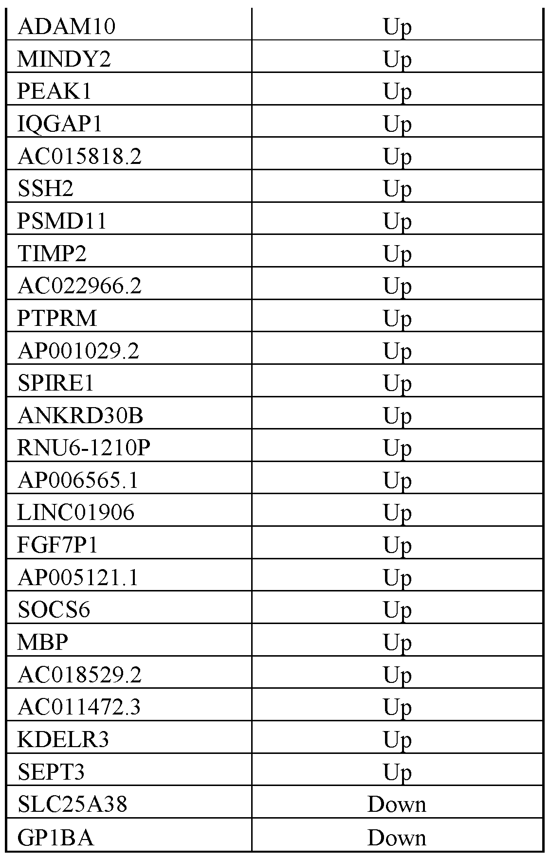


Claims


Priority Applications (6)
| Application Number | Priority Date | Filing Date | Title |
|---|---|---|---|
| EP21867451.3A EP4210704A1 (en) | 2020-09-08 | 2021-09-07 | Pharmaceutical combination and tumor treatment |
| AU2021339579A AU2021339579A1 (en) | 2020-09-08 | 2021-09-07 | Pharmaceutical combination and tumor treatment |
| JP2023515183A JP2023541011A (en) | 2020-09-08 | 2021-09-07 | Pharmaceutical combinations and tumor treatment |
| CA3191363A CA3191363A1 (en) | 2020-09-08 | 2021-09-07 | Pharmaceutical combination and tumor treatment |
| MX2023002757A MX2023002757A (en) | 2020-09-08 | 2021-09-07 | Pharmaceutical combination and tumor treatment. |
| IL300952A IL300952A (en) | 2020-09-08 | 2021-09-07 | Pharmaceutical combination and tumor treatment |
Applications Claiming Priority (2)
| Application Number | Priority Date | Filing Date | Title |
|---|---|---|---|
| US202063075702P | 2020-09-08 | 2020-09-08 | |
| US63/075,702 | 2020-09-08 |
Publications (1)
| Publication Number | Publication Date |
|---|---|
| WO2022055893A1 true WO2022055893A1 (en) | 2022-03-17 |
Family
ID=80629827
Family Applications (1)
| Application Number | Title | Priority Date | Filing Date |
|---|---|---|---|
| PCT/US2021/049310 WO2022055893A1 (en) | 2020-09-08 | 2021-09-07 | Pharmaceutical combination and tumor treatment |
Country Status (7)
| Country | Link |
|---|---|
| EP (1) | EP4210704A1 (en) |
| JP (1) | JP2023541011A (en) |
| AU (1) | AU2021339579A1 (en) |
| CA (1) | CA3191363A1 (en) |
| IL (1) | IL300952A (en) |
| MX (1) | MX2023002757A (en) |
| WO (1) | WO2022055893A1 (en) |
Cited By (2)
| Publication number | Priority date | Publication date | Assignee | Title |
|---|---|---|---|---|
| WO2023107894A1 (en) * | 2021-12-06 | 2023-06-15 | Ideaya Biosciences, Inc. | Combination therapy comprising a pkc inhibitor and a c-met inhibitor |
| WO2024044775A1 (en) * | 2022-08-26 | 2024-02-29 | Ideaya Biosciences, Inc. | Methods of treating uveal melanoma |
Citations (5)
| Publication number | Priority date | Publication date | Assignee | Title |
|---|---|---|---|---|
| US20140295452A1 (en) * | 2009-08-21 | 2014-10-02 | Pierre Fabre Medicament | Anti-cmet antibody and its use for the detection and the diagnosis of cancer |
| WO2014174478A1 (en) * | 2013-04-26 | 2014-10-30 | Novartis Ag | Pharmaceutical combinations of a pkc inhibitor and a c-met receptor tyrosine kinase inhibitor |
| US20170039328A1 (en) * | 2011-06-07 | 2017-02-09 | Caris Mpi, Inc. | Molecular profiling for cancer |
| US20170320853A1 (en) * | 2011-11-14 | 2017-11-09 | Ignyta, Inc. | Uracil derivatives as axl and c-met kinase inhibitors |
| US20190202809A1 (en) * | 2014-08-06 | 2019-07-04 | Novartis Ag | Protein kinase c inhibitors and methods of their use |
-
2021
- 2021-09-07 CA CA3191363A patent/CA3191363A1/en active Pending
- 2021-09-07 WO PCT/US2021/049310 patent/WO2022055893A1/en active Application Filing
- 2021-09-07 AU AU2021339579A patent/AU2021339579A1/en active Pending
- 2021-09-07 JP JP2023515183A patent/JP2023541011A/en active Pending
- 2021-09-07 IL IL300952A patent/IL300952A/en unknown
- 2021-09-07 EP EP21867451.3A patent/EP4210704A1/en active Pending
- 2021-09-07 MX MX2023002757A patent/MX2023002757A/en unknown
Patent Citations (5)
| Publication number | Priority date | Publication date | Assignee | Title |
|---|---|---|---|---|
| US20140295452A1 (en) * | 2009-08-21 | 2014-10-02 | Pierre Fabre Medicament | Anti-cmet antibody and its use for the detection and the diagnosis of cancer |
| US20170039328A1 (en) * | 2011-06-07 | 2017-02-09 | Caris Mpi, Inc. | Molecular profiling for cancer |
| US20170320853A1 (en) * | 2011-11-14 | 2017-11-09 | Ignyta, Inc. | Uracil derivatives as axl and c-met kinase inhibitors |
| WO2014174478A1 (en) * | 2013-04-26 | 2014-10-30 | Novartis Ag | Pharmaceutical combinations of a pkc inhibitor and a c-met receptor tyrosine kinase inhibitor |
| US20190202809A1 (en) * | 2014-08-06 | 2019-07-04 | Novartis Ag | Protein kinase c inhibitors and methods of their use |
Cited By (2)
| Publication number | Priority date | Publication date | Assignee | Title |
|---|---|---|---|---|
| WO2023107894A1 (en) * | 2021-12-06 | 2023-06-15 | Ideaya Biosciences, Inc. | Combination therapy comprising a pkc inhibitor and a c-met inhibitor |
| WO2024044775A1 (en) * | 2022-08-26 | 2024-02-29 | Ideaya Biosciences, Inc. | Methods of treating uveal melanoma |
Also Published As
| Publication number | Publication date |
|---|---|
| IL300952A (en) | 2023-04-01 |
| EP4210704A1 (en) | 2023-07-19 |
| AU2021339579A1 (en) | 2023-05-18 |
| MX2023002757A (en) | 2023-07-25 |
| JP2023541011A (en) | 2023-09-27 |
| CA3191363A1 (en) | 2022-03-17 |
Similar Documents
| Publication | Publication Date | Title |
|---|---|---|
| JP7019422B2 (en) | How to treat cancer | |
| JP2022017495A (en) | Combination therapy for treating cancer | |
| US20140178366A1 (en) | Preselection of subjects for therapeutic treatment based on hypoxic status | |
| US9937161B2 (en) | Combinatorial compositions and methods for treatment of melanoma | |
| EP3808349B1 (en) | Compounds and methods for treating cancer | |
| US20090042906A1 (en) | Methods for treating cancers associated with constitutive egfr signaling | |
| AU2021339579A1 (en) | Pharmaceutical combination and tumor treatment | |
| US11696924B2 (en) | Combination therapy using PDK1 and PI3K inhibitors | |
| CA2768338A1 (en) | Method for predicting the utility of administering nicotinic acid or a precursor or prodrug thereof to reduce the severity of side-effects of cancer treatment with nicotinamide phosphoribosyltransferase inhibitors | |
| WO2013173436A1 (en) | Pre-selection of subjects for therapeutic treatment with an hsp90 inhibitor based on hypoxic status | |
| WO2020132259A1 (en) | Compositions and methods of treating cancers by administering a phenothiazine-related drug that activates protein phosphatase 2a (pp2a) | |
| EP3866799B1 (en) | Compositions for treating vascular ehlers danlos syndrome | |
| WO2007143629A1 (en) | Treatment of neurofibromatosis with inhibitors of a signal transduction pathway | |
| JP2020519672A (en) | Combination of RET inhibitor and mTORC1 inhibitor and its use for treating cancer mediated by aberrant RET activity | |
| US20180271819A1 (en) | Methods of cancer treatment | |
| KR20230092962A (en) | Use of N-Myristoyl Transferase (NMT) Inhibitors in the Treatment of Cancer, Autoimmune Disorders, and Inflammatory Disorders | |
| Kojima et al. | Selective FLT3 inhibitor FI-700 neutralizes Mcl-1 and enhances p53-mediated apoptosis in AML cells with activating mutations of FLT3 through Mcl-1/Noxa axis | |
| TW201722422A (en) | Rational combination therapy for the treatment of cancer | |
| KR20210039846A (en) | Use of src inhibitor for overcoming drug resistance of cancer | |
| US10045978B2 (en) | Treatment of mastocytosis with masitinib | |
| WO2024048555A1 (en) | Combination drug | |
| US20220193056A1 (en) | Compositions and methods of treating pik3ca helical domain mutant cancers | |
| EP4361136A1 (en) | Compound for the treatment of glioblastoma | |
| WO2023233362A1 (en) | Composition and method for treating cancer |
Legal Events
| Date | Code | Title | Description |
|---|---|---|---|
| 121 | Ep: the epo has been informed by wipo that ep was designated in this application |
Ref document number: 21867451 Country of ref document: EP Kind code of ref document: A1 |
|
| WWE | Wipo information: entry into national phase |
Ref document number: 300952 Country of ref document: IL |
|
| ENP | Entry into the national phase |
Ref document number: 3191363 Country of ref document: CA |
|
| ENP | Entry into the national phase |
Ref document number: 2023515183 Country of ref document: JP Kind code of ref document: A |
|
| REG | Reference to national code |
Ref country code: BR Ref legal event code: B01A Ref document number: 112023003911 Country of ref document: BR |
|
| REG | Reference to national code |
Ref country code: BR Ref legal event code: B01E Ref document number: 112023003911 Country of ref document: BR Free format text: APRESENTE, NO PRAZO DE 60 (SESSENTA) DIAS, DOCUMENTO DE CESSAO COM MELHOR RESOLUCAO, PRINCIPALMENTE AS PAGINAS 227 E 231 DA PETICAO 870230017752, |
|
| NENP | Non-entry into the national phase |
Ref country code: DE |
|
| ENP | Entry into the national phase |
Ref document number: 2021867451 Country of ref document: EP Effective date: 20230411 |
|
| ENP | Entry into the national phase |
Ref document number: 2021339579 Country of ref document: AU Date of ref document: 20210907 Kind code of ref document: A |
|
| ENP | Entry into the national phase |
Ref document number: 112023003911 Country of ref document: BR Kind code of ref document: A2 Effective date: 20230302 |