WO2022025029A1 - in vitroにおいてコロナウイルス科に属するウイルスを増殖させる方法、コロナウイルス科に属するウイルスに対する中和抗体の産生方法、及びSevere acute respiratory syndrome coronavirus 2の感染モデルの製造方法 - Google Patents
in vitroにおいてコロナウイルス科に属するウイルスを増殖させる方法、コロナウイルス科に属するウイルスに対する中和抗体の産生方法、及びSevere acute respiratory syndrome coronavirus 2の感染モデルの製造方法 Download PDFInfo
- Publication number
- WO2022025029A1 WO2022025029A1 PCT/JP2021/027662 JP2021027662W WO2022025029A1 WO 2022025029 A1 WO2022025029 A1 WO 2022025029A1 JP 2021027662 W JP2021027662 W JP 2021027662W WO 2022025029 A1 WO2022025029 A1 WO 2022025029A1
- Authority
- WO
- WIPO (PCT)
- Prior art keywords
- virus
- kidney
- producing
- derived cells
- belonging
- Prior art date
- Legal status (The legal status is an assumption and is not a legal conclusion. Google has not performed a legal analysis and makes no representation as to the accuracy of the status listed.)
- Ceased
Links
Images





Classifications
-
- C—CHEMISTRY; METALLURGY
- C12—BIOCHEMISTRY; BEER; SPIRITS; WINE; VINEGAR; MICROBIOLOGY; ENZYMOLOGY; MUTATION OR GENETIC ENGINEERING
- C12N—MICROORGANISMS OR ENZYMES; COMPOSITIONS THEREOF; PROPAGATING, PRESERVING, OR MAINTAINING MICROORGANISMS; MUTATION OR GENETIC ENGINEERING; CULTURE MEDIA
- C12N7/00—Viruses; Bacteriophages; Compositions thereof; Preparation or purification thereof
-
- C—CHEMISTRY; METALLURGY
- C07—ORGANIC CHEMISTRY
- C07K—PEPTIDES
- C07K16/00—Immunoglobulins [IG], e.g. monoclonal or polyclonal antibodies
- C07K16/08—Immunoglobulins [IG], e.g. monoclonal or polyclonal antibodies against material from viruses
- C07K16/10—RNA viruses
- C07K16/102—Coronaviridae (F)
- C07K16/104—Severe acute respiratory syndrome coronavirus 2 [SARS‐CoV‐2]
-
- C—CHEMISTRY; METALLURGY
- C12—BIOCHEMISTRY; BEER; SPIRITS; WINE; VINEGAR; MICROBIOLOGY; ENZYMOLOGY; MUTATION OR GENETIC ENGINEERING
- C12N—MICROORGANISMS OR ENZYMES; COMPOSITIONS THEREOF; PROPAGATING, PRESERVING, OR MAINTAINING MICROORGANISMS; MUTATION OR GENETIC ENGINEERING; CULTURE MEDIA
- C12N5/00—Undifferentiated human, animal or plant cells, e.g. cell lines; Tissues; Cultivation or maintenance thereof; Culture media therefor
- C12N5/06—Animal cells or tissues; Human cells or tissues
- C12N5/0602—Vertebrate cells
- C12N5/0684—Cells of the urinary tract or kidneys
- C12N5/0686—Kidney cells
-
- C—CHEMISTRY; METALLURGY
- C07—ORGANIC CHEMISTRY
- C07K—PEPTIDES
- C07K2317/00—Immunoglobulins specific features
- C07K2317/10—Immunoglobulins specific features characterized by their source of isolation or production
- C07K2317/14—Specific host cells or culture conditions, e.g. components, pH or temperature
-
- C—CHEMISTRY; METALLURGY
- C12—BIOCHEMISTRY; BEER; SPIRITS; WINE; VINEGAR; MICROBIOLOGY; ENZYMOLOGY; MUTATION OR GENETIC ENGINEERING
- C12N—MICROORGANISMS OR ENZYMES; COMPOSITIONS THEREOF; PROPAGATING, PRESERVING, OR MAINTAINING MICROORGANISMS; MUTATION OR GENETIC ENGINEERING; CULTURE MEDIA
- C12N2501/00—Active agents used in cell culture processes, e.g. differentation
- C12N2501/70—Enzymes
- C12N2501/73—Hydrolases (EC 3.)
- C12N2501/734—Proteases (EC 3.4.)
-
- C—CHEMISTRY; METALLURGY
- C12—BIOCHEMISTRY; BEER; SPIRITS; WINE; VINEGAR; MICROBIOLOGY; ENZYMOLOGY; MUTATION OR GENETIC ENGINEERING
- C12N—MICROORGANISMS OR ENZYMES; COMPOSITIONS THEREOF; PROPAGATING, PRESERVING, OR MAINTAINING MICROORGANISMS; MUTATION OR GENETIC ENGINEERING; CULTURE MEDIA
- C12N2770/00—MICROORGANISMS OR ENZYMES; COMPOSITIONS THEREOF; PROPAGATING, PRESERVING, OR MAINTAINING MICROORGANISMS; MUTATION OR GENETIC ENGINEERING; CULTURE MEDIA ssRNA viruses positive-sense
- C12N2770/00011—Details
- C12N2770/20011—Coronaviridae
- C12N2770/20021—Viruses as such, e.g. new isolates, mutants or their genomic sequences
-
- C—CHEMISTRY; METALLURGY
- C12—BIOCHEMISTRY; BEER; SPIRITS; WINE; VINEGAR; MICROBIOLOGY; ENZYMOLOGY; MUTATION OR GENETIC ENGINEERING
- C12N—MICROORGANISMS OR ENZYMES; COMPOSITIONS THEREOF; PROPAGATING, PRESERVING, OR MAINTAINING MICROORGANISMS; MUTATION OR GENETIC ENGINEERING; CULTURE MEDIA
- C12N2770/00—MICROORGANISMS OR ENZYMES; COMPOSITIONS THEREOF; PROPAGATING, PRESERVING, OR MAINTAINING MICROORGANISMS; MUTATION OR GENETIC ENGINEERING; CULTURE MEDIA ssRNA viruses positive-sense
- C12N2770/00011—Details
- C12N2770/20011—Coronaviridae
- C12N2770/20032—Use of virus as therapeutic agent, other than vaccine, e.g. as cytolytic agent
-
- C—CHEMISTRY; METALLURGY
- C12—BIOCHEMISTRY; BEER; SPIRITS; WINE; VINEGAR; MICROBIOLOGY; ENZYMOLOGY; MUTATION OR GENETIC ENGINEERING
- C12N—MICROORGANISMS OR ENZYMES; COMPOSITIONS THEREOF; PROPAGATING, PRESERVING, OR MAINTAINING MICROORGANISMS; MUTATION OR GENETIC ENGINEERING; CULTURE MEDIA
- C12N2770/00—MICROORGANISMS OR ENZYMES; COMPOSITIONS THEREOF; PROPAGATING, PRESERVING, OR MAINTAINING MICROORGANISMS; MUTATION OR GENETIC ENGINEERING; CULTURE MEDIA ssRNA viruses positive-sense
- C12N2770/00011—Details
- C12N2770/20011—Coronaviridae
- C12N2770/20051—Methods of production or purification of viral material
-
- C—CHEMISTRY; METALLURGY
- C12—BIOCHEMISTRY; BEER; SPIRITS; WINE; VINEGAR; MICROBIOLOGY; ENZYMOLOGY; MUTATION OR GENETIC ENGINEERING
- C12N—MICROORGANISMS OR ENZYMES; COMPOSITIONS THEREOF; PROPAGATING, PRESERVING, OR MAINTAINING MICROORGANISMS; MUTATION OR GENETIC ENGINEERING; CULTURE MEDIA
- C12N2770/00—MICROORGANISMS OR ENZYMES; COMPOSITIONS THEREOF; PROPAGATING, PRESERVING, OR MAINTAINING MICROORGANISMS; MUTATION OR GENETIC ENGINEERING; CULTURE MEDIA ssRNA viruses positive-sense
- C12N2770/00011—Details
- C12N2770/20011—Coronaviridae
- C12N2770/20051—Methods of production or purification of viral material
- C12N2770/20052—Methods of production or purification of viral material relating to complementing cells and packaging systems for producing virus or viral particles
Definitions
- This specification discloses a method for propagating a virus belonging to the coronaviridae in vitro, a method for producing a neutralizing antibody against a virus belonging to the coronaviridae, and a method for producing an infection model of Severe acute respiratory syndrome coronavirus 2.
- Virus This specification discloses a method for propagating a virus belonging to the coronaviridae in vitro, a method for producing a neutralizing antibody against a virus belonging to the coronaviridae, and a method for producing an infection model of Severe acute respiratory syndrome coronavirus 2.
- Severe acute respiratory syndrome coronavirus2 (SARS-CoV-2) infection has spread worldwide since the latter half of 2019, and WHO declared a pandemic on March 11, 2020.
- Patients infected with SARS-CoV-2 include patients with various severity, including asymptomatic patients, patients with mild respiratory symptoms, and patients with acute respiratory distress syndrome (ARDS) who require admission to the ICU. Is included.
- ARDS acute respiratory distress syndrome
- the increasing number of patients infected with SARS-CoV-2 is still ongoing, and in addition to the development of vaccines and antivirals specific for SARS-CoV-2, pathogenicity of patients with severe clinical manifestations of the disease. There is an urgent need to identify sex and develop treatments for severe cases.
- Non-Patent Document 1 describes a SARS-CoV-2 infection model using cynomolgus monkeys.
- Non-Patent Documents 2 to 5 describe a SARS-CoV-2 infection model using rhesus monkeys.
- Patent Document 1 describes a virus inspection method comprising an inoculation step of inoculating a monkey belonging to the Callitrichidae family with dengue virus and a detection step of detecting the dengue virus from the monkey.
- Patent Document 1 describes that monkeys of the Callitrichidae family are preferable as monkeys that infect dengue virus, but cynomolgus monkeys are not preferable because infection is difficult to establish. Thus, even if an animal belonging to the primate order is inoculated with a virus showing pathogenicity to humans, whether or not the infection is established depends on the family or species.
- SARS-CoV-2 even if SARS-CoV-2 enters the human body, the severity of the symptoms caused by SARS-CoV-2 varies. The severity of SARS-CoV-2 infection varies from patient to patient, and it is not clear whether this diversity depends on SARS-CoV-2, on the patient, or on both.
- TMPRSS2 which is a serine protease present in the cell membrane of human respiratory cells, is forcibly expressed in Vero E6 cells, and the forced expression strain is infected with SARS-CoV-2 for efficient separation. -It is stated that it can proliferate.
- this method of virus propagation requires cells to be forced to express TMPRSS2.
- Item 1 A method for growing a virus in vitro, comprising the step of culturing kidney-derived cells in contact with a virus belonging to the family Coronaviridae using a cell culture medium supplemented with a proteolytic enzyme having an optimum pH of 7 to 9. Item 2. Item 2. The method according to Item 1, wherein the virus is Severe acute respiratory syndrome coronavirus 2. Item 3. Item 2.
- Item 4. Item 3. The method according to Item 3, wherein the proteolytic enzyme is trypsin.
- Item 5. A method for producing a neutralizing antibody against a virus belonging to the Coronavirus family, the first step of inoculating a virus belonging to the Coronavirus family, which is an antigen, and / or a part thereof into a monkey belonging to the genus Macaku, and the antigen.
- the second step of collecting a blood sample from the inoculated monkey, the third step of mixing the blood sample with a virus belonging to the coronavirus family, and the virus belonging to the coronaviridae mixed with the blood sample are derived from the kidney.
- the 6th step of evaluating the degenerated state of the cultured kidney-derived cells and the 7th step of evaluating the 6th step it is determined that the neutralizing antibody is produced when there is no degeneration of the kidney-derived cells or the degree of degeneration is low.
- a method for producing the neutralizing antibody against a virus belonging to the Coronavirus family which comprises a step.
- Item 6. Item 5.
- Item 8. A method for producing a neutralizing antibody against Severe acute respiratory syndrome coronavirus 2, which comprises inoculating a crab monkey with an antigen, Severe acute respiratory syndrome 2, and / or a part thereof.
- Item 9. Using a cell culture medium supplemented with a proteolytic enzyme having an optimum pH of 7 to 9, a step of culturing kidney-derived cells in contact with SARS acute respiratory syndrome coronavirus 2 and a step of culturing kidney-derived cells from the culture of the kidney-derived cells are carried.
- a method for producing a Severe acute respiratory syndrome which comprises a step of recovering the syndrome coronavirus 2 and a step of inoculating the recovered Severe acute respiratory syndrome 2 and / or a part thereof into a cannibal monkey.
- FIG. 1A shows the effect of adding trypsin to the virus solution Lot1.
- FIG. 1B shows the effect of adding trypsin to the virus solution Lot 2.
- the change in the amount of virus over time in each individual inoculated with the virus is shown.
- the monitoring result of the body temperature of each individual inoculated with the virus is shown.
- FIG. 3A shows the body temperature of CE0202M.
- FIG. 3B shows the body temperature of CE0324F.
- FIG. 3C shows the body temperature of CE1242F.
- FIG. 3D shows the change in body temperature of CE0202M.
- FIG. 3E shows the change in body temperature of CE0324F.
- FIG. 3F shows the change in body temperature of CE1242F.
- FIG. 4A shows the results of monitoring the body weight of each individual inoculated with the virus.
- FIG. 4B shows the blood saturated oxygen concentration of each individual inoculated with the virus.
- Marker ⁇ indicates the result of individual ID: CE0202M.
- Marker ⁇ indicates the result of individual ID: CE0324F.
- Marker ⁇ indicates the result of individual ID: CE1242F.
- FIG. 5A is a chest X-ray image at the time of inoculation (Day 0) of individual ID: CE0324F.
- FIG. 5B is a chest X-ray image of the individual ID: CE0324F on the third day (Day 3) after inoculation.
- FIG. 5C is a chest X-ray image of the 7th day (Day 7) after inoculation of the individual ID: CE0324F.
- FIG. 5D shows a macro image of the lung taken by autopsy from the individual ID: CE0202M.
- FIG. 5E shows a macro image of the lung taken by autopsy from the individual ID: CE0324F.
- FIG. 5F shows a macro image of the lung taken by autopsy from the individual ID: CE1242F.
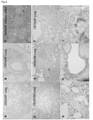
- FIG. 6A shows an HE-stained image of the peripheral tissue of the lung of individual ID: CE0324F.
- FIG. 6B shows an HE-stained image of lung tissue around the bronchi of individual ID: CE0324F.
- FIG. 6C shows an HE-stained image of salivary gland tissue with individual ID: CE0324F.
- FIG. 6D shows an HE-stained image of lung tissue with individual ID: CE0202M.
- FIG. 6E shows an HE-stained image of the thrombus of the lung tissue of individual ID: CE0202M.
- FIG. 6F shows an HE-stained image of lung tissue containing the bronchi of individual ID: CE1242F.
- FIG. 6G shows an immunostaining image of angiotensin converting enzyme 2 in lung tissue.
- H shows an immunostaining image of angiotensin converting enzyme 2 in bronchial tissue.
- FIG. 6I shows an immunostaining image of angiotensin converting enzyme 2 in kidney tissue.
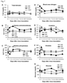
- FIG. 7A shows the total bilirubin concentration.
- FIG. 7B shows the alanine aminotransferase (ALT) value.
- FIG. 7C shows alkaline phosphatase (ALP) values.
- FIG. 7D shows the urea nitrogen (BUN) value.
- FIG. 7E shows the creatinine level.
- FIG. 7F shows the amylase value.
- FIG. 7G shows the glucose level.
- Marker ⁇ indicates the result of individual ID: CE0202M.
- Marker ⁇ indicates the result of individual ID: CE0324F.
- Marker ⁇ indicates the result of individual ID: CE1242F.
- Hematological data of each individual inoculated with the virus is shown.
- FIG. 7A shows the total bilirubin concentration.
- FIG. 7B shows the alanine aminotransferase (ALT) value.
- FIG. 7C shows alkaline phosphatase (ALP) values.
- FIG. 7D shows the
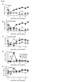
- FIG. 8A shows the white blood cell count.
- FIG. 8B shows the number of neutrophils.
- FIG. 8C shows the number of lymphocytes.
- FIG. 8D shows the number of monocytes.
- FIG. 8E shows the platelet count.
- Marker ⁇ indicates the result of individual ID: CE0202M.
- Marker ⁇ indicates the result of individual ID: CE0324F.
- Marker ⁇ indicates the result of individual ID: CE1242F.
- the concentrations of cytokines and chemokines in the plasma of each individual inoculated with the virus are shown.
- Marker ⁇ indicates the result of individual ID: CE0202M.
- Marker ⁇ indicates the result of individual ID: CE0324F.
- Marker ⁇ indicates the result of individual ID: CE1242F.
- FIG. 10A shows the result of individual ID: CE0202M.
- FIG. 10B shows the result of individual ID: CE0324F.
- FIG. 10C shows the result of individual ID: CE1242F.
- the marker ⁇ indicates interferon gamma-producing cells. Marker ⁇ indicates IL-2-producing cells.
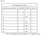
- the neutralizing activity of the plasma of each individual inoculated with the virus against the virus is shown.
- the results of a qualitative test examining whether IgG and IgM that react with SARS-CoV-2 are present in the plasma of each individual inoculated with the virus are shown.
- FIG. 13A shows the antibody titer of IgG against the S1 protein of SARS-CoV-2.
- FIG. 13B shows the antibody titer of IgG against the N protein of SARS-CoV-2.
- Marker ⁇ indicates the result of individual ID: CE0202M.
- Marker ⁇ indicates the result of individual ID: CE0324F.
- Marker ⁇ indicates the result of individual ID: CE1242F.
- An embodiment of the present invention relates to a propagation method for propagating a virus in vitro.
- the proliferation method comprises a step of culturing kidney-derived cells in contact with a virus belonging to the family Coronaviridae using a cell culture medium supplemented with a proteolytic enzyme having an optimum pH of 7 to 9.
- the virus is not limited as long as it belongs to the Coronaviridae family.
- Severe acute respiratory syndrome coronavirus 2 SARS-CoV-2
- SARS coronavirus human coronavirus 229E strain
- human coronavirus NL63 strain human coronavirus OC43 strain
- human coronavirus HKU1 Stocks and the like can be mentioned.
- the virus belonging to the genus Coronavirus is preferably SARS-CoV-2, SARS coronavirus, and more preferably SARS-CoV-2.
- SARS-CoV-2 is a virus having a genomic sequence registered in NCBI Reference Section: NC_045512.2., SARS-CoV-2 JP / TY / WK-521 / 2020 (GenBank: LC522975.1), and these. It may contain the same virus and genome sequence of 70% or more, 80% or more, 85% or more, 90% or more, 93% or more, 95% or more, 98% or more, 99% or more, 99.5% or more. ..
- a virus belonging to the family Coronaviridae (hereinafter, may be simply abbreviated as "virus”) can be propagated in vitro using cells of kidney-derived cells.
- Kidney-derived cells are not particularly limited. Cells derived from the kidney tissue of mammals such as humans, monkeys and dogs can be used. The kidney-derived cells are preferably cells expressing angiotensin converting enzyme 2 (ACE2).
- ACE2 angiotensin converting enzyme 2
- the kidney-derived cell may be a primary cultured cell or a cell established as a cultured cell line. Examples of the cultured cell line include Vero cells, Vero E6 cells and the like. Vero E6 cells are preferred.
- the method of contacting the virus with the kidney-derived cells is not limited as long as the virus can come into contact with the surface of the kidney-derived cells.
- a virus suspension is prepared by suspending the virus in a liquid such as Hanks Balanced Salt Solution (HBSS), PBS, basal medium described later, or cell culture medium, and the virus suspension is applied to kidney-derived cells adhered to a plate or flask. It can be contacted by sowing.
- the virus suspended in the liquid may be contained in a biological sample such as nasal cavity, oral cavity, bronchi and tracheal swab or suspension of scraped sample; blood; pleural effusion; sputum. Further, the virus suspended in the liquid may be contained in the culture supernatant or the like.
- the biological sample is preferably brought into contact with kidney-derived cells within, for example, 24 hours after collection.
- the virus suspension For contact between the virus and kidney-derived cells, for example, it is preferable to incubate the virus suspension at 20 ° C to 28 ° C for about 30 to 90 minutes after seeding the kidney-derived cells.
- the incubation is preferably carried out by allowing the plate to stand still, and when using a flask, using a seesaw-type shaker or the like, and tilting the flask at intervals of about 3 to 7 minutes.
- Kidney-derived cells that have come into contact with the virus can be cultured in a culture environment that matches the culture conditions of the kidney-derived cells.
- the culture temperature is, for example, 33 ° C to 37 ° C, and the culture can be carried out in a wet state in the presence of about 5% carbon dioxide gas.
- the cell culture medium a medium in which bovine serum albumin having a final concentration of about 0.05% to 0.2% is added to a basic medium such as MEM medium, D-MEM medium, ⁇ -MEM medium, etc. is used. Can be done. If necessary, antibiotics such as streptomycin at a final concentration of 50 ⁇ g / mL to 150 ⁇ g / mL and penicillin at a final concentration of about 50 unit / mL to 150 unit / mL may be added as long as the virus does not grow. ..
- a proteolytic enzyme can be added to the cell culture medium.
- the proteolytic enzyme is not limited as long as it exerts its activity at the pH of the culture environment of kidney-derived cells. For example, pH 7 to pH 9 can be mentioned as the optimum for proteolytic enzymes.
- the proteolytic enzyme is preferably endopeptidase.
- the proteolytic enzyme is preferably serine protease or threonine peptidase.
- Preferred examples of the proteolytic enzyme include trypsin, chymotrypsin, and acetylated trypsin.
- the proteolytic enzyme is more preferably trypsin.
- the enzyme activity value of the proteolytic enzyme added to the cell culture medium can be about 5 unit / mL to 25 unit / mL at the final concentration.
- the final concentration is preferably about 10 unit / mL to 15 unit / mL.
- the quality of trypsin can be determined according to JIS K 0605: 2000.
- the enzyme activity equivalent to trypsin can be obtained by calculating the relative activity with trypsin.
- binding a proteolytic enzyme is intended to externally add a proteolytic enzyme to a cell culture medium or the like outside the cell.
- Kidney-derived cells in contact with the virus can be cultured in the cell culture medium in the above culture environment for about 2 to 7 days, preferably about 6 days.
- Denaturation of a cell is intended to be a state in which the cell is dying or dying.
- Cell denaturation can be determined by phase contrast microscopy using an inverted microscope or the like.
- cell degeneration can be determined by crystal violet staining.
- the virus propagation method may include a step of recovering the propagated virus from the culture.
- the virus can be recovered, for example, by recovering the culture supernatant as a culture from a plate or flask and removing the cell residue. Further, after collecting the cells, the virus may be recovered by crushing the cells with ultrasonic waves or the like and removing the cell residue from the cell crushed solution. If necessary, a DNase may be added to the cell disruption solution.
- the cell residue is 1,400 r. p. It can be removed by centrifuging at about m for, for example, 5 to 10 minutes to remove the precipitate. In addition, cell debris may be removed by centrifuging to collect the supernatant and then filtering the supernatant.
- One embodiment of the present invention relates to a method for producing a neutralizing antibody against a virus belonging to the family Coronaviridae.
- the production method may include the following first to seventh steps.
- the first step is a step of inoculating a virus belonging to the Coronaviridae family as an antigen and / or a part thereof to a monkey belonging to the genus Makaku.
- the monkeys belonging to the genus Macaque may include cynomolgus monkeys, rhesus monkeys, Japanese macaques, Formosan macaques and the like.
- the monkeys belonging to the genus Macaque are preferably cynomolgus monkeys and rhesus monkeys, and more preferably cynomolgus monkeys.
- the sex and age of the monkey are not particularly limited. For example, it is preferable to use monkeys aged 3 to 15 years.
- Viruses belonging to the Coronaviridae family are listed in 1. above. As explained in, the above explanation is used here.
- a part of a virus belonging to the family Coronaviridae is a part of a component constituting the virus and is intended to have antigenicity.
- a virus belonging to the family Coronaviridae has an envelope and genomic RNA, and the envelope includes a spike (S) protein, an envelope (E) protein, and a membrane (M) protein.
- S protein spike
- E envelope
- M membrane
- N protein RNA-binding protein
- the antigen may include the whole virus as well as at least a portion of S protein, E protein, M protein and N protein.
- Inoculation of the antigen to monkeys belonging to the genus Macaque can be carried out by a known method. For example, when inoculating a virus, pipette the conjunctiva, nostril, oral cavity, trachea, etc. of the monkey so that the total amount of virus inoculated per individual is 1 ⁇ 10 5 to 1 ⁇ 10 8 TCID 50 . It can be inoculated using a virus or a catheter. Inoculation of the antigen is preferably performed under anesthesia. When a part of the virus is used as an antigen, it may be inoculated into the conjunctiva, nostril, oral cavity, trachea, blood, subcutaneous or the like.
- a blood sample is collected from the monkey inoculated with the antigen.
- Blood samples are preferably collected, for example, 14 to 28 days after inoculation with the antigen.
- Blood samples may include whole blood, serum, plasma.
- the blood sample is preferably plasma.
- the anticoagulant used for collecting plasma is not particularly limited.
- heparin can be used as an anticoagulant without particular limitation.
- the blood sample collected in the second step is mixed with a virus belonging to the family Coronaviridae (hereinafter, may be simply referred to as "virus"). If an antibody is produced against the inoculated antigen by the antigen inoculation in the first step, the produced antibody binds to the virus when mixed with the blood sample.
- the third step is not limited as long as it is performed in an environment where the antibody and the virus can bind. For example, it can be carried out at 20 ° C to 37 ° C for about 15 minutes to 1 hour. Here, it is preferable that the blood sample is treated at 35 ° C. to 37 ° C.
- the complement contained in the blood sample is inactivated in advance.
- Methods for inactivating complement are known. For example, complement can be inactivated by incubating a blood sample at about 56 ° C. for about 1 hour.
- the blood sample is the above 1. It may be diluted with the liquid for suspending the virus described in the above.
- the dilution range of the blood sample can be about 8 to 100,000 times.
- the virus solution to be mixed with the diluted blood sample has a virus concentration of, for example, about 4,000 TCID 50 / mL, and the blood sample or each diluted blood sample and the virus solution are mixed in equal amounts.
- the fourth step is a step of bringing a virus belonging to the family Coronaviridae mixed with a blood sample in the third step into contact with kidney-derived cells. This step is described in 1. above. Since it is the same as the step of contacting the virus and the kidney-derived cells described in 1. above. The explanation of is used here.
- the kidney-derived cells that have come into contact with the virus in the fourth step are cultured using a cell culture medium supplemented with a proteolytic enzyme having an optimum pH of 7 to 9.
- This step is described in 1. above. Since it is the same as the step of culturing kidney-derived cells in contact with the virus described in 1. above. The explanation of is used here.
- the degenerated state of the kidney-derived cells cultured in the 5th step is evaluated.
- the degeneration of virus-infected kidney-derived cells is as described in 1 above. Assessing the degenerated state is intended to assess whether kidney-derived cells are degenerated or not.
- the evaluation is the above 1.
- the morphology of cells is observed with an inverted microscope or the like, or the degeneration of cells is determined by crystal violet staining.
- the evaluation of whether the kidney-derived cells are denatured or not can be performed 2 to 7 days, preferably 6 days after contact with the virus.
- the degree of denaturation can be expressed, for example, by the neutralizing titer.
- the neutralizing titer can be expressed as the dilution factor at which the denaturing effect of cells was observed in the wells.
- the degenerative effect can be expressed, for example, by a value indicating a dilution ratio in which the number of viruses infecting cells is reduced to a predetermined ratio, or a value indicating a dilution ratio in which the number of plaques in a well is reduced to a predetermined ratio. can.
- the predetermined ratio can be selected, for example, in the range of 10% to 60%, and is appropriately in the range of, for example, 10%, 20%, 30%, 40%, 45%, 50%, 55%, 60%. Can be set.
- the predetermined ratio is preferably in the range of 40% to 55%, more preferably 50%.
- the higher the neutralizing titer the lower the degree of degeneration of kidney-derived cells. When the neutralizing titer is low or not neutralized, the degree of degeneration of kidney-derived cells is high. Further, for example, when kidney-derived cells in contact with a virus not mixed with a blood sample are set as positive controls and the degree of degeneration of kidney-derived cells in contact with a virus mixed with a blood sample is lower than that of a positive control. It can be determined that a neutralizing antibody is being produced.
- Another Embodiment of the method for producing a neutralizing antibody comprises neutralizing SARS-CoV-2, which comprises a step of inoculating SARS-CoV-2 as an antigen and / or a part thereof into cynomolgus monkeys.
- the step of inoculating SARS-CoV-2, which is an antigen, and / or a part thereof into cynomolgus monkeys is described in 2. above. Since it corresponds to the first step of the above 2. The above 1. The explanation of is used here.
- One embodiment of the present invention relates to a method for producing a SARS-CoV-2 infection model.
- the manufacturing method may include the following steps I to III.
- Step I is a step of culturing kidney-derived cells in contact with SARS-CoV-2 using a cell culture medium supplemented with a proteolytic enzyme having an optimum pH of 7 to 9. This step is described in 1. above. Since it is the same as the step of contacting the virus and the kidney-derived cells described in 1. above. The explanation of is used here.
- Step II is a step of recovering SARS-CoV-2 from the culture of kidney-derived cells. This step is described in 1. above. Since it is the same as the method for recovering the virus from the culture of kidney-derived cells described in 1. above. The explanation of is used here.
- Step III is a step of inoculating the recovered SARS-CoV-2 and / or a part thereof into cynomolgus monkeys.
- SARS-CoV-2 For a part of SARS-CoV-2, refer to 2. above. The explanation of the first step of the above is incorporated here. The step of inoculating SARS-CoV-2 and / or a part thereof into cynomolgus monkeys is described in 2. above. Since it corresponds to the first step of the above 2. The above 1. The explanation of is used here.
- the step IV may include a step of determining whether or not infection has been established in cynomolgus monkeys inoculated with SARS-CoV-2 and / or a part thereof. Whether or not the infection has been established can be determined by body temperature monitoring, chest radiography, autopsy, and histopathological diagnosis.
- the present invention will be described in more detail with reference to examples.
- the present invention is not limited to the examples.
- the following experiments are guidelines for breeding and management of laboratory animals at the Research Center for Animal Life Sciences, Shiga Medical University, standards for breeding and storage of laboratory animals and alleviation of pain by the Ministry of the Environment, and animals at research institutions of the Ministry of Education, Culture, Sports, Science and Technology. It was carried out in strict accordance with the basic guidelines for conducting experiments.
- the experimental protocol was approved by the Animal Care and Use Committee of Shiga University of Medical Science (license number: 2020-4-2).
- the inoculation experiment using the virus was conducted at the biosafety level 3 facility of the Research Center for Animal Life Sciences, Shiga University of Medical Science.
- Virus proliferation and titer determination 1.
- Virus strain SARS-CoV-2 JP / TY / WK-521 / 2020 GenBank Sequence Accession: LC522975; Received from Dr. Masayuki Saijo and Dr. Masaaki Sato of National Institute of Infectious Diseases (NIID); Matsuyama, S. et al: Proc Natl Acad Sci USA. 2020 Mar 31; 117 (13): 7001-7003. Doi: 10.1073 / pnas.2002589117.) was used as the inoculum.
- the virus was propagated twice using VeroE6 / TMPRSS2 cells at NIID and once using VeroE6 cells (American Type Culture Collection, Manassas, Virginia) at Shiga University of Medical Science.
- VeroE6 cells are MEM medium supplemented with 10% inactivated fetal bovine serum (FBS), penicillin (final concentration 100 units / mL), and streptomycin (final concentration 100 ⁇ g / mL) (Nacalai Tesque). It was cultured in Nacalai Tesque). VeroE6 cells were cultured in flasks.
- FBS inactivated fetal bovine serum
- penicillin final concentration 100 units / mL
- streptomycin final concentration 100 ⁇ g / mL
- Virus growth and recovery VeroE6 cells adhering to the bottom of the flask were washed twice with Hanks balanced salt solution (HBSS; Nacalai Tesque) to remove HBSS.
- HBSS Hanks balanced salt solution
- To each flask add 5 mL of HBSS (called diluted virus solution) containing 10 ⁇ L of seed virus solution distributed by NIID, and tilt the flask every 5 minutes so that the diluted virus solution covers the cells.
- the cells were cultured at room temperature for 1 hour.
- the culture supernatant was collected from the flask and centrifuged at 1,400 r.p.m. for 5 minutes to collect the supernatant.
- the collected supernatant was further filtered through a bottle top filter, and the filtrate was used as a virus solution.
- VeroE6 cells were seeded on 96-well plates and cultured until confluent.
- the virus solution recovered in 1 was serially diluted 10-fold with 0.1% BSA-added MEM medium. After removing the medium, 100 ⁇ L of each of the virus solution stock solution and the diluted virus solution was added to the wells containing VeroE6 cells, and the mixture was allowed to stand at 37 ° C. for 1 hour.
- the above I. 1. A culture solution prepared by adding 20 ⁇ L of a 0.25% trypsin solution to 10 mL of the 0.1% BSA-added MEM medium used in 1 was prepared, and 100 ⁇ L was added to each well. After culturing at 37 ° C. in the presence of 5% carbon dioxide gas for 6 days, the presence or absence of cytopathic effect was determined under a microscope.
- Virus infection experiment 1 Cynomolgus monkeys We used cynomolgus monkeys of 15-year-old and 10-year-old females (individual ID: CE0324F, CE1242F) and 15-year-old males (individual ID: CE0202M) born at Shiga University of Medical Science. The individuals used in this study did not carry Herpes B virus, hepatitis E virus, tuberculosis, Shigella, Salmonella, and Shigella amoebiasis.
- CMK-2 CLEA Japan, Inc., Tokyo, Japan
- CMK-2 CLA Japan, Inc., Tokyo, Japan
- the breeding environment was a light-dark cycle of humidity (39-61%), temperature (24-26 ° C), 12-hour lighting (lit at 8 am), and 12-hour off (turned off at 8 pm).
- a telemetry probe (M00, Data Sciences International, St. Paul, Minnesota) was implanted in the abdominal or subcutaneous tissue of each individual under anesthesia with ketamine / xylazine and isoflurane inhalation to monitor body temperature. ..
- Chest X-ray images were taken using an I-PACS system (Konica Minolta Inc., Tokyo, Japan) and a PX-20BT mini (Kenko Tokina Corporation, Tokyo, Japan).
- a wiping solution sample and a tissue sample were collected in order to evaluate the growth of the virus in the body of the individual.
- the tissue was disrupted to prepare a homogenated sample (10% w / v).
- Serial dilutions of wipe and homogenate samples were prepared and each dilution was inoculated into confluent Vero E6 cells.
- VeroE6 cells were cultured in MEM medium supplemented with 0.1% BSA, penicillin, streptomycin, gentamicin (50 ⁇ g / mL) (Fujifilm, Tokyo, Japan), and trypsin (5 ⁇ g / mL) (Nacalai Tesque). The cytopathic effect of the virus was observed under a microscope 6 days after inoculation.
- Virus neutralization test Blood was collected from individuals inoculated with the virus on the 0th, 7th, 10th, 14th, 17th, 21st, 24th, and 28th days, and plasma was collected. separated. Receptor Destroying Enzyme (RDEII, Denka Seiken, Tokyo, Japan) was added to plasma samples and treated overnight at 37 ° C. The complement was then inactivated by incubation at 56 ° C. for 1 hour. Plasma after inactivation treatment is 8 times, 16 times, 32 times, 64 times, 128 times, 256 times, 512 times, 1024 times, 2048 times, 4096 times, 8192 times in MEM medium containing 0.1% BSA.
- RDEII Receptor Destroying Enzyme
- Antibody detection against SARS-CoV-2 Plasma was collected from individuals inoculated with the virus. Using the COVID-19 IgG / IgM immunodetection kit (Novus Biologicals USA, Centennial Co.), IgG and IgM specific to each of the S protein and N protein of SARS-CoV-2 were measured.
- ELISPOT Detection of Cytokine-Producing Cells by ELISPOT Blood was collected from individuals inoculated with the virus, and blood cells were isolated using Ficoll solution. The cells were centrifuged, suspended in a cell bunker cell storage solution, and then stored at -80 ° C until use. The experiments used ELISPOT plates coated with anti-IFN- ⁇ antibody and anti-IL-2 antibody (Cellular Technology Limited, Shaker Heights, Ohio). Thawed cells were seeded in each well of the ELISPOT plate to a 5 ⁇ 10 5 / well, with a peptide pool of SARS-CoV-2 N protein (PepTivator, Miltenyi Biotech), 1 in the presence of anti-CD28 antibody. Cultured for days. The number of spots on cytokine-producing cells was counted according to the manufacturer's instructions.
- Plasma cytokine / chemokine levels in virus-infected individuals use the Milliplex MAP non-human primate cytokine panel and Luminex 200 (Millipore Corp., Billerica, MA) as directed by the manufacturer. And measured. Blood biochemical analysis and blood cell counts were measured using VetScan VS2 and HM2 (Abaxis, Inc., Union City, CA), respectively.
- Plasma was collected from each individual and used with the MILLIPLEX® SARS-CoV-2 Antigen Panel 1 IgG antibody measurement kit to obtain the S1 protein of SARS-CoV-2 (left). ) And IgG antibody against N protein (right) were measured. The measurement method followed the protocol attached to the kit.
- Results (1) Major SARS-CoV-2 growth in the nasal cavity and oral cavity of cynomolgus monkeys
- the virus growth in each individual was examined by collecting a wiping solution sample from the conjunctiva, nasal cavity, oral cavity, and trachea of the individual infected with the virus. ..
- the virus was detected on day 1 (the day after virus inoculation) in three swab samples of the conjunctiva, nasal cavity, oral cavity, and trachea (Fig. 2).
- the virus was detected in two nasal and oral swab samples (CE0202M and CE0324F) up to day 7. No virus was detected in the swab samples after day 10 and the tissue samples taken at necropsy on day 28.
- 3D-F shows the average body temperature between 8 pm on the 0th day after virus inoculation and 8 am on the 1st day after virus inoculation.
- the average body temperature of each day was compared with that of the previous day (day -1), and the changes are shown in Fig. 3D-F.
- "*" In the figure indicates that there was a significant difference (p ⁇ 0.05 in Student's t-test) in comparison with the previous day.
- FIG. 4 shows the results of monitoring the body weight of each individual.
- the body weight of CE0324F decreased by 10% on the 28th day due to loss of appetite (Fig. 4A). From the above results, infection with the inoculated SARS-CoV-2 was established in each individual.
- SARS-CoV-2 causes clinical signs in each individual after infection, its symptoms have been shown to vary from individual to individual. In addition, some of the individuals who had a fever had a fever from the day after the infection, and it was shown that the incubation period was very short under this infection condition.
- FIG. 6D-E Histopathological analysis of the autopsy lung tissue of CE0202M detected pulmonary vascular congestion and thrombus.
- BALT Bronchial lymphoid tissue
- Fig. 6B Bronchopneumonia was observed in CE1242F (Fig. 6F).
- CE0324F showed lymphocyte infiltration in the submucosal tissue of the trachea. In addition, as a lesion other than respiratory tissue, localized lymphatic infiltration was observed in the submandibular gland of CE0324F.
- Angiotensin converting enzyme 2 (ACE2), an entry receptor for SARS-CoV-2, was detected in type 2 alveolar epithelial cells of the lung and proximal tubules of the kidney (Fig. 6G-I). It was suggested that these cells could be infected with SARS-CoV-2.
- the white blood cell count (WBC) and granulocyte count (Gra) of CE0202M and CD1242F increased temporarily on the first day, but the white blood cell count of CE0324F decreased on the first and third days (Fig. 8A, B).
- the lymphocyte counts of CE0324F and CE1242F decreased on the 1st day after the virus inoculation and recovered to the pre-inoculation level from the 7th to the 10th day after the inoculation (Fig. 8C).
- the monocyte counts of CE0324F and CE1242F increased on the 7th and 3rd days after virus inoculation, respectively (Fig. 8D). Platelet counts for CE0324F and CE1242F decreased 3 days after virus inoculation. Therefore, 2 of the 3 individuals (CE0324F and CE1242F) showed clear changes in the blood cell population.
- CE0324F showed a marked change in plasma cytokine levels after inoculation. Specifically, the inflammatory cytokines interleukin-6 (IL-6) and Monocyte Chemotactic Protein-1 (MCP-1) and IL-10 are higher than before inoculation on the first day after inoculation. Indicated. IL-4, IL-13, IL-17, and MIP-1 ⁇ increased after 10 days after inoculation. Changes in IL-17 and MIP-1 ⁇ were slight. IL-8 increased 21 days after inoculation but decreased thereafter. Interferon- ⁇ (IFN- ⁇ ) and IL-12 increased 1 day after inoculation. At CE1242F, there was little change in cytokines. In CE0202M, a temporary increase was observed in IL-12, MIP-1 ⁇ , IL-8, and IL-13, but the change was not significant.
- IFN- ⁇ Interferon- ⁇
- Cytokine-producing cells The cytokine-producing cells were detected by ELISPOT. The results are shown in Fig. 10. SARS-CoV-2 N-protein peptide-reactive IFN- ⁇ and IL-2 -producing cells increased in CE0234F from 14 days after infection. In CE0202M, IFN- ⁇ -producing cells increased from 14 days after infection, and IL-2-producing cells increased from 21 days after infection. In CE1242F, IFN- ⁇ -producing cells increased from 7 days after infection, and IL-2-producing cells increased from 14 days after infection.
- Neutralizing antibody Neutralizing antibody against SARS-CoV-2 was detected in CE0324F on the 10th day after virus inoculation. Neutralizing antibodies were not detected by day 28 in the other two individuals (Fig. 11).
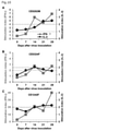
- FIG. 13 shows the measurement results of each individual.
- FIG. 13A shows the antibody titer of IgG against the S1 protein of SARS-CoV-2.
- FIG. 13B shows the antibody titer of IgG against the N protein of SARS-CoV-2.
- IgG for S1 protein and IgG for N protein could be detected in all individuals. 9. above.
- the individual (CE0324F) in which the neutralizing antibody was detected in (7) showed a high antibody titer. 9. above.
- the antibody titer of the individual (CE1242F) in which the antibody was detected in (7) was moderate. 9. above.
- a small amount of IgG was also detected in the individual (CE0202M) in which no antibody could be confirmed in (7).
Landscapes
- Health & Medical Sciences (AREA)
- Life Sciences & Earth Sciences (AREA)
- Chemical & Material Sciences (AREA)
- Organic Chemistry (AREA)
- Engineering & Computer Science (AREA)
- Genetics & Genomics (AREA)
- Wood Science & Technology (AREA)
- Bioinformatics & Cheminformatics (AREA)
- Zoology (AREA)
- Virology (AREA)
- Biomedical Technology (AREA)
- Biochemistry (AREA)
- General Health & Medical Sciences (AREA)
- Biotechnology (AREA)
- Immunology (AREA)
- Medicinal Chemistry (AREA)
- General Engineering & Computer Science (AREA)
- Microbiology (AREA)
- Urology & Nephrology (AREA)
- Molecular Biology (AREA)
- Biophysics (AREA)
- Proteomics, Peptides & Aminoacids (AREA)
- Cell Biology (AREA)
- Micro-Organisms Or Cultivation Processes Thereof (AREA)
Priority Applications (2)
| Application Number | Priority Date | Filing Date | Title |
|---|---|---|---|
| US18/006,920 US20230279361A1 (en) | 2020-07-27 | 2021-07-27 | Method for in vitro proliferating virus belonging to family coronaviridae, method for producing neutralizing antibody to virus belonging to family coronaviridae, and method for producing infection model of severe acute respiratory syndrome coronavirus 2 |
| JP2022539471A JPWO2022025029A1 (https=) | 2020-07-27 | 2021-07-27 |
Applications Claiming Priority (2)
| Application Number | Priority Date | Filing Date | Title |
|---|---|---|---|
| JP2020126321 | 2020-07-27 | ||
| JP2020-126321 | 2020-07-27 |
Publications (1)
| Publication Number | Publication Date |
|---|---|
| WO2022025029A1 true WO2022025029A1 (ja) | 2022-02-03 |
Family
ID=80036209
Family Applications (1)
| Application Number | Title | Priority Date | Filing Date |
|---|---|---|---|
| PCT/JP2021/027662 Ceased WO2022025029A1 (ja) | 2020-07-27 | 2021-07-27 | in vitroにおいてコロナウイルス科に属するウイルスを増殖させる方法、コロナウイルス科に属するウイルスに対する中和抗体の産生方法、及びSevere acute respiratory syndrome coronavirus 2の感染モデルの製造方法 |
Country Status (3)
| Country | Link |
|---|---|
| US (1) | US20230279361A1 (https=) |
| JP (1) | JPWO2022025029A1 (https=) |
| WO (1) | WO2022025029A1 (https=) |
Citations (3)
| Publication number | Priority date | Publication date | Assignee | Title |
|---|---|---|---|---|
| JP2000044473A (ja) * | 1998-07-27 | 2000-02-15 | Mitsui Norin Co Ltd | 家畜のウイルス感染予防剤 |
| JP2007528700A (ja) * | 2003-04-08 | 2007-10-18 | コロノヴァティブ ビー.ブイ. | Sars |
| JP2016539946A (ja) * | 2013-11-29 | 2016-12-22 | ザ トラスティーズ オブ ザ ユニバーシティ オブ ペンシルバニア | MERS‐CoVワクチン |
-
2021
- 2021-07-27 JP JP2022539471A patent/JPWO2022025029A1/ja active Pending
- 2021-07-27 US US18/006,920 patent/US20230279361A1/en active Pending
- 2021-07-27 WO PCT/JP2021/027662 patent/WO2022025029A1/ja not_active Ceased
Patent Citations (3)
| Publication number | Priority date | Publication date | Assignee | Title |
|---|---|---|---|---|
| JP2000044473A (ja) * | 1998-07-27 | 2000-02-15 | Mitsui Norin Co Ltd | 家畜のウイルス感染予防剤 |
| JP2007528700A (ja) * | 2003-04-08 | 2007-10-18 | コロノヴァティブ ビー.ブイ. | Sars |
| JP2016539946A (ja) * | 2013-11-29 | 2016-12-22 | ザ トラスティーズ オブ ザ ユニバーシティ オブ ペンシルバニア | MERS‐CoVワクチン |
Non-Patent Citations (9)
| Title |
|---|
| ISHIGAKI H. ET AL.: "Neutralizing antibody-dependent and -independent immune responses against SARS-CoV-2 in cynomolgus macaques", VIROLOGY, vol. 554, 29 December 2020 (2020-12-29), pages 97 - 105, XP086447536, DOI: 10.1016/j.virol.2020.12.013 * |
| ISHIGAKI HIROHITO; NAKAYAMA MISAKO; KITAGAWA YOSHINORI; NGUYEN CONG THANH; HAYASHI KAORI; SHIOHARA MASANORI; GOTOH BIN; ITOH YASUS: "Neutralizing antibody-dependent and -independent immune responses against SARS-CoV-2 in cynomolgus macaques", VIROLOGY, ELSEVIER, AMSTERDAM, NL, vol. 554, 29 December 2020 (2020-12-29), AMSTERDAM, NL , pages 97 - 105, XP086447536, ISSN: 0042-6822, DOI: 10.1016/j.virol.2020.12.013 * |
| KOMANIWA, HIDEO: "Outbreaks of hog cholera virus type B and isolation of TGE virus using CPK cells"", PROC JPN PIG VET SOC, vol. 2002, no. 40, 2002, pages 3 - 7 * |
| MATSUYAMA S. ET AL.: "Enhanced isolation of SARS-CoV-2 by TMPRSS2-expressing cells", PROC. NATL. ACAD. SCI. U. S. A., vol. 117, no. 13, 12 March 2020 (2020-03-12), pages 7001 - 7003, XP055813015, DOI: 10.1073/pnas.2002589117 * |
| MATSUYAMA S. ET AL.: "Protease-mediated enhancement of severe acute respiratory syndrome coronavirus infection", PROC. NATL. ACAD. SCI. U. S. A, vol. 102, no. 35, 2005, pages 12543 - 12547, XP055862771, DOI: 10.1073/pnas.0503203102 * |
| MENACHERY VINEET D., DINNON KENNETH H., YOUNT BOYD L., MCANARNEY EILEEN T., GRALINSKI LISA E., HALE ANDREW, GRAHAM RACHEL L., SCOB: "Trypsin Treatment Unlocks Barrier for Zoonotic Bat Coronavirus Infection", JOURNAL OF VIROLOGY, THE AMERICAN SOCIETY FOR MICROBIOLOGY, US, vol. 94, no. 5, 14 February 2020 (2020-02-14), US , pages e01774, XP055903766, ISSN: 0022-538X, DOI: 10.1128/JVI.01774-19 * |
| ROCKX BARRY, KUIKEN THIJS, HERFST SANDER, BESTEBROER THEO, LAMERS MART M., OUDE MUNNINK BAS B., DE MEULDER DENNIS, VAN AMERONGEN G: "Comparative pathogenesis of COVID-19, MERS, and SARS in a nonhuman primate model", SCIENCE, AMERICAN ASSOCIATION FOR THE ADVANCEMENT OF SCIENCE, US, vol. 368, no. 6494, 29 May 2020 (2020-05-29), US , pages 1012 - 1015, XP055903768, ISSN: 0036-8075, DOI: 10.1126/science.abb7314 * |
| WANG HUI; ZHANG YUNTAO; HUANG BAOYING; DENG WEI; QUAN YARU; WANG WENLING; XU WENBO; ZHAO YUXIU; LI NA; ZHANG JIN; LIANG HONGYANG; : "Development of an Inactivated Vaccine Candidate, BBIBP-CorV, with Potent Protection against SARS-CoV-2", CELL, ELSEVIER, AMSTERDAM NL, vol. 182, no. 3, 6 June 2020 (2020-06-06), Amsterdam NL , pages 713, XP086239951, ISSN: 0092-8674, DOI: 10.1016/j.cell.2020.06.008 * |
| ZHANG NA-NA, LI XIAO-FENG, DENG YONG-QIANG, ZHAO HUI, HUANG YI-JIAO, YANG GUAN, HUANG WEI-JIN, GAO PENG, ZHOU CHAO, ZHANG RONG-RON: "A Thermostable mRNA Vaccine against COVID-19", CELL, ELSEVIER, AMSTERDAM NL, vol. 182, no. 5, 1 September 2020 (2020-09-01), Amsterdam NL , pages 1271 - 1283.e16, XP055820115, ISSN: 0092-8674, DOI: 10.1016/j.cell.2020.07.024 * |
Also Published As
| Publication number | Publication date |
|---|---|
| JPWO2022025029A1 (https=) | 2022-02-03 |
| US20230279361A1 (en) | 2023-09-07 |
Similar Documents
| Publication | Publication Date | Title |
|---|---|---|
| Di Teodoro et al. | SARS-CoV-2 replicates in respiratory ex vivo organ cultures of domestic ruminant species | |
| Sui et al. | Protection against SARS-CoV-2 infection by a mucosal vaccine in rhesus macaques | |
| Gretebeck et al. | Animal models for SARS and MERS coronaviruses | |
| Clayton et al. | The nature of exposure drives transmission of Nipah viruses from Malaysia and Bangladesh in ferrets | |
| DeBuysscher et al. | Comparison of the pathogenicity of Nipah virus isolates from Bangladesh and Malaysia in the Syrian hamster | |
| Bal et al. | COVID-19: an up-to-date review–from morphology to pathogenesis | |
| Ishigaki et al. | Neutralizing antibody-dependent and-independent immune responses against SARS-CoV-2 in cynomolgus macaques | |
| Vieira et al. | COVID-19: laboratory diagnosis for clinicians. An updating article | |
| Escaffre et al. | Characterization of Nipah virus infection in a model of human airway epithelial cells cultured at an air–liquid interface | |
| Dilnessa et al. | Cell culture, cytopathic effect and immunofluorescence diagnosis of viral infection | |
| Grudlewska-Buda et al. | Sars-cov-2—morphology, transmission and diagnosis during pandemic, review with element of meta-analysis | |
| Khan et al. | Evolution of RNA viruses from SARS to SARS-CoV-2 and diagnostic techniques for COVID-19: A review | |
| Tirupathi et al. | Viral dynamics in the upper respiratory tract (URT) of SARS-CoV-2 | |
| Hashimoto et al. | Immunogenicity and protective efficacy of SARS-CoV-2 recombinant S-protein vaccine S-268019-b in cynomolgus monkeys | |
| Mohammadi et al. | Current trends and new methods of detection of SARS-CoV-2 infection | |
| Kim et al. | Generation of tonsil organoids as an ex vivo model for SARS-CoV-2 infection | |
| WO2022025029A1 (ja) | in vitroにおいてコロナウイルス科に属するウイルスを増殖させる方法、コロナウイルス科に属するウイルスに対する中和抗体の産生方法、及びSevere acute respiratory syndrome coronavirus 2の感染モデルの製造方法 | |
| Wong et al. | The severe acute respiratory syndrome (SARS) | |
| Mohabeer et al. | SARS-CoV-2-an update in virology: A Narrative Review | |
| Tay et al. | Viable viruses in airborne particles detected during cough by participants with acute respiratory viral infections | |
| Nokhova et al. | Anelloviruses: From General Biology to Their Role as Biomarkers of Immune Competence in HIV Infection | |
| Natale et al. | SARS-CoV-2 Natural Infection in a Symptomatic Cat: Diagnostic, Clinical and Medical Management in a One Health Vision. Animals 2021; 11: 1640 | |
| US20050071892A1 (en) | Techniques and applications of establishment of SARS-CoV primate model | |
| Lam | Characterizing and Applying a Nasal Organotypic Model to Investigate the Effects of Immune Profile Types on SARS-CoV-2 Susceptibility | |
| Li et al. | Transmission Routes of SARS-CoV-2: Based on the Epidemiological and Clinical Characteristics of 29 Cases in Jinan, China |
Legal Events
| Date | Code | Title | Description |
|---|---|---|---|
| 121 | Ep: the epo has been informed by wipo that ep was designated in this application |
Ref document number: 21849078 Country of ref document: EP Kind code of ref document: A1 |
|
| DPE2 | Request for preliminary examination filed before expiration of 19th month from priority date (pct application filed from 20040101) | ||
| ENP | Entry into the national phase |
Ref document number: 2022539471 Country of ref document: JP Kind code of ref document: A |
|
| NENP | Non-entry into the national phase |
Ref country code: DE |
|
| 122 | Ep: pct application non-entry in european phase |
Ref document number: 21849078 Country of ref document: EP Kind code of ref document: A1 |