WO2021241729A1 - Anti-cd73 antibody and use thereof - Google Patents
Anti-cd73 antibody and use thereof Download PDFInfo
- Publication number
- WO2021241729A1 WO2021241729A1 PCT/JP2021/020384 JP2021020384W WO2021241729A1 WO 2021241729 A1 WO2021241729 A1 WO 2021241729A1 JP 2021020384 W JP2021020384 W JP 2021020384W WO 2021241729 A1 WO2021241729 A1 WO 2021241729A1
- Authority
- WO
- WIPO (PCT)
- Prior art keywords
- antibody
- seq
- cells
- human
- cancer
- Prior art date
Links
- 210000004027 cell Anatomy 0.000 claims abstract description 290
- 241000282414 Homo sapiens Species 0.000 claims abstract description 166
- 206010028980 Neoplasm Diseases 0.000 claims abstract description 147
- 201000011510 cancer Diseases 0.000 claims abstract description 124
- 210000001744 T-lymphocyte Anatomy 0.000 claims abstract description 93
- 230000027455 binding Effects 0.000 claims abstract description 59
- 108010047041 Complementarity Determining Regions Proteins 0.000 claims abstract description 57
- 102000004008 5'-Nucleotidase Human genes 0.000 claims abstract description 51
- 108700004024 5'-Nucleotidase Proteins 0.000 claims abstract description 51
- 125000003275 alpha amino acid group Chemical group 0.000 claims abstract description 49
- 230000000694 effects Effects 0.000 claims abstract description 40
- 230000003013 cytotoxicity Effects 0.000 claims description 44
- 231100000135 cytotoxicity Toxicity 0.000 claims description 44
- 210000002865 immune cell Anatomy 0.000 claims description 42
- 238000000034 method Methods 0.000 claims description 41
- 206010006187 Breast cancer Diseases 0.000 claims description 28
- 208000026310 Breast neoplasm Diseases 0.000 claims description 28
- 206010058467 Lung neoplasm malignant Diseases 0.000 claims description 22
- 201000005202 lung cancer Diseases 0.000 claims description 22
- 208000020816 lung neoplasm Diseases 0.000 claims description 22
- 238000000338 in vitro Methods 0.000 claims description 19
- 230000028327 secretion Effects 0.000 claims description 18
- 239000000126 substance Substances 0.000 claims description 17
- 230000003308 immunostimulating effect Effects 0.000 claims description 15
- 238000005259 measurement Methods 0.000 claims description 13
- 230000004060 metabolic process Effects 0.000 claims description 11
- 230000002829 reductive effect Effects 0.000 claims description 11
- 230000004913 activation Effects 0.000 claims description 10
- 239000012634 fragment Substances 0.000 claims description 10
- 210000005105 peripheral blood lymphocyte Anatomy 0.000 claims description 9
- 206010009944 Colon cancer Diseases 0.000 claims description 7
- 230000003833 cell viability Effects 0.000 claims description 7
- 230000001965 increasing effect Effects 0.000 claims description 7
- 201000001441 melanoma Diseases 0.000 claims description 7
- 108090000695 Cytokines Proteins 0.000 claims description 6
- 102000004127 Cytokines Human genes 0.000 claims description 6
- 239000000611 antibody drug conjugate Substances 0.000 claims description 6
- 229940049595 antibody-drug conjugate Drugs 0.000 claims description 6
- 239000003814 drug Substances 0.000 claims description 6
- 239000008194 pharmaceutical composition Substances 0.000 claims description 5
- 208000001333 Colorectal Neoplasms Diseases 0.000 claims description 4
- 229940079593 drug Drugs 0.000 claims description 4
- 230000001629 suppression Effects 0.000 claims description 4
- 230000006052 T cell proliferation Effects 0.000 claims description 3
- 230000008569 process Effects 0.000 claims description 2
- 101000678236 Homo sapiens 5'-nucleotidase Proteins 0.000 description 67
- 230000006870 function Effects 0.000 description 57
- 102100022464 5'-nucleotidase Human genes 0.000 description 51
- 230000002401 inhibitory effect Effects 0.000 description 42
- 238000010586 diagram Methods 0.000 description 38
- OIRDTQYFTABQOQ-KQYNXXCUSA-N adenosine Chemical compound C1=NC=2C(N)=NC=NC=2N1[C@@H]1O[C@H](CO)[C@@H](O)[C@H]1O OIRDTQYFTABQOQ-KQYNXXCUSA-N 0.000 description 32
- 238000006731 degradation reaction Methods 0.000 description 29
- 108090000623 proteins and genes Proteins 0.000 description 29
- 230000015556 catabolic process Effects 0.000 description 26
- 230000014509 gene expression Effects 0.000 description 24
- 241000699666 Mus <mouse, genus> Species 0.000 description 23
- 239000002126 C01EB10 - Adenosine Substances 0.000 description 16
- 229960005305 adenosine Drugs 0.000 description 16
- 239000000203 mixture Substances 0.000 description 15
- 239000000427 antigen Substances 0.000 description 14
- 102000036639 antigens Human genes 0.000 description 14
- 108091007433 antigens Proteins 0.000 description 14
- 241000699670 Mus sp. Species 0.000 description 13
- 239000013598 vector Substances 0.000 description 13
- 238000011156 evaluation Methods 0.000 description 12
- 238000000684 flow cytometry Methods 0.000 description 12
- RXWNCPJZOCPEPQ-NVWDDTSBSA-N puromycin Chemical compound C1=CC(OC)=CC=C1C[C@H](N)C(=O)N[C@H]1[C@@H](O)[C@H](N2C3=NC=NC(=C3N=C2)N(C)C)O[C@@H]1CO RXWNCPJZOCPEPQ-NVWDDTSBSA-N 0.000 description 12
- 238000006243 chemical reaction Methods 0.000 description 11
- 108091028043 Nucleic acid sequence Proteins 0.000 description 10
- 238000004458 analytical method Methods 0.000 description 10
- 230000000259 anti-tumor effect Effects 0.000 description 10
- 238000012216 screening Methods 0.000 description 10
- 238000012360 testing method Methods 0.000 description 10
- 229940076838 Immune checkpoint inhibitor Drugs 0.000 description 9
- 230000032823 cell division Effects 0.000 description 9
- 230000007423 decrease Effects 0.000 description 9
- 239000012274 immune-checkpoint protein inhibitor Substances 0.000 description 9
- 238000001727 in vivo Methods 0.000 description 9
- 210000003819 peripheral blood mononuclear cell Anatomy 0.000 description 9
- 239000000047 product Substances 0.000 description 9
- 102000004169 proteins and genes Human genes 0.000 description 9
- 241001465754 Metazoa Species 0.000 description 8
- 229960002621 pembrolizumab Drugs 0.000 description 8
- 238000002360 preparation method Methods 0.000 description 8
- 238000002965 ELISA Methods 0.000 description 7
- 102000045309 human NT5E Human genes 0.000 description 7
- 210000005259 peripheral blood Anatomy 0.000 description 7
- 239000011886 peripheral blood Substances 0.000 description 7
- 230000001225 therapeutic effect Effects 0.000 description 7
- 102000004190 Enzymes Human genes 0.000 description 6
- 108090000790 Enzymes Proteins 0.000 description 6
- 238000012413 Fluorescence activated cell sorting analysis Methods 0.000 description 6
- 108010002350 Interleukin-2 Proteins 0.000 description 6
- 206010025323 Lymphomas Diseases 0.000 description 6
- 230000003213 activating effect Effects 0.000 description 6
- 125000000539 amino acid group Chemical group 0.000 description 6
- 238000012054 celltiter-glo Methods 0.000 description 6
- 230000001419 dependent effect Effects 0.000 description 6
- 238000011161 development Methods 0.000 description 6
- 239000013612 plasmid Substances 0.000 description 6
- 229950010131 puromycin Drugs 0.000 description 6
- 239000000758 substrate Substances 0.000 description 6
- 230000004614 tumor growth Effects 0.000 description 6
- 108020004414 DNA Proteins 0.000 description 5
- 101000914514 Homo sapiens T-cell-specific surface glycoprotein CD28 Proteins 0.000 description 5
- 102100040678 Programmed cell death protein 1 Human genes 0.000 description 5
- 101710089372 Programmed cell death protein 1 Proteins 0.000 description 5
- 102100027213 T-cell-specific surface glycoprotein CD28 Human genes 0.000 description 5
- 108060008682 Tumor Necrosis Factor Proteins 0.000 description 5
- 102000000852 Tumor Necrosis Factor-alpha Human genes 0.000 description 5
- 239000012190 activator Substances 0.000 description 5
- 210000003719 b-lymphocyte Anatomy 0.000 description 5
- 230000005907 cancer growth Effects 0.000 description 5
- 239000012830 cancer therapeutic Substances 0.000 description 5
- 230000020411 cell activation Effects 0.000 description 5
- 230000010261 cell growth Effects 0.000 description 5
- 239000012228 culture supernatant Substances 0.000 description 5
- 238000000354 decomposition reaction Methods 0.000 description 5
- 238000002474 experimental method Methods 0.000 description 5
- 238000011534 incubation Methods 0.000 description 5
- 230000035755 proliferation Effects 0.000 description 5
- 235000018102 proteins Nutrition 0.000 description 5
- 230000003321 amplification Effects 0.000 description 4
- 201000000053 blastoma Diseases 0.000 description 4
- 230000037396 body weight Effects 0.000 description 4
- 239000003153 chemical reaction reagent Substances 0.000 description 4
- 210000004978 chinese hamster ovary cell Anatomy 0.000 description 4
- 238000001514 detection method Methods 0.000 description 4
- 201000008184 embryoma Diseases 0.000 description 4
- 238000009472 formulation Methods 0.000 description 4
- 238000010172 mouse model Methods 0.000 description 4
- 238000003199 nucleic acid amplification method Methods 0.000 description 4
- 239000002245 particle Substances 0.000 description 4
- 230000002265 prevention Effects 0.000 description 4
- 230000001737 promoting effect Effects 0.000 description 4
- 241000588724 Escherichia coli Species 0.000 description 3
- 101100220044 Homo sapiens CD34 gene Proteins 0.000 description 3
- 101000738771 Homo sapiens Receptor-type tyrosine-protein phosphatase C Proteins 0.000 description 3
- 241000713666 Lentivirus Species 0.000 description 3
- 241000282567 Macaca fascicularis Species 0.000 description 3
- 241000282560 Macaca mulatta Species 0.000 description 3
- 241000209094 Oryza Species 0.000 description 3
- 235000007164 Oryza sativa Nutrition 0.000 description 3
- 102100037422 Receptor-type tyrosine-protein phosphatase C Human genes 0.000 description 3
- 210000000577 adipose tissue Anatomy 0.000 description 3
- 235000001014 amino acid Nutrition 0.000 description 3
- 229940024606 amino acid Drugs 0.000 description 3
- 150000001413 amino acids Chemical class 0.000 description 3
- 239000011324 bead Substances 0.000 description 3
- 239000000872 buffer Substances 0.000 description 3
- 230000005779 cell damage Effects 0.000 description 3
- 208000037887 cell injury Diseases 0.000 description 3
- 230000008859 change Effects 0.000 description 3
- KRKNYBCHXYNGOX-UHFFFAOYSA-N citric acid Chemical compound OC(=O)CC(O)(C(O)=O)CC(O)=O KRKNYBCHXYNGOX-UHFFFAOYSA-N 0.000 description 3
- 208000029742 colonic neoplasm Diseases 0.000 description 3
- 239000013613 expression plasmid Substances 0.000 description 3
- 239000013604 expression vector Substances 0.000 description 3
- 210000000987 immune system Anatomy 0.000 description 3
- 230000036039 immunity Effects 0.000 description 3
- 230000003834 intracellular effect Effects 0.000 description 3
- 238000002955 isolation Methods 0.000 description 3
- 210000003289 regulatory T cell Anatomy 0.000 description 3
- 235000009566 rice Nutrition 0.000 description 3
- 230000003248 secreting effect Effects 0.000 description 3
- 239000006228 supernatant Substances 0.000 description 3
- 210000003171 tumor-infiltrating lymphocyte Anatomy 0.000 description 3
- 201000009030 Carcinoma Diseases 0.000 description 2
- 241000282693 Cercopithecidae Species 0.000 description 2
- 241000726425 Circe Species 0.000 description 2
- 102000003855 L-lactate dehydrogenase Human genes 0.000 description 2
- 108700023483 L-lactate dehydrogenases Proteins 0.000 description 2
- 208000015914 Non-Hodgkin lymphomas Diseases 0.000 description 2
- 206010035226 Plasma cell myeloma Diseases 0.000 description 2
- 241000700159 Rattus Species 0.000 description 2
- 238000012300 Sequence Analysis Methods 0.000 description 2
- FAPWRFPIFSIZLT-UHFFFAOYSA-M Sodium chloride Chemical compound [Na+].[Cl-] FAPWRFPIFSIZLT-UHFFFAOYSA-M 0.000 description 2
- 239000011230 binding agent Substances 0.000 description 2
- 210000004369 blood Anatomy 0.000 description 2
- 239000008280 blood Substances 0.000 description 2
- 238000002737 cell proliferation kit Methods 0.000 description 2
- 239000006285 cell suspension Substances 0.000 description 2
- 239000003795 chemical substances by application Substances 0.000 description 2
- 238000002648 combination therapy Methods 0.000 description 2
- 230000006378 damage Effects 0.000 description 2
- 239000000032 diagnostic agent Substances 0.000 description 2
- 229940039227 diagnostic agent Drugs 0.000 description 2
- 230000008034 disappearance Effects 0.000 description 2
- 230000009088 enzymatic function Effects 0.000 description 2
- 238000006911 enzymatic reaction Methods 0.000 description 2
- 230000001605 fetal effect Effects 0.000 description 2
- 238000010230 functional analysis Methods 0.000 description 2
- 201000011066 hemangioma Diseases 0.000 description 2
- 238000004128 high performance liquid chromatography Methods 0.000 description 2
- 238000011577 humanized mouse model Methods 0.000 description 2
- 238000009169 immunotherapy Methods 0.000 description 2
- 238000002347 injection Methods 0.000 description 2
- 239000007924 injection Substances 0.000 description 2
- 210000003292 kidney cell Anatomy 0.000 description 2
- 238000004895 liquid chromatography mass spectrometry Methods 0.000 description 2
- 210000000822 natural killer cell Anatomy 0.000 description 2
- 238000007911 parenteral administration Methods 0.000 description 2
- 239000013641 positive control Substances 0.000 description 2
- 230000004481 post-translational protein modification Effects 0.000 description 2
- 238000000746 purification Methods 0.000 description 2
- 239000000243 solution Substances 0.000 description 2
- 241000894007 species Species 0.000 description 2
- 210000000952 spleen Anatomy 0.000 description 2
- 238000010186 staining Methods 0.000 description 2
- 230000008685 targeting Effects 0.000 description 2
- 229940124597 therapeutic agent Drugs 0.000 description 2
- HDTRYLNUVZCQOY-UHFFFAOYSA-N α-D-glucopyranosyl-α-D-glucopyranoside Natural products OC1C(O)C(O)C(CO)OC1OC1C(O)C(O)C(O)C(CO)O1 HDTRYLNUVZCQOY-UHFFFAOYSA-N 0.000 description 1
- NHBKXEKEPDILRR-UHFFFAOYSA-N 2,3-bis(butanoylsulfanyl)propyl butanoate Chemical compound CCCC(=O)OCC(SC(=O)CCC)CSC(=O)CCC NHBKXEKEPDILRR-UHFFFAOYSA-N 0.000 description 1
- FWMNVWWHGCHHJJ-SKKKGAJSSA-N 4-amino-1-[(2r)-6-amino-2-[[(2r)-2-[[(2r)-2-[[(2r)-2-amino-3-phenylpropanoyl]amino]-3-phenylpropanoyl]amino]-4-methylpentanoyl]amino]hexanoyl]piperidine-4-carboxylic acid Chemical compound C([C@H](C(=O)N[C@H](CC(C)C)C(=O)N[C@H](CCCCN)C(=O)N1CCC(N)(CC1)C(O)=O)NC(=O)[C@H](N)CC=1C=CC=CC=1)C1=CC=CC=C1 FWMNVWWHGCHHJJ-SKKKGAJSSA-N 0.000 description 1
- WOVKYSAHUYNSMH-RRKCRQDMSA-N 5-bromodeoxyuridine Chemical compound C1[C@H](O)[C@@H](CO)O[C@H]1N1C(=O)NC(=O)C(Br)=C1 WOVKYSAHUYNSMH-RRKCRQDMSA-N 0.000 description 1
- 208000024893 Acute lymphoblastic leukemia Diseases 0.000 description 1
- 208000014697 Acute lymphocytic leukaemia Diseases 0.000 description 1
- 102000009346 Adenosine receptors Human genes 0.000 description 1
- 108050000203 Adenosine receptors Proteins 0.000 description 1
- 239000012110 Alexa Fluor 594 Substances 0.000 description 1
- 239000004475 Arginine Substances 0.000 description 1
- DCXYFEDJOCDNAF-UHFFFAOYSA-N Asparagine Natural products OC(=O)C(N)CC(N)=O DCXYFEDJOCDNAF-UHFFFAOYSA-N 0.000 description 1
- 208000010839 B-cell chronic lymphocytic leukemia Diseases 0.000 description 1
- 102000008096 B7-H1 Antigen Human genes 0.000 description 1
- 108010074708 B7-H1 Antigen Proteins 0.000 description 1
- 108091003079 Bovine Serum Albumin Proteins 0.000 description 1
- 102000008203 CTLA-4 Antigen Human genes 0.000 description 1
- 108010021064 CTLA-4 Antigen Proteins 0.000 description 1
- 229940045513 CTLA4 antagonist Drugs 0.000 description 1
- 241000700198 Cavia Species 0.000 description 1
- 206010008342 Cervix carcinoma Diseases 0.000 description 1
- 108020004705 Codon Proteins 0.000 description 1
- 241000699800 Cricetinae Species 0.000 description 1
- 201000005171 Cystadenoma Diseases 0.000 description 1
- FBPFZTCFMRRESA-FSIIMWSLSA-N D-Glucitol Natural products OC[C@H](O)[C@H](O)[C@@H](O)[C@H](O)CO FBPFZTCFMRRESA-FSIIMWSLSA-N 0.000 description 1
- FBPFZTCFMRRESA-KVTDHHQDSA-N D-Mannitol Chemical compound OC[C@@H](O)[C@@H](O)[C@H](O)[C@H](O)CO FBPFZTCFMRRESA-KVTDHHQDSA-N 0.000 description 1
- FBPFZTCFMRRESA-JGWLITMVSA-N D-glucitol Chemical compound OC[C@H](O)[C@@H](O)[C@H](O)[C@H](O)CO FBPFZTCFMRRESA-JGWLITMVSA-N 0.000 description 1
- KCXVZYZYPLLWCC-UHFFFAOYSA-N EDTA Chemical compound OC(=O)CN(CC(O)=O)CCN(CC(O)=O)CC(O)=O KCXVZYZYPLLWCC-UHFFFAOYSA-N 0.000 description 1
- 206010014733 Endometrial cancer Diseases 0.000 description 1
- 206010014759 Endometrial neoplasm Diseases 0.000 description 1
- 201000009273 Endometriosis Diseases 0.000 description 1
- 208000000461 Esophageal Neoplasms Diseases 0.000 description 1
- 108090000371 Esterases Proteins 0.000 description 1
- 201000008808 Fibrosarcoma Diseases 0.000 description 1
- 101000993347 Gallus gallus Ciliary neurotrophic factor Proteins 0.000 description 1
- 208000032612 Glial tumor Diseases 0.000 description 1
- 206010018338 Glioma Diseases 0.000 description 1
- WQZGKKKJIJFFOK-GASJEMHNSA-N Glucose Natural products OC[C@H]1OC(O)[C@H](O)[C@@H](O)[C@@H]1O WQZGKKKJIJFFOK-GASJEMHNSA-N 0.000 description 1
- 208000017604 Hodgkin disease Diseases 0.000 description 1
- 208000021519 Hodgkin lymphoma Diseases 0.000 description 1
- 208000010747 Hodgkins lymphoma Diseases 0.000 description 1
- 241000282412 Homo Species 0.000 description 1
- 101000797623 Homo sapiens Protein AMBP Proteins 0.000 description 1
- 102100037850 Interferon gamma Human genes 0.000 description 1
- 108010074328 Interferon-gamma Proteins 0.000 description 1
- 208000008839 Kidney Neoplasms Diseases 0.000 description 1
- DCXYFEDJOCDNAF-REOHCLBHSA-N L-asparagine Chemical compound OC(=O)[C@@H](N)CC(N)=O DCXYFEDJOCDNAF-REOHCLBHSA-N 0.000 description 1
- 206010023825 Laryngeal cancer Diseases 0.000 description 1
- 208000031422 Lymphocytic Chronic B-Cell Leukemia Diseases 0.000 description 1
- 241000124008 Mammalia Species 0.000 description 1
- 229930195725 Mannitol Natural products 0.000 description 1
- 208000034578 Multiple myelomas Diseases 0.000 description 1
- 206010029260 Neuroblastoma Diseases 0.000 description 1
- 201000004404 Neurofibroma Diseases 0.000 description 1
- 206010030155 Oesophageal carcinoma Diseases 0.000 description 1
- 241000283973 Oryctolagus cuniculus Species 0.000 description 1
- 206010033128 Ovarian cancer Diseases 0.000 description 1
- 206010061535 Ovarian neoplasm Diseases 0.000 description 1
- 206010061902 Pancreatic neoplasm Diseases 0.000 description 1
- 102000010292 Peptide Elongation Factor 1 Human genes 0.000 description 1
- 108010077524 Peptide Elongation Factor 1 Proteins 0.000 description 1
- 208000009565 Pharyngeal Neoplasms Diseases 0.000 description 1
- 206010034811 Pharyngeal cancer Diseases 0.000 description 1
- 208000006664 Precursor Cell Lymphoblastic Leukemia-Lymphoma Diseases 0.000 description 1
- 102100032859 Protein AMBP Human genes 0.000 description 1
- 206010038389 Renal cancer Diseases 0.000 description 1
- 238000011579 SCID mouse model Methods 0.000 description 1
- 206010039491 Sarcoma Diseases 0.000 description 1
- MTCFGRXMJLQNBG-UHFFFAOYSA-N Serine Natural products OCC(N)C(O)=O MTCFGRXMJLQNBG-UHFFFAOYSA-N 0.000 description 1
- 208000000453 Skin Neoplasms Diseases 0.000 description 1
- 208000005718 Stomach Neoplasms Diseases 0.000 description 1
- 230000024932 T cell mediated immunity Effects 0.000 description 1
- IQFYYKKMVGJFEH-XLPZGREQSA-N Thymidine Chemical compound O=C1NC(=O)C(C)=CN1[C@@H]1O[C@H](CO)[C@@H](O)C1 IQFYYKKMVGJFEH-XLPZGREQSA-N 0.000 description 1
- 208000024770 Thyroid neoplasm Diseases 0.000 description 1
- HDTRYLNUVZCQOY-WSWWMNSNSA-N Trehalose Natural products O[C@@H]1[C@@H](O)[C@@H](O)[C@@H](CO)O[C@@H]1O[C@@H]1[C@H](O)[C@@H](O)[C@@H](O)[C@@H](CO)O1 HDTRYLNUVZCQOY-WSWWMNSNSA-N 0.000 description 1
- 229940127174 UCHT1 Drugs 0.000 description 1
- 208000006593 Urologic Neoplasms Diseases 0.000 description 1
- 208000006105 Uterine Cervical Neoplasms Diseases 0.000 description 1
- 208000008383 Wilms tumor Diseases 0.000 description 1
- 208000027418 Wounds and injury Diseases 0.000 description 1
- 238000009825 accumulation Methods 0.000 description 1
- 239000008351 acetate buffer Substances 0.000 description 1
- 230000009471 action Effects 0.000 description 1
- 230000002730 additional effect Effects 0.000 description 1
- 239000000654 additive Substances 0.000 description 1
- 239000002671 adjuvant Substances 0.000 description 1
- 230000002776 aggregation Effects 0.000 description 1
- 238000004220 aggregation Methods 0.000 description 1
- HDTRYLNUVZCQOY-LIZSDCNHSA-N alpha,alpha-trehalose Chemical compound O[C@@H]1[C@@H](O)[C@H](O)[C@@H](CO)O[C@@H]1O[C@@H]1[C@H](O)[C@@H](O)[C@H](O)[C@@H](CO)O1 HDTRYLNUVZCQOY-LIZSDCNHSA-N 0.000 description 1
- 230000005809 anti-tumor immunity Effects 0.000 description 1
- 238000011091 antibody purification Methods 0.000 description 1
- 210000000628 antibody-producing cell Anatomy 0.000 description 1
- 230000030741 antigen processing and presentation Effects 0.000 description 1
- 239000002246 antineoplastic agent Substances 0.000 description 1
- 239000007864 aqueous solution Substances 0.000 description 1
- ODKSFYDXXFIFQN-UHFFFAOYSA-N arginine Natural products OC(=O)C(N)CCCNC(N)=N ODKSFYDXXFIFQN-UHFFFAOYSA-N 0.000 description 1
- 235000009582 asparagine Nutrition 0.000 description 1
- 229960001230 asparagine Drugs 0.000 description 1
- 235000003704 aspartic acid Nutrition 0.000 description 1
- CKLJMWTZIZZHCS-REOHCLBHSA-N aspartic acid group Chemical group N[C@@H](CC(=O)O)C(=O)O CKLJMWTZIZZHCS-REOHCLBHSA-N 0.000 description 1
- 229960003852 atezolizumab Drugs 0.000 description 1
- WQZGKKKJIJFFOK-VFUOTHLCSA-N beta-D-glucose Chemical compound OC[C@H]1O[C@@H](O)[C@H](O)[C@@H](O)[C@@H]1O WQZGKKKJIJFFOK-VFUOTHLCSA-N 0.000 description 1
- OQFSQFPPLPISGP-UHFFFAOYSA-N beta-carboxyaspartic acid Natural products OC(=O)C(N)C(C(O)=O)C(O)=O OQFSQFPPLPISGP-UHFFFAOYSA-N 0.000 description 1
- 239000000969 carrier Substances 0.000 description 1
- 230000030833 cell death Effects 0.000 description 1
- 239000002771 cell marker Substances 0.000 description 1
- 230000004663 cell proliferation Effects 0.000 description 1
- 201000010881 cervical cancer Diseases 0.000 description 1
- 208000032852 chronic lymphocytic leukemia Diseases 0.000 description 1
- 230000001276 controlling effect Effects 0.000 description 1
- 238000007796 conventional method Methods 0.000 description 1
- 230000008878 coupling Effects 0.000 description 1
- 238000010168 coupling process Methods 0.000 description 1
- 238000005859 coupling reaction Methods 0.000 description 1
- 238000012258 culturing Methods 0.000 description 1
- 235000018417 cysteine Nutrition 0.000 description 1
- XUJNEKJLAYXESH-UHFFFAOYSA-N cysteine Natural products SCC(N)C(O)=O XUJNEKJLAYXESH-UHFFFAOYSA-N 0.000 description 1
- 230000001472 cytotoxic effect Effects 0.000 description 1
- 230000000593 degrading effect Effects 0.000 description 1
- 210000004443 dendritic cell Anatomy 0.000 description 1
- 239000003085 diluting agent Substances 0.000 description 1
- 238000010494 dissociation reaction Methods 0.000 description 1
- 230000005593 dissociations Effects 0.000 description 1
- 238000009826 distribution Methods 0.000 description 1
- 239000002552 dosage form Substances 0.000 description 1
- 210000003162 effector t lymphocyte Anatomy 0.000 description 1
- 201000006828 endometrial hyperplasia Diseases 0.000 description 1
- 230000002708 enhancing effect Effects 0.000 description 1
- 201000004101 esophageal cancer Diseases 0.000 description 1
- 239000012091 fetal bovine serum Substances 0.000 description 1
- MHMNJMPURVTYEJ-UHFFFAOYSA-N fluorescein-5-isothiocyanate Chemical compound O1C(=O)C2=CC(N=C=S)=CC=C2C21C1=CC=C(O)C=C1OC1=CC(O)=CC=C21 MHMNJMPURVTYEJ-UHFFFAOYSA-N 0.000 description 1
- 238000002073 fluorescence micrograph Methods 0.000 description 1
- 238000001943 fluorescence-activated cell sorting Methods 0.000 description 1
- 201000003444 follicular lymphoma Diseases 0.000 description 1
- 206010017758 gastric cancer Diseases 0.000 description 1
- 239000008103 glucose Substances 0.000 description 1
- ZDXPYRJPNDTMRX-UHFFFAOYSA-N glutamine Natural products OC(=O)C(N)CCC(N)=O ZDXPYRJPNDTMRX-UHFFFAOYSA-N 0.000 description 1
- 201000010536 head and neck cancer Diseases 0.000 description 1
- 208000014829 head and neck neoplasm Diseases 0.000 description 1
- 201000002222 hemangioblastoma Diseases 0.000 description 1
- 230000004727 humoral immunity Effects 0.000 description 1
- 238000003384 imaging method Methods 0.000 description 1
- 230000005965 immune activity Effects 0.000 description 1
- 230000001900 immune effect Effects 0.000 description 1
- 238000003018 immunoassay Methods 0.000 description 1
- 238000003365 immunocytochemistry Methods 0.000 description 1
- 230000002163 immunogen Effects 0.000 description 1
- 238000003364 immunohistochemistry Methods 0.000 description 1
- 238000001114 immunoprecipitation Methods 0.000 description 1
- 238000012750 in vivo screening Methods 0.000 description 1
- 230000008595 infiltration Effects 0.000 description 1
- 238000001764 infiltration Methods 0.000 description 1
- 238000001802 infusion Methods 0.000 description 1
- 230000005764 inhibitory process Effects 0.000 description 1
- 230000000266 injurious effect Effects 0.000 description 1
- 208000014674 injury Diseases 0.000 description 1
- 238000003780 insertion Methods 0.000 description 1
- 230000037431 insertion Effects 0.000 description 1
- 230000003993 interaction Effects 0.000 description 1
- 238000010255 intramuscular injection Methods 0.000 description 1
- 239000007927 intramuscular injection Substances 0.000 description 1
- 239000007928 intraperitoneal injection Substances 0.000 description 1
- 238000010253 intravenous injection Methods 0.000 description 1
- 201000010982 kidney cancer Diseases 0.000 description 1
- 238000012933 kinetic analysis Methods 0.000 description 1
- 238000002372 labelling Methods 0.000 description 1
- 206010023841 laryngeal neoplasm Diseases 0.000 description 1
- 208000032839 leukemia Diseases 0.000 description 1
- 150000002632 lipids Chemical class 0.000 description 1
- 239000002502 liposome Substances 0.000 description 1
- 201000007270 liver cancer Diseases 0.000 description 1
- 208000014018 liver neoplasm Diseases 0.000 description 1
- 238000004020 luminiscence type Methods 0.000 description 1
- 210000004698 lymphocyte Anatomy 0.000 description 1
- 208000025036 lymphosarcoma Diseases 0.000 description 1
- 208000015486 malignant pancreatic neoplasm Diseases 0.000 description 1
- 239000000594 mannitol Substances 0.000 description 1
- 235000010355 mannitol Nutrition 0.000 description 1
- 238000004519 manufacturing process Methods 0.000 description 1
- 239000002207 metabolite Substances 0.000 description 1
- 201000000050 myeloid neoplasm Diseases 0.000 description 1
- 239000013642 negative control Substances 0.000 description 1
- 201000008026 nephroblastoma Diseases 0.000 description 1
- 238000011580 nude mouse model Methods 0.000 description 1
- 201000008968 osteosarcoma Diseases 0.000 description 1
- 208000008443 pancreatic carcinoma Diseases 0.000 description 1
- 238000003359 percent control normalization Methods 0.000 description 1
- 230000008823 permeabilization Effects 0.000 description 1
- 239000008363 phosphate buffer Substances 0.000 description 1
- 229920000136 polysorbate Polymers 0.000 description 1
- 229940068965 polysorbates Drugs 0.000 description 1
- 238000010837 poor prognosis Methods 0.000 description 1
- 208000019585 progressive encephalomyelitis with rigidity and myoclonus Diseases 0.000 description 1
- 201000001475 prostate lymphoma Diseases 0.000 description 1
- 238000005215 recombination Methods 0.000 description 1
- 230000002207 retinal effect Effects 0.000 description 1
- 238000003757 reverse transcription PCR Methods 0.000 description 1
- 230000002441 reversible effect Effects 0.000 description 1
- 102200094422 rs3812883 Human genes 0.000 description 1
- 150000003839 salts Chemical class 0.000 description 1
- 201000000849 skin cancer Diseases 0.000 description 1
- 210000002460 smooth muscle Anatomy 0.000 description 1
- 239000011780 sodium chloride Substances 0.000 description 1
- 239000000600 sorbitol Substances 0.000 description 1
- 210000004989 spleen cell Anatomy 0.000 description 1
- 210000004500 stellate cell Anatomy 0.000 description 1
- 201000011549 stomach cancer Diseases 0.000 description 1
- 238000007920 subcutaneous administration Methods 0.000 description 1
- 238000010254 subcutaneous injection Methods 0.000 description 1
- 239000007929 subcutaneous injection Substances 0.000 description 1
- 235000000346 sugar Nutrition 0.000 description 1
- 150000008163 sugars Chemical class 0.000 description 1
- 239000004094 surface-active agent Substances 0.000 description 1
- 230000004083 survival effect Effects 0.000 description 1
- 208000024891 symptom Diseases 0.000 description 1
- 208000013076 thyroid tumor Diseases 0.000 description 1
- 230000001131 transforming effect Effects 0.000 description 1
- 230000007704 transition Effects 0.000 description 1
- 230000014616 translation Effects 0.000 description 1
- 210000003556 vascular endothelial cell Anatomy 0.000 description 1
- 210000003462 vein Anatomy 0.000 description 1
- 238000005406 washing Methods 0.000 description 1
- 230000003442 weekly effect Effects 0.000 description 1
- 238000001262 western blot Methods 0.000 description 1
Images


Classifications
-
- A—HUMAN NECESSITIES
- A61—MEDICAL OR VETERINARY SCIENCE; HYGIENE
- A61P—SPECIFIC THERAPEUTIC ACTIVITY OF CHEMICAL COMPOUNDS OR MEDICINAL PREPARATIONS
- A61P43/00—Drugs for specific purposes, not provided for in groups A61P1/00-A61P41/00
-
- A—HUMAN NECESSITIES
- A61—MEDICAL OR VETERINARY SCIENCE; HYGIENE
- A61P—SPECIFIC THERAPEUTIC ACTIVITY OF CHEMICAL COMPOUNDS OR MEDICINAL PREPARATIONS
- A61P35/00—Antineoplastic agents
-
- C—CHEMISTRY; METALLURGY
- C07—ORGANIC CHEMISTRY
- C07K—PEPTIDES
- C07K16/00—Immunoglobulins [IGs], e.g. monoclonal or polyclonal antibodies
- C07K16/18—Immunoglobulins [IGs], e.g. monoclonal or polyclonal antibodies against material from animals or humans
-
- C—CHEMISTRY; METALLURGY
- C07—ORGANIC CHEMISTRY
- C07K—PEPTIDES
- C07K16/00—Immunoglobulins [IGs], e.g. monoclonal or polyclonal antibodies
- C07K16/18—Immunoglobulins [IGs], e.g. monoclonal or polyclonal antibodies against material from animals or humans
- C07K16/28—Immunoglobulins [IGs], e.g. monoclonal or polyclonal antibodies against material from animals or humans against receptors, cell surface antigens or cell surface determinants
- C07K16/2896—Immunoglobulins [IGs], e.g. monoclonal or polyclonal antibodies against material from animals or humans against receptors, cell surface antigens or cell surface determinants against molecules with a "CD"-designation, not provided for elsewhere
-
- C—CHEMISTRY; METALLURGY
- C07—ORGANIC CHEMISTRY
- C07K—PEPTIDES
- C07K16/00—Immunoglobulins [IGs], e.g. monoclonal or polyclonal antibodies
- C07K16/40—Immunoglobulins [IGs], e.g. monoclonal or polyclonal antibodies against enzymes
-
- G—PHYSICS
- G01—MEASURING; TESTING
- G01N—INVESTIGATING OR ANALYSING MATERIALS BY DETERMINING THEIR CHEMICAL OR PHYSICAL PROPERTIES
- G01N33/00—Investigating or analysing materials by specific methods not covered by groups G01N1/00 - G01N31/00
- G01N33/48—Biological material, e.g. blood, urine; Haemocytometers
- G01N33/50—Chemical analysis of biological material, e.g. blood, urine; Testing involving biospecific ligand binding methods; Immunological testing
- G01N33/5005—Chemical analysis of biological material, e.g. blood, urine; Testing involving biospecific ligand binding methods; Immunological testing involving human or animal cells
- G01N33/5008—Chemical analysis of biological material, e.g. blood, urine; Testing involving biospecific ligand binding methods; Immunological testing involving human or animal cells for testing or evaluating the effect of chemical or biological compounds, e.g. drugs, cosmetics
- G01N33/5011—Chemical analysis of biological material, e.g. blood, urine; Testing involving biospecific ligand binding methods; Immunological testing involving human or animal cells for testing or evaluating the effect of chemical or biological compounds, e.g. drugs, cosmetics for testing antineoplastic activity
-
- A—HUMAN NECESSITIES
- A61—MEDICAL OR VETERINARY SCIENCE; HYGIENE
- A61K—PREPARATIONS FOR MEDICAL, DENTAL OR TOILETRY PURPOSES
- A61K39/00—Medicinal preparations containing antigens or antibodies
- A61K2039/505—Medicinal preparations containing antigens or antibodies comprising antibodies
-
- C—CHEMISTRY; METALLURGY
- C07—ORGANIC CHEMISTRY
- C07K—PEPTIDES
- C07K2317/00—Immunoglobulins specific features
- C07K2317/20—Immunoglobulins specific features characterized by taxonomic origin
- C07K2317/24—Immunoglobulins specific features characterized by taxonomic origin containing regions, domains or residues from different species, e.g. chimeric, humanized or veneered
-
- C—CHEMISTRY; METALLURGY
- C07—ORGANIC CHEMISTRY
- C07K—PEPTIDES
- C07K2317/00—Immunoglobulins specific features
- C07K2317/50—Immunoglobulins specific features characterized by immunoglobulin fragments
- C07K2317/54—F(ab')2
-
- C—CHEMISTRY; METALLURGY
- C07—ORGANIC CHEMISTRY
- C07K—PEPTIDES
- C07K2317/00—Immunoglobulins specific features
- C07K2317/50—Immunoglobulins specific features characterized by immunoglobulin fragments
- C07K2317/56—Immunoglobulins specific features characterized by immunoglobulin fragments variable (Fv) region, i.e. VH and/or VL
- C07K2317/565—Complementarity determining region [CDR]
-
- C—CHEMISTRY; METALLURGY
- C07—ORGANIC CHEMISTRY
- C07K—PEPTIDES
- C07K2317/00—Immunoglobulins specific features
- C07K2317/70—Immunoglobulins specific features characterized by effect upon binding to a cell or to an antigen
-
- C—CHEMISTRY; METALLURGY
- C07—ORGANIC CHEMISTRY
- C07K—PEPTIDES
- C07K2317/00—Immunoglobulins specific features
- C07K2317/70—Immunoglobulins specific features characterized by effect upon binding to a cell or to an antigen
- C07K2317/73—Inducing cell death, e.g. apoptosis, necrosis or inhibition of cell proliferation
-
- C—CHEMISTRY; METALLURGY
- C07—ORGANIC CHEMISTRY
- C07K—PEPTIDES
- C07K2317/00—Immunoglobulins specific features
- C07K2317/70—Immunoglobulins specific features characterized by effect upon binding to a cell or to an antigen
- C07K2317/74—Inducing cell proliferation
-
- C—CHEMISTRY; METALLURGY
- C07—ORGANIC CHEMISTRY
- C07K—PEPTIDES
- C07K2317/00—Immunoglobulins specific features
- C07K2317/70—Immunoglobulins specific features characterized by effect upon binding to a cell or to an antigen
- C07K2317/76—Antagonist effect on antigen, e.g. neutralization or inhibition of binding
-
- C—CHEMISTRY; METALLURGY
- C07—ORGANIC CHEMISTRY
- C07K—PEPTIDES
- C07K2317/00—Immunoglobulins specific features
- C07K2317/70—Immunoglobulins specific features characterized by effect upon binding to a cell or to an antigen
- C07K2317/77—Internalization into the cell
-
- C—CHEMISTRY; METALLURGY
- C07—ORGANIC CHEMISTRY
- C07K—PEPTIDES
- C07K2317/00—Immunoglobulins specific features
- C07K2317/90—Immunoglobulins specific features characterized by (pharmaco)kinetic aspects or by stability of the immunoglobulin
- C07K2317/92—Affinity (KD), association rate (Ka), dissociation rate (Kd) or EC50 value
-
- G—PHYSICS
- G01—MEASURING; TESTING
- G01N—INVESTIGATING OR ANALYSING MATERIALS BY DETERMINING THEIR CHEMICAL OR PHYSICAL PROPERTIES
- G01N33/00—Investigating or analysing materials by specific methods not covered by groups G01N1/00 - G01N31/00
- G01N33/48—Biological material, e.g. blood, urine; Haemocytometers
- G01N33/50—Chemical analysis of biological material, e.g. blood, urine; Testing involving biospecific ligand binding methods; Immunological testing
- G01N33/5005—Chemical analysis of biological material, e.g. blood, urine; Testing involving biospecific ligand binding methods; Immunological testing involving human or animal cells
- G01N33/5008—Chemical analysis of biological material, e.g. blood, urine; Testing involving biospecific ligand binding methods; Immunological testing involving human or animal cells for testing or evaluating the effect of chemical or biological compounds, e.g. drugs, cosmetics
- G01N33/5044—Chemical analysis of biological material, e.g. blood, urine; Testing involving biospecific ligand binding methods; Immunological testing involving human or animal cells for testing or evaluating the effect of chemical or biological compounds, e.g. drugs, cosmetics involving specific cell types
- G01N33/5047—Cells of the immune system
- G01N33/505—Cells of the immune system involving T-cells
Definitions
- the present invention relates to an antibody having cytotoxic activity specifically for a cancer expressing the CD73 antigen, and a composition and method for cancer treatment and cancer testing containing the antibody.
- CD73 is an enzyme that catalyzes the reaction that decomposes AMP into adenosine in vivo, and is known to be expressed on the cell surface of cancer cells, inhibitory T cells (Treg cells), and vascular endothelial cells. It is known that the adenosine produced by CD73 on these cells is released into the microenvironment, binds to the adenosine receptor on immune cells, and induces exhaustion of immune cells.
- CD73 antigen is highly expressed in multiple carcinomas, and that cases with high expression of the CD73 antigen have a poor prognosis. It is thought that this is because the increase in adenosine in the microenvironment around the cancer cells suppresses antitumor immunity, and the released adenosine promotes the suppression of immune activity by Treg cells. Has been done.
- An object of the present invention is to develop an antibody having higher function targeting the CD73 antigen, more specifically, an antibody for cancer treatment having higher activity.
- the present invention is an antibody that activates T cells that are binding to the CD73 antigen and are damaging to cancer cells, including heavy and light chain complementarity determining regions having a specific amino acid sequence.
- a human antibody derivative e.g., he found a human antibody derivative and showed that the above problems could be solved.
- it is a monoclonal antibody that specifically binds to the CD73 antigen and has the following functions.
- the reaction of CD73 to degrade AMP to adenosine is performed with the same sequence as the published variable region of MEDI9447. It has a stronger inhibitory effect than the antibody it has; -Releases adenosine-induced T cell exhaustion and activates T cells.
- [1] (1) Heavy chain complementarity determining regions, CDR1 (DX 1 NMD (X 1 is C or S), SEQ ID No .: 1), CDR2 (DINPNNGGTIYNQKFKG, SEQ ID No .: 2), and CDR3 (TNWDYAMDY, SEQ ID No .: 3) and light chain complementarity determining regions, CDR1 (KASQDINSX 2 LS (X 2 is N, D or Q), SEQ ID No .: 4), CDR2 (RANRLID, SEQ) ID No .: 5), and CDR3 (X 3 QYDVFPRT (X 3 is L or Q), SEQ ID No .: 6); (2) Heavy chain complementarity determining regions, CDR1 (SFGMH, SEQ ID No .: 9), CDR2 (YISSGSRTIYYADTVRG, SEQ ID No .: 10), and CDR3 (DF
- CDR1 (RASESVDNYGISFMN, SEQ ID No .: 12), CDR2 (AASNQGS, SEQ ID No .: 13), and CDR3 (QQSKEVPWT, SEQ ID No .: 14); (3) Heavy chain complementarity determining regions, CDR1 (GYWMN, SEQ ID No .: 17), CDR2 (RIDPYDSETHYSQKFKD, SEQ ID No .: 18), and CDR3 (SSPITTAPFDY, SEQ ID No .: 19), and light Chain complementarity determining regions, CDR1 (RASESVDYYGFSFMN, SEQ ID No .: 20), CDR2 (AASTQGS, SEQ ID No .: 21), and CDR3 (QQSKEVPYT, SEQ ID No .: 22); (4) Heavy chain complementarity determining regions, CDR1 (SYGVS, SEQ ID No .: 25), CDR2 (VIWGDG
- Antibodies or human antibody derivatives [3]: The human antibody derivative is selected from a human antibody variant selected from humanized antibodies, chimeric antibodies, polyvalent antibodies, and multispecific antibodies or functional fragments thereof, [1] or [ 2] The antibody or human antibody derivative described in [2]; [4]: The antibody or human antibody derivative according to any one of [1] to [3], wherein the functional fragment is F (ab') 2.
- the amino acid sequence of the heavy chain variable region VH domain of the antibody or human antibody derivative is (1) SEQ ID No .: 7, (2) SEQ ID No .: 15, (3) SEQ ID No .: 23, (4) SEQ ID No .: 31, (5) SEQ ID No .: 39, (6) SEQ ID No .: 47, (7) SEQ ID No .: 55, and (8) SEQ ID No.
- the antibody or human antibody derivative according to any one of [1] to [4] selected from 63; [6]: The amino acid sequence of the light chain variable region VL domain of the antibody or human antibody derivative is (1) SEQ ID No .: 8, (2) SEQ ID No .: 16, (3) SEQ ID No .: 24, (4) SEQ ID No .: 32, (5) SEQ ID No .: 40, (6) SEQ ID No .: 48, (7) SEQ ID No .: 56, and (8) SEQ ID No.
- Process Methods for measuring cytotoxicity against cancer cells, including [13]: To measure whether the cell viability of cancer cells is reduced or whether immune cells derived from peripheral blood lymphocytes are activated in the presence of peripheral blood lymphocytes of the same subject, [13]. 12] The method for measuring cytotoxicity against cancer cells; [14]: The antibody or human antibody derivative according to any one of [1] to [9] is used from the viewpoint of in vitro AMP metabolism suppression or cytotoxicity of cancer cells collected from a subject.
- the antibody or human antibody derivative obtained by the present invention activates immune cells by suppressing the function of CD73 expressed in cancer cells or immune cells and releasing the exhaustion of immune cells (particularly T cells). , Tumor growth inhibitory effect and cancer treatment effect.
- the present invention can be widely used as a therapeutic / diagnostic agent for cancer. Since the antibody according to the present invention has an effect of relieving the exhaustion of immune cells, it is expected to be particularly effective in combination therapy with other immunotherapies.
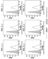
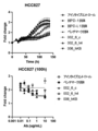
- FIG. 1 is a diagram showing the AMP degradation inhibitory function (H322 cells) of the 002_m antibody prepared in Example 1.
- FIG. 2 is a diagram showing amino acid sequences of the heavy chain variable region and the light chain variable region of the 002_m antibody.
- FIG. 3 is a diagram showing the binding properties of the obtained chimeric antibodies 002_1_c to 002_6_c to human lung cancer cell line H322 cells expressing CD73.
- FIG. 4 is a diagram showing the AMP degradation inhibitory function of human lung cancer H322 cells for the 002_1_c antibody to 002_6_c antibody.
- FIG. 1 is a diagram showing the AMP degradation inhibitory function (H322 cells) of the 002_m antibody prepared in Example 1.
- FIG. 2 is a diagram showing amino acid sequences of the heavy chain variable region and the light chain variable region of the 002_m antibody.
- FIG. 3 is a diagram showing the binding properties of the obtained chimeric antibodies
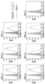
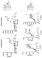
- FIG. 5-1 is a diagram showing the amino acid sequence of the heavy chain variable region of the obtained humanized antibody (002_6_h1 to 002_6_h8) of 002_6_c.
- FIG. 5-2 is a diagram showing the amino acid sequence of the light chain variable region of the obtained humanized antibody (002_6_h1 to 002_6_h8) of 002_6_c.
- FIG. 6-1 is a diagram showing the binding property of humanized antibodies (002_6_h1 to 002_6_h8) to human lung cancer cell line H322 cells expressing CD73.
- FIG. 6-2 is a diagram showing the binding property of humanized antibodies (002_6_h1 to 002_6_h8) to human lung cancer cell line H322 cells expressing CD73.
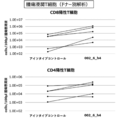
- FIG. 7 is a diagram showing the AMP degradation inhibitory function of three types of humanized antibodies (002_6_h2, 002_6_h4, 002_6_h8) using human lung cancer H322 cells.
- FIG. 8-1 is a diagram showing the binding property of the humanized antibody 002_6_h4 to various cells in comparison with the existing antibody and the base chimeric antibody 002_6_c.
- FIG. 8-2 is a diagram showing the binding property of the humanized antibody 002_6_h4 to various cells in comparison with the existing antibody and the base chimeric antibody 002_6_c.
- FIG. 8-1 is a diagram showing the binding property of the humanized antibody 002_6_h4 to various cells in comparison with the existing antibody and the base chimeric antibody 002_6_c.
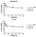
- FIG. 9 is a diagram showing the AMP degradation inhibitory function of humanized antibody 002_6_h4 using human breast cancer MDA-MB-231 cells as compared with the existing antibody and the base chimeric antibody 002_6_c.
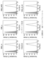
- FIG. 10 is a diagram showing the AMP degradation inhibitory function of 002_6_h4 on the recombinant CD73 antigen.
- FIG. 11 is a diagram showing the effect of 002_6_h4 on the division (proliferation) of human peripheral blood T cells.
- FIG. 12 is a diagram showing the AMP degradation inhibitory function (H322 cells) of the 003_m antibody to 009_m antibody prepared in Example 1.
- FIG. 13 is a diagram showing the AMP degradation inhibitory function (MDA-MB-231 cells) of the four antibodies (004_m, 006_m, 007_m, 008_m) judged to have high functionality in FIG.
- FIG. 14-1 is a diagram showing the amino acid sequence of the heavy chain variable region of the obtained antibody (003_m antibody to 009_m antibody).
- FIG. 14-2 is a diagram showing the amino acid sequence of the light chain variable region of the obtained antibody (003_m antibody to 009_m antibody).
- FIG. 15 is a diagram showing the binding property of the 003_m antibody to 009_m antibody to the target expressing cells (H322 cells).
- FIG. 16 is a diagram showing the binding property of the 003_m antibody to 009_m antibody to the target expressing cells (MDA-MB-231 cells).
- FIG. 17 is a diagram showing the binding property of the 003_m antibody to 009_m antibody to the target expressing cells (CD73 forced expression HEK-293T cells).
- FIG. 18 is a diagram showing the results of comparing the proportions of dividing cells when the antibody concentration was 1 nM.
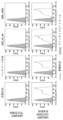
- FIG. 19 is a diagram showing the binding activity of an antibody to cells expressing the human CD73 gene.
- FIG. 20-1 is a diagram showing the antibody binding activity to cells expressing the human CD73 gene.
- FIG. 20-2 is a diagram showing the antibody binding activity to cells expressing the human CD73 gene.
- FIG. 21 is a diagram showing the antibody binding activity to cells expressing cynomolgus monkey CD73.
- FIG. 22 is a diagram showing that the antibody of the present invention has an inhibitory activity against the AMP degradation action of CD73 on the cell surface.
- FIG. 23 is a diagram showing that the antibody of the present invention has a division-promoting effect on CD4T cells and CD8T cells, and has an effect of enhancing the production ability of IFN ⁇ , TNF, and IL-2 from these cells. be.
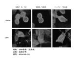
- FIG. 24 is a diagram showing a fluorescence microscope image showing the intracellular uptake of the 002_6_h4 antibody and the 006_hKB antibody.
- FIG. 25 is a diagram showing that the antibody of the present invention bound to CD73 on the surface of T cells decreases with time (internalized in T cells) when these cells are cultured.
- FIG. 26 is a diagram showing that the antibody of the present invention bound to CD73 on the surface of cancer cells decreases with time (internalized in cancer cells) when these cells are cultured.
- FIG. 27 is a diagram showing the results of measuring the number of tumor-infiltrating T cells in a mouse on day 35 after administration of 002_6_h4 antibody or isotype control antibody.
- FIG. 28-1 is a diagram showing whether the developed antibody of the present invention enhances the cancer cell injury activity by T cells.
- FIG. 28-1 is a diagram showing whether the developed antibody of the present invention enhances the cancer cell injury activity by T cells.
- FIG. 28-2 is a diagram showing whether the developed antibody of the present invention enhances the cancer cell injury activity by T cells.
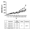
- FIG. 29 is a diagram showing the results of animal experiments on whether 002_6_h4 can enhance the antitumor effect of the human immune system simulated in mice.
- FIG. 30 is a diagram showing the result of volume change of a tumor mass in an immunodeficient mouse body.
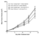
- FIG. 31 is a diagram confirming the antitumor effect of the anti-CD73 antibody of the present invention in a knock-in mouse expressing human CD73.
- the present invention in one embodiment, is a binding agent to a CD73 antigen, particularly an antibody, that activates T cells that are binding to the CD73 antigen on cancer cells or immune cells and are damaging to the cancer cells.
- a human antibody derivative can be provided.
- the binding agent of the present invention particularly an antibody or a human antibody derivative, can be used to shrink or eliminate a tumor because it inhibits the growth of cancer cells for the treatment of cancer cells expressing the CD73 antigen. It can also be used to activate T cells that are injurious to cancer cells when they are exhausted.
- the present invention provides an antibody or human antibody derivative that has binding to the CD73 antigen.
- This antibody or human antibody derivative is characterized by activating T cells that are damaging to cancer cells.
- the antibody or human antibody derivative of the present invention suppresses the function of CD73 expressed in cancer cells and causes exhaustion of immune cells (particularly T cells) caused by accumulation of AMP metabolite (adenosine) around the cancer cells. By releasing it, it activates immune cells and exerts tumor growth inhibitory effect and cancer therapeutic effect, suppresses the function of CD73 expressed in immune cells (particularly T cells), activates immune cells, and activates immune cells. It may be a tumor growth inhibitory effect and a cancer therapeutic effect, a cancer therapeutic effect by controlling the environment around the tumor other than immune cells, or a cancer therapeutic effect by direct control on the tumor.
- the antibody may be derived from any animal species of mammals, and the species of origin of the antibody is not limited to humans, and mice, rats, guinea pigs, hamsters, etc. It may be a rabbit or the like.
- the present invention may also be a human antibody derivative of the above-mentioned antibody as long as it has a binding property to the CD73 antigen and has a functional feature of suppressing cancer.
- a human antibody derivative in the present invention as one embodiment, the amino acid sequences of the CDRs at six positions of the above antibody (heavy chain complementarity determining regions (CDRs) 1 to CDR3 and light chain CDR1 to CDR3), and a human antibody. It is possible to provide a derivative of an antibody characterized by having the amino acid sequences of the constant regions of origin and the other amino acid sequences being a combination of the original antibody-derived amino acid sequence and the human antibody-derived amino acid sequence. can.
- a humanized antibody in which a region other than the complementarity determination region (CDR) of the above-mentioned "antibody” is replaced with an amino acid sequence derived from a human antibody, or a variable region of the above antibody is a constant of a human antibody.
- CDR complementarity determination region
- a variable region of the above antibody is a constant of a human antibody.
- chimeric antibodies such as those linked to a region, multivalent antibodies in which one type of antibody has multiple antigen binding sites, and multispecific antibodies (bispecific antibodies) in which one type of antibody has multiple specificities.
- bispecific antibodies multispecific antibodies
- the antibody or human antibody derivative of the present invention is characterized by having a total of 6 amino acid sequences of CDR1 to 3 of the same heavy chain and CDR1 to 3 of the light chain, which are the same as the antibody having binding property to the CD73 antigen. It is something that can be done.
- amino acid sequences of 6 CDRs of an antibody having binding to such a CD73 antigen include the following (1) to (8) :.
- CDR1 Heavy chain complementarity determining regions, CDR1 (DX 1 NMD (X 1 is C or S), SEQ ID No .: 1), CDR2 (DINPNNGGTIYNQKFKG, SEQ ID No .: 2), and CDR3 (TNWDYAMDY, SEQ ID No .: 3), and light chain complementarity determining regions, CDR1 (KASQDINSX 2 LS (X 2 is N, D or Q), SEQ ID No .: 4), CDR2 (RANRLID, SEQ ID No .:) 5), and CDR3 (X 3 QYDVFPRT (X 3 is L or Q), SEQ ID No .: 6); (2) Heavy chain complementarity determining regions, CDR1 (SFGMH, SEQ ID No .: 9), CDR2 (YISSGSRTIYYADTVRG, SEQ ID No .: 10), and CDR3 (DFGSSSPNYFDY, SEQ ID No .: 11), and light.
- CDR1 (RASESVDNYGISFMN, SEQ ID No .: 12), CDR2 (AASNQGS, SEQ ID No .: 13), and CDR3 (QQSKEVPWT, SEQ ID No .: 14); (3) Heavy chain complementarity determining regions, CDR1 (GYWMN, SEQ ID No .: 17), CDR2 (RIDPYDSETHYSQKFKD, SEQ ID No .: 18), and CDR3 (SSPITTAPFDY, SEQ ID No .: 19), and light Chain complementarity determining regions, CDR1 (RASESVDYYGFSFMN, SEQ ID No .: 20), CDR2 (AASTQGS, SEQ ID No .: 21), and CDR3 (QQSKEVPYT, SEQ ID No .: 22); (4) Heavy chain complementarity determining regions, CDR1 (SYGVS, SEQ ID No .: 25), CDR2 (VIWGDG
- the antibody or human antibody derivative of the present invention is also characterized by having the ability to activate T cells.
- T cells by suppressing the function of CD73 expressed in cancer cells or immune cells and releasing the exhaustion of immune cells (particularly T cells), immune cells are activated, and cancer growth inhibitory effect and cancer therapeutic effect are achieved. It is preferable to exert it.
- Such activation of T cells may occur in vitro or in vivo. Whether or not it has the ability to activate T cells that are damaging to such cancer cells is screened for whether or not they actually activate T cells from among the antibodies that have binding to the CD73 antigen. Can be obtained by doing.
- the exhaustion of immune cells refers to a state in which immune cells have become dysfunctional. Specifically, it causes a decrease in cytokine secretion ability and cytotoxicity in T cells, a decrease in cytokine secretion ability and cytotoxicity in NK cells, a decrease in antigen presentation ability of dendritic cells, activation of regulatory T cells, etc. be.
- an individual's cancer immunity to cancer cells does not function or functions poorly, and the activity to eliminate cancer cells decreases.
- Such exhaustion of immune cells is caused by the increase of checkpoint proteins such as PD-1 and CTLA-4 on the surface of T cells, and the cancer cells that continue to proliferate force the immune system to activate for a long period of time. It is known that it occurs in effector T cells, but it is a reversible state, and it is thought that it is possible to eliminate the exhausted state by activating the same cells.
- the activation of T cells can be grasped by using the proliferation of T cells, the increase in cytotoxicity of T cells against cancer cells, the secretion of cytokines from T cells, and the like as indicators. If these T cells are activated, it indicates that the antibody of the present invention or the human antibody derivative inhibits the activity of CD73 in T cells and suppresses the reaction of degrading AMP to adenosine. ..
- In vivo screening involves transplanting target cancer cells into an animal such as a wild-type mouse or a mouse in which immune cells are transplanted into an immunodeficient mouse such as a nude mouse or a SCID mouse, and then using the antibody of the present invention or a human.
- Infiltration of T cells around the tumor mass in the body by measuring the change in the size of the tumor mass in the body when the type antibody derivative is administered, or when the antibody of the present invention or the human type antibody derivative is administered. Can be done by anatomically examining.
- In vitro screening involves contacting peripheral blood T cells with the antibody or humanoid antibody derivative of the invention under culture conditions to measure T cell proliferation, T cell cytokine secretion, or This can be done by investigating whether T cells cause cell death against cancer cells.
- the antibody or human antibody derivative of the present invention can be specified as having the amino acid sequence of the following heavy chain variable region VH domain: (1) 002_m heavy chain variable region (SEQ ID No .: 7), (2) 003_m heavy chain variable region (SEQ ID No .: 15), (3) 004_m heavy chain variable region (SEQ ID No .: 23), (4) 005_m heavy chain variable region (SEQ ID No.
- the numbers (1) to (8) correspond to the numbers of the combination of the sequences of the heavy chain CDR1 to CDR3 described above.
- the amino acid sequence of SEQ ID NO: 7 is a sequence including the amino acid sequences of the heavy chains CDR1 to CDR3 specified by SEQ ID NO: 1 to SEQ ID NO: 3.
- the antibody or human antibody derivative of the present invention can be identified as having the amino acid sequence of the following light chain variable region VL domain: (1) 002_m light chain variable region (SEQ ID No .: 8), (2) 003_m light chain variable region (SEQ ID No .: 16), (3) 004_m light chain variable region (SEQ ID No .: 24), (4) 005_m light chain variable region (SEQ ID No. : 32), (5) 006_m light chain variable region (SEQ ID No .: 40), (6) 007_m light chain variable region (SEQ ID No .: 48), (7) 008_m light chain variable region (SEQ ID No.
- the numbers (1) to (8) correspond to the numbers of the combination of the sequences of the light chain CDR1 to CDR3 described above.
- the amino acid sequence of SEQ ID NO: 8 is a sequence including the amino acid sequences of light chain CDR1 to CDR3 specified by SEQ ID NO: 4 to SEQ ID NO: 6.
- the human antibody derivative of the present invention also includes the above-mentioned antibody or a functional fragment of the human antibody derivative.
- Functional fragments of the antibody or human antibody derivative in the present invention include F (ab') 2, Fab', Fab, single chain Fv (scFv) and the like.
- the functional fragment of the present invention may be any fragment thereof as long as it can induce injury to cancer cells, for example, F (ab') 2 fragment and the like. , Can be used as such a functional fragment.
- the antibody or human antibody derivative of the present invention is obtained by administering the CD73 antigen as an immunogen to an animal of the above-mentioned derived species and culturing antibody-producing cells collected from the animal body.
- a vector for protein expression containing a DNA sequence capable of defining the amino acid sequence of an antibody or human antibody derivative is designed, and the vector is introduced into cells for protein production and obtained recombinantly. You may.
- the DNA sequence capable of defining the amino acid sequence of the antibody or human antibody derivative of the present invention can be obtained from a cell producing the target antibody or human antibody derivative, depending on the animal species used in the expression system based on the amino acid sequence. It can be made by a method of designing based on optimized codons or a method of using a combination of these methods.
- the prepared DNA sequence is obtained by incorporating it into an expression vector suitable for a protein expression cell type (for example, CHO cell) in which the antibody or human antibody derivative is to be expressed and introducing it into the protein expression cell type. It can be obtained by using a method well known to those skilled in the art.
- a protein expression cell type for example, CHO cell
- introducing it into the protein expression cell type it can be obtained by using a method well known to those skilled in the art.
- a vector containing a DNA sequence defining a heavy chain amino acid sequence and a DNA defining a light chain amino acid sequence are used for a protein expression cell type.
- a method of introducing a vector containing a sequence and expressing both proteins in the cell to produce an antibody in the cell, or a DNA sequence defining a heavy chain amino acid sequence and a DNA sequence defining a light chain amino acid sequence It can be prepared by a method of introducing a vector containing both of them and expressing both proteins in the cells to produce an antibody in the cells.
- human antibody derivative obtained as one embodiment in the present invention
- a human antibody derivative is prepared from the above-mentioned antibody (1).
- Antibodies containing the 002_6_h2 heavy chain variable region (SEQ ID NO: 67) and the 002_6_h2 light chain variable region SEQ ID NO: 68).
- the antibody or human-type antibody derivative of the present invention is based on the above-mentioned characteristic of having the ability to activate immune cells (particularly T cells) having cytotoxicity against cancer cells.
- a pharmaceutical composition comprising an antibody of the invention or a human antibody derivative that activates immune cells (particularly T cells) that are damaging to cancer cells in a subject in need of treatment or prevention of cancer. Goods, can be provided.
- Cancer cells that can be targeted in the present invention include, for example, leukemia (including chronic lymphocytic leukemia and acute lymphocytic leukemia), lymphoma (non-hodgkin lymphoma, hodgkin lymphoma, T-cell line lymphoma, B-cell line lymphoma, etc.).
- leukemia including chronic lymphocytic leukemia and acute lymphocytic leukemia
- lymphoma non-hodgkin lymphoma, hodgkin lymphoma, T-cell line lymphoma, B-cell line lymphoma, etc.
- Berkit lymphoma malignant lymphoma, diffuse lymphoma, follicular lymphoma), myeloma (including multiple myeloma), melanoma, lung cancer, breast cancer, colon cancer, kidney cancer, stomach cancer, ovarian cancer, pancreas Cancer, cervical cancer, uterine body cancer, endometrial cancer, esophageal cancer, liver cancer, head and neck cancer, head and neck squamous epithelial cancer, skin cancer, urinary tract cancer, prostate Lymphoma, pharyngeal cancer, laryngeal cancer, lymphoma, male embryoma, endometrial hyperplasia, endometriosis, germoma, fibrosarcoma, caposic sarcoma, hemangiomas, spongy hemangiomas, Hemangioblastoma, retinal blastoma, stellate cell tumor, neurofibromas, rare projectile cystoma, medullary blastom
- the antibody or human antibody derivative of the present invention is characterized by activating immune cells (particularly T cells) having cytotoxicity against target cancer cells as described above. That is, when administered to a living body, activation of immune cells (particularly T cells) occurs only in immune cells having cytotoxicity against cancer cells expressing the target CD73 antigen, and expresses the CD73 antigen. However, in the case of normal cells, it is required not to induce cytotoxicity that may cause clinical problems.
- the antibody or human antibody derivative of the present invention can also be provided as a composition in combination with other antibodies or other agents such as anticancer agents.
- the antibody or human antibody derivative of the present invention can also be bound to a drug to form an antibody drug conjugate (ADC).
- ADC antibody drug conjugate
- a preparation containing the antibody or human type antibody derivative of the present invention together with a physiologically acceptable diluent or carrier.
- suitable carriers include buffers (phosphate buffer, citric acid buffer, acetate buffer, etc.), salts (sodium chloride, etc.), sugars (glucose, trehalose, mannitol, sorbitol, etc.), additives (arginine, etc.). Amino acids, surfactants such as polysorbates, etc.), but are not limited to these.
- the antibody or human antibody derivative of the present invention can be freeze-dried and reconstituted and used by adding a buffered aqueous solution as described above when necessary.
- the preparation containing the antibody of the present invention or the human antibody derivative can be administered in various dosage forms, and can be, for example, a parenteral administration agent such as an injection or an infusion.
- the dose of the antibody or humanoid antibody derivative of the present invention varies depending on symptoms, age, body weight, etc., but usually, in parenteral administration, 0.01 mg to 1000 mg per kg body weight, preferably 0.05 per kg body weight per day.
- Intraperitoneal injection subcutaneous injection in the range of mg to 500 mg, 0.05 mg to 100 mg, 0.05 mg to 50 mg, 0.05 mg to 20 mg, more preferably 0.1 mg to 10 mg per kg of body weight per day.
- Intramuscular injection, intratumor injection, or intravenous injection which can be administered by the appropriate route of administration depending on the type of cancer.
- the present invention requires treatment or prevention of cancer in another embodiment.
- a method of treating or preventing cancer in a subject comprising administering to the subject an effective amount of an antibody or humanoid antibody derivative of the invention.
- Treatment or prevention of cancer with the antibody or human antibody derivative of the present invention is that the antibody or human antibody derivative activates immune cells (particularly T cells) having cytotoxicity against cancer cells in the body. Caused by.
- an adjuvant for example, Clin. Microbiol
- Particles that can be administered with (such as those described in Rev., 7: 277-289, 1994), liposome formulations, particulate formulations bound to beads with a diameter of several ⁇ m, lipid-bound formulations, etc. It can also be administered in the form of a formulation.
- the antibodies or human-type antibody derivatives of the present invention are used to detect the CD73 antigen in a sample due to the characteristic of having binding property to the CD73 antigen.
- the antibody or human antibody derivative of the present invention can be used for a sample containing cancer cells or immune cells (for example, T cells) collected from a subject, such as an immunoprecipitation method, an aggregation reaction, and a magnetic bead method.
- a sample containing cancer cells or immune cells (for example, T cells) collected from a subject such as an immunoprecipitation method, an aggregation reaction, and a magnetic bead method.
- the antibody or human antibody derivative of the present invention is also based on the feature that it has a binding property to the CD73 antigen and activates immune cells (particularly T cells) having damage to cancer cells.
- a method for measuring the cytotoxicity to cancer cells in such a subject the following steps: A step of contacting a cancer cell collected from a subject with an antibody of the present invention or a human antibody derivative under culture conditions (that is, in vitro).
- the step of measuring whether the AMP metabolism of cancer cells (the reaction that decomposes AMP into adenosine) is reduced, the step of measuring whether the cell viability is reduced, or the secretion of immunostimulatory substances. Steps to measure whether or not
- a method for measuring cytotoxicity against cancer cells using the antibody of the present invention or a human antibody derivative including the above can be mentioned.
- AMP metabolism reaction that decomposes AMP to adenosine
- T cells are activated and tumors shrink, and cancer against cancer cells is known. It is believed that immunity is activated.
- cancer cells collected from a subject are brought into contact with the antibody of the present invention or a human antibody derivative in vitro, and the cancer cells are subjected to culture conditions. By measuring whether or not AMP metabolism is reduced, cytotoxicity to cancer cells can be measured.
- cancer cells collected from a subject are brought into contact with the antibody of the present invention or a human antibody derivative, and the degradation reaction from AMP to adenosine is measured under culture conditions (that is, in vitro). Therefore, when the antibody of the present invention or the human antibody derivative is administered to the subject, the cytotoxicity to the cancer cells in the subject (in vivo) can be measured.
- the decomposition reaction from AMP to adenosine can be measured by HPLC method, LC-MS method, ELISA method, or AMP-dependent chemiluminescence method (Cell titer glo, etc.).
- peripheral blood lymphocytes cause a decrease in the cell survival rate of cancer cells.
- cancer cells collected from a subject are brought into contact with the antibody of the present invention or a human antibody derivative in vitro, and the cell viability is increased under culture conditions.
- the cytotoxicity to cancer cells can be measured. More specifically, cancer cells collected from a subject are brought into contact with the antibody or human antibody derivative of the present invention in the presence of peripheral blood lymphocytes of the same subject under culture conditions (ie, in. Cancer in vivo when the antibody or human antibody derivative of the present invention is administered to the subject by measuring whether the cell viability of the cancer cells is reduced in vivo). The cytotoxicity to cells can be measured.
- cancer cells are activated and tumors shrink when the secretion of immunostimulatory substances from peripheral blood lymphocytes is enhanced in the living body, and cancer immunity against cancer cells is known. Is believed to be activated.
- cancer cells collected from a subject are brought into contact with the antibody of the present invention or a human antibody derivative in vitro, and under culture conditions, an immunostimulatory substance. Cytotoxicity to cancer cells can be measured by measuring whether or not the secretion of the antibody is enhanced. More specifically, cancer cells collected from a subject are brought into contact with the antibody of the present invention or a human antibody derivative in the presence of peripheral blood lymphocytes of the same subject under culture conditions (that is, in vitro).
- IFN- ⁇ , TNF ⁇ and the like can be mentioned as examples as the immunostimulatory substance to be measured for determining the cytotoxicity to cancer cells.
- IFN ⁇ is known to activate immune cells such as NK cells and exert an antitumor effect.
- the invention also comprises AMP metabolism, cytotoxicity, secretion of immunostimulatory substances, or activation of immune cells to cancer cells harvested from a subject, including the antibodies of the invention or humanoid antibody derivatives. Measurement kits for measurement in vitro can also be provided.
- the measurement kit for measuring AMP metabolism includes a reagent for measuring AMP as a substrate and adenosine, which is a metabolite thereof (AMP-dependent chemiluminescent reagent). , ELISA reagents) and equipment (HPLC system, LC-MS system).
- the measurement kit for measuring cytotoxicity includes known means for measuring cell proliferation (for example, uptake of thymidin, uptake of BrdU, free release). Measurement of lactate dehydrogenase (LDH) activity, measurement of living cell-derived substances (reducing enzyme activity, esterase activity, ATP, etc.) can be included.
- LDH lactate dehydrogenase
- the measurement kit for measuring the secretion of the immunostimulatory substance includes means for detecting the immunostimulatory substance to be measured (for example, primary for the immunostimulatory substance).
- Antibodies and secondary antibodies for the detection of the primary antibody, etc. can be included.
- the measurement kit for measuring the activation of immune cells can include, in addition to the antibody or human antibody derivative of the present invention, a labeling reagent for measuring the division of immune cells with a flow cytometer.
- Example 1 Preparation of antibody The purpose of this example was to obtain a monoclonal antibody against the CD73 antigen using mice.
- a single cell suspension of spleen cells was prepared from the spleen according to a known method, and B cells were purified using a MACS-pan B cell isolation kit (Miltenny 130-104-443). Purified B cells were stained with ZOMBIE-APC-Cy7 (Biolegend), Alexa488anti-IgM (Biolegend), VB421anti-IgG (Biolegend), PE-CD73 (self-prepared product) and tested on FACSAria (BD). , IgG-positive and CD73 antigen-binding cell populations were separated into 96-well plates one by one.
- the genes corresponding to the heavy chain variable region and the light chain variable region of the antibody were amplified for each well by the RT-PCR method.
- As the primer used for amplification based on the information in the publicly known literature, a sequence containing the 5'end and 3'end regions of the heavy chain variable region and the light chain variable region, respectively, was adopted, and the diversity of the antibody gene sequence was taken into consideration. (Heavy chain: H1 mix and H2 mix, light chain: k mix) (Lotta von Boehmer et al., Nature Protocols, vol 11, p.1908-1923 (2016)) ..
- a linker sequence for insertion into the antibody expression vector is added to the end of each gene by a PCR reaction, and a vector for heavy chain expression or a vector for light chain expression (vector for heavy chain expression or light chain expression).
- a CMV promoter, a secretory signal, a gene for a heavy chain variable region or a light chain variable region, and a gene for a heavy chain constant region or a light chain constant region were tandemly linked to each other) by a ligation reaction.
- the heavy chain gene expression plasmid (derived from H1 or H2 mixed amplification product) and the light chain gene expression plasmid (derived from k mix amplification product) are transferred to CHO cells. I did an expression. Antibodies produced in the culture supernatant were collected from CHO cells, and the antigen specificity of each antibody was confirmed by antigen-fixed ELISA.
- ATP Sigma, A2383, final concentration 100 ⁇ M
- CellTiterGlo Promega, G9243
- chemiluminescence was measured using a microplate reader. That is, using the principle that AMP inhibits chemiluminescence due to the reaction between ATP and CellTiterGlo, the enzyme activity inhibitory effect of the antibody was evaluated from the amount of residual AMP.
- an antibody having the same sequence as the published variable region of MEDI9447 was produced using the gene recombination technology based on the sequence published in the advanced development product MEDI9447 (Special Table 2018-501197). "Benchmark antibody”) was adopted.
- mouse antibodies As a result of antibody function screening, eight types of mouse antibodies named 002_m, 003_m, 004_m, 005_m, 006_m, 007_m, 008_m, 009_m could be selected as CD73-binding activity-positive antibodies.
- Example 2 Antibody function evaluation (concentration-dependent evaluation) The purpose of this example was to evaluate the function of the 002_m antibody obtained in Example 1.
- the antibody function evaluation was carried out by the same method as the antibody function screening described in Example 1 (1-3). That is, human lung cancer cell line H322 cells or human breast cancer cell line MDA-MB-231 cells (ATCC® HTB-26 TM ) were used as cells.
- the antibody concentration dependence of the AMP decomposition inhibitory function was analyzed by measuring the AMP decomposition reaction in the same manner as in Example 1 (1-3), and the IC50 value (50% inhibition concentration: CD73 in this case). The concentration of the antibody that can inhibit 50% of the enzyme function of the antibody) was calculated, and the function of the antibody was quantitatively evaluated.
- Figure 1 shows the AMP degradation inhibitory function (H322 cells) of the 002_m antibody prepared in Example 1. Although this antibody is a mouse antibody, it was revealed that it shows an IC50 value close to that of the benchmark antibody (see Table 1).
- Example 3 Antibody gene sequence analysis This example was performed for the purpose of determining the amino acid sequence of the 002_m antibody obtained in Example 1.
- the obtained plasmid DNA was analyzed for the gene sequences defining the heavy chain variable region and the light chain variable region by the Sanger sequence, and the amino acid sequence was determined based on the same sequence.
- the amino acid sequences of the heavy chain variable region and the light chain variable region of the 002_m antibody are shown in FIG. 2 (SEQ ID NO: 7 and SEQ ID NO: 8 respectively). Further, in this figure, the heavy chains CDR1, CDR2, CDR3 (SEQ ID NO: 1, SEQ ID NO: 2, SEQ ID NO: 3, respectively) and the light chains CDR1, CDR2, CDR3 (SEQ ID NO, respectively). : 4, SEQ ID NO: 5, SEQ ID NO: 6) are underlined.
- Example 4 Preparation of human-mouse human chimeric antibody The purpose of this example is to prepare a chimeric antibody having a variable region of a mouse antibody and a constant region of a human antibody for the 002_m antibody sequenced in Example 3. rice field.
- the gene sequence of the variable region of the mouse antibody was artificially synthesized and incorporated into a vector for expressing a human constant region (Fc) chimeric recombinant antibody.
- a light chain expression element EF-1 ⁇ promoter, a secretory signal, a DNA sequence defining a light chain variable region, and a DNA sequence defining a light chain constant region are linked in tandem
- a heavy chain expression element EF
- a plasmid having a -1 ⁇ promoter, a secretory signal, a DNA sequence defining a heavy chain variable region, and a DNA sequence defining a heavy chain constant region was tandemly linked) was constructed.
- the plasmid was transfected into ExpiCHO cells (Thermo) to secrete the antibody into the medium, and then the antibody was purified using a Protein A column (GL Science).
- the chimeric antibody thus prepared was defined as 002_1_c antibody.
- the amino acid sequence of the heavy chain variable region of this antibody is composed of the amino acid sequence of SEQ ID NO: 7
- the amino acid sequence of the light chain variable region is composed of the amino acid sequence of SEQ ID NO: 8.
- Example 5 Preparation of CDR-modified antibody
- a modified antibody is prepared by substituting a residue that may undergo post-translational modification in CDR with an unmodified residue for the 002_m antibody. I went there for the purpose.
- the 002_m antibody heavy chain variable region (SEQ ID NO: 7) 32nd amino acid cysteine (C) is used as serine (S), and the 002_m antibody light chain variable region (SEQ ID NO: 8) 32nd amino acid asparagine (SEQ ID NO: 8) N) was replaced with aspartic acid (D) or glutamine (Q), respectively (see Table 2 below).
- a gene containing the relevant region was artificially synthesized and inserted into a human Fc chimeric recombinant antibody expression vector by ligation. Antibody expression and purification were carried out in the same manner as for human chimeric antibodies.
- Binding analysis of each of the obtained chimeric antibodies 002_1_c to 002_6_c to target-expressing cells was performed by FACS analysis using CD73 native-expressing cells (human lung cancer cell line H322). After reacting the test antibody or the isotype control antibody with the cells, the antibody was stained with a fluorescently labeled secondary antibody and subjected to flow cytometric analysis, and the antibody binding property was evaluated based on the obtained fluorescence intensity.
- Example 6 Preparation of humanized antibody
- a humanized antibody was prepared based on the 002_6_c antibody (SEQ ID NO: 7 (C32S) / SEQ ID NO: 8 (N32Q)) selected in Example 5. I went for the purpose of doing.
- the humanized antibody was prepared using the public software Tabhu (http://circe.med.uniroma1.it/tabhu/). Using the same software, we extracted from a human antibody database that has sequences that are similar in sequence and conformation to 002_6_c, picked up 4 types of sequences (FJ039788, AF146404, 3SQO_H, AY686911) as candidate sequences, and selected these. The sequences other than the CDR regions of the antibody and the sequences of the CDR regions of 002_6_c were graphed.
- amino acid residues to be backmutated were selected up to 10 amino acid residues in each sequence based on the large structural difference between the humanized antibody and the mouse antibody (higher TubHu score).
- the method described in the non-patent document Bioinformatics. 2015 Feb 1; 31 (3): 434-5 was followed.
- Fig. 5 The amino acid sequences of the variable regions of the obtained 002_6_c humanized antibodies (002_6_h1 to 002_6_h8) are shown in Fig. 5 (heavy chain variable region is shown in Fig. 5-1 and light chain variable region is shown in Fig. 5-2). Each amino acid sequence was assigned SEQ ID NO: as shown in Table 4 below.
- Binding analysis of each of the obtained humanized antibodies (002_6_h1 to 002_6_h8) to target-expressing cells was performed by FACS analysis using CD73 native-expressing cells (human lung cancer cell line H322). The results are shown in FIG. It was confirmed that all humanized antibodies showed the same binding properties as the base chimeric antibody 002_6_c antibody.
- the humanized antibody 002_6_h4 selected as the lead antibody above was compared with existing antibodies (commercially available anti-CD73 antibody (biolegend), benchmark antibody) and the base chimeric antibody 002_6_c, and the binding property to various cells was measured by flow cytometry. confirmed.
- CD73 native-expressing cells human breast cancer cell line MDA-MB-231
- human breast cancer cell line MDA-MB-231 cells expressing CD73 antigen
- HEK293T wild type not expressing CD73 antigen FACS analysis was performed using cells (derived from human fetal kidney cells) and cells in which CD73 antigen was forcibly expressed in HEK293T cells.
- CD73 forced expression cells were generated using the lentiviral vector system (Origene).
- Lentivirus particles carrying the human CD73 gene (NM_002526) and puromycin resistance gene are prepared, infected with HEK293T cells, and then the cells are cultured in the presence of puromycin, and the target gene is integrated into the genome to be CD73 positive.
- HEK293T cells were collected and used for binding analysis. After reacting the test antibody or the isotype control antibody with the cells, the antibody was stained with a fluorescently labeled secondary antibody and subjected to flow cytometric analysis, and the antibody binding property was evaluated based on the obtained fluorescence intensity.
- the humanized antibody 002_6_h4 was compared with the existing antibody (commercially available anti-CD73 antibody (biolegend), benchmark antibody) and the base chimeric antibody 002_6_c, and the same method as in (1-3) was used for human breast cancer MDA-MB-.
- An antibody function screening was performed to measure the AMP degradation inhibitory function using 231 cells. The results are shown in FIG. From this result, the IC50 value of the humanized antibody against the AMP degradation inhibitory function using human breast cancer MDA-MB-231 cells was calculated as shown in Table 6 below. Together with the results, it was shown that the humanized antibody 002_6_h4 has an AMP degradation inhibitory function in multiple cancer types, similar to the chimeric antibody 002_6_c.
- Example 7 Suppressive function against recombinant antigen The purpose of this example was to confirm the enzymatic reaction inhibitory function of the anti-CD73 antibody obtained in Example 6 against the recombinant CD73 antigen.
- the CD73 antigen (terminal His-Tag, self-prepared product) was immobilized on a 96-well plate for ELISA via an anti-His-Tag antibody, and the humanized anti-CD73 antibody 002_6_h4 and the substrate AMP (final concentration 100 ⁇ M) were used.
- the humanized anti-CD73 antibody 002_6_h4 and the substrate AMP (final concentration 100 ⁇ M) were used.
- ATP final concentration 100 ⁇ M
- CellTiterGlo Promega, G9243
- chemiluminescence was measured using a microplate reader.
- AMP enzyme activity inhibitory effect of the antibody was evaluated from the amount of residual AMP.
- the same experiment was performed using a benchmark antibody as a positive control.
- Figure 10 shows the AMP degradation inhibitory function of 002_6_h4 for the recombinant CD73 antigen. As a result, it was suggested that 002_6_h4 showed an AMP degradation inhibitory function against the recombinant CD73 antigen and directly inhibited the enzyme.
- Example 8 Evaluation of effect on T cell division This example was performed for the purpose of confirming the proliferation promoting activity of the anti-CD73 antibody obtained in Example 6 on human peripheral blood T cells.
- PBMC peripheral blood mononuclear cells
- CD4 + T Cell Isolation Kit human (Miltenyi Biotech, 130-096-533) was used to purify CD4 positive T cells, and CellTrace CFSE cell proliferation kit (ThermoFisher Scientific, #) Cells were fluorescently labeled with C34554).
- Figure 11 shows the effect of 002_6_h4 on the division (proliferation) of human peripheral blood T cells.
- the proportion of divided T cells increased in an antibody concentration-dependent manner, indicating that the anti-CD73 antibody had the effect of promoting T cell division.
- Example 9 Evaluation of antibody function (concentration-dependent evaluation) The purpose of this example is to evaluate the functions of antibodies other than the 002_m antibody obtained in Example 1 (that is, 003_m antibody, 004_m antibody, 005_m antibody, 006_m antibody, 007_m antibody, 008_m antibody, 009_m antibody). went.
- the antibody function evaluation was carried out by the same method as the antibody function screening described in Example 1 (1-3). That is, human lung cancer cell line H322 cells or human breast cancer cell line MDA-MB-231 cells (ATCC® HTB-26 TM ) were used as cells.
- the antibody concentration dependence of the AMP degradation inhibitory function was analyzed by measuring the AMP degradation reaction in the same manner as in Example 1, and the IC50 value (50% inhibitory concentration: 50 of the enzyme function of CD73 here). %) was calculated and the function of the antibody was quantitatively evaluated.
- the AMP degradation inhibitory function of the 003_m antibody, 004_m antibody, 005_m antibody, 006_m antibody, 007_m antibody, 008_m antibody, and 009_m antibody prepared in Example 1 against H322 cells is shown in FIG. 12, and the AMP degradation inhibitory function against MDA-MB-231 cells. Is shown in FIG. 13, respectively. Some of these antibodies, which are mouse antibodies, were found to show IC50 values that were lower or closer to the benchmark antibodies (see Table 7).
- clones judged to have high functionality were selected from the tests shown in FIGS. 12 and 13 above (004_m, 006_m, 007_m, 008_m).
- the AMP degradation inhibitory function (MDA-MB-231 cells) of these four antibodies is shown.
- the IC50 values of these clones were good compared to the benchmark antibody (under clinical development) and were about the same as 002_6_c (see Table 8).
- Example 10 Antibody gene sequence analysis The purpose of this example was to determine the amino acid sequences of the 003_m antibody to 009_m antibody obtained in Example 1.
- the obtained plasmid DNA was analyzed for the gene sequences defining the heavy chain variable region and the light chain variable region by the Sanger sequence, and the amino acid sequence was determined based on the same sequence.
- the heavy chain variable region amino acid sequence (Fig. 14-1) and light chain variable region amino acid sequence (Fig. 14-2) of 003_m antibody, 004_m antibody, 005_m antibody, 006_m antibody, 007_m antibody, 008_m antibody, 009_m antibody are shown.
- SEQ ID NOs were assigned to each amino acid sequence as shown in Table 9 below.
- Example 11 Analysis of binding to target-expressing cells The purpose of this example was to evaluate the binding of each antibody of 003_m to 009_m obtained in Example 1.
- Binding analysis to target-expressing cells was performed by FACS analysis using CD73 native-expressing cells (human lung cancer cell line H322 or human breast cancer cell line MDA-MB-231) or CD73 forced-expressing cells.
- CD73 forced expression cells were generated using the lentiviral vector system (Origene). After preparing lentivirus particles carrying the human CD73 gene (NM_002526) and puromycin resistance gene and infecting HEK293T cells, the cells were cultured in the presence of puromycin, and the target gene was integrated into the genome to be CD73 positive. The resulting HEK293T cells were collected and used for binding analysis. As a negative control, HEK293T cells expressing CD73 were used.
- the antibody After reacting the test antibody or isotype control antibody with the cells, the antibody was stained with a fluorescently labeled secondary antibody and subjected to flow cytometric analysis, and the antibody binding property was evaluated based on the obtained fluorescence intensity.
- FIG. 15 The binding of 003_m antibody to 009_m antibody to target-expressing cells (H322 cells) is shown in Fig. 15, and the binding of 003_m antibody to 009_m antibody to target-expressing cells (MDA-MB-231 cells) is shown in Fig. 16 and 003_m.
- the binding properties of the antibody to 009_m antibody to the target-expressing cells are shown in FIG. 17, respectively.
- solid lines and gray fills show fluorescence intensity histograms of the test antibody and the isotype control antibody, respectively.
- Example 12 Evaluation of effect on T cell division The purpose of this example was to confirm the proliferation-promoting activity of the anti-CD73 antibody obtained in Example 9 on human peripheral blood T cells.
- Example 8 The same method as in Example 8 was adopted, and the ratio of dividing cells was compared when the antibody concentration was 1 nM. The results are shown in FIG. The dotted line shows the proportion of dividing cells in the isotype control antibody.
- CD4 + T Cell Isolation Kit human (Miltenyi Biotech, 130-096-533) is used to purify CD4-positive T cells from peripheral blood mononuclear cells (PBMC) derived from healthy volunteers, and CellTrace CFSE cell proliferation kit. Cells were fluorescently labeled with (ThermoFisher Scientific, # C34554).
- Figure 18 shows the effect of 1 nM anti-CD73 antibody (006_m antibody, 007_m antibody, or 008_m antibody) on the division (proliferation) of human peripheral blood T cells.
- “w / o AMP” and “with AMP” (100 ⁇ M AMP) were incorporated to see the effect of the presence or absence of AMP without antibody because AMP has a T cell division inhibitory effect. ..
- the proportion of divided T cells increased in an antibody concentration-dependent manner (data shows). (Not shown), the effect of anti-CD73 antibody on promoting T cell division was shown.
- Example 13 Preparation of humanized antibody (2)
- a humanized antibody is prepared based on the 004_m antibody, 006_m antibody, 007_m antibody, and 008_m antibody selected in Example 9 among the respective antibodies of 003_m to 009_m obtained in Example 1. I went for the purpose.
- the humanized antibody was prepared using the public software Tabhu (http://circe.med.uniroma1.it/tabhu/) as in Example 6. Using this software, 004_m antibody, 007_m antibody, 008_m antibody and 004_m antibody are extracted from the human antibody database having sequences having characteristics similar in sequence and three-dimensional structure, 004_m antibody is 5 types, 007_m antibody is 4 types, and 008_m antibody is. Each of the four sequences was picked up as a candidate sequence, and the sequences other than the CDR region of these antibodies were graphed with the sequences of the CDR regions of each of the 004_m antibody, 007_m antibody, and 008_m antibody.
- Table 10 below shows the amino acid sequences of the variable regions of the humanized antibodies (004_h3 antibody, 006_hKB antibody, 007_h1 antibody, 008_h4 antibody) obtained based on the 004_m antibody, 006_m antibody, 007_m antibody, and 008_m antibody, respectively. As shown in Table 10 below, each amino acid sequence was assigned SEQ ID NO: 81 to SEQ ID NO: 88.
- Binding analysis to human target-expressing cells was performed by FACS analysis using CD73 native-expressing cells (human lung cancer cell line H322). Binding analysis to target-expressing cells was performed on CD73 native-expressing cells (human breast cancer cell line MDA-MB-231), human breast cancer cell line MDA-MB-231 cells expressing CD73 antigen, and HEK293T wild type not expressing CD73 antigen. FACS analysis was performed using cells (derived from human fetal kidney cells) and cells in which CD73 antigen was forcibly expressed in HEK293T cells. CD73 forced expression cells were generated using the lentiviral vector system (Origene).
- Lentivirus particles carrying the human CD73 gene (NM_002526), the red-tailed monkey CD73 gene (XM_001086989.2) (the amino acid sequence is the same as that of the crab monkey CD73) and the puromycin resistance gene were prepared, infected with HEK293T cells, and then puromycin.
- the cells were cultured in the presence of HEK293T cells in which the target gene was integrated into the genome and became CD73 positive, and used for binding analysis. After reacting the test antibody or the isotype control antibody with the cells, the antibody was stained with a fluorescently labeled secondary antibody and subjected to flow cytometric analysis, and the antibody binding property was evaluated based on the obtained fluorescence intensity.
- FIGS. 19 to 21 The results are shown in FIGS. 19 to 21.
- FIGS. 19 and 20 are diagrams confirming the binding of the antibody to cells expressing the human CD73 gene
- FIG. 21 confirms the binding of the antibody to cells expressing the rhesus monkey CD73 gene. It is a figure.
- the 004_h3 antibody, 006_hKB antibody, and 008_h4 antibody were confirmed to show the same binding property to the human CD73 antigen as the 002_6_h4 antibody when compared, and the binding property to the CD73 antigen could be confirmed.
- rice field the humanized antibodies examined.
- the 006_hKB antibody among them showed the same binding property to the rhesus monkey CD73 antigen (cynomolgus monkey CD73 antigen) as the 002_6_h4 antibody, and the effect of the antibody of the present invention was confirmed by animal experiments in the future. It was shown that rhesus monkeys or cynomolgus monkeys can be used for examination.
- Example 14 Suppressive function against recombinant antigen The purpose of this example was to confirm the enzymatic reaction inhibitory function of the humanized anti-CD73 antibody obtained in Example 13 against the recombinant CD73 antigen.
- Example 1 For antibody function evaluation, 004_h3 antibody, 006_hKB antibody, 007_h1 antibody, 008_h4 antibody were used as test antibodies, and 002_6_c antibody, 002_6_h4 antibody, isotype control antibody and benchmark antibody (MEDI9447 antibody) were used as controls, and Example 1 (1-). It was carried out by the same method as the antibody function screening described in 3). That is, human breast cancer cell line MDA-MB-231 cells (ATCC® HTB-26 TM ) were seeded on 96-well plates and incubated overnight at 37 ° C.
- human breast cancer cell line MDA-MB-231 cells ATCC® HTB-26 TM
- Example 1 As in Example 1 (1-3), AMP (Sigma, A1752), which is a substrate of CD73, was added to a final concentration of 200 ⁇ M, and the mixture was incubated at 37 ° C. for 4.5 hours to AMP with CD73.
- AMP Sigma, A1752
- the antibody concentration dependence of the AMP degradation inhibitory function was analyzed. A decomposition reaction was carried out.
- ATP Sigma, A2383, final concentration 10 ⁇ M
- Cell TiterGlo Promega, G9243
- Figure 22 shows the AMP degradation inhibitory function of the 004_h3 antibody, 006_hKB antibody, 007_h1 antibody, and 008_h4 antibody. All of these antibodies have been shown to have the function of suppressing AMP degradation by CD73 present on the cell surface (Figs. 22 and 23), and among these antibodies, the 006_hKB antibody is similar to 002_6_h4. , It was shown to have an inhibitory activity equal to or higher than that of the benchmark antibody.
- Example 15 Evaluation of effects on T cell division This example confirms the growth-promoting activity of the anti-CD73 antibody obtained in Example 13 and showing a particularly high effect in Example 14 on human peripheral blood T cells. I went for that purpose.
- PBMC peripheral blood mononuclear cells
- T cell marker staining antibody BioLegend, anti-CD3: UCHT1, anti-CD4: RPA-T4, anti-CD8a: HIT8a. Beads for cell count were added to the stained cells and analyzed with a flow cytometer to calculate the number of CD4 and CD8 positive cells per well.
- IL-2 BAF202 (R & D Systems)
- TNF ⁇ BAF210 (R & D Systems)
- Figure 23 shows the results of the evaluation of the effect of the antibody on T cell division.
- the 006_hKB antibody is as high as the 002_6_h4 antibody for both CD4-positive and CD8-positive T cells. It was shown to have a T cell division promoting effect (upper part of FIG. 23). It was also confirmed that high concentrations of IFN ⁇ , TNF ⁇ , and IL-2 were released into the culture supernatant from the cells to which the 006_hKB antibody was administered (Fig. 23, lower part).
- Example 16 Evaluation of intracellular uptake of antibody
- the anti-CD73 antibody obtained in Example 6 and the anti-CD73 antibody obtained in Example 13 and showing a particularly high effect in Example 14 were used. The purpose was to evaluate the uptake into cells.
- Human breast cancer cells MDA-MB-231 (1 ⁇ 10 ⁇ 5 cells) are seeded on a chamber slide (Thermo Fisher Scientific, 154534PK), and 10 ⁇ g / ml of various antibody solutions (002_6_h4, 006_hKB, MEDI9447) are added to 37 ° C. Incubated for 15 minutes or 19 hours. Samples before and after incubation were fixed and permeabilized (Thermo Fisher Scientific, FIX & PERM Fixation and Permeabilization Kit, GAS003) and stained with Anti-human IgG Alexa Fluor 594 (Thermo Fisher Scientific, A-11014). The distribution of the antibody was observed with a fluorescence microscope (Thermo Fisher Scientific, EVOS FLoid Imaging System, 4471136) to determine the presence or absence of the antibody uptake into the cells.
- a fluorescence microscope Thermo Fisher Scientific, EVOS FLoid Imaging System, 4471136
- Figure 24 shows the intracellular uptake of 002_6_h4 and 006_hKB.
- the antibody was distributed on the cell surface at the time of 15 minutes of incubation (upper part of Fig. 24), whereas the antibody was accumulated in the cell 19 hours after incubation (lower part of Fig. 24). Was shown to have taken up into the cells. This result suggests that the uptake of anti-CD73 antibody may induce the disappearance of the antigen from the cell surface.
- PBMC Peripheral blood mononuclear cells
- % Control (anti-CD73 antibody (X minutes) MFI-isotype control (X minutes) MFI) / (anti-CD73 antibody (0 minutes) MFI-isotype control (0 minutes) MFI) x 100
- % CD73 Ag (anti-CD73 antibody (X minutes) MFI-isotype control (X minutes) MFI) / (anti-CD73 antibody (0 minutes) MFI-isotype control (0 minutes) MFI) x 100
- Example 17 Measurement of KD value
- the anti-CD73 antibody obtained in Example 6 and the anti-CD73 antibody obtained in Example 13 and showing a particularly high effect in Example 14 bind to the CD73 antigen.
- the purpose was to evaluate the dissociation constant (KD value), which is an index of affinity.
- the KD value of the antibody bound to the target antigen was measured with Biacore 8K (GE Healthcare). Each antibody of 002_6_h4 antibody and 006_hKB antibody was immobilized on the sensor chip (CM5) using Human Antibody Capture Kit Type2 (Cytiva, 26-2346-00). A solution of human CD73 antigen diluted serially from 5 ⁇ 10 -9 M in 4 steps was prepared and used for multicycle kinetic analysis. In the 1: 1 coupling model, the KD values are as shown in Table 11 below.
- Example 18 Human Immune Mouse Test This example is a human CD34-positive cell of the anti-CD73 antibody obtained in Example 6 and the anti-CD73 antibody obtained in Example 13 and showing a particularly high effect in Example 14. The purpose was to confirm the antitumor effect in the transplanted mice.
- Human breast cancer cell line MDA-MB-231 was transplanted into human breast cancer cell line MDA-MB-231 into the mammary fat pad of human CD34-positive cell transplant NSG mice (hu-NSG, JACKSON laboratories) prepared from multiple healthy donors.
- a breast cancer cell-carrying mouse model was created.
- 002_6_h4 antibody or isotype control antibody is administered at a dose of 20 mg / kg once every 4 days for a total of 7 times (indicated as "Q4Dx7").
- Administered intraperitoneally The administration start date was set to day 0, and observation was performed until day 34.
- Figure 27 shows the results of measuring the number of tumor-infiltrating T cells in mice on day 34 after administration of 002_6_h4 antibody or isotype control antibody.
- the administration of 002_6_h4 antibody to tumor-infiltrating T cells showed an increasing tendency as compared with the case of administration of isotype control antibody, suggesting that the anti-CD73 antibody has a tumor immunological effect.
- the developed antibody enhances the cancer cell injury activity by T cells, and if so, the degree thereof in comparison with the benchmark antibody.
- Human lung cancer cells NCI-H292 or HCC827 introduced with RFP protein were seeded on 96-well plates and cultured overnight. After removing the medium the next day, IL-2 (final concentration 30 IU) suspended in X-VIVO-15 medium (Lonza, 04-418Q) and various antibodies were added, and the mixture was incubated at 37 ° C. for 30 minutes. After that, healthy volunteer PBMCs stimulated with Dynabeads TM Human T-Activator CD3 / CD28 for T Cell Expansion and Activation (Veritas, DB11131) for 10 days to proliferate T cells were added at an E / T ratio of 2 to 37 ° C.
- NCI-H292 was added to a final concentration of 300 [mu] M ATP in a final concentration of 500 [mu] M ATP, HCC827 to, 37 °C, 5% CO over time the growth of cancer cells by IncuCyte ZOOM System (Sartorius) with 2 conditions The change was observed.
- the results are shown in Fig. 28.
- the developed antibody (002_6_h4 and 006_hKB were used as examples in this example, but other developed antibodies of the present invention are also used) suppresses T cells even when anti-PD-1 antibody and anti-PD-1 antibody are allowed to act.
- the effect of suppressing the growth of cancer cells of T cells was enhanced under the condition that the effect of releasing the antibody was hardly observed and the growth of cancer cells could not be stopped.
- mice For the mammary fat pad of NSG mice (hu-NSG, JACKSON laboratories) (hereinafter referred to as "immunized humanized mice") transplanted with human CD34-positive cells prepared from multiple healthy donors.
- the human breast cancer cell line MDA-MB-231 was transplanted to create a human breast cancer-carrying mouse model.
- 002_6_h4 antibody or isotype control antibody is administered at a dose of 20 mg / kg once every 4 days for a total of 7 times (“Q4Dx7").
- a checkpoint inhibitor anti-PD-1 antibody pembrolizumab, keytruda, MSD
- isotype control antibody at a dose of 10 mg / kg for the first time only and 5 mg / kg for the second and subsequent times.
- Q5Dx6 A total of 6 times (indicated as "Q5Dx6") were administered intraperitoneally once every 5 days.
- the administration start date was set to day 0, and the tumor size was measured twice a week until day 34.
- Figure 29 shows the average value and standard error of each administration group of the increase rate of tumor volume from Day 0 to day 34 in a typical donor.
- the average tumor volume increase rate in the combination administration group (Group 4) was lower than that in the control group (Group 1) and the checkpoint inhibitor administration group (Group 3). rice field.
- the average tumor volume increase rate was lower in the 002_6_h4 antibody-administered group (Group 2) than in the control group (Group 1), and the anti-CD73 antibody was used alone or in immune humanized mice. , It was suggested that it has an antitumor effect when used in combination with a checkpoint inhibitor (pembrolizumab).
- Example 19 Immunodeficient mouse test This example was conducted for the purpose of confirming the antitumor effect of the anti-CD73 antibody obtained in Example 6 in an immunodeficient mouse body.
- the human breast cancer cell line MDA-MB-231 was transplanted into the mammary fat pad of NSG mice (JACKSON laboratories).
- 002_6_h4 antibody or isotype control antibody was administered at each dose once every 4 days for a total of 7 times (Q4Dx7) in the group composition shown in Table 12 below.
- Administered intraperitoneally The tumor size was measured twice a week with the administration start date as day 0 and the observation period up to day 35.
- Figure 30 shows the results of volume changes in the tumor mass in immunodeficient mice.
- the 002_6_h4 antibody was shown to be more effective in suppressing tumor growth than the isotype control.
- Example 20 Antitumor effect (knock-in model) The purpose of this example was to confirm the antitumor effect of the anti-CD73 antibody obtained in Example 6 in a knock-in mouse expressing human CD73.
- a colon cancer cell line (Biocytogen, MC38-hCD73) that lacks mouse CD73 and expresses human CD73 is 5 ⁇ 10 ⁇ 5 were transplanted.
- 002_6_h4 antibody or isotype control antibody was administered at a dose of 10 mg / kg twice a week for a total of 7 times (denoted as "BIWx7").
- Figure 31 shows the transition of the average tumor volume from Day 0 to day 18 as the average value and standard error of each administration group.
- the mean tumor volume of the combination administration group (Group 4) was lower than that of the control group (Group 1) and the checkpoint inhibitor administration group (Group 3), and the anti-CD73 antibody was a mouse expressing human CD73. It was suggested that it has an antitumor effect when used in combination with a checkpoint inhibitor.
- the antibody or human antibody derivative obtained by the present invention activates immune cells by suppressing the function of CD73 expressed in cancer cells or immune cells and releasing the exhaustion of immune cells (particularly T cells). , Tumor growth inhibitory effect and cancer treatment effect.
- the present invention can be widely used as a therapeutic / diagnostic agent for cancer. Since the antibody according to the present invention has an effect of relieving the exhaustion of immune cells, it is expected to be particularly effective in combination therapy with other immunotherapies.
Abstract
The present invention addresses the problem of developing an antibody which targets a CD73 antigen and has a higher function, more specifically, an antibody which is for treating cancer and has a higher activity. The present invention has shown that the problem can be solved by finding an antibody or a human antibody derivative which includes a heavy or light chain complementarity-determining region having a specific amino acid sequence, has binding properties to a CD73 antigen, and activates T cells having the property of damaging cancer cells.
Description
本発明は、CD73抗原を発現するがんに対して特異的に細胞傷害性活性を有する抗体、および該抗体を含むがん治療用およびがん検査用の組成物および方法に関する。
The present invention relates to an antibody having cytotoxic activity specifically for a cancer expressing the CD73 antigen, and a composition and method for cancer treatment and cancer testing containing the antibody.
CD73は、生体内でAMPをアデノシンに分解する反応を触媒する酵素であり、特にがん細胞や抑制性T細胞(Treg細胞)、血管内皮細胞の細胞表面に発現することが知られている。これらの細胞上のCD73により生成されたアデノシンは、微小環境中へ放出され、免疫細胞上のアデノシンレセプターに結合し、免疫細胞の疲弊を誘導することが知られている。
CD73 is an enzyme that catalyzes the reaction that decomposes AMP into adenosine in vivo, and is known to be expressed on the cell surface of cancer cells, inhibitory T cells (Treg cells), and vascular endothelial cells. It is known that the adenosine produced by CD73 on these cells is released into the microenvironment, binds to the adenosine receptor on immune cells, and induces exhaustion of immune cells.
CD73抗原は複数がん腫で高発現しており、CD73抗原の発現が高い症例は予後不良であることが報告されている。これは、がん細胞周囲の微小環境中にアデノシンが増加することで、抗腫瘍免疫が抑制されるため、そして放出されたアデノシンは、Treg細胞による免疫活性の抑制を促進するために生じると考えられている。
It has been reported that the CD73 antigen is highly expressed in multiple carcinomas, and that cases with high expression of the CD73 antigen have a poor prognosis. It is thought that this is because the increase in adenosine in the microenvironment around the cancer cells suppresses antitumor immunity, and the released adenosine promotes the suppression of immune activity by Treg cells. Has been done.
一方、CD73抗原に対する抗体が腫瘍増殖抑制効果を有することが、非臨床モデルで報告されている(Oncoimmunology. 2016 Aug; 5(8): e1208875)。このような知見に基づいてCD73抗原を標的としたがん治療用抗体は臨床開発(PhaseI~II)が進められている。しかしながら、検証的な治験の開始に至ったものは無く、より高い機能を有する抗体の開発が求められている。
On the other hand, it has been reported in a non-clinical model that an antibody against the CD73 antigen has a tumor growth inhibitory effect (Oncoimmunology. 2016 Aug; 5 (8): e1208875). Based on these findings, clinical development (Phase I-II) of antibodies for cancer treatment targeting the CD73 antigen is underway. However, none have led to the start of confirmatory clinical trials, and the development of antibodies with higher functions is required.
本発明は、CD73抗原を標的としたより高い機能を有する抗体、より具体的にはより高い活性を有するがん治療用抗体を開発することを課題とする。
An object of the present invention is to develop an antibody having higher function targeting the CD73 antigen, more specifically, an antibody for cancer treatment having higher activity.
本発明は、特定のアミノ酸配列を有する重鎖・軽鎖の相補性決定領域を含む、CD73抗原に対して結合性を有し、がん細胞に対する傷害性を有するT細胞を活性化する、抗体またはヒト型抗体誘導体を見出し、上記課題を解決することができることを示した。具体的には、CD73抗原に特異的に結合するモノクローナル抗体で、以下の機能を有する
・CD73による、AMPをアデノシンに分解する反応を、ベンチマーク抗体(公表されているMEDI9447の可変領域と同じ配列を有する抗体)よりも強く抑制する効果を示し;
・アデノシンによって誘導されるT細胞の疲弊を解除し、T細胞を活性化する。 The present invention is an antibody that activates T cells that are binding to the CD73 antigen and are damaging to cancer cells, including heavy and light chain complementarity determining regions having a specific amino acid sequence. Alternatively, he found a human antibody derivative and showed that the above problems could be solved. Specifically, it is a monoclonal antibody that specifically binds to the CD73 antigen and has the following functions. ・ The reaction of CD73 to degrade AMP to adenosine is performed with the same sequence as the published variable region of MEDI9447. It has a stronger inhibitory effect than the antibody it has;
-Releases adenosine-induced T cell exhaustion and activates T cells.
より具体的には、本件出願は、前述した課題を解決するため、以下の態様を提供する:
[1]:(1) 重鎖の相補性決定領域、CDR1(DX1NMD(X1はCまたはS)、SEQ ID No.: 1)、CDR2(DINPNNGGTIYNQKFKG、SEQ ID No.: 2)、およびCDR3(TNWDYAMDY、SEQ ID No.: 3)、および
軽鎖の相補性決定領域、CDR1(KASQDINSX2LS(X2はN、DまたはQ)、SEQ ID No.: 4)、CDR2(RANRLID、SEQ ID No.: 5)、およびCDR3(X3QYDVFPRT(X3はLまたはQ)、SEQ ID No.: 6);
(2) 重鎖の相補性決定領域、CDR1(SFGMH、SEQ ID No.: 9)、CDR2(YISSGSRTIYYADTVRG、SEQ ID No.: 10)、およびCDR3(DFGSSSPNYFDY、SEQ ID No.: 11)、および
軽鎖の相補性決定領域、CDR1(RASESVDNYGISFMN、SEQ ID No.: 12)、CDR2(AASNQGS、SEQ ID No.: 13)、およびCDR3(QQSKEVPWT、SEQ ID No.: 14);
(3) 重鎖の相補性決定領域、CDR1(GYWMN、SEQ ID No.: 17)、CDR2(RIDPYDSETHYSQKFKD、SEQ ID No.: 18)、およびCDR3(SSPITTAPFDY、SEQ ID No.: 19)、および
軽鎖の相補性決定領域、CDR1(RASESVDYYGFSFMN、SEQ ID No.: 20)、CDR2(AASTQGS、SEQ ID No.: 21)、およびCDR3(QQSKEVPYT、SEQ ID No.: 22);
(4) 重鎖の相補性決定領域、CDR1(SYGVS、SEQ ID No.: 25)、CDR2(VIWGDGSTNYHSALIS、SEQ ID No.: 26)、およびCDR3(TNIFYDYDWYLDV、SEQ ID No.: 27)、および
軽鎖の相補性決定領域、CDR1(RSSQSLVHSNGNTYLH、SEQ ID No.: 28)、CDR2(KVSNRFS、SEQ ID No.: 29)、およびCDR3(SHSTHVPWT、SEQ ID No.: 30);
(5) 重鎖の相補性決定領域、CDR1(SYWMH、SEQ ID No.: 33)、CDR2(EINPSNARTNYNENFKS、SEQ ID No.: 34)、およびCDR3(RGTSGNYFDF、SEQ ID No.: 35)、および
軽鎖の相補性決定領域、CDR1(KASQDINTYLS、SEQ ID No.: 36)、CDR2(RANRLVD、SEQ ID No.: 37)、およびCDR3(LQYDEFPYT、SEQ ID No.: 38);
(6) 重鎖の相補性決定領域、CDR1(SYWMN、SEQ ID No.: 41)、CDR2(KIDPYDSETHYNQKFKD、SEQ ID No.: 42)、およびCDR3(IRYGTFDY、SEQ ID No.: 43)、および
軽鎖の相補性決定領域、CDR1(KASQDINNYLS、SEQ ID No.: 44)、CDR2(RANILVD、SEQ ID No.: 45)、およびCDR3(LQYDEFPYT、SEQ ID No.: 46);
(7) 重鎖の相補性決定領域、CDR1(SYWMH、SEQ ID No.: 49)、CDR2(EINPSNGRTNYNEKFKN、SEQ ID No.: 50)、およびCDR3(RGTSGNYFDY、SEQ ID No.: 51)、および
軽鎖の相補性決定領域、CDR1(KASQDINTYLS、SEQ ID No.: 52)、CDR2(RANRLVD、SEQ ID No.: 53)、およびCDR3(LQYDEFPYT、SEQ ID No.: 54);
(8) 重鎖の相補性決定領域、CDR1(SYWMN、SEQ ID No.: 57)、CDR2(RIDPYDSEAHYNQKFKD、SEQ ID No.: 58)、およびCDR3(IRYGTFDY、SEQ ID No.: 59)、および
軽鎖の相補性決定領域、CDR1(KASQDINSYLS、SEQ ID No.: 60)、CDR2(RSNSLVD、SEQ ID No.: 61)、およびCDR3(LQYDEFPYT、SEQ ID No.: 62);
からなる群から選択されるいずれかの重鎖・軽鎖の相補性決定領域を含む、CD73抗原に対して結合性を有し、がん細胞に対する傷害性を有するT細胞を活性化する、抗体またはヒト型抗体誘導体;
[2]: T細胞の活性化が、T細胞の増殖、T細胞のがん細胞に対する細胞傷害性の上昇、およびT細胞のサイトカイン分泌促進からなる群から選択される、[1]に記載の抗体またはヒト型抗体誘導体;
[3]: ヒト型抗体誘導体が、ヒト化抗体、キメラ抗体、多価抗体、および多重特異性抗体から選択されるヒト型抗体改変体またはその機能的断片から選択される、[1]または[2]に記載の抗体またはヒト型抗体誘導体;
[4]: 機能的断片が、F(ab')2である、[1]~[3]のいずれか1項に記載の抗体またはヒト型抗体誘導体;
[5]: 抗体またはヒト型抗体誘導体の重鎖可変領域VHドメインのアミノ酸配列が、(1)SEQ ID No.: 7、(2)SEQ ID No.: 15、(3)SEQ ID No.: 23、(4)SEQ ID No.: 31、(5)SEQ ID No.: 39、(6)SEQ ID No.: 47、(7)SEQ ID No.: 55、および(8)SEQ ID No.: 63から選択される、[1]~[4]のいずれか1項に記載の抗体またはヒト型抗体誘導体;
[6]: 抗体またはヒト型抗体誘導体の軽鎖可変領域VLドメインのアミノ酸配列が、(1)SEQ ID No.: 8、(2)SEQ ID No.: 16、(3)SEQ ID No.: 24、(4)SEQ ID No.: 32、(5)SEQ ID No.: 40、(6)SEQ ID No.: 48、(7)SEQ ID No.: 56、および(8)SEQ ID No.: 64から選択される、[1]~[5]のいずれか1項に記載の抗体またはヒト型抗体誘導体;
[7]: がん細胞に対する細胞傷害性を誘導するが、正常細胞に対する細胞傷害性を誘導しない、[1]~[6]のいずれか1項に記載の抗体またはヒト型抗体誘導体;
[8]: がん細胞が、メラノーマ、乳がん、肺がん、大腸がんからなる群から選択される、[1]~[7]のいずれか1項に記載の抗体またはヒト型抗体誘導体;
[9]: 薬物と結合され、抗体薬物複合体(ADC)を形成する、[1]~[8]のいずれか1項に記載の抗体またはヒト型抗体誘導体;
[10]: [1]~[9]のいずれか1項に記載の抗体またはヒト型抗体誘導体を含む、がん治療のための医薬組成物;
[11]: がんが、メラノーマ、乳がん、肺がん、大腸がんからなる群から選択される、[10]に記載の医薬組成物;
[12]: 被験体から採取されたがん細胞を、in vitroにおいて[1]~[9]のいずれか1項に記載の抗体またはヒト型抗体誘導体と接触させる工程、
培養条件下で、がん細胞のAMP代謝が低下するかどうかを測定する工程 、若しくは細胞生存率が低下するかどうかを測定する工程、若しくは免疫活性化物質の分泌が亢進するかどうかを測定する工程、
を含む、がん細胞に対する細胞傷害性測定方法;
[13]: 同一被験体の末梢血リンパ球の存在下で、がん細胞の細胞生存率が低下するかどうか、若しくは末梢血リンパ球由来の免疫細胞が活性化するかどうかを測定する、[12]に記載のがん細胞に対する細胞傷害性測定方法;
[14]: 被験体から採取されたがん細胞のin vitroでのAMP代謝抑制若しくは細胞傷害性から、[1]~[9]のいずれか1項に記載の抗体またはヒト型抗体誘導体をその被験体に投与した場合のがん細胞に対する細胞傷害性の亢進を測定する、[12]または[13]に記載のがん細胞に対する細胞傷害性測定方法;
[15]: in vitroでの免疫活性化物質の分泌の亢進から、[1]~[9]のいずれか1項に記載の抗体またはヒト型抗体誘導体をその被験体に投与した場合のがん細胞に対する細胞傷害性の亢進を測定する、[12]または[13]に記載のがん細胞に対する細胞傷害性測定方法;
[16]: [1]~[9]のいずれか1項に記載の抗体またはヒト型抗体誘導体の、被験体から採取されたがん細胞に対するAMP代謝、細胞傷害性、免疫活性化物質の分泌、若しくは免疫細胞の活性化を、in vitroにおいて測定するための、前記抗体を含む測定キット。 More specifically, the present application provides the following aspects in order to solve the above-mentioned problems:
[1]: (1) Heavy chain complementarity determining regions, CDR1 (DX 1 NMD (X 1 is C or S), SEQ ID No .: 1), CDR2 (DINPNNGGTIYNQKFKG, SEQ ID No .: 2), and CDR3 (TNWDYAMDY, SEQ ID No .: 3) and light chain complementarity determining regions, CDR1 (KASQDINSX 2 LS (X 2 is N, D or Q), SEQ ID No .: 4), CDR2 (RANRLID, SEQ) ID No .: 5), and CDR3 (X 3 QYDVFPRT (X 3 is L or Q), SEQ ID No .: 6);
(2) Heavy chain complementarity determining regions, CDR1 (SFGMH, SEQ ID No .: 9), CDR2 (YISSGSRTIYYADTVRG, SEQ ID No .: 10), and CDR3 (DFGSSSPNYFDY, SEQ ID No .: 11), and light. Chain complementarity determining regions, CDR1 (RASESVDNYGISFMN, SEQ ID No .: 12), CDR2 (AASNQGS, SEQ ID No .: 13), and CDR3 (QQSKEVPWT, SEQ ID No .: 14);
(3) Heavy chain complementarity determining regions, CDR1 (GYWMN, SEQ ID No .: 17), CDR2 (RIDPYDSETHYSQKFKD, SEQ ID No .: 18), and CDR3 (SSPITTAPFDY, SEQ ID No .: 19), and light Chain complementarity determining regions, CDR1 (RASESVDYYGFSFMN, SEQ ID No .: 20), CDR2 (AASTQGS, SEQ ID No .: 21), and CDR3 (QQSKEVPYT, SEQ ID No .: 22);
(4) Heavy chain complementarity determining regions, CDR1 (SYGVS, SEQ ID No .: 25), CDR2 (VIWGDGSTNYHSALIS, SEQ ID No .: 26), and CDR3 (TNIFYDYDWYLDV, SEQ ID No .: 27), and light Chain complementarity determining regions, CDR1 (RSSQSLVHSNGNTYLH, SEQ ID No .: 28), CDR2 (KVSNRFS, SEQ ID No .: 29), and CDR3 (SHSTHVPWT, SEQ ID No .: 30);
(5) Heavy chain complementarity determining regions, CDR1 (SYWMH, SEQ ID No .: 33), CDR2 (EINPSNARTNYNENFKS, SEQ ID No .: 34), and CDR3 (RGTSGNYFDF, SEQ ID No .: 35), and light Chain complementarity determining regions, CDR1 (KASQDINTYLS, SEQ ID No .: 36), CDR2 (RANRLVD, SEQ ID No .: 37), and CDR3 (LQYDEFPYT, SEQ ID No .: 38);
(6) Heavy chain complementarity determining regions, CDR1 (SYWMN, SEQ ID No .: 41), CDR2 (KIDPYDSETHYNQKFKD, SEQ ID No .: 42), and CDR3 (IRYGTFDY, SEQ ID No .: 43), and light Chain complementarity determining regions, CDR1 (KASQDINNYLS, SEQ ID No .: 44), CDR2 (RANILVD, SEQ ID No .: 45), and CDR3 (LQYDEFPYT, SEQ ID No .: 46);
(7) Heavy chain complementarity determining regions, CDR1 (SYWMH, SEQ ID No .: 49), CDR2 (EINPSNGRTNYNEKFKN, SEQ ID No .: 50), and CDR3 (RGTSGNYFDY, SEQ ID No .: 51), and light Chain complementarity determining regions, CDR1 (KASQDINTYLS, SEQ ID No .: 52), CDR2 (RANRLVD, SEQ ID No .: 53), and CDR3 (LQYDEFPYT, SEQ ID No .: 54);
(8) Heavy chain complementarity determining regions, CDR1 (SYWMN, SEQ ID No .: 57), CDR2 (RIDPYDSEAHYNQKFKD, SEQ ID No .: 58), and CDR3 (IRYGTFDY, SEQ ID No .: 59), and light Chain complementarity determining regions, CDR1 (KASQDINSYLS, SEQ ID No .: 60), CDR2 (RSNSLVD, SEQ ID No .: 61), and CDR3 (LQYDEFPYT, SEQ ID No .: 62);
An antibody that activates T cells that are binding to the CD73 antigen and are damaging to cancer cells, including the complementarity determining regions of any of the heavy and light chains selected from the group consisting of Or human antibody derivatives;
[2]: The activation of T cells is selected from the group consisting of T cell proliferation, increased cytotoxicity of T cells to cancer cells, and promotion of T cell cytokine secretion, according to [1]. Antibodies or human antibody derivatives;
[3]: The human antibody derivative is selected from a human antibody variant selected from humanized antibodies, chimeric antibodies, polyvalent antibodies, and multispecific antibodies or functional fragments thereof, [1] or [ 2] The antibody or human antibody derivative described in [2];
[4]: The antibody or human antibody derivative according to any one of [1] to [3], wherein the functional fragment is F (ab') 2.
[5]: The amino acid sequence of the heavy chain variable region VH domain of the antibody or human antibody derivative is (1) SEQ ID No .: 7, (2) SEQ ID No .: 15, (3) SEQ ID No .: 23, (4) SEQ ID No .: 31, (5) SEQ ID No .: 39, (6) SEQ ID No .: 47, (7) SEQ ID No .: 55, and (8) SEQ ID No. : The antibody or human antibody derivative according to any one of [1] to [4] selected from 63;
[6]: The amino acid sequence of the light chain variable region VL domain of the antibody or human antibody derivative is (1) SEQ ID No .: 8, (2) SEQ ID No .: 16, (3) SEQ ID No .: 24, (4) SEQ ID No .: 32, (5) SEQ ID No .: 40, (6) SEQ ID No .: 48, (7) SEQ ID No .: 56, and (8) SEQ ID No. : The antibody or human antibody derivative according to any one of [1] to [5] selected from 64;
[7]: The antibody or human antibody derivative according to any one of [1] to [6], which induces cytotoxicity to cancer cells but not cytotoxicity to normal cells;
[8]: The antibody or human antibody derivative according to any one of [1] to [7], wherein the cancer cells are selected from the group consisting of melanoma, breast cancer, lung cancer, and colorectal cancer;
[9]: The antibody or human antibody derivative according to any one of [1] to [8], which is bound to a drug to form an antibody drug conjugate (ADC);
[10]: A pharmaceutical composition for treating cancer, which comprises the antibody or human antibody derivative according to any one of [1] to [9];
[11]: The pharmaceutical composition according to [10], wherein the cancer is selected from the group consisting of melanoma, breast cancer, lung cancer, and colorectal cancer;
[12]: A step of in vitro contacting a cancer cell collected from a subject with the antibody or human antibody derivative according to any one of [1] to [9].
Under culture conditions, the step of measuring whether the AMP metabolism of cancer cells is reduced, the step of measuring whether the cell viability is reduced, or the step of measuring whether the secretion of immunostimulatory substances is enhanced. Process,
Methods for measuring cytotoxicity against cancer cells, including
[13]: To measure whether the cell viability of cancer cells is reduced or whether immune cells derived from peripheral blood lymphocytes are activated in the presence of peripheral blood lymphocytes of the same subject, [13]. 12] The method for measuring cytotoxicity against cancer cells;
[14]: The antibody or human antibody derivative according to any one of [1] to [9] is used from the viewpoint of in vitro AMP metabolism suppression or cytotoxicity of cancer cells collected from a subject. The method for measuring cytotoxicity against cancer cells according to [12] or [13], which measures the increase in cytotoxicity against cancer cells when administered to a subject;
[15]: Cancer when the antibody or human antibody derivative according to any one of [1] to [9] is administered to the subject due to the enhanced secretion of the immunostimulatory substance in vitro. The method for measuring cytotoxicity against cancer cells according to [12] or [13], which measures the enhancement of cytotoxicity to cells;
[16]: AMP metabolism, cytotoxicity, and secretion of immunostimulatory substance of the antibody or human antibody derivative according to any one of [1] to [9] to cancer cells collected from a subject. Or, a measurement kit containing the antibody for measuring the activation of immune cells in vitro.
本発明により得られた抗体またはヒト型抗体誘導体は、がん細胞あるいは免疫細胞に発現するCD73の機能を抑制し、免疫細胞(特にT細胞)の疲弊を解除することにより、免疫細胞を活性化し、腫瘍増殖抑制効果とがん治療効果を発揮する。
The antibody or human antibody derivative obtained by the present invention activates immune cells by suppressing the function of CD73 expressed in cancer cells or immune cells and releasing the exhaustion of immune cells (particularly T cells). , Tumor growth inhibitory effect and cancer treatment effect.
本発明は、がんの治療・診断薬として幅広く使用可能である。本発明による抗体は免疫細胞の疲弊を解除する効果を有することから、特に他の免疫治療との併用治療において有効となることが期待される。
The present invention can be widely used as a therapeutic / diagnostic agent for cancer. Since the antibody according to the present invention has an effect of relieving the exhaustion of immune cells, it is expected to be particularly effective in combination therapy with other immunotherapies.
本発明は、一態様において、がん細胞あるいは免疫細胞上のCD73抗原に対して結合性を有し、がん細胞に対する傷害性を有するT細胞を活性化する、CD73抗原に対する結合物質、特に抗体またはヒト型抗体誘導体を提供することができる。本発明の結合物質、特に抗体またはヒト型抗体誘導体は、CD73抗原を発現するがん細胞に対する治療のため、がん細胞の増殖を阻害するため、腫瘍を縮小・消失させるために使用することができるとともに、がん細胞に対する傷害性を有するT細胞が疲弊してしまっている場合にそのT細胞を活性化するためにも使用することができる。
The present invention, in one embodiment, is a binding agent to a CD73 antigen, particularly an antibody, that activates T cells that are binding to the CD73 antigen on cancer cells or immune cells and are damaging to the cancer cells. Alternatively, a human antibody derivative can be provided. The binding agent of the present invention, particularly an antibody or a human antibody derivative, can be used to shrink or eliminate a tumor because it inhibits the growth of cancer cells for the treatment of cancer cells expressing the CD73 antigen. It can also be used to activate T cells that are injurious to cancer cells when they are exhausted.
抗体およびヒト型抗体誘導体
本発明は、一態様において、CD73抗原に対して結合性を有する、抗体またはヒト型抗体誘導体を提供する。この抗体またはヒト型抗体誘導体は、がん細胞に対する傷害性を有するT細胞を活性化することを特徴としている。 Antibodies and Human Antibodies Derivatives The present invention, in one embodiment, provides an antibody or human antibody derivative that has binding to the CD73 antigen. This antibody or human antibody derivative is characterized by activating T cells that are damaging to cancer cells.
本発明は、一態様において、CD73抗原に対して結合性を有する、抗体またはヒト型抗体誘導体を提供する。この抗体またはヒト型抗体誘導体は、がん細胞に対する傷害性を有するT細胞を活性化することを特徴としている。 Antibodies and Human Antibodies Derivatives The present invention, in one embodiment, provides an antibody or human antibody derivative that has binding to the CD73 antigen. This antibody or human antibody derivative is characterized by activating T cells that are damaging to cancer cells.
本発明の抗体またはヒト型抗体誘導体は、がん細胞に発現するCD73の機能を抑制し、がん細胞周辺へのAMP代謝産物(アデノシン)蓄積により発生する免疫細胞(特にT細胞)の疲弊を解除することにより、免疫細胞を活性化し、腫瘍増殖抑制効果とがん治療効果を発揮する態様によるもの、免疫細胞(特にT細胞)に発現するCD73の機能を抑制し、免疫細胞を活性化し、腫瘍増殖抑制効果とがん治療効果を発揮する態様によるもの、あるいは免疫細胞以外の腫瘍周辺環境の制御によるがん治療効果、あるいは腫瘍に対する直接的制御によるがん治療効果であってもよい。
The antibody or human antibody derivative of the present invention suppresses the function of CD73 expressed in cancer cells and causes exhaustion of immune cells (particularly T cells) caused by accumulation of AMP metabolite (adenosine) around the cancer cells. By releasing it, it activates immune cells and exerts tumor growth inhibitory effect and cancer therapeutic effect, suppresses the function of CD73 expressed in immune cells (particularly T cells), activates immune cells, and activates immune cells. It may be a tumor growth inhibitory effect and a cancer therapeutic effect, a cancer therapeutic effect by controlling the environment around the tumor other than immune cells, or a cancer therapeutic effect by direct control on the tumor.
本発明において単に「抗体」という場合、その抗体は哺乳動物のどのような動物種由来のものであってもよく、その抗体の由来種はヒトに限定されず、マウス、ラット、モルモット、ハムスター、ウサギなどであってもよい。
When simply referred to as an "antibody" in the present invention, the antibody may be derived from any animal species of mammals, and the species of origin of the antibody is not limited to humans, and mice, rats, guinea pigs, hamsters, etc. It may be a rabbit or the like.
本発明においてはまた、CD73抗原に対して結合性を有し、がんを抑制するという機能的な特徴を有する限りにおいて、上述の抗体のヒト型抗体誘導体であってもよい。本発明においてヒト型抗体誘導体という場合、一態様として、上記抗体の6箇所のCDRのアミノ酸配列(重鎖の相補性決定領域(CDR)1~CDR3および軽鎖のCDR1~CDR3)、及びヒト抗体由来の定常領域のアミノ酸配列を有し、それ以外のアミノ酸配列は、元の抗体由来のアミノ酸配列とヒト抗体由来のアミノ酸配列との組み合わせであることを特徴とした抗体の誘導体を提供することができる。このような抗体誘導体の例としては、上述した「抗体」の相補性決定領域(CDR)以外をヒトの抗体由来のアミノ酸配列に置き換えたヒト化抗体、または上記抗体の可変領域をヒト抗体の定常領域に連結したものなどのキメラ抗体、1種類の抗体が複数の抗原結合部位を有する多価抗体、および1種類の抗体が複数の特異性を有する多重特異性抗体(バイスペシフィック抗体)が含まれるが、これら以外のものも含まれる。
In the present invention, it may also be a human antibody derivative of the above-mentioned antibody as long as it has a binding property to the CD73 antigen and has a functional feature of suppressing cancer. In the case of a human antibody derivative in the present invention, as one embodiment, the amino acid sequences of the CDRs at six positions of the above antibody (heavy chain complementarity determining regions (CDRs) 1 to CDR3 and light chain CDR1 to CDR3), and a human antibody. It is possible to provide a derivative of an antibody characterized by having the amino acid sequences of the constant regions of origin and the other amino acid sequences being a combination of the original antibody-derived amino acid sequence and the human antibody-derived amino acid sequence. can. As an example of such an antibody derivative, a humanized antibody in which a region other than the complementarity determination region (CDR) of the above-mentioned "antibody" is replaced with an amino acid sequence derived from a human antibody, or a variable region of the above antibody is a constant of a human antibody. Included are chimeric antibodies such as those linked to a region, multivalent antibodies in which one type of antibody has multiple antigen binding sites, and multispecific antibodies (bispecific antibodies) in which one type of antibody has multiple specificities. However, other than these are also included.
本発明の抗体またはヒト型抗体誘導体は、CD73抗原に対して結合性を有する抗体と同一の重鎖のCDR1~3および軽鎖のCDR1~3の合計6箇所のアミノ酸配列を有することにより特徴づけられるものである。そのようなCD73抗原に対して結合性を有する抗体の6箇所のCDRのアミノ配列の例としては、以下の(1)から(8):
(1) 重鎖の相補性決定領域、CDR1(DX1NMD(X1はCまたはS)、SEQ ID No.: 1)、CDR2(DINPNNGGTIYNQKFKG、SEQ ID No.: 2)、およびCDR3(TNWDYAMDY、SEQ ID No.: 3)、および
軽鎖の相補性決定領域、CDR1(KASQDINSX2LS(X2はN、DまたはQ)、SEQ ID No.: 4)、CDR2(RANRLID、SEQ ID No.: 5)、およびCDR3(X3QYDVFPRT(X3はLまたはQ)、SEQ ID No.: 6);
(2) 重鎖の相補性決定領域、CDR1(SFGMH、SEQ ID No.: 9)、CDR2(YISSGSRTIYYADTVRG、SEQ ID No.: 10)、およびCDR3(DFGSSSPNYFDY、SEQ ID No.: 11)、および
軽鎖の相補性決定領域、CDR1(RASESVDNYGISFMN、SEQ ID No.: 12)、CDR2(AASNQGS、SEQ ID No.: 13)、およびCDR3(QQSKEVPWT、SEQ ID No.: 14);
(3) 重鎖の相補性決定領域、CDR1(GYWMN、SEQ ID No.: 17)、CDR2(RIDPYDSETHYSQKFKD、SEQ ID No.: 18)、およびCDR3(SSPITTAPFDY、SEQ ID No.: 19)、および
軽鎖の相補性決定領域、CDR1(RASESVDYYGFSFMN、SEQ ID No.: 20)、CDR2(AASTQGS、SEQ ID No.: 21)、およびCDR3(QQSKEVPYT、SEQ ID No.: 22);
(4) 重鎖の相補性決定領域、CDR1(SYGVS、SEQ ID No.: 25)、CDR2(VIWGDGSTNYHSALIS、SEQ ID No.: 26)、およびCDR3(TNIFYDYDWYLDV、SEQ ID No.: 27)、および
軽鎖の相補性決定領域、CDR1(RSSQSLVHSNGNTYLH、SEQ ID No.: 28)、CDR2(KVSNRFS、SEQ ID No.: 29)、およびCDR3(SHSTHVPWT、SEQ ID No.: 30);
(5) 重鎖の相補性決定領域、CDR1(SYWMH、SEQ ID No.: 33)、CDR2(EINPSNARTNYNENFKS、SEQ ID No.: 34)、およびCDR3(RGTSGNYFDF、SEQ ID No.: 35)、および
軽鎖の相補性決定領域、CDR1(KASQDINTYLS、SEQ ID No.: 36)、CDR2(RANRLVD、SEQ ID No.: 37)、およびCDR3(LQYDEFPYT、SEQ ID No.: 38);
(6) 重鎖の相補性決定領域、CDR1(SYWMN、SEQ ID No.: 41)、CDR2(KIDPYDSETHYNQKFKD、SEQ ID No.: 42)、およびCDR3(IRYGTFDY、SEQ ID No.: 43)、および
軽鎖の相補性決定領域、CDR1(KASQDINNYLS、SEQ ID No.: 44)、CDR2(RANILVD、SEQ ID No.: 45)、およびCDR3(LQYDEFPYT、SEQ ID No.: 46);
(7) 重鎖の相補性決定領域、CDR1(SYWMH、SEQ ID No.: 49)、CDR2(EINPSNGRTNYNEKFKN、SEQ ID No.: 50)、およびCDR3(RGTSGNYFDY、SEQ ID No.: 51)、および
軽鎖の相補性決定領域、CDR1(KASQDINTYLS、SEQ ID No.: 52)、CDR2(RANRLVD、SEQ ID No.: 53)、およびCDR3(LQYDEFPYT、SEQ ID No.: 54);
(8) 重鎖の相補性決定領域、CDR1(SYWMN、SEQ ID No.: 57)、CDR2(RIDPYDSEAHYNQKFKD、SEQ ID No.: 58)、およびCDR3(IRYGTFDY、SEQ ID No.: 59)、および
軽鎖の相補性決定領域、CDR1(KASQDINSYLS、SEQ ID No.: 60)、CDR2(RSNSLVD、SEQ ID No.: 61)、およびCDR3(LQYDEFPYT、SEQ ID No.: 62);
からなる群から選択されるいずれかの重鎖・軽鎖の相補性決定領域を含むものが考えられるが、CD73抗原に対して結合性を有するという特徴を有する限りにおいて、上記のCDRの組み合わせ以外のCDRの組み合わせで特定される抗体またはヒト型抗体誘導体もまた含まれる。 The antibody or human antibody derivative of the present invention is characterized by having a total of 6 amino acid sequences of CDR1 to 3 of the same heavy chain and CDR1 to 3 of the light chain, which are the same as the antibody having binding property to the CD73 antigen. It is something that can be done. Examples of amino acid sequences of 6 CDRs of an antibody having binding to such a CD73 antigen include the following (1) to (8) :.
(1) Heavy chain complementarity determining regions, CDR1 (DX 1 NMD (X 1 is C or S), SEQ ID No .: 1), CDR2 (DINPNNGGTIYNQKFKG, SEQ ID No .: 2), and CDR3 (TNWDYAMDY, SEQ ID No .: 3), and light chain complementarity determining regions, CDR1 (KASQDINSX 2 LS (X 2 is N, D or Q), SEQ ID No .: 4), CDR2 (RANRLID, SEQ ID No .:) 5), and CDR3 (X 3 QYDVFPRT (X 3 is L or Q), SEQ ID No .: 6);
(2) Heavy chain complementarity determining regions, CDR1 (SFGMH, SEQ ID No .: 9), CDR2 (YISSGSRTIYYADTVRG, SEQ ID No .: 10), and CDR3 (DFGSSSPNYFDY, SEQ ID No .: 11), and light. Chain complementarity determining regions, CDR1 (RASESVDNYGISFMN, SEQ ID No .: 12), CDR2 (AASNQGS, SEQ ID No .: 13), and CDR3 (QQSKEVPWT, SEQ ID No .: 14);
(3) Heavy chain complementarity determining regions, CDR1 (GYWMN, SEQ ID No .: 17), CDR2 (RIDPYDSETHYSQKFKD, SEQ ID No .: 18), and CDR3 (SSPITTAPFDY, SEQ ID No .: 19), and light Chain complementarity determining regions, CDR1 (RASESVDYYGFSFMN, SEQ ID No .: 20), CDR2 (AASTQGS, SEQ ID No .: 21), and CDR3 (QQSKEVPYT, SEQ ID No .: 22);
(4) Heavy chain complementarity determining regions, CDR1 (SYGVS, SEQ ID No .: 25), CDR2 (VIWGDGSTNYHSALIS, SEQ ID No .: 26), and CDR3 (TNIFYDYDWYLDV, SEQ ID No .: 27), and light Chain complementarity determining regions, CDR1 (RSSQSLVHSNGNTYLH, SEQ ID No .: 28), CDR2 (KVSNRFS, SEQ ID No .: 29), and CDR3 (SHSTHVPWT, SEQ ID No .: 30);
(5) Heavy chain complementarity determining regions, CDR1 (SYWMH, SEQ ID No .: 33), CDR2 (EINPSNARTNYNENFKS, SEQ ID No .: 34), and CDR3 (RGTSGNYFDF, SEQ ID No .: 35), and light Chain complementarity determining regions, CDR1 (KASQDINTYLS, SEQ ID No .: 36), CDR2 (RANRLVD, SEQ ID No .: 37), and CDR3 (LQYDEFPYT, SEQ ID No .: 38);
(6) Heavy chain complementarity determining regions, CDR1 (SYWMN, SEQ ID No .: 41), CDR2 (KIDPYDSETHYNQKFKD, SEQ ID No .: 42), and CDR3 (IRYGTFDY, SEQ ID No .: 43), and light Chain complementarity determining regions, CDR1 (KASQDINNYLS, SEQ ID No .: 44), CDR2 (RANILVD, SEQ ID No .: 45), and CDR3 (LQYDEFPYT, SEQ ID No .: 46);
(7) Heavy chain complementarity determining regions, CDR1 (SYWMH, SEQ ID No .: 49), CDR2 (EINPSNGRTNYNEKFKN, SEQ ID No .: 50), and CDR3 (RGTSGNYFDY, SEQ ID No .: 51), and light Chain complementarity determining regions, CDR1 (KASQDINTYLS, SEQ ID No .: 52), CDR2 (RANRLVD, SEQ ID No .: 53), and CDR3 (LQYDEFPYT, SEQ ID No .: 54);
(8) Heavy chain complementarity determining regions, CDR1 (SYWMN, SEQ ID No .: 57), CDR2 (RIDPYDSEAHYNQKFKD, SEQ ID No .: 58), and CDR3 (IRYGTFDY, SEQ ID No .: 59), and light Chain complementarity determining regions, CDR1 (KASQDINSYLS, SEQ ID No .: 60), CDR2 (RSNSLVD, SEQ ID No .: 61), and CDR3 (LQYDEFPYT, SEQ ID No .: 62);
It is conceivable that any of the heavy and light chain complementarity determining regions selected from the group consisting of the above CD73 antigens is contained, but other than the above CDR combinations as long as they have the characteristic of binding to the CD73 antigen. Also included are antibodies or human antibody derivatives identified by the CDR combinations of.
本発明の抗体またはヒト型抗体誘導体は、T細胞を活性化する能力を有することもまた特徴とする。特に、がん細胞あるいは免疫細胞に発現するCD73の機能を抑制し、免疫細胞(特にT細胞)の疲弊を解除することにより、免疫細胞を活性化し、がん増殖抑制効果とがん治療効果を発揮することが好ましい。かかるT細胞の活性化は、in vitroにおいて生じるものであっても、in vivoにおいて生じるものであってもよい。このようながん細胞に対する傷害性を有するT細胞を活性化する能力を有するかどうかは、CD73抗原に対して結合性を有する抗体の中から、実際にT細胞を活性化するかどうかをスクリーニングすることにより得ることができる。
The antibody or human antibody derivative of the present invention is also characterized by having the ability to activate T cells. In particular, by suppressing the function of CD73 expressed in cancer cells or immune cells and releasing the exhaustion of immune cells (particularly T cells), immune cells are activated, and cancer growth inhibitory effect and cancer therapeutic effect are achieved. It is preferable to exert it. Such activation of T cells may occur in vitro or in vivo. Whether or not it has the ability to activate T cells that are damaging to such cancer cells is screened for whether or not they actually activate T cells from among the antibodies that have binding to the CD73 antigen. Can be obtained by doing.
本発明において、免疫細胞(特にT細胞)の疲弊という場合、免疫細胞が機能不全に陥った状態を指す。具体的には、T細胞におけるサイトカイン分泌能や細胞傷害性の低下、NK細胞におけるサイトカイン分泌能や細胞傷害性の低下、樹状細胞の抗原提示能の低下、制御性T細胞の活性化等である。この状態にある場合、がん細胞に対する個体のがん免疫は機能しないか機能が低下し、がん細胞を排除する活性が低下することとなる。このような免疫細胞の疲弊は、PD-1やCTLA-4などのチェックポイントタンパク質がT細胞表面に増加することにより生じたり、増殖し続けるがん細胞により免疫系が長期間にわたり活性を強いられた場合にエフェクターT細胞に生じたりすることが知られているが、可逆的な状態であり、同じ細胞を活性化させることにより疲弊の状態を解消することも可能と考えられている。
In the present invention, the exhaustion of immune cells (particularly T cells) refers to a state in which immune cells have become dysfunctional. Specifically, it causes a decrease in cytokine secretion ability and cytotoxicity in T cells, a decrease in cytokine secretion ability and cytotoxicity in NK cells, a decrease in antigen presentation ability of dendritic cells, activation of regulatory T cells, etc. be. In this state, an individual's cancer immunity to cancer cells does not function or functions poorly, and the activity to eliminate cancer cells decreases. Such exhaustion of immune cells is caused by the increase of checkpoint proteins such as PD-1 and CTLA-4 on the surface of T cells, and the cancer cells that continue to proliferate force the immune system to activate for a long period of time. It is known that it occurs in effector T cells, but it is a reversible state, and it is thought that it is possible to eliminate the exhausted state by activating the same cells.
本発明においてT細胞の活性化という場合、T細胞の増殖、T細胞のがん細胞に対する細胞傷害性の上昇、T細胞からのサイトカイン分泌などを指標として把握することができる。これらのT細胞の活性化がみられる場合には、本発明の抗体またはヒト型抗体誘導体により、T細胞におけるCD73の活性が阻害され、AMPをアデノシンに分解する反応が抑制されていることを示す。
In the present invention, the activation of T cells can be grasped by using the proliferation of T cells, the increase in cytotoxicity of T cells against cancer cells, the secretion of cytokines from T cells, and the like as indicators. If these T cells are activated, it indicates that the antibody of the present invention or the human antibody derivative inhibits the activity of CD73 in T cells and suppresses the reaction of degrading AMP to adenosine. ..
本発明の抗体またはヒト型抗体誘導体のT細胞を活性化する能力のスクリーニングは、生体内で行うこともでき、あるいは生体外で行うこともできる。生体内で行うスクリーニングは、野生型マウス、あるいはヌードマウスやSCIDマウス等の免疫不全マウスに免疫細胞を移植したマウスなどの動物に、標的とするがん細胞を移植し、本発明の抗体またはヒト型抗体誘導体を投与したときの体内での腫瘍塊のサイズの変化を測定することにより、あるいは本発明の抗体またはヒト型抗体誘導体を投与したときの体内での腫瘍塊周辺へのT細胞の浸潤を解剖学的に調べることにより、行うことができる。生体外で行うスクリーニングは、末梢血T細胞を培養条件下で本発明の抗体またはヒト型抗体誘導体と接触させ、当該T細胞の増殖を測定するか、T細胞のサイトカイン分泌を測定するか、あるいはT細胞ががん細胞に対する細胞死を引き起こすかどうかを調べることにより行うことができる。
Screening for the ability of the antibody or human antibody derivative of the present invention to activate T cells can be performed in vivo or in vitro. In vivo screening involves transplanting target cancer cells into an animal such as a wild-type mouse or a mouse in which immune cells are transplanted into an immunodeficient mouse such as a nude mouse or a SCID mouse, and then using the antibody of the present invention or a human. Infiltration of T cells around the tumor mass in the body by measuring the change in the size of the tumor mass in the body when the type antibody derivative is administered, or when the antibody of the present invention or the human type antibody derivative is administered. Can be done by anatomically examining. In vitro screening involves contacting peripheral blood T cells with the antibody or humanoid antibody derivative of the invention under culture conditions to measure T cell proliferation, T cell cytokine secretion, or This can be done by investigating whether T cells cause cell death against cancer cells.
一態様において、本発明の抗体またはヒト型抗体誘導体は、以下の重鎖可変領域VHドメインのアミノ酸配列を有するものとして特定することができる:(1)002_m重鎖可変領域(SEQ ID No.: 7)、(2)003_m重鎖可変領域(SEQ ID No.: 15)、(3)004_m重鎖可変領域(SEQ ID No.: 23)、(4)005_m重鎖可変領域(SEQ ID No.: 31)、(5)006_m重鎖可変領域(SEQ ID No.: 39)、(6)007_m重鎖可変領域(SEQ ID No.: 47)、(7)008_m重鎖可変領域(SEQ ID No.: 55)、および(8)009_m重鎖可変領域(SEQ ID No.: 63)。ここで、(1)から(8)の番号は、前述した重鎖CDR1~CDR3の配列の組み合わせの番号と対応したものである。例えば、SEQ ID NO: 7のアミノ酸配列は、SEQ ID NO: 1~SEQ ID NO: 3で特定される重鎖CDR1~CDR3のアミノ酸配列を包含する配列である。
In one embodiment, the antibody or human antibody derivative of the present invention can be specified as having the amino acid sequence of the following heavy chain variable region VH domain: (1) 002_m heavy chain variable region (SEQ ID No .: 7), (2) 003_m heavy chain variable region (SEQ ID No .: 15), (3) 004_m heavy chain variable region (SEQ ID No .: 23), (4) 005_m heavy chain variable region (SEQ ID No. : 31), (5) 006_m heavy chain variable region (SEQ ID No .: 39), (6) 007_m heavy chain variable region (SEQ ID No .: 47), (7) 008_m heavy chain variable region (SEQ ID No.) .: 55), and (8) 009_m heavy chain variable region (SEQ ID No .: 63). Here, the numbers (1) to (8) correspond to the numbers of the combination of the sequences of the heavy chain CDR1 to CDR3 described above. For example, the amino acid sequence of SEQ ID NO: 7 is a sequence including the amino acid sequences of the heavy chains CDR1 to CDR3 specified by SEQ ID NO: 1 to SEQ ID NO: 3.
一態様において、本発明の抗体またはヒト型抗体誘導体は、以下の軽鎖可変領域VLドメインのアミノ酸配列を有するものとして特定することができる:(1)002_m軽鎖可変領域(SEQ ID No.: 8)、(2)003_m軽鎖可変領域(SEQ ID No.: 16)、(3)004_m軽鎖可変領域(SEQ ID No.: 24)、(4)005_m軽鎖可変領域(SEQ ID No.: 32)、(5)006_m軽鎖可変領域(SEQ ID No.: 40)、(6)007_m軽鎖可変領域(SEQ ID No.: 48)、(7)008_m軽鎖可変領域(SEQ ID No.: 56)、および(8)009_m軽鎖可変領域(SEQ ID No.: 64)。ここで、(1)から(8)の番号は、前述した軽鎖CDR1~CDR3の配列の組み合わせの番号と対応したものである。例えば、SEQ ID NO: 8のアミノ酸配列は、SEQ ID NO: 4~SEQ ID NO: 6で特定される軽鎖CDR1~CDR3のアミノ酸配列を包含する配列である。
In one embodiment, the antibody or human antibody derivative of the present invention can be identified as having the amino acid sequence of the following light chain variable region VL domain: (1) 002_m light chain variable region (SEQ ID No .: 8), (2) 003_m light chain variable region (SEQ ID No .: 16), (3) 004_m light chain variable region (SEQ ID No .: 24), (4) 005_m light chain variable region (SEQ ID No. : 32), (5) 006_m light chain variable region (SEQ ID No .: 40), (6) 007_m light chain variable region (SEQ ID No .: 48), (7) 008_m light chain variable region (SEQ ID No. .: 56), and (8) 009_m light chain variable region (SEQ ID No .: 64). Here, the numbers (1) to (8) correspond to the numbers of the combination of the sequences of the light chain CDR1 to CDR3 described above. For example, the amino acid sequence of SEQ ID NO: 8 is a sequence including the amino acid sequences of light chain CDR1 to CDR3 specified by SEQ ID NO: 4 to SEQ ID NO: 6.
本発明においては、本発明のヒト型抗体誘導体には、上述した抗体またはヒト型抗体誘導体の機能的断片も含まれる。本発明における抗体またはヒト型抗体誘導体の機能的断片は、F(ab')2、Fab'、Fab、一本鎖Fv(scFv)などが含まれる。本発明の機能性断片は、がん細胞に対する傷害性を誘導することができることを特徴としていればこれらのうちのどのような断片であってもよく、例えば、F(ab')2断片などを、そのような機能的断片として使用することができる。
In the present invention, the human antibody derivative of the present invention also includes the above-mentioned antibody or a functional fragment of the human antibody derivative. Functional fragments of the antibody or human antibody derivative in the present invention include F (ab') 2, Fab', Fab, single chain Fv (scFv) and the like. The functional fragment of the present invention may be any fragment thereof as long as it can induce injury to cancer cells, for example, F (ab') 2 fragment and the like. , Can be used as such a functional fragment.
抗体またはヒト型抗体誘導体の取得
本発明の抗体またはヒト型抗体誘導体は、前述した由来種の動物に対してCD73抗原を免疫原として投与し、その動物体内から採取した抗体産生細胞を培養することにより取得することもできるが、抗体またはヒト型抗体誘導体のアミノ酸配列を規定できるDNA配列を含むタンパク質発現用のベクターを設計し、そのベクターをタンパク質産生用の細胞に導入し、組換え的に取得してもよい。 Acquisition of antibody or human antibody derivative The antibody or human antibody derivative of the present invention is obtained by administering the CD73 antigen as an immunogen to an animal of the above-mentioned derived species and culturing antibody-producing cells collected from the animal body. A vector for protein expression containing a DNA sequence capable of defining the amino acid sequence of an antibody or human antibody derivative is designed, and the vector is introduced into cells for protein production and obtained recombinantly. You may.
本発明の抗体またはヒト型抗体誘導体は、前述した由来種の動物に対してCD73抗原を免疫原として投与し、その動物体内から採取した抗体産生細胞を培養することにより取得することもできるが、抗体またはヒト型抗体誘導体のアミノ酸配列を規定できるDNA配列を含むタンパク質発現用のベクターを設計し、そのベクターをタンパク質産生用の細胞に導入し、組換え的に取得してもよい。 Acquisition of antibody or human antibody derivative The antibody or human antibody derivative of the present invention is obtained by administering the CD73 antigen as an immunogen to an animal of the above-mentioned derived species and culturing antibody-producing cells collected from the animal body. A vector for protein expression containing a DNA sequence capable of defining the amino acid sequence of an antibody or human antibody derivative is designed, and the vector is introduced into cells for protein production and obtained recombinantly. You may.
本発明の抗体またはヒト型抗体誘導体のアミノ酸配列を規定できるDNA配列は、目的とする抗体またはヒト型抗体誘導体を産生する細胞から取得する方法、アミノ酸配列に基づき発現系で使用される動物種に最適化したコドンに基づき設計する方法、あるいはこれらの方法を組み合わせて使用する方法により作製することができる。
The DNA sequence capable of defining the amino acid sequence of the antibody or human antibody derivative of the present invention can be obtained from a cell producing the target antibody or human antibody derivative, depending on the animal species used in the expression system based on the amino acid sequence. It can be made by a method of designing based on optimized codons or a method of using a combination of these methods.
作製したDNA配列は、その抗体またはヒト型抗体誘導体を発現させようとするタンパク質発現用細胞種(例えば、CHO細胞など)に適した発現ベクターに組み込み、タンパク質発現用細胞種に導入することにより取得するなど、当業者によく知られた手法を用いて取得することができる。
The prepared DNA sequence is obtained by incorporating it into an expression vector suitable for a protein expression cell type (for example, CHO cell) in which the antibody or human antibody derivative is to be expressed and introducing it into the protein expression cell type. It can be obtained by using a method well known to those skilled in the art.
抗体の場合、重鎖と軽鎖を組み合わせた構造になっていることから、タンパク質発現用細胞種に対して、重鎖アミノ酸配列を規定するDNA配列を含むベクターと軽鎖アミノ酸配列を規定するDNA配列を含むベクターとを導入して両方のタンパク質を細胞内で発現させて細胞内で抗体を産生する方法、あるいは重鎖アミノ酸配列を規定するDNA配列と軽鎖アミノ酸配列を規定するDNA配列とを両方含むベクターを導入して両方のタンパク質を細胞内で発現させて細胞内で抗体を産生する方法、などにより作成することができる。
In the case of an antibody, since it has a structure in which a heavy chain and a light chain are combined, a vector containing a DNA sequence defining a heavy chain amino acid sequence and a DNA defining a light chain amino acid sequence are used for a protein expression cell type. A method of introducing a vector containing a sequence and expressing both proteins in the cell to produce an antibody in the cell, or a DNA sequence defining a heavy chain amino acid sequence and a DNA sequence defining a light chain amino acid sequence. It can be prepared by a method of introducing a vector containing both of them and expressing both proteins in the cells to produce an antibody in the cells.
本発明において一実施態様として取得されたヒト型抗体誘導体の例としては、上述の(1)の抗体からヒト型抗体誘導体を作成する場合の
002_6_h1重鎖可変領域(SEQ ID NO: 65)および002_6_h1軽鎖可変領域(SEQ ID NO: 66)を含む抗体、
002_6_h2重鎖可変領域(SEQ ID NO: 67)および002_6_h2軽鎖可変領域(SEQ ID NO: 68)を含む抗体、
002_6_h3重鎖可変領域(SEQ ID NO: 69)および002_6_h3軽鎖可変領域(SEQ ID NO: 70)を含む抗体、
002_6_h4重鎖可変領域(SEQ ID NO: 71)および002_6_h4軽鎖可変領域(SEQ ID NO: 72)を含む抗体、
002_6_h5重鎖可変領域(SEQ ID NO: 73)および002_6_h5軽鎖可変領域(SEQ ID NO: 74)を含む抗体、
002_6_h6重鎖可変領域(SEQ ID NO: 75)および002_6_h6軽鎖可変領域(SEQ ID NO: 76)を含む抗体、
002_6_h7重鎖可変領域(SEQ ID NO: 77)および002_6_h7軽鎖可変領域(SEQ ID NO: 78)を含む抗体、
002_6_h8重鎖可変領域(SEQ ID NO: 79)および002_6_h8軽鎖可変領域(SEQ ID NO: 80)を含む抗体、
004_h3_重鎖可変領域(SEQ ID NO: 81)および004_h3_軽鎖可変領域(SEQ ID NO: 82)を含む抗体、
006_hKB_重鎖可変領域(SEQ ID NO: 83)および006_hKB_軽鎖可変領域(SEQ ID NO: 84)を含む抗体、
007_h1_重鎖可変領域(SEQ ID NO: 85)および007_h1_軽鎖可変領域(SEQ ID NO: 86)を含む抗体、
008_h4_重鎖可変領域(SEQ ID NO: 87)および008_h4_軽鎖可変領域(SEQ ID NO: 88)を含む抗体、
などが含まれるが、これらには限定されず、その他の抗体から作成されるヒト型抗体誘導体も本発明の態様として含まれる。 As an example of the human antibody derivative obtained as one embodiment in the present invention, there is a case where a human antibody derivative is prepared from the above-mentioned antibody (1).
Antibodies containing the 002_6_h1 heavy chain variable region (SEQ ID NO: 65) and the 002_6_h1 light chain variable region (SEQ ID NO: 66),
Antibodies containing the 002_6_h2 heavy chain variable region (SEQ ID NO: 67) and the 002_6_h2 light chain variable region (SEQ ID NO: 68).
Antibodies containing the 002_6_h3 heavy chain variable region (SEQ ID NO: 69) and the 002_6_h3 light chain variable region (SEQ ID NO: 70),
Antibodies containing 002_6_h4 heavy chain variable region (SEQ ID NO: 71) and 002_6_h4 light chain variable region (SEQ ID NO: 72),
Antibodies containing 002_6_h5 heavy chain variable region (SEQ ID NO: 73) and 002_6_h5 light chain variable region (SEQ ID NO: 74),
Antibodies containing 002_6_h6 heavy chain variable region (SEQ ID NO: 75) and 002_6_h6 light chain variable region (SEQ ID NO: 76),
Antibodies containing the 002_6_h7 heavy chain variable region (SEQ ID NO: 77) and the 002_6_h7 light chain variable region (SEQ ID NO: 78),
Antibodies containing the 002_6_h8 heavy chain variable region (SEQ ID NO: 79) and the 002_6_h8 light chain variable region (SEQ ID NO: 80),
Antibodies containing 004_h3_ heavy chain variable region (SEQ ID NO: 81) and 004_h3_ light chain variable region (SEQ ID NO: 82),
Antibodies containing 006_hKB_ heavy chain variable region (SEQ ID NO: 83) and 006_hKB_ light chain variable region (SEQ ID NO: 84),
Antibodies containing 007_h1_ heavy chain variable region (SEQ ID NO: 85) and 007_h1_ light chain variable region (SEQ ID NO: 86),
Antibodies containing 008_h4_ heavy chain variable region (SEQ ID NO: 87) and 008_h4_ light chain variable region (SEQ ID NO: 88),
However, the present invention is not limited to these, and human antibody derivatives prepared from other antibodies are also included as aspects of the present invention.
抗体またはヒト型抗体誘導体の用途
本発明の抗体またはヒト型抗体誘導体は、上述したがん細胞に対する細胞傷害性を有する免疫細胞(特にT細胞)を活性化する能力を有するという特徴に基づき、一態様において、がんの治療または予防を必要としている被験体において、がん細胞に対する傷害性を有する免疫細胞(特にT細胞)を活性化する、本発明の抗体またはヒト型抗体誘導体を含む医薬組成物、を提供することができる。 Uses of Antibodies or Human-Type Antibody Derivatives The antibody or human-type antibody derivative of the present invention is based on the above-mentioned characteristic of having the ability to activate immune cells (particularly T cells) having cytotoxicity against cancer cells. In embodiments, a pharmaceutical composition comprising an antibody of the invention or a human antibody derivative that activates immune cells (particularly T cells) that are damaging to cancer cells in a subject in need of treatment or prevention of cancer. Goods, can be provided.
本発明の抗体またはヒト型抗体誘導体は、上述したがん細胞に対する細胞傷害性を有する免疫細胞(特にT細胞)を活性化する能力を有するという特徴に基づき、一態様において、がんの治療または予防を必要としている被験体において、がん細胞に対する傷害性を有する免疫細胞(特にT細胞)を活性化する、本発明の抗体またはヒト型抗体誘導体を含む医薬組成物、を提供することができる。 Uses of Antibodies or Human-Type Antibody Derivatives The antibody or human-type antibody derivative of the present invention is based on the above-mentioned characteristic of having the ability to activate immune cells (particularly T cells) having cytotoxicity against cancer cells. In embodiments, a pharmaceutical composition comprising an antibody of the invention or a human antibody derivative that activates immune cells (particularly T cells) that are damaging to cancer cells in a subject in need of treatment or prevention of cancer. Goods, can be provided.
本発明において標的とすることができるがん細胞は、例えば、白血病(慢性リンパ性白血病、急性リンパ性白血病を含む)、リンパ腫(非ホジキンリンパ腫、ホジキンリンパ腫、T細胞系リンパ腫、B細胞系リンパ腫、バーキットリンパ腫、悪性リンパ腫、びまん性リンパ腫、濾胞性リンパ腫を含む)、骨髄腫(多発性骨髄腫を含む)、メラノーマ、肺がん、乳がん、大腸がん、腎臓がん、胃がん、卵巣がん、膵臓がん、子宮頚がん、子宮体がん、子宮内膜がん、食道がん、肝臓がん、頭頚部がん、頭頚部扁平上皮がん、皮膚がん、尿路がん、前立腺がん、絨毛がん、咽頭がん、喉頭がん、きょう膜腫、男性胚腫、子宮内膜過形成、子宮内膜症、胚芽腫、線維肉腫、カポジ肉腫、血管腫、海綿状血管腫、血管芽腫、網膜芽腫、星状細胞腫、神経線維腫、稀突起謬腫、髄芽腫、神経芽腫、神経膠腫、横紋筋肉腫、謬芽腫、骨原性肉腫、平滑筋肉腫、甲状肉腫及びウィルムス腫瘍からなる群から選択されるがん由来の細胞が含まれる。本発明の態様において、がん細胞は、CD73抗原を発現するがん細胞であることが好ましく、例えばメラノーマ、乳がん、肺がん、大腸がんに由来する細胞が好ましい。
Cancer cells that can be targeted in the present invention include, for example, leukemia (including chronic lymphocytic leukemia and acute lymphocytic leukemia), lymphoma (non-hodgkin lymphoma, hodgkin lymphoma, T-cell line lymphoma, B-cell line lymphoma, etc.). Berkit lymphoma, malignant lymphoma, diffuse lymphoma, follicular lymphoma), myeloma (including multiple myeloma), melanoma, lung cancer, breast cancer, colon cancer, kidney cancer, stomach cancer, ovarian cancer, pancreas Cancer, cervical cancer, uterine body cancer, endometrial cancer, esophageal cancer, liver cancer, head and neck cancer, head and neck squamous epithelial cancer, skin cancer, urinary tract cancer, prostate Lymphoma, pharyngeal cancer, laryngeal cancer, lymphoma, male embryoma, endometrial hyperplasia, endometriosis, germoma, fibrosarcoma, caposic sarcoma, hemangiomas, spongy hemangiomas, Hemangioblastoma, retinal blastoma, stellate cell tumor, neurofibromas, rare projectile cystoma, medullary blastoma, neuroblastoma, glioma, rhabdomyomyoma, blastoma, osteogenic sarcoma, smooth muscle Includes cancer-derived cells selected from the group consisting of tumors, thyroid tumors and Wilms tumors. In the embodiment of the present invention, the cancer cell is preferably a cancer cell expressing the CD73 antigen, and is preferably a cell derived from, for example, melanoma, breast cancer, lung cancer, or colon cancer.
本発明の抗体またはヒト型抗体誘導体は、上述するように標的となるがん細胞に対する細胞傷害性を有する免疫細胞(特にT細胞)を活性化することを特徴とする。すなわち、生体に投与した場合、免疫細胞(特にT細胞)の活性化は、標的となるCD73抗原を発現するがん細胞に対する細胞傷害性を有する免疫細胞に対してのみ生じ、CD73抗原を発現するものの正常細胞である場合には、臨床上問題となりうる細胞傷害性を誘導しないことが求められる。
The antibody or human antibody derivative of the present invention is characterized by activating immune cells (particularly T cells) having cytotoxicity against target cancer cells as described above. That is, when administered to a living body, activation of immune cells (particularly T cells) occurs only in immune cells having cytotoxicity against cancer cells expressing the target CD73 antigen, and expresses the CD73 antigen. However, in the case of normal cells, it is required not to induce cytotoxicity that may cause clinical problems.
本発明において、本発明の抗体またはヒト型抗体誘導体を、他の抗体または抗がん剤のような他の薬剤と組み合わせて組成物として提供することもできる。一態様において、本発明の抗体またはヒト型抗体誘導体を、薬物と結合し、抗体薬物複合体(ADC)を形成することもできる。
In the present invention, the antibody or human antibody derivative of the present invention can also be provided as a composition in combination with other antibodies or other agents such as anticancer agents. In one embodiment, the antibody or human antibody derivative of the present invention can also be bound to a drug to form an antibody drug conjugate (ADC).
本発明において、本発明の抗体またはヒト型抗体誘導体を、生理学的に許容され得る希釈剤またはキャリアとともに含む製剤もまた提供することができる。適切なキャリアとしては、緩衝剤(リン酸緩衝液、クエン酸緩衝液、酢酸緩衝液等)、塩(塩化ナトリウム等)、糖(グルコース、トレハロース、マンニトール、ソルビトール等)、添加物(アルギニン等のアミノ酸、ポリソルベート等の界面活性剤等)、を含むものが含まれるが、これらに限定されるものではない。あるいは、本発明の抗体またはヒト型抗体誘導体は、凍結乾燥(フリーズドライ)し、必要とされるときに上記のような緩衝水溶液を添加することにより再構成して使用することもできる。本発明の抗体またはヒト型抗体誘導体を含む製剤は、種々の剤型で投与することができ、例えば、注射剤、点滴剤等による非経口投与剤とすることができる。
In the present invention, it is also possible to provide a preparation containing the antibody or human type antibody derivative of the present invention together with a physiologically acceptable diluent or carrier. Suitable carriers include buffers (phosphate buffer, citric acid buffer, acetate buffer, etc.), salts (sodium chloride, etc.), sugars (glucose, trehalose, mannitol, sorbitol, etc.), additives (arginine, etc.). Amino acids, surfactants such as polysorbates, etc.), but are not limited to these. Alternatively, the antibody or human antibody derivative of the present invention can be freeze-dried and reconstituted and used by adding a buffered aqueous solution as described above when necessary. The preparation containing the antibody of the present invention or the human antibody derivative can be administered in various dosage forms, and can be, for example, a parenteral administration agent such as an injection or an infusion.
本発明の抗体またはヒト型抗体誘導体の投与量は、症状、年齢、体重などによって異なるが、通常、非経口投与では、1回体重kgあたり0.01 mg~1000 mg、好ましくは1日体重kgあたり0.05 mg~500 mg、0.05 mg~100 mg、0.05 mg~50 mg、0.05 mg~20 mgなどの範囲で、より好ましくは1日体重kgあたり0.1 mg~10 mgの範囲で、腹腔内注射、皮下注射、筋肉注射、腫瘍内注射、または静脈注射など、がんの種類によって適切な投与経路から投与することができる。
The dose of the antibody or humanoid antibody derivative of the present invention varies depending on symptoms, age, body weight, etc., but usually, in parenteral administration, 0.01 mg to 1000 mg per kg body weight, preferably 0.05 per kg body weight per day. Intraperitoneal injection, subcutaneous injection in the range of mg to 500 mg, 0.05 mg to 100 mg, 0.05 mg to 50 mg, 0.05 mg to 20 mg, more preferably 0.1 mg to 10 mg per kg of body weight per day. , Intramuscular injection, intratumor injection, or intravenous injection, which can be administered by the appropriate route of administration depending on the type of cancer.
本発明は、上述したがん細胞に対する細胞傷害性を有する免疫細胞(特にT細胞)を活性化する能力を有するという特徴に基づき、別の一態様において、がんの治療または予防を必要としている被験体に、本発明の抗体またはヒト型抗体誘導体の有効量を投与することを含む、被験体中のがんを治療または予防する方法もまた提供することができる。本発明の抗体またはヒト型抗体誘導体によるがんの治療または予防は、この抗体またはヒト型抗体誘導体が、体内においてがん細胞に対する細胞傷害性を有する免疫細胞(特にT細胞)を活性化することにより生じる。
Based on the above-mentioned characteristic of having the ability to activate immune cells (particularly T cells) having cytotoxicity against cancer cells, the present invention requires treatment or prevention of cancer in another embodiment. Also provided can be a method of treating or preventing cancer in a subject, comprising administering to the subject an effective amount of an antibody or humanoid antibody derivative of the invention. Treatment or prevention of cancer with the antibody or human antibody derivative of the present invention is that the antibody or human antibody derivative activates immune cells (particularly T cells) having cytotoxicity against cancer cells in the body. Caused by.
本発明の抗体またはヒト型抗体誘導体をがんの治療または予防のために生体に投与する場合、生体内で細胞性免疫あるいは液性免疫が効果的に成立するようにアジュバント(例えば、Clin. Microbiol. Rev., 7:277-289, 1994に記載のものなど)とともに投与したり、また、リポソーム製剤、直径数μmのビーズに結合させた粒子状の製剤、リピッドを結合させた製剤など、粒子状の剤型にして投与することもできる。
When the antibody or human antibody derivative of the present invention is administered to a living body for the treatment or prevention of cancer, an adjuvant (for example, Clin. Microbiol) is used so that cell-mediated immunity or humoral immunity is effectively established in the living body. Particles that can be administered with (such as those described in Rev., 7: 277-289, 1994), liposome formulations, particulate formulations bound to beads with a diameter of several μm, lipid-bound formulations, etc. It can also be administered in the form of a formulation.
抗体およびヒト型抗体誘導体の生体外での用途
本発明の抗体またはヒト型抗体誘導体は、CD73抗原に対して結合性を有するという特徴に基づき、試料中のCD73抗原を検出するために使用することができる。具体的には、本発明の抗体またはヒト型抗体誘導体は、被験体から採取したがん細胞あるいは免疫細胞(例えばT細胞)を含む試料に対して、免疫沈降法、凝集反応、磁気ビーズ法などの抗体を用いた精製法、ELISA法、ウェスタンブロット、免疫組織化学などのイムノアッセイ、フローサイトメトリーなどの免疫細胞化学等、抗体を用いて行うことができるCD73抗原の様々な検出方法を実施する際に使用することができる。それぞれの場合において、本発明の抗体またはヒト型抗体誘導体は、当業者に一般的に知られている検出用標識(例えば、蛍光、酵素、など)を用いて検出することができる。 In vitro Use of Antibodies and Human-Type Antibody Derivatives The antibodies or human-type antibody derivatives of the present invention are used to detect the CD73 antigen in a sample due to the characteristic of having binding property to the CD73 antigen. Can be done. Specifically, the antibody or human antibody derivative of the present invention can be used for a sample containing cancer cells or immune cells (for example, T cells) collected from a subject, such as an immunoprecipitation method, an aggregation reaction, and a magnetic bead method. When carrying out various detection methods of CD73 antigen that can be performed using antibodies, such as purification method using antibody, ELISA method, western blot, immunoassay such as immunohistochemistry, immunocytochemistry such as flow cytometry, etc. Can be used for. In each case, the antibody or human antibody derivative of the present invention can be detected using a detection label generally known to those skilled in the art (eg, fluorescence, enzyme, etc.).
本発明の抗体またはヒト型抗体誘導体は、CD73抗原に対して結合性を有するという特徴に基づき、試料中のCD73抗原を検出するために使用することができる。具体的には、本発明の抗体またはヒト型抗体誘導体は、被験体から採取したがん細胞あるいは免疫細胞(例えばT細胞)を含む試料に対して、免疫沈降法、凝集反応、磁気ビーズ法などの抗体を用いた精製法、ELISA法、ウェスタンブロット、免疫組織化学などのイムノアッセイ、フローサイトメトリーなどの免疫細胞化学等、抗体を用いて行うことができるCD73抗原の様々な検出方法を実施する際に使用することができる。それぞれの場合において、本発明の抗体またはヒト型抗体誘導体は、当業者に一般的に知られている検出用標識(例えば、蛍光、酵素、など)を用いて検出することができる。 In vitro Use of Antibodies and Human-Type Antibody Derivatives The antibodies or human-type antibody derivatives of the present invention are used to detect the CD73 antigen in a sample due to the characteristic of having binding property to the CD73 antigen. Can be done. Specifically, the antibody or human antibody derivative of the present invention can be used for a sample containing cancer cells or immune cells (for example, T cells) collected from a subject, such as an immunoprecipitation method, an aggregation reaction, and a magnetic bead method. When carrying out various detection methods of CD73 antigen that can be performed using antibodies, such as purification method using antibody, ELISA method, western blot, immunoassay such as immunohistochemistry, immunocytochemistry such as flow cytometry, etc. Can be used for. In each case, the antibody or human antibody derivative of the present invention can be detected using a detection label generally known to those skilled in the art (eg, fluorescence, enzyme, etc.).
本発明の抗体またはヒト型抗体誘導体はまた、CD73抗原に対して結合性を有し、がん細胞に対する傷害性を有する免疫細胞(特にT細胞)を活性化する、という特徴に基づき、本発明の抗体またはヒト型抗体誘導体の、被験体におけるがん細胞に対する細胞傷害性を測定するために使用することができる。このような被験体におけるがん細胞に対する細胞傷害性を測定するための方法としては、以下の工程:
被験体から採取されたがん細胞を、培養条件下(すなわち、in vitro)において本発明の抗体またはヒト型抗体誘導体と接触させる工程、
培養条件下で、がん細胞のAMP代謝(AMPをアデノシンに分解する反応)が低下するかどうかを測定する工程、細胞生存率が低下するかどうかを測定する工程、若しくは免疫活性化物質の分泌が亢進するかどうかを測定する工程、
を含む、本発明の抗体またはヒト型抗体誘導体を用いてがん細胞に対する細胞傷害性を測定する方法を例として挙げることができる。 The antibody or human antibody derivative of the present invention is also based on the feature that it has a binding property to the CD73 antigen and activates immune cells (particularly T cells) having damage to cancer cells. Can be used to measure the cytotoxicity of an antibody or humanoid antibody derivative to cancer cells in a subject. As a method for measuring the cytotoxicity to cancer cells in such a subject, the following steps:
A step of contacting a cancer cell collected from a subject with an antibody of the present invention or a human antibody derivative under culture conditions (that is, in vitro).
Under culture conditions, the step of measuring whether the AMP metabolism of cancer cells (the reaction that decomposes AMP into adenosine) is reduced, the step of measuring whether the cell viability is reduced, or the secretion of immunostimulatory substances. Steps to measure whether or not
As an example, a method for measuring cytotoxicity against cancer cells using the antibody of the present invention or a human antibody derivative including the above can be mentioned.
生体内において、がん細胞におけるAMP代謝(AMPをアデノシンに分解する反応)が低下すると、T細胞が活性化されたり、腫瘍が縮小したりする現象が知られており、がん細胞に対するがん免疫が活性化されると考えられている。この現象を利用して、本発明の一態様において、被験体から採取されたがん細胞を、in vitroにおいて本発明の抗体またはヒト型抗体誘導体と接触させ、培養条件下で、がん細胞のAMP代謝が低下するかどうかを測定することにより、がん細胞に対する細胞傷害性を測定することができる。より具体的には、被験体から採取されたがん細胞を本発明の抗体またはヒト型抗体誘導体と接触させ、培養条件下(すなわち、in vitro)においてAMPからアデノシンへの分解反応を測定することにより、本発明の抗体またはヒト型抗体誘導体をその被験体に投与した場合の、被験体内(in vivo)でのがん細胞への細胞傷害性を測定することができる。
It is known that when AMP metabolism (reaction that decomposes AMP to adenosine) in cancer cells decreases in the living body, T cells are activated and tumors shrink, and cancer against cancer cells is known. It is believed that immunity is activated. Utilizing this phenomenon, in one embodiment of the present invention, cancer cells collected from a subject are brought into contact with the antibody of the present invention or a human antibody derivative in vitro, and the cancer cells are subjected to culture conditions. By measuring whether or not AMP metabolism is reduced, cytotoxicity to cancer cells can be measured. More specifically, cancer cells collected from a subject are brought into contact with the antibody of the present invention or a human antibody derivative, and the degradation reaction from AMP to adenosine is measured under culture conditions (that is, in vitro). Therefore, when the antibody of the present invention or the human antibody derivative is administered to the subject, the cytotoxicity to the cancer cells in the subject (in vivo) can be measured.
この態様において、AMPからアデノシンへの分解反応は、HPLC法、LC-MS法、ELISA法、あるいはAMP依存性の化学発光法(Cell titer glo等)により測定することができる。
In this embodiment, the decomposition reaction from AMP to adenosine can be measured by HPLC method, LC-MS method, ELISA method, or AMP-dependent chemiluminescence method (Cell titer glo, etc.).
生体内において、末梢血リンパ球により、がん細胞の細胞生存率の低下が生じる。この現象を利用して、本発明の一態様において、被験体から採取されたがん細胞を、in vitroにおいて本発明の抗体またはヒト型抗体誘導体と接触させ、培養条件下で、細胞生存率が低下するかどうかを測定することにより、がん細胞に対する細胞傷害性を測定することができる。より具体的には、被検体から採取されたがん細胞を、同一被験体の末梢血リンパ球の存在下で、本発明の抗体またはヒト型抗体誘導体と接触させ、培養条件下(すなわち、in vitro)においてがん細胞の細胞生存率が低下するかどうかを測定することにより、本発明の抗体またはヒト型抗体誘導体をその被験体に投与した場合の、被験体内(in vivo)でのがん細胞への細胞傷害性を測定することができる。
In vivo, peripheral blood lymphocytes cause a decrease in the cell survival rate of cancer cells. Utilizing this phenomenon, in one embodiment of the present invention, cancer cells collected from a subject are brought into contact with the antibody of the present invention or a human antibody derivative in vitro, and the cell viability is increased under culture conditions. By measuring whether or not it decreases, the cytotoxicity to cancer cells can be measured. More specifically, cancer cells collected from a subject are brought into contact with the antibody or human antibody derivative of the present invention in the presence of peripheral blood lymphocytes of the same subject under culture conditions (ie, in. Cancer in vivo when the antibody or human antibody derivative of the present invention is administered to the subject by measuring whether the cell viability of the cancer cells is reduced in vivo). The cytotoxicity to cells can be measured.
生体内において、末梢血リンパ球からの免疫活性化物質の分泌が亢進されると、T細胞が活性化されたり、腫瘍が縮小したりする現象が知られており、がん細胞に対するがん免疫が活性化されると考えられている。この現象を利用して、本発明の一態様において、被験体から採取されたがん細胞を、in vitroにおいて本発明の抗体またはヒト型抗体誘導体と接触させ、培養条件下で、免疫活性化物質の分泌が亢進するかどうかを測定することにより、がん細胞に対する細胞傷害性を測定することができる。より具体的には、被検体から採取されたがん細胞を、同一被験体の末梢血リンパ球の存在下で本発明の抗体またはヒト型抗体誘導体と接触させ、培養条件下(すなわち、in vitro)において末梢血リンパ球からの免疫活性化物質の分泌が亢進されるかどうかを測定することにより、本発明の抗体またはヒト型抗体誘導体をその被験体に投与した場合の、被験体内(in vivo)において細胞傷害性を有するリンパ球などの免疫細胞の活性化や、その結果として生じるがん細胞に対する細胞傷害性の亢進を測定することができる。
It is known that T cells are activated and tumors shrink when the secretion of immunostimulatory substances from peripheral blood lymphocytes is enhanced in the living body, and cancer immunity against cancer cells is known. Is believed to be activated. Utilizing this phenomenon, in one embodiment of the present invention, cancer cells collected from a subject are brought into contact with the antibody of the present invention or a human antibody derivative in vitro, and under culture conditions, an immunostimulatory substance. Cytotoxicity to cancer cells can be measured by measuring whether or not the secretion of the antibody is enhanced. More specifically, cancer cells collected from a subject are brought into contact with the antibody of the present invention or a human antibody derivative in the presence of peripheral blood lymphocytes of the same subject under culture conditions (that is, in vitro). In vivo (in vivo) when the antibody of the present invention or the human antibody derivative is administered to the subject by measuring whether or not the secretion of the immunostimulatory substance from peripheral blood lymphocytes is enhanced in). ), It is possible to measure the activation of immune cells such as lymphocytes having cytotoxicity and the enhancement of cytotoxicity to the resulting cancer cells.
この態様において、がん細胞への細胞傷害性を判断するために測定する免疫活性化物質としては、IFN-γ、TNFαなどを例として挙げることができる。IFNγはNK細胞などの免疫細胞を活性化し、抗腫瘍効果を発揮させることが知られている。
In this embodiment, IFN-γ, TNFα and the like can be mentioned as examples as the immunostimulatory substance to be measured for determining the cytotoxicity to cancer cells. IFNγ is known to activate immune cells such as NK cells and exert an antitumor effect.
本発明はまた、本発明の抗体またはヒト型抗体誘導体を含む、被験体から採取されたがん細胞に対するAMP代謝、細胞傷害性、免疫活性化物質の分泌、若しくは免疫細胞の活性化を、in vitroにおいて測定するための測定キットもまた提供することができる。
The invention also comprises AMP metabolism, cytotoxicity, secretion of immunostimulatory substances, or activation of immune cells to cancer cells harvested from a subject, including the antibodies of the invention or humanoid antibody derivatives. Measurement kits for measurement in vitro can also be provided.
AMP代謝を測定するための測定キットには、本発明の抗体またはヒト型抗体誘導体に加えて、基質となるAMP、その代謝産物であるアデノシンを測定するための試薬(AMP依存性の化学発光試薬、ELISA試薬)および装置(HPLCシステム、LC-MSシステム)を含むことができる。
In addition to the antibody or human antibody derivative of the present invention, the measurement kit for measuring AMP metabolism includes a reagent for measuring AMP as a substrate and adenosine, which is a metabolite thereof (AMP-dependent chemiluminescent reagent). , ELISA reagents) and equipment (HPLC system, LC-MS system).
細胞傷害性を測定するための測定キットには、本発明の抗体またはヒト型抗体誘導体に加えて、公知の細胞増殖性を測定するための手段(例えば、チミジンの取り込み、BrdUの取り込み、遊離の乳酸脱水素酵素(LDH)活性の測定、生細胞由来物質の測定(還元酵素活性、エステラーゼ活性、ATPなど)を含むことができる。
In addition to the antibody of the present invention or the human-type antibody derivative, the measurement kit for measuring cytotoxicity includes known means for measuring cell proliferation (for example, uptake of thymidin, uptake of BrdU, free release). Measurement of lactate dehydrogenase (LDH) activity, measurement of living cell-derived substances (reducing enzyme activity, esterase activity, ATP, etc.) can be included.
免疫活性化物質の分泌を測定するための測定キットには、本発明の抗体またはヒト型抗体誘導体に加えて、測定する免疫活性化物質の検出のための手段(例えば、免疫活性化物質に対する一次抗体と当該一次抗体の検出のための二次抗体など)を含むことができる。
In addition to the antibody or human antibody derivative of the present invention, the measurement kit for measuring the secretion of the immunostimulatory substance includes means for detecting the immunostimulatory substance to be measured (for example, primary for the immunostimulatory substance). Antibodies and secondary antibodies for the detection of the primary antibody, etc.) can be included.
免疫細胞の活性化を測定するための測定キットには、本発明の抗体またはヒト型抗体誘導体に加えて、フローサイトメーターで免疫細胞の分裂を測定するための標識試薬を含むことができる。
The measurement kit for measuring the activation of immune cells can include, in addition to the antibody or human antibody derivative of the present invention, a labeling reagent for measuring the division of immune cells with a flow cytometer.
以下、実施例を挙げて本発明を具体的に示す。下記に示す実施例はいかなる方法によっても本発明を限定するものではない。
Hereinafter, the present invention will be specifically shown with reference to examples. The examples shown below do not limit the invention in any way.
実施例1:抗体の調製
本実施例は、マウスを使用してCD73抗原に対するモノクローナル抗体を取得することを目的として行った。 Example 1: Preparation of antibody The purpose of this example was to obtain a monoclonal antibody against the CD73 antigen using mice.
本実施例は、マウスを使用してCD73抗原に対するモノクローナル抗体を取得することを目的として行った。 Example 1: Preparation of antibody The purpose of this example was to obtain a monoclonal antibody against the CD73 antigen using mice.
(1-1)B細胞シングルセルソーティング法による抗体作製
マウス(BALB/cAJcl メス7週齢、日本クレア)に対して、CD73組換え抗原(自家調製品)10μgを、二週間おきに4~8回、腹腔内投与して免疫化した。最終投与から一週間後に尾静脈より採血し、血中のCD73抗原に特異的な抗体価の上昇を、従来法に基づいて抗原固定ELISAによって確認した後、CD73抗原に特異的な抗体化が有意に上昇した抗体を有するマウスの脾臓を回収した。 (1-1) Preparation of antibody by B cell singlecell sorting method 10 μg of CD73 recombinant antigen (self-prepared product) was applied to mice (BALB / cAJcl female 7 weeks old, Claire Japan) every 2 weeks 4-8. It was administered intraperitoneally once to immunize. One week after the final administration, blood was collected from the tail vein, and the increase in antibody titer specific to the CD73 antigen in the blood was confirmed by antigen-fixed ELISA based on the conventional method, and then the antibodyization specific to the CD73 antigen was significant. The spleen of mice with elevated antibody was recovered.
マウス(BALB/cAJcl メス7週齢、日本クレア)に対して、CD73組換え抗原(自家調製品)10μgを、二週間おきに4~8回、腹腔内投与して免疫化した。最終投与から一週間後に尾静脈より採血し、血中のCD73抗原に特異的な抗体価の上昇を、従来法に基づいて抗原固定ELISAによって確認した後、CD73抗原に特異的な抗体化が有意に上昇した抗体を有するマウスの脾臓を回収した。 (1-1) Preparation of antibody by B cell single
公知の方法に従って、脾臓から脾細胞のsingle cell suspensionを調製し、MACS-pan B cell isolation kit(ミルテニー 130-104-443)を用いてB細胞を精製した。精製したB細胞をZOMBIE-APC-Cy7(Biolegend)、Alexa488 anti-IgM(Biolegend)、VB421anti-IgG(Biolegend)、PE-CD73(自家調製品)で染色し、FACS Aria(BD)に供試して、IgG陽性かつCD73抗原に結合性を示す細胞集団を96 well plateに1細胞ずつ分取した。
A single cell suspension of spleen cells was prepared from the spleen according to a known method, and B cells were purified using a MACS-pan B cell isolation kit (Miltenny 130-104-443). Purified B cells were stained with ZOMBIE-APC-Cy7 (Biolegend), Alexa488anti-IgM (Biolegend), VB421anti-IgG (Biolegend), PE-CD73 (self-prepared product) and tested on FACSAria (BD). , IgG-positive and CD73 antigen-binding cell populations were separated into 96-well plates one by one.
RT-PCR法により、各ウェルごとに抗体の重鎖可変領域および軽鎖可変領域に該当する遺伝子を増幅した。増幅に用いるプライマーとして、公知文献の情報に基づいて、重鎖可変領域および軽鎖可変領域のそれぞれ5’末端及および3’末端の領域を含む配列を採用し、抗体遺伝子配列の多様性を考慮して複数の配列のミックスプライマーを使用した(重鎖:H1ミックスおよびH2ミックス、軽鎖:kミックス)(Lotta von Boehmer et al., Nature Protocols, vol 11, p.1908-1923 (2016))。
The genes corresponding to the heavy chain variable region and the light chain variable region of the antibody were amplified for each well by the RT-PCR method. As the primer used for amplification, based on the information in the publicly known literature, a sequence containing the 5'end and 3'end regions of the heavy chain variable region and the light chain variable region, respectively, was adopted, and the diversity of the antibody gene sequence was taken into consideration. (Heavy chain: H1 mix and H2 mix, light chain: k mix) (Lotta von Boehmer et al., Nature Protocols, vol 11, p.1908-1923 (2016)) ..
抗体の重鎖遺伝子および軽鎖遺伝子の増幅後、それぞれの遺伝子の末端に抗体発現ベクターに挿入するためのリンカー配列をPCR反応によって付加し、重鎖発現用のベクターもしくは軽鎖発現用のベクター(それぞれCMVプロモーター、分泌シグナル、重鎖可変領域もしくは軽鎖可変領域の遺伝子、重鎖定常領域もしくは軽鎖定常領域の遺伝子をタンデムに連結)にライゲーション反応によって挿入した。
After amplification of the heavy chain gene and light chain gene of the antibody, a linker sequence for insertion into the antibody expression vector is added to the end of each gene by a PCR reaction, and a vector for heavy chain expression or a vector for light chain expression (vector for heavy chain expression or light chain expression). A CMV promoter, a secretory signal, a gene for a heavy chain variable region or a light chain variable region, and a gene for a heavy chain constant region or a light chain constant region were tandemly linked to each other) by a ligation reaction.
ライゲーション産物を大腸菌にトランスフォーメーションして抗体発現用プラスミドを回収した後、重鎖遺伝子発現プラスミド(H1もしくはH2ミックス増幅産物由来)および軽鎖遺伝子発現プラスミド(kミックス増幅産物由来)をCHO細胞にトランスフェクションした。CHO細胞から培養上清中に産生される抗体を回収し、それぞれの抗体の抗原特異性を抗原固定ELISAによって確認した。
After transforming the ligation product into Escherichia coli and recovering the antibody expression plasmid, the heavy chain gene expression plasmid (derived from H1 or H2 mixed amplification product) and the light chain gene expression plasmid (derived from k mix amplification product) are transferred to CHO cells. I did an expression. Antibodies produced in the culture supernatant were collected from CHO cells, and the antigen specificity of each antibody was confirmed by antigen-fixed ELISA.
(1-2)抗体精製
抗原固定ELISAで、抗原(CD73)結合性が認められた抗体について、CHO細胞の培養上清を回収し、培養上清中の抗体をMonospinProGカラム(GLサイエンス)によって精製した。 (1-2) Antibody purification For antibodies whose antigen (CD73) binding was confirmed by antigen-fixed ELISA, the culture supernatant of CHO cells was collected, and the antibody in the culture supernatant was purified by Monospin ProG column (GL Science). bottom.
抗原固定ELISAで、抗原(CD73)結合性が認められた抗体について、CHO細胞の培養上清を回収し、培養上清中の抗体をMonospinProGカラム(GLサイエンス)によって精製した。 (1-2) Antibody purification For antibodies whose antigen (CD73) binding was confirmed by antigen-fixed ELISA, the culture supernatant of CHO cells was collected, and the antibody in the culture supernatant was purified by Monospin ProG column (GL Science). bottom.
(1-3)抗体機能スクリーニング
ヒト肺がん細胞株H322(ATCC(登録商標)CRL-5806TM)を96 wellプレートに播種し、精製した抗体を添加して37℃で一時間インキュベートした。CD73の基質であるAMP(Sigma, A1752)を終濃度200μMとなるように添加し、37℃で一晩インキュベートして、CD73によるAMPのアデノシンへの分解反応を行った。 (1-3) Antibody function screening Human lung cancer cell line H322 (ATCC® CRL-5806 TM ) was seeded on a 96-well plate, purified antibody was added, and the cells were incubated at 37 ° C. for 1 hour. AMP (Sigma, A1752), which is a substrate of CD73, was added to a final concentration of 200 μM, and the mixture was incubated overnight at 37 ° C. to decompose AMP into adenosine by CD73.
ヒト肺がん細胞株H322(ATCC(登録商標)CRL-5806TM)を96 wellプレートに播種し、精製した抗体を添加して37℃で一時間インキュベートした。CD73の基質であるAMP(Sigma, A1752)を終濃度200μMとなるように添加し、37℃で一晩インキュベートして、CD73によるAMPのアデノシンへの分解反応を行った。 (1-3) Antibody function screening Human lung cancer cell line H322 (ATCC® CRL-5806 TM ) was seeded on a 96-well plate, purified antibody was added, and the cells were incubated at 37 ° C. for 1 hour. AMP (Sigma, A1752), which is a substrate of CD73, was added to a final concentration of 200 μM, and the mixture was incubated overnight at 37 ° C. to decompose AMP into adenosine by CD73.
上清を回収し、ATP(Sigma, A2383、終濃度100μM)およびCell TiterGlo(Promega, G9243)を添加して混合した後、マイクロプレートリーダーを用いて化学発光を測定した。すなわち、ATPとCell TiterGloの反応による化学発光をAMPが阻害する原理を用い、残存AMP量から抗体の酵素活性阻害効果を評価した。
The supernatant was collected, ATP (Sigma, A2383, final concentration 100 μM) and CellTiterGlo (Promega, G9243) were added and mixed, and then chemiluminescence was measured using a microplate reader. That is, using the principle that AMP inhibits chemiluminescence due to the reaction between ATP and CellTiterGlo, the enzyme activity inhibitory effect of the antibody was evaluated from the amount of residual AMP.
なお、陽性対照として、先行開発品であるMEDI9447(特表2018-501197に掲載の配列に基づき、公表されいているMEDI9447の可変領域と同じ配列を有する抗体を遺伝子組み換え技術を用いて製造、以下、「ベンチマーク抗体」という)を採用した。
As a positive control, an antibody having the same sequence as the published variable region of MEDI9447 was produced using the gene recombination technology based on the sequence published in the advanced development product MEDI9447 (Special Table 2018-501197). "Benchmark antibody") was adopted.
抗体機能スクリーニングの結果、002_m、003_m、004_m、005_m、006_m、007_m、008_m、009_mと命名する8種類のマウス抗体を、CD73結合活性陽性の抗体として選択することができた。
As a result of antibody function screening, eight types of mouse antibodies named 002_m, 003_m, 004_m, 005_m, 006_m, 007_m, 008_m, 009_m could be selected as CD73-binding activity-positive antibodies.
実施例2:抗体機能評価(濃度依存性評価)
本実施例は、実施例1で取得された002_m抗体の機能評価を行うことを目的として行った。 Example 2: Antibody function evaluation (concentration-dependent evaluation)
The purpose of this example was to evaluate the function of the 002_m antibody obtained in Example 1.
本実施例は、実施例1で取得された002_m抗体の機能評価を行うことを目的として行った。 Example 2: Antibody function evaluation (concentration-dependent evaluation)
The purpose of this example was to evaluate the function of the 002_m antibody obtained in Example 1.
抗体機能評価は、実施例1(1-3)において記載した抗体機能スクリーニングと同じ方法により実施した。すなわち、細胞としてヒト肺がん細胞株H322細胞またはヒト乳がん細胞株MDA-MB-231細胞(ATCC(登録商標)HTB-26TM)を用いた。本実施例では、実施例1(1-3)と同様にAMP分解反応を測定することにより、AMP分解抑制機能の抗体濃度依存性を解析して、IC50値(50%阻害濃度:ここではCD73の酵素機能の50%を阻害できる抗体濃度を指す)を算出し、抗体の機能を定量的に評価した。
The antibody function evaluation was carried out by the same method as the antibody function screening described in Example 1 (1-3). That is, human lung cancer cell line H322 cells or human breast cancer cell line MDA-MB-231 cells (ATCC® HTB-26 TM ) were used as cells. In this example, the antibody concentration dependence of the AMP decomposition inhibitory function was analyzed by measuring the AMP decomposition reaction in the same manner as in Example 1 (1-3), and the IC50 value (50% inhibition concentration: CD73 in this case). The concentration of the antibody that can inhibit 50% of the enzyme function of the antibody) was calculated, and the function of the antibody was quantitatively evaluated.
実施例1において作製した002_m抗体についてのAMP分解抑制機能(H322細胞)を図1に示す。この抗体は、マウス抗体であるが、ベンチマーク抗体に近いIC50値を示すことが明らかになった(表1参照)。
Figure 1 shows the AMP degradation inhibitory function (H322 cells) of the 002_m antibody prepared in Example 1. Although this antibody is a mouse antibody, it was revealed that it shows an IC50 value close to that of the benchmark antibody (see Table 1).
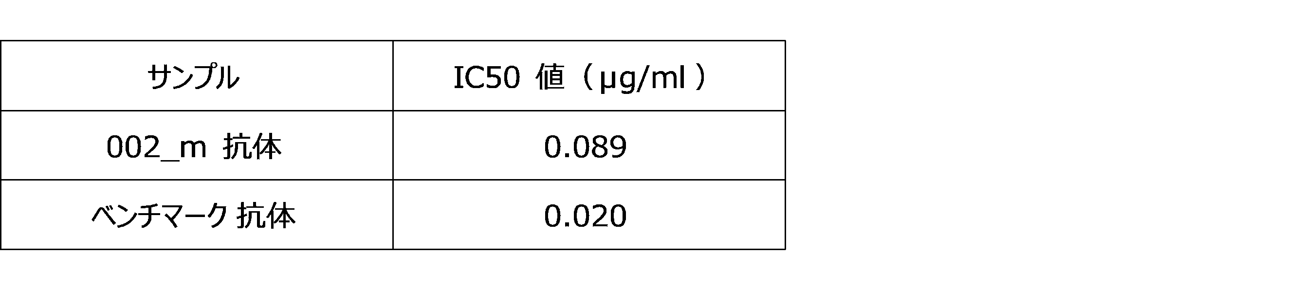
実施例3:抗体遺伝子配列解析
本実施例は、実施例1で取得された002_m抗体のアミノ酸配列を決定することを目的として行った。 Example 3: Antibody gene sequence analysis This example was performed for the purpose of determining the amino acid sequence of the 002_m antibody obtained in Example 1.
本実施例は、実施例1で取得された002_m抗体のアミノ酸配列を決定することを目的として行った。 Example 3: Antibody gene sequence analysis This example was performed for the purpose of determining the amino acid sequence of the 002_m antibody obtained in Example 1.
機能解析でAMP分解抑制機能が認められた002_m抗体クローンについて、実施例1(1-1)で作製したライゲーション産物をトランスフェクションした大腸菌の植菌プレートからシングルコロニーを回収し、プラスミドDNAを取得した。
For the 002_m antibody clone whose AMP degradation inhibitory function was confirmed by functional analysis, single colonies were collected from the inoculated plate of Escherichia coli transfected with the ligation product prepared in Example 1 (1-1), and plasmid DNA was obtained. ..
得られたプラスミドDNAをサンガーシーケンスによって重鎖可変領域および軽鎖可変領域を規定する遺伝子配列を解析し、同配列に基づいてアミノ酸配列を決定した。
The obtained plasmid DNA was analyzed for the gene sequences defining the heavy chain variable region and the light chain variable region by the Sanger sequence, and the amino acid sequence was determined based on the same sequence.
002_m抗体の重鎖可変領域および軽鎖可変領域のアミノ酸配列を、図2に示す(それぞれ、SEQ ID NO: 7およびSEQ ID NO: 8)。また、この図において、重鎖のCDR1、CDR2、CDR3(それぞれ、SEQ ID NO: 1、SEQ ID NO: 2、SEQ ID NO: 3)および軽鎖のCDR1、CDR2、CDR3(それぞれ、SEQ ID NO: 4、SEQ ID NO: 5、SEQ ID NO: 6)に下線を引いた。
The amino acid sequences of the heavy chain variable region and the light chain variable region of the 002_m antibody are shown in FIG. 2 (SEQ ID NO: 7 and SEQ ID NO: 8 respectively). Further, in this figure, the heavy chains CDR1, CDR2, CDR3 (SEQ ID NO: 1, SEQ ID NO: 2, SEQ ID NO: 3, respectively) and the light chains CDR1, CDR2, CDR3 (SEQ ID NO, respectively). : 4, SEQ ID NO: 5, SEQ ID NO: 6) are underlined.
実施例4:ヒト-マウスヒトキメラ抗体作製
本実施例は、実施例3で配列決定した002_m抗体について、マウス抗体の可変領域およびヒト抗体の定常領域を有するキメラ抗体を作製することを目的として行った。 Example 4: Preparation of human-mouse human chimeric antibody The purpose of this example is to prepare a chimeric antibody having a variable region of a mouse antibody and a constant region of a human antibody for the 002_m antibody sequenced in Example 3. rice field.
本実施例は、実施例3で配列決定した002_m抗体について、マウス抗体の可変領域およびヒト抗体の定常領域を有するキメラ抗体を作製することを目的として行った。 Example 4: Preparation of human-mouse human chimeric antibody The purpose of this example is to prepare a chimeric antibody having a variable region of a mouse antibody and a constant region of a human antibody for the 002_m antibody sequenced in Example 3. rice field.
マウス抗体の可変領域の遺伝子配列を人工合成し、ヒト定常領域(Fc)キメラ組み換え抗体発現用のベクターに組み込んだ。発現用のベクターとして、軽鎖発現エレメント(EF-1αプロモーター、分泌シグナル、軽鎖可変領域を規定するDNA配列、軽鎖定常領域を規定するDNA配列をタンデムに連結)、重鎖発現エレメント(EF-1αプロモーター、分泌シグナル、重鎖可変領域を規定するDNA配列、重鎖定常領域を規定するDNA配列をタンデムに連結)を有するプラスミドを構築した。当該プラスミドをExpiCHO細胞(Thermo)にトランスフェクションして抗体を培地中に分泌させた後、ProteinAカラム(GLサイエンス)を用いて抗体を精製した。
The gene sequence of the variable region of the mouse antibody was artificially synthesized and incorporated into a vector for expressing a human constant region (Fc) chimeric recombinant antibody. As vectors for expression, a light chain expression element (EF-1α promoter, a secretory signal, a DNA sequence defining a light chain variable region, and a DNA sequence defining a light chain constant region are linked in tandem), a heavy chain expression element (EF). A plasmid having a -1α promoter, a secretory signal, a DNA sequence defining a heavy chain variable region, and a DNA sequence defining a heavy chain constant region was tandemly linked) was constructed. The plasmid was transfected into ExpiCHO cells (Thermo) to secrete the antibody into the medium, and then the antibody was purified using a Protein A column (GL Science).
このようにして作成されたキメラ抗体を002_1_c抗体と規定した。この抗体の重鎖可変領域のアミノ酸配列はSEQ ID NO: 7のアミノ酸配列から構成され、軽鎖可変領域のアミノ酸配列はSEQ ID NO: 8のアミノ酸配列から構成される。
The chimeric antibody thus prepared was defined as 002_1_c antibody. The amino acid sequence of the heavy chain variable region of this antibody is composed of the amino acid sequence of SEQ ID NO: 7, and the amino acid sequence of the light chain variable region is composed of the amino acid sequence of SEQ ID NO: 8.
実施例5:CDR改変抗体の作製
本実施例は、002_m抗体について、CDR中の翻訳後修飾を受ける可能性がある残基を、修飾を受けない残基に置換した改変抗体を作製することを目的として行った。 Example 5: Preparation of CDR-modified antibody In this example, a modified antibody is prepared by substituting a residue that may undergo post-translational modification in CDR with an unmodified residue for the 002_m antibody. I went there for the purpose.
本実施例は、002_m抗体について、CDR中の翻訳後修飾を受ける可能性がある残基を、修飾を受けない残基に置換した改変抗体を作製することを目的として行った。 Example 5: Preparation of CDR-modified antibody In this example, a modified antibody is prepared by substituting a residue that may undergo post-translational modification in CDR with an unmodified residue for the 002_m antibody. I went there for the purpose.
具体的には、002_m抗体重鎖可変領域(SEQ ID NO: 7)32番目アミノ酸システイン(C)をセリン(S)に、002_m抗体軽鎖可変領域(SEQ ID NO: 8)32番目アミノ酸アスパラギン(N)をアスパラギン酸(D)またはグルタミン(Q)にそれぞれ置換した(以下の表2を参照)。
Specifically, the 002_m antibody heavy chain variable region (SEQ ID NO: 7) 32nd amino acid cysteine (C) is used as serine (S), and the 002_m antibody light chain variable region (SEQ ID NO: 8) 32nd amino acid asparagine (SEQ ID NO: 8) N) was replaced with aspartic acid (D) or glutamine (Q), respectively (see Table 2 below).

該当領域を含む遺伝子を人工合成し、ライゲーションによってヒトFcキメラ組み換え抗体発現用ベクターに挿入した。抗体発現・精製は、ヒトキメラ抗体と同様の方法で実施した。
A gene containing the relevant region was artificially synthesized and inserted into a human Fc chimeric recombinant antibody expression vector by ligation. Antibody expression and purification were carried out in the same manner as for human chimeric antibodies.
得られたそれぞれのキメラ抗体002_1_c~002_6_cについての標的発現細胞への結合性解析は、CD73 native発現細胞(ヒト肺がん細胞株H322)を用いたFACS解析により実施した。被験抗体もしくはアイソタイプコントロール抗体を細胞と反応させた後、蛍光標識二次抗体で染色してフローサイトメトリー解析に供試し、得られた蛍光強度に基づき抗体結合性を評価した。
Binding analysis of each of the obtained chimeric antibodies 002_1_c to 002_6_c to target-expressing cells was performed by FACS analysis using CD73 native-expressing cells (human lung cancer cell line H322). After reacting the test antibody or the isotype control antibody with the cells, the antibody was stained with a fluorescently labeled secondary antibody and subjected to flow cytometric analysis, and the antibody binding property was evaluated based on the obtained fluorescence intensity.
結果を、図3に示す。実施例4および本実施例において002_m抗体をベースに作製したキメラ抗体はいずれも、H322細胞に対する高い結合性を示すことが明らかになった。
The results are shown in Figure 3. It was revealed that both the chimeric antibody prepared based on the 002_m antibody in Example 4 and this example show high binding to H322 cells.
次いで、それぞれのキメラ抗体について、(1-3)と同じ方法で、002_1_c抗体~002_6_c抗体についての、ヒト肺がんH322細胞を用いたAMP分解抑制機能を測定する、抗体機能スクリーニングを行った。結果を、図4に示す
Next, for each chimeric antibody, antibody function screening was performed to measure the AMP degradation inhibitory function of 002_1_c antibody to 002_6_c antibody using human lung cancer H322 cells by the same method as in (1-3). The results are shown in Figure 4.
本結果より、それぞれの抗体のIC50値を算出したところ、下記の表3の通りとなり、002_1_c~002_6_cの結合性、機能性は全て同等と判断された。また、ベンチマーク抗体のIC50値とも遜色ない結果が得られた。そこで、以降の開発は、翻訳後修飾の可能性が無い002_6_cについて実施することとした。
From this result, the IC50 values of each antibody were calculated and shown in Table 3 below, and it was judged that the binding and functionality of 002_1_c to 002_6_c were all the same. In addition, the results were comparable to the IC50 value of the benchmark antibody. Therefore, we decided to carry out the subsequent development on 002_6_c, which has no possibility of post-translational modification.

実施例6:ヒト化抗体の作製
本実施例は、実施例5で選択した002_6_c抗体(SEQ ID NO: 7(C32S)/SEQ ID NO: 8(N32Q))に基づいて、ヒト化抗体を作製することを目的として行った。 Example 6: Preparation of humanized antibody In this example, a humanized antibody was prepared based on the 002_6_c antibody (SEQ ID NO: 7 (C32S) / SEQ ID NO: 8 (N32Q)) selected in Example 5. I went for the purpose of doing.
本実施例は、実施例5で選択した002_6_c抗体(SEQ ID NO: 7(C32S)/SEQ ID NO: 8(N32Q))に基づいて、ヒト化抗体を作製することを目的として行った。 Example 6: Preparation of humanized antibody In this example, a humanized antibody was prepared based on the 002_6_c antibody (SEQ ID NO: 7 (C32S) / SEQ ID NO: 8 (N32Q)) selected in Example 5. I went for the purpose of doing.
ヒト化抗体の作製は、公開ソフトウエアであるTabhu(http://circe.med.uniroma1.it/tabhu/)を用いて実施した。同ソフトを用い、002_6_cと配列的・立体構造的に近い特性を有する配列を持つヒト抗体データベースより抽出し、4種配列(FJ039788、AF146404、3SQO_H、AY686911)を候補配列としてピックアップして、これらの抗体のCDR領域以外の配列と、002_6_cのCDR領域の配列とのグラフティングを行った。
The humanized antibody was prepared using the public software Tabhu (http://circe.med.uniroma1.it/tabhu/). Using the same software, we extracted from a human antibody database that has sequences that are similar in sequence and conformation to 002_6_c, picked up 4 types of sequences (FJ039788, AF146404, 3SQO_H, AY686911) as candidate sequences, and selected these. The sequences other than the CDR regions of the antibody and the sequences of the CDR regions of 002_6_c were graphed.
次に、一部のアミノ酸残基のマウス配列へのbackmutationを実施した。backmutation 対象のアミノ酸残基は、ヒト化抗体とマウス抗体において構造的差異が大きいこと(TubHuスコア上位)に基づいて、各配列10アミノ酸残基を上限として選定した。詳細は非特許文献(Bioinformatics. 2015 Feb 1;31(3):434-5)に記載の方法に従った。
Next, backmutation of some amino acid residues to the mouse sequence was performed. The amino acid residues to be backmutated were selected up to 10 amino acid residues in each sequence based on the large structural difference between the humanized antibody and the mouse antibody (higher TubHu score). For details, the method described in the non-patent document (Bioinformatics. 2015 Feb 1; 31 (3): 434-5) was followed.
得られた002_6_cのヒト化抗体(002_6_h1~002_6_h8)の可変領域のアミノ酸配列を、図5(重鎖可変領域を図5-1、軽鎖可変領域を図5-2)に示す。それぞれのアミノ酸配列は、下記の表4の通り、SEQ ID NO: を割り当てた。
The amino acid sequences of the variable regions of the obtained 002_6_c humanized antibodies (002_6_h1 to 002_6_h8) are shown in Fig. 5 (heavy chain variable region is shown in Fig. 5-1 and light chain variable region is shown in Fig. 5-2). Each amino acid sequence was assigned SEQ ID NO: as shown in Table 4 below.

得られたそれぞれのヒト化抗体(002_6_h1~002_6_h8)についての標的発現細胞への結合性解析は、CD73 native発現細胞(ヒト肺がん細胞株H322)を用いたFACS解析により実施した。結果を、図6に示す。いずれのヒト化抗体も、ベースとするキメラ抗体002_6_c抗体と同様の結合性を示すことが確認された。
Binding analysis of each of the obtained humanized antibodies (002_6_h1 to 002_6_h8) to target-expressing cells was performed by FACS analysis using CD73 native-expressing cells (human lung cancer cell line H322). The results are shown in FIG. It was confirmed that all humanized antibodies showed the same binding properties as the base chimeric antibody 002_6_c antibody.
次いで、ヒト化抗体002_6_h1~002_6_h8のうちの3種類の抗体(002_6_h2、002_6_h4、002_6_h8)について、(1-3)と同じ方法で、ヒト肺がんH322細胞を用いたAMP分解抑制機能を測定する、抗体機能スクリーニングを行った。結果を、図7に示す。
Next, for the three types of humanized antibodies 002_6_h1 to 002_6_h8 (002_6_h2, 002_6_h4, 002_6_h8), the antibody that measures the AMP degradation inhibitory function using human lung cancer H322 cells by the same method as in (1-3). Functional screening was performed. The results are shown in FIG.
本結果より、ヒト肺がんH322細胞を用いたAMP分解抑制機能に対するそれぞれのヒト化抗体のIC50値を算出したところ、下記の表5の通りとなり、ヒト化抗体の中で最も抑制機能の高い002_6_h4をリード抗体として以降の開発を行うこととした。同抗体は、ベンチマーク抗体と比較して良好なIC50値を示した。
From this result, the IC50 values of each humanized antibody for the AMP degradation inhibitory function using human lung cancer H322 cells were calculated as shown in Table 5 below, and 002_6_h4, which has the highest inhibitory function among humanized antibodies, was obtained. It was decided to carry out subsequent development as a lead antibody. The antibody showed a good IC50 value compared to the benchmark antibody.

上記でリード抗体として選択したヒト化抗体002_6_h4について、既存の抗体(市販抗CD73抗体(biolegend)、ベンチマーク抗体)、ベースとなるキメラ抗体002_6_cと比較し、種々の細胞に対する結合性をフローサイトメトリーにより確認した。
The humanized antibody 002_6_h4 selected as the lead antibody above was compared with existing antibodies (commercially available anti-CD73 antibody (biolegend), benchmark antibody) and the base chimeric antibody 002_6_c, and the binding property to various cells was measured by flow cytometry. confirmed.
標的発現細胞への結合性解析は、CD73 native発現細胞(ヒト乳がん細胞株MDA-MB-231)、CD73抗原を発現するヒト乳がん細胞株MDA-MB-231細胞、CD73抗原を発現しないHEK293T野生型細胞(ヒト胎児腎細胞由来)、およびHEK293T細胞にCD73抗原を強制発現させた細胞を用いたFACS解析により実施した。CD73強制発現細胞は、レンチウイルスベクターシステム(Origene)を使用して作製した。ヒトCD73遺伝子(NM_002526)およびピューロマイシン耐性遺伝子を保有するレンチウイルス粒子を作製して、HEK293T細胞に感染させた後、ピューロマイシン存在下で細胞を培養し、目的遺伝子がゲノムに組み込まれてCD73陽性となったHEK293T細胞を回収して結合性解析に使用した。被験抗体もしくはアイソタイプコントロール抗体を細胞と反応させた後、蛍光標識二次抗体で染色してフローサイトメトリー解析に供試し、得られた蛍光強度に基づき抗体結合性を評価した。
Binding analysis to target-expressing cells was performed on CD73 native-expressing cells (human breast cancer cell line MDA-MB-231), human breast cancer cell line MDA-MB-231 cells expressing CD73 antigen, and HEK293T wild type not expressing CD73 antigen. FACS analysis was performed using cells (derived from human fetal kidney cells) and cells in which CD73 antigen was forcibly expressed in HEK293T cells. CD73 forced expression cells were generated using the lentiviral vector system (Origene). Lentivirus particles carrying the human CD73 gene (NM_002526) and puromycin resistance gene are prepared, infected with HEK293T cells, and then the cells are cultured in the presence of puromycin, and the target gene is integrated into the genome to be CD73 positive. HEK293T cells were collected and used for binding analysis. After reacting the test antibody or the isotype control antibody with the cells, the antibody was stained with a fluorescently labeled secondary antibody and subjected to flow cytometric analysis, and the antibody binding property was evaluated based on the obtained fluorescence intensity.
結果を図8に示す。この結果、002_6_cおよび002_6_h4ともに、既存の抗体(市販抗CD73抗体(biolegend)、ベンチマーク抗体)と同様の各種細胞への結合性を示すことがわかり、CD73抗原に対する結合性を確認できた。
The results are shown in Fig. 8. As a result, it was found that both 002_6_c and 002_6_h4 showed the same binding property to various cells as the existing antibodies (commercially available anti-CD73 antibody (biolegend), benchmark antibody), and the binding property to the CD73 antigen was confirmed.
次いで、ヒト化抗体002_6_h4について、既存の抗体(市販抗CD73抗体(biolegend)、ベンチマーク抗体)、ベースとなるキメラ抗体002_6_cと比較し、(1-3)と同じ方法で、ヒト乳がんMDA-MB-231細胞を用いたAMP分解抑制機能を測定する、抗体機能スクリーニングを行った。結果を、図9に示す。本結果より、ヒト乳がんMDA-MB-231細胞を用いたAMP分解抑制機能に対するヒト化抗体のIC50値を算出したところ、下記の表6の通りとなり、上記のヒト肺がんH322細胞を用いた試験の結果とあわせ、ヒト化抗体002_6_h4がキメラ抗体002_6_cと同様に、複数のがん種においてAMP分解抑制機能を有することが示された。
Next, the humanized antibody 002_6_h4 was compared with the existing antibody (commercially available anti-CD73 antibody (biolegend), benchmark antibody) and the base chimeric antibody 002_6_c, and the same method as in (1-3) was used for human breast cancer MDA-MB-. An antibody function screening was performed to measure the AMP degradation inhibitory function using 231 cells. The results are shown in FIG. From this result, the IC50 value of the humanized antibody against the AMP degradation inhibitory function using human breast cancer MDA-MB-231 cells was calculated as shown in Table 6 below. Together with the results, it was shown that the humanized antibody 002_6_h4 has an AMP degradation inhibitory function in multiple cancer types, similar to the chimeric antibody 002_6_c.
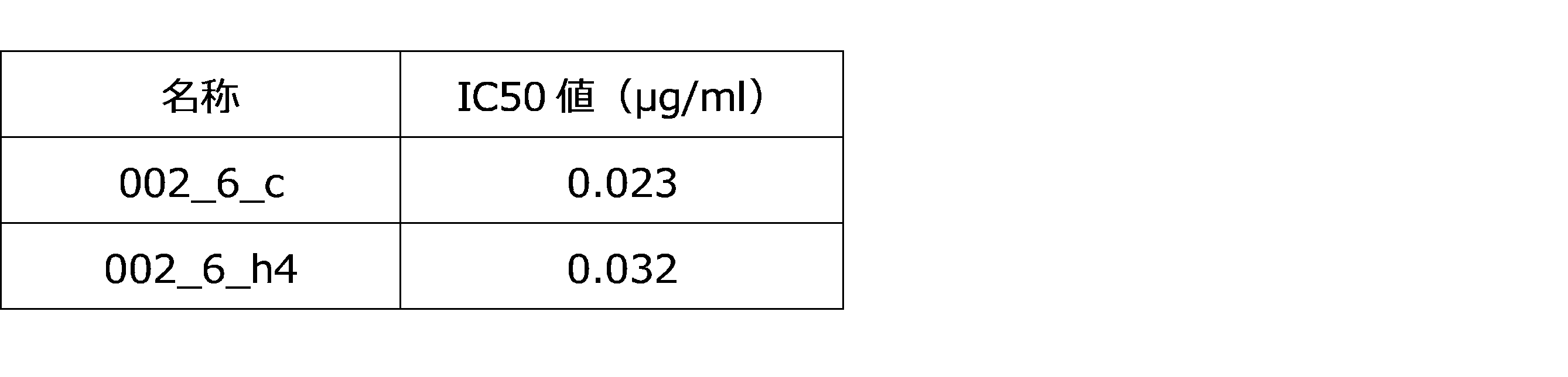
実施例7:組換え抗原に対する抑制機能
本実施例は、実施例6で得られた抗CD73抗体の、組換えCD73抗原に対する酵素反応抑制機能を確認することを目的として行った。 Example 7: Suppressive function against recombinant antigen The purpose of this example was to confirm the enzymatic reaction inhibitory function of the anti-CD73 antibody obtained in Example 6 against the recombinant CD73 antigen.
本実施例は、実施例6で得られた抗CD73抗体の、組換えCD73抗原に対する酵素反応抑制機能を確認することを目的として行った。 Example 7: Suppressive function against recombinant antigen The purpose of this example was to confirm the enzymatic reaction inhibitory function of the anti-CD73 antibody obtained in Example 6 against the recombinant CD73 antigen.
実験は、CD73抗原(末端His-Tag、自家調製品)を、抗His-Tag抗体を介してELISA用96 wellプレートに固定化し、ヒト化抗CD73抗体002_6_h4および基質であるAMP(終濃度100μM)を添加して37℃で一時間インキュベートして、実施例1(1-3)と同様にCD73によるAMPのアデノシンへの分解反応を測定することにより行った。
In the experiment, the CD73 antigen (terminal His-Tag, self-prepared product) was immobilized on a 96-well plate for ELISA via an anti-His-Tag antibody, and the humanized anti-CD73 antibody 002_6_h4 and the substrate AMP (final concentration 100 μM) were used. Was added and incubated at 37 ° C. for 1 hour, and the decomposition reaction of AMP to adenosine by CD73 was measured in the same manner as in Example 1 (1-3).
上清を回収し、ATP(終濃度100μM)およびCell TiterGlo(Promega、G9243)を添加して混合した後、マイクロプレートリーダーを用いて化学発光を測定した。ATPとCell TiterGloの反応による発光をAMPが阻害する原理を用い、残存AMP量から抗体の酵素活性阻害効果を評価した。陽性対照として、ベンチマーク抗体を使用して同じ実験を行った。
The supernatant was collected, ATP (final concentration 100 μM) and CellTiterGlo (Promega, G9243) were added and mixed, and then chemiluminescence was measured using a microplate reader. Using the principle that AMP inhibits luminescence due to the reaction between ATP and CellTiterGlo, the enzyme activity inhibitory effect of the antibody was evaluated from the amount of residual AMP. The same experiment was performed using a benchmark antibody as a positive control.
002_6_h4の組換えCD73抗原に対するAMP分解抑制機能を、図10に示す。この結果、002_6_h4は組換えCD73抗原に対してAMP分解抑制機能を示し、酵素を直接的に抑制することが示唆された。
Figure 10 shows the AMP degradation inhibitory function of 002_6_h4 for the recombinant CD73 antigen. As a result, it was suggested that 002_6_h4 showed an AMP degradation inhibitory function against the recombinant CD73 antigen and directly inhibited the enzyme.
実施例8:T細胞分裂への影響評価
本実施例は、実施例6で得られた抗CD73抗体の、ヒト末梢血T細胞に対する増殖促進活性を確認することを目的として行った。 Example 8: Evaluation of effect on T cell division This example was performed for the purpose of confirming the proliferation promoting activity of the anti-CD73 antibody obtained in Example 6 on human peripheral blood T cells.
本実施例は、実施例6で得られた抗CD73抗体の、ヒト末梢血T細胞に対する増殖促進活性を確認することを目的として行った。 Example 8: Evaluation of effect on T cell division This example was performed for the purpose of confirming the proliferation promoting activity of the anti-CD73 antibody obtained in Example 6 on human peripheral blood T cells.
健常人ボランティア由来末梢血単核球(PBMC)から、CD4+ T Cell Isolation Kit, human(Miltenyi Biotech,130-096-533)によってCD4陽性T細胞を精製し、CellTrace CFSE cell proliferation kit(ThermoFisher Scientific, #C34554)を用いて細胞を蛍光標識した。
From healthy volunteer-derived peripheral blood mononuclear cells (PBMC), CD4 + T Cell Isolation Kit, human (Miltenyi Biotech, 130-096-533) was used to purify CD4 positive T cells, and CellTrace CFSE cell proliferation kit (ThermoFisher Scientific, #) Cells were fluorescently labeled with C34554).
DynabeadsTM Human T-Activator CD3/CD28 for T Cell Expansion and Activation(ベリタス、DB11131)を用いてCD4陽性T細胞を活性化した後、96ウェルプレートに細胞を播種(1×10^5 cells/well)し、ヒト化抗CD73抗体002_6_h4および基質であるAMP(100μM)の存在下で3~7日間培養した。細胞をフローサイトメトリー解析に供試して蛍光強度を測定し、蛍光強度に基づいて、分裂した細胞の割合を解析した。
After activating CD4-positive T cells using Dynabeads TM Human T-Activator CD3 / CD28 for T Cell Expansion and Activation (Veritas, DB11131), seed cells into 96-well plates (1 × 10 ^ 5 cells / well). The cells were cultured in the presence of humanized anti-CD73 antibody 002_6_h4 and the substrate AMP (100 μM) for 3 to 7 days. The cells were subjected to flow cytometric analysis to measure the fluorescence intensity, and the percentage of divided cells was analyzed based on the fluorescence intensity.
002_6_h4の、ヒト末梢血T細胞の分裂(増殖)に与える影響について、図11に示す。分裂したT細胞の割合は、抗体濃度依存的に増加し、抗CD73抗体がT細胞分裂を促進する効果が示された。
Figure 11 shows the effect of 002_6_h4 on the division (proliferation) of human peripheral blood T cells. The proportion of divided T cells increased in an antibody concentration-dependent manner, indicating that the anti-CD73 antibody had the effect of promoting T cell division.
実施例9:抗体機能評価(濃度依存性評価)
本実施例は、実施例1で取得された002_m抗体以外の抗体(すなわち、003_m抗体、004_m抗体、005_m抗体、006_m抗体、007_m抗体、008_m抗体、009_m抗体)の機能評価を行うことを目的として行った。 Example 9: Evaluation of antibody function (concentration-dependent evaluation)
The purpose of this example is to evaluate the functions of antibodies other than the 002_m antibody obtained in Example 1 (that is, 003_m antibody, 004_m antibody, 005_m antibody, 006_m antibody, 007_m antibody, 008_m antibody, 009_m antibody). went.
抗体機能評価は、実施例1(1-3)において記載した抗体機能スクリーニングと同じ方法により実施した。すなわち、細胞としてヒト肺がん細胞株H322細胞またはヒト乳がん細胞株MDA-MB-231細胞(ATCC(登録商標)HTB-26TM)を用いた。本実施例では、実施例1と同様にAMP分解反応を測定することにより、AMP分解抑制機能の抗体濃度依存性を解析して、IC50値(50%阻害濃度:ここではCD73の酵素機能の50%を阻害できる抗体濃度を指す)を算出し、抗体の機能を定量的に評価した。
The antibody function evaluation was carried out by the same method as the antibody function screening described in Example 1 (1-3). That is, human lung cancer cell line H322 cells or human breast cancer cell line MDA-MB-231 cells (ATCC® HTB-26 TM ) were used as cells. In this example, the antibody concentration dependence of the AMP degradation inhibitory function was analyzed by measuring the AMP degradation reaction in the same manner as in Example 1, and the IC50 value (50% inhibitory concentration: 50 of the enzyme function of CD73 here). %) Was calculated and the function of the antibody was quantitatively evaluated.
実施例1において作製した003_m抗体、004_m抗体、005_m抗体、006_m抗体、007_m抗体、008_m抗体、009_m抗体のH322細胞に対するAMP分解抑制機能を図12に、MDA-MB-231細胞に対するAMP分解抑制機能を図13に、それぞれ示す。これらの抗体のうちいくつかのものは、マウス抗体であるが、ベンチマーク抗体によりも低いか、もしくはベンチマーク抗体に近いIC50値を示すことが明らかになった(表7参照)。
The AMP degradation inhibitory function of the 003_m antibody, 004_m antibody, 005_m antibody, 006_m antibody, 007_m antibody, 008_m antibody, and 009_m antibody prepared in Example 1 against H322 cells is shown in FIG. 12, and the AMP degradation inhibitory function against MDA-MB-231 cells. Is shown in FIG. 13, respectively. Some of these antibodies, which are mouse antibodies, were found to show IC50 values that were lower or closer to the benchmark antibodies (see Table 7).

さらに、上記図12および図13に示す試験より、機能性が高いと判断されたクローンを選抜した(004_m、006_m、007_m、008_m)。そしてこれら4種の抗体のAMP分解抑制機能(MDA-MB-231細胞)を、示す。これらのクローンのIC50値は、ベンチマーク抗体(臨床開発中)と比較して良好であり、002_6_cとほぼ同等であった(表8を参照)。
Furthermore, clones judged to have high functionality were selected from the tests shown in FIGS. 12 and 13 above (004_m, 006_m, 007_m, 008_m). The AMP degradation inhibitory function (MDA-MB-231 cells) of these four antibodies is shown. The IC50 values of these clones were good compared to the benchmark antibody (under clinical development) and were about the same as 002_6_c (see Table 8).
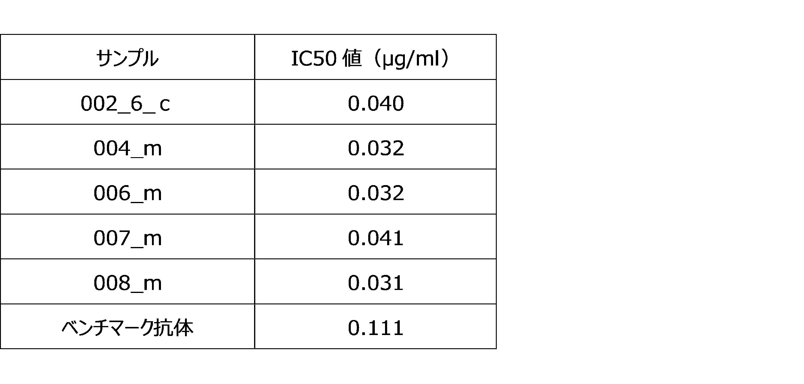
実施例10:抗体遺伝子配列解析
本実施例は、実施例1で取得された003_m抗体~009_m抗体のアミノ酸配列を決定することを目的として行った。 Example 10: Antibody gene sequence analysis The purpose of this example was to determine the amino acid sequences of the 003_m antibody to 009_m antibody obtained in Example 1.
本実施例は、実施例1で取得された003_m抗体~009_m抗体のアミノ酸配列を決定することを目的として行った。 Example 10: Antibody gene sequence analysis The purpose of this example was to determine the amino acid sequences of the 003_m antibody to 009_m antibody obtained in Example 1.
機能解析でAMP分解抑制機能が認められた003_m抗体~009_m抗体のクローンについて、実施例1(1-1)で作製したライゲーション産物をトランスフェクションした大腸菌の植菌プレートからシングルコロニーを回収し、プラスミドDNAを取得した。
For clones of 003_m antibody to 009_m antibody whose AMP degradation inhibitory function was confirmed by functional analysis, single colonies were collected from the inoculated plate of Escherichia coli transfected with the ligation product prepared in Example 1 (1-1), and plasmids were collected. Obtained DNA.
得られたプラスミドDNAをサンガーシーケンスによって重鎖可変領域および軽鎖可変領域を規定する遺伝子配列を解析し、同配列に基づいてアミノ酸配列を決定した。
The obtained plasmid DNA was analyzed for the gene sequences defining the heavy chain variable region and the light chain variable region by the Sanger sequence, and the amino acid sequence was determined based on the same sequence.
003_m抗体、004_m抗体、005_m抗体、006_m抗体、007_m抗体、008_m抗体、009_m抗体の重鎖可変領域アミノ酸配列(図14-1)および軽鎖可変領域アミノ酸配列(図14-2)を示す。それぞれのアミノ酸配列について、以下表9に示す通りSEQ ID NOを割り当てた。
The heavy chain variable region amino acid sequence (Fig. 14-1) and light chain variable region amino acid sequence (Fig. 14-2) of 003_m antibody, 004_m antibody, 005_m antibody, 006_m antibody, 007_m antibody, 008_m antibody, 009_m antibody are shown. SEQ ID NOs were assigned to each amino acid sequence as shown in Table 9 below.

実施例11:標的発現細胞への結合性解析
本実施例は、実施例1において取得された003_m~009_mのそれぞれの抗体の結合性評価を行うことを目的として行った。 Example 11: Analysis of binding to target-expressing cells The purpose of this example was to evaluate the binding of each antibody of 003_m to 009_m obtained in Example 1.
本実施例は、実施例1において取得された003_m~009_mのそれぞれの抗体の結合性評価を行うことを目的として行った。 Example 11: Analysis of binding to target-expressing cells The purpose of this example was to evaluate the binding of each antibody of 003_m to 009_m obtained in Example 1.
標的発現細胞への結合性解析は、CD73 native発現細胞(ヒト肺がん細胞株H322もしくはヒト乳がん細胞株MDA-MB-231)、もしくはCD73強制発現細胞を用いたFACS解析により実施した。CD73強制発現細胞は、レンチウイルスベクターシステム(Origene)を使用して作製した。ヒトCD73遺伝子(NM_002526)およびピューロマイシン耐性遺伝子を保有するレンチウイルス粒子を作製してHEK293T細胞に感染させた後、ピューロマイシン存在下で細胞を培養し、目的遺伝子がゲノムに組み込まれてCD73陽性となったHEK293T細胞を回収して結合性解析に使用した。ネガティブコントロールとして、CD73を発現していないHEK293T細胞を使用した。
Binding analysis to target-expressing cells was performed by FACS analysis using CD73 native-expressing cells (human lung cancer cell line H322 or human breast cancer cell line MDA-MB-231) or CD73 forced-expressing cells. CD73 forced expression cells were generated using the lentiviral vector system (Origene). After preparing lentivirus particles carrying the human CD73 gene (NM_002526) and puromycin resistance gene and infecting HEK293T cells, the cells were cultured in the presence of puromycin, and the target gene was integrated into the genome to be CD73 positive. The resulting HEK293T cells were collected and used for binding analysis. As a negative control, HEK293T cells expressing CD73 were used.
被験抗体もしくはアイソタイプコントロール抗体を細胞と反応させた後、蛍光標識二次抗体で染色してフローサイトメトリー解析に供試し、得られた蛍光強度に基づき抗体結合性を評価した。
After reacting the test antibody or isotype control antibody with the cells, the antibody was stained with a fluorescently labeled secondary antibody and subjected to flow cytometric analysis, and the antibody binding property was evaluated based on the obtained fluorescence intensity.
003_m抗体~009_m抗体の標的発現細胞(H322細胞)への結合性を図15に、003_m抗体~009_m抗体の標的発現細胞(MDA-MB-231細胞)への結合性を図16に、そして003_m抗体~009_m抗体の標的発現細胞(CD73強制発現HEK-293T細胞)への結合性を図17に、それぞれ示す。これらの図15~図17において、実線および灰色塗りつぶしが、それぞれ被験抗体およびアイソタイプコントロール抗体の蛍光強度ヒストグラムを示す。
The binding of 003_m antibody to 009_m antibody to target-expressing cells (H322 cells) is shown in Fig. 15, and the binding of 003_m antibody to 009_m antibody to target-expressing cells (MDA-MB-231 cells) is shown in Fig. 16 and 003_m. The binding properties of the antibody to 009_m antibody to the target-expressing cells (CD73 forced expression HEK-293T cells) are shown in FIG. 17, respectively. In these FIGS. 15 to 17, solid lines and gray fills show fluorescence intensity histograms of the test antibody and the isotype control antibody, respectively.
実施例12:T細胞分裂への影響評価
本実施例は、実施例9で得られた抗CD73抗体の、ヒト末梢血T細胞に対する増殖促進活性を確認することを目的として行った。 Example 12: Evaluation of effect on T cell division The purpose of this example was to confirm the proliferation-promoting activity of the anti-CD73 antibody obtained in Example 9 on human peripheral blood T cells.
本実施例は、実施例9で得られた抗CD73抗体の、ヒト末梢血T細胞に対する増殖促進活性を確認することを目的として行った。 Example 12: Evaluation of effect on T cell division The purpose of this example was to confirm the proliferation-promoting activity of the anti-CD73 antibody obtained in Example 9 on human peripheral blood T cells.
方法は実施例8と同様の方法を採用し、抗体濃度を1 nMとした際の分裂細胞割合を比較した。結果を図18に示す。点線は、アイソタイプコントロール抗体での分裂細胞割合を示している。
The same method as in Example 8 was adopted, and the ratio of dividing cells was compared when the antibody concentration was 1 nM. The results are shown in FIG. The dotted line shows the proportion of dividing cells in the isotype control antibody.
具体的には、健常人ボランティア由来末梢血単核球(PBMC)から、CD4+ T Cell Isolation Kit, human(Miltenyi Biotech,130-096-533)によってCD4陽性T細胞を精製し、CellTrace CFSE cell proliferation kit(ThermoFisher Scientific, #C34554)を用いて細胞を蛍光標識した。
Specifically, CD4 + T Cell Isolation Kit, human (Miltenyi Biotech, 130-096-533) is used to purify CD4-positive T cells from peripheral blood mononuclear cells (PBMC) derived from healthy volunteers, and CellTrace CFSE cell proliferation kit. Cells were fluorescently labeled with (ThermoFisher Scientific, # C34554).
DynabeadsTM Human T-Activator CD3/CD28 for T Cell Expansion and Activation(ベリタス、DB11131)を用いてCD4陽性T細胞を活性化した後、96ウェルプレートに細胞を播種(1×10^5 cells/well)し、抗CD73抗体(006_m抗体、007_m抗体、008_m抗体)、および基質であるAMP(100μM)の存在下で3~7日間培養した。細胞をフローサイトメトリー解析に供試して蛍光強度を測定し、蛍光強度に基づいて、分裂した細胞の割合を解析した。
After activating CD4-positive T cells using Dynabeads TM Human T-Activator CD3 / CD28 for T Cell Expansion and Activation (Veritas, DB11131), seed cells into 96-well plates (1 × 10 ^ 5 cells / well). Then, the cells were cultured for 3 to 7 days in the presence of anti-CD73 antibody (006_m antibody, 007_m antibody, 008_m antibody) and the substrate AMP (100 μM). The cells were subjected to flow cytometric analysis to measure the fluorescence intensity, and the percentage of divided cells was analyzed based on the fluorescence intensity.
1 nMの抗CD73抗体(006_m抗体、007_m抗体、または008_m抗体)の、ヒト末梢血T細胞の分裂(増殖)に与える影響について、図18に示す。図中、「w/o AMP」および「with AMP」(100μM AMP)は、AMPがT細胞の分裂抑制効果を有することから、抗体なしでAMPの有無の効果を見るために組み入れたものである。一方、ヒト化抗CD73抗体(006_m抗体、007_m抗体、008_m抗体)を1 nM、0.1 nM、0.01 nM添加した群において、分裂したT細胞の割合は、抗体濃度依存的に増加し(データには示さず)、抗CD73抗体がT細胞分裂を促進する効果が示された。
Figure 18 shows the effect of 1 nM anti-CD73 antibody (006_m antibody, 007_m antibody, or 008_m antibody) on the division (proliferation) of human peripheral blood T cells. In the figure, "w / o AMP" and "with AMP" (100 μM AMP) were incorporated to see the effect of the presence or absence of AMP without antibody because AMP has a T cell division inhibitory effect. .. On the other hand, in the group to which 1 nM, 0.1 nM, 0.01 nM of humanized anti-CD73 antibody (006_m antibody, 007_m antibody, 008_m antibody) was added, the proportion of divided T cells increased in an antibody concentration-dependent manner (data shows). (Not shown), the effect of anti-CD73 antibody on promoting T cell division was shown.
003_m抗体~009_m抗体はいずれもアイソタイプコントロール抗体よりも細胞分裂を促進し、ベンチマーク抗体、002_6_cおよび002_6_h4とほぼ同等であった。
All of the 003_m antibody to 009_m antibody promoted cell division more than the isotype control antibody, and were almost equivalent to the benchmark antibodies 002_6_c and 002_6_h4.
実施例13:ヒト化抗体の作製(2)
本実施例は、実施例1において取得された003_m~009_mのそれぞれの抗体のうち、実施例9で選択した004_m抗体、006_m抗体、007_m抗体、008_m抗体に基づいて、ヒト化抗体を作製することを目的として行った。 Example 13: Preparation of humanized antibody (2)
In this example, a humanized antibody is prepared based on the 004_m antibody, 006_m antibody, 007_m antibody, and 008_m antibody selected in Example 9 among the respective antibodies of 003_m to 009_m obtained in Example 1. I went for the purpose.
ヒト化抗体の作製は、実施例6と同様に、公開ソフトウエアであるTabhu(http://circe.med.uniroma1.it/tabhu/)を用いて実施した。同ソフトを用い、004_m抗体、007_m抗体、008_m抗体と配列的・立体構造的に近い特性を有する配列を持つヒト抗体データベースより抽出し、004_m抗体は5種、007_m抗体は4種、008_m抗体は4種の配列をそれぞれ候補配列としてピックアップして、これらの抗体のCDR領域以外の配列と、004_m抗体、007_m抗体、008_m抗体のそれぞれのCDR領域の配列とのグラフティングを行った。
The humanized antibody was prepared using the public software Tabhu (http://circe.med.uniroma1.it/tabhu/) as in Example 6. Using this software, 004_m antibody, 007_m antibody, 008_m antibody and 004_m antibody are extracted from the human antibody database having sequences having characteristics similar in sequence and three-dimensional structure, 004_m antibody is 5 types, 007_m antibody is 4 types, and 008_m antibody is. Each of the four sequences was picked up as a candidate sequence, and the sequences other than the CDR region of these antibodies were graphed with the sequences of the CDR regions of each of the 004_m antibody, 007_m antibody, and 008_m antibody.
006_m抗体については、市販のバイオロジカルソフトを用いて6種のヒト化抗体配列候補を作成した。
For the 006_m antibody, 6 types of humanized antibody sequence candidates were created using commercially available biological software.
次に、グラフティングした004m抗体、007m抗体、008m抗体の一部のアミノ酸残基のマウス配列へのbackmutationを実施した。backmutation 対象のアミノ酸残基は、ヒト化抗体とマウス抗体において構造的差異が大きいこと(TubHuスコア上位)に基づいて、各配列10アミノ酸残基を上限として選定した。詳細は非特許文献(Bioinformatics. 2015 Feb 1;31(3):434-5)に記載の方法に従った。
Next, backmutation of some amino acid residues of the graphed 004m antibody, 007m antibody, and 008m antibody to the mouse sequence was performed. The amino acid residues to be backmutated were selected up to 10 amino acid residues in each sequence based on the large structural difference between the humanized antibody and the mouse antibody (higher TubHu score). For details, the method described in the non-patent document (Bioinformatics. 2015 Feb 1; 31 (3): 434-5) was followed.
004_m抗体、006_m抗体、007_m抗体、008_m抗体に基づいてそれぞれ得られたヒト化抗体(004_h3抗体、006_hKB抗体、007_h1抗体、008_h4抗体)の可変領域のアミノ酸配列を、以下の表10に示す。それぞれのアミノ酸配列は、下記の表10の通り、SEQ ID NO: 81~SEQ ID NO: 88を割り当てた。
Table 10 below shows the amino acid sequences of the variable regions of the humanized antibodies (004_h3 antibody, 006_hKB antibody, 007_h1 antibody, 008_h4 antibody) obtained based on the 004_m antibody, 006_m antibody, 007_m antibody, and 008_m antibody, respectively. As shown in Table 10 below, each amino acid sequence was assigned SEQ ID NO: 81 to SEQ ID NO: 88.

得られたそれぞれのヒト化抗体(004_h3抗体、006_hKB抗体、007_h1抗体、008_h4抗体)について、既存の抗体(市販抗CD73抗体(biolegend)、ベンチマーク抗体)、ベースとなるキメラ抗体002_6_cと比較し、種々の細胞に対する結合性をフローサイトメトリーにより確認した。
Each of the obtained humanized antibodies (004_h3 antibody, 006_hKB antibody, 007_h1 antibody, 008_h4 antibody) was compared with existing antibodies (commercially available anti-CD73 antibody (biolegend), benchmark antibody) and base chimeric antibody 002_6_c. The binding property to cells was confirmed by flow cytometry.
ヒト標的発現細胞への結合性解析を、CD73 native発現細胞(ヒト肺がん細胞株H322)を用いたFACS解析により実施した。標的発現細胞への結合性解析は、CD73 native発現細胞(ヒト乳がん細胞株MDA-MB-231)、CD73抗原を発現するヒト乳がん細胞株MDA-MB-231細胞、CD73抗原を発現しないHEK293T野生型細胞(ヒト胎児腎細胞由来)、およびHEK293T細胞にCD73抗原を強制発現させた細胞を用いたFACS解析により実施した。CD73強制発現細胞は、レンチウイルスベクターシステム(Origene)を使用して作製した。ヒトCD73遺伝子(NM_002526)、アカゲザルCD73遺伝子(XM_001086989.2)(アミノ酸配列はカニクイザルCD73と同一)およびピューロマイシン耐性遺伝子を保有するレンチウイルス粒子を作製して、HEK293T細胞に感染させた後、ピューロマイシン存在下で細胞を培養し、目的遺伝子がゲノムに組み込まれてCD73陽性となったHEK293T細胞を回収して結合性解析に使用した。被験抗体もしくはアイソタイプコントロール抗体を細胞と反応させた後、蛍光標識二次抗体で染色してフローサイトメトリー解析に供試し、得られた蛍光強度に基づき抗体結合性を評価した。
Binding analysis to human target-expressing cells was performed by FACS analysis using CD73 native-expressing cells (human lung cancer cell line H322). Binding analysis to target-expressing cells was performed on CD73 native-expressing cells (human breast cancer cell line MDA-MB-231), human breast cancer cell line MDA-MB-231 cells expressing CD73 antigen, and HEK293T wild type not expressing CD73 antigen. FACS analysis was performed using cells (derived from human fetal kidney cells) and cells in which CD73 antigen was forcibly expressed in HEK293T cells. CD73 forced expression cells were generated using the lentiviral vector system (Origene). Lentivirus particles carrying the human CD73 gene (NM_002526), the red-tailed monkey CD73 gene (XM_001086989.2) (the amino acid sequence is the same as that of the crab monkey CD73) and the puromycin resistance gene were prepared, infected with HEK293T cells, and then puromycin. The cells were cultured in the presence of HEK293T cells in which the target gene was integrated into the genome and became CD73 positive, and used for binding analysis. After reacting the test antibody or the isotype control antibody with the cells, the antibody was stained with a fluorescently labeled secondary antibody and subjected to flow cytometric analysis, and the antibody binding property was evaluated based on the obtained fluorescence intensity.
結果を、図19~図21に示す。このうち、図19および図20は、ヒトCD73遺伝子を発現させた細胞に対する抗体の結合性を確認した図であり、図21は、アカゲザルCD73遺伝子を発現させた細胞に対する抗体の結合性を確認した図である。検討したヒト化抗体のうち、004_h3抗体、006_hKB抗体、008_h4抗体については、比較対象としたと002_6_h4抗体と同様のヒトCD73抗原に対する結合性を示すことが確認され、CD73抗原に対する結合性を確認できた。また、このうちの006_hKB抗体については、アカゲザルCD73抗原(カニクイザルCD73抗原)に対して、002_6_h4抗体と同様の結合性を示すことが確認され、将来的に動物実験により本発明の抗体の効果を確認する際に、アカゲザルあるいはカニクイザルにより検討を行うことができることが示された。
The results are shown in FIGS. 19 to 21. Of these, FIGS. 19 and 20 are diagrams confirming the binding of the antibody to cells expressing the human CD73 gene, and FIG. 21 confirms the binding of the antibody to cells expressing the rhesus monkey CD73 gene. It is a figure. Among the humanized antibodies examined, the 004_h3 antibody, 006_hKB antibody, and 008_h4 antibody were confirmed to show the same binding property to the human CD73 antigen as the 002_6_h4 antibody when compared, and the binding property to the CD73 antigen could be confirmed. rice field. In addition, it was confirmed that the 006_hKB antibody among them showed the same binding property to the rhesus monkey CD73 antigen (cynomolgus monkey CD73 antigen) as the 002_6_h4 antibody, and the effect of the antibody of the present invention was confirmed by animal experiments in the future. It was shown that rhesus monkeys or cynomolgus monkeys can be used for examination.
実施例14:組換え抗原に対する抑制機能
本実施例は、実施例13で得られたヒト化抗CD73抗体の、組換えCD73抗原に対する酵素反応抑制機能を確認することを目的として行った。 Example 14: Suppressive function against recombinant antigen The purpose of this example was to confirm the enzymatic reaction inhibitory function of the humanized anti-CD73 antibody obtained in Example 13 against the recombinant CD73 antigen.
本実施例は、実施例13で得られたヒト化抗CD73抗体の、組換えCD73抗原に対する酵素反応抑制機能を確認することを目的として行った。 Example 14: Suppressive function against recombinant antigen The purpose of this example was to confirm the enzymatic reaction inhibitory function of the humanized anti-CD73 antibody obtained in Example 13 against the recombinant CD73 antigen.
抗体機能評価は、004_h3抗体、006_hKB抗体、007_h1抗体、008_h4抗体を被検抗体として、002_6_c抗体、002_6_h4抗体、アイソタイプコントロール抗体およびベンチマーク抗体(MEDI9447抗体)をコントロールとして使用し、実施例1(1-3)において記載した抗体機能スクリーニングと同様の方法により実施した。すなわち、細胞としてヒト乳がん細胞株MDA-MB-231細胞(ATCC(登録商標)HTB-26TM)を96 wellプレートに播種し、37℃で一晩インキュベートした。翌日、細胞をL-15培地(富士フイルム和光純薬、128-06075)で洗浄した後、精製した抗体を添加して37℃で一時間インキュベートした。本実施例では、実施例1(1-3)と同様に、CD73の基質であるAMP(Sigma, A1752)を終濃度200μMとなるように添加し、37℃で4.5時間インキュベートしてCD73によるAMPのアデノシンへのAMP分解反応を測定することにより、AMP分解抑制機能の抗体濃度依存性を解析した。分解反応を行った。反応後、上清を回収し、ATP(Sigma, A2383、終濃度10μM)およびCell TiterGlo(Promega, G9243)を添加して混合した後、マイクロプレートリーダーを用いて化学発光を測定した。
For antibody function evaluation, 004_h3 antibody, 006_hKB antibody, 007_h1 antibody, 008_h4 antibody were used as test antibodies, and 002_6_c antibody, 002_6_h4 antibody, isotype control antibody and benchmark antibody (MEDI9447 antibody) were used as controls, and Example 1 (1-). It was carried out by the same method as the antibody function screening described in 3). That is, human breast cancer cell line MDA-MB-231 cells (ATCC® HTB-26 TM ) were seeded on 96-well plates and incubated overnight at 37 ° C. The next day, the cells were washed with L-15 medium (Fujifilm Wako Pure Chemical Industries, Ltd., 128-06075), purified antibody was added, and the cells were incubated at 37 ° C. for 1 hour. In this example, as in Example 1 (1-3), AMP (Sigma, A1752), which is a substrate of CD73, was added to a final concentration of 200 μM, and the mixture was incubated at 37 ° C. for 4.5 hours to AMP with CD73. By measuring the AMP degradation reaction to adenosine, the antibody concentration dependence of the AMP degradation inhibitory function was analyzed. A decomposition reaction was carried out. After the reaction, the supernatant was collected, ATP (Sigma, A2383, final concentration 10 μM) and Cell TiterGlo (Promega, G9243) were added and mixed, and then chemiluminescence was measured using a microplate reader.
004_h3抗体、006_hKB抗体、007_h1抗体、008_h4抗体についてのAMP分解抑制機能を図22に示す。これらの抗体はいずれも細胞表面に存在するCD73によるAMP分解を抑制する機能を有していることが示され(図22および図23)、特にこれらの抗体のうち006_hKB抗体は、002_6_h4と同様に、ベンチマーク抗体と同等以上の阻害活性を持つことが示された。
Figure 22 shows the AMP degradation inhibitory function of the 004_h3 antibody, 006_hKB antibody, 007_h1 antibody, and 008_h4 antibody. All of these antibodies have been shown to have the function of suppressing AMP degradation by CD73 present on the cell surface (Figs. 22 and 23), and among these antibodies, the 006_hKB antibody is similar to 002_6_h4. , It was shown to have an inhibitory activity equal to or higher than that of the benchmark antibody.
実施例15:T細胞分裂への影響評価
本実施例は、実施例13で得られ、実施例14で特に高い効果を示した抗CD73抗体の、ヒト末梢血T細胞に対する増殖促進活性を確認することを目的として行った。 Example 15: Evaluation of effects on T cell division This example confirms the growth-promoting activity of the anti-CD73 antibody obtained in Example 13 and showing a particularly high effect in Example 14 on human peripheral blood T cells. I went for that purpose.
本実施例は、実施例13で得られ、実施例14で特に高い効果を示した抗CD73抗体の、ヒト末梢血T細胞に対する増殖促進活性を確認することを目的として行った。 Example 15: Evaluation of effects on T cell division This example confirms the growth-promoting activity of the anti-CD73 antibody obtained in Example 13 and showing a particularly high effect in Example 14 on human peripheral blood T cells. I went for that purpose.
健常人ボランティア由来末梢血単核球(PBMC)に対して、終濃度0.0005~1μg/mlの各種抗体(006_hKB抗体、002_6_h4抗体、ベンチマーク抗体(MEDI9447抗体)、アイソタイプコントロール抗体)のいずれかを添加し、室温で30分間インキュベーションした。続いて終濃度350μM ATPおよびDynabeadsTM Human T-Activator CD3/CD28 for T Cell Expansion and Activation(ベリタス、DB11131)を添加して、37℃、5%CO2条件下で96時間インキュベーションした。培養終了後、T細胞マーカー染色抗体(BioLegend、anti-CD3:UCHT1、anti-CD4:RPA-T4、anti-CD8a:HIT8a)を用いて細胞を染色した。染色した細胞に細胞数計測用のビーズを添加して、フローサイトメーターで解析し、1ウェルあたりのCD4およびCD8陽性細胞数を算出した。
Add any of various antibodies (006_hKB antibody, 002_6_h4 antibody, benchmark antibody (MEDI9447 antibody), isotype control antibody) with a final concentration of 0.0005 to 1 μg / ml to peripheral blood mononuclear cells (PBMC) derived from healthy volunteers. , Incubated for 30 minutes at room temperature. Subsequently, a final concentration of 350 μM ATP and Dynabeads TM Human T-Activator CD3 / CD28 for T Cell Expansion and Activation (Veritas, DB11131) were added, and the mixture was incubated at 37 ° C. under 5% CO 2 conditions for 96 hours. After completion of culture, cells were stained with T cell marker staining antibody (BioLegend, anti-CD3: UCHT1, anti-CD4: RPA-T4, anti-CD8a: HIT8a). Beads for cell count were added to the stained cells and analyzed with a flow cytometer to calculate the number of CD4 and CD8 positive cells per well.
また、回収した培養上清中のIFNγ、TNFα、IL-2の濃度を、各種抗体[キャプチャー抗体=IFNγ:M7004(Thermo)、TNFα:MAB602(R&D Systems)、IL-2:MAB610;検出抗体=IFNγ:M701B(Thermo)、IL-2:BAF202(R&D Systems)、TNFα:BAF210(R&D Systems)]を用いてLuminex法にて測定した。
In addition, the concentrations of IFNγ, TNFα, and IL-2 in the collected culture supernatant are measured by various antibodies [capture antibody = IFNγ: M7004 (Thermo), TNFα: MAB602 (R & D Systems), IL-2: MAB610; detection antibody =. IFNγ: M701B (Thermo), IL-2: BAF202 (R & D Systems), TNFα: BAF210 (R & D Systems)] was used for measurement by the Luminex method.
抗体によるT細胞分裂へ影響評価について、結果を図23に示す。実施例8において良好な活性を示すことが明らかになった002_6_h4抗体と比較して、006_hKB抗体は、CD4陽性T細胞に対してもCD8陽性T細胞に対しても、002_6_h4抗体と同程度の高いT細胞分裂促進効果を有することが示された(図23上段)。また、006_hKB抗体を投与した細胞から、高い濃度のIFNγ、TNFα、IL-2が培養上清中に放出されていることも確認された(図23下段)。
Figure 23 shows the results of the evaluation of the effect of the antibody on T cell division. Compared to the 002_6_h4 antibody, which was found to show good activity in Example 8, the 006_hKB antibody is as high as the 002_6_h4 antibody for both CD4-positive and CD8-positive T cells. It was shown to have a T cell division promoting effect (upper part of FIG. 23). It was also confirmed that high concentrations of IFNγ, TNFα, and IL-2 were released into the culture supernatant from the cells to which the 006_hKB antibody was administered (Fig. 23, lower part).
実施例16:抗体の細胞内への取り込み評価
本実施例は、実施例6で得られた抗CD73抗体および実施例13で得られ、実施例14で特に高い効果を示した抗CD73抗体の、細胞内への取り込みを評価することを目的として行った。 Example 16: Evaluation of intracellular uptake of antibody In this example, the anti-CD73 antibody obtained in Example 6 and the anti-CD73 antibody obtained in Example 13 and showing a particularly high effect in Example 14 were used. The purpose was to evaluate the uptake into cells.
本実施例は、実施例6で得られた抗CD73抗体および実施例13で得られ、実施例14で特に高い効果を示した抗CD73抗体の、細胞内への取り込みを評価することを目的として行った。 Example 16: Evaluation of intracellular uptake of antibody In this example, the anti-CD73 antibody obtained in Example 6 and the anti-CD73 antibody obtained in Example 13 and showing a particularly high effect in Example 14 were used. The purpose was to evaluate the uptake into cells.
ヒト乳がん細胞MDA-MB-231(1×10^5 cells)をチャンバースライド(Thermo Fisher Scientific、154534PK)に播種し、10μg/mlの各種抗体溶液(002_6_h4、006_hKB、MEDI9447)を添加して37℃で15分間もしくは19時間インキュベートした。インキュベート前後の試料を固定および透過処理し(Thermo Fisher Scientific、FIX & PERM Fixation and Permeabilization Kit、GAS003)、Anti-human IgG Alexa Fluor 594(Thermo Fisher Scientific、A-11014)で染色した。抗体の分布を蛍光顕微鏡(Thermo Fisher Scientific、EVOS FLoid Imaging System、4471136)で観察し、抗体の細胞内への取り込みの有無を判断した。
Human breast cancer cells MDA-MB-231 (1 × 10 ^ 5 cells) are seeded on a chamber slide (Thermo Fisher Scientific, 154534PK), and 10 μg / ml of various antibody solutions (002_6_h4, 006_hKB, MEDI9447) are added to 37 ° C. Incubated for 15 minutes or 19 hours. Samples before and after incubation were fixed and permeabilized (Thermo Fisher Scientific, FIX & PERM Fixation and Permeabilization Kit, GAS003) and stained with Anti-human IgG Alexa Fluor 594 (Thermo Fisher Scientific, A-11014). The distribution of the antibody was observed with a fluorescence microscope (Thermo Fisher Scientific, EVOS FLoid Imaging System, 4471136) to determine the presence or absence of the antibody uptake into the cells.
002_6_h4および006_hKBの細胞内への取り込みを、図24に示す。002_6_h4および006_hKBともに、インキュベート15分間の時点(図24上段)は抗体が細胞表面に分布しているのに対し、インキュベート19時間後(図24下段)は抗体が細胞内に集積しており、抗体の細胞内への取り込みが起こったことが示された。本結果より、抗CD73抗体の取り込みに伴う抗原の細胞表面からの消失が誘導される可能性が示唆された。
Figure 24 shows the intracellular uptake of 002_6_h4 and 006_hKB. In both 002_6_h4 and 006_hKB, the antibody was distributed on the cell surface at the time of 15 minutes of incubation (upper part of Fig. 24), whereas the antibody was accumulated in the cell 19 hours after incubation (lower part of Fig. 24). Was shown to have taken up into the cells. This result suggests that the uptake of anti-CD73 antibody may induce the disappearance of the antigen from the cell surface.
このような抗体のT細胞への内在化を、経時的に数値化した。健常人ボランティア由来末梢血単核球(PBMC)を、Dynabeads TM Human T-Activator CD3/CD28 for T Cell Expansion and Activation(ベリタス、DB11131)で6日間刺激後、細胞を回収し10μg/mlの各種抗体(002_6_h4抗体、006_hKB抗体、ベンチマーク抗体(MEDI9447抗体)、アイソタイプコントロール抗体)を添加し、氷上で30分間インキュベーションした。
The internalization of such antibodies into T cells was quantified over time. Peripheral blood mononuclear cells (PBMC) derived from healthy volunteers were stimulated with Dynabeads TM Human T-Activator CD3 / CD28 for T Cell Expansion and Activation (Veritas, DB11131) for 6 days, and then cells were collected and various antibodies of 10 μg / ml were collected. (002_6_h4 antibody, 006_hKB antibody, benchmark antibody (MEDI9447 antibody), isotype control antibody) were added and incubated on ice for 30 minutes.
細胞を洗浄後、FACS緩衝液(PBS、0.5%BSA、2 mM EDTA)に懸濁し、37℃で最大240分までインキュベートし、その間、30分、120分、240分に細胞試料をサンプリングし、それぞれの試料を蛍光標識二次抗体(APC anti-human IgG Fc)(BioLegend、409306)で細胞を染色してフローサイトメトリー解析に供試し、得られた蛍光強度に基づき抗体の内在化を評価した。抗体の内在化割合は下記計算式により算出した。
%対照=(抗CD73抗体(X分)MFI-アイソタイプコントロール(X分)MFI)/(抗CD73抗体(0分)MFI-アイソタイプコントロール(0分)MFI)×100 After washing the cells, suspend them in FACS buffer (PBS, 0.5% BSA, 2 mM EDTA) and incubate at 37 ° C for up to 240 minutes, during which time cell samples are sampled at 30, 120 and 240 minutes. Each sample was subjected to flow cytometric analysis by staining cells with a fluorescently labeled secondary antibody (APC anti-human IgG Fc) (BioLegend, 409306), and the internalization of the antibody was evaluated based on the obtained fluorescence intensity. .. The internalization ratio of the antibody was calculated by the following formula.
% Control = (anti-CD73 antibody (X minutes) MFI-isotype control (X minutes) MFI) / (anti-CD73 antibody (0 minutes) MFI-isotype control (0 minutes) MFI) x 100
結果を図25に示す。002_6_h4および006_hKBは、各測定時間においてベンチマーク抗体より内在化の割合が大きかった。本結果からも、抗CD73抗体の取り込みに伴う抗原の細胞表面からの消失が誘導される可能性が示唆された。
The results are shown in Fig. 25. 002_6_h4 and 006_hKB had a higher rate of internalization than the benchmark antibody at each measurement time. This result also suggests that the disappearance of the antigen from the cell surface associated with the uptake of the anti-CD73 antibody may be induced.
本発明の抗体で処理したがん細胞における、細胞表面CD73への内在化を、経時的に数値化した。ヒト乳がん細胞MDA-MB-231とヒトメラノーマ細胞HT-144を、MDA-MB-231はL-15培地(富士フイルム和光純薬、128-06075)、HT-144はMcCoy's 5A(Modified)(Gibco、16600082)の牛胎児血清無添加培地にそれぞれ懸濁後、10μg/mlの各種抗体(002_6_h4抗体、006_hKB抗体、ベンチマーク抗体(MEDI9447抗体)、アイソタイプコントロール抗体)を添加し、37℃で1時間、3時間、6時間、22時間および48時間インキュベーションした。
The internalization of cancer cells treated with the antibody of the present invention into cell surface CD73 was quantified over time. Human breast cancer cells MDA-MB-231 and human melanoma cells HT-144, MDA-MB-231 in L-15 medium (Fujifilm Wako Pure Drug, 128-06075), HT-144 in McCoy's 5A (Modified) (Gibco) , 16600882), each of which was suspended in a medium containing no fetal bovine serum, and then 10 μg / ml of various antibodies (002_6_h4 antibody, 006_hKB antibody, benchmark antibody (MEDI9447 antibody), isotype control antibody) was added, and the temperature was 37 ° C. for 1 hour. Incubated for 3 hours, 6 hours, 22 hours and 48 hours.
再度各種抗体を10μg/mL添加して反応させ、細胞を洗浄後、蛍光標識二次抗体で細胞を染色して、フローサイトメトリー解析に供試し、得られた蛍光強度に基づき抗原の内在化を評価した。抗原の内在化割合は下記計算式により算出した。
%CD73 Ag=(抗CD73抗体(X分)MFI-アイソタイプコントロール(X分)MFI)/(抗CD73抗体(0分)MFI-アイソタイプコントロール(0分)MFI)×100 Add 10 μg / mL of various antibodies again to react, wash the cells, stain the cells with a fluorescently labeled secondary antibody, perform flow cytometric analysis, and internalize the antigen based on the obtained fluorescence intensity. evaluated. The internalization ratio of the antigen was calculated by the following formula.
% CD73 Ag = (anti-CD73 antibody (X minutes) MFI-isotype control (X minutes) MFI) / (anti-CD73 antibody (0 minutes) MFI-isotype control (0 minutes) MFI) x 100
結果を図26に示す。002_6_h4、006_hKBおよびベンチマーク抗体は、いずれもがん細胞とインキュベーションすることで、時間経過とともにがん細胞表面のCD73発現量を減少させた。このことは、CD73に結合した抗CD73抗体が抗原とともに内在化することで、細胞表面のCD73発現量を低下させたことに基づくことが示唆された。
The results are shown in Fig. 26. Both 002_6_h4, 006_hKB and the benchmark antibody reduced the expression level of CD73 on the surface of cancer cells over time by incubation with cancer cells. This was suggested to be based on the fact that the anti-CD73 antibody bound to CD73 was internalized together with the antigen to reduce the expression level of CD73 on the cell surface.
実施例17:KD値の測定
本実施例は、実施例6で得られた抗CD73抗体および実施例13で得られ、実施例14で特に高い効果を示した抗CD73抗体の、CD73抗原に対する結合親和性の指標である解離定数(KD値)を評価することを目的として行った。 Example 17: Measurement of KD value In this example, the anti-CD73 antibody obtained in Example 6 and the anti-CD73 antibody obtained in Example 13 and showing a particularly high effect in Example 14 bind to the CD73 antigen. The purpose was to evaluate the dissociation constant (KD value), which is an index of affinity.
本実施例は、実施例6で得られた抗CD73抗体および実施例13で得られ、実施例14で特に高い効果を示した抗CD73抗体の、CD73抗原に対する結合親和性の指標である解離定数(KD値)を評価することを目的として行った。 Example 17: Measurement of KD value In this example, the anti-CD73 antibody obtained in Example 6 and the anti-CD73 antibody obtained in Example 13 and showing a particularly high effect in Example 14 bind to the CD73 antigen. The purpose was to evaluate the dissociation constant (KD value), which is an index of affinity.
抗体の標的抗原に対する結合のKD値の測定は、Biacore 8K(GEヘルスケア)にて実施した。Human Antibody Capture Kit Type2(Cytiva、26-2346-00)を用いて002_6_h4抗体および006_hKB抗体の各抗体をセンサーチップ(CM5)に固定化した。ヒトCD73抗原を5×10⁻⁹ Mから4段階に段階希釈した溶液を作製し、マルチサイクルカイネティクス解析に供試した。1:1結合モデルにてKD値は、以下の表11に記載の通りであった。
The KD value of the antibody bound to the target antigen was measured with Biacore 8K (GE Healthcare). Each antibody of 002_6_h4 antibody and 006_hKB antibody was immobilized on the sensor chip (CM5) using Human Antibody Capture Kit Type2 (Cytiva, 26-2346-00). A solution of human CD73 antigen diluted serially from 5 × 10 -9 M in 4 steps was prepared and used for multicycle kinetic analysis. In the 1: 1 coupling model, the KD values are as shown in Table 11 below.

実施例18:ヒト免疫マウス試験
本実施例は、実施例6で得られた抗CD73抗体および実施例13で得られ、実施例14で特に高い効果を示した抗CD73抗体の、ヒトCD34陽性細胞移植マウス体内における抗腫瘍効果を確認することを目的として行った。 Example 18: Human Immune Mouse Test This example is a human CD34-positive cell of the anti-CD73 antibody obtained in Example 6 and the anti-CD73 antibody obtained in Example 13 and showing a particularly high effect in Example 14. The purpose was to confirm the antitumor effect in the transplanted mice.
本実施例は、実施例6で得られた抗CD73抗体および実施例13で得られ、実施例14で特に高い効果を示した抗CD73抗体の、ヒトCD34陽性細胞移植マウス体内における抗腫瘍効果を確認することを目的として行った。 Example 18: Human Immune Mouse Test This example is a human CD34-positive cell of the anti-CD73 antibody obtained in Example 6 and the anti-CD73 antibody obtained in Example 13 and showing a particularly high effect in Example 14. The purpose was to confirm the antitumor effect in the transplanted mice.
複数の健常人ドナーから作製したヒトCD34陽性細胞移植NSGマウス(hu-NSG, JACKSON laboratories)の乳腺皮下脂肪(Mammary fat pad)に対して、ヒト乳癌細胞株MDA-MB-231を移植し、ヒト乳癌細胞担持マウスモデルを作出した。このマウスモデルに移植したヒト乳癌細胞が増殖して100 mm3になった段階で002_6_h4抗体もしくはアイソタイプコントロール抗体を20 mg/kgの用量にて4日に一度、合計7回(「Q4Dx7」と示す)、腹腔投与した。投与開始日をday0とし、day34まで観察した。
Human breast cancer cell line MDA-MB-231 was transplanted into human breast cancer cell line MDA-MB-231 into the mammary fat pad of human CD34-positive cell transplant NSG mice (hu-NSG, JACKSON laboratories) prepared from multiple healthy donors. A breast cancer cell-carrying mouse model was created. When the human breast cancer cells transplanted into this mouse model proliferate to 100 mm 3 , 002_6_h4 antibody or isotype control antibody is administered at a dose of 20 mg / kg once every 4 days for a total of 7 times (indicated as "Q4Dx7"). ), Administered intraperitoneally. The administration start date was set to day 0, and observation was performed until day 34.
観察期間終了後に腫瘍を摘出し、Gentle MACS(ミルテニー)を用いて細胞サスペンションとした後、Human (hu)CD45 AF700 clone HI30(BioLegend)、huCD3 APCCy7 clone HITa(BioLegend)、huCD4 PECy7 clone SK3(BioLegend)、およびhuCD8 FITC clone SK1(BioLegend)を用いて細胞を染色し、腫瘍に浸潤したCD45+,CD3+, CD8+T細胞およびCD45+,CD3+, CD4+T細胞の量をフローサイトメトリーにより測定した。
After the tumor was removed after the observation period and used as a cell suspension using GentleMACS (Milteny), Human (hu) CD45 AF700 clone HI30 (BioLegend), huCD3 APKCy7 clone HITa (BioLegend), huCD4 PECy7 clone SK3 (BioLegend) , And huCD8 FITC clone SK1 (BioLegend) were used to stain the cells, and the amount of CD45 +, CD3 +, CD8 + T cells and CD45 +, CD3 +, CD4 + T cells infiltrating the tumor was measured by flow cytometry.
002_6_h4抗体もしくはアイソタイプコントロール抗体を投与後、day34においてマウス体内の腫瘍浸潤T細胞数を測定した結果を、図27に示す。腫瘍浸潤T細胞について、002_6_h4抗体を投与したことにより、アイソタイプコントロール抗体を投与した場合と比較して増加傾向が示され、抗CD73抗体が腫瘍免疫効果を有することが示唆された。
Figure 27 shows the results of measuring the number of tumor-infiltrating T cells in mice on day 34 after administration of 002_6_h4 antibody or isotype control antibody. The administration of 002_6_h4 antibody to tumor-infiltrating T cells showed an increasing tendency as compared with the case of administration of isotype control antibody, suggesting that the anti-CD73 antibody has a tumor immunological effect.
本実施例においてはさらに、開発抗体がT細胞によるがん細胞傷害活性を増強させるかどうか、する場合はその程度を、ベンチマーク抗体と比較して確認した。
In this example, it was further confirmed whether or not the developed antibody enhances the cancer cell injury activity by T cells, and if so, the degree thereof in comparison with the benchmark antibody.
RFPタンパクを導入したヒト肺がん細胞NCI-H292またはHCC827を96ウェルプレートに播種し、一晩培養した。翌日培地を除去した後、X-VIVO-15培地(Lonza、04-418Q)に懸濁したIL-2(終濃度30 IU)および各種抗体を添加し、37℃で30分間インキュベーションした。その後、Dynabeads TM Human T-Activator CD3/CD28 for T Cell Expansion and Activation(ベリタス、DB11131)で10日間刺激しT細胞増殖を行った健常人ボランティアPBMCを、E/T比2で添加し、37℃で30分間インキュベーション後、NCI-H292には終濃度500μM ATP、HCC827には終濃度300μM ATPを添加し、37℃、5%CO2条件下でIncuCyte ZOOM System(Sartorius)によりがん細胞増殖の経時変化を観察した。
Human lung cancer cells NCI-H292 or HCC827 introduced with RFP protein were seeded on 96-well plates and cultured overnight. After removing the medium the next day, IL-2 (final concentration 30 IU) suspended in X-VIVO-15 medium (Lonza, 04-418Q) and various antibodies were added, and the mixture was incubated at 37 ° C. for 30 minutes. After that, healthy volunteer PBMCs stimulated with Dynabeads TM Human T-Activator CD3 / CD28 for T Cell Expansion and Activation (Veritas, DB11131) for 10 days to proliferate T cells were added at an E / T ratio of 2 to 37 ° C. in 30 minutes after incubation, NCI-H292 was added to a final concentration of 300 [mu] M ATP in a final concentration of 500 [mu] M ATP, HCC827 to, 37 ℃, 5% CO over time the growth of cancer cells by IncuCyte ZOOM System (Sartorius) with 2 conditions The change was observed.
コントロールとして、アイソタイプコントロール抗体、ベンチマーク抗体(MEDI9447)に加えて、ガン細胞のPD-1抗原とT細胞のPD-L1抗原との相互作用を妨害してT細胞抑制を阻害し、T細胞を活性化させる働きがある抗PD-1抗体(ペムブロリズマブ、キートルーダ、MSD)および抗PD-L1抗体(アテゾリズマブ)を使用した。
As a control, in addition to isotype control antibody and benchmark antibody (MEDI9447), it interferes with the interaction between PD-1 antigen of cancer cells and PD-L1 antigen of T cells to inhibit T cell suppression and activate T cells. Anti-PD-1 antibody (pembrolizumab, keytruda, MSD) and anti-PD-L1 antibody (atezolizumab), which have the effect of activating, were used.
結果を図28に示す。開発抗体(本実施例においては、例として002_6_h4および006_hKBを使用したが、その他の本発明の開発抗体も同様)は、抗PD-1抗体および抗PD-1抗体を作用させてもT細胞抑制の解除の効果がほとんど見られず、がん細胞の増殖を止められない条件下で、T細胞のがん細胞の増殖抑制効果を増強させた。
The results are shown in Fig. 28. The developed antibody (002_6_h4 and 006_hKB were used as examples in this example, but other developed antibodies of the present invention are also used) suppresses T cells even when anti-PD-1 antibody and anti-PD-1 antibody are allowed to act. The effect of suppressing the growth of cancer cells of T cells was enhanced under the condition that the effect of releasing the antibody was hardly observed and the growth of cancer cells could not be stopped.
本実施例においてはさらに、マウスの中で模擬的に再現させたヒト免疫系の抗腫瘍効果を、002_6_h4が増強させることができるかどうか、動物実験により調べた。その際、臨床でその効果が立証されている抗PD-1抗体(ペムブロリズマブ)との併用投与による上乗せ効果が見られるかも確認しました。
複数の健常人ドナーから作製したヒトCD34陽性細胞を移植したNSGマウス(hu-NSG, JACKSON laboratories)(以下、「免疫ヒト化マウス」と呼ぶ)の乳腺脂肪体(Mammary fat pad)に対して、ヒト乳癌細胞株MDA-MB-231を移植し、ヒト乳癌担持マウスモデルを作出した。このマウスモデルにおいて、移植したヒト乳癌細胞が増殖して100 mm3になった段階で、002_6_h4抗体もしくはアイソタイプコントロール抗体を、20 mg/kgの用量にて4日に一度、合計7回(「Q4Dx7」と示す)と、チェックポイント阻害剤(抗PD-1抗体であるペンブロリズマブ、キートルーダ、MSD)もしくはアイソタイプコントロール抗体を、初回のみ10 mg/kg、2回目以降は5 mg/kgの用量にて、5日に一度、合計6回(「Q5Dx6」と示す)を組み合わせて腹腔内投与した。投与開始日をday0とし、day34まで週2回腫瘍サイズの計測を行った。抗体の投与は以下の通りである:
Group 1=アイソタイプコントロール抗体のみ投与群
Group 2=002_6_h4抗体およびアイソタイプコントロール抗体投与群
Group 3=チェックポイント阻害剤(ペンブロリズマブ)およびアイソタイプコントロール抗体投与群
Group 4=002_6_h4抗体およびチェックポイント阻害剤(ペンブロリズマブ)の併用投与群。 In this example, it was further investigated by animal experiments whether 002_6_h4 could enhance the antitumor effect of the human immune system simulated in mice. At that time, it was also confirmed that an additional effect could be seen by concomitant administration with an anti-PD-1 antibody (pembrolizumab) whose effect has been proven clinically.
For the mammary fat pad of NSG mice (hu-NSG, JACKSON laboratories) (hereinafter referred to as "immunized humanized mice") transplanted with human CD34-positive cells prepared from multiple healthy donors. The human breast cancer cell line MDA-MB-231 was transplanted to create a human breast cancer-carrying mouse model. In this mouse model, when the transplanted human breast cancer cells proliferate to 100 mm 3 , 002_6_h4 antibody or isotype control antibody is administered at a dose of 20 mg / kg once every 4 days for a total of 7 times ("Q4Dx7"). ”) And a checkpoint inhibitor (anti-PD-1 antibody pembrolizumab, keytruda, MSD) or isotype control antibody at a dose of 10 mg / kg for the first time only and 5 mg / kg for the second and subsequent times. A total of 6 times (indicated as "Q5Dx6") were administered intraperitoneally once every 5 days. The administration start date was set today 0, and the tumor size was measured twice a week until day 34. The administration of the antibody is as follows:
Group 1 = Isotype control antibody only administration group
Group 2 = 002_6_h4 antibody and isotype control antibody administration group
Group 3 = checkpoint inhibitor (pembrolizumab) and isotype control antibody administration group
Group 4 = 002_6_h4 antibody and checkpoint inhibitor (pembrolizumab) combined administration group.
複数の健常人ドナーから作製したヒトCD34陽性細胞を移植したNSGマウス(hu-NSG, JACKSON laboratories)(以下、「免疫ヒト化マウス」と呼ぶ)の乳腺脂肪体(Mammary fat pad)に対して、ヒト乳癌細胞株MDA-MB-231を移植し、ヒト乳癌担持マウスモデルを作出した。このマウスモデルにおいて、移植したヒト乳癌細胞が増殖して100 mm3になった段階で、002_6_h4抗体もしくはアイソタイプコントロール抗体を、20 mg/kgの用量にて4日に一度、合計7回(「Q4Dx7」と示す)と、チェックポイント阻害剤(抗PD-1抗体であるペンブロリズマブ、キートルーダ、MSD)もしくはアイソタイプコントロール抗体を、初回のみ10 mg/kg、2回目以降は5 mg/kgの用量にて、5日に一度、合計6回(「Q5Dx6」と示す)を組み合わせて腹腔内投与した。投与開始日をday0とし、day34まで週2回腫瘍サイズの計測を行った。抗体の投与は以下の通りである:
Group 1=アイソタイプコントロール抗体のみ投与群
Group 2=002_6_h4抗体およびアイソタイプコントロール抗体投与群
Group 3=チェックポイント阻害剤(ペンブロリズマブ)およびアイソタイプコントロール抗体投与群
Group 4=002_6_h4抗体およびチェックポイント阻害剤(ペンブロリズマブ)の併用投与群。 In this example, it was further investigated by animal experiments whether 002_6_h4 could enhance the antitumor effect of the human immune system simulated in mice. At that time, it was also confirmed that an additional effect could be seen by concomitant administration with an anti-PD-1 antibody (pembrolizumab) whose effect has been proven clinically.
For the mammary fat pad of NSG mice (hu-NSG, JACKSON laboratories) (hereinafter referred to as "immunized humanized mice") transplanted with human CD34-positive cells prepared from multiple healthy donors. The human breast cancer cell line MDA-MB-231 was transplanted to create a human breast cancer-carrying mouse model. In this mouse model, when the transplanted human breast cancer cells proliferate to 100 mm 3 , 002_6_h4 antibody or isotype control antibody is administered at a dose of 20 mg / kg once every 4 days for a total of 7 times ("Q4Dx7"). ”) And a checkpoint inhibitor (anti-PD-1 antibody pembrolizumab, keytruda, MSD) or isotype control antibody at a dose of 10 mg / kg for the first time only and 5 mg / kg for the second and subsequent times. A total of 6 times (indicated as "Q5Dx6") were administered intraperitoneally once every 5 days. The administration start date was set to
代表的なドナーにおける、Day0からday34までの腫瘍体積の増加率の各投与群の平均値と標準誤差を、図29に示す。ドナー#5910、ドナー#0030ともに、併用投与群(Group 4)の平均腫瘍体積増加率は、対照群(Group 1)、およびチェックポイント阻害剤投与群(Group 3)に比較して低値を示した。
Figure 29 shows the average value and standard error of each administration group of the increase rate of tumor volume from Day 0 to day 34 in a typical donor. In both donor # 5910 and donor # 0030, the average tumor volume increase rate in the combination administration group (Group 4) was lower than that in the control group (Group 1) and the checkpoint inhibitor administration group (Group 3). rice field.
さらにドナー#0030では、002_6_h4抗体投与群(Group 2)では、対照群(Group 1)に比較して平均腫瘍体積増加率は低値を示し、抗CD73抗体は、免疫ヒト化マウスにおいて、単独あるいは、チェックポイント阻害剤(ペンブロリズマブ)との併用で抗腫瘍作用を有することが示唆された。
Furthermore, in donor # 0030, the average tumor volume increase rate was lower in the 002_6_h4 antibody-administered group (Group 2) than in the control group (Group 1), and the anti-CD73 antibody was used alone or in immune humanized mice. , It was suggested that it has an antitumor effect when used in combination with a checkpoint inhibitor (pembrolizumab).
実施例19:免疫不全マウス試験
本実施例は、実施例6で得られた抗CD73抗体の、免疫不全マウス体内における抗腫瘍効果を確認することを目的として行った。 Example 19: Immunodeficient mouse test This example was conducted for the purpose of confirming the antitumor effect of the anti-CD73 antibody obtained in Example 6 in an immunodeficient mouse body.
本実施例は、実施例6で得られた抗CD73抗体の、免疫不全マウス体内における抗腫瘍効果を確認することを目的として行った。 Example 19: Immunodeficient mouse test This example was conducted for the purpose of confirming the antitumor effect of the anti-CD73 antibody obtained in Example 6 in an immunodeficient mouse body.
NSGマウス(JACKSON laboratories)の乳腺皮下脂肪(Mammary fat pad)に対して、ヒト乳癌細胞株MDA-MB-231を移植した。移植したヒト乳癌細胞が増殖して100 mm3になった段階で、以下の表12に示す群構成にて002_6_h4抗体もしくはアイソタイプコントロール抗体を各用量にて4日に一度、合計7回(Q4Dx7)、腹腔投与した。投与開始日をday0とし、day35までを観察期間として週二回腫瘍サイズの計測を行った。
The human breast cancer cell line MDA-MB-231 was transplanted into the mammary fat pad of NSG mice (JACKSON laboratories). When the transplanted human breast cancer cells proliferated to 100 mm 3 , 002_6_h4 antibody or isotype control antibody was administered at each dose once every 4 days for a total of 7 times (Q4Dx7) in the group composition shown in Table 12 below. , Administered intraperitoneally. The tumor size was measured twice a week with the administration start date as day 0 and the observation period up to day 35.

免疫不全マウス体内における腫瘍塊の体積変化の結果を、図30に示す。002_6_h4抗体は、アイソタイプコントロールと比較して腫瘍増殖を抑制する効果が示された。
Figure 30 shows the results of volume changes in the tumor mass in immunodeficient mice. The 002_6_h4 antibody was shown to be more effective in suppressing tumor growth than the isotype control.
実施例20:抗腫瘍効果(ノックインモデル)
本実施例は、実施例6で得られた抗CD73抗体の、ヒトCD73を発現するノックインマウス体内における抗腫瘍効果を確認することを目的として行った。 Example 20: Antitumor effect (knock-in model)
The purpose of this example was to confirm the antitumor effect of the anti-CD73 antibody obtained in Example 6 in a knock-in mouse expressing human CD73.
本実施例は、実施例6で得られた抗CD73抗体の、ヒトCD73を発現するノックインマウス体内における抗腫瘍効果を確認することを目的として行った。 Example 20: Antitumor effect (knock-in model)
The purpose of this example was to confirm the antitumor effect of the anti-CD73 antibody obtained in Example 6 in a knock-in mouse expressing human CD73.
マウスCD73に代替してヒトCD73を発現するノックインマウス(Biocytogen、B-hCD73 mice)の皮下に、マウスCD73を欠損しヒトCD73を発現する大腸がん細胞株(Biocytogen、MC38-hCD73)を5×10^5個移植した。移植したMC38-hCD73が増殖して50~80 mm3に達した段階で002_6_h4抗体もしくはアイソタイプコントロール抗体を10 mg/kgの用量にて週に2回、合計7回(「BIWx7」と示す)と、抗マウスPD-1抗体クローンRMP1-14(Bio X Cell、BE0146)もしくはラットIgG2aアイソタイプコントロール抗体(Bio X Cell、BE0089)を3 mg/kgの用量にて週に1回(「QWx4」と示す)を組み合わせて腹腔内投与した。3、4日ごとに腫瘍径を測定しながら投与開始日をday0とし、day18まで観察した。抗体の投与は以下の通りである:
Group 1=アイソタイプコントロール抗体のみ投与群
Group 2=002_6_h4抗体およびアイソタイプコントロール抗体投与群
Group 3=チェックポイント阻害剤(抗マウスPD-1抗体)およびアイソタイプコントロール抗体投与群
Group 4=002_6_h4抗体とチェックポイント阻害剤(抗マウスPD-1抗体)の併用投与を行った群。 Under the subcutaneous of a knock-in mouse (Biocytogen, B-hCD73 mice) that expresses human CD73 in place of mouse CD73, a colon cancer cell line (Biocytogen, MC38-hCD73) that lacks mouse CD73 and expresses human CD73 is 5 × 10 ^ 5 were transplanted. When the transplanted MC38-hCD73 proliferated and reached 50-80 mm3, 002_6_h4 antibody or isotype control antibody was administered at a dose of 10 mg / kg twice a week for a total of 7 times (denoted as "BIWx7"). Anti-mouse PD-1 antibody clone RMP1-14 (Bio X Cell, BE0146) or rat IgG2a isotype control antibody (Bio X Cell, BE0089) once weekly at a dose of 3 mg / kg (denoted as "QWx4") Was administered intraperitoneally in combination. While measuring the tumor diameter every 3 or 4 days, the administration start date was set today 0, and observation was performed until day 18. The administration of the antibody is as follows:
Group 1 = Isotype control antibody only administration group
Group 2 = 002_6_h4 antibody and isotype control antibody administration group
Group 3 = Checkpoint inhibitor (anti-mouse PD-1 antibody) and isotype control antibody administration group
Group 4 = 002_6_h4 antibody and checkpoint inhibitor (anti-mouse PD-1 antibody) were co-administered.
Group 1=アイソタイプコントロール抗体のみ投与群
Group 2=002_6_h4抗体およびアイソタイプコントロール抗体投与群
Group 3=チェックポイント阻害剤(抗マウスPD-1抗体)およびアイソタイプコントロール抗体投与群
Group 4=002_6_h4抗体とチェックポイント阻害剤(抗マウスPD-1抗体)の併用投与を行った群。 Under the subcutaneous of a knock-in mouse (Biocytogen, B-hCD73 mice) that expresses human CD73 in place of mouse CD73, a colon cancer cell line (Biocytogen, MC38-hCD73) that lacks mouse CD73 and expresses human CD73 is 5 × 10 ^ 5 were transplanted. When the transplanted MC38-hCD73 proliferated and reached 50-80 mm3, 002_6_h4 antibody or isotype control antibody was administered at a dose of 10 mg / kg twice a week for a total of 7 times (denoted as "BIWx7"). Anti-mouse PD-1 antibody clone RMP1-14 (Bio X Cell, BE0146) or rat IgG2a isotype control antibody (Bio X Cell, BE0089) once weekly at a dose of 3 mg / kg (denoted as "QWx4") Was administered intraperitoneally in combination. While measuring the tumor diameter every 3 or 4 days, the administration start date was set to
Day0からday18までの平均腫瘍体積の推移を各投与群の平均値と標準誤差として図31に示す。併用投与群(Group 4)の平均腫瘍体積は、対照群(Group 1)、およびチェックポイント阻害剤投与群(Group 3)に比較して低値を示し、抗CD73抗体はヒトCD73を発現するマウスにおいて、チェックポイント阻害剤との併用で抗腫瘍作用を有することが示唆された。
Figure 31 shows the transition of the average tumor volume from Day 0 to day 18 as the average value and standard error of each administration group. The mean tumor volume of the combination administration group (Group 4) was lower than that of the control group (Group 1) and the checkpoint inhibitor administration group (Group 3), and the anti-CD73 antibody was a mouse expressing human CD73. It was suggested that it has an antitumor effect when used in combination with a checkpoint inhibitor.
本発明により得られた抗体またはヒト型抗体誘導体は、がん細胞あるいは免疫細胞に発現するCD73の機能を抑制し、免疫細胞(特にT細胞)の疲弊を解除することにより、免疫細胞を活性化し、腫瘍増殖抑制効果とがん治療効果を発揮する。
The antibody or human antibody derivative obtained by the present invention activates immune cells by suppressing the function of CD73 expressed in cancer cells or immune cells and releasing the exhaustion of immune cells (particularly T cells). , Tumor growth inhibitory effect and cancer treatment effect.
本発明は、がんの治療・診断薬として幅広く使用可能である。本発明による抗体は免疫細胞の疲弊を解除する効果を有することから、特に他の免疫治療との併用治療において有効となることが期待される。
The present invention can be widely used as a therapeutic / diagnostic agent for cancer. Since the antibody according to the present invention has an effect of relieving the exhaustion of immune cells, it is expected to be particularly effective in combination therapy with other immunotherapies.
Claims (16)
- (1) 重鎖の相補性決定領域、CDR1(DX1NMD(X1はCまたはS)、SEQ ID No.: 1)、CDR2(DINPNNGGTIYNQKFKG、SEQ ID No.: 2)、およびCDR3(TNWDYAMDY、SEQ ID No.: 3)、および
軽鎖の相補性決定領域、CDR1(KASQDINSX2LS(X2はNまたはDまたはQ)、SEQ ID No.: 4)、CDR2(RANRLID、SEQ ID No.: 5)、およびCDR3(X3QYDVFPRT(X3はLまたはQ)、SEQ ID No.: 6);
(2) 重鎖の相補性決定領域、CDR1(SFGMH、SEQ ID No.: 9)、CDR2(YISSGSRTIYYADTVRG、SEQ ID No.: 10)、およびCDR3(DFGSSSPNYFDY、SEQ ID No.: 11)、および
軽鎖の相補性決定領域、CDR1(RASESVDNYGISFMN、SEQ ID No.: 12)、CDR2(AASNQGS、SEQ ID No.: 13)、およびCDR3(QQSKEVPWT、SEQ ID No.: 14);
(3) 重鎖の相補性決定領域、CDR1(GYWMN、SEQ ID No.: 17)、CDR2(RIDPYDSETHYSQKFKD、SEQ ID No.: 18)、およびCDR3(SSPITTAPFDY、SEQ ID No.: 19)、および
軽鎖の相補性決定領域、CDR1(RASESVDYYGFSFMN、SEQ ID No.: 20)、CDR2(AASTQGS、SEQ ID No.: 21)、およびCDR3(QQSKEVPYT、SEQ ID No.: 22);
(4) 重鎖の相補性決定領域、CDR1(SYGVS、SEQ ID No.: 25)、CDR2(VIWGDGSTNYHSALIS、SEQ ID No.: 26)、およびCDR3(TNIFYDYDWYLDV、SEQ ID No.: 27)、および
軽鎖の相補性決定領域、CDR1(RSSQSLVHSNGNTYLH、SEQ ID No.: 28)、CDR2(KVSNRFS、SEQ ID No.: 29)、およびCDR3(SHSTHVPWT、SEQ ID No.: 30);
(5) 重鎖の相補性決定領域、CDR1(SYWMH、SEQ ID No.: 33)、CDR2(EINPSNARTNYNENFKS、SEQ ID No.: 34)、およびCDR3(RGTSGNYFDF、SEQ ID No.: 35)、および
軽鎖の相補性決定領域、CDR1(KASQDINTYLS、SEQ ID No.: 36)、CDR2(RANRLVD、SEQ ID No.: 37)、およびCDR3(LQYDEFPYT、SEQ ID No.: 38);
(6) 重鎖の相補性決定領域、CDR1(SYWMN、SEQ ID No.: 41)、CDR2(KIDPYDSETHYNQKFKD、SEQ ID No.: 42)、およびCDR3(IRYGTFDY、SEQ ID No.: 43)、および
軽鎖の相補性決定領域、CDR1(KASQDINNYLS、SEQ ID No.: 44)、CDR2(RANILVD、SEQ ID No.: 45)、およびCDR3(LQYDEFPYT、SEQ ID No.: 46);
(7) 重鎖の相補性決定領域、CDR1(SYWMH、SEQ ID No.: 49)、CDR2(EINPSNGRTNYNEKFKN、SEQ ID No.: 50)、およびCDR3(RGTSGNYFDY、SEQ ID No.: 51)、および
軽鎖の相補性決定領域、CDR1(KASQDINTYLS、SEQ ID No.: 52)、CDR2(RANRLVD、SEQ ID No.: 53)、およびCDR3(LQYDEFPYT、SEQ ID No.: 54);
(8) 重鎖の相補性決定領域、CDR1(SYWMN、SEQ ID No.: 57)、CDR2(RIDPYDSEAHYNQKFKD、SEQ ID No.: 58)、およびCDR3(IRYGTFDY、SEQ ID No.: 59)、および
軽鎖の相補性決定領域、CDR1(KASQDINSYLS、SEQ ID No.: 60)、CDR2(RSNSLVD、SEQ ID No.: 61)、およびCDR3(LQYDEFPYT、SEQ ID No.: 62);
からなる群から選択されるいずれかの重鎖・軽鎖の相補性決定領域を含む、CD73抗原に対して結合性を有し、がん細胞に対する傷害性を有するT細胞を活性化する、抗体またはヒト型抗体誘導体。 (1) Heavy chain complementarity determining regions, CDR1 (DX 1 NMD (X 1 is C or S), SEQ ID No .: 1), CDR2 (DINPNNGGTIYNQKFKG, SEQ ID No .: 2), and CDR3 (TNWDYAMDY, SEQ ID No .: 3), and light chain complementarity determining regions, CDR1 (KASQDINSX 2 LS (X 2 is N or D or Q), SEQ ID No .: 4), CDR2 (RANRLID, SEQ ID No .:) 5), and CDR3 (X 3 QYDVFPRT (X 3 is L or Q), SEQ ID No .: 6);
(2) Heavy chain complementarity determining regions, CDR1 (SFGMH, SEQ ID No .: 9), CDR2 (YISSGSRTIYYADTVRG, SEQ ID No .: 10), and CDR3 (DFGSSSPNYFDY, SEQ ID No .: 11), and light. Chain complementarity determining regions, CDR1 (RASESVDNYGISFMN, SEQ ID No .: 12), CDR2 (AASNQGS, SEQ ID No .: 13), and CDR3 (QQSKEVPWT, SEQ ID No .: 14);
(3) Heavy chain complementarity determining regions, CDR1 (GYWMN, SEQ ID No .: 17), CDR2 (RIDPYDSETHYSQKFKD, SEQ ID No .: 18), and CDR3 (SSPITTAPFDY, SEQ ID No .: 19), and light Chain complementarity determining regions, CDR1 (RASESVDYYGFSFMN, SEQ ID No .: 20), CDR2 (AASTQGS, SEQ ID No .: 21), and CDR3 (QQSKEVPYT, SEQ ID No .: 22);
(4) Heavy chain complementarity determining regions, CDR1 (SYGVS, SEQ ID No .: 25), CDR2 (VIWGDGSTNYHSALIS, SEQ ID No .: 26), and CDR3 (TNIFYDYDWYLDV, SEQ ID No .: 27), and light Chain complementarity determining regions, CDR1 (RSSQSLVHSNGNTYLH, SEQ ID No .: 28), CDR2 (KVSNRFS, SEQ ID No .: 29), and CDR3 (SHSTHVPWT, SEQ ID No .: 30);
(5) Heavy chain complementarity determining regions, CDR1 (SYWMH, SEQ ID No .: 33), CDR2 (EINPSNARTNYNENFKS, SEQ ID No .: 34), and CDR3 (RGTSGNYFDF, SEQ ID No .: 35), and light Chain complementarity determining regions, CDR1 (KASQDINTYLS, SEQ ID No .: 36), CDR2 (RANRLVD, SEQ ID No .: 37), and CDR3 (LQYDEFPYT, SEQ ID No .: 38);
(6) Heavy chain complementarity determining regions, CDR1 (SYWMN, SEQ ID No .: 41), CDR2 (KIDPYDSETHYNQKFKD, SEQ ID No .: 42), and CDR3 (IRYGTFDY, SEQ ID No .: 43), and light Chain complementarity determining regions, CDR1 (KASQDINNYLS, SEQ ID No .: 44), CDR2 (RANILVD, SEQ ID No .: 45), and CDR3 (LQYDEFPYT, SEQ ID No .: 46);
(7) Heavy chain complementarity determining regions, CDR1 (SYWMH, SEQ ID No .: 49), CDR2 (EINPSNGRTNYNEKFKN, SEQ ID No .: 50), and CDR3 (RGTSGNYFDY, SEQ ID No .: 51), and light Chain complementarity determining regions, CDR1 (KASQDINTYLS, SEQ ID No .: 52), CDR2 (RANRLVD, SEQ ID No .: 53), and CDR3 (LQYDEFPYT, SEQ ID No .: 54);
(8) Heavy chain complementarity determining regions, CDR1 (SYWMN, SEQ ID No .: 57), CDR2 (RIDPYDSEAHYNQKFKD, SEQ ID No .: 58), and CDR3 (IRYGTFDY, SEQ ID No .: 59), and light Chain complementarity determining regions, CDR1 (KASQDINSYLS, SEQ ID No .: 60), CDR2 (RSNSLVD, SEQ ID No .: 61), and CDR3 (LQYDEFPYT, SEQ ID No .: 62);
An antibody that activates T cells that are binding to the CD73 antigen and are damaging to cancer cells, including the complementarity determining regions of any of the heavy and light chains selected from the group consisting of Or human antibody derivatives. - T細胞の活性化が、T細胞の増殖、T細胞のがん細胞に対する細胞傷害性の上昇、およびT細胞のサイトカイン分泌促進からなる群から選択される、請求項1に記載の抗体またはヒト型抗体誘導体。 The antibody or human form according to claim 1, wherein activation of T cells is selected from the group consisting of T cell proliferation, increased cytotoxicity of T cells to cancer cells, and promotion of T cell cytokine secretion. Antibody derivative.
- ヒト型抗体誘導体が、ヒト化抗体、キメラ抗体、多価抗体、および多重特異性抗体から選択されるヒト型抗体改変体またはその機能的断片から選択される、請求項1または2に記載の抗体またはヒト型抗体誘導体。 The antibody according to claim 1 or 2, wherein the human antibody derivative is selected from a human antibody variant selected from humanized antibodies, chimeric antibodies, polyvalent antibodies, and multispecific antibodies or functional fragments thereof. Or a human antibody derivative.
- 機能的断片が、F(ab')2である、請求項1~3のいずれか1項に記載の抗体またはヒト型抗体誘導体。 The antibody or human antibody derivative according to any one of claims 1 to 3, wherein the functional fragment is F (ab') 2.
- 抗体またはヒト型抗体誘導体の重鎖可変領域VHドメインのアミノ酸配列が、(1)SEQ ID No.: 7、(2)SEQ ID No.: 15、(3)SEQ ID No.: 23、(4)SEQ ID No.: 31、(5)SEQ ID No.: 39、(6)SEQ ID No.: 47、(7)SEQ ID No.: 55、および(8)SEQ ID No.: 63から選択される、請求項1~4のいずれか1項に記載の抗体またはヒト型抗体誘導体。 The amino acid sequence of the heavy chain variable region VH domain of the antibody or human antibody derivative is (1) SEQ ID No .: 7, (2) SEQ ID No .: 15, (3) SEQ ID No .: 23, (4). ) SEQ ID No .: 31, (5) SEQ ID No .: 39, (6) SEQ ID No .: 47, (7) SEQ ID No .: 55, and (8) SEQ ID No .: 63 The antibody or human antibody derivative according to any one of claims 1 to 4.
- 抗体またはヒト型抗体誘導体の軽鎖可変領域VLドメインのアミノ酸配列が、(1)SEQ ID No.: 8、(2)SEQ ID No.: 16、(3)SEQ ID No.: 24、(4)SEQ ID No.: 32、(5)SEQ ID No.: 40、(6)SEQ ID No.: 48、(7)SEQ ID No.: 56、および(8)SEQ ID No.: 64から選択される、請求項1~5のいずれか1項に記載の抗体またはヒト型抗体誘導体。 The amino acid sequence of the light chain variable region VL domain of the antibody or human antibody derivative is (1) SEQ ID No .: 8, (2) SEQ ID No .: 16, (3) SEQ ID No .: 24, (4). ) SEQ ID No .: 32, (5) SEQ ID No .: 40, (6) SEQ ID No .: 48, (7) SEQ ID No .: 56, and (8) SEQ ID No .: 64 The antibody or human antibody derivative according to any one of claims 1 to 5.
- がん細胞に対する細胞傷害性を誘導するが、正常細胞に対する細胞傷害性を誘導しない、請求項1~6のいずれか1項に記載の抗体またはヒト型抗体誘導体。 The antibody or human-type antibody derivative according to any one of claims 1 to 6, which induces cytotoxicity to cancer cells but does not induce cytotoxicity to normal cells.
- がん細胞が、メラノーマ、乳がん、肺がん、大腸がんからなる群から選択される、請求項1~7のいずれか1項に記載の抗体またはヒト型抗体誘導体。 The antibody or human-type antibody derivative according to any one of claims 1 to 7, wherein the cancer cells are selected from the group consisting of melanoma, breast cancer, lung cancer, and colorectal cancer.
- 薬物と結合され、抗体薬物複合体(ADC)を形成する、請求項1~8のいずれか1項に記載の抗体またはヒト型抗体誘導体。 The antibody or human antibody derivative according to any one of claims 1 to 8, which is bound to a drug to form an antibody drug conjugate (ADC).
- 請求項1~9のいずれか1項に記載の抗体またはヒト型抗体誘導体を含む、がん治療のための医薬組成物。 A pharmaceutical composition for treating cancer, which comprises the antibody or human antibody derivative according to any one of claims 1 to 9.
- がんが、メラノーマ、乳がん、肺がん、大腸がんからなる群から選択される、請求項10に記載の医薬組成物。 The pharmaceutical composition according to claim 10, wherein the cancer is selected from the group consisting of melanoma, breast cancer, lung cancer, and colorectal cancer.
- 被験体から採取されたがん細胞を、in vitroにおいて請求項1~9のいずれか1項に記載の抗体またはヒト型抗体誘導体と接触させる工程、
培養条件下で、がん細胞のAMP代謝が低下するかどうかを測定する工程、若しくは細胞生存率が低下するかどうかを測定する工程、若しくは免疫活性化物質の分泌が亢進するかどうかを測定する工程、
を含む、がん細胞に対する細胞傷害性測定方法。 A step of in vitro contacting a cancer cell collected from a subject with the antibody or human antibody derivative according to any one of claims 1 to 9.
Under culture conditions, the step of measuring whether the AMP metabolism of cancer cells is reduced, the step of measuring whether the cell viability is reduced, or the step of measuring whether the secretion of immunostimulatory substances is enhanced. Process,
A method for measuring cytotoxicity against cancer cells, including. - 同一被験体の末梢血リンパ球の存在下で、がん細胞の細胞生存率が低下するかどうか、若しくは末梢血リンパ球由来の免疫細胞が活性化するかどうかを測定する、請求項12に記載のがん細胞に対する細胞傷害性測定方法。 13. Claim 12, measuring whether the cell viability of cancer cells is reduced or whether immune cells derived from peripheral blood lymphocytes are activated in the presence of peripheral blood lymphocytes of the same subject. Method for measuring cytotoxicity of cancer cells.
- 被験体から採取されたがん細胞のin vitroでのAMP代謝抑制若しくは細胞傷害性から、請求項1~9のいずれか1項に記載の抗体またはヒト型抗体誘導体をその被験体に投与した場合のがん細胞に対する細胞傷害性の亢進を測定する、請求項12または13に記載のがん細胞に対する細胞傷害性測定方法。 When the antibody or human antibody derivative according to any one of claims 1 to 9 is administered to the subject because of in vitro AMP metabolism suppression or cytotoxicity of cancer cells collected from the subject. The method for measuring cytotoxicity against cancer cells according to claim 12 or 13, which measures the enhancement of cytotoxicity against cancer cells.
- in vitroでの免疫活性化物質の分泌の亢進から、請求項1~9のいずれか1項に記載の抗体またはヒト型抗体誘導体をその被験体に投与した場合のがん細胞に対する細胞傷害性の亢進を測定する、請求項12または13に記載のがん細胞に対する細胞傷害性測定方法。 Due to the increased secretion of immunostimulatory substances in vitro, the cytotoxicity to cancer cells when the antibody or human antibody derivative according to any one of claims 1 to 9 is administered to the subject. The method for measuring cytotoxicity against cancer cells according to claim 12 or 13, wherein the enhancement is measured.
- 請求項1~9のいずれか1項に記載の抗体またはヒト型抗体誘導体の、被験体から採取されたがん細胞に対するAMP代謝、細胞傷害性、免疫活性化物質の分泌、若しくは免疫細胞の活性化を、in vitroにおいて測定するための、前記抗体を含む測定キット。
AMP metabolism, cytotoxicity, secretion of immunostimulatory substances, or immune cell activity of the antibody or human antibody derivative according to any one of claims 1 to 9 to cancer cells collected from a subject. A measurement kit containing the antibody for measuring in vitro.
Priority Applications (4)
| Application Number | Priority Date | Filing Date | Title |
|---|---|---|---|
| US18/000,064 US20230220104A1 (en) | 2020-05-29 | 2021-05-28 | Anti-cd73 antibody and use thereof |
| CN202180039122.XA CN115867582A (en) | 2020-05-29 | 2021-05-28 | anti-CD 73 antibodies and uses thereof |
| JP2022526665A JPWO2021241729A1 (en) | 2020-05-29 | 2021-05-28 | |
| EP21812743.9A EP4159763A1 (en) | 2020-05-29 | 2021-05-28 | Anti-cd73 antibody and use thereof |
Applications Claiming Priority (2)
| Application Number | Priority Date | Filing Date | Title |
|---|---|---|---|
| JP2020094545 | 2020-05-29 | ||
| JP2020-094545 | 2020-05-29 |
Publications (1)
| Publication Number | Publication Date |
|---|---|
| WO2021241729A1 true WO2021241729A1 (en) | 2021-12-02 |
Family
ID=78744840
Family Applications (1)
| Application Number | Title | Priority Date | Filing Date |
|---|---|---|---|
| PCT/JP2021/020384 WO2021241729A1 (en) | 2020-05-29 | 2021-05-28 | Anti-cd73 antibody and use thereof |
Country Status (5)
| Country | Link |
|---|---|
| US (1) | US20230220104A1 (en) |
| EP (1) | EP4159763A1 (en) |
| JP (1) | JPWO2021241729A1 (en) |
| CN (1) | CN115867582A (en) |
| WO (1) | WO2021241729A1 (en) |
Cited By (3)
| Publication number | Priority date | Publication date | Assignee | Title |
|---|---|---|---|---|
| WO2023201267A1 (en) | 2022-04-13 | 2023-10-19 | Gilead Sciences, Inc. | Combination therapy for treating trop-2 expressing cancers |
| WO2024032777A1 (en) * | 2022-08-12 | 2024-02-15 | 南京蓬勃生物科技有限公司 | Anti-cd73 antibody or antigen fragment thereof and use thereof |
| WO2024040194A1 (en) | 2022-08-17 | 2024-02-22 | Capstan Therapeutics, Inc. | Conditioning for in vivo immune cell engineering |
Citations (3)
| Publication number | Priority date | Publication date | Assignee | Title |
|---|---|---|---|---|
| JP2015143226A (en) * | 2006-07-10 | 2015-08-06 | 学校法人藤田学園 | Antibody and application thereof |
| JP2018501197A (en) | 2014-11-10 | 2018-01-18 | メディミューン リミテッド | CD73-specific binding molecules and uses thereof |
| JP2019503709A (en) * | 2015-12-09 | 2019-02-14 | コーバス・ファーマシューティカルズ・インコーポレイテッド | Humanized anti-CD73 antibody |
-
2021
- 2021-05-28 JP JP2022526665A patent/JPWO2021241729A1/ja active Pending
- 2021-05-28 CN CN202180039122.XA patent/CN115867582A/en active Pending
- 2021-05-28 EP EP21812743.9A patent/EP4159763A1/en active Pending
- 2021-05-28 US US18/000,064 patent/US20230220104A1/en active Pending
- 2021-05-28 WO PCT/JP2021/020384 patent/WO2021241729A1/en unknown
Patent Citations (3)
| Publication number | Priority date | Publication date | Assignee | Title |
|---|---|---|---|---|
| JP2015143226A (en) * | 2006-07-10 | 2015-08-06 | 学校法人藤田学園 | Antibody and application thereof |
| JP2018501197A (en) | 2014-11-10 | 2018-01-18 | メディミューン リミテッド | CD73-specific binding molecules and uses thereof |
| JP2019503709A (en) * | 2015-12-09 | 2019-02-14 | コーバス・ファーマシューティカルズ・インコーポレイテッド | Humanized anti-CD73 antibody |
Non-Patent Citations (5)
| Title |
|---|
| BIOINFORMATICS, vol. 31, no. 3, 1 February 2015 (2015-02-01), pages 434 - 5 |
| CLIN. MICROBIOL. REV, vol. 7, 1994, pages 277 - 289 |
| HAMMAMI, A. ET AL.: "Targeting the adenosine pathway for cancer immunotherapy", SEMINARS IN IMMUNOLOGY, vol. 42, no. 101304, 2019, pages 1 - 7, XP085854510, DOI: 10.1016/j.smim.2019.101304 * |
| LOTTA VON BOEHMER ET AL., NATURE PROTOCOLS, vol. 11, pages 1908 - 1923 |
| ONCOIMMUNOLOGY, vol. 5, no. 8, August 2016 (2016-08-01), pages e1208875 |
Cited By (4)
| Publication number | Priority date | Publication date | Assignee | Title |
|---|---|---|---|---|
| WO2023201267A1 (en) | 2022-04-13 | 2023-10-19 | Gilead Sciences, Inc. | Combination therapy for treating trop-2 expressing cancers |
| WO2024032777A1 (en) * | 2022-08-12 | 2024-02-15 | 南京蓬勃生物科技有限公司 | Anti-cd73 antibody or antigen fragment thereof and use thereof |
| WO2024040194A1 (en) | 2022-08-17 | 2024-02-22 | Capstan Therapeutics, Inc. | Conditioning for in vivo immune cell engineering |
| WO2024040195A1 (en) | 2022-08-17 | 2024-02-22 | Capstan Therapeutics, Inc. | Conditioning for in vivo immune cell engineering |
Also Published As
| Publication number | Publication date |
|---|---|
| JPWO2021241729A1 (en) | 2021-12-02 |
| CN115867582A (en) | 2023-03-28 |
| US20230220104A1 (en) | 2023-07-13 |
| EP4159763A1 (en) | 2023-04-05 |
Similar Documents
| Publication | Publication Date | Title |
|---|---|---|
| WO2021241729A1 (en) | Anti-cd73 antibody and use thereof | |
| JP6901400B2 (en) | Cancer treatment using TGF-β and PD-1 inhibitors | |
| CN110536903B (en) | anti-OX 40 antibodies and uses thereof | |
| KR102012113B1 (en) | Anti-pd-1 antibodies and methods of use thereof | |
| CN111542539B (en) | Recovery of T cell Activity by CD39/CD73 axis | |
| US9828432B2 (en) | Cancer treatment and monitoring methods using OX40 agonists | |
| JP6279903B2 (en) | New modulators and how to use them | |
| CN111372606B (en) | Enhancement of ATP release | |
| SA518390904B1 (en) | Chimeric antigen receptors based on single-domain antibodies and methods of use thereof | |
| JP7374440B2 (en) | Anti-B7-H3 antibody | |
| CN110914300A (en) | Methods of treating cancer using PS-targeted antibodies and immunooncology agents | |
| CN113840840A (en) | TIGIT antibody and use thereof | |
| JP2018524300A (en) | Methods and antibodies for modulating immune responses | |
| CA2990520C (en) | Methods for detecting tissue infiltrating nk cells | |
| JP2021514649A (en) | Human PD-L1 binding immunoglobulin | |
| JP2022540806A (en) | Immunomodulatory antibodies and methods of use thereof | |
| KR20140027051A (en) | Antibody against human prostaglandin e2 receptor ep4 | |
| TW202124444A (en) | Anti-cd39 antibody compositions and methods | |
| JP2020523038A (en) | CD38 regulatory antibody | |
| TW202116807A (en) | Antibody combinations for treatment of cancer in specific patients | |
| JP2020522281A (en) | CD38 regulatory antibody | |
| TWI818916B (en) | Anti cd147 antibody, use and manufacturing method thereof | |
| JP6908710B2 (en) | Chimeric antigen receptor (CAR) targeting the chemokine receptor CCR4 and its use | |
| EP4103611B1 (en) | Bispecific antibodies binding hvem and cd9 | |
| WO2020171171A1 (en) | Anti-hla-dr antibody, and use thereof for cancer therapy |
Legal Events
| Date | Code | Title | Description |
|---|---|---|---|
| 121 | Ep: the epo has been informed by wipo that ep was designated in this application |
Ref document number: 21812743 Country of ref document: EP Kind code of ref document: A1 |
|
| ENP | Entry into the national phase |
Ref document number: 2022526665 Country of ref document: JP Kind code of ref document: A |
|
| NENP | Non-entry into the national phase |
Ref country code: DE |
|
| ENP | Entry into the national phase |
Ref document number: 2021812743 Country of ref document: EP Effective date: 20230102 |