WO2021237100A1 - Methods of targeting extracellular vesicles to lung - Google Patents
Methods of targeting extracellular vesicles to lung Download PDFInfo
- Publication number
- WO2021237100A1 WO2021237100A1 PCT/US2021/033668 US2021033668W WO2021237100A1 WO 2021237100 A1 WO2021237100 A1 WO 2021237100A1 US 2021033668 W US2021033668 W US 2021033668W WO 2021237100 A1 WO2021237100 A1 WO 2021237100A1
- Authority
- WO
- WIPO (PCT)
- Prior art keywords
- aspects
- protein
- scaffold
- biologically active
- moiety
- Prior art date
Links
Classifications
-
- A—HUMAN NECESSITIES
- A61—MEDICAL OR VETERINARY SCIENCE; HYGIENE
- A61K—PREPARATIONS FOR MEDICAL, DENTAL OR TOILETRY PURPOSES
- A61K35/00—Medicinal preparations containing materials or reaction products thereof with undetermined constitution
- A61K35/12—Materials from mammals; Compositions comprising non-specified tissues or cells; Compositions comprising non-embryonic stem cells; Genetically modified cells
-
- A—HUMAN NECESSITIES
- A61—MEDICAL OR VETERINARY SCIENCE; HYGIENE
- A61K—PREPARATIONS FOR MEDICAL, DENTAL OR TOILETRY PURPOSES
- A61K38/00—Medicinal preparations containing peptides
- A61K38/16—Peptides having more than 20 amino acids; Gastrins; Somatostatins; Melanotropins; Derivatives thereof
- A61K38/17—Peptides having more than 20 amino acids; Gastrins; Somatostatins; Melanotropins; Derivatives thereof from animals; from humans
- A61K38/1703—Peptides having more than 20 amino acids; Gastrins; Somatostatins; Melanotropins; Derivatives thereof from animals; from humans from vertebrates
- A61K38/1709—Peptides having more than 20 amino acids; Gastrins; Somatostatins; Melanotropins; Derivatives thereof from animals; from humans from vertebrates from mammals
-
- A—HUMAN NECESSITIES
- A61—MEDICAL OR VETERINARY SCIENCE; HYGIENE
- A61K—PREPARATIONS FOR MEDICAL, DENTAL OR TOILETRY PURPOSES
- A61K9/00—Medicinal preparations characterised by special physical form
- A61K9/0012—Galenical forms characterised by the site of application
- A61K9/0043—Nose
-
- A—HUMAN NECESSITIES
- A61—MEDICAL OR VETERINARY SCIENCE; HYGIENE
- A61K—PREPARATIONS FOR MEDICAL, DENTAL OR TOILETRY PURPOSES
- A61K9/00—Medicinal preparations characterised by special physical form
- A61K9/10—Dispersions; Emulsions
- A61K9/12—Aerosols; Foams
-
- A—HUMAN NECESSITIES
- A61—MEDICAL OR VETERINARY SCIENCE; HYGIENE
- A61K—PREPARATIONS FOR MEDICAL, DENTAL OR TOILETRY PURPOSES
- A61K9/00—Medicinal preparations characterised by special physical form
- A61K9/48—Preparations in capsules, e.g. of gelatin, of chocolate
- A61K9/50—Microcapsules having a gas, liquid or semi-solid filling; Solid microparticles or pellets surrounded by a distinct coating layer, e.g. coated microspheres, coated drug crystals
- A61K9/51—Nanocapsules; Nanoparticles
- A61K9/5107—Excipients; Inactive ingredients
- A61K9/5176—Compounds of unknown constitution, e.g. material from plants or animals
- A61K9/5184—Virus capsids or envelopes enclosing drugs
-
- A—HUMAN NECESSITIES
- A61—MEDICAL OR VETERINARY SCIENCE; HYGIENE
- A61P—SPECIFIC THERAPEUTIC ACTIVITY OF CHEMICAL COMPOUNDS OR MEDICINAL PREPARATIONS
- A61P11/00—Drugs for disorders of the respiratory system
Definitions
- the present disclosure provides methods to deliver payloads in extracellular vesicles (EVs), e.g., exosomes, to the lung cells and/or tissue.
- EVs extracellular vesicles
- bioactive compounds for the treatment of neurological diseases or conditions have potent biological activity that is of therapeutic interest.
- these compounds often exhibit toxicity in non-target tissues and organs.
- One way to limit exposure of non-target tissues is to chemically conjugate small molecules to affinity-based reagents such as antibodies, which can direct the therapeutic compound to specific cell types (Dosio, F. el al ., Toxins (Basel) 3(7): 848- 883 (2011)), but this approach is limited by the number of molecules of the compound of interest that can be attached to an antibody (typically 2-6 molecules per antibody), and by the availability/existence of antibodies that specifically bind to targeted, relevant diseased/effector cells without binding to non-target cells.
- Exosomes are small extracellular vesicles that are naturally produced by every eukaryotic cell. Exosomes comprise a membrane that encloses an internal space (i.e., lumen). As drug delivery vehicles, EVs offer many advantages over traditional drug delivery methods as a new treatment modality in many therapeutic areas. In particular, exosomes have intrinsically low immunogenicity, even when administered to a different species. [0006] Accordingly, there is a need for delivery systems with a higher payload, that can selectively target specific tissue or locations in the lung cells and/or tissue (while at the same time limiting overall off-target exposure to the therapeutic compound), and appropriate administration strategies to administer such improved delivery system across the lung cells and/or tissue.
- a method of targeting an extracellular vesicle to a lung in a subject in need thereof comprising intranasally administering a composition comprising an extracellular vesicle (EV) which comprises a biologically active molecule to the subject.
- EV extracellular vesicle
- Also provided herein is a method of treating a pulmonary disease in a subject in need thereof comprising intranasally administering a composition comprising an extracellular vesicle (EV) which comprises a biologically active molecule to the subject.
- a composition comprising an extracellular vesicle (EV) which comprises a biologically active molecule to the subject.
- EV extracellular vesicle
- intranasal administration is by a nasal spray. In some aspects, intranasal administration is by a nebulizer.
- a subject has a pulmonary disease.
- the pulmonary disease comprises pulmonary fibrosis, chronic obstructive pulmonary disease, asthma, cystic fibrosis, emphysema, bronchiectasis, loss of lung function, interstitial lung disease, chronic bronchitis, eosinophilic bronchitis, eosinophilic pneumonia, and/or pneumonia.
- the pulmonary disease comprises acute respiratory distress syndrome (ARDS), influenza, respiratory syncytial virus (RSV), sarcoidosis, and any combination thereof.
- the pulmonary disease comprises (ARDS).
- the ARDS is associated with and/or caused by SARS, influenza, or a combination thereof.
- an EV useful for the methods disclosed herein further comprises a scaffold moiety.
- the EV further comprises a biologically active molecule.
- the biologically active molecule is encapsured within the EV.
- the biologically active molecule is linked to the outer surface of the EV.
- the biologically active molecule is linked in the luminal surface of the EV.
- the biologically active molecule is in the lumen of the EV.
- the biologically active molecule is linked to the scaffold moiety.
- the EV comprises at least two biologically active molecules, at least three biologically active molecules, at least four biologically active molecules, or at least five biologically active molecules.
- the EV comprises a targeting moiety that specifically binds to a marker present on a cell in the target tissue.
- the targeting moiety comprises a peptide, an antibody or an antigen-binding fragment thereof, a chemical compound, or any combination thereof.
- the targeting moiety comprises an antibody or antigen binding fragment thereof.
- the antibody or antigen-binding fragment thereof comprises a full-length antibody, a single domain antibody, a heavy chain only antibody (VHH), a single chain antibody, a shark heavy chain only antibody (VNAR), an scFv, a Fv, a Fab, a Fab', a F(ab')2, or any combination thereof.
- the antibody is a single chain antibody.
- the targeting moiety comprises a microprotein, a designed ankyrin repeat protein (darpin), an anticalin, an adnectin, an aptamer, a peptide mimetic molecule, a natural ligand for a receptor, a camelid nanobody, or any combination thereof.
- the targeting moiety specifically binds to a marker on a lung cell.
- the lung cell is a type I pneumonocyte, a type II pneumonocyte, and/or an alveolar macrophage. In some aspects, the lung cell is selected from an alveolar type 1 cell, an alveolar type 2 cell, a goblet cell, cilia, an innate lymphocyte type 1 cell, an innate lymphocyte type 2 cell, an innate lymphocyte type 3 cell, a neutrophil, a mast cell, and any combination thereof.
- the targeting moiety is capable of targeting a CD4 T cell, a CD8 T cell, a B cell, and any combination thereof. In certain aspects, the targeting moiety binds CD3. In some aspects, the targeting moiety comprises CD40L. In some aspects, the targeting moiety comprises CD103, CD69, CD49a, CD101, or any combination thereof.
- the targeting moiety specifically binds to a marker on a macrophage.
- the marker on the macrophage is selected from CD64, CD 11c, MerTK, CD206, and any combination thereof.
- the targeting moiety increases uptake of the EV by a macrophage.
- uptake of the EV by the macrophage activates the macrophage.
- the targeting moiety increases uptake of the EV by alveolar epithelial cells such as ACE2. In certain aspects, uptake of the EV by the alveolar epithelial cell activates or inhibits the epithelial cell.
- the targeting moiety increases uptake of the EV by ILC cells such as IE-12b2, CRTH2 and CD117.
- uptake of the EV by the ILC activates or inhibits the ILC.
- an EV comprises a biologically active molecule, wherein the biologically active molecule is capable of repolarizing a macrophage.
- the macrophage is repolarized from an M2 to an Ml phenotype.
- the macrophage is repolarized from an Ml to an M2 phenotype in ARDS.
- the EV comprises a surface antigen that inhibits uptake of the EV by a macrophage.
- the surface antigen is selected from CD47, CD24, a fragment thereof, and any combination thereof. In some aspects, wherein the surface antigen is associated with the exterior surface of the EV.
- the biologically active molecule, the targeting moiety, or both are linked to the EV by a scaffold moiety.
- the scaffold moiety is a scaffold protein.
- the scaffold protein is a Scaffold X protein.
- the scaffold protein is a Scaffold Y protein.
- the biologically active molecule comprises a therapeutic molecule, an immune modulator, an adjuvant, or any combination thereof.
- the therapeutic molecule comprises an antigen.
- the therapeutic molecule comprises an antisense oligonucleotide (ASO).
- ASO is capable of inhibiting the expression of a target gene selected from STAT6, CEBPb, NLRP3, IRF5, and any combination thereof.
- the adjuvant comprises a Stimulator of Interferon Genes (STING) agonist, a toll-like receptor (TLR) agonist, an inflammatory mediator, or any combination thereof.
- the adjuvant comprises a STING agonist.
- the STING agonist comprises a cyclic dinucleotide STING agonist or a non-cyclic dinucleotide STING agonist.
- the immune modulator comprises a cytokine.
- the EV is an exosome.
- the pulmonary disease comprises pulmonary fibrosis. In some aspects, the pulmonary disease comprises cystic fibrosis.
- the pulmonary disease comprises an infectious disease affecting the lung.
- the infectious disease is selected from Human Gamma herpes virus 4 (Epstein Barr virus), influenza A virus (IAV), influenza B virus, cytomegalovirus, staphylococcus aureus, mycobacterium tuberculosis, chlamydia trachomatis, HIV-1, HIV-2, corona viruses (e.g ., MERS-CoV and SARS CoV), filoviruses (e.g., Marburg and Ebola), Streptococcus pyogenes , Streptococcus pneumoniae , Plasmodia species (e.g., vivax and falciparum), Chikunga virus, Human Papilloma virus (HPV), Hepatitis B, Hepatitis C, human herpes virus 8, herpes simplex virus 2 (HSV2), Klebsiella sp., Pseudomona
- Epstein Barr virus Human Gamm
- the biologically active molecule comprises an AAV. In some aspects, the biologically active molecule comprises an SARS receptor binding protein (RBD). In some aspects, the biologically active molecule comprises an anti-ACE2 antibody or an antigen binding portion thereof. In some aspects, the biologically active molecule comprises a small molecule ACE2 inhibitor. In some aspects, the small molecule ACE2 inhibitor is selected from the group consisting of MLN-4760, Captopril, Enalapril, and any combination thereof. In some aspects, the biologically active molecule comprises an antibody that specifically binds an antigen selected from the group consisting of AGER, VEGFA, CLDN18, SFTPC, ABCA3, and CD36. In some aspects, the biologically active molecule comprises a ligand that binds a receptor selected from the group consisting of AGER, VEGFA, CLDN18, SFTPC, ABCA3, and CD36.
- FIG. 1A and IB illustrates two exemplary methods of compartmental dosing of
- FIG. 1A the EVs can be administered via intranasal administration (left) or via intratracheal administration (right).
- FIG. IB shows the general paths for intranasal (solid line) and intratracheal (dashed) administrations.
- the different tissue regions shown include: (1) hard palate, (2) nasopharynx, (3) soft palate, (4) oral cavity, (5) tongue, (6) lower jaw, (7) laryngopharynx, (8) larynx, (9) esophagus, and (10) trachea.
- FIGs. 2A and 2B show EV distribution within the alveolae and alveolar sac of the lung after intranasal administration.
- FIG. 2A shows whole body imaging of mice treated with PBS alone or with EV at 30 min (left) or 24 hours (right) after intranasal administration.
- FIG. 2B shows the distribution of the EVs in different tissues (i.e., heart, lung, pancreas, spleen, and kidney) in animals treated with PBS alone or with EV.
- FIGs. 3A-3D show the localization of EVs within the bronchi (FIG. 3A), bronchioles (FIG. 3B), terminal bronchiole (FIG. 3C), and alveoli (FIG. 3D) of the lung after intranasal administration using immunohistochemistry analysis.
- FIGs. 4A-4M show EV biodistribution organ panels after intranasal administration.
- FIGs. 4A-4L show distribution in male (FIGs. 4A-4C and 4G-4I) and females (FIGs. 4D-4F and 4J-4L) at 30 minutes (FIGs. 4A, 4D, 4G, and 4J), 2 hours (FIGs. 4B, 4E, 4H, and 4K), and 4 hours (FIGs. 4C, 4F, 41, and 4L) after administration based on radiant (FIGs. 4A-4F) and immunofluorescence (FIGs. 4G-4L) imaging.
- FIG. 4M provides a graph quantifying the data shown in FIGs. 4A-4F.
- FIGs. 5A-5H provide immunofluorescence microscopy images showing the uptake of EVs by different cells (i.e., lung macrophages, lung type II pneumocytes, and endothelial cells) within the lung after intranasal administration.
- FIG. 5A shows exosome and DAPI staining
- FIG. 5B shows CD31 and DAPI staining
- FIG. 5C shows pneumocytes II and DAPI staining
- FIG. 5D shows macrophage and DAPI staining
- FIG. 5E shows exosome and macrophage staining (costaining marked by arrows)
- FIG. 5F shows exosome and CD31 staining;
- FIG. 5A shows exosome and DAPI staining
- FIG. 5B shows CD31 and DAPI staining
- FIG. 5C shows pneumocytes II and DAPI staining
- FIG. 5D shows macrophage and DAPI staining
- FIGs. 6A-6D provide further immunohistochemistry images of GFP-AP stained lung samples at 4 hours and a negative control (FIGs. 6A-6B, respectively) and immunofluorescence images of lung tissue stained for exosomes, F4-80, CK18+CK, and DAPI (FIG. 6C) or for exosomes and CD31 (FIG. 6D). These images show EV uptake by macrophages, pneumocytes, and endothelial cells.
- FIGs. 7A-7C show that the delivery of EVs comprising IL-12 via intranasal administration can increase antigen-specific immune response.
- FIG. 7A shows the experimental design.
- FIGs. 7B and 7C show the number of TB10.4 and ESTAT6 cells within the lungs of animals treated with one of the following: (i) control, (ii) empty EVs, (iii) recombinant IL-12 protein, and (iv) EVs comprising IL-12.
- FIGs. 8A-8D show that the delivery of EVs comprising STING agonist via intranasal administration can induce robust antigen-specific CD8+ resident memory T cells within the lung.
- FIG. 8A provides an illustration of the EV comprising STING agonist used in the experiment.
- FIG. 8B provides the administration schedule.
- FIGs. 8C and 8D provides a comparison of the frequency of CD8+ resident memory T cells in the different animals.
- FIGs. 9A-9D are schematic drawings of exemplary CD47-Scaffold X fusion constructs that can be delivered on the extracellular vesicles described herein.
- FIG. 9A shows constructs comprising the extracellular domain of wild-type CD47 (with a C15S substitution) fused to either a flag-tagged (1083 and 1084) or non-flag-tagged (1085 and 1086) full length Scaffold X (1083 and 1086) or a truncated Scaffold X (1084 and 1085).
- FIG. 9A shows constructs comprising the extracellular domain of wild-type CD47 (with a C15S substitution) fused to either a flag-tagged (1083 and 1084) or non-flag-tagged (1085 and 1086) full length Scaffold X (1083 and 1086) or a truncated Scaffold X (1084 and 1085).
- FIG. 9A shows constructs comprising the extracellular domain of wild-type CD47 (with a C15S substitution) fuse
- FIG. 9B shows constructs comprising the extracellular domain of Velcro-CD47 fused to either a flag-tagged (1087 and 1088) or non- flag-tagged (1089 and 1090) full length Scaffold X (1087 and 1090) or a truncated Scaffold X (1088 and 1089).
- FIG. 9C shows constructs wherein the first transmembrane domain of wild-type CD47 (with a C15S substitution; 1127 and 1128) or Velcro-CD47 (1129 and 1130) is replaced with a fragment of Scaffold X, comprising the transmembrane domain and the first extracellular motif of Scaffold X.
- FIG. 9C shows constructs wherein the first transmembrane domain of wild-type CD47 (with a C15S substitution; 1127 and 1128) or Velcro-CD47 (1129 and 1130) is replaced with a fragment of Scaffold X, comprising the transmembrane domain and the first extracellular motif of Scaffold X.
- 9D shows various constructs comprising a minimal "self peptide (GNYTCEVTELTREGETIIELK; SEQ ID NO: 628) fused to either a flag-tagged (1158 and 1159) or non-flag-tagged (1160 and 1161) full length Scaffold X (1158 and 1161) or a truncated Scaffold X (1159 and 1160).
- FIG. 10 shows the expression of exemplary mouse CD47-Scaffold X fusion constructs that can be delivered on the surface of modified exosomes disclosed herein.
- the constructs comprises the extracellular domain of wild-type murine CD47 (with a C15S substitution) fused to either a flag-tagged (1923 and 1925) or non-flag-tagged (1924 and 1922) full length Scaffold X (1923 and 1922) or a truncated Scaffold X (1925 and 1924).
- FIG. 11A is schematic representation of a dosing regimen for bleomycin induced pulmonary fibrosis in a mouse model and subsequent delivery of exosomes loaded with ASO-Cy5.
- FIG. 1 IB is a graphical representation of Cy5 total flux in mice with or without intranasal delivery of exosomes loaded with ASO-Cy5 and treated or untreated with bleomycin, as indicated.
- FIGs. 11C-11D are immunohistochemistry images of exosome localization in naive (FIG. 11C) or bleomycin treated (FIG. 11D) mouse lung tissue.
- FIGs. 12A-12J are images of H&E staining of lung tissue obtained from three mice
- mice (Ml, M2, and M3) from four groups of mice (Gl, G2, G3, and G4).
- Mice in groups G1 (FIGs. 12A-12C) and G2 (FIGs. 12D-12F) are normal control (NC) mice treated with a vehicle control (Gl; FIGs. 12A-12C) or exosomes (G2; FIGs. 12D-12F).
- Mice in groups G3 (FIGs. 12G-12I) and G4 (FIGs. 12J-12L) are bleomycin-induced pulmonary fibrosis (IPF) mice treated with a vehicle control (G3; FIGs. 12G-12I) or exosomes (G4; FIGs. 12J-12L).
- Tissue samples in FIGs. 12G, 121, and 12L show signs of IPF.
- FIGs. 13A-13B are enlarged images of H&E staining of normal control lung tissue
- FIG. 13A bleomycin IPF lung tissue
- FIG. 13B corresponding to M3 of G4 in FIG. 12L. Arrows indicate example alveolar surfaces.
- FIG. 14A is a bar graph showing exosome uptake in lung tissue by control and bleomycin IPF mice. Representative images of each group represented in FIG. 14A are shown in FIGs. 14B-14E (Gl, G2, G3, and G4, respectively).
- FIGs. 15A-15D are immunohistochemistry images of lung tissue stained for exosomes and DAPI (FIG. 15A); exosomes, CD31, and DAPI (FIG. 15B); exosomes CD31, MQ, and DAPI (FIG. 15C); and exosomes, CD31, MQ, SP-c, and DAPI (FIG. 15D).
- FIG. 15A is immunohistochemistry images of lung tissue stained for exosomes and DAPI
- FIG. 15B exosomes, CD31, MQ, and DAPI
- FIG. 15D are immunohistochemistry images of lung tissue stained for exosomes and DAPI (FIG. 15A); exosomes, CD31, and DAPI (FIG. 15B); exosomes CD31, MQ, and DAPI (FIG. 15C); and exosomes, CD31, MQ, SP-c, and DAPI (FIG. 15D).
- the present disclosure relates to methods to deliver extracellular vesicles comprising a biologically active molecule to the lung cells and/or tissue.
- the biologically active molecule can be covalently linked to the extracellular vesicle ( e.g ., to the internal and/or external side of the membrane) and/or encapsulated in the lumen of the extracellular vesicle.
- the biologically active molecule can be useful, e.g., as an agent for the prophylaxis or treatment of pulmonary diseases.
- the administration of the extracellular vesicles is intranasal.
- delivery to the lung cells is further improved by the attachment to the surface of the extracellular vesicle of an anti -phagocytic signal (e.g, CD47 and/or CD24), a half-life extension moiety (e.g, albumin or PEG), a targeting moiety for cell type-directed tropism (e.g, an immuno-affmity ligand targeting a certain alveolar cell type), or any combination thereof.
- an anti -phagocytic signal e.g, CD47 and/or CD24
- a half-life extension moiety e.g, albumin or PEG
- a targeting moiety for cell type-directed tropism e.g, an immuno-affmity ligand targeting a certain alveolar cell type
- a or “an” entity refers to one or more of that entity; for example, "a nucleotide sequence,” is understood to represent one or more nucleotide sequences.
- the terms “a” (or “an”), “one or more,” and “at least one” can be used interchangeably herein.
- the claims can be drafted to exclude any optional element. As such, this statement is intended to serve as antecedent basis for use of such exclusive terminology as “solely,” “only” and the like in connection with the recitation of claim elements, or use of a negative limitation.
- Nucleotides are referred to by their commonly accepted single-letter codes. Unless otherwise indicated, nucleotide sequences are written left to right in 5' to 3' orientation. Nucleotides are referred to herein by their commonly known one-letter symbols recommended by the IUPAC- IUB Biochemical Nomenclature Commission. Accordingly, A represents adenine, C represents cytosine, G represents guanine, T represents thymine, U represents uracil.
- Amino acids are referred to herein by either their commonly known three letter symbols or by the one-letter symbols recommended by the IUPAC-IUB Biochemical Nomenclature Commission.
- the term “about” is used herein to mean approximately, roughly, around, or in the regions of. When the term “about” is used in conjunction with a numerical range, it modifies that range by extending the boundaries above and below the numerical values set forth. In general, the term “about” can modify a numerical value above and below the stated value by a variance of, e.g., 10 percent, up or down (higher or lower).
- administration refers to introducing a composition, such as an EV of the present disclosure, into a subject via a pharmaceutically acceptable route.
- the introduction of a composition, such as an EV of the present disclosure, into a subject is by any suitable route, including intranasally.
- Administration includes self-administration and the administration by another.
- a suitable route of administration allows the composition or the agent to perform its intended function. For example, if a suitable route is intravenous, the composition is administered by introducing the composition or agent into a vein of the subject.
- the exosomes are delivered as an aerosol, e.g. , using an inhaler or nebulizer.
- agonist refers to a molecule that binds to a receptor and activates the receptor to produce a biological response.
- Receptors can be activated by either an endogenous or an exogenous agonist.
- endogenous agonist include hormones, neurotransmitters, and cyclic dinucleotides.
- exogenous agonist include drugs, small molecules, and cyclic dinucleotides.
- the agonist can be a full, partial, or inverse agonist.
- amino acid substitution refers to replacing an amino acid residue present in a parent or reference sequence (e.g ., a wild type sequence) with another amino acid residue.
- An amino acid can be substituted in a parent or reference sequence (e.g., a wild type polypeptide sequence), for example, via chemical peptide synthesis or through recombinant methods known in the art.
- a reference to a "substitution at position X” refers to the substitution of an amino acid present at position X with an alternative amino acid residue.
- substitution patterns can be described according to the schema AnY, wherein A is the single letter code corresponding to the amino acid naturally or originally present at position n, and Y is the substituting amino acid residue.
- substitution patterns can be described according to the schema An(YZ), wherein A is the single letter code corresponding to the amino acid residue substituting the amino acid naturally or originally present at position n, and Y and Z are alternative substituting amino acid residues that can replace A.
- antagonist refers to a molecule that blocks or dampens an agonist mediated response rather than provoking a biological response itself upon bind to a receptor.
- Many antagonists achieve their potency by competing with endogenous ligands or substrates at structurally defined binding sites on the receptors.
- Non-limiting examples of antagonists include alpha blockers, beta-blocker, and calcium channel blockers.
- the antagonist can be a competitive, non-competitive, or uncompetitive antagonist.
- antibody encompasses an immunoglobulin whether natural or partly or wholly synthetically produced, and fragments thereof. The term also covers any protein having a binding domain that is homologous to an immunoglobulin-binding domain. "Antibody” further includes a polypeptide comprising a framework region from an immunoglobulin gene or fragments thereof that specifically binds and recognizes an antigen.
- antibody is meant to include whole antibodies, polyclonal, monoclonal and recombinant antibodies, fragments thereof, and further includes single-chain antibodies, humanized antibodies, murine antibodies, chimeric, mouse-human, mouse-primate, primate-human monoclonal antibodies, anti-idiotype antibodies, antibody fragments, such as, e.g. , scFv, (scFv)2, Fab, Fab', and F(ab')2, F(abl)2, Fv, dAb, and Fd fragments, diabodies, and antibody -related polypeptides.
- Antibody includes bispecific antibodies and multispecific antibodies so long as they exhibit the desired biological activity or function.
- the biologically active molecule is an antibody or a molecule comprising an antigen-binding fragment thereof.
- antibody-drug conjugate and “ADC” are used interchangeably and refer to an antibody linked, e.g., covalently, to a therapeutic agent (sometimes referred to herein as agent, drug, or active pharmaceutical ingredient) or agents.
- the biologically active molecule is an antibody-drug conjugate.
- the term “approximately,” as applied to one or more values of interest, refers to a value that is similar to a stated reference value. In certain aspects, the term “approximately” refers to a range of values that fall within 10%, 9%, 8%, 7%, 6%, 5%, 4%, 3%, 2%, 1%, or less in either direction (greater than or less than) of the stated reference value unless otherwise stated or otherwise evident from the context (except where such number would exceed 100% of a possible value).
- biologically active molecule refers to any molecule that can be attached to an EV via a maleimide moiety, wherein the molecule can have a therapeutic or prophylactic effect in a subject in need thereof, or be used for diagnostic purposes.
- biologically active molecule include proteins (e.g, antibodies, proteins, polypeptides, and derivatives, fragments, and variants thereof), lipids and derivatives thereof, carbohydrates (e.g, glycan portions in glycoproteins), or small molecules.
- the biologically active molecule is a radioisotope.
- the biologically active molecule is a detectable moiety, e.g, a radionuclide, a fluorescent molecule, or a contrast agent.
- a "conservative amino acid substitution” is one in which the amino acid residue is replaced with an amino acid residue having a similar side chain.
- Families of amino acid residues having similar side chains have been defined in the art, including basic side chains (e.g, lysine, arginine, histidine), acidic side chains (e.g, aspartic acid, glutamic acid), uncharged polar side chains (e.g, glycine, asparagine, glutamine, serine, threonine, tyrosine, cysteine), nonpolar side chains (e.g, alanine, valine, leucine, isoleucine, proline, phenylalanine, methionine, tryptophan), beta-branched side chains (e.g, threonine, valine, isoleucine) and aromatic side chains (e.g, tyrosine, phenylalanine, tryptophan, histidine).
- basic side chains e.g, lys
- a string of amino acids can be conservatively replaced with a structurally similar string that differs in order and/or composition of side chain family members.
- conserved refers to nucleotides or amino acid residues of a polynucleotide sequence or polypeptide sequence, respectively, that are those that occur unaltered in the same position of two or more sequences being compared. Nucleotides or amino acids that are relatively conserved are those that are conserved amongst more related sequences than nucleotides or amino acids appearing elsewhere in the sequences.
- two or more sequences are said to be “highly conserved” if they are at least about 70% identical, at least about 80% identical, at least about 90% identical, or at least about 95% identical to one another. In some aspects, two or more sequences are said to be “conserved” if they are at least about 30% identical, at least about 40% identical, at least about 50% identical, at least about 60% identical, at least about 70% identical, at least about 80% identical, at least about 90% identical, or at least about 95% identical to one another. Conservation of sequence can apply to the entire length of a polynucleotide or polypeptide or can apply to a portion, region or feature thereof.
- invention EV protein means a protein previously known to be enriched in EVs.
- the term "conventional exosome protein” means a protein previously known to be enriched in exosomes, including but is not limited to CD9, CD63, CD81, PDGFR, GPI anchor proteins, lactadherin LAMP2, and LAMP2B, a fragment thereof, or a peptide that binds thereto.
- the term "derivative" as used herein refers to an EV component (e.g ., a protein, such as Scaffold X and/or Scaffold Y, a lipid, or a carbohydrate) or to a biologically active molecule (e.g., a polypeptide, polynucleotide, lipid, carbohydrate, antibody or fragment thereof, PROTAC, etc.) that has been chemically modified to either introduce a reactive maleimide group or a thiol group susceptible of reaction with a maleimide group.
- a protein such as Scaffold X and/or Scaffold Y, a lipid, or a carbohydrate
- a biologically active molecule e.g., a polypeptide, polynucleotide, lipid, carbohydrate, antibody or fragment thereof, PROTAC, etc.
- an antibody modified with a bifunctional reagent comprising (i) a group reacting, e.g, with free amino groups, and (ii) a maleimide group, could result in antibody derivative comprising a reactive maleimide group that can react with free thiol groups in a Scaffold X protein on the EV.
- an Scaffold X on the EV could be modified with a bifunctional reagent comprising (i) a group reacting, e.g, with free amino groups, and (ii) a maleimide group, resulting in a Scaffold X derivative comprising a reactive maleimide group that can react with free thiol groups in a biologically active molecule, e.g, an antibody.
- Extracellular vesicle refers to a cell-derived vesicle comprising a membrane that encloses an internal space.
- Extracellular vesicles comprise all membrane-bound vesicles (e.g ., exosomes, nanovesicles) that have a smaller diameter than the cell from which they are derived.
- extracellular vesicles range in diameter from 20 nm to 1000 nm, and can comprise various macromolecular payload either within the internal space (i.e., lumen), displayed on the external surface of the extracellular vesicle, and/or spanning the membrane.
- the payload can comprise adeno-associated virus (AAV), nucleic acids (e.g., DNA or RNA, such as antisense oligonucleotides, siRNA, shRNA, or mRNA), morpholinos, proteins, carbohydrates, lipids, small molecules, vaccines, and/or combinations thereof.
- AAV adeno-associated virus
- nucleic acids e.g., DNA or RNA, such as antisense oligonucleotides, siRNA, shRNA, or mRNA
- morpholinos proteins, carbohydrates, lipids, small molecules, vaccines, and/or combinations thereof.
- extracellular vesicle or EV refers to a population of extracellular ve
- an extracellular vehicle comprises a scaffold moiety.
- extracellular vesicles include apoptotic bodies, fragments of cells, vesicles derived from cells by direct or indirect manipulation (e.g, by serial extrusion or treatment with alkaline solutions), vesiculated organelles, and vesicles produced by living cells (e.g, by direct plasma membrane budding or fusion of the late endosome with the plasma membrane).
- Extracellular vesicles can be derived from a living or dead organism, explanted tissues or organs, prokaryotic or eukaryotic cells, and/or cultured cells.
- the extracellular vesicles are produced by cells that express one or more transgene products.
- exosome refers to an extracellular vesicle with a diameter between 20-300 nm (e.g., between 40-200 nm). Exosomes comprise a membrane that encloses an internal space (i.e., lumen), and, in some aspects, can be generated from a cell (e.g, producer cell) by direct plasma membrane budding or by fusion of the late endosome with the plasma membrane. In certain aspects, an exosome comprises a scaffold moiety. As described infra, exosome can be derived from a producer cell, and isolated from the producer cell based on its size, density, biochemical parameters, or a combination thereof. In some aspects, the exosomes of the present disclosure are produced by cells that express one or more transgene products. In some aspects, the term exosome refers to a population of exosomes.
- EVs e.g, exosomes, e.g, nanovesicles, of the present disclosure are engineered by covalently linking at least one biologically active molecule (e.g, a protein such as an antibody or ADC, a RNA or DNA such as an antisense oligonucleotide, a small molecule drug, a toxin, a PROTAC, an AAV, or a morpholino) to the EV via a maleimide moiety.
- the maleimide moiety is part of a bifunctional reagent.
- the EVs of the present disclosure can comprise various macromolecular payloads either within the internal space (i.e., lumen), displayed on the external (exterior) surface or internal (luminal) surface of the EV, and/or spanning the membrane.
- the payload can comprise, e.g., nucleic acids, proteins, carbohydrates, lipids, small molecules, and/or combinations thereof.
- an EV comprises a scaffold moiety, e.g, Scaffold X.
- EVs can be derived from a living or dead organism, explanted tissues or organs, prokaryotic or eukaryotic cells, and/or cultured cells.
- the EVs are produced by cells that express one or more transgene products.
- the EVs of the present disclosure are without limitation nanovesicles, microsomes, microvesicles, extracellular bodies, or apoptotic bodies.
- fragment of a protein refers to an amino acid sequence of a protein that is shorter than the naturally-occurring sequence, N- and/or C-terminally deleted or any part of the protein deleted in comparison to the naturally occurring protein.
- a functional fragment refers to a protein fragment that retains protein function. Accordingly, in some aspects, a functional fragment of a Scaffold protein, e.g, Scaffold X protein, retains the ability to anchor a biologically active molecule on the luminal surface or on the external surface of the EV via a maleimide moiety. Similarly, in certain aspects, a functional fragment of a Scaffold Y protein retains the ability to anchor a moiety on the luminal surface of the EV.
- a fragment is a functional fragment can be assessed by any art known methods to determine the protein content of EVs including Western Blots, FACS analysis and fusions of the fragments with autofluorescent proteins like, e.g. , GFP.
- a functional fragment of a Scaffold X protein retains, e.g, at least about 50%, at least about 60%, at least about 70%, at least about 80%, at least about 90% or at least about 100% of the ability of the naturally occurring Scaffold X protein to anchor a biologically active molecule on the luminal or on the external surface of the EV via a maleimide moiety.
- a functional fragment of a Scaffold Y protein retains, e.g, at least about 50%, at least about 60%, at least about 70%, at least about 80%, at least about 90% or at least about 100% of the ability of the naturally occurring Scaffold Y protein to anchor a moiety on the luminal surface of the EV.
- anchoring a biologically active molecule on the luminal or external surface of an EV of the present disclosure via, e.g, a scaffold protein, refers to attaching covalently or non-covalently the biologically active molecule to the portion of the scaffold molecule located on the luminal or external surface of the EV, respectively, or to an anchoring moiety (e.g ., cholesterol).
- an anchoring moiety e.g ., cholesterol
- the term "anchored,” as used herein, refers to an element that is associated with the membrane.
- the element that is anchored to the membrane is associated with a transmembrane protein, wherein the transmembrane protein anchors the element to the membrane.
- the element that is anchored to the membrane is associated with a scaffold protein that comprises a motif (e.g., a scaffold protein comprising GGKLSKK (SEQ ID NO: 17)) that interacts with the membrane, thereby anchoring the element to the membrane.
- the scaffold protein comprises a myristoylated amino acid residue at the N terminus of the scaffold protein, wherein the myristoylated amino acid anchors the scaffold protein to the membrane of the EV.
- An element can be anchored directly (e.g. a peptide bond) or by a linker to the membrane.
- extracellular can be used interchangeably with the terms
- external “exterior,” and “extra-vesicular,” wherein each term refers to an element that is outside the membrane that encloses the EV.
- intracellular can be used interchangeably with the terms “internal,” “interior,” and “intra-vesicular,” wherein each term refers to an element that is inside the membrane that encloses the EV.
- the term “lumen” refers to the space inside the membrane enclosing the EV. Accordingly, an element that is inside the lumen of an EV can be referred to herein as being “located in the lumen” or "luminal.”
- homology refers to the overall relatedness between polymeric molecules, e.g. between nucleic acid molecules (e.g. DNA molecules and/or RNA molecules) and/or between polypeptide molecules.
- nucleic acid molecules e.g. DNA molecules and/or RNA molecules
- homology implies an evolutionary relationship between two molecules. Thus, two molecules that are homologous will have a common evolutionary ancestor.
- homology encompasses both to identity and similarity.
- polymeric molecules are considered to be "homologous" to one another if at least about 25%, at least about 30%, at least about 35%, at least about 40%, at least about 45%, at least about 50%, at least about 55%, at least about 60%, at least about 65%, at least about 70%, at least about 75%, at least about 80%, at least about 85%, at least about 90%, at least about 95%, or at least about 99% of the monomers in the molecule are identical (exactly the same monomer) or are similar (conservative substitutions).
- the term "homologous” necessarily refers to a comparison between at least two sequences (polynucleotide or polypeptide sequences).
- substitutions are conducted at the nucleic acid level, i.e., substituting an amino acid residue with an alternative amino acid residue is conducted by substituting the codon encoding the first amino acid with a codon encoding the second amino acid.
- identity refers to the overall monomer conservation between polymeric molecules, e.g ., between polypeptide molecules or polynucleotide molecules (e.g. DNA molecules and/or RNA molecules).
- polypeptide molecules or polynucleotide molecules e.g. DNA molecules and/or RNA molecules.
- identity without any additional qualifiers, e.g, protein A is identical to protein B, implies the sequences are 100% identical (100% sequence identity). Describing two sequences as, e.g, "70% identical,” is equivalent to describing them as having, e.g, "70% sequence identity.”
- Calculation of the percent identity of two polypeptide sequences can be performed by aligning the two sequences for optimal comparison purposes (e.g, gaps can be introduced in one or both of a first and a second polypeptide sequences for optimal alignment and non-identical sequences can be disregarded for comparison purposes).
- the length of a sequence aligned for comparison purposes is at least about 30%, at least about 40%, at least about 50%, at least about 60%, at least about 70%, at least about 80%, at least about 90%, at least about 95%, or about 100% of the length of the reference sequence.
- the amino acids at corresponding amino acid positions are then compared.
- Suitable software programs are available from various sources, and for alignment of both protein and nucleotide sequences.
- One suitable program to determine percent sequence identity is bl2seq, part of the BLAST suite of program available from the U.S. government's National Center for Biotechnology Information BLAST web site (blast.ncbi.nlm.nih.gov).
- B12seq performs a comparison between two sequences using either the BLASTN or BLASTP algorithm.
- BLASTN is used to compare nucleic acid sequences
- BLASTP is used to compare amino acid sequences.
- Sequence alignments can be conducted using methods known in the art such as
- MAFFT Clustal (ClustalW, Clustal X or Clustal Omega), MUSCLE, etc.
- Different regions within a single polynucleotide or polypeptide target sequence that aligns with a polynucleotide or polypeptide reference sequence can each have their own percent sequence identity. It is noted that the percent sequence identity value is rounded to the nearest tenth. For example, 80.11, 80.12, 80.13, and 80.14 are rounded down to 80.1, while 80.15, 80.16, 80.17, 80.18, and 80.19 are rounded up to 80.2. It also is noted that the length value will always be an integer.
- %ID 100 x (Y/Z), where Y is the number of amino acid residues (or nucleobases) scored as identical matches in the alignment of the first and second sequences (as aligned by visual inspection or a particular sequence alignment program) and Z is the total number of residues in the second sequence. If the length of a first sequence is longer than the second sequence, the percent identity of the first sequence to the second sequence will be higher than the percent identity of the second sequence to the first sequence.
- sequence alignments can be generated by integrating sequence data with data from heterogeneous sources such as structural data (e.g., crystallographic protein structures), functional data (e.g, location of mutations), or phylogenetic data.
- a suitable program that integrates heterogeneous data to generate a multiple sequence alignment is T-Coffee, available at www.tcoffee.org, and alternatively available, e.g, from the EBI. It will also be appreciated that the final alignment used to calculate percent sequence identity can be curated either automatically or manually.
- immune modulator refers to an agent that acts on a target
- immune modulator that can be introduced into an EV and/or a producer cell include agents such as, modulators of checkpoint inhibitors, ligands of checkpoint inhibitors, cytokines, derivatives thereof, or any combination thereof.
- the immune modulator can also include an agonist, an antagonist, an antibody, an antigen-binding fragment, a polynucleotide, such as siRNA, miRNA, IncRNA, mRNA or DNA, or a small molecule.
- the biologically active molecule is an immune modulator.
- an "immune response” refers to a biological response within a vertebrate against foreign agents or abnormal, e.g., cancerous cells, which response protects the organism against these agents and diseases caused by them.
- An immune response is mediated by the action of one or more cells of the immune system (for example, a T lymphocyte, B lymphocyte, natural killer (NK) cell, macrophage, eosinophil, mast cell, dendritic cell or neutrophil) and soluble macromolecules produced by any of these cells or the liver (including antibodies, cytokines, and complement) that results in selective targeting, binding to, damage to, destruction of, and/or elimination from the vertebrate's body of invading pathogens, cells or tissues infected with pathogens, cancerous or other abnormal cells, or, in cases of autoimmunity or pathological inflammation, normal human cells or tissues.
- an immune reaction includes, e.g, activation or inhibition of a T cell, e.g, an effector T cell, a Th cell, a CD4+ cell, a CD8+ T cell, or a Treg cell, or activation or inhibition of any other cell of the immune system, e.g, NK cell.
- an immune response can comprise a humoral immune response (e.g, mediated by B-cells), cellular immune response (e.g, mediated by T cells), or both humoral and cellular immune responses.
- the biologically active molecule is a molecule capable of eliciting an immune response.
- an immune response is an "inhibitory” immune response.
- An inhibitory immune response is an immune response that blocks or diminishes the effects of a stimulus (e.g, antigen).
- the inhibitory immune response comprises the production of inhibitory antibodies against the stimulus.
- an immune response is a "stimulatory" immune response.
- a stimulatory immune response is an immune response that results in the generation of effectors cells (e.g, cytotoxic T lymphocytes) that can destroy and clear a target antigen (e.g, tumor antigen or viruses).
- immunoconjugate refers to a compound comprising a binding molecule (e.g, an antibody) and one or more moieties, e.g, therapeutic or diagnostic moieties, chemically conjugated to the binding molecule.
- an immunoconjugate is defined by a generic formula: A-(L-M)n wherein A is a binding molecule (e.g, an antibody), L is an optional linker, and M is a heterologous moiety, which can be for example a therapeutic agent, a detectable label, etc., and n is an integer.
- multiple heterologous moieties can be chemically conjugated to the different attachment points in the same binding molecule (e.g, an antibody).
- multiple heterologous moieties can be concatenated and attached to an attachment point in the binding molecule (e.g ., an antibody).
- multiple heterologous moieties (being the same or different) can be conjugated to the binding molecule (e.g., an antibody).
- Immunoconjugates can also be defined by the generic formula in reverse order.
- the immunoconjugate is an "antibody -Drug Conjugate" ("ADC").
- ADC antibody -Drug Conjugate
- the term “immunoconjugate” is not limited to chemically or enzymatically conjugated molecules.
- the term “immunoconjugate” as used in the present disclosure also includes genetic fusions.
- the biologically active molecule is an immunoconj ugate .
- isolating or purifying as used herein is the process of removing, partially removing (e.g, a fraction) of the EVs from a sample containing producer cells.
- an isolated EV composition has no detectable undesired activity or, alternatively, the level or amount of the undesired activity is at or below an acceptable level or amount. In other aspects, an isolated EV composition has an amount and/or concentration of desired EVs at or above an acceptable amount and/or concentration. In other aspects, the isolated EVs composition is enriched as compared to the starting material (e.g, producer cell preparations) from which the composition is obtained.
- the starting material e.g, producer cell preparations
- This enrichment can be by at least about 10%, at least about 20%, at least about 30%, at least about 40%, at least about 50%, at least about 60%, at least about 70%, at least about 80%, at least about 90%, at least about 95%, at least about 96%, at least about 97%, at least about 98%, at least about 99%, at least about 99.9%, at least about 99.99%, at least about 99.999%, at least about 99.9999%, or greater than 99.9999% as compared to the starting material.
- isolated EV preparations are substantially free of residual biological products.
- the isolated EV preparations are 100% free, at least about 99% free, at least about 98% free, at least about 97% free, at least about 96% free, at least about 95% free, at least about 94% free, at least about 93% free, at least about 92% free, at least about 91% free, or at least about 90% free of any contaminating biological matter.
- Residual biological products can include abiotic materials (including chemicals) or unwanted nucleic acids, proteins, lipids, or metabolites. Substantially free of residual biological products can also mean that the EV composition contains no detectable producer cells and that only EVs are detectable.
- the terms "linked,” “fused,” and grammatical variants thereof are used interchangeably and refer to a first moiety, e.g., a first amino acid sequence or nucleotide sequence, covalently or non-covalently joined to a second moiety, e.g, a second amino acid sequence or nucleotide sequence, respectively.
- the first moiety can be directly joined or juxtaposed to the second moiety or alternatively an intervening moiety can covalently join the first moiety to the second moiety.
- the term "linked” means not only a fusion of a first moiety to a second moiety at the C-terminus or the N-terminus, but also includes insertion of the whole first moiety (or the second moiety) into any two points, e.g, amino acids, in the second moiety (or the first moiety, respectively).
- the first moiety is linked to a second moiety by a peptide bond or a linker.
- the first moiety can be linked to a second moiety by a phosphodiester bond or a linker.
- the linker can be a peptide or a polypeptide (for polypeptide chains) or a nucleotide or a nucleotide chain (for nucleotide chains) or any chemical moiety (for polypeptide or polynucleotide chains or any chemical molecules).
- the term "linked” is also indicated by a hyphen (-).
- a Scaffold X protein on an EV can be linked or fused to a biologically active molecule via a maleimide moiety.
- lumen-engineered EV refers to an EV with the luminal surface of the membrane or the lumen of the EV modified in its composition so that the luminal surface or the lumen of the engineered EV is different from that of the EV prior to the modification or of the naturally occurring EV.
- the engineering can be directly in the lumen (i.e., the void within the EV) or in the membrane of the EV, in particular the luminal surface of the EV, so that the lumen and/or the luminal surface of the EV is changed.
- the membrane is modified in its composition of a protein, a lipid, a small molecule, a carbohydrate, etc. so that the luminal surface of the EV is modified.
- the contents in the lumen can be modified.
- the composition can be changed by a chemical, a physical, or a biological method or by being produced from a cell previously modified by a chemical, a physical, or a biological method.
- a lumen-engineered EV e.g, lumen-engineered exosome
- comprises an exogenous protein i.e. , a protein that the EV does not naturally express
- a fragment or variant thereof that can be exposed on the luminal surface or lumen of the EV or can be an anchoring point (attachment) for a moiety exposed on the inner layer of the EV.
- a lumen- engineered EV e.g, a lumen-engineered exosome, comprises a higher expression of a natural EV protein (e.g, Scaffold X or Scaffold Y) or a fragment or variant thereof that can be exposed to the lumen of the EV or can be an anchoring point (attachment) for a moiety exposed on the luminal surface of the EV.
- a natural EV protein e.g, Scaffold X or Scaffold Y
- macromolecule refers to nucleic acids, proteins, lipids, carbohydrates, metabolites, or combinations thereof.
- a modified EV described herein refers to an alteration or engineering of an EV and/or its producer cell, such that the modified EV is different from a naturally occurring EV.
- a modified EV described herein comprises a membrane that differs in composition of a protein, a lipid, a small molecular, a carbohydrate, etc. compared to the membrane of a naturally occurring EV.
- the membrane comprises higher density or number of natural EV proteins and/or membrane comprises proteins that are not naturally found in EV.
- modifications to the membrane change the exterior surface of the EV (e.g, surface-engineered EVs and exosomes described herein).
- such modifications to the membrane change the luminal surface of the EV (e.g, lumen-engineered EV and exosomes described herein).
- modified protein or “protein modification” refers to a protein having at least 15% identity to the non-mutant amino acid sequence of the protein.
- a modification of a protein includes a fragment or a variant of the protein.
- a modification of a protein can further include chemical or physical modification to a fragment or a variant of the protein.
- the terms “modulate,” “modify,” and grammatical variants thereof, generally refer when applied to a specific concentration, level, expression, function or behavior, to the ability to alter, by increasing or decreasing, e.g, directly or indirectly promoting/stimulating/up-regulating or interfering with/inhibiting/down-regulating the specific concentration, level, expression, function or behavior, such as, e.g, to act as an antagonist or agonist.
- a modulator can increase and/or decrease a certain concentration, level, activity or function relative to a control, or relative to the average level of activity that would generally be expected or relative to a control level of activity.
- the term "nanovesicle” refers to an extracellular vesicle with a diameter between about 20 nm and about 250 nm (e.g, between about 30 and about 150 nm) and is generated from a cell (e.g, producer cell) by direct or indirect manipulation such that the nanovesicle would not be produced by the cell without the manipulation.
- Appropriate manipulations of the cell to produce the nanovesicles include but are not limited to serial extrusion, treatment with alkaline solutions, sonication, or combinations thereof. In some aspects, production of nanovesicles can result in the destruction of the producer cell.
- population of nanovesicles described herein are substantially free of vesicles that are derived from cells by way of direct budding from the plasma membrane or fusion of the late endosome with the plasma membrane.
- a nanovesicle comprises a scaffold moiety, e.g., Scaffold X and/or Scaffold Y. Nanovesicles, once derived from a producer cell, can be isolated from the producer cell based on its size, density, biochemical parameters, or a combination thereof.
- the term "payload” refers to a biologically active molecule (e.g, a therapeutic agent) that acts on a target (e.g, a target cell) that is contacted with the EV of the present disclosure.
- a biologically active molecule e.g, a therapeutic agent
- a target e.g, a target cell
- Non-limiting examples of payloads that can be introduced into an EV include therapeutic agents such as, nucleotides (e.g, nucleotides comprising a detectable moiety or a toxin or that disrupt transcription), nucleic acids (e.g, DNA or mRNA molecules that encode a polypeptide such as an enzyme, or RNA molecules that have regulatory function such as miRNA, dsDNA, IncRNA, and siRNA), amino acids (e.g, amino acids comprising a detectable moiety or a toxin or that disrupt translation), polypeptides (e.g, enzymes), lipids, carbohydrates, and small molecules (e.g, small molecule drugs and toxins).
- a payload comprises an antigen.
- the term "antigen" refers to any agent that when introduced into a subject elicits an immune response (cellular or humoral) to itself.
- the payload molecules are covalently linked to the EV via a maleimide moiety.
- a payload comprises an adjuvant.
- a payload can be loaded into the lumen or on the exterior surface of an EV using any methods.
- Nonlimiting examples of methods of loading a molecule into or on the surface of an EV can be found in International Publication Nos. WO2020/191369, WO2020/191377, WO2019/183578, W02021/030777, W02021/030780, W02021/030773, W02021/030768, W02021/046550, W02021/030781, W02021/030776, WO2021/062290, and WO2021/062057, each of which is incorporated by reference herein in its entirety.
- pharmaceutically-acceptable carrier encompass any of the agents approved by a regulatory agency of the U.S. Federal government or listed in the U.S. Pharmacopeia for use in animals, including humans, as well as any carrier or diluent that does not cause the production of undesirable physiological effects to a degree that prohibits administration of the composition to a subject and does not abrogate the biological activity and properties of the administered compound. Included are excipients and carriers that are useful in preparing a pharmaceutical composition and are generally safe, non-toxic, and desirable.
- the term "pharmaceutical composition” refers to one or more of the compounds described herein, such as, e.g. , an EV, such as exosome of the present disclosure, mixed or intermingled with, or suspended in one or more other chemical components, such as pharmaceutically acceptable carriers and excipients.
- an EV such as exosome of the present disclosure
- pharmaceutically acceptable carriers and excipients such as pharmaceutically acceptable carriers and excipients.
- One purpose of a pharmaceutical composition is to facilitate administration of preparations of EVs to a subject.
- polynucleotide refers to polymers of nucleotides of any length, including ribonucleotides, deoxyribonucleotides, analogs thereof, or mixtures thereof. This term refers to the primary structure of the molecule. Thus, the term includes triple-, double- and single-stranded deoxyribonucleic acid ("DNA”), as well as triple-, double- and single-stranded ribonucleic acid (“RNA”). It also includes modified, for example by alkylation, and/or by capping, and unmodified forms of the polynucleotide.
- DNA triple-, double- and single-stranded deoxyribonucleic acid
- RNA triple-, double- and single-stranded ribonucleic acid
- polynucleotide includes polydeoxyribonucleotides (containing 2-deoxy-D-ribose), polyribonucleotides (containing D- ribose), including tRNA, rRNA, hRNA, siRNA and mRNA, whether spliced or unspliced, any other type of polynucleotide which is an N- or C-glycoside of a purine or pyrimidine base, and other polymers containing normucleotidic backbones, for example, polyamide (e.g, peptide nucleic acids "PNAs”) and polymorpholino polymers, and other synthetic sequence-specific nucleic acid polymers providing that the polymers contain nucleobases in a configuration which allows for base pairing and base stacking, such as is found in DNA and RNA.
- PNAs peptide nucleic acids
- the biologically active molecule attached to the EV via a maleimide moiety is a polynucleotide, e.g, an antisense oligonucleotide.
- the polynucleotide comprises an mRNA.
- the mRNA is a synthetic mRNA.
- the synthetic mRNA comprises at least one unnatural nucleobase.
- all nucleobases of a certain class have been replaced with unnatural nucleobases (e.g, all uridines in a polynucleotide disclosed herein can be replaced with an unnatural nucleobase, e.g, 5-methoxyuridine).
- the biologically active molecule is a polynucleotide.
- polypeptide polypeptide
- peptide protein
- protein polymers of amino acids of any length.
- the polymer can comprise modified amino acids.
- the terms also encompass an amino acid polymer that has been modified naturally or by intervention; for example, disulfide bond formation, glycosylation, lipidation, acetylation, phosphorylation, or any other manipulation or modification, such as conjugation with a labeling component.
- polypeptides containing one or more analogs of an amino acid including, for example, unnatural amino acids such as homocysteine, ornithine, p-acetylphenylalanine, D-amino acids, and creatine
- an amino acid including, for example, unnatural amino acids such as homocysteine, ornithine, p-acetylphenylalanine, D-amino acids, and creatine
- the biologically active molecule attached to the EV via a maleimide moiety is a polypeptide, e.g., an antibody or a derivative thereof such as an ADC, a PROTAC, a toxin, a fusion protein, or an enzyme.
- polypeptide refers to proteins, polypeptides, and peptides of any size, structure, or function. Polypeptides include gene products, naturally occurring polypeptides, synthetic polypeptides, homologs, orthologs, paralogs, fragments and other equivalents, variants, and analogs of the foregoing.
- a polypeptide can be a single polypeptide or can be a multi-molecular complex such as a dimer, trimer or tetramer. They can also comprise single chain or multichain polypeptides. Most commonly, disulfide linkages are found in multichain polypeptides.
- polypeptide can also apply to amino acid polymers in which one or more amino acid residues are an artificial chemical analogue of a corresponding naturally occurring amino acid.
- a "peptide" can be less than or equal to 50 amino acids long, e.g., about 5, 10, 15, 20, 25, 30, 35, 40, 45, or 50 amino acids long.
- prevent refers partially or completely delaying onset of an disease, disorder and/or condition; partially or completely delaying onset of one or more symptoms, features, or clinical manifestations of a particular disease, disorder, and/or condition; partially or completely delaying onset of one or more symptoms, features, or manifestations of a particular disease, disorder, and/or condition; partially or completely delaying progression from a particular disease, disorder and/or condition; and/or decreasing the risk of developing pathology associated with the disease, disorder, and/or condition. In some aspects, preventing an outcome is achieved through prophylactic treatment.
- the term "producer cell” refers to a cell used for generating an EV.
- a producer cell can be a cell cultured in vitro, or a cell in vivo.
- a producer cell includes, but not limited to, a cell known to be effective in generating EVs, e.g, HEK293 cells, Chinese hamster ovary (CHO) cells, mesenchymal stem cells (MSCs), BJ human foreskin fibroblast cells, fHDF fibroblast cells, AGE.HN ® neuronal precursor cells, CAP ® amniocyte cells, adipose mesenchymal stem cells, RPTEC/TERT1 cells.
- a producer cell is not an antigen-presenting cell.
- a producer cell is not a dendritic cell, a B cell, a mast cell, a macrophage, a neutrophil, Kupffer-Browicz cell, cell derived from any of these cells, or any combination thereof.
- prophylactic refers to a therapeutic or course of action used to prevent the onset of a disease or condition, or to prevent or delay a symptom associated with a disease or condition.
- a “prophylaxis” refers to a measure taken to maintain health and prevent or delay the onset of a bleeding episode, or to prevent or delay symptoms associated with a disease or condition.
- a "recombinant" polypeptide or protein refers to a polypeptide or protein produced via recombinant DNA technology. Recombinantly produced polypeptides and proteins expressed in engineered host cells are considered isolated for the purpose of the disclosure, as are native or recombinant polypeptides, which have been separated, fractionated, or partially or substantially purified by any suitable technique.
- the polypeptides disclosed herein can be recombinantly produced using methods known in the art. Alternatively, the proteins and peptides disclosed herein can be chemically synthesized.
- the Scaffold X and/or Scaffold Y proteins present in EVs are recombinantly produced by overexpressing the scaffold proteins in the producer cells, so that levels of scaffold proteins in the resulting EVs are significantly increased with respect to the levels of scaffold proteins present in EVs of producer cells not overexpressing such scaffold proteins.
- the term "scaffold moiety” refers to a molecule, e.g., a protein such as Scaffold X or Scaffold Y or a fragment thereof (e.g, a functional fragment thereof), that can be used to anchor a payload, e.g, a biologically active molecule, or any other compound of interest (e.g, an AAV) to the EV either on the luminal surface or on the external surface of the EV.
- the scaffold protein is a polypeptide that does not naturally exist in an EV.
- a scaffold moiety comprises a synthetic molecule.
- a scaffold moiety comprises a non-polypeptide moiety.
- a scaffold moiety comprises, e.g, a lipid, carbohydrate, protein, or combination thereof (e.g, a glycoprotein or a proteolipid) that naturally exists in the EV.
- a scaffold moiety comprises a lipid, carbohydrate, or protein that does not naturally exist in the EV.
- a scaffold moiety comprises a lipid or carbohydrate that naturally exists in the EV but has been enriched in the EV with respect to basal/native/wild type levels.
- a scaffold moiety comprises a protein that naturally exists in the EV but has been enriched in the EV for example, by recombinant overexpression in the producer cell, with respect to basal/native/wild type levels.
- a scaffold moiety is Scaffold X. In some aspects, a scaffold moiety is Scaffold Y. In further aspects, a scaffold moiety comprises both Scaffold X and Scaffold Y.
- the scaffold protein is a fusion protein, comprising (i) a naturally occurring EV protein or a fragment thereof and (ii) a heterologous peptide (e.g, an antigen binding domain, a capsid protein, an Fc receptor, a binding partner of a chemically induced dimer, or any combination thereof).
- a heterologous peptide e.g, an antigen binding domain, a capsid protein, an Fc receptor, a binding partner of a chemically induced dimer, or any combination thereof.
- binding partner refers to one member of at least two elements that interact with each other to form a multimer (e.g ., a dimer).
- the binding partner is a first binding partner that interacts with a second binding partner. In some aspects, the binding partner is a first binding partner that interacts with a second binding partner and/or a third binding partner. Any binding partners can be used in the compositions and methods disclosed herein. In some aspects, the binding partner can be a polypeptide, a polynucleotide, a fatty acid, a small molecule, or any combination thereof.
- the binding partner (e.g., the first binding partner and/or the second binding partner) is selected from a first and a second binding partners of a chemically induced dimer selected from the group consisting of (i) FKBP and FKBP (FK1012); (ii) FKBP and CalcineurinA (CNA) (FK506); (iii) FKBP and CyP- Fas (FKCsA); (iv) FKBP and FRB (rapamycin); (v) GyrB and GyrB (coumermycin); (vi) GAI and GID1 (gibberellin); (vii) Snap-tag and HaloTag (HaXS); (viii) eDHFR and HaloTag (TMP-HTag); and (ix) BCL-xL and Fab (AZ1) (ABT-737).
- a chemically induced dimer selected from the group consisting of (i) FKBP and FKBP (FK1012); (ii) FKBP and Calcine
- the scaffold protein comprises (i) a protein that naturally exists in the EV (an EV protein) or a fragment thereof and (ii) a second polypeptide sequence.
- the EV protein is selected from an EV protein described in U.S. Pat. No. 10, 195,290, which is incorporated herein by reference in its entirety.
- Scaffold X refers to EV proteins that have been identified on the surface of EVs. See, e.g, U.S. Pat. No. 10,195,290, which is incorporated herein by reference in its entirety.
- Non-limiting examples of Scaffold X proteins include: prostaglandin F2 receptor negative regulator ("PTGFRN”); basigin (“BSG”); immunoglobulin superfamily member 2 (“IGSF2”); immunoglobulin superfamily member 3 (“IGSF3 “); immunoglobulin superfamily member 8 (“IGSF8”); integrin beta-1 (“ITGB1”); integrin alpha-4 (“ITGA4 “); 4F2 cell-surface antigen heavy chain (“SLC3A2”); and a class of ATP transporter proteins ("ATP1 Al,” “ATP1 A2,” “ATP1A3,” “ATP1A4,” “ATP1B3,” “ATP2B1,” “ATP2B2,” “ATP2B3,” “ATP2B"), a fragment thereof, and any combination thereof.
- ATPRN prostaglandin F2 receptor negative regulator
- BSG basigin
- IGSF2 immunoglobulin superfamily member 2
- IGSF3 immunoglobulin superfamily member 3
- IGSF8 immunoglobulin superfamily member
- a Scaffold X protein can be a whole protein or a fragment thereof (e.g, functional fragment, e.g, the smallest fragment that is capable of anchoring another moiety on the external surface or on the luminal surface of the EV).
- a Scaffold X can anchor a biologically active molecule to the external surface or the lumen of the EV, e.g. an exosome.
- a biologically active molecule can be covalently attached to a Scaffold X via a maleimide moiety.
- the biologically active molecule can be attached to Scaffold X via a maleimide moiety on the luminal surface of the EV.
- Non-limiting examples of other scaffold moieties that can be used with the present disclosure include: aminopeptidase N (CD 13); Neprilysin (membrane metalloendopeptidase; MME); ectonucleotide pyrophosphatase/phosphodiesterase family member 1 (ENPP1); neuropilin-1 (NRP1); CD9, CD63, CD81, PDGFR, GPI anchor proteins, lactadherin, LAMP2, and LAMP2B, a fragment thereof, and any combination thereof.
- Scaffold Y refers to EV proteins that have been identified within the lumen of EV. See, e.g. , International Appl. No. PCT/US2018/061679, which is incorporated herein by reference in its entirety.
- Non-limiting examples of Scaffold Y proteins include: myristoylated alanine rich Protein Kinase C substrate ("MARCKS”); myristoylated alanine rich Protein Kinase C substrate like 1 (“MARCKSLl”); and brain acid soluble protein 1 ("BASP1 "), a fragment thereof, and any combination thereof.
- a Scaffold Y protein can be a whole protein or a fragment thereof (e.g., functional fragment, e.g, the smallest fragment that is capable of anchoring a moiety on the luminal surface of the EV).
- a Scaffold Y can anchor a moiety to the luminal surface of the EV.
- a moiety can be covalently attached to a Scaffold Y.
- the moiety can be attached to Scaffold Y on the luminal surface of the EV.
- the scaffold protein comprises a fragment of an EV protein. In some aspects, the scaffold protein comprises a fragment of MARCKS, MARCKSLl, or BASP1. [0130] In some aspects, the scaffold protein is a transmembrane protein. As used herein, a
- transmembrane protein refers to any protein that comprises an extracellular domain (e.g, at least one amino acid that is located external to the membrane of the EV e.g, extra-vesicular), a transmembrane domain (e.g, at least one amino acid that is located within the membrane of an EV, e.g, within the membrane of an exosome), and an intracellular domain (e.g, at least one amino acid that is located internal to the membrane of the EV).
- extracellular domain e.g, at least one amino acid that is located external to the membrane of the EV e.g, extra-vesicular
- transmembrane domain e.g, at least one amino acid that is located within the membrane of an EV, e.g, within the membrane of an exosome
- intracellular domain e.g, at least one amino acid that is located internal to the membrane of the EV.
- a scaffold protein described herein is a type I transmembrane protein, wherein the N-terminus of the transmembrane protein is located in the extracellular space, e.g, outside the membrane that encloses the EV e.g, extra-vesicular.
- a scaffold protein described herein is a type II transmembrane protein, wherein the N-terminus of the transmembrane protein is located in the intracellular space, e.g, inside the membrane, e.g, on the luminal side of the membrane, that encloses the EV e.g, intra-vesicular.
- the term "self-immolative spacer” as used herein refers to a spacer as defined below that will spontaneously separate from the second moiety (e.g, a biologically active molecule) if its bond to the first moiety (e.g, a cleavable linker) is cleaved.
- the term “similarity” refers to the overall relatedness between polymeric molecules, e.g. between polynucleotide molecules (e.g. DNA molecules and/or RNA molecules) and/or between polypeptide molecules.
- Calculation of percent similarity of polymeric molecules to one another can be performed in the same manner as a calculation of percent identity, except that calculation of percent similarity takes into account conservative substitutions as is understood in the art. It is understood that percentage of similarity is contingent on the comparison scale used, i.e., whether the amino acids are compared, e.g, according to their evolutionary proximity, charge, volume, flexibility, polarity, hydrophobicity, aromaticity, isoelectric point, antigenicity, or combinations thereof.
- spacer refers to a bifunctional chemical moiety which is capable of covalently linking together two spaced moieties (e.g, a cleavable linker and a biologically active molecule) into a normally stable dipartate molecule.
- subject refers to any mammalian subject, including without limitation, humans, domestic animals (e.g, dogs, cats and the like), farm animals (e.g., cows, sheep, pigs, horses and the like), and laboratory animals (e.g, monkey, rats, mice, rabbits, guinea pigs and the like) for whom diagnosis, treatment, or therapy is desired, particularly humans.
- domestic animals e.g., dogs, cats and the like
- farm animals e.g., cows, sheep, pigs, horses and the like
- laboratory animals e.g, monkey, rats, mice, rabbits, guinea pigs and the like
- the term "substantially free” means that the sample comprising EVs comprises less than 10% of macromolecules, e.g, contaminants, by mass/volume (m/v) percentage concentration. Some fractions can contain less than 0.001%, less than 0.01%, less than 0.05%, less than 0.1%, less than 0.2%, less than 0.3%, less than 0.4%, less than 0.5%, less than 0.6%, less than 0.7%, less than 0.8%, less than 0.9%, less than 1%, less than 2%, less than 3%, less than 4%, less than 5%, less than 6%, less than 7%, less than 8%, less than 9%, or less than 10% (m/v) of macromolecules.
- the term "surface-engineered exosome” refers to an exosome with the membrane or the surface of the exosome (external surface or luminal surface) modified in its composition so that the surface of the engineered exosome is different from that of the exosome prior to the modification or of the naturally occurring exosome.
- the engineering can be on the surface of the EV or in the membrane of the EV so that the surface of the EV is changed.
- the membrane can be modified in its composition of, e.g., a protein, a lipid, a small molecule, a carbohydrate, or a combination thereof.
- the composition can be changed by a chemical, a physical, or a biological method or by being produced from a cell previously or concurrently modified by a chemical, a physical, or a biological method.
- the composition can be changed by a genetic engineering or by being produced from a cell previously modified by genetic engineering.
- a surface- engineered EV comprises an exogenous protein (i.e., a protein that the EV does not naturally express) or a fragment or variant thereof that can be exposed to the surface of the EV or can be an anchoring point (attachment) for a moiety exposed on the surface of the EV.
- a surface-engineered EV comprises a higher expression (e.g, higher number) of a natural EV protein (e.g, Scaffold X) or a fragment or variant thereof that can be exposed to the surface of the EV or can be an anchoring point (attachment) for a moiety exposed on the surface of the EV.
- a surface-engineered EV comprises the modification of one or more membrane components, e.g, a protein such as Scaffold X, a lipid, a small molecule, a carbohydrate, or a combination thereof, wherein at least one of the components is covalently attached to a biologically active molecule via a maleimide moiety.
- terapéuticaally effective amount is the amount of reagent or pharmaceutical compound comprising an EV or exosome of the present disclosure that is sufficient to a produce a desired therapeutic effect, pharmacologic and/or physiologic effect on a subject in need thereof.
- a therapeutically effective amount can be a "prophylactically effective amount” as prophylaxis can be considered therapy.
- treat refers to, e.g, the reduction in severity of a disease or condition; the reduction in the duration of a disease course; the amelioration or elimination of one or more symptoms associated with a disease or condition; the provision of beneficial effects to a subject with a disease or condition, without necessarily curing the disease or condition.
- the term also include prophylaxis or prevention of a disease or condition or its symptoms thereof.
- treating or “treatment” means inducing an immune response in a subject against an antigen.
- variants of a molecule (e.g, functional molecule, antigen,
- Scaffold X refers to a molecule that shares certain structural and functional identities with another molecule upon comparison by a method known in the art.
- a variant of a protein can include a substitution, insertion, deletion, frame shift or rearrangement in another protein.
- a variant of a Scaffold X or derivative comprises a Scaffold X variant having at least about 70% identity to the full-length, mature PTGFRN, BSG, IGSF2, IGSF3, IGSF8, ITGB1, ITGA4, SLC3A2, or ATP transporter proteins or a fragment (e.g., functional fragment) of the PTGFRN, BSG, IGSF2, IGSF3, IGSF8, ITGB1, ITGA4, SLC3A2, or ATP transporter proteins.
- the variant or variant of a fragment of Scaffold X protein disclosed herein, or derivatives thereof retains the ability to be specifically targeted to EVs.
- the Scaffold X or Scaffold X derivative includes one or more mutations, for example, conservative amino acid substitutions.
- a variant of a Scaffold Y or derivative thereof comprises a variant having at least 70% identity to MARCKS, MARCKSLl, BASP1, a fragment of MARCKS, MARCKSL1, or a fragment of BASP1.
- the variant or variant of a fragment of Scaffold Y protein, or derivatives thereof retains the ability to be specifically targeted to the luminal surface of EVs.
- the Scaffold Y includes one or more mutations, e.g, conservative amino acid substitutions.
- Naturally occurring variants are called "allelic variants," and refer to one of several alternate forms of a gene occupying a given locus on a chromosome of an organism (Genes II, Lewin, B., ed., John Wiley & Sons, New York (1985)). These allelic variants can vary at either the polynucleotide and/or polypeptide level and are included in the present disclosure. Alternatively, non-naturally occurring variants can be produced by mutagenesis techniques or by direct synthesis. [0147] Using known methods of protein engineering and recombinant DNA technology, variants can be generated to improve or alter the characteristics of the polypeptides.
- one or more amino acids can be deleted from the N-terminus or C-terminus of the secreted protein without substantial loss of biological function.
- interferon gamma exhibited up to ten times higher activity after deleting 8-10 amino acid residues from the carboxy terminus of this protein. (Dobeli et al, J. Biotechnology 7:199-216 (1988), incorporated herein by reference in its entirety.)
- variants or derivatives include, e.g ., modified polypeptides.
- variants or derivatives of, e.g, polypeptides, polynucleotides, lipids, glycoproteins are the result of chemical modification and/or endogenous modification.
- variants or derivatives are the result of in vivo modification.
- variants or derivatives are the result of in vitro modification.
- variant or derivatives are the result of intracellular modification in producer cells.
- Modifications present in variants and derivatives include, e.g. , acetylation, acylation, ADP-ribosylation, amidation, covalent attachment of flavin, covalent attachment of a heme moiety, covalent attachment of a nucleotide or nucleotide derivative, covalent attachment of a lipid or lipid derivative, covalent attachment of phosphotidylinositol, cross-linking, cyclization, disulfide bond formation, demethylation, formation of covalent cross-links, formation of cysteine, formation of pyroglutamate, formylation, gamma-carboxylation, glycosylation, GPI anchor formation, hydroxylation, iodination, methylation, myristoylation, oxidation, pegylation (Mei el ah, Blood 116:270-79 (2010), which is incorporated herein by reference in its entirety), proteolytic processing, phosphorylation, prenylation,
- Scaffold X and/or Scaffold Y can modified at any convenient location.
- a biologically active molecule can be modified at any convenient location.
- an EV component e.g, a protein such as Scaffold X and/or Scaffold Y, a lipid, or a glycan
- a biologically active molecule e.g, an antibody or ADC, a PROTAC, a small molecule such as a cyclic dinucleotide, a toxin such as MMAE, a STING agonist, a tolerizing agent, or an antisense oligonucleotide
- an antibody or ADC e.g, a protein such as Scaffold X and/or Scaffold Y, a lipid, or a glycan
- a biologically active molecule e.g, an antibody or ADC, a PROTAC, a small molecule such as a cyclic dinucleotide, a tox
- the present disclosure relates to methods to deliver extracellular vesicles
- exosomes comprising a biologically active molecule lung cells and/or tissue.
- the biologically active molecule can be covalently linked to the extracellular vesicle (e.g, to the internal and/or external side of the membrane) and/or encapsulated in the lumen of the extracellular vesicle (e.g, exosomes).
- the biologically active molecule can be useful, e.g, as an agent for the prophylaxis or treatment of pulmonary diseases.
- the administration of the extracellular vesicles e.g, exosomes
- delivery to the lung is further improved by the attachment to the surface of the extracellular vesicle of an anti -phagocytic signal (e.g, CD47 and/or CD24), a half-life extension moiety (e.g, albumin or PEG), a targeting moiety for cell type-directed tropism (e.g, an immuno- affmity ligand targeting a certain lung cell type), or any combination thereof.
- an anti -phagocytic signal e.g, CD47 and/or CD24
- a half-life extension moiety e.g, albumin or PEG
- a targeting moiety for cell type-directed tropism e.g, an immuno- affmity ligand targeting a certain lung cell type
- Extracellular vesicles typically have 20 nm to 1000 nm in diameter; e.g, exosomes, which are small extracellular vesicles, have typically 100 to 200 nm in diameter.
- EVs are composed of a limiting lipid bilayer and a diverse set of proteins and nucleic acids (Maas, S.L.N., eta/., Trends. CellBiol. 27(3): ⁇ 72-188 (2017)). EVs exhibit preferential uptake in discrete cell types and tissues, and their tropism can be directed by adding proteins to their surface that interact with receptors on the surface of target cells.
- EVs can accommodate large numbers of molecules attached to their surface, on the order of thousands to tens of thousands of molecules per EV.
- EV drug conjugates thus represent a platform to deliver a high concentration of therapeutic compound to discrete cell types, while at the same time limiting overall systemic exposure to the compound, which in turn reduces off-target toxicity.
- the methods disclosed herein can deliver EVs to the lung, wherein the EVs comprise biologically active molecules which can be, e.g, small molecules such as cyclic dinucleotides, toxins such as monoethyl auristatin E (MMAE), antibodies (e.g, naked antibodies or antibody-drug conjugates), STING agonists, tolerizing agents, antisense oligonucleotides, PROTACs, morpholinos, etc.
- biologically active molecules can be, e.g, small molecules such as cyclic dinucleotides, toxins such as monoethyl auristatin E (MMAE), antibodies (e.g, naked antibodies or antibody-drug conjugates), STING agonists, tolerizing agents, antisense oligonucleotides, PROTACs, morpholinos, etc.
- the pulomoary disease or condition that is treatable with the EV of the present disclosure includes, but are not limited to, cystic fibrosis, pulmonary fibrosis, chronic obstructive pulmonary disease, asthma, emphysema, bronchiectasis, loss of lung function, interstitial lung disease, chronic bronchitis, eosinophilic bronchitis, eosinophilic pneumonia, and/or pneumonia.
- the pulmonary disease or condition comprises acute respiratory distress syndrome (ARDS), SARS, influenza, RSV, or any combination thereof.
- the pulmonary disease comprises ARDS.
- the ARDS is associated with and/or caused by SARS, influenza virus, IAV, an RSV.
- the pulmonary disease comprises a sarcoidosis.
- Cystic fibrosis is a genetic disorder that affects mostly the lungs, but also the pancreas, liver, kidneys, and intestine. Long-term issues include difficulty breathing and coughing up mucus as a result of frequent lung infections. Other signs and symptoms can include sinus infections, poor growth, fatty stool, clubbing of the fingers and toes, and infertility in most males.
- CF is inherited in an autosomal recessive manner.
- CFTR cystic fibrosis transmembrane conductance regulator
- Symptoms include shortness of breath, a dry cough, feeling tired, weight loss, and nail clubbing.
- Complications may include pulmonary hypertension, respiratory failure, pneumothorax, and lung cancer.
- Causes include environmental pollution, certain medications, connective tissue diseases, infections, and interstitial lung diseases.
- Idiopathic pulmonary fibrosis IPF
- an interstitial lung disease of unknown cause is most common. Diagnosis may be based on symptoms, medical imaging, lung biopsy, and lung function tests.
- COPD chronic obstructive pulmonary disease
- chronic obstructive pulmonary disease is a type of obstructive lung disease characterized by long-term breathing problems and poor airflow. The main symptoms include shortness of breath and cough with sputum production. COPD is a progressive disease, meaning it typically worsens over time. Eventually, everyday activities such as walking or getting dressed become difficult.
- Chronic bronchitis and emphysema are older terms used for different types of COPD.
- the term “chronic bronchitis” is still used to define a productive cough that is present for at least three months each year for two years. Those with such a cough are at a greater risk of developing COPD.
- emphysema is also used for the abnormal presence of air or other gas within tissues.
- the efficacy of EVs in the lung cells and/or tissue can be increased by surface engineering to adjust pharmacokinetics and biodistribution. This can be accomplished, for example, by (i) increasing cell type-directed tropism, e.g., directing an EVs to the lung cells and/or tissue, via the attachment of targeting ligands such a immunoaffinity -ligands (e.g, mABs, VNARs) and/or cognate receptor ligands (e.g, peptides or proteins), (ii) modifying clearance, e.g, by increasing the half-life of the EVs via attachment of half-life extension moieties, such as albumin or PEG, or by incorporating an anti -phagocytic signal (also called a “don’t eat me” signal) such as CD47 and/or CD24 to the surface of the EVs, or (iii) any combination thereof.
- targeting ligands such as immunoaffinity -ligands (e
- the present disclosure provides methods to improve the pharmacokinetics and biodistribution of therapeutic and/or diagnostic agents carried by EVs of the present disclosure, via specific administration routes, which can optionally be combined with the surface engineering approaches disclosed above.
- the exosomes are delivered as an aerosol, e.g. , using an inhaler or a nebulizer.
- the present disclosure provides a method of targeting an extracellular vesicle to lung cells and/or tissue in a subject in need thereof comprising intranasally administering a composition comprising an extracellular vesicle (EV) which comprises a biologically active molecule to the subject. Also provided is a method of treating a pulmonary disease in a subject in need thereof comprising administering a composition comprising an EV that comprises a biologically active molecule to the subject, wherein the administration of the composition is intranasal.
- EV extracellular vesicle
- intranasal refers to the administration of an EV (e.g, an exosome) disclosed herein within the nasal cavity.
- the intranasal administration is by instillation or injection. If the nasally administered medication contacts the olfactory mucosa, molecule transport can occur directly across this tissue and into the cerebral spinal fluid.
- the olfactory mucosa is located in the upper nasal cavity, just below the cribriform plate of the skull. It contains olfactory cells that traverse the cribriform plate and extend up into the cranial cavity.
- the EV for delivery to the lung cells and/or tissue comprises a surface anchored anti-phagocytic signal (also known as a "don’t eat me” signal).
- the anti-phagocytic signal is CD47, CD24, a fragment or variant thereof, or a combination thereof.
- CD47 Cluster of Differentiation 47 also known as integrin associated protein (LAP) is a transmembrane protein that in humans is encoded by the CD47 gene.
- CD47 belongs to the immunoglobulin superfamily and partners with membrane integrins and also binds the ligands thrombospondin- 1 (TSP-1) and signal-regulatory protein alpha (SIRPa).
- CD-47 acts as a don't eat me signal to macrophages of the immune system.
- Signal transducer CD24 also known as cluster of differentiation 24 or heat stable antigen CD24 (HSA) is a protein that in humans is encoded by the CD24 gene.
- CD24 is a cell adhesion molecule.
- CD24 is a sialoglycoprotein expressed at the surface of most B lymphocytes and differentiating neuroblasts. It is also expressed on neutrophils and neutrophil precursors from the myelocyte stage onwards. The encoded protein is anchored via a glycosyl phosphatidylinositol (GPI) link to the cell surface.
- CD-47 also acts as a don't eat me signal.
- the EV for delivery to the lung cells and/or tissue comprises (i) at least one payload to treat a disease or condition of the lung cells and/or tissue, (ii) a targeting moiety or tropism moiety that specifically directs the EV to a specific lung tissue or cell type, and (iii) a surface molecule (e.g CD47, CD24, a fragment or variant thereof, or a combination thereof) that protects the EV from degradation by macrophages.
- a targeting moiety or tropism moiety that specifically directs the EV to a specific lung tissue or cell type
- a surface molecule e.g CD47, CD24, a fragment or variant thereof, or a combination thereof
- the EV comprises one or more enzyme that facilitates penetration into the lung tissue and/or cells.
- the EV comprises a lysozyme.
- the EV comprises a hyaluronidase.
- the EV comprises a DNAse.
- the EV comprises Dornase alfa.
- the EV comprises Pulmozyme.
- the EV for delivery to the lung cells and/or tissue comprises a tissue or cell-specific target ligand that increases EV tropism to a specific lung tissue or cell, i.e., a "tropism moiety.”
- the cell is an alveolar cell.
- the alveolar cell is a type I pneumocyte, a type II pneumocyte, an alveolar macrophage.
- the lung cell is selected from an alveolar type 1 cell, an alveolar type 2 cell, a goblet cell, cilia, an innate lymphocyte type 1 cell, an innate lymphocyte type 2 cell, an innate lymphocyte type 3 cell, a neutrophil, a mast cell, and any combination thereof.
- the present disclosure also provides methods of treating a disease or condition is a subject in need thereof comprising administering a composition comprising EVs of the present disclosure to the subject, wherein the EVs are delivered to the pulmonary tissue.
- the present disclosure also provides methods of preventing or ameliorating the symptoms of a disease or condition is a subject in need thereof comprising administering a composition comprising EVs of the present disclosure to the subject wherein the EVs are delivered to the lung cells and/or tissue.
- methods to diagnose a disease or condition in a subject in need thereof comprising administering a composition comprising EVs of the present disclosure to the subject wherein the EVs are delivered to the lung cells and/or tissue.
- the disease or disorder is a cancer, an inflammatory disease, or a metabolic disease related to lung cells and/or tissue.
- a disease or disorder that can be treated with the present methods comprises a cancer, graft-versus-host disease (GvHD), autoimmune disease, allergy and asthmatic diseases, infectious diseases, or fibrotic diseases related to lung cells and/or tissue.
- the treatment is prophylactic.
- the EVs for the present disclosure are used to induce an immune response.
- the EVs for the present disclosure are used to vaccinate a subject.
- the disease or disorder is a cancer to lung cells and/or tissue.
- EVs of the present disclosure can up- regulate an immune response and enhance the tumor targeting of the subject’s immune system.
- the cancer being treated is characterized by infiltration of leukocytes (T-cells, B- cells, macrophages, dendritic cells, monocytes) into the tumor microenvironment, or so-called "hot tumors” or “inflammatory tumors.”
- the cancer being treated is characterized by low levels or undetectable levels of leukocyte infiltration into the tumor microenvironment, or so- called “cold tumors” or “non-inflammatory tumors.”
- an EV is administered in an amount and for a time sufficient to convert a "cold tumor” into a "hot tumor, " i.e., said administering results in the infiltration of leukocytes (such as T-cells) into the tumor microenvironment.
- distal tumor refers to a tumor that has spread from the original (or primary) tumor to distant organs or distant tissues, e.g., melanoma metastasis to the lung.
- the EVs of the disclosure treats a tumor after the metastatic spread.
- the pulmonary disease or condition that can be treated with an EV of the present disclosure formulated for administration to the lung cells and/or tissue can be infection, tumor, inflammation, fibrosis, or autoimmune and allergy disorders.
- the disease or disorder is an infectious disease affecting the lung cells and/or tissue.
- the disease or disorder is an oncogenic virus.
- infectious diseases that can be treated with the present disclosure includes, but not limited to, Human Gamma herpes virus 4 (Epstein Barr virus), influenza A virus (IAV), influenza B virus, cytomegalovirus, staphylococcus aureus, mycobacterium tuberculosis, chlamydia trachomatis, HIV-1, HIV-2, corona viruses (e.g ., MERS-CoV and SARS CoV), filoviruses (e.g., Marburg and Ebola), respiratory syncytial virus (RSV), Streptococcus pyogenes, Streptococcus pneumoniae, Plasmodia species (e.g, vivax and falciparum), Chikunga virus, Human Papilloma virus (HPV), Hepatitis B, Hepatitis B, Hepatitis B, Hepati
- the present disclosure provides a pharmaceutical composition comprising an EV of the present disclosure formulated for administration to the lung cells and/or tissue according to the methods disclosed herein.
- the present disclosure also provides a kit comprising a pharmaceutical composition comprising an EV of the present disclosure formulated for administration to the lung cells and/or tissue, and optionally instructions for use according to the methods disclosed herein, e.g, instructions to administer the pharmaceutical composition to treat a specific disease or disorder of the lung cells and/or tissue.
- the present disclosure provides a method of targeting an EV to the lung cells and/or tissue in a subject in need thereof comprising intranasally administering a composition comprising an EV which comprises a biologically active molecule to the subject, and wherein the EV comprises (i) a surface anchored anti -phagocytic signal and (ii) a tissue or cell-specific target ligand which increases EV tropism to cells in the lung cells and/or tissue.
- Also provided is method of treating a pulmonary disease or condition in a subject in need thereof comprising administering an EV to the lung cells and/or tissue of the subject wherein an EV comprises a biologically active molecule to the subject, wherein the administration of the composition is intranasal, and wherein the EV comprises (i) a surface anchored anti -phagocytic signal and (ii) a tissue or cell-specific target ligand which increases EV tropism to cells in the lung cells and/or tissue.
- the anti-phagocytic signal is CD47, CD24, a fragment or variant thereof, or a combination thereof.
- the anti -phagocytic signal is covalently attached to a Scaffold X moiety.
- the Scaffold X moiety is PTGFRN or a functional fragment thereof.
- an EV disclosed herein is capable of delivering a payload (a biologically active molecule attached to the EV via a maleimide moiety) to a target.
- the payload is an agent that acts on a target (e.g, a target cell) that is contacted with the EV. Contacting can occur in vitro or in a subject.
- Non-limiting examples of payloads that can attached to an EV via a maleimide moiety include agents such as, nucleotides (e.g ., nucleotides comprising a detectable moiety or a toxin or that disrupt transcription), nucleic acids (e.g., DNA or mRNA molecules that encode a polypeptide such as an enzyme, or RNA molecules that have regulatory function such as miRNA, dsDNA, IncRNA, or siRNA), amino acids (e.g, amino acids comprising a detectable moiety or a toxin that disrupt translation), polypeptides (e.g, enzymes), lipids, carbohydrates, and small molecules (e.g, small molecule drugs and toxins).
- nucleotides e.g ., nucleotides comprising a detectable moiety or a toxin or that disrupt transcription
- nucleic acids e.g., DNA or mRNA molecules that encode a polypeptide such as an enzyme, or RNA molecules that have regulatory function such
- a payload is in the lumen of the EV.
- an EV can comprise more than one payload, e.g, a first payload in solution the lumen of EV, and a second payload attached, e.g, to the external surface of the EV via a maleimide moiety.
- the payload targets a tumor antigen.
- tumor antigens include: alpha-fetoprotein (AFP), carcinoembryonic antigen (CEA), epithelial tumor antigen (ETA), mucin 1 (MUC1), Tn-MUCl, mucin 16 (MUC16), tyrosinase, melanoma- associated antigen (MAGE), tumor protein p53 (p53), KRAS, BRAF, ROS1, EML4-ALK, CD4, CD8, CD45, CD80, CD86, programmed death ligand 1 (PD-L1), programmed death ligand 2 (PD- L2), NY-ESO-1, PSMA, TAG-72, ERB2 (HER2), GD2, cMET, EGFR, Mesothelin, VEGFR, alpha-folate receptor, CE7R, IL-3, Cancer-testis antigen (CTA), MART-1 gplOO, TNF-related AFP
- CTA Cancer-testis anti
- the payload is a small molecule.
- the small molecule is a proteolysis-targeting chimera (PROTAC).
- PROTACs are heterobifunctional molecules consisting of a ligand to a target protein, a ligand to the E3 ubiquitinating ligase, and a linker connecting the two ligands. Once the target:PROTAC:E3 ternary complex is formed, E2 ubiquitin-conjugating enzymes transfer ubiquitin to lysine residues on the surface of the target protein.
- the PROTAC target is, e.g, ERoc, BCR-ABL, BRD4, PDE4, ERRoc, RIPK2, c-ABL, BRD2, BRD3, BRIM, FKBP12, TBK1, BRD9, EGFR, c-Met, Sirt2, CDK9, FLT3, BTK, ALK, AR, TRIM24, SMAD3, RAR, PI3K, PCAF, METAP2, HER2, HDAC6, GCN5, ERK1/2, DHODH, CRABP-II, or CK2.
- ERoc ERoc
- BCR-ABL BRD4, PDE4, ERRoc, RIPK2, c-ABL, BRD2, BRD3, BRIM, FKBP12, TBK1, BRD9, EGFR, c-Met, Sirt2, CDK9, FLT3, BTK, ALK, AR, TRIM24, SMAD3, RAR, PI3K, PCAF, METAP2, HER2,
- the PROTAC target ligand is, e.g, 4- OHT, dasatinib, JQ1, a PDE4 inhibitor, JQ1, a chloroalkane, a thizolidinedi one-based ligand, a RIPK2 inhibitor, bosutinib, a JQ1 derivative, OTX015, steel factor, a TBK1 inhibitor, BI-7273, lapatinib, gefitinib, afatinib, foretinib, Sirt2 inhibitor 3b, HJB97, SNS-032, an aminopyrazole analog, AC220, RN-486, ceritinib, an AR antagonist, IACS-7e, or an ibrutinib derivative.
- 4- OHT dasatinib, JQ1, a PDE4 inhibitor, JQ1, a chloroalkane, a thizolidinedi one-based ligand, a RIPK2 inhibitor, bosutinib,
- the PROTAC E3 ligand is, e.g, an LCL161 derivative, VHL1, a hydroxyproline derivative, pomalidomide, thalidomide, a HIF- la-derived (R)-hydroxyproline, VHL ligand 2, a VH032 derivative, lenalidomide, a thalidomide derivative, or VL-269.
- the E3 ligase is, e.g., IAP, VHL, or CRBN. See, for example, An & Fu (2016) EBioMedicine 36:553-562, which is herein incorporated by reference in its entirety.
- PROTACS and related technologies that can be used according to the methods disclosed herein as disclosed for example in W02018106870, US2018155322, WO2018098288,
- an EV (e.g, exosome) composition of the present disclosure can comprise two or more populations of EVs (e.g, exosomes) wherein each population of EVs (e.g, exosomes) comprises a different PROTAC or combination thereof.
- the payload comprises a nucleotide, wherein the nucleotide is a stimulator of interferon genes protein (STING) agonist.
- STING is a cytosolic sensor of cyclic dinucleotides that is typically produced by bacteria. Upon activation, it leads to the production of type I interferons and initiates an immune response
- the EV (e.g, exosome) of the present disclosure comprises one or more STING agonists covalently linked to the EV via a maleimide moiety.
- the STING agonist comprises a cyclic nucleotide STING agonist or a non-cyclic dinucleotide STING agonist.
- the STING agonist is associated with the exosome through a cholesterol tag.
- the cholesterol tag is linked to or fused with to the STING agonist.
- the cholesterol tag is linked to or fused with the STING agonist, and the cholesterol tag is associated with the membrane of the EV.
- Cyclic purine dinucleotides such as, but not limited to, cGMP, cyclic di-GMP (c- di-GMP), cAMP, cyclic di-AMP (c-di-AMP), cyclic-GMP-AMP (cGAMP), cyclic di-IMP (c-di- IMP), cyclic AMP -IMP (cAIMP), and any analogue thereof, are known to stimulate or enhance an immune or inflammation response in a patient.
- the CDNs may have 2’2’, 2’3’, 2’5’, 3’3’, or 3’5’ bonds linking the cyclic dinucleotides, or any combination thereof.
- Cyclic purine dinucleotides may be modified via standard organic chemistry techniques to produce analogues of purine dinucleotides.
- Suitable purine dinucleotides include, but are not limited to, adenine, guanine, inosine, hypoxanthine, xanthine, isoguanine, or any other appropriate purine dinucleotide known in the art.
- the cyclic dinucleotides may be modified analogues. Any suitable modification known in the art may be used, including, but not limited to, phosphorothioate, biphosphorothioate, fluorinate, and difluorinate modifications.
- Non cyclic dinucleotide agonists may also be used, such as 5,6-
- DMXAA Dimethylxanthenone-4-acetic acid
- any STING agonist may be used.
- the STING agonists are DMXAA, STING agonist-1, ML RR-S2 CD A, ML RR-S2c-di-GMP, ML-RR-S2 cGAMP, 2’3’-c-di-AM(PS)2, 2’3’-cGAMP, 2’3’-cGAMPdFHS, 3'3'-cGAMP, 3'3'-cGAMPdFSH, cAIMP, cAIM(PS)2, 3 ’3 ’-cAIMP, 3’3’-cAIMPdFSH, 2’2’-cGAMP, 2’3’-cGAM(PS)2, 3'3'-cGAMP, c-di- AMP, 2'3 '-c-di-AMP, 2’3’-c-di-AM(PS)2, c-di-GMP, 2’3’-c-di-
- the STING agonist useful for the present disclosure comprises a compound disclosed in WO 2016/096174, WO 2014/093936, WO 2014/189805, or WO 2015/077354, which are incorporated herein by reference in their entireties. See also Cell reports 11, 1018-1030 (2015).
- the STING agonist useful for the present disclosure comprises c-di-AMP, c-di-GMP, c-di-IMP, c-AMP-GMP, c-AMP-IMP, and c-GMP-IMP, described in WO 2013/185052 and Sci. Transl. Med. 283,283ra52 (2015), which are incorporated herein by reference in their entireties.
- the STING agonist useful for the present disclosure comprises a compound disclosed in WO 2014/189806, WO 2015/185565, WO 2014/179760, WO 2014/179335, WO 2015/017652, WO 2016/096577, WO 2011/003025, WO 2016/145102, WO 2017/027646, WO 2017/075477, WO 2017/027645, WO 2018/100558, WO 2017/175147, and WO 2017/175156, which are incorporated herein by reference in their entireties.
- the EV comprises a cyclic dinucleotide STING agonist and/or a non-cyclic dinucleotide STING agonist.
- an EV composition of the present disclosure can comprise two or more populations of EVs wherein each population of EVs comprises a different STING agonist or combination thereof.
- the STING agonist useful for the present EV conjugates includes, but are not limited to, CP247, CP250, CP260, CP261, or a pharmaceutically acceptable salt thereof. In some aspects, the STING agonist useful for the present EV conjugates includes CP247 or a pharmaceutically acceptable salt thereof. In some aspects, the STING agonist useful for the present EV conjugates includes CP250 or a pharmaceutically acceptable salt thereof. In some aspects, the STING agonist useful for the present EV conjugates includes CP260 or a pharmaceutically acceptable salt thereof. In some aspects, the STING agonist useful for the present EV conjugates includes CP261 or a pharmaceutically acceptable salt thereof.
- the STING agonist useful for the present EV conjugates includes, but are not limited to, CP227, CP229, or a pharmaceutically acceptable salt thereof. In other aspects, the STING agonist useful for the present EV conjugates includes CP227 or a pharmaceutically acceptable salt thereof. In other aspects, the STING agonist useful for the present EV conjugates includes, but are not limited to, CP229 or a pharmaceutically acceptable salt thereof.
- the payload e.g ., a biologically active molecule
- the payload is a TLR agonist.
- TLR agonists include: TLR2 agonist (e.g., lipoteichoic acid, atypical LPS, MALP-2 and MALP-404, OspA, porin, LcrV, lipomannan, GPI anchor, lysophosphatidylserine, lipophosphoglycan (LPG), glycophosphatidylinositol (GPI), zymosan, hsp60, gH/gL glycoprotein, hemagglutinin), a TLR3 agonist (e.g, double-stranded RNA, e.g, poly(TC)), a TLR4 agonist (e.g, lipopolysaccharides (LPS), lipoteichoic acid, b-defensin 2, fibronectin EDA, HMGB1, snapin, tenascin C, MPLA, GLA), a TLR5 agonist (e.g, flagellin), a TLR2 agonist (
- the payload is an antibody or antigen binding fragment thereof.
- the payload is an ADC.
- the payload is a small molecule comprising a synthetic antineoplastic agent (e.g ., monomethyl auristatin E (MMAE) (vedotin)), a cytokine release inhibitor (e.g., MCC950), an mTOR inhibitor (e.g, rapamycin and its analogs (rapalogs)), an autotaxin inhibitor (e.g, PAT409), a lysophosphatidic acid receptor antagonist (e.g, BMS- 986020), or any combination thereof.
- a synthetic antineoplastic agent e.g ., monomethyl auristatin E (MMAE) (vedotin)
- a cytokine release inhibitor e.g., MCC950
- an mTOR inhibitor e.g, rapamycin and its analogs (rapalogs)
- an autotaxin inhibitor e.g, PAT
- the payload in an antisense oligonucleotide in an antisense oligonucleotide, a phosphorodiamidate
- Morpholino oligomer (PMO), or a peptide-conjugated phosphorodiamidate morpholino oligomer (PPMO).
- the payload is a fusogenic peptide.
- the payload is an antibody or antigen binding fragment thereof.
- the payload is an ADC.
- the payload is a small molecule comprising a synthetic antineoplastic agent (e.g, monomethyl auristatin E (MMAE) (vedotin)), a cytokine release inhibitor (e.g, MCC950), an mTOR inhibitor (e.g, rapamycin and its analogs (rapalogs)), an autotaxin inhibitor (e.g, PAT409), a lysophosphatidic acid receptor antagonist (e.g, BMS- 986020), or any combination thereof.
- a synthetic antineoplastic agent e.g, monomethyl auristatin E (MMAE) (vedotin)
- a cytokine release inhibitor e.g, MCC950
- an mTOR inhibitor e.g, rapamycin and its analogs (rapalogs)
- an autotaxin inhibitor e.g, PAT409
- the biologically active molecule targets macrophages.
- the biologically active molecule induces macrophage polarization. Macrophage polarization is a process by which macrophages adopt different functional programs in response to the signals from their microenvironment. This ability is connected to their multiple roles in the organism: they are powerful effector cells of innate immune system, but also important in removal of cellular debris, embryonic development and tissue repair.
- Ml classically activated macrophages
- M2 alternatively activated macrophages
- This broad classification was based on in vitro studies, in which cultured macrophages were treated with molecules that stimulated their phenotype switching to particular state. In addition to chemical stimulation, it has been shown that the stiffness of the underlying substrate a macrophage is grown on can direct polarization state, functional roles and migration mode.
- Ml macrophages were described as the pro-inflammatory type, important in direct host-defense against pathogens, such as phagocytosis and secretion of pro-inflammatory cytokines and microbicidal molecules.
- M2 macrophages were described to have quite the opposite function: regulation of the resolution phase of inflammation and the repair of damaged tissues.
- M1/M2 ratio can correlate with development of inflammatory bowel disease, as well as obesity in mice.
- M2 macrophages implicated M2 macrophages as the primary mediators of tissue fibrosis.
- Several studies have associated the fibrotic profile of M2 macrophages with the pathogenesis of systemic sclerosis.
- Non-limiting examples of the macrophage targeting biologically active molecules are: RI3Kg (phosphatidylinositol-4,5-bisphosphate 3-kinase catalytic subunit gamma), RIP1 (Receptor Interacting Protein (RIP) kinase 1, RIPK1), HIF-Ia (Hypoxia-inducible factor 1-alpha), AHR1 (Adhesion and hyphal regulator 1), miR146a, miR155, IRF4 (Interferon regulatory factor 4), PPARy (Peroxisome proliferator-activated receptor gamma), IL-4RA (Interleukin-4 receptor subunit alpha), TLR8 (Toll-like receptor 8), and TGF-bI (Transforming growth factor beta-1 proprotein).
- RI3Kg phosphatidylinositol-4,5-bisphosphate 3-kinase catalytic subunit gamma
- RIP1 Receptor Interacting Protein (RIP) kinas
- the payload is an antisense oligonucleotide, a phosphorodiamidate
- Morpholino oligomer or a peptide-conjugated phosphorodiamidate morpholino oligomer (PPMO), an antisense oligonucleotide (ASO), a siRNA, a miRNA, a shRNA, a nucleic acid, or any combination thereof.
- the ASO is targets PMP22.
- the PMP22 gene is located on chromosome 17pl 1.2 and spans approximately 40kb.
- the gene contains six exons conserved in both humans and rodents, two of which are 5’ untranslated exons (la and lb) and result in two different RNA transcripts with identical coding sequences.
- the two transcripts differ in their 5' untranslated regions and have their own promoter regulating expression.
- the remaining exons (2 to 5) include the coding region of the PMP22 gene, and are joined together after post- transcriptional modification (i.e. alternative splicing).
- the PMP22 protein is characterized by four transmembrane domains, two extracellular loops (ECL1 and ECL2), and one intracellular loop.
- ECL1 has been suggested to mediate a homophilic interaction between two PMP22 proteins
- ECL2 has been shown to mediate a heterophilic interaction between PMP22 protein and Myelin protein zero (MPZ or MPO).
- Improper gene dosage of the PMP22 gene can cause aberrant protein synthesis and function of myelin sheath. Since the components of myelin are stoichiometrically set, any irregular expression of a component can cause destabilization of myelin and neuropathic disorders. Alterations of PMP22 gene expression are associated with a variety of neuropathies, such as Charcot-Marie-Tooth type 1A (CMT1A), Dejerine-Sottas disease, and Hereditary Neuropathy with Liability to Pressure Palsy (HNPP). Too much PMP22 (e.g. caused by gene duplication) results in CMT1A. Gene duplication of PMP22 is the most common genetic cause of CMT where the overproduction of PMP22 results in defects in multiple signaling pathways and dysfunction of transcriptional factors like KNOX20, SOX10 and EGR2. PMP22 has also been implicated in lung cancer.
- CMT1A Charcot-Marie-Tooth type 1A
- HNPP Hereditary Neuropathy with Liability
- Alternative RefSeq mRNA transcripts have accession numbers NM_001281455, NM-001281456, NM-153321, and NM_153322, respectively.
- the human PMP22 gene is found at chromosome location 17pl2 at 15,229,777-15,265,326.
- the sequence for the human PMP22 pre-mRNA transcript corresponds to the reverse complement of residues 15,229,777-15,265,326, of chromosome location 17pl2.
- the sequence for human PMP22 protein can be found under publicly available Uniprot Accession Number Q01453.
- Potential PMP22 isoforms have Uniprot Accession Numbers A8MU75, J3KQW0, A0A2R8Y5L5, J3KT36, and J3QS08, respectively.
- the publicly available contents of the database entries corresponding to accession numbers disclosed herein are incorporated by reference in their entireties.
- the ASO targets a transcript, which is a SI A T6 transcript, a CEBP/b transcript, a STAT3 transcript, a KRAS transcript, a NRAS transcript, an NLPR3 transcript, or any combination thereof.
- STAT6 STAT6
- STAT6 is also known as signal transducer and activator of transcription 6.
- STAT6AS73 ⁇ 4r ⁇ 5 Synonyms of STAT6AS73 ⁇ 4r ⁇ 5 are known and include IL-4 STAT; STAT, Interleukin4-Induced; Transcription Factor IL-4 STAT; STAT6B; STAT6C; andD12S1644.
- the sequence for the human STAT6 gene can be found under publicly available GenBank Accession Number NC_000012.12x57111413-57095404.
- the human STAT6 gene is found at chromosome location 12ql3.3 at 57111413-57095404, complement.
- CEBP/b (CEBP/b ) is also known as CCAAT/enhancer-binding protein beta.
- CEBP/ ⁇ /CEBP b Synonyms of CEBP/ ⁇ /CEBP b are known and include C/EBP beta; Liver activator protein; LAP; Liver-enriched inhibitory protein; LIP; Nuclear factor NF-IL6; transcription factor 5; TCF-5; CEBPB ; CEBPb ; OEBRb CEBP/B ; and TCF5.
- the sequence for the human CEBP/b gene can be found under publicly available GenBank Accession Number NC_000020.11 (50190583..50192690).
- the human CEBP/b gene is found at chromosome location 20ql3.13 at 50190583-50192690.
- NRas is an oncogene encoding a membrane protein that shuttles between the Golgi apparatus and the plasma membrane.
- A7ri/.s-encoding genomic DNA can be found at Chromosomal position lpl3.2 (z.e., nucleotides 5001 to 17438 of GenBank Accession No. NG_007572).
- N-ras mutations have been described in melanoma, thyroid carcinoma, teratocarcinoma, fibrosarcoma, neuroblastoma, rhabdomyosarcoma, Burkitt lymphoma, acute promyelocytic leukemia, T cell leukemia, and chronic myelogenous leukemia.
- N-Ras can induce acute myeloid leukemia (AML)- or chronic myelomonocytic leukemia (CMML)-like disease in mice.
- Neuroblastoma RAS viral oncogene (NRas) is known in the art by various names. Such names include: GTPase NRas, N-ras protein part 4, neuroblastoma RAS viral (v-ras) oncogene homolog neuroblastoma RAS viral oncogene homolog, transforming protein N-Ras, and v-ras neuroblastoma RAS viral oncogene homolog.
- STAT3 Signal Transducer and Activator of Transcription 3
- STAT3 is a signal transducer and activator of transcription that transmits signals from cell surface receptors to the nucleus.
- STAT3 is frequently hyperactivated in many human cancers.
- AS4 Jd-encoding genomic DNA can be found at Chromosomal position 17q21.2 (z.e., nucleotides 5,001 to 80,171 of GenBank Accession No. NG_007370.1)
- NLRP3 ( NLRP3 ) is also known as NLR family pyrin domain containing 3.
- NLRP3/NLRP3 Synonyms of NLRP3/NLRP3 are known and include NLRP3 ; Clorfi CIAS1 ; NALP3; PYPAF1 ; nucleotide-binding oligomerization domain, leucine rich repeat and pyrin domain containing 3; cold-induced autoinflammatory syndrome 1 protein; cryopyin; NACHT, LRR and PYD domains- containing protein 3; angiotensin/vasopressin receptor AII/AVP-like; caterpillar protein 1.1; CLR1.1; cold-induced autoinflammatory syndrome 1 protein; and PYRIN-containing APAFl-like protein 1.
- the sequence for the human NLRP3 gene can be found under publicly available GenBank Accession Number NC_000001.11:247416156-247449108.
- the human NLRP3 gene is found at chromosome location lq44 at 247,416,156-247,449,108.
- KRAS is known in the art by various names. Such names include: KRAS Proto-
- Oncogene GTPase; V-Ki-Ras2 Kirsten Rat Sarcoma 2 Viral Oncogene Homolog; GTPase KRas; C-Ki-Ras; K-Ras 2; KRAS2; RASK2; V-Ki-Ras2 Kirsten Rat Sarcoma Viral Oncogene Homolog; Kirsten Rat Sarcoma Viral Proto-Oncogene; Cellular Transforming Proto-Oncogene; Cellular C- Ki-Ras2 Proto-Oncogene; Transforming Protein P21; PR310 C-K-Ras Oncogene; C-Kirsten-Ras Protein; K-Ras P21 Protein; and Oncogene KRAS2.
- the sequence for the human KRAS gene can be found at chromosomal location 12pl2.1 and under publicly available GenBank Accession Number NC_000012 (25,204,789 - 25,250,936).
- NC_000012 25,204,789 - 25,250,936
- the genomic sequence for human wild-type KRAS transcript corresponds to the reverse complement of residues 25,204,789 - 25,250,936 of NC_000012.
- the ASO targets a transcript, which is a STING transcript. In some aspects, the ASO targets a transcript, which is an Autotaxin transcript. In some aspects, the ASO targets a transcript, which is an IRF5 transcript.
- EVs e.g., exosomes
- EVs useful to practice the methods of delivery to the lung cells and/or tissue disclosed herein can have a diameter between about 20 and about 300 nm.
- the size of the EV described herein can be measured according to methods known in the art.
- EVs e.g, exosomes, of the present disclosure comprise a bi-lipid membrane
- exosome membrane (“exosome membrane” or “EV membrane”), comprising an interior surface (luminal surface) and an exterior surface (e.g, an extracellular surface).
- the interior surface faces the inner core of the EV, i.e., the lumen of the EV.
- the external surface can be in contact with the endosome, the multivesicular bodies, or the membrane/cytoplasm of a producer cell.
- the EV membrane comprises a bi-lipid membrane, e.g, a lipid bilayer.
- the EV, e.g, exosome, membrane comprises lipids and fatty acids.
- the EV membrane comprises lipids comprise phospholipids, glycolipids, fatty acids, sphingolipids, phosphoglycerides, sterols, cholesterols, and phosphatidylserines.
- the EV membrane comprises an inner leaflet and an outer leaflet.
- the composition of the inner and outer leaflet can be determined by transbilayer distribution assays known in the art, see, e.g, Kuypers etal, Biohim Biophys Acta 1985 819:170. [0218] In some aspects, the composition of the outer leaflet is between approximately 70-
- the EV or exosome membrane comprises one or more polysaccharides, such as glycan. Glycans on the surface of the EV or exosomes can serve as an attachment to a maleimide moiety or a linker that connect the glycan and a maleimide moiety.
- the glycan can be present on one or more proteins on the surface of an EV, for example, a Scaffold X, such as a PTGFRN polypeptide, or on the lipid membrane of the EV.
- a Scaffold X such as a PTGFRN polypeptide
- Glycans can be modified to have thiofucose that can serve as a functional group for attaching a maleimide moiety to the glycans.
- the Scaffold X can be modified to express a high number of glycan to allow additional attachments on the EV.
- the biologically active molecule is attached to the surface or to the lumen of the EV via a maleimide moiety.
- the biologically active molecule is attached to a scaffold moiety (e.g ., Scaffold X) on the external surface or on the luminal surface of the EV via a maleimide moiety.
- the biologically active molecule is attached to an anchoring moiety (e.g., a cholesterol moiety) on the external surface or on the luminal surface of the EV via a maleimide moiety.
- the one or more moieties are introduced into the EV by transfection.
- the one or more moieties can be introduced into the EV using synthetic macromolecules such as cationic lipids and polymers (Papapetrou et al, Gene Therapy 12: S118-S130 (2005)).
- chemicals such as calcium phosphate, cyclodextrin, or polybrene, can be used to introduce the one or more moieties to the EV.
- one or more scaffold moieties can be CD47, CD55, CD49, CD40,
- CD133 CD59, glypican-1, CD9, CD63, CD81, integrins, selectins, lectins, cadherins, other similar polypeptides known to those of skill in the art, or any combination thereof.
- one or more scaffold moieties are expressed in the membrane of the EVs by recombinantly expressing the scaffold moieties in the producer cells.
- the EVs obtained from the producer cells can be further modified to be conjugated to a maleimide moiety or to a linker.
- the scaffold moiety, Scaffold X and/or Scaffold Y is deglycosylated.
- the scaffold moiety, Scaffold X and/or Scaffold Y is highly glycosylated, e.g., higher than naturally-occurring Scaffold X and/or Scaffold Y under the same condition.
- scaffold moieties modified to have enhanced affinity to a binding agent can be used for generating surface-engineered EVs that can be purified using the binding agent.
- Scaffold moieties modified to be more effectively targeted to EVs and/or membranes can be used.
- Scaffold moieties modified to comprise a minimal fragment required for specific and effective targeting to EV membranes can be also used.
- scaffold moieties can be linked to the maleimide moiety as described herein. In other aspects, scaffold moieties are not linked to the maleimide moiety.
- Scaffold moieties can be engineered synthetically or recombinantly, e.g, to be expressed as a fusion protein, e.g, fusion protein of Scaffold X to another moiety.
- the fusion protein can comprise a scaffold moiety disclosed herein (e.g, Scaffold X, e.g, PTGFRN, BSG, IGSF2, IGSF3, IGSF8, ITGB1, ITGA4, SLC3A2, ATP transporter, or a fragment or a variant thereof) linked to another moiety.
- the second moiety can be a natural peptide, a recombinant peptide, a synthetic peptide, or any combination thereof.
- the scaffold moieties can be CD9, CD63, CD81, PDGFR, GPI anchor proteins, lactadherin, LAMP2, or LAMP2B, or any combination thereof.
- Non-limiting examples of other scaffold moieties that can be used with the present disclosure include: aminopeptidase N (CD 13); Neprilysin, AKA membrane metalloendopeptidase (MME); ectonucleotide pyrophosphatase/phosphodiesterase family member 1 (ENPP1); Neuropilin-1 (NRP1); or any combination thereof.
- the fusion molecule can comprise a scaffold protein disclosed herein (e.g, PTGFRN, BSG, IGSF2, IGSF3, IGSF8, ITGB1, ITGA4, SLC3A2, ATP transporter, or a fragment or a variant thereof) linked to biologically action molecule either directly or through an intermediate (e.g, a chemically inducible dimer, an antigen binding domain, or a receptor).
- a scaffold protein disclosed herein e.g, PTGFRN, BSG, IGSF2, IGSF3, IGSF8, ITGB1, ITGA4, SLC3A2, ATP transporter, or a fragment or a variant thereof
- an intermediate e.g, a chemically inducible dimer, an antigen binding domain, or a receptor
- the surface (e.g, Scaffold X)-engineered EVs described herein demonstrate superior characteristics compared to EVs known in the art.
- surface (e.g, Scaffold X)-engineered contain modified proteins more highly enriched on their external surface or luminal surface of the EV than naturally occurring EVs or the EVs produced using conventional EV proteins.
- the surface (e.g, Scaffold X)-engineered EVs, e.g, exosomes, of the present disclosure can have greater, more specific, or more controlled biological activity compared to naturally occurring EVs or the EVs, e.g., exosomes, produced using conventional EV proteins.
- the scaffold moiety, e.g, Scaffold X comprises Prostaglandin F2 receptor negative regulator (the PTGFRN polypeptide).
- the PTGFRN polypeptide can be also referred to as CD9 partner 1 (CD9P-1), Glu-Trp-Ile EWI motif-containing protein F (EWI-F), Prostaglandin F2-alpha receptor regulatory protein, Prostaglandin F2-alpha receptor-associated protein, or CD315.
- CD9P-1 CD9 partner 1
- EWI-F Glu-Trp-Ile EWI motif-containing protein F
- Prostaglandin F2-alpha receptor regulatory protein Prostaglandin F2-alpha receptor-associated protein
- the full-length amino acid sequence of the human PTGFRN polypeptide (Uniprot Accession No. Q9P2B2) is shown at TABLE 1 as SEQ ID NO: 1.
- the PTGFRN polypeptide contains a signal peptide (amino acids 1 to 25 of SEQ ID NO: 1), the extracellular domain (amino acids 26 to 832 of SEQ ID NO: 1), a transmembrane domain (amino acids 833 to 853 of SEQ ID NO: 1), and a cytoplasmic domain (amino acids 854 to 879 of SEQ ID NO: 1).
- the mature PTGFRN polypeptide consists of SEQ ID NO: 1 without the signal peptide, i.e., amino acids 26 to 879 of SEQ ID NO: 1.
- a PTGFRN polypeptide fragment useful for the present disclosure comprises a transmembrane domain of the PTGFRN polypeptide.
- a PTGFRN polypeptide fragment useful for the present disclosure comprises the transmembrane domain of the PTGFRN polypeptide and (i) at least about five, at least about 10, at least about 15, at least about 20, at least about 25, at least about 30, at least about 40, at least about 50, at least about 70, at least about 80, at least about 90, at least about 100, at least about 110, at least about 120, at least about 130, at least about 140, at least about 150 amino acids at the N terminus of the transmembrane domain, (ii) at least about five, at least about 10, at least about 15, at least about 20, or at least about 25 amino acids at the C terminus of the transmembrane domain, or both (i) and (ii).
- the fragments of PTGFRN polypeptide lack one or more functional or structural domains, such as IgV.
- the scaffold moiety e.g, Scaffold X
- the scaffold moiety e.g, Scaffold X
- the scaffold moiety e.g., Scaffold X
- the mutations can be a substitution, an insertion, a deletion, or any combination thereof.
- the scaffold moiety e.g, Scaffold X
- the scaffold moiety e.g, Scaffold X
- the Scaffold X comprises the amino acid sequence of amino acids 26 to 879 of SEQ ID NO: 1, amino acids 833 to 853 of SEQ ID NO: 1, SEQ ID NO: 2, or SEQ ID NO: 1, except one amino acid mutation, two amino acid mutations, three amino acid mutations, four amino acid mutations, five amino acid mutations, six amino acid mutations, or seven amino acid mutations.
- the mutations can be a substitution, an insertion, a deletion, or any combination thereof.
- the scaffold moiety e.g, Scaffold X
- the scaffold moiety e.g, Scaffold X
- the scaffold moiety e.g, Scaffold X
- the mutations can be a substitution, an insertion, a deletion, or any combination thereof.
- the scaffold moiety e.g., Scaffold X
- the scaffold moiety comprises the amino acid sequence of SEQ ID NO: 186, 187, 188, 189, 190, or 191 and 1 amino acid, two amino acids, three amino acids, four amino acids, five amino acids, six amino acids, seven amino acids, eight amino acids, nine amino acids, ten amino acids, 11 amino acids, 12 amino acids, 13 amino acids, 14 amino acids, 15 amino acids, 16 amino acids, 17 amino acids, 18 amino acids, 19 amino acids, or 20 amino acids or longer at the N terminus and/or C terminus of SEQ ID NO: 186, 187, 188, 189, 190, or 191.
- ATP transporter proteins ATP1A1, ATP1A2, ATP 1 A3, ATP1A4, ATP1B3, ATP2B1, ATP2B2, and ATP2B4), CD 9, CD63, CD81, PDGFR, GPI anchor proteins, lactadherin, LAMP2, and LAMP2B.
- the scaffold moiety e.g., Scaffold X
- the scaffold moiety e.g, Scaffold X
- the scaffold moiety comprises the amino acid sequence of SEQ ID NO: 197, 198, 199, or 200, except one amino acid mutation, two amino acid mutations, three amino acid mutations, four amino acid mutations, five amino acid mutations, six amino acid mutations, or seven amino acid mutations.
- the mutations can be a substitution, an insertion, a deletion, or any combination thereof.
- the scaffold moiety e.g, Scaffold X
- the scaffold moiety comprises the amino acid sequence of SEQ ID NO: 197, 198, 199, or 200 and 1 amino acid, two amino acids, three amino acids, four amino acids, five amino acids, six amino acids, seven amino acids, eight amino acids, nine amino acids, ten amino acids, 11 amino acids, 12 amino acids, 13 amino acids, 14 amino acids, 15 amino acids, 16 amino acids, 17 amino acids, 18 amino acids, 19 amino acids, or 20 amino acids or longer at the N terminus and/or C terminus of SEQ ID NO: 197, 198, 199, or 200.
- Non-limiting examples of other scaffold moieties, e.g, Scaffold X proteins can be found at US Patent No. US10195290B1, issued Feb. 5, 2019, which is incorporated by reference in its entirety.
- the sequence encodes a fragment of the scaffold moiety lacking at least about 5, at least about 10, at least about 50, at least about 100, at least about 200, at least about 300, at least about 400, at least about 500, at least about 600, at least about 700, or at least about 800 amino acids from the N-terminus of the native protein. In some aspects, the sequence encodes a fragment of the scaffold moiety lacking at least about 5, at least about 10, at least about 50, at least about 100, at least about 200, at least about 300, at least about 400, at least about 500, at least about 600, at least about 700, or at least about 800 amino acids from the C-terminus of the native protein.
- the sequence encodes a fragment of the scaffold moiety lacking at least about 5, at least about 10, at least about 50, at least about 100, at least about 200, at least about 300, at least about 400, at least about 500, at least about 600, at least about 700, or at least about 800 amino acids from both the N-terminus and C-terminus of the native protein.
- the sequence encodes a fragment of the scaffold moiety lacking one or more functional or structural domains of the native protein.
- the scaffold moiety e.g., Scaffold X, e.g, a PTGFRN protein
- the one or more heterologous proteins can be linked to the N-terminus of the scaffold moiety.
- the one or more heterologous proteins can be linked to the C-terminus of the scaffold moiety. In some aspects, the one or more heterologous proteins are linked to both the N-terminus and the C-terminus of the scaffold moiety. In some aspects, the heterologous protein is a mammalian protein. In some aspects, the heterologous protein is a human protein.
- the scaffold moiety e.g, Scaffold X
- the PTGFRN polypeptide can be used to link one or more biologically active molecules indirectly through a maleimide moiety or directly to a maleimide moiety or a linker to the luminal surface in addition to the external surface of the EV. Therefore, in certain aspects, Scaffold X can be used for dual purposes.
- the EV s useful to practice the methods for delivery to the lung cells and/or tissue disclosed herein comprise a higher number of Scaffold X proteins compared to the naturally-occurring EVs.
- the EVs of the disclosure comprise at least about 5 fold, at least about 10 fold, at least about 20 fold, at least about 30 fold, at least about 40 fold, at least about 50 fold, at least about 60 fold, at least about 70 fold, at least about 80 fold, at least about 90 fold, at least about 100 fold, at least about 110 fold, at least about 120 fold, at least about 130 fold, at least about 140 fold, at least about 150 fold, at least about 160 fold, at least about 170 fold, at least about 180 fold, at least about 190 fold, at least about 200 fold, at least about 210 fold, at least about 220 fold, at least about 230 fold, at least about 240 fold, at least about 250 fold, at least about 260 fold, at least about 270 fold higher number of Scaffold X (e.g.,
- the number of scaffold moieties, e.g, Scaffold X, such as, a PTGFRN polypeptide, on the EV of the present disclosure is at least about 100, at least about 200, at least about 300, at least about 400, at least about 500, at least about 600, at least about 700, at least about 800, at least about 900, at least about 1000, at least about 1100, at least about 1200, at least about 1300, at least about 1400, at least about 1500, at least about 1600, at least about 1700, at least about 1800, at least about 1900, at least about 2000, at least about 2100, at least about 2200, at least about 2300, at least about 2400, at least about 2500, at least about 2600, at least about 2700, at least about 2800, at least about 2900, at least about 3000, at least about 4000, at least about 5000, at least about 6000, at least about 7000, at least about 8000, at least about 9000, or at least about 10000.
- PTGFRN polypeptide, on the EV of the present disclosure is from about 100 to about 100,000, from about 200 to about 9000, from about 300 to about 9000, from about 400 to about 9000, from about 500 to about 9000, from about 600 to about 8000, from about 800 to about 8000, from about 900 to about 8000, from about 1000 to about 8000, from about 1100 to about 8000, from about 1200 to about 8000, from about 1300 to about 8000, from about 1400 to about 8000, from about 1500 to about 8000, from about 1600 to about 8000, from about 1700 to about 8000, from about 1800 to about 8000, from about 1900 to about 8000, from about 2000 to about 8000, from about 2100 to about 8000, from about 2200 to about 8000, from about 2300 to about 8000, from about 2400 to about 8000, from about 2500 to about 8000, from about 2600, from about 2700 to about 8000, from about 2800 to about 8000, from about 2900 to about 8000, from about 3
- the number of scaffold moieties e.g, Scaffold X, such as, a
- PTGFRN polypeptide, on the EV of the present disclosure is from about 5000 to about 8000, e.g, about 5000, about 6000, about 7000, or about 8000.
- the number of scaffold moieties, e.g, Scaffold X, such as, a PTGFRN polypeptide, on the EV of the present disclosure is from about 6000 to about 8000, e.g, about 6000, about 7000, or about 8000.
- the number scaffold moieties, e.g, Scaffold X, such as, a PTGFRN polypeptide, on the EV of the present disclosure is from about 4000 to about 9000, e.g, about 4000, about 5000, about 6000, about 7000, about 8000, about 9000.
- EVs useful to practice the methods for delivery to the lung cells and/or tissue disclosed herein comprise an internal space (i.e., lumen) that is different from that of the naturally occurring EVs.
- the EV can be changed such that the composition on the luminal surface of the EV has the protein, lipid, or glycan content different from that of the naturally-occurring EVs.
- engineered EVs can be produced from a cell transformed with an exogenous sequence encoding a scaffold moiety (e.g, exosome proteins, e.g, Scaffold Y) or a modification or a fragment of the scaffold moiety that changes the composition or content of the luminal surface of the exosome.
- a scaffold moiety e.g, exosome proteins, e.g, Scaffold Y
- modifications or fragments of the EV protein that can be expressed on the luminal surface of the EV can be used for the aspects of the present disclosure.
- the EV proteins that can change the luminal surface of the EV include, but are not limited to the MARCKS protein, MARCKSL1 protein, BASP1 protein, or any combination thereof.
- the scaffold moiety e.g., Scaffold Y
- the BASP1 protein is also known as 22 kDa neuronal tissue-enriched acidic protein or neuronal axonal membrane protein NAP-22.
- the full-length human BASP1 protein sequence (isomer 1) is shown in TABLE 2.
- An isomer produced by an alternative splicing is missing amino acids 88 to 141 from the BASP1 protein in TABLE 2 (isomer 1) .
- the scaffold moiety comprises a protein is selected from the group consisting of MARCKS, MARKSL1, BASP1, any functional fragment, variant, or derivative thereof, or any combination thereof.
- the scaffold moiety, e.g, Scaffold Y comprises a Src protein or a fragment thereof.
- the scaffold moiety, e.g, Scaffold Y comprises a sequence disclosed, e.g. , in U.S. Patent No. 9,611,481.
- the full-length human BASP1 protein sequence (isomer 1) is 227 amino acids in length.
- An isomer produced by an alternative splicing is missing amino acids 88 to 141 from isomer 1.
- the scaffold moiety, e.g, Scaffold Y, of the present disclosure comprises a mature BASP1 protein (i.e., without N-terminal methionine).
- the scaffold moiety e.g, Scaffold Y
- the scaffold moiety is derived from a mature BASP1 protein, i.e., it is a fragment, variant, or derivate of a mature BASP1 protein and therefore it lacks the N-terminal protein present in the non-mature protein.
- the mature BASP1 protein sequence is missing the first Met from SEQ ID NO: 10 and thus contains amino acids 2 to 227 of SEQ ID NO: 10.
- a scaffold moiety e.g, Scaffold Y
- the scaffold moiety e.g, a Scaffold X protein, comprises an amino acid sequence at least about at least about 70%, at least about 75%, at least about 80%, at least about 85%, at least about 90%, at least about 95%, at least about 96%, at least about 97%, at least about 98%, at least about 99%, or about 100% identical to a functional fragment of the mature form of SEQ ID NO: 10 (BASP1), i.e., without the N-terminal methionine amino acid present in SEQ ID NO: 10.
- a scaffold moiety e.g.
- Scaffold, Y useful for the present disclosure comprises the amino acid sequence of SEQ ID NO: 10 except one amino acid mutation, two amino acid mutations, three amino acid mutations, four amino acid mutations, five amino acid mutations, six amino acid mutations, or seven amino acid mutations.
- the mutations can be a substitution, an insertion, a deletion, or any combination thereof.
- PCT/US2018/061679 is sufficient to be a Scaffold Y for the present disclosure (e.g., scaffold moiety linked to a linker).
- the scaffold moiety e.g, Scaffold Y
- Scaffold Y-engineered exosomes described herein can be produced from a cell transformed with any sequence set forth in PCT/US2018/061679 (SEQ ID NO: 4-109 from PCT/US2018/061679).
- the linker is a peptide linker.
- the peptide linker can comprise at least about two, at least about three, at least about four, at least about five, at least about 10, at least about 15, at least about 20, at least about 25, at least about 30, at least about 35, at least about 40, at least about 45, at least about 50, at least about 55, at least about 60, at least about 65, at least about 70, at least about 75, at least about 80, at least about 85, at least about 90, at least about 95, or at least about 100 amino acids.
- the linker is a glycine/serine linker.
- the peptide linker is glycine/serine linker according to the formula [(Gly)n-Ser]m (SEQ ID NO: 46) where n is any integer from 1 to 100 and m is any integer from 1 to 100.
- the glycine/serine linker is according to the formula [(Gly)x-Sery]z (SEQ ID NO: 47) wherein x in an integer from 1 to 4, y is 0 or 1, and z is an integers from 1 to 50.
- the peptide linker comprises the sequence Gn (SEQ ID NO: 48), where n can be an integer from 1 to 100. In some aspects, the peptide linker can comprise the sequence (GlyAla)n (SEQ ID NO: 49), wherein n is an integer between 1 and 100. In other aspects, the peptide linker can comprise the sequence (GlyGlySer)n (SEQ ID NO:50), wherein n is an integer between 1 and 100.
- the peptide linker is synthetic, i.e., non-naturally occurring.
- a peptide linker includes peptides (or polypeptides) (e.g, natural or non-naturally occurring peptides) which comprise an amino acid sequence that links or genetically fuses a first linear sequence of amino acids to a second linear sequence of amino acids to which it is not naturally linked or genetically fused in nature.
- the peptide linker can comprise non-naturally occurring polypeptides which are modified forms of naturally occurring polypeptides ( e.g ., comprising a mutation such as an addition, substitution or deletion).
- the peptide linker can comprise non-naturally occurring amino acids.
- the peptide linker can comprise naturally occurring amino acids occurring in a linear sequence that does not occur in nature.
- the peptide linker can comprise a naturally occurring polypeptide sequence.
- the lipid anchor can be any lipid anchor known in the art, e.g., palmitic acid or glycosylphosphatidylinositols.
- palmitic acid or glycosylphosphatidylinositols.
- some other fatty acids including shorter-chain and unsaturated, can be attached to the N-terminal glycine.
- myristate has been reported to be attached posttranslationally to internal serine/threonine or tyrosine residues via a hydroxyester linkage.
- Suitable anchoring moieties capable of anchoring a biologically active molecule to the surface of an EV via a maleimide moiety comprise for example sterols (e.g, cholesterol), phospholipid, lysophospholipids, fatty acids, or fat-soluble vitamins, as described in detail below.
- the anchoring moiety can be a lipid.
- a lipid anchoring moiety can be any lipid known in the art, e.g, palmitic acid or glycosylphosphatidylinositols.
- the lipid is a fatty acid, phosphatide, phospholipid (e.g, phosphatidyl choline, phosphatidyl serine, or phosphatidyl ethanolamine), or analogue thereof (e.g. phosphatidylcholine, lecithin, phosphatidylethanolamine, cephalin, or phosphatidylserine or analogue or portion thereof, such as a partially hydrolyzed portion thereof).
- phospholipid e.g, phosphatidyl choline, phosphatidyl serine, or phosphatidyl ethanolamine
- analogue thereof e.g. phosphatidylcholine, lecithin, phosphatidylethanolamine, cephalin, or phosphatidylserine or analogue or portion thereof, such as a partially hydrolyzed portion thereof.
- the anchoring moiety can be conjugated using a maleimide moiety to a biologically active molecule directly or indirectly via a linker combination, at any chemically feasible location, e.g, at the 5' and/or 3' end of a nucleotide sequence, e.g, of a biologically active molecule (e.g, an ASO).
- the anchoring moiety is conjugated only to the 3' end of the biologically active molecule.
- the anchoring moiety is conjugated only to the 5' end of a nucleotide sequence, e.g, of a biologically active molecule (e.g, an ASO).
- the anchoring moiety is conjugated at a location which is not the 3' end or 5’ end of a nucleotide sequence, e.g, of a biologically active molecule (e.g, an ASO).
- a biologically active molecule can be conjugated directly or indirectly via a maleimide group to, e.g., any of the lipid anchors disclosed above (for example, palmitic acid, myristic acid, fatty acid, famesyl, geranyl-geranyl, or cholesterol).
- an anchoring moiety can comprise two or more types of anchoring moieties disclosed herein.
- an anchoring moiety can comprise two lipids, e.g, a phospholipids and a fatty acid, or two phospholipids, or two fatty acids, or a lipid and a vitamin, or cholesterol and a vitamin, etc. which taken together have 6-80 carbon atoms (i.e., an equivalent carbon number (ECN) of about 6 to about 80).
- ECN equivalent carbon number
- the scaffold moiety is linked to one or more heterologous proteins.
- the one or more heterologous proteins can be linked to the N-terminus of the scaffold moieties.
- the one or more heterologous proteins can be linked to the C-terminus of the scaffold moieties.
- the one or more heterologous proteins are linked to both the N-terminus and the C- terminus of the scaffold moieties.
- the heterologous protein is a mammalian protein. In some aspects, the heterologous protein is a human protein.
- the scaffold moiety can be used to link any moiety to the luminal surface and/or the external surface of the EV.
- the PTGFRN polypeptide can be used to link a biologically active molecule inside the lumen (e.g, on the luminal surface) in addition to the external surface of the EV. Therefore, in certain aspects, the scaffold moiety can be used for dual purposes, e.g, a biologically active molecule on the luminal surface and a second biologically active molecule or other payload on the external surface of the EV or a biologically active molecule on the external surface of the exosome and a second biologically active molecule or other payload on the luminal surface of the EV.
- the surface of the EV is modified to limit or block uptake of the EV by cells of the immune system, e.g, macrophages.
- the surface of the EV is modified to express one or more surface antigen that inhibits uptake of the EV by a macrophage.
- the surface antigen is associated with the exterior surface of the EV.
- Surface antigens useful in the present disclosure include, but are not limited to, antigens that label a cell as a "self cell. In some aspects, the surface antigen is selected from CD47, CD24, a fragment thereof, and any combination thereof.
- the surface antigen comprises CD24, e.g ., human CD24. In some aspects, the surface antigen comprises a fragment of CD24, e.g. , human CD24. In certain aspects, the EV is modified to express CD47 or a fragment thereof on the exterior surface of the EV.
- CD47 also referred to as leukocyte surface antigen CD47 and integrin associated protein (LAP), as used herein, is a transmembrane protein that is found on many cells in the body.
- CD47 is often referred to as the "don't eat me” signal, as it signals to immune cells, in particular myeloid cells, that a particular cell expressing CD47 is not a foreign cell.
- CD47 is the receptor for SIRPA, binding to which prevents maturation of immature dendritic cells and inhibits cytokine production by mature dendritic cells. Interaction of CD47 with SIRPG mediates cell-cell adhesion, enhances superantigen-dependent T-cell-mediated proliferation and costimulates T-cell activation.
- CD47 is also known to have a role in both cell adhesion by acting as an adhesion receptor for THBS1 on platelets, and in the modulation of integrins. CD47 also plays an important role in memory formation and synaptic plasticity in the hippocampus (by similarity). In addition, CD47 can play a role in membrane transport and/or integrin dependent signal transduction, prevent premature elimination of red blood cells, and be involved in membrane permeability changes induced following virus infection.
- an EV disclosed herein is modified to express a human CD47 on the surface of the EV.
- the canonical amino acid sequence for human CD47 and various known isoforms are shown in Table 3 (UniProtKB - Q08722; SEQ ID NOs: 213-216).
- the EV is modified to express a polypeptide comprising the amino acid sequence set forth in SEQ ID NO: 213 or a fragment thereof.
- the EV is modified to express a polypeptide comprising the amino acid sequence set forth in SEQ ID NO: 214 or a fragment thereof.
- the EV is modified to express a polypeptide comprising the amino acid sequence set forth in SEQ ID NO: 215 or a fragment thereof.
- the EV is modified to express a polypeptide comprising the amino acid sequence set forth in SEQ ID NO: 216 or a fragment thereof.
- the EV is modified to express full length CD47 on the surface of the EV.
- the EV is modified to express a fragment of CD47 on the surface of the EV wherein the fragment comprises the extracellular domain of CD47, e.g., human CD47. Any fragment of CD47 that retains an ability to block and/or inhibit phagocytosis by a macrophage can be used in the EVs disclosed herein.
- the fragment comprises amino acids 19 to about 141 of the canonical human CD47 sequence (e.g., amino acids 19-141 of SEQ ID NO: 213).
- the fragment comprises amino acids 19 to about 135 of the canonical human CD47 sequence (e.g, amino acids 19-135 of SEQ ID NO: 213). In some aspects, the fragment comprises amino acids 19 to about 130 of the canonical human CD47 sequence (e.g, amino acids 19-130 of SEQ ID NO: 213). In some aspects, the fragment comprises amino acids 19 to about 125 of the canonical human CD47 sequence (e.g, amino acids 19-125 of SEQ: ID NO: 213).
- the EV is modified to express a polypeptide having at least about
- the EV is modified to express a polypeptide having at least about 70%, at least about 75%, at least about 80%, at least about 85%, at least about 90%, at least about 95%, at least about 96%, at least about 97%, at least about 98%, or at least about 99% sequence identity to amino acids 19 to about 135 of the canonical human CD47 sequence (e.g ., amino acids 19-135 of SEQ ID NO: 213).
- the EV is modified to express a polypeptide having at least about 70%, at least about 75%, at least about 80%, at least about 85%, at least about 90%, at least about 95%, at least about 96%, at least about 97%, at least about 98%, or at least about 99% sequence identity to amino acids 19 to about 130 of the canonical human CD47 sequence (e.g., amino acids 19-130 of SEQ ID NO: 213).
- the EV is modified to express a polypeptide having at least about 70%, at least about 75%, at least about 80%, at least about 85%, at least about 90%, at least about 95%, at least about 96%, at least about 97%, at least about 98%, or at least about 99% sequence identity to amino acids 19 to about 125 of the canonical human CD47 sequence (e.g, amino acids 19-125 of SEQ ID NO: 213).
- the CD47 or the fragment thereof is modified to increase the affinity of CD47 and its ligand SIRPa.
- the fragment of CD47 comprises a Velcro- CD47 (see, e.g., Ho et al., JBC 290:12650-63 (2015), which is incorporated by reference herein in its entirety).
- the Velcro-CD47 comprises a C15S substitution relative to the wild- type human CD47 sequence (SEQ ID NO: 213).
- the EV comprises a CD47 or a fragment thereof expressed on the surface of the EV at a level that is higher than an unmodified EV.
- the CD47 or the fragment thereof is fused with a scaffold protein. Any scaffold protein disclosed herein can be used to express the CD47 or the fragment thereof on the surface of the EV.
- the EV is modified to express a fragment of CD47 fused to the N-terminus of a Scaffold X protein.
- the EV is modified to express a fragment of CD47 fused to the N-terminus of PTGFRN.
- the EV comprises at least about 20 molecules, at least about 30 molecules, at least about 40, at least about 50, at least about 75, at least about 100, at least about 125, at least about 150, at least about 200, at least about 250, at least about 300, at least about 350, at least about 400, at least about 450, at least about 500, at least about 750, or at least about 1000 molecules of CD47 on the surface of the EV.
- the EV comprises at least about 20 molecules of CD47 on the surface of the EV.
- the EV comprises at least about 30 molecules of CD47 on the surface of the EV.
- the EV comprises at least about 40 molecules of CD47 on the surface of the EV.
- the EV comprises at least about 50 molecules of CD47 on the surface of the EV. In some aspects, the EV comprises at least about 100 molecules of CD47 on the surface of the EV. In some aspects, the EV comprises at least about 200 molecules of CD47 on the surface of the EV. In some aspects, the EV comprises at least about 300 molecules of CD47 on the surface of the EV. In some aspects, the EV comprises at least about 400 molecules of CD47 on the surface of the EV. In some aspects, the EV comprises at least about 500 molecules of CD47 on the surface of the EV. In some aspects, the EV comprises at least about 1000 molecules of CD47 on the surface of the EV.
- expression CD47 or a fragment thereof on the surface of the EV results in decreased uptake of the EV by myeloid cells as compared to an EV not expressing CD47 or a fragment thereof.
- uptake by myeloid cells of the EV expressing CD47 or a fragment thereof is decreased by at least about 5%, at least about 10%, at least about 15%, at least about 20%, at least about 25%, at least about 30%, at least about 35%, at least about 40%, at least about 45%, at least about 50%, at least about 60%, at least about 70%, at least about 80%, at least about 90%, or at least about 95%, relative to uptake by myeloid cells of EVs that do not express CD47 or a fragment thereof.
- expression CD47 or a fragment thereof on the surface of the EV results in decreased localization of the EV to the liver, as compared to an EV not expressing CD47 or a fragment thereof.
- localization to the liver of EVs expressing CD47 or a fragment thereof is decreased by at least about 5%, at least about 10%, at least about 15%, at least about 20%, at least about 25%, at least about 30%, at least about 35%, at least about 40%, at least about 45%, at least about 50%, at least about 60%, at least about 70%, at least about 80%, at least about 90%, or at least about 95%, relative to the localization to the liver of EVs not expressing CD47 or a fragment thereof.
- the in vivo half-life of an EV expressing CD47 or a fragment thereof is increased relative to the in vivo half-life of an EV that does not express CD47 or a fragment thereof. In some aspects, the in vivo half-life of an EV expressing CD47 or a fragment thereof is increased by at least about 1.5-fold, at least about 2-fold, at least about 2.5-fold, at least about 3-fold, at least about 3.5-fold, at least about 4-fold, at least about 4.5-fold, at least about 5- fold, at least about 6-fold, at least about 7-fold, at least about 8-fold, at least about 9-fold, or at least about 10-fold, relative to the in vivo half-life of an EV that does not express CD47 or a fragment thereof.
- an EV expressing CD47 or a fragment thereof has an increased retention in circulation, e.g ., plasma, relative to the retention of an EV that does not express CD47 or a fragment thereof in circulation, e.g. , plasma.
- retention in circulation e.g.
- plasma, of an EV expressing CD47 or a fragment thereof is increased by at least about 1.5-fold, at least about 2-fold, at least about 2.5-fold, at least about 3-fold, at least about 3.5-fold, at least about 4-fold, at least about 4.5-fold, at least about 5-fold, at least about 6-fold, at least about 7-fold, at least about 8-fold, at least about 9-fold, or at least about 10-fold, relative to the retention in circulation, e.g ., plasma, of an EV that does not express CD47 or a fragment thereof.
- an EV expressing CD47 or a fragment thereof has an altered biodistribution when compared with an exosome that does not express CD47 or a fragment.
- the altered biodistribution leads to increased uptake into endothelial cells, T cells, or increased accumulation in various tissues, including, but not limited to skeletal muscle, cardiac muscle, diaphragm, kidney, bone marrow, central nervous system, lungs, cerebral spinal fluid (CSF), or any combination thereof.
- compositions comprising EVs, e.g, exosomes, described herein that are suitable for administration to a subject according to the methods of administration targeting the lung cells and/or tissue disclosed herein.
- the pharmaceutical compositions generally comprise a plurality of EVs comprising a biologically active molecule covalently linked to the plurality of EVs via a maleimide moiety and a pharmaceutically-acceptable excipient or carrier in a form suitable for administration to a subject.
- Pharmaceutically acceptable excipients or carriers are determined in part by the particular composition being administered, as well as by the particular method used to administer the composition. Accordingly, there is a wide variety of suitable formulations of pharmaceutical compositions comprising a plurality of EVs. See, e.g. , Remington's Pharmaceutical Sciences, Mack Publishing Co., Easton, Pa. 18th ed. (1990).
- the pharmaceutical compositions are generally formulated sterile and in full compliance with all Good Manufacturing Practice (GMP) regulations of the U.S. Food and Drug Administration.
- the pharmaceutical composition comprises one or more chemical compounds, such as for example, small molecules covalently linked to an EV described herein.
- a pharmaceutical composition comprises one or more therapeutic agents and an EV described herein.
- the EVs are co-administered with of one or more additional therapeutic agents, in a pharmaceutically acceptable carrier.
- the pharmaceutical composition comprising the EV is administered prior to administration of the additional therapeutic agents.
- the pharmaceutical composition comprising the EV is administered after the administration of the additional therapeutic agents.
- the pharmaceutical composition comprising the EV is administered concurrently with the additional therapeutic agents.
- pharmaceutical compositions comprising an EV of the present disclosure having the desired degree of purity, and a pharmaceutically acceptable carrier or excipient, in a form suitable for administration to a subject.
- Pharmaceutically acceptable excipients or carriers can be determined in part by the particular composition being administered, as well as by the particular method used to administer the composition. Accordingly, there is a wide variety of suitable formulations of pharmaceutical compositions comprising a plurality of extracellular vesicles. (See, e.g. , Remington's Pharmaceutical Sciences, Mack Publishing Co., Easton, Pa. 21st ed. (2005)).
- the pharmaceutical compositions are generally formulated sterile and in full compliance with all Good Manufacturing Practice (GMP) regulations of the U.S. Food and Drug Administration.
- GMP Good Manufacturing Practice
- a pharmaceutical composition comprises one or more therapeutic agents and an EV described herein.
- the EVs are co-administered with of one or more additional therapeutic agents, in a pharmaceutically acceptable carrier.
- the pharmaceutical composition comprising the EVs is administered prior to administration of the additional therapeutic agents.
- the pharmaceutical composition comprising the EVs is administered after the administration of the additional therapeutic agents.
- the pharmaceutical composition comprising the EVs is administered concurrently with the additional therapeutic agents.
- Acceptable carriers, excipients, or stabilizers are nontoxic to recipients (e.g., animals or humans) at the dosages and concentrations employed, and include buffers such as phosphate, citrate, and other organic acids; antioxidants including ascorbic acid and methionine; preservatives (such as octadecyldimethylbenzyl ammonium chloride; hexamethonium chloride; benzalkonium chloride, benzethonium chloride; phenol, butyl or benzyl alcohol; alkyl parabens such as methyl or propyl paraben; catechol; resorcinol; cyclohexanol; 3-pentanol; and m-cresol); low molecular weight (less than about 10 residues) polypeptides; proteins, such as serum albumin, gelatin, or immunoglobulins; hydrophilic polymers such as polyvinylpyrrolidone; amino acids such as glycine,
- Examples of carriers or diluents include, but are not limited to, water, saline,
- a pharmaceutical composition is formulated to be compatible with its intended route of administration.
- the EVs of the present disclosure can be administered by parenteral, topical, intravenous, oral, subcutaneous, intra-arterial, intradermal, transdermal, rectal, intracranial, intraperitoneal, intranasal, intratumoral, intramuscular route, or as inhalants.
- the pharmaceutical composition comprising EVs is administered intravenously, e.g. by injection. In some aspects, the pharmaceutical composition comprising EVs is delivered as an inhalant. In some aspects, the pharmaceutical composition comprising EVs is delivered using an inhaler. In some aspects, the pharmaceutical composition comprising EVs is delivered using a nebulizer.
- the EVs can optionally be administered in combination with other therapeutic agents that are at least partly effective in treating the disease, disorder or condition for which the EVs are intended.
- Solutions or suspensions can include the following components: a sterile diluent such as water, saline solution, fixed oils, polyethylene glycols, glycerine, propylene glycol or other synthetic solvents; antibacterial compounds such as benzyl alcohol or methyl parabens; antioxidants such as ascorbic acid or sodium bisulfite; chelating compounds such as ethylenediaminetetraacetic acid (EDTA); buffers such as acetates, citrates or phosphates, and compounds for the adjustment of tonicity such as sodium chloride or dextrose.
- the pH can be adjusted with acids or bases, such as hydrochloric acid or sodium hydroxide.
- compositions suitable for injectable use include sterile aqueous solutions (if water-soluble) or dispersions and sterile powders.
- suitable carriers include physiological saline, bacteriostatic water, Cremophor ELTM (BASF, Parsippany, N. J.) or phosphate buffered saline (PBS).
- the composition is generally sterile and fluid to the extent that easy syringeability exists.
- the carrier can be a solvent or dispersion medium containing, e.g.
- microorganisms can be achieved by various antibacterial and antifungal compounds, e.g, parabens, chlorobutanol, phenol, ascorbic acid, thimerosal, and the like.
- polyol e.g, glycerol, propylene glycol, and liquid polyethylene glycol, and the like
- suitable mixtures thereof e.g, water, ethanol, polyol (e.g, glycerol, propylene glycol, and liquid polyethylene glycol, and the like), and suitable mixtures thereof.
- the proper fluidity can be maintained, e.g, by the use of a coating such as lecithin, by the maintenance of the required particle size in the case of dispersion and by the use of surfactants.
- Prevention of the action of microorganisms can be achieved by various antibacterial and antifungal compounds, e.g, parabens, chlorobutanol, phenol, ascorbic acid, thimerosal, and the like.
- isotonic compounds e.g, sugars, polyalcohols such as mannitol, sorbitol, and sodium chloride can be added to the composition.
- Prolonged absorption of the injectable compositions can be brought about by including in the composition a compound, which delays absorption, e.g ., aluminum monostearate and gelatin.
- Sterile injectable solutions can be prepared by incorporating the EVs of the present disclosure in an effective amount and in an appropriate solvent with one or a combination of ingredients enumerated herein, as desired.
- dispersions are prepared by incorporating the EVs into a sterile vehicle that contains a basic dispersion medium and any desired other ingredients.
- methods of preparation are vacuum drying and freeze-drying that yields a powder of the active ingredient plus any additional desired ingredient from a previously sterile-filtered solution thereof.
- the EVs can be administered in the form of a depot injection or implant preparation, which can be formulated in such a manner to permit a sustained or pulsatile release of the EVs.
- compositions comprising EVs of the present disclosure can also be by transmucosal means.
- penetrants appropriate to the barrier to be permeated are used in the formulation.
- penetrants are generally known in the art, and include, e.g. , for transmucosal administration, detergents, bile salts, and fusidic acid derivatives.
- Transmucosal administration can be accomplished through the use of, e.g. , nasal sprays.
- the pharmaceutical composition comprising EVs of the present disclosure is administered intravenously into a subject that would benefit from the pharmaceutical composition.
- the composition is administered to the lymphatic system, e.g., by intralymphatic injection or by intranodal injection (see e.g., Senti et al, PNAS 105(46): 17908 (2008)), or by intramuscular injection, by subcutaneous administration, by intratumoral injection, by direct injection into the thymus, or into the liver.
- the pharmaceutical composition comprising EVs of the present disclosure is administered as a liquid suspension.
- the pharmaceutical composition is administered as a formulation that is capable of forming a depot following administration.
- the depot slowly releases the EVs into circulation, or remains in depot form.
- compositions are highly purified to be free of contaminants, are biocompatible and not toxic, and are suited to administration to a subject. If water is a constituent of the carrier, the water is highly purified and processed to be free of contaminants, e.g, endotoxins.
- the pharmaceutically acceptable carrier can be lactose, dextrose, sucrose, sorbitol, mannitol, starch, gum acacia, calcium phosphate, alginates, gelatin, calcium silicate, micro crystalline cellulose, polyvinylpyrrolidone, cellulose, water, syrup, methyl cellulose, methylhydroxy benzoate, propylhydroxy benzoate, talc, magnesium stearate, and/or mineral oil, but is not limited thereto.
- the pharmaceutical composition can further include a lubricant, a wetting agent, a sweetener, a flavor enhancer, an emulsifying agent, a suspension agent, and/or a preservative.
- compositions described herein comprise the EVs described herein and optionally a pharmaceutically active or therapeutic agent.
- the therapeutic agent can be a biological agent, a small molecule agent, or a nucleic acid agent.
- Dosage forms are provided that comprise a pharmaceutical composition comprising the EVs described herein.
- the dosage form is formulated as a liquid suspension for intravenous injection.
- the dosage form is formulated as a liquid suspension for intratumoral injection.
- the preparation of EVs of the present disclosure is subjected to radiation, e.g ., X rays, gamma rays, beta particles, alpha particles, neutrons, protons, elemental nuclei, UV rays in order to damage residual replication-competent nucleic acids.
- radiation e.g ., X rays, gamma rays, beta particles, alpha particles, neutrons, protons, elemental nuclei, UV rays in order to damage residual replication-competent nucleic acids.
- the preparation of EVs of the present disclosure is subjected to gamma irradiation using an irradiation dose of more than about 1, about 5, about 10, about 15, about 20, about 25, about 30, about 35, about 40, about 50, about 60, about 70, about 80, about 90, about 100, or more than 100 kGy.
- the EVs of the present disclosure can be used concurrently with other drugs.
- the EVs of the present disclosure can be used together with medicaments such as hormonal therapeutic agents, chemotherapeutic agents, immunotherapeutic agents, medicaments inhibiting the action of cell growth factors, or cell growth factor receptors and the like.
- kits, or products of manufacture comprising one or more EVs of the present disclosure and optionally instructions for use according to the methods of administration targeted to the lung cells and/or tissue disclosed herein.
- the kit, or product of manufacture contains a pharmaceutical composition described herein which comprises at least one EV of the present disclosure, and instructions for use according to the methods of administration targeted to the lung cells and/or tissue disclosed herein.
- the kit, or product of manufacture comprises at least one EV of the present disclosure or a pharmaceutical composition comprising the EVs in one or more containers.
- the kit comprises reagents to conjugate a biologically active molecule to an EV via a maleimide moiety, and instructions to conduct the conjugation.
- Exosome isolation Exosomes were collected from the supernatant of high-density suspension cultures of HEK293 SF cells after 7-9 days.
- Cell culture medium was serially centrifuged, with the supernatant of the previous spin serving as the input for the subsequent spin: cell culture medium was centrifuged at 5,000 x g for 30 minutes, the supernatant collected and the pellet discarded; the supernatant was then centrifuged at 16,000 x g for 30 minutes and the supernatant collected and the pellet discarded; the supernatant was then centrifuged at 133,900 x g for 3 hours, and the resulting supernatant discarded and the pellet collected and resuspended in 1 mL of PBS.
- the resuspended 133,900 x g pellet was further purified by running in an OPTIPREPTM Iodixanol gradient: a 4-tier sterile gradient was prepared by mixing 3 mL of OPTIPREPTM (60% Iodixanol) with 1 mL of resuspended pellet to generate 4mL of 45% Iodixanol, then overlaid serially with 3 mL 30% Iodixanol, 2mL 22.5% Iodixanol, 2mL 17.5% Iodixanol, and lmL PBS in a 12 mL Ultra-Clear (344059) tube for a SW 41 Ti rotor.
- OPTIPREPTM Iodixanol gradient a 4-tier sterile gradient was prepared by mixing 3 mL of OPTIPREPTM (60% Iodixanol) with 1 mL of resuspended pellet to generate 4mL of 45% Io
- the gradient was ultracentrifuged at 150,000 x g for 16 hours at 4 °C. Ultracentrifugation resulted in a Top Fraction known to contain exosomes, a Middle Fraction containing cell debris of moderate density, and a Bottom Fraction containing high-density aggregates and cellular debris. The exosome layer was then gently collected from the top ⁇ 2 mL of the tube.
- NTA nanoparticle tracking analysis
- Exosome Loading To load exosomes with maleimide conjugates, exosomes were chemically reduced using TCEP (Tris(2-carboxyethyl)phosphine hydrochloride) at concentrations from 1 to 50 mM; in some cases, the reduction step includes, or is preceded by treatment with, 1- 2 M Guanidine hydrochloride for one hour at room temperature.
- TCEP Tris(2-carboxyethyl)phosphine hydrochloride
- Exosomes were exchanged into PBS by diluting to 1 mL in PBS, centrifuging at 100,000 x g for 20 minutes (TLA 120.2 rotor, Beckman) to pellet exosomes, the supernatant was removed and discarded, and the pellet resuspended in 1 mL PBS; this was repeated once to ensure complete buffer exchange.
- the final exosome pellet was resuspended in 0.1 mL PBS, to which the compound to be loaded was added to a final concentration of up to 300 mM.
- Exosomes were incubated overnight at 4°C, followed by washing with PBS to remove compound not conjugated to exosomes (diluting to 1 mL in PBS, centrifuging at 100,000 x g for 20 minutes (TLA 120.2 rotor, Beckman) to pellet exosomes, the supernatant was removed and discarded, and the pellet resuspended in 1 mL PBS; this was repeated once to ensure complete buffer exchange).
- ExoASO-NLRP3 will be assessed in several different bacterially and chemically induced lung inflammation and injury mouse models including: cystic fibrosis (CF), S aureus pneumonia and Bleomycin-induced idiopathic pulmonary fibrosis. Exosomes will be loaded with ASO-NLRP3 as previously described (see W02021/030773, which is incorporated by reference herein in its entirety). Exosome distribution in the inflamed lung will be assessed using exoASO with Cy5 labeled ASO and analyzed by IHC and FACS.
- Exosomes expressing EGFP and either loaded with Cy 5 labeled ASO (Cy5) or left unloaded (-C) were administered once intranasally (IN) to mice who had either been pre-treated 2 weeks prior with bleomycin (bleo) to induce lung inflammation and injury or were not pre-treated with bleomycin (naive) (FIG. 11 A).
- Bleomycin- induced pulmonary fibrosis was characterized by alveolar epithelial cell injury and hyperplasia, inflammatory cell accumulation, fibroblast hyperplasia, deposition of extracellular matrix, and scar formation (compare FIGs. 12A-12F to 12G-12L; and FIG. 13A to 13B).
- Induced pulmonary fibrosis (IPF) disease was further characterized by the presence of chronic inflammatory infiltrates, myofibroblast hyperplasia, and disordered collagen deposition.
- FIG. 1 IB Cy5 fluorescence using IVIS or immunohistochemistry using an anti-EGFP antibody (exosomes) and DAPI staining for cell nuclei (FIGs. 1 IB-1 ID).
- Exosomes localized to the lung after IN administration.
- Bleomycin induced lung injury led to enhanced exosome uptake in the lung (FIGs. 14A-14E) including by lung macrophages and lung capillary endothelial cells (FIGs. 15A-15D), consistent with uptake in normal lungs (FIGs. 5A-5H).
- RNA isolated from the mouse lung tissues will be analyzed and measured for knockdown of NLRP3 and inflammatory and pharmacodynamic markers (e.g., IL-Ib, IL-8, IL-6, MCP 1, MCP 2, and TNFoc).
- Bronchial alveolar lavage (BAL) samples will be analyzed for the reduction in inflammatory cytokines (e.g, IL-Ib, IL-8, IL-6, MCP 1, MCP 2, TNFoc and TGF-b).
- inflammatory cytokines e.g, IL-Ib, IL-8, IL-6, MCP 1, MCP 2, TNFoc and TGF-b.
- Optimal dosing and mode of delivery will be determined.
- ExoASO-NLRP3 will be assessed in several different mouse models of acute respiratory distress syndrome including SARS-CoV-2, and IAV-induced and LPS-induced ARDS. The ability of exoASO-NLRP3 to attenuate inflammation, suppress the viral load, and/or rescue lung function will be assessed. Exosome distribution in the inflamed lung will be assessed using exoASO with Cy5 labeled ASO and analyzed by IHC and FACS. A dose escalation study to establish the optimal dose and administration regime will be tested.
- RNA and BAL isolated from the mouse lung tissues will be analyzed and measured for knockdown of NLRP3 and inflammatory and pharmacodynamic markers (e.g, IL-Ib, IL-8, IL-6, MCP 1, MCP 2, and TNFoc).
- Bronchial alveolar lavage samples will be analyzed for the reduction in inflammatory cytokines (e.g, IL-Ib and TGF-b).
- Lung tissue damage will be characterized using clinical scoring and IHC analysis.
- the efficacy of exoASO-NLRP3 in repairing the mouse lung function and the efficacy of aerosol formulation for delivery via inhalers will be analyzed.
- An alveolus lung chip or an alveolar organoid model of SARS-CoV-2 or IAV infection will be used analyze the impact of exoASO-NLRP3 activity on virus-induced lung injury.
- the expression of inflammasome-associated genes including IL-Ib, and IL-6 will be analyzed.
- comprehensive transcriptomic analyses will be performed using RNA sequencing.
- Lung chips will be infected with pseudotyped virus and/or poly (I:C) followed by treatment with exoASO-NLRP3 or PB S to identify longitudinal changes in gene expression over time using RNA- seq. Further bioinformatic analyses will also be utilized to identify cellular pathways that are impacted and/or repaired by treatment with exoASO-NLRP3.
- AAV encoding a reporter (e.g ., GFP) will be loaded into native or PTGFRN over expressing exosomes, and sufficient quantities will be purified to enable in vivo studies. Exosomes expressing transferrin may also be used. AAV-loaded exosomes will be administered intranasally, and animals will be sacrificed about two weeks later. Lungs will be isolated, and AAV delivery will be assessed by IHC using anti-GFP and cell-specific markers.
- a reporter e.g ., GFP
- DNA cassettes encoding one or more potential targeting ligand fused to PTGFRN will be synthesized, including (i) the SARS receptor binding protein (RBD), (ii) an anti-ACE2 antibody or an antigen-binding portion thereof, (iii) small molecule ACE2 inhibitors (e.g., MLN- 4760, Captopril, Enalapril), (iv) antibodies against or ligands of AGER, VEGFA, CLDN18 (e.g, for ATI cells) or SFTPC, ABCA3, CD36 (e.g, for AT2 cells).
- C-terminal tags will be included for detection and quantification (e.g. FLAG, GFP, and/or hiBit).
- Stable HEK293 cell lines expressing each fusion protein will be established, and exosomes will be purified and biochemically characterized to confirm targeting ligand expression and reactivity with the cognate receptor and/or cell type. Human/mouse cross-reactivity will also be evaluated when applicable.
- Engineered exosomes will be assessed for functional delivery using the appropriate reporter system (e.g, exoAAV loading, exoASO delivery, and others).
- the lead engineered exosomes that demonstrate targeted delivery in vitro will be selected, and large scale batches sufficient for in vivo delivery studies (e.g, about 20 L to about 50 L) will be generated and characterized.
- Exosomes surface-loaded with SAR.S CoV2 RBD will be administered intranasally at le u p/ml to wild type and hACE transgenic mice (FIG. 16A; Table 4). Mice will be sacrificed at 2 hrs post administration. Biodistribution of exoRBD will be measured by histology on lung and draining lymph nodes for exosomes, RBD, and respiratory epithelial markers, e.g. , CGRP, Mucin2, Keratin, and Aqueporin5.
- STING agonist will be administered intranasally at le u p/ml to wild type and hACE transgenic mice (FIG. 16B; Table 5). Mice will be sacrificed at 4 hrs post administration. Functional delivery of exoRBD will be measured by histology on lung tissue epithelial and macrophage cells for IFNP expression, an indicator of STING activation.
Abstract
The present disclosure relates to methods to deliver extracellular vesicles comprising a biologically active molecule to lung. The biologically active molecule can be useful, e.g., as an agent for the prophylaxis or treatment of lung diseases.
Description
METHODS OF TARGETING EXTRACELLULAR VESICLES TO LUNG
REFERENCE TO SEQUENCE LISTING SUBMITTED ELECTRONICALLY VIA EFS-WEB
[0001] The content of the electronically submitted sequence listing (Name:
4000_103PC03_Seqlisting_ST25, Size: 433,652 bytes; and Date of Creation: May 20, 2021) submitted in this application is incorporated herein by reference in its entirety.
CROSS REFERENCE TO RELATED APPLICATIONS
[0002] This PCT application claims the priority benefit of U.S. Provisional Application
Nos. 63/028,460 filed May 21, 2020; 62/704,988 filed June 5, 2020; and 63/181,883 filed April 29, 2021; each of which is incorporated herein by reference in its entirety.
TECHNICAL FIELD
[0003] The present disclosure provides methods to deliver payloads in extracellular vesicles (EVs), e.g., exosomes, to the lung cells and/or tissue.
BACKGROUND
[0004] Many bioactive compounds for the treatment of neurological diseases or conditions have potent biological activity that is of therapeutic interest. However, these compounds often exhibit toxicity in non-target tissues and organs. One way to limit exposure of non-target tissues is to chemically conjugate small molecules to affinity-based reagents such as antibodies, which can direct the therapeutic compound to specific cell types (Dosio, F. el al ., Toxins (Basel) 3(7): 848- 883 (2011)), but this approach is limited by the number of molecules of the compound of interest that can be attached to an antibody (typically 2-6 molecules per antibody), and by the availability/existence of antibodies that specifically bind to targeted, relevant diseased/effector cells without binding to non-target cells.
[0005] Exosomes are small extracellular vesicles that are naturally produced by every eukaryotic cell. Exosomes comprise a membrane that encloses an internal space (i.e., lumen). As drug delivery vehicles, EVs offer many advantages over traditional drug delivery methods as a new treatment modality in many therapeutic areas. In particular, exosomes have intrinsically low immunogenicity, even when administered to a different species.
[0006] Accordingly, there is a need for delivery systems with a higher payload, that can selectively target specific tissue or locations in the lung cells and/or tissue (while at the same time limiting overall off-target exposure to the therapeutic compound), and appropriate administration strategies to administer such improved delivery system across the lung cells and/or tissue.
SUMMARY OF DISCLOSURE
[0007] Provided herein is a method of targeting an extracellular vesicle to a lung in a subject in need thereof comprising intranasally administering a composition comprising an extracellular vesicle (EV) which comprises a biologically active molecule to the subject.
[0008] Also provided herein is a method of treating a pulmonary disease in a subject in need thereof comprising intranasally administering a composition comprising an extracellular vesicle (EV) which comprises a biologically active molecule to the subject.
[0009] In some aspects, intranasal administration is by a nasal spray. In some aspects, intranasal administration is by a nebulizer.
[0010] In some aspects, a subject has a pulmonary disease. In certain aspects, the pulmonary disease comprises pulmonary fibrosis, chronic obstructive pulmonary disease, asthma, cystic fibrosis, emphysema, bronchiectasis, loss of lung function, interstitial lung disease, chronic bronchitis, eosinophilic bronchitis, eosinophilic pneumonia, and/or pneumonia. In some aspects, the pulmonary disease comprises acute respiratory distress syndrome (ARDS), influenza, respiratory syncytial virus (RSV), sarcoidosis, and any combination thereof. In some aspects, the pulmonary disease comprises (ARDS). In some aspects, the ARDS is associated with and/or caused by SARS, influenza, or a combination thereof.
[0011] In some aspects, an EV useful for the methods disclosed herein further comprises a scaffold moiety. In certain aspects, the EV further comprises a biologically active molecule. In some aspects, the biologically active molecule is encapsured within the EV. In some aspects, the biologically active molecule is linked to the outer surface of the EV. In some aspects, the biologically active molecule is linked in the luminal surface of the EV. In some aspects, the biologically active molecule is in the lumen of the EV. In some aspects, the biologically active molecule is linked to the scaffold moiety.
[0012] In some aspects, the EV comprises at least two biologically active molecules, at least three biologically active molecules, at least four biologically active molecules, or at least five biologically active molecules.
[0013] In some aspects, the EV comprises a targeting moiety that specifically binds to a marker present on a cell in the target tissue. In some aspects, the targeting moiety comprises a peptide, an antibody or an antigen-binding fragment thereof, a chemical compound, or any combination thereof. In some aspects, the targeting moiety comprises an antibody or antigen binding fragment thereof. In certain aspects, the antibody or antigen-binding fragment thereof comprises a full-length antibody, a single domain antibody, a heavy chain only antibody (VHH), a single chain antibody, a shark heavy chain only antibody (VNAR), an scFv, a Fv, a Fab, a Fab', a F(ab')2, or any combination thereof. In certain aspects, the antibody is a single chain antibody. [0014] In some aspects, the targeting moiety comprises a microprotein, a designed ankyrin repeat protein (darpin), an anticalin, an adnectin, an aptamer, a peptide mimetic molecule, a natural ligand for a receptor, a camelid nanobody, or any combination thereof.
[0015] In some aspects, the targeting moiety specifically binds to a marker on a lung cell.
In some aspects, the lung cell is a type I pneumonocyte, a type II pneumonocyte, and/or an alveolar macrophage. In some aspects, the lung cell is selected from an alveolar type 1 cell, an alveolar type 2 cell, a goblet cell, cilia, an innate lymphocyte type 1 cell, an innate lymphocyte type 2 cell, an innate lymphocyte type 3 cell, a neutrophil, a mast cell, and any combination thereof.
[0016] In some aspects, the targeting moiety is capable of targeting a CD4 T cell, a CD8 T cell, a B cell, and any combination thereof. In certain aspects, the targeting moiety binds CD3. In some aspects, the targeting moiety comprises CD40L. In some aspects, the targeting moiety comprises CD103, CD69, CD49a, CD101, or any combination thereof.
[0017] In some aspects, the targeting moiety specifically binds to a marker on a macrophage. In some aspects, the marker on the macrophage is selected from CD64, CD 11c, MerTK, CD206, and any combination thereof.
[0018] In some aspects, the targeting moiety increases uptake of the EV by a macrophage.
In certain aspects, uptake of the EV by the macrophage activates the macrophage.
[0019] In some aspects, the targeting moiety increases uptake of the EV by alveolar epithelial cells such as ACE2. In certain aspects, uptake of the EV by the alveolar epithelial cell activates or inhibits the epithelial cell.
[0020] In some aspects, the targeting moiety increases uptake of the EV by ILC cells such as IE-12b2, CRTH2 and CD117. In certain aspects, uptake of the EV by the ILC activates or inhibits the ILC.
[0021] In some aspects, an EV comprises a biologically active molecule, wherein the biologically active molecule is capable of repolarizing a macrophage. In certain aspects, the
macrophage is repolarized from an M2 to an Ml phenotype. In some aspects, the macrophage is repolarized from an Ml to an M2 phenotype in ARDS.
[0022] In some aspects, the EV comprises a surface antigen that inhibits uptake of the EV by a macrophage. In certain aspects, the surface antigen is selected from CD47, CD24, a fragment thereof, and any combination thereof. In some aspects, wherein the surface antigen is associated with the exterior surface of the EV.
[0023] In some aspects, the biologically active molecule, the targeting moiety, or both are linked to the EV by a scaffold moiety. In certain aspects, the scaffold moiety is a scaffold protein. In some aspects, the scaffold protein is a Scaffold X protein.
[0024] In some aspects, the scaffold protein is a Scaffold Y protein.
[0025] In some aspects, the biologically active molecule comprises a therapeutic molecule, an immune modulator, an adjuvant, or any combination thereof. In some aspects, the therapeutic molecule comprises an antigen. In some aspects, the therapeutic molecule comprises an antisense oligonucleotide (ASO). In some aspects, the ASO is capable of inhibiting the expression of a target gene selected from STAT6, CEBPb, NLRP3, IRF5, and any combination thereof.
[0026] In some aspects, the adjuvant comprises a Stimulator of Interferon Genes (STING) agonist, a toll-like receptor (TLR) agonist, an inflammatory mediator, or any combination thereof. In certain aspects, the adjuvant comprises a STING agonist. In some aspects, the STING agonist comprises a cyclic dinucleotide STING agonist or a non-cyclic dinucleotide STING agonist. [0027] In some aspects, the immune modulator comprises a cytokine.
[0028] In some aspects, the EV is an exosome.
[0029] In some aspects, the pulmonary disease comprises pulmonary fibrosis. In some aspects, the pulmonary disease comprises cystic fibrosis.
[0030] In some aspects, the pulmonary disease comprises an infectious disease affecting the lung. In some aspects, the infectious disease is selected from Human Gamma herpes virus 4 (Epstein Barr virus), influenza A virus (IAV), influenza B virus, cytomegalovirus, staphylococcus aureus, mycobacterium tuberculosis, chlamydia trachomatis, HIV-1, HIV-2, corona viruses ( e.g ., MERS-CoV and SARS CoV), filoviruses (e.g., Marburg and Ebola), Streptococcus pyogenes , Streptococcus pneumoniae , Plasmodia species (e.g., vivax and falciparum), Chikunga virus, Human Papilloma virus (HPV), Hepatitis B, Hepatitis C, human herpes virus 8, herpes simplex virus 2 (HSV2), Klebsiella sp., Pseudomonas aeruginosa, Enterococcus sp., Proteus sp., Enterobacter sp., Actinobacter sp., coagulase-negative staphylococci (CoNS), Mycoplasma sp.,
and a combination thereof. In some aspects, the infectious disease comprises a corona virus. In some aspects, the coronavirus comprises MERS-CoV or SARS CoV.
[0031] In some aspects, the biologically active molecule comprises an AAV. In some aspects, the biologically active molecule comprises an SARS receptor binding protein (RBD). In some aspects, the biologically active molecule comprises an anti-ACE2 antibody or an antigen binding portion thereof. In some aspects, the biologically active molecule comprises a small molecule ACE2 inhibitor. In some aspects, the small molecule ACE2 inhibitor is selected from the group consisting of MLN-4760, Captopril, Enalapril, and any combination thereof. In some aspects, the biologically active molecule comprises an antibody that specifically binds an antigen selected from the group consisting of AGER, VEGFA, CLDN18, SFTPC, ABCA3, and CD36. In some aspects, the biologically active molecule comprises a ligand that binds a receptor selected from the group consisting of AGER, VEGFA, CLDN18, SFTPC, ABCA3, and CD36.
BRIEF DESCRIPTION OF FIGURES
[0032] FIG. 1A and IB illustrates two exemplary methods of compartmental dosing of
EVs to the lung. As shown in FIG. 1A, the EVs can be administered via intranasal administration (left) or via intratracheal administration (right). FIG. IB shows the general paths for intranasal (solid line) and intratracheal (dashed) administrations. The different tissue regions shown include: (1) hard palate, (2) nasopharynx, (3) soft palate, (4) oral cavity, (5) tongue, (6) lower jaw, (7) laryngopharynx, (8) larynx, (9) esophagus, and (10) trachea.
[0033] FIGs. 2A and 2B show EV distribution within the alveolae and alveolar sac of the lung after intranasal administration. FIG. 2A shows whole body imaging of mice treated with PBS alone or with EV at 30 min (left) or 24 hours (right) after intranasal administration. FIG. 2B shows the distribution of the EVs in different tissues (i.e., heart, lung, pancreas, spleen, and kidney) in animals treated with PBS alone or with EV.
[0034] FIGs. 3A-3D show the localization of EVs within the bronchi (FIG. 3A), bronchioles (FIG. 3B), terminal bronchiole (FIG. 3C), and alveoli (FIG. 3D) of the lung after intranasal administration using immunohistochemistry analysis.
[0035] FIGs. 4A-4M show EV biodistribution organ panels after intranasal administration.
FIGs. 4A-4L show distribution in male (FIGs. 4A-4C and 4G-4I) and females (FIGs. 4D-4F and 4J-4L) at 30 minutes (FIGs. 4A, 4D, 4G, and 4J), 2 hours (FIGs. 4B, 4E, 4H, and 4K), and 4 hours (FIGs. 4C, 4F, 41, and 4L) after administration based on radiant (FIGs. 4A-4F) and
immunofluorescence (FIGs. 4G-4L) imaging. FIG. 4M provides a graph quantifying the data shown in FIGs. 4A-4F.
[0036] FIGs. 5A-5H provide immunofluorescence microscopy images showing the uptake of EVs by different cells (i.e., lung macrophages, lung type II pneumocytes, and endothelial cells) within the lung after intranasal administration. FIG. 5A shows exosome and DAPI staining; FIG. 5B shows CD31 and DAPI staining; FIG. 5C shows pneumocytes II and DAPI staining; FIG. 5D shows macrophage and DAPI staining; FIG. 5E shows exosome and macrophage staining (costaining marked by arrows); FIG. 5F shows exosome and CD31 staining; FIG. 5G shows exosome and pneumocytes II staining; and FIG. 5H shows a merger of each of FIGs. 5A-5G. [0037] FIGs. 6A-6D provide further immunohistochemistry images of GFP-AP stained lung samples at 4 hours and a negative control (FIGs. 6A-6B, respectively) and immunofluorescence images of lung tissue stained for exosomes, F4-80, CK18+CK, and DAPI (FIG. 6C) or for exosomes and CD31 (FIG. 6D). These images show EV uptake by macrophages, pneumocytes, and endothelial cells.
[0038] FIGs. 7A-7C show that the delivery of EVs comprising IL-12 via intranasal administration can increase antigen-specific immune response. FIG. 7A shows the experimental design. FIGs. 7B and 7C show the number of TB10.4 and ESTAT6 cells within the lungs of animals treated with one of the following: (i) control, (ii) empty EVs, (iii) recombinant IL-12 protein, and (iv) EVs comprising IL-12.
[0039] FIGs. 8A-8D show that the delivery of EVs comprising STING agonist via intranasal administration can induce robust antigen-specific CD8+ resident memory T cells within the lung. FIG. 8A provides an illustration of the EV comprising STING agonist used in the experiment. FIG. 8B provides the administration schedule. FIGs. 8C and 8D provides a comparison of the frequency of CD8+ resident memory T cells in the different animals.
[0040] FIGs. 9A-9D are schematic drawings of exemplary CD47-Scaffold X fusion constructs that can be delivered on the extracellular vesicles described herein. FIG. 9A shows constructs comprising the extracellular domain of wild-type CD47 (with a C15S substitution) fused to either a flag-tagged (1083 and 1084) or non-flag-tagged (1085 and 1086) full length Scaffold X (1083 and 1086) or a truncated Scaffold X (1084 and 1085). FIG. 9B shows constructs comprising the extracellular domain of Velcro-CD47 fused to either a flag-tagged (1087 and 1088) or non- flag-tagged (1089 and 1090) full length Scaffold X (1087 and 1090) or a truncated Scaffold X (1088 and 1089). FIG. 9C shows constructs wherein the first transmembrane domain of wild-type CD47 (with a C15S substitution; 1127 and 1128) or Velcro-CD47 (1129 and 1130) is replaced
with a fragment of Scaffold X, comprising the transmembrane domain and the first extracellular motif of Scaffold X. FIG. 9D shows various constructs comprising a minimal "self peptide (GNYTCEVTELTREGETIIELK; SEQ ID NO: 628) fused to either a flag-tagged (1158 and 1159) or non-flag-tagged (1160 and 1161) full length Scaffold X (1158 and 1161) or a truncated Scaffold X (1159 and 1160).
[0041] FIG. 10 shows the expression of exemplary mouse CD47-Scaffold X fusion constructs that can be delivered on the surface of modified exosomes disclosed herein. The constructs comprises the extracellular domain of wild-type murine CD47 (with a C15S substitution) fused to either a flag-tagged (1923 and 1925) or non-flag-tagged (1924 and 1922) full length Scaffold X (1923 and 1922) or a truncated Scaffold X (1925 and 1924).
[0042] FIG. 11A is schematic representation of a dosing regimen for bleomycin induced pulmonary fibrosis in a mouse model and subsequent delivery of exosomes loaded with ASO-Cy5. FIG. 1 IB is a graphical representation of Cy5 total flux in mice with or without intranasal delivery of exosomes loaded with ASO-Cy5 and treated or untreated with bleomycin, as indicated. FIGs. 11C-11D are immunohistochemistry images of exosome localization in naive (FIG. 11C) or bleomycin treated (FIG. 11D) mouse lung tissue.
[0043] FIGs. 12A-12J are images of H&E staining of lung tissue obtained from three mice
(Ml, M2, and M3) from four groups of mice (Gl, G2, G3, and G4). Mice in groups G1 (FIGs. 12A-12C) and G2 (FIGs. 12D-12F) are normal control (NC) mice treated with a vehicle control (Gl; FIGs. 12A-12C) or exosomes (G2; FIGs. 12D-12F). Mice in groups G3 (FIGs. 12G-12I) and G4 (FIGs. 12J-12L) are bleomycin-induced pulmonary fibrosis (IPF) mice treated with a vehicle control (G3; FIGs. 12G-12I) or exosomes (G4; FIGs. 12J-12L). Tissue samples in FIGs. 12G, 121, and 12L show signs of IPF.
[0044] FIGs. 13A-13B are enlarged images of H&E staining of normal control lung tissue
(FIG. 13A) and bleomycin IPF lung tissue (FIG. 13B; corresponding to M3 of G4 in FIG. 12L). Arrows indicate example alveolar surfaces.
[0045] FIG. 14A is a bar graph showing exosome uptake in lung tissue by control and bleomycin IPF mice. Representative images of each group represented in FIG. 14A are shown in FIGs. 14B-14E (Gl, G2, G3, and G4, respectively).
[0046] FIGs. 15A-15D are immunohistochemistry images of lung tissue stained for exosomes and DAPI (FIG. 15A); exosomes, CD31, and DAPI (FIG. 15B); exosomes CD31, MQ, and DAPI (FIG. 15C); and exosomes, CD31, MQ, SP-c, and DAPI (FIG. 15D).
DETAILED DESCRIPTION OF DISCLOSURE
[0047] The present disclosure relates to methods to deliver extracellular vesicles comprising a biologically active molecule to the lung cells and/or tissue. The biologically active molecule can be covalently linked to the extracellular vesicle ( e.g ., to the internal and/or external side of the membrane) and/or encapsulated in the lumen of the extracellular vesicle. The biologically active molecule can be useful, e.g., as an agent for the prophylaxis or treatment of pulmonary diseases. In some aspects, the administration of the extracellular vesicles is intranasal. In some aspects, delivery to the lung cells (e.g, intranasally) is further improved by the attachment to the surface of the extracellular vesicle of an anti -phagocytic signal (e.g, CD47 and/or CD24), a half-life extension moiety (e.g, albumin or PEG), a targeting moiety for cell type-directed tropism (e.g, an immuno-affmity ligand targeting a certain alveolar cell type), or any combination thereof. [0048] Before the present disclosure is described in greater detail, it is to be understood that this invention is not limited to the particular compositions or process steps described, as such can, of course, vary. As will be apparent to those of skill in the art upon reading this disclosure, each of the individual aspects described and illustrated herein has discrete components and features which can be readily separated from or combined with the features of any of the other several aspects without departing from the scope or spirit of the present invention. Any recited method can be carried out in the order of events recited or in any other order which is logically possible. [0049] The headings provided herein are not limitations of the various aspects of the disclosure, which can be defined by reference to the specification as a whole. It is also to be understood that the terminology used herein is for the purpose of describing particular aspects only, and is not intended to be limiting, since the scope of the present disclosure will be limited only by the appended claims.
[0050] Accordingly, the terms defined immediately below are more fully defined by reference to the specification in its entirety.
I. Definitions
[0051] In order that the present description can be more readily understood, certain terms are first defined. Additional definitions are set forth throughout the detailed description.
[0052] It is to be noted that the term "a" or "an" entity refers to one or more of that entity; for example, "a nucleotide sequence," is understood to represent one or more nucleotide sequences. As such, the terms "a" (or "an"), "one or more," and "at least one" can be used interchangeably
herein. It is further noted that the claims can be drafted to exclude any optional element. As such, this statement is intended to serve as antecedent basis for use of such exclusive terminology as "solely," "only" and the like in connection with the recitation of claim elements, or use of a negative limitation.
[0053] Furthermore, "and/or" where used herein is to be taken as specific disclosure of each of the two specified features or components with or without the other. Thus, the term "and/or" as used in a phrase such as "A and/or B" herein is intended to include "A and B," "A or B," "A" (alone), and "B" (alone). Likewise, the term "and/or" as used in a phrase such as "A, B, and/or C" is intended to encompass each of the following aspects: A, B, and C; A, B, or C; A or C; A or B; B or C; A and C; A and B; B and C; A (alone); B (alone); and C (alone).
[0054] It is understood that wherever aspects are described herein with the language
"comprising," otherwise analogous aspects described in terms of "consisting of' and/or "consisting essentially of' are also provided.
[0055] Unless defined otherwise, all technical and scientific terms used herein have the same meaning as commonly understood by one of ordinary skill in the art to which this disclosure is related. For example, the Concise Dictionary of Biomedicine and Molecular Biology, Juo, Pei- Show, 2nd ed., 2002, CRC Press; The Dictionary of Cell and Molecular Biology, 3rd ed., 1999, Academic Press; and the Oxford Dictionary Of Biochemistry And Molecular Biology, Revised, 2000, Oxford University Press, provide one of skill with a general dictionary of many of the terms used in this disclosure.
[0056] Units, prefixes, and symbols are denoted in their Systeme International de Unites
(SI) accepted form. Numeric ranges are inclusive of the numbers defining the range. Where a range of values is recited, it is to be understood that each intervening integer value, and each fraction thereof, between the recited upper and lower limits of that range is also specifically disclosed, along with each subrange between such values. The upper and lower limits of any range can independently be included in or excluded from the range, and each range where either, neither or both limits are included is also encompassed within the disclosure. Thus, ranges recited herein are understood to be shorthand for all of the values within the range, inclusive of the recited endpoints. For example, a range of 1 to 10 is understood to include any number, combination of numbers, or sub-range from the group consisting of 1, 2, 3, 4, 5, 6, 7, 8, 9, and 10.
[0057] Where a value is explicitly recited, it is to be understood that values which are about the same quantity or amount as the recited value are also within the scope of the disclosure. Where a combination is disclosed, each subcombination of the elements of that combination is also
specifically disclosed and is within the scope of the disclosure. Conversely, where different elements or groups of elements are individually disclosed, combinations thereof are also disclosed. Where any element of a disclosure is disclosed as having a plurality of alternatives, examples of that disclosure in which each alternative is excluded singly or in any combination with the other alternatives are also hereby disclosed; more than one element of a disclosure can have such exclusions, and all combinations of elements having such exclusions are hereby disclosed.
[0058] Nucleotides are referred to by their commonly accepted single-letter codes. Unless otherwise indicated, nucleotide sequences are written left to right in 5' to 3' orientation. Nucleotides are referred to herein by their commonly known one-letter symbols recommended by the IUPAC- IUB Biochemical Nomenclature Commission. Accordingly, A represents adenine, C represents cytosine, G represents guanine, T represents thymine, U represents uracil.
[0059] Amino acid sequences are written left to right in amino to carboxy orientation.
Amino acids are referred to herein by either their commonly known three letter symbols or by the one-letter symbols recommended by the IUPAC-IUB Biochemical Nomenclature Commission. [0060] The term "about" is used herein to mean approximately, roughly, around, or in the regions of. When the term "about" is used in conjunction with a numerical range, it modifies that range by extending the boundaries above and below the numerical values set forth. In general, the term "about" can modify a numerical value above and below the stated value by a variance of, e.g., 10 percent, up or down (higher or lower).
[0061] The terms "administration," "administering," and grammatical variants thereof refer to introducing a composition, such as an EV of the present disclosure, into a subject via a pharmaceutically acceptable route. The introduction of a composition, such as an EV of the present disclosure, into a subject is by any suitable route, including intranasally. Administration includes self-administration and the administration by another. A suitable route of administration allows the composition or the agent to perform its intended function. For example, if a suitable route is intravenous, the composition is administered by introducing the composition or agent into a vein of the subject. In some aspects, the exosomes are delivered as an aerosol, e.g. , using an inhaler or nebulizer.
[0062] As used herein, the term "agonist" refers to a molecule that binds to a receptor and activates the receptor to produce a biological response. Receptors can be activated by either an endogenous or an exogenous agonist. Non-limiting examples of endogenous agonist include hormones, neurotransmitters, and cyclic dinucleotides. Non-limiting examples of exogenous
agonist include drugs, small molecules, and cyclic dinucleotides. The agonist can be a full, partial, or inverse agonist.
[0063] The term "amino acid substitution" refers to replacing an amino acid residue present in a parent or reference sequence ( e.g ., a wild type sequence) with another amino acid residue. An amino acid can be substituted in a parent or reference sequence (e.g., a wild type polypeptide sequence), for example, via chemical peptide synthesis or through recombinant methods known in the art. Accordingly, a reference to a "substitution at position X" refers to the substitution of an amino acid present at position X with an alternative amino acid residue. In some aspects, substitution patterns can be described according to the schema AnY, wherein A is the single letter code corresponding to the amino acid naturally or originally present at position n, and Y is the substituting amino acid residue. In other aspects, substitution patterns can be described according to the schema An(YZ), wherein A is the single letter code corresponding to the amino acid residue substituting the amino acid naturally or originally present at position n, and Y and Z are alternative substituting amino acid residues that can replace A.
[0064] As used herein, the term "antagonist" refers to a molecule that blocks or dampens an agonist mediated response rather than provoking a biological response itself upon bind to a receptor. Many antagonists achieve their potency by competing with endogenous ligands or substrates at structurally defined binding sites on the receptors. Non-limiting examples of antagonists include alpha blockers, beta-blocker, and calcium channel blockers. The antagonist can be a competitive, non-competitive, or uncompetitive antagonist.
[0065] As used herein, the term "antibody" encompasses an immunoglobulin whether natural or partly or wholly synthetically produced, and fragments thereof. The term also covers any protein having a binding domain that is homologous to an immunoglobulin-binding domain. "Antibody" further includes a polypeptide comprising a framework region from an immunoglobulin gene or fragments thereof that specifically binds and recognizes an antigen. Use of the term antibody is meant to include whole antibodies, polyclonal, monoclonal and recombinant antibodies, fragments thereof, and further includes single-chain antibodies, humanized antibodies, murine antibodies, chimeric, mouse-human, mouse-primate, primate-human monoclonal antibodies, anti-idiotype antibodies, antibody fragments, such as, e.g. , scFv, (scFv)2, Fab, Fab', and F(ab')2, F(abl)2, Fv, dAb, and Fd fragments, diabodies, and antibody -related polypeptides. Antibody includes bispecific antibodies and multispecific antibodies so long as they exhibit the desired biological activity or function. In some aspects of the present disclosure, the biologically active molecule is an antibody or a molecule comprising an antigen-binding fragment thereof.
[0066] The terms "antibody-drug conjugate" and "ADC" are used interchangeably and refer to an antibody linked, e.g., covalently, to a therapeutic agent (sometimes referred to herein as agent, drug, or active pharmaceutical ingredient) or agents. In some aspects of the present disclosure, the biologically active molecule is an antibody-drug conjugate.
[0067] As used herein, the term "approximately," as applied to one or more values of interest, refers to a value that is similar to a stated reference value. In certain aspects, the term "approximately" refers to a range of values that fall within 10%, 9%, 8%, 7%, 6%, 5%, 4%, 3%, 2%, 1%, or less in either direction (greater than or less than) of the stated reference value unless otherwise stated or otherwise evident from the context (except where such number would exceed 100% of a possible value).
[0068] The term "biologically active molecule" as use herein refers to any molecule that can be attached to an EV via a maleimide moiety, wherein the molecule can have a therapeutic or prophylactic effect in a subject in need thereof, or be used for diagnostic purposes. Accordingly, by way of example, the term biologically active molecule include proteins (e.g, antibodies, proteins, polypeptides, and derivatives, fragments, and variants thereof), lipids and derivatives thereof, carbohydrates (e.g, glycan portions in glycoproteins), or small molecules. In some aspects, the biologically active molecule is a radioisotope. In some aspects, the biologically active molecule is a detectable moiety, e.g, a radionuclide, a fluorescent molecule, or a contrast agent.
[0069] A "conservative amino acid substitution" is one in which the amino acid residue is replaced with an amino acid residue having a similar side chain. Families of amino acid residues having similar side chains have been defined in the art, including basic side chains (e.g, lysine, arginine, histidine), acidic side chains (e.g, aspartic acid, glutamic acid), uncharged polar side chains (e.g, glycine, asparagine, glutamine, serine, threonine, tyrosine, cysteine), nonpolar side chains (e.g, alanine, valine, leucine, isoleucine, proline, phenylalanine, methionine, tryptophan), beta-branched side chains (e.g, threonine, valine, isoleucine) and aromatic side chains (e.g, tyrosine, phenylalanine, tryptophan, histidine). Thus, if an amino acid in a polypeptide is replaced with another amino acid from the same side chain family, the substitution is considered to be conservative. In another aspect, a string of amino acids can be conservatively replaced with a structurally similar string that differs in order and/or composition of side chain family members. [0070] As used herein, the term "conserved" refers to nucleotides or amino acid residues of a polynucleotide sequence or polypeptide sequence, respectively, that are those that occur unaltered in the same position of two or more sequences being compared. Nucleotides or amino
acids that are relatively conserved are those that are conserved amongst more related sequences than nucleotides or amino acids appearing elsewhere in the sequences.
[0071] In some aspects, two or more sequences are said to be "completely conserved" or
"identical" if they are 100% identical to one another. In some aspects, two or more sequences are said to be "highly conserved" if they are at least about 70% identical, at least about 80% identical, at least about 90% identical, or at least about 95% identical to one another. In some aspects, two or more sequences are said to be "conserved" if they are at least about 30% identical, at least about 40% identical, at least about 50% identical, at least about 60% identical, at least about 70% identical, at least about 80% identical, at least about 90% identical, or at least about 95% identical to one another. Conservation of sequence can apply to the entire length of a polynucleotide or polypeptide or can apply to a portion, region or feature thereof.
[0072] As used herein, the term "conventional EV protein" means a protein previously known to be enriched in EVs.
[0073] As used herein, the term "conventional exosome protein" means a protein previously known to be enriched in exosomes, including but is not limited to CD9, CD63, CD81, PDGFR, GPI anchor proteins, lactadherin LAMP2, and LAMP2B, a fragment thereof, or a peptide that binds thereto.
[0074] The term "derivative" as used herein refers to an EV component ( e.g ., a protein, such as Scaffold X and/or Scaffold Y, a lipid, or a carbohydrate) or to a biologically active molecule (e.g., a polypeptide, polynucleotide, lipid, carbohydrate, antibody or fragment thereof, PROTAC, etc.) that has been chemically modified to either introduce a reactive maleimide group or a thiol group susceptible of reaction with a maleimide group. For example, an antibody modified with a bifunctional reagent comprising (i) a group reacting, e.g, with free amino groups, and (ii) a maleimide group, could result in antibody derivative comprising a reactive maleimide group that can react with free thiol groups in a Scaffold X protein on the EV. Conversely, an Scaffold X on the EV could be modified with a bifunctional reagent comprising (i) a group reacting, e.g, with free amino groups, and (ii) a maleimide group, resulting in a Scaffold X derivative comprising a reactive maleimide group that can react with free thiol groups in a biologically active molecule, e.g, an antibody.
[0075] The terms "excipient" and "carrier" are used interchangeably and refer to an inert substance added to a pharmaceutical composition to further facilitate administration of a compound.
[0076] As used herein, the terms "extracellular vesicle," "EV," and grammatical variants thereof, are used interchangeably and refer to a cell-derived vesicle comprising a membrane that encloses an internal space. Extracellular vesicles comprise all membrane-bound vesicles ( e.g ., exosomes, nanovesicles) that have a smaller diameter than the cell from which they are derived. In some aspects, extracellular vesicles range in diameter from 20 nm to 1000 nm, and can comprise various macromolecular payload either within the internal space (i.e., lumen), displayed on the external surface of the extracellular vesicle, and/or spanning the membrane. In some aspects, the payload can comprise adeno-associated virus (AAV), nucleic acids (e.g., DNA or RNA, such as antisense oligonucleotides, siRNA, shRNA, or mRNA), morpholinos, proteins, carbohydrates, lipids, small molecules, vaccines, and/or combinations thereof. In some aspects, the term extracellular vesicle or EV refers to a population of extracellular vesicles (EVs).
[0077] In certain aspects, an extracellular vehicle comprises a scaffold moiety. By way of example and without limitation, extracellular vesicles include apoptotic bodies, fragments of cells, vesicles derived from cells by direct or indirect manipulation (e.g, by serial extrusion or treatment with alkaline solutions), vesiculated organelles, and vesicles produced by living cells (e.g, by direct plasma membrane budding or fusion of the late endosome with the plasma membrane). Extracellular vesicles can be derived from a living or dead organism, explanted tissues or organs, prokaryotic or eukaryotic cells, and/or cultured cells. In some aspects, the extracellular vesicles are produced by cells that express one or more transgene products.
[0078] As used herein, the term "exosome" refers to an extracellular vesicle with a diameter between 20-300 nm (e.g., between 40-200 nm). Exosomes comprise a membrane that encloses an internal space (i.e., lumen), and, in some aspects, can be generated from a cell (e.g, producer cell) by direct plasma membrane budding or by fusion of the late endosome with the plasma membrane. In certain aspects, an exosome comprises a scaffold moiety. As described infra, exosome can be derived from a producer cell, and isolated from the producer cell based on its size, density, biochemical parameters, or a combination thereof. In some aspects, the exosomes of the present disclosure are produced by cells that express one or more transgene products. In some aspects, the term exosome refers to a population of exosomes.
[0079] In some aspects, EVs, e.g, exosomes, e.g, nanovesicles, of the present disclosure are engineered by covalently linking at least one biologically active molecule (e.g, a protein such as an antibody or ADC, a RNA or DNA such as an antisense oligonucleotide, a small molecule drug, a toxin, a PROTAC, an AAV, or a morpholino) to the EV via a maleimide moiety. In some aspects, the maleimide moiety is part of a bifunctional reagent.
[0080] In some aspects, the EVs of the present disclosure can comprise various macromolecular payloads either within the internal space (i.e., lumen), displayed on the external (exterior) surface or internal (luminal) surface of the EV, and/or spanning the membrane. In some aspects, the payload can comprise, e.g., nucleic acids, proteins, carbohydrates, lipids, small molecules, and/or combinations thereof. In certain aspects, an EV comprises a scaffold moiety, e.g, Scaffold X. EVs can be derived from a living or dead organism, explanted tissues or organs, prokaryotic or eukaryotic cells, and/or cultured cells. In some aspects, the EVs are produced by cells that express one or more transgene products. In other aspects, the EVs of the present disclosure are without limitation nanovesicles, microsomes, microvesicles, extracellular bodies, or apoptotic bodies.
[0081] As used herein, the term "fragment" of a protein (e.g. , a biologically active molecule such as a therapeutic protein, or an scaffold protein such as Scaffold X or Scaffold Y) refers to an amino acid sequence of a protein that is shorter than the naturally-occurring sequence, N- and/or C-terminally deleted or any part of the protein deleted in comparison to the naturally occurring protein.
[0082] As used herein, the term "functional fragment" refers to a protein fragment that retains protein function. Accordingly, in some aspects, a functional fragment of a Scaffold protein, e.g, Scaffold X protein, retains the ability to anchor a biologically active molecule on the luminal surface or on the external surface of the EV via a maleimide moiety. Similarly, in certain aspects, a functional fragment of a Scaffold Y protein retains the ability to anchor a moiety on the luminal surface of the EV.
[0083] Whether a fragment is a functional fragment can be assessed by any art known methods to determine the protein content of EVs including Western Blots, FACS analysis and fusions of the fragments with autofluorescent proteins like, e.g. , GFP. In certain aspects, a functional fragment of a Scaffold X protein retains, e.g, at least about 50%, at least about 60%, at least about 70%, at least about 80%, at least about 90% or at least about 100% of the ability of the naturally occurring Scaffold X protein to anchor a biologically active molecule on the luminal or on the external surface of the EV via a maleimide moiety. In certain aspects, a functional fragment of a Scaffold Y protein retains, e.g, at least about 50%, at least about 60%, at least about 70%, at least about 80%, at least about 90% or at least about 100% of the ability of the naturally occurring Scaffold Y protein to anchor a moiety on the luminal surface of the EV.
[0084] As used herein "anchoring" a biologically active molecule on the luminal or external surface of an EV of the present disclosure via, e.g, a scaffold protein, refers to attaching covalently
or non-covalently the biologically active molecule to the portion of the scaffold molecule located on the luminal or external surface of the EV, respectively, or to an anchoring moiety ( e.g ., cholesterol).
[0085] The term "anchored," as used herein, refers to an element that is associated with the membrane. In some aspects, the element that is anchored to the membrane is associated with a transmembrane protein, wherein the transmembrane protein anchors the element to the membrane. In some aspects, the element that is anchored to the membrane is associated with a scaffold protein that comprises a motif (e.g., a scaffold protein comprising GGKLSKK (SEQ ID NO: 17)) that interacts with the membrane, thereby anchoring the element to the membrane. In some aspects, the scaffold protein comprises a myristoylated amino acid residue at the N terminus of the scaffold protein, wherein the myristoylated amino acid anchors the scaffold protein to the membrane of the EV. An element can be anchored directly (e.g. a peptide bond) or by a linker to the membrane. [0086] As used herein, the term "extracellular" can be used interchangeably with the terms
"external," "exterior," and "extra-vesicular," wherein each term refers to an element that is outside the membrane that encloses the EV. As used herein, the term "intracellular" can be used interchangeably with the terms "internal," "interior," and "intra-vesicular," wherein each term refers to an element that is inside the membrane that encloses the EV. The term "lumen" refers to the space inside the membrane enclosing the EV. Accordingly, an element that is inside the lumen of an EV can be referred to herein as being "located in the lumen" or "luminal."
[0087] As used herein, the term "homology" refers to the overall relatedness between polymeric molecules, e.g. between nucleic acid molecules (e.g. DNA molecules and/or RNA molecules) and/or between polypeptide molecules. Generally, the term "homology" implies an evolutionary relationship between two molecules. Thus, two molecules that are homologous will have a common evolutionary ancestor. In the context of the present disclosure, the term homology encompasses both to identity and similarity.
[0088] In some aspects, polymeric molecules are considered to be "homologous" to one another if at least about 25%, at least about 30%, at least about 35%, at least about 40%, at least about 45%, at least about 50%, at least about 55%, at least about 60%, at least about 65%, at least about 70%, at least about 75%, at least about 80%, at least about 85%, at least about 90%, at least about 95%, or at least about 99% of the monomers in the molecule are identical (exactly the same monomer) or are similar (conservative substitutions). The term "homologous" necessarily refers to a comparison between at least two sequences (polynucleotide or polypeptide sequences).
[0089] In the context of the present disclosure, substitutions (even when they are referred to as amino acid substitution) are conducted at the nucleic acid level, i.e., substituting an amino acid residue with an alternative amino acid residue is conducted by substituting the codon encoding the first amino acid with a codon encoding the second amino acid.
[0090] As used herein, the term "identity" refers to the overall monomer conservation between polymeric molecules, e.g ., between polypeptide molecules or polynucleotide molecules (e.g. DNA molecules and/or RNA molecules). The term "identical" without any additional qualifiers, e.g, protein A is identical to protein B, implies the sequences are 100% identical (100% sequence identity). Describing two sequences as, e.g, "70% identical," is equivalent to describing them as having, e.g, "70% sequence identity."
[0091] Calculation of the percent identity of two polypeptide sequences, for example, can be performed by aligning the two sequences for optimal comparison purposes (e.g, gaps can be introduced in one or both of a first and a second polypeptide sequences for optimal alignment and non-identical sequences can be disregarded for comparison purposes). In certain aspects, the length of a sequence aligned for comparison purposes is at least about 30%, at least about 40%, at least about 50%, at least about 60%, at least about 70%, at least about 80%, at least about 90%, at least about 95%, or about 100% of the length of the reference sequence. The amino acids at corresponding amino acid positions are then compared.
[0092] When a position in the first sequence is occupied by the same amino acid as the corresponding position in the second sequence, then the molecules are identical at that position. The percent identity between the two sequences is a function of the number of identical positions shared by the sequences, taking into account the number of gaps, and the length of each gap, which needs to be introduced for optimal alignment of the two sequences. The comparison of sequences and determination of percent identity between two sequences can be accomplished using a mathematical algorithm.
[0093] Suitable software programs are available from various sources, and for alignment of both protein and nucleotide sequences. One suitable program to determine percent sequence identity is bl2seq, part of the BLAST suite of program available from the U.S. government's National Center for Biotechnology Information BLAST web site (blast.ncbi.nlm.nih.gov). B12seq performs a comparison between two sequences using either the BLASTN or BLASTP algorithm. BLASTN is used to compare nucleic acid sequences, while BLASTP is used to compare amino acid sequences. Other suitable programs are, e.g, Needle, Stretcher, Water, or Matcher, part of the
EMBOSS suite of bioinformatics programs and also available from the European Bioinformatics Institute (EBI) at www.ebi.ac.uk/Tools/psa.
[0094] Sequence alignments can be conducted using methods known in the art such as
MAFFT, Clustal (ClustalW, Clustal X or Clustal Omega), MUSCLE, etc.
[0095] Different regions within a single polynucleotide or polypeptide target sequence that aligns with a polynucleotide or polypeptide reference sequence can each have their own percent sequence identity. It is noted that the percent sequence identity value is rounded to the nearest tenth. For example, 80.11, 80.12, 80.13, and 80.14 are rounded down to 80.1, while 80.15, 80.16, 80.17, 80.18, and 80.19 are rounded up to 80.2. It also is noted that the length value will always be an integer.
[0096] In certain aspects, the percentage identity (%ID) or of a first amino acid sequence
(or nucleic acid sequence) to a second amino acid sequence (or nucleic acid sequence) is calculated as %ID = 100 x (Y/Z), where Y is the number of amino acid residues (or nucleobases) scored as identical matches in the alignment of the first and second sequences (as aligned by visual inspection or a particular sequence alignment program) and Z is the total number of residues in the second sequence. If the length of a first sequence is longer than the second sequence, the percent identity of the first sequence to the second sequence will be higher than the percent identity of the second sequence to the first sequence.
[0097] One skilled in the art will appreciate that the generation of a sequence alignment for the calculation of a percent sequence identity is not limited to binary sequence-sequence comparisons exclusively driven by primary sequence data. It will also be appreciated that sequence alignments can be generated by integrating sequence data with data from heterogeneous sources such as structural data (e.g., crystallographic protein structures), functional data (e.g, location of mutations), or phylogenetic data. A suitable program that integrates heterogeneous data to generate a multiple sequence alignment is T-Coffee, available at www.tcoffee.org, and alternatively available, e.g, from the EBI. It will also be appreciated that the final alignment used to calculate percent sequence identity can be curated either automatically or manually.
[0098] As used herein, the term "immune modulator" refers to an agent that acts on a target
(e.g, a target cell) that is contacted with the EV, and regulates the immune system. Non-limiting examples of immune modulator that can be introduced into an EV and/or a producer cell include agents such as, modulators of checkpoint inhibitors, ligands of checkpoint inhibitors, cytokines, derivatives thereof, or any combination thereof. The immune modulator can also include an agonist, an antagonist, an antibody, an antigen-binding fragment, a polynucleotide, such as siRNA,
miRNA, IncRNA, mRNA or DNA, or a small molecule. In some aspects of the present disclosure, the biologically active molecule is an immune modulator.
[0099] An "immune response", as used herein, refers to a biological response within a vertebrate against foreign agents or abnormal, e.g., cancerous cells, which response protects the organism against these agents and diseases caused by them. An immune response is mediated by the action of one or more cells of the immune system (for example, a T lymphocyte, B lymphocyte, natural killer (NK) cell, macrophage, eosinophil, mast cell, dendritic cell or neutrophil) and soluble macromolecules produced by any of these cells or the liver (including antibodies, cytokines, and complement) that results in selective targeting, binding to, damage to, destruction of, and/or elimination from the vertebrate's body of invading pathogens, cells or tissues infected with pathogens, cancerous or other abnormal cells, or, in cases of autoimmunity or pathological inflammation, normal human cells or tissues. An immune reaction includes, e.g, activation or inhibition of a T cell, e.g, an effector T cell, a Th cell, a CD4+ cell, a CD8+ T cell, or a Treg cell, or activation or inhibition of any other cell of the immune system, e.g, NK cell. Accordingly, an immune response can comprise a humoral immune response (e.g, mediated by B-cells), cellular immune response (e.g, mediated by T cells), or both humoral and cellular immune responses. In some aspects of the present disclosure, the biologically active molecule is a molecule capable of eliciting an immune response.
[0100] In some aspects, an immune response is an "inhibitory" immune response. An inhibitory immune response is an immune response that blocks or diminishes the effects of a stimulus (e.g, antigen). In certain aspects, the inhibitory immune response comprises the production of inhibitory antibodies against the stimulus. In some aspects, an immune response is a "stimulatory" immune response. A stimulatory immune response is an immune response that results in the generation of effectors cells (e.g, cytotoxic T lymphocytes) that can destroy and clear a target antigen (e.g, tumor antigen or viruses).
[0101] The term "immunoconjugate" as used herein refers to a compound comprising a binding molecule (e.g, an antibody) and one or more moieties, e.g, therapeutic or diagnostic moieties, chemically conjugated to the binding molecule. In general, an immunoconjugate is defined by a generic formula: A-(L-M)n wherein A is a binding molecule (e.g, an antibody), L is an optional linker, and M is a heterologous moiety, which can be for example a therapeutic agent, a detectable label, etc., and n is an integer. In some aspects, multiple heterologous moieties can be chemically conjugated to the different attachment points in the same binding molecule (e.g, an antibody). In other aspects, multiple heterologous moieties can be concatenated and attached to an
attachment point in the binding molecule ( e.g ., an antibody). In some aspects, multiple heterologous moieties (being the same or different) can be conjugated to the binding molecule (e.g., an antibody).
[0102] Immunoconjugates can also be defined by the generic formula in reverse order. In some aspects, the immunoconjugate is an "antibody -Drug Conjugate" ("ADC"). In the context of the present disclosure, the term "immunoconjugate" is not limited to chemically or enzymatically conjugated molecules. The term "immunoconjugate" as used in the present disclosure also includes genetic fusions. In some aspects of the present disclosure, the biologically active molecule is an immunoconj ugate .
[0103] As used herein, the terms "isolated," "purified," "extracted," and grammatical variants thereof are used interchangeably and refer to the state of a preparation of desired EVs (e.g, a plurality of EVs of known or unknown amount and/or concentration), that has undergone one or more processes of purification, e.g, a selection or an enrichment of the desired EV preparation. In some aspects, isolating or purifying as used herein is the process of removing, partially removing (e.g, a fraction) of the EVs from a sample containing producer cells. In some aspects, an isolated EV composition has no detectable undesired activity or, alternatively, the level or amount of the undesired activity is at or below an acceptable level or amount. In other aspects, an isolated EV composition has an amount and/or concentration of desired EVs at or above an acceptable amount and/or concentration. In other aspects, the isolated EVs composition is enriched as compared to the starting material (e.g, producer cell preparations) from which the composition is obtained. This enrichment can be by at least about 10%, at least about 20%, at least about 30%, at least about 40%, at least about 50%, at least about 60%, at least about 70%, at least about 80%, at least about 90%, at least about 95%, at least about 96%, at least about 97%, at least about 98%, at least about 99%, at least about 99.9%, at least about 99.99%, at least about 99.999%, at least about 99.9999%, or greater than 99.9999% as compared to the starting material. In some aspects, isolated EV preparations are substantially free of residual biological products. In some aspects, the isolated EV preparations are 100% free, at least about 99% free, at least about 98% free, at least about 97% free, at least about 96% free, at least about 95% free, at least about 94% free, at least about 93% free, at least about 92% free, at least about 91% free, or at least about 90% free of any contaminating biological matter. Residual biological products can include abiotic materials (including chemicals) or unwanted nucleic acids, proteins, lipids, or metabolites. Substantially free of residual biological products can also mean that the EV composition contains no detectable producer cells and that only EVs are detectable.
[0104] The terms "linked," "fused," and grammatical variants thereof are used interchangeably and refer to a first moiety, e.g., a first amino acid sequence or nucleotide sequence, covalently or non-covalently joined to a second moiety, e.g, a second amino acid sequence or nucleotide sequence, respectively. The first moiety can be directly joined or juxtaposed to the second moiety or alternatively an intervening moiety can covalently join the first moiety to the second moiety. The term "linked" means not only a fusion of a first moiety to a second moiety at the C-terminus or the N-terminus, but also includes insertion of the whole first moiety (or the second moiety) into any two points, e.g, amino acids, in the second moiety (or the first moiety, respectively). In one aspect, the first moiety is linked to a second moiety by a peptide bond or a linker. The first moiety can be linked to a second moiety by a phosphodiester bond or a linker. The linker can be a peptide or a polypeptide (for polypeptide chains) or a nucleotide or a nucleotide chain (for nucleotide chains) or any chemical moiety (for polypeptide or polynucleotide chains or any chemical molecules). The term "linked" is also indicated by a hyphen (-). In some aspects, a Scaffold X protein on an EV can be linked or fused to a biologically active molecule via a maleimide moiety.
[0105] As used herein the term "lumen-engineered EV" refers to an EV with the luminal surface of the membrane or the lumen of the EV modified in its composition so that the luminal surface or the lumen of the engineered EV is different from that of the EV prior to the modification or of the naturally occurring EV.
[0106] The engineering can be directly in the lumen (i.e., the void within the EV) or in the membrane of the EV, in particular the luminal surface of the EV, so that the lumen and/or the luminal surface of the EV is changed. For example, the membrane is modified in its composition of a protein, a lipid, a small molecule, a carbohydrate, etc. so that the luminal surface of the EV is modified. Similarly, the contents in the lumen can be modified. The composition can be changed by a chemical, a physical, or a biological method or by being produced from a cell previously modified by a chemical, a physical, or a biological method. Specifically, the composition can be changed by a genetic engineering or by being produced from a cell previously modified by genetic engineering. In some aspects, a lumen-engineered EV, e.g, lumen-engineered exosome, comprises an exogenous protein (i.e. , a protein that the EV does not naturally express) or a fragment or variant thereof that can be exposed on the luminal surface or lumen of the EV or can be an anchoring point (attachment) for a moiety exposed on the inner layer of the EV. In other aspects, a lumen- engineered EV, e.g, a lumen-engineered exosome, comprises a higher expression of a natural EV protein (e.g, Scaffold X or Scaffold Y) or a fragment or variant thereof that can be exposed to the
lumen of the EV or can be an anchoring point (attachment) for a moiety exposed on the luminal surface of the EV.
[0107] As used herein, the term "macromolecule" refers to nucleic acids, proteins, lipids, carbohydrates, metabolites, or combinations thereof.
[0108] The term "modified," when used in the context of EVs described herein, refers to an alteration or engineering of an EV and/or its producer cell, such that the modified EV is different from a naturally occurring EV. In some aspects, a modified EV described herein comprises a membrane that differs in composition of a protein, a lipid, a small molecular, a carbohydrate, etc. compared to the membrane of a naturally occurring EV. E.g ., the membrane comprises higher density or number of natural EV proteins and/or membrane comprises proteins that are not naturally found in EV. In certain aspects, such modifications to the membrane change the exterior surface of the EV (e.g, surface-engineered EVs and exosomes described herein). In certain aspects, such modifications to the membrane change the luminal surface of the EV (e.g, lumen-engineered EV and exosomes described herein).
[0109] As used herein the terms "modified protein" or "protein modification" refers to a protein having at least 15% identity to the non-mutant amino acid sequence of the protein. A modification of a protein includes a fragment or a variant of the protein. A modification of a protein can further include chemical or physical modification to a fragment or a variant of the protein. [0110] As used herein, the terms "modulate," "modify," and grammatical variants thereof, generally refer when applied to a specific concentration, level, expression, function or behavior, to the ability to alter, by increasing or decreasing, e.g, directly or indirectly promoting/stimulating/up-regulating or interfering with/inhibiting/down-regulating the specific concentration, level, expression, function or behavior, such as, e.g, to act as an antagonist or agonist. In some instances, a modulator can increase and/or decrease a certain concentration, level, activity or function relative to a control, or relative to the average level of activity that would generally be expected or relative to a control level of activity.
[0111] As used herein, the term "nanovesicle" refers to an extracellular vesicle with a diameter between about 20 nm and about 250 nm (e.g, between about 30 and about 150 nm) and is generated from a cell (e.g, producer cell) by direct or indirect manipulation such that the nanovesicle would not be produced by the cell without the manipulation. Appropriate manipulations of the cell to produce the nanovesicles include but are not limited to serial extrusion, treatment with alkaline solutions, sonication, or combinations thereof. In some aspects, production of nanovesicles can result in the destruction of the producer cell. In some aspects, population of
nanovesicles described herein are substantially free of vesicles that are derived from cells by way of direct budding from the plasma membrane or fusion of the late endosome with the plasma membrane. In certain aspects, a nanovesicle comprises a scaffold moiety, e.g., Scaffold X and/or Scaffold Y. Nanovesicles, once derived from a producer cell, can be isolated from the producer cell based on its size, density, biochemical parameters, or a combination thereof.
[0112] As used herein, the term "payload" refers to a biologically active molecule (e.g, a therapeutic agent) that acts on a target (e.g, a target cell) that is contacted with the EV of the present disclosure. Non-limiting examples of payloads that can be introduced into an EV include therapeutic agents such as, nucleotides (e.g, nucleotides comprising a detectable moiety or a toxin or that disrupt transcription), nucleic acids (e.g, DNA or mRNA molecules that encode a polypeptide such as an enzyme, or RNA molecules that have regulatory function such as miRNA, dsDNA, IncRNA, and siRNA), amino acids (e.g, amino acids comprising a detectable moiety or a toxin or that disrupt translation), polypeptides (e.g, enzymes), lipids, carbohydrates, and small molecules (e.g, small molecule drugs and toxins). In certain aspects, a payload comprises an antigen. As used herein, the term "antigen" refers to any agent that when introduced into a subject elicits an immune response (cellular or humoral) to itself. In some aspects, the payload molecules are covalently linked to the EV via a maleimide moiety. In other aspects, a payload comprises an adjuvant.
[0113] A payload can be loaded into the lumen or on the exterior surface of an EV using any methods. Nonlimiting examples of methods of loading a molecule into or on the surface of an EV can be found in International Publication Nos. WO2020/191369, WO2020/191377, WO2019/183578, W02021/030777, W02021/030780, W02021/030773, W02021/030768, W02021/046550, W02021/030781, W02021/030776, WO2021/062290, and WO2021/062057, each of which is incorporated by reference herein in its entirety.
[0114] The terms "pharmaceutically-acceptable carrier," "pharmaceutically-acceptable excipient," and grammatical variations thereof, encompass any of the agents approved by a regulatory agency of the U.S. Federal government or listed in the U.S. Pharmacopeia for use in animals, including humans, as well as any carrier or diluent that does not cause the production of undesirable physiological effects to a degree that prohibits administration of the composition to a subject and does not abrogate the biological activity and properties of the administered compound. Included are excipients and carriers that are useful in preparing a pharmaceutical composition and are generally safe, non-toxic, and desirable.
[0115] As used herein, the term "pharmaceutical composition" refers to one or more of the compounds described herein, such as, e.g. , an EV, such as exosome of the present disclosure, mixed or intermingled with, or suspended in one or more other chemical components, such as pharmaceutically acceptable carriers and excipients. One purpose of a pharmaceutical composition is to facilitate administration of preparations of EVs to a subject.
[0116] The term "polynucleotide" as used herein refers to polymers of nucleotides of any length, including ribonucleotides, deoxyribonucleotides, analogs thereof, or mixtures thereof. This term refers to the primary structure of the molecule. Thus, the term includes triple-, double- and single-stranded deoxyribonucleic acid ("DNA"), as well as triple-, double- and single-stranded ribonucleic acid ("RNA"). It also includes modified, for example by alkylation, and/or by capping, and unmodified forms of the polynucleotide. More particularly, the term "polynucleotide" includes polydeoxyribonucleotides (containing 2-deoxy-D-ribose), polyribonucleotides (containing D- ribose), including tRNA, rRNA, hRNA, siRNA and mRNA, whether spliced or unspliced, any other type of polynucleotide which is an N- or C-glycoside of a purine or pyrimidine base, and other polymers containing normucleotidic backbones, for example, polyamide (e.g, peptide nucleic acids "PNAs") and polymorpholino polymers, and other synthetic sequence-specific nucleic acid polymers providing that the polymers contain nucleobases in a configuration which allows for base pairing and base stacking, such as is found in DNA and RNA. In some aspects of the present disclosure, the biologically active molecule attached to the EV via a maleimide moiety is a polynucleotide, e.g, an antisense oligonucleotide. In particular aspects, the polynucleotide comprises an mRNA. In other aspect, the mRNA is a synthetic mRNA. In some aspects, the synthetic mRNA comprises at least one unnatural nucleobase. In some aspects, all nucleobases of a certain class have been replaced with unnatural nucleobases (e.g, all uridines in a polynucleotide disclosed herein can be replaced with an unnatural nucleobase, e.g, 5-methoxyuridine). In some aspects of the present disclosure, the biologically active molecule is a polynucleotide.
[0117] The terms "polypeptide," "peptide," and "protein" are used interchangeably herein to refer to polymers of amino acids of any length. The polymer can comprise modified amino acids. The terms also encompass an amino acid polymer that has been modified naturally or by intervention; for example, disulfide bond formation, glycosylation, lipidation, acetylation, phosphorylation, or any other manipulation or modification, such as conjugation with a labeling component. Also included within the definition are, for example, polypeptides containing one or more analogs of an amino acid (including, for example, unnatural amino acids such as homocysteine, ornithine, p-acetylphenylalanine, D-amino acids, and creatine), as well as other
modifications known in the art. In some aspects of the present disclosure, the biologically active molecule attached to the EV via a maleimide moiety is a polypeptide, e.g., an antibody or a derivative thereof such as an ADC, a PROTAC, a toxin, a fusion protein, or an enzyme.
[0118] The term "polypeptide," as used herein, refers to proteins, polypeptides, and peptides of any size, structure, or function. Polypeptides include gene products, naturally occurring polypeptides, synthetic polypeptides, homologs, orthologs, paralogs, fragments and other equivalents, variants, and analogs of the foregoing. A polypeptide can be a single polypeptide or can be a multi-molecular complex such as a dimer, trimer or tetramer. They can also comprise single chain or multichain polypeptides. Most commonly, disulfide linkages are found in multichain polypeptides. The term polypeptide can also apply to amino acid polymers in which one or more amino acid residues are an artificial chemical analogue of a corresponding naturally occurring amino acid. In some aspects, a "peptide" can be less than or equal to 50 amino acids long, e.g., about 5, 10, 15, 20, 25, 30, 35, 40, 45, or 50 amino acids long.
[0119] The terms "prevent," "preventing," and variants thereof as used herein, refer partially or completely delaying onset of an disease, disorder and/or condition; partially or completely delaying onset of one or more symptoms, features, or clinical manifestations of a particular disease, disorder, and/or condition; partially or completely delaying onset of one or more symptoms, features, or manifestations of a particular disease, disorder, and/or condition; partially or completely delaying progression from a particular disease, disorder and/or condition; and/or decreasing the risk of developing pathology associated with the disease, disorder, and/or condition. In some aspects, preventing an outcome is achieved through prophylactic treatment.
[0120] As used herein, the term "producer cell" refers to a cell used for generating an EV.
A producer cell can be a cell cultured in vitro, or a cell in vivo. A producer cell includes, but not limited to, a cell known to be effective in generating EVs, e.g, HEK293 cells, Chinese hamster ovary (CHO) cells, mesenchymal stem cells (MSCs), BJ human foreskin fibroblast cells, fHDF fibroblast cells, AGE.HN® neuronal precursor cells, CAP® amniocyte cells, adipose mesenchymal stem cells, RPTEC/TERT1 cells. In certain aspects, a producer cell is not an antigen-presenting cell. In some aspects, a producer cell is not a dendritic cell, a B cell, a mast cell, a macrophage, a neutrophil, Kupffer-Browicz cell, cell derived from any of these cells, or any combination thereof. [0121] As used herein, "prophylactic" refers to a therapeutic or course of action used to prevent the onset of a disease or condition, or to prevent or delay a symptom associated with a disease or condition.
[0122] As used herein, a "prophylaxis" refers to a measure taken to maintain health and prevent or delay the onset of a bleeding episode, or to prevent or delay symptoms associated with a disease or condition.
[0123] A "recombinant" polypeptide or protein refers to a polypeptide or protein produced via recombinant DNA technology. Recombinantly produced polypeptides and proteins expressed in engineered host cells are considered isolated for the purpose of the disclosure, as are native or recombinant polypeptides, which have been separated, fractionated, or partially or substantially purified by any suitable technique. The polypeptides disclosed herein can be recombinantly produced using methods known in the art. Alternatively, the proteins and peptides disclosed herein can be chemically synthesized. In some aspects of the present disclosure, the Scaffold X and/or Scaffold Y proteins present in EVs are recombinantly produced by overexpressing the scaffold proteins in the producer cells, so that levels of scaffold proteins in the resulting EVs are significantly increased with respect to the levels of scaffold proteins present in EVs of producer cells not overexpressing such scaffold proteins.
[0124] As used herein, the term "scaffold moiety" refers to a molecule, e.g., a protein such as Scaffold X or Scaffold Y or a fragment thereof (e.g, a functional fragment thereof), that can be used to anchor a payload, e.g, a biologically active molecule, or any other compound of interest (e.g, an AAV) to the EV either on the luminal surface or on the external surface of the EV. In some aspects, the scaffold protein is a polypeptide that does not naturally exist in an EV. In certain aspects, a scaffold moiety comprises a synthetic molecule. In some aspects, a scaffold moiety comprises a non-polypeptide moiety. In other aspects, a scaffold moiety comprises, e.g, a lipid, carbohydrate, protein, or combination thereof (e.g, a glycoprotein or a proteolipid) that naturally exists in the EV. In some aspects, a scaffold moiety comprises a lipid, carbohydrate, or protein that does not naturally exist in the EV. In some aspects, a scaffold moiety comprises a lipid or carbohydrate that naturally exists in the EV but has been enriched in the EV with respect to basal/native/wild type levels. In some aspects, a scaffold moiety comprises a protein that naturally exists in the EV but has been enriched in the EV for example, by recombinant overexpression in the producer cell, with respect to basal/native/wild type levels. In certain aspects, a scaffold moiety is Scaffold X. In some aspects, a scaffold moiety is Scaffold Y. In further aspects, a scaffold moiety comprises both Scaffold X and Scaffold Y. In some aspects, the scaffold protein is a fusion protein, comprising (i) a naturally occurring EV protein or a fragment thereof and (ii) a heterologous peptide (e.g, an antigen binding domain, a capsid protein, an Fc receptor, a binding partner of a chemically induced dimer, or any combination thereof).
[0125] As used herein, the term "binding partner" refers to one member of at least two elements that interact with each other to form a multimer ( e.g ., a dimer). In some aspects, the binding partner is a first binding partner that interacts with a second binding partner. In some aspects, the binding partner is a first binding partner that interacts with a second binding partner and/or a third binding partner. Any binding partners can be used in the compositions and methods disclosed herein. In some aspects, the binding partner can be a polypeptide, a polynucleotide, a fatty acid, a small molecule, or any combination thereof. In certain aspects, the binding partner (e.g., the first binding partner and/or the second binding partner) is selected from a first and a second binding partners of a chemically induced dimer selected from the group consisting of (i) FKBP and FKBP (FK1012); (ii) FKBP and CalcineurinA (CNA) (FK506); (iii) FKBP and CyP- Fas (FKCsA); (iv) FKBP and FRB (rapamycin); (v) GyrB and GyrB (coumermycin); (vi) GAI and GID1 (gibberellin); (vii) Snap-tag and HaloTag (HaXS); (viii) eDHFR and HaloTag (TMP-HTag); and (ix) BCL-xL and Fab (AZ1) (ABT-737).
[0126] In some aspects, the scaffold protein comprises (i) a protein that naturally exists in the EV (an EV protein) or a fragment thereof and (ii) a second polypeptide sequence. In some aspects, the EV protein is selected from an EV protein described in U.S. Pat. No. 10, 195,290, which is incorporated herein by reference in its entirety.
[0127] As used herein, the term " Scaffold X" refers to EV proteins that have been identified on the surface of EVs. See, e.g, U.S. Pat. No. 10,195,290, which is incorporated herein by reference in its entirety. Non-limiting examples of Scaffold X proteins include: prostaglandin F2 receptor negative regulator ("PTGFRN"); basigin ("BSG"); immunoglobulin superfamily member 2 ("IGSF2"); immunoglobulin superfamily member 3 ("IGSF3 "); immunoglobulin superfamily member 8 ("IGSF8"); integrin beta-1 ("ITGB1"); integrin alpha-4 ("ITGA4 "); 4F2 cell-surface antigen heavy chain ("SLC3A2"); and a class of ATP transporter proteins ("ATP1 Al," "ATP1 A2," "ATP1A3," "ATP1A4," "ATP1B3," "ATP2B1," "ATP2B2," "ATP2B3," "ATP2B"), a fragment thereof, and any combination thereof. In some aspects, a Scaffold X protein can be a whole protein or a fragment thereof (e.g, functional fragment, e.g, the smallest fragment that is capable of anchoring another moiety on the external surface or on the luminal surface of the EV). In some aspects, a Scaffold X can anchor a biologically active molecule to the external surface or the lumen of the EV, e.g. an exosome. In some aspects of the present disclosure, a biologically active molecule can be covalently attached to a Scaffold X via a maleimide moiety. In some aspects, the biologically active molecule can be attached to Scaffold X via a maleimide moiety on the luminal surface of the EV. Non-limiting examples of other scaffold moieties that can be used with the
present disclosure include: aminopeptidase N (CD 13); Neprilysin (membrane metalloendopeptidase; MME); ectonucleotide pyrophosphatase/phosphodiesterase family member 1 (ENPP1); neuropilin-1 (NRP1); CD9, CD63, CD81, PDGFR, GPI anchor proteins, lactadherin, LAMP2, and LAMP2B, a fragment thereof, and any combination thereof.
[0128] As used herein, the term " Scaffold Y" refers to EV proteins that have been identified within the lumen of EV. See, e.g. , International Appl. No. PCT/US2018/061679, which is incorporated herein by reference in its entirety. Non-limiting examples of Scaffold Y proteins include: myristoylated alanine rich Protein Kinase C substrate ("MARCKS"); myristoylated alanine rich Protein Kinase C substrate like 1 ("MARCKSLl"); and brain acid soluble protein 1 ("BASP1 "), a fragment thereof, and any combination thereof. In some aspects, a Scaffold Y protein can be a whole protein or a fragment thereof (e.g., functional fragment, e.g, the smallest fragment that is capable of anchoring a moiety on the luminal surface of the EV). In some aspects, a Scaffold Y can anchor a moiety to the luminal surface of the EV. In some aspects of the present disclosure, a moiety can be covalently attached to a Scaffold Y. In some aspects, the moiety can be attached to Scaffold Y on the luminal surface of the EV.
[0129] In certain aspects, the scaffold protein comprises a fragment of an EV protein. In some aspects, the scaffold protein comprises a fragment of MARCKS, MARCKSLl, or BASP1. [0130] In some aspects, the scaffold protein is a transmembrane protein. As used herein, a
"transmembrane protein" refers to any protein that comprises an extracellular domain (e.g, at least one amino acid that is located external to the membrane of the EV e.g, extra-vesicular), a transmembrane domain (e.g, at least one amino acid that is located within the membrane of an EV, e.g, within the membrane of an exosome), and an intracellular domain (e.g, at least one amino acid that is located internal to the membrane of the EV). In some aspects, a scaffold protein described herein is a type I transmembrane protein, wherein the N-terminus of the transmembrane protein is located in the extracellular space, e.g, outside the membrane that encloses the EV e.g, extra-vesicular. In some aspects, a scaffold protein described herein is a type II transmembrane protein, wherein the N-terminus of the transmembrane protein is located in the intracellular space, e.g, inside the membrane, e.g, on the luminal side of the membrane, that encloses the EV e.g, intra-vesicular.
[0131] The term "self-immolative spacer" as used herein refers to a spacer as defined below that will spontaneously separate from the second moiety (e.g, a biologically active molecule) if its bond to the first moiety (e.g, a cleavable linker) is cleaved.
[0132] As used herein, the term "similarity" refers to the overall relatedness between polymeric molecules, e.g. between polynucleotide molecules (e.g. DNA molecules and/or RNA molecules) and/or between polypeptide molecules. Calculation of percent similarity of polymeric molecules to one another can be performed in the same manner as a calculation of percent identity, except that calculation of percent similarity takes into account conservative substitutions as is understood in the art. It is understood that percentage of similarity is contingent on the comparison scale used, i.e., whether the amino acids are compared, e.g, according to their evolutionary proximity, charge, volume, flexibility, polarity, hydrophobicity, aromaticity, isoelectric point, antigenicity, or combinations thereof.
[0133] The term "spacer" as used herein refers to a bifunctional chemical moiety which is capable of covalently linking together two spaced moieties (e.g, a cleavable linker and a biologically active molecule) into a normally stable dipartate molecule.
[0134] Unless otherwise indicated, reference to a compound that has one or more stereocenters intends each stereoisomer, and all combinations of stereoisomers, thereof.
[0135] The terms "subject," "patient," "individual," and "host," and variants thereof are used interchangeably herein and refer to any mammalian subject, including without limitation, humans, domestic animals (e.g, dogs, cats and the like), farm animals (e.g., cows, sheep, pigs, horses and the like), and laboratory animals (e.g, monkey, rats, mice, rabbits, guinea pigs and the like) for whom diagnosis, treatment, or therapy is desired, particularly humans. The methods described herein are applicable to both human therapy and veterinary applications.
[0136] As used herein, the term "substantially free" means that the sample comprising EVs comprises less than 10% of macromolecules, e.g, contaminants, by mass/volume (m/v) percentage concentration. Some fractions can contain less than 0.001%, less than 0.01%, less than 0.05%, less than 0.1%, less than 0.2%, less than 0.3%, less than 0.4%, less than 0.5%, less than 0.6%, less than 0.7%, less than 0.8%, less than 0.9%, less than 1%, less than 2%, less than 3%, less than 4%, less than 5%, less than 6%, less than 7%, less than 8%, less than 9%, or less than 10% (m/v) of macromolecules.
[0137] As used herein the term "surface-engineered exosome" (e.g, Scaffold X-engineered exosome) refers to an exosome with the membrane or the surface of the exosome (external surface or luminal surface) modified in its composition so that the surface of the engineered exosome is different from that of the exosome prior to the modification or of the naturally occurring exosome. [0138] The engineering can be on the surface of the EV or in the membrane of the EV so that the surface of the EV is changed. For example, the membrane can be modified in its
composition of, e.g., a protein, a lipid, a small molecule, a carbohydrate, or a combination thereof. The composition can be changed by a chemical, a physical, or a biological method or by being produced from a cell previously or concurrently modified by a chemical, a physical, or a biological method. Specifically, the composition can be changed by a genetic engineering or by being produced from a cell previously modified by genetic engineering. In some aspects, a surface- engineered EV comprises an exogenous protein (i.e., a protein that the EV does not naturally express) or a fragment or variant thereof that can be exposed to the surface of the EV or can be an anchoring point (attachment) for a moiety exposed on the surface of the EV. In other aspects, a surface-engineered EV comprises a higher expression (e.g, higher number) of a natural EV protein (e.g, Scaffold X) or a fragment or variant thereof that can be exposed to the surface of the EV or can be an anchoring point (attachment) for a moiety exposed on the surface of the EV. In a specific aspect, a surface-engineered EV comprises the modification of one or more membrane components, e.g, a protein such as Scaffold X, a lipid, a small molecule, a carbohydrate, or a combination thereof, wherein at least one of the components is covalently attached to a biologically active molecule via a maleimide moiety.
[0139] As used herein the term "therapeutically effective amount" is the amount of reagent or pharmaceutical compound comprising an EV or exosome of the present disclosure that is sufficient to a produce a desired therapeutic effect, pharmacologic and/or physiologic effect on a subject in need thereof. A therapeutically effective amount can be a "prophylactically effective amount" as prophylaxis can be considered therapy.
[0140] The terms "treat," "treatment," or "treating," as used herein refers to, e.g, the reduction in severity of a disease or condition; the reduction in the duration of a disease course; the amelioration or elimination of one or more symptoms associated with a disease or condition; the provision of beneficial effects to a subject with a disease or condition, without necessarily curing the disease or condition. The term also include prophylaxis or prevention of a disease or condition or its symptoms thereof. In one aspect, the term "treating" or "treatment" means inducing an immune response in a subject against an antigen.
[0141] As used herein, the term "variant" of a molecule (e.g, functional molecule, antigen,
Scaffold X, and/or Scaffold Y) refers to a molecule that shares certain structural and functional identities with another molecule upon comparison by a method known in the art. For example, a variant of a protein can include a substitution, insertion, deletion, frame shift or rearrangement in another protein.
[0142] In some aspects, a variant of a Scaffold X or derivative comprises a Scaffold X variant having at least about 70% identity to the full-length, mature PTGFRN, BSG, IGSF2, IGSF3, IGSF8, ITGB1, ITGA4, SLC3A2, or ATP transporter proteins or a fragment (e.g., functional fragment) of the PTGFRN, BSG, IGSF2, IGSF3, IGSF8, ITGB1, ITGA4, SLC3A2, or ATP transporter proteins.
[0143] In some aspects, the variant or variant of a fragment of Scaffold X protein disclosed herein, or derivatives thereof, retains the ability to be specifically targeted to EVs. In some aspects, the Scaffold X or Scaffold X derivative includes one or more mutations, for example, conservative amino acid substitutions.
[0144] In some aspects, a variant of a Scaffold Y or derivative thereof comprises a variant having at least 70% identity to MARCKS, MARCKSLl, BASP1, a fragment of MARCKS, MARCKSL1, or a fragment of BASP1.
[0145] In some aspects, the variant or variant of a fragment of Scaffold Y protein, or derivatives thereof, retains the ability to be specifically targeted to the luminal surface of EVs. In some aspects, the Scaffold Y includes one or more mutations, e.g, conservative amino acid substitutions.
[0146] Naturally occurring variants are called "allelic variants," and refer to one of several alternate forms of a gene occupying a given locus on a chromosome of an organism (Genes II, Lewin, B., ed., John Wiley & Sons, New York (1985)). These allelic variants can vary at either the polynucleotide and/or polypeptide level and are included in the present disclosure. Alternatively, non-naturally occurring variants can be produced by mutagenesis techniques or by direct synthesis. [0147] Using known methods of protein engineering and recombinant DNA technology, variants can be generated to improve or alter the characteristics of the polypeptides. For instance, one or more amino acids can be deleted from the N-terminus or C-terminus of the secreted protein without substantial loss of biological function. Ron et al, ./. Biol. Chem. 268: 2984-2988 (1993), incorporated herein by reference in its entirety, reported variant KGF proteins having heparin binding activity even after deleting 3, 8, or 27 amino-terminal amino acid residues. Similarly, interferon gamma exhibited up to ten times higher activity after deleting 8-10 amino acid residues from the carboxy terminus of this protein. (Dobeli et al, J. Biotechnology 7:199-216 (1988), incorporated herein by reference in its entirety.)
[0148] Moreover, ample evidence demonstrates that variants often retain a biological activity similar to that of the naturally occurring protein. For example, Gayle and coworkers (J. Biol. Chem 268: 22105-22111 (1993), incorporated herein by reference in its entirety) conducted
extensive mutational analysis of human cytokine IL-la. They used random mutagenesis to generate over 3,500 individual IL-la mutants that averaged 2.5 amino acid changes per variant over the entire length of the molecule. Multiple mutations were examined at every possible amino acid position. The investigators found that "[m]ost of the molecule could be altered with little effect on either [binding or biological activity]." (See Abstract.) In fact, only 23 unique amino acid sequences, out of more than 3,500 nucleotide sequences examined, produced a protein that significantly differed in activity from wild-type.
[0149] As stated above, variants or derivatives include, e.g ., modified polypeptides. In some aspects, variants or derivatives of, e.g, polypeptides, polynucleotides, lipids, glycoproteins, are the result of chemical modification and/or endogenous modification. In some aspects, variants or derivatives are the result of in vivo modification. In some aspects, variants or derivatives are the result of in vitro modification. In yet other aspects, variant or derivatives are the result of intracellular modification in producer cells.
[0150] Modifications present in variants and derivatives include, e.g. , acetylation, acylation, ADP-ribosylation, amidation, covalent attachment of flavin, covalent attachment of a heme moiety, covalent attachment of a nucleotide or nucleotide derivative, covalent attachment of a lipid or lipid derivative, covalent attachment of phosphotidylinositol, cross-linking, cyclization, disulfide bond formation, demethylation, formation of covalent cross-links, formation of cysteine, formation of pyroglutamate, formylation, gamma-carboxylation, glycosylation, GPI anchor formation, hydroxylation, iodination, methylation, myristoylation, oxidation, pegylation (Mei el ah, Blood 116:270-79 (2010), which is incorporated herein by reference in its entirety), proteolytic processing, phosphorylation, prenylation, racemization, selenoylation, sulfation, transfer-RNA mediated addition of amino acids to proteins such as arginylation, and ubiquitination.
[0151] In some aspects, Scaffold X and/or Scaffold Y can modified at any convenient location. In some aspects, a biologically active molecule can be modified at any convenient location. In particular aspects of the present disclosure, an EV component (e.g, a protein such as Scaffold X and/or Scaffold Y, a lipid, or a glycan) and/or a biologically active molecule (e.g, an antibody or ADC, a PROTAC, a small molecule such as a cyclic dinucleotide, a toxin such as MMAE, a STING agonist, a tolerizing agent, or an antisense oligonucleotide) can be modified to yield a derivative comprising at least one maleimide moiety.
II. Methods of delivery to Lung
[0152] The present disclosure relates to methods to deliver extracellular vesicles
(e.g, exosomes) comprising a biologically active molecule lung cells and/or tissue.
[0153] The biologically active molecule can be covalently linked to the extracellular vesicle (e.g, to the internal and/or external side of the membrane) and/or encapsulated in the lumen of the extracellular vesicle (e.g, exosomes). The biologically active molecule can be useful, e.g, as an agent for the prophylaxis or treatment of pulmonary diseases. In some aspects, the administration of the extracellular vesicles (e.g, exosomes) is intranasal. In some aspects, delivery to the lung (e.g, intranasally) is further improved by the attachment to the surface of the extracellular vesicle of an anti -phagocytic signal (e.g, CD47 and/or CD24), a half-life extension moiety (e.g, albumin or PEG), a targeting moiety for cell type-directed tropism (e.g, an immuno- affmity ligand targeting a certain lung cell type), or any combination thereof.
[0154] Extracellular vesicles (EVs) typically have 20 nm to 1000 nm in diameter; e.g, exosomes, which are small extracellular vesicles, have typically 100 to 200 nm in diameter. EVs are composed of a limiting lipid bilayer and a diverse set of proteins and nucleic acids (Maas, S.L.N., eta/., Trends. CellBiol. 27(3): \ 72-188 (2017)). EVs exhibit preferential uptake in discrete cell types and tissues, and their tropism can be directed by adding proteins to their surface that interact with receptors on the surface of target cells.
[0155] Unlike antibodies, EVs can accommodate large numbers of molecules attached to their surface, on the order of thousands to tens of thousands of molecules per EV. EV drug conjugates thus represent a platform to deliver a high concentration of therapeutic compound to discrete cell types, while at the same time limiting overall systemic exposure to the compound, which in turn reduces off-target toxicity.
[0156] The methods disclosed herein can deliver EVs to the lung, wherein the EVs comprise biologically active molecules which can be, e.g, small molecules such as cyclic dinucleotides, toxins such as monoethyl auristatin E (MMAE), antibodies (e.g, naked antibodies or antibody-drug conjugates), STING agonists, tolerizing agents, antisense oligonucleotides, PROTACs, morpholinos, etc.
[0157] The pulomoary disease or condition that is treatable with the EV of the present disclosure includes, but are not limited to, cystic fibrosis, pulmonary fibrosis, chronic obstructive pulmonary disease, asthma, emphysema, bronchiectasis, loss of lung function, interstitial lung disease, chronic bronchitis, eosinophilic bronchitis, eosinophilic pneumonia, and/or pneumonia. In some aspects, the pulmonary disease or condition comprises acute respiratory distress syndrome
(ARDS), SARS, influenza, RSV, or any combination thereof. In some aspects, the pulmonary disease comprises ARDS. In some aspects, the ARDS is associated with and/or caused by SARS, influenza virus, IAV, an RSV. In some aspects, the pulmonary disease comprises a sarcoidosis. [0158] Cystic fibrosis (CF) is a genetic disorder that affects mostly the lungs, but also the pancreas, liver, kidneys, and intestine. Long-term issues include difficulty breathing and coughing up mucus as a result of frequent lung infections. Other signs and symptoms can include sinus infections, poor growth, fatty stool, clubbing of the fingers and toes, and infertility in most males. [0159] CF is inherited in an autosomal recessive manner. It is caused by the presence of mutations in both copies of the gene for the cystic fibrosis transmembrane conductance regulator (CFTR) protein. Those with a single working copy are carriers and otherwise mostly healthy. CFTR is involved in the production of sweat, digestive fluids, and mucus. When the CFTR is not functional, secretions which are usually thin instead become thick. The condition is diagnosed by a sweat test and genetic testing. Screening of infants at birth takes place in some areas of the world. [0160] Pulmonary fibrosis is a condition in which the lungs become scarred over time.
Symptoms include shortness of breath, a dry cough, feeling tired, weight loss, and nail clubbing. Complications may include pulmonary hypertension, respiratory failure, pneumothorax, and lung cancer.
[0161] Causes include environmental pollution, certain medications, connective tissue diseases, infections, and interstitial lung diseases. Idiopathic pulmonary fibrosis (IPF), an interstitial lung disease of unknown cause, is most common. Diagnosis may be based on symptoms, medical imaging, lung biopsy, and lung function tests.
[0162] Chronic obstructive pulmonary disease (COPD) is a type of obstructive lung disease characterized by long-term breathing problems and poor airflow. The main symptoms include shortness of breath and cough with sputum production. COPD is a progressive disease, meaning it typically worsens over time. Eventually, everyday activities such as walking or getting dressed become difficult. Chronic bronchitis and emphysema are older terms used for different types of COPD. The term "chronic bronchitis" is still used to define a productive cough that is present for at least three months each year for two years. Those with such a cough are at a greater risk of developing COPD. The term "emphysema" is also used for the abnormal presence of air or other gas within tissues.
[0163] The most common cause of COPD is tobacco smoking, with a smaller number due to factors such as air pollution and genetics. In the developing world, one of the common sources of air pollution is poorly vented heating and cooking fires. Long-term exposure to these irritants
causes an inflammatory response in the lungs, resulting in narrowing of the small airways and breakdown of lung tissue. The diagnosis is based on poor airflow as measured by lung function tests. In contrast to asthma, the airflow reduction does not improve much with the use of a bronchodilator.
[0164] The efficacy of EVs in the lung cells and/or tissue can be increased by surface engineering to adjust pharmacokinetics and biodistribution. This can be accomplished, for example, by (i) increasing cell type-directed tropism, e.g., directing an EVs to the lung cells and/or tissue, via the attachment of targeting ligands such a immunoaffinity -ligands (e.g, mABs, VNARs) and/or cognate receptor ligands (e.g, peptides or proteins), (ii) modifying clearance, e.g, by increasing the half-life of the EVs via attachment of half-life extension moieties, such as albumin or PEG, or by incorporating an anti -phagocytic signal (also called a “don’t eat me” signal) such as CD47 and/or CD24 to the surface of the EVs, or (iii) any combination thereof.
[0165] Pharmacokinetics and biodistribution, and in particular tropism to the lung cells and/or tissue and retention in the lung cells and/or tissue can also be accomplished by selecting the appropriate administration route. Thus, the present disclosure provides methods to improve the pharmacokinetics and biodistribution of therapeutic and/or diagnostic agents carried by EVs of the present disclosure, via specific administration routes, which can optionally be combined with the surface engineering approaches disclosed above. In some aspects, the exosomes are delivered as an aerosol, e.g. , using an inhaler or a nebulizer.
[0166] The present disclosure provides a method of targeting an extracellular vesicle to lung cells and/or tissue in a subject in need thereof comprising intranasally administering a composition comprising an extracellular vesicle (EV) which comprises a biologically active molecule to the subject. Also provided is a method of treating a pulmonary disease in a subject in need thereof comprising administering a composition comprising an EV that comprises a biologically active molecule to the subject, wherein the administration of the composition is intranasal.
[0167] The term "intranasal" as used herein refers to the administration of an EV (e.g, an exosome) disclosed herein within the nasal cavity. In some aspects, the intranasal administration is by instillation or injection. If the nasally administered medication contacts the olfactory mucosa, molecule transport can occur directly across this tissue and into the cerebral spinal fluid. The olfactory mucosa is located in the upper nasal cavity, just below the cribriform plate of the skull. It contains olfactory cells that traverse the cribriform plate and extend up into the cranial cavity.
[0168] In some aspects, the EV for delivery to the lung cells and/or tissue comprises a surface anchored anti-phagocytic signal (also known as a "don’t eat me" signal). In some aspects, the anti-phagocytic signal is CD47, CD24, a fragment or variant thereof, or a combination thereof. CD47 (Cluster of Differentiation 47) also known as integrin associated protein (LAP) is a transmembrane protein that in humans is encoded by the CD47 gene. CD47 belongs to the immunoglobulin superfamily and partners with membrane integrins and also binds the ligands thrombospondin- 1 (TSP-1) and signal-regulatory protein alpha (SIRPa). CD-47 acts as a don't eat me signal to macrophages of the immune system. Signal transducer CD24 also known as cluster of differentiation 24 or heat stable antigen CD24 (HSA) is a protein that in humans is encoded by the CD24 gene. CD24 is a cell adhesion molecule. CD24 is a sialoglycoprotein expressed at the surface of most B lymphocytes and differentiating neuroblasts. It is also expressed on neutrophils and neutrophil precursors from the myelocyte stage onwards. The encoded protein is anchored via a glycosyl phosphatidylinositol (GPI) link to the cell surface. CD-47 also acts as a don't eat me signal.
[0169] In some aspects, the EV for delivery to the lung cells and/or tissue comprises (i) at least one payload to treat a disease or condition of the lung cells and/or tissue, (ii) a targeting moiety or tropism moiety that specifically directs the EV to a specific lung tissue or cell type, and (iii) a surface molecule ( e.g CD47, CD24, a fragment or variant thereof, or a combination thereof) that protects the EV from degradation by macrophages.
[0170] In some aspects, the EV comprises one or more enzyme that facilitates penetration into the lung tissue and/or cells. In some aspects, the EV comprises a lysozyme. In some aspects, the EV comprises a hyaluronidase. In some aspects, the EV comprises a DNAse. In some aspects, the EV comprises Dornase alfa. In some aspects, the EV comprises Pulmozyme.
[0171] In some aspects, the EV for delivery to the lung cells and/or tissue comprises a tissue or cell-specific target ligand that increases EV tropism to a specific lung tissue or cell, i.e., a "tropism moiety." In some aspects, the cell is an alveolar cell. In some aspects, the alveolar cell is a type I pneumocyte, a type II pneumocyte, an alveolar macrophage. In some aspects, the lung cell is selected from an alveolar type 1 cell, an alveolar type 2 cell, a goblet cell, cilia, an innate lymphocyte type 1 cell, an innate lymphocyte type 2 cell, an innate lymphocyte type 3 cell, a neutrophil, a mast cell, and any combination thereof.
[0172] The present disclosure also provides methods of treating a disease or condition is a subject in need thereof comprising administering a composition comprising EVs of the present disclosure to the subject, wherein the EVs are delivered to the pulmonary tissue. The present
disclosure also provides methods of preventing or ameliorating the symptoms of a disease or condition is a subject in need thereof comprising administering a composition comprising EVs of the present disclosure to the subject wherein the EVs are delivered to the lung cells and/or tissue. Also provided are methods to diagnose a disease or condition in a subject in need thereof comprising administering a composition comprising EVs of the present disclosure to the subject wherein the EVs are delivered to the lung cells and/or tissue.
[0173] In one aspect, the disease or disorder is a cancer, an inflammatory disease, or a metabolic disease related to lung cells and/or tissue. In some aspects, a disease or disorder that can be treated with the present methods comprises a cancer, graft-versus-host disease (GvHD), autoimmune disease, allergy and asthmatic diseases, infectious diseases, or fibrotic diseases related to lung cells and/or tissue. In some aspects, the treatment is prophylactic. In other aspects, the EVs for the present disclosure are used to induce an immune response. In other aspects, the EVs for the present disclosure are used to vaccinate a subject.
[0174] In some aspects, the disease or disorder is a cancer to lung cells and/or tissue. When administered to a subject with a cancer, in certain aspects, EVs of the present disclosure can up- regulate an immune response and enhance the tumor targeting of the subject’s immune system. In some aspects, the cancer being treated is characterized by infiltration of leukocytes (T-cells, B- cells, macrophages, dendritic cells, monocytes) into the tumor microenvironment, or so-called "hot tumors" or "inflammatory tumors." In some aspects, the cancer being treated is characterized by low levels or undetectable levels of leukocyte infiltration into the tumor microenvironment, or so- called "cold tumors" or "non-inflammatory tumors." In some aspects, an EV is administered in an amount and for a time sufficient to convert a "cold tumor" into a "hot tumor, " i.e., said administering results in the infiltration of leukocytes (such as T-cells) into the tumor microenvironment. The term "distal tumor" or "distant tumor" refers to a tumor that has spread from the original (or primary) tumor to distant organs or distant tissues, e.g., melanoma metastasis to the lung. In some aspects, the EVs of the disclosure treats a tumor after the metastatic spread. [0175] In some aspects, the pulmonary disease or condition that can be treated with an EV of the present disclosure formulated for administration to the lung cells and/or tissue can be infection, tumor, inflammation, fibrosis, or autoimmune and allergy disorders.
[0176] In some aspects, the disease or disorder is an infectious disease affecting the lung cells and/or tissue. In certain aspects, the disease or disorder is an oncogenic virus. In some aspects, infectious diseases that can be treated with the present disclosure includes, but not limited to, Human Gamma herpes virus 4 (Epstein Barr virus), influenza A virus (IAV), influenza B virus,
cytomegalovirus, staphylococcus aureus, mycobacterium tuberculosis, chlamydia trachomatis, HIV-1, HIV-2, corona viruses ( e.g ., MERS-CoV and SARS CoV), filoviruses (e.g., Marburg and Ebola), respiratory syncytial virus (RSV), Streptococcus pyogenes, Streptococcus pneumoniae, Plasmodia species (e.g, vivax and falciparum), Chikunga virus, Human Papilloma virus (HPV), Hepatitis B, Hepatitis C, human herpes virus 8, herpes simplex virus 2 (HSV2), Klebsiella sp., Pseudomonas aeruginosa, Enterococcus sp., Proteus sp., Enterobacter sp., Actinobacter sp., coagulase-negative staphylococci (CoNS), Mycoplasma sp., or combinations thereof.
[0177] In some aspects, the present disclosure provides a pharmaceutical composition comprising an EV of the present disclosure formulated for administration to the lung cells and/or tissue according to the methods disclosed herein. The present disclosure also provides a kit comprising a pharmaceutical composition comprising an EV of the present disclosure formulated for administration to the lung cells and/or tissue, and optionally instructions for use according to the methods disclosed herein, e.g, instructions to administer the pharmaceutical composition to treat a specific disease or disorder of the lung cells and/or tissue.
[0178] The present disclosure provides a method of targeting an EV to the lung cells and/or tissue in a subject in need thereof comprising intranasally administering a composition comprising an EV which comprises a biologically active molecule to the subject, and wherein the EV comprises (i) a surface anchored anti -phagocytic signal and (ii) a tissue or cell-specific target ligand which increases EV tropism to cells in the lung cells and/or tissue. Also provided is method of treating a pulmonary disease or condition in a subject in need thereof comprising administering an EV to the lung cells and/or tissue of the subject wherein an EV comprises a biologically active molecule to the subject, wherein the administration of the composition is intranasal, and wherein the EV comprises (i) a surface anchored anti -phagocytic signal and (ii) a tissue or cell-specific target ligand which increases EV tropism to cells in the lung cells and/or tissue. In some aspects, the anti-phagocytic signal is CD47, CD24, a fragment or variant thereof, or a combination thereof. In some aspects, the anti -phagocytic signal is covalently attached to a Scaffold X moiety. In some aspects, the Scaffold X moiety is PTGFRN or a functional fragment thereof.
III. Biologically Active Molecules
[0179] In some aspects, an EV disclosed herein is capable of delivering a payload (a biologically active molecule attached to the EV via a maleimide moiety) to a target. The payload is an agent that acts on a target (e.g, a target cell) that is contacted with the EV. Contacting can occur in vitro or in a subject. Non-limiting examples of payloads that can attached to an EV via a
maleimide moiety include agents such as, nucleotides ( e.g ., nucleotides comprising a detectable moiety or a toxin or that disrupt transcription), nucleic acids (e.g., DNA or mRNA molecules that encode a polypeptide such as an enzyme, or RNA molecules that have regulatory function such as miRNA, dsDNA, IncRNA, or siRNA), amino acids (e.g, amino acids comprising a detectable moiety or a toxin that disrupt translation), polypeptides (e.g, enzymes), lipids, carbohydrates, and small molecules (e.g, small molecule drugs and toxins). In some aspects, a payload is in the lumen of the EV. In some aspects, an EV can comprise more than one payload, e.g, a first payload in solution the lumen of EV, and a second payload attached, e.g, to the external surface of the EV via a maleimide moiety.
[0180] In some aspects, the payload targets a tumor antigen. Non-limiting examples of tumor antigens include: alpha-fetoprotein (AFP), carcinoembryonic antigen (CEA), epithelial tumor antigen (ETA), mucin 1 (MUC1), Tn-MUCl, mucin 16 (MUC16), tyrosinase, melanoma- associated antigen (MAGE), tumor protein p53 (p53), KRAS, BRAF, ROS1, EML4-ALK, CD4, CD8, CD45, CD80, CD86, programmed death ligand 1 (PD-L1), programmed death ligand 2 (PD- L2), NY-ESO-1, PSMA, TAG-72, ERB2 (HER2), GD2, cMET, EGFR, Mesothelin, VEGFR, alpha-folate receptor, CE7R, IL-3, Cancer-testis antigen (CTA), MART-1 gplOO, TNF-related apoptosis-inducing ligand, or combinations thereof.
[0181] In some aspects, the payload is a small molecule. In some aspects, the small molecule is a proteolysis-targeting chimera (PROTAC). PROTACs are heterobifunctional molecules consisting of a ligand to a target protein, a ligand to the E3 ubiquitinating ligase, and a linker connecting the two ligands. Once the target:PROTAC:E3 ternary complex is formed, E2 ubiquitin-conjugating enzymes transfer ubiquitin to lysine residues on the surface of the target protein. In some aspects, the PROTAC target is, e.g, ERoc, BCR-ABL, BRD4, PDE4, ERRoc, RIPK2, c-ABL, BRD2, BRD3, BRIM, FKBP12, TBK1, BRD9, EGFR, c-Met, Sirt2, CDK9, FLT3, BTK, ALK, AR, TRIM24, SMAD3, RAR, PI3K, PCAF, METAP2, HER2, HDAC6, GCN5, ERK1/2, DHODH, CRABP-II, or CK2. In some aspects, the PROTAC target ligand is, e.g, 4- OHT, dasatinib, JQ1, a PDE4 inhibitor, JQ1, a chloroalkane, a thizolidinedi one-based ligand, a RIPK2 inhibitor, bosutinib, a JQ1 derivative, OTX015, steel factor, a TBK1 inhibitor, BI-7273, lapatinib, gefitinib, afatinib, foretinib, Sirt2 inhibitor 3b, HJB97, SNS-032, an aminopyrazole analog, AC220, RN-486, ceritinib, an AR antagonist, IACS-7e, or an ibrutinib derivative. In some aspects, the PROTAC E3 ligand is, e.g, an LCL161 derivative, VHL1, a hydroxyproline derivative, pomalidomide, thalidomide, a HIF- la-derived (R)-hydroxyproline, VHL ligand 2, a VH032 derivative, lenalidomide, a thalidomide derivative, or VL-269. In some aspects, the E3
ligase is, e.g., IAP, VHL, or CRBN. See, for example, An & Fu (2018) EBioMedicine 36:553-562, which is herein incorporated by reference in its entirety.
[0182] PROTACS and related technologies that can be used according to the methods disclosed herein as disclosed for example in W02018106870, US2018155322, WO2018098288,
W02018098280, WO2018098275, WO2018089736, WO2018085247, US20180125821,
US20180099940, WO2018064589, WO2018053354, WO2017223452, WO2017201449,
WO2017197056, W02017197051, WO2017197046, WO2017185036, WO2017185034,
W02017185031, WO2017185023, WO2017182418, US20170305901, WO2017176708,
US20170281784, WO2017117474, WO2017117473, WO2017079723, US9938264,
US20170065719, WO2017024319, WO2017024318, WO2017024317, US20170037004,
US20170008904, US20180147202, W02018051107, WO2018033556, US20160272639,
US20170327469, WO2017212329, WO2017211924, US20180085465, US20160045607,
US20160022642, W02017046036, US20160058872, US20180134688, US20180118733,
US20180050021, US9855273, US20140255361, US9115184, US20180093990, US20150119435, US20140356322, US20140112922, US9765019, US20180100001, US7390656, or US7208157, all of which are herein incorporated by reference in their entireties.
[0183] In some aspects, when several PROTACs are present on an EV, such PROTACs can be the same or they can be different. In some aspects, when several non-cyclic dinucleotide STING agonist are present on an EV disclosed herein, such PROTACs can be the same or they can be different. In some aspects, an EV (e.g, exosome) composition of the present disclosure can comprise two or more populations of EVs (e.g, exosomes) wherein each population of EVs (e.g, exosomes) comprises a different PROTAC or combination thereof.
[0184] In some aspects, the payload comprises a nucleotide, wherein the nucleotide is a stimulator of interferon genes protein (STING) agonist. STING is a cytosolic sensor of cyclic dinucleotides that is typically produced by bacteria. Upon activation, it leads to the production of type I interferons and initiates an immune response
[0185] In some aspects, the EV (e.g, exosome) of the present disclosure comprises one or more STING agonists covalently linked to the EV via a maleimide moiety. In some aspects, the STING agonist comprises a cyclic nucleotide STING agonist or a non-cyclic dinucleotide STING agonist. In some aspects, the STING agonist is associated with the exosome through a cholesterol tag. In some aspects, the cholesterol tag is linked to or fused with to the STING agonist. In some aspects, the cholesterol tag is linked to or fused with the STING agonist, and the cholesterol tag is associated with the membrane of the EV.
[0186] Cyclic purine dinucleotides such as, but not limited to, cGMP, cyclic di-GMP (c- di-GMP), cAMP, cyclic di-AMP (c-di-AMP), cyclic-GMP-AMP (cGAMP), cyclic di-IMP (c-di- IMP), cyclic AMP -IMP (cAIMP), and any analogue thereof, are known to stimulate or enhance an immune or inflammation response in a patient. The CDNs may have 2’2’, 2’3’, 2’5’, 3’3’, or 3’5’ bonds linking the cyclic dinucleotides, or any combination thereof.
[0187] Cyclic purine dinucleotides may be modified via standard organic chemistry techniques to produce analogues of purine dinucleotides. Suitable purine dinucleotides include, but are not limited to, adenine, guanine, inosine, hypoxanthine, xanthine, isoguanine, or any other appropriate purine dinucleotide known in the art. The cyclic dinucleotides may be modified analogues. Any suitable modification known in the art may be used, including, but not limited to, phosphorothioate, biphosphorothioate, fluorinate, and difluorinate modifications.
[0188] Non cyclic dinucleotide agonists may also be used, such as 5,6-
Dimethylxanthenone-4-acetic acid (DMXAA), or any other non-cyclic dinucleotide agonist known in the art.
[0189] It is contemplated that any STING agonist may be used. Among the STING agonists are DMXAA, STING agonist-1, ML RR-S2 CD A, ML RR-S2c-di-GMP, ML-RR-S2 cGAMP, 2’3’-c-di-AM(PS)2, 2’3’-cGAMP, 2’3’-cGAMPdFHS, 3'3'-cGAMP, 3'3'-cGAMPdFSH, cAIMP, cAIM(PS)2, 3 ’3 ’-cAIMP, 3’3’-cAIMPdFSH, 2’2’-cGAMP, 2’3’-cGAM(PS)2, 3'3'-cGAMP, c-di- AMP, 2'3 '-c-di-AMP, 2’3’-c-di-AM(PS)2, c-di-GMP, 2’3’-c-di-GMP, c-di-IMP, c-di-UMP or any combination thereof. In a specific aspect, the STING agonist is 3’3’-cAIMPdFSH, alternatively named 3-3 cAIMPdFSH. Additional STING agonists known in the art may also be used.
[0190] In some aspects, the STING agonist useful for the present disclosure comprises a compound disclosed in WO 2016/096174, WO 2014/093936, WO 2014/189805, or WO 2015/077354, which are incorporated herein by reference in their entireties. See also Cell reports 11, 1018-1030 (2015). In some aspects, the STING agonist useful for the present disclosure comprises c-di-AMP, c-di-GMP, c-di-IMP, c-AMP-GMP, c-AMP-IMP, and c-GMP-IMP, described in WO 2013/185052 and Sci. Transl. Med. 283,283ra52 (2015), which are incorporated herein by reference in their entireties.
[0191] In some aspects, the STING agonist useful for the present disclosure comprises a compound disclosed in WO 2014/189806, WO 2015/185565, WO 2014/179760, WO 2014/179335, WO 2015/017652, WO 2016/096577, WO 2011/003025, WO 2016/145102, WO 2017/027646, WO 2017/075477, WO 2017/027645, WO 2018/100558, WO 2017/175147, and WO 2017/175156, which are incorporated herein by reference in their entireties.
[0192] In some aspects, the EV comprises a cyclic dinucleotide STING agonist and/or a non-cyclic dinucleotide STING agonist. In some aspects, when several cyclic dinucleotide STING agonist are present on an EV disclosed herein, such STING agonists can be the same or they can be different. In some aspects, when several non-cyclic dinucleotide STING agonist are present, such STING agonists can be the same or they can be different. In some aspects, an EV composition of the present disclosure can comprise two or more populations of EVs wherein each population of EVs comprises a different STING agonist or combination thereof.
[0193] In some aspects, the STING agonist useful for the present EV conjugates includes, but are not limited to, CP247, CP250, CP260, CP261, or a pharmaceutically acceptable salt thereof. In some aspects, the STING agonist useful for the present EV conjugates includes CP247 or a pharmaceutically acceptable salt thereof. In some aspects, the STING agonist useful for the present EV conjugates includes CP250 or a pharmaceutically acceptable salt thereof. In some aspects, the STING agonist useful for the present EV conjugates includes CP260 or a pharmaceutically acceptable salt thereof. In some aspects, the STING agonist useful for the present EV conjugates includes CP261 or a pharmaceutically acceptable salt thereof.
[0194] In other aspects, the STING agonist useful for the present EV conjugates includes, but are not limited to, CP227, CP229, or a pharmaceutically acceptable salt thereof. In other aspects, the STING agonist useful for the present EV conjugates includes CP227 or a pharmaceutically acceptable salt thereof. In other aspects, the STING agonist useful for the present EV conjugates includes, but are not limited to, CP229 or a pharmaceutically acceptable salt thereof. [0195] In some aspects, the payload ( e.g ., a biologically active molecule) is a TLR agonist.
Non-limiting examples of TLR agonists include: TLR2 agonist (e.g., lipoteichoic acid, atypical LPS, MALP-2 and MALP-404, OspA, porin, LcrV, lipomannan, GPI anchor, lysophosphatidylserine, lipophosphoglycan (LPG), glycophosphatidylinositol (GPI), zymosan, hsp60, gH/gL glycoprotein, hemagglutinin), a TLR3 agonist (e.g, double-stranded RNA, e.g, poly(TC)), a TLR4 agonist (e.g, lipopolysaccharides (LPS), lipoteichoic acid, b-defensin 2, fibronectin EDA, HMGB1, snapin, tenascin C, MPLA, GLA), a TLR5 agonist (e.g, flagellin), a TLR6 agonist, a TLR7/8 agonist (e.g, single-stranded RNA, CpG-A, Poly G10, Poly G3, Resiquimod), a TLR9 agonist (e.g, unmethylated CpG DNA), and combinations thereof. Non limiting examples of TLR agonists can be found at WO2008115319A2, US20130202707A1, US20120219615A1, US20100029585A1, W02009030996A1, W02009088401A2, and
WO201 1044246 Al, each of which are incorporated by reference in its entirety.
[0196] In some aspects, the payload is an antibody or antigen binding fragment thereof. In some aspects, the payload is an ADC. In some aspects, the payload is a small molecule comprising a synthetic antineoplastic agent ( e.g ., monomethyl auristatin E (MMAE) (vedotin)), a cytokine release inhibitor (e.g., MCC950), an mTOR inhibitor (e.g, rapamycin and its analogs (rapalogs)), an autotaxin inhibitor (e.g, PAT409), a lysophosphatidic acid receptor antagonist (e.g, BMS- 986020), or any combination thereof.
[0197] In some aspects, the payload in an antisense oligonucleotide, a phosphorodiamidate
Morpholino oligomer (PMO), or a peptide-conjugated phosphorodiamidate morpholino oligomer (PPMO). In some aspects, the payload is a fusogenic peptide.
IILA. Antibodies
[0198] In some aspects, the payload is an antibody or antigen binding fragment thereof. In some aspects, the payload is an ADC. In some aspects, the payload is a small molecule comprising a synthetic antineoplastic agent (e.g, monomethyl auristatin E (MMAE) (vedotin)), a cytokine release inhibitor (e.g, MCC950), an mTOR inhibitor (e.g, rapamycin and its analogs (rapalogs)), an autotaxin inhibitor (e.g, PAT409), a lysophosphatidic acid receptor antagonist (e.g, BMS- 986020), or any combination thereof.
IILB. Macrophage-targeting biologically active molecules
[0199] In some aspects, the biologically active molecule targets macrophages. In other aspects, the biologically active molecule induces macrophage polarization. Macrophage polarization is a process by which macrophages adopt different functional programs in response to the signals from their microenvironment. This ability is connected to their multiple roles in the organism: they are powerful effector cells of innate immune system, but also important in removal of cellular debris, embryonic development and tissue repair.
[0200] By simplified classification, macrophage phenotype has been divided into 2 groups:
Ml (classically activated macrophages) and M2 (alternatively activated macrophages). This broad classification was based on in vitro studies, in which cultured macrophages were treated with molecules that stimulated their phenotype switching to particular state. In addition to chemical stimulation, it has been shown that the stiffness of the underlying substrate a macrophage is grown on can direct polarization state, functional roles and migration mode. Ml macrophages were described as the pro-inflammatory type, important in direct host-defense against pathogens, such as phagocytosis and secretion of pro-inflammatory cytokines and microbicidal molecules. M2 macrophages were described to have quite the opposite function: regulation of the resolution phase
of inflammation and the repair of damaged tissues. Later, more extensive in vitro and ex vivo studies have shown that macrophage phenotypes are much more diverse, overlapping with each other in terms of gene expression and function, revealing that these many hybrid states form a continuum of activation states which depend on the microenvironment. Moreover, in vivo, there is a high diversity in gene expression profile between different populations of tissue macrophages. Macrophage activation spectrum is thus considered to be wider, involving complex regulatory pathway to response to plethora of different signals from the environment. The diversity of macrophage phenotypes still remain to be fully characterized in vivo.
[0201] The imbalance of the macrophage types is related to a number of immunity -related diseases. For example, increased M1/M2 ratio can correlate with development of inflammatory bowel disease, as well as obesity in mice. On the other side, in vitro experiments implicated M2 macrophages as the primary mediators of tissue fibrosis. Several studies have associated the fibrotic profile of M2 macrophages with the pathogenesis of systemic sclerosis. Non-limiting examples of the macrophage targeting biologically active molecules are: RI3Kg (phosphatidylinositol-4,5-bisphosphate 3-kinase catalytic subunit gamma), RIP1 (Receptor Interacting Protein (RIP) kinase 1, RIPK1), HIF-Ia (Hypoxia-inducible factor 1-alpha), AHR1 (Adhesion and hyphal regulator 1), miR146a, miR155, IRF4 (Interferon regulatory factor 4), PPARy (Peroxisome proliferator-activated receptor gamma), IL-4RA (Interleukin-4 receptor subunit alpha), TLR8 (Toll-like receptor 8), and TGF-bI (Transforming growth factor beta-1 proprotein).
IILC. Oligonucleotides
[0202] In some aspects, the payload is an antisense oligonucleotide, a phosphorodiamidate
Morpholino oligomer (PMO), or a peptide-conjugated phosphorodiamidate morpholino oligomer (PPMO), an antisense oligonucleotide (ASO), a siRNA, a miRNA, a shRNA, a nucleic acid, or any combination thereof.
[0203] In some aspects, the ASO is targets PMP22. In humans, the PMP22 gene is located on chromosome 17pl 1.2 and spans approximately 40kb. The gene contains six exons conserved in both humans and rodents, two of which are 5’ untranslated exons (la and lb) and result in two different RNA transcripts with identical coding sequences. The two transcripts differ in their 5' untranslated regions and have their own promoter regulating expression. The remaining exons (2 to 5) include the coding region of the PMP22 gene, and are joined together after post- transcriptional modification (i.e. alternative splicing). The PMP22 protein is characterized by four
transmembrane domains, two extracellular loops (ECL1 and ECL2), and one intracellular loop. ECL1 has been suggested to mediate a homophilic interaction between two PMP22 proteins, whereas ECL2 has been shown to mediate a heterophilic interaction between PMP22 protein and Myelin protein zero (MPZ or MPO).
[0204] Improper gene dosage of the PMP22 gene can cause aberrant protein synthesis and function of myelin sheath. Since the components of myelin are stoichiometrically set, any irregular expression of a component can cause destabilization of myelin and neuropathic disorders. Alterations of PMP22 gene expression are associated with a variety of neuropathies, such as Charcot-Marie-Tooth type 1A (CMT1A), Dejerine-Sottas disease, and Hereditary Neuropathy with Liability to Pressure Palsy (HNPP). Too much PMP22 (e.g. caused by gene duplication) results in CMT1A. Gene duplication of PMP22 is the most common genetic cause of CMT where the overproduction of PMP22 results in defects in multiple signaling pathways and dysfunction of transcriptional factors like KNOX20, SOX10 and EGR2. PMP22 has also been implicated in lung cancer.
[0205] The sequence for the human PMP22 gene can be found under publicly available as
NCBI RefSeq Acc. No. NM_000304. Alternative RefSeq mRNA transcripts have accession numbers NM_001281455, NM-001281456, NM-153321, and NM_153322, respectively. The human PMP22 gene is found at chromosome location 17pl2 at 15,229,777-15,265,326.
[0206] The sequence for the human PMP22 pre-mRNA transcript corresponds to the reverse complement of residues 15,229,777-15,265,326, of chromosome location 17pl2. The sequence for human PMP22 protein can be found under publicly available Uniprot Accession Number Q01453. Potential PMP22 isoforms have Uniprot Accession Numbers A8MU75, J3KQW0, A0A2R8Y5L5, J3KT36, and J3QS08, respectively. The publicly available contents of the database entries corresponding to accession numbers disclosed herein are incorporated by reference in their entireties.
[0207] In some aspects, the ASO targets a transcript, which is a SI A T6 transcript, a CEBP/b transcript, a STAT3 transcript, a KRAS transcript, a NRAS transcript, an NLPR3 transcript, or any combination thereof.
[0208] STAT6 ( STAT6 ) is also known as signal transducer and activator of transcription 6.
Synonyms of STAT6AS7¾r<5 are known and include IL-4 STAT; STAT, Interleukin4-Induced; Transcription Factor IL-4 STAT; STAT6B; STAT6C; andD12S1644. The sequence for the human STAT6 gene can be found under publicly available GenBank Accession Number
NC_000012.12x57111413-57095404. The human STAT6 gene is found at chromosome location 12ql3.3 at 57111413-57095404, complement.
[0209] CEBP/b (CEBP/b ) is also known as CCAAT/enhancer-binding protein beta.
Synonyms of CEBP/^/CEBP b are known and include C/EBP beta; Liver activator protein; LAP; Liver-enriched inhibitory protein; LIP; Nuclear factor NF-IL6; transcription factor 5; TCF-5; CEBPB ; CEBPb ; OEBRb CEBP/B ; and TCF5. The sequence for the human CEBP/b gene can be found under publicly available GenBank Accession Number NC_000020.11 (50190583..50192690). The human CEBP/b gene is found at chromosome location 20ql3.13 at 50190583-50192690.
[0210] NRas is an oncogene encoding a membrane protein that shuttles between the Golgi apparatus and the plasma membrane. A7ri/.s-encoding genomic DNA can be found at Chromosomal position lpl3.2 (z.e., nucleotides 5001 to 17438 of GenBank Accession No. NG_007572). N-ras mutations have been described in melanoma, thyroid carcinoma, teratocarcinoma, fibrosarcoma, neuroblastoma, rhabdomyosarcoma, Burkitt lymphoma, acute promyelocytic leukemia, T cell leukemia, and chronic myelogenous leukemia. Oncogenic N-Ras can induce acute myeloid leukemia (AML)- or chronic myelomonocytic leukemia (CMML)-like disease in mice. Neuroblastoma RAS viral oncogene (NRas) is known in the art by various names. Such names include: GTPase NRas, N-ras protein part 4, neuroblastoma RAS viral (v-ras) oncogene homolog neuroblastoma RAS viral oncogene homolog, transforming protein N-Ras, and v-ras neuroblastoma RAS viral oncogene homolog.
[0211] Signal Transducer and Activator of Transcription 3 (STAT3) is a signal transducer and activator of transcription that transmits signals from cell surface receptors to the nucleus. STAT3 is frequently hyperactivated in many human cancers. AS4 Jd-encoding genomic DNA can be found at Chromosomal position 17q21.2 (z.e., nucleotides 5,001 to 80,171 of GenBank Accession No. NG_007370.1)
[0212] NLRP3 ( NLRP3 ) is also known as NLR family pyrin domain containing 3.
Synonyms of NLRP3/NLRP3 are known and include NLRP3 ; Clorfi CIAS1 ; NALP3; PYPAF1 ; nucleotide-binding oligomerization domain, leucine rich repeat and pyrin domain containing 3; cold-induced autoinflammatory syndrome 1 protein; cryopyin; NACHT, LRR and PYD domains- containing protein 3; angiotensin/vasopressin receptor AII/AVP-like; caterpillar protein 1.1; CLR1.1; cold-induced autoinflammatory syndrome 1 protein; and PYRIN-containing APAFl-like protein 1. The sequence for the human NLRP3 gene can be found under publicly available
GenBank Accession Number NC_000001.11:247416156-247449108. The human NLRP3 gene is found at chromosome location lq44 at 247,416,156-247,449,108.
[0213] KRAS is known in the art by various names. Such names include: KRAS Proto-
Oncogene, GTPase; V-Ki-Ras2 Kirsten Rat Sarcoma 2 Viral Oncogene Homolog; GTPase KRas; C-Ki-Ras; K-Ras 2; KRAS2; RASK2; V-Ki-Ras2 Kirsten Rat Sarcoma Viral Oncogene Homolog; Kirsten Rat Sarcoma Viral Proto-Oncogene; Cellular Transforming Proto-Oncogene; Cellular C- Ki-Ras2 Proto-Oncogene; Transforming Protein P21; PR310 C-K-Ras Oncogene; C-Kirsten-Ras Protein; K-Ras P21 Protein; and Oncogene KRAS2. The sequence for the human KRAS gene can be found at chromosomal location 12pl2.1 and under publicly available GenBank Accession Number NC_000012 (25,204,789 - 25,250,936). The genomic sequence for human wild-type KRAS transcript corresponds to the reverse complement of residues 25,204,789 - 25,250,936 of NC_000012.
[0214] In some aspects, the ASO targets a transcript, which is a STING transcript. In some aspects, the ASO targets a transcript, which is an Autotaxin transcript. In some aspects, the ASO targets a transcript, which is an IRF5 transcript.
IV. EVs, e.g., Exosomes
[0215] EVs, e.g., exosomes, useful to practice the methods of delivery to the lung cells and/or tissue disclosed herein can have a diameter between about 20 and about 300 nm. The size of the EV described herein can be measured according to methods known in the art.
[0216] EVs, e.g, exosomes, of the present disclosure comprise a bi-lipid membrane
("exosome membrane" or "EV membrane"), comprising an interior surface (luminal surface) and an exterior surface (e.g, an extracellular surface). The interior surface faces the inner core of the EV, i.e., the lumen of the EV. In certain aspects, the external surface can be in contact with the endosome, the multivesicular bodies, or the membrane/cytoplasm of a producer cell.
[0217] In some aspects, the EV membrane comprises a bi-lipid membrane, e.g, a lipid bilayer. In some aspects, the EV, e.g, exosome, membrane comprises lipids and fatty acids. In some aspects, the EV membrane comprises lipids comprise phospholipids, glycolipids, fatty acids, sphingolipids, phosphoglycerides, sterols, cholesterols, and phosphatidylserines. In some aspects, the EV membrane comprises an inner leaflet and an outer leaflet. The composition of the inner and outer leaflet can be determined by transbilayer distribution assays known in the art, see, e.g, Kuypers etal, Biohim Biophys Acta 1985 819:170.
[0218] In some aspects, the composition of the outer leaflet is between approximately 70-
90% choline phospholipids, between approximately 0-15% acidic phospholipids, and between approximately 5-30% phosphatidylethanolamine. In some aspects, the composition of the inner leaflet is between approximately 15-40% choline phospholipids, between approximately 10-50% acidic phospholipids, and between approximately 30-60% phosphatidylethanolamine. In some aspects, the EV or exosome membrane comprises one or more polysaccharides, such as glycan. Glycans on the surface of the EV or exosomes can serve as an attachment to a maleimide moiety or a linker that connect the glycan and a maleimide moiety. The glycan can be present on one or more proteins on the surface of an EV, for example, a Scaffold X, such as a PTGFRN polypeptide, or on the lipid membrane of the EV. Glycans can be modified to have thiofucose that can serve as a functional group for attaching a maleimide moiety to the glycans. In some aspects, the Scaffold X can be modified to express a high number of glycan to allow additional attachments on the EV.
V. Scaffold Moieties
[0219] In some aspects, the biologically active molecule is attached to the surface or to the lumen of the EV via a maleimide moiety. In some aspects, the biologically active molecule is attached to a scaffold moiety ( e.g ., Scaffold X) on the external surface or on the luminal surface of the EV via a maleimide moiety. In some aspects, the biologically active molecule is attached to an anchoring moiety (e.g., a cholesterol moiety) on the external surface or on the luminal surface of the EV via a maleimide moiety.
[0220] In certain aspects, the one or more moieties are introduced into the EV by transfection. In some aspects, the one or more moieties can be introduced into the EV using synthetic macromolecules such as cationic lipids and polymers (Papapetrou et al, Gene Therapy 12: S118-S130 (2005)). In certain aspects, chemicals such as calcium phosphate, cyclodextrin, or polybrene, can be used to introduce the one or more moieties to the EV.
[0221] In some aspects, one or more scaffold moieties can be CD47, CD55, CD49, CD40,
CD133, CD59, glypican-1, CD9, CD63, CD81, integrins, selectins, lectins, cadherins, other similar polypeptides known to those of skill in the art, or any combination thereof.
[0222] In other aspects, one or more scaffold moieties are expressed in the membrane of the EVs by recombinantly expressing the scaffold moieties in the producer cells. The EVs obtained from the producer cells can be further modified to be conjugated to a maleimide moiety or to a linker. In other aspects, the scaffold moiety, Scaffold X and/or Scaffold Y, is deglycosylated. In
some aspects, the scaffold moiety, Scaffold X and/or Scaffold Y, is highly glycosylated, e.g., higher than naturally-occurring Scaffold X and/or Scaffold Y under the same condition.
V.A. Transmembrane Scaffold Moieties ( e.g Scaffold X)
[0223] Various modifications or fragments of the scaffold moiety can be used for the aspects of the present disclosure. For example, scaffold moieties modified to have enhanced affinity to a binding agent can be used for generating surface-engineered EVs that can be purified using the binding agent. Scaffold moieties modified to be more effectively targeted to EVs and/or membranes can be used. Scaffold moieties modified to comprise a minimal fragment required for specific and effective targeting to EV membranes can be also used. In some aspects, scaffold moieties can be linked to the maleimide moiety as described herein. In other aspects, scaffold moieties are not linked to the maleimide moiety.
[0224] Scaffold moieties can be engineered synthetically or recombinantly, e.g, to be expressed as a fusion protein, e.g, fusion protein of Scaffold X to another moiety. For example, the fusion protein can comprise a scaffold moiety disclosed herein (e.g, Scaffold X, e.g, PTGFRN, BSG, IGSF2, IGSF3, IGSF8, ITGB1, ITGA4, SLC3A2, ATP transporter, or a fragment or a variant thereof) linked to another moiety. In case of the fusion protein, the second moiety can be a natural peptide, a recombinant peptide, a synthetic peptide, or any combination thereof. In other aspects, the scaffold moieties can be CD9, CD63, CD81, PDGFR, GPI anchor proteins, lactadherin, LAMP2, or LAMP2B, or any combination thereof. Non-limiting examples of other scaffold moieties that can be used with the present disclosure include: aminopeptidase N (CD 13); Neprilysin, AKA membrane metalloendopeptidase (MME); ectonucleotide pyrophosphatase/phosphodiesterase family member 1 (ENPP1); Neuropilin-1 (NRP1); or any combination thereof.
[0225] In some aspects, the fusion molecule can comprise a scaffold protein disclosed herein (e.g, PTGFRN, BSG, IGSF2, IGSF3, IGSF8, ITGB1, ITGA4, SLC3A2, ATP transporter, or a fragment or a variant thereof) linked to biologically action molecule either directly or through an intermediate (e.g, a chemically inducible dimer, an antigen binding domain, or a receptor). [0226] In some aspects, the surface (e.g, Scaffold X)-engineered EVs described herein demonstrate superior characteristics compared to EVs known in the art. For example, surface (e.g, Scaffold X)-engineered contain modified proteins more highly enriched on their external surface or luminal surface of the EV than naturally occurring EVs or the EVs produced using conventional EV proteins. Moreover, the surface (e.g, Scaffold X)-engineered EVs, e.g, exosomes, of the
present disclosure can have greater, more specific, or more controlled biological activity compared to naturally occurring EVs or the EVs, e.g., exosomes, produced using conventional EV proteins. [0227] In some aspects, the scaffold moiety, e.g, Scaffold X, comprises Prostaglandin F2 receptor negative regulator (the PTGFRN polypeptide). The PTGFRN polypeptide can be also referred to as CD9 partner 1 (CD9P-1), Glu-Trp-Ile EWI motif-containing protein F (EWI-F), Prostaglandin F2-alpha receptor regulatory protein, Prostaglandin F2-alpha receptor-associated protein, or CD315. The full-length amino acid sequence of the human PTGFRN polypeptide (Uniprot Accession No. Q9P2B2) is shown at TABLE 1 as SEQ ID NO: 1. The PTGFRN polypeptide contains a signal peptide (amino acids 1 to 25 of SEQ ID NO: 1), the extracellular domain (amino acids 26 to 832 of SEQ ID NO: 1), a transmembrane domain (amino acids 833 to 853 of SEQ ID NO: 1), and a cytoplasmic domain (amino acids 854 to 879 of SEQ ID NO: 1). The mature PTGFRN polypeptide consists of SEQ ID NO: 1 without the signal peptide, i.e., amino acids 26 to 879 of SEQ ID NO: 1. In some aspects, a PTGFRN polypeptide fragment useful for the present disclosure comprises a transmembrane domain of the PTGFRN polypeptide. In other aspects, a PTGFRN polypeptide fragment useful for the present disclosure comprises the transmembrane domain of the PTGFRN polypeptide and (i) at least about five, at least about 10, at least about 15, at least about 20, at least about 25, at least about 30, at least about 40, at least about 50, at least about 70, at least about 80, at least about 90, at least about 100, at least about 110, at least about 120, at least about 130, at least about 140, at least about 150 amino acids at the N terminus of the transmembrane domain, (ii) at least about five, at least about 10, at least about 15, at least about 20, or at least about 25 amino acids at the C terminus of the transmembrane domain, or both (i) and (ii).
[0228] In some aspects, the fragments of PTGFRN polypeptide lack one or more functional or structural domains, such as IgV.
[0229] In other aspects, the scaffold moiety, e.g, Scaffold X, comprises an amino acid sequence at least about 70%, at least about 75%, at least about 80%, at least about 85%, at least about 90%, at least about 95%, at least about 96%, at least about 97%, at least about 98%, at least about 99%, or about 100% identical to amino acids 26 to 879 of SEQ ID NO: 1. In other aspects, the scaffold moiety, e.g, Scaffold X, comprises an amino acid sequence at least about at least about 70%, at least about 75%, at least about 80%, at least about 85%, at least about 90%, at least about 95%, at least about 96%, at least about 97%, at least about 98%, at least about 99%, or about 100% identical to SEQ ID NO: 2, a fragment of the PTGFRN polypeptide (corresponding to positions 687 to 878 of SEQ ID NO: 1).
[0230] In other aspects, the scaffold moiety, e.g., Scaffold X, comprises the amino acid sequence of SEQ ID NO: 2, except one amino acid mutation, two amino acid mutations, three amino acid mutations, four amino acid mutations, five amino acid mutations, six amino acid mutations, or seven amino acid mutations. The mutations can be a substitution, an insertion, a deletion, or any combination thereof. In some aspects, the scaffold moiety, e.g, Scaffold X, comprises the amino acid sequence of SEQ ID NO: 2 and 1 amino acid, two amino acids, three amino acids, four amino acids, five amino acids, six amino acids, seven amino acids, eight amino acids, nine amino acids, ten amino acids, 11 amino acids, 12 amino acids, 13 amino acids, 14 amino acids, 15 amino acids, 16 amino acids, 17 amino acids, 18 amino acids, 19 amino acids, or 20 amino acids or longer at the N terminus and/or C terminus of SEQ ID NO: 2.
[0231] In other aspects, the scaffold moiety, e.g, Scaffold X, comprises an amino acid sequence at least about at least about 70%, at least about 75%, at least about 80%, at least about 85%, at least about 90%, at least about 95%, at least about 96%, at least about 97%, at least about 98%, at least about 99%, or about 100% identical to amino acids 26 to 879 of SEQ ID NO: 1, amino acids 833 to 853 of SEQ ID NO: 1, SEQ ID NO: 2, or SEQ ID NO: 1. In other aspects, the Scaffold X comprises the amino acid sequence of amino acids 26 to 879 of SEQ ID NO: 1, amino acids 833 to 853 of SEQ ID NO: 1, SEQ ID NO: 2, or SEQ ID NO: 1, except one amino acid mutation, two amino acid mutations, three amino acid mutations, four amino acid mutations, five amino acid mutations, six amino acid mutations, or seven amino acid mutations. The mutations can be a substitution, an insertion, a deletion, or any combination thereof.
[0232] In some aspects, the scaffold moiety, e.g, Scaffold X, comprises the amino acid sequence of amino acids 26 to 879 of SEQ ID NO: 1, amino acids 833 to 853 of SEQ ID NO: 1, SEQ ID NO: 2, or SEQ ID NO: 1, and 1 amino acid, two amino acids, three amino acids, four amino acids, five amino acids, six amino acids, seven amino acids, eight amino acids, nine amino acids, ten amino acids, 11 amino acids, 12 amino acids, 13 amino acids, 14 amino acids, 15 amino acids, 16 amino acids, 17 amino acids, 18 amino acids, 19 amino acids, or 20 amino acids or longer at the N terminus and/or C terminus of amino acids 26 to 879 of SEQ ID NO: 1, amino acids 833 to 853 of SEQ ID NO: 1, SEQ ID NO: 2, or SEQ ID NO: 1.
[0233] In other aspects, the scaffold moiety, e.g, Scaffold X, comprises an amino acid sequence at least about at least about 70%, at least about 75%, at least about 80%, at least about 85%, at least about 90%, at least about 95%, at least about 96%, at least about 97%, at least about 98%, at least about 99%, or about 100% identical to SEQ ID NO: 186, 187, 188, 189, 190, or 191. In other aspects, the scaffold moiety, e.g, Scaffold X, comprises the amino acid sequence of SEQ
ID NO: 186, 187, 188, 189, 190, or 191, except one amino acid mutation, two amino acid mutations, three amino acid mutations, four amino acid mutations, five amino acid mutations, six amino acid mutations, or seven amino acid mutations. The mutations can be a substitution, an insertion, a deletion, or any combination thereof. In some aspects, the scaffold moiety, e.g., Scaffold X, comprises the amino acid sequence of SEQ ID NO: 186, 187, 188, 189, 190, or 191 and 1 amino acid, two amino acids, three amino acids, four amino acids, five amino acids, six amino acids, seven amino acids, eight amino acids, nine amino acids, ten amino acids, 11 amino acids, 12 amino acids, 13 amino acids, 14 amino acids, 15 amino acids, 16 amino acids, 17 amino acids, 18 amino acids, 19 amino acids, or 20 amino acids or longer at the N terminus and/or C terminus of SEQ ID NO: 186, 187, 188, 189, 190, or 191.
TABLE 1. Exemplary Scaffold Protein Sequences
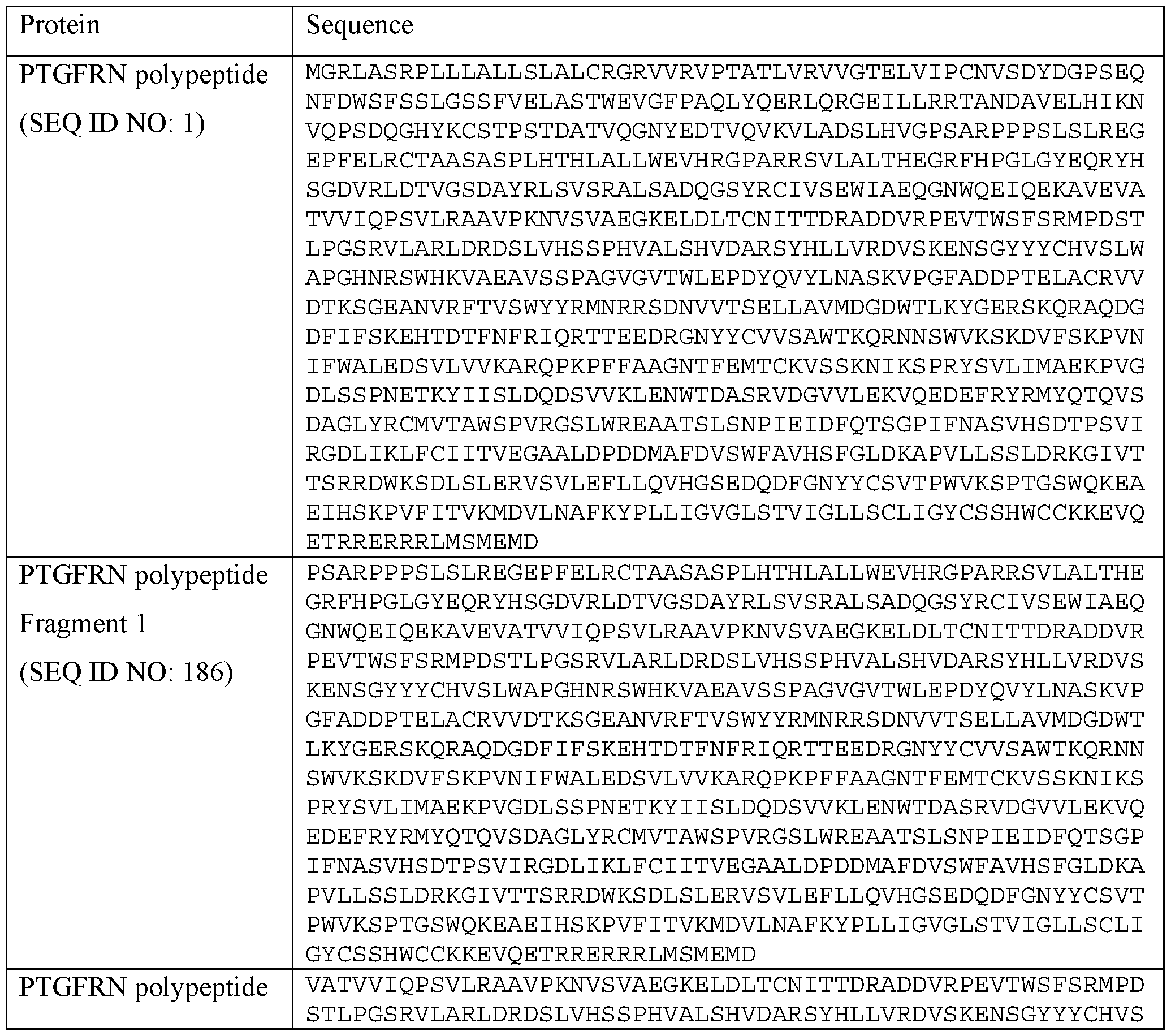

[0234] Non-limiting examples of other Scaffold X proteins can be found at US Patent No.
US 10,195,290B1, issued Feb. 5, 2019, which is incorporated by reference in its entirety, the ATP
transporter proteins: ATP1A1, ATP1A2, ATP 1 A3, ATP1A4, ATP1B3, ATP2B1, ATP2B2, and ATP2B4), CD 9, CD63, CD81, PDGFR, GPI anchor proteins, lactadherin, LAMP2, and LAMP2B. [0235] In some aspects, the scaffold moiety, e.g., Scaffold X, comprises an amino acid sequence at least about 70%, at least about 75%, at least about 80%, at least about 85%, at least about 90%, at least about 95%, at least about 96%, at least about 97%, at least about 98%, at least about 99%, or about 100% identical to SEQ ID NO: 197, 198, 199, or 200. In other aspects, the scaffold moiety, e.g, Scaffold X, comprises the amino acid sequence of SEQ ID NO: 197, 198, 199, or 200, except one amino acid mutation, two amino acid mutations, three amino acid mutations, four amino acid mutations, five amino acid mutations, six amino acid mutations, or seven amino acid mutations. The mutations can be a substitution, an insertion, a deletion, or any combination thereof. In some aspects, the scaffold moiety, e.g, Scaffold X, comprises the amino acid sequence of SEQ ID NO: 197, 198, 199, or 200 and 1 amino acid, two amino acids, three amino acids, four amino acids, five amino acids, six amino acids, seven amino acids, eight amino acids, nine amino acids, ten amino acids, 11 amino acids, 12 amino acids, 13 amino acids, 14 amino acids, 15 amino acids, 16 amino acids, 17 amino acids, 18 amino acids, 19 amino acids, or 20 amino acids or longer at the N terminus and/or C terminus of SEQ ID NO: 197, 198, 199, or 200. [0236] Non-limiting examples of other scaffold moieties, e.g, Scaffold X proteins, can be found at US Patent No. US10195290B1, issued Feb. 5, 2019, which is incorporated by reference in its entirety.
[0237] In some aspects, the sequence encodes a fragment of the scaffold moiety lacking at least about 5, at least about 10, at least about 50, at least about 100, at least about 200, at least about 300, at least about 400, at least about 500, at least about 600, at least about 700, or at least about 800 amino acids from the N-terminus of the native protein. In some aspects, the sequence encodes a fragment of the scaffold moiety lacking at least about 5, at least about 10, at least about 50, at least about 100, at least about 200, at least about 300, at least about 400, at least about 500, at least about 600, at least about 700, or at least about 800 amino acids from the C-terminus of the native protein. In some aspects, the sequence encodes a fragment of the scaffold moiety lacking at least about 5, at least about 10, at least about 50, at least about 100, at least about 200, at least about 300, at least about 400, at least about 500, at least about 600, at least about 700, or at least about 800 amino acids from both the N-terminus and C-terminus of the native protein. In some aspects, the sequence encodes a fragment of the scaffold moiety lacking one or more functional or structural domains of the native protein.
[0238] In some aspects, the scaffold moiety, e.g., Scaffold X, e.g, a PTGFRN protein, is linked to one or more heterologous proteins. The one or more heterologous proteins can be linked to the N-terminus of the scaffold moiety. The one or more heterologous proteins can be linked to the C-terminus of the scaffold moiety. In some aspects, the one or more heterologous proteins are linked to both the N-terminus and the C-terminus of the scaffold moiety. In some aspects, the heterologous protein is a mammalian protein. In some aspects, the heterologous protein is a human protein.
[0239] In some aspects, the scaffold moiety, e.g, Scaffold X, can be used to link any moiety to the luminal surface and the external surface of the EV at the same time. For example, the PTGFRN polypeptide can be used to link one or more biologically active molecules indirectly through a maleimide moiety or directly to a maleimide moiety or a linker to the luminal surface in addition to the external surface of the EV. Therefore, in certain aspects, Scaffold X can be used for dual purposes.
[0240] In other aspects, the EV s useful to practice the methods for delivery to the lung cells and/or tissue disclosed herein comprise a higher number of Scaffold X proteins compared to the naturally-occurring EVs. In some aspects, the EVs of the disclosure comprise at least about 5 fold, at least about 10 fold, at least about 20 fold, at least about 30 fold, at least about 40 fold, at least about 50 fold, at least about 60 fold, at least about 70 fold, at least about 80 fold, at least about 90 fold, at least about 100 fold, at least about 110 fold, at least about 120 fold, at least about 130 fold, at least about 140 fold, at least about 150 fold, at least about 160 fold, at least about 170 fold, at least about 180 fold, at least about 190 fold, at least about 200 fold, at least about 210 fold, at least about 220 fold, at least about 230 fold, at least about 240 fold, at least about 250 fold, at least about 260 fold, at least about 270 fold higher number of Scaffold X (e.g, a PTGFRN polypeptide) compared to the naturally-occurring EV.
[0241] The number of scaffold moieties, e.g, Scaffold X, such as, a PTGFRN polypeptide, on the EV of the present disclosure is at least about 100, at least about 200, at least about 300, at least about 400, at least about 500, at least about 600, at least about 700, at least about 800, at least about 900, at least about 1000, at least about 1100, at least about 1200, at least about 1300, at least about 1400, at least about 1500, at least about 1600, at least about 1700, at least about 1800, at least about 1900, at least about 2000, at least about 2100, at least about 2200, at least about 2300, at least about 2400, at least about 2500, at least about 2600, at least about 2700, at least about 2800, at least about 2900, at least about 3000, at least about 4000, at least about 5000, at least about 6000, at least about 7000, at least about 8000, at least about 9000, or at least about 10000.
[0242] In some aspects, the number of scaffold moieties, e.g., Scaffold X, such as, a
PTGFRN polypeptide, on the EV of the present disclosure is from about 100 to about 100,000, from about 200 to about 9000, from about 300 to about 9000, from about 400 to about 9000, from about 500 to about 9000, from about 600 to about 8000, from about 800 to about 8000, from about 900 to about 8000, from about 1000 to about 8000, from about 1100 to about 8000, from about 1200 to about 8000, from about 1300 to about 8000, from about 1400 to about 8000, from about 1500 to about 8000, from about 1600 to about 8000, from about 1700 to about 8000, from about 1800 to about 8000, from about 1900 to about 8000, from about 2000 to about 8000, from about 2100 to about 8000, from about 2200 to about 8000, from about 2300 to about 8000, from about 2400 to about 8000, from about 2500 to about 8000, from about 2600, from about 2700 to about 8000, from about 2800 to about 8000, from about 2900 to about 8000, from about 3000 to about 8000, from about 4000 to about 8000, from about 5000 to about 8000, from about 6000 to about 8000, from about 7000 to about 8000, from about 8000, from 7000 to about 9000, or from about 6000 to about 10000.
[0243] In some aspects, the number of scaffold moieties, e.g, Scaffold X, such as, a
PTGFRN polypeptide, on the EV of the present disclosure is from about 5000 to about 8000, e.g, about 5000, about 6000, about 7000, or about 8000. In some aspects, the number of scaffold moieties, e.g, Scaffold X, such as, a PTGFRN polypeptide, on the EV of the present disclosure is from about 6000 to about 8000, e.g, about 6000, about 7000, or about 8000. In some aspects, the number scaffold moieties, e.g, Scaffold X, such as, a PTGFRN polypeptide, on the EV of the present disclosure is from about 4000 to about 9000, e.g, about 4000, about 5000, about 6000, about 7000, about 8000, about 9000.
V.B. Luminal Scaffold Moieties ( e.g Scaffold Y)
[0244] In some aspects, EVs useful to practice the methods for delivery to the lung cells and/or tissue disclosed herein comprise an internal space (i.e., lumen) that is different from that of the naturally occurring EVs. For example, the EV can be changed such that the composition on the luminal surface of the EV has the protein, lipid, or glycan content different from that of the naturally-occurring EVs.
[0245] In some aspects, engineered EVs can be produced from a cell transformed with an exogenous sequence encoding a scaffold moiety (e.g, exosome proteins, e.g, Scaffold Y) or a modification or a fragment of the scaffold moiety that changes the composition or content of the
luminal surface of the exosome. Various modifications or fragments of the EV protein that can be expressed on the luminal surface of the EV can be used for the aspects of the present disclosure. [0246] In some aspects, the EV proteins that can change the luminal surface of the EV include, but are not limited to the MARCKS protein, MARCKSL1 protein, BASP1 protein, or any combination thereof. In some aspects, the scaffold moiety, e.g., Scaffold Y, comprises Brain Acid Soluble Protein 1 (the BASP1 protein). The BASP1 protein is also known as 22 kDa neuronal tissue-enriched acidic protein or neuronal axonal membrane protein NAP-22. The full-length human BASP1 protein sequence (isomer 1) is shown in TABLE 2. An isomer produced by an alternative splicing is missing amino acids 88 to 141 from the BASP1 protein in TABLE 2 (isomer 1).
TABLE 2. Exemplary Scaffold Protein Sequences
 [0247] In some aspects, the scaffold moiety, e.g., Scaffold Y, comprises a protein is selected from the group consisting of MARCKS, MARKSL1, BASP1, any functional fragment, variant, or derivative thereof, or any combination thereof. In some aspects, the scaffold moiety, e.g, Scaffold Y, comprises a Src protein or a fragment thereof. In some aspects, the scaffold moiety, e.g, Scaffold Y, comprises a sequence disclosed, e.g. , in U.S. Patent No. 9,611,481. [0248] In some aspects, the scaffold moiety, e.g, Scaffold Y, of the present disclosure comprises the BASP1 protein (Uniprot accession number P80723), also known as 22 kDa neuronal tissue-enriched acidic protein or neuronal axonal membrane protein NAP-22. The full-length human BASP1 protein sequence (isomer 1) is 227 amino acids in length. An isomer produced by an alternative splicing is missing amino acids 88 to 141 from isomer 1. In some aspects, the scaffold moiety, e.g, Scaffold Y, of the present disclosure comprises a mature BASP1 protein (i.e., without N-terminal methionine). In some aspects, the scaffold moiety, e.g, Scaffold Y, of the present disclosure is derived from a mature BASP1 protein, i.e., it is a fragment, variant, or derivate of a mature BASP1 protein and therefore it lacks the N-terminal protein present in the non-mature protein. The mature BASP1 protein sequence is missing the first Met from SEQ ID NO: 10 and thus contains amino acids 2 to 227 of SEQ ID NO: 10.
[0249] In other aspects, a scaffold moiety, e.g, Scaffold Y, useful for the present disclosure comprises an amino acid sequence at least about 70%, at least about 75%, at least about 80%, at least about 85%, at least about 90%, at least about 95%, at least about 96%, at least about 97%, at least about 98%, at least about 99%, or about 100% identical to amino acids 2 to 227 of SEQ ID NO: 10, i.e., the mature form ofBASPl (i.e., without the N-terminal methionine amino acid present in SEQ ID NO: 10). In other aspects, the scaffold moiety, e.g, a Scaffold X protein, comprises an amino acid sequence at least about at least about 70%, at least about 75%, at least about 80%, at least about 85%, at least about 90%, at least about 95%, at least about 96%, at least about 97%, at least about 98%, at least about 99%, or about 100% identical to a functional fragment of the mature form of SEQ ID NO: 10 (BASP1), i.e., without the N-terminal methionine amino acid present in SEQ ID NO: 10. In other aspects, a scaffold moiety, e.g. , Scaffold, Y useful for the present disclosure comprises the amino acid sequence of SEQ ID NO: 10 except one amino acid mutation, two amino acid mutations, three amino acid mutations, four amino acid mutations, five amino acid mutations, six amino acid mutations, or seven amino acid mutations. The mutations can be a substitution, an insertion, a deletion, or any combination thereof. In some aspects, a scaffold moiety, e.g, Scaffold Y, useful for the present disclosure comprises the amino acid sequence of SEQ ID NO: 10 and 1 amino acid, two amino acids, three amino acids, four amino acids, five
amino acids, six amino acids, seven amino acids, eight amino acids, nine amino acids, ten amino acids, 11 amino acids, 12 amino acids, 13 amino acids, 14 amino acids, 15 amino acids, 16 amino acids, 17 amino acids, 18 amino acids, 19 amino acids, or 20 amino acids or longer at the N terminus and/or C terminus of SEQ ID NO: 10.
[0250] In certain aspects, the protein sequence of any of SEQ ID NOs: 1-109 disclosed in
PCT/US2018/061679 is sufficient to be a Scaffold Y for the present disclosure (e.g., scaffold moiety linked to a linker).
[0251] In other aspects, the scaffold moiety, e.g, Scaffold Y, comprises an amino acid sequence at least about at least about 70%, at least about 75%, at least about 80%, at least about 85%, at least about 90%, at least about 95%, at least about 96%, at least about 97%, at least about 98%, at least about 99%, or about 100% identical to any one of the sequences disclosed in US 10,195,290B1, issued Feb. 5, 2019.
[0252] Scaffold Y-engineered exosomes described herein can be produced from a cell transformed with any sequence set forth in PCT/US2018/061679 (SEQ ID NO: 4-109 from PCT/US2018/061679).
[0253] In some aspects, the linker is a peptide linker. In some aspects, the peptide linker can comprise at least about two, at least about three, at least about four, at least about five, at least about 10, at least about 15, at least about 20, at least about 25, at least about 30, at least about 35, at least about 40, at least about 45, at least about 50, at least about 55, at least about 60, at least about 65, at least about 70, at least about 75, at least about 80, at least about 85, at least about 90, at least about 95, or at least about 100 amino acids.
[0254] In some aspects, the linker is a glycine/serine linker. In some aspects, the peptide linker is glycine/serine linker according to the formula [(Gly)n-Ser]m (SEQ ID NO: 46) where n is any integer from 1 to 100 and m is any integer from 1 to 100. In other aspects, the glycine/serine linker is according to the formula [(Gly)x-Sery]z (SEQ ID NO: 47) wherein x in an integer from 1 to 4, y is 0 or 1, and z is an integers from 1 to 50. In some aspects, the peptide linker comprises the sequence Gn (SEQ ID NO: 48), where n can be an integer from 1 to 100. In some aspects, the peptide linker can comprise the sequence (GlyAla)n (SEQ ID NO: 49), wherein n is an integer between 1 and 100. In other aspects, the peptide linker can comprise the sequence (GlyGlySer)n (SEQ ID NO:50), wherein n is an integer between 1 and 100.
[0255] In some aspects, the peptide linker is synthetic, i.e., non-naturally occurring. In one aspect, a peptide linker includes peptides (or polypeptides) (e.g, natural or non-naturally occurring peptides) which comprise an amino acid sequence that links or genetically fuses a first linear
sequence of amino acids to a second linear sequence of amino acids to which it is not naturally linked or genetically fused in nature. For example, in one aspect the peptide linker can comprise non-naturally occurring polypeptides which are modified forms of naturally occurring polypeptides ( e.g ., comprising a mutation such as an addition, substitution or deletion).
[0256] In other aspects, the peptide linker can comprise non-naturally occurring amino acids. In yet other aspects, the peptide linker can comprise naturally occurring amino acids occurring in a linear sequence that does not occur in nature. In still other aspects, the peptide linker can comprise a naturally occurring polypeptide sequence.
[0257] In other aspects, the lipid anchor can be any lipid anchor known in the art, e.g., palmitic acid or glycosylphosphatidylinositols. Under unusual circumstances, e.g, by using a culture medium where myristic acid is limiting, some other fatty acids including shorter-chain and unsaturated, can be attached to the N-terminal glycine. For example, in BK channels, myristate has been reported to be attached posttranslationally to internal serine/threonine or tyrosine residues via a hydroxyester linkage.
V.C. Lipid Anchoring Moieties
[0258] Suitable anchoring moieties capable of anchoring a biologically active molecule to the surface of an EV via a maleimide moiety comprise for example sterols (e.g, cholesterol), phospholipid, lysophospholipids, fatty acids, or fat-soluble vitamins, as described in detail below. [0259] In some aspects, the anchoring moiety can be a lipid. A lipid anchoring moiety can be any lipid known in the art, e.g, palmitic acid or glycosylphosphatidylinositols. In some aspects, the lipid, is a fatty acid, phosphatide, phospholipid (e.g, phosphatidyl choline, phosphatidyl serine, or phosphatidyl ethanolamine), or analogue thereof (e.g. phosphatidylcholine, lecithin, phosphatidylethanolamine, cephalin, or phosphatidylserine or analogue or portion thereof, such as a partially hydrolyzed portion thereof).
[0260] The anchoring moiety can be conjugated using a maleimide moiety to a biologically active molecule directly or indirectly via a linker combination, at any chemically feasible location, e.g, at the 5' and/or 3' end of a nucleotide sequence, e.g, of a biologically active molecule (e.g, an ASO). In one aspect, the anchoring moiety is conjugated only to the 3' end of the biologically active molecule. In one aspect, the anchoring moiety is conjugated only to the 5' end of a nucleotide sequence, e.g, of a biologically active molecule (e.g, an ASO). In one aspect, the anchoring moiety is conjugated at a location which is not the 3' end or 5’ end of a nucleotide sequence, e.g, of a biologically active molecule (e.g, an ASO).
[0261] In some aspects, a biologically active molecule can be conjugated directly or indirectly via a maleimide group to, e.g., any of the lipid anchors disclosed above (for example, palmitic acid, myristic acid, fatty acid, famesyl, geranyl-geranyl, or cholesterol). In some aspects, an anchoring moiety can comprise two or more types of anchoring moieties disclosed herein. For example, in some aspects, an anchoring moiety can comprise two lipids, e.g, a phospholipids and a fatty acid, or two phospholipids, or two fatty acids, or a lipid and a vitamin, or cholesterol and a vitamin, etc. which taken together have 6-80 carbon atoms (i.e., an equivalent carbon number (ECN) of about 6 to about 80).
V.D. Scaffold Protein Fusion Constructs
[0262] In some aspects, the scaffold moiety is linked to one or more heterologous proteins.
The one or more heterologous proteins can be linked to the N-terminus of the scaffold moieties. The one or more heterologous proteins can be linked to the C-terminus of the scaffold moieties. In some aspects, the one or more heterologous proteins are linked to both the N-terminus and the C- terminus of the scaffold moieties. In some aspects, the heterologous protein is a mammalian protein. In some aspects, the heterologous protein is a human protein.
[0263] In some aspects, the scaffold moiety can be used to link any moiety to the luminal surface and/or the external surface of the EV. For example, the PTGFRN polypeptide can be used to link a biologically active molecule inside the lumen (e.g, on the luminal surface) in addition to the external surface of the EV. Therefore, in certain aspects, the scaffold moiety can be used for dual purposes, e.g, a biologically active molecule on the luminal surface and a second biologically active molecule or other payload on the external surface of the EV or a biologically active molecule on the external surface of the exosome and a second biologically active molecule or other payload on the luminal surface of the EV.
V.E. Anti-phagocvtic Signal
[0264] Clearance of administered EVsby the body's immune system can reduce the efficacy of an administered EV therapy. In some aspects, the surface of the EV is modified to limit or block uptake of the EV by cells of the immune system, e.g, macrophages. In some aspects, the surface of the EV is modified to express one or more surface antigen that inhibits uptake of the EV by a macrophage. In some aspects, the surface antigen is associated with the exterior surface of the EV. [0265] Surface antigens useful in the present disclosure include, but are not limited to, antigens that label a cell as a "self cell. In some aspects, the surface antigen is selected from CD47,
CD24, a fragment thereof, and any combination thereof. In certain aspects, the surface antigen comprises CD24, e.g ., human CD24. In some aspects, the surface antigen comprises a fragment of CD24, e.g. , human CD24. In certain aspects, the EV is modified to express CD47 or a fragment thereof on the exterior surface of the EV.
[0266] CD47, also referred to as leukocyte surface antigen CD47 and integrin associated protein (LAP), as used herein, is a transmembrane protein that is found on many cells in the body. CD47 is often referred to as the "don't eat me" signal, as it signals to immune cells, in particular myeloid cells, that a particular cell expressing CD47 is not a foreign cell. CD47 is the receptor for SIRPA, binding to which prevents maturation of immature dendritic cells and inhibits cytokine production by mature dendritic cells. Interaction of CD47 with SIRPG mediates cell-cell adhesion, enhances superantigen-dependent T-cell-mediated proliferation and costimulates T-cell activation. CD47 is also known to have a role in both cell adhesion by acting as an adhesion receptor for THBS1 on platelets, and in the modulation of integrins. CD47 also plays an important role in memory formation and synaptic plasticity in the hippocampus (by similarity). In addition, CD47 can play a role in membrane transport and/or integrin dependent signal transduction, prevent premature elimination of red blood cells, and be involved in membrane permeability changes induced following virus infection.
[0267] In some aspects, an EV disclosed herein is modified to express a human CD47 on the surface of the EV. The canonical amino acid sequence for human CD47 and various known isoforms are shown in Table 3 (UniProtKB - Q08722; SEQ ID NOs: 213-216). In some aspects, the EV is modified to express a polypeptide comprising the amino acid sequence set forth in SEQ ID NO: 213 or a fragment thereof. In some aspects, the EV is modified to express a polypeptide comprising the amino acid sequence set forth in SEQ ID NO: 214 or a fragment thereof. In some aspects, the EV is modified to express a polypeptide comprising the amino acid sequence set forth in SEQ ID NO: 215 or a fragment thereof. In some aspects, the EV is modified to express a polypeptide comprising the amino acid sequence set forth in SEQ ID NO: 216 or a fragment thereof.
Table 3: Human CD47 Amino Acid Sequences


[0268] In some aspects, the EV is modified to express full length CD47 on the surface of the EV. In some aspects, the EV is modified to express a fragment of CD47 on the surface of the EV wherein the fragment comprises the extracellular domain of CD47, e.g., human CD47. Any fragment of CD47 that retains an ability to block and/or inhibit phagocytosis by a macrophage can be used in the EVs disclosed herein. In some aspects, the fragment comprises amino acids 19 to about 141 of the canonical human CD47 sequence (e.g., amino acids 19-141 of SEQ ID NO: 213). In some aspects, the fragment comprises amino acids 19 to about 135 of the canonical human CD47 sequence (e.g, amino acids 19-135 of SEQ ID NO: 213). In some aspects, the fragment comprises amino acids 19 to about 130 of the canonical human CD47 sequence (e.g, amino acids 19-130 of SEQ ID NO: 213). In some aspects, the fragment comprises amino acids 19 to about 125 of the canonical human CD47 sequence (e.g, amino acids 19-125 of SEQ: ID NO: 213).
[0269] In some aspects, the EV is modified to express a polypeptide having at least about
70%, at least about 75%, at least about 80%, at least about 85%, at least about 90%, at least about 95%, at least about 96%, at least about 97%, at least about 98%, or at least about 99% sequence identity to amino acids 19 to about 141 of the canonical human CD47 sequence (e.g, amino acids 19-141 of SEQ ID NO: 213). In some aspects, the EV is modified to express a polypeptide having at least about 70%, at least about 75%, at least about 80%, at least about 85%, at least about 90%, at least about 95%, at least about 96%, at least about 97%, at least about 98%, or at least about
99% sequence identity to amino acids 19 to about 135 of the canonical human CD47 sequence ( e.g ., amino acids 19-135 of SEQ ID NO: 213). In some aspects, the EV is modified to express a polypeptide having at least about 70%, at least about 75%, at least about 80%, at least about 85%, at least about 90%, at least about 95%, at least about 96%, at least about 97%, at least about 98%, or at least about 99% sequence identity to amino acids 19 to about 130 of the canonical human CD47 sequence (e.g., amino acids 19-130 of SEQ ID NO: 213). In some aspects, the EV is modified to express a polypeptide having at least about 70%, at least about 75%, at least about 80%, at least about 85%, at least about 90%, at least about 95%, at least about 96%, at least about 97%, at least about 98%, or at least about 99% sequence identity to amino acids 19 to about 125 of the canonical human CD47 sequence (e.g, amino acids 19-125 of SEQ ID NO: 213).
[0270] In some aspects, the CD47 or the fragment thereof is modified to increase the affinity of CD47 and its ligand SIRPa. In some aspects, the fragment of CD47 comprises a Velcro- CD47 (see, e.g., Ho et al., JBC 290:12650-63 (2015), which is incorporated by reference herein in its entirety). In some aspects, the Velcro-CD47 comprises a C15S substitution relative to the wild- type human CD47 sequence (SEQ ID NO: 213).
[0271] In some aspects, the EV comprises a CD47 or a fragment thereof expressed on the surface of the EV at a level that is higher than an unmodified EV. In some aspects, the CD47 or the fragment thereof is fused with a scaffold protein. Any scaffold protein disclosed herein can be used to express the CD47 or the fragment thereof on the surface of the EV. In some aspects, the EV is modified to express a fragment of CD47 fused to the N-terminus of a Scaffold X protein. In some aspects, the EV is modified to express a fragment of CD47 fused to the N-terminus of PTGFRN.
[0272] In some aspects, the EV comprises at least about 20 molecules, at least about 30 molecules, at least about 40, at least about 50, at least about 75, at least about 100, at least about 125, at least about 150, at least about 200, at least about 250, at least about 300, at least about 350, at least about 400, at least about 450, at least about 500, at least about 750, or at least about 1000 molecules of CD47 on the surface of the EV. In some aspects, the EV comprises at least about 20 molecules of CD47 on the surface of the EV. In some aspects, the EV comprises at least about 30 molecules of CD47 on the surface of the EV. In some aspects, the EV comprises at least about 40 molecules of CD47 on the surface of the EV. In some aspects, the EV comprises at least about 50 molecules of CD47 on the surface of the EV. In some aspects, the EV comprises at least about 100 molecules of CD47 on the surface of the EV. In some aspects, the EV comprises at least about 200 molecules of CD47 on the surface of the EV. In some aspects, the EV comprises at least about 300
molecules of CD47 on the surface of the EV. In some aspects, the EV comprises at least about 400 molecules of CD47 on the surface of the EV. In some aspects, the EV comprises at least about 500 molecules of CD47 on the surface of the EV. In some aspects, the EV comprises at least about 1000 molecules of CD47 on the surface of the EV.
[0273] In some aspects, expression CD47 or a fragment thereof on the surface of the EV results in decreased uptake of the EV by myeloid cells as compared to an EV not expressing CD47 or a fragment thereof. In some aspects, uptake by myeloid cells of the EV expressing CD47 or a fragment thereof is decreased by at least about 5%, at least about 10%, at least about 15%, at least about 20%, at least about 25%, at least about 30%, at least about 35%, at least about 40%, at least about 45%, at least about 50%, at least about 60%, at least about 70%, at least about 80%, at least about 90%, or at least about 95%, relative to uptake by myeloid cells of EVs that do not express CD47 or a fragment thereof.
[0274] In some aspects, expression CD47 or a fragment thereof on the surface of the EV results in decreased localization of the EV to the liver, as compared to an EV not expressing CD47 or a fragment thereof. In some aspects, localization to the liver of EVs expressing CD47 or a fragment thereof is decreased by at least about 5%, at least about 10%, at least about 15%, at least about 20%, at least about 25%, at least about 30%, at least about 35%, at least about 40%, at least about 45%, at least about 50%, at least about 60%, at least about 70%, at least about 80%, at least about 90%, or at least about 95%, relative to the localization to the liver of EVs not expressing CD47 or a fragment thereof.
[0275] In some aspects, the in vivo half-life of an EV expressing CD47 or a fragment thereof is increased relative to the in vivo half-life of an EV that does not express CD47 or a fragment thereof. In some aspects, the in vivo half-life of an EV expressing CD47 or a fragment thereof is increased by at least about 1.5-fold, at least about 2-fold, at least about 2.5-fold, at least about 3-fold, at least about 3.5-fold, at least about 4-fold, at least about 4.5-fold, at least about 5- fold, at least about 6-fold, at least about 7-fold, at least about 8-fold, at least about 9-fold, or at least about 10-fold, relative to the in vivo half-life of an EV that does not express CD47 or a fragment thereof.
[0276] In some aspects, an EV expressing CD47 or a fragment thereof has an increased retention in circulation, e.g ., plasma, relative to the retention of an EV that does not express CD47 or a fragment thereof in circulation, e.g. , plasma. In some aspects, retention in circulation, e.g. , plasma, of an EV expressing CD47 or a fragment thereof is increased by at least about 1.5-fold, at least about 2-fold, at least about 2.5-fold, at least about 3-fold, at least about 3.5-fold, at least about
4-fold, at least about 4.5-fold, at least about 5-fold, at least about 6-fold, at least about 7-fold, at least about 8-fold, at least about 9-fold, or at least about 10-fold, relative to the retention in circulation, e.g ., plasma, of an EV that does not express CD47 or a fragment thereof.
[0277] In some aspects, an EV expressing CD47 or a fragment thereof has an altered biodistribution when compared with an exosome that does not express CD47 or a fragment. In some aspects, the altered biodistribution leads to increased uptake into endothelial cells, T cells, or increased accumulation in various tissues, including, but not limited to skeletal muscle, cardiac muscle, diaphragm, kidney, bone marrow, central nervous system, lungs, cerebral spinal fluid (CSF), or any combination thereof.
VI. Pharmaceutical Compositions and Methods of Administration [0278] The present disclosure also provides pharmaceutical compositions comprising EVs, e.g, exosomes, described herein that are suitable for administration to a subject according to the methods of administration targeting the lung cells and/or tissue disclosed herein. The pharmaceutical compositions generally comprise a plurality of EVs comprising a biologically active molecule covalently linked to the plurality of EVs via a maleimide moiety and a pharmaceutically-acceptable excipient or carrier in a form suitable for administration to a subject. Pharmaceutically acceptable excipients or carriers are determined in part by the particular composition being administered, as well as by the particular method used to administer the composition. Accordingly, there is a wide variety of suitable formulations of pharmaceutical compositions comprising a plurality of EVs. See, e.g. , Remington's Pharmaceutical Sciences, Mack Publishing Co., Easton, Pa. 18th ed. (1990).
[0279] The pharmaceutical compositions are generally formulated sterile and in full compliance with all Good Manufacturing Practice (GMP) regulations of the U.S. Food and Drug Administration. In some aspects, the pharmaceutical composition comprises one or more chemical compounds, such as for example, small molecules covalently linked to an EV described herein. [0280] In some aspects, a pharmaceutical composition comprises one or more therapeutic agents and an EV described herein. In certain aspects, the EVs are co-administered with of one or more additional therapeutic agents, in a pharmaceutically acceptable carrier. In some aspects, the pharmaceutical composition comprising the EV is administered prior to administration of the additional therapeutic agents. In other aspects, the pharmaceutical composition comprising the EV is administered after the administration of the additional therapeutic agents. In further aspects, the pharmaceutical composition comprising the EV is administered concurrently with the additional therapeutic agents.
[0281] Provided herein are pharmaceutical compositions comprising an EV of the present disclosure having the desired degree of purity, and a pharmaceutically acceptable carrier or excipient, in a form suitable for administration to a subject. Pharmaceutically acceptable excipients or carriers can be determined in part by the particular composition being administered, as well as by the particular method used to administer the composition. Accordingly, there is a wide variety of suitable formulations of pharmaceutical compositions comprising a plurality of extracellular vesicles. (See, e.g. , Remington's Pharmaceutical Sciences, Mack Publishing Co., Easton, Pa. 21st ed. (2005)). The pharmaceutical compositions are generally formulated sterile and in full compliance with all Good Manufacturing Practice (GMP) regulations of the U.S. Food and Drug Administration.
[0282] In some aspects, a pharmaceutical composition comprises one or more therapeutic agents and an EV described herein. In certain aspects, the EVs are co-administered with of one or more additional therapeutic agents, in a pharmaceutically acceptable carrier. In some aspects, the pharmaceutical composition comprising the EVs is administered prior to administration of the additional therapeutic agents. In other aspects, the pharmaceutical composition comprising the EVs is administered after the administration of the additional therapeutic agents. In further aspects, the pharmaceutical composition comprising the EVs is administered concurrently with the additional therapeutic agents.
[0283] Acceptable carriers, excipients, or stabilizers are nontoxic to recipients (e.g., animals or humans) at the dosages and concentrations employed, and include buffers such as phosphate, citrate, and other organic acids; antioxidants including ascorbic acid and methionine; preservatives (such as octadecyldimethylbenzyl ammonium chloride; hexamethonium chloride; benzalkonium chloride, benzethonium chloride; phenol, butyl or benzyl alcohol; alkyl parabens such as methyl or propyl paraben; catechol; resorcinol; cyclohexanol; 3-pentanol; and m-cresol); low molecular weight (less than about 10 residues) polypeptides; proteins, such as serum albumin, gelatin, or immunoglobulins; hydrophilic polymers such as polyvinylpyrrolidone; amino acids such as glycine, glutamine, asparagine, histidine, arginine, or lysine; monosaccharides, di saccharides, and other carbohydrates including glucose, mannose, or dextrins; chelating agents such as EDTA; sugars such as sucrose, mannitol, trehalose or sorbitol; salt-forming counter-ions such as sodium; metal complexes (e.g, Zn-protein complexes); and/or non-ionic surfactants such as TWEEN™, PLURONICS™ or polyethylene glycol (PEG).
[0284] Examples of carriers or diluents include, but are not limited to, water, saline,
Ringer's solutions, dextrose solution, and 5% human serum albumin. The use of such media and
compounds for pharmaceutically active substances is well known in the art. Except insofar as any conventional media or compound is incompatible with the extracellular vesicles described herein, use thereof in the compositions is contemplated. Supplementary therapeutic agents can also be incorporated into the compositions. Typically, a pharmaceutical composition is formulated to be compatible with its intended route of administration. The EVs of the present disclosure can be administered by parenteral, topical, intravenous, oral, subcutaneous, intra-arterial, intradermal, transdermal, rectal, intracranial, intraperitoneal, intranasal, intratumoral, intramuscular route, or as inhalants. In certain aspects, the pharmaceutical composition comprising EVs is administered intravenously, e.g. by injection. In some aspects, the pharmaceutical composition comprising EVs is delivered as an inhalant. In some aspects, the pharmaceutical composition comprising EVs is delivered using an inhaler. In some aspects, the pharmaceutical composition comprising EVs is delivered using a nebulizer. The EVs can optionally be administered in combination with other therapeutic agents that are at least partly effective in treating the disease, disorder or condition for which the EVs are intended.
[0285] Solutions or suspensions can include the following components: a sterile diluent such as water, saline solution, fixed oils, polyethylene glycols, glycerine, propylene glycol or other synthetic solvents; antibacterial compounds such as benzyl alcohol or methyl parabens; antioxidants such as ascorbic acid or sodium bisulfite; chelating compounds such as ethylenediaminetetraacetic acid (EDTA); buffers such as acetates, citrates or phosphates, and compounds for the adjustment of tonicity such as sodium chloride or dextrose. The pH can be adjusted with acids or bases, such as hydrochloric acid or sodium hydroxide. The preparation can be enclosed in ampoules, disposable syringes or multiple dose vials made of glass or plastic. [0286] Pharmaceutical compositions suitable for injectable use include sterile aqueous solutions (if water-soluble) or dispersions and sterile powders. For intravenous administration, suitable carriers include physiological saline, bacteriostatic water, Cremophor EL™ (BASF, Parsippany, N. J.) or phosphate buffered saline (PBS). The composition is generally sterile and fluid to the extent that easy syringeability exists. The carrier can be a solvent or dispersion medium containing, e.g. , water, ethanol, polyol (e.g, glycerol, propylene glycol, and liquid polyethylene glycol, and the like), and suitable mixtures thereof. The proper fluidity can be maintained, e.g, by the use of a coating such as lecithin, by the maintenance of the required particle size in the case of dispersion and by the use of surfactants. Prevention of the action of microorganisms can be achieved by various antibacterial and antifungal compounds, e.g, parabens, chlorobutanol, phenol, ascorbic acid, thimerosal, and the like. If desired, isotonic compounds, e.g, sugars, polyalcohols
such as mannitol, sorbitol, and sodium chloride can be added to the composition. Prolonged absorption of the injectable compositions can be brought about by including in the composition a compound, which delays absorption, e.g ., aluminum monostearate and gelatin.
[0287] Sterile injectable solutions can be prepared by incorporating the EVs of the present disclosure in an effective amount and in an appropriate solvent with one or a combination of ingredients enumerated herein, as desired. Generally, dispersions are prepared by incorporating the EVs into a sterile vehicle that contains a basic dispersion medium and any desired other ingredients. In the case of sterile powders for the preparation of sterile injectable solutions, methods of preparation are vacuum drying and freeze-drying that yields a powder of the active ingredient plus any additional desired ingredient from a previously sterile-filtered solution thereof. The EVs can be administered in the form of a depot injection or implant preparation, which can be formulated in such a manner to permit a sustained or pulsatile release of the EVs.
[0288] Systemic administration of compositions comprising EVs of the present disclosure can also be by transmucosal means. For transmucosal administration, penetrants appropriate to the barrier to be permeated are used in the formulation. Such penetrants are generally known in the art, and include, e.g. , for transmucosal administration, detergents, bile salts, and fusidic acid derivatives. Transmucosal administration can be accomplished through the use of, e.g. , nasal sprays.
[0289] In certain aspects, the pharmaceutical composition comprising EVs of the present disclosure is administered intravenously into a subject that would benefit from the pharmaceutical composition. In certain other aspects, the composition is administered to the lymphatic system, e.g., by intralymphatic injection or by intranodal injection ( see e.g., Senti et al, PNAS 105(46): 17908 (2008)), or by intramuscular injection, by subcutaneous administration, by intratumoral injection, by direct injection into the thymus, or into the liver.
[0290] In certain aspects, the pharmaceutical composition comprising EVs of the present disclosure is administered as a liquid suspension. In certain aspects, the pharmaceutical composition is administered as a formulation that is capable of forming a depot following administration. In certain preferred aspects, the depot slowly releases the EVs into circulation, or remains in depot form.
[0291] Typically, pharmaceutically acceptable compositions are highly purified to be free of contaminants, are biocompatible and not toxic, and are suited to administration to a subject. If water is a constituent of the carrier, the water is highly purified and processed to be free of contaminants, e.g, endotoxins.
[0292] The pharmaceutically acceptable carrier can be lactose, dextrose, sucrose, sorbitol, mannitol, starch, gum acacia, calcium phosphate, alginates, gelatin, calcium silicate, micro crystalline cellulose, polyvinylpyrrolidone, cellulose, water, syrup, methyl cellulose, methylhydroxy benzoate, propylhydroxy benzoate, talc, magnesium stearate, and/or mineral oil, but is not limited thereto. The pharmaceutical composition can further include a lubricant, a wetting agent, a sweetener, a flavor enhancer, an emulsifying agent, a suspension agent, and/or a preservative.
[0293] The pharmaceutical compositions described herein comprise the EVs described herein and optionally a pharmaceutically active or therapeutic agent. The therapeutic agent can be a biological agent, a small molecule agent, or a nucleic acid agent.
[0294] Dosage forms are provided that comprise a pharmaceutical composition comprising the EVs described herein. In some aspects, the dosage form is formulated as a liquid suspension for intravenous injection. In some aspects, the dosage form is formulated as a liquid suspension for intratumoral injection.
[0295] In certain aspects, the preparation of EVs of the present disclosure is subjected to radiation, e.g ., X rays, gamma rays, beta particles, alpha particles, neutrons, protons, elemental nuclei, UV rays in order to damage residual replication-competent nucleic acids.
[0296] In certain aspects, the preparation of EVs of the present disclosure is subjected to gamma irradiation using an irradiation dose of more than about 1, about 5, about 10, about 15, about 20, about 25, about 30, about 35, about 40, about 50, about 60, about 70, about 80, about 90, about 100, or more than 100 kGy.
[0297] In certain aspects, the preparation of EVs of the present disclosure is subjected to
X-ray irradiation using an irradiation dose of more than about 0.1, about 0.5, about 1, about 5, about 10, about 15, about 20, about 25, about 30, about 35, about 40, about 50, about 60, about 70, about 80, about 90, about 100, about 200, about 300, about 400, about 500, about 600, about 700, about 800, about 900, about 1000, about 2000, about 3000, about 4000, about 5000, about 6000, about 7000, about 8000, about 9000, or about 10000.
[0298] The EVs of the present disclosure can be used concurrently with other drugs. To be specific, the EVs of the present disclosure can be used together with medicaments such as hormonal therapeutic agents, chemotherapeutic agents, immunotherapeutic agents, medicaments inhibiting the action of cell growth factors, or cell growth factor receptors and the like.
VII. Kits
[0299] The present disclosure also provides kits, or products of manufacture comprising one or more EVs of the present disclosure and optionally instructions for use according to the methods of administration targeted to the lung cells and/or tissue disclosed herein.
[0300] In some aspects, the kit, or product of manufacture contains a pharmaceutical composition described herein which comprises at least one EV of the present disclosure, and instructions for use according to the methods of administration targeted to the lung cells and/or tissue disclosed herein.. In some aspects, the kit, or product of manufacture comprises at least one EV of the present disclosure or a pharmaceutical composition comprising the EVs in one or more containers. One skilled in the art will readily recognize that the EVs of the present disclosure, pharmaceutical composition comprising the EVs of the present disclosure, or combinations thereof can be readily incorporated into one of the established kit formats which are well known in the art. [0301] In some aspects, the kit comprises reagents to conjugate a biologically active molecule to an EV via a maleimide moiety, and instructions to conduct the conjugation.
[0302] The practice of the present disclosure will employ, unless otherwise indicated, conventional techniques of cell biology, cell culture, molecular biology, transgenic biology, microbiology, recombinant DNA, and immunology, which are within the skill of the art. Such techniques are explained fully in the literature. See, for example, Sambrook et ah, ed. (1989) Molecular Cloning A Laboratory Manual (2nd ed.; Cold Spring Harbor Laboratory Press); Sambrook et ah, ed. (1992) Molecular Cloning: A Laboratory Manual, (Cold Springs Harbor Laboratory, NY); D. N. Glover ed., (1985) DNA Cloning, Volumes I and II; Gait, ed. (1984) Oligonucleotide Synthesis; Mullis et al. U.S. Pat. No. 4,683,195; Hames and Higgins, eds. (1984) Nucleic Acid Hybridization; Hames and Higgins, eds. (1984) Transcription And Translation; Freshney (1987) Culture Of Animal Cells (Alan R. Liss, Inc.); Immobilized Cells And Enzymes (IRL Press) (1986); Perbal (1984) A Practical Guide To Molecular Cloning; the treatise, Methods In Enzymology (Academic Press, Inc., N.Y.); Miller and Calos eds. (1987) Gene Transfer Vectors For Mammalian Cells, (Cold Spring Harbor Laboratory); Wu et al., eds., Methods In Enzymology, Vols. 154 and 155; Mayer and Walker, eds. (1987) Immunochemical Methods In Cell And Molecular Biology (Academic Press, London); Weir and Blackwell, eds., (1986) Handbook Of Experimental Immunology, Volumes I-IV; Manipulating the Mouse Embryo, Cold Spring Harbor Laboratory Press, Cold Spring Harbor, N.Y., (1986); ); Crooke, Antisense drug Technology: Principles, Strategies and Applications, 2nd Ed. CRC Press (2007) and in Ausubel et al. (1989) Current Protocols in Molecular Biology (John Wiley and Sons, Baltimore, Md.).
[0303] All of the references cited above, as well as all references cited herein, are incorporated herein by reference in their entireties.
[0304] The following examples are offered by way of illustration and not by way of limitation.
Examples
[0305] The following examples are provided for illustrative purposes only, and are not to be construed as limiting the scope or content of the invention in any way. The practice of the current invention will employ, unless otherwise indicated, conventional methods of protein chemistry, biochemistry, recombinant DNA techniques and pharmacology, within the skill of the art. Such techniques are explained fully in the literature. See, e.g ., T.E. Creighton, Proteins: Structures and Molecular Properties (W.H. Freeman and Company, 1993); Green & Sambrook et ah, Molecular Cloning: A Laboratory Manual, 4th Edition (Cold Spring Harbor Laboratory Press, 2012); Colowick & Kaplan, Methods In Enzymology (Academic Press); Remington: The Science and Practice of Pharmacy, 22nd Edition (Pharmaceutical Press, 2012); Sundberg & Carey, Advanced Organic Chemistry: Parts A and B, 5th Edition (Springer, 2007).
Example 1
Exosome Isolation and Loading
[0306] Exosome isolation. Exosomes were collected from the supernatant of high-density suspension cultures of HEK293 SF cells after 7-9 days. Cell culture medium was serially centrifuged, with the supernatant of the previous spin serving as the input for the subsequent spin: cell culture medium was centrifuged at 5,000 x g for 30 minutes, the supernatant collected and the pellet discarded; the supernatant was then centrifuged at 16,000 x g for 30 minutes and the supernatant collected and the pellet discarded; the supernatant was then centrifuged at 133,900 x g for 3 hours, and the resulting supernatant discarded and the pellet collected and resuspended in 1 mL of PBS. The resuspended 133,900 x g pellet was further purified by running in an OPTIPREP™ Iodixanol gradient: a 4-tier sterile gradient was prepared by mixing 3 mL of OPTIPREP™ (60% Iodixanol) with 1 mL of resuspended pellet to generate 4mL of 45% Iodixanol, then overlaid serially with 3 mL 30% Iodixanol, 2mL 22.5% Iodixanol, 2mL 17.5% Iodixanol, and lmL PBS in a 12 mL Ultra-Clear (344059) tube for a SW 41 Ti rotor. The gradient was ultracentrifuged at 150,000 x g for 16 hours at 4 °C. Ultracentrifugation resulted in a Top Fraction known to contain
exosomes, a Middle Fraction containing cell debris of moderate density, and a Bottom Fraction containing high-density aggregates and cellular debris. The exosome layer was then gently collected from the top ~2 mL of the tube.
[0307] The exosome fraction was diluted in ~32 mL PBS in a 38.5 mL Ultra-Clear
(344058) tube and centrifuged at 10,000 x g for 30 minutes, the supernatant collected and ultracentrifuged at 133,900 x g for 3 hours at 4 °C to pellet the purified exosomes. The pelleted exosomes were then resuspended in a minimal volume of PBS (-200 pL) and stored at 4 °C. Final purified concentration of exosomes was determined using nanoparticle tracking analysis (NTA). [0308] Exosome Loading: To load exosomes with maleimide conjugates, exosomes were chemically reduced using TCEP (Tris(2-carboxyethyl)phosphine hydrochloride) at concentrations from 1 to 50 mM; in some cases, the reduction step includes, or is preceded by treatment with, 1- 2 M Guanidine hydrochloride for one hour at room temperature. Exosomes were exchanged into PBS by diluting to 1 mL in PBS, centrifuging at 100,000 x g for 20 minutes (TLA 120.2 rotor, Beckman) to pellet exosomes, the supernatant was removed and discarded, and the pellet resuspended in 1 mL PBS; this was repeated once to ensure complete buffer exchange. The final exosome pellet was resuspended in 0.1 mL PBS, to which the compound to be loaded was added to a final concentration of up to 300 mM. Exosomes were incubated overnight at 4°C, followed by washing with PBS to remove compound not conjugated to exosomes (diluting to 1 mL in PBS, centrifuging at 100,000 x g for 20 minutes (TLA 120.2 rotor, Beckman) to pellet exosomes, the supernatant was removed and discarded, and the pellet resuspended in 1 mL PBS; this was repeated once to ensure complete buffer exchange).
Example 2
In vivo analysis of ExoASO-NLRP3
[0309] ExoASO-NLRP3 will be assessed in several different bacterially and chemically induced lung inflammation and injury mouse models including: cystic fibrosis (CF), S aureus pneumonia and Bleomycin-induced idiopathic pulmonary fibrosis. Exosomes will be loaded with ASO-NLRP3 as previously described (see W02021/030773, which is incorporated by reference herein in its entirety). Exosome distribution in the inflamed lung will be assessed using exoASO with Cy5 labeled ASO and analyzed by IHC and FACS. Exosomes expressing EGFP and either loaded with Cy 5 labeled ASO (Cy5) or left unloaded (-C) were administered once intranasally (IN) to mice who had either been pre-treated 2 weeks prior with bleomycin (bleo) to induce lung inflammation and injury or were not pre-treated with bleomycin (naive) (FIG. 11 A). Bleomycin-
induced pulmonary fibrosis was characterized by alveolar epithelial cell injury and hyperplasia, inflammatory cell accumulation, fibroblast hyperplasia, deposition of extracellular matrix, and scar formation (compare FIGs. 12A-12F to 12G-12L; and FIG. 13A to 13B). The observed result was the loss of lung elasticity alveolar surface area leading to impairment of gas exchange and pulmonary function. Induced pulmonary fibrosis (IPF) disease was further characterized by the presence of chronic inflammatory infiltrates, myofibroblast hyperplasia, and disordered collagen deposition.
[0310] Mice were sacrificed 4 hours after exosome administration and lungs were analyzed for the presence of exosomes by measuring (FIG. 1 IB) Cy5 fluorescence using IVIS or immunohistochemistry using an anti-EGFP antibody (exosomes) and DAPI staining for cell nuclei (FIGs. 1 IB-1 ID). Exosomes localized to the lung after IN administration. Bleomycin induced lung injury led to enhanced exosome uptake in the lung (FIGs. 14A-14E) including by lung macrophages and lung capillary endothelial cells (FIGs. 15A-15D), consistent with uptake in normal lungs (FIGs. 5A-5H).
[0311] RNA isolated from the mouse lung tissues will be analyzed and measured for knockdown of NLRP3 and inflammatory and pharmacodynamic markers (e.g., IL-Ib, IL-8, IL-6, MCP 1, MCP 2, and TNFoc). Bronchial alveolar lavage (BAL) samples will be analyzed for the reduction in inflammatory cytokines (e.g, IL-Ib, IL-8, IL-6, MCP 1, MCP 2, TNFoc and TGF-b). Optimal dosing and mode of delivery will be determined. Subsequently, we will analyze the in vivo functional effect of NLRP3 suppression on inflammatory disease phenotype (body and lung weight, IHC analysis of lung tissue, sera cytokine expression) as well as lung function in these animals.
[0312] ExoASO-NLRP3 will be assessed in several different mouse models of acute respiratory distress syndrome including SARS-CoV-2, and IAV-induced and LPS-induced ARDS. The ability of exoASO-NLRP3 to attenuate inflammation, suppress the viral load, and/or rescue lung function will be assessed. Exosome distribution in the inflamed lung will be assessed using exoASO with Cy5 labeled ASO and analyzed by IHC and FACS. A dose escalation study to establish the optimal dose and administration regime will be tested. RNA and BAL isolated from the mouse lung tissues will be analyzed and measured for knockdown of NLRP3 and inflammatory and pharmacodynamic markers (e.g, IL-Ib, IL-8, IL-6, MCP 1, MCP 2, and TNFoc). Bronchial alveolar lavage samples will be analyzed for the reduction in inflammatory cytokines (e.g, IL-Ib and TGF-b). Lung tissue damage will be characterized using clinical scoring and IHC analysis.
The efficacy of exoASO-NLRP3 in repairing the mouse lung function and the efficacy of aerosol formulation for delivery via inhalers will be analyzed.
[0313] An alveolus lung chip or an alveolar organoid model of SARS-CoV-2 or IAV infection will be used analyze the impact of exoASO-NLRP3 activity on virus-induced lung injury. The expression of inflammasome-associated genes including IL-Ib, and IL-6 will be analyzed. Subsequently, comprehensive transcriptomic analyses will be performed using RNA sequencing. Lung chips will be infected with pseudotyped virus and/or poly (I:C) followed by treatment with exoASO-NLRP3 or PB S to identify longitudinal changes in gene expression over time using RNA- seq. Further bioinformatic analyses will also be utilized to identify cellular pathways that are impacted and/or repaired by treatment with exoASO-NLRP3.
Example 3
Delivery of exosomes loaded with AAV to the lungs
[0314] AAV encoding a reporter ( e.g ., GFP) will be loaded into native or PTGFRN over expressing exosomes, and sufficient quantities will be purified to enable in vivo studies. Exosomes expressing transferrin may also be used. AAV-loaded exosomes will be administered intranasally, and animals will be sacrificed about two weeks later. Lungs will be isolated, and AAV delivery will be assessed by IHC using anti-GFP and cell-specific markers.
Example 4
Targeted delivery of exosomes to the lung epithelium
[0315] DNA cassettes encoding one or more potential targeting ligand fused to PTGFRN will be synthesized, including (i) the SARS receptor binding protein (RBD), (ii) an anti-ACE2 antibody or an antigen-binding portion thereof, (iii) small molecule ACE2 inhibitors (e.g., MLN- 4760, Captopril, Enalapril), (iv) antibodies against or ligands of AGER, VEGFA, CLDN18 (e.g, for ATI cells) or SFTPC, ABCA3, CD36 (e.g, for AT2 cells). C-terminal tags will be included for detection and quantification (e.g. FLAG, GFP, and/or hiBit). Stable HEK293 cell lines expressing each fusion protein will be established, and exosomes will be purified and biochemically characterized to confirm targeting ligand expression and reactivity with the cognate receptor and/or cell type. Human/mouse cross-reactivity will also be evaluated when applicable.
[0316] Engineered exosomes will be assessed for functional delivery using the appropriate reporter system (e.g, exoAAV loading, exoASO delivery, and others).
[0317] The lead engineered exosomes that demonstrate targeted delivery in vitro will be selected, and large scale batches sufficient for in vivo delivery studies (e.g, about 20 L to about 50 L) will be generated and characterized.
Example 5
Intranasal delivery of exosomes
[0318] Exosomes surface-loaded with SAR.S CoV2 RBD (exoRBD) will be administered intranasally at leup/ml to wild type and hACE transgenic mice (FIG. 16A; Table 4). Mice will be sacrificed at 2 hrs post administration. Biodistribution of exoRBD will be measured by histology on lung and draining lymph nodes for exosomes, RBD, and respiratory epithelial markers, e.g. , CGRP, Mucin2, Keratin, and Aqueporin5.
Table 4. Intranasal delivery of ExoRBD in mice

All doses will be administered intranasally as a single 10 mΐ dose. WT = wild type; Tg = transgenic; exos = exosomes; PBS = phosphate buffered saline.
[0319] Exosomes surface-loaded with SAR.S CoV2 RBD (exoRBD) and luminal-loaded
STING agonist will be administered intranasally at leup/ml to wild type and hACE transgenic mice (FIG. 16B; Table 5). Mice will be sacrificed at 4 hrs post administration. Functional delivery of exoRBD will be measured by histology on lung tissue epithelial and macrophage cells for IFNP expression, an indicator of STING activation.
Table 5. Intranasal delivery of ExoRBD with/without STING in mice


All doses will be administered intranasally as a single 10 mΐ dose. WT = wild type; Tg = transgenic; exos = exosomes; PBS = phosphate buffered saline.
[0320] It is to be appreciated that the Detailed Description section, and not the Summary and Abstract sections, is intended to be used to interpret the claims. The Summary and Abstract sections can set forth one or more but not all exemplary aspects of the present disclosure as contemplated by the inventor(s), and thus, are not intended to limit the present disclosure and the appended claims in any way.
[0321] The present disclosure has been described above with the aid of functional building blocks illustrating the implementation of specified functions and relationships thereof. The boundaries of these functional building blocks have been arbitrarily defined herein for the convenience of the description. Alternate boundaries can be defined so long as the specified functions and relationships thereof are appropriately performed.
[0322] The foregoing description of the specific aspects will so fully reveal the general nature of the disclosure that others can, by applying knowledge within the skill of the art, readily modify and/or adapt for various applications such specific aspects, without undue experimentation, without departing from the general concept of the present disclosure. Therefore, such adaptations and modifications are intended to be within the meaning and range of equivalents of the disclosed aspects, based on the teaching and guidance presented herein. It is to be understood that the phraseology or terminology herein is for the purpose of description and not of limitation, such that the terminology or phraseology of the present specification is to be interpreted by the skilled artisan in light of the teachings and guidance.
[0323] The breadth and scope of the present disclosure should not be limited by any of the above-described exemplary aspects, but should be defined only in accordance with the following claims and their equivalents.
[0324] The contents of all cited references (including literature references, patents, patent applications, and websites) that may be cited throughout this application are hereby expressly incorporated by reference in their entirety for any purpose, as are the references cited therein.
Claims
1. A method of targeting an extracellular vesicle to a lung in a subject in need thereof comprising intranasally administering a composition comprising an extracellular vesicle (EV) which comprises a biologically active molecule to the subject.
2. A method of treating a pulmonary disease in a subject in need thereof comprising intranasally administering a composition comprising an extracellular vesicle (EV) which comprises a biologically active molecule to the subject.
3. The method of claim 1 or 2, wherein the intranasal administration is by a nasal spray.
4. The method of claim 1 to 3, wherein the intranasal administration is by a nebulizer.
5. The method of any one of claims 1, 3, and 4, wherein the subject has a pulmonary disease.
6. The method of any one of claims 2 to 5, wherein the pulmonary disease comprises pulmonary fibrosis, chronic obstructive pulmonary disease, asthma, cystic fibrosis, emphysema, bronchiectasis, loss of lung function, interstitial lung disease, chronic bronchitis, eosinophilic bronchitis, eosinophilic pneumonia, and/or pneumonia.
7. The method of any one of claims 1 to 6, wherein the EV further comprises a scaffold moiety.
8. The method of claim 7, wherein the EV further comprises a biologically active molecule.
9. The method of claim 8, wherein the biologically active molecule is encapsulated within the EV.
10. The method of claim 8, wherein the biologically active molecule is linked to the outer surface of the EV.
11. The method of claim 8, wherein the biologically active molecule is linked in the luminal surface of the EV.
12. The method of claim 8, wherein the biologically active molecule is in the lumen of the EV.
13. The method of any one of claims 8 to 11, wherein the biologically active molecule is linked to the scaffold moiety.
14. The method of any one of claims 8 to 13, wherein the EV comprises at least two biologically active molecules, at least three biologically active molecules, at least four biologically active molecules, or at least five biologically active molecules.
15. The method of any one of claims 1 to 14, wherein the EV comprises a targeting moiety that specifically binds to a marker present on a cell in the target tissue.
16. The method of claim 15, wherein the targeting moiety comprises a peptide, an antibody or an antigen-binding fragment thereof, a chemical compound, or any combination thereof.
17. The method of claim 15 or 16, wherein the targeting moiety comprises an antibody or antigen-binding fragment thereof.
18. The method of claim 16 or 17, wherein the antibody or antigen-binding fragment thereof comprises a full-length antibody, a single domain antibody, a heavy chain only antibody (VHH), a single chain antibody, a shark heavy chain only antibody (VNAR), an scFv, a Fv, a Fab, a Fab', a F(ab')2, or any combination thereof.
19. The method of any one of claims 16 to 18, wherein the antibody is a single chain antibody.
20. The method of claim 15 or 16, wherein the targeting moiety comprises a microprotein, a designed ankyrin repeat protein (darpin), an anticalin, an adnectin, an aptamer, a peptide mimetic molecule, a natural ligand for a receptor, a camelid nanobody, or any combination thereof.
21. The method of any one of claims 15 to 20, wherein the targeting moiety specifically binds to a marker on a lung cell.
22. The method of claim 21, wherein the lung cell is selected from a neuronal cell, a glial cell, and any combination thereof.
23. The method of claim 21, wherein the lung cell is a type I pneumonocyte, a type II pneumonocyte, and/or an alveolar macrophage.
24. The method of claim 15, wherein the targeting moiety is capable of targeting a CD4 T cell, a CD8 T cell, a B cell, or any combination thereof.
25. The method of claim 24, wherein the targeting moiety binds CD3.
26. The method of claim 15, wherein the targeting moiety comprises CD40L.
27. The method of claim 15, wherein the targeting moiety specifically binds to a marker on a macrophage.
28. The method of claim 27, wherein the targeting moiety increases uptake of the EV by a macrophage.
29. The method of claim 28, wherein uptake of the EV by the macrophage activates the macrophage.
30. The method of any one of claims 8 to 29, wherein the biologically active molecule is capable of repolarizing a macrophage.
31. The method of claim 30, wherein the macrophage is repolarized from an M2 to an Ml phenotype.
32. The method of any one of claims 1 to 31, wherein the EV comprises a surface antigen that inhibits uptake of the EV by a macrophage.
33. The method of claim 32, wherein the surface antigen is selected from CD47, CD24, a fragment thereof, and any combination thereof.
34. The method of claim 32 or 33, wherein the surface antigen is associated with the exterior surface of the EV.
35. The method of any one of claims 7 to 34, wherein the biologically active molecule, the targeting moiety, or both are linked to the EV by a scaffold moiety.
36. The method of claim 35, wherein the scaffold moiety is a scaffold protein.
37. The method of claim 35, wherein the scaffold protein is a Scaffold X protein.
38. The method of claim 37, wherein the Scaffold X protein comprises prostaglandin
F2 receptor negative regulator (the PTGFRN protein); basigin (the BSG protein); immunoglobulin superfamily member 2 (the IGSF2 protein); immunoglobulin superfamily member 3 (the IGSF3 protein); immunoglobulin superfamily member 8 (the IGSF8 protein); integrin beta-1 (the ITGB1 protein); integrin alpha-4 (the ITGA4 protein); 4F2 cell-surface antigen heavy chain (the SLC3 A2 protein); a class of ATP transporter proteins (the ATP1A1, ATP1A2, ATP1A3, ATP1A4, ATP1B3, ATP2B1, ATP2B2, ATP2B3, ATP2B4 proteins), CD13, aminopeptidase N (ANPEP), neprilysin (membrane metalloendopeptidase; MME), ectonucleotide pyrophosphatase/phosphodiesterase family member 1 (ENPP1), neuropilin-1 (NRP1), CD9, CD63, CD81, PDGFR, GPI anchor proteins, lactadherin, LAMP2, LAMP2B, a fragment thereof, or any combination thereof.
39 The method of claim 37 or 38, wherein the Scaffold X protein comprises the amino acid sequence set forth as SEQ ID NO: 33.
40. The method of claim 37 or 38, wherein the Scaffold X protein comprises an amino acid sequence having at least about 70%, at least about 75%, at least about 80%, at least about 85%, at least about 90%, at least about 95%, at least about 96%, at least about 97%, at least about 98%, at least about 99%, or about 100% sequence identity to SEQ ID NO: 1.
41. The method of claim 36, wherein the scaffold protein is a Scaffold Y protein.
42. The method of claim 41, wherein the Scaffold Y protein comprises myristoylated alanine rich Protein Kinase C substrate (the MARCKS protein), myristoylated alanine rich Protein Kinase C substrate like 1 (the MARCKSL1 protein), brain acid soluble protein 1 (the BASP1 protein), a fragment thereof, or any combination thereof.
43. The method of claim 41 or 42, wherein the Scaffold Y protein is BASP1 protein or a fragment thereof.
44. The method of any one of claims 41 to 43, wherein the Scaffold Y protein comprises an N-terminus domain (ND) and an effector domain (ED), wherein the ND and/or the ED are associated with the luminal surface of the EV.
45. The method of any one of claims 8 to 44, wherein the biologically active molecule comprises a therapeutic molecule, an immune modulator, an adjuvant, or any combination thereof.
46. The method of claim 45, wherein the therapeutic molecule comprises an antigen.
47. The method of claim 45, wherein the adjuvant comprises a Stimulator of Interferon
Genes (STING) agonist, a toll-like receptor (TLR) agonist, an inflammatory mediator, or any combination thereof.
48. The method of claim 45 or 47, wherein the adjuvant comprises a STING agonist.
49. The method of claim 48, wherein the STING agonist comprises a cyclic dinucleotide STING agonist or a non-cyclic dinucleotide STING agonist.
50. The method of claim 45, wherein the adjuvant is a TLR agonist.
51. The method of claim 50, wherein the TLR agonist comprises a TLR2 agonist ( e.g ., lipoteichoic acid, atypical LPS, MALP-2 and MALP-404, OspA, porin, LcrV, lipomannan, GPI anchor, lysophosphatidylserine, lipophosphoglycan (LPG), glycophosphatidylinositol (GPI), zymosan, hsp60, gH/gL glycoprotein, hemagglutinin), a TLR3 agonist (e.g., double-stranded RNA, e.g, poly(LC)), a TLR4 agonist (e.g, lipopolysaccharides (LPS), lipoteichoic acid, b- defensin 2, fibronectin EDA, HMGB1, snapin, tenascin C), a TLR5 agonist (e.g, flagellin), a TLR6 agonist, a TLR7/8 agonist (e.g, single-stranded RNA, CpG-A, Poly G10, Poly G3, Resiquimod), a TLR9 agonist (e.g, unmethylated CpG DNA), or any combination thereof.
52. The method of claim 45, wherein the immune modulator comprises a cytokine.
53. The method of any one of claims 1 to 52, wherein the EV is an exosome.
54. The method of any one of claims 2 to 53, wherein the pulmonary disease comprises pulmonary fibrosis.
55. The method of any one of claims 2 to 53, wherein the pulmonary disease comprises cystic fibrosis.
56. The method of any one of claims 2 to 53, wherein the pulmonary disease comprises an infectious disease affecting the lung.
57. The method of claim 56, wherein the infectious disease is selected from Human Gamma herpes virus 4 (Epstein Barr virus), influenza A virus (IAV), influenza B virus, cytomegalovirus , staphylococcus aureus , mycobacterium tuberculosis , chlamydia trachomatis , HIV-1, HIV-2, corona viruses (e.g, MERS-CoV and SARS CoV), filoviruses (e.g, Marburg and Ebola), Streptococcus pyogenes , Streptococcus pneumoniae , Plasmodia species (e.g, vivax and falciparum), Chikunga virus, Human Papilloma virus (HPV), Hepatitis B, Hepatitis C, human herpes virus 8, herpes simplex virus 2 (HSV2), Klebsiella sp., Pseudomonas aeruginosa , Enterococcus sp., Proteus sp., Enterobacter sp., Actinobacter sp., coagulase-negative staphylococci (CoNS), Mycoplasma sp., RSV, and a combination thereof.
58. The method of claim 56 or 57, wherein the infectious disease comprises a corona virus.
59. The method of claim 57 or 58, wherein the coronavirus comprises MERS-CoV or SARS CoV.
60. The method of any one of claims 1 to 59, wherein the biologically active molecule comprises an AAV.
61. The method of any one of claims 1 to 59, wherein the biologically active molecule comprises an SARS receptor binding protein (RBD).
62. The method of any one of claims 1 to 59, wherein the biologically active molecule comprises an anti-ACE2 antibody or an antigen-binding portion thereof.
63. The method of any one of claims 1 to 59, wherein the biologically active molecule comprises a small molecule ACE2 inhibitor.
64. The method of claim 63, wherein the small molecule ACE2 inhibitor is selected from the group consisting of MLN-4760, Captopril, Enalapril, and any combination thereof.
65. The method of any one of claims 1 to 59, wherein the biologically active molecule comprises an antibody that specifically binds an antigen selected from the group consisting of AGER, VEGFA, CLDN18, SFTPC, ABCA3, and CD36.
66. The method of any one of claims 1 to 59, wherein the biologically active molecule comprises a ligand that binds a receptor selected from the group consisting of AGER, VEGFA, CLDN18, SFTPC, ABC A3, and CD36.
Applications Claiming Priority (6)
| Application Number | Priority Date | Filing Date | Title |
|---|---|---|---|
| US202063028460P | 2020-05-21 | 2020-05-21 | |
| US63/028,460 | 2020-05-21 | ||
| US202062704988P | 2020-06-05 | 2020-06-05 | |
| US62/704,988 | 2020-06-05 | ||
| US202163181883P | 2021-04-29 | 2021-04-29 | |
| US63/181,883 | 2021-04-29 |
Publications (1)
| Publication Number | Publication Date |
|---|---|
| WO2021237100A1 true WO2021237100A1 (en) | 2021-11-25 |
Family
ID=76532236
Family Applications (1)
| Application Number | Title | Priority Date | Filing Date |
|---|---|---|---|
| PCT/US2021/033668 WO2021237100A1 (en) | 2020-05-21 | 2021-05-21 | Methods of targeting extracellular vesicles to lung |
Country Status (1)
| Country | Link |
|---|---|
| WO (1) | WO2021237100A1 (en) |
Citations (101)
| Publication number | Priority date | Publication date | Assignee | Title |
|---|---|---|---|---|
| US4683195A (en) | 1986-01-30 | 1987-07-28 | Cetus Corporation | Process for amplifying, detecting, and/or-cloning nucleic acid sequences |
| US7208157B2 (en) | 2000-09-08 | 2007-04-24 | California Institute Of Technology | Proteolysis targeting chimeric pharmaceutical |
| US7390656B2 (en) | 1999-06-11 | 2008-06-24 | The Burnham Institute | Nucleic acid encoding proteins involved in protein degradation, products and methods related thereto |
| WO2008115319A2 (en) | 2007-02-07 | 2008-09-25 | Regents Of The University Of California | Conjugates of synthetic tlr agonists and uses therefor |
| WO2009030996A1 (en) | 2007-09-05 | 2009-03-12 | Coley Pharmaceutical Group, Inc. | Triazole compounds as toll-like receptor (tlr) agonists |
| WO2009088401A2 (en) | 2007-09-24 | 2009-07-16 | Government Of The Usa, As Represented By The Secretary, Dept. Of Health And Human Services | Immunostimulatory combinations of tlr ligands and methods of use |
| US20100029585A1 (en) | 2008-08-01 | 2010-02-04 | Howbert J Jeffry | Toll-like receptor agonist formulations and their use |
| WO2011003025A1 (en) | 2009-07-01 | 2011-01-06 | Rutgers, The State University Of New Jersey | Synthesis of cyclic diguanosine monophosphate and thiophosphate analogs thereof |
| WO2011044246A1 (en) | 2009-10-06 | 2011-04-14 | Cleveland Biolabs, Inc. | Use of toll-like receptor and agonist for treating cancer |
| US20120219615A1 (en) | 2010-10-01 | 2012-08-30 | The Trustees Of The University Of Pennsylvania | Therapeutic Use of a TLR Agonist and Combination Therapy |
| US20130202707A1 (en) | 2008-02-13 | 2013-08-08 | Dana-Farber Cancer Institute, Inc. | Controlled Delivery of TLR Agonists in Structural Polymeric Devices |
| WO2013185052A1 (en) | 2012-06-08 | 2013-12-12 | Aduro Biotech | Compostions and methods for cancer immunotherapy |
| US20140112922A1 (en) | 2011-03-28 | 2014-04-24 | Cornell University | Targeted protein silencing using chimeras between antibodies and ubiquitination enzymes |
| WO2014093936A1 (en) | 2012-12-13 | 2014-06-19 | Aduro Biotech, Inc. | Compositions comprising cyclic purine dinucleotides having defined stereochemistries and methods for their preparation and use |
| US20140255361A1 (en) | 2013-03-07 | 2014-09-11 | The Board Of Trustees Of The Leland Stanford Junior University | Estrogen-receptor based ligand system for regulating protein stability |
| WO2014179760A1 (en) | 2013-05-03 | 2014-11-06 | The Regents Of The University Of California | Cyclic di-nucleotide induction of type i interferon |
| WO2014179335A1 (en) | 2013-04-29 | 2014-11-06 | Memorial Sloan Kettering Cancer Center | Compositions and methods for altering second messenger signaling |
| WO2014189806A1 (en) | 2013-05-18 | 2014-11-27 | Aduro Biotech, Inc. | Compositions and methods for inhibiting "stimulator of interferon gene" dependent signalling |
| WO2014189805A1 (en) | 2013-05-18 | 2014-11-27 | Auro Biotech, Inc. | Compositions and methods for activating "stimulator of interferon gene"-dependent signalling |
| US20140356322A1 (en) | 2012-01-12 | 2014-12-04 | Yale University | Compounds & Methods for the Enhanced Degradation of Targeted Proteins & Other Polypeptides by an E3 Ubiquitin Ligase |
| WO2015017652A1 (en) | 2013-07-31 | 2015-02-05 | Memorial Sloan-Kettering Cancer Center | Sting crystals and modulators |
| US20150119435A1 (en) | 2012-05-11 | 2015-04-30 | Yale University | Compounds useful for promoting protein degradation and methods using same |
| WO2015077354A1 (en) | 2013-11-19 | 2015-05-28 | The University Of Chicago | Use of sting agonist as cancer treatment |
| US9115184B2 (en) | 2013-03-01 | 2015-08-25 | The Board Of Trustees Of The Leland Stanford Junior University | Light-inducible system for regulating protein stability |
| WO2015185565A1 (en) | 2014-06-04 | 2015-12-10 | Glaxosmithkline Intellectual Property Development Limited | Cyclic di-nucleotides as modulators of sting |
| US20160022642A1 (en) | 2014-07-25 | 2016-01-28 | Yale University | Compounds Useful for Promoting Protein Degradation and Methods Using Same |
| US20160045607A1 (en) | 2014-08-11 | 2016-02-18 | Yale University | Estrogen-related receptor alpha based protac compounds and associated methods of use |
| US20160058872A1 (en) | 2014-04-14 | 2016-03-03 | Arvinas, Inc. | Imide-based modulators of proteolysis and associated methods of use |
| WO2016096577A1 (en) | 2014-12-16 | 2016-06-23 | Invivogen | Combined use of a chemotherapeutic agent and a cyclic dinucleotide for cancer treatment |
| WO2016096174A1 (en) | 2014-12-16 | 2016-06-23 | Invivogen | Fluorinated cyclic dinucleotides for cytokine induction |
| WO2016145102A1 (en) | 2015-03-10 | 2016-09-15 | Aduro Biotech, Inc. | Compositions and methods for activating "stimulator of interferon gene" -dependent signalling |
| US20160272639A1 (en) | 2015-03-18 | 2016-09-22 | Arvinas, Inc. | Compounds and methods for the enhanced degradation of targeted proteins |
| US20170008904A1 (en) | 2015-07-10 | 2017-01-12 | Arvinas, Inc. | Mdm2-based modulators of proteolysis and associated methods of use |
| WO2017024318A1 (en) | 2015-08-06 | 2017-02-09 | Dana-Farber Cancer Institute, Inc. | Targeted protein degradation to attenuate adoptive t-cell therapy associated adverse inflammatory responses |
| WO2017024317A2 (en) | 2015-08-06 | 2017-02-09 | Dana-Farber Cancer Institute, Inc. | Methods to induce targeted protein degradation through bifunctional molecules |
| US20170037004A1 (en) | 2015-07-13 | 2017-02-09 | Arvinas, Inc. | Alanine-based modulators of proteolysis and associated methods of use |
| WO2017027646A1 (en) | 2015-08-13 | 2017-02-16 | Merck Sharp & Dohme Corp. | Cyclic di-nucleotide compounds as sting agonists |
| US20170065719A1 (en) | 2015-08-19 | 2017-03-09 | Arvinas, Inc. | Compounds and methods for the targeted degradation of bromodomain-containing proteins |
| WO2017046036A1 (en) | 2015-09-14 | 2017-03-23 | Glaxosmithkline Intellectual Property Development Limited | Compounds for the modulation of rip2 kinase activity |
| US9611481B2 (en) | 2009-09-24 | 2017-04-04 | Universite De Montpellier | Chimeric polynucleotides and polypeptides enabling the secretion of a polypeptide of interest in combination with exosomes and uses thereof |
| WO2017075477A1 (en) | 2015-10-28 | 2017-05-04 | Aduro Biotech, Inc. | Compositions and methods for activating "stimulator of interferon gene"-dependent signalling |
| WO2017079723A1 (en) | 2015-11-07 | 2017-05-11 | Board Of Regents, The University Of Texas System | Targeting proteins for degradation |
| WO2017117474A1 (en) | 2015-12-30 | 2017-07-06 | Dana-Farber Cancer Institute, Inc. | Bifunctional compounds for her3 degradation and methods of use |
| WO2017117473A1 (en) | 2015-12-30 | 2017-07-06 | Dana-Farber Cancer Institute, Inc. | Bifunctional molescules for her3 degradation and methods of use |
| US9765019B2 (en) | 2010-06-30 | 2017-09-19 | Brandeis University | Small-molecule-targeted protein degradation |
| US20170281784A1 (en) | 2016-04-05 | 2017-10-05 | Arvinas, Inc. | Protein-protein interaction inducing technology |
| WO2017175147A1 (en) | 2016-04-07 | 2017-10-12 | Glaxosmithkline Intellectual Property Development Limited | Heterocyclic amides useful as protein modulators |
| WO2017175156A1 (en) | 2016-04-07 | 2017-10-12 | Glaxosmithkline Intellectual Property Development Limited | Heterocyclic amides useful as protein modulators |
| WO2017185036A1 (en) | 2016-04-22 | 2017-10-26 | Dana Farber Cancer Institute, Inc. | Bifunctional molecules for degradation of egfr and methods of use |
| WO2017185031A1 (en) | 2016-04-22 | 2017-10-26 | Dana-Farber Cancer Institute, Inc. | Degradation of cyclin-dependent kinase 4/6 (cdk4/6) by conjugation of cdk4/6 inhibitors with e3 ligase ligand and methods of use |
| WO2017185023A1 (en) | 2016-04-22 | 2017-10-26 | Dana-Farber Cancer Institute, Inc. | Degradation of cyclin-dependent kinase 9 (cdk9) by conjugation of cdk9 inhibitors with e3 ligase ligand and methods of use |
| WO2017182418A1 (en) | 2016-04-20 | 2017-10-26 | Glaxosmithkline Intellectual Property Development Limited | Conjugates comprising ripk2 inhibitors |
| WO2017185034A1 (en) | 2016-04-22 | 2017-10-26 | Dana-Farber Cancer Institute, Inc. | Degradation of cyclin-dependent kinase 8 (cdk8) by conjugation of cdk8 inhibitors with e3 ligase ligand and methods of use |
| US20170305901A1 (en) | 2016-04-26 | 2017-10-26 | Northwestern University | THERAPEUTIC TARGETING OF INTERLEUKIN-1 RECEPTOR-ASSOCIATED KINASE 4 (IRAK4) IN CANCERS CHARACTERIZED BY REARRANGEMENTS IN THE MIXED LINEAGE LEUKEMIA GENE (MLL-r) |
| WO2017197051A1 (en) | 2016-05-10 | 2017-11-16 | C4 Therapeutics, Inc. | Amine-linked c3-glutarimide degronimers for target protein degradation |
| WO2017197056A1 (en) | 2016-05-10 | 2017-11-16 | C4 Therapeutics, Inc. | Bromodomain targeting degronimers for target protein degradation |
| WO2017197046A1 (en) | 2016-05-10 | 2017-11-16 | C4 Therapeutics, Inc. | C3-carbon linked glutarimide degronimers for target protein degradation |
| US20170327469A1 (en) | 2015-01-20 | 2017-11-16 | Arvinas, Inc. | Compounds and methods for the targeted degradation of androgen receptor |
| WO2017201449A1 (en) | 2016-05-20 | 2017-11-23 | Genentech, Inc. | Protac antibody conjugates and methods of use |
| WO2017211924A1 (en) | 2016-06-10 | 2017-12-14 | Glaxosmithkline Intellectual Property Development Limited | Novel compounds |
| WO2017212329A1 (en) | 2016-06-10 | 2017-12-14 | Otsuka Pharmaceutical Co., Ltd. | Cliptac composition |
| WO2017223452A1 (en) | 2016-06-23 | 2017-12-28 | Dana-Farber Cancer Institute, Inc. | Degradation of bromodomain-containing protein 9 (brd9) by conjugation of brd9 inhibitors with e3 ligase ligand and methods of use |
| US9855273B2 (en) | 2013-05-22 | 2018-01-02 | Children's Hospital Medical Center | Combination therapy for MDS |
| WO2018033556A1 (en) | 2016-08-18 | 2018-02-22 | Glaxosmithkline Intellectual Property Development Limited | Novel compounds |
| US20180050021A1 (en) | 2015-03-13 | 2018-02-22 | University Of Dundee | Derivatives of 1-[(cyclopentyl or 2-pyrrolidinyl)carbonylaminomethyl]-4-(1,3-Thiazol-5-yl) benzene which are Useful for the Treatment of Proliferative, Autoimmune or Inflammatory Diseases |
| WO2018053354A1 (en) | 2016-09-15 | 2018-03-22 | Arvinas, Inc. | Indole derivatives as estrogen receptor degraders |
| WO2018051107A1 (en) | 2016-09-14 | 2018-03-22 | University Of Dundee | Fluorohydroxyproline derivatives useful in the preparation of proteolysis targeted chimeras |
| US20180085465A1 (en) | 2014-12-23 | 2018-03-29 | Dana-Farber Cancer Institute, Inc. | Methods to induce targeted protein degradation through bifunctional molecules |
| US20180093990A1 (en) | 2012-10-19 | 2018-04-05 | Dana-Farber Cancer Institute, Inc. | Hydrophobically tagged small molecules as inducers of protein degradation |
| WO2018064589A1 (en) | 2016-09-29 | 2018-04-05 | Dana-Farber Cancer Institute, Inc. | Targeted protein degradation using a mutant e3 ubiquitin ligase |
| US9938264B2 (en) | 2015-11-02 | 2018-04-10 | Yale University | Proteolysis targeting chimera compounds and methods of preparing and using same |
| US20180100001A1 (en) | 2009-07-13 | 2018-04-12 | President And Fellows Of Harvard College | Bifunctional stapled polypeptides and uses thereof |
| US20180099940A1 (en) | 2016-10-11 | 2018-04-12 | Arvinas, Inc. | Compounds and methods for the targeted degradation of androgen receptor |
| US20180118733A1 (en) | 2015-04-22 | 2018-05-03 | Glaxosmithkline Intellectual Property Development Limited | lAP E3 LIGASE DIRECTED PROTEOLYSIS TARGETING CHIMERIC MOLECULES |
| US20180125821A1 (en) | 2016-11-01 | 2018-05-10 | Arvinas, Inc. | Tau-protein targeting protacs and associated methods of use |
| WO2018085247A1 (en) | 2016-11-01 | 2018-05-11 | Cornell University | Compounds for malt1 degradation |
| WO2018089736A1 (en) | 2016-11-10 | 2018-05-17 | Dana-Farber Cancer Institute, Inc. | Degradation of protein kinases by conjugation of protein kinase inhibitors with e3 ligase ligand and methods of use |
| US20180134688A1 (en) | 2015-04-22 | 2018-05-17 | Glaxosmithkline Intellectual Property Development Limited | Novel compounds |
| WO2018098288A1 (en) | 2016-11-22 | 2018-05-31 | Dana-Farber Cancer Institute, Inc. | Degradation of bruton's tyrosine kinase (btk) by conjugation of btk inhibitors with e3 ligase ligand and methods of use |
| WO2018098280A1 (en) | 2016-11-22 | 2018-05-31 | Dana-Farber Cancer Institute, Inc. | Degradation of protein kinases by conjugation of protein kinase inhibitors with e3 ligase ligand and methods of use |
| WO2018098275A1 (en) | 2016-11-22 | 2018-05-31 | Dana-Farber Cancer Institute, Inc. | Degradation of bruton's tyrosine kinase (btk) by conjugation of btk inhibitors with e3 ligase ligand and methods of use |
| US20180147202A1 (en) | 2015-06-05 | 2018-05-31 | Arvinas, Inc. | TANK-BINDING KINASE-1 PROTACs AND ASSOCIATED METHODS OF USE |
| WO2018100558A2 (en) | 2016-12-01 | 2018-06-07 | Takeda Pharmaceutical Company Limited | Cyclic dinucleotide |
| US20180155322A1 (en) | 2016-12-01 | 2018-06-07 | Arvinas, Inc. | Tetrahydronaphthalene and tetrahydroisoquinoline derivatives as estrogen receptor degraders |
| WO2018106870A1 (en) | 2016-12-08 | 2018-06-14 | Icahn School Of Medicine At Mount Sinai | Compositions and methods for treating cdk4/6-mediated cancer |
| US10195290B1 (en) | 2017-08-25 | 2019-02-05 | Codiak Biosciences, Inc. | Preparation of therapeutic exosomes using membrane proteins |
| WO2019157535A1 (en) * | 2018-02-12 | 2019-08-15 | Codiak Biosciences, Inc. | Methods and compositions for macrophage polarization |
| WO2019183578A1 (en) | 2018-03-23 | 2019-09-26 | Codiak Biosciences, Inc. | Extracellular vesicles comprising sting-agonist |
| US20190336539A1 (en) * | 2015-06-12 | 2019-11-07 | Hudson Institute of Medical Research | A method of treatment |
| WO2020041720A1 (en) * | 2018-08-24 | 2020-02-27 | Codiak Biosciences, Inc. | Extracellular vesicles targeting dendritic cells and uses thereof |
| WO2020191377A1 (en) | 2019-03-21 | 2020-09-24 | Codiak Biosciences, Inc. | Extracellular vesicle conjugates and uses thereof |
| WO2020191369A1 (en) | 2019-03-21 | 2020-09-24 | Codiak Biosciences, Inc. | Process for preparing extracellular vesicles |
| WO2021030773A1 (en) | 2019-08-14 | 2021-02-18 | Codiak Biosciences, Inc. | Extracellular vesicle-nlrp3 antagonist |
| WO2021030776A1 (en) | 2019-08-14 | 2021-02-18 | Codiak Biosciences, Inc. | Extracellular vesicle-aso constructs targeting stat6 |
| WO2021030780A1 (en) | 2019-08-14 | 2021-02-18 | Codiak Biosciences, Inc. | Extracellular vesicle-aso constructs targeting cebp/beta |
| WO2021030781A1 (en) | 2019-08-14 | 2021-02-18 | Codiak Biosciences, Inc. | Extracellular vesicles with antisense oligonucleotides targeting kras |
| WO2021030768A1 (en) | 2019-08-14 | 2021-02-18 | Codiak Biosciences, Inc. | Extracellular vesicles with stat3-antisense oligonucleotides |
| WO2021030777A1 (en) | 2019-08-14 | 2021-02-18 | Codiak Biosciences, Inc. | Extracellular vesicle linked to molecules and uses thereof |
| WO2021046550A1 (en) | 2019-09-06 | 2021-03-11 | Mantra Bio, Inc. | Extracellular vesicle-fenretinide compositions, extracellular vesicle-c-kit inhibitor compositions, methods of making and uses thereof |
| WO2021062290A1 (en) | 2019-09-25 | 2021-04-01 | Codiak Biosciences, Inc. | Methods of producing extracellular vesicles |
| WO2021062057A1 (en) | 2019-09-25 | 2021-04-01 | Codiak Biosciences, Inc. | Exogenous loading of exosomes via lyophilization |
-
2021
- 2021-05-21 WO PCT/US2021/033668 patent/WO2021237100A1/en active Application Filing
Patent Citations (106)
| Publication number | Priority date | Publication date | Assignee | Title |
|---|---|---|---|---|
| US4683195A (en) | 1986-01-30 | 1987-07-28 | Cetus Corporation | Process for amplifying, detecting, and/or-cloning nucleic acid sequences |
| US4683195B1 (en) | 1986-01-30 | 1990-11-27 | Cetus Corp | |
| US7390656B2 (en) | 1999-06-11 | 2008-06-24 | The Burnham Institute | Nucleic acid encoding proteins involved in protein degradation, products and methods related thereto |
| US7208157B2 (en) | 2000-09-08 | 2007-04-24 | California Institute Of Technology | Proteolysis targeting chimeric pharmaceutical |
| WO2008115319A2 (en) | 2007-02-07 | 2008-09-25 | Regents Of The University Of California | Conjugates of synthetic tlr agonists and uses therefor |
| WO2009030996A1 (en) | 2007-09-05 | 2009-03-12 | Coley Pharmaceutical Group, Inc. | Triazole compounds as toll-like receptor (tlr) agonists |
| WO2009088401A2 (en) | 2007-09-24 | 2009-07-16 | Government Of The Usa, As Represented By The Secretary, Dept. Of Health And Human Services | Immunostimulatory combinations of tlr ligands and methods of use |
| US20130202707A1 (en) | 2008-02-13 | 2013-08-08 | Dana-Farber Cancer Institute, Inc. | Controlled Delivery of TLR Agonists in Structural Polymeric Devices |
| US20100029585A1 (en) | 2008-08-01 | 2010-02-04 | Howbert J Jeffry | Toll-like receptor agonist formulations and their use |
| WO2011003025A1 (en) | 2009-07-01 | 2011-01-06 | Rutgers, The State University Of New Jersey | Synthesis of cyclic diguanosine monophosphate and thiophosphate analogs thereof |
| US20180100001A1 (en) | 2009-07-13 | 2018-04-12 | President And Fellows Of Harvard College | Bifunctional stapled polypeptides and uses thereof |
| US9611481B2 (en) | 2009-09-24 | 2017-04-04 | Universite De Montpellier | Chimeric polynucleotides and polypeptides enabling the secretion of a polypeptide of interest in combination with exosomes and uses thereof |
| WO2011044246A1 (en) | 2009-10-06 | 2011-04-14 | Cleveland Biolabs, Inc. | Use of toll-like receptor and agonist for treating cancer |
| US9765019B2 (en) | 2010-06-30 | 2017-09-19 | Brandeis University | Small-molecule-targeted protein degradation |
| US20120219615A1 (en) | 2010-10-01 | 2012-08-30 | The Trustees Of The University Of Pennsylvania | Therapeutic Use of a TLR Agonist and Combination Therapy |
| US20140112922A1 (en) | 2011-03-28 | 2014-04-24 | Cornell University | Targeted protein silencing using chimeras between antibodies and ubiquitination enzymes |
| US20140356322A1 (en) | 2012-01-12 | 2014-12-04 | Yale University | Compounds & Methods for the Enhanced Degradation of Targeted Proteins & Other Polypeptides by an E3 Ubiquitin Ligase |
| US20150119435A1 (en) | 2012-05-11 | 2015-04-30 | Yale University | Compounds useful for promoting protein degradation and methods using same |
| WO2013185052A1 (en) | 2012-06-08 | 2013-12-12 | Aduro Biotech | Compostions and methods for cancer immunotherapy |
| US20180093990A1 (en) | 2012-10-19 | 2018-04-05 | Dana-Farber Cancer Institute, Inc. | Hydrophobically tagged small molecules as inducers of protein degradation |
| WO2014093936A1 (en) | 2012-12-13 | 2014-06-19 | Aduro Biotech, Inc. | Compositions comprising cyclic purine dinucleotides having defined stereochemistries and methods for their preparation and use |
| US9115184B2 (en) | 2013-03-01 | 2015-08-25 | The Board Of Trustees Of The Leland Stanford Junior University | Light-inducible system for regulating protein stability |
| US20140255361A1 (en) | 2013-03-07 | 2014-09-11 | The Board Of Trustees Of The Leland Stanford Junior University | Estrogen-receptor based ligand system for regulating protein stability |
| WO2014179335A1 (en) | 2013-04-29 | 2014-11-06 | Memorial Sloan Kettering Cancer Center | Compositions and methods for altering second messenger signaling |
| WO2014179760A1 (en) | 2013-05-03 | 2014-11-06 | The Regents Of The University Of California | Cyclic di-nucleotide induction of type i interferon |
| WO2014189805A1 (en) | 2013-05-18 | 2014-11-27 | Auro Biotech, Inc. | Compositions and methods for activating "stimulator of interferon gene"-dependent signalling |
| WO2014189806A1 (en) | 2013-05-18 | 2014-11-27 | Aduro Biotech, Inc. | Compositions and methods for inhibiting "stimulator of interferon gene" dependent signalling |
| US9855273B2 (en) | 2013-05-22 | 2018-01-02 | Children's Hospital Medical Center | Combination therapy for MDS |
| WO2015017652A1 (en) | 2013-07-31 | 2015-02-05 | Memorial Sloan-Kettering Cancer Center | Sting crystals and modulators |
| WO2015077354A1 (en) | 2013-11-19 | 2015-05-28 | The University Of Chicago | Use of sting agonist as cancer treatment |
| US20160058872A1 (en) | 2014-04-14 | 2016-03-03 | Arvinas, Inc. | Imide-based modulators of proteolysis and associated methods of use |
| WO2015185565A1 (en) | 2014-06-04 | 2015-12-10 | Glaxosmithkline Intellectual Property Development Limited | Cyclic di-nucleotides as modulators of sting |
| US20160022642A1 (en) | 2014-07-25 | 2016-01-28 | Yale University | Compounds Useful for Promoting Protein Degradation and Methods Using Same |
| US20160045607A1 (en) | 2014-08-11 | 2016-02-18 | Yale University | Estrogen-related receptor alpha based protac compounds and associated methods of use |
| WO2016096577A1 (en) | 2014-12-16 | 2016-06-23 | Invivogen | Combined use of a chemotherapeutic agent and a cyclic dinucleotide for cancer treatment |
| WO2016096174A1 (en) | 2014-12-16 | 2016-06-23 | Invivogen | Fluorinated cyclic dinucleotides for cytokine induction |
| US20180085465A1 (en) | 2014-12-23 | 2018-03-29 | Dana-Farber Cancer Institute, Inc. | Methods to induce targeted protein degradation through bifunctional molecules |
| US20170327469A1 (en) | 2015-01-20 | 2017-11-16 | Arvinas, Inc. | Compounds and methods for the targeted degradation of androgen receptor |
| WO2016145102A1 (en) | 2015-03-10 | 2016-09-15 | Aduro Biotech, Inc. | Compositions and methods for activating "stimulator of interferon gene" -dependent signalling |
| US20180050021A1 (en) | 2015-03-13 | 2018-02-22 | University Of Dundee | Derivatives of 1-[(cyclopentyl or 2-pyrrolidinyl)carbonylaminomethyl]-4-(1,3-Thiazol-5-yl) benzene which are Useful for the Treatment of Proliferative, Autoimmune or Inflammatory Diseases |
| US20160272639A1 (en) | 2015-03-18 | 2016-09-22 | Arvinas, Inc. | Compounds and methods for the enhanced degradation of targeted proteins |
| US20180118733A1 (en) | 2015-04-22 | 2018-05-03 | Glaxosmithkline Intellectual Property Development Limited | lAP E3 LIGASE DIRECTED PROTEOLYSIS TARGETING CHIMERIC MOLECULES |
| US20180134688A1 (en) | 2015-04-22 | 2018-05-17 | Glaxosmithkline Intellectual Property Development Limited | Novel compounds |
| US20180147202A1 (en) | 2015-06-05 | 2018-05-31 | Arvinas, Inc. | TANK-BINDING KINASE-1 PROTACs AND ASSOCIATED METHODS OF USE |
| US20190336539A1 (en) * | 2015-06-12 | 2019-11-07 | Hudson Institute of Medical Research | A method of treatment |
| US20170008904A1 (en) | 2015-07-10 | 2017-01-12 | Arvinas, Inc. | Mdm2-based modulators of proteolysis and associated methods of use |
| US20170037004A1 (en) | 2015-07-13 | 2017-02-09 | Arvinas, Inc. | Alanine-based modulators of proteolysis and associated methods of use |
| WO2017024318A1 (en) | 2015-08-06 | 2017-02-09 | Dana-Farber Cancer Institute, Inc. | Targeted protein degradation to attenuate adoptive t-cell therapy associated adverse inflammatory responses |
| WO2017024317A2 (en) | 2015-08-06 | 2017-02-09 | Dana-Farber Cancer Institute, Inc. | Methods to induce targeted protein degradation through bifunctional molecules |
| WO2017024319A1 (en) | 2015-08-06 | 2017-02-09 | Dana-Farber Cancer Institute, Inc. | Tunable endogenous protein degradation |
| WO2017027645A1 (en) | 2015-08-13 | 2017-02-16 | Merck Sharp & Dohme Corp. | Cyclic di-nucleotide compounds as sting agonists |
| WO2017027646A1 (en) | 2015-08-13 | 2017-02-16 | Merck Sharp & Dohme Corp. | Cyclic di-nucleotide compounds as sting agonists |
| US20170065719A1 (en) | 2015-08-19 | 2017-03-09 | Arvinas, Inc. | Compounds and methods for the targeted degradation of bromodomain-containing proteins |
| WO2017046036A1 (en) | 2015-09-14 | 2017-03-23 | Glaxosmithkline Intellectual Property Development Limited | Compounds for the modulation of rip2 kinase activity |
| WO2017075477A1 (en) | 2015-10-28 | 2017-05-04 | Aduro Biotech, Inc. | Compositions and methods for activating "stimulator of interferon gene"-dependent signalling |
| US9938264B2 (en) | 2015-11-02 | 2018-04-10 | Yale University | Proteolysis targeting chimera compounds and methods of preparing and using same |
| WO2017079723A1 (en) | 2015-11-07 | 2017-05-11 | Board Of Regents, The University Of Texas System | Targeting proteins for degradation |
| WO2017117473A1 (en) | 2015-12-30 | 2017-07-06 | Dana-Farber Cancer Institute, Inc. | Bifunctional molescules for her3 degradation and methods of use |
| WO2017117474A1 (en) | 2015-12-30 | 2017-07-06 | Dana-Farber Cancer Institute, Inc. | Bifunctional compounds for her3 degradation and methods of use |
| WO2017176708A1 (en) | 2016-04-05 | 2017-10-12 | Arvinas, Inc. | Protein-protein interaction inducing technology |
| US20170281784A1 (en) | 2016-04-05 | 2017-10-05 | Arvinas, Inc. | Protein-protein interaction inducing technology |
| WO2017175156A1 (en) | 2016-04-07 | 2017-10-12 | Glaxosmithkline Intellectual Property Development Limited | Heterocyclic amides useful as protein modulators |
| WO2017175147A1 (en) | 2016-04-07 | 2017-10-12 | Glaxosmithkline Intellectual Property Development Limited | Heterocyclic amides useful as protein modulators |
| WO2017182418A1 (en) | 2016-04-20 | 2017-10-26 | Glaxosmithkline Intellectual Property Development Limited | Conjugates comprising ripk2 inhibitors |
| WO2017185036A1 (en) | 2016-04-22 | 2017-10-26 | Dana Farber Cancer Institute, Inc. | Bifunctional molecules for degradation of egfr and methods of use |
| WO2017185031A1 (en) | 2016-04-22 | 2017-10-26 | Dana-Farber Cancer Institute, Inc. | Degradation of cyclin-dependent kinase 4/6 (cdk4/6) by conjugation of cdk4/6 inhibitors with e3 ligase ligand and methods of use |
| WO2017185023A1 (en) | 2016-04-22 | 2017-10-26 | Dana-Farber Cancer Institute, Inc. | Degradation of cyclin-dependent kinase 9 (cdk9) by conjugation of cdk9 inhibitors with e3 ligase ligand and methods of use |
| WO2017185034A1 (en) | 2016-04-22 | 2017-10-26 | Dana-Farber Cancer Institute, Inc. | Degradation of cyclin-dependent kinase 8 (cdk8) by conjugation of cdk8 inhibitors with e3 ligase ligand and methods of use |
| US20170305901A1 (en) | 2016-04-26 | 2017-10-26 | Northwestern University | THERAPEUTIC TARGETING OF INTERLEUKIN-1 RECEPTOR-ASSOCIATED KINASE 4 (IRAK4) IN CANCERS CHARACTERIZED BY REARRANGEMENTS IN THE MIXED LINEAGE LEUKEMIA GENE (MLL-r) |
| WO2017197051A1 (en) | 2016-05-10 | 2017-11-16 | C4 Therapeutics, Inc. | Amine-linked c3-glutarimide degronimers for target protein degradation |
| WO2017197056A1 (en) | 2016-05-10 | 2017-11-16 | C4 Therapeutics, Inc. | Bromodomain targeting degronimers for target protein degradation |
| WO2017197046A1 (en) | 2016-05-10 | 2017-11-16 | C4 Therapeutics, Inc. | C3-carbon linked glutarimide degronimers for target protein degradation |
| WO2017201449A1 (en) | 2016-05-20 | 2017-11-23 | Genentech, Inc. | Protac antibody conjugates and methods of use |
| WO2017211924A1 (en) | 2016-06-10 | 2017-12-14 | Glaxosmithkline Intellectual Property Development Limited | Novel compounds |
| WO2017212329A1 (en) | 2016-06-10 | 2017-12-14 | Otsuka Pharmaceutical Co., Ltd. | Cliptac composition |
| WO2017223452A1 (en) | 2016-06-23 | 2017-12-28 | Dana-Farber Cancer Institute, Inc. | Degradation of bromodomain-containing protein 9 (brd9) by conjugation of brd9 inhibitors with e3 ligase ligand and methods of use |
| WO2018033556A1 (en) | 2016-08-18 | 2018-02-22 | Glaxosmithkline Intellectual Property Development Limited | Novel compounds |
| WO2018051107A1 (en) | 2016-09-14 | 2018-03-22 | University Of Dundee | Fluorohydroxyproline derivatives useful in the preparation of proteolysis targeted chimeras |
| WO2018053354A1 (en) | 2016-09-15 | 2018-03-22 | Arvinas, Inc. | Indole derivatives as estrogen receptor degraders |
| WO2018064589A1 (en) | 2016-09-29 | 2018-04-05 | Dana-Farber Cancer Institute, Inc. | Targeted protein degradation using a mutant e3 ubiquitin ligase |
| US20180099940A1 (en) | 2016-10-11 | 2018-04-12 | Arvinas, Inc. | Compounds and methods for the targeted degradation of androgen receptor |
| WO2018085247A1 (en) | 2016-11-01 | 2018-05-11 | Cornell University | Compounds for malt1 degradation |
| US20180125821A1 (en) | 2016-11-01 | 2018-05-10 | Arvinas, Inc. | Tau-protein targeting protacs and associated methods of use |
| WO2018089736A1 (en) | 2016-11-10 | 2018-05-17 | Dana-Farber Cancer Institute, Inc. | Degradation of protein kinases by conjugation of protein kinase inhibitors with e3 ligase ligand and methods of use |
| WO2018098288A1 (en) | 2016-11-22 | 2018-05-31 | Dana-Farber Cancer Institute, Inc. | Degradation of bruton's tyrosine kinase (btk) by conjugation of btk inhibitors with e3 ligase ligand and methods of use |
| WO2018098275A1 (en) | 2016-11-22 | 2018-05-31 | Dana-Farber Cancer Institute, Inc. | Degradation of bruton's tyrosine kinase (btk) by conjugation of btk inhibitors with e3 ligase ligand and methods of use |
| WO2018098280A1 (en) | 2016-11-22 | 2018-05-31 | Dana-Farber Cancer Institute, Inc. | Degradation of protein kinases by conjugation of protein kinase inhibitors with e3 ligase ligand and methods of use |
| WO2018100558A2 (en) | 2016-12-01 | 2018-06-07 | Takeda Pharmaceutical Company Limited | Cyclic dinucleotide |
| US20180155322A1 (en) | 2016-12-01 | 2018-06-07 | Arvinas, Inc. | Tetrahydronaphthalene and tetrahydroisoquinoline derivatives as estrogen receptor degraders |
| WO2018106870A1 (en) | 2016-12-08 | 2018-06-14 | Icahn School Of Medicine At Mount Sinai | Compositions and methods for treating cdk4/6-mediated cancer |
| WO2019040920A1 (en) * | 2017-08-25 | 2019-02-28 | Codiak Biosciences, Inc. | Preparation of therapeutic exosomes using membrane proteins |
| US10195290B1 (en) | 2017-08-25 | 2019-02-05 | Codiak Biosciences, Inc. | Preparation of therapeutic exosomes using membrane proteins |
| WO2019157535A1 (en) * | 2018-02-12 | 2019-08-15 | Codiak Biosciences, Inc. | Methods and compositions for macrophage polarization |
| WO2019183578A1 (en) | 2018-03-23 | 2019-09-26 | Codiak Biosciences, Inc. | Extracellular vesicles comprising sting-agonist |
| WO2020041720A1 (en) * | 2018-08-24 | 2020-02-27 | Codiak Biosciences, Inc. | Extracellular vesicles targeting dendritic cells and uses thereof |
| WO2020191377A1 (en) | 2019-03-21 | 2020-09-24 | Codiak Biosciences, Inc. | Extracellular vesicle conjugates and uses thereof |
| WO2020191369A1 (en) | 2019-03-21 | 2020-09-24 | Codiak Biosciences, Inc. | Process for preparing extracellular vesicles |
| WO2021030773A1 (en) | 2019-08-14 | 2021-02-18 | Codiak Biosciences, Inc. | Extracellular vesicle-nlrp3 antagonist |
| WO2021030776A1 (en) | 2019-08-14 | 2021-02-18 | Codiak Biosciences, Inc. | Extracellular vesicle-aso constructs targeting stat6 |
| WO2021030780A1 (en) | 2019-08-14 | 2021-02-18 | Codiak Biosciences, Inc. | Extracellular vesicle-aso constructs targeting cebp/beta |
| WO2021030781A1 (en) | 2019-08-14 | 2021-02-18 | Codiak Biosciences, Inc. | Extracellular vesicles with antisense oligonucleotides targeting kras |
| WO2021030768A1 (en) | 2019-08-14 | 2021-02-18 | Codiak Biosciences, Inc. | Extracellular vesicles with stat3-antisense oligonucleotides |
| WO2021030777A1 (en) | 2019-08-14 | 2021-02-18 | Codiak Biosciences, Inc. | Extracellular vesicle linked to molecules and uses thereof |
| WO2021046550A1 (en) | 2019-09-06 | 2021-03-11 | Mantra Bio, Inc. | Extracellular vesicle-fenretinide compositions, extracellular vesicle-c-kit inhibitor compositions, methods of making and uses thereof |
| WO2021062290A1 (en) | 2019-09-25 | 2021-04-01 | Codiak Biosciences, Inc. | Methods of producing extracellular vesicles |
| WO2021062057A1 (en) | 2019-09-25 | 2021-04-01 | Codiak Biosciences, Inc. | Exogenous loading of exosomes via lyophilization |
Non-Patent Citations (27)
| Title |
|---|
| "Biomedicine and Molecular Biology", 2002, CRC PRESS |
| "GenBank", Database accession no. NC_000001.11 |
| "Handbook Of Experimental Immunology", vol. I-IV, 1986, COLD SPRING HARBOR LABORATORY PRESS |
| "NCBI", Database accession no. NM_000304 |
| "Oxford Dictionary Of Biochemistry And Molecular Biology, Revised", 2000, OXFORD UNIVERSITY PRESS |
| "Remington's Pharmaceutical Sciences", 2005, MACK PUBLISHING CO. |
| "The Dictionary of Cell and Molecular Biology", 1999, ACADEMIC PRESS |
| "Uniprot", Database accession no. AOA2R8Y5L5 |
| ANFU, EBIOMEDICINE, vol. 36, 2018, pages 553 - 562 |
| ANONYMOUS: "Codiak Biosciences, Inc., form S-1", 20 April 2019 (2019-04-20), pages 1 - 254, XP055748751, Retrieved from the Internet <URL:http://web.archive.org/web/20190813023102/https://www.sec.gov/Archives/edgar/data/1659352/000119312519125303/d702648ds1.htm> [retrieved on 20201110] * |
| COLOWICKKAPLAN: "Methods In Enzymology", ACADEMIC PRESS |
| DOBELI ET AL., J. BIOTECHNOLOGY, vol. 7, 1988, pages 199 - 216 |
| DOSIO, F ET AL., TOXINS (BASEL, vol. 3, no. 7, 2011, pages 848 - 883 |
| FRESHNEY: "Immunochemical Methods In Cell And Molecular Biology", 1987, COLD SPRING HARBOR LABORATORY |
| GREENSAMBROOK ET AL.: "Remington: The Science and Practice of Pharmacy", 2012, COLD SPRING HARBOR LABORATORY PRESS |
| HO ET AL., JBC, vol. 290, 2015, pages 12650 - 63 |
| J. BIOL. CHEM, vol. 268, 1993, pages 22105 - 22111 |
| KUYPERS ET AL., BIOHIM BIOPHYS ACTA, no. 819, 1985, pages 170 |
| MAAS, S.L.N. ET AL., TRENDS. CELL BIOL., vol. 27, no. 3, 2017, pages 172 - 188 |
| MEI ET AL., BLOOD, vol. 116, 2010, pages 270 - 79 |
| PAPAPETROU ET AL., GENE THERAPY, vol. 12, 2005, pages S118 - S130 |
| PERBAL: "A Practical Guide To Molecular Cloning; the treatise, Methods In Enzymology", 1984, TRANSCRIPTION AND TRANSLATION |
| RON ET AL., J. BIOL. CHEM., vol. 268, 1993, pages 2984 - 2988 |
| SAMBROOK ET AL.: "Current Protocols in Molecular Biology", 1989, COLD SPRING HARBOR LABORATORY PRESS |
| SENTI ET AL., PNAS, vol. 105, no. 46, 2008, pages 17908 |
| SUNDBERGCAREY: "Crooke, Antisense drug Technology: Principles, Strategies and Applications", vol. 154, 155, 2007, CRC PRESS |
| T.E. CREIGHTON: "Proteins: Structures and Molecular Properties", 1993, W.H. FREEMAN AND COMPANY |
Similar Documents
| Publication | Publication Date | Title |
|---|---|---|
| ES2907967T3 (en) | Modified extracellular vesicles and their uses | |
| US20200155675A1 (en) | Methods and compositions for cancer treatment and treatment selection | |
| US20230181758A1 (en) | Extracellular vesicles targeting dendritic cells and uses thereof | |
| US20230270674A1 (en) | Targeted delivery of extracellular vesicles | |
| US20230114434A1 (en) | Extracellular vesicles for treating neurological disorders | |
| US20220251200A1 (en) | Extracellular vesicles targeting t cells and uses thereof | |
| US20240082389A1 (en) | Methods of producing extracellular vesicles | |
| WO2021237100A1 (en) | Methods of targeting extracellular vesicles to lung | |
| US20230220068A1 (en) | Anti-transferrin extracellular vesicles | |
| EP4196144A1 (en) | Methods of treating cancer | |
| CN117157093A (en) | Extracellular vesicle-NLRP 3 antagonists | |
| KR20230080398A (en) | TAU vaccine for the treatment of Alzheimer's disease | |
| US20220017907A1 (en) | Engineered extracellular vesicles and uses thereof | |
| WO2020036987A1 (en) | Peptides and compositions for targeted treatment and imaging | |
| US20220218811A1 (en) | Methods of treating tuberculosis | |
| US20230241089A1 (en) | Sting agonist comprising exosomes for treating neuroimmunological disorders | |
| US20210322508A1 (en) | Peptides and compositions for targeted treatment and imaging | |
| Li et al. | A Novel Cargo Delivery System‐AnCar‐ExoLaIMTS Ameliorates Arthritis via Specifically Targeting Pro‐Inflammatory Macrophages | |
| KR20210108418A (en) | Phagocytic particles for use in the treatment or prevention of cancer | |
| WO2021003425A1 (en) | Methods of inducing hematopoiesis | |
| WO2022066883A1 (en) | Extracellular vesicles comprising kras antigens and uses thereof | |
| TW202225185A (en) | Re-folded human serum albumin and use thereof for anti-tumor | |
| WO2023069971A2 (en) | Cd24-loaded vesicles for treatment of cytokine storm and other conditions | |
| EA045701B1 (en) | SCAFFOLDS OF MEMBRANE PROTEINS FOR CONSTRUCTION OF EXOSOMES |
Legal Events
| Date | Code | Title | Description |
|---|---|---|---|
| 121 | Ep: the epo has been informed by wipo that ep was designated in this application |
Ref document number: 21733869 Country of ref document: EP Kind code of ref document: A1 |
|
| NENP | Non-entry into the national phase |
Ref country code: DE |
|
| 122 | Ep: pct application non-entry in european phase |
Ref document number: 21733869 Country of ref document: EP Kind code of ref document: A1 |