WO2021201286A1 - High-potential pluripotent stem cells - Google Patents
High-potential pluripotent stem cells Download PDFInfo
- Publication number
- WO2021201286A1 WO2021201286A1 PCT/JP2021/014365 JP2021014365W WO2021201286A1 WO 2021201286 A1 WO2021201286 A1 WO 2021201286A1 JP 2021014365 W JP2021014365 W JP 2021014365W WO 2021201286 A1 WO2021201286 A1 WO 2021201286A1
- Authority
- WO
- WIPO (PCT)
- Prior art keywords
- cells
- pluripotent stem
- derived
- cell
- ssea
- Prior art date
Links
- 210000001778 pluripotent stem cell Anatomy 0.000 title claims abstract description 177
- 210000004027 cell Anatomy 0.000 claims abstract description 483
- 210000004602 germ cell Anatomy 0.000 claims abstract description 77
- 210000002826 placenta Anatomy 0.000 claims abstract description 42
- 239000003814 drug Substances 0.000 claims abstract description 16
- 230000001172 regenerating effect Effects 0.000 claims abstract description 12
- 210000003954 umbilical cord Anatomy 0.000 claims description 110
- 210000001519 tissue Anatomy 0.000 claims description 90
- 101000610551 Homo sapiens Prominin-1 Proteins 0.000 claims description 36
- 102100040120 Prominin-1 Human genes 0.000 claims description 35
- 210000000130 stem cell Anatomy 0.000 claims description 20
- 230000000694 effects Effects 0.000 claims description 15
- 108010017842 Telomerase Proteins 0.000 claims description 13
- 210000001654 germ layer Anatomy 0.000 claims description 10
- 206010028980 Neoplasm Diseases 0.000 claims description 8
- 210000004700 fetal blood Anatomy 0.000 claims description 7
- 210000004381 amniotic fluid Anatomy 0.000 claims description 6
- 210000004369 blood Anatomy 0.000 claims description 6
- 239000008280 blood Substances 0.000 claims description 6
- 210000003785 decidua Anatomy 0.000 claims description 4
- 229940079593 drug Drugs 0.000 claims description 4
- 210000001136 chorion Anatomy 0.000 claims description 2
- 230000004069 differentiation Effects 0.000 abstract description 52
- 238000011282 treatment Methods 0.000 abstract description 14
- 230000006870 function Effects 0.000 abstract description 12
- 208000037265 diseases, disorders, signs and symptoms Diseases 0.000 abstract description 10
- 201000010099 disease Diseases 0.000 abstract description 9
- 230000006378 damage Effects 0.000 abstract description 4
- 230000036512 infertility Effects 0.000 abstract description 3
- 208000000509 infertility Diseases 0.000 abstract description 3
- 231100000535 infertility Toxicity 0.000 abstract description 3
- 229940028444 muse Drugs 0.000 description 120
- 210000001185 bone marrow Anatomy 0.000 description 80
- 230000014509 gene expression Effects 0.000 description 68
- 230000006698 induction Effects 0.000 description 53
- 239000003550 marker Substances 0.000 description 40
- 238000002360 preparation method Methods 0.000 description 39
- 210000001691 amnion Anatomy 0.000 description 38
- 238000000034 method Methods 0.000 description 38
- 210000002901 mesenchymal stem cell Anatomy 0.000 description 28
- 101000687343 Mus musculus PR domain zinc finger protein 1 Proteins 0.000 description 18
- 210000004379 membrane Anatomy 0.000 description 18
- 239000012528 membrane Substances 0.000 description 18
- 238000003753 real-time PCR Methods 0.000 description 18
- 241001494479 Pecora Species 0.000 description 16
- 210000002993 trophoblast Anatomy 0.000 description 15
- 241000700159 Rattus Species 0.000 description 14
- 239000002458 cell surface marker Substances 0.000 description 14
- 239000008103 glucose Substances 0.000 description 14
- 208000010125 myocardial infarction Diseases 0.000 description 14
- 238000004114 suspension culture Methods 0.000 description 14
- 102100028967 HLA class I histocompatibility antigen, alpha chain G Human genes 0.000 description 12
- 108010024164 HLA-G Antigens Proteins 0.000 description 12
- 241000699666 Mus <mouse, genus> Species 0.000 description 12
- 210000005259 peripheral blood Anatomy 0.000 description 12
- 239000011886 peripheral blood Substances 0.000 description 12
- 238000012258 culturing Methods 0.000 description 11
- 101150063564 DPPA3 gene Proteins 0.000 description 10
- 238000000684 flow cytometry Methods 0.000 description 10
- VZSRBBMJRBPUNF-UHFFFAOYSA-N 2-(2,3-dihydro-1H-inden-2-ylamino)-N-[3-oxo-3-(2,4,6,7-tetrahydrotriazolo[4,5-c]pyridin-5-yl)propyl]pyrimidine-5-carboxamide Chemical compound C1C(CC2=CC=CC=C12)NC1=NC=C(C=N1)C(=O)NCCC(N1CC2=C(CC1)NN=N2)=O VZSRBBMJRBPUNF-UHFFFAOYSA-N 0.000 description 9
- 102100037241 Endoglin Human genes 0.000 description 9
- 101000881679 Homo sapiens Endoglin Proteins 0.000 description 9
- 238000012744 immunostaining Methods 0.000 description 9
- 238000002955 isolation Methods 0.000 description 9
- 210000005059 placental tissue Anatomy 0.000 description 9
- GMVPRGQOIOIIMI-DWKJAMRDSA-N prostaglandin E1 Chemical compound CCCCC[C@H](O)\C=C\[C@H]1[C@H](O)CC(=O)[C@@H]1CCCCCCC(O)=O GMVPRGQOIOIIMI-DWKJAMRDSA-N 0.000 description 9
- LAQPKDLYOBZWBT-NYLDSJSYSA-N (2s,4s,5r,6r)-5-acetamido-2-{[(2s,3r,4s,5s,6r)-2-{[(2r,3r,4r,5r)-5-acetamido-1,2-dihydroxy-6-oxo-4-{[(2s,3s,4r,5s,6s)-3,4,5-trihydroxy-6-methyloxan-2-yl]oxy}hexan-3-yl]oxy}-3,5-dihydroxy-6-(hydroxymethyl)oxan-4-yl]oxy}-4-hydroxy-6-[(1r,2r)-1,2,3-trihydrox Chemical compound O[C@H]1[C@H](O)[C@H](O)[C@H](C)O[C@H]1O[C@H]([C@@H](NC(C)=O)C=O)[C@@H]([C@H](O)CO)O[C@H]1[C@H](O)[C@@H](O[C@]2(O[C@H]([C@H](NC(C)=O)[C@@H](O)C2)[C@H](O)[C@H](O)CO)C(O)=O)[C@@H](O)[C@@H](CO)O1 LAQPKDLYOBZWBT-NYLDSJSYSA-N 0.000 description 8
- 108091005804 Peptidases Proteins 0.000 description 8
- 102000035195 Peptidases Human genes 0.000 description 8
- 210000001047 desmosome Anatomy 0.000 description 8
- 210000001900 endoderm Anatomy 0.000 description 8
- 238000001943 fluorescence-activated cell sorting Methods 0.000 description 8
- 102100024505 Bone morphogenetic protein 4 Human genes 0.000 description 7
- 108010010803 Gelatin Proteins 0.000 description 7
- 239000004365 Protease Substances 0.000 description 7
- 210000003981 ectoderm Anatomy 0.000 description 7
- 230000006355 external stress Effects 0.000 description 7
- 229920000159 gelatin Polymers 0.000 description 7
- 239000008273 gelatin Substances 0.000 description 7
- 235000019322 gelatine Nutrition 0.000 description 7
- 235000011852 gelatine desserts Nutrition 0.000 description 7
- 230000001939 inductive effect Effects 0.000 description 7
- 101000762379 Homo sapiens Bone morphogenetic protein 4 Proteins 0.000 description 6
- NWIBSHFKIJFRCO-WUDYKRTCSA-N Mytomycin Chemical compound C1N2C(C(C(C)=C(N)C3=O)=O)=C3[C@@H](COC(N)=O)[C@@]2(OC)[C@@H]2[C@H]1N2 NWIBSHFKIJFRCO-WUDYKRTCSA-N 0.000 description 6
- 101150064154 SYCP3 gene Proteins 0.000 description 6
- 238000004458 analytical method Methods 0.000 description 6
- 102100031573 Hematopoietic progenitor cell antigen CD34 Human genes 0.000 description 5
- 101000777663 Homo sapiens Hematopoietic progenitor cell antigen CD34 Proteins 0.000 description 5
- 210000002257 embryonic structure Anatomy 0.000 description 5
- -1 etc. as Early Maker Proteins 0.000 description 5
- 230000001965 increasing effect Effects 0.000 description 5
- 238000002347 injection Methods 0.000 description 5
- 239000007924 injection Substances 0.000 description 5
- 210000003716 mesoderm Anatomy 0.000 description 5
- 210000000056 organ Anatomy 0.000 description 5
- 239000008363 phosphate buffer Substances 0.000 description 5
- 239000013641 positive control Substances 0.000 description 5
- 108090000623 proteins and genes Proteins 0.000 description 5
- 230000001850 reproductive effect Effects 0.000 description 5
- 239000000126 substance Substances 0.000 description 5
- 238000002054 transplantation Methods 0.000 description 5
- 230000002861 ventricular Effects 0.000 description 5
- HMUNWXXNJPVALC-UHFFFAOYSA-N 1-[4-[2-(2,3-dihydro-1H-inden-2-ylamino)pyrimidin-5-yl]piperazin-1-yl]-2-(2,4,6,7-tetrahydrotriazolo[4,5-c]pyridin-5-yl)ethanone Chemical compound C1C(CC2=CC=CC=C12)NC1=NC=C(C=N1)N1CCN(CC1)C(CN1CC2=C(CC1)NN=N2)=O HMUNWXXNJPVALC-UHFFFAOYSA-N 0.000 description 4
- 102100034578 Desmoglein-2 Human genes 0.000 description 4
- IAZDPXIOMUYVGZ-UHFFFAOYSA-N Dimethylsulphoxide Chemical compound CS(C)=O IAZDPXIOMUYVGZ-UHFFFAOYSA-N 0.000 description 4
- 102100024785 Fibroblast growth factor 2 Human genes 0.000 description 4
- 108090000379 Fibroblast growth factor 2 Proteins 0.000 description 4
- 101001123298 Homo sapiens PR domain zinc finger protein 14 Proteins 0.000 description 4
- 102100023974 Keratin, type II cytoskeletal 7 Human genes 0.000 description 4
- 102100028974 PR domain zinc finger protein 14 Human genes 0.000 description 4
- NBIIXXVUZAFLBC-UHFFFAOYSA-N Phosphoric acid Chemical compound OP(O)(O)=O NBIIXXVUZAFLBC-UHFFFAOYSA-N 0.000 description 4
- 101100247004 Rattus norvegicus Qsox1 gene Proteins 0.000 description 4
- 101150086694 SLC22A3 gene Proteins 0.000 description 4
- 102100033725 Tumor necrosis factor receptor superfamily member 16 Human genes 0.000 description 4
- 210000001789 adipocyte Anatomy 0.000 description 4
- 210000000577 adipose tissue Anatomy 0.000 description 4
- 230000000735 allogeneic effect Effects 0.000 description 4
- QVGXLLKOCUKJST-UHFFFAOYSA-N atomic oxygen Chemical compound [O] QVGXLLKOCUKJST-UHFFFAOYSA-N 0.000 description 4
- 210000002808 connective tissue Anatomy 0.000 description 4
- 210000003494 hepatocyte Anatomy 0.000 description 4
- 210000001161 mammalian embryo Anatomy 0.000 description 4
- 239000002609 medium Substances 0.000 description 4
- 210000005036 nerve Anatomy 0.000 description 4
- 229910052760 oxygen Inorganic materials 0.000 description 4
- 239000001301 oxygen Substances 0.000 description 4
- 210000004786 perivascular cell Anatomy 0.000 description 4
- 230000035755 proliferation Effects 0.000 description 4
- 238000011084 recovery Methods 0.000 description 4
- 210000002966 serum Anatomy 0.000 description 4
- 239000000243 solution Substances 0.000 description 4
- 239000002904 solvent Substances 0.000 description 4
- 230000001225 therapeutic effect Effects 0.000 description 4
- 108010047303 von Willebrand Factor Proteins 0.000 description 4
- 102100036537 von Willebrand factor Human genes 0.000 description 4
- 229960001134 von willebrand factor Drugs 0.000 description 4
- 102100023635 Alpha-fetoprotein Human genes 0.000 description 3
- 102100032912 CD44 antigen Human genes 0.000 description 3
- 108010083123 CDX2 Transcription Factor Proteins 0.000 description 3
- 102000006277 CDX2 Transcription Factor Human genes 0.000 description 3
- 101100498453 Caenorhabditis elegans daz-1 gene Proteins 0.000 description 3
- 102100023126 Cell surface glycoprotein MUC18 Human genes 0.000 description 3
- 102100030499 Chorion-specific transcription factor GCMa Human genes 0.000 description 3
- 101150090523 DAZL gene Proteins 0.000 description 3
- 108010045583 Desmoglein 2 Proteins 0.000 description 3
- 241000282412 Homo Species 0.000 description 3
- 101000868273 Homo sapiens CD44 antigen Proteins 0.000 description 3
- 101000862639 Homo sapiens Chorion-specific transcription factor GCMa Proteins 0.000 description 3
- 101001139146 Homo sapiens Krueppel-like factor 2 Proteins 0.000 description 3
- 101001139134 Homo sapiens Krueppel-like factor 4 Proteins 0.000 description 3
- 101000738771 Homo sapiens Receptor-type tyrosine-protein phosphatase C Proteins 0.000 description 3
- 101000820777 Homo sapiens Syncytin-1 Proteins 0.000 description 3
- 101000820789 Homo sapiens Syncytin-2 Proteins 0.000 description 3
- 101000800116 Homo sapiens Thy-1 membrane glycoprotein Proteins 0.000 description 3
- 101000976622 Homo sapiens Zinc finger protein 42 homolog Proteins 0.000 description 3
- 102100020675 Krueppel-like factor 2 Human genes 0.000 description 3
- 102100020677 Krueppel-like factor 4 Human genes 0.000 description 3
- 241000124008 Mammalia Species 0.000 description 3
- 102100027754 Mast/stem cell growth factor receptor Kit Human genes 0.000 description 3
- 102100037422 Receptor-type tyrosine-protein phosphatase C Human genes 0.000 description 3
- 102100021696 Syncytin-1 Human genes 0.000 description 3
- 102100021742 Syncytin-2 Human genes 0.000 description 3
- 206010043276 Teratoma Diseases 0.000 description 3
- 102100033523 Thy-1 membrane glycoprotein Human genes 0.000 description 3
- 102100027881 Tumor protein 63 Human genes 0.000 description 3
- 101710140697 Tumor protein 63 Proteins 0.000 description 3
- 102100023550 Zinc finger protein 42 homolog Human genes 0.000 description 3
- 108010026331 alpha-Fetoproteins Proteins 0.000 description 3
- 210000002449 bone cell Anatomy 0.000 description 3
- 210000004252 chorionic villi Anatomy 0.000 description 3
- 238000011156 evaluation Methods 0.000 description 3
- 238000010195 expression analysis Methods 0.000 description 3
- 210000002950 fibroblast Anatomy 0.000 description 3
- 239000012530 fluid Substances 0.000 description 3
- 102000054078 gamma Catenin Human genes 0.000 description 3
- 108010084448 gamma Catenin Proteins 0.000 description 3
- 238000001727 in vivo Methods 0.000 description 3
- 229960004857 mitomycin Drugs 0.000 description 3
- 210000003205 muscle Anatomy 0.000 description 3
- 230000010410 reperfusion Effects 0.000 description 3
- 238000003757 reverse transcription PCR Methods 0.000 description 3
- 239000000523 sample Substances 0.000 description 3
- 238000004904 shortening Methods 0.000 description 3
- 238000012360 testing method Methods 0.000 description 3
- 210000001550 testis Anatomy 0.000 description 3
- MNULEGDCPYONBU-UIXCWHRQSA-N (1R,4E,5'S,6S,6'S,7R,8S,10R,11R,12S,14R,15S,16R,18Z,20Z,22R,25S,27R,28S,29R)-22-ethyl-7,11,14,15-tetrahydroxy-6'-[(2R)-2-hydroxypropyl]-5',6,8,10,12,14,16,28,29-nonamethylspiro[2,26-dioxabicyclo[23.3.1]nonacosa-4,18,20-triene-27,2'-oxane]-3,9,13-trione Polymers CC[C@@H]1CC[C@@H]2O[C@]3(CC[C@H](C)[C@H](C[C@@H](C)O)O3)[C@@H](C)[C@H](OC(=O)\C=C\[C@H](C)[C@@H](O)[C@H](C)C(=O)[C@H](C)[C@@H](O)[C@H](C)C(=O)[C@](C)(O)[C@@H](O)[C@H](C)C\C=C/C=C\1)[C@@H]2C MNULEGDCPYONBU-UIXCWHRQSA-N 0.000 description 2
- MNULEGDCPYONBU-CBLVMMTCSA-N (1R,4Z,5'S,6S,6'S,7R,8S,10R,11R,12S,14R,15S,16R,18Z,20Z,22R,25S,27R,28S,29R)-22-ethyl-7,11,14,15-tetrahydroxy-6'-[(2R)-2-hydroxypropyl]-5',6,8,10,12,14,16,28,29-nonamethylspiro[2,26-dioxabicyclo[23.3.1]nonacosa-4,18,20-triene-27,2'-oxane]-3,9,13-trione Polymers CC[C@@H]1CC[C@@H]2O[C@]3(CC[C@H](C)[C@H](C[C@@H](C)O)O3)[C@@H](C)[C@H](OC(=O)\C=C/[C@H](C)[C@@H](O)[C@H](C)C(=O)[C@H](C)[C@@H](O)[C@H](C)C(=O)[C@](C)(O)[C@@H](O)[C@H](C)C\C=C/C=C\1)[C@@H]2C MNULEGDCPYONBU-CBLVMMTCSA-N 0.000 description 2
- MNULEGDCPYONBU-WABYXMGOSA-N (1S,4E,5'R,6R,6'R,7S,8R,10S,11S,12R,14S,15R,16S,18E,22S,25R,27S,28R,29S)-22-ethyl-7,11,14,15-tetrahydroxy-6'-(2-hydroxypropyl)-5',6,8,10,12,14,16,28,29-nonamethylspiro[2,26-dioxabicyclo[23.3.1]nonacosa-4,18,20-triene-27,2'-oxane]-3,9,13-trione Polymers CC[C@H]1CC[C@H]2O[C@@]3(CC[C@@H](C)[C@@H](CC(C)O)O3)[C@H](C)[C@@H](OC(=O)\C=C\[C@@H](C)[C@H](O)[C@@H](C)C(=O)[C@@H](C)[C@H](O)[C@@H](C)C(=O)[C@@](C)(O)[C@H](O)[C@@H](C)C\C=C\C=C1)[C@H]2C MNULEGDCPYONBU-WABYXMGOSA-N 0.000 description 2
- MNULEGDCPYONBU-QECWTJOCSA-N (1r,4s,5e,5'r,6'r,7e,10s,11r,12s,14r,15s,16s,18r,19s,20r,21e,25s,26r,27s,29s)-4-ethyl-11,12,15,19-tetrahydroxy-6'-(2-hydroxypropyl)-5',10,12,14,16,18,20,26,29-nonamethylspiro[24,28-dioxabicyclo[23.3.1]nonacosa-5,7,21-triene-27,2'-oxane]-13,17,23-trione Polymers O([C@@H]1CC[C@@H](/C=C/C=C/C[C@H](C)[C@@H](O)[C@](C)(O)C(=O)[C@H](C)[C@@H](O)[C@H](C)C(=O)[C@H](C)[C@@H](O)[C@H](C)/C=C/C(=O)O[C@H]([C@H]2C)[C@H]1C)CC)[C@]12CC[C@@H](C)[C@@H](CC(C)O)O1 MNULEGDCPYONBU-QECWTJOCSA-N 0.000 description 2
- MNULEGDCPYONBU-WMBHJXFZSA-N (1r,4s,5e,5'r,6'r,7e,10s,11r,12s,14r,15s,16s,18r,19s,20r,21e,25s,26r,27s,29s)-4-ethyl-11,12,15,19-tetrahydroxy-6'-[(2s)-2-hydroxypropyl]-5',10,12,14,16,18,20,26,29-nonamethylspiro[24,28-dioxabicyclo[23.3.1]nonacosa-5,7,21-triene-27,2'-oxane]-13,17,23-trio Polymers O([C@@H]1CC[C@@H](/C=C/C=C/C[C@H](C)[C@@H](O)[C@](C)(O)C(=O)[C@H](C)[C@@H](O)[C@H](C)C(=O)[C@H](C)[C@@H](O)[C@H](C)/C=C/C(=O)O[C@H]([C@H]2C)[C@H]1C)CC)[C@]12CC[C@@H](C)[C@@H](C[C@H](C)O)O1 MNULEGDCPYONBU-WMBHJXFZSA-N 0.000 description 2
- MNULEGDCPYONBU-BOXGPLBDSA-N (1r,4s,5e,5'r,6'r,7e,10s,11s,12s,14r,15s,16s,18r,19s,20r,21e,25s,26r,27s,29s)-4-ethyl-11,12,15,19-tetrahydroxy-6'-[(2s)-2-hydroxypropyl]-5',10,12,14,16,18,20,26,29-nonamethylspiro[24,28-dioxabicyclo[23.3.1]nonacosa-5,7,21-triene-27,2'-oxane]-13,17,23-trio Polymers O([C@@H]1CC[C@@H](/C=C/C=C/C[C@H](C)[C@H](O)[C@](C)(O)C(=O)[C@H](C)[C@@H](O)[C@H](C)C(=O)[C@H](C)[C@@H](O)[C@H](C)/C=C/C(=O)O[C@H]([C@H]2C)[C@H]1C)CC)[C@]12CC[C@@H](C)[C@@H](C[C@H](C)O)O1 MNULEGDCPYONBU-BOXGPLBDSA-N 0.000 description 2
- MNULEGDCPYONBU-YOKYSHDFSA-N (5'R,10S,11R,12S,14S,15R,16R,18R,19S,20R,26R,29S)-4-ethyl-11,12,15,19-tetrahydroxy-6'-[(2S)-2-hydroxypropyl]-5',10,12,14,16,18,20,26,29-nonamethylspiro[24,28-dioxabicyclo[23.3.1]nonacosa-5,7,21-triene-27,2'-oxane]-13,17,23-trione Polymers C([C@H](C)[C@@H](O)[C@](C)(O)C(=O)[C@@H](C)[C@H](O)[C@@H](C)C(=O)[C@H](C)[C@@H](O)[C@H](C)C=CC(=O)OC([C@H]1C)[C@H]2C)C=CC=CC(CC)CCC2OC21CC[C@@H](C)C(C[C@H](C)O)O2 MNULEGDCPYONBU-YOKYSHDFSA-N 0.000 description 2
- MNULEGDCPYONBU-VVXVDZGXSA-N (5e,5'r,7e,10s,11r,12s,14s,15r,16r,18r,19s,20r,21e,26r,29s)-4-ethyl-11,12,15,19-tetrahydroxy-6'-[(2s)-2-hydroxypropyl]-5',10,12,14,16,18,20,26,29-nonamethylspiro[24,28-dioxabicyclo[23.3.1]nonacosa-5,7,21-triene-27,2'-oxane]-13,17,23-trione Polymers C([C@H](C)[C@@H](O)[C@](C)(O)C(=O)[C@@H](C)[C@H](O)[C@@H](C)C(=O)[C@H](C)[C@@H](O)[C@H](C)/C=C/C(=O)OC([C@H]1C)[C@H]2C)\C=C\C=C\C(CC)CCC2OC21CC[C@@H](C)C(C[C@H](C)O)O2 MNULEGDCPYONBU-VVXVDZGXSA-N 0.000 description 2
- 108010005465 AC133 Antigen Proteins 0.000 description 2
- 102000005908 AC133 Antigen Human genes 0.000 description 2
- 102000007469 Actins Human genes 0.000 description 2
- 108010085238 Actins Proteins 0.000 description 2
- HJCMDXDYPOUFDY-WHFBIAKZSA-N Ala-Gln Chemical compound C[C@H](N)C(=O)N[C@H](C(O)=O)CCC(N)=O HJCMDXDYPOUFDY-WHFBIAKZSA-N 0.000 description 2
- MNULEGDCPYONBU-MQLHLVDXSA-N CC[C@@H]1CC[C@@H]2O[C@]3(CC[C@H](C)[C@H](C[C@@H](C)O)O3)[C@@H](C)[C@H](OC(=O)\C=C\[C@H](C)[C@@H](O)[C@H](C)C(=O)[C@H](C)[C@@H](O)[C@H](C)C(=O)[C@](C)(O)[C@@H](O)[C@H](C)C\C=C\C=C\1)C2C Polymers CC[C@@H]1CC[C@@H]2O[C@]3(CC[C@H](C)[C@H](C[C@@H](C)O)O3)[C@@H](C)[C@H](OC(=O)\C=C\[C@H](C)[C@@H](O)[C@H](C)C(=O)[C@H](C)[C@@H](O)[C@H](C)C(=O)[C@](C)(O)[C@@H](O)[C@H](C)C\C=C\C=C\1)C2C MNULEGDCPYONBU-MQLHLVDXSA-N 0.000 description 2
- 108010062540 Chorionic Gonadotropin Proteins 0.000 description 2
- 102000011022 Chorionic Gonadotropin Human genes 0.000 description 2
- 102100040481 Desmocollin-2 Human genes 0.000 description 2
- 101710157873 Desmocollin-2 Proteins 0.000 description 2
- WQZGKKKJIJFFOK-GASJEMHNSA-N Glucose Natural products OC[C@H]1OC(O)[C@H](O)[C@@H](O)[C@@H]1O WQZGKKKJIJFFOK-GASJEMHNSA-N 0.000 description 2
- 102000003886 Glycoproteins Human genes 0.000 description 2
- 108090000288 Glycoproteins Proteins 0.000 description 2
- PIWKPBJCKXDKJR-UHFFFAOYSA-N Isoflurane Chemical compound FC(F)OC(Cl)C(F)(F)F PIWKPBJCKXDKJR-UHFFFAOYSA-N 0.000 description 2
- 102100033420 Keratin, type I cytoskeletal 19 Human genes 0.000 description 2
- 108010066302 Keratin-19 Proteins 0.000 description 2
- 108010070507 Keratin-7 Proteins 0.000 description 2
- ZRVUJXDFFKFLMG-UHFFFAOYSA-N Meloxicam Chemical compound OC=1C2=CC=CC=C2S(=O)(=O)N(C)C=1C(=O)NC1=NC=C(C)S1 ZRVUJXDFFKFLMG-UHFFFAOYSA-N 0.000 description 2
- 241001465754 Metazoa Species 0.000 description 2
- 102100023174 Methionine aminopeptidase 2 Human genes 0.000 description 2
- 108090000192 Methionyl aminopeptidases Proteins 0.000 description 2
- 108010020004 Microtubule-Associated Proteins Proteins 0.000 description 2
- 102000009664 Microtubule-Associated Proteins Human genes 0.000 description 2
- 241000699670 Mus sp. Species 0.000 description 2
- 101150114527 Nkx2-5 gene Proteins 0.000 description 2
- 238000003559 RNA-seq method Methods 0.000 description 2
- 241000283984 Rodentia Species 0.000 description 2
- 102000004142 Trypsin Human genes 0.000 description 2
- 108090000631 Trypsin Proteins 0.000 description 2
- 101100460507 Xenopus laevis nkx-2.5 gene Proteins 0.000 description 2
- 239000000654 additive Substances 0.000 description 2
- 239000000853 adhesive Substances 0.000 description 2
- 230000001070 adhesive effect Effects 0.000 description 2
- 229910000147 aluminium phosphate Inorganic materials 0.000 description 2
- 239000003242 anti bacterial agent Substances 0.000 description 2
- 229940088710 antibiotic agent Drugs 0.000 description 2
- 239000000427 antigen Substances 0.000 description 2
- 102000036639 antigens Human genes 0.000 description 2
- 108091007433 antigens Proteins 0.000 description 2
- 230000001707 blastogenic effect Effects 0.000 description 2
- 210000000988 bone and bone Anatomy 0.000 description 2
- 210000004271 bone marrow stromal cell Anatomy 0.000 description 2
- 201000011510 cancer Diseases 0.000 description 2
- 230000008859 change Effects 0.000 description 2
- 239000003795 chemical substances by application Substances 0.000 description 2
- 238000010835 comparative analysis Methods 0.000 description 2
- 230000000052 comparative effect Effects 0.000 description 2
- 210000003074 dental pulp Anatomy 0.000 description 2
- 238000011161 development Methods 0.000 description 2
- 230000018109 developmental process Effects 0.000 description 2
- 238000002592 echocardiography Methods 0.000 description 2
- 230000002500 effect on skin Effects 0.000 description 2
- 230000003511 endothelial effect Effects 0.000 description 2
- 230000001605 fetal effect Effects 0.000 description 2
- 230000009931 harmful effect Effects 0.000 description 2
- 210000002216 heart Anatomy 0.000 description 2
- 229940084986 human chorionic gonadotropin Drugs 0.000 description 2
- 230000008975 immunomodulatory function Effects 0.000 description 2
- 230000001506 immunosuppresive effect Effects 0.000 description 2
- 230000006872 improvement Effects 0.000 description 2
- 238000000338 in vitro Methods 0.000 description 2
- 229960002725 isoflurane Drugs 0.000 description 2
- 238000012423 maintenance Methods 0.000 description 2
- 230000008774 maternal effect Effects 0.000 description 2
- 239000007758 minimum essential medium Substances 0.000 description 2
- 210000003470 mitochondria Anatomy 0.000 description 2
- 210000005088 multinucleated cell Anatomy 0.000 description 2
- 210000001178 neural stem cell Anatomy 0.000 description 2
- 235000016709 nutrition Nutrition 0.000 description 2
- MNULEGDCPYONBU-UHFFFAOYSA-N oligomycin A Natural products CC1C(C2C)OC(=O)C=CC(C)C(O)C(C)C(=O)C(C)C(O)C(C)C(=O)C(C)(O)C(O)C(C)CC=CC=CC(CC)CCC2OC21CCC(C)C(CC(C)O)O2 MNULEGDCPYONBU-UHFFFAOYSA-N 0.000 description 2
- MNULEGDCPYONBU-AWJDAWNUSA-N oligomycin A Polymers O([C@H]1CC[C@H](/C=C/C=C/C[C@@H](C)[C@H](O)[C@@](C)(O)C(=O)[C@@H](C)[C@H](O)[C@@H](C)C(=O)[C@@H](C)[C@H](O)[C@@H](C)/C=C/C(=O)O[C@@H]([C@@H]2C)[C@@H]1C)CC)[C@@]12CC[C@H](C)[C@H](C[C@@H](C)O)O1 MNULEGDCPYONBU-AWJDAWNUSA-N 0.000 description 2
- 239000002953 phosphate buffered saline Substances 0.000 description 2
- 239000002504 physiological saline solution Substances 0.000 description 2
- 239000003755 preservative agent Substances 0.000 description 2
- 235000018102 proteins Nutrition 0.000 description 2
- 102000004169 proteins and genes Human genes 0.000 description 2
- 238000011160 research Methods 0.000 description 2
- 229940080817 rotenone Drugs 0.000 description 2
- JUVIOZPCNVVQFO-UHFFFAOYSA-N rotenone Natural products O1C2=C3CC(C(C)=C)OC3=CC=C2C(=O)C2C1COC1=C2C=C(OC)C(OC)=C1 JUVIOZPCNVVQFO-UHFFFAOYSA-N 0.000 description 2
- 230000035939 shock Effects 0.000 description 2
- 210000003491 skin Anatomy 0.000 description 2
- 210000001082 somatic cell Anatomy 0.000 description 2
- 230000002269 spontaneous effect Effects 0.000 description 2
- 230000000638 stimulation Effects 0.000 description 2
- 239000000725 suspension Substances 0.000 description 2
- 239000012588 trypsin Substances 0.000 description 2
- 210000003556 vascular endothelial cell Anatomy 0.000 description 2
- 208000003663 ventricular fibrillation Diseases 0.000 description 2
- XLYOFNOQVPJJNP-UHFFFAOYSA-N water Substances O XLYOFNOQVPJJNP-UHFFFAOYSA-N 0.000 description 2
- CPKVUHPKYQGHMW-UHFFFAOYSA-N 1-ethenylpyrrolidin-2-one;molecular iodine Chemical compound II.C=CN1CCCC1=O CPKVUHPKYQGHMW-UHFFFAOYSA-N 0.000 description 1
- LDXJRKWFNNFDSA-UHFFFAOYSA-N 2-(2,4,6,7-tetrahydrotriazolo[4,5-c]pyridin-5-yl)-1-[4-[2-[[3-(trifluoromethoxy)phenyl]methylamino]pyrimidin-5-yl]piperazin-1-yl]ethanone Chemical compound C1CN(CC2=NNN=C21)CC(=O)N3CCN(CC3)C4=CN=C(N=C4)NCC5=CC(=CC=C5)OC(F)(F)F LDXJRKWFNNFDSA-UHFFFAOYSA-N 0.000 description 1
- YLZOPXRUQYQQID-UHFFFAOYSA-N 3-(2,4,6,7-tetrahydrotriazolo[4,5-c]pyridin-5-yl)-1-[4-[2-[[3-(trifluoromethoxy)phenyl]methylamino]pyrimidin-5-yl]piperazin-1-yl]propan-1-one Chemical compound N1N=NC=2CN(CCC=21)CCC(=O)N1CCN(CC1)C=1C=NC(=NC=1)NCC1=CC(=CC=C1)OC(F)(F)F YLZOPXRUQYQQID-UHFFFAOYSA-N 0.000 description 1
- 102100022464 5'-nucleotidase Human genes 0.000 description 1
- NMUSYJAQQFHJEW-UHFFFAOYSA-N 5-Azacytidine Natural products O=C1N=C(N)N=CN1C1C(O)C(O)C(CO)O1 NMUSYJAQQFHJEW-UHFFFAOYSA-N 0.000 description 1
- NMUSYJAQQFHJEW-KVTDHHQDSA-N 5-azacytidine Chemical compound O=C1N=C(N)N=CN1[C@H]1[C@H](O)[C@H](O)[C@@H](CO)O1 NMUSYJAQQFHJEW-KVTDHHQDSA-N 0.000 description 1
- 102100027211 Albumin Human genes 0.000 description 1
- 108010088751 Albumins Proteins 0.000 description 1
- 206010002091 Anaesthesia Diseases 0.000 description 1
- 102000035101 Aspartic proteases Human genes 0.000 description 1
- 108091005502 Aspartic proteases Proteins 0.000 description 1
- 239000012583 B-27 Supplement Substances 0.000 description 1
- 108010049955 Bone Morphogenetic Protein 4 Proteins 0.000 description 1
- 241000283690 Bos taurus Species 0.000 description 1
- 102100024210 CD166 antigen Human genes 0.000 description 1
- AQGNHMOJWBZFQQ-UHFFFAOYSA-N CT 99021 Chemical compound CC1=CNC(C=2C(=NC(NCCNC=3N=CC(=CC=3)C#N)=NC=2)C=2C(=CC(Cl)=CC=2)Cl)=N1 AQGNHMOJWBZFQQ-UHFFFAOYSA-N 0.000 description 1
- 241000282472 Canis lupus familiaris Species 0.000 description 1
- 241000283707 Capra Species 0.000 description 1
- 241000700198 Cavia Species 0.000 description 1
- 241000282693 Cercopithecidae Species 0.000 description 1
- 102000008186 Collagen Human genes 0.000 description 1
- 108010035532 Collagen Proteins 0.000 description 1
- 102000029816 Collagenase Human genes 0.000 description 1
- 108060005980 Collagenase Proteins 0.000 description 1
- 102000005927 Cysteine Proteases Human genes 0.000 description 1
- 108010005843 Cysteine Proteases Proteins 0.000 description 1
- 102100027642 DNA-binding protein inhibitor ID-2 Human genes 0.000 description 1
- 101150099380 Ddx4 gene Proteins 0.000 description 1
- 206010011985 Decubitus ulcer Diseases 0.000 description 1
- 201000004624 Dermatitis Diseases 0.000 description 1
- 102000006375 Desmocollins Human genes 0.000 description 1
- 108010019063 Desmocollins Proteins 0.000 description 1
- 102000011799 Desmoglein Human genes 0.000 description 1
- 108050002238 Desmoglein Proteins 0.000 description 1
- 102100021811 E3 ubiquitin-protein ligase RNF5 Human genes 0.000 description 1
- 241000283086 Equidae Species 0.000 description 1
- 241000283074 Equus asinus Species 0.000 description 1
- 108700024394 Exon Proteins 0.000 description 1
- 241000282326 Felis catus Species 0.000 description 1
- 102100028072 Fibroblast growth factor 4 Human genes 0.000 description 1
- 101000678236 Homo sapiens 5'-nucleotidase Proteins 0.000 description 1
- 101000980840 Homo sapiens CD166 antigen Proteins 0.000 description 1
- 101001081582 Homo sapiens DNA-binding protein inhibitor ID-2 Proteins 0.000 description 1
- 101000924314 Homo sapiens Desmoglein-2 Proteins 0.000 description 1
- 101001060274 Homo sapiens Fibroblast growth factor 4 Proteins 0.000 description 1
- 101000613565 Homo sapiens PRKC apoptosis WT1 regulator protein Proteins 0.000 description 1
- 101000595923 Homo sapiens Placenta growth factor Proteins 0.000 description 1
- 101001113471 Homo sapiens Proteinase-activated receptor 4 Proteins 0.000 description 1
- 101000666775 Homo sapiens T-box transcription factor TBX3 Proteins 0.000 description 1
- 101000732336 Homo sapiens Transcription factor AP-2 gamma Proteins 0.000 description 1
- WHUUTDBJXJRKMK-VKHMYHEASA-N L-glutamic acid Chemical compound OC(=O)[C@@H](N)CCC(O)=O WHUUTDBJXJRKMK-VKHMYHEASA-N 0.000 description 1
- 108010052285 Membrane Proteins Proteins 0.000 description 1
- 102000018697 Membrane Proteins Human genes 0.000 description 1
- 101100310648 Mus musculus Sox17 gene Proteins 0.000 description 1
- 101100154912 Mus musculus Tyrp1 gene Proteins 0.000 description 1
- 241000282339 Mustela Species 0.000 description 1
- MKYBYDHXWVHEJW-UHFFFAOYSA-N N-[1-oxo-1-(2,4,6,7-tetrahydrotriazolo[4,5-c]pyridin-5-yl)propan-2-yl]-2-[[3-(trifluoromethoxy)phenyl]methylamino]pyrimidine-5-carboxamide Chemical compound O=C(C(C)NC(=O)C=1C=NC(=NC=1)NCC1=CC(=CC=C1)OC(F)(F)F)N1CC2=C(CC1)NN=N2 MKYBYDHXWVHEJW-UHFFFAOYSA-N 0.000 description 1
- NIPNSKYNPDTRPC-UHFFFAOYSA-N N-[2-oxo-2-(2,4,6,7-tetrahydrotriazolo[4,5-c]pyridin-5-yl)ethyl]-2-[[3-(trifluoromethoxy)phenyl]methylamino]pyrimidine-5-carboxamide Chemical compound O=C(CNC(=O)C=1C=NC(=NC=1)NCC1=CC(=CC=C1)OC(F)(F)F)N1CC2=C(CC1)NN=N2 NIPNSKYNPDTRPC-UHFFFAOYSA-N 0.000 description 1
- AFCARXCZXQIEQB-UHFFFAOYSA-N N-[3-oxo-3-(2,4,6,7-tetrahydrotriazolo[4,5-c]pyridin-5-yl)propyl]-2-[[3-(trifluoromethoxy)phenyl]methylamino]pyrimidine-5-carboxamide Chemical compound O=C(CCNC(=O)C=1C=NC(=NC=1)NCC1=CC(=CC=C1)OC(F)(F)F)N1CC2=C(CC1)NN=N2 AFCARXCZXQIEQB-UHFFFAOYSA-N 0.000 description 1
- 108010032605 Nerve Growth Factor Receptors Proteins 0.000 description 1
- 102000008730 Nestin Human genes 0.000 description 1
- 108010088225 Nestin Proteins 0.000 description 1
- 102100032063 Neurogenic differentiation factor 1 Human genes 0.000 description 1
- 108050000588 Neurogenic differentiation factor 1 Proteins 0.000 description 1
- 241000283973 Oryctolagus cuniculus Species 0.000 description 1
- 102000004067 Osteocalcin Human genes 0.000 description 1
- 108090000573 Osteocalcin Proteins 0.000 description 1
- 102100035194 Placenta growth factor Human genes 0.000 description 1
- 229920000153 Povidone-iodine Polymers 0.000 description 1
- 241000288906 Primates Species 0.000 description 1
- 102100023710 Proteinase-activated receptor 4 Human genes 0.000 description 1
- 108010014608 Proto-Oncogene Proteins c-kit Proteins 0.000 description 1
- 102000016971 Proto-Oncogene Proteins c-kit Human genes 0.000 description 1
- 102100034026 RNA-binding protein Musashi homolog 1 Human genes 0.000 description 1
- 101710129077 RNA-binding protein Musashi homolog 1 Proteins 0.000 description 1
- 102000012479 Serine Proteases Human genes 0.000 description 1
- 108010022999 Serine Proteases Proteins 0.000 description 1
- 102000007562 Serum Albumin Human genes 0.000 description 1
- 108010071390 Serum Albumin Proteins 0.000 description 1
- FAPWRFPIFSIZLT-UHFFFAOYSA-M Sodium chloride Chemical compound [Na+].[Cl-] FAPWRFPIFSIZLT-UHFFFAOYSA-M 0.000 description 1
- 241000282887 Suidae Species 0.000 description 1
- 102100038409 T-box transcription factor TBX3 Human genes 0.000 description 1
- 102000035100 Threonine proteases Human genes 0.000 description 1
- 108091005501 Threonine proteases Proteins 0.000 description 1
- 102100033345 Transcription factor AP-2 gamma Human genes 0.000 description 1
- DFPAKSUCGFBDDF-ZQBYOMGUSA-N [14c]-nicotinamide Chemical compound N[14C](=O)C1=CC=CN=C1 DFPAKSUCGFBDDF-ZQBYOMGUSA-N 0.000 description 1
- 108010023082 activin A Proteins 0.000 description 1
- 230000001154 acute effect Effects 0.000 description 1
- 230000001464 adherent effect Effects 0.000 description 1
- SHGAZHPCJJPHSC-YCNIQYBTSA-N all-trans-retinoic acid Chemical compound OC(=O)\C=C(/C)\C=C\C=C(/C)\C=C\C1=C(C)CCCC1(C)C SHGAZHPCJJPHSC-YCNIQYBTSA-N 0.000 description 1
- 229960001931 ampicillin sodium Drugs 0.000 description 1
- KLOHDWPABZXLGI-YWUHCJSESA-M ampicillin sodium Chemical compound [Na+].C1([C@@H](N)C(=O)N[C@H]2[C@H]3SC([C@@H](N3C2=O)C([O-])=O)(C)C)=CC=CC=C1 KLOHDWPABZXLGI-YWUHCJSESA-M 0.000 description 1
- 230000037005 anaesthesia Effects 0.000 description 1
- 230000003444 anaesthetic effect Effects 0.000 description 1
- 230000000202 analgesic effect Effects 0.000 description 1
- HSWPZIDYAHLZDD-UHFFFAOYSA-N atipamezole Chemical compound C1C2=CC=CC=C2CC1(CC)C1=CN=CN1 HSWPZIDYAHLZDD-UHFFFAOYSA-N 0.000 description 1
- 229960003002 atipamezole Drugs 0.000 description 1
- 229960002756 azacitidine Drugs 0.000 description 1
- 230000001580 bacterial effect Effects 0.000 description 1
- 230000008901 benefit Effects 0.000 description 1
- 230000015572 biosynthetic process Effects 0.000 description 1
- 210000000601 blood cell Anatomy 0.000 description 1
- 230000037396 body weight Effects 0.000 description 1
- 239000000872 buffer Substances 0.000 description 1
- 239000007853 buffer solution Substances 0.000 description 1
- 244000309466 calf Species 0.000 description 1
- 239000000969 carrier Substances 0.000 description 1
- 210000000845 cartilage Anatomy 0.000 description 1
- 239000002771 cell marker Substances 0.000 description 1
- 230000004663 cell proliferation Effects 0.000 description 1
- 230000035606 childbirth Effects 0.000 description 1
- 230000001684 chronic effect Effects 0.000 description 1
- 229920001436 collagen Polymers 0.000 description 1
- 229960002424 collagenase Drugs 0.000 description 1
- 238000011109 contamination Methods 0.000 description 1
- 238000007796 conventional method Methods 0.000 description 1
- 210000004351 coronary vessel Anatomy 0.000 description 1
- 238000004163 cytometry Methods 0.000 description 1
- 230000003247 decreasing effect Effects 0.000 description 1
- 238000001514 detection method Methods 0.000 description 1
- UREBDLICKHMUKA-CXSFZGCWSA-N dexamethasone Chemical compound C1CC2=CC(=O)C=C[C@]2(C)[C@]2(F)[C@@H]1[C@@H]1C[C@@H](C)[C@@](C(=O)CO)(O)[C@@]1(C)C[C@@H]2O UREBDLICKHMUKA-CXSFZGCWSA-N 0.000 description 1
- 229960003957 dexamethasone Drugs 0.000 description 1
- 238000002059 diagnostic imaging Methods 0.000 description 1
- LOKCTEFSRHRXRJ-UHFFFAOYSA-I dipotassium trisodium dihydrogen phosphate hydrogen phosphate dichloride Chemical compound P(=O)(O)(O)[O-].[K+].P(=O)(O)([O-])[O-].[Na+].[Na+].[Cl-].[K+].[Cl-].[Na+] LOKCTEFSRHRXRJ-UHFFFAOYSA-I 0.000 description 1
- 239000007884 disintegrant Substances 0.000 description 1
- 208000035475 disorder Diseases 0.000 description 1
- 108010007093 dispase Proteins 0.000 description 1
- 239000002552 dosage form Substances 0.000 description 1
- 210000001671 embryonic stem cell Anatomy 0.000 description 1
- 239000003995 emulsifying agent Substances 0.000 description 1
- 210000004039 endoderm cell Anatomy 0.000 description 1
- 230000002357 endometrial effect Effects 0.000 description 1
- 210000002919 epithelial cell Anatomy 0.000 description 1
- 210000003999 epithelial cell of bile duct Anatomy 0.000 description 1
- 210000003754 fetus Anatomy 0.000 description 1
- 239000012634 fragment Substances 0.000 description 1
- 230000008014 freezing Effects 0.000 description 1
- 238000007710 freezing Methods 0.000 description 1
- 201000008822 gestational choriocarcinoma Diseases 0.000 description 1
- 239000011521 glass Substances 0.000 description 1
- 229930195712 glutamate Natural products 0.000 description 1
- 239000001963 growth medium Substances 0.000 description 1
- 230000003394 haemopoietic effect Effects 0.000 description 1
- 230000036541 health Effects 0.000 description 1
- 210000003958 hematopoietic stem cell Anatomy 0.000 description 1
- 102000044752 human PROM1 Human genes 0.000 description 1
- 210000000987 immune system Anatomy 0.000 description 1
- 208000015181 infectious disease Diseases 0.000 description 1
- 239000004615 ingredient Substances 0.000 description 1
- 239000003983 inhalation anesthetic agent Substances 0.000 description 1
- 230000005732 intercellular adhesion Effects 0.000 description 1
- 230000003834 intracellular effect Effects 0.000 description 1
- 238000001990 intravenous administration Methods 0.000 description 1
- 238000010253 intravenous injection Methods 0.000 description 1
- 230000000302 ischemic effect Effects 0.000 description 1
- 210000005240 left ventricle Anatomy 0.000 description 1
- 210000003041 ligament Anatomy 0.000 description 1
- 210000004185 liver Anatomy 0.000 description 1
- 230000007774 longterm Effects 0.000 description 1
- 238000005259 measurement Methods 0.000 description 1
- 230000021121 meiosis Effects 0.000 description 1
- 210000003866 melanoblast Anatomy 0.000 description 1
- 229960001929 meloxicam Drugs 0.000 description 1
- 229940001676 metacam Drugs 0.000 description 1
- 229910052751 metal Inorganic materials 0.000 description 1
- 239000002184 metal Substances 0.000 description 1
- 230000002438 mitochondrial effect Effects 0.000 description 1
- 230000002107 myocardial effect Effects 0.000 description 1
- 210000004165 myocardium Anatomy 0.000 description 1
- 239000013642 negative control Substances 0.000 description 1
- 210000005170 neoplastic cell Anatomy 0.000 description 1
- 230000001613 neoplastic effect Effects 0.000 description 1
- 210000005055 nestin Anatomy 0.000 description 1
- 210000000933 neural crest Anatomy 0.000 description 1
- 230000001537 neural effect Effects 0.000 description 1
- 210000002569 neuron Anatomy 0.000 description 1
- 230000035764 nutrition Effects 0.000 description 1
- 238000007911 parenteral administration Methods 0.000 description 1
- 239000008194 pharmaceutical composition Substances 0.000 description 1
- 239000000546 pharmaceutical excipient Substances 0.000 description 1
- 230000003169 placental effect Effects 0.000 description 1
- 229920002338 polyhydroxyethylmethacrylate Polymers 0.000 description 1
- 229960001621 povidone-iodine Drugs 0.000 description 1
- 239000002243 precursor Substances 0.000 description 1
- 230000035935 pregnancy Effects 0.000 description 1
- 230000008569 process Effects 0.000 description 1
- 239000000047 product Substances 0.000 description 1
- 239000002994 raw material Substances 0.000 description 1
- 230000029058 respiratory gaseous exchange Effects 0.000 description 1
- 229930002330 retinoic acid Natural products 0.000 description 1
- 229910052711 selenium Inorganic materials 0.000 description 1
- 239000011669 selenium Substances 0.000 description 1
- 210000002027 skeletal muscle Anatomy 0.000 description 1
- 210000002363 skeletal muscle cell Anatomy 0.000 description 1
- 210000000329 smooth muscle myocyte Anatomy 0.000 description 1
- 239000011780 sodium chloride Substances 0.000 description 1
- 230000000392 somatic effect Effects 0.000 description 1
- 238000009331 sowing Methods 0.000 description 1
- 101150077014 sox10 gene Proteins 0.000 description 1
- 210000000278 spinal cord Anatomy 0.000 description 1
- 239000003381 stabilizer Substances 0.000 description 1
- 238000010186 staining Methods 0.000 description 1
- 230000035882 stress Effects 0.000 description 1
- 238000007920 subcutaneous administration Methods 0.000 description 1
- 210000001258 synovial membrane Anatomy 0.000 description 1
- 210000002435 tendon Anatomy 0.000 description 1
- 238000002560 therapeutic procedure Methods 0.000 description 1
- 230000000451 tissue damage Effects 0.000 description 1
- 231100000827 tissue damage Toxicity 0.000 description 1
- 230000017423 tissue regeneration Effects 0.000 description 1
- 229960001727 tretinoin Drugs 0.000 description 1
- 230000000381 tumorigenic effect Effects 0.000 description 1
- 238000012285 ultrasound imaging Methods 0.000 description 1
- 210000004291 uterus Anatomy 0.000 description 1
- 210000003462 vein Anatomy 0.000 description 1
- 239000002699 waste material Substances 0.000 description 1
- 230000002087 whitening effect Effects 0.000 description 1
Images


Classifications
-
- A—HUMAN NECESSITIES
- A61—MEDICAL OR VETERINARY SCIENCE; HYGIENE
- A61P—SPECIFIC THERAPEUTIC ACTIVITY OF CHEMICAL COMPOUNDS OR MEDICINAL PREPARATIONS
- A61P43/00—Drugs for specific purposes, not provided for in groups A61P1/00-A61P41/00
-
- C—CHEMISTRY; METALLURGY
- C12—BIOCHEMISTRY; BEER; SPIRITS; WINE; VINEGAR; MICROBIOLOGY; ENZYMOLOGY; MUTATION OR GENETIC ENGINEERING
- C12N—MICROORGANISMS OR ENZYMES; COMPOSITIONS THEREOF; PROPAGATING, PRESERVING, OR MAINTAINING MICROORGANISMS; MUTATION OR GENETIC ENGINEERING; CULTURE MEDIA
- C12N5/00—Undifferentiated human, animal or plant cells, e.g. cell lines; Tissues; Cultivation or maintenance thereof; Culture media therefor
- C12N5/06—Animal cells or tissues; Human cells or tissues
- C12N5/0602—Vertebrate cells
- C12N5/0603—Embryonic cells ; Embryoid bodies
- C12N5/0606—Pluripotent embryonic cells, e.g. embryonic stem cells [ES]
-
- A—HUMAN NECESSITIES
- A61—MEDICAL OR VETERINARY SCIENCE; HYGIENE
- A61K—PREPARATIONS FOR MEDICAL, DENTAL OR TOILETRY PURPOSES
- A61K35/00—Medicinal preparations containing materials or reaction products thereof with undetermined constitution
- A61K35/12—Materials from mammals; Compositions comprising non-specified tissues or cells; Compositions comprising non-embryonic stem cells; Genetically modified cells
- A61K35/28—Bone marrow; Haematopoietic stem cells; Mesenchymal stem cells of any origin, e.g. adipose-derived stem cells
-
- A—HUMAN NECESSITIES
- A61—MEDICAL OR VETERINARY SCIENCE; HYGIENE
- A61K—PREPARATIONS FOR MEDICAL, DENTAL OR TOILETRY PURPOSES
- A61K35/00—Medicinal preparations containing materials or reaction products thereof with undetermined constitution
- A61K35/12—Materials from mammals; Compositions comprising non-specified tissues or cells; Compositions comprising non-embryonic stem cells; Genetically modified cells
- A61K35/48—Reproductive organs
- A61K35/50—Placenta; Placental stem cells; Amniotic fluid; Amnion; Amniotic stem cells
-
- A—HUMAN NECESSITIES
- A61—MEDICAL OR VETERINARY SCIENCE; HYGIENE
- A61K—PREPARATIONS FOR MEDICAL, DENTAL OR TOILETRY PURPOSES
- A61K35/00—Medicinal preparations containing materials or reaction products thereof with undetermined constitution
- A61K35/12—Materials from mammals; Compositions comprising non-specified tissues or cells; Compositions comprising non-embryonic stem cells; Genetically modified cells
- A61K35/48—Reproductive organs
- A61K35/51—Umbilical cord; Umbilical cord blood; Umbilical stem cells
-
- A—HUMAN NECESSITIES
- A61—MEDICAL OR VETERINARY SCIENCE; HYGIENE
- A61K—PREPARATIONS FOR MEDICAL, DENTAL OR TOILETRY PURPOSES
- A61K35/00—Medicinal preparations containing materials or reaction products thereof with undetermined constitution
- A61K35/12—Materials from mammals; Compositions comprising non-specified tissues or cells; Compositions comprising non-embryonic stem cells; Genetically modified cells
- A61K35/48—Reproductive organs
- A61K35/54—Ovaries; Ova; Ovules; Embryos; Foetal cells; Germ cells
- A61K35/545—Embryonic stem cells; Pluripotent stem cells; Induced pluripotent stem cells; Uncharacterised stem cells
-
- C—CHEMISTRY; METALLURGY
- C12—BIOCHEMISTRY; BEER; SPIRITS; WINE; VINEGAR; MICROBIOLOGY; ENZYMOLOGY; MUTATION OR GENETIC ENGINEERING
- C12N—MICROORGANISMS OR ENZYMES; COMPOSITIONS THEREOF; PROPAGATING, PRESERVING, OR MAINTAINING MICROORGANISMS; MUTATION OR GENETIC ENGINEERING; CULTURE MEDIA
- C12N5/00—Undifferentiated human, animal or plant cells, e.g. cell lines; Tissues; Cultivation or maintenance thereof; Culture media therefor
- C12N5/06—Animal cells or tissues; Human cells or tissues
- C12N5/0602—Vertebrate cells
- C12N5/0603—Embryonic cells ; Embryoid bodies
- C12N5/0605—Cells from extra-embryonic tissues, e.g. placenta, amnion, yolk sac, Wharton's jelly
-
- C—CHEMISTRY; METALLURGY
- C12—BIOCHEMISTRY; BEER; SPIRITS; WINE; VINEGAR; MICROBIOLOGY; ENZYMOLOGY; MUTATION OR GENETIC ENGINEERING
- C12N—MICROORGANISMS OR ENZYMES; COMPOSITIONS THEREOF; PROPAGATING, PRESERVING, OR MAINTAINING MICROORGANISMS; MUTATION OR GENETIC ENGINEERING; CULTURE MEDIA
- C12N5/00—Undifferentiated human, animal or plant cells, e.g. cell lines; Tissues; Cultivation or maintenance thereof; Culture media therefor
- C12N5/06—Animal cells or tissues; Human cells or tissues
- C12N5/0602—Vertebrate cells
- C12N5/0652—Cells of skeletal and connective tissues; Mesenchyme
- C12N5/0662—Stem cells
- C12N5/0665—Blood-borne mesenchymal stem cells, e.g. from umbilical cord blood
-
- C—CHEMISTRY; METALLURGY
- C12—BIOCHEMISTRY; BEER; SPIRITS; WINE; VINEGAR; MICROBIOLOGY; ENZYMOLOGY; MUTATION OR GENETIC ENGINEERING
- C12N—MICROORGANISMS OR ENZYMES; COMPOSITIONS THEREOF; PROPAGATING, PRESERVING, OR MAINTAINING MICROORGANISMS; MUTATION OR GENETIC ENGINEERING; CULTURE MEDIA
- C12N2501/00—Active agents used in cell culture processes, e.g. differentation
- C12N2501/40—Regulators of development
- C12N2501/48—Regulators of apoptosis
Definitions
- the present invention relates to a cell preparation in regenerative medicine. More specifically, the present invention relates to high-potency pluripotent stem cells isolated from extraembryonic tissue and the like, and a cell preparation containing the high-potency pluripotent stem cells.
- Mesenchymal stem cells which are one of the tissue stem cells, exist in mesenchymal tissues such as bone marrow, skin, and fat, and are composed of a population containing various cells. It has been reported that it differentiates not only into mesenchymal cells such as bone, fat, and cartilage belonging to the same lineage, but also into external and endoderm cells beyond the germ layer. The ability to differentiate into these three germ layers, mesodermal, endoderm, and ectoderm, suggests pluripotency, but the low rate of differentiation suggests that not all mesenchymal stem cells are pluripotent. , It was presumed that some cells were pluripotent.
- Muse cells are pluripotent but non-neoplastic cells that differentiate beyond the embryo into various cells of the three embryos, and homing and engrafting in damaged tissue just by intravenous administration. It has been found that spontaneous differentiation into tissue-specific cell types leads to tissue repair and functional recovery according to the theory of Tissue, and its application to regenerative medicine therapy for various diseases is being investigated (Patent). Documents 2-4).
- Muse cells can be obtained from bone marrow fluid, adipose tissue (Non-Patent Document 4), dermal connective tissue of skin, etc., and are widely known to be present in connective tissue of organs and peripheral blood.
- the present invention has the properties of conventional Muse cells, that is, the ability to differentiate into embryonic tissue, which is all the cells that make up the body, and differentiate into cells and / or germline of extraembryonic tissue such as the placenta.
- An object of the present invention is to provide a high-potency pluripotent stem cell having a ability, that is, a differentiation potential close to totipotency, and a cell preparation containing the high-potency pluripotent stem cell.
- extraembryonic tissues are biological tissues that end their role as organs and become waste after pregnancy, childbirth, and delivery.
- extraembryonic tissues are biological tissues that end their role as organs and become waste after pregnancy, childbirth, and delivery.
- Etc. an attempt was made to isolate embryo cells using cells that can be collected by a non-invasive method.
- SSEA-3 positive cells so-called Muse cells
- Muse cells were examined in detail, the obtained SSEA-3 positive cells contained high-potency pluripotent stem cells different from those of Muse cells.
- the high-potential pluripotent stem cells have the ability to differentiate into cells and / or germ cell lines of extraembryonic tissue in addition to the useful properties of Muse cells conventionally obtained from bone marrow and the like, and are totipotent. We have found that it has a differentiation potential close to that of (totipotency), and have completed the present invention.
- the present invention is as follows. [1] SSEA-3 positive high-potential pluripotent stem cells. [2] The SSEA-3 positive high-potency pluripotent stem cell according to the above [1], wherein the pluripotent stem cell is derived from an extraembryonic tissue. [3] The SSEA-3 positive high potential according to the above [2], wherein the extraembryonic tissue is selected from umbilical cord, umbilical cord blood, placenta, placenta blood, decidua, chorion, amniotic fluid, amniotic fluid and the like. Pluripotent stem cells.
- the SSEA-3 positive high-potency pluripotent stem cell according to the above [1] to [5], wherein the pluripotent stem cell is a pluripotent stem cell having at least one of the following properties: (I) Low or no telomerase activity; (Ii) Has the ability to differentiate into cells of any of the three germ layers; (Iii) show no neoplastic growth; and (iv) have self-renewal ability.
- a regenerative medicine drug comprising the SSEA-3 positive high-potential pluripotent stem cells according to the above [1] to [7].
- the high-potency pluripotent stem cells of the present invention have the properties of conventional Muse cells, that is, the ability to differentiate into embryonic tissue, which is all the cells constituting the body, as well as cells of extraembryonic tissue such as the placenta and /.
- it is a cell surface marker that has the ability to differentiate into the germline, that is, the ability to differentiate close to totipotency, and is negative for Muse cells obtained from conventional bone marrow, peripheral blood, fat, skin, etc. It has properties such as being positive for a certain CD133.
- the damaged site of the extraembryonic tissues is reconstructed.
- it can bring about improvement or recovery of the function of the damaged part.
- reproductive disorders such as infertility, due to its ability to differentiate into germline. Therefore, the high-potency pluripotent stem cell of the present invention and the cell preparation containing the high-potency pluripotent stem cell can be applied to a new field of regenerative medicine.
- the high-potential pluripotent stem cell of the present invention can selectively migrate to a damaged site, accumulate and engraft, and a cell constituting a tissue at the engrafted site. Since it can be reconstructed by replacing injured cells and dead cells, it is not necessary to induce differentiation into the cells to be treated prior to transplantation (administration). In addition, it is non-tumorogenic and has excellent safety. Furthermore, the high-potency pluripotent stem cells of the present invention, like conventional Muse cells, are not subject to immune rejection, and thus are produced from donors without the need for HLA matching or long-term immunosuppressive administration. Treatment with home preparations is also possible.
- the high-potency pluripotent stem cells of the present invention having the above-mentioned excellent performance can be used for reproductive treatment as well as treatment of patients with damage to embryonic tissue and / or tissue formed from extraembryonic tissue. Possible means can be provided.
- the high-potential pluripotent stem cells of the present invention have high expression of HLA-G and high immunomodulatory function. For example, the risk of rejection is further reduced when transplanting a donor preparation. Is suggested to be useful.
- the high-potency pluripotent stem cells of the present invention are also expressed in early embryos and pluripotent stem cells during normal development, and are essential molecules for maintaining pluripotency and self-renewal in human pluripotent stem cells and the like.
- Desmoglein2, desmocollin2, 3, plakoglobin, etc. are highly expressed, suggesting that they have excellent pluripotent function maintenance and self-renewal function, and various tissue damages. It is suggested that it can be used as a useful regenerative medicine drug due to its ability to better improve or recover the function of the injured site and to differentiate into a germ cell lineage for various diseases.
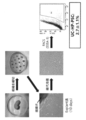
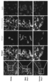
- Embryoid-like cell clusters of human umbilical cord-derived SSEA-3 positive cells are adherently cultured on gelatin-coated dishes, spontaneously differentiated, and tri-embryoid markers by immunostaining or RT-PCR.
- the figure which shows the result of having investigated the expression photo.
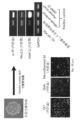
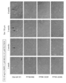
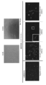
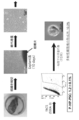
- human umbilical cord-derived SSEA-3 positive cells (UC-HP-PSC) and various Muse cells were induced to differentiate into trophoblast cell lines (conditions: 10 ng / mL BMP4, 1 ⁇ M A83-01, 0.1 ⁇ M PD173074)
- the figure which shows the morphology of a cell photo.
- Human umbilical cord-derived SSEA-3 positive cells (UC-HP-PSC) and various Muse cells induced to differentiate into trophoblast cell lineages (conditions: 10 ng / mL BMP4, 20 ⁇ M SB431542, 20 ⁇ M SU5402)
- the figure which shows the morphology (photo).
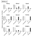
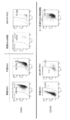
- human umbilical cord-derived SSEA-3 positive cells (UC-HP-PSC) and various Muse cells were induced to differentiate into trophoblast cell lines (conditions: 10 ng / mL BMP4, 1 ⁇ M A83-01, 0.1 ⁇ M PD173074)
- Trophoblast when human umbilical cord-derived SSEA-3 positive cells (UC-HP-PSC) and various Muse cells are induced to differentiate into trophoblast cell lines conditions: 10 ng / mL BMP4, 20 ⁇ M SB431542, 20 ⁇ M SU5402)
- the figure which shows the result of having examined the expression of a marker by quantitative PCR The figure which shows the result of having examined the expression of CD133 in NTERA-2 cell which is a human pluripotent embryonic cancer cell line.
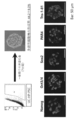
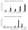
- the expression of germline markers when human umbilical cord-derived SSEA-3 positive cells (UC-HP-PSC) were induced to differentiate according to the differentiation-inducing conditions that induce the differentiation of iPS cells into primordial germ cells was examined by quantitative PCR.
- the figure which shows the result of having performed the immunostaining against Blimp1 which is a marker (photo).
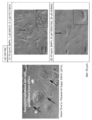
- Embryoid-like cell clusters of human amniotic membrane-derived SSEA-3 positive cells (AM-HP-PSC) were adherently cultured on gelatin-coated dishes and observed and immunostained with endoderm markers, mesoderm markers, and ectoderm markers.
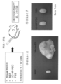
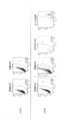
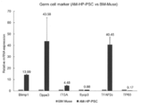
- Human placenta-derived high-potential pluripotent stem cells (P-HP-PSC) phagocytose male mouse primordial germline to induce differentiation into germline and express germline early and late markers.
- the figure which shows the result of evaluation by quantitative PCR Human placenta-derived high-potential pluripotent stem cells (P-HP-PSC) phagocytose female mouse primordial germline to induce differentiation into germline, expressing early and late germline markers.
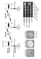
- solvent only Control
- human bone marrow-derived Muse cells BM-Muse
- human umbilical cord-derived high-potency pluripotent stem cell fraction UC-HP-PSC
- human bone marrow-derived SSEA-3 negative MSC The figure which shows the ejection fraction (EF) when (BM non-Muse) was administered.
- UC-HP-PSC Human umbilical cord-derived high-potency pluripotent stem cells
- BM-Muse human bone marrow-derived Muse cells
- ADSC-Muse human adipose-derived Muse cells
- NHDF-Muse human skin-derived Muse cells
- the present invention relates to high-potency pluripotent stem cells isolated from extraembryonic tissues and the like, and cell preparations containing the high-potency pluripotent stem cells.
- the present invention will be described in detail below.
- High-potency pluripotent stem cells used in the cell preparation of the present invention are high-potency pluripotent stem cells isolated from extraembryonic tissues, etc., and are shown below. It has the useful properties of pluripotent stem cells similar to those of Muse cells, and in addition, it has the ability to differentiate extraembryonic tissues into cells and / or germline, and has a differentiation potential close to totipotency. In addition, it has the property of being positive for CD133, which is a cell surface marker that is negative for Muse cells obtained from conventional bone marrow, peripheral blood, fat, skin, and the like.
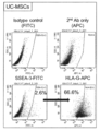
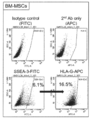
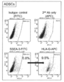
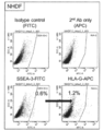
- the human umbilical cord-derived SSEA-3 positive cells obtained in Example 1 are Muse cells obtained from conventional bone marrow, peripheral blood, fat, skin, etc. by other analysis of Example 1. It has the same useful properties of pluripotent stem cells as, and in addition, it has the ability to differentiate extraembryonic tissue into cells and / or germline, and has near totipotency. Furthermore, it has the property that the cell surface marker CD133 is 99.9% or more positive. For this reason, the human umbilical cord-derived SSEA-3 positive cells obtained in Example 1 are referred to as "human umbilical cord-derived high potential pluripotent stem cells" and are abbreviated as UC-HP-PSC (Umbilical cord High potential Pluripotent stem cells).
- the human sheep membrane-derived SSEA-3 positive cells obtained in Example 2 are useful as pluripotent stem cells similar to Muse cells obtained from conventional bone marrow, peripheral blood, fat, skin, etc. by other analysis. In addition to having properties, it has the ability to differentiate extraembryonic tissues into cells and / or germ cell lines, has near totipotency differentiation potential, and further has the cell surface marker CD133. It has properties such as being positive by about 70% or more. For this reason, the human amniotic membrane-derived SSEA-3 positive cells obtained in Example 2 are referred to as "human amniotic membrane-derived high-potency pluripotent stem cells" or "human amniotic membrane-derived high-potency pluripotent stem cell fraction".
- AM-HP-PSC Amniotic membrane High potential Pluripotent stem cells
- human placenta-derived SSE-3 positive cells obtained in Example 3 are useful as pluripotent stem cells similar to Muse cells obtained from conventional bone marrow, peripheral blood, fat, skin, etc. by other analyzes.
- it has the ability to differentiate extraembryonic tissues into cells and / or germ cell lines, has near totipotency differentiation potential, and the surface marker CD133 is about. It has properties such as being 80% or more positive.
- the human placenta-derived SSEA-3 positive cells obtained in Example 3 are referred to as "human placenta-derived high-potency pluripotent stem cells” or “human placenta-derived high-potency pluripotent stem cell fraction", and are referred to as P-. It may be abbreviated as HP-PSC (Placenta High potential Pluripotent stem cells).
- HP-PSC Peaka High potential Pluripotent stem cells
- the high-potential pluripotent stem cells of the present invention exist among the conventional bone marrow-derived SSEA-3 positive cells, although only a small amount (up to about several percent). Therefore, the high-potential pluripotent stem cells of the present invention can be obtained not only from extraembryonic tissues but also from conventional bone marrow, peripheral blood, fat, skin and the like.
- the high-potential pluripotent stem cells of the present invention can be regarded as mainly some special Muse cells isolated from extraembryonic tissues. That is, the high-potency pluripotent stem cell of the present invention has the same useful properties of pluripotent stem cells as Muse cells, which means the properties of Muse cells described below.
- Muse cells can be obtained from bone marrow fluid, adipose tissue (Non-Patent Document 4), dermal connective tissue of skin, etc., and are widely present in connective tissue of organs and peripheral blood. ..
- this cell is a cell having both pluripotent stem cell and mesenchymal stem cell properties, and is, for example, a cell surface marker "SSEA-3" positive cell, preferably SSEA-3 positive and CD105 positive. Identified as double positive cells. Therefore, a Muse cell or a cell population containing a Muse cell can be separated from a living tissue using, for example, SSEA-3 alone or the expression of SSEA-3 and CD105 as an index.
- Muse cells can be selectively concentrated by culturing under various external stress conditions such as the presence of a substance, the presence of active oxygen, mechanical stimulation, and pressure treatment.
- SSEA-3 a cell population containing pluripotent stem cells (Muse cells) or Muse cells prepared from living mesenchymal tissue or cultured mesenchymal tissue is simply referred to. It may be described as "SSEA-3 positive cells”.
- Muse cells or cell populations containing Muse cells can be prepared from living tissues (for example, mesenchymal tissues) using the cell surface markers SSEA-3 or SSEA-3 and CD105 as indicators.
- the "living body” refers to a living body of a mammal. In the present invention, the living body does not include a fertilized egg or an embryo at a developmental stage before the blastogenic stage, but includes an embryo at a developmental stage after the blastogenic stage including a foetation or a blastoblast.
- Mammals include, but are not limited to, primates such as humans and monkeys, rodents such as mice, rats, rabbits and guinea pigs, cats, dogs, sheep, pigs, cows, horses, donkeys, goats, ferrets and the like. Be done. Muse cells used in the cell preparation of the present invention are clearly distinguished from embryonic stem cells (ES cells) and iPS cells in that they are separated directly from living tissues with markers. "Membranous tissue” refers to tissues such as bone, synovium, fat, blood, bone marrow, skeletal muscle, dermatitis, ligament, tendon, dental pulp, umbilical cord, umbilical cord blood, sheep membrane, and tissues existing in various organs. say.
- Muse cells can be obtained from bone marrow, skin, adipose tissue, blood, dental pulp, umbilical cord, umbilical cord blood, amniotic membrane and the like. For example, it is preferable to collect mesenchymal tissue of a living body, prepare Muse cells from this tissue, and use it.
- Muse cells may be prepared from cultured mesenchymal cells such as fibroblasts and bone marrow mesenchymal stem cells using the above-mentioned preparation means.
- the cell population containing Muse cells selectively proliferates cells resistant to the external stress by giving an external stress stimulus to the mesenchymal tissue of the living body or the cultured mesenchymal cells, and the abundance ratio thereof. It can also be prepared by methods involving the recovery of cells that have been enriched.
- the external stress is protease treatment, culture at low oxygen concentration, culture under low phosphoric acid condition, culture at low serum concentration, culture under low nutrition condition, culture under heat shock exposure, low temperature.
- the total treatment time with the protease is preferably 0.5 to 36 hours in order to apply external stress to the cells.
- the protease concentration may be a concentration used when peeling cells adhered to a culture vessel, breaking up a cell mass into a single cell, or recovering a single cell from a tissue.
- the protease is preferably a serine protease, an aspartic protease, a cysteine protease, a metal protease, a glutamate protease or an N-terminal threonine protease. Further, it is preferable that the protease is trypsin, collagenase or dispase.
- Muse cells may be autologous or allogeneic to the recipient undergoing cell transplantation.
- a Muse cell or a cell population containing a Muse cell can be prepared from a living tissue using, for example, SSEA-3 positive or double positive of SSEA-3 and CD105 as an index.
- Human adult skin is known to contain various types of stem cells and progenitor cells, such stem cells and progenitor cells include skin-derived progenitor cells (SKP), neural ridge stem cells (NCSC), and melanoblasts. (MB), perivascular cells (PC), endothelial progenitor cells (EP), adipose-derived stem cells (ADSC). Since Muse cells are not the same as these cells, Muse cells can be prepared using the "non-expression" of a marker unique to these cells as an index.
- SSEA-3 positive or double positive of SSEA-3 and CD105 as an index.
- Human adult skin is known to contain various types of stem cells and progenitor cells, such stem cells and progenitor cells include skin-derived progenitor cells (SKP), neural ridge stem cells (NCSC),
- Muse cells include CD34 (markers for EP and ADSC), CD117 (c-kit) (markers for MB), CD146 (markers for PC and ADSC), CD271 (NGFR) (markers for NCSC), NG2 (PC marker), vWF factor (Fonville brand factor) (EP marker), Sox10 (NCSC marker), Snai1 (SKP marker), Slug (SKP marker), Tyrp1 (MB marker), and At least one of 11 markers selected from the group consisting of Dct (MB markers), eg, 2, 3, 4, 5, 6, 7, 8, 9, 10 The non-expression of 11 or 11 markers can be separated as an index.
- the non-expression of CD117 and CD146 can be used as an index
- the non-expression of CD117, CD146, NG2, CD34, vWF and CD271 can be used as an index
- the non-expression of 11 markers can be used as an index for preparation.
- Muse cells having the above characteristics are as follows: (I) Low or no telomerase activity; (Ii) Has the ability to differentiate into cells of any of the three germ layers; It may have at least one property selected from the group consisting of (iii) no neoplastic growth; and (iv) capable of self-renewal.
- the Muse cells used in the present invention have all of the above properties.
- "the telomerase activity is low or absent" means that, for example, when the telomerase activity is detected using TRAPEZE XL telomerase detection kit (Millipore), it is low or cannot be detected. say.
- telomerase activity means, for example, telomerase having the same level of telomerase activity as somatic human fibroblasts, or 1/5 or less, preferably 1/10 or less of that of Hela cells. It means having activity.
- Muse cells have the ability to differentiate into three embryos (endometrial, mesophyll, and ectodermal) in vitro and in vivo, and are, for example, induced and cultured in vitro. Can differentiate into hepatocytes (including hepatoblasts or cells expressing hepatocyte markers), nerve cells, skeletal muscle cells, smooth muscle cells, bone cells, fat cells and the like.
- Muse cells proliferate at a proliferation rate of about 1.3 days, but in suspension culture, they proliferate from one cell, and when they form an embryoid body-like cell mass and reach a certain size, they proliferate in about 14 days.
- Muse cells have a self-renewal (self-renewal) ability.
- self-renewal means that when cells contained in an embryoid body-like cell mass obtained by culturing one Muse cell in suspension culture are transferred to adhesive culture, differentiation into three embryoid cells occurs.
- the high-potency pluripotent stem cells used in the cell preparation of the present invention are high-potency pluripotent stem cells isolated from extraembryonic tissues and the like, but the pluripotent stem cells similar to the Muse cells shown above are useful. In addition, it has the ability to differentiate extraembryonic tissues into cells and / or germline, and has near totipotency, and also has conventional bone marrow, peripheral blood, etc. It has properties such as being positive for CD133, which is a cell surface marker that is negative for Muse cells obtained from fat, skin, and the like. CD133 is a cell surface marker that is one of the glycoproteins and is also known as Prominin1.
- CD133 positive means cells stained when cell staining is performed using the CD133 antibody or cells selected by the CD133 antibody by flow cytometry.
- the high-potential pluripotent stem cells of the present invention can be a cell population containing CD133-positive cells, preferably containing 50% or more of CD133-positive cells, more preferably 60% or more, 70% or 80% or more. It is preferable, and it is more preferable to contain 90% or more.
- the positive cell surface marker CD133 can be determined by an identification method using an ordinary antibody or the like.
- the high-potential pluripotent stem cell of the present invention can be obtained from extraembryonic tissue or the like.
- the extraembryonic tissue and the like are umbilical cord, umbilical cord blood, placenta, placenta blood, decidua, chorionic villi, amniotic fluid, amniotic fluid, etc. Obtainable.
- the high-potency pluripotent stem cells of the present invention can be differentiated into all germ layers (germ layer, mesodermal, and endoderm trigerm) constituting an individual, and further, cells of extraembryonic tissue and / or reproduction. It has the ability to differentiate into cell lines and has the ability to differentiate to near totipotency.
- “having the ability to differentiate extraembryonic tissue into cells and / or germ cell lineage, and having near totipotency” means the germ cell lineage such as the placenta, which is extraembryonic tissue. It means the ability to differentiate into a variety of cells, including a spouse (sperm or egg) and its underlying primordial germ cell (PGC).
- PPC primordial germ cell
- the ability of extraembryonic tissue to differentiate into cells and / or germ cell lineages is the differentiation of each extraembryonic tissue into various cells, differentiation into vegetative membrane cell lineage, cell multinucleation, and vegetative membrane marker.
- ERVW-1 human Chorionic gonadotropin alpha chain (hCGA), CDX2, TP63, ID2, etc. as Early Maker, GCM1, PGF, ERVFRD-1, etc.
- Late maker involved in the differentiation of germ cell lineage Gene expression (eg Blimp1, Dappa3, ITGA, Sycp3, TBX3, TFAP2c, TP63, Nanos3, PRDM14, SSEA-1, Daz1, Stra8, etc.), expression of genes suggesting pluripotency (eg Oct3 / 4) , Nanog, Sox2, KLF2, KLF4, REX1, TERT, etc.).
- germ cell lineage Gene expression eg Blimp1, Dappa3, ITGA, Sycp3, TBX3, TFAP2c, TP63, Nanos3, PRDM14, SSEA-1, Daz1, Stra8, etc.
- pluripotency eg Oct3 / 4
- Nanog Sox2, KLF2, KLF4, REX1, TERT, etc.
- the high-potential pluripotent stem cells of the present invention can be obtained by a conventional method for separating Muse cells from extraembryonic tissue or the like (International Publication No. WO2011 / 007900).
- extraembryonic tissue or the like was cut into small pieces, the tissue pieces were cultured, and mesenchymal cells derived from extraembryonic tissue were cultured and increased until an effective cell mass was reached. Later, (i) SSEA-3 alone, (ii) SSEA-3 and CD105 double, (iii) SSEA-3 and CD133 double, or (iv) SSEA-3, CD105 and CD133 triple antigen markers are used as indicators.
- the high-potency pluripotent stem cells of the present invention can be obtained according to the method for separating from embryonic tissues such as bone marrow, peripheral blood, fat and skin, and from the above-mentioned extraembryonic tissues.
- the high-potential pluripotent stem cells of the present invention are cells having a differentiation potential close to that of totipotency by examining the characteristics of the Naive-type or Primed-type pluripotent stem cells shown in Table 1 below. You can confirm that there is. ⁇ References: 1) L. Weinberger, M. Ayyash, N. Novershtern, J. H. Hanna, Dynamic stem cell states: naive to primed pluripotency in rodents and humans. Nat. Rev. Mol. Cell Biol. 17, 155-169 (2016). 2) M. Ueda, Y. Takashima, History of Pluripotent Stem Cells and Human Naive Pluripotent Stem Cells. Cytometry Research 27, 19-24 (2017). 3) Kiichiro Tomoda, Biochemistry, Vol. 90, No. 2, p187-191 (2018)>
- the high-potency pluripotent stem cells of the present invention are also expressed in early embryos and pluripotent stem cells during normal development, and are essential molecules for maintaining pluripotency and self-renewal in human pluripotent stem cells and the like. It can be confirmed by the expression of desmoglein2, desmocollin2,3, plakoglobin, etc., which are considered to be desmosome-related molecules.
- the cell preparation of the present invention is not limited, but the high-potency pluripotent stem cells of the present invention obtained in (1) above or a cell population containing the cells are appropriately used in physiological saline or the like. It is obtained by suspending the product in a buffer solution (for example, phosphate buffered saline).
- a buffer solution for example, phosphate buffered saline.
- the cells may be cultured before cell transplantation and proliferated until a predetermined number of cells is obtained. ..
- the high-potency pluripotent stem cells of the present invention do not have neoplasticity, even if cells recovered from living tissues are contained in an undifferentiated state, the possibility of canceration is low and it is safe.
- the recovered high-potential pluripotent stem cells of the present invention are cultured in a normal growth medium (for example, ⁇ -minimum essential medium ( ⁇ -MEM) containing 10% calf serum), although the culture is not particularly limited. Can be done.
- a normal growth medium for example, ⁇ -minimum essential medium ( ⁇ -MEM) containing 10% calf serum
- WO2011 / 007900 pamphlet and appropriately select a medium, additives (for example, antibiotics, serum) and the like in the culture and proliferation of the high-potency pluripotent stem cells of the present invention. Then, a solution containing a predetermined concentration of the high-potency pluripotent stem cells of the present invention can be prepared.
- a human extracorporeal tissue is collected, and for example, umbilical cord tissue-derived mesenchymal stem cells are cultured as adherent cells from the umbilical cord tissue to provide an effective therapeutic amount.
- High-potential pluripotent stem cells can be prepared as cell preparations.
- the high potential pluripotent stem cells of the present invention are proliferated by culturing mesenchymal stem cells derived from extraembryonic tissue obtained from human extraembryonic tissue under external stress conditions until an effective therapeutic amount is reached.
- autologous or allogeneic high-potency pluripotent stem cells of the present invention can be prepared as cell preparations.
- DMSO dimethylsulfoxide
- serum albumin etc.
- antibiotics are used to prevent bacterial contamination and proliferation.
- Etc. may be contained in the cell preparation.
- other pharmaceutically acceptable ingredients eg, carriers, excipients, disintegrants, buffers, emulsifiers, suspensions, soothing agents, stabilizers, preservatives, preservatives, saline, etc.
- It may be contained in a cell preparation. Those skilled in the art can add these factors and agents to the cell preparation at appropriate concentrations.
- the high-potential pluripotent stem cells of the present invention can also be used as a pharmaceutical composition containing various additives.
- the dosage form of the cell preparation is not particularly limited, but is preferably a parenteral administration preparation, and more preferably an injection preparation.
- the number of high-potential pluripotent stem cells of the present invention contained in the cell preparation prepared above is used according to the sex, age, body weight, condition of the affected area, and the subject so as to obtain the desired effect in the disease to be treated. It can be adjusted as appropriate in consideration of the state of cells and the like.
- the target individuals include, but are not limited to, mammals such as humans.
- the cell preparation of the present invention may be administered once, but may be administered multiple times (for example, 2 to 10 times) and at appropriate intervals (for example, twice a day, one day) until a desired therapeutic effect is obtained. Once once a week, once a week, once every two weeks, once a month, once every two months, once every three months, once every six months) May be done.
- the time of administration may be any of an acute phase, a subacute phase, and a chronic phase as long as a therapeutic effect can be obtained, and may be two or more types selected from these.
- the therapeutically effective amount depends on the condition of the subject, but for example, a dose of 1 ⁇ 10 3 cells to 1 ⁇ 10 10 cells per individual is preferable at 1 to 10 times.
- the total dose to be administered in one individual is not limited, but is limited to 1 ⁇ 10 3 cells to 1 ⁇ 10 11 cells, preferably 1 ⁇ 10 4 cells to 1 ⁇ 10 10 cells, and more preferably 1 ⁇ 10 5 cells to 1 ⁇ 10. 9 cells and the like can be mentioned.
- the high-potency pluripotent stem cells of the present invention used in the cell preparation of the present invention have the property of migrating to the damaged site of the disease to be treated and engrafting. Therefore, in the administration of the cell preparation, the administration site and administration method of the cell preparation are not limited, and the cell preparation may be locally administered to the affected area, or may be administered intravenously or the like.
- the cell preparation of the present invention can reconstruct the tissue of the damaged site of the patient of the disease to be treated, and can improve or restore the lost function of the damaged site of the patient.
- Example 1-1 Isolation of high-potency pluripotent stem cells derived from human umbilical cord High-potency pluripotency from human umbilical cord according to the method described in International Publication No. WO2011 / 007900 on the isolation and identification of human Muse cells. Sex stem cells can be obtained. More specifically, human umbilical cord-derived high-potential pluripotent stem cells were obtained by isolation by FACS using SSEA-3 as an index by the step of FIG. Specifically, a 35-week-old human umbilical cord was cut into 0.5 mm square pieces, and tissue pieces were placed on a 10 cm dish at 1 cm intervals.
- ⁇ MEM 10% FBS, 1 ng / mL bFGF, and 2 mM GlutaMAX were added, and the cells were cultured by the explant method for about 10 days.
- SSEA-3-positive cells derived from human umbilical cord tissue using an anti-SSEA-3 antibody that is, high-potential pluripotency derived from the human umbilical cord of the present invention.
- Stem cells UC-HP-PSC were isolated. About 3% of SSEA-3 positive cells were endogenous to the established human umbilical cord-derived mesenchymal stem cells.
- Example 1-2 High-potential pluripotent stem cells derived from human umbilical cord co-express mesenchymal cell surface markers FACS the cell surface markers of human umbilical cord-derived SSEA-3 positive cells obtained in Example 1-1 Analyzed in. As a result, as shown in FIG. 2, human umbilical cord-derived SSEA-3 positive cells co-expressed mesenchymal stem cell markers CD105, CD90, CD44, CD73, and CD166. However, the hematopoietic markers CD34 and CD45, the neural crest-derived stem cell marker CD271, and the endothelial precursor marker von Willebrand factor (vWF) were negative.
- vWF von Willebrand factor
- Example 1-3 High-potential pluripotent stem cells derived from human umbilical cord have the following properties similar to conventional human bone marrow-derived Muse cells (1) High-potential pluripotent stem cells derived from human umbilical cord are pluripotent Forming cell clusters Single-cell suspension culture was performed using human umbilical cord-derived SSEA-3-positive cells obtained by FACS of Example 1-1. On the 7th day of culture, 44.2% of SSEA-3 positive cells formed embryoid body-like cell clusters (Fig. 3, top). This cell mass was freeze-embedded and sliced, and immunostained against the pluripotent markers Nanog, Oct3 / 4, Sox2, PAR4, and Tra-1-81. As a result, as shown in the lower part of FIG. 3, the embryoid body-like cell mass expresses all pluripotency markers, and the embryoid body-like cell mass formed from the human umbilical cord-derived SSEA-3 positive cells is numerous. It was confirmed to be a pluripotent cell mass.
- Human umbilical band-derived high-potency pluripotent stem cells differentiate into trigermoid cells by differentiation induction Using a dish in which human umbilical band-derived SSEA-3 positive cells are coated with poly-HEMA for nerve induction.
- the cells were cultured in Neurobasal medium (Gibco) containing B-27 supplement at a density of 1 ⁇ 10 5 cells / mL for 7 days to form spheres (spherical cell clusters).
- human umbilical cord-derived SSEA-3 positive cells were cultured at a density of 4 ⁇ 10 3 cells / cm 2 for 16 days using the bone cell induction medium of the Human Mesenchymal Stem Cell Functional Identification Kit (R & D Systems). ..
- human umbilical cord-derived SSEA-3 positive cells were cultured at a density of 1 ⁇ 10 4 cells / cm 2 for 16 days using the adipocyte induction medium of the Human Mesenchymal Stem Cell Functional Identification Kit (R & D Systems). .. Human umbilical cord-derived SSEA-3 positive cells were seeded on collagen-coated plates at a density of 1 ⁇ 10 4 cells / cm 2 for hepatocyte induction. After sowing, the cells were cultured in DMEM-low glucose, 20 ng / mL EGF, and 10 ng / mL bFGF for 2 days.
- DMEM-low glucose 0.5 mg / mL BSA, 1 ⁇ insulin-transferrin-selenium, 5 mM nicotinamide, 10 nM dexamethasone, 150 ng / mL HGF, 50 ng / mL FGF4 for 28 days.
- DMEM-low glucose 0.5 mg / mL BSA, 1 ⁇ insulin-transferrin-selenium, 5 mM nicotinamide, 10 nM dexamethasone, 150 ng / mL HGF, 50 ng / mL FGF4 for 28 days.
- DMEM-low glucose 0.5 mg / mL BSA
- 1 ⁇ insulin-transferrin-selenium 5 mM nicotinamide
- 10 nM dexamethasone 150 ng / mL HGF
- 50 ng / mL FGF4 50 ng / mL FGF4 for 28
- Nerve induction formed a sphere, which was confirmed to be positive for the neural stem cell markers NeuroD1, Nestin, and Musashi-1.
- Expression of Osteocalcin was observed by osteocytoduction, FABP-4 was observed by adipocyte induction, and CD141 was observed by vascular endothelial cell induction.
- Expression of ⁇ -fetoprotein, Albumin, and cytokeratin 19 (CK19) was confirmed by hepatocyte induction.
- the cell mass adherently cultured on the gelatin-coated dish was adherently cultured for about 10 days, and then RNA was extracted.
- ⁇ -fetoprotein ⁇ -FP, endoderm marker
- Nkx2.5 meoderm marker
- MAP-2 microtubule-associated protein
- High-potential pluripotent stem cells derived from human umbilical cord do not form tumors even when transplanted into living organisms
- immunodeficient mice were used. 1 ⁇ 10 5 human umbilical cord-derived SSEA-3 positive cells were transplanted into the testis of a CB17 / IcrJcl-Prkdc scid (male, 2 months old) using a glass microtube.
- a control group 1 ⁇ 10 5 cells of mouse ES cells (positive control), 1 ⁇ 10 5 cells of mitomycin C-treated MEF (mouse embryonic fibroblasts) and PBS (negative control) were transplanted.
- mouse ES cells formed teratomas 3 months after transplantation, whereas human umbilical cord-derived SSEA-3 positive cells did not form teratomas even 6 months after transplantation. This indicates that human umbilical cord-derived SSEA-3 positive cells do not have tumorigenicity.
- Example 1-4 High-potential pluripotent stem cells derived from human umbilical cord tissue have the following properties not found in conventional bone marrow, fat, skin-derived Muse cells, etc.
- Induction ES cells and iPS cells are induced to differentiate into the vegetative membrane cell lineage.
- the differentiation of human umbilical cord-derived SSEA-3 positive cells obtained in Example 1-1 into the vegetative membrane cell lineage is induced. It was confirmed by the following two induction methods.
- Muse cells derived from human bone marrow (BM-Muse), human fat (ADSC-Muse), and human skin (NHDF-Muse) were used as a control group.
- DMEM-low-glucose 2% FBS, 10 ng / mL BMP4, 1 ⁇ M A83-01, 0.1 ⁇ M PD173074, [2].
- DMEM-low-glucose, 2% FBS, 10 ng / mL BMP4, 20 ⁇ M SB431542, 20 ⁇ M SU5402 were added and cultured for 3 weeks (Yabe, S. et al. Proc. Natl. Acad. Sci. USA 113, E2598-2607 (2016), Sudheer, S et al. Stem Cells Dev 21, 2987-3000 (2012)).
- High-potential pluripotent stem cells derived from human umbilical cord are multinucleated cells in which syncytiotrophoblast-like cells of the placenta appear by the differentiation induction method described in [1] and [2] above.
- FIG. 8 in human umbilical cord-derived SSEA-3 positive cells, multinucleated cells appeared after 2 weeks by the induction method [1] and after 3 weeks by the induction method [2]. It also closely resembles the morphology of syncytiotrophoblast-like cells made from human ES cells.
- Muse cells derived from bone marrow (BM), fat (ADSC), and skin (NHDF) no polynuclear cells were observed by any of the induction methods.
- trophoblast markers CDX2, GCM1 and ERVFRD-1 was evaluated by quantitative PCR 3 days, 5 days, 1, 2 and 3 weeks after induction.
- CDX2 which is an early marker
- GCM1 and ERVFRD-1 which are late markers
- trophoblast markers is higher in all of the induction methods [2] than in the induction method [1], and it is considered that the induction method is more efficient.
- BM bone marrow
- ADSC fat
- NHDF skin
- Example 1-5 High-potency pluripotent stem cells derived from human umbilical cord tissue are positive for the cell surface marker CD133
- CD133 is a cell surface marker that is one of glycoproteins and is also known as Prominin1. Is. Its function is still unknown, but it has been clarified that it is expressed in some stem / progenitor cells such as hematopoietic stem cells and neural stem cells.
- the expression of CD133 in the human umbilical cord-derived SSEA-3 positive cells obtained in Example 1-1 was confirmed by FACS. Bone marrow-derived Muse cells were used as a comparison group, and NTERA-2, a pluripotent human fetal cancer cell line, was used as a positive control. As a result, as shown in FIGS.
- SSEA-3 positive cells of NTERA-2 were CD133 positive
- SSEA-3 of human umbilical cord-derived SSEA-3 positive cells 99.9% of the positive cells were CD133 positive.
- bone marrow-derived Muse cells 2.0% of SSEA-3-positive cells were CD133-positive, which was much lower than that of human umbilical cord-derived SSEA-3-positive cells.
- Example 1-6 High-potential pluripotent stem cells derived from human umbilical cord tissue have the ability to differentiate into germ cell lineages (1) Induce differentiation into primordial germ cells Induce iPS cells to differentiate into primordial germ cells: According to the differentiation induction conditions, the induction of differentiation of the human umbilical cord-derived SSEA-3 positive cells obtained in Example 1-1 into primordial germ cells was confirmed by the following induction method. Human bone marrow-derived Muse cells were used as a control group, and primordial germ cells (iPS-PGC) derived from iPS cells were used as a positive control group.
- iPS-PGC primordial germ cells
- Human umbilical cord-derived SSEA-3 positive cells or human bone marrow-derived Muse cells were seeded in a dish at a density of 5 ⁇ 10 4 cells / cm 2 , DMEM-low-glucose, 10% FBS, 50 ng / mL Activin A, 3 ⁇ M CHIR99021, 10 ⁇ M Y-27632 was added, and the cells were adherently cultured for 2 days.
- Blimp1 and Nanos3 were detected after 4 days of induction. Furthermore, Dppa3 expression was observed 6 days after induction, and SSEA-1 expression was also increased. On the other hand, although the expression of some markers such as Nanos3 and Dppa3 was observed in bone marrow-derived Muse cells, the values of Blimp1 and SSEA-1 were lower than those of human umbilical cord-derived SSEA-3 positive cells. In addition, 4 days and 6 days after induction, the cells were fixed with 4% PFA / 0.1M phosphate buffer and immunostained for the germline marker Blimp1. The result is shown in FIG.
- Blimp1 was detected in human umbilical cord-derived SSEA-3 positive cells, but was hardly expressed in bone marrow-derived Muse cells. This indicates that human umbilical cord-derived SSEA-3 positive cells have the ability to differentiate into more efficient germline compared to bone marrow-derived Muse cells.
- High-potential pluripotent stem cells derived from human umbilical cord use commercially available human umbilical cord-derived SSEA-3 positive cells that more efficiently induce differentiation into germline by phagocytosing dead primordial germ cells.
- the induction of differentiation into primordial germ cells was confirmed by the following induction method.
- Human umbilical cord-derived SSEA-3 positive cells were seeded in a dish at a density of 5 ⁇ 10 3 cells / cm 2 , and then 1 ⁇ 10 male or female mouse primordial germ cells were killed with 100 ⁇ M Rotenone and 100 ⁇ M Oligomycin A. It was added at a density of 4 cells / cm 2 and fed in DMEM-low-glucose, 10% FBS for 3 days. After that, the following three types of induction were attempted.
- ⁇ 1> After removing the cell-dead mouse primordial germ cells, they were adherently cultured in DMEM-low-glucose, 10% FBS for 11 days.
- Human umbilical band-derived SSEA-3 positive cells were seeded in a dish at a density of 5 ⁇ 10 3 cells / cm 2 , and then somatic cells in reproductive ridge other than primordial germ cells treated with mitomycin C were 5 ⁇ 10 3 cells.
- the cells were adherently cultured in DMEM-low-glucose, 10% FBS for 11 days in addition to a density of / cm 2.
- ⁇ 3> Using a Low-cell binding V-bottom 96-well plate, 5 ⁇ 10 3 cells / well of human umbilical cord-derived SSEA-3 positive cells, 5 somatic cells in reproductive ridge other than primordial germ cells treated with mitomycin C The cells were suspended so as to be ⁇ 10 4 cells / well, and suspended-cultured in DMEM-low-glucose, 10% FBS for 11 days. Expression of germline markers PRDM14, Blimp1, Dppa3, Dazl, Nanos3, SSEA-1, Stra8, and Sycp3 was evaluated by quantitative PCR at 3, 5, 7, and 14 days after induction. Primordial germ cell-like cells (PGCLC) derived from human iPS cells were used as the positive control group.
- PPCLC Primordial germ cell-like cells
- FIGS. 14-1 and 14-2 The results are shown in FIGS. 14-1 and 14-2.
- increased expression was observed with markers such as Blimp1, Dppa3, and Nanos3 regardless of whether the primordial germ cells of both sexes were phagocytosed.
- the expression was higher in some markers than in the induction method ⁇ 1>, but the expression of many markers was similar.
- the expression of all markers was high 5 days after the induction, indicating that it is a more efficient method for inducing differentiation into germline than other induction methods. However, after 7 days, the number of cells dying due to the effect of suspension culture increased, and the expression decreased.
- the human umbilical cord-derived SSEA-3 positive cells obtained in Example 1 above are pluripotent stem cells similar to Muse cells obtained from conventional bone marrow, peripheral blood, fat, skin, etc. by other analysis of Example 1. In addition, it has the ability to differentiate extraembryonic tissues into cells and / or germline, and has near totipotency, and is a cell surface marker. It has properties such as being positive for CD133. From this, CD133-positive pluripotent stem cells having the ability to differentiate into cells of extraembryonic tissue and / or germ cell lineage exemplified by the human umbilical cord-derived SSEA-3 positive cells obtained in Example 1 above.
- high potential pluripotent stem cell As a novel SSEA-3 positive pluripotent stem cell, in the present invention, it is referred to as “high potential pluripotent stem cell”, and in particular, a cell derived from human umbilical cord is referred to as “human umbilical cord-derived high potential pluripotent stem cell”.
- Example 2-1 Isolation of high-potency pluripotent stem cells derived from human amniotic membrane High-potency pluripotency from human amniotic membrane according to the method described in International Publication No. WO2011 / 007900 on the isolation and identification of human Muse cells. Obtained sex stem cells. After expanding and subculturing the obtained human amniotic membrane-derived mesenchymal stem cells, SSEA-3 positive cells derived from human amniotic membrane (that is, human amniotic membrane-derived high-potency pluripotent stem cells: AM) were used using an anti-SSEA-3 antibody. -HP-PSC) was isolated. As a result, as shown in FIG.
- Example 2-2 Examination of properties of high-potential pluripotent stem cells derived from human sheep membrane (1) Clusters of high-potential pluripotent stem cells derived from human sheep membrane during adhesion culture and trigerm differentiation Differentiation into trigerm cells To confirm the ability, pluripotent cell clusters of single human sheep membrane-derived SSEA-3 positive cells were transferred to gelatin-coated dishes and adherently cultured. Approximately 10 days later, immunostaining detected cytokeratin 7 (CK7, endoderm marker) and ⁇ -smooth muscle actin (SMA, mesoderm marker) nerve filament-M (ectoderm marker) (Fig. 17). From these results, it was shown that human amniotic membrane-derived SSEA-3 positive cells have the ability to spontaneously differentiate into trigermoid cells.
- CK7 cytokeratin 7
- SMA ⁇ -smooth muscle actin
- Human amniotic mesenchymal stem cells MSC
- human amniotic membrane-derived high-potential pluripotent stem cells Human amniotic membrane-derived SSEA-3 positive cells have a double positive rate of SSEA-3 and CD105 of 81. The double positive rate of SSEA-3 and CD90 was 84.0%, and the double positive rate of SSEA-3 and CD44 was 74.2%. In addition, the human amniotic membrane-derived SSEA-3 positive cells had a double positive rate of SSEA-3 and CD133 of 71.5% (FIGS. 18 and 19).
- Human amnion-derived MSCs and human amnion-derived SSEA-3-positive cells like bone marrow-derived MSCs and Muse cells, hardly express blood cell surface markers such as CD34 and CD45 (FIG. 20).
- Prime-type pluripotency markers (Oct3 / 4 / Nanog / Sox2) Oct3 / 4, Nanog, Sox2, which are prime pluripotency markers in human amniotic membrane-derived SSEA-3 positive cells, human amniotic membrane-derived SSEA-3 negative cells (AM-non-Muse), and human bone marrow-derived Muse cells by quantitative PCR. Expression was compared and analyzed. Human amniotic membrane-derived SSEA-3 positive cells showed higher expression of the prime-type pluripotency marker than bone marrow Muse cells (Fig. 22). This result suggests that human amniotic membrane-derived SSEA-3 positive cells are more pluripotent than bone marrow Muse and human amniotic membrane-derived SSEA-3 negative cells.
- KLF2 / KLF4 / REX1 Naive pluripotency marker (KLF2 / KLF4 / REX1) Expression of naive pluripotency markers KLF2, KLF4, REX1 in human amniotic membrane-derived SSEA-3 positive cells, human amniotic membrane-derived SSEA-3 negative cells (AM-non-Muse) and human bone marrow-derived Muse cells by quantitative PCR was compared and analyzed. There was no difference in expression between human amniotic membrane-derived SSEA-3 positive cells and bone marrow Muse cells (Fig. 22). On the other hand, human amniotic membrane-derived SSEA-3 positive cells showed higher expression than human amniotic membrane-derived SSEA-3 negative cells.
- telomerase Reverse Transcriptase Gene (C) Telomerase Reverse Transcriptase Gene (TERT) The expression of telomerase reverse transcriptase gene (TERT) in human sheep membrane-derived SSEA-3 positive cells and human bone marrow-derived Muse cells was compared and analyzed by quantitative PCR. Neither human amniotic membrane-derived SSEA-3 positive cells nor bone marrow Muse cells were expressed (Fig. 22). It was suggested that this property is a common property (non-neoplastic) of SSEA-3 positive pluripotent stem cells regardless of the cell tissue of origin.
- the human sheep membrane-derived SSEA-3 positive cells obtained in Example 2 above have other useful properties of pluripotent stem cells similar to Muse cells obtained from conventional bone marrow, peripheral blood, fat, skin, etc. by other analysis. In addition, it has the ability to differentiate extraembryonic tissues into cells and / or germ cell lines, has near totipotency differentiation potential, and further, the cell surface marker CD133 is about. It has properties such as being 70% or more positive. From this, it was clarified by the above-mentioned Example 2 that the "high-potential pluripotent stem cell" of the present invention also exists in the human amniotic membrane, which is one of the extraembryonic tissues.
- the human amniotic membrane-derived SSEA-3 positive cells obtained in Example 2 above contain 70% or more of human amniotic membrane-derived SSEA-3 and CD133 double-positive high-potency pluripotent stem cells. It can be called a pluripotent stem cell fraction.
- Example 3-1 Isolation of high-potency pluripotent stem cells derived from human placental tissue High potential from human placental tissue according to the method described in International Publication No. WO2011 / 007900 on the isolation and identification of human Muse cells. Pluripotent stem cells can be obtained. More specifically, high-potential pluripotent stem cells derived from human placental tissue were obtained by separating by FACS using SSEA-3 as an index by the step of FIG. 23. Specifically, the human placenta was chopped into 0.5 mm squares and tissue pieces were placed on a 10 cm dish at 1 cm intervals.
- SSEA-3-positive cells derived from human placental tissue were isolated using an anti-SSEA-3 antibody.
- About 1% of SSEA-3 positive cells were endogenous to the established human placenta-derived mesenchymal stem cells.
- Example 3-2 High-potential pluripotent stem cells derived from human placenta tissue are positive for the cell surface marker CD133 CD133 in human placenta-derived SSEA-3 positive cells obtained in Example 3-1. Expression was confirmed by FACS. As a result, as shown in FIG. 24, 85.2% of the SSEA-3 positive cells derived from human placenta were CD133 positive. Therefore, the SSEA-3 positive cells derived from human placenta tissue obtained in Example 3 contain 80% or more of human placenta-derived high-potency pluripotent stem cells (P-HP-PSC). It can be called a pluripotent stem cell fraction.
- P-HP-PSC human placenta-derived high-potency pluripotent stem cells
- Example 3-3 High-potential pluripotent stem cells derived from human placenta efficiently induce differentiation into germline by phagocytosing cell-killed primordial germ cells. Human placenta obtained in Example 3-1. Using the derived high-potential pluripotent stem cell fraction, the induction of differentiation into primordial germ cells was confirmed by the following induction method.
- SSEA-3 positive cells of the human placenta-derived high-potency pluripotent stem cell fraction were seeded on a dish at a density of 5 ⁇ 10 3 cells / cm 2 and then killed with 100 ⁇ M Rotenone, 100 ⁇ M Oligomycin A or Female mouse primordial germ cells were added at a density of 1 ⁇ 10 4 cells / cm 2 and fed in DMEM-low-glucose, 10% FBS for 3 days. Then, after removing the cell-dead mouse primordial germ cells, they were adherently cultured in DMEM-low-glucose, 10% FBS, 20 ⁇ M 5-Azacytidine, and 10 ⁇ M Retinoic acid for 3 days, and then low-cell binding V-.
- the high-potency pluripotent stem cells of the present invention have the ability to differentiate into germline lines and have the ability to differentiate to near totipotency.
- Example 4 High-potential pluripotent stem cells derived from human umbilical cord tissue and placenta express HLA-G higher than bone marrow, fat and skin-derived Muse cells
- HLA-G is a placenta in which fetal tissue invades maternal tissue It is expressed in extravesicular trophoblast cells that compose the outermost layer of the placenta, and has an immunosuppressive function for the fetal to escape from the maternal immune system.
- Expression of HLA-G using the human umbilical cord-derived high-potential pluripotent stem cell fraction obtained in Example 1-1 and the human placenta-derived high-potential pluripotent stem cell fraction obtained in Example 3-1 was confirmed by FACS.
- Bone marrow, fat and skin-derived Muse cells were used as a comparative group.
- high-potential pluripotent stem cell drawings derived from human umbilical cord (UC) and human placenta (Placenta)
- the proportion of HLA-G positive cells in SSEA-3 positive cells was 66.6% and 52.6%, respectively.
- Muse cells derived from bone marrow (BM), fat (ADSC), and skin (NHDF) 16.5%, 9.5%, and 1.2% of HLA-G positive cells among SSEA-3 positive cells were derived from human umbilical cord and human placenta, respectively.
- the high-potency pluripotent stem cells of the present invention have high expression of HLA-G and high immunomodulatory function as compared with Muse cells. For example, there is a risk of rejection when administering a donor preparation. It is suggested that it is useful, such as a decrease in.
- Example 5 Effect of high-potential pluripotent stem cells derived from human umbilical cord on rat myocardial infarction model (1) Preparation of rat myocardial infarction model It was carried out with deliberation and approval by the committee. The day before the administration of the test substance, a myocardial infarction model-making operation was performed. Specifically, rats are anesthetized by subcutaneous administration of a three-kind mixed anesthetic at a volume of 2.5 mL / kg, fixed in the dorsal position, a tracheal tube is orally inserted into the airway, and artificially artificial with a small animal respirator.
- a threaded suture needle elp threaded suture needle M10-50B2, Akiyama Seisakusho Co., Ltd.
- LAD left anterior descending coronary artery
- an electrocardiogram lead II was measured via LabChart-Pro (AD Instruments), and an increase in ST potential and whitening of the myocardium were visually observed to confirm the presence or absence of obstruction.
- VF ventricular fibrillation
- a myocardial infarction model (ischemic reperfusion) was prepared by reperfusion 30 minutes after occlusion.
- the day after the myocardial infarction model was created was set as the 0th day.
- the analgesic meloxicam (Metacam 0.5% injection, Boehringer Ingelheim Animal Health Japan Co., Ltd.) was subcutaneously administered for 2 days from the day after the operation.
- Echocardiography was performed on the 14th and 28th days after administration of the test substance to evaluate the effect on myocardial infarction. Echocardiography is evaluated by general-purpose ultrasound imaging after fixing rats in the dorsal or lateral decubitus position under anesthesia by inhaling 0.5-3.0% isoflurane (isoflurane inhalation anesthetic solution "Phaiser", Mylan Pharmaceutical Co., Ltd.). Using a device (Vivid S6, GE Medical Systems) or an ultrasonic diagnostic imaging device (Nemio SSA-550A, Toshiba Medistems Co., Ltd.), apply a probe (linear probe or sector probe: 5 to 13 MHz) to the chest of the rat.
- a probe linear probe or sector probe: 5 to 13 MHz
- the left ventricle is visualized in a short-axis cross section, and the left ventricular end-diastolic diameter (LVIDd), left ventricular end-systolic diameter (LVIDs), end-diastolic left ventricular anterior wall thickness (LVAWd), and end-diastolic left ventricular posterior wall thickness are visualized in M-mode.
- LVPWd left ventricular diameter shortening rate
- % FS left ventricular diameter shortening rate
- EF ejection fraction
- the EF and% FS 14 days after administration were significantly higher in the BM-Muse group and the UC-HP-PSC group than in the control group.
- The% FS at 28 days after administration was significantly higher in the UC-HP-PSC group than in the control group.
- the BM non-Muse group no significant difference was observed from the control group at both 14 days and 28 days after administration. No significant difference was observed between the groups in LVIDs, LVAWd and LVPWd.
- BM-Muse and UC-HP-PSC were both effective in the rat myocardial infarction model according to this example, but UC-HP-PSC was significantly effective for% FS even 28 days after administration. It is suggested that it has an excellent effect as compared with BM-Muse.
- Desmosome is an intercellular adhesion device, which is composed of cell membrane proteins such as desmoglein and desmocollin and intracellular components such as plasmoglobin. There is. It is known that desmosomes are mainly expressed in epithelial cells and myocardial cells, but they are also expressed in early embryos and pluripotent stem cells, and in human pluripotent stem cells, desmosome-related molecules (particularly desmograin).
- desmoglein2 desmocollin2, which are desmosome-related molecules in human umbilical cord-derived high-potency pluripotent stem cells, are compared with bone marrow (BM), fat (ADSC), and skin (NHDF) -derived Muse cells. 3, plakoglobin was highly expressed.
- the high-potency pluripotent stem cells of the present invention have superior pluripotent function maintenance and self-renewal functions as compared with conventional bone marrow, fat and skin-derived Muse cells. It can be used as a useful regenerative medicine drug because it brings about better improvement or recovery of the function of the damaged site for various diseases having damage, and further, because of its ability to differentiate into germline cells.
- the high-potency pluripotent stem cells of the present invention have the properties of conventional pluripotent stem cells, that is, the ability to differentiate into embryonic tissue, which is all the cells constituting the body, and cells of extraembryonic tissue such as the placenta. And / or has the ability to differentiate into germline, ie, near totipotency. From this, in addition to the conventional regenerative medicine for various diseases with damage that constitutes the body, it has the ability to differentiate into cells of extraembryonic tissue such as placenta and uterus, so that the damaged part of this extraembryonic tissue is regenerated. It can be configured to result in improved or restored function of the injured site.
- the high-potency pluripotent stem cell of the present invention and the cell preparation containing the high-potency pluripotent stem cell can be applied to a new field of regenerative medicine.
Abstract
Description
これらのことから、発明者らは、間葉系幹細胞の中に存在していると考えられる多能性幹細胞を見出すべく鋭意検討を行うことにより、新たな多能性幹細胞(Multilineage-differentiating stress enduring cell:Muse細胞)を見出した(特許文献1、非特許文献1~3)。 Mesenchymal stem cells, which are one of the tissue stem cells, exist in mesenchymal tissues such as bone marrow, skin, and fat, and are composed of a population containing various cells. It has been reported that it differentiates not only into mesenchymal cells such as bone, fat, and cartilage belonging to the same lineage, but also into external and endoderm cells beyond the germ layer. The ability to differentiate into these three germ layers, mesodermal, endoderm, and ectoderm, suggests pluripotency, but the low rate of differentiation suggests that not all mesenchymal stem cells are pluripotent. , It was presumed that some cells were pluripotent.
Based on these findings, the inventors conducted a diligent study to find pluripotent stem cells that are thought to be present in mesenchymal stem cells, and by conducting diligent studies, new pluripotent stem cells (Multilineage-differentiating stress endinguring). (cell: Muse cell) was found (
[1]SSEA-3陽性ハイポテンシャル多能性幹細胞。
[2]前記多能性幹細胞が、胚体外組織由来のものである、上記[1]に記載のSSEA-3陽性ハイポテンシャル多能性幹細胞。
[3]前記胚体外組織が、臍帯、臍帯血、胎盤、胎盤血、脱落膜、絨毛膜、羊膜、羊水等から選択されるものである、上記[2]に記載のSSEA-3陽性ハイポテンシャル多能性幹細胞。
[4]前記多能性幹細胞が、CD133陽性である、上記[1]~[3]に記載のSSEA-3陽性ハイポテンシャル多能性幹細胞。
[5]前記多能性幹細胞が、胚体外組織の細胞及び/又は生殖細胞系列への分化能を有するものである、上記[1]~[4]に記載のSSEA-3陽性ハイポテンシャル多能性幹細胞。
[6]前記多能性幹細胞が、以下の少なくとも1つの性質を有する多能性幹細胞である、上記[1]~[5]に記載のSSEA-3陽性ハイポテンシャル多能性幹細胞:
(i)テロメラーゼ活性が低いか又は無い;
(ii)三胚葉のいずれの胚葉の細胞に分化する能力を持つ;
(iii)腫瘍性増殖を示さない;及び
(iv)セルフリニューアル能を持つ。
[7]前記多能性幹細胞が、以下の性質の全てを有する多能性幹細胞である、上記[1]~[6]に記載のSSEA-3陽性ハイポテンシャル多能性幹細胞:
(i)テロメラーゼ活性が低いか又は無い;
(ii)三胚葉のいずれの胚葉の細胞に分化する能力を持つ;
(iii)腫瘍性増殖を示さない;及び
(iv)セルフリニューアル能を持つ。
[8] 上記[1]~[7]に記載のSSEA-3陽性ハイポテンシャル多能性幹細胞からなる再生医療用医薬。 That is, the present invention is as follows.
[1] SSEA-3 positive high-potential pluripotent stem cells.
[2] The SSEA-3 positive high-potency pluripotent stem cell according to the above [1], wherein the pluripotent stem cell is derived from an extraembryonic tissue.
[3] The SSEA-3 positive high potential according to the above [2], wherein the extraembryonic tissue is selected from umbilical cord, umbilical cord blood, placenta, placenta blood, decidua, chorion, amniotic fluid, amniotic fluid and the like. Pluripotent stem cells.
[4] The SSEA-3 positive high-potential pluripotent stem cell according to the above [1] to [3], wherein the pluripotent stem cell is CD133 positive.
[5] The SSEA-3 positive high-potential pluripotency according to the above [1] to [4], wherein the pluripotent stem cell has the ability to differentiate into cells and / or germline of extraembryonic tissue. Sex stem cells.
[6] The SSEA-3 positive high-potency pluripotent stem cell according to the above [1] to [5], wherein the pluripotent stem cell is a pluripotent stem cell having at least one of the following properties:
(I) Low or no telomerase activity;
(Ii) Has the ability to differentiate into cells of any of the three germ layers;
(Iii) show no neoplastic growth; and (iv) have self-renewal ability.
[7] The SSEA-3 positive high-potency pluripotent stem cell according to the above [1] to [6], wherein the pluripotent stem cell is a pluripotent stem cell having all of the following properties:
(I) Low or no telomerase activity;
(Ii) Has the ability to differentiate into cells of any of the three germ layers;
(Iii) show no neoplastic growth; and (iv) have self-renewal ability.
[8] A regenerative medicine drug comprising the SSEA-3 positive high-potential pluripotent stem cells according to the above [1] to [7].
したがって、本発明のハイポテンシャル多能性幹細胞、及び当該ハイポテンシャル多能性幹細胞を含有する細胞製剤は、新たな再生医療の分野に応用することができる。 The high-potency pluripotent stem cells of the present invention have the properties of conventional Muse cells, that is, the ability to differentiate into embryonic tissue, which is all the cells constituting the body, as well as cells of extraembryonic tissue such as the placenta and /. Alternatively, it is a cell surface marker that has the ability to differentiate into the germline, that is, the ability to differentiate close to totipotency, and is negative for Muse cells obtained from conventional bone marrow, peripheral blood, fat, skin, etc. It has properties such as being positive for a certain CD133. From this, in addition to the conventional regenerative medicine for various diseases having damaged tissues constituting the body, since it has the ability to differentiate into cells of extraembryonic tissues such as placenta, the damaged site of the extraembryonic tissues is reconstructed. However, it can bring about improvement or recovery of the function of the damaged part. It can also be used to treat reproductive disorders, such as infertility, due to its ability to differentiate into germline.
Therefore, the high-potency pluripotent stem cell of the present invention and the cell preparation containing the high-potency pluripotent stem cell can be applied to a new field of regenerative medicine.
更に、本発明のハイポテンシャル多能性幹細胞は、正常発生における初期胚や多能性幹細胞にも発現しており、ヒト多能性幹細胞等において、多能性の維持や自己複製に必須の分子であると考えられるデスモソーム関連分子、特に、desmoglein2、desmocollin2, 3、plakoglobin等が高発現しており、優れた多能性機能の維持や自己複製機能を有することが示唆され、組織損傷を有する様々な疾患に対し、より優れた当該損傷部位の機能の改善、又は回復をもたらすこと、更には、生殖細胞系列への分化能により、有用な再生医療用医薬として用いることができることが示唆される。 Similar to conventional Muse cells, the high-potential pluripotent stem cell of the present invention can selectively migrate to a damaged site, accumulate and engraft, and a cell constituting a tissue at the engrafted site. Since it can be reconstructed by replacing injured cells and dead cells, it is not necessary to induce differentiation into the cells to be treated prior to transplantation (administration). In addition, it is non-tumorogenic and has excellent safety. Furthermore, the high-potency pluripotent stem cells of the present invention, like conventional Muse cells, are not subject to immune rejection, and thus are produced from donors without the need for HLA matching or long-term immunosuppressive administration. Treatment with home preparations is also possible. Therefore, the high-potency pluripotent stem cells of the present invention having the above-mentioned excellent performance can be used for reproductive treatment as well as treatment of patients with damage to embryonic tissue and / or tissue formed from extraembryonic tissue. Possible means can be provided. In addition, it is suggested that the high-potential pluripotent stem cells of the present invention have high expression of HLA-G and high immunomodulatory function. For example, the risk of rejection is further reduced when transplanting a donor preparation. Is suggested to be useful.
Furthermore, the high-potency pluripotent stem cells of the present invention are also expressed in early embryos and pluripotent stem cells during normal development, and are essential molecules for maintaining pluripotency and self-renewal in human pluripotent stem cells and the like. Desmoglein2, desmocollin2, 3, plakoglobin, etc. are highly expressed, suggesting that they have excellent pluripotent function maintenance and self-renewal function, and various tissue damages. It is suggested that it can be used as a useful regenerative medicine drug due to its ability to better improve or recover the function of the injured site and to differentiate into a germ cell lineage for various diseases.
(1)ハイポテンシャル多能性幹細胞
本発明の細胞製剤に使用されるハイポテンシャル多能性幹細は、胚体外組織などから分離されるハイポテンシャル多能性幹細胞であるが、下記に示されるMuse細胞と同様な多能性幹細胞の有用な性質を有し、それに加えて、胚体外組織の細胞及び/又は生殖細胞系列への分化能を有し、全能性(totipotency)に近い分化能を有し、さらに、従来の骨髄、末梢血、脂肪や皮膚などから得られるMuse細胞では陰性である細胞表面マーカーであるCD133が陽性である等の性質を有する。
特に、以下に示すように、実施例1により得られたヒト臍帯由来SSEA-3陽性細胞は、実施例1のその他の解析により、従来の骨髄、末梢血、脂肪や皮膚などから得られるMuse細胞と同様な多能性幹細胞の有用な性質を有し、それに加えて、胚体外組織の細胞及び/又は生殖細胞系列への分化能を有し、全能性(totipotency)に近い分化能を有し、さらに、細胞表面マーカーであるCD133が99.9%以上陽性である等の性質を有する。このことから、実施例1により得られたヒト臍帯由来SSEA-3陽性細胞を「ヒト臍帯由来ハイポテンシャル多能性幹細胞」と称し、UC-HP-PSC(Umbilical cord High potential Pluripotent stem cells)と省略することもある。
また、実施例2により得られたヒト羊膜由来SSEA-3陽性細胞は、その他の解析により、従来の骨髄、末梢血、脂肪や皮膚などから得られるMuse細胞と同様な多能性幹細胞の有用な性質を有し、それに加えて、胚体外組織の細胞及び/又は生殖細胞系列への分化能を有し、全能性(totipotency)に近い分化能を有し、さらに、細胞表面マーカーであるCD133が約70%以上陽性である等の性質を有する。このことから、実施例2により得られたヒト羊膜由来SSEA-3陽性細胞を「ヒト羊膜由来のハイポテンシャル多能性幹細胞」又は「ヒト羊膜由来のハイポテンシャル多能性幹細胞画分」と称し、AM-HP-PSC(Amniotic membrane High potential Pluripotent stem cells)と省略することもある。
更に、実施例3により得られたヒト胎盤由来SSE-3陽性細胞は、その他の解析により、従来の骨髄、末梢血、脂肪や皮膚などから得られるMuse細胞と同様な多能性幹細胞の有用な性質を有し、それに加えて、胚体外組織の細胞及び/又は生殖細胞系列への分化能を有し、全能性(totipotency)に近い分化能を有し、さらに、表面マーカーであるCD133が約80%以上陽性である等の性質を有する。このことから、実施例3により得られたヒト胎盤由来SSEA-3陽性細胞を「ヒト胎盤由来ハイポテンシャル多能性幹細胞」又は「ヒト胎盤由来ハイポテンシャル多能性幹細胞画分」と称し、P-HP-PSC(Placenta High potential Pluripotent stem cells)と省略することもある。
また、従来の骨髄由来SSEA-3陽性細胞のなかにも、ごくわずか(~約数%)ではあるが、本発明のハイポテンシャル多能性幹細胞が存在することが確認された。したがって、本発明のハイポテンシャル多能性幹細胞は、胚体外組織だけではなく、従来の骨髄、末梢血、脂肪や皮膚などからも得ることができる。 1. 1. Cell preparation (1) High-potency pluripotent stem cells The high-potential pluripotent stem cells used in the cell preparation of the present invention are high-potency pluripotent stem cells isolated from extraembryonic tissues, etc., and are shown below. It has the useful properties of pluripotent stem cells similar to those of Muse cells, and in addition, it has the ability to differentiate extraembryonic tissues into cells and / or germline, and has a differentiation potential close to totipotency. In addition, it has the property of being positive for CD133, which is a cell surface marker that is negative for Muse cells obtained from conventional bone marrow, peripheral blood, fat, skin, and the like.
In particular, as shown below, the human umbilical cord-derived SSEA-3 positive cells obtained in Example 1 are Muse cells obtained from conventional bone marrow, peripheral blood, fat, skin, etc. by other analysis of Example 1. It has the same useful properties of pluripotent stem cells as, and in addition, it has the ability to differentiate extraembryonic tissue into cells and / or germline, and has near totipotency. Furthermore, it has the property that the cell surface marker CD133 is 99.9% or more positive. For this reason, the human umbilical cord-derived SSEA-3 positive cells obtained in Example 1 are referred to as "human umbilical cord-derived high potential pluripotent stem cells" and are abbreviated as UC-HP-PSC (Umbilical cord High potential Pluripotent stem cells). Sometimes.
In addition, the human sheep membrane-derived SSEA-3 positive cells obtained in Example 2 are useful as pluripotent stem cells similar to Muse cells obtained from conventional bone marrow, peripheral blood, fat, skin, etc. by other analysis. In addition to having properties, it has the ability to differentiate extraembryonic tissues into cells and / or germ cell lines, has near totipotency differentiation potential, and further has the cell surface marker CD133. It has properties such as being positive by about 70% or more. For this reason, the human amniotic membrane-derived SSEA-3 positive cells obtained in Example 2 are referred to as "human amniotic membrane-derived high-potency pluripotent stem cells" or "human amniotic membrane-derived high-potency pluripotent stem cell fraction". It may be abbreviated as AM-HP-PSC (Amniotic membrane High potential Pluripotent stem cells).
Furthermore, the human placenta-derived SSE-3 positive cells obtained in Example 3 are useful as pluripotent stem cells similar to Muse cells obtained from conventional bone marrow, peripheral blood, fat, skin, etc. by other analyzes. In addition to having properties, it has the ability to differentiate extraembryonic tissues into cells and / or germ cell lines, has near totipotency differentiation potential, and the surface marker CD133 is about. It has properties such as being 80% or more positive. For this reason, the human placenta-derived SSEA-3 positive cells obtained in Example 3 are referred to as "human placenta-derived high-potency pluripotent stem cells" or "human placenta-derived high-potency pluripotent stem cell fraction", and are referred to as P-. It may be abbreviated as HP-PSC (Placenta High potential Pluripotent stem cells).
In addition, it was confirmed that the high-potential pluripotent stem cells of the present invention exist among the conventional bone marrow-derived SSEA-3 positive cells, although only a small amount (up to about several percent). Therefore, the high-potential pluripotent stem cells of the present invention can be obtained not only from extraembryonic tissues but also from conventional bone marrow, peripheral blood, fat, skin and the like.
前記外的ストレスは、プロテアーゼ処理、低酸素濃度での培養、低リン酸条件下での培養、低血清濃度での培養、低栄養条件での培養、熱ショックへの暴露下での培養、低温での培養、凍結処理、有害物質存在下での培養、活性酸素存在下での培養、機械的刺激下での培養、振とう処理下での培養、圧力処理下での培養又は物理的衝撃のいずれか又は複数の組み合わせであってもよい。
前記プロテアーゼによる処理時間は、細胞に外的ストレスを与えるために合計0.5~36時間行うことが好ましい。また、プロテアーゼ濃度は、培養容器に接着した細胞を剥がすとき、細胞塊を単一細胞にばらばらにするとき、又は組織から単一細胞を回収するときに用いられる濃度であればよい。
前記プロテアーゼは、セリンプロテアーゼ、アスパラギン酸プロテアーゼ、システインプロテアーゼ、金属プロテアーゼ、グルタミン酸プロテアーゼ又はN末端スレオニンプロテアーゼであることが好ましい。更に、前記プロテアーゼがトリプシン、コラゲナーゼ又はジスパーゼであることが好ましい。 In addition, the cell population containing Muse cells selectively proliferates cells resistant to the external stress by giving an external stress stimulus to the mesenchymal tissue of the living body or the cultured mesenchymal cells, and the abundance ratio thereof. It can also be prepared by methods involving the recovery of cells that have been enriched.
The external stress is protease treatment, culture at low oxygen concentration, culture under low phosphoric acid condition, culture at low serum concentration, culture under low nutrition condition, culture under heat shock exposure, low temperature. Culturing in, freezing treatment, culturing in the presence of harmful substances, culturing in the presence of active oxygen, culturing under mechanical stimulation, culturing under shaking treatment, culturing under pressure treatment or physical impact It may be any or a combination of two or more.
The total treatment time with the protease is preferably 0.5 to 36 hours in order to apply external stress to the cells. In addition, the protease concentration may be a concentration used when peeling cells adhered to a culture vessel, breaking up a cell mass into a single cell, or recovering a single cell from a tissue.
The protease is preferably a serine protease, an aspartic protease, a cysteine protease, a metal protease, a glutamate protease or an N-terminal threonine protease. Further, it is preferable that the protease is trypsin, collagenase or dispase.
ヒト成人皮膚には、種々のタイプの幹細胞及び前駆細胞を含むことが知られており、このような幹細胞及び前駆細胞には、皮膚由来前駆細胞(SKP)、神経堤幹細胞(NCSC)、メラノブラスト(MB)、血管周囲細胞(PC)、内皮前駆細胞(EP)、脂肪由来幹細胞(ADSC)が挙げられる。
Muse細胞は、これらの細胞と同じではないため、これらの細胞に固有のマーカーの「非発現」を指標として、Muse細胞を調製することができる。より具体的には、Muse細胞は、CD34(EP及びADSCのマーカー)、CD117(c-kit)(MBのマーカー)、CD146(PC及びADSCのマーカー)、CD271(NGFR)(NCSCのマーカー)、NG2(PCのマーカー)、vWF因子(フォンビルブランド因子)(EPのマーカー)、Sox10(NCSCのマーカー)、Snai1(SKPのマーカー)、Slug(SKPのマーカー)、Tyrp1(MBのマーカー)、及びDct(MBのマーカー)からなる群から選択される11個のマーカーのうち少なくとも1個、例えば、2個、3個、4個、5個、6個、7個、8個、9個、10個又は11個のマーカーの非発現を指標に分離することができる。例えば、限定されないが、CD117及びCD146の非発現を指標に調製することができ、さらに、CD117、CD146、NG2、CD34、vWF及びCD271の非発現を指標に調製することができ、さらに、上記の11個のマーカーの非発現を指標に調製することができる。 As described above, a Muse cell or a cell population containing a Muse cell can be prepared from a living tissue using, for example, SSEA-3 positive or double positive of SSEA-3 and CD105 as an index.
Human adult skin is known to contain various types of stem cells and progenitor cells, such stem cells and progenitor cells include skin-derived progenitor cells (SKP), neural ridge stem cells (NCSC), and melanoblasts. (MB), perivascular cells (PC), endothelial progenitor cells (EP), adipose-derived stem cells (ADSC).
Since Muse cells are not the same as these cells, Muse cells can be prepared using the "non-expression" of a marker unique to these cells as an index. More specifically, Muse cells include CD34 (markers for EP and ADSC), CD117 (c-kit) (markers for MB), CD146 (markers for PC and ADSC), CD271 (NGFR) (markers for NCSC), NG2 (PC marker), vWF factor (Fonville brand factor) (EP marker), Sox10 (NCSC marker), Snai1 (SKP marker), Slug (SKP marker), Tyrp1 (MB marker), and At least one of 11 markers selected from the group consisting of Dct (MB markers), eg, 2, 3, 4, 5, 6, 7, 8, 9, 10 The non-expression of 11 or 11 markers can be separated as an index. For example, without limitation, the non-expression of CD117 and CD146 can be used as an index, and the non-expression of CD117, CD146, NG2, CD34, vWF and CD271 can be used as an index. The non-expression of 11 markers can be used as an index for preparation.
(i)テロメラーゼ活性が低いか又は無い;
(ii)三胚葉のいずれの胚葉の細胞に分化する能力を持つ;
(iii)腫瘍性増殖を示さない;及び
(iv)セルフリニューアル能を持つ
からなる群から選択される少なくとも1つの性質を有してもよい。好ましくは、本発明に使用されるMuse細胞は、上記性質を全て有する。
ここで、上記(i)について、「テロメラーゼ活性が低いか又は無い」とは、例えば、TRAPEZE XL telomerase detection kit(Millipore社)を用いてテロメラーゼ活性を検出した場合に、低いか又は検出できないことをいう。テロメラーゼ活性が「低い」とは、例えば、体細胞であるヒト線維芽細胞と同程度のテロメラーゼ活性を有しているか、又はHela細胞に比べて1/5以下、好ましくは1/10以下のテロメラーゼ活性を有していることをいう。
上記(ii)について、Muse細胞は、in vitro及びin vivoにおいて、三胚葉(内胚葉系、中胚葉系、及び外胚葉系)に分化する能力を有し、例えば、in vitroで誘導培養することにより、肝細胞(肝芽細胞又は肝細胞マーカーを発現する細胞を含む)、神経細胞、骨格筋細胞、平滑筋細胞、骨細胞、脂肪細胞等に分化し得る。また、in vivoで精巣に移植した場合にも三胚葉に分化する能力を示す場合がある。さらに、静注により生体に移植することで傷害を受けた臓器(心臓、皮膚、脊髄、肝、筋肉等)に遊走及び生着し、組織に応じた細胞に分化する能力を有する。
上記(iii)について、Muse細胞は、増殖速度約1.3日で増殖するが、浮遊培養では1細胞から増殖し、胚様体様細胞塊を作り一定の大きさになると14日間程度で増殖が止まる、という性質を有するが、これらの胚様体様細胞塊を接着培養に移行すると、再び細胞増殖が開始され、細胞塊から増殖した細胞が約1.3日の増殖速度で広がっていく。さらに精巣に移植した場合、少なくとも半年間は癌化しないという性質を有する。
また、上記(iv)について、Muse細胞は、セルフリニューアル(自己複製)能を有する。ここで、「セルフリニューアル」とは、1個のMuse細胞から浮遊培養で培養することにより得られる胚様体様細胞塊に含まれる細胞を接着培養に移行すると3胚葉性の細胞への分化が確認できると同時に、胚様体様細胞塊の細胞を再び1細胞で浮遊培養に持っていくことにより、次の世代の胚様体様細胞塊を形成させ、それを接着培養に移行すれば、そこから再び3胚葉性の分化が確認されると同時に、浮遊培養での1細胞の培養で、再び胚様体様細胞塊が確認できることをいう。セルフリニューアルは1回又は複数回のサイクルを繰り返せばよい。 In addition, Muse cells having the above characteristics are as follows:
(I) Low or no telomerase activity;
(Ii) Has the ability to differentiate into cells of any of the three germ layers;
It may have at least one property selected from the group consisting of (iii) no neoplastic growth; and (iv) capable of self-renewal. Preferably, the Muse cells used in the present invention have all of the above properties.
Here, with respect to the above (i), "the telomerase activity is low or absent" means that, for example, when the telomerase activity is detected using TRAPEZE XL telomerase detection kit (Millipore), it is low or cannot be detected. say. "Low" telomerase activity means, for example, telomerase having the same level of telomerase activity as somatic human fibroblasts, or 1/5 or less, preferably 1/10 or less of that of Hela cells. It means having activity.
With respect to (ii) above, Muse cells have the ability to differentiate into three embryos (endometrial, mesophyll, and ectodermal) in vitro and in vivo, and are, for example, induced and cultured in vitro. Can differentiate into hepatocytes (including hepatoblasts or cells expressing hepatocyte markers), nerve cells, skeletal muscle cells, smooth muscle cells, bone cells, fat cells and the like. It may also show the ability to differentiate into three germ layers when transplanted into the testis in vivo. Furthermore, it has the ability to migrate and engraft in injured organs (heart, skin, spinal cord, liver, muscle, etc.) by transplanting into a living body by intravenous injection, and to differentiate into cells according to tissues.
Regarding (iii) above, Muse cells proliferate at a proliferation rate of about 1.3 days, but in suspension culture, they proliferate from one cell, and when they form an embryoid body-like cell mass and reach a certain size, they proliferate in about 14 days. However, when these embryoid body-like cell clusters are transferred to adhesive culture, cell proliferation is started again, and the cells proliferated from the cell cluster spread at a proliferation rate of about 1.3 days. .. Furthermore, when transplanted into the testis, it has the property of not becoming cancerous for at least half a year.
In addition, with respect to the above (iv), Muse cells have a self-renewal (self-renewal) ability. Here, "self-renewal" means that when cells contained in an embryoid body-like cell mass obtained by culturing one Muse cell in suspension culture are transferred to adhesive culture, differentiation into three embryoid cells occurs. At the same time, if the cells of the embryoid body-like cell mass are brought back to the suspension culture with one cell to form the next generation embryoid body-like cell mass and then transferred to the adhesion culture, From there, tri-embryoid differentiation is confirmed again, and at the same time, embryoid body-like cell clusters can be confirmed again by culturing one cell in suspension culture. Self-renewal may be repeated one or more cycles.
CD133は、glycoproteinの一つであり、別名Prominin1としても知られている細胞表面マーカーである。ヒトのCD133としては、NCBI(National Center for Biotechnology Information)のProteinデータベースにアクセション番号AER93377やAER93376で登録されているタンパク質が例示される。
CD133陽性とは、CD133抗体を用いて細胞染色を行った場合に染色される細胞またはフローサイトメトリーによりCD133抗体で選別される細胞を意味する。
本発明のハイポテンシャル多能性幹細胞は、CD133陽性細胞を含む細胞集団であることができ、CD133陽性細胞を50%以上含むことが好ましく、60%以上、70%または80%以上含むことがより好ましく、90%以上含むことがさらに好ましい。
細胞表面マーカーCD133が陽性であることは、通常の抗体等を用いた同定方法により行うことができる。 The high-potency pluripotent stem cells used in the cell preparation of the present invention are high-potency pluripotent stem cells isolated from extraembryonic tissues and the like, but the pluripotent stem cells similar to the Muse cells shown above are useful. In addition, it has the ability to differentiate extraembryonic tissues into cells and / or germline, and has near totipotency, and also has conventional bone marrow, peripheral blood, etc. It has properties such as being positive for CD133, which is a cell surface marker that is negative for Muse cells obtained from fat, skin, and the like.
CD133 is a cell surface marker that is one of the glycoproteins and is also known as Prominin1. Examples of human CD133 include proteins registered under the access numbers AER93377 and AER93376 in the Protein database of NCBI (National Center for Biotechnology Information).
CD133 positive means cells stained when cell staining is performed using the CD133 antibody or cells selected by the CD133 antibody by flow cytometry.
The high-potential pluripotent stem cells of the present invention can be a cell population containing CD133-positive cells, preferably containing 50% or more of CD133-positive cells, more preferably 60% or more, 70% or 80% or more. It is preferable, and it is more preferable to contain 90% or more.
The positive cell surface marker CD133 can be determined by an identification method using an ordinary antibody or the like.
ここで、「胚体外組織の細胞及び/又は生殖細胞系列への分化能を有し、全能性(totipotency)に近い分化能を有する」とは、胚体外組織である胎盤等、生殖細胞系列である配偶子(精子または卵子)、その基となる始原生殖細胞(PGC)を含む様々な細胞に分化できる能力を意味する。
具体的に、胚体外組織の細胞及び/又は生殖細胞系列への分化能は、各胚体外組織である様々な細胞への分化、栄養膜細胞系列への分化、細胞の多核化、栄養膜マーカーの発現(例えば、ERVW-1、human Chorionic gonadotropin alpha chain (hCGA)など、Early MakerとしてCDX2、TP63、ID2など、Late makerとしてGCM1、PGF、ERVFRD-1など)、生殖細胞系列の分化に関与する遺伝子の発現(例えば、Blimp1、Dappa3、ITGA、Sycp3、TBX3、TFAP2c、TP63、Nanos3、PRDM14、SSEA-1、Daz1、Stra8、など)、多能性を示唆する遺伝子の発現(例えば、Oct3/4、Nanog、Sox2、KLF2、KLF4、REX1、TERTなど)などにより確認することができる。 In addition, the high-potency pluripotent stem cells of the present invention can be differentiated into all germ layers (germ layer, mesodermal, and endoderm trigerm) constituting an individual, and further, cells of extraembryonic tissue and / or reproduction. It has the ability to differentiate into cell lines and has the ability to differentiate to near totipotency.
Here, "having the ability to differentiate extraembryonic tissue into cells and / or germ cell lineage, and having near totipotency" means the germ cell lineage such as the placenta, which is extraembryonic tissue. It means the ability to differentiate into a variety of cells, including a spouse (sperm or egg) and its underlying primordial germ cell (PGC).
Specifically, the ability of extraembryonic tissue to differentiate into cells and / or germ cell lineages is the differentiation of each extraembryonic tissue into various cells, differentiation into vegetative membrane cell lineage, cell multinucleation, and vegetative membrane marker. (For example, ERVW-1, human Chorionic gonadotropin alpha chain (hCGA), CDX2, TP63, ID2, etc. as Early Maker, GCM1, PGF, ERVFRD-1, etc. as Late maker), involved in the differentiation of germ cell lineage Gene expression (eg Blimp1, Dappa3, ITGA, Sycp3, TBX3, TFAP2c, TP63, Nanos3, PRDM14, SSEA-1, Daz1, Stra8, etc.), expression of genes suggesting pluripotency (eg Oct3 / 4) , Nanog, Sox2, KLF2, KLF4, REX1, TERT, etc.).
例えば、本発明のハイポテンシャル多能性幹細胞は、胚体外組織等を細切し当該組織片を培養し、胚体外組織由来の間葉系細胞を培養して有効な細胞量に達するまで増やした後、(i)SSEA-3単独、(ii)SSEA-3とCD105のダブル、(iii)SSEA-3とCD133のダブル、又は(iv)SSEA-3とCD105とCD133のトリプルの抗原マーカーを指標として、分離することができる。
また、本発明のハイポテンシャル多能性幹細胞は、骨髄、末梢血、脂肪や皮膚など、胚体組織から、上記胚体外組織等から分離する方法に準じて得ることができる。 In particular, the high-potential pluripotent stem cells of the present invention can be obtained by a conventional method for separating Muse cells from extraembryonic tissue or the like (International Publication No. WO2011 / 007900).
For example, in the high-potency pluripotent stem cells of the present invention, extraembryonic tissue or the like was cut into small pieces, the tissue pieces were cultured, and mesenchymal cells derived from extraembryonic tissue were cultured and increased until an effective cell mass was reached. Later, (i) SSEA-3 alone, (ii) SSEA-3 and CD105 double, (iii) SSEA-3 and CD133 double, or (iv) SSEA-3, CD105 and CD133 triple antigen markers are used as indicators. As, it can be separated.
In addition, the high-potency pluripotent stem cells of the present invention can be obtained according to the method for separating from embryonic tissues such as bone marrow, peripheral blood, fat and skin, and from the above-mentioned extraembryonic tissues.

<参考文献:
1)L. Weinberger, M. Ayyash, N. Novershtern, J. H. Hanna, Dynamic stem cell states: naive to primed pluripotency in rodents and humans. Nat. Rev. Mol. Cell Biol. 17, 155-169 (2016).
2)M. Ueda, Y. Takashima, History of Pluripotent Stem Cells and Human Naive Pluripotent Stem Cells. Cytometry Research 27, 19-24 (2017).
3)友田紀一郎、生化学、第90巻第2号、p187-191(2018)> Further, the high-potential pluripotent stem cells of the present invention are cells having a differentiation potential close to that of totipotency by examining the characteristics of the Naive-type or Primed-type pluripotent stem cells shown in Table 1 below. You can confirm that there is.
<References:
1) L. Weinberger, M. Ayyash, N. Novershtern, J. H. Hanna, Dynamic stem cell states: naive to primed pluripotency in rodents and humans. Nat. Rev. Mol. Cell Biol. 17, 155-169 (2016).
2) M. Ueda, Y. Takashima, History of Pluripotent Stem Cells and Human Naive Pluripotent Stem Cells.
3) Kiichiro Tomoda, Biochemistry, Vol. 90, No. 2, p187-191 (2018)>
本発明の細胞製剤は、限定されないが、上記(1)で得られた本発明のハイポテンシャル多能性幹細胞又は当該細胞を含む細胞集団を生理食塩水や適切な緩衝液(例えば、リン酸緩衝生理食塩水)に懸濁させることによって得られる。この場合、自家又は他家の組織から分離した本発明のハイポテンシャル多能性幹細胞が少ない場合には、細胞移植前に細胞を培養して、所定の細胞数が得られるまで増殖させてもよい。なお、本発明のハイポテンシャル多能性幹細胞は、腫瘍性を持たないため、生体組織から回収した細胞が未分化のまま含まれていても癌化の可能性が低く安全である。また、回収した本発明のハイポテンシャル多能性幹細胞の培養は、特に限定されないが、通常の増殖培地(例えば、10%仔牛血清を含むα-最少必須培地(α-MEM)など)において行うことができる。より詳しくは、上記国際公開第WO2011/007900号パンフレットを参照して、本発明のハイポテンシャル多能性幹細胞の培養及び増殖において、適宜、培地、添加物(例えば、抗生物質、血清)等を選択し、所定濃度の本発明のハイポテンシャル多能性幹細胞を含む溶液を調製することができる。ヒト対象に本発明の細胞製剤を投与する場合には、ヒトの胚体外組織を採取し、例えば、臍帯組織からの接着細胞として臍帯組織由来間葉系幹細胞を培養して有効な治療量の本発明のハイポテンシャル多能性幹細胞が得られる細胞量に達するまで増やした後、本発明のハイポテンシャル多能性幹細胞をSSEA-3の抗原マーカーを指標として分離し、自家又は他家の本発明のハイポテンシャル多能性幹細胞を細胞製剤として調製することができる。あるいは、例えば、ヒトの胚体外組織から得られた胚体外組織由来間葉系幹細胞を外的ストレス条件下で培養して有効な治療量に達するまで本発明のハイポテンシャル多能性幹細胞を増殖、濃縮した後、自家又は他家の本発明のハイポテンシャル多能性幹細胞を細胞製剤として調製することができる。 (2) Preparation and use of cell preparation The cell preparation of the present invention is not limited, but the high-potency pluripotent stem cells of the present invention obtained in (1) above or a cell population containing the cells are appropriately used in physiological saline or the like. It is obtained by suspending the product in a buffer solution (for example, phosphate buffered saline). In this case, when the number of high-potential pluripotent stem cells of the present invention isolated from autologous or allogeneic tissues is small, the cells may be cultured before cell transplantation and proliferated until a predetermined number of cells is obtained. .. Since the high-potency pluripotent stem cells of the present invention do not have neoplasticity, even if cells recovered from living tissues are contained in an undifferentiated state, the possibility of canceration is low and it is safe. The recovered high-potential pluripotent stem cells of the present invention are cultured in a normal growth medium (for example, α-minimum essential medium (α-MEM) containing 10% calf serum), although the culture is not particularly limited. Can be done. For more details, refer to the above-mentioned International Publication No. WO2011 / 007900 pamphlet, and appropriately select a medium, additives (for example, antibiotics, serum) and the like in the culture and proliferation of the high-potency pluripotent stem cells of the present invention. Then, a solution containing a predetermined concentration of the high-potency pluripotent stem cells of the present invention can be prepared. When administering the cell preparation of the present invention to a human subject, a human extracorporeal tissue is collected, and for example, umbilical cord tissue-derived mesenchymal stem cells are cultured as adherent cells from the umbilical cord tissue to provide an effective therapeutic amount. After increasing the number of high-potential pluripotent stem cells of the present invention until the amount of cells obtained is reached, the high-potential pluripotent stem cells of the present invention are separated using the antigen marker of SSEA-3 as an index, and the autologous or allogeneic invention of the present invention is isolated. High-potential pluripotent stem cells can be prepared as cell preparations. Alternatively, for example, the high potential pluripotent stem cells of the present invention are proliferated by culturing mesenchymal stem cells derived from extraembryonic tissue obtained from human extraembryonic tissue under external stress conditions until an effective therapeutic amount is reached. After concentration, autologous or allogeneic high-potency pluripotent stem cells of the present invention can be prepared as cell preparations.
治療上有効量としては、対象の状態にもよるが、例えば、一個体あたり一回につき1×10³細胞~1×10¹⁰細胞で1~10回の投与量が好ましい。一個体における投与総量としては、限定されないが、1×10³細胞~1×10¹¹細胞、好ましくは1×10⁴細胞~1×10¹⁰細胞、さらに好ましくは1×10⁵細胞~1×10⁹細胞などが挙げられる。 The number of high-potential pluripotent stem cells of the present invention contained in the cell preparation prepared above is used according to the sex, age, body weight, condition of the affected area, and the subject so as to obtain the desired effect in the disease to be treated. It can be adjusted as appropriate in consideration of the state of cells and the like. The target individuals include, but are not limited to, mammals such as humans. The cell preparation of the present invention may be administered once, but may be administered multiple times (for example, 2 to 10 times) and at appropriate intervals (for example, twice a day, one day) until a desired therapeutic effect is obtained. Once once a week, once a week, once every two weeks, once a month, once every two months, once every three months, once every six months) May be done. The time of administration may be any of an acute phase, a subacute phase, and a chronic phase as long as a therapeutic effect can be obtained, and may be two or more types selected from these.
The therapeutically effective amount depends on the condition of the subject, but for example, a dose of 1 × 10 3 cells to 1 × 10 10 cells per individual is preferable at 1 to 10 times. The total dose to be administered in one individual is not limited, but is limited to 1 × 10 3 cells to 1 × 10 11 cells, preferably 1 × 10 4 cells to 1 × 10 10 cells, and more preferably 1 × 10 5 cells to 1 × 10. 9 cells and the like can be mentioned.
ヒトMuse細胞の分離及び同定に関する国際公開第WO2011/007900号に記載された方法に準じて、ヒト臍帯からハイポテンシャル多能性幹細胞を得ることができる。より具体的には、ヒト臍帯由来のハイポテンシャル多能性幹細胞は、図1の工程により、SSEA-3を指標にしたFACSにより分離することによって得た。
具体的には、35週齢のヒト臍帯を0.5 mm角に細切し、1 cm間隔で10 cm dishに組織片を置いた。αMEM, 10%FBS, 1 ng/mL bFGF, 2 mM GlutaMAXを加え、explant法にて約10日間培養した。樹立したヒト臍帯由来間葉系幹細胞を拡大・継代培養した後、抗SSEA-3抗体を用いてヒト臍帯組織由来のSSEA-3陽性細胞(すなわち、本発明のヒト臍帯由来ハイポテンシャル多能性幹細胞:UC-HP-PSC)を単離した。樹立したヒト臍帯由来間葉系幹細胞には約3%程度のSSEA-3陽性細胞が内在していた。 Example 1-1: Isolation of high-potency pluripotent stem cells derived from human umbilical cord High-potency pluripotency from human umbilical cord according to the method described in International Publication No. WO2011 / 007900 on the isolation and identification of human Muse cells. Sex stem cells can be obtained. More specifically, human umbilical cord-derived high-potential pluripotent stem cells were obtained by isolation by FACS using SSEA-3 as an index by the step of FIG.
Specifically, a 35-week-old human umbilical cord was cut into 0.5 mm square pieces, and tissue pieces were placed on a 10 cm dish at 1 cm intervals. αMEM, 10% FBS, 1 ng / mL bFGF, and 2 mM GlutaMAX were added, and the cells were cultured by the explant method for about 10 days. After expanding and subculturing the established human umbilical cord-derived mesenchymal stem cells, SSEA-3-positive cells derived from human umbilical cord tissue using an anti-SSEA-3 antibody (that is, high-potential pluripotency derived from the human umbilical cord of the present invention). Stem cells: UC-HP-PSC) were isolated. About 3% of SSEA-3 positive cells were endogenous to the established human umbilical cord-derived mesenchymal stem cells.
実施例1-1で得られたヒト臍帯由来SSEA-3陽性細胞の細胞表面マーカーをFACSで解析した。その結果、図2に示すように、ヒト臍帯由来SSEA-3陽性細胞は間葉系幹細胞マーカーであるCD105、CD90、CD44、CD73、CD166を共発現していた。しかし、造血系マーカーであるCD34、CD45、神経堤由来幹細胞マーカーであるCD271、内皮前駆マーカーであるフォンビルブランド因子(vWF)は陰性であった。 Example 1-2: High-potential pluripotent stem cells derived from human umbilical cord co-express mesenchymal cell surface markers FACS the cell surface markers of human umbilical cord-derived SSEA-3 positive cells obtained in Example 1-1 Analyzed in. As a result, as shown in FIG. 2, human umbilical cord-derived SSEA-3 positive cells co-expressed mesenchymal stem cell markers CD105, CD90, CD44, CD73, and CD166. However, the hematopoietic markers CD34 and CD45, the neural crest-derived stem cell marker CD271, and the endothelial precursor marker von Willebrand factor (vWF) were negative.
(1)ヒト臍帯由来のハイポテンシャル多能性幹細胞は多能性細胞塊を形成する
実施例1-1のFACSにて得られたヒト臍帯由来SSEA-3陽性細胞を用い、単一細胞浮遊培養を行った。培養7日目でSSEA-3陽性細胞の44.2%が胚様体様細胞塊を形成した(図3上)。この細胞塊を凍結包埋・薄切し、多能性マーカーであるNanog、Oct3/4、Sox2、PAR4、Tra-1-81に対する免疫染色を行った。その結果、図3下に示すように、胚様体様細胞塊はいずれの多能性マーカーも発現しており、ヒト臍帯由来SSEA-3陽性細胞から形成された胚様体様細胞塊は多能性細胞塊であることが確認された。 Example 1-3: High-potential pluripotent stem cells derived from human umbilical cord have the following properties similar to conventional human bone marrow-derived Muse cells (1) High-potential pluripotent stem cells derived from human umbilical cord are pluripotent Forming cell clusters Single-cell suspension culture was performed using human umbilical cord-derived SSEA-3-positive cells obtained by FACS of Example 1-1. On the 7th day of culture, 44.2% of SSEA-3 positive cells formed embryoid body-like cell clusters (Fig. 3, top). This cell mass was freeze-embedded and sliced, and immunostained against the pluripotent markers Nanog, Oct3 / 4, Sox2, PAR4, and Tra-1-81. As a result, as shown in the lower part of FIG. 3, the embryoid body-like cell mass expresses all pluripotency markers, and the embryoid body-like cell mass formed from the human umbilical cord-derived SSEA-3 positive cells is numerous. It was confirmed to be a pluripotent cell mass.
三胚葉性の細胞への分化能を確認するために、単一のヒト臍帯由来SSEA-3陽性細胞の多能性細胞塊をゼラチンでコートしたディッシュに移し、接着培養した。約10日後、免疫染色を行ったところ、図4に示すように、サイトケラチン7(CK7、内胚葉マーカー)、α平滑筋アクチン(SMA、中胚葉マーカー)神経フィラメント-M(外胚葉マーカー)が検出された。また、RT-PCRを行ったところ、図4に示すように、αフェトプロテイン(α-FP、内胚葉マーカー)、Nkx2.5(中胚葉マーカー)、微小管結合タンパク質(MAP-2、外胚葉マーカー)を発現していることが確認された。これらの結果からヒト臍帯由来SSEA-3陽性細胞は三胚葉性の細胞への自発的な分化能を有することが示された。 (2) High-potency pluripotent stem cells derived from human umbilical cord spontaneously differentiate into trigermoid cells To confirm the ability to differentiate into trigermoid cells, single human umbilical cord-derived SSEA-3 positive The pluripotent cell clumps of cells were transferred to a dish coated with gelatin and adherently cultured. Approximately 10 days later, immunostaining revealed that cytokeratin 7 (CK7, endoderm marker) and α-smooth muscle actin (SMA, mesoderm marker) nerve filament-M (ectoderm marker) were found. was detected. In addition, when RT-PCR was performed, as shown in FIG. 4, α-fetoprotein (α-FP, endoderm marker), Nkx2.5 (mesoderm marker), microtubule-associated protein (MAP-2, ectoderm marker) ) Was confirmed. These results indicate that human umbilical cord-derived SSEA-3 positive cells have the ability to spontaneously differentiate into trigermoid cells.
神経誘導のために、ヒト臍帯由来SSEA-3陽性細胞をpoly-HEMAでコートしたディッシュを用いて1×10⁵cells/mLの密度でB-27 supplementを含むNeurobasal培地(Gibco)中で7日間培養し、sphere(球状の細胞塊)を形成させた。
骨細胞誘導のために、ヒト臍帯由来SSEA-3陽性細胞を4×10³ cells/cm2の密度でHuman Mesenchymal Stem Cell Functional Identification Kit (R&D Systems)の骨細胞誘導培地を用いて16日間培養した。
脂肪細胞誘導のために、ヒト臍帯由来SSEA-3陽性細胞を1×10⁴ cells/cm2の密度でHuman Mesenchymal Stem Cell Functional Identification Kit (R&D Systems)の脂肪細胞誘導培地を用いて16日間培養した。
肝臓細胞誘導のために、ヒト臍帯由来SSEA-3陽性細胞を1×10⁴ cells/cm2の密度でコラーゲンコートしたプレートに播種した。播種後、DMEM-low glucose, 20 ng/mL EGF, 10 ng/mL bFGFで2日間培養した。その後、DMEM-low glucose, 0.5 mg/mL BSA, 1×insulin-transferrin-selenium, 5 mM nicotinamide, 10 nM dexamethasone, 150 ng/mL HGF, 50 ng/mL FGF4で28日間培養した。
血管内皮細胞誘導のために、細胞を5×10³ cells/cm2の密度でDMEM-low-glucose, 2%FBS, 20 ng/mL bFGFを用いて8日間培養した。 (3) Human umbilical band-derived high-potency pluripotent stem cells differentiate into trigermoid cells by differentiation induction Using a dish in which human umbilical band-derived SSEA-3 positive cells are coated with poly-HEMA for nerve induction. The cells were cultured in Neurobasal medium (Gibco) containing B-27 supplement at a density of 1 × 10 5 cells / mL for 7 days to form spheres (spherical cell clusters).
For bone cell induction, human umbilical cord-derived SSEA-3 positive cells were cultured at a density of 4 × 10 3 cells / cm 2 for 16 days using the bone cell induction medium of the Human Mesenchymal Stem Cell Functional Identification Kit (R & D Systems). ..
For adipocyte induction, human umbilical cord-derived SSEA-3 positive cells were cultured at a density of 1 × 10 4 cells / cm 2 for 16 days using the adipocyte induction medium of the Human Mesenchymal Stem Cell Functional Identification Kit (R & D Systems). ..
Human umbilical cord-derived SSEA-3 positive cells were seeded on collagen-coated plates at a density of 1 × 10 4 cells / cm 2 for hepatocyte induction. After sowing, the cells were cultured in DMEM-low glucose, 20 ng / mL EGF, and 10 ng / mL bFGF for 2 days. Then, it was cultured in DMEM-low glucose, 0.5 mg / mL BSA, 1 × insulin-transferrin-selenium, 5 mM nicotinamide, 10 nM dexamethasone, 150 ng / mL HGF, 50 ng / mL FGF4 for 28 days.
For induction of vascular endothelial cells, cells were cultured in DMEM-low-glucose, 2% FBS, 20 ng / mL bFGF at a density of 5 × 10 3 cells / cm 2 for 8 days.
神経誘導によりsphereが形成され、sphereは神経幹細胞マーカーであるNeuroD1、Nestin、Musashi-1陽性であることが確認された。
骨細胞誘導によりOsteocalcin、脂肪細胞誘導によりFABP-4、血管内皮細胞誘導によりCD141の発現がそれぞれ認められた。
肝細胞誘導によりα-fetoprotein、Albumin、サイトケラチン19(CK19)の発現が確認された。
これらの結果は、ヒト臍帯由来SSEA-3陽性細胞は分化誘導によっても三胚葉性の細胞への分化能を有することを示している。 The results are shown in FIG.
Nerve induction formed a sphere, which was confirmed to be positive for the neural stem cell markers NeuroD1, Nestin, and Musashi-1.
Expression of Osteocalcin was observed by osteocytoduction, FABP-4 was observed by adipocyte induction, and CD141 was observed by vascular endothelial cell induction.
Expression of α-fetoprotein, Albumin, and cytokeratin 19 (CK19) was confirmed by hepatocyte induction.
These results indicate that human umbilical cord-derived SSEA-3 positive cells have the ability to differentiate into trigermoid cells even by inducing differentiation.
三胚葉性の細胞への分化能を指標にヒト臍帯由来SSEA-3陽性細胞の自己複製能を検証した。単一のヒト臍帯由来SSEA-3陽性細胞から多能性細胞塊を形成した後、単一の細胞塊を未コートまたはゼラチンコートしたディッシュ上で接着培養した。未コートのディッシュで接着培養した細胞塊は7日間培養した後、5分のトリプシン処理で単一細胞とし、再び単一細胞浮遊培養を行い、多能性細胞塊を形成させた(2nd cluster)。一方、ゼラチンコートしたディッシュ上で接着培養した細胞塊は、約10日間接着培養した後、RNAを抽出した。この浮遊培養-接着培養を計3回繰り返したところ、それぞれの世代においてαフェトプロテイン(α-FP、内胚葉マーカー)、Nkx2.5(中胚葉マーカー)、微小管結合タンパク質(MAP-2、外胚葉マーカー)を発現していることが確認された(図6)。このことからヒト臍帯由来SSEA-3陽性細胞は自己複製能を有し、クローン増殖することを示している。 (4) The self-renewal ability of human umbilical cord-derived SSEA-3 positive cells was verified using the ability of human umbilical cord-derived high-potential pluripotent stem cells to differentiate into trigermoid cells having self-renewal ability. Pluripotent cell clusters were formed from single human umbilical cord-derived SSEA-3 positive cells, and then the single cell clusters were adherently cultured on uncoated or gelatin-coated dishes. Cell clusters adherently cultured in uncoated dishes were cultured for 7 days, then treated with trypsin for 5 minutes to form single cells, and single cell suspension culture was performed again to form pluripotent cell clusters (2nd cluster). .. On the other hand, the cell mass adherently cultured on the gelatin-coated dish was adherently cultured for about 10 days, and then RNA was extracted. When this suspension culture-adhesion culture was repeated a total of 3 times, α-fetoprotein (α-FP, endoderm marker), Nkx2.5 (mesoderm marker), and microtubule-associated protein (MAP-2, ectoderm) were repeated in each generation. It was confirmed that the marker) was expressed (Fig. 6). This indicates that human umbilical cord-derived SSEA-3 positive cells have self-renewal ability and clonally proliferate.
ヒト臍帯由来SSEA-3陽性細胞の生体内での腫瘍形成能を評価するために、免疫不全マウスであるCB17/IcrJcl-Prkdcscid(雄、2か月齢)の精巣に1×105細胞のヒト臍帯由来SSEA-3陽性細胞をガラスマイクロチューブを用いて移植した。対照群として1×105細胞のマウスES細胞(陽性対照)、1×105細胞のマイトマイシンC処理MEF(マウス胚性線維芽細胞)及びPBS(陰性対照)を移植した。その結果、図7に示すように、マウスES細胞は移植後3か月でテラトーマを形成したのに対し、ヒト臍帯由来SSEA-3陽性細胞は移植後6か月でもテラトーマは形成されなかった。このことからヒト臍帯由来SSEA-3陽性細胞は腫瘍形成能を持たないことが示された。 (5) High-potential pluripotent stem cells derived from human umbilical cord do not form tumors even when transplanted into living organisms In order to evaluate the tumorigenicity of human umbilical cord-derived SSEA-3 positive cells in vivo, immunodeficient mice were used. 1 × 10 5 human umbilical cord-derived SSEA-3 positive cells were transplanted into the testis of a CB17 / IcrJcl-Prkdc scid (male, 2 months old) using a glass microtube. As a control group, 1 × 10 5 cells of mouse ES cells (positive control), 1 × 10 5 cells of mitomycin C-treated MEF (mouse embryonic fibroblasts) and PBS (negative control) were transplanted. As a result, as shown in FIG. 7, mouse ES cells formed
(1)栄養膜細胞系列への分化誘導
ES細胞、iPS細胞を栄養膜細胞系列へ分化誘導する下記分化誘導条件に準じて、実施例1-1で得られたヒト臍帯由来SSEA-3陽性細胞の栄養膜細胞系列への分化誘導を次の2つの誘導方法にて確認した。対照群としてヒト骨髄(BM-Muse)、ヒト脂肪(ADSC-Muse)、ヒト皮膚由来(NHDF-Muse)のMuse細胞を用いた。細胞を1×10⁴ cells/cm2の密度でディッシュに播種し、[1]DMEM-low-glucose, 2%FBS, 10 ng/mL BMP4, 1 μM A83-01, 0.1 μM PD173074、[2]DMEM-low-glucose, 2%FBS, 10 ng/mL BMP4,20 μM SB431542, 20 μM SU5402をそれぞれ加え、3週間培養した(Yabe, S. et al. Proc. Natl. Acad. Sci. U.S.A. 113, E2598-2607(2016)、Sudheer, S et al. Stem Cells Dev 21, 2987-3000(2012))。 Example 1-4: High-potential pluripotent stem cells derived from human umbilical cord tissue have the following properties not found in conventional bone marrow, fat, skin-derived Muse cells, etc. (1) Differentiation into vegetative membrane cell lineage Induction ES cells and iPS cells are induced to differentiate into the vegetative membrane cell lineage. According to the following differentiation induction conditions, the differentiation of human umbilical cord-derived SSEA-3 positive cells obtained in Example 1-1 into the vegetative membrane cell lineage is induced. It was confirmed by the following two induction methods. As a control group, Muse cells derived from human bone marrow (BM-Muse), human fat (ADSC-Muse), and human skin (NHDF-Muse) were used. Cells were seeded in a dish at a density of 1 × 10 4 cells / cm 2 , [1] DMEM-low-glucose, 2% FBS, 10 ng / mL BMP4, 1 μM A83-01, 0.1 μM PD173074, [2]. DMEM-low-glucose, 2% FBS, 10 ng / mL BMP4, 20 μM SB431542, 20 μM SU5402 were added and cultured for 3 weeks (Yabe, S. et al. Proc. Natl. Acad. Sci. USA 113, E2598-2607 (2016), Sudheer, S et al. Stem Cells Dev 21, 2987-3000 (2012)).
胎盤の合胞体性栄養膜様細胞は多核細胞である。図8に示すように、ヒト臍帯由来SSEA-3陽性細胞では誘導方法[1]では2週以降、誘導方法[2]では3週で多核の細胞が出現した。また、ヒトES細胞から作成された合胞体性栄養膜様細胞の形態と非常に類似している。一方、骨髄(BM)、脂肪(ADSC)、皮膚(NHDF)由来のMuse細胞ではいずれの誘導方法でも多核の細胞は認められなかった。 (2) High-potential pluripotent stem cells derived from human umbilical cord are multinucleated cells in which syncytiotrophoblast-like cells of the placenta appear by the differentiation induction method described in [1] and [2] above. As shown in FIG. 8, in human umbilical cord-derived SSEA-3 positive cells, multinucleated cells appeared after 2 weeks by the induction method [1] and after 3 weeks by the induction method [2]. It also closely resembles the morphology of syncytiotrophoblast-like cells made from human ES cells. On the other hand, in Muse cells derived from bone marrow (BM), fat (ADSC), and skin (NHDF), no polynuclear cells were observed by any of the induction methods.
誘導後3週で4%PFA/0.1M phosphate bufferで固定し、栄養膜マーカーであるERVW-1とhuman Chorionic gonadotropin alpha chain (hCGA)に対する免疫染色を行った。その結果、図9に示すように、誘導方法[1]、[2]いずれもヒト臍帯由来SSEA-3陽性細胞では、ERVW-1とhCGAが検出されたものの、骨髄、脂肪、皮膚由来のMuse細胞では発現は認められなかった。
また、定量PCRにて誘導後3日、5日、1、2、3週での栄養膜マーカーのCDX2、GCM1、ERVFRD-1の発現を評価した。その結果、図10-1、10-2に示すように、ヒト臍帯由来SSEA-3陽性細胞では初期マーカーであるCDX2が誘導3日以降で検出され、後期マーカーであるGCM1とERVFRD-1は2週以降で発現が認められた。また誘導方法[1]に比べ、誘導方法[2]ではいずれも栄養膜マーカーの発現が高く、より効率的な誘導方法であると考えられる。一方、骨髄(BM)、脂肪(ADSC)、皮膚(NHDF)由来のMuse細胞ではいずれのマーカーの発現も認められなかった。
このことから、骨髄、脂肪、皮膚由来のMuse細胞とは異なり、ヒト臍帯由来SSEA-3陽性細胞は胚体外組織である栄養膜細胞への分化能を有することが示された。 (3) Human umbilical cord-derived high-potency pluripotent stem cells were fixed with 4% PFA / 0.1
In addition, the expression of trophoblast markers CDX2, GCM1 and ERVFRD-1 was evaluated by
From this, it was shown that unlike Muse cells derived from bone marrow, fat and skin, human umbilical cord-derived SSEA-3 positive cells have the ability to differentiate into trophoblasts, which are extraembryonic tissues.
CD133は、glycoproteinの一つであり、別名Prominin1としても知られている細胞表面マーカーである。その機能はいまだに不明だが、造血幹細胞や神経幹細胞などの一部の幹/前駆細胞で発現していることが明らかとなっている。
実施例1-1で得られたヒト臍帯由来SSEA-3陽性細胞でのCD133の発現をFACSにて確認した。比較群として骨髄由来Muse細胞を、陽性対照として多能性ヒト胎児性癌細胞株であるNTERA-2を用いた。その結果、図11-1、11-2、11-3に示すように、NTERA-2はSSEA-3陽性細胞中84.9%がCD133陽性であり、ヒト臍帯由来SSEA-3陽性細胞ではSSEA-3陽性細胞中99.9%がCD133陽性であった。一方、骨髄由来Muse細胞ではSSEA-3陽性細胞中2.0%がCD133陽性とヒト臍帯由来SSEA-3陽性細胞に比べ、非常に低値であった。 Example 1-5: High-potency pluripotent stem cells derived from human umbilical cord tissue are positive for the cell surface marker CD133 CD133 is a cell surface marker that is one of glycoproteins and is also known as Prominin1. Is. Its function is still unknown, but it has been clarified that it is expressed in some stem / progenitor cells such as hematopoietic stem cells and neural stem cells.
The expression of CD133 in the human umbilical cord-derived SSEA-3 positive cells obtained in Example 1-1 was confirmed by FACS. Bone marrow-derived Muse cells were used as a comparison group, and NTERA-2, a pluripotent human fetal cancer cell line, was used as a positive control. As a result, as shown in FIGS. 11-1, 11-2, and 11-3, 84.9% of SSEA-3 positive cells of NTERA-2 were CD133 positive, and SSEA-3 of human umbilical cord-derived SSEA-3 positive cells. 99.9% of the positive cells were CD133 positive. On the other hand, in bone marrow-derived Muse cells, 2.0% of SSEA-3-positive cells were CD133-positive, which was much lower than that of human umbilical cord-derived SSEA-3-positive cells.
(1)始原生殖細胞への分化誘導
iPS細胞を始原生殖細胞への分化誘導する下記分化誘導条件に準じて、実施例1-1で得られたヒト臍帯由来SSEA-3陽性細胞の始原生殖細胞への分化誘導を次の誘導方法にて確認した。対照群としてヒト骨髄由来のMuse細胞を、陽性対照群としてiPS細胞から誘導した始原生殖細胞(iPS-PGC)を用いた。ヒト臍帯由来SSEA-3陽性細胞またはヒト骨髄由来のMuse細胞を5×10⁴ cells/cm2の密度でディッシュに播種し、DMEM-low-glucose, 10%FBS, 50 ng/mL Activin A, 3 μM CHIR99021, 10 μM Y-27632を加え、2日間接着培養した。その後、Low-cell binding V-bottom 96-well plateを用い3×10³cells/wellとなるように細胞を播種し、DMEM-low-glucose, 10%FBS, 200 ng/mL BMP-4, 1000 U/mL LIF, 100 ng/mL SCF, 50 ng/mL EGF, 10 μM Y-27632を加え、4日間浮遊培養した。
定量PCRにて誘導後4日、6日での生殖細胞系列マーカーであるBlimp1、Nanos3、Dppa3、TFAP2C、PRDM14、SSEA-1の発現を評価した。その結果を図12-1、12-2に示す。ヒト臍帯由来SSEA-3陽性細胞では誘導4日以降でBlimp1、Nanos3が検出された。さらに、誘導6日でDppa3の発現が認められ、SSEA-1の発現も上昇した。一方、骨髄由来のMuse細胞はNanos3、Dppa3など一部のマーカーの発現は認められたものの、Blimp1、SSEA-1はヒト臍帯由来SSEA-3陽性細胞と比べ低値であった。また、誘導後4日、6日で4%PFA/0.1M phosphate bufferで固定し、生殖細胞系列マーカーであるBlimp1に対する免疫染色を行った。その結果を図13に示す。ヒト臍帯由来SSEA-3陽性細胞では、Blimp1が検出されたものの、骨髄由来のMuse細胞ではほとんど発現は認められなかった。このことから、骨髄由来のMuse細胞と比較し、ヒト臍帯由来SSEA-3陽性細胞はより効率的な生殖細胞系列への分化能を有することが示された。 Example 1-6: High-potential pluripotent stem cells derived from human umbilical cord tissue have the ability to differentiate into germ cell lineages (1) Induce differentiation into primordial germ cells Induce iPS cells to differentiate into primordial germ cells: According to the differentiation induction conditions, the induction of differentiation of the human umbilical cord-derived SSEA-3 positive cells obtained in Example 1-1 into primordial germ cells was confirmed by the following induction method. Human bone marrow-derived Muse cells were used as a control group, and primordial germ cells (iPS-PGC) derived from iPS cells were used as a positive control group. Human umbilical cord-derived SSEA-3 positive cells or human bone marrow-derived Muse cells were seeded in a dish at a density of 5 × 10 4 cells / cm 2 , DMEM-low-glucose, 10% FBS, 50 ng / mL Activin A, 3 μM CHIR99021, 10 μM Y-27632 was added, and the cells were adherently cultured for 2 days. Then, using a Low-cell binding V-bottom 96-well plate , the cells were seeded to 3 × 10 3 cells / well, and DMEM-low-glucose, 10% FBS, 200 ng / mL BMP-4, 1000 U / mL LIF, 100 ng / mL SCF, 50 ng / mL EGF, 10 μM Y-27632 were added, and suspension culture was performed for 4 days.
Expression of germline markers Blimp1, Nanos3, Dppa3, TFAP2C, PRDM14, and SSEA-1 was evaluated 4 and 6 days after induction by quantitative PCR. The results are shown in FIGS. 12-1 and 12-2. In human umbilical cord-derived SSEA-3 positive cells, Blimp1 and Nanos3 were detected after 4 days of induction. Furthermore, Dppa3 expression was observed 6 days after induction, and SSEA-1 expression was also increased. On the other hand, although the expression of some markers such as Nanos3 and Dppa3 was observed in bone marrow-derived Muse cells, the values of Blimp1 and SSEA-1 were lower than those of human umbilical cord-derived SSEA-3 positive cells. In addition, 4 days and 6 days after induction, the cells were fixed with 4% PFA / 0.1M phosphate buffer and immunostained for the germline marker Blimp1. The result is shown in FIG. Blimp1 was detected in human umbilical cord-derived SSEA-3 positive cells, but was hardly expressed in bone marrow-derived Muse cells. This indicates that human umbilical cord-derived SSEA-3 positive cells have the ability to differentiate into more efficient germline compared to bone marrow-derived Muse cells.
市販のヒト臍帯由来SSEA-3陽性細胞を用いて始原生殖細胞への分化誘導を次の誘導方法にて確認した。ヒト臍帯由来SSEA-3陽性細胞を5×10³ cells/cm2の密度でディッシュに播種した後、100 μM Rotenone、100 μM Oligomycin Aで細胞死させた雄または雌マウス始原生殖細胞を1×10⁴cells/cm2の密度で加え、DMEM-low-glucose, 10%FBS中で3日間貪食させた。その後、次の3種類の誘導を試みた。<1>細胞死させたマウス始原生殖細胞を除去した後、DMEM-low-glucose, 10%FBS中で11日間接着培養した。<2>ヒト臍帯由来SSEA-3陽性細胞を5×10³ cells/cm2の密度でディッシュに播種した後、マイトマイシンC処理した始原生殖細胞以外の生殖隆起中の体細胞を5×103cells/cm2の密度となるように加え、DMEM-low-glucose, 10%FBS中で、11日間接着培養した。<3>Low-cell binding V-bottom 96-well plateを用いヒト臍帯由来SSEA-3陽性細胞を5×10³ cells/well、マイトマイシンC処理した始原生殖細胞以外の生殖隆起中の体細胞を5×104cells/wellとなるように細胞を懸濁し、DMEM-low-glucose, 10%FBS中で、11日間浮遊培養した。
定量PCRにて誘導後3日、5日、7日、14日での生殖細胞系列マーカーであるPRDM14、Blimp1、Dppa3、Dazl、Nanos3、SSEA-1、Stra8、Sycp3の発現を評価した。陽性対照群にはヒトiPS細胞から誘導した始原生殖細胞様細胞(PGCLC)を用いた。その結果を図14-1、14-2に示す。誘導方法<1>では雌雄いずれの始原生殖細胞を貪食させてもBlimp1、Dppa3、Nanos3などのマーカーで発現の上昇が認められた。誘導方法<2>では誘導方法<1>に比べ、一部のマーカーで高値を示したものの、多くのマーカーは同程度の発現が認められた。誘導方法<3>では誘導後5日で、いずれのマーカーの発現も高値を示し、他の誘導方法より効率的な生殖細胞系列への分化誘導方法であることが示された。しかし、7日以降は浮遊培養による影響で細胞死になる細胞が増加したため、発現が低下した。そこで、誘導方法<3>5日目で細胞を4%PFA/0.1M phosphate bufferで固定し、ヒトミトコンドリアと生殖細胞系列マーカーであるBlimp1に対する免疫染色を行った。その結果を図15に示す。雌雄いずれの始原生殖細胞を貪食させてもヒトミトコンドリア陽性細胞においてBlimp1の発現が確認された。このことからヒト臍帯由来SSEA-3陽性細胞は細胞死した始原生殖細胞を貪食させることにより、効率的に生殖細胞系列へ分化することが示された。
なお、本実施例で使用した市販のヒト臍帯由来SSEA-3陽性細胞は、91.2%のCD133陽性細胞を含むヒト臍帯由来ハイポテンシャル多能性幹細胞画分であった。 (2) High-potential pluripotent stem cells derived from human umbilical cord use commercially available human umbilical cord-derived SSEA-3 positive cells that more efficiently induce differentiation into germline by phagocytosing dead primordial germ cells. The induction of differentiation into primordial germ cells was confirmed by the following induction method. Human umbilical cord-derived SSEA-3 positive cells were seeded in a dish at a density of 5 × 10 3 cells / cm 2 , and then 1 × 10 male or female mouse primordial germ cells were killed with 100 μM Rotenone and 100 μM Oligomycin A. It was added at a density of 4 cells / cm 2 and fed in DMEM-low-glucose, 10% FBS for 3 days. After that, the following three types of induction were attempted. <1> After removing the cell-dead mouse primordial germ cells, they were adherently cultured in DMEM-low-glucose, 10% FBS for 11 days. <2> Human umbilical band-derived SSEA-3 positive cells were seeded in a dish at a density of 5 × 10 3 cells / cm 2 , and then somatic cells in reproductive ridge other than primordial germ cells treated with mitomycin C were 5 × 10 3 cells. The cells were adherently cultured in DMEM-low-glucose, 10% FBS for 11 days in addition to a density of / cm 2. <3> Using a Low-cell binding V-bottom 96-well plate, 5 × 10 3 cells / well of human umbilical cord-derived SSEA-3 positive cells, 5 somatic cells in reproductive ridge other than primordial germ cells treated with mitomycin C The cells were suspended so as to be × 10 4 cells / well, and suspended-cultured in DMEM-low-glucose, 10% FBS for 11 days.
Expression of germline markers PRDM14, Blimp1, Dppa3, Dazl, Nanos3, SSEA-1, Stra8, and Sycp3 was evaluated by quantitative PCR at 3, 5, 7, and 14 days after induction. Primordial germ cell-like cells (PGCLC) derived from human iPS cells were used as the positive control group. The results are shown in FIGS. 14-1 and 14-2. In the induction method <1>, increased expression was observed with markers such as Blimp1, Dppa3, and Nanos3 regardless of whether the primordial germ cells of both sexes were phagocytosed. In the induction method <2>, the expression was higher in some markers than in the induction method <1>, but the expression of many markers was similar. In the induction method <3>, the expression of all markers was high 5 days after the induction, indicating that it is a more efficient method for inducing differentiation into germline than other induction methods. However, after 7 days, the number of cells dying due to the effect of suspension culture increased, and the expression decreased. Therefore, cells were fixed with 4% PFA / 0.1M phosphate buffer on the 5th day of the induction method <3>, and immunostaining was performed on human mitochondria and germline marker Blimp1. The result is shown in FIG. Blimp1 expression was confirmed in human mitochondrial-positive cells by phagocytosing both male and female primordial germ cells. This indicates that human umbilical cord-derived SSEA-3 positive cells efficiently differentiate into germline lines by phagocytosing dead primordial germ cells.
The commercially available human umbilical cord-derived SSEA-3 positive cells used in this example were human umbilical cord-derived high-potential pluripotent stem cell fractions containing 91.2% of CD133-positive cells.
ヒトMuse細胞の分離及び同定に関する国際公開第WO2011/007900号に記載された方法に準じて、ヒト羊膜からハイポテンシャル多能性幹細胞を得た。
得られたヒト羊膜由来間葉系幹細胞を拡大・継代培養した後、抗SSEA-3抗体を用いてヒト羊膜由来のSSEA-3陽性細胞(すなわち、ヒト羊膜由来ハイポテンシャル多能性幹細胞:AM-HP-PSC)を単離した。その結果、図16に示すように、ヒト羊膜由来間葉系幹細胞には約1%程度のSSEA-3陽性細胞が内在していた。また、得られたヒト羊膜由来SSEA-3陽性細胞を用い、単一細胞浮遊培養を行うと、培養6日目でSSEA-3陽性細胞の約25%が胚様体様細胞塊を形成した(図16) Example 2-1: Isolation of high-potency pluripotent stem cells derived from human amniotic membrane High-potency pluripotency from human amniotic membrane according to the method described in International Publication No. WO2011 / 007900 on the isolation and identification of human Muse cells. Obtained sex stem cells.
After expanding and subculturing the obtained human amniotic membrane-derived mesenchymal stem cells, SSEA-3 positive cells derived from human amniotic membrane (that is, human amniotic membrane-derived high-potency pluripotent stem cells: AM) were used using an anti-SSEA-3 antibody. -HP-PSC) was isolated. As a result, as shown in FIG. 16, about 1% of SSEA-3 positive cells were endogenous to human amniotic membrane-derived mesenchymal stem cells. In addition, when single cell suspension culture was performed using the obtained human amniotic membrane-derived SSEA-3 positive cells, about 25% of SSEA-3 positive cells formed embryoid body-like cell clusters on the 6th day of culture (). Fig. 16)
(1)ヒト羊膜由来のハイポテンシャル多能性幹細胞の接着培養中クラスターと三胚葉分化
三胚葉性の細胞への分化能を確認するために、単一のヒト羊膜由来SSEA-3陽性細胞の多能性細胞塊をゼラチンでコートしたディッシュに移し、接着培養した。約10日後、免疫染色にてサイトケラチン7(CK7、内胚葉マーカー)、α平滑筋アクチン(SMA、中胚葉マーカー)神経フィラメント-M(外胚葉マーカー)が検出された(図17)。これらの結果からヒト羊膜由来SSEA-3陽性細胞は三胚葉性の細胞への自発的な分化能を有することが示された。 Example 2-2: Examination of properties of high-potential pluripotent stem cells derived from human sheep membrane (1) Clusters of high-potential pluripotent stem cells derived from human sheep membrane during adhesion culture and trigerm differentiation Differentiation into trigerm cells To confirm the ability, pluripotent cell clusters of single human sheep membrane-derived SSEA-3 positive cells were transferred to gelatin-coated dishes and adherently cultured. Approximately 10 days later, immunostaining detected cytokeratin 7 (CK7, endoderm marker) and α-smooth muscle actin (SMA, mesoderm marker) nerve filament-M (ectoderm marker) (Fig. 17). From these results, it was shown that human amniotic membrane-derived SSEA-3 positive cells have the ability to spontaneously differentiate into trigermoid cells.
ヒト羊膜由来SSEA-3陽性細胞は、SSEA-3とCD105のダブル陽性率は81.1%、SSEA-3とCD90のダブル陽性率は84.0%、SSEA-3とCD44のダブル陽性率は74.2%であった。 また、ヒト羊膜由来SSEA-3陽性細胞は、SSEA-3とCD133のダブル陽性率は71.5%であった(図18、19)。
ヒト羊膜由来MSCおよびヒト羊膜由来SSEA-3陽性細胞は、骨髄由来MSCやMuse細胞と同様にCD34、CD45などの血球系の細胞表面マーカーはほとんど発現していない(図20)。 (2) Analysis of cell surface markers of human amniotic mesenchymal stem cells (MSC) and human amniotic membrane-derived high-potential pluripotent stem cells Human amniotic membrane-derived SSEA-3 positive cells have a double positive rate of SSEA-3 and CD105 of 81. The double positive rate of SSEA-3 and CD90 was 84.0%, and the double positive rate of SSEA-3 and CD44 was 74.2%. In addition, the human amniotic membrane-derived SSEA-3 positive cells had a double positive rate of SSEA-3 and CD133 of 71.5% (FIGS. 18 and 19).
Human amnion-derived MSCs and human amnion-derived SSEA-3-positive cells, like bone marrow-derived MSCs and Muse cells, hardly express blood cell surface markers such as CD34 and CD45 (FIG. 20).
1)生殖細胞系列に特異的な遺伝子(Blimp1/Dppa3/ITGA/Sycp3/TFAP2c/TP63)の発現解析
定量PCRにてヒト羊膜由来SSEA-3陽性細胞とヒト骨髄由来Muse細胞における生殖細胞系列に特異的な遺伝子発現を比較解析した。その結果、図21に示すように、ヒト羊膜由来SSEA-3陽性細胞は生殖細胞系列マーカーであるBlimp1、Dppa3、ITGA、TFAP2cの発現が骨髄由来Muse細胞に比べ非常に高発現していた。この結果はヒト羊膜由来SSEA-3陽性細胞が生殖細胞系列への分化の可能性が示唆され、さらに、三胚葉分化を超えて胚体外組織の細胞へ分化を誘導できる可能性が示唆された。 (3) Gene expression analysis of high-potential pluripotent stem cells derived from human sheep membrane and conventional human bone marrow Muse cells 1) Expression of genes specific to germ cell lineage (Blimp1 / Dppa3 / ITGA / Sycp3 / TFAP2c / TP63) Analysis We compared and analyzed the gene expression specific to the germ cell lineage in human sheep membrane-derived SSEA-3 positive cells and human bone marrow-derived Muse cells by quantitative PCR. As a result, as shown in FIG. 21, the expression of germline markers Blimp1, Dppa3, ITGA, and TFAP2c was much higher in human amniotic membrane-derived SSEA-3 positive cells than in bone marrow-derived Muse cells. This result suggests that human sheep membrane-derived SSEA-3 positive cells may differentiate into germline cells, and further suggest that they may induce differentiation into cells of extraembryonic tissue beyond trigerm differentiation.
(a) プライム型多能性マーカー(Oct3/4/Nanog/Sox2)
定量PCRにてヒト羊膜由来SSEA-3陽性細胞、ヒト羊膜由来SSEA-3陰性細胞(AM-non-Muse)及びヒト骨髄由来Muse細胞におけるプライム型多能性マーカーであるOct3/4、Nanog、Sox2の発現を比較解析した。ヒト羊膜由来SSEA-3陽性細胞は骨髄Muse細胞に比較して上記プライム型多能性マーカーの高い発現が見られた(図22)。この結果から、ヒト羊膜由来SSEA-3陽性細胞が骨髄Muse及びヒト羊膜由来SSEA-3陰性細胞と比較して高い多能性が示唆された。 2) Genes suggesting pluripotency (a) Prime-type pluripotency markers (Oct3 / 4 / Nanog / Sox2)
Oct3 / 4, Nanog, Sox2, which are prime pluripotency markers in human amniotic membrane-derived SSEA-3 positive cells, human amniotic membrane-derived SSEA-3 negative cells (AM-non-Muse), and human bone marrow-derived Muse cells by quantitative PCR. Expression was compared and analyzed. Human amniotic membrane-derived SSEA-3 positive cells showed higher expression of the prime-type pluripotency marker than bone marrow Muse cells (Fig. 22). This result suggests that human amniotic membrane-derived SSEA-3 positive cells are more pluripotent than bone marrow Muse and human amniotic membrane-derived SSEA-3 negative cells.
定量PCRにてヒト羊膜由来SSEA-3陽性細胞、ヒト羊膜由来SSEA-3陰性細胞(AM-non-Muse)及びヒト骨髄由来Muse細胞におけるナイーブ型多能性マーカーであるKLF2、KLF4、REX1の発現を比較解析した。ヒト羊膜由来SSEA-3陽性細胞と骨髄Muse細胞で発現に差はなかった(図22)。一方、ヒト羊膜由来SSEA-3陽性細胞はヒト羊膜由来SSEA-3陰性細胞と比較して高い発現が見られた。 (B) Naive pluripotency marker (KLF2 / KLF4 / REX1)
Expression of naive pluripotency markers KLF2, KLF4, REX1 in human amniotic membrane-derived SSEA-3 positive cells, human amniotic membrane-derived SSEA-3 negative cells (AM-non-Muse) and human bone marrow-derived Muse cells by quantitative PCR Was compared and analyzed. There was no difference in expression between human amniotic membrane-derived SSEA-3 positive cells and bone marrow Muse cells (Fig. 22). On the other hand, human amniotic membrane-derived SSEA-3 positive cells showed higher expression than human amniotic membrane-derived SSEA-3 negative cells.
定量PCRにてヒト羊膜由来SSEA-3陽性細胞とヒト骨髄由来Muse細胞におけるテロメラーゼ逆転写酵素遺伝子(TERT)の発現を比較解析した。ヒト羊膜由来SSEA-3陽性細胞、骨髄Muse細胞ともに発現していなかった(図22)。この性質は由来細胞組織にかかわらないSSEA-3陽性多能性幹細胞の共通の性質(非腫瘍化性)であることが示唆された。 (C) Telomerase Reverse Transcriptase Gene (TERT)
The expression of telomerase reverse transcriptase gene (TERT) in human sheep membrane-derived SSEA-3 positive cells and human bone marrow-derived Muse cells was compared and analyzed by quantitative PCR. Neither human amniotic membrane-derived SSEA-3 positive cells nor bone marrow Muse cells were expressed (Fig. 22). It was suggested that this property is a common property (non-neoplastic) of SSEA-3 positive pluripotent stem cells regardless of the cell tissue of origin.
ヒトMuse細胞の分離及び同定に関する国際公開第WO2011/007900号に記載された方法に準じて、ヒト胎盤組織からハイポテンシャル多能性幹細胞を得ることができる。より具体的には、ヒト胎盤組織由来のハイポテンシャル多能性幹細胞は、図23の工程により、SSEA-3を指標にしたFACSにより分離することによって得た。具体的には、ヒト胎盤を0.5 mm角に細切し、1 cm間隔で10 cm dishに組織片を置いた。αMEM, 15%FBS, 1 ng/mL bFGF, 2 mM GlutaMAXを加え、explant法にて約10日間培養した。樹立したヒト胎盤組織由来間葉系幹細胞を拡大・継代培養した後、抗SSEA-3抗体を用いてヒト胎盤組織由来のSSEA-3陽性細胞を単離した。樹立したヒト胎盤由来間葉系幹細胞には約1%程度のSSEA-3陽性細胞が内在していた。FACSにて単離したヒト胎盤由来SSEA-3陽性細胞を単一細胞浮遊培養すると、培養7日目でSSEA-3陽性細胞の10.3%が胚様体様細胞塊を形成した。 Example 3-1: Isolation of high-potency pluripotent stem cells derived from human placental tissue High potential from human placental tissue according to the method described in International Publication No. WO2011 / 007900 on the isolation and identification of human Muse cells. Pluripotent stem cells can be obtained. More specifically, high-potential pluripotent stem cells derived from human placental tissue were obtained by separating by FACS using SSEA-3 as an index by the step of FIG. 23. Specifically, the human placenta was chopped into 0.5 mm squares and tissue pieces were placed on a 10 cm dish at 1 cm intervals. αMEM, 15% FBS, 1 ng / mL bFGF, and 2 mM GlutaMAX were added, and the cells were cultured by the explant method for about 10 days. After expanding and subculturing the established human placental tissue-derived mesenchymal stem cells, SSEA-3-positive cells derived from human placental tissue were isolated using an anti-SSEA-3 antibody. About 1% of SSEA-3 positive cells were endogenous to the established human placenta-derived mesenchymal stem cells. When single cell suspension culture of human placenta-derived SSEA-3 positive cells isolated by FACS was performed, 10.3% of SSEA-3 positive cells formed embryoid body-like cell clusters on the 7th day of culture.
実施例3-1で得られたヒト胎盤由来SSEA-3陽性細胞でのCD133の発現をFACSにて確認した。その結果、図24に示すように、ヒト胎盤由来SSEA-3陽性細胞ではSSEA-3陽性細胞中85.2%がCD133陽性であった。
したがって、実施例3で得られたヒト胎盤組織由来のSSEA-3陽性細胞は、ヒト胎盤由来のハイポテンシャル多能性幹細胞(P-HP-PSC)を80%以上含む、ヒト胎盤由来ハイポテンシャル多能性幹細胞画分と称することができる。 Example 3-2: High-potential pluripotent stem cells derived from human placenta tissue are positive for the cell surface marker CD133 CD133 in human placenta-derived SSEA-3 positive cells obtained in Example 3-1. Expression was confirmed by FACS. As a result, as shown in FIG. 24, 85.2% of the SSEA-3 positive cells derived from human placenta were CD133 positive.
Therefore, the SSEA-3 positive cells derived from human placenta tissue obtained in Example 3 contain 80% or more of human placenta-derived high-potency pluripotent stem cells (P-HP-PSC). It can be called a pluripotent stem cell fraction.
実施例3-1で得られたヒト胎盤由来ハイポテンシャル多能性幹細胞画分を用いて始原生殖細胞への分化誘導を次の誘導方法にて確認した。ヒト胎盤由来ハイポテンシャル多能性幹細胞画分のSSEA-3陽性細胞を5×10³ cells/cm2の密度でディッシュに播種した後、100 μM Rotenone、100 μM Oligomycin Aで細胞死させた雄または雌マウス始原生殖細胞を1×10⁴cells/cm2の密度で加え、DMEM-low-glucose、10%FBS中で3日間貪食させた。その後、細胞死させたマウス始原生殖細胞を除去した後、DMEM-low-glucose, 10%FBS、20 μM 5-Azacytidine、10 μM Retinoic acid中で3日間接着培養した後、Low-cell binding V-bottom 96-well plateを用い、5×10³cells/wellとなるように細胞を懸濁し、DMEM-low-glucose, 10%FBS中で2日間浮遊培養した。
定量PCRにて誘導後8日での生殖細胞系列の初期マーカーとしてPRDM14、Blimp1、Sox17、Tfap2c、Dppa3、Nanos3、SSEA-1、後期マーカーとしてDazl、Ddx4、減数分裂マーカーとしてStra8、Sycp3の発現を評価した。陽性対照群にはヒトiPS細胞から誘導した始原生殖細胞様細胞(PGCLC)またはヒト妊娠性絨毛癌細胞株JEG3を用いた。その結果を図25-1、25-2に示す。雌雄いずれの始原生殖細胞を貪食させてもBlimp1、Nanos3などの初期マーカーの上昇が認められ、特に後期マーカーであるDazl、減数分裂マーカーであるStra8の発現の上昇は顕著であった。このことから、ヒト臍帯由来ハイポテンシャル多能性幹細胞画分と同様に、ヒト胎盤由来ハイポテンシャル多能性幹細胞画分は細胞死した始原生殖細胞を貪食させることにより、効率的に生殖細胞系列へ分化することが示された。
したがって、本発明のハイポテンシャル多能性幹細胞は、生殖細胞系列への分化能を有し、全能性に近い分化能を有することが示唆される。 Example 3-3: High-potential pluripotent stem cells derived from human placenta efficiently induce differentiation into germline by phagocytosing cell-killed primordial germ cells. Human placenta obtained in Example 3-1. Using the derived high-potential pluripotent stem cell fraction, the induction of differentiation into primordial germ cells was confirmed by the following induction method. SSEA-3 positive cells of the human placenta-derived high-potency pluripotent stem cell fraction were seeded on a dish at a density of 5 × 10 3 cells / cm 2 and then killed with 100 μM Rotenone, 100 μM Oligomycin A or Female mouse primordial germ cells were added at a density of 1 × 10 4 cells / cm 2 and fed in DMEM-low-glucose, 10% FBS for 3 days. Then, after removing the cell-dead mouse primordial germ cells, they were adherently cultured in DMEM-low-glucose, 10% FBS, 20 μM 5-Azacytidine, and 10 μM Retinoic acid for 3 days, and then low-cell binding V-. Using a bottom 96-well plate , cells were suspended to 5 × 10 3 cells / well, and suspended-cultured in DMEM-low-glucose, 10% FBS for 2 days.
Expression of PRDM14, Blimp1, Sox17, Tfap2c, Dppa3, Nanos3, SSEA-1, late markers Dazl, Ddx4, and meiotic markers Stra8,
Therefore, it is suggested that the high-potency pluripotent stem cells of the present invention have the ability to differentiate into germline lines and have the ability to differentiate to near totipotency.
HLA-Gは胎児組織が母体組織に侵入していく胎盤の最外層を構成する絨毛外栄養膜細胞に発現しており、胎児が母体の免疫機構から逃れるための免疫抑制機能を持つ。
実施例1-1で得られたヒト臍帯由来ハイポテンシャル多能性幹細胞画分および実施例3-1で得られたヒト胎盤由来ハイポテンシャル多能性幹細胞画分を用いて、HLA-Gの発現をFACSにて確認した。比較群として骨髄、脂肪および皮膚由来Muse細胞を用いた。その結果、図26-1、26-2、26-3、26-4、26-5及び表2に示すように、ヒト臍帯(UC)およびヒト胎盤(Placenta)由来ハイポテンシャル多能性幹細胞画分ではSSEA-3陽性細胞中のHLA-G陽性細胞の割合はそれぞれ66.6%、52.6%であった。一方、骨髄(BM)、脂肪(ADSC)、皮膚(NHDF)由来Muse細胞ではSSEA-3陽性細胞中のHLA-G陽性細胞はそれぞれ16.5%、9.5%、1.2%と、ヒト臍帯およびヒト胎盤由来ハイポテンシャル多能性幹細胞画分に比べ、非常に低値であった。
したがって、本発明のハイポテンシャル多能性幹細胞は、Muse細胞に比較して、HLA-Gが高発現しており、免疫調節機能も高いことが示唆され、例えば、ドナー製剤投与に際して拒絶の危険性が低下するなどの有用性が示唆される。 Example 4: High-potential pluripotent stem cells derived from human umbilical cord tissue and placenta express HLA-G higher than bone marrow, fat and skin-derived Muse cells HLA-G is a placenta in which fetal tissue invades maternal tissue It is expressed in extravesicular trophoblast cells that compose the outermost layer of the placenta, and has an immunosuppressive function for the fetal to escape from the maternal immune system.
Expression of HLA-G using the human umbilical cord-derived high-potential pluripotent stem cell fraction obtained in Example 1-1 and the human placenta-derived high-potential pluripotent stem cell fraction obtained in Example 3-1 Was confirmed by FACS. Bone marrow, fat and skin-derived Muse cells were used as a comparative group. As a result, as shown in FIGS. 26-1, 26-2, 26-3, 26-4, 26-5 and Table 2, high-potential pluripotent stem cell drawings derived from human umbilical cord (UC) and human placenta (Placenta) In minutes, the proportion of HLA-G positive cells in SSEA-3 positive cells was 66.6% and 52.6%, respectively. On the other hand, among Muse cells derived from bone marrow (BM), fat (ADSC), and skin (NHDF), 16.5%, 9.5%, and 1.2% of HLA-G positive cells among SSEA-3 positive cells were derived from human umbilical cord and human placenta, respectively. It was very low compared to the high-potency pluripotent stem cell fraction.
Therefore, it is suggested that the high-potency pluripotent stem cells of the present invention have high expression of HLA-G and high immunomodulatory function as compared with Muse cells. For example, there is a risk of rejection when administering a donor preparation. It is suggested that it is useful, such as a decrease in.

(1)ラット心筋梗塞モデルの作製
本研究に使用された実験動物に関するプロトコールは、日精バイリス株式会社滋賀研究所動物実験委員会により審議・承認を得て実施した。被験物質投与前日に、心筋梗塞モデル作製手術を施した。具体的には、ラットに三種混合麻酔薬を2.5mL/kgの容量で皮下投与により麻酔し、背位に固定し、気道に気管チューブを経口的に挿入し、小動物用人工呼吸器により人工呼吸を施し、左胸部側壁を開胸し、心臓を露出したのち、糸付縫合針(エルプ糸付縫合針M10-50B2、株式会社秋山製作所)を用いて、左冠動脈前下行枝(LAD)を30分間閉塞した。この時、LabChart-Pro(AD Instruments)を介して心電図(第II誘導)を測定し、ST電位の上昇及び心筋の白色化を肉眼的に観察し、閉塞の有無を確認した。なお、心室細動(ventricular fibrillation:VF)が出現した場合は、蘇生処置を行った。閉塞30分後に再灌流させることにより心筋梗塞モデル(虚血再灌流)を作製した。
再灌流後は、閉胸して切開部を縫合した。その際、感染予防のために注射用アンピシリンナトリウム(ビクシリン注射用0.5g、Meiji Seikaファルマ株式会社)2mg/mL濃度液を切開部に適量滴下した。縫合後、ポビドンヨード製剤(ポピヨドン液10%、吉田製薬株式会社)を用いて消毒した。その後、アチパメゾール0.15mg/kg(アチパメ注、共立製薬株式会社)の用量で皮下投与し、覚醒を待ってケージへ戻した。心筋梗塞モデル作製翌日を0日目とした。心筋梗塞モデルの作製手術翌日から2日間、鎮痛剤メロキシカム(メタカム0.5%注射液、ベーリンガーインゲルハイムアニマルヘルスジャパン株式会社)を皮下投与した。 Example 5: Effect of high-potential pluripotent stem cells derived from human umbilical cord on rat myocardial infarction model (1) Preparation of rat myocardial infarction model It was carried out with deliberation and approval by the committee. The day before the administration of the test substance, a myocardial infarction model-making operation was performed. Specifically, rats are anesthetized by subcutaneous administration of a three-kind mixed anesthetic at a volume of 2.5 mL / kg, fixed in the dorsal position, a tracheal tube is orally inserted into the airway, and artificially artificial with a small animal respirator. After breathing, opening the left chest side wall and exposing the heart, use a threaded suture needle (elp threaded suture needle M10-50B2, Akiyama Seisakusho Co., Ltd.) to remove the left anterior descending coronary artery (LAD). It was occluded for 30 minutes. At this time, an electrocardiogram (lead II) was measured via LabChart-Pro (AD Instruments), and an increase in ST potential and whitening of the myocardium were visually observed to confirm the presence or absence of obstruction. When ventricular fibrillation (VF) appeared, resuscitation was performed. A myocardial infarction model (ischemic reperfusion) was prepared by
After reperfusion, the chest was closed and the incision was sutured. At that time, an appropriate amount of 2 mg / mL concentration solution of ampicillin sodium for injection (0.5 g for injection of bixillin, Meiji Seika Pharma Co., Ltd.) was added dropwise to the incision portion to prevent infection. After suturing, it was disinfected with a povidone iodine preparation (
上記で作製した心筋梗塞モデルラットに、モデル作製の翌日(0日目)に、被験物質としてヒト骨髄由来Muse細胞(BM-Muse)、実施例1-1で得られたヒト臍帯由来ハイポテンシャル多能性幹細胞画分(UC-HP-PSC)およびヒト骨髄由来SSEA-3陰性MSC(BM non-Muse)をモデルラットの尾静脈から3×10⁴個/1mLを投与した。対照群には媒体である生理食塩液を1mL投与した。被験物質の投与後、14日目と28日目に心エコーを行い、心筋梗塞に対する作用を評価した。
心エコーの評価は、ラットを0.5~3.0%イソフルラン(イソフルラン吸入麻酔液「ファイザー」、マイラン製薬株式会社)吸入により麻酔下で背位又は側臥位に固定後、汎用超音波画像診断装置(Vivid S6、GE Medical Systems)又は超音波画像診断装置(Nemio SSA-550A、東芝メディステムズ株式会社)を用いて、ラットの胸部にプローブ(リニアプローブ又はセクタープローブ:5~13MHz)を当て、左心室を短軸断面で描出させてM-modeで左室拡張末期径(LVIDd)、左室収縮末期径(LVIDs)、拡張末期左室前壁厚(LVAWd)、拡張末期左室後壁厚(LVPWd)、左室内径短縮率(%FS)及び駆出率(EF)を測定した。これらを3心拍分計測し、その平均値を測定値とした。
EFおよび%FSの結果をそれぞれ図27-1~27-4に示す。投与14日後におけるEF及び%FSは、BM-Muse群及びUC-HP-PSC群で対照群と比較して有意に高値を示した。投与28日後における%FSは、UC-HP-PSC群で対照群と比較して有意に高値を示した。一方、BM non-Muse群では、投与14日後及び28日後のいずれにおいても、対照群に対して有意な差は認められなかった。LVIDs、LVAWdおよびLVPWdは、いずれにおいても群間に有意差は認められなかった。 (2) Evaluation in rat myocardial infarction model (echocardiographic measurement)
On the day after model preparation (day 0), human bone marrow-derived Muse cells (BM-Muse) were used as test substances in the myocardial infarction model rat prepared above, and the human umbilical cord-derived high potential pluripotent obtained in Example 1-1. Pluripotent stem cell fractions (UC-HP-PSC) and human bone marrow-derived SSEA-3 negative MSC (BM non-Muse) were administered from the tail vein of model rats in an amount of 3 × 10 4 cells / 1 mL. The control group was administered 1 mL of physiological saline as a vehicle. Echocardiography was performed on the 14th and 28th days after administration of the test substance to evaluate the effect on myocardial infarction.
Echocardiography is evaluated by general-purpose ultrasound imaging after fixing rats in the dorsal or lateral decubitus position under anesthesia by inhaling 0.5-3.0% isoflurane (isoflurane inhalation anesthetic solution "Phaiser", Mylan Pharmaceutical Co., Ltd.). Using a device (Vivid S6, GE Medical Systems) or an ultrasonic diagnostic imaging device (Nemio SSA-550A, Toshiba Medistems Co., Ltd.), apply a probe (linear probe or sector probe: 5 to 13 MHz) to the chest of the rat. The left ventricle is visualized in a short-axis cross section, and the left ventricular end-diastolic diameter (LVIDd), left ventricular end-systolic diameter (LVIDs), end-diastolic left ventricular anterior wall thickness (LVAWd), and end-diastolic left ventricular posterior wall thickness are visualized in M-mode. (LVPWd), left ventricular diameter shortening rate (% FS) and ejection fraction (EF) were measured. These were measured for 3 heartbeats, and the average value was used as the measured value.
The results of EF and% FS are shown in FIGS. 27-1 to 27-4, respectively. The EF and
デスモソームは細胞間接着装置であり、デスモグレインやデスモコリンなどの細胞膜タンパク質とプラコグロビンなどの細胞内成分から構成されている。デスモソームは主に上皮細胞や心筋細胞に発現していることが知られているが、初期胚や多能性幹細胞でも発現しており、ヒト多能性幹細胞において、デスモソーム関連分子(特に、デスモグレイン2等)は多能性の維持や自己複製に必須の分子であることが報告されている(Jongjin Park et al., DSG2 Is a Functional Cell Surface Marker for Identification and Isolation of Human Pluripotent Stem Cells. Stem cell Reports, 11, 115-127 (2018))。
実施例1-1で得られたヒト臍帯由来のハイポテンシャル多能性幹細胞を用いてRNA-seqにて網羅的な遺伝子発現解析を行い、デスモソーム関連分子の発現を調べた。比較群として骨髄、脂肪および皮膚由来Muse細胞を用いた。その結果、図28に示すように、骨髄(BM)、脂肪(ADSC)、皮膚(NHDF)由来Muse細胞に比べ、ヒト臍帯由来のハイポテンシャル多能性幹細胞ではデスモソーム関連分子であるdesmoglein2、desmocollin2, 3、plakoglobinが高発現していた。 Example 6: High-potential pluripotent stem cells derived from human umbilical cord tissue express desmosome-related molecules Desmosome is an intercellular adhesion device, which is composed of cell membrane proteins such as desmoglein and desmocollin and intracellular components such as plasmoglobin. There is. It is known that desmosomes are mainly expressed in epithelial cells and myocardial cells, but they are also expressed in early embryos and pluripotent stem cells, and in human pluripotent stem cells, desmosome-related molecules (particularly desmograin). (2nd grade) has been reported to be an essential molecule for maintaining pluripotency and self-renewal (Jongjin Park et al., DSG2 Is a Functional Cell Surface Marker for Identification and Isolation of Human Pluripotent Stem Cells. Stem cell. Reports, 11, 115-127 (2018)).
Using the high-potential pluripotent stem cells derived from the human umbilical cord obtained in Example 1-1, comprehensive gene expression analysis was performed by RNA-seq to examine the expression of desmosome-related molecules. Bone marrow, fat and skin-derived Muse cells were used as a comparative group. As a result, as shown in FIG. 28, desmoglein2, desmocollin2, which are desmosome-related molecules in human umbilical cord-derived high-potency pluripotent stem cells, are compared with bone marrow (BM), fat (ADSC), and skin (NHDF) -derived Muse cells. 3, plakoglobin was highly expressed.
したがって、本発明のハイポテンシャル多能性幹細胞、及び当該ハイポテンシャル多能性幹細胞を含有する細胞製剤は、新たな再生医療の分野に応用することができる。 The high-potency pluripotent stem cells of the present invention have the properties of conventional pluripotent stem cells, that is, the ability to differentiate into embryonic tissue, which is all the cells constituting the body, and cells of extraembryonic tissue such as the placenta. And / or has the ability to differentiate into germline, ie, near totipotency. From this, in addition to the conventional regenerative medicine for various diseases with damage that constitutes the body, it has the ability to differentiate into cells of extraembryonic tissue such as placenta and uterus, so that the damaged part of this extraembryonic tissue is regenerated. It can be configured to result in improved or restored function of the injured site. It can also be used for reproductive treatments such as infertility treatment due to its ability to differentiate into germline.
Therefore, the high-potency pluripotent stem cell of the present invention and the cell preparation containing the high-potency pluripotent stem cell can be applied to a new field of regenerative medicine.
Claims (8)
- SSEA-3陽性ハイポテンシャル多能性幹細胞。 SSEA-3 positive high potential pluripotent stem cells.
- 前記多能性幹細胞が、胚体外組織由来のものである、請求項1に記載のSSEA-3陽性ハイポテンシャル多能性幹細胞。 The SSEA-3 positive high-potential pluripotent stem cell according to claim 1, wherein the pluripotent stem cell is derived from extraembryonic tissue.
- 前記胚体外組織が、臍帯、臍帯血、胎盤、胎盤血、脱落膜、絨毛膜、羊膜、羊水等から選択されるものである、請求項2に記載のSSEA-3陽性ハイポテンシャル多能性幹細胞。 The SSEA-3 positive high potential pluripotent stem cell according to claim 2, wherein the extraembryonic tissue is selected from umbilical cord, umbilical cord blood, placenta, placenta blood, decidua, chorion, amniotic fluid, amniotic fluid and the like. ..
- 前記多能性幹細胞が、CD133陽性である、請求項1~3のいずれか1項に記載のSSEA-3陽性ハイポテンシャル多能性幹細胞。 The SSEA-3 positive high-potential pluripotent stem cell according to any one of claims 1 to 3, wherein the pluripotent stem cell is CD133 positive.
- 前記多能性幹細胞が、胚体外組織の細胞及び/又は生殖細胞系列への分化能を有するものである、請求項1~4のいずれか1項に記載のSSEA-3陽性ハイポテンシャル多能性幹細胞。 The SSEA-3 positive high-potential pluripotency according to any one of claims 1 to 4, wherein the pluripotent stem cell has the ability to differentiate into cells and / or germline of extraembryonic tissue. Stem cells.
- 前記多能性幹細胞が、以下の少なくとも1つの性質を有する多能性幹細胞である、請求項1~5のいずれか1項に記載のSSEA-3陽性ハイポテンシャル多能性幹細胞:
(i)テロメラーゼ活性が低いか又は無い;
(ii)三胚葉のいずれの胚葉の細胞に分化する能力を持つ;
(iii)腫瘍性増殖を示さない;及び
(iv)セルフリニューアル能を持つ。 The SSEA-3 positive high-potency pluripotent stem cell according to any one of claims 1 to 5, wherein the pluripotent stem cell is a pluripotent stem cell having at least one of the following properties:
(I) Low or no telomerase activity;
(Ii) Has the ability to differentiate into cells of any of the three germ layers;
(Iii) show no neoplastic growth; and (iv) have self-renewal ability. - 前記多能性幹細胞が、以下の性質の全てを有する多能性幹細胞である、請求項1~6のいずれか1項に記載のSSEA-3陽性ハイポテンシャル多能性幹細胞:
(i)テロメラーゼ活性が低いか又は無い;
(ii)三胚葉のいずれの胚葉の細胞に分化する能力を持つ;
(iii)腫瘍性増殖を示さない;及び
(iv)セルフリニューアル能を持つ。 The SSEA-3 positive high-potency pluripotent stem cell according to any one of claims 1 to 6, wherein the pluripotent stem cell is a pluripotent stem cell having all of the following properties:
(I) Low or no telomerase activity;
(Ii) Has the ability to differentiate into cells of any of the three germ layers;
(Iii) show no neoplastic growth; and (iv) have self-renewal ability. - 請求項1~7のいずれか1項に記載のSSEA-3陽性ハイポテンシャル多能性幹細胞からなる再生医療用医薬。 A regenerative medicine drug comprising the SSEA-3 positive high-potential pluripotent stem cell according to any one of claims 1 to 7.
Priority Applications (5)
| Application Number | Priority Date | Filing Date | Title |
|---|---|---|---|
| JP2022511163A JPWO2021201286A1 (en) | 2020-04-02 | 2021-04-02 | |
| CN202180025361.XA CN115843311A (en) | 2020-04-02 | 2021-04-02 | High potential pluripotent stem cell |
| US17/995,263 US20230203436A1 (en) | 2020-04-02 | 2021-04-02 | High-potential pluripotent stem cells |
| EP21782018.2A EP4130251A1 (en) | 2020-04-02 | 2021-04-02 | High-potential pluripotent stem cells |
| CA3179498A CA3179498A1 (en) | 2020-04-02 | 2021-04-02 | High-potential pluripotent stem cells |
Applications Claiming Priority (4)
| Application Number | Priority Date | Filing Date | Title |
|---|---|---|---|
| JP2020067038 | 2020-04-02 | ||
| JP2020-067038 | 2020-04-02 | ||
| JP2020-199458 | 2020-12-01 | ||
| JP2020199458 | 2020-12-01 |
Publications (1)
| Publication Number | Publication Date |
|---|---|
| WO2021201286A1 true WO2021201286A1 (en) | 2021-10-07 |
Family
ID=77928637
Family Applications (1)
| Application Number | Title | Priority Date | Filing Date |
|---|---|---|---|
| PCT/JP2021/014365 WO2021201286A1 (en) | 2020-04-02 | 2021-04-02 | High-potential pluripotent stem cells |
Country Status (6)
| Country | Link |
|---|---|
| US (1) | US20230203436A1 (en) |
| EP (1) | EP4130251A1 (en) |
| JP (1) | JPWO2021201286A1 (en) |
| CN (1) | CN115843311A (en) |
| CA (1) | CA3179498A1 (en) |
| WO (1) | WO2021201286A1 (en) |
Citations (5)
| Publication number | Priority date | Publication date | Assignee | Title |
|---|---|---|---|---|
| WO2011007900A1 (en) | 2009-07-15 | 2011-01-20 | Dezawa Mari | Pluripotent stem cell that can be isolated from body tissue |
| WO2012133942A1 (en) * | 2011-03-30 | 2012-10-04 | 株式会社Clio | Pluripotent stem cell capable of being isolated from fat tissue or umbilical cord of biological body |
| WO2014027684A1 (en) | 2012-08-17 | 2014-02-20 | 株式会社Clio | Pluripotent stem cell that induces repair and regeneration after myocardial infarction |
| JP2015159895A (en) | 2014-02-26 | 2015-09-07 | 株式会社Clio | Pluripotent stem cell for cerebral infarction treatment |
| WO2018235834A1 (en) | 2017-06-19 | 2018-12-27 | 国立大学法人北海道大学 | Treatment agent for epidermolysis bullosa |
-
2021
- 2021-04-02 WO PCT/JP2021/014365 patent/WO2021201286A1/en unknown
- 2021-04-02 JP JP2022511163A patent/JPWO2021201286A1/ja active Pending
- 2021-04-02 CA CA3179498A patent/CA3179498A1/en active Pending
- 2021-04-02 CN CN202180025361.XA patent/CN115843311A/en active Pending
- 2021-04-02 EP EP21782018.2A patent/EP4130251A1/en active Pending
- 2021-04-02 US US17/995,263 patent/US20230203436A1/en active Pending
Patent Citations (6)
| Publication number | Priority date | Publication date | Assignee | Title |
|---|---|---|---|---|
| WO2011007900A1 (en) | 2009-07-15 | 2011-01-20 | Dezawa Mari | Pluripotent stem cell that can be isolated from body tissue |
| JP5185443B2 (en) | 2009-07-15 | 2013-04-17 | 真理 出澤 | Pluripotent stem cell fraction separable from living tissue |
| WO2012133942A1 (en) * | 2011-03-30 | 2012-10-04 | 株式会社Clio | Pluripotent stem cell capable of being isolated from fat tissue or umbilical cord of biological body |
| WO2014027684A1 (en) | 2012-08-17 | 2014-02-20 | 株式会社Clio | Pluripotent stem cell that induces repair and regeneration after myocardial infarction |
| JP2015159895A (en) | 2014-02-26 | 2015-09-07 | 株式会社Clio | Pluripotent stem cell for cerebral infarction treatment |
| WO2018235834A1 (en) | 2017-06-19 | 2018-12-27 | 国立大学法人北海道大学 | Treatment agent for epidermolysis bullosa |
Non-Patent Citations (12)
| Title |
|---|
| JONGJIN PARK ET AL.: "DSG2 Is a Functional Cell Surface Marker for Identification and Isolation of Human Pluripotent Stem Cells", STEM CELL REPORTS, vol. 11, 2018, pages 115 - 127 |
| KIICHIRO TOMODA, BIOCHEMISTRY, vol. 90, no. 2, 2018, pages 187 - 191 |
| KURODA Y ET AL., NAT PROTC, vol. 8, 2013, pages 1391 - 1415 |
| KURODA Y ET AL., PROC NATL ACAD SCI USA, vol. 107, 2010, pages 8639 - 8643 |
| L. WEINBERGERM. AYYASHN. NOVERSHTERNJ. H. HANNA: "Dynamic stem cell states: naive to primed pluripotency in rodents and humans", NAT. REV. MOL. CELL BIOL., vol. 17, 2016, pages 155 - 169 |
| LENG ZIKUAN, SUN DONGMING, HUANG ZIHAO, TADMORI IMAN, CHIANG NING, KETHIDI NIKHIT, SABRA AHMED, KUSHIDA YOSHIHIRO, FU YU-SHOW, DEZ: "Quantitative Analysis of SSEA3+ Cells from Human Umbilical Cord after Magnetic Sorting", CELL TRANSPLANTATION, SAGE, US, vol. 28, no. 7, 1 July 2019 (2019-07-01), US , pages 907 - 923, XP055923958, ISSN: 0963-6897, DOI: 10.1177/0963689719844260 * |
| M. UEDAY. TAKASHIMA: "History of Pluripotent Stem Cells and Human Naive Pluripotent Stem Cells", CYTOMETRY RESEARCH, vol. 27, 2017, pages 19 - 24 |
| OGURA F. ET AL., STEM CELLS DEV, vol. 23, 2014, pages 717 - 728 |
| PAVON LORENA FAVARO, SIBOV TATIANA TAIS, DE OLIVEIRA DANIELA MARA, MARTI LUCIANA C, CABRAL FRANCISCO ROMERO, DE SOUZA JEAN GABRIEL: "Mesenchymal stem cell-like properties of CD133+ glioblastoma initiating cells", ONCOTARGET, IMPACT JOURNALS LLC, UNITED STATES, 28 June 2016 (2016-06-28), United States, pages 40546 - 40557, XP055923963, Retrieved from the Internet <URL:https://www.ncbi.nlm.nih.gov/pmc/articles/PMC5130027/pdf/oncotarget-07-40546.pdf> [retrieved on 20220523], DOI: 10.18632/oncotarget.9658 * |
| SUDHEER, S ET AL., STEM CELLS DEV, vol. 21, 2012, pages 2987 - 3000 |
| WAKAO S ET AL., PROC NATL ACAD SCI USA, vol. 108, 2011, pages 9875 - 9880 |
| YABE, S. ET AL., PROC. NATL. ACAD. SCI. U.S.A., vol. 113, 2016, pages E2598 - 2607 |
Also Published As
| Publication number | Publication date |
|---|---|
| EP4130251A1 (en) | 2023-02-08 |
| CN115843311A (en) | 2023-03-24 |
| JPWO2021201286A1 (en) | 2021-10-07 |
| CA3179498A1 (en) | 2021-10-07 |
| US20230203436A1 (en) | 2023-06-29 |
Similar Documents
| Publication | Publication Date | Title |
|---|---|---|
| JP5968442B2 (en) | Pluripotent stem cells that induce repair and regeneration of myocardial infarction | |
| JP3934539B2 (en) | Adult or postnatal tissue progenitor cells derived from placenta | |
| JP5791111B2 (en) | Conditioned medium and method for making conditioned medium | |
| US20230313143A1 (en) | Multifunctional immature dental pulp stem cells and therapeutic applications | |
| JP5431146B2 (en) | Immunological tolerance and regulatory progenitor cells | |
| JP2011519574A5 (en) | ||
| AU2018289935A1 (en) | Treatment agent for epidermolysis bullosa | |
| WO2021201286A1 (en) | High-potential pluripotent stem cells | |
| US11963983B2 (en) | Methods of cardiac repair | |
| CA2587129A1 (en) | Methods and compositions for stem cell therapies | |
| Murphy et al. | Placental-derived stem cells: potential clinical applications | |
| CA3168330A1 (en) | Method for treating chronic graft versus host disease | |
| Davydova | Stem cells in human amniotic fluid | |
| Cavusoglu et al. | Clinical Use and Patentability of Cord Blood | |
| WO2018003997A1 (en) | Prophylactic or therapeutic agent for organ fibrosis | |
| WO2021085639A1 (en) | Therapy for interstitial cystitis by pluripotent stem cells | |
| Miki et al. | Functional dualism of perinatal stem cells | |
| WO2019216380A1 (en) | Therapeutic agent for spinal cord injury | |
| Packthongsuk | Detection of porcine umbilical cord matrix stem cells in the intestine and other organs after oral and intraperitoneal administration to allogeneic recipients |
Legal Events
| Date | Code | Title | Description |
|---|---|---|---|
| 121 | Ep: the epo has been informed by wipo that ep was designated in this application |
Ref document number: 21782018 Country of ref document: EP Kind code of ref document: A1 |
|
| ENP | Entry into the national phase |
Ref document number: 2022511163 Country of ref document: JP Kind code of ref document: A |
|
| ENP | Entry into the national phase |
Ref document number: 3179498 Country of ref document: CA |
|
| NENP | Non-entry into the national phase |
Ref country code: DE |
|
| ENP | Entry into the national phase |
Ref document number: 2021782018 Country of ref document: EP Effective date: 20221102 |