WO2021200797A1 - Quantification method for modified hdl, and analysis reagent for use in same method - Google Patents
Quantification method for modified hdl, and analysis reagent for use in same method Download PDFInfo
- Publication number
- WO2021200797A1 WO2021200797A1 PCT/JP2021/013229 JP2021013229W WO2021200797A1 WO 2021200797 A1 WO2021200797 A1 WO 2021200797A1 JP 2021013229 W JP2021013229 W JP 2021013229W WO 2021200797 A1 WO2021200797 A1 WO 2021200797A1
- Authority
- WO
- WIPO (PCT)
- Prior art keywords
- hdl
- protein
- denatured
- modified
- binding protein
- Prior art date
Links
- 238000000034 method Methods 0.000 title claims abstract description 84
- 238000011002 quantification Methods 0.000 title claims description 43
- 239000003153 chemical reaction reagent Substances 0.000 title claims description 37
- 238000004458 analytical method Methods 0.000 title description 9
- 108010014172 Factor V Proteins 0.000 claims abstract description 83
- 108090000623 proteins and genes Proteins 0.000 claims abstract description 60
- 102000004169 proteins and genes Human genes 0.000 claims abstract description 58
- 108010010234 HDL Lipoproteins Proteins 0.000 claims description 204
- 102000015779 HDL Lipoproteins Human genes 0.000 claims description 204
- 102100037814 Vigilin Human genes 0.000 claims description 79
- 108010092427 high density lipoprotein binding protein Proteins 0.000 claims description 79
- 101710199789 Oxidized low-density lipoprotein receptor 1 Proteins 0.000 claims description 56
- 102100025386 Oxidized low-density lipoprotein receptor 1 Human genes 0.000 claims description 55
- 210000002966 serum Anatomy 0.000 claims description 36
- 238000002965 ELISA Methods 0.000 claims description 24
- 206010012601 diabetes mellitus Diseases 0.000 claims description 18
- 230000027455 binding Effects 0.000 claims description 17
- 208000037265 diseases, disorders, signs and symptoms Diseases 0.000 claims description 17
- 238000003018 immunoassay Methods 0.000 claims description 13
- 208000024172 Cardiovascular disease Diseases 0.000 claims description 12
- 150000003904 phospholipids Chemical class 0.000 claims description 12
- 206010003210 Arteriosclerosis Diseases 0.000 claims description 9
- 208000011775 arteriosclerosis disease Diseases 0.000 claims description 9
- 102000007056 Recombinant Fusion Proteins Human genes 0.000 claims description 8
- 108010008281 Recombinant Fusion Proteins Proteins 0.000 claims description 8
- 101100170173 Caenorhabditis elegans del-1 gene Proteins 0.000 claims description 5
- 108010054218 Factor VIII Proteins 0.000 claims description 5
- 102000001690 Factor VIII Human genes 0.000 claims description 5
- 101710191666 Lactadherin Proteins 0.000 claims description 5
- 102100039648 Lactadherin Human genes 0.000 claims description 5
- 229960000301 factor viii Drugs 0.000 claims description 5
- 208000010125 myocardial infarction Diseases 0.000 claims description 3
- 238000004445 quantitative analysis Methods 0.000 claims 2
- 102000014914 Carrier Proteins Human genes 0.000 abstract description 4
- 108091008324 binding proteins Proteins 0.000 abstract description 4
- 238000001514 detection method Methods 0.000 description 24
- 238000012360 testing method Methods 0.000 description 20
- 230000035945 sensitivity Effects 0.000 description 18
- 102000005962 receptors Human genes 0.000 description 13
- 108020003175 receptors Proteins 0.000 description 13
- 210000001124 body fluid Anatomy 0.000 description 11
- 239000010839 body fluid Substances 0.000 description 11
- 238000006243 chemical reaction Methods 0.000 description 10
- 201000010099 disease Diseases 0.000 description 9
- 238000005406 washing Methods 0.000 description 9
- HVYWMOMLDIMFJA-DPAQBDIFSA-N cholesterol Chemical compound C1C=C2C[C@@H](O)CC[C@]2(C)[C@@H]2[C@@H]1[C@@H]1CC[C@H]([C@H](C)CCCC(C)C)[C@@]1(C)CC2 HVYWMOMLDIMFJA-DPAQBDIFSA-N 0.000 description 8
- 210000004369 blood Anatomy 0.000 description 7
- 239000008280 blood Substances 0.000 description 7
- 230000000694 effects Effects 0.000 description 7
- 239000000126 substance Substances 0.000 description 7
- QAOWNCQODCNURD-UHFFFAOYSA-N Sulfuric acid Chemical compound OS(O)(=O)=O QAOWNCQODCNURD-UHFFFAOYSA-N 0.000 description 6
- 238000005259 measurement Methods 0.000 description 6
- 239000000203 mixture Substances 0.000 description 6
- 230000001766 physiological effect Effects 0.000 description 6
- 108010007622 LDL Lipoproteins Proteins 0.000 description 5
- 102000007330 LDL Lipoproteins Human genes 0.000 description 5
- FAPWRFPIFSIZLT-UHFFFAOYSA-M Sodium chloride Chemical compound [Na+].[Cl-] FAPWRFPIFSIZLT-UHFFFAOYSA-M 0.000 description 4
- 230000000903 blocking effect Effects 0.000 description 4
- 235000012000 cholesterol Nutrition 0.000 description 4
- 238000007796 conventional method Methods 0.000 description 4
- 241000287828 Gallus gallus Species 0.000 description 3
- 238000002835 absorbance Methods 0.000 description 3
- 238000011088 calibration curve Methods 0.000 description 3
- 102000037865 fusion proteins Human genes 0.000 description 3
- 108020001507 fusion proteins Proteins 0.000 description 3
- 108010064060 high density lipoprotein receptors Proteins 0.000 description 3
- 102000054823 high-density lipoprotein particle receptor activity proteins Human genes 0.000 description 3
- 230000000503 lectinlike effect Effects 0.000 description 3
- 239000007788 liquid Substances 0.000 description 3
- 239000000243 solution Substances 0.000 description 3
- PMHUSCHKTSTQEP-UHFFFAOYSA-N (4-carbamimidoylphenyl)methanesulfonyl fluoride Chemical compound NC(=N)C1=CC=C(CS(F)(=O)=O)C=C1 PMHUSCHKTSTQEP-UHFFFAOYSA-N 0.000 description 2
- TZCPCKNHXULUIY-RGULYWFUSA-N 1,2-distearoyl-sn-glycero-3-phosphoserine Chemical compound CCCCCCCCCCCCCCCCCC(=O)OC[C@H](COP(O)(=O)OC[C@H](N)C(O)=O)OC(=O)CCCCCCCCCCCCCCCCC TZCPCKNHXULUIY-RGULYWFUSA-N 0.000 description 2
- JKMHFZQWWAIEOD-UHFFFAOYSA-N 2-[4-(2-hydroxyethyl)piperazin-1-yl]ethanesulfonic acid Chemical compound OCC[NH+]1CCN(CCS([O-])(=O)=O)CC1 JKMHFZQWWAIEOD-UHFFFAOYSA-N 0.000 description 2
- 102000004190 Enzymes Human genes 0.000 description 2
- 108090000790 Enzymes Proteins 0.000 description 2
- ZWZWYGMENQVNFU-UHFFFAOYSA-N Glycerophosphorylserin Natural products OC(=O)C(N)COP(O)(=O)OCC(O)CO ZWZWYGMENQVNFU-UHFFFAOYSA-N 0.000 description 2
- 239000007995 HEPES buffer Substances 0.000 description 2
- 210000000170 cell membrane Anatomy 0.000 description 2
- 230000008859 change Effects 0.000 description 2
- 229910000365 copper sulfate Inorganic materials 0.000 description 2
- ARUVKPQLZAKDPS-UHFFFAOYSA-L copper(II) sulfate Chemical compound [Cu+2].[O-][S+2]([O-])([O-])[O-] ARUVKPQLZAKDPS-UHFFFAOYSA-L 0.000 description 2
- 208000029078 coronary artery disease Diseases 0.000 description 2
- 230000003412 degenerative effect Effects 0.000 description 2
- 238000003745 diagnosis Methods 0.000 description 2
- 238000002405 diagnostic procedure Methods 0.000 description 2
- 239000003085 diluting agent Substances 0.000 description 2
- 230000004927 fusion Effects 0.000 description 2
- 230000036541 health Effects 0.000 description 2
- 230000001900 immune effect Effects 0.000 description 2
- 150000002632 lipids Chemical class 0.000 description 2
- 230000007246 mechanism Effects 0.000 description 2
- 230000001404 mediated effect Effects 0.000 description 2
- 108010071584 oxidized low density lipoprotein Proteins 0.000 description 2
- 230000008569 process Effects 0.000 description 2
- 238000003127 radioimmunoassay Methods 0.000 description 2
- 238000005215 recombination Methods 0.000 description 2
- 230000006798 recombination Effects 0.000 description 2
- 239000011780 sodium chloride Substances 0.000 description 2
- 239000007790 solid phase Substances 0.000 description 2
- 238000012795 verification Methods 0.000 description 2
- 206010067484 Adverse reaction Diseases 0.000 description 1
- 102000006410 Apoproteins Human genes 0.000 description 1
- 108010083590 Apoproteins Proteins 0.000 description 1
- 101000722029 Homo sapiens Oxidized low-density lipoprotein receptor 1 Proteins 0.000 description 1
- 206010020772 Hypertension Diseases 0.000 description 1
- 108010052285 Membrane Proteins Proteins 0.000 description 1
- 102000018697 Membrane Proteins Human genes 0.000 description 1
- 208000008589 Obesity Diseases 0.000 description 1
- 102000016610 Oxidized LDL Receptors Human genes 0.000 description 1
- 108010028191 Oxidized LDL Receptors Proteins 0.000 description 1
- 101100379247 Salmo trutta apoa1 gene Proteins 0.000 description 1
- 208000007536 Thrombosis Diseases 0.000 description 1
- 230000005856 abnormality Effects 0.000 description 1
- 230000006838 adverse reaction Effects 0.000 description 1
- 230000036765 blood level Effects 0.000 description 1
- 210000004204 blood vessel Anatomy 0.000 description 1
- 239000000969 carrier Substances 0.000 description 1
- 230000015556 catabolic process Effects 0.000 description 1
- 238000007385 chemical modification Methods 0.000 description 1
- 230000015271 coagulation Effects 0.000 description 1
- 238000005345 coagulation Methods 0.000 description 1
- 239000000470 constituent Substances 0.000 description 1
- 230000003013 cytotoxicity Effects 0.000 description 1
- 231100000135 cytotoxicity Toxicity 0.000 description 1
- 230000006378 damage Effects 0.000 description 1
- 230000007850 degeneration Effects 0.000 description 1
- 238000006731 degradation reaction Methods 0.000 description 1
- 238000004925 denaturation Methods 0.000 description 1
- 230000036425 denaturation Effects 0.000 description 1
- 230000001419 dependent effect Effects 0.000 description 1
- 238000010586 diagram Methods 0.000 description 1
- 239000012634 fragment Substances 0.000 description 1
- 230000009395 genetic defect Effects 0.000 description 1
- 230000005484 gravity Effects 0.000 description 1
- 230000035876 healing Effects 0.000 description 1
- 208000019622 heart disease Diseases 0.000 description 1
- 102000052513 human OLR1 Human genes 0.000 description 1
- 230000003100 immobilizing effect Effects 0.000 description 1
- 238000001727 in vivo Methods 0.000 description 1
- 208000023589 ischemic disease Diseases 0.000 description 1
- 230000003902 lesion Effects 0.000 description 1
- 239000003446 ligand Substances 0.000 description 1
- 210000004185 liver Anatomy 0.000 description 1
- 238000012986 modification Methods 0.000 description 1
- 230000004048 modification Effects 0.000 description 1
- 102000035118 modified proteins Human genes 0.000 description 1
- 108091005573 modified proteins Proteins 0.000 description 1
- 230000035772 mutation Effects 0.000 description 1
- 235000020824 obesity Nutrition 0.000 description 1
- 230000003647 oxidation Effects 0.000 description 1
- 238000007254 oxidation reaction Methods 0.000 description 1
- 230000001590 oxidative effect Effects 0.000 description 1
- 239000002245 particle Substances 0.000 description 1
- 230000001575 pathological effect Effects 0.000 description 1
- 239000000941 radioactive substance Substances 0.000 description 1
- 238000011160 research Methods 0.000 description 1
- 210000003296 saliva Anatomy 0.000 description 1
- 238000003118 sandwich ELISA Methods 0.000 description 1
- 201000002859 sleep apnea Diseases 0.000 description 1
- 239000007787 solid Substances 0.000 description 1
- 125000006850 spacer group Chemical group 0.000 description 1
- 239000000758 substrate Substances 0.000 description 1
- 230000001502 supplementing effect Effects 0.000 description 1
- -1 variants Proteins 0.000 description 1
- 230000002792 vascular Effects 0.000 description 1
- 210000003556 vascular endothelial cell Anatomy 0.000 description 1
Images


Classifications
-
- G—PHYSICS
- G01—MEASURING; TESTING
- G01N—INVESTIGATING OR ANALYSING MATERIALS BY DETERMINING THEIR CHEMICAL OR PHYSICAL PROPERTIES
- G01N33/00—Investigating or analysing materials by specific methods not covered by groups G01N1/00 - G01N31/00
- G01N33/48—Biological material, e.g. blood, urine; Haemocytometers
- G01N33/50—Chemical analysis of biological material, e.g. blood, urine; Testing involving biospecific ligand binding methods; Immunological testing
- G01N33/92—Chemical analysis of biological material, e.g. blood, urine; Testing involving biospecific ligand binding methods; Immunological testing involving lipids, e.g. cholesterol, lipoproteins, or their receptors
-
- C—CHEMISTRY; METALLURGY
- C07—ORGANIC CHEMISTRY
- C07K—PEPTIDES
- C07K14/00—Peptides having more than 20 amino acids; Gastrins; Somatostatins; Melanotropins; Derivatives thereof
- C07K14/435—Peptides having more than 20 amino acids; Gastrins; Somatostatins; Melanotropins; Derivatives thereof from animals; from humans
- C07K14/745—Blood coagulation or fibrinolysis factors
-
- G—PHYSICS
- G01—MEASURING; TESTING
- G01N—INVESTIGATING OR ANALYSING MATERIALS BY DETERMINING THEIR CHEMICAL OR PHYSICAL PROPERTIES
- G01N2333/00—Assays involving biological materials from specific organisms or of a specific nature
- G01N2333/435—Assays involving biological materials from specific organisms or of a specific nature from animals; from humans
- G01N2333/745—Assays involving non-enzymic blood coagulation factors
- G01N2333/7456—Factor V
-
- G—PHYSICS
- G01—MEASURING; TESTING
- G01N—INVESTIGATING OR ANALYSING MATERIALS BY DETERMINING THEIR CHEMICAL OR PHYSICAL PROPERTIES
- G01N2470/00—Immunochemical assays or immunoassays characterised by the reaction format or reaction type
- G01N2470/04—Sandwich assay format
-
- G—PHYSICS
- G01—MEASURING; TESTING
- G01N—INVESTIGATING OR ANALYSING MATERIALS BY DETERMINING THEIR CHEMICAL OR PHYSICAL PROPERTIES
- G01N2800/00—Detection or diagnosis of diseases
- G01N2800/04—Endocrine or metabolic disorders
- G01N2800/042—Disorders of carbohydrate metabolism, e.g. diabetes, glucose metabolism
-
- G—PHYSICS
- G01—MEASURING; TESTING
- G01N—INVESTIGATING OR ANALYSING MATERIALS BY DETERMINING THEIR CHEMICAL OR PHYSICAL PROPERTIES
- G01N2800/00—Detection or diagnosis of diseases
- G01N2800/32—Cardiovascular disorders
Definitions
- the present invention relates to a method for quantifying modified HDL that can be used for determining the risk of lifestyle-related diseases and its healing effect, and an analytical reagent used therefor.
- HDL High-Density Lipoprotein, high-density lipoprotein
- HDL is a type of so-called cholesterol, which has a high specific gravity and a small particle size. It is known that HDL is responsible for the transport of cholesterol from tissues to the liver. HDL is sometimes referred to as "good cholesterol” because it reduces the risk of arteriosclerosis when blood levels are high.
- LDL Low-Density Lipoprotein, low-density lipoprotein having a low specific density is known as "bad cholesterol”. The concentrations of HDL and LDL are used as indicators of health examination.
- HDL As the physiological activity of HDL, it not only suppresses the oxidation of LDL, but also reduces the cytotoxicity due to the oxidized LDL through an increase in NO and exerts an anti-arteriosclerosis effect.
- blood degenerative HDL is increasing in patients with coronary artery disease, which acts on blood vessels via LOX-1 to promote arteriosclerosis, and in human arteriosclerosis lesions, HDL apoprotein ApoAI It has been clarified that a large amount of denatured substances are accumulated. From these results, in order to use HDL as an index for health diagnosis, it is necessary not only to measure the concentration of HDL, but also to separately quantify HDL having the original physiological activity and HDL whose activity has changed due to denaturation or the like. There is. In particular, if degenerative HDL can be quantified, it is considered that it can be used for verification of diseases related to the circulatory system and blood and their risks, including the above-mentioned coronary artery diseases.
- Patent Document 1 is a method for measuring dysfunctional HDL contained in a sample, in which the sample is brought into contact with a receptor for dysfunctional HDL, the dysfunctional HDL in the sample is bound to the receptor, and the dysfunctional HDL in the sample is bound to the receptor.
- the receptor is LOX-1 or a variant thereof, and the receptor is immobilized on a carrier, and the dysfunctional HDL bound to the receptor is dysfunctional HDL.
- the present inventors are a fusion in which the full protein sequence or partial fragment protein sequence of ApoA1 is directly or via a spacer to the LOX-1 binding protein sequence that binds to LOX-1. It discloses a protein and a measurement kit for high-density lipoprotein using the protein. This technique can measure HDL that has undergone various degenerations by measuring HDL that is simultaneously recognized by LOX-1 and anti-ApoA1 antibodies, and the LOX-1 antibody against LOX-1 (anti-LOX-1 antibody). A fusion protein obtained by fusing a protein that specifically binds to LOX-1 and ApoA1 such as a binding site is used.
- fusion protein various denatured HDL can be recognized at the same time, and it is used to measure the receptor binding activity and the physiological activity itself. It does not contain lipids, can be stored for a long period of time, and can be reproduced. It is intended to obtain a fusion protein that can be adjusted in a flexible manner and can serve as a reference that can reduce variations between measurement tests and increase reliability.
- modified HDL can be quantified in the laboratory by the technique described in Patent Document 1.
- the technique of Patent Document 2 of the present inventors is more sophisticated than the conventional technique. It was expected that it would be easy to apply these to establish a method that could be used to quantify denatured HDL, even if it was not very sophisticated.
- Patent Document 1 has not yet become widespread. Furthermore, even if the technique of Patent Document 2 of the present inventors is used, the result of quantification of denatured HDL may not be constant when it is carried out under conditions other than the laboratory or using different samples. Various factors such as the difference in the test technique of the person who implements the technique were considered as well as the conditions in the laboratory. Considering the above-mentioned historical background and the fact that it should be possible to measure even if the conditions are somewhat inferior for clinical application, the present inventors have changed the quantification technique of denatured HDL to a more versatile quantification technique. We proceeded with diligent research with the goal of improving it.
- the present invention has been made in view of the above circumstances, and an object of the present invention is to be able to quantify denatured HDL more stably and accurately than before, and to perform cardiovascular disease, diabetes or diabetic disease, or the like. It is an object of the present invention to provide a method for quantifying modified HDL useful for determining the degree of progress, and an analytical reagent used for the quantification method.
- the method for quantifying modified HDL wherein the modified HDL-binding protein is a protein having a site partially homologous to the C1-C2 domain, which is a phospholipid recognition site of Factor V.
- the method for quantifying modified HDL wherein the protein having a site partially homologous to the C1-C2 domain is MFG-E8, Del-1 or Factor VIII.
- the method for quantifying modified HDL, wherein the modified HDL-binding protein is a recombinant protein.
- the method for quantifying modified HDL wherein the quantification forms a complex of the modified HDL and the modified HDL binding protein, and the complex is quantified by immunoassay.
- the method for quantifying modified HDL which is carried out by the ELISA method using an ELISA plate on which the modified HDL-binding protein or a protein that binds the modified HDL-binding protein is immobilized.
- the method for quantifying the modified HDL wherein the quantification forms a complex of the modified HDL, the modified HDL binding protein and the LOX-1 protein, and the complex is quantified by immunoassay.
- the method for quantifying denatured HDL which uses serum or plasma as a sample in the quantification.
- the method for quantifying modified HDL which comprises a step of using serum as a sample and adding the modified HDL-binding protein to the sample.
- An analytical reagent used for quantifying denatured HDL for determining cardiovascular disease, diabetes or diabetic disease, their risk, or the degree thereof, and denatured that can bind to the denatured HDL comprising a HDL-binding protein, wherein the denatured HDL-binding protein contains a protein having an L-chain site sequence of Factor V or a variant thereof.
- the reagent for analysis, wherein the denatured HDL-binding protein contains a sequence of a phospholipid recognition site of Factor V.
- the reagent for analysis, wherein the denatured HDL-binding protein comprises a Factor V sequence.
- the reagent for analysis, wherein the denatured HDL-binding protein is a protein having a site having a partial homology with the C1-C2 domain, which is a phospholipid recognition site of Factor V.
- the reagent for analysis, wherein the protein having a site partially homologous to the C1-C2 domain is MFG-E8, Del-1 or Factor VIII.
- the reagent for analysis, wherein the denatured HDL-binding protein is a recombinant protein.
- the reagent for analysis, wherein the cardiovascular disease is myocardial infarction or stroke caused by arteriosclerosis or its progression.
- denatured HDL can be quantified more stably and accurately than before, and a method for quantifying denatured HDL useful for determining cardiovascular disease, diabetes or diabetic disease, or the degree of progression thereof, and An analytical reagent used for the quantification method is obtained.
- First Embodiment (Method for quantifying denatured HDL)
- the modified HDL and the modified HDL binding protein are bound to each other in quantifying the modified HDL contained in the sample.
- the complex in which this denatured HDL-binding protein and denatured HDL are bound is quantified.
- the denatured HDL in the present embodiment includes HDL having some structural difference from HDL that originally has physiological activity in the living body, and in particular, refers to one in which physiological activity is reduced due to the difference in those structures. ..
- the mechanism by which HDL becomes denatured HDL includes, for example, chemical modification or partial or complete degradation of constituent proteins, genetic defects and mutations, abnormalities or changes in higher-order structures, or binding to molecules that cause loss of activity. Factors are included.
- denatured HDL examples include oxidized HDL in which either a protein or lipid site constituting HDL is oxidized.
- modified HDL examples include HClO-HDL, HNE-HDL, Carbamylated HDL and MDA-HDL.
- the modified HDL and the modified HDL binding protein are bound to each other, and the conjugate of the modified HDL binding protein and the modified HDL is quantified.
- the modified HDL-binding protein of the present embodiment broadly refers to a protein that can directly interact with the modified HDL, for example, the protein itself can bind to the modified HDL.
- Denatured HDL may have acquired activity that interacts with receptors that cause adverse reactions to the body.
- the denatured HDL is bound to the receptor via the denatured HDL binding protein, that is, the denatured HDL and the denatured HDL binding protein are directly bound, and this denatured HDL binding protein is bound to the receptor.
- Examples of the denatured HDL-binding protein include Factor V, a mutant thereof, and a protein having partial homology. As a receptor that may bind to denatured HDL via another protein, there is a LOX-1 protein described later.
- the denatured HDL binding protein comprises a protein having the sequence of the L chain site of Factor V or a variant thereof.
- Factor V Factor V
- Factor V is known as a protein that constitutes the coagulation system, is a protein of approximately 330 kDa composed of the A1-A2-B-A3-C1-C2 domain, and H consisting of the A1-A2 domain. It is roughly divided into a chain (heavy chain) and an L chain (light chain) consisting of the A3-C1-C2 domain. It is known that the H chain mainly binds to the cell membrane, and the L chain recognizes and binds to phospholipids via the C1-C2 domain.
- the mutant refers to one in which a part of the sequence has been replaced, and in the present embodiment, it is preferable that a site other than the L chain site of Factor V is mainly replaced.
- the denatured HDL binding protein is a protein containing this site or a variant thereof, so that the denatured HDL and the denatured HDL binding protein are bound and quantified. Can be used for. Further, by using a protein having an L chain site sequence that is a part of Factor V as the denatured HDL binding protein, a protein having a site that binds to the denatured HDL but having a structure different from that of Factor V is used. be able to. That is, a protein that is easier to obtain than Factor V or a protein that is easier to test can be used.
- a protein that is more easily available than Factor V for example, by using only the L chain site and the minimum sequence, a protein that has a shorter overall length than Factor V and is easily produced as a recombinant protein can be used.
- a protein that can be easily tested a protein that can be more easily preserved or immobilized on a plate can be used by changing the sequence other than the L chain.
- the denatured HDL-binding protein contains the sequence of the phospholipid recognition site of Factor V.
- the phospholipid recognition site contained in the L chain can recognize denatured HDL and effectively bind to it.
- the denatured HDL-binding protein preferably contains the Factor V sequence.
- the sequence of Factor V is the total length of the Factor V protein, and Factor V itself may be used, or a fusion or binding with another protein, a modified protein, or the like may be used.
- the denatured HDL-binding protein is also preferably a protein having a site partially homologous to the C1-C2 domain, which is a phospholipid recognition site of Factor V.
- the protein having a site having a site partially homologous to such a C1-C2 domain include MFG-E8, Del-1 or Factor VIII. Further, these proteins, variants, and proteins containing a part of the sequences of these proteins may be used.
- the denatured HDL binding protein for example, it is also preferable to use a recombinant protein.
- the recombinant protein is a protein obtained by expressing it by gene recombination.
- the recombinant protein may be fused or mutated with other structures as needed.
- the modified HDL contained in the sample is quantified.
- the sample broadly refers to a sample containing a component extracted from an organism. It is preferable to use a liquid sample of the above-mentioned component, or to add a liquid to the above-mentioned component to prepare a liquid sample.
- the sample for example, the body fluid of an organism can be directly used as a sample.
- the body fluid of an organism for example, human blood or saliva can be used. Serum, plasma, or the like can be used as the blood sample. In the present embodiment, these body fluids are used as samples and subjected to the above-mentioned quantification.
- serum it is preferable to use serum as a sample.
- a denatured HDL-binding protein having a sequence of the L chain portion of Factor V that directly binds to the denatured HDL is bound to the denatured HDL in the sample. Therefore, even if serum that may not contain a factor that directly binds to denatured HDL is used as a sample, denatured HDL can be quantified stably and accurately.
- serum is easier to obtain and store than other body fluid samples such as plasma.
- a sample that is cryopreserved in the form of serum is actually common internationally, so it is a useful technique.
- the denatured HDL can be detected by using plasma as a sample, but serum is used as a sample.
- serum is used as a sample.
- the present inventors have focused on Factor V as the supplementary substance.
- Factor V As a reaction in vivo, when denatured HDL binds to LOX-1, it binds via Factor V. Specifically, the phosphatidylserine (PS) recognition site contained in Factor V binds to denatured HDL, and the LOX-1 recognition site binds to LOX-1.
- PS phosphatidylserine
- the sensitivity of detecting denatured HDL in serum increases substantially according to the amount of addition. Therefore, by adding a denatured HDL-binding protein such as Factor V to a sample such as serum, the denatured HDL can be quantified.
- a known means for quantifying protein can be used.
- an immunological method can be used for the above-mentioned quantification.
- an enzyme enzyme immunoassay, EIA, ELISA
- a fluorescent substance fluorescent immunoassay, FIA
- a radioactive substance radioactive substance (radioimmunoassay, RIA) or the like
- EIA enzyme immunoassay
- FIA fluorescent immunoassay
- RIA radioactive substance
- ELISA is desirable because it is relatively inexpensive, simple, and can analyze a large number of samples.
- the ELISA method can be used for the quantification.
- the denatured HDL-binding protein is immobilized on an ELISA plate.
- a known method can be appropriately used for immobilization (immobilization) of the denatured HDL-binding protein.
- the receptor can be attached to a known carrier (support).
- a sample is added to the ELISA plate, the denatured HDL contained in the sample is bound to the denatured HDL binding protein, the ELISA plate is washed to remove unbound substances, and an antibody capable of detecting the denatured HDL or complex is used. When detected, it is possible to quantify the target to be quantified and the complex of the HDL-binding protein that denatures the denatured HDL.
- an antibody that binds to the HDL sequence can be appropriately used.
- the complex of the denatured HDL and the denatured HDL binding protein is detected on the ELISA plate, so that it is the denatured HDL detected on the plate.
- the antibody that binds to the denatured HDL in this embodiment either a monoclonal antibody or a polyclonal antibody can be used.
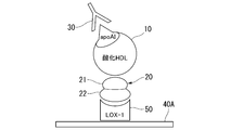
- the modified HDL-binding protein 20 is immobilized on the plate 40.
- the denatured HDL-binding protein 20 is a protein having homology with Factor V protein, which comprises, for example, an L chain site 21 and an H chain site 22.
- the plate 40 is an ELISA plate.
- the plate 40 is then washed to remove the unbound denatured HDL-binding protein 20.
- a sample derived from body fluid is injected into the plate 40.
- the modified HDL10 in the sample binds to the L chain site 21 on the plate 40.
- the complex on the plate 40 is quantified with denaturing HDL or antibody 30 that binds to the complex.
- an anti-apolipoprotein AI antibody (anti-ApoA1) is used as the antibody 30.
- the denatured HDL-binding protein that directly binds to the denatured HDL is immobilized on the plate without using the denatured HDL receptor such as LOX-1 as in the prior art, so that the denatured HDL receptor is unnecessary.
- the denatured HDL receptor such as LOX-1
- the denatured HDL-binding protein it is more effective than using a denatured HDL receptor such as LOX-1 for quantification. The quantification can be easily performed.
- Analytical reagent used for denaturing modified HDL Next, analytical reagents used in the above-mentioned method for quantifying modified HDL in body fluids will be described.
- This analytical reagent is used for quantification of denatured HDL in body fluid, and is used for quantification of cardiovascular disease, diabetes or diabetic disease, or denatured HDL in body fluid for determining the degree of progression thereof. Can be done.
- the analytical reagent contains the modified HDL-binding protein described above. By including the denatured HDL-binding protein, it is possible to quantify the denatured HDL having physiological activity.
- the analytical reagent of the present embodiment may contain other components contained in the immunoassay reagent.
- the analytical reagent of the present embodiment may be provided as a kit having a plurality of the above configurations.
- the kit may include analytical reagents containing a modified HDL binding protein for the quantification of modified HDL.
- the kit may also include carriers, ELISA plates, color-developing substrates, antibodies, etc. for use in the above-mentioned quantification operations.
- an ELISA plate on which the above-mentioned protein is immobilized may be included.
- the ELISA plate on which the protein is immobilized may be provided, and the denatured HDL-binding protein may be provided as an analytical plate containing a protein having an L chain site sequence of Factor V or a variant thereof.
- the modified HDL quantification method of the present embodiment can stably and accurately quantify the modified HDL as compared with the prior art.
- the stable and accurate quantification of this embodiment means that the conventional method has a difference in detection sensitivity itself depending on the sample or the detection sensitivity is low, whereas the method of this embodiment is based on the conditions of the sample.
- the modified HDL can be quantified with a certain high sensitivity regardless of the inclusions other than the modified HDL.
- the method for quantifying denatured HDL of the present embodiment can be used for determining diseases, their risks, and the degree of progression because stable and accurate quantification of denatured HDL can be performed.
- Diseases that are considered to be related to HDL are widely targeted, and examples thereof include cardiovascular diseases, diabetes mellitus, and diabetic diseases.
- Cardiovascular diseases include myocardial infarction or stroke caused by arteriosclerosis or its progression.
- the method for quantifying denatured HDL and the reagent for analysis of the present embodiment can be used, for example, in quantifying HDL in body fluids, by measuring denatured HDL, causing the above-mentioned cardiovascular disease and / or diabetes or diabetic. It can be used as a method for diagnosing morbidity to a disease.
- the risk of morbidity to the disease and / or the degree of progression thereof may be diagnosed from the change in the ratio of denatured HDL to the total amount of HDL.
- a standard product of protein, a healthy body fluid sample, etc. are used as a standard product, a calibration curve, a contrast table, etc. are created, and the quantification results of the sample sample to be diagnosed are compared with the calibration curve, the contrast table, etc.
- the process used for diagnosis is also included. It can also be used to determine and diagnose the morbidity, risk of morbidity, and / or the degree of progression of obesity-related diseases such as hypertension and sleep apnea syndrome.
- the present embodiment is more stable and reliable by further interposing a denatured HDL-binding protein than the conventional technique which has been attempted to quantify by detecting a complex of a denatured HDL and a LOX-1 protein. Is to be detected high.
- the quantification can be performed by improving the method, equipment, etc. that have been performed in the prior art using the LOX-1 protein.
- LOX-1 is a molecule discovered by the present inventor (Sawamura T et al. Nature 386, 73-77, 1997) and is known as one of the lectin-like oxidized LDL receptors.
- the detailed structure of LOX-1 has also been clarified, and it is a membrane protein that penetrates the cell membrane once, has a lectin-like domain, and this lectin-like domain is a recognition site for oxidized LDL (Japanese Patent Laid-Open No. 9). It is known that a soluble component is also present in blood, and that a modified high-density lipoprotein (HDL) acts as a ligand for LOX-1 (Japanese Patent Laid-Open No. 2012, etc.). -10045, Patent No. 6231307, etc.).
- HDL modified high-density lipoprotein
- the LOX-1 protein for example, it is also preferable to use a recombinant LOX-1 protein.
- the recombinant LOX-1 protein is a LOX-1 protein obtained by expressing it by gene recombination.
- the recombinant LOX-1 protein may be fused or mutated with other structures as needed.
- the quantification of the complex of denatured HDL, denatured HDL binding protein and LOX-1 protein can be performed using a so-called sandwich ELISA.
- the outline of the quantification method of this embodiment is shown in FIG.
- LOX-1 protein 50 is immobilized on the plate 40A.
- the denatured HDL-binding protein 20 is bound to the LOX-1 protein 50 on the plate 40A.
- the denatured HDL10 is bound to the complex of the LOX-1 protein 50 and the denatured HDL-binding protein 20.
- the complex on plate 40A can then be quantified with the denatured HDL described above or the antibody 30 that binds to the complex.
- An anti-Factor-V antibody can also be used as the antibody 30.
- serum may be used as a sample, and the sample may be provided with a step of adding the modified HDL-binding protein.
- a complex in which the modified HDL-binding protein and the modified HDL are bound is formed on the LOX-1 on the ELISA plate, and the ELISA plate is formed.
- the above conjugate can be quantified with the modified HDL described above or an antibody that binds to the complex.
- the analytical reagent used in the quantification method of the present embodiment may contain a recombinant LOX-1 protein for quantification of denatured HDL having pathological activity.
- the recombinant LOX-1 protein interacts with the denatured HDL, so that the denatured HDL can be quantified by immunoassay for the complex of the denatured HDL, the denatured HDL binding protein and the LOX-1. It can be carried out.
- the analytical reagent of the present embodiment may be provided as a kit having a plurality of the above configurations.
- the kit may include an analytical reagent containing a modified HDL binding protein and an analytical reagent containing a LOX-1 protein for the quantification of modified HDL.
- the ELISA method was carried out according to the following procedure. Recombinant human LOX-1 (61-273aa) was immobilized on a 384-well plate at 0.15 ⁇ g / well. After washing twice with PBS, blocking was performed with 1% casein-Na. After washing 3 times with PBS, plasma or serum samples supplemented with Factor V were incubated at room temperature for 2 hours. As the sample diluent, 1% casein-Na, 10 mM HEPES, 150 mM NaCl, pH 7.4, 100 ⁇ M APMSF was used.
- the anti-apyerA1 antibody (chicken # 1) prepared at 1 ⁇ g / ml was added after washing 3 times with PBS, and the mixture was incubated at room temperature for 1 hour.
- HRP-labeled anti-chicken IgY antibody diluted 3 times with PBS and diluted 4000-fold was added and incubated at room temperature for 1 hour.
- the mixture was washed 5 times with PBS, a TMB solution (manufactured by Bio-Rad) was added to a plate, and the reaction was carried out at room temperature. The reaction was stopped with 2M sulfuric acid, and the absorbance at 450 nm was measured.
- the modified HDL concentration (ng / ml) was calculated using a calibration curve prepared using oxidized HDL oxidized with copper sulfate as a standard product.
- FIG. 3 The detection result by the ELISA method is shown in FIG. 3 (a), (b), (c), (d), and (e) show plasma (plasma) and serum (serum) collected from different subjects (A, B, C, D, E), respectively. The result was shown.
- the complex on the vertical axis is resistant to denatured HDL (including those in which the binding of LOX-1 denatured HDL is mediated by other proteins) that has formed some complex with immobilized LOX-1.
- the concentration detected by the ap republicA1 antibody is shown, and the horizontal axis shows the concentration of added Factor V.
- serum as shown in FIGS.
- plasma may contain proteins such as Factor V that mediate the binding of LOX-1 and denatured HDL, but the content differs depending on the sample, its collection, storage method, etc., and Factor V is added. It is thought that the effects of Based on these results, Factor V binds to modified HDL, and modified HDL and LOX-1 are bound via Factor V, and this binding is performed depending on the amount of Factor V added. It was shown that the detection sensitivity was improved.
- proteins such as Factor V that mediate the binding of LOX-1 and denatured HDL
- FIGS. 4 to 7 when a plate on which LOX-1 is not immobilized is used, there is no difference in the detected amount between the case where Factor V is added to the sample and the case where Factor V is not added to the sample. From this result, it is shown that Factor V increases the amount of detection of LOX-1, Factor V and modified HDL binding to form a complex. Comparing FIGS. 4 and 5 and FIGS. 6 and 7, in FIGS. 5 and 7 using serum, the amount of detection increased by adding Factor V, as opposed to FIGS. 4 and 6 using plasma. ing. As for this result, as in Test Example 1, it is considered that the substance that seems not to be contained in the serum is supplemented by the addition of Factor V.
- nHDL unmodified HDL
- modified HDL As the modified HDL, Cu 2+ - réellexHDL, which is an oxidized HDL obtained by oxidizing HDL with copper sulfate, was used.
- Factor V was immobilized on a 384-well plate (manufactured by greener). After washing twice with PBS, blocking was performed with 1% casein-Na.
- nHDL or Cu 2+ - etcxHDL prepared at 1 ⁇ g / ml with sample diluent (1% casein-Na, 10 mM HEPES, 150 mM NaCl, pH 7.4, 100 ⁇ M APMSF) is incubated at room temperature for 2 hours. bottom.
- sample diluent 1% casein-Na, 10 mM HEPES, 150 mM NaCl, pH 7.4, 100 ⁇ M APMSF
- the anti-apsteadA1 antibody (chicken # 1) prepared at 1 ⁇ g / ml was added after washing 3 times with PBS, and the mixture was incubated at room temperature for 1 hour.
- HRP-labeled anti-chicken IgY antibody diluted 3 times with PBS and diluted 4000-fold was added and incubated at room temperature for 1 hour.
- the mixture was washed 5 times with PBS, a TMB solution (manufactured by Bio-Rad) was added to a plate, and the reaction was carried out at room temperature. The reaction was stopped with 2M sulfuric acid, and the absorbance at 450 nm was measured.
- Test Example 4 Denatured HDL in human plasma and serum was detected using a Factor V-immobilized plate prepared in the same manner as in Test Example 3. Denatured HDL was quantified by the ELISA method using plasma and serum samples collected from subjects X and Y. Factor V was immobilized on a 384-well plate (manufactured by greener). After washing twice with PBS, blocking was performed with 1% casein-Na. After washing 3 times with PBS, plasma or serum samples were incubated at room temperature for 2 hours. The anti-apyerA1 antibody (chicken # 1) prepared at 1 ⁇ g / ml was added after washing 3 times with PBS, and the mixture was incubated at room temperature for 1 hour.
- HRP-labeled anti-chicken IgY antibody diluted 3 times with PBS and diluted 4000-fold was added and incubated at room temperature for 1 hour. After the antibody reaction, the mixture was washed 5 times with PBS, a TMB solution (manufactured by Bio-Rad) was added to a plate, and the reaction was carried out at room temperature. The reaction was stopped with 2M sulfuric acid, and the absorbance at 450 nm was measured.
- FIG. 10 The sample collected from the subject X is shown in FIG. 10 (a), and the sample collected from the subject Y is shown in FIG. 10 (b).
- LOX-1 solid phase in the prior art
- This technique is more sensitive than the technique of immobilizing LOX-1, and not only can be more easily quantified than the sandwich method using LOX-1 and Factor V, but also for both plasma and serum.
- quantification can be performed stably and reliably with high sensitivity.
- a method for quantifying modified HDL capable of quantifying modified HDL more stably and accurately than before, and an analytical reagent used for the quantification method can be obtained.
- This quantification method and analytical reagent can be used for determining the risk or degree of progression of morbidity to cardiovascular disease, diabetes and diabetic disease.
Abstract
The present invention provides a method for quantifying a modified HDL in a sample, the method comprising: combining the modified HDL with a modified-HDL-binding protein; and quantifying a complex between the modified-HDL-binding protein and the modified HDL. The modified-HDL-binding protein includes either a protein having the sequence of a Factor V light chain site, or a variant thereof.
Description
本発明は、生活習慣病リスク及びその治癒効果の判定に用いることのできる変性HDLの定量方法、及びそれに用いる分析用試薬に関するものである。
The present invention relates to a method for quantifying modified HDL that can be used for determining the risk of lifestyle-related diseases and its healing effect, and an analytical reagent used therefor.
HDL(High-Density Lipoprotein、高比重リポ蛋白質)は、いわゆるコレステロールの一種で、比重が高く粒子径が小さいものである。組織から肝臓へコレステロールへの輸送は、HDLが担っていることが知られている。HDLは、血中濃度が高いと動脈硬化のリスクを低減させることから、「善玉コレステロール」と通称されることもある。一方、比重が低いLDL(Low-Density Lipoprotein、低比重リポ蛋白質)は、「悪玉コレステロール」として知られる。HDLとLDLの濃度は、健康診断の指標として用いられている。
HDL (High-Density Lipoprotein, high-density lipoprotein) is a type of so-called cholesterol, which has a high specific gravity and a small particle size. It is known that HDL is responsible for the transport of cholesterol from tissues to the liver. HDL is sometimes referred to as "good cholesterol" because it reduces the risk of arteriosclerosis when blood levels are high. On the other hand, LDL (Low-Density Lipoprotein, low-density lipoprotein) having a low specific density is known as "bad cholesterol". The concentrations of HDL and LDL are used as indicators of health examination.
HDLの生理的活性としては、LDLの酸化を抑制するだけでなく、NOの増加などを介して酸化LDLによる細胞毒性を軽減し、抗動脈硬化効果を発揮する。しかし、冠動脈疾患患者では血中変性HDLが増えており、それがLOX-1を介して血管に作用して、動脈硬化を促進すること、ヒトの動脈硬化巣にはHDLのアポタンパク質であるApoAIが変性したものが多量に蓄積していることが明らかになっている。これらの結果から、HDLを健康診断の指標として用いるには、単にHDLの濃度を測定するだけでなく、本来の生理的活性を有するHDLと、変性などによって活性の変化したHDLを別に定量する必要がある。特に変性HDLを定量することができれば、上述の冠動脈疾患を含めて、循環系及び血液に関係する疾患やそのリスクの検証に用いることができると考えられている。
As the physiological activity of HDL, it not only suppresses the oxidation of LDL, but also reduces the cytotoxicity due to the oxidized LDL through an increase in NO and exerts an anti-arteriosclerosis effect. However, blood degenerative HDL is increasing in patients with coronary artery disease, which acts on blood vessels via LOX-1 to promote arteriosclerosis, and in human arteriosclerosis lesions, HDL apoprotein ApoAI It has been clarified that a large amount of denatured substances are accumulated. From these results, in order to use HDL as an index for health diagnosis, it is necessary not only to measure the concentration of HDL, but also to separately quantify HDL having the original physiological activity and HDL whose activity has changed due to denaturation or the like. There is. In particular, if degenerative HDL can be quantified, it is considered that it can be used for verification of diseases related to the circulatory system and blood and their risks, including the above-mentioned coronary artery diseases.
特許文献1は、試料中に含まれる機能不全HDLの測定方法であって、機能不全HDLの受容体に試料を接触させて、試料中の機能不全HDLを受容体に結合させ、受容体に結合した機能不全HDLを検出するものであり、受容体は、LOX-1又はその改変体であり、受容体は、担体に固定化されており、受容体に結合した機能不全HDLに、機能不全HDLを認識する抗体をさらに結合させる機能不全HDLの測定方法を開示している。この技術は、多様な分子形態の機能不全HDLを広く検出できる機能不全HDLの測定技術を提供するとともに、当該技術を利用した生活習慣病の検出技術を提供しようとするものである。
Patent Document 1 is a method for measuring dysfunctional HDL contained in a sample, in which the sample is brought into contact with a receptor for dysfunctional HDL, the dysfunctional HDL in the sample is bound to the receptor, and the dysfunctional HDL in the sample is bound to the receptor. The receptor is LOX-1 or a variant thereof, and the receptor is immobilized on a carrier, and the dysfunctional HDL bound to the receptor is dysfunctional HDL. Discloses a method for measuring dysfunctional HDL that further binds an antibody that recognizes. This technique provides a technique for measuring dysfunctional HDL that can widely detect dysfunctional HDL in various molecular forms, and aims to provide a technique for detecting lifestyle-related diseases using the technique.
また、本発明者らは、特許文献2において、LOX-1へ結合するLOX-1結合タンパク配列に、ApoA1の全タンパク配列又は部分フラグメントタンパク配列が、直接又はスペーサを介して連結されている融合タンパク質及びそれを用いた高密度リポタンパク質の測定キットを開示している。この技術は、LOX-1と抗ApoA1抗体により同時に認識されるHDLを測定することにより様々な変性を受けたHDLを測定でき、及びLOX-1に対する抗体(抗LOX-1抗体)のLOX-1結合部位等のようにLOX-1に対して特異的に結合するタンパク質とApoA1とを融合させた融合タンパク質を用いるものである。この融合タンパク質により、様々な変性を受けたHDLを同時に認識でき、受容体結合活性を測定して生理活性そのものを測定するために用いられるもので、脂質を含まず、長期保存が可能で、再現的に調整でき、測定試験間ごとのばらつきを低減して信頼性を高くすることができるリファレンスとなり得る融合タンパク質を得ようとするものである。
Further, in Patent Document 2, the present inventors are a fusion in which the full protein sequence or partial fragment protein sequence of ApoA1 is directly or via a spacer to the LOX-1 binding protein sequence that binds to LOX-1. It discloses a protein and a measurement kit for high-density lipoprotein using the protein. This technique can measure HDL that has undergone various degenerations by measuring HDL that is simultaneously recognized by LOX-1 and anti-ApoA1 antibodies, and the LOX-1 antibody against LOX-1 (anti-LOX-1 antibody). A fusion protein obtained by fusing a protein that specifically binds to LOX-1 and ApoA1 such as a binding site is used. With this fusion protein, various denatured HDL can be recognized at the same time, and it is used to measure the receptor binding activity and the physiological activity itself. It does not contain lipids, can be stored for a long period of time, and can be reproduced. It is intended to obtain a fusion protein that can be adjusted in a flexible manner and can serve as a reference that can reduce variations between measurement tests and increase reliability.
変性HDLは、理論上、実験室では特許文献1に記載の技術で定量が可能である。本発明者らの特許文献2の技術は、従来の技術よりさらに洗練されたものである。これらを応用して、さほど洗練されたものでなくとも変性HDLの定量に用いることができる方法を樹立することは容易と予想された。
Theoretically, modified HDL can be quantified in the laboratory by the technique described in Patent Document 1. The technique of Patent Document 2 of the present inventors is more sophisticated than the conventional technique. It was expected that it would be easy to apply these to establish a method that could be used to quantify denatured HDL, even if it was not very sophisticated.
しかしながら、特許文献1の技術も普及には至っていないのが現状である。さらに、本発明者らの特許文献2の技術を用いても、実験室以外の条件や異なる検体を用いて実施すると、変性HDLの定量の結果が一定でないことがあった。これらの差は、実験室内の条件の他、技術を実施する者の試験技術の差などの、さまざまな要因が考えられた。
本発明者らは、上述の歴史的な経緯と、臨床の実地応用には多少条件が劣悪でも測定可能とすべきことを考え、変性HDLの定量技術を、より汎用性がある定量の技術へと改良することを目標として鋭意研究を進めていった。 However, the technology ofPatent Document 1 has not yet become widespread. Furthermore, even if the technique of Patent Document 2 of the present inventors is used, the result of quantification of denatured HDL may not be constant when it is carried out under conditions other than the laboratory or using different samples. Various factors such as the difference in the test technique of the person who implements the technique were considered as well as the conditions in the laboratory.
Considering the above-mentioned historical background and the fact that it should be possible to measure even if the conditions are somewhat inferior for clinical application, the present inventors have changed the quantification technique of denatured HDL to a more versatile quantification technique. We proceeded with diligent research with the goal of improving it.
本発明者らは、上述の歴史的な経緯と、臨床の実地応用には多少条件が劣悪でも測定可能とすべきことを考え、変性HDLの定量技術を、より汎用性がある定量の技術へと改良することを目標として鋭意研究を進めていった。 However, the technology of
Considering the above-mentioned historical background and the fact that it should be possible to measure even if the conditions are somewhat inferior for clinical application, the present inventors have changed the quantification technique of denatured HDL to a more versatile quantification technique. We proceeded with diligent research with the goal of improving it.
本発明は上記のような事情を鑑みてなされたものであり、その目的は、変性HDLを従来よりも安定して正確に定量でき、心・血管系疾患、糖尿病若しくは糖尿病性疾患等、又はその進行度の判定に有用な変性HDLの定量方法、及び、その定量方法に用いる分析用試薬を提供することにある。
The present invention has been made in view of the above circumstances, and an object of the present invention is to be able to quantify denatured HDL more stably and accurately than before, and to perform cardiovascular disease, diabetes or diabetic disease, or the like. It is an object of the present invention to provide a method for quantifying modified HDL useful for determining the degree of progress, and an analytical reagent used for the quantification method.
上記課題を解決するため、本発明は以下の態様を有する。
[1]試料中に含まれる変性HDLの定量方法であって、前記変性HDLと変性HDL結合蛋白質とを結合させ、前記変性HDL結合蛋白質と前記変性HDLとの結合体を定量し、前記変性HDL結合蛋白質は、Factor VのL鎖部位の配列を有する蛋白質又はその変異体を含む、変性HDLの定量方法。
[2]前記変性HDL結合蛋白質は、Factor Vのリン脂質認識部位の配列を含む、前記の変性HDLの定量方法。
[3]前記変性HDL結合蛋白質は、Factor Vの配列を含む、前記の変性HDLの定量方法。
[4]前記変性HDL結合蛋白質は、Factor Vのリン脂質認識部位であるC1-C2ドメインと一部相同性を有する部位を備えた蛋白質である、前記の変性HDLの定量方法。
[5]前記C1-C2ドメインと一部相同性を有する部位を備えた蛋白質は、MFG-E8、Del-1又はFactor VIIIである、前記の変性HDLの定量方法。
[6]前記変性HDL結合蛋白質は、組み換え蛋白質である、前記の変性HDLの定量方法。
[7]前記定量は、前記変性HDL及び前記変性HDL結合蛋白質の複合体を形成し、前記複合体を免疫分析により定量する、前記の変性HDLの定量方法。
[8]前記免疫分析による定量は、前記変性HDL結合蛋白質、あるいはこれを結合する蛋白質を固相化したELISAプレートを用いたELISA法により行う、前記の変性HDLの定量方法。
[9]前記定量は、前記変性HDL、前記変性HDL結合蛋白質及びLOX-1蛋白質の複合体を形成し、前記複合体を免疫分析により定量する、前記の変性HDLの定量方法。
[10]前記定量では、試料として血清又は血漿を用いる、前記の変性HDLの定量方法。
[11]前記定量では、試料として血清を用い、前記試料に前記変性HDL結合蛋白質を添加する工程を備える、前記の変性HDLの定量方法。
[12]心・血管系疾患、糖尿病若しくは糖尿病性疾患、それらのリスク、又は、その進行度を判定するための変性HDLの定量に用いる分析用試薬であって、前記変性HDLと結合可能な変性HDL結合蛋白質を含み、前記変性HDL結合蛋白質は、Factor VのL鎖部位配列を有する蛋白質又はその変異体を含む、分析用試薬。
[13]前記変性HDL結合蛋白質は、Factor Vのリン脂質認識部位の配列を含む、前記の分析用試薬。
[14]前記変性HDL結合蛋白質は、Factor Vの配列を含む、前記の分析用試薬。
[15]前記変性HDL結合蛋白質は、Factor Vのリン脂質認識部位であるC1-C2ドメインと一部相同性を有する部位を備えた蛋白質である、前記の分析用試薬。[16]前記C1-C2ドメインと一部相同性を有する部位を備えた蛋白質は、MFG-E8、Del-1又はFactor VIIIである、前記の分析用試薬。
[17]前記変性HDL結合蛋白質は、組み換え蛋白質である、前記の分析用試薬。
[18]前記心・血管系疾患は、動脈硬化又はその進行によって生じる心筋梗塞若しくは脳卒中である、前記の分析用試薬。 In order to solve the above problems, the present invention has the following aspects.
[1] A method for quantifying modified HDL contained in a sample, in which the modified HDL is bound to a modified HDL binding protein, the conjugate of the modified HDL binding protein and the modified HDL is quantified, and the modified HDL is quantified. A method for quantifying denatured HDL, wherein the binding protein comprises a protein having the sequence of the L chain site of Factor V or a variant thereof.
[2] The method for quantifying modified HDL, wherein the modified HDL-binding protein contains a sequence of a phospholipid recognition site of Factor V.
[3] The method for quantifying modified HDL, wherein the modified HDL-binding protein contains a Factor V sequence.
[4] The method for quantifying modified HDL, wherein the modified HDL-binding protein is a protein having a site partially homologous to the C1-C2 domain, which is a phospholipid recognition site of Factor V.
[5] The method for quantifying modified HDL, wherein the protein having a site partially homologous to the C1-C2 domain is MFG-E8, Del-1 or Factor VIII.
[6] The method for quantifying modified HDL, wherein the modified HDL-binding protein is a recombinant protein.
[7] The method for quantifying modified HDL, wherein the quantification forms a complex of the modified HDL and the modified HDL binding protein, and the complex is quantified by immunoassay.
[8] The method for quantifying modified HDL, which is carried out by the ELISA method using an ELISA plate on which the modified HDL-binding protein or a protein that binds the modified HDL-binding protein is immobilized.
[9] The method for quantifying the modified HDL, wherein the quantification forms a complex of the modified HDL, the modified HDL binding protein and the LOX-1 protein, and the complex is quantified by immunoassay.
[10] The method for quantifying denatured HDL, which uses serum or plasma as a sample in the quantification.
[11] The method for quantifying modified HDL, which comprises a step of using serum as a sample and adding the modified HDL-binding protein to the sample.
[12] An analytical reagent used for quantifying denatured HDL for determining cardiovascular disease, diabetes or diabetic disease, their risk, or the degree thereof, and denatured that can bind to the denatured HDL. An analytical reagent comprising a HDL-binding protein, wherein the denatured HDL-binding protein contains a protein having an L-chain site sequence of Factor V or a variant thereof.
[13] The reagent for analysis, wherein the denatured HDL-binding protein contains a sequence of a phospholipid recognition site of Factor V.
[14] The reagent for analysis, wherein the denatured HDL-binding protein comprises a Factor V sequence.
[15] The reagent for analysis, wherein the denatured HDL-binding protein is a protein having a site having a partial homology with the C1-C2 domain, which is a phospholipid recognition site of Factor V. [16] The reagent for analysis, wherein the protein having a site partially homologous to the C1-C2 domain is MFG-E8, Del-1 or Factor VIII.
[17] The reagent for analysis, wherein the denatured HDL-binding protein is a recombinant protein.
[18] The reagent for analysis, wherein the cardiovascular disease is myocardial infarction or stroke caused by arteriosclerosis or its progression.
本発明によれば、変性HDLを従来よりも安定して正確に定量でき、心・血管系疾患、糖尿病若しくは糖尿病性疾患等、又はその進行度の判定に有用な変性HDLの定量方法、及び、その定量方法に用いる分析用試薬が得られる。
According to the present invention, denatured HDL can be quantified more stably and accurately than before, and a method for quantifying denatured HDL useful for determining cardiovascular disease, diabetes or diabetic disease, or the degree of progression thereof, and An analytical reagent used for the quantification method is obtained.
以下、本発明に係る変性HDLの定量方法及び分析用試薬について、実施形態を示して説明する。ただし、本発明は以下の実施形態に限定されるものではない。
Hereinafter, the method for quantifying modified HDL and the reagent for analysis according to the present invention will be described with reference to embodiments. However, the present invention is not limited to the following embodiments.
(第1の実施形態)
(変性HDLの定量方法)
本実施形態の変性HDLの定量方法は、試料中に含まれる変性HDLを定量するにあたり、変性HDLと変性HDL結合蛋白質とを結合させる。この変性HDL結合蛋白質と変性HDLとが結合した複合体を定量する。 (First Embodiment)
(Method for quantifying denatured HDL)
In the method for quantifying modified HDL of the present embodiment, the modified HDL and the modified HDL binding protein are bound to each other in quantifying the modified HDL contained in the sample. The complex in which this denatured HDL-binding protein and denatured HDL are bound is quantified.
本実施形態における変性HDLとは、本来生体内で生理的活性を有するHDLとはなんらかの構造の差異を有するHDLを含み、特に、それらの構造の差異により生理的活性が低下しているものを指す。HDLが変性HDLとなる機序としては、例えば構成蛋白質の化学的修飾や部分的あるいは完全分解、遺伝的欠損および変異、高次構造の異常や変化、又は、活性を失わせる分子との結合などの要因が含まれる。
The denatured HDL in the present embodiment includes HDL having some structural difference from HDL that originally has physiological activity in the living body, and in particular, refers to one in which physiological activity is reduced due to the difference in those structures. .. The mechanism by which HDL becomes denatured HDL includes, for example, chemical modification or partial or complete degradation of constituent proteins, genetic defects and mutations, abnormalities or changes in higher-order structures, or binding to molecules that cause loss of activity. Factors are included.
こうした変性HDLの例としては、例えば、HDLを構成する蛋白質又は脂質のいずれかの部位が酸化された、酸化HDLが含まれる。このような変性HDLとしては、例えば、HClO-HDL、HNE-HDL、Carbamylated HDL及びMDA-HDLなどが挙げられる。これらの変性HDLは、血管壁の血管内皮細胞を障害し、動脈硬化や血栓形成を引き起こす心疾患や虚血疾患等の動脈硬化性疾患のような各種疾患の発症リスクを高めるものとして知られている。
Examples of such denatured HDL include oxidized HDL in which either a protein or lipid site constituting HDL is oxidized. Examples of such modified HDL include HClO-HDL, HNE-HDL, Carbamylated HDL and MDA-HDL. These degenerated HDLs are known to damage the vascular endothelial cells of the vascular wall and increase the risk of developing various diseases such as arteriosclerosis such as heart disease and ischemic disease that cause arteriosclerosis and thrombosis. There is.
本実施形態の変性HDLの定量方法では、変性HDLと変性HDL結合蛋白質とを結合させ、変性HDL結合蛋白質と変性HDLとの結合体を定量する。
本実施形態の変性HDL結合蛋白質は、変性HDLと直接相互作用し得る、例えばその蛋白質自体が変性HDLと結合し得る蛋白質を広く指す。
変性HDLは、生体にとって不利な反応を引き起こす受容体と相互作用する活性を獲得していることがある。このとき、変性HDLは変性HDL結合蛋白質を介して受容体と結合している、すなわち変性HDLと変性HDL結合蛋白質とが直接結合し、この変性HDL結合蛋白質が受容体に結合していることがある。
変性HDL結合蛋白質としては、例えばFactor V、その変異体及び一部相同性を有する蛋白質などがある。変性HDLに別蛋白質を介して結合することのある受容体としては、後述のLOX-1蛋白質がある。 In the method for quantifying modified HDL of the present embodiment, the modified HDL and the modified HDL binding protein are bound to each other, and the conjugate of the modified HDL binding protein and the modified HDL is quantified.
The modified HDL-binding protein of the present embodiment broadly refers to a protein that can directly interact with the modified HDL, for example, the protein itself can bind to the modified HDL.
Denatured HDL may have acquired activity that interacts with receptors that cause adverse reactions to the body. At this time, the denatured HDL is bound to the receptor via the denatured HDL binding protein, that is, the denatured HDL and the denatured HDL binding protein are directly bound, and this denatured HDL binding protein is bound to the receptor. be.
Examples of the denatured HDL-binding protein include Factor V, a mutant thereof, and a protein having partial homology. As a receptor that may bind to denatured HDL via another protein, there is a LOX-1 protein described later.
本実施形態では、変性HDL結合蛋白質は、Factor VのL鎖部位の配列を有する蛋白質又はその変異体を含んでいる。
Factor V(第V因子)は、凝固系を構成する蛋白質として知られ、A1-A2-B-A3-C1-C2ドメインから構成されているおよそ330kDaの蛋白質であり、A1-A2ドメインからなるH鎖(重鎖)と、A3-C1-C2ドメインからなるL鎖(軽鎖)に大きく分けられる。主にH鎖は細胞膜と結合し、L鎖はC1-C2ドメインを介してリン脂質を認識、結合することが知られている。
変異体とは、一部の配列の置換が行われたものを指し、本実施形態では主にFactor VのL鎖部位以外の部位が置換されているものであることが好ましい。 In this embodiment, the denatured HDL binding protein comprises a protein having the sequence of the L chain site of Factor V or a variant thereof.
Factor V (factor V) is known as a protein that constitutes the coagulation system, is a protein of approximately 330 kDa composed of the A1-A2-B-A3-C1-C2 domain, and H consisting of the A1-A2 domain. It is roughly divided into a chain (heavy chain) and an L chain (light chain) consisting of the A3-C1-C2 domain. It is known that the H chain mainly binds to the cell membrane, and the L chain recognizes and binds to phospholipids via the C1-C2 domain.
The mutant refers to one in which a part of the sequence has been replaced, and in the present embodiment, it is preferable that a site other than the L chain site of Factor V is mainly replaced.
Factor VのL鎖部位は、変性HDLと直接結合する部位であるため、変性HDL結合蛋白質がこの部位を含む蛋白質又はその変異体であることで、変性HDLと変性HDL結合蛋白質を結合させ、定量に用いることができる。
また、変性HDL結合蛋白質として、Factor Vの一部であるL鎖部位の配列を有する蛋白質を用いることで、変性HDLと結合する部位を有しつつ、Factor Vとは異なる構造を有する蛋白質を用いることができる。すなわち、Factor Vよりも入手が容易な蛋白質や、試験の行いやすい蛋白質を用いることができる。Factor Vよりも入手が容易な蛋白質としては、例えばL鎖部位及び最小限の配列のみ用いることで、Factor Vよりも全長が短く、組み換え蛋白質としての製造が行いやすい蛋白質を用いることができる。試験が行いやすい蛋白質としては、L鎖以外の配列を変更することで、より保存やプレートへの固相化が行いやすい蛋白質を用いることができる。 Since the L chain site of Factor V is a site that directly binds to denatured HDL, the denatured HDL binding protein is a protein containing this site or a variant thereof, so that the denatured HDL and the denatured HDL binding protein are bound and quantified. Can be used for.
Further, by using a protein having an L chain site sequence that is a part of Factor V as the denatured HDL binding protein, a protein having a site that binds to the denatured HDL but having a structure different from that of Factor V is used. be able to. That is, a protein that is easier to obtain than Factor V or a protein that is easier to test can be used. As a protein that is more easily available than Factor V, for example, by using only the L chain site and the minimum sequence, a protein that has a shorter overall length than Factor V and is easily produced as a recombinant protein can be used. As a protein that can be easily tested, a protein that can be more easily preserved or immobilized on a plate can be used by changing the sequence other than the L chain.
変性HDL結合蛋白質は、Factor Vのリン脂質認識部位の配列を含むことも好ましい。Factor Vの配列のうち、L鎖が備えるリン脂質認識部位は、変性HDLを認識し、有効に結合することができる。
It is also preferable that the denatured HDL-binding protein contains the sequence of the phospholipid recognition site of Factor V. Among the Factor V sequences, the phospholipid recognition site contained in the L chain can recognize denatured HDL and effectively bind to it.
変性HDL結合蛋白質は、Factor Vの配列を含むことが好ましい。ここでFactor Vの配列とはFactor V蛋白質の全長であり、Factor Vそのものを用いてもよく、他の蛋白質との融合体又は結合、修飾蛋白質等を用いてもよい。
The denatured HDL-binding protein preferably contains the Factor V sequence. Here, the sequence of Factor V is the total length of the Factor V protein, and Factor V itself may be used, or a fusion or binding with another protein, a modified protein, or the like may be used.
変性HDL結合蛋白質は、Factor Vのリン脂質認識部位であるC1-C2ドメインと一部相同性を有する部位を備える蛋白質であることも好ましい。このようなC1-C2ドメインと一部相同性を有する部位を備えた蛋白質としては、MFG-E8、Del-1又はFactor VIIIが挙げられる。また、これらの蛋白質や変異体、これらの蛋白質の配列を一部に含む蛋白質であってもよい。
The denatured HDL-binding protein is also preferably a protein having a site partially homologous to the C1-C2 domain, which is a phospholipid recognition site of Factor V. Examples of the protein having a site having a site partially homologous to such a C1-C2 domain include MFG-E8, Del-1 or Factor VIII. Further, these proteins, variants, and proteins containing a part of the sequences of these proteins may be used.
変性HDL結合蛋白質は、例えば、組み換え蛋白質を用いることも好ましい。ここで組み換え蛋白質とは、遺伝子組み換えによって発現させて得られた蛋白質である。組み換え蛋白質は、必要に応じて他の構造との融合や変異を導入されていてもよい。
As the denatured HDL binding protein, for example, it is also preferable to use a recombinant protein. Here, the recombinant protein is a protein obtained by expressing it by gene recombination. The recombinant protein may be fused or mutated with other structures as needed.
本実施形態では、試料中に含まれる変性HDLの定量を行う。ここで試料としては、生物から取り出した成分を含む試料を広く指す。試料は前記成分の液体のものを用いるか、又は前記成分に液体を添加して、液体の試料とすることが好ましい。試料としては、例えば、生物の体液を直接試料として用いることができる。生物の体液としては、例えばヒトの血液又は唾液を用いることができる。血液のサンプルとしては血清又は血漿などを用いることができる。本実施形態では、これらの体液を試料として、上述の定量に供する。
In this embodiment, the modified HDL contained in the sample is quantified. Here, the sample broadly refers to a sample containing a component extracted from an organism. It is preferable to use a liquid sample of the above-mentioned component, or to add a liquid to the above-mentioned component to prepare a liquid sample. As the sample, for example, the body fluid of an organism can be directly used as a sample. As the body fluid of an organism, for example, human blood or saliva can be used. Serum, plasma, or the like can be used as the blood sample. In the present embodiment, these body fluids are used as samples and subjected to the above-mentioned quantification.
本実施形態では、試料として血清を用いることが好ましい。本実施形態では、変性HDLと直接結合するFactor VのL鎖部分の配列を備えた変性HDL結合蛋白質を、試料中の変性HDLと結合させる。そのため、変性HDLと直接結合する因子を含んでいない場合のある血清を試料として用いても、変性HDLを安定して正確に定量することができる。加えて血清は血漿等の他の体液試料よりも、血清の方が検体の取得・保存が容易である。実際に血液試料としては、血清の形で凍結保持されている検体が国際的に一般的であることから、有益な技術である。
In this embodiment, it is preferable to use serum as a sample. In this embodiment, a denatured HDL-binding protein having a sequence of the L chain portion of Factor V that directly binds to the denatured HDL is bound to the denatured HDL in the sample. Therefore, even if serum that may not contain a factor that directly binds to denatured HDL is used as a sample, denatured HDL can be quantified stably and accurately. In addition, serum is easier to obtain and store than other body fluid samples such as plasma. As a blood sample, a sample that is cryopreserved in the form of serum is actually common internationally, so it is a useful technique.
本発明者らは、従来の技術においてLOX-1等の変性HDLと相互作用する蛋白質を変性HDLの定量に用いた場合、血漿を試料に用いると変性HDLが検出できるが、血清を試料に用いると変性HDLの検出が少なく、正確に定量することができないという点に着目した。そこで、血液や血漿から血清を形成する過程において分解喪失される物質を測定系に補填することにより、血清を試料に用いた測定が可能となることを予測した。
本発明者らは、前記補填する物質として、Factor Vに着目した。生体内の反応として、変性HDLがLOX-1と結合する際には、Factor Vを介して結合する。具体的には、Factor Vが備えるフォスファチジルセリン(PS)認識部位が変性HDLに、LOX-1認識部位がLOX-1と結合する。 When a protein that interacts with denatured HDL such as LOX-1 is used for quantification of denatured HDL in the conventional technique, the denatured HDL can be detected by using plasma as a sample, but serum is used as a sample. We focused on the fact that the detection of denatured HDL was small and it was not possible to accurately quantify it. Therefore, it was predicted that by supplementing the measurement system with a substance that is decomposed and lost in the process of forming serum from blood or plasma, it becomes possible to perform measurement using serum as a sample.
The present inventors have focused on Factor V as the supplementary substance. As a reaction in vivo, when denatured HDL binds to LOX-1, it binds via Factor V. Specifically, the phosphatidylserine (PS) recognition site contained in Factor V binds to denatured HDL, and the LOX-1 recognition site binds to LOX-1.
後述するように、血清を試料として用い、試料にFactor Vを添加すると、血清中の変性HDL検出感度がほぼ添加量に応じて増加する。このため、血清等の試料にFactor Vのような変性HDL結合蛋白質を添加することで、変性HDLを定量することができる。
As will be described later, when serum is used as a sample and Factor V is added to the sample, the sensitivity of detecting denatured HDL in serum increases substantially according to the amount of addition. Therefore, by adding a denatured HDL-binding protein such as Factor V to a sample such as serum, the denatured HDL can be quantified.
上述の定量には、蛋白質の定量(蛋白質量の測定)のための公知の手段を用いることができる。例えば、上述の定量には免疫学的手法を用いることができる。
免疫学的手法を用いた定量、免疫分析としては、酵素(エンザイムイムノアッセイ、EIA、ELISA)、蛍光物質(蛍光イムノアッセイ、FIA)又は放射性物質(ラジオイムノアッセイ、RIA)などを適宜用いることができる。このうち、ELISAは比較的安価、簡便かつ多数のサンプルを分析できるため望ましい。 For the above-mentioned quantification, a known means for quantifying protein (measurement of protein mass) can be used. For example, an immunological method can be used for the above-mentioned quantification.
As the quantification and immunoanalysis using an immunological method, an enzyme (enzyme immunoassay, EIA, ELISA), a fluorescent substance (fluorescent immunoassay, FIA), a radioactive substance (radioimmunoassay, RIA) or the like can be appropriately used. Of these, ELISA is desirable because it is relatively inexpensive, simple, and can analyze a large number of samples.
例えば、本実施形態では前記定量に、ELISA法を用いることができる。具体的には、変性HDL結合蛋白質を、ELISAプレートに固相化する。変性HDL結合蛋白質の固相化(固定化)には、既知の方法を適宜用いることができる。例えば、公知の担体(支持体)に受容体を結合させることができる。
このELISAプレートに試料を添加して、試料に含まれる変性HDLを変性HDL結合蛋白質に結合させ、ELISAプレートを洗浄して未結合物を除去し、変性HDL又は複合体を検出できる抗体を用いて検出すると、定量する対象と、変性HDLを変性HDL結合蛋白質の複合体を定量することができる。
変性HDL又は前記複合体に結合する抗体としては、HDL配列に結合する抗体を適宜用いることができる。本実施形態では、ELISAプレート上の変性HDL及び変性HDL結合蛋白質の複合体を検出するので、プレート上で検出されるのは変性HDLである。 なお、本実施形態において変性HDLに結合する抗体は、モノクローナル及びポリクローナル抗体のいずれも用いることができる。 For example, in the present embodiment, the ELISA method can be used for the quantification. Specifically, the denatured HDL-binding protein is immobilized on an ELISA plate. A known method can be appropriately used for immobilization (immobilization) of the denatured HDL-binding protein. For example, the receptor can be attached to a known carrier (support).
A sample is added to the ELISA plate, the denatured HDL contained in the sample is bound to the denatured HDL binding protein, the ELISA plate is washed to remove unbound substances, and an antibody capable of detecting the denatured HDL or complex is used. When detected, it is possible to quantify the target to be quantified and the complex of the HDL-binding protein that denatures the denatured HDL.
As the antibody that binds to the denatured HDL or the complex, an antibody that binds to the HDL sequence can be appropriately used. In this embodiment, the complex of the denatured HDL and the denatured HDL binding protein is detected on the ELISA plate, so that it is the denatured HDL detected on the plate. As the antibody that binds to the denatured HDL in this embodiment, either a monoclonal antibody or a polyclonal antibody can be used.
本実施形態においては、さらに具体的には、変性HDL及び変性HDL結合蛋白質が結合した複合体を形成し、この複合体を免疫分析により定量することができる。
この免疫分析について、図1に模式図を示して説明する。例えば、変性HDL結合蛋白質20を、プレート40に固相化させる。本実施形態では、変性HDL結合蛋白質20は、例としてL鎖部位21とH鎖部位22とを備えた、Factor V蛋白質と相同性を有する蛋白質である。プレート40は、ELISAプレートである。既知の担体で固相化を行うと、プレート40上には、前記H鎖部位22がプレート40に固相化された構造が生じる。ついで、プレート40を洗浄し、未結合の変性HDL結合蛋白質20を除去する。
このプレート40に、体液由来の試料を注入する。プレート40上のL鎖部位21に、試料中の変性HDL10が結合する。このプレート40上の複合を、変性HDL又は前記複合体に結合する抗体30で定量する。本実施形態では、抗体30には抗アポリポタンパクAI抗体(抗ApoA1)を用いている。 In the present embodiment, more specifically, a complex in which a denatured HDL and a denatured HDL-binding protein are bound can be formed, and this complex can be quantified by immunoassay.
This immunoassay will be described with reference to FIG. For example, the modified HDL-bindingprotein 20 is immobilized on the plate 40. In the present embodiment, the denatured HDL-binding protein 20 is a protein having homology with Factor V protein, which comprises, for example, an L chain site 21 and an H chain site 22. The plate 40 is an ELISA plate. When immobilization is performed with a known carrier, a structure in which the H chain portion 22 is immobilized on the plate 40 is formed on the plate 40. The plate 40 is then washed to remove the unbound denatured HDL-binding protein 20.
A sample derived from body fluid is injected into theplate 40. The modified HDL10 in the sample binds to the L chain site 21 on the plate 40. The complex on the plate 40 is quantified with denaturing HDL or antibody 30 that binds to the complex. In this embodiment, an anti-apolipoprotein AI antibody (anti-ApoA1) is used as the antibody 30.
この免疫分析について、図1に模式図を示して説明する。例えば、変性HDL結合蛋白質20を、プレート40に固相化させる。本実施形態では、変性HDL結合蛋白質20は、例としてL鎖部位21とH鎖部位22とを備えた、Factor V蛋白質と相同性を有する蛋白質である。プレート40は、ELISAプレートである。既知の担体で固相化を行うと、プレート40上には、前記H鎖部位22がプレート40に固相化された構造が生じる。ついで、プレート40を洗浄し、未結合の変性HDL結合蛋白質20を除去する。
このプレート40に、体液由来の試料を注入する。プレート40上のL鎖部位21に、試料中の変性HDL10が結合する。このプレート40上の複合を、変性HDL又は前記複合体に結合する抗体30で定量する。本実施形態では、抗体30には抗アポリポタンパクAI抗体(抗ApoA1)を用いている。 In the present embodiment, more specifically, a complex in which a denatured HDL and a denatured HDL-binding protein are bound can be formed, and this complex can be quantified by immunoassay.
This immunoassay will be described with reference to FIG. For example, the modified HDL-binding
A sample derived from body fluid is injected into the
この実施形態では、従来技術のようなLOX-1等の変性HDL受容体を用いず、変性HDLに直接結合する変性HDL結合蛋白質をプレート上に固相化するので、変性HDL受容体が不要である。例えば変性HDL結合蛋白質として、上述するようなFactor Vよりもさらに全長が短い蛋白質等、入手が容易な蛋白質を用いれば、LOX-1等の変性HDL受容体を定量に用いるのに比べて、より容易に定量を行うことができる。
In this embodiment, the denatured HDL-binding protein that directly binds to the denatured HDL is immobilized on the plate without using the denatured HDL receptor such as LOX-1 as in the prior art, so that the denatured HDL receptor is unnecessary. be. For example, if a protein that is easily available, such as a protein having a shorter overall length than Factor V as described above, is used as the denatured HDL-binding protein, it is more effective than using a denatured HDL receptor such as LOX-1 for quantification. The quantification can be easily performed.
(変性HDL定量に用いる分析用試薬)
次に、上述の体液中の変性HDL定量方法に用いる分析用試薬について説明する。
この分析用試薬は、体液中の変性HDLの定量に用いることで、心・血管系疾患、糖尿病若しくは糖尿病性疾患、又は、その進行度を判定するための体液中の変性HDLの定量に用いることができる。
分析用試薬は、上述した変性HDL結合蛋白質を含んでいる。変性HDL結合蛋白質を含むことで、生理的活性を有する変性HDLの定量を行うことができる。 (Analytical reagent used for denaturing modified HDL)
Next, analytical reagents used in the above-mentioned method for quantifying modified HDL in body fluids will be described.
This analytical reagent is used for quantification of denatured HDL in body fluid, and is used for quantification of cardiovascular disease, diabetes or diabetic disease, or denatured HDL in body fluid for determining the degree of progression thereof. Can be done.
The analytical reagent contains the modified HDL-binding protein described above. By including the denatured HDL-binding protein, it is possible to quantify the denatured HDL having physiological activity.
(その他の構成)
本実施形態の分析用試薬は、その他免疫分析の試薬に含まれる成分を含有していてもよい。 (Other configurations)
The analytical reagent of the present embodiment may contain other components contained in the immunoassay reagent.
本実施形態の分析用試薬は、その他免疫分析の試薬に含まれる成分を含有していてもよい。 (Other configurations)
The analytical reagent of the present embodiment may contain other components contained in the immunoassay reagent.
また、本実施形態の分析用試薬は、上記構成を複数備えたキットとして提供されてもよい。例えば、このキットは変性HDLの定量のための変性HDL結合蛋白質を含む分析用試薬を備えていてもよい。また、このキットは、上述した定量の操作に用いるための担体、ELISAプレート、発色基質又は抗体等を含んでいてもよい。
Further, the analytical reagent of the present embodiment may be provided as a kit having a plurality of the above configurations. For example, the kit may include analytical reagents containing a modified HDL binding protein for the quantification of modified HDL. The kit may also include carriers, ELISA plates, color-developing substrates, antibodies, etc. for use in the above-mentioned quantification operations.
また、上述した蛋白質が固相化されたELISAプレートを含んでいてもよい。例えば、心・血管系疾患、糖尿病若しくは糖尿病性疾患、それらのリスク、又は、その進行度を判定するための変性HDLの定量に用いる分析用プレートであって、変性HDLと結合可能な変性HDL結合蛋白質が固相化されたELISAプレートを備え、変性HDL結合蛋白質は、Factor VのL鎖部位配列を有する蛋白質又はその変異体を含む、分析用プレートとして提供されていてもよい。
Further, an ELISA plate on which the above-mentioned protein is immobilized may be included. For example, an analytical plate used to quantify denatured HDL for determining cardiovascular disease, diabetes or diabetic disease, their risk, or their progression, and denatured HDL binding capable of binding to denatured HDL. The ELISA plate on which the protein is immobilized may be provided, and the denatured HDL-binding protein may be provided as an analytical plate containing a protein having an L chain site sequence of Factor V or a variant thereof.
(本実施形態の効果)
本実施形態の変性HDLの定量方法は、従来技術に比して変性HDLを安定して正確に定量することができる。本実施形態が安定して正確に定量できるとは、従来の方法が試料によって検出感度自体に差がある場合や、検出感度が低かったのに対して、本実施形態の方法は試料の条件、例えば変性HDL以外の含有物等によらず、変性HDLを一定の高い感度で定量することができる。 (Effect of this embodiment)
The modified HDL quantification method of the present embodiment can stably and accurately quantify the modified HDL as compared with the prior art. The stable and accurate quantification of this embodiment means that the conventional method has a difference in detection sensitivity itself depending on the sample or the detection sensitivity is low, whereas the method of this embodiment is based on the conditions of the sample. For example, the modified HDL can be quantified with a certain high sensitivity regardless of the inclusions other than the modified HDL.
本実施形態の変性HDLの定量方法は、変性HDLの安定かつ正確な定量を行うことができることで、疾患、それらのリスクや進行度の判定に用いることができる。疾患としては、HDLが関連すると思われる疾患が広く対象となり、例えば、心・血管系疾患、糖尿病若しくは糖尿病性疾患が挙げられる。心・血管系疾患としては特に、動脈硬化又はその進行によって生じる心筋梗塞若しくは脳卒中が挙げられる。
The method for quantifying denatured HDL of the present embodiment can be used for determining diseases, their risks, and the degree of progression because stable and accurate quantification of denatured HDL can be performed. Diseases that are considered to be related to HDL are widely targeted, and examples thereof include cardiovascular diseases, diabetes mellitus, and diabetic diseases. Cardiovascular diseases include myocardial infarction or stroke caused by arteriosclerosis or its progression.
(本実施形態の用途)
本実施形態の変性HDLの定量方法及び分析用試薬は、例えば、体液中のHDLの定量において、変性HDLの測定により、上述した心・血管系疾患への罹患、及び/又は、糖尿病若しくは糖尿病性疾患への罹患を診断する方法に用いることができる。 (Use of this embodiment)
The method for quantifying denatured HDL and the reagent for analysis of the present embodiment can be used, for example, in quantifying HDL in body fluids, by measuring denatured HDL, causing the above-mentioned cardiovascular disease and / or diabetes or diabetic. It can be used as a method for diagnosing morbidity to a disease.
診断方法としては、例えば、前記試料中の変性HDLの測定において、総HDL量に対する変性HDLの比率の変化から、前記疾患への罹患のリスク、及び/又は、これらの進行度合いを診断することもできる。
この診断方法では、蛋白質の標準品、健常体液試料などを用いて標準品として用い、検量線、対比表などを作成し、診断する対象の検体試料による定量結果を検量線や対比表などと比較して診断に用いる過程なども含まれる。
また、高血圧、睡眠時無呼吸症候群といった、肥満に関連する疾患への罹患、罹患リスク、及び/又はその進行度合いの判定及び診断にも用いることができる。 As a diagnostic method, for example, in the measurement of denatured HDL in the sample, the risk of morbidity to the disease and / or the degree of progression thereof may be diagnosed from the change in the ratio of denatured HDL to the total amount of HDL. can.
In this diagnostic method, a standard product of protein, a healthy body fluid sample, etc. are used as a standard product, a calibration curve, a contrast table, etc. are created, and the quantification results of the sample sample to be diagnosed are compared with the calibration curve, the contrast table, etc. The process used for diagnosis is also included.
It can also be used to determine and diagnose the morbidity, risk of morbidity, and / or the degree of progression of obesity-related diseases such as hypertension and sleep apnea syndrome.
(第2の実施形態)
第2の実施形態では、変性HDL、変性HDL結合蛋白質及びLOX-1蛋白質の複合体を形成し、この複合体を免疫分析により定量する。
なお、第1の実施形態と同様の構成については、図面中に同符号を付して説明を省略する。 (Second Embodiment)
In the second embodiment, a complex of denatured HDL, denatured HDL binding protein and LOX-1 protein is formed, and this complex is quantified by immunoassay.
The same components as those in the first embodiment are designated by the same reference numerals in the drawings, and the description thereof will be omitted.
本実施形態は、換言すれば、変性HDL及びLOX-1蛋白質の複合体を検出することによって定量しようとしていた従来技術に対して、さらに変性HDL結合蛋白質を介することで、より安定性、確実性を高く検出しようとするものである。この実施形態では、LOX-1蛋白質を用いた従来技術で行っていた手法や設備等を改良することで定量を行うことができる。
In other words, the present embodiment is more stable and reliable by further interposing a denatured HDL-binding protein than the conventional technique which has been attempted to quantify by detecting a complex of a denatured HDL and a LOX-1 protein. Is to be detected high. In this embodiment, the quantification can be performed by improving the method, equipment, etc. that have been performed in the prior art using the LOX-1 protein.
LOX-1は、本発明者が発見した分子で(Sawamura T et al. Nature 386, 73-77,1997)、レクチン様酸化LDL受容体の1種として知られている。LOX-1は、詳細な構造も明らかとなっており、細胞膜一回貫通型の膜タンパク質であり、レクチン様のドメインを備え、このレクチン様ドメインが酸化LDLの認識部位であること(特開平9-98787号公報など)、血液中に可溶性の成分も存在していること、修飾された高比重リポ蛋白質(HDL)がLOX-1のリガンドとして作用すること等が知られている(特開2012-100585号公報、特許第6231307号公報など)。
LOX-1 is a molecule discovered by the present inventor (Sawamura T et al. Nature 386, 73-77, 1997) and is known as one of the lectin-like oxidized LDL receptors. The detailed structure of LOX-1 has also been clarified, and it is a membrane protein that penetrates the cell membrane once, has a lectin-like domain, and this lectin-like domain is a recognition site for oxidized LDL (Japanese Patent Laid-Open No. 9). It is known that a soluble component is also present in blood, and that a modified high-density lipoprotein (HDL) acts as a ligand for LOX-1 (Japanese Patent Laid-Open No. 2012, etc.). -10045, Patent No. 6231307, etc.).
前記LOX-1蛋白質は、例えば、組み換えLOX-1蛋白質を用いることも好ましい。ここで組み換えLOX-1蛋白質とは、遺伝子組み換えによって発現させて得られたLOX-1蛋白質である。組み換えLOX-1蛋白質は、必要に応じて他の構造との融合や変異を導入されていてもよい。
As the LOX-1 protein, for example, it is also preferable to use a recombinant LOX-1 protein. Here, the recombinant LOX-1 protein is a LOX-1 protein obtained by expressing it by gene recombination. The recombinant LOX-1 protein may be fused or mutated with other structures as needed.
上述の免疫分析においては、変性HDL、変性HDL結合蛋白質及びLOX-1蛋白質の複合体の定量は、いわゆるサンドイッチELISAを用いて行うことができる。
本実施形態の定量法の概略を図2に示す。この定量法としては、例えば、LOX-1蛋白質50を、プレート40Aに固相化させる。このプレート40A上のLOX-1蛋白質50に、変性HDL結合蛋白質20を結合させる。このプレート40Aに体液由来の試料を注入することで、このLOX-1蛋白質50と変性HDL結合蛋白質20の複合体に、変性HDL10を結合させる。ついで、プレート40A上の複合体を、上述した変性HDL又は前記複合体に結合する抗体30で定量することができる。抗体30には、抗Factor-V抗体を用いることもできる。 In the immunoassay described above, the quantification of the complex of denatured HDL, denatured HDL binding protein and LOX-1 protein can be performed using a so-called sandwich ELISA.
The outline of the quantification method of this embodiment is shown in FIG. As this quantification method, for example, LOX-1protein 50 is immobilized on the plate 40A. The denatured HDL-binding protein 20 is bound to the LOX-1 protein 50 on the plate 40A. By injecting a sample derived from a body fluid into the plate 40A, the denatured HDL10 is bound to the complex of the LOX-1 protein 50 and the denatured HDL-binding protein 20. The complex on plate 40A can then be quantified with the denatured HDL described above or the antibody 30 that binds to the complex. An anti-Factor-V antibody can also be used as the antibody 30.
本実施形態の定量法の概略を図2に示す。この定量法としては、例えば、LOX-1蛋白質50を、プレート40Aに固相化させる。このプレート40A上のLOX-1蛋白質50に、変性HDL結合蛋白質20を結合させる。このプレート40Aに体液由来の試料を注入することで、このLOX-1蛋白質50と変性HDL結合蛋白質20の複合体に、変性HDL10を結合させる。ついで、プレート40A上の複合体を、上述した変性HDL又は前記複合体に結合する抗体30で定量することができる。抗体30には、抗Factor-V抗体を用いることもできる。 In the immunoassay described above, the quantification of the complex of denatured HDL, denatured HDL binding protein and LOX-1 protein can be performed using a so-called sandwich ELISA.
The outline of the quantification method of this embodiment is shown in FIG. As this quantification method, for example, LOX-1
本実施形態では、試料として血清を用い、前記試料に前記変性HDL結合蛋白質を添加する工程を備えていてもよい。この試料を、上述したLOX-1が固相化されたELISAプレートに添加することで、ELISAプレート上のLOX-1に、変性HDL結合蛋白質及び変性HDLが結合した複合体が形成され、ELISAプレート上の結合体を、上述した変性HDL又は前記複合体に結合する抗体で定量することができる。
In this embodiment, serum may be used as a sample, and the sample may be provided with a step of adding the modified HDL-binding protein. By adding this sample to the ELISA plate on which the above-mentioned LOX-1 is immobilized, a complex in which the modified HDL-binding protein and the modified HDL are bound is formed on the LOX-1 on the ELISA plate, and the ELISA plate is formed. The above conjugate can be quantified with the modified HDL described above or an antibody that binds to the complex.
なお、本実施形態の定量方法に用いる分析用試薬は、病理学的活性を有する変性HDLの定量のための、組み換えLOX-1蛋白質を含むものであってもよい。この分析用試薬は、上述したように、組み換えLOX-1蛋白質が変性HDLと相互作用するので、変性HDL、変性HDL結合蛋白質及びLOX-1の複合体に対して免疫分析により変性HDLの定量を行うことができる。
The analytical reagent used in the quantification method of the present embodiment may contain a recombinant LOX-1 protein for quantification of denatured HDL having pathological activity. As described above, in this analytical reagent, the recombinant LOX-1 protein interacts with the denatured HDL, so that the denatured HDL can be quantified by immunoassay for the complex of the denatured HDL, the denatured HDL binding protein and the LOX-1. It can be carried out.
また、本実施形態の分析用試薬は、上記構成を複数備えたキットとして提供されてもよい。例えば、このキットは変性HDLの定量のための変性HDL結合蛋白質を含む分析用試薬及びLOX-1蛋白質を含む分析用試薬を備えていてもよい。
Further, the analytical reagent of the present embodiment may be provided as a kit having a plurality of the above configurations. For example, the kit may include an analytical reagent containing a modified HDL binding protein and an analytical reagent containing a LOX-1 protein for the quantification of modified HDL.
以下、実施例を示す。なお、本発明は実施例に限定されるものではない。
Examples are shown below. The present invention is not limited to the examples.
(試験例1)
(Factor Vによる変性HDL検出感度の検証)
まず、変性HDLと、変性HDLの受容体であるLOX-1との結合のFactor Vの影響を調べた。
LOX-1を固相化したプレートに変性HDLを含む血漿又は血清試料を添加して、LOX-1と変性HDLの複合体を、変性HDLの抗体(抗apоA1抗体)で検出する試験を行う。このとき、試料にFactor Vを添加することで、Factor Vの添加量に依存して検出感度が向上し、また安定した検出感度が得られるのであれば、Factor Vと変性HDLの結合の機序を変性HDLの定量に用いることができる。 (Test Example 1)
(Verification of modified HDL detection sensitivity by Factor V)
First, the effect of Factor V on the binding of denatured HDL to LOX-1, a receptor for denatured HDL, was investigated.
A plasma or serum sample containing denatured HDL is added to a plate on which LOX-1 is immobilized, and a test is performed in which a complex of LOX-1 and denatured HDL is detected with an antibody of denatured HDL (anti-apоA1 antibody). At this time, by adding Factor V to the sample, the detection sensitivity is improved depending on the amount of Factor V added, and if stable detection sensitivity can be obtained, the mechanism of binding between Factor V and the modified HDL. Can be used to quantify modified HDL.
ELISA法は以下の手順で行った。
384 well プレートに組換えヒトLOX-1(61-273aa)を0.15μg/well固相化した。PBSで2回洗浄した後、1%カゼイン-Naでブロッキングを行った。PBSで3回洗浄後、Factor Vを添加した血漿又は血清試料を室温で2時間インキュベーションした。試料希釈液には1%カゼイン-Na, 10mM HEPES,150mM NaCl,pH7.4, 100μM APMSFを用いた。PBSで3回洗浄度、1μg/mlに調製した抗apоA1抗体(chicken #1)を添加し、室温で1時間インキュベートした。PBSで3回洗浄度、4000倍希釈したHRP標識抗chicken IgY抗体を添加し、室温で1時間インキュベートした。抗体反応後、PBSで5回洗浄し、TMB溶液(Bio-Rad社製)をプレートに添加し室温で反応させた。2M硫酸で反応を停止させ、450nmの吸光度を測定した。変性HDL濃度(ng/ml)は、硫酸銅で酸化させた酸化HDLを標準品として作成した検量線を用いて算出した。 The ELISA method was carried out according to the following procedure.
Recombinant human LOX-1 (61-273aa) was immobilized on a 384-well plate at 0.15 μg / well. After washing twice with PBS, blocking was performed with 1% casein-Na. After washing 3 times with PBS, plasma or serum samples supplemented with Factor V were incubated at room temperature for 2 hours. As the sample diluent, 1% casein-Na, 10 mM HEPES, 150 mM NaCl, pH 7.4, 100 μM APMSF was used. The anti-apоA1 antibody (chicken # 1) prepared at 1 μg / ml was added after washing 3 times with PBS, and the mixture was incubated at room temperature for 1 hour. HRP-labeled anti-chicken IgY antibody diluted 3 times with PBS and diluted 4000-fold was added and incubated at room temperature for 1 hour. After the antibody reaction, the mixture was washed 5 times with PBS, a TMB solution (manufactured by Bio-Rad) was added to a plate, and the reaction was carried out at room temperature. The reaction was stopped with 2M sulfuric acid, and the absorbance at 450 nm was measured. The modified HDL concentration (ng / ml) was calculated using a calibration curve prepared using oxidized HDL oxidized with copper sulfate as a standard product.
ELISA法による検出結果を図3に示す。図3(a)、(b)、(c)、(d)、(e)は、それぞれ異なる被験者(A、B、C、D、E)より採取した血漿(plasma)、及び血清(serum)についての結果を示した。縦軸のcomplexは、固相化されたLOX-1に対してなんらかの複合体を形成した変性HDL(LOX-1変性HDLの結合を他の蛋白質が介しているものを含む)に対して、抗apоA1抗体が検出した濃度を示し、横軸は添加したFactor Vの濃度を示す。
血清については、図3(a)、(b)、(c)、(d)、(e)に示すように、被験者A、B、C、D、Eより採取した検体の5例全てにおいて、Factor Vの添加量に依存して変性HDLの検出感度が上昇している。血漿については、採取する検体によって、添加量による差が大きく見られない場合と、ほぼ添加量に依存して上昇する場合があった。
これらの結果は、血漿よりも含有物質の少ない血清については、Factor V等のLOX-1と変性HDLの結合を介する蛋白質がほとんど含まれていないため、このFactor Vの添加によってこの結合が介されたと考えられる。一方、血漿にはFactor V等のLOX-1と変性HDLの結合を介する蛋白質が含まれている場合があるが、含有量が検体やその採取、保存方法などによってそれぞれ異なっており、Factor V添加による影響が異なると考えられる。
これらの結果により、Factor Vが変性HDLと結合すること、変性HDLとLOX-1の結合がFactor Vを介して行われ、Factor Vの添加量に依存してこの結合が行われた複合体の検出感度が向上することが示された。 The detection result by the ELISA method is shown in FIG. 3 (a), (b), (c), (d), and (e) show plasma (plasma) and serum (serum) collected from different subjects (A, B, C, D, E), respectively. The result was shown. The complex on the vertical axis is resistant to denatured HDL (including those in which the binding of LOX-1 denatured HDL is mediated by other proteins) that has formed some complex with immobilized LOX-1. The concentration detected by the apоA1 antibody is shown, and the horizontal axis shows the concentration of added Factor V.
Regarding serum, as shown in FIGS. 3 (a), (b), (c), (d), and (e), in all five samples collected from subjects A, B, C, D, and E, The detection sensitivity of modified HDL increases depending on the amount of Factor V added. Regarding plasma, depending on the sample to be collected, there were cases where the difference depending on the addition amount was not large, and there were cases where the plasma increased almost depending on the addition amount.
These results show that serum containing less substances than plasma contains almost no protein that mediates the binding of LOX-1 such as Factor V and denatured HDL, and therefore this binding is mediated by the addition of Factor V. It is thought that it was. On the other hand, plasma may contain proteins such as Factor V that mediate the binding of LOX-1 and denatured HDL, but the content differs depending on the sample, its collection, storage method, etc., and Factor V is added. It is thought that the effects of
Based on these results, Factor V binds to modified HDL, and modified HDL and LOX-1 are bound via Factor V, and this binding is performed depending on the amount of Factor V added. It was shown that the detection sensitivity was improved.
(試験例2)
プレートに対するLOX-1の固相化の有無で検出感度を調べることで、Factor Vによる検出感度の上昇がLOX-1、Factor V及び変性HDLの複合体に対して起こっているかを調べた。
図4に、試料として前記A、B、C、D、Eより採取したそれぞれの血漿を用いて、1μg/mLとなるようFactor V蛋白質を加えた場合(plasma+FV)及び加えない場合(plasma)、プレートとしてLOX-1を固相化したもの(LOX-1 immobilized)及び固相化を行わないもの(non coat)を用い、その他は試験例1と同様に、ELISA法により検出を行った結果を示す。
図5に、試料として前記A、B、C、D、Eより採取したそれぞれの血清を用いて1μg/mLとなるようFactor V蛋白質を加えた場合(serum+FV)及び加えない場合(serum)、他は図4と同様に検出を行った結果を示す。
図6に、試料として前記A、B、C、D、Eより採取したそれぞれの血漿を用いて、10μg/mLとなるようFactor V蛋白質を加えた場合及び加えない場合について、他は図4と同様に検出を行った結果を示す。
図7に、試料として前記A、B、C、D、Eより採取したそれぞれの血清を用いて、10μg/mLとなるようFactor V蛋白質を加えた場合及び加えない場合について、他は図5と同様に検出を行った結果を示す。 (Test Example 2)
By examining the detection sensitivity with and without immobilization of LOX-1 on the plate, it was investigated whether the increase in detection sensitivity by Factor V occurred for the complex of LOX-1, Factor V and modified HDL.
In FIG. 4, each plasma collected from the above A, B, C, D, and E was used as a sample, and when Factor V protein was added (plasma + FV) to 1 μg / mL and when it was not added (plasma). As a plate, one in which LOX-1 was immobilized (LOX-1 millilitered) and one in which immobilization was not performed (noncoat) were used, and the results of detection by the ELISA method were used in the same manner as in Test Example 1. show.
In FIG. 5, when each serum collected from A, B, C, D, and E was used as a sample and Factor V protein was added to 1 μg / mL (serum + FV) and when not added (serum), etc. Shows the result of detection in the same manner as in FIG.
In FIG. 6, each plasma collected from A, B, C, D, and E was used as a sample, and Factor V protein was added and not added so as to be 10 μg / mL. The result of the detection is shown in the same manner.
In FIG. 7, each serum collected from A, B, C, D, and E was used as a sample, and Factor V protein was added and not added so as to be 10 μg / mL. The result of the detection is shown in the same manner.
図4~図7によれば、LOX-1を固相化しないプレートを用いると、Factor Vを試料に加えた場合も加えない場合も検出量に差が見られない。この結果から、Factor VはLOX-1、Factor V及び変性HDLが結合し、複合体を形成したものの検出量を増加させることが示されている。
図4と図5、図6と図7を比較すると、血漿を用いた図4、図6に対し、血清を用いた図5、図7では、Factor Vを添加することによって検出量が高くなっている。この結果も試験例1と同様、血清に含まれていないと思われる物質が、Factor Vの添加によって補われると考えられる。 According to FIGS. 4 to 7, when a plate on which LOX-1 is not immobilized is used, there is no difference in the detected amount between the case where Factor V is added to the sample and the case where Factor V is not added to the sample. From this result, it is shown that Factor V increases the amount of detection of LOX-1, Factor V and modified HDL binding to form a complex.
Comparing FIGS. 4 and 5 and FIGS. 6 and 7, in FIGS. 5 and 7 using serum, the amount of detection increased by adding Factor V, as opposed to FIGS. 4 and 6 using plasma. ing. As for this result, as in Test Example 1, it is considered that the substance that seems not to be contained in the serum is supplemented by the addition of Factor V.
(試験例3)
Factor Vが変性HDLに結合することから、Factor Vを直接プレートに固相化させ、変性HDLの定量を行うことができるかを試みた。 (Test Example 3)
Since Factor V binds to modified HDL, it was attempted whether Factor V could be immobilized directly on the plate to quantify the modified HDL.
Factor Vが変性HDLに結合することから、Factor Vを直接プレートに固相化させ、変性HDLの定量を行うことができるかを試みた。 (Test Example 3)
Since Factor V binds to modified HDL, it was attempted whether Factor V could be immobilized directly on the plate to quantify the modified HDL.
まず、PBS(-)に0~10μg/mLとなるよう溶解したFactor Vをプレートに添加し、抗Factor V抗体によるELISA法で、プレートへの固相化が可能かを試みた。条件は以下のように行った。
固相化:Human Factor V 各濃度
ブロッキング:1%カゼイン-Na中、2hr 室温
検出:一次抗体 anti-FV-LC (AHV-5101) 1μg/ml、1hr、室温
二次抗体 anti-mouse IgG-HRP、1:4000、 1% カゼイン-Na中、1hr 室温 First, Factor V dissolved in PBS (-) at a concentration of 0 to 10 μg / mL was added to the plate, and an ELISA method using an anti-Factor V antibody was used to try to immobilize the plate on the plate. The conditions were as follows.
Immobilization: Human Factor V Concentration Blocking: 1% Casein-Na, 2 hr Room temperature Detection: Primary antibody anti-FV-LC (AHV-5101) 1 μg / ml, 1 hr, room temperature Secondary antibody anti-mouse IgG-HRP , 1: 4000, 1% casein-Na, 1 hr room temperature
結果を図8に示す。プレートに対し、ほぼ濃度依存でFactor Vの固相化が可能であることが示されている。
The results are shown in Fig. 8. It has been shown that Factor V can be immobilized on the plate almost in a concentration-dependent manner.
続いて、このFactor V固相化ELISAプレートに対して、変性していないHDL(nHDL)及び変性HDLの結合の試験を行った。変性HDLは、HDLを硫酸銅によって酸化させた酸化HDLである、Cu2+-оxHDLを用いた。
384wellプレート(greiner社製)にFactor Vを固相化した。PBSで2回洗浄した後、1%カゼイン-Naでブロッキングを行った。PBSで3回洗浄後、試料希釈液(1%カゼイン-Na, 10mM HEPES,150mM NaCl,pH7.4, 100μM APMSF)にて1μg/mlに調製したnHDL又はCu2+-оxHDL を室温で2時間インキュベーションした。PBSで3回洗浄度、1μg/mlに調製した抗apоA1抗体(chicken #1)を添加し、室温で1時間インキュベートした。PBSで3回洗浄度、4000倍希釈したHRP標識抗chicken IgY抗体を添加し、室温で1時間インキュベートした。抗体反応後、PBSで5回洗浄し、TMB溶液(Bio-Rad社製)をプレートに添加し室温で反応させた。2M硫酸で反応を停止させ、450nmの吸光度を測定した。 Subsequently, the binding of unmodified HDL (nHDL) and modified HDL was tested on this Factor V-immobilized ELISA plate. As the modified HDL, Cu 2+ -оxHDL, which is an oxidized HDL obtained by oxidizing HDL with copper sulfate, was used.
Factor V was immobilized on a 384-well plate (manufactured by greener). After washing twice with PBS, blocking was performed with 1% casein-Na. After washing 3 times with PBS, nHDL or Cu 2+ -оxHDL prepared at 1 μg / ml with sample diluent (1% casein-Na, 10 mM HEPES, 150 mM NaCl, pH 7.4, 100 μM APMSF) is incubated at room temperature for 2 hours. bottom. The anti-apоA1 antibody (chicken # 1) prepared at 1 μg / ml was added after washing 3 times with PBS, and the mixture was incubated at room temperature for 1 hour. HRP-labeled anti-chicken IgY antibody diluted 3 times with PBS and diluted 4000-fold was added and incubated at room temperature for 1 hour. After the antibody reaction, the mixture was washed 5 times with PBS, a TMB solution (manufactured by Bio-Rad) was added to a plate, and the reaction was carried out at room temperature. The reaction was stopped with 2M sulfuric acid, and the absorbance at 450 nm was measured.
結果を図9に示す。プレートへのFactor V固相化量に依存して、変性HDLの検出量は高くなっている。一方、nHDLの検出量は大きく変化しない。この結果より、このプレートが変性HDLの検出に用いることができる旨が示されている。
The results are shown in Fig. 9. The amount of modified HDL detected increases depending on the amount of Factor V solid phased on the plate. On the other hand, the amount of nHDL detected does not change significantly. The results indicate that this plate can be used to detect denatured HDL.
(試験例4)
試験例3と同様に調整したFactor V固相化プレートを用いて、ヒト血漿及び血清中の変性HDLを検出した。被験者X,Yから採取した血漿及び血清の検体を用い、ELISA法により変性HDLの定量を行った。
384wellプレート(greiner社製)にFactor Vを固相化した。PBSで2回洗浄した後、1%カゼイン-Naでブロッキングを行った。PBSで3回洗浄後、血漿又は血清試料を室温で2時間インキュベーションした。PBSで3回洗浄度、1μg/mlに調製した抗apоA1抗体(chicken #1)を添加し、室温で1時間インキュベートした。PBSで3回洗浄度、4000倍希釈したHRP標識抗chicken IgY抗体を添加し、室温で1時間インキュベートした。抗体反応後、PBSで5回洗浄し、TMB溶液(Bio-Rad社製)をプレートに添加し室温で反応させた。2M硫酸で反応を停止させ、450nmの吸光度を測定した。 (Test Example 4)
Denatured HDL in human plasma and serum was detected using a Factor V-immobilized plate prepared in the same manner as in Test Example 3. Denatured HDL was quantified by the ELISA method using plasma and serum samples collected from subjects X and Y.
Factor V was immobilized on a 384-well plate (manufactured by greener). After washing twice with PBS, blocking was performed with 1% casein-Na. After washing 3 times with PBS, plasma or serum samples were incubated at room temperature for 2 hours. The anti-apоA1 antibody (chicken # 1) prepared at 1 μg / ml was added after washing 3 times with PBS, and the mixture was incubated at room temperature for 1 hour. HRP-labeled anti-chicken IgY antibody diluted 3 times with PBS and diluted 4000-fold was added and incubated at room temperature for 1 hour. After the antibody reaction, the mixture was washed 5 times with PBS, a TMB solution (manufactured by Bio-Rad) was added to a plate, and the reaction was carried out at room temperature. The reaction was stopped with 2M sulfuric acid, and the absorbance at 450 nm was measured.
結果を図10に示す。被験者Xから採取した検体について図10(a)、被験者Yから採取した検体について図10(b)に示した。
従来技術におけるLOX-1固相化の場合と異なり、血漿と血清で測定値にほとんど違いはなく、すなわち、血漿に対しても検体によらず、安定した検出感度の高さが見られた。この技術は、LOX-1を固相化した技術よりも感度が高く、またLOX-1及びFactor Vを用いたサンドイッチ法よりも容易に定量を行うことができるだけでなく、血漿及び血清の両方に対して安定して確実に高い感度で定量を行うことができることが示された。 The results are shown in FIG. The sample collected from the subject X is shown in FIG. 10 (a), and the sample collected from the subject Y is shown in FIG. 10 (b).
Unlike the case of LOX-1 solid phase in the prior art, there was almost no difference in the measured values between plasma and serum, that is, stable detection sensitivity was observed for plasma regardless of the sample. This technique is more sensitive than the technique of immobilizing LOX-1, and not only can be more easily quantified than the sandwich method using LOX-1 and Factor V, but also for both plasma and serum. On the other hand, it was shown that quantification can be performed stably and reliably with high sensitivity.
以上、本発明のいくつかの実施形態を説明したが、これらの実施形態は、例として提示したものであり、発明の範囲を限定することは意図していない。これら実施形態は、その他の様々な形態で実施されることが可能であり、発明の要旨を逸脱しない範囲で、種々の省略、置き換え、変更を行うことができる。これら実施形態やその変形は、発明の範囲や要旨に含まれると同様に、特許請求の範囲に記載された発明とその均等の範囲に含まれるものである。
Although some embodiments of the present invention have been described above, these embodiments are presented as examples and are not intended to limit the scope of the invention. These embodiments can be implemented in various other forms, and various omissions, replacements, and changes can be made without departing from the gist of the invention. These embodiments and modifications thereof are included in the scope and gist of the invention, as well as in the scope of the invention described in the claims and the equivalent scope thereof.
本発明によれば、変性HDLを従来よりも安定して正確に定量できる変性HDLの定量方法、及び、その定量方法に用いる分析用試薬が得られる。この定量方法及び分析用試薬は、特に心・血管系疾患への罹患、糖尿病及び糖尿病性疾患への罹患のリスク又は進行度合いの判定等に用いることができる。
According to the present invention, a method for quantifying modified HDL capable of quantifying modified HDL more stably and accurately than before, and an analytical reagent used for the quantification method can be obtained. This quantification method and analytical reagent can be used for determining the risk or degree of progression of morbidity to cardiovascular disease, diabetes and diabetic disease.
10 変性HDL
20 変性HDL結合蛋白質
21 L鎖部位
22 H鎖部位
30 抗体
40、40A プレート
50 LOX-1蛋白質 10 Denatured HDL
20 Denatured HDL binding protein 21 L chain site 22H chain site 30 antibody 40, 40A plate 50 LOX-1 protein
20 変性HDL結合蛋白質
21 L鎖部位
22 H鎖部位
30 抗体
40、40A プレート
50 LOX-1蛋白質 10 Denatured HDL
20 Denatured HDL binding protein 21 L chain site 22
Claims (18)
- 試料中に含まれる変性HDLの定量方法であって、
前記変性HDLと変性HDL結合蛋白質とを結合させ、前記変性HDL結合蛋白質と前記変性HDLとの結合体を定量し、
前記変性HDL結合蛋白質は、Factor VのL鎖部位の配列を有する蛋白質又はその変異体を含む、変性HDLの定量方法。 A method for quantifying modified HDL contained in a sample.
The denatured HDL and the denatured HDL binding protein are bound to each other, and the conjugate of the denatured HDL binding protein and the denatured HDL is quantified.
The modified HDL binding protein is a method for quantifying modified HDL, which comprises a protein having the sequence of the L chain site of Factor V or a variant thereof. - 前記変性HDL結合蛋白質は、Factor Vのリン脂質認識部位の配列を含む、請求項1に記載の変性HDLの定量方法。 The method for quantifying modified HDL according to claim 1, wherein the modified HDL-binding protein contains a sequence of a phospholipid recognition site of Factor V.
- 前記変性HDL結合蛋白質は、Factor Vの配列を含む、請求項1又は2に記載の変性HDLの定量方法。 The method for quantifying modified HDL according to claim 1 or 2, wherein the modified HDL-binding protein contains a Factor V sequence.
- 前記変性HDL結合蛋白質は、Factor Vのリン脂質認識部位であるC1-C2ドメインと一部相同性を有する部位を備えた蛋白質である、請求項1から3のいずれか1項に記載の変性HDLの定量方法。 The modified HDL according to any one of claims 1 to 3, wherein the modified HDL binding protein is a protein having a site having a partial homology with the C1-C2 domain which is a phospholipid recognition site of Factor V. Quantitative method.
- 前記C1-C2ドメインと一部相同性を有する部位を備えた蛋白質は、MFG-E8、Del-1又はFactor VIIIである、請求項4に記載の変性HDLの定量方法。 The method for quantifying denatured HDL according to claim 4, wherein the protein having a site partially homologous to the C1-C2 domain is MFG-E8, Del-1 or Factor VIII.
- 前記変性HDL結合蛋白質は、組み換え蛋白質である、請求項1から5のいずれか1項に記載の変性HDLの定量方法。 The method for quantifying modified HDL according to any one of claims 1 to 5, wherein the modified HDL-binding protein is a recombinant protein.
- 前記定量は、前記変性HDL及び前記変性HDL結合蛋白質の複合体を形成し、前記複合体を免疫分析により定量する、請求項1から6のいずれか1項に記載の変性HDLの定量方法。 The method for quantifying modified HDL according to any one of claims 1 to 6, wherein the quantification forms a complex of the modified HDL and the modified HDL binding protein, and the complex is quantified by immunoassay.
- 前記免疫分析による定量は、前記変性HDL結合蛋白質、又は前記変性HDL結合蛋白質と結合する蛋白質を固相化したELISAプレートを用いたELISA法により行う、請求項7に記載の変性HDLの定量方法。 The method for quantifying modified HDL according to claim 7, wherein the quantification by the immunoassay is performed by an ELISA method using an ELISA plate on which the modified HDL-binding protein or a protein that binds to the modified HDL-binding protein is immobilized.
- 前記定量は、前記変性HDL、前記変性HDL結合蛋白質及びLOX-1蛋白質の複合体を形成し、前記複合体を免疫分析により定量する、請求項1から8のいずれか1項に記載の変性HDLの定量方法。 The modified HDL according to any one of claims 1 to 8, wherein the quantification forms a complex of the modified HDL, the modified HDL binding protein and the LOX-1 protein, and the complex is quantified by immunoassay. Quantitative method.
- 前記定量では、試料として血清又は血漿を用いる、請求項1から9のいずれか1項に記載の変性HDLの定量方法。 The method for quantifying denatured HDL according to any one of claims 1 to 9, wherein serum or plasma is used as a sample in the quantification.
- 前記定量では、試料として血清を用い、前記試料に前記変性HDL結合蛋白質を添加する工程を備える、請求項1から10のいずれか1項に記載の変性HDLの定量方法。 The method for quantifying modified HDL according to any one of claims 1 to 10, further comprising a step of adding the modified HDL-binding protein to the sample using serum as a sample in the quantification.
- 心・血管系疾患、糖尿病若しくは糖尿病性疾患、それらのリスク、又は、その進行度を判定するための変性HDLの定量に用いる分析用試薬であって、
前記変性HDLと結合可能な変性HDL結合蛋白質を含み、
前記変性HDL結合蛋白質は、Factor VのL鎖部位配列を有する蛋白質又はその変異体を含む、分析用試薬。 An analytical reagent used to quantify denatured HDL for determining cardiovascular disease, diabetes or diabetic disease, their risk, or their progression.
Containing a modified HDL binding protein capable of binding to the modified HDL,
The modified HDL-binding protein is an analytical reagent containing a protein having an L-chain site sequence of Factor V or a variant thereof. - 前記変性HDL結合蛋白質は、Factor Vのリン脂質認識部位の配列を含む、請求項12に記載の分析用試薬。 The analytical reagent according to claim 12, wherein the denatured HDL-binding protein contains a sequence of a phospholipid recognition site of Factor V.
- 前記変性HDL結合蛋白質は、Factor Vの配列を含む、請求項12又は13に記載の分析用試薬。 The analytical reagent according to claim 12 or 13, wherein the denatured HDL-binding protein contains a Factor V sequence.
- 前記変性HDL結合蛋白質は、Factor Vのリン脂質認識部位であるC1-C2ドメインと一部相同性を有する部位を備えた蛋白質である、請求項12から14のいずれか1項に記載の分析用試薬。 The analytical use according to any one of claims 12 to 14, wherein the denatured HDL-binding protein is a protein having a site having a partial homology with the C1-C2 domain which is a phospholipid recognition site of Factor V. reagent.
- 前記C1-C2ドメインと一部相同性を有する部位を備えた蛋白質は、MFG-E8、Del-1又はFactor VIIIである、請求項15に記載の分析用試薬。 The analytical reagent according to claim 15, wherein the protein having a site partially homologous to the C1-C2 domain is MFG-E8, Del-1 or Factor VIII.
- 前記変性HDL結合蛋白質は、組み換え蛋白質である、請求項12から16のいずれか1項に記載の分析用試薬。 The analytical reagent according to any one of claims 12 to 16, wherein the denatured HDL-binding protein is a recombinant protein.
- 前記心・血管系疾患は、動脈硬化又はその進行によって生じる心筋梗塞若しくは脳卒中である、請求項12から17のいずれか1項に記載の分析用試薬。 The analytical reagent according to any one of claims 12 to 17, wherein the cardiovascular disease is myocardial infarction or stroke caused by arteriosclerosis or its progression.
Priority Applications (4)
| Application Number | Priority Date | Filing Date | Title |
|---|---|---|---|
| US17/907,746 US20230288437A1 (en) | 2020-03-30 | 2021-03-29 | Quantification method for modified hdl, and analysis reagent for use in same method |
| CN202180037523.1A CN115698713A (en) | 2020-03-30 | 2021-03-29 | Method for determining modified HDL and assay reagent used in the method |
| JP2022512194A JPWO2021200797A1 (en) | 2020-03-30 | 2021-03-29 | |
| EP21781075.3A EP4130030A4 (en) | 2020-03-30 | 2021-03-29 | Quantification method for modified hdl, and analysis reagent for use in same method |
Applications Claiming Priority (2)
| Application Number | Priority Date | Filing Date | Title |
|---|---|---|---|
| JP2020059965 | 2020-03-30 | ||
| JP2020-059965 | 2020-03-30 |
Publications (1)
| Publication Number | Publication Date |
|---|---|
| WO2021200797A1 true WO2021200797A1 (en) | 2021-10-07 |
Family
ID=77929007
Family Applications (1)
| Application Number | Title | Priority Date | Filing Date |
|---|---|---|---|
| PCT/JP2021/013229 WO2021200797A1 (en) | 2020-03-30 | 2021-03-29 | Quantification method for modified hdl, and analysis reagent for use in same method |
Country Status (5)
| Country | Link |
|---|---|
| US (1) | US20230288437A1 (en) |
| EP (1) | EP4130030A4 (en) |
| JP (1) | JPWO2021200797A1 (en) |
| CN (1) | CN115698713A (en) |
| WO (1) | WO2021200797A1 (en) |
Citations (11)
| Publication number | Priority date | Publication date | Assignee | Title |
|---|---|---|---|---|
| JPH0933525A (en) * | 1995-07-21 | 1997-02-07 | Yamasa Shoyu Co Ltd | Method and kit for measuring oxidized hdl |
| JPH0998787A (en) | 1994-11-30 | 1997-04-15 | Nippon Chemiphar Co Ltd | Modified low density lipoprotein receptor |
| WO2003006989A1 (en) * | 2001-07-12 | 2003-01-23 | Kyowa Medex Co., Ltd. | Method of quantifying denatured lipoprotein, reagents for quantifying denatured lipoprotein, method of detecting circulatory disease and reagents for detecting circulatory disease |
| US20100055796A1 (en) * | 2007-04-11 | 2010-03-04 | Markku Ahotupa | Estimation and biological consequences of oxidative metabolism |
| JP4671823B2 (en) * | 2004-09-27 | 2011-04-20 | シスメックス株式会社 | Blood coagulation factor inactivation method and blood coagulation factor inactivation sample |
| JP2012100585A (en) | 2010-11-10 | 2012-05-31 | National Cerebral & Cardiovascular Center | Fusion protein, nucleic acid, vector, cell, method for measuring lab, and kit for measuring lab |
| WO2015098901A1 (en) * | 2013-12-27 | 2015-07-02 | 独立行政法人国立循環器病研究センター | Anticoagulant |
| JP6231307B2 (en) | 2013-06-27 | 2017-11-15 | 中野 恵正 | Method for measuring dysfunctional HDL, kit for measuring dysfunctional HDL, and method for measuring markers for lifestyle-related diseases |
| JP2018024007A (en) | 2016-08-12 | 2018-02-15 | 旭サナック株式会社 | Workpiece attitude changing device and multi-process heading machine |
| WO2018079674A1 (en) * | 2016-10-31 | 2018-05-03 | 国立大学法人大阪大学 | Method for selecting subject needing treatment for dyslipidemia and reagent for such selection |
| WO2019159934A1 (en) * | 2018-02-14 | 2019-08-22 | 国立大学法人信州大学 | Fusion protein and high-density lipoprotein measurement kit using same |
-
2021
- 2021-03-29 EP EP21781075.3A patent/EP4130030A4/en active Pending
- 2021-03-29 JP JP2022512194A patent/JPWO2021200797A1/ja active Pending
- 2021-03-29 US US17/907,746 patent/US20230288437A1/en active Pending
- 2021-03-29 CN CN202180037523.1A patent/CN115698713A/en active Pending
- 2021-03-29 WO PCT/JP2021/013229 patent/WO2021200797A1/en unknown
Patent Citations (11)
| Publication number | Priority date | Publication date | Assignee | Title |
|---|---|---|---|---|
| JPH0998787A (en) | 1994-11-30 | 1997-04-15 | Nippon Chemiphar Co Ltd | Modified low density lipoprotein receptor |
| JPH0933525A (en) * | 1995-07-21 | 1997-02-07 | Yamasa Shoyu Co Ltd | Method and kit for measuring oxidized hdl |
| WO2003006989A1 (en) * | 2001-07-12 | 2003-01-23 | Kyowa Medex Co., Ltd. | Method of quantifying denatured lipoprotein, reagents for quantifying denatured lipoprotein, method of detecting circulatory disease and reagents for detecting circulatory disease |
| JP4671823B2 (en) * | 2004-09-27 | 2011-04-20 | シスメックス株式会社 | Blood coagulation factor inactivation method and blood coagulation factor inactivation sample |
| US20100055796A1 (en) * | 2007-04-11 | 2010-03-04 | Markku Ahotupa | Estimation and biological consequences of oxidative metabolism |
| JP2012100585A (en) | 2010-11-10 | 2012-05-31 | National Cerebral & Cardiovascular Center | Fusion protein, nucleic acid, vector, cell, method for measuring lab, and kit for measuring lab |
| JP6231307B2 (en) | 2013-06-27 | 2017-11-15 | 中野 恵正 | Method for measuring dysfunctional HDL, kit for measuring dysfunctional HDL, and method for measuring markers for lifestyle-related diseases |
| WO2015098901A1 (en) * | 2013-12-27 | 2015-07-02 | 独立行政法人国立循環器病研究センター | Anticoagulant |
| JP2018024007A (en) | 2016-08-12 | 2018-02-15 | 旭サナック株式会社 | Workpiece attitude changing device and multi-process heading machine |
| WO2018079674A1 (en) * | 2016-10-31 | 2018-05-03 | 国立大学法人大阪大学 | Method for selecting subject needing treatment for dyslipidemia and reagent for such selection |
| WO2019159934A1 (en) * | 2018-02-14 | 2019-08-22 | 国立大学法人信州大学 | Fusion protein and high-density lipoprotein measurement kit using same |
Non-Patent Citations (6)
| Title |
|---|
| HIRATA, A. ET AL.: "The relationship between serum levels of LOX-1 ligand containing ApoAI as a novel marker of dysfunctional HDL and coronary artery calcification in middle-aged Japanese men", ATHEROSCLEROSIS, vol. 313, 23 September 2020 (2020-09-23), pages 20 - 25, XP086337054, DOI: 10.1016/j.atherosclerosis.2020.09.013 * |
| KAKINO, A. ET AL.: "A Novel Cell -free, Non- fluorescent Method to Measure LOX-1-Binding Activity Corresponding to The Functional Activity of HDL", JOURNAL OF ATHEROSCLEROSIS AND THROMBOSIS, vol. 26, no. 11, 2019, pages 947 - 958, XP055853034, DOI: 10.5551/jat.47183 * |
| KAKINO, A. ET AL.: "A NOVEL METHOD TO QUANTIFY THE BIOLOCICAL ACTIVITY OF MODIFIED HDL", INTERNATIONAL SYMPOSIUM ON ATHEROSCLEROSIS (ISA) 2018 ABSTRACT BOOK, vol. 32, 5 June 2018 (2018-06-05), pages 35, XP055635076 * |
| KAKINO, AKEMI: "2P-386 Development of a novel method to measure modified HDL activity", 92ND ANNUAL MEETING OF THE JAPANESE BIOCHEMICAL SOCIETY; SEPTEMBER 18-20, 2019, vol. 92, 30 November 2018 (2018-11-30), JP, pages 706 - 707, XP009541009 * |
| SAWAMURA T ET AL., NATURE, vol. 386, 1997, pages 73 - 77 |
| See also references of EP4130030A4 |
Also Published As
| Publication number | Publication date |
|---|---|
| US20230288437A1 (en) | 2023-09-14 |
| CN115698713A (en) | 2023-02-03 |
| JPWO2021200797A1 (en) | 2021-10-07 |
| EP4130030A1 (en) | 2023-02-08 |
| EP4130030A4 (en) | 2024-03-27 |
Similar Documents
| Publication | Publication Date | Title |
|---|---|---|
| US20240103018A1 (en) | Galectin-3 immunoassay | |
| EP2005168A2 (en) | Methods and compositions for the diagnosis of diseases of the aorta | |
| JP4523587B2 (en) | Method for distinguishing between type A and type B acute aortic dissection and acute myocardial infarction and kit for differentiation | |
| JP2012530900A (en) | Method for measuring lipoprotein-specific apolipoprotein | |
| WO2008061149A2 (en) | Methods and compositions for diagnosis and prognosis of renal artery stenosis | |
| US20060234315A1 (en) | Markers for sudden death in heart failure | |
| Demirtas et al. | Ischemic modified albumin predicts critical coronary artery disease in unstable angina pectoris and non-ST-elevation myocardial infarction | |
| WO2006015873A1 (en) | Method for diagnosing liver fibrosis | |
| US6670196B1 (en) | Rapid evaluation of the ratio of biological molecules | |
| JP6231307B2 (en) | Method for measuring dysfunctional HDL, kit for measuring dysfunctional HDL, and method for measuring markers for lifestyle-related diseases | |
| US7659125B2 (en) | Latex reagent for adiponectin analysis and method of adiponectin analysis | |
| WO2021200797A1 (en) | Quantification method for modified hdl, and analysis reagent for use in same method | |
| US20030059370A1 (en) | In vivo diagnostic apparatus and methods thereof | |
| Irish et al. | Lipoprotein (a) concentration in diabetes: relationship to proteinuria and diabetes control | |
| Lin et al. | Positive interference from contrast media in cardiac troponin I immunoassays | |
| WO2021177333A1 (en) | Adiponectin quantification method and analysis reagent for use in said method | |
| JP2005106466A (en) | Quantification method of oxidation degree | |
| WO2004111645A1 (en) | Method of adjudicating on acute aortic dissection and reagent for adjudication | |
| JP2011038858A (en) | Inspection method for early diagnosis of thrombosis, inspection reagent and kit | |
| WO2009099228A1 (en) | Method for prediction and test of heart failure, and reagent and kit for use in the test | |
| JP2008516218A (en) | In vitro diagnostic method to exclude suspicion of acute coronary syndrome | |
| JP2006337322A (en) | Thrombus formation marker, and usage thereof | |
| WO2007119481A1 (en) | Diagnosis of acute enteritis by determination of intestinal fatty acid-binding protein in the blood | |
| EP2860527A1 (en) | Marker for acute coronary syndrome, and use thereof |
Legal Events
| Date | Code | Title | Description |
|---|---|---|---|
| 121 | Ep: the epo has been informed by wipo that ep was designated in this application |
Ref document number: 21781075 Country of ref document: EP Kind code of ref document: A1 |
|
| ENP | Entry into the national phase |
Ref document number: 2022512194 Country of ref document: JP Kind code of ref document: A |
|
| NENP | Non-entry into the national phase |
Ref country code: DE |
|
| ENP | Entry into the national phase |
Ref document number: 2021781075 Country of ref document: EP Effective date: 20221031 |