WO2021193199A1 - Method for analyzing sugar chain - Google Patents
Method for analyzing sugar chain Download PDFInfo
- Publication number
- WO2021193199A1 WO2021193199A1 PCT/JP2021/010385 JP2021010385W WO2021193199A1 WO 2021193199 A1 WO2021193199 A1 WO 2021193199A1 JP 2021010385 W JP2021010385 W JP 2021010385W WO 2021193199 A1 WO2021193199 A1 WO 2021193199A1
- Authority
- WO
- WIPO (PCT)
- Prior art keywords
- nucleic acid
- labeled
- cell
- cells
- sugar chain
- Prior art date
Links
- 238000000034 method Methods 0.000 title claims abstract description 63
- 150000007523 nucleic acids Chemical class 0.000 claims abstract description 237
- 108020004707 nucleic acids Proteins 0.000 claims abstract description 235
- 102000039446 nucleic acids Human genes 0.000 claims abstract description 235
- 239000000126 substance Substances 0.000 claims abstract description 100
- 238000011529 RT qPCR Methods 0.000 claims description 37
- 239000007853 buffer solution Substances 0.000 claims description 11
- 238000001514 detection method Methods 0.000 claims description 10
- 108010088751 Albumins Proteins 0.000 claims description 9
- 102000009027 Albumins Human genes 0.000 claims description 9
- 238000012163 sequencing technique Methods 0.000 claims description 7
- 238000007847 digital PCR Methods 0.000 claims description 6
- 238000002372 labelling Methods 0.000 abstract description 7
- 210000004027 cell Anatomy 0.000 description 276
- 108090001090 Lectins Proteins 0.000 description 184
- 102000004856 Lectins Human genes 0.000 description 184
- 239000002523 lectin Substances 0.000 description 184
- 239000002953 phosphate buffered saline Substances 0.000 description 49
- 108091034117 Oligonucleotide Proteins 0.000 description 42
- 238000004458 analytical method Methods 0.000 description 29
- 238000000684 flow cytometry Methods 0.000 description 27
- 108020001507 fusion proteins Proteins 0.000 description 24
- 102000037865 fusion proteins Human genes 0.000 description 24
- 239000003550 marker Substances 0.000 description 24
- 239000006228 supernatant Substances 0.000 description 20
- 230000004069 differentiation Effects 0.000 description 17
- 108090000623 proteins and genes Proteins 0.000 description 17
- 239000000203 mixture Substances 0.000 description 15
- 239000006285 cell suspension Substances 0.000 description 14
- 238000005119 centrifugation Methods 0.000 description 14
- 230000009257 reactivity Effects 0.000 description 14
- 238000002415 sodium dodecyl sulfate polyacrylamide gel electrophoresis Methods 0.000 description 14
- 108091032973 (ribonucleotides)n+m Proteins 0.000 description 13
- 238000000746 purification Methods 0.000 description 12
- 108010062580 Concanavalin A Proteins 0.000 description 11
- 229920002684 Sepharose Polymers 0.000 description 10
- 244000005700 microbiome Species 0.000 description 10
- 238000002360 preparation method Methods 0.000 description 10
- 230000008569 process Effects 0.000 description 10
- 239000000047 product Substances 0.000 description 10
- SHZGCJCMOBCMKK-UHFFFAOYSA-N D-mannomethylose Natural products CC1OC(O)C(O)C(O)C1O SHZGCJCMOBCMKK-UHFFFAOYSA-N 0.000 description 9
- SHZGCJCMOBCMKK-DHVFOXMCSA-N L-fucopyranose Chemical compound C[C@@H]1OC(O)[C@@H](O)[C@H](O)[C@@H]1O SHZGCJCMOBCMKK-DHVFOXMCSA-N 0.000 description 9
- JLCPHMBAVCMARE-UHFFFAOYSA-N [3-[[3-[[3-[[3-[[3-[[3-[[3-[[3-[[3-[[3-[[3-[[5-(2-amino-6-oxo-1H-purin-9-yl)-3-[[3-[[3-[[3-[[3-[[3-[[5-(2-amino-6-oxo-1H-purin-9-yl)-3-[[5-(2-amino-6-oxo-1H-purin-9-yl)-3-hydroxyoxolan-2-yl]methoxy-hydroxyphosphoryl]oxyoxolan-2-yl]methoxy-hydroxyphosphoryl]oxy-5-(5-methyl-2,4-dioxopyrimidin-1-yl)oxolan-2-yl]methoxy-hydroxyphosphoryl]oxy-5-(6-aminopurin-9-yl)oxolan-2-yl]methoxy-hydroxyphosphoryl]oxy-5-(6-aminopurin-9-yl)oxolan-2-yl]methoxy-hydroxyphosphoryl]oxy-5-(6-aminopurin-9-yl)oxolan-2-yl]methoxy-hydroxyphosphoryl]oxy-5-(6-aminopurin-9-yl)oxolan-2-yl]methoxy-hydroxyphosphoryl]oxyoxolan-2-yl]methoxy-hydroxyphosphoryl]oxy-5-(5-methyl-2,4-dioxopyrimidin-1-yl)oxolan-2-yl]methoxy-hydroxyphosphoryl]oxy-5-(4-amino-2-oxopyrimidin-1-yl)oxolan-2-yl]methoxy-hydroxyphosphoryl]oxy-5-(5-methyl-2,4-dioxopyrimidin-1-yl)oxolan-2-yl]methoxy-hydroxyphosphoryl]oxy-5-(5-methyl-2,4-dioxopyrimidin-1-yl)oxolan-2-yl]methoxy-hydroxyphosphoryl]oxy-5-(6-aminopurin-9-yl)oxolan-2-yl]methoxy-hydroxyphosphoryl]oxy-5-(6-aminopurin-9-yl)oxolan-2-yl]methoxy-hydroxyphosphoryl]oxy-5-(4-amino-2-oxopyrimidin-1-yl)oxolan-2-yl]methoxy-hydroxyphosphoryl]oxy-5-(4-amino-2-oxopyrimidin-1-yl)oxolan-2-yl]methoxy-hydroxyphosphoryl]oxy-5-(4-amino-2-oxopyrimidin-1-yl)oxolan-2-yl]methoxy-hydroxyphosphoryl]oxy-5-(6-aminopurin-9-yl)oxolan-2-yl]methoxy-hydroxyphosphoryl]oxy-5-(4-amino-2-oxopyrimidin-1-yl)oxolan-2-yl]methyl [5-(6-aminopurin-9-yl)-2-(hydroxymethyl)oxolan-3-yl] hydrogen phosphate Polymers Cc1cn(C2CC(OP(O)(=O)OCC3OC(CC3OP(O)(=O)OCC3OC(CC3O)n3cnc4c3nc(N)[nH]c4=O)n3cnc4c3nc(N)[nH]c4=O)C(COP(O)(=O)OC3CC(OC3COP(O)(=O)OC3CC(OC3COP(O)(=O)OC3CC(OC3COP(O)(=O)OC3CC(OC3COP(O)(=O)OC3CC(OC3COP(O)(=O)OC3CC(OC3COP(O)(=O)OC3CC(OC3COP(O)(=O)OC3CC(OC3COP(O)(=O)OC3CC(OC3COP(O)(=O)OC3CC(OC3COP(O)(=O)OC3CC(OC3COP(O)(=O)OC3CC(OC3COP(O)(=O)OC3CC(OC3COP(O)(=O)OC3CC(OC3COP(O)(=O)OC3CC(OC3COP(O)(=O)OC3CC(OC3COP(O)(=O)OC3CC(OC3CO)n3cnc4c(N)ncnc34)n3ccc(N)nc3=O)n3cnc4c(N)ncnc34)n3ccc(N)nc3=O)n3ccc(N)nc3=O)n3ccc(N)nc3=O)n3cnc4c(N)ncnc34)n3cnc4c(N)ncnc34)n3cc(C)c(=O)[nH]c3=O)n3cc(C)c(=O)[nH]c3=O)n3ccc(N)nc3=O)n3cc(C)c(=O)[nH]c3=O)n3cnc4c3nc(N)[nH]c4=O)n3cnc4c(N)ncnc34)n3cnc4c(N)ncnc34)n3cnc4c(N)ncnc34)n3cnc4c(N)ncnc34)O2)c(=O)[nH]c1=O JLCPHMBAVCMARE-UHFFFAOYSA-N 0.000 description 9
- 208000002109 Argyria Diseases 0.000 description 8
- 108020004414 DNA Proteins 0.000 description 8
- 108091007494 Nucleic acid- binding domains Proteins 0.000 description 8
- 101150082761 POU5F1 gene Proteins 0.000 description 8
- 239000011324 bead Substances 0.000 description 8
- 238000006243 chemical reaction Methods 0.000 description 8
- 238000012650 click reaction Methods 0.000 description 8
- 230000006698 induction Effects 0.000 description 8
- 210000005036 nerve Anatomy 0.000 description 8
- 102000004169 proteins and genes Human genes 0.000 description 8
- LOKCTEFSRHRXRJ-UHFFFAOYSA-I dipotassium trisodium dihydrogen phosphate hydrogen phosphate dichloride Chemical compound P(=O)(O)(O)[O-].[K+].P(=O)(O)([O-])[O-].[Na+].[Na+].[Cl-].[K+].[Cl-].[Na+] LOKCTEFSRHRXRJ-UHFFFAOYSA-I 0.000 description 7
- 241000588724 Escherichia coli Species 0.000 description 6
- PNNNRSAQSRJVSB-SLPGGIOYSA-N Fucose Natural products C[C@H](O)[C@@H](O)[C@H](O)[C@H](O)C=O PNNNRSAQSRJVSB-SLPGGIOYSA-N 0.000 description 6
- 206010061902 Pancreatic neoplasm Diseases 0.000 description 6
- 208000015486 malignant pancreatic neoplasm Diseases 0.000 description 6
- 201000002528 pancreatic cancer Diseases 0.000 description 6
- 208000008443 pancreatic carcinoma Diseases 0.000 description 6
- 101000835023 Homo sapiens Transcription factor A, mitochondrial Proteins 0.000 description 5
- 102100026155 Transcription factor A, mitochondrial Human genes 0.000 description 5
- 125000000637 arginyl group Chemical group N[C@@H](CCCNC(N)=N)C(=O)* 0.000 description 5
- 210000003981 ectoderm Anatomy 0.000 description 5
- MHMNJMPURVTYEJ-UHFFFAOYSA-N fluorescein-5-isothiocyanate Chemical compound O1C(=O)C2=CC(N=C=S)=CC=C2C21C1=CC=C(O)C=C1OC1=CC(O)=CC=C21 MHMNJMPURVTYEJ-UHFFFAOYSA-N 0.000 description 5
- 239000013612 plasmid Substances 0.000 description 5
- 108090000765 processed proteins & peptides Proteins 0.000 description 5
- 239000000243 solution Substances 0.000 description 5
- 125000006850 spacer group Chemical group 0.000 description 5
- QRZUPJILJVGUFF-UHFFFAOYSA-N 2,8-dibenzylcyclooctan-1-one Chemical compound C1CCCCC(CC=2C=CC=CC=2)C(=O)C1CC1=CC=CC=C1 QRZUPJILJVGUFF-UHFFFAOYSA-N 0.000 description 4
- WQZGKKKJIJFFOK-QTVWNMPRSA-N D-mannopyranose Chemical compound OC[C@H]1OC(O)[C@@H](O)[C@@H](O)[C@@H]1O WQZGKKKJIJFFOK-QTVWNMPRSA-N 0.000 description 4
- 101000595198 Homo sapiens Podocalyxin Proteins 0.000 description 4
- 101150092239 OTX2 gene Proteins 0.000 description 4
- 102100036031 Podocalyxin Human genes 0.000 description 4
- 101150018417 VIM gene Proteins 0.000 description 4
- 238000001042 affinity chromatography Methods 0.000 description 4
- 125000002355 alkine group Chemical group 0.000 description 4
- IVRMZWNICZWHMI-UHFFFAOYSA-N azide group Chemical group [N-]=[N+]=[N-] IVRMZWNICZWHMI-UHFFFAOYSA-N 0.000 description 4
- NKLPQNGYXWVELD-UHFFFAOYSA-M coomassie brilliant blue Chemical compound [Na+].C1=CC(OCC)=CC=C1NC1=CC=C(C(=C2C=CC(C=C2)=[N+](CC)CC=2C=C(C=CC=2)S([O-])(=O)=O)C=2C=CC(=CC=2)N(CC)CC=2C=C(C=CC=2)S([O-])(=O)=O)C=C1 NKLPQNGYXWVELD-UHFFFAOYSA-M 0.000 description 4
- 238000010586 diagram Methods 0.000 description 4
- 238000005516 engineering process Methods 0.000 description 4
- 230000003834 intracellular effect Effects 0.000 description 4
- 230000001537 neural effect Effects 0.000 description 4
- 238000000513 principal component analysis Methods 0.000 description 4
- 230000035945 sensitivity Effects 0.000 description 4
- 238000010186 staining Methods 0.000 description 4
- 108091003079 Bovine Serum Albumin Proteins 0.000 description 3
- PNNNRSAQSRJVSB-UHFFFAOYSA-N L-rhamnose Natural products CC(O)C(O)C(O)C(O)C=O PNNNRSAQSRJVSB-UHFFFAOYSA-N 0.000 description 3
- OVRNDRQMDRJTHS-UHFFFAOYSA-N N-acelyl-D-glucosamine Natural products CC(=O)NC1C(O)OC(CO)C(O)C1O OVRNDRQMDRJTHS-UHFFFAOYSA-N 0.000 description 3
- MBLBDJOUHNCFQT-UHFFFAOYSA-N N-acetyl-D-galactosamine Natural products CC(=O)NC(C=O)C(O)C(O)C(O)CO MBLBDJOUHNCFQT-UHFFFAOYSA-N 0.000 description 3
- MBLBDJOUHNCFQT-LXGUWJNJSA-N N-acetylglucosamine Natural products CC(=O)N[C@@H](C=O)[C@@H](O)[C@H](O)[C@H](O)CO MBLBDJOUHNCFQT-LXGUWJNJSA-N 0.000 description 3
- 108091028043 Nucleic acid sequence Proteins 0.000 description 3
- 239000012124 Opti-MEM Substances 0.000 description 3
- 238000000246 agarose gel electrophoresis Methods 0.000 description 3
- -1 antibodies Proteins 0.000 description 3
- 150000001540 azides Chemical class 0.000 description 3
- 229940098773 bovine serum albumin Drugs 0.000 description 3
- 239000002609 medium Substances 0.000 description 3
- 238000007481 next generation sequencing Methods 0.000 description 3
- 239000002773 nucleotide Substances 0.000 description 3
- 125000003729 nucleotide group Chemical group 0.000 description 3
- WQZGKKKJIJFFOK-SVZMEOIVSA-N (+)-Galactose Chemical compound OC[C@H]1OC(O)[C@H](O)[C@@H](O)[C@H]1O WQZGKKKJIJFFOK-SVZMEOIVSA-N 0.000 description 2
- QWDCXCRLPNMJIH-UHFFFAOYSA-N 4-formylbenzamide Chemical compound NC(=O)C1=CC=C(C=O)C=C1 QWDCXCRLPNMJIH-UHFFFAOYSA-N 0.000 description 2
- 239000004475 Arginine Substances 0.000 description 2
- 241000192093 Deinococcus Species 0.000 description 2
- WQZGKKKJIJFFOK-GASJEMHNSA-N Glucose Chemical compound OC[C@H]1OC(O)[C@H](O)[C@@H](O)[C@@H]1O WQZGKKKJIJFFOK-GASJEMHNSA-N 0.000 description 2
- 108010021625 Immunoglobulin Fragments Proteins 0.000 description 2
- 102000008394 Immunoglobulin Fragments Human genes 0.000 description 2
- 101710197072 Lectin 1 Proteins 0.000 description 2
- 101710197067 Lectin 5 Proteins 0.000 description 2
- 101710197070 Lectin-2 Proteins 0.000 description 2
- 101710197063 Lectin-3 Proteins 0.000 description 2
- 101710197068 Lectin-4 Proteins 0.000 description 2
- OVRNDRQMDRJTHS-KEWYIRBNSA-N N-acetyl-D-galactosamine Chemical compound CC(=O)N[C@H]1C(O)O[C@H](CO)[C@H](O)[C@@H]1O OVRNDRQMDRJTHS-KEWYIRBNSA-N 0.000 description 2
- OVRNDRQMDRJTHS-RTRLPJTCSA-N N-acetyl-D-glucosamine Chemical compound CC(=O)N[C@H]1C(O)O[C@H](CO)[C@@H](O)[C@@H]1O OVRNDRQMDRJTHS-RTRLPJTCSA-N 0.000 description 2
- KFEUJDWYNGMDBV-LODBTCKLSA-N N-acetyllactosamine Chemical compound O[C@@H]1[C@@H](NC(=O)C)[C@H](O)O[C@H](CO)[C@H]1O[C@H]1[C@H](O)[C@@H](O)[C@@H](O)[C@@H](CO)O1 KFEUJDWYNGMDBV-LODBTCKLSA-N 0.000 description 2
- HESSGHHCXGBPAJ-UHFFFAOYSA-N N-acetyllactosamine Natural products CC(=O)NC(C=O)C(O)C(C(O)CO)OC1OC(CO)C(O)C(O)C1O HESSGHHCXGBPAJ-UHFFFAOYSA-N 0.000 description 2
- 240000007594 Oryza sativa Species 0.000 description 2
- 235000007164 Oryza sativa Nutrition 0.000 description 2
- 101100540718 Rattus norvegicus Washc1 gene Proteins 0.000 description 2
- 240000004808 Saccharomyces cerevisiae Species 0.000 description 2
- 235000014680 Saccharomyces cerevisiae Nutrition 0.000 description 2
- BQCADISMDOOEFD-UHFFFAOYSA-N Silver Chemical compound [Ag] BQCADISMDOOEFD-UHFFFAOYSA-N 0.000 description 2
- FAPWRFPIFSIZLT-UHFFFAOYSA-M Sodium chloride Chemical compound [Na+].[Cl-] FAPWRFPIFSIZLT-UHFFFAOYSA-M 0.000 description 2
- 125000003277 amino group Chemical group 0.000 description 2
- ODKSFYDXXFIFQN-UHFFFAOYSA-N arginine Natural products OC(=O)C(N)CCCNC(N)=N ODKSFYDXXFIFQN-UHFFFAOYSA-N 0.000 description 2
- 230000008859 change Effects 0.000 description 2
- 210000004978 chinese hamster ovary cell Anatomy 0.000 description 2
- 238000004140 cleaning Methods 0.000 description 2
- 239000002299 complementary DNA Substances 0.000 description 2
- XCEBOJWFQSQZKR-UHFFFAOYSA-N dbco-nhs Chemical compound C1C2=CC=CC=C2C#CC2=CC=CC=C2N1C(=O)CCC(=O)ON1C(=O)CCC1=O XCEBOJWFQSQZKR-UHFFFAOYSA-N 0.000 description 2
- 108091008053 gene clusters Proteins 0.000 description 2
- 230000001678 irradiating effect Effects 0.000 description 2
- 238000004811 liquid chromatography Methods 0.000 description 2
- 238000013507 mapping Methods 0.000 description 2
- 238000002156 mixing Methods 0.000 description 2
- 238000012545 processing Methods 0.000 description 2
- 239000000376 reactant Substances 0.000 description 2
- 238000003753 real-time PCR Methods 0.000 description 2
- 235000009566 rice Nutrition 0.000 description 2
- 229910052709 silver Inorganic materials 0.000 description 2
- 239000004332 silver Substances 0.000 description 2
- 210000000130 stem cell Anatomy 0.000 description 2
- 101150117224 washc1 gene Proteins 0.000 description 2
- KFEUJDWYNGMDBV-UHFFFAOYSA-N (N-Acetyl)-glucosamin-4-beta-galaktosid Natural products OC1C(NC(=O)C)C(O)OC(CO)C1OC1C(O)C(O)C(O)C(CO)O1 KFEUJDWYNGMDBV-UHFFFAOYSA-N 0.000 description 1
- 108020004465 16S ribosomal RNA Proteins 0.000 description 1
- RVJFYAXPARXKHK-UHFFFAOYSA-N 2-(2,5-dioxopyrrolidin-1-yl)-4-hydrazinylpyridine-3-carboxylic acid propan-2-ylidenehydrazine Chemical compound CC(C)=NN.NNc1ccnc(N2C(=O)CCC2=O)c1C(O)=O RVJFYAXPARXKHK-UHFFFAOYSA-N 0.000 description 1
- UAZJXTYFWCKMQG-UHFFFAOYSA-N 2-hydrazinylpyridine-3-carboxylic acid propan-2-ylidenehydrazine Chemical compound CC(C)=NN.NNC1=NC=CC=C1C(O)=O UAZJXTYFWCKMQG-UHFFFAOYSA-N 0.000 description 1
- FWMNVWWHGCHHJJ-SKKKGAJSSA-N 4-amino-1-[(2r)-6-amino-2-[[(2r)-2-[[(2r)-2-[[(2r)-2-amino-3-phenylpropanoyl]amino]-3-phenylpropanoyl]amino]-4-methylpentanoyl]amino]hexanoyl]piperidine-4-carboxylic acid Chemical compound C([C@H](C(=O)N[C@H](CC(C)C)C(=O)N[C@H](CCCCN)C(=O)N1CCC(N)(CC1)C(O)=O)NC(=O)[C@H](N)CC=1C=CC=CC=1)C1=CC=CC=C1 FWMNVWWHGCHHJJ-SKKKGAJSSA-N 0.000 description 1
- 108091023037 Aptamer Proteins 0.000 description 1
- 241000894006 Bacteria Species 0.000 description 1
- 101100342815 Caenorhabditis elegans lec-1 gene Proteins 0.000 description 1
- 101100074330 Caenorhabditis elegans lec-8 gene Proteins 0.000 description 1
- 108020004638 Circular DNA Proteins 0.000 description 1
- 241000196324 Embryophyta Species 0.000 description 1
- 241000233866 Fungi Species 0.000 description 1
- 101100412102 Haemophilus influenzae (strain ATCC 51907 / DSM 11121 / KW20 / Rd) rec2 gene Proteins 0.000 description 1
- 241000238631 Hexapoda Species 0.000 description 1
- 101001094700 Homo sapiens POU domain, class 5, transcription factor 1 Proteins 0.000 description 1
- 102000008100 Human Serum Albumin Human genes 0.000 description 1
- SHZGCJCMOBCMKK-PQMKYFCFSA-N L-Fucose Natural products C[C@H]1O[C@H](O)[C@@H](O)[C@@H](O)[C@@H]1O SHZGCJCMOBCMKK-PQMKYFCFSA-N 0.000 description 1
- GUBGYTABKSRVRQ-QKKXKWKRSA-N Lactose Natural products OC[C@H]1O[C@@H](O[C@H]2[C@H](O)[C@@H](O)C(O)O[C@@H]2CO)[C@H](O)[C@@H](O)[C@H]1O GUBGYTABKSRVRQ-QKKXKWKRSA-N 0.000 description 1
- 101710197066 Lectin 6 Proteins 0.000 description 1
- 101710197058 Lectin 7 Proteins 0.000 description 1
- 101710197062 Lectin 8 Proteins 0.000 description 1
- 108700011259 MicroRNAs Proteins 0.000 description 1
- OVRNDRQMDRJTHS-CBQIKETKSA-N N-Acetyl-D-Galactosamine Chemical compound CC(=O)N[C@H]1[C@@H](O)O[C@H](CO)[C@H](O)[C@@H]1O OVRNDRQMDRJTHS-CBQIKETKSA-N 0.000 description 1
- OVRNDRQMDRJTHS-FMDGEEDCSA-N N-acetyl-beta-D-glucosamine Chemical compound CC(=O)N[C@H]1[C@H](O)O[C@H](CO)[C@@H](O)[C@@H]1O OVRNDRQMDRJTHS-FMDGEEDCSA-N 0.000 description 1
- 206010028980 Neoplasm Diseases 0.000 description 1
- 102100035423 POU domain, class 5, transcription factor 1 Human genes 0.000 description 1
- NBIIXXVUZAFLBC-UHFFFAOYSA-N Phosphoric acid Chemical group OP(O)(O)=O NBIIXXVUZAFLBC-UHFFFAOYSA-N 0.000 description 1
- 239000002202 Polyethylene glycol Substances 0.000 description 1
- 241000235343 Saccharomycetales Species 0.000 description 1
- 239000007983 Tris buffer Substances 0.000 description 1
- 241000700605 Viruses Species 0.000 description 1
- 125000003275 alpha amino acid group Chemical group 0.000 description 1
- 210000004102 animal cell Anatomy 0.000 description 1
- SQVRNKJHWKZAKO-UHFFFAOYSA-N beta-N-Acetyl-D-neuraminic acid Natural products CC(=O)NC1C(O)CC(O)(C(O)=O)OC1C(O)C(O)CO SQVRNKJHWKZAKO-UHFFFAOYSA-N 0.000 description 1
- 210000004899 c-terminal region Anatomy 0.000 description 1
- 201000011510 cancer Diseases 0.000 description 1
- 239000007795 chemical reaction product Substances 0.000 description 1
- 239000003638 chemical reducing agent Substances 0.000 description 1
- 238000007621 cluster analysis Methods 0.000 description 1
- 230000000295 complement effect Effects 0.000 description 1
- 230000021615 conjugation Effects 0.000 description 1
- 238000007796 conventional method Methods 0.000 description 1
- 229920001577 copolymer Polymers 0.000 description 1
- 238000003745 diagnosis Methods 0.000 description 1
- 230000000694 effects Effects 0.000 description 1
- 238000010828 elution Methods 0.000 description 1
- 239000000839 emulsion Substances 0.000 description 1
- 239000012467 final product Substances 0.000 description 1
- 238000001943 fluorescence-activated cell sorting Methods 0.000 description 1
- 125000000524 functional group Chemical group 0.000 description 1
- 230000002068 genetic effect Effects 0.000 description 1
- 235000003869 genetically modified organism Nutrition 0.000 description 1
- 125000003827 glycol group Chemical group 0.000 description 1
- 210000004263 induced pluripotent stem cell Anatomy 0.000 description 1
- 230000008595 infiltration Effects 0.000 description 1
- 238000001764 infiltration Methods 0.000 description 1
- 239000007924 injection Substances 0.000 description 1
- 238000002347 injection Methods 0.000 description 1
- 230000008611 intercellular interaction Effects 0.000 description 1
- 239000008101 lactose Substances 0.000 description 1
- 150000002632 lipids Chemical class 0.000 description 1
- 210000004072 lung Anatomy 0.000 description 1
- 239000000463 material Substances 0.000 description 1
- 210000003716 mesoderm Anatomy 0.000 description 1
- 108020004999 messenger RNA Proteins 0.000 description 1
- 239000002679 microRNA Substances 0.000 description 1
- 238000002493 microarray Methods 0.000 description 1
- 230000000813 microbial effect Effects 0.000 description 1
- 108010045576 mitochondrial transcription factor A Proteins 0.000 description 1
- 150000002772 monosaccharides Chemical class 0.000 description 1
- RMTLMTYIGIOBQR-UHFFFAOYSA-N n-(2,5-dioxopyrrolidin-1-yl)-4-formylbenzamide Chemical compound C1=CC(C=O)=CC=C1C(=O)NN1C(=O)CCC1=O RMTLMTYIGIOBQR-UHFFFAOYSA-N 0.000 description 1
- 229950006780 n-acetylglucosamine Drugs 0.000 description 1
- 229930014626 natural product Natural products 0.000 description 1
- 210000002569 neuron Anatomy 0.000 description 1
- 108091027963 non-coding RNA Proteins 0.000 description 1
- 102000042567 non-coding RNA Human genes 0.000 description 1
- 239000002504 physiological saline solution Substances 0.000 description 1
- 229920002401 polyacrylamide Polymers 0.000 description 1
- 229920000728 polyester Polymers 0.000 description 1
- 229920001223 polyethylene glycol Polymers 0.000 description 1
- 229920002635 polyurethane Polymers 0.000 description 1
- 239000004814 polyurethane Substances 0.000 description 1
- 125000002924 primary amino group Chemical group [H]N([H])* 0.000 description 1
- 230000035755 proliferation Effects 0.000 description 1
- 230000000717 retained effect Effects 0.000 description 1
- 229920006395 saturated elastomer Polymers 0.000 description 1
- SQVRNKJHWKZAKO-OQPLDHBCSA-N sialic acid Chemical compound CC(=O)N[C@@H]1[C@@H](O)C[C@@](O)(C(O)=O)OC1[C@H](O)[C@H](O)CO SQVRNKJHWKZAKO-OQPLDHBCSA-N 0.000 description 1
- 239000011780 sodium chloride Substances 0.000 description 1
- 239000002904 solvent Substances 0.000 description 1
- 239000000758 substrate Substances 0.000 description 1
- 239000004094 surface-active agent Substances 0.000 description 1
- 238000003786 synthesis reaction Methods 0.000 description 1
- LENZDBCJOHFCAS-UHFFFAOYSA-N tris Chemical compound OCC(N)(CO)CO LENZDBCJOHFCAS-UHFFFAOYSA-N 0.000 description 1
- 231100000588 tumorigenic Toxicity 0.000 description 1
- 230000000381 tumorigenic effect Effects 0.000 description 1
Images


Classifications
-
- G—PHYSICS
- G01—MEASURING; TESTING
- G01N—INVESTIGATING OR ANALYSING MATERIALS BY DETERMINING THEIR CHEMICAL OR PHYSICAL PROPERTIES
- G01N33/00—Investigating or analysing materials by specific methods not covered by groups G01N1/00 - G01N31/00
- G01N33/48—Biological material, e.g. blood, urine; Haemocytometers
- G01N33/50—Chemical analysis of biological material, e.g. blood, urine; Testing involving biospecific ligand binding methods; Immunological testing
- G01N33/53—Immunoassay; Biospecific binding assay; Materials therefor
- G01N33/5308—Immunoassay; Biospecific binding assay; Materials therefor for analytes not provided for elsewhere, e.g. nucleic acids, uric acid, worms, mites
-
- C—CHEMISTRY; METALLURGY
- C12—BIOCHEMISTRY; BEER; SPIRITS; WINE; VINEGAR; MICROBIOLOGY; ENZYMOLOGY; MUTATION OR GENETIC ENGINEERING
- C12Q—MEASURING OR TESTING PROCESSES INVOLVING ENZYMES, NUCLEIC ACIDS OR MICROORGANISMS; COMPOSITIONS OR TEST PAPERS THEREFOR; PROCESSES OF PREPARING SUCH COMPOSITIONS; CONDITION-RESPONSIVE CONTROL IN MICROBIOLOGICAL OR ENZYMOLOGICAL PROCESSES
- C12Q1/00—Measuring or testing processes involving enzymes, nucleic acids or microorganisms; Compositions therefor; Processes of preparing such compositions
- C12Q1/68—Measuring or testing processes involving enzymes, nucleic acids or microorganisms; Compositions therefor; Processes of preparing such compositions involving nucleic acids
- C12Q1/6844—Nucleic acid amplification reactions
- C12Q1/686—Polymerase chain reaction [PCR]
-
- G—PHYSICS
- G01—MEASURING; TESTING
- G01N—INVESTIGATING OR ANALYSING MATERIALS BY DETERMINING THEIR CHEMICAL OR PHYSICAL PROPERTIES
- G01N33/00—Investigating or analysing materials by specific methods not covered by groups G01N1/00 - G01N31/00
- G01N33/48—Biological material, e.g. blood, urine; Haemocytometers
- G01N33/50—Chemical analysis of biological material, e.g. blood, urine; Testing involving biospecific ligand binding methods; Immunological testing
- G01N33/58—Chemical analysis of biological material, e.g. blood, urine; Testing involving biospecific ligand binding methods; Immunological testing involving labelled substances
-
- C—CHEMISTRY; METALLURGY
- C07—ORGANIC CHEMISTRY
- C07K—PEPTIDES
- C07K14/00—Peptides having more than 20 amino acids; Gastrins; Somatostatins; Melanotropins; Derivatives thereof
- C07K14/415—Peptides having more than 20 amino acids; Gastrins; Somatostatins; Melanotropins; Derivatives thereof from plants
- C07K14/42—Lectins, e.g. concanavalin, phytohaemagglutinin
-
- G—PHYSICS
- G01—MEASURING; TESTING
- G01N—INVESTIGATING OR ANALYSING MATERIALS BY DETERMINING THEIR CHEMICAL OR PHYSICAL PROPERTIES
- G01N2400/00—Assays, e.g. immunoassays or enzyme assays, involving carbohydrates
- G01N2400/10—Polysaccharides, i.e. having more than five saccharide radicals attached to each other by glycosidic linkages; Derivatives thereof, e.g. ethers, esters
Abstract
A method for analyzing a sugar chain on the surface of a cell, said method comprising contacting the cell with a sugar chain-binding substance that is labeled with a nucleic acid and detecting the nucleic acid labeling the sugar chain-binding substance that binds to the cell, wherein the kind and quantity of the nucleic acid depend on the kind and quantity of the sugar chain on the surface of the cell.
Description
本発明は、糖鎖を解析する方法に関する。より詳細には、本発明は、糖鎖を解析する方法、糖鎖を解析するためのキット及び糖鎖結合物質に関する。本願は、2020年3月24日に、日本に出願された特願2020-053296号に基づき優先権を主張し、その内容をここに援用する。
The present invention relates to a method for analyzing sugar chains. More specifically, the present invention relates to a method for analyzing a sugar chain, a kit for analyzing a sugar chain, and a sugar chain binding substance. The present application claims priority based on Japanese Patent Application No. 2020-053296 filed in Japan on March 24, 2020, the contents of which are incorporated herein by reference.
細胞の表面には様々なタンパク質や脂質が存在する。これらの中には糖鎖修飾されているものも多く、細胞の表面は多種多様な糖鎖に覆われていることが知られている。
There are various proteins and lipids on the surface of cells. Many of these are sugar chain-modified, and it is known that the surface of cells is covered with a wide variety of sugar chains.
糖鎖は、細胞間相互作用を媒介する等の機能を有しており、また、細胞の種類や状態に応じて変化することが知られている。例えば、糖鎖を細胞の未分化マーカーや癌マーカーとして利用できることが知られている。
It is known that sugar chains have functions such as mediating cell-cell interactions and change according to the type and state of cells. For example, it is known that sugar chains can be used as cell undifferentiated markers and cancer markers.
従来、糖鎖の解析は、レクチンや抗体を用いて細胞等を染色する方法、液体クロマトグラフィー及び質量分析装置を用いる方法、多種類のレクチンを基板上に固定化したレクチンアレイを利用する方法等により行われてきた(例えば、特許文献1を参照。)。
Conventionally, sugar chain analysis has been performed by a method of staining cells or the like using lectins or antibodies, a method of using a liquid chromatography and mass spectrometer, a method of using a lectin array in which various types of lectins are immobilized on a substrate, etc. (See, for example, Patent Document 1).
しかしながら、レクチンや抗体を用いて細胞等を染色する方法では、糖鎖の全体像を把握することが困難な場合がある。また、液体クロマトグラフィー及び質量分析装置を用いる方法は、多大な労力、時間、多くのサンプルを必要とするものである。
However, it may be difficult to grasp the whole picture of sugar chains by the method of staining cells or the like using lectins or antibodies. Also, methods using liquid chromatography and mass spectrometers require a great deal of labor, time, and many samples.
一方、レクチンアレイを用いる方法によれば、比較的簡便に糖鎖を解析することができる。しかしながら、細胞を破壊して抽出したタンパク質を解析するため、実際の生きた細胞のグライコームを解析することが困難な場合がある。また、500ng程度のタンパク質を解析に用いる必要があり、1細胞レベルでの糖鎖の解析を行うことができない。このため、特に組織切片の解析が困難である。また、レクチンアレイの作製は、特殊なスポッターを用いて行われるが、ロット間差が生じてしまい、均一なレクチンアレイを作製することが困難な傾向にある。このため、レクチンアレイを用いる方法では、再現性よい解析結果を得ることが困難な場合がある。また、レクチンアレイを用いる糖鎖の解析では、検出に特殊で高価なスキャナーが必要である。このような背景のもと、本発明は、糖鎖を解析する新たな技術を提供することを目的とする。
On the other hand, according to the method using a lectin array, sugar chains can be analyzed relatively easily. However, it may be difficult to analyze the glycome of an actual living cell because the protein extracted by destroying the cell is analyzed. In addition, it is necessary to use about 500 ng of protein for analysis, and it is not possible to analyze sugar chains at the single cell level. For this reason, it is particularly difficult to analyze tissue sections. Further, although the lectin array is produced by using a special spotter, it tends to be difficult to produce a uniform lectin array due to a difference between lots. Therefore, it may be difficult to obtain analysis results with good reproducibility by the method using a lectin array. In addition, analysis of sugar chains using a lectin array requires a special and expensive scanner for detection. Against this background, an object of the present invention is to provide a new technique for analyzing sugar chains.
本発明は以下の態様を含む。
[1]細胞の表面の糖鎖を解析する方法であって、前記細胞に、核酸標識された糖鎖結合物質を接触させることと、前記細胞に結合した前記糖鎖結合物質に標識された前記核酸を検出することと、を含み、前記核酸の種類及び量が、前記細胞の表面の糖鎖の種類及び量に対応する、方法。
[2]前記糖鎖と共に、前記細胞の表現型又は前記細胞内のRNA情報を更に解析する、請求項1に記載の方法。
[3]前記細胞に、核酸標識された前記糖鎖結合物質を接触させることが、アルブミンを含む緩衝液中で行われる、[1]又は[2]に記載の方法。
[4]1細胞レベルで行われる、[1]~[3]のいずれかに記載の方法。
[5]前記検出が、リアルタイム定量PCR、デジタルPCR又は次世代シーケンサーによるシーケンスにより行われる、[1]~[4]のいずれかに記載の方法。
[6]核酸標識された糖鎖結合物質。
[7][6]に記載の糖鎖結合物質を含む、細胞の表面の糖鎖を解析するためのキット。 The present invention includes the following aspects.
[1] A method for analyzing a sugar chain on the surface of a cell, wherein the cell is brought into contact with a nucleic acid-labeled sugar chain-binding substance, and the sugar chain-binding substance labeled on the cell is labeled. A method comprising detecting a nucleic acid, wherein the type and amount of the nucleic acid corresponds to the type and amount of sugar chains on the surface of the cell.
[2] The method according toclaim 1, wherein the phenotype of the cell or RNA information in the cell is further analyzed together with the sugar chain.
[3] The method according to [1] or [2], wherein contacting the cell with the nucleic acid-labeled sugar chain-binding substance is performed in a buffer solution containing albumin.
[4] The method according to any one of [1] to [3], which is carried out at the 1-cell level.
[5] The method according to any one of [1] to [4], wherein the detection is performed by real-time quantitative PCR, digital PCR, or sequencing by a next-generation sequencer.
[6] Nucleic acid-labeled sugar chain binding substance.
[7] A kit for analyzing sugar chains on the surface of cells, which comprises the sugar chain-binding substance according to [6].
[1]細胞の表面の糖鎖を解析する方法であって、前記細胞に、核酸標識された糖鎖結合物質を接触させることと、前記細胞に結合した前記糖鎖結合物質に標識された前記核酸を検出することと、を含み、前記核酸の種類及び量が、前記細胞の表面の糖鎖の種類及び量に対応する、方法。
[2]前記糖鎖と共に、前記細胞の表現型又は前記細胞内のRNA情報を更に解析する、請求項1に記載の方法。
[3]前記細胞に、核酸標識された前記糖鎖結合物質を接触させることが、アルブミンを含む緩衝液中で行われる、[1]又は[2]に記載の方法。
[4]1細胞レベルで行われる、[1]~[3]のいずれかに記載の方法。
[5]前記検出が、リアルタイム定量PCR、デジタルPCR又は次世代シーケンサーによるシーケンスにより行われる、[1]~[4]のいずれかに記載の方法。
[6]核酸標識された糖鎖結合物質。
[7][6]に記載の糖鎖結合物質を含む、細胞の表面の糖鎖を解析するためのキット。 The present invention includes the following aspects.
[1] A method for analyzing a sugar chain on the surface of a cell, wherein the cell is brought into contact with a nucleic acid-labeled sugar chain-binding substance, and the sugar chain-binding substance labeled on the cell is labeled. A method comprising detecting a nucleic acid, wherein the type and amount of the nucleic acid corresponds to the type and amount of sugar chains on the surface of the cell.
[2] The method according to
[3] The method according to [1] or [2], wherein contacting the cell with the nucleic acid-labeled sugar chain-binding substance is performed in a buffer solution containing albumin.
[4] The method according to any one of [1] to [3], which is carried out at the 1-cell level.
[5] The method according to any one of [1] to [4], wherein the detection is performed by real-time quantitative PCR, digital PCR, or sequencing by a next-generation sequencer.
[6] Nucleic acid-labeled sugar chain binding substance.
[7] A kit for analyzing sugar chains on the surface of cells, which comprises the sugar chain-binding substance according to [6].
本発明によれば、糖鎖を解析する新たな技術を提供することができる。
According to the present invention, it is possible to provide a new technique for analyzing sugar chains.
[核酸標識された糖鎖結合物質]
1実施形態において、本発明は、核酸標識された糖鎖結合物質を提供する。後述するように、本実施形態の糖鎖結合物質を用いることにより、細胞の表面の糖鎖を簡便に高感度で解析することができる。 [Nucleic acid-labeled sugar chain binding substance]
In one embodiment, the invention provides a nucleic acid-labeled sugar chain binding material. As will be described later, by using the sugar chain-binding substance of the present embodiment, the sugar chain on the surface of the cell can be easily and highly sensitively analyzed.
糖鎖結合物質としては、糖鎖構造を認識して特異的に結合する物質であれば特に限定されず、例えば、レクチン、抗体、抗体断片、アプタマー等が挙げられる。抗体断片としては、F(ab’)2、Fab’、Fab、Fv、scFv等が挙げられる。
The sugar chain-binding substance is not particularly limited as long as it recognizes the sugar chain structure and specifically binds to the substance, and examples thereof include lectins, antibodies, antibody fragments, and aptamers. Examples of the antibody fragment include F (ab') 2 , Fab', Fab, Fv, scFv and the like.
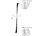
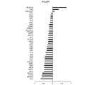
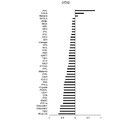
本明細書では、レクチンは糖鎖に結合する活性を有するタンパク質の総称と定義する。レクチンとしては、特に限定されず、例えば、下記表1~5に記載のレクチンを好適に使用することができる。表1~5中、「Natural」は天然物由来であることを示し、「E.coli」は遺伝子組み換え体であることを示す。また、「EY Lab.」はEYラボラトリーズ社を示し、「Wako」は富士フイルム和光純薬株式会社を示し、「Seikagaku」は生化学工業株式会社を示し、「Vector」はベクターラボラトリース社を示し、「JOM」は株式会社J-オイルミルズを示し、「AIST」は産業技術総合研究所を示す。入手先が「AIST」であるレクチンは発明者らが調製したものである(Tateno H., et al., Glycome diagnosis of human induced pluripotent stem cells using lectin microarray, J Biol Chem., 286 (23), 20345-20353, 2011. を参照。)
In the present specification, lectin is defined as a general term for proteins having an activity of binding to a sugar chain. The lectin is not particularly limited, and for example, the lectins shown in Tables 1 to 5 below can be preferably used. In Tables 1 to 5, "Natural" indicates that it is derived from a natural product, and "E. coli" indicates that it is a genetically modified organism. In addition, "EY Lab." Indicates EY Laboratories, "Wako" indicates Fujifilm Wako Pure Chemical Industries, Ltd., "Seikagaku" indicates Seikagaku Corporation, and "Vector" indicates Vector Laboratories. , "JOM" indicates J-Oil Mills Co., Ltd., and "AIST" indicates National Institute of Advanced Industrial Science and Technology. The lectins obtained from "AIST" were prepared by the inventors (Tateno H., et al., Glycome diagnosis of human induced pluripotent stem cells using lecture microarray, J Biol Chem., 286 (23), See 20345-20353, 2011.)
また、「Sia」はシアル酸を示し、「GlcNAc」はN-アセチル-グルコサミンを示し、「Man」はマンノースを示し、「Gal」はD-ガラクトースを示し、「GalNAc」はN-アセチル-ガラクトサミンを示し、「Fuc」はL-フコースを示し、「Glc」はD-グルコースを示し、「LacNAc」はN-アセチル-ラクトサミンを示す。
In addition, "Sia" indicates sialic acid, "GlcNAc" indicates N-acetyl-glucosamine, "Man" indicates mannose, "Gal" indicates D-galactose, and "GalNAc" indicates N-acetyl-galactosamine. "Fuc" indicates L-fucose, "Glc" indicates D-glucose, and "LacNAc" indicates N-acetyl-lactosamine.
レクチンとしては、糖鎖修飾されていない大腸菌由来の組み換えレクチンが好ましい。また、網羅的に糖鎖を解析するために、糖鎖を構成する単糖であるSia、Gal、GlcNAc、Man、Fuc、GalNAcを認識するレクチンを混合して用いることが好ましい。
As the lectin, a recombinant lectin derived from Escherichia coli that has not been modified with a sugar chain is preferable. Further, in order to comprehensively analyze the sugar chain, it is preferable to use a mixture of lectins that recognize the monosaccharides Sia, Gal, GlcNAc, Man, Fuc, and GalNAc constituting the sugar chain.


糖鎖結合物質を標識する核酸は、例えば環状核酸であってもよく、例えば1本鎖核酸断片であってもよく、例えば2本鎖核酸断片であってもよい。環状核酸としては、例えばプラスミドDNA等が挙げられる。また、糖鎖結合物質を標識する核酸は、DNAであってもRNAであってもよいが、安定性の観点からはDNAであることが好ましい。
The nucleic acid that labels the sugar chain binding substance may be, for example, a cyclic nucleic acid, for example, a single-stranded nucleic acid fragment, or for example, a double-stranded nucleic acid fragment. Examples of the cyclic nucleic acid include plasmid DNA and the like. The nucleic acid that labels the sugar chain-binding substance may be DNA or RNA, but from the viewpoint of stability, DNA is preferable.
特定の種類の糖鎖結合物質には特定の種類の核酸を標識することが好ましい。例えば、糖鎖結合物質の種類とこれを標識する核酸の塩基配列を対応させることにより、糖鎖結合物質をコード化することが可能となる。したがって、糖鎖結合物質を標識する核酸の塩基配列は、天然に存在しない塩基配列であることが好ましい。糖鎖結合物質を標識する核酸に特異的な塩基配列を検出することにより、それに対応する糖鎖結合物質の存在を検出することができる。
It is preferable to label a specific type of sugar chain binding substance with a specific type of nucleic acid. For example, by associating the type of sugar chain-binding substance with the base sequence of the nucleic acid that labels the sugar chain-binding substance, it becomes possible to encode the sugar chain-binding substance. Therefore, the base sequence of the nucleic acid that labels the sugar chain-binding substance is preferably a base sequence that does not exist in nature. By detecting the base sequence specific to the nucleic acid that labels the sugar chain-binding substance, the presence of the corresponding sugar chain-binding substance can be detected.
また、糖鎖結合物質に結合した核酸をPCR等により増幅することにより、糖鎖結合物質の存在を示すシグナルを増幅することができる。これにより、例えば検出感度を高めることができる。
Further, by amplifying the nucleic acid bound to the sugar chain-binding substance by PCR or the like, the signal indicating the presence of the sugar chain-binding substance can be amplified. Thereby, for example, the detection sensitivity can be increased.
糖鎖結合物質を標識する核酸が、1本鎖核酸断片又は2本鎖核酸断片である場合、その長さは、糖鎖の結合に影響を及ぼさず、対応する糖鎖結合物質を示す情報を保持することができる限り特に限定されず、例えば数十塩基(又は塩基対)から数十キロ塩基(又は塩基対)であってもよい。また、核酸はプラスミド等の環状DNAであってもよい。
When the nucleic acid labeling the sugar chain binding substance is a single-stranded nucleic acid fragment or a double-stranded nucleic acid fragment, the length does not affect the binding of the sugar chain and provides information indicating the corresponding sugar chain binding substance. It is not particularly limited as long as it can be retained, and may be, for example, several tens of bases (or base pairs) to several tens of kilobases (or base pairs). Further, the nucleic acid may be circular DNA such as a plasmid.
後述するように、糖鎖結合物質を標識する核酸は、リアルタイム定量PCRで検出してもよく、デジタルPCRで検出してもよく、次世代シーケンサーによるシーケンスにより検出してもよい。
As will be described later, the nucleic acid labeling the sugar chain binding substance may be detected by real-time quantitative PCR, digital PCR, or sequence by a next-generation sequencer.
したがって、糖鎖結合物質を標識する核酸は、PCR用プライマーがハイブリダイズすることが可能な塩基配列領域を更に有していることが好ましい。また、糖鎖結合物質を標識する核酸を次世代シーケンサーによるシーケンスにより検出する場合、糖鎖結合物質を標識する核酸は、ブリッジPCR、エマルジョンPCR等の次世代シーケンス用の前処理を可能にする塩基配列を更に有していることが好ましい。
Therefore, it is preferable that the nucleic acid labeling the sugar chain binding substance further has a base sequence region to which the PCR primer can hybridize. When a nucleic acid labeling a sugar chain-binding substance is detected by a sequence using a next-generation sequencer, the nucleic acid labeling the sugar chain-binding substance is a base that enables pretreatment for next-generation sequencing such as bridge PCR and emulsion PCR. It is preferable to have more sequences.
糖鎖結合物質を標識する核酸の長さは、50~100塩基であることが特に好ましい。このうち、糖鎖結合物質をコード化するための塩基配列が10~30塩基であり、その5’側及び3’側にそれぞれ10~30塩基のアダプター配列を付加することが好ましい。糖鎖結合物質をコード化するための塩基配列は、塩基の偏りがないように選択する。
The length of the nucleic acid labeling the sugar chain binding substance is particularly preferably 50 to 100 bases. Of these, the base sequence for encoding the sugar chain binding substance is 10 to 30 bases, and it is preferable to add an adapter sequence of 10 to 30 bases to the 5'side and the 3'side, respectively. The base sequence for encoding the sugar chain-binding substance is selected so that there is no base bias.
また、PCR用プライマーの塩基配列は、上記アダプター配列に相補的な配列に加えて、多種類の細胞を識別するための塩基配列(5~10塩基)、及び、次世代シーケンスにおいて、フローセルにハイブリダイズさせるための塩基配列(20~30塩基)を含むことが好ましい。
In addition to the sequence complementary to the adapter sequence, the base sequence of the PCR primer is a base sequence (5 to 10 bases) for identifying various types of cells, and hybridizes to the flow cell in the next-generation sequence. It preferably contains a base sequence (20 to 30 bases) for hybridizing.
実施例において後述するように、糖鎖結合物質への核酸の結合は、例えば、糖鎖結合物質に核酸結合ドメインを連結させ、当該核酸結合ドメインに核酸を結合させることにより行ってもよいし、化学リンカーを用いて糖鎖結合物質と核酸を結合させてもよいし、クリック反応を用いて糖鎖結合物質と核酸を結合させてもよい。
As will be described later in the examples, the nucleic acid may be bound to the sugar chain-binding substance, for example, by linking the nucleic acid-binding domain to the sugar-chain-binding substance and binding the nucleic acid to the nucleic acid-binding domain. A chemical linker may be used to bind the sugar chain-binding substance to the nucleic acid, or a click reaction may be used to bind the sugar chain-binding substance to the nucleic acid.
化学リンカーを利用可能とするために、核酸にはアミノ基、SH基等の官能基を導入してもよい。また、クリック反応を利用可能とするために、核酸にアジド基、アルキン基等を導入してもよい。これらの基の導入は、核酸の化学合成等により行うことができる。
In order to make a chemical linker available, a functional group such as an amino group or an SH group may be introduced into the nucleic acid. Further, in order to make the click reaction available, an azide group, an alkyne group or the like may be introduced into the nucleic acid. The introduction of these groups can be carried out by chemical synthesis of nucleic acids or the like.
また、糖鎖結合物質と核酸との間には、スペーサーが存在していてもよい。スペーサーは、特に限定されず、例えば、ポリエチレングリコール鎖、ポリアクリルアミド鎖、ポリエステル鎖、ポリウレタン鎖、これらのコポリマー等が挙げられる。スペーサーは化学リンカーに由来するものであってもよい。
Further, a spacer may exist between the sugar chain binding substance and the nucleic acid. The spacer is not particularly limited, and examples thereof include polyethylene glycol chains, polyacrylamide chains, polyester chains, polyurethane chains, copolymers thereof, and the like. The spacer may be derived from a chemical linker.
また、スペーサーは切断可能な基を含んでいてもよい。例えば、実施例において後述するように、スペーサーが、光照射により切断可能な基を含んでいる場合、核酸標識された糖鎖結合物質に光を照射することにより、糖鎖結合物質から核酸を切り離して回収すること等が可能になる。
Also, the spacer may contain a cleaveable group. For example, as will be described later in Examples, when the spacer contains a group that can be cleaved by light irradiation, the nucleic acid is separated from the sugar chain-binding substance by irradiating the nucleic acid-labeled sugar chain-binding substance with light. It becomes possible to collect it.
[糖鎖を解析する方法]
1実施形態において、本発明は、細胞の表面の糖鎖を解析する方法であって、前記細胞に、核酸標識された糖鎖結合物質を接触させることと、前記細胞に結合した前記糖鎖結合物質に標識された前記核酸を検出することと、を含み、前記核酸の種類及び量が、前記細胞の表面の糖鎖の種類及び量に対応する方法を提供する。 [Method of analyzing sugar chains]
In one embodiment, the present invention is a method for analyzing a sugar chain on the surface of a cell, in which the cell is brought into contact with a nucleic acid-labeled sugar chain binding substance and the sugar chain binding bound to the cell. Provided is a method comprising detecting the nucleic acid labeled with a substance, wherein the type and amount of the nucleic acid corresponds to the type and amount of sugar chains on the surface of the cell.
細胞としては、特に限定されず、微生物(ウイルス、細菌、真菌)、昆虫細胞、植物細胞、動物細胞等が挙げられる。また、試料として組織切片等を用いることもできる。細胞や組織切片は、細胞が生きた状態であってもよく、固定されていてもよい。
The cells are not particularly limited, and examples thereof include microorganisms (viruses, bacteria, fungi), insect cells, plant cells, animal cells, and the like. Moreover, a tissue section or the like can also be used as a sample. The cell or tissue section may be alive or fixed.
本実施形態の方法では、まず、解析対象の細胞に、核酸標識された糖鎖結合物質を接触させる。核酸標識された糖鎖結合物質としては上述したものを用いることができる。
In the method of the present embodiment, first, the cell to be analyzed is brought into contact with a nucleic acid-labeled sugar chain-binding substance. As the nucleic acid-labeled sugar chain binding substance, the above-mentioned substances can be used.
核酸標識された糖鎖結合物質は1種類を単独で細胞に接触させてもよいし、2種類以上を混合して細胞に接触させてもよい。多種類の核酸標識された糖鎖結合物質を同時に細胞に接触させることにより、細胞の表面の糖鎖構造を網羅的に解析することが可能になる。
One type of nucleic acid-labeled sugar chain binding substance may be brought into contact with cells alone, or two or more types may be mixed and brought into contact with cells. By simultaneously contacting cells with various nucleic acid-labeled sugar chain-binding substances, it becomes possible to comprehensively analyze the sugar chain structure on the surface of cells.
細胞と核酸標識された糖鎖結合物質との接触は、培地中、生理食塩水中、緩衝液中等の溶液中で、細胞と核酸標識された糖鎖結合物質を混合すること等により行うことができる。
Contact between the cell and the nucleic acid-labeled sugar chain-binding substance can be performed by mixing the cell and the nucleic acid-labeled sugar chain-binding substance in a solution such as in a medium, physiological saline, or a buffer solution. ..
中でも、細胞と核酸標識された糖鎖結合物質との接触を、アルブミンを含む緩衝液中で行うことが好ましい。実施例において後述するように、細胞と核酸標識された糖鎖結合物質との接触を、アルブミンを含む緩衝液中で行うことにより、検出シグナルを格段に増強させることができる。
Above all, it is preferable that the cells are brought into contact with the nucleic acid-labeled sugar chain-binding substance in a buffer solution containing albumin. As will be described later in the examples, the detection signal can be significantly enhanced by contacting the cells with the nucleic acid-labeled sugar chain-binding substance in a buffer solution containing albumin.
緩衝液としては、トリス緩衝液、リン酸緩衝生理食塩水等が挙げられる。リン酸緩衝生理食塩水の組成としては、例えば、NaCl 137mmol/L、KCl 2.7mmol/L、Na2HPO4 10mmol/L、KH2PO4 1.76mmol/Lが挙げられる。また、リン酸緩衝生理食塩水のpHは7.4程度に調整されていることが好ましい。
Examples of the buffer solution include Tris buffer solution, phosphate buffered saline and the like. Examples of the composition of the phosphate buffered saline include NaCl 137 mmol / L, KCl 2.7 mmol / L, Na 2 HPO 4 10 mmol / L, and KH 2 PO 4 1.76 mmol / L. Further, it is preferable that the pH of the phosphate buffered saline is adjusted to about 7.4.
アルブミンとしては、ウシ血清アルブミン、ヒト血清アルブミン等を用いることができる。アルブミンは組換え体であってもよい。緩衝液中のアルブミンの濃度は0.1~10質量%程度が好ましい。
As albumin, bovine serum albumin, human serum albumin and the like can be used. Albumin may be a recombinant. The concentration of albumin in the buffer solution is preferably about 0.1 to 10% by mass.
解析対象の細胞に接触させる、核酸標識された糖鎖結合物質の量は、細胞1個に対して、核酸標識された糖鎖結合物質1種類あたり、1分子以上を接触させればよく、細胞の表面に存在する糖鎖を飽和させることができる量であることが好ましい。
The amount of the nucleic acid-labeled sugar chain-binding substance to be brought into contact with the cell to be analyzed may be one or more molecules per one type of nucleic acid-labeled sugar chain-binding substance per cell. It is preferable that the amount is such that the sugar chains existing on the surface of the cell can be saturated.
また、解析対象の細胞に、核酸標識された糖鎖結合物質を接触させる時間は、糖鎖結合物質が解析対象の細胞の表面の糖鎖に結合するのに十分な時間であれば特に限定されず、例えば10分間~24時間程度であってもよく、例えば10分間~8時間程度であってもよく、例えば10分間~3時間程度であってもよく、例えば1時間程度であってもよい。また、核酸標識された糖鎖結合物質を接触させる温度は、糖鎖結合物質が解析対象の細胞の表面の糖鎖に結合する温度であれば特に限定されず、例えば4~37℃程度であってよい。
In addition, the time for contacting the nucleic acid-labeled sugar chain-binding substance with the cell to be analyzed is particularly limited as long as the sugar chain-binding substance is sufficiently time to bind to the sugar chain on the surface of the cell to be analyzed. It may be, for example, about 10 minutes to 24 hours, for example, about 10 minutes to 8 hours, for example, about 10 minutes to 3 hours, or for example, about 1 hour. .. The temperature at which the nucleic acid-labeled sugar chain-binding substance is brought into contact is not particularly limited as long as the sugar chain-binding substance binds to the sugar chain on the surface of the cell to be analyzed, and is, for example, about 4 to 37 ° C. It's okay.
解析対象の細胞に核酸標識された糖鎖結合物質を接触させた結果、糖鎖結合物質が解析対象の細胞の表面の糖鎖に結合する。ここで、未反応の糖鎖結合物質を除去することが好ましい。未反応の糖鎖結合物質の除去は、例えば、緩衝液を加えて細胞を遠心して上清を除去する操作を1~数回繰り返すことにより行うことができる。
As a result of contacting the nucleic acid-labeled sugar chain-binding substance with the cell to be analyzed, the sugar chain-binding substance binds to the sugar chain on the surface of the cell to be analyzed. Here, it is preferable to remove the unreacted sugar chain binding substance. The unreacted sugar chain-binding substance can be removed, for example, by repeating the operation of adding a buffer solution and centrifuging the cells to remove the supernatant one to several times.
続いて、細胞に結合した糖鎖結合物質に標識された核酸を検出する。ここで、核酸の検出は、核酸標識された糖鎖結合物質が細胞に結合した状態で行ってもよいし、核酸標識された糖鎖結合物質を細胞から解離させ、更に、核酸標識された糖鎖結合物質を細胞から分離した後に行ってもよいし、核酸標識された糖鎖結合物質から、核酸を切り離して回収した後に行ってもよい。
Subsequently, the nucleic acid labeled with the sugar chain binding substance bound to the cell is detected. Here, the nucleic acid may be detected in a state where the nucleic acid-labeled sugar chain-binding substance is bound to the cell, or the nucleic acid-labeled sugar chain-binding substance is dissociated from the cell, and the nucleic acid-labeled sugar is further detected. It may be performed after separating the chain-binding substance from the cell, or after separating and recovering the nucleic acid from the nucleic acid-labeled sugar chain-binding substance.
核酸標識された糖鎖結合物質を細胞から解離させる方法としては、例えば、細胞に糖鎖結合物質と競合する糖を反応させる方法、界面活性剤を作用させて糖鎖結合物質と細胞の表面の糖鎖との結合を解離させる方法、pHを変化させて糖鎖結合物質と細胞の表面の糖鎖との結合を解離させる方法、還元剤を作用させて糖鎖結合物質と細胞の表面の糖鎖との結合を解離させる方法等が挙げられる。
Examples of the method for dissociating the nucleic acid-labeled sugar chain-binding substance from the cell include a method in which the cell reacts with a sugar that competes with the sugar chain-binding substance, and a method in which a surfactant is allowed to act on the sugar chain-binding substance and the surface of the cell. A method of dissociating the bond with the sugar chain, a method of dissociating the bond between the sugar chain-binding substance and the sugar chain on the cell surface by changing the pH, and a method of acting a reducing agent to dissociate the sugar chain-binding substance and the sugar on the cell surface. Examples thereof include a method of dissociating the bond with the chain.
また、上述したように、核酸と糖鎖結合物質との間に切断可能な基を導入していた場合、核酸標識された糖鎖結合物質から、核酸を切り離して回収することができる。例えば、予め核酸と糖鎖結合物質との間に光照射により切断可能な基を導入していた場合、光を照射することにより、核酸標識された糖鎖結合物質から、核酸を切り離すことができる。その後、例えば遠心分離して上清を回収することにより、核酸を回収することができる。
Further, as described above, when a cleaving group is introduced between the nucleic acid and the sugar chain-binding substance, the nucleic acid can be separated and recovered from the nucleic acid-labeled sugar chain-binding substance. For example, when a group that can be cleaved by light irradiation has been introduced between the nucleic acid and the sugar chain-binding substance in advance, the nucleic acid can be separated from the nucleic acid-labeled sugar chain-binding substance by irradiating with light. .. Then, for example, the nucleic acid can be recovered by centrifuging and collecting the supernatant.
細胞に結合した糖鎖結合物質に標識された核酸の検出は、例えば、リアルタイム定量PCRにより行ってもよいし、デジタルPCRにより行ってもよいし、次世代シーケンサーによるシーケンスにより行ってもよい。核酸をPCR等により増幅することにより、検出シグナルを増幅し、検出感度を高めることができる。ここで、検出された核酸の種類及び量が、細胞の表面の糖鎖の種類及び量に対応する。
The detection of the nucleic acid labeled with the sugar chain binding substance bound to the cell may be performed by, for example, real-time quantitative PCR, digital PCR, or sequencing by a next-generation sequencer. By amplifying the nucleic acid by PCR or the like, the detection signal can be amplified and the detection sensitivity can be increased. Here, the type and amount of nucleic acid detected corresponds to the type and amount of sugar chains on the surface of the cell.
ここで、核酸の種類とは、糖鎖結合物質に標識された核酸の塩基配列の種類である。核酸の種類を特定することにより、核酸が標識されていた糖鎖結合物質の種類を特定することができる。したがって、核酸の塩基配列を特定することにより、解析対象の細胞の表面に存在していた糖鎖構造を特定することができる。また、特定の塩基配列を有する核酸の量が、解析対象の細胞に結合した糖鎖結合物質の量に対応する。すなわち、特定の塩基配列を有する核酸の量が、解析対象の細胞の表面に存在する特定の糖鎖構造の量に対応する。これにより、解析対象の細胞の表面に存在する糖鎖構造の種類及び量を定量的に解析することができる。
Here, the type of nucleic acid is the type of the base sequence of the nucleic acid labeled with the sugar chain binding substance. By specifying the type of nucleic acid, the type of sugar chain-binding substance on which the nucleic acid is labeled can be specified. Therefore, by specifying the base sequence of the nucleic acid, the sugar chain structure existing on the surface of the cell to be analyzed can be specified. In addition, the amount of nucleic acid having a specific base sequence corresponds to the amount of sugar chain-binding substance bound to the cell to be analyzed. That is, the amount of nucleic acid having a specific base sequence corresponds to the amount of a specific sugar chain structure present on the surface of the cell to be analyzed. This makes it possible to quantitatively analyze the type and amount of sugar chain structure existing on the surface of the cell to be analyzed.
リアルタイム定量PCR、デジタルPCR、次世代シーケンスは、汎用的な装置を用いて行うことができる。したがって、これらの装置が既に存在する場合には、糖鎖の解析のために新たに特殊な装置を用意しなくてもよい。
Real-time quantitative PCR, digital PCR, and next-generation sequencing can be performed using a general-purpose device. Therefore, if these devices already exist, it is not necessary to prepare a new special device for the analysis of sugar chains.
また、実施例において後述するように、本実施形態の方法は、検出感度が高いため、細胞の表面の糖鎖の解析を1細胞レベルで行うことも可能である。ここで、1細胞レベルで糖鎖を解析するとは、1個の細胞を試料に用いて、核酸標識された糖鎖結合物質を接触させ、結合した糖鎖結合物質に標識された核酸を検出し、細胞の表面の糖鎖の種類及び量を特定することを意味する。1細胞レベルでの糖鎖の解析は、従来の方法では行うことができなかった。
Further, as will be described later in Examples, since the method of this embodiment has high detection sensitivity, it is possible to analyze sugar chains on the surface of cells at the single cell level. Here, to analyze the sugar chain at the one-cell level, one cell is used as a sample, a nucleic acid-labeled sugar chain-binding substance is brought into contact with the sample, and the nucleic acid labeled with the bound sugar chain-binding substance is detected. , Means to identify the type and amount of sugar chains on the surface of cells. Analysis of sugar chains at the single cell level could not be performed by conventional methods.
また、本実施形態の方法では、細胞の表面から核酸を回収した後も細胞は生存している。このため、細胞表面の糖鎖を解析するとともに、細胞の表現型又は細胞内のRNA情報を同時に解析することができる。
Further, in the method of the present embodiment, the cells are still alive even after the nucleic acid is recovered from the surface of the cells. Therefore, it is possible to analyze the sugar chain on the cell surface and simultaneously analyze the cell phenotype or intracellular RNA information.
細胞の表現型としては、例えば、細胞の形態、増殖能、分化能、浸潤能、腫瘍形成能、マーカータンパク質の発現等が挙げられる。また、細胞内の遺伝子情報又は細胞の表現型も1細胞レベルで解析することが可能である。細胞内のRNA情報としては、例えば、mRNA、microRNA、16S rRNA、ノンコーディングRNA等の、塩基配列情報又は発現量の情報等が挙げられる。
Examples of cell phenotypes include cell morphology, proliferation ability, differentiation ability, infiltration ability, tumorigenic ability, expression of marker protein, and the like. In addition, intracellular genetic information or cell phenotype can also be analyzed at the single cell level. Examples of intracellular RNA information include nucleotide sequence information or expression level information such as mRNA, microRNA, 16S rRNA, and non-coding RNA.
[キット]
1実施形態において、本発明は、上述した、核酸標識された糖鎖結合物質を含む、細胞の表面の糖鎖を解析するためのキットを提供する。本実施形態のキットを用いることにより、上述した細胞の表面の糖鎖の解析を好適に行うことができる。 [kit]
In one embodiment, the present invention provides a kit for analyzing sugar chains on the surface of cells, which comprises the above-mentioned nucleic acid-labeled sugar chain-binding substance. By using the kit of this embodiment, the above-mentioned analysis of sugar chains on the surface of cells can be preferably performed.
本実施形態のキットは、核酸標識された糖鎖結合物質を1種類含んでいてもよいし、2種類以上、例えば10種類以上、例えば30種類以上、例えば50種類以上、例えば100種類以上含んでいてもよい。本実施形態のキットが、核酸標識された糖鎖結合物質を多種類含んでいると、細胞の表面に存在する糖鎖構造を網羅的に解析することが容易となるため好ましい。
The kit of the present embodiment may contain one kind of nucleic acid-labeled sugar chain binding substance, or may contain two or more kinds, for example, 10 kinds or more, for example, 30 kinds or more, for example, 50 kinds or more, for example, 100 kinds or more. You may. It is preferable that the kit of the present embodiment contains a large number of nucleic acid-labeled sugar chain-binding substances because it facilitates comprehensive analysis of the sugar chain structure existing on the surface of cells.
本実施形態のキットは、解析対象の細胞に、核酸標識された前記糖鎖結合物質を接触させる溶媒として、アルブミンを含む緩衝液を更に含んでいてもよい。アルブミンを含む緩衝液については、上述したものと同様である。実施例において後述するように、細胞と核酸標識された糖鎖結合物質との接触を、アルブミンを含む緩衝液中で行うことにより、バクグラウンドを抑制させることができる。
The kit of the present embodiment may further contain a buffer solution containing albumin as a solvent for contacting the nucleic acid-labeled sugar chain-binding substance with the cells to be analyzed. The buffer solution containing albumin is the same as that described above. As will be described later in the examples, the background can be suppressed by contacting the cells with the nucleic acid-labeled sugar chain-binding substance in a buffer solution containing albumin.
また、本実施形態のキットは、糖鎖結合物質に結合した核酸を検出するためのプライマーを更に含んでいてもよい。当該プライマーを用いたリアルタイム定量PCR、デジタルPCR、次世代シーケンス等により、糖鎖結合物質に結合した核酸を検出することができる。
Further, the kit of the present embodiment may further contain a primer for detecting a nucleic acid bound to a sugar chain binding substance. Nucleic acids bound to sugar chain-binding substances can be detected by real-time quantitative PCR, digital PCR, next-generation sequencing, etc. using the primers.
次に実施例を示して本発明を更に詳細に説明するが、本発明は以下の実施例に限定されるものではない。
Next, the present invention will be described in more detail with reference to Examples, but the present invention is not limited to the following Examples.
[レクチン]
下記表6に示すレクチンを使用した。また、各レクチンを核酸標識する場合には、下記表6に示す配列番号の塩基配列からなる核酸で標識した。表6中、「Seikagaku」は生化学工業株式会社を示し、「Vector」はベクターラボラトリース社を示し、「AIST」は産業技術総合研究所を示し、「Wako」は富士フイルム和光純薬株式会社を示し、「JOM」は株式会社J-オイルミルズを示す。また、「リアルタイムPCR用」は、後述する実験例においてリアルタイムPCR解析に用いた塩基配列の配列番号であることを示し、「次世代シーケンス用」は、後述する実験例において次世代シーケンスに用いた塩基配列の配列番号であることを示す。 [Lectin]
The lectins shown in Table 6 below were used. When each lectin was labeled with nucleic acid, it was labeled with a nucleic acid consisting of the nucleotide sequence of the SEQ ID NO: shown in Table 6 below. In Table 6, "Seikagaku" indicates Seikagaku Corporation, "Vector" indicates Vector Laboratory, "AIST" indicates National Institute of Advanced Industrial Science and Technology, and "Wako" indicates Fujifilm Wako Pure Chemical Industries, Ltd. , And "JOM" indicates J-Oil Mills, Inc. Further, "for real-time PCR" indicates the sequence number of the base sequence used for real-time PCR analysis in the experimental example described later, and "for next-generation sequence" was used for the next-generation sequence in the experimental example described later. Indicates that it is the sequence number of the base sequence.

[実験例1]
(核酸標識レクチンの調製1)
核酸標識レクチンの調製を試みた。まず、レクチンと核酸結合ドメインとの融合タンパク質を調製した。レクチンとして、BC2LCNレクチンを使用した。BC2LCNレクチンのアミノ酸配列を配列番号1に示す。 [Experimental Example 1]
(Preparation of nucleic acid-labeled lectin 1)
An attempt was made to prepare a nucleic acid-labeled lectin. First, a fusion protein of a lectin and a nucleic acid binding domain was prepared. BC2LCN lectin was used as the lectin. The amino acid sequence of BC2LCN lectin is shown in SEQ ID NO: 1.
また、核酸結合ドメインとして、アルギニン残基が4個連続したペプチド(配列番号2、以下、「R4」という。)、アルギニン残基が5個連続したペプチド(配列番号3、以下、「R5」という。)、アルギニン残基が6個連続したペプチド(配列番号4、以下、「R6」という。)、アルギニン残基が7個連続したペプチド(配列番号5、以下、「R7」という。)、アルギニン残基が10個連続したペプチド(配列番号6、以下、「R10」という。)、及び、ミトコンドリア転写因子A(TFAM)のHMG BoxAドメイン(配列番号7、以下、「TFAM」という。)を使用した。なお、アルギニンは塩基性であり、リン酸部分を持つ核酸と結合しやすいことが知られている。
Further, as the nucleic acid binding domain, a peptide having four consecutive arginine residues (SEQ ID NO: 2, hereinafter referred to as "R4") and a peptide having five consecutive arginine residues (SEQ ID NO: 3, hereinafter referred to as "R5"). ), A peptide having 6 consecutive arginine residues (SEQ ID NO: 4, hereinafter referred to as "R6"), a peptide having 7 consecutive arginine residues (SEQ ID NO: 5, hereinafter referred to as "R7"), arginine. A peptide having 10 consecutive residues (SEQ ID NO: 6, hereinafter referred to as "R10") and the HMG BoxA domain of mitochondrial transcription factor A (TFAM) (SEQ ID NO: 7, hereinafter referred to as "TFAM") are used. bottom. It is known that arginine is basic and easily binds to a nucleic acid having a phosphoric acid moiety.
まず、各核酸結合ドメインをC末端側に結合させた、組換えBC2LCNレクチン(以下、「rBC2LCN」という。)の融合タンパク質を大腸菌で発現させて精製した。各融合タンパク質のN末端側にはFLAGタグを導入した。
First, a fusion protein of recombinant BC2LCN lectin (hereinafter referred to as "rBC2LCN") in which each nucleic acid binding domain was bound to the C-terminal side was expressed in Escherichia coli and purified. A FLAG tag was introduced on the N-terminal side of each fusion protein.
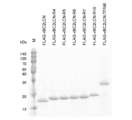
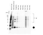
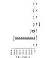
図1は、精製した各融合タンパク質をSDS-ポリアクリルアミドゲル電気泳動(SDS-PAGE)で分離後、クマシーブリリアントブルー(CBB)染色した結果を示す写真である。
FIG. 1 is a photograph showing the results of coomassie brilliant blue (CBB) staining after separating each purified fusion protein by SDS-polyacrylamide gel electrophoresis (SDS-PAGE).
図1中、「M」は分子量マーカーを示し、「FLAG-rBC2LCN」は核酸結合ドメインを有しない融合タンパク質を示し、「FLAG-rBC2LCN-R4」は「R4」を連結した融合タンパク質を示し、「FLAG-rBC2LCN-R5」は「R5」を連結した融合タンパク質を示し、「FLAG-rBC2LCN-R6」は「R6」を連結した融合タンパク質を示し、「FLAG-rBC2LCN-R7」は「R7」を連結した融合タンパク質を示し、「FLAG-rBC2LCN-R10」は「R10」を連結した融合タンパク質を示し、「FLAG-rBC2LCN-TFAM」は「TFAM」を連結した融合タンパク質を示す。
In FIG. 1, "M" indicates a molecular weight marker, "FLAG-rBC2LCN" indicates a fusion protein having no nucleic acid binding domain, and "FLAG-rBC2LCN-R4" indicates a fusion protein linked with "R4". "FLAG-rBC2LCN-R5" indicates a fusion protein linked with "R5", "FLAG-rBC2LCN-R6" indicates a fusion protein linked with "R6", and "FLAG-rBC2LCN-R7" indicates a fusion protein linked with "R7". "FLAG-rBC2LCN-R10" indicates a fusion protein linked with "R10", and "FLAG-rBC2LCN-TFAM" indicates a fusion protein linked with "TFAM".
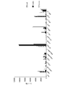
続いて、精製した各融合タンパク質とプラスミドDNA(pCR2.1)との結合性を解析した。具体的には、プラスミドDNA(pCR2.1) 1μgと、1、2、3、4、5μgの各融合タンパク質を混合し、アガロース電気泳動に供した。
Subsequently, the binding property between each purified fusion protein and plasmid DNA (pCR2.1) was analyzed. Specifically, 1 μg of plasmid DNA (pCR2.1) and 1, 2, 3, 4, 5 μg of each fusion protein were mixed and subjected to agarose gel electrophoresis.
図2は、アガロース電気泳動の結果を示す写真である。図2中、「M」は分子量マーカーを示し、「rBC2LCN」は核酸結合ドメインを有しない融合タンパク質を示し、「rBC2LCN-R4」は「R4」を連結した融合タンパク質を示し、「rBC2LCN-R5」は「R5」を連結した融合タンパク質を示し、「rBC2LCN-R6」は「R6」を連結した融合タンパク質を示し、「rBC2LCN-R7」は「R7」を連結した融合タンパク質を示し、「rBC2LCN-R10」は「R10」を連結した融合タンパク質を示し、「rBC2LCN-TFAM」は「TFAM」を連結した融合タンパク質を示す。
FIG. 2 is a photograph showing the results of agarose gel electrophoresis. In FIG. 2, "M" indicates a molecular weight marker, "rBC2LCN" indicates a fusion protein having no nucleic acid binding domain, "rBC2LCN-R4" indicates a fusion protein linked with "R4", and "rBC2LCN-R5". Indicates a fusion protein linked with "R5", "rBC2LCN-R6" indicates a fusion protein linked with "R6", "rBC2LCN-R7" indicates a fusion protein linked with "R7", and "rBC2LCN-R10" indicates a fusion protein linked with "R7". "" Indicates a fusion protein in which "R10" is linked, and "rBC2LCN-TFAM" indicates a fusion protein in which "TFAM" is linked.
その結果、アルギニン残基5個以上又はTFAMを融合させた融合タンパク質は、プラスミドDNAと結合することが明らかとなった。したがって、レクチンと核酸結合ドメインとの融合タンパク質に、核酸を混合することにより、核酸標識レクチンを調製することができることが明らかとなった。
As a result, it was clarified that the fusion protein in which 5 or more arginine residues or TFAM was fused binds to the plasmid DNA. Therefore, it was clarified that a nucleic acid-labeled lectin can be prepared by mixing a nucleic acid with a fusion protein of a lectin and a nucleic acid binding domain.
[実験例2]
(核酸標識レクチンの調製2)
市販のキット(Protein-oligo conjugation kit、カタログ番号「S-9011-1」、Solulink社)を用いて、核酸標識レクチンの調製を試みた。 [Experimental Example 2]
(Preparation of nucleic acid-labeled lectin 2)
A commercially available kit (Protein-oligo conjugation kit, catalog number "S-9011-1", Solulink) was used to attempt to prepare a nucleic acid-labeled lectin.
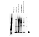
このキットでは、まず、5’末端又は3’末端がアミノ基修飾されたオリゴヌクレオチドのアミノ基に、N-スクシンイミジル-4-ホルミルベンズアミド(以下、「S-4FB」という。)を結合させて4-ホルミルベンズアミド(4FB)化オリゴヌクレオチド(「4FB-オリゴヌクレオチド」)を調製する。
In this kit, first, N-succinimidyl-4-formylbenzamide (hereinafter referred to as "S-4FB") is bound to the amino group of an oligonucleotide having an amino group-modified 5'end or 3'end, and 4 -Formylbenzamide (4FB) -modified oligonucleotide ("4FB-oligonucleotide") is prepared.
また、タンパク質に、スクシンイミジル-4-ヒドラジノニコチネートアセトンヒドラゾン(以下、「S-HyNic」という。)を結合させてヒドラジノニコチネートアセトンヒドラゾン(HyNic)化タンパク質(「HyNic-タンパク質」)を調製する。
Further, a hydrazinonicotinate acetone hydrazone (HyNic) -ized protein (“HyNic-protein”) is prepared by binding succinimidyl-4-hydrazinonicotinate acetone hydrazone (hereinafter referred to as “S-HyNic”) to the protein. do.
続いて、上記4FB-オリゴヌクレオチドと上記HyNic-タンパク質を混合すると、両者が共有結合し、核酸標識タンパク質を得ることができる。
Subsequently, when the above 4FB-oligonucleotide and the above HyNic-protein are mixed, both can be covalently bound to obtain a nucleic acid-labeled protein.
本実験例では、キットの説明書にしたがって、rBC2LCNにオリゴヌクレオチドを結合させ、核酸標識レクチンを調製した。レクチンとして、rBC2LCNを使用した。また、核酸として、配列番号8に示す塩基配列を有するオリゴヌクレオチドを使用した。
In this experimental example, a nucleic acid-labeled lectin was prepared by binding an oligonucleotide to rBC2LCN according to the instructions of the kit. As the lectin, rBC2LCN was used. Further, as the nucleic acid, an oligonucleotide having the nucleotide sequence shown in SEQ ID NO: 8 was used.
図3は、核酸標識レクチンの調製の各過程の試料をSDS-PAGEに供し、銀染色した結果を示す写真である。図3中、「4FB-oligo」は4FB化したオリゴヌクレオチドを表し、「HyNic-rBC2LCN」はHyNic化したrBC2LCNを表し、「Crude complex」は未精製の反応物を表し、「Purified complex(conc)」は精製し濃縮した反応物を表す。また、「*」はオリゴヌクレオチドが結合したrBC2LCNを示し、矢印はオリゴヌクレオチドが結合していないrBC2LCNを示す。
FIG. 3 is a photograph showing the results of silver staining of samples from each process of preparation of nucleic acid-labeled lectins by subjecting them to SDS-PAGE. In FIG. 3, "4FB-oligo" represents a 4FB-ized oligonucleotide, "HyNic-rBC2LCN" represents a HyNicized rBC2LCN, "Crede complex" represents an unpurified reactant, and "Purified complex (conc)". Represents a purified and concentrated reactant. Further, "*" indicates rBC2LCN to which the oligonucleotide is bound, and the arrow indicates rBC2LCN to which the oligonucleotide is not bound.
その結果、最終産物(Purified complex(conc))において、オリゴヌクレオチドが結合して分子量が大きくなったrBC2LCNが検出された。この結果から、市販のキットを使用することにより、核酸標識レクチンを調製することができることが明らかとなった。
As a result, rBC2LCN having an increased molecular weight due to the binding of oligonucleotides was detected in the final product (Purified complex (conc)). From this result, it was clarified that the nucleic acid-labeled lectin can be prepared by using a commercially available kit.
[実験例3]
(核酸標識レクチンの調製3)
アジド基とアルキン基とのクリック反応を利用して、核酸標識レクチンの調製を試みた。具体的には、まず、rBC2LCNに、15倍、30倍及び50倍モル濃度のジベンゾシクロオクチン-N-ヒドロキシスクシンイミジルエステル(NHS-DBCO)を、それぞれ室温で1時間反応させ、DBCO化した。続いて、DBCO化した各rBC2LCNに、10倍モル濃度のアジド化オリゴヌクレオチド(配列番号8)を混合し、4℃で一晩反応させた。オリゴヌクレオチドは5’末端をアジド化修飾した。 [Experimental Example 3]
(Preparation of nucleic acid-labeled lectin 3)
An attempt was made to prepare a nucleic acid-labeled lectin using a click reaction between an azide group and an alkyne group. Specifically, first, rBC2LCN was reacted with 15-fold, 30-fold, and 50-fold molar concentrations of dibenzocyclooctyne-N-hydroxysuccinimidyl ester (NHS-DBCO) for 1 hour at room temperature to obtain DBCO. .. Subsequently, each rBC2LCN converted to DBCO was mixed with a 10-fold molar concentration of azide oligonucleotide (SEQ ID NO: 8) and reacted at 4 ° C. overnight. The oligonucleotide was azide-modified at the 5'end.
図4は、各過程の試料をSDS-PAGEに供し、銀染色した結果を示す写真である。図4中、「DBCO-rBC2LCN」はDBCO化したrBC2LCNを表し、「15×」、「30×」、「50×」は、それぞれ15倍、30倍、50倍モル濃度のNHS-DBCOを反応させた結果であることを表し、「Crude complex」は未精製の反応物を表す。また、「*」はオリゴヌクレオチドが結合したrBC2LCNを示し、矢印はオリゴヌクレオチドが結合していないrBC2LCNを示す。
FIG. 4 is a photograph showing the results of silver-staining the samples of each process by subjecting them to SDS-PAGE. In FIG. 4, "DBCO-rBC2LCN" represents rBC2LCN converted to DBCO, and "15x", "30x", and "50x" react with NHS-DBCO having 15-fold, 30-fold, and 50-fold molar concentrations, respectively. "Crude complex" represents an unpurified reaction product. Further, "*" indicates rBC2LCN to which the oligonucleotide is bound, and the arrow indicates rBC2LCN to which the oligonucleotide is not bound.
その結果、オリゴヌクレオチドが結合して分子量が大きくなったrBC2LCNが検出された。この結果から、クリック反応により、効率よく核酸標識レクチンを調製することができることが明らかとなった。
As a result, rBC2LCN with an increased molecular weight due to the binding of oligonucleotides was detected. From this result, it was clarified that the nucleic acid-labeled lectin can be efficiently prepared by the click reaction.
[実験例4]
(核酸標識レクチンの調製4)
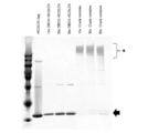
アジド基とアルキン基とのクリック反応を利用して、核酸標識レクチンを調製した。具体的には、まず、rBC2LCNに2倍モル濃度のジベンゾシクロオクチン-N-ヒドロキシスクシンイミジルエステル(NHS-DBCO)を室温で1時間反応させ、DBCO化した。続いて、DBCO化したrBC2LCNに、10倍モル濃度のアジド化オリゴヌクレオチド(配列番号8)を混合して4℃で一晩反応させ、核酸標識レクチンを得た。オリゴヌクレオチドは5’末端をアジド化修飾した。続いて、フコースセファロースを用いたアフィニティークロマトグラフィーで核酸標識レクチンを精製した。 [Experimental Example 4]
(Preparation of nucleic acid-labeled lectin 4)
Nucleic acid-labeled lectins were prepared using the click reaction between the azide group and the alkyne group. Specifically, first, rBC2LCN was reacted with a 2-fold molar concentration of dibenzocyclooctyne-N-hydroxysuccinimidyl ester (NHS-DBCO) at room temperature for 1 hour to convert it into DBCO. Subsequently, a 10-fold molar concentration of azide oligonucleotide (SEQ ID NO: 8) was mixed with DBCO-modified rBC2LCN and reacted at 4 ° C. overnight to obtain a nucleic acid-labeled lectin. The oligonucleotide was azide-modified at the 5'end. Subsequently, the nucleic acid-labeled lectin was purified by affinity chromatography using fucose sepharose.
(核酸標識レクチンの調製4)
アジド基とアルキン基とのクリック反応を利用して、核酸標識レクチンを調製した。具体的には、まず、rBC2LCNに2倍モル濃度のジベンゾシクロオクチン-N-ヒドロキシスクシンイミジルエステル(NHS-DBCO)を室温で1時間反応させ、DBCO化した。続いて、DBCO化したrBC2LCNに、10倍モル濃度のアジド化オリゴヌクレオチド(配列番号8)を混合して4℃で一晩反応させ、核酸標識レクチンを得た。オリゴヌクレオチドは5’末端をアジド化修飾した。続いて、フコースセファロースを用いたアフィニティークロマトグラフィーで核酸標識レクチンを精製した。 [Experimental Example 4]
(Preparation of nucleic acid-labeled lectin 4)
Nucleic acid-labeled lectins were prepared using the click reaction between the azide group and the alkyne group. Specifically, first, rBC2LCN was reacted with a 2-fold molar concentration of dibenzocyclooctyne-N-hydroxysuccinimidyl ester (NHS-DBCO) at room temperature for 1 hour to convert it into DBCO. Subsequently, a 10-fold molar concentration of azide oligonucleotide (SEQ ID NO: 8) was mixed with DBCO-modified rBC2LCN and reacted at 4 ° C. overnight to obtain a nucleic acid-labeled lectin. The oligonucleotide was azide-modified at the 5'end. Subsequently, the nucleic acid-labeled lectin was purified by affinity chromatography using fucose sepharose.
図5は、核酸標識レクチンの精製の各過程の試料をSDS-PAGEに供し、銀染色した結果を示す写真である。図5中、「DBCO-rBC2LCN」はDBCO化したrBC2LCNを表し、「Crude complex」は未精製の核酸標識レクチンを表す。また、「Through」は、アフィニティーカラムを通過した試料を表し、「Wash1」、「Wash2」、「Wash3」は、それぞれ洗浄1回目、2回目、3回目の洗浄液を表し、「Elute1」、「Elute2」、「Elute3」は、それぞれ溶出1回目、2回目、3回目の溶出液を表す。また、「*」はオリゴヌクレオチドが結合したrBC2LCNを示し、矢印はオリゴヌクレオチドが結合していないrBC2LCNを示す。
FIG. 5 is a photograph showing the results of silver staining of samples from each process of purification of nucleic acid-labeled lectins by subjecting them to SDS-PAGE. In FIG. 5, "DBCO-rBC2LCN" represents DBCO-converted rBC2LCN, and "Crede complex" represents an unpurified nucleic acid-labeled lectin. Further, "Through" represents a sample that has passed through an affinity column, and "Wash1", "Wash2", and "Wash3" represent the first, second, and third cleaning solutions, respectively, and "Elute1" and "Elute2". "," Elute3 "represents the first, second, and third eluates, respectively. Further, "*" indicates rBC2LCN to which the oligonucleotide is bound, and the arrow indicates rBC2LCN to which the oligonucleotide is not bound.
その結果、オリゴヌクレオチドが結合して分子量が大きくなったrBC2LCNが検出された。この結果から、クリック反応により、核酸標識レクチンが調製されたことが確認された。
As a result, rBC2LCN with an increased molecular weight due to the binding of oligonucleotides was detected. From this result, it was confirmed that the nucleic acid-labeled lectin was prepared by the click reaction.
続いて、rBC2LCNと同様の方法により、ABAレクチンの組換え体(rABA)、LSLNレクチンの組換え体(rLSLN)、SNAレクチン(カタログ番号「L-1300」、Vector社)、GSLIIレクチン(カタログ番号「L-1210」、Vector社)、AALレクチンの組換え体(rAAL)、コンカナバリンA(ConA、カタログ番号「300036」、生化学工業社)に、上記表6に示すオリゴヌクレオチドを結合させた核酸標識レクチンをそれぞれ調製した。
Subsequently, by the same method as rBC2LCN, a recombinant of ABA lectin (rABA), a recombinant of LSLN lectin (rLSRN), SNA lectin (catalog number "L-1300", Vector), GSLII lectin (catalog number). "L-1210", Vector), a recombinant of AAL lectin (rAAL), concanavalin A (ConA, catalog number "3000036", Biochemical Industry Co., Ltd.), and a nucleic acid in which the oligonucleotides shown in Table 6 above are bound. Labeled lectins were prepared respectively.
各レクチンは、各レクチンが結合する糖をそれぞれ固定化したセファロースビーズを用いたアフィニティークロマトグラフィーにより精製した。具体的には、rABAの精製にはN-アセチルグルコサミンを固定化したセファロースビーズを使用し、rLSLNの精製にはガラクトースを固定化したセファロースビーズを使用し、SNAの精製にはラクトースを固定化したセファロースビーズを使用し、GSLIIの精製にはN-アセチルグルコサミンを固定化したセファロースビーズを使用し、rAALの精製にはフコースを固定化したセファロースビーズを使用し、ConAの精製にはマンノースを固定化したセファロースビーズを使用した。
Each lectin was purified by affinity chromatography using Sepharose beads in which the sugar to which each lectin was bound was immobilized. Specifically, N-acetylglucosamine-immobilized Sepharose beads were used for purification of rABA, galactose-immobilized Sepharose beads were used for purification of rLSRN, and lactose was immobilized for SNA purification. Sepharose beads are used, N-acetylglucosamine-immobilized Sepharose beads are used for GSLII purification, fucose-immobilized Sepharose beads are used for rAAL purification, and mannose is immobilized for ConA purification. The Sepharose beads were used.
精製の各過程の試料をSDS-PAGEに供し、銀染色した結果、rABA、rLSLN、SNA、GSLII、rAAL、ConAのそれぞれを核酸標識できたことが確認された。
Samples from each process of purification were subjected to SDS-PAGE and silver-stained. As a result, it was confirmed that rABA, rLSRN, SNA, GSLII, rAAL and ConA could be labeled with nucleic acids.
[実験例5]
(核酸標識レクチンの調製5)
アジド基とアルキン基とのクリック反応を利用して、光照射により核酸を遊離させることが可能な核酸標識レクチンを調製した。具体的には、まず、rBC2LCNに16倍モル濃度のNHS-PC-DBCOエステル(カタログ番号「1160」、クリックケミストリーツールズ社)を室温で1時間反応させ、PC-DBCO化した。 [Experimental Example 5]
(Preparation of nucleic acid-labeled lectin 5)
Using the click reaction between the azide group and the alkyne group, a nucleic acid-labeled lectin capable of releasing nucleic acid by light irradiation was prepared. Specifically, first, a 16-fold molar concentration of NHS-PC-DBCO ester (catalog number "1160", Click Chemistry Tools Co., Ltd.) was reacted with rBC2LCN at room temperature for 1 hour to form PC-DBCO.
続いて、PC-DBCO化したrBC2LCNに、10倍モル濃度のアジド化オリゴヌクレオチド(配列番号8)を混合して4℃で一晩反応させ、核酸標識レクチンを得た。オリゴヌクレオチドは5’末端をアジド化修飾した。続いて、フコースセファロースを用いたアフィニティークロマトグラフィーで核酸標識レクチンを精製した。
Subsequently, a 10-fold molar concentration of azide oligonucleotide (SEQ ID NO: 8) was mixed with PC-DBCO-modified rBC2LCN and reacted at 4 ° C. overnight to obtain a nucleic acid-labeled lectin. The oligonucleotide was azide-modified at the 5'end. Subsequently, the nucleic acid-labeled lectin was purified by affinity chromatography using fucose sepharose.
図6は、核酸標識レクチンの精製の各過程の試料をSDS-PAGEに供し、銀染色した結果を示す写真である。図5中、「PC-DBCO-rBC2LCN」はPC-DBCO化したrBC2LCNを表し、「Through」は、アフィニティーカラムを通過した試料を表し、「Wash1」、「Wash2」、「Wash3」は、それぞれ洗浄1回目、2回目、3回目の洗浄液を表し、「Elute1」、「Elute2」、「Elute3」は、それぞれ溶出1回目、2回目、3回目の溶出液を表す。また、「*」はオリゴヌクレオチドが結合したrBC2LCNを示し、矢印はオリゴヌクレオチドが結合していないrBC2LCNを示す。
FIG. 6 is a photograph showing the results of silver staining of samples from each process of purification of nucleic acid-labeled lectins by subjecting them to SDS-PAGE. In FIG. 5, "PC-DBCO-rBC2LCN" represents PC-DBCO-converted rBC2LCN, "Through" represents a sample that has passed through an affinity column, and "Wash1," "Wash2," and "Wash3" are washed. The first, second, and third cleaning solutions are represented, and "Elute1", "Elute2", and "Elute3" represent the first, second, and third elution solutions, respectively. Further, "*" indicates rBC2LCN to which the oligonucleotide is bound, and the arrow indicates rBC2LCN to which the oligonucleotide is not bound.
その結果、オリゴヌクレオチドが結合して分子量が大きくなったrBC2LCNが検出された。この結果から、クリック反応により、光照射により核酸を遊離させることが可能な核酸標識レクチンが調製されたことが確認された。
As a result, rBC2LCN with an increased molecular weight due to the binding of oligonucleotides was detected. From this result, it was confirmed that a nucleic acid-labeled lectin capable of releasing nucleic acid by light irradiation was prepared by a click reaction.
続いて、rBC2LCNと同様の方法により、rABA、rLSLN、SNAレクチン(カタログ番号「L-1300」、Vector社)、GSLIIレクチン(カタログ番号「L-1210」、Vector社)、rAAL、コンカナバリンA(ConA、カタログ番号「300036」、生化学工業社)に、上記表6に示すオリゴヌクレオチドを光照射により開裂可能に結合させた核酸標識レクチンをそれぞれ調製した。
Subsequently, rABA, rLSRN, SNA lectin (catalog number "L-1300", Vector), GSLII lectin (catalog number "L-1210", Vector), rAAL, concanavalin A (ConA) by the same method as rBC2LCN. , Catalog No. "3000036", Biochemical Industry Co., Ltd.), respectively, prepared nucleic acid-labeled lectins in which the oligonucleotides shown in Table 6 above were cleavably bound by light irradiation.
各レクチンは、各レクチンが結合する糖をそれぞれ固定化したセファロースビーズを用いたアフィニティークロマトグラフィーにより精製した。精製の各過程の試料をSDS-PAGEに供し、銀染色した結果、rABA、rLSLN、SNA、GSLII、rAAL、ConAのそれぞれに、各オリゴヌクレオチドを光照射により開裂可能に結合できたことが確認された。
Each lectin was purified by affinity chromatography using Sepharose beads in which the sugar to which each lectin was bound was immobilized. Samples from each process of purification were subjected to SDS-PAGE and silver-stained. As a result, it was confirmed that each oligonucleotide could be cleaved to each of rABA, rLSRN, SNA, GSLII, rAAL and ConA by light irradiation. rice field.
[実験例6]
(核酸標識レクチンと細胞との反応条件の検討)
核酸標識レクチンと細胞との反応条件を検討した。具体的には、以下の方法1及び方法2の条件で核酸標識レクチンと細胞とを反応させ、比較した。 [Experimental Example 6]
(Examination of reaction conditions between nucleic acid-labeled lectins and cells)
The reaction conditions between the nucleic acid-labeled lectin and the cells were examined. Specifically, the nucleic acid-labeled lectin was reacted with cells under the conditions ofMethod 1 and Method 2 below, and compared.
(核酸標識レクチンと細胞との反応条件の検討)
核酸標識レクチンと細胞との反応条件を検討した。具体的には、以下の方法1及び方法2の条件で核酸標識レクチンと細胞とを反応させ、比較した。 [Experimental Example 6]
(Examination of reaction conditions between nucleic acid-labeled lectins and cells)
The reaction conditions between the nucleic acid-labeled lectin and the cells were examined. Specifically, the nucleic acid-labeled lectin was reacted with cells under the conditions of
《方法1》
核酸標識レクチンとして、実験例4と同様にして調製した、核酸標識rBC2LCNレクチンを使用した。また、細胞として、いずれもヒト膵臓癌由来細胞である、MIAPaCa-2細胞、BxPC-3細胞及びCapan-1細胞を用いた。 <<Method 1 >>
As the nucleic acid-labeled lectin, the nucleic acid-labeled rBC2LCN lectin prepared in the same manner as in Experimental Example 4 was used. In addition, as cells, MIAPaCa-2 cells, BxPC-3 cells and Capan-1 cells, which are all human pancreatic cancer-derived cells, were used.
核酸標識レクチンとして、実験例4と同様にして調製した、核酸標識rBC2LCNレクチンを使用した。また、細胞として、いずれもヒト膵臓癌由来細胞である、MIAPaCa-2細胞、BxPC-3細胞及びCapan-1細胞を用いた。 <<
As the nucleic acid-labeled lectin, the nucleic acid-labeled rBC2LCN lectin prepared in the same manner as in Experimental Example 4 was used. In addition, as cells, MIAPaCa-2 cells, BxPC-3 cells and Capan-1 cells, which are all human pancreatic cancer-derived cells, were used.
まず、1×10⁵個の各細胞を、ウシ血清アルブミン(BSA)を1質量%含むリン酸緩衝生理食塩水(PBS)(以下、「BSA-PBS」という場合がある。)100μLにそれぞれ懸濁した。続いて、100ngの核酸標識rBC2LCNレクチンを、各細胞懸濁液に添加し、4℃で1時間反応させた。続いて、各細胞を1mLのBSA-PBSで3回洗浄後、200μLのBSA-PBSに懸濁した。
First, each of 1 × 10 5 cells is suspended in 100 μL of phosphate buffered saline (PBS) (hereinafter, may be referred to as “BSA-PBS”) containing 1% by mass of bovine serum albumin (BSA). It became cloudy. Subsequently, 100 ng of nucleic acid-labeled rBC2LCN lectin was added to each cell suspension and reacted at 4 ° C. for 1 hour. Subsequently, each cell was washed 3 times with 1 mL of BSA-PBS and then suspended in 200 μL of BSA-PBS.
続いて、1×10⁴個の生きた細胞を新しいチューブにとり、遠心して上清を除き、0.2Mフコース100μLを加えて4℃で30分間反応させ、核酸標識rBC2LCNレクチンを細胞から遊離させた。続いて、15000rpmで10分間遠心して、上清を回収し、リアルタイム定量PCRでrBC2LCNレクチンに結合したオリゴヌクレオチドを定量した。
Subsequently, 1 × 10 4 live cells were placed in a new tube, centrifuged to remove the supernatant, 100 μL of 0.2 M fucose was added, and the mixture was reacted at 4 ° C. for 30 minutes to release the nucleic acid-labeled rBC2LCN lectin from the cells. .. Subsequently, the supernatant was collected by centrifugation at 15,000 rpm for 10 minutes, and the oligonucleotide bound to the rBC2LCN lectin was quantified by real-time quantitative PCR.
また、比較のために、R-フィコエリスリン標識したrBC2LCNレクチンで各細胞を染色し、フローサイトメトリー解析を行った。
For comparison, each cell was stained with R-phycoerythrin-labeled rBC2LCN lectin, and flow cytometric analysis was performed.
《方法2》
方法2では、核酸標識レクチン及び細胞の反応を、BSA-PBS中ではなく、OptiMEM培地中で行った点において、方法1と主に異なっていた。 <<Method 2 >>
Method 2 was primarily different from Method 1 in that the reaction between the nucleic acid-labeled lectin and the cells was performed in OptiMEM medium rather than in BSA-PBS.
方法2では、核酸標識レクチン及び細胞の反応を、BSA-PBS中ではなく、OptiMEM培地中で行った点において、方法1と主に異なっていた。 <<
核酸標識レクチン及び細胞として、方法1と同様のものを使用した。まず、1×10⁵個の各細胞を、OptiMEM培地(サーモフィッシャーサイエンティフィック社)100μLにそれぞれ懸濁した。続いて、100ngの核酸標識rBC2LCNレクチンを、各細胞懸濁液に添加し、4℃で1時間反応させた。続いて、各細胞を1mLのBSA-PBSで3回洗浄後、200μLのPBSに懸濁した。
As the nucleic acid-labeled lectin and cells, the same ones as in Method 1 were used. First, each of 1 × 10 5 cells was suspended in 100 μL of OptiMEM medium (Thermo Fisher Scientific). Subsequently, 100 ng of nucleic acid-labeled rBC2LCN lectin was added to each cell suspension and reacted at 4 ° C. for 1 hour. Subsequently, each cell was washed 3 times with 1 mL of BSA-PBS and then suspended in 200 μL of PBS.
続いて、1×10⁴個の生きた細胞を新しいチューブにとり、遠心して上清を除き、0.2Mフコース100μLを加えて4℃で30分間反応させ、核酸標識rBC2LCNレクチンを細胞から遊離させた。
Subsequently, 1 × 10 4 live cells were placed in a new tube, centrifuged to remove the supernatant, 100 μL of 0.2 M fucose was added, and the mixture was reacted at 4 ° C. for 30 minutes to release the nucleic acid-labeled rBC2LCN lectin from the cells. ..
続いて、15000rpmで10分間遠心して、上清を回収し、リアルタイム定量PCRでrBC2LCNレクチンに結合したオリゴヌクレオチドを定量した。
Subsequently, the supernatant was collected by centrifugation at 15000 rpm for 10 minutes, and the oligonucleotide bound to rBC2LCN lectin was quantified by real-time quantitative PCR.
図7Aは、リアルタイム定量PCRの結果を示すグラフである。図7A中、「BSA-PBS」は方法1の結果であることを示し、「OptiMEM」は方法2の結果であることを示す。その結果、方法1を使用する方が高い感度で細胞とレクチンとの結合を検出できることが明らかとなった。
FIG. 7A is a graph showing the results of real-time quantitative PCR. In FIG. 7A, "BSA-PBS" indicates the result of Method 1, and "OptiMEM" indicates the result of Method 2. As a result, it was clarified that the binding between the cell and the lectin can be detected with higher sensitivity by using the method 1.
また、図7Bは、フローサイトメトリー解析の結果を示すグラフである。その結果、図7Aに示すリアルタイム定量PCRによる解析結果は、フローサイトメトリー解析の結果と一致し、rBC2LCNレクチンはCapan-1細胞に最も高い反応性を示し、BxPC-3細胞にも反応したが、MIAPaCa-2細胞には反応しなかったことが明らかとなった。
FIG. 7B is a graph showing the results of flow cytometry analysis. As a result, the analysis result by real-time quantitative PCR shown in FIG. 7A was in agreement with the result of flow cytometry analysis, and rBC2LCN lectin showed the highest reactivity to Capan-1 cells and also reacted to BxPC-3 cells. It was revealed that it did not react with MIAPaCa-2 cells.
[実験例7]
(光照射による核酸の遊離条件の検討)
光照射により核酸を遊離させることが可能な核酸標識レクチンから、核酸を遊離させる条件を検討した。核酸標識レクチンとして、実験例5と同様にして調製した、核酸標識rBC2LCNレクチンを使用した。また、細胞として、ヒト膵臓癌由来細胞であるCapan-1細胞を用いた。 [Experimental Example 7]
(Examination of nucleic acid release conditions by light irradiation)
The conditions for releasing nucleic acid from a nucleic acid-labeled lectin capable of releasing nucleic acid by light irradiation were investigated. As the nucleic acid-labeled lectin, the nucleic acid-labeled rBC2LCN lectin prepared in the same manner as in Experimental Example 5 was used. In addition, as cells, Capan-1 cells, which are cells derived from human pancreatic cancer, were used.
まず、1×10⁵個の細胞をBSA-PBS 100μLに懸濁した。続いて、100ngの核酸標識rBC2LCNレクチンを、細胞懸濁液に添加し、4℃で1時間反応させた。続いて、細胞を1mLのBSA-PBSで3回洗浄後、200μLのBSA-PBSに懸濁した。
First, 1 × 10 5 cells were suspended in 100 μL of BSA-PBS. Subsequently, 100 ng of nucleic acid-labeled rBC2LCN lectin was added to the cell suspension and reacted at 4 ° C. for 1 hour. Subsequently, the cells were washed 3 times with 1 mL of BSA-PBS and then suspended in 200 μL of BSA-PBS.
続いて、細胞を1×10⁴個ずつ新しいチューブにとり、紫外線(UV)照射装置(カタログ番号「95-0042-14」、フナコシ株式会社)を用いて、波長365nmにピーク波長を有する紫外線を15Wで5分間、10分間又は15分間照射してオリゴヌクレオチドを遊離させた。続いて、15000rpmで10分間遠心して上清を回収し、リアルタイム定量PCRでrBC2LCNレクチンに結合したオリゴヌクレオチドを定量した。
Subsequently, 1 × 10 4 cells were placed in a new tube, and an ultraviolet (UV) irradiator (catalog number “95-0042-14”, Funakoshi Co., Ltd.) was used to emit 15 W of ultraviolet rays having a peak wavelength at a wavelength of 365 nm. The oligonucleotide was released by irradiation for 5 minutes, 10 minutes or 15 minutes. Subsequently, the supernatant was collected by centrifugation at 15,000 rpm for 10 minutes, and the oligonucleotide bound to the rBC2LCN lectin was quantified by real-time quantitative PCR.
また、比較のために、紫外線照射せず直ちに遠心して上清を回収した試料、紫外線照射せずに室温で15分間放置後遠心して上清を回収した試料、及び0.2Mフコース100μLを加えて4℃で15分間反応させ、核酸標識rBC2LCNレクチンを細胞から遊離させた後、遠心して上清を回収した試料についても、リアルタイム定量PCRでrBC2LCNレクチンに結合したオリゴヌクレオチドを定量した。
For comparison, a sample in which the supernatant was recovered by immediate centrifugation without irradiation with ultraviolet rays, a sample in which the supernatant was recovered by centrifugation after leaving for 15 minutes at room temperature without irradiation with ultraviolet rays, and 100 μL of 0.2M fucose were added. The nucleic acid-labeled rBC2LCN lectin was released from the cells by reacting at 4 ° C. for 15 minutes, and then the sample was centrifuged to collect the supernatant, and the oligonucleotide bound to the rBC2LCN lectin was quantified by real-time quantitative PCR.
図8は、リアルタイム定量PCRの結果を示すグラフである。図8中、「照射なし0分」は、紫外線照射せずに15000rpmで10分間遠心して上清を回収した試料の結果であることを表し、「照射なし15分」は、紫外線照射せずに室温で15分間放置後、15000rpmで10分間遠心して上清を回収した試料の結果であることを表し、「対照15分」は、0.2Mフコース100μLを加えて4℃で15分間反応させ、核酸標識rBC2LCNレクチンを細胞から遊離させた後、15000rpmで10分間遠心して上清を回収した試料の結果であることを表す。
FIG. 8 is a graph showing the results of real-time quantitative PCR. In FIG. 8, “0 minutes without irradiation” indicates the result of a sample obtained by centrifuging at 15000 rpm for 10 minutes without irradiation for 10 minutes, and “15 minutes without irradiation” means without irradiation with ultraviolet rays. After leaving at room temperature for 15 minutes, the result was obtained by centrifuging at 15000 rpm for 10 minutes to collect the supernatant. For "control 15 minutes", 100 μL of 0.2 M fucose was added and reacted at 4 ° C. for 15 minutes. It represents the result of a sample in which the nucleic acid-labeled rBC2LCN lectin was released from the cells and then centrifuged at 15,000 rpm for 10 minutes to collect the supernatant.
その結果、紫外線を15分照射した場合に、最も多くのオリゴヌクレオチドを遊離させることができることが明らかとなった。
As a result, it was clarified that the largest amount of oligonucleotides can be released when irradiated with ultraviolet rays for 15 minutes.
[実験例8]
(核酸標識レクチンの反応性の検討1)
核酸標識していない通常のレクチン及び核酸標識レクチンの反応性に違いが認められるか否かについて検討した。 [Experimental Example 8]
(Examination of reactivity of nucleic acid-labeled lectin 1)
It was examined whether or not there was a difference in the reactivity between the non-nucleic acid-labeled normal lectin and the nucleic acid-labeled lectin.
具体的には、通常のレクチン(非標識レクチン)及び核酸標識レクチンを、それぞれフルオレセインイソチオシアネート(FITC)で標識して細胞に反応させ、フローサイトメトリー解析を行った。
Specifically, normal lectins (unlabeled lectins) and nucleic acid-labeled lectins were labeled with fluorescein isothiocyanate (FITC) and reacted with cells, and flow cytometric analysis was performed.
非標識レクチンとしては、GSLII、rABA、rBC2LCN、rLSLN、SNAを使用した。また、核酸標識レクチンとしては、実験例4と同様にして調製した、上記と同じレクチン(GSLII、rABA、rBC2LCN、rLSLN、SNA)の核酸標識体を使用した。また、細胞としては、いずれもヒト膵臓癌由来細胞である、SUIT-2細胞、AsPC-1細胞、BxPC-3細胞、MIAPaCa-2細胞及びCapan-1細胞を使用した。
As the unlabeled lectin, GSLII, rABA, rBC2LCN, rLSRN, and SNA were used. As the nucleic acid-labeled lectin, a nucleic acid-labeled substance of the same lectin (GSLII, rABA, rBC2LCN, rLSRN, SNA) prepared in the same manner as in Experimental Example 4 was used. As the cells, SUIT-2 cells, AsPC-1 cells, BxPC-3 cells, MIAPaCa-2 cells and Capan-1 cells, which are all human pancreatic cancer-derived cells, were used.
まず、各細胞を1×10⁵個ずつBSA-PBS 100μLにそれぞれ懸濁してチューブに分注した。続いて、100ngの非標識レクチン(GSLII、rABA、rBC2LCN、rLSLN、SNA)又は核酸標識レクチン(GSLII、rABA、rBC2LCN、rLSLN、SNA)を、それぞれ各細胞懸濁液に添加し、4℃で1時間反応させた。続いて、各細胞を1mLのBSA-PBSで3回洗浄後、500μLのBSA-PBSに懸濁した。
First, 1 × 10 5 cells of each cell were suspended in 100 μL of BSA-PBS and dispensed into a tube. Subsequently, 100 ng of unlabeled lectins (GSLII, rABA, rBC2LCN, rLSRN, SNA) or nucleic acid labeled lectins (GSLII, rABA, rBC2LCN, rLSRN, SNA) were added to each cell suspension and 1 at 4 ° C. Reacted for time. Subsequently, each cell was washed 3 times with 1 mL of BSA-PBS and then suspended in 500 μL of BSA-PBS.
続いて、各細胞をフローサイトメトリー解析し、細胞への各レクチンの結合量を測定し、それぞれのレクチンについて、非標識体と核酸標識体の反応性の相関性について調べた。図9A~図9Eは、フローサイトメトリー解析の結果を示すグラフである。図9AはSUIT-2細胞の結果であり、図9BはAsPC-1細胞の結果であり、図9CはBxPC-3細胞の結果であり、図9DはMIAPaCa-2細胞の結果であり、図9EはCapan-1細胞の結果である。また、図9A~図9E中、横軸は非標識レクチン(GSLII、rABA、rBC2LCN、rLSLN、SNA)の結合量(平均蛍光強度)を示し、縦軸は核酸標識レクチン(GSLII、rABA、rBC2LCN、rLSLN、SNA)の結合量(平均蛍光強度)を示す。
Subsequently, each cell was subjected to flow cytometric analysis, the amount of each lectin bound to the cell was measured, and the correlation between the reactivity of the unlabeled substance and the nucleic acid labeled substance was investigated for each lectin. 9A-9E are graphs showing the results of flow cytometry analysis. FIG. 9A is the result of SUIT-2 cells, FIG. 9B is the result of AsPC-1 cells, FIG. 9C is the result of BxPC-3 cells, FIG. 9D is the result of MIAPaCa-2 cells, and FIG. 9E. Is the result of Capan-1 cells. Further, in FIGS. 9A to 9E, the horizontal axis shows the binding amount (average fluorescence intensity) of unlabeled lectins (GSLII, rABA, rBC2LCN, rLSRN, SNA), and the vertical axis shows the nucleic acid-labeled lectins (GSLII, rABA, rBC2LCN,). The binding amount (average fluorescence intensity) of rLSRN, SNA) is shown.
その結果、非標識レクチン及び核酸標識レクチンの各細胞への反応性は、それぞれ高い相関性を示すことが明らかとなった。この結果から、レクチンを核酸標識しても、レクチンの反応性が変化しないことが確認された。
As a result, it was clarified that the reactivity of the unlabeled lectin and the nucleic acid-labeled lectin to each cell showed a high correlation. From this result, it was confirmed that the reactivity of the lectin did not change even if the lectin was labeled with nucleic acid.
[実験例9]
(核酸標識レクチンの反応性の検討2)
核酸標識レクチンを細胞と反応させ、その結合量をフローサイトメトリー解析及びリアルタイム定量PCR解析により測定して比較した。 [Experimental Example 9]
(Examination of reactivity of nucleic acid-labeled lectin 2)
Nucleic acid-labeled lectins were reacted with cells, and the amount of binding thereof was measured and compared by flow cytometry analysis and real-time quantitative PCR analysis.
核酸標識レクチンとしては、実験例5と同様にして調製した、光照射により核酸を遊離させることが可能な核酸標識レクチン(GSLII、rABA、rBC2LCN、rLSLN、SNA)を使用した。また、細胞としては、ヒト膵臓癌由来細胞であるCapan-1細胞を使用した。
As the nucleic acid-labeled lectin, a nucleic acid-labeled lectin (GSLII, rABA, rBC2LCN, rLSRN, SNA) prepared in the same manner as in Experimental Example 5 and capable of releasing nucleic acid by light irradiation was used. As the cells, Capan-1 cells, which are cells derived from human pancreatic cancer, were used.
まず、細胞を1×10⁵個ずつBSA-PBS 100μLに懸濁してチューブに分注した。続いて、100ngの核酸標識レクチン(GSLII、rABA、rBC2LCN、rLSLN、SNA)を、各細胞懸濁液に添加し、4℃で1時間反応させた。続いて、各細胞を1mLのBSA-PBSで3回洗浄後、500μLのBSA-PBSに懸濁した。
First, 1 × 10 5 cells were suspended in 100 μL of BSA-PBS and dispensed into a tube. Subsequently, 100 ng of nucleic acid-labeled lectins (GSLII, rABA, rBC2LCN, rLSRN, SNA) were added to each cell suspension and reacted at 4 ° C. for 1 hour. Subsequently, each cell was washed 3 times with 1 mL of BSA-PBS and then suspended in 500 μL of BSA-PBS.
続いて、各細胞の一部をフローサイトメトリー解析し、残りの一部をリアルタイム定量PCRに供して、細胞へのレクチンの結合量を測定した。リアルタイム定量PCRは次のようにして行った。まず、細胞の一部に紫外線(UV)照射装置(カタログ番号「95-0042-14」、フナコシ株式会社)を用いて、波長365nmにピーク波長を有する紫外線を15Wで15分間照射してオリゴヌクレオチドを遊離させた。続いて、15000rpmで10分間遠心して上清を回収し、リアルタイム定量PCRで遊離したオリゴヌクレオチドを定量した。
Subsequently, a part of each cell was subjected to flow cytometric analysis, and the remaining part was subjected to real-time quantitative PCR to measure the amount of lectin bound to the cells. Real-time quantitative PCR was performed as follows. First, a part of cells is irradiated with ultraviolet rays having a peak wavelength at a wavelength of 365 nm for 15 minutes at 15 W using an ultraviolet (UV) irradiation device (catalog number “95-0042-14”, Funakoshi Co., Ltd.) to obtain an oligonucleotide. Was released. Subsequently, the supernatant was collected by centrifugation at 15,000 rpm for 10 minutes, and the liberated oligonucleotide was quantified by real-time quantitative PCR.
図10は、フローサイトメトリー解析及びリアルタイム定量PCRの結果を示すグラフである。図10中、「qPCR」はリアルタイム定量PCRの結果(オリゴヌクレオチド量)を表し、「FACS」はフローサイトメトリー解析の結果(平均蛍光強度)を表す。
FIG. 10 is a graph showing the results of flow cytometry analysis and real-time quantitative PCR. In FIG. 10, "qPCR" represents the result of real-time quantitative PCR (amount of oligonucleotide), and "FACS" represents the result of flow cytometry analysis (average fluorescence intensity).
その結果、フローサイトメトリー解析の結果及びリアルタイム定量PCRの結果は高い相関性を示すことが明らかとなった。この結果から、核酸標識レクチンを用いたリアルタイム定量PCRにより、フローサイトメトリー解析と同様の結果を得ることができることが明らかとなった。
As a result, it was clarified that the results of flow cytometry analysis and the results of real-time quantitative PCR show high correlation. From this result, it was clarified that the same result as the flow cytometry analysis can be obtained by real-time quantitative PCR using a nucleic acid-labeled lectin.
[実験例10]
(核酸標識レクチンの反応性の検討3)
複数のレクチンを細胞に反応させ、その結合をフローサイトメトリー解析及びリアルタイム定量PCRにより解析した。具体的には、それぞれフルオレセインイソチオシアネート(FITC)で標識した、GSLII、rABA、rBC2LCN、rLSLN、SNAを、それぞれCapan-1細胞に反応させ、フローサイトメトリー解析を行った。 [Experimental Example 10]
(Examination of reactivity of nucleic acid-labeled lectin 3)
Multiple lectins were reacted with cells and their binding was analyzed by flow cytometric analysis and real-time quantitative PCR. Specifically, GSLII, rABA, rBC2LCN, rLSRN, and SNA labeled with fluorescein isothiocyanate (FITC) were reacted with Capan-1 cells, respectively, and flow cytometric analysis was performed.
まず、Capan-1細胞を1×10⁵個ずつBSA-PBS 100μLにそれぞれ懸濁してチューブに分注した。続いて、100ngの各FITC標識レクチンを、それぞれ細胞懸濁液に添加し、4℃で1時間反応させた。続いて、各細胞を1mLのBSA-PBSで3回洗浄後、200μLのBSA-PBSに懸濁した。続いて、各細胞をフローサイトメトリー解析し、細胞への各レクチンの結合量を測定した。
First, 1 × 10 5 Capan-1 cells were suspended in 100 μL of BSA-PBS and dispensed into tubes. Subsequently, 100 ng of each FITC-labeled lectin was added to the cell suspension and reacted at 4 ° C. for 1 hour. Subsequently, each cell was washed 3 times with 1 mL of BSA-PBS and then suspended in 200 μL of BSA-PBS. Subsequently, each cell was subjected to flow cytometric analysis, and the amount of each lectin bound to the cell was measured.
また、実験例5と同様にしてそれぞれ核酸標識した、GSLII、rABA、rBC2LCN、rLSLN、SNAを、それぞれCapan-1細胞に反応させ、結合したレクチンをリアルタイム定量PCRにより測定した。
In addition, GSLII, rABA, rBC2LCN, rLSRN, and SNA, which were nucleic acid-labeled in the same manner as in Experimental Example 5, were reacted with Capan-1 cells, respectively, and the bound lectin was measured by real-time quantitative PCR.
具体的には、まず、Capan-1細胞を1×10⁵個ずつBSA-PBS 100μLにそれぞれ懸濁してチューブに分注した。続いて、100ngの各核酸標識レクチンを、それぞれ細胞懸濁液に添加し、4℃で1時間反応させた。続いて、各細胞を1mLのBSA-PBSで3回洗浄後、500μLのBSA-PBSに懸濁した。
Specifically, first, 1 × 10 5 Capan-1 cells were suspended in 100 μL of BSA-PBS and dispensed into a tube. Subsequently, 100 ng of each nucleic acid-labeled lectin was added to the cell suspension and reacted at 4 ° C. for 1 hour. Subsequently, each cell was washed 3 times with 1 mL of BSA-PBS and then suspended in 500 μL of BSA-PBS.
続いて、各細胞に紫外線(UV)照射装置(カタログ番号「95-0042-14」、フナコシ株式会社)を用いて、波長365nmにピーク波長を有する紫外線を15Wで15分間照射してオリゴヌクレオチドを遊離させた。続いて、15000rpmで10分間遠心して上清を回収し、リアルタイム定量PCRで遊離したオリゴヌクレオチドをそれぞれ定量した。
Subsequently, each cell is irradiated with ultraviolet rays having a peak wavelength at a wavelength of 365 nm for 15 minutes at 15 W using an ultraviolet (UV) irradiation device (catalog number “95-0042-14”, Funakoshi Co., Ltd.) to irradiate the oligonucleotide. It was released. Subsequently, the supernatant was collected by centrifugation at 15,000 rpm for 10 minutes, and the liberated oligonucleotides were quantified by real-time quantitative PCR.
図11Aはフローサイトメトリー解析の結果を示すグラフである。図11A中、「MFI」は測定された蛍光強度の平均値を示す。また、図11Bはリアルタイム定量PCRの結果を示すグラフである。また、図12は、図11A及び図11Bの結果に基づいて、フローサイトメトリー解析の結果を横軸に、リアルタイム定量PCRの結果を縦軸に示したグラフである。
FIG. 11A is a graph showing the results of flow cytometry analysis. In FIG. 11A, "MFI" indicates the average value of the measured fluorescence intensities. FIG. 11B is a graph showing the results of real-time quantitative PCR. Further, FIG. 12 is a graph showing the results of flow cytometry analysis on the horizontal axis and the results of real-time quantitative PCR on the vertical axis based on the results of FIGS. 11A and 11B.
その結果、フローサイトメトリー解析の結果とリアルタイム定量PCRの結果は高い相関性を示すことが確認された。この結果は、核酸標識レクチンを細胞に反応させて、核酸標識レクチンに結合していた核酸を検出することにより、細胞へのレクチンの結合を解析できることを更に支持するものである。
As a result, it was confirmed that the results of flow cytometry analysis and the results of real-time quantitative PCR show a high correlation. This result further supports that the binding of the lectin to the cell can be analyzed by reacting the nucleic acid-labeled lectin with the cell and detecting the nucleic acid bound to the nucleic acid-labeled lectin.
[実験例11]
(核酸標識レクチンの反応性の検討4)
核酸標識レクチンを、段階希釈した細胞と反応させ、その結合量をリアルタイム定量PCR解析により測定した。 [Experimental Example 11]
(Examination of reactivity of nucleic acid-labeled lectin 4)
Nucleic acid-labeled lectins were reacted with serially diluted cells, and the amount of binding thereof was measured by real-time quantitative PCR analysis.
核酸標識レクチンとしては、実験例5と同様にして調製した、光照射により核酸を遊離させることが可能な核酸標識rBC2LCNレクチンを使用した。また、細胞としては、ヒト膵臓癌由来細胞であるCapan-1細胞を使用した。
As the nucleic acid-labeled lectin, a nucleic acid-labeled rBC2LCN lectin prepared in the same manner as in Experimental Example 5 capable of releasing nucleic acid by light irradiation was used. As the cells, Capan-1 cells, which are cells derived from human pancreatic cancer, were used.
まず、10,000個、1,000個、100個、10個、1個及び0個のCapan-1細胞を、それぞれBSA-PBS 100μLに懸濁してチューブに入れた。続いて、100ngの核酸標識rBC2LCNレクチンを、各細胞懸濁液に添加し、4℃で1時間反応させた。続いて、各細胞を1mLのBSA-PBSで3回洗浄後、200μLのBSA-PBSに懸濁した。
First, 10,000, 1,000, 100, 10, 1 and 0 Capan-1 cells were suspended in 100 μL of BSA-PBS and placed in a tube. Subsequently, 100 ng of nucleic acid-labeled rBC2LCN lectin was added to each cell suspension and reacted at 4 ° C. for 1 hour. Subsequently, each cell was washed 3 times with 1 mL of BSA-PBS and then suspended in 200 μL of BSA-PBS.
続いて、各細胞に紫外線(UV)照射装置(カタログ番号「95-0042-14」、フナコシ株式会社)を用いて、波長365nmにピーク波長を有する紫外線を15Wで15分間照射してオリゴヌクレオチドを遊離させた。続いて、15000rpmで10分間遠心して上清を回収し、リアルタイム定量PCRで遊離したオリゴヌクレオチドをそれぞれ定量した。
Subsequently, each cell is irradiated with ultraviolet rays having a peak wavelength at a wavelength of 365 nm for 15 minutes at 15 W using an ultraviolet (UV) irradiation device (catalog number “95-0042-14”, Funakoshi Co., Ltd.) to irradiate the oligonucleotide. It was released. Subsequently, the supernatant was collected by centrifugation at 15,000 rpm for 10 minutes, and the liberated oligonucleotides were quantified by real-time quantitative PCR.
図13はリアルタイム定量PCRの結果を示すグラフである。図13中、「対照」は、核酸標識rBC2LCNレクチンを添加せずに一連の反応を行った試料の結果である。その結果、核酸標識レクチンを細胞に反応させて、核酸標識レクチンに結合していた核酸を検出することにより、細胞1個に対しても、レクチンの結合を解析できることが明らかとなった。
FIG. 13 is a graph showing the results of real-time quantitative PCR. In FIG. 13, “control” is the result of a sample in which a series of reactions were carried out without adding the nucleic acid-labeled rBC2LCN lectin. As a result, it was clarified that the binding of the lectin can be analyzed even for one cell by reacting the nucleic acid-labeled lectin with the cell and detecting the nucleic acid bound to the nucleic acid-labeled lectin.
[実験例12]
(核酸標識レクチンの反応性の検討5)
複数種類の核酸標識レクチンの混合物を、100,000個の細胞に反応させ、そのうち10,000個の細胞に結合した核酸を、次世代シーケンサーを用いて解析した。核酸標識レクチンとしては、実験例4と同様にして調製した、核酸標識レクチン(SNA、ConA、GSLII、rBC2LCN、rAAL、rABA、rLSLN、rPSL1a、rDiscoidinI、rF17AG、rPVL、rCGL2、rPAIL、rPPL、rRSIIL、rCNL、WFA、HPA、SSA、rOrysata、rPALa、rDiscoidinII、CSA、rGRFT、rSRL、rAOL、rBanana、rBC2LA、rC14、rCalsepa、rGal3C、rGC2、rMalectin、rMOA、rPTL、rRSL、TJAI、TJAII、UEAI)及びポドカリキシン(PODXL)抗体(R&Dシステムズ社製)を使用した。また、細胞としては、ヒト膵臓癌由来細胞である、Capan-1細胞、BxPC-3細胞、PANC-1細胞、AsPC1細胞、並びに、CHO細胞及びCHO細胞の変異細胞であるLec1、Lec2、Lec8を用いた。 [Experimental Example 12]
(Examination of reactivity of nucleic acid-labeled lectin 5)
A mixture of multiple types of nucleic acid-labeled lectins was reacted with 100,000 cells, and nucleic acids bound to 10,000 cells were analyzed using a next-generation sequencer. Examples of the nucleic acid-labeled lectin include nucleic acid-labeled lectins (SNA, ConA, GSLII, rBC2LCN, rAAL, rABA, rLSRN, rPSL1a, rDiscoidinI, rF17AG, rPVL, rCGL2, rPAIL, rPPL, rRSIIL, prepared in the same manner as in Experimental Example 4. rCNL, WFA, HPA, SSA, rOrysata, rPALa, rDiscoidinII, CSA, rGRFT, rSRL, rAOL, rBana, rBC2LA, rC14, rCalsepa, rGal3C, rGC2, rMlectin (PODXL) antibody (manufactured by R & D Systems) was used. The cells include Capan-1 cells, BxPC-3 cells, PANC-1 cells, AsPC1 cells, which are human pancreatic cancer-derived cells, and Lec1, Rec2, and Lec8, which are mutant cells of CHO cells and CHO cells. Using.
まず、各細胞を100,000個ずつBSA-PBS 100μLにそれぞれ懸濁してチューブに分注した。続いて、50ngずつの各核酸標識レクチンを、それぞれ細胞懸濁液に添加し、4℃で1時間反応させた。続いて、各細胞を1mLのBSA-PBSで3回洗浄後、200μLのBSA-PBSに懸濁した。続いて、細胞数を測定し、そのうち10,000個の細胞を新しいチューブに移した。続いて、遠心して上清を除いた後、100μLのPBSに懸濁した。続いて、紫外線(UV)照射装置(カタログ番号「95-0042-14」、フナコシ株式会社)を用いて、波長365nmにピーク波長を有する紫外線を15Wで15分間照射してオリゴヌクレオチドを遊離させ、遠心後に上清を回収した。続いて、これをテンプレートとしてPCR反応を行い、次世代シーケンサーで解析した。
First, 100,000 cells were suspended in 100 μL of BSA-PBS and dispensed into tubes. Subsequently, 50 ng of each nucleic acid-labeled lectin was added to the cell suspension, and the mixture was reacted at 4 ° C. for 1 hour. Subsequently, each cell was washed 3 times with 1 mL of BSA-PBS and then suspended in 200 μL of BSA-PBS. Subsequently, the number of cells was measured and 10,000 of them were transferred to a new tube. Subsequently, the supernatant was removed by centrifugation and then suspended in 100 μL of PBS. Subsequently, using an ultraviolet (UV) irradiation device (catalog number "95-0042-14", Funakoshi Co., Ltd.), ultraviolet rays having a peak wavelength at a wavelength of 365 nm were irradiated at 15 W for 15 minutes to release the oligonucleotide. The supernatant was collected after centrifugation. Subsequently, a PCR reaction was carried out using this as a template, and analysis was performed with a next-generation sequencer.
次世代シーケンサーでのシーケンスを行うにあたり、次のような操作を行った。I5indexプライマー(配列番号75)とI7indexプライマー(配列番号76)でPCRを行い、増幅されたPCR産物を精製、濃縮後、精製度と濃度をDNA/RNA分析用マイクロチップ電気泳動装置(島津製作所社)で確認した。続いて、各細胞に結合した核酸標識レクチンに標識された核酸の塩基配列を、次世代シーケンサー(製品名「MiSeq」、イルミナ社)を用いてシーケンスし、検出された塩基配列のリード数を集計した。また、配列番号75及び76中の「nnnnnnnn」(ここで、「n」は「a」、「t」、「g」又は「c」を表す。)は、細胞毎に異なる配列とし、細胞の識別に用いた。
The following operations were performed when performing the sequence with the next-generation sequencer. PCR was performed with I5index primer (SEQ ID NO: 75) and I7index primer (SEQ ID NO: 76), and the amplified PCR product was purified and concentrated. ) Confirmed. Subsequently, the nucleotide sequences of the nucleic acids labeled with the nucleic acid-labeled lectin bound to each cell are sequenced using a next-generation sequencer (product name "MiSeq", Illumina), and the number of reads of the detected nucleotide sequences is totaled. bottom. Further, "nnnnnnnn" in SEQ ID NOs: 75 and 76 (where "n" represents "a", "t", "g" or "c") has a different sequence for each cell, and the cell has a different sequence. Used for identification.
図14は、各レクチンに標識された核酸の塩基配列のリード数を用いてクラスター解析を行った結果を示す図である。
FIG. 14 is a diagram showing the results of cluster analysis using the number of reads of the base sequence of the nucleic acid labeled on each lectin.
その結果、10,000個の細胞に核酸標識レクチンを反応させて、核酸標識レクチンに結合していた核酸を次世代シーケンサーでシーケンスすることにより、細胞へのレクチンの結合を解析できることが明らかとなった。さらに、それぞれの細胞でレクチンとの反応パターンが異なることも明らかとなった。
As a result, it was clarified that the binding of the lectin to the cells can be analyzed by reacting 10,000 cells with the nucleic acid-labeled lectin and sequencing the nucleic acid bound to the nucleic acid-labeled lectin with a next-generation sequencer. rice field. Furthermore, it was clarified that the reaction pattern with the lectin was different in each cell.
[実験例13]
(核酸標識レクチンの反応性の検討6)
複数種類の核酸標識したレクチンの混合物を、10,000個の細胞及び1個の細胞にそれぞれ反応させ、次世代シーケンサーを用いてその結合を解析した。核酸標識レクチンとしては、実験例4と同様にして調製した、核酸標識レクチン(SNA、ConA、GSLII、rBC2LCN、rAAL、rABA、rLSLN、rPSL1a、rDiscoidinI、rF17AG、rPVL、rCGL2、rPAIL、rPPL、rRSIIL、rCNL、WFA、HPA、SSA、rOrysata、rPALa、rDiscoidinII、CSA、rGRFT、rSRL、rAOL、rBanana、rBC2LA、rC14、rCalsepa、rGal3C、rGC2、rMalectin、rMOA、rPTL、rRSL、TJAI、TJAII、UEAI)及びポドカリキシン(PODXL)抗体を使用した。また、細胞としては、ヒトiPS細胞株である201B7を用いた。 [Experimental Example 13]
(Examination of reactivity of nucleic acid-labeled lectin 6)
A mixture of multiple nucleic acid-labeled lectins was reacted with 10,000 cells and one cell, respectively, and their binding was analyzed using a next-generation sequencer. Examples of the nucleic acid-labeled lectin include nucleic acid-labeled lectins (SNA, ConA, GSLII, rBC2LCN, rAAL, rABA, rLSRN, rPSL1a, rDiscoidinI, rF17AG, rPVL, rCGL2, rPAIL, rPPL, rRSIIL, prepared in the same manner as in Experimental Example 4. rCNL, WFA, HPA, SSA, rOrysata, rPALa, rDiscoidinII, CSA, rGRFT, rSRL, rAOL, rBana, rBC2LA, rC14, rCalsepa, rGal3C, rGC2, rMlectin (PODXL) antibody was used. Moreover, as a cell, 201B7 which is a human iPS cell line was used.
まず、ヒトiPS細胞を100,000個ずつBSA-PBS 100μLに懸濁してチューブに分注した。続いて、0.5ngずつの各核酸標識レクチンを、それぞれ細胞懸濁液に添加し、4℃で1時間反応させた。続いて、細胞を1mLのBSA-PBSで3回洗浄後、200μLのBSA-PBSに懸濁した。
First, 100,000 human iPS cells were suspended in 100 μL of BSA-PBS and dispensed into a tube. Subsequently, 0.5 ng of each nucleic acid-labeled lectin was added to the cell suspension, and the mixture was reacted at 4 ° C. for 1 hour. Subsequently, the cells were washed 3 times with 1 mL of BSA-PBS and then suspended in 200 μL of BSA-PBS.
続いて、細胞を10,000個及び1個分注し、細胞に結合した核酸標識レクチンに標識された核酸の塩基配列を、次世代シーケンサー(製品名「MiSeq」、イルミナ社)を用いてシーケンスし、検出された塩基配列のリード数を集計した。
Subsequently, 10,000 cells and 1 cell were dispensed, and the base sequence of the nucleic acid labeled with the nucleic acid-labeled lectin bound to the cells was sequenced using a next-generation sequencer (product name "MiSeq", Illumina). Then, the number of reads of the detected base sequence was totaled.
図15は、得られたリード数を主成分分析した結果を示すグラフである。図15中、「PC1」はPrinciple component 1(主成分1)を示し、「PC2」はPrinciple component 2(主成分2)を示す。また、グラフ中、黒丸は細胞1個レベルで解析した結果を示し、白丸は細胞10,000個レベルで解析した結果を示す。
FIG. 15 is a graph showing the results of principal component analysis of the obtained number of reads. In FIG. 15, "PC1" indicates Principle component 1 (main component 1), and "PC2" indicates Principle component 2 (main component 2). In the graph, black circles indicate the results of analysis at the level of one cell, and white circles indicate the results of analysis at the level of 10,000 cells.
その結果、細胞に核酸標識レクチンの混合物を反応させて、核酸標識レクチンに結合していた核酸を次世代シーケンサーでシーケンスすることにより、細胞1個レベルで細胞へのレクチンの結合を解析できることが明らかとなった。また、細胞1個レベルでの解析結果は、10,000個の細胞の解析結果と類似しているものの、かなりヘテロな糖鎖を持つ細胞集団であることが明らかとなった。
As a result, it was clarified that the binding of the lectin to the cell can be analyzed at the cell level by reacting the cell with a mixture of the nucleic acid-labeled lectin and sequencing the nucleic acid bound to the nucleic acid-labeled lectin with a next-generation sequencer. It became. Moreover, although the analysis result at the cell level was similar to the analysis result of 10,000 cells, it was clarified that the cell population had a considerably heterogeneous sugar chain.
[実験例14]
(核酸標識レクチンの反応性の検討7)
複数種類の核酸標識レクチンの混合物を、10,000個の微生物の細胞にそれぞれ反応させ、次世代シーケンサーを用いてその結合を解析した。核酸標識レクチンとしては、実験例4と同様にして調製した、核酸標識レクチン(rBC2LCN、rAAL、rABA、rLSLN、rPSL1a、rDiscoidinI、rF17AG、rPVL、rCGL2、rPAIL、rPPL、rRSIIL、rCNL、WFA、HPA、SSA、rOrysata、rPALa、rDiscoidinII)を使用した。また、微生物としては、大腸菌、デイノコッカス・ラディオデュランス、出芽酵母(サッカロマイセス・セレビシエ)を使用した。 [Experimental Example 14]
(Examination of reactivity of nucleic acid-labeled lectin 7)
A mixture of multiple nucleic acid-labeled lectins was reacted with 10,000 microbial cells, respectively, and their binding was analyzed using a next-generation sequencer. Examples of the nucleic acid-labeled lectin include nucleic acid-labeled lectins (rBC2LCN, rAAL, rABA, rLSRN, rPSL1a, rDiscoidinI, rF17AG, rPVL, rCGL2, rPAIL, rPPL, rRSIIL, rCNL, WFA, HPA, etc., prepared in the same manner as in Experimental Example 4. SSA, rOrysata, rPALa, rDiscoidinII) were used. As microorganisms, Escherichia coli, Deinococcus radiodurance, and budding yeast (Saccharomyces cerevisiae) were used.
まず、各微生物を1,000,000個ずつBSA-PBS 100μLにそれぞれ懸濁してチューブに分注した。続いて、0.5ngずつの各核酸標識レクチンを、それぞれ細胞懸濁液に添加し、4℃で1時間反応させた。続いて、各微生物を1mLのBSA-PBSで3回洗浄後、200μLのBSA-PBSに懸濁した。
First, 1,000,000 microorganisms were suspended in 100 μL of BSA-PBS and dispensed into tubes. Subsequently, 0.5 ng of each nucleic acid-labeled lectin was added to the cell suspension, and the mixture was reacted at 4 ° C. for 1 hour. Subsequently, each microorganism was washed 3 times with 1 mL of BSA-PBS and then suspended in 200 μL of BSA-PBS.
続いて、各微生物を100,000個分注し、各微生物に結合した核酸標識レクチンに標識された核酸の塩基配列を、次世代シーケンサー(製品名「MiSeq」、イルミナ社)を用いてシーケンスし、検出された塩基配列のリード数を集計した。シーケンスは、各微生物毎に独立して3回ずつ行った。
Subsequently, 100,000 of each microorganism were dispensed, and the nucleotide sequences of the nucleic acids labeled with the nucleic acid-labeled lectin bound to each microorganism were sequenced using a next-generation sequencer (product name "MiSeq", Illumina). , The number of reads of the detected base sequence was totaled. The sequence was performed three times independently for each microorganism.
図16は、各レクチンに標識された核酸の塩基配列のリード数をそれぞれ示すグラフである。図16中、「E.coli」は大腸菌を示し、「D.radio」はデイノコッカス・ラディオデュランスを示し、「S.cerev」はサッカロマイセス・セレビシエを示す。
FIG. 16 is a graph showing the number of reads of the base sequence of the nucleic acid labeled on each lectin. In FIG. 16, "E. coli" indicates Escherichia coli, "D. radio" indicates Deinococcus radiodurance, and "S. cerev" indicates Saccharomyces cerevisiae.
その結果、微生物の細胞に核酸標識レクチンを反応させて、核酸標識レクチンに結合していた核酸を次世代シーケンサーでシーケンスすることにより、微生物1個レベルで微生物へのレクチンの結合を解析できることが明らかとなった。
As a result, it is clear that the binding of the lectin to the microorganism can be analyzed at the level of one microorganism by reacting the cells of the microorganism with the nucleic acid-labeled lectin and sequencing the nucleic acid bound to the nucleic acid-labeled lectin with a next-generation sequencer. It became.
[実験例15]
(核酸標識レクチンの反応性の検討8)
ヒトiPS細胞201B7株を市販のキット(製品名「STEMdiff SMADi Neural Induction Kit,2 pack」、カタログ番号「#08582」、ステムセルテクノロジーズ社)を用いて外胚葉に分化させて、4日目及び11日目に細胞を回収した。続いて、複数種類の核酸標識したレクチンをそれぞれ反応させ、次世代シーケンサーを用いてその結合を解析した。 [Experimental Example 15]
(Examination of reactivity of nucleic acid-labeled lectin 8)
Human iPS cell 201B7 strain was differentiated into ectoderm using a commercially available kit (product name "STEMdiff SMADi Natural Injection Kit, 2 pack", catalog number "# 08582", Stem Cell Technologies, Inc.) on the 4th and 11th days. Cells were collected in the eyes. Subsequently, a plurality of types of nucleic acid-labeled lectins were reacted with each other, and their binding was analyzed using a next-generation sequencer.
核酸標識レクチンとしては、実験例4と同様にして調製した、核酸標識レクチン(SNA、ConA、GSLII、rBC2LCN、rAAL、rABA、rLSLN、rPSL1a、rDiscoidinI、rF17AG、rPVL、rCGL2、rPAIL、rPPL、rRSIIL、rCNL、WFA、HPA、SSA、rOrysata、rPALa、rDiscoidinII、CSA、rGRFT、rSRL、rAOL、rBanana、rBC2LA、rC14、rCalsepa、rGal3C、rGC2、rMalectin、rMOA、rPTL、rRSL、TJAI、TJAII、UEAI)及びポドカリキシン(PODXL)抗体を使用した。
Examples of the nucleic acid-labeled lectin include nucleic acid-labeled lectins (SNA, ConA, GSLII, rBC2LCN, rAAL, rABA, rLSRN, rPSL1a, rDiscoidinI, rF17AG, rPVL, rCGL2, rPAIL, rPPL, rRSIIL, prepared in the same manner as in Experimental Example 4. rCNL, WFA, HPA, SSA, rOrysata, rPALa, rDiscoidinII, CSA, rGRFT, rSRL, rAOL, rBana, rBC2LA, rC14, rCalsepa, rGal3C, rGC2, rMlectin (PODXL) antibody was used.
まず、各細胞を100,000個ずつBSA-PBS 100μLに懸濁してチューブに分注した。続いて、0.5ngずつの各核酸標識レクチンを、それぞれ細胞懸濁液に添加し、4℃で1時間反応させた。続いて、細胞を1mLのBSA-PBSで3回洗浄後、200μLのBSA-PBSに懸濁した。
First, 100,000 cells were suspended in 100 μL of BSA-PBS and dispensed into a tube. Subsequently, 0.5 ng of each nucleic acid-labeled lectin was added to the cell suspension, and the mixture was reacted at 4 ° C. for 1 hour. Subsequently, the cells were washed 3 times with 1 mL of BSA-PBS and then suspended in 200 μL of BSA-PBS.
続いて、細胞を10,000個及び1個分注し、細胞に結合した核酸標識レクチンに標識された核酸の塩基配列を、次世代シーケンサー(製品名「MiSeq」、イルミナ社)を用いてシーケンスし、検出された塩基配列のリード数を集計した。
Subsequently, 10,000 cells and 1 cell were dispensed, and the base sequence of the nucleic acid labeled with the nucleic acid-labeled lectin bound to the cells was sequenced using a next-generation sequencer (product name "MiSeq", Illumina). Then, the number of reads of the detected base sequence was totaled.
図17A及び図17Bは、得られたリード数を主成分分析した結果を示すグラフである。図17Aは細胞10,000個レベルで解析した結果を示す(平均値、n=3)。また、図17Bは細胞1個レベルで解析した結果を示す(n=3)。図17A及び図17B中、「PC1」はPrinciple component 1(主成分1)を示し、「PC2」はPrinciple component 2(主成分2)を示す。また、「Day0」は分化誘導前のiPS細胞の結果であることを示し、「Day4」は分化誘導後4日目のiPS細胞の結果であることを示し、「Day7」は分化誘導後7日目のiPS細胞の結果であることを示す。
17A and 17B are graphs showing the results of principal component analysis of the obtained number of reads. FIG. 17A shows the results of analysis at the level of 10,000 cells (mean value, n = 3). In addition, FIG. 17B shows the result of analysis at the level of one cell (n = 3). In FIGS. 17A and 17B, "PC1" indicates Principle component 1 (main component 1), and "PC2" indicates Principle component 2 (main component 2). Further, "Day 0" indicates that it is the result of iPS cells before differentiation induction, "Day 4" indicates that it is the result of iPS cells 4 days after differentiation induction, and "Day 7" indicates that it is the result of iPS cells 7 days after differentiation induction. Shows that it is the result of iPS cells in the eye.
その結果、細胞に核酸標識レクチンの混合物を反応させて、核酸標識レクチンに結合していた核酸を次世代シーケンサーでシーケンスすることにより、細胞1個レベルで細胞へのレクチンの結合を解析できることが明らかとなった。また、細胞1個レベルでの解析結果は、10,000個の細胞の解析結果と類似しているものの、かなりヘテロな糖鎖を持つ細胞集団であることが明らかとなった。
As a result, it was clarified that the binding of the lectin to the cell can be analyzed at the cell level by reacting the cell with a mixture of the nucleic acid-labeled lectin and sequencing the nucleic acid bound to the nucleic acid-labeled lectin with a next-generation sequencer. It became. Moreover, although the analysis result at the cell level was similar to the analysis result of 10,000 cells, it was clarified that the cell population had a considerably heterogeneous sugar chain.
また、細胞10,000個レベルでの解析結果から、分化誘導後の経過時間により、糖鎖構造が変化していることが明らかとなった。そして、細胞1個レベルでの解析結果から、iPS細胞は比較的均一な細胞集団であるものの、分化誘導後は多様な糖鎖を持つ細胞集団となっていることが明らかとなった。
In addition, from the analysis results at the level of 10,000 cells, it became clear that the sugar chain structure changed with the elapsed time after the induction of differentiation. From the analysis results at the cell level, it was clarified that iPS cells are a relatively uniform cell population, but after the induction of differentiation, they are a cell population having various sugar chains.
この結果は、細胞集団を構成する個々の細胞の糖鎖を細胞1個レベルで解析できることを更に支持するものである。
This result further supports the ability to analyze the sugar chains of individual cells that make up the cell population at the level of one cell.
[実験例16]
(細胞の表面の糖鎖及び細胞内のRNAの1細胞レベルでの解析1)
ヒトiPS細胞201B7株を100,000個ずつBSA-PBS 100μLに懸濁してチューブに分注した。続いて、0.5ngずつの各核酸標識レクチンを、それぞれ細胞懸濁液に添加し、4℃で1時間反応させた。続いて、細胞を1mLのBSA-PBSで3回洗浄後、200μLのBSA-PBSに懸濁した。 [Experimental Example 16]
(Analysis of sugar chains on the surface of cells and RNA in cells at the cell level 1)
100,000 human iPS cell 201B7 strains were suspended in 100 μL of BSA-PBS and dispensed into tubes. Subsequently, 0.5 ng of each nucleic acid-labeled lectin was added to the cell suspension, and the mixture was reacted at 4 ° C. for 1 hour. Subsequently, the cells were washed 3 times with 1 mL of BSA-PBS and then suspended in 200 μL of BSA-PBS.
続いて、紫外線(UV)照射装置(カタログ番号「95-0042-14」、フナコシ株式会社)を用いて、波長365nmにピーク波長を有する紫外線を15Wで15分間照射して核酸標識レクチンからオリゴヌクレオチドを遊離させ、遠心後に上清を回収した。続いて、これをテンプレートとしてPCR反応を行い、次世代シーケンサーで解析した。
Subsequently, using an ultraviolet (UV) irradiation device (catalog number "95-0042-14", Funakoshi Co., Ltd.), ultraviolet rays having a peak wavelength at a wavelength of 365 nm are irradiated at 15 W for 15 minutes to obtain an oligonucleotide from the nucleic acid-labeled lectin. Was released, and the supernatant was collected after centrifugation. Subsequently, a PCR reaction was carried out using this as a template, and analysis was performed with a next-generation sequencer.
また、遠心後に沈殿した1細胞の全RNAから、GenNext(R)RamDA-seq(TM) Single Cell Kit(東洋紡株式会社)を用いてcDNAライブラリーを調製した。
In addition, a cDNA library was prepared from the total RNA of one cell precipitated after centrifugation using GenNext (R) RamDA-seq (TM) Single Cell Kit (Toyobo Co., Ltd.).
続いて、Nextera XT DNA Sample Preparation Kit (イルミナ社)を用いてサンプルを処理後、次世代シーケンサー(製品名「MiSeq」、イルミナ社)を用いてシーケンスし、検出された塩基配列のリード数を集計した。
Subsequently, after processing the sample using Nextera XT DNA Sample Preparation Kit (Illumina), sequence using the next-generation sequencer (product name "MiSeq", Illumina), and total the number of reads of the detected base sequence. bottom.
図18A及び図18Bは、細胞の表面の糖鎖及び細胞内のRNAを1細胞レベルで解析した代表的な結果を示すグラフである。図18Aは糖鎖プロファイルの結果であり、図18Bは遺伝子プロファイルの結果である。図18A中、縦軸はシグナル強度(相対値)を示し、横軸はレクチンの種類を示す。また、図18B中、縦軸はTPM(Transcripts Per Million)を示し、横軸は、未分化マーカー、内肺葉マーカー、中胚葉マーカー、外胚葉マーカーの各マーカー遺伝子の遺伝子名を示す。
FIGS. 18A and 18B are graphs showing typical results of analysis of sugar chains on the surface of cells and RNA in cells at the cell level. FIG. 18A is the result of the sugar chain profile and FIG. 18B is the result of the gene profile. In FIG. 18A, the vertical axis indicates the signal intensity (relative value), and the horizontal axis indicates the type of lectin. Further, in FIG. 18B, the vertical axis indicates TPM (Transcripts Per Million), and the horizontal axis indicates the gene name of each marker gene of undifferentiated marker, inner lung lobe marker, mesoderm marker, and ectoderm marker.
その結果、1細胞から、細胞表層の糖鎖情報のみならず、RNA情報を取得できることが明らかとなった。
As a result, it became clear that RNA information as well as sugar chain information on the cell surface can be obtained from one cell.
[実験例17]
(細胞の表面の糖鎖及び細胞内のRNAの1細胞レベルでの解析2)
ヒトiPS細胞201B7株を外胚葉(神経)に分化させて、0日目及び11日目の細胞の表面の糖鎖及び細胞内のRNAを1細胞レベルで解析した。 [Experimental Example 17]
(Analysis of sugar chains on the surface of cells and RNA in cells at the cell level 2)
Human iPS cell 201B7 strain was differentiated into ectoderm (nerve), and sugar chains on the cell surface and intracellular RNA ondays 0 and 11 were analyzed at the 1-cell level.
(細胞の表面の糖鎖及び細胞内のRNAの1細胞レベルでの解析2)
ヒトiPS細胞201B7株を外胚葉(神経)に分化させて、0日目及び11日目の細胞の表面の糖鎖及び細胞内のRNAを1細胞レベルで解析した。 [Experimental Example 17]
(Analysis of sugar chains on the surface of cells and RNA in cells at the cell level 2)
Human iPS cell 201B7 strain was differentiated into ectoderm (nerve), and sugar chains on the cell surface and intracellular RNA on
具体的には、まず、ヒトiPS細胞201B7株を、市販のキット(製品名「STEMdiff SMADi Neural Induction Kit,2 pack」、カタログ番号「#08582」、ステムセルテクノロジーズ社)を用いて外胚葉(神経)に分化させ、0日目及び11日目に細胞を回収した。
Specifically, first, a human iPS cell 201B7 strain was used in a commercially available kit (product name "STEMdiff SMADi Natural Invention Kit, 2 pack", catalog number "# 08582", Stem Cell Technologies, Inc.) to ectoderm (nerve). And the cells were collected on the 0th and 11th days.
続いて、回収した各細胞に、複数種類の核酸標識したレクチンをそれぞれ反応させ、次世代シーケンサーを用いてその結合を解析した。核酸標識レクチンとしては、実験例4と同様にして調製した、核酸標識レクチン(SNA、ConA、GSLII、rBC2LCN、rAAL、rABA、rLSLN、rPSL1a、rDiscoidinI、rF17AG、rPVL、rCGL2、rPAIL、rPPL、rRSIIL、rCNL、WFA、HPA、SSA、rOrysata、rPALa、rDiscoidinII、CSA、rGRFT、rSRL、rAOL、rBanana、rBC2LA、rC14、rCalsepa、rGal3C、rGC2、rMalectin、rMOA、rPTL、rRSL、TJAI、TJAII、UEAI)及びポドカリキシン(PODXL)抗体を使用した。
Subsequently, each of the collected cells was reacted with a plurality of types of nucleic acid-labeled lectins, and their binding was analyzed using a next-generation sequencer. Examples of the nucleic acid-labeled lectin include nucleic acid-labeled lectins (SNA, ConA, GSLII, rBC2LCN, rAAL, rABA, rLSRN, rPSL1a, rDiscoidinI, rF17AG, rPVL, rCGL2, rPAIL, rPPL, rRSIIL, prepared in the same manner as in Experimental Example 4. rCNL, WFA, HPA, SSA, rOrysata, rPALa, rDiscoidinII, CSA, rGRFT, rSRL, rAOL, rBana, rBC2LA, rC14, rCalsepa, rGal3C, rGC2, rMlectin (PODXL) antibody was used.
各細胞を100,000個ずつBSA-PBS 100μLに懸濁してチューブに分注した。続いて、0.5ngずつの各核酸標識レクチンを、それぞれ細胞懸濁液に添加し、4℃で1時間反応させた。続いて、細胞を1mLのBSA-PBSで3回洗浄後、200μLのBSA-PBSに懸濁した。
Each cell was suspended in 100 μL of BSA-PBS and dispensed into a tube. Subsequently, 0.5 ng of each nucleic acid-labeled lectin was added to the cell suspension, and the mixture was reacted at 4 ° C. for 1 hour. Subsequently, the cells were washed 3 times with 1 mL of BSA-PBS and then suspended in 200 μL of BSA-PBS.
続いて、細胞を1個ずつ分注し、紫外線(UV)照射装置(カタログ番号「95-0042-14」、フナコシ株式会社)を用いて、波長365nmにピーク波長を有する紫外線を15Wで15分間照射して核酸標識レクチンからオリゴヌクレオチドを遊離させ、遠心後に上清を回収した。続いて、回収した上清をテンプレートとしてPCR反応を行い、次世代シーケンサー(製品名「MiSeq」、イルミナ社)を用いてシーケンスし、検出された塩基配列のリード数を集計した。
Subsequently, the cells are dispensed one by one, and using an ultraviolet (UV) irradiator (catalog number "95-0042-14", Funakoshi Co., Ltd.), ultraviolet rays having a peak wavelength at a wavelength of 365 nm are applied at 15 W for 15 minutes. Irradiation was performed to release the oligonucleotide from the nucleic acid-labeled lectin, and the supernatant was collected after centrifugation. Subsequently, a PCR reaction was carried out using the collected supernatant as a template, sequenced using a next-generation sequencer (product name "MiSeq", Illumina), and the number of reads of the detected base sequence was totaled.
また、遠心後に沈殿した1細胞の全RNAから、GenNext(R)RamDA-seq(TM) Single Cell Kit(東洋紡株式会社)を用いてcDNAライブラリーを調製した。
In addition, a cDNA library was prepared from the total RNA of one cell precipitated after centrifugation using GenNext (R) RamDA-seq (TM) Single Cell Kit (Toyobo Co., Ltd.).
続いて、Nextera XT DNA Sample Preparation Kit (イルミナ社)を用いてサンプルを処理後、次世代シーケンサー(製品名「NovaSeq 6000」、イルミナ社)を用いてシーケンスし、FastQC v0.11.7(https://www.bioinformatics.babraham.ac.uk/projects/fastqc/)を用いて生データの品質チェックを行った。
Subsequently, after processing the sample using Nextera XT DNA Sample Preparation Kit (Illumina), it was sequenced using a next-generation sequencer (product name "NovaSeq 6000", Illumina), and FastQC v0.11.7 (https:: The quality of raw data was checked using (//www.bioinformatics.babraham.ac.uk/projects/fastqc/).
続いて、Trimmomatic 0.38(http://www.usadellab.org/cms/?page=trimmomatic)でトリミングし、HISAT2 v2.1.0(http://daehwankimlab.github.io/hisat2/)とBowtie2 2.3.5.1(https://sourceforge.net/projects/bowtie-bio/files/bowtie2/2.3.5.1/)を用いてゲノムへのマッピングを行った。さらに、StringTie v1.3.4d(https://ccb.jhu.edu/software/stringtie/)を用いてトランスクリプトへのマッピングを行った。
Then, trim with Trimmomatic 0.38 (http://www.usadellab.org/cms/?page=trimmomatic) and HISAT2 v2.1.0 (http://daehwankimlab.github.io/hisat2/). Mapping to the genome was performed using Bowtie2 2.3.5.1 (https://sourceforge.net/projects/bowtie-bio/files/bowtie2/2.3.5.1/). Furthermore, mapping to a transcript was performed using StringTie v1.3.4d (https://ccb.jhu.edu/software/stringtie/).
図19Aは、得られた糖鎖プロファイルを主成分分析した結果を示すグラフである。その結果、0日目と比較して、分化誘導後11日目の細胞では、糖鎖プロファイルの不均一性が増加していることが明らかとなった。
FIG. 19A is a graph showing the results of principal component analysis of the obtained sugar chain profile. As a result, it was clarified that the heterogeneity of the sugar chain profile was increased in the cells on the 11th day after the induction of differentiation as compared with the 0th day.
続いて、0日目に近い糖鎖プロファイルを示したDay11-A3、Day11-A9の各細胞について、神経分化マーカーOTX2遺伝子と未分化マーカーPOU5F1遺伝子の発現を確認した。
Subsequently, the expression of the nerve differentiation marker OTX2 gene and the undifferentiated marker POU5F1 gene was confirmed in each of the cells of Day11-A3 and Day11-A9 showing the sugar chain profile close to the 0th day.
図19Bは、各マーカー遺伝子の発現量を解析した結果を示すグラフである。その結果、Day11-A3、Day11-A9の各細胞は、神経分化マーカーの発現が低く、未分化マーカーの発現が高いことが明らかとなった。すなわち、これらの細胞は、外胚葉(神経)分化誘導後11日目においても、分化誘導されず、未分化な細胞の状態であることが明らかとなった。
FIG. 19B is a graph showing the results of analyzing the expression level of each marker gene. As a result, it was clarified that the expression of the neural differentiation marker was low and the expression of the undifferentiated marker was high in each of the cells of Day11-A3 and Day11-A9. That is, it was clarified that these cells were in an undifferentiated state without being induced to differentiate even on the 11th day after the induction of ectoderm (nerve) differentiation.
図20Aは、ヒトiPS細胞に特異的に結合するrBC2LCNレクチンの各細胞に対する結合量と、27,686種類の遺伝子群の発現量の相関係数を算出し、数値が大きい順に並べた結果を示すグラフである。
FIG. 20A shows the results of calculating the correlation coefficient between the amount of rBC2LCN lectin that specifically binds to human iPS cells to each cell and the expression level of 27,686 gene clusters, and arranging them in descending order of numerical value. It is a graph.
その結果、最も高い正相関を示す遺伝子は未分化マーカーPOU5F1であることが明らかとなった。一方、最も高い負相関を示す遺伝子は神経分化マーカーVIMであることが明らかとなった。
As a result, it was clarified that the gene showing the highest positive correlation is the undifferentiated marker POU5F1. On the other hand, it was revealed that the gene showing the highest negative correlation is the neural differentiation marker VIM.
図20Bは、rBC2LCNレクチンの各細胞に対する結合量と、未分化マーカーPOU5F1遺伝子の発現量を示す散布図である。図20Cは、rBC2LCNレクチンの各細胞に対する結合量と、神経分化マーカーVIM遺伝子の発現量を示す散布図である。
FIG. 20B is a scatter plot showing the amount of rBC2LCN lectin bound to each cell and the expression level of the undifferentiated marker POU5F1 gene. FIG. 20C is a scatter plot showing the amount of rBC2LCN lectin bound to each cell and the expression level of the nerve differentiation marker VIM gene.
その結果、rBC2LCNレクチンの結合量は、POU5F1遺伝子の発現量と正の相関を示し、VIM遺伝子の発現量と負の相関を示すことが明らかとなった。すなわち、rBC2LCNレクチンは、未分化マーカーPOU5F1遺伝子の発現が高く、神経分化マーカーVIM遺伝子の発現が低い細胞に結合することが明らかとなった。
As a result, it was clarified that the binding amount of rBC2LCN lectin showed a positive correlation with the expression level of the POU5F1 gene and a negative correlation with the expression level of the VIM gene. That is, it was revealed that rBC2LCN lectin binds to cells in which the expression of the undifferentiated marker POU5F1 gene is high and the expression of the neural differentiation marker VIM gene is low.
図21Aは、各細胞における未分化マーカーPOU5F1遺伝子の発現量と、39種のレクチンの結合量との相関係数を算出し、高い順に並べた図である。また、図21Bは、各細胞における神経分化マーカーOTX2遺伝子の発現量と、39種のレクチンの結合量との相関係数を算出し、高い順に並べた図である。
FIG. 21A is a diagram in which the correlation coefficient between the expression level of the undifferentiated marker POU5F1 gene in each cell and the binding amount of 39 types of lectins was calculated and arranged in descending order. In addition, FIG. 21B is a diagram in which the correlation coefficient between the expression level of the nerve differentiation marker OTX2 gene in each cell and the binding amount of 39 types of lectins was calculated and arranged in descending order.
その結果、未分化マーカーPOU5F1遺伝子の発現量と最も高い正の相関係数を示したレクチンはrBC2LCNであることが明らかとなった。また、神経分化マーカーOTX2遺伝子の発現量と最も高い正の相関係数を示すレクチンはrAALであることが明らかとなった。すなわち、rAALは、ヒトiPS細胞と比べて分化誘導後の神経細胞に優位に高い反応性を示す可能性が考えられた。
As a result, it was clarified that the lectin showing the highest positive correlation coefficient with the expression level of the undifferentiated marker POU5F1 gene was rBC2LCN. In addition, it was revealed that the lectin showing the highest positive correlation coefficient with the expression level of the neural differentiation marker OTX2 gene is rAAL. That is, it was considered that rAAL may exhibit significantly higher reactivity to nerve cells after induction of differentiation than human iPS cells.
以上の結果から、本実験例の方法により、不均一で多様な細胞集団に発現する糖鎖と遺伝子を1細胞レベルで同時に計測することができ、更に得られたデータから1細胞レベルで発現する糖鎖と遺伝子の関係性を明らかにできることが示された。
From the above results, sugar chains and genes expressed in heterogeneous and diverse cell populations can be simultaneously measured at the 1-cell level by the method of this experimental example, and further expressed at the 1-cell level from the obtained data. It was shown that the relationship between sugar chains and genes can be clarified.
本発明によれば、糖鎖を解析する新たな技術を提供することができる。
According to the present invention, it is possible to provide a new technique for analyzing sugar chains.
Claims (7)
- 細胞の表面の糖鎖を解析する方法であって、
前記細胞に、核酸標識された糖鎖結合物質を接触させることと、
前記細胞に結合した前記糖鎖結合物質に標識された前記核酸を検出することと、を含み、
前記核酸の種類及び量が、前記細胞の表面の糖鎖の種類及び量に対応する、方法。 It is a method of analyzing sugar chains on the surface of cells.
Contacting the cells with a nucleic acid-labeled sugar chain-binding substance
The detection of the nucleic acid labeled with the sugar chain binding substance bound to the cell includes.
A method in which the type and amount of the nucleic acid corresponds to the type and amount of sugar chains on the surface of the cell. - 前記糖鎖と共に、前記細胞の表現型又は前記細胞内のRNA情報を更に解析する、請求項1に記載の方法。 The method according to claim 1, further analyzing the phenotype of the cell or RNA information in the cell together with the sugar chain.
- 前記細胞に、核酸標識された前記糖鎖結合物質を接触させることが、アルブミンを含む緩衝液中で行われる、請求項1又は2に記載の方法。 The method according to claim 1 or 2, wherein the cells are brought into contact with the nucleic acid-labeled sugar chain-binding substance in a buffer solution containing albumin.
- 1細胞レベルで行われる、請求項1~3のいずれか一項に記載の方法。 The method according to any one of claims 1 to 3, which is performed at the 1-cell level.
- 前記検出が、リアルタイム定量PCR、デジタルPCR又は次世代シーケンサーによるシーケンスにより行われる、請求項1~4のいずれか一項に記載の方法。 The method according to any one of claims 1 to 4, wherein the detection is performed by real-time quantitative PCR, digital PCR, or sequencing by a next-generation sequencer.
- 核酸標識された糖鎖結合物質。 Nucleic acid-labeled sugar chain binding substance.
- 請求項6に記載の糖鎖結合物質を含む、細胞の表面の糖鎖を解析するためのキット。 A kit for analyzing sugar chains on the surface of cells, which contains the sugar chain binding substance according to claim 6.
Priority Applications (1)
| Application Number | Priority Date | Filing Date | Title |
|---|---|---|---|
| US17/913,698 US20230117360A1 (en) | 2020-03-24 | 2021-03-15 | Method for analyzing sugar chain |
Applications Claiming Priority (2)
| Application Number | Priority Date | Filing Date | Title |
|---|---|---|---|
| JP2020053296A JP7445966B2 (en) | 2020-03-24 | 2020-03-24 | How to analyze sugar chains |
| JP2020-053296 | 2020-03-24 |
Publications (1)
| Publication Number | Publication Date |
|---|---|
| WO2021193199A1 true WO2021193199A1 (en) | 2021-09-30 |
Family
ID=77886263
Family Applications (1)
| Application Number | Title | Priority Date | Filing Date |
|---|---|---|---|
| PCT/JP2021/010385 WO2021193199A1 (en) | 2020-03-24 | 2021-03-15 | Method for analyzing sugar chain |
Country Status (3)
| Country | Link |
|---|---|
| US (1) | US20230117360A1 (en) |
| JP (1) | JP7445966B2 (en) |
| WO (1) | WO2021193199A1 (en) |
Cited By (1)
| Publication number | Priority date | Publication date | Assignee | Title |
|---|---|---|---|---|
| WO2024048644A1 (en) * | 2022-08-30 | 2024-03-07 | 国立大学法人東京大学 | Carrier, complex, pharmaceutical composition, and diagnostic drug |
Citations (5)
| Publication number | Priority date | Publication date | Assignee | Title |
|---|---|---|---|---|
| JP2008187932A (en) * | 2007-02-02 | 2008-08-21 | National Institute Of Advanced Industrial & Technology | Method and member for analyzing molecule on living cell surface layer |
| JP2009219388A (en) * | 2008-03-14 | 2009-10-01 | Jsr Corp | Magnetic particle for immunopcr, method for detecting target substance and detection kit for target substance |
| JP2017507656A (en) * | 2014-02-26 | 2017-03-23 | ヴェンタナ メディカル システムズ, インク. | Photoselective methods for biological sample analysis |
| WO2017061449A1 (en) * | 2015-10-05 | 2017-04-13 | 国立研究開発法人産業技術総合研究所 | Method for detecting cancer cells, reagent for introducing substance into cancer cells, and composition for treating cancer |
| JP2020503893A (en) * | 2016-12-30 | 2020-02-06 | ブリンク アーゲー | Prefabricated microparticles for analyte detection |
-
2020
- 2020-03-24 JP JP2020053296A patent/JP7445966B2/en active Active
-
2021
- 2021-03-15 US US17/913,698 patent/US20230117360A1/en active Pending
- 2021-03-15 WO PCT/JP2021/010385 patent/WO2021193199A1/en active Application Filing
Patent Citations (5)
| Publication number | Priority date | Publication date | Assignee | Title |
|---|---|---|---|---|
| JP2008187932A (en) * | 2007-02-02 | 2008-08-21 | National Institute Of Advanced Industrial & Technology | Method and member for analyzing molecule on living cell surface layer |
| JP2009219388A (en) * | 2008-03-14 | 2009-10-01 | Jsr Corp | Magnetic particle for immunopcr, method for detecting target substance and detection kit for target substance |
| JP2017507656A (en) * | 2014-02-26 | 2017-03-23 | ヴェンタナ メディカル システムズ, インク. | Photoselective methods for biological sample analysis |
| WO2017061449A1 (en) * | 2015-10-05 | 2017-04-13 | 国立研究開発法人産業技術総合研究所 | Method for detecting cancer cells, reagent for introducing substance into cancer cells, and composition for treating cancer |
| JP2020503893A (en) * | 2016-12-30 | 2020-02-06 | ブリンク アーゲー | Prefabricated microparticles for analyte detection |
Non-Patent Citations (5)
| Title |
|---|
| ANONYMOUS: "Fluorescent label rBC2LCN", FUJIFILM, 28 January 2019 (2019-01-28), XP055861426, Retrieved from the Internet <URL:https://labchem-wako.fujifilm.com/jp/category/00571.html> [retrieved on 20210510] * |
| FENG YIMEI; GUO YUNA; LI YIRAN; TAO JING; DING LIN; WU JIE; JU HUANGXIAN: "Lectin-mediated in situ rolling circle amplification on exosomes for probing cancer-related glycan pattern", ANALYTICA CHIMICA ACTA, ELSEVIER, AMSTERDAM, NL, vol. 1039, 19 July 2018 (2018-07-19), AMSTERDAM, NL , pages 108 - 115, XP085505074, ISSN: 0003-2670, DOI: 10.1016/j.aca.2018.07.040 * |
| HIRABAYASHI, J. ET AL.: "Glycomics, Coming of Age!", TRENDS IN GLYCOSCIENCE AND GLYCOTECHNOLOGY, vol. 12, no. 63, 2000, pages 1 - 5, XP055861424 * |
| MISEVIC, G. N. ET AL.: "New Nanotechnology Applications in Single Cell Analysis: Why and How?", EXPRESSION PROFILING IN NEUROSCIENCE, vol. 64, 2012, pages 271 - 281 * |
| TATENO HIROAKI: "Development of sugar chain profiling technique and application to regenerative medicine and drug develpoment", BIOSCIENCE AND INDUSTRY AWARD-WINNING ACHIEVEMENTS OF BIOINDUSTRY RESEARCH AWARD, vol. 77, 2019, pages 326 - 327 * |
Cited By (1)
| Publication number | Priority date | Publication date | Assignee | Title |
|---|---|---|---|---|
| WO2024048644A1 (en) * | 2022-08-30 | 2024-03-07 | 国立大学法人東京大学 | Carrier, complex, pharmaceutical composition, and diagnostic drug |
Also Published As
| Publication number | Publication date |
|---|---|
| US20230117360A1 (en) | 2023-04-20 |
| JP7445966B2 (en) | 2024-03-08 |
| JP2021151206A (en) | 2021-09-30 |
Similar Documents
| Publication | Publication Date | Title |
|---|---|---|
| CN107636162B (en) | Compositions and methods for screening T cells with antigens directed to specific populations | |
| EP1761639B1 (en) | Analysis of methylated nucleic acid | |
| US20070020618A1 (en) | Process to study changes in gene expression in T lymphocytes | |
| CN102858995A (en) | Methods of targeted sequencing | |
| CN108699101A (en) | The preparative electrophoresis method of targeting purifying for genomic DNA fragment | |
| JP7212720B2 (en) | Precision sequencing method | |
| US11519026B2 (en) | Methods for removal of adaptor dimers from nucleic acid sequencing preparations | |
| WO2021193199A1 (en) | Method for analyzing sugar chain | |
| CN113046385A (en) | Single-double-impurity high-throughput screening method for liquid yeast and application thereof | |
| JP2022174242A (en) | Method for detecting target nucleic acid | |
| CN110129422B (en) | Method for analyzing mutation structure of repeated mutation disease of polynucleotide based on long-fragment PCR and single-molecule sequencing | |
| CA2267642A1 (en) | Methods for detecting mutation in base sequence | |
| CN115667522A (en) | Method for detecting target nucleic acid | |
| EP4083278A1 (en) | Method for producing sequencing library | |
| EP4353836A2 (en) | Highly accurate sequencing method | |
| JP2001505417A (en) | Methods for identifying genes essential for organism growth | |
| EP3998338B1 (en) | Method for amplifying nucleic acid using solid-phase carrier | |
| KR20160118478A (en) | Single stranded dna aptamers specifically binding to campylobacter jejuni | |
| US20200347448A1 (en) | Functional metagenomics | |
| US20220034881A1 (en) | Barcoded peptide-mhc complexes and uses thereof | |
| CN116472052A (en) | Array peptide neoepitope generator | |
| Kato | Adaptor-tagged competitive PCR: study of the mammalian nervous system | |
| Liu et al. | TRANS-C³-The Transcriptome of downy oak (Quercus pubescens Willd.) in Response to Climate Change | |
| Berns | Isolation of calf lens messenger RNA and its translation in heterologous systems | |
| Rinehart et al. | Molecular Tools for Studying Plant Pathogens |
Legal Events
| Date | Code | Title | Description |
|---|---|---|---|
| 121 | Ep: the epo has been informed by wipo that ep was designated in this application |
Ref document number: 21777061 Country of ref document: EP Kind code of ref document: A1 |
|
| NENP | Non-entry into the national phase |
Ref country code: DE |
|
| 122 | Ep: pct application non-entry in european phase |
Ref document number: 21777061 Country of ref document: EP Kind code of ref document: A1 |
|
| 122 | Ep: pct application non-entry in european phase |
Ref document number: 21777061 Country of ref document: EP Kind code of ref document: A1 |