WO2021177374A1 - 画像処理装置、画像処理モデル生成装置、学習用データ生成装置、及びプログラム - Google Patents
画像処理装置、画像処理モデル生成装置、学習用データ生成装置、及びプログラム Download PDFInfo
- Publication number
- WO2021177374A1 WO2021177374A1 PCT/JP2021/008263 JP2021008263W WO2021177374A1 WO 2021177374 A1 WO2021177374 A1 WO 2021177374A1 JP 2021008263 W JP2021008263 W JP 2021008263W WO 2021177374 A1 WO2021177374 A1 WO 2021177374A1
- Authority
- WO
- WIPO (PCT)
- Prior art keywords
- data
- medical image
- learning
- region
- dimensional
- Prior art date
- Legal status (The legal status is an assumption and is not a legal conclusion. Google has not performed a legal analysis and makes no representation as to the accuracy of the status listed.)
- Ceased
Links
Images




Classifications
-
- A—HUMAN NECESSITIES
- A61—MEDICAL OR VETERINARY SCIENCE; HYGIENE
- A61B—DIAGNOSIS; SURGERY; IDENTIFICATION
- A61B6/00—Apparatus or devices for radiation diagnosis; Apparatus or devices for radiation diagnosis combined with radiation therapy equipment
- A61B6/02—Arrangements for diagnosis sequentially in different planes; Stereoscopic radiation diagnosis
- A61B6/03—Computed tomography [CT]
-
- G—PHYSICS
- G06—COMPUTING OR CALCULATING; COUNTING
- G06T—IMAGE DATA PROCESSING OR GENERATION, IN GENERAL
- G06T1/00—General purpose image data processing
-
- G—PHYSICS
- G06—COMPUTING OR CALCULATING; COUNTING
- G06T—IMAGE DATA PROCESSING OR GENERATION, IN GENERAL
- G06T3/00—Geometric image transformations in the plane of the image
-
- G—PHYSICS
- G06—COMPUTING OR CALCULATING; COUNTING
- G06T—IMAGE DATA PROCESSING OR GENERATION, IN GENERAL
- G06T7/00—Image analysis
-
- G—PHYSICS
- G06—COMPUTING OR CALCULATING; COUNTING
- G06T—IMAGE DATA PROCESSING OR GENERATION, IN GENERAL
- G06T7/00—Image analysis
- G06T7/30—Determination of transform parameters for the alignment of images, i.e. image registration
- G06T7/32—Determination of transform parameters for the alignment of images, i.e. image registration using correlation-based methods
Definitions
- the disclosed technology relates to an image processing device, an image processing model generator, a learning data generator, and a program.
- Patent Document 1 Japanese Patent Application Laid-Open No. 2018-515197.
- the technique disclosed in Patent Document 1 uses a training phase and a test phase. Specifically, during the training phase, the supervised machine learning concept is used to train the classifier based on the labeled training data, and during the test phase, the trained classifier is applied and laparoscopic images or within. A new spectroscopic image is input and semantic segmentation is performed (paragraph [0009] of Patent Document 1).
- Patent Document 2 Japanese Patent Application Laid-Open No. 2018-5226212.
- frames of the intraoperative image stream are transformed and semantic segmentation is performed on the frames for the purpose of generating semantically labeled images and training machine learning-based classifiers for semantic segmentation.
- paragraph [0010] of Patent Document 2 illustrates a scene analysis method in an intraoperative image stream using preoperative 3D image data.
- a technique for generating a pseudo CT image from an MR image is known (for example, Patent Document 3: Japanese Patent Application Laid-Open No. 2018-535732).
- the technique provides a learning-based technique that includes a training module and a prediction module to create a pseudo CT image (also referred to as a composite CT image or a guided CT image) from an MR image.
- the training module builds a predictive model (also called a regression model) that can be used to predict the CT value of any given voxel based on features extracted from one or more MR images for a selected location.
- a predictive model also called a regression model
- the predictive model can be trained using training data, and during the training phase, the trained data collected will be modeled using regression methods (eg, statistical learning, regression analysis, machine learning, etc.). Can be trained (paragraph [0035]).
- Patent Document 4 Japanese Patent Application Laid-Open No. 2017-). No. 189394.
- This information processing apparatus determines the value of each parameter set in advance based on the extraction accuracy of the lung nodule region by using machine learning (paragraph [0034] of Patent Document 4).
- the disclosed technology has been made in view of the above circumstances, and provides an image processing device, an image processing model generation device, a learning data generation device, and a program capable of accurately extracting an area of interest from a medical image.
- the purpose is to do.
- the image processing apparatus includes an acquisition unit that acquires target data that is three-dimensional medical image data to be extracted from an area of interest, and the target data acquired by the acquisition unit. , The conversion process is performed so that at least one of the size, orientation, and position of the target data is aligned with the standard data so that the degree of agreement with the standard data, which is three-dimensional image data representing the standard, is large.
- a conversion unit that generates converted data that is three-dimensional medical image data that has undergone the conversion processing on the target data, and the converted data obtained by the conversion unit are obtained from the three-dimensional medical image data.
- Area extraction unit that inputs to a pre-trained trained model for extracting the region of interest included in the three-dimensional medical image data and extracts the region of interest included in the converted data by calculation by the trained model. And, including.
- the standard data of the disclosed technique is to be 3D image data generated by aligning at least one of the sizes, orientations, and positions of each of the plurality of 3D medical image data and then averaging them. be able to.
- the standard data of the disclosed technology can be made to be three-dimensional medical image data selected in advance from a plurality of three-dimensional medical image data.
- the standard data of the disclosed technology can be made to be three-dimensional image data generated in advance by performing statistical processing on a plurality of three-dimensional medical image data.
- the trained model of the disclosed technique is a pre-trained model based on training data which is a combination of three-dimensional medical image data for learning and a region of interest included in the three-dimensional medical image data for learning. Can be.
- the learning data of the disclosed technique includes the three-dimensional medical image data for learning and the three-dimensional medical image data for learning, which have been subjected to the conversion process so as to have a large degree of agreement with the standard data. It is possible to make the data associated with the area of interest of.
- the three-dimensional medical image data for learning in the learning data is a first type of medical image
- the area of interest in the learning data is a second type of medical image. It can be an area of interest extracted from the image.
- the first type of medical image of the disclosed technique is a medical image taken when the contrast medium is not administered to the patient
- the second type of medical image is a medical image taken when the contrast medium is administered to the patient. It can be a medical image taken.
- the first type of medical image and the second type of medical image of the disclosed technique can be made to be a medical image taken when a contrast medium is administered to a patient.
- the target data of the disclosed technique is a type of medical image different from the first type medical image and the second type medical image, and the conversion unit corrects the gradation of the brightness value of the target data. By performing the processing and performing the conversion processing on the target data, the converted data can be generated.
- the conversion process for correcting the gradation of the brightness value of the target data in the disclosed technology can be a gamma correction process.
- the disclosed technique performs an inverse transformation process on the target data according to the conversion process on the region of interest obtained by the region extraction unit, and the inverse conversion process is performed on the target data before conversion.
- the region of interest can be extracted from the target data, and a three-dimensional object generation unit that generates a three-dimensional object of the region of interest can be further included based on the region of interest.
- the three-dimensional object generation unit of the disclosed technique corresponds to the target data before conversion and the region of interest by the Hadamard product of the target data before the conversion and the region of interest subjected to the inverse transformation processing.
- the region of interest can be extracted from the target data, and the three-dimensional object of the region of interest can be displayed by volume rendering based on the region of interest.
- the conversion unit of the disclosed technology complements the target data and generates the converted data by integrating a region different from the standard data region that matches the target data into the target data. Can be done.
- the conversion unit of the disclosed technique When integrating a region different from the region of the standard data that matches the target data into the target data, the conversion unit of the disclosed technique includes averaging or dispersion of the brightness values of the standard data and the target data.
- the converted data can be generated by performing the correction process on the standard data so that the average or the variance of the brightness values approaches the standard data and performing the integration.
- the image processing model generator of the disclosed technique is generated by the data setting unit for each of a data setting unit that sets standard data, which is three-dimensional image data representing a standard, and a plurality of three-dimensional medical image data for learning.
- the conversion process is performed so that at least one of the size, orientation, and position of each of the plurality of three-dimensional medical image data for learning is aligned so that the degree of agreement with the standard data is increased.
- the conversion process is performed on the region of interest of the three-dimensional medical image data for learning, and the converted three-dimensional medical image data for learning and the converted region of interest are associated with each other to obtain the learning data. Learning to extract the region of interest included in the three-dimensional medical image data from the three-dimensional medical image data based on the learning data generation unit to be generated and the learning data generated by the learning data generation unit. Includes a learning unit that generates a completed model.
- the learning data generation device of the disclosed technique has an interval between the standard data, which is the three-dimensional image data representing the standard, and the three-dimensional medical image data for learning, for each of the plurality of three-dimensional medical image data for learning.
- the conversion process is performed so that at least one of the size, orientation, and position of the three-dimensional medical image data for learning is aligned so that the degree of coincidence becomes large, and the interest of the three-dimensional medical image data for learning is increased.
- It includes a learning data generation unit that performs the conversion process on a region and generates learning data by associating the converted three-dimensional medical image data for learning with the converted region of interest.
- the disclosure technology program is a program for causing the computer to function as each part of the disclosure technology image processing device, the disclosure technology image processing model generation device, and the learning data generation device.
- the effect that the region of interest can be accurately extracted from the medical image can be obtained.
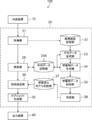
- FIG. 1 is a block diagram showing a medical image processing system 100 of the first embodiment.
- the medical image processing system 100 of the first embodiment includes an external device 10, an image processing device 20, and an output device 40.
- T1-weighted images and CEFIESTA (contrast enhanced fast imaging steady-state acquisition) images are known as a type of medical image.
- a T1-weighted image is a type of medical image taken by magnetic resonance imaging (MRI), and is an example of a medical image taken when a contrast medium is not administered to a patient.
- the CEFIESTA image is a medical image taken by an imaging method called Fast Imaging Employing Steady State Acquisition by GE (General Electric), and is an example of a medical image taken when a contrast medium is administered to a patient. Is.
- CEFIESTA images require the administration of a contrast medium to the patient. Since the image signal of a predetermined part of the human body is emphasized by administering the contrast medium to the patient, the CEFIESTA image shows the predetermined part of the human body more clearly than the T1-weighted image. However, since the contrast medium is invasive to the human body, it is preferable not to administer the contrast medium to the patient as much as possible.
- the medical image processing system 100 of the first embodiment extracts the region of interest from the T1-weighted image obtained without administering a contrast medium to the patient. Specifically, the medical image processing system 100 of the first embodiment uses a trained model obtained by machine learning using a CEFIESTA image obtained when a contrast medium is administered to a patient to obtain a region of interest from a T1-weighted image. Extract. Then, the medical image processing system 100 of the first embodiment generates a three-dimensional object of the target site of the patient by using the T1-weighted image from which the region of interest is extracted.
- the region of interest in this embodiment is the cerebral region in the medical image obtained by imaging the head.
- the T1-weighted image is an example of a first type of medical image
- the CEFIESTA image is an example of a second type of medical image.
- FIG. 2 shows an explanatory diagram for explaining the outline of the processing executed by the medical image processing system 100 of the present embodiment.
- an image of the head of a human body is targeted.
- the medical image processing system 100 of the present embodiment in the learning phase LP (Learning Phase) shown in FIG. 2, the grayscale image ImL1 of the T1-weighted image for learning and the binarized image ImL2 of the CEFIESTA image for learning are combined.
- the neural network M is machine-learned based on the associated learning data. Then, the medical image processing system 100 generates a trained neural network LM for extracting the cerebral region from the T1-weighted image.
- One data is a grayscale image of a T1-weighted image taken from the same patient (hereinafter, simply referred to as "T1-weighted image”) and a binarized image of a CEFIESTA image (hereinafter, simply "CEFIESTA image”). It is a combination with).
- the binarized image of the CEFIESTA image is an image in which the cerebral region is extracted in advance by a doctor or the like. Further, in the present embodiment, as shown in FIG. 3, a plurality of image Ims representing each cross section of the brain of the patient P are used as one three-dimensional medical image data.
- the medical image processing system 100 inputs the three-dimensional medical image data ImU1 which is a T1-weighted image to be extracted in the cerebral region into the trained neural network LM. Generates the cerebral region ImU2, which is the region of interest for 3D medical image data. Then, the medical image processing system 100 generates a three-dimensional object of the target site of the patient based on the cerebral region ImU2.
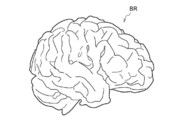
- FIG. 4 shows an example of a three-dimensional object in the patient's brain. As shown in FIG. 4, in this embodiment, a three-dimensional object BR of the patient's cerebrum is generated.
- the external device 10 inputs data in which a T1-weighted image captured from the same patient and a CEFIESTA image are associated with each other to the image processing device 20. Further, the external device 10 inputs the T1-weighted image of the cerebral region extraction target captured from the patient to the image processing device 20. The T1-weighted image to be extracted from the cerebral region is subjected to the extraction processing of the cerebral region in the image processing device 20 described later.
- the image processing device 20 includes a CPU (Central Processing Unit), a ROM (Read Only Memory) that stores programs for realizing each processing routine, a RAM (Random Access Memory) that temporarily stores data, and a storage means. It is realized by a computer that includes the memory, network interface, etc. As shown in FIG. 1, functionally, the image processing device 20 includes an acquisition unit 21, a medical image storage unit 22, an average data generation unit 23 which is an example of a data setting unit, and an average data storage unit 23A. A learning data generation unit 24, a learning data storage unit 25, a learning unit 26, a learned model storage unit 27, a conversion unit 28, an area extraction unit 30, and an object generation unit 32 are provided. There is.
- a CPU Central Processing Unit
- ROM Read Only Memory
- RAM Random Access Memory
- the acquisition unit 21 acquires the data input from the external device 10 in which the T1-weighted image and the CEFIESTA image for each of the plurality of patients are associated with each other. Then, the acquisition unit 21 stores the data in which the T1-weighted image and the CEFIESTA image for each of the plurality of patients are associated with each other in the medical image storage unit 22. In addition, the acquisition unit 21 acquires a T1-weighted image of the cerebral region extraction target input from the external device 10.
- the T1-weighted image is an example of the target data of the disclosed technology.
- the medical image storage unit 22 stores data in which a T1-weighted image and a CEFIESTA image for each of a plurality of patients are associated with each other.
- the cerebral region and the region different from the cerebral region are binarized. Extraction of the cerebral region in the CEFIESTA image is performed in advance by a doctor or the like. For example, as shown in FIG. 2, the cerebral region in the CEFIESTA image is represented by white pixels, and the region different from the cerebral region is represented by black pixels.
- the medical image storage unit 22 stores a T1-weighted image and a CEFIESTA image for each of a plurality of patients in association with each other.
- the "patient ID” shown in FIG. 5 is identification information for identifying the patient. Further, the "image ID” is identification information for identifying an image, and one image ID corresponds to one cross section. Therefore, one image is an image showing one cross section of the brain. In this embodiment, a T1-weighted image or a CEFIESTA image of each cross section obtained from one patient is treated as one three-dimensional medical image data.
- the average data which is an example of the standard data, is generated based on the data stored in the medical image storage unit 22, and then the learning data is generated.
- FIG. 6 shows an explanatory diagram for explaining the generation of the average data.
- learning data is generated by aligning the position, size, and orientation of each of the three-dimensional medical image data and the region of interest of the three-dimensional medical image data.
- the three-dimensional medical image data of each of a plurality of patients, to produce an average data H AV is three-dimensional medical image data of the average of the head .
- conversion processing such as affine transformation is performed so that each of the three-dimensional medical image data of the heads of a plurality of patients is brought closer to the average data.
- the differences in position, size, and orientation of each of the three-dimensional medical image data (T1-weighted image, CIFIESTA image, etc.) of a plurality of patients are suppressed, and the positions, sizes, and orientations are aligned. It becomes.
- the average data generation unit 23 sets average data, which is an example of standard data, which is three-dimensional image data representing a standard. Specifically, the average data generation unit 23 aligns at least one of the sizes, orientations, and positions of each of the plurality of T1-weighted images, which are three-dimensional medical image data, and then averages the plurality of T1-weighted images. Generate average image data.
- the average data generation unit 23 generates average data of T1-weighted images of patients according to the procedures shown in (1) to (4) below.
- the average data generation unit 23 acquires a T1-weighted image, which is three-dimensional medical image data of the head, from a plurality of data stored in the medical image storage unit 22. For example, the average data generation unit 23 reads out 10 T1-weighted images from the data stored in the medical image storage unit 22.
- the average data generation unit 23 selects one reference data, which is a reference T1-weighted image, from the ten read T1-weighted images. The reference data may be selected by the user.
- the average data generation unit 23 performs conversion such as affine transformation so that each of the nine T1-weighted images different from the reference data is brought closer to one reference data.
- the average data generation unit 23 obtains average data of the head by adding and averaging the T1-weighted image of the reference data and the nine T1-weighted images subjected to conversion processing such as affine transformation. Then, the average data generation unit 23 stores the average data of the T1-weighted image in the average data storage unit 23A.
- average data in which the sizes, orientations, and positions of the plurality of three-dimensional medical image data are aligned is obtained.
- the sizes, orientations, and positions of T1-weighted images H X , HY , and H Z which differ in the size, orientation, and position of the heads of a plurality of patients, are aligned.
- the average data HAR is obtained.
- the learning data generation unit 24 uses the average data of the head stored in the average data storage unit 23A to bring the data of each of the plurality of patients stored in the medical image storage unit 22 closer to the average data. Generate training data.
- the learning data generation unit 24 stores the average data storage unit 23A for each of the plurality of learning three-dimensional medical image data stored in the medical image storage unit 22 and the T1-weighted image for learning.
- the conversion process is performed so that at least one of the sizes, orientations, and positions of each of the multiple training T1-weighted images is aligned so that the degree of agreement with the average data of the stored T1-weighted images is large. conduct.
- the learning data generation unit 24 of the present embodiment uses affine transformation, which is an example of anisotropic scaling, which is a known technique, as the transformation process.
- affine transformation which is an example of anisotropic scaling, which is a known technique, as the transformation process.
- the affine transformation process is performed so that the correlation value, which is an example of the degree of agreement, becomes large.
- the affine transformation process is performed on one T1-weighted image for learning, one affine transformation matrix is obtained.
- the learning data generation unit 24 performs the same conversion processing on each of the CEFIESTA images from which the region of interest of the T1-weighted image for learning is extracted. Specifically, the learning data generation unit 24 converts the CEFIESTA image from which the region of interest is extracted by using the affine transformation matrix obtained when converting the T1-weighted image for learning.
- the learning data generation unit 24 generates learning data by associating the converted three-dimensional medical image data for learning, the T1-weighted image, with the converted CEFIESTA image, which is the region of interest.
- the sizes, orientations, and positions of the plurality of T1-weighted images and the plurality of CEFIESTA images, which had different positions, sizes, and orientations, are aligned.
- FIG. 7 shows an explanatory diagram for explaining a process of aligning the size, orientation, and position of the T1-weighted image for learning and the CEFIESTA image for learning.
- Figure 7 (A) (B) is a T1-weighted image H 1 is a three-dimensional medical image data of a patient P 1 head, it is shown and CEFIESTA image B 1 of the cerebral region of the patient P 1 is ..
- the learning data generation unit 24 matches the T1-weighted image H 1 of the head of a patient P 1 with the average data H AV of the head. as degree increases performs affine transformation T, to produce a T1 weighted image H 1 'of the conversion process of the patient P 1 was made head.
- the affine transformation matrix at this time is obtained by the affine transformation T.
- the learning data generation unit 24 displays the T1-weighted image H 1 of the head on the CEFIESTA image B 1 from which the cerebral region of the patient P 1 is extracted.
- mean affine transformation matrix obtained when a match to the data H AV was used to implement the conversion process to generate an CEFIESTA image B 1 'of the conversion process is cerebral region of the patient P 1 was made of.
- the size, orientation, and position of the head of the CEFIESTA image which is the source of the T1-weighted image H 1 and the CEFIESTA image B 1, of the head, are already aligned.
- the data of a patient P 1 is brought close to the average data, and the 'CEFIESTA image B 1 of the patient P 1 cerebral region' T1 weighted images H 1 of the head patient P 1 is obtained.
- the learning data creation section 24 'the three-dimensional medical image data corresponding to, CEFIESTA image B 1 of the converted cerebral regions' T1 weighted images H 1 of the converted head and brain area corresponding to Generate training data in association with each other.
- the learning data generation unit 24 performs the above conversion processing on each of the plurality of data stored in the medical image storage unit 22, and generates learning data having the same size, orientation, and position. Then, it is stored in the learning data storage unit 25.
- the data used for generating the average data may be excluded from the plurality of data stored in the medical image storage unit 22.
- the learning data generation unit 24 absorbs differences in the size, orientation, and position of the head of each patient, and reduces variations in the size and shape of the brain of each patient. Is obtained. Further, learning data of a plurality of patients with reduced variations caused by different imaging conditions can be obtained.
- the learning data storage unit 25 stores learning data of a plurality of patients generated by the learning data generation unit 24. Specifically, the learning data storage unit 25 contains three-dimensional medical image data representing a T1-weighted image in which the size, orientation, and position of each of the plurality of patients are aligned, and the size, orientation, and the like. And the learning data associated with the three-dimensional medical image data representing the aligned CEFIESTA image is stored.
- the learning data storage unit 25 contains a T1-weighted image in which the sizes, orientations, and positions of each of the plurality of patients are aligned, and the size, orientation, and position.
- the aligned CEFIESTA images are stored in association with each other.
- the learning unit 26 generates a trained model for extracting a cerebral region from a T1-weighted image based on a plurality of learning data stored in the learning data storage unit 25.
- a trained neural network LM as shown in FIG. 2 is generated.
- deep learning is used as an example of the learning algorithm.
- the learning unit 26 stores the trained neural network LM in the trained model storage unit 27.
- the conversion unit 28 targets the T1-weighted image of the cerebral region extraction target acquired by the acquisition unit 21 so that the degree of agreement between the T1-weighted image and the average data of the head stored in the average data storage unit 23A becomes large.
- the affine transformation process is performed so that at least one of the size, orientation, and position of the data is aligned with the average data. Then, the conversion unit 28 generates the converted data obtained by performing the conversion process on the T1-weighted image to be extracted from the cerebral region.
- the average data of the head is generated, and the data of each patient is averaged so that the degree of agreement with the average data of the head becomes large.
- Generated training data Since the trained model is generated from the training data, it is necessary to perform the same conversion process on the T1-weighted image to be extracted from the cerebral region. Therefore, the conversion unit 28 performs the affine transformation process so that the degree of agreement between the T1-weighted image to be extracted from the cerebral region and the average data of the head becomes large. Then, the conversion unit 28 obtains the converted data that has undergone the affine transformation processing.
- the area extraction unit 30 reads out the trained neural network LM stored in the trained model storage unit 27. Then, the region extraction unit 30 inputs the converted data that has been converted by the conversion unit 28 into the trained neural network LM, and extracts the cerebral region. As a result, the cerebral region was automatically extracted from the T1-weighted image taken without administration of the contrast medium.
- the object generation unit 32 generates a three-dimensional object of the target site of the patient based on the cerebral region extracted by the region extraction unit 30.
- the image output from the trained neural network LM is a binarized image in which the cerebral region is represented by "1" and the region other than the cerebrum is represented by "0".
- a grayscale image is required because volume rendering, which is a known technique, is used when drawing a three-dimensional object of the cerebrum.
- the object generation unit 32 extracts the cerebral region from the T1-weighted image, which is the original three-dimensional medical image data before conversion, based on the cerebral region represented by "1" in the binarized image.
- the object generation unit 32 performs an inverse transformation process on the cerebral region obtained by the region extraction unit 30 according to the transformation process for aligning the size, orientation, and position performed by the conversion unit 28. conduct.
- the cerebral region B 2 is volume data.
- Object generation unit 32 with respect to cerebral region B 2, conversion unit 28 performs the inverse transformation process INVT corresponding to affine transformation process conducted by the pre-conversion of the T1-weighted images and cerebral region inverse transform processing has been performed B 2
- the cerebral region is extracted from the T1-weighted image by associating with.
- the object generation unit 32 takes the Hadamard product between the cerebral region B 2 that has undergone the inverse transformation process and the T1-weighted image that is the original three-dimensional medical image data, so that the T1-weighted image is used as the cerebrum. Extract the area. This extracts the grayscale cerebral region.
- the object generation unit 32 displays a three-dimensional object of the patient's brain using volume rendering, which is a known technique, based on the cerebral region extracted from the original T1-weighted image before conversion. Specifically, the object generation unit 32 outputs the three-dimensional object of the patient's brain generated from the volume data of the patient's brain to the output device 40.
- the output device 40 outputs the three-dimensional object output from the object generation unit 32 as a result.
- the output device 40 is composed of, for example, a display or the like. From the output device 40, for example, a three-dimensional object BR of the cerebrum as shown in FIG. 4 is displayed.
- the image processing device 20 can be realized by, for example, the computer 50 shown in FIG.
- the computer 50 includes a CPU 51, a memory 52 as a temporary storage area, and a non-volatile storage unit 53. Further, the computer 50 is a read / write (R / W) unit that controls reading and writing of data to the input / output interface (I / F) 54 and the recording medium 59 to which the external device 10 and the output device 40 are connected. 55 is provided. Further, the computer 50 includes a network I / F 56 connected to a network such as the Internet.
- the CPU 51, the memory 52, the storage unit 53, the input / output I / F 54, the R / W unit 55, and the network I / F 56 are connected to each other via the bus 57.
- the storage unit 53 can be realized by a Hard Disk Drive (HDD), a Solid State Drive (SSD), a flash memory, or the like.
- a program for operating the computer 50 is stored in the storage unit 53 as a storage medium.
- the CPU 51 reads the program from the storage unit 53, expands it in the memory 52, and sequentially executes the processes included in the program.
- the operation of the medical image processing system 100 of the present embodiment will be described with reference to FIGS. 10 to 12.
- the medical image processing system 100 operates and data in which a T1-weighted image and a CEFIESTA image for each of a plurality of patients are associated with each other is input from the external device 10, the acquisition unit 21 inputs each of the data. It is stored in the medical image storage unit 22. Then, when the image processing device 20 receives the instruction signal for average data generation, the image processing device 20 executes the average data generation processing routine shown in FIG.
- step S50 the average data generation unit 23 reads, for example, 10 T1-weighted images stored in the medical image storage unit 22.
- step S52 the average data generation unit 23 selects one reference data from the T1-weighted images of the plurality of patients read out in step S50.
- step S54 the average data generation unit 23 takes between the reference data selected in step S52 and the T1-weighted image for each of the nine T1-weighted images different from the reference data selected in step S52.
- the affine transformation is performed so that at least one of the size, orientation, and position is aligned.
- step S56 the average data generation unit 23 adds and averages the T1-weighted image corresponding to the reference data selected in step S52 and the T1-weighted image subjected to the affine transformation in step S54, and averages the data. To generate.
- step S58 the average data generation unit 23 stores the average data generated in step S56 in the average data storage unit 23A.
- the image processing device 20 executes the learning data generation processing routine shown in FIG.
- step S70 the learning data generation unit 24 reads one data from the data stored in the medical image storage unit 22 in which the T1-weighted image and the CEFIESTA image for each of the plurality of patients are associated with each other.
- step S72 the learning data generation unit 24 reads out the average data stored in the average data storage unit 23A.
- step S74 the learning data generation unit 24 determines the size, orientation, and position between the T1-weighted image of the data read in step S70 and the average data read in step S72. Affine transformation is performed on the T1-weighted image so that at least one is aligned. At this time, an affine transformation matrix is obtained.
- step S76 the learning data generation unit 24 was obtained in step S74 so as to align the CEFIESTA image of the data read in step S70 with the average data read in step S72.
- the affine transformation is performed using the affine transformation matrix.
- step S78 the learning data generation unit 24 associates the T1-weighted image converted in step S74 with the CEFIESTA image converted in step S76 to the learning data storage unit 25. Store as one learning data.
- step S80 the learning data generation unit 24 determines whether or not the processes of steps S70 to S78 have been executed for all the data stored in the medical image storage unit 22. When the processes of steps S70 to S78 are executed for all the data, the learning data generation processing routine is terminated. On the other hand, if there is data for which the processes of steps S70 to S78 have not been executed, the process returns to step S70.
- the data used when generating the average data among the plurality of data stored in the medical image storage unit 22 may be excluded from the generation of the learning data.
- the machine learning process for the neural network M can be executed. It becomes. Therefore, when the image processing device 20 receives the instruction signal for the learning process, the image processing device 20 executes the learning processing routine shown in FIG.
- step S100 the learning unit 26 acquires a plurality of learning data stored in the learning data storage unit 25.
- step S102 the learning unit 26 generates a trained neural network LM for extracting the cerebral region from the T1-weighted image by machine learning based on the plurality of learning data acquired in step S100.
- step S104 the learning unit 26 stores the learned neural network LM generated in step S102 in the learned model storage unit 27, and ends the learning processing routine.
- the cerebral region can be extracted using the trained neural network LM. Therefore, when the T1-weighted image to be extracted from the cerebral region is input from the external device 10 to the image processing device 20, the image processing device 20 executes the image processing routine shown in FIG.
- step S200 the acquisition unit 21 acquires a T1-weighted image to be extracted from the cerebral region.
- step S201 the conversion unit 28 receives the T1-weighted image of the cerebral region extraction target acquired in step S200, and between the T1-weighted image and the average data of the head stored in the average data storage unit 23A.
- the affine transformation process is performed so that the degree of coincidence becomes large, and the converted data is generated.
- step S202 the area extraction unit 30 reads out the trained neural network LM stored in the trained model storage unit 27.
- step S204 the region extraction unit 30 inputs the converted data generated in step S201 to the learned neural network LM read in step S202, and extracts the volume data of the cerebral region.
- step S206 the object generation unit 32 performs an inverse transformation process on the volume data of the cerebral region extracted in step S204 according to the affine transformation process performed in step S201. Then, the object generation unit 32 extracts the cerebral region from the original T1-weighted image by the Hadamard product of the volume data of the T1-weighted image before the affine transformation and the volume data of the cerebral region subjected to the inverse transformation processing. This extracts the grayscale cerebral region.
- step S208 the object generation unit 32 generates a three-dimensional object of the patient's brain using volume rendering, which is a known technique, based on the volume data of the grayscale cerebral region obtained in step S206. ..
- step S210 the object generation unit 32 outputs the three-dimensional object of the patient's brain to the output device 40, and ends the image processing routine.
- the output device 40 outputs the three-dimensional object of the patient's brain output from the object generation unit 32 as a result.
- the image processing apparatus 20 has the size, orientation, and orientation of the target data, which is the three-dimensional medical image data to be extracted from the region of interest, and the respective sizes, orientations, and orientations of the plurality of three-dimensional medical image data. At least one of the size, orientation, and position of the target data so that there is a greater degree of agreement with the average data, which is the three-dimensional medical image data generated by averaging after aligning at least one of the positions. The conversion process is performed so that one is aligned with the average data.
- the image processing device 20 extracts the converted data, which is the three-dimensional medical image data that has been converted with respect to the target data, from the three-dimensional medical image data to the region of interest included in the three-dimensional medical image data.
- Input to the pre-trained trained model for the purpose and extract the region of interest contained in the transformed data by the calculation by the trained model. This makes it possible to accurately extract the region of interest from the medical image.
- the area of interest can be accurately extracted from the medical image by the trained model generated from the learning data in which the variation of the medical image obtained from the patient is reduced.
- the present embodiment by treating the combination of a plurality of medical images representing each cross section of one patient as one learning data, the information in the height direction is reflected in the learning data. Therefore, the training data is reflected in the trained model, and the patient's three-dimensional object can be generated with high accuracy.
- the region of interest is equivalent to the result of extracting the region of interest from the CEFIESTA image obtained by administering the contrast medium to the patient from the T1-weighted image that does not require administration of the contrast medium to the patient. Can be extracted. Therefore, it is possible to reduce the burden on the patient and obtain data in which the region of interest is extracted with high accuracy.
- the trained model is used to extract the region of interest of the T1-weighted image. Then, according to the present embodiment, it is possible to accurately display the three-dimensional object of the target part of the patient by volume rendering based on the T1-weighted image in which the region of interest is extracted.
- the second embodiment differs from the first embodiment in that the cerebral region is extracted from the FIESTA image.
- the FIESTA-enhanced image is an example of a first-class medical image
- the CEFIESTA image is an example of a second-class medical image.
- the FIESTA image is a medical image taken by an imaging method called Fast Imaging Employing Steady State Acquisition by GE (General Electric).
- the FIESTA image without "CE” is an example of a medical image taken when no contrast medium is administered to the patient.
- the cerebral region is extracted from the FIESTA image that does not require the administration of the contrast medium to the patient, as much as the cerebral region is extracted from the CEFIESTA image taken by administering the contrast medium to the patient. It can be extracted with high accuracy. Therefore, as in the first embodiment, the burden on the patient can be reduced and the cerebral region can be extracted with high accuracy.
- the medical image storage unit 22 of the second embodiment stores data in which the FIESTA image and the CEFIESTA image are associated with each other, instead of the data in which the T1-weighted image and the CEFIESTA image in the first embodiment are associated with each other. Will be done.
- the learning data storage unit 25 of the second embodiment has a three-dimensional medical image representing a T1-enhanced image in which the size, orientation, and position of each of the plurality of patients in the first embodiment are aligned.
- learning data associated with the data and 3D medical image data representing a CEFIESTA image with aligned size, orientation, and position the size, orientation, and orientation of each of the plurality of patients.
- the learning data in which the three-dimensional medical image data representing the FIESTA image in which the positions are aligned and the three-dimensional medical image data representing the CEFIESTA image in which the size, orientation, and position are aligned are stored is stored.
- the region extraction unit 30 of the second embodiment inputs the FIESTA image, which is the three-dimensional medical image data to be extracted of the region of interest, into the trained neural network of the second embodiment, and extracts the cerebral region from the FIESTA image. Extract. As a result, the cerebral region is extracted with high accuracy.
- the object generation unit 32 of the second embodiment is based on the information of the cerebral region extracted by the region extraction unit 30, and of "0" and "1" output from the region extraction unit 30 without performing the Hadamard product.
- a three-dimensional object in the cerebral region is displayed by surface-rendering the binarized image by the marching cube method.
- a trained model previously trained by a type of medical image different from the target data which is the three-dimensional medical image data to be extracted from the region of interest, is used. Therefore, the area of interest can be extracted from the target data.
- the third embodiment Since the configuration of the medical image processing system according to the third embodiment has the same configuration as that of the first embodiment, the same reference numerals are given and the description thereof will be omitted.
- the third embodiment is different from the first embodiment and the second embodiment in that a combination of the CEFIESTA image and the CEFIESTA image is used as the learning data.
- the CEFIESTA image is an example of a first type medical image and a second type medical image.
- the CEFIESTA image is a medical image taken when a contrast medium is administered to a patient.
- the CEFIESTA image and the cerebral region in which the cerebral region is not extracted are extracted in place of the data in which the T1-weighted image and the CEFIESTA image in the first embodiment are associated with each other.
- the data associated with the CEFIESTA image is stored.
- the region extraction unit 30 of the third embodiment inputs the CEFIESTA image, which is the three-dimensional medical image data to be extracted of the region of interest, into the trained neural network of the third embodiment, and extracts the cerebral region from the CEFIESTA image. Extract. As a result, the cerebral region is extracted with high accuracy.
- the object generation unit 32 of the third embodiment is based on the information of the cerebral region extracted by the region extraction unit 30, and of "0" and "1" output from the region extraction unit 30 without performing the Hadamard product.
- a three-dimensional object in the cerebral region is displayed by surface-rendering the binarized image by the marching cube method.
- a trained model previously trained by a type of medical image different from the target data which is the three-dimensional medical image data to be extracted from the region of interest, is used. Therefore, the area of interest can be extracted from the target data.
- FIG. 14 is a three-dimensional object of the cerebrum generated by the image processing device 20 of the present embodiment. As shown in FIG. 14, it can be seen that the cerebral region is extracted with high accuracy. In particular, it can be seen that the sulci and gyrus are extracted with high accuracy.
- the degree of agreement (dice coefficient) between the patient's cerebral volume data generated by this embodiment and the cerebral volume data generated from the result of labeling the CEFIESTA image by a doctor is shown in the following table. ..
- the results shown in the table below generate a trained model based on the training data associated with the CEFIESTA image from which the cerebral region has not been extracted and the CEFIESTA image from which the cerebral region has been extracted, and are unknown. This is the result of extracting the cerebral region from the CEFIESTA image.
- the dice coefficient for each of the plurality of patients is 0.9 or more. This result indicates that the three-dimensional object of the cerebrum is generated with higher accuracy than before. Therefore, it can be said that the cerebral region is accurately generated from the medical image and the three-dimensional object of the cerebrum is generated with high accuracy.
- the case where the first type of medical image is a T1-weighted image and the second type of medical image is a CEFIESTA image has been described as an example.
- the case where the first type of medical image is a FIESTA image and the second type of medical image is a CEFIESTA image has been described as an example.
- the case where the first type of medical image is a CEFIESTA image and the second type of medical image is also a CEFIESTA image has been described as an example.
- the combination of the first type of medical image and the second type of medical image is not limited to this, and any combination of medical images in which the brain region can be extracted by a doctor's hand is not limited to this. It may be.
- the first type of medical image and the second type of medical image may be any medical image such as a T2-weighted image and a CT image.
- the case where the binarized image of the CEFIESTA image is used has been described as an example, but the present invention is not limited to this, and a grayscale image or the like may be used.
- a trained model is generated using a plurality of types of medical images Im1, Im2, Im3, Im4, and a different type of medical image is generated from any type of medical image, or
- the target area may be extracted from the medical image to generate a three-dimensional object BR of the target part.
- the present embodiment may be used when creating a three-dimensional object of an artery.
- the affine transformation is used as an example of anisotropic scaling has been described as an example, but the present invention is not limited to this, and the transformation processing may be performed using another method.
- the case where the average data generation processing routine, the learning data generation processing routine, the learning processing routine, and the image processing routine are executed in the same image processing apparatus 20 has been described as an example. , Not limited to this.
- the average data generator that executes the average data generation processing routine, the learning data generator that executes the learning data generation processing routine, the image processing model generator that executes the learning processing routine, and the image processing routine are executed. It may be configured as a separate device such as an image processing device.
- the neural network model as an example of the model is trained by deep learning has been described as an example, but the present invention is not limited to this.
- another model different from the neural network model may be trained by another learning method different from deep learning.
- a trained model is generated using the first type of medical image and the second type of medical image, and the target data, which is the three-dimensional medical image data to be extracted from the region of interest, is converted into the trained model.
- the target data which is the three-dimensional medical image data to be extracted from the region of interest.
- the region of interest is extracted from the T2-weighted image using the image processing device 20 of the third embodiment.
- the imaging method of the T2-weighted image is similar to the imaging method of the CEFIESTA image. Therefore, the region of interest can be extracted from the T2-weighted image by performing some conversion processing on the T2-weighted image to make it resemble the CEFIESTA image and then inputting it to the trained model trained in advance by the CEFIESTA image. ..
- the conversion unit 28 performs a conversion process for aligning the T2-weighted image, which is the target data, with the average data, and then performs a conversion process for correcting the gradation of the brightness value of the T2-weighted image.
- the converted data is generated.
- the conversion unit 28 performs ⁇ -correction processing on the T2-weighted image to make it resemble a CEFIESTA image, and then inputs the ⁇ -corrected T2-weighted image to the trained model to set the region of interest. It can also be extracted.
- the conversion unit 28 may perform a conversion process for correcting the gradation of the brightness value of the T2-weighted image, and then perform a conversion process for aligning the T2-weighted image with the average data.
- the part may be complemented and input to the trained model.
- the target data which is the three-dimensional medical image data showing only the affected part
- the trained model generated from the learning data of the three-dimensional medical image data showing the entire head. Extraction of the region of interest may fail due to the missing part of the target data other than the affected area.
- the conversion unit 28 when the conversion unit 28 performs the conversion process so as to align the target data with the average data, the conversion unit 28 integrates an area different from the area of the average data that matches the target data into the target data. Complements the target data and generates converted data. In this case, the conversion unit 28 performs correction processing on the average data so that the average or variance of the brightness value of the average data and the average or variance of the brightness value of the target data are close to each other, and integrates the data. , The converted data may be generated. Then, the region extraction unit 30 inputs the complemented target data into the trained model and extracts the region of interest. In this case, the region extraction unit 30 extracts only the affected portion corresponding to the target data from the extracted region of interest.
- 3D medical image data selected in advance from a plurality of 3D medical image data may be used as standard data.
- three-dimensional medical image data of a patient having a standard head is preset as standard data.
- 3D image data generated in advance by performing statistical processing on a plurality of 3D medical image data may be used as standard data.
- principal component analysis which is an example of statistical processing, is performed on a plurality of three-dimensional medical image data, and the three-dimensional image data corresponding to the first principal component or the like obtained thereby is standardized. It can be data.
Landscapes
- Engineering & Computer Science (AREA)
- Physics & Mathematics (AREA)
- Theoretical Computer Science (AREA)
- General Physics & Mathematics (AREA)
- Health & Medical Sciences (AREA)
- Life Sciences & Earth Sciences (AREA)
- Computer Vision & Pattern Recognition (AREA)
- Medical Informatics (AREA)
- Heart & Thoracic Surgery (AREA)
- Public Health (AREA)
- Radiology & Medical Imaging (AREA)
- Biomedical Technology (AREA)
- Optics & Photonics (AREA)
- Molecular Biology (AREA)
- Surgery (AREA)
- Animal Behavior & Ethology (AREA)
- General Health & Medical Sciences (AREA)
- Pathology (AREA)
- Veterinary Medicine (AREA)
- Nuclear Medicine, Radiotherapy & Molecular Imaging (AREA)
- High Energy & Nuclear Physics (AREA)
- Biophysics (AREA)
- Magnetic Resonance Imaging Apparatus (AREA)
- Image Analysis (AREA)
- Apparatus For Radiation Diagnosis (AREA)
- Image Processing (AREA)
Priority Applications (1)
| Application Number | Priority Date | Filing Date | Title |
|---|---|---|---|
| JP2022504437A JP7323885B2 (ja) | 2020-03-04 | 2021-03-03 | 画像処理装置、画像処理モデル生成装置、学習用データ生成装置、及びプログラム |
Applications Claiming Priority (2)
| Application Number | Priority Date | Filing Date | Title |
|---|---|---|---|
| JP2020037135 | 2020-03-04 | ||
| JP2020-037135 | 2020-03-04 |
Publications (1)
| Publication Number | Publication Date |
|---|---|
| WO2021177374A1 true WO2021177374A1 (ja) | 2021-09-10 |
Family
ID=77613434
Family Applications (1)
| Application Number | Title | Priority Date | Filing Date |
|---|---|---|---|
| PCT/JP2021/008263 Ceased WO2021177374A1 (ja) | 2020-03-04 | 2021-03-03 | 画像処理装置、画像処理モデル生成装置、学習用データ生成装置、及びプログラム |
Country Status (2)
| Country | Link |
|---|---|
| JP (1) | JP7323885B2 (https=) |
| WO (1) | WO2021177374A1 (https=) |
Cited By (1)
| Publication number | Priority date | Publication date | Assignee | Title |
|---|---|---|---|---|
| CN120047937A (zh) * | 2025-04-27 | 2025-05-27 | 骨圣元化机器人(深圳)有限公司 | 三维影像数据的包围盒预测方法及数据传输方法、设备 |
Citations (7)
| Publication number | Priority date | Publication date | Assignee | Title |
|---|---|---|---|---|
| US20190030371A1 (en) * | 2017-07-28 | 2019-01-31 | Elekta, Inc. | Automated image segmentation using dcnn such as for radiation therapy |
| WO2019044078A1 (ja) * | 2017-08-28 | 2019-03-07 | 富士フイルム株式会社 | 脳画像正規化装置、方法およびプログラム |
| JP2019525786A (ja) * | 2016-07-08 | 2019-09-12 | アヴェント インコーポレイテッド | 解剖学的対象物を自動的に検出、位置特定、及びセマンティックセグメンテーションするシステム及び方法 |
| JP2019531783A (ja) * | 2016-08-26 | 2019-11-07 | エレクタ、インク.Elekta, Inc. | 畳み込みニューラルネットワークを用いた画像セグメンテーションのためのシステムおよび方法 |
| WO2019216125A1 (ja) * | 2018-05-09 | 2019-11-14 | 富士フイルム株式会社 | 梗塞領域を判別する判別器の学習装置、方法およびプログラム、梗塞領域を判別する判別器、並びに梗塞領域判別装置、方法およびプログラム |
| JP2019534763A (ja) * | 2016-09-06 | 2019-12-05 | エレクタ、インク.Elekta, Inc. | 合成医療画像を生成するためのニューラルネットワーク |
| CN110766652A (zh) * | 2019-09-06 | 2020-02-07 | 上海联影智能医疗科技有限公司 | 网络训练方法、装置、分割方法、计算机设备和存储介质 |
-
2021
- 2021-03-03 WO PCT/JP2021/008263 patent/WO2021177374A1/ja not_active Ceased
- 2021-03-03 JP JP2022504437A patent/JP7323885B2/ja active Active
Patent Citations (7)
| Publication number | Priority date | Publication date | Assignee | Title |
|---|---|---|---|---|
| JP2019525786A (ja) * | 2016-07-08 | 2019-09-12 | アヴェント インコーポレイテッド | 解剖学的対象物を自動的に検出、位置特定、及びセマンティックセグメンテーションするシステム及び方法 |
| JP2019531783A (ja) * | 2016-08-26 | 2019-11-07 | エレクタ、インク.Elekta, Inc. | 畳み込みニューラルネットワークを用いた画像セグメンテーションのためのシステムおよび方法 |
| JP2019534763A (ja) * | 2016-09-06 | 2019-12-05 | エレクタ、インク.Elekta, Inc. | 合成医療画像を生成するためのニューラルネットワーク |
| US20190030371A1 (en) * | 2017-07-28 | 2019-01-31 | Elekta, Inc. | Automated image segmentation using dcnn such as for radiation therapy |
| WO2019044078A1 (ja) * | 2017-08-28 | 2019-03-07 | 富士フイルム株式会社 | 脳画像正規化装置、方法およびプログラム |
| WO2019216125A1 (ja) * | 2018-05-09 | 2019-11-14 | 富士フイルム株式会社 | 梗塞領域を判別する判別器の学習装置、方法およびプログラム、梗塞領域を判別する判別器、並びに梗塞領域判別装置、方法およびプログラム |
| CN110766652A (zh) * | 2019-09-06 | 2020-02-07 | 上海联影智能医疗科技有限公司 | 网络训练方法、装置、分割方法、计算机设备和存储介质 |
Cited By (1)
| Publication number | Priority date | Publication date | Assignee | Title |
|---|---|---|---|---|
| CN120047937A (zh) * | 2025-04-27 | 2025-05-27 | 骨圣元化机器人(深圳)有限公司 | 三维影像数据的包围盒预测方法及数据传输方法、设备 |
Also Published As
| Publication number | Publication date |
|---|---|
| JPWO2021177374A1 (https=) | 2021-09-10 |
| JP7323885B2 (ja) | 2023-08-09 |
Similar Documents
| Publication | Publication Date | Title |
|---|---|---|
| Zhang et al. | PTNet3D: A 3D high-resolution longitudinal infant brain MRI synthesizer based on transformers | |
| US11610313B2 (en) | Systems and methods for generating normative imaging data for medical image processing using deep learning | |
| US10692198B2 (en) | Image processing apparatus, image processing method, image processing system, and non-transitory computer-readable storage medium for presenting three-dimensional images | |
| US20190213779A1 (en) | Apparatus and method for producing three-dimensional models from magnetic resonance imaging | |
| US20140126789A1 (en) | Image diagnosis assisting apparatus, and method | |
| US8861891B2 (en) | Hierarchical atlas-based segmentation | |
| CN116368516A (zh) | 使用联合合成、分割和配准的多模态临床图像对齐方法和设备 | |
| JP7795352B2 (ja) | 画像処理方法、画像処理装置およびプログラム | |
| JP7203978B2 (ja) | 学習装置、方法およびプログラム、関心領域抽出装置、方法およびプログラム、並びに学習済み抽出モデル | |
| CN111178502A (zh) | 循环生成对抗网络模型的训练方法、系统以及电子设备 | |
| Lu et al. | Two-stage self-supervised cycle-consistency transformer network for reducing slice gap in MR images | |
| KR20240086195A (ko) | 확산 모델을 이용한 비지도 학습 기반 변형 영상 정합 및 합성 영상 생성 시스템 및 그 방법 | |
| CN114830181A (zh) | 用于人工智能的患者图像的解剖加密 | |
| US11138736B2 (en) | Information processing apparatus and information processing method | |
| Che Azemin et al. | Assessing the efficacy of StyleGAN 3 in generating realistic medical images with limited data availability | |
| US12597130B2 (en) | Image processing apparatus, operation method of image processing apparatus, and operation program of image processing apparatus for generating examination results of new medical examinations | |
| JP7323885B2 (ja) | 画像処理装置、画像処理モデル生成装置、学習用データ生成装置、及びプログラム | |
| Chen et al. | Harmonized neonatal brain MR image segmentation model for cross-site datasets | |
| CN113822323A (zh) | 脑部扫描图像的识别处理方法、装置、设备及存储介质 | |
| KR101801376B1 (ko) | 3차원 위상 기술자를 이용한 두개골 이형 자동판단시스템 및 이를 이용한 두개골 이형 자동판단방법 | |
| WO2015175852A1 (en) | System and method for ct image synthesis from mri using generative sub-image synthesis | |
| US20250045992A1 (en) | Image conversion device, control method for image conversion device, and medium | |
| US20210383547A1 (en) | Medical image processing | |
| CN113674838B (zh) | 医学图像处理装置、医学图像处理方法和记录介质 | |
| US11880987B2 (en) | Image processing apparatus, image processing method, and storage medium |
Legal Events
| Date | Code | Title | Description |
|---|---|---|---|
| 121 | Ep: the epo has been informed by wipo that ep was designated in this application |
Ref document number: 21763534 Country of ref document: EP Kind code of ref document: A1 |
|
| ENP | Entry into the national phase |
Ref document number: 2022504437 Country of ref document: JP Kind code of ref document: A |
|
| NENP | Non-entry into the national phase |
Ref country code: DE |
|
| 122 | Ep: pct application non-entry in european phase |
Ref document number: 21763534 Country of ref document: EP Kind code of ref document: A1 |