WO2021147829A1 - A bispecific anti-pd-l1/vegf antibody and uses thereof - Google Patents
A bispecific anti-pd-l1/vegf antibody and uses thereof Download PDFInfo
- Publication number
- WO2021147829A1 WO2021147829A1 PCT/CN2021/072584 CN2021072584W WO2021147829A1 WO 2021147829 A1 WO2021147829 A1 WO 2021147829A1 CN 2021072584 W CN2021072584 W CN 2021072584W WO 2021147829 A1 WO2021147829 A1 WO 2021147829A1
- Authority
- WO
- WIPO (PCT)
- Prior art keywords
- antigen
- antibody
- binding
- cancer
- vegf
- Prior art date
Links
- 101100372758 Danio rerio vegfaa gene Proteins 0.000 title 1
- 101150030763 Vegfa gene Proteins 0.000 title 1
- 230000027455 binding Effects 0.000 claims abstract description 239
- 239000000427 antigen Substances 0.000 claims abstract description 206
- 102000036639 antigens Human genes 0.000 claims abstract description 202
- 108091007433 antigens Proteins 0.000 claims abstract description 202
- 102000008096 B7-H1 Antigen Human genes 0.000 claims abstract description 102
- 108010074708 B7-H1 Antigen Proteins 0.000 claims abstract description 102
- 238000000034 method Methods 0.000 claims abstract description 50
- 210000004027 cell Anatomy 0.000 claims description 124
- 108010019530 Vascular Endothelial Growth Factors Proteins 0.000 claims description 97
- 206010028980 Neoplasm Diseases 0.000 claims description 64
- 125000003275 alpha amino acid group Chemical group 0.000 claims description 45
- 150000007523 nucleic acids Chemical class 0.000 claims description 41
- 239000013598 vector Substances 0.000 claims description 40
- 102000039446 nucleic acids Human genes 0.000 claims description 28
- 108020004707 nucleic acids Proteins 0.000 claims description 28
- 108010003723 Single-Domain Antibodies Proteins 0.000 claims description 23
- 201000011510 cancer Diseases 0.000 claims description 22
- 239000002246 antineoplastic agent Substances 0.000 claims description 20
- 108010047041 Complementarity Determining Regions Proteins 0.000 claims description 17
- 108091028043 Nucleic acid sequence Proteins 0.000 claims description 16
- 241001465754 Metazoa Species 0.000 claims description 14
- 230000028993 immune response Effects 0.000 claims description 14
- 239000008194 pharmaceutical composition Substances 0.000 claims description 14
- 239000003795 chemical substances by application Substances 0.000 claims description 13
- 206010009944 Colon cancer Diseases 0.000 claims description 11
- 229940127089 cytotoxic agent Drugs 0.000 claims description 11
- 238000004519 manufacturing process Methods 0.000 claims description 10
- 210000004881 tumor cell Anatomy 0.000 claims description 10
- 230000003405 preventing effect Effects 0.000 claims description 9
- 230000002401 inhibitory effect Effects 0.000 claims description 8
- 208000001333 Colorectal Neoplasms Diseases 0.000 claims description 6
- 206010025323 Lymphomas Diseases 0.000 claims description 6
- 208000029742 colonic neoplasm Diseases 0.000 claims description 6
- 230000004614 tumor growth Effects 0.000 claims description 6
- FWMNVWWHGCHHJJ-SKKKGAJSSA-N 4-amino-1-[(2r)-6-amino-2-[[(2r)-2-[[(2r)-2-[[(2r)-2-amino-3-phenylpropanoyl]amino]-3-phenylpropanoyl]amino]-4-methylpentanoyl]amino]hexanoyl]piperidine-4-carboxylic acid Chemical compound C([C@H](C(=O)N[C@H](CC(C)C)C(=O)N[C@H](CCCCN)C(=O)N1CCC(N)(CC1)C(O)=O)NC(=O)[C@H](N)CC=1C=CC=CC=1)C1=CC=CC=C1 FWMNVWWHGCHHJJ-SKKKGAJSSA-N 0.000 claims description 5
- 206010033128 Ovarian cancer Diseases 0.000 claims description 4
- 206010060862 Prostate cancer Diseases 0.000 claims description 4
- 208000000236 Prostatic Neoplasms Diseases 0.000 claims description 4
- 241001416177 Vicugna pacos Species 0.000 claims description 4
- 239000003937 drug carrier Substances 0.000 claims description 4
- 201000010536 head and neck cancer Diseases 0.000 claims description 4
- 208000014829 head and neck neoplasm Diseases 0.000 claims description 4
- 230000004936 stimulating effect Effects 0.000 claims description 4
- 235000002198 Annona diversifolia Nutrition 0.000 claims description 3
- 206010006187 Breast cancer Diseases 0.000 claims description 3
- 208000026310 Breast neoplasm Diseases 0.000 claims description 3
- 206010008342 Cervix carcinoma Diseases 0.000 claims description 3
- 208000008839 Kidney Neoplasms Diseases 0.000 claims description 3
- 206010061535 Ovarian neoplasm Diseases 0.000 claims description 3
- 206010061902 Pancreatic neoplasm Diseases 0.000 claims description 3
- 208000006105 Uterine Cervical Neoplasms Diseases 0.000 claims description 3
- 201000010881 cervical cancer Diseases 0.000 claims description 3
- 230000002708 enhancing effect Effects 0.000 claims description 3
- 208000005017 glioblastoma Diseases 0.000 claims description 3
- 208000014018 liver neoplasm Diseases 0.000 claims description 3
- 208000020816 lung neoplasm Diseases 0.000 claims description 3
- 201000001441 melanoma Diseases 0.000 claims description 3
- 208000000461 Esophageal Neoplasms Diseases 0.000 claims description 2
- 206010058467 Lung neoplasm malignant Diseases 0.000 claims description 2
- 206010030155 Oesophageal carcinoma Diseases 0.000 claims description 2
- 206010038389 Renal cancer Diseases 0.000 claims description 2
- 208000005718 Stomach Neoplasms Diseases 0.000 claims description 2
- 230000006052 T cell proliferation Effects 0.000 claims description 2
- 238000002619 cancer immunotherapy Methods 0.000 claims description 2
- 230000016396 cytokine production Effects 0.000 claims description 2
- 201000004101 esophageal cancer Diseases 0.000 claims description 2
- 206010017758 gastric cancer Diseases 0.000 claims description 2
- 230000036737 immune function Effects 0.000 claims description 2
- 201000010982 kidney cancer Diseases 0.000 claims description 2
- 201000007270 liver cancer Diseases 0.000 claims description 2
- 201000005202 lung cancer Diseases 0.000 claims description 2
- 208000015486 malignant pancreatic neoplasm Diseases 0.000 claims description 2
- 201000002528 pancreatic cancer Diseases 0.000 claims description 2
- 208000008443 pancreatic carcinoma Diseases 0.000 claims description 2
- 230000005855 radiation Effects 0.000 claims description 2
- 201000011549 stomach cancer Diseases 0.000 claims description 2
- 102000005789 Vascular Endothelial Growth Factors Human genes 0.000 claims 8
- 241000282842 Lama glama Species 0.000 claims 1
- 208000037265 diseases, disorders, signs and symptoms Diseases 0.000 abstract description 18
- 201000010099 disease Diseases 0.000 abstract description 10
- 108010073929 Vascular Endothelial Growth Factor A Proteins 0.000 description 93
- 102000009524 Vascular Endothelial Growth Factor A Human genes 0.000 description 92
- 150000001413 amino acids Chemical class 0.000 description 40
- 108090000623 proteins and genes Proteins 0.000 description 37
- 239000000203 mixture Substances 0.000 description 33
- 235000018102 proteins Nutrition 0.000 description 31
- 102000004169 proteins and genes Human genes 0.000 description 31
- 208000015914 Non-Hodgkin lymphomas Diseases 0.000 description 30
- 239000012634 fragment Substances 0.000 description 26
- 101001117317 Homo sapiens Programmed cell death 1 ligand 1 Proteins 0.000 description 22
- 235000001014 amino acid Nutrition 0.000 description 22
- 102000048776 human CD274 Human genes 0.000 description 22
- 238000002965 ELISA Methods 0.000 description 20
- 101000808011 Homo sapiens Vascular endothelial growth factor A Proteins 0.000 description 20
- 229940024606 amino acid Drugs 0.000 description 20
- 102000058223 human VEGFA Human genes 0.000 description 20
- 108090000765 processed proteins & peptides Proteins 0.000 description 20
- 238000006467 substitution reaction Methods 0.000 description 20
- 101100519207 Mus musculus Pdcd1 gene Proteins 0.000 description 17
- 102000004196 processed proteins & peptides Human genes 0.000 description 17
- 230000037396 body weight Effects 0.000 description 16
- 239000013604 expression vector Substances 0.000 description 16
- 229920001184 polypeptide Polymers 0.000 description 16
- 239000000126 substance Substances 0.000 description 16
- 238000001943 fluorescence-activated cell sorting Methods 0.000 description 15
- 101001117316 Mus musculus Programmed cell death 1 ligand 1 Proteins 0.000 description 14
- 238000012217 deletion Methods 0.000 description 14
- 230000037430 deletion Effects 0.000 description 14
- 230000000694 effects Effects 0.000 description 14
- 238000011282 treatment Methods 0.000 description 14
- -1 but not limited to Substances 0.000 description 13
- YBJHBAHKTGYVGT-ZKWXMUAHSA-N (+)-Biotin Chemical compound N1C(=O)N[C@@H]2[C@H](CCCCC(=O)O)SC[C@@H]21 YBJHBAHKTGYVGT-ZKWXMUAHSA-N 0.000 description 12
- 101710117290 Aldo-keto reductase family 1 member C4 Proteins 0.000 description 12
- 238000003556 assay Methods 0.000 description 12
- 238000009472 formulation Methods 0.000 description 12
- 238000001890 transfection Methods 0.000 description 12
- 210000001744 T-lymphocyte Anatomy 0.000 description 11
- 239000002671 adjuvant Substances 0.000 description 11
- 239000003814 drug Substances 0.000 description 11
- GOJUJUVQIVIZAV-UHFFFAOYSA-N 2-amino-4,6-dichloropyrimidine-5-carbaldehyde Chemical group NC1=NC(Cl)=C(C=O)C(Cl)=N1 GOJUJUVQIVIZAV-UHFFFAOYSA-N 0.000 description 10
- 239000000872 buffer Substances 0.000 description 10
- LOKCTEFSRHRXRJ-UHFFFAOYSA-I dipotassium trisodium dihydrogen phosphate hydrogen phosphate dichloride Chemical compound P(=O)(O)(O)[O-].[K+].P(=O)(O)([O-])[O-].[Na+].[Na+].[Cl-].[K+].[Cl-].[Na+] LOKCTEFSRHRXRJ-UHFFFAOYSA-I 0.000 description 10
- 239000002953 phosphate buffered saline Substances 0.000 description 10
- 108020004414 DNA Proteins 0.000 description 9
- 108091008605 VEGF receptors Proteins 0.000 description 9
- 230000006229 amino acid addition Effects 0.000 description 9
- 102000040430 polynucleotide Human genes 0.000 description 9
- 108091033319 polynucleotide Proteins 0.000 description 9
- 239000002157 polynucleotide Substances 0.000 description 9
- 239000000243 solution Substances 0.000 description 9
- 230000001225 therapeutic effect Effects 0.000 description 9
- 208000010839 B-cell chronic lymphocytic leukemia Diseases 0.000 description 8
- 101000611936 Homo sapiens Programmed cell death protein 1 Proteins 0.000 description 8
- 101000808007 Mus musculus Vascular endothelial growth factor A Proteins 0.000 description 8
- FAPWRFPIFSIZLT-UHFFFAOYSA-M Sodium chloride Chemical compound [Na+].[Cl-] FAPWRFPIFSIZLT-UHFFFAOYSA-M 0.000 description 8
- 238000004458 analytical method Methods 0.000 description 8
- 239000003963 antioxidant agent Substances 0.000 description 8
- 235000006708 antioxidants Nutrition 0.000 description 8
- 150000001875 compounds Chemical class 0.000 description 8
- 208000035475 disorder Diseases 0.000 description 8
- 238000002347 injection Methods 0.000 description 8
- 239000007924 injection Substances 0.000 description 8
- 239000013612 plasmid Substances 0.000 description 8
- 230000035755 proliferation Effects 0.000 description 8
- 238000005406 washing Methods 0.000 description 8
- XLYOFNOQVPJJNP-UHFFFAOYSA-N water Chemical compound O XLYOFNOQVPJJNP-UHFFFAOYSA-N 0.000 description 8
- 208000031422 Lymphocytic Chronic B-Cell Leukemia Diseases 0.000 description 7
- 241000282567 Macaca fascicularis Species 0.000 description 7
- 241000699666 Mus <mouse, genus> Species 0.000 description 7
- 125000000539 amino acid group Chemical group 0.000 description 7
- 229960000397 bevacizumab Drugs 0.000 description 7
- 229960002685 biotin Drugs 0.000 description 7
- 239000011616 biotin Substances 0.000 description 7
- 238000001514 detection method Methods 0.000 description 7
- 229940079593 drug Drugs 0.000 description 7
- 239000002158 endotoxin Substances 0.000 description 7
- 239000012091 fetal bovine serum Substances 0.000 description 7
- 102000048362 human PDCD1 Human genes 0.000 description 7
- 239000003446 ligand Substances 0.000 description 7
- 239000000463 material Substances 0.000 description 7
- 238000007799 mixed lymphocyte reaction assay Methods 0.000 description 7
- 238000001959 radiotherapy Methods 0.000 description 7
- 238000013207 serial dilution Methods 0.000 description 7
- 241000894007 species Species 0.000 description 7
- NFGXHKASABOEEW-UHFFFAOYSA-N 1-methylethyl 11-methoxy-3,7,11-trimethyl-2,4-dodecadienoate Chemical group COC(C)(C)CCCC(C)CC=CC(C)=CC(=O)OC(C)C NFGXHKASABOEEW-UHFFFAOYSA-N 0.000 description 6
- 108090000695 Cytokines Proteins 0.000 description 6
- 102000004127 Cytokines Human genes 0.000 description 6
- PEDCQBHIVMGVHV-UHFFFAOYSA-N Glycerine Chemical compound OCC(O)CO PEDCQBHIVMGVHV-UHFFFAOYSA-N 0.000 description 6
- VEXZGXHMUGYJMC-UHFFFAOYSA-N Hydrochloric acid Chemical compound Cl VEXZGXHMUGYJMC-UHFFFAOYSA-N 0.000 description 6
- 241000699670 Mus sp. Species 0.000 description 6
- 235000020958 biotin Nutrition 0.000 description 6
- KRKNYBCHXYNGOX-UHFFFAOYSA-N citric acid Chemical compound OC(=O)CC(O)(C(O)=O)CC(O)=O KRKNYBCHXYNGOX-UHFFFAOYSA-N 0.000 description 6
- 239000002254 cytotoxic agent Substances 0.000 description 6
- 238000000684 flow cytometry Methods 0.000 description 6
- 230000006870 function Effects 0.000 description 6
- 239000003112 inhibitor Substances 0.000 description 6
- 230000003993 interaction Effects 0.000 description 6
- 238000010253 intravenous injection Methods 0.000 description 6
- 230000000670 limiting effect Effects 0.000 description 6
- 239000007788 liquid Substances 0.000 description 6
- 239000002609 medium Substances 0.000 description 6
- 239000000546 pharmaceutical excipient Substances 0.000 description 6
- 230000028327 secretion Effects 0.000 description 6
- 238000012360 testing method Methods 0.000 description 6
- 238000002560 therapeutic procedure Methods 0.000 description 6
- 208000011691 Burkitt lymphomas Diseases 0.000 description 5
- 101100112922 Candida albicans CDR3 gene Proteins 0.000 description 5
- LFQSCWFLJHTTHZ-UHFFFAOYSA-N Ethanol Chemical compound CCO LFQSCWFLJHTTHZ-UHFFFAOYSA-N 0.000 description 5
- 102100037850 Interferon gamma Human genes 0.000 description 5
- 108010074328 Interferon-gamma Proteins 0.000 description 5
- FFEARJCKVFRZRR-BYPYZUCNSA-N L-methionine Chemical compound CSCC[C@H](N)C(O)=O FFEARJCKVFRZRR-BYPYZUCNSA-N 0.000 description 5
- 206010027476 Metastases Diseases 0.000 description 5
- 206010035226 Plasma cell myeloma Diseases 0.000 description 5
- 102100033177 Vascular endothelial growth factor receptor 2 Human genes 0.000 description 5
- 230000033115 angiogenesis Effects 0.000 description 5
- 210000003719 b-lymphocyte Anatomy 0.000 description 5
- 230000008901 benefit Effects 0.000 description 5
- 230000000903 blocking effect Effects 0.000 description 5
- 230000010261 cell growth Effects 0.000 description 5
- 231100000599 cytotoxic agent Toxicity 0.000 description 5
- 230000007423 decrease Effects 0.000 description 5
- 239000003623 enhancer Substances 0.000 description 5
- 230000005764 inhibitory process Effects 0.000 description 5
- 208000032839 leukemia Diseases 0.000 description 5
- 239000003550 marker Substances 0.000 description 5
- 230000009401 metastasis Effects 0.000 description 5
- 229930182817 methionine Natural products 0.000 description 5
- 210000003819 peripheral blood mononuclear cell Anatomy 0.000 description 5
- 238000002823 phage display Methods 0.000 description 5
- 238000002360 preparation method Methods 0.000 description 5
- 238000002415 sodium dodecyl sulfate polyacrylamide gel electrophoresis Methods 0.000 description 5
- 210000001519 tissue Anatomy 0.000 description 5
- IJGRMHOSHXDMSA-UHFFFAOYSA-N Atomic nitrogen Chemical compound N#N IJGRMHOSHXDMSA-UHFFFAOYSA-N 0.000 description 4
- 241000196324 Embryophyta Species 0.000 description 4
- WQZGKKKJIJFFOK-GASJEMHNSA-N Glucose Natural products OC[C@H]1OC(O)[C@H](O)[C@@H](O)[C@@H]1O WQZGKKKJIJFFOK-GASJEMHNSA-N 0.000 description 4
- DHMQDGOQFOQNFH-UHFFFAOYSA-N Glycine Chemical compound NCC(O)=O DHMQDGOQFOQNFH-UHFFFAOYSA-N 0.000 description 4
- 201000003791 MALT lymphoma Diseases 0.000 description 4
- 208000025205 Mantle-Cell Lymphoma Diseases 0.000 description 4
- 208000034578 Multiple myelomas Diseases 0.000 description 4
- 240000004808 Saccharomyces cerevisiae Species 0.000 description 4
- 102000009484 Vascular Endothelial Growth Factor Receptors Human genes 0.000 description 4
- 241000700605 Viruses Species 0.000 description 4
- 239000002253 acid Substances 0.000 description 4
- 229910002091 carbon monoxide Inorganic materials 0.000 description 4
- 239000000969 carrier Substances 0.000 description 4
- 230000004663 cell proliferation Effects 0.000 description 4
- 230000001413 cellular effect Effects 0.000 description 4
- 238000002022 differential scanning fluorescence spectroscopy Methods 0.000 description 4
- 239000012636 effector Substances 0.000 description 4
- 210000002889 endothelial cell Anatomy 0.000 description 4
- 230000003325 follicular Effects 0.000 description 4
- 201000003444 follicular lymphoma Diseases 0.000 description 4
- 230000012010 growth Effects 0.000 description 4
- 201000009277 hairy cell leukemia Diseases 0.000 description 4
- 230000036541 health Effects 0.000 description 4
- 230000003053 immunization Effects 0.000 description 4
- 238000001727 in vivo Methods 0.000 description 4
- 201000005962 mycosis fungoides Diseases 0.000 description 4
- 230000001613 neoplastic effect Effects 0.000 description 4
- BASFCYQUMIYNBI-UHFFFAOYSA-N platinum Chemical compound [Pt] BASFCYQUMIYNBI-UHFFFAOYSA-N 0.000 description 4
- 239000000843 powder Substances 0.000 description 4
- 230000002062 proliferating effect Effects 0.000 description 4
- 238000000159 protein binding assay Methods 0.000 description 4
- 238000001742 protein purification Methods 0.000 description 4
- 238000000746 purification Methods 0.000 description 4
- 238000012216 screening Methods 0.000 description 4
- 239000011780 sodium chloride Substances 0.000 description 4
- 239000007787 solid Substances 0.000 description 4
- 239000000758 substrate Substances 0.000 description 4
- 239000006228 supernatant Substances 0.000 description 4
- 230000008685 targeting Effects 0.000 description 4
- 239000003053 toxin Substances 0.000 description 4
- 231100000765 toxin Toxicity 0.000 description 4
- 108700012359 toxins Proteins 0.000 description 4
- YRNWIFYIFSBPAU-UHFFFAOYSA-N 4-[4-(dimethylamino)phenyl]-n,n-dimethylaniline Chemical compound C1=CC(N(C)C)=CC=C1C1=CC=C(N(C)C)C=C1 YRNWIFYIFSBPAU-UHFFFAOYSA-N 0.000 description 3
- 208000024893 Acute lymphoblastic leukemia Diseases 0.000 description 3
- 208000003950 B-cell lymphoma Diseases 0.000 description 3
- 241000283707 Capra Species 0.000 description 3
- IAZDPXIOMUYVGZ-UHFFFAOYSA-N Dimethylsulphoxide Chemical compound CS(C)=O IAZDPXIOMUYVGZ-UHFFFAOYSA-N 0.000 description 3
- KCXVZYZYPLLWCC-UHFFFAOYSA-N EDTA Chemical compound OC(=O)CN(CC(O)=O)CCN(CC(O)=O)CC(O)=O KCXVZYZYPLLWCC-UHFFFAOYSA-N 0.000 description 3
- 208000017604 Hodgkin disease Diseases 0.000 description 3
- 208000010747 Hodgkins lymphoma Diseases 0.000 description 3
- 101100005713 Homo sapiens CD4 gene Proteins 0.000 description 3
- 101100372760 Homo sapiens FLT1 gene Proteins 0.000 description 3
- 101000599940 Homo sapiens Interferon gamma Proteins 0.000 description 3
- 101000595923 Homo sapiens Placenta growth factor Proteins 0.000 description 3
- 108060003951 Immunoglobulin Proteins 0.000 description 3
- 108010054477 Immunoglobulin Fab Fragments Proteins 0.000 description 3
- 102000001706 Immunoglobulin Fab Fragments Human genes 0.000 description 3
- 102000008394 Immunoglobulin Fragments Human genes 0.000 description 3
- 108010067060 Immunoglobulin Variable Region Proteins 0.000 description 3
- 102000017727 Immunoglobulin Variable Region Human genes 0.000 description 3
- 208000031671 Large B-Cell Diffuse Lymphoma Diseases 0.000 description 3
- 101100372761 Mus musculus Flt1 gene Proteins 0.000 description 3
- 102100035194 Placenta growth factor Human genes 0.000 description 3
- DNIAPMSPPWPWGF-UHFFFAOYSA-N Propylene glycol Chemical compound CC(O)CO DNIAPMSPPWPWGF-UHFFFAOYSA-N 0.000 description 3
- 239000012980 RPMI-1640 medium Substances 0.000 description 3
- 239000008156 Ringer's lactate solution Substances 0.000 description 3
- HEMHJVSKTPXQMS-UHFFFAOYSA-M Sodium hydroxide Chemical compound [OH-].[Na+] HEMHJVSKTPXQMS-UHFFFAOYSA-M 0.000 description 3
- 102000004142 Trypsin Human genes 0.000 description 3
- 108090000631 Trypsin Proteins 0.000 description 3
- 238000002835 absorbance Methods 0.000 description 3
- 150000007513 acids Chemical class 0.000 description 3
- 208000009956 adenocarcinoma Diseases 0.000 description 3
- 210000004100 adrenal gland Anatomy 0.000 description 3
- 239000004037 angiogenesis inhibitor Substances 0.000 description 3
- 239000003242 anti bacterial agent Substances 0.000 description 3
- 230000009830 antibody antigen interaction Effects 0.000 description 3
- 230000000890 antigenic effect Effects 0.000 description 3
- 239000004599 antimicrobial Substances 0.000 description 3
- 230000001580 bacterial effect Effects 0.000 description 3
- WQZGKKKJIJFFOK-VFUOTHLCSA-N beta-D-glucose Chemical compound OC[C@H]1O[C@@H](O)[C@H](O)[C@@H](O)[C@@H]1O WQZGKKKJIJFFOK-VFUOTHLCSA-N 0.000 description 3
- 230000015572 biosynthetic process Effects 0.000 description 3
- 210000004204 blood vessel Anatomy 0.000 description 3
- 238000004422 calculation algorithm Methods 0.000 description 3
- 239000011545 carbonate/bicarbonate buffer Substances 0.000 description 3
- 239000005018 casein Substances 0.000 description 3
- BECPQYXYKAMYBN-UHFFFAOYSA-N casein, tech. Chemical compound NCCCCC(C(O)=O)N=C(O)C(CC(O)=O)N=C(O)C(CCC(O)=N)N=C(O)C(CC(C)C)N=C(O)C(CCC(O)=O)N=C(O)C(CC(O)=O)N=C(O)C(CCC(O)=O)N=C(O)C(C(C)O)N=C(O)C(CCC(O)=N)N=C(O)C(CCC(O)=N)N=C(O)C(CCC(O)=N)N=C(O)C(CCC(O)=O)N=C(O)C(CCC(O)=O)N=C(O)C(COP(O)(O)=O)N=C(O)C(CCC(O)=N)N=C(O)C(N)CC1=CC=CC=C1 BECPQYXYKAMYBN-UHFFFAOYSA-N 0.000 description 3
- 235000021240 caseins Nutrition 0.000 description 3
- 238000006243 chemical reaction Methods 0.000 description 3
- 238000002648 combination therapy Methods 0.000 description 3
- 235000018417 cysteine Nutrition 0.000 description 3
- 230000001419 dependent effect Effects 0.000 description 3
- 238000011161 development Methods 0.000 description 3
- 230000018109 developmental process Effects 0.000 description 3
- 239000008121 dextrose Substances 0.000 description 3
- 239000003085 diluting agent Substances 0.000 description 3
- 238000010494 dissociation reaction Methods 0.000 description 3
- 230000005593 dissociations Effects 0.000 description 3
- 230000009977 dual effect Effects 0.000 description 3
- 239000003995 emulsifying agent Substances 0.000 description 3
- 235000019441 ethanol Nutrition 0.000 description 3
- 238000002474 experimental method Methods 0.000 description 3
- MHMNJMPURVTYEJ-UHFFFAOYSA-N fluorescein-5-isothiocyanate Chemical compound O1C(=O)C2=CC(N=C=S)=CC=C2C21C1=CC=C(O)C=C1OC1=CC(O)=CC=C21 MHMNJMPURVTYEJ-UHFFFAOYSA-N 0.000 description 3
- 239000000499 gel Substances 0.000 description 3
- 239000003102 growth factor Substances 0.000 description 3
- 239000001963 growth medium Substances 0.000 description 3
- 210000004408 hybridoma Anatomy 0.000 description 3
- 230000001900 immune effect Effects 0.000 description 3
- 210000000987 immune system Anatomy 0.000 description 3
- 230000005847 immunogenicity Effects 0.000 description 3
- 102000018358 immunoglobulin Human genes 0.000 description 3
- 230000001976 improved effect Effects 0.000 description 3
- 230000001965 increasing effect Effects 0.000 description 3
- 238000011534 incubation Methods 0.000 description 3
- 230000001939 inductive effect Effects 0.000 description 3
- 230000000968 intestinal effect Effects 0.000 description 3
- UWKQSNNFCGGAFS-XIFFEERXSA-N irinotecan Chemical compound C1=C2C(CC)=C3CN(C(C4=C([C@@](C(=O)OC4)(O)CC)C=4)=O)C=4C3=NC2=CC=C1OC(=O)N(CC1)CCC1N1CCCCC1 UWKQSNNFCGGAFS-XIFFEERXSA-N 0.000 description 3
- 238000004020 luminiscence type Methods 0.000 description 3
- 230000000527 lymphocytic effect Effects 0.000 description 3
- 230000003211 malignant effect Effects 0.000 description 3
- 210000004962 mammalian cell Anatomy 0.000 description 3
- 239000011159 matrix material Substances 0.000 description 3
- 238000005259 measurement Methods 0.000 description 3
- KKZJGLLVHKMTCM-UHFFFAOYSA-N mitoxantrone Chemical compound O=C1C2=C(O)C=CC(O)=C2C(=O)C2=C1C(NCCNCCO)=CC=C2NCCNCCO KKZJGLLVHKMTCM-UHFFFAOYSA-N 0.000 description 3
- 230000004048 modification Effects 0.000 description 3
- 238000012986 modification Methods 0.000 description 3
- 238000011275 oncology therapy Methods 0.000 description 3
- 235000010482 polyoxyethylene sorbitan monooleate Nutrition 0.000 description 3
- 229920000053 polysorbate 80 Polymers 0.000 description 3
- 239000000047 product Substances 0.000 description 3
- 102000005962 receptors Human genes 0.000 description 3
- 108020003175 receptors Proteins 0.000 description 3
- 238000003571 reporter gene assay Methods 0.000 description 3
- 230000004044 response Effects 0.000 description 3
- 150000003839 salts Chemical class 0.000 description 3
- 239000006152 selective media Substances 0.000 description 3
- 230000019491 signal transduction Effects 0.000 description 3
- 239000011734 sodium Substances 0.000 description 3
- 239000002904 solvent Substances 0.000 description 3
- 206010041823 squamous cell carcinoma Diseases 0.000 description 3
- 239000003381 stabilizer Substances 0.000 description 3
- 238000002198 surface plasmon resonance spectroscopy Methods 0.000 description 3
- 230000004083 survival effect Effects 0.000 description 3
- 239000000375 suspending agent Substances 0.000 description 3
- 239000012588 trypsin Substances 0.000 description 3
- 231100000588 tumorigenic Toxicity 0.000 description 3
- 230000000381 tumorigenic effect Effects 0.000 description 3
- 229960005486 vaccine Drugs 0.000 description 3
- 108091032973 (ribonucleotides)n+m Proteins 0.000 description 2
- VPFUWHKTPYPNGT-UHFFFAOYSA-N 3-(3,4-dihydroxyphenyl)-1-(5-hydroxy-2,2-dimethylchromen-6-yl)propan-1-one Chemical compound OC1=C2C=CC(C)(C)OC2=CC=C1C(=O)CCC1=CC=C(O)C(O)=C1 VPFUWHKTPYPNGT-UHFFFAOYSA-N 0.000 description 2
- STQGQHZAVUOBTE-UHFFFAOYSA-N 7-Cyan-hept-2t-en-4,6-diinsaeure Natural products C1=2C(O)=C3C(=O)C=4C(OC)=CC=CC=4C(=O)C3=C(O)C=2CC(O)(C(C)=O)CC1OC1CC(N)C(O)C(C)O1 STQGQHZAVUOBTE-UHFFFAOYSA-N 0.000 description 2
- 208000014697 Acute lymphocytic leukaemia Diseases 0.000 description 2
- 206010002412 Angiocentric lymphomas Diseases 0.000 description 2
- 101710145634 Antigen 1 Proteins 0.000 description 2
- 239000004475 Arginine Substances 0.000 description 2
- CIWBSHSKHKDKBQ-JLAZNSOCSA-N Ascorbic acid Chemical compound OC[C@H](O)[C@H]1OC(=O)C(O)=C1O CIWBSHSKHKDKBQ-JLAZNSOCSA-N 0.000 description 2
- 101000669426 Aspergillus restrictus Ribonuclease mitogillin Proteins 0.000 description 2
- 206010003571 Astrocytoma Diseases 0.000 description 2
- 208000023275 Autoimmune disease Diseases 0.000 description 2
- 208000036170 B-Cell Marginal Zone Lymphoma Diseases 0.000 description 2
- 208000032568 B-cell prolymphocytic leukaemia Diseases 0.000 description 2
- 108091003079 Bovine Serum Albumin Proteins 0.000 description 2
- 208000003174 Brain Neoplasms Diseases 0.000 description 2
- 238000011740 C57BL/6 mouse Methods 0.000 description 2
- GAGWJHPBXLXJQN-UORFTKCHSA-N Capecitabine Chemical compound C1=C(F)C(NC(=O)OCCCCC)=NC(=O)N1[C@H]1[C@H](O)[C@H](O)[C@@H](C)O1 GAGWJHPBXLXJQN-UORFTKCHSA-N 0.000 description 2
- 201000009047 Chordoma Diseases 0.000 description 2
- 229920000858 Cyclodextrin Polymers 0.000 description 2
- 108010092160 Dactinomycin Proteins 0.000 description 2
- BWGNESOTFCXPMA-UHFFFAOYSA-N Dihydrogen disulfide Chemical compound SS BWGNESOTFCXPMA-UHFFFAOYSA-N 0.000 description 2
- AOJJSUZBOXZQNB-TZSSRYMLSA-N Doxorubicin Chemical compound O([C@H]1C[C@@](O)(CC=2C(O)=C3C(=O)C=4C=CC=C(C=4C(=O)C3=C(O)C=21)OC)C(=O)CO)[C@H]1C[C@H](N)[C@H](O)[C@H](C)O1 AOJJSUZBOXZQNB-TZSSRYMLSA-N 0.000 description 2
- 238000012286 ELISA Assay Methods 0.000 description 2
- 229930189413 Esperamicin Natural products 0.000 description 2
- 208000006168 Ewing Sarcoma Diseases 0.000 description 2
- 108700004714 Gelonium multiflorum GEL Proteins 0.000 description 2
- 239000004471 Glycine Substances 0.000 description 2
- 208000021519 Hodgkin lymphoma Diseases 0.000 description 2
- 108010001336 Horseradish Peroxidase Proteins 0.000 description 2
- 108010021625 Immunoglobulin Fragments Proteins 0.000 description 2
- 206010061218 Inflammation Diseases 0.000 description 2
- XUJNEKJLAYXESH-REOHCLBHSA-N L-Cysteine Chemical compound SC[C@H](N)C(O)=O XUJNEKJLAYXESH-REOHCLBHSA-N 0.000 description 2
- ODKSFYDXXFIFQN-BYPYZUCNSA-P L-argininium(2+) Chemical compound NC(=[NH2+])NCCC[C@H]([NH3+])C(O)=O ODKSFYDXXFIFQN-BYPYZUCNSA-P 0.000 description 2
- HNDVDQJCIGZPNO-YFKPBYRVSA-N L-histidine Chemical compound OC(=O)[C@@H](N)CC1=CN=CN1 HNDVDQJCIGZPNO-YFKPBYRVSA-N 0.000 description 2
- AGPKZVBTJJNPAG-WHFBIAKZSA-N L-isoleucine Chemical compound CC[C@H](C)[C@H](N)C(O)=O AGPKZVBTJJNPAG-WHFBIAKZSA-N 0.000 description 2
- COLNVLDHVKWLRT-QMMMGPOBSA-N L-phenylalanine Chemical compound OC(=O)[C@@H](N)CC1=CC=CC=C1 COLNVLDHVKWLRT-QMMMGPOBSA-N 0.000 description 2
- AYFVYJQAPQTCCC-GBXIJSLDSA-N L-threonine Chemical compound C[C@@H](O)[C@H](N)C(O)=O AYFVYJQAPQTCCC-GBXIJSLDSA-N 0.000 description 2
- QIVBCDIJIAJPQS-VIFPVBQESA-N L-tryptophane Chemical compound C1=CC=C2C(C[C@H](N)C(O)=O)=CNC2=C1 QIVBCDIJIAJPQS-VIFPVBQESA-N 0.000 description 2
- OUYCCCASQSFEME-QMMMGPOBSA-N L-tyrosine Chemical compound OC(=O)[C@@H](N)CC1=CC=C(O)C=C1 OUYCCCASQSFEME-QMMMGPOBSA-N 0.000 description 2
- KZSNJWFQEVHDMF-BYPYZUCNSA-N L-valine Chemical compound CC(C)[C@H](N)C(O)=O KZSNJWFQEVHDMF-BYPYZUCNSA-N 0.000 description 2
- 239000012097 Lipofectamine 2000 Substances 0.000 description 2
- 101100288142 Mus musculus Klkb1 gene Proteins 0.000 description 2
- 208000014767 Myeloproliferative disease Diseases 0.000 description 2
- 206010029461 Nodal marginal zone B-cell lymphomas Diseases 0.000 description 2
- 239000012124 Opti-MEM Substances 0.000 description 2
- 229910019142 PO4 Inorganic materials 0.000 description 2
- 208000007452 Plasmacytoma Diseases 0.000 description 2
- 239000004698 Polyethylene Substances 0.000 description 2
- 208000006664 Precursor Cell Lymphoblastic Leukemia-Lymphoma Diseases 0.000 description 2
- 208000035416 Prolymphocytic B-Cell Leukemia Diseases 0.000 description 2
- 108010076504 Protein Sorting Signals Proteins 0.000 description 2
- 208000006265 Renal cell carcinoma Diseases 0.000 description 2
- 238000012300 Sequence Analysis Methods 0.000 description 2
- 208000009359 Sezary Syndrome Diseases 0.000 description 2
- 208000021388 Sezary disease Diseases 0.000 description 2
- CZMRCDWAGMRECN-UGDNZRGBSA-N Sucrose Chemical compound O[C@H]1[C@H](O)[C@@H](CO)O[C@@]1(CO)O[C@@H]1[C@H](O)[C@@H](O)[C@H](O)[C@@H](CO)O1 CZMRCDWAGMRECN-UGDNZRGBSA-N 0.000 description 2
- 229930006000 Sucrose Natural products 0.000 description 2
- 206010043276 Teratoma Diseases 0.000 description 2
- AYFVYJQAPQTCCC-UHFFFAOYSA-N Threonine Natural products CC(O)C(N)C(O)=O AYFVYJQAPQTCCC-UHFFFAOYSA-N 0.000 description 2
- 239000004473 Threonine Substances 0.000 description 2
- 101710120037 Toxin CcdB Proteins 0.000 description 2
- QIVBCDIJIAJPQS-UHFFFAOYSA-N Tryptophan Natural products C1=CC=C2C(CC(N)C(O)=O)=CNC2=C1 QIVBCDIJIAJPQS-UHFFFAOYSA-N 0.000 description 2
- KZSNJWFQEVHDMF-UHFFFAOYSA-N Valine Natural products CC(C)C(N)C(O)=O KZSNJWFQEVHDMF-UHFFFAOYSA-N 0.000 description 2
- 108010073925 Vascular Endothelial Growth Factor B Proteins 0.000 description 2
- 102100038217 Vascular endothelial growth factor B Human genes 0.000 description 2
- 208000033559 Waldenström macroglobulinemia Diseases 0.000 description 2
- RJURFGZVJUQBHK-UHFFFAOYSA-N actinomycin D Natural products CC1OC(=O)C(C(C)C)N(C)C(=O)CN(C)C(=O)C2CCCN2C(=O)C(C(C)C)NC(=O)C1NC(=O)C1=C(N)C(=O)C(C)=C2OC(C(C)=CC=C3C(=O)NC4C(=O)NC(C(N5CCCC5C(=O)N(C)CC(=O)N(C)C(C(C)C)C(=O)OC4C)=O)C(C)C)=C3N=C21 RJURFGZVJUQBHK-UHFFFAOYSA-N 0.000 description 2
- 230000009471 action Effects 0.000 description 2
- 230000004913 activation Effects 0.000 description 2
- 239000004480 active ingredient Substances 0.000 description 2
- 230000001154 acute effect Effects 0.000 description 2
- 230000001270 agonistic effect Effects 0.000 description 2
- WNROFYMDJYEPJX-UHFFFAOYSA-K aluminium hydroxide Chemical compound [OH-].[OH-].[OH-].[Al+3] WNROFYMDJYEPJX-UHFFFAOYSA-K 0.000 description 2
- 230000000259 anti-tumor effect Effects 0.000 description 2
- 229940088710 antibiotic agent Drugs 0.000 description 2
- 239000002257 antimetastatic agent Substances 0.000 description 2
- 239000012736 aqueous medium Substances 0.000 description 2
- ODKSFYDXXFIFQN-UHFFFAOYSA-N arginine Natural products OC(=O)C(N)CCCNC(N)=N ODKSFYDXXFIFQN-UHFFFAOYSA-N 0.000 description 2
- 210000004436 artificial bacterial chromosome Anatomy 0.000 description 2
- 210000004507 artificial chromosome Anatomy 0.000 description 2
- 210000001106 artificial yeast chromosome Anatomy 0.000 description 2
- 229960003852 atezolizumab Drugs 0.000 description 2
- 230000002238 attenuated effect Effects 0.000 description 2
- 239000008228 bacteriostatic water for injection Substances 0.000 description 2
- 238000013357 binding ELISA Methods 0.000 description 2
- 230000008827 biological function Effects 0.000 description 2
- 229960000074 biopharmaceutical Drugs 0.000 description 2
- 229930189065 blasticidin Natural products 0.000 description 2
- 210000004369 blood Anatomy 0.000 description 2
- 239000008280 blood Substances 0.000 description 2
- 210000004899 c-terminal region Anatomy 0.000 description 2
- BPKIGYQJPYCAOW-FFJTTWKXSA-I calcium;potassium;disodium;(2s)-2-hydroxypropanoate;dichloride;dihydroxide;hydrate Chemical compound O.[OH-].[OH-].[Na+].[Na+].[Cl-].[Cl-].[K+].[Ca+2].C[C@H](O)C([O-])=O BPKIGYQJPYCAOW-FFJTTWKXSA-I 0.000 description 2
- 230000008859 change Effects 0.000 description 2
- 239000002738 chelating agent Substances 0.000 description 2
- 239000013043 chemical agent Substances 0.000 description 2
- 239000003153 chemical reaction reagent Substances 0.000 description 2
- 238000002512 chemotherapy Methods 0.000 description 2
- OSASVXMJTNOKOY-UHFFFAOYSA-N chlorobutanol Chemical compound CC(C)(O)C(Cl)(Cl)Cl OSASVXMJTNOKOY-UHFFFAOYSA-N 0.000 description 2
- 230000002759 chromosomal effect Effects 0.000 description 2
- 230000001684 chronic effect Effects 0.000 description 2
- 208000032852 chronic lymphocytic leukemia Diseases 0.000 description 2
- 238000010367 cloning Methods 0.000 description 2
- 230000002860 competitive effect Effects 0.000 description 2
- 238000010276 construction Methods 0.000 description 2
- 230000001276 controlling effect Effects 0.000 description 2
- 210000004748 cultured cell Anatomy 0.000 description 2
- XUJNEKJLAYXESH-UHFFFAOYSA-N cysteine Natural products SCC(N)C(O)=O XUJNEKJLAYXESH-UHFFFAOYSA-N 0.000 description 2
- 125000000151 cysteine group Chemical group N[C@@H](CS)C(=O)* 0.000 description 2
- OPTASPLRGRRNAP-UHFFFAOYSA-N cytosine Chemical compound NC=1C=CNC(=O)N=1 OPTASPLRGRRNAP-UHFFFAOYSA-N 0.000 description 2
- 239000000824 cytostatic agent Substances 0.000 description 2
- 231100000433 cytotoxic Toxicity 0.000 description 2
- 230000001472 cytotoxic effect Effects 0.000 description 2
- STQGQHZAVUOBTE-VGBVRHCVSA-N daunorubicin Chemical compound O([C@H]1C[C@@](O)(CC=2C(O)=C3C(=O)C=4C=CC=C(C=4C(=O)C3=C(O)C=21)OC)C(C)=O)[C@H]1C[C@H](N)[C@H](O)[C@H](C)O1 STQGQHZAVUOBTE-VGBVRHCVSA-N 0.000 description 2
- 230000002939 deleterious effect Effects 0.000 description 2
- 210000004443 dendritic cell Anatomy 0.000 description 2
- 238000003745 diagnosis Methods 0.000 description 2
- 238000010790 dilution Methods 0.000 description 2
- 239000012895 dilution Substances 0.000 description 2
- 239000002270 dispersing agent Substances 0.000 description 2
- 238000005516 engineering process Methods 0.000 description 2
- DEFVIWRASFVYLL-UHFFFAOYSA-N ethylene glycol bis(2-aminoethyl)tetraacetic acid Chemical compound OC(=O)CN(CC(O)=O)CCOCCOCCN(CC(O)=O)CC(O)=O DEFVIWRASFVYLL-UHFFFAOYSA-N 0.000 description 2
- 239000013613 expression plasmid Substances 0.000 description 2
- 206010016629 fibroma Diseases 0.000 description 2
- 238000001506 fluorescence spectroscopy Methods 0.000 description 2
- OVBPIULPVIDEAO-LBPRGKRZSA-N folic acid Chemical compound C=1N=C2NC(N)=NC(=O)C2=NC=1CNC1=CC=C(C(=O)N[C@@H](CCC(O)=O)C(O)=O)C=C1 OVBPIULPVIDEAO-LBPRGKRZSA-N 0.000 description 2
- 235000013305 food Nutrition 0.000 description 2
- CHPZKNULDCNCBW-UHFFFAOYSA-N gallium nitrate Chemical compound [Ga+3].[O-][N+]([O-])=O.[O-][N+]([O-])=O.[O-][N+]([O-])=O CHPZKNULDCNCBW-UHFFFAOYSA-N 0.000 description 2
- 210000004602 germ cell Anatomy 0.000 description 2
- 230000002489 hematologic effect Effects 0.000 description 2
- HNDVDQJCIGZPNO-UHFFFAOYSA-N histidine Natural products OC(=O)C(N)CC1=CN=CN1 HNDVDQJCIGZPNO-UHFFFAOYSA-N 0.000 description 2
- 229910052739 hydrogen Inorganic materials 0.000 description 2
- 238000002649 immunization Methods 0.000 description 2
- 238000000338 in vitro Methods 0.000 description 2
- 230000004054 inflammatory process Effects 0.000 description 2
- 239000004615 ingredient Substances 0.000 description 2
- 238000003780 insertion Methods 0.000 description 2
- 230000037431 insertion Effects 0.000 description 2
- 238000001990 intravenous administration Methods 0.000 description 2
- AGPKZVBTJJNPAG-UHFFFAOYSA-N isoleucine Natural products CCC(C)C(N)C(O)=O AGPKZVBTJJNPAG-UHFFFAOYSA-N 0.000 description 2
- 229960000310 isoleucine Drugs 0.000 description 2
- 239000007951 isotonicity adjuster Substances 0.000 description 2
- 210000003734 kidney Anatomy 0.000 description 2
- JVTAAEKCZFNVCJ-UHFFFAOYSA-N lactic acid Chemical compound CC(O)C(O)=O JVTAAEKCZFNVCJ-UHFFFAOYSA-N 0.000 description 2
- 238000012417 linear regression Methods 0.000 description 2
- 229920006008 lipopolysaccharide Polymers 0.000 description 2
- 239000006193 liquid solution Substances 0.000 description 2
- 210000004698 lymphocyte Anatomy 0.000 description 2
- 208000003747 lymphoid leukemia Diseases 0.000 description 2
- 201000007919 lymphoplasmacytic lymphoma Diseases 0.000 description 2
- 201000007924 marginal zone B-cell lymphoma Diseases 0.000 description 2
- 208000021937 marginal zone lymphoma Diseases 0.000 description 2
- 230000001404 mediated effect Effects 0.000 description 2
- 229960001156 mitoxantrone Drugs 0.000 description 2
- 238000001823 molecular biology technique Methods 0.000 description 2
- 238000010369 molecular cloning Methods 0.000 description 2
- 210000001616 monocyte Anatomy 0.000 description 2
- 230000035772 mutation Effects 0.000 description 2
- 210000005170 neoplastic cell Anatomy 0.000 description 2
- 229910052757 nitrogen Inorganic materials 0.000 description 2
- 231100000252 nontoxic Toxicity 0.000 description 2
- 230000003000 nontoxic effect Effects 0.000 description 2
- 239000002773 nucleotide Substances 0.000 description 2
- 125000003729 nucleotide group Chemical group 0.000 description 2
- 239000003002 pH adjusting agent Substances 0.000 description 2
- 238000007911 parenteral administration Methods 0.000 description 2
- 230000037361 pathway Effects 0.000 description 2
- 239000012071 phase Substances 0.000 description 2
- COLNVLDHVKWLRT-UHFFFAOYSA-N phenylalanine Natural products OC(=O)C(N)CC1=CC=CC=C1 COLNVLDHVKWLRT-UHFFFAOYSA-N 0.000 description 2
- NBIIXXVUZAFLBC-UHFFFAOYSA-K phosphate Chemical compound [O-]P([O-])([O-])=O NBIIXXVUZAFLBC-UHFFFAOYSA-K 0.000 description 2
- 239000008363 phosphate buffer Substances 0.000 description 2
- 229910052697 platinum Inorganic materials 0.000 description 2
- 229920000136 polysorbate Polymers 0.000 description 2
- 239000003755 preservative agent Substances 0.000 description 2
- 230000002265 prevention Effects 0.000 description 2
- 238000004393 prognosis Methods 0.000 description 2
- RXWNCPJZOCPEPQ-NVWDDTSBSA-N puromycin Chemical compound C1=CC(OC)=CC=C1C[C@H](N)C(=O)N[C@H]1[C@@H](O)[C@H](N2C3=NC=NC(=C3N=C2)N(C)C)O[C@@H]1CO RXWNCPJZOCPEPQ-NVWDDTSBSA-N 0.000 description 2
- 230000003439 radiotherapeutic effect Effects 0.000 description 2
- 230000009467 reduction Effects 0.000 description 2
- 230000001105 regulatory effect Effects 0.000 description 2
- 230000010076 replication Effects 0.000 description 2
- 208000015608 reproductive system cancer Diseases 0.000 description 2
- 208000029922 reticulum cell sarcoma Diseases 0.000 description 2
- 239000012146 running buffer Substances 0.000 description 2
- HFHDHCJBZVLPGP-UHFFFAOYSA-N schardinger α-dextrin Chemical compound O1C(C(C2O)O)C(CO)OC2OC(C(C2O)O)C(CO)OC2OC(C(C2O)O)C(CO)OC2OC(C(O)C2O)C(CO)OC2OC(C(C2O)O)C(CO)OC2OC2C(O)C(O)C1OC2CO HFHDHCJBZVLPGP-UHFFFAOYSA-N 0.000 description 2
- 230000011664 signaling Effects 0.000 description 2
- 239000008354 sodium chloride injection Substances 0.000 description 2
- 206010062113 splenic marginal zone lymphoma Diseases 0.000 description 2
- 238000010561 standard procedure Methods 0.000 description 2
- 239000008223 sterile water Substances 0.000 description 2
- UCSJYZPVAKXKNQ-HZYVHMACSA-N streptomycin Chemical compound CN[C@H]1[C@H](O)[C@@H](O)[C@H](CO)O[C@H]1O[C@@H]1[C@](C=O)(O)[C@H](C)O[C@H]1O[C@@H]1[C@@H](NC(N)=N)[C@H](O)[C@@H](NC(N)=N)[C@H](O)[C@H]1O UCSJYZPVAKXKNQ-HZYVHMACSA-N 0.000 description 2
- PVYJZLYGTZKPJE-UHFFFAOYSA-N streptonigrin Chemical compound C=1C=C2C(=O)C(OC)=C(N)C(=O)C2=NC=1C(C=1N)=NC(C(O)=O)=C(C)C=1C1=CC=C(OC)C(OC)=C1O PVYJZLYGTZKPJE-UHFFFAOYSA-N 0.000 description 2
- 239000005720 sucrose Substances 0.000 description 2
- 235000000346 sugar Nutrition 0.000 description 2
- 239000004094 surface-active agent Substances 0.000 description 2
- 239000000725 suspension Substances 0.000 description 2
- 239000003826 tablet Substances 0.000 description 2
- 239000002562 thickening agent Substances 0.000 description 2
- CWERGRDVMFNCDR-UHFFFAOYSA-N thioglycolic acid Chemical compound OC(=O)CS CWERGRDVMFNCDR-UHFFFAOYSA-N 0.000 description 2
- WYWHKKSPHMUBEB-UHFFFAOYSA-N tioguanine Chemical compound N1C(N)=NC(=S)C2=C1N=CN2 WYWHKKSPHMUBEB-UHFFFAOYSA-N 0.000 description 2
- 230000001988 toxicity Effects 0.000 description 2
- 231100000419 toxicity Toxicity 0.000 description 2
- 230000009261 transgenic effect Effects 0.000 description 2
- OUYCCCASQSFEME-UHFFFAOYSA-N tyrosine Natural products OC(=O)C(N)CC1=CC=C(O)C=C1 OUYCCCASQSFEME-UHFFFAOYSA-N 0.000 description 2
- 241000701447 unidentified baculovirus Species 0.000 description 2
- 239000004474 valine Substances 0.000 description 2
- 239000003981 vehicle Substances 0.000 description 2
- OGWKCGZFUXNPDA-XQKSVPLYSA-N vincristine Chemical compound C([N@]1C[C@@H](C[C@]2(C(=O)OC)C=3C(=CC4=C([C@]56[C@H]([C@@]([C@H](OC(C)=O)[C@]7(CC)C=CCN([C@H]67)CC5)(O)C(=O)OC)N4C=O)C=3)OC)C[C@@](C1)(O)CC)CC1=C2NC2=CC=CC=C12 OGWKCGZFUXNPDA-XQKSVPLYSA-N 0.000 description 2
- 229960004528 vincristine Drugs 0.000 description 2
- OGWKCGZFUXNPDA-UHFFFAOYSA-N vincristine Natural products C1C(CC)(O)CC(CC2(C(=O)OC)C=3C(=CC4=C(C56C(C(C(OC(C)=O)C7(CC)C=CCN(C67)CC5)(O)C(=O)OC)N4C=O)C=3)OC)CN1CCC1=C2NC2=CC=CC=C12 OGWKCGZFUXNPDA-UHFFFAOYSA-N 0.000 description 2
- GBABOYUKABKIAF-GHYRFKGUSA-N vinorelbine Chemical compound C1N(CC=2C3=CC=CC=C3NC=22)CC(CC)=C[C@H]1C[C@]2(C(=O)OC)C1=CC([C@]23[C@H]([C@]([C@H](OC(C)=O)[C@]4(CC)C=CCN([C@H]34)CC2)(O)C(=O)OC)N2C)=C2C=C1OC GBABOYUKABKIAF-GHYRFKGUSA-N 0.000 description 2
- 229960002066 vinorelbine Drugs 0.000 description 2
- 230000003442 weekly effect Effects 0.000 description 2
- NNJPGOLRFBJNIW-HNNXBMFYSA-N (-)-demecolcine Chemical compound C1=C(OC)C(=O)C=C2[C@@H](NC)CCC3=CC(OC)=C(OC)C(OC)=C3C2=C1 NNJPGOLRFBJNIW-HNNXBMFYSA-N 0.000 description 1
- MTCFGRXMJLQNBG-REOHCLBHSA-N (2S)-2-Amino-3-hydroxypropansäure Chemical compound OC[C@H](N)C(O)=O MTCFGRXMJLQNBG-REOHCLBHSA-N 0.000 description 1
- WDQLRUYAYXDIFW-RWKIJVEZSA-N (2r,3r,4s,5r,6r)-4-[(2s,3r,4s,5r,6r)-3,5-dihydroxy-4-[(2r,3r,4s,5s,6r)-3,4,5-trihydroxy-6-(hydroxymethyl)oxan-2-yl]oxy-6-[[(2r,3r,4s,5s,6r)-3,4,5-trihydroxy-6-(hydroxymethyl)oxan-2-yl]oxymethyl]oxan-2-yl]oxy-6-(hydroxymethyl)oxane-2,3,5-triol Chemical compound O[C@@H]1[C@@H](CO)O[C@@H](O)[C@H](O)[C@H]1O[C@H]1[C@H](O)[C@@H](O[C@H]2[C@@H]([C@@H](O)[C@H](O)[C@@H](CO)O2)O)[C@H](O)[C@@H](CO[C@H]2[C@@H]([C@@H](O)[C@H](O)[C@@H](CO)O2)O)O1 WDQLRUYAYXDIFW-RWKIJVEZSA-N 0.000 description 1
- CGMTUJFWROPELF-YPAAEMCBSA-N (3E,5S)-5-[(2S)-butan-2-yl]-3-(1-hydroxyethylidene)pyrrolidine-2,4-dione Chemical compound CC[C@H](C)[C@@H]1NC(=O)\C(=C(/C)O)C1=O CGMTUJFWROPELF-YPAAEMCBSA-N 0.000 description 1
- AESVUZLWRXEGEX-DKCAWCKPSA-N (7S,9R)-7-[(2S,4R,5R,6R)-4-amino-5-hydroxy-6-methyloxan-2-yl]oxy-6,9,11-trihydroxy-9-(2-hydroxyacetyl)-4-methoxy-8,10-dihydro-7H-tetracene-5,12-dione iron(3+) Chemical compound [Fe+3].COc1cccc2C(=O)c3c(O)c4C[C@@](O)(C[C@H](O[C@@H]5C[C@@H](N)[C@@H](O)[C@@H](C)O5)c4c(O)c3C(=O)c12)C(=O)CO AESVUZLWRXEGEX-DKCAWCKPSA-N 0.000 description 1
- JXVAMODRWBNUSF-KZQKBALLSA-N (7s,9r,10r)-7-[(2r,4s,5s,6s)-5-[[(2s,4as,5as,7s,9s,9ar,10ar)-2,9-dimethyl-3-oxo-4,4a,5a,6,7,9,9a,10a-octahydrodipyrano[4,2-a:4',3'-e][1,4]dioxin-7-yl]oxy]-4-(dimethylamino)-6-methyloxan-2-yl]oxy-10-[(2s,4s,5s,6s)-4-(dimethylamino)-5-hydroxy-6-methyloxan-2 Chemical compound O([C@@H]1C2=C(O)C=3C(=O)C4=CC=CC(O)=C4C(=O)C=3C(O)=C2[C@@H](O[C@@H]2O[C@@H](C)[C@@H](O[C@@H]3O[C@@H](C)[C@H]4O[C@@H]5O[C@@H](C)C(=O)C[C@@H]5O[C@H]4C3)[C@H](C2)N(C)C)C[C@]1(O)CC)[C@H]1C[C@H](N(C)C)[C@H](O)[C@H](C)O1 JXVAMODRWBNUSF-KZQKBALLSA-N 0.000 description 1
- FPVKHBSQESCIEP-UHFFFAOYSA-N (8S)-3-(2-deoxy-beta-D-erythro-pentofuranosyl)-3,6,7,8-tetrahydroimidazo[4,5-d][1,3]diazepin-8-ol Natural products C1C(O)C(CO)OC1N1C(NC=NCC2O)=C2N=C1 FPVKHBSQESCIEP-UHFFFAOYSA-N 0.000 description 1
- FDKXTQMXEQVLRF-ZHACJKMWSA-N (E)-dacarbazine Chemical compound CN(C)\N=N\c1[nH]cnc1C(N)=O FDKXTQMXEQVLRF-ZHACJKMWSA-N 0.000 description 1
- AGNGYMCLFWQVGX-AGFFZDDWSA-N (e)-1-[(2s)-2-amino-2-carboxyethoxy]-2-diazonioethenolate Chemical compound OC(=O)[C@@H](N)CO\C([O-])=C\[N+]#N AGNGYMCLFWQVGX-AGFFZDDWSA-N 0.000 description 1
- ICLYJLBTOGPLMC-KVVVOXFISA-N (z)-octadec-9-enoate;tris(2-hydroxyethyl)azanium Chemical compound OCCN(CCO)CCO.CCCCCCCC\C=C/CCCCCCCC(O)=O ICLYJLBTOGPLMC-KVVVOXFISA-N 0.000 description 1
- WNXJIVFYUVYPPR-UHFFFAOYSA-N 1,3-dioxolane Chemical compound C1COCO1 WNXJIVFYUVYPPR-UHFFFAOYSA-N 0.000 description 1
- ULWKUJKHKUABSH-UHFFFAOYSA-N 1-butyl-3-methoxy-2-methylbenzene Chemical compound CCCCC1=CC=CC(OC)=C1C ULWKUJKHKUABSH-UHFFFAOYSA-N 0.000 description 1
- VSNHCAURESNICA-NJFSPNSNSA-N 1-oxidanylurea Chemical compound N[14C](=O)NO VSNHCAURESNICA-NJFSPNSNSA-N 0.000 description 1
- PBFKVYVGYHNCGT-UHFFFAOYSA-N 1-sulfanylpropane-1,2,3-triol Chemical compound OCC(O)C(O)S PBFKVYVGYHNCGT-UHFFFAOYSA-N 0.000 description 1
- BOMZMNZEXMAQQW-UHFFFAOYSA-N 2,5,11-trimethyl-6h-pyrido[4,3-b]carbazol-2-ium-9-ol;acetate Chemical compound CC([O-])=O.C[N+]1=CC=C2C(C)=C(NC=3C4=CC(O)=CC=3)C4=C(C)C2=C1 BOMZMNZEXMAQQW-UHFFFAOYSA-N 0.000 description 1
- OTLLEIBWKHEHGU-UHFFFAOYSA-N 2-[5-[[5-(6-aminopurin-9-yl)-3,4-dihydroxyoxolan-2-yl]methoxy]-3,4-dihydroxy-6-(hydroxymethyl)oxan-2-yl]oxy-3,5-dihydroxy-4-phosphonooxyhexanedioic acid Chemical compound C1=NC=2C(N)=NC=NC=2N1C(C(C1O)O)OC1COC1C(CO)OC(OC(C(O)C(OP(O)(O)=O)C(O)C(O)=O)C(O)=O)C(O)C1O OTLLEIBWKHEHGU-UHFFFAOYSA-N 0.000 description 1
- FBUTXZSKZCQABC-UHFFFAOYSA-N 2-amino-1-methyl-7h-purine-6-thione Chemical compound S=C1N(C)C(N)=NC2=C1NC=N2 FBUTXZSKZCQABC-UHFFFAOYSA-N 0.000 description 1
- QCXJFISCRQIYID-IAEPZHFASA-N 2-amino-1-n-[(3s,6s,7r,10s,16s)-3-[(2s)-butan-2-yl]-7,11,14-trimethyl-2,5,9,12,15-pentaoxo-10-propan-2-yl-8-oxa-1,4,11,14-tetrazabicyclo[14.3.0]nonadecan-6-yl]-4,6-dimethyl-3-oxo-9-n-[(3s,6s,7r,10s,16s)-7,11,14-trimethyl-2,5,9,12,15-pentaoxo-3,10-di(propa Chemical compound C[C@H]1OC(=O)[C@H](C(C)C)N(C)C(=O)CN(C)C(=O)[C@@H]2CCCN2C(=O)[C@H](C(C)C)NC(=O)[C@H]1NC(=O)C1=C(N=C2C(C(=O)N[C@@H]3C(=O)N[C@H](C(N4CCC[C@H]4C(=O)N(C)CC(=O)N(C)[C@@H](C(C)C)C(=O)O[C@@H]3C)=O)[C@@H](C)CC)=C(N)C(=O)C(C)=C2O2)C2=C(C)C=C1 QCXJFISCRQIYID-IAEPZHFASA-N 0.000 description 1
- UAIUNKRWKOVEES-UHFFFAOYSA-N 3,3',5,5'-tetramethylbenzidine Chemical compound CC1=C(N)C(C)=CC(C=2C=C(C)C(N)=C(C)C=2)=C1 UAIUNKRWKOVEES-UHFFFAOYSA-N 0.000 description 1
- NDMPLJNOPCLANR-UHFFFAOYSA-N 3,4-dihydroxy-15-(4-hydroxy-18-methoxycarbonyl-5,18-seco-ibogamin-18-yl)-16-methoxy-1-methyl-6,7-didehydro-aspidospermidine-3-carboxylic acid methyl ester Natural products C1C(CC)(O)CC(CC2(C(=O)OC)C=3C(=CC4=C(C56C(C(C(O)C7(CC)C=CCN(C67)CC5)(O)C(=O)OC)N4C)C=3)OC)CN1CCC1=C2NC2=CC=CC=C12 NDMPLJNOPCLANR-UHFFFAOYSA-N 0.000 description 1
- PWMYMKOUNYTVQN-UHFFFAOYSA-N 3-(8,8-diethyl-2-aza-8-germaspiro[4.5]decan-2-yl)-n,n-dimethylpropan-1-amine Chemical compound C1C[Ge](CC)(CC)CCC11CN(CCCN(C)C)CC1 PWMYMKOUNYTVQN-UHFFFAOYSA-N 0.000 description 1
- AOJJSUZBOXZQNB-VTZDEGQISA-N 4'-epidoxorubicin Chemical compound O([C@H]1C[C@@](O)(CC=2C(O)=C3C(=O)C=4C=CC=C(C=4C(=O)C3=C(O)C=21)OC)C(=O)CO)[C@H]1C[C@H](N)[C@@H](O)[C@H](C)O1 AOJJSUZBOXZQNB-VTZDEGQISA-N 0.000 description 1
- TVZGACDUOSZQKY-LBPRGKRZSA-N 4-aminofolic acid Chemical compound C1=NC2=NC(N)=NC(N)=C2N=C1CNC1=CC=C(C(=O)N[C@@H](CCC(O)=O)C(O)=O)C=C1 TVZGACDUOSZQKY-LBPRGKRZSA-N 0.000 description 1
- ALEVUYMOJKJJSA-UHFFFAOYSA-N 4-hydroxy-2-propylbenzoic acid Chemical class CCCC1=CC(O)=CC=C1C(O)=O ALEVUYMOJKJJSA-UHFFFAOYSA-N 0.000 description 1
- LGZKGOGODCLQHG-CYBMUJFWSA-N 5-[(2r)-2-hydroxy-2-(3,4,5-trimethoxyphenyl)ethyl]-2-methoxyphenol Chemical compound C1=C(O)C(OC)=CC=C1C[C@@H](O)C1=CC(OC)=C(OC)C(OC)=C1 LGZKGOGODCLQHG-CYBMUJFWSA-N 0.000 description 1
- XZIIFPSPUDAGJM-UHFFFAOYSA-N 6-chloro-2-n,2-n-diethylpyrimidine-2,4-diamine Chemical compound CCN(CC)C1=NC(N)=CC(Cl)=N1 XZIIFPSPUDAGJM-UHFFFAOYSA-N 0.000 description 1
- YCWQAMGASJSUIP-YFKPBYRVSA-N 6-diazo-5-oxo-L-norleucine Chemical compound OC(=O)[C@@H](N)CCC(=O)C=[N+]=[N-] YCWQAMGASJSUIP-YFKPBYRVSA-N 0.000 description 1
- 229960005538 6-diazo-5-oxo-L-norleucine Drugs 0.000 description 1
- VVIAGPKUTFNRDU-UHFFFAOYSA-N 6S-folinic acid Natural products C1NC=2NC(N)=NC(=O)C=2N(C=O)C1CNC1=CC=C(C(=O)NC(CCC(O)=O)C(O)=O)C=C1 VVIAGPKUTFNRDU-UHFFFAOYSA-N 0.000 description 1
- ZGXJTSGNIOSYLO-UHFFFAOYSA-N 88755TAZ87 Chemical compound NCC(=O)CCC(O)=O ZGXJTSGNIOSYLO-UHFFFAOYSA-N 0.000 description 1
- HDZZVAMISRMYHH-UHFFFAOYSA-N 9beta-Ribofuranosyl-7-deazaadenin Natural products C1=CC=2C(N)=NC=NC=2N1C1OC(CO)C(O)C1O HDZZVAMISRMYHH-UHFFFAOYSA-N 0.000 description 1
- 208000002008 AIDS-Related Lymphoma Diseases 0.000 description 1
- 108010066676 Abrin Proteins 0.000 description 1
- 108010025188 Alcohol oxidase Proteins 0.000 description 1
- CEIZFXOZIQNICU-UHFFFAOYSA-N Alternaria alternata Crofton-weed toxin Natural products CCC(C)C1NC(=O)C(C(C)=O)=C1O CEIZFXOZIQNICU-UHFFFAOYSA-N 0.000 description 1
- 206010073478 Anaplastic large-cell lymphoma Diseases 0.000 description 1
- 201000003076 Angiosarcoma Diseases 0.000 description 1
- 244000303258 Annona diversifolia Species 0.000 description 1
- 108020000948 Antisense Oligonucleotides Proteins 0.000 description 1
- 102000014654 Aromatase Human genes 0.000 description 1
- 108010078554 Aromatase Proteins 0.000 description 1
- DCXYFEDJOCDNAF-UHFFFAOYSA-N Asparagine Natural products OC(=O)C(N)CC(N)=O DCXYFEDJOCDNAF-UHFFFAOYSA-N 0.000 description 1
- NOWKCMXCCJGMRR-UHFFFAOYSA-N Aziridine Chemical class C1CN1 NOWKCMXCCJGMRR-UHFFFAOYSA-N 0.000 description 1
- 208000028564 B-cell non-Hodgkin lymphoma Diseases 0.000 description 1
- MLDQJTXFUGDVEO-UHFFFAOYSA-N BAY-43-9006 Chemical compound C1=NC(C(=O)NC)=CC(OC=2C=CC(NC(=O)NC=3C=C(C(Cl)=CC=3)C(F)(F)F)=CC=2)=C1 MLDQJTXFUGDVEO-UHFFFAOYSA-N 0.000 description 1
- 208000032791 BCR-ABL1 positive chronic myelogenous leukemia Diseases 0.000 description 1
- 241000894006 Bacteria Species 0.000 description 1
- 206010004146 Basal cell carcinoma Diseases 0.000 description 1
- 206010004272 Benign hydatidiform mole Diseases 0.000 description 1
- 206010004446 Benign prostatic hyperplasia Diseases 0.000 description 1
- VGGGPCQERPFHOB-MCIONIFRSA-N Bestatin Chemical compound CC(C)C[C@H](C(O)=O)NC(=O)[C@@H](O)[C@H](N)CC1=CC=CC=C1 VGGGPCQERPFHOB-MCIONIFRSA-N 0.000 description 1
- 229940122361 Bisphosphonate Drugs 0.000 description 1
- 108010006654 Bleomycin Proteins 0.000 description 1
- 206010005949 Bone cancer Diseases 0.000 description 1
- 208000018084 Bone neoplasm Diseases 0.000 description 1
- 208000003170 Bronchiolo-Alveolar Adenocarcinoma Diseases 0.000 description 1
- 206010058354 Bronchioloalveolar carcinoma Diseases 0.000 description 1
- 239000004322 Butylated hydroxytoluene Substances 0.000 description 1
- NLZUEZXRPGMBCV-UHFFFAOYSA-N Butylhydroxytoluene Chemical compound CC1=CC(C(C)(C)C)=C(O)C(C(C)(C)C)=C1 NLZUEZXRPGMBCV-UHFFFAOYSA-N 0.000 description 1
- 241000282832 Camelidae Species 0.000 description 1
- 241000282836 Camelus dromedarius Species 0.000 description 1
- KLWPJMFMVPTNCC-UHFFFAOYSA-N Camptothecin Natural products CCC1(O)C(=O)OCC2=C1C=C3C4Nc5ccccc5C=C4CN3C2=O KLWPJMFMVPTNCC-UHFFFAOYSA-N 0.000 description 1
- 101710158575 Cap-specific mRNA (nucleoside-2'-O-)-methyltransferase Proteins 0.000 description 1
- GAGWJHPBXLXJQN-UHFFFAOYSA-N Capecitabine Natural products C1=C(F)C(NC(=O)OCCCCC)=NC(=O)N1C1C(O)C(O)C(C)O1 GAGWJHPBXLXJQN-UHFFFAOYSA-N 0.000 description 1
- 201000009030 Carcinoma Diseases 0.000 description 1
- 102000016938 Catalase Human genes 0.000 description 1
- 108010053835 Catalase Proteins 0.000 description 1
- 102000053642 Catalytic RNA Human genes 0.000 description 1
- 108090000994 Catalytic RNA Proteins 0.000 description 1
- 241000700198 Cavia Species 0.000 description 1
- 241000256135 Chironomus thummi Species 0.000 description 1
- 241000251730 Chondrichthyes Species 0.000 description 1
- 208000010126 Chondromatosis Diseases 0.000 description 1
- 208000019591 Chondromyxoid fibroma Diseases 0.000 description 1
- 208000005243 Chondrosarcoma Diseases 0.000 description 1
- 208000006332 Choriocarcinoma Diseases 0.000 description 1
- 208000010833 Chronic myeloid leukaemia Diseases 0.000 description 1
- 108020004705 Codon Proteins 0.000 description 1
- 208000035473 Communicable disease Diseases 0.000 description 1
- 241000557626 Corvus corax Species 0.000 description 1
- 241000186216 Corynebacterium Species 0.000 description 1
- 208000009798 Craniopharyngioma Diseases 0.000 description 1
- 241000699800 Cricetinae Species 0.000 description 1
- 108700032819 Croton tiglium crotin II Proteins 0.000 description 1
- 229930188224 Cryptophycin Natural products 0.000 description 1
- CMSMOCZEIVJLDB-UHFFFAOYSA-N Cyclophosphamide Chemical compound ClCCN(CCCl)P1(=O)NCCCO1 CMSMOCZEIVJLDB-UHFFFAOYSA-N 0.000 description 1
- UHDGCWIWMRVCDJ-CCXZUQQUSA-N Cytarabine Chemical compound O=C1N=C(N)C=CN1[C@H]1[C@@H](O)[C@H](O)[C@@H](CO)O1 UHDGCWIWMRVCDJ-CCXZUQQUSA-N 0.000 description 1
- FBPFZTCFMRRESA-FSIIMWSLSA-N D-Glucitol Natural products OC[C@H](O)[C@H](O)[C@@H](O)[C@H](O)CO FBPFZTCFMRRESA-FSIIMWSLSA-N 0.000 description 1
- FBPFZTCFMRRESA-JGWLITMVSA-N D-glucitol Chemical compound OC[C@H](O)[C@@H](O)[C@H](O)[C@H](O)CO FBPFZTCFMRRESA-JGWLITMVSA-N 0.000 description 1
- 230000005778 DNA damage Effects 0.000 description 1
- 231100000277 DNA damage Toxicity 0.000 description 1
- WEAHRLBPCANXCN-UHFFFAOYSA-N Daunomycin Natural products CCC1(O)CC(OC2CC(N)C(O)C(C)O2)c3cc4C(=O)c5c(OC)cccc5C(=O)c4c(O)c3C1 WEAHRLBPCANXCN-UHFFFAOYSA-N 0.000 description 1
- NNJPGOLRFBJNIW-UHFFFAOYSA-N Demecolcine Natural products C1=C(OC)C(=O)C=C2C(NC)CCC3=CC(OC)=C(OC)C(OC)=C3C2=C1 NNJPGOLRFBJNIW-UHFFFAOYSA-N 0.000 description 1
- 241000702421 Dependoparvovirus Species 0.000 description 1
- 108010002156 Depsipeptides Proteins 0.000 description 1
- 208000001154 Dermoid Cyst Diseases 0.000 description 1
- AUGQEEXBDZWUJY-ZLJUKNTDSA-N Diacetoxyscirpenol Chemical compound C([C@]12[C@]3(C)[C@H](OC(C)=O)[C@@H](O)[C@H]1O[C@@H]1C=C(C)CC[C@@]13COC(=O)C)O2 AUGQEEXBDZWUJY-ZLJUKNTDSA-N 0.000 description 1
- AUGQEEXBDZWUJY-UHFFFAOYSA-N Diacetoxyscirpenol Natural products CC(=O)OCC12CCC(C)=CC1OC1C(O)C(OC(C)=O)C2(C)C11CO1 AUGQEEXBDZWUJY-UHFFFAOYSA-N 0.000 description 1
- 206010013710 Drug interaction Diseases 0.000 description 1
- 229930193152 Dynemicin Natural products 0.000 description 1
- 231100000491 EC50 Toxicity 0.000 description 1
- 201000009051 Embryonal Carcinoma Diseases 0.000 description 1
- 206010014733 Endometrial cancer Diseases 0.000 description 1
- 206010014759 Endometrial neoplasm Diseases 0.000 description 1
- 241000792859 Enema Species 0.000 description 1
- 206010014967 Ependymoma Diseases 0.000 description 1
- HTIJFSOGRVMCQR-UHFFFAOYSA-N Epirubicin Natural products COc1cccc2C(=O)c3c(O)c4CC(O)(CC(OC5CC(N)C(=O)C(C)O5)c4c(O)c3C(=O)c12)C(=O)CO HTIJFSOGRVMCQR-UHFFFAOYSA-N 0.000 description 1
- 241000588724 Escherichia coli Species 0.000 description 1
- 241000701959 Escherichia virus Lambda Species 0.000 description 1
- 241001524679 Escherichia virus M13 Species 0.000 description 1
- JOYRKODLDBILNP-UHFFFAOYSA-N Ethyl urethane Chemical compound CCOC(N)=O JOYRKODLDBILNP-UHFFFAOYSA-N 0.000 description 1
- 206010061850 Extranodal marginal zone B-cell lymphoma (MALT type) Diseases 0.000 description 1
- 201000001342 Fallopian tube cancer Diseases 0.000 description 1
- 208000013452 Fallopian tube neoplasm Diseases 0.000 description 1
- 108010008177 Fd immunoglobulins Proteins 0.000 description 1
- 108090000386 Fibroblast Growth Factor 1 Proteins 0.000 description 1
- 102100031706 Fibroblast growth factor 1 Human genes 0.000 description 1
- 201000008808 Fibrosarcoma Diseases 0.000 description 1
- 108700028146 Genetic Enhancer Elements Proteins 0.000 description 1
- 208000000527 Germinoma Diseases 0.000 description 1
- 208000007569 Giant Cell Tumors Diseases 0.000 description 1
- 201000010915 Glioblastoma multiforme Diseases 0.000 description 1
- 206010018338 Glioma Diseases 0.000 description 1
- 208000022461 Glomerular disease Diseases 0.000 description 1
- WHUUTDBJXJRKMK-UHFFFAOYSA-N Glutamic acid Natural products OC(=O)C(N)CCC(O)=O WHUUTDBJXJRKMK-UHFFFAOYSA-N 0.000 description 1
- 208000002927 Hamartoma Diseases 0.000 description 1
- 208000001258 Hemangiosarcoma Diseases 0.000 description 1
- 208000002250 Hematologic Neoplasms Diseases 0.000 description 1
- 241000238631 Hexapoda Species 0.000 description 1
- 241000282412 Homo Species 0.000 description 1
- 101000690301 Homo sapiens Aldo-keto reductase family 1 member C4 Proteins 0.000 description 1
- 101000746373 Homo sapiens Granulocyte-macrophage colony-stimulating factor Proteins 0.000 description 1
- 101001002657 Homo sapiens Interleukin-2 Proteins 0.000 description 1
- 101000702559 Homo sapiens Probable global transcription activator SNF2L2 Proteins 0.000 description 1
- 101001116548 Homo sapiens Protein CBFA2T1 Proteins 0.000 description 1
- 101001012157 Homo sapiens Receptor tyrosine-protein kinase erbB-2 Proteins 0.000 description 1
- 240000005979 Hordeum vulgare Species 0.000 description 1
- 235000007340 Hordeum vulgare Nutrition 0.000 description 1
- 208000006937 Hydatidiform mole Diseases 0.000 description 1
- MPBVHIBUJCELCL-UHFFFAOYSA-N Ibandronate Chemical compound CCCCCN(C)CCC(O)(P(O)(O)=O)P(O)(O)=O MPBVHIBUJCELCL-UHFFFAOYSA-N 0.000 description 1
- XDXDZDZNSLXDNA-TZNDIEGXSA-N Idarubicin Chemical compound C1[C@H](N)[C@H](O)[C@H](C)O[C@H]1O[C@@H]1C2=C(O)C(C(=O)C3=CC=CC=C3C3=O)=C3C(O)=C2C[C@@](O)(C(C)=O)C1 XDXDZDZNSLXDNA-TZNDIEGXSA-N 0.000 description 1
- XDXDZDZNSLXDNA-UHFFFAOYSA-N Idarubicin Natural products C1C(N)C(O)C(C)OC1OC1C2=C(O)C(C(=O)C3=CC=CC=C3C3=O)=C3C(O)=C2CC(O)(C(C)=O)C1 XDXDZDZNSLXDNA-UHFFFAOYSA-N 0.000 description 1
- 102000009490 IgG Receptors Human genes 0.000 description 1
- 108010073807 IgG Receptors Proteins 0.000 description 1
- 102100026120 IgG receptor FcRn large subunit p51 Human genes 0.000 description 1
- 101710177940 IgG receptor FcRn large subunit p51 Proteins 0.000 description 1
- DGAQECJNVWCQMB-PUAWFVPOSA-M Ilexoside XXIX Chemical compound C[C@@H]1CC[C@@]2(CC[C@@]3(C(=CC[C@H]4[C@]3(CC[C@@H]5[C@@]4(CC[C@@H](C5(C)C)OS(=O)(=O)[O-])C)C)[C@@H]2[C@]1(C)O)C)C(=O)O[C@H]6[C@@H]([C@H]([C@@H]([C@H](O6)CO)O)O)O.[Na+] DGAQECJNVWCQMB-PUAWFVPOSA-M 0.000 description 1
- 229940076838 Immune checkpoint inhibitor Drugs 0.000 description 1
- 102000037982 Immune checkpoint proteins Human genes 0.000 description 1
- 108091008036 Immune checkpoint proteins Proteins 0.000 description 1
- 206010053574 Immunoblastic lymphoma Diseases 0.000 description 1
- 102000006496 Immunoglobulin Heavy Chains Human genes 0.000 description 1
- 108010019476 Immunoglobulin Heavy Chains Proteins 0.000 description 1
- 206010062016 Immunosuppression Diseases 0.000 description 1
- 108091008026 Inhibitory immune checkpoint proteins Proteins 0.000 description 1
- 102000037984 Inhibitory immune checkpoint proteins Human genes 0.000 description 1
- 208000005045 Interdigitating dendritic cell sarcoma Diseases 0.000 description 1
- 108090000978 Interleukin-4 Proteins 0.000 description 1
- 208000007766 Kaposi sarcoma Diseases 0.000 description 1
- ONIBWKKTOPOVIA-BYPYZUCNSA-N L-Proline Chemical compound OC(=O)[C@@H]1CCCN1 ONIBWKKTOPOVIA-BYPYZUCNSA-N 0.000 description 1
- QNAYBMKLOCPYGJ-REOHCLBHSA-N L-alanine Chemical compound C[C@H](N)C(O)=O QNAYBMKLOCPYGJ-REOHCLBHSA-N 0.000 description 1
- DCXYFEDJOCDNAF-REOHCLBHSA-N L-asparagine Chemical compound OC(=O)[C@@H](N)CC(N)=O DCXYFEDJOCDNAF-REOHCLBHSA-N 0.000 description 1
- CKLJMWTZIZZHCS-REOHCLBHSA-N L-aspartic acid Chemical compound OC(=O)[C@@H](N)CC(O)=O CKLJMWTZIZZHCS-REOHCLBHSA-N 0.000 description 1
- WHUUTDBJXJRKMK-VKHMYHEASA-N L-glutamic acid Chemical compound OC(=O)[C@@H](N)CCC(O)=O WHUUTDBJXJRKMK-VKHMYHEASA-N 0.000 description 1
- ZDXPYRJPNDTMRX-VKHMYHEASA-N L-glutamine Chemical compound OC(=O)[C@@H](N)CCC(N)=O ZDXPYRJPNDTMRX-VKHMYHEASA-N 0.000 description 1
- ROHFNLRQFUQHCH-YFKPBYRVSA-N L-leucine Chemical compound CC(C)C[C@H](N)C(O)=O ROHFNLRQFUQHCH-YFKPBYRVSA-N 0.000 description 1
- KDXKERNSBIXSRK-YFKPBYRVSA-N L-lysine Chemical compound NCCCC[C@H](N)C(O)=O KDXKERNSBIXSRK-YFKPBYRVSA-N 0.000 description 1
- FBOZXECLQNJBKD-ZDUSSCGKSA-N L-methotrexate Chemical compound C=1N=C2N=C(N)N=C(N)C2=NC=1CN(C)C1=CC=C(C(=O)N[C@@H](CCC(O)=O)C(O)=O)C=C1 FBOZXECLQNJBKD-ZDUSSCGKSA-N 0.000 description 1
- 239000005551 L01XE03 - Erlotinib Substances 0.000 description 1
- 239000005511 L01XE05 - Sorafenib Substances 0.000 description 1
- 239000002146 L01XE16 - Crizotinib Substances 0.000 description 1
- 239000002177 L01XE27 - Ibrutinib Substances 0.000 description 1
- 241000282838 Lama Species 0.000 description 1
- 208000006404 Large Granular Lymphocytic Leukemia Diseases 0.000 description 1
- 208000032004 Large-Cell Anaplastic Lymphoma Diseases 0.000 description 1
- 208000018142 Leiomyosarcoma Diseases 0.000 description 1
- 229920001491 Lentinan Polymers 0.000 description 1
- 241000713666 Lentivirus Species 0.000 description 1
- ROHFNLRQFUQHCH-UHFFFAOYSA-N Leucine Natural products CC(C)CC(N)C(O)=O ROHFNLRQFUQHCH-UHFFFAOYSA-N 0.000 description 1
- 206010024612 Lipoma Diseases 0.000 description 1
- 208000002404 Liver Cell Adenoma Diseases 0.000 description 1
- 206010052178 Lymphocytic lymphoma Diseases 0.000 description 1
- 206010025312 Lymphoma AIDS related Diseases 0.000 description 1
- KDXKERNSBIXSRK-UHFFFAOYSA-N Lysine Natural products NCCCCC(N)C(O)=O KDXKERNSBIXSRK-UHFFFAOYSA-N 0.000 description 1
- 239000004472 Lysine Substances 0.000 description 1
- 208000006644 Malignant Fibrous Histiocytoma Diseases 0.000 description 1
- 241000124008 Mammalia Species 0.000 description 1
- VJRAUFKOOPNFIQ-UHFFFAOYSA-N Marcellomycin Natural products C12=C(O)C=3C(=O)C4=C(O)C=CC(O)=C4C(=O)C=3C=C2C(C(=O)OC)C(CC)(O)CC1OC(OC1C)CC(N(C)C)C1OC(OC1C)CC(O)C1OC1CC(O)C(O)C(C)O1 VJRAUFKOOPNFIQ-UHFFFAOYSA-N 0.000 description 1
- 208000000172 Medulloblastoma Diseases 0.000 description 1
- 208000006395 Meigs Syndrome Diseases 0.000 description 1
- 206010027139 Meigs' syndrome Diseases 0.000 description 1
- 102000029749 Microtubule Human genes 0.000 description 1
- 108091022875 Microtubule Proteins 0.000 description 1
- VFKZTMPDYBFSTM-KVTDHHQDSA-N Mitobronitol Chemical compound BrC[C@@H](O)[C@@H](O)[C@H](O)[C@H](O)CBr VFKZTMPDYBFSTM-KVTDHHQDSA-N 0.000 description 1
- 229930192392 Mitomycin Natural products 0.000 description 1
- 244000302512 Momordica charantia Species 0.000 description 1
- 235000009811 Momordica charantia Nutrition 0.000 description 1
- 241000713333 Mouse mammary tumor virus Species 0.000 description 1
- 241001529936 Murinae Species 0.000 description 1
- 208000033761 Myelogenous Chronic BCR-ABL Positive Leukemia Diseases 0.000 description 1
- OVBPIULPVIDEAO-UHFFFAOYSA-N N-Pteroyl-L-glutaminsaeure Natural products C=1N=C2NC(N)=NC(=O)C2=NC=1CNC1=CC=C(C(=O)NC(CCC(O)=O)C(O)=O)C=C1 OVBPIULPVIDEAO-UHFFFAOYSA-N 0.000 description 1
- GXCLVBGFBYZDAG-UHFFFAOYSA-N N-[2-(1H-indol-3-yl)ethyl]-N-methylprop-2-en-1-amine Chemical compound CN(CCC1=CNC2=C1C=CC=C2)CC=C GXCLVBGFBYZDAG-UHFFFAOYSA-N 0.000 description 1
- 108091007491 NSP3 Papain-like protease domains Proteins 0.000 description 1
- 108091061960 Naked DNA Proteins 0.000 description 1
- 229930193140 Neomycin Natural products 0.000 description 1
- 201000004404 Neurofibroma Diseases 0.000 description 1
- KGTDRFCXGRULNK-UHFFFAOYSA-N Nogalamycin Natural products COC1C(OC)(C)C(OC)C(C)OC1OC1C2=C(O)C(C(=O)C3=C(O)C=C4C5(C)OC(C(C(C5O)N(C)C)O)OC4=C3C3=O)=C3C=C2C(C(=O)OC)C(C)(O)C1 KGTDRFCXGRULNK-UHFFFAOYSA-N 0.000 description 1
- 206010030113 Oedema Diseases 0.000 description 1
- 201000010133 Oligodendroglioma Diseases 0.000 description 1
- 229930187135 Olivomycin Natural products 0.000 description 1
- 108700021439 Orf virus VEGF-like Proteins 0.000 description 1
- 208000010191 Osteitis Deformans Diseases 0.000 description 1
- 208000000035 Osteochondroma Diseases 0.000 description 1
- 208000027868 Paget disease Diseases 0.000 description 1
- VREZDOWOLGNDPW-ALTGWBOUSA-N Pancratistatin Chemical compound C1=C2[C@H]3[C@@H](O)[C@H](O)[C@@H](O)[C@@H](O)[C@@H]3NC(=O)C2=C(O)C2=C1OCO2 VREZDOWOLGNDPW-ALTGWBOUSA-N 0.000 description 1
- VREZDOWOLGNDPW-MYVCAWNPSA-N Pancratistatin Natural products O=C1N[C@H]2[C@H](O)[C@H](O)[C@H](O)[C@H](O)[C@@H]2c2c1c(O)c1OCOc1c2 VREZDOWOLGNDPW-MYVCAWNPSA-N 0.000 description 1
- 241001631646 Papillomaviridae Species 0.000 description 1
- 206010033799 Paralysis Diseases 0.000 description 1
- 235000019483 Peanut oil Nutrition 0.000 description 1
- 229930182555 Penicillin Natural products 0.000 description 1
- JGSARLDLIJGVTE-MBNYWOFBSA-N Penicillin G Chemical compound N([C@H]1[C@H]2SC([C@@H](N2C1=O)C(O)=O)(C)C)C(=O)CC1=CC=CC=C1 JGSARLDLIJGVTE-MBNYWOFBSA-N 0.000 description 1
- 108010057150 Peplomycin Proteins 0.000 description 1
- 102000003992 Peroxidases Human genes 0.000 description 1
- 206010048734 Phakomatosis Diseases 0.000 description 1
- 241000219506 Phytolacca Species 0.000 description 1
- 101100413173 Phytolacca americana PAP2 gene Proteins 0.000 description 1
- 208000007641 Pinealoma Diseases 0.000 description 1
- KMSKQZKKOZQFFG-HSUXVGOQSA-N Pirarubicin Chemical compound O([C@H]1[C@@H](N)C[C@@H](O[C@H]1C)O[C@H]1C[C@@](O)(CC=2C(O)=C3C(=O)C=4C=CC=C(C=4C(=O)C3=C(O)C=21)OC)C(=O)CO)[C@H]1CCCCO1 KMSKQZKKOZQFFG-HSUXVGOQSA-N 0.000 description 1
- 208000007913 Pituitary Neoplasms Diseases 0.000 description 1
- 201000005746 Pituitary adenoma Diseases 0.000 description 1
- 206010061538 Pituitary tumour benign Diseases 0.000 description 1
- 108010038512 Platelet-Derived Growth Factor Proteins 0.000 description 1
- 102000010780 Platelet-Derived Growth Factor Human genes 0.000 description 1
- 102100040681 Platelet-derived growth factor C Human genes 0.000 description 1
- 239000002202 Polyethylene glycol Substances 0.000 description 1
- 229920001213 Polysorbate 20 Polymers 0.000 description 1
- 206010036524 Precursor B-lymphoblastic lymphomas Diseases 0.000 description 1
- 206010065857 Primary Effusion Lymphoma Diseases 0.000 description 1
- 206010036711 Primary mediastinal large B-cell lymphomas Diseases 0.000 description 1
- HCBIBCJNVBAKAB-UHFFFAOYSA-N Procaine hydrochloride Chemical compound Cl.CCN(CC)CCOC(=O)C1=CC=C(N)C=C1 HCBIBCJNVBAKAB-UHFFFAOYSA-N 0.000 description 1
- 102100040678 Programmed cell death protein 1 Human genes 0.000 description 1
- ONIBWKKTOPOVIA-UHFFFAOYSA-N Proline Natural products OC(=O)C1CCCN1 ONIBWKKTOPOVIA-UHFFFAOYSA-N 0.000 description 1
- 208000004403 Prostatic Hyperplasia Diseases 0.000 description 1
- 102100024924 Protein kinase C alpha type Human genes 0.000 description 1
- 101710109947 Protein kinase C alpha type Proteins 0.000 description 1
- 241000015864 Protobothrops flavoviridis Species 0.000 description 1
- 241000589516 Pseudomonas Species 0.000 description 1
- 108091034057 RNA (poly(A)) Proteins 0.000 description 1
- 241000700159 Rattus Species 0.000 description 1
- 102100030086 Receptor tyrosine-protein kinase erbB-2 Human genes 0.000 description 1
- 108020004511 Recombinant DNA Proteins 0.000 description 1
- 108700008625 Reporter Genes Proteins 0.000 description 1
- 201000000582 Retinoblastoma Diseases 0.000 description 1
- 208000005678 Rhabdomyoma Diseases 0.000 description 1
- OWPCHSCAPHNHAV-UHFFFAOYSA-N Rhizoxin Natural products C1C(O)C2(C)OC2C=CC(C)C(OC(=O)C2)CC2CC2OC2C(=O)OC1C(C)C(OC)C(C)=CC=CC(C)=CC1=COC(C)=N1 OWPCHSCAPHNHAV-UHFFFAOYSA-N 0.000 description 1
- 108010039491 Ricin Proteins 0.000 description 1
- 241000283984 Rodentia Species 0.000 description 1
- NSFWWJIQIKBZMJ-YKNYLIOZSA-N Roridin A Chemical compound C([C@]12[C@]3(C)[C@H]4C[C@H]1O[C@@H]1C=C(C)CC[C@@]13COC(=O)[C@@H](O)[C@H](C)CCO[C@H](\C=C\C=C/C(=O)O4)[C@H](O)C)O2 NSFWWJIQIKBZMJ-YKNYLIOZSA-N 0.000 description 1
- 240000003946 Saponaria officinalis Species 0.000 description 1
- 108010084592 Saporins Proteins 0.000 description 1
- 206010039491 Sarcoma Diseases 0.000 description 1
- 201000010208 Seminoma Diseases 0.000 description 1
- MTCFGRXMJLQNBG-UHFFFAOYSA-N Serine Natural products OCC(N)C(O)=O MTCFGRXMJLQNBG-UHFFFAOYSA-N 0.000 description 1
- 241000700584 Simplexvirus Species 0.000 description 1
- 229920000519 Sizofiran Polymers 0.000 description 1
- 208000000453 Skin Neoplasms Diseases 0.000 description 1
- 101710105463 Snake venom vascular endothelial growth factor toxin Proteins 0.000 description 1
- VMHLLURERBWHNL-UHFFFAOYSA-M Sodium acetate Chemical compound [Na+].CC([O-])=O VMHLLURERBWHNL-UHFFFAOYSA-M 0.000 description 1
- 108010057517 Strep-avidin conjugated horseradish peroxidase Proteins 0.000 description 1
- 108010090804 Streptavidin Proteins 0.000 description 1
- 230000005867 T cell response Effects 0.000 description 1
- BXFOFFBJRFZBQZ-QYWOHJEZSA-N T-2 toxin Chemical compound C([C@@]12[C@]3(C)[C@H](OC(C)=O)[C@@H](O)[C@H]1O[C@H]1[C@]3(COC(C)=O)C[C@@H](C(=C1)C)OC(=O)CC(C)C)O2 BXFOFFBJRFZBQZ-QYWOHJEZSA-N 0.000 description 1
- 201000008717 T-cell large granular lymphocyte leukemia Diseases 0.000 description 1
- CGMTUJFWROPELF-UHFFFAOYSA-N Tenuazonic acid Natural products CCC(C)C1NC(=O)C(=C(C)/O)C1=O CGMTUJFWROPELF-UHFFFAOYSA-N 0.000 description 1
- 208000024313 Testicular Neoplasms Diseases 0.000 description 1
- FOCVUCIESVLUNU-UHFFFAOYSA-N Thiotepa Chemical compound C1CN1P(N1CC1)(=S)N1CC1 FOCVUCIESVLUNU-UHFFFAOYSA-N 0.000 description 1
- 208000005485 Thrombocytosis Diseases 0.000 description 1
- 208000024770 Thyroid neoplasm Diseases 0.000 description 1
- 101710183280 Topoisomerase Proteins 0.000 description 1
- 108010021119 Trichosanthin Proteins 0.000 description 1
- UMILHIMHKXVDGH-UHFFFAOYSA-N Triethylene glycol diglycidyl ether Chemical compound C1OC1COCCOCCOCCOCC1CO1 UMILHIMHKXVDGH-UHFFFAOYSA-N 0.000 description 1
- 208000007097 Urinary Bladder Neoplasms Diseases 0.000 description 1
- 208000002495 Uterine Neoplasms Diseases 0.000 description 1
- 101150117115 V gene Proteins 0.000 description 1
- 201000003761 Vaginal carcinoma Diseases 0.000 description 1
- 108010073923 Vascular Endothelial Growth Factor C Proteins 0.000 description 1
- 108010073919 Vascular Endothelial Growth Factor D Proteins 0.000 description 1
- 108010053096 Vascular Endothelial Growth Factor Receptor-1 Proteins 0.000 description 1
- 102100038232 Vascular endothelial growth factor C Human genes 0.000 description 1
- 102100038234 Vascular endothelial growth factor D Human genes 0.000 description 1
- 102100033178 Vascular endothelial growth factor receptor 1 Human genes 0.000 description 1
- 240000001866 Vernicia fordii Species 0.000 description 1
- JXLYSJRDGCGARV-WWYNWVTFSA-N Vinblastine Natural products O=C(O[C@H]1[C@](O)(C(=O)OC)[C@@H]2N(C)c3c(cc(c(OC)c3)[C@]3(C(=O)OC)c4[nH]c5c(c4CCN4C[C@](O)(CC)C[C@H](C3)C4)cccc5)[C@@]32[C@H]2[C@@]1(CC)C=CCN2CC3)C JXLYSJRDGCGARV-WWYNWVTFSA-N 0.000 description 1
- 206010047741 Vulval cancer Diseases 0.000 description 1
- IXKSXJFAGXLQOQ-XISFHERQSA-N WHWLQLKPGQPMY Chemical compound C([C@@H](C(=O)N[C@@H](CC=1C2=CC=CC=C2NC=1)C(=O)N[C@@H](CC(C)C)C(=O)N[C@@H](CCC(N)=O)C(=O)N[C@@H](CC(C)C)C(=O)N1CCC[C@H]1C(=O)NCC(=O)N[C@@H](CCC(N)=O)C(=O)N[C@@H](CC(O)=O)C(=O)N1CCC[C@H]1C(=O)N[C@@H](CCSC)C(=O)N[C@@H](CC=1C=CC(O)=CC=1)C(O)=O)NC(=O)[C@@H](N)CC=1C2=CC=CC=C2NC=1)C1=CNC=N1 IXKSXJFAGXLQOQ-XISFHERQSA-N 0.000 description 1
- IFJUINDAXYAPTO-UUBSBJJBSA-N [(8r,9s,13s,14s,17s)-17-[2-[4-[4-[bis(2-chloroethyl)amino]phenyl]butanoyloxy]acetyl]oxy-13-methyl-6,7,8,9,11,12,14,15,16,17-decahydrocyclopenta[a]phenanthren-3-yl] benzoate Chemical compound C([C@@H]1[C@@H](C2=CC=3)CC[C@]4([C@H]1CC[C@@H]4OC(=O)COC(=O)CCCC=1C=CC(=CC=1)N(CCCl)CCCl)C)CC2=CC=3OC(=O)C1=CC=CC=C1 IFJUINDAXYAPTO-UUBSBJJBSA-N 0.000 description 1
- XZSRRNFBEIOBDA-CFNBKWCHSA-N [2-[(2s,4s)-4-[(2r,4s,5s,6s)-4-amino-5-hydroxy-6-methyloxan-2-yl]oxy-2,5,12-trihydroxy-7-methoxy-6,11-dioxo-3,4-dihydro-1h-tetracen-2-yl]-2-oxoethyl] 2,2-diethoxyacetate Chemical compound O([C@H]1C[C@](CC2=C(O)C=3C(=O)C4=CC=CC(OC)=C4C(=O)C=3C(O)=C21)(O)C(=O)COC(=O)C(OCC)OCC)[C@H]1C[C@H](N)[C@H](O)[C@H](C)O1 XZSRRNFBEIOBDA-CFNBKWCHSA-N 0.000 description 1
- 230000002159 abnormal effect Effects 0.000 description 1
- ZOZKYEHVNDEUCO-XUTVFYLZSA-N aceglatone Chemical compound O1C(=O)[C@H](OC(C)=O)[C@@H]2OC(=O)[C@@H](OC(=O)C)[C@@H]21 ZOZKYEHVNDEUCO-XUTVFYLZSA-N 0.000 description 1
- 229950002684 aceglatone Drugs 0.000 description 1
- 230000002378 acidificating effect Effects 0.000 description 1
- 208000017733 acquired polycythemia vera Diseases 0.000 description 1
- 229930183665 actinomycin Natural products 0.000 description 1
- RJURFGZVJUQBHK-IIXSONLDSA-N actinomycin D Chemical compound C[C@H]1OC(=O)[C@H](C(C)C)N(C)C(=O)CN(C)C(=O)[C@@H]2CCCN2C(=O)[C@@H](C(C)C)NC(=O)[C@H]1NC(=O)C1=C(N)C(=O)C(C)=C2OC(C(C)=CC=C3C(=O)N[C@@H]4C(=O)N[C@@H](C(N5CCC[C@H]5C(=O)N(C)CC(=O)N(C)[C@@H](C(C)C)C(=O)O[C@@H]4C)=O)C(C)C)=C3N=C21 RJURFGZVJUQBHK-IIXSONLDSA-N 0.000 description 1
- 239000013543 active substance Substances 0.000 description 1
- 230000003044 adaptive effect Effects 0.000 description 1
- 239000000443 aerosol Substances 0.000 description 1
- 238000001042 affinity chromatography Methods 0.000 description 1
- 235000004279 alanine Nutrition 0.000 description 1
- 150000001298 alcohols Chemical class 0.000 description 1
- 229940045714 alkyl sulfonate alkylating agent Drugs 0.000 description 1
- 150000008052 alkyl sulfonates Chemical class 0.000 description 1
- 229940100198 alkylating agent Drugs 0.000 description 1
- 239000002168 alkylating agent Substances 0.000 description 1
- 201000009961 allergic asthma Diseases 0.000 description 1
- 108010001818 alpha-sarcin Proteins 0.000 description 1
- 230000004075 alteration Effects 0.000 description 1
- 229910052782 aluminium Inorganic materials 0.000 description 1
- XAGFODPZIPBFFR-UHFFFAOYSA-N aluminium Chemical compound [Al] XAGFODPZIPBFFR-UHFFFAOYSA-N 0.000 description 1
- 239000004411 aluminium Substances 0.000 description 1
- 150000001412 amines Chemical class 0.000 description 1
- 229960002749 aminolevulinic acid Drugs 0.000 description 1
- 229960003896 aminopterin Drugs 0.000 description 1
- XCPGHVQEEXUHNC-UHFFFAOYSA-N amsacrine Chemical compound COC1=CC(NS(C)(=O)=O)=CC=C1NC1=C(C=CC=C2)C2=NC2=CC=CC=C12 XCPGHVQEEXUHNC-UHFFFAOYSA-N 0.000 description 1
- 229960001220 amsacrine Drugs 0.000 description 1
- 230000003698 anagen phase Effects 0.000 description 1
- 239000003098 androgen Substances 0.000 description 1
- 229940030486 androgens Drugs 0.000 description 1
- 229940035674 anesthetics Drugs 0.000 description 1
- 125000000129 anionic group Chemical group 0.000 description 1
- 239000003945 anionic surfactant Substances 0.000 description 1
- 230000002280 anti-androgenic effect Effects 0.000 description 1
- 230000003527 anti-angiogenesis Effects 0.000 description 1
- 230000001772 anti-angiogenic effect Effects 0.000 description 1
- 229940046836 anti-estrogen Drugs 0.000 description 1
- 230000001833 anti-estrogenic effect Effects 0.000 description 1
- 230000000340 anti-metabolite Effects 0.000 description 1
- 230000001028 anti-proliverative effect Effects 0.000 description 1
- 239000000051 antiandrogen Substances 0.000 description 1
- 229940030495 antiandrogen sex hormone and modulator of the genital system Drugs 0.000 description 1
- 229940124691 antibody therapeutics Drugs 0.000 description 1
- 230000010056 antibody-dependent cellular cytotoxicity Effects 0.000 description 1
- 230000005904 anticancer immunity Effects 0.000 description 1
- 239000013059 antihormonal agent Substances 0.000 description 1
- 229940100197 antimetabolite Drugs 0.000 description 1
- 239000002256 antimetabolite Substances 0.000 description 1
- 229940045687 antimetabolites folic acid analogs Drugs 0.000 description 1
- 239000000074 antisense oligonucleotide Substances 0.000 description 1
- 238000012230 antisense oligonucleotides Methods 0.000 description 1
- 239000003443 antiviral agent Substances 0.000 description 1
- 238000013459 approach Methods 0.000 description 1
- 239000007864 aqueous solution Substances 0.000 description 1
- 239000008135 aqueous vehicle Substances 0.000 description 1
- 150000008209 arabinosides Chemical class 0.000 description 1
- 238000000149 argon plasma sintering Methods 0.000 description 1
- 239000003886 aromatase inhibitor Substances 0.000 description 1
- 229940046844 aromatase inhibitors Drugs 0.000 description 1
- 125000003118 aryl group Chemical group 0.000 description 1
- 235000010323 ascorbic acid Nutrition 0.000 description 1
- 229960005070 ascorbic acid Drugs 0.000 description 1
- 239000011668 ascorbic acid Substances 0.000 description 1
- 235000009582 asparagine Nutrition 0.000 description 1
- 229960001230 asparagine Drugs 0.000 description 1
- 235000003704 aspartic acid Nutrition 0.000 description 1
- 239000012131 assay buffer Substances 0.000 description 1
- 208000006673 asthma Diseases 0.000 description 1
- 239000012298 atmosphere Substances 0.000 description 1
- 230000001363 autoimmune Effects 0.000 description 1
- 229940120638 avastin Drugs 0.000 description 1
- 229950011321 azaserine Drugs 0.000 description 1
- 150000001541 aziridines Chemical class 0.000 description 1
- 230000003385 bacteriostatic effect Effects 0.000 description 1
- 229960000686 benzalkonium chloride Drugs 0.000 description 1
- UREZNYTWGJKWBI-UHFFFAOYSA-M benzethonium chloride Chemical compound [Cl-].C1=CC(C(C)(C)CC(C)(C)C)=CC=C1OCCOCC[N+](C)(C)CC1=CC=CC=C1 UREZNYTWGJKWBI-UHFFFAOYSA-M 0.000 description 1
- 229960001950 benzethonium chloride Drugs 0.000 description 1
- CADWTSSKOVRVJC-UHFFFAOYSA-N benzyl(dimethyl)azanium;chloride Chemical compound [Cl-].C[NH+](C)CC1=CC=CC=C1 CADWTSSKOVRVJC-UHFFFAOYSA-N 0.000 description 1
- OQFSQFPPLPISGP-UHFFFAOYSA-N beta-carboxyaspartic acid Natural products OC(=O)C(N)C(C(O)=O)C(O)=O OQFSQFPPLPISGP-UHFFFAOYSA-N 0.000 description 1
- 239000011230 binding agent Substances 0.000 description 1
- 230000003115 biocidal effect Effects 0.000 description 1
- 230000004071 biological effect Effects 0.000 description 1
- 238000001574 biopsy Methods 0.000 description 1
- OWMVSZAMULFTJU-UHFFFAOYSA-N bis-tris Chemical compound OCCN(CCO)C(CO)(CO)CO OWMVSZAMULFTJU-UHFFFAOYSA-N 0.000 description 1
- 229950008548 bisantrene Drugs 0.000 description 1
- 150000004663 bisphosphonates Chemical class 0.000 description 1
- OYVAGSVQBOHSSS-UAPAGMARSA-O bleomycin A2 Chemical class N([C@H](C(=O)N[C@H](C)[C@@H](O)[C@H](C)C(=O)N[C@@H]([C@H](O)C)C(=O)NCCC=1SC=C(N=1)C=1SC=C(N=1)C(=O)NCCC[S+](C)C)[C@@H](O[C@H]1[C@H]([C@@H](O)[C@H](O)[C@H](CO)O1)O[C@@H]1[C@H]([C@@H](OC(N)=O)[C@H](O)[C@@H](CO)O1)O)C=1N=CNC=1)C(=O)C1=NC([C@H](CC(N)=O)NC[C@H](N)C(N)=O)=NC(N)=C1C OYVAGSVQBOHSSS-UAPAGMARSA-O 0.000 description 1
- 210000001124 body fluid Anatomy 0.000 description 1
- 210000000988 bone and bone Anatomy 0.000 description 1
- 229940098773 bovine serum albumin Drugs 0.000 description 1
- 208000003362 bronchogenic carcinoma Diseases 0.000 description 1
- 201000002143 bronchus adenoma Diseases 0.000 description 1
- 229960005520 bryostatin Drugs 0.000 description 1
- MJQUEDHRCUIRLF-TVIXENOKSA-N bryostatin 1 Chemical compound C([C@@H]1CC(/[C@@H]([C@@](C(C)(C)/C=C/2)(O)O1)OC(=O)/C=C/C=C/CCC)=C\C(=O)OC)[C@H]([C@@H](C)O)OC(=O)C[C@H](O)C[C@@H](O1)C[C@H](OC(C)=O)C(C)(C)[C@]1(O)C[C@@H]1C\C(=C\C(=O)OC)C[C@H]\2O1 MJQUEDHRCUIRLF-TVIXENOKSA-N 0.000 description 1
- MUIWQCKLQMOUAT-AKUNNTHJSA-N bryostatin 20 Natural products COC(=O)C=C1C[C@@]2(C)C[C@]3(O)O[C@](C)(C[C@@H](O)CC(=O)O[C@](C)(C[C@@]4(C)O[C@](O)(CC5=CC(=O)O[C@]45C)C(C)(C)C=C[C@@](C)(C1)O2)[C@@H](C)O)C[C@H](OC(=O)C(C)(C)C)C3(C)C MUIWQCKLQMOUAT-AKUNNTHJSA-N 0.000 description 1
- 235000010354 butylated hydroxytoluene Nutrition 0.000 description 1
- 229940095259 butylated hydroxytoluene Drugs 0.000 description 1
- 108700002839 cactinomycin Proteins 0.000 description 1
- 229950009908 cactinomycin Drugs 0.000 description 1
- BMLSTPRTEKLIPM-UHFFFAOYSA-I calcium;potassium;disodium;hydrogen carbonate;dichloride;dihydroxide;hydrate Chemical compound O.[OH-].[OH-].[Na+].[Na+].[Cl-].[Cl-].[K+].[Ca+2].OC([O-])=O BMLSTPRTEKLIPM-UHFFFAOYSA-I 0.000 description 1
- 238000004364 calculation method Methods 0.000 description 1
- 229940088954 camptosar Drugs 0.000 description 1
- 229940127093 camptothecin Drugs 0.000 description 1
- VSJKWCGYPAHWDS-FQEVSTJZSA-N camptothecin Chemical compound C1=CC=C2C=C(CN3C4=CC5=C(C3=O)COC(=O)[C@]5(O)CC)C4=NC2=C1 VSJKWCGYPAHWDS-FQEVSTJZSA-N 0.000 description 1
- 229940022399 cancer vaccine Drugs 0.000 description 1
- 238000009566 cancer vaccine Methods 0.000 description 1
- 229960004117 capecitabine Drugs 0.000 description 1
- 239000002775 capsule Substances 0.000 description 1
- 150000001720 carbohydrates Chemical class 0.000 description 1
- 235000014633 carbohydrates Nutrition 0.000 description 1
- XREUEWVEMYWFFA-CSKJXFQVSA-N carminomycin Chemical compound C1[C@H](N)[C@H](O)[C@H](C)O[C@H]1O[C@@H]1C2=C(O)C(C(=O)C3=C(O)C=CC=C3C3=O)=C3C(O)=C2C[C@@](O)(C(C)=O)C1 XREUEWVEMYWFFA-CSKJXFQVSA-N 0.000 description 1
- 229930188550 carminomycin Natural products 0.000 description 1
- XREUEWVEMYWFFA-UHFFFAOYSA-N carminomycin I Natural products C1C(N)C(O)C(C)OC1OC1C2=C(O)C(C(=O)C3=C(O)C=CC=C3C3=O)=C3C(O)=C2CC(O)(C(C)=O)C1 XREUEWVEMYWFFA-UHFFFAOYSA-N 0.000 description 1
- 229950001725 carubicin Drugs 0.000 description 1
- 108010047060 carzinophilin Proteins 0.000 description 1
- 125000002091 cationic group Chemical group 0.000 description 1
- 239000003093 cationic surfactant Substances 0.000 description 1
- 239000006143 cell culture medium Substances 0.000 description 1
- 230000032823 cell division Effects 0.000 description 1
- 210000000170 cell membrane Anatomy 0.000 description 1
- 238000001516 cell proliferation assay Methods 0.000 description 1
- 230000003833 cell viability Effects 0.000 description 1
- 238000012054 celltiter-glo Methods 0.000 description 1
- 238000005119 centrifugation Methods 0.000 description 1
- 208000019065 cervical carcinoma Diseases 0.000 description 1
- 238000012512 characterization method Methods 0.000 description 1
- 230000000973 chemotherapeutic effect Effects 0.000 description 1
- 210000004978 chinese hamster ovary cell Anatomy 0.000 description 1
- 229960004926 chlorobutanol Drugs 0.000 description 1
- 208000006990 cholangiocarcinoma Diseases 0.000 description 1
- 201000005217 chondroblastoma Diseases 0.000 description 1
- HPNSNYBUADCFDR-UHFFFAOYSA-N chromafenozide Chemical compound CC1=CC(C)=CC(C(=O)N(NC(=O)C=2C(=C3CCCOC3=CC=2)C)C(C)(C)C)=C1 HPNSNYBUADCFDR-UHFFFAOYSA-N 0.000 description 1
- 208000017760 chronic graft versus host disease Diseases 0.000 description 1
- 239000007979 citrate buffer Substances 0.000 description 1
- 239000011248 coating agent Substances 0.000 description 1
- 238000000576 coating method Methods 0.000 description 1
- 108091008033 coinhibitory receptors Proteins 0.000 description 1
- 239000003086 colorant Substances 0.000 description 1
- LGZKGOGODCLQHG-UHFFFAOYSA-N combretastatin Natural products C1=C(O)C(OC)=CC=C1CC(O)C1=CC(OC)=C(OC)C(OC)=C1 LGZKGOGODCLQHG-UHFFFAOYSA-N 0.000 description 1
- 230000000295 complement effect Effects 0.000 description 1
- 238000004590 computer program Methods 0.000 description 1
- 230000021615 conjugation Effects 0.000 description 1
- 238000013270 controlled release Methods 0.000 description 1
- 238000007796 conventional method Methods 0.000 description 1
- 239000002285 corn oil Substances 0.000 description 1
- 235000005687 corn oil Nutrition 0.000 description 1
- 239000002385 cottonseed oil Substances 0.000 description 1
- 235000012343 cottonseed oil Nutrition 0.000 description 1
- 230000008878 coupling Effects 0.000 description 1
- 238000010168 coupling process Methods 0.000 description 1
- 238000005859 coupling reaction Methods 0.000 description 1
- 150000001896 cresols Chemical class 0.000 description 1
- 229960005061 crizotinib Drugs 0.000 description 1
- KTEIFNKAUNYNJU-GFCCVEGCSA-N crizotinib Chemical compound O([C@H](C)C=1C(=C(F)C=CC=1Cl)Cl)C(C(=NC=1)N)=CC=1C(=C1)C=NN1C1CCNCC1 KTEIFNKAUNYNJU-GFCCVEGCSA-N 0.000 description 1
- 230000009260 cross reactivity Effects 0.000 description 1
- 238000012258 culturing Methods 0.000 description 1
- 229960004397 cyclophosphamide Drugs 0.000 description 1
- 229940104302 cytosine Drugs 0.000 description 1
- 229960003901 dacarbazine Drugs 0.000 description 1
- 229960000640 dactinomycin Drugs 0.000 description 1
- 230000006378 damage Effects 0.000 description 1
- 229960000975 daunorubicin Drugs 0.000 description 1
- 230000003247 decreasing effect Effects 0.000 description 1
- 230000003111 delayed effect Effects 0.000 description 1
- 229960005052 demecolcine Drugs 0.000 description 1
- 229950003913 detorubicin Drugs 0.000 description 1
- 239000008355 dextrose injection Substances 0.000 description 1
- 229930191339 dianthin Natural products 0.000 description 1
- WVYXNIXAMZOZFK-UHFFFAOYSA-N diaziquone Chemical compound O=C1C(NC(=O)OCC)=C(N2CC2)C(=O)C(NC(=O)OCC)=C1N1CC1 WVYXNIXAMZOZFK-UHFFFAOYSA-N 0.000 description 1
- 229950002389 diaziquone Drugs 0.000 description 1
- 206010012818 diffuse large B-cell lymphoma Diseases 0.000 description 1
- 230000029087 digestion Effects 0.000 description 1
- 210000002249 digestive system Anatomy 0.000 description 1
- 239000013024 dilution buffer Substances 0.000 description 1
- 238000009826 distribution Methods 0.000 description 1
- 230000006334 disulfide bridging Effects 0.000 description 1
- VSJKWCGYPAHWDS-UHFFFAOYSA-N dl-camptothecin Natural products C1=CC=C2C=C(CN3C4=CC5=C(C3=O)COC(=O)C5(O)CC)C4=NC2=C1 VSJKWCGYPAHWDS-UHFFFAOYSA-N 0.000 description 1
- 239000003534 dna topoisomerase inhibitor Substances 0.000 description 1
- AMRJKAQTDDKMCE-UHFFFAOYSA-N dolastatin Chemical compound CC(C)C(N(C)C)C(=O)NC(C(C)C)C(=O)N(C)C(C(C)C)C(OC)CC(=O)N1CCCC1C(OC)C(C)C(=O)NC(C=1SC=CN=1)CC1=CC=CC=C1 AMRJKAQTDDKMCE-UHFFFAOYSA-N 0.000 description 1
- 229930188854 dolastatin Natural products 0.000 description 1
- 229960004679 doxorubicin Drugs 0.000 description 1
- 229940126534 drug product Drugs 0.000 description 1
- 229960005501 duocarmycin Drugs 0.000 description 1
- VQNATVDKACXKTF-XELLLNAOSA-N duocarmycin Chemical compound COC1=C(OC)C(OC)=C2NC(C(=O)N3C4=CC(=O)C5=C([C@@]64C[C@@H]6C3)C=C(N5)C(=O)OC)=CC2=C1 VQNATVDKACXKTF-XELLLNAOSA-N 0.000 description 1
- 229930184221 duocarmycin Natural products 0.000 description 1
- FSIRXIHZBIXHKT-MHTVFEQDSA-N edatrexate Chemical compound C=1N=C2N=C(N)N=C(N)C2=NC=1CC(CC)C1=CC=C(C(=O)N[C@@H](CCC(O)=O)C(O)=O)C=C1 FSIRXIHZBIXHKT-MHTVFEQDSA-N 0.000 description 1
- 229950006700 edatrexate Drugs 0.000 description 1
- VLCYCQAOQCDTCN-UHFFFAOYSA-N eflornithine Chemical compound NCCCC(N)(C(F)F)C(O)=O VLCYCQAOQCDTCN-UHFFFAOYSA-N 0.000 description 1
- 229960002759 eflornithine Drugs 0.000 description 1
- 238000004520 electroporation Methods 0.000 description 1
- XOPYFXBZMVTEJF-PDACKIITSA-N eleutherobin Chemical compound C(/[C@H]1[C@H](C(=CC[C@@H]1C(C)C)C)C[C@@H]([C@@]1(C)O[C@@]2(C=C1)OC)OC(=O)\C=C\C=1N=CN(C)C=1)=C2\CO[C@@H]1OC[C@@H](O)[C@@H](O)[C@@H]1OC(C)=O XOPYFXBZMVTEJF-PDACKIITSA-N 0.000 description 1
- XOPYFXBZMVTEJF-UHFFFAOYSA-N eleutherobin Natural products C1=CC2(OC)OC1(C)C(OC(=O)C=CC=1N=CN(C)C=1)CC(C(=CCC1C(C)C)C)C1C=C2COC1OCC(O)C(O)C1OC(C)=O XOPYFXBZMVTEJF-UHFFFAOYSA-N 0.000 description 1
- 229950000549 elliptinium acetate Drugs 0.000 description 1
- 201000002491 encephalomyelitis Diseases 0.000 description 1
- 201000003914 endometrial carcinoma Diseases 0.000 description 1
- 239000007920 enema Substances 0.000 description 1
- 229940079360 enema for constipation Drugs 0.000 description 1
- JOZGNYDSEBIJDH-UHFFFAOYSA-N eniluracil Chemical compound O=C1NC=C(C#C)C(=O)N1 JOZGNYDSEBIJDH-UHFFFAOYSA-N 0.000 description 1
- 229950010213 eniluracil Drugs 0.000 description 1
- 231100000655 enterotoxin Toxicity 0.000 description 1
- 230000007613 environmental effect Effects 0.000 description 1
- 229960004671 enzalutamide Drugs 0.000 description 1
- WXCXUHSOUPDCQV-UHFFFAOYSA-N enzalutamide Chemical compound C1=C(F)C(C(=O)NC)=CC=C1N1C(C)(C)C(=O)N(C=2C=C(C(C#N)=CC=2)C(F)(F)F)C1=S WXCXUHSOUPDCQV-UHFFFAOYSA-N 0.000 description 1
- 102000052116 epidermal growth factor receptor activity proteins Human genes 0.000 description 1
- 108700015053 epidermal growth factor receptor activity proteins Proteins 0.000 description 1
- 229960001904 epirubicin Drugs 0.000 description 1
- 210000002919 epithelial cell Anatomy 0.000 description 1
- 229930013356 epothilone Natural products 0.000 description 1
- 150000003883 epothilone derivatives Chemical class 0.000 description 1
- 229960001433 erlotinib Drugs 0.000 description 1
- AAKJLRGGTJKAMG-UHFFFAOYSA-N erlotinib Chemical compound C=12C=C(OCCOC)C(OCCOC)=CC2=NC=NC=1NC1=CC=CC(C#C)=C1 AAKJLRGGTJKAMG-UHFFFAOYSA-N 0.000 description 1
- ITSGNOIFAJAQHJ-BMFNZSJVSA-N esorubicin Chemical compound O([C@H]1C[C@@](O)(CC=2C(O)=C3C(=O)C=4C=CC=C(C=4C(=O)C3=C(O)C=21)OC)C(=O)CO)[C@H]1C[C@H](N)C[C@H](C)O1 ITSGNOIFAJAQHJ-BMFNZSJVSA-N 0.000 description 1
- 229950002017 esorubicin Drugs 0.000 description 1
- LJQQFQHBKUKHIS-WJHRIEJJSA-N esperamicin Chemical compound O1CC(NC(C)C)C(OC)CC1OC1C(O)C(NOC2OC(C)C(SC)C(O)C2)C(C)OC1OC1C(\C2=C/CSSSC)=C(NC(=O)OC)C(=O)C(OC3OC(C)C(O)C(OC(=O)C=4C(=CC(OC)=C(OC)C=4)NC(=O)C(=C)OC)C3)C2(O)C#C\C=C/C#C1 LJQQFQHBKUKHIS-WJHRIEJJSA-N 0.000 description 1
- 229940011871 estrogen Drugs 0.000 description 1
- 239000000262 estrogen Substances 0.000 description 1
- 239000000328 estrogen antagonist Substances 0.000 description 1
- QSRLNKCNOLVZIR-KRWDZBQOSA-N ethyl (2s)-2-[[2-[4-[bis(2-chloroethyl)amino]phenyl]acetyl]amino]-4-methylsulfanylbutanoate Chemical compound CCOC(=O)[C@H](CCSC)NC(=O)CC1=CC=C(N(CCCl)CCCl)C=C1 QSRLNKCNOLVZIR-KRWDZBQOSA-N 0.000 description 1
- 229960005237 etoglucid Drugs 0.000 description 1
- VJJPUSNTGOMMGY-MRVIYFEKSA-N etoposide Chemical compound COC1=C(O)C(OC)=CC([C@@H]2C3=CC=4OCOC=4C=C3[C@@H](O[C@H]3[C@@H]([C@@H](O)[C@@H]4O[C@H](C)OC[C@H]4O3)O)[C@@H]3[C@@H]2C(OC3)=O)=C1 VJJPUSNTGOMMGY-MRVIYFEKSA-N 0.000 description 1
- 210000003527 eukaryotic cell Anatomy 0.000 description 1
- 230000003203 everyday effect Effects 0.000 description 1
- 230000029142 excretion Effects 0.000 description 1
- 239000002095 exotoxin Substances 0.000 description 1
- 231100000776 exotoxin Toxicity 0.000 description 1
- 210000004996 female reproductive system Anatomy 0.000 description 1
- 201000004098 fibrolamellar carcinoma Diseases 0.000 description 1
- 239000012997 ficoll-paque Substances 0.000 description 1
- 239000000945 filler Substances 0.000 description 1
- JYEFSHLLTQIXIO-SMNQTINBSA-N folfiri regimen Chemical compound FC1=CNC(=O)NC1=O.C1NC=2NC(N)=NC(=O)C=2N(C=O)C1CNC1=CC=C(C(=O)N[C@@H](CCC(O)=O)C(O)=O)C=C1.C1=C2C(CC)=C3CN(C(C4=C([C@@](C(=O)OC4)(O)CC)C=4)=O)C=4C3=NC2=CC=C1OC(=O)N(CC1)CCC1N1CCCCC1 JYEFSHLLTQIXIO-SMNQTINBSA-N 0.000 description 1
- 235000019152 folic acid Nutrition 0.000 description 1
- 239000011724 folic acid Substances 0.000 description 1
- 229960000304 folic acid Drugs 0.000 description 1
- 150000002224 folic acids Chemical class 0.000 description 1
- 235000008191 folinic acid Nutrition 0.000 description 1
- 239000011672 folinic acid Substances 0.000 description 1
- VVIAGPKUTFNRDU-ABLWVSNPSA-N folinic acid Chemical compound C1NC=2NC(N)=NC(=O)C=2N(C=O)C1CNC1=CC=C(C(=O)N[C@@H](CCC(O)=O)C(O)=O)C=C1 VVIAGPKUTFNRDU-ABLWVSNPSA-N 0.000 description 1
- 239000012737 fresh medium Substances 0.000 description 1
- 230000002538 fungal effect Effects 0.000 description 1
- 230000001408 fungistatic effect Effects 0.000 description 1
- 108020001507 fusion proteins Proteins 0.000 description 1
- 102000037865 fusion proteins Human genes 0.000 description 1
- 229940044658 gallium nitrate Drugs 0.000 description 1
- 201000006585 gastric adenocarcinoma Diseases 0.000 description 1
- 239000007903 gelatin capsule Substances 0.000 description 1
- SDUQYLNIPVEERB-QPPQHZFASA-N gemcitabine Chemical compound O=C1N=C(N)C=CN1[C@H]1C(F)(F)[C@H](O)[C@@H](CO)O1 SDUQYLNIPVEERB-QPPQHZFASA-N 0.000 description 1
- 229960005277 gemcitabine Drugs 0.000 description 1
- 239000003193 general anesthetic agent Substances 0.000 description 1
- 238000010353 genetic engineering Methods 0.000 description 1
- 201000007116 gestational trophoblastic neoplasm Diseases 0.000 description 1
- 239000011521 glass Substances 0.000 description 1
- 235000013922 glutamic acid Nutrition 0.000 description 1
- 239000004220 glutamic acid Substances 0.000 description 1
- ZDXPYRJPNDTMRX-UHFFFAOYSA-N glutamine Natural products OC(=O)C(N)CCC(N)=O ZDXPYRJPNDTMRX-UHFFFAOYSA-N 0.000 description 1
- 102000005396 glutamine synthetase Human genes 0.000 description 1
- 108020002326 glutamine synthetase Proteins 0.000 description 1
- 150000004676 glycans Chemical class 0.000 description 1
- 229930182470 glycoside Natural products 0.000 description 1
- 239000008187 granular material Substances 0.000 description 1
- 201000010235 heart cancer Diseases 0.000 description 1
- 208000024348 heart neoplasm Diseases 0.000 description 1
- 201000002222 hemangioblastoma Diseases 0.000 description 1
- 201000011066 hemangioma Diseases 0.000 description 1
- 208000006359 hepatoblastoma Diseases 0.000 description 1
- 239000008241 heterogeneous mixture Substances 0.000 description 1
- 229940088597 hormone Drugs 0.000 description 1
- 239000005556 hormone Substances 0.000 description 1
- 238000001794 hormone therapy Methods 0.000 description 1
- 102000046157 human CSF2 Human genes 0.000 description 1
- 102000054751 human RUNX1T1 Human genes 0.000 description 1
- 238000009396 hybridization Methods 0.000 description 1
- 239000001257 hydrogen Substances 0.000 description 1
- 239000001866 hydroxypropyl methyl cellulose Substances 0.000 description 1
- 235000010979 hydroxypropyl methyl cellulose Nutrition 0.000 description 1
- UFVKGYZPFZQRLF-UHFFFAOYSA-N hydroxypropyl methyl cellulose Chemical compound OC1C(O)C(OC)OC(CO)C1OC1C(O)C(O)C(OC2C(C(O)C(OC3C(C(O)C(O)C(CO)O3)O)C(CO)O2)O)C(CO)O1 UFVKGYZPFZQRLF-UHFFFAOYSA-N 0.000 description 1
- 229920003088 hydroxypropyl methyl cellulose Polymers 0.000 description 1
- 229940015872 ibandronate Drugs 0.000 description 1
- 229960001507 ibrutinib Drugs 0.000 description 1
- XYFPWWZEPKGCCK-GOSISDBHSA-N ibrutinib Chemical compound C1=2C(N)=NC=NC=2N([C@H]2CN(CCC2)C(=O)C=C)N=C1C(C=C1)=CC=C1OC1=CC=CC=C1 XYFPWWZEPKGCCK-GOSISDBHSA-N 0.000 description 1
- 229960000908 idarubicin Drugs 0.000 description 1
- 229960001101 ifosfamide Drugs 0.000 description 1
- HOMGKSMUEGBAAB-UHFFFAOYSA-N ifosfamide Chemical compound ClCCNP1(=O)OCCCN1CCCl HOMGKSMUEGBAAB-UHFFFAOYSA-N 0.000 description 1
- 238000003384 imaging method Methods 0.000 description 1
- 210000002865 immune cell Anatomy 0.000 description 1
- 230000005746 immune checkpoint blockade Effects 0.000 description 1
- 229940126546 immune checkpoint molecule Drugs 0.000 description 1
- 239000012274 immune-checkpoint protein inhibitor Substances 0.000 description 1
- 230000002871 immunocytoma Effects 0.000 description 1
- 239000002955 immunomodulating agent Substances 0.000 description 1
- 230000000091 immunopotentiator Effects 0.000 description 1
- 230000001506 immunosuppresive effect Effects 0.000 description 1
- 238000002513 implantation Methods 0.000 description 1
- 230000006872 improvement Effects 0.000 description 1
- 230000005917 in vivo anti-tumor Effects 0.000 description 1
- 206010021654 increased appetite Diseases 0.000 description 1
- 208000015181 infectious disease Diseases 0.000 description 1
- 230000008595 infiltration Effects 0.000 description 1
- 238000001764 infiltration Methods 0.000 description 1
- 208000027866 inflammatory disease Diseases 0.000 description 1
- 238000011081 inoculation Methods 0.000 description 1
- 238000001361 intraarterial administration Methods 0.000 description 1
- 230000006525 intracellular process Effects 0.000 description 1
- 238000007917 intracranial administration Methods 0.000 description 1
- 238000007918 intramuscular administration Methods 0.000 description 1
- 238000007912 intraperitoneal administration Methods 0.000 description 1
- 238000007913 intrathecal administration Methods 0.000 description 1
- 238000007914 intraventricular administration Methods 0.000 description 1
- 229960004768 irinotecan Drugs 0.000 description 1
- 238000002955 isolation Methods 0.000 description 1
- 239000004310 lactic acid Substances 0.000 description 1
- 235000014655 lactic acid Nutrition 0.000 description 1
- 208000003849 large cell carcinoma Diseases 0.000 description 1
- 210000002429 large intestine Anatomy 0.000 description 1
- 201000011061 large intestine cancer Diseases 0.000 description 1
- 201000010260 leiomyoma Diseases 0.000 description 1
- 229940115286 lentinan Drugs 0.000 description 1
- 229960001691 leucovorin Drugs 0.000 description 1
- 210000000265 leukocyte Anatomy 0.000 description 1
- 150000002632 lipids Chemical class 0.000 description 1
- 239000002502 liposome Substances 0.000 description 1
- 239000003589 local anesthetic agent Substances 0.000 description 1
- 229960005015 local anesthetics Drugs 0.000 description 1
- 230000004807 localization Effects 0.000 description 1
- 230000007774 longterm Effects 0.000 description 1
- 229950008745 losoxantrone Drugs 0.000 description 1
- YROQEQPFUCPDCP-UHFFFAOYSA-N losoxantrone Chemical compound OCCNCCN1N=C2C3=CC=CC(O)=C3C(=O)C3=C2C1=CC=C3NCCNCCO YROQEQPFUCPDCP-UHFFFAOYSA-N 0.000 description 1
- 239000000314 lubricant Substances 0.000 description 1
- 238000003670 luciferase enzyme activity assay Methods 0.000 description 1
- 206010025135 lupus erythematosus Diseases 0.000 description 1
- 239000008176 lyophilized powder Substances 0.000 description 1
- 229920002521 macromolecule Polymers 0.000 description 1
- 230000014759 maintenance of location Effects 0.000 description 1
- 210000004995 male reproductive system Anatomy 0.000 description 1
- 208000027202 mammary Paget disease Diseases 0.000 description 1
- MQXVYODZCMMZEM-ZYUZMQFOSA-N mannomustine Chemical compound ClCCNC[C@@H](O)[C@@H](O)[C@H](O)[C@H](O)CNCCCl MQXVYODZCMMZEM-ZYUZMQFOSA-N 0.000 description 1
- 229950008612 mannomustine Drugs 0.000 description 1
- 238000013507 mapping Methods 0.000 description 1
- 238000013178 mathematical model Methods 0.000 description 1
- 230000035800 maturation Effects 0.000 description 1
- 230000007246 mechanism Effects 0.000 description 1
- 239000012528 membrane Substances 0.000 description 1
- 206010027191 meningioma Diseases 0.000 description 1
- GLVAUDGFNGKCSF-UHFFFAOYSA-N mercaptopurine Chemical compound S=C1NC=NC2=C1NC=N2 GLVAUDGFNGKCSF-UHFFFAOYSA-N 0.000 description 1
- 229960001428 mercaptopurine Drugs 0.000 description 1
- 108020004999 messenger RNA Proteins 0.000 description 1
- 229960000485 methotrexate Drugs 0.000 description 1
- VJRAUFKOOPNFIQ-TVEKBUMESA-N methyl (1r,2r,4s)-4-[(2r,4s,5s,6s)-5-[(2s,4s,5s,6s)-5-[(2s,4s,5s,6s)-4,5-dihydroxy-6-methyloxan-2-yl]oxy-4-hydroxy-6-methyloxan-2-yl]oxy-4-(dimethylamino)-6-methyloxan-2-yl]oxy-2-ethyl-2,5,7,10-tetrahydroxy-6,11-dioxo-3,4-dihydro-1h-tetracene-1-carboxylat Chemical compound O([C@H]1[C@@H](O)C[C@@H](O[C@H]1C)O[C@H]1[C@H](C[C@@H](O[C@H]1C)O[C@H]1C[C@]([C@@H](C2=CC=3C(=O)C4=C(O)C=CC(O)=C4C(=O)C=3C(O)=C21)C(=O)OC)(O)CC)N(C)C)[C@H]1C[C@H](O)[C@H](O)[C@H](C)O1 VJRAUFKOOPNFIQ-TVEKBUMESA-N 0.000 description 1
- 125000002496 methyl group Chemical group [H]C([H])([H])* 0.000 description 1
- HPNSFSBZBAHARI-UHFFFAOYSA-N micophenolic acid Natural products OC1=C(CC=C(C)CCC(O)=O)C(OC)=C(C)C2=C1C(=O)OC2 HPNSFSBZBAHARI-UHFFFAOYSA-N 0.000 description 1
- 230000000813 microbial effect Effects 0.000 description 1
- 210000004688 microtubule Anatomy 0.000 description 1
- 230000003278 mimic effect Effects 0.000 description 1
- 108010022050 mistletoe lectin I Proteins 0.000 description 1
- 229960005485 mitobronitol Drugs 0.000 description 1
- 229960003539 mitoguazone Drugs 0.000 description 1
- MXWHMTNPTTVWDM-NXOFHUPFSA-N mitoguazone Chemical compound NC(N)=N\N=C(/C)\C=N\N=C(N)N MXWHMTNPTTVWDM-NXOFHUPFSA-N 0.000 description 1
- VFKZTMPDYBFSTM-GUCUJZIJSA-N mitolactol Chemical compound BrC[C@H](O)[C@@H](O)[C@@H](O)[C@H](O)CBr VFKZTMPDYBFSTM-GUCUJZIJSA-N 0.000 description 1
- 229950010913 mitolactol Drugs 0.000 description 1
- CPTIBDHUFVHUJK-NZYDNVMFSA-N mitopodozide Chemical compound C1([C@@H]2C3=CC=4OCOC=4C=C3[C@H](O)[C@@H](CO)[C@@H]2C(=O)NNCC)=CC(OC)=C(OC)C(OC)=C1 CPTIBDHUFVHUJK-NZYDNVMFSA-N 0.000 description 1
- 230000011278 mitosis Effects 0.000 description 1
- 108010010621 modeccin Proteins 0.000 description 1
- 239000002991 molded plastic Substances 0.000 description 1
- 238000012544 monitoring process Methods 0.000 description 1
- 238000004264 monolayer culture Methods 0.000 description 1
- 238000010172 mouse model Methods 0.000 description 1
- 238000002703 mutagenesis Methods 0.000 description 1
- 231100000350 mutagenesis Toxicity 0.000 description 1
- 229960000951 mycophenolic acid Drugs 0.000 description 1
- HPNSFSBZBAHARI-RUDMXATFSA-N mycophenolic acid Chemical compound OC1=C(C\C=C(/C)CCC(O)=O)C(OC)=C(C)C2=C1C(=O)OC2 HPNSFSBZBAHARI-RUDMXATFSA-N 0.000 description 1
- 210000001167 myeloblast Anatomy 0.000 description 1
- 206010028537 myelofibrosis Diseases 0.000 description 1
- 208000025113 myeloid leukemia Diseases 0.000 description 1
- 201000000050 myeloid neoplasm Diseases 0.000 description 1
- 208000009091 myxoma Diseases 0.000 description 1
- NJSMWLQOCQIOPE-OCHFTUDZSA-N n-[(e)-[10-[(e)-(4,5-dihydro-1h-imidazol-2-ylhydrazinylidene)methyl]anthracen-9-yl]methylideneamino]-4,5-dihydro-1h-imidazol-2-amine Chemical compound N1CCN=C1N\N=C\C(C1=CC=CC=C11)=C(C=CC=C2)C2=C1\C=N\NC1=NCCN1 NJSMWLQOCQIOPE-OCHFTUDZSA-N 0.000 description 1
- YOHYSYJDKVYCJI-UHFFFAOYSA-N n-[3-[[6-[3-(trifluoromethyl)anilino]pyrimidin-4-yl]amino]phenyl]cyclopropanecarboxamide Chemical compound FC(F)(F)C1=CC=CC(NC=2N=CN=C(NC=3C=C(NC(=O)C4CC4)C=CC=3)C=2)=C1 YOHYSYJDKVYCJI-UHFFFAOYSA-N 0.000 description 1
- POFWRMVFWIJXHP-UHFFFAOYSA-N n-benzyl-9-(oxan-2-yl)purin-6-amine Chemical compound C=1C=CC=CC=1CNC(C=1N=C2)=NC=NC=1N2C1CCCCO1 POFWRMVFWIJXHP-UHFFFAOYSA-N 0.000 description 1
- 229930014626 natural product Natural products 0.000 description 1
- QZGIWPZCWHMVQL-UIYAJPBUSA-N neocarzinostatin chromophore Chemical compound O1[C@H](C)[C@H](O)[C@H](O)[C@@H](NC)[C@H]1O[C@@H]1C/2=C/C#C[C@H]3O[C@@]3([C@@H]3OC(=O)OC3)C#CC\2=C[C@H]1OC(=O)C1=C(O)C=CC2=C(C)C=C(OC)C=C12 QZGIWPZCWHMVQL-UIYAJPBUSA-N 0.000 description 1
- 229960004927 neomycin Drugs 0.000 description 1
- 208000007538 neurilemmoma Diseases 0.000 description 1
- 230000003472 neutralizing effect Effects 0.000 description 1
- 229960003301 nivolumab Drugs 0.000 description 1
- 201000006039 nodal marginal zone lymphoma Diseases 0.000 description 1
- KGTDRFCXGRULNK-JYOBTZKQSA-N nogalamycin Chemical compound CO[C@@H]1[C@@](OC)(C)[C@@H](OC)[C@H](C)O[C@H]1O[C@@H]1C2=C(O)C(C(=O)C3=C(O)C=C4[C@@]5(C)O[C@H]([C@H]([C@@H]([C@H]5O)N(C)C)O)OC4=C3C3=O)=C3C=C2[C@@H](C(=O)OC)[C@@](C)(O)C1 KGTDRFCXGRULNK-JYOBTZKQSA-N 0.000 description 1
- 229950009266 nogalamycin Drugs 0.000 description 1
- 239000002687 nonaqueous vehicle Substances 0.000 description 1
- 239000002736 nonionic surfactant Substances 0.000 description 1
- 239000000346 nonvolatile oil Substances 0.000 description 1
- 239000002777 nucleoside Substances 0.000 description 1
- 239000002674 ointment Substances 0.000 description 1
- CZDBNBLGZNWKMC-MWQNXGTOSA-N olivomycin Chemical class O([C@@H]1C[C@@H](O[C@H](C)[C@@H]1O)OC=1C=C2C=C3C[C@H]([C@@H](C(=O)C3=C(O)C2=C(O)C=1)O[C@H]1O[C@@H](C)[C@H](O)[C@@H](OC2O[C@@H](C)[C@H](O)[C@@H](O)C2)C1)[C@H](OC)C(=O)[C@@H](O)[C@@H](C)O)[C@H]1C[C@H](O)[C@H](OC)[C@H](C)O1 CZDBNBLGZNWKMC-MWQNXGTOSA-N 0.000 description 1
- 230000003287 optical effect Effects 0.000 description 1
- 208000003388 osteoid osteoma Diseases 0.000 description 1
- 208000008798 osteoma Diseases 0.000 description 1
- 201000008968 osteosarcoma Diseases 0.000 description 1
- 229940127084 other anti-cancer agent Drugs 0.000 description 1
- 229960001756 oxaliplatin Drugs 0.000 description 1
- DWAFYCQODLXJNR-BNTLRKBRSA-L oxaliplatin Chemical compound O1C(=O)C(=O)O[Pt]11N[C@@H]2CCCC[C@H]2N1 DWAFYCQODLXJNR-BNTLRKBRSA-L 0.000 description 1
- 230000003647 oxidation Effects 0.000 description 1
- 238000007254 oxidation reaction Methods 0.000 description 1
- 230000033116 oxidation-reduction process Effects 0.000 description 1
- 239000006179 pH buffering agent Substances 0.000 description 1
- 238000002559 palpation Methods 0.000 description 1
- VREZDOWOLGNDPW-UHFFFAOYSA-N pancratistatine Natural products C1=C2C3C(O)C(O)C(O)C(O)C3NC(=O)C2=C(O)C2=C1OCO2 VREZDOWOLGNDPW-UHFFFAOYSA-N 0.000 description 1
- 238000004091 panning Methods 0.000 description 1
- 210000002990 parathyroid gland Anatomy 0.000 description 1
- 230000036961 partial effect Effects 0.000 description 1
- 239000000312 peanut oil Substances 0.000 description 1
- 229960002621 pembrolizumab Drugs 0.000 description 1
- 229940049954 penicillin Drugs 0.000 description 1
- FPVKHBSQESCIEP-JQCXWYLXSA-N pentostatin Chemical compound C1[C@H](O)[C@@H](CO)O[C@H]1N1C(N=CNC[C@H]2O)=C2N=C1 FPVKHBSQESCIEP-JQCXWYLXSA-N 0.000 description 1
- 229960002340 pentostatin Drugs 0.000 description 1
- QIMGFXOHTOXMQP-GFAGFCTOSA-N peplomycin Chemical compound N([C@H](C(=O)N[C@H](C)[C@@H](O)[C@H](C)C(=O)N[C@@H]([C@H](O)C)C(=O)NCCC=1SC=C(N=1)C=1SC=C(N=1)C(=O)NCCCN[C@@H](C)C=1C=CC=CC=1)[C@@H](O[C@H]1[C@H]([C@@H](O)[C@H](O)[C@H](CO)O1)O[C@@H]1[C@H]([C@@H](OC(N)=O)[C@H](O)[C@@H](CO)O1)O)C=1NC=NC=1)C(=O)C1=NC([C@H](CC(N)=O)NC[C@H](N)C(N)=O)=NC(N)=C1C QIMGFXOHTOXMQP-GFAGFCTOSA-N 0.000 description 1
- 229950003180 peplomycin Drugs 0.000 description 1
- 230000002093 peripheral effect Effects 0.000 description 1
- 210000001322 periplasm Anatomy 0.000 description 1
- 108040007629 peroxidase activity proteins Proteins 0.000 description 1
- 239000000825 pharmaceutical preparation Substances 0.000 description 1
- 150000002989 phenols Chemical class 0.000 description 1
- 108010076042 phenomycin Proteins 0.000 description 1
- WVDDGKGOMKODPV-ZQBYOMGUSA-N phenyl(114C)methanol Chemical compound O[14CH2]C1=CC=CC=C1 WVDDGKGOMKODPV-ZQBYOMGUSA-N 0.000 description 1
- 208000028591 pheochromocytoma Diseases 0.000 description 1
- 239000010452 phosphate Substances 0.000 description 1
- 238000000053 physical method Methods 0.000 description 1
- 239000006187 pill Substances 0.000 description 1
- 208000024724 pineal body neoplasm Diseases 0.000 description 1
- 201000004123 pineal gland cancer Diseases 0.000 description 1
- 229960000952 pipobroman Drugs 0.000 description 1
- NJBFOOCLYDNZJN-UHFFFAOYSA-N pipobroman Chemical compound BrCCC(=O)N1CCN(C(=O)CCBr)CC1 NJBFOOCLYDNZJN-UHFFFAOYSA-N 0.000 description 1
- 229960001221 pirarubicin Drugs 0.000 description 1
- 208000021310 pituitary gland adenoma Diseases 0.000 description 1
- 208000010626 plasma cell neoplasm Diseases 0.000 description 1
- 239000013600 plasmid vector Substances 0.000 description 1
- 239000004033 plastic Substances 0.000 description 1
- 229920003023 plastic Polymers 0.000 description 1
- 108010017992 platelet-derived growth factor C Proteins 0.000 description 1
- 150000003057 platinum Chemical class 0.000 description 1
- 108700028325 pokeweed antiviral Proteins 0.000 description 1
- 208000037244 polycythemia vera Diseases 0.000 description 1
- 229920001223 polyethylene glycol Polymers 0.000 description 1
- 238000003752 polymerase chain reaction Methods 0.000 description 1
- 229920005862 polyol Polymers 0.000 description 1
- 150000003077 polyols Chemical class 0.000 description 1
- 239000000256 polyoxyethylene sorbitan monolaurate Substances 0.000 description 1
- 235000010486 polyoxyethylene sorbitan monolaurate Nutrition 0.000 description 1
- 239000000244 polyoxyethylene sorbitan monooleate Substances 0.000 description 1
- 229920001282 polysaccharide Polymers 0.000 description 1
- 239000005017 polysaccharide Substances 0.000 description 1
- 229940068968 polysorbate 80 Drugs 0.000 description 1
- 239000001267 polyvinylpyrrolidone Substances 0.000 description 1
- 235000013855 polyvinylpyrrolidone Nutrition 0.000 description 1
- 229920000036 polyvinylpyrrolidone Polymers 0.000 description 1
- 239000002243 precursor Substances 0.000 description 1
- 208000017426 precursor B-cell acute lymphoblastic leukemia Diseases 0.000 description 1
- 229940071643 prefilled syringe Drugs 0.000 description 1
- 201000006037 primary mediastinal B-cell lymphoma Diseases 0.000 description 1
- 230000037452 priming Effects 0.000 description 1
- 229960001309 procaine hydrochloride Drugs 0.000 description 1
- CPTBDICYNRMXFX-UHFFFAOYSA-N procarbazine Chemical compound CNNCC1=CC=C(C(=O)NC(C)C)C=C1 CPTBDICYNRMXFX-UHFFFAOYSA-N 0.000 description 1
- 229960000624 procarbazine Drugs 0.000 description 1
- 230000008569 process Effects 0.000 description 1
- 230000002035 prolonged effect Effects 0.000 description 1
- 230000001737 promoting effect Effects 0.000 description 1
- 230000000069 prophylactic effect Effects 0.000 description 1
- 238000011321 prophylaxis Methods 0.000 description 1
- 230000001681 protective effect Effects 0.000 description 1
- 230000004845 protein aggregation Effects 0.000 description 1
- 230000002797 proteolythic effect Effects 0.000 description 1
- 150000003212 purines Chemical class 0.000 description 1
- 229950010131 puromycin Drugs 0.000 description 1
- 238000011002 quantification Methods 0.000 description 1
- BMKDZUISNHGIBY-UHFFFAOYSA-N razoxane Chemical compound C1C(=O)NC(=O)CN1C(C)CN1CC(=O)NC(=O)C1 BMKDZUISNHGIBY-UHFFFAOYSA-N 0.000 description 1
- 229960000460 razoxane Drugs 0.000 description 1
- 230000009257 reactivity Effects 0.000 description 1
- 238000003753 real-time PCR Methods 0.000 description 1
- 238000003259 recombinant expression Methods 0.000 description 1
- 230000006798 recombination Effects 0.000 description 1
- 238000005215 recombination Methods 0.000 description 1
- 230000002829 reductive effect Effects 0.000 description 1
- 230000008929 regeneration Effects 0.000 description 1
- 238000011069 regeneration method Methods 0.000 description 1
- 208000010639 renal pelvis urothelial carcinoma Diseases 0.000 description 1
- 230000029058 respiratory gaseous exchange Effects 0.000 description 1
- 230000000717 retained effect Effects 0.000 description 1
- 201000006845 reticulosarcoma Diseases 0.000 description 1
- 238000010839 reverse transcription Methods 0.000 description 1
- 206010039073 rheumatoid arthritis Diseases 0.000 description 1
- OWPCHSCAPHNHAV-LMONGJCWSA-N rhizoxin Chemical compound C/C([C@H](OC)[C@@H](C)[C@@H]1C[C@H](O)[C@]2(C)O[C@@H]2/C=C/[C@@H](C)[C@]2([H])OC(=O)C[C@@](C2)(C[C@@H]2O[C@H]2C(=O)O1)[H])=C\C=C\C(\C)=C\C1=COC(C)=N1 OWPCHSCAPHNHAV-LMONGJCWSA-N 0.000 description 1
- 238000002702 ribosome display Methods 0.000 description 1
- 108091092562 ribozyme Proteins 0.000 description 1
- 229950004892 rodorubicin Drugs 0.000 description 1
- IMUQLZLGWJSVMV-UOBFQKKOSA-N roridin A Natural products CC(O)C1OCCC(C)C(O)C(=O)OCC2CC(=CC3OC4CC(OC(=O)C=C/C=C/1)C(C)(C23)C45CO5)C IMUQLZLGWJSVMV-UOBFQKKOSA-N 0.000 description 1
- VHXNKPBCCMUMSW-FQEVSTJZSA-N rubitecan Chemical compound C1=CC([N+]([O-])=O)=C2C=C(CN3C4=CC5=C(C3=O)COC(=O)[C@]5(O)CC)C4=NC2=C1 VHXNKPBCCMUMSW-FQEVSTJZSA-N 0.000 description 1
- 229930182947 sarcodictyin Natural products 0.000 description 1
- 239000000333 selective estrogen receptor modulator Substances 0.000 description 1
- 229940095743 selective estrogen receptor modulator Drugs 0.000 description 1
- 238000012163 sequencing technique Methods 0.000 description 1
- 239000003352 sequestering agent Substances 0.000 description 1
- 210000002966 serum Anatomy 0.000 description 1
- 239000008159 sesame oil Substances 0.000 description 1
- 235000011803 sesame oil Nutrition 0.000 description 1
- 108091006024 signal transducing proteins Proteins 0.000 description 1
- 102000034285 signal transducing proteins Human genes 0.000 description 1
- 230000003584 silencer Effects 0.000 description 1
- 238000002741 site-directed mutagenesis Methods 0.000 description 1
- 229950001403 sizofiran Drugs 0.000 description 1
- 208000000649 small cell carcinoma Diseases 0.000 description 1
- 150000003384 small molecules Chemical class 0.000 description 1
- 229910052708 sodium Inorganic materials 0.000 description 1
- 239000001632 sodium acetate Substances 0.000 description 1
- 235000017281 sodium acetate Nutrition 0.000 description 1
- 229960004249 sodium acetate Drugs 0.000 description 1
- WBHQBSYUUJJSRZ-UHFFFAOYSA-M sodium bisulfate Chemical compound [Na+].OS([O-])(=O)=O WBHQBSYUUJJSRZ-UHFFFAOYSA-M 0.000 description 1
- 229910000342 sodium bisulfate Inorganic materials 0.000 description 1
- AKHNMLFCWUSKQB-UHFFFAOYSA-L sodium thiosulfate Chemical compound [Na+].[Na+].[O-]S([O-])(=O)=S AKHNMLFCWUSKQB-UHFFFAOYSA-L 0.000 description 1
- 235000019345 sodium thiosulphate Nutrition 0.000 description 1
- 239000008137 solubility enhancer Substances 0.000 description 1
- 210000001082 somatic cell Anatomy 0.000 description 1
- 229960003787 sorafenib Drugs 0.000 description 1
- 229940035044 sorbitan monolaurate Drugs 0.000 description 1
- 239000000600 sorbitol Substances 0.000 description 1
- 125000006850 spacer group Chemical group 0.000 description 1
- 229950006315 spirogermanium Drugs 0.000 description 1
- ICXJVZHDZFXYQC-UHFFFAOYSA-N spongistatin 1 Natural products OC1C(O2)(O)CC(O)C(C)C2CCCC=CC(O2)CC(O)CC2(O2)CC(OC)CC2CC(=O)C(C)C(OC(C)=O)C(C)C(=C)CC(O2)CC(C)(O)CC2(O2)CC(OC(C)=O)CC2CC(=O)OC2C(O)C(CC(=C)CC(O)C=CC(Cl)=C)OC1C2C ICXJVZHDZFXYQC-UHFFFAOYSA-N 0.000 description 1
- 238000011272 standard treatment Methods 0.000 description 1
- 238000007619 statistical method Methods 0.000 description 1
- 238000011476 stem cell transplantation Methods 0.000 description 1
- 238000003860 storage Methods 0.000 description 1
- 229960005322 streptomycin Drugs 0.000 description 1
- 229960001052 streptozocin Drugs 0.000 description 1
- ZSJLQEPLLKMAKR-GKHCUFPYSA-N streptozocin Chemical compound O=NN(C)C(=O)N[C@H]1[C@@H](O)O[C@H](CO)[C@@H](O)[C@@H]1O ZSJLQEPLLKMAKR-GKHCUFPYSA-N 0.000 description 1
- 238000007920 subcutaneous administration Methods 0.000 description 1
- 150000008163 sugars Chemical class 0.000 description 1
- PXQLVRUNWNTZOS-UHFFFAOYSA-N sulfanyl Chemical class [SH] PXQLVRUNWNTZOS-UHFFFAOYSA-N 0.000 description 1
- 239000000829 suppository Substances 0.000 description 1
- 230000002459 sustained effect Effects 0.000 description 1
- 208000024891 symptom Diseases 0.000 description 1
- 239000006188 syrup Substances 0.000 description 1
- 235000020357 syrup Nutrition 0.000 description 1
- 238000012353 t test Methods 0.000 description 1
- NRUKOCRGYNPUPR-QBPJDGROSA-N teniposide Chemical compound COC1=C(O)C(OC)=CC([C@@H]2C3=CC=4OCOC=4C=C3[C@@H](O[C@H]3[C@@H]([C@@H](O)[C@@H]4O[C@@H](OC[C@H]4O3)C=3SC=CC=3)O)[C@@H]3[C@@H]2C(OC3)=O)=C1 NRUKOCRGYNPUPR-QBPJDGROSA-N 0.000 description 1
- 229960001278 teniposide Drugs 0.000 description 1
- 229940124597 therapeutic agent Drugs 0.000 description 1
- 238000011285 therapeutic regimen Methods 0.000 description 1
- RTKIYNMVFMVABJ-UHFFFAOYSA-L thimerosal Chemical compound [Na+].CC[Hg]SC1=CC=CC=C1C([O-])=O RTKIYNMVFMVABJ-UHFFFAOYSA-L 0.000 description 1
- 229940033663 thimerosal Drugs 0.000 description 1
- 229960001196 thiotepa Drugs 0.000 description 1
- 201000002510 thyroid cancer Diseases 0.000 description 1
- 229960003087 tioguanine Drugs 0.000 description 1
- 230000000699 topical effect Effects 0.000 description 1
- 229940044693 topoisomerase inhibitor Drugs 0.000 description 1
- 231100000331 toxic Toxicity 0.000 description 1
- 231100000167 toxic agent Toxicity 0.000 description 1
- 230000002588 toxic effect Effects 0.000 description 1
- 239000003440 toxic substance Substances 0.000 description 1
- 230000005026 transcription initiation Effects 0.000 description 1
- 238000010361 transduction Methods 0.000 description 1
- 230000026683 transduction Effects 0.000 description 1
- 239000012096 transfection reagent Substances 0.000 description 1
- 230000009466 transformation Effects 0.000 description 1
- 230000007704 transition Effects 0.000 description 1
- 108091005703 transmembrane proteins Proteins 0.000 description 1
- 102000035160 transmembrane proteins Human genes 0.000 description 1
- 238000011269 treatment regimen Methods 0.000 description 1
- PXSOHRWMIRDKMP-UHFFFAOYSA-N triaziquone Chemical compound O=C1C(N2CC2)=C(N2CC2)C(=O)C=C1N1CC1 PXSOHRWMIRDKMP-UHFFFAOYSA-N 0.000 description 1
- 229960004560 triaziquone Drugs 0.000 description 1
- 229930013292 trichothecene Natural products 0.000 description 1
- 150000003327 trichothecene derivatives Chemical class 0.000 description 1
- 229940117013 triethanolamine oleate Drugs 0.000 description 1
- 229950010147 troxacitabine Drugs 0.000 description 1
- RXRGZNYSEHTMHC-BQBZGAKWSA-N troxacitabine Chemical compound O=C1N=C(N)C=CN1[C@H]1O[C@@H](CO)OC1 RXRGZNYSEHTMHC-BQBZGAKWSA-N 0.000 description 1
- HDZZVAMISRMYHH-LITAXDCLSA-N tubercidin Chemical compound C1=CC=2C(N)=NC=NC=2N1[C@@H]1O[C@@H](CO)[C@H](O)[C@H]1O HDZZVAMISRMYHH-LITAXDCLSA-N 0.000 description 1
- 230000005748 tumor development Effects 0.000 description 1
- 239000000439 tumor marker Substances 0.000 description 1
- 238000007492 two-way ANOVA Methods 0.000 description 1
- 229950009811 ubenimex Drugs 0.000 description 1
- 238000009281 ultraviolet germicidal irradiation Methods 0.000 description 1
- 210000003606 umbilical vein Anatomy 0.000 description 1
- 241000701161 unidentified adenovirus Species 0.000 description 1
- 241001529453 unidentified herpesvirus Species 0.000 description 1
- 241001430294 unidentified retrovirus Species 0.000 description 1
- 206010046766 uterine cancer Diseases 0.000 description 1
- 230000002477 vacuolizing effect Effects 0.000 description 1
- 239000012808 vapor phase Substances 0.000 description 1
- 230000002792 vascular Effects 0.000 description 1
- 210000005166 vasculature Anatomy 0.000 description 1
- 230000004862 vasculogenesis Effects 0.000 description 1
- 235000015112 vegetable and seed oil Nutrition 0.000 description 1
- 239000008158 vegetable oil Substances 0.000 description 1
- 235000013311 vegetables Nutrition 0.000 description 1
- 229960003862 vemurafenib Drugs 0.000 description 1
- GPXBXXGIAQBQNI-UHFFFAOYSA-N vemurafenib Chemical compound CCCS(=O)(=O)NC1=CC=C(F)C(C(=O)C=2C3=CC(=CN=C3NC=2)C=2C=CC(Cl)=CC=2)=C1F GPXBXXGIAQBQNI-UHFFFAOYSA-N 0.000 description 1
- 229960003048 vinblastine Drugs 0.000 description 1
- JXLYSJRDGCGARV-XQKSVPLYSA-N vincaleukoblastine Chemical compound C([C@@H](C[C@]1(C(=O)OC)C=2C(=CC3=C([C@]45[C@H]([C@@]([C@H](OC(C)=O)[C@]6(CC)C=CCN([C@H]56)CC4)(O)C(=O)OC)N3C)C=2)OC)C[C@@](C2)(O)CC)N2CCC2=C1NC1=CC=CC=C21 JXLYSJRDGCGARV-XQKSVPLYSA-N 0.000 description 1
- UGGWPQSBPIFKDZ-KOTLKJBCSA-N vindesine Chemical compound C([C@@H](C[C@]1(C(=O)OC)C=2C(=CC3=C([C@]45[C@H]([C@@]([C@H](O)[C@]6(CC)C=CCN([C@H]56)CC4)(O)C(N)=O)N3C)C=2)OC)C[C@@](C2)(O)CC)N2CCC2=C1N=C1[C]2C=CC=C1 UGGWPQSBPIFKDZ-KOTLKJBCSA-N 0.000 description 1
- 229960004355 vindesine Drugs 0.000 description 1
- 230000003612 virological effect Effects 0.000 description 1
- 230000000007 visual effect Effects 0.000 description 1
- 201000004916 vulva carcinoma Diseases 0.000 description 1
- 208000013013 vulvar carcinoma Diseases 0.000 description 1
- 238000009736 wetting Methods 0.000 description 1
- 239000000080 wetting agent Substances 0.000 description 1
- 229940053867 xeloda Drugs 0.000 description 1
- 210000005253 yeast cell Anatomy 0.000 description 1
- 229950009268 zinostatin Drugs 0.000 description 1
- 229960000641 zorubicin Drugs 0.000 description 1
- FBTUMDXHSRTGRV-ALTNURHMSA-N zorubicin Chemical compound O([C@H]1C[C@@](O)(CC=2C(O)=C3C(=O)C=4C=CC=C(C=4C(=O)C3=C(O)C=21)OC)C(\C)=N\NC(=O)C=1C=CC=CC=1)[C@H]1C[C@H](N)[C@H](O)[C@H](C)O1 FBTUMDXHSRTGRV-ALTNURHMSA-N 0.000 description 1
Images

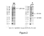


Classifications
-
- C—CHEMISTRY; METALLURGY
- C07—ORGANIC CHEMISTRY
- C07K—PEPTIDES
- C07K16/00—Immunoglobulins [IGs], e.g. monoclonal or polyclonal antibodies
- C07K16/18—Immunoglobulins [IGs], e.g. monoclonal or polyclonal antibodies against material from animals or humans
- C07K16/28—Immunoglobulins [IGs], e.g. monoclonal or polyclonal antibodies against material from animals or humans against receptors, cell surface antigens or cell surface determinants
- C07K16/2803—Immunoglobulins [IGs], e.g. monoclonal or polyclonal antibodies against material from animals or humans against receptors, cell surface antigens or cell surface determinants against the immunoglobulin superfamily
- C07K16/2827—Immunoglobulins [IGs], e.g. monoclonal or polyclonal antibodies against material from animals or humans against receptors, cell surface antigens or cell surface determinants against the immunoglobulin superfamily against B7 molecules, e.g. CD80, CD86
-
- A—HUMAN NECESSITIES
- A61—MEDICAL OR VETERINARY SCIENCE; HYGIENE
- A61K—PREPARATIONS FOR MEDICAL, DENTAL OR TOILETRY PURPOSES
- A61K39/00—Medicinal preparations containing antigens or antibodies
- A61K39/395—Antibodies; Immunoglobulins; Immune serum, e.g. antilymphocytic serum
- A61K39/39533—Antibodies; Immunoglobulins; Immune serum, e.g. antilymphocytic serum against materials from animals
- A61K39/3955—Antibodies; Immunoglobulins; Immune serum, e.g. antilymphocytic serum against materials from animals against proteinaceous materials, e.g. enzymes, hormones, lymphokines
-
- A—HUMAN NECESSITIES
- A61—MEDICAL OR VETERINARY SCIENCE; HYGIENE
- A61K—PREPARATIONS FOR MEDICAL, DENTAL OR TOILETRY PURPOSES
- A61K45/00—Medicinal preparations containing active ingredients not provided for in groups A61K31/00 - A61K41/00
- A61K45/06—Mixtures of active ingredients without chemical characterisation, e.g. antiphlogistics and cardiaca
-
- A—HUMAN NECESSITIES
- A61—MEDICAL OR VETERINARY SCIENCE; HYGIENE
- A61P—SPECIFIC THERAPEUTIC ACTIVITY OF CHEMICAL COMPOUNDS OR MEDICINAL PREPARATIONS
- A61P35/00—Antineoplastic agents
-
- A—HUMAN NECESSITIES
- A61—MEDICAL OR VETERINARY SCIENCE; HYGIENE
- A61P—SPECIFIC THERAPEUTIC ACTIVITY OF CHEMICAL COMPOUNDS OR MEDICINAL PREPARATIONS
- A61P37/00—Drugs for immunological or allergic disorders
- A61P37/02—Immunomodulators
-
- C—CHEMISTRY; METALLURGY
- C07—ORGANIC CHEMISTRY
- C07K—PEPTIDES
- C07K16/00—Immunoglobulins [IGs], e.g. monoclonal or polyclonal antibodies
- C07K16/18—Immunoglobulins [IGs], e.g. monoclonal or polyclonal antibodies against material from animals or humans
- C07K16/22—Immunoglobulins [IGs], e.g. monoclonal or polyclonal antibodies against material from animals or humans against growth factors ; against growth regulators
-
- A—HUMAN NECESSITIES
- A61—MEDICAL OR VETERINARY SCIENCE; HYGIENE
- A61K—PREPARATIONS FOR MEDICAL, DENTAL OR TOILETRY PURPOSES
- A61K39/00—Medicinal preparations containing antigens or antibodies
- A61K2039/505—Medicinal preparations containing antigens or antibodies comprising antibodies
-
- C—CHEMISTRY; METALLURGY
- C07—ORGANIC CHEMISTRY
- C07K—PEPTIDES
- C07K2317/00—Immunoglobulins specific features
- C07K2317/20—Immunoglobulins specific features characterized by taxonomic origin
- C07K2317/22—Immunoglobulins specific features characterized by taxonomic origin from camelids, e.g. camel, llama or dromedary
-
- C—CHEMISTRY; METALLURGY
- C07—ORGANIC CHEMISTRY
- C07K—PEPTIDES
- C07K2317/00—Immunoglobulins specific features
- C07K2317/30—Immunoglobulins specific features characterized by aspects of specificity or valency
- C07K2317/31—Immunoglobulins specific features characterized by aspects of specificity or valency multispecific
-
- C—CHEMISTRY; METALLURGY
- C07—ORGANIC CHEMISTRY
- C07K—PEPTIDES
- C07K2317/00—Immunoglobulins specific features
- C07K2317/30—Immunoglobulins specific features characterized by aspects of specificity or valency
- C07K2317/33—Crossreactivity, e.g. for species or epitope, or lack of said crossreactivity
-
- C—CHEMISTRY; METALLURGY
- C07—ORGANIC CHEMISTRY
- C07K—PEPTIDES
- C07K2317/00—Immunoglobulins specific features
- C07K2317/50—Immunoglobulins specific features characterized by immunoglobulin fragments
- C07K2317/52—Constant or Fc region; Isotype
- C07K2317/522—CH1 domain
-
- C—CHEMISTRY; METALLURGY
- C07—ORGANIC CHEMISTRY
- C07K—PEPTIDES
- C07K2317/00—Immunoglobulins specific features
- C07K2317/50—Immunoglobulins specific features characterized by immunoglobulin fragments
- C07K2317/52—Constant or Fc region; Isotype
- C07K2317/53—Hinge
-
- C—CHEMISTRY; METALLURGY
- C07—ORGANIC CHEMISTRY
- C07K—PEPTIDES
- C07K2317/00—Immunoglobulins specific features
- C07K2317/50—Immunoglobulins specific features characterized by immunoglobulin fragments
- C07K2317/55—Fab or Fab'
-
- C—CHEMISTRY; METALLURGY
- C07—ORGANIC CHEMISTRY
- C07K—PEPTIDES
- C07K2317/00—Immunoglobulins specific features
- C07K2317/50—Immunoglobulins specific features characterized by immunoglobulin fragments
- C07K2317/56—Immunoglobulins specific features characterized by immunoglobulin fragments variable (Fv) region, i.e. VH and/or VL
- C07K2317/565—Complementarity determining region [CDR]
-
- C—CHEMISTRY; METALLURGY
- C07—ORGANIC CHEMISTRY
- C07K—PEPTIDES
- C07K2317/00—Immunoglobulins specific features
- C07K2317/50—Immunoglobulins specific features characterized by immunoglobulin fragments
- C07K2317/56—Immunoglobulins specific features characterized by immunoglobulin fragments variable (Fv) region, i.e. VH and/or VL
- C07K2317/569—Single domain, e.g. dAb, sdAb, VHH, VNAR or nanobody®
-
- C—CHEMISTRY; METALLURGY
- C07—ORGANIC CHEMISTRY
- C07K—PEPTIDES
- C07K2317/00—Immunoglobulins specific features
- C07K2317/60—Immunoglobulins specific features characterized by non-natural combinations of immunoglobulin fragments
- C07K2317/64—Immunoglobulins specific features characterized by non-natural combinations of immunoglobulin fragments comprising a combination of variable region and constant region components
-
- C—CHEMISTRY; METALLURGY
- C07—ORGANIC CHEMISTRY
- C07K—PEPTIDES
- C07K2317/00—Immunoglobulins specific features
- C07K2317/70—Immunoglobulins specific features characterized by effect upon binding to a cell or to an antigen
- C07K2317/74—Inducing cell proliferation
-
- C—CHEMISTRY; METALLURGY
- C07—ORGANIC CHEMISTRY
- C07K—PEPTIDES
- C07K2317/00—Immunoglobulins specific features
- C07K2317/70—Immunoglobulins specific features characterized by effect upon binding to a cell or to an antigen
- C07K2317/76—Antagonist effect on antigen, e.g. neutralization or inhibition of binding
-
- C—CHEMISTRY; METALLURGY
- C07—ORGANIC CHEMISTRY
- C07K—PEPTIDES
- C07K2317/00—Immunoglobulins specific features
- C07K2317/90—Immunoglobulins specific features characterized by (pharmaco)kinetic aspects or by stability of the immunoglobulin
- C07K2317/92—Affinity (KD), association rate (Ka), dissociation rate (Kd) or EC50 value
Definitions
- the present disclosure generally relates to bispecific anti-PD-L1 x VEGF antibodies, a method for preparing the same and uses thereof.
- VEGF Vascular endothelial growth factor
- Tumor cells release growth factors such as VEGF that bind to nearby endothelial cells, which initiates a signaling cascade that stimulates endothelial cells to divide and form new blood vessels.
- VEGF signaling through its receptors VEGFR plays a critical role in angiogenesis and growth of many solid tumors.
- Antiangiogenic drugs such as Avastin (Bevacizumab) which targets the VEGF pathway, have achieved a success in clinic.
- PD-L1 expression strongly correlates with unfavorable prognosis in various types of cancers.
- Anti-PD-L1 antibody can target PD-L1 expressed on tumor cells and tumor-infiltrating immune cells, and prevent binding to PD-1 and B7.1 on the surface of T cells, and also enable the activation of T cells as well as recruit other T cells to attack the tumor, then empower the immune system to fight multiple types of cancer.
- Anti-VEGF therapy in addition to its established anti-angiogenic effects, may further enhance anti-PD-1/PD-L1 therapy’s ability to restore anti-cancer immunity, by inhibiting VEGF-related immunosuppression, promoting T-cell tumor infiltration and enabling priming and activation of T-cell responses against tumor antigens. Therefore, developing VEGF and PD-L1 bispecific antibodies which combine anti-angiogenesis therapy and immune checkpoint inhibition together could achieve promising results in cancer therapy.
- bispecific antibody that could simultaneously bind to human PD-L1 and VEGF with high affinity, block both PD-1/PD-L1 and VEGF/VEGFR signaling, and display superior anti-tumor efficacy has been generated.
- the present disclosure provides a bispecific antibody or antigen-binding portion thereof, comprising a PD-L1 antigen-binding moiety associated with a VEGF antigen-binding moiety, wherein:
- the PD-L1 antigen-binding moiety comprises: a complementarity determining region (CDR) 1 comprising SEQ ID NO: 1, a CDR2 comprising SEQ ID NO: 2, and a CDR3 comprising SEQ ID NO: 3, and
- CDR complementarity determining region
- the VEGF antigen-binding moiety comprises: a heavy chain complementarity determining region (HCDR) 1 comprising SEQ ID NO: 4, a HCDR2 comprising SEQ ID NO: 5, a HCDR3 comprising SEQ ID NO: 6, a light chain complementarity determining region (LCDR) 1 comprising SEQ ID NO: 7, a LCDR2 comprising SEQ ID NO: 8, and a LCDR3 comprising SEQ ID NO: 9.
- HCDR heavy chain complementarity determining region
- HCDR2 comprising SEQ ID NO: 5
- a HCDR3 comprising SEQ ID NO: 6
- LCDR light chain complementarity determining region
- LCDR2 comprising SEQ ID NO: 8
- a LCDR3 comprising SEQ ID NO: 9.
- the PD-L1 antigen-binding moiety as disclosed herein comprises: a variable domain comprising the amino acid sequence of SEQ ID NO: 10 or an amino acid sequence at least 85%, 90%, or 95%identical to SEQ ID NO: 10.
- the VEGF antigen-binding moiety as disclosed herein comprises:
- a heavy chain variable domain comprising the amino acid sequence of SEQ ID NO: 11 or an amino acid sequence at least 85%, 90%, or 95%identical to SEQ ID NO: 11;
- a light chain variable domain comprising the amino acid sequence of SEQ ID NO: 12 or an amino acid sequence at least 85%, 90%, or 95%identical to SEQ ID NO: 12.
- the PD-L1 antigen-binding moiety is fused to the N terminal of the VEGF antigen-binding moiety. In some other embodiements, the PD-L1 antigen-binding moiety is fused to the C terminal of the VEGF antigen-binding moiety.
- the PD-L1 antigen-binding moiety is from a single-domain antibody (sdAb) , such as a VHH antibody.
- sdAb single-domain antibody
- the VHH may be derived from a camelid animal, comprising an alpaca or a llama.
- the VHH is a humanized VHH.
- the PD-L1 antigen-binding moiety is operably linked to the N terminal of the light chain or heavy chain of the VEGF antigen-binding moiety, optionally via a linker.
- the linker may comprise or consist of 1 to 4 copies of GGGGS (G4S) , for example, the linker may be (G4S) 2.
- the bispecific antibody or antigen-binding portion thereof as disclosed herein comprises a heavy chain and a light chain, wherein:
- the heavy chain comprises domains operably linked as in VH-CH1-hinge-Fc, wherein the VH-CH1 is from the VEGF antigen binding moiety;
- the light chain comprises domains operably linked as in VHH-VL-CL, wherein the VHH is from the PD-L1 antigen binding moiety and the VL-CL is from the VEGF antigen binding moiety.
- the Fc region is a human IgG Fc region, preferably a human IgG1 Fc region.
- the bispecific antibody or antigen-binding portion thereof as disclosed herein comprise a heavy chain comprising SEQ ID NO: 13 and a light chain comprising SEQ ID NO: 14.
- the bispecific antibody or antigen-binding portion thereof as disclosed herein is a humanized antibody.
- the present disclosure provides an isolated nucleic acid molecule, comprising a nucleic acid sequence encoding the bispecific antibody or the antigen-binding portion thereof as disclosed herein.
- the present disclosure provides a vector comprising the nucleic acid molecule as disclosed herein. In one aspect, the present disclosure provides a host cell comprising the nucleic acid molecule or the vector as disclosed herein.
- the present disclosure provides a pharmaceutical composition
- a pharmaceutical composition comprising the bispecific antibody or the antigen-binding portion thereof as disclosed herein and a pharmaceutically acceptable carrier.
- the present disclosure provides a method for producing the bispecific antibody or the antigen-binding portion thereof as disclosed herein, comprising the steps of:
- the present disclosure provides a method for modulating an immune response in a subject, comprising administering to the subject the bispecific antibody or the antigen-binding portion thereof or the pharmaceutical composition as disclosed herein to the subject.
- the present disclosure provides a method for inhibiting growth of tumor cells in a subject, comprising administering an effective amount of the bispecific antibody or the antigen-binding portion thereof or the pharmaceutical composition as disclosed herein to the subject.
- the present disclosure provides a method for preventing or treating cancer in a subject, comprising administering an effective amount of the bispecific antibody or the antigen-binding portion thereof or the pharmaceutical composition to the subject.
- the cancer may be selected from colon cancer, colorectal cancer, breast cancer, lung cancer, cervical cancer, renal cancer, glioblastoma, ovarian cancer, pancreatic cancer, prostate cancer, esophageal cancer, gastric cancer, lymphoma, melanoma, liver cancer, and head and neck cancer.
- the cancer is colon cancer or colorectal cancer.
- the bispecific antibody or antigen-binding portion thereof as disclosed herein may be administered in combination with a chemotherapeutic agent, radiation and/or other agents for use in cancer immunotherapy.
- the present disclosure provides a bispecific antibody or antigen-binding portion thereof as disclosed herein for use
- the present disclosure provides the bispecific antibody or antigen-binding portion thereof as disclosed herein for use in diagnosing, preventing or treating cancers.
- the present disclosure provides use of the bispecific antibody or antigen- binding portion thereof as disclosed herein in the manufacture of a medicament for modulating an immune response or inhibiting growth of tumor cells in a subject.
- the present disclosure provides use of the bispecific antibody or antigen-binding portion thereof as disclosed herein in the manufacture of a medicament for treating or preventing cancers.
- the present disclosure provides a kit comprising the bispecific antibody or antigen-binding portion thereof as disclosed herein.
- the kit can be used for detection, diagnosis, prognosis, or treatment of a disease or condition, such as cancer.
- Figure 1 shows a schematic representation of the format of W3256 antibody.
- Figures 2 shows SDS-PAGE of W3256 antibody (lane 1) .
- NR Non-reducing status
- R reducing status in NuPAGE (Novex 4-12%Bis-Tris) gel.
- M PageRuler TM Unstained Protein Ladder.
- FIG. 3 shows the result of HPLC-SEC of W3256 antibody.
- Figure 4 shows the DSF profile of W3256 antibody.
- Figure 5 shows the ELISA binding result of W3256 antibody to human VEGF (same as cyno VEGF) .
- hIgG1 is an isotype control.
- Figure 6 shows the FACS binding result of W3256 antibody to human PD-L1.
- Figure 7 shows the dual binding result of W3256 antibody to VEGF and then PD-L1.
- Figure 8 shows the dual binding of W3256 antibody to PD-L1 and then VEGF.
- Figure 9 shows the FACS binding result of W3256 antibody to cynomolgus PD-L1.
- Figure 10 shows the ELISA binding result of W3256 antibody to mouse VEGF.
- Figure 11 shows the FACS binding result of W3256 antibody to mouse PD-L1.
- Figures 12-13 show the SPR sensorgram of W3256 antibody binding to human PD-L1 (Fig. 12) and VEGF (Fig. 13) .
- Figures 14-15 show the competition of W3256 antibody to human VEGFR1 on human VEGF binding (Fig. 14) , and the competition of W3256 antibody to human PD-1 on human PD-L1 binding (Fig. 15) .
- Figures 16-17 show the competition of W3256 antibody to mouse VEGFR1 on human VEGF binding (Fig. 16) , and the competition of W3256 antibody to mouse PD-1 on mouse PD-L1 binding (Fig. 17) .
- Figure 18 shows the inhibition of antibodies on HUVEC cells proliferation.
- Figure 19 shows the effect of antibodies on reporter gene assay.
- Figure 20 shows the effect of antibodies on hCD4+T cell IFN- ⁇ secretion in MLR.
- “Combo” refers to the combination of WBP325-BMK3. uIgG1 and W315-BMK8. uIgG1K (RKNA) .
- Figure 21 shows mouse PK analysis of W3256 antibody in total IgG binding assay after a single intravenous injection of equal molar dose of antibody.
- mpk refers to the abbreviation of “mg/kg” .
- Figure 22 shows mouse PK analysis of W3256 antibody in dual-antigen binding assay after a single intravenous injection of equal molar dose of antibody.
- Figure 23 shows the efficacy of antibodies in PBMC-RKO cancer model in mice.
- antibody or “Ab, ” herein is used in the broadest sense, which encompasses various antibody structures, including polyclonal antibodies, monospecific and multispecific antibodies (e.g. bispecific antibodies) .
- a native intact antibody generally is a Y-shaped tetrameric protein comprising two heavy (H) and two light (L) polypeptide chains held together by covalent disulfide bonds and non-covalent interactions.
- Light chains of an antibody may be classified into ⁇ and ⁇ light chain.
- Heavy chains may be classified into ⁇ , ⁇ , ⁇ , ⁇ and ⁇ , which define isotypes of an antibody as IgM, IgD, IgG, IgA and IgE, respectively.
- a variable region is linked to a constant region via a “J” region of about 12 or more amino acids, and a heavy chain further comprises a “D” region of about 3 or more amino acids.
- Each heavy chain consists of a heavy chain variable region (VH) and a heavy chain constant region (CH) .
- a heavy chain constant region consists of 3 domains (CH1, CH2 and CH3) .
- Each light chain consists of a light chain variable region (VL) and a light chain constant region (CL) .
- VH and VL region can further be divided into hypervariable regions (called complementary determining regions (CDR) ) , which are interspaced by relatively conservative regions (called framework region (FR) ) .
- CDR complementary determining regions
- FR framework region
- Each VH and VL consists of 3 CDRs and 4 FRs in the following order: FR1, CDR1, FR2, CDR2, FR3, CDR3, FR4 from N-terminal to C-terminal.
- the variable region (V H and V L ) of each heavy/light chain pair forms antigen binding sites, respectively. Distribution of amino acids in various regions or domains follows the definition in Kabat Sequences of Proteins of Immunological Interest (National Institutes of Health, Bethesda, Md. (1987 and 1991) ) , or Chothia & Lesk (1987) J. Mol. Biol. 196: 901-917; Chothia et al., (1989) Nature 342: 878-883.
- Antibodies may be of different antibody isotypes, for example, IgG (e.g., IgG1, IgG2, IgG3 or IgG4 subtype) , IgA1, IgA2, IgD, IgE or IgM antibody.
- IgG e.g., IgG1, IgG2, IgG3 or IgG4 subtype
- IgA1, IgA2, IgD, IgE or IgM antibody for example, IgG (e.g., IgG1, IgG2, IgG3 or IgG4 subtype)
- IgA1, IgA2, IgD, IgE or IgM antibody e.gA1, IgA2, IgD, IgE or IgM antibody.
- antigen-binding portion or “antigen-binding fragment” of an antibody, which can be interchangeably used in the context of the application, refers to polypeptides comprising fragments of a full-length antibody, which retain the ability of specifically binding to an antigen that the full-length antibody specifically binds to, and/or compete with the full-length antibody for binding to the same antigen.
- antigen-binding portion or “antigen-binding fragment” of an antibody, which can be interchangeably used in the context of the application, refers to polypeptides comprising fragments of a full-length antibody, which retain the ability of specifically binding to an antigen that the full-length antibody specifically binds to, and/or compete with the full-length antibody for binding to the same antigen.
- Antigen-binding fragments of an antibody may be derived, e.g., from full antibody molecules using any suitable standard techniques such as proteolytic digestion or recombinant genetic engineering techniques involving the manipulation and expression of DNA encoding antibody variable and optionally constant domains.
- DNA is known and/or is readily available from, e.g., commercial sources, DNA libraries (including, e.g., phage-antibody libraries) , or can be synthesized.
- the DNA may be sequenced and manipulated chemically or by using molecular biology techniques, for example, to arrange one or more variable and/or constant domains into a suitable configuration, or to introduce codons, create cysteine residues, modify, add or delete amino acids, etc.
- Non-limiting examples of antigen-binding fragments include: (i) Fab fragments; (ii) F (ab’) 2 fragments; (iii) Fd fragments; (iv) Fv fragments; (v) single-chain Fv (scFv) molecules; (vi) dAb fragments; and (vii) minimal recognition units consisting of the amino acid residues that mimic the hypervariable region of an antibody (e.g., an isolated complementarity determining region (CDR) such as a CDR3 peptide) , or a constrained FR3-CDR3-FR4 peptide.
- CDR complementarity determining region
- an antigen-binding fragment of an antibody may contain at least one variable domain covalently linked to at least one constant domain.
- the variable and constant domains may be either directly linked to one another or may be linked by a full or partial hinge or linker region.
- a hinge region may consist of at least 2 (e.g., 5, 10, 15, 20, 40, 60 or more) amino acids which result in a flexible or semi-flexible linkage between adjacent variable and/or constant domains in a single polypeptide molecule.
- variable domain refers to an antibody variable region or a fragment thereof comprising one or more CDRs.
- a variable domain may comprise an intact variable region (such as HCVR or LCVR) , it is also possible to comprise less than an intact variable region yet still retain the capability of binding to an antigen or forming an antigen-binding site.
- antigen-binding moiety refers to an antibody fragment formed from a portion of an antibody comprising one or more CDRs, or any other antibody fragment that binds to an antigen but does not comprise an intact native antibody structure.
- an antigen-binding moiety may comprise constant domains in addition to variable domains.
- antigen-binding moiety examples include, without limitation, a variable domain, a variable region, a diabody, a Fab, a Fab', a F (ab') 2 , an Fv fragment, a disulfide stabilized Fv fragment (dsFv) , a (dsFv) 2 , a bispecific dsFv (dsFv-dsFv') , a disulfide stabilized diabody (ds diabody) , a multispecific antibody, a camelized single domain antibody, a nanobody, a domain antibody, and a bivalent domain antibody.
- An antigen-binding moiety is capable of binding to the same antigen to which the parent antibody binds.
- an antigen-binding moiety may be a Fab fragment or a VHH antibody.
- an antigen-binding moiety may comprise one or more CDRs from a particular human antibody grafted to a framework region from one or more different human antibodies.
- Fab with regard to an antibody refers to that portion of the antibody consisting of a single light chain (both variable and constant regions) associating to the variable region and first constant region of a single heavy chain by a disulfide bond.
- Fc with regard to an antibody refers to that portion of the antibody comprising the second (CH2) and third (CH3) constant regions of a first heavy chain bound to the second and third constant regions of a second heavy chain via disulfide bonding.
- a Fc region depending on the context, it may refer to one chain or both chains of the Fc region.
- the Fc portion of the antibody is responsible for various effector functions such as ADCC, and CDC, but does not function in antigen binding.
- the capacity of antibodies to initiate and regulate effector functions through their Fc domain is a key component of their in vivo protective activity.
- PD-L1 also known as programmed death-ligand 1
- programmed death-ligand 1 is a 40 kDa type 1 transmembrane protein that has been speculated to play a major role in suppressing the adaptive arm of immune system.
- PD-L1 is the principal ligand of programmed death 1 (PD-1) , a coinhibitory receptor that can be constitutively expressed or induced in myeloid, lymphoid, normal epithelial cells and in cancer.
- PD-L1 when referring to the amino acid sequence of PD-L1 protein, including full-length PD-L1 protein, or the extracellular domain of PD-L1 (PD-L1 ECD) or fragment containing PD-L1 ECD; Fusion protein of PD-L1 ECD, for example, fragment fused with IgG Fc from mice or human (mFc or hFc) is also included.
- PD-L1 protein would also include those into which mutations of amino acid sequence are naturally or artificially introduced (including but not limited to replacement, deletion and/or addition) without affecting the biological functions.
- an antibody that binds PD-L1 or an “anti-PD-L1 antibody” as used herein includes antibodies and antigen-binding fragments thereof that specifically recognize PD-L1.
- the antibodies and antigen-binding fragments of the present disclosure may bind soluble PD-L1 protein and/or cell surface expressed PD-L1.
- Soluble PD-L1 includes natural PD-L1 proteins as well as recombinant PD-L1 protein variants that lack a transmembrane domain or are otherwise unassociated with a cell membrane .
- anti-PD-L1 antibody includes both monovalent antibodies with a single specificity, as well as bispecific antibodies comprising a first antigen-binding site that binds PD-L1 and a second antigen-binding site that binds a second (target) antigen, wherein the anti-PD-L1 antigen-binding site comprises any of the HCVR/LCVR or CDR sequences as set forth in Table A herein. Examples of anti-PD-L1 bispecific antibodies are described elsewhere herein.
- the term "antigen-binding molecule” includes antibodies and antigen-binding fragments of antibodies, including, e.g., bispecific antibodies.
- VEGF vascular endothelial growth factor
- VEGF-A vascular endothelial growth factor
- VEGF-A vascular endothelial growth factor
- the VEGF family includes VEGF-A, VEGF-B, VEGF-C, VEGF-D, PlGF (placental growth factor) , VEGF-E (Orf-VEGF) , and Trimeresurus flavoviridis svVEGF.
- VEGF receptor refers to receptors for vascular endothelial growth factor (VEGF) .
- VEGF vascular endothelial growth factor
- the VEGF receptors may be membrane-bound or soluble, depending on alternative splicing.
- VEGFR-1 binds VEGF-A, PlGF, and VEGF-B.
- a “bispecific antibody” refers to an artificial antibody, which has fragments derived from two different monoclonal antibodies and is capable of binding to two different epitopes.
- the two epitopes may present on the same antigen, or they may present on two different antigens.
- bispecific antigen-binding molecule means a protein, polypeptide or molecular complex comprising at least a first antigen-binding domain (also referred to as a first antigen-binding site herein) and a second antigen-binding domain (also referred to as a second antigen-binding site herein) .
- the “bispecific antigen-binding molecule” is a “bispecific antibody” .
- Each antigen-binding domain within the bispecific antibody comprises at least one CDR that alone, or in combination with one or more additional CDRs and/or FRs, specifically binds to a particular antigen.
- the first antigen-binding site specifically binds to a first antigen (e.g., PD-L1)
- the second antigen-binding site specifically binds to a second, distinct antigen (e.g., VEGF) .
- anti-PD-L1/anti-VEGF antibody refers to a bispecific antibody that specifically binds to PD-L1 and VEGF.
- monoclonal antibody or “mAb” , as used herein, refer to a preparation of antibody molecules of single molecular composition.
- a monoclonal antibody displays a single binding specificity and affinity for a particular epitope.
- chimeric antibody refers to antibodies in which the variable region sequences are derived from one species and the constant region sequences are derived from another species, such as an antibody in which the variable region sequences are derived from a mouse antibody and the constant region sequences are derived from a human antibody.
- humanized antibody is intended to refer to antibodies in which CDR sequences derived from the germline of another mammalian species, such as a mouse, have been grafted onto human framework sequences. Additional framework region modifications may be made within the human framework sequences.
- operably linked refers to a juxtaposition, with or without a spacer or linker, of two or more biological sequences of interest in such a way that they are in a relationship permitting them to function in an intended manner.
- polypeptides it is intended to mean that the polypeptide sequences are linked in such a way that permits the linked product to have the intended biological function.
- an antibody variable region may be operably linked to a constant region so as to provide for a stable product with antigen-binding activity.
- the term may also be used with respect to polynucleotides.
- a polynucleotide encoding a polypeptide when operably linked to a regulatory sequence (e.g., promoter, enhancer, silencer sequence, etc. ) , it is intended to mean that the polynucleotide sequences are linked in such a way that permits regulated expression of the polypeptide from the polynucleotide.
- a regulatory sequence e.g., promoter, enhancer, silencer sequence, etc.
- Ka is intended to refer to the association rate of a particular antibody-antigen interaction
- Kd is intended to refer to the dissociation rate of a particular antibody-antigen interaction.
- Kd values for antibodies can be determined using methods well established in the art.
- K D is intended to refer to the dissociation constant of a particular antibody-antigen interaction, which is obtained from the ratio of Kd to Ka (i.e., Kd/Ka) and is expressed as a molar concentration (M) .
- a preferred method for determining the Kd of an antibody is by using surface plasmon resonance, preferably using a biosensor system such as a system.
- high affinity for an IgG antibody refers to an antibody having a K D of 1 x 10 -7 M or less, more preferably 5 x 10 -8 M or less, even more preferably 1x10 -8 M or less, even more preferably 5 x 10 -9 M or less and even more preferably 1 x 10 -9 M or less for a target antigen.
- EC 50 as used herein, which is also termed as “half maximal effective concentration” refers to the concentration of a drug, antibody or toxicant which induces a response halfway between the baseline and maximum after a specified exposure time. In the context of the application, EC 50 is expressed in the unit of “nM” .
- IC 50 as used herein, which is also termed as “half maximal inhibitory concentration” is a measure of the potency of a substance in inhibiting a specific biological or biochemical function. In the context of the application, IC 50 is expressed in the unit of “nM” .
- inhibitor binding refers to the ability of an antibody or antigen-binding fragment thereof to inhibit the binding of two molecules (eg, human PD-L1/VEGF and human PD-1/VEGFR) to any detectable level.
- the binding of the two molecules can be inhibited by the antibodies at an IC 50 of no more than 50 nM, no more than 30 nM, no more than 10 nM, no more than 5 nM, no more than 1 nM or even less.
- epitope refers to a portion on antigen that an immunoglobulin or antibody specifically binds to. “Epitope” is also known as “antigenic determinant” .
- Epitope or antigenic determinant generally consists of chemically active surface groups of a molecule such as amino acids, carbohydrates or sugar side chains, and generally has a specific three-dimensional structure and a specific charge characteristic. See, for example, Epitope Mapping Protocols in Methods in Molecular Biology, Vol. 66, G.E. Morris, Ed. (1996) .
- isolated refers to a state obtained from natural state by artificial means. If a certain “isolated” substance or component is present in nature, it is possible because its natural environment changes, or the substance is isolated from natural environment, or both. For example, a certain un-isolated polynucleotide or polypeptide naturally exists in a certain living animal body, and the same polynucleotide or polypeptide with a high purity isolated from such a natural state is called isolated polynucleotide or polypeptide.
- isolated excludes neither the mixed artificial or synthesized substance nor other impure substances that do not affect the activity of the isolated substance.
- isolated antibody is intended to refer to an antibody that is substantially free of other antibodies having different antigenic specificities (e.g., an isolated antibody that specifically binds a PD-L1/VEGF protein is substantially free of antibodies that specifically bind antigens other than PD-L1/VEGF proteins) .
- An isolated antibody that specifically binds a human PD-L1/VEGF protein may, however, have cross-reactivity to other antigens, such as PD-L1/VEGF proteins from other species.
- an isolated antibody can be substantially free of other cellular material and/or chemicals.
- vector refers to a nucleic acid vehicle which can have a polynucleotide inserted therein.
- the vector allows for the expression of the protein encoded by the polynucleotide inserted therein, the vector is called an expression vector.
- the vector can have the carried genetic material elements expressed in a host cell by transformation, transduction, or transfection into the host cell.
- Vectors are well known by a person skilled in the art, including, but not limited to plasmids, phages, cosmids, artificial chromosome such as yeast artificial chromosome (YAC) , bacterial artificial chromosome (BAC) or P1-derived artificial chromosome (PAC) ; phage such as ⁇ phage or M13 phage and animal virus.
- the animal viruses that can be used as vectors include, but are not limited to, retrovirus (including lentivirus) , adenovirus, adeno-associated virus, herpes virus (such as herpes simplex virus) , pox virus, baculovirus, papillomavirus, papova virus (such as SV40) .
- a vector may comprise multiple elements for controlling expression, including, but not limited to, a promoter sequence, a transcription initiation sequence, an enhancer sequence, a selection element and a reporter gene.
- a vector may comprise origin of replication.
- host cell refers to a cellular system which can be engineered to generate proteins, protein fragments, or peptides of interest.
- Host cells include, without limitation, cultured cells, e.g., mammalian cultured cells derived from rodents (rats, mice, guinea pigs, or hamsters) such as CHO, BHK, NSO, SP2/0, YB2/0; or human tissues or hybridoma cells, yeast cells, and insect cells, and cells comprised within a transgenic animal or cultured tissue.
- the term encompasses not only the particular subject cell but also the progeny of such a cell. Because certain modifications may occur in succeeding generations due to either mutation or environmental influences, such progeny may not be identical to the parent cell, but are still included within the scope of the term “host cell. ”
- identity refers to a relationship between the sequences of two or more polypeptide molecules or two or more nucleic acid molecules, as determined by aligning and comparing the sequences. “Percent identity” means the percent of identical residues between the amino acids or nucleotides in the compared molecules and is calculated based on the size of the smallest of the molecules being compared. For these calculations, gaps in alignments (if any) are preferably addressed by a particular mathematical model or computer program (i.e., an “algorithm” ) . Methods that can be used to calculate the identity of the aligned nucleic acids or polypeptides include those described in Computational Molecular Biology, (Lesk, A.M., ed.
- immunogenicity refers to ability of stimulating the formation of specific antibodies or sensitized lymphocytes in organisms. It not only refers to the property of an antigen to stimulate a specific immunocyte to activate, proliferate and differentiate so as to finally generate immunologic effector substance such as antibody and sensitized lymphocyte, but also refers to the specific immune response that antibody or sensitized T lymphocyte can be formed in immune system of an organism after stimulating the organism with an antigen. Immunogenicity is the most important property of an antigen. Whether an antigen can successfully induce the generation of an immune response in a host depends on three factors, properties of an antigen, reactivity of a host, and immunization means.
- transfection refers to the process by which nucleic acids are introduced into eukaryotic cells, particularly mammalian cells. Protocols and techniques for transfection include but not limited to lipid transfection and chemical and physical methods such as electroporation. A number of transfection techniques are well known in the art and are disclosed herein. See, e.g., Graham et al., 1973, Virology 52: 456; Sambrook et al., 2001, Molecular Cloning: A Laboratory Manual, supra; Davis et al., 1986, Basic Methods in Molecular Biology, Elsevier; Chu et al, 1981, Gene 13: 197. In a specific embodiment of the disclosure, human PD-L1/VEGF gene was transfected into 293F cells.
- SPR or “surface plasmon resonance, ” as used herein, refers to and includes an optical phenomenon that allows for the analysis of real-time biospecific interactions by detection of alterations in protein concentrations within a biosensor matrix, for example using the BIAcore system (Pharmacia Biosensor AB, Uppsala, Sweden and Piscataway, N.J. ) .
- BIAcore Pharmacia Biosensor AB, Uppsala, Sweden and Piscataway, N.J.
- FACS fluorescence-activated cell sorting
- subject includes any human or nonhuman animal, preferably humans.
- cancer refers to any or a tumor or a malignant cell growth, proliferation or metastasis-mediated, solid tumors and non-solid tumors such as leukemia and initiate a medical condition.
- treatment refers generally to treatment and therapy, whether of a human or an animal, in which some desired therapeutic effect is achieved, for example, the inhibition of the progress of the condition, and includes a reduction in the rate of progress, a halt in the rate of progress, regression of the condition, amelioration of the condition, and cure of the condition.
- Treatment as a prophylactic measure i.e., prophylaxis, prevention
- treating may refer to dampen or slow the tumor or malignant cell growth, proliferation, or metastasis, or some combination thereof.
- treatment includes removal of all or part of the tumor, inhibiting or slowing tumor growth and metastasis, preventing or delaying the development of a tumor, or some combination thereof.
- an effective amount refers to that amount of an active compound, or a material, composition or dosage from comprising an active compound, which is effective for producing some desired therapeutic effect, commensurate with a reasonable benefit/risk ratio, when administered in accordance with a desired treatment regimen.
- an effective amount, ” when used in connection with treatment of PD-L1/VEGF-related diseases or conditions refers to an antibody or antigen-binding portion thereof in an amount or concentration effective to treat the said diseases or conditions.
- prevention refers to preventing or delaying the onset of the disease, or preventing the manifestation of clinical or subclinical symptoms thereof.
- pharmaceutically acceptable means that the vehicle, diluent, excipient and/or salts thereof, are chemically and/or physically is compatible with other ingredients in the formulation, and the physiologically compatible with the recipient.
- apharmaceutically acceptable carrier and/or excipient refers to a carrier and/or excipient pharmacologically and/or physiologically compatible with a subject and an active agent, which is well known in the art (see, e.g., Remington's Pharmaceutical Sciences. Edited by Gennaro AR, 19th ed. Pennsylvania: Mack Publishing Company, 1995) , and includes, but is not limited to pH adjuster, surfactant, adjuvant and ionic strength enhancer.
- the pH adjuster includes, but is not limited to, phosphate buffer;
- the surfactant includes, but is not limited to, cationic, anionic, or non-ionic surfactant, e.g., Tween-80;
- the ionic strength enhancer includes, but is not limited to, sodium chloride.
- adjuvant refers to a non-specific immunopotentiator, which can enhance immune response to an antigen or change the type of immune response in an organism when it is delivered together with the antigen to the organism or is delivered to the organism in advance.
- adjuvants including, but not limited to, aluminium adjuvants (for example, aluminum hydroxide) , Freund’s adjuvants (for example, Freund’s complete adjuvant and Freund’s incomplete adjuvant) , coryne bacterium parvum, lipopolysaccharide, cytokines, and the like.
- Freund's adjuvant is the most commonly used adjuvant in animal experiments now.
- Aluminum hydroxide adjuvant is more commonly used in clinical trials.
- the antibodies and antigen-binding portions thereof provided herein are bispecific. In some embodiments, the bispecific antibodies and antigen-binding portions thereof provided herein have a first specificity for PD-L1, and a second specificity for VEGF.
- the present disclosure includes a bispecific antibody or the antigen-binding portion thereof, comprising a first antigen-binding moiety that specifically binds to PD-L1 and a second antigen-binding moiety that specifically binds to VEGF.
- a bispecific antibody or the antigen-binding portion thereof comprising a first antigen-binding moiety that specifically binds to PD-L1 and a second antigen-binding moiety that specifically binds to VEGF.
- Such antibodies may be referred to herein as, e.g., “anti-VEGF/anti-PD-L1” or “anti-PD-L1/VEGF, ” or “anti-PD-L1xVEGF” or “PD-L1xVEGF” bispecific antibodies, or other similar terminology.
- the bispecific antibodies of the disclosure could bind to human PD-L1 and human VEGF with high affinity.
- the binding of an antibody of the disclosure to PD-L1 or VEGF can be assessed using one or more techniques well established in the art, for instance, ELISA.
- the binding specificity of an antibody of the disclosure can also be determined by monitoring binding of the antibody to cells expressing a PD-L1 protein or a VEGF protein, e.g., flow cytometry.
- an antibody can be tested by a flow cytometry assay in which the antibody is reacted with a cell line that expresses human PD-L1, such as CHO cells that have been transfected to express PD-L1 on their cell surface.
- the binding of the antibody can be tested in BIAcore binding assays.
- suitable binding assays include ELISA or FACS assays, for example using a recombinant PD-L1 protein.
- an antibody of the disclosure binds to a human PD-L1 protein or human VEGF protein with a K D of 1 ⁇ 10 -7 M or less, a K D of 5 ⁇ 10 -8 M or less, a K D of 2 ⁇ 10 -8 M or less, a K D of 1 ⁇ 10 -8 M or less, a K D of 5 ⁇ 10 -9 M or less, a K D of 4 ⁇ 10 -9 M or less, a K D of 3 ⁇ 10 -9 M or less, a K D of 2 ⁇ 10 -9 M or less, a K D of 1 ⁇ 10 -9 M or less, a K D of 5 ⁇ 10 -10 M or less, or a K D of 1 ⁇ 10 -10 M or less, as measured by Surface Plasmon Resonance.
- the bispecific antibodies of the disclosure could bind to human PD-L1 and human VEGF (same as cyno VEGF) with a high affinity; bind to cyno and mouse PD-L1; effectively block both PD-1/PD-L1 and VEGFR/VEGF signaling pathways, e.g. with an IC50 of nM grade; block VEGF induced HUVEC proliferation; and produce strong agonistic effect on cytokine secretion.
- the PD-L1 binding moiety as defined herein may have various formats (e.g. VHH, scFv, Fab) , as long as it can specifically bind to the antigen.
- the PD-L1 binding moiety comprised in the bispecific antibody is derived from a monospecific anti-PD-L1 antibody, which may be a known antibody in the art or an antibody developed de novo.
- the PD-L1 binding moiety may be derived from a parental single-domain antibody (sdAb) , such as a VHH antibody, which generally refers to an antibody consisting of a single variable domain. Like a whole antibody, a single-domain antibody is able to bind selectively to a specific antigen.
- the PD-L1 binding moiety may be derived from a heavy chain antibody, which is devoid of light chains.
- VHH molecules derived from Camelidae antibodies are among the smallest intact antigen-binding domains known (approximately 15 kDa, or 10 times smaller than a conventional IgG) and hence are well suited towards delivery to dense tissues and for accessing the limited space between macromolecules.
- VHHs may be obtained using methods known in the art such as by immunizing a camel and obtaining hybridoma's therefrom, or by cloning a library of VHHs of the invention using molecular biology techniques known in the art and subsequent selection by using phage display.
- a VHH antibody can be obtained by immunization of llamas or alpacas with the desired antigen and subsequent isolation of the mRNA coding for heavy-chain antibodies.
- a gene library of single-domain antibodies containing several million clones is produced. Screening techniques like phage display and ribosome display help to identify the clones binding the antigen.
- One technique is phage display in which a library of (e.g., human) antibodies is synthesized on phages, the library is screened with the antigen of interest or an antibody-binding portion thereof, and the phage that binds the antigen is isolated, from which one may obtain the immunoreactive fragments.
- kits for generating phage display libraries are commercially available (e.g., the Pharmacia Recombinant Phage Antibody System, catalog no. 27-9400-01; and the Stratagene SurfZAP TM phage display kit, catalog no. 240612) .
- There also are other methods and reagents that can be used in generating and screening antibody display libraries see, e.g., Barbas et al., Proc. Natl. Acad. Sci. USA 88: 7978-7982 (1991) ) .
- VHH antibody Humanization of the VHH antibody can be achieved via a number of well-established methods in the art, for example, amino acid sequences of VHH framework regions can be blasted against human germline V-gene database, and humanized VHH sequences can be generated by replacing human CDR sequences in the top hit with VHH CDR sequences using Kabat CDR definition. Further, certain residues in the framework region may be back mutated to maintain affinity.
- the PD-L1 antigen binding moiety comprises one or more CDRs selected from the group consisting of:
- a CDR1 comprising SEQ ID NO: 1 or an amino acid sequence that differsfrom SEQ ID NO: 1 by an amino acid addition, deletion or substitution of not more than 2 amino acids;
- a CDR2 comprising SEQ ID NO: 2 or an amino acid sequence that differs from SEQ ID NO: 2 by an amino acid addition, deletion or substitution of not more than 2 amino acids;
- a CDR3 comprising SEQ ID NO: 3 or an amino acid sequence that differs from SEQ ID NO: 3 by an amino acid addition, deletion or substitution of not more than 2 amino acids.
- the PD-L1 antigen binding moiety is a VHH antibody that comprises (i) a CDR1 comprising or consisting of SEQ ID NO: 1; (ii) a CDR2 comprising or consisting of SEQ ID NO: 2; and (iii) a CDR3 comprising or consisting of SEQ ID NO: 3.
- the VHH of the PD-L1 antigen-binding moiety comprises: (i) the amino acid sequence of SEQ ID NO: 10; (ii) an amino acid sequence at least 85%, 90%, or 95%identical to SEQ ID NO: 10; or (iii) an amino acid sequence with addition, deletion and/or substitution of one or more (e.g. 10, 9, 8, 7, 6, 5, 4, 3, 2, 1) amino acid (s) compared with SEQ ID NO: 10.
- the VEGF antigen-binding moiety is a VEGF antigen-binding moiety
- the VEGF antigen-binding moiety provided herein may be derived from a parental anti-VEGF monospecific antibody.
- the VEGF antigen-binding moiety is the Fab fragment of an anti-VEGF full antibody, i.e. comprising VH region and CH1 region in the heavy chain, and VL region and CL region in the light chain.
- the anti-VEGF antibody which is used as the parental antibody may be a monoclonal antibody already known in the art (such as Bevacizumab) or which is de novo developed.
- the anti-VEGF antibody is a fully human antibody or a humanized antibody.
- the VH region of the VEGF antigen-binding moiety comprises one or more heavy chain CDRs (HCDRs) selected from the group consisting of:
- a HCDR1 consisting of SEQ ID NO: 4 or a HCDR1 that differs in amino acid sequence from SEQ ID NO: 4 by an amino acid addition, deletion or substitution of not more than 2 amino acids;
- a HCDR2 consisting of SEQ ID NO: 5 or a HCDR2 that differs in amino acid sequence from SEQ ID NO: 5 by an amino acid addition, deletion or substitution of not more than 2 amino acids;
- a HCDR3 consisting of SEQ ID NO: 6 or a HCDR3 that differs in amino acid sequence from SEQ ID NO: 6 by an amino acid addition, deletion or substitution of not more than 2 amino acids;
- the VL region comprises one or more light chain CDRs (LCDRs) selected from the group consisting of:
- a LCDR1 consisting of SEQ ID NO: 7 or a LCDR1 that differs in amino acid sequence from SEQ ID NO: 7 by an amino acid addition, deletion or substitution of not more than 2 amino acids;
- a LCDR2 consisting of SEQ ID NO: 8 or a LCDR2 that differs in amino acid sequence from SEQ ID NO: 8 by an amino acid addition, deletion or substitution of not more than 2 amino acids;
- a LCDR3 consisting of SEQ ID NO: 9 or a LCDR3 that differs in amino acid sequence from SEQ ID NO: 9 by an amino acid addition, deletion or substitution of not more than 2 amino acids.
- the VH comprises (i) a HCDR1 comprising or consisting of SEQ ID NO: 4; (ii) a HCDR2 comprising or consisting of SEQ ID NO: 5; and (iii) a HCDR3 comprising or consisting of SEQ ID NO: 6; and the VL comprises: (i) a LCDR1 comprising or consisting of SEQ ID NO: 7; (ii) a LCDR2 comprising or consisting of SEQ ID NO: 8; and (iii) a LCDR3 comprising or consisting of SEQ ID NO: 9.
- the VH of the VEGF antigen-binding moiety comprises: (i) the amino acid sequence of SEQ ID NO: 11; (ii) an amino acid sequence at least 85%, 90%, or 95%identical to SEQ ID NO: 11; or (iii) an amino acid sequence with addition, deletion and/or substitution of one or more (e.g. 10, 9, 8, 7, 6, 5, 4, 3, 2, 1) amino acids compared with SEQ ID NO: 11.
- the VL of the VEGF antigen-binding moiety comprises: (i) the amino acid sequence of SEQ ID NO: 12; (ii) an amino acid sequence at least 85%, 90%, or 95%identical to SEQ ID NO: 12; or (iii) an amino acid sequence with addition, deletion and/or substitution of one or more (e.g. 10, 9, 8, 7, 6, 5, 4, 3, 2, 1) amino acids compared with SEQ ID NO: 12.
- Variable regions and CDRs in an antibody sequence can be identified according to general rules that have been developed in the art (as set out above, such as, for example, the Kabat numbering system) or by aligning the sequences against a database of known variable regions. Methods for identifying these regions are described in Kontermann and Dubel, eds., Antibody Engineering, Springer, New York, NY, 2001 and Dinarello et al., Current Protocols in Immunology, John Wiley and Sons Inc., Hoboken, NJ, 2000. Exemplary databases of antibody sequences are described in, and can be accessed through, the “Abysis” website at www. bioinf. org. uk/abs (maintained by A.C.
- sequences are analyzed using the Abysis database, which integrates sequence data from Kabat, IMGT and the Protein Data Bank (PDB) with structural data from the PDB. See Dr. Andrew C.R. Martin's book chapter Protein Sequence and Structure Analysis of Antibody Variable Domains. In: Antibody Engineering Lab Manual (Ed.: Duebel, S.
- the Abysis database website further includes general rules that have been developed for identifying CDRs which can be used in accordance with the teachings herein. Unless otherwise indicated, all CDRs set forth herein are derived according to the Abysis database website as per Kabat.
- the percent identity between two amino acid sequences can be determined using the algorithm of E. Meyers and W. Miller (Comput. Appl. Biosci., 4: 11-17 (1988) ) which has been incorporated into the ALIGN program (version 2.0) , using a PAM120 weight residue table, a gap length penalty of 12 and a gap penalty of 4.
- the percentage of identity between two amino acid sequences can be determined by the algorithm of Needleman and Wunsch (J. Mol. Biol. 48: 444-453 (1970) ) which has been incorporated into the GAP program in the GCG software package (available at http: //www. gcg. com) , using either a Blossum 62 matrix or a PAM250 matrix, and a gap weight of 16, 14, 12, 10, 8, 6, or 4 and a length weight of 1, 2, 3, 4, 5, or 6.
- the protein sequences of the present disclosure can further be used as a “query sequence” to perform a search against public databases to, for example, identify related sequences.
- Such searches can be performed using the XBLAST program (version 2.0) of Altschul, et al. (1990) J. MoI. Biol. 215: 403-10.
- Gapped BLAST can be utilized as described in Altschul et al, (1997) Nucleic Acids Res. 25 (17) : 3389-3402.
- the default parameters of the respective programs e.g., XBLAST and NBLAST
- the default parameters of the respective programs e.g., XBLAST and NBLAST
- the CDR amino acid sequences can be at least 90%, 91%, 92%, 93%, 94%, 95%, 96%, 97%, 98%or 99%identical to the respective sequences set forth above.
- the amino acid sequences of the variable region can be at least 90%, 91%, 92%, 93%, 94%, 95%, 96%, 97%, 98%or 99%identical to the respective sequences set forth above.
- the CDRs of the isolated antibody or the antigen-binding portion thereof contain a conservative substitution of not more than 2 amino acids, or not more than 1 amino acid.
- conservative substitution refers to amino acid substitutions which would not disadvantageously affect or change the essential properties of a protein/polypeptide comprising the amino acid sequence.
- a conservative substitution may be introduced by standard techniques known in the art such as site-directed mutagenesis and PCR-mediated mutagenesis.
- Conservative amino acid substitutions include substitutions wherein an amino acid residue is substituted with another amino acid residue having a similar side chain, for example, a residue physically or functionally similar (such as, having similar size, shape, charge, chemical property including the capability of forming covalent bond or hydrogen bond, etc. ) to the corresponding amino acid residue.
- a residue physically or functionally similar such as, having similar size, shape, charge, chemical property including the capability of forming covalent bond or hydrogen bond, etc.
- amino acids having alkaline side chains for example, lysine, arginine and histidine
- amino acids having acidic side chains for example, aspartic acid and glutamic acid
- amino acids having uncharged polar side chains for example, glycine, asparagine, glutamine, serine, threonine, tyrosine, cysteine, tryptophan
- amino acids having nonpolar side chains for example, alanine, valine, leucine, isoleucine, proline, phenylalanine, methionine
- amino acids having ⁇ -branched side chains such as threonine, valine, isoleucine
- amino acids having aromatic side chains for example, tyrosine, phenylalanine, tryptophan, histidine
- a corresponding amino acid residue is preferably substituted with another amino acid residue from the same side-chain family.
- Methods for identifying amino acid conservative substitutions are well known in the art (see, for example, Brummell et al., Biochem. 32: 1180-1187 (1993) ; Kobayashi et al., Protein Eng. 12 (10) : 879-884 (1999) ; and Burks et al., Proc. Natl. Acad. Sci. USA 94: 412-417 (1997) , which are incorporated herein by reference) .
- the PD-L1 antigen-binding moiety and the VEGF antigen-binding moiety as descrived above may be fused together in various formats.
- the PD-L1 antigen-binding moiety is fused to the N terminal of the VEGF antigen-binding moiety.
- the single chain of the PD-L1 antigen-binding moiety may be operably linked to the heavy chain or light chain of the VEGF antigen-binding moiety, optionally via a linker.
- the PD-L1 antigen-binding moiety is linked to the light chain of the VEGF antigen-binding moiety.
- the linker may be a peptide linker, such as comprising 1-4 copies of GGGGS (G4S) .
- the linker is (G4S) 2.
- bispecific antibodies and antigen-binding portions provided herein can be made with any suitable methods known in the art.
- two immunoglobulin heavy chain-light chain pairs can be co-expressed in a host cell to produce bispecific antibodies in a recombinant way (see, for example, Milstein and Cuello, Nature, 305: 537 (1983) ) , followed by purification by affinity chromatography.
- sequences encoding the antibody heavy chain variable domains for the two specificities can also be respectively fused to immunoglobulin constant domain sequences, followed by insertion to an expression vector which is co-transfected with an expression vector for the light chain sequences to a suitable host cell for recombinant expression of the bispecific antibody (see, for example, WO 94/04690; Suresh et al., Methods in Enzymology, 121: 210 (1986) ) .
- the bispecific antibody comprises a Fc region that is operably linked to the VEGF antigen-binding moiety.
- the Fc region of the bispecific antibodies disclosed herein may be a human IgG Fc region.
- the IgG Fc region may be of any isotype, including, but not limited to, IgG1, IgG2, IgG3 or IgG4. In certain embodiments, the Fc region is of the IgG1 isotype.
- the Fc region may comprise one or more amino acid changes (e.g., insertions, deletions or substitutions) as compared to the specified chimeric version of the Fc region.
- the disclosure includes bispecific antigen-binding molecules comprising one or more modifications in the Fc region that results in a modified Fc region having a modified binding interaction between Fc and FcRn or Fc ⁇ R.
- EU numbering system or “EU index” is generally used when referring to a residue in an immunoglobulin heavy chain constant region (e.g., the EU index reported in Kabat et al., supra) .
- EU numbering as in Kabat or “EU index as in Kabat” refers to the residue numbering of the human IgG1 EU antibody. Unless stated otherwise herein, references to residue numbers in the constant domain of antibodies means residue numbering by the EU numbering system.
- the Fc region is operably linked to the VEGF binding moiety via a hinge region.
- the hinge region may be derived from human IgG1, IgG2 or IgG4. In certain embodiments, the hinge region is derived from human IgG1.
- the disclosure is directed to an isolated nucleic acid molecule, comprising a nucleic acid sequence encoding the bispecific antibody or the antigen-binding portion as disclosed herein.
- the nucleic acid sequence may encode a heavy chain and/or a light chain of the bispecific antibody.
- the nucleic acid sequence may encode the PD-L1 antigen-binding moiety, or a heavy chain or a light chain variable region of the VEGF antigen-binding moiety.
- the nucleic acid sequence may further encode the Fc region of the bispecific antibody.
- the disclosure is directed to a vector comprising the nucleic acid sequence as disclosed herein.
- the expression vector further comprises a nucleotide sequence encoding the constant region of a bispecific antibody, e.g. a humanized bispecific antibody.
- a vector in the context of the present disclosure may be any suitable vector, including chromosomal, non-chromosomal, and synthetic nucleic acid vectors (a nucleic acid sequence comprising a suitable set of expression control elements) .
- suitable vectors include derivatives of SV40, bacterial plasmids, phage DNA, baculovirus, yeast plasmids, vectors derived from combinations of plasmids and phage DNA, and viral nucleic acid (RNA or DNA) vectors.
- a PD-L1 or a VEGF antibody-encoding nucleic acid is comprised in a naked DNA or RNA vector, including, for example, a linear expression element (as described in for instance Sykes and Johnston, Nat Biotech 17, 355-59 (1997) ) , a compacted nucleic acid vector (as described in for instance US 6,077,835 and/or WO 00/70087) , a plasmid vector such as pBR322, pUC 19/18, or pUC 118/119, a “midge” minimally-sized nucleic acid vector (as described in for instance Schakowski et al., Mol Ther 3, 793-800 (2001) ) , or as a precipitated nucleic acid vector construct, such as a CaP04-precipitated construct (as described in for instance WO200046147, Benvenisty and Reshef, PNAS USA 83, 9551-55 (1986) , Wigler et al
- the vector is suitable for expression of the anti-PD-L1 antibody and/or anti-VEGF antibody in a bacterial cell.
- vectors include expression vectors such as BlueScript (Stratagene) , pIN vectors (Van Heeke & Schuster, J Biol Chem 264, 5503-5509 (1989) , pET vectors (Novagen, Madison WI) and the like) .
- a vector may also or alternatively be a vector suitable for expression in a yeast system. Any vector suitable for expression in a yeast system may be employed. Suitable vectors include, for example, vectors comprising constitutive or inducible promoters such as alpha factor, alcohol oxidase and PGH (reviewed in: F.
- a vector may also or alternatively be a vector suitable for expression in mammalian cells, e.g. a vector comprising glutamine synthetase as a selectable marker, such as the vectors described in Bebbington (1992) Biotechnology (NY) 10: 169-175.
- a nucleic acid and/or vector may also comprise a nucleic acid sequence encoding a secretion/localization sequence, which can target a polypeptide, such as a nascent polypeptide chain, to the periplasmic space or into cell culture media.
- a secretion/localization sequence which can target a polypeptide, such as a nascent polypeptide chain, to the periplasmic space or into cell culture media.
- sequences are known in the art, and include secretion leader or signal peptides.
- the vector may comprise or be associated with any suitable promoter, enhancer, and other expression-facilitating elements.
- suitable promoter, enhancer, and other expression-facilitating elements include strong expression promoters (e.g., human CMV IE promoter/enhancer as well as RSV, SV40, SL3-3, MMTV, and HIV LTR promoters) , effective poly (A) termination sequences, an origin of replication for plasmid product in E. coli, an antibiotic resistance gene as selectable marker, and/or a convenient cloning site (e.g., a polylinker) .
- Nucleic acids may also comprise an inducible promoter as opposed to a constitutive promoter such as CMV IE.
- the disclosure relates to a host cell comprising the vector specified herein above.
- the present disclosure also relates to a recombinant eukaryotic or prokaryotic host cell which produces a bispecific antibody of the present disclosure, such as a transfectoma.
- the PD-L1-specific antibody may be expressed in a recombinant eukaryotic or prokaryotic host cell, such as a transfectoma, which produces an antibody of the disclosure as defined herein or a bispecific antibody of the disclosure as defined herein.
- the VEGF-specific antibody may likewise be expressed in a recombinant eukaryotic or prokaryotic host cell, such as a transfectoma, which produces an antibody of the disclosure as defined herein or a bispecific antibody of the disclosure as defined herein.
- host cells include yeast, bacterial, plant and mammalian cells, such as CHO, CHO-S, HEK, HEK293, HEK-293F, Expi293F, PER. C6 or NSO cells or lymphocytic cells.
- the host cell may comprise a first and second nucleic acid construct stably integrated into the cellular genome.
- the present disclosure provides a cell comprising a non-integrated nucleic acid, such as a plasmid, cosmid, phagemid, or linear expression element, which comprises a first and second nucleic acid construct as specified above.
- the disclosure relates to a transgenic non-human animal or plant comprising nucleic acids encoding one or two sets of a human heavy chain and a human light chain, wherein the animal or plant produces a bispecific antibody of the disclosure.
- the disclosure relates to a hybridoma which produces an antibody for use in a bispecific antibody of the disclosure as defined herein.
- the disclosure relates to an expression vector comprising:
- the disclosure relates to a nucleic acid construct encoding one or more amino acid sequences set out in the sequence listing.
- the disclosure relates to a method for producing a bispecific antibody according to any one of the embodiments as disclosed herein, comprising the steps of culturing a host cell as disclosed herein comprising an expression vector or more than one expression vectors as disclosed herein expressing the bispecific antibody as disclosed herein and purifying said antibody from the culture media.
- the disclosure relates to a host cell comprising an expression vector as defined above.
- the host cell is a recombinant eukaryotic, recombinant prokaryotic, or recombinant microbial host cell.
- the disclosure is directed to a pharmaceutical composition
- a pharmaceutical composition comprising at least one bispecific antibody or antigen-binding portion thereof as disclosed herein and a pharmaceutically acceptable carrier.
- the pharmaceutical composition may optionally contain one or more additional pharmaceutically active ingredients, such as another antibody or a drug.
- additional pharmaceutically active ingredients such as another antibody or a drug.
- the pharmaceutical compositions of the disclosure also can be administered in a combination therapy with, for example, another immune-stimulatory agent, anti-cancer agent, an antiviral agent, or a vaccine, such that the anti-PD-L1/anti-VEGF bispecific antibody enhances the immune response against the vaccine.
- a pharmaceutically acceptable carrier can include, for example, a pharmaceutically acceptable liquid, gel or solid carriers, an aqueous medium, a non-aqueous medium, an anti-microbial agent, isotonic agents, buffers, antioxidants, anesthetics, suspending/dispersing agent, a chelating agent, a diluent, adjuvant, excipient or a nontoxic auxiliary substance, other known in the art various combinations of components or more.
- Suitable components may include, for example, antioxidants, fillers, binders, disintegrating agents, buffers, preservatives, lubricants, flavorings, thickening agents, coloring agents, emulsifiers or stabilizers such as sugars and cyclodextrin.
- Suitable anti-oxidants may include, for example, methionine, ascorbic acid, EDTA, sodium thiosulfate, platinum, catalase, citric acid, cysteine, mercapto glycerol, thioglycolic acid, Mercapto sorbitol, butyl methyl anisole, butylated hydroxy toluene and/or propylgalacte.
- compositions include one or more anti-oxidants such as methionine, reducing antibody or antigen binding fragment thereof may be oxidized.
- the oxidation reduction may prevent or reduce a decrease in binding affinity, thereby enhancing antibody stability and extended shelf life.
- the present disclosure provides a composition comprising one or more antibodies or antigen binding fragment thereof and one or more anti-oxidants such as methionine.
- the present disclosure further provides a variety of methods, wherein an antibody or antigen binding fragment thereof is mixed with one or more anti-oxidants, such as methionine, so that the antibody or antigen binding fragment thereof can be prevented from oxidation, to extend their shelf life and/or increased activity.
- one or more anti-oxidants such as methionine
- pharmaceutical acceptable carriers may include, for example, aqueous vehicles such as sodium chloride injection, Ringer’s injection, isotonic dextrose injection, sterile water injection, or dextrose and lactated Ringer’s injection, nonaqueous vehicles such as fixed oils of vegetable origin, cottonseed oil, corn oil, sesame oil, or peanut oil, antimicrobial agents at bacteriostatic or fungistatic concentrations, isotonic agents such as sodium chloride or dextrose, buffers such as phosphate or citrate buffers, antioxidants such as sodium bisulfate, local anesthetics such as procaine hydrochloride, suspending and dispersing agents such as sodium carboxymethylcelluose, hydroxypropyl methylcellulose, or polyvinylpyrrolidone, emulsifying agents such as Polysorbate 80 (TWEEN-80) , sequestering or chelating agents such as EDTA (ethylenediaminetetraacetic acid) or EGTA (
- Antimicrobial agents utilized as carriers may be added to pharmaceutical compositions in multiple-dose containers that include phenols or cresols, mercurials, benzyl alcohol, chlorobutanol, methyl and propyl p-hydroxybenzoic acid esters, thimerosal, benzalkonium chloride and benzethonium chloride.
- Suitable excipients may include, for example, water, saline, dextrose, glycerol, or ethanol.
- Suitable non-toxic auxiliary substances may include, for example, wetting or emulsifying agents, pH buffering agents, stabilizers, solubility enhancers, or agents such as sodium acetate, sorbitan monolaurate, triethanolamine oleate, or cyclodextrin.
- composition of the disclosure may be administered in vivo, to a subject in need thereof, by various routes, including, but not limited to, oral, intravenous, intra-arterial, subcutaneous, parenteral, intranasal, intramuscular, intracranial, intracardiac, intraventricular, intratracheal, buccal, rectal, intraperitoneal, intradermal, topical, transdermal, and intrathecal, or otherwise by implantation or inhalation.
- compositions may be formulated into preparations in solid, semi-solid, liquid, or gaseous forms; including, but not limited to, tablets, capsules, powders, granules, ointments, solutions, suppositories, enemas, injections, inhalants, and aerosols.
- the appropriate formulation and route of administration may be selected according to the intended application and therapeutic regimen.
- Suitable formulations for enteral administration include hard or soft gelatin capsules, pills, tablets, including coated tablets, elixirs, suspensions, syrups or inhalations and controlled release forms thereof.
- Formulations suitable for parenteral administration include aqueous or non-aqueous, isotonic, pyrogen-free, sterile liquids (e.g., solutions, suspensions) , in which the active ingredient is dissolved, suspended, or otherwise provided (e.g., in a liposome or other microparticulate) .
- Such liquids may additional contain other pharmaceutically acceptable ingredients, such as anti-oxidants, buffers, preservatives, stabilisers, bacteriostats, suspending agents, thickening agents, and solutes which render the formulation isotonic with the blood (or other relevant bodily fluid) of the intended recipient.
- excipients include, for example, water, alcohols, polyols, glycerol, vegetable oils, and the like.
- suitable isotonic carriers for use in such formulations include Sodium Chloride Injection, Ringer’s Solution, or Lactated Ringer’s Injection.
- the particular dosage regimen, including dose, timing and repetition, will depend on the particular individual and that individual’s medical history, as well as empirical considerations such as pharmacokinetics (e.g., half-life, clearance rate, etc. ) .
- Frequency of administration may be determined and adjusted over the course of therapy, and is based on reducing the number of proliferative or tumorigenic cells, maintaining the reduction of such neoplastic cells, reducing the proliferation of neoplastic cells, or delaying the development of metastasis.
- the dosage administered may be adjusted or attenuated to manage potential side effects and/or toxicity.
- sustained continuous release formulations of a subject therapeutic composition may be appropriate.
- appropriate dosages can vary from patient to patient. Determining the optimal dosage will generally involve the balancing of the level of therapeutic benefit against any risk or deleterious side effects.
- the selected dosage level will depend on a variety of factors including, but not limited to, the activity of the particular compound, the route of administration, the time of administration, the rate of excretion of the compound, the duration of the treatment, other drugs, compounds, and/or materials used in combination, the severity of the condition, and the species, sex, age, weight, condition, general health, and prior medical history of the patient.
- the amount of compound and route of administration will ultimately be at the discretion of the physician, veterinarian, or clinician, although generally the dosage will be selected to achieve local concentrations at the site of action that achieve the desired effect without causing substantial harmful or deleterious side-effects.
- the antibody or the antigen binding portion thereof of the disclosure may be administered in various ranges. These include about 5 ⁇ g/kg body weight to about 100 mg/kg body weight per dose; about 50 ⁇ g/kg body weight to about 5 mg/kg body weight per dose; about 100 ⁇ g/kg body weight to about 10 mg/kg body weight per dose. Other ranges include about 100 ⁇ g/kg body weight to about 20 mg/kg body weight per dose and about 0.5 mg/kg body weight to about 20 mg/kg body weight per dose.
- the dosage is at least about 100 ⁇ g/kg body weight, at least about 250 ⁇ g/kg body weight, at least about 750 ⁇ g/kg body weight, at least about 3 mg/kg body weight, at least about 5 mg/kg body weight, at least about 10 mg/kg body weight per dose.
- the antibody or the antigen binding portion thereof of the disclosure is preferably administered as needed to a subject in need thereof. Determination of the frequency of administration may be made by persons skilled in the art, such as an attending physician based on considerations of the condition being treated, age of the subject being treated, severity of the condition being treated, general state of health of the subject being treated and the like.
- the course of treatment involving the antibody or the antigen-binding portion thereof of the instant disclosure will comprise multiple doses of the selected drug product over a period of weeks or months. More specifically, the antibody or the antigen-binding portion thereof of the instant disclosure may be administered once every day, every two days, every four days, every week, every ten days, every two weeks, every three weeks, every month, every six weeks, every two months, every ten weeks or every three months. In this regard, it will be appreciated that the dosages may be altered or the interval may be adjusted based on patient response and clinical practices.
- Dosages and regimens may also be determined empirically for the disclosed therapeutic compositions in individuals who have been given one or more administration (s) .
- individuals may be given incremental dosages of a therapeutic composition produced as described herein.
- the dosage may be gradually increased or reduced or attenuated based respectively on empirically determined or observed side effects or toxicity.
- a marker of the specific disease, disorder or condition can be followed as described previously.
- these include direct measurements of tumor size via palpation or visual observation, indirect measurement of tumor size by x-ray or other imaging techniques; an improvement as assessed by direct tumor biopsy and microscopic examination of the tumor sample; the measurement of an indirect tumor marker (e.g., PSA for prostate cancer) or a tumorigenic antigen identified according to the methods described herein, a decrease in pain or paralysis; improved speech, vision, breathing or other disability associated with the tumor; increased appetite; or an increase in quality of life as measured by accepted tests or prolongation of survival.
- an indirect tumor marker e.g., PSA for prostate cancer
- the dosage will vary depending on the individual, the type of neoplastic condition, the stage of neoplastic condition, whether the neoplastic condition has begun to metastasize to other location in the individual, and the past and concurrent treatments being used.
- Compatible formulations for parenteral administration will comprise the antibody or antigen-binding portion thereof as disclosed herein in concentrations of from about 10 ⁇ g/ml to about 100 mg/ml.
- the concentrations of the antibody or the antigen binding portion thereof will comprise 20 ⁇ g/ml, 40 ⁇ g/ml, 60 ⁇ g/ml, 80 ⁇ g/ml, 100 ⁇ g/ml, 200 ⁇ g/ml, 300, ⁇ g/ml, 400 ⁇ g/ml, 500 ⁇ g/ml, 600 ⁇ g/ml, 700 ⁇ g/ml, 800 ⁇ g/ml, 900 ⁇ g/ml or 1 mg/ml.
- the concentrations of the antibody or the antigen binding portion thereof will comprise 2 mg/ml, 3 mg/ml, 4 mg/ml, 5 mg/ml, 6 mg/ml, 8 mg/ml, 10 mg/ml, 12 mg/ml, 14 mg/ml, 16 mg/ml, 18 mg/ml, 20 mg/ml, 25 mg/ml, 30 mg/ml, 35 mg/ml, 40 mg/ml, 45 mg/ml, 50 mg/ml, 60 mg/ml, 70 mg/ml, 80 mg/ml, 90 mg/ml or 100 mg/ml.
- the present disclosure provides a method of treating a disorder in a subject, which comprises administering to the subject (for example, a human) in need of treatment a therapeutically effective amount of the antibody or antigen-binding portion thereof as disclosed herein.
- the disorder is a cancer.
- cancers where PD-L1 and/or VEGF is implicated may be treated or prevented with a method provided by the disclosure.
- the cancers may be solid cancers or hematologic malignancies.
- lung cancers such as bronchogenic carcinoma (e.g., squamous cell carcinoma, small cell carcinoma, large cell carcinoma, and adenocarcinoma) , alveolar cell carcinoma, bronchial adenoma, chondromatous hamartoma (noncancerous) , and sarcoma (cancerous) ; heart cancer such as myxoma, fibromas, and rhabdomyomas; bone cancers such as osteochondromas, condromas, chondroblastomas, chondromyxoid fibromas, osteoid osteomas, giant cell tumors, chondrosarcoma, multiple myeloma, osteosarcoma, fibrosarcom
- examples of cancer include but not limited to B-cell cancers, including B-cell lymphoma (including low grade/follicular non-Hodgkin’s lymphoma (NHL) ; small lymphocytic (SL) NHL; intermediate grade/follicular NHL; intermediate grade diffuse NHL; high grade immunoblastic NHL; high grade lymphoblastic NHL; high grade small non-cleaved cell NHL; bulky disease NHL; mantle cell lymphoma; AIDS-related lymphoma; and Waldenstrom’s Macroglobulinemia; chronic lymphocytic leukemia (CLL) ; acute lymphoblastic leukemia (ALL) ; Hairy cell leukemia; chronic myeloblastic leukemia; and post-transplant lymphoproliierative disorder (PTLD) , as well as abnormal vascular proliferation associated with phakomatoses, edema (such as that associated with brain tumors) , B-cell proliferative disorders, and Meigs’ syndrome.
- More specific examples include, but are not limited to, relapsed or refractory NHL, front line low grade NHL, Stage III/IV NHL, chemotherapy resistant NHL, precursor B lymphoblastic leukemia and/or lymphoma, small lymphocytic lymphoma, B-cell chronic lymphocytic leukemia and/or prolymphocytic leukemia and/or small lymphocytic lymphoma, B-cell prolymphocytic lymphoma, immunocytoma and/or lymphoplasmacytic lymphoma, lymphoplasmacytic lymphoma, marginal zone B-cell lymphoma, splenic marginal zone lymphoma, extranodal marginal zone-MALT lymphoma, nodal marginal zone lymphoma, hairy cell leukemia, plasmacytoma and/or plasma cell myeloma, low grade/follicular lymphoma, intermediate grade/follicular NHL, mantle cell lymphoma, follicle center lymphoma (folli
- examples of cancer further include, but are not limited to, B-cell proliferative disorders, which further include, but are not limited to, lymphomas (e.g., B-Cell Non-Hodgkin’s lymphomas (NHL) ) and lymphocytic leukemias.
- lymphomas e.g., B-Cell Non-Hodgkin’s lymphomas (NHL)
- lymphocytic leukemias include e.g.
- follicular lymphomas a) follicular lymphomas, b) Small Non-Cleaved Cell Lymphomas/Burkitt’s lymphoma (including endemic Burkitt’s lymphoma, sporadic Burkitt’s lymphoma and Non-Burkitt’s lymphoma) , c) marginal zone lymphomas (including extranodal marginal zone B-cell lymphoma (Mucosa-associated lymphatic tissue lymphomas, MALT) , nodal marginal zone B-cell lymphoma and splenic marginal zone lymphoma) , d) Mantle cell lymphoma (MCL) , e) Large Cell Lymphoma (including B-cell diffuse large cell lymphoma (DLCL) , Diffuse Mixed Cell Lymphoma, Immunoblastic Lymphoma, Primary Mediastinal B-Cell Lymphoma, Angiocentric Lymphoma-Pulmonary B-Cell Lymp
- the disorder is an autoimmune disease.
- autoimmune diseases that may be treated with the antibody or antigen-binding portion thereof include autoimmune encephalomyelitis, lupus erythematosus, and rheumatoid arthritis.
- the antibody or the antigen-binding portion thereof may also be used to treat or prevent infectious disease, inflammatory disease (such as allergic asthma) and chronic graft-versus-host disease.
- the antibody or the antigen-binding portion thereof may be used in combination with an anti-cancer agent, a cytotoxic agent or chemotherapeutic agent.
- anti-cancer agent or “anti-proliferative agent” means any agent that can be used to treat a cell proliferative disorder such as cancer, and includes, but is not limited to, cytotoxic agents, cytostatic agents, anti-angiogenic agents, debulking agents, chemotherapeutic agents, radiotherapy and radiotherapeutic agents, targeted anti-cancer agents, BRMs, therapeutic antibodies, cancer vaccines, cytokines, hormone therapies, radiation therapy and anti-metastatic agents and immunotherapeutic agents. It will be appreciated that, in selected embodiments as discussed above, such anti-cancer agents may comprise conjugates and may be associated with the disclosed site-specific antibodies prior to administration.
- selected anti-cancer agents will be linked to the unpaired cysteines of the engineered antibodies to provide engineered conjugates as set forth herein. Accordingly, such engineered conjugates are expressly contemplated as being within the scope of the instant disclosure. In other embodiments, the disclosed anti-cancer agents will be given in combination with site-specific conjugates comprising a different therapeutic agent as set forth above.
- cytotoxic agent means a substance that is toxic to the cells and decreases or inhibits the function of cells and/or causes destruction of cells.
- the substance is a naturally occurring molecule derived from a living organism.
- cytotoxic agents include, but are not limited to, small molecule toxins or enzymatically active toxins of bacteria (e.g., Diptheria toxin, Pseudomonas endotoxin and exotoxin, Staphylococcal enterotoxin A) , fungal (e.g., ⁇ -sarcin, restrictocin) , plants (e.g., abrin, ricin, modeccin, viscumin, pokeweed anti-viral protein, saporin, gelonin, momoridin, trichosanthin, barley toxin, Aleurites fordii proteins, dianthin proteins, Phytolacca mericana proteins (PAPI, PAPII, and PAP-S)
- chemotherapeutic agent comprises a chemical compound that non-specifically decreases or inhibits the growth, proliferation, and/or survival of cancer cells (e.g., cytotoxic or cytostatic agents) .
- Such chemical agents are often directed to intracellular processes necessary for cell growth or division, and are thus particularly effective against cancerous cells, which generally grow and divide rapidly.
- vincristine depolymerizes microtubules, and thus inhibits cells from entering mitosis.
- chemotherapeutic agents can include any chemical agent that inhibits, or is designed to inhibit, a cancerous cell or a cell likely to become cancerous or generate tumorigenic progeny (e.g., TIC) .
- Such agents are often administered, and are often most effective, in combination, e.g., in regimens such as CHOP or FOLFIRI.
- anti-cancer agents that may be used in combination with the site-specific constructs of the present disclosure (either as a component of a site specific conjugate or in an unconjugated state) include, but are not limited to, alkylating agents, alkyl sulfonates, aziridines, ethylenimines and methylamelamines, acetogenins, a camptothecin, bryostatin, callystatin, CC-1065, cryptophycins, dolastatin, duocarmycin, eleutherobin, pancratistatin, a sarcodictyin, spongistatin, nitrogen mustards, antibiotics, enediyne antibiotics, dynemicin, bisphosphonates, esperamicin, chromoprotein enediyne antiobiotic chromophores, aclacinomysins, actinomycin, authramycin, azaserine, bleomycins,
- anti-hormonal agents that act to regulate or inhibit hormone action on tumors
- anti-estrogens and selective estrogen receptor modulators aromatase inhibitors that inhibit the enzyme aromatase, which regulates estrogen production in the adrenal glands, and anti-androgens
- troxacitabine a 1, 3-dioxolane nucleoside cytosine analog
- antisense oligonucleotides, ribozymes such as a VEGF expression inhibitor and a HER2 expression inhibitor
- vaccines rIL-2; topoisomerase 1 inhibitor; rmRH; Vinorelbine and Esperamicins and pharmaceutically acceptable salts, acids or derivatives of any of the above.
- the present disclosure also provides for the combination of the antibody or the antigen-binding portion thereof with radiotherapy (i.e., any mechanism for inducing DNA damage locally within tumor cells such as gamma-irradiation, X-rays, UV-irradiation, microwaves, electronic emissions and the like) .
- radiotherapy i.e., any mechanism for inducing DNA damage locally within tumor cells such as gamma-irradiation, X-rays, UV-irradiation, microwaves, electronic emissions and the like
- Combination therapy using the directed delivery of radioisotopes to tumor cells is also contemplated, and the disclosed conjugates may be used in connection with a targeted anti-cancer agent or other targeting means.
- radiation therapy is administered in pulses over a period of time from about 1 to about 2 weeks.
- the radiation therapy may be administered to subjects having head and neck cancer for about 6 to 7 weeks.
- the radiation therapy may be administered as a single dose or as multiple, sequential doses.
- a unit dosage comprising one or more containers, comprising one or more doses of the antibody or the antigen-binding portion thereof are also provided.
- a unit dosage is provided wherein the unit dosage contains a predetermined amount of a composition comprising, for example, the antibody or the antigen-binding portion thereof, with or without one or more additional agents.
- such a unit dosage is supplied in single-use prefilled syringe for injection.
- the composition contained in the unit dosage may comprise saline, sucrose, or the like; a buffer, such as phosphate, or the like; and/or be formulated within a stable and effective pH range.
- the composition may be provided as a lyophilized powder that may be reconstituted upon addition of an appropriate liquid, for example, sterile water or saline solution.
- the composition comprises one or more substances that inhibit protein aggregation, including, but not limited to, sucrose and arginine. Any label on, or associated with, the container (s) indicates that the enclosed composition is used for treating the neoplastic disease condition of choice.
- kits for producing single-dose or multi-dose administration units of antibodies and, optionally, one or more anti-cancer agents comprises a container and a label or package insert on or associated with the container.
- Suitable containers include, for example, bottles, vials, syringes, etc.
- the containers may be formed from a variety of materials such as glass or plastic and contain a pharmaceutically effective amount of the disclosed antibodies, either in a conjugated or unconjugated form.
- the container (s) comprise a sterile access port (for example the container may be an intravenous solution bag or a vial having a stopper pierceable by a hypodermic injection needle) .
- kits will generally contain in a suitable container a pharmaceutically acceptable formulation of the antibodies and, optionally, one or more anti-cancer agents in the same or different containers.
- the kits may also contain other pharmaceutically acceptable formulations, either for diagnosis or combined therapy.
- such kits may contain any one or more of a range of anti-cancer agents such as chemotherapeutic or radiotherapeutic drugs; anti-angiogenic agents; anti-metastatic agents; targeted anti-cancer agents; cytotoxic agents; and/or other anti-cancer agents.
- kits may have a single container that contains the disclosed the antibody or the antigen-binding portion thereof, with or without additional components, or they may have distinct containers for each desired agent.
- a single solution may be pre-mixed, either in a molar equivalent combination, or with one component in excess of the other.
- the antibodies and any optional anti-cancer agent of the kit may be maintained separately within distinct containers prior to administration to a patient.
- kits may also comprise a second/third container means for containing a sterile, pharmaceutically acceptable buffer or other diluent such as bacteriostatic water for injection (BWFI) , phosphate-buffered saline (PBS) , Ringer’s solution and dextrose solution.
- BWFI bacteriostatic water for injection
- PBS phosphate-buffered saline
- Ringer Ringer’s solution
- dextrose solution dextrose
- the liquid solution is preferably an aqueous solution, with a sterile aqueous or saline solution being particularly preferred.
- the components of the kit may be provided as dried powder (s) .
- the powder can be reconstituted by the addition of a suitable solvent. It is envisioned that the solvent may also be provided in another container.
- kits may also contain a means by which to administer the antibody or the antigen-binding portion thereof and any optional components to a patient, e.g., one or more needles, I. V. bags or syringes, or even an eye dropper, pipette, or other such like apparatus, from which the formulation may be injected or introduced into the animal or applied to a diseased area of the body.
- the kits of the present disclosure will also typically include a means for containing the vials, or such like, and other component in close confinement for commercial sale, such as, e.g., injection or blow-molded plastic containers into which the desired vials and other apparatus are placed and retained.
- W3256-U15T2 One illustrative antibody as disclosed herein, which is an anti-VEGF/anti-PD-L1 bispecific antibody, is designated as W3256-U15T2. G6-1. uIgG1 (abbreviated as “W3256” throughout the disclosure) .
- tags such as 6xhis, human Fc, or mouse Fc
- Expi293 cells (Thermo Fisher Scientific, A14527) were transfected with the purified expression vector. Cells were cultured for 5 days and supernatant was collected for protein purification using Ni-NTA column (GE Healthcare, 175248) , Protein A column (GE Healthcare, 175438) or Protein G column (GE Healthcare, 170618) . The obtained human VEGF, human PD-L1, mouse PD-L1, human PD-1 and mouse PD-1 were QC’ed by SDS-PAGE and SEC, and then stored at -80 °C.
- DNA sequence encoding the fragment of anti-VEGF antibodies was synthesized in Sangon Biothech (Shanghai, China) or Genewiz (Suzhou, China) , and then subcloned into modified pcDNA3.3 expression vectors with Fc region of human IgG1.
- Anti-PD-L1 VHH antibody (W3156-AP3R2-1A3-z12) was generated by immunizing Alpacas with extracellular domain of human PD-L1 and mouse PD-L1 alternately, and PBMCs were isolated for phage library construction. After panning, screening and sequencing, one unique positive VHH fragment was identified (SEQ ID No: 4 as disclosed in patent No: PCT/CN2020/117351) .
- Atezolizumab (named as W315-BMK8. uIgG1K (RKNA) or abbreviated as W315-BMK8) , the anti-PD-L1 antibody developed by Roche, was used as a control antibody.
- the plasmid containing recombination VH and VL gene were co-transfected into Expi293 cells.
- Cells were cultured for 5 days and supernatant was collected for protein purification using Protein A column (GE Healthcare, 175438) or Protein G column (GE Healthcare, 170618) .
- the obtained antibodies were analyzed by SDS-PAGE and SEC, and then stored at -80 °C.
- CHO-K1 or 293F cells were transfected with the expression vector containing gene encoding full length human PD-L1 or mouse PD-L1 or cynomolgus monkey PD-L1.
- Cells were cultured in medium containing proper selection marker.
- Human PD-L1 high expression stable cell line WBP315.
- CHO-K1. hPro1. C11 mouse PD-L1 high expression stable cell line
- cynomolgus monkey PD-L1 high expression stable cell line WBP315.293F. cPro1.2A
- the genes of human PD-L1 or mouse PD-L1 or cynomolgus monkey PD-L1 were respectively inserted into expression vector pcDNA 3.3.
- the plasmids were then transfected to CHO-K1 cells or 293 cells, respectively. Briefly, for CHO-K1 cells, one day prior to transfection, 5x10 5 CHO-K1 cells were plated into one well of 6-well tissue culture plate and incubated at 5%CO 2 and 37°C. The cells were fed with 3 ml of fresh non-selective media (F12-K, 10%FBS) .
- Transfection reagents were prepared in a 1.5 ml tube, including 4 ⁇ g of DNA mixed with 10 ⁇ g of Lipofectamine 2000 to make the final volume 200 ⁇ l in Opti-MEM medium. The solution in the tube pipette was added to the cells drop by drop. 6-8 hours after transfection, cells were washed with PBS and feed with 3ml of fresh non-selective media. Expressing cells were harvested with trypsin 24-48 hours post-transfection and plated to T75 flask in selective media (F12-K, 10%FBS, 10 ⁇ g /ml Blasticidin) .
- selective media F12-K, 10%FBS, 10 ⁇ g /ml Blasticidin
- Human Umbilical Vein Endothelial Cell was purchased from ScienceCell (Cat: 8000) and cultured in endothelial cell medium (ECM, ScienceCell, Cat: 1001) containing basic medium, 5%FBS, 1%endothelial cell growth factor (ECGS, ScienceCell, 1052) .
- ECM endothelial cell medium
- ECGS 1%endothelial cell growth factor
- the cells were cultured in an incubator with 37 °C and 5%CO 2 .
- the cells were frozen in complete growth medium supplemented with 5% (v/v) DMSO and stored in liquid nitrogen vapor phase.
- DNA sequence encoding the VHH antibody W3156-AP3R2-1A3-z12 linked by flexibly G4S linker was put on the N-terminal of light chain of WBP325-BMK3 (Bevacizumab) .
- the heavy chain was constructed using the sequence same as the sequence of WBP325-BMK3 (Bevacizumab) .
- the recombinant DNA sequence were cloned into modified pcDNA3.3 expression vector, respectively.
- the constructed antibody was named as W3256-U15T2.
- G6-1.uIgG1 abbreviated as “W3256” throughout the disclosure) .
- uIgG1 comprises the variable heavy chain region of WBP325-BMK3 (Bevacizumab) and constant heavy chain region (CH1-CH3) of human IgG1.
- G6-1. uIgG1 is composed of the variable light chain region of WBP325-BMK3 (Bevacizumab) and constant light chain region (CL) of human IgG1 with VHH antibody W3156-AP3R2-1A3-z12 on the N-terminal, as shown in Figure 1.
- the specific sequences of W3256 antibody are listed in Tables 2-4 below.
- Heavy chain and light chain expression plasmids were co-transfected into Expi293 cells using Expi293 expression system kit (ThermoFisher-A14635) according to the manufacturer’s instructions. 5 days after transfection, the supernatants were collected and used for protein purification using Protein A column (GE Healthcare-17543802) . Antibody concentration was measured by NanoDrop. The purity of proteins was evaluated by SDS-PAGE and HPLC-SEC. Bispecific antibodies, including W3256-U15T2. G6-1. uIgG1 was obtained after expression and purification.
- G6-1 uIgG1 expression plasmids were transfected into Expi293 cells using Expi293 expression system kit (ThermoFisher-A14635) according to the manufacturer’s instructions. Five days after transfection, the supernatants were collected and used for protein purification using Protein A column (GE Healthcare-17543802) under endotoxin controlled condition. The low endotoxin level (less than 10 EU/mg) was confirmed by using endotoxin detection kit (GenScript-L00350) .
- Antibody concentration was measured by NanoDrop, and the yield of W3256-U15T2.
- G6-1. uIgG1 is 44.8 mg/l.
- the purity of proteins was evaluated by SDS-PAGE ( Figure 2) and HPLC-SEC ( Figure 3) . According to HPLC-SEC, the purity of W3256-U15T2.
- G6-1. uIgG1 is 93.43%after Protein A one-step purification.
- a DSF assay was performed using 7500 Fast Real-Time PCR system (Applied Biosystems) . Briefly, 19 ⁇ l of bispecific antibody solution was mixed with 1 ⁇ l of 62.5X SYPRO Orange solution (ThermoFisher-S6650) and added to a 96 well plate. The plate was heated from 26 °C to 95 °C at a rate of 2 °C/min and the resulting fluorescence data was collected. The data was analyzed automatically by its operation software and Th was calculated by taking the maximal value of negative derivative of the resulting fluorescence data with respect to temperature. T on can be roughly determined as the temperature of negative derivative plot beginning to decrease from a pre-transition baseline.
- the T h 1 value of the W3256 antibody is 65.7 °C.
- VEGF antigen WBP325-hPro1, Sino Biological, 11066-HNAB
- binding ELISA assay Since the amino acid sequence of cynomolgus monkey VEGF is the same as human VEGF, the binding result represents both human VEGF binding and cyno VEGF binding.
- 96-well ELISA plates Nunc MaxiSorp, ThermoFisher, 442404 were coated overnight at 4°C with 0.25 ⁇ g/ml human VEGF in Carbonate-bicarbonate buffer (20 mM Na 2 CO 3 , 180 mM NaHCO 3 , pH9.2) .
- W3256 shows comparable binding ability to WBP325-BMK3.
- Engineered human PD-L1 expressing cells (WBP315. CHO-K1. hPro1. C11) were seeded at 1 ⁇ 10 5 cells/well in U-bottom 96-well plates. Serial dilutions of W3256-U15T2. G6-1. UIgG1 was added to the cells. Plates were incubated at 4 °C for 1 hour. After wash, FITC-labeled goat anti- human IgG (Jackson ImmunoResearch, 109-095-008) was added to each well and the plates were incubated at 4 °C for 1 hour. The binding of the antibodies onto the cells was tested by flow cytometry and the mean fluorescence intensity (MFI) was analyzed by FlowJo.
- MFI mean fluorescence intensity
- W3256 also shows comparable binding ability to W3156-AP3R2-1A3-z12-hIgG1 on human PD-L1 with EC 50 of ⁇ 0.128 nM ( Figure 6) .
- ELISA assay was developed as below.
- a 96-well ELISA plate (Nunc MaxiSorp, ThermoFisher) was coated overnight at 4°C with 1 ⁇ g/ml antigen-1 (VEGF, WBP325-hPro1, Sino Biological) or 1 ⁇ g/ml antigen-2 (PD-L1. ECD. mFc, W3153-hPro1. ECD. mFc) in carbonate-bicarbonate buffer. After a 1 hour blocking step, serial dilutions of W3256-U15T2. G6-1.
- UIgG1 in casein buffer are incubated on the plates for 1 hour at room temperature. Following the incubation, plates are washed three times with 300 ⁇ L per well of PBS containing 0.5% (v/v) Tween 20.0.5 ⁇ g/ml antigen-2. Biotin (PD-L1-ECD, WBP315-hPro1. ECD. mFc) or 0.5 ⁇ g/ml antigen-1. Biotin (VEGF, WBP325-hPro1. his) was added to plates and incubated for 1 hour. After washing the plates three times, HRP labeled secondary detection antibdoy is added and incubated on the plates for 1 hour at room temperature.
- TMB Tetramethylbenzidine
- Sigma-860336-5G Tetramethylbenzidine Substrate
- the reaction is stopped after approximate 10 minutes through the addition of 100 ⁇ L per well of 2 M HCl.
- the absorbance of the wells is measured at 450 nm with a multiwell plate reader ( M5 e ) .
- Engineered cynomolgus monkey PD-L1 expressing cells (WBP315.293F. cPro1.2A) were seeded at 1 ⁇ 10 5 cells/well in U-bottom 96-well plates. Serial dilutions of W3256-U15T2. G6-1. UIgG1 was added to the cells. Plates were incubated at 4 °C for 1 hour. After wash, FITC-labeled goat anti-human IgG (Jackson ImmunoResearch, 109-095-008) was added to each well and the plates were incubated at 4 °C for 1 hour. The binding of the antibodies onto the cells was tested by flow cytometry and the mean fluorescence intensity (MFI) was analyzed by FlowJo.
- MFI mean fluorescence intensity
- the binding of the antibodies to the mouse VEGF antigen (WBP325-mPro1, Sino Biological, 50159-MNAB) were tested by ELISA assay same as human VEGF binding described above, except the coating protein is 100 ⁇ L of 0.25 ⁇ g/mL Mouse VEGF.
- Engineered mouse PD-L1 expressing cells (WBP315.293F. mPro1. C1) were seeded at 1 ⁇ 10 5 cells/well in U-bottom 96-well plates. Serial dilutions of the different antibodies were added to the cells. Plates were incubated at 4 °C for 1 hour. After wash, FITC-labeled goat anti-human IgG (Jackson ImmunoResearch, 109-095-008) was added to each well and the plates were incubated at 4 °C for 1 hour. The binding of W3256-U15T2. G6-1. UIgG1 onto the cells was tested by flow cytometry and the mean fluorescence intensity (MFI) was analyzed by FlowJo.
- MFI mean fluorescence intensity
- W3256 also has binding activity to cynomolgus VEGF.
- Cross species binding activity of W3256 to cynomolgus PD-L1, mouse VEGF, and mouse PD-L1 were also evaluated. Their binding activity to VEGF or PD-L1 were detected as shown in Figure 9, Figure 10 and Figure 11.
- W3256 shows comparable binding ability to the parental antibodies on cynomolgus PD-L1 with EC 50 of ⁇ 2.618 nM (Fig. 9) .
- W3256 could not bind to mouse VEGF (Fig. 10) .
- W3256 also shows comparable binding ability to the parental antibodies on mouse PD-L1 with EC 50 of ⁇ 1.687 nM (Fig. 11) .
- Biacore T200, Series S Sensor Chip CM5, Amine Coupling Kit, and 10x HBS-EP were purchased from GE Healthcare.
- the antibody was captured onto the anti-human Fc IgG (Jackson, 109-005-098) surface immobilized on CM5-biosensor chip (GE Healthcare Inc. ) .
- the assay was performed at 25°C with HBS-EP+ buffer (GE Healthcare Inc. ) as running and dilution buffer.
- Serially diluted PD-L1 antigen or VEGF antigen W315-hPro1. ECD. his or W325-hPro1. his
- running buffer were injected for an association phase and followed dissociation phase detection. Regeneration of the chip surface was reached by an injection of 10 mM Glycine, pH 1.5.
- the affinity constant (KD) of W3256 was measured based on SPR technology.
- the on-rate constant (ka) and off-rate constant (kd) were also measured in the mean time.
- Final data of each interaction was deducted from reference channel and buffer channel data.
- the experimental data was analysed as shown in Figure 12 and Figure 13.
- the Kinetic affinity results of antibodies were listed in Table 5.
- TMB Tetramethylbenzidine
- W3256 shows comparable competition ability to the parental antibodies against hVEGFR1, binding to VEGF with IC 50 of ⁇ 3.86 nM ( Figure 14) .
- ECD. hFc. his (R&D, 471-F1) was assessed by competitive ELISA.
- the mouse VEGFR1 competition ELISA method is similar with the human VEGFR1 competition ELISA, except the protein pre-coated in 96-well ELISA plate is 2 ⁇ g/ml of W325-mpro1R1.
- the blocking buffer is PBS/2%BSA, and the concentration of W325-hPro1. Biotin is 0.44 ⁇ g/ml.
- W3256 shows comparable competition ability to the parental antibodies against mVEGFR1 ( Figure 16) , binding to VEGF with IC 50 of ⁇ 31 nM.
- W3256 shows comparable competition ability to the parental antibodies and block hPD-1 (Figure 15) binding to PD-L1 with IC 50 of ⁇ 0.048 nM.
- Mouse PD-L1 expressing CHO-K1 cells (WBP315.293F. mPro1. C1) were seeded at 1 ⁇ 10 5 cells/well in 96-well U-bottom plates. Serial dilutions of testing antibodies were premixed with 5 ⁇ g/ml of mouse Fc tagged murine PD-1, and then added to the cells and incubated for 1 hour at 4°C. After washing, PE-labeled anti-mouse IgG was added and incubated with cells at 4°C for 1 hour. Cells were washed twice and MFI of the cells was measured by a flow cytometer and analyzed by FlowJo.
- W3256 shows comparable competition ability to the parental antibodies and blocks mouse PD-1 (Figure 17) binding to PD-L1 with IC 50 of ⁇ 1.687 nM.
- HUVEC cells were routinely cultured in ECM+5%FBS+1%ECGS. Sub-confluent cells were harvested by trypsin, diluted to 1 ⁇ 10 5 cells/mL with ECM+1%FBS+0.05%ECGS. Cells were plated in 96-well clear bottom black plates (Greiner, 655090) at a density of 5000 cells/well. Serial diluted antibodies were added, together with 50ng/mL of human VEGF (WBP325-hPro1, Sino Biological, 11066-HNAB) . The plates were returned to the incubator for 4 days before assessing cell viability using CellTiter Glo (Promega, G7573) .
- W3256 effectively blocked VEGF induced HUVEC proliferation in a concentration-dependent manner with IC 50 of ⁇ 12 nM and maximum inhibition rate of 101% (Figure 18) .
- CHOK1-OKT3-PD-L1 cells were harvested by trypsin, and were plated in 96-well clear bottom black plates (Greiner, 655090) at a density of 20000 cells/well. Serial diluted antibodies were added, together with report signal NFAT-RE-Luc2p integrated and full length PD-1-expressing Jurkat cells. The plates were returned to the incubator for 4 hours before 50 ⁇ l One-glo luciferase assay buffer/substrate mixture added. The luminescence was measured in a multiwell plate reader M5e.
- W3256 shows functionality in reporter gene assay ( Figure 19) , indicating that the antibody induced PD-1 signaling pathway.
- MLR was used to test the agonistic effect of PD-L1 antibodies on cytokine, human IFN- ⁇ secretion and proliferation of activated human CD4 + T cells.
- PBMCs Human peripheral blood mononuclear cells
- Isolated PBMCs were cultured in complete RPMI-1640 (containing 10%FBS and 1%PS) supplemented with 100 U recombinant human IL-2 (SL PHARM) .
- Monocytes were isolated using Human Monocyte Enrichment Kit according to the manufacturer’s instructions. Cell concentration was adjusted to 2 ⁇ 10 6 cells/ml in complete RPMI-1640 medium supplemented with recombinant human GM-CSF at 800 U/ml and IL-4 at 50 ⁇ g/ml. Cells were cultured for 5 to 7 days to differentiate into dendritic cells (DC) .
- DC dendritic cells
- Cytokines were replenished every 2-3 days by replacing half of the media with fresh media supplemented with cytokines. 18 to 24 hours before MLR, 1 ⁇ g/ml LPS was added to the culture to induce the maturation of the DCs.
- Human CD4 + T cells were isolated using Human CD4 + T Cell Enrichment kit according to the manufacturer’s protocol.
- MLR was set up in 96-well round bottom plates (Nunc, 163320) using complete RPMI-1640 medium. CD4 + T cells, various concentrations of antibodies, and mature DCs were added to the plates. The plates were incubated at 37°C, 5%CO 2 . IFN- ⁇ production was determined at day 5.
- Human IFN- ⁇ was measured by enzyme-linked immunosorbent assay (ELISA) using matched antibody pairs. Recombinant human IFN- ⁇ (PeproTech, 300-02) was used as standards. The plates were pre-coated with capture antibody specific for human IFN- ⁇ (Pierce, M700A) . After blocking, standards or samples were pipetted into each well and incubated for 2 hours at ambient temperature. Following removal of the unbound substances, the biotin-conjugated detecting antibody specific for IFN- ⁇ (Pierce, M701B) was added to the wells and incubated for one hour, respectively.
- ELISA enzyme-linked immunosorbent assay
- HRP horseradish Peroxidase
- Figure 20 shows the effect of antibodies on hCD4+T cell IFN- ⁇ secretion in mixed lymphocyte reaction assay. The result indicated that W3256 could induce IFN- ⁇ secretion in a concentration-dependent manner in MLR.
- mice Male C57BL/6 mice (age of 6-8 weeks, 18-22 g) were purchased from Shanghai SLAC or BK Laboratory Co., LTD., and housed under specific pathogen-free conditions with free access to food and water in the animal facility of WuXi Biologics (Shanghai, China) .
- the concentration of serum antibody was measured by using the human IgG ELISA quantification method and dual-antigen binding ELISA method.
- the half-life of W3256 is 129 hours by i. v. injection route at a dose of 11.5 mg/kg according to total IgG binding results ( Figure 21) , and 104 hours by i. v. injection route at a dose of 11.5 mg/kg in dual-antigen binding PK assay ( Figure 22) .
- the RKO tumor cells were maintained in vitro as a monolayer culture in culture medium supplemented with 10% fetal bovine serum, 100 U/ml penicillin and 100 ⁇ g/ml streptomycin at 37°C in an atmosphere of 5%CO 2 in air.
- the tumor cells were routinely sub-cultured twice weekly by trypsin-EDTA treatment.
- the cells growing in an exponential growth phase were harvested and counted for tumor inoculation.
- mice Female NCG mice (6-10 weeks old) were purchased from Shanghai SLAC or BK Laboratory Co., LTD., and housed under specific pathogen-free conditions with free access to food and water in the animal facility of WuXi Biologics (Shanghai, China) .
- Each mouse was inoculated subcutaneously with RKO tumor cells (2 x 10 6 ) in 0.2 ml of PBS with 50%matric gel for tumor development and also i. p. injection of 4 x 10 6 PBMC (Hemacare) at Day 0. The treatments were started at Day 7.
- Summary statistics including mean and the standard error of the mean (SEM) , are provided for the tumor volume of each group at each time point.
- SEM standard error of the mean
- Statistical analysis of difference in tumor volume among the groups and the analysis of drug interaction were conducted on the data obtained at the best therapeutic time point after the final dose (the 28th day after start dosing) .
- Two-way ANOVA was performed to compare tumor volume among groups, followed by post-hoc multiple comparison of Dunnett’t test (all compared to IgG group) . All data were analyzed using SPSS 17.0 or Prism 5. p ⁇ 0.05 was considered to be statistically significant.
- W3256 shows the antitumor efficacy in PBMC-RKO colorectal cancer model in NCG mice, which is significantly better than W315-BMK8 and comparable with W325-BMK3 ( Figure 23) .
Landscapes
- Health & Medical Sciences (AREA)
- Chemical & Material Sciences (AREA)
- Immunology (AREA)
- Organic Chemistry (AREA)
- Medicinal Chemistry (AREA)
- Life Sciences & Earth Sciences (AREA)
- General Health & Medical Sciences (AREA)
- Public Health (AREA)
- Pharmacology & Pharmacy (AREA)
- Animal Behavior & Ethology (AREA)
- Veterinary Medicine (AREA)
- Proteomics, Peptides & Aminoacids (AREA)
- Biochemistry (AREA)
- Biophysics (AREA)
- Genetics & Genomics (AREA)
- Molecular Biology (AREA)
- General Chemical & Material Sciences (AREA)
- Chemical Kinetics & Catalysis (AREA)
- Nuclear Medicine, Radiotherapy & Molecular Imaging (AREA)
- Epidemiology (AREA)
- Bioinformatics & Cheminformatics (AREA)
- Engineering & Computer Science (AREA)
- Endocrinology (AREA)
- Microbiology (AREA)
- Mycology (AREA)
- Peptides Or Proteins (AREA)
- Medicines Containing Antibodies Or Antigens For Use As Internal Diagnostic Agents (AREA)
- Micro-Organisms Or Cultivation Processes Thereof (AREA)
- Medicines That Contain Protein Lipid Enzymes And Other Medicines (AREA)
- Preparation Of Compounds By Using Micro-Organisms (AREA)
Abstract
Description


Claims (26)
- A bispecific antibody or antigen-binding portion thereof, comprising a PD-L1 antigen-binding moiety associated with a VEGF antigen-binding moiety, wherein:the PD-L1 antigen-binding moiety comprises:a complementarity determining region (CDR) 1 comprising SEQ ID NO: 1, a CDR2 comprising SEQ ID NO: 2, and a CDR3 comprising SEQ ID NO: 3; andthe VEGF antigen-binding moiety comprises:a heavy chain complementarity determining region (HCDR) 1 comprising SEQ ID NO: 4, a HCDR2 comprising SEQ ID NO: 5, a HCDR3 comprising SEQ ID NO: 6, a light chain complementarity determining region (LCDR) 1 comprising SEQ ID NO: 7, a LCDR2 comprising SEQ ID NO: 8, and a LCDR3 comprising SEQ ID NO: 9.
- The bispecific antibody or antigen-binding portion thereof of claim 1, wherein the PD-L1 antigen-binding moiety comprises:a variable domain comprising the amino acid sequence of SEQ ID NO: 10 or an amino acid sequence at least 85%, 90%, or 95%identical to SEQ ID NO: 10.
- The bispecific antibody or antigen-binding portion thereof of claim 1 or 2, wherein the VEGF antigen-binding moiety comprises:a heavy chain variable domain comprising the amino acid sequence of SEQ ID NO: 11 or an amino acid sequence at least 85%, 90%, or 95%identical to SEQ ID NO: 11; anda light chain variable domain comprising the amino acid sequence of SEQ ID NO: 12 or an amino acid sequence at least 85%, 90%, or 95%identical to SEQ ID NO: 12.
- The bispecific antibody or antigen-binding portion thereof of any of the preceding claims, wherein the PD-L1 antigen-binding moiety is fused to the N terminal of the VEGF antigen-binding moiety.
- The bispecific antibody or antigen-binding portion thereof of any of the preceding claims, wherein the PD-L1 antigen-binding moiety is from a single-domain antibody (sdAb) , such as a VHH antibody.
- The bispecific antibody or antigen-binding portion thereof of claim 5, wherein the VHH is derived from a camelid animal, comprising an alpaca or a llama.
- The bispecific antibody or antigen-binding portion thereof of claim 5 or 6, wherein the PD-L1 antigen-binding moiety is operably linked to the N terminal of the light chain or heavy chain of the VEGF antigen-binding moiety, optionally via a linker.
- The bispecific antibody or antigen-binding portion thereof of claim 7, wherein the linker comprises or consists of 1 to 4 copies of GGGGS (G4S) .
- The bispecific antibody or antigen-binding portion thereof of any of the preceding claims, comprising a heavy chain and a light chain, wherein:the heavy chain comprises domains operably linked as in VH-CH1-hinge-Fc, wherein the VH-CH1 is from the VEGF antigen binding moiety; andthe light chain comprises domains operably linked as in VHH-VL-CL, wherein the VHH is from the PD-L1 antigen binding moiety and the VL-CL is from the VEGF antigen binding moiety.
- The bispecific antibody or antigen-binding portion thereof of claim 9, wherein the Fc region is a human IgG Fc region, preferably a human IgG1 Fc region.
- The bispecific antibody or antigen-binding portion thereof of any of the preceding claims, wherein the heavy chain comprises SEQ ID NO: 13, and the light chain comprises SEQ ID NO: 14.
- The bispecific antibody or antigen-binding portion thereof of any of the preceding claims, wherein the bispecific antibody is a humanized antibody.
- An isolated nucleic acid molecule, comprising a nucleic acid sequence encoding the bispecific antibody or the antigen-binding portion thereof of any of claims 1-12.
- A vector comprising the nucleic acid molecule of claim 13.
- A host cell comprising the nucleic acid molecule of claim 13 or the vector of claim 14.
- A pharmaceutical composition comprising the bispecific antibody or the antigen-binding portion thereof of any of claims 1-12 and a pharmaceutically acceptable carrier.
- A method for producing the bispecific antibody or the antigen-binding portion thereof of any of claims 1-12, comprising the steps of:- expressing the antibody or the antigen-binding portion thereof of any of claims 1-12 in the host cell of claim 15; and- isolating the antibody or antigen-binding portion thereof from the host cell.
- A method for modulating an immune response in a subject, comprising administering to the subject the bispecific antibody or the antigen-binding portion thereof as defined in any of claims 1-12 or the pharmaceutical composition of claim 16 to the subject.
- A method for inhibiting growth of tumor cells in a subject, comprising administering an effective amount of the bispecific antibody or the antigen-binding portion thereof as defined in any of claims 1-12 or the pharmaceutical composition of claim 16 to the subject.
- A method for preventing or treating cancer in a subject, comprising administering an effective amount of the bispecific antibody or the antigen-binding portion thereof as defined in any of claims 1-12 or the pharmaceutical composition of claim 16 to the subject.
- The method of claim 20, wherein the cancer is selected from colon cancer, colorectal cancer, breast cancer, lung cancer, cervical cancer, renal cancer, glioblastoma, ovarian cancer, pancreatic cancer, prostate cancer, esophageal cancer, gastric cancer, lymphoma, melanoma, liver cancer, and head and neck cancer.
- The method of claim 20 or 21, wherein the cancer is colon cancer or colorectal cancer.
- The method of any of claims 19-22, wherein the bispecific antibody or antigen-binding portion thereof as defined in any of claims 1-12 is administered in combination with a chemotherapeutic agent, radiation and/or other agents for use in cancer immunotherapy.
- The bispecific antibody or antigen-binding portion thereof of any of claims 1-12 for usei) in the modulation of PD-L1/VEGF related immune responses;ii) in enhancing T cell proliferation and cytokine production; and/oriii) in stimulating an immune response or function, such as boosting the immune response against cancer cells.
- The bispecific antibody or antigen-binding portion thereof as defined in any of claims 1-12 for use in diagnosing, treating or preventing cancers.
- A kit comprising the bispecific antibody or antigen-binding portion thereof of any of claims 1-12.
Priority Applications (7)
| Application Number | Priority Date | Filing Date | Title |
|---|---|---|---|
| BR112022014487A BR112022014487A2 (en) | 2020-01-21 | 2021-01-19 | BISPECIFIC ANTI-PD-L1/VEGF ANTIBODY AND USES THEREOF |
| AU2021210025A AU2021210025A1 (en) | 2020-01-21 | 2021-01-19 | A bispecific anti-PD-L1/VEGF antibody and uses thereof |
| US17/792,386 US20230242645A1 (en) | 2020-01-21 | 2021-01-19 | A bispecific anti-pd-l1/vegf antibody and uses thereof |
| JP2022544267A JP2023511898A (en) | 2020-01-21 | 2021-01-19 | Bispecific anti-PD-L1/VEGF antibody and uses thereof |
| EP21744356.3A EP4093765A4 (en) | 2020-01-21 | 2021-01-19 | A bispecific anti-pd-l1/vegf antibody and uses thereof |
| CA3165556A CA3165556A1 (en) | 2020-01-21 | 2021-01-19 | A bispecific anti-pd-l1/vegf antibody and uses thereof |
| ZA2022/08337A ZA202208337B (en) | 2020-01-21 | 2022-07-26 | A bispecific anti-pd-l1/vegf antibody and uses thereof |
Applications Claiming Priority (2)
| Application Number | Priority Date | Filing Date | Title |
|---|---|---|---|
| CN2020073497 | 2020-01-21 | ||
| CNPCT/CN2020/073497 | 2020-01-21 |
Publications (1)
| Publication Number | Publication Date |
|---|---|
| WO2021147829A1 true WO2021147829A1 (en) | 2021-07-29 |
Family
ID=76993149
Family Applications (1)
| Application Number | Title | Priority Date | Filing Date |
|---|---|---|---|
| PCT/CN2021/072584 WO2021147829A1 (en) | 2020-01-21 | 2021-01-19 | A bispecific anti-pd-l1/vegf antibody and uses thereof |
Country Status (8)
| Country | Link |
|---|---|
| US (1) | US20230242645A1 (en) |
| EP (1) | EP4093765A4 (en) |
| JP (1) | JP2023511898A (en) |
| AU (1) | AU2021210025A1 (en) |
| BR (1) | BR112022014487A2 (en) |
| CA (1) | CA3165556A1 (en) |
| WO (1) | WO2021147829A1 (en) |
| ZA (1) | ZA202208337B (en) |
Cited By (2)
| Publication number | Priority date | Publication date | Assignee | Title |
|---|---|---|---|---|
| CN114230669A (en) * | 2021-12-24 | 2022-03-25 | 天士力生物医药股份有限公司 | Production method of bispecific antibody |
| WO2024044732A3 (en) * | 2022-08-25 | 2024-04-25 | Bright Biopharmaceutical | Multispecific antibodies and uses thereof |
Citations (4)
| Publication number | Priority date | Publication date | Assignee | Title |
|---|---|---|---|---|
| CN109575140A (en) * | 2017-09-29 | 2019-04-05 | 北京比洋生物技术有限公司 | It targets PD-1 or PD-L1 and targets double targent fused proteins and application thereof of VEGF family |
| CN109942712A (en) * | 2019-04-01 | 2019-06-28 | 华博生物医药技术(上海)有限公司 | Anti- PD-L1/VEGF bifunctional antibody and application thereof |
| WO2019154349A1 (en) * | 2018-02-11 | 2019-08-15 | 北京韩美药品有限公司 | Anti-pd-1/anti-vegf natural antibody structure-like heterodimeric form bispecific antibody and preparation thereof |
| CN110305210A (en) * | 2018-03-27 | 2019-10-08 | 信达生物制药(苏州)有限公司 | Novel antibody molecules, Its Preparation Method And Use |
Family Cites Families (3)
| Publication number | Priority date | Publication date | Assignee | Title |
|---|---|---|---|---|
| CN106397592A (en) * | 2015-07-31 | 2017-02-15 | 苏州康宁杰瑞生物科技有限公司 | Single-domain antibody directed at programmed death ligand (PD-L1) and derived protein thereof |
| SG11202005290QA (en) * | 2018-02-28 | 2020-07-29 | Ap Biosciences Inc | Bifunctional proteins combining checkpoint blockade for targeted therapy |
| WO2019184909A1 (en) * | 2018-03-27 | 2019-10-03 | 信达生物制药(苏州)有限公司 | Novel antibody molecule, and preparation method and use thereof |
-
2021
- 2021-01-19 EP EP21744356.3A patent/EP4093765A4/en active Pending
- 2021-01-19 JP JP2022544267A patent/JP2023511898A/en active Pending
- 2021-01-19 AU AU2021210025A patent/AU2021210025A1/en active Pending
- 2021-01-19 BR BR112022014487A patent/BR112022014487A2/en unknown
- 2021-01-19 WO PCT/CN2021/072584 patent/WO2021147829A1/en active Application Filing
- 2021-01-19 US US17/792,386 patent/US20230242645A1/en active Pending
- 2021-01-19 CA CA3165556A patent/CA3165556A1/en active Pending
-
2022
- 2022-07-26 ZA ZA2022/08337A patent/ZA202208337B/en unknown
Patent Citations (4)
| Publication number | Priority date | Publication date | Assignee | Title |
|---|---|---|---|---|
| CN109575140A (en) * | 2017-09-29 | 2019-04-05 | 北京比洋生物技术有限公司 | It targets PD-1 or PD-L1 and targets double targent fused proteins and application thereof of VEGF family |
| WO2019154349A1 (en) * | 2018-02-11 | 2019-08-15 | 北京韩美药品有限公司 | Anti-pd-1/anti-vegf natural antibody structure-like heterodimeric form bispecific antibody and preparation thereof |
| CN110305210A (en) * | 2018-03-27 | 2019-10-08 | 信达生物制药(苏州)有限公司 | Novel antibody molecules, Its Preparation Method And Use |
| CN109942712A (en) * | 2019-04-01 | 2019-06-28 | 华博生物医药技术(上海)有限公司 | Anti- PD-L1/VEGF bifunctional antibody and application thereof |
Non-Patent Citations (3)
| Title |
|---|
| CHENGHAO XIONG, YINGQING MAO, TAO WU, NANNAN KANG, MINGJUN ZHAO, RONGRONG DI, XIAOPING LI, XUEMEI JI, YU LIU: "Optimized Expression and Characterization of a Novel Fully Human Bispecific Single-Chain Diabody Targeting Vascular Endothelial Growth Factor165 and Programmed Death-1 in Pichia pastoris and Evaluation of Antitumor Activity In Vivo", INTERNATIONAL JOURNAL OF MOLECULAR SCIENCES, vol. 19, no. 10, pages 2900, XP055626522, DOI: 10.3390/ijms19102900 * |
| MIKAMI SHUJI, MIKAMI SHUJI, MIZUNO RYUICHI, KONDO TSUNENORI, SHINOHARA NOBUO, NONOMURA NORIO, OZONO SEIICHIRO, ETO MASATOSHI, TATS: "Clinical significance of programmed death‐1 and programmed death‐ligand 1 expression in the tumor microenvironment of clear cell renal cell carcinoma", CANCER SCIENCE, JAPANESE CANCER ASSOCIATION, TOKYO, JP, vol. 110, no. 6, JP, pages 1820 - 1828, XP055832946, ISSN: 1347-9032, DOI: 10.1111/cas.14019 * |
| See also references of EP4093765A4 * |
Cited By (3)
| Publication number | Priority date | Publication date | Assignee | Title |
|---|---|---|---|---|
| CN114230669A (en) * | 2021-12-24 | 2022-03-25 | 天士力生物医药股份有限公司 | Production method of bispecific antibody |
| CN114230669B (en) * | 2021-12-24 | 2024-01-30 | 天士力生物医药股份有限公司 | Production method of bispecific antibody |
| WO2024044732A3 (en) * | 2022-08-25 | 2024-04-25 | Bright Biopharmaceutical | Multispecific antibodies and uses thereof |
Also Published As
| Publication number | Publication date |
|---|---|
| EP4093765A1 (en) | 2022-11-30 |
| US20230242645A1 (en) | 2023-08-03 |
| ZA202208337B (en) | 2024-01-31 |
| EP4093765A4 (en) | 2023-06-28 |
| CA3165556A1 (en) | 2021-07-29 |
| BR112022014487A2 (en) | 2022-09-27 |
| AU2021210025A1 (en) | 2022-08-25 |
| JP2023511898A (en) | 2023-03-23 |
Similar Documents
| Publication | Publication Date | Title |
|---|---|---|
| CN113214400B (en) | Bispecific anti-PD-L1/VEGF antibody and application thereof | |
| WO2019214624A1 (en) | Fully human antibodies against ox40, method for preparing same, and use thereof | |
| US20230008090A1 (en) | A novel anti-cd3/anti-egfr bispecific antibody and uses thereof | |
| WO2021147829A1 (en) | A bispecific anti-pd-l1/vegf antibody and uses thereof | |
| WO2021169986A1 (en) | A bifunctional fusion protein and uses thereof | |
| WO2021057836A1 (en) | Novel anti-pd-l1 antibodies | |
| WO2022206843A9 (en) | A bispecific anti-pd-l1/vegf antibody and uses thereof | |
| WO2021057930A1 (en) | A novel anti-pd-l1/anti-lag-3 bispecific antibody and uses thereof | |
| US20230348609A1 (en) | Cd40 agonistic antibody and method of use | |
| US20210340252A1 (en) | Anti-tim-3 antibodies and uses thereof | |
| WO2023222017A1 (en) | Anti-b7h3 antibody and uses thereof | |
| US20240034786A1 (en) | An antibody against p-cadherin and uses thereof | |
| WO2024146553A1 (en) | Antibodies against cd47, method for preparing the same, and use thereof | |
| WO2023041041A1 (en) | D3-binding molecules and uses thereof | |
| WO2020119789A1 (en) | Fully human antibodies against ox40, method for preparing the same, and use thereof | |
| WO2020119793A1 (en) | Humanized antibodies against ox40, method for preparing the same, and use thereof | |
| WO2020119792A1 (en) | Humanized antibodies against ox40, method for preparing the same, and use thereof |
Legal Events
| Date | Code | Title | Description |
|---|---|---|---|
| 121 | Ep: the epo has been informed by wipo that ep was designated in this application |
Ref document number: 21744356 Country of ref document: EP Kind code of ref document: A1 |
|
| ENP | Entry into the national phase |
Ref document number: 3165556 Country of ref document: CA |
|
| ENP | Entry into the national phase |
Ref document number: 2022544267 Country of ref document: JP Kind code of ref document: A |
|
| REG | Reference to national code |
Ref country code: BR Ref legal event code: B01A Ref document number: 112022014487 Country of ref document: BR |
|
| WWE | Wipo information: entry into national phase |
Ref document number: 202217045622 Country of ref document: IN |
|
| NENP | Non-entry into the national phase |
Ref country code: DE |
|
| ENP | Entry into the national phase |
Ref document number: 2021210025 Country of ref document: AU Date of ref document: 20210119 Kind code of ref document: A |
|
| ENP | Entry into the national phase |
Ref document number: 2021744356 Country of ref document: EP Effective date: 20220822 |
|
| ENP | Entry into the national phase |
Ref document number: 112022014487 Country of ref document: BR Kind code of ref document: A2 Effective date: 20220721 |