WO2021106840A1 - Visualization method and information acquisition method - Google Patents
Visualization method and information acquisition method Download PDFInfo
- Publication number
- WO2021106840A1 WO2021106840A1 PCT/JP2020/043593 JP2020043593W WO2021106840A1 WO 2021106840 A1 WO2021106840 A1 WO 2021106840A1 JP 2020043593 W JP2020043593 W JP 2020043593W WO 2021106840 A1 WO2021106840 A1 WO 2021106840A1
- Authority
- WO
- WIPO (PCT)
- Prior art keywords
- substance
- image
- information
- cell
- binding
- Prior art date
Links
Images


Classifications
-
- G—PHYSICS
- G01—MEASURING; TESTING
- G01N—INVESTIGATING OR ANALYSING MATERIALS BY DETERMINING THEIR CHEMICAL OR PHYSICAL PROPERTIES
- G01N21/00—Investigating or analysing materials by the use of optical means, i.e. using sub-millimetre waves, infrared, visible or ultraviolet light
- G01N21/62—Systems in which the material investigated is excited whereby it emits light or causes a change in wavelength of the incident light
- G01N21/63—Systems in which the material investigated is excited whereby it emits light or causes a change in wavelength of the incident light optically excited
- G01N21/64—Fluorescence; Phosphorescence
- G01N21/645—Specially adapted constructive features of fluorimeters
- G01N21/6456—Spatial resolved fluorescence measurements; Imaging
- G01N21/6458—Fluorescence microscopy
-
- G—PHYSICS
- G01—MEASURING; TESTING
- G01N—INVESTIGATING OR ANALYSING MATERIALS BY DETERMINING THEIR CHEMICAL OR PHYSICAL PROPERTIES
- G01N33/00—Investigating or analysing materials by specific methods not covered by groups G01N1/00 - G01N31/00
- G01N33/48—Biological material, e.g. blood, urine; Haemocytometers
- G01N33/50—Chemical analysis of biological material, e.g. blood, urine; Testing involving biospecific ligand binding methods; Immunological testing
- G01N33/5005—Chemical analysis of biological material, e.g. blood, urine; Testing involving biospecific ligand binding methods; Immunological testing involving human or animal cells
- G01N33/5008—Chemical analysis of biological material, e.g. blood, urine; Testing involving biospecific ligand binding methods; Immunological testing involving human or animal cells for testing or evaluating the effect of chemical or biological compounds, e.g. drugs, cosmetics
- G01N33/502—Chemical analysis of biological material, e.g. blood, urine; Testing involving biospecific ligand binding methods; Immunological testing involving human or animal cells for testing or evaluating the effect of chemical or biological compounds, e.g. drugs, cosmetics for testing non-proliferative effects
-
- G—PHYSICS
- G01—MEASURING; TESTING
- G01N—INVESTIGATING OR ANALYSING MATERIALS BY DETERMINING THEIR CHEMICAL OR PHYSICAL PROPERTIES
- G01N33/00—Investigating or analysing materials by specific methods not covered by groups G01N1/00 - G01N31/00
- G01N33/48—Biological material, e.g. blood, urine; Haemocytometers
- G01N33/50—Chemical analysis of biological material, e.g. blood, urine; Testing involving biospecific ligand binding methods; Immunological testing
- G01N33/58—Chemical analysis of biological material, e.g. blood, urine; Testing involving biospecific ligand binding methods; Immunological testing involving labelled substances
- G01N33/582—Chemical analysis of biological material, e.g. blood, urine; Testing involving biospecific ligand binding methods; Immunological testing involving labelled substances with fluorescent label
-
- G—PHYSICS
- G01—MEASURING; TESTING
- G01N—INVESTIGATING OR ANALYSING MATERIALS BY DETERMINING THEIR CHEMICAL OR PHYSICAL PROPERTIES
- G01N33/00—Investigating or analysing materials by specific methods not covered by groups G01N1/00 - G01N31/00
- G01N33/48—Biological material, e.g. blood, urine; Haemocytometers
- G01N33/50—Chemical analysis of biological material, e.g. blood, urine; Testing involving biospecific ligand binding methods; Immunological testing
- G01N33/58—Chemical analysis of biological material, e.g. blood, urine; Testing involving biospecific ligand binding methods; Immunological testing involving labelled substances
- G01N33/585—Chemical analysis of biological material, e.g. blood, urine; Testing involving biospecific ligand binding methods; Immunological testing involving labelled substances with a particulate label, e.g. coloured latex
-
- G—PHYSICS
- G01—MEASURING; TESTING
- G01N—INVESTIGATING OR ANALYSING MATERIALS BY DETERMINING THEIR CHEMICAL OR PHYSICAL PROPERTIES
- G01N21/00—Investigating or analysing materials by the use of optical means, i.e. using sub-millimetre waves, infrared, visible or ultraviolet light
- G01N21/62—Systems in which the material investigated is excited whereby it emits light or causes a change in wavelength of the incident light
- G01N21/63—Systems in which the material investigated is excited whereby it emits light or causes a change in wavelength of the incident light optically excited
- G01N21/64—Fluorescence; Phosphorescence
- G01N21/6428—Measuring fluorescence of fluorescent products of reactions or of fluorochrome labelled reactive substances, e.g. measuring quenching effects, using measuring "optrodes"
- G01N2021/6439—Measuring fluorescence of fluorescent products of reactions or of fluorochrome labelled reactive substances, e.g. measuring quenching effects, using measuring "optrodes" with indicators, stains, dyes, tags, labels, marks
Definitions
- the present invention relates to a visualization method for visualizing a first substance and a second substance in a sample, and an information acquisition method for acquiring information on the first substance and the second substance in a sample.
- Drugs often bind to target substances such as receptors, enzymes, ion channels, and transporters in the body. Then, the drug reaches the cell, binds to the target substance, and then acts on the cell to show its medicinal effect. Therefore, the judgment as to whether the drug has reached the target substance or whether the drug that has reached the target substance has a medicinal effect is an important criterion in screening.
- Non-Patent Documents 1 and 2 As a method for detecting a target substance or the like, an immunostaining method using an antigen-antibody reaction or the like on a prepared tissue sample is known (see, for example, Non-Patent Documents 1 and 2).
- Non-Patent Document 1 describes a phase 1 study in which ABT-888, which is a PARP inhibitor, and topotecan are used in combination.
- ABT-888 is orally administered
- topotecan is intravenously administered
- the presence or absence of poly (ADP-ribose) (PAR) and the DNA damage marker ⁇ H2AX is observed in tumors and peripheral blood mononuclear cells. doing.
- the presence or absence of ⁇ H2AX has been confirmed by an immunostaining method using a fluorescent labeling substance.
- Non-Patent Document 2 describes the relationship between DNA double-strand breaks caused by cytotoxic drugs and DNA double-strand breaks associated with apoptosis.
- Non-Patent Document 2 by observing the co-expression of a plurality of markers in cells, whether the expression of ⁇ H2AX in a specific cell is a DNA double-strand break by a cell-damaging drug or DNA 2 associated with apoptosis. We are analyzing whether it is a main-strand break. The presence or absence of ⁇ H2AX has been confirmed by an immunostaining method using a fluorescent labeling substance.
- Non-Patent Documents 1 and 2 since only the result of administration of a drug or the like is evaluated, it can be determined whether or not the drug was effective, but the cause cannot be grasped. Therefore, the methods described in Non-Patent Documents 1 and 2 are insufficient in evaluating the characteristics of a drug. For example, when it is confirmed that there is no medicinal effect, the amount of the drug that reaches the target substance is unknown, so it may be because the drug did not reach the target substance, and the drug did not have the effect. Can not be evaluated whether it was ineffective due to another reason. In such a situation, when considering countermeasures, it is not possible to obtain knowledge such as whether to increase the dosage or change the type of drug in the first place, and it is not possible to determine effective therapy. ..
- An object of the present invention is to provide a visualization method and an information acquisition method capable of accurately identifying the relationship between a substance that can bind to a target substance and the action of the substance, and accurately evaluating the characteristics of the substance.
- the visualization method as one means for solving the above-mentioned problems is a step of visualizing a first substance that can bind to a target substance in a sample and a second method produced by the action of the first substance in the sample. It has a step of visualizing a substance.
- the information acquisition method as one means for solving the above-mentioned problems includes a step of obtaining a first image in which the first substance that can bind to the target substance in the sample is visualized, and a step of obtaining the first substance in the sample. It has a step of obtaining a second image in which the second substance produced by the action is visualized, and a step of obtaining information on the first substance and the second substance from the first image and the second image.
- the characteristics of the substance can be accurately evaluated.
- FIG. 1 is a flowchart for explaining an information acquisition method according to the first embodiment of the present invention.
- 2A and 2B are diagrams for explaining information obtained from the first image and the second image.
- 3A to 3D are various immunostaining images.
- 4A and 4B are graphs showing the amount of the drug reached and the effect of the drug.
- FIG. 5 is a flowchart for explaining the visualization method according to the second embodiment of the present invention.
- FIG. 1 is a flowchart for explaining an information acquisition method according to an embodiment of the present invention.
- the information acquisition method includes a step of obtaining a first image of a first substance (S100), a step of obtaining a second image of a second substance (S120), and a first image and a second image. It has a step (S140) of obtaining information about the first substance and the second substance from the image.
- the step of obtaining information on the first substance and the second substance (S140) may be performed in two steps after the step of obtaining the first image (S100) and after the step of obtaining the second image (S120). However, it may be performed after both the steps of obtaining the first image (S100) and the step of obtaining the second image (S120) are completed.
- the order in which the step of obtaining the first image (S100) and the step of obtaining the second image (S120) are performed is not particularly limited.
- the "first substance” means a substance that can bind to the target substance in the sample.
- the first substance is a substance administered to a sample.
- the “second substance” means a substance produced by the action of the first substance in the sample. The second substance can function as a marker indicating that the first substance has acted.
- the type of sample is not particularly limited.
- specimens include blood (serum, plasma), urine, nasal fluid, saliva, stool, body cavity fluid (cerebrospinal fluid, ascites, pleural effusion, etc.), biological tissues, and cells or tissues produced by culture.
- target substance is also not particularly limited.
- target substances include nucleic acids (single-stranded or double-stranded DNA, RNA, polynucleotides, oligonucleotides, PNAs (peptide nucleic acids), etc., or nucleosides, nucleotides and modified molecules thereof.
- Proteins polypeptides, oligopeptides, receptors present on the cell membrane of target cells, etc.
- Amino acids including modified amino acids
- Sugars oligosaccharides, polysaccharides, sugar chains, etc.
- Lipids Lipids; Exosomes; or These modified molecules and complexes are included.
- Examples of these molecules include 5T4, AXL, BCMA, C4.4A, CA6, Caderin3, Caderin6, CEACAM5, CD16, CD19, CD22, CD37, CD56, CD71, CD138, CD142, CD352, DLL3, EphA2, EphrinA4, ETBR, Fc ⁇ RIII, FOLR1, FGFR2, FGFR3, GCC, HER1 (EGFR), HER2, HER3, Integrin ⁇ V, LAMP1, LIV1, Mesothelin, MUC1, MUC16, NaPi2B, NPTML1 , PTK7, SLAMF7, SLITRK6, STEAP1 and TROP2.
- the first substance that can bind to the target substance in the sample is visualized to obtain the first image.
- the first substance is, for example, a substance administered to a sample.
- the first substance include antibodies, antibody drug conjugates (ADCs), drugs such as ordinary pharmaceutical compounds, and toxic substances.
- Drugs include substances that inhibit DNA replication in cells.
- pharmaceuticals include therapeutic agents for various diseases, especially anti-cancer agents.
- the first substance may be one substance that can bind to the target substance and produce the second substance, but the binding substance that can bind to the target substance and the agent for producing the second substance are separately separated. May be included in. In the latter case, the binding substance and the agent may be bound to each other or independent of each other.
- the first substance is, for example, an antibody drug conjugate.
- the binding agent is, for example, an antibody capable of binding to a receptor
- the agent is, for example, a drug that acts on a cell. Examples in which the binding substance and the acting substance are not bound to each other include the case where an antibody drug and a drug are used in combination.
- the first substance is a substance that can bind to the target substance and produces the second substance
- the first substance is an antibody drug.
- the antibody drug conjugate may be administered as the first substance, the antibody and the drug may be administered separately, or the antibody drug may be administered.
- the method of visualizing the first substance is not particularly limited.
- Examples of the method for visualizing the first substance include an immunostaining method and a staining method using a molecular recognition group similar to an antibody.
- the target substance to which the first substance binds for example, the target cell
- a biological tissue section (specimen) containing a receptor present on the cell membrane is immunostained to obtain an immunostained image (first image) in which the first substance is visualized by fluorescent labeling or enzyme labeling.
- phosphor-accumulated particles can be used in the secondary reaction of the immunostaining method.
- Phosphor-accumulated particles are based on particles made of organic or inorganic substances, and have a structure in which a plurality of phosphors (for example, fluorescent dyes and semiconductor nanoparticles) are contained therein and / or adsorbed on the surface thereof. It is a nano-sized particle that has.
- fluorescent dyes constituting phosphor-accumulated nanoparticles include rhodamine dyes, Cy dyes, Alexa Fluor (registered trademark) dyes, BODIPY dyes, squarylium dyes, cyanine dyes, aromatic ring dyes, and oxazines.
- the phosphor-accumulated particles can be produced according to a known method (see, for example, Japanese Patent Application Laid-Open No. 2013-57937).
- an aptamer or SNAP-tag is used as a molecular recognition group to obtain a first image.
- the first substance contains a binding substance and an acting substance
- the second substance produced by the action of the first substance in the sample is visualized to obtain the second image.
- the first and second images are obtained for the same site (eg, the same tissue section) or the corresponding site (eg, adjacent tissue sections) in the sample.
- the second substance is a substance produced by the action of the first substance in the sample.
- the second substance may be a biomarker produced by the inhibition of replication.
- the target on which the first substance acts is not particularly limited, and is, for example, an organelle in a cell or a substance existing in a cell (for example, a protein such as an enzyme or a receptor).
- the second substance include a DNA damage response marker, a microtubule polymerization marker, and a microtubule depolymerization marker.
- DNA damage response markers include 53BP1, Ku80, MDC1, Nbs1, pATM, pATR, pChk1, pDNA-PK, Rad51, ⁇ H2AX.
- microtubule polymerization markers and depolymerization markers include APC, CLASP, CLIP170, EB1, EB3, MACF, MCAK, STIM1, XMAP215, ⁇ -Tubulin, ⁇ -Tubulin.
- the method of visualizing the second substance is not particularly limited.
- Examples of the method for visualizing the second substance include an immunostaining method and a staining method using a molecular recognition group similar to an antibody.
- a second substance for example, an antibody drug complex
- an immunostaining of a biological tissue section (specimen) containing a DNA fragment is performed to obtain an immunostaining image (second image) in which the second substance produced by the action of the first substance is visualized by fluorescent labeling or enzyme labeling. ..
- an aptamer or SNAP-tag is used as a molecular recognition group to obtain a second image.
- the second substance for example, a fragment of DNA
- the agent for example, a drug
- the binding substance for example, an antibody
- step (S140) of obtaining information on the first substance and the second substance from the first image and the second image information on the first substance and the second substance is obtained from the first image and the second image.
- the type of information regarding the first substance and the second substance is not particularly limited as long as it is obtained from the first image and the second image.
- Information about the first substance and the second substance includes information about the position or amount of the first substance and information about the position or amount of the second substance. For example, in the process of obtaining information, the amount of the binding substance bound to the target substance (for example, an antibody or the antibody component of the antibody drug conjugate) and the first substance (for example, the drug component of the antibody drug conjugate) acted. The amount of the second substance produced by the above can be obtained. Further, based on the obtained information on the first substance and the second substance, further information on the action or effect of the first substance can be obtained.
- the position or amount of the binding substance bound to the target substance may be generated by the action of the first substance. It may be generated. Specifically, with respect to information on how much second substance was produced relative to the amount of the first substance or binding substance bound to the target substance, and the amount of the first substance or binding substance bound to the target substance. Contains information on how many cells produced the second substance. In some cases, the binding substance that has reached the target cell may be regarded as the binding substance bound to the target substance.
- the first substance contains binding substances and agents that are bound to each other (eg, if the first substance is an antibody drug conjugate)
- the first substance bound to the target substance eg, for example.
- the position or amount of the antibody-drug conjugate can be obtained.
- the position or amount of the second substance generated by the action of the first substance for example, the drug component of the antibody drug conjugate
- the position or amount of the binding substance (for example, an antibody) bound to the target substance is determined from the first image. Obtainable. From the second image, the position or amount of the second substance produced by the action of the agent (for example, a drug) can be obtained.
- a binding substance for example, an antibody
- an agent for example, a drug
- the position or amount of the first substance (for example, antibody drug) bound to the target substance is obtained from the first image. be able to. From the second image, the position or amount of the second substance produced by the action of the first substance (for example, an antibody drug) can be obtained.
- information on the first substance and the second substance can be obtained, and if necessary, information on the action or effect of the first substance can be obtained.
- information on the action or effect of the first substance the amount of the first substance (for example, antibody drug conjugate, antibody drug) reaching the target substance and the first substance (for example, antibody drug conjugate, antibody drug) act. By doing so, it is possible to obtain information such as the number, position, and type of cells that produced the second substance.
- FIG. 2A and 2B are diagrams for explaining information obtained from the first image and the second image.
- FIG. 2A is a schematic view of a biological tissue section after administration of a drug, and shows an image in which a first image and a second image are superimposed.
- FIG. 2B is a schematic diagram showing an example of a graph summarizing the information obtained from FIG. 2A.
- the horizontal axis of FIG. 2B is the number of arrivals of the first substance (for example, a drug) per cell, and the vertical axis is the amount of the second substance (for example, the effect of the drug) in one cell.
- the antibody-drug conjugate 30 to which the antibody 10 and the drug 20 (white stars) are bound is administered as the first substance.
- the antibody-drug conjugate 30 migrates to a receptor (not shown) present on the cell membrane of the target cell 50.
- the antibody-drug conjugate 30 that has migrated to the receptor binds to the receptor.
- the drug 20 acts to produce the second substance 40 (black circle).
- the information obtained from the first image and the second image is evaluated in units of regions on which the drug 20 acts.
- the drug 20 when the drug 20 acts on the cytoplasm, it may be evaluated on a cell-by-cell basis, and when the drug 20 acts on the cell nucleus, it may be evaluated on a cell-nucleus basis.
- it may be evaluated in units of prepared biological tissue sections. In the examples shown in FIGS. 2A and 2B, evaluation is performed on a cell-by-cell basis.
- Such evaluation can be performed by, for example, image analysis using image processing software (Imaris; Bitplane) to perform each of the following processing on the first image and the second image.
- image processing software Imaris; Bitplane
- the first image is a fluorescence image obtained by observing the first substance by fluorescence immunostaining
- the second image is a fluorescence image obtained by observing the second substance by fluorescence immunostaining.
- an image obtained by staining the DNA in the cell nucleus with the fluorescent dye DAPI and an image obtained by staining the cell membrane with another fluorescent dye can be obtained. It will be explained assuming that the image is also obtained. However, staining of the cell nucleus and staining of the cell membrane may be omitted if necessary.
- the following processes are, and are not limited to, the processes in an example of image analysis.
- Image preprocessing does not have to be performed, and is performed as necessary to improve the accuracy of analysis when the image quality of the first image and / or the second image is poor. Specifically, by removing noise derived from the image sensor of a microscope image, the shape of the cell nucleus is repaired, the cut portion of the cell membrane is repaired, and the shape of a bright spot-like object is repaired. The accuracy of subsequent processing can be improved.
- the optimum diameter of the cell nuclei is set within the range of 6 to 12 ⁇ m while visually confirming the images.
- a site having a DAPI center intensity ratio (ratio of image signal values between the center and the periphery) of 5 or more is set as the center position (Seed Point) of the cell nucleus.
- the region of the cell nucleus is determined while visually confirming the image within the range of fluorescence intensity (image signal value) of 50 to 200.
- the region of the cell nucleus may be determined only by the information of the region of the cell nucleus.
- the optimum diameter of the cell nucleus, the central position of the cell nucleus, and the region of the cell nucleus may be determined from the information obtained by deep learning.
- the optimum minimum diameter of the cell is set within the range of 6 to 20 ⁇ m while visually confirming the image, and the cell region is set. To determine. Next, the position information of the cell nucleus obtained in (2) above is input to each of the obtained cell regions. If one cell region contains one cell nucleus, that region is regarded as a cell. On the other hand, when one cell region contains two or more cell nuclei, the cell region is divided so that the cell nuclei become one. If one cell region does not contain a cell nucleus, it is not regarded as a cell.
- the minimum diameter of cells may be determined based on the information obtained by deep learning. If the evaluation unit is not a cell, this process may be omitted.
- the optimum diameter of the bright spot (Foci) (the background and the fluorescence intensity are clearly different) within the range of 0.5 to 2 ⁇ m while visually confirming the image. (A certain range of) is determined. Next, a portion having a center intensity ratio (ratio of image signal values between the center and the periphery) of the fluorescent substance of 10 or more is set as the center position of the bright spot. Next, the optimum bright spot region is determined while visually confirming the image within the range where the fluorescence intensity (image signal value) of the fluorescence emitted by the fluorescent substance is in the range of 50 to 200.
- the total fluorescence intensity (integrated value) corresponding to the bright spots included in the bright spot region of the obtained fluorescent substance is calculated. After that, the calculated integrated value of the fluorescence intensity may be used as it is as the amount of the first substance, or the calculated integrated value of the fluorescence intensity may be separately converted into the amount of the first substance.
- the optimum diameter of the bright spot (the range where the background and the fluorescence intensity are clearly different) within the range of 0.5 to 2 ⁇ m while visually confirming the image. ) Is determined.
- the optimum center position of the bright spot is set while visually confirming the image within a range of 10 to 200 in the center intensity ratio (ratio of the image signal value between the center and the periphery) of the fluorescent substance.
- the optimum bright spot region is determined in the range of 50 to 200 while visually confirming the image.
- the total fluorescence intensity (integrated value) corresponding to the bright spots included in the bright spot region of the obtained fluorescent substance is calculated. After that, the calculated integrated value of the fluorescence intensity may be used as it is as the amount of the second substance, or the calculated integrated value of the fluorescence intensity may be separately converted into the amount of the second substance.
- the diameter of the bright spot, the center position of the bright spot, and the region of the bright spot may be determined from the information obtained by deep learning. Further, it may be the total sum (integrated value) of the fluorescence intensities corresponding to the bright spots contained in the cell nucleus region.
- the effect of the first substance may be evaluated by the average value and the median value of each parameter. For example, when the drug A was administered as the first substance, the average value of the fluorescence intensity indicating the first substance was 10, the average value of the fluorescence intensity indicating the second substance was 20, and the drug B was administered as the first substance. When the average value of the fluorescence intensity indicating the first substance is 30 and the average value of the fluorescence intensity indicating the second substance is 20, it can be estimated that the drug A can exert the effect with a smaller amount. ..
- the first substance 30 has not arrived, and therefore the intended effect is not exhibited.
- the second substance 40 is not produced.
- the first substance is present because the cells have resistance to the first substance (antibody drug conjugate) 30. It is considered that the intended effect has not been achieved.
- the first substance (antibody drug conjugate) 30 is able to exert the intended effect.
- the effect is also exerted on the cells surrounding the cells to which the first substance (antibody drug conjugate) 30 has reached, which is a bystander effect. It is considered that the effect has been obtained.
- the first substance contains a binding substance and an agonist that are bound to each other
- the binding substance for example, an antibody
- the agonist in which the first substance is independent of each other have been described.
- the action or effect of the first substance can be evaluated by the same method in the case of a substance in which the first substance can bind to the target substance and produce the second substance.
- information on the first substance and the second substance is obtained from the first image in which the first substance is visualized and the second image in which the second substance is visualized.
- both the information on the first substance and the information on the second substance reflecting the action of the first substance can be grasped, and the relationship between the first substance and the action of the first substance can be accurately specified. it can.
- the first substance is a drug containing an antibody and a drug and the second substance is a substance produced by the action of the drug on cells, it is possible to accurately identify the relationship between the drug and the action of the drug. It is possible to accurately evaluate the characteristics of a drug.
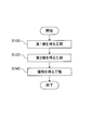
- FIG. 5 is a flowchart for explaining the visualization method according to the second embodiment of the present invention.
- the visualization method according to the second embodiment of the present invention includes a step of visualizing the first substance (S200) and a step of visualizing the second substance (S220).
- the order in which the step of visualizing the first substance (S200) and the step of visualizing the second substance (S220) is not particularly limited.
- the order in which the step of visualizing the first substance (S200) and the step of visualizing the second substance (S220) may be performed at the same time.
- the first substance that can bind to the target substance in the sample is visualized.
- the step of visualizing the first substance (S200) can be performed by the same method as the "method of visualizing the first substance" in the first embodiment.
- the sample, the target substance and the first substance are the same as those in the first embodiment.
- the second substance produced by the action of the first substance in the sample is visualized.
- the step of visualizing the second substance (S220) can be performed by the same method as the "method of visualizing the second substance" in the first embodiment.
- the second substance is the same as in the first embodiment.
- trastuzumab which is an anti-HER2 humanized monoclonal antibody
- irinotecan which is an antineoplastic agent (type I topoisomerase inhibitor)
- trastuzumab and irinotecan correspond to the first substance
- ⁇ H2AX which is a marker of DNA damage
- a CDX model mouse was prepared by transplanting a cell mass of the human breast cancer cultured cell line KPL-4 into an acquired immunodeficient mouse. Specifically, a cell mass of KPL-4 was subcutaneously transplanted into an acquired immunodeficient mouse. When the tumor tissue grew to about 800 mm 3 (1 month after transplantation), Herceptin® was administered to the mice to a dose of trastuzumab of 15 mg / kg. Then, 20 hours after the administration of trastuzumab, irinotecan was administered to the mice at a dose of 10 mg / kg.
- FFPE formalin-fixed paraffin-embedded
- the biotin-modified anti-human IgG antibody was prepared by biotin-labeling an anti-human IgG antibody (Abcam, product number ab97161) using a biotin labeling kit (Biotin Labeling Kit-NH2; Dojin Chemical Laboratory Co., Ltd.). did.
- a first secondary reaction treatment solution was prepared by diluting each with a diluted solution for fluorescent nanoparticles to 2 ⁇ g / mL to form a cocktail. After washing the sections after the primary reaction treatment with PBS, the sections were immersed in the first secondary reaction treatment solution and reacted at room temperature for 30 minutes.
- a second secondary reaction treatment solution was prepared by diluting streptavidin-modified Texas red dye-encapsulating melamine resin particles (hereinafter, also referred to as fluorescent dye-accumulating particles) to 0.02 nM with a diluent for fluorescent nanoparticles.
- the sections that had completed the reaction with the first secondary reaction treatment solution were immersed in the second secondary reaction treatment solution and reacted at room temperature for 2 hours.
- the fluorescent dye-accumulated particles are prepared by dissolving 2.5 mg of Texas Red (registered trademark) dye (Sulforhodamine 101 :; excitation wavelength 595 nm, emission wavelength 615 nm; Sigma Aldrich) in 22.5 mL of pure water, and then using a hot stirrer to prepare the solution. The mixture was stirred for 20 minutes while maintaining the temperature at 70 ° C. To the stirred solution, 1.5 g of melamine resin (Nicarac MX-035; Nippon Carbide Industries Co., Ltd.) was added, and the mixture was further heated and stirred under the same conditions for 5 minutes.
- 0.1 mg of fluorescent dye-accumulated particles were dispersed in 1.5 mL of ethanol, 2 ⁇ L of aminopropyltrimethoxysilane (LS-3150; Shin-Etsu Chemical Co., Ltd.) was added, and the mixture was reacted for 8 hours to perform surface amination treatment.
- the particles subjected to surface amination treatment using PBS (phosphate buffer physiological saline) containing 2 mM of EDTA (ethylenediaminetetraacetic acid) were adjusted to 3 nM, and SM was added to this solution to a final concentration of 10 mM.
- PBS phosphate buffer physiological saline
- EDTA ethylenediaminetetraacetic acid
- streptavidin (Wako Pure Chemical Industries, Ltd.) can be subjected to thiol group addition treatment using N-succinimidyl S-acetylthioacetylate (SATA) and then filtered by a gel filtration column to bind to maleimide-modified fluorescent dye-accumulated particles.
- SATA N-succinimidyl S-acetylthioacetylate
- a streptavidin solution to which a thiol group was added was obtained.
- Maleimide-modified fluorescent dye-accumulated particles and streptavidin with a thiol group added were mixed in PBS containing 2 mM of EDTA and reacted at room temperature for 1 hour. Then, 10 mM mercaptoethanol was added to stop the reaction.
- the obtained mixed solution was concentrated with a centrifugal filter, and then unreacted streptavidin and the like were removed using a gel filtration column for purification to obtain streptavidin-modified fluorescent dye-accumulated
- a cell nucleus staining treatment solution was prepared by diluting DAPI (Sigma), which is a fluorescent dye for cell nucleus staining, with ultrapure water to a concentration of 10 ⁇ g / mL. After the section after the secondary reaction was washed with PBS, it was immersed in a cell nucleus staining solution and reacted at room temperature for 15 minutes.
- DAPI fluorescent dye for cell nucleus staining
- a laser with a wavelength of 561 nm (output: 0.5%) was used as excitation light for observation of Texas red integrated melamine particles stained with trussumab, and a laser with a wavelength of 640 nm for observation of ⁇ H2AX-Alexa647 showing medicinal properties. (Output: 2%) was used as the excitation light.
- the detector sensitivity (HV) at the time of image capture was adjusted to a range in which the brightness of the image was not saturated.
- FIGS. 3A to 3D are four types of fluorescence images for the same region of the section.
- FIG. 3A is a fluorescence image of DAPI showing the cell nucleus
- FIG. 3B is a fluorescence image of ATPase-Alexa488 showing the cell membrane
- FIG. 3C is a fluorescence image of Texas red accumulated melamine particles showing trussumab (first image).
- FIG. 3D is a fluorescence image (second image) of ⁇ H2AX-Alexa647 showing the medicinal effect.
- Image analysis Image processing software (Imaris; Bitplane) was used for image analysis.
- the optimum bright spot region was determined while visually confirming the image within the range of the fluorescence intensity (image signal value) of the Texas red accumulated melamine particles in the range of 50 to 200.
- the total fluorescence intensity (integrated value) corresponding to the bright spots contained in the bright spot region of the obtained Texas red accumulated melamine particles was calculated and used as the amount of trastuzumab.
- a fluorescence image of Alexa647 shown in FIG. 3D was input as a Source Channel.
- the optimum diameter of the bright spot (the range where there is a clear difference between the background and the fluorescence intensity) was determined while visually confirming the image.
- the optimum center position of the bright spot was set while visually confirming the image within the range of the center intensity ratio (ratio of the image signal values of the center and the periphery) of Alexa647.
- the optimum bright spot region was determined in the range of 50 to 200 while visually confirming the image.
- the total fluorescence intensity (integrated value) corresponding to the bright spots included in the bright spot region of the obtained Alexa647 was calculated and used as the amount of ⁇ H2AX.
- FIG. 4A is a graph showing the amount reached by the first substance (trastuzumab) and the effect of the first substance (irinotecan) when the first substance (trastuzumab and irinotecan) was administered
- FIG. 4B is a graph showing the effect of the first substance (irinotecan). It is a graph which showed the reaching amount of the first substance and the effect of the first substance in the group which did not administer.
- the horizontal axis of FIGS. 4A and 4B shows the amount of trastuzumab that reached the cells, and the vertical axis shows the amount of ⁇ H2AX. This makes it possible to visualize how many cells are contained in each region shown in FIG. 2B.
- the present invention it is useful for, for example, screening of a drug, proof of the mechanism of action of a drug, and evaluation of toxicity of a drug.
Landscapes
- Health & Medical Sciences (AREA)
- Life Sciences & Earth Sciences (AREA)
- Engineering & Computer Science (AREA)
- Immunology (AREA)
- Biomedical Technology (AREA)
- Chemical & Material Sciences (AREA)
- Urology & Nephrology (AREA)
- Hematology (AREA)
- Molecular Biology (AREA)
- Analytical Chemistry (AREA)
- Biochemistry (AREA)
- General Health & Medical Sciences (AREA)
- Physics & Mathematics (AREA)
- General Physics & Mathematics (AREA)
- Pathology (AREA)
- Microbiology (AREA)
- Cell Biology (AREA)
- Biotechnology (AREA)
- Food Science & Technology (AREA)
- Medicinal Chemistry (AREA)
- Nuclear Medicine, Radiotherapy & Molecular Imaging (AREA)
- Bioinformatics & Cheminformatics (AREA)
- Toxicology (AREA)
- Tropical Medicine & Parasitology (AREA)
- Investigating Or Analysing Biological Materials (AREA)
Abstract
This visualization method has a step for visualizing a first substance that is capable of binding to a target substance in a sample, and a step for visualizing a second substance that is generated by an action of the first substance in the sample.
Description
本発明は、検体中における第1物質および第2物質を可視化するための可視化方法と、検体中における第1物質および第2物質に関する情報を取得するための情報取得方法とに関する。
The present invention relates to a visualization method for visualizing a first substance and a second substance in a sample, and an information acquisition method for acquiring information on the first substance and the second substance in a sample.
医薬となる候補の化合物からは、スクリーニングにより、有効と評価された化合物のみ選定される。医薬は、生体内の受容体、酵素、イオンチャネル、トランスポーターなどの標的物質に結合することが多い。そして、医薬は細胞に到達し、標的物質に結合した後、細胞に作用することで薬効を示す。したがって、医薬が標的物質に到達したか、または到達した薬物が薬効を示すかなどの判断は、スクリーニングにおいて重要な判断基準である。
From the candidate compounds that can be used as pharmaceuticals, only the compounds evaluated as effective by screening are selected. Drugs often bind to target substances such as receptors, enzymes, ion channels, and transporters in the body. Then, the drug reaches the cell, binds to the target substance, and then acts on the cell to show its medicinal effect. Therefore, the judgment as to whether the drug has reached the target substance or whether the drug that has reached the target substance has a medicinal effect is an important criterion in screening.
また、標的物質などを検出する方法として、作製した組織標本に対して、抗原抗体反応などを利用した免疫染色法が知られている(例えば、非特許文献1、2参照)。
Further, as a method for detecting a target substance or the like, an immunostaining method using an antigen-antibody reaction or the like on a prepared tissue sample is known (see, for example, Non-Patent Documents 1 and 2).
非特許文献1には、PARP阻害剤であるABT-888とトポテカンとを併用した第1相試験について記載されている。非特許文献1では、ABT-888を経口投与し、トポテカンを静脈内投与し、腫瘍および末梢血単核細胞で、ポリ(ADP-リボース)(PAR)とDNA損傷マーカーであるγH2AXの有無を観察している。また、γH2AXの有無は、蛍光標識物質を用いた免疫染色法により確認されている。
Non-Patent Document 1 describes a phase 1 study in which ABT-888, which is a PARP inhibitor, and topotecan are used in combination. In Non-Patent Document 1, ABT-888 is orally administered, topotecan is intravenously administered, and the presence or absence of poly (ADP-ribose) (PAR) and the DNA damage marker γH2AX is observed in tumors and peripheral blood mononuclear cells. doing. The presence or absence of γH2AX has been confirmed by an immunostaining method using a fluorescent labeling substance.
非特許文献2には、細胞傷害性の薬物によるDNA二本鎖切断と、アポトーシスに関連したDNA二本鎖切断との関係について記載されている。非特許文献2では、細胞内で複数のマーカーの共発現を観察することで、特定の細胞におけるγH2AXの発現が細胞損傷性の薬物によるDNA二本鎖切断であるのか、アポトーシスに関連したDNA二本鎖切断であるのかを解析している。また、γH2AXの有無は、蛍光標識物質を用いた免疫染色法により確認されている。
Non-Patent Document 2 describes the relationship between DNA double-strand breaks caused by cytotoxic drugs and DNA double-strand breaks associated with apoptosis. In Non-Patent Document 2, by observing the co-expression of a plurality of markers in cells, whether the expression of γH2AX in a specific cell is a DNA double-strand break by a cell-damaging drug or DNA 2 associated with apoptosis. We are analyzing whether it is a main-strand break. The presence or absence of γH2AX has been confirmed by an immunostaining method using a fluorescent labeling substance.
しかしながら、非特許文献1、2では、医薬などを投与したことに対する結果のみを評価しているため、医薬の効果があったか否かは判断できるものの、その原因を把握することができない。よって、非特許文献1、2に記載された方法では、医薬の特性についての評価が不十分である。例えば、薬効がなかったことが確認された場合、標的物質に対する医薬の到達量が不明であるため、医薬が標的物質に到達しなかったことに起因して効果がなかったのか、医薬が標的物質に到達したが別の理由に起因して効果がなかったのかを評価できない。このような状況においては、対策を検討するにあたり、例えば投薬量を増やせばよいか、そもそも医薬の種類を変更しなければならないかなどの知見を得ることはできず、有効な療法などを決定できない。
However, in Non-Patent Documents 1 and 2, since only the result of administration of a drug or the like is evaluated, it can be determined whether or not the drug was effective, but the cause cannot be grasped. Therefore, the methods described in Non-Patent Documents 1 and 2 are insufficient in evaluating the characteristics of a drug. For example, when it is confirmed that there is no medicinal effect, the amount of the drug that reaches the target substance is unknown, so it may be because the drug did not reach the target substance, and the drug did not have the effect. Can not be evaluated whether it was ineffective due to another reason. In such a situation, when considering countermeasures, it is not possible to obtain knowledge such as whether to increase the dosage or change the type of drug in the first place, and it is not possible to determine effective therapy. ..
本発明の目的は、標的物質に結合可能な物質と、当該物質の作用との関係を正確に特定し、当該物質の特性を正確に評価できる可視化方法および情報取得方法を提供することである。
An object of the present invention is to provide a visualization method and an information acquisition method capable of accurately identifying the relationship between a substance that can bind to a target substance and the action of the substance, and accurately evaluating the characteristics of the substance.
上記の課題を解決するための一手段としての可視化方法は、検体中における標的物質に結合可能な第1物質を可視化する工程と、前記検体中における前記第1物質の作用により生成された第2物質を可視化する工程と、を有する。
The visualization method as one means for solving the above-mentioned problems is a step of visualizing a first substance that can bind to a target substance in a sample and a second method produced by the action of the first substance in the sample. It has a step of visualizing a substance.
上記の課題を解決するための一手段としての情報取得方法は、検体中における標的物質に結合可能な第1物質が可視化された第1像を得る工程と、前記検体中における前記第1物質の作用により生成された第2物質が可視化された第2像を得る工程と、前記第1像および前記第2像から前記第1物質および前記第2物質に関する情報を得る工程と、を有する。
The information acquisition method as one means for solving the above-mentioned problems includes a step of obtaining a first image in which the first substance that can bind to the target substance in the sample is visualized, and a step of obtaining the first substance in the sample. It has a step of obtaining a second image in which the second substance produced by the action is visualized, and a step of obtaining information on the first substance and the second substance from the first image and the second image.
本発明によれば、標的物質に結合可能な物質と、当該物質の作用との関係を正確に特定できるため、当該物質の特性を正確に評価できる。
According to the present invention, since the relationship between the substance that can be bound to the target substance and the action of the substance can be accurately specified, the characteristics of the substance can be accurately evaluated.
[実施の形態1]
以下、本発明の実施の形態1に係る情報取得方法について、添付した図面を参照して詳細に説明する。 [Embodiment 1]
Hereinafter, the information acquisition method according to the first embodiment of the present invention will be described in detail with reference to the attached drawings.
以下、本発明の実施の形態1に係る情報取得方法について、添付した図面を参照して詳細に説明する。 [Embodiment 1]
Hereinafter, the information acquisition method according to the first embodiment of the present invention will be described in detail with reference to the attached drawings.
図1は、本発明の一実施の形態に係る情報取得方法を説明するためのフローチャートである。
FIG. 1 is a flowchart for explaining an information acquisition method according to an embodiment of the present invention.
本発明の一実施の形態に係る情報取得方法は、第1物質に関する第1像を得る工程(S100)と、第2物質に関する第2像を得る工程(S120)と、第1像および第2像から第1物質および第2物質に関する情報を得る工程(S140)とを有する。第1物質および第2物質に関する情報を得る工程(S140)は、第1像を得る工程(S100)の後および第2像を得る工程(S120)の後に2回に分けて行われてもよいが、第1像を得る工程(S100)および第2像を得る工程(S120)の両工程が終わった後に行われてもよい。第1像を得る工程(S100)および第2像を得る工程(S120)を行う順番は、特に限定されない。
The information acquisition method according to the embodiment of the present invention includes a step of obtaining a first image of a first substance (S100), a step of obtaining a second image of a second substance (S120), and a first image and a second image. It has a step (S140) of obtaining information about the first substance and the second substance from the image. The step of obtaining information on the first substance and the second substance (S140) may be performed in two steps after the step of obtaining the first image (S100) and after the step of obtaining the second image (S120). However, it may be performed after both the steps of obtaining the first image (S100) and the step of obtaining the second image (S120) are completed. The order in which the step of obtaining the first image (S100) and the step of obtaining the second image (S120) are performed is not particularly limited.
ここで、「第1物質」とは、検体中における標的物質に結合可能な物質を意味する。たとえば、第1物質は、検体に投与された物質である。また、「第2物質」とは、検体中において第1物質の作用により生成された物質を意味する。第2物質は、第1物質が作用したことを示すマーカーとして機能しうる。
Here, the "first substance" means a substance that can bind to the target substance in the sample. For example, the first substance is a substance administered to a sample. Further, the "second substance" means a substance produced by the action of the first substance in the sample. The second substance can function as a marker indicating that the first substance has acted.
検体の種類は、特に限定されない。検体の例には、血液(血清、血漿)、尿、鼻孔液、唾液、便、体腔液(髄液、腹水、胸水など)、各生体組織、培養により製造された細胞または組織が含まれる。また、標的物質の種類も、特に限定されない。標的物質の例には、核酸(一本鎖であっても二本鎖であってもよいDNA、RNA、ポリヌクレオチド、オリゴヌクレオチド、PNA(ペプチド核酸)など、またはヌクレオシド、ヌクレオチドおよびそれらの修飾分子);タンパク質(ポリペプチド、オリゴペプチド、標的細胞の細胞膜に存在する受容体など);アミノ酸(修飾アミノ酸も含む。);糖質(オリゴ糖、多糖類、糖鎖など);脂質;エクソソーム;またはこれらの修飾分子、複合体が含まれる。また、これらの分子の例には、5T4、AXL、BCMA、C4.4A、CA6、Cadherin3、Cadherin6、CEACAM5、CD16、CD19、CD22、CD37、CD56、CD71、CD138、CD142、CD352、DLL3、EphA2、EphrinA4、ETBR、FcγRIII、FOLR1、FGFR2、FGFR3、GCC、HER1(EGFR)、HER2、HER3、IntegrinαV、LAMP1、LIV1、Mesothelin、MUC1、MUC16、NaPi2B、Nectin4、NOTCH3、PD-1、PD-L1、PSMA、PTK7、SLAMF7、SLITRK6、STEAP1、TROP2が含まれる。
The type of sample is not particularly limited. Examples of specimens include blood (serum, plasma), urine, nasal fluid, saliva, stool, body cavity fluid (cerebrospinal fluid, ascites, pleural effusion, etc.), biological tissues, and cells or tissues produced by culture. The type of target substance is also not particularly limited. Examples of target substances include nucleic acids (single-stranded or double-stranded DNA, RNA, polynucleotides, oligonucleotides, PNAs (peptide nucleic acids), etc., or nucleosides, nucleotides and modified molecules thereof. ); Proteins (polypeptides, oligopeptides, receptors present on the cell membrane of target cells, etc.); Amino acids (including modified amino acids); Sugars (oligosaccharides, polysaccharides, sugar chains, etc.); Lipids; Exosomes; or These modified molecules and complexes are included. Examples of these molecules include 5T4, AXL, BCMA, C4.4A, CA6, Caderin3, Caderin6, CEACAM5, CD16, CD19, CD22, CD37, CD56, CD71, CD138, CD142, CD352, DLL3, EphA2, EphrinA4, ETBR, FcγRIII, FOLR1, FGFR2, FGFR3, GCC, HER1 (EGFR), HER2, HER3, Integrin αV, LAMP1, LIV1, Mesothelin, MUC1, MUC16, NaPi2B, NPTML1 , PTK7, SLAMF7, SLITRK6, STEAP1 and TROP2.
第1物質に関する第1像を得る工程(S100)では、検体中における標的物質に結合可能な第1物質を可視化して第1像を得る。
In the step (S100) of obtaining the first image of the first substance, the first substance that can bind to the target substance in the sample is visualized to obtain the first image.
前述のとおり、第1物質は、例えば検体に投与された物質である。第1物質の例には、抗体、抗体薬物複合体(ADC;antibody-Drug Conjugate)、通常の医薬化合物などの医薬、毒物が含まれる。医薬は、細胞内におけるDNAの複製を阻害する物質を含む。医薬の例には、各種疾病の治療薬、特に抗癌剤が含まれる。
As described above, the first substance is, for example, a substance administered to a sample. Examples of the first substance include antibodies, antibody drug conjugates (ADCs), drugs such as ordinary pharmaceutical compounds, and toxic substances. Drugs include substances that inhibit DNA replication in cells. Examples of pharmaceuticals include therapeutic agents for various diseases, especially anti-cancer agents.
第1物質は、標的物質に結合可能であり、かつ第2物質を生成させる1つの物質でもよいが、標的物質に結合可能な結合物質と、第2物質を生成させるための作用物質とを別々に含んでいてもよい。後者の場合、結合物質および作用物質は、互いに結合していてもよいし、互いに独立していてもよい。
The first substance may be one substance that can bind to the target substance and produce the second substance, but the binding substance that can bind to the target substance and the agent for producing the second substance are separately separated. May be included in. In the latter case, the binding substance and the agent may be bound to each other or independent of each other.
結合物質および作用物質が互いに結合している場合、第1物質は、例えば抗体薬物複合体である。結合物質および作用物質が互いに結合していない場合、結合物質は、例えば受容体に結合可能な抗体であり、作用物質は、例えば細胞に作用する薬物である。結合物質および作用物質が互いに結合していない例には、抗体医薬および薬物を併用する場合も含まれる。第1物質が標的物質に結合可能であり、かつ第2物質を生成させる物質である場合、第1物質は、抗体医薬である。たとえば、第1物質が抗体を含む場合、第1物質として抗体薬物複合体を投与してもよいし、抗体および薬物を別々に投与してもよいし、抗体医薬を投与してもよい。
When the binding substance and the acting substance are bound to each other, the first substance is, for example, an antibody drug conjugate. If the binding agent and the agent are not bound to each other, the binding agent is, for example, an antibody capable of binding to a receptor, and the agent is, for example, a drug that acts on a cell. Examples in which the binding substance and the acting substance are not bound to each other include the case where an antibody drug and a drug are used in combination. When the first substance is a substance that can bind to the target substance and produces the second substance, the first substance is an antibody drug. For example, when the first substance contains an antibody, the antibody drug conjugate may be administered as the first substance, the antibody and the drug may be administered separately, or the antibody drug may be administered.
第1物質を可視化する方法は、特に限定されない。第1物質を可視化する方法の例には、免疫染色法、抗体に類似の分子認識基を用いた染色法が含まれる。
The method of visualizing the first substance is not particularly limited. Examples of the method for visualizing the first substance include an immunostaining method and a staining method using a molecular recognition group similar to an antibody.
免疫染色法により、第1物質を可視化する方法では、例えば第1物質(例えば抗体薬物複合体)を投与してから一定時間が経過した後、第1物質が結合する標的物質(例えば標的細胞の細胞膜に存在する受容体)を含む生体組織切片(検体)を免疫染色して、第1物質が蛍光標識または酵素標識により可視化された免疫染色像(第1像)を得る。
In the method of visualizing the first substance by immunostaining, for example, after a certain period of time has passed since the administration of the first substance (for example, an antibody drug complex), the target substance to which the first substance binds (for example, the target cell) A biological tissue section (specimen) containing a receptor present on the cell membrane is immunostained to obtain an immunostained image (first image) in which the first substance is visualized by fluorescent labeling or enzyme labeling.
また、免疫染色法の2次反応では、蛍光体集積粒子(蛍光色素集積粒子)を使用できる。蛍光体集積粒子は、有機物または無機物でできた粒子を母体とし、複数の蛍光体(例えば蛍光色素や半導体ナノ粒子)がその中に内包されているおよび/またはその表面に吸着している構造を有する、ナノサイズの粒子である。蛍光体集積ナノ粒子を構成する蛍光色素の例には、ローダミン系色素、Cy系色素、Alexa Fluor(登録商標)系色素、BODIPY系色素、スクアリリウム系色素、シアニン系色素、芳香環系色素、オキサジン系色素、カルボピロニン系色素、ピロメセン系色素が含まれる。蛍光体集積ナノ粒子を構成する半導体ナノ粒子の素材の例には、II-VI族半導体、III-V族半導体、またはIV族半導体が含まれる。蛍光体集積粒子は、公知の方法(例えば、特開2013-57937号公報参照)に従って作製できる。
In addition, phosphor-accumulated particles (fluorescent dye-accumulated particles) can be used in the secondary reaction of the immunostaining method. Phosphor-accumulated particles are based on particles made of organic or inorganic substances, and have a structure in which a plurality of phosphors (for example, fluorescent dyes and semiconductor nanoparticles) are contained therein and / or adsorbed on the surface thereof. It is a nano-sized particle that has. Examples of fluorescent dyes constituting phosphor-accumulated nanoparticles include rhodamine dyes, Cy dyes, Alexa Fluor (registered trademark) dyes, BODIPY dyes, squarylium dyes, cyanine dyes, aromatic ring dyes, and oxazines. Includes dyes, carbopyronine dyes, and pyromesene dyes. Examples of the material of the semiconductor nanoparticles constituting the phosphor-integrated nanoparticles include a group II-VI semiconductor, a group III-V semiconductor, or a group IV semiconductor. The phosphor-accumulated particles can be produced according to a known method (see, for example, Japanese Patent Application Laid-Open No. 2013-57937).
抗体に類似の分子認識基を用いた染色法により第1物質を可視化する方法では、例えば分子認識基として、アプタマー、SNAP-tagを用いて第1像を得る。
In the method of visualizing the first substance by a staining method using a molecular recognition group similar to an antibody, for example, an aptamer or SNAP-tag is used as a molecular recognition group to obtain a first image.
第1物質が結合物質および作用物質を含む場合、結合物質(例えば抗体)を可視化して第1像を得ることが好ましい。
When the first substance contains a binding substance and an acting substance, it is preferable to visualize the binding substance (for example, an antibody) to obtain a first image.
第2物質に関する第2像を得る工程(S120)では、検体中における第1物質の作用により生成された第2物質を可視化して第2像を得る。通常、第1像および第2像は、検体中の同一箇所(例えば同一の組織切片)または対応箇所(例えば隣接する組織切片)について得られる。
In the step of obtaining the second image regarding the second substance (S120), the second substance produced by the action of the first substance in the sample is visualized to obtain the second image. Usually, the first and second images are obtained for the same site (eg, the same tissue section) or the corresponding site (eg, adjacent tissue sections) in the sample.
前述のとおり、第2物質は、検体中において第1物質の作用により生成された物質である。たとえば、第1物質が標的細胞におけるDNAの複製を阻害する場合、第2物質は複製が阻害されたことにより生成されるバイオマーカーでもよい。第1物質が作用する対象は、特に限定されず、例えば細胞内の細胞小器官や細胞に存在する物質(例えば酵素や受容体などのタンパク質)などである。第2物質の例には、DNAの損傷応答マーカーと、微小管重合マーカーと、微小管脱重合マーカーとが含まれる。DNAの損傷応答マーカーの例には、53BP1、Ku80、MDC1、Nbs1、pATM、pATR、pChk1、pDNA-PK、Rad51、γH2AXが含まれる。微小管重合マーカーと、脱重合マーカーとの例には、APC、CLASP、CLIP170、EB1、EB3、MACF、MCAK、STIM1、XMAP215、α-Tubulin、β-Tubulinが含まれる。
As described above, the second substance is a substance produced by the action of the first substance in the sample. For example, if the first substance inhibits DNA replication in target cells, the second substance may be a biomarker produced by the inhibition of replication. The target on which the first substance acts is not particularly limited, and is, for example, an organelle in a cell or a substance existing in a cell (for example, a protein such as an enzyme or a receptor). Examples of the second substance include a DNA damage response marker, a microtubule polymerization marker, and a microtubule depolymerization marker. Examples of DNA damage response markers include 53BP1, Ku80, MDC1, Nbs1, pATM, pATR, pChk1, pDNA-PK, Rad51, γH2AX. Examples of microtubule polymerization markers and depolymerization markers include APC, CLASP, CLIP170, EB1, EB3, MACF, MCAK, STIM1, XMAP215, α-Tubulin, β-Tubulin.
第2物質を可視化する方法は、特に限定されない。第2物質を可視化する方法の例には、免疫染色法、抗体に類似の分子認識基を用いた染色法が含まれる。
The method of visualizing the second substance is not particularly limited. Examples of the method for visualizing the second substance include an immunostaining method and a staining method using a molecular recognition group similar to an antibody.
免疫染色法により、第2物質を可視化する方法では、例えば第1物質(例えば抗体薬物複合体)を投与してから一定時間が経過した後、第1物質の作用により生じた第2物質(例えばDNAの断片)を含む生体組織切片(検体)を免疫染色して、第1物質の作用により生成された第2物質が蛍光標識または酵素標識により可視化された免疫染色像(第2像)を得る。
In the method of visualizing the second substance by the immunostaining method, for example, a second substance (for example, an antibody drug complex) produced by the action of the first substance after a certain period of time has passed after the administration of the first substance (for example, an antibody drug complex). An immunostaining of a biological tissue section (specimen) containing a DNA fragment) is performed to obtain an immunostaining image (second image) in which the second substance produced by the action of the first substance is visualized by fluorescent labeling or enzyme labeling. ..
抗体に類似の分子認識基を用いた染色法により第2物質を可視化する方法では、例えば分子認識基として、アプタマー、SNAP-tagを用いて第2像を得る。
In the method of visualizing the second substance by a staining method using a molecular recognition group similar to an antibody, for example, an aptamer or SNAP-tag is used as a molecular recognition group to obtain a second image.
第1物質が結合物質および作用物質を含む場合、結合物質(例えば抗体)ではなく、作用物質(例えば薬物)が作用することで生成された第2物質(例えばDNAの断片)を可視化して第2像を得ることが好ましい。
When the first substance contains a binding substance and an agent, the second substance (for example, a fragment of DNA) produced by the action of the agent (for example, a drug) instead of the binding substance (for example, an antibody) is visualized and the second substance is visualized. It is preferable to obtain two images.
第1像および第2像から第1物質および第2物質に関する情報を得る工程(S140)では、第1像および第2像から第1物質および第2物質に関する情報を得る。
In the step (S140) of obtaining information on the first substance and the second substance from the first image and the second image, information on the first substance and the second substance is obtained from the first image and the second image.
第1物質および第2物質に関する情報の種類は、第1像および第2像から得られるものであれば、特に限定されない。第1物質および第2物質に関する情報は、第1物質の位置または量に関する情報と、第2物質の位置または量に関する情報とを含む。
例えば、情報を得る工程では、標的物質と結合した結合物質(例えば抗体や、抗体薬物複合体の抗体成分など)の量と、第1物質(例えば抗体薬物複合体の薬物成分)が作用したことにより生じた第2物質の量とを得ることができる。また、得られた第1物質および第2物質に関する情報に基づいて、第1物質の作用または効果に関する情報をさらに得ることもできる。例えば、第1物質の作用または効果に関する情報として、標的物質と結合した結合物質の位置または量と、第1物質が作用したことにより生じた第2物質の位置または量との関係に関する情報をさらに生成してもよい。具体的には、標的物質と結合した第1物質または結合物質の量に対してどのぐらいの第2物質が生じたかに関する情報や、標的物質と結合した第1物質または結合物質の量に対してどのぐらいの細胞に第2物質が生じたかに関する情報が含まれる。また、場合によっては、標的細胞に到達した結合物質を、標的物質と結合した結合物質とみなしてもよい。 The type of information regarding the first substance and the second substance is not particularly limited as long as it is obtained from the first image and the second image. Information about the first substance and the second substance includes information about the position or amount of the first substance and information about the position or amount of the second substance.
For example, in the process of obtaining information, the amount of the binding substance bound to the target substance (for example, an antibody or the antibody component of the antibody drug conjugate) and the first substance (for example, the drug component of the antibody drug conjugate) acted. The amount of the second substance produced by the above can be obtained. Further, based on the obtained information on the first substance and the second substance, further information on the action or effect of the first substance can be obtained. For example, as information on the action or effect of the first substance, further information on the relationship between the position or amount of the binding substance bound to the target substance and the position or amount of the second substance generated by the action of the first substance. It may be generated. Specifically, with respect to information on how much second substance was produced relative to the amount of the first substance or binding substance bound to the target substance, and the amount of the first substance or binding substance bound to the target substance. Contains information on how many cells produced the second substance. In some cases, the binding substance that has reached the target cell may be regarded as the binding substance bound to the target substance.
たとえば、第1物質が互いに結合している結合物質および作用物質を含む場合(例えば第1物質が抗体薬物複合体である場合)、第1像からは、標的物質と結合した第1物質(例えば抗体薬物複合体)の位置または量を得ることができる。第2像からは、第1物質(例えば抗体薬物複合体の薬物成分)が作用したことにより生じた第2物質の位置または量を得ることができる。
For example, if the first substance contains binding substances and agents that are bound to each other (eg, if the first substance is an antibody drug conjugate), then from the first image, the first substance bound to the target substance (eg, for example). The position or amount of the antibody-drug conjugate) can be obtained. From the second image, the position or amount of the second substance generated by the action of the first substance (for example, the drug component of the antibody drug conjugate) can be obtained.
また、第1物質が互いに独立している結合物質(例えば抗体)および作用物質(例えば薬物)を含む場合、第1像からは、標的物質と結合した結合物質(例えば抗体)の位置または量を得ることができる。第2像からは、作用物質(例えば薬物)が作用したことにより生じた第2物質の位置または量を得ることができる。
When the first substance contains a binding substance (for example, an antibody) and an agent (for example, a drug) that are independent of each other, the position or amount of the binding substance (for example, an antibody) bound to the target substance is determined from the first image. Obtainable. From the second image, the position or amount of the second substance produced by the action of the agent (for example, a drug) can be obtained.
さらに、第1物質が標的物質に結合可能であり、かつ第2物質を生成させる物質の場合、第1像からは、標的物質と結合した第1物質(例えば抗体医薬)の位置または量を得ることができる。第2像からは、第1物質(例えば抗体医薬)が作用したことにより生じた第2物質の位置または量を得ることができる。
Further, in the case where the first substance is a substance that can bind to the target substance and produces the second substance, the position or amount of the first substance (for example, antibody drug) bound to the target substance is obtained from the first image. be able to. From the second image, the position or amount of the second substance produced by the action of the first substance (for example, an antibody drug) can be obtained.
よって、第1像および第2像からは、第1物質および第2物質に関する情報が得られ、必要に応じて第1物質の作用または効果に関する情報を得ることもできる。例えば、第1物質の作用または効果に関する情報として、標的物質への第1物質(例えば抗体薬物複合体、抗体医薬)の到達量と、第1物質(例えば抗体薬物複合体、抗体医薬)が作用したことにより第2物質を生成した細胞の数、位置、種類などの情報を得ることができる。
Therefore, from the first image and the second image, information on the first substance and the second substance can be obtained, and if necessary, information on the action or effect of the first substance can be obtained. For example, as information on the action or effect of the first substance, the amount of the first substance (for example, antibody drug conjugate, antibody drug) reaching the target substance and the first substance (for example, antibody drug conjugate, antibody drug) act. By doing so, it is possible to obtain information such as the number, position, and type of cells that produced the second substance.
ここで、第1像および第2像から得られる情報について具体的に説明する。図2A、Bは、第1像および第2像から得られる情報を説明するための図である。図2Aは、医薬を投与した後の生体組織切片の模式図であり、第1像および第2像を重ね合わせた像を示している。図2Bは、図2Aから得られる情報をまとめたグラフの一例を示す模式図である。図2Bの横軸は、1つの細胞に対する第1物質(例えば医薬)の到達数であり、縦軸は、1つの細胞における第2物質の量(例えば薬物の効果)を示している。
Here, the information obtained from the first image and the second image will be specifically described. 2A and 2B are diagrams for explaining information obtained from the first image and the second image. FIG. 2A is a schematic view of a biological tissue section after administration of a drug, and shows an image in which a first image and a second image are superimposed. FIG. 2B is a schematic diagram showing an example of a graph summarizing the information obtained from FIG. 2A. The horizontal axis of FIG. 2B is the number of arrivals of the first substance (for example, a drug) per cell, and the vertical axis is the amount of the second substance (for example, the effect of the drug) in one cell.
ここでは、第1物質として、抗体10および薬物20(白抜きの星)が結合した抗体薬物複合体30を投与したと仮定する。図2Aに示されるように、抗体薬物複合体30を投与すると、抗体薬物複合体30は標的細胞50の細胞膜に存在する受容体(不図示)まで移動する。受容体まで移動した抗体薬物複合体30は、受容体に結合する。受容体に抗体薬物複合体30が結合すると、薬物20が作用して第2物質40(黒塗りの丸)を生成する。
Here, it is assumed that the antibody-drug conjugate 30 to which the antibody 10 and the drug 20 (white stars) are bound is administered as the first substance. As shown in FIG. 2A, when the antibody-drug conjugate 30 is administered, the antibody-drug conjugate 30 migrates to a receptor (not shown) present on the cell membrane of the target cell 50. The antibody-drug conjugate 30 that has migrated to the receptor binds to the receptor. When the antibody-drug conjugate 30 binds to the receptor, the drug 20 acts to produce the second substance 40 (black circle).
たとえば、第1像および第2像から得られる情報は、薬物20が作用する領域単位で評価する。例えば、薬物20が細胞質で作用する場合には、細胞単位で評価すればよいし、薬物20が細胞核で作用する場合には、細胞核単位で評価すればよい。また、作製した生体組織切片単位で評価してもよい。図2A、Bに示される例では、細胞単位で評価している。
For example, the information obtained from the first image and the second image is evaluated in units of regions on which the drug 20 acts. For example, when the drug 20 acts on the cytoplasm, it may be evaluated on a cell-by-cell basis, and when the drug 20 acts on the cell nucleus, it may be evaluated on a cell-nucleus basis. In addition, it may be evaluated in units of prepared biological tissue sections. In the examples shown in FIGS. 2A and 2B, evaluation is performed on a cell-by-cell basis.
このような評価は、例えば、画像処理ソフトウェア(Imaris;Bitplane社)を使用して、第1像および第2像に対して以下の各処理を行う画像解析により行うことができる。ここでは、第1像が、第1物質を蛍光免疫染色で観察した蛍光像であり、第2像が、第2物質を蛍光免疫染色で観察した蛍光像であるものとして説明する。また、第1像および第2像と同一または対応する生体組織切片から、細胞核中のDNAを蛍光色素DAPIにより染色することで得られる画像、および細胞膜を別の蛍光色素により染色することで得られる画像も得たものとして説明する。ただし、細胞核の染色と細胞膜の染色は、必要に応じて省略してもよい。なお、以下の各処理は、画像解析の一例における処理であり、これに限定されない。
Such evaluation can be performed by, for example, image analysis using image processing software (Imaris; Bitplane) to perform each of the following processing on the first image and the second image. Here, it is assumed that the first image is a fluorescence image obtained by observing the first substance by fluorescence immunostaining, and the second image is a fluorescence image obtained by observing the second substance by fluorescence immunostaining. Further, from the same or corresponding biological tissue sections as the first and second images, an image obtained by staining the DNA in the cell nucleus with the fluorescent dye DAPI, and an image obtained by staining the cell membrane with another fluorescent dye can be obtained. It will be explained assuming that the image is also obtained. However, staining of the cell nucleus and staining of the cell membrane may be omitted if necessary. The following processes are, and are not limited to, the processes in an example of image analysis.
(1)画像前処理
画像前処理は、実施されなくてもよく、第1像および/または第2像の画質が不良の場合に解析の精度向上のため必要に応じて実施される。具体的には、顕微鏡画像の撮像素子由来のノイズを除去して、細胞核の形状を修復したり、細胞膜の切断部を修復したり、輝点状のオブジェクトの形状を修復したりすることで、以後の処理の精度を向上させることができる。 (1) Image preprocessing The image preprocessing does not have to be performed, and is performed as necessary to improve the accuracy of analysis when the image quality of the first image and / or the second image is poor. Specifically, by removing noise derived from the image sensor of a microscope image, the shape of the cell nucleus is repaired, the cut portion of the cell membrane is repaired, and the shape of a bright spot-like object is repaired. The accuracy of subsequent processing can be improved.
画像前処理は、実施されなくてもよく、第1像および/または第2像の画質が不良の場合に解析の精度向上のため必要に応じて実施される。具体的には、顕微鏡画像の撮像素子由来のノイズを除去して、細胞核の形状を修復したり、細胞膜の切断部を修復したり、輝点状のオブジェクトの形状を修復したりすることで、以後の処理の精度を向上させることができる。 (1) Image preprocessing The image preprocessing does not have to be performed, and is performed as necessary to improve the accuracy of analysis when the image quality of the first image and / or the second image is poor. Specifically, by removing noise derived from the image sensor of a microscope image, the shape of the cell nucleus is repaired, the cut portion of the cell membrane is repaired, and the shape of a bright spot-like object is repaired. The accuracy of subsequent processing can be improved.
(2)細胞核の領域の決定(細胞核セグメンテーション)
第1像および第2像に対応する細胞核の染色画像について、6~12μmの範囲内で画像目視による確認をしながら最適な細胞核の直径を設定する。次いで、DAPIの中心強度比(中心と周辺の画像信号値の比)が5以上の部位を細胞核の中心位置(Seed Point)として設定する。蛍光強度(画像信号値)が50~200の範囲内で画像目視による確認をしながら細胞核の領域を決定する。 (2) Determination of cell nucleus region (cell nucleus segmentation)
For the stained images of the cell nuclei corresponding to the first image and the second image, the optimum diameter of the cell nuclei is set within the range of 6 to 12 μm while visually confirming the images. Next, a site having a DAPI center intensity ratio (ratio of image signal values between the center and the periphery) of 5 or more is set as the center position (Seed Point) of the cell nucleus. The region of the cell nucleus is determined while visually confirming the image within the range of fluorescence intensity (image signal value) of 50 to 200.
なお、細胞核の領域の情報のみで細胞核の領域の決定を行ってもよい。また、深層学習により得られた情報により、最適な細胞核の直径と、細胞核の中心位置と、細胞核の領域とを決定してもよい。
It should be noted that the region of the cell nucleus may be determined only by the information of the region of the cell nucleus. In addition, the optimum diameter of the cell nucleus, the central position of the cell nucleus, and the region of the cell nucleus may be determined from the information obtained by deep learning.
(3)細胞領域の区画化
第1像および第2像に対応する細胞膜の染色画像について、6~20μmの範囲内で画像目視による確認をしながら最適な細胞の最小直径を設定して細胞領域を決定する。次いで、得られた細胞領域のそれぞれに対して、上記(2)で得られた細胞核の位置情報を入力する。なお、1つの細胞領域に1つの細胞核が含まれている場合は、その領域は細胞とみなす。一方、1つの細胞領域に2つ以上の細胞核が含まれている場合は、細胞核が1つになるように細胞領域を分割する。また、1つの細胞領域に細胞核が含まれない場合は、細胞とみなさない。 (3) Division of cell region Regarding the stained image of the cell membrane corresponding to the first image and the second image, the optimum minimum diameter of the cell is set within the range of 6 to 20 μm while visually confirming the image, and the cell region is set. To determine. Next, the position information of the cell nucleus obtained in (2) above is input to each of the obtained cell regions. If one cell region contains one cell nucleus, that region is regarded as a cell. On the other hand, when one cell region contains two or more cell nuclei, the cell region is divided so that the cell nuclei become one. If one cell region does not contain a cell nucleus, it is not regarded as a cell.
第1像および第2像に対応する細胞膜の染色画像について、6~20μmの範囲内で画像目視による確認をしながら最適な細胞の最小直径を設定して細胞領域を決定する。次いで、得られた細胞領域のそれぞれに対して、上記(2)で得られた細胞核の位置情報を入力する。なお、1つの細胞領域に1つの細胞核が含まれている場合は、その領域は細胞とみなす。一方、1つの細胞領域に2つ以上の細胞核が含まれている場合は、細胞核が1つになるように細胞領域を分割する。また、1つの細胞領域に細胞核が含まれない場合は、細胞とみなさない。 (3) Division of cell region Regarding the stained image of the cell membrane corresponding to the first image and the second image, the optimum minimum diameter of the cell is set within the range of 6 to 20 μm while visually confirming the image, and the cell region is set. To determine. Next, the position information of the cell nucleus obtained in (2) above is input to each of the obtained cell regions. If one cell region contains one cell nucleus, that region is regarded as a cell. On the other hand, when one cell region contains two or more cell nuclei, the cell region is divided so that the cell nuclei become one. If one cell region does not contain a cell nucleus, it is not regarded as a cell.
なお、深層学習により得られた情報により、細胞の最小直径を決定してもよい。なお、評価単位が細胞ではない場合などは、この処理を省略してもよい。
Note that the minimum diameter of cells may be determined based on the information obtained by deep learning. If the evaluation unit is not a cell, this process may be omitted.
(4)第1物質の量の計測
第1像について、0.5~2μmの範囲内で、画像目視による確認をしながら最適な輝点(Foci)の直径(背景と蛍光強度が明確に差のある範囲)を決定する。次いで、蛍光物質の中心強度比(中心と周辺の画像信号値の比)が10以上の部位を輝点の中心位置として設定する。次いで、蛍光物質が放出する蛍光の蛍光強度(画像信号値)が50~200の範囲内で画像目視による確認をしながら最適な輝点の領域を決定する。得られた蛍光物質の輝点の領域に含まれる輝点に対応した蛍光強度の総和(積算値)を算出する。その後、算出された蛍光強度の積算値をそのまま第1物質の量としてもよいし、算出された蛍光強度の積算値を別途第1物質の量に換算してもよい。 (4) Measurement of the amount of the first substance For the first image, the optimum diameter of the bright spot (Foci) (the background and the fluorescence intensity are clearly different) within the range of 0.5 to 2 μm while visually confirming the image. (A certain range of) is determined. Next, a portion having a center intensity ratio (ratio of image signal values between the center and the periphery) of the fluorescent substance of 10 or more is set as the center position of the bright spot. Next, the optimum bright spot region is determined while visually confirming the image within the range where the fluorescence intensity (image signal value) of the fluorescence emitted by the fluorescent substance is in the range of 50 to 200. The total fluorescence intensity (integrated value) corresponding to the bright spots included in the bright spot region of the obtained fluorescent substance is calculated. After that, the calculated integrated value of the fluorescence intensity may be used as it is as the amount of the first substance, or the calculated integrated value of the fluorescence intensity may be separately converted into the amount of the first substance.
第1像について、0.5~2μmの範囲内で、画像目視による確認をしながら最適な輝点(Foci)の直径(背景と蛍光強度が明確に差のある範囲)を決定する。次いで、蛍光物質の中心強度比(中心と周辺の画像信号値の比)が10以上の部位を輝点の中心位置として設定する。次いで、蛍光物質が放出する蛍光の蛍光強度(画像信号値)が50~200の範囲内で画像目視による確認をしながら最適な輝点の領域を決定する。得られた蛍光物質の輝点の領域に含まれる輝点に対応した蛍光強度の総和(積算値)を算出する。その後、算出された蛍光強度の積算値をそのまま第1物質の量としてもよいし、算出された蛍光強度の積算値を別途第1物質の量に換算してもよい。 (4) Measurement of the amount of the first substance For the first image, the optimum diameter of the bright spot (Foci) (the background and the fluorescence intensity are clearly different) within the range of 0.5 to 2 μm while visually confirming the image. (A certain range of) is determined. Next, a portion having a center intensity ratio (ratio of image signal values between the center and the periphery) of the fluorescent substance of 10 or more is set as the center position of the bright spot. Next, the optimum bright spot region is determined while visually confirming the image within the range where the fluorescence intensity (image signal value) of the fluorescence emitted by the fluorescent substance is in the range of 50 to 200. The total fluorescence intensity (integrated value) corresponding to the bright spots included in the bright spot region of the obtained fluorescent substance is calculated. After that, the calculated integrated value of the fluorescence intensity may be used as it is as the amount of the first substance, or the calculated integrated value of the fluorescence intensity may be separately converted into the amount of the first substance.
(5)第2物質の量の計測
第2像について、0.5~2μmの範囲内で、画像目視による確認をしながら最適な輝点の直径(背景と蛍光強度が明確に差のある範囲)を決定する。蛍光物質の中心強度比(中心と周辺の画像信号値の比)が10~200の範囲内で画像目視による確認をしながら最適な輝点の中心位置を設定する。50~200の範囲内で画像目視による確認をしながら最適な輝点の領域を決定する。得られた蛍光物質の輝点の領域に含まれる輝点に対応した蛍光強度の総和(積算値)を算出する。その後、算出された蛍光強度の積算値をそのまま第2物質の量としてもよいし、算出された蛍光強度の積算値を別途第2物質の量に換算してもよい。 (5) Measurement of the amount of the second substance For the second image, the optimum diameter of the bright spot (the range where the background and the fluorescence intensity are clearly different) within the range of 0.5 to 2 μm while visually confirming the image. ) Is determined. The optimum center position of the bright spot is set while visually confirming the image within a range of 10 to 200 in the center intensity ratio (ratio of the image signal value between the center and the periphery) of the fluorescent substance. The optimum bright spot region is determined in the range of 50 to 200 while visually confirming the image. The total fluorescence intensity (integrated value) corresponding to the bright spots included in the bright spot region of the obtained fluorescent substance is calculated. After that, the calculated integrated value of the fluorescence intensity may be used as it is as the amount of the second substance, or the calculated integrated value of the fluorescence intensity may be separately converted into the amount of the second substance.
第2像について、0.5~2μmの範囲内で、画像目視による確認をしながら最適な輝点の直径(背景と蛍光強度が明確に差のある範囲)を決定する。蛍光物質の中心強度比(中心と周辺の画像信号値の比)が10~200の範囲内で画像目視による確認をしながら最適な輝点の中心位置を設定する。50~200の範囲内で画像目視による確認をしながら最適な輝点の領域を決定する。得られた蛍光物質の輝点の領域に含まれる輝点に対応した蛍光強度の総和(積算値)を算出する。その後、算出された蛍光強度の積算値をそのまま第2物質の量としてもよいし、算出された蛍光強度の積算値を別途第2物質の量に換算してもよい。 (5) Measurement of the amount of the second substance For the second image, the optimum diameter of the bright spot (the range where the background and the fluorescence intensity are clearly different) within the range of 0.5 to 2 μm while visually confirming the image. ) Is determined. The optimum center position of the bright spot is set while visually confirming the image within a range of 10 to 200 in the center intensity ratio (ratio of the image signal value between the center and the periphery) of the fluorescent substance. The optimum bright spot region is determined in the range of 50 to 200 while visually confirming the image. The total fluorescence intensity (integrated value) corresponding to the bright spots included in the bright spot region of the obtained fluorescent substance is calculated. After that, the calculated integrated value of the fluorescence intensity may be used as it is as the amount of the second substance, or the calculated integrated value of the fluorescence intensity may be separately converted into the amount of the second substance.
なお、深層学習により得られた情報により、輝点の直径と、輝点の中心位置と、輝点の領域とを決定してもよい。また、細胞核領域に含まれる輝点に対応した蛍光強度の総和(積算値)としてもよい。
Note that the diameter of the bright spot, the center position of the bright spot, and the region of the bright spot may be determined from the information obtained by deep learning. Further, it may be the total sum (integrated value) of the fluorescence intensities corresponding to the bright spots contained in the cell nucleus region.
(6)解析結果の出力
第1像および第2像(またはこれらの重畳画像)について、個々の細胞領域に細胞識別番号を付与する。次いで、第1像(または重畳画像)について、細胞識別番号を付与した個々の細胞領域内に中心位置が含まれている第1物質を示す輝点に対応した蛍光強度の総和を算出する。また、第2像(または重畳画像)について細胞識別番号を付与した個々の細胞領域内に中心位置が含まれている第2物質を示す輝点に対応した蛍光強度の総和を算出する。 (6) Output of analysis results For the first and second images (or superimposed images thereof), cell identification numbers are assigned to individual cell regions. Next, for the first image (or superimposed image), the total fluorescence intensity corresponding to the bright spot indicating the first substance whose center position is contained in each cell region to which the cell identification number is assigned is calculated. In addition, the total fluorescence intensity corresponding to the bright spot indicating the second substance whose center position is contained in each cell region to which the cell identification number is assigned to the second image (or superimposed image) is calculated.
第1像および第2像(またはこれらの重畳画像)について、個々の細胞領域に細胞識別番号を付与する。次いで、第1像(または重畳画像)について、細胞識別番号を付与した個々の細胞領域内に中心位置が含まれている第1物質を示す輝点に対応した蛍光強度の総和を算出する。また、第2像(または重畳画像)について細胞識別番号を付与した個々の細胞領域内に中心位置が含まれている第2物質を示す輝点に対応した蛍光強度の総和を算出する。 (6) Output of analysis results For the first and second images (or superimposed images thereof), cell identification numbers are assigned to individual cell regions. Next, for the first image (or superimposed image), the total fluorescence intensity corresponding to the bright spot indicating the first substance whose center position is contained in each cell region to which the cell identification number is assigned is calculated. In addition, the total fluorescence intensity corresponding to the bright spot indicating the second substance whose center position is contained in each cell region to which the cell identification number is assigned to the second image (or superimposed image) is calculated.
次いで、横軸を細胞に到達した第1物質の量とし、縦軸を第2物質の量とした座標上にプロットする。なお、各パラメーターの平均値、中央値で第1物質の効果を評価してもよい。例えば、第1物質として薬剤Aを投与したとき、第1物質を示す蛍光強度の平均値が10、第2物質を示す蛍光強度の平均値が20であり、第1物質として薬剤Bを投与したとき、第1物質を示す蛍光強度の平均値が30、第2物質を示す蛍光強度の平均値20であれば、薬剤Aの方が、少ない量で効果を発揮することができる薬剤と推定できる。
Next, plot on the coordinates where the horizontal axis is the amount of the first substance that has reached the cell and the vertical axis is the amount of the second substance. The effect of the first substance may be evaluated by the average value and the median value of each parameter. For example, when the drug A was administered as the first substance, the average value of the fluorescence intensity indicating the first substance was 10, the average value of the fluorescence intensity indicating the second substance was 20, and the drug B was administered as the first substance. When the average value of the fluorescence intensity indicating the first substance is 30 and the average value of the fluorescence intensity indicating the second substance is 20, it can be estimated that the drug A can exert the effect with a smaller amount. ..
以下に、図2A、Bを参照して、横軸を細胞に到達した第1物質の量とし、縦軸を第2物質の量とした座標上にデータをプロットする例について説明する。
An example of plotting data on coordinates with the horizontal axis representing the amount of the first substance reaching the cell and the vertical axis representing the amount of the second substance will be described below with reference to FIGS. 2A and 2B.
図2Aおよび図2Bに示されるように、第1物質30および第2物質40が観察されない領域Aでは、第1物質30が到達しておらず、そのために意図した効果も発揮されておらずに、第2物質40が生成されていないと考えられる。一方、第1物質30は観察されるが第2物質40は観察されない領域Bでは、細胞が第1物質(抗体薬物複合体)30に対する耐性を有しているなどの理由で、第1物質が意図した効果を発揮できていないと考えられる。第1物質30および第2物質40が観察される領域Cでは、第1物質(抗体薬物複合体)30により意図した効果を発揮できていると考えられる。第1物質30は観察されないが第2物質40は観察される領域Dでは、第1物質(抗体薬物複合体)30が到達した細胞の周囲の細胞に対しても効果が及んでおり、バイスタンダー効果が得られていると考えられる。
As shown in FIGS. 2A and 2B, in the region A where the first substance 30 and the second substance 40 are not observed, the first substance 30 has not arrived, and therefore the intended effect is not exhibited. , It is considered that the second substance 40 is not produced. On the other hand, in the region B where the first substance 30 is observed but the second substance 40 is not observed, the first substance is present because the cells have resistance to the first substance (antibody drug conjugate) 30. It is considered that the intended effect has not been achieved. In the region C where the first substance 30 and the second substance 40 are observed, it is considered that the first substance (antibody drug conjugate) 30 is able to exert the intended effect. In the region D where the first substance 30 is not observed but the second substance 40 is observed, the effect is also exerted on the cells surrounding the cells to which the first substance (antibody drug conjugate) 30 has reached, which is a bystander effect. It is considered that the effect has been obtained.
図2A、Bに示した例では、第1物質が互いに結合している結合物質および作用物質を含む場合について説明したが、第1物質が互いに独立している結合物質(例えば抗体)および作用物質(例えば薬物)を含む場合、第1物質が標的物質に結合可能であり、かつ第2物質を生成させる物質の場合も同じ手法により第1物質の作用又は効果を評価することができる。
In the examples shown in FIGS. 2A and 2B, the case where the first substance contains a binding substance and an agonist that are bound to each other has been described, but the binding substance (for example, an antibody) and the agonist in which the first substance is independent of each other have been described. When (for example, a drug) is contained, the action or effect of the first substance can be evaluated by the same method in the case of a substance in which the first substance can bind to the target substance and produce the second substance.
(効果)
以上のように、本実施の形態に係る情報取得方法では、第1物質が可視化された第1像と、第2物質が可視化された第2像とから、第1物質および第2物質に関する情報を得るため、第1物質に関する情報と、第1物質の作用を反映した第2物質に関する情報とを共に把握することができ、第1物質と、第1物質の作用との関係を正確に特定できる。これにより、第1物質の特性を正確に評価することが可能になる。例えば、第1物質が抗体および薬物を含む医薬であり、第2物質が医薬の細胞への作用により生じた物質である場合には、医薬と医薬の作用との関係を正確に特定することができ、医薬の特性を正確に評価できる。 (effect)
As described above, in the information acquisition method according to the present embodiment, information on the first substance and the second substance is obtained from the first image in which the first substance is visualized and the second image in which the second substance is visualized. In order to obtain the information, both the information on the first substance and the information on the second substance reflecting the action of the first substance can be grasped, and the relationship between the first substance and the action of the first substance can be accurately specified. it can. This makes it possible to accurately evaluate the properties of the first substance. For example, when the first substance is a drug containing an antibody and a drug and the second substance is a substance produced by the action of the drug on cells, it is possible to accurately identify the relationship between the drug and the action of the drug. It is possible to accurately evaluate the characteristics of a drug.
[実施の形態2]
次に本発明の実施の形態2に係る可視化方法について説明する。 [Embodiment 2]
Next, the visualization method according to the second embodiment of the present invention will be described.
次に本発明の実施の形態2に係る可視化方法について説明する。 [Embodiment 2]
Next, the visualization method according to the second embodiment of the present invention will be described.
図5は、本発明の実施の形態2に係る可視化方法を説明するためのフローチャートである。
FIG. 5 is a flowchart for explaining the visualization method according to the second embodiment of the present invention.
本発明の実施の形態2に係る可視化方法は、第1物質を可視化する工程(S200)と、第2物質を可視化する工程(S220)と、を有する。第1物質を可視化する工程(S200)および第2物質を可視化する工程(S220)を行う順番は、特に限定されない。第1物質を可視化する工程(S200)および第2物質を可視化する工程(S220)を行う順番は、同時でもよい。
The visualization method according to the second embodiment of the present invention includes a step of visualizing the first substance (S200) and a step of visualizing the second substance (S220). The order in which the step of visualizing the first substance (S200) and the step of visualizing the second substance (S220) is not particularly limited. The order in which the step of visualizing the first substance (S200) and the step of visualizing the second substance (S220) may be performed at the same time.
第1物質を可視化する工程(S200)では、検体中における標的物質に結合可能な第1物質を可視化する。第1物質を可視化する工程(S200)は、実施の形態1における「第1物質を可視化する方法」と同じ方法で行うことができる。検体、標的物質および第1物質は、実施の形態1と同様である。
In the step of visualizing the first substance (S200), the first substance that can bind to the target substance in the sample is visualized. The step of visualizing the first substance (S200) can be performed by the same method as the "method of visualizing the first substance" in the first embodiment. The sample, the target substance and the first substance are the same as those in the first embodiment.
第2物質を可視化する工程(S220)では、検体中における第1物質の作用により生成された第2物質を可視化する。第2物質を可視化する工程(S220)は、実施の形態1における「第2物質を可視化する方法」と同じ方法で行うことができる。第2物質は、実施の形態1と同様である。
In the step of visualizing the second substance (S220), the second substance produced by the action of the first substance in the sample is visualized. The step of visualizing the second substance (S220) can be performed by the same method as the "method of visualizing the second substance" in the first embodiment. The second substance is the same as in the first embodiment.
(効果)
以上のように、本実施の形態に係る可視化方法では、第1物質と、第2物質との関係を正確に特定できる。 (effect)
As described above, in the visualization method according to the present embodiment, the relationship between the first substance and the second substance can be accurately specified.
以上のように、本実施の形態に係る可視化方法では、第1物質と、第2物質との関係を正確に特定できる。 (effect)
As described above, in the visualization method according to the present embodiment, the relationship between the first substance and the second substance can be accurately specified.
以下、実施例により本発明をさらに詳細に述べるが、本発明はこれらの実施例に限定されるものではない。
Hereinafter, the present invention will be described in more detail with reference to Examples, but the present invention is not limited to these Examples.
本実施例では、抗HER2ヒト化モノクローナル抗体であるトラスツズマブと、抗悪性腫瘍剤(I型トポイソメラーゼ阻害剤)であるイリノテカンとの併用療法に関する実験を以下に示す。本実施例では、トラスツズマブおよびイリノテカンが第1物質に相当し、DNA損傷のマーカーであるγH2AXが第2物質に相当する。
In this example, an experiment on a combination therapy of trastuzumab, which is an anti-HER2 humanized monoclonal antibody, and irinotecan, which is an antineoplastic agent (type I topoisomerase inhibitor), is shown below. In this example, trastuzumab and irinotecan correspond to the first substance, and γH2AX, which is a marker of DNA damage, corresponds to the second substance.
[標本の作製]
(組織切片の作製)
ヒト乳がん培養細胞株KPL-4の細胞塊を獲得免疫不全マウスに移植することで、CDXモデルマウスを作製した。具体的には、KPL-4の細胞塊を獲得免疫不全マウスの皮下へ移植した。腫瘍組織が約800mm3まで成長した時(移植から1か月経過後)に、トラスツズマブとして15mg/kgとなるように、ハーセプチン(登録商標)をマウスに投与した。次いで、トラスツズマブを投与してから20時間経過後にイリノテカンを10mg/kgとなるようにマウスに投与した。イリノテカンを投与してから4時間経過後に、腫瘍組織をマウスから摘出して、FFPE(ホルマリン固定パラフィン包埋)組織切片を作製し、スライドグラス上に載せた。また、比較のため、KPL-4の細胞塊を移植された獲得免疫不全マウスについて、トラスツズマブおよびイリノテカンを投与せずに摘出した腫瘍組織のFFPE組織切片にも作製した。 [Preparation of specimen]
(Preparation of tissue section)
A CDX model mouse was prepared by transplanting a cell mass of the human breast cancer cultured cell line KPL-4 into an acquired immunodeficient mouse. Specifically, a cell mass of KPL-4 was subcutaneously transplanted into an acquired immunodeficient mouse. When the tumor tissue grew to about 800 mm 3 (1 month after transplantation), Herceptin® was administered to the mice to a dose of trastuzumab of 15 mg / kg. Then, 20 hours after the administration of trastuzumab, irinotecan was administered to the mice at a dose of 10 mg / kg. Four hours after the administration of irinotecan, the tumor tissue was excised from the mouse to prepare a FFPE (formalin-fixed paraffin-embedded) tissue section, which was placed on a slide glass. For comparison, acquired immunodeficient mice transplanted with KPL-4 cell clusters were also prepared in FFPE tissue sections of tumor tissue removed without administration of trastuzumab and irinotecan.
(切片の前処理)
作製した切片を脱パラフィン処理して、パラフィンを純水に置換した。切片を洗浄した後、10mMクエン酸緩衝液(pH6.0)中で121℃、5分間加熱することで抗原を賦活化処理した。賦活化処理後の切片をPBSにより洗浄した。得られた切片に対して蛍光ナノ粒子用希釈液[(カゼイン(組成=α-カゼイン:50w/w%、β-カゼインBSA:50w/w%)とBSAを1%含有する溶液]を用いて15分間ブロッキング処理した。 (Pretreatment of sections)
The prepared sections were deparaffinized to replace paraffin with pure water. After washing the sections, the antigen was activated by heating in 10 mM citrate buffer (pH 6.0) at 121 ° C. for 5 minutes. The section after the activation treatment was washed with PBS. A diluted solution for fluorescent nanoparticles [(a solution containing casein (composition = α-casein: 50 w / w%, β-casein BSA: 50 w / w%) and 1% BSA] was used for the obtained sections]. Blocking treatment was performed for 15 minutes.
(蛍光免疫染色の1次反応)
ビオチン修飾抗ヒトIgG抗体、抗γH2AXウサギモノクローナル抗体(EP854(2)Y;Abcam)、抗ATPaseマウスモノクローナル抗体(464.6;Abcam)を蛍光ナノ粒子用希釈液を用いて、それぞれ2μg/mLの濃度になるように希釈、カクテル化した1次反応処理液を調製した。この1次反応処理液にブロッキング処理を終えた切片を浸漬し、4℃で1晩反応させた。なお、ビオチン修飾抗ヒトIgG抗体は、ビオチン標識キット(Biotin Labeling Kit-NH2;株式会社同仁化学研究所)を用いて、抗ヒトIgG抗体(Abcam社、製品番号ab97161)をビオチン標識することで作製した。 (Primary reaction of fluorescent immunostaining)
Biotin-modified anti-human IgG antibody, anti-γH2AX rabbit monoclonal antibody (EP854 (2) Y; Abcam), anti-ATPase mouse monoclonal antibody (464.6; Abcam), each using a diluted solution for fluorescent nanoparticles at 2 μg / mL. A primary reaction treatment solution diluted to a concentration and made into a cocktail was prepared. The sections that had been blocked were immersed in this primary reaction treatment solution and reacted at 4 ° C. overnight. The biotin-modified anti-human IgG antibody was prepared by biotin-labeling an anti-human IgG antibody (Abcam, product number ab97161) using a biotin labeling kit (Biotin Labeling Kit-NH2; Dojin Chemical Laboratory Co., Ltd.). did.
(蛍光免疫染色の2次反応)
蛍光色素Alexa647で標識された抗ウサギIgGヤギオリゴクローナル抗体(サーモフィッシャーサイエンティフィック株式会社)と、蛍光色素Alexa488で標識された抗マウスIgGヤギオリゴクローナル抗体(サーモフィッシャーサイエンティフィック株式会社)とを蛍光ナノ粒子用希釈液により2μg/mLにそれぞれ希釈してカクテル化した第1の2次反応処理液を調製した。1次反応処理を終えた切片をPBSで洗浄した後、この第1の2次反応処理液に浸漬し、室温で30分間反応させた。 (Secondary reaction of fluorescent immunostaining)
An anti-rabbit IgG goat oligoclonal antibody (Thermo Fisher Scientific Co., Ltd.) labeled with the fluorescent dye Alexa647 and an anti-mouse IgG goat oligoclonal antibody (Thermofisher Scientific Co., Ltd.) labeled with the fluorescent dye Alexa488. A first secondary reaction treatment solution was prepared by diluting each with a diluted solution for fluorescent nanoparticles to 2 μg / mL to form a cocktail. After washing the sections after the primary reaction treatment with PBS, the sections were immersed in the first secondary reaction treatment solution and reacted at room temperature for 30 minutes.
ストレプトアビジン修飾テキサスレッド色素内包メラミン樹脂粒子(以下、蛍光色素集積粒子ともいう)を、蛍光ナノ粒子用希釈液により0.02nMに希釈した第2の2次反応処理液を調製した。第1の2次反応処理液での反応を終えた切片を第2の2次反応処理液に浸漬し、室温で2時間反応させた。
A second secondary reaction treatment solution was prepared by diluting streptavidin-modified Texas red dye-encapsulating melamine resin particles (hereinafter, also referred to as fluorescent dye-accumulating particles) to 0.02 nM with a diluent for fluorescent nanoparticles. The sections that had completed the reaction with the first secondary reaction treatment solution were immersed in the second secondary reaction treatment solution and reacted at room temperature for 2 hours.
蛍光色素集積粒子は、テキサスレッド(登録商標)系色素(Sulforhodamine 101:;励起波長595nm、発光波長615nm;シグマアルドリッチ社)2.5mgを純水22.5mLに溶解した後、ホットスターラーにより溶液の温度を70℃に維持しながら20分間撹拌した。撹拌後の溶液に、メラミン樹脂(ニカラックMX-035;日本カーバイド工業株式会社)1.5gを加え、さらに同一条件で5分間加熱撹拌した。撹拌後の溶液にギ酸100μLを加え、溶液の温度を60℃に維持しながら20分間攪拌した後、その溶液を放置して室温まで冷却した。冷却した後の溶液を複数の遠心用チューブに分注して、12,000rpmで20分間遠心分離して、溶液に含まれる蛍光色素集積粒子を沈殿させた。上澄みを除去し、沈殿した蛍光色素集積粒子をエタノールおよび水で洗浄した。得られた蛍光色素集積粒子の1000個についてSEM観察を行い、平均粒子径を測定したところ、平均粒子径は80nmであった。
The fluorescent dye-accumulated particles are prepared by dissolving 2.5 mg of Texas Red (registered trademark) dye (Sulforhodamine 101 :; excitation wavelength 595 nm, emission wavelength 615 nm; Sigma Aldrich) in 22.5 mL of pure water, and then using a hot stirrer to prepare the solution. The mixture was stirred for 20 minutes while maintaining the temperature at 70 ° C. To the stirred solution, 1.5 g of melamine resin (Nicarac MX-035; Nippon Carbide Industries Co., Ltd.) was added, and the mixture was further heated and stirred under the same conditions for 5 minutes. 100 μL of formic acid was added to the stirred solution, and the solution was stirred for 20 minutes while maintaining the temperature of the solution at 60 ° C., and then the solution was left to cool to room temperature. The cooled solution was dispensed into a plurality of centrifuge tubes and centrifuged at 12,000 rpm for 20 minutes to precipitate fluorescent dye-accumulated particles contained in the solution. The supernatant was removed and the precipitated fluorescent dye-accumulated particles were washed with ethanol and water. SEM observation was performed on 1000 of the obtained fluorescent dye-accumulated particles, and the average particle size was measured. As a result, the average particle size was 80 nm.
蛍光色素集積粒子0.1mgをエタノール1.5mL中に分散し、アミノプロピルトリメトキシシラン(LS-3150;信越化学工業株式会社)2μLを加えて8時間反応させて表面アミノ化処理を行なった。次いで、EDTA(エチレンジアミン四酢酸)を2mM含有したPBS(リン酸緩衝液生理的食塩水)を用いて表面アミノ化処理を行なった粒子を3nMに調整し、この溶液に最終濃度10mMとなるようSM(PEG)12(succinimidyl-[(N-maleimidopropionamido)-dodecaethyleneglycol]ester;サーモサイエンティフィック社)を混合し、1時間反応させた。この混合液を10,000Gで20分遠心分離を行い、上澄みを除去した後、EDTAを2mM含有したPBSを加え、沈降物を分散させ、再度遠心分離を行った。同様の手順による洗浄を3回行うことでマレイミド修飾蛍光色素集積粒子を得た。一方、N-succinimidyl S-acetylthioacetate(SATA)を用いて、ストレプトアビジン(和光純薬工業株式会社)をチオール基付加処理した後、ゲルろ過カラムによるろ過を行い、マレイミド修飾蛍光色素集積粒子に結合可能である、チオール基が付加されたストレプトアビジン溶液を得た。マレイミド修飾蛍光色素集積粒子とチオール基を付加したストレプトアビジンとを、EDTAを2mM含有したPBS中で混合し、室温で1時間反応させた。その後、10mMメルカプトエタノールを添加し、反応を停止させた。得られた混合液を遠心フィルターで濃縮後、精製用ゲルろ過カラムを用いて未反応のストレプトアビジンなどを除去し、ストレプトアビジン修飾蛍光色素集積粒子を得た。
0.1 mg of fluorescent dye-accumulated particles were dispersed in 1.5 mL of ethanol, 2 μL of aminopropyltrimethoxysilane (LS-3150; Shin-Etsu Chemical Co., Ltd.) was added, and the mixture was reacted for 8 hours to perform surface amination treatment. Next, the particles subjected to surface amination treatment using PBS (phosphate buffer physiological saline) containing 2 mM of EDTA (ethylenediaminetetraacetic acid) were adjusted to 3 nM, and SM was added to this solution to a final concentration of 10 mM. (PEG) 12 (bufferimidyl- [(N-maleimidetropionamide) -dodecaethyleneglycol] ester; Thermoscientific) was mixed and reacted for 1 hour. This mixed solution was centrifuged at 10,000 G for 20 minutes to remove the supernatant, then PBS containing 2 mM of EDTA was added to disperse the sediment, and the mixture was centrifuged again. Maleimide-modified fluorescent dye-accumulated particles were obtained by washing with the same procedure three times. On the other hand, streptavidin (Wako Pure Chemical Industries, Ltd.) can be subjected to thiol group addition treatment using N-succinimidyl S-acetylthioacetylate (SATA) and then filtered by a gel filtration column to bind to maleimide-modified fluorescent dye-accumulated particles. A streptavidin solution to which a thiol group was added was obtained. Maleimide-modified fluorescent dye-accumulated particles and streptavidin with a thiol group added were mixed in PBS containing 2 mM of EDTA and reacted at room temperature for 1 hour. Then, 10 mM mercaptoethanol was added to stop the reaction. The obtained mixed solution was concentrated with a centrifugal filter, and then unreacted streptavidin and the like were removed using a gel filtration column for purification to obtain streptavidin-modified fluorescent dye-accumulated particles.
(細胞核の染色)
細胞核染色用蛍光色素であるDAPI(Sigma社)を超純水により10μg/mLとなるように希釈した細胞核染色処理液を調製した。2次反応を終えた切片をPBSで洗浄した後、細胞核染色処理液に浸漬し、室温で15分間反応させた。 (Staining of cell nuclei)
A cell nucleus staining treatment solution was prepared by diluting DAPI (Sigma), which is a fluorescent dye for cell nucleus staining, with ultrapure water to a concentration of 10 μg / mL. After the section after the secondary reaction was washed with PBS, it was immersed in a cell nucleus staining solution and reacted at room temperature for 15 minutes.
(切片の後処理)
免疫染色した切片を純水にて10分間洗浄した後に、純エタノールに浸漬する操作を4回繰り返して固定化および脱水処理をした。次いで、キシレンに浸漬する操作を3回繰り返して透徹処理を行った。次いで、封入剤(マリノール;武藤化学株式会社)を載せて、カバーガラスを被せる封入処理を行い、観察に用いる標本とした。 (Post-processing of sections)
The immunostained sections were washed with pure water for 10 minutes, and then immersed in pure ethanol was repeated 4 times for immobilization and dehydration. Next, the operation of immersing in xylene was repeated three times to perform a transparent treatment. Next, an encapsulant (Marinol; Muto Kagaku Co., Ltd.) was placed on the specimen, and an encapsulation treatment was performed to cover it with a cover glass to prepare a specimen to be used for observation.
[評価]
(共焦点顕微鏡観察)
標本の蛍光観察には、共焦点レーザー顕微鏡(A1R+;ニコン株式会社)を使用した。対物レンズは60x(Oil、ne=1.40)を使用し、イマージョンオイルは屈折率1.51(ニコン株式会社)を使用した。細胞核を染色したDAPIの観察には波長405nmのレーザー(出力:10%)を励起光として使用し、細胞膜を染色したATPase-Alexa488の観察には波長488nmのレーザー(出力:1%)を励起光として使用し、トラスツズマブを染色したテキサスレッド集積メラミン粒子の観察には波長561nmのレーザー(出力:0.5%)を励起光として使用し、薬効を示すγH2AX-Alexa647の観察には波長640nmのレーザー(出力:2%)を励起光として使用した。画像撮影時の検出器感度(HV)は、画像の輝度が飽和しないような範囲に調整した。 [Evaluation]
(Confocal microscope observation)
A confocal laser scanning microscope (A1R +; Nikon Corporation) was used for fluorescence observation of the specimen. The objective lens used was 60x (Oil, ne = 1.40), and the imager oil used a refractive index of 1.51 (Nikon Corporation). A laser with a wavelength of 405 nm (output: 10%) was used as excitation light for observing DAPI that stained the cell nucleus, and a laser with a wavelength of 488 nm (output: 1%) was used for observing ATPase-Alexa488 that stained the cell membrane. A laser with a wavelength of 561 nm (output: 0.5%) was used as excitation light for observation of Texas red integrated melamine particles stained with trussumab, and a laser with a wavelength of 640 nm for observation of γH2AX-Alexa647 showing medicinal properties. (Output: 2%) was used as the excitation light. The detector sensitivity (HV) at the time of image capture was adjusted to a range in which the brightness of the image was not saturated.
図3A~Dは、切片の同一の領域についての4種類の蛍光像である。図3Aは、細胞核を示すDAPIの蛍光画像であり、図3Bは、細胞膜を示すATPase-Alexa488の蛍光画像であり、図3Cは、トラスツズマブを示すテキサスレッド集積メラミン粒子の蛍光画像(第1像)であり、図3Dは、薬効を示すγH2AX-Alexa647の蛍光画像(第2像)である。
FIGS. 3A to 3D are four types of fluorescence images for the same region of the section. FIG. 3A is a fluorescence image of DAPI showing the cell nucleus, FIG. 3B is a fluorescence image of ATPase-Alexa488 showing the cell membrane, and FIG. 3C is a fluorescence image of Texas red accumulated melamine particles showing trussumab (first image). FIG. 3D is a fluorescence image (second image) of γH2AX-Alexa647 showing the medicinal effect.
(画像解析)
画像解析には、画像処理ソフトウェア(Imaris;Bitplane社)を使用した。 (Image analysis)
Image processing software (Imaris; Bitplane) was used for image analysis.
画像解析には、画像処理ソフトウェア(Imaris;Bitplane社)を使用した。 (Image analysis)
Image processing software (Imaris; Bitplane) was used for image analysis.
(1)細胞核の領域の決定
図3Aに示されるDAPIの蛍光画像をSource Channelとして入力した。次いで、6~12μmの範囲内で画像目視による確認をしながら最適な細胞核の直径を設定した。次いで、DAPIの中心強度比(中心と周辺の画像信号値の比)が5以上の箇所を細胞核の中心位置(Seed Point)として設定した。DAPIの蛍光強度(画像信号値)が50~200の範囲内で画像目視による確認をしながら細胞核の領域を決定した。 (1) Determination of cell nucleus region The fluorescence image of DAPI shown in FIG. 3A was input as a Source Channel. Then, the optimum cell nucleus diameter was set within the range of 6 to 12 μm while visually confirming the image. Next, a portion having a DAPI center intensity ratio (ratio of image signal values between the center and the periphery) of 5 or more was set as the center position (Seed Point) of the cell nucleus. The region of the cell nucleus was determined while visually confirming the image within the range of DAPI fluorescence intensity (image signal value) of 50 to 200.
図3Aに示されるDAPIの蛍光画像をSource Channelとして入力した。次いで、6~12μmの範囲内で画像目視による確認をしながら最適な細胞核の直径を設定した。次いで、DAPIの中心強度比(中心と周辺の画像信号値の比)が5以上の箇所を細胞核の中心位置(Seed Point)として設定した。DAPIの蛍光強度(画像信号値)が50~200の範囲内で画像目視による確認をしながら細胞核の領域を決定した。 (1) Determination of cell nucleus region The fluorescence image of DAPI shown in FIG. 3A was input as a Source Channel. Then, the optimum cell nucleus diameter was set within the range of 6 to 12 μm while visually confirming the image. Next, a portion having a DAPI center intensity ratio (ratio of image signal values between the center and the periphery) of 5 or more was set as the center position (Seed Point) of the cell nucleus. The region of the cell nucleus was determined while visually confirming the image within the range of DAPI fluorescence intensity (image signal value) of 50 to 200.
(2)細胞領域の区画化
図3Bに示されるAlexa488の蛍光画像をSource Channelとして入力した。次いで、6~20μmの範囲内で画像目視による確認をしながら最適な細胞の最小直径を設定した。次いで、得られた細胞領域に対して、上記(1)で得られた細胞核の位置情報を入力した。なお、1つの細胞領域に1つの細胞核が含まれている場合は、その領域は細胞とみなした。また、1つの細胞領域に2つ以上の細胞核が含まれている場合は、細胞核が1つになるように細胞領域を分割した。また、細胞領域に細胞核が含まれない場合は、細胞とみなさなかった。 (2) Division of cell region The fluorescence image of Alexa488 shown in FIG. 3B was input as a Source Channel. Then, the optimum minimum cell diameter was set within the range of 6 to 20 μm while visually confirming the image. Next, the position information of the cell nucleus obtained in (1) above was input to the obtained cell region. When one cell region contained one cell nucleus, that region was regarded as a cell. When one cell region contained two or more cell nuclei, the cell region was divided so that the cell nuclei became one. In addition, when the cell region did not contain a cell nucleus, it was not regarded as a cell.
図3Bに示されるAlexa488の蛍光画像をSource Channelとして入力した。次いで、6~20μmの範囲内で画像目視による確認をしながら最適な細胞の最小直径を設定した。次いで、得られた細胞領域に対して、上記(1)で得られた細胞核の位置情報を入力した。なお、1つの細胞領域に1つの細胞核が含まれている場合は、その領域は細胞とみなした。また、1つの細胞領域に2つ以上の細胞核が含まれている場合は、細胞核が1つになるように細胞領域を分割した。また、細胞領域に細胞核が含まれない場合は、細胞とみなさなかった。 (2) Division of cell region The fluorescence image of Alexa488 shown in FIG. 3B was input as a Source Channel. Then, the optimum minimum cell diameter was set within the range of 6 to 20 μm while visually confirming the image. Next, the position information of the cell nucleus obtained in (1) above was input to the obtained cell region. When one cell region contained one cell nucleus, that region was regarded as a cell. When one cell region contained two or more cell nuclei, the cell region was divided so that the cell nuclei became one. In addition, when the cell region did not contain a cell nucleus, it was not regarded as a cell.
(3)トラスツズマブの量の計測
図3Cに示されるテキサスレッド集積メラミン粒子の蛍光画像をSource Channelとして入力した。次いで、0.5~2μmの範囲内で、画像目視による確認をしながら最適な輝点(Foci)の直径(背景と蛍光強度が明確に差のある範囲)を決定した。次いで、テキサスレッド集積メラミン粒子の中心強度比(中心と周辺の画像信号値の比)が10以上の箇所を輝点の中心位置として設定した。次いで、テキサスレッド集積メラミン粒子の蛍光強度(画像信号値)が50~200の範囲内で画像目視による確認をしながら最適な輝点の領域を決定した。得られたテキサスレッド集積メラミン粒子の輝点の領域に含まれる輝点に対応した蛍光強度の総和(積算値)を算出し、トラスツズマブの量とした。 (3) Measurement of Trastuzumab Amount A fluorescence image of Texas red accumulated melamine particles shown in FIG. 3C was input as Source Channel. Then, within the range of 0.5 to 2 μm, the optimum diameter of the bright spot (Foci) (the range in which the background and the fluorescence intensity are clearly different) was determined while visually confirming the image. Next, a position where the center intensity ratio (ratio of the image signal values between the center and the periphery) of the Texas red accumulated melamine particles was 10 or more was set as the center position of the bright spot. Next, the optimum bright spot region was determined while visually confirming the image within the range of the fluorescence intensity (image signal value) of the Texas red accumulated melamine particles in the range of 50 to 200. The total fluorescence intensity (integrated value) corresponding to the bright spots contained in the bright spot region of the obtained Texas red accumulated melamine particles was calculated and used as the amount of trastuzumab.
図3Cに示されるテキサスレッド集積メラミン粒子の蛍光画像をSource Channelとして入力した。次いで、0.5~2μmの範囲内で、画像目視による確認をしながら最適な輝点(Foci)の直径(背景と蛍光強度が明確に差のある範囲)を決定した。次いで、テキサスレッド集積メラミン粒子の中心強度比(中心と周辺の画像信号値の比)が10以上の箇所を輝点の中心位置として設定した。次いで、テキサスレッド集積メラミン粒子の蛍光強度(画像信号値)が50~200の範囲内で画像目視による確認をしながら最適な輝点の領域を決定した。得られたテキサスレッド集積メラミン粒子の輝点の領域に含まれる輝点に対応した蛍光強度の総和(積算値)を算出し、トラスツズマブの量とした。 (3) Measurement of Trastuzumab Amount A fluorescence image of Texas red accumulated melamine particles shown in FIG. 3C was input as Source Channel. Then, within the range of 0.5 to 2 μm, the optimum diameter of the bright spot (Foci) (the range in which the background and the fluorescence intensity are clearly different) was determined while visually confirming the image. Next, a position where the center intensity ratio (ratio of the image signal values between the center and the periphery) of the Texas red accumulated melamine particles was 10 or more was set as the center position of the bright spot. Next, the optimum bright spot region was determined while visually confirming the image within the range of the fluorescence intensity (image signal value) of the Texas red accumulated melamine particles in the range of 50 to 200. The total fluorescence intensity (integrated value) corresponding to the bright spots contained in the bright spot region of the obtained Texas red accumulated melamine particles was calculated and used as the amount of trastuzumab.
(4)γH2AXの量の計測
図3Dに示されるAlexa647の蛍光画像をSource Channelとして入力した。0.5~2μmの範囲内で、画像目視による確認をしながら最適な輝点の直径(背景と蛍光強度が明確に差のある範囲)を決定した。Alexa647の中心強度比(中心と周辺の画像信号値の比)が10~200の範囲内で画像目視による確認をしながら最適な輝点の中心位置を設定した。50~200の範囲内で画像目視による確認をしながら最適な輝点の領域を決定した。得られたAlexa647の輝点の領域に含まれる輝点に対応した蛍光強度の総和(積算値)を算出し、γH2AXの量とした。 (4) Measurement of Amount of γH2AX A fluorescence image of Alexa647 shown in FIG. 3D was input as a Source Channel. Within the range of 0.5 to 2 μm, the optimum diameter of the bright spot (the range where there is a clear difference between the background and the fluorescence intensity) was determined while visually confirming the image. The optimum center position of the bright spot was set while visually confirming the image within the range of the center intensity ratio (ratio of the image signal values of the center and the periphery) of Alexa647. The optimum bright spot region was determined in the range of 50 to 200 while visually confirming the image. The total fluorescence intensity (integrated value) corresponding to the bright spots included in the bright spot region of the obtained Alexa647 was calculated and used as the amount of γH2AX.
図3Dに示されるAlexa647の蛍光画像をSource Channelとして入力した。0.5~2μmの範囲内で、画像目視による確認をしながら最適な輝点の直径(背景と蛍光強度が明確に差のある範囲)を決定した。Alexa647の中心強度比(中心と周辺の画像信号値の比)が10~200の範囲内で画像目視による確認をしながら最適な輝点の中心位置を設定した。50~200の範囲内で画像目視による確認をしながら最適な輝点の領域を決定した。得られたAlexa647の輝点の領域に含まれる輝点に対応した蛍光強度の総和(積算値)を算出し、γH2AXの量とした。 (4) Measurement of Amount of γH2AX A fluorescence image of Alexa647 shown in FIG. 3D was input as a Source Channel. Within the range of 0.5 to 2 μm, the optimum diameter of the bright spot (the range where there is a clear difference between the background and the fluorescence intensity) was determined while visually confirming the image. The optimum center position of the bright spot was set while visually confirming the image within the range of the center intensity ratio (ratio of the image signal values of the center and the periphery) of Alexa647. The optimum bright spot region was determined in the range of 50 to 200 while visually confirming the image. The total fluorescence intensity (integrated value) corresponding to the bright spots included in the bright spot region of the obtained Alexa647 was calculated and used as the amount of γH2AX.
(5)解析結果の出力
個々の細胞領域に細胞識別番号を付与した。次いで、細胞識別番号を付与した個々の細胞領域内に中心位置が含まれているテキサスレッド集積メラミン粒子の輝点の蛍光強度の総和を算出した。また、細胞識別番号を付与した個々の細胞領域内に中心位置が含まれているAlexa647輝点の蛍光強度の総和を算出した。 (5) Output of analysis results Cell identification numbers were assigned to individual cell regions. Next, the sum of the fluorescence intensities of the bright spots of the Texas red accumulated melamine particles whose center position was contained in each cell region assigned the cell identification number was calculated. In addition, the sum of the fluorescence intensities of Alexa647 bright spots including the center position in each cell region to which the cell identification number was given was calculated.
個々の細胞領域に細胞識別番号を付与した。次いで、細胞識別番号を付与した個々の細胞領域内に中心位置が含まれているテキサスレッド集積メラミン粒子の輝点の蛍光強度の総和を算出した。また、細胞識別番号を付与した個々の細胞領域内に中心位置が含まれているAlexa647輝点の蛍光強度の総和を算出した。 (5) Output of analysis results Cell identification numbers were assigned to individual cell regions. Next, the sum of the fluorescence intensities of the bright spots of the Texas red accumulated melamine particles whose center position was contained in each cell region assigned the cell identification number was calculated. In addition, the sum of the fluorescence intensities of Alexa647 bright spots including the center position in each cell region to which the cell identification number was given was calculated.
最後に、細胞毎の算出結果を座標上にプロットした。図4Aは、第1物質(トラスツズマブおよびイリノテカン)を投与した場合における第1物質(トラスツズマブ)の到達量および第1物質(イリノテカン)の効果を示したグラフであり、図4Bは、第1物質を投与しなかった群における第1物質の到達量および第1物質の効果を示したグラフである。図4A、Bの横軸は、細胞に到達したトラスツズマブの量を示しており、縦軸は、γH2AXの量を示している。これにより、図2Bに示される各領域にどのくらいの細胞が含まれているのかを可視化できる。
Finally, the calculation results for each cell were plotted on the coordinates. FIG. 4A is a graph showing the amount reached by the first substance (trastuzumab) and the effect of the first substance (irinotecan) when the first substance (trastuzumab and irinotecan) was administered, and FIG. 4B is a graph showing the effect of the first substance (irinotecan). It is a graph which showed the reaching amount of the first substance and the effect of the first substance in the group which did not administer. The horizontal axis of FIGS. 4A and 4B shows the amount of trastuzumab that reached the cells, and the vertical axis shows the amount of γH2AX. This makes it possible to visualize how many cells are contained in each region shown in FIG. 2B.
本出願は、2019年11月27日出願の特願2019-214702に基づく優先権を主張する。当該出願明細書および図面に記載された内容は、すべて本願明細書に援用される。
This application claims priority based on Japanese Patent Application No. 2019-214702 filed on November 27, 2019. All the contents described in the application specification and drawings are incorporated in the specification of the present application.
本発明によれば、例えば、医薬のスクリーニング、医薬の作用機序の証明、医薬の毒性評価に有用である。
According to the present invention, it is useful for, for example, screening of a drug, proof of the mechanism of action of a drug, and evaluation of toxicity of a drug.
10 抗体
20 薬物
30 抗体薬物複合体
40 第2物質
50 標的細胞 10antibody 20 drug 30 antibody drug conjugate 40 second substance 50 target cell
20 薬物
30 抗体薬物複合体
40 第2物質
50 標的細胞 10
Claims (20)
- 検体中における標的物質に結合可能な第1物質を可視化する工程と、
前記検体中における前記第1物質の作用により生成された第2物質を可視化する工程と、
を有する、
可視化方法。 The process of visualizing the first substance that can bind to the target substance in the sample,
A step of visualizing the second substance produced by the action of the first substance in the sample, and
Have,
Visualization method. - 前記第1物質を可視化する工程では、蛍光色素集積粒子を用いて前記第1物質を可視化する、請求項1に記載の可視化方法。 The visualization method according to claim 1, wherein in the step of visualizing the first substance, the first substance is visualized by using fluorescent dye accumulated particles.
- 前記第1物質は、前記標的物質に結合可能な結合物質と、前記第2物質を生成させるための作用物質と、を含む、請求項1または請求項2に記載に記載の可視化方法。 The visualization method according to claim 1 or 2, wherein the first substance comprises a binding substance capable of binding to the target substance and an acting substance for producing the second substance.
- 前記結合物質および前記作用物質は、互いに結合している、請求項3に記載の可視化方法。 The visualization method according to claim 3, wherein the binding substance and the acting substance are bound to each other.
- 前記結合物質および前記作用物質は、互いに独立している、請求項3に記載の可視化方法。 The visualization method according to claim 3, wherein the binding substance and the acting substance are independent of each other.
- 前記第1物質は、前記標的物質に結合可能であり、かつ第2物質を生成させる、請求項1または請求項2に記載の可視化方法。 The visualization method according to claim 1 or 2, wherein the first substance can be bound to the target substance and produces a second substance.
- 前記標的物質は、標的細胞の細胞膜に存在する受容体であり、
前記結合物質は、前記受容体に結合可能な抗体であり、
前記作用物質は、細胞に作用する医薬であり、
前記第2物質は、前記医薬の前記細胞への作用により生じた物質である、
請求項3~5のいずれか一項に記載の可視化方法。 The target substance is a receptor present on the cell membrane of the target cell.
The binding substance is an antibody capable of binding to the receptor.
The agent is a drug that acts on cells and
The second substance is a substance produced by the action of the medicine on the cells.
The visualization method according to any one of claims 3 to 5. - 前記医薬は、前記細胞内におけるDNAの複製を阻害する物質を含み、
前記第2物質は、前記細胞内におけるDNAの複製が阻害されたことにより生成されるバイオマーカーを含む、
請求項7に記載の可視化方法。 The drug comprises a substance that inhibits DNA replication in the cell.
The second substance comprises a biomarker produced by inhibition of DNA replication in the cell.
The visualization method according to claim 7. - 検体中における標的物質に結合可能な第1物質が可視化された第1像を得る工程と、
前記検体中における前記第1物質の作用により生成された第2物質が可視化された第2像を得る工程と、
前記第1像および前記第2像から前記第1物質および前記第2物質に関する情報を得る工程と、
を有する、
情報取得方法。 The step of obtaining a first image in which the first substance that can bind to the target substance in the sample is visualized,
A step of obtaining a second image in which the second substance produced by the action of the first substance in the sample is visualized, and
A step of obtaining information on the first substance and the second substance from the first image and the second image, and
Have,
Information acquisition method. - 前記第1物質は、
前記標的物質に結合可能な結合物質と、
前記第2物質を生成させるための作用物質と、
を含み、
前記第1像を得る工程では、前記結合物質が可視化された像を得て、
前記第2像を得る工程では、前記作用物質が作用することで生成された前記第2物質が可視化された像を得る、
請求項9に記載の情報取得方法。 The first substance is
A binding substance that can bind to the target substance and
The agent for producing the second substance and
Including
In the step of obtaining the first image, an image in which the binding substance is visualized is obtained.
In the step of obtaining the second image, an image in which the second substance produced by the action of the agent is visualized is obtained.
The information acquisition method according to claim 9. - 前記結合物質および前記作用物質は、互いに結合している、請求項10に記載の情報取得方法。 The information acquisition method according to claim 10, wherein the binding substance and the acting substance are bound to each other.
- 前記結合物質および前記作用物質は、互いに独立している、請求項10に記載の情報取得方法。 The information acquisition method according to claim 10, wherein the binding substance and the acting substance are independent of each other.
- 前記第1物質は、前記標的物質に結合可能であり、かつ第2物質を生成させる、請求項9に記載の情報取得方法。 The information acquisition method according to claim 9, wherein the first substance can be bound to the target substance and produces a second substance.
- 前記標的物質は、標的細胞の細胞膜に存在する受容体であり、
前記結合物質は、前記受容体に結合可能な抗体であり、
前記作用物質は、細胞に作用する医薬であり、
前記第2物質は、前記医薬の前記細胞への作用により生じた物質である、
請求項10~12のいずれか一項に記載の情報取得方法。 The target substance is a receptor present on the cell membrane of the target cell.
The binding substance is an antibody capable of binding to the receptor.
The agent is a drug that acts on cells and
The second substance is a substance produced by the action of the medicine on the cells.
The information acquisition method according to any one of claims 10 to 12. - 前記医薬は、前記細胞内におけるDNAの複製を阻害する物質を含み、
前記第2物質は、前記細胞内におけるDNAの複製が阻害されたことにより生成されるバイオマーカーを含む、
請求項14に記載の情報取得方法。 The drug comprises a substance that inhibits DNA replication in the cell.
The second substance comprises a biomarker produced by inhibition of DNA replication in the cell.
The information acquisition method according to claim 14. - 前記情報を得る工程では、前記第1物質の位置または量を前記第1物質に関する情報として取得し、前記第2物質の位置または量を前記第2物質に関する情報として取得する、請求項9~15のいずれか一項に記載の情報取得方法。 Claims 9 to 15 in the step of obtaining the information, the position or amount of the first substance is acquired as information regarding the first substance, and the position or amount of the second substance is acquired as information regarding the second substance. The information acquisition method described in any one of the above.
- 前記情報を得る工程では、前記第1物質および前記第2物質に関する情報に基づいて、前記第1物質の作用または効果に関する情報を生成する、請求項9~16のいずれか一項に記載の情報取得方法。 The information according to any one of claims 9 to 16, wherein in the step of obtaining the information, information on the action or effect of the first substance is generated based on the information on the first substance and the second substance. Acquisition method.
- 前記情報を得る工程では、前記第1物質の量もしくは前記結合物質の量と前記第2物質の量との関係に関する情報、または前記第1物質の量もしくは前記結合物質の量と第2物質が生じた細胞の数との関係に関する情報を生成する、請求項10に記載の情報取得方法。 In the step of obtaining the information, the amount of the first substance or the information regarding the relationship between the amount of the binding substance and the amount of the second substance, or the amount of the first substance or the amount of the binding substance and the second substance are obtained. The information acquisition method according to claim 10, wherein the information regarding the relationship with the number of generated cells is generated.
- 前記情報を得る工程では、前記標的細胞に結合した前記抗体の量と、前記医薬が作用したことにより生じた前記第2物質の量との関係に関する情報を、前記第1物質の作用に関する情報として得る、請求項14または請求項15に記載の情報取得方法。 In the step of obtaining the information, information on the relationship between the amount of the antibody bound to the target cell and the amount of the second substance generated by the action of the drug is used as information on the action of the first substance. The information acquisition method according to claim 14 or 15.
- 前記第1物質の作用に関する情報は、
前記医薬が作用し、前記第2物質を生成させた細胞数であるか、
前記抗体が到達した細胞数である、
請求項19に記載の情報取得方法。 Information on the action of the first substance is available.
Is it the number of cells that the drug has acted on to produce the second substance?
The number of cells reached by the antibody,
The information acquisition method according to claim 19.
Priority Applications (3)
| Application Number | Priority Date | Filing Date | Title |
|---|---|---|---|
| JP2021561404A JPWO2021106840A1 (en) | 2019-11-27 | 2020-11-24 | |
| EP20894461.1A EP4067502A4 (en) | 2019-11-27 | 2020-11-24 | Visualization method and information acquisition method |
| US17/777,187 US20230358727A1 (en) | 2019-11-27 | 2020-11-24 | Visualization method and information acquisition method |
Applications Claiming Priority (2)
| Application Number | Priority Date | Filing Date | Title |
|---|---|---|---|
| JP2019214702 | 2019-11-27 | ||
| JP2019-214702 | 2019-11-27 |
Publications (1)
| Publication Number | Publication Date |
|---|---|
| WO2021106840A1 true WO2021106840A1 (en) | 2021-06-03 |
Family
ID=76130499
Family Applications (1)
| Application Number | Title | Priority Date | Filing Date |
|---|---|---|---|
| PCT/JP2020/043593 WO2021106840A1 (en) | 2019-11-27 | 2020-11-24 | Visualization method and information acquisition method |
Country Status (4)
| Country | Link |
|---|---|
| US (1) | US20230358727A1 (en) |
| EP (1) | EP4067502A4 (en) |
| JP (1) | JPWO2021106840A1 (en) |
| WO (1) | WO2021106840A1 (en) |
Cited By (1)
| Publication number | Priority date | Publication date | Assignee | Title |
|---|---|---|---|---|
| WO2022059601A1 (en) * | 2020-09-16 | 2022-03-24 | コニカミノルタ株式会社 | Visualization method and information acquisition method |
Citations (4)
| Publication number | Priority date | Publication date | Assignee | Title |
|---|---|---|---|---|
| WO2012035705A1 (en) * | 2010-09-17 | 2012-03-22 | 国立大学法人東北大学 | Method for determining effectiveness of medicine containing antibody as component |
| JP2013057937A (en) | 2011-09-07 | 2013-03-28 | Xerox Corp | Active ozone scrubber |
| WO2018159212A1 (en) * | 2017-02-28 | 2018-09-07 | コニカミノルタ株式会社 | Method for detecting constituent component of antibody-drug conjugate |
| JP2019214702A (en) | 2018-06-07 | 2019-12-19 | 日本製紙株式会社 | Method for producing resin composite |
-
2020
- 2020-11-24 US US17/777,187 patent/US20230358727A1/en active Pending
- 2020-11-24 JP JP2021561404A patent/JPWO2021106840A1/ja active Pending
- 2020-11-24 EP EP20894461.1A patent/EP4067502A4/en not_active Withdrawn
- 2020-11-24 WO PCT/JP2020/043593 patent/WO2021106840A1/en unknown
Patent Citations (4)
| Publication number | Priority date | Publication date | Assignee | Title |
|---|---|---|---|---|
| WO2012035705A1 (en) * | 2010-09-17 | 2012-03-22 | 国立大学法人東北大学 | Method for determining effectiveness of medicine containing antibody as component |
| JP2013057937A (en) | 2011-09-07 | 2013-03-28 | Xerox Corp | Active ozone scrubber |
| WO2018159212A1 (en) * | 2017-02-28 | 2018-09-07 | コニカミノルタ株式会社 | Method for detecting constituent component of antibody-drug conjugate |
| JP2019214702A (en) | 2018-06-07 | 2019-12-19 | 日本製紙株式会社 | Method for producing resin composite |
Non-Patent Citations (3)
| Title |
|---|
| ANGIE B. DULL ET AL.: "Development of a quantitative pharmacodynamic assay for apoptosis in fixed tumor tissue and its application in distinguishing cytotoxic drug-induced DNA double strand breaks from DNA double strand breaks associated with apoptosis", ONCOTARGET, vol. 9, no. 24, 2018, pages 17104 - 17116 |
| JETZABEL GARCIA-PARRA, ALBA DALMASES, BEATRIZ MORANCHO, ORIOL ARPI, SILVIA MENENDEZ, MOHAMMAD A. SABBAGHI, SANDRA ZAZO, CRISTINACH: "Poly (ADP-ribose) polymerase inhibition enhances trastuzumab antitumour activity in HER2 overexpressing breast cancer", EUROPEAN JOURNAL OF CANCER, ELSEVIER, pages 2725 - 2734, XP055723069, [retrieved on 20200817], DOI: 10.1016/j.ejca.2014.07.004 * |
| SHIVAANI KUMMAR ET AL.: "Phase I Study of PARP Inhibitor ABT -888 in Combination with Topotecan in Adults with Refractory Solid Tumors and Lymphomas", CANCER RESEARCH, vol. 71, 2011, pages 5626 - 5634 |
Cited By (1)
| Publication number | Priority date | Publication date | Assignee | Title |
|---|---|---|---|---|
| WO2022059601A1 (en) * | 2020-09-16 | 2022-03-24 | コニカミノルタ株式会社 | Visualization method and information acquisition method |
Also Published As
| Publication number | Publication date |
|---|---|
| EP4067502A4 (en) | 2023-04-19 |
| EP4067502A1 (en) | 2022-10-05 |
| US20230358727A1 (en) | 2023-11-09 |
| JPWO2021106840A1 (en) | 2021-06-03 |
Similar Documents
| Publication | Publication Date | Title |
|---|---|---|
| JP6301892B2 (en) | Proximity-mediated assay for detecting oncogenic fusion proteins | |
| KR101716555B1 (en) | Methods for detecting 5t4-positive circulating tumor cells and methods of diagnosis of 5t4-positive cancer in a mammalian subject | |
| JP5900489B2 (en) | Immunohistochemical staining method and method for determining the effectiveness of an antibody drug using the same | |
| JP2015148624A (en) | Arrays based on antibodies for detecting various signal transducers in rare circulating cells | |
| EP3460474B1 (en) | Method and kit for target molecule detection | |
| CN113366317A (en) | Systems and methods for vesicle cargo labeling and detection | |
| WO2020235607A1 (en) | Method for detecting target molecule | |
| WO2021106840A1 (en) | Visualization method and information acquisition method | |
| WO2019131727A1 (en) | Method for assessing medicine | |
| US20100151447A1 (en) | Method for double staining in immunohistochemistry | |
| EP3608669A1 (en) | Fluorescent premix particles, fluorescent stain containing same, and fluorescent staining method in which these are used | |
| US20140011192A1 (en) | Particle complex and method of isolating target cell | |
| US20200003764A1 (en) | Method for manufacturing purified product of protein-modified phosphor-integrated particle, method for manufacturing fluorescent staining liquid, purified product of protein-modified phosphor-integrated particle, and filter for purifying fluorescent staining liquid and protein-modified phosphor-integrated particle | |
| WO2022059601A1 (en) | Visualization method and information acquisition method | |
| RU2571821C2 (en) | Method of detecting circulating tumour cells, microemboli and apoptotic bodies in blood of lung cancer patients | |
| US20230266341A1 (en) | Method for diagnosis based on circulating extracellular vesicles | |
| WO2022024881A1 (en) | Information generation method | |
| US20220203361A1 (en) | Methods and materials for treating cancer | |
| KR20120060140A (en) | Method for separating target cell in biological sample | |
| Murthy et al. | In vitro characterization of nanoparticle cellular interaction | |
| Tran et al. | Feasibility of multiplex quantum dot stain using primary antibodies from four distinct host animals | |
| KR20180099722A (en) | Prediction methods and kits useful for the method | |
| CN104854455A (en) | Method for selecting pool of molecules |
Legal Events
| Date | Code | Title | Description |
|---|---|---|---|
| 121 | Ep: the epo has been informed by wipo that ep was designated in this application |
Ref document number: 20894461 Country of ref document: EP Kind code of ref document: A1 |
|
| ENP | Entry into the national phase |
Ref document number: 2021561404 Country of ref document: JP Kind code of ref document: A |
|
| NENP | Non-entry into the national phase |
Ref country code: DE |
|
| ENP | Entry into the national phase |
Ref document number: 2020894461 Country of ref document: EP Effective date: 20220627 |