WO2021066076A1 - Method for isolating ureteric bud tip cells - Google Patents
Method for isolating ureteric bud tip cells Download PDFInfo
- Publication number
- WO2021066076A1 WO2021066076A1 PCT/JP2020/037329 JP2020037329W WO2021066076A1 WO 2021066076 A1 WO2021066076 A1 WO 2021066076A1 JP 2020037329 W JP2020037329 W JP 2020037329W WO 2021066076 A1 WO2021066076 A1 WO 2021066076A1
- Authority
- WO
- WIPO (PCT)
- Prior art keywords
- cells
- ureteral
- cell
- tip
- organoid
- Prior art date
Links
- 210000002262 tip cell Anatomy 0.000 title claims abstract description 102
- 238000000034 method Methods 0.000 title claims abstract description 81
- 230000002620 ureteric effect Effects 0.000 title abstract description 8
- 210000004027 cell Anatomy 0.000 claims abstract description 274
- 210000002220 organoid Anatomy 0.000 claims abstract description 148
- 230000027455 binding Effects 0.000 claims abstract description 51
- 210000001519 tissue Anatomy 0.000 claims abstract description 50
- 239000000126 substance Substances 0.000 claims abstract description 49
- 102100039066 Very low-density lipoprotein receptor Human genes 0.000 claims abstract description 44
- 101710177612 Very low-density lipoprotein receptor Proteins 0.000 claims abstract description 44
- 239000003112 inhibitor Substances 0.000 claims description 81
- 102000013814 Wnt Human genes 0.000 claims description 36
- 108050003627 Wnt Proteins 0.000 claims description 36
- 238000004519 manufacturing process Methods 0.000 claims description 28
- 102000004887 Transforming Growth Factor beta Human genes 0.000 claims description 27
- 108090001012 Transforming Growth Factor beta Proteins 0.000 claims description 27
- 108010062497 VLDL Lipoproteins Proteins 0.000 claims description 25
- 238000012258 culturing Methods 0.000 claims description 24
- HIJMSZGHKQPPJS-UHFFFAOYSA-N 3-(6-methylpyridin-2-yl)-n-phenyl-4-quinolin-4-ylpyrazole-1-carbothioamide Chemical group CC1=CC=CC(C=2C(=CN(N=2)C(=S)NC=2C=CC=CC=2)C=2C3=CC=CC=C3N=CC=2)=N1 HIJMSZGHKQPPJS-UHFFFAOYSA-N 0.000 claims description 22
- 229940096885 Retinoic acid receptor agonist Drugs 0.000 claims description 22
- 102000018233 Fibroblast Growth Factor Human genes 0.000 claims description 18
- 108050007372 Fibroblast Growth Factor Proteins 0.000 claims description 18
- 230000000694 effects Effects 0.000 claims description 18
- 229940126864 fibroblast growth factor Drugs 0.000 claims description 18
- 102000003971 Fibroblast Growth Factor 1 Human genes 0.000 claims description 17
- 108090000386 Fibroblast Growth Factor 1 Proteins 0.000 claims description 17
- FOIVPCKZDPCJJY-JQIJEIRASA-N arotinoid acid Chemical group C=1C=C(C(CCC2(C)C)(C)C)C2=CC=1C(/C)=C/C1=CC=C(C(O)=O)C=C1 FOIVPCKZDPCJJY-JQIJEIRASA-N 0.000 claims description 16
- 210000000130 stem cell Anatomy 0.000 claims description 16
- AQGNHMOJWBZFQQ-UHFFFAOYSA-N CT 99021 Chemical group CC1=CNC(C=2C(=NC(NCCNC=3N=CC(=CC=3)C#N)=NC=2)C=2C(=CC(Cl)=CC=2)Cl)=N1 AQGNHMOJWBZFQQ-UHFFFAOYSA-N 0.000 claims description 15
- 102000034615 Glial cell line-derived neurotrophic factor Human genes 0.000 claims description 15
- 108091010837 Glial cell line-derived neurotrophic factor Proteins 0.000 claims description 15
- 108700038175 YAP-Signaling Proteins Proteins 0.000 claims description 14
- 230000011664 signaling Effects 0.000 claims description 14
- 108010051975 Glycogen Synthase Kinase 3 beta Proteins 0.000 claims description 13
- 102000019058 Glycogen Synthase Kinase 3 beta Human genes 0.000 claims description 12
- ZGSXEXBYLJIOGF-ALFLXDJESA-N IWR-1-endo Chemical group C=1C=CC2=CC=CN=C2C=1NC(=O)C(C=C1)=CC=C1N1C(=O)[C@@H]2[C@H](C=C3)C[C@H]3[C@@H]2C1=O ZGSXEXBYLJIOGF-ALFLXDJESA-N 0.000 claims description 11
- 239000012190 activator Substances 0.000 claims description 11
- 238000012544 monitoring process Methods 0.000 claims description 11
- CDOVNWNANFFLFJ-UHFFFAOYSA-N 4-[6-[4-(1-piperazinyl)phenyl]-3-pyrazolo[1,5-a]pyrimidinyl]quinoline Chemical group C1CNCCN1C1=CC=C(C2=CN3N=CC(=C3N=C2)C=2C3=CC=CC=C3N=CC=2)C=C1 CDOVNWNANFFLFJ-UHFFFAOYSA-N 0.000 claims description 9
- 210000001778 pluripotent stem cell Anatomy 0.000 claims description 8
- 210000004748 cultured cell Anatomy 0.000 claims description 7
- 239000000203 mixture Substances 0.000 claims description 7
- DOBKQCZBPPCLEG-UHFFFAOYSA-N n-benzyl-2-(pyrimidin-4-ylamino)-1,3-thiazole-4-carboxamide Chemical group C=1SC(NC=2N=CN=CC=2)=NC=1C(=O)NCC1=CC=CC=C1 DOBKQCZBPPCLEG-UHFFFAOYSA-N 0.000 claims description 7
- 102100037680 Fibroblast growth factor 8 Human genes 0.000 claims description 5
- 101001027382 Homo sapiens Fibroblast growth factor 8 Proteins 0.000 claims description 5
- 239000007850 fluorescent dye Substances 0.000 claims description 5
- 239000003102 growth factor Substances 0.000 claims description 5
- 230000035755 proliferation Effects 0.000 claims description 5
- 108010004103 Chylomicrons Proteins 0.000 claims description 3
- 102000011965 Lipoprotein Receptors Human genes 0.000 claims description 3
- 108010025020 Nerve Growth Factor Proteins 0.000 claims description 3
- 102000007072 Nerve Growth Factors Human genes 0.000 claims description 3
- 241000288906 Primates Species 0.000 claims description 3
- 210000002950 fibroblast Anatomy 0.000 claims description 3
- 210000004498 neuroglial cell Anatomy 0.000 claims description 3
- 239000003900 neurotrophic factor Substances 0.000 claims description 3
- 238000001514 detection method Methods 0.000 claims 1
- 239000000835 fiber Substances 0.000 claims 1
- 239000002609 medium Substances 0.000 description 85
- 238000004458 analytical method Methods 0.000 description 29
- 238000012744 immunostaining Methods 0.000 description 25
- 101000819111 Homo sapiens Trans-acting T-cell-specific transcription factor GATA-3 Proteins 0.000 description 22
- 102100021386 Trans-acting T-cell-specific transcription factor GATA-3 Human genes 0.000 description 22
- 102100028286 Proto-oncogene tyrosine-protein kinase receptor Ret Human genes 0.000 description 20
- 101000579425 Homo sapiens Proto-oncogene tyrosine-protein kinase receptor Ret Proteins 0.000 description 19
- 108010082117 matrigel Proteins 0.000 description 19
- 239000000243 solution Substances 0.000 description 19
- 239000000017 hydrogel Substances 0.000 description 15
- 102100033237 Pro-epidermal growth factor Human genes 0.000 description 14
- 239000000463 material Substances 0.000 description 13
- 210000003734 kidney Anatomy 0.000 description 12
- 239000002243 precursor Substances 0.000 description 12
- 239000006144 Dulbecco’s modified Eagle's medium Substances 0.000 description 10
- 108090000623 proteins and genes Proteins 0.000 description 10
- 210000000626 ureter Anatomy 0.000 description 10
- 108010085895 Laminin Proteins 0.000 description 9
- 102000011899 Aquaporin 2 Human genes 0.000 description 8
- 108010036221 Aquaporin 2 Proteins 0.000 description 8
- 101000613577 Homo sapiens Paired box protein Pax-2 Proteins 0.000 description 8
- 102100040852 Paired box protein Pax-2 Human genes 0.000 description 8
- 108010076089 accutase Proteins 0.000 description 8
- 230000000903 blocking effect Effects 0.000 description 8
- 210000003716 mesoderm Anatomy 0.000 description 8
- 238000005406 washing Methods 0.000 description 8
- SHGAZHPCJJPHSC-YCNIQYBTSA-N all-trans-retinoic acid Chemical compound OC(=O)\C=C(/C)\C=C\C=C(/C)\C=C\C1=C(C)CCCC1(C)C SHGAZHPCJJPHSC-YCNIQYBTSA-N 0.000 description 7
- 238000005138 cryopreservation Methods 0.000 description 7
- 238000001943 fluorescence-activated cell sorting Methods 0.000 description 7
- 229930002330 retinoic acid Natural products 0.000 description 7
- 229960001727 tretinoin Drugs 0.000 description 7
- XLYOFNOQVPJJNP-UHFFFAOYSA-N water Chemical compound O XLYOFNOQVPJJNP-UHFFFAOYSA-N 0.000 description 7
- -1 1-methyl-1H-indole-3-yl Chemical group 0.000 description 6
- 208000020832 chronic kidney disease Diseases 0.000 description 6
- 239000012153 distilled water Substances 0.000 description 6
- 238000000684 flow cytometry Methods 0.000 description 6
- 238000011282 treatment Methods 0.000 description 6
- 108091003079 Bovine Serum Albumin Proteins 0.000 description 5
- 102100020903 Ezrin Human genes 0.000 description 5
- 239000002771 cell marker Substances 0.000 description 5
- 238000011161 development Methods 0.000 description 5
- 230000018109 developmental process Effects 0.000 description 5
- 230000004069 differentiation Effects 0.000 description 5
- 108010055671 ezrin Proteins 0.000 description 5
- 230000004660 morphological change Effects 0.000 description 5
- 102000004169 proteins and genes Human genes 0.000 description 5
- 210000002966 serum Anatomy 0.000 description 5
- 210000002965 wolffian duct Anatomy 0.000 description 5
- 102000003846 Carbonic anhydrases Human genes 0.000 description 4
- 108090000209 Carbonic anhydrases Proteins 0.000 description 4
- 108010035532 Collagen Proteins 0.000 description 4
- 102000008186 Collagen Human genes 0.000 description 4
- 108010037362 Extracellular Matrix Proteins Proteins 0.000 description 4
- 102000010834 Extracellular Matrix Proteins Human genes 0.000 description 4
- 241000282412 Homo Species 0.000 description 4
- 241000124008 Mammalia Species 0.000 description 4
- 229930040373 Paraformaldehyde Natural products 0.000 description 4
- FAPWRFPIFSIZLT-UHFFFAOYSA-M Sodium chloride Chemical compound [Na+].[Cl-] FAPWRFPIFSIZLT-UHFFFAOYSA-M 0.000 description 4
- XSQUKJJJFZCRTK-UHFFFAOYSA-N Urea Chemical compound NC(N)=O XSQUKJJJFZCRTK-UHFFFAOYSA-N 0.000 description 4
- 239000012298 atmosphere Substances 0.000 description 4
- 239000007640 basal medium Substances 0.000 description 4
- WPYMKLBDIGXBTP-UHFFFAOYSA-N benzoic acid Chemical compound OC(=O)C1=CC=CC=C1 WPYMKLBDIGXBTP-UHFFFAOYSA-N 0.000 description 4
- 229920001436 collagen Polymers 0.000 description 4
- 150000001875 compounds Chemical class 0.000 description 4
- 238000010586 diagram Methods 0.000 description 4
- 238000010494 dissociation reaction Methods 0.000 description 4
- 230000005593 dissociations Effects 0.000 description 4
- 239000012091 fetal bovine serum Substances 0.000 description 4
- 238000002826 magnetic-activated cell sorting Methods 0.000 description 4
- 229920002866 paraformaldehyde Polymers 0.000 description 4
- 239000000047 product Substances 0.000 description 4
- 150000004492 retinoid derivatives Chemical class 0.000 description 4
- 230000002485 urinary effect Effects 0.000 description 4
- 210000001635 urinary tract Anatomy 0.000 description 4
- HXBKPYIEQLLNBK-UHFFFAOYSA-N 4-(4-octylphenyl)benzoic acid Chemical compound C1=CC(CCCCCCCC)=CC=C1C1=CC=C(C(O)=O)C=C1 HXBKPYIEQLLNBK-UHFFFAOYSA-N 0.000 description 3
- 239000012583 B-27 Supplement Substances 0.000 description 3
- 229920001342 Bakelite® Polymers 0.000 description 3
- 108060005980 Collagenase Proteins 0.000 description 3
- 102000029816 Collagenase Human genes 0.000 description 3
- 101000711846 Homo sapiens Transcription factor SOX-9 Proteins 0.000 description 3
- 241000699666 Mus <mouse, genus> Species 0.000 description 3
- 238000003559 RNA-seq method Methods 0.000 description 3
- HEMHJVSKTPXQMS-UHFFFAOYSA-M Sodium hydroxide Chemical compound [OH-].[Na+] HEMHJVSKTPXQMS-UHFFFAOYSA-M 0.000 description 3
- 238000000692 Student's t-test Methods 0.000 description 3
- 102100034204 Transcription factor SOX-9 Human genes 0.000 description 3
- 239000000556 agonist Substances 0.000 description 3
- 239000004637 bakelite Substances 0.000 description 3
- 210000002469 basement membrane Anatomy 0.000 description 3
- 239000011230 binding agent Substances 0.000 description 3
- 238000004113 cell culture Methods 0.000 description 3
- 239000011248 coating agent Substances 0.000 description 3
- 229960002424 collagenase Drugs 0.000 description 3
- 229940079593 drug Drugs 0.000 description 3
- 239000003814 drug Substances 0.000 description 3
- 238000005516 engineering process Methods 0.000 description 3
- 239000001963 growth medium Substances 0.000 description 3
- VVOAZFWZEDHOOU-UHFFFAOYSA-N honokiol Natural products OC1=CC=C(CC=C)C=C1C1=CC(CC=C)=CC=C1O VVOAZFWZEDHOOU-UHFFFAOYSA-N 0.000 description 3
- 239000000411 inducer Substances 0.000 description 3
- 230000001939 inductive effect Effects 0.000 description 3
- 238000002955 isolation Methods 0.000 description 3
- 239000003550 marker Substances 0.000 description 3
- 230000004048 modification Effects 0.000 description 3
- 238000012986 modification Methods 0.000 description 3
- 102000005962 receptors Human genes 0.000 description 3
- 108020003175 receptors Proteins 0.000 description 3
- 108090000064 retinoic acid receptors Proteins 0.000 description 3
- 102000003702 retinoic acid receptors Human genes 0.000 description 3
- 210000001082 somatic cell Anatomy 0.000 description 3
- CFKMVGJGLGKFKI-UHFFFAOYSA-N 4-chloro-m-cresol Chemical compound CC1=CC(O)=CC=C1Cl CFKMVGJGLGKFKI-UHFFFAOYSA-N 0.000 description 2
- SZWKGOZKRMMLAJ-UHFFFAOYSA-N 4-{[(5,5,8,8-tetramethyl-5,6,7,8-tetrahydronaphthalen-2-yl)carbonyl]amino}benzoic acid Chemical compound C=1C=C2C(C)(C)CCC(C)(C)C2=CC=1C(=O)NC1=CC=C(C(O)=O)C=C1 SZWKGOZKRMMLAJ-UHFFFAOYSA-N 0.000 description 2
- 102100036774 Afamin Human genes 0.000 description 2
- 101710149366 Afamin Proteins 0.000 description 2
- 102000010637 Aquaporins Human genes 0.000 description 2
- 108010063290 Aquaporins Proteins 0.000 description 2
- 241000271566 Aves Species 0.000 description 2
- 239000005711 Benzoic acid Substances 0.000 description 2
- 102000015735 Beta-catenin Human genes 0.000 description 2
- 108060000903 Beta-catenin Proteins 0.000 description 2
- 241000283690 Bos taurus Species 0.000 description 2
- 241000282472 Canis lupus familiaris Species 0.000 description 2
- 241000700198 Cavia Species 0.000 description 2
- 241000282693 Cercopithecidae Species 0.000 description 2
- 241000699800 Cricetinae Species 0.000 description 2
- RTZKZFJDLAIYFH-UHFFFAOYSA-N Diethyl ether Chemical compound CCOCC RTZKZFJDLAIYFH-UHFFFAOYSA-N 0.000 description 2
- 101800003838 Epidermal growth factor Proteins 0.000 description 2
- 241000283086 Equidae Species 0.000 description 2
- 241000282326 Felis catus Species 0.000 description 2
- 102000008055 Heparan Sulfate Proteoglycans Human genes 0.000 description 2
- 229920002971 Heparan sulfate Polymers 0.000 description 2
- 102100029283 Hepatocyte nuclear factor 3-alpha Human genes 0.000 description 2
- 101001062353 Homo sapiens Hepatocyte nuclear factor 3-alpha Proteins 0.000 description 2
- 101000825954 Homo sapiens R-spondin-1 Proteins 0.000 description 2
- VEXZGXHMUGYJMC-UHFFFAOYSA-N Hydrochloric acid Chemical compound Cl VEXZGXHMUGYJMC-UHFFFAOYSA-N 0.000 description 2
- DGAQECJNVWCQMB-PUAWFVPOSA-M Ilexoside XXIX Chemical compound C[C@@H]1CC[C@@]2(CC[C@@]3(C(=CC[C@H]4[C@]3(CC[C@@H]5[C@@]4(CC[C@@H](C5(C)C)OS(=O)(=O)[O-])C)C)[C@@H]2[C@]1(C)O)C)C(=O)O[C@H]6[C@@H]([C@H]([C@@H]([C@H](O6)CO)O)O)O.[Na+] DGAQECJNVWCQMB-PUAWFVPOSA-M 0.000 description 2
- 241001465754 Metazoa Species 0.000 description 2
- 241000699670 Mus sp. Species 0.000 description 2
- 102100037369 Nidogen-1 Human genes 0.000 description 2
- 241000282579 Pan Species 0.000 description 2
- 241001494479 Pecora Species 0.000 description 2
- 108091005804 Peptidases Proteins 0.000 description 2
- ISWSIDIOOBJBQZ-UHFFFAOYSA-N Phenol Chemical compound OC1=CC=CC=C1 ISWSIDIOOBJBQZ-UHFFFAOYSA-N 0.000 description 2
- 229920001213 Polysorbate 20 Polymers 0.000 description 2
- 241000282405 Pongo abelii Species 0.000 description 2
- 239000004365 Protease Substances 0.000 description 2
- 102100022762 R-spondin-1 Human genes 0.000 description 2
- 241000700159 Rattus Species 0.000 description 2
- 102100037486 Reverse transcriptase/ribonuclease H Human genes 0.000 description 2
- PQCXVIPXISBFPN-UHFFFAOYSA-N SB 415286 Chemical compound C1=C(Cl)C(O)=CC=C1NC1=C(C=2C(=CC=CC=2)[N+]([O-])=O)C(=O)NC1=O PQCXVIPXISBFPN-UHFFFAOYSA-N 0.000 description 2
- 229930006000 Sucrose Natural products 0.000 description 2
- CZMRCDWAGMRECN-UGDNZRGBSA-N Sucrose Chemical compound O[C@H]1[C@H](O)[C@@H](CO)O[C@@]1(CO)O[C@@H]1[C@H](O)[C@@H](O)[C@H](O)[C@@H](CO)O1 CZMRCDWAGMRECN-UGDNZRGBSA-N 0.000 description 2
- 241000282887 Suidae Species 0.000 description 2
- 108090000054 Syndecan-2 Proteins 0.000 description 2
- 229920004890 Triton X-100 Polymers 0.000 description 2
- 239000013504 Triton X-100 Substances 0.000 description 2
- 102000006757 Wnt Receptors Human genes 0.000 description 2
- 108010047118 Wnt Receptors Proteins 0.000 description 2
- HCHKCACWOHOZIP-UHFFFAOYSA-N Zinc Chemical compound [Zn] HCHKCACWOHOZIP-UHFFFAOYSA-N 0.000 description 2
- 108010023082 activin A Proteins 0.000 description 2
- 230000019552 anatomical structure morphogenesis Effects 0.000 description 2
- 239000003963 antioxidant agent Substances 0.000 description 2
- 235000006708 antioxidants Nutrition 0.000 description 2
- 235000010233 benzoic acid Nutrition 0.000 description 2
- 229960004365 benzoic acid Drugs 0.000 description 2
- 230000015572 biosynthetic process Effects 0.000 description 2
- 210000003969 blast cell Anatomy 0.000 description 2
- 239000004202 carbamide Substances 0.000 description 2
- 239000003153 chemical reaction reagent Substances 0.000 description 2
- 208000022831 chronic renal failure syndrome Diseases 0.000 description 2
- 239000000512 collagen gel Substances 0.000 description 2
- 239000003636 conditioned culture medium Substances 0.000 description 2
- UQLDLKMNUJERMK-UHFFFAOYSA-L di(octadecanoyloxy)lead Chemical compound [Pb+2].CCCCCCCCCCCCCCCCCC([O-])=O.CCCCCCCCCCCCCCCCCC([O-])=O UQLDLKMNUJERMK-UHFFFAOYSA-L 0.000 description 2
- XHBVYDAKJHETMP-UHFFFAOYSA-N dorsomorphin Chemical compound C=1C=C(C2=CN3N=CC(=C3N=C2)C=2C=CN=CC=2)C=CC=1OCCN1CCCCC1 XHBVYDAKJHETMP-UHFFFAOYSA-N 0.000 description 2
- 210000002257 embryonic structure Anatomy 0.000 description 2
- 229940116977 epidermal growth factor Drugs 0.000 description 2
- 210000002744 extracellular matrix Anatomy 0.000 description 2
- GNBHRKFJIUUOQI-UHFFFAOYSA-N fluorescein Chemical class O1C(=O)C2=CC=CC=C2C21C1=CC=C(O)C=C1OC1=CC(O)=CC=C21 GNBHRKFJIUUOQI-UHFFFAOYSA-N 0.000 description 2
- MHMNJMPURVTYEJ-UHFFFAOYSA-N fluorescein-5-isothiocyanate Chemical compound O1C(=O)C2=CC(N=C=S)=CC=C2C21C1=CC=C(O)C=C1OC1=CC(O)=CC=C21 MHMNJMPURVTYEJ-UHFFFAOYSA-N 0.000 description 2
- 230000035784 germination Effects 0.000 description 2
- 230000006698 induction Effects 0.000 description 2
- 230000002401 inhibitory effect Effects 0.000 description 2
- NOESYZHRGYRDHS-UHFFFAOYSA-N insulin Chemical compound N1C(=O)C(NC(=O)C(CCC(N)=O)NC(=O)C(CCC(O)=O)NC(=O)C(C(C)C)NC(=O)C(NC(=O)CN)C(C)CC)CSSCC(C(NC(CO)C(=O)NC(CC(C)C)C(=O)NC(CC=2C=CC(O)=CC=2)C(=O)NC(CCC(N)=O)C(=O)NC(CC(C)C)C(=O)NC(CCC(O)=O)C(=O)NC(CC(N)=O)C(=O)NC(CC=2C=CC(O)=CC=2)C(=O)NC(CSSCC(NC(=O)C(C(C)C)NC(=O)C(CC(C)C)NC(=O)C(CC=2C=CC(O)=CC=2)NC(=O)C(CC(C)C)NC(=O)C(C)NC(=O)C(CCC(O)=O)NC(=O)C(C(C)C)NC(=O)C(CC(C)C)NC(=O)C(CC=2NC=NC=2)NC(=O)C(CO)NC(=O)CNC2=O)C(=O)NCC(=O)NC(CCC(O)=O)C(=O)NC(CCCNC(N)=N)C(=O)NCC(=O)NC(CC=3C=CC=CC=3)C(=O)NC(CC=3C=CC=CC=3)C(=O)NC(CC=3C=CC(O)=CC=3)C(=O)NC(C(C)O)C(=O)N3C(CCC3)C(=O)NC(CCCCN)C(=O)NC(C)C(O)=O)C(=O)NC(CC(N)=O)C(O)=O)=O)NC(=O)C(C(C)CC)NC(=O)C(CO)NC(=O)C(C(C)O)NC(=O)C1CSSCC2NC(=O)C(CC(C)C)NC(=O)C(NC(=O)C(CCC(N)=O)NC(=O)C(CC(N)=O)NC(=O)C(NC(=O)C(N)CC=1C=CC=CC=1)C(C)C)CC1=CN=CN1 NOESYZHRGYRDHS-UHFFFAOYSA-N 0.000 description 2
- 239000007951 isotonicity adjuster Substances 0.000 description 2
- 208000017169 kidney disease Diseases 0.000 description 2
- JVTAAEKCZFNVCJ-UHFFFAOYSA-N lactic acid Chemical compound CC(O)C(O)=O JVTAAEKCZFNVCJ-UHFFFAOYSA-N 0.000 description 2
- 239000007788 liquid Substances 0.000 description 2
- KWGKDLIKAYFUFQ-UHFFFAOYSA-M lithium chloride Chemical compound [Li+].[Cl-] KWGKDLIKAYFUFQ-UHFFFAOYSA-M 0.000 description 2
- 210000002851 lower urinary tract cell Anatomy 0.000 description 2
- 229930014626 natural product Natural products 0.000 description 2
- 108010008217 nidogen Proteins 0.000 description 2
- 239000003002 pH adjusting agent Substances 0.000 description 2
- 230000026731 phosphorylation Effects 0.000 description 2
- 238000006366 phosphorylation reaction Methods 0.000 description 2
- 229920000642 polymer Polymers 0.000 description 2
- 239000000256 polyoxyethylene sorbitan monolaurate Substances 0.000 description 2
- 235000010486 polyoxyethylene sorbitan monolaurate Nutrition 0.000 description 2
- 239000000244 polyoxyethylene sorbitan monooleate Substances 0.000 description 2
- 235000010482 polyoxyethylene sorbitan monooleate Nutrition 0.000 description 2
- 229920000053 polysorbate 80 Polymers 0.000 description 2
- 229940068968 polysorbate 80 Drugs 0.000 description 2
- 238000002360 preparation method Methods 0.000 description 2
- 239000003755 preservative agent Substances 0.000 description 2
- 230000002335 preservative effect Effects 0.000 description 2
- 210000001811 primitive streak Anatomy 0.000 description 2
- 108090000765 processed proteins & peptides Proteins 0.000 description 2
- 230000008707 rearrangement Effects 0.000 description 2
- 238000011084 recovery Methods 0.000 description 2
- 238000011160 research Methods 0.000 description 2
- 230000019491 signal transduction Effects 0.000 description 2
- 239000011734 sodium Substances 0.000 description 2
- 229910052708 sodium Inorganic materials 0.000 description 2
- 239000011780 sodium chloride Substances 0.000 description 2
- GEHJYWRUCIMESM-UHFFFAOYSA-L sodium sulfite Chemical compound [Na+].[Na+].[O-]S([O-])=O GEHJYWRUCIMESM-UHFFFAOYSA-L 0.000 description 2
- 239000002904 solvent Substances 0.000 description 2
- 238000001228 spectrum Methods 0.000 description 2
- 239000003381 stabilizer Substances 0.000 description 2
- 238000007619 statistical method Methods 0.000 description 2
- 239000005720 sucrose Substances 0.000 description 2
- 239000000375 suspending agent Substances 0.000 description 2
- 238000002054 transplantation Methods 0.000 description 2
- VBEQCZHXXJYVRD-GACYYNSASA-N uroanthelone Chemical compound C([C@@H](C(=O)N[C@H](C(=O)N[C@@H](CS)C(=O)N[C@@H](CC(N)=O)C(=O)N[C@@H](CS)C(=O)N[C@H](C(=O)N[C@@H]([C@@H](C)CC)C(=O)NCC(=O)N[C@@H](CC=1C=CC(O)=CC=1)C(=O)N[C@@H](CO)C(=O)NCC(=O)N[C@@H](CC(O)=O)C(=O)N[C@@H](CCCNC(N)=N)C(=O)N[C@@H](CS)C(=O)N[C@@H](CCC(N)=O)C(=O)N[C@@H]([C@@H](C)O)C(=O)N[C@@H](CCCNC(N)=N)C(=O)N[C@@H](CC(O)=O)C(=O)N[C@@H](CC(C)C)C(=O)N[C@@H](CCCNC(N)=N)C(=O)N[C@@H](CC=1C2=CC=CC=C2NC=1)C(=O)N[C@@H](CC=1C2=CC=CC=C2NC=1)C(=O)N[C@@H](CCC(O)=O)C(=O)N[C@@H](CC(C)C)C(=O)N[C@@H](CCCNC(N)=N)C(O)=O)C(C)C)[C@@H](C)O)NC(=O)[C@H](CO)NC(=O)[C@H](CC(O)=O)NC(=O)[C@H](CC(C)C)NC(=O)[C@H](CO)NC(=O)[C@H](CCC(O)=O)NC(=O)[C@@H](NC(=O)[C@H](CC=1NC=NC=1)NC(=O)[C@H](CCSC)NC(=O)[C@H](CS)NC(=O)[C@@H](NC(=O)CNC(=O)CNC(=O)[C@H](CC(N)=O)NC(=O)[C@H](CC(C)C)NC(=O)[C@H](CS)NC(=O)[C@H](CC=1C=CC(O)=CC=1)NC(=O)CNC(=O)[C@H](CC(O)=O)NC(=O)[C@H](CC=1C=CC(O)=CC=1)NC(=O)[C@H](CO)NC(=O)[C@H](CO)NC(=O)[C@H]1N(CCC1)C(=O)[C@H](CS)NC(=O)CNC(=O)[C@H]1N(CCC1)C(=O)[C@H](CC=1C=CC(O)=CC=1)NC(=O)[C@H](CO)NC(=O)[C@@H](N)CC(N)=O)C(C)C)[C@@H](C)CC)C1=CC=C(O)C=C1 VBEQCZHXXJYVRD-GACYYNSASA-N 0.000 description 2
- 229930003231 vitamin Natural products 0.000 description 2
- 239000011782 vitamin Substances 0.000 description 2
- 229940088594 vitamin Drugs 0.000 description 2
- 235000013343 vitamin Nutrition 0.000 description 2
- 239000011701 zinc Substances 0.000 description 2
- 229910052725 zinc Inorganic materials 0.000 description 2
- DGVVWUTYPXICAM-UHFFFAOYSA-N β‐Mercaptoethanol Chemical compound OCCS DGVVWUTYPXICAM-UHFFFAOYSA-N 0.000 description 2
- JLHWBVQBEGDSEZ-LFOOZZFTSA-N (4s)-5-[[(2s)-1-[(2s)-2-[(2s)-2-[[(2s)-1-[(2s)-2-[(2s)-2-[[(2s)-5-amino-1-[[(2s)-1-[(2s)-2-carbamoylpyrrolidin-1-yl]-1-oxo-3-phosphonooxypropan-2-yl]amino]-1,5-dioxopentan-2-yl]carbamoyl]pyrrolidine-1-carbonyl]pyrrolidin-1-yl]-1-oxopropan-2-yl]carbamoyl]p Chemical compound CCCCCCCCCCCCCC(=O)NCC(=O)N[C@@H](CCCCN)C(=O)N[C@@H](CCC(O)=O)C(=O)N[C@@H](C)C(=O)N1CCC[C@H]1C(=O)N1[C@H](C(=O)N[C@@H](C)C(=O)N2[C@@H](CCC2)C(=O)N2[C@@H](CCC2)C(=O)N[C@@H](CCC(N)=O)C(=O)N[C@@H](COP(O)(O)=O)C(=O)N2[C@@H](CCC2)C(N)=O)CCC1 JLHWBVQBEGDSEZ-LFOOZZFTSA-N 0.000 description 1
- LBPKYPYHDKKRFS-UHFFFAOYSA-N 1,5-naphthyridine, 2-[3-(6-methyl-2-pyridinyl)-1h-pyrazol-4-yl]- Chemical compound CC1=CC=CC(C2=C(C=NN2)C=2N=C3C=CC=NC3=CC=2)=N1 LBPKYPYHDKKRFS-UHFFFAOYSA-N 0.000 description 1
- FPIPGXGPPPQFEQ-UHFFFAOYSA-N 13-cis retinol Natural products OCC=C(C)C=CC=C(C)C=CC1=C(C)CCCC1(C)C FPIPGXGPPPQFEQ-UHFFFAOYSA-N 0.000 description 1
- PRDFBSVERLRRMY-UHFFFAOYSA-N 2'-(4-ethoxyphenyl)-5-(4-methylpiperazin-1-yl)-2,5'-bibenzimidazole Chemical compound C1=CC(OCC)=CC=C1C1=NC2=CC=C(C=3NC4=CC(=CC=C4N=3)N3CCN(C)CC3)C=C2N1 PRDFBSVERLRRMY-UHFFFAOYSA-N 0.000 description 1
- 125000004201 2,4-dichlorophenyl group Chemical group [H]C1=C([H])C(*)=C(Cl)C([H])=C1Cl 0.000 description 1
- 150000003923 2,5-pyrrolediones Chemical class 0.000 description 1
- ZSZRUEAFVQITHH-UHFFFAOYSA-N 2-(2-methylprop-2-enoyloxy)ethyl 2-(trimethylazaniumyl)ethyl phosphate Chemical compound CC(=C)C(=O)OCCOP([O-])(=O)OCC[N+](C)(C)C ZSZRUEAFVQITHH-UHFFFAOYSA-N 0.000 description 1
- JNGRENQDBKMCCR-UHFFFAOYSA-N 2-(3-amino-6-iminoxanthen-9-yl)benzoic acid;hydrochloride Chemical compound [Cl-].C=12C=CC(=[NH2+])C=C2OC2=CC(N)=CC=C2C=1C1=CC=CC=C1C(O)=O JNGRENQDBKMCCR-UHFFFAOYSA-N 0.000 description 1
- RHUJMHOIQBDFQR-UHFFFAOYSA-N 2-[[3-(2-methoxyphenyl)-4-oxo-6,7-dihydrothieno[3,2-d]pyrimidin-2-yl]sulfanyl]-n-(6-methyl-1,3-benzothiazol-2-yl)acetamide Chemical compound COC1=CC=CC=C1N1C(=O)C(SCC2)=C2N=C1SCC(=O)NC1=NC2=CC=C(C)C=C2S1 RHUJMHOIQBDFQR-UHFFFAOYSA-N 0.000 description 1
- XVMHQSDMKWQNBK-UHFFFAOYSA-N 2-[[3-(4-fluorophenyl)-4-oxo-6,7-dihydrothieno[3,2-d]pyrimidin-2-yl]sulfanyl]-n-(6-methyl-1,3-benzothiazol-2-yl)acetamide Chemical compound S1C2=CC(C)=CC=C2N=C1NC(=O)CSC1=NC=2CCSC=2C(=O)N1C1=CC=C(F)C=C1 XVMHQSDMKWQNBK-UHFFFAOYSA-N 0.000 description 1
- FKJSFKCZZIXQIP-UHFFFAOYSA-N 2-bromo-1-(4-bromophenyl)ethanone Chemical compound BrCC(=O)C1=CC=C(Br)C=C1 FKJSFKCZZIXQIP-UHFFFAOYSA-N 0.000 description 1
- JJYWRQLLQAKNAD-UHFFFAOYSA-N 2-methylpent-2-enoic acid Chemical compound CCC=C(C)C(O)=O JJYWRQLLQAKNAD-UHFFFAOYSA-N 0.000 description 1
- QTWJRLJHJPIABL-UHFFFAOYSA-N 2-methylphenol;3-methylphenol;4-methylphenol Chemical compound CC1=CC=C(O)C=C1.CC1=CC=CC(O)=C1.CC1=CC=CC=C1O QTWJRLJHJPIABL-UHFFFAOYSA-N 0.000 description 1
- UZOVYGYOLBIAJR-UHFFFAOYSA-N 4-isocyanato-4'-methyldiphenylmethane Chemical compound C1=CC(C)=CC=C1CC1=CC=C(N=C=O)C=C1 UZOVYGYOLBIAJR-UHFFFAOYSA-N 0.000 description 1
- DKPQHFZUICCZHF-UHFFFAOYSA-N 6-[2-tert-butyl-5-(6-methyl-2-pyridinyl)-1H-imidazol-4-yl]quinoxaline Chemical compound CC1=CC=CC(C2=C(N=C(N2)C(C)(C)C)C=2C=C3N=CC=NC3=CC=2)=N1 DKPQHFZUICCZHF-UHFFFAOYSA-N 0.000 description 1
- SHGAZHPCJJPHSC-ZVCIMWCZSA-N 9-cis-retinoic acid Chemical compound OC(=O)/C=C(\C)/C=C/C=C(/C)\C=C\C1=C(C)CCCC1(C)C SHGAZHPCJJPHSC-ZVCIMWCZSA-N 0.000 description 1
- HJCMDXDYPOUFDY-WHFBIAKZSA-N Ala-Gln Chemical compound C[C@H](N)C(=O)N[C@H](C(O)=O)CCC(N)=O HJCMDXDYPOUFDY-WHFBIAKZSA-N 0.000 description 1
- 108010088751 Albumins Proteins 0.000 description 1
- 102000009027 Albumins Human genes 0.000 description 1
- 239000012099 Alexa Fluor family Substances 0.000 description 1
- GUBGYTABKSRVRQ-XLOQQCSPSA-N Alpha-Lactose Chemical compound O[C@@H]1[C@@H](O)[C@@H](O)[C@@H](CO)O[C@H]1O[C@@H]1[C@@H](CO)O[C@H](O)[C@H](O)[C@H]1O GUBGYTABKSRVRQ-XLOQQCSPSA-N 0.000 description 1
- 241000416162 Astragalus gummifer Species 0.000 description 1
- 102100024505 Bone morphogenetic protein 4 Human genes 0.000 description 1
- 108091016585 CD44 antigen Proteins 0.000 description 1
- 102100021851 Calbindin Human genes 0.000 description 1
- 102100024423 Carbonic anhydrase 9 Human genes 0.000 description 1
- 241000202252 Cerberus Species 0.000 description 1
- 102100025745 Cerberus Human genes 0.000 description 1
- 101710010675 Cerberus Proteins 0.000 description 1
- 108020004414 DNA Proteins 0.000 description 1
- 102100030074 Dickkopf-related protein 1 Human genes 0.000 description 1
- 101710099518 Dickkopf-related protein 1 Proteins 0.000 description 1
- KCXVZYZYPLLWCC-UHFFFAOYSA-N EDTA Chemical compound OC(=O)CN(CC(O)=O)CCN(CC(O)=O)CC(O)=O KCXVZYZYPLLWCC-UHFFFAOYSA-N 0.000 description 1
- 239000006145 Eagle's minimal essential medium Substances 0.000 description 1
- 102000004190 Enzymes Human genes 0.000 description 1
- 108090000790 Enzymes Proteins 0.000 description 1
- 241000283074 Equus asinus Species 0.000 description 1
- 102100024802 Fibroblast growth factor 23 Human genes 0.000 description 1
- 102000016970 Follistatin Human genes 0.000 description 1
- 108010014612 Follistatin Proteins 0.000 description 1
- 108010010803 Gelatin Proteins 0.000 description 1
- WQZGKKKJIJFFOK-GASJEMHNSA-N Glucose Natural products OC[C@H]1OC(O)[C@H](O)[C@@H](O)[C@@H]1O WQZGKKKJIJFFOK-GASJEMHNSA-N 0.000 description 1
- 108010017080 Granulocyte Colony-Stimulating Factor Proteins 0.000 description 1
- 102000004269 Granulocyte Colony-Stimulating Factor Human genes 0.000 description 1
- 229920000084 Gum arabic Polymers 0.000 description 1
- BYTORXDZJWWIKR-UHFFFAOYSA-N Hinokiol Natural products CC(C)c1cc2CCC3C(C)(CO)C(O)CCC3(C)c2cc1O BYTORXDZJWWIKR-UHFFFAOYSA-N 0.000 description 1
- 101000762379 Homo sapiens Bone morphogenetic protein 4 Proteins 0.000 description 1
- 101000898082 Homo sapiens Calbindin Proteins 0.000 description 1
- 101001051973 Homo sapiens Fibroblast growth factor 23 Proteins 0.000 description 1
- 101000840572 Homo sapiens Insulin-like growth factor-binding protein 4 Proteins 0.000 description 1
- 239000004354 Hydroxyethyl cellulose Substances 0.000 description 1
- 229920000663 Hydroxyethyl cellulose Polymers 0.000 description 1
- 102000008394 Immunoglobulin Fragments Human genes 0.000 description 1
- 108010021625 Immunoglobulin Fragments Proteins 0.000 description 1
- 108090001061 Insulin Proteins 0.000 description 1
- 102000004877 Insulin Human genes 0.000 description 1
- 102100029224 Insulin-like growth factor-binding protein 4 Human genes 0.000 description 1
- ZDXPYRJPNDTMRX-VKHMYHEASA-N L-glutamine Chemical compound OC(=O)[C@@H](N)CCC(N)=O ZDXPYRJPNDTMRX-VKHMYHEASA-N 0.000 description 1
- 229930182816 L-glutamine Natural products 0.000 description 1
- GUBGYTABKSRVRQ-QKKXKWKRSA-N Lactose Natural products OC[C@H]1O[C@@H](O[C@H]2[C@H](O)[C@@H](O)C(O)O[C@@H]2CO)[C@H](O)[C@@H](O)[C@H]1O GUBGYTABKSRVRQ-QKKXKWKRSA-N 0.000 description 1
- 102000004895 Lipoproteins Human genes 0.000 description 1
- 108090001030 Lipoproteins Proteins 0.000 description 1
- ACFGRWJEQJVZTM-LEJBHHMKSA-L Magnesium L-ascorbic acid-2-phosphate Chemical compound [Mg+2].OC[C@H](O)[C@H]1OC(=O)C(OP([O-])([O-])=O)=C1O ACFGRWJEQJVZTM-LEJBHHMKSA-L 0.000 description 1
- 241000204031 Mycoplasma Species 0.000 description 1
- WRKPZSMRWPJJDH-UHFFFAOYSA-N N-(6-methyl-1,3-benzothiazol-2-yl)-2-[(4-oxo-3-phenyl-6,7-dihydrothieno[3,2-d]pyrimidin-2-yl)thio]acetamide Chemical compound S1C2=CC(C)=CC=C2N=C1NC(=O)CSC1=NC=2CCSC=2C(=O)N1C1=CC=CC=C1 WRKPZSMRWPJJDH-UHFFFAOYSA-N 0.000 description 1
- 239000012580 N-2 Supplement Substances 0.000 description 1
- 108010063250 N-myristoyl-glycyl-lysyl-glutamyl-alanyl-prolyl-prolyl-alanyl-prolyl-prolyl-glutaminyl-phosphoseryl-proline Proteins 0.000 description 1
- FABQUVYDAXWUQP-UHFFFAOYSA-N N4-(1,3-benzodioxol-5-ylmethyl)-6-(3-methoxyphenyl)pyrimidine-2,4-diamine Chemical compound COC1=CC=CC(C=2N=C(N)N=C(NCC=3C=C4OCOC4=CC=3)C=2)=C1 FABQUVYDAXWUQP-UHFFFAOYSA-N 0.000 description 1
- 206010028980 Neoplasm Diseases 0.000 description 1
- 206010029155 Nephropathy toxic Diseases 0.000 description 1
- SHGAZHPCJJPHSC-UHFFFAOYSA-N Panrexin Chemical compound OC(=O)C=C(C)C=CC=C(C)C=CC1=C(C)CCCC1(C)C SHGAZHPCJJPHSC-UHFFFAOYSA-N 0.000 description 1
- 102000002508 Peptide Elongation Factors Human genes 0.000 description 1
- 108010068204 Peptide Elongation Factors Proteins 0.000 description 1
- 108091000080 Phosphotransferase Proteins 0.000 description 1
- 229920003171 Poly (ethylene oxide) Polymers 0.000 description 1
- 229920000954 Polyglycolide Polymers 0.000 description 1
- 101710126089 Putative inactive carbonic anhydrase 5B-like protein Proteins 0.000 description 1
- LCTONWCANYUPML-UHFFFAOYSA-M Pyruvate Chemical compound CC(=O)C([O-])=O LCTONWCANYUPML-UHFFFAOYSA-M 0.000 description 1
- 108091008551 RET receptors Proteins 0.000 description 1
- 239000012980 RPMI-1640 medium Substances 0.000 description 1
- 206010039491 Sarcoma Diseases 0.000 description 1
- 241000978776 Senegalia senegal Species 0.000 description 1
- NSOXQYCFHDMMGV-UHFFFAOYSA-N Tetrakis(2-hydroxypropyl)ethylenediamine Chemical compound CC(O)CN(CC(C)O)CCN(CC(C)O)CC(C)O NSOXQYCFHDMMGV-UHFFFAOYSA-N 0.000 description 1
- 102000003978 Tissue Plasminogen Activator Human genes 0.000 description 1
- 108090000373 Tissue Plasminogen Activator Proteins 0.000 description 1
- 102000004357 Transferases Human genes 0.000 description 1
- 108090000992 Transferases Proteins 0.000 description 1
- 102000004338 Transferrin Human genes 0.000 description 1
- 108090000901 Transferrin Proteins 0.000 description 1
- GSEJCLTVZPLZKY-UHFFFAOYSA-N Triethanolamine Chemical compound OCCN(CCO)CCO GSEJCLTVZPLZKY-UHFFFAOYSA-N 0.000 description 1
- 241000251539 Vertebrata <Metazoa> Species 0.000 description 1
- FPIPGXGPPPQFEQ-BOOMUCAASA-N Vitamin A Natural products OC/C=C(/C)\C=C\C=C(\C)/C=C/C1=C(C)CCCC1(C)C FPIPGXGPPPQFEQ-BOOMUCAASA-N 0.000 description 1
- 230000004156 Wnt signaling pathway Effects 0.000 description 1
- 102000052547 Wnt-1 Human genes 0.000 description 1
- 108700020987 Wnt-1 Proteins 0.000 description 1
- KLGQSVMIPOVQAX-UHFFFAOYSA-N XAV939 Chemical compound N=1C=2CCSCC=2C(O)=NC=1C1=CC=C(C(F)(F)F)C=C1 KLGQSVMIPOVQAX-UHFFFAOYSA-N 0.000 description 1
- DFPAKSUCGFBDDF-ZQBYOMGUSA-N [14c]-nicotinamide Chemical compound N[14C](=O)C1=CC=CN=C1 DFPAKSUCGFBDDF-ZQBYOMGUSA-N 0.000 description 1
- 239000000205 acacia gum Substances 0.000 description 1
- 235000010489 acacia gum Nutrition 0.000 description 1
- DPXJVFZANSGRMM-UHFFFAOYSA-N acetic acid;2,3,4,5,6-pentahydroxyhexanal;sodium Chemical compound [Na].CC(O)=O.OCC(O)C(O)C(O)C(O)C=O DPXJVFZANSGRMM-UHFFFAOYSA-N 0.000 description 1
- 230000001464 adherent effect Effects 0.000 description 1
- 239000000853 adhesive Substances 0.000 description 1
- 230000001070 adhesive effect Effects 0.000 description 1
- 229960001445 alitretinoin Drugs 0.000 description 1
- FPIPGXGPPPQFEQ-OVSJKPMPSA-N all-trans-retinol Chemical compound OC\C=C(/C)\C=C\C=C(/C)\C=C\C1=C(C)CCCC1(C)C FPIPGXGPPPQFEQ-OVSJKPMPSA-N 0.000 description 1
- NMPVEAUIHMEAQP-UHFFFAOYSA-N alpha-bromo-acetaldehyde Natural products BrCC=O NMPVEAUIHMEAQP-UHFFFAOYSA-N 0.000 description 1
- 150000001408 amides Chemical class 0.000 description 1
- 150000001413 amino acids Chemical class 0.000 description 1
- 210000004102 animal cell Anatomy 0.000 description 1
- 239000003242 anti bacterial agent Substances 0.000 description 1
- 229940088710 antibiotic agent Drugs 0.000 description 1
- 230000003078 antioxidant effect Effects 0.000 description 1
- 210000003050 axon Anatomy 0.000 description 1
- WQZGKKKJIJFFOK-VFUOTHLCSA-N beta-D-glucose Chemical compound OC[C@H]1O[C@@H](O)[C@H](O)[C@@H](O)[C@@H]1O WQZGKKKJIJFFOK-VFUOTHLCSA-N 0.000 description 1
- 230000008827 biological function Effects 0.000 description 1
- 210000001185 bone marrow Anatomy 0.000 description 1
- 229940098773 bovine serum albumin Drugs 0.000 description 1
- 210000004556 brain Anatomy 0.000 description 1
- 238000000339 bright-field microscopy Methods 0.000 description 1
- 239000000872 buffer Substances 0.000 description 1
- 201000011510 cancer Diseases 0.000 description 1
- 239000000298 carbocyanine Substances 0.000 description 1
- 229910002091 carbon monoxide Inorganic materials 0.000 description 1
- 125000003178 carboxy group Chemical group [H]OC(*)=O 0.000 description 1
- 239000001768 carboxy methyl cellulose Substances 0.000 description 1
- 239000004359 castor oil Substances 0.000 description 1
- 235000019438 castor oil Nutrition 0.000 description 1
- 229960001777 castor oil Drugs 0.000 description 1
- 239000006285 cell suspension Substances 0.000 description 1
- 238000002659 cell therapy Methods 0.000 description 1
- 229960002242 chlorocresol Drugs 0.000 description 1
- 102000006533 chordin Human genes 0.000 description 1
- 108010008846 chordin Proteins 0.000 description 1
- 238000000576 coating method Methods 0.000 description 1
- 239000005515 coenzyme Substances 0.000 description 1
- 238000010205 computational analysis Methods 0.000 description 1
- 238000011109 contamination Methods 0.000 description 1
- 229930003836 cresol Natural products 0.000 description 1
- 229940013361 cresol Drugs 0.000 description 1
- 238000009109 curative therapy Methods 0.000 description 1
- 238000009795 derivation Methods 0.000 description 1
- 238000000502 dialysis Methods 0.000 description 1
- 235000014113 dietary fatty acids Nutrition 0.000 description 1
- 238000007865 diluting Methods 0.000 description 1
- 238000010790 dilution Methods 0.000 description 1
- 239000012895 dilution Substances 0.000 description 1
- 201000010099 disease Diseases 0.000 description 1
- 208000037265 diseases, disorders, signs and symptoms Diseases 0.000 description 1
- BNIILDVGGAEEIG-UHFFFAOYSA-L disodium hydrogen phosphate Chemical compound [Na+].[Na+].OP([O-])([O-])=O BNIILDVGGAEEIG-UHFFFAOYSA-L 0.000 description 1
- 238000009509 drug development Methods 0.000 description 1
- 238000001962 electrophoresis Methods 0.000 description 1
- 210000001671 embryonic stem cell Anatomy 0.000 description 1
- 229940088598 enzyme Drugs 0.000 description 1
- 210000000981 epithelium Anatomy 0.000 description 1
- 239000003797 essential amino acid Substances 0.000 description 1
- 235000020776 essential amino acid Nutrition 0.000 description 1
- BEFDCLMNVWHSGT-UHFFFAOYSA-N ethenylcyclopentane Chemical compound C=CC1CCCC1 BEFDCLMNVWHSGT-UHFFFAOYSA-N 0.000 description 1
- 125000001495 ethyl group Chemical group [H]C([H])([H])C([H])([H])* 0.000 description 1
- 238000011156 evaluation Methods 0.000 description 1
- 238000002474 experimental method Methods 0.000 description 1
- 239000000194 fatty acid Substances 0.000 description 1
- 229930195729 fatty acid Natural products 0.000 description 1
- 150000004665 fatty acids Chemical class 0.000 description 1
- 210000004700 fetal blood Anatomy 0.000 description 1
- 238000002073 fluorescence micrograph Methods 0.000 description 1
- 238000000799 fluorescence microscopy Methods 0.000 description 1
- 239000012634 fragment Substances 0.000 description 1
- 229920000159 gelatin Polymers 0.000 description 1
- 239000008273 gelatin Substances 0.000 description 1
- 235000019322 gelatine Nutrition 0.000 description 1
- 235000011852 gelatine desserts Nutrition 0.000 description 1
- 239000008103 glucose Substances 0.000 description 1
- ZEMPKEQAKRGZGQ-XOQCFJPHSA-N glycerol triricinoleate Natural products CCCCCC[C@@H](O)CC=CCCCCCCCC(=O)OC[C@@H](COC(=O)CCCCCCCC=CC[C@@H](O)CCCCCC)OC(=O)CCCCCCCC=CC[C@H](O)CCCCCC ZEMPKEQAKRGZGQ-XOQCFJPHSA-N 0.000 description 1
- FVYXIJYOAGAUQK-UHFFFAOYSA-N honokiol Chemical compound C1=C(CC=C)C(O)=CC=C1C1=CC(CC=C)=CC=C1O FVYXIJYOAGAUQK-UHFFFAOYSA-N 0.000 description 1
- 235000019447 hydroxyethyl cellulose Nutrition 0.000 description 1
- 238000010191 image analysis Methods 0.000 description 1
- 238000003384 imaging method Methods 0.000 description 1
- 238000000338 in vitro Methods 0.000 description 1
- 238000011534 incubation Methods 0.000 description 1
- 210000004263 induced pluripotent stem cell Anatomy 0.000 description 1
- 108091006086 inhibitor proteins Proteins 0.000 description 1
- 229940125396 insulin Drugs 0.000 description 1
- 230000003993 interaction Effects 0.000 description 1
- 239000004310 lactic acid Substances 0.000 description 1
- 235000014655 lactic acid Nutrition 0.000 description 1
- 239000008101 lactose Substances 0.000 description 1
- 108010038862 laminin 10 Proteins 0.000 description 1
- 239000003446 ligand Substances 0.000 description 1
- 150000002632 lipids Chemical class 0.000 description 1
- 230000007774 longterm Effects 0.000 description 1
- 238000012423 maintenance Methods 0.000 description 1
- 239000011159 matrix material Substances 0.000 description 1
- 230000035800 maturation Effects 0.000 description 1
- 230000001404 mediated effect Effects 0.000 description 1
- 229910052751 metal Inorganic materials 0.000 description 1
- 239000002184 metal Substances 0.000 description 1
- 229920000609 methyl cellulose Polymers 0.000 description 1
- 125000002496 methyl group Chemical group [H]C([H])([H])* 0.000 description 1
- 239000001923 methylcellulose Substances 0.000 description 1
- 235000010981 methylcellulose Nutrition 0.000 description 1
- 239000011325 microbead Substances 0.000 description 1
- 239000007758 minimum essential medium Substances 0.000 description 1
- 229910000403 monosodium phosphate Inorganic materials 0.000 description 1
- 235000019799 monosodium phosphate Nutrition 0.000 description 1
- 230000024799 morphogenesis of a branching structure Effects 0.000 description 1
- 229940028444 muse Drugs 0.000 description 1
- 239000013642 negative control Substances 0.000 description 1
- 230000007694 nephrotoxicity Effects 0.000 description 1
- 231100000417 nephrotoxicity Toxicity 0.000 description 1
- 229910052757 nitrogen Inorganic materials 0.000 description 1
- IJGRMHOSHXDMSA-UHFFFAOYSA-N nitrogen Substances N#N IJGRMHOSHXDMSA-UHFFFAOYSA-N 0.000 description 1
- 102000045246 noggin Human genes 0.000 description 1
- 108700007229 noggin Proteins 0.000 description 1
- 238000010449 nuclear transplantation Methods 0.000 description 1
- 210000000056 organ Anatomy 0.000 description 1
- 230000008520 organization Effects 0.000 description 1
- 210000005259 peripheral blood Anatomy 0.000 description 1
- 239000011886 peripheral blood Substances 0.000 description 1
- 229960003742 phenol Drugs 0.000 description 1
- 102000020233 phosphotransferase Human genes 0.000 description 1
- 229920001610 polycaprolactone Polymers 0.000 description 1
- 239000004632 polycaprolactone Substances 0.000 description 1
- 239000004633 polyglycolic acid Substances 0.000 description 1
- 229920002338 polyhydroxyethylmethacrylate Polymers 0.000 description 1
- 239000000843 powder Substances 0.000 description 1
- 230000035935 pregnancy Effects 0.000 description 1
- 102000004196 processed proteins & peptides Human genes 0.000 description 1
- 230000002062 proliferating effect Effects 0.000 description 1
- 230000001737 promoting effect Effects 0.000 description 1
- XJMOSONTPMZWPB-UHFFFAOYSA-M propidium iodide Chemical compound [I-].[I-].C12=CC(N)=CC=C2C2=CC=C(N)C=C2[N+](CCC[N+](C)(CC)CC)=C1C1=CC=CC=C1 XJMOSONTPMZWPB-UHFFFAOYSA-M 0.000 description 1
- 239000012268 protein inhibitor Substances 0.000 description 1
- 229940121649 protein inhibitor Drugs 0.000 description 1
- 150000003254 radicals Chemical class 0.000 description 1
- 230000002285 radioactive effect Effects 0.000 description 1
- 210000005084 renal tissue Anatomy 0.000 description 1
- 230000008672 reprogramming Effects 0.000 description 1
- PYWVYCXTNDRMGF-UHFFFAOYSA-N rhodamine B Chemical class [Cl-].C=12C=CC(=[N+](CC)CC)C=C2OC2=CC(N(CC)CC)=CC=C2C=1C1=CC=CC=C1C(O)=O PYWVYCXTNDRMGF-UHFFFAOYSA-N 0.000 description 1
- YVSWPCCVTYEEHG-UHFFFAOYSA-N rhodamine B 5-isothiocyanate Chemical compound [Cl-].C=12C=CC(=[N+](CC)CC)C=C2OC2=CC(N(CC)CC)=CC=C2C=1C1=CC=C(N=C=S)C=C1C(O)=O YVSWPCCVTYEEHG-UHFFFAOYSA-N 0.000 description 1
- 239000011435 rock Substances 0.000 description 1
- 150000003839 salts Chemical class 0.000 description 1
- HVAKSLUOHARFLM-UHFFFAOYSA-N selenium;sodium Chemical compound [Se][Na] HVAKSLUOHARFLM-UHFFFAOYSA-N 0.000 description 1
- 239000012679 serum free medium Substances 0.000 description 1
- 150000003384 small molecules Chemical class 0.000 description 1
- 235000019812 sodium carboxymethyl cellulose Nutrition 0.000 description 1
- 229920001027 sodium carboxymethylcellulose Polymers 0.000 description 1
- AJPJDKMHJJGVTQ-UHFFFAOYSA-M sodium dihydrogen phosphate Chemical compound [Na+].OP(O)([O-])=O AJPJDKMHJJGVTQ-UHFFFAOYSA-M 0.000 description 1
- HRZFUMHJMZEROT-UHFFFAOYSA-L sodium disulfite Chemical compound [Na+].[Na+].[O-]S(=O)S([O-])(=O)=O HRZFUMHJMZEROT-UHFFFAOYSA-L 0.000 description 1
- 235000010265 sodium sulphite Nutrition 0.000 description 1
- 235000010199 sorbic acid Nutrition 0.000 description 1
- 239000004334 sorbic acid Substances 0.000 description 1
- 229940075582 sorbic acid Drugs 0.000 description 1
- 238000003860 storage Methods 0.000 description 1
- 239000000758 substrate Substances 0.000 description 1
- 239000006228 supernatant Substances 0.000 description 1
- 239000000725 suspension Substances 0.000 description 1
- 238000004114 suspension culture Methods 0.000 description 1
- WGTODYJZXSJIAG-UHFFFAOYSA-N tetramethylrhodamine chloride Chemical compound [Cl-].C=12C=CC(N(C)C)=CC2=[O+]C2=CC(N(C)C)=CC=C2C=1C1=CC=CC=C1C(O)=O WGTODYJZXSJIAG-UHFFFAOYSA-N 0.000 description 1
- ZRKFYGHZFMAOKI-QMGMOQQFSA-N tgfbeta Chemical compound C([C@H](NC(=O)[C@H](C(C)C)NC(=O)CNC(=O)[C@H](CCC(O)=O)NC(=O)[C@H](CCCNC(N)=N)NC(=O)[C@H](CC(N)=O)NC(=O)[C@H](CC(C)C)NC(=O)[C@H]([C@@H](C)O)NC(=O)[C@H](CCC(O)=O)NC(=O)[C@H]([C@@H](C)O)NC(=O)[C@H](CC(C)C)NC(=O)CNC(=O)[C@H](C)NC(=O)[C@H](CO)NC(=O)[C@H](CCC(N)=O)NC(=O)[C@@H](NC(=O)[C@H](C)NC(=O)[C@H](C)NC(=O)[C@@H](NC(=O)[C@H](CC(C)C)NC(=O)[C@@H](N)CCSC)C(C)C)[C@@H](C)CC)C(=O)N[C@@H]([C@@H](C)O)C(=O)N[C@@H](C(C)C)C(=O)N[C@@H](CC=1C=CC=CC=1)C(=O)N[C@@H](C)C(=O)N1[C@@H](CCC1)C(=O)N[C@@H]([C@@H](C)O)C(=O)N[C@@H](CC(N)=O)C(=O)N[C@@H](CCC(O)=O)C(=O)N[C@@H](C)C(=O)N[C@@H](CC=1C=CC=CC=1)C(=O)N[C@@H](CCCNC(N)=N)C(=O)N[C@@H](C)C(=O)N[C@@H](CC(C)C)C(=O)N1[C@@H](CCC1)C(=O)N1[C@@H](CCC1)C(=O)N[C@@H](CCCNC(N)=N)C(=O)N[C@@H](CCC(O)=O)C(=O)N[C@@H](CCCNC(N)=N)C(=O)N[C@@H](CO)C(=O)N[C@@H](CCCNC(N)=N)C(=O)N[C@@H](CC(C)C)C(=O)N[C@@H](CC(C)C)C(O)=O)C1=CC=C(O)C=C1 ZRKFYGHZFMAOKI-QMGMOQQFSA-N 0.000 description 1
- 229940126585 therapeutic drug Drugs 0.000 description 1
- 238000002560 therapeutic procedure Methods 0.000 description 1
- 229960000187 tissue plasminogen activator Drugs 0.000 description 1
- 239000011573 trace mineral Substances 0.000 description 1
- 235000013619 trace mineral Nutrition 0.000 description 1
- 239000012581 transferrin Substances 0.000 description 1
- 238000005199 ultracentrifugation Methods 0.000 description 1
- 210000002700 urine Anatomy 0.000 description 1
- 235000019155 vitamin A Nutrition 0.000 description 1
- 239000011719 vitamin A Substances 0.000 description 1
- 229940045997 vitamin a Drugs 0.000 description 1
- 150000003722 vitamin derivatives Chemical class 0.000 description 1
- 238000012745 whole-mount immunostaining Methods 0.000 description 1
Images


Classifications
-
- G—PHYSICS
- G01—MEASURING; TESTING
- G01N—INVESTIGATING OR ANALYSING MATERIALS BY DETERMINING THEIR CHEMICAL OR PHYSICAL PROPERTIES
- G01N33/00—Investigating or analysing materials by specific methods not covered by groups G01N1/00 - G01N31/00
- G01N33/48—Biological material, e.g. blood, urine; Haemocytometers
- G01N33/50—Chemical analysis of biological material, e.g. blood, urine; Testing involving biospecific ligand binding methods; Immunological testing
- G01N33/92—Chemical analysis of biological material, e.g. blood, urine; Testing involving biospecific ligand binding methods; Immunological testing involving lipids, e.g. cholesterol, lipoproteins, or their receptors
-
- C—CHEMISTRY; METALLURGY
- C12—BIOCHEMISTRY; BEER; SPIRITS; WINE; VINEGAR; MICROBIOLOGY; ENZYMOLOGY; MUTATION OR GENETIC ENGINEERING
- C12N—MICROORGANISMS OR ENZYMES; COMPOSITIONS THEREOF; PROPAGATING, PRESERVING, OR MAINTAINING MICROORGANISMS; MUTATION OR GENETIC ENGINEERING; CULTURE MEDIA
- C12N5/00—Undifferentiated human, animal or plant cells, e.g. cell lines; Tissues; Cultivation or maintenance thereof; Culture media therefor
- C12N5/06—Animal cells or tissues; Human cells or tissues
- C12N5/0602—Vertebrate cells
- C12N5/0684—Cells of the urinary tract or kidneys
-
- C—CHEMISTRY; METALLURGY
- C12—BIOCHEMISTRY; BEER; SPIRITS; WINE; VINEGAR; MICROBIOLOGY; ENZYMOLOGY; MUTATION OR GENETIC ENGINEERING
- C12N—MICROORGANISMS OR ENZYMES; COMPOSITIONS THEREOF; PROPAGATING, PRESERVING, OR MAINTAINING MICROORGANISMS; MUTATION OR GENETIC ENGINEERING; CULTURE MEDIA
- C12N5/00—Undifferentiated human, animal or plant cells, e.g. cell lines; Tissues; Cultivation or maintenance thereof; Culture media therefor
- C12N5/06—Animal cells or tissues; Human cells or tissues
- C12N5/0697—Artificial constructs associating cells of different lineages, e.g. tissue equivalents
-
- C—CHEMISTRY; METALLURGY
- C12—BIOCHEMISTRY; BEER; SPIRITS; WINE; VINEGAR; MICROBIOLOGY; ENZYMOLOGY; MUTATION OR GENETIC ENGINEERING
- C12Q—MEASURING OR TESTING PROCESSES INVOLVING ENZYMES, NUCLEIC ACIDS OR MICROORGANISMS; COMPOSITIONS OR TEST PAPERS THEREFOR; PROCESSES OF PREPARING SUCH COMPOSITIONS; CONDITION-RESPONSIVE CONTROL IN MICROBIOLOGICAL OR ENZYMOLOGICAL PROCESSES
- C12Q1/00—Measuring or testing processes involving enzymes, nucleic acids or microorganisms; Compositions therefor; Processes of preparing such compositions
- C12Q1/02—Measuring or testing processes involving enzymes, nucleic acids or microorganisms; Compositions therefor; Processes of preparing such compositions involving viable microorganisms
-
- G—PHYSICS
- G01—MEASURING; TESTING
- G01N—INVESTIGATING OR ANALYSING MATERIALS BY DETERMINING THEIR CHEMICAL OR PHYSICAL PROPERTIES
- G01N33/00—Investigating or analysing materials by specific methods not covered by groups G01N1/00 - G01N31/00
- G01N33/48—Biological material, e.g. blood, urine; Haemocytometers
- G01N33/50—Chemical analysis of biological material, e.g. blood, urine; Testing involving biospecific ligand binding methods; Immunological testing
- G01N33/5005—Chemical analysis of biological material, e.g. blood, urine; Testing involving biospecific ligand binding methods; Immunological testing involving human or animal cells
-
- G—PHYSICS
- G01—MEASURING; TESTING
- G01N—INVESTIGATING OR ANALYSING MATERIALS BY DETERMINING THEIR CHEMICAL OR PHYSICAL PROPERTIES
- G01N33/00—Investigating or analysing materials by specific methods not covered by groups G01N1/00 - G01N31/00
- G01N33/48—Biological material, e.g. blood, urine; Haemocytometers
- G01N33/50—Chemical analysis of biological material, e.g. blood, urine; Testing involving biospecific ligand binding methods; Immunological testing
- G01N33/53—Immunoassay; Biospecific binding assay; Materials therefor
- G01N33/569—Immunoassay; Biospecific binding assay; Materials therefor for microorganisms, e.g. protozoa, bacteria, viruses
- G01N33/56966—Animal cells
-
- C—CHEMISTRY; METALLURGY
- C12—BIOCHEMISTRY; BEER; SPIRITS; WINE; VINEGAR; MICROBIOLOGY; ENZYMOLOGY; MUTATION OR GENETIC ENGINEERING
- C12N—MICROORGANISMS OR ENZYMES; COMPOSITIONS THEREOF; PROPAGATING, PRESERVING, OR MAINTAINING MICROORGANISMS; MUTATION OR GENETIC ENGINEERING; CULTURE MEDIA
- C12N2501/00—Active agents used in cell culture processes, e.g. differentation
- C12N2501/10—Growth factors
- C12N2501/11—Epidermal growth factor [EGF]
-
- C—CHEMISTRY; METALLURGY
- C12—BIOCHEMISTRY; BEER; SPIRITS; WINE; VINEGAR; MICROBIOLOGY; ENZYMOLOGY; MUTATION OR GENETIC ENGINEERING
- C12N—MICROORGANISMS OR ENZYMES; COMPOSITIONS THEREOF; PROPAGATING, PRESERVING, OR MAINTAINING MICROORGANISMS; MUTATION OR GENETIC ENGINEERING; CULTURE MEDIA
- C12N2501/00—Active agents used in cell culture processes, e.g. differentation
- C12N2501/10—Growth factors
- C12N2501/113—Acidic fibroblast growth factor (aFGF, FGF-1)
-
- C—CHEMISTRY; METALLURGY
- C12—BIOCHEMISTRY; BEER; SPIRITS; WINE; VINEGAR; MICROBIOLOGY; ENZYMOLOGY; MUTATION OR GENETIC ENGINEERING
- C12N—MICROORGANISMS OR ENZYMES; COMPOSITIONS THEREOF; PROPAGATING, PRESERVING, OR MAINTAINING MICROORGANISMS; MUTATION OR GENETIC ENGINEERING; CULTURE MEDIA
- C12N2501/00—Active agents used in cell culture processes, e.g. differentation
- C12N2501/10—Growth factors
- C12N2501/119—Other fibroblast growth factors, e.g. FGF-4, FGF-8, FGF-10
-
- C—CHEMISTRY; METALLURGY
- C12—BIOCHEMISTRY; BEER; SPIRITS; WINE; VINEGAR; MICROBIOLOGY; ENZYMOLOGY; MUTATION OR GENETIC ENGINEERING
- C12N—MICROORGANISMS OR ENZYMES; COMPOSITIONS THEREOF; PROPAGATING, PRESERVING, OR MAINTAINING MICROORGANISMS; MUTATION OR GENETIC ENGINEERING; CULTURE MEDIA
- C12N2501/00—Active agents used in cell culture processes, e.g. differentation
- C12N2501/10—Growth factors
- C12N2501/13—Nerve growth factor [NGF]; Brain-derived neurotrophic factor [BDNF]; Cilliary neurotrophic factor [CNTF]; Glial-derived neurotrophic factor [GDNF]; Neurotrophins [NT]; Neuregulins
-
- C—CHEMISTRY; METALLURGY
- C12—BIOCHEMISTRY; BEER; SPIRITS; WINE; VINEGAR; MICROBIOLOGY; ENZYMOLOGY; MUTATION OR GENETIC ENGINEERING
- C12N—MICROORGANISMS OR ENZYMES; COMPOSITIONS THEREOF; PROPAGATING, PRESERVING, OR MAINTAINING MICROORGANISMS; MUTATION OR GENETIC ENGINEERING; CULTURE MEDIA
- C12N2501/00—Active agents used in cell culture processes, e.g. differentation
- C12N2501/10—Growth factors
- C12N2501/15—Transforming growth factor beta (TGF-β)
-
- C—CHEMISTRY; METALLURGY
- C12—BIOCHEMISTRY; BEER; SPIRITS; WINE; VINEGAR; MICROBIOLOGY; ENZYMOLOGY; MUTATION OR GENETIC ENGINEERING
- C12N—MICROORGANISMS OR ENZYMES; COMPOSITIONS THEREOF; PROPAGATING, PRESERVING, OR MAINTAINING MICROORGANISMS; MUTATION OR GENETIC ENGINEERING; CULTURE MEDIA
- C12N2501/00—Active agents used in cell culture processes, e.g. differentation
- C12N2501/10—Growth factors
- C12N2501/155—Bone morphogenic proteins [BMP]; Osteogenins; Osteogenic factor; Bone inducing factor
-
- C—CHEMISTRY; METALLURGY
- C12—BIOCHEMISTRY; BEER; SPIRITS; WINE; VINEGAR; MICROBIOLOGY; ENZYMOLOGY; MUTATION OR GENETIC ENGINEERING
- C12N—MICROORGANISMS OR ENZYMES; COMPOSITIONS THEREOF; PROPAGATING, PRESERVING, OR MAINTAINING MICROORGANISMS; MUTATION OR GENETIC ENGINEERING; CULTURE MEDIA
- C12N2501/00—Active agents used in cell culture processes, e.g. differentation
- C12N2501/10—Growth factors
- C12N2501/16—Activin; Inhibin; Mullerian inhibiting substance
-
- C—CHEMISTRY; METALLURGY
- C12—BIOCHEMISTRY; BEER; SPIRITS; WINE; VINEGAR; MICROBIOLOGY; ENZYMOLOGY; MUTATION OR GENETIC ENGINEERING
- C12N—MICROORGANISMS OR ENZYMES; COMPOSITIONS THEREOF; PROPAGATING, PRESERVING, OR MAINTAINING MICROORGANISMS; MUTATION OR GENETIC ENGINEERING; CULTURE MEDIA
- C12N2501/00—Active agents used in cell culture processes, e.g. differentation
- C12N2501/40—Regulators of development
- C12N2501/415—Wnt; Frizzeled
-
- C—CHEMISTRY; METALLURGY
- C12—BIOCHEMISTRY; BEER; SPIRITS; WINE; VINEGAR; MICROBIOLOGY; ENZYMOLOGY; MUTATION OR GENETIC ENGINEERING
- C12N—MICROORGANISMS OR ENZYMES; COMPOSITIONS THEREOF; PROPAGATING, PRESERVING, OR MAINTAINING MICROORGANISMS; MUTATION OR GENETIC ENGINEERING; CULTURE MEDIA
- C12N2501/00—Active agents used in cell culture processes, e.g. differentation
- C12N2501/70—Enzymes
- C12N2501/72—Transferases (EC 2.)
- C12N2501/727—Kinases (EC 2.7.)
-
- C—CHEMISTRY; METALLURGY
- C12—BIOCHEMISTRY; BEER; SPIRITS; WINE; VINEGAR; MICROBIOLOGY; ENZYMOLOGY; MUTATION OR GENETIC ENGINEERING
- C12N—MICROORGANISMS OR ENZYMES; COMPOSITIONS THEREOF; PROPAGATING, PRESERVING, OR MAINTAINING MICROORGANISMS; MUTATION OR GENETIC ENGINEERING; CULTURE MEDIA
- C12N2501/00—Active agents used in cell culture processes, e.g. differentation
- C12N2501/998—Proteins not provided for elsewhere
-
- C—CHEMISTRY; METALLURGY
- C12—BIOCHEMISTRY; BEER; SPIRITS; WINE; VINEGAR; MICROBIOLOGY; ENZYMOLOGY; MUTATION OR GENETIC ENGINEERING
- C12N—MICROORGANISMS OR ENZYMES; COMPOSITIONS THEREOF; PROPAGATING, PRESERVING, OR MAINTAINING MICROORGANISMS; MUTATION OR GENETIC ENGINEERING; CULTURE MEDIA
- C12N2501/00—Active agents used in cell culture processes, e.g. differentation
- C12N2501/999—Small molecules not provided for elsewhere
-
- C—CHEMISTRY; METALLURGY
- C12—BIOCHEMISTRY; BEER; SPIRITS; WINE; VINEGAR; MICROBIOLOGY; ENZYMOLOGY; MUTATION OR GENETIC ENGINEERING
- C12N—MICROORGANISMS OR ENZYMES; COMPOSITIONS THEREOF; PROPAGATING, PRESERVING, OR MAINTAINING MICROORGANISMS; MUTATION OR GENETIC ENGINEERING; CULTURE MEDIA
- C12N2506/00—Differentiation of animal cells from one lineage to another; Differentiation of pluripotent cells
- C12N2506/45—Differentiation of animal cells from one lineage to another; Differentiation of pluripotent cells from artificially induced pluripotent stem cells
-
- G—PHYSICS
- G01—MEASURING; TESTING
- G01N—INVESTIGATING OR ANALYSING MATERIALS BY DETERMINING THEIR CHEMICAL OR PHYSICAL PROPERTIES
- G01N2333/00—Assays involving biological materials from specific organisms or of a specific nature
- G01N2333/435—Assays involving biological materials from specific organisms or of a specific nature from animals; from humans
- G01N2333/46—Assays involving biological materials from specific organisms or of a specific nature from animals; from humans from vertebrates
- G01N2333/47—Assays involving proteins of known structure or function as defined in the subgroups
Definitions
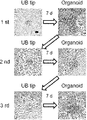
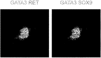
- A 100ng / ml activin A; C3, 3 ⁇ M CHIR99021; B, 10ng / ml BMP4; F8, 200ng / ml FGF8; TT, 0.1 ⁇ M TTNPB; A83, 1 ⁇ M A83-01; LDN, 0.1 ⁇ M LDN193189; Y, 10 ⁇ M Y -27632; C1, 1 ⁇ M CHIR99021; G, 100 ng / ml GDNF; F1, 200 ng / ml FGF1; E, 50 ng / ml EGF; Tzv, 10 ⁇ M Thiazovivin. Immunostaining analysis of stage 4 day 2 cells with ND leader markers GATA3 (green) and RET (red).
- the scale bar is 100 ⁇ m. Immunostaining analysis of stage 4 day 6, 8 and 10 cells with E-CADHERIN (green), GATA3 (red) and PAX2 (purple) was used in the mesonephric duct (ND) elongation stage (stage 4). It is shown that the growth factors that have been produced enhance the epithelialization of ND leader cells.
- the scale bar is 100 ⁇ m.
- the scale bar is 100 ⁇ m.
- the scale bar is 100 ⁇ m.
- the ureteral bud tip cells are isolated using the binding substance as an index.
- the binding substance may be labeled as described above and the label may be used as an index.
- a labeled antibody or the like that recognizes the binding substance may be added, and the label may be used as an index.
- a known method may be appropriately used depending on the type of label. For example, when the label is a fluorescent label, the cells can be isolated by flow cytometry or FACS (fluorescence-activated cell sorting). When the label is a magnetic label, the cells can be isolated by MACS (Magnetic-activated cell sorting).
- retinoic acid receptor agonist compounds that do not have a retinoid skeleton are Am80, AM580 (4-[[5,6,7,8-tetrahydro-5,5,8,8-tetramethyl-2-naphthalenyl] carboxy). Amide] benzoic acid), TTNPB (4-[[E] -2- [5,6,7,8-tetrahydro-5,5,8,8-tetramethyl-2-naphthalenyl] -1-propenyl] ben Zoic acid), AC55649 (4'-octyl- [1,1'-biphenyl] -4-carboxylic acid).
- a material which has been coated with a polymer (Lipidure) of ethylmethacrylic acid (poly-HEMA) or 2-methacryloyloxyethyl phosphorylcholine for example, a commercially available product such as a low-adhesion 35 mm dish (Sumitomo Bakelite) may be used.
- distilled water distilled water, a pH adjuster, a suspending agent, a solubilizing agent, a stabilizer, an isotonic agent, an antioxidant, a preservative, etc. are added to the VLDL-R binding substance as needed.
- the pH adjuster include hydrochloric acid, sodium hydroxide, lactose, lactic acid, sodium, sodium monohydrogen phosphate, sodium dihydrogen phosphate and the like.
- the suspending agent include methyl cellulose, polysorbate 80, hydroxyethyl cellulose, gum arabic, tragant powder, sodium carboxymethyl cellulose, polyoxyethylene sorbitan monolaurate and the like.
- ND cryopreservation Dissociated ND leader cells were resuspended using STEM-CELLBANKER GMP grade (Nippon Zenyaku Kogyo Co., Ltd.) at a dilution of ⁇ 1 ⁇ 10 6 cells / mL. The cell suspension was distributed to each cryopreservation tube. The tubes were frozen at ⁇ 80 ° C. for 24 hours and transferred to a liquid nitrogen cell storage tank for long-term cryopreservation. To initiate culturing, cells were thawed at 37 ° C. in a water bath. The cells were then slowly resuspended in Essential 6 medium containing 10 ⁇ M Y-27632 and centrifuged at 200 g for 5 minutes at room temperature.
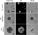
- iUB organoids and apical cell colonies treated with DiI-VLDL were treated with Accutase at 37 ° C. for 3 minutes and then dissociated into single cells by pipette operation. After washing with DMEM / 10% FBS, cells were resuspended in PBS / 2% FBS. Single cells were analyzed and sorted using FACS Aria II (BD). Cells from iUB organoids and apical cell colonies not treated with DiI-VLDL were used as negative controls for gating.
- BD FACS Aria II
- the sample was fixed at 4 ° C for 1 hour using 4% PFA / PBS.
- the fixed sample was treated with 30% sucrose / PBS, frozen with OCT compound (Tissue-Tek), and frozen sections were prepared by the frozen section method.
- Frozen sections were washed with distilled water and incubated with blocking solution for 1 hour at room temperature.
- the primary antibody was diluted 1: 500 with blocking solution and incubated with the sample overnight at room temperature. After washing twice with distilled water, cells were incubated with secondary antibody diluted 1: 500 in blocking solution for 1 hour at room temperature.
Abstract
Description
(1-1)尿管芽先端部細胞を含む細胞、組織またはオルガノイドを超低密度リポタンパク質受容体(VLDL-R)結合物質に接触させる工程、および
(1-2)該結合物質を指標として尿管芽先端部細胞を単離する工程
を含む方法を提供する。 The present application is a method for isolating ureteral bud tips cells from cells, tissues or organoids containing ureteral bud tips cells.
(1-1) A step of contacting cells, tissues or organoids containing ureteral blast tip cells with an ultra-low density lipoprotein receptor (VLDL-R) binding substance, and (1-2) using the binding substance as an index. A method comprising the step of isolating ureteral blast tip cells is provided.
(2)該尿管芽先端部細胞を、グリア細胞株由来神経栄養因子、線維芽細胞増殖因子、レチノイン酸受容体アゴニスト、GSK3β阻害剤およびYes-associated protein(YAP)活性阻害剤を含む培地で培養する工程を含む、尿管芽先端部細胞コロニーの製造方法を提供する。 The present application also isolates ureteral blast tip cells by the method of the present application, and further (2) the ureteral bud tip cells are subjected to glial cell line-derived neurotrophic factor, fibroblast proliferation factor, retinoic acid receptor agonist. , A method for producing ureteral bud tip cell colonies, comprising culturing in a medium containing a GSK3β inhibitor and a Yes-associated protein (YAP) activity inhibitor.
尿管芽先端部細胞コロニーを提供する工程、および
(3)該尿管芽先端部細胞コロニーをWntシグナル伝達活性化因子、BMP阻害剤、線維芽細胞増殖因子、レチノイン酸受容体アゴニスト、およびグリア細胞株由来神経栄養因子を含む培地で培養して、尿管芽様オルガノイドを再構成する工程を含む、尿管芽様オルガノイドの製造方法を提供する。 This application also
Steps to provide ureteral blast tip cell colonies, and (3) ureteral bud tip cell colonies to Wnt signaling activator, BMP inhibitor, fibroblast growth factor, retinoic acid receptor agonist, and glia Provided is a method for producing a ureteral bud-like organoid, which comprises a step of reconstitution of a ureteral bud-like organoid by culturing in a medium containing a cell line-derived neurotrophic factor.
尿管芽様オルガノイドを提供する工程、および
(4)該尿管芽様オルガノイドをWntシグナル阻害剤およびTGFβシグナル阻害剤を含む培地で培養する工程を含む、集合管前駆体様オルガノイドの製造方法を提供する。 This application also
A method for producing a collecting duct precursor-like organoid, which comprises a step of providing a ureteral bud-like organoid and (4) culturing the ureteral bud-like organoid in a medium containing a Wnt signal inhibitor and a TGFβ signal inhibitor. provide.
(2’)該尿管芽先端部細胞をWntシグナル阻害剤を含む培地で培養する工程を含む、集合管前駆細胞の製造方法を提供する。 The present application also comprises the step of isolating the ureteral bud apical cells by the method of the present application and further (2') culturing the ureteral bud apical cells in a medium containing a Wnt signal inhibitor. Provide a manufacturing method.
(3’-1)該尿管芽先端部細胞コロニーを解離する工程、および
(3’-2)該解離された細胞集団をWntシグナル阻害剤を含む培地で培養する工程を含む、集合管前駆細胞の製造方法を提供する。 The present application also obtains ureteral bud tip cell colonies by the method of the present application, and further (3'-1) dissociating the ureteral bud tip cell colonies, and (3'-2) the dissociated cells. Provided is a method for producing collecting duct progenitor cells, which comprises a step of culturing a population in a medium containing a Wnt signal inhibitor.
(1-1)尿管芽先端部細胞を含む細胞、組織またはオルガノイドをVLDL-R結合物質に接触させる工程、および
(1-2)該結合物質を指標として尿管芽先端部細胞を単離する工程を含む、尿管芽先端部細胞集団の製造方法を提供する。 This application also
(1-1) A step of contacting a cell, tissue or organoid containing ureteral bud tip cells with a VLDL-R binding substance, and (1-2) Isolating ureteral bud tip cells using the binding substance as an index. Provided is a method for producing a ureteral bud tip cell population, which comprises a step of making a ureteral bud tip cell population.
(i)尿管芽先端部細胞が含まれていることが予測される生体組織、組織片、培養細胞または培養組織を、VLDL-R結合物質に接触させる工程、および
(ii)該結合物質を検出する工程を含む、尿管芽先端部細胞をモニタリングする方法を提供する。 This application also
(I) A step of bringing a living tissue, a tissue piece, a cultured cell or a cultured tissue, which is predicted to contain ureteral bud tip cells, into contact with a VLDL-R binding substance, and (ii) the binding substance. Provided is a method for monitoring ureteral blast tip cells, including a step of detecting.
本願のある態様において、尿管芽先端部細胞を含む細胞、組織またはオルガノイドから尿管芽先端部細胞を単離する方法であって、
(1-1)尿管芽先端部細胞を含む細胞、組織またはオルガノイドを超低密度リポタンパク質受容体(VLDL-R)結合物質に接触させる工程、および
(1-2)該結合物質を指標として尿管芽先端部細胞を単離する工程
を含む方法を提供する。 Method of Isolating Ureter Sprout Tip Cells from Cells, Tissues or Organoids Containing Ureter Sprout Tip Cells In some embodiments of the present application, ureteral sprouting tips from cells, tissues or organoids containing ureteral blast tip cells. A method of isolating cells
(1-1) A step of contacting cells, tissues or organoids containing ureteral blast tip cells with an ultra-low density lipoprotein receptor (VLDL-R) binding substance, and (1-2) using the binding substance as an index. A method comprising the step of isolating ureteral blast tip cells is provided.
本願のある態様において、
(1-1)尿管芽先端部細胞を含む細胞、組織またはオルガノイドをVLDL-R結合物質に接触させる工程、および
(1-2)該結合物質を指標として尿管芽先端部細胞を単離する工程を含む、尿管芽先端部細胞集団の製造方法を提供する。 Method for Producing a Ureteral Bud Tip Cell Population From Cells, Tissues or Organoids Containing Ureter Bud Tip Cells In certain embodiments of the present application.
(1-1) A step of contacting a cell, tissue or organoid containing ureteral bud tip cells with a VLDL-R binding substance, and (1-2) Isolating ureteral bud tip cells using the binding substance as an index. Provided is a method for producing a ureteral bud tip cell population, which comprises the step of performing.
本願のある態様において、本願の方法で尿管芽先端部細胞を単離し、さらに
(2)該尿管芽先端部細胞を、グリア細胞株由来神経栄養因子、線維芽細胞増殖因子、レチノイン酸受容体アゴニスト、GSK3β阻害剤およびYes-associated protein(YAP)活性阻害剤を含む培地で培養する工程を含む、尿管芽先端部細胞コロニーの製造方法を提供する。 In certain embodiments of the methods herein to produce a ureteric bud tip cell colonies from isolated ureteric bud tip cells, the ureteric bud tip cells isolated by the methods herein, and (2) urine Kanme tip Ureters comprising the step of culturing part cells in a medium containing glial cell line-derived neurotrophic factor, fibroblast growth factor, retinoic acid receptor agonist, GSK3β inhibitor and Yes-associated protein (YAP) activity inhibitor. A method for producing a bud tip cell colony is provided.
本願のある態様において、
(3)尿管芽先端部細胞コロニーをWntシグナル伝達活性化因子、BMP阻害剤、線維芽細胞増殖因子、レチノイン酸受容体アゴニスト、およびグリア細胞株由来神経栄養因子を含む培地で培養して、尿管芽様オルガノイドを再構成する工程を含む、尿管芽様オルガノイドの製造方法を提供する。尿管芽先端部細胞コロニーは本願の方法で得られた細胞を用いてもよく、公知の別の方法によって得られたものを用いてもよい。本工程において、尿管芽先端部細胞コロニーのサイズは特に限定されないが、例えば約10~約1000μmであり得る。 Method for Reconstructing Ureter Bud-like Organoids from Ureteral Bud Tip Cell Colonies In certain embodiments of the present application.
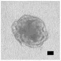
(3) The cell colonies at the tip of urinary tract buds are cultured in a medium containing a Wnt signaling activator, a BMP inhibitor, a fibroblast growth factor, a retinoic acid receptor agonist, and a neurotrophic factor derived from a glial cell line. Provided is a method for producing a urinary bud-like organoid, which comprises a step of reconstructing a urinary bud-like organoid. As the ureteral bud tip cell colony, cells obtained by the method of the present application may be used, or those obtained by another known method may be used. In this step, the size of the ureteral bud tip cell colony is not particularly limited, but may be, for example, about 10 to about 1000 μm.
本願のある態様において、
(4)尿管芽様オルガノイドをWntシグナル阻害剤およびTGFβシグナル阻害剤を含む培地で培養する工程を含む、集合管前駆体様オルガノイドの製造方法を提供する。尿管芽様オルガノイドは本願の方法で製造または再構成された細胞を用いてもよく、公知の別の方法によって得られたものを用いてもよい。 A method for producing a collecting duct precursor-like organoid from a ureteral bud-like organoid In an embodiment of the present application,
(4) Provided is a method for producing a collecting duct precursor-like organoid, which comprises a step of culturing a ureteral bud-like organoid in a medium containing a Wnt signal inhibitor and a TGFβ signal inhibitor. As the ureteral bud-like organoid, cells produced or reconstituted by the method of the present application may be used, or those obtained by another known method may be used.
本願のある態様において、本願の方法で尿管芽先端部細胞を単離し、さらに
(2’)該尿管芽先端部細胞をWntシグナル阻害剤を含む培地で培養する工程を含む、集合管前駆細胞の製造方法を提供する。 Method for producing collecting duct precursor cells from ureteral blast tip cells In one aspect of the present application, ureteral bud tip cells are isolated by the method of the present application, and (2') the ureteral bud tip cells are signaled with Wnt. Provided is a method for producing collecting duct precursor cells, which comprises a step of culturing in a medium containing an inhibitor.
本願のある態様において、本願の方法で尿管芽先端部細胞コロニーを得、さらに
(3’-1)該尿管芽先端部細胞コロニーを解離する工程、および
(3’-2)該解離された細胞集団をWntシグナル阻害剤を含む培地で培養する工程を含む、集合管前駆細胞の製造方法を提供する。 Method for Producing Collective Tube Precursor Cells from Ureteral Sprout Tip Cell Colonies In one aspect of the present application, ureteral bud tip cell colonies are obtained by the method of the present application, and (3'-1) the ureteral bud tip cells Provided is a method for producing a ureteral precursor cell, which comprises a step of dissociating a colony and (3'-2) culturing the dissociated cell population in a medium containing a Wnt signal inhibitor.
本願のある態様において、
(i)尿管芽先端部細胞が含まれていることが予測される生体組織、組織片、培養細胞または培養組織を、VLDL-R結合物質に接触させる工程、および
(ii)該結合物質を検出する工程を含む、尿管芽先端部細胞をモニタリングする方法を提供する。本態様で用いられるVLDL-R結合物質の例は、上述したとおりである。好ましい実施形態では、VLDL-R結合物質は上述のように標識されている。より好ましい実施形態では、VLDL-R結合物質は標識されたVLDLである。 Method for Monitoring Ureter Bud Tip Cells In certain aspects of the present application,
(I) A step of bringing a living tissue, a tissue piece, a cultured cell or a cultured tissue, which is predicted to contain ureteral bud tip cells, into contact with a VLDL-R binding substance, and (ii) the binding substance. Provided is a method for monitoring ureteral blast tip cells, including a step of detecting. Examples of the VLDL-R binding substance used in this embodiment are as described above. In a preferred embodiment, the VLDL-R binding material is labeled as described above. In a more preferred embodiment, the VLDL-R binding agent is a labeled VLDL.
本願のある態様において、VLDL-R結合物質を含む、尿管芽先端部細胞を単離またはモニタリングするための組成物を提供する。本態様で用いられるVLDL-R結合物質の例は、上述したとおりである。 Compositions for Isolating or Monitoring Ureter Bud Tip Cells In certain embodiments of the present application, there is provided a composition for isolating or monitoring ureteral bud tips cells, comprising a VLDL-R binding agent. Examples of the VLDL-R binding substance used in this embodiment are as described above.
本実施例で確立した方法をまとめた模式図を図1に示す。 Examples will be described below in more detail, but the present invention is not limited to the examples.
FIG. 1 shows a schematic diagram summarizing the methods established in this embodiment.
細胞培養
ヒト人工多能性幹細胞(hiPSC)を用いた実験は、京都大学医学部および医学研究科の倫理委員会によって承認された。3種のhiPSC株585A1、1231A3および1383D2を、Stem Fit AK02N培地(Ajinomoto)を用いたフィーダーフリー培養によって0.25μL/cm2 iMatrix-511 silk(Nippi)をコートした細胞培養プレート上で維持した。細胞を0.5mM EDTA/PBS(Thermo Fisher Scientific)を用いて4日毎に継代した。細胞をマイコプラズマ汚染について定期的に検査した。 [Materials and methods]
Experiments using cell-cultured human induced pluripotent stem cells (hiPSC) were approved by the Ethics Committee of Kyoto University School of Medicine and Graduate School of Medicine. Three hiPSC strains 585A1, 1231A3 and 1383D2 were maintained on cell culture plates coated with 0.25 μL / cm 2 iMatrix-511 silk (Nippi) by feeder-free culture in Stem Fit AK02N medium (Ajinomoto). Cells were passaged every 4 days using 0.5 mM EDTA / PBS (Thermo Fisher Scientific). Cells were routinely examined for mycoplasma contamination.
細胞をいくつかの改変を伴って以前に記述されるように尿管芽(UB)系統に誘導した(Mae, SI. & Ryosaka, M. et al. Biochem Biophys Res Commun. 495, 954-61 (2018))。本実施例で使用した増殖因子および小分子の詳細を表1に示す。
hiPSCを、4ウェル培養プレート(Thermo Fisher Scientific)において10μM Y-27632(WAKO)および0.25μL/cm2 iMatrix-511 silkを含むStem Fit AK02N培地に5×10⁴細胞/ウェルの密度で播種した。24時間後に細胞をPBSで洗浄し、100ng/mlアクチビンA(R&D Systems)および3μM CHIR99021(Stem RD)を含むEssential 6培地(Thermo Fisher Scientific)で処理した。24時間後に細胞をPBSで洗浄し、0.1μM LDN193189(Axon Medchem)、1μM A83-01(WAKO)、0.1μM 4-[(E)-2-(5,6,7,8-テトラヒドロ-5,5,8,8-テトラメチル-2-ナフタレニル)-1-プロペニル]-安息香酸(TTNPB; Santa Cruz Biotechnology)および200ng/ml線維芽細胞増殖因子(FGF)8(Peprotech)を含むEssential 6培地で2日間処理した。次に細胞を、マトリゲルまたはGeltrex(Corning)をコートした24ウェル培養プレートにおいて同じ4種の因子および10μM Y-27632を含むEssential 6培地に2×10⁵細胞/ウェルの密度で再播種し、さらに24時間インキュベートして前方中間中胚葉(AIM)を誘導した。 Anterior intermediate mesoderm-induced hiPSC in 5 × 10 4 cells / well in Stem Fit AK02N medium containing 10 μM Y-27632 (WAKO) and 0.25 μL / cm 2 iMatrix-511 silk in a 4-well culture plate (Thermo Fisher Scientific). Seeded at density. After 24 hours, cells were washed with PBS and treated with
AIM細胞を、1μM CHIR99021、0.1μM LDN193189、200ng/ml FGF8、100ng/mlグリア細胞株由来神経栄養因子(GDNF; R&D Systems)および0.1μM TTNPBを含むEssential 6培地で2日間処理し、ND最先端細胞(NDリーダー細胞)を誘導した。二次元培養において上皮化を増強するために、AIM細胞を同じ培地および誘導因子で8日間処理した。NDリーダー細胞をAccutase(Innovative Cell Technologies)を用いて37℃で3分間処理した後、ピペット操作によって単一細胞に解離させた。この細胞を低接着96ウェルプレート(Sumitomo Bakelite)上に1×10⁴細胞/ウェルの密度で播種し、同じ培地および因子に10μM Y-27632を加えて処理し、成熟ND凝集体を2日間誘導した。 Mesonephric duct (ND) -induced AIM cells in
ND凝集体から不要な細胞をピペット操作によって分離した(Mae, SI. & Ryosaka, M. et al. Biochem Biophys Res Commun. 495, 954-61 (2018))。ND凝集体を2%マトリゲルを含む同じ培地および因子で6日間処理し、上皮極性および尿細管腔を有するiUBオルガノイドを構成した。発芽を増強するために、50ng/ml EGF(R&D Systems)および200ng/ml FGF1(R&D Systems)を加えた。先端部をiUBオルガノイドから機械的に分離し、2%マトリゲルを含む同じ培地および因子で6~14日培養して分枝したiUBオルガノイドを再構成した。再構成したiUBオルガノイドから分離した先端部を同じ培地および因子で処理し、iUBオルガノイドを繰り返し再構成した。 Unwanted cells were pipetted from artificial ureteral bud (iUB) organoid-induced ND aggregates (Mae, SI. & Ryosaka, M. et al. Biochem Biophys Res Commun. 495, 954-61 (2018)). ND aggregates were treated with the same medium and factor containing 2% Matrigel for 6 days to construct iUB organoids with epithelial polarity and urinary cavities. 50 ng / ml EGF (R & D Systems) and 200 ng / ml FGF1 (R & D Systems) were added to enhance germination. The tip was mechanically separated from the iUB organoid and cultured in the same medium and factor containing 2% matrigel for 6-14 days to reconstitute the branched iUB organoid. The tips separated from the reconstituted iUB organoids were treated with the same medium and factors to reconstitute the iUB organoids repeatedly.
二次元培養のために、ハイドロゲルから分離した7日目の先端部細胞コロニーをAccutaseを用いて37℃で3分間処理した後、ピペット操作によって単一細胞に解離させた。この単一細胞を、1μM A83-01を含む、または含まない1μM IWR-1(Tocris)および0.5μL/cm2 iMatrix-511 silkを含むEssential 6培地で再懸濁し、96ウェルプレートに4×10⁴細胞/ウェルの密度で播種した。三次元培養のために、14日目の再構成したiUBオルガノイドを1μM IWR-1および1μM A83-01を含むEssential 6培地で14日間処理した。 For collecting duct precursor-induced two-dimensional culture,
解離させたNDリーダー細胞を、STEM-CELLBANKER GMP grade (Nippon Zenyaku Kogyo Co., Ltd.)を用いて1×10⁶細胞/mL未満の希釈率で再懸濁した。細胞懸濁液を各凍結保存チューブに分配した。チューブを-80℃で24時間凍結させ、長期間の凍結保存のために液体窒素細胞貯蔵タンクに移動させた。培養を開始するため、細胞を水浴を用いて37℃で解凍した。次に、細胞を10μM Y-27632を含むEssential 6培地でゆっくりと再懸濁し、室温において200gで5分間遠心した。上清を除去した後、細胞を10μM Y-27632およびND誘導因子を含むEssential 6培地で再懸濁し、低接着96ウェルプレート上に1×10⁴細胞/ウェルの密度で播種した。 ND cryopreservation Dissociated ND leader cells were resuspended using STEM-CELLBANKER GMP grade (Nippon Zenyaku Kogyo Co., Ltd.) at a dilution of < 1 × 10 6 cells / mL. The cell suspension was distributed to each cryopreservation tube. The tubes were frozen at −80 ° C. for 24 hours and transferred to a liquid nitrogen cell storage tank for long-term cryopreservation. To initiate culturing, cells were thawed at 37 ° C. in a water bath. The cells were then slowly resuspended in
iUBオルガノイドを37℃においてAccutaseで5分間処理し、その後ピペット操作によって単一細胞に解離させた。細胞をB27サプリメント(ビタミンA不含)(Thermo Fisher Scientific)、100ng/ml GDNF、200ng/ml FGF1、0.1μM TTNPB、3μM CHIR99021および10μM Thiazovivin(Santa Cruz Biotechnology)を含むDMEM/F12培地(Thermo Fisher Scientific)で再懸濁した。10個のiUBオルガノイドから得た単一細胞を150μLハイドロゲルでコートした48ウェルプレートの1つのウェル上に播種した。ハイドロゲルは50%マトリゲルを含むDMEM/F12培地で構成され、使用する前に37℃で30分間凝固させた。単一細胞は6~7日後に先端部細胞コロニーを構築した。培地を2日毎に交換した。 Proliferation of apical cells iUB organoids were treated with Accutase at 37 ° C. for 5 minutes and then pipette to dissociate into single cells. Cells in DMEM / F12 medium (Thermo Fisher Scientific) containing B27 supplement (Vitamin A-free) (Thermo Fisher Scientific), 100 ng / ml GDNF, 200 ng / ml FGF1, 0.1 μM TTNPB, 3 μM CHIR99021 and 10 μM Thiazovivin (Santa Cruz Biotechnology) ) Resuspended. Single cells from 10 iUB organoids were seeded on one well of a 48-well plate coated with 150 μL hydrogel. The hydrogel was composed of DMEM / F12 medium containing 50% Matrigel and was coagulated at 37 ° C. for 30 minutes before use. Single cells established apical cell colonies after 6-7 days. The medium was changed every 2 days.
ハイドロゲルをCell Recovery Solution(BD Biosciences)を用いて4℃で1時間溶解し、7日目の先端部細胞コロニーを分離した。Cell Recovery Solutionでさらに洗浄した後、先端部細胞コロニーを室温において500gで5分間遠心した。0.1μM LDN193189、100ng/ml GDNF、0.1μM TTNPB、200ng/ml FGF8、1μM CHIR99021または10% Afamin/Wnt3a条件培地(MBL)、200ng/ml R-spondin 1(R&D systems)、50ng/ml EGF、200ng/ml FGF1および10%マトリゲルを含むEssential 6培地を先端部細胞コロニーを含むチューブに加えた。コロニーを破壊しないように広口のマイクロピペット(BMBio)で穏やかにピペット操作した後、懸濁液を低接着35mmディッシュ(Sumitomo Bakelite)に2mL/ディッシュで分配した。培地を2日毎に交換した。4日目に、各コロニーを低接着96ウェルプレートの1つのウェル上の同じ培地中に配置した。さらに6日後に、先端領域を再構成したiUBオルガノイドから分離し、同じ培地で35日間処理した。 Reconstitutional hydrogels of iUB organoids from a single apical cell were lysed using Cell Recovery Solution (BD Biosciences) at 4 ° C. for 1 hour to isolate apical cell colonies on
iUBオルガノイドを37℃においてAccutaseで5分間処理し、その後ピペット操作によって単一細胞に解離させた。細胞をB27サプリメント(ビタミンA不含)、100ng/ml GDNF、200ng/ml FGF1、0.1μM TTNPB、3μM CHIR99021、10μM Thiazovivinおよび1μM A83-01を含むDMEM/F12培地で懸濁した。細胞を150μLハイドロゲルでコートした48ウェルプレートの1つのウェル上に播種した。ハイドロゲルは50%マトリゲルを含むDMEM/F12培地で構成され、使用する前に37℃で1時間凝固させた。単一細胞を37℃、5%CO2で7日間培養することで、先端部細胞コロニーを作製した。培地を2~3日毎に交換した。作製した先端部細胞コロニーを7日毎に同じ培地で継代培養した。作製または3回継代培養した先端部細胞コロニーから、上記と同様の方法を用いてiUBオルガノイドを再構成した。 Maintenance culture of apical cell colonies iUB organoids were treated with Accutase at 37 ° C. for 5 minutes and then pipette to dissociate into single cells. Cells were suspended in DMEM / F12 medium containing B27 supplement (without vitamin A), 100 ng / ml GDNF, 200 ng / ml FGF1, 0.1 μM TTNPB, 3 μM CHIR99021, 10 μM Thiazovivin and 1 μM A83-01. Cells were seeded on one well of a 48-well plate coated with 150 μL hydrogel. The hydrogel was composed of DMEM / F12 medium containing 50% Matrigel and was coagulated at 37 ° C. for 1 hour before use. Tip cell colonies were prepared by culturing single cells at 37 ° C. and 5% CO 2 for 7 days. The medium was changed every 2-3 days. The prepared apical cell colonies were subcultured in the same medium every 7 days. The iUB organoid was reconstituted from the apical cell colonies prepared or subcultured three times using the same method as described above.
iUBオルガノイド、先端部細胞コロニーおよびND凝集体を10μg/mlのDiI標識VLDL(Biomedical Technologies, Inc.)で37℃において2時間処理した。 Monitoring of apical cells with DiI-labeled VLDL (DiI-VLDL) iUB organoids, apical cell colonies and ND aggregates were treated with 10 μg / ml DiI-labeled VLDL (Biomedical Technologies, Inc.) at 37 ° C. for 2 hours. ..
DiI-VLDLで処理したiUBオルガノイドおよび先端部細胞コロニーをAccutaseで37℃において3分間処理した後、ピペット操作によって単一細胞に解離させた。DMEM/10% FBSで洗浄した後、細胞をPBS/2% FBSで再懸濁した。単一細胞をFACS Aria II(BD)を用いて分析およびソーティングした。DiI-VLDLで処理していないiUBオルガノイドおよび先端部細胞コロニー由来の細胞をゲーティングのための陰性対照として用いた。 Flow cytometric analysis The iUB organoids and apical cell colonies treated with DiI-VLDL were treated with Accutase at 37 ° C. for 3 minutes and then dissociated into single cells by pipette operation. After washing with DMEM / 10% FBS, cells were resuspended in PBS / 2% FBS. Single cells were analyzed and sorted using FACS Aria II (BD). Cells from iUB organoids and apical cell colonies not treated with DiI-VLDL were used as negative controls for gating.
6日目のiUBオルガノイドをDiI-VLDLで2時間処理した。フローサイトメトリー分析を行い、DiI-VLDL+細胞およびDiI-VLDL-細胞をソーティングした。ソーティングしたDiI-VLDL+細胞およびDiI-VLDL-細胞の全RNAをNucleoSpin RNA XS(Takara)を用いて単離した。試料を-80℃で保存し、DNAFORM Co., LtdによりRNAシーケンシング解析を行った。 RNA-
免疫染色分析をいくつかの改変を伴って以前に記述されるように行った(Mae, SI. & Ryosaka, M. et al. Biochem Biophys Res Commun. 495, 954-61 (2018))。二次元培養物について、細胞を4%パラホルムアルデヒド(PFA)/PBSを用いて4℃で20分間固定した。PBSで2回洗浄した後、細胞を1%正常ロバ血清(MERCK)および3%ウシ血清アルブミン(BSA; Nacalai Tesque)/PBST(PBS/0.25% Triton X-100、Nacalai Tesque)を用いて室温で1時間ブロックした。一次抗体をブロッキング溶液で1:500に希釈し、試料と共に4℃で一晩インキュベートした。PBSTで2回洗浄した後、ブロッキング溶液で1:500に希釈した二次抗体を室温で1時間インキュベートした。 Immunostaining Immunostaining analysis was performed as previously described with some modifications (Mae, SI. & Ryosaka, M. et al. Biochem Biophys Res Commun. 495, 954-61 (2018)). For two-dimensional cultures, cells were fixed with 4% paraformaldehyde (PFA) / PBS at 4 ° C. for 20 minutes. After washing twice with PBS, cells are treated with 1% normal donkey serum (MERCK) and 3% bovine serum albumin (BSA; Nacalai Tesque) / PBST (PBS / 0.25% Triton X-100, Nacalai Tesque) at room temperature. Blocked for 1 hour. The primary antibody was diluted 1: 500 with blocking solution and incubated with the sample overnight at 4 ° C. After washing twice with PBST, the secondary antibody diluted 1: 500 with blocking solution was incubated for 1 hour at room temperature.


フローサイトメトリーおよび細胞数分析のデータを平均値±SEとして表す。二群間の差についての統計解析をスチューデントのt検定を用いて行った。p<0.05である場合に差を有意であるとみなした。 Statistical analysis Flow cytometry and cell number analysis data are expressed as mean ± SE. Statistical analysis of the difference between the two groups was performed using Student's t-test. The difference was considered significant when p <0.05.
本発明者らはまず、レチノイン酸(RA)シグナルが中腎管(ND)発生に重要な役割を果たすことから(Stewart, K. & Bouchard, M. Semin Cell Dev Biol. 36, 13-20 (2014))、ステージ4の処理にRAアゴニストであるTTNPBを追加することによって以前に報告したND誘導法を改変した(図2、図3)。本発明者らはまた、ステージ4で使用されるND伸長因子での処理を延長することにより、ND細胞の上皮化が誘導されることを見出した(図4)。したがって、本発明者らはステージ4と同じ誘導因子を用いてND上皮性凝集体を作製した。ピペット操作により不要な細胞を取り除いた(図5)。 [result]
First, we found that the retinoic acid (RA) signal plays an important role in the development of the mesonephric duct (ND) (Stewart, K. & Bouchard, M. Semin Cell Dev Biol. 36, 13-20 (Stewart, K. & Bouchard, M. Semin Cell Dev Biol. 36, 13-20). 2014)), the previously reported ND induction method was modified by adding the RA agonist TTNPB to the
Claims (28)
- 尿管芽先端部細胞を含む細胞、組織またはオルガノイドから尿管芽先端部細胞を単離する方法であって、
(1-1)尿管芽先端部細胞を含む細胞、組織またはオルガノイドを超低密度リポタンパク質受容体(VLDL-R)結合物質に接触させる工程、および
(1-2)該結合物質を指標として尿管芽先端部細胞を単離する工程
を含む方法。 A method of isolating ureteral tip cells from cells, tissues or organoids containing ureteral tip cells.
(1-1) A step of contacting cells, tissues or organoids containing ureteral blast tip cells with an ultra-low density lipoprotein receptor (VLDL-R) binding substance, and (1-2) using the binding substance as an index. A method comprising the step of isolating ureteral blast tip cells. - VLDL-R結合物質が標識された超低密度リポタンパク質(VLDL)である、請求項1記載の方法。 The method according to claim 1, wherein the VLDL-R binding substance is a labeled very low density lipoprotein (VLDL).
- 該標識が蛍光標識である、請求項2記載の方法。 The method according to claim 2, wherein the label is a fluorescent label.
- 尿管芽先端部細胞を含む細胞、組織またはオルガノイドが尿管芽様オルガノイドである、請求項1~3のいずれか一項に記載の方法。 The method according to any one of claims 1 to 3, wherein the cell, tissue or organoid containing the ureteral bud tip cell is a ureteral bud-like organoid.
- 尿管芽先端部細胞を含む細胞、組織またはオルガノイドが多能性幹細胞から分化誘導されたものである、請求項1~4のいずれか一項に記載の方法。 The method according to any one of claims 1 to 4, wherein cells, tissues or organoids including ureteral bud tip cells are induced to differentiate from pluripotent stem cells.
- 多能性幹細胞がiPS細胞である、請求項5記載の方法。 The method according to claim 5, wherein the pluripotent stem cells are iPS cells.
- 尿管芽先端部細胞を含む細胞、組織またはオルガノイドが霊長類由来である、請求項1~6のいずれか一項に記載の方法。 The method according to any one of claims 1 to 6, wherein the cell, tissue or organoid containing the ureteral bud tip cell is derived from a primate.
- 尿管芽先端部細胞を含む細胞、組織またはオルガノイドがヒト由来である、請求項1~7のいずれか一項に記載の方法。 The method according to any one of claims 1 to 7, wherein the cell, tissue or organoid containing the ureteral bud tip cell is derived from human.
- 請求項1~8のいずれか一項に記載の方法で尿管芽先端部細胞を単離し、さらに
(2)該尿管芽先端部細胞を、グリア細胞株由来神経栄養因子、線維芽細胞増殖因子、レチノイン酸受容体アゴニスト、GSK3β阻害剤およびYes-associated protein(YAP)活性阻害剤を含む培地で培養する工程を含む、尿管芽先端部細胞コロニーの製造方法。 The ureteral blast tip cells are isolated by the method according to any one of claims 1 to 8, and (2) the ureteral bud tip cells are subjected to glial cell line-derived neurotrophic factor and fibroblast proliferation. A method for producing ureteral bud tip cell colonies, which comprises the step of culturing in a medium containing a factor, a retinoic acid receptor agonist, a GSK3β inhibitor and a Yes-associated protein (YAP) activity inhibitor. - (2)の工程において、線維芽細胞増殖因子がFGF1であり、レチノイン酸受容体アゴニストがTTNPBであり、GSK3β阻害剤がCHIR99021であり、Yes-associated protein(YAP)活性阻害剤がThiazovivinである、請求項9記載の方法。 In step (2), the fibroblast growth factor is FGF1, the retinoic acid receptor agonist is TTNPB, the GSK3β inhibitor is CHIR99021, and the Yes-associated protein (YAP) activity inhibitor is Thiazovivin. The method according to claim 9.
- (2)の工程において、培地がTGFβシグナル阻害剤をさらに含む、請求項9または10に記載の方法。 The method according to claim 9 or 10, wherein in the step (2), the medium further contains a TGFβ signal inhibitor.
- TGFβシグナル阻害剤がA83-01である、請求項11に記載の方法。 The method according to claim 11, wherein the TGFβ signal inhibitor is A83-01.
- (2)の工程の後、得られた尿管芽先端部細胞コロニーを継代培養する工程を含む、請求項9~12のいずれか一項に記載の方法。 The method according to any one of claims 9 to 12, which comprises a step of subculturing the obtained ureteral bud tip cell colony after the step (2).
- 請求項9~13のいずれか一項に記載の方法で尿管芽先端部細胞コロニーを得、さらに
(3)該尿管芽先端部細胞コロニーをWntシグナル伝達活性化因子、BMP阻害剤、線維芽細胞増殖因子、レチノイン酸受容体アゴニスト、およびグリア細胞株由来神経栄養因子を含む培地で培養して、尿管芽様オルガノイドを再構成する工程を含む、尿管芽様オルガノイドの製造方法。 A ureteral bud tip cell colony is obtained by the method according to any one of claims 9 to 13, and (3) the ureteral bud tip cell colony is used as a Wnt signaling activator, a BMP inhibitor, and a fiber. A method for producing a ureteral bud-like organoid, which comprises a step of reconstitution of a ureteral bud-like organoid by culturing in a medium containing a blast growth factor, a retinoic acid receptor agonist, and a glial cell line-derived neurotrophic factor. - 尿管芽先端部細胞コロニーを提供する工程、および
(3)該尿管芽先端部細胞コロニーをWntシグナル伝達活性化因子、BMP阻害剤、線維芽細胞増殖因子、レチノイン酸受容体アゴニスト、およびグリア細胞株由来神経栄養因子を含む培地で培養して、尿管芽様オルガノイドを再構成する工程を含む、尿管芽様オルガノイドの製造方法。 Steps to provide ureteral blast tip cell colonies, and (3) ureteral bud tip cell colonies to Wnt signaling activator, BMP inhibitor, fibroblast growth factor, retinoic acid receptor agonist, and glia A method for producing a ureteral bud-like organoid, which comprises a step of reconstitution of a ureteral bud-like organoid by culturing in a medium containing a cell line-derived neurotrophic factor. - (3)の工程において、Wntシグナル伝達活性化因子がWnt3aおよび/またはR-spondin 1であり、BMP阻害剤がLDN193189であり、線維芽細胞増殖因子がFGF8であり、レチノイン酸受容体アゴニストがTTNPBである、請求項14または15に記載の方法。 In step (3), the Wnt signaling activator is Wnt3a and / or R-spondin1, the BMP inhibitor is LDN193189, the fibroblast growth factor is FGF8, and the retinoic acid receptor agonist is TTNPB. The method according to claim 14 or 15.
- (3)の工程において、培地がEGFおよびFGF1をさらに含む、請求項14~16のいずれか一項に記載の方法。 The method according to any one of claims 14 to 16, wherein in the step (3), the medium further contains EGF and FGF1.
- 請求項14~17のいずれか一項に記載の方法で尿管芽様オルガノイドを得、さらに
(4)該尿管芽様オルガノイドをWntシグナル阻害剤およびTGFβシグナル阻害剤を含む培地で培養する工程を含む、集合管前駆体様オルガノイドの製造方法。 A step of obtaining a ureteral bud-like organoid by the method according to any one of claims 14 to 17, and further (4) culturing the ureteral bud-like organoid in a medium containing a Wnt signal inhibitor and a TGFβ signal inhibitor. A method for producing a collecting duct precursor-like organoid, including. - 尿管芽様オルガノイドを提供する工程、および
(4)該尿管芽様オルガノイドをWntシグナル阻害剤およびTGFβシグナル阻害剤を含む培地で培養する工程を含む、集合管前駆体様オルガノイドの製造方法。 A method for producing a collecting duct precursor-like organoid, which comprises a step of providing a ureteral bud-like organoid and (4) culturing the ureteral bud-like organoid in a medium containing a Wnt signal inhibitor and a TGFβ signal inhibitor. - (4)の工程において、Wntシグナル阻害剤がIWR-1であり、TGFβシグナル阻害剤がA83-01である、請求項18または19に記載の方法。 The method according to claim 18 or 19, wherein in the step (4), the Wnt signal inhibitor is IWR-1 and the TGFβ signal inhibitor is A83-01.
- 請求項1~8のいずれか一項に記載の方法で尿管芽先端部細胞を単離し、さらに
(2’)該尿管芽先端部細胞をWntシグナル阻害剤を含む培地で培養する工程を含む、集合管前駆細胞の製造方法。 The step of isolating the ureteral blast tip cells by the method according to any one of claims 1 to 8 and further (2') culturing the ureteral bud tip cells in a medium containing a Wnt signal inhibitor. A method for producing collecting duct progenitor cells, which comprises. - 請求項9~13のいずれか一項に記載の方法で尿管芽先端部細胞コロニーを得、さらに
(3’-1)該尿管芽先端部細胞コロニーを解離する工程、および
(3’-2)該解離された細胞集団をWntシグナル阻害剤を含む培地で培養する工程を含む、集合管前駆細胞の製造方法。 A step of obtaining a ureteral bud tip cell colony by the method according to any one of claims 9 to 13 and further dissociating the ureteral bud tip cell colony, and (3'-). 2) A method for producing collecting duct progenitor cells, which comprises a step of culturing the dissociated cell population in a medium containing a Wnt signal inhibitor. - (2’)または(3’-2)の工程において、Wntシグナル阻害剤がIWR-1である、請求項21または22に記載の方法。 The method according to claim 21 or 22, wherein the Wnt signal inhibitor is IWR-1 in the step (2') or (3'-2).
- (2’)または(3’-2)の工程において、培地がTGFβシグナル阻害剤をさらに含む、請求項21~23のいずれか一項に記載の方法。 The method according to any one of claims 21 to 23, wherein in the step (2') or (3'-2), the medium further contains a TGFβ signal inhibitor.
- (2’)または(3’-2)の工程において、TGFβシグナル阻害剤がA83-01である、請求項24に記載の方法。 The method according to claim 24, wherein the TGFβ signal inhibitor is A83-01 in the step (2') or (3'-2).
- (1-1)尿管芽先端部細胞を含む細胞、組織またはオルガノイドをVLDL-R結合物質に接触させる工程、および
(1-2)該結合物質を指標として尿管芽先端部細胞を単離する工程を含む、尿管芽先端部細胞集団の製造方法。 (1-1) A step of contacting a cell, tissue or organoid containing ureteral bud tip cells with a VLDL-R binding substance, and (1-2) Isolating ureteral bud tip cells using the binding substance as an index. A method for producing a ureteral bud tip cell population, which comprises a step of: - (i)尿管芽先端部細胞が含まれていることが予測される生体組織、組織片、培養細胞または培養組織を、VLDL-R結合物質に接触させる工程、および
(ii)該結合物質を検出する工程を含む、尿管芽先端部細胞をモニタリングする方法。 (I) A step of bringing a living tissue, a tissue piece, a cultured cell or a cultured tissue, which is predicted to contain ureteral bud tip cells, into contact with a VLDL-R binding substance, and (ii) the binding substance. A method of monitoring ureteral blast tip cells, including a step of detection. - VLDL-R結合物質を含む、尿管芽先端部細胞を単離またはモニタリングするための組成物。 A composition for isolating or monitoring ureteral bud tip cells, which contains a VLDL-R binding substance.
Priority Applications (6)
| Application Number | Priority Date | Filing Date | Title |
|---|---|---|---|
| KR1020227014161A KR20220069096A (en) | 2019-10-01 | 2020-09-30 | Isolation method of ureteral tip cells |
| EP20872677.8A EP4043553A4 (en) | 2019-10-01 | 2020-09-30 | Method for isolating ureteric bud tip cells |
| CN202080068266.3A CN114630898A (en) | 2019-10-01 | 2020-09-30 | Method for separating cells at tip of ureter bud |
| JP2021551427A JPWO2021066076A1 (en) | 2019-10-01 | 2020-09-30 | |
| BR112022006286A BR112022006286A2 (en) | 2019-10-01 | 2020-09-30 | Methods to isolate a ureteric bud tip cell, to produce a ureteric bud tip cell colony, to produce an organoid, to produce a collection duct progenitor cell, to produce a ureteric bud tip cell population, and to monitor a ureteric bud tip cell, and composition for isolating or monitoring a ureteric bud tip cell |
| US17/765,358 US20220380732A1 (en) | 2019-10-01 | 2020-09-30 | Method for isolating ureteric bud tip cells |
Applications Claiming Priority (2)
| Application Number | Priority Date | Filing Date | Title |
|---|---|---|---|
| JP2019-181593 | 2019-10-01 | ||
| JP2019181593 | 2019-10-01 |
Publications (1)
| Publication Number | Publication Date |
|---|---|
| WO2021066076A1 true WO2021066076A1 (en) | 2021-04-08 |
Family
ID=75337056
Family Applications (1)
| Application Number | Title | Priority Date | Filing Date |
|---|---|---|---|
| PCT/JP2020/037329 WO2021066076A1 (en) | 2019-10-01 | 2020-09-30 | Method for isolating ureteric bud tip cells |
Country Status (7)
| Country | Link |
|---|---|
| US (1) | US20220380732A1 (en) |
| EP (1) | EP4043553A4 (en) |
| JP (1) | JPWO2021066076A1 (en) |
| KR (1) | KR20220069096A (en) |
| CN (1) | CN114630898A (en) |
| BR (1) | BR112022006286A2 (en) |
| WO (1) | WO2021066076A1 (en) |
Cited By (1)
| Publication number | Priority date | Publication date | Assignee | Title |
|---|---|---|---|---|
| WO2023228908A1 (en) * | 2022-05-23 | 2023-11-30 | 国立大学法人京都大学 | Production method for renal collecting duct cells and pelvic epithelial cells |
Citations (12)
| Publication number | Priority date | Publication date | Assignee | Title |
|---|---|---|---|---|
| US4326055A (en) | 1977-12-22 | 1982-04-20 | Hoffmann-La Roche Inc. | Stilbene derivatives |
| US5234926A (en) | 1987-03-20 | 1993-08-10 | Allergan, Inc. | Disubstituted acetylenes bearing heteroaromatic and heterobicyclic groups having retinoid like activity |
| JP2006312638A (en) * | 1995-11-15 | 2006-11-16 | Massachusetts Inst Of Technol <Mit> | Method for adjusting cholesterol transportation |
| WO2007069666A1 (en) | 2005-12-13 | 2007-06-21 | Kyoto University | Nuclear reprogramming factor |
| WO2009146408A1 (en) | 2008-05-30 | 2009-12-03 | Summa Health Systems Llc | Methods for using tgf-b receptor inhibitors or activin-like kinase (alk) 5 inhibitors a-83-01 and sb-431542 to treat eye disease and wound healing conditions |
| US20110008892A1 (en) * | 2007-06-04 | 2011-01-13 | The Regents Of The University Of California | Methods of tissue generation and tissue engineered compositions |
| WO2011007900A1 (en) | 2009-07-15 | 2011-01-20 | Dezawa Mari | Pluripotent stem cell that can be isolated from body tissue |
| US20150284689A1 (en) * | 2012-10-26 | 2015-10-08 | The Regents Of The University Of California | Strategy for engineering various 3d tissues, organoids and vasculature |
| JP2017537655A (en) * | 2014-12-15 | 2017-12-21 | ザ ユニバーシティー オブ クイーンズランド | Method for differentiation of pluripotent stem cells to form renal organoids |
| JP2018183137A (en) * | 2017-04-25 | 2018-11-22 | 国立大学法人 熊本大学 | Method for preparing a kidney structure with collecting duct arborized from pluripotent stem cells |
| WO2019098349A1 (en) | 2017-11-17 | 2019-05-23 | 国立大学法人京都大学 | Ureteric-bud-like tissue induction method |
| JP2019181593A (en) | 2018-04-04 | 2019-10-24 | 株式会社Tjmデザイン | Bracket for tool |
Family Cites Families (2)
| Publication number | Priority date | Publication date | Assignee | Title |
|---|---|---|---|---|
| WO2013186398A1 (en) * | 2012-06-15 | 2013-12-19 | Fondazione Telethon | Tfeb for use in treating obesity or metabolic syndrome |
| CN105473706B (en) * | 2013-06-14 | 2021-10-26 | 昆士兰大学 | Renal progenitor cells |
-
2020
- 2020-09-30 US US17/765,358 patent/US20220380732A1/en active Pending
- 2020-09-30 JP JP2021551427A patent/JPWO2021066076A1/ja active Pending
- 2020-09-30 EP EP20872677.8A patent/EP4043553A4/en active Pending
- 2020-09-30 CN CN202080068266.3A patent/CN114630898A/en active Pending
- 2020-09-30 WO PCT/JP2020/037329 patent/WO2021066076A1/en active Application Filing
- 2020-09-30 KR KR1020227014161A patent/KR20220069096A/en unknown
- 2020-09-30 BR BR112022006286A patent/BR112022006286A2/en not_active Application Discontinuation
Patent Citations (12)
| Publication number | Priority date | Publication date | Assignee | Title |
|---|---|---|---|---|
| US4326055A (en) | 1977-12-22 | 1982-04-20 | Hoffmann-La Roche Inc. | Stilbene derivatives |
| US5234926A (en) | 1987-03-20 | 1993-08-10 | Allergan, Inc. | Disubstituted acetylenes bearing heteroaromatic and heterobicyclic groups having retinoid like activity |
| JP2006312638A (en) * | 1995-11-15 | 2006-11-16 | Massachusetts Inst Of Technol <Mit> | Method for adjusting cholesterol transportation |
| WO2007069666A1 (en) | 2005-12-13 | 2007-06-21 | Kyoto University | Nuclear reprogramming factor |
| US20110008892A1 (en) * | 2007-06-04 | 2011-01-13 | The Regents Of The University Of California | Methods of tissue generation and tissue engineered compositions |
| WO2009146408A1 (en) | 2008-05-30 | 2009-12-03 | Summa Health Systems Llc | Methods for using tgf-b receptor inhibitors or activin-like kinase (alk) 5 inhibitors a-83-01 and sb-431542 to treat eye disease and wound healing conditions |
| WO2011007900A1 (en) | 2009-07-15 | 2011-01-20 | Dezawa Mari | Pluripotent stem cell that can be isolated from body tissue |
| US20150284689A1 (en) * | 2012-10-26 | 2015-10-08 | The Regents Of The University Of California | Strategy for engineering various 3d tissues, organoids and vasculature |
| JP2017537655A (en) * | 2014-12-15 | 2017-12-21 | ザ ユニバーシティー オブ クイーンズランド | Method for differentiation of pluripotent stem cells to form renal organoids |
| JP2018183137A (en) * | 2017-04-25 | 2018-11-22 | 国立大学法人 熊本大学 | Method for preparing a kidney structure with collecting duct arborized from pluripotent stem cells |
| WO2019098349A1 (en) | 2017-11-17 | 2019-05-23 | 国立大学法人京都大学 | Ureteric-bud-like tissue induction method |
| JP2019181593A (en) | 2018-04-04 | 2019-10-24 | 株式会社Tjmデザイン | Bracket for tool |
Non-Patent Citations (40)
| Title |
|---|
| "NCBI", Database accession no. NM_010094 |
| ANNUAL REPORT OF RESEARCH INSTITUTE FOR BIOLOGICAL FUNCTION, vol. 9, 2009, pages 55 - 61 |
| BUSH, KEVIN T, SAKURAI HIROYUKI, STEER DYLAN L, LEONARD MARTIN O, SAMPOGNA ROSEMARY V, MEYER TOBIAS N, SCHWESINGER CATHERINE, QIAO: "TGF- beta superfamily members modulate growth, branching, shaping, and patterning of the ureteric bud", DEV. BIOL., vol. 266, no. 2, 2004, pages 285 - 298, XP055811456, DOI: 10.1016/j.ydbio.2003.10.023 * |
| CHI, LIJUN, SAARELA ULLA, RAILO ANTTI, PRUNSKAITE-HYYRYLÄINEN RENATA, SKOVORODKIN ILYA, ANTHONY SHELAGH, KATSU KENJIRO, LIU YU, SH: "A secreted EMP antagonist, Cer1, fine tunes the spatial organization of the ureteric bud tree during mouse kidney development", PLOS ONE, vol. 6, no. issue 11, 2011, pages 1 - 16, XP055811462, DOI: 10.1371/journal.pone.0027676 * |
| GUI, TING; SUN YUJING; GAI ZHIBO; SHIMOKADO AIKO; MURAGAKI YASUTERU; ZHOU GENGYIN: "The loss of Trps1 suppresses ureteric bud branching because of the activation of TGF-beta signaling", DEV. BIOL., vol. 377, no. 2, 2013, pages 415 - 427, XP028590005, DOI: 10.1016/j.ydbio.2013.03.014 * |
| J. A. THOMSON ET AL., BIOL. REPROD., vol. 55, 1996, pages 254 - 259 |
| J. A. THOMSON ET AL., PROC. NATL. ACAD. SCI. USA, vol. 92, 1995, pages 7844 - 7848 |
| J. A. THOMSON ET AL., SCIENCE, vol. 282, 1998, pages 1145 - 1147 |
| J. A. THOMSONV. S. MARSHALL, CURR. TOP. DEV. BIOL., vol. 38, 1998, pages 133 - 165 |
| J. BYRNE, NATURE, vol. 450, 2007, pages 497 - 502 |
| J. HAO ET AL., PLOS ONE, vol. 3, no. 8, 2008, pages e2904 |
| J. L. RESNICK ET AL., NATURE, vol. 359, 1992, pages 550 - 551 |
| J. YU ET AL., SCIENCE, vol. 318, 2007, pages 1917 - 1920 |
| K. SHINOHARA ET AL., CELL, vol. 119, 2004, pages 1001 - 1012 |
| K. TAKAHASHI ET AL., CELL, vol. 131, 2007, pages 861 - 872 |
| K. TAKAHASHIS. YAMANAKA, CELL, vol. 126, 2006, pages 663 - 676 |
| KOBAYASHI A. ET AL., CELL STEM CELL, vol. 3, 2008, pages 169 - 181 |
| KOEHLER, K. R. ET AL., NAT BIOTECHNOL., vol. 35, 2017, pages 583 - 9 |
| M. KANATSU-SHINOHARA ET AL., BIOL. REPROD., vol. 69, 2003, pages 612 - 616 |
| MAE, SHINICHI ET AL.: "Generation of branching ureteric bud tissues from human pluripotent stem cells", BIOCHEM. BIOPHYS. RES. COMMUN., vol. 495, 2018, pages 954 - 961, XP055609767, DOI: 10.1016/j.bbrc.2017.11.105 * |
| MAE, SHIN-ICHI, RYOSAKA MAKOTO, SAKAMOTO SATOKO, MATSUSE KYOKO, NOZAKI AYA, IGAMI MAIKO, KABAI RYOTARO, WATANABE AKIRA, OSAFUNE KE: "Expansion of human iPSC-derived ureteric bud organoid, with repeated branching potential", CELL REPORTS, vol. 32, no. 4, 28 July 2020 (2020-07-28), pages 1 - 16, XP055811473, DOI: 10.1016/j.celrep.2020.107963 * |
| MAE, SI.RYOSAKA, M. ET AL., BIOCHEM BIOPHYS RES COMMUN, vol. 495, 2018, pages 954 - 61 |
| NAKAGAWA, M. ET AL., NAT. BIOTECHNOL., vol. 26, 2008, pages 101 - 106 |
| NOJIMA S. ET AL., SCI REP, vol. 24, 2017, pages 9269 |
| OSAFUNE K. ET AL., DEVELOPMENT, vol. 133, 2006, pages 151 - 161 |
| P. B. YU ET AL., CIRCULATION, vol. 116, no. II, 2007, pages 60 |
| P. B. YU ET AL., NAT. CHEM. BIOL., vol. 4, 2008, pages 33 - 41 |
| R. K. LINDEMANN ET AL., MOL. CANCER, vol. 2, 2003, pages 20 |
| RUTLEDGE, E. A.BENAZET, J. D.MCMAHON, A. P., DEVELOPMENT, vol. 144, 2017, pages 3177 - 88 |
| RUTLEDGE, ELISABETH A. ET AL.: "Cellular heterogeneity in the ureteric progenitor niche and distinct profiles of branching morphogenesis in organ development", DEVELOPMENT, vol. 144, 2017, pages 3177 - 3188, XP055811444, DOI: 10.1242/dev.149112 * |
| S. WAKAYAMA ET AL., BIOL. REPROD., vol. 72, 2005, pages 932 - 936 |
| See also references of EP4043553A4 |
| STEWART, K.BOUCHARD, M., SEMIN CELL DEV BIOL, vol. 36, 2014, pages 13 - 20 |
| SUSAKI EA ET AL., NAT PROTOC, vol. 10, 2015, pages 1709 - 27 |
| T. WAKAYAMA ET AL., SCIENCE, vol. 292, 2001, pages 740 - 743 |
| TAGUCHI A. ET AL., CELL STEM CELL, vol. 14, 2014, pages 53 - 67 |
| WANG, P. ET AL., CELL REP, vol. 24, no. 3554-67, 2018, pages e3 |
| Y. MATSUI, CELL, vol. 70, 1992, pages 841 - 847 |
| YURI, S. ET AL., STEM CELL REPORTS, vol. 8, 2017, pages 401 - 16 |
| YURI, SHUNSUKE ET AL.: "In vitro propagation and branching morphogenesis from single ureteric bud cells", STEM CELL REPORTS, vol. 8, no. 2, 2017, pages 401 - 416, XP055609758, DOI: 10.1016/j.stemcr.2016.12.011 * |
Cited By (1)
| Publication number | Priority date | Publication date | Assignee | Title |
|---|---|---|---|---|
| WO2023228908A1 (en) * | 2022-05-23 | 2023-11-30 | 国立大学法人京都大学 | Production method for renal collecting duct cells and pelvic epithelial cells |
Also Published As
| Publication number | Publication date |
|---|---|
| EP4043553A4 (en) | 2023-11-22 |
| BR112022006286A2 (en) | 2022-06-21 |
| EP4043553A1 (en) | 2022-08-17 |
| JPWO2021066076A1 (en) | 2021-04-08 |
| US20220380732A1 (en) | 2022-12-01 |
| KR20220069096A (en) | 2022-05-26 |
| CN114630898A (en) | 2022-06-14 |
Similar Documents
| Publication | Publication Date | Title |
|---|---|---|
| US20230330152A1 (en) | Methods and compositions for generating chondrocyte lineage cells and/or cartilage like tissue | |
| US20220033773A1 (en) | Derivation of human microglia from pluripotent stem cells | |
| KR102316007B1 (en) | Method of producing skin-derived precursor cells | |
| WO2015166638A1 (en) | Cd82-positive cardiac progenitor cells | |
| Reza et al. | Characterization of a novel umbilical cord lining cell with CD227 positivity and unique pattern of P63 expression and function | |
| EP3882341A1 (en) | Heart tissue model | |
| EP3348632B1 (en) | Method for producing kidney progenitor cells | |
| WO2021241621A1 (en) | Method for producing organoid from lung epithelial cells or lung cancer cells | |
| Xiao et al. | Cell death, cavitation and spontaneous multi‐differentiation of dental pulp stem cells‐derived spheroids in vitro: A journey to survival and organogenesis | |
| KR20210096631A (en) | How to expand hepatocytes | |
| JP7162349B2 (en) | Method for inducing ureteric bud-like tissue | |
| WO2021125340A1 (en) | Medium for culturing and expanding nephron progenitor cells, method for culturing and expanding nephron progenitor cells, and method for producing renal organoids | |
| US20210332329A1 (en) | Novel renal progenitor cell marker and method for concentrating renal progenitor cells using same | |
| US20230279348A1 (en) | Compositions and methods for obtaining functional basal-like cells | |
| WO2020213734A1 (en) | Method for producing nephron progenitor cells | |
| WO2021066076A1 (en) | Method for isolating ureteric bud tip cells | |
| Joshi et al. | Self-regulatory factors of embryonic stem cells in co-culture with stromal cells enhance neural differentiation | |
| JP2017108705A (en) | Cardiomyocyte production method | |
| US20220073882A1 (en) | Expansion culture method for cartilage or bone precursor cells | |
| Noort et al. | Pannexin 1 influences lineage specification of human iPSCs | |
| TW202317750A (en) | Method for producing cells constituting nasal cavity epithelium, and cell population comprising cells constituting nasal cavity epithelium or progenitor cells thereof | |
| KR102183101B1 (en) | Method for preparing lung organoid containing alveolar epithelial cells and macrophages and use thereof | |
| Miyamoto et al. | Noncanonical Notch signals have opposing roles during cardiac development | |
| WO2022210968A1 (en) | Artificial collecting duct organoid having cystic structure | |
| RU2813532C1 (en) | Method of cardiomyocytes cleaning |
Legal Events
| Date | Code | Title | Description |
|---|---|---|---|
| 121 | Ep: the epo has been informed by wipo that ep was designated in this application |
Ref document number: 20872677 Country of ref document: EP Kind code of ref document: A1 |
|
| WWE | Wipo information: entry into national phase |
Ref document number: 2021551427 Country of ref document: JP |
|
| NENP | Non-entry into the national phase |
Ref country code: DE |
|
| REG | Reference to national code |
Ref country code: BR Ref legal event code: B01A Ref document number: 112022006286 Country of ref document: BR |
|
| ENP | Entry into the national phase |
Ref document number: 20227014161 Country of ref document: KR Kind code of ref document: A |
|
| ENP | Entry into the national phase |
Ref document number: 2020872677 Country of ref document: EP Effective date: 20220502 |
|
| ENP | Entry into the national phase |
Ref document number: 112022006286 Country of ref document: BR Kind code of ref document: A2 Effective date: 20220331 |