WO2021033247A1 - 細胞含有医薬組成物 - Google Patents
細胞含有医薬組成物 Download PDFInfo
- Publication number
- WO2021033247A1 WO2021033247A1 PCT/JP2019/032322 JP2019032322W WO2021033247A1 WO 2021033247 A1 WO2021033247 A1 WO 2021033247A1 JP 2019032322 W JP2019032322 W JP 2019032322W WO 2021033247 A1 WO2021033247 A1 WO 2021033247A1
- Authority
- WO
- WIPO (PCT)
- Prior art keywords
- cells
- macrophages
- pharmaceutical composition
- cell
- cell population
- Prior art date
- Legal status (The legal status is an assumption and is not a legal conclusion. Google has not performed a legal analysis and makes no representation as to the accuracy of the status listed.)
- Ceased
Links
Images



Classifications
-
- C—CHEMISTRY; METALLURGY
- C12—BIOCHEMISTRY; BEER; SPIRITS; WINE; VINEGAR; MICROBIOLOGY; ENZYMOLOGY; MUTATION OR GENETIC ENGINEERING
- C12N—MICROORGANISMS OR ENZYMES; COMPOSITIONS THEREOF; PROPAGATING, PRESERVING, OR MAINTAINING MICROORGANISMS; MUTATION OR GENETIC ENGINEERING; CULTURE MEDIA
- C12N5/00—Undifferentiated human, animal or plant cells, e.g. cell lines; Tissues; Cultivation or maintenance thereof; Culture media therefor
- C12N5/06—Animal cells or tissues; Human cells or tissues
- C12N5/0602—Vertebrate cells
- C12N5/0634—Cells from the blood or the immune system
- C12N5/0645—Macrophages, e.g. Kuepfer cells in the liver; Monocytes
-
- A—HUMAN NECESSITIES
- A61—MEDICAL OR VETERINARY SCIENCE; HYGIENE
- A61P—SPECIFIC THERAPEUTIC ACTIVITY OF CHEMICAL COMPOUNDS OR MEDICINAL PREPARATIONS
- A61P29/00—Non-central analgesic, antipyretic or antiinflammatory agents, e.g. antirheumatic agents; Non-steroidal antiinflammatory drugs [NSAID]
-
- A—HUMAN NECESSITIES
- A61—MEDICAL OR VETERINARY SCIENCE; HYGIENE
- A61K—PREPARATIONS FOR MEDICAL, DENTAL OR TOILETRY PURPOSES
- A61K35/00—Medicinal preparations containing materials or reaction products thereof with undetermined constitution
- A61K35/12—Materials from mammals; Compositions comprising non-specified tissues or cells; Compositions comprising non-embryonic stem cells; Genetically modified cells
- A61K35/14—Blood; Artificial blood
- A61K35/15—Cells of the myeloid line, e.g. granulocytes, basophils, eosinophils, neutrophils, leucocytes, monocytes, macrophages or mast cells; Myeloid precursor cells; Antigen-presenting cells, e.g. dendritic cells
-
- A—HUMAN NECESSITIES
- A61—MEDICAL OR VETERINARY SCIENCE; HYGIENE
- A61K—PREPARATIONS FOR MEDICAL, DENTAL OR TOILETRY PURPOSES
- A61K40/00—Cellular immunotherapy
- A61K40/10—Cellular immunotherapy characterised by the cell type used
-
- A—HUMAN NECESSITIES
- A61—MEDICAL OR VETERINARY SCIENCE; HYGIENE
- A61K—PREPARATIONS FOR MEDICAL, DENTAL OR TOILETRY PURPOSES
- A61K40/00—Cellular immunotherapy
- A61K40/10—Cellular immunotherapy characterised by the cell type used
- A61K40/17—Monocytes; Macrophages
-
- A—HUMAN NECESSITIES
- A61—MEDICAL OR VETERINARY SCIENCE; HYGIENE
- A61K—PREPARATIONS FOR MEDICAL, DENTAL OR TOILETRY PURPOSES
- A61K40/00—Cellular immunotherapy
- A61K40/20—Cellular immunotherapy characterised by the effect or the function of the cells
- A61K40/22—Immunosuppressive or immunotolerising
-
- A—HUMAN NECESSITIES
- A61—MEDICAL OR VETERINARY SCIENCE; HYGIENE
- A61K—PREPARATIONS FOR MEDICAL, DENTAL OR TOILETRY PURPOSES
- A61K40/00—Cellular immunotherapy
- A61K40/20—Cellular immunotherapy characterised by the effect or the function of the cells
- A61K40/24—Antigen-presenting cells [APC]
-
- A—HUMAN NECESSITIES
- A61—MEDICAL OR VETERINARY SCIENCE; HYGIENE
- A61K—PREPARATIONS FOR MEDICAL, DENTAL OR TOILETRY PURPOSES
- A61K40/00—Cellular immunotherapy
- A61K40/40—Cellular immunotherapy characterised by antigens that are targeted or presented by cells of the immune system
- A61K40/41—Vertebrate antigens
- A61K40/416—Antigens related to auto-immune diseases; Preparations to induce self-tolerance
-
- A—HUMAN NECESSITIES
- A61—MEDICAL OR VETERINARY SCIENCE; HYGIENE
- A61K—PREPARATIONS FOR MEDICAL, DENTAL OR TOILETRY PURPOSES
- A61K9/00—Medicinal preparations characterised by special physical form
- A61K9/0012—Galenical forms characterised by the site of application
-
- A—HUMAN NECESSITIES
- A61—MEDICAL OR VETERINARY SCIENCE; HYGIENE
- A61P—SPECIFIC THERAPEUTIC ACTIVITY OF CHEMICAL COMPOUNDS OR MEDICINAL PREPARATIONS
- A61P37/00—Drugs for immunological or allergic disorders
- A61P37/02—Immunomodulators
- A61P37/04—Immunostimulants
-
- A—HUMAN NECESSITIES
- A61—MEDICAL OR VETERINARY SCIENCE; HYGIENE
- A61K—PREPARATIONS FOR MEDICAL, DENTAL OR TOILETRY PURPOSES
- A61K2239/00—Indexing codes associated with cellular immunotherapy of group A61K40/00
- A61K2239/31—Indexing codes associated with cellular immunotherapy of group A61K40/00 characterized by the route of administration
-
- A—HUMAN NECESSITIES
- A61—MEDICAL OR VETERINARY SCIENCE; HYGIENE
- A61K—PREPARATIONS FOR MEDICAL, DENTAL OR TOILETRY PURPOSES
- A61K2239/00—Indexing codes associated with cellular immunotherapy of group A61K40/00
- A61K2239/38—Indexing codes associated with cellular immunotherapy of group A61K40/00 characterised by the dose, timing or administration schedule
-
- C—CHEMISTRY; METALLURGY
- C12—BIOCHEMISTRY; BEER; SPIRITS; WINE; VINEGAR; MICROBIOLOGY; ENZYMOLOGY; MUTATION OR GENETIC ENGINEERING
- C12N—MICROORGANISMS OR ENZYMES; COMPOSITIONS THEREOF; PROPAGATING, PRESERVING, OR MAINTAINING MICROORGANISMS; MUTATION OR GENETIC ENGINEERING; CULTURE MEDIA
- C12N2500/00—Specific components of cell culture medium
- C12N2500/90—Serum-free medium, which may still contain naturally-sourced components
Definitions
- the present invention relates to a composition containing a cell population that accumulates at a lesion site. More specifically, it relates to a cell-containing pharmaceutical composition containing a cell population enriched with M2 macrophages and exerting an anti-inflammatory effect by systemic administration.
- MSCs Mesenchymal stem cells
- Patent Document 1 Non-Patent Document 1
- MSCs isolated from living organisms may lose their migration ability by culturing when cells that have lost their migration ability are mixed.
- Muse cells (Patent Document 2 and Non-Patent Document 2) and REC cells (Patent Document 3 and Non-Patent Document 3) are known as cells that solve these problems, but MUSE cells and REC cells are specific from MSC. Since it is obtained by separating cells having a marker, it is necessary to go through a complicated process of sorting from MSC and proliferation culture.
- the present invention provides a pharmaceutical composition, particularly an anti-inflammatory pharmaceutical composition, comprising a cell population rich in cells that are not trapped in the lungs when systemically administered and migrate to the site of injury and a pharmaceutically acceptable carrier.
- a pharmaceutical composition particularly an anti-inflammatory pharmaceutical composition, comprising a cell population rich in cells that are not trapped in the lungs when systemically administered and migrate to the site of injury and a pharmaceutically acceptable carrier.
- the purpose is to do.
- the present inventors have conducted intensive studies, and the cell population obtained by culturing mononuclear cells derived from body fluids under serum-free culture without medium exchange or subculture is rich in M2 macrophages. , It was found that it is hard to be trapped in the lung and has the ability to accumulate in the damaged site.
- the present invention has been completed by obtaining knowledge about some characteristics of such a cell population. That is, the present invention is as follows. [1] A pharmaceutical composition containing a cell population enriched with M2 macrophages and a pharmaceutically acceptable carrier. [2] The pharmaceutical composition according to the above [1], wherein the proportion of M2 macrophages in the cell population is 7% or more.
- the pharmaceutical composition of the present invention contains a cell population enriched with M2 macrophages and, when systemically administered, is not trapped in the lungs and accumulates at the site of injury / inflammation.
- the pharmaceutical compositions of the present invention can migrate a therapeutically sufficient number of cells with anti-inflammatory activity to a targeted inflammation or tissue damage site.
- PBMCs PBMC cell suspensions
- A PBMC cell suspensions
- B PBMC cell suspensions
- Mean angioplasty was assessed at 6 randomly obtained 20-fold images including the border area and the infarcted area.
- Statistically significant differences were performed in the Clascar-Wallis and Posthook Dunn's group comparison test. The results are shown as mean ⁇ standard deviation.
- the present invention is a pharmaceutical composition
- a cell population (hereinafter, also referred to as the cell population of the present invention) that is not trapped in the lungs when systemically administered, is migrated to a site of injury / inflammation and accumulates, and a pharmaceutically acceptable carrier.
- a product (hereinafter, also referred to as a pharmaceutical composition of the present invention) is provided.
- the cell population of the present invention is a cell population enriched with M2 macrophages obtained by culturing mononuclear cells derived from body fluids in a serum-free culture without medium exchange or passage. Therefore, the cell population of the present invention is derived from mononuclear cells. Examples of body fluids include peripheral blood, bone marrow fluid and umbilical cord blood.
- the mononuclear cell refers to a leukocyte contained in peripheral blood, bone marrow, umbilical cord blood, etc., which has a nucleus close to a circle, or a leukocyte excluding granulocytes.
- a mononuclear bulb fraction is obtained by collecting body fluid (peripheral blood, bone marrow or umbilical cord blood) from an animal and subjecting it to, for example, density gradient centrifugation to extract the fraction.
- the density gradient centrifugation method is not particularly limited as long as a monocyte fraction is formed, but Histopaque-1077 for humans and Histopaque-1083 (Sigma-Aldrich) for rats and mice are used.
- Examples of animal species from which the cell population of the present invention is derived include humans, mice, rats, hamsters, dogs, cats, monkeys, rabbits, cows, horses, and pigs. It is preferably human.
- the age of the individual from which the cell is derived is not particularly limited, and may be an adult, a child, or a foetation.
- the culture of mononuclear cells in the present invention is carried out in a medium containing a cell suspension containing mononuclear cells and a factor such as a cytokine is added.
- a cell suspension the body fluid itself containing mononuclear cells (for example, bone marrow fluid, umbilical cord blood, peripheral blood) can also be used.
- the culture conditions for mononuclear cells are not particularly limited, and can be carried out under the conditions usually carried out in the art. For example, the following culture conditions to which various factors are added can be mentioned.
- SCF Stem cell factor
- IL-6 interleukin 6
- FLT-3L FMS-like tyrosine kinase 3 ligand
- thrombopoietin In the presence of one or more factors, preferably 3 or more factors, more preferably 4 or more factors, selected from (Thrombopoietin; TPO; T) and Vascular Endopoietin (VEGF; V). Cultivation.
- the SCF used in the present invention is a glycoprotein having a molecular weight of about 30,000 and consisting of 248 amino acids.
- the SCF used in the present invention may be any type of SCF as long as it is useful for culturing EPC and the like. It is preferably a soluble type.
- the origin of SCF is not particularly limited, but a recombinant in which a stable supply is expected is preferable, and a human recombinant is particularly preferable. Commercially available ones are known.
- the concentration of SCF in the serum-free medium varies depending on the type of SCF used and is not particularly limited as long as it is useful for culturing mononuclear cells, but is, for example, 10 to 1000 ng / mL.
- IL-6 used in the present invention is a glycoprotein having a molecular weight of 210,000 isolated as a factor that induces the final differentiation of B cells into antibody-producing cells, and is an immune response, proliferative differentiation of hematopoietic and neural cells. , It is known to be involved in acute phase reactions.
- IL-6 used in the present invention is appropriately selected, but when used for culturing mononuclear cells, human IL-6 is preferable, and a recombinant that is expected to have a stable supply is particularly preferable. Commercially available ones are known.
- the concentration of IL-6 in the serum-free medium varies depending on the type of IL-6 used and is not particularly limited as long as it is useful for culturing mononuclear cells, but is, for example, 1 to 500 ng / mL.
- FLT-3L used in the present invention is known as a ligand for receptor tyrosine kinase that plays an important role in early hematopoiesis control. Although some alternative splicing products are known, they have been reported to stimulate the proliferation of hematopoietic stem cells.
- the FLT-3L used in the present invention may be any type of FLT-3L as long as it is useful for culturing monocytes and the like. Commercially available ones are known.
- the concentration of FLT-3L in the serum-free medium varies depending on the type of FLT-3L used and is not particularly limited as long as it is useful for culturing mononuclear cells, but is, for example, 10 to 1000 ng / mL.
- the TPO used in the present invention is a kind of hematopoietic cytokine, and is known to act specifically on the process of making megakaryocytes from hematopoietic stem cells and promote the production of megakaryocytes.
- the origin of TPO used in the present invention is not particularly limited, but a recombinant that is expected to be stably supplied is preferable, and a human recombinant is particularly preferable. Commercially available ones are known.
- the concentration of TPO in the serum-free medium varies depending on the type of TPO used and is not particularly limited as long as it is useful for culturing mononuclear cells, but is, for example, 1 to 500 ng / mL.
- VEGF that can be used in the present invention is a growth factor that specifically acts on vascular endothelial progenitor cells (EPC), and is known to be mainly produced in cells around blood vessels. EPC is present in the monocyte fraction. Alternative splicing produces several VEGF proteins of different sizes, which may be any type of VEGF. VEGF165 is preferred.
- the origin of VEGF is not particularly limited, but a recombinant in which a stable supply is expected is preferable, and a human recombinant is particularly preferable. Commercially available ones are known.
- the concentration of VEGF in the serum-free medium varies depending on the type of VEGF used and is not particularly limited as long as it is useful for culturing mononuclear cells, but is, for example, 5 to 500 ng / mL.
- Specific examples include culturing in the presence of a combination of the following factors. S, I, F, T, V; SI, SF, ST, SV, IF, IT, IV, FT, FV, TV; SIF, SIT, SIV, SFT, SFV, STV, IFT, IFV, ITV, FTV; SIFT, SITV, SIFV, SFTV, IFTV; SIFTV.
- culturing in the presence of a combination of the following factors is mentioned.
- SIFT SITV
- SITV SITV
- SIFV SITV
- SFTV IFTV
- Concentration in the medium of mononuclear cells is not particularly limited as long as M2 macrophages obtained enriched cell population by culturing, for example, about 0.5 - 10 ⁇ 10 6 cells / mL, more preferably about 1 to 7 ⁇ 10 6 cells / mL, most preferably about 3-6 ⁇ 10 6 cells / mL.
- a medium usually used in the art can be used, for example, a serum-free medium known as a culture medium for culturing mononuclear cells can be used, and a serum-free medium can be used. It is preferably a medium.
- a serum-free medium known as a culture medium for culturing mononuclear cells
- a serum-free medium can be used. It is preferably a medium.
- the basal medium used as the serum-free medium include RPMI, DMEM, MEM, IMDM and the like.
- the various factors added to the medium of the present invention are also unified with factors derived from animals of the same species as those from which mononuclear cells are derived.
- a cell culture suitable for allogeneic transplantation such as allogeneic transplantation can be obtained. It is also possible to obtain a cell culture suitable for allogeneic transplantation by using mononuclear cells derived from an individual intended for cell transplantation.
- the cell population of the present invention is not particularly limited as long as the anti-inflammatory macrophages are enriched, but it is preferably a cell population enriched with M2 macrophages.
- the cell population of the present invention can be obtained by culturing mononuclear cells in a serum-free medium containing the factors described above.
- the cell population enriched with anti-inflammatory macrophages refers to the cell population after culturing as compared with the respective ratios in the whole mononuclear cells before culturing in the serum-free medium containing the above-mentioned factors. It means that the ratio of each cell increases by 2 times or more, preferably 4 times or more, more preferably 5 times or more. Since the cell population of the present invention has a significantly higher proportion of anti-inflammatory macrophages than the original mononuclear cells before culturing, inflammation can be effectively suppressed.
- M2 macrophages are enriched.
- the culture is preferably carried out in serum-free medium, more preferably without medium exchange or subculture.
- the culture period of mononuclear cells is not particularly limited as long as the M2 macrophages are enriched to a desired degree, but for example, the monocytes are cultured in a 5% CO 2 atmosphere at 37 ° C. for 3 days or more. Since it is preferable not to change the medium or subculture, it is usually 8 days or less, more preferably 7 days or less.
- the anti-inflammatory macrophages contained in the cell population of the present invention are macrophages that are anti-inflammatory and contribute to tissue repair. It is preferably M2 macrophages, more preferably CD206-positive or CD163-positive M2 macrophages. Markers for these M2 macrophages vary by animal species. In addition, there are restrictions on commercially available fluorescent label antibodies for flow cytometry. Therefore, CD206-positive or CD163-positive is used as a marker for human M2 macrophages, Galectin-3-positive or CD206-positive is used as a marker for mouse M2 macrophages, and CD163-positive is often used for rat M2 macrophages.
- the proportion of anti-inflammatory macrophages in the cell population is usually 2 times or more, preferably 4 times or more, more preferably 5 times or more, as compared with the cell population before culturing the mononuclear cells. It has increased.
- the anti-inflammatory macrophage is an M2 macrophage
- the proportion of the M2 macrophage enriched by the culture of the mononuclear cells in humans is generally cultured although there are individual differences depending on the donor when analyzed with the above M2 macrophage marker. Before culturing, it is 2% or less, but after culturing, the proportion of total mononuclear cells increases significantly as compared with that before culturing.
- M2 macrophages in PBMC before culture which is the denominator
- M2 macrophages in mononuclear cells after E culture are often 10% or more, so the magnification is 10 It is enriched from double to 100 times.
- the proportion of M2 macrophages in the cultured cell population is often 5% or more, often 7% or more, 8% or more, 9% or more, and usually 10% or more.
- Time is enriched by more than 20%.
- the cell population of the present invention is 7% or more, 8% or more, 9% or more, preferably about 10%, particularly preferably 15%, and even more preferably 20% or more of M2 with respect to all mononuclear cells.
- the cell population after culturing may be concentrated if desired in order to further increase the proportion of M2 macrophages. If the proportion of M2 macrophages is low, a sufficient anti-inflammatory effect cannot be obtained.
- Amplification of anti-inflammatory macrophages is appropriately selected, for example, depending on the animal species of interest, but in humans CD206 (Cluster of Differentiation means CD) and / or CD163; in mice, Galactin-3 and / Or CD206; in the case of rats, CD163 can be labeled with antibodies having an affinity for it (they) and measured by flow cytometric analysis.
- CD206 Cluster of Differentiation means CD
- CD163 can be labeled with antibodies having an affinity for it (they) and measured by flow cytometric analysis.
- the content of anti-inflammatory macrophages is increased, while the content of inflammatory macrophages (eg, M1 macrophages) is decreased.
- the proportion of inflammatory macrophages in the cell population is significantly reduced to 1/2 or less as compared with the cell population before culturing the mononuclear cells, and is usually 1/4 or less, preferably 1/4 or less. Has decreased to less than 1/8.
- the proportion of M1 macrophages reduced by culturing the mononuclear cells is less than 10%, less than 5%, preferably less than 3%, more preferably less than the total mononuclear cells after culturing. Can be less than 2%.
- the proportion of M1 macrophages is high, the proportion of M2 macrophages is relatively low, and a sufficient anti-inflammatory effect cannot be obtained.
- the decrease in inflammatory macrophages can be measured by labeling CCR2 (C-C chemokine receptor 2) with an antibody having an affinity for it and analyzing it by flow cytometer analysis.
- CCR2 C-C chemokine receptor 2
- the cell population of the present invention may further include anti-inflammatory Th2 cells and regulatory T cells. Therefore, the cell population of the present invention can be a cell population enriched with anti-inflammatory Th2 cells and regulatory T cells in addition to anti-inflammatory macrophages.
- the cells contained in the cell population of the present invention can be appropriately isolated and / or purified.
- CD206, CD163, and Galactin-3 are known as surface markers for anti-inflammatory macrophages
- cell separation is performed using a substance having an affinity for these cell surface markers (for example, an antibody).
- desired cells can be separated. Examples of such a cell separation method include a magnetic cell separation method (MACS) and a fluorescent cell separation method (FACS).
- CD206 and CD163 are known as surface markers in humans, Galactin-3 and CD206 in mice, and CD163 in rats.
- the cell population of the present invention is enriched with anti-inflammatory macrophages, it can exert an anti-inflammatory effect.
- the cell population of the present invention can accumulate at the site of injury / inflammation when administered systemically without being trapped in the lungs.
- the systemic administration in the present invention means that the cell suspension circulates systemically by directly administering the cell suspension into a blood vessel (eg, intravenous administration). Even when migration / trapping to the lung is observed, it is included in the present invention as substantially "not trapped in the lung" as long as it does not affect the accumulation at the disordered / inflamed site. To.
- the cell population of the present invention can be provided as a pharmaceutical composition containing the cell population and a pharmaceutically acceptable carrier. Since the cell population is rich in anti-inflammatory macrophages, it can provide an anti-inflammatory effect, and therefore the present invention can be provided as a pharmaceutical composition for the prevention and / or treatment of inflammatory diseases. ..
- the inflammatory disease may be any disease that accompanies inflammation, for example, allergy-related disease, respiratory-related disease, ischemic disease, diabetes-related disease, bone / cartilage inflammatory disease inflammation, and cerebral nerve.
- Related diseases, gastrointestinal inflammatory diseases, and autoimmune diseases that accompany inflammation are examples of diseases that accompany inflammation.
- autoimmune diseases rheumatoid arthritis (RA), SLE, scleroderma, polymyositis, Schegren's syndrome, ANCA-related vasculitis, Bechet's disease, Kawasaki's disease, mixed cryoglobulinemia, multiple occurrences Sclerosis, Gillan Valley Syndrome, Myasthenia, Type 1 Diabetes, Basedow's Disease, Hashimoto's Disease, Hashimoto's Disease, IPEX, APS Type-II, Autoimmune Myocarditis, Interstitial Pneumonia, Bronchial Asthma, Autoimmune Hepatitis , Primary biliary cirrhosis, Crohn's disease, ulcerative colitis, psoriasis, atopic dermatitis, hemolytic anemia, autoimmune thyroiditis, polyarthritis type of idiopathic juvenile arthritis, etc.).
- RA rheumatoid arthritis
- SLE SLE
- scleroderma poly
- the pharmaceutical composition of the present invention can usually be prepared by mixing the cell population of the present invention, which is an active ingredient, with a pharmaceutically acceptable carrier.
- a pharmaceutically acceptable carrier Specifically, an embodiment in which the cell population of the present invention is suspended in a liquid carrier is exemplified.
- the liquid carrier may be any liquid that can be injected into humans, and for example, a phosphate buffer solution, physiological saline, a medium, or the like can be used.
- the liquid carrier may contain a compound such as albumin that is preferable for cell survival.
- Patient-derived serum is preferably used as a compound containing albumin.
- the pharmaceutical composition of the present invention contains the above-mentioned M2-rich MNC of the present invention as an active ingredient, and the M2-rich MNC contains a therapeutically effective amount of M2 macrophages.
- the therapeutically effective amount means that when the pharmaceutical composition of the present invention is administered to a subject, a therapeutic effect is obtained for the above-mentioned diseases as compared with a subject to which the pharmaceutical composition is not administered.
- the specific therapeutically effective amount can be appropriately set depending on the administration method, purpose of use, age, body weight, symptom and the like of the subject.
- Peripheral blood was collected from the abdominal aorta of anesthetized rats using a heparinized (500 IU) winged needle attached to a 10 mL syringe, and peripheral blood mononuclear cells were collected by density gradient centrifugation using Histopaque (Nakaraitesku, Kyoto, Japan). (PBMC) was isolated.
- the serum-free medium used for culturing was prepared by using stemline TM II (Sigma-Aldrich) according to a known method (Non-Patent Document 6) (hereinafter, also referred to as Effective medium, abbreviated as E medium).
- E cultured for short The cell population after culturing is a mononuclear cell-derived cell population enriched with M2 macrophages (hereinafter, also referred to as M2-rich MNC).
- Flow cytometric analysis was performed as follows. 45 ⁇ L of cell suspension in which 4x10 5 PBMCs and M2-rich MNCs were separately suspended in 2 mM EDTA / 0.2% BSA / PBS buffer in 4 tubes in which the labeled antibody was previously dispensed in equal amounts. After culturing at 4 ° C. for 30 minutes, the cells were washed twice with 1 mL of 2 mM EDTA / 0.2% BSA / PBS buffer. Immediately after fixation in 500 ⁇ L of fixation buffer (BioLegend) at room temperature for 20 minutes, cells were washed twice with FACS buffer.
- fixation buffer BioLegend
- the cell membrane permeation buffer (BioLegend) was placed in a tube containing cells, cultured at room temperature for 15 minutes, stained with anti-CD68-FITC (BIO-RAD), and washed twice with FACS buffer. Data were acquired by FACS Verse (BD Biosciences) and analyzed using FlowJo TM software version 10.2 (Tomy Digital Biology). Details of all monoclonal fluorescently labeled antibodies used are shown in Table 1.
- Rat Myocardial Infarction Induction Method and Cell Transplantation Experiment A 6-10 week old Lewis rat (male) (Charles River, Japan) weighing 150-200 g was used. For the cell labeling experiment, Lew-CAG-eGFP transgenic rats (male) obtained from the Japan National BioResource Project (rats) were used. Animals are anesthetized with 2-4% sevoflurane (Maruishi Pharmaceutical Co., Ltd., Japan), and after sufficient anesthesia, a 14-gauge intravenous catheter is intubated from the mouth and ventilated at 10 mL / kg 60 times per minute with a rodent ventilator. (UGO Basile SRI, Italy).
- LAD left anterior descending artery
- the group was divided into a group to which M2-rich MNC (1x10 5 cells) was administered (M2-rich MNC-administered group) and a group to which PBMC (1x10 5 cells) was administered (PBMC-administered group).
- the control group was administered RPMI medium (GIBCO) from the tail vein using a 24-gauge angiocathether (Terumo). Lew-CAG-eGFP transgenic rats were used to track the treated cells.
- Echocardiography Rat echocardiography was performed using an ALOKA ProSound SSD 4000 model equipped with a recorder under 2.0% sevoflurane anesthesia. In this study, baseline transthoracic Echo Doppler was performed on all rats, and rats that survived myocardial infarction were subjected to a period of 1, 2, 3, or 4 weeks.
- the method of obtaining a two-dimensional short-axis diagram of the left ventricle (LV) at the papillary muscle level was according to a conventional method (Anzai A. et al., Circulation, 2012; 125: 1234-1245).
- a microtip catheter (Miller Instruments Inc., Houston, USA) was used as the cardiac catheter.
- M2-rich MNC (mouse) A mouse-derived M2-rich MNC was prepared using a mouse (Cb6F1 / Slc) having almost the same genetic background as the pathological model animal of Sjogren's syndrome used in Example 6. According to a known method (Non-Patent Document 6) and the above “1. Isolation and culture of PBMC (rat)", PBMC was isolated from mice, and the isolated PBMC was E-cultured to enrich M2 macrophages. "M2-rich MNC (mouse)”, which is a cell population derived from mononuclear cells, was obtained.
- M2-rich MNC human M2-rich MNC was prepared using human peripheral blood.
- PBMC is isolated from human peripheral blood according to a known method (Non-Patent Document 6) and the above-mentioned "1. Isolation and culture of PBMC (rat)", and the isolated PBMC is E-cultured to enrich M2 macrophages.
- M2-rich MNC human
- Hematoxylin and eosin Serial sections of mouse salivary gland specimens were stained with commercially available hematoxylin and eosin solutions.
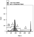
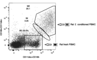
- Example 1 Gene expression profile of M2-rich MNC A scatter plot of rat PBMC or M2-rich MNC is shown. Lymphocyte-sized cells (A), monocyte-sized cells (B), and macrophage-sized cell populations (C) were gated from the overall viable cell fraction, respectively (FIG. 1). Mononuclear cells / macrophages type 1 (M1 macrophages: M1 ⁇ ) exhibiting inflammatory properties were identified as CD68-positive or CD169-negative and CD11b / c co-positive cells in the overall survival cell fraction. Anti-inflammatory and macrophage type 2 (M2 macrophages: M2 ⁇ ) were identified as CD163-positive and CD11b / c co-negative cells in the overall survival cell fraction.
- M1 macrophages M1 ⁇
- M2 macrophages Anti-inflammatory and macrophage type 2 (M2 macrophages: M2 ⁇ ) were identified as CD163-positive and CD11b / c co-negative cells in the overall survival cell fraction.
- PBMCs after E-culture may have macrophages converted from classical M1 ⁇ to M2 ⁇ , and that about 3% of cells are in the intermediate stage between M1 ⁇ and M2 ⁇ .
- CD38 is known as a marker specifically expressed in rat M1 ⁇
- erg2 and arg1 are known as markers specific to rat M2 ⁇ .
- RT-qPCR analysis the expression levels of M2 ⁇ markers arg1 and erg2 were increased in M2-rich MNCs as compared with PBMC, but the expression levels of M1 ⁇ markers CD38 and il1b were decreased.
- M2-rich MNC for example, the expression of ang1, ang2 , and vegfb was significantly increased.
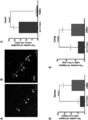
- Example 2 Tracking of M2-rich MNCs at cardiac ischemic sites and other organs PBMCs and M2-rich MNCs were isolated from Lew-CAG-eGFP rats, and 1x10 5 cells were isolated from the tail vein 3 days after the induction of myocardial infarction. It was administered. Four weeks after cell administration, immunohistochemically administered cell tracing experiments were performed. In histological analysis, eGFP-positive cells were observed in the ischemic region and tissues around the ischemic region in the M2-rich MNC-administered group (FIGS. 6A and 6B), but not in the PBMC-administered group (FIG. 6C). Furthermore, many eGFP-positive cells were trapped in the parenchyma spleen and lungs in the PBMC-administered group compared to the M2-rich MNC-administered group (FIGS. 6D and E).
- Example 3 Effect of M2-rich MNC on LV performance and LV reconstruction Rats were weighed before and 28 days after surgery. At 4 weeks post-surgery, the M2-rich MNC-treated group weighed 306.4 ⁇ 5.9 g, the PBMC-treated group (277.6 ⁇ 6.1 g, P ⁇ 0.0001), and the control (283.3 ⁇ ). 8.3 g, increased compared to P ⁇ 0.002). Compared with the Sham-operated group (281.5 ⁇ 7.9 g, P ⁇ 0.01), the body weight of the M2-rich MNC-administered group was increased.
- the EchoCG index including ischemic site volume and left ventricular contractile function (LVSDs) increased to (94.4 ⁇ 3.7 ⁇ l and 0.35 ⁇ 0.03 cm) in the M2-rich MNC-administered group, but in the PBMC-administered group.
- LVSDs left ventricular contractile function
- 63.6 ⁇ 6.07 ⁇ l, P ⁇ 0.004 and 0.49 ⁇ 0.03 cm, P ⁇ 0.0002 in the control group (71.1 ⁇ 2.0 ⁇ l, P ⁇ 0.03 and 0).
- the LV function value deteriorated to .46 ⁇ 0.03 cm, P ⁇ 0.005).
- Example 4 Formation of bilateral "functional bypass” after infarction by M2-rich MNC.
- This is a phenomenon in which arterial blood is bypassed at a site away from the LAD suture, and this bypass is not observed in the PBMC administration group or control group, and it goes to the LV aneurysm and surrounding tissues (thymus and anterior chest wall). A serious adhesion was seen, hindering the heartbeat.
- momentarily frozen slices of cardiac tissue were stained with isolectin B4 and aSMA.
- the staining of blood vessels by aSMA is large in the ischemic site and shows a wide blood vessel inner diameter value, and the ischemic site is a small artery derived from the chest wall (probably the intercostal artery) Was surrounded by the internal thoracic artery).
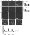
- Example 5 Effect of M2-rich MNC on angiogenesis in rat myocardial infarction
- microvascular density (MVD) was examined by isolectin B4-FITC staining.
- MVD was significant in the M2-rich MNC-administered group compared to the PBMC-administered group (260 ⁇ 35.7 / mm 2 , P ⁇ 0.0001) and the control group (298 ⁇ 45.5 / mm 2, P ⁇ 0.0001). It was high (558 ⁇ 44.4 / mm 2 ).
- the arteriole density per square meter at the ischemic site was calculated.
- the PBMC-administered group (5.714 ⁇ 2.9 / mm 2 , P. It was higher than ⁇ 0.03) and the control group (4.67 ⁇ 2.4 / mm 2 , P ⁇ 0.01) (Fig. 8B).
- the MVD value was higher in the M2-rich MNC-administered group than in the PBMC-administered group and the control group (Fig. 8C).
- Immunohistochemical analysis was performed using Lew-CAG-eGFP transgenic rats to investigate whether the administered M2-rich MNC could differentiate into vascular endothelial cells in the host heart.
- eGFP-labeled M2-rich MNCs expressed CD31, a marker of vascular endothelial cells, and that the M2-rich MNC-treated group also increased MVD per square meter.
- PBMC administration group did not show such a result (Fig. 8D).
- the expression of genes related to angiogenesis such as angpt1 , angpt2 and pecam1 increased 3 times, 9 times and 5 times in the M2-rich MNC-administered group as compared with the control group 3 days after the cell administration, respectively.
- the expression of these genes was 2-fold, 1-fold, and 2-fold lower than that in the control (Fig. 8E).
- Example 6 Effect of M2-rich MNC on the progression of salivary gland pathology (lymphocyte infiltration) in naturally occurring mice with Sjogren's syndrome Using pathological model mice, M2-rich MNC is not trapped in the lungs, etc. It was confirmed that it could exert its medicinal effect by reaching.
- Sjogren's syndrome spontaneously occurring mice Japan SLC, NOD
- This mouse is known to spontaneously develop Sjogren's syndrome-like salivary gland disorders.
- M2-rich MNC mouse was administered intravenously to the tail vein, and pathological analysis of salivary gland tissue was performed at 20 weeks of age when salivary gland secretion disorder occurred.
- Salivary gland tissue was collected from each of 1 cell-administered group (cell-administered group 1) and 2 untreated groups (untreated groups 1 and 2) to which M2-rich MNC was administered, and histopathological observation revealed that "lymphocyte infiltration area".
- cell-administered group 1 and 2 untreated groups untreated groups 1 and 2 to which M2-rich MNC was administered
- histopathological observation revealed that "lymphocyte infiltration area”. was identified.
- continuous sections were created from one salivary gland, and the salivary gland tissue area per section of multiple sections (3 to 6) stained with HE using image analysis software (NIH ImageJ).
- NIH ImageJ image analysis software
- SD standard deviation
- the lymphocyte infiltration area was enlarged by image analysis software at the same magnification so that the areas between the sections could be compared relative to each other.
- the area of salivary gland tissue per section was 97,492 to 139,148 pixels, and there was no significant difference between the cell-administered group and the untreated group.
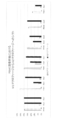
- a remarkable difference was confirmed in the area of the lymphocyte infiltrated part per section between the cell-administered group and the untreated group (Fig. 9). It was confirmed that the area of the lymphocyte infiltrated part was smaller in the cell-administered group than in the untreated group. From the above results, it was confirmed that systemically administered M2-rich MNC exerted an anti-inflammatory effect in the inflamed area without being trapped in the lungs and contributed to the functional regeneration of tissues not only in rats but also in mice. It was.
- Example 7 Enrichment of M2 macrophages by E-culture of human PBMC According to a known method (Non-Patent Document 6) and the above "1. Isolation and culture of PBMC (rat)", a single human peripheral blood-derived PBMC. E-culture of separated and isolated PBMCs was performed. The period of E-culture was 5 to 8 days, and cells were collected on the 5th, 6th, 7th, and 8th days after the start of the culture by experiments (denoted as day5, day6, day7, and day8, respectively). ).
- Mononuclear cells / macrophages type 1 which are positive for CD11b, which is a specific marker for mononuclear cells / macrophages, and exhibit inflammatory properties, are both positive cells for CD11b and CCR2 in the overall survival cell fraction.
- Macrophage type 2 M2 macrophages
- exhibiting anti-inflammatory properties was identified as both CD11b and CD206 positive cells in the overall viable cell fraction. The experiment was carried out 6 times independently. Of these, PBMC was isolated and E-cultured using peripheral blood collected from donor 1, experiment 1, experiment 3, experiment 6 from donor 1, experiment 2 and experiment 4 from donor 2, and experiment 4 from donor 3 (blood collection of PBMC from the same donor). The days are different). The results of the analysis are shown in Table 2 and FIG.
- M2 macrophages were hardly detected in PBMC before culturing, but M1 macrophages were 10-30% of the total PBMC cells, which was higher than that of M2 macrophages. Exists.
- M2 macrophages were generally significantly enriched (7% or more of all monocytes, most of them were 9% or more), and M1 macrophages were significantly reduced (about 5% of all monocytes). Below, most of them are 3% or less). This tendency was more remarkable on the 6th and 7th days than on the 5th day of culturing.
- the pharmaceutical composition of the present invention contains a cell population enriched with M2 macrophages and, when systemically administered, is not trapped in the lungs and accumulates at the site of injury / inflammation.
- the pharmaceutical compositions of the present invention can migrate a therapeutically effective number of cells with anti-inflammatory activity to a targeted inflammation or tissue damage site.
Landscapes
- Health & Medical Sciences (AREA)
- Life Sciences & Earth Sciences (AREA)
- General Health & Medical Sciences (AREA)
- Veterinary Medicine (AREA)
- Animal Behavior & Ethology (AREA)
- Public Health (AREA)
- Immunology (AREA)
- Epidemiology (AREA)
- Chemical & Material Sciences (AREA)
- Engineering & Computer Science (AREA)
- Organic Chemistry (AREA)
- Pharmacology & Pharmacy (AREA)
- Biomedical Technology (AREA)
- Medicinal Chemistry (AREA)
- Bioinformatics & Cheminformatics (AREA)
- Biotechnology (AREA)
- Zoology (AREA)
- Nuclear Medicine, Radiotherapy & Molecular Imaging (AREA)
- General Chemical & Material Sciences (AREA)
- Chemical Kinetics & Catalysis (AREA)
- Hematology (AREA)
- Wood Science & Technology (AREA)
- Genetics & Genomics (AREA)
- Cell Biology (AREA)
- Rheumatology (AREA)
- Pain & Pain Management (AREA)
- General Engineering & Computer Science (AREA)
- Biochemistry (AREA)
- Microbiology (AREA)
- Gastroenterology & Hepatology (AREA)
- Developmental Biology & Embryology (AREA)
- Virology (AREA)
- Medicines Containing Material From Animals Or Micro-Organisms (AREA)
Priority Applications (4)
| Application Number | Priority Date | Filing Date | Title |
|---|---|---|---|
| JP2021541368A JP7500083B2 (ja) | 2019-08-19 | 2019-08-19 | 細胞含有医薬組成物 |
| US17/612,441 US12460182B2 (en) | 2019-08-19 | 2019-08-19 | Cell-containing pharmaceutical composition |
| PCT/JP2019/032322 WO2021033247A1 (ja) | 2019-08-19 | 2019-08-19 | 細胞含有医薬組成物 |
| KR1020217037758A KR102791419B1 (ko) | 2019-08-19 | 2019-08-19 | 세포 함유 의약 조성물 |
Applications Claiming Priority (1)
| Application Number | Priority Date | Filing Date | Title |
|---|---|---|---|
| PCT/JP2019/032322 WO2021033247A1 (ja) | 2019-08-19 | 2019-08-19 | 細胞含有医薬組成物 |
Publications (1)
| Publication Number | Publication Date |
|---|---|
| WO2021033247A1 true WO2021033247A1 (ja) | 2021-02-25 |
Family
ID=74659865
Family Applications (1)
| Application Number | Title | Priority Date | Filing Date |
|---|---|---|---|
| PCT/JP2019/032322 Ceased WO2021033247A1 (ja) | 2019-08-19 | 2019-08-19 | 細胞含有医薬組成物 |
Country Status (4)
| Country | Link |
|---|---|
| US (1) | US12460182B2 (cg-RX-API-DMAC7.html) |
| JP (1) | JP7500083B2 (cg-RX-API-DMAC7.html) |
| KR (1) | KR102791419B1 (cg-RX-API-DMAC7.html) |
| WO (1) | WO2021033247A1 (cg-RX-API-DMAC7.html) |
Citations (1)
| Publication number | Priority date | Publication date | Assignee | Title |
|---|---|---|---|---|
| JP6372782B2 (ja) * | 2012-09-28 | 2018-08-15 | 公益財団法人神戸医療産業都市推進機構 | 虚血性疾患治療に適した細胞を含む細胞群の生体外増幅方法 |
Family Cites Families (5)
| Publication number | Priority date | Publication date | Assignee | Title |
|---|---|---|---|---|
| WO2014027474A1 (ja) | 2012-08-17 | 2014-02-20 | 株式会社Clio | 心筋梗塞の修復再生を誘導する多能性幹細胞 |
| AU2015297347B2 (en) | 2014-08-01 | 2021-05-27 | PuREC Co., Ltd | Method for evaluating quality of human mesenchymal stem cell, and monoclonal antibody for use in said method |
| US20160287638A1 (en) | 2015-04-02 | 2016-10-06 | Stemedica Cell Technologies, Inc. | Ischemic tolerant cells and cellular factors in the treatment of peripheral artery disease |
| EP3297615A1 (en) * | 2015-05-20 | 2018-03-28 | INSERM - Institut National de la Santé et de la Recherche Médicale | Methods and pharmaceutical composition for modulation polarization and activation of macrophages |
| EP3299453B1 (en) * | 2016-09-23 | 2019-05-08 | Xcell Medical Solutions, S.L. | Cell therapy with polarized macrophages for tissue regeneration |
-
2019
- 2019-08-19 US US17/612,441 patent/US12460182B2/en active Active
- 2019-08-19 WO PCT/JP2019/032322 patent/WO2021033247A1/ja not_active Ceased
- 2019-08-19 JP JP2021541368A patent/JP7500083B2/ja active Active
- 2019-08-19 KR KR1020217037758A patent/KR102791419B1/ko active Active
Patent Citations (1)
| Publication number | Priority date | Publication date | Assignee | Title |
|---|---|---|---|---|
| JP6372782B2 (ja) * | 2012-09-28 | 2018-08-15 | 公益財団法人神戸医療産業都市推進機構 | 虚血性疾患治療に適した細胞を含む細胞群の生体外増幅方法 |
Non-Patent Citations (4)
Also Published As
| Publication number | Publication date |
|---|---|
| JP7500083B2 (ja) | 2024-06-17 |
| KR20220003564A (ko) | 2022-01-10 |
| JPWO2021033247A1 (cg-RX-API-DMAC7.html) | 2021-02-25 |
| KR102791419B1 (ko) | 2025-04-08 |
| US20220218744A1 (en) | 2022-07-14 |
| US12460182B2 (en) | 2025-11-04 |
Similar Documents
| Publication | Publication Date | Title |
|---|---|---|
| US20220088084A1 (en) | Uses of mesenchymal stem cells | |
| Yu et al. | Overexpression of CXCR4 in mesenchymal stem cells promotes migration, neuroprotection and angiogenesis in a rat model of stroke | |
| US8747905B2 (en) | Therapeutic use of CD31 expressing cells | |
| Kim et al. | Cultured human bone marrow–derived CD31+ cells are effective for cardiac and vascular repair through enhanced angiogenic, adhesion, and anti-inflammatory effects | |
| US10874698B2 (en) | Therapy using cardiac stem cells and mesenchymal stem cells | |
| TW201130977A (en) | Mesenchymal stem cells (MSCs) isolated from mobilized peripheral blood | |
| CA2705450A1 (en) | Stem cells for treating lung diseases | |
| US6821513B1 (en) | Method for enhancing hematopoiesis | |
| CN114901806B (zh) | 细胞群以及其取得方法 | |
| CN106701668B (zh) | 间质干细胞、其纯株化扩张的方法及其应用 | |
| US20210386825A1 (en) | Osteoarthritis Treatment With Chemokine-Loaded Alginate Microparticles | |
| ES2409128T3 (es) | Procedimientos de mejora de anidamiento e injerto de células madre | |
| Norol et al. | GFP-transduced CD34+ and Lin− CD34− hematopoietic stem cells did not adopt a cardiac phenotype in a nonhuman primate model of myocardial infarct | |
| US12460182B2 (en) | Cell-containing pharmaceutical composition | |
| EP2205251B1 (en) | A method to amplify cardiac stem cells in vitro and in vivo | |
| Mahmud et al. | Growth factors mobilize CXCR4 low/negative primitive hematopoietic stem/progenitor cells from the bone marrow of nonhuman primates | |
| WO2007047963A2 (en) | Use of bone-marrow derived stem cells to treat ischemia | |
| WO2002033043A2 (en) | Method and marker for the isolation of human multipotent hematopoietic stem cells | |
| Efimenko et al. | Viability and angiogenic activity of mesenchymal stromal cells from adipose tissue and bone marrow under hypoxia and inflammation in vitro | |
| AU780468B2 (en) | Method for enhancing hematopoiesis | |
| Delgado et al. | Uses of mesenchymal stem cells | |
| Kanji et al. | Recent Advances in Hematopoietic Stem Cell-Mediated Regeneration | |
| Iwasaki et al. | Cardiomyogenesis for Functional Regenerative Recovery After Myocardial Infarction | |
| Czechowicz | Gaining Access to the Hematopoietic Stem Cell Niche: Novel Non-myeloablative Conditioning Approaches | |
| Milwid | Discovery of novel anti-inflammatory proteins inspired by bone marrow mesenchymal stem cell secretions |
Legal Events
| Date | Code | Title | Description |
|---|---|---|---|
| 121 | Ep: the epo has been informed by wipo that ep was designated in this application |
Ref document number: 19942089 Country of ref document: EP Kind code of ref document: A1 |
|
| ENP | Entry into the national phase |
Ref document number: 20217037758 Country of ref document: KR Kind code of ref document: A |
|
| ENP | Entry into the national phase |
Ref document number: 2021541368 Country of ref document: JP Kind code of ref document: A |
|
| NENP | Non-entry into the national phase |
Ref country code: DE |
|
| 122 | Ep: pct application non-entry in european phase |
Ref document number: 19942089 Country of ref document: EP Kind code of ref document: A1 |
|
| WWG | Wipo information: grant in national office |
Ref document number: 17612441 Country of ref document: US |