WO2020251046A1 - Medicinal composition - Google Patents
Medicinal composition Download PDFInfo
- Publication number
- WO2020251046A1 WO2020251046A1 PCT/JP2020/023308 JP2020023308W WO2020251046A1 WO 2020251046 A1 WO2020251046 A1 WO 2020251046A1 JP 2020023308 W JP2020023308 W JP 2020023308W WO 2020251046 A1 WO2020251046 A1 WO 2020251046A1
- Authority
- WO
- WIPO (PCT)
- Prior art keywords
- cells
- pharmaceutical composition
- cancer
- composition according
- cell
- Prior art date
Links
- 239000000203 mixture Substances 0.000 title claims abstract description 11
- 210000001744 T-lymphocyte Anatomy 0.000 claims abstract description 351
- 239000000427 antigen Substances 0.000 claims abstract description 117
- 108091007433 antigens Proteins 0.000 claims abstract description 116
- 102000036639 antigens Human genes 0.000 claims abstract description 116
- 206010028980 Neoplasm Diseases 0.000 claims abstract description 93
- 201000011510 cancer Diseases 0.000 claims abstract description 66
- 238000000034 method Methods 0.000 claims abstract description 47
- 210000004027 cell Anatomy 0.000 claims description 265
- 239000008194 pharmaceutical composition Substances 0.000 claims description 104
- 229940022399 cancer vaccine Drugs 0.000 claims description 94
- 238000009566 cancer vaccine Methods 0.000 claims description 94
- 102100032530 Glypican-3 Human genes 0.000 claims description 58
- 101001014668 Homo sapiens Glypican-3 Proteins 0.000 claims description 58
- 239000000243 solution Substances 0.000 claims description 30
- 102000004127 Cytokines Human genes 0.000 claims description 29
- 108090000695 Cytokines Proteins 0.000 claims description 29
- 210000001151 cytotoxic T lymphocyte Anatomy 0.000 claims description 28
- 108010074328 Interferon-gamma Proteins 0.000 claims description 15
- 102100037850 Interferon gamma Human genes 0.000 claims description 14
- 108010002350 Interleukin-2 Proteins 0.000 claims description 14
- 102000000588 Interleukin-2 Human genes 0.000 claims description 14
- 210000005259 peripheral blood Anatomy 0.000 claims description 14
- 239000011886 peripheral blood Substances 0.000 claims description 14
- 101000738771 Homo sapiens Receptor-type tyrosine-protein phosphatase C Proteins 0.000 claims description 12
- 102100037422 Receptor-type tyrosine-protein phosphatase C Human genes 0.000 claims description 12
- 108010002586 Interleukin-7 Proteins 0.000 claims description 11
- 101001136981 Homo sapiens Proteasome subunit beta type-9 Proteins 0.000 claims description 9
- 101000814512 Homo sapiens X antigen family member 1 Proteins 0.000 claims description 9
- 102100035764 Proteasome subunit beta type-9 Human genes 0.000 claims description 9
- 102000040856 WT1 Human genes 0.000 claims description 9
- 108700020467 WT1 Proteins 0.000 claims description 9
- 101150084041 WT1 gene Proteins 0.000 claims description 9
- 102100039490 X antigen family member 1 Human genes 0.000 claims description 9
- 230000004083 survival effect Effects 0.000 claims description 9
- 239000007853 buffer solution Substances 0.000 claims description 7
- 230000001939 inductive effect Effects 0.000 claims description 7
- 239000004480 active ingredient Substances 0.000 claims description 6
- 239000000654 additive Substances 0.000 claims description 6
- 230000000996 additive effect Effects 0.000 claims description 6
- 210000002443 helper t lymphocyte Anatomy 0.000 claims description 6
- 230000002265 prevention Effects 0.000 claims description 6
- 208000006359 hepatoblastoma Diseases 0.000 claims description 5
- 108010074108 interleukin-21 Proteins 0.000 claims description 5
- 102000003812 Interleukin-15 Human genes 0.000 claims description 4
- 108090000172 Interleukin-15 Proteins 0.000 claims description 4
- 238000004113 cell culture Methods 0.000 claims description 4
- 239000002504 physiological saline solution Substances 0.000 claims description 4
- 208000010839 B-cell chronic lymphocytic leukemia Diseases 0.000 claims description 3
- 208000028564 B-cell non-Hodgkin lymphoma Diseases 0.000 claims description 3
- 206010006187 Breast cancer Diseases 0.000 claims description 3
- 208000026310 Breast neoplasm Diseases 0.000 claims description 3
- 206010008342 Cervix carcinoma Diseases 0.000 claims description 3
- 108010035532 Collagen Proteins 0.000 claims description 3
- 102000008186 Collagen Human genes 0.000 claims description 3
- 206010009944 Colon cancer Diseases 0.000 claims description 3
- 208000000461 Esophageal Neoplasms Diseases 0.000 claims description 3
- 102100030703 Interleukin-22 Human genes 0.000 claims description 3
- 208000031422 Lymphocytic Chronic B-Cell Leukemia Diseases 0.000 claims description 3
- 206010030155 Oesophageal carcinoma Diseases 0.000 claims description 3
- 206010033128 Ovarian cancer Diseases 0.000 claims description 3
- 206010061535 Ovarian neoplasm Diseases 0.000 claims description 3
- 206010061902 Pancreatic neoplasm Diseases 0.000 claims description 3
- 206010033701 Papillary thyroid cancer Diseases 0.000 claims description 3
- 208000000236 Prostatic Neoplasms Diseases 0.000 claims description 3
- 208000006265 Renal cell carcinoma Diseases 0.000 claims description 3
- 208000005718 Stomach Neoplasms Diseases 0.000 claims description 3
- 208000006105 Uterine Cervical Neoplasms Diseases 0.000 claims description 3
- 230000000735 allogeneic effect Effects 0.000 claims description 3
- 201000010881 cervical cancer Diseases 0.000 claims description 3
- 208000032852 chronic lymphocytic leukemia Diseases 0.000 claims description 3
- 229920001436 collagen Polymers 0.000 claims description 3
- 208000029742 colonic neoplasm Diseases 0.000 claims description 3
- 208000035250 cutaneous malignant susceptibility to 1 melanoma Diseases 0.000 claims description 3
- 201000004101 esophageal cancer Diseases 0.000 claims description 3
- 206010017758 gastric cancer Diseases 0.000 claims description 3
- 206010073071 hepatocellular carcinoma Diseases 0.000 claims description 3
- 231100000844 hepatocellular carcinoma Toxicity 0.000 claims description 3
- 208000015486 malignant pancreatic neoplasm Diseases 0.000 claims description 3
- 201000001441 melanoma Diseases 0.000 claims description 3
- 208000002154 non-small cell lung carcinoma Diseases 0.000 claims description 3
- 201000002528 pancreatic cancer Diseases 0.000 claims description 3
- 208000008443 pancreatic carcinoma Diseases 0.000 claims description 3
- 201000011549 stomach cancer Diseases 0.000 claims description 3
- 208000030045 thyroid gland papillary carcinoma Diseases 0.000 claims description 3
- 208000029729 tumor suppressor gene on chromosome 11 Diseases 0.000 claims description 3
- 239000002246 antineoplastic agent Substances 0.000 claims description 2
- 102000000704 Interleukin-7 Human genes 0.000 claims 1
- 208000016359 neuroblastic tumor Diseases 0.000 claims 1
- 230000002068 genetic effect Effects 0.000 abstract description 35
- 239000001963 growth medium Substances 0.000 description 84
- 108090000765 processed proteins & peptides Proteins 0.000 description 44
- 210000003958 hematopoietic stem cell Anatomy 0.000 description 31
- 239000000047 product Substances 0.000 description 27
- 108091008874 T cell receptors Proteins 0.000 description 26
- 238000012258 culturing Methods 0.000 description 23
- CIWBSHSKHKDKBQ-JLAZNSOCSA-N Ascorbic acid Chemical compound OC[C@H](O)[C@H]1OC(=O)C(O)=C1O CIWBSHSKHKDKBQ-JLAZNSOCSA-N 0.000 description 21
- 102000016266 T-Cell Antigen Receptors Human genes 0.000 description 20
- 239000003550 marker Substances 0.000 description 18
- 108010008951 Chemokine CXCL12 Proteins 0.000 description 17
- 108090000623 proteins and genes Proteins 0.000 description 16
- 210000001519 tissue Anatomy 0.000 description 16
- 238000004519 manufacturing process Methods 0.000 description 15
- 230000001472 cytotoxic effect Effects 0.000 description 14
- 239000002609 medium Substances 0.000 description 13
- 238000012360 testing method Methods 0.000 description 13
- 238000004458 analytical method Methods 0.000 description 12
- 230000008672 reprogramming Effects 0.000 description 12
- 229940088594 vitamin Drugs 0.000 description 12
- 229930003231 vitamin Natural products 0.000 description 12
- 235000013343 vitamin Nutrition 0.000 description 12
- 239000011782 vitamin Substances 0.000 description 12
- 239000012980 RPMI-1640 medium Substances 0.000 description 11
- 102100021669 Stromal cell-derived factor 1 Human genes 0.000 description 11
- 230000000694 effects Effects 0.000 description 11
- 102100021592 Interleukin-7 Human genes 0.000 description 10
- 230000004913 activation Effects 0.000 description 10
- 230000028993 immune response Effects 0.000 description 10
- PHEDXBVPIONUQT-UHFFFAOYSA-N Cocarcinogen A1 Natural products CCCCCCCCCCCCCC(=O)OC1C(C)C2(O)C3C=C(C)C(=O)C3(O)CC(CO)=CC2C2C1(OC(C)=O)C2(C)C PHEDXBVPIONUQT-UHFFFAOYSA-N 0.000 description 9
- -1 Fbx15 Proteins 0.000 description 9
- 150000002500 ions Chemical class 0.000 description 9
- 230000035772 mutation Effects 0.000 description 9
- PHEDXBVPIONUQT-RGYGYFBISA-N phorbol 13-acetate 12-myristate Chemical compound C([C@]1(O)C(=O)C(C)=C[C@H]1[C@@]1(O)[C@H](C)[C@H]2OC(=O)CCCCCCCCCCCCC)C(CO)=C[C@H]1[C@H]1[C@]2(OC(C)=O)C1(C)C PHEDXBVPIONUQT-RGYGYFBISA-N 0.000 description 9
- 238000009256 replacement therapy Methods 0.000 description 9
- 239000006144 Dulbecco’s modified Eagle's medium Substances 0.000 description 8
- 102000036693 Thrombopoietin Human genes 0.000 description 8
- 108010041111 Thrombopoietin Proteins 0.000 description 8
- 239000000872 buffer Substances 0.000 description 8
- NOESYZHRGYRDHS-UHFFFAOYSA-N insulin Chemical compound N1C(=O)C(NC(=O)C(CCC(N)=O)NC(=O)C(CCC(O)=O)NC(=O)C(C(C)C)NC(=O)C(NC(=O)CN)C(C)CC)CSSCC(C(NC(CO)C(=O)NC(CC(C)C)C(=O)NC(CC=2C=CC(O)=CC=2)C(=O)NC(CCC(N)=O)C(=O)NC(CC(C)C)C(=O)NC(CCC(O)=O)C(=O)NC(CC(N)=O)C(=O)NC(CC=2C=CC(O)=CC=2)C(=O)NC(CSSCC(NC(=O)C(C(C)C)NC(=O)C(CC(C)C)NC(=O)C(CC=2C=CC(O)=CC=2)NC(=O)C(CC(C)C)NC(=O)C(C)NC(=O)C(CCC(O)=O)NC(=O)C(C(C)C)NC(=O)C(CC(C)C)NC(=O)C(CC=2NC=NC=2)NC(=O)C(CO)NC(=O)CNC2=O)C(=O)NCC(=O)NC(CCC(O)=O)C(=O)NC(CCCNC(N)=N)C(=O)NCC(=O)NC(CC=3C=CC=CC=3)C(=O)NC(CC=3C=CC=CC=3)C(=O)NC(CC=3C=CC(O)=CC=3)C(=O)NC(C(C)O)C(=O)N3C(CCC3)C(=O)NC(CCCCN)C(=O)NC(C)C(O)=O)C(=O)NC(CC(N)=O)C(O)=O)=O)NC(=O)C(C(C)CC)NC(=O)C(CO)NC(=O)C(C(C)O)NC(=O)C1CSSCC2NC(=O)C(CC(C)C)NC(=O)C(NC(=O)C(CCC(N)=O)NC(=O)C(CC(N)=O)NC(=O)C(NC(=O)C(N)CC=1C=CC=CC=1)C(C)C)CC1=CN=CN1 NOESYZHRGYRDHS-UHFFFAOYSA-N 0.000 description 8
- 102000002574 p38 Mitogen-Activated Protein Kinases Human genes 0.000 description 8
- 108010068338 p38 Mitogen-Activated Protein Kinases Proteins 0.000 description 8
- 150000003722 vitamin derivatives Chemical class 0.000 description 8
- DGVVWUTYPXICAM-UHFFFAOYSA-N β‐Mercaptoethanol Chemical compound OCCS DGVVWUTYPXICAM-UHFFFAOYSA-N 0.000 description 8
- 102000006573 Chemokine CXCL12 Human genes 0.000 description 7
- 150000001413 amino acids Chemical class 0.000 description 7
- 238000005516 engineering process Methods 0.000 description 7
- 210000004698 lymphocyte Anatomy 0.000 description 7
- 238000000926 separation method Methods 0.000 description 7
- 210000002966 serum Anatomy 0.000 description 7
- 230000000638 stimulation Effects 0.000 description 7
- 238000004114 suspension culture Methods 0.000 description 7
- IAZDPXIOMUYVGZ-UHFFFAOYSA-N Dimethylsulphoxide Chemical compound CS(C)=O IAZDPXIOMUYVGZ-UHFFFAOYSA-N 0.000 description 6
- ZDXPYRJPNDTMRX-VKHMYHEASA-N L-glutamine Chemical compound OC(=O)[C@@H](N)CCC(N)=O ZDXPYRJPNDTMRX-VKHMYHEASA-N 0.000 description 6
- 229930182816 L-glutamine Natural products 0.000 description 6
- 108700018351 Major Histocompatibility Complex Proteins 0.000 description 6
- 230000005856 abnormality Effects 0.000 description 6
- 230000003321 amplification Effects 0.000 description 6
- 235000010323 ascorbic acid Nutrition 0.000 description 6
- 239000011668 ascorbic acid Substances 0.000 description 6
- 229960005070 ascorbic acid Drugs 0.000 description 6
- 239000011324 bead Substances 0.000 description 6
- 210000002242 embryoid body Anatomy 0.000 description 6
- 239000012530 fluid Substances 0.000 description 6
- 238000007710 freezing Methods 0.000 description 6
- 230000008014 freezing Effects 0.000 description 6
- 210000005087 mononuclear cell Anatomy 0.000 description 6
- 238000003199 nucleic acid amplification method Methods 0.000 description 6
- 235000018102 proteins Nutrition 0.000 description 6
- 102000004169 proteins and genes Human genes 0.000 description 6
- 238000010186 staining Methods 0.000 description 6
- 239000000126 substance Substances 0.000 description 6
- 230000020382 suppression by virus of host antigen processing and presentation of peptide antigen via MHC class I Effects 0.000 description 6
- 102000003974 Fibroblast growth factor 2 Human genes 0.000 description 5
- 108090000379 Fibroblast growth factor 2 Proteins 0.000 description 5
- 102000043129 MHC class I family Human genes 0.000 description 5
- 108091054437 MHC class I family Proteins 0.000 description 5
- 102000043131 MHC class II family Human genes 0.000 description 5
- 108091054438 MHC class II family Proteins 0.000 description 5
- 239000012826 P38 inhibitor Substances 0.000 description 5
- 241000700605 Viruses Species 0.000 description 5
- 239000000853 adhesive Substances 0.000 description 5
- 230000001070 adhesive effect Effects 0.000 description 5
- 235000001014 amino acid Nutrition 0.000 description 5
- 210000000612 antigen-presenting cell Anatomy 0.000 description 5
- 230000027455 binding Effects 0.000 description 5
- 239000012228 culture supernatant Substances 0.000 description 5
- 231100000433 cytotoxic Toxicity 0.000 description 5
- 210000002861 immature t-cell Anatomy 0.000 description 5
- 239000007788 liquid Substances 0.000 description 5
- 208000020816 lung neoplasm Diseases 0.000 description 5
- 239000006166 lysate Substances 0.000 description 5
- 229910052760 oxygen Inorganic materials 0.000 description 5
- 210000003819 peripheral blood mononuclear cell Anatomy 0.000 description 5
- 108010049955 Bone Morphogenetic Protein 4 Proteins 0.000 description 4
- 102100024505 Bone morphogenetic protein 4 Human genes 0.000 description 4
- 108060005980 Collagenase Proteins 0.000 description 4
- 102000029816 Collagenase Human genes 0.000 description 4
- 239000006145 Eagle's minimal essential medium Substances 0.000 description 4
- 102100020715 Fms-related tyrosine kinase 3 ligand protein Human genes 0.000 description 4
- 101710162577 Fms-related tyrosine kinase 3 ligand protein Proteins 0.000 description 4
- 206010064571 Gene mutation Diseases 0.000 description 4
- 102000004877 Insulin Human genes 0.000 description 4
- 108090001061 Insulin Proteins 0.000 description 4
- 102000015696 Interleukins Human genes 0.000 description 4
- 108010063738 Interleukins Proteins 0.000 description 4
- 206010058467 Lung neoplasm malignant Diseases 0.000 description 4
- 108091092878 Microsatellite Proteins 0.000 description 4
- 241000711408 Murine respirovirus Species 0.000 description 4
- BUGBHKTXTAQXES-UHFFFAOYSA-N Selenium Chemical compound [Se] BUGBHKTXTAQXES-UHFFFAOYSA-N 0.000 description 4
- 108010073929 Vascular Endothelial Growth Factor A Proteins 0.000 description 4
- 102000005789 Vascular Endothelial Growth Factors Human genes 0.000 description 4
- 108010019530 Vascular Endothelial Growth Factors Proteins 0.000 description 4
- 210000004102 animal cell Anatomy 0.000 description 4
- 239000003242 anti bacterial agent Substances 0.000 description 4
- 229940088710 antibiotic agent Drugs 0.000 description 4
- QVGXLLKOCUKJST-UHFFFAOYSA-N atomic oxygen Chemical compound [O] QVGXLLKOCUKJST-UHFFFAOYSA-N 0.000 description 4
- 210000000601 blood cell Anatomy 0.000 description 4
- 230000024245 cell differentiation Effects 0.000 description 4
- 210000000170 cell membrane Anatomy 0.000 description 4
- 238000010367 cloning Methods 0.000 description 4
- 229960002424 collagenase Drugs 0.000 description 4
- 230000021615 conjugation Effects 0.000 description 4
- 238000005138 cryopreservation Methods 0.000 description 4
- 210000004748 cultured cell Anatomy 0.000 description 4
- 230000003247 decreasing effect Effects 0.000 description 4
- 239000007857 degradation product Substances 0.000 description 4
- 230000004069 differentiation Effects 0.000 description 4
- 201000010099 disease Diseases 0.000 description 4
- 208000037265 diseases, disorders, signs and symptoms Diseases 0.000 description 4
- 239000012091 fetal bovine serum Substances 0.000 description 4
- 238000000684 flow cytometry Methods 0.000 description 4
- 229940125396 insulin Drugs 0.000 description 4
- 201000005202 lung cancer Diseases 0.000 description 4
- 239000001301 oxygen Substances 0.000 description 4
- 239000002994 raw material Substances 0.000 description 4
- 229910052711 selenium Inorganic materials 0.000 description 4
- 239000011669 selenium Substances 0.000 description 4
- 235000011649 selenium Nutrition 0.000 description 4
- 229940091258 selenium supplement Drugs 0.000 description 4
- UCSJYZPVAKXKNQ-HZYVHMACSA-N streptomycin Chemical compound CN[C@H]1[C@H](O)[C@@H](O)[C@H](CO)O[C@H]1O[C@@H]1[C@](C=O)(O)[C@H](C)O[C@H]1O[C@@H]1[C@@H](NC(N)=N)[C@H](O)[C@@H](NC(N)=N)[C@H](O)[C@H]1O UCSJYZPVAKXKNQ-HZYVHMACSA-N 0.000 description 4
- 230000001225 therapeutic effect Effects 0.000 description 4
- 108010088751 Albumins Proteins 0.000 description 3
- 102000009027 Albumins Human genes 0.000 description 3
- 241000283690 Bos taurus Species 0.000 description 3
- ZZZCUOFIHGPKAK-UHFFFAOYSA-N D-erythro-ascorbic acid Natural products OCC1OC(=O)C(O)=C1O ZZZCUOFIHGPKAK-UHFFFAOYSA-N 0.000 description 3
- 108020004414 DNA Proteins 0.000 description 3
- 102100031573 Hematopoietic progenitor cell antigen CD34 Human genes 0.000 description 3
- 241000282412 Homo Species 0.000 description 3
- 101000777663 Homo sapiens Hematopoietic progenitor cell antigen CD34 Proteins 0.000 description 3
- 101000608935 Homo sapiens Leukosialin Proteins 0.000 description 3
- 101000662009 Homo sapiens UDP-N-acetylglucosamine pyrophosphorylase Proteins 0.000 description 3
- 102100039564 Leukosialin Human genes 0.000 description 3
- 108091005804 Peptidases Proteins 0.000 description 3
- 239000004365 Protease Substances 0.000 description 3
- LCTONWCANYUPML-UHFFFAOYSA-M Pyruvate Chemical compound CC(=O)C([O-])=O LCTONWCANYUPML-UHFFFAOYSA-M 0.000 description 3
- 102100037486 Reverse transcriptase/ribonuclease H Human genes 0.000 description 3
- 102000004338 Transferrin Human genes 0.000 description 3
- 108090000901 Transferrin Proteins 0.000 description 3
- 102100037921 UDP-N-acetylglucosamine pyrophosphorylase Human genes 0.000 description 3
- 229930003268 Vitamin C Natural products 0.000 description 3
- 108010076089 accutase Proteins 0.000 description 3
- 239000003470 adrenal cortex hormone Substances 0.000 description 3
- 239000003963 antioxidant agent Substances 0.000 description 3
- 235000006708 antioxidants Nutrition 0.000 description 3
- 230000001580 bacterial effect Effects 0.000 description 3
- 239000003153 chemical reaction reagent Substances 0.000 description 3
- 239000011248 coating agent Substances 0.000 description 3
- 230000003013 cytotoxicity Effects 0.000 description 3
- 231100000135 cytotoxicity Toxicity 0.000 description 3
- UREBDLICKHMUKA-CXSFZGCWSA-N dexamethasone Chemical compound C1CC2=CC(=O)C=C[C@]2(C)[C@]2(F)[C@@H]1[C@@H]1C[C@@H](C)[C@@](C(=O)CO)(O)[C@@]1(C)C[C@@H]2O UREBDLICKHMUKA-CXSFZGCWSA-N 0.000 description 3
- 235000014113 dietary fatty acids Nutrition 0.000 description 3
- 239000003797 essential amino acid Substances 0.000 description 3
- 235000020776 essential amino acid Nutrition 0.000 description 3
- 210000003722 extracellular fluid Anatomy 0.000 description 3
- 229930195729 fatty acid Natural products 0.000 description 3
- 239000000194 fatty acid Substances 0.000 description 3
- 150000004665 fatty acids Chemical class 0.000 description 3
- 239000003102 growth factor Substances 0.000 description 3
- 238000000338 in vitro Methods 0.000 description 3
- 238000001727 in vivo Methods 0.000 description 3
- 210000004263 induced pluripotent stem cell Anatomy 0.000 description 3
- 239000003112 inhibitor Substances 0.000 description 3
- 238000002347 injection Methods 0.000 description 3
- 239000007924 injection Substances 0.000 description 3
- 150000002632 lipids Chemical class 0.000 description 3
- 238000012544 monitoring process Methods 0.000 description 3
- 229940023041 peptide vaccine Drugs 0.000 description 3
- 102000004196 processed proteins & peptides Human genes 0.000 description 3
- 230000001012 protector Effects 0.000 description 3
- 150000003839 salts Chemical class 0.000 description 3
- 210000000130 stem cell Anatomy 0.000 description 3
- 238000010257 thawing Methods 0.000 description 3
- 239000011573 trace mineral Substances 0.000 description 3
- 235000013619 trace mineral Nutrition 0.000 description 3
- 239000012581 transferrin Substances 0.000 description 3
- 229960005486 vaccine Drugs 0.000 description 3
- 235000019154 vitamin C Nutrition 0.000 description 3
- 239000011718 vitamin C Substances 0.000 description 3
- 238000005406 washing Methods 0.000 description 3
- VUDQSRFCCHQIIU-UHFFFAOYSA-N 1-(3,5-dichloro-2,6-dihydroxy-4-methoxyphenyl)hexan-1-one Chemical compound CCCCCC(=O)C1=C(O)C(Cl)=C(OC)C(Cl)=C1O VUDQSRFCCHQIIU-UHFFFAOYSA-N 0.000 description 2
- JBNWDYGOTHQHOZ-UHFFFAOYSA-N 2-[5-[4-[(4-fluorophenyl)methyl]piperidine-1-carbonyl]-6-methoxy-1-methylindol-3-yl]-n,n-dimethyl-2-oxoacetamide Chemical compound COC1=CC=2N(C)C=C(C(=O)C(=O)N(C)C)C=2C=C1C(=O)N(CC1)CCC1CC1=CC=C(F)C=C1 JBNWDYGOTHQHOZ-UHFFFAOYSA-N 0.000 description 2
- PMYDPQQPEAYXKD-UHFFFAOYSA-N 3-hydroxy-n-naphthalen-2-ylnaphthalene-2-carboxamide Chemical compound C1=CC=CC2=CC(NC(=O)C3=CC4=CC=CC=C4C=C3O)=CC=C21 PMYDPQQPEAYXKD-UHFFFAOYSA-N 0.000 description 2
- IJGRMHOSHXDMSA-UHFFFAOYSA-N Atomic nitrogen Chemical compound N#N IJGRMHOSHXDMSA-UHFFFAOYSA-N 0.000 description 2
- 241000894006 Bacteria Species 0.000 description 2
- 108091003079 Bovine Serum Albumin Proteins 0.000 description 2
- 108010029697 CD40 Ligand Proteins 0.000 description 2
- 102100032937 CD40 ligand Human genes 0.000 description 2
- 102000019034 Chemokines Human genes 0.000 description 2
- 108010012236 Chemokines Proteins 0.000 description 2
- 108010019670 Chimeric Antigen Receptors Proteins 0.000 description 2
- 101000976075 Homo sapiens Insulin Proteins 0.000 description 2
- 101000942967 Homo sapiens Leukemia inhibitory factor Proteins 0.000 description 2
- 101000946889 Homo sapiens Monocyte differentiation antigen CD14 Proteins 0.000 description 2
- 108010050904 Interferons Proteins 0.000 description 2
- 102000014150 Interferons Human genes 0.000 description 2
- 108700021430 Kruppel-Like Factor 4 Proteins 0.000 description 2
- 239000002211 L-ascorbic acid Substances 0.000 description 2
- 235000000069 L-ascorbic acid Nutrition 0.000 description 2
- 150000000996 L-ascorbic acids Chemical class 0.000 description 2
- 102100032352 Leukemia inhibitory factor Human genes 0.000 description 2
- 241000124008 Mammalia Species 0.000 description 2
- 229930191564 Monensin Natural products 0.000 description 2
- GAOZTHIDHYLHMS-UHFFFAOYSA-N Monensin A Natural products O1C(CC)(C2C(CC(O2)C2C(CC(C)C(O)(CO)O2)C)C)CCC1C(O1)(C)CCC21CC(O)C(C)C(C(C)C(OC)C(C)C(O)=O)O2 GAOZTHIDHYLHMS-UHFFFAOYSA-N 0.000 description 2
- 102100035877 Monocyte differentiation antigen CD14 Human genes 0.000 description 2
- 241000699666 Mus <mouse, genus> Species 0.000 description 2
- 101710135898 Myc proto-oncogene protein Proteins 0.000 description 2
- 102100038895 Myc proto-oncogene protein Human genes 0.000 description 2
- 229930182555 Penicillin Natural products 0.000 description 2
- JGSARLDLIJGVTE-MBNYWOFBSA-N Penicillin G Chemical compound N([C@H]1[C@H]2SC([C@@H](N2C1=O)C(O)=O)(C)C)C(=O)CC1=CC=CC=C1 JGSARLDLIJGVTE-MBNYWOFBSA-N 0.000 description 2
- NBIIXXVUZAFLBC-UHFFFAOYSA-N Phosphoric acid Chemical compound OP(O)(O)=O NBIIXXVUZAFLBC-UHFFFAOYSA-N 0.000 description 2
- 206010060862 Prostate cancer Diseases 0.000 description 2
- 101100247004 Rattus norvegicus Qsox1 gene Proteins 0.000 description 2
- 101150086694 SLC22A3 gene Proteins 0.000 description 2
- 230000006044 T cell activation Effects 0.000 description 2
- 108700042077 T-Cell Receptor beta Genes Proteins 0.000 description 2
- 101710150448 Transcriptional regulator Myc Proteins 0.000 description 2
- 102000004357 Transferases Human genes 0.000 description 2
- 108090000992 Transferases Proteins 0.000 description 2
- 102000004887 Transforming Growth Factor beta Human genes 0.000 description 2
- 108090001012 Transforming Growth Factor beta Proteins 0.000 description 2
- GLNADSQYFUSGOU-GPTZEZBUSA-J Trypan blue Chemical compound [Na+].[Na+].[Na+].[Na+].C1=C(S([O-])(=O)=O)C=C2C=C(S([O-])(=O)=O)C(/N=N/C3=CC=C(C=C3C)C=3C=C(C(=CC=3)\N=N\C=3C(=CC4=CC(=CC(N)=C4C=3O)S([O-])(=O)=O)S([O-])(=O)=O)C)=C(O)C2=C1N GLNADSQYFUSGOU-GPTZEZBUSA-J 0.000 description 2
- MIJPAVRNWPDMOR-UHFFFAOYSA-N [2-(1,2-dihydroxyethyl)-3-hydroxy-5-oxo-2h-furan-4-yl] dihydrogen phosphate Chemical compound OCC(O)C1OC(=O)C(OP(O)(O)=O)=C1O MIJPAVRNWPDMOR-UHFFFAOYSA-N 0.000 description 2
- 230000002159 abnormal effect Effects 0.000 description 2
- 239000002671 adjuvant Substances 0.000 description 2
- 238000011316 allogeneic transplantation Methods 0.000 description 2
- 125000000539 amino acid group Chemical group 0.000 description 2
- 230000000259 anti-tumor effect Effects 0.000 description 2
- 229940072107 ascorbate Drugs 0.000 description 2
- 210000003719 b-lymphocyte Anatomy 0.000 description 2
- 230000015572 biosynthetic process Effects 0.000 description 2
- 230000037396 body weight Effects 0.000 description 2
- 230000005779 cell damage Effects 0.000 description 2
- 239000013000 chemical inhibitor Substances 0.000 description 2
- 238000006243 chemical reaction Methods 0.000 description 2
- 238000000576 coating method Methods 0.000 description 2
- 239000012141 concentrate Substances 0.000 description 2
- 230000007423 decrease Effects 0.000 description 2
- 210000004443 dendritic cell Anatomy 0.000 description 2
- 238000001514 detection method Methods 0.000 description 2
- 229960003957 dexamethasone Drugs 0.000 description 2
- 230000004064 dysfunction Effects 0.000 description 2
- 230000001605 fetal effect Effects 0.000 description 2
- 230000006870 function Effects 0.000 description 2
- 210000001654 germ layer Anatomy 0.000 description 2
- IPCSVZSSVZVIGE-UHFFFAOYSA-N hexadecanoic acid Chemical compound CCCCCCCCCCCCCCCC(O)=O IPCSVZSSVZVIGE-UHFFFAOYSA-N 0.000 description 2
- JYGXADMDTFJGBT-VWUMJDOOSA-N hydrocortisone Chemical compound O=C1CC[C@]2(C)[C@H]3[C@@H](O)C[C@](C)([C@@](CC4)(O)C(=O)CO)[C@@H]4[C@@H]3CCC2=C1 JYGXADMDTFJGBT-VWUMJDOOSA-N 0.000 description 2
- 230000006698 induction Effects 0.000 description 2
- PBGKTOXHQIOBKM-FHFVDXKLSA-N insulin (human) Chemical compound C([C@@H](C(=O)N[C@@H](CC(C)C)C(=O)N[C@H]1CSSC[C@H]2C(=O)N[C@H](C(=O)N[C@@H](CO)C(=O)N[C@H](C(=O)N[C@H](C(N[C@@H](CO)C(=O)N[C@@H](CC(C)C)C(=O)N[C@@H](CC=3C=CC(O)=CC=3)C(=O)N[C@@H](CCC(N)=O)C(=O)N[C@@H](CC(C)C)C(=O)N[C@@H](CCC(O)=O)C(=O)N[C@@H](CC(N)=O)C(=O)N[C@@H](CC=3C=CC(O)=CC=3)C(=O)N[C@@H](CSSC[C@H](NC(=O)[C@H](C(C)C)NC(=O)[C@H](CC(C)C)NC(=O)[C@H](CC=3C=CC(O)=CC=3)NC(=O)[C@H](CC(C)C)NC(=O)[C@H](C)NC(=O)[C@H](CCC(O)=O)NC(=O)[C@H](C(C)C)NC(=O)[C@H](CC(C)C)NC(=O)[C@H](CC=3NC=NC=3)NC(=O)[C@H](CO)NC(=O)CNC1=O)C(=O)NCC(=O)N[C@@H](CCC(O)=O)C(=O)N[C@@H](CCCNC(N)=N)C(=O)NCC(=O)N[C@@H](CC=1C=CC=CC=1)C(=O)N[C@@H](CC=1C=CC=CC=1)C(=O)N[C@@H](CC=1C=CC(O)=CC=1)C(=O)N[C@@H]([C@@H](C)O)C(=O)N1[C@@H](CCC1)C(=O)N[C@@H](CCCCN)C(=O)N[C@@H]([C@@H](C)O)C(O)=O)C(=O)N[C@@H](CC(N)=O)C(O)=O)=O)CSSC[C@@H](C(N2)=O)NC(=O)[C@H](CCC(N)=O)NC(=O)[C@H](CCC(O)=O)NC(=O)[C@H](C(C)C)NC(=O)[C@@H](NC(=O)CN)[C@@H](C)CC)[C@@H](C)CC)[C@@H](C)O)NC(=O)[C@H](CCC(N)=O)NC(=O)[C@H](CC(N)=O)NC(=O)[C@@H](NC(=O)[C@@H](N)CC=1C=CC=CC=1)C(C)C)C1=CN=CN1 PBGKTOXHQIOBKM-FHFVDXKLSA-N 0.000 description 2
- 229940079322 interferon Drugs 0.000 description 2
- PGHMRUGBZOYCAA-UHFFFAOYSA-N ionomycin Natural products O1C(CC(O)C(C)C(O)C(C)C=CCC(C)CC(C)C(O)=CC(=O)C(C)CC(C)CC(CCC(O)=O)C)CCC1(C)C1OC(C)(C(C)O)CC1 PGHMRUGBZOYCAA-UHFFFAOYSA-N 0.000 description 2
- PGHMRUGBZOYCAA-ADZNBVRBSA-N ionomycin Chemical compound O1[C@H](C[C@H](O)[C@H](C)[C@H](O)[C@H](C)/C=C/C[C@@H](C)C[C@@H](C)C(/O)=C/C(=O)[C@@H](C)C[C@@H](C)C[C@@H](CCC(O)=O)C)CC[C@@]1(C)[C@@H]1O[C@](C)([C@@H](C)O)CC1 PGHMRUGBZOYCAA-ADZNBVRBSA-N 0.000 description 2
- 238000002955 isolation Methods 0.000 description 2
- 230000035800 maturation Effects 0.000 description 2
- 229960005358 monensin Drugs 0.000 description 2
- GAOZTHIDHYLHMS-KEOBGNEYSA-N monensin A Chemical compound C([C@@](O1)(C)[C@H]2CC[C@@](O2)(CC)[C@H]2[C@H](C[C@@H](O2)[C@@H]2[C@H](C[C@@H](C)[C@](O)(CO)O2)C)C)C[C@@]21C[C@H](O)[C@@H](C)[C@@H]([C@@H](C)[C@@H](OC)[C@H](C)C(O)=O)O2 GAOZTHIDHYLHMS-KEOBGNEYSA-N 0.000 description 2
- 244000052769 pathogen Species 0.000 description 2
- 230000001575 pathological effect Effects 0.000 description 2
- 229940049954 penicillin Drugs 0.000 description 2
- 230000008823 permeabilization Effects 0.000 description 2
- 239000000049 pigment Substances 0.000 description 2
- 210000001778 pluripotent stem cell Anatomy 0.000 description 2
- 229920002338 polyhydroxyethylmethacrylate Polymers 0.000 description 2
- 238000002360 preparation method Methods 0.000 description 2
- 239000003755 preservative agent Substances 0.000 description 2
- 238000005215 recombination Methods 0.000 description 2
- 230000006798 recombination Effects 0.000 description 2
- 230000001172 regenerating effect Effects 0.000 description 2
- 230000000717 retained effect Effects 0.000 description 2
- 108010056030 retronectin Proteins 0.000 description 2
- 150000003384 small molecules Chemical class 0.000 description 2
- 229960001881 sodium selenate Drugs 0.000 description 2
- 235000018716 sodium selenate Nutrition 0.000 description 2
- 239000011655 sodium selenate Substances 0.000 description 2
- 238000003860 storage Methods 0.000 description 2
- 229960005322 streptomycin Drugs 0.000 description 2
- 230000008685 targeting Effects 0.000 description 2
- 230000037455 tumor specific immune response Effects 0.000 description 2
- RQVKVJIRFKVPBF-VWLOTQADSA-N 2-[[(2s)-2-amino-3-phenylpropyl]amino]-3-methyl-5-naphthalen-2-yl-6-pyridin-4-ylpyrimidin-4-one Chemical compound C([C@H](N)CNC=1N(C(C(C=2C=C3C=CC=CC3=CC=2)=C(C=2C=CN=CC=2)N=1)=O)C)C1=CC=CC=C1 RQVKVJIRFKVPBF-VWLOTQADSA-N 0.000 description 1
- UCWMQLDNYHPJJE-UHFFFAOYSA-N 4-[1-(4-fluorophenyl)-5-pyridin-4-ylimidazol-2-yl]phenol Chemical compound FC1=CC=C(C=C1)N1C(=NC=C1C1=CC=NC=C1)C1=CC=C(C=C1)O UCWMQLDNYHPJJE-UHFFFAOYSA-N 0.000 description 1
- XEOVWJYINDYNSM-UHFFFAOYSA-N 4-[4-(4-fluorophenyl)-2-(4-methylsulfonylphenyl)-1H-imidazol-5-yl]pyridine Chemical compound C1=CC(S(=O)(=O)C)=CC=C1C1=NC(C=2C=CC(F)=CC=2)=C(C=2C=CN=CC=2)N1 XEOVWJYINDYNSM-UHFFFAOYSA-N 0.000 description 1
- ZQUSFAUAYSEREK-UHFFFAOYSA-N 4-[4-(4-fluorophenyl)-5-(2-methoxy-4-pyrimidinyl)-1-imidazolyl]-1-cyclohexanol Chemical compound COC1=NC=CC(C=2N(C=NC=2C=2C=CC(F)=CC=2)C2CCC(O)CC2)=N1 ZQUSFAUAYSEREK-UHFFFAOYSA-N 0.000 description 1
- 102000006306 Antigen Receptors Human genes 0.000 description 1
- 108010083359 Antigen Receptors Proteins 0.000 description 1
- LITUBCVUXPBCGA-WMZHIEFXSA-N Ascorbyl stearate Chemical compound CCCCCCCCCCCCCCCCCC(=O)OC[C@H](O)[C@H]1OC(=O)C(O)=C1O LITUBCVUXPBCGA-WMZHIEFXSA-N 0.000 description 1
- 239000004261 Ascorbyl stearate Substances 0.000 description 1
- 108060000903 Beta-catenin Proteins 0.000 description 1
- 102000015735 Beta-catenin Human genes 0.000 description 1
- 229920002498 Beta-glucan Polymers 0.000 description 1
- 101150013553 CD40 gene Proteins 0.000 description 1
- 108091016585 CD44 antigen Proteins 0.000 description 1
- 101100257372 Caenorhabditis elegans sox-3 gene Proteins 0.000 description 1
- 241000282693 Cercopithecidae Species 0.000 description 1
- 208000035473 Communicable disease Diseases 0.000 description 1
- ITRJWOMZKQRYTA-RFZYENFJSA-N Cortisone acetate Chemical compound C1CC2=CC(=O)CC[C@]2(C)[C@@H]2[C@@H]1[C@@H]1CC[C@@](C(=O)COC(=O)C)(O)[C@@]1(C)CC2=O ITRJWOMZKQRYTA-RFZYENFJSA-N 0.000 description 1
- 230000004568 DNA-binding Effects 0.000 description 1
- 241000283073 Equus caballus Species 0.000 description 1
- 101150099612 Esrrb gene Proteins 0.000 description 1
- 229910052693 Europium Inorganic materials 0.000 description 1
- 108010037362 Extracellular Matrix Proteins Proteins 0.000 description 1
- 102000010834 Extracellular Matrix Proteins Human genes 0.000 description 1
- 241000282326 Felis catus Species 0.000 description 1
- 108010010803 Gelatin Proteins 0.000 description 1
- 108060005986 Granzyme Proteins 0.000 description 1
- 102000001398 Granzyme Human genes 0.000 description 1
- 208000031886 HIV Infections Diseases 0.000 description 1
- 102000008055 Heparan Sulfate Proteoglycans Human genes 0.000 description 1
- 229920002971 Heparan sulfate Polymers 0.000 description 1
- 241000598436 Human T-cell lymphotropic virus Species 0.000 description 1
- 241000713772 Human immunodeficiency virus 1 Species 0.000 description 1
- 241000713340 Human immunodeficiency virus 2 Species 0.000 description 1
- DGAQECJNVWCQMB-PUAWFVPOSA-M Ilexoside XXIX Chemical compound C[C@@H]1CC[C@@]2(CC[C@@]3(C(=CC[C@H]4[C@]3(CC[C@@H]5[C@@]4(CC[C@@H](C5(C)C)OS(=O)(=O)[O-])C)C)[C@@H]2[C@]1(C)O)C)C(=O)O[C@H]6[C@@H]([C@H]([C@@H]([C@H](O6)CO)O)O)O.[Na+] DGAQECJNVWCQMB-PUAWFVPOSA-M 0.000 description 1
- 108060003951 Immunoglobulin Proteins 0.000 description 1
- 102000008070 Interferon-gamma Human genes 0.000 description 1
- 101150072501 Klf2 gene Proteins 0.000 description 1
- QNAYBMKLOCPYGJ-REOHCLBHSA-N L-alanine Chemical compound C[C@H](N)C(O)=O QNAYBMKLOCPYGJ-REOHCLBHSA-N 0.000 description 1
- COLNVLDHVKWLRT-QMMMGPOBSA-N L-phenylalanine Chemical compound OC(=O)[C@@H](N)CC1=CC=CC=C1 COLNVLDHVKWLRT-QMMMGPOBSA-N 0.000 description 1
- 108010085895 Laminin Proteins 0.000 description 1
- FYYHWMGAXLPEAU-UHFFFAOYSA-N Magnesium Chemical compound [Mg] FYYHWMGAXLPEAU-UHFFFAOYSA-N 0.000 description 1
- FQISKWAFAHGMGT-SGJOWKDISA-M Methylprednisolone sodium succinate Chemical compound [Na+].C([C@@]12C)=CC(=O)C=C1[C@@H](C)C[C@@H]1[C@@H]2[C@@H](O)C[C@]2(C)[C@@](O)(C(=O)COC(=O)CCC([O-])=O)CC[C@H]21 FQISKWAFAHGMGT-SGJOWKDISA-M 0.000 description 1
- 101100310657 Mus musculus Sox1 gene Proteins 0.000 description 1
- 101100310648 Mus musculus Sox17 gene Proteins 0.000 description 1
- 101100257376 Mus musculus Sox3 gene Proteins 0.000 description 1
- 241000699670 Mus sp. Species 0.000 description 1
- 241000204031 Mycoplasma Species 0.000 description 1
- 206010028470 Mycoplasma infections Diseases 0.000 description 1
- 108700026495 N-Myc Proto-Oncogene Proteins 0.000 description 1
- 102100030124 N-myc proto-oncogene protein Human genes 0.000 description 1
- 101150072008 NR5A2 gene Proteins 0.000 description 1
- 206010029260 Neuroblastoma Diseases 0.000 description 1
- 102100037369 Nidogen-1 Human genes 0.000 description 1
- 241000283973 Oryctolagus cuniculus Species 0.000 description 1
- 229910019142 PO4 Inorganic materials 0.000 description 1
- 235000021314 Palmitic acid Nutrition 0.000 description 1
- 241001494479 Pecora Species 0.000 description 1
- 241000009328 Perro Species 0.000 description 1
- XBDQKXXYIPTUBI-UHFFFAOYSA-M Propionate Chemical compound CCC([O-])=O XBDQKXXYIPTUBI-UHFFFAOYSA-M 0.000 description 1
- 108010029485 Protein Isoforms Proteins 0.000 description 1
- 102000001708 Protein Isoforms Human genes 0.000 description 1
- 229940022005 RNA vaccine Drugs 0.000 description 1
- 238000011529 RT qPCR Methods 0.000 description 1
- 241000700159 Rattus Species 0.000 description 1
- ZQUSFAUAYSEREK-WKILWMFISA-N SB-239063 Chemical compound COC1=NC=CC(C=2N(C=NC=2C=2C=CC(F)=CC=2)[C@@H]2CC[C@@H](O)CC2)=N1 ZQUSFAUAYSEREK-WKILWMFISA-N 0.000 description 1
- 206010070834 Sensitisation Diseases 0.000 description 1
- 238000012300 Sequence Analysis Methods 0.000 description 1
- 108010071390 Serum Albumin Proteins 0.000 description 1
- 102000007562 Serum Albumin Human genes 0.000 description 1
- FAPWRFPIFSIZLT-UHFFFAOYSA-M Sodium chloride Chemical compound [Na+].[Cl-] FAPWRFPIFSIZLT-UHFFFAOYSA-M 0.000 description 1
- 101150001847 Sox15 gene Proteins 0.000 description 1
- 101710088580 Stromal cell-derived factor 1 Proteins 0.000 description 1
- 241000282898 Sus scrofa Species 0.000 description 1
- 108090000054 Syndecan-2 Proteins 0.000 description 1
- 102000043168 TGF-beta family Human genes 0.000 description 1
- 108091085018 TGF-beta family Proteins 0.000 description 1
- 101150111019 Tbx3 gene Proteins 0.000 description 1
- AYFVYJQAPQTCCC-UHFFFAOYSA-N Threonine Natural products CC(O)C(N)C(O)=O AYFVYJQAPQTCCC-UHFFFAOYSA-N 0.000 description 1
- 239000004473 Threonine Substances 0.000 description 1
- 102100040245 Tumor necrosis factor receptor superfamily member 5 Human genes 0.000 description 1
- 108020005202 Viral DNA Proteins 0.000 description 1
- 230000003187 abdominal effect Effects 0.000 description 1
- 230000009471 action Effects 0.000 description 1
- 230000003213 activating effect Effects 0.000 description 1
- 239000008186 active pharmaceutical agent Substances 0.000 description 1
- 238000004115 adherent culture Methods 0.000 description 1
- 230000001464 adherent effect Effects 0.000 description 1
- 235000004279 alanine Nutrition 0.000 description 1
- 229910000147 aluminium phosphate Inorganic materials 0.000 description 1
- 230000001093 anti-cancer Effects 0.000 description 1
- 230000000890 antigenic effect Effects 0.000 description 1
- 235000019276 ascorbyl stearate Nutrition 0.000 description 1
- 238000003149 assay kit Methods 0.000 description 1
- 210000003651 basophil Anatomy 0.000 description 1
- 229960002537 betamethasone Drugs 0.000 description 1
- UREBDLICKHMUKA-DVTGEIKXSA-N betamethasone Chemical compound C1CC2=CC(=O)C=C[C@]2(C)[C@]2(F)[C@@H]1[C@@H]1C[C@H](C)[C@@](C(=O)CO)(O)[C@@]1(C)C[C@@H]2O UREBDLICKHMUKA-DVTGEIKXSA-N 0.000 description 1
- 201000000053 blastoma Diseases 0.000 description 1
- 239000000969 carrier Substances 0.000 description 1
- 230000020411 cell activation Effects 0.000 description 1
- 208000037887 cell injury Diseases 0.000 description 1
- 230000004663 cell proliferation Effects 0.000 description 1
- 238000002659 cell therapy Methods 0.000 description 1
- 229940030156 cell vaccine Drugs 0.000 description 1
- 210000002421 cell wall Anatomy 0.000 description 1
- 238000005119 centrifugation Methods 0.000 description 1
- 210000001175 cerebrospinal fluid Anatomy 0.000 description 1
- 239000013522 chelant Substances 0.000 description 1
- 239000003795 chemical substances by application Substances 0.000 description 1
- 210000000349 chromosome Anatomy 0.000 description 1
- 238000003501 co-culture Methods 0.000 description 1
- 150000001875 compounds Chemical class 0.000 description 1
- 238000011109 contamination Methods 0.000 description 1
- 239000003246 corticosteroid Substances 0.000 description 1
- 229960003290 cortisone acetate Drugs 0.000 description 1
- 238000012864 cross contamination Methods 0.000 description 1
- 238000012136 culture method Methods 0.000 description 1
- 125000000151 cysteine group Chemical group N[C@@H](CS)C(=O)* 0.000 description 1
- 230000006378 damage Effects 0.000 description 1
- 238000000432 density-gradient centrifugation Methods 0.000 description 1
- NIJJYAXOARWZEE-UHFFFAOYSA-N di-n-propyl-acetic acid Natural products CCCC(C(O)=O)CCC NIJJYAXOARWZEE-UHFFFAOYSA-N 0.000 description 1
- 239000007884 disintegrant Substances 0.000 description 1
- MVCOAUNKQVWQHZ-UHFFFAOYSA-N doramapimod Chemical compound C1=CC(C)=CC=C1N1C(NC(=O)NC=2C3=CC=CC=C3C(OCCN3CCOCC3)=CC=2)=CC(C(C)(C)C)=N1 MVCOAUNKQVWQHZ-UHFFFAOYSA-N 0.000 description 1
- 210000003317 double-positive, alpha-beta immature T lymphocyte Anatomy 0.000 description 1
- 229940079593 drug Drugs 0.000 description 1
- 239000003814 drug Substances 0.000 description 1
- 201000008184 embryoma Diseases 0.000 description 1
- 239000003995 emulsifying agent Substances 0.000 description 1
- 210000002889 endothelial cell Anatomy 0.000 description 1
- 239000002158 endotoxin Substances 0.000 description 1
- 230000002708 enhancing effect Effects 0.000 description 1
- 238000006911 enzymatic reaction Methods 0.000 description 1
- 210000003979 eosinophil Anatomy 0.000 description 1
- 210000003743 erythrocyte Anatomy 0.000 description 1
- OGPBJKLSAFTDLK-UHFFFAOYSA-N europium atom Chemical compound [Eu] OGPBJKLSAFTDLK-UHFFFAOYSA-N 0.000 description 1
- 230000017188 evasion or tolerance of host immune response Effects 0.000 description 1
- 230000003203 everyday effect Effects 0.000 description 1
- 238000010195 expression analysis Methods 0.000 description 1
- 210000002744 extracellular matrix Anatomy 0.000 description 1
- 239000000284 extract Substances 0.000 description 1
- 210000004700 fetal blood Anatomy 0.000 description 1
- SYWHXTATXSMDSB-GSLJADNHSA-N fludrocortisone acetate Chemical compound C1CC2=CC(=O)CC[C@]2(C)[C@]2(F)[C@@H]1[C@@H]1CC[C@@](C(=O)COC(=O)C)(O)[C@@]1(C)C[C@@H]2O SYWHXTATXSMDSB-GSLJADNHSA-N 0.000 description 1
- 239000007850 fluorescent dye Substances 0.000 description 1
- 102000034287 fluorescent proteins Human genes 0.000 description 1
- 108091006047 fluorescent proteins Proteins 0.000 description 1
- 229960003336 fluorocortisol acetate Drugs 0.000 description 1
- 238000009472 formulation Methods 0.000 description 1
- 238000005194 fractionation Methods 0.000 description 1
- 235000011389 fruit/vegetable juice Nutrition 0.000 description 1
- 229920000159 gelatin Polymers 0.000 description 1
- 239000008273 gelatin Substances 0.000 description 1
- 235000019322 gelatine Nutrition 0.000 description 1
- 235000011852 gelatine desserts Nutrition 0.000 description 1
- 238000012252 genetic analysis Methods 0.000 description 1
- 238000010353 genetic engineering Methods 0.000 description 1
- 239000003862 glucocorticoid Substances 0.000 description 1
- 229930182478 glucoside Natural products 0.000 description 1
- 150000008131 glucosides Chemical class 0.000 description 1
- 239000003292 glue Substances 0.000 description 1
- 239000000833 heterodimer Substances 0.000 description 1
- 229960000890 hydrocortisone Drugs 0.000 description 1
- 230000008105 immune reaction Effects 0.000 description 1
- 102000018358 immunoglobulin Human genes 0.000 description 1
- 229960001438 immunostimulant agent Drugs 0.000 description 1
- 239000003022 immunostimulating agent Substances 0.000 description 1
- 230000003308 immunostimulating effect Effects 0.000 description 1
- 238000011534 incubation Methods 0.000 description 1
- 208000015181 infectious disease Diseases 0.000 description 1
- 201000006747 infectious mononucleosis Diseases 0.000 description 1
- 230000036512 infertility Effects 0.000 description 1
- 238000001802 infusion Methods 0.000 description 1
- 239000004615 ingredient Substances 0.000 description 1
- 238000007689 inspection Methods 0.000 description 1
- 229960003130 interferon gamma Drugs 0.000 description 1
- 230000004073 interleukin-2 production Effects 0.000 description 1
- 238000001361 intraarterial administration Methods 0.000 description 1
- 238000007912 intraperitoneal administration Methods 0.000 description 1
- 230000002601 intratumoral effect Effects 0.000 description 1
- 230000007794 irritation Effects 0.000 description 1
- 238000002372 labelling Methods 0.000 description 1
- 210000000265 leukocyte Anatomy 0.000 description 1
- 101150111214 lin-28 gene Proteins 0.000 description 1
- 201000007270 liver cancer Diseases 0.000 description 1
- 208000014018 liver neoplasm Diseases 0.000 description 1
- 230000007774 longterm Effects 0.000 description 1
- 201000006385 lung benign neoplasm Diseases 0.000 description 1
- 210000002751 lymph Anatomy 0.000 description 1
- 108700021021 mRNA Vaccine Proteins 0.000 description 1
- 210000002540 macrophage Anatomy 0.000 description 1
- 239000011777 magnesium Substances 0.000 description 1
- 229910052749 magnesium Inorganic materials 0.000 description 1
- 108010082117 matrigel Proteins 0.000 description 1
- 210000003593 megakaryocyte Anatomy 0.000 description 1
- 239000012528 membrane Substances 0.000 description 1
- 229960004584 methylprednisolone Drugs 0.000 description 1
- 244000005700 microbiome Species 0.000 description 1
- 239000011259 mixed solution Substances 0.000 description 1
- 210000001616 monocyte Anatomy 0.000 description 1
- PJUIMOJAAPLTRJ-UHFFFAOYSA-N monothioglycerol Chemical compound OCC(O)CS PJUIMOJAAPLTRJ-UHFFFAOYSA-N 0.000 description 1
- WQEPLUUGTLDZJY-UHFFFAOYSA-N n-Pentadecanoic acid Natural products CCCCCCCCCCCCCCC(O)=O WQEPLUUGTLDZJY-UHFFFAOYSA-N 0.000 description 1
- 239000013642 negative control Substances 0.000 description 1
- 210000003757 neuroblast Anatomy 0.000 description 1
- 210000000440 neutrophil Anatomy 0.000 description 1
- 108010008217 nidogen Proteins 0.000 description 1
- 229910052757 nitrogen Inorganic materials 0.000 description 1
- 239000002736 nonionic surfactant Substances 0.000 description 1
- 238000011330 nucleic acid test Methods 0.000 description 1
- 102000039446 nucleic acids Human genes 0.000 description 1
- 108020004707 nucleic acids Proteins 0.000 description 1
- 150000007523 nucleic acids Chemical class 0.000 description 1
- 244000309459 oncolytic virus Species 0.000 description 1
- 210000000056 organ Anatomy 0.000 description 1
- 230000001717 pathogenic effect Effects 0.000 description 1
- 230000007918 pathogenicity Effects 0.000 description 1
- 230000037361 pathway Effects 0.000 description 1
- 239000013610 patient sample Substances 0.000 description 1
- 238000010647 peptide synthesis reaction Methods 0.000 description 1
- LUYQYZLEHLTPBH-UHFFFAOYSA-N perfluorobutanesulfonyl fluoride Chemical compound FC(F)(F)C(F)(F)C(F)(F)C(F)(F)S(F)(=O)=O LUYQYZLEHLTPBH-UHFFFAOYSA-N 0.000 description 1
- 239000000546 pharmaceutical excipient Substances 0.000 description 1
- COLNVLDHVKWLRT-UHFFFAOYSA-N phenylalanine Natural products OC(=O)C(N)CC1=CC=CC=C1 COLNVLDHVKWLRT-UHFFFAOYSA-N 0.000 description 1
- NBIIXXVUZAFLBC-UHFFFAOYSA-K phosphate Chemical compound [O-]P([O-])([O-])=O NBIIXXVUZAFLBC-UHFFFAOYSA-K 0.000 description 1
- 239000010452 phosphate Substances 0.000 description 1
- 239000008055 phosphate buffer solution Substances 0.000 description 1
- 210000004910 pleural fluid Anatomy 0.000 description 1
- 229920001992 poloxamer 407 Polymers 0.000 description 1
- 210000003240 portal vein Anatomy 0.000 description 1
- 229960005205 prednisolone Drugs 0.000 description 1
- OIGNJSKKLXVSLS-VWUMJDOOSA-N prednisolone Chemical compound O=C1C=C[C@]2(C)[C@H]3[C@@H](O)C[C@](C)([C@@](CC4)(O)C(=O)CO)[C@@H]4[C@@H]3CCC2=C1 OIGNJSKKLXVSLS-VWUMJDOOSA-N 0.000 description 1
- 230000003449 preventive effect Effects 0.000 description 1
- 125000002924 primary amino group Chemical group [H]N([H])* 0.000 description 1
- 230000008569 process Effects 0.000 description 1
- 230000035755 proliferation Effects 0.000 description 1
- 230000001737 promoting effect Effects 0.000 description 1
- 230000036647 reaction Effects 0.000 description 1
- 230000009257 reactivity Effects 0.000 description 1
- 108020003175 receptors Proteins 0.000 description 1
- 102000005962 receptors Human genes 0.000 description 1
- 230000008929 regeneration Effects 0.000 description 1
- 238000011069 regeneration method Methods 0.000 description 1
- 230000003716 rejuvenation Effects 0.000 description 1
- 238000011160 research Methods 0.000 description 1
- 230000028327 secretion Effects 0.000 description 1
- 230000008313 sensitization Effects 0.000 description 1
- 230000011664 signaling Effects 0.000 description 1
- 239000011734 sodium Substances 0.000 description 1
- 229910052708 sodium Inorganic materials 0.000 description 1
- 239000011780 sodium chloride Substances 0.000 description 1
- 239000003381 stabilizer Substances 0.000 description 1
- 238000007447 staining method Methods 0.000 description 1
- 210000002536 stromal cell Anatomy 0.000 description 1
- 230000009469 supplementation Effects 0.000 description 1
- 239000000725 suspension Substances 0.000 description 1
- 230000002459 sustained effect Effects 0.000 description 1
- 230000002195 synergetic effect Effects 0.000 description 1
- 230000002194 synthesizing effect Effects 0.000 description 1
- 238000010998 test method Methods 0.000 description 1
- 238000002560 therapeutic procedure Methods 0.000 description 1
- 125000000341 threoninyl group Chemical group [H]OC([H])(C([H])([H])[H])C([H])(N([H])[H])C(*)=O 0.000 description 1
- 210000001541 thymus gland Anatomy 0.000 description 1
- 238000002054 transplantation Methods 0.000 description 1
- 229960005294 triamcinolone Drugs 0.000 description 1
- GFNANZIMVAIWHM-OBYCQNJPSA-N triamcinolone Chemical compound O=C1C=C[C@]2(C)[C@@]3(F)[C@@H](O)C[C@](C)([C@@]([C@H](O)C4)(O)C(=O)CO)[C@@H]4[C@@H]3CCC2=C1 GFNANZIMVAIWHM-OBYCQNJPSA-N 0.000 description 1
- 230000010415 tropism Effects 0.000 description 1
- 210000004881 tumor cell Anatomy 0.000 description 1
- 210000003171 tumor-infiltrating lymphocyte Anatomy 0.000 description 1
- OUYCCCASQSFEME-UHFFFAOYSA-N tyrosine Natural products OC(=O)C(N)CC1=CC=C(O)C=C1 OUYCCCASQSFEME-UHFFFAOYSA-N 0.000 description 1
- 125000001493 tyrosinyl group Chemical group [H]OC1=C([H])C([H])=C(C([H])=C1[H])C([H])([H])C([H])(N([H])[H])C(*)=O 0.000 description 1
- 241000701161 unidentified adenovirus Species 0.000 description 1
- 210000002700 urine Anatomy 0.000 description 1
- MSRILKIQRXUYCT-UHFFFAOYSA-M valproate semisodium Chemical compound [Na+].CCCC(C(O)=O)CCC.CCCC(C([O-])=O)CCC MSRILKIQRXUYCT-UHFFFAOYSA-M 0.000 description 1
- 229960000604 valproic acid Drugs 0.000 description 1
- 230000003612 virological effect Effects 0.000 description 1
Images


Classifications
-
- A—HUMAN NECESSITIES
- A61—MEDICAL OR VETERINARY SCIENCE; HYGIENE
- A61K—PREPARATIONS FOR MEDICAL, DENTAL OR TOILETRY PURPOSES
- A61K39/00—Medicinal preparations containing antigens or antibodies
- A61K39/0005—Vertebrate antigens
- A61K39/0011—Cancer antigens
- A61K39/001152—Transcription factors, e.g. SOX or c-MYC
- A61K39/001153—Wilms tumor 1 [WT1]
-
- A—HUMAN NECESSITIES
- A61—MEDICAL OR VETERINARY SCIENCE; HYGIENE
- A61K—PREPARATIONS FOR MEDICAL, DENTAL OR TOILETRY PURPOSES
- A61K39/00—Medicinal preparations containing antigens or antibodies
- A61K39/39—Medicinal preparations containing antigens or antibodies characterised by the immunostimulating additives, e.g. chemical adjuvants
-
- A—HUMAN NECESSITIES
- A61—MEDICAL OR VETERINARY SCIENCE; HYGIENE
- A61K—PREPARATIONS FOR MEDICAL, DENTAL OR TOILETRY PURPOSES
- A61K39/00—Medicinal preparations containing antigens or antibodies
- A61K39/46—Cellular immunotherapy
- A61K39/461—Cellular immunotherapy characterised by the cell type used
- A61K39/4611—T-cells, e.g. tumor infiltrating lymphocytes [TIL], lymphokine-activated killer cells [LAK] or regulatory T cells [Treg]
-
- A—HUMAN NECESSITIES
- A61—MEDICAL OR VETERINARY SCIENCE; HYGIENE
- A61K—PREPARATIONS FOR MEDICAL, DENTAL OR TOILETRY PURPOSES
- A61K39/00—Medicinal preparations containing antigens or antibodies
- A61K39/46—Cellular immunotherapy
- A61K39/464—Cellular immunotherapy characterised by the antigen targeted or presented
- A61K39/4643—Vertebrate antigens
- A61K39/4644—Cancer antigens
- A61K39/464452—Transcription factors, e.g. SOX or c-MYC
- A61K39/464453—Wilms tumor 1 [WT1]
-
- A—HUMAN NECESSITIES
- A61—MEDICAL OR VETERINARY SCIENCE; HYGIENE
- A61K—PREPARATIONS FOR MEDICAL, DENTAL OR TOILETRY PURPOSES
- A61K39/00—Medicinal preparations containing antigens or antibodies
- A61K39/46—Cellular immunotherapy
- A61K39/464—Cellular immunotherapy characterised by the antigen targeted or presented
- A61K39/4643—Vertebrate antigens
- A61K39/4644—Cancer antigens
- A61K39/464474—Proteoglycans, e.g. glypican, brevican or CSPG4
-
- A—HUMAN NECESSITIES
- A61—MEDICAL OR VETERINARY SCIENCE; HYGIENE
- A61K—PREPARATIONS FOR MEDICAL, DENTAL OR TOILETRY PURPOSES
- A61K39/00—Medicinal preparations containing antigens or antibodies
- A61K39/46—Cellular immunotherapy
- A61K39/464—Cellular immunotherapy characterised by the antigen targeted or presented
- A61K39/464838—Viral antigens
-
- A—HUMAN NECESSITIES
- A61—MEDICAL OR VETERINARY SCIENCE; HYGIENE
- A61P—SPECIFIC THERAPEUTIC ACTIVITY OF CHEMICAL COMPOUNDS OR MEDICINAL PREPARATIONS
- A61P35/00—Antineoplastic agents
-
- A—HUMAN NECESSITIES
- A61—MEDICAL OR VETERINARY SCIENCE; HYGIENE
- A61P—SPECIFIC THERAPEUTIC ACTIVITY OF CHEMICAL COMPOUNDS OR MEDICINAL PREPARATIONS
- A61P35/00—Antineoplastic agents
- A61P35/02—Antineoplastic agents specific for leukemia
-
- C—CHEMISTRY; METALLURGY
- C12—BIOCHEMISTRY; BEER; SPIRITS; WINE; VINEGAR; MICROBIOLOGY; ENZYMOLOGY; MUTATION OR GENETIC ENGINEERING
- C12N—MICROORGANISMS OR ENZYMES; COMPOSITIONS THEREOF; PROPAGATING, PRESERVING, OR MAINTAINING MICROORGANISMS; MUTATION OR GENETIC ENGINEERING; CULTURE MEDIA
- C12N7/00—Viruses; Bacteriophages; Compositions thereof; Preparation or purification thereof
-
- A—HUMAN NECESSITIES
- A61—MEDICAL OR VETERINARY SCIENCE; HYGIENE
- A61K—PREPARATIONS FOR MEDICAL, DENTAL OR TOILETRY PURPOSES
- A61K39/00—Medicinal preparations containing antigens or antibodies
- A61K2039/51—Medicinal preparations containing antigens or antibodies comprising whole cells, viruses or DNA/RNA
- A61K2039/515—Animal cells
- A61K2039/5158—Antigen-pulsed cells, e.g. T-cells
-
- A—HUMAN NECESSITIES
- A61—MEDICAL OR VETERINARY SCIENCE; HYGIENE
- A61K—PREPARATIONS FOR MEDICAL, DENTAL OR TOILETRY PURPOSES
- A61K39/00—Medicinal preparations containing antigens or antibodies
- A61K2039/60—Medicinal preparations containing antigens or antibodies characteristics by the carrier linked to the antigen
- A61K2039/6031—Proteins
- A61K2039/6075—Viral proteins
-
- C—CHEMISTRY; METALLURGY
- C12—BIOCHEMISTRY; BEER; SPIRITS; WINE; VINEGAR; MICROBIOLOGY; ENZYMOLOGY; MUTATION OR GENETIC ENGINEERING
- C12N—MICROORGANISMS OR ENZYMES; COMPOSITIONS THEREOF; PROPAGATING, PRESERVING, OR MAINTAINING MICROORGANISMS; MUTATION OR GENETIC ENGINEERING; CULTURE MEDIA
- C12N2710/00—MICROORGANISMS OR ENZYMES; COMPOSITIONS THEREOF; PROPAGATING, PRESERVING, OR MAINTAINING MICROORGANISMS; MUTATION OR GENETIC ENGINEERING; CULTURE MEDIA dsDNA viruses
- C12N2710/00011—Details
- C12N2710/16011—Herpesviridae
- C12N2710/16211—Lymphocryptovirus, e.g. human herpesvirus 4, Epstein-Barr Virus
- C12N2710/16234—Use of virus or viral component as vaccine, e.g. live-attenuated or inactivated virus, VLP, viral protein
Definitions
- the present invention relates to a pharmaceutical composition containing a T cell population having excellent antigen specificity and genetic diversity, and a method for preventing or treating cancer using the pharmaceutical composition.
- T cells play a central role in the immune response to foreign pathogens such as bacteria or viruses or abnormal cells such as cancer cells. Therefore, T cell dysfunction is thought to cause pathogen infection and cancer. For diseases caused by T cell dysfunction, T cell supplementation and regeneration can be an extremely effective means for improving or treating the pathological condition of the disease.
- T cell replacement therapy using T cells amplified via pluripotent stem cells such as iPS cells has been proposed.
- T cells specific for the target antigen as a raw material for producing iPS cells
- Non-Patent Document 1 The contents of this document are incorporated herein by reference in their entirety.
- the established iPS cells are reprogrammed in cell population (colony) units derived from a single cell.
- TCR T cell receptor
- CAR chimeric antigen receptor
- the gene sequence of the receptor is single (Patent Documents 1 to 3).
- Patent Document 4 a method of redifferentiating iPS cells produced from peripheral blood T cells to produce T cells having the same antigen specificity as the original T cells has been reported, but the addition of genetic diversity is mentioned. Not done (Patent Document 4).
- Nishimura T et al. Generation of rejuvenated antigen-specific T cells by reprogramming to pluripotency and redifferentiation. Cell Stem Cell. 2013; 12: 114-126.
- iPS cells In iPS regenerative T cell therapy, iPS cells (T-iPS cells) are prepared from individual patient's T cells, then T cells are regenerated using these T-iPS cells, and the regenerated T cells are used for patient treatment. How to do it.
- cloning means excluding clones other than the selected clone. Therefore, when iPS cells established from T cells are cloned, the genetic diversity existing in the original T cell population is completely lost. This means that only immune responses targeting a single antigenic epitope are available. However, an immune reaction targeting a single antigen epitope is susceptible to immunoescape due to decreased expression of the antigen molecule or mutation, and it may be difficult to obtain a high therapeutic effect in T cell replacement therapy. The problem arises.
- the conventional T cell replacement therapy a T cell population having genetic diversity can be used, but the T cell is exhausted due to the amplification of the T cell population in vitro, and the immune response to the antigen is generated. There is a problem that it decreases.
- T cell replacement therapy that overcomes the above problems is desired, and provision of a T cell population that maintains an immune response to an antigen and has genetic diversity and is effective using the T cell population.
- T cell population that maintains an immune response to an antigen and has genetic diversity and is effective using the T cell population.
- An object of the present invention is to provide a pharmaceutical composition containing a T cell population having excellent antigen specificity and genetic diversity, and a method for preventing or treating cancer using the pharmaceutical composition.
- the T cells (I) Having specificity for cancer vaccine antigens, (Ii) CD3 and CD45 are positive, and (Iii) Producing IFN- ⁇ (interferon gamma), A pharmaceutical composition comprising.
- the cancer vaccine antigen is at least one selected from the group consisting of GPC3, WT1, XAGE1, LMP2 and neoantigen.
- the pharmaceutical composition according to [1], wherein the cancer vaccine antigen is GPC3.
- the pharmaceutical composition according to [1], wherein the T cells are ⁇ T cells, ⁇ T cells, helper T cells, cytotoxic T cells, natural killer (NK) T cells or a mixture thereof.
- the pharmaceutical composition according to [1], wherein the T cells are cytotoxic T cells.
- the cytotoxic T cells are obtained by collecting T cells from a cancer patient vaccinated with a cancer vaccine, and fractionating the cytotoxic T cells against the cancer vaccine antigen contained in the cancer vaccine.
- the pharmaceutical composition according to [5] which is a T cell obtained by amplifying a cytotoxic T cell.
- the pharmaceutical composition according to [6] which comprises fractionating cytotoxic T cells using a step.
- [8] [6], which amplifies cytotoxic T cells in the presence of one or more cytokines selected from the group consisting of IL-2, IL-7, IL-15 and IL-21.
- Pharmaceutical composition [9] The pharmaceutical composition according to [5], wherein the cytotoxic T cells are a regenerated T cell population obtained by inducing redifferentiation of iPS cells into cytotoxic T cells.
- the iPS cells are prepared from T cells having specificity for the cancer vaccine antigen by contacting the T cells with one or more types of cancer vaccine antigens selected from the group consisting of GPC3, WT1, XAGE1, LMP2 and neoantigen.
- the pharmaceutical composition according to [9], wherein the iPS cells are prepared from T cells having a specificity for GPC3 by contacting the cancer vaccine antigen GPC3 with T cells.
- the pharmaceutical composition according to [10] or [11] wherein the T cells collected peripheral blood of a cancer patient or a healthy person.
- the pharmaceutical composition according to [10] or [11] wherein the T cells collected peripheral blood of a cancer patient.
- the pharmaceutical composition according to [9], wherein the redifferentiation comprises fractionating CD8 ⁇ CD8 ⁇ -positive T cells using a flow cytometer.
- PHA phytohaemagglutinin
- the pharmaceutical composition according to any one of [1] to [15], wherein the T cells are CD8 ⁇ / CD8 ⁇ double positive mature T cells.
- the pharmaceutical composition according to any one of [1] to [16], wherein the expression rate of CD3 in the T cells is 70% or more.
- the additive is one or more selected from the group consisting of a cell culture solution, a physiological saline solution and a buffer solution.
- the pharmaceutical composition according to any one of [1] to [21] which is characterized by being used in combination with a cancer vaccine.
- the pharmaceutical composition according to any one of [22] to [24], wherein the cancer vaccine antigen contained in the cancer vaccine is one or more selected from the group consisting of GPC3, WT1, XAGE1, LMP2 and neoantigen. Stuff.
- the pharmaceutical composition according to any one of [22] to [24], wherein the cancer vaccine antigen contained in the cancer vaccine is GPC3.
- [27] The pharmaceutical composition according to any one of [1] to [26], which has 1 ⁇ 10 6 or more T cells as a single dose.
- An anticancer agent having the pharmaceutical composition according to any one of [1] to [27].
- [29] The pharmaceutical composition according to any one of [1] to [27] for the prevention or treatment of cancer.
- Cancers are ovarian cancer, hepatoblastoma, hepatocellular carcinoma, gastric cancer, esophageal cancer, pancreatic cancer, renal cell carcinoma, breast cancer, malignant melanoma, non-small cell lung cancer, cervical cancer, collagen bud 29.
- the pharmaceutical composition according to [29] which is selected from the group consisting of tumor, prostate cancer, neuroblast tumor, chronic lymphocytic leukemia, papillary thyroid cancer, colon cancer, and B-cell non-Hodgkin's lymphoma.
- a method for preventing or treating cancer in a cancer patient which comprises administering an effective amount of the pharmaceutical composition according to [30] to the cancer patient.
- the present invention it is possible to provide a pharmaceutical composition containing T cells having excellent specificity for a cancer vaccine antigen and having genetic diversity.
- the pharmaceutical composition containing T cells according to the present invention as an active ingredient in combination with a cancer vaccine, it is possible to provide an excellent cancer prevention or treatment method as a T cell replacement therapy for cancer. Become.
- V ⁇ 1 to V ⁇ 23 in this figure indicate that the isolated T cell population is a T cell population having genetic diversity of TCR. It is a figure which shows the ratio of the T cell which has a V ⁇ chain of each TCR in the regenerated T cell population produced through iPS cells. V ⁇ 1 to V ⁇ 23 in this figure indicate that the regenerated T cell population is also a T cell population having genetic diversity. It is a figure which shows the result of having analyzed the T cell population which expressed the activation marker by amplification culture using GPC3 peptide with a flow cytometer.
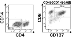
- CD8 / CD137 double positive cells having a directivity to GPC3 are amplified at a high rate.
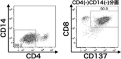
- the cell population expressing both CD8 and CD137 was concentrated and analyzed by a flow cytometer in the same manner as in FIG. It is a figure which shows the result of this.
- the CD8 negative selection shows that the cells expressing CD4 have been removed when compared with FIG. 3 (left figure). It is also shown that the CD137 positive selection also removed cells that did not express CD137 (right figure).
- CD8 / CD137 double positive cells having a directivity to GPC3 are concentrated at a high rate. It is a figure which shows the result of having induced the redifferentiation of iPS cells reprogrammed from the T cell population having a directivity to GPC3 into CD8 positive T cells.
- Antibodies CD3-APC / Cy7, CD4-BV421, CD8a-PerCP / Cy5.5, CD8b-PE, TCRab-FITC, CD45-BV510, and GPC3-HLA complex (Dextramer) against cells that have been induced to redifferentiate.
- BD FACSAria® II flow cytometer
- Redifferentiated CD8-positive T cells are both CD8a and CD8b positive, indicating a high rate of binding to GPC3D extramer. It is a figure which shows the result of having analyzed the T cell population which expressed the activation marker by adding the WT1 overlap peptide to the peripheral blood mononuclear cell of a lung cancer patient and amplifying and culturing it in the same manner as in FIG. From this figure, it is shown that CD8 / CD137 double positive cells having directivity to WT1 are amplified at a high rate. It is a figure which shows the result of having performed the analysis in the same manner as FIG. 4 for the T cell population which was amplified culture using WT1 overlap peptide and expressed the activation marker.
- CD8 / CD137 double positive cells having a directivity to WT1 are concentrated at a high rate.
- Peripheral blood mononuclear cells of non-cancer patients were amplified and cultured using EBV LMP2A overlapping peptide, and the T cell population expressing the activation marker was analyzed in the same manner as in FIG. It is a figure which shows the result which performed. From this figure, it is shown that CD8 / CD137 double positive cells having directivity to EBV LMP2A are amplified at a high rate. It is a figure which shows the result of having performed the analysis in the same manner as FIG.
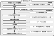
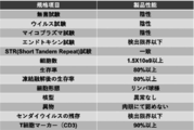
- (B) It is a figure which shows the result of having compared the cytotoxic activity of the regenerated T cell population with the target cell which expressed or did not express GPC3 peptide. It is a figure which shows the manufacturing process of a regenerated T cell product. It is a figure which shows the manufacturing process of the regenerated T cell of both CD8 ⁇ / CD8 ⁇ positive from the iPS cell derived from the tumor antigen-reactive T cell (tumor-specific T cell). It is a figure which shows the setting of the target value of the standard of the cell product of this invention.
- a “cancer vaccine” is a cancer or tumor-specific protein or peptide that is derived from a cancer or tumor-related antigen and is a “cancer vaccine antigen” for inducing a cancer or tumor-specific immune response. It is a composition containing. Cell vaccines such as dendritic cells antigen-pulsed with cancer tissues or proteins or peptides that are antigens specific to cancer or tumors, viral DNA or RNA vaccines that express antigens in vivo after administration, and proliferation. A composition containing cancer or tumor cells that suppress the disease and crushed or lysate thereof is also included in the "cancer vaccine". In addition, non-specific immunostimulants such as bacterial cells, bacterial extracts and ⁇ -glucans, and oncolytic viruses such as adenovirus are also included in the "cancer vaccine".
- Cancer vaccines usually contain adjuvants to enhance the immune response.
- Cancer vaccine antigens contained in cancer vaccines are phagocytosed by antigen-presenting cells (mainly dendritic cells and macrophages) after being administered to a living body, and processed inside the cells to produce amino acids of about 8 to 30 residues. It becomes an epitope peptide consisting of the major histocompatibility complex (MHC) and is presented on the surface of antigen-presenting cells as a complex bound to class I or class II.
- MHC major histocompatibility complex
- the length of the epitope peptide is 8 to 11 amino acid residues in the case of MHC class I and 13 to 30 amino acid residues in the case of MHC class II.
- T cell receptor expressed on the surface of cytotoxic T cells and helper T cells specifically recognizes MHC class I / peptide complex or MHC class II / peptide complex, respectively. As a result, these T cells are activated and exert an antitumor effect. That is, cytotoxic T cells recognize and destroy cancer cells that present the same cytokine peptides as those contained in the vaccine antigen, and helper T cells are interferon (IFN) - ⁇ and interleukin (IL) -2. It enhances the action of cytotoxic T cells through the secretion of cytokines such as and chemokines. Helper T cells also have the function of enhancing the antigen-presenting ability of antigen-presenting cells and the production of antigen-specific immunoglobulin (Ig) G antibody of B cells via the CD40 ligand (CD40L) / CD40 pathway.
- IFN interferon
- IL interleukin
- a "neoantigen” is an antigen produced from a gene that has mutated or spliced abnormalities. A large number of gene mutations are accumulated in cancer cells, and the site of mutation and the degree of mutation vary from patient to patient. In addition, since it is a non-self-antigen that does not exist in the normal body, T cells can strongly damage cancer cells that express neoantigen.
- neoantigen is used as a vaccine by examining gene mutations or splicing abnormalities contained in cancer cells for each patient, synthesizing peptides or proteins containing the mutations or abnormal sites, or extracting them from tumor tissues. Point to.
- the X peptide means an antigen peptide
- preferred examples of the antigen peptide include tumor-related antigens such as GPC3, WT1, XAGE1, LMP2 and novel tumor antigens (neoantigens) due to gene mutations and splicing abnormalities. , And more preferably GPC3, but not limited to these.
- T cell is a cell that expresses an antigen receptor called a T cell receptor (TCR) on the cell surface.
- TCR T cell receptor
- the T cells of the present invention are characterized by (i) having specificity for a cancer vaccine antigen, (ii) being positive for CD3 and CD45, and (iii) producing IFN- ⁇ .
- “Having specificity for a cancer vaccine antigen” means binding / conjugation of T cells selectively to major histocompatibility complex (MHC) class I or class II to which the cancer vaccine antigen or its degradation product is bound, and It means the reaction of T cells caused by selective binding / conjugation to major histocompatibility complex class I or class II to which the cancer vaccine antigen or its degradation product is bound, and binds to something other than the target cancer vaccine antigen. It means that no binding / conjugation of T cells to the major histocompatibility complex class I or class II is observed.
- MHC major histocompatibility complex
- the major histocompatibility complex class I or class II to which the cancer vaccine antigen or its degradation product is bound may be expressed on cells, and the major histocompatibility complex I or II to which the cancer vaccine or its degradation product is bound may be expressed. It may be a complex of proteins. T cell reactions resulting from binding / conjugation to major histocompatibility complex I or II to which a cancer vaccine antigen has bound include cytotoxicity, IFN- ⁇ and granzyme production, and T cell activation marker expression. Can be mentioned.
- T cell used in the present invention includes a T cell population and a regenerated T cell population.
- the "T cell population” is a lymphocyte that is positive for CD3 and CD45, and is a cell population in which the gene sequence of the T cell receptor (TCR) that recognizes an antigen is diverse as a group. Therefore, T cells collected from a living body are a T cell population having specificity for various antigens. T cells existing in the living body have different TCR gene sequences due to random recombination of TCR genes when T progenitor cells develop and differentiate in the thymus, and for all antigens. It is possible to provoke an immune response.
- TCR possessed by each T cell is determined by the antigen or peptide sequence that is specifically recognized, by treating the T cell as a group, the T cell population can immunize various antigens. Understandable. Therefore, T cells collected from living organisms are a population of T cells with genetic diversity.
- the "regenerated T cell population” is a T cell population obtained by regenerating a T cell population having the genetic diversity collected from a living body through reprogramming into iPS cells, and is a T cell population before reprogramming. It is a T cell population that maintains the genetic diversity of T cells and has a directivity toward antigens. "Directive" means that individual T cells specifically recognize different antigen epitopes or peptide sequences, while the T cell population exhibits an immune response to the target tissue or antigen. .. Although iPS cells established from a T cell population with genetic diversity do not express the TCR gene, they retain the genetic information of TCR generated by recombination in DNA, and the genetic information of the T cell population is retained. Maintain diversity. Such iPS cells are a cell population in which the gene sequence of TCR is different for each cell.
- T cells are preferably derived from mammals, more preferably from humans (cancer patients or non-cancer patients).
- the cancer patients and non-cancer patients are preferably patients who have been, are currently receiving, or will be administered a cancer vaccine.
- One or more cancer vaccines may be administered.
- T cells include ⁇ T cells, ⁇ T cells, helper T cells, cytotoxic T cells, and natural killer (NK) T cells, with preference given to cytotoxic T cells.
- peripheral blood is preferable because it is less invasive, but the source is not limited thereto.
- preferred sources include cancer tissue or tumor tissue or other tissue or organ, or blood, umbilical cord blood, lymph, tissue fluid (interstitial fluid, interstitial fluid and interstitial fluid), body cavity fluid (abdominal fluid, pleural fluid, etc.) All sources in the body such as heart sac fluid, cerebrospinal fluid, joint fluid and atrioventricular fluid), nasal juice, urine, etc. are included.
- preferred T cells are tumor tissue-derived T cells. Tumor tissue-derived T cells are usually tumor-infiltrating T cells.
- T cells there are a great variety of T cells in the body so that they can react to any antigen, but most of them are T cells that are unrelated to the antigen targeted in T cell replacement therapy. Therefore, it is effective in cancer treatment to concentrate and use a T cell population that is reactive with a cancer vaccine antigen in advance.
- a concentrated T cell population is amplified in order to obtain a large amount of T cells, there is a problem that the T cells are exhausted and the immune response to the antigen is lowered.
- the above-mentioned concentrated T cell population is initialized to iPS cells, and redifferentiation into T cells is obtained while maintaining the genetic diversity of the T cell population before reprogramming. It is preferable to use the regenerated T cell population obtained.
- Concentration of T cell population is achieved by activating by contact with one or more cancer vaccine antigens and separating and recovering proliferated T cells.
- T cells When T cells are brought into contact with a cancer vaccine antigen, the T cells are activated by receiving recognition and co-stimulation signals of the cancer vaccine antigen by the T cell receptor.
- a culture solution containing a cancer vaccine antigen or a biological tissue containing a cancer vaccine antigen or a crushed product thereof or a lysate thereof is used, and peripheral blood mononuclear cells containing antigen-presenting cells and T cells are used. Culture the spheres.
- cytokines such as IL-2, IL-7, IL-15, and IL-21
- Amplified and cultured T cells express functional molecules such as CD137 molecule and IFN- ⁇ on the cell membrane surface. These functional molecules are stained with a specific antibody labeled with a fluorescent protein or magnetic beads, and the orientation toward the target cancer vaccine antigen is determined by sorting with a flow cytometer or separating with a magnet.
- the T cells that have it can be concentrated. T cell enrichment can also be performed using T cells collected from cancer patients vaccinated with the cancer vaccine.
- T cell concentration can also be performed as follows. Reactive T by repeatedly contacting cancer vaccine antigens with T cells in the process of culturing a mononuclear cell fraction containing antigen-presenting cells and T cells and adding the cancer vaccine antigen to the culture medium. The cells proliferate and increase in proportion to the entire T cell population. On the other hand, T cells that do not react with the added cancer vaccine antigen are not activated, and the proportion in the T cell population gradually decreases during in vitro culture, and eventually they die. Therefore, after culturing for a certain period of time, a T cell population having a specific reactivity with the cancer vaccine antigen added to the culture medium and having diversity in the recognized cancer vaccine antigen epitope or peptide sequence Obtained by concentration.
- a method for concentrating the T cell population there is also a method of selecting T cells to which an oligomer of an antigen peptide-HLA (human leukocyte antigen) complex binds.
- an oligomer consisting of a specific HLA type and a complex of a target antigen peptide capable of presenting the HLA is labeled with a fluorescent dye, and cells to which the oligomer is bound are sorted by using a flow cytometer to target the target. T cells having a directivity to the tissue or antigen to be obtained can be obtained.
- the above-mentioned concentrated T cell population can be initialized into iPS cells and cultured while maintaining the genetic diversity of the concentrated T cell population.
- Methods for producing iPS cells are known in the art. iPS cells are preferably produced by introducing a reprogramming factor into the concentrated T cell population.
- reprogramming factors for example, Oct3 / 4, Sox2, Sox1, Sox3, Sox15, Sox17, Klf4, Klf2, c-Myc, N-Myc, L-Myc, Nanog, Lin28, Fbx15, ERas, ECAT15 Genes or gene products such as -2, Tcl1, beta-catenin, Lin28b, Sall1, Sall4, Esrrb, Nr5a2, Tbx3 or Glis1 can be mentioned, and these reprogramming factors may be used alone or in combination. You may.
- the T cell population existing in the living body has a diverse TCR gene sequence as a population, and in that sense, has gene diversity.
- IPS cells established from this T cell population are cultured while maintaining this genetic diversity.
- passage is preferably performed. However, in the general passage method, cells other than the cells used for passage are discarded, so that the diversity of iPS cells established from the T cell population having genetic diversity is gradually lost.
- the incubator is washed to remove cells other than iPS cells, and iPS cells are collected without cloning. Substitute to another incubator. By repeating such passages, iPS cells for redifferentiation into the regenerated T cell population used in the present invention can be obtained.
- the culture medium used for culturing iPS cells is not particularly limited, but the culture medium used for culturing animal cells is used as the basal culture medium, and cytokines for maintaining the undifferentiated ability of iPS cells are added thereto. Can be prepared.
- basal culture medium examples include Isove's Modified Dulbecco's Medium (IMDM) culture medium, Medium 199 culture medium, Eagle's Minimum Essential Medium (EMEM) culture medium, ⁇ MEM culture medium, Dulbecco's modified Eagle's Medium (DMEM) culture medium, and H Examples thereof include liquid, RPMI 1640 culture medium, Fischer's, Neurobasal Medium culture medium (Life Technologies), StemFit (registered trademark) AK03N (Ajinomoto Healthy Supply), and a mixed culture medium thereof. Serum may be added to the culture broth, or serum-free.
- Preferred examples of cytokines include bFGF, and the concentration thereof in the culture medium is, for example, 1 to 100 ⁇ g / mL (preferably 50 ⁇ g / mL).
- the iPS cell culture method may be adhesive culture or suspension culture, but adhesive culture is preferable.
- a method for separating iPS cells for example, a method for mechanically separating with a cell scraper or the like, a separation solution having protease activity, a separation solution having collagenase activity, or a separation solution having protease activity and collagenase activity (for example, Accutase (for example, Accutase) Separation methods using (registered trademark) and Accumax (registered trademark), etc. can be mentioned.
- iPS cells are preferably cells of 1 ⁇ 10 3 to 1 ⁇ 10 4 cells / cm 2 , 1 ⁇ 10 4 to 1 ⁇ 10 5 cells / cm 2 or 1 ⁇ 10 5 to 1 ⁇ 10 6 cells / cm 2 .
- the number of passages may be any number as long as the iPS cells required to induce differentiation of iPS cells and obtain the amount of regenerated T cell population required for T cell replacement therapy are obtained, preferably 1 to 5 times. Or 5 to 10 times.
- the obtained iPS cells having genetic diversity may be used as they are for inducing redifferentiation into T cells, or may be cryopreserved until necessary. Methods of cryopreserving cells are well known to those of skill in the art.
- the regenerated T cell population used in the present invention maintains the genetic diversity of the enriched T cell population described above and has a directivity toward the cancer vaccine antigen used for T cell activation.
- the regenerated T cell population used in the present invention is preferably obtained by differentiating the cultured iPS cells into CD4 / CD8 double positive T cells via hematopoietic progenitor cells, or hematopoietic progenitor cells and CD4 /. It is obtained by differentiating into CD8 single positive T cells via CD8 double positive T cells.
- Hematopoietic progenitor cells are cells that can differentiate into blood cell lineage cells such as lymphocytes, eosinophils, neutrophils, basophils, erythrocytes, or megakaryocytes. Hematopoietic progenitor cells and hematopoietic stem cells are not distinguished and refer to the same cells unless otherwise specified. Hematopoietic stem cells / progenitor cells are recognized, for example, by being positive for the surface antigens CD34 and / or CD43.
- Hematopoietic progenitor cells are preferably produced by culturing iPS cells in a culture medium supplemented with vitamin Cs.
- vitamin Cs refers to L-ascorbic acid and its derivatives
- L-ascorbic acid derivatives means those that are converted to vitamin C by an enzymatic reaction in vivo.
- Derivatives of L-ascorbic acid include, for example, vitamin C phosphate, glucoside ascorbic acid, ascorbyl ethyl, vitamin C ester, ascorbyl tetrahexyldecanoate, ascorbyl stearate and ascorbic acid-2 phosphate-6 palmitic acid.
- the derivative of L-ascorbic acid is preferably vitamin C phosphate, and examples thereof include phosphate-L ascorbate such as sodium phosphate-L ascorbate or magnesium phosphate-L-ascorbate.
- Vitamin Cs are contained, for example, in a culture medium at a concentration of 5 to 500 ⁇ g / mL.
- the culture medium used for producing hematopoietic progenitor cells is not particularly limited, but can be prepared by using the culture medium used for culturing animal cells as the basal culture solution and adding vitamin C or the like to it.
- the basal culture medium include Isove's Modified Dulbecco's Medium (IMDM) culture medium, Medium 199 culture medium, Eagle's Minimum Essential Medium (EMEM) culture medium, ⁇ MEM culture medium, Dulbecco's modified Eagle's Medium (DMEM) culture medium, and H Examples thereof include liquid, RPMI 1640 culture medium, Fischer's culture medium, Neurobasal Medium (Life Technologies), StemPro34 (Life Technologies) and a mixed culture medium thereof.
- the culture broth may contain serum or may be serum-free.
- the basal culture solution may contain, for example, albumin, insulin, transferrin, selenium, fatty acids, trace elements, 2-mercaptoethanol, thiolglycerol, lipids, amino acids, L-glutamine, non-essential amino acids, vitamins, growth factors, etc. It may also contain one or more substances selected from low molecular weight compounds, antibiotics, antioxidants, pyruvate, buffers, inorganic salts, cytokines and the like.
- the culture medium used for the production of hematopoietic progenitor cells includes BMP4 (Bonemorphogenetic protein 4), VEGF (vascular endothelial growth factor), bFGF (basic fibroblast growth factor), SCF (stem cell factor), TPO (thrombopoietin), and FLT.
- Cytokines selected from the group consisting of -3L (Flt3 ligand) may be further added. These concentrations are, for example, 1-100 ng / mL for BMP4, 1-100 ng / mL for VEGF, 1-100 ng / mL for bFGF, and 10-100 ng / mL for SCF.
- TPO is 1 to 100 ng / mL
- FLT-3L is 1 to 100 ng / mL.
- TGF ⁇ inhibitors are small molecule inhibitors that interfere with TGF ⁇ family signaling, such as SB431542 and SB202190 (RKLindemann et al., Mol. Cancer 2: 20 (2003)), SB505124 (GlaxoSmith Kline). , NPC30345, SD093, SD908 and SD208 (Scios), and LY2109761, LY364947 and LY580276 (Lilly Research Laboratories), and the concentration added to the culture solution is preferably 0.5 to 100 ⁇ M.
- iPS cells are C3H10T1 / 2 (Takayama N., et al. J Exp Med. 2817-2830, 2010) or heterologous stromal cells (Niwa A et al. J Ce11 Physiol. 2009 Nov; 221 (2): Although it may be co-cultured with a feeder cell such as 367-77), iPS cells are preferably cultured without using the feeder cell.
- the method for culturing iPS cells at the time of producing hematopoietic progenitor cells may be adhesive culture or suspension culture, but suspension culture is preferable.
- iPS cells can be subjected to suspension culture after separating colonies cultured to 80% confluence with respect to the dish used, dissociating them into single cells.
- a method for separating iPS cells for example, a method for mechanically separating with a cell scraper or the like, a separation solution having protease activity and collagenase activity (for example, Accutase® and Accumax®), or collagenase activity.
- a separation method using a separation solution having the above can be mentioned.
- Suspension culture is to culture cells in a non-adherent state to a culture vessel.
- the suspension culture is not particularly limited, but is a culture vessel that has not been artificially treated (for example, a coating treatment with an extracellular matrix) for the purpose of improving adhesion to cells, or artificially suppresses adhesion.
- coating treatment with polyhydroxyethyl methacrylate (po1y-HEMA) or nonionic surfactant (Pluronic F-127, etc.) can be performed using the culture vessel.
- po1y-HEMA polyhydroxyethyl methacrylate
- Pluronic F-127 nonionic surfactant
- Hematopoietic progenitor cells can also be prepared from a net-like structure (also referred to as iPS-sac) obtained by culturing iPS cells.
- the "net-like structure” is a three-dimensional sac-like structure (with a space inside) derived from iPS cells, which is formed by a population of endothelial cells and the like and contains hematopoietic progenitor cells inside. Is.
- the temperature conditions for culturing to produce hematopoietic progenitor cells from iPS cells are not particularly limited, but are, for example, about 37 ° C to about 42 ° C, preferably about 37 ° C to about 39 ° C. Further, the culture period can be appropriately determined by those skilled in the art while monitoring the number of hematopoietic progenitor cells and the like.
- the number of culture days is not particularly limited as long as hematopoietic progenitor cells can be obtained, but for example, at least 6 days or more, 7 days or more, 8 days or more, 9 days or more, 10 days or more, 11 days or more, 12 days or more, 13 days or more. , Or 14 days or more, preferably 14 days.
- the long culture period is not a problem in the production of hematopoietic progenitor cells.
- the cells may be cultured under low oxygen conditions, and in one embodiment of the present invention, the low oxygen conditions include, for example, 15%, 10%, 9%, 8%, 7%, 6%, 5% or less. Oxygen concentration of.
- CD4 / CD8 double positive T cells refers to T cells that are both positive for the surface antigens CD4 and CD8 (CD8 + CD4 + ), and the T cells are the surface antigens CD3 and CD45.
- CD4 / CD8 double positive T cells can be identified as CD4, CD8, CD3 and CD45 positive cells because they can be recognized by being positive.
- CD4 / CD8 double positive T cells can be induced to differentiate into CD4 single positive cells or CD8 single positive cells.
- CD4 / CD8 double positive T cells can be produced by a method including the step of culturing hematopoietic progenitor cells in a culture medium supplemented with a p38 inhibitor and / or SDF-1.
- P38 inhibitor is defined as a substance that inhibits the function of p38 protein (p38MAP kinase).
- p38 inhibitors include, but are not limited to, chemical inhibitors of p38, dominant negative variants of p38, or nucleic acids encoding them.
- Chemical inhibitors of p38 include SB203580 (4- (4-fluorophenyl) -2- (4-methylsulfonylphenyl) -5- (4-pyridyl) -1H-imidazole) and its derivatives, SB202190 (4-).
- SB203580, SB202190, SC239063, SB220025 and PD169316 are available from Calbiochem, SCl0-469 and SCIO-323 are available from Scios and the like.
- the p38 inhibitor is added to the culture medium, for example, in the range of about 1 ⁇ M to about 50 ⁇ M.
- Dominant negative variants of p38 include p38T180A, which is a point mutation of threonine at position 180 located in the DNA binding region of p38 to alanine, and p38Y182F, which is a point mutation of tyrosine at position 182 of p38 to phenylalanine in humans and mice. Can be mentioned.
- SDF-1 (Stromal cell-derived factor 1) is not only SDF-1 ⁇ or its mature form, but also isoforms such as SDF-1 ⁇ , SDF-1 ⁇ , SDF-1 ⁇ , SDF-1 ⁇ or SDF-1 ⁇ , or them. It may be a mature form of, or a mixture of any proportions thereof and the like. Preferably, SDF-1 ⁇ is used. SDF-1 may also be referred to as CXCL-12 or PBSF.
- SDF-1 has its chemokine activity
- one or several amino acids in its amino acid sequence may be substituted, deleted and / or added, and similarly, sugar chains may be substituted or deleted. And / or may be added.
- SDF-1 may be of a mammal, such as a non-human mammal such as a monkey, sheep, cow, horse, pig, dog, cat, rabbit, rat, or mouse.
- GenBank registration number: NP_954637 can be used as human SDF-1 ⁇
- a protein registered with GenBank registration number: NP_000600 can be used as SDF-1 ⁇ .
- SDF-1 a commercially available product may be used, a product purified from nature may be used, or a product produced by peptide synthesis or genetic engineering techniques may be used. SDF-1 is added to the culture medium, for example, in the range of about 10 ng / mL to about 100 ng / mL.
- the culture medium used for producing CD4 / CD8 double positive T cells is not particularly limited, but the culture medium used for culturing animal cells is used as the basal culture medium, and p38 inhibitor and / or SDF-1 is more preferable. It can be prepared by adding vitamin Cs.
- the types of vitamin Cs used in the production of CD4 / CD8 double positive T cells are as described above, and the concentration of vitamin Cs is, for example, 5 to 200 ⁇ g / mL.
- basal culture medium examples include Isove's Modified Dulbecco's Medium (IMDM) culture medium, Medium 199 culture medium, Eagle's Minimum Essential Medium (EMEM) culture medium, ⁇ MEM culture medium, Dulbecco's modified Eagle's Medium (DMEM) culture medium, and H. , RPMI 1640 culture medium, Fischer's Neurobasal Medium culture medium (Life Technologies), and a mixed culture medium thereof. Serum may be added to the culture broth, or serum-free.
- IMDM Isove's Modified Dulbecco's Medium
- EMEM Eagle's Minimum Essential Medium
- DMEM Dulbecco's modified Eagle's Medium
- H. RPMI 1640 culture medium
- Fischer's Neurobasal Medium culture medium Life Technologies
- basal culture solutions include, for example, albumin, insulin, transferrin, selenium, fatty acids, trace elements, 2-mercaptoethanol, thiolglycerol, lipids, amino acids, L-glutamine, non-essential amino acids, vitamins, growth factors. , Small molecule compounds, antibiotics, antioxidants, pyruvate, buffers, inorganic salts, and one or more substances selected from cytokines and the like.
- Cytokines selected from the group consisting of SCF, TPO (thrombopoietin), FLT-3L and IL-7 may be further added to the culture medium used for producing CD4 / CD8 double positive T cells. These concentrations are, for example, 10-100 ng / mL for SCF, 1O-200 ng / mL for TPO, 1-100 ng / mL for FLT-3L, and 1-100 ng for IL-7. / mL.
- hematopoietic progenitor cells may be cultured using feeder cells, but the culture is preferably performed without using feeder cells.
- Hematopoietic progenitor cells may be adherently cultured or suspension-cultured, but adherent culture is preferable.
- the culture vessel may be coated and used.
- Matrigel Niwa A, et al. PLoS One. 6 (7): e22261, 2011
- collagen gelatin, laminin, heparan sulfate proteoglycan, retronectin, Fc-DLL4 or entactin, and a combination thereof.
- a coating agent Matrigel (Niwa A, et al. PLoS One. 6 (7): e22261, 2011), collagen, gelatin, laminin, heparan sulfate proteoglycan, retronectin, Fc-DLL4 or entactin, and a combination thereof. Can be mentioned.
- the culture temperature conditions for culturing hematopoietic progenitor cells to produce CD4 / CD8 double positive T cells are not particularly limited, but are preferably about 37 ° C. to about 42 ° C., preferably about 37 ° C. to about 39 ° C. The degree is more preferable. Further, the culture period can be appropriately determined by those skilled in the art while monitoring the number of CD4 / CD8 double positive T cells and the like. As long as CD4 / CD8 double positive T cells can be obtained, the number of culture days is not particularly limited, but for example, at least 10 days or more, 12 days or more, 14 days or more, 16 days or more, 18 days or more, 20 days or more, 22 days or more. , Or 23 days or more, preferably 23 days.
- the obtained CD4 / CD8 double positive T cells may be isolated and used, or may be used as a cell population containing other cell types. When isolated, it can be isolated using any one index selected from the group consisting of CD4, CD8, CD3 and CD45.
- the method of isolation can be a method well known to those of skill in the art, for example, labeling with an antibody against CD4, CD8, CD3 or CD45 and isolating with a flow cytometer, or the desired antigen. Examples thereof include a method of purifying using an immobilized affinity column or the like.
- CD8 single positive T cells refers to all T cells, CD8 surface antigens cells positive (CD8 + CD4 -) refers to, also called cytotoxic T cells. Since T cells can be recognized by being positive for the surface antigens CD3 and CD45, CD8 single positive T cells can be identified as cells that are positive for CD8, CD3 and CD45 and negative for CD4. ..
- CD8 single positive T cells can be produced by a method including a step of culturing CD4 / CD8 double positive T cells in a culture medium supplemented with a corticosteroid.
- the corticosteroid agent is preferably a glucocorticoid or a derivative thereof, and examples thereof include cortisone acetate, hydrocortisone, fludrocortisone acetate, prednisolone, triamcinolone, methylprednisolone, dexamethasone, betamethasone, or bechrometazone propionate.
- the corticosteroid agent is dexamethasone. Its concentration in the culture medium is, for example, 1 to 100 nM.
- the culture medium used for producing CD8 single positive T cells is not particularly limited, but can be prepared by using the culture medium used for culturing animal cells as the basal culture medium and adding a corticosteroid agent to it.
- the basal culture medium include Isove's Modified Dulbecco's Medium (IMDM) culture medium, Medium 199 culture medium, Eagle's Minimum Essential Medium (EMEM) culture medium, ⁇ MEM culture medium, Dulbecco's modified Eagle's Medium (DMEM) culture medium, and H Examples include liquid, RPMI 1640 culture medium, Fischer's Neurobasal Medium culture medium (Life Technologies), and a mixed culture medium thereof. Serum may be added to the culture broth, or serum-free.
- IMDM Isove's Modified Dulbecco's Medium
- EMEM Eagle's Minimum Essential Medium
- DMEM Dulbecco's modified Eagle's Medium
- H Examples include liquid, RPMI 1640 culture medium, Fischer's Neurobasal Medium culture medium (Life
- the basal culture solution may contain, for example, albumin, insulin, transferrin, selenium, fatty acids, trace elements, 2-mercaptoethanol, thiolglycerol, lipids, aminos, L-glutamine, non-essential amino acids, vitamins, growth factors. , Small molecule compounds, antibiotics, antioxidants, pyruvate, buffers, inorganic salts, and one or more substances selected from cytokines and the like.
- the culture medium used for producing CD8 single positive T cells preferably further contains anti-CD3 antibodies, vitamin Cs, or cytokines.
- cytokine examples include IL-2 and IL-7.
- the anti-CD3 antibody is not particularly limited as long as it is an antibody that specifically recognizes CD3, and examples thereof include an antibody produced from an OKT3 clone.
- the concentration of the anti-CD3 antibody in the culture medium is, for example, 10 to 1000 ng / mL.
- the vitamin Cs used for producing CD8 single positive T cells are, for example, those described above, and can be used under the same conditions as described above.
- the concentration of cytokines used in the production of CD8 single positive T cells in the culture medium is, for example, 10 to 1000 U / mL for IL-2 and 1 to 100 ng / mL for IL-7.
- the temperature conditions for culturing CD4 / CD8 double positive T cells for producing CD8 single positive T cells are not particularly limited, but for example, about 37 ° C to about 42 ° C is preferable, and about 37 ° C to about 39 ° C. About ° C is more preferable.
- the culture period can be appropriately determined by those skilled in the art while monitoring the number of CD8 single positive T cells and the like. As long as CD8 single positive T cells can be obtained, the number of culture days is not particularly limited, but is, for example, at least 1 day or more, 2 days or more, 3 days or more, 4 days or more, or 5 days or more, preferably 3 days. ..
- the degree of genetic diversity maintained is preferably at least 30%, more preferably at least 40%, even more preferably at least 50% of the T cell population having the genetic diversity of the raw material. is there.
- the degree of genetic diversity maintained can be confirmed, for example, based on repertoire analysis of the TCR ⁇ gene using the TCRV ⁇ analysis kit (BECKMAN COULTER: Catalog No. IM-3497). From the repertoire analysis of the TCR ⁇ gene using the TCRV ⁇ analysis kit (BECKMAN COULTER: Catalog No. IM-3497), the degree of genetic diversity maintained in one aspect of the present invention is T having the genetic diversity of the raw material. It was confirmed that it maintained more than 40% of the cell population.
- the survival rate of regenerated T cells of the present invention is 60% or more, more preferably 70% or more, still more preferably 80% or more.
- the survival rate is calculated by analyzing with a cell counter and calculating the percentage of cells that do not take up a pigment such as trypan blue that is specifically taken up by dead cells.
- the expression rate of CD3 in regenerated T cells of the present invention is 70% or more, more preferably 80% or more, still more preferably 90% or more.
- the expression rate of CD3 is calculated using flow cytometry.
- the expression rate of the exhaustion marker is 30% or less, more preferably 20% or less, still more preferably 10% or less.
- the expression rate of the exhaustion marker of regenerated T cells is determined by staining each T cell with an antibody (TIGIT-PacificBlue, PD-1-APC / Cy7) and using a flow cytometer (BD FACSAria (registered trademark) II). Can be analyzed using.
- the regenerated T cells of the present invention have the ability to produce IFN-g / IL-2 (Fig. 15).
- PMA Phorbol 12-Myristate 13 Acetate, WAKO
- ION Ionomycin, WAKO
- An ION solution was prepared.
- PMA / ION solution and Monensin (BioLegend) were added to the regenerated T cells and stimulated for 4 hours.
- cell surface antigens are stained with antibodies (CD45-BV510, CD8 ⁇ -PerCP / Cy5.5, CD8 ⁇ -APC, CD19-FITC), fixed with Fixation Buffer (BioLegend), and permeated into cell membrane with Permeabilization Buffer (BioLegend). Processed. Permeation treatment, staining with cytokine antibody (IFN-g-APC / Cy7, IL-2-PE), and percentage of regenerated T cells that produced cytokines using a flow cytometer (BD FACSAria® II). was analyzed.
- cytokine antibody IFN-g-APC / Cy7, IL-2-PE
- the pharmaceutical composition of the present invention containing T cells as an active ingredient can be used for treating a cancer treatment target.
- the pharmaceutical composition of the present invention can be produced by a method commonly used in the field of formulation technology, for example, the method described in the Japanese Pharmacopoeia.
- the pharmaceutical composition of the present invention may contain a pharmaceutically acceptable additive. Examples of the additive include a cell culture solution, a physiological saline solution, an appropriate buffer solution (for example, a phosphoric acid-based buffer solution), and the like.
- the pharmaceutical composition of the present invention can be produced by suspending T cells in physiological saline, an appropriate buffer solution (for example, a phosphate-based buffer solution), or the like. It is preferable to contain, for example, 1 ⁇ 10 7 or more cells as a single dose so that the desired therapeutic effect is exhibited. More preferably, it is 1 ⁇ 10 8 or more, and further preferably 1 ⁇ 10 9 or more.
- the cell content can be appropriately adjusted in consideration of the sex, age, body weight, condition of the affected area, cell condition, etc. of the target.
- the pharmaceutical composition of the present invention may contain dimethyl sulfoxide (DMSO), serum albumin, etc. for the purpose of protecting the cells.
- DMSO dimethyl sulfoxide
- antibiotics and the like may be contained for the purpose of preventing bacterial contamination
- vitamins and cytokines and the like may be contained for the purpose of promoting cell activation and differentiation
- other formulationally acceptable ingredients eg, carriers, excipients, disintegrants, buffers, emulsifiers, suspensions, soothing agents, stabilizers, preservatives, preservatives, saline, etc.
- It may be contained in the pharmaceutical composition of the present invention.
- the pharmaceutical composition containing the T cells of the present invention as an active ingredient can be cryopreserved at low temperature.
- the temperature is not particularly limited as long as it is a temperature suitable for cell storage.
- ⁇ 20 ° C., ⁇ 80 ° C. and ⁇ 120 to ⁇ 196 ° C. can be mentioned.
- cells can be stored in a suitable container such as a vial. Procedures for minimizing the risk of cell damage during freezing and thawing of T cells are well known to those of skill in the art.
- T cells are collected from the culture medium, washed with a buffer solution or a culture solution, the number of cells is counted, concentrated by centrifugation or the like, and containing a frozen medium (for example, 10% DMSO). After suspending in culture solution), cryopreserve at low temperature. T cells can be combined into a single lot by combining cells cultured in multiple culture vessels.
- the pharmaceutical composition containing T cells of the present invention contains, for example, 5 ⁇ 10 4 to 9 ⁇ 10 10 T cells per container such as a vial, and the target cancer type, administration target, and administration. It can be changed according to the route and the like.
- Examples of the route of administration of the pharmaceutical composition containing T cells of the present invention include infusion, intratumoral injection, intraarterial injection, portal vein injection, intraperitoneal administration and the like.
- the route of administration is not limited to this as long as T cells, which are the active ingredient in the pharmaceutical composition of the present invention, are delivered to the affected area.
- the administration schedule can be a single administration or multiple administrations. For the period of multiple administration, for example, a method of repeating administration once every 2 to 4 weeks, a method of repeating administration once every 6 months to 1 year, and the like can be adopted.
- the sex, age, body weight, pathological condition, etc. of the target patient can be taken into consideration.
- the pharmaceutical composition containing T cells of the present invention is used for the prevention or treatment of cancer
- a cancer patient or a cancer patient from which T cells are collected from the viewpoint of being less likely to cause rejection.
- the non-cancer patient has the same HLA type as the cancer patient to whom the pharmaceutical composition of the present invention is administered for cancer treatment.
- the cancer patient to whom the pharmaceutical composition of the present invention is administered and the cancer patient from which T cells are collected are the same person. That is, cancer treatment by autologous transplantation is more preferable than allogeneic transplantation.
- the pharmaceutical composition of the present invention is used for the prevention or treatment of cancer.
- Cancers include ovarian cancer, hepatoblastoma, hepatocellular carcinoma, gastric cancer, esophageal cancer, pancreatic cancer, renal cell carcinoma, breast cancer, malignant melanoma, non-small cell lung cancer, cervical cancer, and glue. Examples include, but are not limited to, blastoma, prostate cancer, neuroblastoma, chronic lymphocytic leukemia, papillary thyroid cancer, colon cancer, and B-cell non-Hodgkin's lymphoma.
- the administration time of the pharmaceutical composition of the present invention and the cancer vaccine is not limited.
- the pharmaceutical composition of the present invention and the cancer vaccine may be administered to the administration subject at the same time or at different times.
- the pharmaceutical composition of the present invention and the cancer vaccine may be formulated separately, or may be a mixture of both.
- the dose of the pharmaceutical composition and the cancer vaccine of the present invention may be based on the dose clinically used, and can be appropriately selected depending on the disease, administration target, administration route, combination with a drug, and the like.
- the administration form of the pharmaceutical composition of the present invention and the cancer vaccine is not particularly limited, and finally both the pharmaceutical composition of the present invention and the cancer vaccine may be administered.
- Such administration forms include, for example, (1) administration of a single preparation obtained by integrally formulating the pharmaceutical composition of the present invention and a cancer vaccine, and (2) administration of the pharmaceutical composition of the present invention and cancer.
- the following excellent effects can be obtained.
- (1) The dose can be reduced as compared with the case where the pharmaceutical composition of the present invention or the cancer vaccine is administered alone.
- a long treatment period can be set by administration of the pharmaceutical composition of the present invention and the cancer vaccine.
- the pharmaceutical composition of the present invention is used in combination with a cancer vaccine, a synergistic effect in anticancer or antitumor effect can be obtained.
- cytotoxic T cells against GPC3 are isolated and amplified from the peripheral blood of a hepatoblastoma patient who received a cancer peptide vaccine (for example, a cancer vaccine containing GPC3) to obtain T cells.
- a cancer peptide vaccine for example, a cancer vaccine containing GPC3
- the obtained T cells maintain antigen specificity for GPC3 and have antigen-specific cytotoxicity for cancer cells (tumors) expressing GPC3, a pharmaceutical composition containing effective T cells can be obtained.
- cytotoxic T cells against GPC3 are isolated, amplified, isolated and isolated from the peripheral blood of hepatoblastoma patients who have been administered a cancer peptide vaccine (eg, a cancer vaccine containing GPC3).
- Regenerated T cells can be obtained by reprogramming amplified cytotoxic T cells into iPS cells and then inducing redifferentiation of the iPS cells into cytotoxic T cells. Since the obtained regenerated T cells maintain antigen specificity for GPC3 and have antigen-specific cytotoxicity for cancer cells expressing GPC3, a pharmaceutical composition containing effective regenerated T cells can be obtained. be able to.
- the method for producing the cell product of the present invention is shown in FIG.
- the production of the cell product of the present invention proceeds in the steps 1 to 15 shown in FIG. (1)
- a cancer vaccine (X peptide + IFA) is administered to a patient to elicit an immune response against antigen X.
- Peripheral blood is collected from the patient and mononuclear cells are separated.
- the obtained mononuclear cells are stimulated with an X peptide in vitro to amplify and isolate antigen X-specific T cells.
- Yamanaka 4 factors and SV40T antigen are introduced into the obtained antigen X-specific T cells using a Sendai virus vector and reprogrammed.
- Established IPS cells are amplified and cultured while containing many clones. Make primary frozen stock.
- a secondary frozen stock is prepared from the primary frozen stock.
- the formation of germ layer containing blood cells is induced by differentiation culture.
- the obtained hematopoietic stem cells are cultured together with a T cell differentiation inducing factor to obtain immature T cells.
- the cell population at this point is temporarily cryopreserved. Repeat the above operations to secure a sufficient amount of immature T cells.
- T cells Freeze and thaw the frozen stock of immature T cells and start culturing.
- CD8 ⁇ CD8 ⁇ positive T cells are fractionated by a flow cytometer.
- the obtained cells are cultured together with PHA + autologous or allogeneic feeder cells for maturation culture.
- T cells After culturing, T cells are washed and cryopreserved to prepare for treatment.
- Fig. 11 (1) Administer cancer vaccine (X peptide + IFA) to patients and promote cell proliferation of T cells that react specifically with X peptide in vivo. .. Peripheral blood is collected from the above patients and mononuclear cells are isolated (IFA: Incomplete Freund's adjuvant).
- Step 1 Concentration of T cells Cancer vaccine antigens (GPC3, WT1, XAGE1, LMP2 or neoantigen) are added to the culture medium and brought into contact with the T cell population to contact 200 U / mL IL-2, 10 ng / mL IL.
- the cells were cultured in RPMI-1640 culture medium supplemented with -15 and 10 ng / mL IL-21 (containing 10% human AB serum and 1% penicillin-streptomycin-glutamine) for 12 days.
- Tumor-related antigens to be added to the culture medium should be added at a concentration of 1 to 100 mg / mL for peptides, and 1 to 2 mm square when living tissue or crushed products thereof are used as cancer vaccine antigens. It is preferable to add about 1 to 5 shredded tissue pieces per tube containing the culture solution and bring them into contact with the T cell population. In the case of lysates of living tissue, it is preferable to add the lysates to the culture medium and bring them into contact with the T cell population. On the 12th day after culturing, the same cancer vaccine antigen was contacted again and cultured for another 24 hours to express an activation marker on T cells having a direction to the cancer vaccine antigen.
- Negative selection using blood cell fractionation markers other than CD8 molecules CD8 + T cell Isolation Kit, Miltenyi Biotech
- positive selection for activation markers CD137 or IFN- ⁇ CD137 MicorBead Kit
- Cytokine capture system-IFN- ⁇ Miltenyi Biotech
- the T cell population used is a cell population that has been cultured for 12 days in contact with the cancer vaccine antigen, or a cell population that has not been cultured. It may be either.
- a fluorescently labeled antigen peptide-HLA complex of interest is added, first stained for 30 minutes, and then fluorescently labeled antibodies against other blood cell markers (CD8, CD4 and CD14) are added for an additional 30 minutes. Staining was performed for minutes.
- the collected cells are washed with a phosphate buffer solution, and then only the cells expressing the antigen peptide-HLA complex and the CD8 molecule are sorted using a flow cytometer to obtain the desired cancer vaccine.
- T cells with tropism for the antigen were concentrated (Fig. 11 (3)).
- 4 factors Oct3 / 4, Sox2, Klf4 and c-Myc
- the beads and cells were separated by pipetting, and then the tube was allowed to stand on a stand with a magnet to collect only the T cells suspended in the culture medium.
- T cells were seeded on a 6 cm dish coated with iMatrix-511 (Nippi: Catalog No. 892011) at 0.5 ⁇ g / cm 2 , and in RPMI-1640 culture medium at 37 ° C., 5% CO 2 Culturing was started using the incubator set in.
- iMatrix-511 Nippi: Catalog No. 892011
- iPS cell colonies were obtained in a 6 cm dish by continuing the culture for 20 to 40 days while exchanging the same half-volume culture medium every day.
- the culture medium for establishment was replaced with the culture medium for iPS cells (StemFit AK03N only), and the culture was continued.
- Step 3 Culture while maintaining the genetic diversity of established iPS cells (Fig. 11 (5)) Once the iPS cell colonies had grown to the extent that they were visible to the naked eye, the iPS cells were subcultured into a new iMatrix-511 coated 6 cm dish.
- the iPS cells adhering to the dish are washed with PBS (-), and then TrypLeSelect (Life Technologies, Inc .: Catalog No. A12859-01) is added and incubated for about 7 minutes to allow the iPS cells to be incubated. It was exfoliated and dispersed into a single cell. The cells were washed with a culture medium for iPS cells, and the number of cells was counted. The dispersed cells were then seeded on a freshly iMatrix-511 coated 6 cm dish at a density of 1500 cells / cm 2 . Here, all the recovered iPS cells were seeded into a new dish without discarding.
- the culture scale gradually expands. Therefore, 3 to 5 passages were performed after establishment, and when the amount of iPS cells required for T cell replacement therapy using iPS cells was obtained, the cells were cryopreserved according to the following procedure.
- the cells After collecting iPS cells using the same procedure as for passage, the cells are suspended in a cell freezing solution such as a TC protector (DS Pharma: Catalog No. KBTCP001), and the temperature is raised to -80 ° C at a rate of -1 ° C / min.
- the iPS cells were frozen by lowering.
- Step 4 Production of regenerated T cell population (Fig. 11 (8))
- the cryopreserved T-iPS cells are thawed and amplified and cultured (Fig. 11 (7)).
- the iPS cells obtained in step 3 were seeded in 3 ⁇ 10 5 to 6 ⁇ 10 5 cells / well on a 6-well plate (CORNING: Catalog No. 3471) that had been subjected to ultra-low adhesion treatment (Day O), and EB culture was performed.
- liquid (StemPro34 containing 10 ⁇ g / mL human insulin, 5.5 ⁇ g / mL human transferase, 5 ng / mL sodium selenate, 2 mM L-glutamine, 45 mM ⁇ -monothioglycerol and 50 ⁇ g / mL ascorbic acid) 10 ng / mL BMP4, 5 ng / mL bFGF, 15 ng / mL VEGF, and 2 ⁇ M SB431542 were added, and the mixture was cultured under low oxygen conditions (5% O 2 ) for 5 days (Day 5).
- the obtained hematopoietic progenitor cells were 50 ng / mL on a 4-well plate coated with Fc-DLL4 (5 ⁇ g / mL) (Sino Biological Inc.) and Retronectin (5 ⁇ g / mL) (Takara Bio Co., Ltd.).
- Fc-DLL4 5 ⁇ g / mL
- Retronectin 5 ⁇ g / mL
- ⁇ MEM culture solution (15% FBS, 15% FBS,) containing SCF, 50 ng / mL IL-7, 50 ng / mL Flt3L, 100 ng / mL TPO, 15 ⁇ M SB203580 (Tocris Bioscience) and 30 ng / mL SDF-1 ⁇ (PeproTech).
- Cryopreservation of immature lymphocytes The immature lymphocytes induced in the culture were collected and cryopreserved using a cell freezing solution such as a TC protector (FIG. 11 (11)).
- Fig. 11 (12) Thaw the cryopreserved immature lymphocytes (Fig. 11 (12)), and use a flow cytometer on Day 35 for CD4 / CD8 double positive cells (DP cells) in which all of CD45, CD3, CD4 and CD8 are positive.
- DP cells CD4 / CD8 double positive cells
- CD4 / CD8 double positive cells obtained from immature lymphocytes induced in the above cultures were placed on a 96-well plate with 15% bovine fetal serum, 500 ng / mL anti-CD3 antibody (eBioscience), 200 U / mL IL. -2, 10 ng / mL IL-7 and 10 nM dexamethazone were added and cultured in RPMI 1640 culture medium for 2 days (Day 37).
- Antibodies were removed by washing cells with RPMI 1640 culture medium supplemented with 15% fetal bovine serum and cultured in RPMI 1640 culture medium supplemented with 15% fetal bovine serum and 10 ng / mL IL-7 for an additional 5 days.
- CD8 single positive T cells were obtained (Day 42).
- CD8 ⁇ / CD8 ⁇ double positive cells from immature lymphocytes induced in the culture, 15% bovine fetal serum, 2 ⁇ g / mL phytohemaglutinin (PHA), 40 Gy irradiated peripheral blood mononuclear cells on a 96-well plate.
- PHA phytohemaglutinin
- PBMC peripheral blood mononuclear cell
- IL-2 10 ng / mL IL-7, 5 ng / mL IL-15, 5 ng / mL IL-21, 50 ⁇ g / mL ascorbic acid, 2 mM L-glutamine, 100 Incubate for 21 days in an ⁇ MEM culture solution containing U / mL penicillin, 100 ng / mL streptomycin, and insulin / transferase / selenium mixed solution (Thermo Fisher Scientific: ITS-G solution, catalog number 41400045), and double CD8 ⁇ / CD8 ⁇ . Positive mature T cells were obtained (Fig. 11 (14)).
- iPS cells were established from a T cell population having genetic diversity isolated from an excised tumor of a lung cancer patient, and redifferentiation was induced into T cells having genetic diversity.
- TCR T cell receptor
- TCRV ⁇ analysis kit (BECKMAN COULTER: Catalog No. IM-3497)
- the TCR repertoire of the T cell population isolated from the resected tumor of a lung cancer patient was analyzed (Fig. 1). It was confirmed that the patient sample contained cells expressing each V ⁇ chain at a rate of about 1 to 13%, and that it was a T cell population having genetic diversity.
- These T cell populations were reprogrammed into iPS cells by the method described above.
- IPS cells are pluripotent stem cells, and T cell-related genes including the TCR gene are not expressed. That is, it is difficult to analyze the diversity of TCR genes at the stage of undifferentiated iPS cells.
- the established iPS cells were passaged so as not to lose their genetic diversity, and then differentiated into T cells through the hematopoietic progenitor cell stage.
- the TCR repertoire expressed by the regenerated T cells was analyzed using the above kit (Fig. 2). With the establishment of iPS cells and the induction of T cell differentiation, the diversity of T cells expressing the 2, 3, 4, 7.1, 7.2, 8, 12, 14, 16, 20 and 21.3 segments of V ⁇ is lost. However, about half of the V ⁇ repertoires were conserved, and it was confirmed that the regenerated T cell population is a T cell population with genetic diversity.
- Peripheral blood was collected from patients who received the peptide vaccine for GPC3, one of the cancer antigens, and the mononuclear cell fraction was separated using density gradient centrifugation. 2 ⁇ 10 6 cells of this cell population were added to a 24-well plate and T cells directional to GPC3 were amplified and cultured using GPC3 peptide at a concentration of 10 mg / mL according to step 1 above. .. The cultured cells were re-contacted with the GPC3 peptide and cultured for 24 hours to express the activation marker.
- This cell population was stained with antibodies (CD4-BV421, CD8-PerCP / Cy5.5, CD14-FITC and CD137-PE) and GPC3 directional using a flow cytometer (BD FACSAria® II).
- the proportion of T cell populations with (Fig. 3).
- CD8 / CD137 double positive cells having a directivity to GPC3 were amplified at a high rate.
- magnetic beads were used to express both CD8 and CD137, and a T cell population directional with GPC3 was concentrated (Fig. 4).
- This cell population was reprogrammed into iPS cells according to steps 2 and 3 above, and then redifferentiation into CD8 single positive cells was induced according to step 4 above.
- Example 2 by isolating monocytes from the peripheral blood of a non-cancer patient (healthy person) and contacting them with an overlapping peptide (PepTivator EBV LMP2A, Miltenyi Biotech) of EBV LMP2A, which is one of the viral antigens.
- EBV LMP2A overlapping peptide
- a T cell population directional to EBV LMP2A was amplified and cultured.
- the EBV LMP2A overlap peptide was added to the culture medium at 1 ⁇ g / mL.
- the cultured cells were re-contacted with the EBV LMP2A overlapping peptide to express the activation marker.
- the regenerated T cell population prepared via iPS cells from the T cell population directional to GPC3 has cytotoxic activity specific to the target cells expressing the GPC3 peptide.
- Mononucleosis was separated from the peripheral blood of a liver cancer patient, and by contacting with the GPC3 peptide in the same manner as in Example 2, a T cell population having a directivity to GPC3 was amplified and cultured, and further concentrated. This concentrated cell population was reprogrammed into iPS cells according to steps 2 and 3 above, and then redifferentiation into CD8 single positive cells was induced according to step 4 above. In the obtained regenerated T cell population, flow cytometric analysis confirmed that CD8 was present as a CD8 ⁇ heterodimer (Fig. 10A).
- target cells B cells infected with EB virus and immortalized were used.
- the target cells were cultured in the presence or absence of GPC3 peptide (1 ⁇ M) to prepare GPC3 peptide-expressing cells and GPC3 peptide-non-expressing cells.
- the cytotoxic activity of the regenerated T cell population against target cells was measured using the N-SPC non-RI cytotoxic assay kit (Techno Suzuta).
- Target cells expressing or not expressing GPC3 peptide were treated with BM-HT Reagent, and BM-HT Reagent was incorporated into the cells. The target cells were then washed to remove excess BM-HT Reagent.
- the target cells and the regenerated T cell population were co-cultured for 2 hours, and the culture supernatant was added to Eu solution (Europium solution).
- Eu solution Europium solution
- the HT chelate leaked from the injured target cells into the culture medium became an HT / Eu complex, which was excited by laser light and time-resolved fluorescence was measured to evaluate the cytotoxic activity.
- the cytotoxic activity was calculated by the following formula.
- Cellular injury activity (%) [(fluorescence in culture supernatant in the presence of regenerated T cells)-(fluorescence in culture supernatant in the absence of regenerated T cells)] / [(all target cells Fluorescence amount in the lysed lysate)-(Fluorescence amount in the culture supernatant in the absence of regenerated T cells)] ⁇ 100
- the cell number ratios of regenerated T cells to target cells during co-culture were 20: 1, 10: 1 and 5: 1.
- the cytotoxic activity of the regenerated T cell population against the GPC3 peptide-expressing target cell was shown, and the activity was enhanced as the number of regenerated T cells against the target cell increased (Fig. 10B).
- the regenerated T cell population showed no cytotoxic activity against target cells that did not express the GPC3 peptide (Fig. 10B). From these results, it was shown that the obtained regenerated T cell population maintained the antigen specificity for GPC3 and had the antigen-specific cytotoxic activity for the target cells expressing GPC3.
- FIG. It is a figure which shows the manufacturing process of the regenerated T cell positive for both CD8 ⁇ / CD8 ⁇ from the iPS cell derived from the tumor antigen X-reactive T cell (tumor-specific T cell).
- Hematopoietic stem cells are induced to differentiate from iPS cells derived from tumor antigen X-reactive T cells using the embryoid body formation method, and CD34 / CD43-positive hematopoietic stem cells are isolated by a flow cytometer.
- CD34 / CD43-positive hematopoietic stem cells are cultured together with a T cell differentiation-inducing factor (T cell induction) to induce the differentiation of immature T cells.
- T cell induction T cell differentiation-inducing factor
- CD8 ⁇ / CD8 ⁇ -positive immature T cells induced using a flow cytometer are isolated and cultured with PHA and feeder cells to obtain mature T cells.
- These CD3-positive mature T cells are tumor antigen X-reactive (data show one representative example, and the numbers in the figure indicate their frequency of occurrence (%)).
- FIG. 13 shows the setting of target values for the standards for cell products of the present invention.
- the target values of product standards are set as follows. The outline of each test is as follows. 1) Sterility test: A test that denies the Magnoliaeria in the product package by the 17th revised Japanese Pharmacopoeia (direct method), the 17th revised Japanese Pharmacopoeia (Membrane filter method), rapid microorganism test method (Bacte Alert), etc. carry out. Make sure it is negative. 2) Virus test: A test that denies HIV-1, HIV-2, HBV, HCV, HTLV, ParvoB19, EBV, CMV, WNV, which are human viruses that show pathogenicity to humans in cell products. Make sure it is negative.
- Mycoplasma test A test that denies that cell products are free of mycoplasma infection, and is conducted in accordance with the 17th revised Japanese Pharmacopoeia reference information (culture method, DNA staining method, NAT method). Make sure it is negative.
- Endotoxin test A test that denies the cell wall component of Gram-negative bacteria, and is carried out by the 17th revised Japanese Pharmacopoeia (Kinetic turbidity method). Confirm that it is below the detection limit.
- STR Short Tandem Repeat
- FIG. 14 shows the decreased expression of the exhaustion marker of the regenerated T cell of the present invention.
- the expression of cytotoxic marker was analyzed in the GPC3 directional T cell population (left) amplified and cultured according to step 1 and the CD8-positive cells (right) redifferentiated according to step 4. Each cell was stained with an antibody (TIGIT-Pacific Blue, PD-1-APC / Cy7) and analyzed using a flow cytometer (BD FACSAria® II).
- BD FACSAria® II flow cytometer
- IFN- ⁇ IL-2 production by PMA / ION stimulation of regenerated T cells of the present invention The results of the regenerated T cells of the present invention having the ability to produce IFN- ⁇ / IL-2 are shown in FIG.
- PMA Phorbol 12-Myristate 13 Acetate, WAKO
- ION Ionomycin, WAKO
- An ION solution was prepared.
- PMA / ION solution and Monensin (BioLegend) were added to the regenerated T cells and stimulated for 4 hours.
- cell surface antigens are stained with antibodies (CD45-BV510, CD8 ⁇ -PerCP / Cy5.5, CD8 ⁇ -APC, CD19-FITC), fixed with Fixation Buffer (BioLegend), and permeated into cell membrane with Permeabilization Buffer (BioLegend). Processed. Permeation treatment, staining with cytokine antibody (IFN- ⁇ -APC / Cy7, IL-2-PE), and percentage of regenerated T cells that produced cytokines using a flow cytometer (BD FACSAria® II). was analyzed.
- cytokine antibody IFN- ⁇ -APC / Cy7, IL-2-PE
Abstract
The present invention provides a medicinal composition containing a T cell population that exhibits an excellent antigen specificity and has genetic diversity, and a method for preventing or treating cancer using the medicinal composition.
Description
本発明は、抗原に対する特異性に優れ、かつ遺伝的多様性を有するT細胞集団を含有する医薬組成物および該医薬組成物を使用するがんの予防または治療方法に関する。
The present invention relates to a pharmaceutical composition containing a T cell population having excellent antigen specificity and genetic diversity, and a method for preventing or treating cancer using the pharmaceutical composition.
T細胞は、細菌もしくはウイルス等の外来病原体またはがん細胞等の異常な細胞に対する免疫応答において中心的な役割を果たしている。このため、T細胞の機能低下は、病原体感染やがんの原因となると考えられている。T細胞の機能低下に起因する疾患に対して、T細胞の補充や再生は、疾患の病態改善や治療に極めて有効な手段となり得る。
T cells play a central role in the immune response to foreign pathogens such as bacteria or viruses or abnormal cells such as cancer cells. Therefore, T cell dysfunction is thought to cause pathogen infection and cancer. For diseases caused by T cell dysfunction, T cell supplementation and regeneration can be an extremely effective means for improving or treating the pathological condition of the disease.
ヒトおよびマウスを用いた研究において、感染症やがんに対するT細胞補充療法を行う際、遺伝的多様性を有するT細胞集団を用いる方法により、目的とする組織や抗原に対する多角的な免疫反応を誘導することができる。また、この方法は、抗原の発現低下や変異による免疫逃避(イミューンエスケープ)の影響を受けにくいため、高い治療効果が得られることが知られている。
In human and mouse studies, when performing T cell replacement therapy for infectious diseases and cancer, a method using a T cell population with genetic diversity was used to obtain a multifaceted immune response against the target tissue or antigen. Can be induced. Further, it is known that this method can obtain a high therapeutic effect because it is not easily affected by immune escape (immun escape) due to decreased expression of antigen or mutation.
さらに、iPS細胞等の多能性幹細胞を経由して増幅したT細胞を用いたT細胞補充療法が提案されている。iPS細胞を製造するための原料として、目的の抗原に対して特異的なT細胞を使用することにより、元のT細胞と同じ抗原特異性を示す再生T細胞を製造することが可能である(非特許文献1;本文献の内容は、その全体が参照により本明細書に援用される。)。この方法によれば、樹立されたiPS細胞は、単一細胞由来の細胞集団(コロニー)単位で初期化される。この方法により、細胞性状が安定しており、目的の細胞・組織への分化効率が良く、変異や異常のないことが遺伝子解析等により確認されたiPS細胞株を利用することが可能になる。ここで、クローンの品質や患者に対する適合性を確認するためには、クローンを1個ずつ検査する必要があり、このためクローン化は重要な工程と考えられている。
Furthermore, T cell replacement therapy using T cells amplified via pluripotent stem cells such as iPS cells has been proposed. By using T cells specific for the target antigen as a raw material for producing iPS cells, it is possible to produce regenerated T cells showing the same antigen specificity as the original T cells ( Non-Patent Document 1; The contents of this document are incorporated herein by reference in their entirety). According to this method, the established iPS cells are reprogrammed in cell population (colony) units derived from a single cell. By this method, it becomes possible to use an iPS cell line whose cell properties are stable, its differentiation efficiency into a target cell / tissue is good, and it has been confirmed by genetic analysis or the like that there is no mutation or abnormality. Here, in order to confirm the quality of clones and suitability for patients, it is necessary to inspect clones one by one, and for this reason, cloning is considered to be an important step.
従来技術として、T細胞へのT細胞受容体(T cell receptor:TCR)やキメラ抗原受容体(chimeric antigen receptor:CAR)の遺伝子を導入した遺伝子改変T細胞が報告されているが、導入される前記受容体の遺伝子配列は単一である(特許文献1~3)。また、末梢血T細胞から製造したiPS細胞を再分化させ、元のT細胞と同一の抗原特異性を有するT細胞を製造する方法が報告されているが、遺伝的多様性の付与については言及されていない(特許文献4)。
As a prior art, a genetically modified T cell into which a T cell receptor (TCR) or chimeric antigen receptor (CAR) gene has been introduced into T cells has been reported, but it is introduced. The gene sequence of the receptor is single (Patent Documents 1 to 3). In addition, a method of redifferentiating iPS cells produced from peripheral blood T cells to produce T cells having the same antigen specificity as the original T cells has been reported, but the addition of genetic diversity is mentioned. Not done (Patent Document 4).
iPS再生T細胞療法は、患者個人のT細胞からiPS細胞(T-iPS細胞)を作製した後、このT-iPS細胞を用いてT細胞を再生し、この再生T細胞を患者の治療に利用する方法である。
In iPS regenerative T cell therapy, iPS cells (T-iPS cells) are prepared from individual patient's T cells, then T cells are regenerated using these T-iPS cells, and the regenerated T cells are used for patient treatment. How to do it.
通常、iPS細胞株を利用する方法では、クローン化を行うが、クローン化するということは、選択したクローン以外を除くことを意味している。従って、T細胞より樹立されたiPS細胞をクローン化した場合、元のT細胞集団に存在する遺伝的多様性は完全に失われてしまうことになる。これは、単一の抗原エピトープを対象とした免疫反応しか利用できないということを意味する。しかしながら、単一の抗原エピトープを対象とする免疫反応では、抗原分子の発現低下や変異による免疫逃避(イミューンエスケープ)の影響を受けやすく、T細胞補充療法において高い治療効果を得にくいのではないかという問題が生じる。一方、従来から行われているT細胞補充療法では、遺伝的多様性を有するT細胞集団を用いることができるが、体外でのT細胞集団の増幅によりT細胞が疲弊し、抗原に対する免疫反応が低下するという問題がある。
Normally, the method using an iPS cell line performs cloning, but cloning means excluding clones other than the selected clone. Therefore, when iPS cells established from T cells are cloned, the genetic diversity existing in the original T cell population is completely lost. This means that only immune responses targeting a single antigenic epitope are available. However, an immune reaction targeting a single antigen epitope is susceptible to immunoescape due to decreased expression of the antigen molecule or mutation, and it may be difficult to obtain a high therapeutic effect in T cell replacement therapy. The problem arises. On the other hand, in the conventional T cell replacement therapy, a T cell population having genetic diversity can be used, but the T cell is exhausted due to the amplification of the T cell population in vitro, and the immune response to the antigen is generated. There is a problem that it decreases.
臨床現場では、上記の問題を克服するT細胞補充療法が望まれており、抗原に対する免疫反応が維持され、かつ遺伝的多様性を有するT細胞集団の提供および該T細胞集団を用いた有効ながんの予防または治療方法の提供が要望されている。
In clinical practice, T cell replacement therapy that overcomes the above problems is desired, and provision of a T cell population that maintains an immune response to an antigen and has genetic diversity and is effective using the T cell population. There is a demand for the provision of preventive or therapeutic methods for cancer.
本発明は、抗原に対する特異性に優れ、かつ遺伝的多様性を有するT細胞集団を含有する医薬組成物および該医薬組成物を用いるがんの予防または治療方法を提供することを目的とする。
An object of the present invention is to provide a pharmaceutical composition containing a T cell population having excellent antigen specificity and genetic diversity, and a method for preventing or treating cancer using the pharmaceutical composition.
本発明の目的は、以下の発明により達成される。
〔1〕
T細胞を有効成分として含む医薬組成物であって、
該T細胞が、
(i)がんワクチン抗原に対する特異性を有すること、
(ii)CD3およびCD45が陽性であること、および、
(iii)IFN-γ(インターフェロンガンマ)を産生すること、
を特徴とする、医薬組成物。
〔2〕
前記がんワクチン抗原が、GPC3、WT1、XAGE1、LMP2およびネオアンチゲンからなる群より選択される1種類以上である、〔1〕に記載の医薬組成物。
〔3〕
前記がんワクチン抗原が、GPC3である、〔1〕に記載の医薬組成物。
〔4〕
前記T細胞が、αβT細胞、γδT細胞、ヘルパーT細胞、細胞傷害性T細胞、ナチュラルキラー(NK)T細胞またはこれらの混合物である、〔1〕に記載の医薬組成物。
〔5〕
前記T細胞が、細胞傷害性T細胞である、〔1〕に記載の医薬組成物。
〔6〕
前記細胞傷害性T細胞が、がんワクチンを接種したがん患者からT細胞を採取し、該がんワクチンに含まれるがんワクチン抗原に対する細胞傷害性T細胞を画分し、画分した細胞傷害性T細胞を増幅して得られたT細胞である、〔5〕に記載の医薬組成物。
〔7〕
前記T細胞より、CD137発現T細胞を選択する工程、IFN-γ産生T細胞を選択する工程、および抗原ペプチド‐HLA複合体が結合するT細胞を選択する工程からなる群より選ばれる1以上の工程を用いて、細胞傷害性T細胞を画分することを特徴とする、〔6〕に記載の医薬組成物。
〔8〕
IL-2、IL-7、IL-15およびIL-21からなる群から選択される1種以上のサイトカインの存在下、細胞傷害性T細胞を増幅することを特徴とする、〔6〕に記載の医薬組成物。
〔9〕
前記細胞傷害性T細胞が、iPS細胞より細胞傷害性T細胞への再分化を誘導して得られた再生T細胞集団である、〔5〕に記載の医薬組成物。
〔10〕
前記iPS細胞が、GPC3、WT1、XAGE1、LMP2およびネオアンチゲンからなる群より選択される1種類以上のがんワクチン抗原をT細胞と接触させ、該がんワクチン抗原に対する特異性を有するT細胞から作製される、〔9〕に記載の医薬組成物。
〔11〕
前記iPS細胞が、がんワクチン抗原GPC3をT細胞と接触させ、GPC3に対する特異性を有するT細胞から作製される、〔9〕に記載の医薬組成物。
〔12〕
前記T細胞が、がん患者または健常者の末梢血を採取した、〔10〕または〔11〕に記載の医薬組成物。
〔13〕
前記T細胞が、がん患者の末梢血を採取した、〔10〕または〔11〕に記載の医薬組成物。
〔14〕
前記再分化は、CD8αCD8β両陽性のT細胞をフローサイトメーターを用いて、分画することを含む、〔9〕に記載の医薬組成物。
〔15〕
さらに細分化したT細胞をフィトヘマグルチニン(PHA)および自家または他家のフィーダー細胞とともに培養し、成熟培養を行うことを含む、〔9〕に記載の医薬組成物。
〔16〕
前記T細胞は、CD8α/CD8βダブルポジティブ成熟T細胞である、〔1〕~〔15〕のいずれかに記載の医薬組成物。
〔17〕
前記T細胞のCD3の発現率は、70%以上である、〔1〕~〔16〕のいずれかに記載の医薬組成物。
〔18〕
前記T細胞がIFN-γ/IL-2産生能を有する、〔1〕~〔17〕のいずれかに記載の医薬組成物。
〔19〕
前記T細胞の生存率は、60%以上である、〔1〕~〔18〕のいずれかに記載の医薬組成物。
〔20〕
さらに薬学的に許容される添加剤を含む、〔1〕~〔19〕のいずれかに記載の医薬組成物。
〔21〕
前記添加剤が、細胞培養液、生理食塩水および緩衝液からなる群より選択される1種類以上である、〔20〕に記載の医薬組成物。
〔22〕
がんワクチンと併用することを特徴とする、〔1〕~〔21〕のいずれかに記載の医薬組成物。
〔23〕
がんワクチンと同時に投与されることを特徴とする、〔1〕~〔21〕のいずれかに記載の医薬組成物。
〔24〕
がんワクチンの投与前または投与後に投与されることを特徴とする、〔1〕~〔21〕のいずれかに記載の医薬組成物。
〔25〕
前記がんワクチンに含まれるがんワクチン抗原が、GPC3、WT1、XAGE1、LMP2およびネオアンチゲンからなる群より選択される1種類以上である、〔22〕~[24]のいずれかに記載の医薬組成物。
〔26〕
前記がんワクチンに含まれるがんワクチン抗原が、GPC3である、〔22〕~[24]のいずれかに記載の医薬組成物。
〔27〕
一回投与分の量として、1×10⁶個以上のT細胞を有する、〔1〕~〔26〕のいずれか1項に記載の医薬組成物。
〔28〕
〔1〕~〔27〕のいずれか1項に記載の医薬組成物を有する抗がん剤。
〔29〕
がんの予防または治療のための、〔1〕~〔27〕のいずれか1項に記載の医薬組成物。
〔30〕
がんが、卵巣がん、肝芽腫、肝細胞がん、胃がん、食道がん、膵臓がん、腎細胞がん、乳がん、悪性黒色腫、非小細胞肺がん、子宮頸がん、膠芽腫、前立腺がん、神経芽腫瘍、慢性リンパ性白血病、甲状腺乳頭がん、大腸がん、およびB細胞非ホジキンリンパ腫からなる群より選択される、〔29〕に記載の医薬組成物。
〔31〕
がん患者に対して、〔30〕に記載の医薬組成物の有効量を投与することを特徴とする、該がん患者におけるがんの予防または治療方法。
〔32〕
前記投与が単回投与または複数回投与である、〔31〕に記載のがんの予防または治療方法。 The object of the present invention is achieved by the following invention.
[1]
A pharmaceutical composition containing T cells as an active ingredient.
The T cells
(I) Having specificity for cancer vaccine antigens,
(Ii) CD3 and CD45 are positive, and
(Iii) Producing IFN-γ (interferon gamma),
A pharmaceutical composition comprising.
[2]
The pharmaceutical composition according to [1], wherein the cancer vaccine antigen is at least one selected from the group consisting of GPC3, WT1, XAGE1, LMP2 and neoantigen.
[3]
The pharmaceutical composition according to [1], wherein the cancer vaccine antigen is GPC3.
[4]
The pharmaceutical composition according to [1], wherein the T cells are αβ T cells, γδ T cells, helper T cells, cytotoxic T cells, natural killer (NK) T cells or a mixture thereof.
[5]
The pharmaceutical composition according to [1], wherein the T cells are cytotoxic T cells.
[6]
The cytotoxic T cells are obtained by collecting T cells from a cancer patient vaccinated with a cancer vaccine, and fractionating the cytotoxic T cells against the cancer vaccine antigen contained in the cancer vaccine. The pharmaceutical composition according to [5], which is a T cell obtained by amplifying a cytotoxic T cell.
[7]
One or more selected from the group consisting of a step of selecting CD137-expressing T cells, a step of selecting IFN-γ-producing T cells, and a step of selecting T cells to which the antigen peptide-HLA complex binds from the T cells. The pharmaceutical composition according to [6], which comprises fractionating cytotoxic T cells using a step.
[8]
[6], which amplifies cytotoxic T cells in the presence of one or more cytokines selected from the group consisting of IL-2, IL-7, IL-15 and IL-21. Pharmaceutical composition.
[9]
The pharmaceutical composition according to [5], wherein the cytotoxic T cells are a regenerated T cell population obtained by inducing redifferentiation of iPS cells into cytotoxic T cells.
[10]
The iPS cells are prepared from T cells having specificity for the cancer vaccine antigen by contacting the T cells with one or more types of cancer vaccine antigens selected from the group consisting of GPC3, WT1, XAGE1, LMP2 and neoantigen. The pharmaceutical composition according to [9].
[11]
The pharmaceutical composition according to [9], wherein the iPS cells are prepared from T cells having a specificity for GPC3 by contacting the cancer vaccine antigen GPC3 with T cells.
[12]
The pharmaceutical composition according to [10] or [11], wherein the T cells collected peripheral blood of a cancer patient or a healthy person.
[13]
The pharmaceutical composition according to [10] or [11], wherein the T cells collected peripheral blood of a cancer patient.
[14]
The pharmaceutical composition according to [9], wherein the redifferentiation comprises fractionating CD8αCD8β-positive T cells using a flow cytometer.
[15]
The pharmaceutical composition according to [9], which comprises culturing further subdivided T cells together with phytohaemagglutinin (PHA) and autologous or allogeneic feeder cells and performing maturation culture.
[16]
The pharmaceutical composition according to any one of [1] to [15], wherein the T cells are CD8α / CD8β double positive mature T cells.
[17]
The pharmaceutical composition according to any one of [1] to [16], wherein the expression rate of CD3 in the T cells is 70% or more.
[18]
The pharmaceutical composition according to any one of [1] to [17], wherein the T cells have an IFN-γ / IL-2 producing ability.
[19]
The pharmaceutical composition according to any one of [1] to [18], wherein the survival rate of the T cells is 60% or more.
[20]
The pharmaceutical composition according to any one of [1] to [19], further comprising a pharmaceutically acceptable additive.
[21]
The pharmaceutical composition according to [20], wherein the additive is one or more selected from the group consisting of a cell culture solution, a physiological saline solution and a buffer solution.
[22]
The pharmaceutical composition according to any one of [1] to [21], which is characterized by being used in combination with a cancer vaccine.
[23]
The pharmaceutical composition according to any one of [1] to [21], which is administered at the same time as a cancer vaccine.
[24]
The pharmaceutical composition according to any one of [1] to [21], which is administered before or after administration of a cancer vaccine.
[25]
The pharmaceutical composition according to any one of [22] to [24], wherein the cancer vaccine antigen contained in the cancer vaccine is one or more selected from the group consisting of GPC3, WT1, XAGE1, LMP2 and neoantigen. Stuff.
[26]
The pharmaceutical composition according to any one of [22] to [24], wherein the cancer vaccine antigen contained in the cancer vaccine is GPC3.
[27]
The pharmaceutical composition according to any one of [1] to [26], which has 1 × 10 6 or more T cells as a single dose.
[28]
An anticancer agent having the pharmaceutical composition according to any one of [1] to [27].
[29]
The pharmaceutical composition according to any one of [1] to [27] for the prevention or treatment of cancer.
[30]
Cancers are ovarian cancer, hepatoblastoma, hepatocellular carcinoma, gastric cancer, esophageal cancer, pancreatic cancer, renal cell carcinoma, breast cancer, malignant melanoma, non-small cell lung cancer, cervical cancer, collagen bud 29. The pharmaceutical composition according to [29], which is selected from the group consisting of tumor, prostate cancer, neuroblast tumor, chronic lymphocytic leukemia, papillary thyroid cancer, colon cancer, and B-cell non-Hodgkin's lymphoma.
[31]
A method for preventing or treating cancer in a cancer patient, which comprises administering an effective amount of the pharmaceutical composition according to [30] to the cancer patient.
[32]
The method for preventing or treating cancer according to [31], wherein the administration is a single administration or a plurality of administrations.
本発明により、がんワクチン抗原に対する特異性に優れ、かつ遺伝的多様性を有するT細胞を含有する医薬組成物を提供することができる。本発明にかかるT細胞を有効成分として含む医薬組成物を、がんワクチンと併用することにより、がんに対するT細胞補充療法として、優れたがんの予防または治療方法を提供することが可能となる。
INDUSTRIAL APPLICABILITY According to the present invention, it is possible to provide a pharmaceutical composition containing T cells having excellent specificity for a cancer vaccine antigen and having genetic diversity. By using the pharmaceutical composition containing T cells according to the present invention as an active ingredient in combination with a cancer vaccine, it is possible to provide an excellent cancer prevention or treatment method as a T cell replacement therapy for cancer. Become.
「がんワクチン」とは、がんもしくは腫瘍特異的タンパク質またはペプチドであって、がんまたは腫瘍関連抗原に由来し、がんまたは腫瘍特異的免疫応答を誘導するための「がんワクチン抗原」を含む組成物である。がんもしくは腫瘍に特異的な抗原であるがん組織またはタンパク質もしくはペプチドで抗原パルスされた樹状細胞等の細胞ワクチン、投与された生体内で抗原を発現するウイルスDNAまたはRNAワクチン、および、増殖を抑制したがんもしくは腫瘍細胞およびこれらの破砕物もしくは溶解物を含む組成物も「がんワクチン」に含まれる。さらに、細菌菌体、細菌抽出物およびβグルカン等の非特異的免疫賦活剤、ならびにアデノウイルス等の腫瘍溶解性ウイルスも「がんワクチン」に含まれる。
A "cancer vaccine" is a cancer or tumor-specific protein or peptide that is derived from a cancer or tumor-related antigen and is a "cancer vaccine antigen" for inducing a cancer or tumor-specific immune response. It is a composition containing. Cell vaccines such as dendritic cells antigen-pulsed with cancer tissues or proteins or peptides that are antigens specific to cancer or tumors, viral DNA or RNA vaccines that express antigens in vivo after administration, and proliferation. A composition containing cancer or tumor cells that suppress the disease and crushed or lysate thereof is also included in the "cancer vaccine". In addition, non-specific immunostimulants such as bacterial cells, bacterial extracts and β-glucans, and oncolytic viruses such as adenovirus are also included in the "cancer vaccine".
通常、がんワクチンは、免疫応答を増強するためのアジュバントを含んでいる。がんワクチンに含まれるがんワクチン抗原は、生体に投与された後、抗原提示細胞(主として樹状細胞およびマクロファージ)に貪食され、その細胞内部で処理されて、8~30残基程度のアミノ酸からなるエピトープペプチドとなり、主要組織適合遺伝子複合体(major histocompatibility complex:MHC)クラスIまたはクラスIIと結合した複合体として、抗原提示細胞の表面で提示される。エピトープペプチドの長さは、MHCクラスIの場合はアミノ酸8~11残基、またMHCクラスIIの場合はアミノ酸13~30残基である。細胞傷害性T細胞およびヘルパーT細胞の表面に発現するT細胞受容体(TCR)は、それぞれMHCクラスI/ペプチド複合体またはMHCクラスII/ペプチド複合体を特異的に認識する。その結果、これらのT細胞は活性化されて抗腫瘍効果を発揮する。即ち、細胞傷害性T細胞はワクチン抗原に含まれているものと同じエピトープペプチドを提示するがん細胞を認識・破壊し、ヘルパーT細胞はインターフェロン(IFN)-γやインターロイキン(IL)-2等のサイトカインおよびケモカインの分泌を通じて細胞傷害性T細胞の働きを増強する。ヘルパーT細胞には、CD40リガンド(CD40L)/CD40経路を介して抗原提示細胞の抗原提示能力やB細胞の抗原特異的免疫グロブリン(Ig)G抗体産生を増強する働きもある。
Cancer vaccines usually contain adjuvants to enhance the immune response. Cancer vaccine antigens contained in cancer vaccines are phagocytosed by antigen-presenting cells (mainly dendritic cells and macrophages) after being administered to a living body, and processed inside the cells to produce amino acids of about 8 to 30 residues. It becomes an epitope peptide consisting of the major histocompatibility complex (MHC) and is presented on the surface of antigen-presenting cells as a complex bound to class I or class II. The length of the epitope peptide is 8 to 11 amino acid residues in the case of MHC class I and 13 to 30 amino acid residues in the case of MHC class II. The T cell receptor (TCR) expressed on the surface of cytotoxic T cells and helper T cells specifically recognizes MHC class I / peptide complex or MHC class II / peptide complex, respectively. As a result, these T cells are activated and exert an antitumor effect. That is, cytotoxic T cells recognize and destroy cancer cells that present the same cytokine peptides as those contained in the vaccine antigen, and helper T cells are interferon (IFN) -γ and interleukin (IL) -2. It enhances the action of cytotoxic T cells through the secretion of cytokines such as and chemokines. Helper T cells also have the function of enhancing the antigen-presenting ability of antigen-presenting cells and the production of antigen-specific immunoglobulin (Ig) G antibody of B cells via the CD40 ligand (CD40L) / CD40 pathway.
本発明において、がんまたは腫瘍特異的免疫応答を誘導するための「がんワクチン抗原」の好ましい例としては、GPC3、WT1、XAGE1、LMP2ならびに遺伝子変異およびスプライシングの異常による新規腫瘍抗原(ネオアンチゲン)等の腫瘍関連抗原が挙げられ、より好ましくはGPC3であるが、これらに限定されない。「ネオアンチゲン」とは、変異またはスプライシングの異常を生じた遺伝子から生み出された抗原である。がん細胞には遺伝子の変異が非常に多く蓄積しており、患者ごとにその変異部位や変異の程度が異なる。また、正常な体内には存在しない非自己抗原であるため、T細胞はネオアンチゲンを発現するがん細胞を強く傷害することが可能である。本発明において、ネオアンチゲンは、がん細胞に含まれる遺伝子変異またはスプライシングの異常を患者ごとに検査し、変異または異常部位を含むペプチドまたはタンパク質を合成し、または腫瘍組織から抽出し、ワクチンとして用いるものを指す。
In the present invention, preferred examples of a "cancer vaccine antigen" for inducing a cancer or tumor-specific immune response include GPC3, WT1, XAGE1, LMP2 and a novel tumor antigen (neoantigen) due to gene mutation and splicing abnormality. Etc., and more preferably GPC3, but not limited to these. A "neoantigen" is an antigen produced from a gene that has mutated or spliced abnormalities. A large number of gene mutations are accumulated in cancer cells, and the site of mutation and the degree of mutation vary from patient to patient. In addition, since it is a non-self-antigen that does not exist in the normal body, T cells can strongly damage cancer cells that express neoantigen. In the present invention, neoantigen is used as a vaccine by examining gene mutations or splicing abnormalities contained in cancer cells for each patient, synthesizing peptides or proteins containing the mutations or abnormal sites, or extracting them from tumor tissues. Point to.
本発明において、Xペプチドとは、抗原ペプチドを意味し、該抗原ペプチドの好ましい例としては、GPC3、WT1、XAGE1、LMP2ならびに遺伝子変異およびスプライシングの異常による新規腫瘍抗原(ネオアンチゲン)等の腫瘍関連抗原が挙げられ、より好ましくはGPC3であるが、これらに限定されない。
In the present invention, the X peptide means an antigen peptide, and preferred examples of the antigen peptide include tumor-related antigens such as GPC3, WT1, XAGE1, LMP2 and novel tumor antigens (neoantigens) due to gene mutations and splicing abnormalities. , And more preferably GPC3, but not limited to these.
「T細胞」とは、細胞表面にT細胞受容体(T cell receptor:TCR)と呼ばれる抗原受容体を発現している細胞である。本発明のT細胞は、(i)がんワクチン抗原に対する特異性を有すること、(ii)CD3およびCD45が陽性であること、および(iii)IFN-γを産生することを特徴とする。「がんワクチン抗原に対する特異性を有する」とは、がんワクチン抗原もしくはその分解物が結合した主要組織適合抗原(MHC)クラスIまたはクラスIIに選択的なT細胞の結合/接合、および、がんワクチン抗原もしくはその分解物が結合した主要組織適合抗原クラスIもしくはクラスIIへの選択的な結合/接合により生じるT細胞の反応を意味し、標的とするがんワクチン抗原以外のものが結合した主要組織適合抗原クラスIもしくはクラスIIへのT細胞の結合/接合は認めないことを指す。がんワクチン抗原もしくはその分解物が結合した主要組織適合抗原クラスIまたはクラスIIは、細胞上に発現されたものでもよく、がんワクチンもしくはその分解物が結合した主要組織適合抗原IまたはIIのタンパク質の複合体でもよい。がんワクチン抗原が結合した主要組織適合抗原IまたはIIへの結合/接合により生じるT細胞の反応には、細胞傷害性、IFN-γおよびグランザイムの産生、ならびにT細胞活性化マーカーの発現等が挙げられる。
A "T cell" is a cell that expresses an antigen receptor called a T cell receptor (TCR) on the cell surface. The T cells of the present invention are characterized by (i) having specificity for a cancer vaccine antigen, (ii) being positive for CD3 and CD45, and (iii) producing IFN-γ. “Having specificity for a cancer vaccine antigen” means binding / conjugation of T cells selectively to major histocompatibility complex (MHC) class I or class II to which the cancer vaccine antigen or its degradation product is bound, and It means the reaction of T cells caused by selective binding / conjugation to major histocompatibility complex class I or class II to which the cancer vaccine antigen or its degradation product is bound, and binds to something other than the target cancer vaccine antigen. It means that no binding / conjugation of T cells to the major histocompatibility complex class I or class II is observed. The major histocompatibility complex class I or class II to which the cancer vaccine antigen or its degradation product is bound may be expressed on cells, and the major histocompatibility complex I or II to which the cancer vaccine or its degradation product is bound may be expressed. It may be a complex of proteins. T cell reactions resulting from binding / conjugation to major histocompatibility complex I or II to which a cancer vaccine antigen has bound include cytotoxicity, IFN-γ and granzyme production, and T cell activation marker expression. Can be mentioned.
本発明で使用するT細胞の用語は、T細胞集団および再生T細胞集団を含む。「T細胞集団」とは、CD3およびCD45が陽性であるリンパ球であり、抗原を認識するT細胞受容体(TCR)の遺伝子配列が集団として多様な細胞集団である。従って、生体から採取されたT細胞は、多様な抗原に対する特異性を有するT細胞集団である。生体内に存在するT細胞は、T前駆細胞が胸腺内で発生分化する際に、TCR遺伝子のランダムな組み換えが生じることにより、個々のT細胞が異なるTCR遺伝子配列を有し、あらゆる抗原に対して免疫反応を引き起こすことが可能となっている。個々のT細胞が有するTCRはそれぞれ特異的に認識する抗原またはペプチド配列が決定しているものの、T細胞を集団として捉えることにより、そのT細胞集団が、多様な抗原を免疫対象とすることが理解できる。従って、生体から採取されたT細胞は、遺伝的多様性を有するT細胞集団である。
The term T cell used in the present invention includes a T cell population and a regenerated T cell population. The "T cell population" is a lymphocyte that is positive for CD3 and CD45, and is a cell population in which the gene sequence of the T cell receptor (TCR) that recognizes an antigen is diverse as a group. Therefore, T cells collected from a living body are a T cell population having specificity for various antigens. T cells existing in the living body have different TCR gene sequences due to random recombination of TCR genes when T progenitor cells develop and differentiate in the thymus, and for all antigens. It is possible to provoke an immune response. Although the TCR possessed by each T cell is determined by the antigen or peptide sequence that is specifically recognized, by treating the T cell as a group, the T cell population can immunize various antigens. Understandable. Therefore, T cells collected from living organisms are a population of T cells with genetic diversity.
「再生T細胞集団」とは、生体から採取された前記遺伝的多様性を有するT細胞集団を、iPS細胞への初期化を介して再生したT細胞集団であり、初期化前のT細胞集団の遺伝的多様性を維持しつつ、かつ抗原に対する指向性を有するT細胞集団である。「指向性を有する」とは、個々のT細胞がそれぞれ別々の抗原エピトープまたはペプチド配列を特異的に認識しつつ、T細胞集団としては、標的とする組織または抗原に対する免疫反応を示すことである。なお、遺伝的多様性を有するT細胞集団から樹立されたiPS細胞は、TCR遺伝子を発現していないが、組み換えで生じたTCRの遺伝子情報をDNAに保持しており、T細胞集団の遺伝的多様性を維持する。かかるiPS細胞は、個々の細胞については、TCRの遺伝子配列が異なる細胞集団である。
The "regenerated T cell population" is a T cell population obtained by regenerating a T cell population having the genetic diversity collected from a living body through reprogramming into iPS cells, and is a T cell population before reprogramming. It is a T cell population that maintains the genetic diversity of T cells and has a directivity toward antigens. "Directive" means that individual T cells specifically recognize different antigen epitopes or peptide sequences, while the T cell population exhibits an immune response to the target tissue or antigen. .. Although iPS cells established from a T cell population with genetic diversity do not express the TCR gene, they retain the genetic information of TCR generated by recombination in DNA, and the genetic information of the T cell population is retained. Maintain diversity. Such iPS cells are a cell population in which the gene sequence of TCR is different for each cell.
本発明において、T細胞は哺乳動物由来であることが好ましく、ヒト(がん患者また非がん患者)由来であることがより好ましい。前記がん患者また非がん患者は、がんワクチンを投与されたことがあるか、現在投与されているか、または今後投与される予定の患者が好ましい。投与されるがんワクチンは、1種類またはそれ以上投与されてもよい。T細胞としては、αβT細胞、γδT細胞、ヘルパーT細胞、細胞傷害性T細胞、およびナチュラルキラー(NK)T細胞が挙げられるが、好ましくは細胞傷害性T細胞である。また、T細胞の採取源としては、侵襲性が低いことから末梢血が好ましいが、それに限定されない。その他の好ましい採取源として、がん組織もしくは腫瘍組織もしくはその他の組織もしくは臓器、または血液、臍帯血、リンパ液、組織液(組織間液、細胞間液および間質液)、体腔液(腹水、胸水、心嚢液、脳脊髄液、関節液および眼房水)、鼻汁、尿等の体内のすべての採取源が挙げられる。本発明の一態様において、好ましいT細胞は、腫瘍組織由来T細胞である。腫瘍組織由来T細胞は、通常、腫瘍浸潤T細胞である。
In the present invention, T cells are preferably derived from mammals, more preferably from humans (cancer patients or non-cancer patients). The cancer patients and non-cancer patients are preferably patients who have been, are currently receiving, or will be administered a cancer vaccine. One or more cancer vaccines may be administered. Examples of T cells include αβ T cells, γδ T cells, helper T cells, cytotoxic T cells, and natural killer (NK) T cells, with preference given to cytotoxic T cells. Further, as a source for collecting T cells, peripheral blood is preferable because it is less invasive, but the source is not limited thereto. Other preferred sources include cancer tissue or tumor tissue or other tissue or organ, or blood, umbilical cord blood, lymph, tissue fluid (interstitial fluid, interstitial fluid and interstitial fluid), body cavity fluid (abdominal fluid, pleural fluid, etc.) All sources in the body such as heart sac fluid, cerebrospinal fluid, joint fluid and atrioventricular fluid), nasal juice, urine, etc. are included. In one aspect of the invention, preferred T cells are tumor tissue-derived T cells. Tumor tissue-derived T cells are usually tumor-infiltrating T cells.
生体内には、あらゆる抗原に対して反応できるように、非常に多様なT細胞が存在しているが、大部分はT細胞補充療法において標的とする抗原とは無関係なT細胞である。そのため、がんワクチン抗原に反応性のあるT細胞集団をあらかじめ濃縮して使用することはがん治療において有効である。一方、多量のT細胞を得るため、濃縮したT細胞集団を増幅すると、T細胞が疲弊し、抗原に対する免疫反応が低下するという問題がある。この問題を解決するためには、上記の濃縮したT細胞集団をiPS細胞へと初期化し、初期化前のT細胞集団の遺伝的多様性を維持したままT細胞へ再分化を誘導して得られた再生T細胞集団を使用することが好ましい。
There are a great variety of T cells in the body so that they can react to any antigen, but most of them are T cells that are unrelated to the antigen targeted in T cell replacement therapy. Therefore, it is effective in cancer treatment to concentrate and use a T cell population that is reactive with a cancer vaccine antigen in advance. On the other hand, when a concentrated T cell population is amplified in order to obtain a large amount of T cells, there is a problem that the T cells are exhausted and the immune response to the antigen is lowered. In order to solve this problem, the above-mentioned concentrated T cell population is initialized to iPS cells, and redifferentiation into T cells is obtained while maintaining the genetic diversity of the T cell population before reprogramming. It is preferable to use the regenerated T cell population obtained.
T細胞集団の濃縮は、1種以上のがんワクチン抗原と接触させることにより活性化し、増殖したT細胞を分離し、回収することにより達成される。T細胞をがんワクチン抗原と接触させると、T細胞受容体によるがんワクチン抗原の認識および共刺激シグナルを受けることにより、T細胞は活性化する。接触させる方法としては、例えば、がんワクチン抗原またはがんワクチン抗原を含む生体組織もしくはその破砕物もしくはその溶解物を添加した培養液を用いて、抗原提示細胞およびT細胞を含む末梢血単核球の培養を行う。培養液中には、IL-2、IL-7、IL-15、IL-21等のサイトカインを加えることにより、効率的に目的のT細胞を増幅させることが可能である。増幅培養したT細胞は、細胞膜表面にCD137分子およびIFN-γ等の機能性分子を発現する。これらの機能性分子に対して、蛍光タンパク質または磁気ビーズにより標識した特異的抗体で染色し、フローサイトメーターを用いたソーティング、または磁石を用いた分離により、目的のがんワクチン抗原に対する指向性を有するT細胞を濃縮することができる。T細胞の濃縮は、がんワクチンを接種したがん患者から採取したT細胞を用いて行うこともできる。
Concentration of T cell population is achieved by activating by contact with one or more cancer vaccine antigens and separating and recovering proliferated T cells. When T cells are brought into contact with a cancer vaccine antigen, the T cells are activated by receiving recognition and co-stimulation signals of the cancer vaccine antigen by the T cell receptor. As a method of contact, for example, a culture solution containing a cancer vaccine antigen or a biological tissue containing a cancer vaccine antigen or a crushed product thereof or a lysate thereof is used, and peripheral blood mononuclear cells containing antigen-presenting cells and T cells are used. Culture the spheres. By adding cytokines such as IL-2, IL-7, IL-15, and IL-21 to the culture medium, it is possible to efficiently amplify the target T cells. Amplified and cultured T cells express functional molecules such as CD137 molecule and IFN-γ on the cell membrane surface. These functional molecules are stained with a specific antibody labeled with a fluorescent protein or magnetic beads, and the orientation toward the target cancer vaccine antigen is determined by sorting with a flow cytometer or separating with a magnet. The T cells that have it can be concentrated. T cell enrichment can also be performed using T cells collected from cancer patients vaccinated with the cancer vaccine.
T細胞の濃縮は、以下のように行うこともできる。抗原提示細胞およびT細胞を含む単核球分画を培養し、がんワクチン抗原を培養液中に添加する過程において、繰り返してがんワクチン抗原とT細胞を接触させることにより、反応性のT細胞が増殖し、T細胞集団全体に対する割合が上昇する。一方、添加したがんワクチン抗原に反応しないT細胞は活性化されることがなく、in vitroでの培養中にT細胞集団における割合が徐々に低下し、最終的には死滅する。このため、一定期間の培養後には、培養液中に添加したがんワクチン抗原に特異的な反応性を有しつつ、認識するがんワクチン抗原エピトープまたはペプチド配列において多様性を有するT細胞集団が濃縮して得られる。
T cell concentration can also be performed as follows. Reactive T by repeatedly contacting cancer vaccine antigens with T cells in the process of culturing a mononuclear cell fraction containing antigen-presenting cells and T cells and adding the cancer vaccine antigen to the culture medium. The cells proliferate and increase in proportion to the entire T cell population. On the other hand, T cells that do not react with the added cancer vaccine antigen are not activated, and the proportion in the T cell population gradually decreases during in vitro culture, and eventually they die. Therefore, after culturing for a certain period of time, a T cell population having a specific reactivity with the cancer vaccine antigen added to the culture medium and having diversity in the recognized cancer vaccine antigen epitope or peptide sequence Obtained by concentration.
T細胞集団の濃縮方法として、抗原ペプチド‐HLA(human leukocyte antigen、ヒト白血球抗原)複合体のオリゴマーが結合するT細胞を選択する方法も挙げられる。この方法では、特定のHLA型およびそのHLAが提示可能な標的抗原ペプチドの複合体からなるオリゴマーを蛍光色素で標識し、オリゴマーが結合した細胞を、フローサイトメーターを用いてソーティングすることにより、標的とする組織または抗原に対する指向性を有するT細胞を得ることができる。T細胞集団を濃縮するに当たり、これらの方法は、単独または組み合わせて使用してもよい。
As a method for concentrating the T cell population, there is also a method of selecting T cells to which an oligomer of an antigen peptide-HLA (human leukocyte antigen) complex binds. In this method, an oligomer consisting of a specific HLA type and a complex of a target antigen peptide capable of presenting the HLA is labeled with a fluorescent dye, and cells to which the oligomer is bound are sorted by using a flow cytometer to target the target. T cells having a directivity to the tissue or antigen to be obtained can be obtained. These methods may be used alone or in combination in concentrating the T cell population.
上述の濃縮したT細胞集団は、iPS細胞へと初期化し、前記濃縮したT細胞集団の遺伝的多様性を維持したまま培養することができる。iPS細胞の製造方法は当該分野で公知である。iPS細胞は、好ましくは、前記濃縮したT細胞集団へ初期化因子を導入することによって製造される。ここで、初期化因子としては、例えば、Oct3/4、Sox2、Sox1、Sox3、Sox15、Sox17、Klf4、Klf2、c-Myc、N-Myc、L-Myc、Nanog、Lin28、Fbx15、ERas、ECAT15-2、Tcl1、beta-catenin、Lin28b、Sall1、Sall4、Esrrb、Nr5a2、Tbx3またはGlis1等の遺伝子または遺伝子産物が挙げられ、これらの初期化因子は、単独で用いてもよく、また組み合わせて用いてもよい。
The above-mentioned concentrated T cell population can be initialized into iPS cells and cultured while maintaining the genetic diversity of the concentrated T cell population. Methods for producing iPS cells are known in the art. iPS cells are preferably produced by introducing a reprogramming factor into the concentrated T cell population. Here, as the reprogramming factors, for example, Oct3 / 4, Sox2, Sox1, Sox3, Sox15, Sox17, Klf4, Klf2, c-Myc, N-Myc, L-Myc, Nanog, Lin28, Fbx15, ERas, ECAT15 Genes or gene products such as -2, Tcl1, beta-catenin, Lin28b, Sall1, Sall4, Esrrb, Nr5a2, Tbx3 or Glis1 can be mentioned, and these reprogramming factors may be used alone or in combination. You may.
生体内に存在するT細胞集団は、TCR遺伝子配列が集団として多様であり、その意味で遺伝子多様性を有している。このT細胞集団から樹立されたiPS細胞は、この遺伝的多様性を維持したまま培養される。iPS細胞を培養する場合、好ましくは、継代が行われる。しかしながら、一般的な継代方法では、継代に用いる細胞以外の細胞は破棄されるので、遺伝的多様性を有するT細胞集団から樹立されたiPS細胞の多様性が徐々に失われる。これに対して、本発明においては、好ましくは、iPS細胞を樹立した後、培養器を洗浄して、好ましくはiPS細胞以外の細胞を除去しながら、iPS細胞をクローン化することなく回収し、別の培養器に継代する。そして係る継代を繰り返すことにより、本発明において使用される再生T細胞集団へ再分化させるためのiPS細胞を得ることができる。
The T cell population existing in the living body has a diverse TCR gene sequence as a population, and in that sense, has gene diversity. IPS cells established from this T cell population are cultured while maintaining this genetic diversity. When culturing iPS cells, passage is preferably performed. However, in the general passage method, cells other than the cells used for passage are discarded, so that the diversity of iPS cells established from the T cell population having genetic diversity is gradually lost. On the other hand, in the present invention, preferably, after establishing iPS cells, the incubator is washed to remove cells other than iPS cells, and iPS cells are collected without cloning. Substitute to another incubator. By repeating such passages, iPS cells for redifferentiation into the regenerated T cell population used in the present invention can be obtained.
iPS細胞の培養に用いられる培養液としては、特に限定されないが、動物細胞の培養に用いられる培養液を基礎培養液とし、これにiPS細胞の未分化能を維持するためのサイトカイン類を添加して調製することができる。基礎培養液としては、例えば、Iscove's Modified Dulbecco's Medium(IMDM)培養液、Medium 199培養液、Eagle's Minimum Essential Medium(EMEM)培養液、αMEM培養液、Dulbecco's modified Eagle's Medium(DMEM)培養液、Ham's F12培養液、RPMI 1640培養液、Fischer's、Neurobasal Medium 培養液(ライフテクノロジーズ社)、StemFit(登録商標)AK03N(味の素ヘルシーサプライ社)およびこれらの混合培養液が挙げられる。培養液には、血清を添加してもよく、または無血清であってもよい。サイトカイン類としては、好ましくは、bFGFが挙げられ、培養液中におけるその濃度は、例えば、1~100μg/mL(好ましくは50μg/mL)である。
The culture medium used for culturing iPS cells is not particularly limited, but the culture medium used for culturing animal cells is used as the basal culture medium, and cytokines for maintaining the undifferentiated ability of iPS cells are added thereto. Can be prepared. Examples of the basal culture medium include Isove's Modified Dulbecco's Medium (IMDM) culture medium, Medium 199 culture medium, Eagle's Minimum Essential Medium (EMEM) culture medium, αMEM culture medium, Dulbecco's modified Eagle's Medium (DMEM) culture medium, and H Examples thereof include liquid, RPMI 1640 culture medium, Fischer's, Neurobasal Medium culture medium (Life Technologies), StemFit (registered trademark) AK03N (Ajinomoto Healthy Supply), and a mixed culture medium thereof. Serum may be added to the culture broth, or serum-free. Preferred examples of cytokines include bFGF, and the concentration thereof in the culture medium is, for example, 1 to 100 μg / mL (preferably 50 μg / mL).
iPS細胞の培養方法は、接着培養または浮遊培養であってもよいが、接着培養が好ましい。iPS細胞の分離方法としては、例えば、セルスクレーパー等により力学的に分離する方法、プロテアーゼ活性を有する分離溶液、コラゲナーゼ活性を有する分離溶液、またはプロテアーゼ活性およびコラゲナーゼ活性を有する分離溶液(例えば、Accutase(登録商標)およびAccumax(登録商標)等)を用いた分離方法が挙げられる。
The iPS cell culture method may be adhesive culture or suspension culture, but adhesive culture is preferable. As a method for separating iPS cells, for example, a method for mechanically separating with a cell scraper or the like, a separation solution having protease activity, a separation solution having collagenase activity, or a separation solution having protease activity and collagenase activity (for example, Accutase (for example, Accutase) Separation methods using (registered trademark) and Accumax (registered trademark), etc. can be mentioned.
iPS細胞は、好ましくは、1×10³~1×10⁴ cells/cm2、1×104~1×105 cells/cm2または1×105~1×106 cells/cm2の細胞密度に到達した場合に、別の培養器に継代される。継代の回数は、iPS細胞を分化誘導し、T細胞補充療法に必要な量の再生T細胞集団を得るために必要なiPS細胞が得られれば何回でもよく、好ましくは、1~5回または5~10回である。
iPS cells are preferably cells of 1 × 10 3 to 1 × 10 4 cells / cm 2 , 1 × 10 4 to 1 × 10 5 cells / cm 2 or 1 × 10 5 to 1 × 10 6 cells / cm 2 . When the density is reached, it is passed on to another incubator. The number of passages may be any number as long as the iPS cells required to induce differentiation of iPS cells and obtain the amount of regenerated T cell population required for T cell replacement therapy are obtained, preferably 1 to 5 times. Or 5 to 10 times.
得られた遺伝的多様性を有するiPS細胞は、T細胞への再分化を誘導するためにそのまま使用してもよいし、または必要な時まで凍結保存してもよい。細胞の凍結保存方法は、当業者に周知である。
The obtained iPS cells having genetic diversity may be used as they are for inducing redifferentiation into T cells, or may be cryopreserved until necessary. Methods of cryopreserving cells are well known to those of skill in the art.
本発明において使用される再生T細胞集団は、上述の濃縮されたT細胞集団の遺伝的多様性を維持し、かつ、T細胞の活性化に使用したがんワクチン抗原に対する指向性を有する。
The regenerated T cell population used in the present invention maintains the genetic diversity of the enriched T cell population described above and has a directivity toward the cancer vaccine antigen used for T cell activation.
本発明において使用される再生T細胞集団は、好ましくは、培養した前記iPS細胞を、造血前駆細胞を経てCD4/CD8ダブルポジティブT細胞に分化させることによって得られ、または、造血前駆細胞およびCD4/CD8ダブルポジティブT細胞を経て、CD8シングルポジティブT細胞に分化させることによって得られる。
The regenerated T cell population used in the present invention is preferably obtained by differentiating the cultured iPS cells into CD4 / CD8 double positive T cells via hematopoietic progenitor cells, or hematopoietic progenitor cells and CD4 /. It is obtained by differentiating into CD8 single positive T cells via CD8 double positive T cells.
造血前駆細胞(hematopoietic progenitor cells、HPC)とは、リンパ球、好酸球、好中球、好塩基球、赤血球、または巨核球等の血球系細胞に分化可能な細胞である。なお、造血前駆細胞と造血幹細胞は、区別されるものではなく、特に断らない限り同一の細胞を指す。造血幹細胞/前駆細胞は、例えば、表面抗原であるCD34および/またはCD43がポジティブであることによって認識される。
Hematopoietic progenitor cells (HPC) are cells that can differentiate into blood cell lineage cells such as lymphocytes, eosinophils, neutrophils, basophils, erythrocytes, or megakaryocytes. Hematopoietic progenitor cells and hematopoietic stem cells are not distinguished and refer to the same cells unless otherwise specified. Hematopoietic stem cells / progenitor cells are recognized, for example, by being positive for the surface antigens CD34 and / or CD43.
造血前駆細胞は、好ましくは、ビタミンC類を添加した培養液中でiPS細胞を培養することによって製造される。ここで、「ビタミンC類」とは、L-アスコルビン酸およびその誘導体を指し、「L-アスコルビン酸誘導体」とは、生体内で酵素反応によりビタミンCに変換されるものを意味する。L-アスコルビン酸の誘導体としては、例えば、リン酸ビタミンC、アスコルビン酸グルコシド、アスコルビルエチル、ビタミンCエステル、テトラヘキシルデカン酸アスコルビル、ステアリン酸アスコルビルおよびアスコルビン酸-2リン酸-6パルミチン酸が挙げられる。L-アスコルビン酸の誘導体は、好ましくは、リン酸ビタミンCであり、例えば、リン酸-Lアスコルビン酸ナトリウムまたはリン酸-L-アスコルビン酸マグネシウム等のリン酸-Lアスコルビン酸塩が挙げられる。ビタミンC類は、例えば、培養液において、5~500μg/mLの濃度で含有される。
Hematopoietic progenitor cells are preferably produced by culturing iPS cells in a culture medium supplemented with vitamin Cs. Here, "vitamin Cs" refers to L-ascorbic acid and its derivatives, and "L-ascorbic acid derivatives" means those that are converted to vitamin C by an enzymatic reaction in vivo. Derivatives of L-ascorbic acid include, for example, vitamin C phosphate, glucoside ascorbic acid, ascorbyl ethyl, vitamin C ester, ascorbyl tetrahexyldecanoate, ascorbyl stearate and ascorbic acid-2 phosphate-6 palmitic acid. The derivative of L-ascorbic acid is preferably vitamin C phosphate, and examples thereof include phosphate-L ascorbate such as sodium phosphate-L ascorbate or magnesium phosphate-L-ascorbate. Vitamin Cs are contained, for example, in a culture medium at a concentration of 5 to 500 μg / mL.
造血前駆細胞の製造に用いる培養液は、特に限定されないが、動物細胞の培養に用いられる培養液を基礎培養液とし、これにビタミンC類等を添加して調製することができる。基礎培養液としては、例えば、Iscove's Modified Dulbecco's Medium(IMDM)培養液、Medium 199培養液、Eagle's Minimum Essential Medium(EMEM)培養液、αMEM培養液、Dulbecco's modified Eagle's Medium(DMEM)培養液、Ham's F12培養液、RPMI 1640培養液、Fischer's培養液、Neurobasal Medium(ライフテクノロジーズ社)、StemPro34(ライフテクノロジーズ社)およびこれらの混合培養液が挙げられる。培養液には、血清が含有されていてもよく、または無血清であってもよい。必要に応じて、基礎培養液は、例えば、アルブミン、インスリン、トランスフェリン、セレン、脂肪酸、微量元素、2-メルカプトエタノール、チオールグリセロール、脂質、アミノ酸、L-グルタミン、非必須アミノ酸、ビタミン、増殖因子、低分子化合物、抗生物質、抗酸化剤、ピルビン酸、緩衝剤、無機塩類、およびサイトカイン等から選ばれる1つ以上の物質も含有し得る。
The culture medium used for producing hematopoietic progenitor cells is not particularly limited, but can be prepared by using the culture medium used for culturing animal cells as the basal culture solution and adding vitamin C or the like to it. Examples of the basal culture medium include Isove's Modified Dulbecco's Medium (IMDM) culture medium, Medium 199 culture medium, Eagle's Minimum Essential Medium (EMEM) culture medium, αMEM culture medium, Dulbecco's modified Eagle's Medium (DMEM) culture medium, and H Examples thereof include liquid, RPMI 1640 culture medium, Fischer's culture medium, Neurobasal Medium (Life Technologies), StemPro34 (Life Technologies) and a mixed culture medium thereof. The culture broth may contain serum or may be serum-free. If necessary, the basal culture solution may contain, for example, albumin, insulin, transferrin, selenium, fatty acids, trace elements, 2-mercaptoethanol, thiolglycerol, lipids, amino acids, L-glutamine, non-essential amino acids, vitamins, growth factors, etc. It may also contain one or more substances selected from low molecular weight compounds, antibiotics, antioxidants, pyruvate, buffers, inorganic salts, cytokines and the like.
造血前駆細胞の製造に用いる培養液には、BMP4(Bone morphogenetic protein 4)、VEGF(vascular endothelial growth factor)、bFGF(basic fibroblast growth factor)、SCF(stem cell factor)、TPO(トロンボポエチン)、およびFLT-3L(Flt3 ligand)からなる群より選ばれるサイトカインをさらに添加してもよい。これらの濃度は、例えば、BMP4は1~100 ng/mLであり、VEGFは1~100 ng/mLであり、bFGFは1~100 ng/mLであり、SCFは10~100 ng/mLであり、TPOは1~100 ng/mLであり、またFLT-3Lは1~100 ng/mLである。
The culture medium used for the production of hematopoietic progenitor cells includes BMP4 (Bonemorphogenetic protein 4), VEGF (vascular endothelial growth factor), bFGF (basic fibroblast growth factor), SCF (stem cell factor), TPO (thrombopoietin), and FLT. Cytokines selected from the group consisting of -3L (Flt3 ligand) may be further added. These concentrations are, for example, 1-100 ng / mL for BMP4, 1-100 ng / mL for VEGF, 1-100 ng / mL for bFGF, and 10-100 ng / mL for SCF. , TPO is 1 to 100 ng / mL, and FLT-3L is 1 to 100 ng / mL.
造血前駆細胞の培養液には、TGFβ阻害剤を培養液に添加してもよい。「TGFβ阻害剤」とは、TGFβファミリーのシグナル伝達に干渉する低分子阻害剤であり、例えば、SB431542およびSB202190(R.K.Lindemann et al., Mol. Cancer 2: 20 (2003))、SB505124 (GlaxoSmithKline)、NPC30345、SD093、SD908およびSD208 (Scios)、ならびにLY2109761、LY364947およびLY580276(Lilly Research Laboratories)が挙げられ、培養液への添加濃度は、好ましくは0.5~100μMである。
A TGFβ inhibitor may be added to the culture medium of hematopoietic progenitor cells. "TGFβ inhibitors" are small molecule inhibitors that interfere with TGFβ family signaling, such as SB431542 and SB202190 (RKLindemann et al., Mol. Cancer 2: 20 (2003)), SB505124 (GlaxoSmith Kline). , NPC30345, SD093, SD908 and SD208 (Scios), and LY2109761, LY364947 and LY580276 (Lilly Research Laboratories), and the concentration added to the culture solution is preferably 0.5 to 100 μM.
iPS細胞は、C3H10T1/2(Takayama N., et al. J Exp Med. 2817-2830, 2010)、または異種由来のストローマ細胞(Niwa A et al. J Ce11 Physiol. 2009 Nov; 221 (2) :367-77)等のフィーダー細胞と共培養してもよいが、好ましくは、フィーダー細胞を使用せずにiPS細胞の培養が行われる。
iPS cells are C3H10T1 / 2 (Takayama N., et al. J Exp Med. 2817-2830, 2010) or heterologous stromal cells (Niwa A et al. J Ce11 Physiol. 2009 Nov; 221 (2): Although it may be co-cultured with a feeder cell such as 367-77), iPS cells are preferably cultured without using the feeder cell.
造血前駆細胞の製造時のiPS細胞の培養方法は、接着培養または浮遊培養であってもよいが、浮遊培養が好ましい。例えば、iPS細胞は、使用したディッシュに対して80%コンフルエントになるまで培養されたコロニーを分離し、単細胞に解離させたのちに、浮遊培養に供することができる。iPS細胞の分離方法としては、例えば、セルスクレーパー等で力学的に分離する方法、プロテアーゼ活性およびコラゲナーゼ活性を有する分離溶液(例えば、Accutase(登録商標)およびAccumax(登録商標)等)、またはコラゲナーゼ活性を有する分離溶液を用いた分離方法が挙げられる。
The method for culturing iPS cells at the time of producing hematopoietic progenitor cells may be adhesive culture or suspension culture, but suspension culture is preferable. For example, iPS cells can be subjected to suspension culture after separating colonies cultured to 80% confluence with respect to the dish used, dissociating them into single cells. As a method for separating iPS cells, for example, a method for mechanically separating with a cell scraper or the like, a separation solution having protease activity and collagenase activity (for example, Accutase® and Accumax®), or collagenase activity. A separation method using a separation solution having the above can be mentioned.
浮遊培養とは、細胞を培養容器に対して非接着の状態で培養することである。浮遊培養は、特に限定はされないが、細胞との接着性を向上させる目的で人工的な処理(例えば、細胞外マトリックス等によるコーティング処理)が施されていない培養容器、または人工的に接着を抑制する処理(例えば、ポリヒドロキシエチルメタクリル酸(po1y-HEMA)もしくは非イオン性の界面活性ポリオール(Pluronic F-127等)によるコーティング処理)した培養容器を使用して行うことができる。浮遊培養の際には、胚様体(EB)を形成させて培養することが好ましい。
Suspension culture is to culture cells in a non-adherent state to a culture vessel. The suspension culture is not particularly limited, but is a culture vessel that has not been artificially treated (for example, a coating treatment with an extracellular matrix) for the purpose of improving adhesion to cells, or artificially suppresses adhesion. (For example, coating treatment with polyhydroxyethyl methacrylate (po1y-HEMA) or nonionic surfactant (Pluronic F-127, etc.)) can be performed using the culture vessel. In the case of suspension culture, it is preferable to form an embryoid body (EB) and culture it.
造血前駆細胞は、iPS細胞を培養することにより得られるネット様構造物(iPS-sacとも称する)から調製することもできる。ここで、「ネット様構造物」とは、iPS細胞由来の立体的な嚢状(内部に空間を伴うもの)構造体で、内皮細胞集団等で形成され、内部に造血前駆細胞を含む構造体である。
Hematopoietic progenitor cells can also be prepared from a net-like structure (also referred to as iPS-sac) obtained by culturing iPS cells. Here, the "net-like structure" is a three-dimensional sac-like structure (with a space inside) derived from iPS cells, which is formed by a population of endothelial cells and the like and contains hematopoietic progenitor cells inside. Is.
iPS細胞から造血前駆細胞を製造するために培養する際の温度条件は、特に限定されないが、例えば、約37℃~約42℃程度、好ましくは約37℃~約39℃程度である。また、培養期間については、当業者であれば造血前駆細胞の細胞数等をモニターしながら、適宜決定することが可能である。造血前駆細胞が得られる限り、培養日数は特に限定されないが、例えば、少なくとも6日以上、7日以上、8日以上、9日以上、10日以上、11日以上、12日以上、13日以上、または14日以上であり、好ましくは14日である。培養期間が長いことについては、造血前駆細胞の製造において問題とされない。また、低酸素条件で培養してもよく、本発明の一態様において、低酸素条件としては、例えば、15%、10%、9%、8%、7%、6%、5%またはそれら未満の酸素濃度が挙げられる。
The temperature conditions for culturing to produce hematopoietic progenitor cells from iPS cells are not particularly limited, but are, for example, about 37 ° C to about 42 ° C, preferably about 37 ° C to about 39 ° C. Further, the culture period can be appropriately determined by those skilled in the art while monitoring the number of hematopoietic progenitor cells and the like. The number of culture days is not particularly limited as long as hematopoietic progenitor cells can be obtained, but for example, at least 6 days or more, 7 days or more, 8 days or more, 9 days or more, 10 days or more, 11 days or more, 12 days or more, 13 days or more. , Or 14 days or more, preferably 14 days. The long culture period is not a problem in the production of hematopoietic progenitor cells. In addition, the cells may be cultured under low oxygen conditions, and in one embodiment of the present invention, the low oxygen conditions include, for example, 15%, 10%, 9%, 8%, 7%, 6%, 5% or less. Oxygen concentration of.
「CD4/CD8ダブルポジティブT細胞」とは、T細胞のうち、表面抗原のCD4およびCD8が共に陽性である細胞(CD8+ CD4+)を指し、T細胞は、表面抗原であるCD3およびCD45が陽性であることによって認識できることから、CD4/CD8ダブルポジティブT細胞は、CD4、CD8、CD3およびCD45が陽性である細胞として同定することができる。CD4/CD8ダブルポジティブT細胞は、誘導によってCD4シングルポジティブ細胞またはCD8シングルポジティブ細胞へと分化させることができる。
"CD4 / CD8 double positive T cells" refers to T cells that are both positive for the surface antigens CD4 and CD8 (CD8 + CD4 + ), and the T cells are the surface antigens CD3 and CD45. CD4 / CD8 double positive T cells can be identified as CD4, CD8, CD3 and CD45 positive cells because they can be recognized by being positive. CD4 / CD8 double positive T cells can be induced to differentiate into CD4 single positive cells or CD8 single positive cells.
CD4/CD8ダブルポジティブT細胞は、p38阻害剤および/またはSDF-1を添加した培養液中で、造血前駆細胞を培養する工程を含む方法によって製造することができる。
CD4 / CD8 double positive T cells can be produced by a method including the step of culturing hematopoietic progenitor cells in a culture medium supplemented with a p38 inhibitor and / or SDF-1.
「p38阻害剤」とは、p38タンパク質(p38MAPキナーゼ)の機能を阻害する物質として定義される。p38阻害剤は、例えば、p38の化学的阻害剤、p38のドミナントネガティブ変異体、またはそれをコードする核酸が挙げられるが、これらに限定されない。
"P38 inhibitor" is defined as a substance that inhibits the function of p38 protein (p38MAP kinase). Examples of p38 inhibitors include, but are not limited to, chemical inhibitors of p38, dominant negative variants of p38, or nucleic acids encoding them.
p38の化学的阻害剤としては、SB203580(4-(4-フルオロフェニル)-2-(4-メチルスルホニルフェニル)-5-(4-ピリジル)-1H-イミダゾール)およびその誘導体、SB202190 (4-(4-フルオロフェニル)-2-(4-ヒドロキシフェニル)-5-(4-ピリジル)-1H-イミダゾール)およびその誘導体、SB239063 (trans-4-[4-(4-フルオロフェニル)-5-(2-メトキシ-4-ピリミジニル)-1H-イミダゾール-1-イル]シクロヘキサノール)およびその誘導体、SB220025およびその誘導体、PD169316、RPR200765A、AMG-548、BIRB-796、SCl0-469、SCIO-323、VX-702、またはFR167653が挙げられるが、これらに限定されない。これらの化合物は市販されており、例えば、SB203580、SB202190、SC239063、SB220025およびPD169316についてはCalbiochem社、SCl0-469およびSCIO-323についてはScios社等から入手可能である。p38阻害剤は、例えば、約1μM~約50μMの範囲で培養液に添加される。
Chemical inhibitors of p38 include SB203580 (4- (4-fluorophenyl) -2- (4-methylsulfonylphenyl) -5- (4-pyridyl) -1H-imidazole) and its derivatives, SB202190 (4-). (4-Fluorophenyl) -2- (4-Hydroxyphenyl) -5- (4-pyridyl) -1H-imidazole) and its derivatives, SB239063 (trans-4- [4- (4-fluorophenyl) -5- (2-methoxy-4-pyrimidinyl) -1H-imidazol-1-yl] cyclohexanol) and its derivatives, SB220025 and its derivatives, PD169316, RPR200765A, AMG-548, BIRB-796, SCl0-469, SCIO-323, Examples include, but are not limited to, VX-702 or FR167653. These compounds are commercially available, for example, SB203580, SB202190, SC239063, SB220025 and PD169316 are available from Calbiochem, SCl0-469 and SCIO-323 are available from Scios and the like. The p38 inhibitor is added to the culture medium, for example, in the range of about 1 μM to about 50 μM.
p38のドミナントネガティブ変異体としては、p38のDNA結合領域に位置する180位のスレオニンをアラニンに点変異させたp38T180A、ならびにヒトおよびマウスにおけるp38の182位のチロシンをフェニルアラニンに点変異させたp38Y182F等が挙げられる。
Dominant negative variants of p38 include p38T180A, which is a point mutation of threonine at position 180 located in the DNA binding region of p38 to alanine, and p38Y182F, which is a point mutation of tyrosine at position 182 of p38 to phenylalanine in humans and mice. Can be mentioned.
SDF-1(Stromal cell-derived factor 1)は、SDF-1αまたはその成熟型だけでなく、SDF-1β、SDF-1γ、SDF-1δ、SDF-1εもしくはSDF-1φ等のアイソフォーム、またはそれらの成熟型であってもよく、もしくはこれらの任意の割合の混合物等であってもよい。好ましくは、SDF-1αが使用される。なお、SDF-1は、CXCL-12またはPBSFと称される場合もある。
SDF-1 (Stromal cell-derived factor 1) is not only SDF-1α or its mature form, but also isoforms such as SDF-1β, SDF-1γ, SDF-1δ, SDF-1ε or SDF-1φ, or them. It may be a mature form of, or a mixture of any proportions thereof and the like. Preferably, SDF-1α is used. SDF-1 may also be referred to as CXCL-12 or PBSF.
SDF-1は、そのケモカインとしての活性を有する限り、そのアミノ酸配列中の1または数個のアミノ酸が置換、欠失および/または付加されていてもよく、また同様に糖鎖が置換、欠失および/または付加されていてもよい。SDF-1において少なくとも4つのシステイン残基(ヒトSDF-1αの場合、Cys30、Cys32、Cys55およびCys71)が保持されており、かつ天然体のアミノ酸配列に対して90%以上の同一性を有する範囲であれば、アミノ酸変異が許容される。SDF-1は、哺乳動物、例えば、ヒト、例えば、サル、ヒツジ、ウシ、ウマ、ブタ、イヌ、ネコ、ウサギ、ラット、またはマウス等の非ヒト哺乳動物のものであってもよい。例えば、ヒトのSDF-1αとして、GenBank登録番号:NP_954637で登録されているタンパク質が使用でき、SDF-1βとして、GenBank登録番号:NP_000600で登録されているタンパク質が使用できる。
As long as SDF-1 has its chemokine activity, one or several amino acids in its amino acid sequence may be substituted, deleted and / or added, and similarly, sugar chains may be substituted or deleted. And / or may be added. A range in which at least 4 cysteine residues (Cys30, Cys32, Cys55 and Cys71 in the case of human SDF-1α) are retained in SDF-1 and have 90% or more identity with respect to the natural amino acid sequence. If so, amino acid mutations are acceptable. SDF-1 may be of a mammal, such as a non-human mammal such as a monkey, sheep, cow, horse, pig, dog, cat, rabbit, rat, or mouse. For example, a protein registered with GenBank registration number: NP_954637 can be used as human SDF-1α, and a protein registered with GenBank registration number: NP_000600 can be used as SDF-1β.
SDF-1は、市販のものを使用してもよいし、天然から精製されたものを使用してもよいし、またはペプチド合成や遺伝子工学的手法によって製造されたものを使用してもよい。SDF-1は、例えば、約10 ng/mL~約100 ng/mLの範囲で培養液に添加される。
As SDF-1, a commercially available product may be used, a product purified from nature may be used, or a product produced by peptide synthesis or genetic engineering techniques may be used. SDF-1 is added to the culture medium, for example, in the range of about 10 ng / mL to about 100 ng / mL.
CD4/CD8ダブルポジティブT細胞の製造に用いる培養液は、特に限定されないが、動物細胞の培養に用いられる培養液を基礎培養液とし、これにp38阻害剤および/またはSDF-1、さらに好ましくはビタミンC類を添加して調製することができる。なお、CD4/CD8ダブルポジティブT細胞の製造において使用されるビタミンC類の種類は、例えば上述のとおりであるが、ビタミンC類の濃度は、例えば、5~200μg/mLである。基礎培養液としては、例えばIscove's Modified Dulbecco's Medium(IMDM)培養液、Medium 199培養液、Eagle's Minimum Essential Medium(EMEM)培養液、αMEM培養液、Dulbecco's modified Eagle's Medium(DMEM)培養液、Ham's F12培養液、RPMI 1640培養液、Fischer's Neurobasal Medium培養液(ライフテクノロジーズ社)、およびこれらの混合培養液が挙げられる。培養液には、血清を添加してもよく、または無血清であってもよい。必要に応じて、基礎培養液には、例えば、アルブミン、インスリン、トランスフェリン、セレン、脂肪酸、微量元素、2-メルカプトエタノール、チオールグリセロール、脂質、アミノ酸、L-グルタミン、非必須アミノ酸、ビタミン、増殖因子、低分子化合物、抗生物質、抗酸化剤、ピルビン酸、緩衝剤、無機塩類、およびサイトカイン等から選ばれる1つ以上の物質も含有し得る。
The culture medium used for producing CD4 / CD8 double positive T cells is not particularly limited, but the culture medium used for culturing animal cells is used as the basal culture medium, and p38 inhibitor and / or SDF-1 is more preferable. It can be prepared by adding vitamin Cs. The types of vitamin Cs used in the production of CD4 / CD8 double positive T cells are as described above, and the concentration of vitamin Cs is, for example, 5 to 200 μg / mL. Examples of the basal culture medium include Isove's Modified Dulbecco's Medium (IMDM) culture medium, Medium 199 culture medium, Eagle's Minimum Essential Medium (EMEM) culture medium, αMEM culture medium, Dulbecco's modified Eagle's Medium (DMEM) culture medium, and H. , RPMI 1640 culture medium, Fischer's Neurobasal Medium culture medium (Life Technologies), and a mixed culture medium thereof. Serum may be added to the culture broth, or serum-free. If desired, basal culture solutions include, for example, albumin, insulin, transferrin, selenium, fatty acids, trace elements, 2-mercaptoethanol, thiolglycerol, lipids, amino acids, L-glutamine, non-essential amino acids, vitamins, growth factors. , Small molecule compounds, antibiotics, antioxidants, pyruvate, buffers, inorganic salts, and one or more substances selected from cytokines and the like.
CD4/CD8ダブルポジティブT細胞の製造に用いる培養液には、SCF、TPO(トロンボポエチン)、FLT-3LおよびIL-7からなる群より選ばれるサイトカインをさらに添加してもよい。これらの濃度は、例えば、SCFは10~100 ng/mLであり、TPOは1O~200 ng/mLであり、FLT-3Lは1~100 ng/mLであり、IL-7は1~100 ng/mLである。
Cytokines selected from the group consisting of SCF, TPO (thrombopoietin), FLT-3L and IL-7 may be further added to the culture medium used for producing CD4 / CD8 double positive T cells. These concentrations are, for example, 10-100 ng / mL for SCF, 1O-200 ng / mL for TPO, 1-100 ng / mL for FLT-3L, and 1-100 ng for IL-7. / mL.
CD4/CD8ダブルポジティブT細胞の製造時に、造血前駆細胞はフィーダー細胞を用いて培養してもよいが、好ましくはフィーダー細胞を用いずに培養を行う。
When producing CD4 / CD8 double positive T cells, hematopoietic progenitor cells may be cultured using feeder cells, but the culture is preferably performed without using feeder cells.
造血前駆細胞は、接着培養しても、または浮遊培養してもよいが、接着培養することが好ましい。接着培養の場合、培養容器をコーティングして用いてもよい。例えば、コーティング剤としては、マトリゲル(Niwa A, et al. PLoS One. 6(7):e22261, 2011)、コラーゲン、ゼラチン、ラミニン、ヘパラン硫酸プロテオグリカン、レトロネクチン、Fc-DLL4またはエンタクチン、およびこれらの組み合わせが挙げられる。
Hematopoietic progenitor cells may be adherently cultured or suspension-cultured, but adherent culture is preferable. In the case of adhesive culture, the culture vessel may be coated and used. For example, as a coating agent, Matrigel (Niwa A, et al. PLoS One. 6 (7): e22261, 2011), collagen, gelatin, laminin, heparan sulfate proteoglycan, retronectin, Fc-DLL4 or entactin, and a combination thereof. Can be mentioned.
胚様体を浮遊培養して造血前駆細胞を得た場合は、これを単細胞に解離させたのちに、接着培養を行うことが好ましい。
When embryoid bodies are suspended-cultured to obtain hematopoietic progenitor cells, it is preferable to dissociate them into single cells and then perform adhesive culture.
CD4/CD8ダブルポジティブT細胞を製造するために造血前駆細胞を培養する際の培養温度条件は、特に限定されないが、例えば、約37℃~約42℃程度が好ましく、約37℃~約39℃程度がより好ましい。また、培養期間については、当業者であれば、CD4/CD8ダブルポジティブT細胞の細胞数等をモニターしながら、適宜決定することが可能である。CD4/CD8ダブルポジティブT細胞が得られる限り、培養日数は特に限定されないが、例えば、少なくとも10日以上、12日以上、14日以上、16日以上、18日以上、20日以上、22日以上、または23日以上であり、好ましくは23日である。
The culture temperature conditions for culturing hematopoietic progenitor cells to produce CD4 / CD8 double positive T cells are not particularly limited, but are preferably about 37 ° C. to about 42 ° C., preferably about 37 ° C. to about 39 ° C. The degree is more preferable. Further, the culture period can be appropriately determined by those skilled in the art while monitoring the number of CD4 / CD8 double positive T cells and the like. As long as CD4 / CD8 double positive T cells can be obtained, the number of culture days is not particularly limited, but for example, at least 10 days or more, 12 days or more, 14 days or more, 16 days or more, 18 days or more, 20 days or more, 22 days or more. , Or 23 days or more, preferably 23 days.
得られたCD4/CD8ダブルポジティブT細胞は、単離して用いても良く、他の細胞種が含有される細胞集団として用いても良い。単離する場合、CD4、CD8、CD3およびCD45からなる群より選ばれる、いずれか一つの指標を用いて単離することができる。当該単離の方法は、当業者に周知の方法を用いることができ、例えば、CD4、CD8、CD3またはCD45に対する抗体により標識し、フローサイトメーターを用いて単離する方法、または所望の抗原を固定化したアフィニティカラム等を用いて精製する方法が挙げられる。
The obtained CD4 / CD8 double positive T cells may be isolated and used, or may be used as a cell population containing other cell types. When isolated, it can be isolated using any one index selected from the group consisting of CD4, CD8, CD3 and CD45. The method of isolation can be a method well known to those of skill in the art, for example, labeling with an antibody against CD4, CD8, CD3 or CD45 and isolating with a flow cytometer, or the desired antigen. Examples thereof include a method of purifying using an immobilized affinity column or the like.
「CD8シングルポジティブT細胞」とは、T細胞のうち、表面抗原のCD8が陽性である細胞(CD8+ CD4-)を指し、細胞傷害性T細胞とも呼ばれる。T細胞は、表面抗原であるCD3およびCD45が陽性であることによって認識できることから、CD8シングルポジティブT細胞は、CD8、CD3およびCD45が陽性であり、CD4が陰性である細胞として同定することができる。
The "CD8 single positive T cells" refers to all T cells, CD8 surface antigens cells positive (CD8 + CD4 -) refers to, also called cytotoxic T cells. Since T cells can be recognized by being positive for the surface antigens CD3 and CD45, CD8 single positive T cells can be identified as cells that are positive for CD8, CD3 and CD45 and negative for CD4. ..
CD8シングルポジティブT細胞は、副腎皮質ホルモン剤を添加した培養液中で、CD4/CD8ダブルポジティブT細胞を培養する工程を含む方法によって製造することができる。
CD8 single positive T cells can be produced by a method including a step of culturing CD4 / CD8 double positive T cells in a culture medium supplemented with a corticosteroid.
副腎皮質ホルモン剤は、好ましくは、糖質コルチコイドまたはその誘導体であり、例えば、酢酸コルチゾン、ヒドロコルチゾン、酢酸フルドロコルチゾン、プレドニゾロン、トリアムシノロン、メチルプレドニゾロン、デキサメタゾン、ベタメタゾン、またはプロピオン酸ベクロメタゾンが挙げられる。好ましくは、副腎皮質ホルモン剤は、デキサメタゾンである。培養液中におけるその濃度は、例えば、1~100 nMである。
The corticosteroid agent is preferably a glucocorticoid or a derivative thereof, and examples thereof include cortisone acetate, hydrocortisone, fludrocortisone acetate, prednisolone, triamcinolone, methylprednisolone, dexamethasone, betamethasone, or bechrometazone propionate. Preferably, the corticosteroid agent is dexamethasone. Its concentration in the culture medium is, for example, 1 to 100 nM.
CD8シングルポジティブT細胞の製造に用いる培養液は、特に限定されないが、動物細胞の培養に用いられる培養液を基礎培養液とし、これに副腎皮質ホルモン剤を添加して調製することができる。基礎培養液としては、例えば、Iscove's Modified Dulbecco's Medium(IMDM)培養液、Medium 199培養液、Eagle's Minimum Essential Medium(EMEM)培養液、αMEM培養液、Dulbecco's modified Eagle's Medium(DMEM)培養液、Ham's F12培養液、RPMI 1640培養液、Fischer's Neurobasal Medium培養液(ライフテクノロジーズ社)、およびこれらの混合培養液が挙げられる。培養液には、血清を添加してもよく、または無血清であってもよい。必要に応じて、基礎培養液は、例えば、アルブミン、インスリン、トランスフェリン、セレン、脂肪酸、微量元素、2-メルカプトエタノール、チオールグリセロール、脂質、アミノ殿、L-グルタミン、非必須アミノ酸、ビタミン、増殖因子、低分子化合物、抗生物質、抗酸化剤、ピルビン酸、緩衝剤、無機塩類、およびサイトカイン等から選ばれる1つ以上の物質も含有し得る。
The culture medium used for producing CD8 single positive T cells is not particularly limited, but can be prepared by using the culture medium used for culturing animal cells as the basal culture medium and adding a corticosteroid agent to it. Examples of the basal culture medium include Isove's Modified Dulbecco's Medium (IMDM) culture medium, Medium 199 culture medium, Eagle's Minimum Essential Medium (EMEM) culture medium, αMEM culture medium, Dulbecco's modified Eagle's Medium (DMEM) culture medium, and H Examples include liquid, RPMI 1640 culture medium, Fischer's Neurobasal Medium culture medium (Life Technologies), and a mixed culture medium thereof. Serum may be added to the culture broth, or serum-free. If necessary, the basal culture solution may contain, for example, albumin, insulin, transferrin, selenium, fatty acids, trace elements, 2-mercaptoethanol, thiolglycerol, lipids, aminos, L-glutamine, non-essential amino acids, vitamins, growth factors. , Small molecule compounds, antibiotics, antioxidants, pyruvate, buffers, inorganic salts, and one or more substances selected from cytokines and the like.
CD8シングルポジティブT細胞の製造に用いる培養液は、抗CD3抗体、ビタミンC類、またはサイトカインをさらに含有することが好ましい。当該サイトカインとしては、例えば、IL-2およびIL-7等が挙げられる。
The culture medium used for producing CD8 single positive T cells preferably further contains anti-CD3 antibodies, vitamin Cs, or cytokines. Examples of the cytokine include IL-2 and IL-7.
抗CD3抗体としては、CD3を特異的に認識する抗体であれば特に限定されないが、例えば、OKT3クローンから産生される抗体が挙げられる。抗CD3抗体の培養液中における濃度は、例えば、10~1000 ng/mLである。
The anti-CD3 antibody is not particularly limited as long as it is an antibody that specifically recognizes CD3, and examples thereof include an antibody produced from an OKT3 clone. The concentration of the anti-CD3 antibody in the culture medium is, for example, 10 to 1000 ng / mL.
CD8シングルポジティブT細胞の製造に用いるビタミンC類は、例えば上述したものであり、上述と同じ条件で用いることができる。
The vitamin Cs used for producing CD8 single positive T cells are, for example, those described above, and can be used under the same conditions as described above.
CD8シングルポジティブT細胞の製造に用いるサイトカインの培養液中における濃度は、例えば、IL-2は10~1000 U/mLであり、IL-7は1~100 ng/mLである。
The concentration of cytokines used in the production of CD8 single positive T cells in the culture medium is, for example, 10 to 1000 U / mL for IL-2 and 1 to 100 ng / mL for IL-7.
CD8シングルポジティブT細胞を製造するためのCD4/CD8ダブルポジティブT細胞を培養する際の温度条件は、特に限定されないが、例えば、約37℃~約42℃程度が好ましく、約37℃~約39℃程度がより好ましい。また、培養期間については、当業者であればCD8シングルポジティブT細胞の細胞数等をモニターしながら、適宜決定することが可能である。CD8シングルポジティブT細胞が得られる限り、培養日数は特に限定されないが、例えば、少なくとも1日以上、2日以上、3日以上、4日以上、または5日以上であり、好ましくは3日である。
The temperature conditions for culturing CD4 / CD8 double positive T cells for producing CD8 single positive T cells are not particularly limited, but for example, about 37 ° C to about 42 ° C is preferable, and about 37 ° C to about 39 ° C. About ° C is more preferable. Further, the culture period can be appropriately determined by those skilled in the art while monitoring the number of CD8 single positive T cells and the like. As long as CD8 single positive T cells can be obtained, the number of culture days is not particularly limited, but is, for example, at least 1 day or more, 2 days or more, 3 days or more, 4 days or more, or 5 days or more, preferably 3 days. ..
本発明の一態様において、維持される遺伝的多様性の程度は、好ましくは原料の遺伝的多様性を有するT細胞集団の30%以上、より好ましくは40%以上、さらに好ましくは50%以上である。維持される遺伝的多様性の程度は、例えば、TCRVβ解析キット(BECKMAN COULTER社:カタログ番号IM-3497)を用いたTCRβ遺伝子のレパトア解析に基づき、確認することができる。TCRVβ解析キット(BECKMAN COULTER社:カタログ番号IM-3497)を用いたTCRβ遺伝子のレパトア解析より、本発明の一態様において維持される遺伝的多様性の程度は、原料の遺伝的多様性を有するT細胞集団の40%以上を維持していることが確認された。
In one aspect of the invention, the degree of genetic diversity maintained is preferably at least 30%, more preferably at least 40%, even more preferably at least 50% of the T cell population having the genetic diversity of the raw material. is there. The degree of genetic diversity maintained can be confirmed, for example, based on repertoire analysis of the TCRβ gene using the TCRVβ analysis kit (BECKMAN COULTER: Catalog No. IM-3497). From the repertoire analysis of the TCRβ gene using the TCRVβ analysis kit (BECKMAN COULTER: Catalog No. IM-3497), the degree of genetic diversity maintained in one aspect of the present invention is T having the genetic diversity of the raw material. It was confirmed that it maintained more than 40% of the cell population.
本発明の、再生T細胞の生存率は、60%以上、より好ましくは70%以上、さらに好ましくは80%以上である。生存率は、セルカウンターによる解析を行い、死細胞へ特異的に取り込まれるトリパンブルーなどの色素の取り込みのない細胞の割合で算出される。
The survival rate of regenerated T cells of the present invention is 60% or more, more preferably 70% or more, still more preferably 80% or more. The survival rate is calculated by analyzing with a cell counter and calculating the percentage of cells that do not take up a pigment such as trypan blue that is specifically taken up by dead cells.
本発明の、再生T細胞のCD3の発現率は、70%以上、より好ましくは80%以上、さらに好ましくは90%以上である。CD3の発現率は、フローサイトメトリーを用いて算出される。
The expression rate of CD3 in regenerated T cells of the present invention is 70% or more, more preferably 80% or more, still more preferably 90% or more. The expression rate of CD3 is calculated using flow cytometry.
本発明の再生T細胞は、疲弊マーカーの発現割合は、30%以下、より好ましくは20%以下、さらに好ましくは10%以下である。再生T細胞の疲弊マーカーの発現割合は、それぞれのT細胞に対し、抗体(TIGIT-Pacific Blue、PD-1-APC/Cy7)で染色し、フローサイトメーター(BD FACSAria(登録商標)II)を用いて解析することができる。
In the regenerated T cells of the present invention, the expression rate of the exhaustion marker is 30% or less, more preferably 20% or less, still more preferably 10% or less. The expression rate of the exhaustion marker of regenerated T cells is determined by staining each T cell with an antibody (TIGIT-PacificBlue, PD-1-APC / Cy7) and using a flow cytometer (BD FACSAria (registered trademark) II). Can be analyzed using.
本発明の再生T細胞がIFN-g/IL-2産生能を有する(図15)。
再生T細胞を刺激する物質として、PMA(Phorbol 12-Myristate 13 Acetate、WAKO社)の終濃度が20 ng/mLおよびION(Ionomycin、WAKO社)の終濃度が1 μg/mLとなるようPMA/ION溶液を作製した。再生T細胞にPMA/ION溶液およびMonensin(BioLegend社)を加え4時間刺激した。刺激後、細胞の表面抗原を抗体(CD45-BV510、CD8α-PerCP/Cy5.5、CD8β-APC、CD19-FITC)で染色し、Fixation Buffer(BioLegend)で固定、Permeabilization Buffer(BioLegend)で細胞膜透過処理した。透過処理後、サイトカイン抗体(IFN-g-APC/Cy7、IL-2-PE)で染色し、フローサイトメーター(BD FACSAria(登録商標)II)を用いて、サイトカインを産生した再生T細胞の割合を解析した。 サイトカイン産生した再生T細胞(CD19- CD45+ CD8a+ CD8b+細胞)の割合をPMA/ION刺激有りと刺激無しで比較したところ、刺激有りの方が優位に両サイトカイン及び各サイトカインを産生する細胞の割合が多かった。 これらの結果から、得られた再生T細胞はIFN-g/IL-2産生能を有していると示唆される。 The regenerated T cells of the present invention have the ability to produce IFN-g / IL-2 (Fig. 15).
As a substance that stimulates regenerated T cells, PMA (Phorbol 12-Myristate 13 Acetate, WAKO) has a final concentration of 20 ng / mL and ION (Ionomycin, WAKO) has a final concentration of 1 μg / mL. An ION solution was prepared. PMA / ION solution and Monensin (BioLegend) were added to the regenerated T cells and stimulated for 4 hours. After stimulation, cell surface antigens are stained with antibodies (CD45-BV510, CD8α-PerCP / Cy5.5, CD8β-APC, CD19-FITC), fixed with Fixation Buffer (BioLegend), and permeated into cell membrane with Permeabilization Buffer (BioLegend). Processed. Permeation treatment, staining with cytokine antibody (IFN-g-APC / Cy7, IL-2-PE), and percentage of regenerated T cells that produced cytokines using a flow cytometer (BD FACSAria® II). Was analyzed. Comparing the proportion of cytokine-producing regenerated T cells (CD19-CD45 + CD8a + CD8b + cells) with and without PMA / ION stimulation, the proportion of cells producing both cytokines and each cytokine was higher with stimulation. It was. From these results, it is suggested that the obtained regenerated T cells have the ability to produce IFN-g / IL-2.
本発明の、T細胞を有効成分として含む医薬組成物は、がん治療対象を処置するために使用することができる。本発明の医薬組成物は、製剤技術分野において慣用の方法、例えば、日本薬局方に記載の方法等により製造することができる。本発明の医薬組成物は、薬学的に許容される添加剤を含んでいてもよい。該添加剤としては、例えば、細胞培養液、生理食塩水や適当な緩衝液(例えば、リン酸系緩衝液)等が挙げられる。
The pharmaceutical composition of the present invention containing T cells as an active ingredient can be used for treating a cancer treatment target. The pharmaceutical composition of the present invention can be produced by a method commonly used in the field of formulation technology, for example, the method described in the Japanese Pharmacopoeia. The pharmaceutical composition of the present invention may contain a pharmaceutically acceptable additive. Examples of the additive include a cell culture solution, a physiological saline solution, an appropriate buffer solution (for example, a phosphoric acid-based buffer solution), and the like.
本発明の医薬組成物は、T細胞を生理食塩水や適当な緩衝液(例えばリン酸系緩衝液)等に懸濁することによって製造することができる。所望の治療効果が発揮されるように、一回投与分の量として、例えば1×10⁷個以上の細胞を含有させることが好ましい。より好ましくは1×10⁸個以上、さらに好ましくは1×10⁹個以上である。細胞の含有量は、適用対象の性別、年齢、体重、患部の状態、細胞の状態等を考慮して適宜調整することができる。本発明の医薬組成物には、T細胞の他、細胞を保護する目的でジメチルスルフォキシド(DMSO)および血清アルブミン等を含有させてもよい。また、細菌の混入を阻止する目的で抗生物質等ならびに細胞の活性化および分化を促す目的でビタミン類やサイトカイン等を含有させてもよい。さらに、製剤上許容される他の成分(例えば、担体、賦形剤、崩壊剤、緩衝剤、乳化剤、懸濁剤、無痛化剤、安定剤、保存剤、防腐剤、生理食塩水等)を本発明の医薬組成物に含有させてもよい。
The pharmaceutical composition of the present invention can be produced by suspending T cells in physiological saline, an appropriate buffer solution (for example, a phosphate-based buffer solution), or the like. It is preferable to contain, for example, 1 × 10 7 or more cells as a single dose so that the desired therapeutic effect is exhibited. More preferably, it is 1 × 10 8 or more, and further preferably 1 × 10 9 or more. The cell content can be appropriately adjusted in consideration of the sex, age, body weight, condition of the affected area, cell condition, etc. of the target. In addition to T cells, the pharmaceutical composition of the present invention may contain dimethyl sulfoxide (DMSO), serum albumin, etc. for the purpose of protecting the cells. In addition, antibiotics and the like may be contained for the purpose of preventing bacterial contamination, and vitamins and cytokines and the like may be contained for the purpose of promoting cell activation and differentiation. In addition, other formulationally acceptable ingredients (eg, carriers, excipients, disintegrants, buffers, emulsifiers, suspensions, soothing agents, stabilizers, preservatives, preservatives, saline, etc.) It may be contained in the pharmaceutical composition of the present invention.
本発明のT細胞を有効成分として含む医薬組成物は、低温凍結保存することができる。低温凍結保存する場合、温度は、細胞の保存に適した温度であれば特に限定されない。例えば、-20℃、-80℃および-120~-196℃が挙げられる。低温凍結保存する場合、細胞は、バイアル等の適切な容器中で保存することができる。T細胞の凍結時および解凍時の細胞損傷リスクを最小限にするための操作は、当業者には周知である。
The pharmaceutical composition containing the T cells of the present invention as an active ingredient can be cryopreserved at low temperature. In the case of low-temperature cryopreservation, the temperature is not particularly limited as long as it is a temperature suitable for cell storage. For example, −20 ° C., −80 ° C. and −120 to −196 ° C. can be mentioned. For cold cryopreservation, cells can be stored in a suitable container such as a vial. Procedures for minimizing the risk of cell damage during freezing and thawing of T cells are well known to those of skill in the art.
T細胞の冷凍保存においては、T細胞を培養液から回収し、緩衝液または培養液により洗浄し、細胞数を計数し、遠心分離等により濃縮して、凍結媒質(例えば、10% DMSOを含む培養液)中に懸濁した後、低温凍結保存する。T細胞は、複数の培養容器で培養した細胞を合わせ、単一ロットにすることができる。本発明のT細胞を含む医薬組成物は、バイアル等の容器当たり、例えば、5×10⁴個~9×10¹⁰個のT細胞を含むが、対象とするがんの種類、投与対象、投与経路等に応じて変更することができる。
In cryopreservation of T cells, T cells are collected from the culture medium, washed with a buffer solution or a culture solution, the number of cells is counted, concentrated by centrifugation or the like, and containing a frozen medium (for example, 10% DMSO). After suspending in culture solution), cryopreserve at low temperature. T cells can be combined into a single lot by combining cells cultured in multiple culture vessels. The pharmaceutical composition containing T cells of the present invention contains, for example, 5 × 10 4 to 9 × 10 10 T cells per container such as a vial, and the target cancer type, administration target, and administration. It can be changed according to the route and the like.
本発明のT細胞を含む医薬組成物の投与経路としては、例えば、輸注、腫瘍内注射、動注、門脈注、腹腔内投与等が挙げられる。但し、本発明の医薬組成物中の有効成分であるT細胞が患部に送達される限り、投与経路はこれに限られるものではない。投与スケジュールとしては、単回投与または複数回投与とすることができる。複数回投与の期間は例えば、2~4週間に1回投与を繰り返す方法または半年から1年に1回投与を繰り返す方法等を採用することができる。投与スケジュールの作成においては、対象患者の性別、年齢、体重、病態等を考慮することができる。
Examples of the route of administration of the pharmaceutical composition containing T cells of the present invention include infusion, intratumoral injection, intraarterial injection, portal vein injection, intraperitoneal administration and the like. However, the route of administration is not limited to this as long as T cells, which are the active ingredient in the pharmaceutical composition of the present invention, are delivered to the affected area. The administration schedule can be a single administration or multiple administrations. For the period of multiple administration, for example, a method of repeating administration once every 2 to 4 weeks, a method of repeating administration once every 6 months to 1 year, and the like can be adopted. In preparing the administration schedule, the sex, age, body weight, pathological condition, etc. of the target patient can be taken into consideration.
本発明のT細胞を含む医薬組成物を、がんの予防または治療に使用する場合において、他家移植である場合、拒絶反応を起こしにくいという観点から、T細胞を採取されるがん患者または非がん患者は、がん治療のために、本発明の医薬組成物を投与されるがん患者と、HLAの型が一致していることが好ましい。また、本発明の医薬組成物が投与されるがん患者と、T細胞を採取されるがん患者とは同一人であることがより好ましい。すなわち、他家移植より自家的な移植によるがん治療がより好ましい。
When the pharmaceutical composition containing T cells of the present invention is used for the prevention or treatment of cancer, in the case of allogeneic transplantation, a cancer patient or a cancer patient from which T cells are collected from the viewpoint of being less likely to cause rejection. It is preferred that the non-cancer patient has the same HLA type as the cancer patient to whom the pharmaceutical composition of the present invention is administered for cancer treatment. Further, it is more preferable that the cancer patient to whom the pharmaceutical composition of the present invention is administered and the cancer patient from which T cells are collected are the same person. That is, cancer treatment by autologous transplantation is more preferable than allogeneic transplantation.
本発明の医薬組成物は、がんの予防または治療のために用いられる。がんとしては、卵巣がん、肝芽腫、肝細胞がん、胃がん、食道がん、膵臓がん、腎細胞がん、乳がん、悪性黒色腫、非小細胞肺がん、子宮頸がん、膠芽腫、前立腺がん、神経芽腫瘍、慢性リンパ性白血病、甲状腺乳頭がん、大腸がん、およびB細胞非ホジキンリンパ腫が挙げられるが、これらに限定されない。
The pharmaceutical composition of the present invention is used for the prevention or treatment of cancer. Cancers include ovarian cancer, hepatoblastoma, hepatocellular carcinoma, gastric cancer, esophageal cancer, pancreatic cancer, renal cell carcinoma, breast cancer, malignant melanoma, non-small cell lung cancer, cervical cancer, and glue. Examples include, but are not limited to, blastoma, prostate cancer, neuroblastoma, chronic lymphocytic leukemia, papillary thyroid cancer, colon cancer, and B-cell non-Hodgkin's lymphoma.
がんの予防または治療のために、本発明のT細胞を含む医薬組成物とがんワクチンとを組み合わせて使用する場合、本発明の医薬組成物およびがんワクチンの投与時期は限定されず、本発明の医薬組成物およびがんワクチンの投与を、投与対象に対し、同時に投与してもよく、また時間差をおいて投与してもよい。本発明の医薬組成物とがんワクチンとは別々に製剤化されていてもよく、また両者が混合された合剤であってもよい。本発明の医薬組成物およびがんワクチンの投与量は、臨床上用いられている投与量に準ずればよく、疾患、投与対象、投与経路、薬物との組み合わせ等により適宜選択することができる。
When the pharmaceutical composition containing the T cells of the present invention is used in combination with a cancer vaccine for the prevention or treatment of cancer, the administration time of the pharmaceutical composition of the present invention and the cancer vaccine is not limited. The pharmaceutical composition of the present invention and the cancer vaccine may be administered to the administration subject at the same time or at different times. The pharmaceutical composition of the present invention and the cancer vaccine may be formulated separately, or may be a mixture of both. The dose of the pharmaceutical composition and the cancer vaccine of the present invention may be based on the dose clinically used, and can be appropriately selected depending on the disease, administration target, administration route, combination with a drug, and the like.
本発明の医薬組成物およびがんワクチンの投与形態は特に限定されず、最終的に、本発明の医薬組成物およびがんワクチンの両方が投与されればよい。このような投与形態としては、例えば、(1)本発明の医薬組成物およびがんワクチンを一体として製剤化して得られる単一の製剤の投与、(2)本発明の医薬組成物およびがんワクチンの同一投与経路による同時投与、(3)本発明の医薬組成物およびがんワクチンの同一投与経路による時間差をおいた個別投与、(4)本発明の医薬組成物およびがんワクチンの異なる投与経路による同時投与、(5)本発明の医薬組成物およびがんワクチンの異なる投与経路による時間差をおいた個別投与(例えば、まず、がんワクチンを投与し、次に本発明の医薬組成物を投与する。または、その逆の順序で投与する。)等が挙げられる。
The administration form of the pharmaceutical composition of the present invention and the cancer vaccine is not particularly limited, and finally both the pharmaceutical composition of the present invention and the cancer vaccine may be administered. Such administration forms include, for example, (1) administration of a single preparation obtained by integrally formulating the pharmaceutical composition of the present invention and a cancer vaccine, and (2) administration of the pharmaceutical composition of the present invention and cancer. Simultaneous administration of the vaccine by the same route of administration, (3) Individual administration of the pharmaceutical composition of the present invention and the cancer vaccine with a time lag, (4) Different administration of the pharmaceutical composition of the present invention and the cancer vaccine Simultaneous administration by route, (5) Individual administration of the pharmaceutical composition of the present invention and the cancer vaccine with different administration routes (for example, first the cancer vaccine is administered, and then the pharmaceutical composition of the present invention is administered. Administer, or vice versa.)) And the like.
本発明の医薬組成物とがんワクチンとを組み合わせることにより、以下のような優れた効果を得ることができる。
(1)本発明の医薬組成物またはがんワクチンを単独で投与する場合に比べて、その投与量を軽減することができる、
(2)本発明の医薬組成物およびがんワクチンの投与により、長い治療期間を設定することができる、
(3)本発明の医薬組成物およびがんワクチンの投与により、治療効果の持続を図ることができる、および、
(4)本発明の医薬組成物およびがんワクチンとの併用により、抗がんまたは抗腫瘍効果における相乗効果が得られる。 By combining the pharmaceutical composition of the present invention with a cancer vaccine, the following excellent effects can be obtained.
(1) The dose can be reduced as compared with the case where the pharmaceutical composition of the present invention or the cancer vaccine is administered alone.
(2) A long treatment period can be set by administration of the pharmaceutical composition of the present invention and the cancer vaccine.
(3) By administering the pharmaceutical composition of the present invention and the cancer vaccine, the therapeutic effect can be sustained, and
(4) When the pharmaceutical composition of the present invention is used in combination with a cancer vaccine, a synergistic effect in anticancer or antitumor effect can be obtained.
本発明の一態様において、がんペプチドワクチン(例えば、GPC3を含有するがんワクチン)を投与した肝芽腫患者の末梢血からGPC3に対する細胞傷害性T細胞を単離および増幅し、T細胞を得ることができる。得られたT細胞は、GPC3に対する抗原特異性を維持し、GPC3を発現するがん細胞(腫瘍)に対する抗原特異的な細胞傷害性を有することから、有効なT細胞を含有する医薬組成物を得ることができる。
In one embodiment of the present invention, cytotoxic T cells against GPC3 are isolated and amplified from the peripheral blood of a hepatoblastoma patient who received a cancer peptide vaccine (for example, a cancer vaccine containing GPC3) to obtain T cells. Obtainable. Since the obtained T cells maintain antigen specificity for GPC3 and have antigen-specific cytotoxicity for cancer cells (tumors) expressing GPC3, a pharmaceutical composition containing effective T cells can be obtained. Obtainable.
本発明の一態様において、がんペプチドワクチン(例えば、GPC3を含有するがんワクチン)を投与した肝芽腫患者の末梢血からGPC3に対する細胞傷害性T細胞を単離および増幅し、単離および増幅された細胞傷害性T細胞をiPS細胞に初期化し、次に該iPS細胞より細胞傷害性T細胞への再分化を誘導して、再生T細胞を得ることができる。得られた再生T細胞は、GPC3に対する抗原特異性を維持し、GPC3を発現するがん細胞に対する抗原特異的な細胞傷害性を有することから、有効な再生T細胞を含有する医薬組成物を得ることができる。
In one aspect of the invention, cytotoxic T cells against GPC3 are isolated, amplified, isolated and isolated from the peripheral blood of hepatoblastoma patients who have been administered a cancer peptide vaccine (eg, a cancer vaccine containing GPC3). Regenerated T cells can be obtained by reprogramming amplified cytotoxic T cells into iPS cells and then inducing redifferentiation of the iPS cells into cytotoxic T cells. Since the obtained regenerated T cells maintain antigen specificity for GPC3 and have antigen-specific cytotoxicity for cancer cells expressing GPC3, a pharmaceutical composition containing effective regenerated T cells can be obtained. be able to.
本発明を以下の実施例でさらに具体的に説明するが、本発明の範囲はこれらの実施例に限定されない。
The present invention will be described in more detail with reference to the following examples, but the scope of the present invention is not limited to these examples.
本発明の細胞製品の製造方法
本発明の細胞製品の製造方法を図11に示す。
本発明の細胞製品の製造は図11に示す1から15までの工程で進められる。
(1)患者にがんワクチン(Xペプチド+IFA)を投与し、抗原Xに対する免疫応答を惹起する。
(2)患者より末梢血を採取し単核球を分離する
(3)得られた単核球を試験管内にてXペプチドで刺激し抗原X特異的T細胞を増幅単離する。
(4)得られた抗原X特異的T細胞にセンダイウイルスベクター を用いて山中4因子とSV40T抗原を導入し初期化を行う
(5)樹立されたIPS細胞を多くのクローンを含んだまま増幅培養し一次凍結ストック作製する。
(6)一次凍結ストックより二次凍結ストックを作製する。
(7)二次凍結ストックを融解し増幅培養を行う。
(8)増幅培養の後、分化培養より血球細胞を含む胚葉体の形成を誘導する。
(9)誘導された胚葉体からフローサイトメーターを用いて造血幹細胞を分画する。
(10)得られた造血幹細胞をT細胞分化誘導因子と共に培養し未熟T細胞を得
る。
(11)この時点での細胞集団を一旦凍結保存する。ここまでの操作を繰り返し十分量の未熟T細胞を確保する。
(12)未熟T細胞の凍結ストックを凍結融解し培養を開始する。
(13)CD8αCD8β両陽性のT細胞をフローサイトメーターにて分画する。
(14)得られた細胞をPHA+自家または他家のフィーダー細胞とともに培養し成
熟培養を行う。
(15)培養後、T細胞を洗浄後、凍結保存し治療に備える。 Method for Producing Cell Product of the Present Invention The method for producing the cell product of the present invention is shown in FIG.
The production of the cell product of the present invention proceeds in thesteps 1 to 15 shown in FIG.
(1) A cancer vaccine (X peptide + IFA) is administered to a patient to elicit an immune response against antigen X.
(2) Peripheral blood is collected from the patient and mononuclear cells are separated. (3) The obtained mononuclear cells are stimulated with an X peptide in vitro to amplify and isolate antigen X-specific T cells.
(4)Yamanaka 4 factors and SV40T antigen are introduced into the obtained antigen X-specific T cells using a Sendai virus vector and reprogrammed. (5) Established IPS cells are amplified and cultured while containing many clones. Make primary frozen stock.
(6) A secondary frozen stock is prepared from the primary frozen stock.
(7) Thaw the secondary frozen stock and perform amplification culture.
(8) After amplification culture, the formation of germ layer containing blood cells is induced by differentiation culture.
(9) Fraction the hematopoietic stem cells from the induced germ layer using a flow cytometer.
(10) The obtained hematopoietic stem cells are cultured together with a T cell differentiation inducing factor to obtain immature T cells.
(11) The cell population at this point is temporarily cryopreserved. Repeat the above operations to secure a sufficient amount of immature T cells.
(12) Freeze and thaw the frozen stock of immature T cells and start culturing.
(13) CD8α CD8β positive T cells are fractionated by a flow cytometer.
(14) The obtained cells are cultured together with PHA + autologous or allogeneic feeder cells for maturation culture.
(15) After culturing, T cells are washed and cryopreserved to prepare for treatment.
本発明の細胞製品の製造方法を図11に示す。
本発明の細胞製品の製造は図11に示す1から15までの工程で進められる。
(1)患者にがんワクチン(Xペプチド+IFA)を投与し、抗原Xに対する免疫応答を惹起する。
(2)患者より末梢血を採取し単核球を分離する
(3)得られた単核球を試験管内にてXペプチドで刺激し抗原X特異的T細胞を増幅単離する。
(4)得られた抗原X特異的T細胞にセンダイウイルスベクター を用いて山中4因子とSV40T抗原を導入し初期化を行う
(5)樹立されたIPS細胞を多くのクローンを含んだまま増幅培養し一次凍結ストック作製する。
(6)一次凍結ストックより二次凍結ストックを作製する。
(7)二次凍結ストックを融解し増幅培養を行う。
(8)増幅培養の後、分化培養より血球細胞を含む胚葉体の形成を誘導する。
(9)誘導された胚葉体からフローサイトメーターを用いて造血幹細胞を分画する。
(10)得られた造血幹細胞をT細胞分化誘導因子と共に培養し未熟T細胞を得
る。
(11)この時点での細胞集団を一旦凍結保存する。ここまでの操作を繰り返し十分量の未熟T細胞を確保する。
(12)未熟T細胞の凍結ストックを凍結融解し培養を開始する。
(13)CD8αCD8β両陽性のT細胞をフローサイトメーターにて分画する。
(14)得られた細胞をPHA+自家または他家のフィーダー細胞とともに培養し成
熟培養を行う。
(15)培養後、T細胞を洗浄後、凍結保存し治療に備える。 Method for Producing Cell Product of the Present Invention The method for producing the cell product of the present invention is shown in FIG.
The production of the cell product of the present invention proceeds in the
(1) A cancer vaccine (X peptide + IFA) is administered to a patient to elicit an immune response against antigen X.
(2) Peripheral blood is collected from the patient and mononuclear cells are separated. (3) The obtained mononuclear cells are stimulated with an X peptide in vitro to amplify and isolate antigen X-specific T cells.
(4)
(6) A secondary frozen stock is prepared from the primary frozen stock.
(7) Thaw the secondary frozen stock and perform amplification culture.
(8) After amplification culture, the formation of germ layer containing blood cells is induced by differentiation culture.
(9) Fraction the hematopoietic stem cells from the induced germ layer using a flow cytometer.
(10) The obtained hematopoietic stem cells are cultured together with a T cell differentiation inducing factor to obtain immature T cells.
(11) The cell population at this point is temporarily cryopreserved. Repeat the above operations to secure a sufficient amount of immature T cells.
(12) Freeze and thaw the frozen stock of immature T cells and start culturing.
(13) CD8α CD8β positive T cells are fractionated by a flow cytometer.
(14) The obtained cells are cultured together with PHA + autologous or allogeneic feeder cells for maturation culture.
(15) After culturing, T cells are washed and cryopreserved to prepare for treatment.
原料細胞の入手
Xペプチド+IFA投与による感作(図11(1)):患者にがんワクチン(Xペプチド+IFA)を投与し、生体内においてXペプチド特異的に反応するT細胞の細胞増殖を促す。前記の患者より末梢血を採取し、単核球の単離を行う(IFA:Incomplete Freund's adjuvant、不完全フロイントアジュバント)。 Obtaining raw material cells Sensitization by administration of X peptide + IFA (Fig. 11 (1)): Administer cancer vaccine (X peptide + IFA) to patients and promote cell proliferation of T cells that react specifically with X peptide in vivo. .. Peripheral blood is collected from the above patients and mononuclear cells are isolated (IFA: Incomplete Freund's adjuvant).
Xペプチド+IFA投与による感作(図11(1)):患者にがんワクチン(Xペプチド+IFA)を投与し、生体内においてXペプチド特異的に反応するT細胞の細胞増殖を促す。前記の患者より末梢血を採取し、単核球の単離を行う(IFA:Incomplete Freund's adjuvant、不完全フロイントアジュバント)。 Obtaining raw material cells Sensitization by administration of X peptide + IFA (Fig. 11 (1)): Administer cancer vaccine (X peptide + IFA) to patients and promote cell proliferation of T cells that react specifically with X peptide in vivo. .. Peripheral blood is collected from the above patients and mononuclear cells are isolated (IFA: Incomplete Freund's adjuvant).
(工程1)T細胞の濃縮
がんワクチン抗原(GPC3、WT1、XAGE1、LMP2またはネオアンチゲン)を培養液に添加してT細胞集団と接触させ、200 U/mL IL-2、10 ng/mL IL-15および10 ng/mL IL-21を添加したRPMI-1640培養液(10%ヒトAB血清、1%ペニシリン-ストレプトマイシン-グルタミンを含む)中で12日間培養を行った。培養液に添加する腫瘍関連抗原は、ペプチドの場合は1~100 mg/mLの濃度で添加し、また、がんワクチン抗原として生体組織またはその破砕物を用いる場合は、1~2 mm角に細断した組織片を、培養液を含むチューブあたり1~5個程度添加してT細胞集団と接触させることが好ましい。生体組織の溶解物の場合は、該溶解物を培養液に添加してT細胞集団と接触させることが好ましい。培養後12日目に、同一のがんワクチン抗原を再度接触させ、さらに24時間培養することにより、がんワクチン抗原に指向性を有するT細胞に対して活性化マーカーを発現させた。培養した細胞集団に対し、CD8分子以外の血球分画マーカーを用いたネガティブセレクション(CD8+ T cell Isolation Kit, Miltenyi Biotech社)、および活性化マーカーであるCD137またはIFN-γに対するポジティブセレクション(CD137 MicorBead KitまたはCytokine capture system-IFN-γ, Miltenyi Biotech社)を行うことにより、がんワクチン抗原に指向性を有するCD8シングルポジティブT細胞のみを濃縮した(図11(2))。 (Step 1) Concentration of T cells Cancer vaccine antigens (GPC3, WT1, XAGE1, LMP2 or neoantigen) are added to the culture medium and brought into contact with the T cell population to contact 200 U / mL IL-2, 10 ng / mL IL. The cells were cultured in RPMI-1640 culture medium supplemented with -15 and 10 ng / mL IL-21 (containing 10% human AB serum and 1% penicillin-streptomycin-glutamine) for 12 days. Tumor-related antigens to be added to the culture medium should be added at a concentration of 1 to 100 mg / mL for peptides, and 1 to 2 mm square when living tissue or crushed products thereof are used as cancer vaccine antigens. It is preferable to add about 1 to 5 shredded tissue pieces per tube containing the culture solution and bring them into contact with the T cell population. In the case of lysates of living tissue, it is preferable to add the lysates to the culture medium and bring them into contact with the T cell population. On the 12th day after culturing, the same cancer vaccine antigen was contacted again and cultured for another 24 hours to express an activation marker on T cells having a direction to the cancer vaccine antigen. Negative selection using blood cell fractionation markers other than CD8 molecules (CD8 + T cell Isolation Kit, Miltenyi Biotech) and positive selection for activation markers CD137 or IFN-γ (CD137 MicorBead Kit) for the cultured cell population. Alternatively, Cytokine capture system-IFN-γ, Miltenyi Biotech) was used to concentrate only CD8 single positive T cells that are directional to the cancer vaccine antigen (Fig. 11 (2)).
がんワクチン抗原(GPC3、WT1、XAGE1、LMP2またはネオアンチゲン)を培養液に添加してT細胞集団と接触させ、200 U/mL IL-2、10 ng/mL IL-15および10 ng/mL IL-21を添加したRPMI-1640培養液(10%ヒトAB血清、1%ペニシリン-ストレプトマイシン-グルタミンを含む)中で12日間培養を行った。培養液に添加する腫瘍関連抗原は、ペプチドの場合は1~100 mg/mLの濃度で添加し、また、がんワクチン抗原として生体組織またはその破砕物を用いる場合は、1~2 mm角に細断した組織片を、培養液を含むチューブあたり1~5個程度添加してT細胞集団と接触させることが好ましい。生体組織の溶解物の場合は、該溶解物を培養液に添加してT細胞集団と接触させることが好ましい。培養後12日目に、同一のがんワクチン抗原を再度接触させ、さらに24時間培養することにより、がんワクチン抗原に指向性を有するT細胞に対して活性化マーカーを発現させた。培養した細胞集団に対し、CD8分子以外の血球分画マーカーを用いたネガティブセレクション(CD8+ T cell Isolation Kit, Miltenyi Biotech社)、および活性化マーカーであるCD137またはIFN-γに対するポジティブセレクション(CD137 MicorBead KitまたはCytokine capture system-IFN-γ, Miltenyi Biotech社)を行うことにより、がんワクチン抗原に指向性を有するCD8シングルポジティブT細胞のみを濃縮した(図11(2))。 (Step 1) Concentration of T cells Cancer vaccine antigens (GPC3, WT1, XAGE1, LMP2 or neoantigen) are added to the culture medium and brought into contact with the T cell population to contact 200 U / mL IL-2, 10 ng / mL IL. The cells were cultured in RPMI-1640 culture medium supplemented with -15 and 10 ng / mL IL-21 (containing 10% human AB serum and 1% penicillin-streptomycin-glutamine) for 12 days. Tumor-related antigens to be added to the culture medium should be added at a concentration of 1 to 100 mg / mL for peptides, and 1 to 2 mm square when living tissue or crushed products thereof are used as cancer vaccine antigens. It is preferable to add about 1 to 5 shredded tissue pieces per tube containing the culture solution and bring them into contact with the T cell population. In the case of lysates of living tissue, it is preferable to add the lysates to the culture medium and bring them into contact with the T cell population. On the 12th day after culturing, the same cancer vaccine antigen was contacted again and cultured for another 24 hours to express an activation marker on T cells having a direction to the cancer vaccine antigen. Negative selection using blood cell fractionation markers other than CD8 molecules (CD8 + T cell Isolation Kit, Miltenyi Biotech) and positive selection for activation markers CD137 or IFN-γ (CD137 MicorBead Kit) for the cultured cell population. Alternatively, Cytokine capture system-IFN-γ, Miltenyi Biotech) was used to concentrate only CD8 single positive T cells that are directional to the cancer vaccine antigen (Fig. 11 (2)).
前記の工程において抗原ペプチド-HLA複合体を用いる場合、使用するT細胞集団は、上記がんワクチン抗原と接触させて12日間の培養を行った細胞集団、または該培養を行なっていない細胞集団のいずれであってもよい。得られた細胞集団に、蛍光標識された目的の抗原ペプチド-HLA複合体を加えて、先に30分間染色後、その他の血球マーカー(CD8、CD4およびCD14)に対する蛍光標識抗体を加えてさらに30分間染色を行った。染色後、回収した細胞をリン酸緩衝液で洗浄した後、フローサイトメーターを用いて、抗原ペプチド-HLA複合体およびCD8分子を発現している細胞のみをソーティングすることにより、目的のがんワクチン抗原に対する指向性を有するT細胞を濃縮した(図11(3))。
When the antigen peptide-HLA complex is used in the above step, the T cell population used is a cell population that has been cultured for 12 days in contact with the cancer vaccine antigen, or a cell population that has not been cultured. It may be either. To the obtained cell population, a fluorescently labeled antigen peptide-HLA complex of interest is added, first stained for 30 minutes, and then fluorescently labeled antibodies against other blood cell markers (CD8, CD4 and CD14) are added for an additional 30 minutes. Staining was performed for minutes. After staining, the collected cells are washed with a phosphate buffer solution, and then only the cells expressing the antigen peptide-HLA complex and the CD8 molecule are sorted using a flow cytometer to obtain the desired cancer vaccine. T cells with tropism for the antigen were concentrated (Fig. 11 (3)).
(工程2)iPS細胞への初期化(図11(4))
続いて、T細胞を15mlチューブに回収し、約250μLのRPMI-1640培養液に浮遊させた後、CytoTune-iPS 2.0キット(株式会社IDファーマ社:カタログ番号DV-0304)を用いて、MOI=5となる力価で初期化に必要な4因子(Oct3/4、Sox2、Klf4およびc-Myc)を含むセンダイウイルスを培養液に添加し、37℃で2時間インキュベートした。インキュベーション後には、RPMI-1640培養液を用いてT細胞を洗浄し、培養液中からウイルスを除去した。 (Step 2) Reprogramming into iPS cells (Fig. 11 (4))
Subsequently, T cells were collected in a 15 ml tube, suspended in about 250 μL of RPMI-1640 culture medium, and then MOI = using the CytoTune-iPS 2.0 kit (ID Pharma Co., Ltd .: Catalog No. DV-0304). Sendai virus containing 4 factors (Oct3 / 4, Sox2, Klf4 and c-Myc) required for initialization with a titer of 5 was added to the culture medium and incubated at 37 ° C. for 2 hours. After incubation, T cells were washed with RPMI-1640 culture medium to remove the virus from the culture medium.
続いて、磁気ビーズを除くため、ピペッティングによりビーズと細胞とを分離させた後、磁石付きのスタンドにチューブを静置することにより、培養液中に浮遊したT細胞のみを回収した。
Subsequently, in order to remove the magnetic beads, the beads and cells were separated by pipetting, and then the tube was allowed to stand on a stand with a magnet to collect only the T cells suspended in the culture medium.
得られたT細胞を、iMatrix-511(Nippi社:カタログ番号892011)を0.5μg/cm2でコーティングした6 cmディッシュ上に播種し、RPMI-1640培養液中で、37℃、5%CO2に設定したインキュベーターを用いて培養を開始した。
The obtained T cells were seeded on a 6 cm dish coated with iMatrix-511 (Nippi: Catalog No. 892011) at 0.5 μg / cm 2 , and in RPMI-1640 culture medium at 37 ° C., 5% CO 2 Culturing was started using the incubator set in.
6 cmディッシュ上で培養を開始した翌日から、細胞が含まれていない培養上清を半量除去し、StemFit(登録商標)AK03N(味の素ヘルシーサプライ社)に1 mMバルプロ酸を添加した樹立用培養液を、除去した培養液と同量加えた。
From the day after the start of culturing on a 6 cm dish, half of the culture supernatant containing no cells was removed, and 1 mM valproic acid was added to StemFit (registered trademark) AK03N (Ajinomoto Healthy Supply Co., Ltd.). Was added in the same amount as the removed culture solution.
以降、同様の半量培養液交換を毎日行いながら、20~40日間培養を続けることにより、6 cmディッシュ中に多数のiPS細胞コロニーを得た。一定量のコロニーが得られた段階で、樹立用培養液からiPS細胞用培養液(StemFit AK03Nのみ)に交換して培養を継続した。
After that, a large number of iPS cell colonies were obtained in a 6 cm dish by continuing the culture for 20 to 40 days while exchanging the same half-volume culture medium every day. When a certain amount of colonies were obtained, the culture medium for establishment was replaced with the culture medium for iPS cells (StemFit AK03N only), and the culture was continued.
(工程3)樹立したiPS細胞の遺伝的多様性を維持したままの培養(図11(5))
iPS細胞のコロニーが肉眼で確認できる程度に成長した段階で、iPS細胞を、新しくiMatrix-511をコーティングした6 cmディッシュへと継代した。 (Step 3) Culture while maintaining the genetic diversity of established iPS cells (Fig. 11 (5))
Once the iPS cell colonies had grown to the extent that they were visible to the naked eye, the iPS cells were subcultured into a new iMatrix-511 coated 6 cm dish.
継代においては、ディッシュに接着しているiPS細胞をPBS(‐)により洗浄した後、TrypLeSelect(ライフテクノロジーズ社:カタログ番号A12859-01)を添加して約7分間インキュベートすることにより、iPS細胞を剥離し、単一の細胞へと分散させた。細胞を、iPS細胞用培養液で洗浄し、細胞数を計測した。次に、分散させた細胞を、新たにiMatrix-511コートした6 cmディッシュに1500 cells/cm2の密度で播種した。ここで、回収されたiPS細胞は破棄することなく、すべて新たなディッシュへ播種した。
In the passage, the iPS cells adhering to the dish are washed with PBS (-), and then TrypLeSelect (Life Technologies, Inc .: Catalog No. A12859-01) is added and incubated for about 7 minutes to allow the iPS cells to be incubated. It was exfoliated and dispersed into a single cell. The cells were washed with a culture medium for iPS cells, and the number of cells was counted. The dispersed cells were then seeded on a freshly iMatrix-511 coated 6 cm dish at a density of 1500 cells / cm 2 . Here, all the recovered iPS cells were seeded into a new dish without discarding.
回収した細胞をすべて植え継ぐことにより、徐々に培養スケールが拡大する。そこで、樹立後から3~5継代を行い、iPS細胞を用いたT細胞補充療法に必要な量のiPS細胞が得られた段階で、以下の手順で細胞を凍結保存した。
By substituting all the collected cells, the culture scale gradually expands. Therefore, 3 to 5 passages were performed after establishment, and when the amount of iPS cells required for T cell replacement therapy using iPS cells was obtained, the cells were cryopreserved according to the following procedure.
継代と同じ手順でiPS細胞を回収したのち、TCプロテクター(DSファーマ社:カタログ番号KBTCP001)等の細胞凍結液に細胞を浮遊させ、-1℃/分の速さで-80℃まで温度を下げてiPS細胞を凍結した。iPS細胞を安定的かつ長期間保存するためには、さらに液体窒素(-196℃)中で保存することが望ましい(図11(6))。
After collecting iPS cells using the same procedure as for passage, the cells are suspended in a cell freezing solution such as a TC protector (DS Pharma: Catalog No. KBTCP001), and the temperature is raised to -80 ° C at a rate of -1 ° C / min. The iPS cells were frozen by lowering. For stable and long-term storage of iPS cells, it is desirable to further store them in liquid nitrogen (-196 ° C) (Fig. 11 (6)).
(工程4)再生T細胞集団の製造(図11(8))
凍結保存したT-iPS細胞を解凍し、増幅培養を行う(図11(7))。
超低接着処理された6ウェルプレート(CORNING社:カタログ番号3471)に、工程3で得られたiPS細胞を3×10⁵~6×10⁵個/ウェルで播種し(Day O)、EB培養液(10 μg/mL ヒトインスリン、5.5 μg/mL ヒトトランスフェリン、5 ng/mL 亜セレン酸ナトリウム、2 mM L-グルタミン、45 mM α-モノチオグリセロールおよび50 μg/mL アスコルビン酸を含むStemPro34)に10 ng/mL BMP4、5 ng/mL bFGF、15 ng/mL VEGF、2 μM SB431542を加えて、低酸素条件下(5%O2)で5日間培養した(Day 5)。 (Step 4) Production of regenerated T cell population (Fig. 11 (8))
The cryopreserved T-iPS cells are thawed and amplified and cultured (Fig. 11 (7)).
The iPS cells obtained instep 3 were seeded in 3 × 10 5 to 6 × 10 5 cells / well on a 6-well plate (CORNING: Catalog No. 3471) that had been subjected to ultra-low adhesion treatment (Day O), and EB culture was performed. In liquid (StemPro34 containing 10 μg / mL human insulin, 5.5 μg / mL human transferase, 5 ng / mL sodium selenate, 2 mM L-glutamine, 45 mM α-monothioglycerol and 50 μg / mL ascorbic acid) 10 ng / mL BMP4, 5 ng / mL bFGF, 15 ng / mL VEGF, and 2 μM SB431542 were added, and the mixture was cultured under low oxygen conditions (5% O 2 ) for 5 days (Day 5).
凍結保存したT-iPS細胞を解凍し、増幅培養を行う(図11(7))。
超低接着処理された6ウェルプレート(CORNING社:カタログ番号3471)に、工程3で得られたiPS細胞を3×10⁵~6×10⁵個/ウェルで播種し(Day O)、EB培養液(10 μg/mL ヒトインスリン、5.5 μg/mL ヒトトランスフェリン、5 ng/mL 亜セレン酸ナトリウム、2 mM L-グルタミン、45 mM α-モノチオグリセロールおよび50 μg/mL アスコルビン酸を含むStemPro34)に10 ng/mL BMP4、5 ng/mL bFGF、15 ng/mL VEGF、2 μM SB431542を加えて、低酸素条件下(5%O2)で5日間培養した(Day 5)。 (Step 4) Production of regenerated T cell population (Fig. 11 (8))
The cryopreserved T-iPS cells are thawed and amplified and cultured (Fig. 11 (7)).
The iPS cells obtained in
続いて、50 ng/mL SCF、30 ng/mL TPO、10 ng/mL Flt3Lを添加し、さらに5~9日間培養した(~Day 14)(図11(9))。
Subsequently, 50 ng / mL SCF, 30 ng / mL TPO, and 10 ng / mL Flt3L were added, and the cells were further cultured for 5 to 9 days (~ Day 14) (Fig. 11 (9)).
得られた造血前駆細胞は、Fc-DLL4(5 μg/mL)(Sino Biological Inc.)およびRetronectin(5 μg/mL)(タカラバイオ株式会社)でコーティングした4ウェルプレート上で、50 ng/mL SCF、50 ng/mL IL-7、50 ng/mL Flt3L、100 ng/mL TPO、15μM SB203580(Tocris Bioscience)および30 ng/mL SDF-1α(PeproTech)を添加したαMEM培養液(15% FBS、2 mM L-グルタミン、100 U/mLペニシリン、100 ng/mL ストレプトマイシン、55μM 2-メルカプトエタノール、50 μg/ml アスコルビン酸、10 μg/mL ヒトインスリン、5.5 μg/mL ヒトトランスフェリンおよび5 ng/mL 亜セレン酸ナトリウムを含む)中で21日間培養を行った(Day 35)(図11(10))。
The obtained hematopoietic progenitor cells were 50 ng / mL on a 4-well plate coated with Fc-DLL4 (5 μg / mL) (Sino Biological Inc.) and Retronectin (5 μg / mL) (Takara Bio Co., Ltd.). ΑMEM culture solution (15% FBS, 15% FBS,) containing SCF, 50 ng / mL IL-7, 50 ng / mL Flt3L, 100 ng / mL TPO, 15 μM SB203580 (Tocris Bioscience) and 30 ng / mL SDF-1α (PeproTech). 2 mM L-glutamine, 100 U / mL penicillin, 100 ng / mL streptomycin, 55 μM 2-mercaptoethanol, 50 μg / ml ascorbic acid, 10 μg / mL human insulin, 5.5 μg / mL human transtransferase and 5 ng / mL sub The cells were cultured in (containing sodium selenate) for 21 days (Day 35) (Fig. 11 (10)).
未熟リンパ球凍結保存:前記培養で誘導された未熟リンパ球細胞を回収して、TCプロテクター等の細胞凍結液を用いて細胞を凍結保存した(図11(11))。
Cryopreservation of immature lymphocytes: The immature lymphocytes induced in the culture were collected and cryopreserved using a cell freezing solution such as a TC protector (FIG. 11 (11)).
凍結保存した未熟リンパ球細胞を解凍し(図11(12))、Day 35にフローサイトメーターを用いてCD45、CD3、CD4およびCD8のすべてが陽性のCD4/CD8ダブルポジティブ細胞(DP細胞)分画、またはCD45、CD3、CD8αおよびCD8βのすべてが陽性のCD8α/CD8βダブルポジティブ細胞分画を得た(図11(13))。
Thaw the cryopreserved immature lymphocytes (Fig. 11 (12)), and use a flow cytometer on Day 35 for CD4 / CD8 double positive cells (DP cells) in which all of CD45, CD3, CD4 and CD8 are positive. A CD8α / CD8β double-positive cell fraction, or CD8α / CD8β, all positive for CD45, CD3, CD8α and CD8β was obtained (FIG. 11 (13)).
前記培養で誘導された未熟リンパ球細胞より得られたCD4/CD8ダブルポジティブ細胞を、96ウェルプレート上で、15%ウシ胎児血清、500 ng/mL 抗CD3抗体(eBioscience)、200 U/mL IL-2、10 ng/mL IL-7および10 nMデキサメタゾンを添加したRPMI 1640培養液中で2日間培養した(Day 37)。細胞を、15%ウシ胎児血清を添加したRPMI 1640培養液で洗浄することにより抗体を除去し、15%ウシ胎児血清および10 ng/mL IL-7を添加したRPMI 1640培養液でさらに5日間培養することにより、CD8シングルポジティブT細胞を得た(Day 42)。前記培養で誘導された未熟リンパ球細胞よりCD8α/CD8βダブルポジティブ細胞を、96ウェルプレート上で、15%ウシ胎児血清、2 μg/mL フィトヘマグルチニン(PHA)、40Gy放射線照射済末梢血単核球(PBMC)、200 U/mL IL-2、10 ng/mL IL-7、5 ng/mL IL-15、5 ng/mL IL-21、50 μg/mLアスコルビン酸、2 mM L-グルタミン、100 U/mL ペニシリン、100 ng/mL ストレプトマイシン、インスリン・トランスフェリン・セレニウム混合溶液(Thermo Fisher Scientific社:ITS-G solution、カタログ番号 41400045)を添加したαMEM培養液中で21日間培養し、CD8α/CD8βダブルポジティブ成熟T細胞を得た(図11(14))。
CD4 / CD8 double positive cells obtained from immature lymphocytes induced in the above cultures were placed on a 96-well plate with 15% bovine fetal serum, 500 ng / mL anti-CD3 antibody (eBioscience), 200 U / mL IL. -2, 10 ng / mL IL-7 and 10 nM dexamethazone were added and cultured in RPMI 1640 culture medium for 2 days (Day 37). Antibodies were removed by washing cells with RPMI 1640 culture medium supplemented with 15% fetal bovine serum and cultured in RPMI 1640 culture medium supplemented with 15% fetal bovine serum and 10 ng / mL IL-7 for an additional 5 days. By doing so, CD8 single positive T cells were obtained (Day 42). CD8α / CD8β double positive cells from immature lymphocytes induced in the culture, 15% bovine fetal serum, 2 μg / mL phytohemaglutinin (PHA), 40 Gy irradiated peripheral blood mononuclear cells on a 96-well plate. (PBMC), 200 U / mL IL-2, 10 ng / mL IL-7, 5 ng / mL IL-15, 5 ng / mL IL-21, 50 μg / mL ascorbic acid, 2 mM L-glutamine, 100 Incubate for 21 days in an αMEM culture solution containing U / mL penicillin, 100 ng / mL streptomycin, and insulin / transferase / selenium mixed solution (Thermo Fisher Scientific: ITS-G solution, catalog number 41400045), and double CD8α / CD8β. Positive mature T cells were obtained (Fig. 11 (14)).
洗浄、成熟T細胞凍結
前工程で得られた成熟T細胞増幅をPBSを用いて細胞を洗浄した後、TCプロテクター等の細胞凍結液を用いて細胞を凍結保存した(図11(15))。 Washing and freezing of mature T cells After washing the cells with the mature T cell amplification obtained in the previous step using PBS, the cells were cryopreserved using a cell freezing solution such as a TC protector (FIG. 11 (15)).
前工程で得られた成熟T細胞増幅をPBSを用いて細胞を洗浄した後、TCプロテクター等の細胞凍結液を用いて細胞を凍結保存した(図11(15))。 Washing and freezing of mature T cells After washing the cells with the mature T cell amplification obtained in the previous step using PBS, the cells were cryopreserved using a cell freezing solution such as a TC protector (FIG. 11 (15)).
前記の方法に基づき、肺がん患者の摘出腫瘍から分離された遺伝的多様性を有するT細胞集団からiPS細胞を樹立し、遺伝的多様性を有するT細胞へと再分化誘導を行った。
Based on the above method, iPS cells were established from a T cell population having genetic diversity isolated from an excised tumor of a lung cancer patient, and redifferentiation was induced into T cells having genetic diversity.
T細胞が発現するT細胞受容体(TCR)の遺伝的多様性は、一般的にDNAやRNAのシークエンス解析により決定されるが、TCRのVβ鎖の各セグメントに特異的に結合する抗体パネルを用いて、簡易に検出することも可能である。
The genetic diversity of the T cell receptor (TCR) expressed by T cells is generally determined by sequence analysis of DNA or RNA, but an antibody panel that specifically binds to each segment of the Vβ chain of TCR. It can also be easily detected by using it.
TCRVβ解析キット(BECKMAN COULTER社:カタログ番号IM-3497)を用いて、肺がん患者の摘出腫瘍から分離されたT細胞集団のTCRレパトアを解析した(図1)。患者検体中には、約1~13%の割合で、それぞれのVβ鎖を発現する細胞が含まれており、遺伝的多様性を有するT細胞集団であることが確認された。これらのT細胞集団を上記の方法によりiPS細胞へと初期化した。
Using the TCRVβ analysis kit (BECKMAN COULTER: Catalog No. IM-3497), the TCR repertoire of the T cell population isolated from the resected tumor of a lung cancer patient was analyzed (Fig. 1). It was confirmed that the patient sample contained cells expressing each Vβ chain at a rate of about 1 to 13%, and that it was a T cell population having genetic diversity. These T cell populations were reprogrammed into iPS cells by the method described above.
iPS細胞は多能性を有する幹細胞であり、TCR遺伝子を含むT細胞関連の遺伝子は発現していない。即ち、TCR遺伝子の多様性を未分化なiPS細胞の段階で解析することは困難である。樹立されたiPS細胞は、遺伝的多様性を失わないように継代したのち、造血前駆細胞段階を経てT細胞へと分化誘導を行った。
IPS cells are pluripotent stem cells, and T cell-related genes including the TCR gene are not expressed. That is, it is difficult to analyze the diversity of TCR genes at the stage of undifferentiated iPS cells. The established iPS cells were passaged so as not to lose their genetic diversity, and then differentiated into T cells through the hematopoietic progenitor cell stage.
再生されたT細胞が発現するTCRのレパトアを、上記のキットを用いて解析した(図2)。iPS細胞の樹立およびT細胞の分化誘導に伴って、Vβの2、3、4、7.1、7.2、8、12、14、16、20および21.3のセグメントを発現するT細胞の多様性は失われたが、約半数のVβレパトアが保存されており、再生されたT細胞集団が遺伝的多様性を有するT細胞集団であることが確認された。
The TCR repertoire expressed by the regenerated T cells was analyzed using the above kit (Fig. 2). With the establishment of iPS cells and the induction of T cell differentiation, the diversity of T cells expressing the 2, 3, 4, 7.1, 7.2, 8, 12, 14, 16, 20 and 21.3 segments of Vβ is lost. However, about half of the Vβ repertoires were conserved, and it was confirmed that the regenerated T cell population is a T cell population with genetic diversity.
がん抗原の1つであるGPC3に対し、ペプチドワクチンを投与された患者から末梢血を採取し、密度勾配遠心法を用いて単核球画分を分離した。この細胞集団の2×10⁶個の細胞を、24ウェルプレートに加え、10 mg/mLの濃度のGPC3ペプチドを用いて、上記の工程1に従って、GPC3に指向性を有するT細胞を増幅培養した。培養した細胞を再びGPC3ペプチドと接触させ、24時間培養することにより活性化マーカーを発現させた。この細胞集団を、抗体(CD4-BV421、CD8-PerCP/Cy5.5、CD14-FITCおよびCD137-PE)で染色し、フローサイトメーター(BD FACSAria(登録商標)II)を用いて、GPC3指向性を有するT細胞集団の割合を解析した。(図3)。その結果、GPC3に指向性を有するCD8/CD137ダブルポジティブ細胞が高い割合で増幅されていることが示された。この細胞集団に対し、磁気ビーズを用いて、CD8およびCD137の両方を発現し、GPC3に指向性を有するT細胞集団を濃縮した(図4)。この細胞集団を、上記の工程2および3に従ってiPS細胞へと初期化した後、上記の工程4に従って、CD8シングルポジティブ細胞への再分化誘導を行った。再分化誘導されたCD8陽性細胞を抗体(CD3-APC/Cy7、CD4-BV421、CD8a-PerCP/Cy5.5、CD8b-PE、TCRab-FITC、CD45-BV510、およびGPC3-HLA複合体(Dextramer)-APC)で染色し、フローサイトメーター(BD FACSAria(登録商標)II)を用いて解析した(図5) 。その結果、再分化誘導されたCD8陽性細胞がCD4を発現していないこと、CD8aおよびCD8b両陽性であること、GPC3 Dextramerに対して高い割合で結合することが示された(図5)。
Peripheral blood was collected from patients who received the peptide vaccine for GPC3, one of the cancer antigens, and the mononuclear cell fraction was separated using density gradient centrifugation. 2 × 10 6 cells of this cell population were added to a 24-well plate and T cells directional to GPC3 were amplified and cultured using GPC3 peptide at a concentration of 10 mg / mL according to step 1 above. .. The cultured cells were re-contacted with the GPC3 peptide and cultured for 24 hours to express the activation marker. This cell population was stained with antibodies (CD4-BV421, CD8-PerCP / Cy5.5, CD14-FITC and CD137-PE) and GPC3 directional using a flow cytometer (BD FACSAria® II). The proportion of T cell populations with (Fig. 3). As a result, it was shown that CD8 / CD137 double positive cells having a directivity to GPC3 were amplified at a high rate. For this cell population, magnetic beads were used to express both CD8 and CD137, and a T cell population directional with GPC3 was concentrated (Fig. 4). This cell population was reprogrammed into iPS cells according to steps 2 and 3 above, and then redifferentiation into CD8 single positive cells was induced according to step 4 above. Redifferentiation-induced CD8-positive cells antibody (CD3-APC / Cy7, CD4-BV421, CD8a-PerCP / Cy5.5, CD8b-PE, TCRab-FITC, CD45-BV510, and GPC3-HLA complex (Dextramer)) -Stained with APC) and analyzed using a flow cytometer (BD FACSAria® II) (Fig. 5). As a result, it was shown that the redifferentiation-induced CD8-positive cells did not express CD4, were both CD8a and CD8b-positive, and bound to GPC3 Dextramer at a high rate (Fig. 5).
肺がん患者の末梢血から単核球を分離し、腫瘍抗原の1つであるWT1のオーバーラップペプチド(PepTivator WT1, Miltenyi Biotech社)と接触させることにより、実施例2と同様にして、WT1に指向性を有するT細胞集団を増幅培養した。WT1オーバーラップペプチドは、1μg/mLで培養液に添加した。培養した細胞を、再びWT1オーバーラップペプチドと接触させることにより、活性化マーカーを発現させた。それを解析した結果、WT1に指向性を有するCD8/CD137ダブルポジティブ細胞が高い割合で増幅されていることが示された(図6)。この細胞集団に対し、磁気ビーズを用いて、CD8およびCD137の両方を発現し、WT1に指向性を有するT細胞集団を濃縮した(図7)。
By separating mononuclear cells from the peripheral blood of a lung cancer patient and contacting them with an overlapping peptide of WT1 (PepTivator WT1, Miltenyi Biotec), which is one of the tumor antigens, it is oriented toward WT1 in the same manner as in Example 2. A T cell population having sex was amplified and cultured. The WT1 overlapping peptide was added to the culture at 1 μg / mL. Activated markers were expressed by contacting the cultured cells with the WT1 overlapping peptide again. As a result of analyzing it, it was shown that CD8 / CD137 double positive cells having a directivity to WT1 were amplified at a high rate (Fig. 6). For this cell population, magnetic beads were used to express both CD8 and CD137, and a T cell population directional to WT1 was concentrated (Fig. 7).
非がん患者(健常人)の末梢血から単核球を分離し、ウイルス抗原の1つであるEBV LMP2Aのオーバーラップペプチド(PepTivator EBV LMP2A, Miltenyi Biotech社)と接触させることにより、実施例2と同様にして、EBV LMP2Aに指向性を有するT細胞集団を増幅培養した。EBV LMP2Aオーバーラップペプチドは、1μg/mLで培養液に添加した。培養した細胞を、再びEBV LMP2Aオーバーラップペプチドと接触させることにより、活性化マーカーを発現させた。それを解析した結果、EBV LMP2Aに指向性を有するCD8/CD137ダブルポジティブ細胞が高い割合で増幅されていることが示された(図8)。この細胞集団に対し、磁気ビーズを用いて、CD8およびCD137の両方を発現し、EBV LMP2Aに指向性を有するT細胞集団を濃縮した(図9)。
Example 2 by isolating monocytes from the peripheral blood of a non-cancer patient (healthy person) and contacting them with an overlapping peptide (PepTivator EBV LMP2A, Miltenyi Biotech) of EBV LMP2A, which is one of the viral antigens. In the same manner as above, a T cell population directional to EBV LMP2A was amplified and cultured. The EBV LMP2A overlap peptide was added to the culture medium at 1 μg / mL. The cultured cells were re-contacted with the EBV LMP2A overlapping peptide to express the activation marker. As a result of analyzing it, it was shown that CD8 / CD137 double positive cells having a directivity to EBV LMP2A were amplified at a high rate (Fig. 8). For this cell population, both CD8 and CD137 were expressed using magnetic beads, and a T cell population directional to EBV LMP2A was concentrated (Fig. 9).
GPC3に指向性を持つT細胞集団から、iPS細胞を経由して作製された再生T細胞集団が、GPC3ペプチドを発現する標的細胞に対して特異的な細胞傷害活性を有することを確認した。
It was confirmed that the regenerated T cell population prepared via iPS cells from the T cell population directional to GPC3 has cytotoxic activity specific to the target cells expressing the GPC3 peptide.
肝臓がん患者の末梢血から単核球を分離し、実施例2と同様にして、GPC3ペプチドと接触させることにより、GPC3に指向性を有するT細胞集団を増幅培養し、さらに濃縮した。この濃縮した細胞集団を、上記の工程2および3に従ってiPS細胞へと初期化した後、上記の工程4に従って、CD8シングルポジティブ細胞への再分化誘導を行った。得られた再生T細胞集団は、フローサイトメトリー解析により、CD8は、CD8αβヘテロダイマーとして存在することが確認された(図10A)。
Mononucleosis was separated from the peripheral blood of a liver cancer patient, and by contacting with the GPC3 peptide in the same manner as in Example 2, a T cell population having a directivity to GPC3 was amplified and cultured, and further concentrated. This concentrated cell population was reprogrammed into iPS cells according to steps 2 and 3 above, and then redifferentiation into CD8 single positive cells was induced according to step 4 above. In the obtained regenerated T cell population, flow cytometric analysis confirmed that CD8 was present as a CD8αβ heterodimer (Fig. 10A).
標的細胞として、EBウイルスに感染させて不死化させたB細胞を用いた。この標的細胞をGPC3ペプチド(1μM)存在下または非存在下で培養し、GPC3ペプチド発現細胞およびGPC3ペプチド非発現細胞を作製した。標的細胞に対する再生T細胞集団の細胞傷害活性は、N-SPC非RI細胞障害性アッセイキット(テクノ・スズタ社)を用いて測定した。GPC3ペプチド発現または非発現標的細胞をBM-HT Reagentで処理し、BM-HT Reagentを細胞内に取り込ませた。その後、標的細胞を洗浄し、過剰のBM-HT Reagentを除去した。この標的細胞と再生T細胞集団とを2時間共培養し、培養上清をEu solution(ユーロピウム溶液)に添加した。傷害された標的細胞から培養液中に漏出するHTキレートはHT/Eu複合体となり、これをレーザー光で励起し、時間分解蛍光を測定することにより細胞傷害活性を評価した。細胞傷害活性は、次式により算出した。
細胞傷害活性(%)=[(再生T細胞存在下での培養上清中の蛍光量)-(再生T細胞非存在下での培養上清中の蛍光量)]/[(全標的細胞を溶解した溶解液中の蛍光量)-(再生T細胞非存在下での培養上清中の蛍光量)]×100
共培養時の再生T細胞と標的細胞との細胞数比は、20:1、10:1および5:1とした。その結果、GPC3ペプチド発現標的細胞に対する再生T細胞集団の細胞傷害活性が示され、その活性は標的細胞に対する再生T細胞の細胞数の増加に応じて増強された(図10B)。一方、再生T細胞集団は、GPC3ペプチドを発現しない標的細胞に対して、細胞傷害活性を示さなかった(図10B)。これらの結果から、得られた再生T細胞集団は、GPC3に対する抗原特異性を維持し、GPC3を発現する標的細胞に対する抗原特異的な細胞傷害活性を有することが示された。 As target cells, B cells infected with EB virus and immortalized were used. The target cells were cultured in the presence or absence of GPC3 peptide (1 μM) to prepare GPC3 peptide-expressing cells and GPC3 peptide-non-expressing cells. The cytotoxic activity of the regenerated T cell population against target cells was measured using the N-SPC non-RI cytotoxic assay kit (Techno Suzuta). Target cells expressing or not expressing GPC3 peptide were treated with BM-HT Reagent, and BM-HT Reagent was incorporated into the cells. The target cells were then washed to remove excess BM-HT Reagent. The target cells and the regenerated T cell population were co-cultured for 2 hours, and the culture supernatant was added to Eu solution (Europium solution). The HT chelate leaked from the injured target cells into the culture medium became an HT / Eu complex, which was excited by laser light and time-resolved fluorescence was measured to evaluate the cytotoxic activity. The cytotoxic activity was calculated by the following formula.
Cellular injury activity (%) = [(fluorescence in culture supernatant in the presence of regenerated T cells)-(fluorescence in culture supernatant in the absence of regenerated T cells)] / [(all target cells Fluorescence amount in the lysed lysate)-(Fluorescence amount in the culture supernatant in the absence of regenerated T cells)] × 100
The cell number ratios of regenerated T cells to target cells during co-culture were 20: 1, 10: 1 and 5: 1. As a result, the cytotoxic activity of the regenerated T cell population against the GPC3 peptide-expressing target cell was shown, and the activity was enhanced as the number of regenerated T cells against the target cell increased (Fig. 10B). On the other hand, the regenerated T cell population showed no cytotoxic activity against target cells that did not express the GPC3 peptide (Fig. 10B). From these results, it was shown that the obtained regenerated T cell population maintained the antigen specificity for GPC3 and had the antigen-specific cytotoxic activity for the target cells expressing GPC3.
本発明の細胞製品の製造方法の1例
本発明の細胞製品の製造方法の1例を図12に示す。
腫瘍抗原X反応性T細胞(腫瘍特異的T細胞)由来のiPS細胞からCD8α/CD8β両陽性の再生T細胞の製造工程を示す図である。胚様体形成法を用いて腫瘍抗原X反応性T細胞由来のiPS細胞より造血幹細胞の分化誘導を行い、フローサイトメーターにてCD34/CD43両陽性の造血幹細胞を単離する。得られたCD34/CD43両陽性の造血幹細胞をT細胞分化誘導因子と共に培養し(T細胞誘導)、未熟T細胞の分化誘導を行う。フローサイトメーターを用いて誘導されたCD8α/CD8β両陽性の未熟T細胞を単離し、PHA及びフィーダー細胞と共に培養し成熟T細胞を得る。このCD3陽性の成熟T細胞は腫瘍抗原X反応性である(データは代表的な1例を示したものであり、図中の数字はその出現頻度(%)を示す。)。 An example of a method for producing a cell product of the present invention An example of a method for producing a cell product of the present invention is shown in FIG.
It is a figure which shows the manufacturing process of the regenerated T cell positive for both CD8α / CD8β from the iPS cell derived from the tumor antigen X-reactive T cell (tumor-specific T cell). Hematopoietic stem cells are induced to differentiate from iPS cells derived from tumor antigen X-reactive T cells using the embryoid body formation method, and CD34 / CD43-positive hematopoietic stem cells are isolated by a flow cytometer. The obtained CD34 / CD43-positive hematopoietic stem cells are cultured together with a T cell differentiation-inducing factor (T cell induction) to induce the differentiation of immature T cells. CD8α / CD8β-positive immature T cells induced using a flow cytometer are isolated and cultured with PHA and feeder cells to obtain mature T cells. These CD3-positive mature T cells are tumor antigen X-reactive (data show one representative example, and the numbers in the figure indicate their frequency of occurrence (%)).
本発明の細胞製品の規格
本発明の細胞製品の規格の目標値の設定を図13に示す。
製品の規格の目標値を以下のように設定している。各種試験の概略は以下の通りである。1)無菌試験:製品パケージ中のバウテリアを否定する試験で第十七改正日本薬局方(直接法)、第十七改正日本薬局方(メンブランフィルター法)、微生物迅速試験法(バクテアラート)等により実施する。陰性であることを確認する。2)ウイルス試験:細胞製品にヒトに病原性を示すヒトのウイルスであるHIV-1, HIV-2, HBV, HCV, HTLV, ParvoB19, EBV, CMV, WNVを否定する試験。陰性であることを確認する。3)マイコプラズマ試験:.細胞製品にマイコプラズマ感染のないことを否定する試験で、第十七改正日本薬局方 参考情報準拠(培養法、DNA染色法、NAT法)にて実施される。陰性であることを確認する。4)エンドトキシン試験:グラム陰性菌の細胞壁成分を否定する試験、第十七改正日本薬局方(カイネティック比濁法)等により実施する。検出限界以下であることを確認する。5)STR(Short Tandem Repeat)試験:細胞製品が患者由来であることを確認する試験で、同一性の確認、製品内のクロスコンタミネーションの検出のために実施。遺伝子中のShortTandemRepeatをuPCRにて検出し遺伝子型を確認する。受け入れ時の患者細胞と製造された製品でのSTR遺伝子型が一致することを確認する。6)細胞数:セルカウンターにて解析をする。細胞製品の総数とし1.5X10e9以上の収量であることを確認する。7)生存率:セルカウンターにて解析をする。死細胞へ特異的に取り込まれるトリパンブルーなどの色素の取り込みのない細胞の割合で検出される。生存率が80%以上であることを確認する。8)凍結融解後の生存率:凍結した細胞製剤として出荷をされるが、融解後の生存率が80%以上であることをあらかじめ確認する。9)細胞形態:顕微鏡による観察にて、細胞の形態がリンパ球様であることを確認する。10)核型検査:細胞製品の染色体に異常の無いことを簡易核型解析(Q-band解析)、G-band解析、FISH解析等により確認。11)異物検査:肉眼にて製品のパケージに異物がないことを確認する。12)センダイウイルスの残存試験;初期化因子を導入するときに用いたセンダイウイルスが細胞製品に残存していないことをqPCRにて検証する。検出限界以下であることを確認する。13)T細胞マーカーの解析:細胞製品がT細胞であることを確認するために、フローサイトメトリーによりT細胞のマーカー、例えば、CD3分子の発現を解析する。陰性対照に対して発現の高い細胞の割合が90%以上であることを確認する。 Standards for cell products of the present invention FIG. 13 shows the setting of target values for the standards for cell products of the present invention.
The target values of product standards are set as follows. The outline of each test is as follows. 1) Sterility test: A test that denies the bauteria in the product package by the 17th revised Japanese Pharmacopoeia (direct method), the 17th revised Japanese Pharmacopoeia (Membrane filter method), rapid microorganism test method (Bacte Alert), etc. carry out. Make sure it is negative. 2) Virus test: A test that denies HIV-1, HIV-2, HBV, HCV, HTLV, ParvoB19, EBV, CMV, WNV, which are human viruses that show pathogenicity to humans in cell products. Make sure it is negative. 3) Mycoplasma test: A test that denies that cell products are free of mycoplasma infection, and is conducted in accordance with the 17th revised Japanese Pharmacopoeia reference information (culture method, DNA staining method, NAT method). Make sure it is negative. 4) Endotoxin test: A test that denies the cell wall component of Gram-negative bacteria, and is carried out by the 17th revised Japanese Pharmacopoeia (Kinetic turbidity method). Confirm that it is below the detection limit. 5) STR (Short Tandem Repeat) test: A test to confirm that the cell product is derived from the patient, and to confirm the identity and detect cross-contamination in the product. Short Tandem Repeat in the gene is detected by uPCR to confirm the genotype. Make sure that the patient cells at the time of acceptance match the STR genotype in the product manufactured. 6) Number of cells: Analyze with a cell counter. Confirm that the total number of cell products is 1.5X10e9 or more. 7) Survival rate: Analyze at the cell counter. It is detected in the proportion of cells that do not take up pigments such as trypan blue, which are specifically taken up by dead cells. Confirm that the survival rate is 80% or more. 8) Survival rate after freezing and thawing: It is shipped as a frozen cell preparation, but it is confirmed in advance that the survival rate after thawing is 80% or more. 9) Cell morphology: Confirm that the cell morphology is lymphocyte-like by observing with a microscope. 10) Karyotype test: Confirmed that there are no abnormalities in the chromosomes of cell products by simple karyotype analysis (Q-band analysis), G-band analysis, FISH analysis, etc. 11) Foreign matter inspection: Check with the naked eye that there is no foreign matter in the product package. 12) Survival test of Sendai virus; It is verified by qPCR that the Sendai virus used when introducing the reprogramming factor does not remain in the cell product. Confirm that it is below the detection limit. 13) Analysis of T cell markers: In order to confirm that the cell product is T cells, the expression of T cell markers, for example, CD3 molecule, is analyzed by flow cytometry. Confirm that the ratio of highly expressed cells to the negative control is 90% or more.
本発明の再生T細胞の疲弊マーカーの発現低下
本発明の再生T細胞の疲弊マーカーの発現低下を図14に示す。工程1に従って増幅培養されたGPC3指向性T細胞集団(左)および、工程4に従って再分化誘導されたCD8陽性細胞(右)における細胞疲弊マーカーの発現を解析した。それぞれの細胞に対し、抗体(TIGIT-Pacific Blue、PD-1-APC/Cy7)で染色し、フローサイトメーター(BD FACSAria(登録商標)II)を用いて解析した。増幅されたGPC3指向性T細胞集団は約70%の細胞がいずれかの細胞疲弊マーカーを発現しているのに対し、再分化誘導されたCD8陽性細胞では細胞疲弊マーカーの発現割合が10%程度に低下している。 Decreased expression of the exhaustion marker of the regenerated T cell of the present invention FIG. 14 shows the decreased expression of the exhaustion marker of the regenerated T cell of the present invention. The expression of cytotoxic marker was analyzed in the GPC3 directional T cell population (left) amplified and cultured according tostep 1 and the CD8-positive cells (right) redifferentiated according to step 4. Each cell was stained with an antibody (TIGIT-Pacific Blue, PD-1-APC / Cy7) and analyzed using a flow cytometer (BD FACSAria® II). In the amplified GPC3 directional T cell population, about 70% of the cells express one of the cytotoxic markers, whereas in the redifferentiation-induced CD8-positive cells, the expression rate of the cytotoxic marker is about 10%. It has dropped to.
本発明の再生T細胞の疲弊マーカーの発現低下を図14に示す。工程1に従って増幅培養されたGPC3指向性T細胞集団(左)および、工程4に従って再分化誘導されたCD8陽性細胞(右)における細胞疲弊マーカーの発現を解析した。それぞれの細胞に対し、抗体(TIGIT-Pacific Blue、PD-1-APC/Cy7)で染色し、フローサイトメーター(BD FACSAria(登録商標)II)を用いて解析した。増幅されたGPC3指向性T細胞集団は約70%の細胞がいずれかの細胞疲弊マーカーを発現しているのに対し、再分化誘導されたCD8陽性細胞では細胞疲弊マーカーの発現割合が10%程度に低下している。 Decreased expression of the exhaustion marker of the regenerated T cell of the present invention FIG. 14 shows the decreased expression of the exhaustion marker of the regenerated T cell of the present invention. The expression of cytotoxic marker was analyzed in the GPC3 directional T cell population (left) amplified and cultured according to
本発明の再生T細胞のPMA/ION刺激におけるIFN-γIL-2産生
本発明の再生T細胞がIFN-γ/IL-2産生能を有する結果を図15に示す。
再生T細胞を刺激する物質として、PMA(Phorbol 12-Myristate 13 Acetate、WAKO社)の終濃度が20 ng/mLおよびION(Ionomycin、WAKO社)の終濃度が1 μg/mLとなるようPMA/ION溶液を作製した。再生T細胞にPMA/ION溶液およびMonensin(BioLegend社)を加え4時間刺激した。刺激後、細胞の表面抗原を抗体(CD45-BV510、CD8α-PerCP/Cy5.5、CD8β-APC、CD19-FITC)で染色し、Fixation Buffer(BioLegend)で固定、Permeabilization Buffer(BioLegend)で細胞膜透過処理した。透過処理後、サイトカイン抗体(IFN-γ-APC/Cy7、IL-2-PE)で染色し、フローサイトメーター(BD FACSAria(登録商標)II)を用いて、サイトカインを産生した再生T細胞の割合を解析した。 サイトカイン産生した再生T細胞(CD19- CD45+ CD8a+ CD8b+細胞)の割合をPMA/ION刺激有りと刺激無しで比較したところ、刺激有りの方が優位に両サイトカイン及び各サイトカインを産生する細胞の割合が多かった。 これらの結果から、得られた再生T細胞はIFN-γ/IL-2産生能を有していると示唆される。
IFN-γ IL-2 production by PMA / ION stimulation of regenerated T cells of the present invention The results of the regenerated T cells of the present invention having the ability to produce IFN-γ / IL-2 are shown in FIG.
As a substance that stimulates regenerated T cells, PMA (Phorbol 12-Myristate 13 Acetate, WAKO) has a final concentration of 20 ng / mL and ION (Ionomycin, WAKO) has a final concentration of 1 μg / mL. An ION solution was prepared. PMA / ION solution and Monensin (BioLegend) were added to the regenerated T cells and stimulated for 4 hours. After stimulation, cell surface antigens are stained with antibodies (CD45-BV510, CD8α-PerCP / Cy5.5, CD8β-APC, CD19-FITC), fixed with Fixation Buffer (BioLegend), and permeated into cell membrane with Permeabilization Buffer (BioLegend). Processed. Permeation treatment, staining with cytokine antibody (IFN-γ-APC / Cy7, IL-2-PE), and percentage of regenerated T cells that produced cytokines using a flow cytometer (BD FACSAria® II). Was analyzed. Comparing the proportion of cytokine-producing regenerated T cells (CD19-CD45 + CD8a + CD8b + cells) with and without PMA / ION stimulation, the proportion of cells producing both cytokines and each cytokine was higher with stimulation. It was. From these results, it is suggested that the obtained regenerated T cells have the ability to produce IFN-γ / IL-2.
Claims (32)
- T細胞を有効成分として含む医薬組成物であって、
該T細胞が、
(i)がんワクチン抗原に対する特異性を有すること、
(ii)CD3およびCD45が陽性であること、および、
(iii)IFN-γを産生すること、
を特徴とする、医薬組成物。 A pharmaceutical composition containing T cells as an active ingredient.
The T cells
(I) Having specificity for cancer vaccine antigens,
(Ii) CD3 and CD45 are positive, and
(Iii) Producing IFN-γ,
A pharmaceutical composition comprising. - 前記がんワクチン抗原が、GPC3、WT1、XAGE1、LMP2およびネオアンチゲンからなる群より選択される1種類以上である、請求項1に記載の医薬組成物。 The pharmaceutical composition according to claim 1, wherein the cancer vaccine antigen is at least one selected from the group consisting of GPC3, WT1, XAGE1, LMP2 and neoantigen.
- 前記がんワクチン抗原が、GPC3である、請求項1に記載の医薬組成物。 The pharmaceutical composition according to claim 1, wherein the cancer vaccine antigen is GPC3.
- 前記T細胞が、αβT細胞、γδT細胞、ヘルパーT細胞、細胞傷害性T細胞、ナチュラルキラー(NK)T細胞またはこれらの混合物である、請求項1に記載の医薬組成物。 The pharmaceutical composition according to claim 1, wherein the T cells are αβ T cells, γδ T cells, helper T cells, cytotoxic T cells, natural killer (NK) T cells, or a mixture thereof.
- 前記T細胞が、細胞傷害性T細胞である、請求項1に記載の医薬組成物。 The pharmaceutical composition according to claim 1, wherein the T cells are cytotoxic T cells.
- 前記細胞傷害性T細胞が、がんワクチンを接種したがん患者からT細胞を採取し、該がんワクチンに含まれるがんワクチン抗原に対する細胞傷害性T細胞を画分し、画分した細胞傷害性T細胞を増幅して得られたT細胞である、請求項5に記載の医薬組成物。 The cytotoxic T cells are obtained by collecting T cells from a cancer patient vaccinated with a cancer vaccine, fractionating the cytotoxic T cells against the cancer vaccine antigen contained in the cancer vaccine, and fractionating the cells. The pharmaceutical composition according to claim 5, which is a T cell obtained by amplifying a cytotoxic T cell.
- 前記T細胞より、CD137発現T細胞を選択する工程、IFN-γ産生T細胞を選択する工程、および抗原ペプチド‐HLA複合体が結合するT細胞を選択する工程からなる群より選ばれる1以上の工程を用いて、細胞傷害性T細胞を画分することを特徴とする、請求項6に記載の医薬組成物。 One or more selected from the group consisting of a step of selecting CD137-expressing T cells, a step of selecting IFN-γ-producing T cells, and a step of selecting T cells to which the antigen peptide-HLA complex binds from the T cells. The pharmaceutical composition according to claim 6, wherein the cytotoxic T cells are fractionated using a step.
- IL-2、IL-7、IL-15およびIL-21からなる群から選択される1種以上のサイトカインの存在下、細胞傷害性T細胞を増幅することを特徴とする、請求項6に記載の医薬組成物。 6. The invention according to claim 6, wherein the cytotoxic T cells are amplified in the presence of one or more cytokines selected from the group consisting of IL-2, IL-7, IL-15 and IL-21. Pharmaceutical composition.
- 前記細胞傷害性T細胞が、iPS細胞より細胞傷害性T細胞への再分化を誘導して得られた再生T細胞集団である、請求項5に記載の医薬組成物。 The pharmaceutical composition according to claim 5, wherein the cytotoxic T cells are a regenerated T cell population obtained by inducing redifferentiation of iPS cells into cytotoxic T cells.
- 前記iPS細胞が、GPC3、WT1、XAGE1、LMP2およびネオアンチゲンからなる群より選択される1種類以上のがんワクチン抗原をT細胞と接触させ、該がんワクチン抗原に対する特異性を有するT細胞から作製される、請求項9に記載の医薬組成物。 The iPS cells are prepared from T cells having specificity for the cancer vaccine antigen by contacting T cells with one or more types of cancer vaccine antigens selected from the group consisting of GPC3, WT1, XAGE1, LMP2 and neoantigen. The pharmaceutical composition according to claim 9.
- 前記iPS細胞が、がんワクチン抗原GPC3をT細胞と接触させ、GPC3に対する特異性を有するT細胞から作製される、請求項9に記載の医薬組成物。 The pharmaceutical composition according to claim 9, wherein the iPS cells are prepared from T cells having a specificity for GPC3 by contacting the cancer vaccine antigen GPC3 with T cells.
- 前記T細胞が、がん患者または健常者の末梢血を採取した、請求項10または11に記載の医薬組成物。 The pharmaceutical composition according to claim 10 or 11, wherein the T cells collected peripheral blood of a cancer patient or a healthy person.
- 前記T細胞が、がん患者の末梢血を採取した、請求項10または11に記載の医薬組成物。 The pharmaceutical composition according to claim 10 or 11, wherein the T cells collected peripheral blood of a cancer patient.
- 前記再分化は、CD8αCD8β両陽性のT細胞をフローサイトメーターを用いて、分画することを含む、請求項9に記載の医薬組成物。 The pharmaceutical composition according to claim 9, wherein the redifferentiation comprises fractionating CD8αCD8β-positive T cells using a flow cytometer.
- さらに細分化したT細胞をフィトヘマグルチニン(PHA)および自家または他家のフィーダー細胞とともに培養し、成熟培養を行うことを含む、請求項9に記載の医薬組成物。 The pharmaceutical composition according to claim 9, wherein further subdivided T cells are cultured together with phytohaemagglutinin (PHA) and autologous or allogeneic feeder cells, and mature culture is performed.
- 前記T細胞は、CD8α/CD8βダブルポジティブ成熟T細胞である、請求項1~15のいずれか1項に記載の医薬組成物。 The pharmaceutical composition according to any one of claims 1 to 15, wherein the T cell is a CD8α / CD8β double positive mature T cell.
- 前記T細胞のCD3の発現率は、70%以上である、請求項1~16のいずれか1項に記載の医薬組成物。 The pharmaceutical composition according to any one of claims 1 to 16, wherein the expression rate of CD3 in the T cells is 70% or more.
- 前記T細胞がIFN-γIL-2産生能を有する、請求項1~17のいずれか1項に記載の医薬組成物。 The pharmaceutical composition according to any one of claims 1 to 17, wherein the T cells have an IFN-γIL-2 producing ability.
- 前記T細胞の生存率は、60%以上である、請求項1~18のいずれか1項に記載の医薬組成物。 The pharmaceutical composition according to any one of claims 1 to 18, wherein the survival rate of the T cells is 60% or more.
- さらに薬学的に許容される添加剤を含む、請求項1~19のいずれか1項に記載の医薬組成物。 The pharmaceutical composition according to any one of claims 1 to 19, further comprising a pharmaceutically acceptable additive.
- 前記添加剤が、細胞培養液、生理食塩水および緩衝液からなる群より選択される1種類以上である、請求項20に記載の医薬組成物。 The pharmaceutical composition according to claim 20, wherein the additive is one or more selected from the group consisting of a cell culture solution, a physiological saline solution, and a buffer solution.
- がんワクチンと併用することを特徴とする、請求項1~21のいずれか1項に記載の医薬組成物。 The pharmaceutical composition according to any one of claims 1 to 21, which is characterized by being used in combination with a cancer vaccine.
- がんワクチンと同時に投与されることを特徴とする、請求項1~21のいずれか1項に記載の医薬組成物。 The pharmaceutical composition according to any one of claims 1 to 21, characterized in that it is administered at the same time as a cancer vaccine.
- がんワクチンの投与前または投与後に投与されることを特徴とする、請求項1~21のいずれか1項に記載の医薬組成物。 The pharmaceutical composition according to any one of claims 1 to 21, characterized in that it is administered before or after administration of the cancer vaccine.
- 前記がんワクチンに含まれるがんワクチン抗原が、GPC3、WT1、XAGE1、LMP2およびネオアンチゲンからなる群より選択される1種類以上である、請求項22~24のいずれか1項に記載の医薬組成物。 The pharmaceutical composition according to any one of claims 22 to 24, wherein the cancer vaccine antigen contained in the cancer vaccine is at least one selected from the group consisting of GPC3, WT1, XAGE1, LMP2 and neoantigen. Stuff.
- 前記がんワクチンに含まれるがんワクチン抗原が、GPC3である、請求項22~24のいずれか1項に記載の医薬組成物。 The pharmaceutical composition according to any one of claims 22 to 24, wherein the cancer vaccine antigen contained in the cancer vaccine is GPC3.
- 一回投与分の量として、1×10⁶個以上のT細胞を有する、請求項1~26のいずれか1項に記載の医薬組成物。 The pharmaceutical composition according to any one of claims 1 to 26, which comprises 1 × 10 6 or more T cells as a single dose.
- 請求項1~27のいずれか1項に記載の医薬組成物を有する抗がん剤。 An anticancer agent having the pharmaceutical composition according to any one of claims 1 to 27.
- がんの予防または治療のための、請求項1~27のいずれか1項に記載の医薬組成物。 The pharmaceutical composition according to any one of claims 1 to 27 for the prevention or treatment of cancer.
- がんが、卵巣がん、肝芽腫、肝細胞がん、胃がん、食道がん、膵臓がん、腎細胞がん、乳がん、悪性黒色腫、非小細胞肺がん、子宮頸がん、膠芽腫、前立腺がん、神経芽腫瘍、慢性リンパ性白血病、甲状腺乳頭がん、大腸がん、およびB細胞非ホジキンリンパ腫からなる群より選択される、請求項29に記載の医薬組成物。 Cancers are ovarian cancer, hepatoblastoma, hepatocellular carcinoma, gastric cancer, esophageal cancer, pancreatic cancer, renal cell carcinoma, breast cancer, malignant melanoma, non-small cell lung cancer, cervical cancer, collagen bud 29. The pharmaceutical composition of claim 29, selected from the group consisting of tumors, prostate cancers, neuroblastic tumors, chronic lymphocytic leukemia, papillary thyroid cancer, colon cancer, and B-cell non-Hodgkin's lymphoma.
- がん患者に対して、請求項30に記載の医薬組成物の有効量を投与することを特徴とする、該がん患者におけるがんの予防または治療方法。 A method for preventing or treating cancer in a cancer patient, which comprises administering an effective amount of the pharmaceutical composition according to claim 30 to the cancer patient.
- 前記投与が単回投与または複数回投与である、請求項31に記載のがんの予防または治療方法。
The method for preventing or treating cancer according to claim 31, wherein the administration is a single administration or a plurality of administrations.
Priority Applications (4)
| Application Number | Priority Date | Filing Date | Title |
|---|---|---|---|
| US17/617,209 US20220233665A1 (en) | 2019-06-14 | 2020-06-12 | Medicinal composition |
| CN202080042617.3A CN113939302A (en) | 2019-06-14 | 2020-06-12 | Pharmaceutical composition |
| EP20822023.6A EP3984549A4 (en) | 2019-06-14 | 2020-06-12 | Medicinal composition |
| JP2021526173A JPWO2020251046A1 (en) | 2019-06-14 | 2020-06-12 |
Applications Claiming Priority (2)
| Application Number | Priority Date | Filing Date | Title |
|---|---|---|---|
| JP2019111293 | 2019-06-14 | ||
| JP2019-111293 | 2019-06-14 |
Publications (1)
| Publication Number | Publication Date |
|---|---|
| WO2020251046A1 true WO2020251046A1 (en) | 2020-12-17 |
Family
ID=73781238
Family Applications (1)
| Application Number | Title | Priority Date | Filing Date |
|---|---|---|---|
| PCT/JP2020/023308 WO2020251046A1 (en) | 2019-06-14 | 2020-06-12 | Medicinal composition |
Country Status (5)
| Country | Link |
|---|---|
| US (1) | US20220233665A1 (en) |
| EP (1) | EP3984549A4 (en) |
| JP (1) | JPWO2020251046A1 (en) |
| CN (1) | CN113939302A (en) |
| WO (1) | WO2020251046A1 (en) |
Cited By (1)
| Publication number | Priority date | Publication date | Assignee | Title |
|---|---|---|---|---|
| WO2023074713A1 (en) * | 2021-10-27 | 2023-05-04 | 国立大学法人京都大学 | T cell quality evaluation method, and reagent to be used therein |
Citations (9)
| Publication number | Priority date | Publication date | Assignee | Title |
|---|---|---|---|---|
| WO2011096482A1 (en) * | 2010-02-03 | 2011-08-11 | 国立大学法人東京大学 | Method for reconstructing immune system using pluripotent stem cells |
| WO2016010155A1 (en) * | 2014-07-18 | 2016-01-21 | 宏 河本 | Production method for pluripotent stem cells having antigen-specific t cell receptor gene |
| JP2017158559A (en) | 2012-05-22 | 2017-09-14 | 国立大学法人 東京大学 | Method for producing antigen-specific T cells |
| JP2017530694A (en) | 2014-08-19 | 2017-10-19 | ノバルティス アーゲー | Anti-CD123 chimeric antigen receptor (CAR) for use in cancer treatment |
| JP2017534261A (en) | 2014-09-17 | 2017-11-24 | ノバルティス アーゲー | Targeting cytotoxic cells with chimeric receptors for adoptive immunotherapy |
| WO2017221975A1 (en) * | 2016-06-23 | 2017-12-28 | 国立大学法人京都大学 | Method for producing cd4/cd8 double-positive t cells |
| JP2018514204A (en) | 2015-04-30 | 2018-06-07 | ユーシーエル ビジネス ピーエルシー | T cells expressing gamma delta T cell receptor (TCR) and chimeric antigen receptor (CAR) |
| WO2019070021A1 (en) * | 2017-10-06 | 2019-04-11 | サイアス株式会社 | Production method for ips cell-derived genetically diverse t cell colony |
| WO2020027094A1 (en) * | 2018-07-31 | 2020-02-06 | サイアス株式会社 | METHOD FOR PRODUCING REGENERATED T CELL POPULATION VIA iPS CELLS |
Family Cites Families (7)
| Publication number | Priority date | Publication date | Assignee | Title |
|---|---|---|---|---|
| JP5548874B2 (en) * | 2010-03-31 | 2014-07-16 | 富山県 | Cancer immunosuppression releasing agent and cancer immunotherapy composition |
| US9682143B2 (en) * | 2012-08-14 | 2017-06-20 | Ibc Pharmaceuticals, Inc. | Combination therapy for inducing immune response to disease |
| JP6715482B2 (en) * | 2014-07-18 | 2020-07-01 | 国立大学法人京都大学 | Method for inducing immunocytic T cells from pluripotent stem cells |
| CA2969659A1 (en) * | 2014-12-09 | 2016-06-16 | Sapporo Medical University | Tumor antigen peptide |
| GB201513996D0 (en) * | 2015-08-07 | 2015-09-23 | Cell Therapy Ltd | Immuno-oncology mesodermal progenitor (IOMP) cell |
| EP3370742A1 (en) * | 2015-11-05 | 2018-09-12 | Glycostem Therapeutics B.V. | Composition for use in immunotherapy |
| JP7131775B2 (en) * | 2017-02-06 | 2022-09-06 | 国立研究開発法人国立がん研究センター | novel T-cell receptor |
-
2020
- 2020-06-12 EP EP20822023.6A patent/EP3984549A4/en active Pending
- 2020-06-12 CN CN202080042617.3A patent/CN113939302A/en active Pending
- 2020-06-12 WO PCT/JP2020/023308 patent/WO2020251046A1/en unknown
- 2020-06-12 US US17/617,209 patent/US20220233665A1/en active Pending
- 2020-06-12 JP JP2021526173A patent/JPWO2020251046A1/ja active Pending
Patent Citations (9)
| Publication number | Priority date | Publication date | Assignee | Title |
|---|---|---|---|---|
| WO2011096482A1 (en) * | 2010-02-03 | 2011-08-11 | 国立大学法人東京大学 | Method for reconstructing immune system using pluripotent stem cells |
| JP2017158559A (en) | 2012-05-22 | 2017-09-14 | 国立大学法人 東京大学 | Method for producing antigen-specific T cells |
| WO2016010155A1 (en) * | 2014-07-18 | 2016-01-21 | 宏 河本 | Production method for pluripotent stem cells having antigen-specific t cell receptor gene |
| JP2017530694A (en) | 2014-08-19 | 2017-10-19 | ノバルティス アーゲー | Anti-CD123 chimeric antigen receptor (CAR) for use in cancer treatment |
| JP2017534261A (en) | 2014-09-17 | 2017-11-24 | ノバルティス アーゲー | Targeting cytotoxic cells with chimeric receptors for adoptive immunotherapy |
| JP2018514204A (en) | 2015-04-30 | 2018-06-07 | ユーシーエル ビジネス ピーエルシー | T cells expressing gamma delta T cell receptor (TCR) and chimeric antigen receptor (CAR) |
| WO2017221975A1 (en) * | 2016-06-23 | 2017-12-28 | 国立大学法人京都大学 | Method for producing cd4/cd8 double-positive t cells |
| WO2019070021A1 (en) * | 2017-10-06 | 2019-04-11 | サイアス株式会社 | Production method for ips cell-derived genetically diverse t cell colony |
| WO2020027094A1 (en) * | 2018-07-31 | 2020-02-06 | サイアス株式会社 | METHOD FOR PRODUCING REGENERATED T CELL POPULATION VIA iPS CELLS |
Non-Patent Citations (10)
| Title |
|---|
| "GenBank", Database accession no. NP-000600 |
| NISHIMURA T ET AL.: "Generation of rejuvenated antigen-specific T cells by reprogramming to pluripotency and redifferentiation", CELL STEM CELL, vol. 12, 2013, pages 114 - 126, XP055567898, DOI: 10.1016/j.stem.2012.11.002 |
| NIWA A ET AL., J CELL PHYSIOL., vol. 221, no. 2, November 2009 (2009-11-01), pages 367 - 77 |
| NIWA A ET AL., PLOS ONE, vol. 6, no. 7, 2011, pages e22261 |
| R. K. LINDEMANN ET AL., MOL. CANCER, vol. 2, 2003, pages 20 |
| RAUL VIZCARDO; KYOKO MASUDA; DAISUKE YAMADA; TOMOKATSU IKAWA; KANAKO SHIMIZU; SHIN-ICHIRO FUJII; HARUHIKO KOSEKI; HIROSHI KAWAMOTO: "Regeneration of human tumor antigen-specific T cells from iPSCs derived from mature CD 8(+) T cells", CELL STEM CELL, vol. 12, no. 1, 2013, pages 31 - 6, XP055240588, DOI: 10.1016/j.stem.2012.12.006 * |
| SAITO, HIDEHITO ET AL.: "Reprogramming of Melanoma Tumor-Infiltrating Lymphocytes to Induced Pluripotent Stem Cells", STEM CELLS INTERNATIONAL, vol. 2016, 2016, XP055589775, DOI: 10.1155/2016/8394960 * |
| See also references of EP3984549A4 |
| TAKAYAMA N ET AL., J EXP MED., 2010, pages 2817 - 2830 |
| TSUKAMOTO, HIROTAK : "Anti-tumor immune response via T cells", EXPERIMENTAL MEDICINE, vol. 31, no. 12, 2013, pages 32 (1856) - 39 (1863), XP009533431, ISSN: 0288-5514 * |
Cited By (1)
| Publication number | Priority date | Publication date | Assignee | Title |
|---|---|---|---|---|
| WO2023074713A1 (en) * | 2021-10-27 | 2023-05-04 | 国立大学法人京都大学 | T cell quality evaluation method, and reagent to be used therein |
Also Published As
| Publication number | Publication date |
|---|---|
| CN113939302A (en) | 2022-01-14 |
| EP3984549A4 (en) | 2023-08-02 |
| US20220233665A1 (en) | 2022-07-28 |
| JPWO2020251046A1 (en) | 2020-12-17 |
| EP3984549A1 (en) | 2022-04-20 |
Similar Documents
| Publication | Publication Date | Title |
|---|---|---|
| US11578310B2 (en) | Method for producing CD4/CD8 double-positive T cells | |
| WO2020027094A1 (en) | METHOD FOR PRODUCING REGENERATED T CELL POPULATION VIA iPS CELLS | |
| EP3000876B1 (en) | Method for preparing nk cells | |
| JP7016102B2 (en) | A method for producing T cells or NK cells, a medium for culturing T cells or NK cells, a method for culturing T cells or NK cells, a method for maintaining an undifferentiated state of undifferentiated T cells, and an agent for promoting the growth of T cells or NK cells. | |
| WO2016010155A1 (en) | Production method for pluripotent stem cells having antigen-specific t cell receptor gene | |
| KR20230004589A (en) | Induced pluripotent cells containing controllable transgenes for conditional immortalization | |
| WO2016010153A1 (en) | Method for inducing t cells for immunotherapy | |
| WO2022059780A1 (en) | Method for producing regenerated t cells via ips cells | |
| WO2015099134A1 (en) | Immunotherapy using t precursor cells derived from pluripotent stem cells having rearranged t cell receptor genes | |
| EP3693456A1 (en) | Production method for ips cell-derived genetically diverse t cell colony | |
| JP6164650B2 (en) | Method for preparing NK cells | |
| WO2020251046A1 (en) | Medicinal composition | |
| EP4324917A1 (en) | Cell bank composed of ips cells for introducing t cell receptor gene | |
| WO2022145490A1 (en) | Method for producing regenerated t cell via ips cell | |
| CN116744947A (en) | Method for producing regenerated T cells through iPS cells |
Legal Events
| Date | Code | Title | Description |
|---|---|---|---|
| 121 | Ep: the epo has been informed by wipo that ep was designated in this application |
Ref document number: 20822023 Country of ref document: EP Kind code of ref document: A1 |
|
| ENP | Entry into the national phase |
Ref document number: 2021526173 Country of ref document: JP Kind code of ref document: A |
|
| NENP | Non-entry into the national phase |
Ref country code: DE |
|
| ENP | Entry into the national phase |
Ref document number: 2020822023 Country of ref document: EP Effective date: 20220114 |