WO2020231809A1 - Humanized anti-cd137 antibodies and uses thereof - Google Patents
Humanized anti-cd137 antibodies and uses thereof Download PDFInfo
- Publication number
- WO2020231809A1 WO2020231809A1 PCT/US2020/032095 US2020032095W WO2020231809A1 WO 2020231809 A1 WO2020231809 A1 WO 2020231809A1 US 2020032095 W US2020032095 W US 2020032095W WO 2020231809 A1 WO2020231809 A1 WO 2020231809A1
- Authority
- WO
- WIPO (PCT)
- Prior art keywords
- antibody
- seq
- amino acid
- humanized
- acid sequence
- Prior art date
Links
Classifications
-
- C—CHEMISTRY; METALLURGY
- C07—ORGANIC CHEMISTRY
- C07K—PEPTIDES
- C07K16/00—Immunoglobulins [IGs], e.g. monoclonal or polyclonal antibodies
- C07K16/18—Immunoglobulins [IGs], e.g. monoclonal or polyclonal antibodies against material from animals or humans
- C07K16/28—Immunoglobulins [IGs], e.g. monoclonal or polyclonal antibodies against material from animals or humans against receptors, cell surface antigens or cell surface determinants
- C07K16/2878—Immunoglobulins [IGs], e.g. monoclonal or polyclonal antibodies against material from animals or humans against receptors, cell surface antigens or cell surface determinants against the NGF-receptor/TNF-receptor superfamily, e.g. CD27, CD30, CD40, CD95
-
- A—HUMAN NECESSITIES
- A61—MEDICAL OR VETERINARY SCIENCE; HYGIENE
- A61P—SPECIFIC THERAPEUTIC ACTIVITY OF CHEMICAL COMPOUNDS OR MEDICINAL PREPARATIONS
- A61P35/00—Antineoplastic agents
-
- A—HUMAN NECESSITIES
- A61—MEDICAL OR VETERINARY SCIENCE; HYGIENE
- A61K—PREPARATIONS FOR MEDICAL, DENTAL OR TOILETRY PURPOSES
- A61K39/00—Medicinal preparations containing antigens or antibodies
- A61K2039/505—Medicinal preparations containing antigens or antibodies comprising antibodies
-
- A—HUMAN NECESSITIES
- A61—MEDICAL OR VETERINARY SCIENCE; HYGIENE
- A61K—PREPARATIONS FOR MEDICAL, DENTAL OR TOILETRY PURPOSES
- A61K39/00—Medicinal preparations containing antigens or antibodies
- A61K2039/505—Medicinal preparations containing antigens or antibodies comprising antibodies
- A61K2039/507—Comprising a combination of two or more separate antibodies
-
- A—HUMAN NECESSITIES
- A61—MEDICAL OR VETERINARY SCIENCE; HYGIENE
- A61K—PREPARATIONS FOR MEDICAL, DENTAL OR TOILETRY PURPOSES
- A61K39/00—Medicinal preparations containing antigens or antibodies
- A61K2039/545—Medicinal preparations containing antigens or antibodies characterised by the dose, timing or administration schedule
-
- C—CHEMISTRY; METALLURGY
- C07—ORGANIC CHEMISTRY
- C07K—PEPTIDES
- C07K16/00—Immunoglobulins [IGs], e.g. monoclonal or polyclonal antibodies
- C07K16/18—Immunoglobulins [IGs], e.g. monoclonal or polyclonal antibodies against material from animals or humans
- C07K16/28—Immunoglobulins [IGs], e.g. monoclonal or polyclonal antibodies against material from animals or humans against receptors, cell surface antigens or cell surface determinants
- C07K16/2803—Immunoglobulins [IGs], e.g. monoclonal or polyclonal antibodies against material from animals or humans against receptors, cell surface antigens or cell surface determinants against the immunoglobulin superfamily
- C07K16/2818—Immunoglobulins [IGs], e.g. monoclonal or polyclonal antibodies against material from animals or humans against receptors, cell surface antigens or cell surface determinants against the immunoglobulin superfamily against CD28 or CD152
-
- C—CHEMISTRY; METALLURGY
- C07—ORGANIC CHEMISTRY
- C07K—PEPTIDES
- C07K2317/00—Immunoglobulins specific features
- C07K2317/20—Immunoglobulins specific features characterized by taxonomic origin
- C07K2317/24—Immunoglobulins specific features characterized by taxonomic origin containing regions, domains or residues from different species, e.g. chimeric, humanized or veneered
-
- C—CHEMISTRY; METALLURGY
- C07—ORGANIC CHEMISTRY
- C07K—PEPTIDES
- C07K2317/00—Immunoglobulins specific features
- C07K2317/30—Immunoglobulins specific features characterized by aspects of specificity or valency
- C07K2317/33—Crossreactivity, e.g. for species or epitope, or lack of said crossreactivity
-
- C—CHEMISTRY; METALLURGY
- C07—ORGANIC CHEMISTRY
- C07K—PEPTIDES
- C07K2317/00—Immunoglobulins specific features
- C07K2317/50—Immunoglobulins specific features characterized by immunoglobulin fragments
- C07K2317/52—Constant or Fc region; Isotype
-
- C—CHEMISTRY; METALLURGY
- C07—ORGANIC CHEMISTRY
- C07K—PEPTIDES
- C07K2317/00—Immunoglobulins specific features
- C07K2317/70—Immunoglobulins specific features characterized by effect upon binding to a cell or to an antigen
- C07K2317/71—Decreased effector function due to an Fc-modification
-
- C—CHEMISTRY; METALLURGY
- C07—ORGANIC CHEMISTRY
- C07K—PEPTIDES
- C07K2317/00—Immunoglobulins specific features
- C07K2317/70—Immunoglobulins specific features characterized by effect upon binding to a cell or to an antigen
- C07K2317/72—Increased effector function due to an Fc-modification
-
- C—CHEMISTRY; METALLURGY
- C07—ORGANIC CHEMISTRY
- C07K—PEPTIDES
- C07K2317/00—Immunoglobulins specific features
- C07K2317/70—Immunoglobulins specific features characterized by effect upon binding to a cell or to an antigen
- C07K2317/75—Agonist effect on antigen
-
- C—CHEMISTRY; METALLURGY
- C07—ORGANIC CHEMISTRY
- C07K—PEPTIDES
- C07K2317/00—Immunoglobulins specific features
- C07K2317/90—Immunoglobulins specific features characterized by (pharmaco)kinetic aspects or by stability of the immunoglobulin
- C07K2317/92—Affinity (KD), association rate (Ka), dissociation rate (Kd) or EC50 value
Definitions
- CD137 also known as 4-1BB or tumor necrosis factor receptor subfamily 9 (TNFRSF9), is a member of the tumor necrosis factor (TNF) receptor family. It is expressed by activated T cells (more prevalently by CD8 + than CD4 + ), as well as dendritic cells, B cells, follicular dendritic cells, natural killer cells, granulocytes, and in the cells of blood vessel walls at sites of inflammation ⁇
- CD137 is a co-stimulator receptor for activated T cells and the crosslinking of CD137 enhances T cell proliferation, IF-2 secretion, survival, and cytolytic activity.
- CD137 can also induce proliferation in peripheral monocytes, enhance T cell apoptosis induced by TCR/CD3- triggered activation and regulated CD28 co- stimulation, resulting in the promotion of Thl cell responses. Its expression is induced by lymphocyte activation, and TNF receptor associated factor (TRAF) adaptor proteins, in addition to CD137 ligand (CD137F) have been found to bind to the receptor, leading to the transduction of signals activating NF-KB.
- TNF receptor associated factor TNF receptor associated factor
- the present disclosure is based, at least in part, on the development of superior humanized anti-CD 137 antibodies, which optionally may be full-length antibodies comprising a Fc variant having modified binding activity to one or more Fc receptors.
- Such humanized anti- CD137 antibodies have demonstrated for possessing various superior features as reported in the Examples below.
- an antibody that binds CD137.
- Such an antibody may comprise (i) a heavy chain variable domain (VH), and (ii) a light chain variable domain (VL).
- the VH comprises the same heavy chain complementary determining regions (CDRs) 1-3 as reference antibody 371, the heavy chain CDRs being grafted in a human IGHVl-2*2 framework.
- the VL comprises the same light chain CDRs 1-3 as reference antibody 371, the light chain CDRs being grafted in a human IGKV1-39*01 framework.
- the humanized anti-CD 137 antibody may comprise a heavy chain CDR1 (HC CDR1) comprising the amino acid sequence of SEQ ID NO: 12, a HC CDR2 comprising the amino acid sequence of SEQ ID NO: 14, a HC CDR3 comprising the amino acid sequence of SEQ ID NO: 16.
- the humanized anti-CD137 antibody may comprise a light chain CDR1 (LC CDR1) comprising the amino acid sequence of SEQ ID NO:29, a LC CDR2 comprising the amino acid sequence of SEQ ID NO: 31, and a LC CDR3 comprising the amino acid sequence of SEQ ID NO: 33.
- the humanized anti-CD 137 antibody disclosed herein may comprise a heavy chain framework region 1 (HC FR1) comprising the amino acid sequence of SEQ ID NO: 18 or a variant thereof having no more than three back mutations, a HC FR2 comprising the amino acid sequence of SEQ ID NO: 19 or a variant thereof having no more than three back mutations, a HC FR3 comprising the amino acid sequence of SEQ ID NO: 20 or a variant thereof having no more than three back mutations, and/or a HC FR4 comprising the amino acid sequence of SEQ ID NO: 21 or a variant thereof having no more than three back mutations.
- HC FR1 heavy chain framework region 1
- HC FR2 comprising the amino acid sequence of SEQ ID NO: 19 or a variant thereof having no more than three back mutations
- a HC FR3 comprising the amino acid sequence of SEQ ID NO: 20 or a variant thereof having no more than three back mutations
- a HC FR4 comprising the amino acid sequence of
- the humanized anti-CD137 antibody disclosed herein may comprise a light chain framework region 1 (LC FR1) comprising the amino acid sequence of SEQ ID NO: 35 or a variant thereof having no more than three back mutations, a LC FR2 comprising the amino acid sequence of SEQ ID NO: 36 or a variant thereof having no more than three back mutations, a LC FR3 comprising the amino acid sequence of SEQ ID NO: 37 or a variant thereof having no more than three back mutations, and/or a LC FR4 comprising the amino acid sequence of SEQ ID NO: 38 or a variant thereof having no more than three back mutations.
- LC FR1 light chain framework region 1
- LC FR2 comprising the amino acid sequence of SEQ ID NO: 36 or a variant thereof having no more than three back mutations
- a LC FR3 comprising the amino acid sequence of SEQ ID NO: 37 or a variant thereof having no more than three back mutations
- a LC FR4 comprising the amino acid sequence of S
- the humanized anti-CD 137 antibody may a humanized light chain variable region comprising one or more back mutations at positions K42 (e.g., K42G), P44 (e.g., P44V), F71 (e.g., F71Y), Y87 (e.g., Y87F), and V104 (e.g., V104L) in SEQ ID NO:4.
- K42 e.g., K42G
- P44 e.g., P44V
- F71 e.g., F71Y
- Y87 e.g., Y87F
- V104 e.g., V104L
- the humanized antibody comprises a LC FR1 comprising the amino acid sequence of SEQ ID NO: 35, a LC FR2 comprising the amino acid sequence of SEQ ID NO: 36 or SEQ ID NO: 39, a LC FR3 comprising the sequence selected from SEQ ID NO: 37 and SEQ ID NO: 40, and/or a LC FR4 comprising the sequence of SEQ ID NO: 38 or SEQ ID NO:41.
- the humanized antibody of claim 4 comprises a LC FR1 comprising the sequence of SEQ ID NO: 35, a LC FR2 comprising the sequence of SEQ ID NO: 39, a LC FR3 comprising the sequence of SEQ ID NO: 40, and a LC FR4 comprising the sequence of SEQ ID NO: 38.
- the humanized anti-CD 137 antibody disclosed herein may comprise a VH comprising the amino acid sequence of SEQ ID NO: 3, 8, or 9; and/or a VL comprise comprising the amino acid sequence of SEQ ID NO: 4, 5, or 10.
- the humanized antibody may comprise a VH comprising the amino acid sequence of SEQ ID NO:3 and a VL comprising the amino acid sequence of SEQ ID NO: 5.
- the humanized antibody may be a full-length antibody (e.g., an IgG molecule such as an IgGl molecule).
- the humanized antibody may be an antigen-binding fragment thereof.
- the full-length antibody may comprise a wild-type Fc region.
- the full-length antibody may comprise a Fc variant having modified effector activity.
- the Fc region comprising the amino acid sequence of SEQ ID NO: 42.
- the humanized antibody disclosed herein may comprise a heavy chain having the amino acid sequence of SEQ ID NO: 6, and/or a light chain having the amino acid sequence of SEQ ID NO: 7.
- the humanized anti-CD 137 antibody disclosed herein may be part of a multi-specific antibody, which further binds Fc RIIB.
- an isolated nucleic acid or set of nucleic acids which collectively encode any of the humanized anti-CD 137 antibodies disclosed herein.
- the isolated nucleic acid or set of nucleic acids are located on one vector.
- the set of nucleic acids are located on two vectors.
- the one or two vectors are one or two expression vectors.
- a host cell comprising any of the isolated nucleic acid or set of nucleic acids disclosed herein.
- the host cell may comprise one or more expression vectors for producing the humanized anti-CD 137 antibodies.
- the present disclosure features a pharmaceutical composition, comprising any of the humanized anti-CD 137 antibodies disclosed herein, or the nucleic acid(s) encoding such.
- the present disclosure provides a method of modulating immune responses in a subject, the method comprising administering to a subject in need thereof an effective amount of any of the pharmaceutical composition disclosed herein that comprise a humanized anti-CD 137 antibody as also disclosed herein.
- the subject may be on a therapy involving an immune checkpoint inhibitor.
- the method may further comprise administering to the subject an immune checkpoint inhibitor.
- Exemplary checkpoint inhibitors include anti- PD- 1 antibodies or anti-PD-Ll antibodies.
- pembrolizumab is one example.
- the subject to be treated may be a human patient having, suspected of having, or at risk for a cancer.
- examples include prostate cancer, colon cancer, or melanoma.
- the cancer is an advanced, a metastatic, or an unresectable malignancy.
- the cancer is confirmed histologically or cytologically.
- the subject to be treated may be a human patient having, suspected of having, or at risk for an immune disorder.
- the immune disorder can be an autoimmune disease. Examples include rheumatoid arthritis (RA), systemic lupus erythematosus (SLE), Type I diabetes, multiple sclerosis, Celiac Disease, and graft- versus-host (GVH) disease.
- RA rheumatoid arthritis
- SLE systemic lupus erythematosus
- Type I diabetes multiple sclerosis
- Celiac Disease Celiac Disease
- graft- versus-host (GVH) disease graft- versus-host
- the subject e.g., a human patient
- the humanized anti-CD137 antibody e.g., clone 3712-IgGlv
- the humanized anti-CD137 antibody can be administered to the subject at a dose of about 0.3 to 10 mg/kg.
- the humanized anti-CD 137 antibody is administered to the subject once every 2-4 weeks, optionally once every three weeks.
- a method of producing a humanized anti-CD 137 antibody comprising: (i) culturing the host cell disclosed herein that comprise one or more expression vectors coding for any of the humanized anti-CD 137 antibodies disclosed herein under conditions allowing for expression of the anti-CD 137 antibody; and (ii) harvesting the anti- CD137 antibody thus produced from the cell culture.
- the method may further comprise isolating the antibody from the host cells or from the culture supernatant.
- humanized anti-CD 137 as disclosed herein for use in treating cancer or immune disorders as also disclosed herein, as well as uses of such humanized anti-CD 137 antibodies for manufacturing a medicament for use in treating the cancer or immune disorders.
- FIG. 1 is a graph showing FACS analysis of reference antibody 371 (murine/human chimeric) and humanized version thereof, clones 3711 and 3712, for binding to CHO cells over expressing human CD 137. Antibodies in serial dilution at final concentrations as shown were incubated with CHO-human CD 137 cells. Mean fluorescence intensity (MFI), shown on the y- axis, indicates antibody binding.
- MFI Mean fluorescence intensity
- FIG. 2 is a bar graph depicting a comparison of reference antibody 371 and humanized antibodies 3711 and 3712 antibodies in a human CD8+ T cell co-stimulation assay.
- Antibodies in serial dilution at final concentrations of 10, 20, and 40 ng/mL as shown were added to a plate in which human CD8+ T cells were co-cultured with CHO-Kl-huFcyRIIB cells.
- IFNy concentration shown on the y-axis, indicates activation of the human CD8+ T cells.
- FIG. 3 is a line graph showing the binding of huCD137-C6His to clone 3712 on Biacore T200.
- Serially diluted clone 3712 (2.5, 5, 10, 20, 40, 80, 160, and 320 nM, in duplicate) was injected sequentially over flow cells with immobilized human CD137 protein with association time of 180 seconds; buffer flow was maintained for 180 seconds for dissociation.
- FIGs. 4A-4B are graphs showing the results of CD137 protein and humanized antibody 3712-binding ELISAs.
- Clone 3712 or Avastin (negative control) in serial dilution at final concentrations as shown on the x-axis were added to plates coated with recombinant human (FIG. 4A) or cyno (FIG. 4B) CD137 protein.
- FIGs. 5A-5C are graphs depicting clone 3712 and cellular CD137 binding FACS.
- Clone 3712 or Avastin in serial dilution at final concentrations as shown on the x-axis were incubated with CHO-human CD137 (FIG. 5A ), CHO-cynomolgus monkey CD137 (FIG. 5B ), or parental CHO (FIG. 5C) cells.
- Mean fluorescence intensity (MFI) shown on the y-axis, indicates antibody binding.
- FIGs. 6A-6B are graphs depicting clone 3712 binding to endogenous CD137 on activated CD8 T cells by FACS.
- Clone 3712 or Avastin in serial dilution at final concentrations as shown on the x-axis were incubated with human (FIG. 6A ) or cynomolgus monkey (FIG. 6B) PBMC pre-activated by anti-CD3 antibodies.
- FIGs. 7A-7E are graphs showing FACS analysis of clone 3712 binding to CHO cells over-expressing human Fey receptors.
- FIGs. 8A-8B are graphs showing the binding of clone 3712 to FcRn (FIG. 8 A) and to Clq (FIG. 8B ), as measured by ELISAs.
- FcRn at final concentrations of 10000, 5000, 2500, 1250, 625, 312.5, and 156.3 ng/mL, as shown on the x-axis were added to plates coated with clone 3712 or Avastin.
- the FcRn bound to the coated antibody was detected by an anti-His Tag-HRP antibody.
- clone 3712 or Avastin at concentrations of 0.125, 0.25, 0.5, 1, 2, 4, and 8 pg/mL, as shown on the x-axis were coated on ELISA plates and Clq at 2 mg/mL was added to plates. Clq bound to antibody was detected by an anti-human Clq-HRP antibody.
- FIG. 9 is a graph showing the results of a CD137 reporter activity assay.
- IL-8 concentration shown on the y-axis, indicates activation of the CD 137 reporter cells.
- FIG. 10 is a graph showing the results of a human CD8+ T cell co-stimulation assay.
- FIG. 11 is a graph showing the pharmacokinetics of clone 3712 in mice. Individual plasma concentration-time profiles of clone 3712 after an IV dose of 3 mg/kg in male C57BL/6 mice are depicted.
- FIGs. 12A-12B are graphs showing tumor growth curves of various groups in a mouse tumor model.
- Murine colon cancer MC38 cells were subcutaneously implanted into
- FIG. 12A The average ⁇ SEM of tumor sizes are shown in FIG. 12A, whereas FIG. 12B shows the individual mouse data.
- FIGs. 13A-13B are graphs showing tumor growth curves in various groups.
- FIG. 13A compares treatment with clone 3712 with two reference antibodies reported in literature.
- FIG. 13B shows dosing effects of clone 3712. Tumor sizes were measured by caliber 2 times a week and calculated as tumor volume using formula of 0.5xlengthxwidth 2 . The average ⁇ SEM of tumor sizes are shown. Mean values were compared using multiple t tests in Prism. Statistically significant differences p ⁇ 0.05, and p ⁇ 0.01 are noted with *, and ** respectively when compared to the clone 3712 10 mg/kg group in both FIG. 13 A and FIG. 13B.
- FIG. 14 is a graph showing the combination efficacy of clone 3712 with an anti-PD-1 antibody in a murine tumor model.
- Tumor growth was measured twice a week.
- the relative tumor size was calculated by dividing the tumor size with the initial tumor size on day 14. The average ⁇ SEM of tumor sizes are shown. Mean values were compared using multiple t tests in Prism. Statistically significant differences p ⁇ 0.05 is noted with * when compared to the single agent groups.
- FIG. 15 is a diagram showing T cell stimulation activity for the humanized antibodies as indicated at different concentrations.
- FIGs. 16A-16 D include diagrams that show binding of clone 3712 with various chimeric CD137 receptor proteins.
- FIG. 16A and FIG. 16C binding of clone 3712 to CD137 receptor proteins Ly048, Ly049, and ly050 relative to human CD137.
- FIG. 16B and F1G.16D binding of clone 3712 to CD137 receptor proteins Ly051, Ly052, and lyl 10 relative to human CD137.
- FIG. 17 is a diagram showing the binding of various anti-CD 137 antibodies to the
- FIG. 18 is a diagram showing inhibition of tumor growth inhibition by clone 3712-
- humanized agonistic antibodies capable of binding to CD137 and FcyRIIB (e.g. , via a suitable Fc portion) and enhancing the signaling mediated by CD137 in the presence of FcyRIIB.
- anti-CD137 antibodies may be derived from reference anti-CD137 antibody clone 371 (a mouse parent), which is disclosed herein.
- the humanized anti-CD137 antibodies disclosed herein showed similar or superior bioactivities relative to the parent clone as shown in the Examples below.
- humanized anti-CD137 antibody clone 3712 (e.g., in IgGl format, which may comprise an IgG Fc variant) had comparable CD137 binding affinity, selective binding to FcyRIIB, and superior T cell stimulation activity to the parent antibody, and demonstrated superior anti-tumor activity to known anti-CD 137 antibodies.
- the humanized antibodies were safe as investigated in an animal model.
- the humanized antibodies are expected to show a safety profile in human.
- the humanized anti-CD 137 antibodies described herein may have synergistic activities in combination with anti-PD-1 and other immunotherapeutics, as well as cancer vaccine, cellular therapy, and/or other oncology therapeutic methods.
- CD137 also known as 4-1BB or TNFRSF9
- 4-1BB or TNFRSF9 tumor necrosis factor
- CD137 is a member of the tumor necrosis factor (TNF) receptor family. It is expressed by activated T cells (more prevalently by CD8 + than CD4 + (Gramaglia et a , Eur. J. Immunol., 30(2):392-402 (2000)), as well as dendritic cells, B cells, follicular dendritic cells, natural killer cells, granulocytes, and in the cells of blood vessel walls at sites of inflammation.
- TNF tumor necrosis factor
- CD 137 expression on dendritic cells has been shown to lead to the secretion of IL-6 and IL-12, as well as an increased ability of the DC to stimulate T cell responses to alloantigens as well as to infiltrate tumors (Pan et ak, J. Immunol., 172(8):4779-89 (2004)).
- Activated natural killer cells express CD 137 after stimulation with cytokines, promoting the proliferation of natural killer cells and IFN-g secretion without affecting cytolytic activity (Wilcox et ak, J. Immunol., 169(8):4230-6 (2002)).
- humanized anti-CD 137 antibodies e.g., agonistic anti- CD137 antibodies
- nucleic acids encoding such
- pharmaceutical compositions comprising the antibody or the encoding nucleic acid(s)
- uses of such antibodies in therapeutic applications e.g., agonistic anti- CD137 antibodies
- the present disclosure provides humanized antibodies that bind CD137, particularly human and/or monkey CD137.
- Such antibodies may be agonistic antibodies, which, upon binding to CD137, elicit cell signaling medicated by CD137.
- an antibody is an immunoglobulin molecule capable of specific binding to a target, such as a carbohydrate, polynucleotide, lipid, polypeptide, etc., through at least one antigen recognition site, located in the variable region of the immunoglobulin molecule.
- a target such as a carbohydrate, polynucleotide, lipid, polypeptide, etc.
- antigen recognition site located in the variable region of the immunoglobulin molecule.
- the term“antibody” encompasses not only intact (/. ⁇ ?
- polyclonal or monoclonal antibodies but also antigen-binding fragments thereof (such as Fab, Fab', F(ab')2, Fv), single chain (scFv), mutants thereof, fusion proteins comprising an antibody portion, humanized antibodies, chimeric antibodies, diabodies, nanobodies, linear antibodies, single chain antibodies, multispecific antibodies (e.g., bispecific antibodies) and any other modified configuration of the immunoglobulin molecule that comprises an antigen recognition site of the required specificity, including glycosylation variants of antibodies, amino acid sequence variants of antibodies, and covalently modified antibodies.
- antigen-binding fragments thereof such as Fab, Fab', F(ab')2, Fv), single chain (scFv), mutants thereof, fusion proteins comprising an antibody portion, humanized antibodies, chimeric antibodies, diabodies, nanobodies, linear antibodies, single chain antibodies, multispecific antibodies (e.g., bispecific antibodies) and any other modified configuration of the immunoglobulin molecule that comprises an antigen
- An antibody includes an antibody of any class, such as IgD, IgE, IgG, IgA, or IgM (or sub-class thereof), and the antibody need not be of any particular class.
- immunoglobulins can be assigned to different classes. There are five major classes of immunoglobulins: IgA, IgD, IgE, IgG, and IgM, and several of these may be further divided into subclasses (isotypes), e.g., IgGl, IgG2, IgG3, IgG4, IgAl and IgA2.
- the heavy-chain constant domains that correspond to the different classes of immunoglobulins are called alpha, delta, epsilon, gamma, and mu, respectively.
- the subunit structures and three-dimensional configurations of different classes of immunoglobulins are well known.
- a typical antibody molecule comprises a heavy chain variable region (VH) and a light chain variable region (VL), which are usually involved in antigen binding.
- VH and VL regions can be further subdivided into regions of hypervariability, also known as
- CDR complementarity determining regions
- FR framework regions
- Each VH and VL is typically composed of three CDRs and four FRs, arranged from amino-terminus to carboxy-terminus in the following order: FR1, CDR1, FR2, CDR2, FR3, CDR3, FR4.
- the extent of the framework region and CDRs can be precisely identified using methodology known in the art, for example, by the Kabat definition, the Chothia definition, the AbM definition, and/or the contact definition, all of which are well known in the art. See, e.g. , Kabat, E.A., et al.
- Humanized antibodies refer to forms of non-human (e.g., murine) antibodies that are specific chimeric immunoglobulins, immunoglobulin chains, or antigen-binding fragments thereof that contain minimal sequence derived from non-human immunoglobulin.
- humanized antibodies are human immunoglobulins (recipient antibody) in which residues from a CDR of the recipient are replaced by residues from a CDR of a non-human species (donor antibody) such as mouse, rat, or rabbit having the desired specificity, affinity, and capacity.
- donor antibody such as mouse, rat, or rabbit having the desired specificity, affinity, and capacity.
- Fv framework region (FR) residues of the human immunoglobulin are replaced by corresponding non-human residues.
- the humanized antibody may comprise residues that are found neither in the recipient antibody nor in the imported CDR or framework sequences, but are included to further refine and optimize antibody performance.
- the humanized antibody will comprise substantially all of at least one, and typically two, variable domains, in which all or substantially all of the CDR regions correspond to those of a non-human immunoglobulin and all or substantially all of the FR regions are those of a human immunoglobulin consensus sequence.
- the humanized antibody optimally also will comprise at least a portion of an immunoglobulin constant region or domain (Fc), typically that of a human immunoglobulin.
- Antibodies may have Fc regions modified as described in WO 99/58572.
- humanized antibodies have one or more CDRs (one, two, three, four, five, or six) which are altered with respect to the original antibody, which are also termed one or more CDRs“derived from” one or more CDRs from the original antibody. Humanized antibodies may also involve affinity maturation.
- variable regions of VH and VL of a parent non-human antibody are subjected to three-dimensional molecular modeling analysis following methods known in the art.
- framework amino acid residues predicted to be important for the formation of the correct CDR structures are identified using the same molecular modeling analysis.
- human VH and VL chains having amino acid sequences that are homologous to those of the parent non-human antibody are identified from any antibody gene database using the parent VH and VL sequences as search queries. Human VH and VL acceptor genes are then selected.
- the CDR regions within the selected human acceptor genes can be replaced with the CDR regions from the parent non-human antibody or functional variants thereof.
- residues within the framework regions of the parent chain that are predicted to be important in interacting with the CDR regions can be used to substitute for the corresponding residues in the human acceptor genes.
- an anti-CD 137 antibody as described herein has a suitable binding affinity for the target antigen (e.g., CD137) or antigenic epitopes thereof.
- binding affinity refers to the apparent association constant or KA.
- the KA is the reciprocal of the dissociation constant (KD).
- the anti-CD137 antibody described herein may have a binding affinity (KD) of at least 10 5 , 10 6 , 10 7 , 10 8 , 10 9 , 10 10 M, or lower for the target antigen or antigenic epitope.
- KD binding affinity
- An increased binding affinity corresponds to a decreased KD.
- the antibody has specificity for the first antigen (e.g., a first protein in a first conformation or mimic thereof) relative to the second antigen (e.g., the same first protein in a second conformation or mimic thereof; or a second protein). Differences in binding affinity (e.g., for specificity or other comparisons) can be at least 1.5, 2, 3, 4, 5, 10, 15, 20, 37.5, 50, 70, 80, 91, 100, 500, 1000, 10,000 or 10 5 fold. In some embodiments, any of the anti-CD137 antibodies may be further affinity matured to increase the binding affinity of the antibody to the target antigen or antigenic epitope thereof.
- Binding affinity can be determined by a variety of methods including equilibrium dialysis, equilibrium binding, gel filtration, ELISA, surface plasmon resonance, or spectroscopy (e.g. , using a fluorescence assay).
- Exemplary conditions for evaluating binding affinity are in HBS-P buffer (10 mM HEPES pH7.4, 150 mM NaCl, 0.005% (v/v) Surfactant P20). These techniques can be used to measure the concentration of bound binding protein as a function of target protein concentration.
- the concentration of bound binding protein [Bound] is generally related to the concentration of free target protein ([Free]) by the following equation:
- the humanized anti-CD 137 antibody described herein may be derived from antibody clone 371, the VH and VL sequences of which are provided below with CDRs in boldface (determined by Kabat numbering). Further information of reference antibody 371 can be found in WO2019/113039, the relevant disclosures of which are herein incorporated by reference for the purposes or subject matter referenced herein.
- the humanized anti-CD 137 antibodies derived from reference antibody 371 may comprise substantially similar heavy chain and light chain complementary regions (CDRs), which can be grafted into a suitable human VH framework and a suitable VL framework, respectively.
- CDRs heavy chain and light chain complementary regions
- An antibody having“substantially similar” heavy chain CDRs or light chain CDRs relative to the corresponding CDRs in a reference antibody means that the heavy chain or light chain CDRs in the antibody, in collection, contain less than 10 amino acid residue variations (e.g., less than 9, 8, 7, 6, 5, 4, 3, 2, or 1 amino acid variation) relative to the corresponding CDRs, in connection, in the reference antibody.
- the humanized antibody may comprise only up to 8 (e.g., 8, 7, 6, 5, 4, 3, 2, or 1) amino acid residue variations in the total heavy chain and/or light chain CDR regions and binds the same epitope of CD137 with substantially similar affinity (e.g., having a KD value in the same order) as the reference antibody.
- the humanized antibody disclosed herein may have the same heavy chain CDR3 as reference antibody 371, and optionally the same light chain CDR3 as the reference antibody.
- the humanized antibody may have the same heavy chain CDR1 and/or CDR2 as the reference antibody, and optionally the same light chain CDR1 and/or CDR2 as the reference antibody.
- amino acid residue variations can be conservative amino acid residue substitutions.
- a“conservative amino acid substitution” refers to an amino acid substitution that does not alter the relative charge or size characteristics of the protein in which the amino acid substitution is made.
- Variants can be prepared according to methods for altering polypeptide sequence known to one of ordinary skill in the art such as are found in references which compile such methods, e.g. Molecular Cloning: A Laboratory Manual, J. Sambrook, et a , eds., Second Edition, Cold Spring Harbor Laboratory Press, Cold Spring Harbor, New York, 1989, or Current Protocols in Molecular Biology, F.M. Ausubel, et ak, eds., John Wiley & Sons, Inc., New York.
- amino acids include substitutions made amongst amino acids within the following groups: (a) M, I, L, V; (b) F, Y, W; (c) K, R, H; (d) A, G; (e) S, T; (f) Q, N; and (g) E, D.
- the humanized anti-CD 137 antibody disclosed herein contains the same heavy chain and light chain CDRs as reference antibody 371.
- Two antibodies having the same VH and/or VL CDRS means that their CDRs are identical when determined by the same approach (e.g., the Rabat approach, the Chothia approach, the AbM approach, the Contact approach, or the IMGT approach as known in the art. See, e.g., bioinf.org.uk/abs/).
- the humanized anti-CD 137 antibodies disclosed herein comprise heavy chain and light chain CDRs derived from the parent antibody 371 (substantially similar or identical), which can be grafted to the framework of a suitable recipient human VH gene and VL gene.
- the recipient human VH gene can be IGHl-2*02.
- the recipient human VL gene can be a VK gene, which can be IGKV1-39*01.
- the heavy and light chain CDRs from clone 371 can be grafted into the framework of the suitable recipient VH and VL genes without introducing further mutations into the framework regions.
- one or more back mutations can be introduced into the framework regions to enhance binding activity, stability, and/or other preferred properties.
- “back mutation” refers to converting the amino acid residue at a particular position in the human recipient VH or VL framework back to the amino acid residue at the corresponding position in the mouse parent antibody framework.
- Tables 1 and 2 below provide the heavy chain and light chain framework regions (FRs) and CDRs sequences of the parent antibody and the exemplary humanized VH and VL chains derived therefrom: Table 1: Heavy Chain FRs and CDRs
- the humanized anti-CD 137 antibody disclosed herein comprises a heavy chain CDR1 (HC CDR1) comprising the sequence of SEQ ID NO: 12, a HC CDR2 comprising the sequence of SEQ ID NO: 14, a HC CDR3 comprising the sequence of SEQ ID NO: 16, and/or a light chain CDR1 (LC CDR1) comprising the sequence of SEQ ID NO:29 , a LC CDR2 comprising the sequence of SEQ ID NO: 31, and a LC CDR3 comprising the sequence of SEQ ID NO: 33.
- HC CDR1 heavy chain CDR1
- HC CDR2 comprising the sequence of SEQ ID NO: 14
- a HC CDR3 comprising the sequence of SEQ ID NO: 16
- LC CDR1 light chain CDR1
- the humanized anti-CD 137 antibody disclosed herein may further comprise the same heavy chain framework region 1 (HC FR1) as LYV371_VH-1 (e.g., SEQ ID NO: 18) or a variant thereof having no more than one, two or three back mutations.
- HC FR1 heavy chain framework region 1
- the humanized anti-CD137 antibody may further comprise the same HC FR2 as LYV371_VH-1 (e.g., SEQ ID NO: 19) or a variant thereof having no more than one, two or three back mutations.
- the humanized anti-CD 137 antibody may further comprise the same HC FR3 as LYV371_VH-1 (e.g., SEQ ID NO:20) or a variant thereof having no more than one, two or three back mutations.
- the humanized anti-CD 137 antibody may comprise the same HC FR4 as LYV371_VH-1 (e.g., SEQ ID NO:21) or a variant thereof having no more than one, two or three back mutations.
- Exemplary back mutations may occur at one or more of positions K12 (e.g., K12V), V20 (e.g., V20L) in FR1, V37 (e.g., V37I), E46 (e.g., E46G), and W48 (e.g., W48I) in FR2, V68 (e.g., V68A), M70 (e.g., M70L), R72 (e.g., R72A) and A97 (e.g., A97T) in FR3.
- K12 e.g., K12V
- V20 e.g., V20L
- V37 e.g., V37I
- E46 e.g., E46G
- W48 e.g., W48I
- V68 e.g., V68A
- M70 e.g., M70L
- R72 e.g., R72A
- A97 e.g., A97T
- the humanized anti-CD137 antibody disclosed herein comprises: (i) a HC FR1 of SEQ ID NO:18, SEQ ID NO:22, or SEQ ID NO:25; (ii) a HC FR2 of SEQ ID NO: 19, SEQ ID NO:23, or SEQ ID NO:26, (iii) a HC FR3 of SEQ ID NO:20, SEQ ID NO:24, or SEQ ID NO:27; and/or (iv) a HC FR4 of SEQ ID NO:21.
- the humanized anti-CD137 antibody has the same heavy chain FR1, FR2, FR3, and FR4 as VH-1, VH-2, or VH-3 shown in Table 1.
- the humanized anti-CD 137 antibody disclosed herein may further comprises the light chain framework region 1 (LC FR1) as LYV371_VL-1 (e.g., SEQ ID NO: 35) or a variant thereof having no more than one, two or three back mutations.
- the humanized anti-CD 137 antibody disclosed herein may further comprise the same LC FR2 as c LYV371_VL-1 (e.g., SEQ ID NO: 36) or a variant thereof having no more than one, two or three back mutations.
- the humanized anti-CD 137 antibody may further comprise the same LC FR3 as LYV371_VL-1 (e.g., SEQ ID NO: 37) or a variant thereof having no more than one, two or three back mutations.
- the humanized anti-CD 137 antibody may further comprise the same LC FR4 as LYV371_VL-1 (e.g., SEQ ID NO: 338) or a variant thereof having no more than one, two or three back mutations.
- the humanized anti-CD 137 antibody disclosed herein comprises a humanized light chain variable region comprising one or more (e.g. 1, 2, 3, or 4) back mutations at one or more positions K42 (e.g., K42G) and P44 (e.g. , P44V) in FR2, F71 (e.g., F71Y) and Y87 (e.g., Y87F) in FR3, and V104 (e.g., V104L) in FR4.
- K42 e.g., K42G
- P44 e.g. , P44V
- F71 e.g., F71Y
- Y87 e.g., Y87F
- V104 e.g., V104L
- the humanized anti-CD 137 antibody disclosed herein further comprises: (i) a LC FR1 comprising the sequence of SEQ ID NO: 35, (ii) a LC FR2 comprising the sequence of SEQ ID NO: 36 or SEQ ID NO: 39, (iii) a LC FR3 comprising the sequence of SEQ ID NO: 37 or SEQ ID NO: 40, and/or (iv) a LC FR4 comprising the sequence of SEQ ID NO: 41.
- the humanized anti-CD137 antibody comprises the same LC FR1, LC FR2, LC FR3, and LC FR4 as VL-1, VL-2, or VL-3 shown in Table 2.
- the humanized anti-CD 137 antibody disclosed herein comprises a VH chain comprising the amino acid sequence of SEQ ID NO: 3, SEQ ID NO:8, or SEQ ID NO: 9.
- the humanized anti-CD137 antibody disclosed herein comprises a VL chain comprising the amino acid sequence of SEQ ID NO: 4, SEQ ID NO:5, or SEQ ID NO: 10.
- Exemplary humanized anti-CD137 antibodies provided herein include clones 3711, 3712, 3713, 3714, 3715, 3716, 3717, 3718, and 3719. See Table 3 below for the VH and VL components of these exemplary humanized anti-CD 137 antibodies.
- Such exemplary antibodies may be in any antibody format as disclosed herein, for example, single-chain antibody, Fab fragment, or full-length antibody.
- the heavy chain of any of the anti-CD 137 antibodies as described herein may further comprise a heavy chain constant region (CH) or a portion thereof (e.g. , CHI, CH2, CH3, or a combination thereof).
- the heavy chain constant region of the antibodies described herein may comprise a single domain (e.g., CHI, CH2, or CH3) or a combination of any of the single domains.
- the heavy chain constant region is from a human IgG (a gamma heavy chain) of any IgG subfamily as described herein.
- the constant region is from human Ig molecule such as an IgGl.
- the Fc regions of any of the humanized anti-CD137 antibodies disclosed herein may be a wild-type Fc domain.
- the Fc domain may be an Fc variant comprising one or more mutations relative to the wild-type counterpart to modulate one or more effector activities.
- it may comprise a modified constant region that is immuno logically inert, e.g., does not trigger complement mediated lysis, or does not stimulate antibody-dependent cell mediated cytotoxicity (ADCC). ADCC activity can be assessed using methods disclosed in U.S. Pat. No. 5,500,362.
- the constant region is modified as described in Eur. J. Immunol. (1999) 29:2613-2624; PCT
- the anti-CD 137 antibody described herein may contain a mutated Fc region as compared with a wild-type counterpart such that the antibody has a higher binding affinity to an Fc receptor, for example, Fc RIIB (CD32B). Such antibodies may engage Fc RIIB-expressing cells efficiently, thereby enhancing therapeutic effects.
- the anti-CD 137 antibody described herein may contain a mutated Fc region as compared with a wild-type counterpart such that the antibody has a selective binding affinity to an Fc receptor, for example, FcyRIIB (CD32B). Such antibodies may engage Fc RIIB-expressing cells selectively and efficiently, thereby enhancing therapeutic effects.
- Fc variants for use in making the humanized anti-CD137 antibodies can be found in, e.g., WO2018/183520, the relevant disclosures of which are incorporated by reference herein for the purpose of subject matter referenced herein.
- the Fc variant may contain an S/P substitution at position 228 (EU numbering).
- the humanized anti-CD137 antibodies disclosed herein may comprise an Fc domain comprising the following amino acid sequence of which is provided below (SEQ ID NO: 42):
- any of the anti-CD 137 antibodies described herein may comprise a light chain that further comprises a light chain constant region, which can be any CL known in the art.
- the CL is a kappa light chain.
- Antibody heavy and light chain constant regions are well known in the art, e.g., those provided in the IMGT database (imgt.org) or at vbase2.org/vbstat.php., both of which are incorporated by reference herein.
- the anti-CD 137 antibody is a bi-specific antibody capable of binding to both CD137 and Fc RIIB (not via Fc-FcR interaction).
- a bi-specific antibody may comprise a first antigen-binding region and a second antigen-binding region, each of which may comprise a VH/VL pair.
- the first antigen-binding region binds CD 137 while the second antigen-binding region binds FcyRIIB.
- the anti-CD137 antibody is a bi-specific or multi-specific (e.g., tri-specific) antibody capable of binding to both CD137 and one or more other antigens of interest.
- a bi-specific or multi-specific antibody may comprise a first antigen-binding region, a second antigen-binding region, and optionally a third antigen-binding region, each of which may comprise a VH/VL pair.
- the first antigen-binding region binds CD 137 while the second antigen-binding region binds another antigen of interest.
- variable regions of VH and VL of a parent non-human antibody are subjected to three-dimensional molecular modeling analysis following methods known in the art.
- framework amino acid residues predicted to be important for the formation of the correct CDR structures are identified using the same molecular modeling analysis.
- human VH and VL chains having amino acid sequences that are homologous to those of the parent non-human antibody are identified from any antibody gene database using the parent VH and VL sequences as search queries. Human VH and VL acceptor genes are then selected.
- the CDR regions within the selected human acceptor genes can be replaced with the CDR regions from the parent non-human antibody or functional variants thereof.
- residues within the framework regions of the parent chain that are predicted to be important in interacting with the CDR regions can be used to substitute for the corresponding residues in the human acceptor genes.
- a humanized anti-CD 137 antibody as disclosed herein can be prepared by recombinant technology as exemplified below.
- Nucleic acids encoding the heavy and light chain of an anti-CD 137 antibody as described herein can be cloned into one expression vector, each nucleotide sequence being in operable linkage to a suitable promoter.
- each of the nucleotide sequences encoding the heavy chain and light chain is in operable linkage to a distinct prompter.
- the nucleotide sequences encoding the heavy chain and the light chain can be in operable linkage with a single promoter, such that both heavy and light chains are expressed from the same promoter.
- an internal ribosomal entry site IRS
- the nucleotide sequences encoding the two chains of the antibody are cloned into two vectors, which can be introduced into the same or different cells.
- the two chains are expressed in different cells, each of them can be isolated from the host cells expressing such and the isolated heavy chains and light chains can be mixed and incubated under suitable conditions allowing for the formation of the antibody.
- a nucleic acid sequence encoding one or all chains of an antibody can be cloned into a suitable expression vector in operable linkage with a suitable promoter using methods known in the art.
- the nucleotide sequence and vector can be contacted, under suitable conditions, with a restriction enzyme to create complementary ends on each molecule that can pair with each other and be joined together with a ligase.
- synthetic nucleic acid linkers can be ligated to the termini of a gene. These synthetic linkers contain nucleic acid sequences that correspond to a particular restriction site in the vector. The selection of expression vectors/promoter would depend on the type of host cells for use in producing the antibodies.
- promoters can be used for expression of the antibodies described herein, including, but not limited to, cytomegalovirus (CMV) intermediate early promoter, a viral LTR such as the Rous sarcoma virus LTR, HIV-LTR, HTLV-1 LTR, the simian virus 40 (SV40) early promoter, E. coli lac UV5 promoter, and the herpes simplex tk virus promoter.
- CMV cytomegalovirus
- a viral LTR such as the Rous sarcoma virus LTR, HIV-LTR, HTLV-1 LTR
- SV40 simian virus 40
- E. coli lac UV5 promoter E. coli lac UV5 promoter
- herpes simplex tk virus promoter the herpes simplex tk virus promoter.
- Regulatable promoters can also be used.
- Such regulatable promoters include those using the lac repressor from E. coli as a transcription modulator to regulate transcription from lac operator-bearing mammalian cell promoters [Brown, M. et al., Cell, 49:603-612 (1987)], those using the tetracycline repressor (tetR) [Gossen, M., and Bujard, H., Proc. Natl. Acad. Sci. USA 89:5547-5551 (1992); Yao, F. et al., Human Gene Therapy, 9:1939-1950 (1998); Shockelt, P., et al., Proc. Natl. Acad. Sci.
- Regulatable promoters that include a repressor with the operon can be used.
- the lac repressor from E. coli can function as a transcriptional modulator to regulate transcription from lac operator-bearing mammalian cell promoters [M. Brown et al., Cell, 49:603-612 (1987); Gossen and Bujard (1992); M. Gossen et al., Natl. Acad. Sci.
- tetracycline repressor tetR
- VP 16 transcription activator
- tetO-bearing minimal promoter derived from the human cytomegalovirus (hCMV) major immediate-early promoter to create a tetR-tet operator system to control gene expression in mammalian cells.
- hCMV human cytomegalovirus
- a tetracycline inducible switch is used.
- tetracycline repressor alone, rather than the tetR-mammalian cell transcription factor fusion derivatives can function as potent trans-modulator to regulate gene expression in mammalian cells when the tetracycline operator is properly positioned downstream for the TATA element of the CMVIE promoter (Yao et al., Human Gene Therapy, 10(16):1392-1399 (2003)).
- tetracycline inducible switch is that it does not require the use of a tetracycline repressor-mammalian cells transactivator or repressor fusion protein, which in some instances can be toxic to cells (Gossen et al., Natl. Acad. Sci. USA, 89:5547-5551 (1992); Shockett et al., Proc. Natl. Acad. Sci. USA, 92:6522-6526 (1995)), to achieve its regulatable effects.
- the vector can contain, for example, some or all of the following: a selectable marker gene, such as the neomycin gene for selection of stable or transient transfectants in mammalian cells; enhancer/promoter sequences from the immediate early gene of human CMV for high levels of transcription; transcription termination and RNA processing signals from SV40 for mRNA stability; SV40 polyoma origins of replication and ColEl for proper episomal replication; internal ribosome binding sites (IRESes), versatile multiple cloning sites; and T7 and SP6 RNA promoters for in vitro transcription of sense and antisense RNA.
- a selectable marker gene such as the neomycin gene for selection of stable or transient transfectants in mammalian cells
- enhancer/promoter sequences from the immediate early gene of human CMV for high levels of transcription
- transcription termination and RNA processing signals from SV40 for mRNA stability
- SV40 polyoma origins of replication and ColEl for proper episomal replication
- polyadenylation signals useful to practice the methods described herein include, but are not limited to, human collagen I polyadenylation signal, human collagen II polyadenylation signal, and SV40 polyadenylation signal.
- One or more vectors comprising nucleic acids encoding any of the antibodies may be introduced into suitable host cells for producing the antibodies.
- the host cells can be cultured under suitable conditions for expression of the antibody or any polypeptide chain thereof.
- Such antibodies or polypeptide chains thereof can be recovered by the cultured cells (e.g., from the cells or the culture supernatant) via a conventional method, e.g., affinity purification.
- polypeptide chains of the antibody can be incubated under suitable conditions for a suitable period of time allowing for production of the antibody.
- methods for preparing an antibody described herein involve a recombinant expression vector that encodes both the heavy chain and the light chain of an anti- CD137 antibody, as also described herein.
- the recombinant expression vector can be introduced into a suitable host cell (e.g., a dhfr- CHO cell) by a conventional method, e.g., calcium phosphate-mediated transfection.
- a suitable host cell e.g., a dhfr- CHO cell
- Positive transformant host cells can be selected and cultured under suitable conditions allowing for the expression of the two polypeptide chains that form the antibody, which can be recovered from the cells or from the culture medium.
- the two chains recovered from the host cells can be incubated under suitable conditions allowing for the formation of the antibody.
- two recombinant expression vectors are provided, one encoding the heavy chain of the anti-CD 137 antibody and the other encoding the light chain of the anti- CD137 antibody.
- Both of the two recombinant expression vectors can be introduced into a suitable host cell (e.g., dhfr- CHO cell) by a conventional method, e.g., calcium phosphate- mediated transfection.
- each of the expression vectors can be introduced into suitable host cells. Positive transformants can be selected and cultured under suitable conditions allowing for the expression of the polypeptide chains of the antibody.
- the antibody produced therein can be recovered from the host cells or from the culture medium.
- the polypeptide chains can be recovered from the host cells or from the culture medium and then incubated under suitable conditions allowing for formation of the antibody.
- the two expression vectors are introduced into different host cells, each of them can be recovered from the corresponding host cells or from the corresponding culture media. The two polypeptide chains can then be incubated under suitable conditions for formation of the antibody.
- Standard molecular biology techniques are used to prepare the recombinant expression vector, transfect the host cells, select for transformants, culture the host cells and recovery of the antibodies from the culture medium.
- some antibodies can be isolated by affinity chromatography with a Protein A or Protein G coupled matrix.
- nucleic acids encoding the heavy chain, the light chain, or both of an anti- CD137 antibody as described herein, vectors (e.g., expression vectors) containing such; and host cells comprising the vectors are within the scope of the present disclosure.
- Anti-CD 137 antibodies thus prepared can be can be characterized using methods known in the art, whereby an increase in CD 137 biological activity is detected and/or measured.
- an ELISA-type assay may be suitable for qualitative or quantitative measurement of CD137 promotion of T cell proliferation.
- the present disclosure provides methods of treating a disease, for example a cancer or an immune disorder such as autoimmune disease, by administering a therapeutically effective amount of an anti-CD 137 antibody.
- a disease for example a cancer or an immune disorder such as autoimmune disease
- any of the humanized anti-CD 137 antibodies as disclosed herein may be co-used with an immune checkpoint inhibitor, such as an anti-PD- 1 antibody.
- the anti- PD- 1 antibody may be the anti- PD- 1 antibody SSI-361 disclosed in WO2017087599, the relevant disclosure of which is incorporated by reference for the subject matter and purpose referenced herein, nivolumab (OPDIVO ® ), pembrolizumab (KEYTRUDA ® ), avelumab (BAVENCIO ® ), durvalumab (IMFINZI ® ), or atezolizumab (TECENTRIQ ® ).
- the anti-PD- 1 antibody may be pembrolizumab.
- the antibodies, as well as the encoding nucleic acids or nucleic acid sets, vectors comprising such, or host cells comprising the vectors, as described herein can be mixed with a pharmaceutically acceptable carrier (excipient) to form a pharmaceutical composition for use in treating a target disease.
- a pharmaceutically acceptable carrier excipient
- “Acceptable” means that the carrier must be compatible with the active ingredient of the composition (and preferably, capable of stabilizing the active ingredient) and not deleterious to the subject to be treated.
- compositions to be used in the present methods can comprise pharmaceutically acceptable carriers, excipients, or stabilizers in the form of lyophilized formulations or aqueous solutions.
- pharmaceutically acceptable carriers, excipients, or stabilizers are nontoxic to recipients at the dosages and concentrations used, and may comprise buffers such as phosphate, citrate, and other organic acids; antioxidants including ascorbic acid and methionine; preservatives (such as octadecyldimethylbenzyl ammonium chloride;
- hexamethonium chloride benzalkonium chloride, benzethonium chloride; phenol, butyl or benzyl alcohol; alkyl parabens such as methyl or propyl paraben; catechol; resorcinol;
- polypeptides such as serum albumin, gelatin, or immunoglobulins
- hydrophilic polymers such as polyvinylpyrrolidone
- amino acids such as glycine, glutamine, asparagine, histidine, arginine, or lysine
- chelating agents such as EDTA
- sugars such as sucrose, mannitol, trehalose or sorbitol
- salt-forming counter-ions such as sodium
- metal complexes e.g. Zn-protein complexes
- non- ionic surfactants such as TWEENTM, PLURONICSTM or polyethylene glycol (PEG).
- the pharmaceutical composition described herein comprises liposomes containing the antibodies (or the encoding nucleic acids) which can be prepared by methods known in the art, such as described in Epstein, et al., Proc. Natl. Acad. Sci. USA 82:3688 (1985); Hwang, et al., Proc. Natl. Acad. Sci. USA 77:4030 (1980); and U.S. Pat. Nos. 4,485,045 and 4,544,545. Liposomes with enhanced circulation time are disclosed in U.S. Pat. No. 5,013,556.
- Particularly useful liposomes can be generated by the reverse phase evaporation method with a lipid composition comprising phosphatidylcholine, cholesterol and PEG- derivatized phosphatidylethanolamine (PEG-PE). Liposomes are extruded through filters of defined pore size to yield liposomes with the desired diameter.
- PEG-PE PEG- derivatized phosphatidylethanolamine
- the antibodies, or the encoding nucleic acid(s), may also be entrapped in microcapsules prepared, for example, by coacervation techniques or by interfacial polymerization, for example, hydroxymethylcellulose or gelatin-microcapsules and poly-(methylmethacylate) microcapsules, respectively, in colloidal drug delivery systems (for example, liposomes, albumin microspheres, microemulsions, nano-particles and nanocapsules) or in macroemulsions.
- colloidal drug delivery systems for example, liposomes, albumin microspheres, microemulsions, nano-particles and nanocapsules
- macroemulsions for example, liposomes, albumin microspheres, microemulsions, nano-particles and nanocapsules
- sustained-release preparations include semipermeable matrices of solid hydrophobic polymers containing the antibody, which matrices are in the form of shaped articles, e.g. films, or microcapsules.
- sustained-release matrices include polyesters, hydrogels (for example, poly(2-hydroxyethyl-methacrylate), or poly( vinyl alcohol)), polylactides (U.S. Pat. No.
- copolymers of L-glutamic acid and 7 ethyl-L-glutamate copolymers of L-glutamic acid and 7 ethyl-L-glutamate, non-degradable ethylene- vinyl acetate, degradable lactic acid-glycolic acid copolymers such as the LUPRON DEPOTTM (injectable microspheres composed of lactic acid- glycolic acid copolymer and leuprolide acetate), sucrose acetate isobutyrate, and poly-D-(-)-3- hydroxybutyric acid.
- LUPRON DEPOTTM injectable microspheres composed of lactic acid- glycolic acid copolymer and leuprolide acetate
- sucrose acetate isobutyrate sucrose acetate isobutyrate
- poly-D-(-)-3- hydroxybutyric acid poly-D-(-)-3- hydroxybutyric acid.
- compositions to be used for in vivo administration must be sterile. This is readily accomplished by, for example, filtration through sterile filtration membranes.
- Therapeutic antibody compositions are generally placed into a container having a sterile access port, for example, an intravenous solution bag or vial having a stopper pierceable by a hypodermic injection needle.
- compositions described herein can be in unit dosage forms such as tablets, pills, capsules, powders, granules, solutions or suspensions, or suppositories, for oral, parenteral or rectal administration, or administration by inhalation or insufflation.
- the principal active ingredient can be mixed with a pharmaceutical carrier, e.g., conventional tableting ingredients such as corn starch, lactose, sucrose, sorbitol, talc, stearic acid, magnesium stearate, dicalcium phosphate or gums, and other pharmaceutical diluents, e.g. , water, to form a solid preformulation composition containing a homogeneous mixture of a compound of the present invention, or a non-toxic pharmaceutically acceptable salt thereof.
- a pharmaceutical carrier e.g., conventional tableting ingredients such as corn starch, lactose, sucrose, sorbitol, talc, stearic acid, magnesium stearate, dicalcium phosphate or gums, and other pharmaceutical diluents, e.g. , water, to form a solid preformulation composition containing a homogeneous mixture of a compound of the present invention, or a non-toxic pharmaceutically acceptable salt thereof.
- preformulation compositions as homogeneous, it is meant that the active ingredient is dispersed evenly throughout the composition so that the composition may be readily subdivided into equally effective unit dosage forms such as tablets, pills and capsules.
- This solid preformulation composition is then subdivided into unit dosage forms of the type described above containing from 0.1 to about 500 mg of the active ingredient of the present invention.
- the tablets or pills of the novel composition can be coated or otherwise compounded to provide a dosage form affording the advantage of prolonged action.
- the tablet or pill can comprise an inner dosage and an outer dosage component, the latter being in the form of an envelope over the former.
- the two components can be separated by an enteric layer that serves to resist disintegration in the stomach and permits the inner component to pass intact into the duodenum or to be delayed in release.
- enteric layers or coatings such materials including a number of polymeric acids and mixtures of polymeric acids with such materials as shellac, cetyl alcohol and cellulose acetate.
- Suitable surface- active agents include, in particular, non-ionic agents, such as polyoxyethylenesorbitans (e.g., TweenTM 20, 40, 60, 80 or 85) and other sorbitans (e.g. , SpanTM 20, 40, 60, 80 or 85).
- Compositions with a surface- active agent will conveniently comprise between 0.05 and 5% surface- active agent, and can be between 0.1 and 2.5%. It will be appreciated that other ingredients may be added, for example mannitol or other pharmaceutically acceptable vehicles, if necessary.
- Suitable emulsions may be prepared using commercially available fat emulsions, such as IntralipidTM, LiposynTM, InfonutrolTM, LipofundinTM and LipiphysanTM.
- the active ingredient may be either dissolved in a pre-mixed emulsion composition or alternatively it may be dissolved in an oil (e.g., soybean oil, safflower oil, cottonseed oil, sesame oil, corn oil or almond oil) and an emulsion formed upon mixing with a phospholipid (e.g. egg phospholipids, soybean phospholipids or soybean lecithin) and water.
- an oil e.g., soybean oil, safflower oil, cottonseed oil, sesame oil, corn oil or almond oil
- a phospholipid e.g. egg phospholipids, soybean phospholipids or soybean lecithin
- Suitable emulsions will typically contain up to 20% oil, for example, between 5 and 20%.
- the fat emulsion can comprise fat droplets between 0.1 and 1.0 mhi, particularly 0.1 and 0.5 mhi, and have a pH in the range of 5.5 to 8.0.
- the emulsion compositions can be those prepared by mixing an antibody with
- IntralipidTM or the components thereof (soybean oil, egg phospholipids, glycerol and water).
- compositions for inhalation or insufflation include solutions and suspensions in pharmaceutically acceptable, aqueous or organic solvents, or mixtures thereof, and powders.
- the liquid or solid compositions may contain suitable pharmaceutically acceptable excipients as set out above.
- the compositions are
- compositions in preferably sterile pharmaceutically acceptable solvents may be nebulized by use of gases. Nebulized solutions may be breathed directly from the nebulizing device or the nebulizing device may be attached to a face mask, tent or intermittent positive pressure breathing machine. Solution, suspension or powder compositions may be administered, preferably orally or nasally, from devices which deliver the formulation in an appropriate manner.
- an effective amount of the pharmaceutical composition described herein can be administered to a subject (e.g., a human) in need of the treatment via a suitable route, such as intravenous administration, e.g., as a bolus or by continuous infusion over a period of time, by intramuscular, intraperitoneal, intracerebrospinal, subcutaneous, intra- articular, intrasynovial, intrathecal, oral, inhalation or topical routes.
- a suitable route such as intravenous administration, e.g., as a bolus or by continuous infusion over a period of time, by intramuscular, intraperitoneal, intracerebrospinal, subcutaneous, intra- articular, intrasynovial, intrathecal, oral, inhalation or topical routes.
- nebulizers for liquid formulations, including jet nebulizers and ultrasonic nebulizers are useful for administration.
- Liquid formulations can be directly nebulized and lyophilized powder can be nebulized after reconstitution.
- the antibodies as described herein can be aerosolized using a fluorocarbon formulation and a metered dose inhaler, or inhaled as a lyophilized and milled powder.
- the subject to be treated by the methods described herein can be a mammal, more preferably a human.
- Mammals include, but are not limited to, farm animals, sport animals, pets, primates, horses, dogs, cats, mice and rats.
- a human subject who needs the treatment may be a human patient having, at risk for, or suspected of having a target disease/disorder, such as a cancer, an immune disorder such as an autoimmune disease, or infection.
- a target disease/disorder such as a cancer, an immune disorder such as an autoimmune disease, or infection.
- the pharmaceutical composition comprising the humanized anti-CD 137 antibody is for use in enhancing immune responses in a subject, which is also within the scope of the present disclosure.
- cancers include, but are not limited to, breast cancer; biliary tract cancer; bladder cancer; brain cancer including glioblastomas and medulloblastomas; cervical cancer; choriocarcinoma; colon cancer; endometrial cancer; esophageal cancer; gastric cancer;
- hematological neoplasms including acute lymphocytic and myelogenous leukemia, e.g., B Cell CLL; T-cell acute lymphoblastic leukemia/lymphoma; hairy cell leukemia; chronic lymphocytic and myelogenous leukemia, e.g., B Cell CLL; T-cell acute lymphoblastic leukemia/lymphoma; hairy cell leukemia; chronic
- myelogenous leukemia multiple myeloma; AIDS-associated leukemias and adult T-cell leukemia/lymphoma; intraepithelial neoplasms including Bowen's disease and Paget's disease; liver cancer; lung cancer; lymphomas including Hodgkin's disease and lymphocytic lymphomas; neuroblastomas; oral cancer including squamous cell carcinoma; ovarian cancer including those arising from epithelial cells, stromal cells, germ cells and mesenchymal cells; pancreatic cancer; prostate cancer; rectal cancer; sarcomas including leiomyosarcoma, rhabdomyosarcoma, liposarcoma, fibrosarcoma, and osteosarcoma; skin cancer including melanoma, Merkel cell carcinoma, Kaposi’s sarcoma, basal cell carcinoma, and squamous cell cancer; testicular cancer including germinal tumors such as seminoma, non-seminom
- the cancer may be an advanced, a metastatic, or an unresectable malignancy.
- the malignancy may be confirmed histologically or cytologically.
- the malignancy may be advanced or metastatic.
- a subject having a target cancer can be identified by routine medical examination, e.g., laboratory tests, organ functional tests, CT scans, or ultrasounds.
- a target cancer can also be identified histologically and/or cytologically.
- the subject to be treated by the method described herein may be a human cancer patient who has undergone or is subjecting to an anti-cancer therapy, for example, chemotherapy, radiotherapy, immunotherapy, or surgery.
- the subject to be treated by the methods may be a human patient at the age of 18 or older.
- the patient may: (i) have a histologically or cytologically confirmed metastatic or unresectable malignancy, (ii) have adequate bone marrow, liver, and renal functions, and/or (iii) recovered from all reversible AEs of previous anticancer therapies to baseline.
- Patients infected with HIV may be treated by the methods disclosed herein if the disease is under control of effective therapy.
- the subject may be a human patient who does not have one or more of the following: (1) receipt of systemic anticancer therapy 5 half-lives of the first dose of the anti-CD 137 antibody disclosed herein; (2) previous radiotherapy within 14 days of the first dose of the anti-CD137 antibody disclosed herein; (3) have active CNS metastasis and/or carcinomatous meningitis; (4) have received a live-virus vaccine within 30 days; (5) have had a Grade ⁇ 3 allergic reaction to treatment with a monoclonal antibody; (6) abnormality of QT interval or syndrome; (7) with history of Grade ⁇ 3 immune-related AEs (irAEs) or irAE; (8) is receiving an immunologically-based treatment for any reason; (9) on treatment
- systemic immune-stimulatory agents within 4 weeks prior to the first dose of the anti- CD137 antibody; (10) have active chronic autoimmune disease that has required systemic treatment in the past 2 years or who are receiving systemic therapy for an autoimmune or inflammatory disease; (11) have a clinically significant cardiac condition, including unstable angina, acute myocardial infarction within 6 months; (12) have an active infection requiring intravenous (i.v.) anti-infectives within 14 days before the first dose of the anti-CD137 antibody; (13) show current evidence or history of interstitial lung disease or active, noninfectious pneumonitis requiring treatment such as oral or intravenous glucocorticoids to assist with management; (14) show evidence of severe or uncontrolled systemic disease; (15) have another disease or clinically significant abnormality in laboratory parameters; and/or (16) have previously had a stem cell or bone marrow or solid organ transplant.
- the methods disclosed herein is for treating an immune disorder.
- Immune disorders refer to a dysfunction of the immune system. Examples include autoimmune diseases, immunodeficiencies, or allergies.
- the target disease for treatment is an autoimmune disease. Examples include, but are not limited to, rheumatoid arthritis (RA), systemic lupus erythematosus (SLE), Myasthenia Gravis (MG), Graves’ Disease, Idiopathic Thrombocytopenia Purpura (ITP), Guillain-Barre Syndrome, autoimmune myocarditis, Membrane Glomerulonephritis, diabetes mellitus, Type I or Type II diabetes, multiple sclerosis, Reynaud's syndrome, autoimmune thyroiditis, gastritis, Celiac Disease, Vitiligo, Hepatitis, primary biliary cirrhosis, inflammatory bowel disease,
- RA rheumatoid arthritis
- SLE systemic lupus erythemato
- spondyloarthropathies experimental autoimmune encephalomyelitis, immune neutropenia, juvenile onset diabetes, and immune responses associated with delayed hypersensitivity mediated by cytokines, T-lymphocytes typically found in tuberculosis, sarcoidosis, and polymyositis, polyarteritis, cutaneous vasculitis, pemphigus, pemphigoid, Goodpasture's syndrome, Kawasaki's disease, systemic sclerosis, anti-phospholipid syndrome, Sjogren's syndrome, graft-versus-host (GVH) disease, and immune thrombocytopenia.
- cytokines T-lymphocytes typically found in tuberculosis, sarcoidosis, and polymyositis, polyarteritis, cutaneous vasculitis, pemphigus, pemphigoid, Goodpasture's syndrome, Kawasaki's disease, systemic sclerosis, anti-phospholipid syndrome, Sjogren's
- a subject having a target autoimmune disease can be identified by routine medical examination, e.g., presence of antinuclear antibodies, anti-mitochondrial autoantibodies, anti neutrophil cytoplasmic antibody, anti-phospholipid antibodies, anti-citrullinated peptide (anti- CCP), anti-rheumatoid factor, immunoglobulin A, C-reactive protein test, complement test, erythrocyte sedimentation rate (ESR) test, blood clotting profile, and protein
- the subject to be treated by the method described herein may be a human subject with an autoimmune disease who has undergone or is subjecting to an autoimmune disease treatment, for example, immunosuppressive mediation, hormone replacement therapy, blood transfusions, anti-inflammatory medication, and/or pain medication.
- an autoimmune disease treatment for example, immunosuppressive mediation, hormone replacement therapy, blood transfusions, anti-inflammatory medication, and/or pain medication.
- a subject suspected of having any of such target disease/disorder might show one or more symptoms of the disease/disorder.
- a subject at risk for the disease/disorder can be a subject having one or more of the risk factors for that disease/disorder.
- an effective amount refers to the amount of each active agent required to confer therapeutic effect on the subject, either alone or in combination with one or more other active agents.
- the therapeutic effect is increased CD 137 activity, increased T cell proliferation and survival, and/or increased anti-tumor immune responses. Determination of whether an amount of the antibody achieved the therapeutic effect would be evident to one of skill in the art. Effective amounts vary, as recognized by those skilled in the art, depending on the particular condition being treated, the severity of the condition, the individual patient parameters including age, physical condition, size, gender and weight, the duration of the treatment, the nature of concurrent therapy (if any), the specific route of administration and like factors within the knowledge and expertise of the health practitioner. These factors are well known to those of ordinary skill in the art and can be addressed with no more than routine experimentation. It is generally preferred that a maximum dose of the individual components or combinations thereof be used, that is, the highest safe dose according to sound medical judgment.
- Empirical considerations such as the half-life, generally will contribute to the determination of the dosage.
- antibodies that are compatible with the human immune system such as humanized antibodies or fully human antibodies, may be used to prolong half-life of the antibody and to prevent the antibody being attacked by the host's immune system.
- Frequency of administration may be determined and adjusted over the course of therapy, and is generally, but not necessarily, based on treatment and/or suppression and/or amelioration and/or delay of a target disease/disorder.
- sustained continuous release formulations of an antibody may be appropriate.
- formulations and devices for achieving sustained release are known in the art.
- dosages for an antibody as described herein may be determined empirically in individuals who have been given one or more administration(s) of the antibody. Individuals are given incremental dosages of the agonist. To assess efficacy of the agonist, an indicator of the disease/disorder can be followed.
- an initial candidate dosage can be about 2 mg/kg.
- a typical daily dosage might range from about any of 0.1 pg/kg to 3 pg/kg to 30 pg/kg to 300 pg/kg to 3 mg/kg, to 30 mg/kg to 100 mg/kg or more, depending on the factors mentioned above.
- a daily dosage may be 10 mg/kg.
- the treatment is sustained until a desired suppression of symptoms occurs or until sufficient therapeutic levels are achieved to alleviate a target disease or disorder, or a symptom thereof.
- An exemplary dosing regimen comprises administering an initial dose of about 2 mg/kg, followed by a weekly maintenance dose of about 1 mg/kg of the antibody, or followed by a maintenance dose of about 1 mg/kg every other week.
- other dosage regimens may be useful, depending on the pattern of pharmacokinetic decay that the practitioner wishes to achieve. For example, dosing from one- four times a week is contemplated. In some embodiments, dosing ranging from about 3 pg/mg to about 2 mg/kg (such as about 3 pg/mg, about 10 pg/mg, about 30 pg/mg, about 100 pg/mg, about 300 pg/mg, about 1 mg/kg, and about 2 mg/kg) may be used.
- dosing frequency is once every week, every 2 weeks, every 4 weeks, every 5 weeks, every 6 weeks, every 7 weeks, every 8 weeks, every 9 weeks, or every 10 weeks; or once every month, every 2 months, or every 3 months, or longer. Each such period can be referred to as a cycle.
- as many as 40 cycles, and particularly 35 cycles, may be administered.
- the progress of this therapy is easily monitored by conventional techniques and assays.
- the dosing regimen (including the antibody used) can vary over time.
- the dosing frequency is once every 3 weeks.
- 0.3 to 10 mg/kg of the humanized anti-CD137 antibody disclosed herein e.g., clone 3712-IgGlv
- the mode of administration in these embodiments may be intravenous.
- doses ranging from about 0.3 to 5.00 mg/kg may be administered.
- the dosage of the anti-CD 137 antibody described herein can be 10 mg/kg.
- the particular dosage regimen, dose, timing and repetition, will depend on the particular individual and that individual's medical history, as well as the properties of the individual agents (such as the half-life of the agent, and other considerations well known in the art).
- an antibody as described herein will depend on the specific antibody, antibodies, and/or non-antibody peptide (or compositions thereof) employed, the type and severity of the disease/disorder, whether the antibody is administered for preventive or therapeutic purposes, previous therapy, the patient's clinical history and response to the agonist, and the discretion of the attending physician.
- the clinician will administer an antibody, until a dosage is reached that achieves the desired result.
- the desired result is an increase in anti-tumor immune response in the tumor microenvironment.
- Methods of determining whether a dosage resulted in the desired result would be evident to one of skill in the art.
- Administration of one or more antibodies can be continuous or intermittent, depending, for example, upon the recipient's physiological condition, whether the purpose of the administration is therapeutic or prophylactic, and other factors known to skilled practitioners.
- the administration of an antibody may be essentially continuous over a preselected period of time or may be in a series of spaced dose, e.g. , either before, during, or after developing a target disease or disorder.
- the term“treating” refers to the application or administration of a composition including one or more active agents to a subject, who has a target disease or disorder, a symptom of the disease/disorder, or a predisposition toward the disease/disorder, with the purpose to cure, heal, alleviate, relieve, alter, remedy, ameliorate, improve, or affect the disorder, the symptom of the disease, or the predisposition toward the disease or disorder.
- Alleviating a target disease/disorder includes delaying the development or progression of the disease, or reducing disease severity or prolonging survival. Alleviating the disease or prolonging survival does not necessarily require curative results.
- "delaying" the development of a target disease or disorder means to defer, hinder, slow, retard, stabilize, and/or postpone progression of the disease. This delay can be of varying lengths of time, depending on the history of the disease and/or individuals being treated.
- a method that“delays” or alleviates the development of a disease, or delays the onset of the disease is a method that reduces probability of developing one or more symptoms of the disease in a given time frame and/or reduces extent of the symptoms in a given time frame, when compared to not using the method. Such comparisons are typically based on clinical studies, using a number of subjects sufficient to give a statistically significant result.
- “Development” or“progression” of a disease means initial manifestations and/or ensuing progression of the disease. Development of the disease can be detectable and assessed using standard clinical techniques as well known in the art. However, development also refers to progression that may be undetectable. For purpose of this disclosure, development or progression refers to the biological course of the symptoms. “Development” includes occurrence, recurrence, and onset. As used herein“onset” or“occurrence” of a target disease or disorder includes initial onset and/or recurrence.
- the antibodies described herein are administered to a subject in need of the treatment at an amount sufficient to enhance the activity of CD 137 (and/or T cell proliferation) by at least 10% (e.g., 20%, 30%, 40%, 50%, 60%, 70%, 80%, 90% or greater) in vivo.
- compositions can be administered via other conventional routes, e.g. , administered orally, parenterally, by inhalation spray, topically, rectally, nasally, buccally, vaginally or via an implanted reservoir.
- parenteral as used herein includes subcutaneous, intracutaneous, intravenous, intramuscular, intraarticular, intraarterial, intrasynovial, intrasternal, intrathecal, intralesional, and intracranial injection or infusion techniques.
- injectable depot routes of administration such as using 1-, 3-, or 6-month depot injectable or biodegradable materials and methods.
- the pharmaceutical composition is administered intraocularly or intravitreally.
- compositions may contain various carriers such as vegetable oils,
- water soluble antibodies can be administered by the drip method, whereby a pharmaceutical formulation containing the antibody and a physiologically acceptable excipient is infused.
- Physiologically acceptable excipients may include, for example, 5% dextrose, 0.9% saline, Ringer’s solution or other suitable excipients.
- Intramuscular preparations e.g., a sterile formulation of a suitable soluble salt form of the antibody
- a pharmaceutical excipient such as Water-for-Injection, 0.9% saline, or 5% glucose solution.
- an antibody is administered via site-specific or targeted local delivery techniques.
- site-specific or targeted local delivery techniques include various implantable depot sources of the antibody or local delivery catheters, such as infusion catheters, an indwelling catheter, or a needle catheter, synthetic grafts, adventitial wraps, shunts and stents or other implantable devices, site specific carriers, direct injection, or direct application. See, e.g., PCT Publication No. WO 00/53211 and U.S. Pat. No. 5,981,568.
- Targeted delivery of therapeutic compositions containing an antisense polynucleotide, expression vector, or subgenomic polynucleotides can also be used.
- Receptor-mediated DNA delivery techniques are described in, for example, Findeis et al., Trends Biotechnol. (1993) 11:202; Chiou et al., Gene Therapeutics: Methods And Applications Of Direct Gene Transfer (J. A. Wolff, ed.) (1994); Wu et al., J. Biol. Chem. (1988) 263:621 ; Wu et al., J. Biol. Chem. (1994) 269:542; Zenke et al., Proc. Natl. Acad. Sci. USA (1990) 87:3655; Wu et al., J. Biol. Chem. (1991) 266:338.
- compositions containing a polynucleotide are administered in a range of about 100 ng to about 200 mg of DNA for local administration in a gene therapy protocol.
- concentration ranges of about 500 ng to about 50 mg, about 1 pg to about 2 mg, about 5 pg to about 500 pg, and about 20 pg to about 100 pg of DNA or more can also be used during a gene therapy protocol.
- the therapeutic polynucleotides and polypeptides described herein can be delivered using gene delivery vehicles.
- the gene delivery vehicle can be of viral or non- viral origin (see generally, Jolly, Cancer Gene Therapy (1994) 1:51; Kimura, Human Gene Therapy (1994) 5:845; Connelly, Human Gene Therapy (1995) 1:185; and Kaplitt, Nature Genetics (1994) 6:148).
- Expression of such coding sequences can be induced using endogenous mammalian or heterologous promoters and/or enhancers. Expression of the coding sequence can be either constitutive or regulated.
- Viral-based vectors for delivery of a desired polynucleotide and expression in a desired cell are well known in the art.
- Exemplary viral-based vehicles include, but are not limited to, recombinant retro viruses (see, e.g., PCT Publication Nos. WO 90/07936; WO 94/03622; WO 93/25698; WO 93/25234; WO 93/11230; WO 93/10218; WO 91/02805; U.S. Pat. Nos.
- alphavirus- based vectors e.g. , Sindbis virus vectors, Semliki forest virus (ATCC VR-67; ATCC VR-1247), Ross River virus (ATCC VR-373; ATCC VR-1246) and Venezuelan equine encephalitis virus (ATCC VR-923; ATCC VR-1250; ATCC VR 1249; ATCC VR-532)
- AAV adeno-associated virus
- WO 94/12649 WO 93/03769; WO 93/19191 ; WO 94/28938; WO 95/11984 and WO 95/00655).
- Administration of DNA linked to killed adenovirus as described in Curiel, Hum. Gene Ther. (1992) 3:147 can also be employed.
- Non- viral delivery vehicles and methods can also be employed, including, but not limited to, polycationic condensed DNA linked or unlinked to killed adenovirus alone (see, e.g., Curiel, Hum. Gene Ther. (1992) 3:147); ligand-linked DNA (see, e.g. , Wu, J. Biol. Chem.
- eukaryotic cell delivery vehicles cells see, e.g., U.S. Pat. No. 5,814,482; PCT Publication Nos. WO 95/07994; WO 96/17072; WO 95/30763; and WO 97/42338, and nucleic charge neutralization or fusion with cell membranes. Naked DNA can also be employed. Exemplary naked DNA introduction methods are described in PCT Publication No. WO 90/11092 and U.S. Pat. No. 5,580,859. Liposomes that can act as gene delivery vehicles are described in U.S. Pat. No. 5,422,120; PCT Publication Nos.
- more than one antibody, or a combination of an antibody and another suitable therapeutic agent may be administered to a subject in need of the treatment.
- the antibody can also be used in conjunction with other agents that serve to enhance and/or complement the effectiveness of the agents.
- Treatment efficacy for a target disease/disorder can be assessed by methods well-known in the art.
- anti-CD 137 antibodies described herein may be utilized in conjunction with other types of therapy for the target disease such as cancer, immune disorders, or infection.
- an anti-CD 137 antibody as described herein when used for treating a cancer, it can be combined with an anti-cancer therapy, for example, those known in the art. Additional anti cancer therapy includes chemotherapy, surgery, radiation, immunotherapy, gene therapy, and so forth. Such therapies can be administered simultaneously or sequentially (in any order) with the immunotherapy according to the present disclosure.
- the anti-CD 137 antibody can be combined with other immunomodulatory treatments such as, e.g., inhibitors of a checkpoint molecule (e.g., PD-1, PD-L1, PD-L2, CTLA-4, CD40, LAG3, TIM-3, or A2aR).
- a checkpoint molecule e.g., PD-1, PD-L1, PD-L2, CTLA-4, CD40, LAG3, TIM-3, or A2aR.
- the treatment of the present disclosure can be combined with a chemotherapeutic agent, for example, pyrimidine analogs (5-fluorouracil, floxuridine, capecitabine, gemcitabine and cytarabine), purine analogs, folate antagonists and related inhibitors (mercaptopurine, thioguanine, pentostatin and 2-chlorodeoxyadenosine (cladribine)); antiproliferative/antimitotic agents including natural products such as vinca alkaloids
- a chemotherapeutic agent for example, pyrimidine analogs (5-fluorouracil, floxuridine, capecitabine, gemcitabine and cytarabine), purine analogs, folate antagonists and related inhibitors (mercaptopurine, thioguanine, pentostatin and 2-chlorodeoxyadenosine (cladribine)
- antiproliferative/antimitotic agents including natural products such as vinca alkaloids
- VBV vinblastine, vincristine, and vinorelbine
- microtubule disruptors such as taxane (paclitaxel, docetaxel), vincristin, vinblastin, nocodazole, epothilones and navelbine, epidipodophyllotoxins (etoposide, teniposide), DNA damaging agents (actinomycin, amsacrine, anthracyclines, bleomycin, busulfan, camptothecin, carboplatin, chlorambucil, cisplatin, cyclophosphamide, cytoxan, dactinomycin, daunorubicin, doxorubicin, epirubicin, hexamethyhnelamineoxaliplatin, iphosphamide, melphalan, merchlorehtamine, mitomycin, mitoxantrone, nitrosourea, plicamycin, procarbazine, taxol
- the anti-CD 137 antibody as described herein for treating an immune disorder can be co-used with other immunomodulatory treatments such as, e.g., therapeutic vaccines (including but not limited to GVAX, DC-based vaccines, etc.).
- therapeutic vaccines including but not limited to GVAX, DC-based vaccines, etc.
- the antibody can be combined with another therapy for autoimmune diseases. Examples include, but are not limited to, intravenous Ig therapy; nonsteroidal anti-inflammatory drugs (NS AID);
- corticosteroids corticosteroids; cyclosporins, rapamycins, ascomycins; cyclophosphamide; azathioprene;
- methotrexate brequinar
- FTY 720 leflunomide
- mizoribine mycophenolic acid
- mycophenolate mofetil 15-deoxyspergualine
- an immunosuppressive agent or an adhesion molecule inhibitor.
- suitable therapeutically effective dosages for each agent may be lowered due to the additive action or synergy.
- kits for use in treating or alleviating a target diseases such as cancer, immune disorders, or infection as described herein.
- kits can include one or more containers comprising an anti-CD137 antibody, e.g., any of those described herein, and optionally a second therapeutic agent to be co-used with the anti-CD 137 antibody, which is also described herein.
- the kit can comprise instructions for use in accordance with any of the methods described herein.
- the included instructions can comprise a description of administration of the anti-CD 137 antibody, and optionally the second therapeutic agent, to treat, delay the onset, or alleviate a target disease as those described herein.
- the kit may further comprise a description of selecting an individual suitable for treatment based on identifying whether that individual has the target disease, e.g., applying the diagnostic method as described herein.
- the instructions comprise a description of administering an antibody to an individual at risk of the target disease.
- the instructions relating to the use of an anti-CD 137 antibody generally include information as to dosage, dosing schedule, and route of administration for the intended treatment.
- the containers may be unit doses, bulk packages (e.g., multi-dose packages) or sub unit doses.
- Instructions supplied in the kits of the invention are typically written instructions on a label or package insert (e.g. , a paper sheet included in the kit), but machine-readable instructions (e.g., instructions carried on a magnetic or optical storage disk) are also acceptable.
- the label or package insert indicates that the composition is used for treating, delaying the onset and/or alleviating the disease, such as cancer, immune disorders (e.g., an autoimmune disease), or infectious diseases. Instructions may be provided for practicing any of the methods described herein.
- kits of this invention are in suitable packaging.
- suitable packaging includes, but is not limited to, vials, bottles, jars, flexible packaging (e.g. , sealed Mylar or plastic bags), and the like.
- packages for use in combination with a specific device such as an inhaler, nasal administration device (e.g., an atomizer) or an infusion device such as a minipump.
- a kit may have a sterile access port (for example the container may be an intravenous solution bag or a vial having a stopper pierceable by a hypodermic injection needle).
- the container may also have a sterile access port (for example the container may be an intravenous solution bag or a vial having a stopper pierceable by a hypodermic injection needle).
- At least one active agent in the composition is an anti-CD 137 antibody as those described herein.
- Kits may optionally provide additional components such as buffers and interpretive information. Normally, the kit comprises a container and a label or package insert(s) on or associated with the container. In some embodiments, the invention provides articles of manufacture comprising contents of the kits described above.
- Reference antibody 371 was humanized as described below. Sequence alignments comparing the reference antibody 371 variable domains to human germlines were generated (Glanville J. et al. PNAS 2009; 106 (48) 20216-21). Based on overall sequence identity, matching interface positions, and similarly classed CDR canonical positions, a germline family was identified for each of the light and heavy chains as containing the most appropriate acceptor frameworks: IGKV1-39*01 for the light chain and IGHVl-2*02 for the heavy chain. The identified human heavy and light chain variable acceptor framework amino acid sequences are provided below:
- VH amino acid sequence (AAP97932.1):
- VL amino acid sequence (BAH04687.1):
- Humanization of clone 371 was performed by CDR-grafting to the selected human frameworks (SEQ ID NOs: 3 and 4, for the heavy and light chains, respectively).
- FR residues Framework region residues that were believed to be important for the binding activity, i.e., canonical FR residues and VH-VL interface residues of the antibody were identified. The framework residues in the inner core were analyzed further.
- reference antibody 371_VL- 1 grafted reference antibody 371_VL; SEQ ID NO: 2
- K42 buried residue with side chain charge
- P44 buried residue at VH/VL interface
- F71 buried canonical residue
- Y87 buried residue at VH/VL interface
- a humanized variant, reference antibody 371_VL-2 (SEQ ID NO: 5), was designed to have the identified residues reverted to the reference antibody 371 residues (K42G, P44V, F71Y, and Y87F), in order to test if these were required to retain optimal activity.
- Recombinant full human IgG4/kappa of humanized reference antibody 371 antibodies were constructed with human IgG4 containing the hinge S228P (EU numbering; Rabat numbering 241) stabilizing mutation (Angal et al, Mol. Immunol 30:105, 1993) and human kappa light chain constant region.
- the humanized reference antibody 371, clone 3711 is the CDR-grafted 371 of VH- 1 and VL- 1 without the back mutations, while clone 3712 has VH- 1 and VL-2, which contains the back mutations of four amino acid residues counterparts (K42G, P44V, F71Y, and Y87F).
- VH-2 ad VH-3) and VL chain (VL-3) were also constructed.
- the amino acid sequences of the VH and VL of the parent clone 371, and all humanized VH and VL chains derived therefrom are provided above.
- Humanized anti-CD 137 antibodies comprising a random combination of the humanized VH and VL chains listed above were constructed. See Table 3 below.
- the cDNA sequences encoding the anti-CD 137 antibody variable domain sequences were synthesized as chimeras to human IgG4 heavy chain constant regions containing the hinge S228P (EU numbering; Kabat numbering 241) stabilizing mutation (Angal et al, Mol. Immunol
- HEK293 and/or CHO transient expression was carried out with plasmids containing the corresponding heavy and light chain sequences.
- chimeric and humanized antibodies were purified by protein affinity chromatography. The purified antibodies were checked for endotoxin ( ⁇ 5 EU/mg) and monomerization (>95%).
- the chimeric and humanized antibodies were tested in an antigen binding assay on Octet Red 96 to estimate binding kinetics.
- Antibodies were loaded onto anti-human Fc (AHC) biosensors. Loaded sensors were dipped into a serial dilution of CD137 protein (300 nM, 1 :3 down, 7 points) in assay buffer (PBS with 0.1% BSA, 0.02% Tween- 20 (pH 7.2)).
- Kinetic constants calculated using a monovalent (1:1) model are shown in Table 4 below.
- Humanized antibody 3711 showed binding kinetics similar to the parental chimeric reference antibody 371.
- Antibody 3712 which has the back mutations, showed a lower affinity due to 3-fold higher off- rate.
- CHO cells over-expressing human CD137 were harvested using trypsin-EDTA partial digestion followed by centrifugation at 1000 g for 3 minutes. The cells were resuspended in cold PBS-BSA (2%) at 5xl0 6 /mL and aliquoted out to 100pL/tube.
- the anti-CD137 antibodies were diluted in PBS-BSA in three times (final concentrations were 0.01, 0.1, 1, and 10 pg/mL) and 50 pL of each concentration was added to the CHO-CD137 cells. The cell solutions were mixed and incubated at 4°C in the dark for 2 hours. The cells were then washed with PBS-BSA twice.
- Fresh PBMCs were isolated from four healthy volunteers and resuspended in PRMI- 1640 containing 10% FBS at lxl0 6 /mL.
- CD8+ T cells were isolated from the samples using EasySepTM Human CD8 + T Cell Isolation Kit (Stemcell, 17953). The resulting T cells were diluted into concentrations of 5xl0 5 /mL in RPMI 1640 (10% FBS).
- Co-stimulation assays of human CD8-positive T cells were performed under co-culture with CHO cells expressing human Fc RIIB.
- CHO cells engineered to express human Fc RIIB were plated in 96-well culture plates at a concentration of 2.5xl0 4 cells/well. The cells were allowed to attach during an overnight incubation period in a cell culture incubator at 37 °C and 5% CO2.
- Human CD8+ T cells from four donors were added at lxlO 5 cells/well, OKT3 was added at 0.1 pg/mL, and CD137 antibodies were added at 0, 0.01, 0.02, and 0.04 pg/mL final concentration.
- chimeric and humanized antibodies showed the ability to co-stimulate human CD8-positive T lymphocytes in the presence of FcyRIIB-expressing CHO cells. It was noted that, unexpectedly, antibody 3711showed weaker potency than the parental reference antibody 371 and clone 3712 (back mutations). Therefore, clone 3712, the humanized antibody containing back mutations in the variable region of light chain, was selected as the desired humanization sequences for Fc engineering.
- CD137 antibody clone 3712 comprising an Fc region of human IgGl/kappa (SEQ ID NOs: 6 and 7), which contains an Fc variant of human IgGl (clone 3712-IgGlv/kappa).
- Clone 3712 was cloned into expression vectors for production in transient expression CHO cells and in stable CHO cell lines.
- CM5 sensor chip GE Healthcare Life Sciences
- the affinity and kinetics data of the experiment are shown in Table 5, and the raw data is provided in Figure 3.
- the affinity (KD value) of clone 3712 and human CD137 protein was 18.9 nM, with a fast-on and fast-off kinetics.
- k a association constant
- k i dissociation constant
- KD affinity constant
- the binding of clone 3712-IgGlv with a recombinant CD137 receptor protein was also analyzed in standard ELISA format.
- the human or cynomolgus monkey CD137 receptor protein was diluted in DPBS to lpg/mL, and then coated onto an ELISA plate (Corning, Catalog number: 9018, high binding) with a volume of 100 pL (0.1 pg) or 50 pL (0.05 pg) per well, and incubated at 4°C overnight. The plate was decanted and washed once; assay diluent was added 200pL/well. After a one-hour incubation at room temperature, the plate was washed with PBST one time.
- Clone 3712 was diluted in assay diluent to 10 pg/mL, and then a 3-fold serial dilution in assay diluent for 11 points to final concentrations of 10000, 3333.3, 1111.1, 370.4, 123.5, 41.2, 13.7, 4.6, 1.52, 0.51 and 0.17 ng/mL.
- the diluted clone 3712-IgGlv was added to the assay plate, 50 pL/well, in duplicate. The plate was incubated one hour at room temperature and then washed three times with PBST. Goat anti-human IgG-H+L HRP conjugated at 1 : 100,000 dilution was added to the plate at 100 pL/well.
- the plate was then incubated one hour at room temperature followed by washing with PBST four times.
- the TMB substrate solution was added at 100 pL/well. The color was allowed to develop for 15 minutes, and was stopped with 100 L /well 2N H2SO4. Absorbance at 450 nm and 620 nm was determined by a Tecan F200 Pro reader.
- protein samples were diluted in DPBS to 1 pg/mL and then coated onto an ELISA plate (Coming, Catalog number: 9018, high binding) at 100 pL (0.1 pg) or 50 pL (0.05 pg) per well, and incubated at 4°C overnight. The plate was decanted and washed once; assay diluent was added at 200 pL/well.
- ELISA plate Coming, Catalog number: 9018, high binding
- a target-specific antibody used as a positive reference/control was diluted in assay diluent to 10 pg/mL; clone 3712-IgGlv was diluted in assay diluent to 10 pg/mL and 1 pg/mL; a negative control, Avastin, was diluted in assay diluent to 10 pg/mL. These samples were added into the plate 50 pL/well. The plate was incubated 1 hour at room temperature and then washed three times with PBST.
- Graphpad 7.0 "[Agonist] vs. response— Variable slope (four parameters)" was used to plot the binding data and calculate binding EC50 values. Representative data are shown in Figures 4A-4B. A summary of binding EC50 values is listed in Table 6. Avastin was used a control antibody for these assays. The results of these studies show that the average binding EC50 values of clone 3712 -IgGlv for human and cynomolgus monkey CD137 are 0.37 nM (i.e. 52.9 ng/mL) and 0.33nM (i.e. 47.4 ng/mL), respectively. Clone 3712-IgGlv showed no binding affinity to mouse or rat CD 137 protein.
- proteins tested including PD-1, LAG-3, VISTA, B7-H3, B7-H4, GITR, CD40, TIGIT, PD-L1, CD20, CD47,
- CD 19 CD27 and LTpR
- CHO cells over-expressing human or cynomolgus monkey CD 137 and CHO parental cells were harvested by centrifugation at 1000 g for 3 minutes. The cells were resuspended in ice cold assay buffer at 2*10 A 6/mL and transferred to 96-well V-bottom plate with lOOpL/well.
- Clone 3712-IgGlv or Avastin antibody was diluted in assay buffer to 300pg/mL, and then a 3- fold serial dilution in assay buffer was carried to prepare for 15 points with concentrations of 300000, 100000, 33333.3, 11111.1, 3703.7, 1234.6, 411.5, 137.2, 45.7, 15.2, 5.08, 1.69, 0.56, 0.19 and 0.063 ng/mL, 3x final concentration.
- the diluted clone 3712- IgGlv or Avastin was added to the assay plate at 50pL/well. The plate was mixed and incubated in the dark for 2 hours at 4 °C.
- the cells were washed 3 times by centrifugation at lOOOg for 3 min and resuspended in ice cold assay buffer containing goat F(ab')2 anti-human IgG - Fc (PE), lOOpL/well. The plate was incubated in the dark for another 1 hour at 4 °C. The cells were washed 2 times by centrifugation at lOOOg for 3 min and resuspended in ice cold assay buffer containing 2% PFA. The cells were then subjected to the flow cytometer immediately.
- PE goat F(ab')2 anti-human IgG - Fc
- CHO cells over-expressing these targets were harvested by centrifugation at 1000 g for 3 minutes. The cells were resuspended in ice cold assay buffer at 2*10 A 6/mL and transferred to 96- well V-bottom plate with lOOpL/well.
- Target-specific antibody was diluted in assay buffer at 30pg/mL or 6pg/mL, clone 3712-IgGlv was diluted in assay buffer at 30pg/mL or 3pg/mL, and Avastin was diluted in assay buffer at 30pg/mL.
- the diluted target- specific antibody, clone 3712-IgGlv, or Avastin was added to the assay plate at 50pL/well. The plate was incubated in the dark for 2 hours at 4 °C.
- the cells were washed 3 times by centrifugation at lOOOg for 3 min and resuspended in 100pL ice cold assay buffer containing goat F(ab')2 anti-human IgG - Fc (PE) or PE goat anti mouse IgG antibody. The plate was incubated in the dark for another 1 hour at 4 °C. The cells were washed 2 times by centrifugation at lOOOg for 3 min and resuspended in ice cold assay buffer containing 2% PFA and analyzed on the flow cytometer immediately.
- PE goat F(ab')2 anti-human IgG - Fc
- PE PE goat anti mouse IgG antibody
- Mean fluorescence intensity was calculated from the total cell population in the FCM experiments.
- Graphpad 7.0 "[Agonist] vs. response— Variable slope (four parameters)” was used to plot the binding MFI in dose response and to calculate FCM binding EC50 values.
- Clone 3712-IgGlv showed potent binding to both human and cynomolgus monkey CD137 overexpressed on CHO by FCM. No binding was seen with the parental CHO cells.
- the reference antibody Avastin was used as a control to show specificity of the CD137 binding. Representative data are shown in Figures 5A-5C, and the results summarized in Table 7.
- CD 19 CD27, LTBR, BTLA, CD 160, CD200R1 and PD-1.
- CHO cells over expressing other human receptor proteins such as LAG-3, VISTA, B7-H3, B7-H4, GITR,
- CD40, TIGIT, PD-L1, CD20, CD47, CD19, CD27, LTBR, BTLA, CD160, CD200R1 and PD-1 were tested by FCM. No binding signal was observed for clone 3712, consistent with the specificity demonstrated in the protein ELISA experiments described above. As expected from the ELISA data, clone 3712-IgGlv did not bind murine cellular CD137.
- PBMCs activated by immobilized anti-CD3 antibodies were examined for binding.
- a 24-well plate was coated with 300pL DPBS containing 2pg/mL AffiniPure goat anti-mouse IgG, Fey fragment specific at 4 °C overnight. The plate was washed 3 times with DPBS. The antibody OKT3 was added to the plate at lpg/mL, lOOpL/well. The plate was incubated at 37°C for 1 hour.
- a freshly thawed vial of human PBMCs was transferred into a 50mL tube containing 25mL pre- warmed culture medium (RPMI1640 containing 10% FBS), and spun at 250g for 10 min.
- the cell pellet was resuspended in 2mL complete culture medium (culture medium containing 50uM 2-Me) and the cell density was adjusted to 3.3*10 L 6 cells/mL in the complete culture medium and then 900pL/well cell suspension was transferred to the plate.
- the culture was incubated at 37°C with 5% CO2 for about 48 hours. After the incubation, the cells were harvested and washed once with DPBS.
- the cell pellet was resuspended in cold assay buffer and the cell density was adjusted to 2*10 L 6 cells/mL. 100pL of the cell suspension was transferred to a 96-well v- bottom plate.
- Clone 3712- IgGlv or the Avastin antibody was diluted in assay buffer to 300pg/mL, and then a 10-fold serial dilution in assay buffer was carried out to prepare concentrations of 300000, 30000, 3000, 300, 30, 3, 0.3 and 0.03 ng/mL, 3x final concentration.
- the diluted clone 3712- IgGlv or Avastin was added to the assay plate at 50pL/well. The plate was incubated in the dark for 30 minutes at 4 °C.
- the cells were washed 3 times by centrifugation at lOOOg for 3 min and resuspended in 50pL ice cold assay buffer containing FITC anti-human CD3 antibody, PE anti-human CD8a antibody and APC anti-human IgG Fc antibody. The plate was incubated in the dark for another 30 minutes at 4 °C. Then, the cells were washed 2 times by centrifugation at lOOOg for 3 min and resuspended in 100 pL ice cold assay buffer containing 7-AAD. After incubation in the dark for 5 minutes at 4 °C, the cells were analyzed on the flow cytometer immediately.
- a 24-well plate was coated with 300pL DPBS containing 2pg/mL AffiniPure F(ab')2 fragment goat anti-human IgG, Fey fragment specific at 4 °C overnight. The plate was washed 3 times by DPBS. An anti-CD3 antibody, Ly305, capable of binding and activating cynomolgus monkey CD3 was added to the plate at 3pg/mL, lOOpL/well. The plate was incubated at 37°C for 1 hour.
- a freshly thawed vial of cynomolgus monkey PBMCs were transferred into a 50 mL tube containing 15mL pre- warmed culture medium (RPMI1640 containing 10% FBS), and spun at 250g for 10 min.
- the cell pellet was resuspended in 2mL culture medium and adjusted to cell density of 3.3*10 L 6 cells/mL in complete culture medium and transferred 900pL/well cell suspension into the plate.
- the culture was incubated at 37°C with 5% CO2 for about 48 hours. After the incubation, the cells were harvested and washed once by DPBS.
- the cell pellet was resuspended in cold assay buffer and the cell density was adjusted to 2*10 L 6 cells/mL.
- a 100 pL cell suspension was transferred to a 96-well v-bottom plate.
- Clone 3712-IgGlv-Biotin or Avastin-Biotin antibody was diluted in assay buffer to 300pg/mL, and then a 10-fold serial dilution in assay buffer was carried out to prepare concentrations of 300000, 30000, 3000, 300, 30, 3, 0.3 and 0.03 ng/mL,
- the cells were washed 2 times by centrifugation at lOOOg for 3 min and resuspended in 100pL ice cold assay buffer containing 7-AAD. The plate was incubated in the dark for 5 minutes at 4 °C and then analyzed on the flow cytometer immediately.
- clone 3712- IgGlv showed dose-dependent binding to activated human or cynomolgus monkey CD8+ T cells. It was found that clone 3712- IgGlv binds to human and cynomolgus monkey cellular CD137 receptor with similar EC50 values (37- 50 ng/mL or -0.3 nM). Clone 3712- IgGlv was also found to bind to endogenous CD137 on activated CD8+ T cells of human or cynomolgus monkey in a dose-dependent manner.
- clone 3712 shows high specificity for CD137 by FCM as expected from the ELISA studies described herein.
- CHO-Kl-huFcyRI cells were harvested by centrifugation at 1000 g for 3 minutes. The cells were resuspended in ice cold assay buffer at 2*10 A 6/mL and transferred to a 96 well V bottom plate at lOOpL/well. Clone 3712-IgGl v or Avastin antibody was diluted in assay buffer to 30pg/mL, and then a 2-fold serial dilution in assay buffer was carried to prepare for 15 points with concentrations of 30000, 15000, 7500, 3750, 1875, 937.5, 468.8, 234.4, 117.2, 58.6, 29.3, 14.6, 7.32, 3.66 and 1.83 ng/mL, 3x final concentration.
- the diluted clone 3712-IgGlv or Avastin was added to the assay plate at 50m L/well.
- the plate was mixed and incubated in the dark for 2 hours at 4 °C.
- the cells were washed 3 times by centrifugation at lOOOg for 3 min and resuspended in ice cold assay buffer containing goat F(ab')2 anti-human IgG-Fc (PE) secondary antibody lOOpL/well.
- the plate was incubated in the dark for another 1 hour at 4 °C.
- the cells were washed 2 times by centrifugation at lOOOg for 3 min and resuspended in ice cold assay buffer containing 2% PFA.
- the cells were subjected to flow cytometry analysis immediately.
- CHO-Kl-huFcyRI cells express high levels of the human FcyRI, which has high affinity for IgGl (Ravetch and Bolland 2001, Roopenian and Akilesh 2007), were used for FcyRI binding assay for clone 3712-IgGlv. Binding was examined through multiple concentrations of antibody, including 10, 5, 2.5, 1.25, 0.625, 0.3125, 0.1563, 0.078, 0.039, 0.0195, 0.0098, 0.0049, 0.0024, 0.0012 and 0.0006 pg/mL (from top 10 pg/mL, 2-fold serial dilution, 15 dilution points).
- FIG. 7A shows representative data of clone 3712-IgGlv exhibiting no binding to FcyRI, while the reference human IgGl antibody, Avastin, showed high binding affinity with an ECso value of 32.2 ng/mL (-0.2 nM).
- CHO-K 1 -huFcyRIIA-Arg 13 1 and CHO-K 1 -huFcyRIIA-His 131 cells were harvested by centrifugation at 1000 g for 3 minutes. The cells were resuspended in ice cold assay buffer at 2*10 A 6/mL an(j transferred to a 96 well V bottom plate at lOOpL/well.
- Clone 3712-IgGlv or Avastin antibody was diluted in assay buffer to 300pg/mL, and then a 2-fold serial dilution in assay buffer was undertaken to prepare concentrations of 300000, 150000, 75000, 37500, 18750, 9375, 4688, 2344, 1172, 586, 293, 146, 73.2, 36.6 and 18.3 ng/mL, 3x final concentration.
- the diluted clone 3712-IgGlv or Avastin was added to the assay plate at 50pL/well. The plate was mixed and incubated in the dark for 2 hours at 4 °C.
- the cells were washed 3 times by centrifugation at lOOOg for 3 min and resuspended in ice cold assay buffer containing goat F(ab')2 anti-human IgG - Fc (PE) secondary antibody at 100pL/well. The plate was incubated in the dark for another hour at 4 °C. The cells were washed twice by centrifugation at lOOOg for 3 min and resuspended in ice cold assay buffer containing 2% PFA. The cells were subjected to flow cytometry immediately.
- CHO-K 1 -huFcyRIIA-Arg 13 1 and CHO-K 1 -huFcyRIIA-His 131 cells express the activating FcyRIIA receptor, which has a low binding affinity for IgGl. Both were investigated for the FcyRIIA binding assay. Binding was examined in a range of concentrations including: 100, 50, 25, 12.5, 6.25, 3.125, 1.56, 0.78, 0.39, 0.195, 0.098, 0.049, 0.024, 0.012 and 0.006 pg/mL (from top 100 pg/mL, 2-fold serial dilution, 15 dilution points).
- FIGS. 7B and 7C show representative data of clone 3712-IgGlv, which exhibited weak binding to huFcyRIIA-Argl 31 and no binding to huFcyRIIA-His 13 1.
- the reference control, Avastin demonstrated moderate binding in both assays, as expected for a normal human IgGl.
- CHO-K 1 -huFcyRIIB-Ile232 cells were harvested by centrifugation at 1000 g for 3 minutes. The cells were resuspended in ice cold assay buffer at 2*10 A 6/mL and transferred to a 96 well V bottom plate at lOOpL/well. Clone 3712-IgGlv or Avastin antibody was diluted in assay buffer to 300pg/mL, and then a 2-fold serial dilution in assay buffer was used to prepare concentrations of 300000, 150000, 75000, 37500, 18750, 9375, 4688, 2344, 1172, 586, 293,
- the diluted clone 3712 or Avastin was added to the assay plate at 50pL/well. The plate was mixed and incubated in the dark for 2 hours at 4 °C. The cells were washed 3 times by centrifugation at lOOOg for 3 min and resuspended in ice cold assay buffer containing goat F(ab')2 anti-human IgG-Fc (PE) secondary antibody at lOOpL/well. The plate was incubated in the dark for another hour at 4 °C. The cells were washed twice by centrifugation at lOOOg for 3 min and resuspended in ice cold assay buffer containing 2% PFA. The cells were subjected to flow cytometry immediately.
- Clone 3712-IgGlv and Avastin showed moderate binding affinity to human FcyRIIB while clone 3712-IgGlv did not show saturation at high concentrations (50 and 100 pg/mL), in contrast to Avastin.
- CHO-K 1 -huFcyRIIIA-Phe 158 cells were harvested by centrifugation at 1000 g for 3 minutes. The cells were resuspended in ice cold assay buffer at 2*10 A 6/mL and transferred to a 96 well V bottom plate with lOOpL/well. Clone 3712-IgGlv or Avastin antibody was diluted in assay buffer to 300pg/mL, and then a 2-fold serial dilution in assay buffer was undertaken to prepare final concentrations of 300000, 150000, 75000, 37500, 18750, 9375, 4688, 2344, 1172, 586, 293, 146, 73.2, 36.6 and 18.3 ng/mL, 3x final concentration.
- the diluted clone 3712-IgGlv or Avastin was added to the assay plate at 50pL/well.
- the plate was mixed and incubated in the dark for 2 hours at 4 °C.
- the cells were washed 3 times by centrifugation at lOOOg for 3 min and resuspended in ice cold assay buffer containing goat F(ab')2 anti-human IgG - Fc (PE) secondary antibody at lOOpL/well.
- the plate was incubated in the dark for another hour at 4 °C.
- the cells were washed twice by centrifugation at lOOOg for 3 min and resuspended in ice cold assay buffer containing 2% PFA.
- the cells were subjected to flow cytometry immediately.
- CHO-K 1 -huFcyRIIIA-Phe 158 cells express high levels of the human FcyRIIIA, which has a relatively high affinity for IgGl. As such, the cells were used for the FcyRIIIA binding assay. Binding was examined through multiple concentrations of antibody: 100, 50, 25, 12.5, 6.25, 3.125, 1.56, 0.78, 0.39, 0.195, 0.098, 0.049, 0.024, 0.012 and 0.006 pg/mL (from top 100 pg/mL, 2-fold serial dilution, 15 dilution points). Binding was detected by flow cytometry using a PE-conjugated, polyclonal, anti- human Fc F(ab’)2 fragment.
- Figure 7E shows representative data demonstrating that clone 3712-IgGlv has no detectable binding to FcyRIIIA, while the reference human IgGl antibody, Avastin, showed the expected moderate binding.
- Clone 3712-IgGlv and a control IgGl monoclonal antibody (Avastin) were diluted in DPBS to 1 pg/mL and coated onto an ELISA plate (Coming, Catalog number: 9018, high binding) at 100 pL/well overnight at 4°C .
- the plate was decanted and washed once; assay diluent was added at 200 pL/well. After a 1-hour incubation at room temperature, the plate was washed with PBST (pH 6.0) once.
- Human FcRn protein was diluted in assay diluent (pH 6.0) to 10 pg/mL, and then a 2-fold serial dilution in assay diluent (pH 6.0) was carried out to prepare final concentrations of 10000, 5000, 2500, 1250, 625, 312.5 and 156.25 ng/mL.
- the diluted FcRn protein was added to the assay plate at 50 pL/well in duplicate. The plate was incubated 1 hour at room temperature and then washed three times with PBST (pH 6.0).
- Mouse anti- His tag antibody, HRP-conjugated, at a 1:20,000 dilution (pH 6.0) was added to the plate at 100 pL/well.
- the plate was incubated 1 hour at room temperature followed by washing with PBST (pH 6.0) four times.
- the TMB substrate solution was added at 100 pL/well.
- the color was developed for 12 minutes and then 100pL/well 2N H2SO4 was added to terminate the reaction.
- Absorbance at 450nm and 620nm was determined by Tecan F200 Pro reader.
- FcRn binding to clone 3712-IgGlv and human IgGl Avastin was determined by ELISA.
- Clone 3712-IgGlv and Avastin were coated separately onto plastic ELISA plates followed by incubation with human FcRn protein with a His tag at concentrations of 10000, 5000, 2500, 1250, 625, 312.5, and 156.3 ng/mL (2-fold serial dilution, 7 dilution points).
- FcRn bound to coated antibody was detected by the anti-His tag-HRP antibody.
- Clone 3712-IgGlv demonstrated expected binding to FcRn, although with a weak signal intensity as compared to that of the reference antibody Avastin. Biacore binding of 3712 and human FcRn was also performed and the results confirmed positive binding with kinetics similar to human IgGl and IgG4.
- Clone 3712 and a control IgGl monoclonal antibody (Avastin) were diluted in DPBS to 8, 4, 2, 1, 0.5, 0.25 and 0.125pg/mL and was coated onto an ELISA plate (Corning, Catalog number: 9018, high binding) 100 pL/well in duplicate, 4°C overnight. The plate was decanted and washed once; assay diluent was added 200 pL/well. After a 1-hour incubation at room temperature, the plate was washed with PBST one time. Human Clq protein was diluted in assay diluent to 2 pg/mL. The diluted Clq protein was added to the assay plate 50 pL/well.
- Avastin IgGl monoclonal antibody
- the plate was incubated 1 hour at room temperature and then washed three times with PBST. Sheep anti-human Clq antibody HRP-conjugated at a 1:800 dilution was added to the plate at 100 pL/well. The plate was incubated 1 hour at room temperature followed by washing with PBST four times. The TMB substrate solution was added at 100 pL/well. The color was developed for 7 minutes, and the 100pL/well 2N H2SO4 was added to terminate the reaction. Absorbance at 450nm and 620nm was determined by Tecan F200 Pro reader.
- Clone 3712-IgGlv is a humanized recombinant IgGl/k with point-mutations in the Fc region that has been engineered to reduce binding to Clq and the activating Fey receptors to minimize Fc effector activity.
- the engineered Fc was designed to retain binding to FcRyllB and FcRn.
- clone 3712 showed no or minimal binding to human FcyRI, FcyRIIA-Argl 31 , FcyRIIA-His 131 , FcyRIIIa, and complement Clq
- the reference human IgGl antibody Avastin showed the binding profile as expected from literature.
- Clone 3712 exhibited binding to human FcyRIIB and FcRn. The results are consistent with the expected profile of the engineered Fc in clone 3712-IgGlv: retaining binding to human FcyRIIB and FcRn , but losing binding to other Fc receptors and Clq.
- CHO-K1 or CHO-K 1 -huFcyRIIB-Ile232 cells were harvested by centrifugation at 250g for 5 minutes.
- the cells were resuspended in pre-warmed F12K medium containing 1% FBS at 2.5*10 L 6 cells/mL and transferred to a 96 well cell culture plate at lOOpL/well. Meanwhile, 100pL/well F12K medium containing 1% FBS was added to a third cell culture plate. DPBS was added to the edge of the cell culture plate. The plate was incubated at 37°C in 5% CO2 overnight. The next day, GS-H2-huCD137 cells were harvested following centrifugation at 250g for 5 minutes.
- the GS-H2-huCD137 cells were resuspended in pre-warmed MEM medium containing 1% FBS at 6*10 L 4 cells/mL, and 50pL/well of cell suspension was added to the cell culture plate.
- Clone 3712-IgGlv or Avastin antibody was diluted in MEM medium containing 1% FBS to 25pg/mL, and then a 4-fold serial dilution in assay buffer was carried out to prepare final concentrations of 25000, 6250, 1562.5, 390.625, 97.65625, 24.414, 6.104, 1.526, 0.381, 0.095, and 0.024 ng/mL.
- the diluted clone 3712-IgGlv or Avastin was added to the cell culture plate at 50pL/well.
- the plate was mixed and incubated at 37 °C, in 5% CO2 for about 18 hours. After the incubation, the cell culture supernatant was subjected for IL-8 detection using a human IL-8 assay kit from Cisbio.
- Clone 3712-IgGlv showed dose-dependent agonistic activity under crosslinking conditions.
- CD137 reporter cells were co-cultured with CHO-K 1 -huFcyRIIB cells, clone 3712 induced CD137 activation, leading to IL8 secretion from the reporter cells.
- Clone 3712-IgGlv did not activate CD137 without crosslinking (co-culture with the control CHO cells or no co-culture).
- Avastin was used as a control to show the specificity of the CD 137 activation.
- IL-8 concentration in the culture supernatant was quantified and used to evaluate the activation of CD 137 reporter cells. Representative data are shown in Figure 9. At least 3 independent experiments were performed and results were comparable.
- CHO-K1 or CHO-K 1 -huFcyRIIB-Ile232 cells were harvested by centrifugation at 250g for 5 minutes. The cells were resuspended in pre-warmed F12K medium containing 10% FBS at 2.5*10 L 6 cells/mL and transferred to 96-well cell culture plate at lOOpL/well. Meanwhile, 100pL/well F12K medium containing 10% FBS was added to a third cell culture plate. DPBS was added to the edge of the cell culture plate. The plate was incubated at 37°C in 5% CO2 overnight.
- a freshly thawed vial of human CD8+ T cells was transferred into a 50mL tube containing 25 mL pre-warmed culture medium (RPMI1640 containing 10% FBS), and spun at 250g for 10 min.
- the cell pellet was resuspended in 2mL culture medium at 0.5- 1*10 L 6 cells/mL, and a 50pL/well cell suspension was added into the plate.
- Clone 3712-IgGlv or Avastin antibody was diluted to 100 pg/mL in RPMI medium containing 10% FBS and 0.4pg/mL OKT3, and then a 4-fold serial dilution in assay buffer was prepared, yielding final concentrations of 100000, 25000, 6250, 1562.5, 390.6, 97.7, 24.4, 6.1, 1.53 and 0.38 ng/mL, 4x final concentration.
- the diluted clone 3712-IgGlv or Avastin was added to the cell culture plate at 50 pL/well. The plate was mixed and incubated at 37°C in 5% CO2 for about 72 hours. After the incubation, the cell culture supernatant was subjected to IFN- y detection using a human IFN-y assay kit from Cisbio.
- Clone 3712-IgGlv showed dose-dependent agonistic activity to activate human CD8+ T cells when co-cultured with CHO-Kl-huFcyRIIB cells. There was no activity observed for clone 3712 when the human CD8+ T cells were co-cultured either with CHO-K1 cells or medium. Avastin was used as a control to show the specificity of the CD 137 activation.
- mice C57BL/6 mice (6-7 weeks old, 19-20 g, male, purchased from SLAC Laboratory Animal Co. LTD) were used for the study.
- Antibodies were formulated in PBS and administered via tail vein injection at 3 mg/kg in groups of 4 mice. No abnormal clinical symptoms were observed during the entire in-life study.
- CD137 knock-in mice C57BL/6, B-h4-lBB
- CD137/PD-1 double knock-in mice C57BL/6, B-hPD-l/h4-lBB
- Biocytogen, Inc Beijing, China
- C57BL/6J mice were purchased from Beijing Vital River Laboratory Animal Technology Co., Ltd. (Beijing, China). All mice were maintained under specific pathogen-free conditions. Animal care and use were in accordance with institutional and NIH protocols and guidelines, and all studies were approved by the Animal Care and Use Committee of relevant institutions.
- Murine colon cancer MC38 cells were purchased from ShunRan Biotech (Shanghai, China) and cultured in 5% CO2 and maintained in vitro in DMEM supplemented with 10% heat- inactivated fetal bovine serum.
- B 16-OVA was cultured in 5% CO2 and maintained in vitro in DMEM supplemented with 10% heat-inactivated fetal bovine serum (Gibco), 2 mmol/L L- glutamine, 100 units/mL penicillin, and 100 pg/mL streptomycin.
- TGI Tumor growth inhibition
- the first study compared the dosing regimens of clone 3712-IgGlv in the murine MC38 syngeneic tumor model, as shown in Figures 12A and 12B.
- clone 3712 When administered intraperitoneally at 3 mg/kg either once only or multiple doses in a twice weekly schedule, clone 3712 showed a comparable antitumor effect against the established tumors, with tumor growth inhibition of 100% and 103% at the end of experiment on day 21 post-dosing for the single dose and multiple doses groups, respectively (Table 9).
- a P value of T.TEST in Excel was performed to compare treatment and vehicle control groups.
- a dose titration of clone 3712-IgGlv was also performed in the study. As shown in Figure 13B, clone 3712-IgGlv at 3, 5, and 10 mg/kg administered only once showed comparable suppression of the tumor growth, but the 1 mg/kg dose was insufficient, as TGI was below 50% at all time points.
- the detailed tumor volume and analysis data of TGI are listed in Table 10. P value of T.TEST in Excel was performed to compare treatment and vehicle control groups.
- mice were maintained on sulfamethoxazole and trimethoprim (Bactrim) antibiotics diluted in drinking water for 4 weeks after reconstitution before the tumor efficacy experiment was started.
- mice were treated with 100 pg (-5 mg/kg) of anti-PD-1 antibody on days 14, 21 and 28, or 100 pg (-5 mg/kg) of anti-4-lBB antibody administered by intraperitoneal injection on day 14, or the combinational treatment, as indicated.
- Tumor growth was measured twice a week.
- the relative tumor size was calculated by dividing the tumor size by the initial tumor size on day 14.
- combination of clone 3712-IgGlv and anti-PD-1 resulted in a more significant inhibition of the tumor growth as compared to either of the single agent treatment groups.
- TGI numbers on Day 52 of study termination for the single agent anti-PD-1, clone 3712-IgGlv and combination groups were 42%, 66% and 91%, respectively.
- CD137 receptor protein The extracellular part of CD137 receptor protein consists of four domains.
- Genes of CD137 receptor human/mouse chimeras were synthesized using standard laboratory techniques. The different chimeras were designed by exchanging domains or modules of the human CD 137 receptor with corresponding mouse CD137 receptor. The chimeras were designed based on evaluation of the human and mouse sequences and 3D investigation of human CD 137 receptor. The synthesized genes were assigned project specific ID numbers (see Table 11).
- CD137 receptor human/mouse chimeras were diluted in assay diluent to 100 or 10 pg/mL, and then a 4-fold serial dilution in assay diluent for 11 points to final concentrations of 100000, 25000, 6250, 1562.5, 390.63, 97.66, 24.41, 6.10, 1.53, 0.38 and 0.095ng/mL or 10000, 2500, 625, 156.25, 39.06, 9.77, 2.44, 0.61, 0.15, 0.038 and 0.0095ng/mL.
- the diluted CD137 receptor was added to the assay plate, 50 pL/well, in duplicate. The plate was incubated one hour at room temperature and then washed three times with PBST.
- Goat anti-human IgG-H+L HRP conjugated at 1:100,000 20 dilution was added to the plate at 100 pL/well. The plate was then incubated one hour at room temperature followed by washing with PBST four times. The TMB substrate solution was added at 100 pL/well. The color was allowed to develop for 15 minutes, and was stopped with 100 pL /well 2N H2S04. Absorbance at 450 nm and 620 nm was determined by a Tecan F200 Pro reader.
- chimeric CD137 receptor protein samples were diluted in DPBS to 5 pg/mL and then coated onto an ELISA plate (Corning, Catalog number: 9018, high binding) at 50 pL (0.25 pg) per well, and incubated at 4°C overnight. The plate was decanted and washed once; assay diluent was added at 200 pL/well. After a 1-hour incubation at room temperature, the plate was washed with PBST one time.
- Clone 3712 was diluted in assay diluent to 100 or 10 pg/mL, and then a 4-fold serial dilution in assay diluent for 11 points to final concentrations of 100000, 25000, 6250, 1562.5, 390.63, 97.66, 24.41, 6.10, 1.53, 0.38 and 0.095ng/mL or 10000, 2500, 625, 156.25, 39.06, 9.77, 2.44, 0.61, 0.15, 0.038 and 0.0095ng/mL.
- the diluted clone 3712 was added to the assay plate, 50 pL/well, in duplicate. The plate was incubated 1 hour at room temperature and then washed three times with PBST.
- the binding pattern for clone 3712 shows that the amino acid residues critical for binding are likely located in domain 2 and 3 (CRD2-3), in contrast to Utomilumab and
- CD137 Antigen Binding were made according to the protocol described in Example 2 above. The chimeric and humanized antibodies were tested in an antigen-binding assay on Octet Red 96 to estimate binding kinetics. Antibodies were loaded onto anti-human Fc (AHC) biosensors. Loaded sensors were dipped into a serial dilution of CD137 protein (300 nM, 1 :3 down, 7 points) in assay buffer (PBS with 0.1% BSA, 0.02%
- CD137L and clone 3712 for binding to CD137 receptor were determined in standard ELISA format.
- Competitive binding profile of clone3712 to CD137 receptor was compared with those of reference anti-CD137 antibodies 371, 370, 372, 375, 390 and 402 (See WO2019/113039, the relevant disclosures of which are incorporated by reference for the subject matter and purpose referenced herein).
- CD137 receptor was diluted in DPBS to lpg/mL, and then coated onto an ELISA plate (Corning, Catalog number: 9018, high binding) with a volume of 50 pL (0.05 pg) per well, and incubated at 4°C overnight.
- the plate was decanted and washed once; assay diluent was added 200pL/well. After one-hour incubation at room temperature, the plate was washed with PBST one time.
- clone3712, 371, 370, 372, 375, 390, 402 and SSI-361 (negative control antibody) were diluted in assay diluent to 9 pg/mL, and then diluted serial in assay diluent for 11 points to final concentrations of 9000, 3000, 900, 300, 90, 30, 9, 3, 0.9, 0.3 and 0.09 ng/mL.
- the diluted CD137 receptor was added to the assay plate, 50 pL/well, in duplicate. The plate was incubated one hour at room temperature.
- CD137L was diluted in assay diluent to 6 pg/mL, and then diluted in serial in assay diluent for 11 points to final concentrations of to get the serial concentration 6000, 2000, 600, 200, 60, 20, 6, 2, 0.6, 0.2, 0.06 ng/ml, or diluted to final concentration of 40 ng/ml.
- the diluted CD137L was added to the assay plate, 50 pL/well, in duplicate. The plate was incubated one hour at room temperature and then washed three times with PBST. Goat anti-human IgG-H+L HRP conjugated at 1 :100,000 20 dilution was added to the plate at 100 pL/well.
- the plate was then incubated one hour at room temperature followed by washing with PBST four times.
- the TMB substrate solution was added at 100 pL/well.
- the color was allowed to develop for 15 minutes, and was stopped with 100 pL /well 2N H2S04.
- Absorbance at 450 nm and 620 nm was determined by a Tecan F200 Pro reader.
- Example 8 Efficacy evaluation of clone 3712 in the A375 NPG Mouse in vivo melanoma
- the humanized VH and VL sequences of clone 3712 described above were used to construct an anti-CD137 antibody (clone 3712-IgGlv disclosed in Example 3 above) comprising an Fc region of human IgGl/kappa (SEQ ID NOs: 6 and 7), which contains an Fc variant of human IgGl.
- 3712-IgGlv was cloned into expression vectors for production in transient expression CHO cells and in stable CHO cell lines, according to methods described in Example 3 above.
- the A375 cell line was obtained from iCell Bioscience Inc, and maintained in vitro as a monolayer culture in DMEM medium supplemented with 10% heat inactivated fetal bovine serum, at 37° C. in an atmosphere of 5% C02 in air.
- the tumor cells were routinely subcultured every 3-5 days by trypsin-EDTA treatment. Cells growing in an exponential growth phase were harvested and counted for tumor grafting.
- NPG NPG (NOD-Prkde scld I12rg nu11 ) mice provided by Joinn-lab company are used for the study, which are all males, 4 weeks old, weighing approximately 19-31 g. A total number of 20 mice were randomized by weight into 3 groups.
- G-CSF-mobilized peripheral blood CD34(+) hemopoietic stem cells were isolated from peripheral blood stem cells (PBSC) by magnetic activated cell sorting (MACS), and then were transplanted into NPG mice irradiated with sublethal dose of X ray by marrow cavity transplantation. After 12 weeks after transplantation, each mouse is grafted subcutaneously at the right flank with 2xl0 6 of A375 cells in 0.2 ml of PBS. Immunomodulatory Agent Treatment
- mice of Groups 1 were treated with PBS and mice of Groups 2 were treated with clone 3712-IgGlv at 5 mg/kg twice weekly through intravenous injection starting at day 1, day 4, day 8, day 11, day 15, day 18, day 22, day 25 and day 29 for a total of 9 doses.
- TGI tumor growth inhibition
- the in vitro cytokine release assays using human whole blood and PBMCs suggested a low risk of 3712-IgGlv, either in soluble or solid forms, to induce cytokine release storm, and compared with a CD28 agonistic antibody (TGN1412) and a CD3 agonistic antibody (OKT3).
- TGN1412 CD28 agonistic antibody
- OKT3 CD3 agonistic antibody
- PBMC from four healthy donors were tested in the high-density culture experiments to assess the potential risk of cytokine induction by soluble 3712-IgGlv.
- CD28 agonist antibody TGN1412 and CD3 antibody OKT3 were used as positive controls, while Avastin was used as negative control.
- the culture supernatants at 2 and 24 hours post antibody treatment were analyzed for the levels of IFN-g, TNF-a, IL-6 and IL-2. At 2 hour time point, none of the tested antibodies induced changes of these cytokines.
- Plate-bound 3712-IgGlv was tested in culture with PBMCs from four healthy donors to assess its ability to induce cytokine production.
- CD28 agonist antibody TGN1412 and CD3 antibody OKT3 were used as positive controls, while Avastin was used as negative control.
- the culture supernatants at 2 and 24 hours post incubation were analyzed for the levels of IFN-g, TNF-a, IL-6 and IL-2. At 2 hour time point, none of the tested antibodies induced changes of these cytokine.
- TGN1412 and/or OKT3 caused elevation of IFN-g, TNF-a, IL-6 and IL-2 in all donors except for TNF-a and IL-6 in donor 1, but 3712-IgGlv, as well as Avastin, at a wide range of concentrations (from 0.1 pg/well to 100 pg/well) did not induce any noticeable change.
- concentrations from 0.1 pg/well to 100 pg/well
- the data suggest that the risk of 3712-IgGlv to induce cytokine storm may be low.
- the immune modulating activity of 3712-IgGlv was also examined using whole blood samples from a cohort of 15 healthy donors. The whole blood within 4 hours of blood draw was incubated with 3712-IgGlv or controls. Levels of cytokines in the plasma were evaluated using the Luminex platform. Whole blood incubated with PWM was used as a positive control and whole blood incubated with PBS was used to determine background responses. Plasma samples were examined for IL-6, IL-8, IL-10, IFN-g and TNF-a. As expected, incubation of whole blood with positive control PWM resulted in a significant increase in secretion of all cytokines compared to whole blood incubated with PBS.
- Lemtrada is associated with a high rate of infusion reaction in the clinic (Bugelski, Achuthanandam, Capocasale, Treacy, & Bouman- Thio, 2009) and was used to establish a high responding clinical control.
- the mean levels of IL-6, IL-8, IL-10, IFN-g and TNF-a following treatment of whole blood with Lemtrada were significantly above cultures treated with 3712-IgGlv or Erbitux ® .
- Statistical analysis showed no significant difference between the levels of IL-6, IL-10, IFN-g and TNF-a produced in response to 3712-IgGlv when compared to Erbitux ® , irrespective of the concentration.
- cynomolgus monkey was identified as the only pharmacologically relevant nonclinical species for assessing the PK and safety profile of 3712-IgGlv.
- the repeat-dose PK was evaluated in conjunction with a repeat-dose toxicology study in cynomolgus monkeys. Following repeated i.v. doses of 3712-IgGlv (QW x 5 at 0, 10, 30 or 100 mg/kg), significant decreases in systemic exposure were observed in animals in the 10 and 30 mg/kg groups, which are likely the result of increased clearance and the impact of ADA.
- the evaluation of the potential risk to humans receiving 3712-IgGlv was conducted in two US-GLP compliant toxicology studies in naive cynomolgus monkeys. The first was a single-dose study to evaluate the potential acute toxicity of an i.v. infusion of 3712-IgGlv (at 0, 10, 30, or 100 mg/kg) and to determine the maximum tolerated dose (MTD). The second, a pivotal, 29-day repeat-dose toxicity study was conducted by administering five i.v. infusions of 0, 10, 30 or 100 mg/kg, QW, for 29-days with a 6-week recovery period. The potential target organ and the sub-chronic toxicity were assessed, as well as the reversibility, persistence, or delayed occurrence of any toxicity. Safety pharmacology and injection site reaction were also evaluated as part of the repeat-dose general toxicology study.
- a US-GLP compliance, single-dose toxicology study was conducted to evaluate the potential acute toxicities of 3712-IgGlv in naive cynomolgus monkeys.
- a total of eight monkeys were assigned to 4 groups (1/sex/group) and received 0 (vehicle), 10, 30 or 100 mg/kg of 3712-IgGlv via a 60-min i.v. infusion.
- the animals were observed for 14 days post dosing for the evaluations of viability, clinical observations, body weight, food consumption, clinical pathology (hematology, serum chemistry, coagulation, and urinalyses), toxicokinetics (TK), and immunogenicity.
- the potential sub-chronic toxic effects of 3712-IgGlv were investigated in a US-GLP compliant, 29-day repeat-dose toxicology study in cynomolgus monkeys.
- the study design included a 42-day recovery phase to assess the reversibility, persistence, or any delayed occurrence of adverse effects.
- a total of 40 monkeys were assigned to 4 groups (5/sex/group) and received a 60-min i.v. infusion of 0 (vehicle), 10, 30, and 100 mg/kg 3712-IgGlv, once per week for 29 days (QW x 5).
- monkeys were at ages of approximate 2.5 to 3.5 years and the body weights ranged from 2.0 to 2.7 kg.
- the main group animals (3/sex/group) were euthanized and necropsied while the recovery group animals (2/sex/group) were observed for additional 42 days until euthanization on Day 71.
- the animals were evaluated for mortality, clinical observations (including injection site observation), body weight, food consumption, body temperature, safety pharmacology (ECG, heart rate, blood pressure, respiration parameters and neurological examinations), ophthalmic examinations, clinical pathology (hematology, coagulation, serum chemistry and urinalysis),
- TK immunogenicity
- ADA immunogenicity
- organ weight and gross pathological and histopathological examinations.
- non-GLP compliant analyses of immunotoxicity (cytokines), lymphocyte phenotyping and soluble target antigen were performed.
- a reference to“A and/or B”, when used in conjunction with open-ended language such as“comprising” can refer, in one embodiment, to A only (optionally including elements other than B); in another embodiment, to B only (optionally including elements other than A); in yet another embodiment, to both A and B (optionally including other elements); etc.
- “or” should be understood to have the same meaning as“and/or” as defined above.
- “or” or“and/or” shall be interpreted as being inclusive, i.e.. , the inclusion of at least one, but also including more than one, of a number or list of elements, and, optionally, additional unlisted items. Only terms clearly indicated to the contrary, such as“only one of’ or“exactly one of,” or, when used in the claims,“consisting of,” will refer to the inclusion of exactly one element of a number or list of elements.
- the term“or” as used herein shall only be interpreted as indicating exclusive alternatives (i.e..“one or the other but not both”) when preceded by terms of exclusivity, such as“either,”“one of,”“only one of,” or“exactly one of.”“Consisting essentially of,” when used in the claims, shall have its ordinary meaning as used in the field of patent law.
- the phrase“at least one,” in reference to a list of one or more elements should be understood to mean at least one element selected from any one or more of the elements in the list of elements, but not necessarily including at least one of each and every element specifically listed within the list of elements and not excluding any combinations of elements in the list of elements.
- “at least one of A and B” can refer, in one embodiment, to at least one, optionally including more than one, A, with no B present (and optionally including elements other than B); in another embodiment, to at least one, optionally including more than one, B, with no A present (and optionally including elements other than A); in yet another embodiment, to at least one, optionally including more than one, A, and at least one, optionally including more than one, B (and optionally including other elements); etc.
Abstract
Description


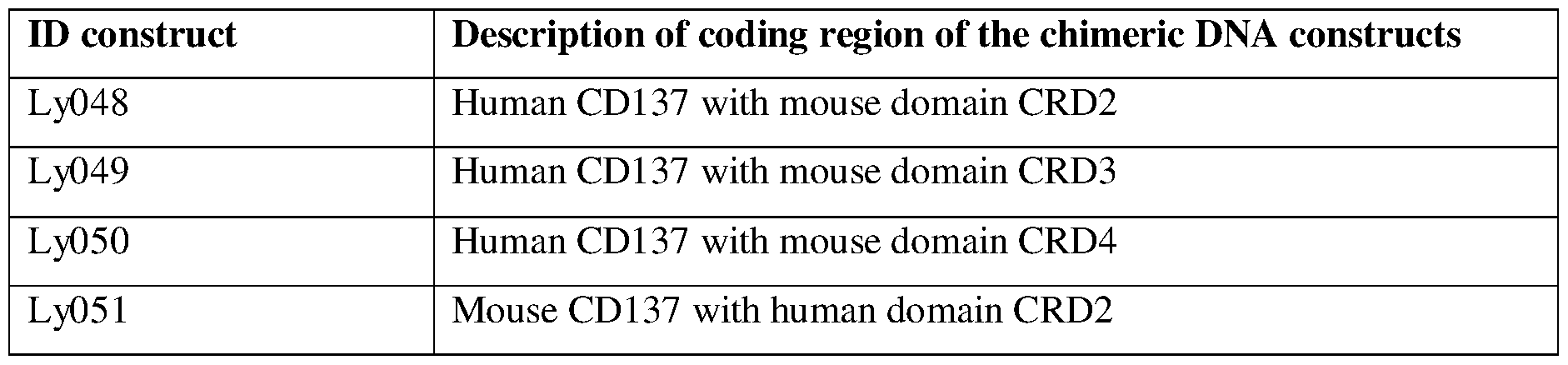


Claims
Priority Applications (7)
| Application Number | Priority Date | Filing Date | Title |
|---|---|---|---|
| CA3139274A CA3139274A1 (en) | 2019-05-10 | 2020-05-08 | Humanized anti-cd137 antibodies and uses thereof |
| CN202080021830.6A CN114026120B (en) | 2019-05-10 | 2020-05-08 | Humanized anti-CD 137 antibodies and uses thereof |
| KR1020217040560A KR20220008306A (en) | 2019-05-10 | 2020-05-08 | Humanized anti-CD137 antibodies and uses thereof |
| JP2021566504A JP2022532173A (en) | 2019-05-10 | 2020-05-08 | Humanized anti-CD137 antibody and its use |
| EP20804799.3A EP3966252A4 (en) | 2019-05-10 | 2020-05-08 | Humanized anti-cd137 antibodies and uses thereof |
| AU2020276500A AU2020276500A1 (en) | 2019-05-10 | 2020-05-08 | Humanized anti-CD137 antibodies and uses thereof |
| US17/206,730 US11203643B2 (en) | 2019-05-10 | 2021-03-19 | Humanized anti-CD137 antibodies and uses thereof |
Applications Claiming Priority (2)
| Application Number | Priority Date | Filing Date | Title |
|---|---|---|---|
| CN2019086364 | 2019-05-10 | ||
| CNPCT/CN2019/086364 | 2019-05-10 |
Related Child Applications (1)
| Application Number | Title | Priority Date | Filing Date |
|---|---|---|---|
| US17/206,730 Continuation US11203643B2 (en) | 2019-05-10 | 2021-03-19 | Humanized anti-CD137 antibodies and uses thereof |
Publications (1)
| Publication Number | Publication Date |
|---|---|
| WO2020231809A1 true WO2020231809A1 (en) | 2020-11-19 |
Family
ID=73289535
Family Applications (1)
| Application Number | Title | Priority Date | Filing Date |
|---|---|---|---|
| PCT/US2020/032095 WO2020231809A1 (en) | 2019-05-10 | 2020-05-08 | Humanized anti-cd137 antibodies and uses thereof |
Country Status (8)
| Country | Link |
|---|---|
| US (1) | US11203643B2 (en) |
| EP (1) | EP3966252A4 (en) |
| JP (1) | JP2022532173A (en) |
| KR (1) | KR20220008306A (en) |
| CN (1) | CN114026120B (en) |
| AU (1) | AU2020276500A1 (en) |
| CA (1) | CA3139274A1 (en) |
| WO (1) | WO2020231809A1 (en) |
Cited By (2)
| Publication number | Priority date | Publication date | Assignee | Title |
|---|---|---|---|---|
| US11505615B2 (en) | 2017-12-05 | 2022-11-22 | Lyvgen Biopharma Holdings Limited | Anti-CD137 antibodies and uses thereof |
| EP4063388A4 (en) * | 2019-11-19 | 2024-01-17 | Sound Biopharmaceuticals Co Ltd | Humanized 4-1bb monoclonal antibody and pharmaceutical composition thereof |
Families Citing this family (2)
| Publication number | Priority date | Publication date | Assignee | Title |
|---|---|---|---|---|
| CN113603780B (en) * | 2017-03-28 | 2022-07-01 | 礼进生物医药科技(上海)有限公司 | Therapeutic agents and methods for enhancing immune responses in tumor microenvironments |
| WO2024040195A1 (en) | 2022-08-17 | 2024-02-22 | Capstan Therapeutics, Inc. | Conditioning for in vivo immune cell engineering |
Citations (5)
| Publication number | Priority date | Publication date | Assignee | Title |
|---|---|---|---|---|
| US6569997B1 (en) * | 1995-03-23 | 2003-05-27 | Advanced Research And Technology Institute, Inc. | Antibody specific for H4-1BB |
| US20100183621A1 (en) * | 2003-10-10 | 2010-07-22 | Bristol-Myers Squibb Company | Fully human antibodies against human 4-1BB |
| US20140127225A1 (en) * | 2012-10-05 | 2014-05-08 | Neotope Biosciences Limited | Compositions and methods for treating diseases of protein aggregation involving ic3b deposition |
| US20160272708A1 (en) * | 2013-06-26 | 2016-09-22 | Shanghai Junshi Biosciences Inc. | Anti-pd-1 antibody and use thereof |
| WO2019113039A1 (en) * | 2017-12-05 | 2019-06-13 | Lyvgen Biopharma Co., Ltd. | Anti-cd137 antibodies and uses thereof |
Family Cites Families (20)
| Publication number | Priority date | Publication date | Assignee | Title |
|---|---|---|---|---|
| KR20000034847A (en) * | 1998-11-17 | 2000-06-26 | 성재갑 | Humanized Antibody Specific for Human 4-1BB Molecule and Pharmaceutical Composition Comprising Same |
| AU2002364935A1 (en) | 2001-10-09 | 2003-06-23 | Mayo Foundation For Medical Education And Research | Enhancement of immune responses by 4-1bb-binding agents |
| US20030223989A1 (en) | 2002-04-18 | 2003-12-04 | Pluenneke John D. | CD137 agonists to treat patients with IgE-mediated conditions |
| CN1997667A (en) * | 2004-05-10 | 2007-07-11 | 宏观基因有限公司 | Humanized FcgammaRIIB-specific antibodies and methods of use thereof |
| US20070253961A1 (en) | 2004-06-09 | 2007-11-01 | Ulsan Industrial Education Foundation | Pharmaceutical Composition Comprising the Anti-4-1Bb Antibody for Treating or Preventing Rheumatoid Arthritis |
| US20060171929A1 (en) * | 2005-01-31 | 2006-08-03 | The University Of Washington | Regulation of dendritic cell functions by the DCAL-2 receptor |
| WO2006129849A1 (en) | 2005-06-03 | 2006-12-07 | Mochida Pharmaceutical Co., Ltd. | Anti-cd14 antibody-fused protein |
| EP1806365A1 (en) | 2006-01-05 | 2007-07-11 | Boehringer Ingelheim International GmbH | Antibody molecules specific for fibroblast activation protein and immunoconjugates containing them |
| KR101527300B1 (en) | 2010-09-09 | 2015-06-09 | 화이자 인코포레이티드 | 4-1bb binding molecules |
| BR112016026993A2 (en) | 2014-05-21 | 2017-10-31 | Kyowa Hakko Kirin Co Ltd | combination of an anti-ccr4 antibody and a 4-1bb agonist to treat cancer |
| US20170247455A1 (en) | 2014-08-22 | 2017-08-31 | Bristol-Myers Squibb Company | Treatment of cancer using a combination of an anti-pd-1 antibody and an anti-cd137 antibody |
| CA3002588C (en) | 2015-10-23 | 2021-11-23 | Apogenix Ag | Single-chain cd137-receptor agonist proteins |
| CN106699889A (en) | 2015-11-18 | 2017-05-24 | 礼进生物医药科技(上海)有限公司 | PD-1 resisting antibody and treatment application thereof |
| US10875921B2 (en) | 2016-05-27 | 2020-12-29 | Abbvie Biotherapeutics Inc. | Anti-4-1BB antibodies and their uses |
| BR112019005895A2 (en) * | 2016-09-23 | 2019-06-11 | Merus N.V. | binding molecules that modulate a biological activity expressed by a cell |
| US10576161B2 (en) * | 2017-01-20 | 2020-03-03 | Magenta Therapeutics, Inc. | Compositions and methods for the depletion of CD137+ cells |
| WO2018227018A1 (en) * | 2017-06-07 | 2018-12-13 | Silverback Therapeutics, Inc. | Antibody conjugates of immune-modulatory compounds and uses thereof |
| CN107982538B (en) * | 2017-12-26 | 2021-10-22 | 深圳市体内生物医药科技有限公司 | Pharmaceutical composition and application thereof |
| WO2020069382A1 (en) * | 2018-09-28 | 2020-04-02 | Lyvgen Biopharma Co., Ltd. | Anti-cd137 binding molecules having engineered fc domains and therapeutic uses thereof |
| US20210395379A1 (en) * | 2018-10-09 | 2021-12-23 | Numab Therapeutics AG | Antibodies targeting cd137 and methods of use thereof |
-
2020
- 2020-05-08 WO PCT/US2020/032095 patent/WO2020231809A1/en unknown
- 2020-05-08 KR KR1020217040560A patent/KR20220008306A/en active Search and Examination
- 2020-05-08 JP JP2021566504A patent/JP2022532173A/en active Pending
- 2020-05-08 AU AU2020276500A patent/AU2020276500A1/en active Pending
- 2020-05-08 EP EP20804799.3A patent/EP3966252A4/en active Pending
- 2020-05-08 CN CN202080021830.6A patent/CN114026120B/en active Active
- 2020-05-08 CA CA3139274A patent/CA3139274A1/en active Pending
-
2021
- 2021-03-19 US US17/206,730 patent/US11203643B2/en active Active
Patent Citations (5)
| Publication number | Priority date | Publication date | Assignee | Title |
|---|---|---|---|---|
| US6569997B1 (en) * | 1995-03-23 | 2003-05-27 | Advanced Research And Technology Institute, Inc. | Antibody specific for H4-1BB |
| US20100183621A1 (en) * | 2003-10-10 | 2010-07-22 | Bristol-Myers Squibb Company | Fully human antibodies against human 4-1BB |
| US20140127225A1 (en) * | 2012-10-05 | 2014-05-08 | Neotope Biosciences Limited | Compositions and methods for treating diseases of protein aggregation involving ic3b deposition |
| US20160272708A1 (en) * | 2013-06-26 | 2016-09-22 | Shanghai Junshi Biosciences Inc. | Anti-pd-1 antibody and use thereof |
| WO2019113039A1 (en) * | 2017-12-05 | 2019-06-13 | Lyvgen Biopharma Co., Ltd. | Anti-cd137 antibodies and uses thereof |
Non-Patent Citations (1)
| Title |
|---|
| See also references of EP3966252A4 * |
Cited By (2)
| Publication number | Priority date | Publication date | Assignee | Title |
|---|---|---|---|---|
| US11505615B2 (en) | 2017-12-05 | 2022-11-22 | Lyvgen Biopharma Holdings Limited | Anti-CD137 antibodies and uses thereof |
| EP4063388A4 (en) * | 2019-11-19 | 2024-01-17 | Sound Biopharmaceuticals Co Ltd | Humanized 4-1bb monoclonal antibody and pharmaceutical composition thereof |
Also Published As
| Publication number | Publication date |
|---|---|
| AU2020276500A1 (en) | 2021-12-16 |
| JP2022532173A (en) | 2022-07-13 |
| CN114026120A (en) | 2022-02-08 |
| US20210246218A1 (en) | 2021-08-12 |
| CN114026120B (en) | 2024-03-19 |
| KR20220008306A (en) | 2022-01-20 |
| EP3966252A4 (en) | 2023-01-25 |
| US11203643B2 (en) | 2021-12-21 |
| EP3966252A1 (en) | 2022-03-16 |
| CA3139274A1 (en) | 2020-11-19 |
Similar Documents
| Publication | Publication Date | Title |
|---|---|---|
| AU2020294364B2 (en) | Anti-Galectin-9 antibodies and uses thereof | |
| US11203643B2 (en) | Humanized anti-CD137 antibodies and uses thereof | |
| US20220372155A1 (en) | Anti-cd40 binding molecules and bi-specific antibodies comprising such | |
| US11505615B2 (en) | Anti-CD137 antibodies and uses thereof | |
| WO2020069382A1 (en) | Anti-cd137 binding molecules having engineered fc domains and therapeutic uses thereof | |
| US11186648B2 (en) | Anti-CD40 antibody having engineered Fc domains and therapeutic uses thereof | |
| US20210179727A1 (en) | Anti-cd40 antibodies and uses thereof | |
| AU2019349874A1 (en) | Anti-CD40 binding molecules having engineered Fc domains and therapeutic uses thereof | |
| US20230406950A1 (en) | Bi-specific antibodies comprising anti-cd137 binding molecules | |
| WO2022232392A2 (en) | Bi-specific antibodies comprising anti-b7h3 binding molecules |
Legal Events
| Date | Code | Title | Description |
|---|---|---|---|
| 121 | Ep: the epo has been informed by wipo that ep was designated in this application |
Ref document number: 20804799 Country of ref document: EP Kind code of ref document: A1 |
|
| ENP | Entry into the national phase |
Ref document number: 3139274 Country of ref document: CA |
|
| ENP | Entry into the national phase |
Ref document number: 2021566504 Country of ref document: JP Kind code of ref document: A |
|
| NENP | Non-entry into the national phase |
Ref country code: DE |
|
| ENP | Entry into the national phase |
Ref document number: 20217040560 Country of ref document: KR Kind code of ref document: A |
|
| ENP | Entry into the national phase |
Ref document number: 2020276500 Country of ref document: AU Date of ref document: 20200508 Kind code of ref document: A |
|
| ENP | Entry into the national phase |
Ref document number: 2020804799 Country of ref document: EP Effective date: 20211210 |