WO2020217462A1 - Lifestyle habitat assessment system and program therefor - Google Patents
Lifestyle habitat assessment system and program therefor Download PDFInfo
- Publication number
- WO2020217462A1 WO2020217462A1 PCT/JP2019/017959 JP2019017959W WO2020217462A1 WO 2020217462 A1 WO2020217462 A1 WO 2020217462A1 JP 2019017959 W JP2019017959 W JP 2019017959W WO 2020217462 A1 WO2020217462 A1 WO 2020217462A1
- Authority
- WO
- WIPO (PCT)
- Prior art keywords
- lifestyle
- fat layer
- tomographic image
- rectus abdominis
- evaluation
- Prior art date
Links
Images


Classifications
-
- A—HUMAN NECESSITIES
- A61—MEDICAL OR VETERINARY SCIENCE; HYGIENE
- A61B—DIAGNOSIS; SURGERY; IDENTIFICATION
- A61B8/00—Diagnosis using ultrasonic, sonic or infrasonic waves
- A61B8/08—Detecting organic movements or changes, e.g. tumours, cysts, swellings
-
- A—HUMAN NECESSITIES
- A61—MEDICAL OR VETERINARY SCIENCE; HYGIENE
- A61B—DIAGNOSIS; SURGERY; IDENTIFICATION
- A61B5/00—Measuring for diagnostic purposes; Identification of persons
- A61B5/103—Detecting, measuring or recording devices for testing the shape, pattern, colour, size or movement of the body or parts thereof, for diagnostic purposes
- A61B5/107—Measuring physical dimensions, e.g. size of the entire body or parts thereof
-
- A—HUMAN NECESSITIES
- A61—MEDICAL OR VETERINARY SCIENCE; HYGIENE
- A61B—DIAGNOSIS; SURGERY; IDENTIFICATION
- A61B8/00—Diagnosis using ultrasonic, sonic or infrasonic waves
- A61B8/13—Tomography
- A61B8/14—Echo-tomography
-
- A—HUMAN NECESSITIES
- A61—MEDICAL OR VETERINARY SCIENCE; HYGIENE
- A61B—DIAGNOSIS; SURGERY; IDENTIFICATION
- A61B8/00—Diagnosis using ultrasonic, sonic or infrasonic waves
- A61B8/52—Devices using data or image processing specially adapted for diagnosis using ultrasonic, sonic or infrasonic waves
- A61B8/5207—Devices using data or image processing specially adapted for diagnosis using ultrasonic, sonic or infrasonic waves involving processing of raw data to produce diagnostic data, e.g. for generating an image
-
- G—PHYSICS
- G06—COMPUTING; CALCULATING OR COUNTING
- G06N—COMPUTING ARRANGEMENTS BASED ON SPECIFIC COMPUTATIONAL MODELS
- G06N3/00—Computing arrangements based on biological models
- G06N3/02—Neural networks
- G06N3/04—Architecture, e.g. interconnection topology
- G06N3/045—Combinations of networks
-
- G—PHYSICS
- G06—COMPUTING; CALCULATING OR COUNTING
- G06N—COMPUTING ARRANGEMENTS BASED ON SPECIFIC COMPUTATIONAL MODELS
- G06N3/00—Computing arrangements based on biological models
- G06N3/02—Neural networks
- G06N3/08—Learning methods
-
- G—PHYSICS
- G16—INFORMATION AND COMMUNICATION TECHNOLOGY [ICT] SPECIALLY ADAPTED FOR SPECIFIC APPLICATION FIELDS
- G16H—HEALTHCARE INFORMATICS, i.e. INFORMATION AND COMMUNICATION TECHNOLOGY [ICT] SPECIALLY ADAPTED FOR THE HANDLING OR PROCESSING OF MEDICAL OR HEALTHCARE DATA
- G16H20/00—ICT specially adapted for therapies or health-improving plans, e.g. for handling prescriptions, for steering therapy or for monitoring patient compliance
- G16H20/70—ICT specially adapted for therapies or health-improving plans, e.g. for handling prescriptions, for steering therapy or for monitoring patient compliance relating to mental therapies, e.g. psychological therapy or autogenous training
-
- G—PHYSICS
- G16—INFORMATION AND COMMUNICATION TECHNOLOGY [ICT] SPECIALLY ADAPTED FOR SPECIFIC APPLICATION FIELDS
- G16H—HEALTHCARE INFORMATICS, i.e. INFORMATION AND COMMUNICATION TECHNOLOGY [ICT] SPECIALLY ADAPTED FOR THE HANDLING OR PROCESSING OF MEDICAL OR HEALTHCARE DATA
- G16H30/00—ICT specially adapted for the handling or processing of medical images
- G16H30/40—ICT specially adapted for the handling or processing of medical images for processing medical images, e.g. editing
-
- G—PHYSICS
- G16—INFORMATION AND COMMUNICATION TECHNOLOGY [ICT] SPECIALLY ADAPTED FOR SPECIFIC APPLICATION FIELDS
- G16H—HEALTHCARE INFORMATICS, i.e. INFORMATION AND COMMUNICATION TECHNOLOGY [ICT] SPECIALLY ADAPTED FOR THE HANDLING OR PROCESSING OF MEDICAL OR HEALTHCARE DATA
- G16H50/00—ICT specially adapted for medical diagnosis, medical simulation or medical data mining; ICT specially adapted for detecting, monitoring or modelling epidemics or pandemics
- G16H50/30—ICT specially adapted for medical diagnosis, medical simulation or medical data mining; ICT specially adapted for detecting, monitoring or modelling epidemics or pandemics for calculating health indices; for individual health risk assessment
Definitions
- the present invention relates to a lifestyle-related evaluation system and its program, and particularly to the evaluation of lifestyle-related habits related to metabolic syndrome.
- Patent Document 1 discloses a visceral fat estimation method for estimating visceral fat based on the abdominal circumference at the navel position and the abdominal subcutaneous fat thickness.
- Patent Document 2 describes the preperitoneal visceral fat thickening degree (PFT), the carotid vascular thickening degree (IMT), and the brachial artery vascular endothelial dilatation degree (FMD) in the test site image acquired by the ultrasonic probe.
- PFT preperitoneal visceral fat thickening degree
- IMT carotid vascular thickening degree
- FMD brachial artery vascular endothelial dilatation degree
- Patent Document 3 discloses an ultrasonic system that measures an index value indicating the amount of visceral fat in the examination of metabolic syndrome.
- this diagnostic system first, in the tomographic image of the abdomen acquired by the ultrasonic probe, the length a between the body surface and the abdominal aorta and the length a1 between the outer edge of the visceral fat-containing region and the abdominal aorta. And are measured. Separately, the abdominal circumference is measured. The total area of the abdomen is calculated from the abdominal circumference length and the length a on the premise of elliptical approximation of the abdominal cross section.
- the area of the visceral fat-containing region is calculated as the area (partial area) of the similar ellipse, and the partial area and one or more of the subjects are calculated. From the individual parameter value, an index value indicating the amount of visceral fat is calculated.
- the tissue thickness information including the muscle thickness and the fat thickness of the subject is obtained based on the ultrasonic image, and the tissue mass index value of the subject is set as a target value based on the tissue thickness information.
- An ultrasonic measuring device that generates guideline information for approaching is disclosed.
- the present inventor observes and hears the abdomen of more than 20,000 subjects in 13 years at various sites such as medical sites, fitness gyms, and event venues to prevent overeating and lack of exercise for lean people. As a result of continuous verification, we came to find an effective and objective evaluation method.
- the present invention has been made in view of such circumstances, and an object thereof is to appropriately evaluate lifestyle-related habits related to metabolic syndrome.
- the first invention provides a lifestyle-related evaluation system that has an ultrasonic probe and a feature evaluation unit and evaluates lifestyle-related habits related to metabolic syndrome.
- the ultrasonic probe images the subject's abdomen and outputs a tomographic image of the abdomen.
- the feature evaluation unit outputs data showing the characteristics of at least the subcutaneous fat layer, the visceral fat layer, and the left and right rectus abdominis muscles as lifestyle evaluation index data among the biological parts drawn on the tomographic image. To do.
- a countermeasure presentation unit may be provided.
- the countermeasure presentation unit selectively presents one of a plurality of countermeasure patterns that systematically classify the countermeasures related to lifestyle-related habits based on the evaluation index data.
- the feature evaluation unit may have a first learning model and a measurement unit.
- the first learning model distinguishes between the subcutaneous fat layer, the visceral fat layer, and the left and right rectus abdominis muscles depicted on the tomographic image.
- the measuring unit measures each of the subcutaneous fat layer, the visceral fat layer, and the left and right rectus abdominis muscles identified by the first learning model according to a predetermined standard, and uses a plurality of measured values obtained by this measurement. Based on this, the evaluation index data is output. In this case, it is preferable to provide the first learning processing unit.
- the first learning processing unit is based on supervised learning using supervised learning that teaches the positions of the subcutaneous fat layer, the visceral fat layer, and the left and right rectus abdominis muscles drawn on the tomographic image. Performs learning processing of the learning model.
- the feature evaluation unit may have a second learning model.
- the second learning model classifies the integrated features of the subcutaneous fat layer, the visceral fat layer, and the left and right rectus abdominis muscles depicted in the tomographic image into one of a plurality of predetermined classification patterns. .. It has a second learning model. Then, the feature evaluation unit outputs evaluation index data based on the classification pattern classified by the second learning model. In this case, it is preferable to provide a second learning processing unit.
- the second learning model is supervised learning using teacher data that teaches a classification pattern that classifies the integrated features of the subcutaneous fat layer, the visceral fat layer, and the left and right rectus abdominis muscles depicted on the tomographic image. Performs the learning process of the second learning model.
- the evaluation index data preferably includes the shape characteristics of the left and right rectus abdominis muscles and the quantitative characteristics of the subcutaneous fat layer and the visceral fat layer.
- the evaluation index data may include the brightness of the rectus abdominis muscle in the tomographic image.
- the tomographic image is acquired by an ultrasonic probe with the upper body of the subject standing upright.
- the second invention provides a lifestyle-related evaluation program that causes a computer to perform the following steps and evaluates lifestyle-related habits related to metabolic syndrome.
- the first step the tomographic image obtained by imaging the abdomen of the subject with an ultrasonic probe is analyzed.
- the second step among the biological parts depicted on the tomographic image, data showing the characteristics of at least the subcutaneous fat layer, the visceral fat layer, and the left and right rectus abdominis muscles are used as lifestyle-related evaluation index data.
- Output the tomographic image obtained by imaging the abdomen of the subject with an ultrasonic probe is analyzed.
- the third step may be provided.
- one of a plurality of countermeasure patterns systematically categorizing lifestyle-related measures is selectively presented based on the evaluation index data.
- the first step is acquired by an ultrasonic probe into a first learning model that distinguishes the subcutaneous fat layer, the visceral fat layer, and the left and right rectus abdominis muscles depicted in the tomographic image. It may have a step of inputting a tomographic image and a step of measuring each of the subcutaneous fat layer identified by the first learning model, the visceral fat layer, and the left and right rectus abdominis muscles. In this case, the second step outputs evaluation index data based on the plurality of measured values obtained by the measurement.
- the first learning model is subjected to supervised learning using supervised learning that teaches the positions of the subcutaneous fat layer, the visceral fat layer, and the left and right rectus abdominis muscles drawn on the tomographic image.
- a fourth step of performing the learning process may be provided.
- the first step describes a plurality of predetermined classification patterns of the integrated features of the subcutaneous fat layer, the visceral fat layer, and the left and right rectus abdominis muscles depicted in the tomographic image.
- the second learning model classified into any of the above may have a step of inputting a tomographic image acquired by an ultrasonic probe.
- the second step outputs the evaluation index data based on the classification pattern classified by the second learning model.
- supervised learning using teacher data that teaches a classification pattern that classifies the integrated features of the subcutaneous fat layer, the visceral fat layer, and the left and right rectus abdominis muscles depicted on the tomographic image.
- a fourth step of performing the learning process of the second learning model may be provided.
- the evaluation index data is based on the shape characteristics of the left and right rectus abdominis muscles. It preferably includes the quantitative characteristics of the subcutaneous fat layer and the visceral fat layer.
- the evaluation index data may include the brightness of the rectus abdominis muscle in the tomographic image. Further, it is preferable that the tomographic image is acquired by an ultrasonic probe with the upper body of the subject standing upright.
- data showing the characteristics of the subcutaneous fat layer, the visceral fat layer, and the left and right rectus abdominis muscles among the biological parts visualized on the tomographic image are output as lifestyle-related evaluation index data. ..
- lifestyle-related evaluation index data By focusing not only on the fat layers such as the subcutaneous fat layer and the visceral fat layer but also on the left and right abdominal muscles and comprehensively evaluating these characteristics, not only those who have already developed metabolic syndrome but also those who have not yet developed it. Lifestyle-related habits related to metabolic syndrome can be appropriately evaluated even for those who have the disease.
- Block diagram of lifestyle-related evaluation system Explanatory drawing of the abdominal diagnosis of the subject
- Explanatory drawing of measurement example of rectus abdominis muscle Explanatory drawing of the brightness of the rectus abdominis muscle in the tomographic image
- Classification chart of rectus abdominis muscle characteristics Explanatory diagram of combination pattern of countermeasure advice
- Block diagram of lifestyle-related evaluation system according to the second embodiment
- FIG. 1 is a block diagram of a lifestyle-related evaluation system according to the first embodiment.
- This lifestyle-related evaluation system 1 is based on an ultrasonic image (echo image) of the inside of the abdomen of a subject, and mainly includes amounts related to four biological parts such as a subcutaneous fat layer, a visceral fat layer, and left and right rectus abdominis muscles. Evaluate features such as shape and output these as lifestyle-related evaluation indexes.
- various lifestyle-related habits are assumed, but the focus of this embodiment is the lifestyle-related habits related to metabolic syndrome.
- the lifestyle-related evaluation system also has a function of presenting useful advice for improving the lifestyle of the subject.
- a function of presenting useful advice for improving the lifestyle of the subject By systematizing the quantitative, qualitative or morphological changes in subcutaneous fat, visceral fat, and left and right rectus abdominis muscles, we objectively evaluate overeating and lack of exercise so that anyone can understand them.
- One of the features of this embodiment is that it focuses not only on the subcutaneous fat layer and the visceral fat layer but also on the left and right rectus abdominis muscles in order to evaluate the subject's lack of exercise.
- the rectus abdominis muscle is not used as much as the muscles of the limbs in daily life, so a person who has a strong rectus abdominis muscle can think that the muscles of the limbs are almost strong. From this, it is possible to estimate the degree of lack of exercise of the subject by introducing the characteristics of the left and right rectus abdominis muscles as an evaluation index.
- the lifestyle-related evaluation system 1 has an ultrasonic probe 2, a feature evaluation unit 3A, a countermeasure presentation unit 4, and a learning processing unit 5.
- the ultrasonic probe 2 images the abdomen of the subject and acquires a tomographic image of the abdomen.
- FIG. 2 is an explanatory diagram of the abdominal diagnosis of the subject.
- the examiner takes a tomographic image of the abdomen by bringing the ultrasonic probe 1 into contact with the abdomen of the subject.
- the example in the figure is a tomographic image of the vicinity of the liver, which is located between the subcutaneous fat layer located directly below the skin layer, the visceral fat layer located directly above the peritoneum, and the subcutaneous fat layer and the visceral fat layer.
- the left and right rectus abdominis muscles (rectus abdominis) are depicted.
- the tomographic image acquired by the ultrasonic probe 2 is output to the feature evaluation unit 3A.
- the feature evaluation unit 3A inputs the tomographic image of the ultrasonic probe 2 and outputs the evaluation index data.
- This evaluation index data shows at least the characteristics of the subcutaneous fat layer, the visceral fat layer, and the left and right rectus abdominis muscles among the biological parts depicted in the tomographic image, and these characteristics are indexed. It was done.
- the feature evaluation unit 3A has a learning model 3a and a measurement unit 3b.
- the learning model 3a is built mainly on a neural network, and has a predetermined problem-solving ability. Specifically, the learning model 3a regionally distinguishes the subcutaneous fat layer, the visceral fat layer, and the left and right rectus abdominis muscles depicted in the tomographic image in response to the input of the tomographic image (FIG. 2). reference).
- the "neural network” is a combination of mathematical models of neurons, and is not only the most primitive configuration of a neural network, but also a convolutional neural network (CNN) or a recurrent neural network (RNN). As such, it broadly includes its derivative forms and advanced forms. Further, YOLO (You Only Look Once) or SSD (Single Shot MultiBox Detector), which has recently attracted attention as an object detection algorithm of a neural network system, may be used.
- the learning processing unit 5 learns the learning model 3a by supervised learning using supervised learning that teaches the positions of the subcutaneous fat layer, the visceral fat layer, and the left and right rectus abdominis muscles drawn on the tomographic image. Perform processing. By this learning process, the internal parameter ⁇ of the learning model 3a is adjusted. By repeating supervised learning using a large amount of various teacher data, the learning model 3a is optimized so that appropriate outputs can be obtained for various inputs.
- the measuring unit 3b measures the characteristics of the left and right rectus abdominis muscles regionally identified by the learning model 3a, specifically, the shape characteristics.
- FIG. 3 is an explanatory diagram of a measurement example of one rectus abdominis muscle.
- the thickness A, the angle B, and the rise C are measured as the shape features of the rectus abdominis muscle.
- the thickness A is the thickness of the rectus abdominis muscle
- the angle B is the angle formed by the median of the rectus abdominis muscle and the highest point of the ridge
- the rising C is the rising form from the median of the rectus abdominis muscle.
- the measured values A to C of the shape feature as shown in FIG.
- the brightness D of the left and right rectus abdominis muscles drawn on the tomographic image may be measured.
- These measured values A to D are calculated for each of the left and right rectus abdominis muscles.
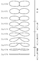
- FIG. 5 is a classification diagram of the characteristics of the rectus abdominis muscle.
- the shape characteristics (state) of the rectus abdominis muscle are classified into one of a plurality of predetermined phases based on the measured values A to D of the rectus abdominis muscle.
- the rectus abdominis muscle in the best condition (exercise is the most sufficient), and as the phase becomes larger, the rectus abdominis muscle condition gradually weakens (the tendency of lack of exercise increases), and the phase 9 is the weakest state (complete lack of exercise).
- the phase representing the shape characteristics of the rectus abdominis muscle is output to the countermeasure presentation unit 4 as a part of the evaluation index data.
- the measuring unit 3b individually measures the characteristics of the subcutaneous fat layer and the visceral fat layer specifically identified by the learning model 3a, specifically, the quantitative characteristics (states) of these fat layers.
- the quantitative characteristics of the subcutaneous fat layer are classified into one of a plurality of predetermined phases based on the measured value (for example, thickness) of the subcutaneous fat layer.
- the quantitative characteristics (state) of the visceral fat layer are classified into one of a plurality of predetermined phases based on the measured value (for example, thickness) of the visceral fat layer.
- Each of the classified phases for the subcutaneous fat layer and the visceral fat layer is output to the countermeasure presentation unit 4 as a part of the evaluation index data.
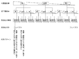
- the countermeasure presentation unit 4 presents a lifestyle-related countermeasure pattern to the subject according to the evaluation index data output from the feature evaluation unit 3A.
- the evaluation index data includes at least "subcutaneous fat thickness” (10 levels) representing the quantitative characteristics of the visceral fat layer and "" representing the quantitative characteristics of the visceral fat layer. It suffices to have "visceral fat thickness” (10 levels) and “rectus abdominis muscle shape” (10 levels) that represents the shape characteristics of the rectus abdominis muscle (three-dimensional vector).
- rectus abdominis muscle brightness (5 levels)
- premeasure or absence of rectus abdominis muscle separation (2 categories)
- visceral organs are representative of the state of rectus abdominis muscle brightness.
- premeasure or absence of exclusion (2 categories) (6 dimensional vector). This makes it possible to infer possible causes and present countermeasures as countermeasure patterns for 20,000 combinations. Although it is possible to set up to 20,000 countermeasure patterns, the number of patterns may be smaller than this by standardizing the stages and classifications on the vector in terms of implementation.
- the countermeasure presentation unit 4 includes a knowledge database 4a.
- this knowledge database 4a a large number of countermeasure patterns that systematically classify measures related to lifestyle habits are stored, and one of the countermeasure patterns is selectively presented according to the evaluation index data.
- the content of the countermeasure advice is individually defined by "visceral fat thickness”, “subcutaneous fat thickness”, “luminance of the rectus abdominis muscle”, “shape of the rectus abdominis muscle” and the like.
- visceral fat thickness the accumulation of visceral fat is considered to be directly linked to lifestyle-related diseases, and is correlated with lack of exercise and intake of alcohol, fat, and sweets.
- Subcutaneous fat thickness is less likely to fluctuate than visceral fat, and does not decrease with muscle training, but tends to decrease with aerobic exercise, etc., and tends to increase with those who eat sweet foods.
- luminance of the rectus abdominis muscle it is considered that the higher the brightness is, the more fat is contained, and the lower the brightness is, the more pure muscle is.
- the "shape of the rectus abdominis muscle” is as described above.
- the content of the countermeasure advice presented to the subject differs depending on the pattern classified according to the evaluation index data.
- This pattern includes, for example, “ideal pattern”, “athlete pattern”, “male metabolic syndrome”, “female metabolic syndrome”, “left-right asymmetric pattern”, “rectus abdominis muscle separation pattern”, “stomach leaning pattern”, etc.
- the "ideal pattern” can be said to be in an ideal state with less subcutaneous fat and visceral fat, thick rectus abdominis muscle, and firm constriction.
- the "athlete pattern” is low in subcutaneous and visceral fat, and the rectus abdominis muscle is fairly thick and trapezoidal.
- the "male metabolic syndrome” is a pattern in which the visceral fat is thick and the eating habits are poor. The rectus abdominis muscle weakened by the excessive visceral fat is pushed to the left and right, and the abdomen is hungry. In the “female metabolic syndrome", the rectus abdominis muscles are thin and weak, and the rectus abdominis muscles are not constricted and are connected in a straight line.
- the "left-right asymmetric pattern” has thick subcutaneous fat and visceral fat and is a complete poor lifestyle, and there is a difference in the thickness of the left and right rectus abdominis muscles, which causes a problem in how to use the body.
- the rectus abdominis muscle separation pattern if the person is thin but the left and right rectus abdominis muscles are separated by a large width, the rectus abdominis muscle may be stronger and pulled outward than the rectus abdominis muscle. , The waist tends to be less constricted.
- the abdomen does not come forward due to the stiff muscles, but the excess visceral fat is squeezing out the organs, making it easy for the stomach to lean.
- the total amount of visceral fat is small, but due to overeating for a very short period of time, the visceral fat that has increased rapidly excludes the organs, making it easy for the stomach to lean.
- the data showing the characteristics of the subcutaneous fat layer, the visceral fat layer, and the left and right rectus abdominis muscles are used to evaluate the lifestyle. Output as index data.
- the characteristics of the fat layer such as the subcutaneous fat layer and the visceral fat layer, serve as evaluation indexes for the subject such as overeating and stomach upset.
- the characteristics of the left and right rectus abdominis muscles serve as evaluation indexes such as lack of exercise of the subject.
- the left and right abdominal muscles are also focused on, and these characteristics are comprehensively evaluated. This makes it possible to appropriately evaluate lifestyle-related habits related to metabolic syndrome, including those who have already developed metabolic syndrome as well as those who have not yet developed it.
- one of a plurality of countermeasure patterns systematically categorizing measures related to lifestyle habits is selectively presented based on the evaluation index data of lifestyle habits. This makes it possible to automatically present objective and effective countermeasure advice regarding lifestyle-related improvements related to metabolic syndrome.
- the learning model 3a by using the learning model 3a, it is possible to accurately distinguish each of the subcutaneous fat layer, the visceral fat layer, and the left and right rectus abdominis muscles depicted in the tomographic image. Can be presented appropriately and with high reliability.
- the ultrasonic probe 2 by acquiring the tomographic image by the ultrasonic probe 2 with the upper body of the subject standing upright, it is possible to reduce the change (variation) of the biological part in the tomographic image, which is good for diagnosis. A stable tomographic image can be obtained.
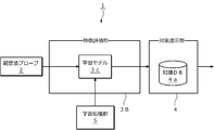
- FIG. 8 is a block diagram of the lifestyle-related evaluation system according to the second embodiment.
- the feature of this embodiment is that the configuration of the feature evaluation unit 3B, specifically, the functions of the learning model 3a and the measurement unit 3b according to the first embodiment are integratedly realized by a single learning model 3c. It is a point. Since the other points are the same as those in the first embodiment, the same reference numerals are given and the description thereof will be omitted here.
- the learning model 3c classifies the integrated features of the subcutaneous fat layer, the visceral fat layer, and the left and right rectus abdominis muscles depicted in the tomographic image into one of a plurality of predetermined classification patterns.
- this classification pattern includes "subcutaneous fat thickness” (10 levels), “visceral fat thickness” (10 levels) that represents the quantitative characteristics of the visceral fat layer, and the shape of the rectus abdominis muscle. It includes at least the "rectus abdominis muscle shape” (10 steps), which represents various features (three-dimensional vector).
- the learning processing unit 5 performs learning processing on the learning model 3c.
- the teacher data for supervised learning is different.
- teacher data that teaches a classification pattern that classifies the integrated features of the subcutaneous fat layer, the visceral fat layer, and the left and right rectus abdominis muscles depicted in the tomographic image is available. Used.
- the learning model 3a and the measuring unit 3c according to the first embodiment are integrated by a single learning model 3c. By doing so, the processing load can be reduced.
- the countermeasure presentation unit 4 it is not always necessary to provide the countermeasure presentation unit 4 in each of the above-described embodiments.
- the advisor refers to the above-mentioned evaluation index data and advises the subject on measures related to lifestyle habits, it is not necessary to provide the measure presentation unit 4.
- the lifestyle evaluation system 1 is linked with an external system, it is not necessary to provide the countermeasure presentation unit 4.
- the lifestyle-related evaluation system 1 is linked with a diagnostic system for arteriosclerosis and the circulatory system, and the evaluation index data of the lifestyle-related evaluation system 1 is used as one element of this diagnosis.
- the present invention is a computer program that equivalently realizes the functional blocks constituting the lifestyle-related evaluation system according to each of the above-described embodiments, specifically, the feature evaluation units 3A and 3B and the countermeasure presentation unit 4 on a computer. It can also be regarded as a (lifestyle-related measure presentation program).
Landscapes
- Health & Medical Sciences (AREA)
- Life Sciences & Earth Sciences (AREA)
- Engineering & Computer Science (AREA)
- General Health & Medical Sciences (AREA)
- Physics & Mathematics (AREA)
- Medical Informatics (AREA)
- Public Health (AREA)
- Biomedical Technology (AREA)
- Molecular Biology (AREA)
- Biophysics (AREA)
- Pathology (AREA)
- Heart & Thoracic Surgery (AREA)
- Radiology & Medical Imaging (AREA)
- Surgery (AREA)
- Animal Behavior & Ethology (AREA)
- Veterinary Medicine (AREA)
- Nuclear Medicine, Radiotherapy & Molecular Imaging (AREA)
- Theoretical Computer Science (AREA)
- Data Mining & Analysis (AREA)
- Epidemiology (AREA)
- Primary Health Care (AREA)
- Computational Linguistics (AREA)
- General Physics & Mathematics (AREA)
- General Engineering & Computer Science (AREA)
- Mathematical Physics (AREA)
- Artificial Intelligence (AREA)
- Software Systems (AREA)
- Evolutionary Computation (AREA)
- Computing Systems (AREA)
- Computer Vision & Pattern Recognition (AREA)
- Databases & Information Systems (AREA)
- Child & Adolescent Psychology (AREA)
- Hospice & Palliative Care (AREA)
- Oral & Maxillofacial Surgery (AREA)
- Ultra Sonic Daignosis Equipment (AREA)
- Social Psychology (AREA)
- Developmental Disabilities (AREA)
- Dentistry (AREA)
- Psychology (AREA)
- Psychiatry (AREA)
Abstract
[Problem] To appropriately assess lifestyle habitats associated with metabolic syndrome. [Solution] An ultrasound probe 2 captures an image of the abdomen of a subject and outputs a tomographic image of the abdomen. A feature assessment unit 3A is provided with a learning model 3a and a measurement unit 3b, and outputs assessment index data indicating the respective features of at least a subcutaneous fat layer, a visceral fat layer, and left and right abdominal rectus muscles among living body parts captured in the tomographic images. A measure presentation unit 4 selectively presents any of a plurality of measure patterns in which measures relating to lifestyle habitats are systematically classified according to the assessment index data.
Description
本発明は、生活習慣評価システムおよびそのプログラムに係り、特に、メタボリックシンドロームに関連した生活習慣の評価に関する。
The present invention relates to a lifestyle-related evaluation system and its program, and particularly to the evaluation of lifestyle-related habits related to metabolic syndrome.
従来、メタボリックシンドロームの診断を行う手法が知られている。例えば、特許文献1には、臍位置における腹部周囲径と、腹部皮下脂肪厚とに基づいて、内臓脂肪を推定する内臓脂肪推定方法が開示されている。特許文献2には、超音波プローブによって取得された被検部画像において、腹膜前内蔵脂肪肥厚度(PFT)と、頸動脈血管肥厚度(IMT)と、上腕動脈血管内皮拡張度(FMD)とを測定し、これらの測定結果を総合してメタボリックシンドロームの危険度を診断・評価するメタボリックシンドローム血管評価システムが開示されている。
Conventionally, a method for diagnosing metabolic syndrome has been known. For example, Patent Document 1 discloses a visceral fat estimation method for estimating visceral fat based on the abdominal circumference at the navel position and the abdominal subcutaneous fat thickness. Patent Document 2 describes the preperitoneal visceral fat thickening degree (PFT), the carotid vascular thickening degree (IMT), and the brachial artery vascular endothelial dilatation degree (FMD) in the test site image acquired by the ultrasonic probe. Is disclosed, and a metabolic syndrome blood vessel evaluation system is disclosed that diagnoses and evaluates the risk of metabolic syndrome by integrating these measurement results.
また、特許文献3には、メタボリックシンドロームの検診において、内臓脂肪量を表す指標値を測定する超音波システムが開示されている。この診断システムでは、まず、超音波プローブによって取得された腹部の断層画像において、体表面と腹部大動脈との間の長さaと、内臓脂肪含有領域の外縁と腹部大動脈との間の長さa1とが計測される。それとは別に、腹囲長が計測される。腹部断面の楕円近似を前提として、腹囲長と長さaとから腹部の全体面積が演算される。そして、全体面積と、長さa及び長さa1の比率とから、内臓脂肪含有領域の面積が相似形楕円の面積(部分面積)として演算され、その部分面積と、被験者についての1または複数の個人パラメータ値とから、内臓脂肪量を表す指標値が演算される。
Further, Patent Document 3 discloses an ultrasonic system that measures an index value indicating the amount of visceral fat in the examination of metabolic syndrome. In this diagnostic system, first, in the tomographic image of the abdomen acquired by the ultrasonic probe, the length a between the body surface and the abdominal aorta and the length a1 between the outer edge of the visceral fat-containing region and the abdominal aorta. And are measured. Separately, the abdominal circumference is measured. The total area of the abdomen is calculated from the abdominal circumference length and the length a on the premise of elliptical approximation of the abdominal cross section. Then, from the total area and the ratio of the length a and the length a1, the area of the visceral fat-containing region is calculated as the area (partial area) of the similar ellipse, and the partial area and one or more of the subjects are calculated. From the individual parameter value, an index value indicating the amount of visceral fat is calculated.
さらに、特許文献4には、超音波画像に基づいて、被験者の筋肉の厚さおよび脂肪の厚さを含む組織厚情報を求め、この組織厚情報に基づいて被験者の組織量指標値を目標値に近づけるための指針情報を生成する超音波測定装置が開示されている。
Further, in Patent Document 4, the tissue thickness information including the muscle thickness and the fat thickness of the subject is obtained based on the ultrasonic image, and the tissue mass index value of the subject is set as a target value based on the tissue thickness information. An ultrasonic measuring device that generates guideline information for approaching is disclosed.
近年、医療費の抑制などの観点から、健康づくり対策が推進されており、将来的なメタボリックシンドロームなどの発症を防止すべく、日々の生活習慣を改善して内臓脂肪を減少することが望まれている。しかしながら、現状、メタボリックシンドロームであるか否かの診断方法しか存在せず、これが未だ発症していない人も含めて、メタボリックシンドロームにならないように、生活習慣の改善や指導を行う仕組みは存在しない。いうまでもなく、痩せている人であっても、食べ過ぎや運動不足が続けば、将来的に発症する危険性があるので、メタボリックシンドロームが未発症の段階で生活習慣を評価し、必要に応じて改善を促すことの意義は大きい。本発明者は、医療現場、フィットネスジム、イベント会場などの様々な現場において、13年間で2万人以上の被験者の腹部の観察とヒアリングとを行い、痩せている人の食べ過ぎや運動不足を検証し続けた結果、客観性のある有効な評価手法を見出すに至った。
In recent years, health promotion measures have been promoted from the viewpoint of controlling medical expenses, and it is desired to improve daily lifestyles and reduce visceral fat in order to prevent the onset of metabolic syndrome in the future. ing. However, at present, there is only a method for diagnosing whether or not the person has metabolic syndrome, and there is no mechanism for improving lifestyle habits or giving guidance so as to prevent the person from developing the metabolic syndrome, including those who have not yet developed the syndrome. Needless to say, even if you are thin, if you continue to eat too much or lack exercise, there is a risk of developing it in the future, so it is necessary to evaluate lifestyle habits at the stage when metabolic syndrome has not developed. The significance of promoting improvement accordingly is great. The present inventor observes and hears the abdomen of more than 20,000 subjects in 13 years at various sites such as medical sites, fitness gyms, and event venues to prevent overeating and lack of exercise for lean people. As a result of continuous verification, we came to find an effective and objective evaluation method.
本発明は、かかる事情に鑑みてなされたものであり、その目的は、メタボリックシンドロームに関連した生活習慣を適切に評価することである。
The present invention has been made in view of such circumstances, and an object thereof is to appropriately evaluate lifestyle-related habits related to metabolic syndrome.
かかる課題を解決すべく、第1の発明は、超音波プローブと、特徴評価部とを有し、メタボリックシンドロームに関連した生活習慣を評価する生活習慣評価システムを提供する。超音波プローブは、被験者の腹部を撮像して、腹部の断層画像を出力する。特徴評価部は、断層画像に描出された生体部位のうち、少なくとも、皮下脂肪層と、内蔵脂肪層と、左右の腹直筋とのそれぞれの特徴を示すデータを生活習慣の評価指標データとして出力する。
In order to solve such a problem, the first invention provides a lifestyle-related evaluation system that has an ultrasonic probe and a feature evaluation unit and evaluates lifestyle-related habits related to metabolic syndrome. The ultrasonic probe images the subject's abdomen and outputs a tomographic image of the abdomen. The feature evaluation unit outputs data showing the characteristics of at least the subcutaneous fat layer, the visceral fat layer, and the left and right rectus abdominis muscles as lifestyle evaluation index data among the biological parts drawn on the tomographic image. To do.
ここで、第1の発明において、対策提示部を設けてもよい。対策提示部は、評価指標データに基づいて、生活習慣に関する対策を体系的に分類した複数の対策パターンのいずれかを選択的に提示する。
Here, in the first invention, a countermeasure presentation unit may be provided. The countermeasure presentation unit selectively presents one of a plurality of countermeasure patterns that systematically classify the countermeasures related to lifestyle-related habits based on the evaluation index data.
第1の発明において、上記特徴評価部は、第1の学習モデルと、測定部とを有していてもよい。第1の学習モデルは、断層画像に描出された皮下脂肪層と、内蔵脂肪層と、左右の腹直筋とのそれぞれを識別する。測定部は、第1の学習モデルによって識別された皮下脂肪層と、内蔵脂肪層と、左右の腹直筋とのそれぞれを所定の基準に従って測定し、この測定によって得られた複数の測定値に基づいて、評価指標データを出力する。この場合、第1の学習処理部を設けることが好ましい。第1の学習処理部は、断層画像に描出された皮下脂肪層と、内蔵脂肪層と、左右の腹直筋とのそれぞれの位置を教示する教師データを用いた教師あり学習によって、第1の学習モデルの学習処理を行う。
In the first invention, the feature evaluation unit may have a first learning model and a measurement unit. The first learning model distinguishes between the subcutaneous fat layer, the visceral fat layer, and the left and right rectus abdominis muscles depicted on the tomographic image. The measuring unit measures each of the subcutaneous fat layer, the visceral fat layer, and the left and right rectus abdominis muscles identified by the first learning model according to a predetermined standard, and uses a plurality of measured values obtained by this measurement. Based on this, the evaluation index data is output. In this case, it is preferable to provide the first learning processing unit. The first learning processing unit is based on supervised learning using supervised learning that teaches the positions of the subcutaneous fat layer, the visceral fat layer, and the left and right rectus abdominis muscles drawn on the tomographic image. Performs learning processing of the learning model.
第1の発明において、上記特徴評価部は、第2の学習モデルを有していてもよい。第2の学習モデルは、断層画像に描出された皮下脂肪層と、内蔵脂肪層と、左右の腹直筋との統合的な特徴を、予め定められた複数の分類パターンのいずれかに分類する。第2の学習モデルを有する。そして、上記特徴評価部は、第2の学習モデルによって分類された分類パターンに基づいて、評価指標データを出力する。この場合、第2の学習処理部を設けることが好ましい。第2の学習モデルは、断層画像に描出された皮下脂肪層と、内蔵脂肪層と、左右の腹直筋との統合的な特徴を分類した分類パターンを教示する教師データを用いた教師あり学習によって、第2の学習モデルの学習処理を行う。
In the first invention, the feature evaluation unit may have a second learning model. The second learning model classifies the integrated features of the subcutaneous fat layer, the visceral fat layer, and the left and right rectus abdominis muscles depicted in the tomographic image into one of a plurality of predetermined classification patterns. .. It has a second learning model. Then, the feature evaluation unit outputs evaluation index data based on the classification pattern classified by the second learning model. In this case, it is preferable to provide a second learning processing unit. The second learning model is supervised learning using teacher data that teaches a classification pattern that classifies the integrated features of the subcutaneous fat layer, the visceral fat layer, and the left and right rectus abdominis muscles depicted on the tomographic image. Performs the learning process of the second learning model.
第1の発明において、上記評価指標データは、左右の腹直筋の形状的な特徴と、皮下脂肪層および内蔵脂肪層の量的な特徴とを含むことが好ましい。また、上記評価指標データは、断層画像における腹直筋の輝度を含んでいてもよい。さらに、上記断層画像は、被験者の上半身が起立した状態で、超音波プローブによって取得されることが好ましい。
In the first invention, the evaluation index data preferably includes the shape characteristics of the left and right rectus abdominis muscles and the quantitative characteristics of the subcutaneous fat layer and the visceral fat layer. In addition, the evaluation index data may include the brightness of the rectus abdominis muscle in the tomographic image. Further, it is preferable that the tomographic image is acquired by an ultrasonic probe with the upper body of the subject standing upright.
第2の発明は、以下のステップをコンピュータに実行させ、メタボリックシンドロームに関連した生活習慣を評価する生活習慣評価プログラムを提供する。第1のステップでは、超音波プローブで被験者の腹部を撮像することによって取得された断層画像を解析する。第2のステップでは、断層画像に描出された生体部位のうち、少なくとも、皮下脂肪層と、内蔵脂肪層と、左右の腹直筋とのそれぞれの特徴を示すデータを生活習慣の評価指標データとして出力する。
The second invention provides a lifestyle-related evaluation program that causes a computer to perform the following steps and evaluates lifestyle-related habits related to metabolic syndrome. In the first step, the tomographic image obtained by imaging the abdomen of the subject with an ultrasonic probe is analyzed. In the second step, among the biological parts depicted on the tomographic image, data showing the characteristics of at least the subcutaneous fat layer, the visceral fat layer, and the left and right rectus abdominis muscles are used as lifestyle-related evaluation index data. Output.
ここで、第2の発明において、第3のステップを設けてもよい。第3のステップでは、評価指標データに基づいて、生活習慣に関する対策を体系的に分類した複数の対策パターンのいずれかを選択的に提示する。
Here, in the second invention, the third step may be provided. In the third step, one of a plurality of countermeasure patterns systematically categorizing lifestyle-related measures is selectively presented based on the evaluation index data.
第2の発明において、上記第1のステップは、断層画像に描出された皮下脂肪層と、内蔵脂肪層と、左右の腹直筋とを識別する第1の学習モデルに、超音波プローブによって取得された断層画像を入力するステップと、第1の学習モデルによって識別された皮下脂肪層と、内蔵脂肪層と、左右の腹直筋とのそれぞれを測定するステップとを有していてもよい。この場合、上記第2のステップは、測定によって得られた複数の測定値に基づいて、評価指標データを出力する。また、この場合、断層画像に描出された皮下脂肪層と、内蔵脂肪層と、左右の腹直筋とのそれぞれの位置を教示する教師データを用いた教師あり学習によって、第1の学習モデルの学習処理を行う第4のステップを設けてもよい。
In the second invention, the first step is acquired by an ultrasonic probe into a first learning model that distinguishes the subcutaneous fat layer, the visceral fat layer, and the left and right rectus abdominis muscles depicted in the tomographic image. It may have a step of inputting a tomographic image and a step of measuring each of the subcutaneous fat layer identified by the first learning model, the visceral fat layer, and the left and right rectus abdominis muscles. In this case, the second step outputs evaluation index data based on the plurality of measured values obtained by the measurement. Further, in this case, the first learning model is subjected to supervised learning using supervised learning that teaches the positions of the subcutaneous fat layer, the visceral fat layer, and the left and right rectus abdominis muscles drawn on the tomographic image. A fourth step of performing the learning process may be provided.
第2の発明において、上記第1のステップは、断層画像に描出された皮下脂肪層と、内蔵脂肪層と、左右の腹直筋との統合的な特徴を、予め定められた複数の分類パターンのいずれかに分類する第2の学習モデルに、超音波プローブによって取得された断層画像を入力するステップを有していてもよい。この場合、上記第2のステップは、第2の学習モデルによって分類された分類パターンに基づいて、評価指標データを出力する。また、この場合、断層画像に描出された皮下脂肪層と、内蔵脂肪層と、左右の腹直筋との統合的な特徴を分類した分類パターンを教示する教師データを用いた教師あり学習によって、第2の学習モデルの学習処理を行う第4のステップを設けてもよい。
In the second invention, the first step describes a plurality of predetermined classification patterns of the integrated features of the subcutaneous fat layer, the visceral fat layer, and the left and right rectus abdominis muscles depicted in the tomographic image. The second learning model classified into any of the above may have a step of inputting a tomographic image acquired by an ultrasonic probe. In this case, the second step outputs the evaluation index data based on the classification pattern classified by the second learning model. In this case, supervised learning using teacher data that teaches a classification pattern that classifies the integrated features of the subcutaneous fat layer, the visceral fat layer, and the left and right rectus abdominis muscles depicted on the tomographic image. A fourth step of performing the learning process of the second learning model may be provided.
第2の発明において、上記評価指標データは、左右の腹直筋の形状的な特徴と、
皮下脂肪層および内蔵脂肪層の量的な特徴とを含むことが好ましい。また、上記評価指標データは、断層画像における腹直筋の輝度を含んでいてもよい。さらに、上記断層画像は、被験者の上半身が起立した状態で、超音波プローブによって取得されることが好ましい。 In the second invention, the evaluation index data is based on the shape characteristics of the left and right rectus abdominis muscles.
It preferably includes the quantitative characteristics of the subcutaneous fat layer and the visceral fat layer. In addition, the evaluation index data may include the brightness of the rectus abdominis muscle in the tomographic image. Further, it is preferable that the tomographic image is acquired by an ultrasonic probe with the upper body of the subject standing upright.
本発明によれば、断層画像に描出された生体部位のうち、皮下脂肪層と、内蔵脂肪層と、左右の腹直筋とのそれぞれの特徴を示すデータを生活習慣の評価指標データとして出力する。皮下脂肪層および内蔵脂肪層といった脂肪層のみならず、左右の直腹筋にも着目し、これらの特徴を総合的に評価することで、メタボリックシンドロームを既に発症している人はもとより、それが未発症である人に対しても、メタボリックシンドロームに関連した生活習慣を適切に評価することができる。
According to the present invention, data showing the characteristics of the subcutaneous fat layer, the visceral fat layer, and the left and right rectus abdominis muscles among the biological parts visualized on the tomographic image are output as lifestyle-related evaluation index data. .. By focusing not only on the fat layers such as the subcutaneous fat layer and the visceral fat layer but also on the left and right abdominal muscles and comprehensively evaluating these characteristics, not only those who have already developed metabolic syndrome but also those who have not yet developed it. Lifestyle-related habits related to metabolic syndrome can be appropriately evaluated even for those who have the disease.
(第1の実施形態)
図1は、第1の実施形態に係る生活習慣評価システムのブロック図である。この生活習慣評価システム1は、被験者の腹部内部を撮像した超音波画像(エコー画像)に基づいて、主に、皮下脂肪層、内蔵脂肪層、左右の腹直筋といった4つの生体部位に関する量や形状などの特徴を評価し、これらを生活習慣の評価指標として出力する。ここで、生活習慣には様々なものが想定されるが、本実施形態が着目するのは、メタボリックシンドロームに関連した生活習慣である。 (First Embodiment)
FIG. 1 is a block diagram of a lifestyle-related evaluation system according to the first embodiment. This lifestyle-related evaluation system 1 is based on an ultrasonic image (echo image) of the inside of the abdomen of a subject, and mainly includes amounts related to four biological parts such as a subcutaneous fat layer, a visceral fat layer, and left and right rectus abdominis muscles. Evaluate features such as shape and output these as lifestyle-related evaluation indexes. Here, various lifestyle-related habits are assumed, but the focus of this embodiment is the lifestyle-related habits related to metabolic syndrome.
また、本実施形態に係る生活習慣評価システムは、被験者の生活習慣の改善などに役立つアドバイスを提示する機能も備えている。皮下脂肪、内臓脂肪、左右の腹直筋の量的、質的または形態的な変化を体系化することにより、食べ過ぎや運動不足などを誰でも理解できるように客観的に評価する。本実施形態の特徴の一つは、被験者の運動不足などを評価するために、皮下脂肪層や内蔵脂肪層のみならず、左右の腹直筋にも着目している点である。一般に、日常生活において腹直筋は手足の筋肉に比べて使わないため、腹直筋がしっかりしている人は、ほぼ四肢の筋肉はしっかりしていると考えることができる。このことから、左右の腹直筋の特徴を評価指標として導入することで、被験者の運動不足の程度を推定することができる。
In addition, the lifestyle-related evaluation system according to this embodiment also has a function of presenting useful advice for improving the lifestyle of the subject. By systematizing the quantitative, qualitative or morphological changes in subcutaneous fat, visceral fat, and left and right rectus abdominis muscles, we objectively evaluate overeating and lack of exercise so that anyone can understand them. One of the features of this embodiment is that it focuses not only on the subcutaneous fat layer and the visceral fat layer but also on the left and right rectus abdominis muscles in order to evaluate the subject's lack of exercise. In general, the rectus abdominis muscle is not used as much as the muscles of the limbs in daily life, so a person who has a strong rectus abdominis muscle can think that the muscles of the limbs are almost strong. From this, it is possible to estimate the degree of lack of exercise of the subject by introducing the characteristics of the left and right rectus abdominis muscles as an evaluation index.
生活習慣評価システム1は、超音波プローブ2と、特徴評価部3Aと、対策提示部4と、学習処理部5とを有する。超音波プローブ2は、被験者の腹部を撮像して、腹部の断層画像を取得する。図2は、被験者の腹部診断の説明図である。診察者は、被験者の腹部に超音波プローブ1を当接させることによって、腹部の断層画像を撮像する。同図の例は、肝臓近傍を撮像した断層画像であり、皮膚層の直下に位置する皮下脂肪層と、腹膜の直上に位置する内臓脂肪層と、皮下脂肪層および内蔵脂肪層の間に位置する左右の腹直筋(腹直筋)とが描出されている。
The lifestyle-related evaluation system 1 has an ultrasonic probe 2, a feature evaluation unit 3A, a countermeasure presentation unit 4, and a learning processing unit 5. The ultrasonic probe 2 images the abdomen of the subject and acquires a tomographic image of the abdomen. FIG. 2 is an explanatory diagram of the abdominal diagnosis of the subject. The examiner takes a tomographic image of the abdomen by bringing the ultrasonic probe 1 into contact with the abdomen of the subject. The example in the figure is a tomographic image of the vicinity of the liver, which is located between the subcutaneous fat layer located directly below the skin layer, the visceral fat layer located directly above the peritoneum, and the subcutaneous fat layer and the visceral fat layer. The left and right rectus abdominis muscles (rectus abdominis) are depicted.
ここで、断層画像を撮像するに際しては、図示したように、被験者の上半身が起立した状態で撮像することが好ましい。本発明者の長年に亘る試行錯誤の結果、被験者が横たわった状態よりも、上半身が起立した状態の方が、生体部位の変化(バラツキ)が比較的少なく、診断に適した良好な断層画像が安定的に取得できることを知得するに至った。
Here, when taking a tomographic image, it is preferable to take an image with the upper body of the subject standing upright as shown in the figure. As a result of many years of trial and error by the present inventor, there is relatively little change (variation) in the living body part in the state where the upper body is upright than in the state where the subject is lying down, and a good tomographic image suitable for diagnosis is obtained. I came to know that it can be obtained stably.
超音波プローブ2によって取得された断層画像は、特徴評価部3Aに出力される。特徴評価部3Aは、超音波プローブ2の断層画像を入力として、評価指標データを出力する。この評価指標データは、断層画像に描出された生体部位のうち、少なくとも、皮下脂肪層と、内蔵脂肪層と、左右の腹直筋とのそれぞれの特徴を示しており、これらの特徴を指標化したものである。
The tomographic image acquired by the ultrasonic probe 2 is output to the feature evaluation unit 3A. The feature evaluation unit 3A inputs the tomographic image of the ultrasonic probe 2 and outputs the evaluation index data. This evaluation index data shows at least the characteristics of the subcutaneous fat layer, the visceral fat layer, and the left and right rectus abdominis muscles among the biological parts depicted in the tomographic image, and these characteristics are indexed. It was done.
本実施形態において、特徴評価部3Aは、学習モデル3aと、測定部3bとを有する。学習モデル3aは、ニューラルネットワークを主体に構築されており、所定の問題解決能力を備えている。具体的には、学習モデル3aは、断層画像の入力に対して、この断層画像に描出された皮下脂肪層と、内蔵脂肪層と、左右の腹直筋とを領域的に識別する(図2参照)。ここで、「ニューラルネットワーク」とは、ニューロンを数理モデル化したものの組み合わせであって、ニューラルネットワークとしての最も原始的な構成のみならず、畳み込みニューラルネットワーク(CNN)や再起型ニューラルネットワーク(RNN)の如く、その派生形や発展形などを広く包含する。また、ニューラルネットワーク系の物体検出アルゴリズムとして最近注目されているYOLO(You Only Look Once)やSSD(Single Shot MultiBox Detector)などを用いてもよい。
In the present embodiment, the feature evaluation unit 3A has a learning model 3a and a measurement unit 3b. The learning model 3a is built mainly on a neural network, and has a predetermined problem-solving ability. Specifically, the learning model 3a regionally distinguishes the subcutaneous fat layer, the visceral fat layer, and the left and right rectus abdominis muscles depicted in the tomographic image in response to the input of the tomographic image (FIG. 2). reference). Here, the "neural network" is a combination of mathematical models of neurons, and is not only the most primitive configuration of a neural network, but also a convolutional neural network (CNN) or a recurrent neural network (RNN). As such, it broadly includes its derivative forms and advanced forms. Further, YOLO (You Only Look Once) or SSD (Single Shot MultiBox Detector), which has recently attracted attention as an object detection algorithm of a neural network system, may be used.
学習モデル3aは、所定の関数(Y=f(X,θ))を備えており、その内部パラメータθ、例えば、ニューラルネットワークの結合重みは、断層画像の入力に対して、この断層画像中における着目すべき生体部位(皮下脂肪層、内臓脂肪層、および、左右の腹直筋)が適切に識別できるように、事前の学習によって予め調整されている。
The training model 3a has a predetermined function (Y = f (X, θ)), and its internal parameter θ, for example, the connection weight of the neural network is set in the tomographic image with respect to the input of the tomographic image. It is pre-adjusted by prior learning so that the biological parts of interest (subcutaneous fat layer, visceral fat layer, and left and right rectus abdominis muscles) can be appropriately identified.
学習処理部5は、断層画像に描出された皮下脂肪層と、内蔵脂肪層と、左右の腹直筋とのそれぞれの位置を教示する教師データを用いた教師あり学習によって、学習モデル3aの学習処理を行う。この学習処理によって、学習モデル3aの内部パラメータθが調整される。大量かつ多様な教師データを用いた教師あり学習を繰り返すことで、様々な入力に対して適切な出力が得られるように学習モデル3aが最適化される。
The learning processing unit 5 learns the learning model 3a by supervised learning using supervised learning that teaches the positions of the subcutaneous fat layer, the visceral fat layer, and the left and right rectus abdominis muscles drawn on the tomographic image. Perform processing. By this learning process, the internal parameter θ of the learning model 3a is adjusted. By repeating supervised learning using a large amount of various teacher data, the learning model 3a is optimized so that appropriate outputs can be obtained for various inputs.
測定部3bは、学習モデル3aによって領域的に識別された左右の腹直筋の特徴、具体的には、形状的な特徴を測定する。図3は、一方の腹直筋の測定例の説明図である。本実施形態では、腹直筋の形状的な特徴として、厚みA、角度Bおよび立上りCを測定する。ここで、厚みAは腹直筋の厚み、角度Bは腹直筋横断像中線と隆起最高点とがなす角度、立上りCは腹直筋横断像中線からの立上り形態である。また、形状的な特徴の測定値A~Cに加えて、図4に示すように、断層画像に描出された左右の腹直筋の輝度Dを測定してもよい。一般に、筋肉が弱ったり、筋肉を積極的に使用してない期間が長くなるほど、腹直筋の輝度Dが高くなる(白くなる)。そこで、腹直筋の輝度の状態を輝度Dとし、これを評価することで、筋肉の健康状態を推測することができる。これらの測定値A~Dは、左右の腹直筋のそれぞれについて算出される。
The measuring unit 3b measures the characteristics of the left and right rectus abdominis muscles regionally identified by the learning model 3a, specifically, the shape characteristics. FIG. 3 is an explanatory diagram of a measurement example of one rectus abdominis muscle. In this embodiment, the thickness A, the angle B, and the rise C are measured as the shape features of the rectus abdominis muscle. Here, the thickness A is the thickness of the rectus abdominis muscle, the angle B is the angle formed by the median of the rectus abdominis muscle and the highest point of the ridge, and the rising C is the rising form from the median of the rectus abdominis muscle. Further, in addition to the measured values A to C of the shape feature, as shown in FIG. 4, the brightness D of the left and right rectus abdominis muscles drawn on the tomographic image may be measured. In general, the longer the muscle is weakened or the period during which the muscle is not actively used is longer, the higher the brightness D of the rectus abdominis muscle (whitening). Therefore, the brightness state of the rectus abdominis muscle is defined as the brightness D, and by evaluating this, the health state of the muscle can be estimated. These measured values A to D are calculated for each of the left and right rectus abdominis muscles.
図5は、腹直筋の特徴の分類図である。腹直筋の形状的な特徴(状態)は、腹直筋の測定値A~Dに基づいて、予め規定された複数のフェイズのいずれかに分類される。同図において、フェイズ0は腹直筋が最も良好な状態(運動が最も十分)であり、フェイズが大きくなるほど、腹直筋の状態が徐々に弱っていき(運動不足の傾向が高まる)、フェイズ9が最も弱っている状態である(完全な運動不足)。腹直筋の形状的な特徴を代表化したフェイズは、評価指標データの一部として、対策提示部4に出力される。
FIG. 5 is a classification diagram of the characteristics of the rectus abdominis muscle. The shape characteristics (state) of the rectus abdominis muscle are classified into one of a plurality of predetermined phases based on the measured values A to D of the rectus abdominis muscle. In the figure, in Phase 0, the rectus abdominis muscle is in the best condition (exercise is the most sufficient), and as the phase becomes larger, the rectus abdominis muscle condition gradually weakens (the tendency of lack of exercise increases), and the phase 9 is the weakest state (complete lack of exercise). The phase representing the shape characteristics of the rectus abdominis muscle is output to the countermeasure presentation unit 4 as a part of the evaluation index data.
また、測定部3bは、学習モデル3aによって領域的に識別された皮下脂肪層および内蔵脂肪層の特徴、具体的には、これらの脂肪層の量的な特徴(状態)を個別に測定する。皮下脂肪層の量的な特徴は、皮下脂肪層の測定値(例えば厚み)に基づいて、予め規定された複数のフェイズのいずれかに分類される。同様に、内蔵脂肪層の量的な特徴(状態)は、内蔵脂肪層の測定値(例えば厚み)に基づいて、予め規定された複数のフェイズのいずれかに分類される。皮下脂肪層および内蔵脂肪層についての分類された各フェイズは、評価指標データの一部として、対策提示部4に出力される。
Further, the measuring unit 3b individually measures the characteristics of the subcutaneous fat layer and the visceral fat layer specifically identified by the learning model 3a, specifically, the quantitative characteristics (states) of these fat layers. The quantitative characteristics of the subcutaneous fat layer are classified into one of a plurality of predetermined phases based on the measured value (for example, thickness) of the subcutaneous fat layer. Similarly, the quantitative characteristics (state) of the visceral fat layer are classified into one of a plurality of predetermined phases based on the measured value (for example, thickness) of the visceral fat layer. Each of the classified phases for the subcutaneous fat layer and the visceral fat layer is output to the countermeasure presentation unit 4 as a part of the evaluation index data.
対策提示部4は、特徴評価部3Aより出力された評価指標データに応じて、生活習慣に関する対策パターンを被験者に提示する。図6に示すように、評価指標データは、少なくとも、内蔵脂肪層の量的な特徴を代表化した「皮下脂肪厚」(10段階)と、内蔵脂肪層の量的な特徴を代表化した「内臓脂肪厚」(10段階)と、腹直筋の形状的な特徴を代表化した「腹直筋の形」(10段階)とを有していれば足りるが(3次元のベクトル)、本実施形態では、これらに加えて、腹直筋の輝度の状態を代表化した「腹直筋の輝度」(5段階)と、「腹直筋の離れの有無」(2分類)と、「内臓圧排の有無」(2分類)とが存在する(6次元のベクトル)。これにより、2万通りの組み合わせに対して、考えられる原因の推察や対策を対策パターンとして提示することが可能となる。なお、対策パターンは、最大2万通り設定することは可能だが、実装上、ベクトル上の段階や分類を共通化して、これよりも少ないパターン数としてもよい。
The countermeasure presentation unit 4 presents a lifestyle-related countermeasure pattern to the subject according to the evaluation index data output from the feature evaluation unit 3A. As shown in FIG. 6, the evaluation index data includes at least "subcutaneous fat thickness" (10 levels) representing the quantitative characteristics of the visceral fat layer and "" representing the quantitative characteristics of the visceral fat layer. It suffices to have "visceral fat thickness" (10 levels) and "rectus abdominis muscle shape" (10 levels) that represents the shape characteristics of the rectus abdominis muscle (three-dimensional vector). In the embodiment, in addition to these, "rectus abdominis muscle brightness" (5 levels), "presence or absence of rectus abdominis muscle separation" (2 categories), and "visceral organs" are representative of the state of rectus abdominis muscle brightness. There is "presence or absence of exclusion" (2 categories) (6 dimensional vector). This makes it possible to infer possible causes and present countermeasures as countermeasure patterns for 20,000 combinations. Although it is possible to set up to 20,000 countermeasure patterns, the number of patterns may be smaller than this by standardizing the stages and classifications on the vector in terms of implementation.
対策提示部4は、知識データベース4aを備える。この知識データベース4aには、生活習慣に関する対策を体系的に分類した多数の対策パターンが格納されており、評価指標データに応じて、いずれかの対策パターンが選択的に提示される。図7に示すように、対策アドバイスの内容は、「内蔵脂肪厚」、「皮下脂肪厚」、「腹直筋の輝度」、「腹直筋の形」などによって個別に規定されている。ここで、「内臓脂肪厚」について、内臓脂肪の蓄積は、生活習慣病と直結した状態と考えられており、運動不足や、アルコール・脂肪・甘いものの摂取と相関がみられる。「皮下脂肪厚」については、内臓脂肪より変動し難く、筋肉のトレーニングでは減らず、有酸素運動などで減る傾向がみられる他、甘いものを摂る人ほど厚くなる傾向がある。「腹直筋の輝度」については、エコー検査で輝度が高いほど脂肪などを多く含み、輝度が低いほど、純粋な筋肉のみの状態と考えられる。「腹直筋の形」については、上述したとおりである。
The countermeasure presentation unit 4 includes a knowledge database 4a. In this knowledge database 4a, a large number of countermeasure patterns that systematically classify measures related to lifestyle habits are stored, and one of the countermeasure patterns is selectively presented according to the evaluation index data. As shown in FIG. 7, the content of the countermeasure advice is individually defined by "visceral fat thickness", "subcutaneous fat thickness", "luminance of the rectus abdominis muscle", "shape of the rectus abdominis muscle" and the like. Here, regarding "visceral fat thickness", the accumulation of visceral fat is considered to be directly linked to lifestyle-related diseases, and is correlated with lack of exercise and intake of alcohol, fat, and sweets. "Subcutaneous fat thickness" is less likely to fluctuate than visceral fat, and does not decrease with muscle training, but tends to decrease with aerobic exercise, etc., and tends to increase with those who eat sweet foods. Regarding the "luminance of the rectus abdominis muscle", it is considered that the higher the brightness is, the more fat is contained, and the lower the brightness is, the more pure muscle is. The "shape of the rectus abdominis muscle" is as described above.
被験者に提示される対策アドバイスの内容は、評価指標データに応じて分類されるパターンによって異なる。このパターンには、例えば、「理想的パターン」、「アスリートパターン」、「男性メタボパターン」、「女性メタボパターン」、「左右非対称パターン」、「腹直筋離れパターン」、「胃もたれパターン」、「短期食べ過ぎパターン」などが存在する。
The content of the countermeasure advice presented to the subject differs depending on the pattern classified according to the evaluation index data. This pattern includes, for example, "ideal pattern", "athlete pattern", "male metabolic syndrome", "female metabolic syndrome", "left-right asymmetric pattern", "rectus abdominis muscle separation pattern", "stomach leaning pattern", etc. There are "short-term overeating patterns" and the like.
具体的には、「理想的パターン」は、皮下脂肪および内臓脂肪が少なく、腹直筋も厚く、しっかりとしたクビレがあって理想的な状態といえる。「アスリートパターン」は、皮下脂肪および内臓脂肪が少なく、腹直筋はかなり厚く、台形型になっている。「男性メタボパターン」は、内臓脂肪が厚く、食生活不良のパターンであり、増えすぎた内臓脂肪により弱った腹直筋が左右に押し広げられお腹が出ている。「女性メタボパターン」は、腹直筋が薄く弱っており、左右の腹直筋のくびれが無くなり一直線に伸びて繋がっている状態で、腹直筋の輝度も高くなっており、かなりの運動不足であることが推測される。「左右非対称パターン」は、皮下脂肪および内臓脂肪が厚く完全な生活習慣不良であると共に、左右の腹直筋の厚さに左右差があり、身体の使い方に問題がある。「腹直筋離れパターン」は、痩せてはいるが左右の腹直筋が離れている幅が大きい方は、腹直筋より側腹直筋が強く外側に引っ張られている可能性があると共に、ウエストのくびれが少ない傾向になる。「胃もたれパターン」は、硬い筋肉のおかげでお腹は前に出ていないが、そのため過剰な内臓脂肪が臓器を圧排しており、胃もたれしやすい状態になっている。「短期食べ過ぎパターン」は、内臓脂肪の総量は少ないが超短期間の食べ過ぎにより、急激に増えた内臓脂肪が臓器を圧排しており、胃もたれしやすい状態になっている。
Specifically, the "ideal pattern" can be said to be in an ideal state with less subcutaneous fat and visceral fat, thick rectus abdominis muscle, and firm constriction. The "athlete pattern" is low in subcutaneous and visceral fat, and the rectus abdominis muscle is fairly thick and trapezoidal. The "male metabolic syndrome" is a pattern in which the visceral fat is thick and the eating habits are poor. The rectus abdominis muscle weakened by the excessive visceral fat is pushed to the left and right, and the abdomen is hungry. In the "female metabolic syndrome", the rectus abdominis muscles are thin and weak, and the rectus abdominis muscles are not constricted and are connected in a straight line. Is presumed to be. The "left-right asymmetric pattern" has thick subcutaneous fat and visceral fat and is a complete poor lifestyle, and there is a difference in the thickness of the left and right rectus abdominis muscles, which causes a problem in how to use the body. In the "rectus abdominis muscle separation pattern", if the person is thin but the left and right rectus abdominis muscles are separated by a large width, the rectus abdominis muscle may be stronger and pulled outward than the rectus abdominis muscle. , The waist tends to be less constricted. In the "stomach leaning pattern", the abdomen does not come forward due to the stiff muscles, but the excess visceral fat is squeezing out the organs, making it easy for the stomach to lean. In the "short-term overeating pattern", the total amount of visceral fat is small, but due to overeating for a very short period of time, the visceral fat that has increased rapidly excludes the organs, making it easy for the stomach to lean.
このように、本実施形態によれば、断層画像に描出された生体部位のうち、皮下脂肪層と、内蔵脂肪層と、左右の腹直筋とのそれぞれの特徴を示すデータを生活習慣の評価指標データとして出力する。皮下脂肪層や内蔵脂肪層といった脂肪層の特徴は、被験者の食べ過ぎや胃もたれなどの評価指標となる。また、左右の腹直筋の特徴は、被験者の運動不足などの評価指標となる。本実施形態では、メタボリックシンドローム診断における主な着目部位である脂肪層に加えて、左右の直腹筋にも着目し、これらの特徴を総合的に評価する。これにより、メタボリックシンドロームを既に発症している人はもとより、それが未発症である人も含めて、メタボリックシンドロームに関連した生活習慣を適切に評価することができる。
As described above, according to the present embodiment, among the biological parts depicted in the tomographic image, the data showing the characteristics of the subcutaneous fat layer, the visceral fat layer, and the left and right rectus abdominis muscles are used to evaluate the lifestyle. Output as index data. The characteristics of the fat layer, such as the subcutaneous fat layer and the visceral fat layer, serve as evaluation indexes for the subject such as overeating and stomach upset. In addition, the characteristics of the left and right rectus abdominis muscles serve as evaluation indexes such as lack of exercise of the subject. In this embodiment, in addition to the fat layer, which is the main site of interest in the diagnosis of metabolic syndrome, the left and right abdominal muscles are also focused on, and these characteristics are comprehensively evaluated. This makes it possible to appropriately evaluate lifestyle-related habits related to metabolic syndrome, including those who have already developed metabolic syndrome as well as those who have not yet developed it.
また、本実施形態によれば、生活習慣の評価指標データに基づいて、生活習慣に関する対策を体系的に分類した複数の対策パターンのいずれかを選択的に提示する。これにより、メタボリックシンドロームに関連した生活習慣の改善などについて、客観性および有効性のある対策アドバイスを自動的に提示することが可能になる。
Further, according to the present embodiment, one of a plurality of countermeasure patterns systematically categorizing measures related to lifestyle habits is selectively presented based on the evaluation index data of lifestyle habits. This makes it possible to automatically present objective and effective countermeasure advice regarding lifestyle-related improvements related to metabolic syndrome.
また、本実施形態によれば、学習モデル3aを用いることで、断層画像に描出された皮下脂肪層と、内蔵脂肪層と、左右の腹直筋とのそれぞれを精度良く識別できるので、対策アドバイスの提示を適切かつ高い信頼性で行うことができる。
Further, according to the present embodiment, by using the learning model 3a, it is possible to accurately distinguish each of the subcutaneous fat layer, the visceral fat layer, and the left and right rectus abdominis muscles depicted in the tomographic image. Can be presented appropriately and with high reliability.
さらに、本実施形態によれば、超音波プローブ2による断層画像の取得を被験者の上半身が起立した状態で行うことで、断層画像における生体部位の変化(バラツキ)を少なくでき、診断に適した良好な断層画像が安定的に取得できることができる。
Further, according to the present embodiment, by acquiring the tomographic image by the ultrasonic probe 2 with the upper body of the subject standing upright, it is possible to reduce the change (variation) of the biological part in the tomographic image, which is good for diagnosis. A stable tomographic image can be obtained.
(第2の実施形態)
図8は、第2の実施形態に係る生活習慣評価システムのブロック図である。本実施形態の特徴は、特徴評価部3Bの構成、具体的には、第1の実施形態に係る学習モデル3aおよび測定部3bの機能を単一の学習モデル3cで統合的に実現している点である。それ以外の点については、第1の実施形態と同様なので、同一の符号を付して、ここでの説明を省略する。 (Second Embodiment)
FIG. 8 is a block diagram of the lifestyle-related evaluation system according to the second embodiment. The feature of this embodiment is that the configuration of thefeature evaluation unit 3B, specifically, the functions of the learning model 3a and the measurement unit 3b according to the first embodiment are integratedly realized by a single learning model 3c. It is a point. Since the other points are the same as those in the first embodiment, the same reference numerals are given and the description thereof will be omitted here.
図8は、第2の実施形態に係る生活習慣評価システムのブロック図である。本実施形態の特徴は、特徴評価部3Bの構成、具体的には、第1の実施形態に係る学習モデル3aおよび測定部3bの機能を単一の学習モデル3cで統合的に実現している点である。それ以外の点については、第1の実施形態と同様なので、同一の符号を付して、ここでの説明を省略する。 (Second Embodiment)
FIG. 8 is a block diagram of the lifestyle-related evaluation system according to the second embodiment. The feature of this embodiment is that the configuration of the
学習モデル3cは、断層画像に描出された皮下脂肪層と、内蔵脂肪層と、左右の腹直筋との統合的な特徴を、予め定められた複数の分類パターンのいずれかに分類する。この分類パターンは、上述したように、「皮下脂肪厚」(10段階)と、内蔵脂肪層の量的な特徴を代表化した「内臓脂肪厚」(10段階)と、腹直筋の形状的な特徴を代表化した「腹直筋の形」(10段階)とを少なくとも含む(3次元のベクトル)。また、これらに加えて、腹直筋の輝度の状態を代表化した「腹直筋の輝度」(5段階)と、「腹直筋の離れの有無」(2分類)と、「内臓圧排の有無」(2分類)とが含まれていてもよい(6次元のベクトル)。このような多次元ベクトルの分類データが、評価指標データとして対策提示部4に出力される。対策提示部4は、第1の実施形態と同様の手法で、評価指標データに基づいて対策パターンを提示する。
The learning model 3c classifies the integrated features of the subcutaneous fat layer, the visceral fat layer, and the left and right rectus abdominis muscles depicted in the tomographic image into one of a plurality of predetermined classification patterns. As described above, this classification pattern includes "subcutaneous fat thickness" (10 levels), "visceral fat thickness" (10 levels) that represents the quantitative characteristics of the visceral fat layer, and the shape of the rectus abdominis muscle. It includes at least the "rectus abdominis muscle shape" (10 steps), which represents various features (three-dimensional vector). In addition to these, "brightness of the rectus abdominis muscle" (5 levels), "presence or absence of separation of the rectus abdominis muscle" (2 categories), and "exclusion of internal organs", which represent the state of the brightness of the rectus abdominis muscle. Presence / absence ”(two categories) may be included (six-dimensional vector). Such multidimensional vector classification data is output to the countermeasure presentation unit 4 as evaluation index data. The countermeasure presentation unit 4 presents a countermeasure pattern based on the evaluation index data by the same method as in the first embodiment.
学習処理部5は、学習モデル3cに対する学習処理を行う。ただし、学習モデル3a,3cは出力の内容が異なることから、教師あり学習用の教師データが異なる。具体的には、学習モデル5c用データとして、断層画像に描出された皮下脂肪層と、内蔵脂肪層と、左右の腹直筋との統合的な特徴を分類した分類パターンを教示する教師データが用いられる。
The learning processing unit 5 performs learning processing on the learning model 3c. However, since the output contents of the learning models 3a and 3c are different, the teacher data for supervised learning is different. Specifically, as data for the learning model 5c, teacher data that teaches a classification pattern that classifies the integrated features of the subcutaneous fat layer, the visceral fat layer, and the left and right rectus abdominis muscles depicted in the tomographic image is available. Used.
このように、本実施形態によれば、上述した第1の実施形態と同様の作用効果を奏する他、第1の実施形態に係る学習モデル3aおよび測定部3cを単一の学習モデル3cで統合することで、処理負荷の軽減を図ることができる。
As described above, according to the present embodiment, in addition to exhibiting the same effects as those of the first embodiment described above, the learning model 3a and the measuring unit 3c according to the first embodiment are integrated by a single learning model 3c. By doing so, the processing load can be reduced.
なお、上述した各実施形態において、対策提示部4を必ずしも設ける必要はない。例えば、アドバイザーが上述した評価指標データを参照して、被験者に対して生活習慣に関する対策をアドバイスするような場合には、対策提示部4を設ける必要はない。また、生活習慣評価システム1を外部システムと連係させるような場合には、対策提示部4を設ける必要はない。例えば、生活習慣評価システム1を動脈硬化や循環器系の診断システムと連係させ、この診断の一要素として、生活習慣評価システム1の評価指標データを用いるといった如くである。
It should be noted that it is not always necessary to provide the countermeasure presentation unit 4 in each of the above-described embodiments. For example, when the advisor refers to the above-mentioned evaluation index data and advises the subject on measures related to lifestyle habits, it is not necessary to provide the measure presentation unit 4. Further, when the lifestyle evaluation system 1 is linked with an external system, it is not necessary to provide the countermeasure presentation unit 4. For example, the lifestyle-related evaluation system 1 is linked with a diagnostic system for arteriosclerosis and the circulatory system, and the evaluation index data of the lifestyle-related evaluation system 1 is used as one element of this diagnosis.
さらに、本発明は、上述した各実施形態に係る生活習慣評価システムを構成する機能ブロック、具体的には、特徴評価部3A,3Bおよび対策提示部4をコンピュータで等価的に実現するコンピュータ・プログラム(生活習慣の対策提示プログラム)として捉えることもできる。
Further, the present invention is a computer program that equivalently realizes the functional blocks constituting the lifestyle-related evaluation system according to each of the above-described embodiments, specifically, the feature evaluation units 3A and 3B and the countermeasure presentation unit 4 on a computer. It can also be regarded as a (lifestyle-related measure presentation program).
1 生活習慣評価システム
2 超音波プローブ
3A,3B 特徴評価部
3a,3c 学習モデル
3b 測定部
4 対策提示部
4a 知識データベース
5 学習処理部
1 Lifestyle evaluation system 2 Ultrasonic probe 3A, 3B Feature evaluation unit 3a, 3c Learning model 3b Measurement unit 4 Countermeasure presentation unit 4a Knowledge database 5 Learning processing unit
2 超音波プローブ
3A,3B 特徴評価部
3a,3c 学習モデル
3b 測定部
4 対策提示部
4a 知識データベース
5 学習処理部
1 Lifestyle evaluation system 2
Claims (18)
- メタボリックシンドロームに関連した生活習慣を評価する生活習慣評価システムにおいて、
被験者の腹部を撮像して、腹部の断層画像を出力する超音波プローブと、
前記断層画像に描出された生体部位のうち、少なくとも、皮下脂肪層と、内蔵脂肪層と、左右の腹直筋とのそれぞれの特徴を示すデータを生活習慣の評価指標データとして出力する特徴評価部と
を有することを特徴とする生活習慣評価システム。 In a lifestyle-related evaluation system that evaluates lifestyle-related habits related to metabolic syndrome,
An ultrasonic probe that images the subject's abdomen and outputs a tomographic image of the abdomen,
A feature evaluation unit that outputs data showing the characteristics of at least the subcutaneous fat layer, the visceral fat layer, and the left and right rectus abdominis muscles as lifestyle evaluation index data among the biological parts depicted in the tomographic image. A lifestyle evaluation system characterized by having and. - 前記評価指標データに基づいて、生活習慣に関する対策を体系的に分類した複数の対策パターンのいずれかを選択的に提示する対策提示部をさらに有することを特徴とする請求項1に記載された生活習慣評価システム。 The lifestyle according to claim 1, further comprising a countermeasure presenting unit that selectively presents one of a plurality of countermeasure patterns that systematically classify lifestyle-related measures based on the evaluation index data. Habit evaluation system.
- 前記特徴評価部は、
前記断層画像に描出された皮下脂肪層と、内蔵脂肪層と、左右の腹直筋とのそれぞれを識別する第1の学習モデルと、
前記第1の学習モデルによって識別された皮下脂肪層と、内蔵脂肪層と、左右の腹直筋とのそれぞれを所定の基準に従って測定し、当該測定によって得られた複数の測定値に基づいて、前記評価指標データを出力する測定部と
を有することを特徴とする請求項1に記載された生活習慣評価システム。 The feature evaluation unit
A first learning model that discriminates between the subcutaneous fat layer, the visceral fat layer, and the left and right rectus abdominis muscles depicted in the tomographic image.
The subcutaneous fat layer, the visceral fat layer, and the left and right abdominal muscles identified by the first learning model were measured according to predetermined criteria, and based on a plurality of measured values obtained by the measurement, The lifestyle evaluation system according to claim 1, further comprising a measuring unit that outputs the evaluation index data. - 前記断層画像に描出された皮下脂肪層と、内蔵脂肪層と、左右の腹直筋とのそれぞれの位置を教示する教師データを用いた教師あり学習によって、前記第1の学習モデルの学習処理を行う第1の学習処理部をさらに有することを特徴とする請求項3に記載された生活習慣評価システム。 The learning process of the first learning model is performed by supervised learning using supervised learning using teacher data that teaches the positions of the subcutaneous fat layer, the visceral fat layer, and the left and right rectus abdominis muscles depicted in the tomographic image. The lifestyle evaluation system according to claim 3, further comprising a first learning processing unit to perform.
- 前記特徴評価部は、
前記断層画像に描出された皮下脂肪層と、内蔵脂肪層と、左右の腹直筋との統合的な特徴を、予め定められた複数の分類パターンのいずれかに分類する第2の学習モデルを有し、
前記第2の学習モデルによって分類された前記分類パターンに基づいて、前記評価指標データを出力することを特徴とする請求項1に記載された生活習慣評価システム。 The feature evaluation unit
A second learning model that classifies the integrated features of the subcutaneous fat layer, the visceral fat layer, and the left and right rectus abdominis muscles depicted in the tomographic image into one of a plurality of predetermined classification patterns. Have and
The lifestyle evaluation system according to claim 1, wherein the evaluation index data is output based on the classification pattern classified by the second learning model. - 前記断層画像に描出された皮下脂肪層と、内蔵脂肪層と、左右の腹直筋との統合的な特徴を分類した分類パターンを教示する教師データを用いた教師あり学習によって、前記第2の学習モデルの学習処理を行う第2の学習処理部をさらに有することを特徴とする請求項5に記載された生活習慣評価システム。 By supervised learning using supervised learning that teaches a classification pattern that classifies the integrated features of the subcutaneous fat layer, the visceral fat layer, and the left and right rectus abdominis muscles depicted in the tomographic image, the second The lifestyle evaluation system according to claim 5, further comprising a second learning processing unit that performs learning processing of the learning model.
- 前記評価指標データは、
前記左右の腹直筋の形状的な特徴と、
前記皮下脂肪層および前記内蔵脂肪層の量的な特徴と
を含むことを特徴とする請求項1に記載された生活習慣評価システム。 The evaluation index data is
The shape characteristics of the left and right rectus abdominis muscles and
The lifestyle-related evaluation system according to claim 1, further comprising the quantitative characteristics of the subcutaneous fat layer and the visceral fat layer. - 前記評価指標データは、前記断層画像における腹直筋の輝度を含むことを特徴とする請求項1に記載された生活習慣評価システム。 The lifestyle-related evaluation system according to claim 1, wherein the evaluation index data includes the brightness of the rectus abdominis muscle in the tomographic image.
- 前記断層画像は、被験者の上半身が起立した状態で、前記超音波プローブによって取得されることを特徴とする請求項1から8のいずれかに記載された生活習慣評価システム。 The lifestyle-related evaluation system according to any one of claims 1 to 8, wherein the tomographic image is acquired by the ultrasonic probe with the upper body of the subject standing upright.
- メタボリックシンドロームに関連した生活習慣を評価する生活習慣評価プログラムにおいて、
超音波プローブで被験者の腹部を撮像することによって取得された断層画像を解析する第1のステップと、
前記断層画像に描出された生体部位のうち、少なくとも、皮下脂肪層と、内蔵脂肪層と、左右の腹直筋とのそれぞれの特徴を示すデータを生活習慣の評価指標データとして出力する第2のステップと
を有する処理をコンピュータに実行させることを特徴とする生活習慣評価プログラム。 In a lifestyle-related evaluation program that evaluates lifestyle-related lifestyles related to metabolic syndrome,
The first step of analyzing the tomographic image obtained by imaging the subject's abdomen with an ultrasonic probe,
Of the biological parts depicted in the tomographic image, at least data showing the characteristics of the subcutaneous fat layer, the visceral fat layer, and the left and right rectus abdominis muscles are output as lifestyle evaluation index data. A lifestyle evaluation program characterized by having a computer execute a process having steps. - 前記評価指標データに基づいて、生活習慣に関する対策を体系的に分類した複数の対策パターンのいずれかを選択的に提示する第3のステップをさらに有することを特徴とする請求項10に記載された生活習慣評価プログラム。 It is described in claim 10, further comprising a third step of selectively presenting any one of a plurality of countermeasure patterns that systematically classify lifestyle-related measures based on the evaluation index data. Lifestyle evaluation program.
- 前記第1のステップは、
前記断層画像に描出された皮下脂肪層と、内蔵脂肪層と、左右の腹直筋とを識別する第1の学習モデルに、前記超音波プローブによって取得された断層画像を入力するステップと、
前記第1の学習モデルによって識別された皮下脂肪層と、内蔵脂肪層と、左右の腹直筋とのそれぞれを所定の基準に従って測定するステップとを有し、
前記第2のステップは、前記測定によって得られた複数の測定値に基づいて、前記評価指標データを出力することを特徴とする請求項10に記載された生活習慣評価プログラム。 The first step is
A step of inputting a tomographic image acquired by the ultrasonic probe into a first learning model for distinguishing the subcutaneous fat layer, the visceral fat layer, and the left and right rectus abdominis muscles drawn on the tomographic image.
It has a step of measuring each of the subcutaneous fat layer, the visceral fat layer, and the left and right rectus abdominis muscles identified by the first learning model according to a predetermined standard.
The lifestyle evaluation program according to claim 10, wherein the second step outputs the evaluation index data based on a plurality of measured values obtained by the measurement. - 前記断層画像に描出された皮下脂肪層と、内蔵脂肪層と、左右の腹直筋とのそれぞれの位置を教示する教師データを用いた教師あり学習によって、前記第1の学習モデルの学習処理を行う第4のステップをさらに有することを特徴とする請求項12に記載された生活習慣評価プログラム。 The learning process of the first learning model is performed by supervised learning using supervised learning using teacher data that teaches the positions of the subcutaneous fat layer, the visceral fat layer, and the left and right rectus abdominis muscles drawn on the tomographic image. The lifestyle evaluation program according to claim 12, further comprising a fourth step to be performed.
- 前記第1のステップは、
前記断層画像に描出された皮下脂肪層と、内蔵脂肪層と、左右の腹直筋との統合的な特徴を、予め定められた複数の分類パターンのいずれかに分類する第2の学習モデルに、前記超音波プローブによって取得された断層画像を入力するステップを有し、
前記第2のステップは、前記第2の学習モデルによって分類された前記分類パターンに基づいて、前記評価指標データを出力することを特徴とする請求項10に記載された生活習慣評価プログラム。 The first step is
A second learning model that classifies the integrated features of the subcutaneous fat layer, the visceral fat layer, and the left and right rectus abdominis muscles depicted in the tomographic image into one of a plurality of predetermined classification patterns. , Has a step of inputting a tomographic image acquired by the ultrasonic probe.
The lifestyle evaluation program according to claim 10, wherein the second step outputs the evaluation index data based on the classification pattern classified by the second learning model. - 前記断層画像に描出された皮下脂肪層と、内蔵脂肪層と、左右の腹直筋との統合的な特徴を分類した分類パターンを教示する教師データを用いた教師あり学習によって、前記第2の学習モデルの学習処理を行う第4のステップをさらに有することを特徴とする請求項14に記載された生活習慣評価プログラム。 By supervised learning using supervised learning that teaches a classification pattern that classifies the integrated features of the subcutaneous fat layer, the visceral fat layer, and the left and right rectus abdominis muscles depicted in the tomographic image, the second The lifestyle evaluation program according to claim 14, further comprising a fourth step of performing a learning process of the learning model.
- 前記評価指標データは、
前記左右の腹直筋の形状的な特徴と、
前記皮下脂肪層および前記内蔵脂肪層の量的な特徴と
を含むことを特徴とする請求項10に記載された生活習慣評価プログラム。 The evaluation index data is
The shape characteristics of the left and right rectus abdominis muscles and
The lifestyle-related evaluation program according to claim 10, further comprising the quantitative characteristics of the subcutaneous fat layer and the visceral fat layer. - 前記評価指標データは、前記断層画像における前記腹直筋の輝度を含むことを特徴とする請求項10に記載された生活習慣評価プログラム。 The lifestyle-related evaluation program according to claim 10, wherein the evaluation index data includes the brightness of the rectus abdominis muscle in the tomographic image.
- 前記断層画像は、被験者の上半身が起立した状態で、前記超音波プローブによって取得されることを特徴とする請求項10から17のいずれかに記載された生活習慣評価プログラム。
The lifestyle-related evaluation program according to any one of claims 10 to 17, wherein the tomographic image is acquired by the ultrasonic probe with the upper body of the subject standing upright.
Priority Applications (3)
| Application Number | Priority Date | Filing Date | Title |
|---|---|---|---|
| JP2020508064A JP6709013B1 (en) | 2019-04-26 | 2019-04-26 | Lifestyle evaluation system and its program |
| PCT/JP2019/017959 WO2020217462A1 (en) | 2019-04-26 | 2019-04-26 | Lifestyle habitat assessment system and program therefor |
| US17/255,764 US20210259656A1 (en) | 2019-04-26 | 2019-04-26 | Lifestyle assessment system and program thereof |
Applications Claiming Priority (1)
| Application Number | Priority Date | Filing Date | Title |
|---|---|---|---|
| PCT/JP2019/017959 WO2020217462A1 (en) | 2019-04-26 | 2019-04-26 | Lifestyle habitat assessment system and program therefor |
Publications (1)
| Publication Number | Publication Date |
|---|---|
| WO2020217462A1 true WO2020217462A1 (en) | 2020-10-29 |
Family
ID=70976321
Family Applications (1)
| Application Number | Title | Priority Date | Filing Date |
|---|---|---|---|
| PCT/JP2019/017959 WO2020217462A1 (en) | 2019-04-26 | 2019-04-26 | Lifestyle habitat assessment system and program therefor |
Country Status (3)
| Country | Link |
|---|---|
| US (1) | US20210259656A1 (en) |
| JP (1) | JP6709013B1 (en) |
| WO (1) | WO2020217462A1 (en) |
Cited By (3)
| Publication number | Priority date | Publication date | Assignee | Title |
|---|---|---|---|---|
| CN113367661A (en) * | 2021-06-11 | 2021-09-10 | 河南大学淮河医院 | Detection and rehabilitation method and system for postpartum rectus abdominis separation |
| EP4008269A1 (en) * | 2020-12-04 | 2022-06-08 | Koninklijke Philips N.V. | Analysing ultrasound image data of the rectus abdominis muscles |
| WO2022117674A1 (en) * | 2020-12-04 | 2022-06-09 | Koninklijke Philips N.V. | Analysing ultrasound image data of the rectus abdominis muscles |
Citations (4)
| Publication number | Priority date | Publication date | Assignee | Title |
|---|---|---|---|---|
| JP2001212111A (en) * | 1999-11-25 | 2001-08-07 | Mitsubishi Electric Corp | Visceral fat measuring apparatus |
| JP2015080570A (en) * | 2013-10-22 | 2015-04-27 | セイコーエプソン株式会社 | Ultrasonic measuring device and ultrasonic measuring method |
| JP2016202208A (en) * | 2015-04-15 | 2016-12-08 | 国立大学法人 東京大学 | Ultrasonic diagnostic system |
| JP2018000597A (en) * | 2016-07-04 | 2018-01-11 | セイコーエプソン株式会社 | Biological information processing system and program |
Family Cites Families (3)
| Publication number | Priority date | Publication date | Assignee | Title |
|---|---|---|---|---|
| JP4797194B2 (en) * | 2006-05-09 | 2011-10-19 | 独立行政法人産業技術総合研究所 | Biological tissue evaluation system using ultrasonic tomographic images |
| JP5935344B2 (en) * | 2011-05-13 | 2016-06-15 | ソニー株式会社 | Image processing apparatus, image processing method, program, recording medium, and image processing system |
| JP2015037472A (en) * | 2013-08-17 | 2015-02-26 | セイコーエプソン株式会社 | Image processing system and method for controlling image processing system |
-
2019
- 2019-04-26 US US17/255,764 patent/US20210259656A1/en active Pending
- 2019-04-26 WO PCT/JP2019/017959 patent/WO2020217462A1/en active Application Filing
- 2019-04-26 JP JP2020508064A patent/JP6709013B1/en active Active
Patent Citations (4)
| Publication number | Priority date | Publication date | Assignee | Title |
|---|---|---|---|---|
| JP2001212111A (en) * | 1999-11-25 | 2001-08-07 | Mitsubishi Electric Corp | Visceral fat measuring apparatus |
| JP2015080570A (en) * | 2013-10-22 | 2015-04-27 | セイコーエプソン株式会社 | Ultrasonic measuring device and ultrasonic measuring method |
| JP2016202208A (en) * | 2015-04-15 | 2016-12-08 | 国立大学法人 東京大学 | Ultrasonic diagnostic system |
| JP2018000597A (en) * | 2016-07-04 | 2018-01-11 | セイコーエプソン株式会社 | Biological information processing system and program |
Cited By (4)
| Publication number | Priority date | Publication date | Assignee | Title |
|---|---|---|---|---|
| EP4008269A1 (en) * | 2020-12-04 | 2022-06-08 | Koninklijke Philips N.V. | Analysing ultrasound image data of the rectus abdominis muscles |
| WO2022117674A1 (en) * | 2020-12-04 | 2022-06-09 | Koninklijke Philips N.V. | Analysing ultrasound image data of the rectus abdominis muscles |
| JP7535189B2 (en) | 2020-12-04 | 2024-08-15 | コーニンクレッカ フィリップス エヌ ヴェ | Analysis of ultrasound image data of the rectus abdominis muscle |
| CN113367661A (en) * | 2021-06-11 | 2021-09-10 | 河南大学淮河医院 | Detection and rehabilitation method and system for postpartum rectus abdominis separation |
Also Published As
| Publication number | Publication date |
|---|---|
| JP6709013B1 (en) | 2020-06-10 |
| US20210259656A1 (en) | 2021-08-26 |
| JPWO2020217462A1 (en) | 2021-05-06 |
Similar Documents
| Publication | Publication Date | Title |
|---|---|---|
| CN109069014B (en) | System and method for estimating healthy lumen diameter and stenosis quantification in coronary arteries | |
| Yousefi et al. | Glaucoma progression detection using structural retinal nerve fiber layer measurements and functional visual field points | |
| CN106456078B (en) | Method and system for the assessment based on machine learning to blood flow reserve score | |
| Maqsood et al. | A benchmark study of machine learning for analysis of signal feature extraction techniques for blood pressure estimation using photoplethysmography (PPG) | |
| WO2020217462A1 (en) | Lifestyle habitat assessment system and program therefor | |
| JP5981417B2 (en) | Methods and systems for knee joint pathology assessment and diagnostic assistance | |
| JP6914896B2 (en) | Health condition diagnosis system | |
| JP6907563B2 (en) | Image processing device and image processing program | |
| US11627934B2 (en) | Non-invasive determination of pennation angle and/or fascicle length | |
| CN111508607B (en) | Obesity prediction system based on BP neural network | |
| JP2022008806A (en) | Blood pressure estimation system, blood pressure estimation method, learning device, learning method, and program | |
| Hanif et al. | Estimation of apnea-hypopnea index using deep learning on 3-D craniofacial scans | |
| JPH05176932A (en) | Diagnostic method for cerebral infarction | |
| WO2020257609A1 (en) | Diagnostics and treatments for cardiac disease | |
| Donohue et al. | Establishing reference values for temporal kinematic swallow events across the lifespan in healthy community dwelling adults using high-resolution cervical auscultation | |
| JP2007105164A (en) | Medical diagnostic imaging support apparatus | |
| Verde et al. | A Machine Learning Approach for Carotid Diseases using Heart Rate Variability Features. | |
| Granados et al. | Acoustic heart. Interpretation of phonocardiograms by computer | |
| Wang et al. | SENSITIVITY-BASED INFORMATION SELECTION FOR PREDICTING INDIVIDUAL'S SUB-HEALTH ON TCM DOCTORS'DIAGNOSIS DATA | |
| KR20200049683A (en) | Respiratory measurement system using depth camera | |
| CN112598028A (en) | Eye fundus image registration model training method, eye fundus image registration method and eye fundus image registration device | |
| Serhatlıoğlu et al. | Analyses of a cirrhotic patient’s evolution using self organizing mapping and Child-Pugh scoring | |
| Couceiro et al. | Beat-to-beat cardiac output inference using heart sounds | |
| KR102481564B1 (en) | Medical image process apparatus and medical image learning method, and medical image process method | |
| Shanthakumari et al. | Glaucoma Detection using Fundus Images using Deep Learning |
Legal Events
| Date | Code | Title | Description |
|---|---|---|---|
| ENP | Entry into the national phase |
Ref document number: 2020508064 Country of ref document: JP Kind code of ref document: A |
|
| 121 | Ep: the epo has been informed by wipo that ep was designated in this application |
Ref document number: 19926349 Country of ref document: EP Kind code of ref document: A1 |
|
| NENP | Non-entry into the national phase |
Ref country code: DE |
|
| 122 | Ep: pct application non-entry in european phase |
Ref document number: 19926349 Country of ref document: EP Kind code of ref document: A1 |