WO2020209304A1 - Therapeutic agent for fibrosis, inflammation, and/or aging disease - Google Patents
Therapeutic agent for fibrosis, inflammation, and/or aging disease Download PDFInfo
- Publication number
- WO2020209304A1 WO2020209304A1 PCT/JP2020/015861 JP2020015861W WO2020209304A1 WO 2020209304 A1 WO2020209304 A1 WO 2020209304A1 JP 2020015861 W JP2020015861 W JP 2020015861W WO 2020209304 A1 WO2020209304 A1 WO 2020209304A1
- Authority
- WO
- WIPO (PCT)
- Prior art keywords
- cells
- disease
- therapeutic
- derived
- exosomes
- Prior art date
Links
Images


Classifications
-
- A—HUMAN NECESSITIES
- A61—MEDICAL OR VETERINARY SCIENCE; HYGIENE
- A61K—PREPARATIONS FOR MEDICAL, DENTAL OR TOILETRY PURPOSES
- A61K9/00—Medicinal preparations characterised by special physical form
- A61K9/48—Preparations in capsules, e.g. of gelatin, of chocolate
- A61K9/50—Microcapsules having a gas, liquid or semi-solid filling; Solid microparticles or pellets surrounded by a distinct coating layer, e.g. coated microspheres, coated drug crystals
- A61K9/5005—Wall or coating material
- A61K9/5063—Compounds of unknown constitution, e.g. material from plants or animals
- A61K9/5068—Cell membranes or bacterial membranes enclosing drugs
-
- A—HUMAN NECESSITIES
- A61—MEDICAL OR VETERINARY SCIENCE; HYGIENE
- A61K—PREPARATIONS FOR MEDICAL, DENTAL OR TOILETRY PURPOSES
- A61K35/00—Medicinal preparations containing materials or reaction products thereof with undetermined constitution
- A61K35/12—Materials from mammals; Compositions comprising non-specified tissues or cells; Compositions comprising non-embryonic stem cells; Genetically modified cells
- A61K35/30—Nerves; Brain; Eyes; Corneal cells; Cerebrospinal fluid; Neuronal stem cells; Neuronal precursor cells; Glial cells; Oligodendrocytes; Schwann cells; Astroglia; Astrocytes; Choroid plexus; Spinal cord tissue
-
- A—HUMAN NECESSITIES
- A61—MEDICAL OR VETERINARY SCIENCE; HYGIENE
- A61K—PREPARATIONS FOR MEDICAL, DENTAL OR TOILETRY PURPOSES
- A61K35/00—Medicinal preparations containing materials or reaction products thereof with undetermined constitution
- A61K35/12—Materials from mammals; Compositions comprising non-specified tissues or cells; Compositions comprising non-embryonic stem cells; Genetically modified cells
- A61K35/34—Muscles; Smooth muscle cells; Heart; Cardiac stem cells; Myoblasts; Myocytes; Cardiomyocytes
-
- A—HUMAN NECESSITIES
- A61—MEDICAL OR VETERINARY SCIENCE; HYGIENE
- A61K—PREPARATIONS FOR MEDICAL, DENTAL OR TOILETRY PURPOSES
- A61K35/00—Medicinal preparations containing materials or reaction products thereof with undetermined constitution
- A61K35/12—Materials from mammals; Compositions comprising non-specified tissues or cells; Compositions comprising non-embryonic stem cells; Genetically modified cells
- A61K35/42—Respiratory system, e.g. lungs, bronchi or lung cells
-
- A—HUMAN NECESSITIES
- A61—MEDICAL OR VETERINARY SCIENCE; HYGIENE
- A61K—PREPARATIONS FOR MEDICAL, DENTAL OR TOILETRY PURPOSES
- A61K35/00—Medicinal preparations containing materials or reaction products thereof with undetermined constitution
- A61K35/12—Materials from mammals; Compositions comprising non-specified tissues or cells; Compositions comprising non-embryonic stem cells; Genetically modified cells
- A61K35/44—Vessels; Vascular smooth muscle cells; Endothelial cells; Endothelial progenitor cells
-
- A—HUMAN NECESSITIES
- A61—MEDICAL OR VETERINARY SCIENCE; HYGIENE
- A61K—PREPARATIONS FOR MEDICAL, DENTAL OR TOILETRY PURPOSES
- A61K9/00—Medicinal preparations characterised by special physical form
- A61K9/0012—Galenical forms characterised by the site of application
- A61K9/0019—Injectable compositions; Intramuscular, intravenous, arterial, subcutaneous administration; Compositions to be administered through the skin in an invasive manner
-
- A—HUMAN NECESSITIES
- A61—MEDICAL OR VETERINARY SCIENCE; HYGIENE
- A61P—SPECIFIC THERAPEUTIC ACTIVITY OF CHEMICAL COMPOUNDS OR MEDICINAL PREPARATIONS
- A61P1/00—Drugs for disorders of the alimentary tract or the digestive system
- A61P1/16—Drugs for disorders of the alimentary tract or the digestive system for liver or gallbladder disorders, e.g. hepatoprotective agents, cholagogues, litholytics
-
- A—HUMAN NECESSITIES
- A61—MEDICAL OR VETERINARY SCIENCE; HYGIENE
- A61P—SPECIFIC THERAPEUTIC ACTIVITY OF CHEMICAL COMPOUNDS OR MEDICINAL PREPARATIONS
- A61P11/00—Drugs for disorders of the respiratory system
-
- A—HUMAN NECESSITIES
- A61—MEDICAL OR VETERINARY SCIENCE; HYGIENE
- A61P—SPECIFIC THERAPEUTIC ACTIVITY OF CHEMICAL COMPOUNDS OR MEDICINAL PREPARATIONS
- A61P25/00—Drugs for disorders of the nervous system
- A61P25/28—Drugs for disorders of the nervous system for treating neurodegenerative disorders of the central nervous system, e.g. nootropic agents, cognition enhancers, drugs for treating Alzheimer's disease or other forms of dementia
-
- A—HUMAN NECESSITIES
- A61—MEDICAL OR VETERINARY SCIENCE; HYGIENE
- A61P—SPECIFIC THERAPEUTIC ACTIVITY OF CHEMICAL COMPOUNDS OR MEDICINAL PREPARATIONS
- A61P29/00—Non-central analgesic, antipyretic or antiinflammatory agents, e.g. antirheumatic agents; Non-steroidal antiinflammatory drugs [NSAID]
Abstract
The purpose of the present invention is to provide a therapeutic and/or preventive agent for fibrotic disease such as pulmonary fibrosis, inflammatory disease and/or geriatric disease. The present invention pertains to a therapeutic and/or preventive agent for fibrotic disease, inflammatory disease and/or geriatric disease, which comprises extracellular vesicle granules derived from cells in tissue where the aforementioned diseases can occur or peripheral tissue, said cells being differentiated cells, and pertains to a pharmaceutical composition that contains said therapeutic and/or preventive agent.
Description
一実施形態において、本発明は、線維化疾患、炎症疾患、及び老化疾患の少なくとも1つの疾患の治療及び/又は予防剤、並びに該治療及び/又は予防剤を含む医薬組成物に関する。
In one embodiment, the present invention relates to a therapeutic and / or prophylactic agent for at least one of fibrotic, inflammatory, and aging diseases, and a pharmaceutical composition comprising the therapeutic and / or prophylactic agent.
組織の線維化とは、器官又は組織の修復又は治癒過程において生ずる、線維結合組織の過剰な形成である。このような組織の線維化は、様々な臓器において生じ得、線維化疾患としては、例えば肺線維症及び肝硬変などが知られている。線維化疾患の患者は、国内で数十万人にのぼるが、未だ有効な治療法は存在しない。
Tissue fibrosis is the excessive formation of fibrous connective tissue that occurs during the repair or healing process of an organ or tissue. Fibrosis of such tissues can occur in various organs, and known fibrotic diseases include, for example, pulmonary fibrosis and liver cirrhosis. There are hundreds of thousands of patients with fibrotic disease in the country, but there is still no effective treatment.
肺線維症は、代表的な線維化疾患の一種であり、炎症疾患又は老化疾患の一種であるということもできる。肺線維症の中でも、原因の特定できない特発性肺線維症(IPF)は、診断後の平均寿命が3~5年と予後が極めて悪い。日本におけるIPFの有病率は、10万人当たり約20人であり、70歳以上の高齢者における頻度はその10倍と考えられる。肺線維症の治療法としては、ピルフェニドンやニンテダニブといった抗線維化薬や、ステロイド、免疫抑制剤等が用いられているが(非特許文献1~2)、効果は限定的である。
Pulmonary fibrosis is a type of typical fibrotic disease, and can also be said to be a type of inflammatory disease or aging disease. Among pulmonary fibrosis, idiopathic pulmonary fibrosis (IPF), whose cause cannot be identified, has an extremely poor prognosis with an average life expectancy of 3 to 5 years after diagnosis. The prevalence of IPF in Japan is about 20 per 100,000, and it is thought that the frequency of IPF in the elderly aged 70 and over is 10 times that. As a treatment method for pulmonary fibrosis, antifibrotic agents such as pirfenidone and nintedanib, steroids, immunosuppressants and the like are used (Non-Patent Documents 1 and 2), but their effects are limited.
一実施形態において、本発明は、肺線維症等の線維化疾患、炎症疾患、及び/又は老化疾患の治療及び/又は予防剤を提供することを課題とする。
In one embodiment, it is an object of the present invention to provide a therapeutic and / or prophylactic agent for fibrotic diseases such as pulmonary fibrosis, inflammatory diseases, and / or aging diseases.
本発明者は、上記疾患が生じ得る組織又はその周辺組織の細胞由来の細胞外小胞顆粒が、上記疾患に対して優れた治療及び/又は予防効果を有することを見出し、本発明を完成させた。
The present inventor has found that cell-derived extracellular vesicle granules of a tissue in which the disease can occur or a tissue surrounding the disease have an excellent therapeutic and / or preventive effect on the disease, and completed the present invention. It was.
本発明は、以下の実施形態を包含する。
(1)線維化疾患、炎症疾患、及び老化疾患の少なくとも1つの疾患の治療及び/又は予防剤であって、前記疾患が生じ得る組織又はその周辺組織の細胞由来の細胞外小胞顆粒からなり、前記細胞が分化細胞である、前記治療及び/又は予防剤。
(2)前記細胞外小胞顆粒を供与する細胞の由来する組織と受容する細胞の組織とが異なる、(1)に記載の治療及び/又は予防剤。
(3)前記細胞が、上皮細胞、内皮細胞、中皮細胞、筋細胞、又は神経細胞である、(1)に記載の治療及び/又は予防剤。
(4)前記上皮細胞が、気道上皮細胞、小気道上皮細胞、又は肺胞上皮細胞である、(3)に記載の治療及び/又は予防剤。
(5)前記組織又はその周辺組織が、肺又は気道;眼;腎臓又は糸球体;肝臓、胆嚢又は胆管;腸管;膵臓;心臓;血管;甲状腺;脳又は神経;腹腔内;子宮;皮膚;筋肉;骨;及び関節からなる群から選択される、(1)に記載の治療及び/又は予防剤。
(6)前記組織又はその周辺組織が肺又は気道である、(5)に記載の治療及び/又は予防剤。
(7)前記疾患が、強皮症、肺線維症、COPD(慢性閉塞性肺疾患)、気管支喘息、ARDS(急性呼吸窮迫症候群)、肺高血圧症、放射性肺臓炎、肺サルコイドーシス、膠原病に伴う肺胞出血、間質性肺炎、気管支拡張症、嚢胞性線維症、又は気管支肺異形成症からなる群から選択される、(4)又は(6)に記載の治療及び/又は予防剤。
(8)前記細胞が健常者に由来する、(1)~(7)のいずれかに記載の治療及び/又は予防剤。
(9)前記細胞外小胞顆粒がエクソソームである、(1)~(8)のいずれかに記載の治療及び/又は予防剤。
(10)(1)~(9)のいずれかに記載の治療及び/又は予防剤を有効成分として含む、医薬組成物。
(11)吸入、噴霧投与、注射、点滴注入、経口、経皮、経鼻、経局所、経膣、経粘膜、又は経直腸用に製剤化されている、(10)に記載の医薬組成物。
本明細書は本願の優先権の基礎となる日本国特許出願番号2019-074263号の開示内容を包含する。 The present invention includes the following embodiments.
(1) A therapeutic and / or preventive agent for at least one disease of fibrotic disease, inflammatory disease, and aging disease, which comprises cell-derived extracellular vesicle granules of the tissue in which the disease can occur or its surrounding tissue. , The therapeutic and / or prophylactic agent, wherein the cell is a differentiated cell.
(2) The therapeutic and / or preventive agent according to (1), wherein the tissue from which the cell that donates the extracellular vesicle granules is derived and the tissue of the cell that receives the extracellular vesicle granules are different.
(3) The therapeutic and / or prophylactic agent according to (1), wherein the cells are epithelial cells, endothelial cells, mesothelial cells, muscle cells, or nerve cells.
(4) The therapeutic and / or prophylactic agent according to (3), wherein the epithelial cells are airway epithelial cells, small airway epithelial cells, or alveolar epithelial cells.
(5) The tissue or its surrounding tissue is the lung or airway; eye; kidney or glomerule; liver, gallbladder or bile duct; intestinal tract; pancreas; heart; blood vessel; thyroid; brain or nerve; intraperitoneal; uterus; skin; muscle The therapeutic and / or prophylactic agent according to (1), which is selected from the group consisting of bones; and joints.
(6) The therapeutic and / or prophylactic agent according to (5), wherein the tissue or its surrounding tissue is the lung or respiratory tract.
(7) The disease is associated with strong skin disease, pulmonary fibrosis, COPD (chronic obstructive pulmonary disease), bronchial asthma, ARDS (acute respiratory distress syndrome), pulmonary hypertension, radioactive pneumonitis, pulmonary sarcoidosis, and collagen disease. The treatment and / or preventive agent according to (4) or (6), selected from the group consisting of alveolar hemorrhage, interstitial pneumonia, bronchial dilatation, cystic fibrosis, or bronchopulmonary dysplasia.
(8) The therapeutic and / or preventive agent according to any one of (1) to (7), wherein the cells are derived from a healthy person.
(9) The therapeutic and / or preventive agent according to any one of (1) to (8), wherein the extracellular vesicle granules are exosomes.
(10) A pharmaceutical composition comprising the therapeutic and / or prophylactic agent according to any one of (1) to (9) as an active ingredient.
(11) The pharmaceutical composition according to (10), which is formulated for inhalation, spray administration, injection, infusion, oral, transdermal, nasal, translocal, transvaginal, transmucosal, or transrectal. ..
This specification includes the disclosure of Japanese Patent Application No. 2019-074263, which is the basis of the priority of the present application.
本発明により、線維化疾患、炎症疾患、及び/又は老化疾患の治療及び/又は予防剤が提供され得る。
The present invention may provide a therapeutic and / or prophylactic agent for fibrotic, inflammatory and / or aging diseases.
1.疾患の治療及び/又は予防剤
一実施形態において、本発明は、線維化疾患、炎症疾患、及び老化疾患の少なくとも1つの疾患の治療及び/又は予防剤であって、前記疾患が生じ得る組織又はその周辺組織の細胞由来の細胞外小胞顆粒を含む、前記治療及び/又は予防剤に関する。 1. 1. Treatment and / or Prophylactic Agents of Diseases In one embodiment, the present invention is a therapeutic and / or prophylactic agent for at least one of fibrotic, inflammatory, and aging diseases, the tissue in which the disease can occur or It relates to the therapeutic and / or prophylactic agent comprising extracellular vesicle granules derived from cells of the surrounding tissue.
一実施形態において、本発明は、線維化疾患、炎症疾患、及び老化疾患の少なくとも1つの疾患の治療及び/又は予防剤であって、前記疾患が生じ得る組織又はその周辺組織の細胞由来の細胞外小胞顆粒を含む、前記治療及び/又は予防剤に関する。 1. 1. Treatment and / or Prophylactic Agents of Diseases In one embodiment, the present invention is a therapeutic and / or prophylactic agent for at least one of fibrotic, inflammatory, and aging diseases, the tissue in which the disease can occur or It relates to the therapeutic and / or prophylactic agent comprising extracellular vesicle granules derived from cells of the surrounding tissue.
本明細書において、「線維化疾患」とは、器官又は組織の修復又は治癒過程等において生じ得る、線維結合組織の過剰な形成を伴う疾患を意味する。線維化疾患は、瘢痕組織の異常沈着に起因する疾患、又はこれを伴う疾患であり得、多様な組織で起こり得る。限定するものではないが、線維化疾患の例として、肺線維症、肺高血圧症、肝硬変、心筋症、虚血性心疾患、弁膜症、心内膜心筋線維症、心筋線維化、動脈硬化、腎線維症、腎硬化症、糸球体硬化症、強皮症、後腹膜線維症、及び子宮線維症が挙げられる。
As used herein, the term "fibrotic disease" means a disease associated with excessive formation of fibrous connective tissue, which may occur during the repair or healing process of an organ or tissue. Fibrotic disease can be a disease caused by, or associated with, abnormal deposition of scar tissue, and can occur in a variety of tissues. Examples of fibrotic diseases, but not limited to, are pulmonary fibrosis, pulmonary hypertension, liver cirrhosis, cardiomyopathy, ischemic heart disease, valvular disease, endocardial myocardial fibrosis, myocardial fibrosis, arteriosclerosis, kidneys. Examples include fibrosis, nephrosclerosis, glomerulosclerosis, sclerosis, retroperitoneal fibrosis, and uterine fibrosis.
強皮症は、皮膚硬化を主な症状とする疾患である。強皮症には、限局性強皮症と全身性強皮症の2種類が含まれる。限局性強皮症は、皮膚のみが障害される疾患である。全身性強皮症は、皮膚だけでなく全身の様々な臓器に病変が生じる。
Scleroderma is a disease whose main symptom is skin hardening. There are two types of scleroderma: localized scleroderma and systemic scleroderma. Localized scleroderma is a disease in which only the skin is damaged. Systemic scleroderma causes lesions not only on the skin but also on various organs throughout the body.
本明細書において、「炎症疾患」とは、持続性又は一過性の炎症状態を呈し、且つその炎症を軽減することによって症状が改善され得る疾患を意味する。限定するものではないが、炎症疾患の例として、COPD(慢性閉塞性肺疾患)、気管支喘息、間質性肺炎急性増悪、ARDS(急性呼吸窮迫症候群)、放射性肺臓炎、肺サルコイドーシス、気管支拡張症、嚢胞性線維症、気管支肺異形成症、ウイルス性肝炎、自己免疫性肝炎、原発性胆汁性胆管炎、原発性硬化性胆管炎、肝細胞癌、クローン病、潰瘍性大腸炎、急性膵炎、慢性膵炎、自己免疫性膵炎、大動脈炎、バセドウ病、橋本病、糖尿病、腎炎、膠原病(例えば、関節リウマチ、SLE(全身性エリテマトーデス)、血管炎、シェーグレン症候群、関節炎、サルコイドーシス)、皮膚筋炎、及び乾癬が挙げられる。
As used herein, the term "inflammatory disease" means a disease that exhibits a persistent or transient inflammatory state and whose symptoms can be improved by reducing the inflammation. Examples of inflammatory diseases, but not limited to, are COPD (chronic obstructive pulmonary disease), bronchial asthma, acute exacerbation of interstitial pneumonia, ARDS (acute respiratory distress syndrome), radiopneumonia, pulmonary sarcoidosis, bronchial dilatation. , Cystic fibrosis, bronchopulmonary dysplasia, viral hepatitis, autoimmune hepatitis, primary biliary cholangitis, primary sclerosing cholangitis, hepatocellular carcinoma, Crohn's disease, ulcerative colitis, acute pancreatitis, Chronic pancreatitis, autoimmune pancreatitis, aortitis, Basedou disease, Hashimoto disease, diabetes, nephritis, collagen disease (eg rheumatoid arthritis, SLE (systemic elitematodes), vasculitis, Schegren syndrome, arthritis, sarcoidosis), dermatitis, And psoriasis.
ARDSは、先行する疾患を誘因として発症する、重篤な呼吸障害である。ARDSの誘因となる先行する疾患には、細菌性肺炎、間質性肺炎、間質性肺炎急性増悪、敗血症、ウイルス感染症、多発外傷、熱傷、急性膵炎、有毒ガスの吸入、薬物中毒、溺水、肺挫傷、放射線肺障害、輸血等が挙げられる。ARDSの誘因となるウイルス感染症には、限定するものではないが、インフルエンザウイルスやCOVID-19等のコロナウイルスによるウイルス感染症が挙げられる。
ARDS is a serious respiratory distress that develops as a result of a preceding disease. Preceding diseases that trigger ARDS include bacterial pneumonia, interstitial pneumonia, acute exacerbation of interstitial pneumonia, sepsis, viral infections, multiple trauma, burns, acute pancreatitis, inhalation of toxic gas, drug poisoning, and drowning. , Pulmonary contusion, radiation lung disease, blood transfusion, etc. Viral infections that trigger ARDS include, but are not limited to, viral infections caused by influenza viruses and coronaviruses such as COVID-19.
本明細書において、「老化疾患」とは加齢に起因し組織や個体における恒常性維持機能の低下に伴って生じる疾患を意味する。老化の代表的な表現型が細胞老化であり、この老化した細胞の集積や分泌する様々なメディエーターが慢性炎症の惹起、さらには線維化等の構造変化と機能障害に関与する。限定するものではないが、老化疾患の例として、アルツハイマー病、認知症、パーキンソン病、脊髄小脳変性症、及び多系統萎縮症が挙げられる。
In the present specification, "aging disease" means a disease caused by a decrease in homeostatic function in tissues or individuals due to aging. The typical phenotype of aging is cellular senescence, and various mediators that accumulate and secrete these aged cells are involved in the induction of chronic inflammation, as well as structural changes such as fibrosis and dysfunction. Examples of aging diseases include, but are not limited to, Alzheimer's disease, dementia, Parkinson's disease, spinocerebellar degeneration, and multiple system atrophy.
なお、上記疾患の分類は相互排他的なものではなく、1つの疾患が複数の分類に属し得る。例えば、原発性硬化性胆管炎及び肝細胞癌は、炎症疾患の1種であると同時に、線維化疾患の1種であるということもできる。
The above classifications of diseases are not mutually exclusive, and one disease may belong to a plurality of classifications. For example, primary sclerosing cholangitis and hepatocellular carcinoma can be said to be a type of inflammatory disease as well as a type of fibrotic disease.
本発明の治療及び/又は予防剤は、上記疾患が生じ得る組織又はその周辺組織の細胞由来の細胞外小胞顆粒を含むか、これからなるか、又は実質的にこれからなる。
The therapeutic and / or prophylactic agent of the present invention contains, consists of, or substantially consists of cell-derived extracellular vesicle granules of the tissue in which the disease can occur or its surrounding tissues.
本明細書において、「細胞外小胞顆粒(Extracellular vesicle; EV)」とは、細胞から放出される小胞を指す。細胞外小胞顆粒は、細胞間コミュニケ―ションを媒介し、免疫応答や血液凝固、ファゴサイトーシスの誘導等の様々な生物学的過程に関与することが知られている。細胞外小胞顆粒は、細胞内の由来によって、主にエクソソーム、微小小胞体(マイクロベシクル)、及びアポトーシス小体に分類される。
In the present specification, "extracellular vesicle (EV)" refers to vesicles released from cells. Extracellular vesicle granules are known to mediate intercellular communication and participate in various biological processes such as immune response, blood coagulation, and induction of fagocytosis. Extracellular vesicle granules are mainly classified into exosomes, microvesicles (microvesicles), and apoptotic bodies according to their intracellular origin.
一態様において、本発明の治療及び/又は予防剤を構成する細胞外小胞顆粒は、エクソソームであるか、又は実質的にエクソソームである。
In one embodiment, the extracellular vesicle granules constituting the therapeutic and / or prophylactic agent of the present invention are exosomes or substantially exosomes.
本明細書において、「エクソソーム」とは、様々な細胞から放出される直径約20~200nmの細胞外小胞を指す。エクソソームは、細胞間通信、抗原提示、タンパク質並びにmRNA及びmiRNA等の核酸の輸送をはじめとする様々な機能を有し得ることが知られている。
As used herein, the term "exosome" refers to extracellular vesicles having a diameter of about 20 to 200 nm released from various cells. It is known that exosomes can have various functions such as cell-cell communication, antigen presentation, protein and nucleic acid transport such as mRNA and miRNA.
細胞外小胞顆粒又はエクソソームの調製方法は限定せず、例えば従来公知の方法を使用して調製することができる。例えば、本明細書に記載の細胞を培地で培養し、培養上清から細胞外小胞顆粒又はエクソソームを回収することができる。培養条件(温度及び期間等)は、適宜選択することができ、例えば、培養温度は約20℃~約40℃、約30℃~約40℃、約35℃~約39℃、約36℃~約38℃又は約37℃とすることができる。培養期間は、例えば6h~7日、12h~4日、1日~3日、又は約2日であってよい。CO2の存在下で培養を行ってもよく、この場合CO2濃度は約2%~約10%、約4%~約6%、又は約5%であってよい。また、培養に用いる培地は、用いる細胞種に応じて選択することができる。用い得る培地として、市販の培地(例えば、BEGM、DMEM、MEM、BME、RPMI 1640、Advanced RPMI 1640、F-10、F-12、DMEM-F12、α-MEM、IMDM、McCoy's 5A培地、mTeSR1培地又はこれらの混合物)又は調製した培地が挙げられる。培地には、各種添加物(例えば、血清又は血清代替物、非必須アミノ酸、ペニシリン及びストレプトマイシン等の抗生物質)を加えることもできる。培地は、血清等の他の成分由来の細胞外小胞顆粒又はエクソソームを含まないことが好ましい。
The method for preparing extracellular vesicle granules or exosomes is not limited, and for example, conventionally known methods can be used for preparation. For example, the cells described herein can be cultured in a medium and extracellular vesicle granules or exosomes can be recovered from the culture supernatant. The culture conditions (temperature, period, etc.) can be appropriately selected. For example, the culture temperature is about 20 ° C to about 40 ° C, about 30 ° C to about 40 ° C, about 35 ° C to about 39 ° C, and about 36 ° C to. It can be about 38 ° C or about 37 ° C. The culture period may be, for example, 6h to 7 days, 12h to 4 days, 1 to 3 days, or about 2 days. Culturing may be carried out in the presence of CO 2 , in which case the CO 2 concentration may be from about 2% to about 10%, from about 4% to about 6%, or about 5%. In addition, the medium used for culturing can be selected according to the cell type used. Commercially available media (eg, BEGM, DMEM, MEM, BME, RPMI 1640, Advanced RPMI 1640, F-10, F-12, DMEM-F12, α-MEM, IMDM, McCoy's 5A medium, mTeSR1 medium) can be used. Or a mixture thereof) or a prepared medium. Various additives (eg, serum or serum substitutes, non-essential amino acids, antibiotics such as penicillin and streptomycin) can also be added to the medium. The medium preferably does not contain extracellular vesicle granules or exosomes derived from other components such as serum.
培養液中から細胞外小胞顆粒又はエクソソームを回収する方法は限定せず、公知の方法を用いて回収することができる。例えば、超遠心分離法(例えば、Thery C., Curr. Protoc. Cell Biol. (2006) Chapter 3:Unit 3.22.)、ポリマー沈殿法、免疫沈降法、FACS法、限外濾過法、ゲル濾過法、HPLC法、及び抗体やレクチンを利用してビーズ等の担体に吸着させる方法が挙げられる。また、市販の細胞外小胞顆粒又はエクソソーム単離用キットを用いて、細胞外小胞顆粒又はエクソソームを回収してもよい。
The method for recovering extracellular vesicle granules or exosomes from the culture medium is not limited, and can be recovered using a known method. For example, ultracentrifugation method (for example, Thery C., Curr. Protoc. Cell Biol. (2006) Chapter 3: Unit 3.22.), Polymer precipitation method, immunoprecipitation method, FACS method, ultrafiltration method, gel filtration method. , HPLC method, and a method of adsorbing to a carrier such as beads using an antibody or lectin. Alternatively, extracellular vesicle granules or exosomes may be recovered using a commercially available extracellular vesicle granule or exosome isolation kit.
上記回収方法のうち、超遠心分離法は、細胞外小胞顆粒又はエクソソームの単離に最も一般的に利用されている標準的な方法である。超遠心分離法における遠心力は、例えば50000×g以上、100000×g以上、又は150000×g以上であってよく、また300000×g以下、250000×g以下、又は200000×g以下であってよい。遠心時間は、限定しないが、例えば、30分~120分、60分~90分、又は70分~80分間とすることができる。また、遠心分離前に、必要に応じて、フィルター濾過及び/又はより低い遠心力での遠心分離を行うことにより、夾雑物を除去又は低減してもよい。
Of the above recovery methods, the ultracentrifugation method is the most commonly used standard method for isolating extracellular vesicle granules or exosomes. The centrifugal force in the ultracentrifugal method may be, for example, 50000 × g or more, 100000 × g or more, or 150,000 × g or more, and may be 300000 × g or less, 250,000 × g or less, or 200000 × g or less. .. The centrifugation time is not limited, but can be, for example, 30 minutes to 120 minutes, 60 minutes to 90 minutes, or 70 minutes to 80 minutes. In addition, impurities may be removed or reduced by performing filter filtration and / or centrifugation at a lower centrifugal force, if necessary, before centrifugation.
細胞外小胞顆粒又はエクソソームが回収されたこと、又は細胞外小胞顆粒又はエクソソームの物性の確認は、公知の方法に従って行うことができ、例えば電子顕微鏡により視覚的に確認してもよく、又はNTA(Nano Tracking Analysis)技術を用いて細胞外小胞顆粒又はエクソソームの粒子径および粒子数を計測してもよい。或いは、細胞外小胞顆粒又はエクソソームのマーカーとなり得るタンパク質及び/又は遺伝子の発現を確認することにより、細胞外小胞顆粒又はエクソソームの存在を確認することもできる。
The recovery of extracellular vesicle granules or exosomes, or confirmation of the physical properties of extracellular vesicle granules or exosomes can be performed according to a known method, and may be visually confirmed, for example, by an electron microscope. The particle size and number of extracellular vesicle granules or exosomes may be measured using NTA (Nano Tracking Analysis) technology. Alternatively, the presence of extracellular vesicle granules or exosomes can be confirmed by confirming the expression of proteins and / or genes that can serve as markers for extracellular vesicle granules or exosomes.
本明細書において、細胞外小胞顆粒又はエクソソームの由来となり得る細胞は、分化細胞である。本明細書において「分化細胞」とは、幹細胞等の未分化な状態の細胞から分化した細胞を指す。本明細書における「分化細胞」には、血液、皮膚、筋肉、神経、脂肪、消化器、呼吸器、泌尿器、生殖器、及び内分泌器等の生体組織に存在する分化した細胞が含まれる。例えば上皮細胞、内皮細胞、中皮細胞、筋細胞、及び神経細胞が挙げられる。
In the present specification, the cell from which extracellular vesicle granules or exosomes can be derived is a differentiated cell. As used herein, the term "differentiated cell" refers to a cell differentiated from an undifferentiated state cell such as a stem cell. As used herein, "differentiated cells" include differentiated cells present in living tissues such as blood, skin, muscle, nerve, fat, digestive organs, respiratory organs, urinary organs, reproductive organs, and endocrine organs. Examples include epithelial cells, endothelial cells, mesothelial cells, muscle cells, and nerve cells.
本明細書において、細胞は、初代培養細胞、継代培養細胞、及び凍結細胞のいずれであってもよい。また、本明細書において、細胞は、上記疾患に罹患したものであってもよく、又は罹患していないものであってもよい。一実施形態では、細胞は健常者に由来する。本明細書において「健常者」とは、健常状態にある個体をいう。ただし、本明細書では健常細胞も広義の健常者に含むものとする。したがって、個体レベルのみならず、上記疾患に罹患した患者から採取した正常組織や、上記疾患に罹患した患者から採取した組織のうちの正常部分のように、細胞レベルで健常状態にあれば健常者と称することとする。
In the present specification, the cell may be any of a primary cultured cell, a subcultured cell, and a frozen cell. In addition, in the present specification, the cells may or may not be affected by the above-mentioned diseases. In one embodiment, the cells are derived from a healthy subject. As used herein, the term "healthy person" means an individual in a healthy state. However, in the present specification, healthy cells are also included in the broad sense of healthy subjects. Therefore, if it is in a healthy state not only at the individual level but also at the cellular level, such as a normal tissue collected from a patient suffering from the above disease or a normal part of a tissue collected from a patient suffering from the above disease, a healthy person It will be referred to as.
細胞の由来となる生物種は限定されず、例えば本発明の治療及び/又は予防剤又は組成物が投与されるのと同一の生物種であってよく、限定されるものではないが、哺乳動物、例えばヒト及びチンパンジー等の霊長類、ラット及びマウス等の実験動物、ブタ、ウシ、ウマ、ヒツジ、及びヤギ等の家畜動物、並びにイヌ及びネコ等の愛玩動物が挙げられ、例えばヒト又はマウス、好ましくはヒトである。
The species from which the cells are derived is not limited and may be, for example, the same species to which the therapeutic and / or prophylactic agent or composition of the invention is administered, but not limited to mammals. Examples include primates such as humans and chimpanzees, experimental animals such as rats and mice, domestic animals such as pigs, cows, horses, sheep and goats, and pet animals such as dogs and cats, such as humans or mice. It is preferably a human.
上記の通り、細胞外小胞顆粒又はエクソソームは、上記疾患が生じ得る組織又はその周辺組織の細胞に由来し得る。細胞外小胞顆粒を供与する細胞の由来する組織と受容する細胞の組織は、同一であるか、互いに周辺に位置する関係にあるか、又は異なるものであってもよい。一実施形態では、細胞外小胞顆粒を供与する細胞の由来する組織と受容する細胞の組織とは同一である。別の実施形態では、細胞外小胞顆粒を供与する細胞の由来する組織と受容する細胞の組織とは異なる。
As described above, extracellular vesicle granules or exosomes can be derived from cells in the tissue in which the disease can occur or in surrounding tissues. The tissue from which the cell that donates the extracellular vesicle granules is derived and the tissue of the cell that receives the extracellular vesicle granules may be the same, may be located in a peripheral relationship with each other, or may be different. In one embodiment, the tissue from which the cell that donates the extracellular vesicle granules is derived and the tissue of the cell that receives it are identical. In another embodiment, the tissue from which the cell that donates the extracellular vesicle granules is derived and the tissue of the cell that receives it are different.
細胞外小胞顆粒を供与する細胞の由来する組織と受容する細胞の組織とが同一であるか、又は互いに周辺に位置する関係にある場合、上記疾患と、その疾患が生じ得る組織又はその周辺組織の細胞の非限定的な組み合わせとしては、以下が挙げられる:
肺又は気道に生じ得る疾患(例えば、肺線維症、肺高血圧症、COPD、気管支喘息、ARDS、放射性肺臓炎、肺サルコイドーシス、膠原病(例えば、SLE(全身性エリテマトーデス)又は血管炎)に由来する肺胞出血、膠原病(例えば、SLE、血管炎、強皮症、シェーグレン症候群、関節リウマチ、サルコイドーシス)に伴う間質性肺炎、気管支拡張症、嚢胞性線維症、気管支肺異形成症)には、気道上皮細胞(例えば気管上皮細胞又は気管支上皮細胞)、又は肺胞上皮細胞(例えばI型肺胞上皮細胞又はII型肺胞上皮細胞)由来の細胞外小胞顆粒が用いられ得る;
眼に生じ得る疾患(例えば、糖尿病性網膜症)には、眼の上皮細胞由来の細胞外小胞顆粒が用いられ得る;
耳鼻咽頭に生じ得る疾患(例えば、慢性副鼻腔炎、慢性中耳炎、真珠性中耳炎、鼻茸など)には、耳鼻咽頭の上皮細胞由来の細胞外小胞顆粒が用いられ得る;
腎臓又は糸球体に生じ得る疾患(例えば、腎炎、腎線維症、腎硬化症、糸球体硬化症、糖尿病性腎症、並びにSLE及び強皮症等に伴う腎炎)には、腎臓上皮細胞由来の細胞外小胞顆粒が用いられ得る;
肝臓、胆嚢又は胆管に生じ得る疾患(例えば、ウイルス性肝炎、自己免疫性肝炎、原発性胆汁性胆管炎、原発性硬化性胆管炎、肝硬変、肝細胞癌)には、肝上皮細胞、胆管上皮細胞、又は胆嚢上皮細胞由来の細胞外小胞顆粒が用いられ得る;
腸管に生じ得る疾患(例えば、クローン病及び潰瘍性大腸炎)には、腸管上皮細胞(例えば小腸又は大腸上皮細胞)由来の細胞外小胞顆粒が用いられ得る;
膵臓に生じ得る疾患(例えば、急性膵炎、慢性膵炎、及び自己免疫性膵炎)には、膵臓上皮細胞由来の細胞外小胞顆粒が用いられ得る;
心臓に生じ得る疾患(例えば、心筋症、虚血性心疾患、弁膜症、心内膜心筋線維症、及び心筋線維化)には、心臓上皮細胞、又は心筋細胞由来の細胞外小胞顆粒が用いられ得る;
血管に生じ得る疾患(例えば、動脈硬化、大動脈炎、及び糖尿病に伴う血管病変)には、血管上皮又は内皮細胞由来の細胞外小胞顆粒が用いられ得る;
甲状腺に生じ得る疾患(例えば、バセドウ病及び橋本病)には、甲状腺上皮細胞由来の細胞外小胞顆粒が用いられ得る;
脳又は神経に生じ得る疾患(例えば、アルツハイマー病、認知症、パーキンソン病,脊髄小脳変性症、多系統萎縮症、及び糖尿病性神経障害)には、脳上皮細胞、脳上衣細胞、又は神経細胞由来の細胞外小胞顆粒が用いられ得る;
腹腔内に生じ得る疾患(例えば、後腹膜線維症)には、腹膜上皮細胞由来の細胞外小胞顆粒が用いられ得る;
子宮及び卵巣に生じ得る疾患(例えば、子宮線維症、卵巣腫瘍)には、子宮上皮細胞由来の細胞外小胞顆粒が用いられ得る;
皮膚に生じ得る疾患(例えば、強皮症、皮膚筋炎)には、皮膚上皮細胞由来の細胞外小胞顆粒が用いられ得る;
骨又は関節に生じ得る疾患(例えば、関節リウマチ)には、骨細胞又は軟骨細胞由来の細胞外小胞顆粒が用いられ得る;
なお、強皮症は各種臓器において、皮膚筋炎は筋肉においても生じ得、この場合、それぞれの組織の細胞由来の細胞外小胞顆粒が用いられ得る。 When the tissue from which the cell that donates the extracellular vesicle granules is derived and the tissue of the cell that receives the extracellular vesicle granules are the same or are located in the vicinity of each other, the above-mentioned disease and the tissue in which the disease can occur or its surroundings Non-limiting combinations of tissue cells include:
Derived from diseases that can occur in the lungs or airways (eg, pulmonary fibrosis, pulmonary hypertension, COPD, bronchial asthma, ARDS, radioactive pneumonia, pulmonary sarcoidosis, collagen disease (eg, SLE (systemic erythematosus) or vasculitis) For alveolar hemorrhage, interstitial pneumonia, bronchial dilatation, cystic fibrosis, bronchopulmonary dysplasia associated with collagen disease (eg, SLE, vasculitis, sclerosis, Schegren's syndrome, rheumatoid arthritis, sarcoidosis) , Airway epithelial cells (eg, tracheal epithelial cells or bronchial epithelial cells), or alveolar epithelial cells (eg, type I alveolar epithelial cells or type II alveolar epithelial cells) can be used;
For diseases that can occur in the eye (eg, diabetic retinopathy), extracellular vesicle granules derived from epithelial cells of the eye can be used;
Extracellular vesicle granules derived from otolaryngological epithelial cells can be used for diseases that can occur in the otolaryngology (eg, chronic sinusitis, chronic otitis media, cholesteatoma, nasal polyps, etc.);
Diseases that can occur in the kidney or glomerulus (eg, nephritis, renal fibrosis, nephrosclerosis, glomerular sclerosis, diabetic nephropathy, and nephritis associated with SLE and sclerosis) are derived from renal epithelial cells. Extracellular vesicle granules can be used;
For diseases that can occur in the liver, gallbladder or bile duct (eg, viral hepatitis, autoimmune hepatitis, primary biliary cholangitis, primary biliary cholangitis, liver cirrhosis, hepatocellular carcinoma), hepatic epithelial cells, bile duct epithelium Extracellular vesicle granules derived from cells or gallbladder epithelial cells can be used;
For diseases that can occur in the intestinal tract (eg Crohn's disease and ulcerative colitis), extracellular vesicular granules derived from intestinal epithelial cells (eg, small intestine or colonic epithelial cells) can be used;
Extracellular vesicular granules derived from pancreatic epithelial cells can be used for diseases that can occur in the pancreas (eg, acute pancreatitis, chronic pancreatitis, and autoimmune pancreatitis);
Cardiac epithelial cells or extracellular vesicle granules derived from myocardial cells are used for diseases that can occur in the heart (for example, cardiomyopathy, ischemic heart disease, valvular disease, endocardial myocardial fibrosis, and myocardial fibrosis). Can be;
Extracellular vesicular granules derived from vascular epithelium or endothelial cells can be used for diseases that can occur in blood vessels (eg, arteriosclerosis, aortitis, and vascular lesions associated with diabetes);
Extracellular vesicular granules derived from thyroid epithelial cells can be used for diseases that can occur in the thyroid gland (eg, Graves' disease and Hashimoto's disease);
Diseases that can occur in the brain or nerves (eg, Alzheimer's disease, dementia, Parkinson's disease, spinocerebellar degeneration, multiple system atrophy, and diabetic neuropathy) are derived from brain epithelial cells, ependymal cells, or neurons. Extracellular vesicle granules can be used;
Extracellular vesicular granules derived from peritoneal epithelial cells can be used for diseases that can occur in the abdominal cavity (eg, retroperitoneal fibrosis);
Extracellular vesicular granules derived from uterine epithelial cells can be used for diseases that can occur in the uterus and ovaries (eg, uterine fibrosis, ovarian tumors);
Extracellular vesicular granules derived from cutaneous epithelial cells can be used for diseases that can occur on the skin (eg, scleroderma, dermatomyositis);
Extracellular vesicular granules derived from osteoocytes or chondrocytes can be used for diseases that can occur in bone or joints (eg, rheumatoid arthritis);
Scleroderma can occur in various organs, and dermatomyositis can also occur in muscle. In this case, extracellular vesicle granules derived from cells of each tissue can be used.
細胞外小胞顆粒を供与する細胞の由来する組織と受容する細胞の組織とが異なる場合、上記疾患と、その疾患が生じ得る組織又はその周辺組織の細胞の組み合わせは、限定しない。
例えば、肺又は気道に生じ得る上記疾患には、肺以外の細胞由来の細胞外小胞顆粒、例えば腎臓の細胞由来の細胞外小胞顆粒(例えば、腎臓上皮細胞由来の細胞外小胞顆粒)等が用いられ得る;
眼に生じ得る上記疾患には、眼以外の細胞由来の細胞外小胞顆粒が用いられ得る;
耳鼻咽頭に生じ得る上記疾患には、耳鼻咽頭以外の細胞由来の細胞外小胞顆粒が用いられ得る;
腎臓又は糸球体に生じ得る上記疾患には、腎臓又は糸球体以外の細胞由来の細胞外小胞顆粒、例えば肺の細胞由来の細胞外小胞顆粒(例えば、気道上皮細胞、気管上皮細胞、気管支上皮細胞、又は肺胞上皮細胞由来の細胞外小胞顆粒)等が用いられ得る;
肝臓、胆嚢又は胆管に生じ得る上記疾患には、肝臓以外の細胞外小胞顆粒が用いられ得る;
腸管に生じ得る上記疾患には、腸管以外の細胞由来の細胞外小胞顆粒が用いられ得る;
膵臓に生じ得る上記疾患には、膵臓以外の細胞由来の細胞外小胞顆粒が用いられ得る;
心臓に生じ得る上記疾患には、心臓以外の細胞由来の細胞外小胞顆粒が用いられ得る;
血管に生じ得る上記疾患には、血管以外の細胞由来の細胞外小胞顆粒が用いられ得る;
甲状腺に生じ得る上記疾患には、甲状腺以外の細胞由来の細胞外小胞顆粒が用いられ得る;
脳又は神経に生じ得る上記疾患には、脳又は神経以外の細胞由来の細胞外小胞顆粒が用いられ得る;
腹腔内に生じ得る上記疾患には、腹腔内以外の細胞由来の細胞外小胞顆粒が用いられ得る;
子宮に生じ得る上記疾患には、子宮以外の細胞由来の細胞外小胞顆粒が用いられ得る;
卵巣に生じ得る上記疾患には、卵巣以外の細胞由来の細胞外小胞顆粒が用いられ得る;
皮膚に生じ得る上記疾患には、皮膚以外の細胞由来の細胞外小胞顆粒、例えば腎臓の細胞由来の細胞外小胞顆粒(例えば、腎臓上皮細胞由来の細胞外小胞顆粒)や、肺の細胞由来の細胞外小胞顆粒(例えば、気道上皮細胞、気管上皮細胞、気管支上皮細胞、又は肺胞上皮細胞由来の細胞外小胞顆粒)等が用いられ得る;
骨又は関節に生じ得る疾患には、骨又は関節以外の細胞由来の細胞外小胞顆粒が用いられ得る;
なお、強皮症は各種臓器において、皮膚筋炎は筋肉においても生じ得、この場合、強皮症を罹患した組織以外の細胞に由来する細胞外小胞顆粒が用いられ得る。 When the tissue from which the cells that donate the extracellular vesicle granules are derived and the tissue of the cells that receive the extracellular vesicle granules are different, the combination of the above-mentioned disease and the cells of the tissue in which the disease can occur or the tissue surrounding the disease is not limited.
For example, for the above diseases that may occur in the lungs or respiratory tract, extracellular vesicle granules derived from cells other than lung, such as extracellular vesicle granules derived from kidney cells (eg, extracellular vesicle granules derived from renal epithelial cells). Etc. can be used;
Extracellular vesicle granules derived from cells other than the eye can be used for the above diseases that may occur in the eye;
Extracellular vesicle granules derived from cells other than otolaryngology can be used for the above-mentioned diseases that can occur in the otolaryngology;
Diseases that can occur in the kidney or glomerule include extracellular vesicle granules derived from cells other than the kidney or glomerule, eg, extracellular vesicle granules derived from lung cells (eg, airway epithelial cells, tracheal epithelial cells, bronchi). Epithelial cells or extracellular vesicle granules derived from alveolar epithelial cells) can be used;
Extracellular vesicle granules other than the liver can be used for the above diseases that can occur in the liver, gallbladder or bile ducts;
Extracellular vesicle granules derived from cells other than the intestinal tract can be used for the above-mentioned diseases that may occur in the intestinal tract;
Extracellular vesicle granules derived from cells other than the pancreas can be used for the above-mentioned diseases that can occur in the pancreas;
Extracellular vesicle granules derived from cells other than the heart can be used for the above-mentioned diseases that can occur in the heart;
Extracellular vesicle granules derived from cells other than blood vessels can be used for the above-mentioned diseases that can occur in blood vessels;
Extracellular vesicle granules derived from cells other than the thyroid gland can be used for the above diseases that can occur in the thyroid gland;
Extracellular vesicle granules derived from cells other than the brain or nerve can be used for the above diseases that can occur in the brain or nerve;
Extracellular vesicle granules derived from cells other than intraperitoneal can be used for the above-mentioned diseases that may occur in the abdominal cavity;
Extracellular vesicle granules derived from cells other than the uterus can be used for the above-mentioned diseases that can occur in the uterus;
Extracellular vesicle granules derived from cells other than the ovary can be used for the above diseases that can occur in the ovary;
Diseases that can occur in the skin include extracellular vesicle granules derived from cells other than skin, such as extracellular vesicle granules derived from kidney cells (eg, extracellular vesicle granules derived from kidney epithelial cells), and lungs. Cell-derived extracellular vesicle granules (eg, airway epithelial cells, tracheal epithelial cells, bronchial epithelial cells, or alveolar epithelial cell-derived extracellular vesicle granules) can be used;
Extracellular vesicle granules derived from cells other than bone or joint can be used for diseases that can occur in bone or joint;
Scleroderma can occur in various organs, and dermatomyositis can also occur in muscle. In this case, extracellular vesicle granules derived from cells other than the tissue affected by scleroderma can be used.
本明細書において、「治療」とは、既に症状を有する患者における疾患又はこれに伴う症状の完全若しくは部分的な治癒、軽減、又は進行の防止を含む。本明細書において、「予防」とは、疾患に罹患する可能性を有する患者における発症の抑制又は発症率の低減を含む。
As used herein, "treatment" includes complete or partial cure, alleviation, or prevention of progression of a disease or associated symptoms in a patient who already has symptoms. As used herein, "prevention" includes suppressing the onset or reducing the incidence in a patient who may have the disease.
2.組成物
一態様において、本発明は、本明細書に記載の細胞外小胞顆粒若しくはエクソソーム、又は治療及び/又は予防剤を含む、線維化疾患、炎症疾患、及び老化疾患の少なくとも1つの疾患の治療及び/又は予防のための組成物、例えば医薬組成物、食品組成物又はサプリメントに関する。本発明の組成物は、本明細書に記載の細胞外小胞顆粒若しくはエクソソーム、又は治療及び/又は予防剤から実質的になるものであってもよい。 2. Composition In one aspect, the invention relates to at least one disease of fibrotic, inflammatory, and aging diseases, including extracellular vesicle granules or exosomes described herein, or therapeutic and / or prophylactic agents. With respect to compositions for treatment and / or prevention, such as pharmaceutical compositions, food compositions or supplements. The composition of the present invention may substantially consist of the extracellular vesicle granules or exosomes described herein, or therapeutic and / or prophylactic agents.
一態様において、本発明は、本明細書に記載の細胞外小胞顆粒若しくはエクソソーム、又は治療及び/又は予防剤を含む、線維化疾患、炎症疾患、及び老化疾患の少なくとも1つの疾患の治療及び/又は予防のための組成物、例えば医薬組成物、食品組成物又はサプリメントに関する。本発明の組成物は、本明細書に記載の細胞外小胞顆粒若しくはエクソソーム、又は治療及び/又は予防剤から実質的になるものであってもよい。 2. Composition In one aspect, the invention relates to at least one disease of fibrotic, inflammatory, and aging diseases, including extracellular vesicle granules or exosomes described herein, or therapeutic and / or prophylactic agents. With respect to compositions for treatment and / or prevention, such as pharmaceutical compositions, food compositions or supplements. The composition of the present invention may substantially consist of the extracellular vesicle granules or exosomes described herein, or therapeutic and / or prophylactic agents.
本発明の組成物に含まれる細胞外小胞顆粒又はエクソソームの量(例えば治療及び/又は予防有効量)は、当業者であれば、被験体の性別、体重、年齢、並びに疾患等の経過及び症状等の種々の要因を考慮して適宜定めることができる。例えば、本発明の組成物に含まれる細胞外小胞顆粒又はエクソソームの量は、限定するものではないが、例えば組成物が投与される被験体の体重1kg当たり、例えば約0.0001~100.0mg、約0.001~10.0mg、約0.01~1.0mg又は約0.05~2.0mgであってよい。
The amount of extracellular vesicle granules or exosomes (eg, therapeutic and / or prophylactically effective amount) contained in the composition of the present invention can be determined by those skilled in the art, such as the sex, weight, age, course of disease, etc. of the subject. It can be appropriately determined in consideration of various factors such as symptoms. For example, the amount of extracellular vesicle granules or exosomes contained in the composition of the present invention is not limited, but is, for example, about 0.0001 to 100.0 mg, for example, about 0.0001 to 100.0 mg per kg of body weight of the subject to whom the composition is administered. It may be 0.001 to 10.0 mg, about 0.01 to 1.0 mg or about 0.05 to 2.0 mg.
本発明の組成物は、上記細胞外小胞顆粒又はエクソソーム、又は治療及び/又は予防剤に加えて、他の成分、薬学的に許容可能な担体等の担体、例えば滅菌水、生理食塩水、緩衝剤、賦形剤、結合剤、崩壊剤、乳化剤、界面活性剤、安定化剤、滑沢剤、希釈剤、流動性促進剤、矯味剤、着色剤、及び香料等を含んでもよい。
In addition to the above extracellular vesicle granules or exosomes, or therapeutic and / or preventive agents, the compositions of the present invention include other components, carriers such as pharmaceutically acceptable carriers, such as sterile water, saline. It may contain buffers, excipients, binders, disintegrants, emulsifiers, surfactants, stabilizers, lubricants, diluents, fluidity promoters, flavoring agents, colorants, fragrances and the like.
本発明の組成物は、常法により製剤化され得る。製剤化は、例えば、Remington’s Pharmaceutical Sciences(Merck Publishing Co.,Easton,Pa.)に記載の方法を参照することができる。
The composition of the present invention can be formulated by a conventional method. For formulation, for example, the method described in Remington's Pharmaceutical Sciences (Merck Publishing Co., Easton, Pa.) Can be referred to.
投与形態は特に制限されず、必要に応じ適宜選択されるが、一般には錠剤、カプセル剤、顆粒剤、細粒剤、散剤、液剤、シロップ剤、懸濁剤、乳剤、及びエリキシル剤等の経口剤、又は注射剤、点滴剤、坐剤、吸入剤、経皮吸収剤、経粘膜吸収剤、貼付剤、及び軟膏剤等の非経口剤として投与され得る。
The form of administration is not particularly limited and is appropriately selected as needed, but is generally oral such as tablets, capsules, granules, fine granules, powders, liquids, syrups, suspensions, emulsions, and elixirs. It can be administered as an agent or a parenteral agent such as an injection, a drip, a suppository, an inhalant, a transdermal absorbent, a transmucosal absorbent, a patch, and an ointment.
本発明の組成物の投与経路は限定しないが、例えば吸入、噴霧投与用、注射、点滴注入、経口、経皮、経鼻、経局所、経膣、経粘膜、又は経直腸により投与されてもよい。また、本発明の組成物は、吸入、噴霧投与、注射、点滴注入、経口、経皮、経鼻、経局所、経膣、経粘膜、又は経直腸用に製剤化されているものであってよい。投与経路は適用疾患によって適宜選択することができ、例えば肺疾患であれば吸入又は噴霧投与とすることができる。
The route of administration of the composition of the present invention is not limited, but may be administered by, for example, inhalation, spray administration, injection, infusion, oral, transdermal, nasal, translocal, transvaginal, transmucosal, or transrectal. Good. The composition of the present invention is formulated for inhalation, spray administration, injection, infusion, oral, transdermal, nasal, translocal, transvaginal, transmucosal, or transrectal. Good. The route of administration can be appropriately selected depending on the applicable disease. For example, in the case of lung disease, inhalation or spray administration can be used.
本発明の治療及び/又は予防剤又は組成物が投与され得る被験体としては、限定されるものではないが、哺乳動物、例えばヒト及びチンパンジー等の霊長類、ラット及びマウス等の実験動物、ブタ、ウシ、ウマ、ヒツジ、及びヤギ等の家畜動物、並びにイヌ及びネコ等の愛玩動物が挙げられ、例えばヒト又はマウス、好ましくはヒトである。
Subjects to whom the therapeutic and / or prophylactic agent or composition of the present invention can be administered are, but are not limited to, mammals, such as primates such as humans and chimpanzees, experimental animals such as rats and mice, and pigs. , Domestic animals such as cows, horses, sheep, and goats, and pet animals such as dogs and cats, such as humans or mice, preferably humans.
本明細書に記載の細胞外小胞顆粒又はエクソソーム、治療及び/又は予防剤、又は組成物の投与量、投与間隔、及び投与期間は、当業者であれば、被験体の性別、体重、年齢、並びに疾患等の経過及び症状等の種々の要因を考慮して適宜定めることができる。例えば、細胞外小胞顆粒又はエクソソーム、治療及び/又は予防剤、又は組成物の投与量は、例えば約0.0001~100.0mg/1kg体重、約0.001~10.0mg/1kg体重、約0.01~1.0mg/1kg体重又は約0.05~2.0mg/1kg体重であってよい。また、投与回数は、限定するものではないが、1日3回、1日2回、1日1回、2日に1回、3日に1回、1週間に1回、2週間に1回、1カ月に1回等であってよい。また、投与期間は、限定するものではないが、1日、2日、3日、1週間、2週間、1カ月、半年、一年、又はそれ以上であってよい。
The dosage, dosing interval, and duration of administration of extracellular vesicle granules or exosomes, therapeutic and / or prophylactic agents, or compositions described herein are those of skill in the art, such as the sex, body weight, and age of the subject. , And various factors such as the course of the disease and the symptoms can be appropriately determined. For example, the dose of extracellular vesicle granules or exosomes, therapeutic and / or prophylactic agents, or compositions is, for example, about 0.0001 to 100.0 mg / 1 kg body weight, about 0.001 to 10.0 mg / 1 kg body weight, about 0.01 to 1.0 mg /. It may be 1 kg body weight or about 0.05-2.0 mg / 1 kg body weight. The number of administrations is not limited, but is 3 times a day, 2 times a day, once a day, once every 2 days, once every 3 days, once a week, once every two weeks. It may be once, once a month, etc. The administration period may be, but is not limited to, 1 day, 2 days, 3 days, 1 week, 2 weeks, 1 month, 6 months, 1 year, or more.
3.他の態様
一態様において、本発明は、本明細書に記載の細胞外小胞顆粒又はエクソソーム、治療及び/又は予防剤、又は組成物を被験体に投与することを含む、線維化疾患、炎症疾患、及び老化疾患の少なくとも1つの疾患の治療及び/又は予防方法に関する。本態様における投与対象、投与量、投与回数、及び投与経路等は、上記「2.組成物」において記載した通りである。 3. 3. Other Aspects In one aspect, the invention comprises administering to a subject an extracellular vesicle granule or exosome, a therapeutic and / or prophylactic agent, or composition as described herein, a fibrotic disease, inflammation. It relates to a method of treating and / or preventing a disease and at least one of aging diseases. The administration target, dose, number of administrations, administration route, etc. in this embodiment are as described in "2. Composition" above.
一態様において、本発明は、本明細書に記載の細胞外小胞顆粒又はエクソソーム、治療及び/又は予防剤、又は組成物を被験体に投与することを含む、線維化疾患、炎症疾患、及び老化疾患の少なくとも1つの疾患の治療及び/又は予防方法に関する。本態様における投与対象、投与量、投与回数、及び投与経路等は、上記「2.組成物」において記載した通りである。 3. 3. Other Aspects In one aspect, the invention comprises administering to a subject an extracellular vesicle granule or exosome, a therapeutic and / or prophylactic agent, or composition as described herein, a fibrotic disease, inflammation. It relates to a method of treating and / or preventing a disease and at least one of aging diseases. The administration target, dose, number of administrations, administration route, etc. in this embodiment are as described in "2. Composition" above.
一態様において、本発明は、線維化疾患、炎症疾患、及び老化疾患の少なくとも1つの疾患の治療及び/又は予防における使用のための、本明細書に記載の細胞外小胞顆粒又はエクソソーム、治療及び/又は予防剤、又は組成物に関する。
In one aspect, the invention is the treatment of extracellular vesicle granules or exosomes, described herein, for use in the treatment and / or prevention of at least one disease of fibrotic, inflammatory, and aging diseases. And / or with respect to prophylactic agents or compositions.
以下、実施例を参照して本発明をさらに詳細に説明するが、本発明の範囲は実施例により限定されるものではない。
Hereinafter, the present invention will be described in more detail with reference to Examples, but the scope of the present invention is not limited to the Examples.
<材料と方法>
試験プロトコールは、東京慈恵会医科大学の施設内倫理委員会により承認された。また、材料を用いることについては、全て書面による同意を得ている。 <Materials and methods>
The trial protocol was approved by the Institutional Ethics Committee of the Jikei University School of Medicine. In addition, all written consent has been obtained for the use of materials.
試験プロトコールは、東京慈恵会医科大学の施設内倫理委員会により承認された。また、材料を用いることについては、全て書面による同意を得ている。 <Materials and methods>
The trial protocol was approved by the Institutional Ethics Committee of the Jikei University School of Medicine. In addition, all written consent has been obtained for the use of materials.
(試料:ヒト初代気道上皮細胞及びヒト初代肺線維芽細胞)
ヒト初代気道上皮細胞(HBEC)、及びヒト初代肺線維芽細胞は、いずれも東京慈恵会医科大学附属病院内で肺切除組織から分離培養を行って得た。分離培養の方法は、既報の通りである(Araya J. et al., 2007, J. Clin. Invest., 117, pp. 3551~3562)。また、ヒト気道上皮細胞株BEAS-2BはATCC(Manassas, USA)から、ヒト小気道上皮細胞HSAECはLonzaから、骨髄由来間葉系幹細胞BM-MSCはRIKENから、ヒト胎児腎細胞293(HEK293)はATCCから、単球性白血病細胞株THP1はATCCから、ヒト皮膚線維芽細胞NHDFはLonzaから購入した。 (Sample: human primary airway epithelial cells and human primary lung fibroblasts)
Both human primary airway epithelial cells (HBEC) and human primary lung fibroblasts were obtained by isolation culture from lung excision tissue in the Jikei University School of Medicine Hospital. The method of isolation culture is as previously reported (Araya J. et al., 2007, J. Clin. Invest., 117, pp. 3551-3562). The human airway epithelial cell line BEAS-2B is from ATCC (Manassas, USA), the human small airway epithelial cell HSAEC is from Lonza, the bone marrow-derived mesenchymal stem cell BM-MSC is from RIKEN, and human fetal kidney cell 293 (HEK293). Was purchased from ATCC, the monocytic leukemia cell line THP1 from ATCC, and the human skin fibroblast NHDF from Lonza.
(細胞培養)
細胞は37℃、5%CO2にて、10%の熱不活化ウシ胎児血清(FBS)及びAntibiotic-Antimycotic(Thermo Fisher Scientific)を含む培地中で維持した。BEAS-2BはAdvanced RPMI 1640培地(Thermo Fisher Scientific)、ヒト初代気道上皮細胞はBEGM培地(Lonza)、ヒト初代肺線維芽細胞はDMEM培地(Thermo Fisher Scientific)、HSAECはSAGM培地(Lonza)、BM-MSCはMessenPRO培地(Thermo Fisher Scientific)、HEK293はRPMI1640培地(Thermo Fisher Scientific)、THP1はRPMI1640培地(Thermo Fisher Scientific)、NHDFはDMEM培地(Thermo Fisher Scientific)を用いて培養を行った。 (Cell culture)
Cells were maintained at 37 ° C., 5% CO 2 in medium containing 10% heat-inactivated fetal bovine serum (FBS) and Antibiotic-Antimycotic (Thermo Fisher Scientific). BEAS-2B is Advanced RPMI 1640 medium (Thermo Fisher Scientific), human primary airway epithelial cells are BEGM medium (Lonza), human primary lung fibroblasts are DMEM medium (Thermo Fisher Scientific), HSAEC is SAGM medium (Lonza), BM -MSC was cultured in Messenger PRO medium (Thermo Fisher Scientific), HEK293 was cultured in RPMI1640 medium (Thermo Fisher Scientific), THP1 was cultured in RPMI1640 medium (Thermo Fisher Scientific), and NHDF was cultured in DMEM medium (Thermo Fisher Scientific).
(エクソソーム回収方法)
ヒト気道上皮細胞株BEAS-2BはAdvanced RPMI 1640培地(Thermo Fisher Scientific)、ヒト初代気道上皮細胞HBECはBEGM培地(Lonza)、HSAECはSAGM培地(Lonza)、BM-MSCはSTEMPRO培地(Thermo Fisher Scientific)、HEK293はAdvanced DMEM培地(Thermo Fisher Scientific)、THP1はAdvancedRPMI1640培地(Thermo Fisher Scientific)を用いて2日間培養し、培養上清を回収した。回収した培養上清を0.22 μmフィルター(Millipore)でろ過後、超遠心法(35,000 r.p.m.、70分間、4℃、SW41Ti rotor、ベックマン社)を用いてエクソソームを分離精製した。なお、超遠心法は、試料を超遠心することにより、エクソソームを沈殿させて単離する技術であり、エクソソームの単離に最も一般的に利用されている標準的な方法である。得られたエクソソームは、NTA(Nano Tracking Analysis)技術を用い粒子径および粒子数を計測した(NanoSight)。 (Exosome recovery method)
Human airway epithelial cell line BEAS-2B is Advanced RPMI 1640 medium (Thermo Fisher Scientific), human primary airway epithelial cell HBEC is BEGM medium (Lonza), HSAEC is SAGM medium (Lonza), and BM-MSC is STEMPRO medium (Thermo Fisher Scientific). ), HEK293 was cultured in Advanced DMEM medium (Thermo Fisher Scientific), and THP1 was cultured in Advanced RPMI1640 medium (Thermo Fisher Scientific) for 2 days, and the culture supernatant was collected. The collected culture supernatant was filtered through a 0.22 μm filter (Millipore), and then exosomes were separated and purified using an ultracentrifugation method (35,000 rpm, 70 minutes, 4 ° C., SW41Ti rotor, Beckman). The ultracentrifugation method is a technique for precipitating and isolating exosomes by ultracentrifuging a sample, and is the most commonly used standard method for isolating exosomes. The obtained exosomes were measured for particle size and number of particles using NTA (Nano Tracking Analysis) technology (NanoSight).
(ウェスタンブロッティング)
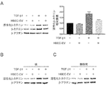
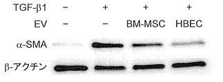
エクソソーム及び/又はトランスフォーミング増殖因子-β(TGF-β)1を添加したヒト初代気道上皮細胞、ヒト初代肺線維芽細胞、ヒト皮膚線維芽細胞から、Mammalian Protein Extract Reagent(M-PER、Thermo Fisher Scientific)を用いてタンパク質回収を行った。得られた試料にサンプルバッファー(Wako、198-13282又は191-13272)を添加し、SDS-PAGEを用いて電気泳動を行った。続いて、膜転写(polyvinylidene difluoride membrane、Millipore)、及びBlocking One(Nacalai Tesque)を用いたブロッキングを行った。抗原抗体反応後、ImmunoStar LD(Wako)を用いて発光させ、定量を行った。1次抗体として、alpha smooth muscle actin(α-SMA、Sigma-Aldrich、A2547)、goat anti-COL1/type I collagen(Southern Biotech、1310-01)、mouse anti-FN1/ cellular fibronectin containing extra domain A(Abcam、ab6328)、mouse anti-ACTB/b-actin(Sigma- Aldrich、A5316)、rabbit anti-p21Waf1/Cip1(Cell Signaling Technology、2947)、rabbit anti-β-Catenin (Cell Signaling Technology、8480)、rabbit anti-non-phospho(Active)β-Catenin (Cell Signaling Technology、19807)を用いた。得られたバンドの強度を、Image Jを用いて数値化した。 (Western blotting)
Mammalian Protein Extract Reagent (M-PER, Thermo Fisher) from human primary airway epithelial cells, human primary lung fibroblasts, and human skin fibroblasts supplemented with exosome and / or transforming growth factor-β (TGF-β) 1. Protein recovery was performed using Scientific). A sample buffer (Wako, 198-13282 or 191-13272) was added to the obtained sample, and electrophoresis was performed using SDS-PAGE. Subsequently, blocking was performed using membrane transcription (polyvinylidene difluoride membrane, Millipore) and Blocking One (Nacalai Tesque). After the antigen-antibody reaction, light was emitted using ImmunoStar LD (Wako) and quantification was performed. As primary antibodies, alpha smooth muscle actin (α-SMA, Sigma-Aldrich, A2547), goat anti-COL1 / type I collagen (Southern Biotech, 1310-01), mouse anti-FN1 / cellular fibronectin containing extra domain A ( Abcam, ab6328), mouse anti-ACTB / b-actin (Sigma-Aldrich, A5316), rabbit anti-p21 Waf1 / Cip1 (Cell Signaling Technology, 2947), rabbit anti-β-Catenin (Cell Signaling Technology, 8480), rabbit anti-non-phospho (Active) β-Catenin (Cell Signaling Technology, 19807) was used. The intensity of the obtained band was quantified using Image J.
(Senescence associated β-galactosidase染色)
ヒト初代気道上皮細胞にTGF-β1及び/又は気道上皮細胞由来エクソソームを添加し、Senescence associated β-galactosidase染色キット(Sigma, CS0030)を用いて染色陽性細胞数を数えた。染色は製造元の説明書に従って行った。 (Senescence associated β-galactosidase staining)
TGF-β1 and / or airway epithelial cell-derived exosomes were added to human primary airway epithelial cells, and the number of staining-positive cells was counted using the Senescence associated β-galactosidase staining kit (Sigma, CS0030). Dyeing was performed according to the manufacturer's instructions.
(WNT/b-cateninシグナリング分析における細胞成分分画)
核、細胞質タンパク質の分画は、細胞成分分画キット(Nuclear and Cytoplasmic Extraction Reagents、Thermo Fisher Scientific、78833)を用い、製造元の説明書に従って行った。 (Cell component fraction in WNT / b-catenin signaling analysis)
Fractionation of nuclear and cytoplasmic proteins was performed using a cell component fractionation kit (Nuclear and Cytoplasmic Extraction Reagents, Thermo Fisher Scientific, 78833) and according to the manufacturer's instructions.
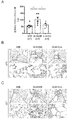
(動物実験)
動物実験は、東京慈恵会医科大学の実験動物研究機関のガイドラインに従って実施した。8-12週齢のC57BL/6Jマウスに、2.5 U/kg bleomycin(Nippon Kayaku Co.、4234400D4032)を50μlの生理食塩水に溶解し経気道的に投与した、肺線維化モデルマウスを用いた。bleomycinの投与後7、14日目にBEAS2B由来エクソソーム3μg(50μl)を経気道的に投与した。投与後17日目に屠殺し、以下の免疫組織染色に用いた。 (Animal experimentation)
Animal experiments were conducted in accordance with the guidelines of the Experimental Animal Research Institute of the Jikei University School of Medicine. Pulmonary fibrosis model mice in which 2.5 U / kg bleomycin (Nippon Kayaku Co., 4234400D4032) was dissolved in 50 μl of physiological saline and administered transairway to 8- to 12-week-old C57BL / 6J mice were used. On the 7th and 14th days after the administration of bleomycin, 3 μg (50 μl) of BEAS2B-derived exosomes were administered transairway. It was sacrificed 17 days after administration and used for the following immunohistochemical staining.
(免疫組織染色)
マウス肺を既報の通りにMasson trichrome染色、並びにヘマトキシリン及びエオシン染色で評価した(Kobayashi K. et al., J. Immunol., 2016, 197(2), pp. 504-16)。また、Masson trichrome染色の組織から、Ashcroft’s methodを用いてAshcroftスコアを決定し、線維化を評価した。細胞老化マーカーp16に対する組織染色には、1次抗体として、Anti-mouse CDKN2A/p16INK4a抗体(abcam、ab54210)を用いた。細胞老化マーカーp21に対する組織染色には、1次抗体として、rabbit anti-p21Waf1/Cip1(Cell Signaling Technology、2947)を用いた。 (Immunohistochemical staining)
Mouse lungs were evaluated by Masson trichrome staining and hematoxylin and eosin staining as previously reported (Kobayashi K. et al., J. Immunol., 2016, 197 (2), pp. 504-16). In addition, the Ashcroft score was determined using the Ashcroft's method from Masson trichrome-stained tissue, and fibrosis was evaluated. Anti-mouse CDKN2A / p16INK4a antibody (abcam, ab54210) was used as the primary antibody for tissue staining against the cell senescence marker p16. Rabbit anti-p21 Waf1 / Cip1 (Cell Signaling Technology, 2947) was used as the primary antibody for tissue staining against the cell senescence marker p21.
ヒドロキシプロリン量の測定は、Hydroxyproline assay kit(Chondrex、6017)により行った。
The amount of hydroxyproline was measured by the Hydroxyproline assay kit (Chondrex, 6017).
(統計学的解析)
実験はいずれも、少なくとも3回繰り返し、その結果を平均±SEMとして表した。解析は、3つの比較ではスチューデントt検定、3つ以上での比較はANOVAを用いた。Prism version 7 (GraphPad Software, San Diego, CA)を用い、p値<0.05を統計学的有意差とした。 (Statistical analysis)
Each experiment was repeated at least 3 times and the results were expressed as mean ± SEM. For the analysis, Student's t-test was used for the three comparisons, and ANOVA was used for the comparison with three or more. Using Prism version 7 (GraphPad Software, San Diego, CA), a p value <0.05 was defined as a statistically significant difference.
(HBEC由来エクソソームと骨髄由来MSC由来エクソソームの比較)
初代肺線維芽細胞に、TGF-β1 2ng/ml及び/又はエクソソーム 10μg/ml(HBEC由来エクソソーム又は骨髄由来間葉系幹細胞(BM-MSC)由来エクソソーム)を添加して24時間培養し、培地を交換してさらに48時間培養した。その後、上記と同様にウェスタンブロッティングを行った。BM-MSC由来エクソソームは、BM-MSCをStemPro MSC SFM(Thermo Fisher Scientific)で培養し、培養上清から超遠心法を用いて、上記の通りHBEC由来エクソソームと同様に回収した。 (Comparison of HBEC-derived exosomes and bone marrow-derived MSC-derived exosomes)
TGF-β1 2 ng / ml and / or exosome 10 μg / ml (HBEC-derived exosome or bone marrow-derived mesenchymal stem cell (BM-MSC) -derived exosome) was added to primary lung fibroblasts and cultured for 24 hours to prepare a medium. It was replaced and cultured for another 48 hours. Then, Western blotting was performed in the same manner as described above. For BM-MSC-derived exosomes, BM-MSCs were cultured in StemPro MSC SFM (Thermo Fisher Scientific) and recovered from the culture supernatant using an ultracentrifugation method in the same manner as HBEC-derived exosomes as described above.
初代肺線維芽細胞に、TGF-β1 2ng/ml及び/又はエクソソーム 10μg/ml(HBEC由来エクソソーム又は骨髄由来間葉系幹細胞(BM-MSC)由来エクソソーム)を添加して24時間培養し、培地を交換してさらに48時間培養した。その後、上記と同様にウェスタンブロッティングを行った。BM-MSC由来エクソソームは、BM-MSCをStemPro MSC SFM(Thermo Fisher Scientific)で培養し、培養上清から超遠心法を用いて、上記の通りHBEC由来エクソソームと同様に回収した。 (Comparison of HBEC-derived exosomes and bone marrow-derived MSC-derived exosomes)
TGF-
(ARDSモデルマウス)
グラム陰性菌の成分であり敗血症モデルで使用されるリポ多糖(LPS)と、ウイルスRNAを模倣する合成二本鎖RNAアナログのPoly I:Cをマウス(C57BL/6J、8週齢、雄)の気管に直接噴霧することによって急性呼吸窮迫症候群(ARDS)モデルマウスを作製した。具体的には、LPS(5μg/mg)を噴霧した4時間後にPoly I:C(5μg/mg)を噴霧し、さらに20時間後にマウスを屠殺し、BALF好中球数の測定、HE染色、及び肺炎症スコアの測定を行った。HBEC由来エクソソームを用いる場合は、Poly I:Cの噴霧と同時に、1個体当たり2×10⁹粒子を経気道で噴霧した(図12A)。 (ARDS model mouse)
Lipopolysaccharide (LPS), a component of Gram-negative bacteria and used in sepsis models, and Poly I: C, a synthetic double-stranded RNA analog that mimics viral RNA, in mice (C57BL / 6J, 8 weeks old, male) Acute respiratory distress syndrome (ARDS) model mice were generated by spraying directly into the trachea. Specifically, 4 hours after spraying LPS (5 μg / mg), Poly I: C (5 μg / mg) was sprayed, and 20 hours later, the mice were sacrificed, BALF neutrophil count measurement, HE staining, And the pneumonia score was measured. When using HBEC-derived exosomes, 2 × 10 9 particles per individual were sprayed through the airways at the same time as the spraying of Poly I: C (Fig. 12A).
BALF(気管支肺胞洗浄液)好中球数は、以下の方法で測定した。まず、トリパンブルーを用いてBALF中全細胞数を計測した。次に、BALFをオートスメア遠心機(CF-120, 800rpm, 5分)にて遠心し、得られた上清をディフ・クイック染色にて染色して、全細胞数に対する好中球数の割合を計測し、それによりBALF中好中球数を算出した。
BALF (bronchoalveolar lavage fluid) neutrophil count was measured by the following method. First, the total number of cells in BALF was measured using trypan blue. Next, BALF was centrifuged in an autosmear centrifuge (CF-120, 800 rpm, 5 minutes), and the obtained supernatant was stained with Diff-Quik staining to determine the ratio of neutrophil count to total cell count. The number of BALF neutrophils was calculated.
肺炎症スコアは、Sun Y. Q. et al., Stem Cells, 30(12); 2692-9 (2012)に記載の方法を参考にして、以下の方法で測定した。1匹のマウスの肺切片において、気管支周囲の5箇所の領域をランダムに選択した。各領域に関して、気管支周囲及び血管周囲の炎症細胞浸潤を0~3の4段階にスコア化した。5箇所の領域のスコア平均値を肺炎症スコアとした。
The pneumonia score was measured by the following method with reference to the method described in Sun Y. Q. et al., Stem Cells, 30 (12); 2692-9 (2012). Five regions around the bronchi were randomly selected from lung sections of one mouse. Peribronchial and perivascular inflammatory cell infiltration was scored on a 4-point scale from 0 to 3 for each region. The average score of the five regions was used as the pneumonia score.
<結果>
(調製されたエクソソームの分析)
ヒト初代気道上皮細胞HBEC、及びヒト気道上皮細胞株BEAS-2Bの培養上清から、超遠心法で回収したエクソソームの、NTAによる分析結果(A:HBEC、B:BEAS-2B)を図1に示す。
これらの結果は、超遠心法による無事エクソソームが回収されたことを示している。また、回収したエクソソームの平均直径は、約80nm前後であった。 <Result>
(Analysis of prepared exosomes)
Figure 1 shows the results of analysis by NTA (A: HBEC, B: BEAS-2B) of exosomes recovered by ultracentrifugation from the culture supernatants of the human primary airway epithelial cell HBEC and the human airway epithelial cell line BEAS-2B. Shown.
These results indicate that the exosomes were successfully recovered by ultracentrifugation. The average diameter of the recovered exosomes was about 80 nm.
(TGF-β1誘導筋線維芽細胞分化のエクソソームによる抑制)
肺線維症、COPD、及び気管支喘息等の気道リモデリングの病態形成において、筋線維芽細胞への分化は重要な役割を担う。そこで、エクソソームの肺線維芽細胞への影響を検討した。 (Exosome suppression of TGF-β1-induced myofibroblast differentiation)
Differentiation into myofibroblasts plays an important role in the pathogenesis of airway remodeling such as pulmonary fibrosis, COPD, and bronchial asthma. Therefore, the effects of exosomes on lung fibroblasts were investigated.
初代肺線維芽細胞に、TGF-β1 2ng/ml及び/又はエクソソーム 10μg/ml(HBEC-EV)を添加して24時間し、培地を交換してさらに48時間培養した。その後、筋線維芽細胞のマーカーであるα-SMA、I型コラーゲン、及びフィブロネクチン、並びにハウスキーピングタンパク質であるアクチンの発現をウェスタンブロッティングにより確認した結果を図2Aに示す。また、各タンパク質の発現量をアクチンの発現量で割って数値化した結果を図2B~Dに示す。TGF-β1によって誘導されるα-SMA、I型コラーゲン、及びフィブロネクチンの発現上昇はエクソソーム添加により低下したことから、HBEC由来エクソソームはTGF-β1誘導筋線維芽細胞分化を抑制することが示された。
TGF-β1 2 ng / ml and / or exosome 10 μg / ml (HBEC-EV) was added to primary lung fibroblasts for 24 hours, the medium was changed, and the cells were cultured for another 48 hours. After that, the expression of α-SMA, type I collagen, and fibronectin, which are markers of myofibroblasts, and actin, which is a housekeeping protein, was confirmed by Western blotting, and the results are shown in FIG. 2A. The results of dividing the expression level of each protein by the expression level of actin and quantifying it are shown in FIGS. 2B to 2D. The increased expression of α-SMA, type I collagen, and fibronectin induced by TGF-β1 was decreased by the addition of exosomes, indicating that HBEC-derived exosomes suppress TGF-β1-induced myofibroblast differentiation. ..
(TGF-β1誘導肺上皮細胞老化のエクソソームによる抑制)
近年、肺線維症及びCOPD病態等において、肺上皮細胞老化の関与が広く知られるようになった。そこで、エクソソームの肺上皮細胞の老化への影響を検討した。 (Exosome suppression of TGF-β1-induced lung epithelial cell senescence)
In recent years, the involvement of pulmonary epithelial cell senescence in pulmonary fibrosis and COPD pathology has become widely known. Therefore, the effect of exosomes on aging of lung epithelial cells was investigated.
TGF-β1 2ng/mlと共に気道上皮細胞(HBEC)由来エクソソーム10μg/mlを気道上皮細胞(HBEC)に添加し、48時間培養した。その結果、老化マーカーであるSA-β-Gal(Senescence associated β-galactosidase)(図3A)及びp21(図3B)のTGF-β1による発現上昇を抑制した。ヒト小気道上皮細胞(HSAEC)由来エクソソームを用いた場合にも、HBEC由来エクソソームと同様にp21のTGF-β1による発現上昇が抑制された(図3C)。p21発現上昇に対する、HBEC由来エクソソームによる抑制効果は、BM-MSC由来エクソソームよりも大きかった(図3D)。
Airway epithelial cell (HBEC) -derived exosomes 10 μg / ml were added to airway epithelial cells (HBEC) together with TGF-β1 2 ng / ml and cultured for 48 hours. As a result, the increase in the expression of the aging markers SA-β-Gal (Senescence associated β-galactosidase) (Fig. 3A) and p21 (Fig. 3B) by TGF-β1 was suppressed. When human small airway epithelial cell (HSAEC) -derived exosomes were used, the expression increase of p21 by TGF-β1 was suppressed as in the case of HBEC-derived exosomes (Fig. 3C). The inhibitory effect of HBEC-derived exosomes on increasing p21 expression was greater than that of BM-MSC-derived exosomes (Fig. 3D).
次にエクソソームによる細胞老化抑制の作用機序について検討した。これまでに、肺線維症において、WNT/b-cateninシグナルの抑制が、筋線維芽細胞分化を抑制することが知られている。
Next, the mechanism of action of exosomes to suppress cell senescence was examined. So far, it is known that suppression of WNT / b-catenin signal suppresses myofibroblast differentiation in pulmonary fibrosis.
そこで、気道上皮細胞由来エクソソームによるWNT/b-cateninの影響を調べた。その結果、エクソソームがTGF-β1誘導WNT/b-cateninを抑制したことから、エクソソームは、WNT/b-cateninの抑制を介してTGF-β1誘導筋線維芽細胞分化を抑制することが示唆された(図4、A:分画前、B:核、C:細胞質)。
Therefore, the effect of WNT / b-catenin on airway epithelial cell-derived exosomes was investigated. As a result, exosomes suppressed TGF-β1-induced WNT / b-catenin, suggesting that exosomes suppress TGF-β1-induced myofibroblast differentiation through suppression of WNT / b-catenin. (Fig. 4, A: before fractionation, B: nucleus, C: cytoplasm).
以上の結果から、気道上皮細胞由来エクソソームは、筋線維芽細胞の分化、及び/又は上皮細胞の老化を抑制することによって、肺線維症等に治療及び/又は予防効果を奏し得ることが示された。
From the above results, it was shown that airway epithelial cell-derived exosomes can exert a therapeutic and / or preventive effect on pulmonary fibrosis and the like by suppressing the differentiation of myofibroblasts and / or the aging of epithelial cells. It was.
(既存治療薬との比較)
特発性肺線維症に対する既存の治療薬であるピルフェニドン及びニンテダニブとの効果比較を行った。ピルフェニドンの血中濃度である10μg/ml、及びその50倍濃度である500μg/ml、並びにニンテダニブの血中濃度である100nM、及びその50倍濃度である5000μg/mlを比較対象とした。初代肺線維芽細胞を、記載の濃度の各成分で24時間刺激し、培地を交換してさらに48時間培養した後、分析を行った。 (Comparison with existing therapeutic agents)
The effects of pirfenidone and nintedanib, which are existing therapeutic agents for idiopathic pulmonary fibrosis, were compared. The blood concentration of pirfenidone, 10 μg / ml, 50 times the concentration of 500 μg / ml, and the blood concentration of nintedanib, 100 nM, and 50 times the concentration, 5000 μg / ml, were compared. Primary lung fibroblasts were stimulated with each component at the listed concentrations for 24 hours, the medium was changed, and the cells were cultured for another 48 hours before analysis.
結果を図5に示す。気道上皮細胞株由来エクソソームは、TGF-β1誘導筋線維芽細胞分化マーカーであるI型コラーゲン、フィブロネクチン(FN)、及びα-SMAを濃度依存的に抑制し、その抑制効果はIPF治療薬の血中濃度と同等以上であった.
The results are shown in Fig. 5. Exosomes derived from airway epithelial cell lines suppress TGF-β1-induced myofibroblast differentiation markers type I collagen, fibronectin (FN), and α-SMA in a concentration-dependent manner, and the inhibitory effect is blood of IPF therapeutic agents. It was equal to or higher than the medium concentration.
(動物実験)
8-12週齢のC57BL/6Jマウスに、2.5 U/kg bleomycinを経気道的に投与し、肺線維化モデルマウスを作製した。このモデルマウスに、気道上皮細胞株由来エクソソームを経気道投与することにより、Masson trichrome染色により認められる肺線維化スコアが低減した(図6A及びB)。さらに、気道上皮細胞株由来エクソソームの経気道投与によって、コラーゲン蓄積の指標となる肺組織のヒドロキシプロリン量が低減し(図7A)、細胞老化マーカーであるp16及びp21の陽性細胞の数が減少した(図7B及びC)。したがって、気道上皮細胞株由来エクソソームは、in vivoで肺線維症マウスの線維化及び老化を抑制することが示された。 (Animal experimentation)
2.5 U / kg bleomycin was transairway-administered to 8- to 12-week-old C57BL / 6J mice to prepare lung fibrosis model mice. Transairway administration of airway epithelial cell line-derived exosomes to this model mouse reduced the pulmonary fibrosis score observed by Masson trichrome staining (FIGS. 6A and 6B). In addition, transairway administration of airway epithelial cell line-derived exosomes reduced the amount of hydroxyproline in lung tissue, an indicator of collagen accumulation (Fig. 7A), and reduced the number of cells positive for the cellular senescence markers p16 and p21. (Figs. 7B and C). Therefore, exosomes derived from airway epithelial cell lines were shown to suppress fibrosis and aging in pulmonary fibrotic mice in vivo.
(MSC由来エクソソームとの比較)
初代肺線維芽細胞に、TGF-β1及び/又はエクソソーム(HBEC由来エクソソーム又は骨髄由来間葉系幹細胞(BM-MSC)由来エクソソーム)を添加して24時間培養し、培地を交換してさらに48時間培養した。その後、筋線維芽細胞のマーカーであるα-SMA及びハウスキーピングタンパク質であるアクチンの発現をウェスタンブロッティングにより確認した結果を図8に示す。図8から明らかな通り、TGF-β1によって誘導されるα-SMAの発現上昇はBM-MSC由来エクソソームに比べて、HBEC由来エクソソーム添加により顕著に低下した。この結果は、未分化なBM-MSCよりも、分化が進んだHBEC由来のエクソソームの方が、肺における高い筋線維芽細胞分化抑制能を有し得ることを示している。 (Comparison with MSC-derived exosomes)
TGF-β1 and / or exosomes (HBEC-derived exosomes or bone marrow-derived mesenchymal stem cell (BM-MSC) -derived exosomes) were added to primary lung fibroblasts and cultured for 24 hours, and the medium was changed for an additional 48 hours. It was cultured. After that, the expression of α-SMA, which is a marker for myofibroblasts, and actin, which is a housekeeping protein, was confirmed by Western blotting, and the results are shown in FIG. As is clear from FIG. 8, the TGF-β1-induced increase in α-SMA expression was significantly reduced by the addition of HBEC-derived exosomes as compared with BM-MSC-derived exosomes. This result indicates that more differentiated HBEC-derived exosomes may have a higher ability to suppress myofibroblast differentiation in the lung than undifferentiated BM-MSCs.
(気道上皮細胞以外の細胞に由来するエクソソームの効果)
気道上皮細胞(HBEC)以外の細胞に由来するエクソソームを初代肺線維芽細胞に添加することによって、TGF-β1誘導筋線維芽細胞分化に対する効果を検討した。具体的には、ヒト気道上皮細胞(HBEC)、ヒト気道上皮細胞株(BEAS-2B)、ヒト胎児腎細胞293(HEK293)、単球性白血病細胞株(THP1)、ヒト小気道上皮細胞(HSAEC)由来エクソソーム、及び骨髄由来間葉系幹細胞(BM-MSC)に由来するエクソソームについて、TGF-β1誘導筋線維芽細胞分化に対する効果を検討した。 (Effects of exosomes derived from cells other than airway epithelial cells)
The effect on TGF-β1-induced myofibroblast differentiation was investigated by adding exosomes derived from cells other than airway epithelial cells (HBEC) to primary lung fibroblasts. Specifically, human airway epithelial cells (HBEC), human airway epithelial cell lines (BEAS-2B), human fetal kidney cells 293 (HEK293), monocytic leukemia cell lines (THP1), human small airway epithelial cells (HSAEC). ) Derived exosomes and bone marrow-derived mesenchymal stem cells (BM-MSC) -derived exosomes were examined for their effects on TGF-β1-induced myofibroblast differentiation.
TGF-β1によって誘導されるα-SMAの発現上昇に対して、BEAS-2B由来エクソソーム、及びHEK293由来エクソソームは、HBEC由来エクソソームと同等の抑制効果を示した。これらのエクソソームの効果は、いずれもBM-MSC由来エクソソームよりも顕著であった(図9A及びB)。一方、THP1由来エクソソームでは効果が認められなかった。TGF-β1によって誘導されるα-SMA及びI型コラーゲンの発現上昇に対しては、BEAS-2B由来エクソソーム及びHSAEC由来エクソソームが、HBEC由来エクソソームと同等の抑制効果を示した(図10)。
BEAS-2B-derived exosomes and HEK293-derived exosomes showed the same inhibitory effect as HBEC-derived exosomes on the increase in α-SMA expression induced by TGF-β1. The effects of these exosomes were all more pronounced than those of BM-MSC-derived exosomes (FIGS. 9A and B). On the other hand, no effect was observed with THP1-derived exosomes. BEAS-2B-derived exosomes and HSAEC-derived exosomes showed the same inhibitory effect as HBEC-derived exosomes on the expression of α-SMA and type I collagen induced by TGF-β1 (Fig. 10).
これらの結果は、気道上皮細胞以外の細胞、例えば腎細胞や肺胞上皮細胞等に由来するエクソソームも肺における高い筋線維芽細胞分化抑制能を有し得ることを示した。
These results showed that exosomes derived from cells other than airway epithelial cells, such as renal cells and alveolar epithelial cells, may also have a high ability to suppress myofibroblast differentiation in the lungs.
(皮膚線維芽細胞のTGF-β1誘導筋線維芽細胞分化に対する、エクソソームによる抑制効果)
HBEC由来エクソソームが皮膚線維芽細胞に対しても線維化を抑制する効果を有するか否かを検討した。 (Exosome-suppressing effect on TGF-β1-induced myofibroblast differentiation of skin fibroblasts)
We investigated whether HBEC-derived exosomes also have the effect of suppressing fibrosis on skin fibroblasts.
HBEC由来エクソソームが皮膚線維芽細胞に対しても線維化を抑制する効果を有するか否かを検討した。 (Exosome-suppressing effect on TGF-β1-induced myofibroblast differentiation of skin fibroblasts)
We investigated whether HBEC-derived exosomes also have the effect of suppressing fibrosis on skin fibroblasts.
皮膚線維芽細胞(NHDF)に、TGF-β1 2ng/ml及び/又はエクソソーム 10μg/ml(HBEC-EV)を添加して24時間し、培地を交換してさらに48時間培養した。その後、筋線維芽細胞のマーカーであるα-SMA、及びI型コラーゲン、並びにアクチンの発現をウェスタンブロッティングにより確認した結果を図11に示す。
TGF-β1 2 ng / ml and / or exosome 10 μg / ml (HBEC-EV) was added to skin fibroblasts (NHDF) for 24 hours, the medium was changed, and the cells were cultured for another 48 hours. After that, the expression of α-SMA, which is a marker of myofibroblast, type I collagen, and actin was confirmed by Western blotting, and the result is shown in FIG.
皮膚線維芽細胞(NHDF)においてTGF-β1によって誘導されるα-SMA、及びI型コラーゲンの発現上昇がエクソソーム添加により低下したことから、HBEC由来エクソソームは、皮膚線維芽細胞においてもTGF-β1誘導筋線維芽細胞分化を抑制することが示された。この結果は、肺に由来するエクソソームが、肺とは異なる組織において、強皮症の治療薬となり得る可能性を示している。
Since the increase in expression of α-SMA and type I collagen induced by TGF-β1 in skin fibroblasts (NHDF) was decreased by the addition of exosomes, HBEC-derived exosomes also induced TGF-β1 in skin fibroblasts. It was shown to suppress myofibroblast differentiation. This result indicates that lung-derived exosomes may be therapeutic agents for scleroderma in tissues different from the lung.
(ARDSモデルにおけるエクソソームの効果)
HBEC由来エクソソームが急性呼吸窮迫症候群(ARDS)モデルマウスにおける炎症反応を抑制する効果を有するか否かを検討した。 (Effects of exosomes in ARDS model)
We investigated whether HBEC-derived exosomes have the effect of suppressing the inflammatory response in acute respiratory distress syndrome (ARDS) model mice.
HBEC由来エクソソームが急性呼吸窮迫症候群(ARDS)モデルマウスにおける炎症反応を抑制する効果を有するか否かを検討した。 (Effects of exosomes in ARDS model)
We investigated whether HBEC-derived exosomes have the effect of suppressing the inflammatory response in acute respiratory distress syndrome (ARDS) model mice.
リポ多糖(LPS)を気管に噴霧した4時間後に、poly I:C及び/又はエクソソーム(HBEC-EV)を気管に噴霧して急性呼吸窮迫症候群(ARDS)モデルマウスを作製し、さらに20時間後にマウスを解剖し(図12A)、肺における炎症反応について検討を行った。
Four hours after spraying lipopolysaccharide (LPS) into the trachea, polyI: C and / or exosomes (HBEC-EV) were sprayed into the trachea to generate acute respiratory distress syndrome (ARDS) model mice, and 20 hours later. Mice were dissected (Fig. 12A) to examine the inflammatory response in the lungs.
LPS及びpoly I:Cによって誘導される好中球数の増大は、HBEC由来エクソソームによって有意に抑制された(図12B)。また、HE染色で評価した病理画像においては、HBEC由来エクソソームによって気道や血管周囲の炎症細胞浸潤が低減し、肺障害が抑制されることが示された(図12C)。さらに、LPS及びpoly I:Cによって誘導される肺炎症スコアの増大が、HBEC由来エクソソームによって有意に低下した(図12D)。以上の結果は、本発明のエクソソームがARDSを含む炎症疾患の治療薬となり得る可能性を示している。
The increase in neutrophil count induced by LPS and poly I: C was significantly suppressed by HBEC-derived exosomes (Fig. 12B). In addition, pathological images evaluated by HE staining showed that HBEC-derived exosomes reduced infiltration of inflammatory cells around the airways and blood vessels and suppressed lung damage (Fig. 12C). In addition, the increase in pneumonia score induced by LPS and poly I: C was significantly reduced by HBEC-derived exosomes (Fig. 12D). The above results indicate that the exosomes of the present invention may be therapeutic agents for inflammatory diseases including ARDS.
本明細書で引用した全ての刊行物、特許及び特許出願はそのまま引用により本明細書に組み入れられるものとする。
All publications, patents and patent applications cited in this specification shall be incorporated herein by reference as is.
Claims (11)
- 線維化疾患、炎症疾患、及び老化疾患の少なくとも1つの疾患の治療及び/又は予防剤であって、前記疾患が生じ得る組織又はその周辺組織の細胞由来の細胞外小胞顆粒からなり、前記細胞が分化細胞である、前記治療及び/又は予防剤。 A therapeutic and / or prophylactic agent for at least one disease of fibrosis, inflammatory disease, and aging disease, which comprises extracellular vesicle granules derived from cells of the tissue in which the disease can occur or its surrounding tissue. The therapeutic and / or prophylactic agent, wherein is a differentiated cell.
- 前記細胞外小胞顆粒を供与する細胞の由来する組織と受容する細胞の組織とが異なる、請求項1に記載の治療及び/又は予防剤。 The therapeutic and / or preventive agent according to claim 1, wherein the tissue from which the cells that donate the extracellular vesicle granules are derived and the tissue of the cells that receive the extracellular vesicle granules are different.
- 前記細胞が、上皮細胞、内皮細胞、中皮細胞、筋細胞、又は神経細胞である、請求項1に記載の治療及び/又は予防剤。 The therapeutic and / or prophylactic agent according to claim 1, wherein the cells are epithelial cells, endothelial cells, mesothelial cells, muscle cells, or nerve cells.
- 前記上皮細胞が、気道上皮細胞、小気道上皮細胞、又は肺胞上皮細胞である、請求項3に記載の治療及び/又は予防剤。 The therapeutic and / or preventive agent according to claim 3, wherein the epithelial cells are airway epithelial cells, small airway epithelial cells, or alveolar epithelial cells.
- 前記組織又はその周辺組織が、肺又は気道;眼;腎臓又は糸球体;肝臓、胆嚢又は胆管;腸管;膵臓;心臓;血管;甲状腺;脳又は神経;腹腔内;子宮;皮膚;筋肉;骨;及び関節からなる群から選択される、請求項1に記載の治療及び/又は予防剤。 The tissue or surrounding tissue is the lung or airway; eye; kidney or glomerular; liver, gallbladder or bile duct; intestinal tract; pancreas; heart; blood vessel; thyroid; brain or nerve; intraperitoneal; uterus; skin; muscle; bone; The therapeutic and / or prophylactic agent according to claim 1, selected from the group consisting of and joints.
- 前記組織又はその周辺組織が肺又は気道である、請求項5に記載の治療及び/又は予防剤。 The therapeutic and / or preventive agent according to claim 5, wherein the tissue or its surrounding tissue is the lung or respiratory tract.
- 前記疾患が、強皮症、肺線維症、COPD(慢性閉塞性肺疾患)、気管支喘息、ARDS(急性呼吸窮迫症候群)、肺高血圧症、放射性肺臓炎、肺サルコイドーシス、膠原病に伴う肺胞出血、間質性肺炎、気管支拡張症、嚢胞性線維症、又は気管支肺異形成症からなる群から選択される、請求項4又は6に記載の治療及び/又は予防剤。 The diseases are strong skin disease, pulmonary fibrosis, COPD (chronic obstructive pulmonary disease), bronchial asthma, ARDS (acute respiratory distress syndrome), pulmonary hypertension, radiopneumonitis, pulmonary sarcoidosis, alveolar hemorrhage associated with collagen disease The therapeutic and / or prophylactic agent according to claim 4 or 6, selected from the group consisting of interstitial pneumonia, bronchial dilatation, cystic fibrosis, or bronchopulmonary dysplasia.
- 前記細胞が健常者に由来する、請求項1~7のいずれか一項に記載の治療及び/又は予防剤。 The therapeutic and / or preventive agent according to any one of claims 1 to 7, wherein the cells are derived from a healthy person.
- 前記細胞外小胞顆粒がエクソソームである、請求項1~8のいずれか一項に記載の治療及び/又は予防剤。 The therapeutic and / or preventive agent according to any one of claims 1 to 8, wherein the extracellular vesicle granules are exosomes.
- 請求項1~9のいずれか一項に記載の治療及び/又は予防剤を有効成分として含む、医薬組成物。 A pharmaceutical composition comprising the therapeutic and / or prophylactic agent according to any one of claims 1 to 9 as an active ingredient.
- 吸入、噴霧投与、注射、点滴注入、経口、経皮、経鼻、経局所、経膣、経粘膜、又は経直腸用に製剤化されている、請求項10に記載の医薬組成物。 The pharmaceutical composition according to claim 10, which is formulated for inhalation, spray administration, injection, infusion, oral, transdermal, nasal, translocal, transvaginal, transmucosal, or transrectal.
Priority Applications (4)
| Application Number | Priority Date | Filing Date | Title |
|---|---|---|---|
| CN202080022694.2A CN113795261A (en) | 2019-04-09 | 2020-04-08 | Therapeutic agent for fibrosis, inflammation and/or aging |
| JP2021513681A JPWO2020209304A1 (en) | 2019-04-09 | 2020-04-08 | |
| US17/602,644 US20220160786A1 (en) | 2019-04-09 | 2020-04-08 | Therapeutic agent for fibrosis, inflammation, and/or aging disease |
| EP20787381.1A EP3954435A4 (en) | 2019-04-09 | 2020-04-08 | Therapeutic agent for fibrosis, inflammation, and/or aging disease |
Applications Claiming Priority (2)
| Application Number | Priority Date | Filing Date | Title |
|---|---|---|---|
| JP2019074263 | 2019-04-09 | ||
| JP2019-074263 | 2019-04-09 |
Publications (1)
| Publication Number | Publication Date |
|---|---|
| WO2020209304A1 true WO2020209304A1 (en) | 2020-10-15 |
Family
ID=72750693
Family Applications (1)
| Application Number | Title | Priority Date | Filing Date |
|---|---|---|---|
| PCT/JP2020/015861 WO2020209304A1 (en) | 2019-04-09 | 2020-04-08 | Therapeutic agent for fibrosis, inflammation, and/or aging disease |
Country Status (5)
| Country | Link |
|---|---|
| US (1) | US20220160786A1 (en) |
| EP (1) | EP3954435A4 (en) |
| JP (1) | JPWO2020209304A1 (en) |
| CN (1) | CN113795261A (en) |
| WO (1) | WO2020209304A1 (en) |
Cited By (1)
| Publication number | Priority date | Publication date | Assignee | Title |
|---|---|---|---|---|
| WO2023085423A1 (en) * | 2021-11-12 | 2023-05-19 | Ism株式会社 | Long-term culture of large intestine epithelial cells |
Families Citing this family (1)
| Publication number | Priority date | Publication date | Assignee | Title |
|---|---|---|---|---|
| CN114272265A (en) * | 2022-01-21 | 2022-04-05 | 上海市东方医院(同济大学附属东方医院) | Extracellular vesicles derived from normal tissue space and uses thereof |
Citations (6)
| Publication number | Priority date | Publication date | Assignee | Title |
|---|---|---|---|---|
| JP2015524844A (en) * | 2012-08-13 | 2015-08-27 | シーダーズ−サイナイ・メディカル・センターCedars−Sinai Medical Center | Exosomes and microribonucleic acids for tissue regeneration |
| JP2018518534A (en) * | 2015-06-12 | 2018-07-12 | ハドソン インスティチュート オブ メディカル リサーチHudson Institute Of Medical Research | Method of treatment |
| JP2019031447A (en) * | 2017-08-04 | 2019-02-28 | タンデム カンパニー,リミテッド | Novel recombinant exosome and application thereof |
| JP2019508461A (en) * | 2016-03-14 | 2019-03-28 | カプリコール,インコーポレイテッド | Method of treating ocular inflammation and chemical damage of the eye with extracellular vesicles |
| JP2019074263A (en) | 2017-10-17 | 2019-05-16 | イビデン株式会社 | Heat exchanger |
| WO2020032186A1 (en) * | 2018-08-08 | 2020-02-13 | 学校法人東京女子医科大学 | Topical composition containing extracellular vesicles produced by buccal epithelial cells |
Family Cites Families (5)
| Publication number | Priority date | Publication date | Assignee | Title |
|---|---|---|---|---|
| KR20110082480A (en) * | 2010-01-11 | 2011-07-19 | 포항공과대학교 산학협력단 | Composition comprising extracellular vesicles derived from mammals, and use thereof |
| CN103007291B (en) * | 2011-09-26 | 2015-04-29 | 苏州圣诺生物医药技术有限公司 | Composition for treating eye diseases by double-target/multi-target small nucleic acid and applications of composition |
| WO2014041087A1 (en) * | 2012-09-12 | 2014-03-20 | INSERM (Institut National de la Santé et de la Recherche Médicale) | Muscle secretome and uses thereof |
| WO2015069897A1 (en) * | 2013-11-07 | 2015-05-14 | University Of Pittsburgh-Of The Commonwealth System Of Higher Education | Red blood cell membrane-derived microparticles and their use for the treatment of lung disease |
| EP4091622A1 (en) * | 2016-08-16 | 2022-11-23 | Henry Ford Health System | Compositions for the treatment of chemotherapy-induced neuropathic pain |
-
2020
- 2020-04-08 JP JP2021513681A patent/JPWO2020209304A1/ja active Pending
- 2020-04-08 US US17/602,644 patent/US20220160786A1/en active Pending
- 2020-04-08 EP EP20787381.1A patent/EP3954435A4/en active Pending
- 2020-04-08 CN CN202080022694.2A patent/CN113795261A/en active Pending
- 2020-04-08 WO PCT/JP2020/015861 patent/WO2020209304A1/en unknown
Patent Citations (6)
| Publication number | Priority date | Publication date | Assignee | Title |
|---|---|---|---|---|
| JP2015524844A (en) * | 2012-08-13 | 2015-08-27 | シーダーズ−サイナイ・メディカル・センターCedars−Sinai Medical Center | Exosomes and microribonucleic acids for tissue regeneration |
| JP2018518534A (en) * | 2015-06-12 | 2018-07-12 | ハドソン インスティチュート オブ メディカル リサーチHudson Institute Of Medical Research | Method of treatment |
| JP2019508461A (en) * | 2016-03-14 | 2019-03-28 | カプリコール,インコーポレイテッド | Method of treating ocular inflammation and chemical damage of the eye with extracellular vesicles |
| JP2019031447A (en) * | 2017-08-04 | 2019-02-28 | タンデム カンパニー,リミテッド | Novel recombinant exosome and application thereof |
| JP2019074263A (en) | 2017-10-17 | 2019-05-16 | イビデン株式会社 | Heat exchanger |
| WO2020032186A1 (en) * | 2018-08-08 | 2020-02-13 | 学校法人東京女子医科大学 | Topical composition containing extracellular vesicles produced by buccal epithelial cells |
Non-Patent Citations (7)
| Title |
|---|
| ARAYA J. ET AL., J. CLIN. INVEST., vol. 117, 2007, pages 3551 - 3562 |
| AZUMA A ET AL., AM. J. RESPIR. CRIT. CARE MED., vol. 171, 2005, pages 1040 - 1047 |
| FUJITA, YU : "Morbidity Control Using Extracellular Follicle Granules/Exosomes in COPD", RESPIRATION RESEARCH, vol. 34, no. 9, 2015, pages 863 - 868, XP009531173, ISSN: 0286-9314 * |
| KHANNA, KRITIKA ET AL.: "Secretory inositol polyphosphate 4-phosphatase protects against airway inflammation and remodeling", AM J RESPIR CELL MOL BIOL, vol. 6, no. 4, 18 October 2018 (2018-10-18), pages 399 - 412, XP55750212 * |
| KOBAYASHI K. ET AL., J. IMMUNOL., vol. 197, no. 2, 2016, pages 504 - 16 |
| RICHELDI L ET AL., N. ENGL. J. MED., vol. 365, no. 12, 2011, pages 1079 - 1087 |
| SUN Y. Q. ET AL., STEM CELLS, vol. 30, no. 12, 2012, pages 2692 - 9 |
Cited By (1)
| Publication number | Priority date | Publication date | Assignee | Title |
|---|---|---|---|---|
| WO2023085423A1 (en) * | 2021-11-12 | 2023-05-19 | Ism株式会社 | Long-term culture of large intestine epithelial cells |
Also Published As
| Publication number | Publication date |
|---|---|
| JPWO2020209304A1 (en) | 2020-10-15 |
| EP3954435A4 (en) | 2022-11-23 |
| EP3954435A1 (en) | 2022-02-16 |
| US20220160786A1 (en) | 2022-05-26 |
| CN113795261A (en) | 2021-12-14 |
Similar Documents
| Publication | Publication Date | Title |
|---|---|---|
| JP6524162B2 (en) | Methods and compositions related to mesenchymal stem cell exosomes | |
| Li et al. | Mesenchymal stem cells in idiopathic pulmonary fibrosis | |
| JP6878274B2 (en) | Myocardial cell-derived cells and exosomes secreted by such cells in the treatment of muscular dystrophy | |
| Han et al. | Human adipose stem cell-derived extracellular nanovesicles for treatment of chronic liver fibrosis | |
| WO2020209304A1 (en) | Therapeutic agent for fibrosis, inflammation, and/or aging disease | |
| US20220218757A1 (en) | Therapeutically active cells and exosomes | |
| US20190167587A1 (en) | Pharmaceutical composition for prevention or treatment of pulmonary disease including mesenchymal stem cell-derived artificial nanosomes | |
| Ahmadi et al. | Bone marrow mesenchymal stem cells modified pathological changes and immunological responses in ovalbumin-induced asthmatic rats possibly by the modulation of miRNA155 and miRNA133 | |
| AU2018335788A1 (en) | Methods and compositions for the treatment of epidermolysis bullosa | |
| WO2022242189A1 (en) | Application of exosomes derived from stem cells in preparing medicine for treating chronic obstructive pulmonary diseases | |
| Geng et al. | HGF-modified dental pulp stem cells mitigate the inflammatory and fibrotic responses in paraquat-induced acute respiratory distress syndrome | |
| Xu et al. | Revealing the mechanism of Jiegeng decoction attenuates bleomycin-induced pulmonary fibrosis via PI3K/Akt signaling pathway based on lipidomics and transcriptomics | |
| Han et al. | Adipose-derived stem cell-derived extracellular vesicles inhibit the fibrosis of fibrotic buccal mucosal fibroblasts via the microRNA-375/FOXF1 axis | |
| Wan et al. | Injectable photocrosslinking spherical hydrogel-encapsulated targeting peptide-modified engineered exosomes for osteoarthritis therapy | |
| WO2021016727A1 (en) | Therapeutic medicine for fibrosis, inflammation, and/or age-related diseases | |
| Liu et al. | Bone marrow mesenchymal stem cell-derived exosomes improve renal fibrosis via regulating Smurf 2/Smad 7 | |
| Ohnishi et al. | Inhibition of tubular cell proliferation by neutralizing endogenous HGF leads to renal hypoxia and bone marrow-derived cell engraftment in acute renal failure | |
| JP2021116263A (en) | Therapeutic agent against liver disease | |
| WO2020032186A1 (en) | Topical composition containing extracellular vesicles produced by buccal epithelial cells | |
| Attia et al. | Alleviation of pulmonary fibrosis by the dual PPAR agonist saroglitazar and breast milk mesenchymal stem cells via modulating TGFß/SMAD pathway | |
| WO2023040938A1 (en) | Method for treating ilk signaling pathway related diseases using exosome derived from mesenchymal stem cells, and pharmaceutical composition | |
| Li et al. | Therapeutic effects of human umbilical cord-derived mesenchymal stem cells against acute tubular necrosis quantified through measures of iNOS, BMP-7 and Bcl-2 | |
| JP2022058273A (en) | Pharmaceutical compositions for fibrotic or aging diseases and pharmaceutical compositions for inflammatory diseases, and methods for producing them | |
| Serikov et al. | MSCs for treatment of acute lung injury | |
| Carbone et al. | Placenta-Derived Stem Cells as a Source for Treatment of Lung and Liver Disease in Cystic Fibrosis |
Legal Events
| Date | Code | Title | Description |
|---|---|---|---|
| 121 | Ep: the epo has been informed by wipo that ep was designated in this application |
Ref document number: 20787381 Country of ref document: EP Kind code of ref document: A1 |
|
| ENP | Entry into the national phase |
Ref document number: 2021513681 Country of ref document: JP Kind code of ref document: A |
|
| NENP | Non-entry into the national phase |
Ref country code: DE |
|
| ENP | Entry into the national phase |
Ref document number: 2020787381 Country of ref document: EP Effective date: 20211109 |