WO2020184676A1 - Nmda受容体阻害活性のハイスループット評価方法 - Google Patents
Nmda受容体阻害活性のハイスループット評価方法 Download PDFInfo
- Publication number
- WO2020184676A1 WO2020184676A1 PCT/JP2020/010909 JP2020010909W WO2020184676A1 WO 2020184676 A1 WO2020184676 A1 WO 2020184676A1 JP 2020010909 W JP2020010909 W JP 2020010909W WO 2020184676 A1 WO2020184676 A1 WO 2020184676A1
- Authority
- WO
- WIPO (PCT)
- Prior art keywords
- drebrin
- nmda receptor
- test substance
- nerve cells
- cultured
- Prior art date
- Legal status (The legal status is an assumption and is not a legal conclusion. Google has not performed a legal analysis and makes no representation as to the accuracy of the status listed.)
- Ceased
Links
Images


Classifications
-
- C—CHEMISTRY; METALLURGY
- C12—BIOCHEMISTRY; BEER; SPIRITS; WINE; VINEGAR; MICROBIOLOGY; ENZYMOLOGY; MUTATION OR GENETIC ENGINEERING
- C12Q—MEASURING OR TESTING PROCESSES INVOLVING ENZYMES, NUCLEIC ACIDS OR MICROORGANISMS; COMPOSITIONS OR TEST PAPERS THEREFOR; PROCESSES OF PREPARING SUCH COMPOSITIONS; CONDITION-RESPONSIVE CONTROL IN MICROBIOLOGICAL OR ENZYMOLOGICAL PROCESSES
- C12Q1/00—Measuring or testing processes involving enzymes, nucleic acids or microorganisms; Compositions therefor; Processes of preparing such compositions
- C12Q1/02—Measuring or testing processes involving enzymes, nucleic acids or microorganisms; Compositions therefor; Processes of preparing such compositions involving viable microorganisms
-
- G—PHYSICS
- G01—MEASURING; TESTING
- G01N—INVESTIGATING OR ANALYSING MATERIALS BY DETERMINING THEIR CHEMICAL OR PHYSICAL PROPERTIES
- G01N33/00—Investigating or analysing materials by specific methods not covered by groups G01N1/00 - G01N31/00
- G01N33/15—Medicinal preparations ; Physical properties thereof, e.g. dissolubility
-
- G—PHYSICS
- G01—MEASURING; TESTING
- G01N—INVESTIGATING OR ANALYSING MATERIALS BY DETERMINING THEIR CHEMICAL OR PHYSICAL PROPERTIES
- G01N33/00—Investigating or analysing materials by specific methods not covered by groups G01N1/00 - G01N31/00
- G01N33/48—Biological material, e.g. blood, urine; Haemocytometers
-
- G—PHYSICS
- G01—MEASURING; TESTING
- G01N—INVESTIGATING OR ANALYSING MATERIALS BY DETERMINING THEIR CHEMICAL OR PHYSICAL PROPERTIES
- G01N33/00—Investigating or analysing materials by specific methods not covered by groups G01N1/00 - G01N31/00
- G01N33/48—Biological material, e.g. blood, urine; Haemocytometers
- G01N33/50—Chemical analysis of biological material, e.g. blood, urine; Testing involving biospecific ligand binding methods; Immunological testing
-
- G—PHYSICS
- G01—MEASURING; TESTING
- G01N—INVESTIGATING OR ANALYSING MATERIALS BY DETERMINING THEIR CHEMICAL OR PHYSICAL PROPERTIES
- G01N33/00—Investigating or analysing materials by specific methods not covered by groups G01N1/00 - G01N31/00
- G01N33/48—Biological material, e.g. blood, urine; Haemocytometers
- G01N33/50—Chemical analysis of biological material, e.g. blood, urine; Testing involving biospecific ligand binding methods; Immunological testing
- G01N33/53—Immunoassay; Biospecific binding assay; Materials therefor
Definitions
- the present invention relates to a high-throughput evaluation method of NMDA receptor inhibitory activity using the linear density along the dendrites of the dendrite clusters of cultured nerve cells as an index.
- NPS neuroactive protein
- Non-Patent Documents 2 to 5 etc. NPS having NMDA receptor inhibitory activity such as methoxetamine and diphenidine have been reported (Non-Patent Documents 2 to 5 etc.).
- NMDA receptors are receptors that are primarily involved in learning and memory of the central nervous system, and it is known that reduced activity of NMDA receptors causes forgetfulness, perceptual changes, hallucinations and delusions.
- NMDA receptor inhibitors can be used as substances of abuse, and NMDA receptor inhibitors are similar to narcotics. It has been suggested that it is likely to have a dangerous effect (Non-Patent Document 6).
- Non-Patent Documents 7 and 8 Two methods have been proposed as methods for detecting and measuring NMDA receptor inhibitory activity.
- the first is a calcium imaging method (Non-Patent Documents 7 and 8), and the second is a substitution assay using a tritium-labeled MK801 (Non-Patent Document 9).
- these methods cannot directly measure the action on the NMDA receptor, are complicated to operate, and may require the use of radioisotopes, so that the action on the NMDA receptor is direct and simple. , Not suitable for rapid measurement.
- UNODC UNODC report-Global Synthetic Drugs Assessment-Amphetamine-type stimulatets and new psychoactive substances. (2017). Https://www.unodc.org/unodc/en/scientists/global-synthetic-drugs-assessment-2017.html .. Kikura-Hanajiri, R., Uchiyama, N., Kawamura, M., & Goda, Y. (2014). Changes in the prevalence of new psychoactive substances before and after the introduction of the generic scheduling of synthetic cannabinoid Testing and Analysis, 6, 832-839. Https://do.org/10.1002/dta.1584. Kikura-Hanajiri, R. (2017).
- NMDA receptor inhibitors As mentioned above, a direct, rapid, and simple method for evaluating the inhibitory activity of NMDA receptor inhibitors has not yet been established, and new NMDA receptor inhibitors have not been regulated from the functional aspect. The current situation.
- the present invention has been made in view of the above circumstances, and by establishing a direct, rapid, and simple high-throughput evaluation method for NMDA receptor inhibitory activity, rapid regulation of NMDA receptor inhibitor and novelty. It is intended to enable screening and activity evaluation of NMDA receptor inhibitors as therapeutic agents.
- PCP phencyclidine
- a high-throughput evaluation method for NMDA receptor inhibitory activity which comprises sequentially providing the following steps (A) to (E).
- E The linear density along the dendrites of the drebrin cluster is measured, and when the linear density is higher than the linear density of the cultured nerve cells that are not in contact with the test substance, the test substance is present.
- Steps to determine that it has an NMDA receptor inhibitory effect (2) The method according to (1) above, wherein the following step (F) is provided after the step (E). (F) A step of calculating the IC 50 of the test substance based on the correlation between the concentration of the test substance brought into contact with the cultured nerve cells in step (A) and the linear density of the drebrin cluster measured in step (E). ; (3) The method according to (1) or (2) above, wherein the following step (G) is provided after the step (F).
- NMDA receptor inhibitory activity it is possible to provide a high-throughput evaluation method for NMDA receptor inhibitory activity, which can be carried out easily and in a short time.
- the method for evaluating the high throughput of the NMDA receptor inhibitory activity in the present invention is (A) a step of contacting a cultured nerve cell with a test substance; (B) a step of contacting the cultured nerve cell with a glutamate solution; (C). The steps of immobilizing the cultured neurons; (D) the steps of visualizing the dendrites spine drebrin clusters of the cultured neurons; and (E) measuring the linear density along the dendrites of the cultured neurons.
- This method is sequentially provided with a step of determining that the test substance has an NMDA receptor inhibitory action when the linear density is higher than the linear density of cultured nerve cells that are not in contact with the test substance.
- the method of the present invention is a method for evaluating the NMDA receptor inhibitory activity of a test substance using the change in linear density of drebrin clusters formed along the dendrites of cultured nerve cells as an index.
- the cultured nerve cell may be a cell in which the drebrin cluster is reduced by activation of the NMDA receptor, for example, a primary cultured nerve cell, a hybridoma of neuroblastoma and glioma, a neuroblastoma, or the like.
- Cultured neurons derived from pluripotent stem cells embryonic stem cells, induced pluripotent stem cells
- primary cultured neurons derived from Kaiba can be preferably used.
- the origin of the cultured nerve cell is preferably one that can be used for evaluation of the inhibitory activity of the human NMDA receptor, and examples thereof include mammal origin, more preferably primate origin, and further preferably human origin.
- cultured neurons derived from rodents such as mice and rats can also be used.
- the cultured nerve cell may be a cultured nerve cell derived from a human fetus.
- the test substance may be any compound that can have an inhibitory activity on the NMDA receptor, and candidate compounds for NPS and drug candidate compounds such as anesthesia can be exemplified.
- Phencyclidine, methoxetamine, etc. It may be a known NPS analog compound such as diphenylidine, or an NMDA receptor antagonist analog compound such as ketamine.
- the final concentration of the test substance in the above step (A) can be determined based on a conventional method at a concentration suitable for calculating the IC 50 , but for example, a concentration having an inhibition rate of 50% can be obtained. It is desirable to use a dilution series in which the test substance is serially diluted.
- the contact time in step (A) may be a time sufficient for the test substance to reach the NMDA receptor, for example, 2 to 20 minutes, preferably 5 to 15 minutes, and more preferably 8 to 12. Minutes can be exemplified.
- the step (B) is a step of contacting a glutamate solution in order to activate the NMDA receptor and cause a decrease in drebrin clusters.
- the final concentration and contact time of the glutamic acid solution may be any concentration and contact time that cause appropriate NMDA receptor activation for calculating the IC 50 of the test substance.
- the concentration may be, for example, 1 ⁇ M to 500 ⁇ M, preferably 10 ⁇ M to 300 ⁇ M, more preferably 50 ⁇ M to 200 ⁇ M, still more preferably 80 ⁇ M to 150 ⁇ M
- the contact time may be, for example, 2 to 20 minutes, preferably 5 to 5 to. 15 minutes, more preferably 8-12 minutes can be exemplified.
- any compound capable of inducing NMDA receptor activation may be used instead of the glutamate solution.
- any known method may be used as long as the conditions can efficiently visualize the drebrin cluster after fixation, and examples of the fixation reagent include methanol, acetone, and the like.
- the fixation reagent include methanol, acetone, and the like.
- Formaldehyde, paraformaldehyde (PFA), ethanol, glutaraldehyde, dimethyl suberiminoate can be mentioned, and the fixative can be one cooled in a freezer (-20 ⁇ 2 ° C) or a refrigerator (4 ⁇ 2 ° C), or at room temperature. Can be used.
- the visualization of the drebrin cluster of the cultured nerve cells may be visualized by any known method, and a method of visualization using a molecule capable of specifically binding to the drebrin can be exemplified. it can.
- the molecule capable of specifically binding to the above-mentioned drebrin include anti-drebrin antibody, which is easy to detect, co-staining using another cultured neuronal marker, and 4', 6 From the viewpoint of distinguishability when cells are stained with -diamidino-2-phenylindole (DAPI), it is preferable to use a fluorescently labeled anti-drebrin antibody.
- fluorescent label examples include fluorescent substances such as FITC, Cy3, Cy5, Rhodamine, and Alexafluor (registered trademark, manufactured by Invitrogen).
- Anti-drebrin antibodies may be labeled directly or may be detected using secondary antibodies against them.
- the antibody used in the present invention may be either a monoclonal antibody or a polyclonal antibody, and a commercially available antibody can be used, or it can be produced by a conventional method.
- cultured neurons may be co-stained using other cultured neurons markers such as anti-MAP2 antibody.
- visualization of drebrin clusters can also be performed, for example, by expressing an anti-drebrin antibody (anti-drebrin camel VHH antibody, etc.) in cultured neurons and detecting the accumulation of the anti-drebrin antibody.
- the anti-drebrin antibody may be detected by a secondary antibody, but live cells are used by expressing the anti-drebrin antibody to which a labeling substance such as a fluorescent protein is bound in cultured nerve cells and detecting the labeling substance.
- Real-time imaging can be performed.
- a drebrin or a drebrin fragment to which a labeling substance such as a fluorescent protein is bound in advance is expressed in a cultured nerve cell
- the expressed labeled drebrin or the drebrin fragment forms a drebrin cluster.
- the drebrin cluster can be detected without additional visualization steps.
- the cultured nerve cells in step (C) may or may not be fixed.
- the number of drebrin positive signals visualized in the step (D) is manually or automatically counted, and the number of drebrin positive signals per unit length of the dendrite is calculated. It is measured by calculation. It is preferable to automatically measure the linear density using a known image analysis software such as IN Cell Developer Toolbox (manufactured by GE Healthcare).
- the method of the present invention is based on the correlation between the concentration of the test substance brought into contact with the cultured nerve cells in the step (A) after the step (E) and the linear density with the drebrin cluster measured in the step (E).
- the step (F) for calculating the IC 50 of the test substance may be provided.
- the IC 50 can be calculated based on a conventional method. For example, an inhibition curve in which the inhibitory activity value with respect to the concentration of the test substance is plotted can be created and calculated.
- IC 50 obtained by the method of the present invention because it is in good correlation with K i values, the IC 50 of the resultant test substance in step (F), a known NMDA receptor inhibitors By calculating the inhibitory activity ratio in comparison with the IC 50 , the activity of the test substance can be evaluated by a rapid and simple method.
- the method of the invention can also be used for screening and evaluating activity of substances that cause NMDA receptor activation.
- the NMDA receptor activation ability of the test substance is evaluated based on the correlation between the concentration of the test substance and the decrease in the linear density of the drebrin cluster.
- a change in the expression level of drebrin A and / or drebrin E may be used in the activity evaluation of the test substance instead of the linear density of the drebrin cluster or together with the linear density of the drebrin cluster.
- Such an embodiment may be useful in assessing the long-term effects of a test substance on nerve cells.
- the present invention can evaluate the NMDA receptor inhibitory activity quickly and easily, with good quantitativeness and reproducibility, and with high accuracy as compared with the conventional method for measuring the NMDA receptor inhibitory activity. Therefore, it is expected to be applied to rapid regulation of NPS, which has been difficult to evaluate and regulate from the functional aspect, and high-throughput screening of therapeutic agents targeting NMDA receptors.
- Cytosine ⁇ -D-arabinofuranoside (manufactured by Sigma) was added to 4DIV (in vitro culture days) to a final concentration of 0.2 ⁇ M to suppress glial cell proliferation. After 21 DIV cultures of neurons, they were treated pharmacologically.
- test substance PCP, 3-MeO-PCMo, and 3-MeO-PCP (manufactured by Cayman Chemical Co., Ltd.) were diluted to a predetermined concentration with a 0.1% DMSO solution, and the cultured neurons in the 96-well plate were used. It was administered to the predetermined well in which the cells were cultured.
- a glutamic acid solution (manufactured by Wako Pure Chemical Industries, Ltd.) was prepared so that the final concentration in the well was 100 ⁇ M 10 minutes after the administration of the test substance, and the solution was administered 10 minutes after the administration of the drug solution.
- the final glutamate concentration was 1, 3, 10, 30, 50 or 100 ⁇ M for experiments to obtain a dose-response curve for the drebrin cluster density.
- Some cultured neurons were treated with D-2-amino-5-phosphonovaleric acid (APV; 50, 100 or 500 ⁇ M), a competitive antagonist to the NMDA receptor, for 10 minutes prior to glutamate treatment.
- AMV D-2-amino-5-phosphonovaleric acid
- DAPI 6-diamidino-2-phenylindole dihydrochloride
- the segmentation function localized the phosphors based on the fluorescence intensity of the MAP2-positive signal via a contrast-based segmentation algorithm with different kernel sizes and sensitivities.
- Cell bodies of cultured neurons were identified using Erosion, Sieve, Dilation commands and DAPI-positive regions.
- the segmentation function, as well as the Dilation and Erosion commands were used with separate parameters for MAP2-positive signals, after which the cell body region of the cultured neurons was subtracted.
- the segmentation feature localized fluorescent objects based on the fluorescence intensity of the drebrin-positive signal via contrast-based segmentation algorithms with different kernel sizes and sensitivities.
- the Sieve command was used to eliminate false positive background signals. In addition, only drebrin-positive spots located within a defined distance to the dendrites, as determined by the Dilation command, were counted as drebrin clusters along the dendrites. After identifying the nerve cell bodies, dendrites and dendrite clusters, number of cells per visual field, and dendrite length per visual field, the linear density of the dendrite clusters was automatically calculated.
- PCP which is a non-competitive antagonist of the NMDA receptor
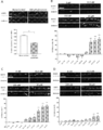
- FIG. 2C shows that 3-MeO-PCP significantly inhibited the reduction of drebrin clusters by 100 ⁇ M glutamate at concentrations above 333 nM.
- FIG. 2D shows that 3-MeO-PCMo significantly inhibited the reduction of drebrin clusters by 100 ⁇ M glutamate at a concentration of 3.33 ⁇ M or higher.
- both 3-MeO-PCP and 3-MeO-PCMo inhibited the reduction of drebrin clusters in a dose-dependent manner.
- 3-MeO-PCMo and quantitative reliability of an IC 50 value analysis of 3-MeO-PCP created the dose-response curves curve approximation based on the measured values (Figure 3). From FIG. 3, it was shown that the inhibitory activity of 3-MeO-PCP was similar to that of PCP, and the inhibitory activity of 3-MeO-PCMo was lower than that of PCP.
- NMDA receptor inhibitory activity it is possible to provide a high-throughput evaluation method for NMDA receptor inhibitory activity, which can be carried out easily and in a short time.
- This evaluation method enables rapid regulation of NPS, which was difficult to evaluate and regulate from the functional aspect in the past, and high-throughput screening and functional evaluation of therapeutic agents targeting NMDA receptors. Is extremely useful.
Landscapes
- Life Sciences & Earth Sciences (AREA)
- Health & Medical Sciences (AREA)
- Engineering & Computer Science (AREA)
- Chemical & Material Sciences (AREA)
- Immunology (AREA)
- Molecular Biology (AREA)
- Physics & Mathematics (AREA)
- Biomedical Technology (AREA)
- Hematology (AREA)
- General Health & Medical Sciences (AREA)
- Urology & Nephrology (AREA)
- Biochemistry (AREA)
- Analytical Chemistry (AREA)
- Pathology (AREA)
- Food Science & Technology (AREA)
- Medicinal Chemistry (AREA)
- General Physics & Mathematics (AREA)
- Organic Chemistry (AREA)
- Biotechnology (AREA)
- Microbiology (AREA)
- Zoology (AREA)
- Biophysics (AREA)
- Proteomics, Peptides & Aminoacids (AREA)
- Bioinformatics & Cheminformatics (AREA)
- Wood Science & Technology (AREA)
- Cell Biology (AREA)
- Pharmacology & Pharmacy (AREA)
- General Engineering & Computer Science (AREA)
- Genetics & Genomics (AREA)
- Measuring Or Testing Involving Enzymes Or Micro-Organisms (AREA)
- Investigating Or Analysing Biological Materials (AREA)
Abstract
本発明は、NMDA受容体阻害活性の直接的且つ迅速、簡便なハイスループット評価方法を確立することを課題とする。 (A)培養神経細胞に、被験物質を接触させるステップ;(B)前記培養神経細胞に、グルタミン酸溶液を接触させるステップ;(C)前記培養神経細胞を固定するステップ;(D)前記培養神経細胞の樹状突起スパインのドレブリンクラスターを可視化するステップ;及び(E)前記ドレブリンクラスターの樹状突起に沿った線密度を測定し、前記線密度が、前記被験物質に接触させていない培養神経細胞の線密度と比較して高い場合に、前記被験物質がNMDA受容体阻害作用を有すると判定するステップを順次備えた方法により、NMDA受容体阻害活性をハイスループットで評価することができる。
Description
本発明は、培養神経細胞の樹状突起スパインのドレブリンクラスターの樹状突起に沿った線密度を指標として用いる、NMDA受容体阻害活性のハイスループット評価方法に関する。
近年、新精神作用物質(New psychoactive substances;NPS、日本では「危険ドラッグ」とも呼ばれる)が、「リーガル・ハイ」、「バスソルト」、「リサーチケミカル」等の用語を使って販売されている。しかしながら、それらは国際的に規制されている薬物と同様の効果を持つ乱用物質である。NPSの出現は世界中の100以上の国と地域で報告されており、NPSの広がりが、多くの国で公衆衛生上の懸念を高めている(非特許文献1)。したがって、リスク評価プロセスを通じてこれらの物質を速やかに規制するために、NPSに対して利用可能な関連データ及び分析結果を蓄積していくことが極めて重要である。
メトキセタミンやジフェニジン等の、NMDA受容体阻害活性を有するNPSが報告されている(非特許文献2~5等)。NMDA受容体は、中枢神経系の学習と記憶に主に関与する受容体であり、NMDA受容体の活動が低下すると、健忘症、知覚の変化、幻覚及び妄想が起こることが知られている。このように、NMDA受容体は、知覚的及び精神的現象に大いに関連しているため、NMDA受容体阻害剤が乱用薬物として使用される可能性、及び、NMDA受容体阻害剤が麻薬と同様の危険な効果を持つ可能性が高いことが示唆されている(非特許文献6)。しかしながら、中枢神経系に対して作用する化合物の安全性薬理試験法は、ネズミ個体を用いた行動解析のみであり、コスト、時間がかかるという問題点がある。そのため、規制当局は、新規化合物についてインシリコで構造の類似性を判定し、規制するか否かを判断しているのが現状である。
上述の状況から、NMDA受容体阻害活性を有するNPSを、機能的側面から既存の薬物との類似度を評価して迅速に規制できるようにするため、NMDA受容体阻害活性を検出、測定する方法が求められている。現状では、NMDA受容体阻害活性の検出、測定方法として、2つの方法が提案されている。第一はカルシウムイメージング法であり(非特許文献7、8)、第二はトリチウム標識したMK801を用いた置換アッセイである(非特許文献9)。しかしながら、これらの方法は、NMDA受容体に対する作用を直接測定することができないうえ、操作が煩雑であり、放射性同位体を使用しなければならない場合もあり、NMDA受容体阻害作用の直接的且つ簡便、迅速な測定には不向きである。
培養神経細胞は、培養を行うと樹状突起を徐々に伸ばし、ニューロン間の成熟シナプスを形成する。その後、培養3週間で、きのこ型の頭部を持つ「樹状突起スパイン」と呼ばれる構造が完成する。樹状突起スパインにおいて、ドレブリンはアクチンフィラメントに高濃度で蓄積され、樹状突起スパインにおける受容体及び足場タンパク質の動態を安定化させる(非特許文献10、11)。このように樹状突起スパインに蓄積されたドレブリンは、ドレブリンクラスターと呼ばれる。培養神経細胞に対するグルタミン酸処理は、樹状突起スパインから樹状突起への急速なドレブリン流出を誘導する(非特許文献12、13)。投与されたグルタミン酸は、AMPA型グルタミン酸レセプター(AMPAR)とNMDA受容体の両方を活性化するが、ドレブリンクラスターからのドレブリンの流出は、NMDA受容体活性化によって誘導され、定量的である(非特許文献13~15)。
UNODC. UNODC report-Global Synthetic Drugs Assessment-Amphetamine-type stimulants and new psychoactive substances. (2017). https://www.unodc.org/unodc/en/scientists/global-synthetic-drugs-assessment-2017.html.
Kikura-Hanajiri, R., Uchiyama, N., Kawamura, M., & Goda, Y. (2014). Changes in the prevalence of new psychoactive substances before and after the introduction of the generic scheduling of synthetic cannabinoids in Japan.Drug Testing and Analysis, 6, 832-839. https://do.org/10.1002/dta.1584.
Kikura-Hanajiri, R. (2017). Changes in the prevalence substances and their legal status in Japan. Folia Pharmacol. Jpn., 150, 1-6.
Hondebrink, L., Kasteel, E., Tukker, A., Wijnolts, F., Verboven, A., & Westerink, R. (2017). Neuropharmacological characterization of the new psychoactive substance methoxetamine. Neuropharmacology, 123, 1, 1-9.
Roth, B., Gibbons, S., Arunotayanun, W., Huang, X., Setola, V., & Treble, R. (2013). The Ketamine Analogue Methoxetamine and 3- and 4-Methoxy Analogues of Phencyclidine Are High Affinity and Selective Ligands for the Glutamate NMDA. Les Iversen published.
Morris, H., & Wallach, J. (2014). From PCP to MXE: a comprehensive review of the non‐medical use of dissociative drugs. Drug Testing and Analysis, 6, 614-632.
Ring, A., & Tanso, R. (2007). Measurements with fluorescent probes in primary neural cultures; improved multiwell techniques. Journal of Pharmacological and Toxicological Methods 56, 300-307.
Sato, K., Takahashi, K., Shigemoto-Mogami, Y., Chujo, K., & Sekino, Y. (2016). Glypican 6 Enhances N-Methyl-D-Aspartate Receptor Function inHuman-Induced Pluripotent Stem Cell-Derived Neurons. Frontiers in Cellular Neuroscience, 10(259).
Reynolds, I. J. (2001). [3H] (+)MK801 radioligand binding assay at the N-methyl-D-aspartate receptor. Curr. Protoc. Pharmacol. Chap. 1: Unit 1.20.
Sekino, Y., Kojima, N., & Shirao, T. (2007). Role of actincytoskeleton in dendritic spine morphogenesis. Neurochemistry International, 51, 92-104.
Shirao, T., Hanamura, K., Koganezawa, N., Ishizuka, Y., Yamazaki, H., & Sekino, Y. (2017). The role of drebrin in neurons. Journal of Neurochemistry, 141(6), 819-834.
Sekino, Y., Tanaka, S., Hanamura, K., Yamazaki, H., Sasagawa, Y., Xue, Y., Hayashi, K., & Shirao, T. (2006). Activation of N-methyl-d-aspartate receptor induces a shift of drebrin distribution: Disappearance from dendritic spines and appearance in dendritic shafts. Molecular Cellular Neuroscience,31, 493-594.
Mizui, T., Sekino, Y., Yamazaki, H., Ishizuka, Y., Takahashi, H., Kojima, N., Kojima, M., and Shirao, T. (2014). Myosin II ATPase activitymediates the long-term potentiation-induced exodus of stable F-actin bound by drebrin A from dendritic spines. PLoS One, 9, e85367.
Sekino, Y., Koganezawa, N., Mizui, T., & Shirao, T. (2017). Role of Drebrin in Synaptic Plasticity, Advances in Experimental Medicine and Biology-Drebrin, 1006 (183-201).
Koganezawa, N., Hanamura, K., Sekino, Y., & Shirao, T. (2017). The role of drebrin in dendritic spines. Molecular and Cellular Neuroscience, 84, 85-92.
上述したとおり、NMDA受容体阻害剤の、直接的且つ迅速、簡便な阻害活性の評価方法は未だ構築されておらず、機能面からの新規NMDA受容体阻害剤の規制は行われていないのが現状である。本発明は、以上の事情に鑑みなされたものであり、NMDA受容体阻害活性の直接的且つ迅速、簡便なハイスループット評価方法を確立することにより、NMDA受容体阻害剤の迅速な規制、及び新規治療薬としてのNMDA受容体阻害剤のスクリーニング及び活性評価を可能にすることを目的とするものである。
本発明者らは、鋭意検討を重ねた結果、既知のNMDA受容体阻害剤であるフェンシクリジン(PCP)と、PCPアナログ2種(3-MeO-PCMo及び3-MeO-PCP)を用い、これらの化合物を成熟した培養神経細胞に接触させることで、グルタミン酸処理によるドレブリンクラスターの線密度減少が抑制されることを見いだした。さらに、かかるドレブリンクラスターの線密度減少が、化合物濃度と相関すること、並びに該相関に基づいて算出した50%阻害濃度(IC50)の比が、これらの化合物の阻害定数(Ki)の比と良好な相関関係を示すことを見いだした。本発明は、これらの知見により完成したものである。
すなわち、本発明は以下の事項により特定されるとおりのものである。
(1)以下のステップ(A)~(E)を順次備えたことを特徴とするNMDA受容体阻害活性のハイスループット評価方法。
(A)培養神経細胞に、被験物質を接触させるステップ;
(B)前記培養神経細胞に、グルタミン酸溶液を接触させるステップ;
(C)前記培養神経細胞を固定するステップ;
(D)前記培養神経細胞の樹状突起スパインのドレブリンクラスターを可視化するステップ;
(E)前記ドレブリンクラスターの樹状突起に沿った線密度を測定し、前記線密度が、前記被験物質に接触させていない培養神経細胞の線密度と比較して高い場合に、前記被験物質がNMDA受容体阻害作用を有すると判定するステップ;
(2)以下のステップ(F)を、ステップ(E)の後に備えたことを特徴とする上記(1)に記載の方法。
(F)ステップ(A)で培養神経細胞に接触させた被験物質の濃度と、ステップ(E)で測定したドレブリンクラスターの線密度との相関に基づいて、前記被験物質のIC50を算出するステップ;
(3)以下のステップ(G)を、ステップ(F)の後に備えたことを特徴とする上記(1)又は(2)に記載の方法。
(G)ステップ(F)で算出した被験物質のIC50を、既知のNMDA受容体阻害剤のIC50と比較することにより、前記被験物質と、前記既知のNMDA受容体阻害剤とのNMDA受容体阻害活性比を算出するステップ;
(4)培養神経細胞が、海馬神経細胞由来であることを特徴とする上記(1)~(3)のいずれかに記載の方法。
(5)培養神経細胞が、げっ歯類由来であることを特徴とする上記(1)~(4)のいずれかに記載の方法。
(6)ステップ(D)の可視化が、抗ドレブリン抗体による免疫染色であることを特徴とする上記(1)~(5)のいずれかに記載の方法。
(1)以下のステップ(A)~(E)を順次備えたことを特徴とするNMDA受容体阻害活性のハイスループット評価方法。
(A)培養神経細胞に、被験物質を接触させるステップ;
(B)前記培養神経細胞に、グルタミン酸溶液を接触させるステップ;
(C)前記培養神経細胞を固定するステップ;
(D)前記培養神経細胞の樹状突起スパインのドレブリンクラスターを可視化するステップ;
(E)前記ドレブリンクラスターの樹状突起に沿った線密度を測定し、前記線密度が、前記被験物質に接触させていない培養神経細胞の線密度と比較して高い場合に、前記被験物質がNMDA受容体阻害作用を有すると判定するステップ;
(2)以下のステップ(F)を、ステップ(E)の後に備えたことを特徴とする上記(1)に記載の方法。
(F)ステップ(A)で培養神経細胞に接触させた被験物質の濃度と、ステップ(E)で測定したドレブリンクラスターの線密度との相関に基づいて、前記被験物質のIC50を算出するステップ;
(3)以下のステップ(G)を、ステップ(F)の後に備えたことを特徴とする上記(1)又は(2)に記載の方法。
(G)ステップ(F)で算出した被験物質のIC50を、既知のNMDA受容体阻害剤のIC50と比較することにより、前記被験物質と、前記既知のNMDA受容体阻害剤とのNMDA受容体阻害活性比を算出するステップ;
(4)培養神経細胞が、海馬神経細胞由来であることを特徴とする上記(1)~(3)のいずれかに記載の方法。
(5)培養神経細胞が、げっ歯類由来であることを特徴とする上記(1)~(4)のいずれかに記載の方法。
(6)ステップ(D)の可視化が、抗ドレブリン抗体による免疫染色であることを特徴とする上記(1)~(5)のいずれかに記載の方法。
本発明によれば、簡便且つ短時間で実施することのできる、NMDA受容体阻害活性のハイスループット評価方法を提供することができる。
本発明におけるNMDA受容体阻害活性のハイスループット評価方法とは、(A)培養神経細胞に、被験物質を接触させるステップ;(B)前記培養神経細胞に、グルタミン酸溶液を接触させるステップ;(C)前記培養神経細胞を固定するステップ;(D)前記培養神経細胞の樹状突起スパインのドレブリンクラスターを可視化するステップ;及び(E)前記ドレブリンクラスターの樹状突起に沿った線密度を測定し、前記線密度が、前記被験物質に接触させていない培養神経細胞の線密度と比較して高い場合に、前記被験物質がNMDA受容体阻害作用を有すると判定するステップ;を順次備えた方法である。
本発明の方法は、培養神経細胞の樹状突起に沿って形成されたドレブリンクラスターの線密度変化を指標として、被験物質のNMDA受容体阻害活性を評価する方法である。ここで、培養神経細胞としては、NMDA受容体の活性化によりドレブリンクラスターが減少する細胞であればよく、例えば、初代培養神経細胞、神経芽細胞腫と神経膠腫のハイブリドーマ、神経芽細胞腫や、多能性幹細胞(胚性幹細胞、人工多能性幹細胞)に由来する培養神経細胞等を挙げることができ、海馬由来の初代培養神経細胞を好適に使用することができる。また、培養神経細胞の由来は、ヒトのNMDA受容体の阻害活性評価に用いることができるものであることが好ましく、例えば哺乳類由来、より好ましくは霊長類由来、さらに好ましくはヒト由来を挙げることができるが、マウスやラット等、げっ歯類由来の培養神経細胞を用いることもできる。また、上記培養神経細胞は、ヒト胎児由来の培養神経細胞であってもよい。
本発明において、被験物質とは、NMDA受容体の阻害活性を有し得る化合物であればよく、NPSの候補化合物や、麻酔等の医薬品候補化合物を例示することができ、フェンシクリジン、メトキセタミン、ジフェニジン等の既知のNPSの類縁化合物や、ケタミン等のNMDA受容体拮抗薬の類縁化合物であってもよい。上記ステップ(A)における被験物質の終濃度は、IC50を算出するために適した濃度を常法に基づいて決定することができるが、例えば阻害率50%を挟む濃度が得られるように、被験物質を段階希釈した希釈系列を用いることが望ましい。また、上記ステップ(A)における接触時間は、被験物質がNMDA受容体まで到達するために十分な時間であればよく、例えば2~20分間、好ましくは5~15分間、より好ましくは8~12分間を例示することができる。
上記ステップ(B)は、NMDA受容体を活性化させ、ドレブリンクラスターの減少を引き起こすために、グルタミン酸溶液を接触させるステップである。ここで、グルタミン酸溶液の終濃度及び接触時間は、被験物質のIC50を算出するために適切なNMDA受容体活性化を引き起こす濃度及び接触時間であればよい。上記濃度としては例えば1μM~500μM、好ましくは10μM~300μM、より好ましくは50μM~200μM、さらに好ましくは80μM~150μMを挙げることができ、上記接触時間としては、例えば2~20分間、好ましくは5~15分間、より好ましくは8~12分間を例示することができる。また、グルタミン酸溶液に代えて、NMDA受容体活性化を引き起こすことのできるいかなる化合物を用いてもよい。
上記ステップ(C)における培養神経細胞の固定は、固定後のドレブリンクラスターの可視化が効率的に行える条件であればいかなる公知の方法を用いてもよく、固定化試薬としては、例えばメタノール、アセトン、ホルムアルデヒド、パラホルムアルデヒド(PFA)、エタノール、グルタルアルデヒド、スベルイミノ酸ジメチルを挙げることができ、固定化試薬は、冷凍庫(-20±2℃)又は冷蔵庫(4±2℃)で冷却したものや、室温のものを使用することができる。
上記ステップ(D)において、培養神経細胞のドレブリンクラスターの可視化は、いかなる公知の方法により可視化してもよく、ドレブリンに対して特異的に結合し得る分子を用いて可視化する方法を例示することができる。上記ドレブリンに対して特異的に結合し得る分子としては、例えば抗ドレブリン抗体を挙げることができ、検出の簡便性や、他の培養神経細胞マーカーを用いて共染色した場合や、4’,6-ジアミジノ-2-フェニルインドール(DAPI)を用いて細胞を染色した場合における識別性の観点から、蛍光標識された抗ドレブリン抗体を用いることが好ましい。ここで、蛍光標識としては、FITC、Cy3、Cy5、Rhodamine、Alexa fluor(登録商標、インビトロジェン社製)等の蛍光物質を挙げることができる。抗ドレブリン抗体は、直接標識してもよく、これらに対する二次抗体を用いて検出してもよい。本発明で用いる抗体は、モノクローナル抗体、ポリクローナル抗体のいずれであってもよく、市販のものを使用できる他、常法により作製することもできる。また、抗MAP2抗体等の他の培養神経細胞マーカーを用いて、培養神経細胞を共染色してもよい。
一態様では、ドレブリンクラスターの可視化は、例えば培養神経細胞において抗ドレブリン抗体(抗ドレブリンラクダVHH抗体等)を発現させ、該抗ドレブリン抗体の集積を検出することにより行うこともできる。抗ドレブリン抗体は、二次抗体で検出してもよいが、蛍光タンパク質等の標識物質を結合させた抗ドレブリン抗体を培養神経細胞内で発現させ、標識物質を検出することにより、生細胞を用いたリアルタイムイメージングを行うことができる。また、一態様において、あらかじめ蛍光タンパク質等の標識物質を結合させたドレブリン又はドレブリン断片を培養神経細胞内で発現させると、発現した標識ドレブリン又はドレブリン断片がドレブリンクラスターを形成する。この態様においては、該標識ドレブリン又はドレブリン断片を検出することにより、追加の可視化ステップなしにドレブリンクラスターを検出することができる。これらの態様においては、ステップ(C)における培養神経細胞の固定は行っても行わなくてもよい。
上記ステップ(E)におけるドレブリンクラスターの線密度は、上記ステップ(D)により可視化されたドレブリン陽性シグナルの個数を手動又は自動で計数し、樹状突起の単位長さ当たりのドレブリン陽性シグナルの個数を算出することにより測定される。かかる線密度の測定は、IN Cell Developer Toolbox(GEヘルスケア社製)等の公知の画像解析ソフトウェアを用いて自動測定することが好ましい。
本発明の方法は、上記ステップ(E)の後に、上記ステップ(A)で培養神経細胞に接触させた被験物質の濃度と、ステップ(E)で測定したドレブリンクラスターとの線密度の相関に基づいて、前記被験物質のIC50を算出するステップ(F)を備えてもよい。IC50の算出は、常法に基づいて行うことができ、例えば、被験物質の濃度に対する阻害活性値をプロットした阻害曲線を作成して算出することができる。また、本発明の方法により得られたIC50は、Ki値と良好な相関関係にあるため、上記ステップ(F)で得られた被験物質のIC50を、既知のNMDA受容体阻害剤のIC50と比較して阻害活性比を算出することで、前記被験物質の活性を迅速且つ簡便な方法にて評価することができる。
本発明の一態様において、本発明の方法は、NMDA受容体活性化を引き起こす物質のスクリーニング及び活性評価に用いることもできる。かかる態様において、被験物質の濃度とドレブリンクラスターの線密度の減少との相関に基づいて、被験物質のNMDA受容体活性化能が評価される。
本発明の一態様において、ドレブリンクラスターの線密度に代えて、或いはドレブリンクラスターの線密度と共に、ドレブリンA及び/又はドレブリンEの発現量変化を、被験物質の活性評価に用いてもよい。かかる態様は、被験物質が神経細胞に与える長期的影響を評価するのに有効である可能性がある。
本発明は、従来のNMDA受容体阻害活性の測定方法と比較して迅速且つ簡便に、定量性、再現性良く、高精度にNMDA受容体阻害活性の評価を行うことができる。そのため、従来は機能面からの評価及び規制が困難であったNPSの迅速な規制や、NMDA受容体をターゲットとした治療薬のハイスループットスクリーニングへの応用が期待される。
以下、実施例により本発明をより具体的に説明するが、本発明の技術的範囲はこれらの例示に限定されるものではない。
1.方法
1-2 動物実験
動物実験は、群馬大学昭和キャンパス(群馬県前橋市)の動物実験委員会によって設定されたガイドラインに従って行い、研究における動物の使用に関するNIHガイドラインに準拠した。動物の苦しみを最小限に抑え、使用される動物の数を減らすためにあらゆる努力を払った。妊娠ウィスターラットは、日本チャールス・リバー株式会社から入手した。動物は標準的な動物施設条件下で飼料と水を自由に摂取させて飼育した。
1-2 動物実験
動物実験は、群馬大学昭和キャンパス(群馬県前橋市)の動物実験委員会によって設定されたガイドラインに従って行い、研究における動物の使用に関するNIHガイドラインに準拠した。動物の苦しみを最小限に抑え、使用される動物の数を減らすためにあらゆる努力を払った。妊娠ウィスターラットは、日本チャールス・リバー株式会社から入手した。動物は標準的な動物施設条件下で飼料と水を自由に摂取させて飼育した。
1-2 海馬神経細胞培養
18日目胚のラットの海馬から調製した凍結ニューロンを、ポリリジンであらかじめコーティングした96ウェルプレート(Thermo 96 Well Black Poly Bottom Poly-Lysine、Thermo Fisher Scientific社製)に、3.0×10⁴細胞/cm2の密度で播種した。B27サプリメント、ペニシリン/ストレプトマイシン、及びL-アラニル-L-グルタミン(Glutamax-I;Thermo Fisher Scientific社製)を含むNeurobasal Medium(Thermo Fisher Scientific社製)を使用して、37℃、5%CO2環境下でインキュベートした。
4DIV(インビトロでの培養日数)に、シトシンβ-D-アラビノフラノシド(Sigma社製)を最終濃度0.2μMになるように加えてグリア細胞の増殖を抑制した。ニューロンを21DIV培養した後、それらを薬理学的に処理した。
18日目胚のラットの海馬から調製した凍結ニューロンを、ポリリジンであらかじめコーティングした96ウェルプレート(Thermo 96 Well Black Poly Bottom Poly-Lysine、Thermo Fisher Scientific社製)に、3.0×10⁴細胞/cm2の密度で播種した。B27サプリメント、ペニシリン/ストレプトマイシン、及びL-アラニル-L-グルタミン(Glutamax-I;Thermo Fisher Scientific社製)を含むNeurobasal Medium(Thermo Fisher Scientific社製)を使用して、37℃、5%CO2環境下でインキュベートした。
4DIV(インビトロでの培養日数)に、シトシンβ-D-アラビノフラノシド(Sigma社製)を最終濃度0.2μMになるように加えてグリア細胞の増殖を抑制した。ニューロンを21DIV培養した後、それらを薬理学的に処理した。
1-3 被験物質の投与
PCP、3-MeO-PCMo、及び3-MeO-PCP(Cayman Chemical社製)を0.1%DMSO溶液により所定の濃度に希釈し、96ウェルプレートのうち、培養神経細胞が培養されている所定のウェルに投与した。被験物質の投与から10分後に、ウェル内の最終濃度が100μMとなるようにグルタミン酸溶液(和光純薬株式会社製)を調製し、薬液投与10分後に投与した。ドレブリンクラスター密度の用量反応曲線を得るための実験のために、最終グルタメート濃度は1、3、10、30、50又は100μMであった。いくつかの培養神経細胞は、グルタミン酸塩処理の前に、NMDA受容体に対する競合アンタゴニストであるD-2-アミノ-5-ホスホノバレリン酸(APV;50、100又は500μM)で10分間処理した。
PCP、3-MeO-PCMo、及び3-MeO-PCP(Cayman Chemical社製)を0.1%DMSO溶液により所定の濃度に希釈し、96ウェルプレートのうち、培養神経細胞が培養されている所定のウェルに投与した。被験物質の投与から10分後に、ウェル内の最終濃度が100μMとなるようにグルタミン酸溶液(和光純薬株式会社製)を調製し、薬液投与10分後に投与した。ドレブリンクラスター密度の用量反応曲線を得るための実験のために、最終グルタメート濃度は1、3、10、30、50又は100μMであった。いくつかの培養神経細胞は、グルタミン酸塩処理の前に、NMDA受容体に対する競合アンタゴニストであるD-2-アミノ-5-ホスホノバレリン酸(APV;50、100又は500μM)で10分間処理した。
1-4 免疫細胞化学
培養神経細胞を、リン酸緩衝液中4%パラホルムアルデヒドで固定した。リン酸緩衝生理食塩水(PBS)中0.1%トリトンX-100で5分間浸透処理した後、培養神経細胞をPBS中3%ウシ血清アルブミン(PBSA) で室温(RT)にて1時間ブロッキングし、次いで一次抗体と共に4℃で一晩インキュベートした。抗ドレブリン抗体(マウスモノクローナル、M2F6、1:1)及び抗MAP2抗体(ウサギポリクローナル、1:2000)(Merck Millipore社製)を一次抗体として使用した。PBSで洗浄した後、細胞を二次抗体及び4’,6-ジアミジノ-2-フェニルインドール二塩酸塩(DAPI、1:1000、Thermo Fisher Scientific社製)と共に室温で1時間インキュベートした。使用した二次抗体は、Alexa Fluor 488ロバ抗マウスIgG(1:250)及びAlexa Fluor 594ロバ抗ウサギIgG(1:250、Jackson Immuno Research Laboratories社製)であった。
培養神経細胞を、リン酸緩衝液中4%パラホルムアルデヒドで固定した。リン酸緩衝生理食塩水(PBS)中0.1%トリトンX-100で5分間浸透処理した後、培養神経細胞をPBS中3%ウシ血清アルブミン(PBSA) で室温(RT)にて1時間ブロッキングし、次いで一次抗体と共に4℃で一晩インキュベートした。抗ドレブリン抗体(マウスモノクローナル、M2F6、1:1)及び抗MAP2抗体(ウサギポリクローナル、1:2000)(Merck Millipore社製)を一次抗体として使用した。PBSで洗浄した後、細胞を二次抗体及び4’,6-ジアミジノ-2-フェニルインドール二塩酸塩(DAPI、1:1000、Thermo Fisher Scientific社製)と共に室温で1時間インキュベートした。使用した二次抗体は、Alexa Fluor 488ロバ抗マウスIgG(1:250)及びAlexa Fluor 594ロバ抗ウサギIgG(1:250、Jackson Immuno Research Laboratories社製)であった。
1-5 画像の自動取得と解析
96ウェルプレートで培養された海馬神経細胞の三重染色像は、IN Cell Analyzer 2200(GE Healthcare社製)の自動焦点機能を使用することによって自動的に取得された(20倍レンズ、開口数0.45)。全てのデータは16ビット/ピクセルで2048×2048の解像度で収集された。画像内の単一の画素は、標本面内の325nmの正方形に対応していた。Zスタック画像は、ハイコンテント画像ソフトウェアであるIN Cell Developer Toolbox v1.9(GE Healthcare社製)を用いて拡張焦点変換プロトコル(extended focus transformation protocol)により処理され、Zシリーズスタックからの焦点があった単一の2次元画像を生成した。培養神経細胞の細胞体は、MAP2及びDAPI染色に基づいて認識された。培養神経細胞の細胞体を特定するために、セグメンテーション機能により、異なるカーネルサイズ及び感度を有するコントラストベースのセグメンテーションアルゴリズムを介して、MAP2陽性シグナルの蛍光強度に基づいて蛍光体を局在化させた。Erosion(収縮)、Sieve(ふるい)、Dilation(拡張)のコマンド及びDAPI陽性領域を用いて、培養神経細胞の細胞体を同定した。培養神経細胞の樹状突起を識別するために、セグメンテーション機能、並びにDilation及びErosionコマンドを、MAP2陽性シグナルについての別個のパラメーターと共に使用し、その後培養神経細胞の細胞体領域を差し引いた。ドレブリンクラスターを同定するために、セグメンテーション機能により、異なるカーネルサイズ及び感度を有するコントラストベースのセグメンテーションアルゴリズムを介して、ドレブリン陽性シグナルの蛍光強度に基づいて蛍光物体を局在化させた。偽陽性のバックグラウンドシグナルを除去するためにSieveコマンドを使用した。加えて、Dilationコマンドによって決定される、樹状突起までの定義された距離内に位置するドレブリン陽性斑点のみを、樹状突起に沿ったドレブリンクラスターとしてカウントした。神経細胞体、樹状突起及びドレブリンクラスター、視野当たりの細胞数、視野当たりの樹状突起長を同定した後、ドレブリンクラスターの線密度が自動的に算出された。
96ウェルプレートで培養された海馬神経細胞の三重染色像は、IN Cell Analyzer 2200(GE Healthcare社製)の自動焦点機能を使用することによって自動的に取得された(20倍レンズ、開口数0.45)。全てのデータは16ビット/ピクセルで2048×2048の解像度で収集された。画像内の単一の画素は、標本面内の325nmの正方形に対応していた。Zスタック画像は、ハイコンテント画像ソフトウェアであるIN Cell Developer Toolbox v1.9(GE Healthcare社製)を用いて拡張焦点変換プロトコル(extended focus transformation protocol)により処理され、Zシリーズスタックからの焦点があった単一の2次元画像を生成した。培養神経細胞の細胞体は、MAP2及びDAPI染色に基づいて認識された。培養神経細胞の細胞体を特定するために、セグメンテーション機能により、異なるカーネルサイズ及び感度を有するコントラストベースのセグメンテーションアルゴリズムを介して、MAP2陽性シグナルの蛍光強度に基づいて蛍光体を局在化させた。Erosion(収縮)、Sieve(ふるい)、Dilation(拡張)のコマンド及びDAPI陽性領域を用いて、培養神経細胞の細胞体を同定した。培養神経細胞の樹状突起を識別するために、セグメンテーション機能、並びにDilation及びErosionコマンドを、MAP2陽性シグナルについての別個のパラメーターと共に使用し、その後培養神経細胞の細胞体領域を差し引いた。ドレブリンクラスターを同定するために、セグメンテーション機能により、異なるカーネルサイズ及び感度を有するコントラストベースのセグメンテーションアルゴリズムを介して、ドレブリン陽性シグナルの蛍光強度に基づいて蛍光物体を局在化させた。偽陽性のバックグラウンドシグナルを除去するためにSieveコマンドを使用した。加えて、Dilationコマンドによって決定される、樹状突起までの定義された距離内に位置するドレブリン陽性斑点のみを、樹状突起に沿ったドレブリンクラスターとしてカウントした。神経細胞体、樹状突起及びドレブリンクラスター、視野当たりの細胞数、視野当たりの樹状突起長を同定した後、ドレブリンクラスターの線密度が自動的に算出された。
1-6 統計
データは、ANOVA(エクセル統計Statcel4、有限会社オーエムエス出版製)に従ってt検定又はターキー-クレイマー検定によって統計的に分析した。全てのデータは平均値±平均値の標準誤差(SEM)として示した。市販のソフトウェア(GraphPad Prism 8、GraphPad Software社製)を用いて用量適合曲線を描き、IC50を計算した。
データは、ANOVA(エクセル統計Statcel4、有限会社オーエムエス出版製)に従ってt検定又はターキー-クレイマー検定によって統計的に分析した。全てのデータは平均値±平均値の標準誤差(SEM)として示した。市販のソフトウェア(GraphPad Prism 8、GraphPad Software社製)を用いて用量適合曲線を描き、IC50を計算した。
2.結果
2-1 グルタミン酸処理は、NMDA受容体活性化を介してドレブリンクラスター密度を減少させた
まず、グルタミン酸がドレブリンクラスター密度に及ぼす影響を、ハイスループットイメージングシステムを用いて調べた。21インビトロ培養日数(DIV)のラット海馬培養神経細胞を、いくつかの濃度のグルタミン酸溶液(1μM、N=61;3μM、N=97;10μM、N=88;30μM、N=97;50μM、N=20;100μM、N=106)で処理した。上記1-4の免疫細胞化学の処理を行った後、画像取得及び分析は自動的に行われた。得られたドレブリンクラスター密度のグルタミン酸用量反応曲線を図1Aに示す。EC50は10.4μMであり、95%信頼区間(CI)は6.29μM~17.4μMの範囲であった。
2-1 グルタミン酸処理は、NMDA受容体活性化を介してドレブリンクラスター密度を減少させた
まず、グルタミン酸がドレブリンクラスター密度に及ぼす影響を、ハイスループットイメージングシステムを用いて調べた。21インビトロ培養日数(DIV)のラット海馬培養神経細胞を、いくつかの濃度のグルタミン酸溶液(1μM、N=61;3μM、N=97;10μM、N=88;30μM、N=97;50μM、N=20;100μM、N=106)で処理した。上記1-4の免疫細胞化学の処理を行った後、画像取得及び分析は自動的に行われた。得られたドレブリンクラスター密度のグルタミン酸用量反応曲線を図1Aに示す。EC50は10.4μMであり、95%信頼区間(CI)は6.29μM~17.4μMの範囲であった。
また、グルタミン酸刺激によるドレブリンクラスター密度の減少は、APV(50μM、N=4;100μM、N=7;500μM、N=6)によって阻害されることを確認した(図1B)。ここでの結果により、当該実験系が、グルタミン酸刺激によるドレブリンクラスター密度の減少、及び、NMDA受容体競合的アンタゴニストによるドレブリンクラスター密度減少の阻害を検出することができることが示された。
2-2 グルタミン酸誘発ドレブリンクラスター密度の減少に対するPCP、3-MeO-PCMo及び3-MeO-PCPの阻害効果
まず、本発明者らは、100μMグルタミン酸溶液を用いてドレブリンクラスターの密度をどの程度減少させるかを決定した(対照、N=95;グルタミン酸100μM、N=77)。図2Aに示されるように、100μMグルタミン酸溶液処理により、対照群と比較してドレブリンクラスター密度が約40%減少した(対照0.538±0.0143;100μMグルタミン酸0.319±0.0035)。
まず、本発明者らは、100μMグルタミン酸溶液を用いてドレブリンクラスターの密度をどの程度減少させるかを決定した(対照、N=95;グルタミン酸100μM、N=77)。図2Aに示されるように、100μMグルタミン酸溶液処理により、対照群と比較してドレブリンクラスター密度が約40%減少した(対照0.538±0.0143;100μMグルタミン酸0.319±0.0035)。
次に、グルタミン酸を用いたNMDA受容体刺激によるドレブリンクラスターの密度の低下に対する、NMDA受容体の非競合的拮抗薬であるPCPの抑制効果を調べた(0~10 μM、N=18)。対照群に対する各投与群の平均阻害率を計算することにより、用量依存的効果を分析した。図2Bより、PCPが100μMグルタメートのNMDAR刺激によって1μM以上の濃度でドレブリンクラスターの減少を有意に抑制し、PCPが用量依存的にドレブリンクラスターの減少を抑制することが示された。
同様の方法により、PCPアナログである3-MeO-PCMo及び3-MeO-PCPの阻害作用を調べた(0~10μM;3-MeO-PCMo、N=29;3-MeO-PCMo、N=30)。図2Cは、3-MeO-PCPが、333nM以上の濃度で100μMグルタミン酸によるドレブリンクラスターの減少を有意に阻害したことを示す。図2Dは、3 -MeO-PCMoが、3.33μM以上の濃度で100μMグルタミン酸によるドレブリンクラスターの減少を有意に阻害したことを示す。さらに、3-MeO-PCP及び3-MeO-PCMoの両方が、用量依存的にドレブリンクラスターの減少を阻害した。
2-3 PCP、3-MeO-PCMo及び3-MeO-PCPのIC50値
分析の定量的信頼性を高めるために、測定値に基づいて曲線近似した用量作用曲線を作成した(図3)。図3より、3-MeO-PCPの阻害活性がPCPの阻害活性と同程度であり、3-MeO-PCMoの阻害活性がPCPのそれよりも低いことが示された。IC50は、PCP=2.02μM(95%CI:1.39~2.97μM)、3-MeO-PCP=1.51μM(95%CI:1.16~1.99μM)、3-MeO-PCMo=26.7μM(95%CI:20.0~37.3μM)であった。
分析の定量的信頼性を高めるために、測定値に基づいて曲線近似した用量作用曲線を作成した(図3)。図3より、3-MeO-PCPの阻害活性がPCPの阻害活性と同程度であり、3-MeO-PCMoの阻害活性がPCPのそれよりも低いことが示された。IC50は、PCP=2.02μM(95%CI:1.39~2.97μM)、3-MeO-PCP=1.51μM(95%CI:1.16~1.99μM)、3-MeO-PCMo=26.7μM(95%CI:20.0~37.3μM)であった。
3.考察
上記2-3で算出したIC50に基づく阻害活性の順は、活性が大きい方から3-MeO-PCP>PCP>3-MeO-PCMoであった。最近なされたNMDA受容体に対する結合アッセイの報告によれば、PCPの阻害定数(Ki)値は22.1nM(Colestock et al. (2018)、Drug Testing and Analysis, 10, 272-283)及び57.9nM(Wallach, J. (2014). Structure activity relationship (SAR) studies of arylcyclohexylamines as N-methyl-D-aspartate receptor antagonists. PhD dissertation. University of the Sciences, Philadelphia)であると報告されており、3-MeO-PCPのKi値は38.1nMであると報告されている(Wallach, J. (2014))。これは、3-MeO-PCPが、PCPの平均よりもNMDA受容体に対してわずかに高い親和性を有することを示す。一方で、3-MeO-PCMoのKi値は252.9nMであり、PCPのKi値と比べて1/12であると報告されている(Colestock et al. (2018))。
上記2-3で算出したIC50に基づく阻害活性の順は、活性が大きい方から3-MeO-PCP>PCP>3-MeO-PCMoであった。最近なされたNMDA受容体に対する結合アッセイの報告によれば、PCPの阻害定数(Ki)値は22.1nM(Colestock et al. (2018)、Drug Testing and Analysis, 10, 272-283)及び57.9nM(Wallach, J. (2014). Structure activity relationship (SAR) studies of arylcyclohexylamines as N-methyl-D-aspartate receptor antagonists. PhD dissertation. University of the Sciences, Philadelphia)であると報告されており、3-MeO-PCPのKi値は38.1nMであると報告されている(Wallach, J. (2014))。これは、3-MeO-PCPが、PCPの平均よりもNMDA受容体に対してわずかに高い親和性を有することを示す。一方で、3-MeO-PCMoのKi値は252.9nMであり、PCPのKi値と比べて1/12であると報告されている(Colestock et al. (2018))。
上記のKi値からPCPに対する親和性比を計算したところ、PCP=1としたときに、3-MeO-PCP=0.73(Wallach, J. (2014)のKi値からの再計算)、3-MeO-PCMo=11.4(Colestock et al. (2018)のKi値からの再計算)であった。まとめると、親和性の順序は、3-MeO-PCP>PCP>3-MeO-PCMoで、本発明の方法により得られたIC50に基づく活性の順序と同じであった。
同一の作用機序を有する阻害剤の場合、それらのIC50値を比較することにより、相対的な阻害活性を評価するのに有用であるとの報告がある(Cheng&Prusoff (1973) Biochemical Pharmacology, 22(23), 3099-3108;Cer et al. (2009) Nucleic Acids Res. 1(37): W441-W445)。PCP及び他の高親和性NMDA受容体アンタゴニストは、NMDARと同様の阻害メカニズムを有することが示唆されている(Lodge&Mercier (2015) Br. J. Pharmacol, 172, 4254-4276)。かかる知見に基づいて、本研究で得られたIC50値からPCPに対する効力比を計算したところ、PCP=1としたときに、3-MeO-PCP=0.75、3-MeO-PCMo=13.2であり、上記Ki値から計算した効力比と類似していた。この結果より、本発明によって得られるIC50値は、NMDA受容体阻害作用を反映しており、NMDA受容体阻害活性を有する化合物(NPS候補化合物や、新規治療剤等)の薬理学的評価(毒性評価)に使用することができることが明らかになった。
本発明によれば、簡便且つ短時間で実施することのできる、NMDA受容体阻害活性のハイスループット評価方法を提供することができる。かかる評価方法により、従来は機能面からの評価及び規制が困難であったNPSの迅速な規制や、NMDA受容体をターゲットとした治療薬のハイスループットスクリーニング及び機能評価が可能になるため、産業上の有用性は極めて高い。
Claims (6)
- 以下のステップ(A)~(E)を順次備えたことを特徴とするNMDA受容体阻害活性のハイスループット評価方法。
(A)培養神経細胞に、被験物質を接触させるステップ;
(B)前記培養神経細胞に、グルタミン酸溶液を接触させるステップ;
(C)前記培養神経細胞を固定するステップ;
(D)前記培養神経細胞の樹状突起スパインのドレブリンクラスターを可視化するステップ;
(E)前記ドレブリンクラスターの樹状突起に沿った線密度を測定し、前記線密度が、前記被験物質に接触させていない培養神経細胞の線密度と比較して高い場合に、前記被験物質がNMDA受容体阻害作用を有すると判定するステップ; - 以下のステップ(F)を、ステップ(E)の後に備えたことを特徴とする請求項1に記載の方法。
(F)ステップ(A)で培養神経細胞に接触させた被験物質の濃度と、ステップ(E)で測定したドレブリンクラスターの線密度との相関に基づいて、前記被験物質のIC50を算出するステップ; - 以下のステップ(G)を、ステップ(F)の後に備えたことを特徴とする請求項1又は2に記載の方法。
(G)ステップ(F)で算出した被験物質のIC50を、既知のNMDA受容体阻害剤のIC50と比較することにより、前記被験物質と、前記既知のNMDA受容体阻害剤とのNMDA受容体阻害活性比を算出するステップ; - 培養神経細胞が、海馬神経細胞由来であることを特徴とする請求項1~3のいずれかに記載の方法。
- 培養神経細胞が、げっ歯類由来であることを特徴とする請求項1~4のいずれかに記載の方法。
- ステップ(D)の可視化が、抗ドレブリン抗体による免疫染色であることを特徴とする請求項1~5のいずれかに記載の方法。
Priority Applications (1)
| Application Number | Priority Date | Filing Date | Title |
|---|---|---|---|
| JP2021505138A JP7536240B2 (ja) | 2019-03-13 | 2020-03-12 | Nmda受容体阻害活性のハイスループット評価方法 |
Applications Claiming Priority (2)
| Application Number | Priority Date | Filing Date | Title |
|---|---|---|---|
| JP2019-046531 | 2019-03-13 | ||
| JP2019046531 | 2019-03-13 |
Publications (1)
| Publication Number | Publication Date |
|---|---|
| WO2020184676A1 true WO2020184676A1 (ja) | 2020-09-17 |
Family
ID=72426657
Family Applications (1)
| Application Number | Title | Priority Date | Filing Date |
|---|---|---|---|
| PCT/JP2020/010909 Ceased WO2020184676A1 (ja) | 2019-03-13 | 2020-03-12 | Nmda受容体阻害活性のハイスループット評価方法 |
Country Status (2)
| Country | Link |
|---|---|
| JP (1) | JP7536240B2 (ja) |
| WO (1) | WO2020184676A1 (ja) |
Cited By (1)
| Publication number | Priority date | Publication date | Assignee | Title |
|---|---|---|---|---|
| JP2022001838A (ja) * | 2020-06-19 | 2022-01-06 | アルメッド株式会社 | シナプス機能不全に起因する、又はシナプス機能不全を付随する疾病の判定方法 |
Citations (2)
| Publication number | Priority date | Publication date | Assignee | Title |
|---|---|---|---|---|
| WO2005080976A1 (ja) * | 2004-02-20 | 2005-09-01 | Dainippon Sumitomo Pharma Co., Ltd. | 統合失調症の記憶・学習機能障害治療薬のin vivoスクリーニング方法 |
| JP2016040540A (ja) * | 2014-08-13 | 2016-03-24 | 国立大学法人群馬大学 | ドレブリンa及びドレブリンeの特異的定量法 |
-
2020
- 2020-03-12 JP JP2021505138A patent/JP7536240B2/ja active Active
- 2020-03-12 WO PCT/JP2020/010909 patent/WO2020184676A1/ja not_active Ceased
Patent Citations (2)
| Publication number | Priority date | Publication date | Assignee | Title |
|---|---|---|---|---|
| WO2005080976A1 (ja) * | 2004-02-20 | 2005-09-01 | Dainippon Sumitomo Pharma Co., Ltd. | 統合失調症の記憶・学習機能障害治療薬のin vivoスクリーニング方法 |
| JP2016040540A (ja) * | 2014-08-13 | 2016-03-24 | 国立大学法人群馬大学 | ドレブリンa及びドレブリンeの特異的定量法 |
Non-Patent Citations (5)
| Title |
|---|
| HANAMURA KENJI; KOGANEZAWA NORIKO; KAMIYAMA KAZUMI; TANAKA NATSUME; OKA TAKERO; YAMAMURA MAI; SEKINO YUKO; SHIRAO TOMOAKI: "High-content imaging analysis for detecting the loss of drebrin clusters along dendrites in cultured hippocampal neurons", JOURNAL OF PHARMACOLOGICAL AND TOXICOLOGICAL METHODS, vol. 99, no. 106607, 1 September 2019 (2019-09-01), pages 1 - 8, XP085864741 * |
| KOGANEZAWA, NORIKO ET AL.: "The role of drebrin in dendritic spines", MOLECULAR AND CELLULAR NEUROSCIENCE, vol. 84, 2017, pages 85 - 92, XP085274653, DOI: 10.1016/j.mcn.2017.01.004 * |
| MITSUOKA TOSHINARI; HANAMURA KENJI; KOGANEZAWA NORIKO; KIKURA-HANAJIRI RURI; SEKINO YUKO; SHIRAO TOMOAKI: "Assessment of NMDA receptor inhibition of phencyclidine analogues using a high-throughput drebrin immunocytochemical assay", JOURNAL OF PHARMACOLOGICAL AND TOXICOLOGICAL METHODS, vol. 99, no. 106583, 10 May 2019 (2019-05-10) - September 2019 (2019-09-01), pages 1 - 6, XP085864747 * |
| SEKINO Y; TANAKA S; HANAMURA K; YAMAZAKI H; SASAGAWA Y; XUE Y; HAYASHI K; SHIRAO T: "Activation of N-methyl-D-aspartate receptor induces a shift of drebrin distribution: Disappearance from dendritic spines and appearance in dendritic shafts", MOLECULAR AND CELLULAR NEUROSCIENCE, vol. 31, no. 3, 20 December 2005 (2005-12-20) - March 2006 (2006-03-01), pages 493 - 504, XP024908217 * |
| SHUCHUAN MIAO, KOGANEZAWA NORIKO, HANAMURA KENJI, PUSPITASARI ANGGRAEINI, SHIRAO TOMOAKI: "N-methyl-D-aspartate receptor mediates X-irradiation-induced drebrin decrease in hippocampus", THE KITAKANTO MEDICAL JOURNAL, vol. 68, no. 2, 2018, pages 111 - 115, XP055738602 * |
Cited By (2)
| Publication number | Priority date | Publication date | Assignee | Title |
|---|---|---|---|---|
| JP2022001838A (ja) * | 2020-06-19 | 2022-01-06 | アルメッド株式会社 | シナプス機能不全に起因する、又はシナプス機能不全を付随する疾病の判定方法 |
| JP7244931B2 (ja) | 2020-06-19 | 2023-03-23 | アルメッド株式会社 | シナプス機能不全に起因する、又はシナプス機能不全を付随する疾病の判定方法 |
Also Published As
| Publication number | Publication date |
|---|---|
| JPWO2020184676A1 (ja) | 2020-09-17 |
| JP7536240B2 (ja) | 2024-08-20 |
Similar Documents
| Publication | Publication Date | Title |
|---|---|---|
| US12566183B2 (en) | Detection of biomarkers on vesicles for the diagnosis and prognosis of diseases and disorders | |
| Nutma et al. | A quantitative neuropathological assessment of translocator protein expression in multiple sclerosis | |
| Harrill et al. | Ontogeny of biochemical, morphological and functional parameters of synaptogenesis in primary cultures of rat hippocampal and cortical neurons | |
| Weiss et al. | Mutant huntingtin fragmentation in immune cells tracks Huntington’s disease progression | |
| Koffie et al. | Oligomeric amyloid β associates with postsynaptic densities and correlates with excitatory synapse loss near senile plaques | |
| US20240255509A1 (en) | Compositions and methods related to k180 dimethylated h1.0 protein | |
| Little et al. | A single cell high content assay detects mitochondrial dysfunction in iPSC-derived neurons with mutations in SNCA | |
| Wang-Dietrich et al. | The amyloid-β oligomer count in cerebrospinal fluid is a biomarker for Alzheimer's disease | |
| Stefanits et al. | GABAA receptor subunits in the human amygdala and hippocampus: Immunohistochemical distribution of 7 subunits | |
| Margolis et al. | Reliability in the identification of midbrain dopamine neurons | |
| Harrill et al. | Use of high content image analyses to detect chemical-mediated effects on neurite sub-populations in primary rat cortical neurons | |
| Salvador et al. | Senescence and associated blood–brain barrier alterations in vitro | |
| US8981066B2 (en) | Epigenetic mechanisms related to DNA damage and aging | |
| JP2020500309A (ja) | エキソソームの亜集団の定量および神経変性障害の診断 | |
| Diaz‐Romero et al. | S100A1 and S100B expression patterns identify differentiation status of human articular chondrocytes | |
| CN104931473A (zh) | 一种测定可溶性重金属致细胞dna损伤的评价方法 | |
| Schorpp et al. | A multiplexed high-content screening approach using the chromobody technology to identify cell cycle modulators in living cells | |
| Je et al. | Endogenous alpha-synuclein protein analysis from human brain tissues using single-molecule pull-down assay | |
| Macchi et al. | High-content analysis of α-synuclein aggregation and cell death in a cellular model of Parkinson's disease | |
| Painous et al. | Fluid and tissue biomarkers in Parkinson’s disease: immunodetection or seed amplification? Central or peripheral? | |
| Gilligan et al. | Paraneoplastic Calmodulin Kinase‐Like Vesicle‐Associated Protein (CAMKV) Autoimmune Encephalitis | |
| Norman et al. | Toward identification of markers for brain‐derived extracellular vesicles in cerebrospinal fluid: a large‐scale, unbiased analysis using proximity extension assays | |
| JP7536240B2 (ja) | Nmda受容体阻害活性のハイスループット評価方法 | |
| Hanamura et al. | High-content imaging analysis for detecting the loss of drebrin clusters along dendrites in cultured hippocampal neurons | |
| JP2016040540A (ja) | ドレブリンa及びドレブリンeの特異的定量法 |
Legal Events
| Date | Code | Title | Description |
|---|---|---|---|
| 121 | Ep: the epo has been informed by wipo that ep was designated in this application |
Ref document number: 20768924 Country of ref document: EP Kind code of ref document: A1 |
|
| ENP | Entry into the national phase |
Ref document number: 2021505138 Country of ref document: JP Kind code of ref document: A |
|
| NENP | Non-entry into the national phase |
Ref country code: DE |
|
| 122 | Ep: pct application non-entry in european phase |
Ref document number: 20768924 Country of ref document: EP Kind code of ref document: A1 |