WO2020163637A1 - Bisphosphonate-linked compounds - Google Patents
Bisphosphonate-linked compounds Download PDFInfo
- Publication number
- WO2020163637A1 WO2020163637A1 PCT/US2020/017071 US2020017071W WO2020163637A1 WO 2020163637 A1 WO2020163637 A1 WO 2020163637A1 US 2020017071 W US2020017071 W US 2020017071W WO 2020163637 A1 WO2020163637 A1 WO 2020163637A1
- Authority
- WO
- WIPO (PCT)
- Prior art keywords
- compound
- kbu2046
- pharmaceutically acceptable
- cells
- crystal
- Prior art date
Links
- 150000001875 compounds Chemical class 0.000 title claims abstract description 169
- 229940122361 Bisphosphonate Drugs 0.000 title claims description 12
- 150000004663 bisphosphonates Chemical class 0.000 title abstract description 9
- 150000003839 salts Chemical class 0.000 claims abstract description 90
- 239000013078 crystal Substances 0.000 claims abstract description 87
- 238000011282 treatment Methods 0.000 claims abstract description 70
- -1 polymorphs Substances 0.000 claims abstract description 62
- 238000000034 method Methods 0.000 claims abstract description 54
- 239000012453 solvate Substances 0.000 claims abstract description 50
- 239000008194 pharmaceutical composition Substances 0.000 claims abstract description 14
- 210000000988 bone and bone Anatomy 0.000 claims description 97
- 210000002997 osteoclast Anatomy 0.000 claims description 38
- 230000001404 mediated effect Effects 0.000 claims description 30
- 230000015556 catabolic process Effects 0.000 claims description 28
- 238000006731 degradation reaction Methods 0.000 claims description 28
- 239000000546 pharmaceutical excipient Substances 0.000 claims description 10
- 125000002496 methyl group Chemical group [H]C([H])([H])* 0.000 claims description 6
- 239000003937 drug carrier Substances 0.000 claims description 3
- 125000000217 alkyl group Chemical group 0.000 claims description 2
- 125000006273 (C1-C3) alkyl group Chemical group 0.000 claims 1
- XKAFGKFIJBGYAP-UHFFFAOYSA-N 3-(4-fluorophenyl)-2,3-dihydrochromen-4-one Chemical compound C1=CC(F)=CC=C1C1C(=O)C2=CC=CC=C2OC1 XKAFGKFIJBGYAP-UHFFFAOYSA-N 0.000 abstract description 132
- 206010028980 Neoplasm Diseases 0.000 abstract description 54
- 201000011510 cancer Diseases 0.000 abstract description 29
- 238000004519 manufacturing process Methods 0.000 abstract description 9
- HBAQYPYDRFILMT-UHFFFAOYSA-N 8-[3-(1-cyclopropylpyrazol-4-yl)-1H-pyrazolo[4,3-d]pyrimidin-5-yl]-3-methyl-3,8-diazabicyclo[3.2.1]octan-2-one Chemical class C1(CC1)N1N=CC(=C1)C1=NNC2=C1N=C(N=C2)N1C2C(N(CC1CC2)C)=O HBAQYPYDRFILMT-UHFFFAOYSA-N 0.000 abstract description 7
- 150000004677 hydrates Chemical class 0.000 abstract description 4
- 210000004027 cell Anatomy 0.000 description 137
- 230000000694 effects Effects 0.000 description 90
- XRASPMIURGNCCH-UHFFFAOYSA-N zoledronic acid Chemical compound OP(=O)(O)C(P(O)(O)=O)(O)CN1C=CN=C1 XRASPMIURGNCCH-UHFFFAOYSA-N 0.000 description 53
- 229960004276 zoledronic acid Drugs 0.000 description 51
- 241000699670 Mus sp. Species 0.000 description 40
- ZDZOTLJHXYCWBA-VCVYQWHSSA-N N-debenzoyl-N-(tert-butoxycarbonyl)-10-deacetyltaxol Chemical compound O([C@H]1[C@H]2[C@@](C([C@H](O)C3=C(C)[C@@H](OC(=O)[C@H](O)[C@@H](NC(=O)OC(C)(C)C)C=4C=CC=CC=4)C[C@]1(O)C3(C)C)=O)(C)[C@@H](O)C[C@H]1OC[C@]12OC(=O)C)C(=O)C1=CC=CC=C1 ZDZOTLJHXYCWBA-VCVYQWHSSA-N 0.000 description 36
- 229960003668 docetaxel Drugs 0.000 description 35
- 239000000203 mixture Substances 0.000 description 35
- 206010027476 Metastases Diseases 0.000 description 30
- 230000009401 metastasis Effects 0.000 description 29
- 201000010099 disease Diseases 0.000 description 28
- 208000037265 diseases, disorders, signs and symptoms Diseases 0.000 description 28
- 238000002560 therapeutic procedure Methods 0.000 description 28
- 239000003981 vehicle Substances 0.000 description 28
- 241000282414 Homo sapiens Species 0.000 description 26
- 238000003556 assay Methods 0.000 description 26
- 102100032187 Androgen receptor Human genes 0.000 description 24
- 102000014128 RANK Ligand Human genes 0.000 description 24
- 108010025832 RANK Ligand Proteins 0.000 description 24
- 108010080146 androgen receptors Proteins 0.000 description 24
- 239000003795 chemical substances by application Substances 0.000 description 24
- 230000005764 inhibitory process Effects 0.000 description 22
- 230000012010 growth Effects 0.000 description 20
- 239000007924 injection Substances 0.000 description 20
- 238000002347 injection Methods 0.000 description 20
- JNGZXGGOCLZBFB-IVCQMTBJSA-N compound E Chemical compound N([C@@H](C)C(=O)N[C@@H]1C(N(C)C2=CC=CC=C2C(C=2C=CC=CC=2)=N1)=O)C(=O)CC1=CC(F)=CC(F)=C1 JNGZXGGOCLZBFB-IVCQMTBJSA-N 0.000 description 19
- 229960004671 enzalutamide Drugs 0.000 description 19
- WXCXUHSOUPDCQV-UHFFFAOYSA-N enzalutamide Chemical compound C1=C(F)C(C(=O)NC)=CC=C1N1C(C)(C)C(=O)N(C=2C=C(C(C#N)=CC=2)C(F)(F)F)C1=S WXCXUHSOUPDCQV-UHFFFAOYSA-N 0.000 description 19
- AOJJSUZBOXZQNB-TZSSRYMLSA-N Doxorubicin Chemical compound O([C@H]1C[C@@](O)(CC=2C(O)=C3C(=O)C=4C=CC=C(C=4C(=O)C3=C(O)C=21)OC)C(=O)CO)[C@H]1C[C@H](N)[C@H](O)[C@H](C)O1 AOJJSUZBOXZQNB-TZSSRYMLSA-N 0.000 description 18
- 239000003098 androgen Substances 0.000 description 18
- 230000006378 damage Effects 0.000 description 18
- 230000007423 decrease Effects 0.000 description 18
- 238000002513 implantation Methods 0.000 description 18
- 230000015572 biosynthetic process Effects 0.000 description 17
- 230000002401 inhibitory effect Effects 0.000 description 17
- 230000003442 weekly effect Effects 0.000 description 17
- 230000014509 gene expression Effects 0.000 description 16
- 230000010261 cell growth Effects 0.000 description 15
- 238000003384 imaging method Methods 0.000 description 15
- 230000001965 increasing effect Effects 0.000 description 15
- 239000000243 solution Substances 0.000 description 15
- 230000004614 tumor growth Effects 0.000 description 15
- 230000003247 decreasing effect Effects 0.000 description 14
- 230000006870 function Effects 0.000 description 14
- 239000007787 solid Substances 0.000 description 14
- 230000009087 cell motility Effects 0.000 description 13
- 238000002512 chemotherapy Methods 0.000 description 13
- 239000004480 active ingredient Substances 0.000 description 12
- 230000003833 cell viability Effects 0.000 description 12
- 210000000689 upper leg Anatomy 0.000 description 12
- 206010060862 Prostate cancer Diseases 0.000 description 11
- 230000004913 activation Effects 0.000 description 11
- 238000013459 approach Methods 0.000 description 11
- 210000004072 lung Anatomy 0.000 description 11
- 208000000236 Prostatic Neoplasms Diseases 0.000 description 10
- 238000002474 experimental method Methods 0.000 description 10
- 210000004373 mandible Anatomy 0.000 description 10
- 108090000623 proteins and genes Proteins 0.000 description 10
- 239000000126 substance Substances 0.000 description 10
- 230000008685 targeting Effects 0.000 description 10
- 238000001262 western blot Methods 0.000 description 10
- 102000007469 Actins Human genes 0.000 description 9
- 108010085238 Actins Proteins 0.000 description 9
- 239000002253 acid Substances 0.000 description 9
- 239000000654 additive Substances 0.000 description 9
- 230000000996 additive effect Effects 0.000 description 9
- 239000003814 drug Substances 0.000 description 9
- 238000009472 formulation Methods 0.000 description 9
- 239000003112 inhibitor Substances 0.000 description 9
- 229910052500 inorganic mineral Inorganic materials 0.000 description 9
- 238000005259 measurement Methods 0.000 description 9
- 206010061289 metastatic neoplasm Diseases 0.000 description 9
- 239000011707 mineral Substances 0.000 description 9
- 230000008569 process Effects 0.000 description 9
- 230000001225 therapeutic effect Effects 0.000 description 9
- OGSPWJRAVKPPFI-UHFFFAOYSA-N Alendronic Acid Chemical compound NCCCC(O)(P(O)(O)=O)P(O)(O)=O OGSPWJRAVKPPFI-UHFFFAOYSA-N 0.000 description 8
- 229930012538 Paclitaxel Natural products 0.000 description 8
- 102000007591 Tartrate-Resistant Acid Phosphatase Human genes 0.000 description 8
- 108010032050 Tartrate-Resistant Acid Phosphatase Proteins 0.000 description 8
- 230000006907 apoptotic process Effects 0.000 description 8
- 238000011161 development Methods 0.000 description 8
- 238000004128 high performance liquid chromatography Methods 0.000 description 8
- 238000001794 hormone therapy Methods 0.000 description 8
- 239000004615 ingredient Substances 0.000 description 8
- 239000000463 material Substances 0.000 description 8
- 230000001394 metastastic effect Effects 0.000 description 8
- 238000010606 normalization Methods 0.000 description 8
- 229960001592 paclitaxel Drugs 0.000 description 8
- 230000026731 phosphorylation Effects 0.000 description 8
- 238000006366 phosphorylation reaction Methods 0.000 description 8
- 239000000843 powder Substances 0.000 description 8
- 238000002360 preparation method Methods 0.000 description 8
- 238000007920 subcutaneous administration Methods 0.000 description 8
- RCINICONZNJXQF-MZXODVADSA-N taxol Chemical compound O([C@@H]1[C@@]2(C[C@@H](C(C)=C(C2(C)C)[C@H](C([C@]2(C)[C@@H](O)C[C@H]3OC[C@]3([C@H]21)OC(C)=O)=O)OC(=O)C)OC(=O)[C@H](O)[C@@H](NC(=O)C=1C=CC=CC=1)C=1C=CC=CC=1)O)C(=O)C1=CC=CC=C1 RCINICONZNJXQF-MZXODVADSA-N 0.000 description 8
- 241001465754 Metazoa Species 0.000 description 7
- 208000001132 Osteoporosis Diseases 0.000 description 7
- 108010072866 Prostate-Specific Antigen Proteins 0.000 description 7
- 102000007066 Prostate-Specific Antigen Human genes 0.000 description 7
- 230000003013 cytotoxicity Effects 0.000 description 7
- 231100000135 cytotoxicity Toxicity 0.000 description 7
- 239000003085 diluting agent Substances 0.000 description 7
- 239000007943 implant Substances 0.000 description 7
- 238000001727 in vivo Methods 0.000 description 7
- 239000002609 medium Substances 0.000 description 7
- 239000000523 sample Substances 0.000 description 7
- 208000024891 symptom Diseases 0.000 description 7
- 238000003786 synthesis reaction Methods 0.000 description 7
- 239000003826 tablet Substances 0.000 description 7
- 206010005949 Bone cancer Diseases 0.000 description 6
- 208000018084 Bone neoplasm Diseases 0.000 description 6
- 101710151472 Neuroendocrine convertase 1 Proteins 0.000 description 6
- VYPSYNLAJGMNEJ-UHFFFAOYSA-N Silicium dioxide Chemical compound O=[Si]=O VYPSYNLAJGMNEJ-UHFFFAOYSA-N 0.000 description 6
- RJURFGZVJUQBHK-UHFFFAOYSA-N actinomycin D Natural products CC1OC(=O)C(C(C)C)N(C)C(=O)CN(C)C(=O)C2CCCN2C(=O)C(C(C)C)NC(=O)C1NC(=O)C1=C(N)C(=O)C(C)=C2OC(C(C)=CC=C3C(=O)NC4C(=O)NC(C(N5CCCC5C(=O)N(C)CC(=O)N(C)C(C(C)C)C(=O)OC4C)=O)C(C)C)=C3N=C21 RJURFGZVJUQBHK-UHFFFAOYSA-N 0.000 description 6
- 229940009456 adriamycin Drugs 0.000 description 6
- 229960004343 alendronic acid Drugs 0.000 description 6
- 238000009167 androgen deprivation therapy Methods 0.000 description 6
- 239000000969 carrier Substances 0.000 description 6
- 238000005859 coupling reaction Methods 0.000 description 6
- 210000004292 cytoskeleton Anatomy 0.000 description 6
- 230000001086 cytosolic effect Effects 0.000 description 6
- 229940127089 cytotoxic agent Drugs 0.000 description 6
- 230000001472 cytotoxic effect Effects 0.000 description 6
- 230000004069 differentiation Effects 0.000 description 6
- 238000000338 in vitro Methods 0.000 description 6
- 239000007928 intraperitoneal injection Substances 0.000 description 6
- HQKMJHAJHXVSDF-UHFFFAOYSA-L magnesium stearate Chemical compound [Mg+2].CCCCCCCCCCCCCCCCCC([O-])=O.CCCCCCCCCCCCCCCCCC([O-])=O HQKMJHAJHXVSDF-UHFFFAOYSA-L 0.000 description 6
- 102000004169 proteins and genes Human genes 0.000 description 6
- 239000002904 solvent Substances 0.000 description 6
- XLYOFNOQVPJJNP-UHFFFAOYSA-N water Substances O XLYOFNOQVPJJNP-UHFFFAOYSA-N 0.000 description 6
- 241000282412 Homo Species 0.000 description 5
- 229920000168 Microcrystalline cellulose Polymers 0.000 description 5
- WHNWPMSKXPGLAX-UHFFFAOYSA-N N-Vinyl-2-pyrrolidone Chemical compound C=CN1CCCC1=O WHNWPMSKXPGLAX-UHFFFAOYSA-N 0.000 description 5
- 102100033479 RAF proto-oncogene serine/threonine-protein kinase Human genes 0.000 description 5
- 238000011529 RT qPCR Methods 0.000 description 5
- JXLYSJRDGCGARV-WWYNWVTFSA-N Vinblastine Natural products O=C(O[C@H]1[C@](O)(C(=O)OC)[C@@H]2N(C)c3c(cc(c(OC)c3)[C@]3(C(=O)OC)c4[nH]c5c(c4CCN4C[C@](O)(CC)C[C@H](C3)C4)cccc5)[C@@]32[C@H]2[C@@]1(CC)C=CCN2CC3)C JXLYSJRDGCGARV-WWYNWVTFSA-N 0.000 description 5
- 239000002246 antineoplastic agent Substances 0.000 description 5
- 210000002805 bone matrix Anatomy 0.000 description 5
- 238000004891 communication Methods 0.000 description 5
- 238000002591 computed tomography Methods 0.000 description 5
- 231100000433 cytotoxic Toxicity 0.000 description 5
- 229960001251 denosumab Drugs 0.000 description 5
- 229940088597 hormone Drugs 0.000 description 5
- 239000005556 hormone Substances 0.000 description 5
- 239000007925 intracardiac injection Substances 0.000 description 5
- 238000007912 intraperitoneal administration Methods 0.000 description 5
- 239000000314 lubricant Substances 0.000 description 5
- 230000035800 maturation Effects 0.000 description 5
- 235000019813 microcrystalline cellulose Nutrition 0.000 description 5
- 239000008108 microcrystalline cellulose Substances 0.000 description 5
- 229940016286 microcrystalline cellulose Drugs 0.000 description 5
- 238000010172 mouse model Methods 0.000 description 5
- 238000003305 oral gavage Methods 0.000 description 5
- 235000013855 polyvinylpyrrolidone Nutrition 0.000 description 5
- 238000001953 recrystallisation Methods 0.000 description 5
- 230000004044 response Effects 0.000 description 5
- 230000004083 survival effect Effects 0.000 description 5
- UCPYLLCMEDAXFR-UHFFFAOYSA-N triphosgene Chemical compound ClC(Cl)(Cl)OC(=O)OC(Cl)(Cl)Cl UCPYLLCMEDAXFR-UHFFFAOYSA-N 0.000 description 5
- 229960003048 vinblastine Drugs 0.000 description 5
- JXLYSJRDGCGARV-XQKSVPLYSA-N vincaleukoblastine Chemical compound C([C@@H](C[C@]1(C(=O)OC)C=2C(=CC3=C([C@]45[C@H]([C@@]([C@H](OC(C)=O)[C@]6(CC)C=CCN([C@H]56)CC4)(O)C(=O)OC)N3C)C=2)OC)C[C@@](C2)(O)CC)N2CCC2=C1NC1=CC=CC=C21 JXLYSJRDGCGARV-XQKSVPLYSA-N 0.000 description 5
- GUBGYTABKSRVRQ-XLOQQCSPSA-N Alpha-Lactose Chemical compound O[C@@H]1[C@@H](O)[C@@H](O)[C@@H](CO)O[C@H]1O[C@@H]1[C@@H](CO)O[C@H](O)[C@H](O)[C@H]1O GUBGYTABKSRVRQ-XLOQQCSPSA-N 0.000 description 4
- CMSMOCZEIVJLDB-UHFFFAOYSA-N Cyclophosphamide Chemical compound ClCCN(CCCl)P1(=O)NCCCO1 CMSMOCZEIVJLDB-UHFFFAOYSA-N 0.000 description 4
- FBPFZTCFMRRESA-KVTDHHQDSA-N D-Mannitol Chemical compound OC[C@@H](O)[C@@H](O)[C@H](O)[C@H](O)CO FBPFZTCFMRRESA-KVTDHHQDSA-N 0.000 description 4
- 102100031181 Glyceraldehyde-3-phosphate dehydrogenase Human genes 0.000 description 4
- 101710113864 Heat shock protein 90 Proteins 0.000 description 4
- 102100034051 Heat shock protein HSP 90-alpha Human genes 0.000 description 4
- 229920002153 Hydroxypropyl cellulose Polymers 0.000 description 4
- GUBGYTABKSRVRQ-QKKXKWKRSA-N Lactose Natural products OC[C@H]1O[C@@H](O[C@H]2[C@H](O)[C@@H](O)C(O)O[C@@H]2CO)[C@H](O)[C@@H](O)[C@H]1O GUBGYTABKSRVRQ-QKKXKWKRSA-N 0.000 description 4
- 108060001084 Luciferase Proteins 0.000 description 4
- 239000005089 Luciferase Substances 0.000 description 4
- 229930195725 Mannitol Natural products 0.000 description 4
- 241000699666 Mus <mouse, genus> Species 0.000 description 4
- JGFZNNIVVJXRND-UHFFFAOYSA-N N,N-Diisopropylethylamine (DIPEA) Chemical compound CCN(C(C)C)C(C)C JGFZNNIVVJXRND-UHFFFAOYSA-N 0.000 description 4
- 229920002472 Starch Polymers 0.000 description 4
- 238000000692 Student's t-test Methods 0.000 description 4
- 108010049264 Teriparatide Proteins 0.000 description 4
- 239000013543 active substance Substances 0.000 description 4
- 238000004458 analytical method Methods 0.000 description 4
- 239000001506 calcium phosphate Substances 0.000 description 4
- 230000001413 cellular effect Effects 0.000 description 4
- 230000005754 cellular signaling Effects 0.000 description 4
- 230000001419 dependent effect Effects 0.000 description 4
- 238000010511 deprotection reaction Methods 0.000 description 4
- 239000002552 dosage form Substances 0.000 description 4
- 238000005516 engineering process Methods 0.000 description 4
- 108020004445 glyceraldehyde-3-phosphate dehydrogenase Proteins 0.000 description 4
- 235000010977 hydroxypropyl cellulose Nutrition 0.000 description 4
- 239000001863 hydroxypropyl cellulose Substances 0.000 description 4
- 235000010979 hydroxypropyl methyl cellulose Nutrition 0.000 description 4
- 239000001866 hydroxypropyl methyl cellulose Substances 0.000 description 4
- 229920003088 hydroxypropyl methyl cellulose Polymers 0.000 description 4
- UFVKGYZPFZQRLF-UHFFFAOYSA-N hydroxypropyl methyl cellulose Chemical compound OC1C(O)C(OC)OC(CO)C1OC1C(O)C(O)C(OC2C(C(O)C(OC3C(C(O)C(O)C(CO)O3)O)C(CO)O2)O)C(CO)O1 UFVKGYZPFZQRLF-UHFFFAOYSA-N 0.000 description 4
- HOMGKSMUEGBAAB-UHFFFAOYSA-N ifosfamide Chemical compound ClCCNP1(=O)OCCCN1CCCl HOMGKSMUEGBAAB-UHFFFAOYSA-N 0.000 description 4
- 230000000977 initiatory effect Effects 0.000 description 4
- 210000001847 jaw Anatomy 0.000 description 4
- 229960001375 lactose Drugs 0.000 description 4
- 239000008101 lactose Substances 0.000 description 4
- 239000007788 liquid Substances 0.000 description 4
- 239000000594 mannitol Substances 0.000 description 4
- 235000010355 mannitol Nutrition 0.000 description 4
- 239000006187 pill Substances 0.000 description 4
- 229920000642 polymer Polymers 0.000 description 4
- 229920000036 polyvinylpyrrolidone Polymers 0.000 description 4
- 238000001556 precipitation Methods 0.000 description 4
- 210000002307 prostate Anatomy 0.000 description 4
- 238000000746 purification Methods 0.000 description 4
- 230000008521 reorganization Effects 0.000 description 4
- 238000010186 staining Methods 0.000 description 4
- 235000019698 starch Nutrition 0.000 description 4
- 210000002784 stomach Anatomy 0.000 description 4
- 239000004094 surface-active agent Substances 0.000 description 4
- 230000002459 sustained effect Effects 0.000 description 4
- 230000009885 systemic effect Effects 0.000 description 4
- OGBMKVWORPGQRR-UMXFMPSGSA-N teriparatide Chemical compound C([C@H](NC(=O)[C@H](CCSC)NC(=O)[C@H](CC(C)C)NC(=O)[C@H](CCC(N)=O)NC(=O)[C@@H](NC(=O)[C@H](CCC(O)=O)NC(=O)[C@H](CO)NC(=O)[C@@H](NC(=O)[C@@H](N)CO)C(C)C)[C@@H](C)CC)C(=O)N[C@@H](CC(N)=O)C(=O)N[C@@H](CC(C)C)C(=O)NCC(=O)N[C@@H](CCCCN)C(=O)N[C@@H](CC=1N=CNC=1)C(=O)N[C@@H](CC(C)C)C(=O)N[C@@H](CC(N)=O)C(=O)N[C@@H](CO)C(=O)N[C@@H](CCSC)C(=O)N[C@@H](CCC(O)=O)C(=O)N[C@@H](CCCNC(N)=N)C(=O)N[C@@H](C(C)C)C(=O)N[C@@H](CCC(O)=O)C(=O)N[C@@H](CC=1C2=CC=CC=C2NC=1)C(=O)N[C@@H](CC(C)C)C(=O)N[C@@H](CCCNC(N)=N)C(=O)N[C@@H](CCCCN)C(=O)N[C@@H](CCCCN)C(=O)N[C@@H](CC(C)C)C(=O)N[C@@H](CCC(N)=O)C(=O)N[C@@H](CC(O)=O)C(=O)N[C@@H](C(C)C)C(=O)N[C@@H](CC=1N=CNC=1)C(=O)N[C@@H](CC(N)=O)C(=O)N[C@@H](CC=1C=CC=CC=1)C(O)=O)C1=CNC=N1 OGBMKVWORPGQRR-UMXFMPSGSA-N 0.000 description 4
- 229940124597 therapeutic agent Drugs 0.000 description 4
- QORWJWZARLRLPR-UHFFFAOYSA-H tricalcium bis(phosphate) Chemical compound [Ca+2].[Ca+2].[Ca+2].[O-]P([O-])([O-])=O.[O-]P([O-])([O-])=O QORWJWZARLRLPR-UHFFFAOYSA-H 0.000 description 4
- 230000035899 viability Effects 0.000 description 4
- 229910001868 water Inorganic materials 0.000 description 4
- 238000010626 work up procedure Methods 0.000 description 4
- FWBHETKCLVMNFS-UHFFFAOYSA-N 4',6-Diamino-2-phenylindol Chemical compound C1=CC(C(=N)N)=CC=C1C1=CC2=CC=C(C(N)=N)C=C2N1 FWBHETKCLVMNFS-UHFFFAOYSA-N 0.000 description 3
- 229940123407 Androgen receptor antagonist Drugs 0.000 description 3
- BFYIZQONLCFLEV-DAELLWKTSA-N Aromasine Chemical compound O=C1C=C[C@]2(C)[C@H]3CC[C@](C)(C(CC4)=O)[C@@H]4[C@@H]3CC(=C)C2=C1 BFYIZQONLCFLEV-DAELLWKTSA-N 0.000 description 3
- 229940126062 Compound A Drugs 0.000 description 3
- 108010092160 Dactinomycin Proteins 0.000 description 3
- 102100031480 Dual specificity mitogen-activated protein kinase kinase 1 Human genes 0.000 description 3
- 101710146526 Dual specificity mitogen-activated protein kinase kinase 1 Proteins 0.000 description 3
- LFQSCWFLJHTTHZ-UHFFFAOYSA-N Ethanol Chemical compound CCO LFQSCWFLJHTTHZ-UHFFFAOYSA-N 0.000 description 3
- DBVJJBKOTRCVKF-UHFFFAOYSA-N Etidronic acid Chemical compound OP(=O)(O)C(O)(C)P(O)(O)=O DBVJJBKOTRCVKF-UHFFFAOYSA-N 0.000 description 3
- PEDCQBHIVMGVHV-UHFFFAOYSA-N Glycerine Chemical compound OCC(O)CO PEDCQBHIVMGVHV-UHFFFAOYSA-N 0.000 description 3
- NLDMNSXOCDLTTB-UHFFFAOYSA-N Heterophylliin A Natural products O1C2COC(=O)C3=CC(O)=C(O)C(O)=C3C3=C(O)C(O)=C(O)C=C3C(=O)OC2C(OC(=O)C=2C=C(O)C(O)=C(O)C=2)C(O)C1OC(=O)C1=CC(O)=C(O)C(O)=C1 NLDMNSXOCDLTTB-UHFFFAOYSA-N 0.000 description 3
- FBOZXECLQNJBKD-ZDUSSCGKSA-N L-methotrexate Chemical compound C=1N=C2N=C(N)N=C(N)C2=NC=1CN(C)C1=CC=C(C(=O)N[C@@H](CCC(O)=O)C(O)=O)C=C1 FBOZXECLQNJBKD-ZDUSSCGKSA-N 0.000 description 3
- 241000124008 Mammalia Species 0.000 description 3
- UEZVMMHDMIWARA-UHFFFAOYSA-N Metaphosphoric acid Chemical compound OP(=O)=O UEZVMMHDMIWARA-UHFFFAOYSA-N 0.000 description 3
- OKKJLVBELUTLKV-UHFFFAOYSA-N Methanol Chemical compound OC OKKJLVBELUTLKV-UHFFFAOYSA-N 0.000 description 3
- 229920000881 Modified starch Polymers 0.000 description 3
- ABLZXFCXXLZCGV-UHFFFAOYSA-N Phosphorous acid Chemical compound OP(O)=O ABLZXFCXXLZCGV-UHFFFAOYSA-N 0.000 description 3
- RVGRUAULSDPKGF-UHFFFAOYSA-N Poloxamer Chemical compound C1CO1.CC1CO1 RVGRUAULSDPKGF-UHFFFAOYSA-N 0.000 description 3
- DNIAPMSPPWPWGF-UHFFFAOYSA-N Propylene glycol Chemical compound CC(O)CO DNIAPMSPPWPWGF-UHFFFAOYSA-N 0.000 description 3
- 101150101372 RAF1 gene Proteins 0.000 description 3
- RJURFGZVJUQBHK-IIXSONLDSA-N actinomycin D Chemical compound C[C@H]1OC(=O)[C@H](C(C)C)N(C)C(=O)CN(C)C(=O)[C@@H]2CCCN2C(=O)[C@@H](C(C)C)NC(=O)[C@H]1NC(=O)C1=C(N)C(=O)C(C)=C2OC(C(C)=CC=C3C(=O)N[C@@H]4C(=O)N[C@@H](C(N5CCC[C@H]5C(=O)N(C)CC(=O)N(C)[C@@H](C(C)C)C(=O)O[C@@H]4C)=O)C(C)C)=C3N=C21 RJURFGZVJUQBHK-IIXSONLDSA-N 0.000 description 3
- 230000009471 action Effects 0.000 description 3
- 238000010171 animal model Methods 0.000 description 3
- 230000002001 anti-metastasis Effects 0.000 description 3
- 239000007864 aqueous solution Substances 0.000 description 3
- 239000011230 binding agent Substances 0.000 description 3
- 230000037182 bone density Effects 0.000 description 3
- 229960001573 cabazitaxel Drugs 0.000 description 3
- BMQGVNUXMIRLCK-OAGWZNDDSA-N cabazitaxel Chemical compound O([C@H]1[C@@H]2[C@]3(OC(C)=O)CO[C@@H]3C[C@@H]([C@]2(C(=O)[C@H](OC)C2=C(C)[C@@H](OC(=O)[C@H](O)[C@@H](NC(=O)OC(C)(C)C)C=3C=CC=CC=3)C[C@]1(O)C2(C)C)C)OC)C(=O)C1=CC=CC=C1 BMQGVNUXMIRLCK-OAGWZNDDSA-N 0.000 description 3
- 229960001714 calcium phosphate Drugs 0.000 description 3
- 229910000389 calcium phosphate Inorganic materials 0.000 description 3
- 235000011010 calcium phosphates Nutrition 0.000 description 3
- 239000002775 capsule Substances 0.000 description 3
- 238000004113 cell culture Methods 0.000 description 3
- 238000000006 cell growth inhibition assay Methods 0.000 description 3
- 235000010980 cellulose Nutrition 0.000 description 3
- 229920002678 cellulose Polymers 0.000 description 3
- 239000001913 cellulose Substances 0.000 description 3
- 238000006243 chemical reaction Methods 0.000 description 3
- 239000012829 chemotherapy agent Substances 0.000 description 3
- 229960004316 cisplatin Drugs 0.000 description 3
- DQLATGHUWYMOKM-UHFFFAOYSA-L cisplatin Chemical compound N[Pt](N)(Cl)Cl DQLATGHUWYMOKM-UHFFFAOYSA-L 0.000 description 3
- 238000000576 coating method Methods 0.000 description 3
- 238000010293 colony formation assay Methods 0.000 description 3
- 229940125898 compound 5 Drugs 0.000 description 3
- 230000008878 coupling Effects 0.000 description 3
- 238000010168 coupling process Methods 0.000 description 3
- 229960004397 cyclophosphamide Drugs 0.000 description 3
- 229960000640 dactinomycin Drugs 0.000 description 3
- 239000007884 disintegrant Substances 0.000 description 3
- 229960004679 doxorubicin Drugs 0.000 description 3
- 229940079593 drug Drugs 0.000 description 3
- 239000000839 emulsion Substances 0.000 description 3
- 150000002148 esters Chemical class 0.000 description 3
- 229960000255 exemestane Drugs 0.000 description 3
- 239000012458 free base Substances 0.000 description 3
- 229960001101 ifosfamide Drugs 0.000 description 3
- 230000003993 interaction Effects 0.000 description 3
- 238000011835 investigation Methods 0.000 description 3
- 210000003734 kidney Anatomy 0.000 description 3
- 235000019359 magnesium stearate Nutrition 0.000 description 3
- 239000011159 matrix material Substances 0.000 description 3
- 229960000485 methotrexate Drugs 0.000 description 3
- 230000030648 nucleus localization Effects 0.000 description 3
- 239000003960 organic solvent Substances 0.000 description 3
- 229940069328 povidone Drugs 0.000 description 3
- 230000011664 signaling Effects 0.000 description 3
- 229910000029 sodium carbonate Inorganic materials 0.000 description 3
- 239000008107 starch Substances 0.000 description 3
- 229940032147 starch Drugs 0.000 description 3
- 239000000725 suspension Substances 0.000 description 3
- 239000000454 talc Substances 0.000 description 3
- 229910052623 talc Inorganic materials 0.000 description 3
- 235000012222 talc Nutrition 0.000 description 3
- LMBFAGIMSUYTBN-MPZNNTNKSA-N teixobactin Chemical compound C([C@H](C(=O)N[C@@H]([C@@H](C)CC)C(=O)N[C@@H](CO)C(=O)N[C@H](CCC(N)=O)C(=O)N[C@H]([C@@H](C)CC)C(=O)N[C@@H]([C@@H](C)CC)C(=O)N[C@@H](CO)C(=O)N[C@H]1C(N[C@@H](C)C(=O)N[C@@H](C[C@@H]2NC(=N)NC2)C(=O)N[C@H](C(=O)O[C@H]1C)[C@@H](C)CC)=O)NC)C1=CC=CC=C1 LMBFAGIMSUYTBN-MPZNNTNKSA-N 0.000 description 3
- VBICKXHEKHSIBG-UHFFFAOYSA-N 1-monostearoylglycerol Chemical compound CCCCCCCCCCCCCCCCCC(=O)OCC(O)CO VBICKXHEKHSIBG-UHFFFAOYSA-N 0.000 description 2
- ASKIVFGGGGIGKH-UHFFFAOYSA-N 2,3-dihydroxypropyl 16-methylheptadecanoate Chemical compound CC(C)CCCCCCCCCCCCCCC(=O)OCC(O)CO ASKIVFGGGGIGKH-UHFFFAOYSA-N 0.000 description 2
- IOOMXAQUNPWDLL-UHFFFAOYSA-N 2-[6-(diethylamino)-3-(diethyliminiumyl)-3h-xanthen-9-yl]-5-sulfobenzene-1-sulfonate Chemical compound C=12C=CC(=[N+](CC)CC)C=C2OC2=CC(N(CC)CC)=CC=C2C=1C1=CC=C(S(O)(=O)=O)C=C1S([O-])(=O)=O IOOMXAQUNPWDLL-UHFFFAOYSA-N 0.000 description 2
- XPCTZQVDEJYUGT-UHFFFAOYSA-N 3-hydroxy-2-methyl-4-pyrone Chemical compound CC=1OC=CC(=O)C=1O XPCTZQVDEJYUGT-UHFFFAOYSA-N 0.000 description 2
- QTBSBXVTEAMEQO-UHFFFAOYSA-M Acetate Chemical compound CC([O-])=O QTBSBXVTEAMEQO-UHFFFAOYSA-M 0.000 description 2
- 229920001817 Agar Polymers 0.000 description 2
- WSVLPVUVIUVCRA-KPKNDVKVSA-N Alpha-lactose monohydrate Chemical compound O.O[C@@H]1[C@@H](O)[C@@H](O)[C@@H](CO)O[C@H]1O[C@@H]1[C@@H](CO)O[C@H](O)[C@H](O)[C@H]1O WSVLPVUVIUVCRA-KPKNDVKVSA-N 0.000 description 2
- 108091023043 Alu Element Proteins 0.000 description 2
- IJGRMHOSHXDMSA-UHFFFAOYSA-N Atomic nitrogen Chemical compound N#N IJGRMHOSHXDMSA-UHFFFAOYSA-N 0.000 description 2
- 208000010392 Bone Fractures Diseases 0.000 description 2
- 206010006187 Breast cancer Diseases 0.000 description 2
- 208000026310 Breast neoplasm Diseases 0.000 description 2
- NSCCYXHEZRMPPH-UHFFFAOYSA-N CC(C)(C)OC(CCOCCN)=O Chemical compound CC(C)(C)OC(CCOCCN)=O NSCCYXHEZRMPPH-UHFFFAOYSA-N 0.000 description 2
- OYPRJOBELJOOCE-UHFFFAOYSA-N Calcium Chemical compound [Ca] OYPRJOBELJOOCE-UHFFFAOYSA-N 0.000 description 2
- CURLTUGMZLYLDI-UHFFFAOYSA-N Carbon dioxide Chemical compound O=C=O CURLTUGMZLYLDI-UHFFFAOYSA-N 0.000 description 2
- UHDGCWIWMRVCDJ-CCXZUQQUSA-N Cytarabine Chemical compound O=C1N=C(N)C=CN1[C@H]1[C@@H](O)[C@H](O)[C@@H](CO)O1 UHDGCWIWMRVCDJ-CCXZUQQUSA-N 0.000 description 2
- FBPFZTCFMRRESA-FSIIMWSLSA-N D-Glucitol Natural products OC[C@H](O)[C@H](O)[C@@H](O)[C@H](O)CO FBPFZTCFMRRESA-FSIIMWSLSA-N 0.000 description 2
- FBPFZTCFMRRESA-JGWLITMVSA-N D-glucitol Chemical compound OC[C@H](O)[C@@H](O)[C@H](O)[C@H](O)CO FBPFZTCFMRRESA-JGWLITMVSA-N 0.000 description 2
- 108020004414 DNA Proteins 0.000 description 2
- 238000000729 Fisher's exact test Methods 0.000 description 2
- WSFSSNUMVMOOMR-UHFFFAOYSA-N Formaldehyde Chemical compound O=C WSFSSNUMVMOOMR-UHFFFAOYSA-N 0.000 description 2
- WQZGKKKJIJFFOK-GASJEMHNSA-N Glucose Natural products OC[C@H]1OC(O)[C@H](O)[C@@H](O)[C@@H]1O WQZGKKKJIJFFOK-GASJEMHNSA-N 0.000 description 2
- DHMQDGOQFOQNFH-UHFFFAOYSA-N Glycine Chemical compound NCC(O)=O DHMQDGOQFOQNFH-UHFFFAOYSA-N 0.000 description 2
- AEMRFAOFKBGASW-UHFFFAOYSA-N Glycolic acid Chemical compound OCC(O)=O AEMRFAOFKBGASW-UHFFFAOYSA-N 0.000 description 2
- 101001128694 Homo sapiens Neuroendocrine convertase 1 Proteins 0.000 description 2
- UFHFLCQGNIYNRP-UHFFFAOYSA-N Hydrogen Chemical compound [H][H] UFHFLCQGNIYNRP-UHFFFAOYSA-N 0.000 description 2
- 239000004354 Hydroxyethyl cellulose Substances 0.000 description 2
- 229920000663 Hydroxyethyl cellulose Polymers 0.000 description 2
- MPBVHIBUJCELCL-UHFFFAOYSA-N Ibandronate Chemical compound CCCCCN(C)CCC(O)(P(O)(O)=O)P(O)(O)=O MPBVHIBUJCELCL-UHFFFAOYSA-N 0.000 description 2
- 102000015696 Interleukins Human genes 0.000 description 2
- 108010063738 Interleukins Proteins 0.000 description 2
- 206010025323 Lymphomas Diseases 0.000 description 2
- 102000019149 MAP kinase activity proteins Human genes 0.000 description 2
- 108040008097 MAP kinase activity proteins Proteins 0.000 description 2
- 229940124647 MEK inhibitor Drugs 0.000 description 2
- OFOBLEOULBTSOW-UHFFFAOYSA-N Malonic acid Chemical compound OC(=O)CC(O)=O OFOBLEOULBTSOW-UHFFFAOYSA-N 0.000 description 2
- AFVFQIVMOAPDHO-UHFFFAOYSA-N Methanesulfonic acid Chemical compound CS(O)(=O)=O AFVFQIVMOAPDHO-UHFFFAOYSA-N 0.000 description 2
- 208000034578 Multiple myelomas Diseases 0.000 description 2
- 241000204031 Mycoplasma Species 0.000 description 2
- NWIBSHFKIJFRCO-WUDYKRTCSA-N Mytomycin Chemical compound C1N2C(C(C(C)=C(N)C3=O)=O)=C3[C@@H](COC(N)=O)[C@@]2(OC)[C@@H]2[C@H]1N2 NWIBSHFKIJFRCO-WUDYKRTCSA-N 0.000 description 2
- NQTADLQHYWFPDB-UHFFFAOYSA-N N-Hydroxysuccinimide Chemical compound ON1C(=O)CCC1=O NQTADLQHYWFPDB-UHFFFAOYSA-N 0.000 description 2
- 102000043299 Parathyroid hormone-related Human genes 0.000 description 2
- 101710123753 Parathyroid hormone-related protein Proteins 0.000 description 2
- ISWSIDIOOBJBQZ-UHFFFAOYSA-N Phenol Chemical compound OC1=CC=CC=C1 ISWSIDIOOBJBQZ-UHFFFAOYSA-N 0.000 description 2
- 239000002202 Polyethylene glycol Substances 0.000 description 2
- 208000025844 Prostatic disease Diseases 0.000 description 2
- 102000001253 Protein Kinase Human genes 0.000 description 2
- IIDJRNMFWXDHID-UHFFFAOYSA-N Risedronic acid Chemical compound OP(=O)(O)C(P(O)(O)=O)(O)CC1=CC=CN=C1 IIDJRNMFWXDHID-UHFFFAOYSA-N 0.000 description 2
- 206010039491 Sarcoma Diseases 0.000 description 2
- CDBYLPFSWZWCQE-UHFFFAOYSA-L Sodium Carbonate Chemical compound [Na+].[Na+].[O-]C([O-])=O CDBYLPFSWZWCQE-UHFFFAOYSA-L 0.000 description 2
- 229930006000 Sucrose Natural products 0.000 description 2
- CZMRCDWAGMRECN-UGDNZRGBSA-N Sucrose Chemical compound O[C@H]1[C@H](O)[C@@H](CO)O[C@@]1(CO)O[C@@H]1[C@H](O)[C@@H](O)[C@H](O)[C@@H](CO)O1 CZMRCDWAGMRECN-UGDNZRGBSA-N 0.000 description 2
- NKANXQFJJICGDU-QPLCGJKRSA-N Tamoxifen Chemical compound C=1C=CC=CC=1C(/CC)=C(C=1C=CC(OCCN(C)C)=CC=1)/C1=CC=CC=C1 NKANXQFJJICGDU-QPLCGJKRSA-N 0.000 description 2
- 229940123237 Taxane Drugs 0.000 description 2
- 102000009618 Transforming Growth Factors Human genes 0.000 description 2
- 108010009583 Transforming Growth Factors Proteins 0.000 description 2
- 239000013504 Triton X-100 Substances 0.000 description 2
- 229920004890 Triton X-100 Polymers 0.000 description 2
- UMTSAXAEKHIIDQ-UHFFFAOYSA-N [4-[3-[2-[2-[3-[[2-[[3-(4-fluorophenyl)-4-oxo-2,3-dihydrochromen-7-yl]oxy]acetyl]amino]propoxy]ethoxy]ethoxy]propanoylamino]-1-hydroxy-1-phosphonobutyl]phosphonic acid Chemical compound FC1=CC=C(C=C1)C1COC2=CC(=CC=C2C1=O)OCC(NCCCOCCOCCOCCC(NCCCC(O)(P(O)(O)=O)P(O)(O)=O)=O)=O UMTSAXAEKHIIDQ-UHFFFAOYSA-N 0.000 description 2
- 108010038051 abaloparatide Proteins 0.000 description 2
- BVISQZFBLRSESR-XSCWXTNMSA-N abaloparatide Chemical compound C([C@@H](C(=O)N[C@@H](CC(O)=O)C(=O)N[C@@H](CCCCN)C(=O)NCC(=O)N[C@@H](CCCCN)C(=O)N[C@@H](CO)C(=O)N[C@@H]([C@@H](C)CC)C(=O)N[C@@H](CCC(N)=O)C(=O)N[C@@H](CC(O)=O)C(=O)N[C@@H](CC(C)C)C(=O)N[C@@H](CCCNC(N)=N)C(=O)N[C@@H](CCCNC(N)=N)C(=O)N[C@@H](CCCNC(N)=N)C(=O)N[C@@H](CCC(O)=O)C(=O)N[C@@H](CC(C)C)C(=O)N[C@@H](CC(C)C)C(=O)N[C@@H](CCC(O)=O)C(=O)N[C@@H](CCCCN)C(=O)N[C@@H](CC(C)C)C(=O)N[C@@H](CC(C)C)C(=O)NC(C)(C)C(=O)N[C@@H](CCCCN)C(=O)N[C@@H](CC(C)C)C(=O)N[C@@H](CC=1NC=NC=1)C(=O)N[C@@H]([C@@H](C)O)C(=O)N[C@@H](C)C(N)=O)NC(=O)[C@H](CC(C)C)NC(=O)[C@H](CC(C)C)NC(=O)[C@H](CCC(N)=O)NC(=O)[C@H](CC=1NC=NC=1)NC(=O)[C@H](CCC(O)=O)NC(=O)[C@H](CO)NC(=O)[C@@H](NC(=O)[C@H](C)N)C(C)C)C1=CN=CN1 BVISQZFBLRSESR-XSCWXTNMSA-N 0.000 description 2
- 229950001959 abaloparatide Drugs 0.000 description 2
- DPXJVFZANSGRMM-UHFFFAOYSA-N acetic acid;2,3,4,5,6-pentahydroxyhexanal;sodium Chemical compound [Na].CC(O)=O.OCC(O)C(O)C(O)C(O)C=O DPXJVFZANSGRMM-UHFFFAOYSA-N 0.000 description 2
- 150000007513 acids Chemical class 0.000 description 2
- 239000008186 active pharmaceutical agent Substances 0.000 description 2
- 239000008272 agar Substances 0.000 description 2
- 239000000556 agonist Substances 0.000 description 2
- 229940062527 alendronate Drugs 0.000 description 2
- 235000010443 alginic acid Nutrition 0.000 description 2
- 229920000615 alginic acid Polymers 0.000 description 2
- 239000003242 anti bacterial agent Substances 0.000 description 2
- 230000000844 anti-bacterial effect Effects 0.000 description 2
- 229940121375 antifungal agent Drugs 0.000 description 2
- 239000003429 antifungal agent Substances 0.000 description 2
- 230000009286 beneficial effect Effects 0.000 description 2
- 230000008901 benefit Effects 0.000 description 2
- WQZGKKKJIJFFOK-VFUOTHLCSA-N beta-D-glucose Chemical compound OC[C@H]1O[C@@H](O)[C@H](O)[C@@H](O)[C@@H]1O WQZGKKKJIJFFOK-VFUOTHLCSA-N 0.000 description 2
- 230000002146 bilateral effect Effects 0.000 description 2
- 208000016738 bone Paget disease Diseases 0.000 description 2
- 210000000481 breast Anatomy 0.000 description 2
- BBBFJLBPOGFECG-VJVYQDLKSA-N calcitonin Chemical compound N([C@H](C(=O)N[C@@H](CC(C)C)C(=O)NCC(=O)N[C@@H](CCCCN)C(=O)N[C@@H](CC(C)C)C(=O)N[C@@H](CO)C(=O)N[C@@H](CCC(N)=O)C(=O)N[C@@H](CCC(O)=O)C(=O)N[C@@H](CC(C)C)C(=O)N[C@@H](CC=1NC=NC=1)C(=O)N[C@@H](CCCCN)C(=O)N[C@@H](CC(C)C)C(=O)N[C@@H](CCC(N)=O)C(=O)N[C@@H]([C@@H](C)O)C(=O)N[C@@H](CC=1C=CC(O)=CC=1)C(=O)N1[C@@H](CCC1)C(=O)N[C@@H](CCCNC(N)=N)C(=O)N[C@@H]([C@@H](C)O)C(=O)N[C@@H](CC(N)=O)C(=O)N[C@@H]([C@@H](C)O)C(=O)NCC(=O)N[C@@H](CO)C(=O)NCC(=O)N[C@@H]([C@@H](C)O)C(=O)N1[C@@H](CCC1)C(N)=O)C(C)C)C(=O)[C@@H]1CSSC[C@H](N)C(=O)N[C@@H](CO)C(=O)N[C@@H](CC(N)=O)C(=O)N[C@@H](CC(C)C)C(=O)N[C@@H](CO)C(=O)N[C@@H]([C@@H](C)O)C(=O)N1 BBBFJLBPOGFECG-VJVYQDLKSA-N 0.000 description 2
- 229960005069 calcium Drugs 0.000 description 2
- 239000003153 chemical reaction reagent Substances 0.000 description 2
- OSASVXMJTNOKOY-UHFFFAOYSA-N chlorobutanol Chemical compound CC(C)(O)C(Cl)(Cl)Cl OSASVXMJTNOKOY-UHFFFAOYSA-N 0.000 description 2
- 229940075614 colloidal silicon dioxide Drugs 0.000 description 2
- 229940126214 compound 3 Drugs 0.000 description 2
- 229940035811 conjugated estrogen Drugs 0.000 description 2
- 238000013270 controlled release Methods 0.000 description 2
- 229960000913 crospovidone Drugs 0.000 description 2
- 238000011393 cytotoxic chemotherapy Methods 0.000 description 2
- 230000003111 delayed effect Effects 0.000 description 2
- 239000008121 dextrose Substances 0.000 description 2
- 235000014113 dietary fatty acids Nutrition 0.000 description 2
- 238000010790 dilution Methods 0.000 description 2
- 239000012895 dilution Substances 0.000 description 2
- 230000005750 disease progression Effects 0.000 description 2
- 239000006185 dispersion Substances 0.000 description 2
- 239000002612 dispersion medium Substances 0.000 description 2
- 238000004090 dissolution Methods 0.000 description 2
- POULHZVOKOAJMA-UHFFFAOYSA-N dodecanoic acid Chemical compound CCCCCCCCCCCC(O)=O POULHZVOKOAJMA-UHFFFAOYSA-N 0.000 description 2
- 231100000673 dose–response relationship Toxicity 0.000 description 2
- 239000012055 enteric layer Substances 0.000 description 2
- 210000003238 esophagus Anatomy 0.000 description 2
- 229940011871 estrogen Drugs 0.000 description 2
- 239000000262 estrogen Substances 0.000 description 2
- 125000001495 ethyl group Chemical group [H]C([H])([H])C([H])([H])* 0.000 description 2
- 229940009626 etidronate Drugs 0.000 description 2
- 229960005420 etoposide Drugs 0.000 description 2
- VJJPUSNTGOMMGY-MRVIYFEKSA-N etoposide Chemical compound COC1=C(O)C(OC)=CC([C@@H]2C3=CC=4OCOC=4C=C3[C@@H](O[C@H]3[C@@H]([C@@H](O)[C@@H]4O[C@H](C)OC[C@H]4O3)O)[C@@H]3[C@@H]2C(OC3)=O)=C1 VJJPUSNTGOMMGY-MRVIYFEKSA-N 0.000 description 2
- 239000000284 extract Substances 0.000 description 2
- 239000000194 fatty acid Substances 0.000 description 2
- 229930195729 fatty acid Natural products 0.000 description 2
- 150000004665 fatty acids Chemical class 0.000 description 2
- 229940053641 forteo Drugs 0.000 description 2
- 230000009036 growth inhibition Effects 0.000 description 2
- 210000003128 head Anatomy 0.000 description 2
- BXWNKGSJHAJOGX-UHFFFAOYSA-N hexadecan-1-ol Chemical compound CCCCCCCCCCCCCCCCO BXWNKGSJHAJOGX-UHFFFAOYSA-N 0.000 description 2
- 239000001257 hydrogen Substances 0.000 description 2
- 229910052739 hydrogen Inorganic materials 0.000 description 2
- 235000019447 hydroxyethyl cellulose Nutrition 0.000 description 2
- 229940071826 hydroxyethyl cellulose Drugs 0.000 description 2
- 238000011534 incubation Methods 0.000 description 2
- NOESYZHRGYRDHS-UHFFFAOYSA-N insulin Chemical compound N1C(=O)C(NC(=O)C(CCC(N)=O)NC(=O)C(CCC(O)=O)NC(=O)C(C(C)C)NC(=O)C(NC(=O)CN)C(C)CC)CSSCC(C(NC(CO)C(=O)NC(CC(C)C)C(=O)NC(CC=2C=CC(O)=CC=2)C(=O)NC(CCC(N)=O)C(=O)NC(CC(C)C)C(=O)NC(CCC(O)=O)C(=O)NC(CC(N)=O)C(=O)NC(CC=2C=CC(O)=CC=2)C(=O)NC(CSSCC(NC(=O)C(C(C)C)NC(=O)C(CC(C)C)NC(=O)C(CC=2C=CC(O)=CC=2)NC(=O)C(CC(C)C)NC(=O)C(C)NC(=O)C(CCC(O)=O)NC(=O)C(C(C)C)NC(=O)C(CC(C)C)NC(=O)C(CC=2NC=NC=2)NC(=O)C(CO)NC(=O)CNC2=O)C(=O)NCC(=O)NC(CCC(O)=O)C(=O)NC(CCCNC(N)=N)C(=O)NCC(=O)NC(CC=3C=CC=CC=3)C(=O)NC(CC=3C=CC=CC=3)C(=O)NC(CC=3C=CC(O)=CC=3)C(=O)NC(C(C)O)C(=O)N3C(CCC3)C(=O)NC(CCCCN)C(=O)NC(C)C(O)=O)C(=O)NC(CC(N)=O)C(O)=O)=O)NC(=O)C(C(C)CC)NC(=O)C(CO)NC(=O)C(C(C)O)NC(=O)C1CSSCC2NC(=O)C(CC(C)C)NC(=O)C(NC(=O)C(CCC(N)=O)NC(=O)C(CC(N)=O)NC(=O)C(NC(=O)C(N)CC=1C=CC=CC=1)C(C)C)CC1=CN=CN1 NOESYZHRGYRDHS-UHFFFAOYSA-N 0.000 description 2
- 230000009545 invasion Effects 0.000 description 2
- 229960001021 lactose monohydrate Drugs 0.000 description 2
- 230000003902 lesion Effects 0.000 description 2
- 239000003446 ligand Substances 0.000 description 2
- 230000000670 limiting effect Effects 0.000 description 2
- 210000004185 liver Anatomy 0.000 description 2
- 229960001855 mannitol Drugs 0.000 description 2
- 230000007246 mechanism Effects 0.000 description 2
- PSGAAPLEWMOORI-PEINSRQWSA-N medroxyprogesterone acetate Chemical compound C([C@@]12C)CC(=O)C=C1[C@@H](C)C[C@@H]1[C@@H]2CC[C@]2(C)[C@@](OC(C)=O)(C(C)=O)CC[C@H]21 PSGAAPLEWMOORI-PEINSRQWSA-N 0.000 description 2
- GLVAUDGFNGKCSF-UHFFFAOYSA-N mercaptopurine Chemical compound S=C1NC=NC2=C1NC=N2 GLVAUDGFNGKCSF-UHFFFAOYSA-N 0.000 description 2
- 208000037819 metastatic cancer Diseases 0.000 description 2
- 208000011575 metastatic malignant neoplasm Diseases 0.000 description 2
- 238000000386 microscopy Methods 0.000 description 2
- 230000005012 migration Effects 0.000 description 2
- 238000013508 migration Methods 0.000 description 2
- 230000004899 motility Effects 0.000 description 2
- 210000005088 multinucleated cell Anatomy 0.000 description 2
- 210000003739 neck Anatomy 0.000 description 2
- 230000034724 negative regulation of cellular component movement Effects 0.000 description 2
- 125000001736 nosyl group Chemical group S(=O)(=O)(C1=CC=C([N+](=O)[O-])C=C1)* 0.000 description 2
- QIQXTHQIDYTFRH-UHFFFAOYSA-N octadecanoic acid Chemical compound CCCCCCCCCCCCCCCCCC(O)=O QIQXTHQIDYTFRH-UHFFFAOYSA-N 0.000 description 2
- 229960000572 olaparib Drugs 0.000 description 2
- FAQDUNYVKQKNLD-UHFFFAOYSA-N olaparib Chemical compound FC1=CC=C(CC2=C3[CH]C=CC=C3C(=O)N=N2)C=C1C(=O)N(CC1)CCN1C(=O)C1CC1 FAQDUNYVKQKNLD-UHFFFAOYSA-N 0.000 description 2
- JMANVNJQNLATNU-UHFFFAOYSA-N oxalonitrile Chemical compound N#CC#N JMANVNJQNLATNU-UHFFFAOYSA-N 0.000 description 2
- 210000000496 pancreas Anatomy 0.000 description 2
- 238000007911 parenteral administration Methods 0.000 description 2
- 230000037361 pathway Effects 0.000 description 2
- 230000003239 periodontal effect Effects 0.000 description 2
- 239000008177 pharmaceutical agent Substances 0.000 description 2
- 230000000144 pharmacologic effect Effects 0.000 description 2
- BASFCYQUMIYNBI-UHFFFAOYSA-N platinum Chemical compound [Pt] BASFCYQUMIYNBI-UHFFFAOYSA-N 0.000 description 2
- 229920001223 polyethylene glycol Polymers 0.000 description 2
- 229920001296 polysiloxane Polymers 0.000 description 2
- 229920000523 polyvinylpolypyrrolidone Polymers 0.000 description 2
- 235000013809 polyvinylpolypyrrolidone Nutrition 0.000 description 2
- 230000002265 prevention Effects 0.000 description 2
- 229940092597 prolia Drugs 0.000 description 2
- 108060006633 protein kinase Proteins 0.000 description 2
- 238000011002 quantification Methods 0.000 description 2
- GZUITABIAKMVPG-UHFFFAOYSA-N raloxifene Chemical compound C1=CC(O)=CC=C1C1=C(C(=O)C=2C=CC(OCCN3CCCCC3)=CC=2)C2=CC=C(O)C=C2S1 GZUITABIAKMVPG-UHFFFAOYSA-N 0.000 description 2
- 238000011084 recovery Methods 0.000 description 2
- YGSDEFSMJLZEOE-UHFFFAOYSA-N salicylic acid Chemical compound OC(=O)C1=CC=CC=C1O YGSDEFSMJLZEOE-UHFFFAOYSA-N 0.000 description 2
- 108010068072 salmon calcitonin Proteins 0.000 description 2
- 239000008159 sesame oil Substances 0.000 description 2
- 235000011803 sesame oil Nutrition 0.000 description 2
- 239000000377 silicon dioxide Substances 0.000 description 2
- 210000003491 skin Anatomy 0.000 description 2
- 210000004872 soft tissue Anatomy 0.000 description 2
- 239000008247 solid mixture Substances 0.000 description 2
- 235000010356 sorbitol Nutrition 0.000 description 2
- 239000000600 sorbitol Substances 0.000 description 2
- 229960002920 sorbitol Drugs 0.000 description 2
- 238000001228 spectrum Methods 0.000 description 2
- 239000007858 starting material Substances 0.000 description 2
- 239000008223 sterile water Substances 0.000 description 2
- 239000000758 substrate Substances 0.000 description 2
- KDYFGRWQOYBRFD-UHFFFAOYSA-N succinic acid Chemical compound OC(=O)CCC(O)=O KDYFGRWQOYBRFD-UHFFFAOYSA-N 0.000 description 2
- 239000005720 sucrose Substances 0.000 description 2
- 229960004793 sucrose Drugs 0.000 description 2
- 239000006188 syrup Substances 0.000 description 2
- 235000020357 syrup Nutrition 0.000 description 2
- 229960005460 teriparatide Drugs 0.000 description 2
- 238000012360 testing method Methods 0.000 description 2
- 210000001550 testis Anatomy 0.000 description 2
- RMVRSNDYEFQCLF-UHFFFAOYSA-N thiophenol Chemical compound SC1=CC=CC=C1 RMVRSNDYEFQCLF-UHFFFAOYSA-N 0.000 description 2
- 210000001685 thyroid gland Anatomy 0.000 description 2
- WYWHKKSPHMUBEB-UHFFFAOYSA-N tioguanine Chemical compound N1C(N)=NC(=S)C2=C1N=CN2 WYWHKKSPHMUBEB-UHFFFAOYSA-N 0.000 description 2
- JOXIMZWYDAKGHI-UHFFFAOYSA-N toluene-4-sulfonic acid Chemical compound CC1=CC=C(S(O)(=O)=O)C=C1 JOXIMZWYDAKGHI-UHFFFAOYSA-N 0.000 description 2
- VZCYOOQTPOCHFL-UHFFFAOYSA-N trans-butenedioic acid Natural products OC(=O)C=CC(O)=O VZCYOOQTPOCHFL-UHFFFAOYSA-N 0.000 description 2
- 230000014616 translation Effects 0.000 description 2
- 210000003932 urinary bladder Anatomy 0.000 description 2
- 229960004528 vincristine Drugs 0.000 description 2
- OGWKCGZFUXNPDA-XQKSVPLYSA-N vincristine Chemical compound C([N@]1C[C@@H](C[C@]2(C(=O)OC)C=3C(=CC4=C([C@]56[C@H]([C@@]([C@H](OC(C)=O)[C@]7(CC)C=CCN([C@H]67)CC5)(O)C(=O)OC)N4C=O)C=3)OC)C[C@@](C1)(O)CC)CC1=C2NC2=CC=CC=C12 OGWKCGZFUXNPDA-XQKSVPLYSA-N 0.000 description 2
- OGWKCGZFUXNPDA-UHFFFAOYSA-N vincristine Natural products C1C(CC)(O)CC(CC2(C(=O)OC)C=3C(=CC4=C(C56C(C(C(OC(C)=O)C7(CC)C=CCN(C67)CC5)(O)C(=O)OC)N4C=O)C=3)OC)CN1CCC1=C2NC2=CC=CC=C12 OGWKCGZFUXNPDA-UHFFFAOYSA-N 0.000 description 2
- DNXHEGUUPJUMQT-UHFFFAOYSA-N (+)-estrone Natural products OC1=CC=C2C3CCC(C)(C(CC4)=O)C4C3CCC2=C1 DNXHEGUUPJUMQT-UHFFFAOYSA-N 0.000 description 1
- DENYZIUJOTUUNY-MRXNPFEDSA-N (2R)-14-fluoro-2-methyl-6,9,10,19-tetrazapentacyclo[14.2.1.02,6.08,18.012,17]nonadeca-1(18),8,12(17),13,15-pentaen-11-one Chemical compound FC=1C=C2C=3C=4C(CN5[C@@](C4NC3C1)(CCC5)C)=NNC2=O DENYZIUJOTUUNY-MRXNPFEDSA-N 0.000 description 1
- NYNZQNWKBKUAII-KBXCAEBGSA-N (3s)-n-[5-[(2r)-2-(2,5-difluorophenyl)pyrrolidin-1-yl]pyrazolo[1,5-a]pyrimidin-3-yl]-3-hydroxypyrrolidine-1-carboxamide Chemical compound C1[C@@H](O)CCN1C(=O)NC1=C2N=C(N3[C@H](CCC3)C=3C(=CC=C(F)C=3)F)C=CN2N=C1 NYNZQNWKBKUAII-KBXCAEBGSA-N 0.000 description 1
- UUTKICFRNVKFRG-WDSKDSINSA-N (4R)-3-[oxo-[(2S)-5-oxo-2-pyrrolidinyl]methyl]-4-thiazolidinecarboxylic acid Chemical compound OC(=O)[C@@H]1CSCN1C(=O)[C@H]1NC(=O)CC1 UUTKICFRNVKFRG-WDSKDSINSA-N 0.000 description 1
- WRIDQFICGBMAFQ-UHFFFAOYSA-N (E)-8-Octadecenoic acid Natural products CCCCCCCCCC=CCCCCCCC(O)=O WRIDQFICGBMAFQ-UHFFFAOYSA-N 0.000 description 1
- FDKXTQMXEQVLRF-ZHACJKMWSA-N (E)-dacarbazine Chemical compound CN(C)\N=N\c1[nH]cnc1C(N)=O FDKXTQMXEQVLRF-ZHACJKMWSA-N 0.000 description 1
- GYSCBCSGKXNZRH-UHFFFAOYSA-N 1-benzothiophene-2-carboxamide Chemical compound C1=CC=C2SC(C(=O)N)=CC2=C1 GYSCBCSGKXNZRH-UHFFFAOYSA-N 0.000 description 1
- IIZPXYDJLKNOIY-JXPKJXOSSA-N 1-palmitoyl-2-arachidonoyl-sn-glycero-3-phosphocholine Chemical compound CCCCCCCCCCCCCCCC(=O)OC[C@H](COP([O-])(=O)OCC[N+](C)(C)C)OC(=O)CCC\C=C/C\C=C/C\C=C/C\C=C/CCCCC IIZPXYDJLKNOIY-JXPKJXOSSA-N 0.000 description 1
- TYLVGQKNNUHXIP-MHHARFCSSA-N 10-deacetyltaxol Chemical compound O([C@H]1[C@H]2[C@@](C([C@H](O)C3=C(C)[C@@H](OC(=O)[C@H](O)[C@@H](NC(=O)C=4C=CC=CC=4)C=4C=CC=CC=4)C[C@]1(O)C3(C)C)=O)(C)[C@@H](O)C[C@H]1OC[C@]12OC(=O)C)C(=O)C1=CC=CC=C1 TYLVGQKNNUHXIP-MHHARFCSSA-N 0.000 description 1
- VOXZDWNPVJITMN-ZBRFXRBCSA-N 17β-estradiol Chemical compound OC1=CC=C2[C@H]3CC[C@](C)([C@H](CC4)O)[C@@H]4[C@@H]3CCC2=C1 VOXZDWNPVJITMN-ZBRFXRBCSA-N 0.000 description 1
- MDOGZBATBXWTSQ-UHFFFAOYSA-N 2-[[3-(4-fluorophenyl)-4-oxo-2,3-dihydrochromen-7-yl]oxy]acetic acid Chemical compound FC1=CC=C(C=C1)C1COC2=CC(=CC=C2C1=O)OCC(=O)O MDOGZBATBXWTSQ-UHFFFAOYSA-N 0.000 description 1
- AUVALWUPUHHNQV-UHFFFAOYSA-N 2-hydroxy-3-propylbenzoic acid Chemical class CCCC1=CC=CC(C(O)=O)=C1O AUVALWUPUHHNQV-UHFFFAOYSA-N 0.000 description 1
- IVHKZCSZELZKSJ-UHFFFAOYSA-N 2-hydroxyethyl sulfonate Chemical compound OCCOS(=O)=O IVHKZCSZELZKSJ-UHFFFAOYSA-N 0.000 description 1
- GDHWAMVEOAWHIM-UHFFFAOYSA-N 2-hydroxyoctacosyl 12-hydroxyoctadecanoate Chemical compound CCCCCCCCCCCCCCCCCCCCCCCCCCC(O)COC(=O)CCCCCCCCCCC(O)CCCCCC GDHWAMVEOAWHIM-UHFFFAOYSA-N 0.000 description 1
- LQJBNNIYVWPHFW-UHFFFAOYSA-N 20:1omega9c fatty acid Natural products CCCCCCCCCCC=CCCCCCCCC(O)=O LQJBNNIYVWPHFW-UHFFFAOYSA-N 0.000 description 1
- KRGXWTOLFOPIKV-UHFFFAOYSA-N 3-(methylamino)propan-1-ol Chemical compound CNCCCO KRGXWTOLFOPIKV-UHFFFAOYSA-N 0.000 description 1
- AOJJSUZBOXZQNB-VTZDEGQISA-N 4'-epidoxorubicin Chemical compound O([C@H]1C[C@@](O)(CC=2C(O)=C3C(=O)C=4C=CC=C(C=4C(=O)C3=C(O)C=21)OC)C(=O)CO)[C@H]1C[C@H](N)[C@@H](O)[C@H](C)O1 AOJJSUZBOXZQNB-VTZDEGQISA-N 0.000 description 1
- CLPFFLWZZBQMAO-UHFFFAOYSA-N 4-(5,6,7,8-tetrahydroimidazo[1,5-a]pyridin-5-yl)benzonitrile Chemical compound C1=CC(C#N)=CC=C1C1N2C=NC=C2CCC1 CLPFFLWZZBQMAO-UHFFFAOYSA-N 0.000 description 1
- ZCCPLJOKGAACRT-UHFFFAOYSA-N 4-methyl-3-[[1-methyl-6-(3-pyridinyl)-4-pyrazolo[3,4-d]pyrimidinyl]amino]-N-[3-(trifluoromethyl)phenyl]benzamide Chemical compound CC1=CC=C(C(=O)NC=2C=C(C=CC=2)C(F)(F)F)C=C1NC(C=1C=NN(C)C=1N=1)=NC=1C1=CC=CN=C1 ZCCPLJOKGAACRT-UHFFFAOYSA-N 0.000 description 1
- BZTDTCNHAFUJOG-UHFFFAOYSA-N 6-carboxyfluorescein Chemical compound C12=CC=C(O)C=C2OC2=CC(O)=CC=C2C11OC(=O)C2=CC=C(C(=O)O)C=C21 BZTDTCNHAFUJOG-UHFFFAOYSA-N 0.000 description 1
- STQGQHZAVUOBTE-UHFFFAOYSA-N 7-Cyan-hept-2t-en-4,6-diinsaeure Natural products C1=2C(O)=C3C(=O)C=4C(OC)=CC=CC=4C(=O)C3=C(O)C=2CC(O)(C(C)=O)CC1OC1CC(N)C(O)C(C)O1 STQGQHZAVUOBTE-UHFFFAOYSA-N 0.000 description 1
- QSBYPNXLFMSGKH-UHFFFAOYSA-N 9-Heptadecensaeure Natural products CCCCCCCC=CCCCCCCCC(O)=O QSBYPNXLFMSGKH-UHFFFAOYSA-N 0.000 description 1
- BUROJSBIWGDYCN-GAUTUEMISA-N AP 23573 Chemical compound C1C[C@@H](OP(C)(C)=O)[C@H](OC)C[C@@H]1C[C@@H](C)[C@H]1OC(=O)[C@@H]2CCCCN2C(=O)C(=O)[C@](O)(O2)[C@H](C)CC[C@H]2C[C@H](OC)/C(C)=C/C=C/C=C/[C@@H](C)C[C@@H](C)C(=O)[C@H](OC)[C@H](O)/C(C)=C/[C@@H](C)C(=O)C1 BUROJSBIWGDYCN-GAUTUEMISA-N 0.000 description 1
- 244000215068 Acacia senegal Species 0.000 description 1
- 235000006491 Acacia senegal Nutrition 0.000 description 1
- 102400000068 Angiostatin Human genes 0.000 description 1
- 108010079709 Angiostatins Proteins 0.000 description 1
- 241000416162 Astragalus gummifer Species 0.000 description 1
- 108010006654 Bleomycin Proteins 0.000 description 1
- 206010061728 Bone lesion Diseases 0.000 description 1
- 206010065687 Bone loss Diseases 0.000 description 1
- 108010037003 Buserelin Proteins 0.000 description 1
- COVZYZSDYWQREU-UHFFFAOYSA-N Busulfan Chemical compound CS(=O)(=O)OCCCCOS(C)(=O)=O COVZYZSDYWQREU-UHFFFAOYSA-N 0.000 description 1
- WSHDRDCIVVGXIV-UHFFFAOYSA-N CC(C)(C)OC(CCOCCNS(c(cccc1)c1[N+]([O-])=O)(=O)=O)=O Chemical compound CC(C)(C)OC(CCOCCNS(c(cccc1)c1[N+]([O-])=O)(=O)=O)=O WSHDRDCIVVGXIV-UHFFFAOYSA-N 0.000 description 1
- XIVHGTLMLORWEP-UHFFFAOYSA-N CC(C)(C)OC(CCOCCOCCN)=O Chemical compound CC(C)(C)OC(CCOCCOCCN)=O XIVHGTLMLORWEP-UHFFFAOYSA-N 0.000 description 1
- QORZNASPNZNMDE-UHFFFAOYSA-N CC(C)(C)OC(NCC(C[N+]1=CN(CC(O)(P([O-])(O)=O)P(O)(O)=O)C=C1)O)=O Chemical compound CC(C)(C)OC(NCC(C[N+]1=CN(CC(O)(P([O-])(O)=O)P(O)(O)=O)C=C1)O)=O QORZNASPNZNMDE-UHFFFAOYSA-N 0.000 description 1
- IDPZNVMLVLXZDG-UHFFFAOYSA-N CCNCCC(O)(P(O)(O)=O)P(O)(O)=O Chemical compound CCNCCC(O)(P(O)(O)=O)P(O)(O)=O IDPZNVMLVLXZDG-UHFFFAOYSA-N 0.000 description 1
- UHVMOJKNMRAIBX-UHFFFAOYSA-L CNCCCC(O)(P([O-])(O)=O)P([O-])(O)=O Chemical compound CNCCCC(O)(P([O-])(O)=O)P([O-])(O)=O UHVMOJKNMRAIBX-UHFFFAOYSA-L 0.000 description 1
- SUKGRXNEKZUZCG-UHFFFAOYSA-L C[N-]CCC(O)(P([O-])(O)=O)P([O-])(O)=O Chemical compound C[N-]CCC(O)(P([O-])(O)=O)P([O-])(O)=O SUKGRXNEKZUZCG-UHFFFAOYSA-L 0.000 description 1
- 108060001064 Calcitonin Proteins 0.000 description 1
- 102000055006 Calcitonin Human genes 0.000 description 1
- 241000282472 Canis lupus familiaris Species 0.000 description 1
- GAGWJHPBXLXJQN-UORFTKCHSA-N Capecitabine Chemical compound C1=C(F)C(NC(=O)OCCCCC)=NC(=O)N1[C@H]1[C@H](O)[C@H](O)[C@@H](C)O1 GAGWJHPBXLXJQN-UORFTKCHSA-N 0.000 description 1
- GAGWJHPBXLXJQN-UHFFFAOYSA-N Capecitabine Natural products C1=C(F)C(NC(=O)OCCCCC)=NC(=O)N1C1C(O)C(O)C(C)O1 GAGWJHPBXLXJQN-UHFFFAOYSA-N 0.000 description 1
- 241000283707 Capra Species 0.000 description 1
- 229920002134 Carboxymethyl cellulose Polymers 0.000 description 1
- 102000047934 Caspase-3/7 Human genes 0.000 description 1
- 108700037887 Caspase-3/7 Proteins 0.000 description 1
- KRKNYBCHXYNGOX-UHFFFAOYSA-K Citrate Chemical compound [O-]C(=O)CC(O)(CC([O-])=O)C([O-])=O KRKNYBCHXYNGOX-UHFFFAOYSA-K 0.000 description 1
- PTOAARAWEBMLNO-KVQBGUIXSA-N Cladribine Chemical compound C1=NC=2C(N)=NC(Cl)=NC=2N1[C@H]1C[C@H](O)[C@@H](CO)O1 PTOAARAWEBMLNO-KVQBGUIXSA-N 0.000 description 1
- 102000012422 Collagen Type I Human genes 0.000 description 1
- 108010022452 Collagen Type I Proteins 0.000 description 1
- 102100031162 Collagen alpha-1(XVIII) chain Human genes 0.000 description 1
- 229920002261 Corn starch Polymers 0.000 description 1
- 229920002785 Croscarmellose sodium Polymers 0.000 description 1
- GHVNFZFCNZKVNT-UHFFFAOYSA-N Decanoic acid Natural products CCCCCCCCCC(O)=O GHVNFZFCNZKVNT-UHFFFAOYSA-N 0.000 description 1
- FEWJPZIEWOKRBE-JCYAYHJZSA-N Dextrotartaric acid Chemical compound OC(=O)[C@H](O)[C@@H](O)C(O)=O FEWJPZIEWOKRBE-JCYAYHJZSA-N 0.000 description 1
- 235000019739 Dicalciumphosphate Nutrition 0.000 description 1
- 206010061818 Disease progression Diseases 0.000 description 1
- ZQZFYGIXNQKOAV-OCEACIFDSA-N Droloxifene Chemical compound C=1C=CC=CC=1C(/CC)=C(C=1C=C(O)C=CC=1)\C1=CC=C(OCCN(C)C)C=C1 ZQZFYGIXNQKOAV-OCEACIFDSA-N 0.000 description 1
- XXPXYPLPSDPERN-UHFFFAOYSA-N Ecteinascidin 743 Natural products COc1cc2C(NCCc2cc1O)C(=O)OCC3N4C(O)C5Cc6cc(C)c(OC)c(O)c6C(C4C(S)c7c(OC(=O)C)c(C)c8OCOc8c37)N5C XXPXYPLPSDPERN-UHFFFAOYSA-N 0.000 description 1
- 101100001672 Emericella variicolor andG gene Proteins 0.000 description 1
- 108010079505 Endostatins Proteins 0.000 description 1
- 102000004190 Enzymes Human genes 0.000 description 1
- 108090000790 Enzymes Proteins 0.000 description 1
- YQYJSBFKSSDGFO-UHFFFAOYSA-N Epihygromycin Natural products OC1C(O)C(C(=O)C)OC1OC(C(=C1)O)=CC=C1C=C(C)C(=O)NC1C(O)C(O)C2OCOC2C1O YQYJSBFKSSDGFO-UHFFFAOYSA-N 0.000 description 1
- HTIJFSOGRVMCQR-UHFFFAOYSA-N Epirubicin Natural products COc1cccc2C(=O)c3c(O)c4CC(O)(CC(OC5CC(N)C(=O)C(C)O5)c4c(O)c3C(=O)c12)C(=O)CO HTIJFSOGRVMCQR-UHFFFAOYSA-N 0.000 description 1
- 239000001856 Ethyl cellulose Substances 0.000 description 1
- ZZSNKZQZMQGXPY-UHFFFAOYSA-N Ethyl cellulose Chemical compound CCOCC1OC(OC)C(OCC)C(OCC)C1OC1C(O)C(O)C(OC)C(CO)O1 ZZSNKZQZMQGXPY-UHFFFAOYSA-N 0.000 description 1
- HKVAMNSJSFKALM-GKUWKFKPSA-N Everolimus Chemical compound C1C[C@@H](OCCO)[C@H](OC)C[C@@H]1C[C@@H](C)[C@H]1OC(=O)[C@@H]2CCCCN2C(=O)C(=O)[C@](O)(O2)[C@H](C)CC[C@H]2C[C@H](OC)/C(C)=C/C=C/C=C/[C@@H](C)C[C@@H](C)C(=O)[C@H](OC)[C@H](O)/C(C)=C/[C@@H](C)C(=O)C1 HKVAMNSJSFKALM-GKUWKFKPSA-N 0.000 description 1
- 229940124602 FDA-approved drug Drugs 0.000 description 1
- 241000282326 Felis catus Species 0.000 description 1
- GHASVSINZRGABV-UHFFFAOYSA-N Fluorouracil Chemical compound FC1=CNC(=O)NC1=O GHASVSINZRGABV-UHFFFAOYSA-N 0.000 description 1
- VWUXBMIQPBEWFH-WCCTWKNTSA-N Fulvestrant Chemical compound OC1=CC=C2[C@H]3CC[C@](C)([C@H](CC4)O)[C@@H]4[C@@H]3[C@H](CCCCCCCCCS(=O)CCCC(F)(F)C(F)(F)F)CC2=C1 VWUXBMIQPBEWFH-WCCTWKNTSA-N 0.000 description 1
- VZCYOOQTPOCHFL-OWOJBTEDSA-N Fumaric acid Chemical compound OC(=O)\C=C\C(O)=O VZCYOOQTPOCHFL-OWOJBTEDSA-N 0.000 description 1
- 108700012941 GNRH1 Proteins 0.000 description 1
- 108010010803 Gelatin Proteins 0.000 description 1
- 239000004471 Glycine Substances 0.000 description 1
- 239000000579 Gonadotropin-Releasing Hormone Substances 0.000 description 1
- 108010069236 Goserelin Proteins 0.000 description 1
- BLCLNMBMMGCOAS-URPVMXJPSA-N Goserelin Chemical compound C([C@@H](C(=O)N[C@H](COC(C)(C)C)C(=O)N[C@@H](CC(C)C)C(=O)N[C@@H](CCCN=C(N)N)C(=O)N1[C@@H](CCC1)C(=O)NNC(N)=O)NC(=O)[C@H](CO)NC(=O)[C@H](CC=1C2=CC=CC=C2NC=1)NC(=O)[C@H](CC=1NC=NC=1)NC(=O)[C@H]1NC(=O)CC1)C1=CC=C(O)C=C1 BLCLNMBMMGCOAS-URPVMXJPSA-N 0.000 description 1
- 229920000084 Gum arabic Polymers 0.000 description 1
- 101000904173 Homo sapiens Progonadoliberin-1 Proteins 0.000 description 1
- VEXZGXHMUGYJMC-UHFFFAOYSA-N Hydrochloric acid Chemical compound Cl VEXZGXHMUGYJMC-UHFFFAOYSA-N 0.000 description 1
- CPELXLSAUQHCOX-UHFFFAOYSA-N Hydrogen bromide Chemical compound Br CPELXLSAUQHCOX-UHFFFAOYSA-N 0.000 description 1
- XDXDZDZNSLXDNA-UHFFFAOYSA-N Idarubicin Natural products C1C(N)C(O)C(C)OC1OC1C2=C(O)C(C(=O)C3=CC=CC=C3C3=O)=C3C(O)=C2CC(O)(C(C)=O)C1 XDXDZDZNSLXDNA-UHFFFAOYSA-N 0.000 description 1
- XDXDZDZNSLXDNA-TZNDIEGXSA-N Idarubicin Chemical compound C1[C@H](N)[C@H](O)[C@H](C)O[C@H]1O[C@@H]1C2=C(O)C(C(=O)C3=CC=CC=C3C3=O)=C3C(O)=C2C[C@@](O)(C(C)=O)C1 XDXDZDZNSLXDNA-TZNDIEGXSA-N 0.000 description 1
- JJKOTMDDZAJTGQ-DQSJHHFOSA-N Idoxifene Chemical compound C=1C=CC=CC=1C(/CC)=C(C=1C=CC(OCCN2CCCC2)=CC=1)/C1=CC=C(I)C=C1 JJKOTMDDZAJTGQ-DQSJHHFOSA-N 0.000 description 1
- 102000037982 Immune checkpoint proteins Human genes 0.000 description 1
- 108091008036 Immune checkpoint proteins Proteins 0.000 description 1
- 206010061598 Immunodeficiency Diseases 0.000 description 1
- 102000004877 Insulin Human genes 0.000 description 1
- 108090001061 Insulin Proteins 0.000 description 1
- 102000014150 Interferons Human genes 0.000 description 1
- 108010050904 Interferons Proteins 0.000 description 1
- 239000005517 L01XE01 - Imatinib Substances 0.000 description 1
- 239000003798 L01XE11 - Pazopanib Substances 0.000 description 1
- JVTAAEKCZFNVCJ-UHFFFAOYSA-M Lactate Chemical compound CC(O)C([O-])=O JVTAAEKCZFNVCJ-UHFFFAOYSA-M 0.000 description 1
- 102100026517 Lamin-B1 Human genes 0.000 description 1
- 239000005639 Lauric acid Substances 0.000 description 1
- 108010000817 Leuprolide Proteins 0.000 description 1
- 231100000002 MTT assay Toxicity 0.000 description 1
- 238000000134 MTT assay Methods 0.000 description 1
- 235000019759 Maize starch Nutrition 0.000 description 1
- HYMLWHLQFGRFIY-UHFFFAOYSA-N Maltol Natural products CC1OC=CC(=O)C1=O HYMLWHLQFGRFIY-UHFFFAOYSA-N 0.000 description 1
- 102000029749 Microtubule Human genes 0.000 description 1
- 108091022875 Microtubule Proteins 0.000 description 1
- 108010006519 Molecular Chaperones Proteins 0.000 description 1
- 102000010909 Monoamine Oxidase Human genes 0.000 description 1
- 108010062431 Monoamine oxidase Proteins 0.000 description 1
- 241001529936 Murinae Species 0.000 description 1
- 235000009421 Myristica fragrans Nutrition 0.000 description 1
- 238000007126 N-alkylation reaction Methods 0.000 description 1
- FGCPHWLQACXFHF-UHFFFAOYSA-N NCC(C[N+]1=CN(CC(O)(P([O-])(O)=O)P(O)(O)=O)C=C1)O Chemical compound NCC(C[N+]1=CN(CC(O)(P([O-])(O)=O)P(O)(O)=O)C=C1)O FGCPHWLQACXFHF-UHFFFAOYSA-N 0.000 description 1
- 229910002651 NO3 Inorganic materials 0.000 description 1
- NHNBFGGVMKEFGY-UHFFFAOYSA-N Nitrate Chemical compound [O-][N+]([O-])=O NHNBFGGVMKEFGY-UHFFFAOYSA-N 0.000 description 1
- 238000010934 O-alkylation reaction Methods 0.000 description 1
- COOVTMCCAHZJCS-UHFFFAOYSA-N Oc1ccc2C(=O)C(COc2c1)c1ccc(F)cc1 Chemical compound Oc1ccc2C(=O)C(COc2c1)c1ccc(F)cc1 COOVTMCCAHZJCS-UHFFFAOYSA-N 0.000 description 1
- 239000005642 Oleic acid Substances 0.000 description 1
- ZQPPMHVWECSIRJ-UHFFFAOYSA-N Oleic acid Natural products CCCCCCCCC=CCCCCCCCC(O)=O ZQPPMHVWECSIRJ-UHFFFAOYSA-N 0.000 description 1
- 206010031243 Osteogenesis imperfecta Diseases 0.000 description 1
- 206010031264 Osteonecrosis Diseases 0.000 description 1
- 239000012661 PARP inhibitor Substances 0.000 description 1
- 239000012270 PD-1 inhibitor Substances 0.000 description 1
- 239000012668 PD-1-inhibitor Substances 0.000 description 1
- 239000012271 PD-L1 inhibitor Substances 0.000 description 1
- 229910019142 PO4 Inorganic materials 0.000 description 1
- KDLHZDBZIXYQEI-UHFFFAOYSA-N Palladium on carbon Substances [Pd] KDLHZDBZIXYQEI-UHFFFAOYSA-N 0.000 description 1
- 235000019483 Peanut oil Nutrition 0.000 description 1
- 102100030304 Platelet factor 4 Human genes 0.000 description 1
- 108090000778 Platelet factor 4 Proteins 0.000 description 1
- 229920002507 Poloxamer 124 Polymers 0.000 description 1
- 229920002508 Poloxamer 181 Polymers 0.000 description 1
- 229920002509 Poloxamer 182 Polymers 0.000 description 1
- 229920002511 Poloxamer 237 Polymers 0.000 description 1
- 229940121906 Poly ADP ribose polymerase inhibitor Drugs 0.000 description 1
- 229920001213 Polysorbate 20 Polymers 0.000 description 1
- 102100024028 Progonadoliberin-1 Human genes 0.000 description 1
- 108010029869 Proto-Oncogene Proteins c-raf Proteins 0.000 description 1
- 101710141955 RAF proto-oncogene serine/threonine-protein kinase Proteins 0.000 description 1
- 241000283984 Rodentia Species 0.000 description 1
- 229920001800 Shellac Polymers 0.000 description 1
- FAPWRFPIFSIZLT-UHFFFAOYSA-M Sodium chloride Chemical compound [Na+].[Cl-] FAPWRFPIFSIZLT-UHFFFAOYSA-M 0.000 description 1
- DBMJMQXJHONAFJ-UHFFFAOYSA-M Sodium laurylsulphate Chemical compound [Na+].CCCCCCCCCCCCOS([O-])(=O)=O DBMJMQXJHONAFJ-UHFFFAOYSA-M 0.000 description 1
- 235000021355 Stearic acid Nutrition 0.000 description 1
- QAOWNCQODCNURD-UHFFFAOYSA-L Sulfate Chemical compound [O-]S([O-])(=O)=O QAOWNCQODCNURD-UHFFFAOYSA-L 0.000 description 1
- 101000996723 Sus scrofa Gonadotropin-releasing hormone receptor Proteins 0.000 description 1
- CBPNZQVSJQDFBE-FUXHJELOSA-N Temsirolimus Chemical compound C1C[C@@H](OC(=O)C(C)(CO)CO)[C@H](OC)C[C@@H]1C[C@@H](C)[C@H]1OC(=O)[C@@H]2CCCCN2C(=O)C(=O)[C@](O)(O2)[C@H](C)CC[C@H]2C[C@H](OC)/C(C)=C/C=C/C=C/[C@@H](C)C[C@@H](C)C(=O)[C@H](OC)[C@H](O)/C(C)=C/[C@@H](C)C(=O)C1 CBPNZQVSJQDFBE-FUXHJELOSA-N 0.000 description 1
- 238000012338 Therapeutic targeting Methods 0.000 description 1
- FOCVUCIESVLUNU-UHFFFAOYSA-N Thiotepa Chemical compound C1CN1P(N1CC1)(=S)N1CC1 FOCVUCIESVLUNU-UHFFFAOYSA-N 0.000 description 1
- 102000007614 Thrombospondin 1 Human genes 0.000 description 1
- 108010046722 Thrombospondin 1 Proteins 0.000 description 1
- DKJJVAGXPKPDRL-UHFFFAOYSA-N Tiludronic acid Chemical compound OP(O)(=O)C(P(O)(O)=O)SC1=CC=C(Cl)C=C1 DKJJVAGXPKPDRL-UHFFFAOYSA-N 0.000 description 1
- IVTVGDXNLFLDRM-HNNXBMFYSA-N Tomudex Chemical compound C=1C=C2NC(C)=NC(=O)C2=CC=1CN(C)C1=CC=C(C(=O)N[C@@H](CCC(O)=O)C(O)=O)S1 IVTVGDXNLFLDRM-HNNXBMFYSA-N 0.000 description 1
- 229920001615 Tragacanth Polymers 0.000 description 1
- 208000003721 Triple Negative Breast Neoplasms Diseases 0.000 description 1
- 208000027418 Wounds and injury Diseases 0.000 description 1
- TVXBFESIOXBWNM-UHFFFAOYSA-N Xylitol Natural products OCCC(O)C(O)C(O)CCO TVXBFESIOXBWNM-UHFFFAOYSA-N 0.000 description 1
- OZPWNCNLFBVVEN-RFYLDXRNSA-N [(6s,8r,9s,10r,13s,14s,17r)-17-acetyl-6,10,13-trimethyl-3-oxo-2,6,7,8,9,11,12,14,15,16-decahydro-1h-cyclopenta[a]phenanthren-17-yl] acetate;[(9s,13s,14s)-13-methyl-17-oxo-9,11,12,14,15,16-hexahydro-6h-cyclopenta[a]phenanthren-3-yl] hydrogen sulfate;[(8r,9 Chemical compound OS(=O)(=O)OC1=CC=C2[C@H]3CC[C@](C)(C(CC4)=O)[C@@H]4[C@@H]3CCC2=C1.OS(=O)(=O)OC1=CC=C2[C@H]3CC[C@](C)(C(CC4)=O)[C@@H]4C3=CCC2=C1.OS(=O)(=O)OC1=CC=C2C(CC[C@]3([C@H]4CCC3=O)C)=C4C=CC2=C1.C([C@@]12C)CC(=O)C=C1[C@@H](C)C[C@@H]1[C@@H]2CC[C@]2(C)[C@@](OC(C)=O)(C(C)=O)CC[C@H]21 OZPWNCNLFBVVEN-RFYLDXRNSA-N 0.000 description 1
- SIVJJFCVYVXPNO-UHFFFAOYSA-N [3-(4-fluorophenyl)-4-oxo-2,3-dihydrochromen-7-yl] phenyl carbonate Chemical compound C(OC1=CC=C2C(C(COC2=C1)C1=CC=C(C=C1)F)=O)(OC1=CC=CC=C1)=O SIVJJFCVYVXPNO-UHFFFAOYSA-N 0.000 description 1
- UGEPSJNLORCRBO-UHFFFAOYSA-N [3-(dimethylamino)-1-hydroxy-1-phosphonopropyl]phosphonic acid Chemical compound CN(C)CCC(O)(P(O)(O)=O)P(O)(O)=O UGEPSJNLORCRBO-UHFFFAOYSA-N 0.000 description 1
- KCKQMKDVFQXULS-UHFFFAOYSA-N [4-[3-[2-[2-[[2-[[3-(4-fluorophenyl)-4-oxo-2,3-dihydrochromen-7-yl]oxy]acetyl]amino]ethoxy]ethoxy]propanoylamino]-1-hydroxy-1-phosphonobutyl]phosphonic acid Chemical compound FC1=CC=C(C=C1)C1COC2=CC(=CC=C2C1=O)OCC(NCCOCCOCCC(NCCCC(O)(P(O)(O)=O)P(O)(O)=O)=O)=O KCKQMKDVFQXULS-UHFFFAOYSA-N 0.000 description 1
- WPHUUIODWRNJLO-UHFFFAOYSA-N [O-][N+](c(cccc1)c1S(Cl)(=O)=O)=O Chemical compound [O-][N+](c(cccc1)c1S(Cl)(=O)=O)=O WPHUUIODWRNJLO-UHFFFAOYSA-N 0.000 description 1
- 230000007488 abnormal function Effects 0.000 description 1
- 239000003070 absorption delaying agent Substances 0.000 description 1
- 235000010489 acacia gum Nutrition 0.000 description 1
- 229940081735 acetylcellulose Drugs 0.000 description 1
- 229940037127 actonel Drugs 0.000 description 1
- 239000002671 adjuvant Substances 0.000 description 1
- 239000000443 aerosol Substances 0.000 description 1
- 239000000783 alginic acid Substances 0.000 description 1
- 229960001126 alginic acid Drugs 0.000 description 1
- 150000004781 alginic acids Chemical class 0.000 description 1
- 229940100198 alkylating agent Drugs 0.000 description 1
- 239000002168 alkylating agent Substances 0.000 description 1
- XAGFODPZIPBFFR-UHFFFAOYSA-N aluminium Chemical compound [Al] XAGFODPZIPBFFR-UHFFFAOYSA-N 0.000 description 1
- 229910052782 aluminium Inorganic materials 0.000 description 1
- CEGOLXSVJUTHNZ-UHFFFAOYSA-K aluminium tristearate Chemical compound [Al+3].CCCCCCCCCCCCCCCCCC([O-])=O.CCCCCCCCCCCCCCCCCC([O-])=O.CCCCCCCCCCCCCCCCCC([O-])=O CEGOLXSVJUTHNZ-UHFFFAOYSA-K 0.000 description 1
- 229940063655 aluminum stearate Drugs 0.000 description 1
- 238000007112 amidation reaction Methods 0.000 description 1
- 150000001412 amines Chemical class 0.000 description 1
- 229960003437 aminoglutethimide Drugs 0.000 description 1
- ROBVIMPUHSLWNV-UHFFFAOYSA-N aminoglutethimide Chemical compound C=1C=C(N)C=CC=1C1(CC)CCC(=O)NC1=O ROBVIMPUHSLWNV-UHFFFAOYSA-N 0.000 description 1
- 239000003708 ampul Substances 0.000 description 1
- 229960002932 anastrozole Drugs 0.000 description 1
- YBBLVLTVTVSKRW-UHFFFAOYSA-N anastrozole Chemical compound N#CC(C)(C)C1=CC(C(C)(C#N)C)=CC(CN2N=CN=C2)=C1 YBBLVLTVTVSKRW-UHFFFAOYSA-N 0.000 description 1
- 239000003936 androgen receptor antagonist Substances 0.000 description 1
- 230000033115 angiogenesis Effects 0.000 description 1
- 239000005557 antagonist Substances 0.000 description 1
- 229940045799 anthracyclines and related substance Drugs 0.000 description 1
- 230000002280 anti-androgenic effect Effects 0.000 description 1
- 229940046836 anti-estrogen Drugs 0.000 description 1
- 230000001833 anti-estrogenic effect Effects 0.000 description 1
- 230000000340 anti-metabolite Effects 0.000 description 1
- 230000000259 anti-tumor effect Effects 0.000 description 1
- 239000000051 antiandrogen Substances 0.000 description 1
- 229940030495 antiandrogen sex hormone and modulator of the genital system Drugs 0.000 description 1
- 229940100197 antimetabolite Drugs 0.000 description 1
- 239000002256 antimetabolite Substances 0.000 description 1
- 239000002543 antimycotic Substances 0.000 description 1
- 230000001640 apoptogenic effect Effects 0.000 description 1
- 239000003125 aqueous solvent Substances 0.000 description 1
- 239000007900 aqueous suspension Substances 0.000 description 1
- 239000003886 aromatase inhibitor Substances 0.000 description 1
- 229940046844 aromatase inhibitors Drugs 0.000 description 1
- FZCSTZYAHCUGEM-UHFFFAOYSA-N aspergillomarasmine B Natural products OC(=O)CNC(C(O)=O)CNC(C(O)=O)CC(O)=O FZCSTZYAHCUGEM-UHFFFAOYSA-N 0.000 description 1
- 238000011914 asymmetric synthesis Methods 0.000 description 1
- 229960003852 atezolizumab Drugs 0.000 description 1
- 238000011717 athymic nude mouse Methods 0.000 description 1
- 239000012298 atmosphere Substances 0.000 description 1
- 229950002916 avelumab Drugs 0.000 description 1
- VSRXQHXAPYXROS-UHFFFAOYSA-N azanide;cyclobutane-1,1-dicarboxylic acid;platinum(2+) Chemical compound [NH2-].[NH2-].[Pt+2].OC(=O)C1(C(O)=O)CCC1 VSRXQHXAPYXROS-UHFFFAOYSA-N 0.000 description 1
- 230000004888 barrier function Effects 0.000 description 1
- 239000002585 base Substances 0.000 description 1
- 239000000440 bentonite Substances 0.000 description 1
- 229910000278 bentonite Inorganic materials 0.000 description 1
- 235000012216 bentonite Nutrition 0.000 description 1
- SVPXDRXYRYOSEX-UHFFFAOYSA-N bentoquatam Chemical compound O.O=[Si]=O.O=[Al]O[Al]=O SVPXDRXYRYOSEX-UHFFFAOYSA-N 0.000 description 1
- SRSXLGNVWSONIS-UHFFFAOYSA-M benzenesulfonate Chemical compound [O-]S(=O)(=O)C1=CC=CC=C1 SRSXLGNVWSONIS-UHFFFAOYSA-M 0.000 description 1
- WPYMKLBDIGXBTP-UHFFFAOYSA-N benzoic acid Chemical compound OC(=O)C1=CC=CC=C1 WPYMKLBDIGXBTP-UHFFFAOYSA-N 0.000 description 1
- 229960000397 bevacizumab Drugs 0.000 description 1
- 230000004071 biological effect Effects 0.000 description 1
- 230000031018 biological processes and functions Effects 0.000 description 1
- 230000033228 biological regulation Effects 0.000 description 1
- 238000001574 biopsy Methods 0.000 description 1
- 239000007844 bleaching agent Substances 0.000 description 1
- 229960001561 bleomycin Drugs 0.000 description 1
- OYVAGSVQBOHSSS-UAPAGMARSA-O bleomycin A2 Chemical compound N([C@H](C(=O)N[C@H](C)[C@@H](O)[C@H](C)C(=O)N[C@@H]([C@H](O)C)C(=O)NCCC=1SC=C(N=1)C=1SC=C(N=1)C(=O)NCCC[S+](C)C)[C@@H](O[C@H]1[C@H]([C@@H](O)[C@H](O)[C@H](CO)O1)O[C@@H]1[C@H]([C@@H](OC(N)=O)[C@H](O)[C@@H](CO)O1)O)C=1N=CNC=1)C(=O)C1=NC([C@H](CC(N)=O)NC[C@H](N)C(N)=O)=NC(N)=C1C OYVAGSVQBOHSSS-UAPAGMARSA-O 0.000 description 1
- 229940028101 boniva Drugs 0.000 description 1
- CUWODFFVMXJOKD-UVLQAERKSA-N buserelin Chemical compound CCNC(=O)[C@@H]1CCCN1C(=O)[C@H](CCCN=C(N)N)NC(=O)[C@H](CC(C)C)NC(=O)[C@@H](COC(C)(C)C)NC(=O)[C@@H](NC(=O)[C@H](CO)NC(=O)[C@H](CC=1C2=CC=CC=C2NC=1)NC(=O)[C@H](CC=1NC=NC=1)NC(=O)[C@H]1NC(=O)CC1)CC1=CC=C(O)C=C1 CUWODFFVMXJOKD-UVLQAERKSA-N 0.000 description 1
- 229960002719 buserelin Drugs 0.000 description 1
- 229960002092 busulfan Drugs 0.000 description 1
- 229960004015 calcitonin Drugs 0.000 description 1
- 239000011575 calcium Substances 0.000 description 1
- 229910052791 calcium Inorganic materials 0.000 description 1
- 235000001465 calcium Nutrition 0.000 description 1
- VTYYLEPIZMXCLO-UHFFFAOYSA-L calcium carbonate Substances [Ca+2].[O-]C([O-])=O VTYYLEPIZMXCLO-UHFFFAOYSA-L 0.000 description 1
- 229910000019 calcium carbonate Inorganic materials 0.000 description 1
- FUFJGUQYACFECW-UHFFFAOYSA-L calcium hydrogenphosphate Chemical compound [Ca+2].OP([O-])([O-])=O FUFJGUQYACFECW-UHFFFAOYSA-L 0.000 description 1
- 159000000007 calcium salts Chemical class 0.000 description 1
- 239000000378 calcium silicate Substances 0.000 description 1
- 229910052918 calcium silicate Inorganic materials 0.000 description 1
- 229960003340 calcium silicate Drugs 0.000 description 1
- 235000012241 calcium silicate Nutrition 0.000 description 1
- CJZGTCYPCWQAJB-UHFFFAOYSA-L calcium stearate Chemical compound [Ca+2].CCCCCCCCCCCCCCCCCC([O-])=O.CCCCCCCCCCCCCCCCCC([O-])=O CJZGTCYPCWQAJB-UHFFFAOYSA-L 0.000 description 1
- 239000008116 calcium stearate Substances 0.000 description 1
- 235000013539 calcium stearate Nutrition 0.000 description 1
- OYACROKNLOSFPA-UHFFFAOYSA-N calcium;dioxido(oxo)silane Chemical compound [Ca+2].[O-][Si]([O-])=O OYACROKNLOSFPA-UHFFFAOYSA-N 0.000 description 1
- 239000012830 cancer therapeutic Substances 0.000 description 1
- 229960004117 capecitabine Drugs 0.000 description 1
- 239000007963 capsule composition Substances 0.000 description 1
- 150000001720 carbohydrates Chemical class 0.000 description 1
- 239000001569 carbon dioxide Substances 0.000 description 1
- 229910002092 carbon dioxide Inorganic materials 0.000 description 1
- 229960004562 carboplatin Drugs 0.000 description 1
- 235000010948 carboxy methyl cellulose Nutrition 0.000 description 1
- 239000001768 carboxy methyl cellulose Substances 0.000 description 1
- 229920003123 carboxymethyl cellulose sodium Polymers 0.000 description 1
- 239000008112 carboxymethyl-cellulose Substances 0.000 description 1
- 229940105329 carboxymethylcellulose Drugs 0.000 description 1
- 229940063834 carboxymethylcellulose sodium Drugs 0.000 description 1
- 230000000747 cardiac effect Effects 0.000 description 1
- 230000030833 cell death Effects 0.000 description 1
- 230000003915 cell function Effects 0.000 description 1
- 230000005889 cellular cytotoxicity Effects 0.000 description 1
- 230000036755 cellular response Effects 0.000 description 1
- 229920002301 cellulose acetate Polymers 0.000 description 1
- 229920003086 cellulose ether Polymers 0.000 description 1
- 229940121420 cemiplimab Drugs 0.000 description 1
- 229960000541 cetyl alcohol Drugs 0.000 description 1
- 230000008859 change Effects 0.000 description 1
- HWGQMRYQVZSGDQ-HZPDHXFCSA-N chembl3137320 Chemical compound CN1N=CN=C1[C@H]([C@H](N1)C=2C=CC(F)=CC=2)C2=NNC(=O)C3=C2C1=CC(F)=C3 HWGQMRYQVZSGDQ-HZPDHXFCSA-N 0.000 description 1
- 210000000038 chest Anatomy 0.000 description 1
- 229960004630 chlorambucil Drugs 0.000 description 1
- JCKYGMPEJWAADB-UHFFFAOYSA-N chlorambucil Chemical compound OC(=O)CCCC1=CC=C(N(CCCl)CCCl)C=C1 JCKYGMPEJWAADB-UHFFFAOYSA-N 0.000 description 1
- 229960004926 chlorobutanol Drugs 0.000 description 1
- 238000004587 chromatography analysis Methods 0.000 description 1
- 229950006647 cixutumumab Drugs 0.000 description 1
- 229960002436 cladribine Drugs 0.000 description 1
- ACSIXWWBWUQEHA-UHFFFAOYSA-N clodronic acid Chemical compound OP(O)(=O)C(Cl)(Cl)P(O)(O)=O ACSIXWWBWUQEHA-UHFFFAOYSA-N 0.000 description 1
- 229960002286 clodronic acid Drugs 0.000 description 1
- 238000011260 co-administration Methods 0.000 description 1
- 239000011248 coating agent Substances 0.000 description 1
- 238000002288 cocrystallisation Methods 0.000 description 1
- 238000002648 combination therapy Methods 0.000 description 1
- 238000011284 combination treatment Methods 0.000 description 1
- 230000002301 combined effect Effects 0.000 description 1
- 230000000052 comparative effect Effects 0.000 description 1
- 230000000295 complement effect Effects 0.000 description 1
- 238000007906 compression Methods 0.000 description 1
- 230000006835 compression Effects 0.000 description 1
- 238000002247 constant time method Methods 0.000 description 1
- 238000010276 construction Methods 0.000 description 1
- 238000001816 cooling Methods 0.000 description 1
- 229920001531 copovidone Polymers 0.000 description 1
- 235000005687 corn oil Nutrition 0.000 description 1
- 239000002285 corn oil Substances 0.000 description 1
- 230000001054 cortical effect Effects 0.000 description 1
- 235000012343 cottonseed oil Nutrition 0.000 description 1
- 239000002385 cottonseed oil Substances 0.000 description 1
- 229960001681 croscarmellose sodium Drugs 0.000 description 1
- 235000010947 crosslinked sodium carboxy methyl cellulose Nutrition 0.000 description 1
- 239000002178 crystalline material Substances 0.000 description 1
- 238000002425 crystallisation Methods 0.000 description 1
- 230000008025 crystallization Effects 0.000 description 1
- 238000012258 culturing Methods 0.000 description 1
- 229960000978 cyproterone acetate Drugs 0.000 description 1
- UWFYSQMTEOIJJG-FDTZYFLXSA-N cyproterone acetate Chemical compound C1=C(Cl)C2=CC(=O)[C@@H]3C[C@@H]3[C@]2(C)[C@@H]2[C@@H]1[C@@H]1CC[C@@](C(C)=O)(OC(=O)C)[C@@]1(C)CC2 UWFYSQMTEOIJJG-FDTZYFLXSA-N 0.000 description 1
- 229960000684 cytarabine Drugs 0.000 description 1
- 210000000805 cytoplasm Anatomy 0.000 description 1
- 230000001120 cytoprotective effect Effects 0.000 description 1
- 239000002254 cytotoxic agent Substances 0.000 description 1
- 231100000599 cytotoxic agent Toxicity 0.000 description 1
- 238000002784 cytotoxicity assay Methods 0.000 description 1
- 231100000263 cytotoxicity test Toxicity 0.000 description 1
- 229960000975 daunorubicin Drugs 0.000 description 1
- STQGQHZAVUOBTE-VGBVRHCVSA-N daunorubicin Chemical compound O([C@H]1C[C@@](O)(CC=2C(O)=C3C(=O)C=4C=CC=C(C=4C(=O)C3=C(O)C=21)OC)C(C)=O)[C@H]1C[C@H](N)[C@H](O)[C@H](C)O1 STQGQHZAVUOBTE-VGBVRHCVSA-N 0.000 description 1
- 230000034994 death Effects 0.000 description 1
- 230000018044 dehydration Effects 0.000 description 1
- 238000006297 dehydration reaction Methods 0.000 description 1
- 230000001934 delay Effects 0.000 description 1
- CFCUWKMKBJTWLW-UHFFFAOYSA-N deoliosyl-3C-alpha-L-digitoxosyl-MTM Natural products CC=1C(O)=C2C(O)=C3C(=O)C(OC4OC(C)C(O)C(OC5OC(C)C(O)C(OC6OC(C)C(O)C(C)(O)C6)C5)C4)C(C(OC)C(=O)C(O)C(C)O)CC3=CC2=CC=1OC(OC(C)C1O)CC1OC1CC(O)C(O)C(C)O1 CFCUWKMKBJTWLW-UHFFFAOYSA-N 0.000 description 1
- 238000013461 design Methods 0.000 description 1
- 238000004807 desolvation Methods 0.000 description 1
- 230000001066 destructive effect Effects 0.000 description 1
- 238000001514 detection method Methods 0.000 description 1
- NEFBYIFKOOEVPA-UHFFFAOYSA-K dicalcium phosphate Chemical compound [Ca+2].[Ca+2].[O-]P([O-])([O-])=O NEFBYIFKOOEVPA-UHFFFAOYSA-K 0.000 description 1
- 229940038472 dicalcium phosphate Drugs 0.000 description 1
- 235000019700 dicalcium phosphate Nutrition 0.000 description 1
- 229910000390 dicalcium phosphate Inorganic materials 0.000 description 1
- 229940095079 dicalcium phosphate anhydrous Drugs 0.000 description 1
- 238000009792 diffusion process Methods 0.000 description 1
- UGMCXQCYOVCMTB-UHFFFAOYSA-K dihydroxy(stearato)aluminium Chemical compound CCCCCCCCCCCCCCCCCC(=O)O[Al](O)O UGMCXQCYOVCMTB-UHFFFAOYSA-K 0.000 description 1
- 230000003467 diminishing effect Effects 0.000 description 1
- 239000001177 diphosphate Substances 0.000 description 1
- XPPKVPWEQAFLFU-UHFFFAOYSA-J diphosphate(4-) Chemical compound [O-]P([O-])(=O)OP([O-])([O-])=O XPPKVPWEQAFLFU-UHFFFAOYSA-J 0.000 description 1
- 235000011180 diphosphates Nutrition 0.000 description 1
- 150000002016 disaccharides Chemical class 0.000 description 1
- 238000009826 distribution Methods 0.000 description 1
- 229950004203 droloxifene Drugs 0.000 description 1
- 238000012377 drug delivery Methods 0.000 description 1
- 238000007908 dry granulation Methods 0.000 description 1
- 230000009977 dual effect Effects 0.000 description 1
- 210000001198 duodenum Anatomy 0.000 description 1
- 229950009791 durvalumab Drugs 0.000 description 1
- 230000004064 dysfunction Effects 0.000 description 1
- 230000002900 effect on cell Effects 0.000 description 1
- 229940120655 eloxatin Drugs 0.000 description 1
- 239000003995 emulsifying agent Substances 0.000 description 1
- 230000001804 emulsifying effect Effects 0.000 description 1
- 238000005538 encapsulation Methods 0.000 description 1
- 239000003623 enhancer Substances 0.000 description 1
- 230000002708 enhancing effect Effects 0.000 description 1
- 239000002662 enteric coated tablet Substances 0.000 description 1
- 239000002702 enteric coating Substances 0.000 description 1
- 229960001904 epirubicin Drugs 0.000 description 1
- 229960000439 eribulin mesylate Drugs 0.000 description 1
- QAMYWGZHLCQOOJ-PWIVHLLHSA-N eribulin mesylate Chemical compound CS(O)(=O)=O.C([C@H]1CC[C@@H]2O[C@@H]3[C@H]4O[C@H]5C[C@](O[C@H]4[C@H]2O1)(O[C@@H]53)CC[C@@H]1O[C@H](C(C1)=C)CC1)C(=O)C[C@@H]2[C@@H](OC)[C@@H](C[C@H](O)CN)O[C@H]2C[C@@H]2C(=C)[C@H](C)C[C@H]1O2 QAMYWGZHLCQOOJ-PWIVHLLHSA-N 0.000 description 1
- 229960005309 estradiol Drugs 0.000 description 1
- 229930182833 estradiol Natural products 0.000 description 1
- 239000000328 estrogen antagonist Substances 0.000 description 1
- JKKFKPJIXZFSSB-CBZIJGRNSA-N estrone 3-sulfate Chemical compound OS(=O)(=O)OC1=CC=C2[C@H]3CC[C@](C)(C(CC4)=O)[C@@H]4[C@@H]3CCC2=C1 JKKFKPJIXZFSSB-CBZIJGRNSA-N 0.000 description 1
- HZEQBCVBILBTEP-ZFINNJDLSA-N estropipate Chemical compound C1CNCCN1.OS(=O)(=O)OC1=CC=C2[C@H]3CC[C@](C)(C(CC4)=O)[C@@H]4[C@@H]3CCC2=C1 HZEQBCVBILBTEP-ZFINNJDLSA-N 0.000 description 1
- 229940081345 estropipate Drugs 0.000 description 1
- 235000019441 ethanol Nutrition 0.000 description 1
- BEFDCLMNVWHSGT-UHFFFAOYSA-N ethenylcyclopentane Chemical compound C=CC1CCCC1 BEFDCLMNVWHSGT-UHFFFAOYSA-N 0.000 description 1
- MVPICKVDHDWCJQ-UHFFFAOYSA-N ethyl 3-pyrrolidin-1-ylpropanoate Chemical compound CCOC(=O)CCN1CCCC1 MVPICKVDHDWCJQ-UHFFFAOYSA-N 0.000 description 1
- 235000019325 ethyl cellulose Nutrition 0.000 description 1
- 229920001249 ethyl cellulose Polymers 0.000 description 1
- 238000011156 evaluation Methods 0.000 description 1
- 238000001704 evaporation Methods 0.000 description 1
- 230000008020 evaporation Effects 0.000 description 1
- 229960005167 everolimus Drugs 0.000 description 1
- 229940085363 evista Drugs 0.000 description 1
- 238000013401 experimental design Methods 0.000 description 1
- 238000000605 extraction Methods 0.000 description 1
- 229950011548 fadrozole Drugs 0.000 description 1
- 239000003925 fat Substances 0.000 description 1
- 239000000945 filler Substances 0.000 description 1
- DBEPLOCGEIEOCV-WSBQPABSSA-N finasteride Chemical compound N([C@@H]1CC2)C(=O)C=C[C@]1(C)[C@@H]1[C@@H]2[C@@H]2CC[C@H](C(=O)NC(C)(C)C)[C@@]2(C)CC1 DBEPLOCGEIEOCV-WSBQPABSSA-N 0.000 description 1
- 229960004039 finasteride Drugs 0.000 description 1
- 239000000796 flavoring agent Substances 0.000 description 1
- 229960000390 fludarabine Drugs 0.000 description 1
- GIUYCYHIANZCFB-FJFJXFQQSA-N fludarabine phosphate Chemical compound C1=NC=2C(N)=NC(F)=NC=2N1[C@@H]1O[C@H](COP(O)(O)=O)[C@@H](O)[C@@H]1O GIUYCYHIANZCFB-FJFJXFQQSA-N 0.000 description 1
- 229960002949 fluorouracil Drugs 0.000 description 1
- 229960002074 flutamide Drugs 0.000 description 1
- MKXKFYHWDHIYRV-UHFFFAOYSA-N flutamide Chemical compound CC(C)C(=O)NC1=CC=C([N+]([O-])=O)C(C(F)(F)F)=C1 MKXKFYHWDHIYRV-UHFFFAOYSA-N 0.000 description 1
- 239000011888 foil Substances 0.000 description 1
- 235000013305 food Nutrition 0.000 description 1
- 235000013355 food flavoring agent Nutrition 0.000 description 1
- 235000003599 food sweetener Nutrition 0.000 description 1
- OSVMTWJCGUFAOD-KZQROQTASA-N formestane Chemical compound O=C1CC[C@]2(C)[C@H]3CC[C@](C)(C(CC4)=O)[C@@H]4[C@@H]3CCC2=C1O OSVMTWJCGUFAOD-KZQROQTASA-N 0.000 description 1
- 229940113296 fortical Drugs 0.000 description 1
- 238000004108 freeze drying Methods 0.000 description 1
- 229960002258 fulvestrant Drugs 0.000 description 1
- 229910021485 fumed silica Inorganic materials 0.000 description 1
- 230000004927 fusion Effects 0.000 description 1
- 239000008273 gelatin Substances 0.000 description 1
- 229920000159 gelatin Polymers 0.000 description 1
- 229940014259 gelatin Drugs 0.000 description 1
- 239000007903 gelatin capsule Substances 0.000 description 1
- 235000019322 gelatine Nutrition 0.000 description 1
- 235000011852 gelatine desserts Nutrition 0.000 description 1
- 229960005277 gemcitabine Drugs 0.000 description 1
- SDUQYLNIPVEERB-QPPQHZFASA-N gemcitabine Chemical compound O=C1N=C(N)C=CN1[C@H]1C(F)(F)[C@H](O)[C@@H](CO)O1 SDUQYLNIPVEERB-QPPQHZFASA-N 0.000 description 1
- OKKDEIYWILRZIA-OSZBKLCCSA-N gemcitabine hydrochloride Chemical compound [H+].[Cl-].O=C1N=C(N)C=CN1[C@H]1C(F)(F)[C@H](O)[C@@H](CO)O1 OKKDEIYWILRZIA-OSZBKLCCSA-N 0.000 description 1
- 238000002695 general anesthesia Methods 0.000 description 1
- 230000004077 genetic alteration Effects 0.000 description 1
- 231100000118 genetic alteration Toxicity 0.000 description 1
- 239000011521 glass Substances 0.000 description 1
- 229950000918 glembatumumab Drugs 0.000 description 1
- 201000003617 glucocorticoid-induced osteoporosis Diseases 0.000 description 1
- 229960001031 glucose Drugs 0.000 description 1
- 229940074052 glyceryl isostearate Drugs 0.000 description 1
- 229940075507 glyceryl monostearate Drugs 0.000 description 1
- PCHJSUWPFVWCPO-UHFFFAOYSA-N gold Chemical compound [Au] PCHJSUWPFVWCPO-UHFFFAOYSA-N 0.000 description 1
- 239000010931 gold Substances 0.000 description 1
- 229910052737 gold Inorganic materials 0.000 description 1
- XLXSAKCOAKORKW-UHFFFAOYSA-N gonadorelin Chemical compound C1CCC(C(=O)NCC(N)=O)N1C(=O)C(CCCN=C(N)N)NC(=O)C(CC(C)C)NC(=O)CNC(=O)C(NC(=O)C(CO)NC(=O)C(CC=1C2=CC=CC=C2NC=1)NC(=O)C(CC=1NC=NC=1)NC(=O)C1NC(=O)CC1)CC1=CC=C(O)C=C1 XLXSAKCOAKORKW-UHFFFAOYSA-N 0.000 description 1
- 229960002913 goserelin Drugs 0.000 description 1
- 238000000227 grinding Methods 0.000 description 1
- 239000003102 growth factor Substances 0.000 description 1
- 230000036541 health Effects 0.000 description 1
- 239000008240 homogeneous mixture Substances 0.000 description 1
- 230000003054 hormonal effect Effects 0.000 description 1
- 210000005260 human cell Anatomy 0.000 description 1
- 230000007062 hydrolysis Effects 0.000 description 1
- 238000006460 hydrolysis reaction Methods 0.000 description 1
- DOUHZFSGSXMPIE-UHFFFAOYSA-N hydroxidooxidosulfur(.) Chemical compound [O]SO DOUHZFSGSXMPIE-UHFFFAOYSA-N 0.000 description 1
- 229920003063 hydroxymethyl cellulose Polymers 0.000 description 1
- 229940031574 hydroxymethyl cellulose Drugs 0.000 description 1
- 229940015872 ibandronate Drugs 0.000 description 1
- 229960000908 idarubicin Drugs 0.000 description 1
- 229950002248 idoxifene Drugs 0.000 description 1
- 229940090411 ifex Drugs 0.000 description 1
- 229960002411 imatinib Drugs 0.000 description 1
- KTUFNOKKBVMGRW-UHFFFAOYSA-N imatinib Chemical compound C1CN(C)CCN1CC1=CC=C(C(=O)NC=2C=C(NC=3N=C(C=CN=3)C=3C=NC=CC=3)C(C)=CC=2)C=C1 KTUFNOKKBVMGRW-UHFFFAOYSA-N 0.000 description 1
- 125000002883 imidazolyl group Chemical group 0.000 description 1
- 238000010166 immunofluorescence Methods 0.000 description 1
- 230000006872 improvement Effects 0.000 description 1
- 239000012535 impurity Substances 0.000 description 1
- 238000010874 in vitro model Methods 0.000 description 1
- 230000006698 induction Effects 0.000 description 1
- 239000011261 inert gas Substances 0.000 description 1
- 238000001802 infusion Methods 0.000 description 1
- 229940125396 insulin Drugs 0.000 description 1
- 230000010354 integration Effects 0.000 description 1
- 230000002452 interceptive effect Effects 0.000 description 1
- 229940079322 interferon Drugs 0.000 description 1
- 238000001361 intraarterial administration Methods 0.000 description 1
- 150000002500 ions Chemical class 0.000 description 1
- 229960005386 ipilimumab Drugs 0.000 description 1
- QXJSBBXBKPUZAA-UHFFFAOYSA-N isooleic acid Natural products CCCCCCCC=CCCCCCCCCC(O)=O QXJSBBXBKPUZAA-UHFFFAOYSA-N 0.000 description 1
- 239000007951 isotonicity adjuster Substances 0.000 description 1
- 108010052263 lamin B1 Proteins 0.000 description 1
- 229950003970 larotrectinib Drugs 0.000 description 1
- 235000010445 lecithin Nutrition 0.000 description 1
- 239000000787 lecithin Substances 0.000 description 1
- 229940067606 lecithin Drugs 0.000 description 1
- 210000005240 left ventricle Anatomy 0.000 description 1
- 229960003881 letrozole Drugs 0.000 description 1
- HPJKCIUCZWXJDR-UHFFFAOYSA-N letrozole Chemical compound C1=CC(C#N)=CC=C1C(N1N=CN=C1)C1=CC=C(C#N)C=C1 HPJKCIUCZWXJDR-UHFFFAOYSA-N 0.000 description 1
- GFIJNRVAKGFPGQ-LIJARHBVSA-N leuprolide Chemical compound CCNC(=O)[C@@H]1CCCN1C(=O)[C@H](CCCNC(N)=N)NC(=O)[C@H](CC(C)C)NC(=O)[C@@H](CC(C)C)NC(=O)[C@@H](NC(=O)[C@H](CO)NC(=O)[C@H](CC=1C2=CC=CC=C2NC=1)NC(=O)[C@H](CC=1N=CNC=1)NC(=O)[C@H]1NC(=O)CC1)CC1=CC=C(O)C=C1 GFIJNRVAKGFPGQ-LIJARHBVSA-N 0.000 description 1
- 229960004338 leuprorelin Drugs 0.000 description 1
- 229950002884 lexatumumab Drugs 0.000 description 1
- 238000004895 liquid chromatography mass spectrometry Methods 0.000 description 1
- 239000011344 liquid material Substances 0.000 description 1
- 238000000786 liquid-assisted grinding Methods 0.000 description 1
- 230000033001 locomotion Effects 0.000 description 1
- 210000005244 lower chamber Anatomy 0.000 description 1
- 239000007937 lozenge Substances 0.000 description 1
- 238000004020 luminiscence type Methods 0.000 description 1
- 229940124302 mTOR inhibitor Drugs 0.000 description 1
- 239000001115 mace Substances 0.000 description 1
- 210000002540 macrophage Anatomy 0.000 description 1
- 229940057948 magnesium stearate Drugs 0.000 description 1
- 238000012423 maintenance Methods 0.000 description 1
- 229940049920 malate Drugs 0.000 description 1
- VZCYOOQTPOCHFL-UPHRSURJSA-N maleic acid Chemical compound OC(=O)\C=C/C(O)=O VZCYOOQTPOCHFL-UPHRSURJSA-N 0.000 description 1
- BJEPYKJPYRNKOW-UHFFFAOYSA-N malic acid Chemical compound OC(=O)C(O)CC(O)=O BJEPYKJPYRNKOW-UHFFFAOYSA-N 0.000 description 1
- 239000000845 maltitol Substances 0.000 description 1
- 235000010449 maltitol Nutrition 0.000 description 1
- 229940035436 maltitol Drugs 0.000 description 1
- VQHSOMBJVWLPSR-WUJBLJFYSA-N maltitol Chemical compound OC[C@H](O)[C@@H](O)[C@@H]([C@H](O)CO)O[C@H]1O[C@H](CO)[C@@H](O)[C@H](O)[C@H]1O VQHSOMBJVWLPSR-WUJBLJFYSA-N 0.000 description 1
- 229940043353 maltol Drugs 0.000 description 1
- 239000003628 mammalian target of rapamycin inhibitor Substances 0.000 description 1
- 238000009607 mammography Methods 0.000 description 1
- 238000000691 measurement method Methods 0.000 description 1
- 230000010534 mechanism of action Effects 0.000 description 1
- 229960004616 medroxyprogesterone Drugs 0.000 description 1
- 229960004296 megestrol acetate Drugs 0.000 description 1
- RQZAXGRLVPAYTJ-GQFGMJRRSA-N megestrol acetate Chemical compound C1=C(C)C2=CC(=O)CC[C@]2(C)[C@@H]2[C@@H]1[C@@H]1CC[C@@](C(C)=O)(OC(=O)C)[C@@]1(C)CC2 RQZAXGRLVPAYTJ-GQFGMJRRSA-N 0.000 description 1
- 229960001924 melphalan Drugs 0.000 description 1
- SGDBTWWWUNNDEQ-LBPRGKRZSA-N melphalan Chemical compound OC(=O)[C@@H](N)CC1=CC=C(N(CCCl)CCCl)C=C1 SGDBTWWWUNNDEQ-LBPRGKRZSA-N 0.000 description 1
- 238000000048 melt cooling Methods 0.000 description 1
- 238000002844 melting Methods 0.000 description 1
- 230000008018 melting Effects 0.000 description 1
- 229960001428 mercaptopurine Drugs 0.000 description 1
- HEBKCHPVOIAQTA-UHFFFAOYSA-N meso ribitol Natural products OCC(O)C(O)C(O)CO HEBKCHPVOIAQTA-UHFFFAOYSA-N 0.000 description 1
- 229920000609 methyl cellulose Polymers 0.000 description 1
- 235000010981 methylcellulose Nutrition 0.000 description 1
- 239000001923 methylcellulose Substances 0.000 description 1
- 229940101566 miacalcin Drugs 0.000 description 1
- 244000005700 microbiome Species 0.000 description 1
- 210000004688 microtubule Anatomy 0.000 description 1
- 239000002480 mineral oil Substances 0.000 description 1
- 235000010446 mineral oil Nutrition 0.000 description 1
- 150000007522 mineralic acids Chemical class 0.000 description 1
- VMMKGHQPQIEGSQ-UHFFFAOYSA-N minodronic acid Chemical compound C1=CC=CN2C(CC(O)(P(O)(O)=O)P(O)(O)=O)=CN=C21 VMMKGHQPQIEGSQ-UHFFFAOYSA-N 0.000 description 1
- 229950011129 minodronic acid Drugs 0.000 description 1
- CFCUWKMKBJTWLW-BKHRDMLASA-N mithramycin Chemical compound O([C@@H]1C[C@@H](O[C@H](C)[C@H]1O)OC=1C=C2C=C3C[C@H]([C@@H](C(=O)C3=C(O)C2=C(O)C=1C)O[C@@H]1O[C@H](C)[C@@H](O)[C@H](O[C@@H]2O[C@H](C)[C@H](O)[C@H](O[C@@H]3O[C@H](C)[C@@H](O)[C@@](C)(O)C3)C2)C1)[C@H](OC)C(=O)[C@@H](O)[C@@H](C)O)[C@H]1C[C@@H](O)[C@H](O)[C@@H](C)O1 CFCUWKMKBJTWLW-BKHRDMLASA-N 0.000 description 1
- 229960004857 mitomycin Drugs 0.000 description 1
- 229960001156 mitoxantrone Drugs 0.000 description 1
- KKZJGLLVHKMTCM-UHFFFAOYSA-N mitoxantrone Chemical compound O=C1C2=C(O)C=CC(O)=C2C(=O)C2=C1C(NCCNCCO)=CC=C2NCCNCCO KKZJGLLVHKMTCM-UHFFFAOYSA-N 0.000 description 1
- 230000004048 modification Effects 0.000 description 1
- 238000012986 modification Methods 0.000 description 1
- 235000019426 modified starch Nutrition 0.000 description 1
- 239000001788 mono and diglycerides of fatty acids Substances 0.000 description 1
- 230000000877 morphologic effect Effects 0.000 description 1
- 239000013642 negative control Substances 0.000 description 1
- 230000017095 negative regulation of cell growth Effects 0.000 description 1
- PUUSSSIBPPTKTP-UHFFFAOYSA-N neridronic acid Chemical compound NCCCCCC(O)(P(O)(O)=O)P(O)(O)=O PUUSSSIBPPTKTP-UHFFFAOYSA-N 0.000 description 1
- 229950010733 neridronic acid Drugs 0.000 description 1
- 230000001537 neural effect Effects 0.000 description 1
- 230000007935 neutral effect Effects 0.000 description 1
- 229960002653 nilutamide Drugs 0.000 description 1
- XWXYUMMDTVBTOU-UHFFFAOYSA-N nilutamide Chemical compound O=C1C(C)(C)NC(=O)N1C1=CC=C([N+]([O-])=O)C(C(F)(F)F)=C1 XWXYUMMDTVBTOU-UHFFFAOYSA-N 0.000 description 1
- 229950011068 niraparib Drugs 0.000 description 1
- PCHKPVIQAHNQLW-CQSZACIVSA-N niraparib Chemical compound N1=C2C(C(=O)N)=CC=CC2=CN1C(C=C1)=CC=C1[C@@H]1CCCNC1 PCHKPVIQAHNQLW-CQSZACIVSA-N 0.000 description 1
- 229910052757 nitrogen Inorganic materials 0.000 description 1
- 229960003301 nivolumab Drugs 0.000 description 1
- 231100000252 nontoxic Toxicity 0.000 description 1
- 230000003000 nontoxic effect Effects 0.000 description 1
- OQCDKBAXFALNLD-UHFFFAOYSA-N octadecanoic acid Natural products CCCCCCCC(C)CCCCCCCCC(O)=O OQCDKBAXFALNLD-UHFFFAOYSA-N 0.000 description 1
- 239000012053 oil suspension Substances 0.000 description 1
- 239000002674 ointment Substances 0.000 description 1
- 229950008516 olaratumab Drugs 0.000 description 1
- ZQPPMHVWECSIRJ-KTKRTIGZSA-N oleic acid Chemical compound CCCCCCCC\C=C/CCCCCCCC(O)=O ZQPPMHVWECSIRJ-KTKRTIGZSA-N 0.000 description 1
- 238000011474 orchiectomy Methods 0.000 description 1
- 210000000056 organ Anatomy 0.000 description 1
- 150000007524 organic acids Chemical class 0.000 description 1
- 230000003204 osmotic effect Effects 0.000 description 1
- 210000000963 osteoblast Anatomy 0.000 description 1
- 208000029985 osteonecrosis of the jaw Diseases 0.000 description 1
- 229960001756 oxaliplatin Drugs 0.000 description 1
- DWAFYCQODLXJNR-BNTLRKBRSA-L oxaliplatin Chemical compound O1C(=O)C(=O)O[Pt]11N[C@@H]2CCCC[C@H]2N1 DWAFYCQODLXJNR-BNTLRKBRSA-L 0.000 description 1
- WRUUGTRCQOWXEG-UHFFFAOYSA-N pamidronate Chemical compound NCCC(O)(P(O)(O)=O)P(O)(O)=O WRUUGTRCQOWXEG-UHFFFAOYSA-N 0.000 description 1
- 229940046231 pamidronate Drugs 0.000 description 1
- 229950007072 pamiparib Drugs 0.000 description 1
- FJKROLUGYXJWQN-UHFFFAOYSA-N papa-hydroxy-benzoic acid Natural products OC(=O)C1=CC=C(O)C=C1 FJKROLUGYXJWQN-UHFFFAOYSA-N 0.000 description 1
- 230000036961 partial effect Effects 0.000 description 1
- 239000002245 particle Substances 0.000 description 1
- 229960000639 pazopanib Drugs 0.000 description 1
- CUIHSIWYWATEQL-UHFFFAOYSA-N pazopanib Chemical compound C1=CC2=C(C)N(C)N=C2C=C1N(C)C(N=1)=CC=NC=1NC1=CC=C(C)C(S(N)(=O)=O)=C1 CUIHSIWYWATEQL-UHFFFAOYSA-N 0.000 description 1
- 229940121655 pd-1 inhibitor Drugs 0.000 description 1
- 229940121656 pd-l1 inhibitor Drugs 0.000 description 1
- 239000000312 peanut oil Substances 0.000 description 1
- 229960002621 pembrolizumab Drugs 0.000 description 1
- 238000003359 percent control normalization Methods 0.000 description 1
- 230000000737 periodic effect Effects 0.000 description 1
- 230000008823 permeabilization Effects 0.000 description 1
- 229940124531 pharmaceutical excipient Drugs 0.000 description 1
- 229960003742 phenol Drugs 0.000 description 1
- 239000010452 phosphate Substances 0.000 description 1
- NBIIXXVUZAFLBC-UHFFFAOYSA-K phosphate Chemical compound [O-]P([O-])([O-])=O NBIIXXVUZAFLBC-UHFFFAOYSA-K 0.000 description 1
- UEZVMMHDMIWARA-UHFFFAOYSA-M phosphonate Chemical compound [O-]P(=O)=O UEZVMMHDMIWARA-UHFFFAOYSA-M 0.000 description 1
- 230000000865 phosphorylative effect Effects 0.000 description 1
- 229920003023 plastic Polymers 0.000 description 1
- 239000004033 plastic Substances 0.000 description 1
- 238000007747 plating Methods 0.000 description 1
- 229910052697 platinum Inorganic materials 0.000 description 1
- 229960003171 plicamycin Drugs 0.000 description 1
- 229960000502 poloxamer Drugs 0.000 description 1
- 229920001983 poloxamer Polymers 0.000 description 1
- 229940093448 poloxamer 124 Drugs 0.000 description 1
- 229940085692 poloxamer 181 Drugs 0.000 description 1
- 229940093426 poloxamer 182 Drugs 0.000 description 1
- 229920001993 poloxamer 188 Polymers 0.000 description 1
- 229940044519 poloxamer 188 Drugs 0.000 description 1
- 229920001992 poloxamer 407 Polymers 0.000 description 1
- 229940044476 poloxamer 407 Drugs 0.000 description 1
- 239000000256 polyoxyethylene sorbitan monolaurate Substances 0.000 description 1
- 229920001282 polysaccharide Polymers 0.000 description 1
- 239000005017 polysaccharide Substances 0.000 description 1
- 150000004804 polysaccharides Chemical class 0.000 description 1
- 239000001267 polyvinylpyrrolidone Substances 0.000 description 1
- 239000002243 precursor Substances 0.000 description 1
- 229940063238 premarin Drugs 0.000 description 1
- 239000003755 preservative agent Substances 0.000 description 1
- CPTBDICYNRMXFX-UHFFFAOYSA-N procarbazine Chemical compound CNNCC1=CC=C(C(=O)NC(C)C)C=C1 CPTBDICYNRMXFX-UHFFFAOYSA-N 0.000 description 1
- 229960000624 procarbazine Drugs 0.000 description 1
- 239000000047 product Substances 0.000 description 1
- 239000000583 progesterone congener Substances 0.000 description 1
- 229940095055 progestogen systemic hormonal contraceptives Drugs 0.000 description 1
- 230000000750 progressive effect Effects 0.000 description 1
- 230000002035 prolonged effect Effects 0.000 description 1
- 125000001436 propyl group Chemical group [H]C([*])([H])C([H])([H])C([H])([H])[H] 0.000 description 1
- 201000001514 prostate carcinoma Diseases 0.000 description 1
- 208000023958 prostate neoplasm Diseases 0.000 description 1
- YQUVCSBJEUQKSH-UHFFFAOYSA-N protochatechuic acid Natural products OC(=O)C1=CC=C(O)C(O)=C1 YQUVCSBJEUQKSH-UHFFFAOYSA-N 0.000 description 1
- LVTJOONKWUXEFR-FZRMHRINSA-N protoneodioscin Natural products O(C[C@@H](CC[C@]1(O)[C@H](C)[C@@H]2[C@]3(C)[C@H]([C@H]4[C@@H]([C@]5(C)C(=CC4)C[C@@H](O[C@@H]4[C@H](O[C@H]6[C@@H](O)[C@@H](O)[C@@H](O)[C@H](C)O6)[C@@H](O)[C@H](O[C@H]6[C@@H](O)[C@@H](O)[C@@H](O)[C@H](C)O6)[C@H](CO)O4)CC5)CC3)C[C@@H]2O1)C)[C@H]1[C@H](O)[C@H](O)[C@H](O)[C@@H](CO)O1 LVTJOONKWUXEFR-FZRMHRINSA-N 0.000 description 1
- 230000000541 pulsatile effect Effects 0.000 description 1
- 238000005956 quaternization reaction Methods 0.000 description 1
- 238000001959 radiotherapy Methods 0.000 description 1
- 229960004622 raloxifene Drugs 0.000 description 1
- 229960004432 raltitrexed Drugs 0.000 description 1
- ZAHRKKWIAAJSAO-UHFFFAOYSA-N rapamycin Natural products COCC(O)C(=C/C(C)C(=O)CC(OC(=O)C1CCCCN1C(=O)C(=O)C2(O)OC(CC(OC)C(=CC=CC=CC(C)CC(C)C(=O)C)C)CCC2C)C(C)CC3CCC(O)C(C3)OC)C ZAHRKKWIAAJSAO-UHFFFAOYSA-N 0.000 description 1
- 238000003753 real-time PCR Methods 0.000 description 1
- 229940107023 reclast Drugs 0.000 description 1
- 230000009467 reduction Effects 0.000 description 1
- 230000002829 reductive effect Effects 0.000 description 1
- 230000001105 regulatory effect Effects 0.000 description 1
- 238000011160 research Methods 0.000 description 1
- 230000000241 respiratory effect Effects 0.000 description 1
- 230000029058 respiratory gaseous exchange Effects 0.000 description 1
- 230000004043 responsiveness Effects 0.000 description 1
- 230000000717 retained effect Effects 0.000 description 1
- 238000003757 reverse transcription PCR Methods 0.000 description 1
- 230000002441 reversible effect Effects 0.000 description 1
- 229960001302 ridaforolimus Drugs 0.000 description 1
- 229940089617 risedronate Drugs 0.000 description 1
- 229950010968 romosozumab Drugs 0.000 description 1
- 229950004707 rucaparib Drugs 0.000 description 1
- HMABYWSNWIZPAG-UHFFFAOYSA-N rucaparib Chemical compound C1=CC(CNC)=CC=C1C(N1)=C2CCNC(=O)C3=C2C1=CC(F)=C3 HMABYWSNWIZPAG-UHFFFAOYSA-N 0.000 description 1
- 229960001860 salicylate Drugs 0.000 description 1
- YGSDEFSMJLZEOE-UHFFFAOYSA-M salicylate Chemical compound OC1=CC=CC=C1C([O-])=O YGSDEFSMJLZEOE-UHFFFAOYSA-M 0.000 description 1
- 229960004889 salicylic acid Drugs 0.000 description 1
- BBBFJLBPOGFECG-UHFFFAOYSA-N salmon calcitonin Chemical compound C=1N=CNC=1CC(C(=O)NC(CCCCN)C(=O)NC(CC(C)C)C(=O)NC(CCC(N)=O)C(=O)NC(C(C)O)C(=O)NC(CC=1C=CC(O)=CC=1)C(=O)N1C(CCC1)C(=O)NC(CCCNC(N)=N)C(=O)NC(C(C)O)C(=O)NC(CC(N)=O)C(=O)NC(C(C)O)C(=O)NCC(=O)NC(CO)C(=O)NCC(=O)NC(C(C)O)C(=O)N1C(CCC1)C(N)=O)NC(=O)C(CC(C)C)NC(=O)C(CCC(O)=O)NC(=O)C(CCC(N)=O)NC(=O)C(CO)NC(=O)C(CC(C)C)NC(=O)C(CCCCN)NC(=O)CNC(=O)C(CC(C)C)NC(=O)C(C(C)C)NC(=O)C1CSSCC(N)C(=O)NC(CO)C(=O)NC(CC(N)=O)C(=O)NC(CC(C)C)C(=O)NC(CO)C(=O)NC(C(C)O)C(=O)N1 BBBFJLBPOGFECG-UHFFFAOYSA-N 0.000 description 1
- 229920006395 saturated elastomer Polymers 0.000 description 1
- HFHDHCJBZVLPGP-UHFFFAOYSA-N schardinger α-dextrin Chemical class O1C(C(C2O)O)C(CO)OC2OC(C(C2O)O)C(CO)OC2OC(C(C2O)O)C(CO)OC2OC(C(O)C2O)C(CO)OC2OC(C(C2O)O)C(CO)OC2OC2C(O)C(O)C1OC2CO HFHDHCJBZVLPGP-UHFFFAOYSA-N 0.000 description 1
- 210000004706 scrotum Anatomy 0.000 description 1
- 239000012056 semi-solid material Substances 0.000 description 1
- 230000035945 sensitivity Effects 0.000 description 1
- 238000012163 sequencing technique Methods 0.000 description 1
- 210000002966 serum Anatomy 0.000 description 1
- 239000004208 shellac Substances 0.000 description 1
- 229940113147 shellac Drugs 0.000 description 1
- ZLGIYFNHBLSMPS-ATJNOEHPSA-N shellac Chemical compound OCCCCCC(O)C(O)CCCCCCCC(O)=O.C1C23[C@H](C(O)=O)CCC2[C@](C)(CO)[C@@H]1C(C(O)=O)=C[C@@H]3O ZLGIYFNHBLSMPS-ATJNOEHPSA-N 0.000 description 1
- 235000013874 shellac Nutrition 0.000 description 1
- 235000012239 silicon dioxide Nutrition 0.000 description 1
- 239000013464 silicone adhesive Substances 0.000 description 1
- QFJCIRLUMZQUOT-HPLJOQBZSA-N sirolimus Chemical compound C1C[C@@H](O)[C@H](OC)C[C@@H]1C[C@@H](C)[C@H]1OC(=O)[C@@H]2CCCCN2C(=O)C(=O)[C@](O)(O2)[C@H](C)CC[C@H]2C[C@H](OC)/C(C)=C/C=C/C=C/[C@@H](C)C[C@@H](C)C(=O)[C@H](OC)[C@H](O)/C(C)=C/[C@@H](C)C(=O)C1 QFJCIRLUMZQUOT-HPLJOQBZSA-N 0.000 description 1
- 229960002930 sirolimus Drugs 0.000 description 1
- 210000003625 skull Anatomy 0.000 description 1
- 238000010583 slow cooling Methods 0.000 description 1
- 150000003384 small molecules Chemical class 0.000 description 1
- 239000011734 sodium Substances 0.000 description 1
- 229940083542 sodium Drugs 0.000 description 1
- 229910052708 sodium Inorganic materials 0.000 description 1
- 229940001593 sodium carbonate Drugs 0.000 description 1
- 235000017550 sodium carbonate Nutrition 0.000 description 1
- 239000011780 sodium chloride Substances 0.000 description 1
- 235000019333 sodium laurylsulphate Nutrition 0.000 description 1
- 239000008109 sodium starch glycolate Substances 0.000 description 1
- 229920003109 sodium starch glycolate Polymers 0.000 description 1
- 229940079832 sodium starch glycolate Drugs 0.000 description 1
- 229940045902 sodium stearyl fumarate Drugs 0.000 description 1
- 239000007962 solid dispersion Substances 0.000 description 1
- 239000011343 solid material Substances 0.000 description 1
- 239000004334 sorbic acid Substances 0.000 description 1
- 229940075582 sorbic acid Drugs 0.000 description 1
- 235000010199 sorbic acid Nutrition 0.000 description 1
- 238000009987 spinning Methods 0.000 description 1
- 230000000087 stabilizing effect Effects 0.000 description 1
- 238000010561 standard procedure Methods 0.000 description 1
- 238000007619 statistical method Methods 0.000 description 1
- 239000008117 stearic acid Substances 0.000 description 1
- 239000012058 sterile packaged powder Substances 0.000 description 1
- 230000001954 sterilising effect Effects 0.000 description 1
- 238000004659 sterilization and disinfection Methods 0.000 description 1
- 230000004936 stimulating effect Effects 0.000 description 1
- 210000001768 subcellular fraction Anatomy 0.000 description 1
- 230000004960 subcellular localization Effects 0.000 description 1
- 238000000859 sublimation Methods 0.000 description 1
- 230000008022 sublimation Effects 0.000 description 1
- KDYFGRWQOYBRFD-UHFFFAOYSA-L succinate(2-) Chemical compound [O-]C(=O)CCC([O-])=O KDYFGRWQOYBRFD-UHFFFAOYSA-L 0.000 description 1
- 239000001384 succinic acid Substances 0.000 description 1
- 235000000346 sugar Nutrition 0.000 description 1
- 150000005846 sugar alcohols Chemical class 0.000 description 1
- 150000008163 sugars Chemical class 0.000 description 1
- 231100000338 sulforhodamine B assay Toxicity 0.000 description 1
- 238000003210 sulforhodamine B staining Methods 0.000 description 1
- 239000013589 supplement Substances 0.000 description 1
- 230000001629 suppression Effects 0.000 description 1
- 238000001356 surgical procedure Methods 0.000 description 1
- 239000000375 suspending agent Substances 0.000 description 1
- 239000003765 sweetening agent Substances 0.000 description 1
- 238000012353 t test Methods 0.000 description 1
- 239000007916 tablet composition Substances 0.000 description 1
- 229950004550 talazoparib Drugs 0.000 description 1
- 229960001603 tamoxifen Drugs 0.000 description 1
- 238000002626 targeted therapy Methods 0.000 description 1
- 229940095064 tartrate Drugs 0.000 description 1
- 229940063683 taxotere Drugs 0.000 description 1
- 229960001674 tegafur Drugs 0.000 description 1
- WFWLQNSHRPWKFK-ZCFIWIBFSA-N tegafur Chemical compound O=C1NC(=O)C(F)=CN1[C@@H]1OCCC1 WFWLQNSHRPWKFK-ZCFIWIBFSA-N 0.000 description 1
- 229960000235 temsirolimus Drugs 0.000 description 1
- QFJCIRLUMZQUOT-UHFFFAOYSA-N temsirolimus Natural products C1CC(O)C(OC)CC1CC(C)C1OC(=O)C2CCCCN2C(=O)C(=O)C(O)(O2)C(C)CCC2CC(OC)C(C)=CC=CC=CC(C)CC(C)C(=O)C(OC)C(O)C(C)=CC(C)C(=O)C1 QFJCIRLUMZQUOT-UHFFFAOYSA-N 0.000 description 1
- 125000000999 tert-butyl group Chemical group [H]C([H])([H])C(*)(C([H])([H])[H])C([H])([H])[H] 0.000 description 1
- 230000002381 testicular Effects 0.000 description 1
- RTKIYNMVFMVABJ-UHFFFAOYSA-L thimerosal Chemical compound [Na+].CC[Hg]SC1=CC=CC=C1C([O-])=O RTKIYNMVFMVABJ-UHFFFAOYSA-L 0.000 description 1
- 229940033663 thimerosal Drugs 0.000 description 1
- 229960001196 thiotepa Drugs 0.000 description 1
- 229940019375 tiludronate Drugs 0.000 description 1
- 230000036962 time dependent Effects 0.000 description 1
- 229960003087 tioguanine Drugs 0.000 description 1
- 210000001519 tissue Anatomy 0.000 description 1
- XFCLJVABOIYOMF-QPLCGJKRSA-N toremifene Chemical compound C1=CC(OCCN(C)C)=CC=C1C(\C=1C=CC=CC=1)=C(\CCCl)C1=CC=CC=C1 XFCLJVABOIYOMF-QPLCGJKRSA-N 0.000 description 1
- 229960005026 toremifene Drugs 0.000 description 1
- 229960000977 trabectedin Drugs 0.000 description 1
- PKVRCIRHQMSYJX-AIFWHQITSA-N trabectedin Chemical compound C([C@@]1(C(OC2)=O)NCCC3=C1C=C(C(=C3)O)OC)S[C@@H]1C3=C(OC(C)=O)C(C)=C4OCOC4=C3[C@H]2N2[C@@H](O)[C@H](CC=3C4=C(O)C(OC)=C(C)C=3)N(C)[C@H]4[C@@H]21 PKVRCIRHQMSYJX-AIFWHQITSA-N 0.000 description 1
- 235000010487 tragacanth Nutrition 0.000 description 1
- 239000000196 tragacanth Substances 0.000 description 1
- 229940116362 tragacanth Drugs 0.000 description 1
- 230000037317 transdermal delivery Effects 0.000 description 1
- 230000002463 transducing effect Effects 0.000 description 1
- 230000007704 transition Effects 0.000 description 1
- 238000013519 translation Methods 0.000 description 1
- 229960000575 trastuzumab Drugs 0.000 description 1
- 235000019731 tricalcium phosphate Nutrition 0.000 description 1
- 208000022679 triple-negative breast carcinoma Diseases 0.000 description 1
- 230000005740 tumor formation Effects 0.000 description 1
- 230000009751 type B pancreatic cell apoptotic process Effects 0.000 description 1
- 229910021642 ultra pure water Inorganic materials 0.000 description 1
- 239000012498 ultrapure water Substances 0.000 description 1
- 238000002604 ultrasonography Methods 0.000 description 1
- 238000009424 underpinning Methods 0.000 description 1
- 238000001291 vacuum drying Methods 0.000 description 1
- WKOLLVMJNQIZCI-UHFFFAOYSA-N vanillic acid Chemical compound COC1=CC(C(O)=O)=CC=C1O WKOLLVMJNQIZCI-UHFFFAOYSA-N 0.000 description 1
- TUUBOHWZSQXCSW-UHFFFAOYSA-N vanillic acid Natural products COC1=CC(O)=CC(C(O)=O)=C1 TUUBOHWZSQXCSW-UHFFFAOYSA-N 0.000 description 1
- 235000015112 vegetable and seed oil Nutrition 0.000 description 1
- 239000008158 vegetable oil Substances 0.000 description 1
- 229950011257 veliparib Drugs 0.000 description 1
- JNAHVYVRKWKWKQ-CYBMUJFWSA-N veliparib Chemical compound N=1C2=CC=CC(C(N)=O)=C2NC=1[C@@]1(C)CCCN1 JNAHVYVRKWKWKQ-CYBMUJFWSA-N 0.000 description 1
- 238000012800 visualization Methods 0.000 description 1
- 229960001771 vorozole Drugs 0.000 description 1
- XLMPPFTZALNBFS-INIZCTEOSA-N vorozole Chemical compound C1([C@@H](C2=CC=C3N=NN(C3=C2)C)N2N=CN=C2)=CC=C(Cl)C=C1 XLMPPFTZALNBFS-INIZCTEOSA-N 0.000 description 1
- 238000005406 washing Methods 0.000 description 1
- 238000009736 wetting Methods 0.000 description 1
- 239000000080 wetting agent Substances 0.000 description 1
- 229940014556 xgeva Drugs 0.000 description 1
- 239000000811 xylitol Substances 0.000 description 1
- 235000010447 xylitol Nutrition 0.000 description 1
- 229960002675 xylitol Drugs 0.000 description 1
- HEBKCHPVOIAQTA-SCDXWVJYSA-N xylitol Chemical compound OC[C@H](O)[C@@H](O)[C@H](O)CO HEBKCHPVOIAQTA-SCDXWVJYSA-N 0.000 description 1
- 229940002005 zometa Drugs 0.000 description 1
Classifications
-
- A—HUMAN NECESSITIES
- A61—MEDICAL OR VETERINARY SCIENCE; HYGIENE
- A61K—PREPARATIONS FOR MEDICAL, DENTAL OR TOILETRY PURPOSES
- A61K47/00—Medicinal preparations characterised by the non-active ingredients used, e.g. carriers or inert additives; Targeting or modifying agents chemically bound to the active ingredient
- A61K47/50—Medicinal preparations characterised by the non-active ingredients used, e.g. carriers or inert additives; Targeting or modifying agents chemically bound to the active ingredient the non-active ingredient being chemically bound to the active ingredient, e.g. polymer-drug conjugates
- A61K47/51—Medicinal preparations characterised by the non-active ingredients used, e.g. carriers or inert additives; Targeting or modifying agents chemically bound to the active ingredient the non-active ingredient being chemically bound to the active ingredient, e.g. polymer-drug conjugates the non-active ingredient being a modifying agent
- A61K47/54—Medicinal preparations characterised by the non-active ingredients used, e.g. carriers or inert additives; Targeting or modifying agents chemically bound to the active ingredient the non-active ingredient being chemically bound to the active ingredient, e.g. polymer-drug conjugates the non-active ingredient being a modifying agent the modifying agent being an organic compound
- A61K47/548—Phosphates or phosphonates, e.g. bone-seeking
-
- A—HUMAN NECESSITIES
- A61—MEDICAL OR VETERINARY SCIENCE; HYGIENE
- A61P—SPECIFIC THERAPEUTIC ACTIVITY OF CHEMICAL COMPOUNDS OR MEDICINAL PREPARATIONS
- A61P19/00—Drugs for skeletal disorders
- A61P19/08—Drugs for skeletal disorders for bone diseases, e.g. rachitism, Paget's disease
-
- A—HUMAN NECESSITIES
- A61—MEDICAL OR VETERINARY SCIENCE; HYGIENE
- A61K—PREPARATIONS FOR MEDICAL, DENTAL OR TOILETRY PURPOSES
- A61K31/00—Medicinal preparations containing organic active ingredients
- A61K31/33—Heterocyclic compounds
- A61K31/335—Heterocyclic compounds having oxygen as the only ring hetero atom, e.g. fungichromin
- A61K31/35—Heterocyclic compounds having oxygen as the only ring hetero atom, e.g. fungichromin having six-membered rings with one oxygen as the only ring hetero atom
- A61K31/352—Heterocyclic compounds having oxygen as the only ring hetero atom, e.g. fungichromin having six-membered rings with one oxygen as the only ring hetero atom condensed with carbocyclic rings, e.g. methantheline
- A61K31/353—3,4-Dihydrobenzopyrans, e.g. chroman, catechin
-
- A—HUMAN NECESSITIES
- A61—MEDICAL OR VETERINARY SCIENCE; HYGIENE
- A61K—PREPARATIONS FOR MEDICAL, DENTAL OR TOILETRY PURPOSES
- A61K31/00—Medicinal preparations containing organic active ingredients
- A61K31/66—Phosphorus compounds
- A61K31/662—Phosphorus acids or esters thereof having P—C bonds, e.g. foscarnet, trichlorfon
- A61K31/663—Compounds having two or more phosphorus acid groups or esters thereof, e.g. clodronic acid, pamidronic acid
-
- A—HUMAN NECESSITIES
- A61—MEDICAL OR VETERINARY SCIENCE; HYGIENE
- A61K—PREPARATIONS FOR MEDICAL, DENTAL OR TOILETRY PURPOSES
- A61K47/00—Medicinal preparations characterised by the non-active ingredients used, e.g. carriers or inert additives; Targeting or modifying agents chemically bound to the active ingredient
- A61K47/50—Medicinal preparations characterised by the non-active ingredients used, e.g. carriers or inert additives; Targeting or modifying agents chemically bound to the active ingredient the non-active ingredient being chemically bound to the active ingredient, e.g. polymer-drug conjugates
- A61K47/51—Medicinal preparations characterised by the non-active ingredients used, e.g. carriers or inert additives; Targeting or modifying agents chemically bound to the active ingredient the non-active ingredient being chemically bound to the active ingredient, e.g. polymer-drug conjugates the non-active ingredient being a modifying agent
- A61K47/54—Medicinal preparations characterised by the non-active ingredients used, e.g. carriers or inert additives; Targeting or modifying agents chemically bound to the active ingredient the non-active ingredient being chemically bound to the active ingredient, e.g. polymer-drug conjugates the non-active ingredient being a modifying agent the modifying agent being an organic compound
- A61K47/55—Medicinal preparations characterised by the non-active ingredients used, e.g. carriers or inert additives; Targeting or modifying agents chemically bound to the active ingredient the non-active ingredient being chemically bound to the active ingredient, e.g. polymer-drug conjugates the non-active ingredient being a modifying agent the modifying agent being an organic compound the modifying agent being also a pharmacologically or therapeutically active agent, i.e. the entire conjugate being a codrug, i.e. a dimer, oligomer or polymer of pharmacologically or therapeutically active compounds
-
- C—CHEMISTRY; METALLURGY
- C07—ORGANIC CHEMISTRY
- C07F—ACYCLIC, CARBOCYCLIC OR HETEROCYCLIC COMPOUNDS CONTAINING ELEMENTS OTHER THAN CARBON, HYDROGEN, HALOGEN, OXYGEN, NITROGEN, SULFUR, SELENIUM OR TELLURIUM
- C07F9/00—Compounds containing elements of Groups 5 or 15 of the Periodic System
- C07F9/02—Phosphorus compounds
- C07F9/547—Heterocyclic compounds, e.g. containing phosphorus as a ring hetero atom
- C07F9/655—Heterocyclic compounds, e.g. containing phosphorus as a ring hetero atom having oxygen atoms, with or without sulfur, selenium, or tellurium atoms, as the only ring hetero atoms
- C07F9/6552—Heterocyclic compounds, e.g. containing phosphorus as a ring hetero atom having oxygen atoms, with or without sulfur, selenium, or tellurium atoms, as the only ring hetero atoms the oxygen atom being part of a six-membered ring
- C07F9/65522—Heterocyclic compounds, e.g. containing phosphorus as a ring hetero atom having oxygen atoms, with or without sulfur, selenium, or tellurium atoms, as the only ring hetero atoms the oxygen atom being part of a six-membered ring condensed with carbocyclic rings or carbocyclic ring systems
-
- C—CHEMISTRY; METALLURGY
- C07—ORGANIC CHEMISTRY
- C07F—ACYCLIC, CARBOCYCLIC OR HETEROCYCLIC COMPOUNDS CONTAINING ELEMENTS OTHER THAN CARBON, HYDROGEN, HALOGEN, OXYGEN, NITROGEN, SULFUR, SELENIUM OR TELLURIUM
- C07F9/00—Compounds containing elements of Groups 5 or 15 of the Periodic System
- C07F9/02—Phosphorus compounds
- C07F9/547—Heterocyclic compounds, e.g. containing phosphorus as a ring hetero atom
- C07F9/6558—Heterocyclic compounds, e.g. containing phosphorus as a ring hetero atom containing at least two different or differently substituted hetero rings neither condensed among themselves nor condensed with a common carbocyclic ring or ring system
- C07F9/65586—Heterocyclic compounds, e.g. containing phosphorus as a ring hetero atom containing at least two different or differently substituted hetero rings neither condensed among themselves nor condensed with a common carbocyclic ring or ring system at least one of the hetero rings does not contain nitrogen as ring hetero atom
Definitions
- the present invention concerns novel compounds useful in the treatment of cancer[Steve, a novel use of the compound is to treat a side-effect of the cancer - bone destruction - should this also be mentioned here?], particularly including compounds linking a bisphosphonate moiety with KBU2046, and pharmaceutically acceptable salts, co-crystals, polymorphs, solvates, hydrates, and enantiomers thereof.
- KBU2046 3-(4-fluorophenyl)chroman-4-one
- KBU2046 a first-in-class HSP90 activity modulator
- US Patent Application Publication No. 2016/0128973 Bosset et al. - Northwestern University
- KBU2046 inhibits metastasis across murine models of human prostate and breast cancer, decreases bone destruction, and prolongs survival at nanomolar concentrations after oral administration.
- metastatic cancers that locate to the bone, including metastatic cancers of the breast, prostate, lung, kidney, thyroid, bladder, esophagus, stomach, liver, pancreas, testicles, skin, head and neck, lymphomas, multiple myelomas, and sarcomas from the soft tissue or bone.
- Z is selected from the group of: rii in each instance is an integer selected from the group of 1, 2, 3, 4, 5, and 6;
- X is (-CH 2 -) n2 ;
- Ri is selected from the group of H and Ci-C 3 alkyl
- R 2 is selected from the group of H and OH; n 2 is an integer selected from the group of 0, 1, 2, and 3; n 3 is an integer selected from the group of 0 and 1; and Y is a bisphosphonate compound; or a pharmaceutically acceptable salt, co-crystal, polymorph, solvate, hydrate, or enantiomer thereof.
- Ri is selected from the group of methyl and ethyl.
- Ri is methyl
- Ri is ethyl
- Ri is H.
- R 2 is H.
- R 2 is OH
- ni is an integer selected from the group of 1, 2, 3, 4, and 5.
- ni is an integer selected from the group of 1, 2, 3, and 4.
- Y is selected from structures of the group below, indicated by their corresponding bisphosphonate entity:
- the waving or oscillating line passing through a solid line ( X) indicates the bond through which adjacent atoms are bound.
- FIGURE 1A graphs the effect of chemotherapy on the growth of androgen independent PC3-M PCa cells in vitro.
- FIGURE IB graphs the effect of chemotherapy on the growth of androgen independent PCa cells in vivo.
- FIGURE 2A graphs lung metastasis measured by qPCR for human Alu sequences, expressed as percent control
- FIGURE 2B represents weekly IVIS imaging of tumor.
- FIGURE 2C indicates corresponding tumor weight in tested mice.
- FIGURE 3A indicates effects on AR-responsive gene selection in LNCaP cells treated with R1881 (R), KBU2046 (46) and/or enzalutamide.
- FIGURE 3B indicates effects on AR-responsive gene selection in VCaP cells treated with R1881 (R), KBU2046 (46) and/or enzalutamide.
- FIGURE 3C depicts effects of enzalutamide on LNCaP cell growth.
- FIGURE 3D depicts effects of enzalutamide on VCaP cell growth.
- FIGURE 3E represents effects on AR activation through Western blots of nuclear (N) and cytoplasmic (C) preparations of cell extracts probed for AR, laminin B1 or a-tubulin.
- FIGURE 3F graphs the effect on tumor outgrowth after androgen deprivation therapy in LNCaP/AR-luc cells implanted subcutaneously into castrate mice, followed by KBU2046 treatment.
- FIGURE 3G graphs the effect on tumor outgrowth after androgen deprivation therapy in LNCaP/AR-luc cells implanted orthotopically into castrate mice, followed by KBU2046 treatment.
- FIGURE 4A provides Western blot representations of protein production following KBU2016 administration in RAW 267.4 cells.
- FIGURE 4B depicts effects on cell morphology when RAW 267.4 cells were treated with RANKL and with KBU2046.
- FIGURE 4C demonstrates tartrate resistant acid phosphatase expression in treated cells.
- FIGURE 4D graphs bone degradation as quantified bone surface area.
- FIGURE 5A graphs bone degradation as quantified bone surface area when RAW 267.4 cells were treated with KBU2046, zoledronic acid, or the combination.
- FIGURE 5B depicts effects on bone degradation when RAW 267.4 cells were treated with KBU2046, zoledronic acid, or the combination.
- FIGURE 5C-E graphs cell viability, cytotoxicity and apoptosis, as measured by a Triplex Assay, when RAW 267.4 cells were treated with KBU2046, zoledronic acid, or the combination.
- FIGURE 5F depicts effects on bone degradation, measured by computed tomography, after intra-cardiac injection of human PC3 prostate cancer cells when mice were treated with KBU2046, zoledronic acid, or the combination.
- FIGURES 6G and 6H represent the effect of compound G herein in inhibiting osteoclast- mediated bone degradation.
- FIGURES 61 and 6J represent the effect of compound FI herein in inhibiting osteoclast-mediated bone degradation.
- FIGURES 6K and 6L represent the effect of compound I herein in inhibiting osteoclast-mediated bone degradation.
- FIGURE 7 represents the effects of experimental compound in the inhibition of PCa cell motility.
- FIGURE 13 represents KBU2046 does not inhibit activity of adriamycin in MDA-MB-231 cells.
- FIGURE 14 represents KBU2046 does not inhibit activity of adriamycin in PC3-M cells.
- FIGURE 15 represents KBU2046 does not inhibit activity of vinblastine in MDA-MB-231 cells.
- FIGURE 16 represents KBU2046 does not inhibit activity of vinblastine in PC3-M cells.
- FIGURE 17 represents KBU2046 does not inhibit activity of taxol in MDA-MB-231 cells.
- FIGURE 18 represents KBU2046 does not inhibit activity of taxol in PC3-M cells.
- Ri, R2, , X, Y, and Z are as defined for Formula (I), above, or a pharmaceutically acceptable salt, co-crystal, polymorph, solvate, hydrate, or enantiomer thereof. Also provided is a compound of Formula (lb):
- Ri, R 2 , ni, X, Y, and Z are as defined for Formula (I), above, or a pharmaceutically acceptable salt, co-crystal, polymorph, solvate, hydrate, or enantiomer thereof.
- Ri, R 2 , X, Y, and Z are as defined for Formula (I), above, or a pharmaceutically acceptable salt, co-crystal, polymorph, solvate, hydrate, or enantiomer thereof.
- Another embodiment provides a compound of Formula (lc), wherein Ri is selected from the group of FI and methyl; and is an integer selected from the group of 1, 2, and 3; n3 is an integer selected from the group of 1 and 2; and Z is selected from the group of: or a pharmaceutically acceptable salt, co-crystal, polymorph, solvate, hydrate, or enantiomer thereof.
- Ri is selected from the group of FI and methyl; and is an integer selected from the group of 1, 2, and 3; n3 is an integer selected from the group of 1 and 2; and Z is selected from the group of: or a pharmaceutically acceptable salt, co-crystal, polymorph, solvate, hydrate, or enantiomer thereof.
- n 3 is 0 and Ri is methyl.
- n3 is 1.
- ni is an integer selected from the group of 1, 2, 3, 4, 5, and 6;
- X is (-CH 2 -)n 2 ; n 2 is an integer selected from 0, 1, 2, and 3; and Y is a bisphosphonate compound; or a pharmaceutically acceptable salt, co-crystal, polymorph, solvate, hydrate, or enantiomer thereof.
- a further embodiment provides a compound of Formula (Id), above, or a pharmaceutically acceptable salt, co-crystal, polymorph, solvate, hydrate, or enantiomer thereof, wherein ni is an integer selected from the group of 1, 2, 3, 4, and 5; and n2, X, and Y are as defined for Formula (Id), above.
- a still further embodiment provides a compound of Formula (Id), above, or a pharmaceutically acceptable salt, co-crystal, polymorph, solvate, hydrate, or enantiomer thereof, wherein is an integer selected from the group of 1, 2, 3, and 4; and n2, X, and Y are as defined for Formula (Id), above.
- Z is selected from the group of: ; ni is an integer selected from the group of 1, 2, 3, 4, 5, and 6; and n3 is an integer selected from the group of 0 and 1; or a pharmaceutically acceptable salt, co-crystal, polymorph, solvate, hydrate, or enantiomer thereof.
- a further embodiment provides a compound of Formula (II), above, or a pharmaceutically acceptable salt, co-crystal, polymorph, solvate, hydrate, or enantiomer thereof, wherein is an integer selected from the group of 1, 2, 3, 4, and 5; and n 2 is an integer selected from the group of 0 and 1.
- a still further embodiment provides a compound of Formula (II), above, or a pharmaceutically acceptable salt, co-crystal, polymorph, solvate, hydrate, or enantiomer thereof, wherein ni is an integer selected from the group of 1, 2, 3, and 4; and n 2 is an integer selected from the group of 0 and 1. Also provided is a compound of Formula (lla), or a pharmaceutically acceptable salt thereof:
- ni is an integer selected from the group of 1, 2, 3, 4, 5, and 6.
- a further embodiment provides a compound of Formula (lla), , or a pharmaceutically acceptable salt, co-crystal, polymorph, solvate, hydrate, or enantiomer thereof, wherein is an integer selected from the group of 1, 2, 3, 4, and 5.
- a further embodiment provides a compound of Formula (lla), , or a pharmaceutically acceptable salt, co-crystal, polymorph, solvate, hydrate, or enantiomer thereof, wherein is an integer selected from the group of 1, 2, 3, and 4.
- ni is an integer selected from the group of 1, 2, 3, 4, 5, and 6;
- X is (-CH -)n ; and n 2 is an integer selected from 0, 1, 2, and 3.
- a further embodiment provides a compound of Formula (Id), above, or a pharmaceutically acceptable salt, co-crystal, polymorph, solvate, hydrate, or enantiomer thereof, wherein ni is an integer selected from the group of 1, 2, 3, 4, and 5; and n2 and X are as defined Formulas (lll)-(XI), above.
- a further embodiment provides a compound of Formula (Id), above, or a pharmaceutically acceptable salt, co-crystal, polymorph, solvate, hydrate, or enantiomer thereof, wherein is an integer selected from the group of 1, 2, 3, and 4; and n2 and X are as defined Formulas (lll)-(XI), above.
- Formulas (I), (la), (lb), (lc), (Id), (II), (III), (IV), (V), (VI), (VII), (VIII), (IX), (X), and (XI) there is a separate embodiment in which the integer represented by is 1, 2, or 3.
- references herein to Formulas (l)-(XI) include all formulas, including Formulas (I), (la), (lb), (lc), (Id), (II), (III), (IV), (V), (VI), (VII), (VIII), (IX), (X), and (XI).
- composition comprising a pharmaceutically effective amount of a compound of Formula (I), or a pharmaceutically acceptable salt, co-crystal, polymorph, solvate, hydrate, or enantiomer thereof, and a pharmaceutically acceptable carrier or excipient.
- Additional methods include those specifically to cancers of the bone that originate as, and are metastatic from, cancers of the breast, prostate, lung, kidney, thyroid, bladder, esophagus, stomach, liver, pancreas, testicles, skin, head and neck, lymphomas, multiple myelomas, and sarcomas from the soft tissue or bone.
- a pharmaceutically effective amount of a compound of Formula (I), or a pharmaceutically acceptable salt, co-crystal, polymorph, solvate, hydrate, or enantiomer thereof such as, but not limited to, a hormone therapeutic agent or a chemotherapeutic agent.
- the hormone therapeutic agent can be an antiestrogen (e.g., tamoxifen, toremifene, fulvestrant, raloxifene, droloxifene, idoxifene and the like), progestogens (e.g., megestrol acetate and the like) aromatase inhibitors (e.g., anastrozole, letrozole, exemestane, vorozole, exemestane, fadrozole, aminoglutethimide, exemestane, l-methyl-l,4-androstadiene-3,17-dione and the like), anti androgens (e.g., bicalutimide, nilutamide, flutamide, cyproterone acetate, and the like), luteinizing hormone releasing hormone agonist (LHRH Agonist) (e.g., goserelin, leuprolide,
- LHRH Agonist
- chemotherapy agents such as cisplatin, carboplatin, oxaliplatin, Eloxatin, (iii) anti-metabolites, such as Methotrexate, 5-fluorouracil (e.g., capecitabine), gemcitabine (2'-deoxy-2',2'-difluorocytidine monohydrochloride (.beta.
- anti-metabolites such as Methotrexate, 5-fluorouracil (e.g., capecitabine), gemcitabine (2'-deoxy-2',2'-difluorocytidine monohydrochloride (.beta.
- anthracyclines such as daunorubicin, doxorubicin, idarubicin, epirubicin, mitoxantrone, adriamycin, bleomycin, mitomycin-C, dactinomycin, mithramycin
- taxanes such as paclitaxel, docetaxel, Taxotere, Taxol, taxasm, 7-epipaclitaxel, t-acetyl paclitaxel, 10-desacetyl-paclitaxel, 10-desacetyl-7-epipaclitaxel, 7-xylosylpaclitaxel, 10-desacetyl-7- epipaclitaxel, 7-
- a method of treatment of osteoporosis in a subject comprising administering to a subject in need thereof a pharmaceutically effective amount of a compound of Formula (I), or a pharmaceutically acceptable salt, co-crystal, polymorph, solvate, hydrate, or enantiomer thereof.
- the subject in need of treatment of osteoporosis is a postmenopausal woman.
- the subject in need of treatment of osteoporosis is a man with osteoporosis.
- ni is an integer selected from the group of 1, 2, 3, 4, 5, and 6. Further provided are compounds of the formula:
- Pi is an integer selected from the group of 1, 2, 3, 4, 5, and 6.
- Compounds as described herein may be prepared by methods known in the art. Specific examples of synthetic routes are presented. The method below for the preparation of 3-(4- fluorophenyl)-7-hydroxychroman-4-one (Compound A) may also be seen in U.S. 2016/0128973 Al.
- Compound A may be converted to 2-((3-(4-fluorophenyl)-4-oxochroman-7-yl)oxy)acetic acid, Compound B, by methods described in Chemical & Pharmaceutical Bulletin, 39(10), 2681-90; 1991, as follows.
- compound A may also be converted to the corresponding carbonate compound, 3-(4- fluorophenyl)-4-oxochroman-7-yl phenyl carbonate, Compound C, as follows at from 0°-RT (room temperature) for about an hour.
- Zolendronic acid can be converted to 3-(3-amino-2-hydroxypropyl)-l-(2-hydroxy-2,2- diphosphonoethyl)-lH-imidazol-3-ium, Compound D, as follows.
- Nosyl group protection was performed with PEG-1 followed by N-alkylation on 0.5 g scale and isolated 0.4 g of compound 3.
- Nosyl deprotection of compound 3 was accomplished on 0.4 g [PhSH (1 eq.) / K 2 C0 3 (2.5 eq.) / DMF / RT] scale.
- Work up followed by purification provided 0.14 g of desired compound 4.
- 0.7 g of compound 4 was isolated after two steps of PEG-1 on 2 g scale.
- Coupling of compound 4 with compound F was done on 0.25 g [F (1.1 eq.) / triphosgene (0.5 eq.) / DIPEA (2 eq.) / DCM / 0 °C / lh] scale.
- Step-1 CBZ protection (Step-1) was performed on 5 g scale with 3-(methylamino)propanol (SM 1) followed by O-alkylation (Step-2), we have isolated 4.5 g of compound 5.
- CBZ deprotection (Step-2) of compound 5 was carried out on 4.5 g [Pd-C / H2 /MeOH / RT] scale to achieve 2.6 g of compound 6.
- a coupling reaction between amine intermediate 6 with triphosgene has been attempted on 500 mg [triphosgene (0.5 eq.) /NaHCOs/CI-hCh /DMF/H2O/ 0°C to RT] scale.
- SM remains unreacted even after 12 h.
- KBU2046 does not interfere with the activity of chemotherapy
- Chemotherapy is widely used in the treatment of androgen independent PCa, both docetaxel and cabazitaxel have been shown to prolong life (Tannock et al., N EnglJ Med 351, 1502-1512 (2004); Sweeney et al., N EnglJ Med 373, 737-746 (2015); and Bono et al., Lancet 376, 1147-1154 (2010)), and both are taxanes that target microtubule function, thereby inhibiting cell growth ( Lyseng-Williamson et al., Drugs 65, 2513-2531, 2005). Combining chemotherapy with KBU2046 did not affect the ability of either docetaxel or cabazitaxel to inhibit the growth of androgen independent PC3-M cells in vitro (Fig. 1A).
- Figure 1 demonstrates KBU2046 does not inhibit chemotherapy efficacy against prostate cancer.
- Figure IB depicts the effect of chemotherapy on the growth of androgen independent PCa cells in vivo.
- Unregulated cell growth and unregulated cell movement constitute two elemental processes of cancer.
- a therapeutic strategy wherein both of these processes are targeted is rationally-based.
- the PC3-M orthotopic implantation murine model allows for the quantification of primary tumor growth, the assessment of distant metastasis to the lungs, and has previously been used to measure the effects of therapy and genetic alterations on metastasis (Xu et al., Nature
- mice received orthotopic implants of PC3-M-luc cells, were treated with 0, 10 or 20 mg/kg docetaxel, and tumor growth monitored with weekly IVIS imaging. Docetaxel significantly inhibited growth compared to controls in a dose-dependent manner. Subsequently, mice with orthotopic implants of PC3-M-luc cells were treated with docetaxel, KBU2046, docetaxel+KBU2046 or vehicle, as denoted, and effects on primary tumor growth and lung metastasis quantified (Figs. 2A, 2B, and 2C). Prior studies demonstrated that it is possible to detect as few as 10 human cells per mouse organ, i.e.
- docetaxel+KBU2046 is 42% reduction, giving an experimental/calculated ratio of 1.1, or additive-to- slightly-synergistic.
- Figure 2 demonstrates that KBU2046 plus docetaxel exhibits increased anti-metastatic efficacy.
- Cohorts of N 10 mice bearing orthotopic implants of PC3-M-luc cells were treated daily with 150 mg/kg KUB2046 (46), intraperitoneal docetaxel 7.5 mg/kg weekly (doc), the combination (doc+46), or with oral and intraperitoneal vehicle (CO).
- KBU2046 treatment began 3 days before implantation and docetaxel began 1 week after.
- A Lung metastasis measured by qPCR for human Alu sequences, expressed as percent control
- B Weekly I VIS imaging of tumor.
- C Tumor weight. *denotes P ⁇ 0.05 for differences between cohorts denoted by bars. All data are mean ⁇ SEM.
- KBU2046 does not interfere with hormone therapy
- Hormonal therapy is the mainstay of treatment for metastatic PCa ( N. C. C. Network, Prostate Cancer. NCCN Clinical Practice Guidelines in Oncology (NCCN Guidelines ® ) Version 4, (2016)). It is therefore important to ensure other therapeutic modalities do not interfere with it.
- Initial hormone treatment involves decreasing testicular androgen production, and is termed androgen deprivation therapy (ADT).
- ADT androgen deprivation therapy
- Disease progression which is inevitable, is termed castrate-resistant PCa, and is commonly treated with an androgen receptor (AR) antagonist, such as enzalutamide (Beer et al., N Engl J Med 371, 424-433 (2014)).
- AR androgen receptor
- KBU2046 significantly inhibited R1881-mediated PSA gene expression, consistent with inhibition of AR function.
- AR is a client protein of HSP90
- AR function requires HSP90- mediated chaperone action, and direct inhibitors of HSP90 decrease client protein expression and inhibit AR function (24-26 Fang et al., J Biol Chem 271, 28697-28702 (1996); Saporita et al., Prostate 67, 509- 520 (2007); and Ni et al., Mol Cell Biol 30, 1243-1253 (2010)).
- KBU2046 is not a direct inhibitor of HSP90, and as such does not globally decrease client protein expression.
- FISP90 activity modulator SFIAM
- KBU2046 It is a selective FISP90 activity modulator (SFIAM), and selectively modulates client proteins that regulate cell motility.
- SFIAM selective FISP90 activity modulator
- the effect of KBU2046 on AR expression and function had not been examined.
- functional AR translocates from the cytoplasm to the nucleus, and nuclear localization therefore provides a measure of functional AR.
- R1881, KBU2046, and/or enzalutamide By treating LNCaP and VCaP cells with R1881, KBU2046, and/or enzalutamide, and measuring AR expression by Western blot in resultant nuclear and cytoplasmic cellular fractions, we demonstrated that KBU2046 did not affect AR expression or nuclear localization in response to R1881, nor its inhibition by enzalutamide (Fig. 3E).
- KBU2046 did not alter tumor growth in either model. Taken together, these findings support the notion that KBU2046 does not affect androgen signaling, nor does it inhibit the efficacy of ADT or of AR antagonist therapy.
- Figure 3 helps demonstrate that KBU2046 also does not affect androgen signaling nor therapeutic targeting.
- LNCaP or VCaP cells cultured in hormone free media were treated with R1881 (R), KBU2046 (46) and/or enzalutamide (E).
- R R1881
- KBU2046 (46) enzalutamide
- E enzalutamide
- E Effects on AR activation. Western blots of nuclear (N) and cytoplasmic (C) preparations of cell extracts were probed for AR, laminin
- PCa cells secrete interleukin (IL)-ll and parathyroid hormone-related protein (PTHrP), stimulating osteoblasts to secrete receptor activator of nuclear factor kappa-B ligand (RANKL), RANKL induces osteoclasts to degrade bone, degradation of bone matrix releases insulin growth factor (IGF) and transforming growth factor (TGF) , which act to sustain PCa cell growth.
- IGF insulin growth factor
- TGF transforming growth factor
- RAW 267.4 cells differentiate into mature osteoclasts.
- RAW 267.4 cells differentiate into mature osteoclasts.
- Upon maturation osteoclast become multinucleated, express tartrate resistant acid phosphatase (TRAP), and have the capacity to form actin rings that create an occluded cavity in which bone is degraded.
- TRIP tartrate resistant acid phosphatase
- KBU2046 inhibits phosphorylation of Rail's activation motif in RANKL treated RAW 267.4 osteoclast cells, and does so in a time-dependent manner (Fig. 4A).
- RAW 267.4 cells with KBU2046 induces several structural and functional changes. It reduces RANKL-mediated formation of multinucleated cells, and those that do form have lower numbers of nuclei (see 40x panels, Fig. 4B).
- the characteristic prominent actin ring (white arrow) surrounding the resorptive cavity (yellow arrow) is readily apparent in control cells treated with RANKL ligand, but is lacking in KBU2046-treated cells (100*, panels). Consistent with it inhibiting maturation of RAW 267.4 cells, TRAP staining is decreased in KBU2046-treated cells (Fig. 4C).
- RAW 267.4 cells exhibit a phenotype characterized by isolated cells with long thin cytoplasmic extensions (Fig. 4B).
- treatment with KBU2046 markedly reduces cellular extensions, demonstrating that its ability to induce effects on RAW 267.4 cells is not RANKL dependent.
- Figure 4 represents the KBU2046 inhibition of osteoclast function.
- Figure 4A compares effects on Rafl activation.
- RAW 267.4 cells were treated with RANKL for 4 days, treated with 10 mM KBU2046 for the indicated time periods, and Western blot for the denoted proteins performed.
- B Effects on cell morphology.
- RAW 267.4 cells were treated with RANKL and with KBU2046 for 4 days, or not, and denoted, and stained for actin (green) and DAPI (blue); white arrow denotes an actin ring, yellow arrow a resorptive cavity. Representative immunofluorescent images are depicted.
- C Tartrate resistant acid phosphatase (TRAP) expression.
- Bisphosphonates modify the bone microenvironment by chemically binding to calcium-phosphate bone mineral, are then taken up by osteoclasts after they digest the bound mineral and once inside the cell, they inhibit osteoclast-mediated bone degradation (Polascik et al., Ther Clin Risk Manag 4, 261-268 (2008)).
- ZA bisphosphonate, zoledronic acid
- ZA is used clinically to decrease bone fractures in men with metastatic PCa (Saad et al., J Natl Cancer Inst 94, 1458-1468 (2002); Saad et al., J Natl Cancer Inst 96, 879-882 (2004); and James et al., JAMA Oncol 2, 493-499 (2016) ).
- ZA is administered to humans by injection, whereupon it binds to bone mineral, while the remainder is rapidly cleared from the body, and that bound to bone mineral is responsible for inhibition of osteoclast activity and resultant decrease in bone fractures in men with metastatic PCa (N. Pharmaceuticals, Zoledronic Acid package insert FDA approved drug package insert, (2007)).
- Figure 5 represents the improved efficacy seen with KBU2046 combined with ZA.
- bone degradation was tested using osteo assays performed as in Figure 4D.
- Cells were treated with KBU2046 (46), ZA at 2, 10 or 20 mM, with the indicated combinations, or vehicle (control; CO).
- Figure 5B provides representative photomicrographs of osteo assay wells. Black color denotes presence of bone matrix; RL: RANKL.
- C-E Cell viability, cytotoxicity and apoptosis.
- Treatment was oral 150 mg KBU2046/kg daily (starting 3 days before 1C injection), intraperitoneal ZA 100 pg/kg weekly (starting 1 week before 1C injection), with oral and intraperitoneal vehicle controls given as indicated.
- F Representative computed tomography images. Image types are: side view and coronal sections of skull, and axil and sagittal sections of the femurs. Red arrows denote areas of bone destruction.
- C and D Quantification of bone lesions. The macroscopic lesions in the jaw were quantified by direct measurement from CT images, and expressed as the mean ⁇ SEM percent of control. Bone density of femurs was measured from CT images, and expressed as the mean ⁇ SEM percent of control. *denotes P ⁇ 0.05 compared to control, and for groups indicated by bars.
- Findings in control cells are consistent with this process, demonstrating successive decreases in cell viability, accompanied by increases in cellular cytotoxicity and apoptosis with time.
- Findings with KBU2046 are consistent with it delaying the maturation process, and include significant increases in cell viability and decreases in cytotoxicity at 72 hours, compared to control (P ⁇ 0.05).
- the combination of ZA+KBU2046 significantly increased apoptosis and cytotoxicity at 48 hours, but there was little-to-no effect on viability at 72 hours.
- mice develop widespread metastasis to the bone, with those to the mandible being the most prominent and symptomatic.
- Mice were treated with either ZA, KBU2046, ZA+KBU2046 or vehicle (control), and at the end of the experiment CT images of the bones were obtained and metastasis to the mandible and femur quantified (Figs. 5F-H).
- ZA did not impair KBU2046 efficacy.
- bone density was significantly (P ⁇ 0.05) improved as compared to control by 56 ⁇ 2.7% and 22 ⁇ 2.4% with ZA or KBU2046 treatment, respectively.
- the combination of ZA+KBU2046 resulted in significant (P ⁇ 0.05) additive efficacy compared to either standalone treatment, significantly increasing density by 76 ⁇ 3.7% as compared to the control treatment cohort.
- metastasis is responsible for over 90% of mortality (40), and for PCa it is responsible for essentially 100% (41, 42 Norgaard et al J Urol 184, 162-167 (2010); and Pound et al JAMA 281, 1591-1597 (1999)).
- KBU2046 and its ability to serve as a SHAM agent, selectively inhibiting Rafl activation, precision targeting of cell motility and resultant metastasis is now possible.
- Increased cancer cell motility is a characteristic of cancer which represents a clinically relevant driver of morbidity and mortality, and constitutes an important therapeutic target that we can now access. As such, we believe it is centrally important to consider it as one of several abnormal functions.
- anti-motility therapy can be deployed in a combined non-interfering manner with multiple classes of agents previously established to have efficacy in humans. Specifically, we demonstrate sustained efficacy in conjunction with several differentially acting cytotoxic chemotherapeutic agents, and do so in cell culture based studies as well as in two murine models, one utilizing subcutaneous and one utilizing orthotopic implantation. In the case of ZA, an agent that modifies the bone microenvironment, we also demonstrate that anti-motility therapy does not interfere with its activity. If fact we demonstrate that it enhances it. Here too, experiments spanned cell culture based and human murine xenograft based models of bone destruction.
- Docetaxel + KBU2046 not only retained the growth inhibitory efficacy of docetaxel but also exhibited additive efficacy at inhibiting metastasis.
- Docetaxel is a cytotoxic chemotherapeutic agent that inhibits cell growth and induces cell death through apoptosis, and is widely used across several cancer types inclusive of PCa. Such a strategic approach would be applicable to existing clinical scenarios where docetaxel is used to treat PCa, especially in the newly diagnosed metastatic setting, where the combination would serve to decrease formation of secondary metastasis. Because robust pre-clinical systemic models of the formation of metastasis as a function of targeting the androgen axis are lacking, we have not demonstrated that KBU2046 would provide additive
- KBU2046 will inhibit the motility of androgen- responsive PCa cell lines (Xu et al, above). Further, KBU2046 did not interfere with AR antagonist therapy mediated by enzalutamide, a widely used agent shown to decrease metastasis in humans (Hussain et al., N EnglJ Med 378, 2465-2474 (2016)).
- a multi-functional therapy approach may provide a platform to scale back ZA dosing, minimizing the potential for osteonecrosis while retaining protection against bone related events.
- the effects upon tumor growth and formation of metastasis were measured in athymic mice given orthotopic implants of PC3-M cells, and measuring primary tumor growth and quantifying metastasis to the lungs.
- the effects of hormone therapy on cell growth and PSA expression were measured on LNCaP or VCaP cells grown in vitro, while effects on tumor growth evaluated LNCaP-AR cells implanted either subcutaneously or orthotopically.
- the ability of KBU2046 and ZA to inhibit bone destruction was measured by adding osteoclasts to Osteo-Assay plates, and measuring pit formation. Inhibition of bone destruction in athymic mice was measured by CT scan of animals after intracardiac injection of PC3 cells. Inhibition of Rafl phosphorylation in osteoclasts was measured by Western blot, while changes in the actin cytoskeleton and cell morphology were measured by immunofluorescent microscopy.
- the following prostate cancer cells lines, PC-3, LNCaP, and VCaP were obtained from American Type Culture Collection, the constitutively active luciferase expressing PC3-Luc cell line was obtained from PerkinElmer, the origin a characteristics of PC3-M cells have been previously described by us (Liu et al., Prostate cancer and prostatic diseases 4, 81-91 (2001)).
- the constitutively active luciferase expressing PC3-M-luc cell line was established by transducing the parental cell line with pGL4.50 luciferase reporter (Promega), and culturing under hygromycin selection for stable integration.
- the LNCaP/AR-luc cell line was kindly provided by Charles Sawyers (Tran et al, Science 324, 787-790 (2009)).
- the osteoclasts precursor mouse macrophage cell line Raw 264.7 was obtained from American Type Culture Collection. All cells were cultured as previously described ( Xu et al. above; Liu et al., Prostate cancer and prostatic diseases 4, 81-91 (2001); and Korenchuk et al., In Vivo 15, 163- 168 (2001), were maintained at 37°C in a humidified atmosphere of 5% carbon dioxide with biweekly media changes. All cell lines were drawn from stored stock cells, and replenished on a standardized periodic basis and were routinely monitored for Mycoplasma (PlasmoTestTM, InvivoGen, San Diego, CA).
- Cells were authenticated by the following: they were acquired from the originator of that line, grown under quarantine conditions, expanded and stored as primary stocks and not used until following conditions were met: mycoplasma negative; through morphologic examination; growth characteristics; hormone responsiveness or lack thereof.
- KBU2046 was synthesized as previously described (5).
- Enzalutamide (#S1250), docetaxel (#S1148) and zoledronic acid (#S1314) were purchased from
- Enzalutamide mediated growth inhibition was assessed using Sulforhodamine B (SRB) growth inhibition assays, were performed as previously described (49).
- SRB Sulforhodamine B
- LNCaP and VCaP cells were pre-incubated in the charcoal-striped medium containing 10 pM KBU2046 or vehicle control for 72 hours. Cells were then stimulated with 1.0 nM R1881 and treated for 24-hours with 10 pM KBU2046 or vehicle control in the presence or absence of 10 pM enzalutamide.
- the nuclear and cytosolic subcellular fractions of the cells were prepared using the NE-PER Nuclear and Cytoplasmic Extraction Kit (Pierce, Waltham, MA) according to the manufacturer's instructions. Protein was quantified by BCA assay (Thermo-Fisher Scientific, Waltham, MA), per manufacturer's instructions.
- Pierce ECL western blotting substrate (#32106) and SuperSignal West Femto maximum sensitivity substrate (#34096) were purchased from Thermo Scientific. All primary antibodies were used at a dilution of 1:1000 and corresponding secondary antibodies used at a dilution of 1:5000.
- DylightTM Phallodin (#12935) was purchased from Cell Signaling Technology.
- Raw 264.7 cells were grown in differentiation medium (described below), with or without KBU2046, for 4 days on a Chamber Slide (Nunc Lab-Tek). Chambers were washed once with PBS, followed by 10-minute fixation in cold 10% neutral-buffered formalin (VWR) and a post-fixation wash with PBS. Permeabilization was achieved with incubation in 0.1% Triton X-100 (Sigma) for 5 minutes, and then blocked overnight at 4°C with 1% BSA (Sigma) containing 10% goat serum (Thermo Fisher
- RAW 264.7 cells were suspended in osteoclasts differentiation media (MEM- alpha (Gibco) with 10% FBS, 1% antibiotic-antimycotic and 50 ng/ml RANKL (EMD Millipore, R0525- 10UG)) and plated in 96 well Osteo Surface Assay plates (Corning) at 5000 cells per well.
- MEM- alpha Gibco
- FBS fetal bovine serum
- RANKL EMD Millipore, R0525- 10UG
- Osteo Surface Assay plates Corning
- the osteosurface was pre-coated for 30 minutes at 25°C with a 0, 2, 10, or 20 mM solution of ZA suspended in sterile water, followed by a gentle rinsing in PBS to remove free compound prior to plating of cells.
- the ApoTox-Glo triplex assay (Promega) was performed to assess cell viability, cytotoxicity and caspase-3/7 activation, per manufacturer's instructions.
- 5000 Raw 264.7 cells were plated in 96- well assay plates and cultured in the osteoclasts differentiation media for four days at which point the media was replenished with or without KBU2046 and cell responses measured after 24, 48, and 72 hrs following the manufactures protocol.
- PCa subcutaneous implantation 2.5 x 10 5 PC3-M cells in sterile PBS were implanted into the right flank of 6-7 week old male athymic nude mice (Charles River) and tumor growth measurements were performed twice a week as previously described by us ( Gordon et al., Chemotherapy-induced monoamine oxidase expression in prostate carcinoma functions as a cytoprotective resistance enzyme and associates with clinical outcomes. PLoS One 9, el04271 (2014)).
- Experimental groups were randomly assigned to cages prior to the initiation of the study. Tumor measurements were obtained in a blinded fashion, and tumor volumes were calculated with the following formula: length x (width) 2 x 0.5.
- PCa orthotopic implantation Orthotopic implantation of 2.5 x 10 5 PC3M-luc into 7-8 week old male athymic mice was performed as previously described by us (5, 52 Xu et al., above; and Pavese et al., J Vis Exp, e50873 (2013)).
- Daily oral gavage treatment with 150mg/kg KBU2046 or was initiated 3 days prior to implantation and continued throughout the duration of the study.
- Weekly IP injections with 7.5 mg/kg docetaxel or control were administered beginning one week post implantation.
- Primary tumor out growth was monitored via weekly IVIS imaging. At the completion of the study the primary tumor was resected and weighed and lungs were resected and snap frozen.
- the snap frozen tissue was pulverized, DNA isolated and Alu- sequencing performed as previously described (19).
- the following custom TaqMan primer probe set was utilized: forward primer (101 F) GGTGAAACCCCGTCTCTACT, reverse primer (206 R) GGTTCAAGCGATTCT CCTGC, and hydrolysis probe (144RH) CGCCCGGCTAATTTTTGTAT.
- the 144RH probe was labeled with a 5' fluorescent reporter (6-FAM) and a 3' fluorescent quencher (Black Hole Quencher).
- the relative content of human DNA was calculated with using the comparative C T method, C T (sample) - C T (negative control). All samples were run in triplicate and were repeated.
- Castration resistant PCa subcutaneous implantation 6-7 week old male athymic mice received trans-scrotal bilateral orchiectomy with a single incision in the midline of scrotums under general anesthesia and then allowed to recover for 10 days. Following the recovery period mice received a subcutaneous implantation of 2.0 x 10 s LNCaP/AR-luc into the right flank as previously described by us (Gordon et al., PLoS One 9, el04271 (2014)). Treatment with 80mg/kg KBU2046 or control was administered via oral gavage 5 times weekly starting 4 weeks post-implantation, prior to the emergence of the castration resistant phenotype, and continued for 12 weeks. Experimental groups were randomized prior to the initiation of treatment. Twice weekly tumor measurements were performed and androgen receptor activity monitored biweekly through IVIS imaging.
- Castration resistant PCa orthotopic implantation Castrations of athymic mice were performed as described above, and following a two-week recovery period orthotopic implantations of 5.0 x 10 5 LNCaP/AR-luc were performed as previously described by us (Xu et al., above; and, Pavese et al., above). Biweekly IVIS imaging was performed to assess androgen receptor activity and treatment with 80mg/kg KBU2046 or control via oral gavage 5 times weekly began 12 weeks post-implantation, as the castration resistant phenotype began to emerge, and continued for duration of 4 weeks. Experimental groups were randomly assigned to cages prior to the initiation of the study.
- PCa intracardiac (1C) injection 1C injection of 4.0 x 10 5 PC3-luc cells into the left ventricle of 7-8 week old athymic mice under ultrasound guidance was performed as previously described by us (Xu et al., above). Weekly IP injections of 100 pg/kg ZA or vehicle control (PBS) began 7 days prior to the 1C injection and continued throughout the duration of the study, whereas daily oral gavage treatment with 150mg/kg KBU2046 or control began 3 days prior to the 1C injection. All treatments were randomly assigned to cages prior to the initiation of the study. IVIS imaging was performed 30 min post 1C injection to confirm systemic distribution of cells and conducted weekly starting 7 days post injection, continuing for a period of 4 weeks.
- Computed Tomograpgy (CT) radiographic imaging of the entire cohort was performed with an Inveon X-ray pCT scanner (Inveon, Siemens) at the completion of the study.
- Bone disruption of the mandible was assessed in a blinded fashion utilizing the MPR of the mandible.
- the loss of bone in the periodontal cavity was calculated by measuring the distance between the edge of molar root and the wall of periodontal cavity at both coronal and transverse planes.
- Figures 6A through 6L Experimental compounds inhibit osteoclast mediated bone degradation.
- RAW 267.4 cells were plated onto Osteo Assay plates, in differentiation medium containing RANKL and the experimental compounds (or vehicle control) at the concentration indicated on day 0, medium was replenished with RANKL and treatments on day 3 day of the assay.
- Figures 8A and 8B Experimental compounds inhibit movement of human prostate cancer cells.
- I VIS imaging was performed weekly, and at the end of the experiment, computed tomography (CT) scans performed and the degree of bone destruction in the jaw quantified.
- * denotes P ⁇ / 0.05 by 2-sided Student's t test for comparisons between cohorts indicated by bars.
- KBU2046 does not inhibit the efficacy of chemotherapy
- the compounds of the disclosure may possess an asymmetric center, and can be produced as a racemic mixture or as individual enantiomers.
- the individual enantiomers may be obtained by asymmetric synthesis or by resolving a racemic or non-racemic mixture of an intermediate at some appropriate stage of the synthesis.
- the individual enantiomers may also be obtained by resolution of the compound by conventional means, such as crystallization in the presence of a resolving agent, or chromatography, using, for example a chiral high pressure liquid chromatography (HPLC) column.
- HPLC high pressure liquid chromatography
- the term “comprising” may include the embodiments “consisting of” and “consisting essentially of.”
- the terms “comprise(s),” “include(s),” “having,” “has,”“can,” “contain(s),” and variants thereof, as used herein, are intended to be open-ended transitional phrases, terms, or words that require the presence of the named ingredients/steps and permit the presence of other ingredients/steps.
- compositions or processes as “consisting of and “consisting essentially of the enumerated ingredients/steps, which allows the presence of only the named ingredients/steps, along with any impurities that might result therefrom, and excludes other ingredients/steps.
- therapeutically effective amount refers to an amount that is sufficient to effect treatment, as defined below, when administered to a subject (e.g., a mammal, such as a human) in need of such treatment.
- the therapeutically or pharmaceutically effective amount will vary depending upon the subject and disease condition being treated, the weight and age of the subject, the severity of the disease condition, the manner of administration and the like, which can readily be determined by one of ordinary skill in the art.
- a "therapeutically effective amount” or a “pharmaceutically effective amount” of a compound of Formula I, or a pharmaceutically acceptable salt or co-crystal thereof is an amount sufficient to modulate a physiological expression or activity, and thereby treat a subject (e.g., a human) suffering an indication, or to ameliorate or alleviate the existing symptoms of the indication.
- a therapeutically or pharmaceutically effective amount may be an amount sufficient to decrease a symptom of a disease or condition responsive to inhibition of activity.
- the pharmaceutically effective dose per administration may be from about 1 mg to about 200 mg.
- the pharmaceutically effective dose may be from about 5 mg to about 150 mg per dose.
- the pharmaceutically effective dose may be from about 5 mg to about 100 mg per dose.
- the pharmaceutically effective dose may be from about 1 mg to about 50 mg.
- a medical professional may provide the dosage in a once- weekly regimen or divide the dosage into equal daily doses.
- a single or divided dose may be administered every 1, 2, 3, 4, 5, or 6 months.
- Treatment is an approach for obtaining beneficial or desired results including clinical results.
- beneficial or desired clinical results may include one or more of the following: (i) inhibiting the disease or condition (e.g., decreasing one or more symptoms resulting from the disease or condition, and/or diminishing the extent of the disease or condition); (ii) slowing or arresting the development of one or more clinical symptoms associated with the disease or condition (e.g., stabilizing the disease or condition, preventing or delaying the worsening or progression of the disease or condition, and/or preventing or delaying the spread (e.g., metastasis) of the disease or condition);
- inhibiting the disease or condition e.g., decreasing one or more symptoms resulting from the disease or condition, and/or diminishing the extent of the disease or condition
- slowing or arresting the development of one or more clinical symptoms associated with the disease or condition e.g., stabilizing the disease or condition, preventing or delaying the worsening or progression of the disease or condition, and/or preventing or delaying the spread (e
- inhibiting indicates a decrease, such as a significant decrease, in the baseline activity of a biological activity or process.
- “Inhibition of a given activity” refers to a decrease in the activity as a direct or indirect response to the presence of a compound of Formula I, or a pharmaceutically acceptable salt or co-crystal thereof, relative to the activity of the condition in the absence of such compound or a pharmaceutically acceptable salt or co-crystal thereof.
- the decrease in activity may be due to the direct interaction of the compound with a biological target, or due to the interaction of the compound(s) described herein with one or more other factors that in turn affect the specific activity.
- the presence of the compound(s) may decrease an activity by directly binding to the target, by causing (directly or indirectly) another factor to decrease the activity, or by (directly or indirectly) decreasing the amount of a factor present in the cell or organism.
- the inhibition of the specified activity may be compared in the same subject prior to treatment, or other subjects not receiving the treatment.
- the term "inhibitor" is understood to refer to a compound or agent that, upon administration to a human in need thereof at a pharmaceutically or therapeutically effective dose, provides the inhibition activity desired.
- Delaying the development of a disease or condition means to defer, hinder, slow, retard, stabilize, and/or postpone development of the disease or condition. This delay can be of varying lengths of time, depending on the history of the disease or condition, and/or subject being treated.
- a method that "delays" development of a disease or condition is a method that reduces probability of disease or condition development in a given time frame and/or reduces the extent of the disease or condition in a given time frame, when compared to not using the method. Such comparisons are typically based on clinical studies, using a statistically significant number of subjects.
- Disease or condition development can be detectable using standard methods, such as routine physical exams, mammography, imaging, or biopsy. Development may also refer to disease or condition progression that may be initially undetectable and includes occurrence, recurrence, and onset.
- significant is meant any detectable change that is statistically significant in a standard parametric test of statistical significance such as Student's T-test, where p ⁇ 0.05.
- “Pharmaceutically acceptable salts” include, for example, salts with inorganic acids and salts with an organic acid.
- Examples of salts may include hydrochloride, phosphate, diphosphate, hydrobromide, sulfate, sulfinate, nitrate, malate, maleate, fumarate, tartrate, succinate, citrate, acetate, lactate, methanesulfonate (mesylate), benzenesuflonate (besylate), p-toluenesulfonate (tosylate), 2- hydroxyethylsulfonate, benzoate, salicylate, stearate, and alkanoate (such as acetate, HOOC-(CH 2 ) n -- COOH where n is 0-4).
- the free base can be obtained by basifying a solution of the acid salt.
- an addition salt particularly a pharmaceutically acceptable addition salt, may be produced by dissolving the free base in a suitable organic solvent and treating the solution with an acid, in accordance with conventional procedures for preparing acid addition salts from base compounds.
- Those skilled in the art will recognize various synthetic methodologies that may be used to prepare nontoxic pharmaceutically acceptable addition salts.
- crystal forms and related terms herein refer to the various crystalline modifications of a given substance, including, but not limited to, polymorphs, solvates, hydrates, co-crystals, and other molecular complexes, as well as salts, solvates of salts, hydrates of salts, other molecular complexes of salts, and polymorphs thereof. Crystal forms of a substance can be obtained by a number of methods, as known in the art.
- Such methods include, but are not limited to, melt recrystallization, melt cooling, solvent recrystallization, recrystallization in confined spaces such as, e.g., in nanopores or capillaries, recrystallization on surfaces or templates, such as, e.g., on polymers, recrystallization in the presence of additives, such as, e.g., co-crystal counter-molecules, desolvation, dehydration, rapid evaporation, rapid cooling, slow cooling, vapor diffusion, sublimation, grinding and solvent-drop grinding.
- additives such as, e.g., co-crystal counter-molecules, desolvation, dehydration, rapid evaporation, rapid cooling, slow cooling, vapor diffusion, sublimation, grinding and solvent-drop grinding.
- co-crystal or "co-crystal salt” as used herein means a crystalline material composed of two or more unique solids at room temperature, each of which has distinctive physical characteristics such as structure, melting point, and heats of fusion, hygroscopicity, solubility, and stability.
- a co-crystal or a co-crystal salt can be produced according to a per se known co-crystallization method.
- co-crystal (or cocrystal) or co-crystal salt also refer to a multicomponent system in which there exists a host API (active pharmaceutical ingredient) molecule or molecules, such as a compound of Formula I, and a guest (or co-former) molecule or molecules.
- the pharmaceutically acceptable co-crystal of the compound of Formula I or of the compound of Formula II with a co-former molecule is in a crystalline form selected from a malonic acid co-crystal, a succinic acid co-crystal, a decanoic acid co-crystal, a salicylic acid co-crystal, a vanillic acid co-crystal, a maltol co-crystal, or a glycolic acid co-crystal.
- Co-crystals may have improved properties as compared to the parent form (i.e., the free molecule, zwitter ion, etc.) or a salt of the parent compound.
- Improved properties can include increased solubility, increased dissolution, increased bioavailability, increased dose response, decreased hygroscopicity, a crystalline form of a normally amorphous compound, a crystalline form of a difficult to salt or unsalable compound, decreased form diversity, more desired morphology, and the like.
- subject and/or “patient” refers to an animal, such as a mammal, that has been or will be the object of treatment, observation, or experiment.
- the methods described herein may be useful in both human therapy and veterinary applications.
- the subject is a mammal; in some embodiments the subject is human; and in some embodiments the subject is chosen from cats and dogs.
- Subject in need thereof or “human in need thereof” refers to a subject, such as a human, who may have or is suspected to have diseases or conditions that would benefit from certain treatment; for example treatment with a compound of Formula I, or a pharmaceutically acceptable salt or co-crystal thereof, as described herein. This includes a subject who may be determined to be at risk of or susceptible to such diseases or conditions, such that treatment would prevent the disease or condition from developing.
- pharmaceutically acceptable excipient is a pharmaceutically acceptable vehicle that includes, without limitation, any and all carriers, solvents, dispersion media, coatings, antibacterial and antifungal agents, isotonic and absorption delaying agents and the like.
- pharmaceutically acceptable vehicle includes, without limitation, any and all carriers, solvents, dispersion media, coatings, antibacterial and antifungal agents, isotonic and absorption delaying agents and the like.
- the use of such media and agents for pharmaceutically active substances is well known in the art. Except insofar as any conventional media or agent is incompatible with the active ingredient, its use in the therapeutic compositions is contemplated. Supplementary active ingredients can also be incorporated into the compositions.
- carrier refers to an excipient or vehicle that includes without limitation diluents, disintegrants, precipitation inhibitors, surfactants, glidants, binders, lubricants, and the like with which the compound is administered. Carriers are generally described herein and also in “Remington's Pharmaceutical Sciences” by E. W. Martin.
- Examples of carriers include, but are not limited to, aluminum monostearate, aluminum stearate, carboxymethylcellulose, carboxymethylcellulose sodium, crospovidone, glyceryl isostearate, glyceryl monostearate, hydroxyethyl cellulose, hydroxyethyl cellulose, hydroxymethyl cellulose, hydroxyoctacosanyl hydroxystearate, hydroxypropyl cellulose, hydroxypropyl cellulose, hydroxypropyl methylcellulose, lactose, lactose monohydrate, magnesium stearate, mannitol, microcrystalline cellulose, poloxamer 124, poloxamer 181, poloxamer 182, poloxamer 188, poloxamer 237, poloxamer 407, povidone, silicon dioxide, colloidal silicon dioxide, silicone, silicone adhesive 4102, and silicone emulsion. It should be understood, however, that the carriers selected for the pharmaceutical compositions, and the amounts of such carriers in the composition, may vary depending on the method of
- diluent generally refers to a substance that is used to dilute the compound of interest prior to delivery. Diluents can also serve to stabilize compounds. Examples of diluents may include starch, saccharides, disaccharides, sucrose, lactose, polysaccharides, cellulose, cellulose ethers, hydroxypropyl cellulose, sugar alcohols, xylitol, sorbitol, maltitol, microcrystalline cellulose, calcium or sodium carbonate, lactose, lactose monohydrate, dicalcium phosphate, cellulose, compressible sugars, dibasic calcium phosphate dehydrate, mannitol, microcrystalline cellulose, and tribasic calcium phosphate.
- disintegrant generally refers to a substance which, upon addition to a solid preparation, facilitates its break-up or disintegration after administration and permits the release of an active ingredient as efficiently as possible to allow for its rapid dissolution.
- disintegrants may include maize starch, sodium starch glycolate, croscarmellose sodium, crospovidone,
- microcrystalline cellulose modified corn starch, sodium carboxymethyl starch, povidone, pregelatinized starch, and alginic acid.
- precipitation inhibitors generally refers to a substance that prevents or inhibits precipitation of the active agent from a supersaturated solution.
- a precipitation inhibitor includes hydroxypropylmethylcellulose (HPMC).
- surfactants generally refers to a substance that lowers the surface tension between a liquid and a solid that could improve the wetting of the active agent or improve the solubility of the active agent. Examples of surfactants include poloxamer and sodium lauryl sulfate.
- glidant generally refers to substances used in tablet and capsule formulations to improve flow-properties during tablet compression and to produce an anti-caking effect.
- examples of glidants may include colloidal silicon dioxide, talc, fumed silica, starch, starch derivatives, and bentonite.
- binder generally refers to any pharmaceutically acceptable film which can be used to bind together the active and inert components of the carrier together to maintain cohesive and discrete portions.
- binders may include hydroxypropylcellulose, hydroxypropylmethylcellulose, povidone, copovidone, and ethyl cellulose.
- lubricant generally refers to a substance that is added to a powder blend to prevent the compacted powder mass from sticking to the equipment during the tableting or encapsulation process.
- a lubricant can aid the ejection of the tablet form the dies, and can improve powder flow.
- examples of lubricants may include magnesium stearate, stearic acid, silica, fats, calcium stearate, polyethylene glycol, sodium stearyl fumarate, or talc; and solubilizers such as fatty acids including lauric acid, oleic acid, and Cs/Cio fatty acid.
- compositions that contain, as the active ingredient, one or more of the compounds described, or a pharmaceutically acceptable salt, pharmaceutically acceptable co-crystal or
- compositions may be administered alone or in combination with other therapeutic agents.
- pharmaceutical compositions are prepared in a manner well known in the pharmaceutical art (see, e.g., Remington's Pharmaceutical Sciences, Mace Publishing Co., Philadelphia, Pa. 17th Ed. (1985); and Modern Pharmaceutics, Marcel Dekker, Inc. 3rd Ed. (G. S. Banker & C. T. Rhodes, Eds.).
- compositions may be administered in either single or multiple doses by any of the accepted modes of administration of agents having similar utilities, for example as described in those patents and patent applications incorporated by reference, including rectal, buccal, intranasal and transdermal routes, by intra-arterial injection, intravenously, intraperitoneally, parenterally, intramuscularly, subcutaneously, orally, topically, as an inhalant, or via an impregnated or coated device such as a stent, for example, or an artery-inserted cylindrical polymer.
- agents having similar utilities for example as described in those patents and patent applications incorporated by reference, including rectal, buccal, intranasal and transdermal routes, by intra-arterial injection, intravenously, intraperitoneally, parenterally, intramuscularly, subcutaneously, orally, topically, as an inhalant, or via an impregnated or coated device such as a stent, for example, or an artery-inserted cylindrical polymer.
- One mode for administration is parenteral, particularly by injection.
- the forms in which the compound of Formula I, or a pharmaceutically acceptable salt or co-crystal thereof, may be
- incorporated for administration by injection include aqueous or oil suspensions, or emulsions, with sesame oil, corn oil, cottonseed oil, or peanut oil, as well as elixirs, mannitol, dextrose, or a sterile aqueous solution, and similar pharmaceutical vehicles.
- Aqueous solutions in saline may also conventionally be used for injection.
- Ethanol, glycerol, propylene glycol, liquid polyethylene glycol, and the like (and suitable mixtures thereof), cyclodextrin derivatives, and vegetable oils may also be employed.
- the proper fluidity can be maintained, for example, by the use of a coating, such as lecithin, by the maintenance of the required particle size in the case of dispersion and by the use of surfactants.
- Sterile injectable solutions are prepared by incorporating a compound according to the present disclosure in the required amount in the appropriate solvent with various other ingredients as enumerated above, as required, followed by filtered sterilization.
- dispersions are prepared by incorporating the various sterilized active ingredients into a sterile vehicle which contains the basic dispersion medium and the required other ingredients from those enumerated above.
- the preferred methods of preparation are vacuum-drying and freeze-drying techniques which yield a powder of the active ingredient plus any additional desired ingredient from a previously sterile-filtered solution thereof.
- sterile injectable solutions are prepared containing a therapeutically effective amount, e.g., 0.1 to 1000 mg, of the compound of Formula I, or a pharmaceutically acceptable salt or co-crystal thereof.
- the effective amount will be from about 0.1 mg to about 100 mg per dose.
- the dosage will be from about 5 mg to about 75 mg per dose. It will be understood, however, that the amount of the compound actually administered usually will be determined by a physician, in the light of the relevant circumstances, including the condition to be treated, the chosen route of administration, the actual compound administered and its relative activity, the age, weight, and response of the individual subject, the severity of the subject's symptoms, and the like.
- Oral administration is another route for administration of the compound of Formula I, or a pharmaceutically acceptable salt or co-crystal thereof. Administration may be via capsule or enteric coated tablets, or the like.
- the active ingredient is usually diluted by an excipient and/or enclosed within such a carrier that can be in the form of a capsule, sachet, paper or other container.
- the excipient serves as a diluent, it can be in the form of a solid, semi-solid, or liquid material (as above), which acts as a vehicle, carrier or medium for the active ingredient.
- compositions can be in the form of tablets, pills, powders, lozenges, sachets, cachets, elixirs, suspensions, emulsions, solutions, syrups, aerosols (as a solid or in a liquid medium), ointments containing, for example, up to 10% by weight of the active compound, soft and hard gelatin capsules, sterile injectable solutions, and sterile packaged powders.
- excipients in an oral formulation include lactose, dextrose, sucrose, sorbitol, mannitol, starches, gum acacia, calcium phosphate, alginates, tragacanth, gelatin, calcium silicate, microcrystalline cellulose, polyvinylpyrrolidone, cellulose, sterile water, syrup, and methyl cellulose.
- the formulations can additionally include: lubricating agents such as talc, magnesium stearate, and mineral oil; wetting agents; emulsifying and suspending agents; preserving agents such as methyl and propylhydroxy-benzoates; sweetening agents; and flavoring agents.
- compositions as described herein can be formulated so as to provide quick, sustained or delayed release of the active ingredient after administration to the subject by employing procedures known in the art.
- Controlled release drug delivery systems for oral administration include osmotic pump systems and dissolutional systems containing polymer-coated reservoirs or drug-polymer matrix formulations. Examples of controlled release systems are given in U.S. Pat. Nos. 3,845,770;
- transdermal delivery devices patches
- Such transdermal patches may be used to provide continuous or discontinuous infusion of the compounds of the present disclosure in controlled amounts.
- the construction and use of transdermal patches for the delivery of pharmaceutical agents is well known in the art. See, e.g., U.S. Pat. Nos. 5,023,252, 4,992,445 and 5,001,139.
- patches may be constructed for continuous, pulsatile, or on demand delivery of pharmaceutical agents.
- each dosage unit contains from 0.1 mg to 1 g, 0.1 mg to 700 mg, or 0.1 mg to 100 mg of a compound of Formula I, or a pharmaceutically acceptable salt or co-crystal thereof.
- the amount of the compound actually administered usually will be determined by a physician, in the light of the relevant circumstances, including the condition to be treated, the chosen route of administration, the actual compound administered and its relative activity, the age, weight, and response of the individual subject, the severity of the subject's symptoms, and the like.
- the principal active ingredient is mixed with a pharmaceutical excipient to form a solid preformulation composition containing a homogeneous mixture of the compound of Formula I, or a pharmaceutically acceptable salt or co-crystal thereof.
- a pharmaceutical excipient for preparing solid compositions such as tablets, the principal active ingredient is mixed with a pharmaceutical excipient to form a solid preformulation composition containing a homogeneous mixture of the compound of Formula I, or a pharmaceutically acceptable salt or co-crystal thereof.
- these preformulation compositions as homogeneous, it is meant that the active ingredient is dispersed evenly throughout the composition so that the composition may be readily subdivided into equally effective unit dosage forms such as tablets, pills and capsules.
- the tablets or pills as described herein may be coated or otherwise compounded to provide a dosage form affording the advantage of prolonged action, or to protect from the acid conditions of the stomach.
- the tablet or pill can comprise an inner dosage and an outer dosage component, the latter being in the form of an envelope over the former.
- the two components can be separated by an enteric layer that serves to resist disintegration in the stomach and permit the inner component to pass intact into the duodenum or to be delayed in release.
- enteric layers or coatings such materials including a number of polymeric acids and mixtures of polymeric acids with such materials as shellac, cetyl alcohol, and cellulose acetate.
- compositions for inhalation or insufflation may include solutions and suspensions in
- compositions comprising the compound of Formula I, or a pharmaceutically acceptable salt or co-crystal thereof, may contain suitable pharmaceutically acceptable excipients as described supra.
- the compositions are administered by the oral or nasal respiratory route for local or systemic effect.
- Compositions in preferably pharmaceutically acceptable solvents may be nebulized by use of inert gases. Nebulized solutions may be inhaled directly from the nebulizing device or the nebulizing device may be attached to a facemask tent, or intermittent positive pressure breathing machine. Solution, suspension, or powder compositions may be administered, preferably orally or nasally, from devices that deliver the formulation in an appropriate manner.
- the compounds, pharmaceutical compositions, and methods of treatment herein may be used in combination with other forms of cancer treatment, including chemotherapy, other bone directed therapy, inclusive of bisphosphonates or denosumab, hormone therapy, radiation therapy, surgery, small molecule therapeutics (imatinib, cisplatin, doxorubicin, Ifosfamide, methotrexate, vincristine, cyclophosphamide, etoposide, dactinomycin, eribulin mesylate, larotrectinib, pazopanib, trabectedin), including inhibitors of protein kinases, interferon, mTOR inhibitors (rapamycin, temsirolimus, everolimus, ridaforolimus), PARP inhibitors (olaparib, rucaparib, niraparib, talazoparib, veliparib, pamiparib, AZD2281), biologies, including antibodies (trastuzumab, den
- the compounds herein may be administered in combination treatments utilizing chemotherapeutic agents such as taxol, vinblastine, vincristine, Ifosfamide (IFEX ® ), doxorubicin (ADRIAMYCIN ® ), etoposide, methotrexate, cisplatin, dactinomycin, denosumab, and cyclophosphamide (CYTOXAN ® ), or combinations thereof.
- chemotherapeutic agents such as taxol, vinblastine, vincristine, Ifosfamide (IFEX ® ), doxorubicin (ADRIAMYCIN ® ), etoposide, methotrexate, cisplatin, dactinomycin, denosumab, and cyclophosphamide (CYTOXAN ® ), or combinations thereof.
- the compounds herein may be utilized in combination therapies with additional agents, such as alendronate (FOSOMAX ® ), risedronate
- compositions comprising the compound of Formula I, or a pharmaceutically acceptable salt or co-crystal thereof, can be prepared and placed in an appropriate container, and labeled for treatment of an indicated condition.
- an article of manufacture such as a container comprising a unit dosage form of the compound of Formula I, or a pharmaceutically acceptable salt or co-crystal thereof, and a label containing instructions for use of the compounds.
- the article of manufacture is a container comprising a unit dosage form of the compound of Formula I, or a pharmaceutically acceptable salt or co-crystal thereof, and at least one pharmaceutically acceptable vehicle.
- the article of manufacture may be a bottle, vial, ampoule, single-use disposable applicator, or the like, containing the pharmaceutical composition provided in the present disclosure.
- the container may be formed from a variety of materials, such as glass or plastic and in one aspect also contains a label on, or associated with, the container which indicates directions for use in the treatment of cancer of the bone.
- the active ingredient may be packaged in any material capable of improving chemical and physical stability, such as an aluminum foil bag.
- diseases or conditions indicated on the label can include, for example, treatment of cancer of the bone which is original in origin to bone.
- the label may be directed to treatment of a cancer that has metastasized to the bone.
- Any pharmaceutical composition provided in the present disclosure may be used in the articles of manufacture, the same as if each and every composition were specifically and individually listed for use in an article of manufacture.
- kits that includes a compound of Formula I, or a pharmaceutically acceptable salt, pharmaceutically acceptable co-crystal, pharmaceutically acceptable ester, stereoisomer, mixture of stereoisomers or tautomer thereof.
- kit that includes a compound of Formula II, or a pharmaceutically acceptable salt, pharmaceutically acceptable co-crystal, pharmaceutically acceptable ester, stereoisomer, mixture of stereoisomers or tautomer thereof.
- the kit comprises a compound of Formula I, or a pharmaceutically acceptable salt or co-crystal thereof.
- the kit comprises a compound of Formula II, or a pharmaceutically acceptable salt or co-crystal thereof.
- the kit may comprise a label and/or instructions for use of the compound in the treatment of a disease or condition in a subject (e.g., human) in need thereof.
- the disease or condition may be associated with or mediated by the intended activity.
Abstract
Description
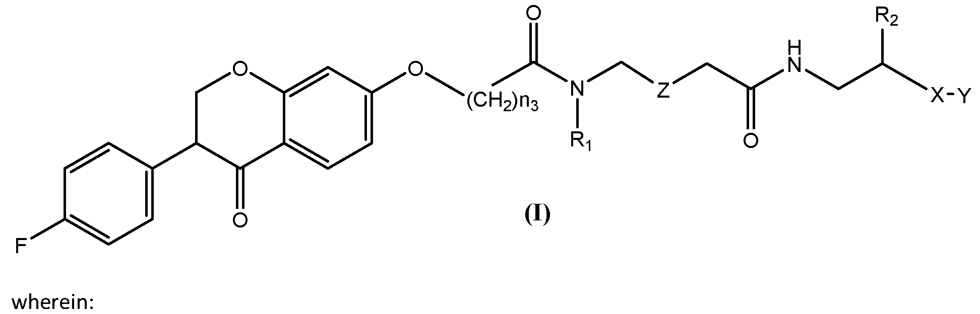


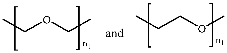


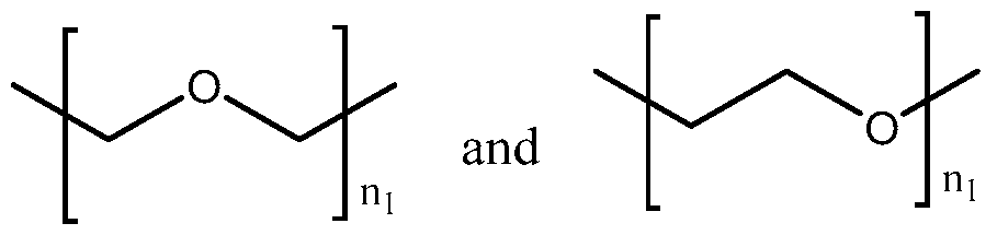


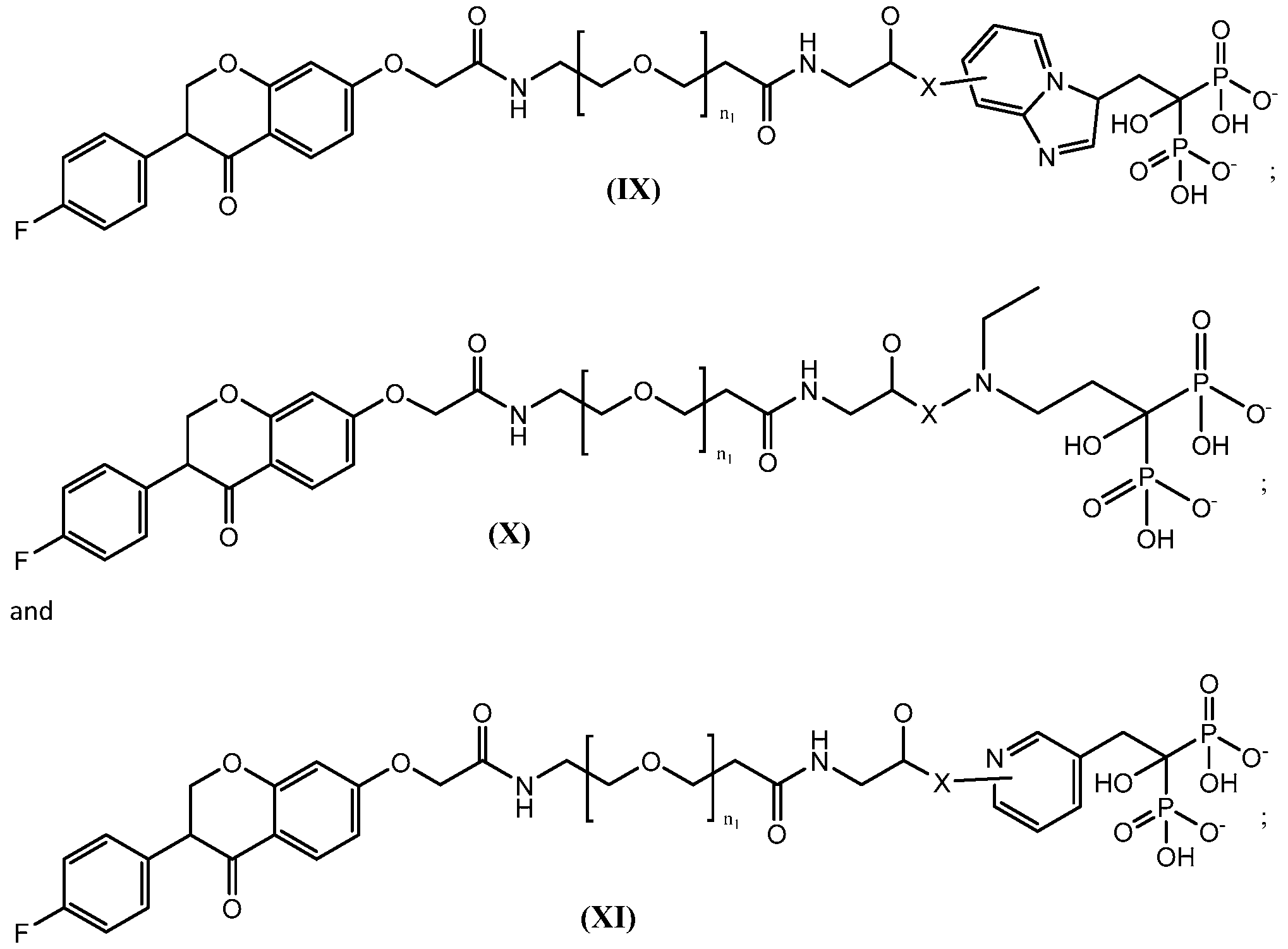


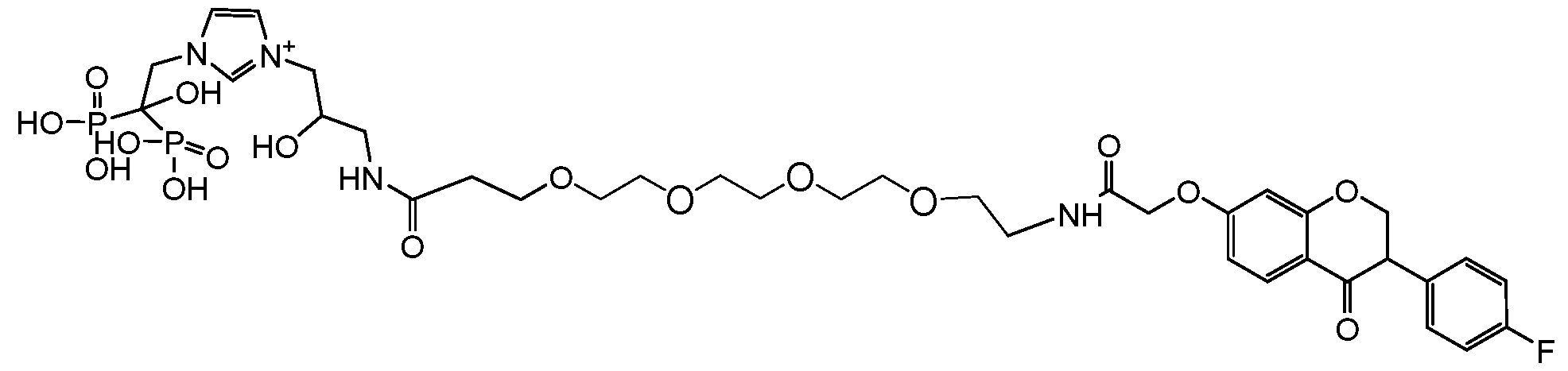


Claims


Priority Applications (4)
| Application Number | Priority Date | Filing Date | Title |
|---|---|---|---|
| JP2021545859A JP2022523146A (en) | 2019-02-06 | 2020-02-06 | Bisphosphonate linking compound |
| US17/428,951 US20220133894A1 (en) | 2019-02-06 | 2020-02-06 | Bisphosphonate-linked compounds |
| CA3128239A CA3128239A1 (en) | 2019-02-06 | 2020-02-06 | Bisphosphonate-linked compounds |
| EP20752874.6A EP3920906A4 (en) | 2019-02-06 | 2020-02-06 | Bisphosphonate-linked compounds |
Applications Claiming Priority (2)
| Application Number | Priority Date | Filing Date | Title |
|---|---|---|---|
| US201962801858P | 2019-02-06 | 2019-02-06 | |
| US62/801,858 | 2019-02-06 |
Publications (1)
| Publication Number | Publication Date |
|---|---|
| WO2020163637A1 true WO2020163637A1 (en) | 2020-08-13 |
Family
ID=71947917
Family Applications (1)
| Application Number | Title | Priority Date | Filing Date |
|---|---|---|---|
| PCT/US2020/017071 WO2020163637A1 (en) | 2019-02-06 | 2020-02-06 | Bisphosphonate-linked compounds |
Country Status (5)
| Country | Link |
|---|---|
| US (1) | US20220133894A1 (en) |
| EP (1) | EP3920906A4 (en) |
| JP (1) | JP2022523146A (en) |
| CA (1) | CA3128239A1 (en) |
| WO (1) | WO2020163637A1 (en) |
Citations (15)
| Publication number | Priority date | Publication date | Assignee | Title |
|---|---|---|---|---|
| US3845770A (en) | 1972-06-05 | 1974-11-05 | Alza Corp | Osmatic dispensing device for releasing beneficial agent |
| US4326525A (en) | 1980-10-14 | 1982-04-27 | Alza Corporation | Osmotic device that improves delivery properties of agent in situ |
| US4902514A (en) | 1988-07-21 | 1990-02-20 | Alza Corporation | Dosage form for administering nilvadipine for treating cardiovascular symptoms |
| US4992445A (en) | 1987-06-12 | 1991-02-12 | American Cyanamid Co. | Transdermal delivery of pharmaceuticals |
| US5001139A (en) | 1987-06-12 | 1991-03-19 | American Cyanamid Company | Enchancers for the transdermal flux of nivadipine |
| US5023252A (en) | 1985-12-04 | 1991-06-11 | Conrex Pharmaceutical Corporation | Transdermal and trans-membrane delivery of drugs |
| US5616345A (en) | 1983-12-22 | 1997-04-01 | Elan Corporation Plc | Controlled absorption diltiazen formulation for once-daily administration |
| US20100137425A1 (en) * | 2008-10-10 | 2010-06-03 | Northwestern University | Inhibition and treatment of prostate cancer metastasis |
| US20110065672A1 (en) | 2009-09-16 | 2011-03-17 | Mount Sinai School Of Medicine Of New York University | Bisphosphonate-prostatic acid phosphatase inhibitor conjugates to treat prostate cancer bone metastasis |
| WO2012098222A1 (en) * | 2011-01-21 | 2012-07-26 | Universita' Degli Studi Magna Graecia Di Catanzaro | Bisphosphonate-polyaspartamide conjugates as polymeric carriers for drug targeting to bones |
| US20140051623A1 (en) | 2011-02-24 | 2014-02-20 | Ktb Tumorforschungsgesellschaft Mbh | Bisphosphonate-Prodrugs |
| US20160128973A1 (en) | 2014-11-06 | 2016-05-12 | Northwestern University | Inhibition of cancer cell motility |
| WO2016081281A1 (en) | 2014-11-17 | 2016-05-26 | Salk Institute For Biological Studies | Lipophilic bisphosphonates and methods of use |
| US20160311805A1 (en) * | 2015-04-27 | 2016-10-27 | Pfizer Inc. | Anti-estrogenic compounds |
| WO2019007447A1 (en) | 2017-07-05 | 2019-01-10 | E.P.O.S Iasis Research And Development Limited | Multifunctional conjugates |
Family Cites Families (1)
| Publication number | Priority date | Publication date | Assignee | Title |
|---|---|---|---|---|
| US5854227A (en) * | 1994-03-04 | 1998-12-29 | Hartmann; John F. | Therapeutic derivatives of diphosphonates |
-
2020
- 2020-02-06 CA CA3128239A patent/CA3128239A1/en active Pending
- 2020-02-06 EP EP20752874.6A patent/EP3920906A4/en active Pending
- 2020-02-06 US US17/428,951 patent/US20220133894A1/en active Pending
- 2020-02-06 JP JP2021545859A patent/JP2022523146A/en active Pending
- 2020-02-06 WO PCT/US2020/017071 patent/WO2020163637A1/en unknown
Patent Citations (16)
| Publication number | Priority date | Publication date | Assignee | Title |
|---|---|---|---|---|
| US3845770A (en) | 1972-06-05 | 1974-11-05 | Alza Corp | Osmatic dispensing device for releasing beneficial agent |
| US4326525A (en) | 1980-10-14 | 1982-04-27 | Alza Corporation | Osmotic device that improves delivery properties of agent in situ |
| US5616345A (en) | 1983-12-22 | 1997-04-01 | Elan Corporation Plc | Controlled absorption diltiazen formulation for once-daily administration |
| US5023252A (en) | 1985-12-04 | 1991-06-11 | Conrex Pharmaceutical Corporation | Transdermal and trans-membrane delivery of drugs |
| US4992445A (en) | 1987-06-12 | 1991-02-12 | American Cyanamid Co. | Transdermal delivery of pharmaceuticals |
| US5001139A (en) | 1987-06-12 | 1991-03-19 | American Cyanamid Company | Enchancers for the transdermal flux of nivadipine |
| US4902514A (en) | 1988-07-21 | 1990-02-20 | Alza Corporation | Dosage form for administering nilvadipine for treating cardiovascular symptoms |
| US20100137425A1 (en) * | 2008-10-10 | 2010-06-03 | Northwestern University | Inhibition and treatment of prostate cancer metastasis |
| US20110065672A1 (en) | 2009-09-16 | 2011-03-17 | Mount Sinai School Of Medicine Of New York University | Bisphosphonate-prostatic acid phosphatase inhibitor conjugates to treat prostate cancer bone metastasis |
| WO2012098222A1 (en) * | 2011-01-21 | 2012-07-26 | Universita' Degli Studi Magna Graecia Di Catanzaro | Bisphosphonate-polyaspartamide conjugates as polymeric carriers for drug targeting to bones |
| US20140051623A1 (en) | 2011-02-24 | 2014-02-20 | Ktb Tumorforschungsgesellschaft Mbh | Bisphosphonate-Prodrugs |
| US20160128973A1 (en) | 2014-11-06 | 2016-05-12 | Northwestern University | Inhibition of cancer cell motility |
| US20180153853A1 (en) * | 2014-11-06 | 2018-06-07 | Northwestern University | Inhibition of cancer cell motility |
| WO2016081281A1 (en) | 2014-11-17 | 2016-05-26 | Salk Institute For Biological Studies | Lipophilic bisphosphonates and methods of use |
| US20160311805A1 (en) * | 2015-04-27 | 2016-10-27 | Pfizer Inc. | Anti-estrogenic compounds |
| WO2019007447A1 (en) | 2017-07-05 | 2019-01-10 | E.P.O.S Iasis Research And Development Limited | Multifunctional conjugates |
Non-Patent Citations (48)
| Title |
|---|
| "Modern Pharmaceutics", MARCEL DEKKER, INC. |
| "Remington's Pharmaceutical Sciences", 1985, MACE PUBLISHING CO. |
| ALEJANDRO, J. D. JOHNSON: "Inhibition of Raf-1 alters multiple downstream pathways to induce pancreatic beta-cell apoptosis", J BIOL CHEM, vol. 283, 2008, pages 2407 - 2417 |
| BEER ET AL., N ENGL J MED, vol. 371, 2014, pages 424 - 433 |
| BONO ET AL., LANCET, vol. 376, 2010, pages 1147 - 1154 |
| BRADLEY ET AL., J CELL BIOCHEM, vol. 104, 2008, pages 1439 - 1451 |
| C. LOGOTHETIS ET AL., CANCER METASTASIS REV, vol. 37, 2018, pages 189 - 196 |
| CHEMICAL & PHARMACEUTICAL BULLETIN, vol. 39, no. 10, 1991, pages 2681 - 90 |
| CHU ET AL., MOL CANCER RES, vol. 6, 2008, pages 1259 - 1267 |
| DING ET AL., J BIOMOL TECH, vol. 18, 2007, pages 321 - 330 |
| FANG ET AL., J BIOL CHEM, vol. 271, 1996, pages 28697 - 28702 |
| FARRELL ET AL., BONE REP, vol. 9, 2018, pages 47 - 60 |
| FLORENCIO-SILVA ET AL., BIOMED RES INT, vol. 2015, 2015, pages 421746 |
| FUNAKOSHI ET AL., SCI REP, vol. 7, 2017, pages 13202 |
| FURR ET AL., EUR UROL, vol. 29, 1996, pages 83 - 95 |
| GORDON ET AL., PLOS ONE, vol. 9, 2014, pages e104271 |
| GORDON ET AL.: "Chemotherapy-induced monoamine oxidase expression in prostate carcinoma functions as a cytoprotective resistance enzyme and associates with clinical outcomes", PLOS ONE, vol. 9, 2014, pages e104271, XP055942107, DOI: 10.1371/journal.pone.0104271 |
| GUISE ET AL., ENDOCR REV, vol. 19, 1998, pages 18 - 54 |
| GUISE, J MUSCULOSKELET NEURONAL INTERACT, vol. 2, 2002, pages 570 - 572 |
| HE ET AL., PLOS ONE, vol. 6, 2011, pages e24780 |
| HUSSAIN ET AL., Λ/ ENGL J MED, vol. 378, 2018, pages 2465 - 2474 |
| J NATL CANCER INST, vol. 100, 2008, pages 1500 - 1510 |
| JAMES ET AL., JAMA ONCOL, vol. 2, 2016, pages 493 - 499 |
| KORENCHUK ET AL., IN VIVO, vol. 15, 2001, pages 163 - 168 |
| LAKSHMAN ET AL., CLIN EXP METASTASIS, vol. 28, 2011, pages 39 - 53 |
| LI XU ET AL., NAT. COMM., vol. 9, no. 1, 2018 |
| LIU ET AL., PROSTATE CANCER AND PROSTATIC DISEASES, vol. 4, 2001, pages 81 - 91 |
| LIVAK ET AL., METHODS, vol. 25, 2001, pages 402 - 408 |
| LYSENG-WILLIAMSON ET AL., DRUGS, vol. 65, 2005, pages 2513 - 2531 |
| NAKAMURA ET AL., J BONE MINER RES, vol. 18, 2003, pages 1198 - 1205 |
| NI ET AL., MOL CELL BIOL, vol. 30, 2010, pages 1243 - 1253 |
| NORGAARD ET AL., J UROL, vol. 184, 2010, pages 162 - 167 |
| PAVESE ET AL., CANCER LETT, vol. 352, 2014, pages 179 - 186 |
| PAVESE ET AL., J VIS EXP, 2013, pages e50873 |
| POLASCIK ET AL., THER CLIN RISK MANAG, vol. 4, 2008, pages 261 - 268 |
| POUND ET AL., JAMA, vol. 281, 1999, pages 1591 - 1597 |
| SAAD ET AL., J NATL CANCER INST, vol. 94, 2002, pages 1458 - 1468 |
| SAAD ET AL., J NATL CANCER INST, vol. 96, 2004, pages 879 - 882 |
| SAPORITA ET AL., PROSTATE, vol. 67, 2007, pages 509 - 520 |
| See also references of EP3920906A4 |
| SEYFRIED ET AL., CRIT REV ONCOG, vol. 18, 2013, pages 43 - 73 |
| SWEENEY ET AL., N ENGL J MED, vol. 373, 2015, pages 737 - 746 |
| TANNOCK ET AL., N ENGL J MED, vol. 351, 2004, pages 1502 - 1512 |
| TRAN ET AL., SCIENCE, vol. 324, 2009, pages 787 - 790 |
| TSAUR ET AL., UROLOGIC ONCOLOGY, vol. 36, 2018, pages 488 - 497 |
| XU ET AL., NATURE COMMUNICATIONS, vol. 9, 2018, pages 2454 |
| YOTOVA ET AL., J CELL MOL MED, vol. 16, 2012, pages 2127 - 2139 |
| ZHANG ET AL.: "A Multifunctional Therapy Approach for Cancer: Targeting Rafi-Mediated Inhibition of Cell Motility, Growth, and Interaction with Microenvironment", MOLECULAR CANCER THERAPEUTICS, vol. 19, no. 1286281-32-0, January 2020 (2020-01-01), pages 39 - 51 |
Also Published As
| Publication number | Publication date |
|---|---|
| JP2022523146A (en) | 2022-04-21 |
| EP3920906A1 (en) | 2021-12-15 |
| CA3128239A1 (en) | 2020-08-13 |
| EP3920906A4 (en) | 2023-07-12 |
| US20220133894A1 (en) | 2022-05-05 |
Similar Documents
| Publication | Publication Date | Title |
|---|---|---|
| RU2767664C2 (en) | Combination therapy with anticancer alkaloid | |
| JP6754071B2 (en) | Combination medications containing metformin and dihydroquercetin, and use for the treatment of cancer | |
| DK2694072T3 (en) | COMBINATION OF ACT-INHIBITOR RELATIONSHIP AND ABIRATERON FOR USE IN THERAPEUTIC TREATMENTS | |
| US20040152759A1 (en) | Combination administration of an indolinone with a chemotherapeutic agent for cell proliferation disorders | |
| EA028452B1 (en) | Treatment of breast cancer | |
| KR20130045341A (en) | Administration of hypoxia activated prodrugs and antiangiogenic agents for the treatment of cancer | |
| AU2015314753A1 (en) | Human dosing of phosphatase inhibitor | |
| JP2012520299A (en) | Kinase protein binding inhibitor | |
| TW201831191A (en) | Novel boric acid derivative and pharmaceutical composition using same | |
| TW201039817A (en) | Pyrrolidine substituted flavones as radio sensitizers | |
| US9539237B2 (en) | Compositions and methods for drug-sensitization or inhibition of a cancer cell | |
| EP3053581B1 (en) | Therapeutic agent for osteoporosis | |
| TWI557128B (en) | Use of a composition for the manufacture of a medicament for preventing or treating nsclc | |
| US20220133894A1 (en) | Bisphosphonate-linked compounds | |
| WO2012082992A1 (en) | Compositions and methods for cancer treatment | |
| TW201242597A (en) | A synergistic pharmaceutical combination for the treatment of pancreatic cancer | |
| US10098860B2 (en) | Bezafibrate for the treatment of cancer | |
| WO2016135138A1 (en) | Oxoquinoline derivatives as mth1 inhibitors for the therapy of cancer | |
| CN111432815A (en) | Enhancing cancer treatment efficacy via sphingosine-1-phosphate pathway | |
| KR20130140052A (en) | Combination of bevacizumab and 2,2-dimethyl-n-((s)-6-oxo-6,7-dihydro-5h-dibenzo[b,d]azepin-7-yl)-n'-(2,2,3,3,3-pentafluoro-propyl)-malonamide for the treatment of proliferative disorders | |
| WO2016135140A1 (en) | 4-aminoquinazoline derivatives as mth1 inhibitors for the therapy of cancer | |
| Sighel | Inhibition of mitochondrial translation as a novel strategy to eradicate glioblastoma stem cells | |
| JP2019532937A (en) | Combination therapy strategy for cancer based on RNA polymerase I inhibition | |
| TW200307533A (en) | Use of EDG receptor binding agents in cancer |
Legal Events
| Date | Code | Title | Description |
|---|---|---|---|
| 121 | Ep: the epo has been informed by wipo that ep was designated in this application |
Ref document number: 20752874 Country of ref document: EP Kind code of ref document: A1 |
|
| ENP | Entry into the national phase |
Ref document number: 3128239 Country of ref document: CA |
|
| ENP | Entry into the national phase |
Ref document number: 2021545859 Country of ref document: JP Kind code of ref document: A |
|
| NENP | Non-entry into the national phase |
Ref country code: DE |
|
| ENP | Entry into the national phase |
Ref document number: 2020752874 Country of ref document: EP Effective date: 20210906 |