WO2020162595A1 - 被検物質への感受性の判定方法、被検物質への感受性の判定装置、抗癌剤のスクリーニング方法、抗癌剤の候補物質の製造方法、学習済モデルの製造方法、学習済モデルの製造装置、および選抜方法 - Google Patents
被検物質への感受性の判定方法、被検物質への感受性の判定装置、抗癌剤のスクリーニング方法、抗癌剤の候補物質の製造方法、学習済モデルの製造方法、学習済モデルの製造装置、および選抜方法 Download PDFInfo
- Publication number
- WO2020162595A1 WO2020162595A1 PCT/JP2020/004819 JP2020004819W WO2020162595A1 WO 2020162595 A1 WO2020162595 A1 WO 2020162595A1 JP 2020004819 W JP2020004819 W JP 2020004819W WO 2020162595 A1 WO2020162595 A1 WO 2020162595A1
- Authority
- WO
- WIPO (PCT)
- Prior art keywords
- shg
- test substance
- tumor cells
- sensitivity
- signal light
- Prior art date
- Legal status (The legal status is an assumption and is not a legal conclusion. Google has not performed a legal analysis and makes no representation as to the accuracy of the status listed.)
- Ceased
Links
Images



Classifications
-
- A—HUMAN NECESSITIES
- A61—MEDICAL OR VETERINARY SCIENCE; HYGIENE
- A61K—PREPARATIONS FOR MEDICAL, DENTAL OR TOILETRY PURPOSES
- A61K45/00—Medicinal preparations containing active ingredients not provided for in groups A61K31/00 - A61K41/00
-
- A—HUMAN NECESSITIES
- A61—MEDICAL OR VETERINARY SCIENCE; HYGIENE
- A61P—SPECIFIC THERAPEUTIC ACTIVITY OF CHEMICAL COMPOUNDS OR MEDICINAL PREPARATIONS
- A61P11/00—Drugs for disorders of the respiratory system
-
- A—HUMAN NECESSITIES
- A61—MEDICAL OR VETERINARY SCIENCE; HYGIENE
- A61P—SPECIFIC THERAPEUTIC ACTIVITY OF CHEMICAL COMPOUNDS OR MEDICINAL PREPARATIONS
- A61P35/00—Antineoplastic agents
-
- G—PHYSICS
- G01—MEASURING; TESTING
- G01N—INVESTIGATING OR ANALYSING MATERIALS BY DETERMINING THEIR CHEMICAL OR PHYSICAL PROPERTIES
- G01N21/00—Investigating or analysing materials by the use of optical means, i.e. using sub-millimetre waves, infrared, visible or ultraviolet light
- G01N21/62—Systems in which the material investigated is excited whereby it emits light or causes a change in wavelength of the incident light
- G01N21/63—Systems in which the material investigated is excited whereby it emits light or causes a change in wavelength of the incident light optically excited
- G01N21/65—Raman scattering
Definitions
- the present invention provides a method for determining sensitivity to a test substance, a determination device for sensitivity to a test substance, a screening method for an anticancer agent, a method for producing a candidate substance for an anticancer agent, a method for producing a learned model, and an apparatus for producing a learned model. , And the selection method.
- Non-patent document 1 a method in which target tumor cells and a candidate compound for an anticancer agent are allowed to coexist and cell death (survival rate) of the tumor cells is used as an index is used.
- the present invention aims to provide a new determination method capable of determining the sensitivity of tumor cells to a test substance.
- a method for determining the sensitivity to a test substance of the present invention is a coherent anti-Stokes Raman scattering (CARS) method for tumor cells contacted with the test substance.
- CARS coherent anti-Stokes Raman scattering
- the device for determining the sensitivity to a test substance of the present invention (hereinafter, also referred to as “determination device”) was obtained using a coherent anti-Stokes Raman scattering (CARS) microscope for tumor cells contacted with the test substance.
- SHG coherent anti-Stokes Raman scattering
- the method for screening an anticancer agent of the present invention (hereinafter, also referred to as “screening method”), a tumor cell contacted with a test substance, a determination step of determining the sensitivity to the test substance, For the test substance determined to be sensitive to the tumor cells, including the step of selecting as a candidate substance of an anticancer agent, The determination step is performed by the determination method of the present invention.
- the method for producing a candidate substance for an anticancer agent of the present invention includes a selection step of selecting a candidate substance for an anticancer agent from a test substance, The selection step is performed by the screening method of the present invention.
- the method for producing a learned model used for determining the sensitivity of tumor cells of the present invention is a coherent anti-Stokes Raman scattering (CARS) microscope for tumor cells contacted with a test substance.
- CARS coherent anti-Stokes Raman scattering
- the apparatus for manufacturing a learned model used for determining the sensitivity of tumor cells of the present invention is a coherent anti-Stokes Raman scattering (CARS) microscope for tumor cells that are contacted with a test substance.
- SHG second harmonic
- the method for selecting cells for determining the sensitivity of a test substance of the present invention is a method of using a coherent anti-Stokes Raman scattering (CARS) microscope for the second harmonic (SHG) of a test tumor cell.
- CARS coherent anti-Stokes Raman scattering
- SHG second harmonic
- the sensitivity of tumor cells to a test substance can be determined.
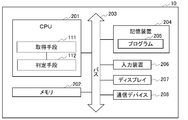
- FIG. 1 is a block diagram illustrating an example of the configuration of the determination device according to the first embodiment.
- FIG. 2 is a block diagram illustrating an example of the hardware configuration of the determination device according to the first embodiment.
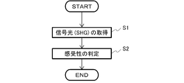
- FIG. 3 is a flowchart showing an example of the determination method of the first embodiment.
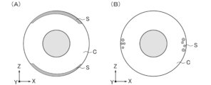
- FIG. 4 is a schematic diagram showing the distribution of signals of the second harmonic in tumor cells in the first embodiment.
- FIG. 5 is a block diagram showing an example of the configuration of the learning device according to the second embodiment.
- FIG. 6 is a flowchart showing an example of the learning method of the second embodiment.
- FIG. 7 is a schematic diagram of the configuration of the optical system of the CARS microscope used in Example 1.
- FIG. 8 is a photograph showing an SHG image in Example 1.
- cell means, for example, an isolated cell, a cell mass composed of cells, a tissue, or an organ.
- the cells may be, for example, cultured cells or cells isolated from a living body.
- the cell mass, tissue or organ may be, for example, a cell mass, cell sheet, tissue or organ prepared from the cells, or may be a cell mass, tissue or organ isolated from a living body.
- the method for determining the sensitivity to a test substance of the present invention is, as described above, the second harmonic (obtained by using a coherent anti-Stokes Raman scattering (CARS) microscope for tumor cells contacted with the test substance ( SHG) acquisition step, and a determination step of determining the sensitivity of the tumor cells to the test substance based on the SHG.
- the device for determining the sensitivity to a test substance of the present invention provides a second harmonic wave (SHG) obtained by using a coherent anti-Stokes Raman scattering (CARS) microscope for tumor cells contacted with the test substance.
- An acquisition unit for acquiring and a determination unit for determining the sensitivity of the tumor cells to the test substance based on the SHG are included.
- the determination method and the determination device of the present invention are characterized by determining the sensitivity of the tumor cells to the test substance based on SHG obtained from the tumor cells contacted with the test substance.
- the conditions are not limited.
- the present inventors have found that signal light obtained by using a CARS microscope, specifically, the second harmonic correlates with the sensitivity of tumor cells to a test substance, and It has been established. Therefore, according to the present invention, the sensitivity of the tumor cells to the test substance can be determined based on the signal light obtained by using the CAR microscope for the test cells.
- FIG. 1 shows a block diagram of a determination device according to this embodiment.
- the determination device 10 of this embodiment includes an acquisition unit 111 and a determination unit 112.
- the acquisition unit 111 and the determination unit 112 may be incorporated in the data processing unit (data processing device) 11 that is hardware, or may be software or hardware in which the software is incorporated.
- the data processing means 11 may include a CPU or the like. Further, the data processing means 11 may include, for example, a ROM, a RAM, etc. described later.
- FIG. 2 illustrates a block diagram of a hardware configuration of the determination device 10.
- the determination device 10 includes, for example, a CPU (central processing unit) 201, a memory 202, a bus 203, a storage device 204, an input device 206, a display 207, a communication device 208, and the like. Each unit of the determination device 10 is connected via the bus 203 by each interface (I/F).
- the hardware configuration of the determination device 10 can also be adopted as a hardware configuration in a learning device described later, for example.
- the CPU 201 cooperates with other components by, for example, a controller (system controller, I/O controller, etc.) and controls the entire determination device 10.
- the CPU 201 executes, for example, the program 205 of the present invention or other programs, and also reads or writes various information.
- the CPU 201 functions as the acquisition unit 111 and the determination unit 112.
- the determination device 10 includes a CPU as an arithmetic device, but may include other arithmetic devices such as a GPU (Graphics Processing Unit) and an APU (Accelerated Processing Unit), or may include a CPU and a combination thereof. Good.
- the CPU 201 functions as, for example, each unit other than the storage unit in the second embodiment described later.
- the memory 202 includes, for example, a main memory.
- the main memory is also called a main storage device.
- the memory 202 reads various operation programs such as the program 205 of the present invention stored in the storage device 204 (auxiliary storage device) described later, for example. Then, the CPU 201 reads the data from the memory 202, decodes the data, and executes the program.
- the main memory is, for example, a RAM (random access memory).
- the memory 202 further includes, for example, a ROM (read-only memory).
- the bus 203 can be connected to an external device, for example.
- the external device include an external storage device (external database and the like), a printer, and the like.
- the determination device 10 can be connected to a communication line network by, for example, the communication device 208 connected to the bus 203, and can also be connected to the external device via the communication line network.
- the determination device 10 can also be connected to a terminal or the like via the communication device 208 and a communication line network.
- the storage device 204 is also called a so-called auxiliary storage device with respect to the main memory (main storage device), for example. As described above, the storage device 204 stores an operation program including the program 205 of the present invention.
- the storage device 204 includes, for example, a storage medium and a drive that reads and writes the storage medium.
- the storage medium is not particularly limited, and may be, for example, an internal type or an external type, HD (hard disk), FD (floppy (registered trademark) disk), CD-ROM, CD-R, CD-RW, MO, Examples thereof include DVD, flash memory, and memory card, and the drive is not particularly limited.
- the storage device 204 may be, for example, a hard disk drive (HDD) in which the storage medium and the drive are integrated.
- HDD hard disk drive
- the determination device 10 further includes, for example, an input device 206 and a display 207.
- the input device 206 is, for example, a pointing device such as a touch panel, a track pad, or a mouse; a keyboard; an imaging means such as a camera or a scanner; a card reader such as an IC card reader or a magnetic card reader; a voice input means such as a microphone;
- the display 207 include a display device such as an LED display and a liquid crystal display.
- the input device 206 and the display 207 are separately configured, but the input device 206 and the display 207 may be integrally configured like a touch panel display.
- the contact between the test substance and the tumor cells may be, for example, in vitro or in vivo .
- the contact can be performed by, for example, culturing in the coexistence of the test substance and the tumor cell.
- the conditions in the culture are not particularly limited and can be appropriately set depending on the type of the tumor cells.
- the culture time is, for example, 1 to 96 hours or 12 to 48 hours.
- the culture temperature is, for example, 25 to 40°C, 35 to 38°C.
- the culture is carried out, for example, in a humid environment.
- the contacting can be performed by, for example, administering the test substance to a subject having the tumor cells.
- the subject is, for example, a human or non-human animal.
- the non-human animal include monkeys, gorillas, chimpanzees, primates such as marmosets, mice, rats, dogs, rabbits, sheep, horses, and guinea pigs.
- the dose, administration route, administration conditions and the like of the test substance are not particularly limited and can be appropriately determined depending on the solubility of the test substance in a solvent, the absorbability into the body and the like. Then, after the contact, the tumor cells are collected and subjected to observation with a CARS microscope.
- the tumor cell may be, for example, a tumor cell isolated from a living body, that is, a primary cell or a passage cell thereof, a conditioned culture cell, or a cell tumorized by a virus, a mutagen, or the like. Good, but isolated tumor cells or passage cells thereof are preferred.
- the tumor cell may be composed of one cell or a plurality of cells. In the latter case, the tumor cell may be, for example, a tissue, organ or organ containing the tumor cell.
- the origin of the tumor cells is not particularly limited.
- the tumor cells are, for example, lung cancer, breast cancer, colon cancer, liver cancer, pancreatic cancer, biliary tract cancer, esophageal cancer, gastric cancer, ovarian cancer, cervical cancer, brain tumor, blood tumor. , Tumor cells derived from mesothelioma, skin cancer and the like.
- the tumor cell may be, for example, a tumor cell sensitive to the test substance, a tumor cell resistant to the test substance, or a tumor cell whose sensitivity to the test substance is unknown. ..
- test substance examples include low molecular weight compounds, peptides, proteins and nucleic acids.
- test substance one type may be used alone, or a plurality of types may be used in combination.
- the test substance may be a substance known to suppress antitumor action or tumor metastasis (so-called anticancer drug or metastasis suppressor), or is not known to inhibit antitumor action or tumor metastasis.
- Substance, or a substance whose antitumor effect or tumor metastasis is unknown is unknown.
- the determination device of the present invention is, for example, a determination device or a test device for sensitivity to an anticancer drug or metastasis inhibitor of cancer. Can be said.
- the determination method of the present invention can also be referred to as, for example, a method for determining the sensitivity of cancer to an anti-cancer agent or a metastasis inhibitor or a test method.
- the tumor cells brought into contact with the test substance in this manner are observed with a CARS microscope to obtain signal light.
- the signal light is obtained by the CARS microscope for all or part of the tumor cells.
- the signal light is acquired by the CARS microscope, for example, in the central portion of the tumor cell, the signal light of a cross section including a direction perpendicular to the arrangement surface of the tumor cell is acquired.
- the number of tumor cells used for the observation may be one or plural. In the latter case, the signal light is obtained by the CARS microscope for some or all of the cells.
- the CARS microscope is a microscope that uses coherent anti-Stokes Raman scattering, and the configuration thereof is not particularly limited, and for example, the configurations of Examples described later can be referred to.
- the CARS microscope includes a light irradiating means for CARS spectroscopy, and the light irradiating means irradiates the tumor cells with the mixed light of the ultra-wide band light and the excitation light. Then, in the CARS microscope, CARS light (signal light) is separated from the emitted light emitted from the tumor cells by a diffraction grating or the like. Then, information such as the signal intensity of the CARS light and the spatial position is acquired.
- the signal light only the second harmonic (SHG) may be acquired, or other signal light such as the third harmonic (THG) or two-photon excitation fluorescence may be acquired. ..
- the wavelength of the ultra wideband light is, for example, 400 to 2400 nm.
- the wavelengths of the excitation light are, for example, 750 to 1100 nm and 750 to 1064 nm.
- the acquisition unit 111 of the determination device 10 acquires the signal light, more specifically, the signal light information acquired by the CARS microscope (S1, acquisition step).
- the signal light acquired in the acquisition step includes signal light used in the determination step described later, and as a specific example, it is preferable that the signal light of the second harmonic (SHG) is included.
- the wave number of the signal light of the SHG is ⁇ /2, where ⁇ is the wavelength of the incident laser.
- the determination unit 112 determines the sensitivity of the tumor cells based on the signal light, specifically, the SHG signal light (S2, determination step).
- the tumor cells may be determined to be sensitive to the test substance or may be determined to be resistant to the test substance.
- it may be determined whether or not the tumor cells have sensitivity to the test substance.
- each wave number (Raman shift/cm ⁇ 1 ) is different in the substance in the tumor cell from which the signal is derived.
- the functions and metabolisms of the cells in different states are different, and therefore it is presumed that the types of substances contained in the tumor cells, the content rates of the substances, and the localization of the substances are different.
- the fact that the difference in the state of the tumor cells occurs, for example, as the difference in the signal light is used to determine the sensitivity of the tumor cells based on the signal light.
- the tumor cells are lung cancer cells
- the present invention is not limited to this and can be used for estimating other tumor cells.
- the SHG signal S is a bipolar electrode in the Z axis direction of the lung cancer cell C. That is, it is localized on the upper end side and the lower end side. Then, when the lung cancer cells C after contact with the test substance and the lung cancer cells C show sensitivity to the test substance, as shown in FIG.
- SHG signal S Is localized around the equator of the lung cancer cell C in the Z-axis direction, that is, in the center.
- the lung cancer cells C are in contact with the test substance and the lung cancer cells C show resistance to the test substance, as shown in FIG. 4(A), the test substance is not in contact with the test substance.
- the SHG signal S is localized at both poles in the Z axis direction of the lung cancer cell C, that is, at the upper end side and the lower end side in the SHG image.
- the lung cancer cells C are sensitive can be determined based on the position of the SHG signal light, that is, the position of the SHG signal in the SHG image reconstructed from the SHG signal.
- the localization is, for example, 50% or more, 60% or more, 70% or more, 80% or more, 90% or more, 95% or more, 96% or more, 97% or more, 98% or more, 99 of all SHG signals. It means that more than% of the SHG signal is present at a given position.
- the upper end side means a region between the upper end and the positions of 30%, 25%, 20%, 15%, and 10% of the length in the Z axis direction from the upper end in the Z axis direction of the tumor cell.
- the lower end side is a region between the lower end and positions of 30%, 25%, 20%, 15%, 10% of the length of the tumor cells in the Z-axis direction from the lower end.
- the central portion means, for example, a region other than the upper end side and the lower end side, and as a specific example, in the Z axis direction of the tumor cell, for example, a position at a length of 50% in the Z axis direction of the tumor cell from the lower end.
- the length of the tumor cells in the Z-axis direction is ⁇ 5%, ⁇ 10%, +15%, ⁇ 20%, ⁇ 25%, and ⁇ 30%.
- the SHG signal S forms a smooth arc in the SHG image.
- SHG signal S Form grains with defocused boundaries.
- the lung cancer cells C are in contact with the test substance and the lung cancer cells C show resistance to the test substance, as shown in FIG. 4(A), the test substance is not in contact with the test substance.
- the SHG signal forms a smooth arc. Therefore, in S2, it is possible to determine whether or not the lung cancer cells are sensitive based on the shape of the SHG signal light, that is, the shape of the SHG signal in the SHG image reconstructed from the SHG signal.
- the sensitivity of the lung cancer cells to the test substance can be determined based on the dispersion value of the SHG signal light, that is, the dispersion value of the signal intensity of the SHG signal light.
- the boundary of the signal of SHG is blurred and the distribution of the signal intensity is relatively wide
- the resistance of the test substance is In lung cancer cells, the boundaries of SHG signals are clear, and the distribution of signal intensities is relatively narrow.
- the dispersion value of the signal light of SHG of the tumor cells that is, the dispersion value of the signal intensity of the signal light of SHG is compared with a reference value, and the dispersion value of the signal of SHG is the reference value.
- the variance value of the signal light of SHG of the tumor cells is compared with the reference value, and the variance value of the SHG signal is equal to (not significantly different from) the reference value or significantly smaller.
- the tumor cells can be determined to be resistant to the test substance.
- the reference value may be, for example, a dispersion value of the signal light of SHG in the lung cancer cells that are not in contact with the test substance, or a dispersion value of the signal light of SHG in the lung cancer cells that are sensitive to the test substance, and A value between the two may be used based on the dispersion value of the signal light of SHG in the lung cancer cells resistant to the test substance.
- a trained model generated by machine learning may be used.
- the learned model is, for example, a learned model that outputs a determination result of the susceptibility of the tumor to the test substance from the SHG using the combination of the SHG described below and the sensitivity of the tumor cell to the test substance as teacher data. Can be given.
- step S2 it is possible to determine whether or not the tumor cells have sensitivity to the test substance.
- the example of using one determination method has been described in step S2, but a plurality of determination methods may be combined.
- the tumor cells may be determined to be sensitive to the test substance when they are determined to be sensitive by any one of the determination methods, or may be determined to be sensitive by a plurality of determination methods. If so, the tumor cells may be determined to be sensitive to the test substance.
- tumor cells have been described as an example of test cells for determining the sensitivity to a test substance, but the present invention is not limited to this, and cells other than tumor cells, for example, cells derived from normal tissue are used. Cells or the like may be used.
- the method for screening an anticancer agent of the present invention is, as described above, for a tumor cell contacted with a test substance, a determination step of determining sensitivity to the test substance, and the tumor cell determined to be sensitive.
- a step of selecting a test substance as a candidate substance of an anticancer agent, and the determining step is performed by the determining method of the present invention.
- the screening method of the present invention is characterized in that the determination step is performed by the determination method of the present invention, and other steps and conditions are not particularly limited.
- an anticancer drug can be screened.
- the description of the determination method and the determination apparatus of the present invention can be cited.
- the anti-cancer agent means, for example, an agent that induces tumor arrest or suppression, tumor growth arrest or suppression, tumor progression arrest or suppression, tumor metastasis arrest or suppression, or tumor shrinkage, It may be used in any sense.
- the method for producing a candidate substance for an anticancer agent of the present invention includes a selection step of selecting a candidate substance for an anticancer agent from a test substance as described above, and the selection step is performed by the screening method of the present invention.
- the production method of the present invention is characterized in that the selection step is performed by the screening method of the present invention, and other steps and conditions are not particularly limited. According to the production method of the present invention, a candidate substance for an anticancer agent can be produced.
- the description of the determination method, the determination device, and the screening method of the present invention can be applied.
- the method for producing a learned model used for determining the sensitivity of tumor cells of the present invention was obtained by using a coherent anti-Stokes Raman scattering (CARS) microscope for tumor cells that were contacted with a test substance.
- An acquisition step of acquiring a second harmonic (SHG), a combination of the SHG and the sensitivity of the tumor cells to the test substance is used as teaching data, and a determination result of the sensitivity of the tumor cells to the test substance is obtained from the SHG.
- a learning step of generating a learned model to be output is used as teaching data.
- the learned model manufacturing apparatus used for determining the sensitivity of tumor cells of the present invention is a second harmonic obtained by using a coherent anti-Stokes Raman scattering (CARS) microscope for tumor cells contacted with a test substance.
- Learning which outputs the determination result of the sensitivity of the tumor cell to the test substance from the SHG using the acquisition means for acquiring a wave (SHG), the SHG, and the sensitivity of the tumor cell to the test substance as teaching data.
- the learning method and the learning device of the present invention use the pair of SHG acquired by a CARS microscope and the sensitivity of the tumor cells to the test substance as the teaching data, and determine the sensitivity of the tumor cells to the test substance from the SHG.
- Is characterized by generating a trained model that outputs, and other configurations and conditions are not particularly limited.
- the learning method and the learning device of the present invention it is possible to manufacture a learned model that can be used in the determination method and the determination device of the present invention.
- the description of the determination method, the determination device, and the screening method of the present invention can be applied.
- FIG. 5 shows a block diagram of the learning device in this embodiment.
- the learning device 20 of this embodiment includes an acquisition unit 111 and a learning unit 113.
- the acquisition unit 111 and the learning unit 113 may be incorporated in the data processing unit (data processing device) 11 that is hardware, or may be software or hardware in which the software is incorporated.
- the data processing means 11 may include a CPU or the like. Further, the data processing means 11 may include, for example, the above-mentioned ROM, RAM and the like.
- the acquisition unit 111 is the same as the acquisition unit 111 in the determination device 10 of the first embodiment, and the description thereof can be applied.
- the acquisition unit 111 acquires the signal light acquired by using the CARS microscope for the tumor cells contacted with the test substance.
- the number of the tumor cells is not particularly limited and can be any number.
- the origin of each tumor cell may be the same or different. In the latter case, it is preferred that some tumor cells have the same origin.
- lung cancer cells, breast cancer cells, and colon cancer cells are preferably used as the tumor cells. Further, it is preferable to use a plurality of each tumor cell.
- the learning unit 113 uses the pair of the signal light (for example, SHG) and the sensitivity of the tumor cells to the test substance as the teaching data, and determines the sensitivity of the tumor cells to the test substance from the signal light.
- a trained model that outputs is generated (S3, learning step).
- the signal light obtained in step S1 is associated with the presence or absence of sensitivity of the tumor cell.
- the associated signal light may be the entire spectrum of the signal light obtained in step S1, may be a specific signal light, or may be a calculated value obtained from the specific signal light.
- the specific signal light is, for example, SHG signal light.
- the learning method detects the specific signal light in the obtained signal by the detecting unit after step S1, for example.
- the detection means may detect, for example, the signal intensity of the specific signal light, or may detect the shape and/or the position (localization) of the signal light in the signal image obtained by reconstructing the specific signal light. It may be detected.
- the position is, for example, the localization of the signal light of SHG in the Z-axis direction of the tumor cell.
- the learning method of the present embodiment for example, by the calculating means, the dispersion value of the specific signal light, that is, the dispersion value of the signal intensity of the specific signal light. May be calculated.
- the machine learning method used to generate the learned model is not particularly limited, and for example, the learning technique used for classification can be used.
- examples of the machine learning method include support vector machines, extreme learning machines, and feature learning.
- the machine learning uses supervised learning, but semi-supervised learning may be used.
- the number of the teacher data is plural, and the upper limit is not particularly limited.
- the method of selecting the cells for determining the sensitivity of the test substance of the present invention is, as described above, the acquisition of the second harmonic (SHG) of the test tumor cell using a coherent anti-Stokes Raman scattering (CARS) microscope. And a selection step of selecting a candidate tumor cell to be used in the sensitivity test of the test substance from the test tumor cell based on the SHG.
- the selection method of the present invention is characterized by selecting the candidate tumor cells based on SHG obtained using a CARS microscope, and other configurations and conditions are not limited. According to the selection method of the present invention, tumor cells suitable for the determination method of the present invention can be selected.
- the description of the determination method, the determination device, the screening method, the learning method, and the learning device of the present invention can be applied.
- the description of the acquisition step in the determination method of the present invention can be applied to the acquisition step.
- the description of the tumor cells can be applied to the test tumor cells, for example.
- a candidate tumor cell used for the sensitivity test of the test substance is selected from the test tumor cells based on the SHG.
- the selection step includes, for example, the presence or absence of the SHG, the signal intensity of the SHG signal light, the shape of the SHG signal in the SHG image reconstructed from the SHG signal light, and/or the position (localization) of the SHG signal in the SHG image.
- the selection step for example, when SHG is obtained in the test tumor cell, when the signal intensity of the signal light of SHG is a predetermined value or more, the shape of the SHG signal in the SHG image is a boundary. Is a clear arc and/or the position of the SHG signal in the SHG image is localized at the upper end side and/or the lower end side in the Z-axis direction of the candidate tumor cell, the test tumor cell Are selected as the candidate tumor cells.
- a program of the present invention is a program capable of executing the determination method, screening method, manufacturing method, learning method, or selection method of the present invention on a computer.
- the program of this embodiment may be recorded in a computer-readable recording medium, for example.
- the recording medium is, for example, a non-transitory computer-readable storage medium.
- the recording medium is not particularly limited, and examples thereof include a random access memory (RAM), a read-only memory (ROM), a hard disk (HD), an optical disk, and a floppy (registered trademark) disk (FD).
- Example 1 It was confirmed that the sensitivity of tumor cells to the test substance can be determined based on the signal light obtained by the CARS microscope.
- Example 1 The CARS microscope used in Example 1 was a multiplex CARS microscope. A schematic diagram of the configuration of the optical system of the CARS microscope used in Example 1 is shown in FIG.
- a cw Q switch microchip Nd:YAG laser (A) having an oscillation wavelength of 1064 nm, a pulse width of 800 ps and a repetition frequency of 33 kHz was used as the light source.
- the pulse width is sub-nanosecond and has a line width of about 1 cm -1 or less.
- Divide the output into two, a pulse laser having a center wavelength of 1064nm is fundamental as one is omega 1 light and the other is introduced into PCF, it was generated supercontinuum light as omega 2 light.
- B plano-convex lens
- f 400.0 mm
- AC254-400-C f 400.0 mm, ⁇ 1" Achromatic Doublet, ARC: 1050-1700 nm; Thorlabs.
- the ⁇ 2 light is collimated and the ⁇ 2 light is a super-continuum light having a broadband wavelength component emitted from the PCF. ) (C) was introduced and collimated and propagated.
- VND Filter Variable Neutral Density Filter
- S333-1064-2 1064 nm half-wave plate
- E Suruga Seiki Co., Ltd.
- the ⁇ 1 light contains a weak component on the short wavelength side in addition to the central wavelength of 1064 nm, and in order to efficiently cut the extra spectral components contained in the ⁇ 1 light other than the CARS light to be detected.
- a 1064 nm narrow band pass filter (1064.1-1 OD7 Ultra Narrow Bandpass; Alluxa) (F) was used.
- the bandpass filter used was an ultra-narrow bandpass filter that transmits only a spectral component having a center wavelength of 1064.1 nm and a half-value width of 1 nm.
- ⁇ 2 light has a broad band that slowly extends from the visible region to the near infrared region by PCF.
- the wavelength range of ⁇ 2 light necessary for measuring CARS light is about 1064 to 1650 nm with respect to the central wavelength of 1064 nm of ⁇ 1 light.
- two filters 1050 nm cuts components of visible light immediately after collimation of the omega 2 light long Pass Filter (G), infrared transmission filter (IR80N; Kenko manufactured optical Ltd.) (H) near red omega 2 light by Only the spectral components that spread to the outer region were sufficiently transmitted. Then, after combining the ⁇ 1 light and the ⁇ 2 light, the two lights were propagated to the microscope.
- an upside down microscope (eclipse Ti-U, manufactured by Nikon Co., Ltd.) that is custom-made is used.
- Nonlinear optics such as CARS and SHG by ⁇ 1 light and ⁇ 2 light incident on the objective lens (Water immersion, Plan, ⁇ 60, 1.27NA; Nikon) (J) from the inverted side of the microscope and focused on the sample. The phenomenon occurs. Since they are focused by the objective lens, these nonlinear optical phenomena efficiently occur in the forward direction due to the phase matching condition.
- the signal light generated at the focal point was condensed and collimated by an objective lens (Dry, S Plan Fluor, ⁇ 40, 0.60NA; manufactured by Nikon) on the upright side of the microscope (K).
- the average power measured with a power meter at the position where the light was focused on the sample surface by the inverted objective lens was ⁇ 1 light of maximum 55 mW and ⁇ 2 light of maximum 20 mW, respectively.
- the sample signal intensity was adjusted by using the above-mentioned VND filter for the ⁇ 1 light with high power.
- the microscope is equipped with a halogen lamp, and an optical image of the sample can be taken with a CCD camera.
- ⁇ 1 light and ⁇ 2 light exist coaxially with the light of the halogen lamp, so the dichroic beam splitter (FF825-SDi01-25 ⁇ 36 ⁇ 2.0 single edge short path Dichroic beam splitter; opt (Manufactured by Line Co., Ltd.) (L) and a dichroic mirror (TFMS-30C05-3/20 ultra-wide band induction multilayer flat mirror; manufactured by Sigma Optical Co., Ltd.) (M) immediately after the erecting side objective lens were used.
- the stage installed in the microscope used a step motor type MicroStage (Micro-Stage(2 axis); MadCityLabs)(N) controlled by a biaxial micro-positioning device. Furthermore, a piezo stage (Nano-LPQ; made by Mad City Labs) (O) with an operating range of 75 ⁇ m ⁇ 75 ⁇ m ⁇ 50 ⁇ m (O) is installed on the MicroStage, and in addition to a wide in-plane stroke in millimeters In-plane stage control of the stroke and scanning in the optical axis Z direction, that is, in the depth direction with respect to the sample are enabled. MicroStage can control a minimum step size of 95nm and a maximum speed of 2mm/sec with an extremely high precision drive.
- the optical system on the detector side was as follows.
- the signal light collected by the erecting objective lens of the microscope is reflected by the dichroic mirror immediately after, and then the dichroic beam splitter (FF685-Di02-25 ⁇ 36 685 nm single-edge Dichroic beam splitter; manufactured by Semrock) (P ) Transmitted near-infrared CARS light and reflected visible light.
- the dichroic beam splitter FF685-Di02-25 ⁇ 36 685 nm single-edge Dichroic beam splitter; manufactured by Semrock
- the grating Groove is set to 300 lines/mm
- the grating Blaze is set to 500 nm
- the center wavelength of the spectroscope is set to 410 nm.
- An electronic cooling CCD camera iVac300; manufactured by Andor) (T) was used to detect SHG.
- a spectroscope Acton Series LS785; Princeton Instruments
- PIXIS 100BR electronically cooled CCD camera
- the PIXIS100BR, iVac316, and iVac300 are controlled by software (LightField; Princeton Instruments, AndorSOLIS; Andor) on the PC.
- PC-9 Tumor cells Lung adenocarcinoma cells
- the PC-9 is a lung cancer cell line known to be sensitive to gefitinib.
- the tumor cells were prepared by placing a preparation in a 24-well dish and culturing on the preparation. Then, gefitinib was added as a test substance and cultured for 48 hours.
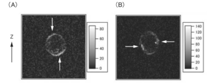
- FIG. 8 is a photograph showing an SHG image.
- (A) shows the result of the control to which gefitinib was not added
- (B) shows the result of the sample to which gefitinib was added.
- FIG. 8(A) in the state in which gefitinib was not added, as shown by the arrow, in PC-9, the SHG signals were localized on the upper end side and the lower end side in the SHG image.
- FIG. 8B in the state where gefitinib was added, the SHG image was localized around the equator, that is, in the center. From these, it was found that the sensitivity of tumor cells can be determined using the signal light acquired by the CARS microscope.
- Appendix 1 An acquisition step of acquiring a second harmonic wave (SHG) acquired by using a coherent anti-Stokes Raman scattering (CARS) microscope with respect to a tumor cell contacted with a test substance, A method for determining the sensitivity to a test substance, comprising the step of determining the sensitivity of the tumor cells to the test substance based on the SHG.
- Appendix 2 The determination method according to Appendix 1, wherein in the determination step, the sensitivity of the tumor cells to the test substance is determined based on at least one of the shape and the position of the SHG signal light. (Appendix 3) 3.
- Appendix 4 In the determination step, the dispersion value of the signal light of SHG is compared with a reference value, and when the dispersion value of the signal light of SHG is larger than the reference value, the tumor cells are sensitive to the test substance.
- the determination method according to appendix 4 wherein the reference value is a dispersion value of signal light of SHG in tumor cells that are not in contact with the test substance.
- An apparatus for determining the sensitivity to a test substance comprising: a determination unit that determines the sensitivity of the tumor cells to the test substance based on the SHG.
- the determination device determines the sensitivity of the tumor cells to the test substance based on the dispersion value of the SHG signal light.
- the determination means determines the sensitivity of the tumor cells to the test substance based on the dispersion value of the SHG signal light.
- the dispersion value of the SHG signal light is compared with a reference value, and when the dispersion value of the SHG signal light is larger than the reference value, the tumor cells are sensitive to the test substance.
- the determination device according to attachment 13, which is determined.
- the determining means uses a learned model generated by machine learning that outputs a determination result of the sensitivity of the tumor cells to the test substance based on the SHG, and determines the sensitivity of the tumor cells to the test substance.
- the determination device according to any one of appendices 11 to 15, which is determined.
- Appendix 21 For the tumor cells contacted with the test substance, a determination step of determining the sensitivity to the test substance, For the test substance determined to be sensitive to the tumor cells, including the step of selecting as a candidate substance of an anticancer agent, The method of screening an anticancer agent, wherein the determination step is performed by the determination method according to any one of appendices 1 to 10.
- Appendix 22 22. The screening method according to appendix 21, wherein the test substance is at least one selected from the group consisting of low-molecular compounds, peptides, proteins and nucleic acids.
- (Appendix 23) Including a selection step of selecting a candidate substance of an anticancer agent from a test substance, The said selection process is a manufacturing method of the candidate substance of an anticancer agent implemented by the screening method of attachment 21 or 22.
- (Appendix 25) A signal light detecting step of detecting at least one of a shape and a position of the signal light of the SHG from the SHG, 25.
- (Appendix 26) A calculation step of calculating a dispersion value of the signal light of the SHG from the SHG, 26.
- (Appendix 27) A localization detecting step of detecting the localization of the signal light of SHG in the Z-axis direction of the tumor cells from the SHG, 27. In the learning step, the set of the localization of the signal light of SHG in the Z-axis direction of the tumor cells and the sensitivity of the tumor cells to the test substance is used as teaching data, and any of Supplementary Notes 24 to 26. Production method.
- (Appendix 28) Acquisition means for acquiring a second harmonic (SHG) obtained by using a coherent anti-Stokes Raman scattering (CARS) microscope with respect to a tumor cell brought into contact with a test substance.
- SHG second harmonic
- CARS coherent anti-Stokes Raman scattering
- a learning unit that generates a learned model that outputs a determination result of the sensitivity of the tumor cells to the test substance from the SHG using the set of the SHG and the sensitivity of the tumor cells to the test substance as teaching data.
- a device for manufacturing a trained model used for determining the sensitivity of tumor cells. (Appendix 29) A signal light detecting means for detecting at least one of the shape and the position of the signal light of the SHG from the SHG; 29. The manufacturing apparatus according to appendix 28, wherein, in the learning means, a pair of at least one of the shape and position of the SHG signal light and the sensitivity of the tumor cells to a test substance is used as teacher data.
- (Appendix 30) A calculating means for calculating a dispersion value of the SHG signal light from the SHG; 30.
- (Appendix 31) A localization detecting means for detecting the localization of SHG signal light in the Z-axis direction of the tumor cells from the SHG; 31. In the learning means, a pair of localization of signal light of SHG in the Z-axis direction of the tumor cell and sensitivity of the tumor cell to a test substance is used as teaching data, and any of supplementary notes 28 to 30. manufacturing device.
- Appendix 32 An acquisition process for acquiring a second harmonic (SHG) in a coherent anti-Stokes Raman scattering (CARS) microscope for tumor cells contacted with a test substance; A learning process for generating a learned model for outputting the determination result of the sensitivity of the tumor to the test substance from the SHG using the combination of the SHG and the sensitivity of the tumor cell to the test substance as teaching data on a computer.
- a program that can be executed by.
- Appendix 34 An acquisition step of acquiring a second harmonic (SHG) of a test tumor cell using a coherent anti-Stokes Raman scattering (CARS) microscope; A selection step of selecting a candidate tumor cell to be used for a sensitivity test of the test substance from the test tumor cell based on the SHG, a method for selecting cells for determining the sensitivity of a test substance.
- Appendix 35 35.
- the selection method according to appendix 34, wherein the test tumor cells in which SHG is detected are selected as the candidate tumor cells in the selection step.
- Appendix 36 A computer-readable recording medium recording the program according to appendix 32 or 33.
- the present invention is extremely useful in screening compounds and the like.
Landscapes
- Health & Medical Sciences (AREA)
- Life Sciences & Earth Sciences (AREA)
- Chemical & Material Sciences (AREA)
- General Health & Medical Sciences (AREA)
- Veterinary Medicine (AREA)
- Public Health (AREA)
- Nuclear Medicine, Radiotherapy & Molecular Imaging (AREA)
- Animal Behavior & Ethology (AREA)
- Pharmacology & Pharmacy (AREA)
- Medicinal Chemistry (AREA)
- Chemical Kinetics & Catalysis (AREA)
- Organic Chemistry (AREA)
- General Chemical & Material Sciences (AREA)
- Biochemistry (AREA)
- Pulmonology (AREA)
- Bioinformatics & Cheminformatics (AREA)
- Engineering & Computer Science (AREA)
- Pathology (AREA)
- Immunology (AREA)
- General Physics & Mathematics (AREA)
- Analytical Chemistry (AREA)
- Physics & Mathematics (AREA)
- Epidemiology (AREA)
- Investigating, Analyzing Materials By Fluorescence Or Luminescence (AREA)
- Measuring Or Testing Involving Enzymes Or Micro-Organisms (AREA)
Abstract
腫瘍細胞の被検物質に対する感受性を判定できる新たな判定方法を提供する。 本発明の被検物質への感受性の判定方法は、被検物質と接触させた腫瘍細胞について、コヒーレント反ストークスラマン散乱(CARS)顕微鏡を用いて取得された第二高調波(SHG)を取得する取得工程と、 前記SHGに基づき、前記腫瘍細胞の前記被検物質に対する感受性を判定する判定工程とを含む。
Description
本発明は、被検物質への感受性の判定方法、被検物質への感受性の判定装置、抗癌剤のスクリーニング方法、抗癌剤の候補物質の製造方法、学習済モデルの製造方法、学習済モデルの製造装置、および選抜方法に関する。
抗癌剤のスクリーニングとしては、対象の腫瘍細胞と抗癌剤の候補化合物とを共存させ、前記腫瘍細胞の細胞死(生存率)を指標とする方法等が用いられている(非特許文献1)。
John G. Moffat et.al., "Phenotypic screening in cancer drug discovery - past, present and future", 2014, Nature Reviews Drug Discovery, volume 13, pages 588-602
本発明は、腫瘍細胞の被検物質に対する感受性を判定可能な新たな判定方法の提供を目的とする。
前記目的を達成するために、本発明の被検物質への感受性の判定方法(以下、「判定方法」ともいう)は、被検物質と接触させた腫瘍細胞について、コヒーレント反ストークスラマン散乱(CARS)顕微鏡を用いて取得された第二高調波(SHG)を取得する取得工程と、
前記SHGに基づき、前記腫瘍細胞の前記被検物質に対する感受性を判定する判定工程とを含む。
前記SHGに基づき、前記腫瘍細胞の前記被検物質に対する感受性を判定する判定工程とを含む。
本発明の被検物質への感受性の判定装置(以下、「判定装置」ともいう)は、被検物質と接触させた腫瘍細胞について、コヒーレント反ストークスラマン散乱(CARS)顕微鏡を用いて取得された第二高調波(SHG)を取得する取得手段と、
前記SHGに基づき、前記腫瘍細胞の前記被検物質に対する感受性を判定する判定手段とを含む。
前記SHGに基づき、前記腫瘍細胞の前記被検物質に対する感受性を判定する判定手段とを含む。
本発明の抗癌剤のスクリーニング方法(以下、「スクリーニング方法」ともいう)は、被検物質と接触させた腫瘍細胞について、前記被検物質に対する感受性を判定する判定工程と、
前記腫瘍細胞が感受性であると判定された被検物質について、抗癌剤の候補物質として選択する工程とを含み、
前記判定工程は、前記本発明の判定方法により実施される。
前記腫瘍細胞が感受性であると判定された被検物質について、抗癌剤の候補物質として選択する工程とを含み、
前記判定工程は、前記本発明の判定方法により実施される。
本発明の抗癌剤の候補物質の製造方法(以下、「製造方法」ともいう)は、被検物質から抗癌剤の候補物質を選抜する選抜工程を含み、
前記選抜工程は、前記本発明のスクリーニング方法で実施される。
前記選抜工程は、前記本発明のスクリーニング方法で実施される。
本発明の腫瘍細胞の感受性の判定に用いる学習済モデルの製造方法(以下、「学習方法」ともいう)は、被検物質と接触させた腫瘍細胞について、コヒーレント反ストークスラマン散乱(CARS)顕微鏡を用いて取得された第二高調波(SHG)を取得する取得工程と、
前記SHGと、前記腫瘍細胞の被検物質に対する感受性との組を教師データとして、前記SHGから腫瘍細胞の被検物質に対する感受性の判定結果を出力する学習済モデルを生成する学習工程とを含む。
前記SHGと、前記腫瘍細胞の被検物質に対する感受性との組を教師データとして、前記SHGから腫瘍細胞の被検物質に対する感受性の判定結果を出力する学習済モデルを生成する学習工程とを含む。
本発明の腫瘍細胞の感受性の判定に用いる学習済モデルの製造装置(以下、「学習装置」ともいう)は、被検物質と接触させた腫瘍細胞について、コヒーレント反ストークスラマン散乱(CARS)顕微鏡を用いて取得された第二高調波(SHG)を取得する取得手段と、
前記SHGと、前記腫瘍細胞の被検物質に対する感受性との組を教師データとして、前記SHGから腫瘍細胞の被検物質に対する感受性の判定結果を出力する学習済モデルを生成する学習手段とを含む。
前記SHGと、前記腫瘍細胞の被検物質に対する感受性との組を教師データとして、前記SHGから腫瘍細胞の被検物質に対する感受性の判定結果を出力する学習済モデルを生成する学習手段とを含む。
本発明の被検物質の感受性判定用細胞の選抜方法(以下、「選抜方法」ともいう)は、被検腫瘍細胞について、コヒーレント反ストークスラマン散乱(CARS)顕微鏡を用いて第二高調波(SHG)を取得する取得工程と、
前記SHGに基づき、前記被検腫瘍細胞から、前記被検物質の感受性試験に用いる候補腫瘍細胞を選択する選択工程とを含む。
前記SHGに基づき、前記被検腫瘍細胞から、前記被検物質の感受性試験に用いる候補腫瘍細胞を選択する選択工程とを含む。
本発明によれば、腫瘍細胞の被検物質に対する感受性を判定できる。
本発明において、「細胞」は、例えば、単離された細胞、細胞から構成される細胞塊、組織、または臓器を意味する。前記細胞は、例えば、培養細胞でもよいし、生体から単離された細胞でもよい。また、前記細胞塊、組織または臓器は、例えば、前記細胞から作製した細胞塊、細胞シート、組織または臓器でもよいし、生体から単離した細胞塊、組織または臓器でもよい。
以下、本発明について、図面を参照して詳細に説明する。ただし、本発明は、以下の説明に限定されない。なお、以下の図1~図8において、同一部分には、同一符号を付し、その説明を省略する場合がある。また、図面においては、説明の便宜上、各部の構造は適宜簡略化して示す場合があり、各部の寸法比等は、実際とは異なり、模式的に示す場合がある。また、各実施形態は、特に言及しない限り、互いにその説明を援用できる。
<判定方法および判定装置>
本発明の被検物質への感受性の判定方法は、前述のように、被検物質と接触させた腫瘍細胞について、コヒーレント反ストークスラマン散乱(CARS)顕微鏡を用いて取得された第二高調波(SHG)を取得する取得工程と、前記SHGに基づき、前記腫瘍細胞の前記被検物質に対する感受性を判定する判定工程とを含む。また、本発明の被検物質への感受性の判定装置は、被検物質と接触させた腫瘍細胞について、コヒーレント反ストークスラマン散乱(CARS)顕微鏡を用いて取得された第二高調波(SHG)を取得する取得手段と、前記SHGに基づき、前記腫瘍細胞の前記被検物質に対する感受性を判定する判定手段とを含む。本発明の判定方法および判定装置は、被検物質と接触させた腫瘍細胞から得られたSHGに基づき、前記腫瘍細胞の前記被検物質に対する感受性を判定することが特徴であり、その他の構成および条件は、制限されない。
本発明の被検物質への感受性の判定方法は、前述のように、被検物質と接触させた腫瘍細胞について、コヒーレント反ストークスラマン散乱(CARS)顕微鏡を用いて取得された第二高調波(SHG)を取得する取得工程と、前記SHGに基づき、前記腫瘍細胞の前記被検物質に対する感受性を判定する判定工程とを含む。また、本発明の被検物質への感受性の判定装置は、被検物質と接触させた腫瘍細胞について、コヒーレント反ストークスラマン散乱(CARS)顕微鏡を用いて取得された第二高調波(SHG)を取得する取得手段と、前記SHGに基づき、前記腫瘍細胞の前記被検物質に対する感受性を判定する判定手段とを含む。本発明の判定方法および判定装置は、被検物質と接触させた腫瘍細胞から得られたSHGに基づき、前記腫瘍細胞の前記被検物質に対する感受性を判定することが特徴であり、その他の構成および条件は、制限されない。
本発明者らは鋭意研究の結果、CARS顕微鏡を用いて取得された信号光、具体的には、第二高調波が、腫瘍細胞の被検物質に対する感受性と相関することを見出し、本発明を確立するに至った。このため、本発明によれば、被検細胞についてCAR顕微鏡を用いて取得された信号光に基づき、腫瘍細胞の被検物質に対する感受性を判定できる。
(実施形態1)
図1に、本実施形態における判定装置のブロック図を示す。図1に示すように、本実施形態の判定装置10は、取得手段111および判定手段112を含む。図1に示すように、取得手段111および判定手段112は、ハードウェアであるデータ処理手段(データ処理装置)11に組み込まれてもよく、ソフトウェアまたは前記ソフトウェアが組み込まれたハードウェアでもよい。データ処理手段11は、CPU等を備えてもよい。また、データ処理手段11は、例えば、後述のROM、RAM等を備えてもよい。
図1に、本実施形態における判定装置のブロック図を示す。図1に示すように、本実施形態の判定装置10は、取得手段111および判定手段112を含む。図1に示すように、取得手段111および判定手段112は、ハードウェアであるデータ処理手段(データ処理装置)11に組み込まれてもよく、ソフトウェアまたは前記ソフトウェアが組み込まれたハードウェアでもよい。データ処理手段11は、CPU等を備えてもよい。また、データ処理手段11は、例えば、後述のROM、RAM等を備えてもよい。
つぎに、図2に、判定装置10のハードウェア構成のブロック図を例示する。判定装置10は、例えば、CPU(中央処理装置)201、メモリ202、バス203、記憶装置204、入力装置206、ディスプレイ207、通信デバイス208等を有する。判定装置10の各部は、それぞれのインタフェース(I/F)により、バス203を介して接続されている。判定装置10のハードウェア構成は、例えば、後述の学習装置におけるハードウェア構成としても採用できる。
CPU201は、例えば、コントローラ(システムコントローラ、I/Oコントローラ等)等により、他の構成と連携動作し、判定装置10の全体の制御を担う。判定装置10において、CPU201により、例えば、本発明のプログラム205やその他のプログラムが実行され、また、各種情報の読み込みや書き込みが行われる。具体的には、例えば、CPU201が、取得手段111および判定手段112として機能する。判定装置10は、演算装置として、CPUを備えるが、GPU(Graphics Processing Unit)、APU(Accelerated Processing Unit)等の他の演算装置を備えてもよいし、CPUとこれらとの組合せを備えてもよい。なお、CPU201は、例えば、後述する実施形態2における記憶手段以外の各手段として機能する。
メモリ202は、例えば、メインメモリを含む。前記メインメモリは、主記憶装置ともいう。CPU201が処理を行う際には、例えば、後述する記憶装置204(補助記憶装置)に記憶されている本発明のプログラム205等の種々の動作プログラムを、メモリ202が読み込む。そして、CPU201は、メモリ202からデータを読み出し、解読し、前記プログラムを実行する。前記メインメモリは、例えば、RAM(ランダムアクセスメモリ)である。メモリ202は、例えば、さらに、ROM(読み出し専用メモリ)を含む。
バス203は、例えば、外部機器とも接続できる。前記外部機器は、例えば、外部記憶装置(外部データベース等)、プリンター等があげられる。判定装置10は、例えば、バス203に接続された通信デバイス208により、通信回線網に接続でき、通信回線網を介して、前記外部機器と接続することもできる。また、判定装置10は、通信デバイス208および通信回線網を介して、端末等にも接続できる。
記憶装置204は、例えば、前記メインメモリ(主記憶装置)に対して、いわゆる補助記憶装置ともいう。前述のように、記憶装置204には、本発明のプログラム205を含む動作プログラムが格納されている。記憶装置204は、例えば、記憶媒体と、前記記憶媒体に読み書きするドライブとを含む。前記記憶媒体は、特に制限されず、例えば、内蔵型でも外付け型でもよく、HD(ハードディスク)、FD(フロッピー(登録商標)ディスク)、CD-ROM、CD-R、CD-RW、MO、DVD、フラッシュメモリー、メモリーカード等があげられ、前記ドライブは、特に制限されない。記憶装置204は、例えば、前記記憶媒体と前記ドライブとが一体化されたハードディスクドライブ(HDD)であってもよい。
判定装置10は、例えば、さらに、入力装置206、ディスプレイ207を有する。入力装置206は、例えば、タッチパネル、トラックパッド、マウス等のポインティングデバイス;キーボード;カメラ、スキャナ等の撮像手段;ICカードリーダ、磁気カードリーダ等のカードリーダ;マイク等の音声入力手段;等があげられる。ディスプレイ207は、例えば、LEDディスプレイ、液晶ディスプレイ等の表示装置があげられる。本実施形態1において、入力装置206とディスプレイ207とは、別個に構成されているが、入力装置206とディスプレイ207とは、タッチパネルディスプレイのように、一体として構成されてもよい。
つぎに、本実施形態の判定装置10の処理の一例について、腫瘍細胞についてCARS顕微鏡を用いて信号光を取得した場合を例に取り、図3のフローチャートに基づき、説明する。
判定装置10の処理に先立ち、まず、被検物質と接触させた腫瘍細胞について、CARS顕微鏡を用いて、信号光を取得する。前記被検物質と前記腫瘍細胞との接触は、例えば、in vitroでもよいし、in vivoでもよい。前記接触をin vitroで実施する場合、前記接触は、例えば、前記被検物質と前記腫瘍細胞との共存下で培養することにより実施できる。前記培養における条件は、特に制限されず、前記腫瘍細胞の種類に応じて適宜設定できる。具体例として、培養時間は、例えば、1~96時間、12~48時間である。培養温度は、例えば、25~40℃、35~38℃である。前記培養は、例えば、湿潤環境下で実施される。前記接触をin vivoで実施する場合、前記接触は、例えば、前記被検物質を、前記腫瘍細胞を有する被検体に投与することにより、実施できる。前記被検体は、例えば、ヒトまたは非ヒト動物があげられる。前記非ヒト動物は、例えば、サル、ゴリラ、チンパンジー、マーモセット等の霊長類、マウス、ラット、イヌ、ウサギ、ヒツジ、ウマ、モルモット等があげられる。前記被検物質の投与量、投与経路、投与条件等は、特に制限されず、前記被検物質の溶媒への溶解性、体内への吸収性等に応じて適宜決定できる。そして、前記接触後、腫瘍細胞を回収し、CARS顕微鏡による観察に供する。
前記腫瘍細胞は、例えば、生体から単離された腫瘍細胞、すなわち、初代細胞またはその継代細胞でもよいし、馴化させた培養細胞でもよいし、ウイルス、変異原等により腫瘍化させた細胞でもよいが、単離された腫瘍細胞またはその継代細胞が好ましい。また、前記腫瘍細胞は、1細胞から構成されもよいし、複数細胞から構成されてもよい。後者の場合、前記腫瘍細胞は、例えば、腫瘍細胞を含む組織、器官または臓器であってもよい。前記腫瘍細胞の由来は、特に制限されない。具体例として、前記腫瘍細胞は、例えば、肺がん、乳がん、大腸がん、肝臓がん、膵臓がん、胆道がん、食道がん、胃がん、卵巣がん、子宮頸がん、脳腫瘍、血液腫瘍、中皮腫、皮膚がん等に由来する腫瘍細胞があげられる。前記腫瘍細胞は、例えば、前記被検物質に対して感受性の腫瘍細胞でもよいし、前記被検物質に対して耐性の腫瘍細胞でもよいし、前記被検物質に対する感受性が不明の腫瘍細胞でもよい。
前記被検物質は、例えば、低分子化合物、ペプチド、タンパク質、核酸等があげられる。前記被検物質は、1種類を単独で用いてもよいし、複数種類を併用してもよい。前記被検物質は、抗腫瘍作用または腫瘍の転移を抑制することが知られている物質(いわゆる、抗癌剤または転移抑制剤)でもよいし、抗腫瘍作用または腫瘍の転移を抑制しないことが知られている物質でもよいし、抗腫瘍作用または腫瘍の転移を抑制するかが不明の物質でもよい。前記被検物質が抗腫瘍作用または腫瘍の転移を抑制することが知られている物質である場合、本発明の判定装置は、例えば、癌の抗癌剤または転移抑制剤に対する感受性の判定装置または試験装置ということができる。また、本発明の判定方法は、例えば、癌の抗癌剤または転移抑制剤に対する感受性の判定方法または試験方法ということもできる。
そして、このように被検物質と接触させた腫瘍細胞についてCARS顕微鏡で観察し、信号光を取得する。前記CARS顕微鏡による信号光の取得は、前記腫瘍細胞全体または一部に対して実施する。後者の場合、前記CARS顕微鏡による信号光の取得は、例えば、前記腫瘍細胞の中心部において、腫瘍細胞の配置面に対して垂直方向を含む断面の信号光を取得することが好ましい。前記観察に供する腫瘍細胞は、1つでもよいし、複数でもよい。後者の場合、前記CARS顕微鏡による信号光の取得は、その一部または全部の細胞に対して実施する。前記CARS顕微鏡は、コヒーレント反ストークスラマン散乱を利用した顕微鏡であり、その構成は特に制限されず、例えば、後述の実施例の構成を参照できる。前記CARS顕微鏡では、CARS分光用の光照射手段を含み、前記光照射手段により、超広帯域光および励起光の混合光が腫瘍細胞に照射される。そして、前記CARS顕微鏡では、前記腫瘍細胞から出射された出射光から、回折格子等によりCARS光(信号光)が分光される。そして、前記CARS光のシグナル強度、空間位置等の情報が、取得される。本発明において、前記信号光としては、第二高調波(SHG)のみを取得してもよいし、第三高調波(THG)、二光子励起蛍光等の他の信号光も取得してもよい。前記超広帯域光の波長は、例えば、400~2400nmである。また、前記励起光の波長は、例えば、750~1100nm、750~1064nmである。
つぎに、判定装置10による処理を開始する。まず、判定装置10の取得手段111が、前記CARS顕微鏡で取得された、信号光、より具体的には、信号光の情報を取得する(S1、取得工程)。前記取得工程において取得する信号光は、後述の判定工程で使用される信号光を含み、具体例として、第二高調波(SHG)の信号光を含むことが好ましい。前記SHGの信号光の波数は、入射レーザの波長をλとするとλ/2となる。
つぎに、判定手段112が、前記信号光、具体的には、SHGの信号光に基づき、前記腫瘍細胞の感受性を判定する(S2、判定工程)。S2工程では、前記腫瘍細胞が、前記被検物質に感受性であると判定してもよいし、前記被検物質に抵抗性であると判定してもよい。また、S2工程では、前記腫瘍細胞が、前記被検物質に感受性を有するか不明と判定してもよい。前記信号光において、各波数(Raman shift/cm-1)は、その信号の由来となる腫瘍細胞内の物質が異なる。また、腫瘍細胞の状態が異なる場合、状態の異なる細胞ではその機能および代謝も異なるため、前記腫瘍細胞が含有する物質の種類、物質の含有率および物質の局在等も異なると推定される。そこで、本発明では、前記腫瘍細胞の状態の違いが、例えば、前記信号光の違いとして生じることを利用し、前記信号光に基づき、前記腫瘍細胞の感受性を判定する。以下、前記腫瘍細胞が、肺がん細胞である場合を例にあげて説明するが、本発明はこれに限定されず、他の腫瘍細胞の推定にも利用できる。
まず、S2工程において、SHGの信号から再構築されるSHG像におけるSHGのシグナルの位置(局在)に基づき、判定する方法について説明する。図4(A)に示すように、前記被検物質と未接触の肺がん細胞Cでは、SHGの信号から再構築されるSHG像において、SHGのシグナルSが、肺がん細胞CのZ軸方向の両極、すなわち、上端側と下端側とに局在する。そして、前記被検物質と接触後の肺がん細胞Cであり、かつ肺がん細胞Cが被検物質に対して感受性を示す場合、図4(B)に示すように、SHG像において、SHGのシグナルSは、肺がん細胞CのZ軸方向の赤道周辺、すなわち、中央部に局在する。他方、前記被検物質と接触後の肺がん細胞Cであり、かつ肺がん細胞Cが被検物質に対して抵抗性を示す場合、図4(A)に示すように、前記被検物質と未接触の肺がん細胞Cと同様に、SHG像において、SHGのシグナルSが、肺がん細胞CのZ軸方向の両極、すなわち、上端側と下端側とに局在する。したがって、S2工程では、SHG信号光の位置、すなわち、SHGの信号から再構築されるSHG像におけるSHGのシグナルの位置に基づき、肺がん細胞Cが感受性か否かを判定できる。前記局在は、例えば、全SHGシグナルのうち、50%以上、60%以上、70%以上、80%以上、90%以上、95%以上、96%以上、97%以上、98%以上、99%以上のSHGシグナルが所定の位置に存在することを意味する。前記上端側は、前記腫瘍細胞のZ軸方向において、上端と、上端からZ軸方向の長さ30%、25%、20%、15%、10%の位置との間の領域を意味する。前記下端側は、前記腫瘍細胞のZ軸方向において、下端と、下端から腫瘍細胞のZ軸方向の長さ30%、25%、20%、15%、10%の位置との間の領域を意味する。前記中央部は、例えば、前記上端側および下端側以外の領域を意味し、具体例として、前記腫瘍細胞のZ軸方向において、例えば、下端から腫瘍細胞のZ軸方向の長さ50%の位置を基準として、腫瘍細胞のZ軸方向の長さ±5%、±10%、+15%、±20%、±25%、±30%の範囲の領域を意味する。
つぎに、S2工程において、SHGの信号から再構築されるSHG像におけるSHGのシグナルの形状に基づき、判定する方法について説明する。図4(A)に示すように、前記被検物質と未接触の肺がん細胞Cでは、SHG像において、SHGのシグナルSは、滑らかな円弧を形成する。そして、前記被検物質と接触後の肺がん細胞Cであり、かつ肺がん細胞Cが被検物質に対して感受性を示す場合、図4(B)に示すように、SHG像において、SHGのシグナルSは、境界がぼやけた粒を形成する。他方、前記被検物質と接触後の肺がん細胞Cであり、かつ肺がん細胞Cが被検物質に対して抵抗性を示す場合、図4(A)に示すように、前記被検物質と未接触の肺がん細胞Cと同様に、SHG像において、SHGのシグナルは、滑らかな円弧を形成する。したがって、S2では、SHG信号光の形状、すなわち、SHGの信号から再構築されるSHG像におけるSHGのシグナルの形状に基づき、前記肺がん細胞が感受性か否かを判定できる。
また、前述のように、前記肺がん細胞の被検物質に対する感受性の有無により、前記SHGのシグナルの形状の差異があり、特にその形状の外延の明瞭さが異なる。このため、S2工程では、SHGの信号光の分散値、すなわち、SHGの信号光のシグナル強度の分散値に基づき、前記肺がん細胞の被検物質に対する感受性を判定できる。具体的には、前記被検物質に感受性の肺がん細胞では、例えば、SHGのシグナルの境界がぼやけており、シグナル強度の分布が相対的に広くなるのに対して、前記被検物質に耐性の肺がん細胞では、SHGのシグナルの境界が明瞭であり、シグナル強度の分布が相対的に狭くなる。このため、S2工程では、前記腫瘍細胞のSHGの信号光の分散値、すなわち、SHGの信号光のシグナル強度の分散値を、基準値と比較して、SHGの信号の分散値が前記基準値より有意に大きい場合、前記腫瘍細胞は、前記被検物質に感受性であると判定できる。他方、S2工程では、前記腫瘍細胞のSHGの信号光の分散値を、前記基準値と比較して、SHGの信号の分散値が前記基準値と同等(有意差がない)または有意に小さい場合、前記腫瘍細胞は、前記被検物質に耐性であると判定できる。前記基準値は、例えば、前記被検物質と未接触の肺がん細胞におけるSHGの信号光の分散値としてもよいし、前記被検物質に感受性の肺がん細胞におけるSHGの信号光の分散値と、前記被検物質に耐性の肺がん細胞におけるSHGの信号光の分散値とに基づき、両者の間の数値としてもよい。
S2工程では、例えば、機械学習により生成された学習済モデルを用いて実施してもよい。前記学習済モデルは、例えば、後述の前記SHGと、前記腫瘍細胞の被検物質に対する感受性との組を教師データとして、前記SHGから腫瘍の被検物質に対する感受性の判定結果を出力する学習済モデルがあげられる。
このようにして、S2工程では、前記腫瘍細胞が、前記被検物質に対する感受性の有無を判定できる。なお、実施形態1において、S2工程では、1つの判定方法を用いる例をあげて説明したが、複数の判定方法を組み合わせてもよい。この場合、S2工程では、いずれか1つの判定方法により感受性と判定された場合に、前記腫瘍細胞は前記被検物質に対する感受性を有すると判定してもよいし、複数の判定方法により感受性と判定された場合に、前記腫瘍細胞は前記被検物質に対する感受性を有すると判定してもよい。
実施形態1において、信号光として、SHGを用いる場合を例にあげて説明したが、他の信号光を用いてもよい。
実施形態1において、被検物質に対する感受性を判定する被検細胞として、腫瘍細胞を例にあげて説明したが、本発明はこれに限定されず、腫瘍細胞以外の細胞、例えば、正常組織由来の細胞等を用いてもよい。
実施形態1では、ハードウェア資源を用いて判定する場合を例にあげて説明したが、本発明はこれに限定されず、ハードウェア資源を用いなくてもよい。
<スクリーニング方法>
本発明の抗癌剤のスクリーニング方法は、前述のように、被検物質と接触させた腫瘍細胞について、前記被検物質に対する感受性を判定する判定工程と、前記腫瘍細胞が感受性であると判定された被検物質について、抗癌剤の候補物質として選択する工程とを含み、前記判定工程は、前記本発明の判定方法により実施される。本発明のスクリーニング方法は、前記判定工程が本発明の判定方法により実施されることが特徴であり、その他の工程および条件は特に制限されない。本発明のスクリーニング方法によれば、抗癌剤をスクリーニングできる。本発明のスクリーニング方法は、前記本発明の判定方法および判定装置の説明を援用できる。
本発明の抗癌剤のスクリーニング方法は、前述のように、被検物質と接触させた腫瘍細胞について、前記被検物質に対する感受性を判定する判定工程と、前記腫瘍細胞が感受性であると判定された被検物質について、抗癌剤の候補物質として選択する工程とを含み、前記判定工程は、前記本発明の判定方法により実施される。本発明のスクリーニング方法は、前記判定工程が本発明の判定方法により実施されることが特徴であり、その他の工程および条件は特に制限されない。本発明のスクリーニング方法によれば、抗癌剤をスクリーニングできる。本発明のスクリーニング方法は、前記本発明の判定方法および判定装置の説明を援用できる。
前記抗癌剤は、例えば、腫瘍の発生の停止もしくは抑制、腫瘍の増殖の停止もしくは抑制、腫瘍の進行の停止もしくは抑制、腫瘍の転移の停止もしくは抑制、または腫瘍の縮小を誘導する薬剤を意味し、いずれの意味で用いてもよい。
<抗癌剤の候補物質の製造方法>
本発明の抗癌剤の候補物質の製造方法は、前述のように、被検物質から抗癌剤の候補物質を選抜する選抜工程を含み、前記選抜工程は、前記本発明のスクリーニング方法で実施される。本発明の製造方法は、前記選抜工程が前記本発明のスクリーニング方法で実施されることが特徴であり、その他の工程および条件は、特に制限されない。本発明の製造方法によれば、抗癌剤の候補物質を製造できる。本発明のスクリーニング方法は、前記本発明の判定方法、判定装置、およびスクリーニング方法の説明を援用できる。
本発明の抗癌剤の候補物質の製造方法は、前述のように、被検物質から抗癌剤の候補物質を選抜する選抜工程を含み、前記選抜工程は、前記本発明のスクリーニング方法で実施される。本発明の製造方法は、前記選抜工程が前記本発明のスクリーニング方法で実施されることが特徴であり、その他の工程および条件は、特に制限されない。本発明の製造方法によれば、抗癌剤の候補物質を製造できる。本発明のスクリーニング方法は、前記本発明の判定方法、判定装置、およびスクリーニング方法の説明を援用できる。
<学習済モデルの製造方法および製造装置>
本発明の腫瘍細胞の感受性の判定に用いる学習済モデルの製造方法は、前述のように、被検物質と接触させた腫瘍細胞について、コヒーレント反ストークスラマン散乱(CARS)顕微鏡を用いて取得された第二高調波(SHG)を取得する取得工程と、前記SHGと、前記腫瘍細胞の被検物質に対する感受性との組を教師データとして、前記SHGから腫瘍細胞の被検物質に対する感受性の判定結果を出力する学習済モデルを生成する学習工程とを含む。また、本発明の腫瘍細胞の感受性の判定に用いる学習済モデルの製造装置は、被検物質と接触させた腫瘍細胞について、コヒーレント反ストークスラマン散乱(CARS)顕微鏡を用いて取得された第二高調波(SHG)を取得する取得手段と、前記SHGと、前記腫瘍細胞の被検物質に対する感受性との組を教師データとして、前記SHGから腫瘍細胞の被検物質に対する感受性の判定結果を出力する学習済モデルを生成する学習手段とを含む。本発明の学習方法および学習装置は、CARS顕微鏡により取得されたSHGと、前記腫瘍細胞の被検物質に対する感受性との組を教師データとして、前記SHGから腫瘍細胞の被検物質に対する感受性の判定結果を出力する学習済モデルを生成することが特徴であり、その他の構成および条件は、特に制限されない。本発明の学習方法および学習装置によれば、前記本発明の判定方法および判定装置に使用可能な学習済モデルを製造できる。本発明の学習方法および学習装置は、前記本発明の判定方法、判定装置、およびスクリーニング方法の説明を援用できる。
本発明の腫瘍細胞の感受性の判定に用いる学習済モデルの製造方法は、前述のように、被検物質と接触させた腫瘍細胞について、コヒーレント反ストークスラマン散乱(CARS)顕微鏡を用いて取得された第二高調波(SHG)を取得する取得工程と、前記SHGと、前記腫瘍細胞の被検物質に対する感受性との組を教師データとして、前記SHGから腫瘍細胞の被検物質に対する感受性の判定結果を出力する学習済モデルを生成する学習工程とを含む。また、本発明の腫瘍細胞の感受性の判定に用いる学習済モデルの製造装置は、被検物質と接触させた腫瘍細胞について、コヒーレント反ストークスラマン散乱(CARS)顕微鏡を用いて取得された第二高調波(SHG)を取得する取得手段と、前記SHGと、前記腫瘍細胞の被検物質に対する感受性との組を教師データとして、前記SHGから腫瘍細胞の被検物質に対する感受性の判定結果を出力する学習済モデルを生成する学習手段とを含む。本発明の学習方法および学習装置は、CARS顕微鏡により取得されたSHGと、前記腫瘍細胞の被検物質に対する感受性との組を教師データとして、前記SHGから腫瘍細胞の被検物質に対する感受性の判定結果を出力する学習済モデルを生成することが特徴であり、その他の構成および条件は、特に制限されない。本発明の学習方法および学習装置によれば、前記本発明の判定方法および判定装置に使用可能な学習済モデルを製造できる。本発明の学習方法および学習装置は、前記本発明の判定方法、判定装置、およびスクリーニング方法の説明を援用できる。
(実施形態2)
図5に、本実施形態における学習装置のブロック図を示す。図5に示すように、本実施形態の学習装置20は、取得手段111および学習手段113を備える。図5に示すように、取得手段111および学習手段113は、ハードウェアであるデータ処理手段(データ処理装置)11に組み込まれてもよく、ソフトウェアまたは前記ソフトウェアが組み込まれたハードウェアでもよい。データ処理手段11は、CPU等を備えてもよい。また、データ処理手段11は、例えば、前述のROM、RAM等を備えてもよい。取得手段111は、実施形態1の判定装置10における取得手段111と同様であり、その説明を援用できる。
図5に、本実施形態における学習装置のブロック図を示す。図5に示すように、本実施形態の学習装置20は、取得手段111および学習手段113を備える。図5に示すように、取得手段111および学習手段113は、ハードウェアであるデータ処理手段(データ処理装置)11に組み込まれてもよく、ソフトウェアまたは前記ソフトウェアが組み込まれたハードウェアでもよい。データ処理手段11は、CPU等を備えてもよい。また、データ処理手段11は、例えば、前述のROM、RAM等を備えてもよい。取得手段111は、実施形態1の判定装置10における取得手段111と同様であり、その説明を援用できる。
つぎに、図5の学習装置における処理の一例を、図6のフローチャートに基づいて説明する。
まず、実施形態1の判定方法のS1と同様に、取得手段111により、被検物質と接触させた腫瘍細胞について、CARS顕微鏡を用いて取得された信号光を取得する。前記腫瘍細胞の数は、特に制限されず、任意の数とできる。前記腫瘍細胞が複数の場合、各腫瘍細胞の由来は、同じでもよいし、異なってもよい。後者の場合、一部の腫瘍細胞は、由来が同じであることが好ましい。具体例として、肺がん細胞、乳がん細胞および大腸がん細胞かを判定可能な学習済モデルを生成する場合、前記腫瘍細胞としては、肺がん細胞、乳がん細胞および大腸がん細胞を用いることが好ましい。また、各腫瘍細胞は、複数用いることが好ましい。
つぎに、学習手段113は、前記信号光(例えば、SHG)と、前記腫瘍細胞の被検物質に対する感受性との組を教師データとして、前記信号光から腫瘍細胞の被検物質に対する感受性の判定結果を出力する学習済モデルを生成する(S3、学習工程)。具体的には、各被検細胞について、S1工程で得られた信号光と、前記腫瘍細胞の感受性の有無とを関連付ける。関連付ける信号光は、S1工程で得られた信号光のスペクトル全体でもよいし、特定の信号光でもよいし、特定の信号光から得られる算出値でもよい。前記特定の信号光と関連づける場合、特定の信号光は、例えば、SHGの信号光があげられる。前記特定の信号光を関連付ける場合、本実施形態の学習方法は、例えば、S1工程後、検出手段により、得られた信号において、特定の信号光を検出する。前記検出手段は、例えば、特定の信号光のシグナル強度を検出してもよいし、特定の信号光を再構築して得られた信号像における信号光の形状および/または位置(局在)を検出してもよい。前記位置は、例えば、前記腫瘍細胞のZ軸方向におけるSHGの信号光の局在である。また、前記特定の信号光から得られる算出値と関連付ける場合、本実施形態の学習方法は、例えば、算出手段により、特定の信号光の分散値、すなわち、特定の信号光のシグナル強度の分散値を算出してもよい。
学習済モデルの生成に用いる機械学習の方法は、特に制限されず、例えば、分類に用いる学習技法を用いることができる。具体例として、前記機械学習の方法としては、サポートベクターマシン、エクストリーム・ラーニング・マシン、表現学習(Feature learning)等があげられる。本実施形態において、前記機械学習は、教師あり学習を用いているが、半教師あり学習を用いてもよい。前記教師データの数は、複数であり、その上限は特に制限されない。
このようにして、S3工程では、学習済モデルを生成できる。
<選抜方法>
本発明の被検物質の感受性判定用細胞の選抜方法は、前述のように、被検腫瘍細胞について、コヒーレント反ストークスラマン散乱(CARS)顕微鏡を用いて第二高調波(SHG)を取得する取得工程と、前記SHGに基づき、前記被検腫瘍細胞から、前記被検物質の感受性試験に用いる候補腫瘍細胞を選択する選択工程とを含む。本発明の選抜方法は、CARS顕微鏡を用いて得られたSHGに基づき、前記候補腫瘍細胞を選択することが特徴であり、その他の構成および条件は、制限されない。本発明の選抜方法によれば、前記本発明の判定方法に適した腫瘍細胞を選抜できる。本発明の選抜方法は、前記本発明の判定方法、判定装置、スクリーニング方法、学習方法および学習装置の説明を援用できる。
本発明の被検物質の感受性判定用細胞の選抜方法は、前述のように、被検腫瘍細胞について、コヒーレント反ストークスラマン散乱(CARS)顕微鏡を用いて第二高調波(SHG)を取得する取得工程と、前記SHGに基づき、前記被検腫瘍細胞から、前記被検物質の感受性試験に用いる候補腫瘍細胞を選択する選択工程とを含む。本発明の選抜方法は、CARS顕微鏡を用いて得られたSHGに基づき、前記候補腫瘍細胞を選択することが特徴であり、その他の構成および条件は、制限されない。本発明の選抜方法によれば、前記本発明の判定方法に適した腫瘍細胞を選抜できる。本発明の選抜方法は、前記本発明の判定方法、判定装置、スクリーニング方法、学習方法および学習装置の説明を援用できる。
前記取得工程は、前記本発明の判定方法における取得工程の説明を援用できる。前記被検腫瘍細胞は、例えば、前記腫瘍細胞の説明を援用できる。
前記選択工程は、前記SHGに基づき、前記被検腫瘍細胞から、前記被検物質の感受性試験に用いる候補腫瘍細胞を選択する。前記選択工程は、例えば、前記SHGの有無、SHGの信号光のシグナル強度、SHGの信号光から再構築されるSHG像におけるSHGシグナルの形状および/またはSHG像におけるSHGシグナルの位置(局在)に基づき、実施できる。具体例として、前記選択工程は、例えば、前記被検腫瘍細胞において、SHGが取得される場合、SHGの信号光のシグナル強度が所定の値以上の場合、SHG像におけるSHGシグナルの形状が、境界が明瞭な円弧状の場合、および/またはSHG像におけるSHGシグナルの位置が、前記候補腫瘍細胞のZ軸方向において、上端側および/または下端側に局在している場合、前記被検腫瘍細胞を、前記候補腫瘍細胞として選択する。
<プログラム>
本発明のプログラムは、前記本発明の判定方法、スクリーニング方法、製造方法、学習方法、または選抜方法を、コンピュータ上で実行可能なプログラムである。または、本実施形態のプログラムは、例えば、コンピュータ読み取り可能な記録媒体に記録されてもよい。前記記録媒体は、例えば、非一時的なコンピュータ可読記録媒体(non-transitory computer-readable storage medium)である。前記記録媒体は、特に制限されず、例えば、ランダムアクセスメモリ(RAM)、読み出し専用メモリ(ROM)、ハードディスク(HD)、光ディスク、フロッピー(登録商標)ディスク(FD)等があげられる。
本発明のプログラムは、前記本発明の判定方法、スクリーニング方法、製造方法、学習方法、または選抜方法を、コンピュータ上で実行可能なプログラムである。または、本実施形態のプログラムは、例えば、コンピュータ読み取り可能な記録媒体に記録されてもよい。前記記録媒体は、例えば、非一時的なコンピュータ可読記録媒体(non-transitory computer-readable storage medium)である。前記記録媒体は、特に制限されず、例えば、ランダムアクセスメモリ(RAM)、読み出し専用メモリ(ROM)、ハードディスク(HD)、光ディスク、フロッピー(登録商標)ディスク(FD)等があげられる。
次に、本発明の実施例について説明する。ただし、本発明は、下記実施例により制限されない。市販の試薬は、特に示さない限り、それらのプロトコルに基づいて使用した。
[実施例1]
CARS顕微鏡により取得した信号光に基づき、腫瘍細胞の被検物質に対する感受性を判定できることを確認した。
CARS顕微鏡により取得した信号光に基づき、腫瘍細胞の被検物質に対する感受性を判定できることを確認した。
(1)CARS顕微鏡
実施例1で用いるCARS顕微鏡は、マルチプレックスCARS顕微分光装置を用いた。実施例1で用いたCARS顕微鏡の光学系の構成の概略図を図7に示す。
実施例1で用いるCARS顕微鏡は、マルチプレックスCARS顕微分光装置を用いた。実施例1で用いたCARS顕微鏡の光学系の構成の概略図を図7に示す。
光源は、発振波長1064nm、パルス幅800ps、繰り返し周波数33 kHzのcw QスイッチマイクロチップNd:YAGレーザー(A)を用いた。パルス幅がサブナノ秒であり、1cm-1以下程度の線幅を持つ。この出力を二つに分け、一方はω1光として基本波である中心波長1064nmのパルスレーザーを、他方はPCFに導入し、ω2光としてスーパーコンティニュウム光を発生させた。ω1光は、光源本体から出射されるレーザ光をf=400.0 mm(AC254-400-C f=400.0 mm, φ1" Achromatic Doublet, ARC: 1050-1700 nm; Thorlabs)の平凸レンズ(B)でコリメートし、ω2光はPCFから出射された広帯域な波長成分を有するスーパーコンティニュウム光を非軸放物面鏡(Protected Silver Reflective Collimator, 450 nm-20 um, φ4 mm, FC/APC; Thorlabs)(C)に導入することでコリメートし伝搬させた。
また、ω1光は伝搬の途中でVariable Neutral Density Filter(VND Filter)(D)と1064nm半波長板(S333-1064-2; 駿河精機社製)(E)を挟んだ。VND Filterは、試料に入射するω1光の光量調整を、1064nm半波長板は後述するSHGアクティブな分子の偏光依存性を確認するために用いた。
さらに、ω1光は中心波長1064nm以外にもその短波長側に微弱な成分を含んでおり、検出するCARS光の以外のω1光に含まれる余分なスペクトル成分を効率的にカットするために1064nm narrow Band Pass Filter(1064.1-1 OD7 Ultra Narrow Bandpass; Alluxa)(F)を用いた。前記バンドパスフィルターは、中心波長1064.1nm、半値幅1nmのスペクトル成分のみを透過させる超狭帯域バンドパスフィルターを用いた。
ω2光はPCFによって可視域から近赤外域まで緩やかに伸びるブロードなバンドを持っている。ω1光の中心波長1064nmに対してCARS光の測定に必要なω2光の波長域は1064~1650nm程度までである。そこで、ω2光のコリメート直後に可視光の成分をカットする二つのフィルター1050 nm long Pass Filter(G)、赤外透過フィルター(IR80N; ケンコー光学社製)(H)によってω2光の近赤外域に広がるスペクトル成分のみを十分に透過させた。そして、ω1光とω2光の合波後、二つの光を顕微鏡へと伝搬させた。
顕微鏡としては、倒立顕微鏡(eclipse Ti-U、株式会社Nikon社製)をカスタムメイドした正倒立顕微鏡を使用した。顕微鏡の倒立側から対物レンズ(Water immersion, Plan, ×60, 1.27NA; Nikon社製)(J)に入射し、試料に集光されたω1光とω2光によりCARSやSHGといった非線形光学現象が発生する。対物レンズで絞られているため、これらの非線形光学現象は位相整合条件から前方方向に効率よく発生する。焦点で発生した信号光は顕微鏡正立側の対物レンズ(Dry, S Plan Fluor, ×40, 0.60NA; Nikon社製)(K)によって集光、コリメートされた。
倒立側対物レンズにより試料面で集光された位置でパワーメーターを用いて測定した平均パワーはそれぞれω1光が最大55 mW、ω2光が最大20 mWであった。実際の測定ではこのうちパワーの高いω1光に前述のVND Filterを用いることでサンプル信号強度の調節を行った。
顕微鏡にはハロゲンランプが備え付けられており、試料の光学像をCCDカメラで撮影可能である。顕微鏡内ではω1光とω2光がハロゲンランプの光と同軸に存在するため、倒立側対物レンズ直前のダイクロイックビームスプリッター(FF825-SDi01-25×36×2.0シングルエッジショートパスDichroicビームスプリッター; オプトライン社製)(L)と、正立側対物レンズの直後のダイクロイックミラー(TFMS-30C05-3/20超広帯域誘多膜平面ミラー; シグマ光機社製)(M)とを使用した。
顕微鏡に設置したステージは二軸の微小位置決め装置によって制御されるステップモータ式MicroStage(Micro-Stage(2 axis); Mad City Labs)(N)を利用した。さらに前記MicroStageの上に動作範囲75 μm×75 μm×50 μmのピエゾステージ(Nano-LPQ; Mad City Labs社製)(O)を設置し、ミリ単位での面内幅広いストロークに加え、さらに細かいストロークの面内ステージ制御と光軸Z方向、すなわち試料に対しての奥行方向のスキャンを可能とした。MicroStageは非常に高精度の駆動装置により最小ステップサイズ95 nm、最大速度2 mm/secの制御が可能である。
つぎに、検出器側の光学系は、以下の通りとした。顕微鏡の正立側対物レンズで集光された信号光は、直後のダイクロイックミラーによって反射され、その後ダイクロイックビームスプリッター(FF685-Di02-25×36 685 nm シングルエッジDichroicビームスプリッター; Semrock社製)(P)によって、近赤外域のCARS光を透過させ、可視域の光は反射させた。
可視域の光について、633nm short Pass Filter(Q)により可視域外の励起光由来のバックグラウンドを遮断し、その後f=65 mmの平凸レンズ(R)により分光器(Z-300 Series; LUCIR社製)(S)に集光させた。前記分光器を接続したPC上でソフトウェア(Ementool; Zolix社製)によって制御を行った。本実施例では、Grating Groove 300本/mm、Grating Blaze 500 nm、分光器の中心波長410 nmに設定した。SHGの検出には電子冷却CCDカメラ(iVac300; Andor社製)(T)を用いた。
近赤外域の光はダイクロイックビームスプリッターを透過し、1064nm notch Filter(1064 Narrow ノッチフィルター; Iridian社製)(U)と1050nm short Pass Filter(3RD1050SP; Omega)(V)によってω1光とω2光を遮断し、CARS光のみを分光器へと導光した。
1064nm notch Filterと1050nm short Pass Filterとを透過後、CARS光はf=25 mmの平凸レンズで分光器(Acton Series LS785; Princeton Instruments社製)(X)によって波長ごとに分光され、電子冷却CCDカメラ(PIXIS 100BR; Princeton Instruments社製)(Y)で検出した。PIXIS 100BR、iVac 316、およびiVac300は、それぞれPC上のソフトウェア(LightField; Princeton Instruments社製、Andor SOLIS; Andor社製)によって制御されている。
(2)腫瘍細胞
腫瘍細胞としては、肺腺がん細胞(PC-9)を使用した。前記PC-9は、ゲフィチニブに対して感受性を示すことが知られている肺がん細胞株である。前記腫瘍細胞は、24ウェルディッシュ内にプレパラートを配置し、プレパラート上で培養した。そして、被検物質として、ゲフィチニブを添加し、48時間培養した。
腫瘍細胞としては、肺腺がん細胞(PC-9)を使用した。前記PC-9は、ゲフィチニブに対して感受性を示すことが知られている肺がん細胞株である。前記腫瘍細胞は、24ウェルディッシュ内にプレパラートを配置し、プレパラート上で培養した。そして、被検物質として、ゲフィチニブを添加し、48時間培養した。
(3)測定
培養後の各腫瘍細胞を含むプレパラートをスライドグラスにマウントし、前記実施例1(1)のCARS顕微鏡で、信号光を取得した。そして、得られたSHGの信号光を再構築し、SHG像を作製した。また、コントロールは、ゲフィチニブを添加しなかった以外は同様にして実施した。これらの結果を図8に示す。
培養後の各腫瘍細胞を含むプレパラートをスライドグラスにマウントし、前記実施例1(1)のCARS顕微鏡で、信号光を取得した。そして、得られたSHGの信号光を再構築し、SHG像を作製した。また、コントロールは、ゲフィチニブを添加しなかった以外は同様にして実施した。これらの結果を図8に示す。
図8は、SHG像を示す写真である。図8において、(A)は、ゲフィチニブ未添加のコントロールの結果を示し、(B)は、ゲフィチニブを添加したサンプルの結果を示す。図8(A)に示すように、ゲフィチニブ未添加の状態では、矢印で示すように、PC-9は、SHG像において上端側および下端側にSHGシグナルが局在していた。これに対して、図8(B)に示すように、ゲフィチニブ添加の状態では、SHG像において赤道周辺、すなわち、中央部に局在していた。これらのことから、CARS顕微鏡により取得した信号光を用いて、腫瘍細胞の感受性を判定できることがわかった。
以上、実施形態および実施例を参照して本発明を説明したが、本発明は、上記実施形態および実施例に限定されるものではない。本発明の構成や詳細には、本発明のスコープ内で当業者が理解しうる様々な変更をすることができる。
この出願は、2019年2月8日に出願された日本出願特願2019-021932を基礎とする優先権を主張し、その開示のすべてをここに取り込む。
<付記>
上記の実施形態および実施例の一部または全部は、以下の付記のように記載されうるが、以下には限られない。
(付記1)
被検物質と接触させた腫瘍細胞について、コヒーレント反ストークスラマン散乱(CARS)顕微鏡を用いて取得された第二高調波(SHG)を取得する取得工程と、
前記SHGに基づき、前記腫瘍細胞の前記被検物質に対する感受性を判定する判定工程とを含む、被検物質への感受性の判定方法。
(付記2)
前記判定工程において、前記SHGの信号光の形状および位置の少なくとも一方に基づき、前記腫瘍細胞の前記被検物質に対する感受性を判定する、付記1記載の判定方法。
(付記3)
前記判定工程において、前記SHGの信号光の分散値に基づき、前記腫瘍細胞の前記被検物質に対する感受性を判定する、付記1または2記載の判定方法。
(付記4)
前記判定工程において、前記SHGの信号光の分散値を基準値と比較し、前記SHGの信号光の分散値が、前記基準値より大きい場合、前記腫瘍細胞は、前記被検物質に対する感受性であると判定する、付記3記載の判定方法。
(付記5)
前記基準値は、前記被検物質と未接触の腫瘍細胞におけるSHGの信号光の分散値である、付記4記載の判定方法。
(付記6)
前記判定工程において、前記SHGに基づき、前記腫瘍細胞の前記被検物質に対する感受性の判定結果を出力する機械学習により生成された学習済みモデルを用いて、前記腫瘍細胞の前記被検物質に対する感受性を判定する、付記1から5のいずれかに記載の判定方法。
(付記7)
前記判定工程において、前記腫瘍細胞のZ軸方向におけるSHGの信号光の局在に基づき、前記腫瘍細胞の前記被検物質に対する感受性を判定する、付記1から6のいずれかに記載の判定方法。
(付記8)
前記判定工程において、前記腫瘍細胞のZ軸方向における中央部にSHGの信号光が局在する場合、前記腫瘍細胞は、前記被検物質に対する感受性であると判定する、付記1から7のいずれかに記載の判定方法。
(付記9)
前記被検物質が、低分子化合物、ペプチド、タンパク質および核酸からなる群から選択された少なくとも1つである、付記1から8のいずれかに記載の判定方法。
(付記10)
前記腫瘍細胞が、肺がん細胞である、付記1から9のいずれかに記載の判定方法。
(付記11)
被検物質と接触させた腫瘍細胞について、コヒーレント反ストークスラマン散乱(CARS)顕微鏡を用いて取得された第二高調波(SHG)を取得する取得手段と、
前記SHGに基づき、前記腫瘍細胞の前記被検物質に対する感受性を判定する判定手段とを含む、被検物質への感受性の判定装置。
(付記12)
前記判定手段では、前記SHGの信号光の形状および位置の少なくとも一方に基づき、前記腫瘍細胞の前記被検物質に対する感受性が判定される、付記11記載の判定装置。
(付記13)
前記判定手段では、前記SHGの信号光の分散値に基づき、前記腫瘍細胞の前記被検物質に対する感受性が判定される、付記11または12記載の判定装置。
(付記14)
前記判定手段では、前記SHGの信号光の分散値を基準値と比較し、前記SHGの信号光の分散値が、前記基準値より大きい場合、前記腫瘍細胞は、前記被検物質に対する感受性であると判定される、付記13記載の判定装置。
(付記15)
前記基準値は、前記被検物質と未接触の腫瘍細胞におけるSHGの信号光の分散値である、付記14記載の判定装置。
(付記16)
前記判定手段では、前記SHGに基づき、前記腫瘍細胞の前記被検物質に対する感受性の判定結果を出力する機械学習により生成された学習済みモデルを用いて、前記腫瘍細胞の前記被検物質に対する感受性が判定される、付記11から15のいずれかに記載の判定装置。
(付記17)
前記判定手段では、前記腫瘍細胞のZ軸方向におけるSHGの信号光の局在に基づき、前記腫瘍細胞の前記被検物質に対する感受性が判定される、付記11から16のいずれかに記載の判定装置。
(付記18)
前記判定手段では、前記腫瘍細胞のZ軸方向における中央部にSHGの信号光が局在する場合、前記腫瘍細胞は、前記被検物質に対する感受性であると判定される、付記11から17のいずれかに記載の判定装置。
(付記19)
前記被検物質が、低分子化合物、ペプチド、タンパク質および核酸からなる群から選択された少なくとも1つである、付記11から18のいずれかに記載の判定装置。
(付記20)
前記腫瘍細胞が、肺がん細胞である、付記11から19のいずれかに記載の判定装置。
(付記21)
被検物質と接触させた腫瘍細胞について、前記被検物質に対する感受性を判定する判定工程と、
前記腫瘍細胞が感受性であると判定された被検物質について、抗癌剤の候補物質として選択する工程とを含み、
前記判定工程は、付記1から10のいずれかに記載の判定方法により実施される、抗癌剤のスクリーニング方法。
(付記22)
前記被検物質が、低分子化合物、ペプチド、タンパク質および核酸からなる群から選択された少なくとも1つである、付記21記載のスクリーニング方法。
(付記23)
被検物質から抗癌剤の候補物質を選抜する選抜工程を含み、
前記選抜工程は、付記21または22記載のスクリーニング方法で実施される、抗癌剤の候補物質の製造方法。
(付記24)
被検物質と接触させた腫瘍細胞について、コヒーレント反ストークスラマン散乱(CARS)顕微鏡を用いて取得された第二高調波(SHG)を取得する取得工程と、
前記SHGと、前記腫瘍細胞の被検物質に対する感受性との組を教師データとして、前記SHGから腫瘍細胞の被検物質に対する感受性の判定結果を出力する学習済モデルを生成する学習工程とを含む、腫瘍細胞の感受性の判定に用いる学習済モデルの製造方法。
(付記25)
前記SHGから、前記SHGの信号光の形状および位置の少なくとも一方を検出する信号光検出工程を含み、
前記学習工程において、前記SHGの信号光の形状および位置の少なくとも一方と、前記腫瘍細胞の被検物質に対する感受性との組を教師データとする、付記24記載の製造方法。
(付記26)
前記SHGから、前記SHGの信号光の分散値を算出する算出工程を含み、
前記学習工程において、前記SHGの信号光の分散値と、前記腫瘍細胞の被検物質に対する感受性との組を教師データとする、付記24または25記載の製造方法。
(付記27)
前記SHGから、前記腫瘍細胞のZ軸方向におけるSHGの信号光の局在を検出する局在検出工程を含み、
前記学習工程において、前記腫瘍細胞のZ軸方向におけるSHGの信号光の局在と、前記腫瘍細胞の被検物質に対する感受性との組を教師データとする、付記24から26のいずれかに記載の製造方法。
(付記28)
被検物質と接触させた腫瘍細胞について、コヒーレント反ストークスラマン散乱(CARS)顕微鏡を用いて取得された第二高調波(SHG)を取得する取得手段と、
前記SHGと、前記腫瘍細胞の被検物質に対する感受性との組を教師データとして、前記SHGから腫瘍細胞の被検物質に対する感受性の判定結果を出力する学習済モデルを生成する学習手段とを含む、腫瘍細胞の感受性の判定に用いる学習済モデルの製造装置。
(付記29)
前記SHGから、前記SHGの信号光の形状および位置の少なくとも一方を検出する信号光検出手段を含み、
前記学習手段において、前記SHGの信号光の形状および位置の少なくとも一方と、前記腫瘍細胞の被検物質に対する感受性との組を教師データとする、付記28記載の製造装置。
(付記30)
前記SHGから、前記SHGの信号光の分散値を算出する算出手段を含み、
前記学習手段において、前記SHGの信号光の分散値と、前記腫瘍細胞の被検物質に対する感受性との組を教師データとする、付記28または29記載の製造装置。
(付記31)
前記SHGから、前記腫瘍細胞のZ軸方向におけるSHGの信号光の局在を検出する局在検出手段を含み、
前記学習手段において、前記腫瘍細胞のZ軸方向におけるSHGの信号光の局在と、前記腫瘍細胞の被検物質に対する感受性との組を教師データとする、付記28から30のいずれかに記載の製造装置。
(付記32)
被検物質と接触させた腫瘍細胞について、コヒーレント反ストークスラマン散乱(CARS)顕微鏡における第二高調波(SHG)を取得する取得処理と、
前記SHGと、前記腫瘍細胞の被検物質に対する感受性との組を教師データとして、前記SHGから腫瘍の被検物質に対する感受性の判定結果を出力する学習済モデルを生成する学習処理とを、コンピュータ上で実行可能であるプログラム。
(付記33)
被検物質と接触させた腫瘍細胞のコヒーレント反ストークスラマン散乱(CARS)顕微鏡を用いて取得された第二高調波(SHG)を取得する取得処理と、
前記SHGと、前記腫瘍細胞の被検物質に対する感受性との組を教師データとして、前記SHGから腫瘍細胞の被検物質に対する感受性の判定結果を出力する学習済モデルを生成する学習処理とを、コンピュータ上で実行可能であるプログラム。
(付記34)
被検腫瘍細胞について、コヒーレント反ストークスラマン散乱(CARS)顕微鏡を用いて第二高調波(SHG)を取得する取得工程と、
前記SHGに基づき、前記被検腫瘍細胞から、前記被検物質の感受性試験に用いる候補腫瘍細胞を選択する選択工程とを含む、被検物質の感受性判定用細胞の選抜方法。
(付記35)
前記選択工程において、前記SHGが検出された被検腫瘍細胞を前記候補腫瘍細胞として選択する、付記34記載の選抜方法。
(付記36)
付記32または33に記載のプログラムを記録している、コンピュータ読み取り可能な記録媒体。
上記の実施形態および実施例の一部または全部は、以下の付記のように記載されうるが、以下には限られない。
(付記1)
被検物質と接触させた腫瘍細胞について、コヒーレント反ストークスラマン散乱(CARS)顕微鏡を用いて取得された第二高調波(SHG)を取得する取得工程と、
前記SHGに基づき、前記腫瘍細胞の前記被検物質に対する感受性を判定する判定工程とを含む、被検物質への感受性の判定方法。
(付記2)
前記判定工程において、前記SHGの信号光の形状および位置の少なくとも一方に基づき、前記腫瘍細胞の前記被検物質に対する感受性を判定する、付記1記載の判定方法。
(付記3)
前記判定工程において、前記SHGの信号光の分散値に基づき、前記腫瘍細胞の前記被検物質に対する感受性を判定する、付記1または2記載の判定方法。
(付記4)
前記判定工程において、前記SHGの信号光の分散値を基準値と比較し、前記SHGの信号光の分散値が、前記基準値より大きい場合、前記腫瘍細胞は、前記被検物質に対する感受性であると判定する、付記3記載の判定方法。
(付記5)
前記基準値は、前記被検物質と未接触の腫瘍細胞におけるSHGの信号光の分散値である、付記4記載の判定方法。
(付記6)
前記判定工程において、前記SHGに基づき、前記腫瘍細胞の前記被検物質に対する感受性の判定結果を出力する機械学習により生成された学習済みモデルを用いて、前記腫瘍細胞の前記被検物質に対する感受性を判定する、付記1から5のいずれかに記載の判定方法。
(付記7)
前記判定工程において、前記腫瘍細胞のZ軸方向におけるSHGの信号光の局在に基づき、前記腫瘍細胞の前記被検物質に対する感受性を判定する、付記1から6のいずれかに記載の判定方法。
(付記8)
前記判定工程において、前記腫瘍細胞のZ軸方向における中央部にSHGの信号光が局在する場合、前記腫瘍細胞は、前記被検物質に対する感受性であると判定する、付記1から7のいずれかに記載の判定方法。
(付記9)
前記被検物質が、低分子化合物、ペプチド、タンパク質および核酸からなる群から選択された少なくとも1つである、付記1から8のいずれかに記載の判定方法。
(付記10)
前記腫瘍細胞が、肺がん細胞である、付記1から9のいずれかに記載の判定方法。
(付記11)
被検物質と接触させた腫瘍細胞について、コヒーレント反ストークスラマン散乱(CARS)顕微鏡を用いて取得された第二高調波(SHG)を取得する取得手段と、
前記SHGに基づき、前記腫瘍細胞の前記被検物質に対する感受性を判定する判定手段とを含む、被検物質への感受性の判定装置。
(付記12)
前記判定手段では、前記SHGの信号光の形状および位置の少なくとも一方に基づき、前記腫瘍細胞の前記被検物質に対する感受性が判定される、付記11記載の判定装置。
(付記13)
前記判定手段では、前記SHGの信号光の分散値に基づき、前記腫瘍細胞の前記被検物質に対する感受性が判定される、付記11または12記載の判定装置。
(付記14)
前記判定手段では、前記SHGの信号光の分散値を基準値と比較し、前記SHGの信号光の分散値が、前記基準値より大きい場合、前記腫瘍細胞は、前記被検物質に対する感受性であると判定される、付記13記載の判定装置。
(付記15)
前記基準値は、前記被検物質と未接触の腫瘍細胞におけるSHGの信号光の分散値である、付記14記載の判定装置。
(付記16)
前記判定手段では、前記SHGに基づき、前記腫瘍細胞の前記被検物質に対する感受性の判定結果を出力する機械学習により生成された学習済みモデルを用いて、前記腫瘍細胞の前記被検物質に対する感受性が判定される、付記11から15のいずれかに記載の判定装置。
(付記17)
前記判定手段では、前記腫瘍細胞のZ軸方向におけるSHGの信号光の局在に基づき、前記腫瘍細胞の前記被検物質に対する感受性が判定される、付記11から16のいずれかに記載の判定装置。
(付記18)
前記判定手段では、前記腫瘍細胞のZ軸方向における中央部にSHGの信号光が局在する場合、前記腫瘍細胞は、前記被検物質に対する感受性であると判定される、付記11から17のいずれかに記載の判定装置。
(付記19)
前記被検物質が、低分子化合物、ペプチド、タンパク質および核酸からなる群から選択された少なくとも1つである、付記11から18のいずれかに記載の判定装置。
(付記20)
前記腫瘍細胞が、肺がん細胞である、付記11から19のいずれかに記載の判定装置。
(付記21)
被検物質と接触させた腫瘍細胞について、前記被検物質に対する感受性を判定する判定工程と、
前記腫瘍細胞が感受性であると判定された被検物質について、抗癌剤の候補物質として選択する工程とを含み、
前記判定工程は、付記1から10のいずれかに記載の判定方法により実施される、抗癌剤のスクリーニング方法。
(付記22)
前記被検物質が、低分子化合物、ペプチド、タンパク質および核酸からなる群から選択された少なくとも1つである、付記21記載のスクリーニング方法。
(付記23)
被検物質から抗癌剤の候補物質を選抜する選抜工程を含み、
前記選抜工程は、付記21または22記載のスクリーニング方法で実施される、抗癌剤の候補物質の製造方法。
(付記24)
被検物質と接触させた腫瘍細胞について、コヒーレント反ストークスラマン散乱(CARS)顕微鏡を用いて取得された第二高調波(SHG)を取得する取得工程と、
前記SHGと、前記腫瘍細胞の被検物質に対する感受性との組を教師データとして、前記SHGから腫瘍細胞の被検物質に対する感受性の判定結果を出力する学習済モデルを生成する学習工程とを含む、腫瘍細胞の感受性の判定に用いる学習済モデルの製造方法。
(付記25)
前記SHGから、前記SHGの信号光の形状および位置の少なくとも一方を検出する信号光検出工程を含み、
前記学習工程において、前記SHGの信号光の形状および位置の少なくとも一方と、前記腫瘍細胞の被検物質に対する感受性との組を教師データとする、付記24記載の製造方法。
(付記26)
前記SHGから、前記SHGの信号光の分散値を算出する算出工程を含み、
前記学習工程において、前記SHGの信号光の分散値と、前記腫瘍細胞の被検物質に対する感受性との組を教師データとする、付記24または25記載の製造方法。
(付記27)
前記SHGから、前記腫瘍細胞のZ軸方向におけるSHGの信号光の局在を検出する局在検出工程を含み、
前記学習工程において、前記腫瘍細胞のZ軸方向におけるSHGの信号光の局在と、前記腫瘍細胞の被検物質に対する感受性との組を教師データとする、付記24から26のいずれかに記載の製造方法。
(付記28)
被検物質と接触させた腫瘍細胞について、コヒーレント反ストークスラマン散乱(CARS)顕微鏡を用いて取得された第二高調波(SHG)を取得する取得手段と、
前記SHGと、前記腫瘍細胞の被検物質に対する感受性との組を教師データとして、前記SHGから腫瘍細胞の被検物質に対する感受性の判定結果を出力する学習済モデルを生成する学習手段とを含む、腫瘍細胞の感受性の判定に用いる学習済モデルの製造装置。
(付記29)
前記SHGから、前記SHGの信号光の形状および位置の少なくとも一方を検出する信号光検出手段を含み、
前記学習手段において、前記SHGの信号光の形状および位置の少なくとも一方と、前記腫瘍細胞の被検物質に対する感受性との組を教師データとする、付記28記載の製造装置。
(付記30)
前記SHGから、前記SHGの信号光の分散値を算出する算出手段を含み、
前記学習手段において、前記SHGの信号光の分散値と、前記腫瘍細胞の被検物質に対する感受性との組を教師データとする、付記28または29記載の製造装置。
(付記31)
前記SHGから、前記腫瘍細胞のZ軸方向におけるSHGの信号光の局在を検出する局在検出手段を含み、
前記学習手段において、前記腫瘍細胞のZ軸方向におけるSHGの信号光の局在と、前記腫瘍細胞の被検物質に対する感受性との組を教師データとする、付記28から30のいずれかに記載の製造装置。
(付記32)
被検物質と接触させた腫瘍細胞について、コヒーレント反ストークスラマン散乱(CARS)顕微鏡における第二高調波(SHG)を取得する取得処理と、
前記SHGと、前記腫瘍細胞の被検物質に対する感受性との組を教師データとして、前記SHGから腫瘍の被検物質に対する感受性の判定結果を出力する学習済モデルを生成する学習処理とを、コンピュータ上で実行可能であるプログラム。
(付記33)
被検物質と接触させた腫瘍細胞のコヒーレント反ストークスラマン散乱(CARS)顕微鏡を用いて取得された第二高調波(SHG)を取得する取得処理と、
前記SHGと、前記腫瘍細胞の被検物質に対する感受性との組を教師データとして、前記SHGから腫瘍細胞の被検物質に対する感受性の判定結果を出力する学習済モデルを生成する学習処理とを、コンピュータ上で実行可能であるプログラム。
(付記34)
被検腫瘍細胞について、コヒーレント反ストークスラマン散乱(CARS)顕微鏡を用いて第二高調波(SHG)を取得する取得工程と、
前記SHGに基づき、前記被検腫瘍細胞から、前記被検物質の感受性試験に用いる候補腫瘍細胞を選択する選択工程とを含む、被検物質の感受性判定用細胞の選抜方法。
(付記35)
前記選択工程において、前記SHGが検出された被検腫瘍細胞を前記候補腫瘍細胞として選択する、付記34記載の選抜方法。
(付記36)
付記32または33に記載のプログラムを記録している、コンピュータ読み取り可能な記録媒体。
以上のように、本発明によれば、腫瘍細胞の被検物質に対する感受性を判定できる。このため、本発明は、化合物のスクリーニングを行なう等において、極めて有用である。
10 判定装置
11 データ処理手段
111 取得手段
112 判定手段
113 学習手段
20 学習装置
11 データ処理手段
111 取得手段
112 判定手段
113 学習手段
20 学習装置
Claims (35)
- 被検物質と接触させた腫瘍細胞について、コヒーレント反ストークスラマン散乱(CARS)顕微鏡を用いて取得された第二高調波(SHG)を取得する取得工程と、
前記SHGに基づき、前記腫瘍細胞の前記被検物質に対する感受性を判定する判定工程とを含む、被検物質への感受性の判定方法。 - 前記判定工程において、前記SHGの信号光の形状および位置の少なくとも一方に基づき、前記腫瘍細胞の前記被検物質に対する感受性を判定する、請求項1記載の判定方法。
- 前記判定工程において、前記SHGの信号光の分散値に基づき、前記腫瘍細胞の前記被検物質に対する感受性を判定する、請求項1または2記載の判定方法。
- 前記判定工程において、前記SHGの信号光の分散値を基準値と比較し、前記SHGの信号光の分散値が、前記基準値より大きい場合、前記腫瘍細胞は、前記被検物質に対する感受性であると判定する、請求項3記載の判定方法。
- 前記基準値は、前記被検物質と未接触の腫瘍細胞におけるSHGの信号光の分散値である、請求項4記載の判定方法。
- 前記判定工程において、前記SHGに基づき、前記腫瘍細胞の前記被検物質に対する感受性の判定結果を出力する機械学習により生成された学習済みモデルを用いて、前記腫瘍細胞の前記被検物質に対する感受性を判定する、請求項1から5のいずれか一項に記載の判定方法。
- 前記判定工程において、前記腫瘍細胞のZ軸方向におけるSHGの信号光の局在に基づき、前記腫瘍細胞の前記被検物質に対する感受性を判定する、請求項1から6のいずれか一項に記載の判定方法。
- 前記判定工程において、前記腫瘍細胞のZ軸方向における中央部にSHGの信号光が局在する場合、前記腫瘍細胞は、前記被検物質に対する感受性であると判定する、請求項1から7のいずれか一項に記載の判定方法。
- 前記被検物質が、低分子化合物、ペプチド、タンパク質および核酸からなる群から選択された少なくとも1つである、請求項1から8のいずれか一項に記載の判定方法。
- 前記腫瘍細胞が、肺がん細胞である、請求項1から9のいずれか一項に記載の判定方法。
- 被検物質と接触させた腫瘍細胞について、コヒーレント反ストークスラマン散乱(CARS)顕微鏡を用いて取得された第二高調波(SHG)を取得する取得手段と、
前記SHGに基づき、前記腫瘍細胞の前記被検物質に対する感受性を判定する判定手段とを含む、被検物質への感受性の判定装置。 - 前記判定手段では、前記SHGの信号光の形状および位置の少なくとも一方に基づき、前記腫瘍細胞の前記被検物質に対する感受性が判定される、請求項11記載の判定装置。
- 前記判定手段では、前記SHGの信号光の分散値に基づき、前記腫瘍細胞の前記被検物質に対する感受性が判定される、請求項11または12記載の判定装置。
- 前記判定手段では、前記SHGの信号光の分散値を基準値と比較し、前記SHGの信号光の分散値が、前記基準値より大きい場合、前記腫瘍細胞は、前記被検物質に対する感受性であると判定される、請求項13記載の判定装置。
- 前記基準値は、前記被検物質と未接触の腫瘍細胞におけるSHGの信号光の分散値である、請求項14記載の判定装置。
- 前記判定手段では、前記SHGに基づき、前記腫瘍細胞の前記被検物質に対する感受性の判定結果を出力する機械学習により生成された学習済みモデルを用いて、前記腫瘍細胞の前記被検物質に対する感受性が判定される、請求項11から15のいずれか一項に記載の判定装置。
- 前記判定手段では、前記腫瘍細胞のZ軸方向におけるSHGの信号光の局在に基づき、前記腫瘍細胞の前記被検物質に対する感受性が判定される、請求項11から16のいずれか一項に記載の判定装置。
- 前記判定手段では、前記腫瘍細胞のZ軸方向における中央部にSHGの信号光が局在する場合、前記腫瘍細胞は、前記被検物質に対する感受性であると判定される、請求項11から17のいずれか一項に記載の判定装置。
- 前記被検物質が、低分子化合物、ペプチド、タンパク質および核酸からなる群から選択された少なくとも1つである、請求項11から18のいずれか一項に記載の判定装置。
- 前記腫瘍細胞が、肺がん細胞である、請求項11から19のいずれか一項に記載の判定装置。
- 被検物質と接触させた腫瘍細胞について、前記被検物質に対する感受性を判定する判定工程と、
前記腫瘍細胞が感受性であると判定された被検物質について、抗癌剤の候補物質として選択する工程とを含み、
前記判定工程は、請求項1から10のいずれか一項に記載の判定方法により実施される、抗癌剤のスクリーニング方法。 - 前記被検物質が、低分子化合物、ペプチド、タンパク質および核酸からなる群から選択された少なくとも1つである、請求項21記載のスクリーニング方法。
- 被検物質から抗癌剤の候補物質を選抜する選抜工程を含み、
前記選抜工程は、請求項21または22記載のスクリーニング方法で実施される、抗癌剤の候補物質の製造方法。 - 被検物質と接触させた腫瘍細胞について、コヒーレント反ストークスラマン散乱(CARS)顕微鏡を用いて取得された第二高調波(SHG)を取得する取得工程と、
前記SHGと、前記腫瘍細胞の被検物質に対する感受性との組を教師データとして、前記SHGから腫瘍細胞の被検物質に対する感受性の判定結果を出力する学習済モデルを生成する学習工程とを含む、腫瘍細胞の感受性の判定に用いる学習済モデルの製造方法。 - 前記SHGから、前記SHGの信号光の形状および位置の少なくとも一方を検出する信号光検出工程を含み、
前記学習工程において、前記SHGの信号光の形状および位置の少なくとも一方と、前記腫瘍細胞の被検物質に対する感受性との組を教師データとする、請求項24記載の製造方法。 - 前記SHGから、前記SHGの信号光の分散値を算出する算出工程を含み、
前記学習工程において、前記SHGの信号光の分散値と、前記腫瘍細胞の被検物質に対する感受性との組を教師データとする、請求項24または25記載の製造方法。 - 前記SHGから、前記腫瘍細胞のZ軸方向におけるSHGの信号光の局在を検出する局在検出工程を含み、
前記学習工程において、前記腫瘍細胞のZ軸方向におけるSHGの信号光の局在と、前記腫瘍細胞の被検物質に対する感受性との組を教師データとする、請求項24から26のいずれか一項に記載の製造方法。 - 被検物質と接触させた腫瘍細胞について、コヒーレント反ストークスラマン散乱(CARS)顕微鏡を用いて取得された第二高調波(SHG)を取得する取得手段と、
前記SHGと、前記腫瘍細胞の被検物質に対する感受性との組を教師データとして、前記SHGから腫瘍細胞の被検物質に対する感受性の判定結果を出力する学習済モデルを生成する学習手段とを含む、腫瘍細胞の感受性の判定に用いる学習済モデルの製造装置。 - 前記SHGから、前記SHGの信号光の形状および位置の少なくとも一方を検出する信号光検出手段を含み、
前記学習手段において、前記SHGの信号光の形状および位置の少なくとも一方と、前記腫瘍細胞の被検物質に対する感受性との組を教師データとする、請求項28記載の製造装置。 - 前記SHGから、前記SHGの信号光の分散値を算出する算出手段を含み、
前記学習手段において、前記SHGの信号光の分散値と、前記腫瘍細胞の被検物質に対する感受性との組を教師データとする、請求項28または29記載の製造装置。 - 前記SHGから、前記腫瘍細胞のZ軸方向におけるSHGの信号光の局在を検出する局在検出手段を含み、
前記学習手段において、前記腫瘍細胞のZ軸方向におけるSHGの信号光の局在と、前記腫瘍細胞の被検物質に対する感受性との組を教師データとする、請求項28から30のいずれか一項に記載の製造装置。 - 被検物質と接触させた腫瘍細胞について、コヒーレント反ストークスラマン散乱(CARS)顕微鏡における第二高調波(SHG)を取得する取得処理と、
前記SHGと、前記腫瘍細胞の被検物質に対する感受性との組を教師データとして、前記SHGから腫瘍の被検物質に対する感受性の判定結果を出力する学習済モデルを生成する学習処理とを、コンピュータ上で実行可能であるプログラム。 - 被検物質と接触させた腫瘍細胞のコヒーレント反ストークスラマン散乱(CARS)顕微鏡を用いて取得された第二高調波(SHG)を取得する取得処理と、
前記SHGと、前記腫瘍細胞の被検物質に対する感受性との組を教師データとして、前記SHGから腫瘍細胞の被検物質に対する感受性の判定結果を出力する学習済モデルを生成する学習処理とを、コンピュータ上で実行可能であるプログラム。 - 被検腫瘍細胞について、コヒーレント反ストークスラマン散乱(CARS)顕微鏡を用いて第二高調波(SHG)を取得する取得工程と、
前記SHGに基づき、前記被検腫瘍細胞から、前記被検物質の感受性試験に用いる候補腫瘍細胞を選択する選択工程とを含む、被検物質の感受性判定用細胞の選抜方法。 - 前記選択工程において、前記SHGが検出された被検腫瘍細胞を前記候補腫瘍細胞として選択する、請求項34記載の選抜方法。
Priority Applications (1)
| Application Number | Priority Date | Filing Date | Title |
|---|---|---|---|
| JP2020571290A JPWO2020162595A1 (ja) | 2019-02-08 | 2020-02-07 | 被検物質への感受性の判定方法、被検物質への感受性の判定装置、抗癌剤のスクリーニング方法、抗癌剤の候補物質の製造方法、学習済モデルの製造方法、学習済モデルの製造装置、および選抜方法 |
Applications Claiming Priority (2)
| Application Number | Priority Date | Filing Date | Title |
|---|---|---|---|
| JP2019021932 | 2019-02-08 | ||
| JP2019-021932 | 2019-02-08 |
Publications (1)
| Publication Number | Publication Date |
|---|---|
| WO2020162595A1 true WO2020162595A1 (ja) | 2020-08-13 |
Family
ID=71948318
Family Applications (1)
| Application Number | Title | Priority Date | Filing Date |
|---|---|---|---|
| PCT/JP2020/004819 Ceased WO2020162595A1 (ja) | 2019-02-08 | 2020-02-07 | 被検物質への感受性の判定方法、被検物質への感受性の判定装置、抗癌剤のスクリーニング方法、抗癌剤の候補物質の製造方法、学習済モデルの製造方法、学習済モデルの製造装置、および選抜方法 |
Country Status (2)
| Country | Link |
|---|---|
| JP (1) | JPWO2020162595A1 (ja) |
| WO (1) | WO2020162595A1 (ja) |
Cited By (1)
| Publication number | Priority date | Publication date | Assignee | Title |
|---|---|---|---|---|
| CN116685359A (zh) * | 2020-11-12 | 2023-09-01 | 英联邦高等教育系统天普大学 | 采用非线性光散射的革兰氏染色区分 |
Citations (3)
| Publication number | Priority date | Publication date | Assignee | Title |
|---|---|---|---|---|
| US20110282166A1 (en) * | 2009-12-18 | 2011-11-17 | The Regents Of The University Of California | System and Method for Efficient Coherence Anti-Stokes Raman Scattering Endoscopic and Intravascular Imaging and Multimodal Imaging |
| US20170050045A1 (en) * | 2014-04-28 | 2017-02-23 | The Research Foundation For The State University Of New York | Photodynamic therapy using in situ nonlinear photon upconversion of nir light by biological medium |
| US20180286044A1 (en) * | 2017-03-29 | 2018-10-04 | The Board Of Trustees Of The University Of Iiiinois | Molecular Imaging Biomarkers |
-
2020
- 2020-02-07 JP JP2020571290A patent/JPWO2020162595A1/ja active Pending
- 2020-02-07 WO PCT/JP2020/004819 patent/WO2020162595A1/ja not_active Ceased
Patent Citations (3)
| Publication number | Priority date | Publication date | Assignee | Title |
|---|---|---|---|---|
| US20110282166A1 (en) * | 2009-12-18 | 2011-11-17 | The Regents Of The University Of California | System and Method for Efficient Coherence Anti-Stokes Raman Scattering Endoscopic and Intravascular Imaging and Multimodal Imaging |
| US20170050045A1 (en) * | 2014-04-28 | 2017-02-23 | The Research Foundation For The State University Of New York | Photodynamic therapy using in situ nonlinear photon upconversion of nir light by biological medium |
| US20180286044A1 (en) * | 2017-03-29 | 2018-10-04 | The Board Of Trustees Of The University Of Iiiinois | Molecular Imaging Biomarkers |
Non-Patent Citations (3)
| Title |
|---|
| APARNA, D.U. ET AL.: "Types of advanced optical microscopy techniques for breast cancer research", A REVIEW , LASERS IN MEDICAL SCIENCE, vol. 33, 11 October 2018 (2018-10-11), pages 1849 - 1858, XP036636233, DOI: 10.1007/s10103-018-2659-6 * |
| KANO HIDEAKI: "Label-free live cell/tissue imaging using nonlinear optical spectroscopy", JAPANESE JOURNAL OF OPTICS, vol. 46, no. 7, July 2017 (2017-07-01), pages 268 - 276 * |
| SEGAWA, H. ET AL.: "Label-free tetra-modal molecular imaging of living cells with CARS, SHG, THG and TSFG (coherent anti-Stokes Raman scattering, second harmonic generation, third harmonic generation and third-order sum frequency generation", OPTICS EXPRESS, vol. 20, no. 9, 11 April 2012 (2012-04-11), pages 9551 - 9557, XP055730457, Retrieved from the Internet <URL:https://www.osapublishing.org/oe/abstract.cfm/viewmedia.cfm?uri=oe-20-9-9551&seq=0> * |
Cited By (1)
| Publication number | Priority date | Publication date | Assignee | Title |
|---|---|---|---|---|
| CN116685359A (zh) * | 2020-11-12 | 2023-09-01 | 英联邦高等教育系统天普大学 | 采用非线性光散射的革兰氏染色区分 |
Also Published As
| Publication number | Publication date |
|---|---|
| JPWO2020162595A1 (ja) | 2021-12-09 |
Similar Documents
| Publication | Publication Date | Title |
|---|---|---|
| Meyer et al. | Expanding multimodal microscopy by high spectral resolution coherent anti-Stokes Raman scattering imaging for clinical disease diagnostics | |
| Lavoie-Cardinal et al. | Gold nanoparticle-assisted all optical localized stimulation and monitoring of Ca2+ signaling in neurons | |
| Mazumder et al. | Label-free non-linear multimodal optical microscopy—basics, development, and applications | |
| Stringari et al. | Multicolor two-photon imaging of endogenous fluorophores in living tissues by wavelength mixing | |
| JP5100461B2 (ja) | 非線形分光計測システム用の光源装置、非線形分光計測システム及び方法 | |
| Chong et al. | Noninvasive, in vivo imaging of subcortical mouse brain regions with 1.7 μm optical coherence tomography | |
| Wachsmann-Hogiu et al. | Chemical analysis in vivo and in vitro by Raman spectroscopy—from single cells to humans | |
| Losonczy et al. | Network mechanisms of theta related neuronal activity in hippocampal CA1 pyramidal neurons | |
| Weyers et al. | Fluorescence lifetime imaging for intraoperative cancer delineation in transoral robotic surgery | |
| Breckwoldt et al. | Multiparametric optical analysis of mitochondrial redox signals during neuronal physiology and pathology in vivo | |
| Helmchen et al. | Deep tissue two-photon microscopy | |
| Vanna et al. | Vibrational imaging for label-free cancer diagnosis and classification | |
| Rakhymzhan et al. | Synergistic strategy for multicolor two-photon microscopy: application to the analysis of germinal center reactions in vivo | |
| Vernuccio et al. | Full-spectrum CARS microscopy of cells and tissues with ultrashort white-light continuum pulses | |
| JP2024079850A (ja) | Cars光学システム | |
| JP5901346B2 (ja) | 計測装置及び計測方法 | |
| Strobbia et al. | Accurate in vivo tumor detection using plasmonic-enhanced shifted-excitation Raman difference spectroscopy (SERDS) | |
| JP2006195240A (ja) | 断層画像化装置 | |
| WO2014061147A1 (ja) | Cars顕微鏡 | |
| Patel et al. | Coherent anti-Stokes Raman scattering for label-free biomedical imaging | |
| Marchetti et al. | Custom multiphoton/raman microscopy setup for imaging and characterization of biological samples | |
| Khan et al. | Evaluation of human melanoma and normal formalin paraffin-fixed samples using Raman and LIBS fused data | |
| Willadt et al. | Combined optogenetics and voltage sensitive dye imaging at single cell resolution | |
| WO2020162595A1 (ja) | 被検物質への感受性の判定方法、被検物質への感受性の判定装置、抗癌剤のスクリーニング方法、抗癌剤の候補物質の製造方法、学習済モデルの製造方法、学習済モデルの製造装置、および選抜方法 | |
| Meng et al. | Identification of tauopathy-associated lipid signatures in Alzheimer’s disease mouse brain using label-free chemical imaging |
Legal Events
| Date | Code | Title | Description |
|---|---|---|---|
| 121 | Ep: the epo has been informed by wipo that ep was designated in this application |
Ref document number: 20752643 Country of ref document: EP Kind code of ref document: A1 |
|
| ENP | Entry into the national phase |
Ref document number: 2020571290 Country of ref document: JP Kind code of ref document: A |
|
| NENP | Non-entry into the national phase |
Ref country code: DE |
|
| 122 | Ep: pct application non-entry in european phase |
Ref document number: 20752643 Country of ref document: EP Kind code of ref document: A1 |