WO2020141608A1 - Test method for ulcerative colitis and primary sclerosing cholangitis - Google Patents
Test method for ulcerative colitis and primary sclerosing cholangitis Download PDFInfo
- Publication number
- WO2020141608A1 WO2020141608A1 PCT/JP2019/051592 JP2019051592W WO2020141608A1 WO 2020141608 A1 WO2020141608 A1 WO 2020141608A1 JP 2019051592 W JP2019051592 W JP 2019051592W WO 2020141608 A1 WO2020141608 A1 WO 2020141608A1
- Authority
- WO
- WIPO (PCT)
- Prior art keywords
- antibody
- integrin
- fragment
- ulcerative colitis
- human
- Prior art date
Links
Images


Classifications
-
- G—PHYSICS
- G01—MEASURING; TESTING
- G01N—INVESTIGATING OR ANALYSING MATERIALS BY DETERMINING THEIR CHEMICAL OR PHYSICAL PROPERTIES
- G01N33/00—Investigating or analysing materials by specific methods not covered by groups G01N1/00 - G01N31/00
- G01N33/48—Biological material, e.g. blood, urine; Haemocytometers
- G01N33/50—Chemical analysis of biological material, e.g. blood, urine; Testing involving biospecific ligand binding methods; Immunological testing
- G01N33/68—Chemical analysis of biological material, e.g. blood, urine; Testing involving biospecific ligand binding methods; Immunological testing involving proteins, peptides or amino acids
- G01N33/6893—Chemical analysis of biological material, e.g. blood, urine; Testing involving biospecific ligand binding methods; Immunological testing involving proteins, peptides or amino acids related to diseases not provided for elsewhere
-
- A—HUMAN NECESSITIES
- A61—MEDICAL OR VETERINARY SCIENCE; HYGIENE
- A61K—PREPARATIONS FOR MEDICAL, DENTAL OR TOILETRY PURPOSES
- A61K31/00—Medicinal preparations containing organic active ingredients
- A61K31/33—Heterocyclic compounds
- A61K31/395—Heterocyclic compounds having nitrogen as a ring hetero atom, e.g. guanethidine or rifamycins
- A61K31/435—Heterocyclic compounds having nitrogen as a ring hetero atom, e.g. guanethidine or rifamycins having six-membered rings with one nitrogen as the only ring hetero atom
- A61K31/44—Non condensed pyridines; Hydrogenated derivatives thereof
- A61K31/445—Non condensed piperidines, e.g. piperocaine
- A61K31/4523—Non condensed piperidines, e.g. piperocaine containing further heterocyclic ring systems
- A61K31/453—Non condensed piperidines, e.g. piperocaine containing further heterocyclic ring systems containing a six-membered ring with oxygen as a ring hetero atom
-
- A—HUMAN NECESSITIES
- A61—MEDICAL OR VETERINARY SCIENCE; HYGIENE
- A61K—PREPARATIONS FOR MEDICAL, DENTAL OR TOILETRY PURPOSES
- A61K31/00—Medicinal preparations containing organic active ingredients
- A61K31/33—Heterocyclic compounds
- A61K31/395—Heterocyclic compounds having nitrogen as a ring hetero atom, e.g. guanethidine or rifamycins
- A61K31/495—Heterocyclic compounds having nitrogen as a ring hetero atom, e.g. guanethidine or rifamycins having six-membered rings with two or more nitrogen atoms as the only ring heteroatoms, e.g. piperazine or tetrazines
- A61K31/505—Pyrimidines; Hydrogenated pyrimidines, e.g. trimethoprim
- A61K31/519—Pyrimidines; Hydrogenated pyrimidines, e.g. trimethoprim ortho- or peri-condensed with heterocyclic rings
- A61K31/52—Purines, e.g. adenine
-
- A—HUMAN NECESSITIES
- A61—MEDICAL OR VETERINARY SCIENCE; HYGIENE
- A61K—PREPARATIONS FOR MEDICAL, DENTAL OR TOILETRY PURPOSES
- A61K39/00—Medicinal preparations containing antigens or antibodies
- A61K39/395—Antibodies; Immunoglobulins; Immune serum, e.g. antilymphocytic serum
- A61K39/39533—Antibodies; Immunoglobulins; Immune serum, e.g. antilymphocytic serum against materials from animals
- A61K39/3955—Antibodies; Immunoglobulins; Immune serum, e.g. antilymphocytic serum against materials from animals against proteinaceous materials, e.g. enzymes, hormones, lymphokines
-
- A—HUMAN NECESSITIES
- A61—MEDICAL OR VETERINARY SCIENCE; HYGIENE
- A61K—PREPARATIONS FOR MEDICAL, DENTAL OR TOILETRY PURPOSES
- A61K45/00—Medicinal preparations containing active ingredients not provided for in groups A61K31/00 - A61K41/00
- A61K45/06—Mixtures of active ingredients without chemical characterisation, e.g. antiphlogistics and cardiaca
-
- A—HUMAN NECESSITIES
- A61—MEDICAL OR VETERINARY SCIENCE; HYGIENE
- A61P—SPECIFIC THERAPEUTIC ACTIVITY OF CHEMICAL COMPOUNDS OR MEDICINAL PREPARATIONS
- A61P1/00—Drugs for disorders of the alimentary tract or the digestive system
- A61P1/04—Drugs for disorders of the alimentary tract or the digestive system for ulcers, gastritis or reflux esophagitis, e.g. antacids, inhibitors of acid secretion, mucosal protectants
-
- C—CHEMISTRY; METALLURGY
- C07—ORGANIC CHEMISTRY
- C07K—PEPTIDES
- C07K14/00—Peptides having more than 20 amino acids; Gastrins; Somatostatins; Melanotropins; Derivatives thereof
- C07K14/435—Peptides having more than 20 amino acids; Gastrins; Somatostatins; Melanotropins; Derivatives thereof from animals; from humans
- C07K14/705—Receptors; Cell surface antigens; Cell surface determinants
-
- G—PHYSICS
- G01—MEASURING; TESTING
- G01N—INVESTIGATING OR ANALYSING MATERIALS BY DETERMINING THEIR CHEMICAL OR PHYSICAL PROPERTIES
- G01N33/00—Investigating or analysing materials by specific methods not covered by groups G01N1/00 - G01N31/00
- G01N33/48—Biological material, e.g. blood, urine; Haemocytometers
- G01N33/50—Chemical analysis of biological material, e.g. blood, urine; Testing involving biospecific ligand binding methods; Immunological testing
- G01N33/5005—Chemical analysis of biological material, e.g. blood, urine; Testing involving biospecific ligand binding methods; Immunological testing involving human or animal cells
- G01N33/5008—Chemical analysis of biological material, e.g. blood, urine; Testing involving biospecific ligand binding methods; Immunological testing involving human or animal cells for testing or evaluating the effect of chemical or biological compounds, e.g. drugs, cosmetics
-
- G—PHYSICS
- G01—MEASURING; TESTING
- G01N—INVESTIGATING OR ANALYSING MATERIALS BY DETERMINING THEIR CHEMICAL OR PHYSICAL PROPERTIES
- G01N33/00—Investigating or analysing materials by specific methods not covered by groups G01N1/00 - G01N31/00
- G01N33/48—Biological material, e.g. blood, urine; Haemocytometers
- G01N33/50—Chemical analysis of biological material, e.g. blood, urine; Testing involving biospecific ligand binding methods; Immunological testing
- G01N33/68—Chemical analysis of biological material, e.g. blood, urine; Testing involving biospecific ligand binding methods; Immunological testing involving proteins, peptides or amino acids
- G01N33/6854—Immunoglobulins
- G01N33/6857—Antibody fragments
-
- G—PHYSICS
- G01—MEASURING; TESTING
- G01N—INVESTIGATING OR ANALYSING MATERIALS BY DETERMINING THEIR CHEMICAL OR PHYSICAL PROPERTIES
- G01N2333/00—Assays involving biological materials from specific organisms or of a specific nature
- G01N2333/435—Assays involving biological materials from specific organisms or of a specific nature from animals; from humans
- G01N2333/705—Assays involving receptors, cell surface antigens or cell surface determinants
- G01N2333/70546—Integrin superfamily, e.g. VLAs, leuCAM, GPIIb/GPIIIa, LPAM
-
- G—PHYSICS
- G01—MEASURING; TESTING
- G01N—INVESTIGATING OR ANALYSING MATERIALS BY DETERMINING THEIR CHEMICAL OR PHYSICAL PROPERTIES
- G01N2800/00—Detection or diagnosis of diseases
- G01N2800/06—Gastro-intestinal diseases
- G01N2800/065—Bowel diseases, e.g. Crohn, ulcerative colitis, IBS
Definitions
- One aspect of the present invention relates to a method for testing ulcerative colitis (UC) and a test kit for testing UC.
- UC ulcerative colitis
- Another aspect of the present invention relates to a test method for primary sclerosing cholangitis (PSC) and a test kit for testing PSC.
- PSC primary sclerosing cholangitis
- ulcerative colitis In ulcerative colitis, erosive or inflammatory ulcers are continuously generated in the large intestine mucous membrane from the rectum and are accompanied by symptoms such as diarrhea, bloody stools, and abdominal pain. Ulcerative colitis develops in young people, as well as in the elderly, and requires repeated treatment for a long period of time because it repeats the active phase and the remission phase. In recent years, ulcerative colitis has been increasing worldwide.
- Primary sclerosing cholangitis is a progressive chronic liver disease that causes multiple and diffuse strictures in the bile ducts inside and outside the liver. Primary sclerosing cholangitis is thought to be a multifactorial disease including immunological abnormalities, but the cause is unknown. Primary sclerosing cholangitis is often associated with inflammatory bowel disease (especially ulcerative colitis).
- Patent Document 1 as a mapping method for diagnosing ulcerative colitis and/or predicting prognosis, the intestinal bacterial flora of a subject is classified into 10 types of intestinal bacterial groups, and each intestinal bacterial group is autologous. Methods are described that include processing data by organized map analysis.
- Patent Document 2 it is possible to measure the soluble LR11 concentration in a blood-derived sample derived from a mammal as an index for evaluating the degree of severity of liver diseases such as primary sclerosing cholangitis and viral hepatitis and the prognosis prediction. Have been described.
- Patent Document 1 describes a mapping method for diagnosing ulcerative colitis, but it is not easy to carry out because it requires collection of intestinal flora and gene analysis thereof.
- Patent Document 2 is a method for diagnosing liver diseases including primary sclerosing cholangitis, viral hepatitis, etc., but is specifically diagnosed by distinguishing primary sclerosing cholangitis from other liver diseases. Not the way to do it.
- the present inventors have surprisingly found that the blood level of autoantibodies against integrin ⁇ V ⁇ 6 and integrin ⁇ V ⁇ 3 is significantly high in patients with ulcerative colitis and patients with primary sclerosing cholangitis. Based on this finding, the inventors have completed the following invention.
- a method for examining ulcerative colitis comprising: As an index of ulcerative colitis, a detection step of detecting an antibody immunologically reacting with a fragment or whole of integrin ⁇ V ⁇ 6 and/or an antibody immunologically reacting with a fragment or whole of integrin ⁇ V ⁇ 3 in a sample Including the method.
- the detection step detects an antibody that immunologically reacts with the fragment or whole of integrin ⁇ V ⁇ 6 as an antigen, and/or immunology with the fragment or whole of integrin ⁇ V ⁇ 3 as an antigen
- the method according to (1) which comprises detecting an antibody that reacts with the reaction.
- a test kit or test reagent for testing ulcerative colitis which comprises a fragment or whole of integrin ⁇ V ⁇ 6 and/or a fragment or whole of integrin ⁇ V ⁇ 3.
- the test kit or test reagent according to (4) which further comprises a detection antibody.
- the test kit or test reagent according to (4) or (5) which further contains a positive standard solution and/or a negative standard solution.
- a method for examining primary sclerosing cholangitis As an index of primary sclerosing cholangitis, an antibody immunologically reacting with a fragment or whole of integrin ⁇ V ⁇ 6 and/or an antibody immunologically reacting with a fragment or whole of integrin ⁇ V ⁇ 3 in a sample is detected.
- a method comprising a detecting step.
- the detection step detects an antibody that immunologically reacts with the integrin ⁇ V ⁇ 6 fragment or the whole as an antigen, and/or immunology with the integrin ⁇ V ⁇ 3 fragment or the whole as an antigen
- the method according to (8) which comprises detecting an antibody that reacts with the reaction.
- test kit or test reagent for testing primary sclerosing cholangitis which comprises a fragment or whole of integrin ⁇ V ⁇ 6 and/or a fragment or whole of integrin ⁇ V ⁇ 3.
- test kit according to any one of (11) to (13), which comprises the fragment or the whole fragment of integrin ⁇ V ⁇ 6 and/or the fragment or the whole fragment of integrin ⁇ V ⁇ 3 in a form immobilized on a solid phase. Testing reagent.
- a method for evaluating the effect of treatment for ulcerative colitis comprising: An antibody that immunologically reacts with a fragment or whole of integrin ⁇ V ⁇ 6 and/or an immunological reaction with a fragment or whole of integrin ⁇ V ⁇ 3 in a sample obtained from a test animal that has been treated for ulcerative colitis A method comprising a detection step of detecting the antibody.
- a method for evaluating the effect of treatment for primary sclerosing cholangitis comprising: An antibody that immunologically reacts with the fragment or whole of integrin ⁇ V ⁇ 6 and/or the fragment or whole of integrin ⁇ V ⁇ 3 in a specimen obtained from a test animal that has been treated for primary sclerosing cholangitis A method comprising a detection step of detecting an antibody that reacts with.
- a method for diagnosing ulcerative colitis comprising: Detecting an antibody that immunologically reacts with a fragment or whole of integrin ⁇ V ⁇ 6 in a specimen derived from a test animal, and/or an antibody that immunologically reacts with a fragment or whole of integrin ⁇ V ⁇ 3, and A method comprising determining that the test animal suffers from ulcerative colitis when an antibody is detected.
- the test animal is a human or non-human animal, preferably a human.
- the detection comprises detecting an antibody that immunologically reacts with a fragment or whole of integrin ⁇ V ⁇ 6 as an antigen, and/or immunologically using the fragment or whole of integrin ⁇ V ⁇ 3 as an antigen
- the method according to (17), which comprises detecting an antibody that reacts with which comprises detecting an antibody that reacts with.
- the method according to (17) or (18), wherein the sample is a blood sample, and preferably a blood sample isolated from the subject animal.
- a biomarker for the diagnosis of ulcerative colitis which comprises an antibody that immunologically reacts with a fragment or whole of integrin ⁇ V ⁇ 6 and/or an antibody that immunologically reacts with a fragment or whole of integrin ⁇ V ⁇ 3 ..
- a method for screening a candidate substance for a therapeutic agent for ulcerative colitis comprising: Detecting an antibody immunologically reacting with a fragment or whole of integrin ⁇ V ⁇ 6 and/or an antibody immunologically reacting with a fragment or whole of integrin ⁇ V ⁇ 3 in a specimen obtained from an animal treated with a test substance An antibody detection step, And a step of selecting the test substance as a candidate substance for a therapeutic agent for ulcerative colitis when the antibody in the sample decreases due to the action of the test substance.
- a method for producing a non-human model animal for ulcerative colitis which comprises: An antibody administration step of administering to a non-human animal an antibody that immunologically reacts with a fragment or whole of integrin ⁇ V ⁇ 6 and/or an antibody that immunologically reacts with a fragment or whole of integrin ⁇ V ⁇ 3, and a non-human animal A method comprising at least one of an immunization step of immunizing with a fragment or whole of integrin ⁇ V ⁇ 6 and/or a fragment or whole of integrin ⁇ V ⁇ 3 as an antigen.
- a method for obtaining an index of ulcerative colitis comprising: A method comprising a detection step of detecting an antibody which immunologically reacts with a fragment or whole of integrin ⁇ V ⁇ 6 and/or an antibody which immunologically reacts with a fragment or whole of integrin ⁇ V ⁇ 3 in a sample.
- the detection step detects an antibody that immunologically reacts with a fragment or whole of integrin ⁇ V ⁇ 6 as an antigen, and/or immunology with the fragment or whole of integrin ⁇ V ⁇ 3 as an antigen
- a diagnostic method for primary sclerosing cholangitis comprising: Detecting an antibody that immunologically reacts with a fragment or whole of integrin ⁇ V ⁇ 6 in a specimen derived from a test animal, and/or an antibody that immunologically reacts with a fragment or whole of integrin ⁇ V ⁇ 3, and A method comprising determining that the subject animal has primary sclerosing cholangitis when an antibody is detected.
- the test animal is a human or non-human animal, preferably a human.
- the detection comprises detecting an antibody that immunologically reacts with a fragment or whole of integrin ⁇ V ⁇ 6 as an antigen, and/or immunologically using a fragment or whole of integrin ⁇ V ⁇ 3 as an antigen
- the method according to (34) which comprises detecting an antibody that reacts with.
- the method according to (34) or (35), wherein the specimen is a blood sample, and preferably a blood sample isolated from the subject animal.
- (37) For the diagnosis of primary sclerosing cholangitis, which comprises an antibody immunologically reactive with a fragment or whole of integrin ⁇ V ⁇ 6 and/or an antibody immunologically reactive with a fragment or whole of integrin ⁇ V ⁇ 3 Biomarkers.
- a method for screening a candidate substance for a therapeutic agent for primary sclerosing cholangitis comprising: Detecting an antibody immunologically reacting with a fragment or whole of integrin ⁇ V ⁇ 6 and/or an antibody immunologically reacting with a fragment or whole of integrin ⁇ V ⁇ 3 in a specimen obtained from an animal treated with a test substance An antibody detection step, And a step of selecting the test substance as a candidate substance for a therapeutic agent for primary sclerosing cholangitis, when the antibody in the sample decreases due to the action of the test substance.
- a method for producing a non-human model animal of primary sclerosing cholangitis comprising: An antibody administration step of administering to a non-human animal an antibody that immunologically reacts with a fragment or whole of integrin ⁇ V ⁇ 6 and/or an antibody that immunologically reacts with a fragment or whole of integrin ⁇ V ⁇ 3, and a non-human animal
- An antibody administration step of administering to a non-human animal an antibody that immunologically reacts with a fragment or whole of integrin ⁇ V ⁇ 6 and/or an antibody that immunologically reacts with a fragment or whole of integrin ⁇ V ⁇ 3, and a non-human animal A method comprising at least one of an immunization step of immunizing with a fragment or whole of integrin ⁇ V ⁇ 6 and/or a fragment or whole of integrin ⁇ V ⁇ 3 as an antigen.
- a method for obtaining an index of primary sclerosing cholangitis comprising: A method comprising a detection step of detecting an antibody which immunologically reacts with a fragment or whole of integrin ⁇ V ⁇ 6 and/or an antibody which immunologically reacts with a fragment or whole of integrin ⁇ V ⁇ 3 in a sample. (41) The detection step detects an antibody that immunologically reacts with the fragment or whole of integrin ⁇ V ⁇ 6 as an antigen, and/or immunology with the fragment or whole of integrin ⁇ V ⁇ 3 as an antigen
- the method according to (40) which comprises detecting an antibody that reacts with the reaction.
- An antibody that immunologically reacts with the fragment or whole of integrin ⁇ V ⁇ 6 and/or the fragment or whole of integrin ⁇ V ⁇ 3 in the production of a reagent for producing a non-human model animal of primary sclerosing cholangitis Use of immunologically reactive antibodies.
- a method for detecting an antibody which immunologically reacts with a fragment or whole of integrin ⁇ V ⁇ 6 in a test animal, and/or an antibody which immunologically reacts with a fragment or whole of integrin ⁇ V ⁇ 3 A method comprising detecting the presence of the antibody in a specimen derived from a test animal.
- the test animal is a human or non-human animal, preferably a human.
- the human may be a patient with ulcerative colitis or a person suspected of having ulcerative colitis.
- the human may be a patient with primary sclerosing cholangitis or a person suspected of having primary sclerosing cholangitis.
- the human may be a patient with primary sclerosing cholangitis associated with ulcerative colitis or a person suspected of having primary sclerosing cholangitis associated with ulcerative colitis.
- the sample is brought into contact with an antigen which is a fragment or whole of integrin ⁇ V ⁇ 6 and/or a fragment or whole of integrin ⁇ V ⁇ 3, and the binding between the antigen and the antibody is detected,
- the method according to (51) which comprises detecting the presence of the antibody in a sample.
- the sample is contacted with an antigen that is a fragment or whole of integrin ⁇ V ⁇ 6 and/or a fragment or whole of integrin ⁇ V ⁇ 3, and further, anti-human IgG antibody, anti-human IgA antibody, anti-human IgM.
- An antibody and/or an anti-human IgE antibody is contacted, and the binding between the antibody and the anti-human IgG antibody, the anti-human IgA antibody, the anti-human IgM antibody and/or the anti-human IgE antibody is detected in the sample.
- the method according to (51) which comprises detecting the presence of the antibody.
- the antigen is preferably immobilized on a solid phase.
- the sample is a blood sample isolated from the subject animal.
- the blood sample is preferably a serum sample, a plasma sample or whole blood.
- a method for diagnosing ulcerative colitis in a test animal comprising: A specimen derived from a test animal is brought into contact with a solid phase carrier on which an antigen that is a fragment or whole of integrin ⁇ V ⁇ 6 and/or a fragment or whole of integrin ⁇ V ⁇ 3 is immobilized, and the antigen and the antigen are immunologically Detecting the presence of the antibody in the sample by detecting binding with a reactive antibody, and A method comprising diagnosing that the test animal suffers from ulcerative colitis when the presence of the antibody is detected in the sample.
- the test animal is a human or non-human animal, preferably a human.
- the human may be a patient with ulcerative colitis or a person suspected of having ulcerative colitis.
- the sample is contacted with an antigen that is a fragment or whole of integrin ⁇ V ⁇ 6 and/or a fragment or whole of integrin ⁇ V ⁇ 3, and further, anti-human IgG antibody, anti-human IgA antibody, anti-human IgM.
- An antibody and/or an anti-human IgE antibody is contacted, and the binding between the antibody and the anti-human IgG antibody, the anti-human IgA antibody, the anti-human IgM antibody and/or the anti-human IgE antibody is detected in the sample.
- the blood sample is preferably a serum sample, a plasma sample or whole blood.
- a method for diagnosing and treating ulcerative colitis in a test animal comprising: Detects whether an antibody that immunologically reacts with a fragment or whole of integrin ⁇ V ⁇ 6 and/or an antibody immunologically reacts with a fragment or whole of integrin ⁇ V ⁇ 3 in a specimen derived from a test animal thing, When the presence of the antibody in the sample is detected, diagnosing that the test animal suffers from ulcerative colitis, and, Administration of an effective amount of a therapeutic agent for ulcerative colitis to the subject animal diagnosed as suffering from ulcerative colitis, and/or blood cell component removal for treating ulcerative colitis A method comprising administering therapy or surgery.
- the test animal is a human or non-human animal, preferably a human.
- the human may be a patient with ulcerative colitis or a person suspected of having ulcerative colitis.
- the therapeutic agent is a steroid drug, a 5-aminosalicylic acid (5ASA) preparation, an immunomodulator (eg, azathioprine, mercaptopurine), a biological preparation (eg, infliximab, adalimumab, golimumab, tofacitinib, vedolizumab), anti-TNF ⁇ .
- the method according to (58) which is one or more selected from drugs and immunosuppressants (eg, tacrolimus, cyclosporine).
- the blood sample is preferably a serum sample, a plasma sample or whole blood.
- a method for diagnosing primary sclerosing cholangitis in a test animal comprising: A specimen derived from a test animal is brought into contact with a solid phase carrier on which an antigen that is a fragment or whole of integrin ⁇ V ⁇ 6 and/or a fragment or whole of integrin ⁇ V ⁇ 3 is immobilized, and the antigen and the antigen are immunologically Detecting the presence of the antibody in the sample by detecting binding with a reactive antibody, and A method comprising diagnosing that the subject animal has primary sclerosing cholangitis, when the presence of the antibody is detected in the sample.
- the test animal is a human or non-human animal, preferably a human.
- the human may be a patient with primary sclerosing cholangitis or a person suspected of having primary sclerosing cholangitis.
- the sample is contacted with an antigen that is a fragment or whole of integrin ⁇ V ⁇ 6 and/or a fragment or whole of integrin ⁇ V ⁇ 3, and further, anti-human IgG antibody, anti-human IgA antibody, anti-human IgM
- An antibody and/or an anti-human IgE antibody is contacted, and the binding between the antibody and the anti-human IgG antibody, the anti-human IgA antibody, the anti-human IgM antibody and/or the anti-human IgE antibody is detected in the sample.
- the test animal is a human or non-human animal, preferably a human.
- the human may be a patient with primary sclerosing cholangitis or a person suspected of having primary sclerosing cholangitis.
- the sample is a blood sample isolated from the subject animal.
- the blood sample is preferably a serum sample, a plasma sample or whole blood.
- a method for diagnosing and treating primary sclerosing cholangitis associated with ulcerative colitis in a test animal comprising: Detects whether an antibody that immunologically reacts with a fragment or whole of integrin ⁇ V ⁇ 6 and/or an antibody immunologically reacts with a fragment or whole of integrin ⁇ V ⁇ 3 in a specimen derived from a test animal thing, When the presence of the antibody in the specimen is detected, the subject animal is diagnosed as having primary sclerosing cholangitis with ulcerative colitis, and, An effective amount of a therapeutic agent for primary sclerosing cholangitis associated with ulcerative colitis is administered to the test animal diagnosed as having primary sclerosing cholangitis associated with ulcerative colitis And/or performing a blood cell depleting therapy or surgery for treating primary sclerosing cholangitis associated with ulcerative colitis.
- the test animal is a human or non-human animal, preferably a human.
- the human may be a patient with primary sclerosing cholangitis associated with ulcerative colitis or a person suspected of having primary sclerosing cholangitis associated with ulcerative colitis.
- the therapeutic agent is a steroid drug, a 5-aminosalicylic acid (5ASA) preparation, an immunomodulator (eg azathioprine, mercaptopurine), a biological preparation (eg infliximab, adalimumab, golimumab, tofacitinib, vedolizumab), anti-TNF ⁇ .
- the method according to (67) which is one or more selected from drugs and immunosuppressants (eg, tacrolimus, cyclosporine).
- the test method for ulcerative colitis of the present invention it is possible to detect ulcerative colitis with high sensitivity and specificity, and distinguish it from Crohn's disease and other inflammatory bowel diseases. It is possible to detect specifically.
- primary sclerosing cholangitis can be detected with high sensitivity and specificity, and IgG4-related sclerosing cholangitis and It can be specifically detected by being distinguished from other hepatobiliary diseases.
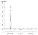
- FIG. 3 shows the concentrations of antibodies against human integrins having different combinations of subunit ⁇ chains and ⁇ chains in ulcerative colitis serum (UC) and control serum (Control).
- FIG. 3 shows the concentrations of antibodies against human integrins having different combinations of subunit ⁇ chains and ⁇ chains in ulcerative colitis serum (UC) and control serum (Control).
- FIG. 3 shows the concentrations of antibodies against human integrins having different combinations of subunit ⁇ chains and ⁇ chains in ulcerative colitis serum (UC) and control serum (Control).
- FIG. 3 shows the concentrations of antibodies against human integrins having different combinations of subunit ⁇ chains and ⁇ chains in ulcerative colitis serum (UC) and control serum (Control).
- FIG. 2 shows the concentrations of anti-human integrin ⁇ V ⁇ 6 antibody in ulcerative colitis patient serum, Crohn disease patient serum, and control serum.
- Fig. 3 shows the concentration of anti-human integrin ⁇ V ⁇ 6 antibody in the serum of patients with primary sclerosing cholangitis, the serum of patients with IgG4-related sclerosing cholangitis, and the control serum.
- 2 shows the concentrations of anti-human integrin ⁇ V ⁇ 3 antibody in ulcerative colitis patient serum, Crohn disease patient serum, and control serum.
- 3 shows the concentration of anti-human integrin ⁇ V ⁇ 3 antibody in the serum of patients with primary sclerosing cholangitis, the serum of patients with IgG4-related sclerosing cholangitis, and the control serum.
- 2 shows the concentration of anti-human integrin ⁇ V ⁇ 6 antibody in sera of ulcerative colitis, serum of patients with Crohn's disease, and sera of patients with other intestinal diseases.
- 2 shows the concentrations of anti-human integrin ⁇ V ⁇ 6 antibody in ulcerative colitis patient serum, Crohn's disease patient serum, other intestinal disease patient serum, collagen disease patient serum, and healthy subject serum.
- 2 shows the concentration of anti-human integrin ⁇ V ⁇ 6 antibody IgG1 in the sera of patients with ulcerative colitis, the sera of patients with Crohn's disease, and the sera of patients with other intestinal diseases.
- 2 shows the concentration of anti-human integrin ⁇ V ⁇ 6 antibody IgG2 in ulcerative colitis patient serum, Crohn's disease patient serum, and other intestinal disease patient serum.
- the concentration of anti-human integrin ⁇ V ⁇ 6 antibody IgG3 in the sera of patients with ulcerative colitis, the sera of patients with Crohn's disease, and the sera of patients with other intestinal diseases is shown.
- the concentration of the anti-human integrin ⁇ V ⁇ 6 antibody IgG4 in the sera of patients with ulcerative colitis, the sera of patients with Crohn's disease, and the sera of patients with other intestinal diseases is shown.
- 2 shows the concentration of anti-human integrin ⁇ V ⁇ 6 antibody IgA in the sera of ulcerative colitis patients, the sera of patients with Crohn's disease, and the sera of patients with other intestinal diseases.
- the concentration of anti-human integrin ⁇ V ⁇ 6 antibody IgM in the sera of patients with ulcerative colitis, the sera of patients with Crohn's disease, and the sera of patients with other intestinal diseases is shown.
- FIG. 10A is a partial Mayo score (left vertical axis) and an anti-human integrin ⁇ V ⁇ 6 autoantibody (IgG) antibody in patients with ulcerative colitis at each time point from November 2017 to November 2018. Value (right vertical axis) is shown.
- FIG. 10B is a partial Mayo score (left vertical axis) and an anti-human integrin ⁇ V ⁇ 6 autoantibody (IgG) of another patient with ulcerative colitis at each time point from November 2017 to November 2018. Shows the antibody titer (right vertical axis).
- 2 shows the concentration of anti-human integrin ⁇ V ⁇ 3 antibody in sera of patients with ulcerative colitis, sera of patients with Crohn's disease, and sera of patients with other intestinal diseases.
- 1 shows the concentrations of anti-human integrin ⁇ V ⁇ 3 antibody in ulcerative colitis patient serum, Crohn's disease patient serum, other intestinal disease patient serum, collagen disease patient serum, and healthy subject serum.
- 2 shows the concentration of anti-human integrin ⁇ V ⁇ 3 antibody IgG1 in the sera of ulcerative colitis patients, the sera of patients with Crohn's disease, and the sera of patients with other intestinal diseases.
- 2 shows the concentration of anti-human integrin ⁇ V ⁇ 3 antibody IgG2 in ulcerative colitis patient serum, Crohn disease patient serum, and other intestinal disease patient serum.
- the concentration of anti-human integrin ⁇ V ⁇ 3 antibody IgG3 in the sera of patients with ulcerative colitis, the sera of patients with Crohn's disease, and the sera of patients with other intestinal diseases is shown.
- 2 shows the concentration of anti-human integrin ⁇ V ⁇ 3 antibody IgG4 in sera of patients with ulcerative colitis, sera of patients with Crohn's disease, and sera of patients with other intestinal diseases.
- the concentration of anti-human integrin ⁇ V ⁇ 3 antibody IgA in the sera of patients with ulcerative colitis, the sera of patients with Crohn's disease, and the sera of patients with other intestinal diseases is shown.
- the concentration of the anti-human integrin ⁇ V ⁇ 3 antibody IgM in the sera of patients with ulcerative colitis, the sera of patients with Crohn's disease, and the sera of patients with other intestinal diseases is shown.
- the concentration of anti-human integrin ⁇ V ⁇ 3 antibody IgE in the sera of patients with ulcerative colitis, the sera of patients with Crohn's disease, and the sera of patients with other intestinal diseases is shown.
- test animal targeted by the test method of the present invention is not particularly limited and may be a human or other non-human mammal, but is preferably a human.
- the sample used in the test method of the present invention includes body fluid collected from a subject animal. Specifically, in addition to blood samples such as serum, plasma, and whole blood, body fluid samples other than blood such as saliva, spinal fluid, and urine may be used. In addition to body fluids, tissues collected from animals such as ulcerative colitis disease site (eg, large intestine, rectum) or primary sclerosing cholangitis disease site (eg, liver) to be examined. The collected tissue can also be used as a sample. The sample is used in the test method of the present invention in a form separated from the test animal.
- body fluid samples other than blood such as saliva, spinal fluid, and urine
- tissues collected from animals such as ulcerative colitis disease site (eg, large intestine, rectum) or primary sclerosing cholangitis disease site (eg, liver) to be examined.
- the collected tissue can also be used as a sample.
- the sample is used in the test method of the present invention in a form separated from the test animal.
- Integrin is, in its natural form, a protein composed of a heterodimeric molecule composed of two subunit chains, an ⁇ chain and a ⁇ chain.
- ⁇ 1 to ⁇ 11, ⁇ V, ⁇ X, ⁇ M, ⁇ L, ⁇ D, ⁇ E, ⁇ IIb and ⁇ 1 to ⁇ 8 are known as ⁇ chains, and there are a plurality of isoforms having different combinations thereof.
- Integrin is present on the surface of epithelial cells and binds to extracellular matrix proteins such as laminin and fibronectin on the surface of connective tissues.
- Integrin ⁇ V ⁇ 6 consists of a heterodimeric molecule containing ⁇ V as the ⁇ chain and ⁇ 6 as the ⁇ chain. Integrin ⁇ V ⁇ 6 is hardly expressed in normal tissues, but is expressed on the surface of epithelial cells during inflammation stimulation.
- the origin of the fragment or whole of integrin ⁇ V ⁇ 6 that immunologically binds to the autoantibody to be detected is not particularly limited, but it is preferably the same species as the test animal, and particularly preferably human.
- the nucleotide sequence information of the genes encoding the ⁇ V chain and ⁇ 6 chain of integrin of mammalian species such as human and the amino acid sequence information of each chain can be obtained from a known database (GenBank etc.).
- the amino acid sequence of the preproprotein of human integrin ⁇ V chain isoform 1 has been registered as GenBank Accession Number NP — 002101.2 and is shown in SEQ ID NO:1.
- the amino acid sequence of the precursor of human integrin ⁇ 6 chain is registered as GenBank Accession Number NP_000879.2, and is shown in SEQ ID NO: 2.
- the amino acid sequence information of integrin ⁇ V chain and ⁇ 6 chain of various mammals other than human can be similarly obtained from a known database (GenBank etc.).
- An integrin ⁇ V ⁇ 6 may be formed by an ⁇ V chain and a ⁇ 6 chain containing an amino acid sequence formed by posttranslational modification of one or both of the amino acid sequences of the ⁇ V chain and ⁇ 6 chain registered in the database.
- the partial sequence from the 1st position to the 30th position in the amino acid sequence of SEQ ID NO: 1 is a signal peptide sequence
- the amino acid sequence of the mature polypeptide of human integrin ⁇ V chain is from the 31st position to the amino acid sequence of SEQ ID NO: 1 It includes the sequence at position 1048.
- the partial sequence from the 1st position to the 21st position in the amino acid sequence of SEQ ID NO: 2 is a signal peptide sequence
- the amino acid sequence of the mature polypeptide of human integrin ⁇ 6 chain is the 22nd position in the amino acid sequence of SEQ ID NO: 2.
- integrin ⁇ V ⁇ 6 is not limited to a natural type containing a mature or immature amino acid sequence, and may be a mutant in a form equivalent to natural type integrin ⁇ V ⁇ 6.
- the integrin ⁇ V ⁇ 6 is not limited to a form in which the natural or mutant ⁇ chain and ⁇ chain containing the mature or immature amino acid sequence include the entire length thereof (that is, the whole integrin ⁇ V ⁇ 6), and is a fragment of the integrin ⁇ V ⁇ 6. It may be in the form of.
- the autoantibody to be detected is not limited to an antibody that immunologically binds to a fragment of integrin ⁇ V ⁇ 6 or an amino acid sequence consisting entirely of the integrin ⁇ V ⁇ 6, and other peptides may be attached to each fragment of the integrin ⁇ V ⁇ 6 or the entire chain. It may be an antibody that immunologically binds to the one to which is added (particularly to the one added to the C-terminal side).
- the integrin ⁇ V ⁇ 6 fragment examples include those in which at least one of the ⁇ V chain and the ⁇ 6 chain forming the dimer of integrin is shorter than the mature or immature natural type or its mutant.
- the ⁇ V chain includes an ⁇ V chain containing a partial sequence from Phe at position 31 to Val at position 992 in the amino acid sequence of the ⁇ V chain shown in SEQ ID NO: 1, and/or ⁇ 6 chain
- a dimer containing the ⁇ 6 chain containing a partial sequence from Gly at the 22nd position to Asn at the 707th position in the amino acid sequence of the ⁇ 6 chain shown in SEQ ID NO: 2 can be exemplified.
- the fragment of integrin ⁇ V ⁇ 6 preferably forms a dimer, and more preferably has binding activity to extracellular matrix proteins such as laminin and fibronectin.
- extracellular matrix proteins such as laminin and fibronectin.
- the fact that the fragment of integrin ⁇ V ⁇ 6 has the binding activity to the extracellular matrix protein can be confirmed by the ELISA method or the like.
- the fact that the whole integrin ⁇ V ⁇ 6 or a fragment thereof forms a dimer means that the whole integrin ⁇ V ⁇ 6 or a fragment thereof is subjected to SDS-PAGE in the absence of 2-mercaptoethanol. It can be confirmed that a band corresponding to the molecular weight can be detected, and that the band corresponding to the molecular weight of the dimer disappears when the band is subjected to SDS-PAGE in the presence of 2-mercaptoethanol.
- integrin ⁇ V ⁇ 6 is recombinant human integrin ⁇ V ⁇ 6 (R&D Systems, Minnesota, USA, product number 3817-AV).
- This recombinant human integrin ⁇ V ⁇ 6 has a partial sequence from Phe at position 31 to Val at position 992 in the amino acid sequence of the ⁇ V chain shown in SEQ ID NO: 1, a linker sequence added to its C terminus, and an acidic tail sequence. And a partial sequence from Gly at the 22nd position to Asn at the 707th position in the amino acid sequence of the ⁇ 6 chain shown in SEQ ID NO: 2, and a linker sequence and a basic tail sequence added to the C-terminus thereof. It is a dimer with the ⁇ 6 chain.
- amino acid sequence shown in SEQ ID NO: 1 or a polypeptide containing a partial sequence from Phe at position 31 to Thr at position 1048 in the amino acid sequence shown in SEQ ID NO: 1 (II) Of the amino acid sequence shown in SEQ ID NO: 1 A polypeptide comprising a partial sequence and functionally equivalent to the polypeptide of (I), (III) A polypeptide comprising an amino acid sequence having 85% or more sequence identity with the amino acid sequence shown in SEQ ID NO: 1 or a partial sequence thereof, and functionally equivalent to the polypeptide of (I), and (IV) sequence
- the amino acid sequence shown in No. 1 or a partial sequence thereof comprises an amino acid sequence in which one or more amino acids are substituted, deleted and/or added, and is composed of a polypeptide functionally equivalent to the polypeptide of (I) Included are polypeptides selected from the group.
- polypeptides (I) to (IV) preferably have the amino acid sequence or partial sequence defined in the above (I) to (IV), and further have another amino acid sequence on at least one of the N-terminal side and the C-terminal side. May be a polypeptide containing an amino acid sequence added to the C-terminal side.
- polypeptide functionally equivalent to the polypeptide of (I) means a polypeptide consisting of the integrin ⁇ 6 chain (particularly preferably, the amino acid sequence shown in SEQ ID NO: 2).
- a polypeptide chain consisting of a partial sequence up to Asn at position 707) can form a dimer, and the dimer formed is a natural integrin ⁇ V ⁇ 6 such as laminin or fibronectin or a commercially available integrin.
- An example is a polypeptide that will have the ability to bind to extracellular matrix proteins to which ⁇ V ⁇ 6 can bind.
- the partial sequence in (II) above includes the partial sequence from Phe at position 31 to Val at position 992 in the amino acid sequence shown in SEQ ID NO: 1.
- the partial sequences in (III) and (IV) above include the partial sequence from Phe at position 31 to Thr at position 1048 in the amino acid sequence shown in SEQ ID NO: 1, or the amino acid sequence shown in SEQ ID NO: 1.
- the partial sequence from Phe at the 31st position to Val at the 992nd position can be mentioned.
- sequence identity in the above (III) is preferably 90% or more, more preferably 95% or more, even more preferably 96% or more, particularly preferably 97% or more, most preferably 98% or more, or 99% or more.
- “1 or more” is, for example, 1 to 100, preferably 1 to 50, preferably 1 to 30, preferably 1 to 20, preferably 1 to 15, preferably The number is 1 to 10, preferably 1 to 5, preferably 1 to 4, preferably 1 to 3, preferably 1 to 2, and preferably 1.
- ⁇ 6 chain constituting the fragment or whole of integrin ⁇ V ⁇ 6 (V) of the amino acid sequence shown in SEQ ID NO: 2 or the polypeptide (VI) containing the partial sequence from Gly at the 22nd position to Cys at the 788th position in the amino acid sequence shown in SEQ ID NO: 2
- VII A polypeptide comprising an amino acid sequence having 85% or more sequence identity with the amino acid sequence shown in SEQ ID NO: 2 or a partial sequence thereof, and functionally equivalent to the polypeptide of (V), and (VIII) sequence
- the amino acid sequence shown in No. 2 or a partial sequence thereof includes an amino acid sequence in which one or more amino acids are substituted, deleted, and/or added, and is composed of a polypeptide functionally equivalent to the polypeptide (V) Included are polypeptides selected from the group.
- polypeptides of (V) to (VIII) preferably have the amino acid sequence or partial sequence defined in (V) to (VIII), and further have another amino acid sequence on at least one of the N-terminal side and the C-terminal side. May be a polypeptide containing an amino acid sequence added to the C-terminal side.
- the polypeptide functionally equivalent to the polypeptide of (V) means an integrin ⁇ V chain (particularly preferably a polypeptide consisting of the amino acid sequence shown in SEQ ID NO: 1).
- a chain a polypeptide chain consisting of a partial sequence from Phe at position 31 to Thr at position 1048 in the amino acid sequence shown in SEQ ID NO: 1, or from Phe at position 31 in the amino acid sequence shown in SEQ ID NO: 1
- a polypeptide chain consisting of a partial sequence up to Val at position 992 can form a dimer, and the formed dimer is a natural integrin ⁇ V ⁇ 6 or a commercially available integrin ⁇ V ⁇ 6 such as laminin and fibronectin.
- An example is a polypeptide that has the ability to bind to extracellular matrix proteins that can bind to.
- the partial sequence in (VI) above includes the partial sequence from Gly at the 22nd position to Asn at the 707th position in the amino acid sequence shown in SEQ ID NO: 2.
- the partial sequences in (VII) and (VIII) above include the partial sequence from Gly at the 22nd position to Cys at the 788th position in the amino acid sequence shown in SEQ ID NO:2, or the amino acid sequence shown in SEQ ID NO:2.
- the partial sequence from Gly at the 22nd position to Asn at the 707th position can be mentioned.
- sequence identity in the above (VII) is preferably 90% or more, more preferably 95% or more, even more preferably 96% or more, particularly preferably 97% or more, most preferably 98% or more, or most preferably 99% or more.
- “1 or more” is, for example, 1 to 100, preferably 1 to 50, preferably 1 to 30, preferably 1 to 20, preferably 1 to 15, preferably The number is 1 to 10, preferably 1 to 5, preferably 1 to 4, preferably 1 to 3, preferably 1 to 2, and preferably 1.
- sequence identity of amino acid sequences can be determined using a method well known to those skilled in the art, sequence analysis software, or the like.
- sequence analysis software include the blastp program of the BLAST algorithm and the fasta program of the FASTA algorithm.
- the origin of the integrin ⁇ V ⁇ 3 fragment or whole that immunologically binds to the autoantibody to be detected is not particularly limited, but it is preferably the same species as the test animal, and particularly preferably human.
- the nucleotide sequence information of the genes encoding the ⁇ V chain and ⁇ 3 chain of integrins of mammalian species such as human and the amino acid sequence information of each chain can be obtained from a known database (GenBank etc.). The description of the ⁇ V chain is omitted because it is as described above.
- amino acid sequence of the precursor of the human integrin ⁇ 3 chain has been registered as GenBank Accession Number AAA52589.1 and is shown in SEQ ID NO:3.
- Amino acid sequence information of the integrin ⁇ 3 chain of various mammals other than humans can also be obtained from publicly known databases (GenBank etc.).
- An integrin ⁇ V ⁇ 3 may be formed by an ⁇ V chain and a ⁇ 3 chain containing an amino acid sequence formed by post-translational modification of one or both of the ⁇ V chain and ⁇ 3 chain amino acid sequences registered in the database.
- the amino acid sequence of the mature polypeptide of human integrin ⁇ V chain includes the sequence from position 31 to position 1048 in the amino acid sequence of SEQ ID NO: 1.
- the partial sequence from the 1st position to the 26th position in the amino acid sequence of SEQ ID NO: 3 is a signal peptide sequence
- the amino acid sequence of the mature polypeptide of human integrin ⁇ 3 chain is the 27th position in the amino acid sequence of SEQ ID NO: 3. To include the sequence at position 788.
- the integrin ⁇ V ⁇ 3 is not limited to a natural type containing a mature or immature amino acid sequence, and may be a mutant in a form equivalent to the natural type integrin ⁇ V ⁇ 3.
- integrin ⁇ V ⁇ 3 is not limited to a form in which the ⁇ chain and ⁇ chain of a natural type or mutant containing a mature or immature amino acid sequence include both full lengths (that is, whole integrin ⁇ V ⁇ 3), and a fragment of integrin ⁇ V ⁇ 3 It may be in the form of.
- the autoantibody to be detected is not limited to an antibody that immunologically binds to a fragment of integrin ⁇ V ⁇ 3 or an amino acid sequence of the entire integrin ⁇ V ⁇ 3. It may be an antibody that immunologically binds to the one to which is added (particularly to the one added to the C-terminal side).
- the integrin ⁇ V ⁇ 3 fragment examples include those in which at least one of the ⁇ V chain and the ⁇ 3 chain forming the dimer of integrin is shorter than the mature or immature natural type or its mutant.
- the ⁇ V chain includes an ⁇ V chain containing a partial sequence from Phe at position 31 to Val at position 992 in the amino acid sequence of the ⁇ V chain shown in SEQ ID NO: 1, and/or ⁇ 3 chain
- a dimer containing the ⁇ 3 chain containing a partial sequence from Gly at the 27th position to Asp at the 718th position in the amino acid sequence of the ⁇ 3 chain shown in SEQ ID NO: 3 can be exemplified.
- the fragment of integrin ⁇ V ⁇ 3 preferably forms a dimer, and more preferably has binding activity to extracellular matrix proteins such as laminin and fibronectin.
- extracellular matrix proteins such as laminin and fibronectin.
- the fact that the fragment of integrin ⁇ V ⁇ 3 has the binding activity to the extracellular matrix protein can be confirmed by the ELISA method or the like.
- the fact that the whole integrin ⁇ V ⁇ 3 or a fragment thereof forms a dimer means that the whole integrin ⁇ V ⁇ 3 or a fragment thereof was subjected to SDS-PAGE in the absence of 2-mercaptoethanol. It can be confirmed that a band corresponding to the molecular weight can be detected, and that the band corresponding to the molecular weight of the dimer disappears when the band is subjected to SDS-PAGE in the presence of 2-mercaptoethanol.
- Examples of commercially available integrin ⁇ V ⁇ 3 include recombinant human integrin ⁇ V ⁇ 3 (R&D Systems, Minnesota, USA, product number 3050-AV).
- This recombinant human integrin ⁇ V ⁇ 3 comprises a partial sequence from Phe at position 31 to Val at position 992 in the amino acid sequence of the ⁇ V chain shown in SEQ ID NO: 1, a linker sequence added to its C-terminal and an acidic tail sequence.
- a more specific embodiment of the fragment of integrin ⁇ V ⁇ 3 or the ⁇ V chain constituting the whole is a polypeptide selected from the above (I) to (IV).
- a more specific embodiment of the fragment of integrin ⁇ V ⁇ 3 or the ⁇ 3 chain constituting the whole is: (IX) Partial sequence from the 27th position Gly to the 718th Asp of the amino acid sequence shown in SEQ ID NO: 3 or the amino acid sequence shown in SEQ ID NO: 3 or the 27th position Gly of the amino acid sequence shown in SEQ ID NO: 3 To the Thr at position 788 (X) A polypeptide (X) comprising a partial sequence of the amino acid sequence shown in SEQ ID NO: 3 and functionally equivalent to the polypeptide (IX), (XI) A polypeptide comprising an amino acid sequence having a sequence identity of 85% or more with the amino acid sequence shown in SEQ ID NO: 3 or a partial sequence thereof, and being functionally equivalent to the polypeptide of (IX), and (XII) sequence
- the amino acid sequence shown in No. 3 or a partial sequence thereof contains an amino acid sequence in which one or more amino acids are substituted, deleted, and/or added, and is composed
- the polypeptide of (IX) to (XII) is preferably an amino acid sequence or a partial sequence defined in (IX) to (XII), and another amino acid sequence is preferably present on at least one of the N-terminal side and the C-terminal side. May be a polypeptide containing an amino acid sequence added to the C-terminal side.
- the polypeptide functionally equivalent to the polypeptide of (IX) means an integrin ⁇ V chain (particularly preferably, a polypeptide chain consisting of the amino acid sequence shown in SEQ ID NO: 1, SEQ ID NO: In the amino acid sequence shown in 1, the polypeptide chain consisting of a partial sequence from Phe at position 31 to Thr at position 1048, or in the amino acid sequence shown in SEQ ID NO: 1 from Phe at position 31 to Val at position 992
- a polypeptide chain consisting of a partial sequence up to) and a dimer formed can bind to natural type integrin ⁇ V ⁇ 3 or commercially available integrin ⁇ V ⁇ 3 such as laminin and fibronectin.
- a polypeptide that has the ability to bind to extracellular matrix proteins that can
- the partial sequence in (X) above includes the partial sequence from Gly at the 27th position to Asp at the 718th position in the amino acid sequence shown in SEQ ID NO:3.
- the partial sequences in (XI) and (XII) include the partial sequence from Gly at the 27th position to Asp at the 718th position in the amino acid sequence shown in SEQ ID NO:3, or the amino acid sequence shown in SEQ ID NO:3.
- the partial sequence from Gly at the 27th position to Thr at the 788th position can be mentioned.
- sequence identity in the above (XI) is preferably 90% or more, more preferably 95% or more, even more preferably 96% or more, particularly preferably 97% or more, most preferably 98% or more, or 99% or more.
- “1 or more” is, for example, 1 to 100, preferably 1 to 50, preferably 1 to 30, preferably 1 to 20, preferably 1 to 15, preferably The number is 1 to 10, preferably 1 to 5, preferably 1 to 4, preferably 1 to 3, preferably 1 to 2, and preferably 1.
- Test method for ulcerative colitis or primary sclerosing cholangitis is, firstly, A method for examining ulcerative colitis, As an index of ulcerative colitis, a detection step of detecting an antibody immunologically reacting with a fragment or whole of integrin ⁇ V ⁇ 6 and/or an antibody immunologically reacting with a fragment or whole of integrin ⁇ V ⁇ 3 in a sample Regarding the method including.
- a method for testing primary sclerosing cholangitis As an index of primary sclerosing cholangitis, an antibody immunologically reacting with a fragment or whole of integrin ⁇ V ⁇ 6 and/or an antibody immunologically reacting with a fragment or whole of integrin ⁇ V ⁇ 3 in a sample is detected.
- a method comprising a detection step.
- fragment or whole of integrin ⁇ V ⁇ 6 is generically referred to as “integrin ⁇ V ⁇ 6”
- fragment or whole of integrin ⁇ V ⁇ 3 is generically referred to as “integrin ⁇ V ⁇ 3”.
- the first test method of the present invention is an indicator that an antibody against integrin ⁇ V ⁇ 6 (autoantibody) and an antibody against integrin ⁇ V ⁇ 3 (autoantibody) in a sample obtained from a test animal have ulcerative colitis. It was completed based on the surprising finding that
- the second test method of the present invention is that the antibody to the integrin ⁇ V ⁇ 6 (autoantibody) and the antibody to integrin ⁇ V ⁇ 3 (autoantibody) in the sample obtained from the test animal are affected by primary sclerosing cholangitis. It was completed based on the surprising knowledge that it will be an index of.
- the detection step in the test method of the present invention is a step of detecting an antibody that immunologically reacts with integrin ⁇ V ⁇ 6 and/or an antibody that immunologically reacts with integrin ⁇ V ⁇ 3 in the sample.
- detection means to confirm whether or not an antibody immunologically reacts with integrin ⁇ V ⁇ 6 and/or an antibody immunologically reacts with integrin ⁇ V ⁇ 3 in the sample.
- the concept also includes measuring the content of the antibody in a sample, that is, "quantifying”.
- the detection step in the test method of the present invention may be any method that can quantitatively or qualitatively detect the antibody in the sample, and the specific embodiment is not particularly limited.
- immunological techniques can be used, for example, antibody enzyme method (ELISA method), immunoprecipitation method (IPP method), immunoblotting method (IB method), latex agglutination method, immunochromatography method, indirect fluorescent antibody. Methods (IF method), radioimmunoassay method (RIA method) and the like can be mentioned.
- ELISA method antibody enzyme method
- IPP method immunoprecipitation method
- IB method immunoblotting method
- latex agglutination method immunochromatography method
- indirect fluorescent antibody indirect fluorescent antibody.
- Methods IF method
- radioimmunoassay method RIA method
- An ELISA method that can process many specimens is particularly preferred.
- integrin ⁇ V ⁇ 6 and/or integrin ⁇ V ⁇ 3, which are the antigens of the antibody, as described above, are immobilized on a solid phase, and the specimen is contacted with the antigen immobilized on the solid phase. Then, the antibody can be detected by detecting an immune complex between the antigen and the antibody that may be contained in the sample using an enzyme-labeled detection antibody or the like.
- an enzyme-labeled detection antibody or the like There is no particular limitation on the method for suppressing non-specific reaction accompanying the implementation of the ELISA method, the labeling substance that can be used at the time of detection, the measuring instrument, and the like.
- the solid phase for immobilizing the antigen a solid phase having any shape such as a plate, beads, or tube can be used.
- a step of bringing a sample that may contain the antibody into contact with the antigen, a necessary washing step, a necessary labeling step, and a necessary detection step are carried out in an appropriate buffer solution.
- a buffer solution containing at least one metal ion selected from calcium ion, magnesium ion, manganese ion, sodium, lithium and the like.
- the metal ion is preferably a divalent metal ion such as calcium ion, magnesium ion and manganese ion, and particularly preferably one or two kinds of calcium ion, magnesium ion and manganese ion.
- the concentration of the metal ion in the buffer solution is not particularly limited, but may be 0.02 mM to 200 mM in total, preferably 0.2 mM to 20 mM, and particularly preferably 0.4 mM to 10 mM. In a buffer solution containing these metal ions, detection sensitivity is enhanced, which is preferable.
- the amount of the antibody that immunologically reacts with the integrin ⁇ V ⁇ 6 and/or the amount of the antibody that immunologically reacts with the integrin ⁇ V ⁇ 3 is determined for the detection that is bound to the immune complex of the antibody and the antigen. It can be determined as a measured value of the labeled amount of the antibody. It is also possible to prepare a calibration curve using a positive standard solution containing a known concentration of the antibody, and use the calibration curve to calculate the amount of the antibody in the test sample from the measured value of the labeled amount.
- determination of whether or not an antibody immunologically reacting with integrin ⁇ V ⁇ 6 and/or an antibody immunologically reacting with integrin ⁇ V ⁇ 3 is present is carried out by the test animal individual.
- the measurement value of the antibody in a sample obtained from a human who is suspected to have ulcerative colitis or primary sclerosing cholangitis is not affected by ulcerative colitis or primary sclerosing cholangitis. It can be carried out by comparing with the measured value of the antibody in a sample obtained from an individual animal (for example, a healthy person).
- the measurement value of the antibody in a sample obtained from an individual not suffering from ulcerative colitis or primary sclerosing cholangitis may be a value obtained by simultaneous measurement, or may be measured in advance. It may be a value obtained by The measurement value of the antibody in the sample obtained from the test animal individual is significantly higher than the measurement value of the antibody in the sample obtained from the animal individual not suffering from ulcerative colitis or primary sclerosing cholangitis. If it is larger, it can be judged as positive.
- the antibody that immunologically reacts with integrin ⁇ V ⁇ 6 and/or the antibody that immunologically reacts with integrin ⁇ V ⁇ 3 that is detected in the detection step is not particularly limited, and may be an IgG antibody, an IgA antibody, an IgM antibody, an IgE antibody. IgG antibodies or IgA antibodies are particularly preferred. As an antibody that immunologically reacts with integrin ⁇ V ⁇ 6, it is particularly preferable to detect IgG or IgA. As an antibody that immunologically reacts with integrin ⁇ V ⁇ 3, it is particularly preferable to detect an IgG antibody.
- the subclass among IgG antibodies is not particularly limited, but, for example, IgG1 antibody, IgG2 antibody, IgG3 antibody or IgG4 antibody can be detected.
- IgG antibody that immunologically reacts with integrin ⁇ V ⁇ 6 it is more preferable to detect an IgG1 antibody, an IgG2 antibody or an IgG4 antibody, and it is most preferable to detect an IgG1 antibody.
- IgG antibody that immunologically reacts with integrin ⁇ V ⁇ 3 it is more preferable to detect an IgG1 antibody, an IgG2 antibody or an IgG3 antibody, and it is most preferable to detect an IgG1 antibody.
- Test kit or test reagent for ulcerative colitis or primary sclerosing cholangitis firstly relates to a test kit or test reagent for testing ulcerative colitis, which comprises a fragment or whole of integrin ⁇ V ⁇ 6 and/or a fragment or whole of integrin ⁇ V ⁇ 3.
- test kit or test reagent of the present invention relates to a test kit or test reagent for testing primary sclerosing cholangitis, which comprises a fragment or whole of integrin ⁇ V ⁇ 6 and/or a fragment or whole of integrin ⁇ V ⁇ 3. ..
- fragment or whole of integrin ⁇ V ⁇ 6 is generically referred to as “integrin ⁇ V ⁇ 6”
- fragment or whole of integrin ⁇ V ⁇ 3 is generically referred to as “integrin ⁇ V ⁇ 3”.
- test kit or test reagent of the present invention can be used in the above-described test method of the present invention.
- the test kit of the present invention requires reagents necessary for immunological measurement, for example, one or more metal salts selected from buffers, calcium salts, magnesium salts, manganese salts, sodium salts, lithium salts and the like. May be included depending on
- test reagent of the present invention may be a liquid or solid composition containing integrin ⁇ V ⁇ 6 and/or integrin ⁇ V ⁇ 3 and a carrier such as a solvent or an excipient which is acceptable for the purpose of formulation.
- the form of integrin ⁇ V ⁇ 6 and/or integrin ⁇ V ⁇ 3 is not particularly limited, and may be in a solution state, a dry state, or immobilized on a solid phase. It may have a different form.
- a buffer solution or a solvent for making it into a solution state before use may be included in the test kit or the test reagent of the present invention.
- a test kit or test reagent containing integrin ⁇ V ⁇ 6 and/or integrin ⁇ V ⁇ 3 immobilized on a solid phase can be used to test for ulcerative colitis or primary sclerosing cholangitis by the ELISA method.
- the form of the solid phase may be any form such as plates, beads, tubes and the like.
- Kits for examining ulcerative colitis or primary sclerosing cholangitis by ELISA method include, in addition to the above-mentioned solid phase, positive standard solution, negative standard solution, blocking solution, washing solution, specimen diluting solution, detection antibody, substrate A liquid or the like can be included.
- the detection antibody may be labeled with a detectable label such as an enzyme.
- Examples of the positive standard solution include integrin ⁇ V ⁇ 6 and/or integrin ⁇ V ⁇ 3 and/or integrin ⁇ V ⁇ 3, as well as a serum diluent of a patient with ulcerative colitis or primary sclerosing cholangitis containing an integrin ⁇ V ⁇ 3 autoantibody.
- a solution containing an antibody is used.
- the antibody added to the solution is preferably the same isotype as the autotype of the autoantibody to be measured, such as IgG antibody, IgA antibody, IgM antibody, and IgE antibody.
- Negative standard solution is a serum dilution of a person who does not have ulcerative colitis or primary sclerosing cholangitis, serum of other intestinal diseases medically distinguished from ulcerative colitis, primary sclerosing cholangio Serum of other hepatobiliary system diseases medically distinguished from inflammation, serum of normal human (control serum) and its diluted solution are used.
- a method of evaluating the effect of treatment for ulcerative colitis comprising: An antibody that immunologically reacts with a fragment or whole of integrin ⁇ V ⁇ 6 and/or an immunological reaction with a fragment or whole of integrin ⁇ V ⁇ 3 in a sample obtained from a test animal that has been treated for ulcerative colitis To detect the antibody.
- the method for evaluating the therapeutic effect of the present invention A method of assessing the effect of treatment for primary sclerosing cholangitis, comprising: An antibody that immunologically reacts with the fragment or whole of integrin ⁇ V ⁇ 6 and/or the fragment or whole of integrin ⁇ V ⁇ 3 in a specimen obtained from a test animal that has been treated for primary sclerosing cholangitis And a detection step of detecting an antibody that reacts with.
- fragment or whole of integrin ⁇ V ⁇ 6 is generically referred to as “integrin ⁇ V ⁇ 6”
- fragment or whole of integrin ⁇ V ⁇ 3 is generically referred to as “integrin ⁇ V ⁇ 3”.
- an antibody that immunologically reacts with integrin ⁇ V ⁇ 6 and an antibody that immunologically reacts with integrin ⁇ V ⁇ 3 in the specimen are respectively ulcerative colitis or primary sclerosis. It is useful as an indicator of cholangitis. Therefore, the amount of the antibody in a sample obtained from a subject animal treated for ulcerative colitis or primary sclerosing cholangitis is decreased when the treatment is effective and cured, and the treatment is When the effect is not sufficiently exerted, it does not change or increases before the start of treatment. Therefore, the effect of the treatment can be evaluated by detecting the antibody in the sample obtained from the subject animal treated for the ulcerative colitis or the primary sclerosing cholangitis.
- test animals examples include humans and non-human animals treated for ulcerative colitis or primary sclerosing cholangitis.
- the quantification result of the antibody in a sample obtained from a test animal that has been treated for ulcerative colitis or primary sclerosing cholangitis for example, When the former is smaller than the latter as compared with the quantification result of the antibody in the sample obtained from the same animal individual before starting the treatment, it can be evaluated that the treatment is effective. On the other hand, when the former is comparable to the latter or larger than the latter, it can be evaluated that the treatment is ineffective or insufficient. Based on this evaluation result, it is possible to determine whether to continue the treatment, change the treatment policy, stop the treatment, or the like.
- the quantification result of the antibody in a sample obtained from a test animal that has been treated for ulcerative colitis or primary sclerosing cholangitis For example, in comparison with the quantification result of the antibody in a sample obtained from a healthy individual of the same animal species, if the former is comparable to or smaller than the latter, it can be evaluated that the treatment is effective. .. On the other hand, when the former is larger than the latter, it can be evaluated that the treatment is ineffective or insufficient. Based on this evaluation result, it is possible to determine whether to continue the treatment, change the treatment policy, stop the treatment, or the like.
- the screening method of the present invention comprises: A method for screening a candidate substance for a therapeutic agent for ulcerative colitis, comprising: Detecting an antibody immunologically reacting with a fragment or whole of integrin ⁇ V ⁇ 6 and/or an antibody immunologically reacting with a fragment or whole of integrin ⁇ V ⁇ 3 in a specimen obtained from an animal treated with a test substance An antibody detection step, And a step of selecting the test substance as a candidate substance for a therapeutic agent for ulcerative colitis when the antibody in the sample is decreased by the action of the test substance.
- the screening method of the present invention comprises: A method for screening a candidate substance for a therapeutic agent for primary sclerosing cholangitis, comprising: Detecting an antibody immunologically reacting with a fragment or whole of integrin ⁇ V ⁇ 6 and/or an antibody immunologically reacting with a fragment or whole of integrin ⁇ V ⁇ 3 in a specimen obtained from an animal treated with a test substance An antibody detection step, And a step of selecting the test substance as a candidate substance for a therapeutic agent for primary sclerosing cholangitis, when the antibody in the sample decreases due to the action of the test substance.
- the antibody immunologically reacting with the fragment or whole of integrin ⁇ V ⁇ 6 and/or the antibody immunologically reacting with the fragment or whole of integrin ⁇ V ⁇ 3 in the sample is ulcerative colitis or primary It is useful as an index of sclerosing cholangitis. Therefore, when the antibody in the specimen is decreased by the action of a test substance, the test substance may be selected as a candidate substance for a therapeutic agent for ulcerative colitis or primary sclerosing cholangitis. It will be possible.
- the test substance is a test substance that may be a candidate for a new drug and is not particularly limited.
- the animal to which the test substance acts is typically a fragment or whole of integrin ⁇ V ⁇ 6, and/or, as compared with a healthy individual such as a non-human model animal of ulcerative colitis or primary sclerosing cholangitis, and/or It is an animal in which a large amount of an antibody that immunologically reacts with a fragment or whole of integrin ⁇ V ⁇ 3 in a sample.
- the test A substance can be selected as a candidate substance for a therapeutic agent for ulcerative colitis or primary sclerosing cholangitis.
- the quantification result of the antibody in the sample obtained from the animal to which the test substance was allowed to act was obtained from a healthy individual of the same animal species.
- the test substance can be selected as a candidate substance for a therapeutic agent for ulcerative colitis or primary sclerosing cholangitis.
- the method for producing the non-human model animal of the present invention is A method for producing a non-human model animal for ulcerative colitis, comprising: An antibody administration step of administering to a non-human animal an antibody that immunologically reacts with a fragment or whole of integrin ⁇ V ⁇ 6 and/or an antibody that immunologically reacts with a fragment or whole of integrin ⁇ V ⁇ 3, and a non-human animal , An integrin ⁇ V ⁇ 6 fragment or the whole, and/or an integrin ⁇ V ⁇ 3 fragment or the whole or an immunization step as an antigen.
- the method for producing the non-human model animal of the present invention comprises: A method for producing a non-human model animal of primary sclerosing cholangitis, comprising: An antibody administration step of administering to a non-human animal an antibody that immunologically reacts with a fragment or whole of integrin ⁇ V ⁇ 6 and/or an antibody that immunologically reacts with a fragment or whole of integrin ⁇ V ⁇ 3, and a non-human animal , An integrin ⁇ V ⁇ 6 fragment or the whole, and/or an integrin ⁇ V ⁇ 3 fragment or the whole or an immunization step as an antigen.
- the specific forms of the integrin ⁇ V ⁇ 6 fragment or whole, the integrin ⁇ V ⁇ 3 fragment or whole, the antibody and the like in the method for producing a non-human model animal of the present invention are the same as those in the test method of the present invention.
- the non-human animal is administered with an antibody immunologically reactive with the fragment or whole of integrin ⁇ V ⁇ 6 and/or the fragment or whole of integrin ⁇ V ⁇ 3, whereby ulcerative colitis or primary sclerosing cholangitis It is possible to produce non-human model animals.
- non-human animals to which the antibody is administered include mice such as BALB/c mouse and B6 mouse, and other non-human animals.
- the administration route of the antibody immunologically reacting with the fragment or whole of integrin ⁇ V ⁇ 6 and/or the antibody immunologically reacting with the fragment or whole of integrin ⁇ V ⁇ 3 is not particularly limited, but, for example, subcutaneously Administration, intraperitoneal administration, intravenous administration and the like can be exemplified.
- the antibody need not be administered to the non-human animal as a purified antibody, but may be administered to the non-human animal as serum containing the antibody, for example.
- the antibody to be administered in the antibody administration step is selected so that it can immunologically react with integrin ⁇ V ⁇ 6 and/or integrin ⁇ V ⁇ 3 possessed by the non-human animal receiving the administration.
- a non-human animal model of ulcerative colitis or primary sclerosing cholangitis is produced by an immunization step of immunizing a non-human animal with the fragment or whole of integrin ⁇ V ⁇ 6 and/or the fragment or whole of integrin ⁇ V ⁇ 3 as an antigen. It can be manufactured.
- the non-human animal to which the antigen is administered include mice such as BALB/c mouse and B6 mouse, and other non-human animals.
- the method in the immunization process is not particularly limited.
- the fragment or whole of integrin ⁇ V ⁇ 6 and/or the fragment or whole of integrin ⁇ V ⁇ 3 is administered to a non-human animal by a route such as subcutaneous administration, intravenous administration or intraperitoneal administration together with an appropriate adjuvant such as complete Freund's adjuvant.
- a non-human animal can be immunized.
- a fragment or whole of integrin ⁇ V ⁇ 6 and/or a fragment or whole of integrin ⁇ V ⁇ 3 to be administered as an antigen is immunized by an antibody against the integrin ⁇ V ⁇ 6 and/or integrin ⁇ V ⁇ 3 by autoimmunity. Selected so that it can react chemically.
- human IgG that can be contained in serum of patients with ulcerative colitis and that binds to an antigen candidate immobilized on a solid phase is used as an autoantibody to be detected, and rabbit anti-antibody labeled with HRP (horseradish peroxidase) is used.
- HRP horseradish peroxidase
- Human integrin ⁇ 2b ⁇ 3 (7148-A2, R&D system) Human integrin ⁇ 1 ⁇ 1 (7064-AB, R&D system) Human integrin ⁇ 2 ⁇ 1 (5698-A2, R&D system) Human integrin ⁇ 3 ⁇ 1 (2840-A3, R&D system) Human integrin ⁇ 4 ⁇ 1 (5668-A4, R&D system) Human integrin ⁇ 4 ⁇ 7 (5397-A3, R&D system) Human integrin ⁇ 5 ⁇ 1 (3230-A5, R&D system) Human integrin ⁇ 6 ⁇ 1 (7809-A6, R&D system) Human integrin ⁇ 6 ⁇ 4 (5497-A6, R&D system) Human integrin ⁇ 9 ⁇ 1 (5438-A9, R&D system) Human integrin ⁇ 10 ⁇ 1 (5895-AB, R&D system) Human integrin ⁇ 11 ⁇ 1 (6357-AB, R&D system) Human integrin ⁇ 10 ⁇ 1 (5895-AB,
- Serum Samples Serum samples were obtained from 8 patients diagnosed with ulcerative colitis. Patients without ulcerative colitis without primary sclerosing cholangitis were selected. Serum samples from three healthy individuals were obtained as control serum samples.
- Blocking (1) 200 ⁇ l of the blocking buffer for ELISA of the above kit was added to each well. (2) Incubated for 30 minutes. (3) After incubation, the blocking buffer for ELISA was removed, and each well was washed 3 times.
- HRP-conjugated detection antibody A detection antibody (abcam6759, rabbit anti-human IgG H&L (HRP)) conjugated with HRP (horseradish peroxidase) was added to the conjugate diluent (Conjugate Diluent) of the above-mentioned kit 1 : Diluted at a rate of 50,000. (2) 100 ⁇ l of the detection antibody diluent obtained by the above dilution was added to each well of the microwell plate in contact with serum. (3) Incubated for 60 minutes. (4) After incubation, the detection antibody diluent was removed, and each well was washed 5 times.
- Enzyme Substrate Reaction (1) A TMB (3,3′,5,5′-tetramethylbenzidine) solution was prepared according to the manufacturer's recommended conditions. (2) 100 ⁇ l of the TMB solution was added to each well of the microwell plate in contact with the detection antibody. (3) Incubated for 7 to 8 minutes. (4) 100 ⁇ l of 0.18 MH 2 SO 4 was added to each well to stop the oxidation of TMB. (5) Using a microplate reader, the coloration due to the product of the oxidation reaction was measured at a wavelength of 450 nm.
- the vertical axis represents the relative value of the absorbance at 450 nm, which is proportional to the amount of detection antibody on the solid phase.
- the anti-human integrin ⁇ V ⁇ 6 antibody concentration in each serum sample was measured by the ELISA method in the same procedure as described in Experiment 1. However, CaCl 2 and MgCl 2 were added to the ELISA washing solution, the ELISA blocking buffer, and the conjugate diluent so that the final concentration of each was 1 mM.
- the vertical axis represents the relative value of absorbance at 450 nm, which is proportional to the amount of detection antibody on the solid phase.
- the cut-off value indicated by the broken line is a value obtained by adding a value three times the standard deviation (SD) to the average value of the relative absorbance values of the control serum sample.
- SD standard deviation
- the anti-human integrin ⁇ V ⁇ 6 antibody was positive in 58 of 63 cases in the serum sample of ulcerative colitis patient, and the sensitivity was 92%.
- the serum sample of Crohn's disease patient 45 out of 48 cases were negative, and in the control serum sample, 11 out of 11 cases were negative, and the specificity was 95%. This result supports that it is possible to diagnose ulcerative colitis based on the blood level of autoantibodies against human integrin ⁇ V ⁇ 6.
- the anti-human integrin ⁇ V ⁇ 6 antibody concentration in each serum sample was measured by the ELISA method in the same procedure as described in Experiment 1. However, CaCl 2 and MgCl 2 were added to the ELISA washing solution, the ELISA blocking buffer, and the conjugate diluent so that the final concentration of each was 1 mM.
- the vertical axis represents the relative value of the absorbance at 450 nm, which is proportional to the amount of detection antibody on the solid phase.
- the cut-off value indicated by the broken line is a value obtained by adding a value three times the standard deviation (SD) to the average value of the relative absorbance values of the control serum sample.
- SD standard deviation
- the anti-human integrin ⁇ V ⁇ 6 antibody was positive in 23 of 24 PSC patient serum samples and the sensitivity was 96%.
- the IgG4 related sclerosing cholangitis patient serum sample was negative in 4 out of 5 cases, and the control serum sample was negative in 6 out of 6 cases, and the specificity was 91%.
- PSC primary sclerosing cholangitis
- the anti-human integrin ⁇ V ⁇ 3 antibody concentration in each serum sample was measured by the ELISA method in the same procedure as described in Experiment 1. However, CaCl 2 and MgCl 2 were added to the ELISA washing solution, the ELISA blocking buffer, and the conjugate diluent so that the final concentration of each was 1 mM.
- the vertical axis represents the relative value of the absorbance at 450 nm, which is proportional to the amount of detection antibody on the solid phase.
- the cut-off value indicated by the broken line is a value obtained by adding a value three times the standard deviation (SD) to the average value of the relative absorbance values of the control serum sample.
- SD standard deviation
- the anti-human integrin ⁇ V ⁇ 3 antibody was positive in 58 out of 63 cases and the sensitivity was 93% in the ulcerative colitis patient serum sample.
- 4 out of 4 serum samples from Crohn's disease patients were negative, and 9 out of 9 control serum samples were negative, and the specificity was 100%. This result supports that it is possible to diagnose ulcerative colitis based on the blood level of autoantibodies against human integrin ⁇ V ⁇ 3.
- the anti-human integrin ⁇ V ⁇ 3 antibody concentration in each serum sample was measured by the ELISA method in the same procedure as described in Experiment 1. However, CaCl 2 and MgCl 2 were added to the ELISA washing solution, the ELISA blocking buffer, and the conjugate diluent so that the final concentration of each was 1 mM.
- the vertical axis represents the relative value of the absorbance at 450 nm, which is proportional to the amount of detection antibody on the solid phase.
- the cut-off value indicated by the broken line is a value obtained by adding a value three times the standard deviation (SD) to the average value of the relative absorbance values of the control serum sample.
- SD standard deviation
- the anti-human integrin ⁇ V ⁇ 3 antibody was positive in 15 of 24 PSC patient serum samples and the sensitivity was 63%.
- the IgG4 related sclerosing cholangitis patient serum sample was negative in 5 out of 5 cases, and the control serum sample was negative in 6 cases in 6 cases, and the specificity was 100%.
- PSC primary sclerosing cholangitis
- the anti-human integrin ⁇ V ⁇ 6 antibody concentration in each serum sample was measured by the ELISA method in the same procedure as described in Experiment 2.
- the vertical axis represents the relative value of the absorbance at 450 nm, which is proportional to the amount of detection antibody on the solid phase.
- “Ulcerative colitis” refers to a serum sample of ulcerative colitis patient
- “Crohn's disease” refers to a serum sample of Crohn's disease
- other intestinal disease refers to a serum sample of the other intestinal disease.
- the cut-off value shown by the broken line is a value obtained by adding a value three times the standard deviation (SD) to the average value of the relative absorbance values of the serum samples of the other intestinal disease patients.
- the anti-human integrin ⁇ V ⁇ 6 antibody was positive in 62 out of 66 serum samples of ulcerative colitis patients, and the sensitivity was 94%. On the other hand, 44 out of 47 serum samples from Crohn's disease patients were negative, and 9 out of 9 serum samples from other intestinal disease patients were negative, and the specificity was 95%. This result indicates that the blood level of autoantibodies against human integrin ⁇ V ⁇ 6 is specifically high in patients with ulcerative colitis.
- the anti-human integrin ⁇ V ⁇ 6 antibody concentration in each serum sample was measured by the ELISA method in the same procedure as described in Experiment 2.
- Fig. 7 the vertical axis represents the relative value of the absorbance at 450 nm, which is proportional to the amount of detection antibody on the solid phase.
- "Ulcerative colitis” indicates a ulcerative colitis patient serum sample
- "Crohn's disease” indicates a Crohn's disease patient serum sample
- “other intestinal disease” indicates the other intestinal disease patient serum sample
- “Collagen disease” refers to a serum sample of a collagen disease patient
- healthy person refers to a serum sample of a healthy person.
- the cut-off value indicated by the broken line is a value obtained by adding a value three times the standard deviation (SD) to the average value of the relative absorbance values of the serum sample of a healthy person.
- SD standard deviation
- the anti-human integrin ⁇ V ⁇ 6 antibody was positive in 43 out of 51 serum samples from patients with ulcerative colitis, and the sensitivity was 84%.
- 24 out of 26 serum samples from Crohn's disease patients were negative
- 23 out of 24 serum samples from other intestinal disease patients were negative
- 26 out of 27 serum samples from collagen disease were negative, serum samples from healthy subjects.
- 22 cases 22 cases were negative and the specificity was 96%.
- This result also shows that, as in Experiment 6, the blood level of autoantibodies against human integrin ⁇ V ⁇ 6 was specifically high in patients with ulcerative colitis.
- n 47 for ulcerative colitis patient serum sample
- intestinal diseases infectious enteritis, ischemic enteritis, Behcet's disease, eosinophilic gastroenteritis, clonkite
- IBDU intestinal diseases
- Anti-human IgG1 antibody (Anti-IgG1-HRP, AP006, Binding Site), anti-human IgG2 antibody conjugated with HRP (Anti-IgG2-HRP, AP007, Binding Site), anti-human IgG3 conjugated with HRP Antibody (Anti-IgG3-HRP, AP008, Binding Site) or HRP-conjugated anti-human IgG4 antibody (Anti-IgG4-HRP, AP009, Binding Site) is used for washing solution for ELISA, blocking for ELISA
- the ELISA method was performed in the same procedure as in Experiment 1 except that CaCl 2 and MgCl 2 were added to the buffer and the conjugate diluent so that the final concentration of each was 1 mM.
- FIG. 8A shows the detection result of IgG1
- FIG. 8B shows the detection result of IgG2
- FIG. 8C shows the detection result of IgG3
- FIG. 8D shows the detection result of IgG4.
- the vertical axis represents the relative value of the absorbance at 450 nm, which is proportional to the amount of detection antibody on the solid phase.
- other intestinal diseases indicate the other
- the cutoff value shown by the broken line is a value obtained by adding a value three times the standard deviation (SD) to the average value of the relative absorbance values of the serum samples of the other intestinal disease patients.
- SD standard deviation
- Figure 8A shows the following results. IgG1 that binds to human integrin ⁇ V ⁇ 6 is positive in 45 of 47 ulcerative colitis serum samples (96% sensitivity), negative in 8 of 8 serum samples of Crohn's disease patients Serum samples were negative in 8 of 8 cases.
- Figure 8B shows the following results. IgG2 that binds to human integrin ⁇ V ⁇ 6 is positive in 29 out of 47 serum samples of ulcerative colitis patients (sensitivity 62%), and negative in 7 out of 8 serum samples of Crohn's disease patients Serum samples were negative in 8 of 8 cases.
- Figure 8C shows the following results. IgG3 that binds to human integrin ⁇ V ⁇ 6 is positive in 10 out of 47 serum samples of ulcerative colitis patients (sensitivity 21%), negative in 6 out of 8 serum samples of Crohn's disease patients, and other intestinal disease patients Serum samples were negative in 8 of 8 cases.
- Figure 8D shows the following results. IgG4 binding to human integrin ⁇ V ⁇ 6 is positive in 19 of 47 ulcerative colitis serum samples (sensitivity 40%), negative in 8 of 8 serum samples of Crohn's disease patients, other intestinal disease patients Serum samples were negative in 8 of 8 cases.
- IgG1 is predominant in the IgG subclass of anti-human integrin ⁇ V ⁇ 6 autoantibodies in patients with ulcerative colitis.
- n 47 for ulcerative colitis patient serum sample
- intestinal diseases infectious enteritis, ischemic enteritis, Behcet's disease, eosinophilic gastroenteritis, clonkite
- IBDU intestinal diseases
- anti-human IgA conjugated with HRP was used as a detection antibody.
- Antibodies Goat anti-Human IgA Antibodies HRP Conjugated, A80-102P, BETHYL
- HRP-conjugated anti-human IgM antibodies Goat anti-Human IgU Antibodies HRP Conjugated Antibodies HRP Conjugated, H80 Conjugated, A80-102P, BETHYL).
- FIG. 9A shows the detection result of IgA
- FIG. 9B shows the detection result of IgM
- FIG. 9C shows the detection result of IgE.
- the vertical axis represents the relative value of the absorbance at 450 nm, which is proportional to the amount of detection antibody on the solid phase.
- other intestinal diseases indicate the other
- the cutoff value shown by the broken line is a value obtained by adding a value three times the standard deviation (SD) to the average value of the relative absorbance values of the serum samples of the other intestinal disease patients.
- SD standard deviation
- Figure 9A shows the following results. IgA that binds to human integrin ⁇ V ⁇ 6 was positive in 35 of 47 patients with ulcerative colitis serum sample (sensitivity: 74%), and negative in 8 of 8 serum samples with Crohn's disease patient; Serum samples were negative in 8 of 8 cases.
- Figure 9B shows the following results. IgM that binds to human integrin ⁇ V ⁇ 6 was positive in 11 of 47 serum samples from ulcerative colitis patients (sensitivity: 23%), and negative in 8 serum samples from Crohn's disease patients, negative in 8 of 8 patients. Serum samples were negative in 8 of 8 cases.
- Figure 9C shows the following results. IgE that binds to human integrin ⁇ V ⁇ 6 was positive in 10 out of 47 serum samples from ulcerative colitis patients (sensitivity 21%), negative in 7 out of 8 serum samples from Crohn's disease patients, and other intestinal disease patients Serum samples were negative in 8 of 8 cases.
- IgA, IgM, and IgE are included as anti-human integrin ⁇ V ⁇ 6 autoantibodies in patients with ulcerative colitis, and IgA is predominant.
- the antibody titer of anti-human integrin ⁇ V ⁇ 6 autoantibody (IgG) was measured by the same procedure as in Experiment 2 above.
- FIG. 10A and FIG. 10B show two representative ulcerative colitis patients with partial Mayo score (left vertical axis) and anti-human integrin at each time point from November 2017 to November 2018.
- the antibody titers of ⁇ V ⁇ 6 autoantibodies (IgG) (right vertical axis) are shown.
- the anti-human integrin ⁇ V ⁇ 3 antibody concentration in each serum sample was measured by the ELISA method in the same procedure as described in Experiment 4.
- Fig. 11 the vertical axis represents the relative value of the absorbance at 450 nm, which is proportional to the amount of detection antibody on the solid phase.
- “Ulcerative colitis” refers to a serum sample of ulcerative colitis patient
- “Crohn's disease” refers to a serum sample of Crohn's disease
- other intestinal disease refers to a serum sample of the other intestinal disease.
- the cut-off value shown by the broken line is a value obtained by adding a value three times the standard deviation (SD) to the average value of the relative absorbance values of the serum samples of the other intestinal disease patients.
- SD standard deviation
- the anti-human integrin ⁇ V ⁇ 3 antibody was positive in 60 of 66 serum samples from ulcerative colitis patient serum samples, and the sensitivity was 91%. On the other hand, in the serum sample of Crohn's disease patient, 38 cases out of 47 cases were negative, and in the serum samples of other intestinal disease patient, 9 cases out of 9 cases were negative, and the specificity was 84%. This result indicates that the blood level of autoantibodies against human integrin ⁇ V ⁇ 3 is specifically high in patients with ulcerative colitis.
- the anti-human integrin ⁇ V ⁇ 3 antibody concentration in each serum sample was measured by the ELISA method in the same procedure as described in Experiment 4.
- the vertical axis represents the relative value of the absorbance at 450 nm, which is proportional to the amount of detection antibody on the solid phase.
- UC indicates a ulcerative colitis patient serum sample
- CD indicates a Crohn's disease patient serum sample
- other intestinal disease indicates the other intestinal disease patient serum sample
- collagen disease indicates A serum sample of a patient with collagen disease is shown
- healthy person means a serum sample of a healthy person.
- the cut-off value indicated by the broken line is a value obtained by adding a value three times the standard deviation (SD) to the average value of the relative absorbance values of the serum sample of a healthy person.
- SD standard deviation
- the anti-human integrin ⁇ V ⁇ 3 antibody was positive in 42 out of 51 serum samples from patients with ulcerative colitis, and the sensitivity was 82%.
- 21 out of 26 serum samples of Crohn's disease patients were negative
- 21 out of 24 serum samples of other intestinal disease patients were negative
- 27 out of 27 serum samples of collagen disease were negative
- healthy human serum samples In 22 of 22 cases were negative, and the specificity was 92%.
- This result also shows that the blood concentration of autoantibodies against human integrin ⁇ V ⁇ 3 is specifically high in patients with ulcerative colitis, as in Experiment 11.
- n 47 for ulcerative colitis patient serum sample
- intestinal diseases infectious enteritis, ischemic enteritis, Behcet's disease, eosinophilic gastroenteritis, clonkite
- IBDU intestinal diseases
- Anti-human IgG1 antibody (Anti-IgG1-HRP, AP006, Binding Site), anti-human IgG2 antibody conjugated with HRP (Anti-IgG2-HRP, AP007, Binding Site), anti-human IgG3 conjugated with HRP Antibody (Anti-IgG3-HRP, AP008, Binding Site) or HRP-conjugated anti-human IgG4 antibody (Anti-IgG4-HRP, AP009, Binding Site) is used for washing solution for ELISA, blocking for ELISA
- the ELISA method was performed in the same procedure as in Experiment 1 except that CaCl 2 and MgCl 2 were added to the buffer and the conjugate diluent so that the final concentration of each was 1 mM.
- the detection result of IgG1 is shown in FIG. 13A
- the detection result of IgG2 is shown in FIG. 13B
- the detection result of IgG3 is shown in FIG. 13C
- the detection result of IgG4 is shown in FIG. 13D.
- the vertical axis represents the relative value of the absorbance at 450 nm, which is proportional to the amount of detection antibody on the solid phase.
- “other intestinal diseases” indicate the other
- the cutoff value shown by the broken line is a value obtained by adding a value three times the standard deviation (SD) to the average value of the relative absorbance values of the serum samples of the other intestinal disease patients.
- SD standard deviation
- FIG. 13A shows the following results. IgG1 that binds to human integrin ⁇ V ⁇ 3 is positive in 39 of 47 ulcerative colitis serum samples (sensitivity 83%), negative in 8 of 8 serum samples of Crohn's disease patients, and other intestinal disease patients Serum samples were negative in 8 of 8 cases.
- FIG. 13B shows the following result. IgG2, which binds to human integrin ⁇ V ⁇ 3, is positive in 14 of 47 sera samples of patients with ulcerative colitis (sensitivity 30%), and negative in 8 of 8 serum samples of Crohn's disease patients Serum samples were negative in 8 of 8 cases.
- FIG. 13C shows the following result. IgG3 that binds to human integrin ⁇ V ⁇ 3 is positive in 16 out of 47 serum samples of ulcerative colitis patients (sensitivity 34%), negative in 8 out of 8 serum samples of Crohn's disease patients, patients with other intestinal diseases Serum samples were negative in 8 of 8 cases.
- FIG. 13D shows the following result.
- IgG4 which binds to human integrin ⁇ V ⁇ 3
- IgG1 is predominant in the IgG subclass of anti-human integrin ⁇ V ⁇ 3 autoantibodies in patients with ulcerative colitis.
- n 47 for ulcerative colitis patient serum sample
- intestinal diseases infectious enteritis, ischemic enteritis, Behcet's disease, eosinophilic gastroenteritis, clonkite
- IBDU intestinal diseases
- anti-human IgA conjugated with HRP was used as a detection antibody.
- Antibodies Goat anti-Human IgA Antibodies HRP Conjugated, A80-102P, BETHYL
- HRP-conjugated anti-human IgM antibodies Goat anti-Human IgU Antibodies HRP Conjugated Antibodies HRP Conjugated, H80 Conjugated, A80-102P, BETHYL).
- Fig. 14A shows the detection result of IgA
- Fig. 14B shows the detection result of IgM
- Fig. 14C shows the detection result of IgE.
- the vertical axis represents the relative value of absorbance at 450 nm, which is proportional to the amount of detection antibody on the solid phase.
- other intestinal diseases indicate the other
- the cutoff value shown by the broken line is a value obtained by adding a value three times the standard deviation (SD) to the average value of the relative absorbance values of the serum samples of the other intestinal disease patients.
- SD standard deviation
- Figure 14A shows the following results. IgA, which binds to human integrin ⁇ V ⁇ 3, was positive in 5 out of 47 serum samples from ulcerative colitis patients (sensitivity: 11%) and negative in 8 out of 8 serum samples from Crohn's disease patients Serum samples were negative in 8 of 8 cases.
- FIG. 14B shows the following results. IgM, which binds to human integrin ⁇ V ⁇ 3, was positive in 10 out of 47 serum samples of ulcerative colitis patients (sensitivity: 21%), and negative in 8 out of 8 serum samples of Crohn's disease patients. Serum samples were negative in 8 of 8 cases.
- FIG. 14C shows the following results. IgE that binds to human integrin ⁇ V ⁇ 3 was positive (0% in 47 cases) in serum samples from ulcerative colitis patients, and negative in 8 cases in serum samples from Crohn's disease patients. Serum samples were negative in 8 of 8 cases.
- IgA, IgM, and IgE were all small as isotypes of anti-human integrin ⁇ V ⁇ 3 autoantibodies in patients with ulcerative colitis.
Landscapes
- Health & Medical Sciences (AREA)
- Life Sciences & Earth Sciences (AREA)
- Chemical & Material Sciences (AREA)
- Engineering & Computer Science (AREA)
- Immunology (AREA)
- Medicinal Chemistry (AREA)
- General Health & Medical Sciences (AREA)
- Biomedical Technology (AREA)
- Molecular Biology (AREA)
- Hematology (AREA)
- Urology & Nephrology (AREA)
- Animal Behavior & Ethology (AREA)
- Public Health (AREA)
- Veterinary Medicine (AREA)
- Pharmacology & Pharmacy (AREA)
- Microbiology (AREA)
- Biochemistry (AREA)
- Cell Biology (AREA)
- Epidemiology (AREA)
- Pathology (AREA)
- Analytical Chemistry (AREA)
- Biotechnology (AREA)
- General Physics & Mathematics (AREA)
- Food Science & Technology (AREA)
- Physics & Mathematics (AREA)
- Proteomics, Peptides & Aminoacids (AREA)
- Bioinformatics & Cheminformatics (AREA)
- Organic Chemistry (AREA)
- Toxicology (AREA)
- Nuclear Medicine, Radiotherapy & Molecular Imaging (AREA)
- Chemical Kinetics & Catalysis (AREA)
- Endocrinology (AREA)
- Mycology (AREA)
- General Chemical & Material Sciences (AREA)
- Tropical Medicine & Parasitology (AREA)
- Zoology (AREA)
- Gastroenterology & Hepatology (AREA)
- Biophysics (AREA)
- Genetics & Genomics (AREA)
- Peptides Or Proteins (AREA)
Abstract
The purpose of the present invention is to provide a test method and the like that is specific for ulcerative colitis and for primary sclerosing cholangitis. The present invention firstly provides a test method for ulcerative colitis, the test method being a detection step of detecting, in a sample and as an indicator of ulcerative colitis, an antibody that immunologically reacts with integrin αVβ6 and/or an antibody that immunologically reacts with integrin αVβ3. The present invention secondly provides a test method for primary sclerosing cholangitis, the test method being a detection step of detecting, in a sample and as an indicator of primary sclerosing cholangitis, an antibody that immunologically reacts with an antibody that immunologically reacts with integrin αVβ6 and/or integrin αVβ3.
Description
本発明の一態様は、潰瘍性大腸炎(UC)の検査方法、及び、UCを検査するための検査キットに関する。
One aspect of the present invention relates to a method for testing ulcerative colitis (UC) and a test kit for testing UC.
本発明の別の一態様は、原発性硬化性胆管炎(PSC)の検査方法、及び、PSCを検査するための検査キットに関する。
Another aspect of the present invention relates to a test method for primary sclerosing cholangitis (PSC) and a test kit for testing PSC.
潰瘍性大腸炎では、大腸粘膜においてびらん又は炎症性の潰瘍が直腸から連続して生じ、下痢、血便、腹痛等の症状を伴う。潰瘍性大腸炎は若年者を中心に高齢者まで発症し、活動期と寛解期を繰り返すことから、長期にわたる治療が必要である。近年、潰瘍性大腸炎は全世界的に増加傾向にある。
In ulcerative colitis, erosive or inflammatory ulcers are continuously generated in the large intestine mucous membrane from the rectum and are accompanied by symptoms such as diarrhea, bloody stools, and abdominal pain. Ulcerative colitis develops in young people, as well as in the elderly, and requires repeated treatment for a long period of time because it repeats the active phase and the remission phase. In recent years, ulcerative colitis has been increasing worldwide.
原発性硬化性胆管炎(PSC)は肝内外の胆管に多発性・びまん性の狭窄を生じる進行性の慢性肝疾患である。原発性硬化性胆管炎は、免疫学的異常を含めた多因子疾患と考えられているが、原因は不明である。原発性硬化性胆管炎は、炎症性腸疾患(特に潰瘍性大腸炎)との合併が多い。
Primary sclerosing cholangitis (PSC) is a progressive chronic liver disease that causes multiple and diffuse strictures in the bile ducts inside and outside the liver. Primary sclerosing cholangitis is thought to be a multifactorial disease including immunological abnormalities, but the cause is unknown. Primary sclerosing cholangitis is often associated with inflammatory bowel disease (especially ulcerative colitis).
特許文献1では、潰瘍性大腸炎の診断及び/又は予後予測のためのマッピング方法として、被検対象の腸内細菌叢を10種類の腸内細菌群に分類し、各腸内細菌群について自己組織化マップ解析によりデータ処理することを含む方法が記載されている。
In Patent Document 1, as a mapping method for diagnosing ulcerative colitis and/or predicting prognosis, the intestinal bacterial flora of a subject is classified into 10 types of intestinal bacterial groups, and each intestinal bacterial group is autologous. Methods are described that include processing data by organized map analysis.
特許文献2では、原発性硬化性胆管炎、ウイルス性肝炎等の肝臓疾患の重篤の程度及び予後予測の評価の指標として、哺乳動物由来の血液由来試料中の可溶性LR11濃度を測定することが記載されている。
In Patent Document 2, it is possible to measure the soluble LR11 concentration in a blood-derived sample derived from a mammal as an index for evaluating the degree of severity of liver diseases such as primary sclerosing cholangitis and viral hepatitis and the prognosis prediction. Have been described.
従来の潰瘍性大腸炎の診断は、臨床症状、内視鏡的所見、注腸X線検査、生検組織学的検査に基づいた総合的な判断により行われている。しかしながら、潰瘍性大腸に特異的なバイオマーカーを用いた分子生物学的な診断基準は見出されていない。特許文献1には、潰瘍性大腸炎の診断のためのマッピング方法が記載されているが、腸内細菌叢の採取とその遺伝子解析を必要とするため実施は容易ではない。
Conventional diagnosis of ulcerative colitis is made by comprehensive judgment based on clinical symptoms, endoscopic findings, enema X-ray examination, and biopsy histological examination. However, molecular biological diagnostic criteria using biomarkers specific to ulcerative large intestine have not been found. Patent Document 1 describes a mapping method for diagnosing ulcerative colitis, but it is not easy to carry out because it requires collection of intestinal flora and gene analysis thereof.
同様に、原発性硬化性胆管炎についても、従来の診断は臨床症状等の非特異的な基準に基づくものであり、原発性硬化性胆管炎に特異的なバイオマーカーを用いた分子生物学的な診断基準は見出されていない。特許文献2に記載の方法は、原発性硬化性胆管炎、ウイルス性肝炎等を含む肝臓疾患を診断する方法であるが、原発性硬化性胆管炎を他の肝臓疾患と区別して特異的に診断する方法ではない。
Similarly, with respect to primary sclerosing cholangitis, conventional diagnosis is based on nonspecific criteria such as clinical symptoms, and molecular biology using biomarkers specific to primary sclerosing cholangitis. No diagnostic criteria have been found. The method described in Patent Document 2 is a method for diagnosing liver diseases including primary sclerosing cholangitis, viral hepatitis, etc., but is specifically diagnosed by distinguishing primary sclerosing cholangitis from other liver diseases. Not the way to do it.
そこで本発明は、潰瘍性大腸炎及び原発性硬化性胆管炎の分子生物学的な診断に関する技術を提供することを目的とする。
Therefore, it is an object of the present invention to provide a technique for molecular biological diagnosis of ulcerative colitis and primary sclerosing cholangitis.
本発明者らは驚くべきことに、潰瘍性大腸炎の患者、及び、原発性硬化性胆管炎の患者ではインテグリンαVβ6に対する自己抗体、及び、インテグリンαVβ3に対する自己抗体の血中濃度が有意に高いことを解明し、この知見に基づいて以下の発明を完成するに至った。
The present inventors have surprisingly found that the blood level of autoantibodies against integrin αVβ6 and integrin αVβ3 is significantly high in patients with ulcerative colitis and patients with primary sclerosing cholangitis. Based on this finding, the inventors have completed the following invention.
(1)潰瘍性大腸炎の検査方法であって、
潰瘍性大腸炎の指標として、検体中の、インテグリンαVβ6の断片又は全体と免疫学的に反応する抗体、及び/又は、インテグリンαVβ3の断片又は全体と免疫学的に反応する抗体を検出する検出工程
を含む方法。
(2)前記検出工程が、インテグリンαVβ6の断片又は全体を抗原として用いてそれと免疫学的に反応する抗体を検出すること、及び/又は、インテグリンαVβ3の断片又は全体を抗原として用いてそれと免疫学的に反応する抗体を検出することを含む、(1)に記載の方法。
(3)前記検体が血液試料である、(1)又は(2)に記載の方法。
(4)インテグリンαVβ6の断片又は全体、及び/又は、インテグリンαVβ3の断片又は全体を含む、潰瘍性大腸炎を検査するための検査キット又は検査試薬。
(5)更に検出用抗体を含む、(4)に記載の検査キット又は検査試薬。
(6)更に陽性標準液及び/又は陰性標準液を含む、(4)又は(5)に記載の検査キット又は検査試薬。
(7)前記インテグリンαVβ6の断片又は全体、及び/又は、前記インテグリンαVβ3の断片又は全体を、固相に固定された形態で含む、(4)~(6)のいずれかに記載の検査キット又は検査試薬。
(8)原発性硬化性胆管炎の検査方法であって、
原発性硬化性胆管炎の指標として、検体中の、インテグリンαVβ6の断片又は全体と免疫学的に反応する抗体、及び/又は、インテグリンαVβ3の断片又は全体と免疫学的に反応する抗体を検出する検出工程
を含む方法。
(9)前記検出工程が、インテグリンαVβ6の断片又は全体を抗原として用いてそれと免疫学的に反応する抗体を検出すること、及び/又は、インテグリンαVβ3の断片又は全体を抗原として用いてそれと免疫学的に反応する抗体を検出することを含む、(8)に記載の方法。
(10)前記検体が血液試料である、(8)又は(9)に記載の方法。
(11)インテグリンαVβ6の断片又は全体、及び/又は、インテグリンαVβ3の断片又は全体を含む、原発性硬化性胆管炎を検査するための検査キット又は検査試薬。
(12)更に検出用抗体を含む、(11)に記載の検査キット又は検査試薬。
(13)更に陽性標準液及び/又は陰性標準液を含む、(11)又は(12)に記載の検査キット又は検査試薬。
(14)前記インテグリンαVβ6の断片又は全体、及び/又は、前記インテグリンαVβ3の断片又は全体を、固相に固定された形態で含む、(11)~(13)のいずれかに記載の検査キット又は検査試薬。
(15)潰瘍性大腸炎に対する治療の効果を評価する方法であって、
潰瘍性大腸炎に対する治療が施された被験動物から得た検体中の、インテグリンαVβ6の断片又は全体と免疫学的に反応する抗体、及び/又は、インテグリンαVβ3の断片又は全体と免疫学的に反応する抗体を検出する検出工程
を含む方法。
(16)原発性硬化性胆管炎に対する治療の効果を評価する方法であって、
原発性硬化性胆管炎に対する治療が施された被験動物から得た検体中の、インテグリンαVβ6の断片又は全体と免疫学的に反応する抗体、及び/又は、インテグリンαVβ3の断片又は全体と免疫学的に反応する抗体を検出する検出工程
を含む方法。 (1) A method for examining ulcerative colitis, comprising:
As an index of ulcerative colitis, a detection step of detecting an antibody immunologically reacting with a fragment or whole of integrin αVβ6 and/or an antibody immunologically reacting with a fragment or whole of integrin αVβ3 in a sample Including the method.
(2) The detection step detects an antibody that immunologically reacts with the fragment or whole of integrin αVβ6 as an antigen, and/or immunology with the fragment or whole of integrin αVβ3 as an antigen The method according to (1), which comprises detecting an antibody that reacts with the reaction.
(3) The method according to (1) or (2), wherein the sample is a blood sample.
(4) A test kit or test reagent for testing ulcerative colitis, which comprises a fragment or whole of integrin αVβ6 and/or a fragment or whole of integrin αVβ3.
(5) The test kit or test reagent according to (4), which further comprises a detection antibody.
(6) The test kit or test reagent according to (4) or (5), which further contains a positive standard solution and/or a negative standard solution.
(7) The test kit according to any one of (4) to (6), which comprises the fragment or whole of integrin αVβ6 and/or the fragment or whole of integrin αVβ3 in a form immobilized on a solid phase. Testing reagent.
(8) A method for examining primary sclerosing cholangitis,
As an index of primary sclerosing cholangitis, an antibody immunologically reacting with a fragment or whole of integrin αVβ6 and/or an antibody immunologically reacting with a fragment or whole of integrin αVβ3 in a sample is detected. A method comprising a detecting step.
(9) The detection step detects an antibody that immunologically reacts with the integrin αVβ6 fragment or the whole as an antigen, and/or immunology with the integrin αVβ3 fragment or the whole as an antigen The method according to (8), which comprises detecting an antibody that reacts with the reaction.
(10) The method according to (8) or (9), wherein the sample is a blood sample.
(11) A test kit or test reagent for testing primary sclerosing cholangitis, which comprises a fragment or whole of integrin αVβ6 and/or a fragment or whole of integrin αVβ3.
(12) The test kit or test reagent according to (11), which further comprises a detection antibody.
(13) The test kit or test reagent according to (11) or (12), which further contains a positive standard solution and/or a negative standard solution.
(14) The test kit according to any one of (11) to (13), which comprises the fragment or the whole fragment of integrin αVβ6 and/or the fragment or the whole fragment of integrin αVβ3 in a form immobilized on a solid phase. Testing reagent.
(15) A method for evaluating the effect of treatment for ulcerative colitis, comprising:
An antibody that immunologically reacts with a fragment or whole of integrin αVβ6 and/or an immunological reaction with a fragment or whole of integrin αVβ3 in a sample obtained from a test animal that has been treated for ulcerative colitis A method comprising a detection step of detecting the antibody.
(16) A method for evaluating the effect of treatment for primary sclerosing cholangitis, comprising:
An antibody that immunologically reacts with the fragment or whole of integrin αVβ6 and/or the fragment or whole of integrin αVβ3 in a specimen obtained from a test animal that has been treated for primary sclerosing cholangitis A method comprising a detection step of detecting an antibody that reacts with.
(17)潰瘍性大腸炎の診断方法であって、
被検動物に由来する検体中の、インテグリンαVβ6の断片又は全体と免疫学的に反応する抗体、及び/又は、インテグリンαVβ3の断片又は全体と免疫学的に反応する抗体を検出すること、及び
前記抗体が検出された場合に、前記被検動物が潰瘍性大腸炎に罹患していると判断すること
を含む方法。
ここで前記被検動物はヒト又は非ヒト動物であり、好ましくはヒトである。
(18)前記検出が、インテグリンαVβ6の断片又は全体を抗原として用いてそれと免疫学的に反応する抗体を検出すること、及び/又は、インテグリンαVβ3の断片又は全体を抗原として用いてそれと免疫学的に反応する抗体を検出することを含む、(17)に記載の方法。
(19)前記検体が血液試料であり、好ましくは、前記被検動物から単離された血液試料である、(17)又は(18)に記載の方法。
(20)インテグリンαVβ6の断片又は全体と免疫学的に反応する抗体、及び/又は、インテグリンαVβ3の断片又は全体と免疫学的に反応する抗体を含む、潰瘍性大腸炎の診断のためのバイオマーカー。
(21)潰瘍性大腸炎の治療薬の候補物質をスクリーニングする方法であって、
試験物質を作用させた動物から得た検体中の、インテグリンαVβ6の断片又は全体と免疫学的に反応する抗体、及び/又は、インテグリンαVβ3の断片又は全体と免疫学的に反応する抗体を検出する抗体検出工程と、
前記試験物質を作用させたことにより前記検体中の前記抗体が減少した場合に、前記試験物質を、潰瘍性大腸炎の治療薬の候補物質として選抜する選抜工程と
を含む方法。
(22)潰瘍性大腸炎の非ヒトモデル動物を作製する方法であって、
非ヒト動物に、インテグリンαVβ6の断片又は全体と免疫学的に反応する抗体、及び/又は、インテグリンαVβ3の断片又は全体と免疫学的に反応する抗体を投与する抗体投与工程、並びに
非ヒト動物を、インテグリンαVβ6の断片又は全体、及び/又は、インテグリンαVβ3の断片又は全体を抗原として免疫する免疫工程
のうち少なくとも一方を含む方法。
(23)潰瘍性大腸炎の指標を取得する方法であって、
検体中の、インテグリンαVβ6の断片又は全体と免疫学的に反応する抗体、及び/又は、インテグリンαVβ3の断片又は全体と免疫学的に反応する抗体を検出する検出工程
を含む方法。
(24)前記検出工程が、インテグリンαVβ6の断片又は全体を抗原として用いてそれと免疫学的に反応する抗体を検出すること、及び/又は、インテグリンαVβ3の断片又は全体を抗原として用いてそれと免疫学的に反応する抗体を検出することを含む、(23)に記載の方法。
(25)前記検体が血液試料である、(23)又は(24)に記載の方法。
(26)潰瘍性大腸炎を検査するための検査キット又は検査試薬の製造における、インテグリンαVβ6の断片又は全体、及び/又は、インテグリンαVβ3の断片又は全体の使用。
(27)前記インテグリンαVβ6の断片又は全体、及び/又は、前記インテグリンαVβ3の断片又は全体が、固相に固定された形態である、(26)に記載の使用。
(28)潰瘍性大腸炎の検査に使用するための、インテグリンαVβ6の断片又は全体、及び/又は、インテグリンαVβ3の断片又は全体。
(29)固相に固定された形態である、(25)に記載の、インテグリンαVβ6の断片又は全体、及び/又は、インテグリンαVβ3の断片又は全体。
(30)潰瘍性大腸炎の非ヒトモデル動物を作製する方法に使用するための、インテグリンαVβ6の断片又は全体と免疫学的に反応する抗体、及び/又は、インテグリンαVβ3の断片又は全体と免疫学的に反応する抗体。
(31)潰瘍性大腸炎の非ヒトモデル動物を作製する方法に使用するための、インテグリンαVβ6の断片又は全体、及び/又は、インテグリンαVβ3の断片又は全体。
(32)潰瘍性大腸炎の非ヒトモデル動物を作製するための試薬の製造における、インテグリンαVβ6の断片又は全体と免疫学的に反応する抗体、及び/又は、インテグリンαVβ3の断片又は全体と免疫学的に反応する抗体の使用。
(33)潰瘍性大腸炎の非ヒトモデル動物を作製するための試薬の製造における、インテグリンαVβ6の断片又は全体、及び/又は、インテグリンαVβ3の断片又は全体の使用。 (17) A method for diagnosing ulcerative colitis, comprising:
Detecting an antibody that immunologically reacts with a fragment or whole of integrin αVβ6 in a specimen derived from a test animal, and/or an antibody that immunologically reacts with a fragment or whole of integrin αVβ3, and A method comprising determining that the test animal suffers from ulcerative colitis when an antibody is detected.
Here, the test animal is a human or non-human animal, preferably a human.
(18) The detection comprises detecting an antibody that immunologically reacts with a fragment or whole of integrin αVβ6 as an antigen, and/or immunologically using the fragment or whole of integrin αVβ3 as an antigen The method according to (17), which comprises detecting an antibody that reacts with.
(19) The method according to (17) or (18), wherein the sample is a blood sample, and preferably a blood sample isolated from the subject animal.
(20) A biomarker for the diagnosis of ulcerative colitis, which comprises an antibody that immunologically reacts with a fragment or whole of integrin αVβ6 and/or an antibody that immunologically reacts with a fragment or whole of integrin αVβ3 ..
(21) A method for screening a candidate substance for a therapeutic agent for ulcerative colitis, comprising:
Detecting an antibody immunologically reacting with a fragment or whole of integrin αVβ6 and/or an antibody immunologically reacting with a fragment or whole of integrin αVβ3 in a specimen obtained from an animal treated with a test substance An antibody detection step,
And a step of selecting the test substance as a candidate substance for a therapeutic agent for ulcerative colitis when the antibody in the sample decreases due to the action of the test substance.
(22) A method for producing a non-human model animal for ulcerative colitis, which comprises:
An antibody administration step of administering to a non-human animal an antibody that immunologically reacts with a fragment or whole of integrin αVβ6 and/or an antibody that immunologically reacts with a fragment or whole of integrin αVβ3, and a non-human animal A method comprising at least one of an immunization step of immunizing with a fragment or whole of integrin αVβ6 and/or a fragment or whole of integrin αVβ3 as an antigen.
(23) A method for obtaining an index of ulcerative colitis, comprising:
A method comprising a detection step of detecting an antibody which immunologically reacts with a fragment or whole of integrin αVβ6 and/or an antibody which immunologically reacts with a fragment or whole of integrin αVβ3 in a sample.
(24) The detection step detects an antibody that immunologically reacts with a fragment or whole of integrin αVβ6 as an antigen, and/or immunology with the fragment or whole of integrin αVβ3 as an antigen The method according to (23), which comprises detecting an antibody that reacts chemically.
(25) The method according to (23) or (24), wherein the sample is a blood sample.
(26) Use of a fragment or whole of integrin αVβ6 and/or a fragment or whole of integrin αVβ3 in the manufacture of a test kit or a test reagent for testing ulcerative colitis.
(27) The use according to (26), wherein the fragment or whole of integrin αVβ6 and/or the fragment or whole of integrin αVβ3 is in a form immobilized on a solid phase.
(28) A fragment or whole of integrin αVβ6 and/or a fragment or whole of integrin αVβ3 for use in the examination of ulcerative colitis.
(29) The fragment or whole of integrin αVβ6 and/or the fragment or whole of integrin αVβ3 according to (25), which is in a form immobilized on a solid phase.
(30) An antibody that immunologically reacts with a fragment or whole of integrin αVβ6 and/or an immunology with a fragment or whole of integrin αVβ3, for use in a method for producing a non-human model animal for ulcerative colitis Antibody that reacts positively.
(31) A fragment or whole of integrin αVβ6 and/or a fragment or whole of integrin αVβ3 for use in a method for producing a non-human model animal for ulcerative colitis.
(32) An antibody that immunologically reacts with the fragment or whole of integrin αVβ6 and/or the fragment or whole of integrin αVβ3 and immunology in the production of a reagent for producing a non-human model animal for ulcerative colitis Use of reactive antibodies.
(33) Use of a fragment or whole of integrin αVβ6 and/or a fragment or whole of integrin αVβ3 in the production of a reagent for producing a non-human model animal for ulcerative colitis.
(34)原発性硬化性胆管炎の診断方法であって、
被検動物に由来する検体中の、インテグリンαVβ6の断片又は全体と免疫学的に反応する抗体、及び/又は、インテグリンαVβ3の断片又は全体と免疫学的に反応する抗体を検出すること、及び
前記抗体が検出された場合に、前記被検動物が原発性硬化性胆管炎に罹患していると判断すること
を含む方法。
ここで前記被検動物はヒト又は非ヒト動物であり、好ましくはヒトである。
(35)前記検出が、インテグリンαVβ6の断片又は全体を抗原として用いてそれと免疫学的に反応する抗体を検出すること、及び/又は、インテグリンαVβ3の断片又は全体を抗原として用いてそれと免疫学的に反応する抗体を検出することを含む、(34)に記載の方法。
(36)前記検体が血液試料であり、好ましくは、前記被検動物から単離された血液試料である、(34)又は(35)に記載の方法。
(37)インテグリンαVβ6の断片又は全体と免疫学的に反応する抗体、及び/又は、インテグリンαVβ3の断片又は全体と免疫学的に反応する抗体を含む、原発性硬化性胆管炎の診断のためのバイオマーカー。
(38)原発性硬化性胆管炎の治療薬の候補物質をスクリーニングする方法であって、
試験物質を作用させた動物から得た検体中の、インテグリンαVβ6の断片又は全体と免疫学的に反応する抗体、及び/又は、インテグリンαVβ3の断片又は全体と免疫学的に反応する抗体を検出する抗体検出工程と、
前記試験物質を作用させたことにより前記検体中の前記抗体が減少した場合に、前記試験物質を、原発性硬化性胆管炎の治療薬の候補物質として選抜する選抜工程と
を含む方法。
(39)原発性硬化性胆管炎の非ヒトモデル動物を作製する方法であって、
非ヒト動物に、インテグリンαVβ6の断片又は全体と免疫学的に反応する抗体、及び/又は、インテグリンαVβ3の断片又は全体と免疫学的に反応する抗体を投与する抗体投与工程、並びに
非ヒト動物を、インテグリンαVβ6の断片又は全体、及び/又は、インテグリンαVβ3の断片又は全体を抗原として免疫する免疫工程
のうち少なくとも一方を含む方法。
(40)原発性硬化性胆管炎の指標を取得する方法であって、
検体中の、インテグリンαVβ6の断片又は全体と免疫学的に反応する抗体、及び/又は、インテグリンαVβ3の断片又は全体と免疫学的に反応する抗体を検出する検出工程
を含む方法。
(41)前記検出工程が、インテグリンαVβ6の断片又は全体を抗原として用いてそれと免疫学的に反応する抗体を検出すること、及び/又は、インテグリンαVβ3の断片又は全体を抗原として用いてそれと免疫学的に反応する抗体を検出することを含む、(40)に記載の方法。
(42)前記検体が血液試料である、(40)又は(41)に記載の方法。
(43)原発性硬化性胆管炎を検査するための検査キット又は検査試薬の製造における、インテグリンαVβ6の断片又は全体、及び/又は、インテグリンαVβ3の断片又は全体の使用。
(44)前記インテグリンαVβ6の断片又は全体、及び/又は、前記インテグリンαVβ3の断片又は全体が、固相に固定された形態である、(43)に記載の使用。
(45)原発性硬化性胆管炎の検査に使用するための、インテグリンαVβ6の断片又は全体、及び/又は、インテグリンαVβ3の断片又は全体。
(46)固相に固定された形態である、(45)に記載の、インテグリンαVβ6の断片又は全体、及び/又は、インテグリンαVβ3の断片又は全体。
(47)原発性硬化性胆管炎の非ヒトモデル動物を作製する方法に使用するための、インテグリンαVβ6の断片又は全体と免疫学的に反応する抗体、及び/又は、インテグリンαVβ3の断片又は全体と免疫学的に反応する抗体。
(48)原発性硬化性胆管炎の非ヒトモデル動物を作製する方法に使用するための、インテグリンαVβ6の断片又は全体、及び/又は、インテグリンαVβ3の断片又は全体。
(49)原発性硬化性胆管炎の非ヒトモデル動物を作製するための試薬の製造における、インテグリンαVβ6の断片又は全体と免疫学的に反応する抗体、及び/又は、インテグリンαVβ3の断片又は全体と免疫学的に反応する抗体の使用。
(50)原発性硬化性胆管炎の非ヒトモデル動物を作製するための試薬の製造における、インテグリンαVβ6の断片又は全体、及び/又は、インテグリンαVβ3の断片又は全体の使用。 (34) A diagnostic method for primary sclerosing cholangitis, comprising:
Detecting an antibody that immunologically reacts with a fragment or whole of integrin αVβ6 in a specimen derived from a test animal, and/or an antibody that immunologically reacts with a fragment or whole of integrin αVβ3, and A method comprising determining that the subject animal has primary sclerosing cholangitis when an antibody is detected.
Here, the test animal is a human or non-human animal, preferably a human.
(35) The detection comprises detecting an antibody that immunologically reacts with a fragment or whole of integrin αVβ6 as an antigen, and/or immunologically using a fragment or whole of integrin αVβ3 as an antigen The method according to (34), which comprises detecting an antibody that reacts with.
(36) The method according to (34) or (35), wherein the specimen is a blood sample, and preferably a blood sample isolated from the subject animal.
(37) For the diagnosis of primary sclerosing cholangitis, which comprises an antibody immunologically reactive with a fragment or whole of integrin αVβ6 and/or an antibody immunologically reactive with a fragment or whole of integrin αVβ3 Biomarkers.
(38) A method for screening a candidate substance for a therapeutic agent for primary sclerosing cholangitis, comprising:
Detecting an antibody immunologically reacting with a fragment or whole of integrin αVβ6 and/or an antibody immunologically reacting with a fragment or whole of integrin αVβ3 in a specimen obtained from an animal treated with a test substance An antibody detection step,
And a step of selecting the test substance as a candidate substance for a therapeutic agent for primary sclerosing cholangitis, when the antibody in the sample decreases due to the action of the test substance.
(39) A method for producing a non-human model animal of primary sclerosing cholangitis, comprising:
An antibody administration step of administering to a non-human animal an antibody that immunologically reacts with a fragment or whole of integrin αVβ6 and/or an antibody that immunologically reacts with a fragment or whole of integrin αVβ3, and a non-human animal A method comprising at least one of an immunization step of immunizing with a fragment or whole of integrin αVβ6 and/or a fragment or whole of integrin αVβ3 as an antigen.
(40) A method for obtaining an index of primary sclerosing cholangitis, comprising:
A method comprising a detection step of detecting an antibody which immunologically reacts with a fragment or whole of integrin αVβ6 and/or an antibody which immunologically reacts with a fragment or whole of integrin αVβ3 in a sample.
(41) The detection step detects an antibody that immunologically reacts with the fragment or whole of integrin αVβ6 as an antigen, and/or immunology with the fragment or whole of integrin αVβ3 as an antigen The method according to (40), which comprises detecting an antibody that reacts with the reaction.
(42) The method according to (40) or (41), wherein the sample is a blood sample.
(43) Use of a fragment or whole of integrin αVβ6 and/or a fragment or whole of integrin αVβ3 in the production of a test kit or a test reagent for testing primary sclerosing cholangitis.
(44) The use according to (43), wherein the fragment or whole of integrin αVβ6 and/or the fragment or whole of integrin αVβ3 is in a form immobilized on a solid phase.
(45) A fragment or whole of integrin αVβ6 and/or a fragment or whole of integrin αVβ3 for use in the test for primary sclerosing cholangitis.
(46) The fragment or whole of integrin αVβ6 and/or the fragment or whole of integrin αVβ3 according to (45), which is in a form immobilized on a solid phase.
(47) An antibody that immunologically reacts with a fragment or whole of integrin αVβ6, and/or a fragment or whole of integrin αVβ3, for use in a method for producing a non-human model animal of primary sclerosing cholangitis An immunologically reactive antibody.
(48) A fragment or whole of integrin αVβ6 and/or a fragment or whole of integrin αVβ3, for use in a method for producing a non-human model animal of primary sclerosing cholangitis.
(49) An antibody that immunologically reacts with the fragment or whole of integrin αVβ6 and/or the fragment or whole of integrin αVβ3 in the production of a reagent for producing a non-human model animal of primary sclerosing cholangitis Use of immunologically reactive antibodies.
(50) Use of a fragment or whole of integrin αVβ6 and/or a fragment or whole of integrin αVβ3 in the production of a reagent for producing a non-human model animal of primary sclerosing cholangitis.
(51)被検動物におけるインテグリンαVβ6の断片又は全体と免疫学的に反応する抗体、及び/又は、インテグリンαVβ3の断片又は全体と免疫学的に反応する抗体を検出する方法であって、
被検動物に由来する検体中において前記抗体が存在するかを検出すること
を含む方法。
ここで前記被検動物はヒト又は非ヒト動物であり、好ましくはヒトである。前記ヒトは、潰瘍性大腸炎患者又は潰瘍性大腸炎に罹患していることが疑われるヒトであってよい。前記ヒトは、原発性硬化性胆管炎患者又は原発性硬化性胆管炎に罹患していることが疑われるヒトであってよい。前記ヒトは、潰瘍性大腸炎を合併する原発性硬化性胆管炎患者又は潰瘍性大腸炎を合併する原発性硬化性胆管炎に罹患していることが疑われるヒトであってよい。
(52)前記検出が、前記検体を、インテグリンαVβ6の断片又は全体、及び/又は、インテグリンαVβ3の断片又は全体である抗原と接触させ、前記抗原と前記抗体との結合を検出することにより、前記検体中に前記抗体が存在するかを検出することである、(51)に記載の方法。
(53)前記検出が、前記検体を、インテグリンαVβ6の断片又は全体、及び/又は、インテグリンαVβ3の断片又は全体である抗原と接触させ、さらに、抗ヒトIgG抗体、抗ヒトIgA抗体、抗ヒトIgM抗体及び/又は抗ヒトIgE抗体を接触させ、前記抗体と前記抗ヒトIgG抗体、抗ヒトIgA抗体、抗ヒトIgM抗体及び/又は抗ヒトIgE抗体との結合を検出することにより、前記検体中に前記抗体が存在するかを検出することである、(51)に記載の方法。
ここで、前記抗原は、固相に固定化されていることが好ましい。
(54)前記検体が、前記被検動物から単離された血液試料である、(51)~(53)のいずれかに記載の方法。ここで、前記血液試料は血清試料、血漿試料又は全血であることが好ましい。
(55)被検動物における潰瘍性大腸炎を診断する方法であって、
被検動物に由来する検体を、インテグリンαVβ6の断片又は全体、及び/又は、インテグリンαVβ3の断片又は全体である抗原を固定した固相担体と接触させ、前記抗原と、前記抗原と免疫学的に反応する抗体との結合を検出することにより、前記検体中に前記抗体が存在するかを検出すること、並びに、
前記検体中に前記抗体の存在が検出された場合に、前記被検動物が潰瘍性大腸炎に罹患していると診断すること
を含む方法。
ここで前記被検動物はヒト又は非ヒト動物であり、好ましくはヒトである。前記ヒトは、潰瘍性大腸炎患者又は潰瘍性大腸炎に罹患していることが疑われるヒトであってよい。
(56)前記検出が、前記検体を、インテグリンαVβ6の断片又は全体、及び/又は、インテグリンαVβ3の断片又は全体である抗原と接触させ、さらに、抗ヒトIgG抗体、抗ヒトIgA抗体、抗ヒトIgM抗体及び/又は抗ヒトIgE抗体を接触させ、前記抗体と前記抗ヒトIgG抗体、抗ヒトIgA抗体、抗ヒトIgM抗体及び/又は抗ヒトIgE抗体との結合を検出することにより、前記検体中に前記抗体が存在するかを検出することである、(55)に記載の方法。
(57)前記検体が、前記被検動物から単離された血液試料である、(55)又は(56)に記載の方法。ここで、前記血液試料は血清試料、血漿試料又は全血であることが好ましい。
(58)被検動物における潰瘍性大腸炎を診断及び治療する方法であって、
被検動物に由来する検体中に、インテグリンαVβ6の断片又は全体と免疫学的に反応する抗体、及び/又は、インテグリンαVβ3の断片又は全体と免疫学的に反応する抗体が存在するかを検出すること、
前記検体中に前記抗体の存在が検出された場合に、前記被検動物が潰瘍性大腸炎に罹患していると診断すること、並びに、
潰瘍性大腸炎に罹患していると診断された前記被検動物に、有効量の、潰瘍性大腸炎の治療薬を投与する、及び/又は、潰瘍性大腸炎を治療するための血球成分除去療法もしくは手術を施すこと
を含む方法。
ここで前記被検動物はヒト又は非ヒト動物であり、好ましくはヒトである。前記ヒトは、潰瘍性大腸炎患者又は潰瘍性大腸炎に罹患していることが疑われるヒトであってよい。
(59)前記治療薬が、ステロイド薬、5-アミノサリチル酸(5ASA)製剤、免疫調節薬(例えばアザチオプリン、メルカプトプリン)、生物学的製剤(例えばインフリキシマブ、アダリムマブ、ゴリムマブ、トファシチニブ、ベドリズマブ)、抗TNFα薬及び免疫抑制薬(例えばタクロリムス、シクロスポリン)から選択される1以上である、(58)に記載の方法。
(60)前記検体が、前記被検動物から単離された血液試料である、(58)又は(59)に記載の方法。ここで、前記血液試料は血清試料、血漿試料又は全血であることが好ましい。
(61)被検動物における原発性硬化性胆管炎を診断する方法であって、
被検動物に由来する検体を、インテグリンαVβ6の断片又は全体、及び/又は、インテグリンαVβ3の断片又は全体である抗原を固定した固相担体と接触させ、前記抗原と、前記抗原と免疫学的に反応する抗体との結合を検出することにより、前記検体中に前記抗体が存在するかを検出すること、並びに、
前記検体中に前記抗体の存在が検出された場合に、前記被検動物が原発性硬化性胆管炎に罹患していると診断すること
を含む方法。
ここで前記被検動物はヒト又は非ヒト動物であり、好ましくはヒトである。前記ヒトは、原発性硬化性胆管炎患者又は原発性硬化性胆管炎に罹患していることが疑われるヒトであってよい。
(62)前記検出が、前記検体を、インテグリンαVβ6の断片又は全体、及び/又は、インテグリンαVβ3の断片又は全体である抗原と接触させ、さらに、抗ヒトIgG抗体、抗ヒトIgA抗体、抗ヒトIgM抗体及び/又は抗ヒトIgE抗体を接触させ、前記抗体と前記抗ヒトIgG抗体、抗ヒトIgA抗体、抗ヒトIgM抗体及び/又は抗ヒトIgE抗体との結合を検出することにより、前記検体中に前記抗体が存在するかを検出することである、(61)に記載の方法。
(63)前記検体が、前記被検動物から単離された血液試料である、(61)又は(62)に記載の方法。
(64)被検動物における原発性硬化性胆管炎を診断及び治療する方法であって、
被検動物に由来する検体中に、インテグリンαVβ6の断片又は全体と免疫学的に反応する抗体、及び/又は、インテグリンαVβ3の断片又は全体と免疫学的に反応する抗体が存在するかを検出すること、
前記検体中に前記抗体の存在が検出された場合に、前記被検動物が原発性硬化性胆管炎に罹患していると診断すること、並びに、
原発性硬化性胆管炎に罹患していると診断された前記被検動物に、有効量の、原発性硬化性胆管炎の治療薬を投与すること
を含む方法。
ここで前記被検動物はヒト又は非ヒト動物であり、好ましくはヒトである。前記ヒトは、原発性硬化性胆管炎患者又は原発性硬化性胆管炎に罹患していることが疑われるヒトであってよい。
(65)前記検体が、前記被検動物から単離された血液試料である、(64)に記載の方法。ここで、前記血液試料は血清試料、血漿試料又は全血であることが好ましい。
(66)前記被検動物から前記血液試料を単離することを更に含む、(54)、(57)、(60)又は(63)に記載の方法。
(67)被検動物における潰瘍性大腸炎を合併する原発性硬化性胆管炎を診断及び治療する方法であって、
被検動物に由来する検体中に、インテグリンαVβ6の断片又は全体と免疫学的に反応する抗体、及び/又は、インテグリンαVβ3の断片又は全体と免疫学的に反応する抗体が存在するかを検出すること、
前記検体中に前記抗体の存在が検出された場合に、前記被検動物が潰瘍性大腸炎を合併する原発性硬化性胆管炎に罹患していると診断すること、並びに、
潰瘍性大腸炎を合併する原発性硬化性胆管炎に罹患していると診断された前記被検動物に、有効量の、潰瘍性大腸炎を合併する原発性硬化性胆管炎の治療薬を投与する、及び/又は、潰瘍性大腸炎を合併する原発性硬化性胆管炎を治療するための血球成分除去療法もしくは手術を施すことを含む方法。
ここで前記被検動物はヒト又は非ヒト動物であり、好ましくはヒトである。前記ヒトは、潰瘍性大腸炎を合併する原発性硬化性胆管炎患者又は潰瘍性大腸炎を合併する原発性硬化性胆管炎に罹患していることが疑われるヒトであってよい。
(68)前記治療薬が、ステロイド薬、5-アミノサリチル酸(5ASA)製剤、免疫調節薬(例えばアザチオプリン、メルカプトプリン)、生物学的製剤(例えばインフリキシマブ、アダリムマブ、ゴリムマブ、トファシチニブ、ベドリズマブ)、抗TNFα薬及び免疫抑制薬(例えばタクロリムス、シクロスポリン)から選択される1以上である、(67)に記載の方法。 (51) A method for detecting an antibody which immunologically reacts with a fragment or whole of integrin αVβ6 in a test animal, and/or an antibody which immunologically reacts with a fragment or whole of integrin αVβ3
A method comprising detecting the presence of the antibody in a specimen derived from a test animal.
Here, the test animal is a human or non-human animal, preferably a human. The human may be a patient with ulcerative colitis or a person suspected of having ulcerative colitis. The human may be a patient with primary sclerosing cholangitis or a person suspected of having primary sclerosing cholangitis. The human may be a patient with primary sclerosing cholangitis associated with ulcerative colitis or a person suspected of having primary sclerosing cholangitis associated with ulcerative colitis.
(52) In the detection, the sample is brought into contact with an antigen which is a fragment or whole of integrin αVβ6 and/or a fragment or whole of integrin αVβ3, and the binding between the antigen and the antibody is detected, The method according to (51), which comprises detecting the presence of the antibody in a sample.
(53) In the detection, the sample is contacted with an antigen that is a fragment or whole of integrin αVβ6 and/or a fragment or whole of integrin αVβ3, and further, anti-human IgG antibody, anti-human IgA antibody, anti-human IgM. An antibody and/or an anti-human IgE antibody is contacted, and the binding between the antibody and the anti-human IgG antibody, the anti-human IgA antibody, the anti-human IgM antibody and/or the anti-human IgE antibody is detected in the sample. The method according to (51), which comprises detecting the presence of the antibody.
Here, the antigen is preferably immobilized on a solid phase.
(54) The method according to any of (51) to (53), wherein the sample is a blood sample isolated from the subject animal. Here, the blood sample is preferably a serum sample, a plasma sample or whole blood.
(55) A method for diagnosing ulcerative colitis in a test animal, the method comprising:
A specimen derived from a test animal is brought into contact with a solid phase carrier on which an antigen that is a fragment or whole of integrin αVβ6 and/or a fragment or whole of integrin αVβ3 is immobilized, and the antigen and the antigen are immunologically Detecting the presence of the antibody in the sample by detecting binding with a reactive antibody, and
A method comprising diagnosing that the test animal suffers from ulcerative colitis when the presence of the antibody is detected in the sample.
Here, the test animal is a human or non-human animal, preferably a human. The human may be a patient with ulcerative colitis or a person suspected of having ulcerative colitis.
(56) In the detection, the sample is contacted with an antigen that is a fragment or whole of integrin αVβ6 and/or a fragment or whole of integrin αVβ3, and further, anti-human IgG antibody, anti-human IgA antibody, anti-human IgM. An antibody and/or an anti-human IgE antibody is contacted, and the binding between the antibody and the anti-human IgG antibody, the anti-human IgA antibody, the anti-human IgM antibody and/or the anti-human IgE antibody is detected in the sample. The method according to (55), which comprises detecting the presence of the antibody.
(57) The method according to (55) or (56), wherein the sample is a blood sample isolated from the subject animal. Here, the blood sample is preferably a serum sample, a plasma sample or whole blood.
(58) A method for diagnosing and treating ulcerative colitis in a test animal, the method comprising:
Detects whether an antibody that immunologically reacts with a fragment or whole of integrin αVβ6 and/or an antibody immunologically reacts with a fragment or whole of integrin αVβ3 in a specimen derived from a test animal thing,
When the presence of the antibody in the sample is detected, diagnosing that the test animal suffers from ulcerative colitis, and,
Administration of an effective amount of a therapeutic agent for ulcerative colitis to the subject animal diagnosed as suffering from ulcerative colitis, and/or blood cell component removal for treating ulcerative colitis A method comprising administering therapy or surgery.
Here, the test animal is a human or non-human animal, preferably a human. The human may be a patient with ulcerative colitis or a person suspected of having ulcerative colitis.
(59) The therapeutic agent is a steroid drug, a 5-aminosalicylic acid (5ASA) preparation, an immunomodulator (eg, azathioprine, mercaptopurine), a biological preparation (eg, infliximab, adalimumab, golimumab, tofacitinib, vedolizumab), anti-TNFα. The method according to (58), which is one or more selected from drugs and immunosuppressants (eg, tacrolimus, cyclosporine).
(60) The method according to (58) or (59), wherein the sample is a blood sample isolated from the subject animal. Here, the blood sample is preferably a serum sample, a plasma sample or whole blood.
(61) A method for diagnosing primary sclerosing cholangitis in a test animal, the method comprising:
A specimen derived from a test animal is brought into contact with a solid phase carrier on which an antigen that is a fragment or whole of integrin αVβ6 and/or a fragment or whole of integrin αVβ3 is immobilized, and the antigen and the antigen are immunologically Detecting the presence of the antibody in the sample by detecting binding with a reactive antibody, and
A method comprising diagnosing that the subject animal has primary sclerosing cholangitis, when the presence of the antibody is detected in the sample.
Here, the test animal is a human or non-human animal, preferably a human. The human may be a patient with primary sclerosing cholangitis or a person suspected of having primary sclerosing cholangitis.
(62) In the detection, the sample is contacted with an antigen that is a fragment or whole of integrin αVβ6 and/or a fragment or whole of integrin αVβ3, and further, anti-human IgG antibody, anti-human IgA antibody, anti-human IgM An antibody and/or an anti-human IgE antibody is contacted, and the binding between the antibody and the anti-human IgG antibody, the anti-human IgA antibody, the anti-human IgM antibody and/or the anti-human IgE antibody is detected in the sample. The method according to (61), which comprises detecting the presence of the antibody.
(63) The method according to (61) or (62), wherein the sample is a blood sample isolated from the subject animal.
(64) A method for diagnosing and treating primary sclerosing cholangitis in a test animal, comprising:
Detects whether an antibody that immunologically reacts with a fragment or whole of integrin αVβ6 and/or an antibody immunologically reacts with a fragment or whole of integrin αVβ3 in a specimen derived from a test animal thing,
When the presence of the antibody in the specimen is detected, diagnosing that the test animal is suffering from primary sclerosing cholangitis, and,
A method comprising administering an effective amount of a therapeutic agent for primary sclerosing cholangitis to the subject animal diagnosed as suffering from primary sclerosing cholangitis.
Here, the test animal is a human or non-human animal, preferably a human. The human may be a patient with primary sclerosing cholangitis or a person suspected of having primary sclerosing cholangitis.
(65) The method according to (64), wherein the sample is a blood sample isolated from the subject animal. Here, the blood sample is preferably a serum sample, a plasma sample or whole blood.
(66) The method according to (54), (57), (60) or (63), further comprising isolating the blood sample from the test animal.
(67) A method for diagnosing and treating primary sclerosing cholangitis associated with ulcerative colitis in a test animal, the method comprising:
Detects whether an antibody that immunologically reacts with a fragment or whole of integrin αVβ6 and/or an antibody immunologically reacts with a fragment or whole of integrin αVβ3 in a specimen derived from a test animal thing,
When the presence of the antibody in the specimen is detected, the subject animal is diagnosed as having primary sclerosing cholangitis with ulcerative colitis, and,
An effective amount of a therapeutic agent for primary sclerosing cholangitis associated with ulcerative colitis is administered to the test animal diagnosed as having primary sclerosing cholangitis associated with ulcerative colitis And/or performing a blood cell depleting therapy or surgery for treating primary sclerosing cholangitis associated with ulcerative colitis.
Here, the test animal is a human or non-human animal, preferably a human. The human may be a patient with primary sclerosing cholangitis associated with ulcerative colitis or a person suspected of having primary sclerosing cholangitis associated with ulcerative colitis.
(68) The therapeutic agent is a steroid drug, a 5-aminosalicylic acid (5ASA) preparation, an immunomodulator (eg azathioprine, mercaptopurine), a biological preparation (eg infliximab, adalimumab, golimumab, tofacitinib, vedolizumab), anti-TNFα. The method according to (67), which is one or more selected from drugs and immunosuppressants (eg, tacrolimus, cyclosporine).
本明細書は本願の優先権の基礎となる日本国特許出願番号2019-000060号の開示内容を包含する。
This specification includes the disclosure of Japanese Patent Application No. 2019-000060, which is the basis of the priority right of the present application.
本発明の潰瘍性大腸炎の検査方法及びそのための検査キットによれば、潰瘍性大腸炎を、高い感度及び特異度で検出することが可能であり、クローン病やその他炎症性腸疾患と区別して特異的に検出することが可能である。
According to the test method for ulcerative colitis of the present invention and the test kit therefor, it is possible to detect ulcerative colitis with high sensitivity and specificity, and distinguish it from Crohn's disease and other inflammatory bowel diseases. It is possible to detect specifically.
本発明の原発性硬化性胆管炎の検査方法及びそのための検査キットによれば、原発性硬化性胆管炎を、高い感度及び特異度で検出することが可能であり、IgG4関連硬化性胆管炎やその他肝胆道系疾患と区別して特異的に検出することが可能である。
According to the test method for primary sclerosing cholangitis and the test kit for the same according to the present invention, primary sclerosing cholangitis can be detected with high sensitivity and specificity, and IgG4-related sclerosing cholangitis and It can be specifically detected by being distinguished from other hepatobiliary diseases.
<1.検体>
本発明の検査方法が対象とする被検動物は特に限定されずヒトであってもよいし、他の非ヒト哺乳動物であってもよいが、好ましくはヒトである。 <1. Sample>
The test animal targeted by the test method of the present invention is not particularly limited and may be a human or other non-human mammal, but is preferably a human.
本発明の検査方法が対象とする被検動物は特に限定されずヒトであってもよいし、他の非ヒト哺乳動物であってもよいが、好ましくはヒトである。 <1. Sample>
The test animal targeted by the test method of the present invention is not particularly limited and may be a human or other non-human mammal, but is preferably a human.
本発明の検査方法に用いる検体としては、被検動物から採取される体液が挙げられる。具体的には、血清、血漿、全血等の血液試料のほか、唾液、髄液、尿などの血液以外の体液試料であってもよい。また、体液に限らず、被検動物から採取される組織、例えば、検査しようとする潰瘍性大腸炎の疾患部位(例えば大腸、直腸)や原発性硬化性胆管炎の疾患部位(例えば肝臓)から採取される組織も、検体として用いることができる。検体は、被検動物から分離された形態で本発明の検査方法に用いる。
The sample used in the test method of the present invention includes body fluid collected from a subject animal. Specifically, in addition to blood samples such as serum, plasma, and whole blood, body fluid samples other than blood such as saliva, spinal fluid, and urine may be used. In addition to body fluids, tissues collected from animals such as ulcerative colitis disease site (eg, large intestine, rectum) or primary sclerosing cholangitis disease site (eg, liver) to be examined. The collected tissue can also be used as a sample. The sample is used in the test method of the present invention in a form separated from the test animal.
<2.インテグリンαVβ6>
インテグリンは、天然型では、α鎖及びβ鎖の2本のサブユニット鎖からなるヘテロ二量体分子からなるタンパク質である。α鎖としてα1~α11、αV、αX、αM、αL、αD、αE、αIIb、β鎖としてβ1~β8が知られており、それらの組み合わせが異なる複数のアイソフォームが存在する。 <2. Integrin αVβ6>
Integrin is, in its natural form, a protein composed of a heterodimeric molecule composed of two subunit chains, an α chain and a β chain. α1 to α11, αV, αX, αM, αL, αD, αE, αIIb and β1 to β8 are known as α chains, and there are a plurality of isoforms having different combinations thereof.
インテグリンは上皮細胞表面に存在し、結合組織表面の細胞外マトリクスタンパクであるラミニン、フィブロネクチン等と結合する。
Integrin is present on the surface of epithelial cells and binds to extracellular matrix proteins such as laminin and fibronectin on the surface of connective tissues.
インテグリンαVβ6は、α鎖としてαV、β鎖としてβ6を含むヘテロ二量体分子からなる。インテグリンαVβ6は正常組織ではほとんど発現しないが、炎症刺激時などに上皮細胞表面で発現する。
Integrin αVβ6 consists of a heterodimeric molecule containing αV as the α chain and β6 as the β chain. Integrin αVβ6 is hardly expressed in normal tissues, but is expressed on the surface of epithelial cells during inflammation stimulation.
本発明において、検出しようとする自己抗体と免疫学的に結合するインテグリンαVβ6の断片又は全体の起源は特に限定されないが、被検動物と同じ種であることが好ましく、特にヒトが好ましい。ヒト等の哺乳動物の種のインテグリンのαV鎖、β6鎖をコードする遺伝子の塩基配列情報および各鎖のアミノ酸配列情報は、公知のデータベース(GenBank等)から取得することができる。特に、ヒトインテグリンαV鎖アイソフォーム1のプレプロタンパク質のアミノ酸配列はGenBank Accession Number NP_002201.2として登録されており、配列番号1に示す。ヒトインテグリンβ6鎖の前駆体のアミノ酸配列はGenBank Accession Number NP_000879.2として登録されており、配列番号2に示す。ヒト以外の各種哺乳動物のインテグリンαV鎖及びβ6鎖のアミノ酸配列情報も同様に公知のデータベース(GenBank等)から取得することができる。データベースに登録されたαV鎖及びβ6鎖の一方又は両方のアミノ酸配列が、更に翻訳後修飾を受けて形成されたアミノ酸配列を含むαV鎖及びβ6鎖がインテグリンαVβ6を構成していてもよい。例えば、配列番号1のアミノ酸配列における第1位~第30位の部分配列はシグナルペプチド配列であり、ヒトインテグリンαV鎖の成熟ポリペプチドのアミノ酸配列は、配列番号1のアミノ酸配列における第31位~第1048位の配列を含む。同様に、配列番号2のアミノ酸配列における第1位~第21位の部分配列はシグナルペプチド配列であり、ヒトインテグリンβ6鎖の成熟ポリペプチドのアミノ酸配列は、配列番号2のアミノ酸配列における第22位~第788位の配列を含む。
In the present invention, the origin of the fragment or whole of integrin αVβ6 that immunologically binds to the autoantibody to be detected is not particularly limited, but it is preferably the same species as the test animal, and particularly preferably human. The nucleotide sequence information of the genes encoding the αV chain and β6 chain of integrin of mammalian species such as human and the amino acid sequence information of each chain can be obtained from a known database (GenBank etc.). In particular, the amino acid sequence of the preproprotein of human integrin αV chain isoform 1 has been registered as GenBank Accession Number NP — 002101.2 and is shown in SEQ ID NO:1. The amino acid sequence of the precursor of human integrin β6 chain is registered as GenBank Accession Number NP_000879.2, and is shown in SEQ ID NO: 2. The amino acid sequence information of integrin αV chain and β6 chain of various mammals other than human can be similarly obtained from a known database (GenBank etc.). An integrin αVβ6 may be formed by an αV chain and a β6 chain containing an amino acid sequence formed by posttranslational modification of one or both of the amino acid sequences of the αV chain and β6 chain registered in the database. For example, the partial sequence from the 1st position to the 30th position in the amino acid sequence of SEQ ID NO: 1 is a signal peptide sequence, and the amino acid sequence of the mature polypeptide of human integrin αV chain is from the 31st position to the amino acid sequence of SEQ ID NO: 1 It includes the sequence at position 1048. Similarly, the partial sequence from the 1st position to the 21st position in the amino acid sequence of SEQ ID NO: 2 is a signal peptide sequence, and the amino acid sequence of the mature polypeptide of human integrin β6 chain is the 22nd position in the amino acid sequence of SEQ ID NO: 2. To include the sequence at position 788.
更に、本発明においてインテグリンαVβ6は、特に明示しない場合は、成熟又は未成熟のアミノ酸配列を含む天然型には限定されず、天然型のインテグリンαVβ6と等価な形態の変異体であってもよい。
Further, in the present invention, unless otherwise specified, integrin αVβ6 is not limited to a natural type containing a mature or immature amino acid sequence, and may be a mutant in a form equivalent to natural type integrin αVβ6.
また、インテグリンαVβ6は、成熟又は未成熟のアミノ酸配列を含む天然型又は変異体のα鎖及びβ鎖が共にその全長を含む形態(すなわちインテグリンαVβ6の全体)には限定されず、インテグリンαVβ6の断片の形態であってもよい。
The integrin αVβ6 is not limited to a form in which the natural or mutant α chain and β chain containing the mature or immature amino acid sequence include the entire length thereof (that is, the whole integrin αVβ6), and is a fragment of the integrin αVβ6. It may be in the form of.
また、検出しようとする自己抗体は、インテグリンαVβ6の断片又は全体のアミノ酸配列のみからなるものに免疫学的に結合する抗体には限定されず、インテグリンαVβ6の断片又は全体の各鎖に他のペプチドが付加されたもの(特にC末端側に付加されたもの)に免疫学的に結合する抗体であってもよい。
The autoantibody to be detected is not limited to an antibody that immunologically binds to a fragment of integrin αVβ6 or an amino acid sequence consisting entirely of the integrin αVβ6, and other peptides may be attached to each fragment of the integrin αVβ6 or the entire chain. It may be an antibody that immunologically binds to the one to which is added (particularly to the one added to the C-terminal side).
インテグリンαVβ6の断片としては、インテグリンの二量体を構成するαV鎖、β6鎖のうち少なくとも1つが成熟又は未成熟の天然型又はその変異体よりも短いものが挙げられる。具体的には、αV鎖として、配列番号1に示すαV鎖のアミノ酸配列のうち、第31位のPheから第992位のValまでの部分配列を含むαV鎖を含む、及び/又は、β6鎖として、配列番号2に示すβ6鎖のアミノ酸配列のうち、第22位のGlyから第707位のAsnまでの部分配列を含むβ6鎖を含む二量体が、インテグリンαVβ6の断片として例示できる。インテグリンαVβ6の断片は、二量体を形成していることが好ましく、ラミニン、フィブロネクチン等の細胞外マトリクスタンパク質と結合活性を有していることがより好ましい。インテグリンαVβ6の断片が細胞外マトリクスタンパク質との結合活性を有していることは、ELISA法等により確認することができる。
Examples of the integrin αVβ6 fragment include those in which at least one of the αV chain and the β6 chain forming the dimer of integrin is shorter than the mature or immature natural type or its mutant. Specifically, the αV chain includes an αV chain containing a partial sequence from Phe at position 31 to Val at position 992 in the amino acid sequence of the αV chain shown in SEQ ID NO: 1, and/or β6 chain As a fragment of integrin αVβ6, a dimer containing the β6 chain containing a partial sequence from Gly at the 22nd position to Asn at the 707th position in the amino acid sequence of the β6 chain shown in SEQ ID NO: 2 can be exemplified. The fragment of integrin αVβ6 preferably forms a dimer, and more preferably has binding activity to extracellular matrix proteins such as laminin and fibronectin. The fact that the fragment of integrin αVβ6 has the binding activity to the extracellular matrix protein can be confirmed by the ELISA method or the like.
インテグリンαVβ6の全体又はその断片が二量体を形成していることは、インテグリンαVβ6の全体又はその断片を2-メルカプトエタノールの非存在下において、SDS-PAGEに供した際に、二量体の分子量相当のバンドを検出でき、かつ、2-メルカプトエタノールの存在下において、SDS-PAGEに供した際に、二量体の分子量相当のバンドが消失することにより確認できる。
The fact that the whole integrin αVβ6 or a fragment thereof forms a dimer means that the whole integrin αVβ6 or a fragment thereof is subjected to SDS-PAGE in the absence of 2-mercaptoethanol. It can be confirmed that a band corresponding to the molecular weight can be detected, and that the band corresponding to the molecular weight of the dimer disappears when the band is subjected to SDS-PAGE in the presence of 2-mercaptoethanol.
市販のインテグリンαVβ6としては、リコンビナントヒトインテグリンαVβ6(R&DSystems、米国ミネソタ州、製品番号3817-AV)が例示できる。このリコンビナントヒトインテグリンαVβ6は、配列番号1に示すαV鎖のアミノ酸配列のうち、第31位のPheから第992位のValまでの部分配列とそのC末端に付加されたリンカー配列及び酸性テール配列とからなるαV鎖と、配列番号2に示すβ6鎖のアミノ酸配列のうち、第22位のGlyから第707位のAsnまでの部分配列とそのC末端に付加されたリンカー配列及び塩基性テール配列とからなるβ6鎖との二量体である。
An example of commercially available integrin αVβ6 is recombinant human integrin αVβ6 (R&D Systems, Minnesota, USA, product number 3817-AV). This recombinant human integrin αVβ6 has a partial sequence from Phe at position 31 to Val at position 992 in the amino acid sequence of the αV chain shown in SEQ ID NO: 1, a linker sequence added to its C terminus, and an acidic tail sequence. And a partial sequence from Gly at the 22nd position to Asn at the 707th position in the amino acid sequence of the β6 chain shown in SEQ ID NO: 2, and a linker sequence and a basic tail sequence added to the C-terminus thereof. It is a dimer with the β6 chain.
インテグリンαVβ6の断片又は全体を構成するαV鎖の、より具体的な実施形態としては、
(I)配列番号1に示すアミノ酸配列又は配列番号1に示すアミノ酸配列のうち第31位のPheから第1048位のThrまでの部分配列を含むポリペプチド
(II)配列番号1に示すアミノ酸配列の部分配列を含み、(I)のポリペプチドと機能的に等価なポリペプチド、
(III)配列番号1に示すアミノ酸配列又はその部分配列と、85%以上の配列同一性を有するアミノ酸配列を含み、(I)のポリペプチドと機能的に等価なポリペプチド、及び
(IV)配列番号1に示すアミノ酸配列又はその部分配列において、1もしくは複数個のアミノ酸が置換、欠失、及び/又は付加したアミノ酸配列を含み、(I)のポリペプチドと機能的に等価なポリペプチド
からなる群から選択されるポリペプチドが挙げられる。 As a more specific embodiment of the αV chain constituting the fragment or whole of integrin αVβ6,
(I) Amino acid sequence shown in SEQ ID NO: 1 or a polypeptide containing a partial sequence from Phe at position 31 to Thr at position 1048 in the amino acid sequence shown in SEQ ID NO: 1 (II) Of the amino acid sequence shown in SEQ ID NO: 1 A polypeptide comprising a partial sequence and functionally equivalent to the polypeptide of (I),
(III) A polypeptide comprising an amino acid sequence having 85% or more sequence identity with the amino acid sequence shown in SEQ ID NO: 1 or a partial sequence thereof, and functionally equivalent to the polypeptide of (I), and (IV) sequence The amino acid sequence shown in No. 1 or a partial sequence thereof comprises an amino acid sequence in which one or more amino acids are substituted, deleted and/or added, and is composed of a polypeptide functionally equivalent to the polypeptide of (I) Included are polypeptides selected from the group.
前記(I)~(IV)のポリペプチドは、前記(I)~(IV)で規定するアミノ酸配列又は部分配列に、更に他のアミノ酸配列がN末端側及びC末端側の少なくとも一方に、好ましくはC末端側に、付加されたアミノ酸配列を含むポリペプチドであってもよい。
The polypeptides (I) to (IV) preferably have the amino acid sequence or partial sequence defined in the above (I) to (IV), and further have another amino acid sequence on at least one of the N-terminal side and the C-terminal side. May be a polypeptide containing an amino acid sequence added to the C-terminal side.
前記(II)、(III)及び(IV)において、(I)のポリペプチドと機能的に等価なポリペプチドとは、インテグリンβ6鎖(特に好ましくは、配列番号2に示すアミノ酸配列からなるポリペプチド鎖、配列番号2に示すアミノ酸配列のうち第22位のGlyから第788位のCysまでの部分配列からなるポリペプチド鎖、又は、配列番号2に示すアミノ酸配列のうち、第22位のGlyから第707位のAsnまでの部分配列からなるポリペプチド鎖)と二量体を形成することができ、且つ、形成された二量体が、ラミニン、フィブロネクチン等の、天然型インテグリンαVβ6又は市販のインテグリンαVβ6が結合することができる細胞外マトリクスタンパク質と結合する能力を有することとなるポリペプチドが例示できる。
In the above (II), (III) and (IV), the polypeptide functionally equivalent to the polypeptide of (I) means a polypeptide consisting of the integrin β6 chain (particularly preferably, the amino acid sequence shown in SEQ ID NO: 2). Chain, a polypeptide chain consisting of a partial sequence from Gly at the 22nd position to Cys at the 788th position in the amino acid sequence shown in SEQ ID NO: 2, or from Gly at the 22nd position in the amino acid sequence shown in SEQ ID NO: 2 A polypeptide chain consisting of a partial sequence up to Asn at position 707) can form a dimer, and the dimer formed is a natural integrin αVβ6 such as laminin or fibronectin or a commercially available integrin. An example is a polypeptide that will have the ability to bind to extracellular matrix proteins to which αVβ6 can bind.
前記(II)における部分配列としては、配列番号1に示すアミノ酸配列のうち第31位のPheから第992位のValまでの部分配列が挙げられる。前記(III)及び(IV)における部分配列としては、配列番号1に示すアミノ酸配列のうち第31位のPheから第1048位のThrまでの部分配列、又は、配列番号1に示すアミノ酸配列のうち第31位のPheから第992位のValまでの部分配列が挙げられる。
The partial sequence in (II) above includes the partial sequence from Phe at position 31 to Val at position 992 in the amino acid sequence shown in SEQ ID NO: 1. The partial sequences in (III) and (IV) above include the partial sequence from Phe at position 31 to Thr at position 1048 in the amino acid sequence shown in SEQ ID NO: 1, or the amino acid sequence shown in SEQ ID NO: 1. The partial sequence from Phe at the 31st position to Val at the 992nd position can be mentioned.
前記(III)における配列同一性は、90%以上が好ましく、95%以上がより好ましく、96%以上がさらにより好ましく、97%以上が特に好ましく、98%以上、又は99%以上が最も好ましい。
The sequence identity in the above (III) is preferably 90% or more, more preferably 95% or more, even more preferably 96% or more, particularly preferably 97% or more, most preferably 98% or more, or 99% or more.
前記(IV)において、「1もしくは複数個」は、例えば1~100個、好ましくは1~50個、好ましくは1~30個、好ましくは1~20個、好ましくは1~15個、好ましくは1~10個、好ましくは1~5個、好ましくは1~4個、好ましくは1~3個、好ましくは1~2個、好ましくは1個である。
In the above (IV), “1 or more” is, for example, 1 to 100, preferably 1 to 50, preferably 1 to 30, preferably 1 to 20, preferably 1 to 15, preferably The number is 1 to 10, preferably 1 to 5, preferably 1 to 4, preferably 1 to 3, preferably 1 to 2, and preferably 1.
インテグリンαVβ6の断片又は全体を構成するβ6鎖の、より具体的な実施形態としては、
(V)配列番号2に示すアミノ酸配列又は配列番号2に示すアミノ酸配列のうち第22位のGlyから第788位のCysまでの部分配列を含むポリペプチド
(VI)配列番号2に示すアミノ酸配列の部分配列を含み、(V)のポリペプチドと機能的に等価なポリペプチド、
(VII)配列番号2に示すアミノ酸配列又はその部分配列と、85%以上の配列同一性を有するアミノ酸配列を含み、(V)のポリペプチドと機能的に等価なポリペプチド、及び
(VIII)配列番号2に示すアミノ酸配列又はその部分配列において、1もしくは複数個のアミノ酸が置換、欠失、及び/又は付加したアミノ酸配列を含み、(V)のポリペプチドと機能的に等価なポリペプチド
からなる群から選択されるポリペプチドが挙げられる。 As a more specific embodiment of the β6 chain constituting the fragment or whole of integrin αVβ6,
(V) of the amino acid sequence shown in SEQ ID NO: 2 or the polypeptide (VI) containing the partial sequence from Gly at the 22nd position to Cys at the 788th position in the amino acid sequence shown in SEQ ID NO: 2 A polypeptide comprising a partial sequence and functionally equivalent to the polypeptide of (V),
(VII) A polypeptide comprising an amino acid sequence having 85% or more sequence identity with the amino acid sequence shown in SEQ ID NO: 2 or a partial sequence thereof, and functionally equivalent to the polypeptide of (V), and (VIII) sequence The amino acid sequence shown in No. 2 or a partial sequence thereof includes an amino acid sequence in which one or more amino acids are substituted, deleted, and/or added, and is composed of a polypeptide functionally equivalent to the polypeptide (V) Included are polypeptides selected from the group.
前記(V)~(VIII)のポリペプチドは、前記(V)~(VIII)で規定するアミノ酸配列又は部分配列に、更に他のアミノ酸配列がN末端側及びC末端側の少なくとも一方に、好ましくはC末端側に、付加されたアミノ酸配列を含むポリペプチドであってもよい。
The polypeptides of (V) to (VIII) preferably have the amino acid sequence or partial sequence defined in (V) to (VIII), and further have another amino acid sequence on at least one of the N-terminal side and the C-terminal side. May be a polypeptide containing an amino acid sequence added to the C-terminal side.
前記(VI)、(VII)及び(VIII)において、(V)のポリペプチドと機能的に等価なポリペプチドとは、インテグリンαV鎖(特に好ましくは、配列番号1に示すアミノ酸配列からなるポリペプチド鎖、配列番号1に示すアミノ酸配列のうち第31位のPheから第1048位のThrまでの部分配列からなるポリペプチド鎖、又は、配列番号1に示すアミノ酸配列のうち第31位のPheから第992位のValまでの部分配列からなるポリペプチド鎖)と二量体を形成することができ、且つ、形成された二量体が、ラミニン、フィブロネクチン等の、天然型インテグリンαVβ6又は市販のインテグリンαVβ6が結合することができる細胞外マトリクスタンパク質と結合する能力を有することとなるポリペプチドが例示できる。
In the above (VI), (VII) and (VIII), the polypeptide functionally equivalent to the polypeptide of (V) means an integrin αV chain (particularly preferably a polypeptide consisting of the amino acid sequence shown in SEQ ID NO: 1). A chain, a polypeptide chain consisting of a partial sequence from Phe at position 31 to Thr at position 1048 in the amino acid sequence shown in SEQ ID NO: 1, or from Phe at position 31 in the amino acid sequence shown in SEQ ID NO: 1 A polypeptide chain consisting of a partial sequence up to Val at position 992) can form a dimer, and the formed dimer is a natural integrin αVβ6 or a commercially available integrin αVβ6 such as laminin and fibronectin. An example is a polypeptide that has the ability to bind to extracellular matrix proteins that can bind to.
前記(VI)における部分配列としては、配列番号2に示すアミノ酸配列のうち第22位のGlyから第707位のAsnまでの部分配列が挙げられる。前記(VII)及び(VIII)における部分配列としては、配列番号2に示すアミノ酸配列のうち第22位のGlyから第788位のCysまでの部分配列、又は、配列番号2に示すアミノ酸配列のうち第22位のGlyから第707位のAsnまでの部分配列が挙げられる。
The partial sequence in (VI) above includes the partial sequence from Gly at the 22nd position to Asn at the 707th position in the amino acid sequence shown in SEQ ID NO: 2. The partial sequences in (VII) and (VIII) above include the partial sequence from Gly at the 22nd position to Cys at the 788th position in the amino acid sequence shown in SEQ ID NO:2, or the amino acid sequence shown in SEQ ID NO:2. The partial sequence from Gly at the 22nd position to Asn at the 707th position can be mentioned.
前記(VII)における配列同一性は、90%以上が好ましく、95%以上がより好ましく、96%以上がさらにより好ましく、97%以上が特に好ましく、98%以上、又は99%以上が最も好ましい。
The sequence identity in the above (VII) is preferably 90% or more, more preferably 95% or more, even more preferably 96% or more, particularly preferably 97% or more, most preferably 98% or more, or most preferably 99% or more.
前記(VIII)において、「1もしくは複数個」は、例えば1~100個、好ましくは1~50個、好ましくは1~30個、好ましくは1~20個、好ましくは1~15個、好ましくは1~10個、好ましくは1~5個、好ましくは1~4個、好ましくは1~3個、好ましくは1~2個、好ましくは1個である。
In the above (VIII), “1 or more” is, for example, 1 to 100, preferably 1 to 50, preferably 1 to 30, preferably 1 to 20, preferably 1 to 15, preferably The number is 1 to 10, preferably 1 to 5, preferably 1 to 4, preferably 1 to 3, preferably 1 to 2, and preferably 1.
前記(III)、前記(VII)及び後述する(XI)において、アミノ酸配列の配列同一性は、当業者に周知の方法、配列解析ソフトウェア等を使用して求めることができる。配列解析ソフトウェアとしては、例えば、BLASTアルゴリズムのblastpプログラム、FASTAアルゴリズムのfastaプログラムが挙げられる。
In (III), (VII), and (XI) described later, the sequence identity of amino acid sequences can be determined using a method well known to those skilled in the art, sequence analysis software, or the like. Examples of the sequence analysis software include the blastp program of the BLAST algorithm and the fasta program of the FASTA algorithm.
<3.インテグリンαVβ3>
インテグリンαVβ3は、α鎖としてαV、β鎖としてβ3を含むヘテロ二量体分子からなる。 <3. Integrin αVβ3>
Integrin αVβ3 consists of a heterodimeric molecule containing αV as the α chain and β3 as the β chain.
インテグリンαVβ3は、α鎖としてαV、β鎖としてβ3を含むヘテロ二量体分子からなる。 <3. Integrin αVβ3>
Integrin αVβ3 consists of a heterodimeric molecule containing αV as the α chain and β3 as the β chain.
本発明において、検出しようとする自己抗体と免疫学的に結合するインテグリンαVβ3の断片又は全体の起源は特に限定されないが、被検動物と同じ種であることが好ましく、特にヒトが好ましい。ヒト等の哺乳動物の種のインテグリンのαV鎖、β3鎖をコードする遺伝子の塩基配列情報および各鎖のアミノ酸配列情報は、公知のデータベース(GenBank等)から取得することができる。
αV鎖については上述の通りであるから説明を省略する。 In the present invention, the origin of the integrin αVβ3 fragment or whole that immunologically binds to the autoantibody to be detected is not particularly limited, but it is preferably the same species as the test animal, and particularly preferably human. The nucleotide sequence information of the genes encoding the αV chain and β3 chain of integrins of mammalian species such as human and the amino acid sequence information of each chain can be obtained from a known database (GenBank etc.).
The description of the αV chain is omitted because it is as described above.
ヒトインテグリンβ3鎖の前駆体のアミノ酸配列はGenBank Accession Number AAA52589.1として登録されており、配列番号3に示す。ヒト以外の各種哺乳動物のインテグリンβ3鎖のアミノ酸配列情報も同様に公知のデータベース(GenBank等)から取得することができる。
The amino acid sequence of the precursor of the human integrin β3 chain has been registered as GenBank Accession Number AAA52589.1 and is shown in SEQ ID NO:3. Amino acid sequence information of the integrin β3 chain of various mammals other than humans can also be obtained from publicly known databases (GenBank etc.).
データベースに登録されたαV鎖及びβ3鎖の一方又は両方のアミノ酸配列が、更に翻訳後修飾を受けて形成されたアミノ酸配列を含むαV鎖及びβ3鎖がインテグリンαVβ3を構成していてもよい。例えば、ヒトインテグリンαV鎖の成熟ポリペプチドのアミノ酸配列は、配列番号1のアミノ酸配列における第31位~第1048位の配列を含む。同様に、配列番号3のアミノ酸配列における第1位~第26位の部分配列はシグナルペプチド配列であり、ヒトインテグリンβ3鎖の成熟ポリペプチドのアミノ酸配列は、配列番号3のアミノ酸配列における第27位~第788位の配列を含む。
An integrin αVβ3 may be formed by an αV chain and a β3 chain containing an amino acid sequence formed by post-translational modification of one or both of the αV chain and β3 chain amino acid sequences registered in the database. For example, the amino acid sequence of the mature polypeptide of human integrin αV chain includes the sequence from position 31 to position 1048 in the amino acid sequence of SEQ ID NO: 1. Similarly, the partial sequence from the 1st position to the 26th position in the amino acid sequence of SEQ ID NO: 3 is a signal peptide sequence, and the amino acid sequence of the mature polypeptide of human integrin β3 chain is the 27th position in the amino acid sequence of SEQ ID NO: 3. To include the sequence at position 788.
更に、本発明においてインテグリンαVβ3は、特に明示しない場合は、成熟又は未成熟のアミノ酸配列を含む天然型には限定されず、天然型のインテグリンαVβ3と等価な形態の変異体であってもよい。
Further, in the present invention, unless otherwise specified, the integrin αVβ3 is not limited to a natural type containing a mature or immature amino acid sequence, and may be a mutant in a form equivalent to the natural type integrin αVβ3.
また、インテグリンαVβ3は、成熟又は未成熟のアミノ酸配列を含む天然型又は変異体のα鎖及びβ鎖が共にその全長を含む形態(すなわちインテグリンαVβ3の全体)には限定されず、インテグリンαVβ3の断片の形態であってもよい。
Further, integrin αVβ3 is not limited to a form in which the α chain and β chain of a natural type or mutant containing a mature or immature amino acid sequence include both full lengths (that is, whole integrin αVβ3), and a fragment of integrin αVβ3 It may be in the form of.
また、検出しようとする自己抗体は、インテグリンαVβ3の断片又は全体のアミノ酸配列のみからなるものに免疫学的に結合する抗体には限定されず、インテグリンαVβ3の断片又は全体の各鎖に他のペプチドが付加されたもの(特にC末端側に付加されたもの)に免疫学的に結合する抗体であってもよい。
In addition, the autoantibody to be detected is not limited to an antibody that immunologically binds to a fragment of integrin αVβ3 or an amino acid sequence of the entire integrin αVβ3. It may be an antibody that immunologically binds to the one to which is added (particularly to the one added to the C-terminal side).
インテグリンαVβ3の断片としては、インテグリンの二量体を構成するαV鎖、β3鎖のうち少なくとも1つが成熟又は未成熟の天然型又はその変異体よりも短いものが挙げられる。具体的には、αV鎖として、配列番号1に示すαV鎖のアミノ酸配列のうち、第31位のPheから第992位のValまでの部分配列を含むαV鎖を含む、及び/又は、β3鎖として、配列番号3に示すβ3鎖のアミノ酸配列のうち、第27位のGlyから第718位のAspまでの部分配列を含むβ3鎖を含む二量体が、インテグリンαVβ3の断片として例示できる。インテグリンαVβ3の断片は、二量体を形成していることが好ましく、ラミニン、フィブロネクチン等の細胞外マトリクスタンパク質と結合活性を有していることがより好ましい。インテグリンαVβ3の断片が細胞外マトリクスタンパク質との結合活性を有していることは、ELISA法等により確認することができる。
Examples of the integrin αVβ3 fragment include those in which at least one of the αV chain and the β3 chain forming the dimer of integrin is shorter than the mature or immature natural type or its mutant. Specifically, the αV chain includes an αV chain containing a partial sequence from Phe at position 31 to Val at position 992 in the amino acid sequence of the αV chain shown in SEQ ID NO: 1, and/or β3 chain As a fragment of integrin αVβ3, a dimer containing the β3 chain containing a partial sequence from Gly at the 27th position to Asp at the 718th position in the amino acid sequence of the β3 chain shown in SEQ ID NO: 3 can be exemplified. The fragment of integrin αVβ3 preferably forms a dimer, and more preferably has binding activity to extracellular matrix proteins such as laminin and fibronectin. The fact that the fragment of integrin αVβ3 has the binding activity to the extracellular matrix protein can be confirmed by the ELISA method or the like.
インテグリンαVβ3の全体又はその断片が二量体を形成していることは、インテグリンαVβ3の全体又はその断片を2-メルカプトエタノールの非存在下において、SDS-PAGEに供した際に、二量体の分子量相当のバンドを検出でき、かつ、2-メルカプトエタノールの存在下において、SDS-PAGEに供した際に、二量体の分子量相当のバンドが消失することにより確認できる。
The fact that the whole integrin αVβ3 or a fragment thereof forms a dimer means that the whole integrin αVβ3 or a fragment thereof was subjected to SDS-PAGE in the absence of 2-mercaptoethanol. It can be confirmed that a band corresponding to the molecular weight can be detected, and that the band corresponding to the molecular weight of the dimer disappears when the band is subjected to SDS-PAGE in the presence of 2-mercaptoethanol.
市販のインテグリンαVβ3としては、リコンビナントヒトインテグリンαVβ3(R&DSystems、米国ミネソタ州、製品番号3050-AV)が例示できる。このリコンビナントヒトインテグリンαVβ3は、配列番号1に示すαV鎖のアミノ酸配列のうち、第31位のPheから第992位のValまでの部分配列とそのC末端に付加されたリンカー配列及び酸性テール配列とからなるαV鎖と、配列番号3に示すβ3鎖のアミノ酸配列のうち、第27位のGlyから第718位のAspでの部分配列とそのC末端に付加されたリンカー配列及び塩基性テール配列とからなるβ3鎖との二量体である。
Examples of commercially available integrin αVβ3 include recombinant human integrin αVβ3 (R&D Systems, Minnesota, USA, product number 3050-AV). This recombinant human integrin αVβ3 comprises a partial sequence from Phe at position 31 to Val at position 992 in the amino acid sequence of the αV chain shown in SEQ ID NO: 1, a linker sequence added to its C-terminal and an acidic tail sequence. And a partial sequence from Gly at the 27th position to Asp at the 718th position in the amino acid sequence of the β3 chain shown in SEQ ID NO: 3, and a linker sequence and a basic tail sequence added to the C terminus thereof. It is a dimer with the β3 chain.
インテグリンαVβ3の断片又は全体を構成するαV鎖の、より具体的な実施形態としては、前記(I)~(IV)から選択されるポリペプチドが挙げられる。
A more specific embodiment of the fragment of integrin αVβ3 or the αV chain constituting the whole is a polypeptide selected from the above (I) to (IV).
インテグリンαVβ3の断片又は全体を構成するβ3鎖の、より具体的な実施形態としては、
(IX)配列番号3に示すアミノ酸配列又は配列番号3に示すアミノ酸配列のうち第27位のGlyから第718位のAspまでの部分配列又は配列番号3に示すアミノ酸配列のうち第27位のGlyから第788位のThrまでの部分配列を含むポリペプチド
(X)配列番号3に示すアミノ酸配列の部分配列を含み、(IX)のポリペプチドと機能的に等価なポリペプチド、
(XI)配列番号3に示すアミノ酸配列又はその部分配列と、85%以上の配列同一性を有するアミノ酸配列を含み、(IX)のポリペプチドと機能的に等価なポリペプチド、及び
(XII)配列番号3に示すアミノ酸配列又はその部分配列において、1もしくは複数個のアミノ酸が置換、欠失、及び/又は付加したアミノ酸配列を含み、(IX)のポリペプチドと機能的に等価なポリペプチド
からなる群から選択されるポリペプチドが挙げられる。 A more specific embodiment of the fragment of integrin αVβ3 or the β3 chain constituting the whole is:
(IX) Partial sequence from the 27th position Gly to the 718th Asp of the amino acid sequence shown in SEQ ID NO: 3 or the amino acid sequence shown in SEQ ID NO: 3 or the 27th position Gly of the amino acid sequence shown in SEQ ID NO: 3 To the Thr at position 788 (X) A polypeptide (X) comprising a partial sequence of the amino acid sequence shown in SEQ ID NO: 3 and functionally equivalent to the polypeptide (IX),
(XI) A polypeptide comprising an amino acid sequence having a sequence identity of 85% or more with the amino acid sequence shown in SEQ ID NO: 3 or a partial sequence thereof, and being functionally equivalent to the polypeptide of (IX), and (XII) sequence The amino acid sequence shown in No. 3 or a partial sequence thereof contains an amino acid sequence in which one or more amino acids are substituted, deleted, and/or added, and is composed of a polypeptide functionally equivalent to the polypeptide (IX). Included are polypeptides selected from the group.
前記(IX)~(XII)のポリペプチドは、前記(IX)~(XII)で規定するアミノ酸配列又は部分配列に、更に他のアミノ酸配列がN末端側及びC末端側の少なくとも一方に、好ましくはC末端側に、付加されたアミノ酸配列を含むポリペプチドであってもよい。
The polypeptide of (IX) to (XII) is preferably an amino acid sequence or a partial sequence defined in (IX) to (XII), and another amino acid sequence is preferably present on at least one of the N-terminal side and the C-terminal side. May be a polypeptide containing an amino acid sequence added to the C-terminal side.
前記(X)~(XII)において、(IX)のポリペプチドと機能的に等価なポリペプチドとは、インテグリンαV鎖(特に好ましくは、配列番号1に示すアミノ酸配列からなるポリペプチド鎖、配列番号1に示すアミノ酸配列のうち第31位のPheから第1048位のThrまでの部分配列からなるポリペプチド鎖、又は、配列番号1に示すアミノ酸配列のうち第31位のPheから第992位のValまでの部分配列からなるポリペプチド鎖)と二量体を形成することができ、且つ、形成された二量体が、ラミニン、フィブロネクチン等の、天然型インテグリンαVβ3又は市販のインテグリンαVβ3が結合することができる細胞外マトリクスタンパク質と結合する能力を有することとなるポリペプチドが例示できる。
In (X) to (XII) above, the polypeptide functionally equivalent to the polypeptide of (IX) means an integrin αV chain (particularly preferably, a polypeptide chain consisting of the amino acid sequence shown in SEQ ID NO: 1, SEQ ID NO: In the amino acid sequence shown in 1, the polypeptide chain consisting of a partial sequence from Phe at position 31 to Thr at position 1048, or in the amino acid sequence shown in SEQ ID NO: 1 from Phe at position 31 to Val at position 992 A polypeptide chain consisting of a partial sequence up to) and a dimer formed can bind to natural type integrin αVβ3 or commercially available integrin αVβ3 such as laminin and fibronectin. A polypeptide that has the ability to bind to extracellular matrix proteins that can
前記(X)における部分配列としては、配列番号3に示すアミノ酸配列のうち第27位のGlyから第718位のAspまでの部分配列が挙げられる。前記(XI)及び(XII)における部分配列としては、配列番号3に示すアミノ酸配列のうち第27位のGlyから第718位のAspまでの部分配列、又は、配列番号3に示すアミノ酸配列のうち第27位のGlyから第788位のThrまでの部分配列が挙げられる。
The partial sequence in (X) above includes the partial sequence from Gly at the 27th position to Asp at the 718th position in the amino acid sequence shown in SEQ ID NO:3. The partial sequences in (XI) and (XII) include the partial sequence from Gly at the 27th position to Asp at the 718th position in the amino acid sequence shown in SEQ ID NO:3, or the amino acid sequence shown in SEQ ID NO:3. The partial sequence from Gly at the 27th position to Thr at the 788th position can be mentioned.
前記(XI)における配列同一性は、90%以上が好ましく、95%以上がより好ましく、96%以上がさらにより好ましく、97%以上が特に好ましく、98%以上、又は99%以上が最も好ましい。
The sequence identity in the above (XI) is preferably 90% or more, more preferably 95% or more, even more preferably 96% or more, particularly preferably 97% or more, most preferably 98% or more, or 99% or more.
前記(XII)において、「1もしくは複数個」は、例えば1~100個、好ましくは1~50個、好ましくは1~30個、好ましくは1~20個、好ましくは1~15個、好ましくは1~10個、好ましくは1~5個、好ましくは1~4個、好ましくは1~3個、好ましくは1~2個、好ましくは1個である。
In the above (XII), “1 or more” is, for example, 1 to 100, preferably 1 to 50, preferably 1 to 30, preferably 1 to 20, preferably 1 to 15, preferably The number is 1 to 10, preferably 1 to 5, preferably 1 to 4, preferably 1 to 3, preferably 1 to 2, and preferably 1.
<4.潰瘍性大腸炎又は原発性硬化性胆管炎の検査方法>
本発明の検査方法は第一に、
潰瘍性大腸炎の検査方法であって、
潰瘍性大腸炎の指標として、検体中の、インテグリンαVβ6の断片又は全体と免疫学的に反応する抗体、及び/又は、インテグリンαVβ3の断片又は全体と免疫学的に反応する抗体を検出する検出工程
を含む方法に関する。 <4. Test method for ulcerative colitis or primary sclerosing cholangitis>
The inspection method of the present invention is, firstly,
A method for examining ulcerative colitis,
As an index of ulcerative colitis, a detection step of detecting an antibody immunologically reacting with a fragment or whole of integrin αVβ6 and/or an antibody immunologically reacting with a fragment or whole of integrin αVβ3 in a sample Regarding the method including.
本発明の検査方法は第二に、
原発性硬化性胆管炎の検査方法であって、
原発性硬化性胆管炎の指標として、検体中の、インテグリンαVβ6の断片又は全体と免疫学的に反応する抗体、及び/又は、インテグリンαVβ3の断片又は全体と免疫学的に反応する抗体を検出する検出工程
を含む方法に関する。 Secondly, the inspection method of the present invention,
A method for testing primary sclerosing cholangitis,
As an index of primary sclerosing cholangitis, an antibody immunologically reacting with a fragment or whole of integrin αVβ6 and/or an antibody immunologically reacting with a fragment or whole of integrin αVβ3 in a sample is detected. A method comprising a detection step.
以下の説明では「インテグリンαVβ6の断片又は全体」を総称して「インテグリンαVβ6」と称し、「インテグリンαVβ3の断片又は全体」を総称して「インテグリンαVβ3」と称する。
In the following description, the “fragment or whole of integrin αVβ6” is generically referred to as “integrin αVβ6”, and the “fragment or whole of integrin αVβ3” is generically referred to as “integrin αVβ3”.
本発明の第一の検査方法は、被検動物から取得した検体中のインテグリンαVβ6に対する抗体(自己抗体)及びインテグリンαVβ3に対する抗体(自己抗体)が、潰瘍性大腸炎に罹患していることの指標となるという驚くべき知見に基づき完成された。
The first test method of the present invention is an indicator that an antibody against integrin αVβ6 (autoantibody) and an antibody against integrin αVβ3 (autoantibody) in a sample obtained from a test animal have ulcerative colitis. It was completed based on the surprising finding that
本発明の第二の検査方法は、被検動物から取得した検体中のインテグリンαVβ6に対する抗体(自己抗体)及びインテグリンαVβ3に対する抗体(自己抗体)が、原発性硬化性胆管炎に罹患していることの指標となるという驚くべき知見に基づき完成された。
The second test method of the present invention is that the antibody to the integrin αVβ6 (autoantibody) and the antibody to integrin αVβ3 (autoantibody) in the sample obtained from the test animal are affected by primary sclerosing cholangitis. It was completed based on the surprising knowledge that it will be an index of.
本発明の検査方法における検出工程は、検体中の、インテグリンαVβ6と免疫学的に反応する抗体、及び/又は、インテグリンαVβ3と免疫学的に反応する抗体を検出する工程である。本発明において「検出」とは、検体中に、インテグリンαVβ6と免疫学的に反応する抗体、及び/又は、インテグリンαVβ3と免疫学的に反応する抗体が存在するか否かを確認することのほか、検体中における該抗体の含有量を測定すること、すなわち「定量」すること、も包含する概念である。
The detection step in the test method of the present invention is a step of detecting an antibody that immunologically reacts with integrin αVβ6 and/or an antibody that immunologically reacts with integrin αVβ3 in the sample. In the present invention, "detection" means to confirm whether or not an antibody immunologically reacts with integrin αVβ6 and/or an antibody immunologically reacts with integrin αVβ3 in the sample. The concept also includes measuring the content of the antibody in a sample, that is, "quantifying".
本発明の検査方法における検出工程は、検体中の前記抗体を定量的又は定性的に検出可能な方法であればよく、具体的な態様は特に限定されない。例えば、免疫学的手法を用いることができ、例えば、抗体酵素法(ELISA法)、免疫沈降法(IPP法)、免疫ブロット法(IB法)、ラテックス凝集法、免疫クロマトグラフィー法、間接蛍光抗体法(IF法)、ラジオイムノアッセイ法(RIA法)などの手法を挙げることができる。多くの検体を処理し得るELISA法が特に好ましい。ELISA法では、具体的には、前記抗体の抗原である、上記で詳述したようなインテグリンαVβ6、及び/又は、インテグリンαVβ3を固相に固定し、該固相に固定した抗原に検体を接触させ、抗原と、検体中に含有される可能性のある前記抗体との免疫複合体を、酵素標識をした検出用抗体等を用いて検出することで、前記抗体を検出することができる。ELISA法の実施に伴う非特異反応の抑制方法や、検出の際に使用し得る標識物質、測定機器などは、特に限定されない。抗原を固定するための固相としては、プレート、ビーズ、チューブなどの任意の形状の固相を用いることができる。
The detection step in the test method of the present invention may be any method that can quantitatively or qualitatively detect the antibody in the sample, and the specific embodiment is not particularly limited. For example, immunological techniques can be used, for example, antibody enzyme method (ELISA method), immunoprecipitation method (IPP method), immunoblotting method (IB method), latex agglutination method, immunochromatography method, indirect fluorescent antibody. Methods (IF method), radioimmunoassay method (RIA method) and the like can be mentioned. An ELISA method that can process many specimens is particularly preferred. In the ELISA method, specifically, integrin αVβ6 and/or integrin αVβ3, which are the antigens of the antibody, as described above, are immobilized on a solid phase, and the specimen is contacted with the antigen immobilized on the solid phase. Then, the antibody can be detected by detecting an immune complex between the antigen and the antibody that may be contained in the sample using an enzyme-labeled detection antibody or the like. There is no particular limitation on the method for suppressing non-specific reaction accompanying the implementation of the ELISA method, the labeling substance that can be used at the time of detection, the measuring instrument, and the like. As the solid phase for immobilizing the antigen, a solid phase having any shape such as a plate, beads, or tube can be used.
ELISA法等の免疫学的手法では、前記抗原に、前記抗体を含む可能性のある検体を接触させる工程や、必要な洗浄工程や、必要な標識工程、必要な検出工程を、適当な緩衝液中で行うことができるが、好ましくは、カルシウムイオン、マグネシウムイオン、マンガンイオン、ナトリウム、リチウム等から選択される1種以上の金属イオン含む緩衝液中で行うことが好ましい。前記金属イオンは好ましくはカルシウムイオン、マグネシウムイオン、マンガンイオン等の二価金属イオンであり、特に好ましくはカルシウムイオン、マグネシウムイオン及びマンガンイオンのうち1種又は2種である。前記金属イオンの緩衝液中の濃度は特に限定されないが、合計で0.02mM~200mMが例示でき、0.2mM~20mMが好ましく、0.4mM~10mMが特に好ましい。これらの金属イオンを含む緩衝液中では、検出感度が高まるため好ましい。
In an immunological method such as ELISA, a step of bringing a sample that may contain the antibody into contact with the antigen, a necessary washing step, a necessary labeling step, and a necessary detection step are carried out in an appropriate buffer solution. Although it can be carried out in a buffer solution, it is preferably carried out in a buffer solution containing at least one metal ion selected from calcium ion, magnesium ion, manganese ion, sodium, lithium and the like. The metal ion is preferably a divalent metal ion such as calcium ion, magnesium ion and manganese ion, and particularly preferably one or two kinds of calcium ion, magnesium ion and manganese ion. The concentration of the metal ion in the buffer solution is not particularly limited, but may be 0.02 mM to 200 mM in total, preferably 0.2 mM to 20 mM, and particularly preferably 0.4 mM to 10 mM. In a buffer solution containing these metal ions, detection sensitivity is enhanced, which is preferable.
検出工程において、インテグリンαVβ6と免疫学的に反応する抗体の量、及び/又は、インテグリンαVβ3と免疫学的に反応する抗体の量は、前記抗体と前記抗原との免疫複合体に結合した検出用抗体の標識量の測定値として求めることができる。既知濃度の前記抗体を含む陽性標準液により検量線を作成し、検量線を用いて標識量の測定値から被検試料中の前記抗体の量を算出することもできる。
In the detection step, the amount of the antibody that immunologically reacts with the integrin αVβ6 and/or the amount of the antibody that immunologically reacts with the integrin αVβ3 is determined for the detection that is bound to the immune complex of the antibody and the antigen. It can be determined as a measured value of the labeled amount of the antibody. It is also possible to prepare a calibration curve using a positive standard solution containing a known concentration of the antibody, and use the calibration curve to calculate the amount of the antibody in the test sample from the measured value of the labeled amount.
検出工程において、インテグリンαVβ6と免疫学的に反応する抗体、及び/又は、インテグリンαVβ3と免疫学的に反応する抗体が存在するか否かの判定(陽性/陰性の判定)は、被検動物個体(例えば、潰瘍性大腸炎又は原発性硬化性胆管炎の罹患が疑われるヒト)から得た検体中の前記抗体の測定値を、潰瘍性大腸炎又は原発性硬化性胆管炎を罹患していない動物個体(例えば健常者)から得た検体中の前記抗体の測定値と比較することによって行うことができる。ここで、潰瘍性大腸炎又は原発性硬化性胆管炎を罹患していない個体から得た検体中の前記抗体の測定値は、同時に測定して得た値であってもよいし、予め測定して得た値であってもよい。被検動物個体から得た検体中の前記抗体の測定値が、潰瘍性大腸炎又は原発性硬化性胆管炎を罹患していない動物個体から得た検体中の前記抗体の測定値よりも有意に大きい場合に、陽性と判断することができる。
In the detection step, determination of whether or not an antibody immunologically reacting with integrin αVβ6 and/or an antibody immunologically reacting with integrin αVβ3 is present (determination of positive/negative) is carried out by the test animal individual. (For example, the measurement value of the antibody in a sample obtained from a human who is suspected to have ulcerative colitis or primary sclerosing cholangitis) is not affected by ulcerative colitis or primary sclerosing cholangitis. It can be carried out by comparing with the measured value of the antibody in a sample obtained from an individual animal (for example, a healthy person). Here, the measurement value of the antibody in a sample obtained from an individual not suffering from ulcerative colitis or primary sclerosing cholangitis may be a value obtained by simultaneous measurement, or may be measured in advance. It may be a value obtained by The measurement value of the antibody in the sample obtained from the test animal individual is significantly higher than the measurement value of the antibody in the sample obtained from the animal individual not suffering from ulcerative colitis or primary sclerosing cholangitis. If it is larger, it can be judged as positive.
検出工程において検出する、インテグリンαVβ6と免疫学的に反応する抗体、及び/又は、インテグリンαVβ3と免疫学的に反応する抗体のアイソタイプは特に限定されず、IgG抗体、IgA抗体、IgM抗体、IgE抗体等のアイソタイプであってよく、IgG抗体又はIgA抗体が特に好ましい。インテグリンαVβ6と免疫学的に反応する抗体としては、特にIgG又はIgAを検出することが好ましい。インテグリンαVβ3と免疫学的に反応する抗体としては特にIgG抗体を検出することが好ましい。IgG抗体のなかのサブクラスは特に限定されないが、例えばIgG1抗体、IgG2抗体、IgG3抗体又はIgG4抗体を検出することができる。インテグリンαVβ6と免疫学的に反応するIgG抗体としては、IgG1抗体、IgG2抗体又はIgG4抗体を検出することがより好ましく、IgG1抗体を検出することが最も好ましい。インテグリンαVβ3と免疫学的に反応するIgG抗体としては、IgG1抗体、IgG2抗体又はIgG3抗体を検出することがより好ましく、IgG1抗体を検出することが最も好ましい。
The antibody that immunologically reacts with integrin αVβ6 and/or the antibody that immunologically reacts with integrin αVβ3 that is detected in the detection step is not particularly limited, and may be an IgG antibody, an IgA antibody, an IgM antibody, an IgE antibody. IgG antibodies or IgA antibodies are particularly preferred. As an antibody that immunologically reacts with integrin αVβ6, it is particularly preferable to detect IgG or IgA. As an antibody that immunologically reacts with integrin αVβ3, it is particularly preferable to detect an IgG antibody. The subclass among IgG antibodies is not particularly limited, but, for example, IgG1 antibody, IgG2 antibody, IgG3 antibody or IgG4 antibody can be detected. As an IgG antibody that immunologically reacts with integrin αVβ6, it is more preferable to detect an IgG1 antibody, an IgG2 antibody or an IgG4 antibody, and it is most preferable to detect an IgG1 antibody. As an IgG antibody that immunologically reacts with integrin αVβ3, it is more preferable to detect an IgG1 antibody, an IgG2 antibody or an IgG3 antibody, and it is most preferable to detect an IgG1 antibody.
<5.潰瘍性大腸炎又は原発性硬化性胆管炎の検査キット又は検査試薬>
本発明の検査キット又は検査試薬は第一に、インテグリンαVβ6の断片又は全体、及び/又は、インテグリンαVβ3の断片又は全体を含む、潰瘍性大腸炎を検査するための検査キット又は検査試薬に関する。 <5. Test kit or test reagent for ulcerative colitis or primary sclerosing cholangitis>
The test kit or test reagent of the present invention firstly relates to a test kit or test reagent for testing ulcerative colitis, which comprises a fragment or whole of integrin αVβ6 and/or a fragment or whole of integrin αVβ3.
本発明の検査キット又は検査試薬は第二に、インテグリンαVβ6の断片又は全体、及び/又は、インテグリンαVβ3の断片又は全体を含む、原発性硬化性胆管炎を検査するための検査キット又は検査試薬に関する。
Secondly, the test kit or test reagent of the present invention relates to a test kit or test reagent for testing primary sclerosing cholangitis, which comprises a fragment or whole of integrin αVβ6 and/or a fragment or whole of integrin αVβ3. ..
以下の説明では「インテグリンαVβ6の断片又は全体」を総称して「インテグリンαVβ6」と称し、「インテグリンαVβ3の断片又は全体」を総称して「インテグリンαVβ3」と称する。
In the following description, the “fragment or whole of integrin αVβ6” is generically referred to as “integrin αVβ6”, and the “fragment or whole of integrin αVβ3” is generically referred to as “integrin αVβ3”.
本発明の検査キット又は検査試薬は、上記の本発明の検査方法に使用することができる。
本発明の検査キットは、免疫学的測定に必要な試薬類、例えば、緩衝液、カルシウム塩、マグネシウム塩、マンガン塩、ナトリウム塩、リチウム塩等から選択される1種以上の金属塩等を必要に応じて含んでよい。 The test kit or test reagent of the present invention can be used in the above-described test method of the present invention.
The test kit of the present invention requires reagents necessary for immunological measurement, for example, one or more metal salts selected from buffers, calcium salts, magnesium salts, manganese salts, sodium salts, lithium salts and the like. May be included depending on
本発明の検査試薬は、インテグリンαVβ6及び/又はインテグリンαVβ3と、溶媒、賦形剤等の製剤化の目的で許容される担体とを含有する、液状又は固形状の組成物であることができる。
The test reagent of the present invention may be a liquid or solid composition containing integrin αVβ6 and/or integrin αVβ3 and a carrier such as a solvent or an excipient which is acceptable for the purpose of formulation.
本発明の検査キット又は検査試薬において、インテグリンαVβ6、及び/又は、インテグリンαVβ3の形態は特に限定されず、溶液状態であってもよいし、乾燥状態であってもよいし、固相に固定された形態であってもよい。インテグリンαVβ6、及び/又は、インテグリンαVβ3が乾燥状態である場合、使用前に溶液状態にするための緩衝液や溶媒を本発明の検査キット又は検査試薬に含めてもよい。
In the test kit or the test reagent of the present invention, the form of integrin αVβ6 and/or integrin αVβ3 is not particularly limited, and may be in a solution state, a dry state, or immobilized on a solid phase. It may have a different form. When the integrin αVβ6 and/or the integrin αVβ3 is in a dry state, a buffer solution or a solvent for making it into a solution state before use may be included in the test kit or the test reagent of the present invention.
固相に固定された形態のインテグリンαVβ6、及び/又は、インテグリンαVβ3を含む検査キット又は検査試薬は、ELISA法により潰瘍性大腸炎又は原発性硬化性胆管炎を検査するために用いることができる。固相の形態としては、プレート、ビーズ、チューブなどの任意の形態であってよい。ELISA法により潰瘍性大腸炎又は原発性硬化性胆管炎を検査するためのキットは、前記固相以外に、陽性標準液、陰性標準液、ブロッキング液、洗浄液、検体希釈液、検出用抗体、基質液等を含むことができる。検出用抗体は、酵素等の検出可能な標識により標識されていてよい。陽性標準液には、インテグリンαVβ6、及び/又は、インテグリンαVβ3の自己抗体を含んだ潰瘍性大腸炎又は原発性硬化性胆管炎患者の血清希釈液以外に、インテグリンαVβ6、及び/又は、インテグリンαVβ3の抗体を添加した溶液が用いられる。当該溶液に添加する抗体としては、IgG抗体、IgA抗体、IgM抗体、IgE抗体等、測定の対象とする自己抗体のアイソタイプと同一のアイソタイプであることが好ましい。陰性標準液は、潰瘍性大腸炎又は原発性硬化性胆管炎を罹患していない人の血清希釈液、潰瘍性大腸炎と医学的に区別される他の腸疾患の血清、原発性硬化性胆管炎と医学的に区別されるその他肝胆道系疾患の血清、健常人の血清(対照血清)及びその希釈液等が用いられる。
A test kit or test reagent containing integrin αVβ6 and/or integrin αVβ3 immobilized on a solid phase can be used to test for ulcerative colitis or primary sclerosing cholangitis by the ELISA method. The form of the solid phase may be any form such as plates, beads, tubes and the like. Kits for examining ulcerative colitis or primary sclerosing cholangitis by ELISA method include, in addition to the above-mentioned solid phase, positive standard solution, negative standard solution, blocking solution, washing solution, specimen diluting solution, detection antibody, substrate A liquid or the like can be included. The detection antibody may be labeled with a detectable label such as an enzyme. Examples of the positive standard solution include integrin αVβ6 and/or integrin αVβ3 and/or integrin αVβ3, as well as a serum diluent of a patient with ulcerative colitis or primary sclerosing cholangitis containing an integrin αVβ3 autoantibody. A solution containing an antibody is used. The antibody added to the solution is preferably the same isotype as the autotype of the autoantibody to be measured, such as IgG antibody, IgA antibody, IgM antibody, and IgE antibody. Negative standard solution is a serum dilution of a person who does not have ulcerative colitis or primary sclerosing cholangitis, serum of other intestinal diseases medically distinguished from ulcerative colitis, primary sclerosing cholangio Serum of other hepatobiliary system diseases medically distinguished from inflammation, serum of normal human (control serum) and its diluted solution are used.
<6.潰瘍性大腸炎又は原発性硬化性胆管炎に対する治療の効果を評価する方法>
本発明の治療効果の評価方法は第一に、
潰瘍性大腸炎に対する治療の効果を評価する方法であって、
潰瘍性大腸炎に対する治療が施された被験動物から得た検体中の、インテグリンαVβ6の断片又は全体と免疫学的に反応する抗体、及び/又は、インテグリンαVβ3の断片又は全体と免疫学的に反応する抗体を検出する検出工程
を含む方法に関する。 <6. Method for evaluating the effect of treatment for ulcerative colitis or primary sclerosing cholangitis>
First, the method for evaluating the therapeutic effect of the present invention,
A method of evaluating the effect of treatment for ulcerative colitis, comprising:
An antibody that immunologically reacts with a fragment or whole of integrin αVβ6 and/or an immunological reaction with a fragment or whole of integrin αVβ3 in a sample obtained from a test animal that has been treated for ulcerative colitis To detect the antibody.
本発明の治療効果の評価方法は第二に、
原発性硬化性胆管炎に対する治療の効果を評価する方法であって、
原発性硬化性胆管炎に対する治療が施された被験動物から得た検体中の、インテグリンαVβ6の断片又は全体と免疫学的に反応する抗体、及び/又は、インテグリンαVβ3の断片又は全体と免疫学的に反応する抗体を検出する検出工程
を含む方法に関する。 Second, the method for evaluating the therapeutic effect of the present invention,
A method of assessing the effect of treatment for primary sclerosing cholangitis, comprising:
An antibody that immunologically reacts with the fragment or whole of integrin αVβ6 and/or the fragment or whole of integrin αVβ3 in a specimen obtained from a test animal that has been treated for primary sclerosing cholangitis And a detection step of detecting an antibody that reacts with.
以下の説明では「インテグリンαVβ6の断片又は全体」を総称して「インテグリンαVβ6」と称し、「インテグリンαVβ3の断片又は全体」を総称して「インテグリンαVβ3」と称する。
In the following description, the “fragment or whole of integrin αVβ6” is generically referred to as “integrin αVβ6”, and the “fragment or whole of integrin αVβ3” is generically referred to as “integrin αVβ3”.
本発明の検査方法に関して既述の通り、検体中の、インテグリンαVβ6と免疫学的に反応する抗体、及び、インテグリンαVβ3と免疫学的に反応する抗体は、それぞれ、潰瘍性大腸炎又は原発性硬化性胆管炎の指標として有用である。このため、潰瘍性大腸炎又は原発性硬化性胆管炎に対する治療が施された被検動物から得た検体中の前記抗体の量は、治療が有効で治癒している場合は低下し、治療が十分に効果を発揮していない場合は治療開始前から変化しない又は上昇する。そこで、潰瘍性大腸炎又は原発性硬化性胆管炎に対する治療が施された被検動物から得た検体中の前記抗体を検出することで、治療の効果を評価することが可能となる。
As described above with respect to the test method of the present invention, an antibody that immunologically reacts with integrin αVβ6 and an antibody that immunologically reacts with integrin αVβ3 in the specimen are respectively ulcerative colitis or primary sclerosis. It is useful as an indicator of cholangitis. Therefore, the amount of the antibody in a sample obtained from a subject animal treated for ulcerative colitis or primary sclerosing cholangitis is decreased when the treatment is effective and cured, and the treatment is When the effect is not sufficiently exerted, it does not change or increases before the start of treatment. Therefore, the effect of the treatment can be evaluated by detecting the antibody in the sample obtained from the subject animal treated for the ulcerative colitis or the primary sclerosing cholangitis.
被検動物としては、潰瘍性大腸炎又は原発性硬化性胆管炎の治療を受けたヒトや非ヒト動物が例示できる。
Examples of test animals include humans and non-human animals treated for ulcerative colitis or primary sclerosing cholangitis.
本発明の治療効果の評価方法における、検体、インテグリンαVβ6、インテグリンαVβ3、抗体、検出工程などの具体的態様は、本発明の検査方法に関して説明したのと同様である。
Specific aspects of the method for evaluating a therapeutic effect of the present invention, such as a sample, integrin αVβ6, integrin αVβ3, antibody, and detection step, are the same as those described for the test method of the present invention.
本発明の治療効果の評価方法における検出工程で得た、潰瘍性大腸炎又は原発性硬化性胆管炎に対する治療が施された被検動物から得た検体中の前記抗体の定量結果を、例えば、前記治療を開始する前の同一動物個体から得た検体中の前記抗体の定量結果と比較して、前者が後者よりも小さい場合には、治療が有効であると評価することができる。一方、前者が後者と同程度或いは後者よりも大きい場合には、治療が有効でない又は十分でないと評価することができる。この評価結果に基づいて、治療を更に継続する、治療方針を変更する、治療を中止するなどの判断が可能である。
Obtained in the detection step in the method for evaluating a therapeutic effect of the present invention, the quantification result of the antibody in a sample obtained from a test animal that has been treated for ulcerative colitis or primary sclerosing cholangitis, for example, When the former is smaller than the latter as compared with the quantification result of the antibody in the sample obtained from the same animal individual before starting the treatment, it can be evaluated that the treatment is effective. On the other hand, when the former is comparable to the latter or larger than the latter, it can be evaluated that the treatment is ineffective or insufficient. Based on this evaluation result, it is possible to determine whether to continue the treatment, change the treatment policy, stop the treatment, or the like.
また、本発明の治療効果の評価方法における検出工程で得た、潰瘍性大腸炎又は原発性硬化性胆管炎に対する治療が施された被検動物から得た検体中の前記抗体の定量結果を、例えば、同一動物種の健常個体から得た検体中の前記抗体の定量結果と比較して、前者が後者と同程度である又はより小さい場合には、治療が有効であると評価することができる。一方、前者が後者よりも大きい場合には、治療が有効でない又は十分でないと評価することができる。この評価結果に基づいて、治療を更に継続する、治療方針を変更する、治療を中止するなどの判断が可能である。
Further, obtained in the detection step in the method for evaluating a therapeutic effect of the present invention, the quantification result of the antibody in a sample obtained from a test animal that has been treated for ulcerative colitis or primary sclerosing cholangitis, For example, in comparison with the quantification result of the antibody in a sample obtained from a healthy individual of the same animal species, if the former is comparable to or smaller than the latter, it can be evaluated that the treatment is effective. .. On the other hand, when the former is larger than the latter, it can be evaluated that the treatment is ineffective or insufficient. Based on this evaluation result, it is possible to determine whether to continue the treatment, change the treatment policy, stop the treatment, or the like.
<潰瘍性大腸炎又は原発性硬化性胆管炎の治療薬の候補物質をスクリーニングする方法>
本発明のスクリーニング方法は第一に、
潰瘍性大腸炎の治療薬の候補物質をスクリーニングする方法であって、
試験物質を作用させた動物から得た検体中の、インテグリンαVβ6の断片又は全体と免疫学的に反応する抗体、及び/又は、インテグリンαVβ3の断片又は全体と免疫学的に反応する抗体を検出する抗体検出工程と、
前記試験物質を作用させたことにより前記検体中の前記抗体が減少した場合に、前記試験物質を、潰瘍性大腸炎の治療薬の候補物質として選抜する選抜工程と
を含む方法
に関する。 <Method for screening candidate substance for therapeutic agent for ulcerative colitis or primary sclerosing cholangitis>
First, the screening method of the present invention comprises:
A method for screening a candidate substance for a therapeutic agent for ulcerative colitis, comprising:
Detecting an antibody immunologically reacting with a fragment or whole of integrin αVβ6 and/or an antibody immunologically reacting with a fragment or whole of integrin αVβ3 in a specimen obtained from an animal treated with a test substance An antibody detection step,
And a step of selecting the test substance as a candidate substance for a therapeutic agent for ulcerative colitis when the antibody in the sample is decreased by the action of the test substance.
本発明のスクリーニング方法は第二に、
原発性硬化性胆管炎の治療薬の候補物質をスクリーニングする方法であって、
試験物質を作用させた動物から得た検体中の、インテグリンαVβ6の断片又は全体と免疫学的に反応する抗体、及び/又は、インテグリンαVβ3の断片又は全体と免疫学的に反応する抗体を検出する抗体検出工程と、
前記試験物質を作用させたことにより前記検体中の前記抗体が減少した場合に、前記試験物質を、原発性硬化性胆管炎の治療薬の候補物質として選抜する選抜工程と
を含む方法
に関する。 Secondly, the screening method of the present invention comprises:
A method for screening a candidate substance for a therapeutic agent for primary sclerosing cholangitis, comprising:
Detecting an antibody immunologically reacting with a fragment or whole of integrin αVβ6 and/or an antibody immunologically reacting with a fragment or whole of integrin αVβ3 in a specimen obtained from an animal treated with a test substance An antibody detection step,
And a step of selecting the test substance as a candidate substance for a therapeutic agent for primary sclerosing cholangitis, when the antibody in the sample decreases due to the action of the test substance.
既述の通り、検体中の、インテグリンαVβ6の断片又は全体と免疫学的に反応する抗体、及び/又は、インテグリンαVβ3の断片又は全体と免疫学的に反応する抗体は、潰瘍性大腸炎又は原発性硬化性胆管炎の指標として有用である。このため、試験物質を作用させたことにより前記検体中の前記抗体が減少した場合に、前記試験物質を、潰瘍性大腸炎又は原発性硬化性胆管炎の治療薬の候補物質として選抜することが可能となる。
As described above, the antibody immunologically reacting with the fragment or whole of integrin αVβ6 and/or the antibody immunologically reacting with the fragment or whole of integrin αVβ3 in the sample is ulcerative colitis or primary It is useful as an index of sclerosing cholangitis. Therefore, when the antibody in the specimen is decreased by the action of a test substance, the test substance may be selected as a candidate substance for a therapeutic agent for ulcerative colitis or primary sclerosing cholangitis. It will be possible.
本発明のスクリーニング方法の抗体検出工程を含む実施形態における、検体、インテグリンαVβ6の断片又は全体、インテグリンαVβ3の断片又は全体、抗体、抗体検出工程などの具体的態様は、本発明の検査方法に関して説明したのと同様である。
Specific embodiments of the specimen, the integrin αVβ6 fragment or whole, the integrin αVβ3 fragment or whole, the antibody, the antibody detecting step, etc. in the embodiment including the antibody detection step of the screening method of the present invention will be described with respect to the inspection method of the present invention. It is the same as I did.
本発明のスクリーニング方法において、試験物質としては、新規医薬の候補となる可能性のある試験物質であり特に限定されない。試験物質を作用させる動物は、典型的には、潰瘍性大腸炎又は原発性硬化性胆管炎の非ヒトモデル動物等の、健常個体と比較して、インテグリンαVβ6の断片又は全体、及び/又は、インテグリンαVβ3の断片又は全体と免疫学的に反応する抗体の、検体中での量が大きい動物である。
In the screening method of the present invention, the test substance is a test substance that may be a candidate for a new drug and is not particularly limited. The animal to which the test substance acts is typically a fragment or whole of integrin αVβ6, and/or, as compared with a healthy individual such as a non-human model animal of ulcerative colitis or primary sclerosing cholangitis, and/or It is an animal in which a large amount of an antibody that immunologically reacts with a fragment or whole of integrin αVβ3 in a sample.
本発明のスクリーニング方法の選抜工程では、抗体検出工程で得た、試験物質を作用させた動物から得た検体中の前記抗体の定量結果を、例えば、前記試験物質を作用させる前の同一動物個体から得た検体中の前記抗体の定量結果と比較して、前者が後者よりも小さい場合には、前記試験物質を作用させたことにより前記検体中の前記抗体が減少したと判断し、前記試験物質を、潰瘍性大腸炎又は原発性硬化性胆管炎の治療薬の候補物質として選抜することができる。
In the selection step of the screening method of the present invention, obtained in the antibody detection step, the quantification result of the antibody in the sample obtained from the animal to which the test substance is acted, for example, the same animal individual before the action of the test substance Compared with the quantification result of the antibody in the sample obtained from, if the former is smaller than the latter, it was determined that the antibody in the sample was decreased by the action of the test substance, the test A substance can be selected as a candidate substance for a therapeutic agent for ulcerative colitis or primary sclerosing cholangitis.
また、本発明のスクリーニング方法の選抜工程では、抗体検出工程で得た、試験物質を作用させた動物から得た検体中の前記抗体の定量結果を、例えば、同一動物種の健常個体から得た検体中の前記抗体の定量結果と比較して、前者が後者と同程度である又はより小さい場合には、前記試験物質を作用させたことにより前記検体中の前記抗体が減少したと判断し、前記試験物質を、潰瘍性大腸炎又は原発性硬化性胆管炎の治療薬の候補物質として選抜することができる。
Further, in the selection step of the screening method of the present invention, obtained in the antibody detection step, the quantification result of the antibody in the sample obtained from the animal to which the test substance was allowed to act, for example, was obtained from a healthy individual of the same animal species. Compared with the quantification result of the antibody in the sample, if the former is the same or smaller than the latter, it is determined that the antibody in the sample is decreased by the action of the test substance, The test substance can be selected as a candidate substance for a therapeutic agent for ulcerative colitis or primary sclerosing cholangitis.
<潰瘍性大腸炎又は原発性硬化性胆管炎の非ヒトモデル動物を作製する方法>
本発明の非ヒトモデル動物を作製する方法は第一に、
潰瘍性大腸炎の非ヒトモデル動物を作製する方法であって、
非ヒト動物に、インテグリンαVβ6の断片又は全体と免疫学的に反応する抗体、及び/又は、インテグリンαVβ3の断片又は全体と免疫学的に反応する抗体を投与する抗体投与工程、並びに
非ヒト動物を、インテグリンαVβ6の断片又は全体、及び/又は、インテグリンαVβ3の断片又は全体を抗原として免疫する免疫工程
のうち少なくとも一方を含む方法
に関する。 <Method for producing non-human model animal of ulcerative colitis or primary sclerosing cholangitis>
The method for producing the non-human model animal of the present invention is
A method for producing a non-human model animal for ulcerative colitis, comprising:
An antibody administration step of administering to a non-human animal an antibody that immunologically reacts with a fragment or whole of integrin αVβ6 and/or an antibody that immunologically reacts with a fragment or whole of integrin αVβ3, and a non-human animal , An integrin αVβ6 fragment or the whole, and/or an integrin αVβ3 fragment or the whole or an immunization step as an antigen.
本発明の非ヒトモデル動物を作製する方法は第二に、
原発性硬化性胆管炎の非ヒトモデル動物を作製する方法であって、
非ヒト動物に、インテグリンαVβ6の断片又は全体と免疫学的に反応する抗体、及び/又は、インテグリンαVβ3の断片又は全体と免疫学的に反応する抗体を投与する抗体投与工程、並びに
非ヒト動物を、インテグリンαVβ6の断片又は全体、及び/又は、インテグリンαVβ3の断片又は全体を抗原として免疫する免疫工程
のうち少なくとも一方を含む方法
に関する。 Secondly, the method for producing the non-human model animal of the present invention comprises:
A method for producing a non-human model animal of primary sclerosing cholangitis, comprising:
An antibody administration step of administering to a non-human animal an antibody that immunologically reacts with a fragment or whole of integrin αVβ6 and/or an antibody that immunologically reacts with a fragment or whole of integrin αVβ3, and a non-human animal , An integrin αVβ6 fragment or the whole, and/or an integrin αVβ3 fragment or the whole or an immunization step as an antigen.
本発明の非ヒトモデル動物の作製方法での、インテグリンαVβ6の断片又は全体、インテグリンαVβ3の断片又は全体、抗体等の具体的形態は、本発明の検査方法におけるものと同様である。
The specific forms of the integrin αVβ6 fragment or whole, the integrin αVβ3 fragment or whole, the antibody and the like in the method for producing a non-human model animal of the present invention are the same as those in the test method of the present invention.
非ヒト動物に、インテグリンαVβ6の断片又は全体、及び/又は、インテグリンαVβ3の断片又は全体と免疫学的に反応する抗体を投与する抗体投与工程により、潰瘍性大腸炎又は原発性硬化性胆管炎の非ヒトモデル動物の作製が可能である。
The non-human animal is administered with an antibody immunologically reactive with the fragment or whole of integrin αVβ6 and/or the fragment or whole of integrin αVβ3, whereby ulcerative colitis or primary sclerosing cholangitis It is possible to produce non-human model animals.
抗体を投与する非ヒト動物としては、例えばBALB/cマウス、B6マウス等のマウス、その他の非ヒト動物が例示できる。
Examples of non-human animals to which the antibody is administered include mice such as BALB/c mouse and B6 mouse, and other non-human animals.
抗体投与工程における、インテグリンαVβ6の断片又は全体と免疫学的に反応する抗体、及び/又は、インテグリンαVβ3の断片又は全体と免疫学的に反応する抗体の投与経路は特に限定されないが、例えば、皮下投与、腹腔内投与、静脈投与等が例示できる。前記抗体は、精製された抗体として非ヒト動物に投与される必要はなく、例えば、抗体を含む血清として非ヒト動物に投与されてもよい。
In the antibody administration step, the administration route of the antibody immunologically reacting with the fragment or whole of integrin αVβ6 and/or the antibody immunologically reacting with the fragment or whole of integrin αVβ3 is not particularly limited, but, for example, subcutaneously Administration, intraperitoneal administration, intravenous administration and the like can be exemplified. The antibody need not be administered to the non-human animal as a purified antibody, but may be administered to the non-human animal as serum containing the antibody, for example.
抗体投与工程において投与する抗体は、投与を受ける非ヒト動物が有するインテグリンαVβ6及び/又はインテグリンαVβ3と免疫学的に反応できるように選択する。
The antibody to be administered in the antibody administration step is selected so that it can immunologically react with integrin αVβ6 and/or integrin αVβ3 possessed by the non-human animal receiving the administration.
一方、非ヒト動物を、インテグリンαVβ6の断片又は全体、及び/又は、インテグリンαVβ3の断片又は全体を抗原として免疫する免疫工程により、潰瘍性大腸炎又は原発性硬化性胆管炎の非ヒトモデル動物の作製が可能である。
抗原を投与する非ヒト動物としては、例えばBALB/cマウス、B6マウス等のマウス、その他の非ヒト動物が例示できる。 On the other hand, a non-human animal model of ulcerative colitis or primary sclerosing cholangitis is produced by an immunization step of immunizing a non-human animal with the fragment or whole of integrin αVβ6 and/or the fragment or whole of integrin αVβ3 as an antigen. It can be manufactured.
Examples of the non-human animal to which the antigen is administered include mice such as BALB/c mouse and B6 mouse, and other non-human animals.
免疫工程における方法は特に限定されない。インテグリンαVβ6の断片又は全体、及び/又は、インテグリンαVβ3の断片又は全体を、完全フロイントアジュバント等の適当なアジュバントとともに、非ヒト動物に皮下投与、静脈内投与、或いは腹腔内投与等の経路で投与することで、非ヒト動物を免疫することができる。
The method in the immunization process is not particularly limited. The fragment or whole of integrin αVβ6 and/or the fragment or whole of integrin αVβ3 is administered to a non-human animal by a route such as subcutaneous administration, intravenous administration or intraperitoneal administration together with an appropriate adjuvant such as complete Freund's adjuvant. Thus, a non-human animal can be immunized.
免疫工程において、抗原として投与するインテグリンαVβ6の断片又は全体及び/又はインテグリンαVβ3の断片又は全体は、それに対する抗体が、免疫される非ヒト動物が有するインテグリンαVβ6及び/又はインテグリンαVβ3に自己免疫により免疫学的に反応できるように選択する。
In the immunization step, a fragment or whole of integrin αVβ6 and/or a fragment or whole of integrin αVβ3 to be administered as an antigen is immunized by an antibody against the integrin αVβ6 and/or integrin αVβ3 by autoimmunity. Selected so that it can react chemically.
<実験1:潰瘍性大腸炎の患者に特異的な自己抗体の特定>
本発明者らは、潰瘍性大腸炎の患者に特異的な自己抗体の抗原を特定するために、文献から既知の細胞外マトリクスタンパク質を抗原候補として、個別に固相に固定し、潰瘍性大腸炎患者の血清中に、前記細胞外マトリクスタンパク質に結合する抗体が含まれるか否かを、ELISA(Enzyme-Linked ImmunoSorbent Assay)法により確認した。 <Experiment 1: Identification of autoantibodies specific to patients with ulcerative colitis>
In order to identify an autoantibody antigen specific to a patient with ulcerative colitis, the present inventors individually fixed an extracellular matrix protein known from the literature as an antigen candidate to a solid phase to form an ulcerative colon. It was confirmed by an ELISA (Enzyme-Linked Immunosorbent Assay) method whether or not the antibody binding to the extracellular matrix protein was contained in the serum of a patient with inflammation.
ELISA法では、潰瘍性大腸炎患者の血清中に含まれ得る、固相に固定された抗原候補と結合するヒトIgGを検出対象の自己抗体とし、HRP(セイヨウワサビペルオキシダーゼ)により標識されたウサギ抗ヒトIgGポリクローナル抗体を検出用抗体とした。
In the ELISA method, human IgG that can be contained in serum of patients with ulcerative colitis and that binds to an antigen candidate immobilized on a solid phase is used as an autoantibody to be detected, and rabbit anti-antibody labeled with HRP (horseradish peroxidase) is used. Human IgG polyclonal antibody was used as the detection antibody.
1.抗原の候補
抗原候補として、細胞外マトリクスタンパク質として既知の以下のタンパク質を用いた。
ヒトインテグリンα2bβ3(7148-A2、R&D system)
ヒトインテグリンα1β1(7064-AB、R&D system)
ヒトインテグリンα2β1(5698-A2、R&D system)
ヒトインテグリンα3β1(2840-A3、R&D system)
ヒトインテグリンα4β1(5668-A4、R&D system)
ヒトインテグリンα4β7(5397-A3、R&D system)
ヒトインテグリンα5β1(3230-A5、R&D system)
ヒトインテグリンα6β1(7809-A6、R&D system)
ヒトインテグリンα6β4(5497-A6、R&D system)
ヒトインテグリンα9β1(5438-A9、R&D system)
ヒトインテグリンα10β1(5895-AB、R&D system)
ヒトインテグリンα11β1(6357-AB、R&D system)
ヒトインテグリンαEβ7(5850-A3、R&D system)
ヒトインテグリンαLβ2(3868-AV、R&D system)
ヒトインテグリンαMβ2(4047-AM、R&D system)
ヒトインテグリンαVβ1(6579-AVB、R&D system)
ヒトインテグリンαVβ3(3050-AV、R&D system)
ヒトインテグリンαVβ5(2528-AV、R&D system)
ヒトインテグリンαVβ6(3817-AV、R&D system)
ヒトインテグリンαVβ8(4135-AV、R&D system)
ヒトインテグリンαXβ2(5755-AX、R&D system) 1. Antigen Candidates The following proteins known as extracellular matrix proteins were used as antigen candidates.
Human integrin α2bβ3 (7148-A2, R&D system)
Human integrin α1β1 (7064-AB, R&D system)
Human integrin α2β1 (5698-A2, R&D system)
Human integrin α3β1 (2840-A3, R&D system)
Human integrin α4β1 (5668-A4, R&D system)
Human integrin α4β7 (5397-A3, R&D system)
Human integrin α5β1 (3230-A5, R&D system)
Human integrin α6β1 (7809-A6, R&D system)
Human integrin α6β4 (5497-A6, R&D system)
Human integrin α9β1 (5438-A9, R&D system)
Human integrin α10β1 (5895-AB, R&D system)
Human integrin α11β1 (6357-AB, R&D system)
Human integrin αEβ7 (5850-A3, R&D system)
Human integrin αLβ2 (3868-AV, R&D system)
Human integrin αMβ2 (4047-AM, R&D system)
Human integrin αVβ1 (6579-AVB, R&D system)
Human integrin αVβ3 (3050-AV, R&D system)
Human integrin αVβ5 (2528-AV, R&D system)
Human integrin αVβ6 (3817-AV, R&D system)
Human integrin αVβ8 (4135-AV, R&D system)
Human integrin αXβ2 (5755-AX, R&D system)
2.血清試料
潰瘍性大腸炎と診断された8名の患者から血清試料を取得した。
潰瘍性大腸炎患者としては、原発性硬化性胆管炎の合併がない患者を選択した。
対照血清試料として、3名の健常者からの血清試料を取得した。 2. Serum Samples Serum samples were obtained from 8 patients diagnosed with ulcerative colitis.
Patients without ulcerative colitis without primary sclerosing cholangitis were selected.
Serum samples from three healthy individuals were obtained as control serum samples.
3.手順
以下の試験では、特に明示しない場合は、ELISA Starter Accessory Kit(E101,Bethyl Laboratories)を用いた。
ELISA用コーティングバッファー(ELISA Coating Buffer)、ELISA用洗浄溶液(ELISA Wash Solution)、ELISA用ブロッキングバッファー(ELISA Blocking Buffer)、コンジュゲート希釈剤(Conjugate Diluent)は全て前記キットの指示書に従い調製した。
全ての工程は明示しない限り室温で行った。 3. Procedure In the following tests, an ELISA Starter Accessory Kit (E101, Bethyl Laboratories) was used unless otherwise specified.
The coating buffer for ELISA (ELISA Coating Buffer), the washing solution for ELISA (ELISA Wash Solution), the blocking buffer for ELISA (ELISA Blocking Buffer), and the conjugate diluent (Conjugate Diluent) were all prepared according to the instructions of the kit.
All steps were performed at room temperature unless otherwise stated.
3.1.抗原候補によるコーティング
(1)上記の抗原候補の1つを前記キットのELISA用コーティングバッファーに希釈して2μg/ml濃度溶液とした。この溶液100μlを前記キットのマイクロウェルプレートの各ウェルに加えた。
(2)上記のマイクロウェルプレートを4℃で60分間インキュベートした。
(3)インキュベート後に各ウェルから溶液を吸引除去した。
(4)各ウェルを前記キットのELISA用洗浄溶液により洗浄した。具体的には、各ウェルに前記ELISA用洗浄溶液を満たし、次いで、前記ELISA用洗浄溶液を吸引除去する洗浄操作を3回行った。 3.1. Coating with Antigen Candidates (1) One of the above-mentioned antigen candidates was diluted with the coating buffer for ELISA of the kit to prepare a 2 μg/ml concentration solution. 100 μl of this solution was added to each well of the microwell plate of the kit.
(2) The above microwell plate was incubated at 4° C. for 60 minutes.
(3) After incubation, the solution was removed by suction from each well.
(4) Each well was washed with the washing solution for ELISA of the kit. Specifically, each well was filled with the washing solution for ELISA, and then the washing operation for sucking and removing the washing solution for ELISA was performed three times.
3.2.ブロッキング
(1)前記キットのELISA用ブロッキングバッファー200μlを各ウェルに添加した。
(2)30分間インキュベートした。
(3)インキュベート後、前記ELISA用ブロッキングバッファーを除去した後、各ウェルを3回洗浄した。 3.2. Blocking (1) 200 μl of the blocking buffer for ELISA of the above kit was added to each well.
(2) Incubated for 30 minutes.
(3) After incubation, the blocking buffer for ELISA was removed, and each well was washed 3 times.
3.3.被検試料
(1)前記潰瘍性大腸炎患者血清試料(n=8)又は前記対照血清試料(n=3)を、前記キットのコンジュゲート希釈剤(Conjugate Diluent)により1:100の割合で希釈した。
(2)前記希釈により得られた血清希釈液を、ブロッキング後のマイクロウェルプレートの各ウェルに100μl加えた。
(3)60分間インキュベートした。
(4)インキュベート後、前記血清希釈液を除去し、各ウェルを5回洗浄した。 3.3. Test sample (1) The ulcerative colitis patient serum sample (n=8) or the control serum sample (n=3) was diluted with the conjugate diluent of the kit at a ratio of 1:100. did.
(2) 100 μl of the serum diluted solution obtained by the above dilution was added to each well of the microwell plate after blocking.
(3) Incubated for 60 minutes.
(4) After incubation, the serum diluent was removed, and each well was washed 5 times.
3.4.HRPコンジュゲート検出用抗体
(1)HRP(セイヨウワサビペルオキシダーゼ)とコンジュゲートされた検出用抗体(abcam6759,ウサギ抗ヒトIgG H&L(HRP))を、前記キットのコンジュゲート希釈剤(Conjugate Diluent)により1:50000の割合で希釈した。
(2)前記希釈により得られた検出用抗体希釈液を、血清を接触させたマイクロウェルプレートの各ウェルに100μl加えた。
(3)60分間インキュベートした。
(4)インキュベート後、前記検出用抗体希釈液を除去し、各ウェルを5回洗浄した。 3.4. HRP-conjugated detection antibody (1) A detection antibody (abcam6759, rabbit anti-human IgG H&L (HRP)) conjugated with HRP (horseradish peroxidase) was added to the conjugate diluent (Conjugate Diluent) of the above-mentioned kit 1 : Diluted at a rate of 50,000.
(2) 100 μl of the detection antibody diluent obtained by the above dilution was added to each well of the microwell plate in contact with serum.
(3) Incubated for 60 minutes.
(4) After incubation, the detection antibody diluent was removed, and each well was washed 5 times.
3.5.酵素基質反応
(1)製造元の推奨条件に従ってTMB(3,3’,5,5’-テトラメチルベンジジン)溶液を調製した。
(2)前記TMB溶液を、検出用抗体を接触させたマイクロウェルプレートの各ウェルに100μl加えた。
(3)7分間~8分間インキュベートした。
(4)TMBの酸化を停止するために、100μlの0.18M H2SO4を、各ウェルに加えた。
(5)マイクロプレートリーダーを用い、450nmの波長にて、前記酸化反応の生成物による呈色を測定した。 3.5. Enzyme Substrate Reaction (1) A TMB (3,3′,5,5′-tetramethylbenzidine) solution was prepared according to the manufacturer's recommended conditions.
(2) 100 μl of the TMB solution was added to each well of the microwell plate in contact with the detection antibody.
(3) Incubated for 7 to 8 minutes.
(4) 100 μl of 0.18 MH 2 SO 4 was added to each well to stop the oxidation of TMB.
(5) Using a microplate reader, the coloration due to the product of the oxidation reaction was measured at a wavelength of 450 nm.
4.結果と考察
結果を図1-1~図1-4に示す。各グラフの表題部分に抗原候補のインテグリンのαサブユニットとβサブユニットのタイプの組合せを示す。 4. Results and discussion The results are shown in Figures 1-1 to 1-4. The title part of each graph shows the combination of the α subunit and β subunit types of integrin of the antigen candidate.
結果を図1-1~図1-4に示す。各グラフの表題部分に抗原候補のインテグリンのαサブユニットとβサブユニットのタイプの組合せを示す。 4. Results and discussion The results are shown in Figures 1-1 to 1-4. The title part of each graph shows the combination of the α subunit and β subunit types of integrin of the antigen candidate.
図1-1~図1-4において縦軸は固相上の検出用抗体量に比例する450nmでの吸光度の相対値を示す。UCは潰瘍性大腸炎患者血清試料(n=8)を示し、Controlは対照血清試料(n=3)を示す。
In FIGS. 1-1 to 1-4, the vertical axis represents the relative value of the absorbance at 450 nm, which is proportional to the amount of detection antibody on the solid phase. UC indicates a serum sample of ulcerative colitis patient (n=8), and Control indicates a control serum sample (n=3).
ヒトインテグリンαVβ6及びヒトインテグリンαVβ3を抗原として用いたとき、それに対する抗体の濃度が、潰瘍性大腸炎患者血清の4つの試料において、対照血清試料よりも高いことが確認された(図1-3、図1-4)。このことは、ヒトインテグリンαVβ6に対する自己抗体の血清中濃度の増加、及び、ヒトインテグリンαVβ3に対する自己抗体の血清中濃度の増加が、潰瘍性大腸炎の診断指標となり得ることを示唆する。他のヒトインテグリンを抗原として用いたときは、それに対する抗体の濃度は、潰瘍性大腸炎患者血清試料と対照血清試料とで有意な差は認められなかった。
When human integrin αVβ6 and human integrin αVβ3 were used as antigens, it was confirmed that the concentration of the antibody against them was higher in the four ulcerative colitis patient serum samples than in the control serum samples (FIGS. 1-3, (Fig. 1-4). This suggests that an increase in the serum concentration of autoantibodies against human integrin αVβ6 and an increase in the serum concentration of autoantibodies against human integrin αVβ3 can be diagnostic indicators for ulcerative colitis. When another human integrin was used as an antigen, the concentration of the antibody against it was not significantly different between the ulcerative colitis patient serum sample and the control serum sample.
<実験2:抗ヒトインテグリンαVβ6抗体に基づく潰瘍性大腸炎の診断>
実験1では、潰瘍性大腸炎患者血清において抗ヒトインテグリンαVβ6抗体の濃度が高いことが確認された。 <Experiment 2: Diagnosis of ulcerative colitis based on anti-human integrin αVβ6 antibody>
InExperiment 1, it was confirmed that the concentration of anti-human integrin αVβ6 antibody was high in the serum of patients with ulcerative colitis.
実験1では、潰瘍性大腸炎患者血清において抗ヒトインテグリンαVβ6抗体の濃度が高いことが確認された。 <Experiment 2: Diagnosis of ulcerative colitis based on anti-human integrin αVβ6 antibody>
In
そこで、抗ヒトインテグリンαVβ6抗体の血清中濃度に基づいて潰瘍性大腸炎を診断できる可能性を検討するため、本実験では試料の母数を増やし、潰瘍性大腸炎患者血清試料をn=63、クローン病と診断された患者からの血清試料をn=48、健常者又はベーチェット病、好酸球性胃腸炎、感染性腸炎、虚血性腸炎、クロンカイト・カナダ症候群、IBDU(inflammatory bowel disease unclassified)と診断された患者からの対照血清試料をn=11の母数で使用した。
この実験でも、潰瘍性大腸炎患者としては、原発性硬化性胆管炎の合併がない患者を選択した。 Therefore, in order to examine the possibility of diagnosing ulcerative colitis based on the serum concentration of anti-human integrin αVβ6 antibody, in this experiment, the number of samples was increased and the ulcerative colitis patient serum sample was n=63, Serum samples from patients diagnosed with Crohn's disease were n=48, normal subjects or Behcet's disease, eosinophilic gastroenteritis, infectious enteritis, ischemic enteritis, Cronkaito-Canada syndrome, IBDU (inflammatory bowel disease unclassified) Control serum samples from diagnosed patients were used with a parameter of n=11.
Also in this experiment, patients with ulcerative colitis without primary sclerosing cholangitis were selected.
各血清試料中の抗ヒトインテグリンαVβ6抗体濃度を実験1で記載したのと同様の手順でELISA法により測定した。ただし、ELISA用洗浄溶液、ELISA用ブロッキングバッファー及びコンジュゲート希釈剤には、CaCl2及びMgCl2をそれぞれの終濃度が1mMとなるように添加した。
The anti-human integrin αVβ6 antibody concentration in each serum sample was measured by the ELISA method in the same procedure as described in Experiment 1. However, CaCl 2 and MgCl 2 were added to the ELISA washing solution, the ELISA blocking buffer, and the conjugate diluent so that the final concentration of each was 1 mM.
結果を図2に示す。図2において縦軸は固相上の検出用抗体量に比例する450nmでの吸光度の相対値を示す。「潰瘍性大腸炎」は潰瘍性大腸炎患者血清試料(n=63)を示し、「クローン病」はクローン病患者血清試料(n=48)を示し、「コントロール」は対照血清試料(n=11)を示す。破線で示すカットオフ値は、対照血清試料の吸光度相対値の平均値に、標準偏差(SD)の3倍の値を加えた値である。
The results are shown in Figure 2. In FIG. 2, the vertical axis represents the relative value of absorbance at 450 nm, which is proportional to the amount of detection antibody on the solid phase. “Ulcerative colitis” indicates a serum sample of ulcerative colitis patient (n=63), “Crohn disease” indicates a serum sample of Crohn disease patient (n=48), and “control” indicates a control serum sample (n=). 11) is shown. The cut-off value indicated by the broken line is a value obtained by adding a value three times the standard deviation (SD) to the average value of the relative absorbance values of the control serum sample.
図2から、抗ヒトインテグリンαVβ6抗体は、潰瘍性大腸炎患者血清試料では63例中58例で陽性であり感度は92%であった。一方、クローン病患者血清試料では48例中45例で陰性、対照血清試料では11例中11例で陰性であり特異度は95%であった。この結果は、ヒトインテグリンαVβ6に対する自己抗体の血中濃度に基づき潰瘍性大腸炎の診断が可能であることを裏付ける。
From FIG. 2, the anti-human integrin αVβ6 antibody was positive in 58 of 63 cases in the serum sample of ulcerative colitis patient, and the sensitivity was 92%. On the other hand, in the serum sample of Crohn's disease patient, 45 out of 48 cases were negative, and in the control serum sample, 11 out of 11 cases were negative, and the specificity was 95%. This result supports that it is possible to diagnose ulcerative colitis based on the blood level of autoantibodies against human integrin αVβ6.
<実験3:抗ヒトインテグリンαVβ6抗体に基づく原発性硬化性胆管炎の診断>
本実験では抗ヒトインテグリンαVβ6抗体の血清中濃度に基づいて原発性硬化性胆管炎(PSC)を診断できる可能性を検討した。PSC患者血清試料をn=24、IgG4関連硬化性胆管炎と診断された患者からの血清試料をn=5、健常者からの対照血清試料をn=6の母数で使用した。
原発性硬化性胆管炎24症例のうち18症例は潰瘍性大腸炎を合併していた。 <Experiment 3: Diagnosis of primary sclerosing cholangitis based on anti-human integrin αVβ6 antibody>
In this experiment, the possibility of diagnosing primary sclerosing cholangitis (PSC) based on the serum concentration of anti-human integrin αVβ6 antibody was examined. A PSC patient serum sample was used with a parameter of n=24, a serum sample from a patient diagnosed with IgG4-related sclerosing cholangitis with a parameter of n=5, and a control serum sample from a healthy person with a parameter of n=6.
Eighteen out of 24 cases of primary sclerosing cholangitis had ulcerative colitis.
各血清試料中の抗ヒトインテグリンαVβ6抗体濃度を実験1で記載したのと同様の手順でELISA法により測定した。ただし、ELISA用洗浄溶液、ELISA用ブロッキングバッファー及びコンジュゲート希釈剤には、CaCl2及びMgCl2をそれぞれの終濃度が1mMとなるように添加した。
The anti-human integrin αVβ6 antibody concentration in each serum sample was measured by the ELISA method in the same procedure as described in Experiment 1. However, CaCl 2 and MgCl 2 were added to the ELISA washing solution, the ELISA blocking buffer, and the conjugate diluent so that the final concentration of each was 1 mM.
結果を図3に示す。図3において縦軸は固相上の検出用抗体量に比例する450nmでの吸光度の相対値を示す。「原発性硬化性胆管炎」はPSC患者血清試料(n=24)を示し、「IgG4関連硬化性胆管炎」はIgG4関連硬化性胆管炎患者血清試料(n=5)を示し、「コントロール」は対照血清試料(n=6)を示す。破線で示すカットオフ値は、対照血清試料の吸光度相対値の平均値に、標準偏差(SD)の3倍の値を加えた値である。
The results are shown in Figure 3. In FIG. 3, the vertical axis represents the relative value of the absorbance at 450 nm, which is proportional to the amount of detection antibody on the solid phase. “Primary sclerosing cholangitis” indicates a PSC patient serum sample (n=24), “IgG4 related sclerosing cholangitis” indicates an IgG4 related sclerosing cholangitis patient serum sample (n=5), and “control” Indicates a control serum sample (n=6). The cut-off value indicated by the broken line is a value obtained by adding a value three times the standard deviation (SD) to the average value of the relative absorbance values of the control serum sample.
図3から、抗ヒトインテグリンαVβ6抗体は、PSC患者血清試料では24例中23例で陽性であり感度は96%であった。一方、IgG4関連硬化性胆管炎患者血清試料では5例中4例で陰性、対照血清試料では6例中6例で陰性であり特異度は91%であった。この結果は、ヒトインテグリンαVβ6に対する自己抗体の血中濃度に基づき原発性硬化性胆管炎(PSC)の診断が可能であることを裏付ける。
From FIG. 3, the anti-human integrin αVβ6 antibody was positive in 23 of 24 PSC patient serum samples and the sensitivity was 96%. On the other hand, the IgG4 related sclerosing cholangitis patient serum sample was negative in 4 out of 5 cases, and the control serum sample was negative in 6 out of 6 cases, and the specificity was 91%. This result supports that it is possible to diagnose primary sclerosing cholangitis (PSC) based on the blood level of autoantibodies against human integrin αVβ6.
<実験4:抗ヒトインテグリンαVβ3抗体に基づく潰瘍性大腸炎の診断>
実験1では、潰瘍性大腸炎患者血清において抗ヒトインテグリンαVβ3抗体の濃度が高いことが確認された。 <Experiment 4: Diagnosis of ulcerative colitis based on anti-human integrin αVβ3 antibody>
InExperiment 1, it was confirmed that the concentration of anti-human integrin αVβ3 antibody was high in the serum of patients with ulcerative colitis.
実験1では、潰瘍性大腸炎患者血清において抗ヒトインテグリンαVβ3抗体の濃度が高いことが確認された。 <Experiment 4: Diagnosis of ulcerative colitis based on anti-human integrin αVβ3 antibody>
In
そこで、抗ヒトインテグリンαVβ3抗体の血清中濃度に基づいて潰瘍性大腸炎を診断できる可能性を検討するため、本実験では試料の母数を増やし、潰瘍性大腸炎患者血清試料をn=15、クローン病と診断された患者からの血清試料をn=4、健常者からの対照血清試料をn=9の母数で使用した。
この実験でも、潰瘍性大腸炎患者としては、原発性硬化性胆管炎の合併がない患者を選択した。 Therefore, in order to examine the possibility of diagnosing ulcerative colitis based on the serum concentration of anti-human integrin αVβ3 antibody, in this experiment, the number of samples was increased and the ulcerative colitis patient serum sample was n=15, Serum samples from patients diagnosed with Crohn's disease were used with a parameter of n=4, control serum samples from healthy subjects with a parameter of n=9.
Also in this experiment, patients with ulcerative colitis without primary sclerosing cholangitis were selected.
各血清試料中の抗ヒトインテグリンαVβ3抗体濃度を実験1で記載したのと同様の手順でELISA法により測定した。ただし、ELISA用洗浄溶液、ELISA用ブロッキングバッファー及びコンジュゲート希釈剤には、CaCl2及びMgCl2をそれぞれの終濃度が1mMとなるように添加した。
The anti-human integrin αVβ3 antibody concentration in each serum sample was measured by the ELISA method in the same procedure as described in Experiment 1. However, CaCl 2 and MgCl 2 were added to the ELISA washing solution, the ELISA blocking buffer, and the conjugate diluent so that the final concentration of each was 1 mM.
結果を図4に示す。図4において縦軸は固相上の検出用抗体量に比例する450nmでの吸光度の相対値を示す。「潰瘍性大腸炎」は潰瘍性大腸炎患者血清試料(n=15)を示し、「クローン病」はクローン病患者血清試料(n=4)を示し、「コントロール」は対照血清試料(n=9)を示す。破線で示すカットオフ値は、対照血清試料の吸光度相対値の平均値に、標準偏差(SD)の3倍の値を加えた値である。
The results are shown in Figure 4. In FIG. 4, the vertical axis represents the relative value of the absorbance at 450 nm, which is proportional to the amount of detection antibody on the solid phase. “Ulcerative colitis” indicates a serum sample of ulcerative colitis patient (n=15), “Crohn's disease” indicates a serum sample of Crohn's disease patient (n=4), and “control” indicates a control serum sample (n=). 9) is shown. The cut-off value indicated by the broken line is a value obtained by adding a value three times the standard deviation (SD) to the average value of the relative absorbance values of the control serum sample.
図4から、抗ヒトインテグリンαVβ3抗体は、潰瘍性大腸炎患者血清試料では63例中58例で陽性であり感度は93%であった。一方、クローン病患者血清試料では4例中4例で陰性、対照血清試料では9例中9例で陰性であり特異度は100%であった。この結果は、ヒトインテグリンαVβ3に対する自己抗体の血中濃度に基づき潰瘍性大腸炎の診断が可能であることを裏付ける。
From FIG. 4, the anti-human integrin αVβ3 antibody was positive in 58 out of 63 cases and the sensitivity was 93% in the ulcerative colitis patient serum sample. On the other hand, 4 out of 4 serum samples from Crohn's disease patients were negative, and 9 out of 9 control serum samples were negative, and the specificity was 100%. This result supports that it is possible to diagnose ulcerative colitis based on the blood level of autoantibodies against human integrin αVβ3.
<実験5:抗ヒトインテグリンαVβ3抗体に基づく原発性硬化性胆管炎の診断>
本実験では抗ヒトインテグリンαVβ3抗体の血清中濃度に基づいて原発性硬化性胆管炎(PSC)を診断できる可能性を検討した。PSC患者血清試料をn=24、IgG4関連硬化性胆管炎と診断された患者からの血清試料をn=5、健常者又は胆管癌、原発性胆汁性胆管炎、肝硬変、肝細胞癌と診断された患者からの対照血清試料をn=6の母数で使用した。
原発性硬化性胆管炎24症例のうち18症例は潰瘍性大腸炎を合併していた。 <Experiment 5: Diagnosis of primary sclerosing cholangitis based on anti-human integrin αVβ3 antibody>
In this experiment, the possibility of diagnosing primary sclerosing cholangitis (PSC) based on the serum concentration of anti-human integrin αVβ3 antibody was examined. PSC patient serum sample n=24, IgG4 related sclerosing cholangitis patient serum sample n=5, healthy person or cholangiocarcinoma, primary biliary cholangitis, cirrhosis, hepatocellular carcinoma Control serum samples from these patients were used with a parameter of n=6.
Eighteen out of 24 cases of primary sclerosing cholangitis had ulcerative colitis.
各血清試料中の抗ヒトインテグリンαVβ3抗体濃度を実験1で記載したのと同様の手順でELISA法により測定した。ただし、ELISA用洗浄溶液、ELISA用ブロッキングバッファー及びコンジュゲート希釈剤には、CaCl2及びMgCl2をそれぞれの終濃度が1mMとなるように添加した。
The anti-human integrin αVβ3 antibody concentration in each serum sample was measured by the ELISA method in the same procedure as described in Experiment 1. However, CaCl 2 and MgCl 2 were added to the ELISA washing solution, the ELISA blocking buffer, and the conjugate diluent so that the final concentration of each was 1 mM.
結果を図5に示す。図5において縦軸は固相上の検出用抗体量に比例する450nmでの吸光度の相対値を示す。「原発性硬化性胆管炎」はPSC患者血清試料(n=24)を示し、「IgG4関連硬化性胆管炎」はIgG4関連硬化性胆管炎患者血清試料(n=5)を示し、「コントロール」は対照血清試料(n=6)を示す。破線で示すカットオフ値は、対照血清試料の吸光度相対値の平均値に、標準偏差(SD)の3倍の値を加えた値である。
The results are shown in Figure 5. In FIG. 5, the vertical axis represents the relative value of the absorbance at 450 nm, which is proportional to the amount of detection antibody on the solid phase. “Primary sclerosing cholangitis” indicates a PSC patient serum sample (n=24), “IgG4 related sclerosing cholangitis” indicates an IgG4 related sclerosing cholangitis patient serum sample (n=5), and “control” Indicates a control serum sample (n=6). The cut-off value indicated by the broken line is a value obtained by adding a value three times the standard deviation (SD) to the average value of the relative absorbance values of the control serum sample.
図5から、抗ヒトインテグリンαVβ3抗体は、PSC患者血清試料では24例中15例で陽性であり感度は63%であった。一方、IgG4関連硬化性胆管炎患者血清試料では5例中5例で陰性、対照血清試料では6例中6例で陰性であり特異度は100%であった。この結果は、ヒトインテグリンαVβ3に対する自己抗体の血中濃度に基づき原発性硬化性胆管炎(PSC)の診断が可能であることを裏付ける。
From FIG. 5, the anti-human integrin αVβ3 antibody was positive in 15 of 24 PSC patient serum samples and the sensitivity was 63%. On the other hand, the IgG4 related sclerosing cholangitis patient serum sample was negative in 5 out of 5 cases, and the control serum sample was negative in 6 cases in 6 cases, and the specificity was 100%. This result supports that it is possible to diagnose primary sclerosing cholangitis (PSC) based on the blood level of autoantibodies against human integrin αVβ3.
<実験6:抗ヒトインテグリンαVβ6抗体に基づく潰瘍性大腸炎の診断>
本実験では、抗ヒトインテグリンαVβ6抗体の血清中濃度に基づいて潰瘍性大腸炎を診断する方法が、潰瘍性大腸炎を特異的に陽性と診断できることを確認した。本実験では、潰瘍性大腸炎患者血清試料をn=66、クローン病患者血清試料をn=47、他の腸疾患(感染性腸炎、虚血性腸炎、ベーチェット病、好酸球性胃腸炎、クロンカイト・カナダ症候群、又は、IBDU)患者血清試料をn=9の母数で使用した。
この実験でも、潰瘍性大腸炎患者としては、原発性硬化性胆管炎の合併がない患者を選択した。 <Experiment 6: Diagnosis of ulcerative colitis based on anti-human integrin αVβ6 antibody>
In this experiment, it was confirmed that the method for diagnosing ulcerative colitis based on the serum concentration of anti-human integrin αVβ6 antibody can specifically diagnose ulcerative colitis as positive. In this experiment, n=66 serum samples of ulcerative colitis patients, n=47 serum samples of Crohn's disease patients, other intestinal diseases (infectious enteritis, ischemic enteritis, Behcet's disease, eosinophilic gastroenteritis, clonkite) -Canada syndrome or IBDU) patient serum samples were used with a parameter of n=9.
Also in this experiment, patients with ulcerative colitis without primary sclerosing cholangitis were selected.
各血清試料中の抗ヒトインテグリンαVβ6抗体濃度を実験2で記載したのと同様の手順でELISA法により測定した。
The anti-human integrin αVβ6 antibody concentration in each serum sample was measured by the ELISA method in the same procedure as described in Experiment 2.
結果を図6に示す。図6において縦軸は固相上の検出用抗体量に比例する450nmでの吸光度の相対値を示す。「潰瘍性大腸炎」は潰瘍性大腸炎患者血清試料を示し、「クローン病」はクローン病患者血清試料を示し、「他の腸疾患」は、前記他の腸疾患患者血清試料を示す。破線で示すカットオフ値は、前記他の腸疾患患者血清試料の吸光度相対値の平均値に、標準偏差(SD)の3倍の値を加えた値である。
The results are shown in Figure 6. In FIG. 6, the vertical axis represents the relative value of the absorbance at 450 nm, which is proportional to the amount of detection antibody on the solid phase. “Ulcerative colitis” refers to a serum sample of ulcerative colitis patient, “Crohn's disease” refers to a serum sample of Crohn's disease, and “other intestinal disease” refers to a serum sample of the other intestinal disease. The cut-off value shown by the broken line is a value obtained by adding a value three times the standard deviation (SD) to the average value of the relative absorbance values of the serum samples of the other intestinal disease patients.
抗ヒトインテグリンαVβ6抗体は、潰瘍性大腸炎患者血清試料では66例中62例で陽性であり感度は94%であった。一方、クローン病患者血清試料では47例中44例で陰性、他の腸疾患患者血清試料では9例中9例で陰性であり、特異度は95%であった。この結果は、ヒトインテグリンαVβ6に対する自己抗体の血中濃度は、潰瘍性大腸炎患者において特異的に高いことを示す。
The anti-human integrin αVβ6 antibody was positive in 62 out of 66 serum samples of ulcerative colitis patients, and the sensitivity was 94%. On the other hand, 44 out of 47 serum samples from Crohn's disease patients were negative, and 9 out of 9 serum samples from other intestinal disease patients were negative, and the specificity was 95%. This result indicates that the blood level of autoantibodies against human integrin αVβ6 is specifically high in patients with ulcerative colitis.
<実験7:抗ヒトインテグリンαVβ6抗体に基づく潰瘍性大腸炎の診断>
本実験では、抗ヒトインテグリンαVβ6抗体の血清中濃度に基づいて潰瘍性大腸炎を診断する方法が、潰瘍性大腸炎を特異的に陽性と診断できることを確認した。本実験では、潰瘍性大腸炎患者血清試料をn=51、クローン病患者血清試料をn=26、他の腸疾患(感染性腸炎、虚血性腸炎、ベーチェット病、好酸球性胃腸炎、クロンカイト・カナダ症候群、又は、IBDU)患者血清試料をn=24、膠原病患者血清試料をn=27、健常人の血清試料をn=22の母数で使用した。
この実験でも、潰瘍性大腸炎患者としては、原発性硬化性胆管炎の合併がない患者を選択した。 <Experiment 7: Diagnosis of ulcerative colitis based on anti-human integrin αVβ6 antibody>
In this experiment, it was confirmed that the method for diagnosing ulcerative colitis based on the serum concentration of anti-human integrin αVβ6 antibody can specifically diagnose ulcerative colitis as positive. In this experiment, serum samples of ulcerative colitis patients were n=51, serum samples of Crohn's disease patients were n=26, other intestinal diseases (infectious enteritis, ischemic enteritis, Behcet's disease, eosinophilic gastroenteritis, clonkite). -Canada syndrome or IBDU) patient serum samples were used with n=24, collagen patient serum samples with n=27, and healthy subject serum samples with n=22.
Also in this experiment, patients with ulcerative colitis without primary sclerosing cholangitis were selected.
各血清試料中の抗ヒトインテグリンαVβ6抗体濃度を実験2で記載したのと同様の手順でELISA法により測定した。
The anti-human integrin αVβ6 antibody concentration in each serum sample was measured by the ELISA method in the same procedure as described in Experiment 2.
結果を図7に示す。図7において縦軸は固相上の検出用抗体量に比例する450nmでの吸光度の相対値を示す。「潰瘍性大腸炎」は潰瘍性大腸炎患者血清試料を示し、「クローン病」はクローン病患者血清試料を示し、「他の腸疾患」は、前記他の腸疾患患者血清試料を示し、「膠原病」は膠原病患者血清試料を示し、「健常人」は健常人血清試料を示す。破線で示すカットオフ値は、健常人血清試料の吸光度相対値の平均値に、標準偏差(SD)の3倍の値を加えた値である。
The results are shown in Fig. 7. In FIG. 7, the vertical axis represents the relative value of the absorbance at 450 nm, which is proportional to the amount of detection antibody on the solid phase. "Ulcerative colitis" indicates a ulcerative colitis patient serum sample, "Crohn's disease" indicates a Crohn's disease patient serum sample, "other intestinal disease" indicates the other intestinal disease patient serum sample, " “Collagen disease” refers to a serum sample of a collagen disease patient, and “healthy person” refers to a serum sample of a healthy person. The cut-off value indicated by the broken line is a value obtained by adding a value three times the standard deviation (SD) to the average value of the relative absorbance values of the serum sample of a healthy person.
抗ヒトインテグリンαVβ6抗体は、潰瘍性大腸炎患者血清試料では51例中43例で陽性であり感度は84%であった。一方、クローン病患者血清試料では26例中24例で陰性、他の腸疾患患者血清試料では24例中23例で陰性、膠原病患者血清試料では27例中26例で陰性、健常人血清試料では22例中22例で陰性であり、特異度は96%であった。この結果もまた、実験6と同様に、ヒトインテグリンαVβ6に対する自己抗体の血中濃度は、潰瘍性大腸炎患者において特異的に高いことを示す。
The anti-human integrin αVβ6 antibody was positive in 43 out of 51 serum samples from patients with ulcerative colitis, and the sensitivity was 84%. On the other hand, 24 out of 26 serum samples from Crohn's disease patients were negative, 23 out of 24 serum samples from other intestinal disease patients were negative, and 26 out of 27 serum samples from collagen disease were negative, serum samples from healthy subjects. In 22 cases, 22 cases were negative and the specificity was 96%. This result also shows that, as in Experiment 6, the blood level of autoantibodies against human integrin αVβ6 was specifically high in patients with ulcerative colitis.
<実験8:潰瘍性大腸炎患者の抗ヒトインテグリンαVβ6自己抗体のIgGサブクラス>
本実験では、潰瘍性大腸炎患者の血清中の抗ヒトインテグリンαVβ6自己抗体のIgGサブクラスを確認した。 <Experiment 8: IgG subclass of anti-human integrin αVβ6 autoantibody in patients with ulcerative colitis>
In this experiment, the IgG subclass of anti-human integrin αVβ6 autoantibody was confirmed in the serum of patients with ulcerative colitis.
本実験では、潰瘍性大腸炎患者の血清中の抗ヒトインテグリンαVβ6自己抗体のIgGサブクラスを確認した。 <Experiment 8: IgG subclass of anti-human integrin αVβ6 autoantibody in patients with ulcerative colitis>
In this experiment, the IgG subclass of anti-human integrin αVβ6 autoantibody was confirmed in the serum of patients with ulcerative colitis.
本実験では、潰瘍性大腸炎患者血清試料をn=47、クローン病患者血清試料をn=8、他の腸疾患(感染性腸炎、虚血性腸炎、ベーチェット病、好酸球性胃腸炎、クロンカイト・カナダ症候群、又は、IBDU)患者血清試料をn=8の母数で使用した。
この実験でも、潰瘍性大腸炎患者としては、原発性硬化性胆管炎の合併がない患者を選択した。 In this experiment, n=47 for ulcerative colitis patient serum sample, n=8 for Crohn's disease patient serum sample, other intestinal diseases (infectious enteritis, ischemic enteritis, Behcet's disease, eosinophilic gastroenteritis, clonkite) -Canada syndrome or IBDU) patient serum samples were used with a parameter of n=8.
Also in this experiment, patients with ulcerative colitis without primary sclerosing cholangitis were selected.
ヒトインテグリンαVβ6と特異的に結合するIgG1、IgG2、IgG3、IgG4を区別して検出するために、抗原としてヒトインテグリンαVβ6を用いる実験1で記載したELISA法において、検出用抗体として、HRPとコンジュゲートされた抗ヒトIgG1抗体(Anti-IgG1-HRP、AP006、Binding Site)、HRPとコンジュゲートされた抗ヒトIgG2抗体(Anti-IgG2-HRP、AP007、Binding Site)、HRPとコンジュゲートされた抗ヒトIgG3抗体(Anti-IgG3-HRP、AP008、Binding Site)、又は、HRPとコンジュゲートされた抗ヒトIgG4抗体(Anti-IgG4-HRP、AP009、Binding Site)を使用し、ELISA用洗浄溶液、ELISA用ブロッキングバッファー及びコンジュゲート希釈剤には、CaCl2及びMgCl2をそれぞれの終濃度が1mMとなるように添加した以外は、実験1と同様の手順でELISA法を行った。
In order to distinguish and detect IgG1, IgG2, IgG3, and IgG4 that specifically bind to human integrin αVβ6, in the ELISA method described in Experiment 1 using human integrin αVβ6 as an antigen, it was conjugated with HRP as a detection antibody. Anti-human IgG1 antibody (Anti-IgG1-HRP, AP006, Binding Site), anti-human IgG2 antibody conjugated with HRP (Anti-IgG2-HRP, AP007, Binding Site), anti-human IgG3 conjugated with HRP Antibody (Anti-IgG3-HRP, AP008, Binding Site) or HRP-conjugated anti-human IgG4 antibody (Anti-IgG4-HRP, AP009, Binding Site) is used for washing solution for ELISA, blocking for ELISA The ELISA method was performed in the same procedure as in Experiment 1 except that CaCl 2 and MgCl 2 were added to the buffer and the conjugate diluent so that the final concentration of each was 1 mM.
IgG1の検出結果を図8Aに、IgG2の検出結果を図8Bに、IgG3の検出結果を図8Cに、IgG4の検出結果を図8Dにそれぞれ示す。図8A~図8Dにおいて、縦軸は固相上の検出用抗体量に比例する450nmでの吸光度の相対値を示す。「潰瘍性大腸炎」は潰瘍性大腸炎患者血清試料(n=47)を示し、「クローン病」はクローン病患者血清試料(n=8)を示し、「他の腸疾患」は前記他の腸疾患患者血清試料(n=8)を示す。図8A~図8Dにおいて、破線で示すカットオフ値は、前記他の腸疾患患者血清試料の吸光度相対値の平均値に、標準偏差(SD)の3倍の値を加えた値である。
8A shows the detection result of IgG1, FIG. 8B shows the detection result of IgG2, FIG. 8C shows the detection result of IgG3, and FIG. 8D shows the detection result of IgG4. 8A to 8D, the vertical axis represents the relative value of the absorbance at 450 nm, which is proportional to the amount of detection antibody on the solid phase. "Ulcerative colitis" indicates a serum sample of ulcerative colitis patient (n=47), "Crohn's disease" indicates a serum sample of Crohn's disease patient (n=8), and "other intestinal diseases" indicate the other A serum sample of an intestinal disease patient (n=8) is shown. In FIGS. 8A to 8D, the cutoff value shown by the broken line is a value obtained by adding a value three times the standard deviation (SD) to the average value of the relative absorbance values of the serum samples of the other intestinal disease patients.
図8Aは次の結果を示す。ヒトインテグリンαVβ6と結合するIgG1は、潰瘍性大腸炎患者血清試料では47例中45例で陽性(感度96%)、クローン病患者血清試料では8例中8例で陰性、前記他の腸疾患患者血清試料では8例中8例で陰性であった。
Figure 8A shows the following results. IgG1 that binds to human integrin αVβ6 is positive in 45 of 47 ulcerative colitis serum samples (96% sensitivity), negative in 8 of 8 serum samples of Crohn's disease patients Serum samples were negative in 8 of 8 cases.
図8Bは次の結果を示す。ヒトインテグリンαVβ6と結合するIgG2は、潰瘍性大腸炎患者血清試料では47例中29例で陽性(感度62%)、クローン病患者血清試料では8例中7例で陰性、前記他の腸疾患患者血清試料では8例中8例で陰性であった。
Figure 8B shows the following results. IgG2 that binds to human integrin αVβ6 is positive in 29 out of 47 serum samples of ulcerative colitis patients (sensitivity 62%), and negative in 7 out of 8 serum samples of Crohn's disease patients Serum samples were negative in 8 of 8 cases.
図8Cは次の結果を示す。ヒトインテグリンαVβ6と結合するIgG3は、潰瘍性大腸炎患者血清試料では47例中10例で陽性(感度21%)、クローン病患者血清試料では8例中6例で陰性、前記他の腸疾患患者血清試料では8例中8例で陰性であった。
Figure 8C shows the following results. IgG3 that binds to human integrin αVβ6 is positive in 10 out of 47 serum samples of ulcerative colitis patients (sensitivity 21%), negative in 6 out of 8 serum samples of Crohn's disease patients, and other intestinal disease patients Serum samples were negative in 8 of 8 cases.
図8Dは次の結果を示す。ヒトインテグリンαVβ6と結合するIgG4は、潰瘍性大腸炎患者血清試料では47例中19例で陽性(感度40%)、クローン病患者血清試料では8例中8例で陰性、前記他の腸疾患患者血清試料では8例中8例で陰性であった。
Figure 8D shows the following results. IgG4 binding to human integrin αVβ6 is positive in 19 of 47 ulcerative colitis serum samples (sensitivity 40%), negative in 8 of 8 serum samples of Crohn's disease patients, other intestinal disease patients Serum samples were negative in 8 of 8 cases.
これらの実験結果から、潰瘍性大腸炎患者の抗ヒトインテグリンαVβ6自己抗体のIgGサブクラスはIgG1が優勢であることが確認された。
From these experimental results, it was confirmed that IgG1 is predominant in the IgG subclass of anti-human integrin αVβ6 autoantibodies in patients with ulcerative colitis.
<実験9:潰瘍性大腸炎患者の抗ヒトインテグリンαVβ6自己抗体のアイソタイプ>
本実験では、潰瘍性大腸炎患者の血清中の抗ヒトインテグリンαVβ6自己抗体として、IgG以外のアイソタイプであるIgA、IgM、IgEを検出した。 <Experiment 9: Isotype of anti-human integrin αVβ6 autoantibody in patients with ulcerative colitis>
In this experiment, IgA, IgM, and IgE, which are isotypes other than IgG, were detected as anti-human integrin αVβ6 autoantibodies in the serum of patients with ulcerative colitis.
本実験では、潰瘍性大腸炎患者血清試料をn=47、クローン病患者血清試料をn=8、他の腸疾患(感染性腸炎、虚血性腸炎、ベーチェット病、好酸球性胃腸炎、クロンカイト・カナダ症候群、又は、IBDU)患者血清試料をn=8の母数で使用した。
この実験でも、潰瘍性大腸炎患者としては、原発性硬化性胆管炎の合併がない患者を選択した。 In this experiment, n=47 for ulcerative colitis patient serum sample, n=8 for Crohn's disease patient serum sample, other intestinal diseases (infectious enteritis, ischemic enteritis, Behcet's disease, eosinophilic gastroenteritis, clonkite) -Canada syndrome or IBDU) patient serum samples were used with a parameter of n=8.
Also in this experiment, patients with ulcerative colitis without primary sclerosing cholangitis were selected.
この実験でも、潰瘍性大腸炎患者としては、原発性硬化性胆管炎の合併がない患者を選択した。 In this experiment, n=47 for ulcerative colitis patient serum sample, n=8 for Crohn's disease patient serum sample, other intestinal diseases (infectious enteritis, ischemic enteritis, Behcet's disease, eosinophilic gastroenteritis, clonkite) -Canada syndrome or IBDU) patient serum samples were used with a parameter of n=8.
Also in this experiment, patients with ulcerative colitis without primary sclerosing cholangitis were selected.
ヒトインテグリンαVβ6と特異的に結合するIgA、IgM、IgEを検出するために、抗原としてヒトインテグリンαVβ6を用いる実験1で記載したELISA法において、検出用抗体として、HRPとコンジュゲートされた抗ヒトIgA抗体(Goat anti-Human IgA Antibody HRP Conjugated、A80-102P、BETHYL)、HRPとコンジュゲートされた抗ヒトIgM抗体(Goat anti-Human IgM Antibody HRP Conjugated、A80-100P、BETHYL)、又は、HRPとコンジュゲートされた抗ヒトIgE抗体(Goat anti-Human IgE Cross-Adsorbed Secondary Antibody, HRP、A18799、Invitrogen)を使用し、ELISA用洗浄溶液、ELISA用ブロッキングバッファー及びコンジュゲート希釈剤には、CaCl2及びMgCl2をそれぞれの終濃度が1mMとなるように添加した以外は、実験1と同様の手順でELISA法を行った。
In order to detect IgA, IgM, and IgE that specifically bind to human integrin αVβ6, in the ELISA method described in Experiment 1 using human integrin αVβ6 as an antigen, anti-human IgA conjugated with HRP was used as a detection antibody. Antibodies (Goat anti-Human IgA Antibodies HRP Conjugated, A80-102P, BETHYL), HRP-conjugated anti-human IgM antibodies (Goat anti-Human IgU Antibodies HRP Conjugated Antibodies HRP Conjugated, H80 Conjugated, A80-102P, BETHYL). gated anti-human IgE antibody (Goat anti-human IgE Cross- Adsorbed Secondary antibody, HRP, A18799, Invitrogen) using, for ELISA wash solution, the ELISA for blocking buffer and conjugate diluent, CaCl 2 and MgCl The ELISA method was performed in the same procedure as in Experiment 1 except that 2 was added so that the final concentration of each was 1 mM.
IgAの検出結果を図9Aに、IgMの検出結果を図9Bに、IgEの検出結果を図9Cにそれぞれ示す。図9A~図9Cにおいて、縦軸は固相上の検出用抗体量に比例する450nmでの吸光度の相対値を示す。「潰瘍性大腸炎」は潰瘍性大腸炎患者血清試料(n=47)を示し、「クローン病」はクローン病患者血清試料(n=8)を示し、「他の腸疾患」は前記他の腸疾患患者血清試料(n=8)を示す。図9A~図9Cにおいて、破線で示すカットオフ値は、前記他の腸疾患患者血清試料の吸光度相対値の平均値に、標準偏差(SD)の3倍の値を加えた値である。
9A shows the detection result of IgA, FIG. 9B shows the detection result of IgM, and FIG. 9C shows the detection result of IgE. 9A to 9C, the vertical axis represents the relative value of the absorbance at 450 nm, which is proportional to the amount of detection antibody on the solid phase. "Ulcerative colitis" indicates a serum sample of ulcerative colitis patient (n=47), "Crohn's disease" indicates a serum sample of Crohn's disease patient (n=8), and "other intestinal diseases" indicate the other A serum sample of an intestinal disease patient (n=8) is shown. In FIGS. 9A to 9C, the cutoff value shown by the broken line is a value obtained by adding a value three times the standard deviation (SD) to the average value of the relative absorbance values of the serum samples of the other intestinal disease patients.
図9Aは次の結果を示す。ヒトインテグリンαVβ6と結合するIgAは、潰瘍性大腸炎患者血清試料では47例中35例で陽性(感度74%)、クローン病患者血清試料では8例中8例で陰性、前記他の腸疾患患者血清試料では8例中8例で陰性であった。
Figure 9A shows the following results. IgA that binds to human integrin αVβ6 was positive in 35 of 47 patients with ulcerative colitis serum sample (sensitivity: 74%), and negative in 8 of 8 serum samples with Crohn's disease patient; Serum samples were negative in 8 of 8 cases.
図9Bは次の結果を示す。ヒトインテグリンαVβ6と結合するIgMは、潰瘍性大腸炎患者血清試料では47例中11例で陽性(感度23%)、クローン病患者血清試料では8例中8例で陰性、前記他の腸疾患患者血清試料では8例中8例で陰性であった。
Figure 9B shows the following results. IgM that binds to human integrin αVβ6 was positive in 11 of 47 serum samples from ulcerative colitis patients (sensitivity: 23%), and negative in 8 serum samples from Crohn's disease patients, negative in 8 of 8 patients. Serum samples were negative in 8 of 8 cases.
図9Cは次の結果を示す。ヒトインテグリンαVβ6と結合するIgEは、潰瘍性大腸炎患者血清試料では47例中10例で陽性(感度21%)、クローン病患者血清試料では8例中7例で陰性、前記他の腸疾患患者血清試料では8例中8例で陰性であった。
Figure 9C shows the following results. IgE that binds to human integrin αVβ6 was positive in 10 out of 47 serum samples from ulcerative colitis patients (sensitivity 21%), negative in 7 out of 8 serum samples from Crohn's disease patients, and other intestinal disease patients Serum samples were negative in 8 of 8 cases.
これらの実験結果から、潰瘍性大腸炎患者の抗ヒトインテグリンαVβ6自己抗体としてはIgG以外にIgA、IgM、IgEも含まれ、IgAが優勢であることが確認された。
From these experimental results, it was confirmed that IgA, IgM, and IgE are included as anti-human integrin αVβ6 autoantibodies in patients with ulcerative colitis, and IgA is predominant.
<実験10:潰瘍性大腸炎の病勢と、抗ヒトインテグリンαVβ6自己抗体の抗体価との相関関係>
潰瘍性大腸炎の病勢と、抗ヒトインテグリンαVβ6自己抗体(IgG)の抗体価との相関関係について検証した。 <Experiment 10: Correlation between disease state of ulcerative colitis and antibody titer of anti-human integrin αVβ6 autoantibody>
The correlation between the disease state of ulcerative colitis and the antibody titer of anti-human integrin αVβ6 autoantibody (IgG) was examined.
病勢の評価にはMayo scoreから粘膜所見(内視鏡所見)を除いたpartial Mayo scoreを用いた。
For evaluation of the disease state, partial Mayo score obtained by removing mucosal findings (endoscopic findings) from Mayo score was used.
抗ヒトインテグリンαVβ6自己抗体(IgG)の抗体価は、上記の実験2と同様の手順で測定した。
The antibody titer of anti-human integrin αVβ6 autoantibody (IgG) was measured by the same procedure as in Experiment 2 above.
図10Aと図10Bに、代表的な2名の潰瘍性大腸炎患者の、2017年11月から2018年11月までの期間の各時点でpartial Mayo score(左縦軸)、及び、抗ヒトインテグリンαVβ6自己抗体(IgG)の抗体価(右縦軸)をそれぞれ示す。
FIG. 10A and FIG. 10B show two representative ulcerative colitis patients with partial Mayo score (left vertical axis) and anti-human integrin at each time point from November 2017 to November 2018. The antibody titers of αVβ6 autoantibodies (IgG) (right vertical axis) are shown.
どちらの患者も、partial Mayo scoreの変化に伴い、抗体価も同様に変化していた。
Both patients had similar changes in antibody titers due to changes in the partial Mayo score.
<実験11:抗ヒトインテグリンαVβ3抗体に基づく潰瘍性大腸炎の診断>
本実験では、抗ヒトインテグリンαVβ3抗体の血清中濃度に基づいて潰瘍性大腸炎を診断する方法が、潰瘍性大腸炎を特異的に陽性と診断できることを確認した。本実験では、潰瘍性大腸炎患者血清試料をn=66、クローン病患者血清試料をn=47、他の腸疾患(感染性腸炎、虚血性腸炎、ベーチェット病、好酸球性胃腸炎、クロンカイト・カナダ症候群、又は、IBDU)患者血清試料をn=9の母数で使用した。
この実験でも、潰瘍性大腸炎患者としては、原発性硬化性胆管炎の合併がない患者を選択した。 <Experiment 11: Diagnosis of ulcerative colitis based on anti-human integrin αVβ3 antibody>
In this experiment, it was confirmed that the method for diagnosing ulcerative colitis based on the serum concentration of anti-human integrin αVβ3 antibody can specifically diagnose ulcerative colitis as positive. In this experiment, n=66 serum samples of ulcerative colitis patients, n=47 serum samples of Crohn's disease patients, other intestinal diseases (infectious enteritis, ischemic enteritis, Behcet's disease, eosinophilic gastroenteritis, clonkite) -Canada syndrome or IBDU) patient serum samples were used with a parameter of n=9.
Also in this experiment, patients with ulcerative colitis without primary sclerosing cholangitis were selected.
各血清試料中の抗ヒトインテグリンαVβ3抗体濃度を実験4で記載したのと同様の手順でELISA法により測定した。
The anti-human integrin αVβ3 antibody concentration in each serum sample was measured by the ELISA method in the same procedure as described in Experiment 4.
結果を図11に示す。図11において縦軸は固相上の検出用抗体量に比例する450nmでの吸光度の相対値を示す。「潰瘍性大腸炎」は潰瘍性大腸炎患者血清試料を示し、「クローン病」はクローン病患者血清試料を示し、「他の腸疾患」は、前記他の腸疾患患者血清試料を示す。破線で示すカットオフ値は、前記他の腸疾患患者血清試料の吸光度相対値の平均値に、標準偏差(SD)の3倍の値を加えた値である。
The results are shown in Fig. 11. In FIG. 11, the vertical axis represents the relative value of the absorbance at 450 nm, which is proportional to the amount of detection antibody on the solid phase. “Ulcerative colitis” refers to a serum sample of ulcerative colitis patient, “Crohn's disease” refers to a serum sample of Crohn's disease, and “other intestinal disease” refers to a serum sample of the other intestinal disease. The cut-off value shown by the broken line is a value obtained by adding a value three times the standard deviation (SD) to the average value of the relative absorbance values of the serum samples of the other intestinal disease patients.
抗ヒトインテグリンαVβ3抗体は、潰瘍性大腸炎患者血清試料では66例中60例で陽性であり感度は91%であった。一方、クローン病患者血清試料では47例中38例で陰性、他の腸疾患患者血清試料では9例中9例で陰性であり、特異度は84%であった。この結果は、ヒトインテグリンαVβ3に対する自己抗体の血中濃度は、潰瘍性大腸炎患者において特異的に高いことを示す。
The anti-human integrin αVβ3 antibody was positive in 60 of 66 serum samples from ulcerative colitis patient serum samples, and the sensitivity was 91%. On the other hand, in the serum sample of Crohn's disease patient, 38 cases out of 47 cases were negative, and in the serum samples of other intestinal disease patient, 9 cases out of 9 cases were negative, and the specificity was 84%. This result indicates that the blood level of autoantibodies against human integrin αVβ3 is specifically high in patients with ulcerative colitis.
<実験12:抗ヒトインテグリンαVβ3抗体に基づく潰瘍性大腸炎の診断>
本実験では、抗ヒトインテグリンαVβ3抗体の血清中濃度に基づいて潰瘍性大腸炎を診断する方法が、潰瘍性大腸炎を特異的に陽性と診断できることを確認した。本実験では、潰瘍性大腸炎患者血清試料をn=51、クローン病患者血清試料をn=26、他の腸疾患(感染性腸炎、虚血性腸炎、ベーチェット病、好酸球性胃腸炎、クロンカイト・カナダ症候群、又は、IBDU)患者血清試料をn=24、膠原病患者血清試料をn=27、健常人の血清試料をn=22の母数で使用した。
この実験でも、潰瘍性大腸炎患者としては、原発性硬化性胆管炎の合併がない患者を選択した。 <Experiment 12: Diagnosis of ulcerative colitis based on anti-human integrin αVβ3 antibody>
In this experiment, it was confirmed that the method for diagnosing ulcerative colitis based on the serum concentration of anti-human integrin αVβ3 antibody can specifically diagnose ulcerative colitis as positive. In this experiment, serum samples of ulcerative colitis patients were n=51, serum samples of Crohn's disease patients were n=26, other intestinal diseases (infectious enteritis, ischemic enteritis, Behcet's disease, eosinophilic gastroenteritis, clonkite). -Canada syndrome or IBDU) patient serum samples were used with n=24, collagen patient serum samples with n=27, and healthy subject serum samples with n=22.
Also in this experiment, patients with ulcerative colitis without primary sclerosing cholangitis were selected.
各血清試料中の抗ヒトインテグリンαVβ3抗体濃度を実験4で記載したのと同様の手順でELISA法により測定した。
The anti-human integrin αVβ3 antibody concentration in each serum sample was measured by the ELISA method in the same procedure as described in Experiment 4.
結果を図12に示す。図12において縦軸は固相上の検出用抗体量に比例する450nmでの吸光度の相対値を示す。「UC」は潰瘍性大腸炎患者血清試料を示し、「CD」はクローン病患者血清試料を示し、「他の腸疾患」は、前記他の腸疾患患者血清試料を示し、「膠原病」は膠原病患者血清試料を示し、「健常人」は健常人血清試料を示す。破線で示すカットオフ値は、健常人血清試料の吸光度相対値の平均値に、標準偏差(SD)の3倍の値を加えた値である。
The results are shown in Figure 12. In FIG. 12, the vertical axis represents the relative value of the absorbance at 450 nm, which is proportional to the amount of detection antibody on the solid phase. “UC” indicates a ulcerative colitis patient serum sample, “CD” indicates a Crohn's disease patient serum sample, “other intestinal disease” indicates the other intestinal disease patient serum sample, and “collagen disease” indicates A serum sample of a patient with collagen disease is shown, and "healthy person" means a serum sample of a healthy person. The cut-off value indicated by the broken line is a value obtained by adding a value three times the standard deviation (SD) to the average value of the relative absorbance values of the serum sample of a healthy person.
抗ヒトインテグリンαVβ3抗体は、潰瘍性大腸炎患者血清試料では51例中42例で陽性であり感度は82%であった。一方、クローン病患者血清試料では26例中21例で陰性、他の腸疾患患者血清試料では24例中21例で陰性、膠原病患者血清試料では27例中27例で陰性、健常人血清試料では22例中22例で陰性であり、特異度は92%であった。この結果もまた、実験11と同様に、ヒトインテグリンαVβ3に対する自己抗体の血中濃度は、潰瘍性大腸炎患者において特異的に高いことを示す。
The anti-human integrin αVβ3 antibody was positive in 42 out of 51 serum samples from patients with ulcerative colitis, and the sensitivity was 82%. On the other hand, 21 out of 26 serum samples of Crohn's disease patients were negative, 21 out of 24 serum samples of other intestinal disease patients were negative, 27 out of 27 serum samples of collagen disease were negative, healthy human serum samples In 22 of 22 cases were negative, and the specificity was 92%. This result also shows that the blood concentration of autoantibodies against human integrin αVβ3 is specifically high in patients with ulcerative colitis, as in Experiment 11.
<実験13:潰瘍性大腸炎患者の抗ヒトインテグリンαVβ3自己抗体のIgGサブクラス>
本実験では、潰瘍性大腸炎患者の血清中の抗ヒトインテグリンαVβ3自己抗体のIgGサブクラスを確認した。 <Experiment 13: IgG subclass of anti-human integrin αVβ3 autoantibody in patients with ulcerative colitis>
In this experiment, the IgG subclass of anti-human integrin αVβ3 autoantibody was confirmed in the serum of patients with ulcerative colitis.
本実験では、潰瘍性大腸炎患者血清試料をn=47、クローン病患者血清試料をn=8、他の腸疾患(感染性腸炎、虚血性腸炎、ベーチェット病、好酸球性胃腸炎、クロンカイト・カナダ症候群、又は、IBDU)患者血清試料をn=8の母数で使用した。
この実験でも、潰瘍性大腸炎患者としては、原発性硬化性胆管炎の合併がない患者を選択した。 In this experiment, n=47 for ulcerative colitis patient serum sample, n=8 for Crohn's disease patient serum sample, other intestinal diseases (infectious enteritis, ischemic enteritis, Behcet's disease, eosinophilic gastroenteritis, clonkite) -Canada syndrome or IBDU) patient serum samples were used with a parameter of n=8.
Also in this experiment, patients with ulcerative colitis without primary sclerosing cholangitis were selected.
この実験でも、潰瘍性大腸炎患者としては、原発性硬化性胆管炎の合併がない患者を選択した。 In this experiment, n=47 for ulcerative colitis patient serum sample, n=8 for Crohn's disease patient serum sample, other intestinal diseases (infectious enteritis, ischemic enteritis, Behcet's disease, eosinophilic gastroenteritis, clonkite) -Canada syndrome or IBDU) patient serum samples were used with a parameter of n=8.
Also in this experiment, patients with ulcerative colitis without primary sclerosing cholangitis were selected.
ヒトインテグリンαVβ3と特異的に結合するIgG1、IgG2、IgG3、IgG4を区別して検出するために、抗原としてヒトインテグリンαVβ3を用いる実験1で記載したELISA法において、検出用抗体として、HRPとコンジュゲートされた抗ヒトIgG1抗体(Anti-IgG1-HRP、AP006、Binding Site)、HRPとコンジュゲートされた抗ヒトIgG2抗体(Anti-IgG2-HRP、AP007、Binding Site)、HRPとコンジュゲートされた抗ヒトIgG3抗体(Anti-IgG3-HRP、AP008、Binding Site)、又は、HRPとコンジュゲートされた抗ヒトIgG4抗体(Anti-IgG4-HRP、AP009、Binding Site)を使用し、ELISA用洗浄溶液、ELISA用ブロッキングバッファー及びコンジュゲート希釈剤には、CaCl2及びMgCl2をそれぞれの終濃度が1mMとなるように添加した以外は、実験1と同様の手順でELISA法を行った。
In order to distinguish and detect IgG1, IgG2, IgG3, and IgG4 that specifically bind to human integrin αVβ3, in the ELISA method described in Experiment 1 using human integrin αVβ3 as an antigen, HRP was conjugated as a detection antibody. Anti-human IgG1 antibody (Anti-IgG1-HRP, AP006, Binding Site), anti-human IgG2 antibody conjugated with HRP (Anti-IgG2-HRP, AP007, Binding Site), anti-human IgG3 conjugated with HRP Antibody (Anti-IgG3-HRP, AP008, Binding Site) or HRP-conjugated anti-human IgG4 antibody (Anti-IgG4-HRP, AP009, Binding Site) is used for washing solution for ELISA, blocking for ELISA The ELISA method was performed in the same procedure as in Experiment 1 except that CaCl 2 and MgCl 2 were added to the buffer and the conjugate diluent so that the final concentration of each was 1 mM.
IgG1の検出結果を図13Aに、IgG2の検出結果を図13Bに、IgG3の検出結果を図13Cに、IgG4の検出結果を図13Dにそれぞれ示す。図13A~図13Dにおいて、縦軸は固相上の検出用抗体量に比例する450nmでの吸光度の相対値を示す。「潰瘍性大腸炎」は潰瘍性大腸炎患者血清試料(n=47)を示し、「クローン病」はクローン病患者血清試料(n=8)を示し、「他の腸疾患」は前記他の腸疾患患者血清試料(n=8)を示す。図13A~図13Dにおいて、破線で示すカットオフ値は、前記他の腸疾患患者血清試料の吸光度相対値の平均値に、標準偏差(SD)の3倍の値を加えた値である。
The detection result of IgG1 is shown in FIG. 13A, the detection result of IgG2 is shown in FIG. 13B, the detection result of IgG3 is shown in FIG. 13C, and the detection result of IgG4 is shown in FIG. 13D. 13A to 13D, the vertical axis represents the relative value of the absorbance at 450 nm, which is proportional to the amount of detection antibody on the solid phase. "Ulcerative colitis" indicates a serum sample of ulcerative colitis patient (n=47), "Crohn's disease" indicates a serum sample of Crohn's disease patient (n=8), and "other intestinal diseases" indicate the other A serum sample of an intestinal disease patient (n=8) is shown. In FIGS. 13A to 13D, the cutoff value shown by the broken line is a value obtained by adding a value three times the standard deviation (SD) to the average value of the relative absorbance values of the serum samples of the other intestinal disease patients.
図13Aは次の結果を示す。ヒトインテグリンαVβ3と結合するIgG1は、潰瘍性大腸炎患者血清試料では47例中39例で陽性(感度83%)、クローン病患者血清試料では8例中8例で陰性、前記他の腸疾患患者血清試料では8例中8例で陰性であった。
FIG. 13A shows the following results. IgG1 that binds to human integrin αVβ3 is positive in 39 of 47 ulcerative colitis serum samples (sensitivity 83%), negative in 8 of 8 serum samples of Crohn's disease patients, and other intestinal disease patients Serum samples were negative in 8 of 8 cases.
図13Bは次の結果を示す。ヒトインテグリンαVβ3と結合するIgG2は、潰瘍性大腸炎患者血清試料では47例中14例で陽性(感度30%)、クローン病患者血清試料では8例中8例で陰性、前記他の腸疾患患者血清試料では8例中8例で陰性であった。
FIG. 13B shows the following result. IgG2, which binds to human integrin αVβ3, is positive in 14 of 47 sera samples of patients with ulcerative colitis (sensitivity 30%), and negative in 8 of 8 serum samples of Crohn's disease patients Serum samples were negative in 8 of 8 cases.
図13Cは次の結果を示す。ヒトインテグリンαVβ3と結合するIgG3は、潰瘍性大腸炎患者血清試料では47例中16例で陽性(感度34%)、クローン病患者血清試料では8例中8例で陰性、前記他の腸疾患患者血清試料では8例中8例で陰性であった。
FIG. 13C shows the following result. IgG3 that binds to human integrin αVβ3 is positive in 16 out of 47 serum samples of ulcerative colitis patients (sensitivity 34%), negative in 8 out of 8 serum samples of Crohn's disease patients, patients with other intestinal diseases Serum samples were negative in 8 of 8 cases.
図13Dは次の結果を示す。ヒトインテグリンαVβ3と結合するIgG4は、潰瘍性大腸炎患者血清試料では47例中3例で陽性(感度6%)、クローン病患者血清試料では8例中8例で陰性、前記他の腸疾患患者血清試料では8例中8例で陰性であった。
FIG. 13D shows the following result. IgG4, which binds to human integrin αVβ3, was positive in 3 out of 47 serum samples from ulcerative colitis patients (sensitivity 6%), and negative in 8 out of 8 serum samples from Crohn's disease patients. Serum samples were negative in 8 of 8 cases.
これらの実験結果から、潰瘍性大腸炎患者の抗ヒトインテグリンαVβ3自己抗体のIgGサブクラスはIgG1が優勢であることが確認された。
From these experimental results, it was confirmed that IgG1 is predominant in the IgG subclass of anti-human integrin αVβ3 autoantibodies in patients with ulcerative colitis.
<実験14:潰瘍性大腸炎患者の抗ヒトインテグリンαVβ3自己抗体のアイソタイプ>
本実験では、潰瘍性大腸炎患者の血清中の抗ヒトインテグリンαVβ3自己抗体として、IgG以外のアイソタイプであるIgA、IgM、IgEを検出した。 <Experiment 14: Isotype of anti-human integrin αVβ3 autoantibody in patients with ulcerative colitis>
In this experiment, IgA, IgM, and IgE, which are isotypes other than IgG, were detected as anti-human integrin αVβ3 autoantibodies in the serum of patients with ulcerative colitis.
本実験では、潰瘍性大腸炎患者血清試料をn=47、クローン病患者血清試料をn=8、他の腸疾患(感染性腸炎、虚血性腸炎、ベーチェット病、好酸球性胃腸炎、クロンカイト・カナダ症候群、又は、IBDU)患者血清試料をn=8の母数で使用した。
この実験でも、潰瘍性大腸炎患者としては、原発性硬化性胆管炎の合併がない患者を選択した。 In this experiment, n=47 for ulcerative colitis patient serum sample, n=8 for Crohn's disease patient serum sample, other intestinal diseases (infectious enteritis, ischemic enteritis, Behcet's disease, eosinophilic gastroenteritis, clonkite) -Canada syndrome or IBDU) patient serum samples were used with a parameter of n=8.
Also in this experiment, patients with ulcerative colitis without primary sclerosing cholangitis were selected.
この実験でも、潰瘍性大腸炎患者としては、原発性硬化性胆管炎の合併がない患者を選択した。 In this experiment, n=47 for ulcerative colitis patient serum sample, n=8 for Crohn's disease patient serum sample, other intestinal diseases (infectious enteritis, ischemic enteritis, Behcet's disease, eosinophilic gastroenteritis, clonkite) -Canada syndrome or IBDU) patient serum samples were used with a parameter of n=8.
Also in this experiment, patients with ulcerative colitis without primary sclerosing cholangitis were selected.
ヒトインテグリンαVβ3と特異的に結合するIgA、IgM、IgEを検出するために、抗原としてヒトインテグリンαVβ3を用いる実験1で記載したELISA法において、検出用抗体として、HRPとコンジュゲートされた抗ヒトIgA抗体(Goat anti-Human IgA Antibody HRP Conjugated、A80-102P、BETHYL)、HRPとコンジュゲートされた抗ヒトIgM抗体(Goat anti-Human IgM Antibody HRP Conjugated、A80-100P、BETHYL)、又は、HRPとコンジュゲートされた抗ヒトIgE抗体(Goat anti-Human IgE Cross-Adsorbed Secondary Antibody, HRP、A18799、Invitrogen)を使用し、ELISA用洗浄溶液、ELISA用ブロッキングバッファー及びコンジュゲート希釈剤には、CaCl2及びMgCl2をそれぞれの終濃度が1mMとなるように添加した以外は、実験1と同様の手順でELISA法を行った。
To detect IgA, IgM, and IgE that specifically bind to human integrin αVβ3, in the ELISA method described in Experiment 1 using human integrin αVβ3 as an antigen, anti-human IgA conjugated with HRP was used as a detection antibody. Antibodies (Goat anti-Human IgA Antibodies HRP Conjugated, A80-102P, BETHYL), HRP-conjugated anti-human IgM antibodies (Goat anti-Human IgU Antibodies HRP Conjugated Antibodies HRP Conjugated, H80 Conjugated, A80-102P, BETHYL). gated anti-human IgE antibody (Goat anti-human IgE Cross- Adsorbed Secondary antibody, HRP, A18799, Invitrogen) using, for ELISA wash solution, the ELISA for blocking buffer and conjugate diluent, CaCl 2 and MgCl The ELISA method was performed in the same procedure as in Experiment 1 except that 2 was added so that the final concentration of each was 1 mM.
IgAの検出結果を図14Aに、IgMの検出結果を図14Bに、IgEの検出結果を図14Cにそれぞれ示す。図14A~図14Cにおいて、縦軸は固相上の検出用抗体量に比例する450nmでの吸光度の相対値を示す。「潰瘍性大腸炎」は潰瘍性大腸炎患者血清試料(n=47)を示し、「クローン病」はクローン病患者血清試料(n=8)を示し、「他の腸疾患」は前記他の腸疾患患者血清試料(n=8)を示す。図14A~図14Cにおいて、破線で示すカットオフ値は、前記他の腸疾患患者血清試料の吸光度相対値の平均値に、標準偏差(SD)の3倍の値を加えた値である。
Fig. 14A shows the detection result of IgA, Fig. 14B shows the detection result of IgM, and Fig. 14C shows the detection result of IgE. 14A to 14C, the vertical axis represents the relative value of absorbance at 450 nm, which is proportional to the amount of detection antibody on the solid phase. "Ulcerative colitis" indicates a serum sample of ulcerative colitis patient (n=47), "Crohn's disease" indicates a serum sample of Crohn's disease patient (n=8), and "other intestinal diseases" indicate the other A serum sample of an intestinal disease patient (n=8) is shown. In FIGS. 14A to 14C, the cutoff value shown by the broken line is a value obtained by adding a value three times the standard deviation (SD) to the average value of the relative absorbance values of the serum samples of the other intestinal disease patients.
図14Aは次の結果を示す。ヒトインテグリンαVβ3と結合するIgAは、潰瘍性大腸炎患者血清試料では47例中5例で陽性(感度11%)、クローン病患者血清試料では8例中8例で陰性、前記他の腸疾患患者血清試料では8例中8例で陰性であった。
Figure 14A shows the following results. IgA, which binds to human integrin αVβ3, was positive in 5 out of 47 serum samples from ulcerative colitis patients (sensitivity: 11%) and negative in 8 out of 8 serum samples from Crohn's disease patients Serum samples were negative in 8 of 8 cases.
図14Bは次の結果を示す。ヒトインテグリンαVβ3と結合するIgMは、潰瘍性大腸炎患者血清試料では47例中10例で陽性(感度21%)、クローン病患者血清試料では8例中8例で陰性、前記他の腸疾患患者血清試料では8例中8例で陰性であった。
FIG. 14B shows the following results. IgM, which binds to human integrin αVβ3, was positive in 10 out of 47 serum samples of ulcerative colitis patients (sensitivity: 21%), and negative in 8 out of 8 serum samples of Crohn's disease patients. Serum samples were negative in 8 of 8 cases.
図14Cは次の結果を示す。ヒトインテグリンαVβ3と結合するIgEは、潰瘍性大腸炎患者血清試料では47例中0例で陽性(感度0%)、クローン病患者血清試料では8例中8例で陰性、前記他の腸疾患患者血清試料では8例中8例で陰性であった。
FIG. 14C shows the following results. IgE that binds to human integrin αVβ3 was positive (0% in 47 cases) in serum samples from ulcerative colitis patients, and negative in 8 cases in serum samples from Crohn's disease patients. Serum samples were negative in 8 of 8 cases.
これらの実験結果から、潰瘍性大腸炎患者の抗ヒトインテグリンαVβ3自己抗体のアイソタイプとしてはIgA、IgM、IgEはいずれも少ないことが確認された。
From these experimental results, it was confirmed that IgA, IgM, and IgE were all small as isotypes of anti-human integrin αVβ3 autoantibodies in patients with ulcerative colitis.
本明細書で引用した全ての刊行物、特許及び特許出願はそのまま引用により本明細書に組み入れられるものとする。
All publications, patents and patent applications cited in this specification are incorporated by reference in this specification as they are.
Claims (14)
- 潰瘍性大腸炎の検査方法であって、
潰瘍性大腸炎の指標として、検体中の、インテグリンαVβ6の断片又は全体と免疫学的に反応する抗体、及び/又は、インテグリンαVβ3の断片又は全体と免疫学的に反応する抗体を検出する検出工程
を含む方法。 A method for examining ulcerative colitis,
As an index of ulcerative colitis, a detection step of detecting an antibody immunologically reacting with a fragment or whole of integrin αVβ6 and/or an antibody immunologically reacting with a fragment or whole of integrin αVβ3 in a sample Including the method. - 前記検出工程が、インテグリンαVβ6の断片又は全体を抗原として用いてそれと免疫学的に反応する抗体を検出すること、及び/又は、インテグリンαVβ3の断片又は全体を抗原として用いてそれと免疫学的に反応する抗体を検出することを含む、請求項1に記載の方法。 The detecting step detects an antibody that immunologically reacts with the fragment or whole of integrin αVβ6 as an antigen, and/or immunologically reacts with the fragment or whole of integrin αVβ3 as an antigen The method according to claim 1, which comprises detecting an antibody that
- 前記検体が血液試料である、請求項1又は2に記載の方法。 The method according to claim 1 or 2, wherein the sample is a blood sample.
- インテグリンαVβ6の断片又は全体、及び/又は、インテグリンαVβ3の断片又は全体を含む、潰瘍性大腸炎を検査するための検査キット。 A test kit for examining ulcerative colitis, which comprises a fragment or whole of integrin αVβ6 and/or a fragment or whole of integrin αVβ3.
- 更に検出用抗体を含む、請求項4に記載の検査キット。 The test kit according to claim 4, further comprising a detection antibody.
- 更に陽性標準液及び/又は陰性標準液を含む、請求項4又は5に記載の検査キット。 The test kit according to claim 4 or 5, further comprising a positive standard solution and/or a negative standard solution.
- 前記インテグリンαVβ6の断片又は全体、及び/又は、前記インテグリンαVβ3の断片又は全体を、固相に固定された形態で含む、請求項4~6いずれか1項に記載の検査キット。 The test kit according to any one of claims 4 to 6, comprising the fragment or whole of integrin αVβ6 and/or the fragment or whole of integrin αVβ3 in a form immobilized on a solid phase.
- 原発性硬化性胆管炎の検査方法であって、
原発性硬化性胆管炎の指標として、検体中の、インテグリンαVβ6の断片又は全体と免疫学的に反応する抗体、及び/又は、インテグリンαVβ3の断片又は全体と免疫学的に反応する抗体を検出する検出工程
を含む方法。 A method for testing primary sclerosing cholangitis,
As an index of primary sclerosing cholangitis, an antibody immunologically reacting with a fragment or whole of integrin αVβ6 and/or an antibody immunologically reacting with a fragment or whole of integrin αVβ3 in a sample is detected. A method comprising a detecting step. - 前記検出工程が、インテグリンαVβ6の断片又は全体を抗原として用いてそれと免疫学的に反応する抗体を検出すること、及び/又は、インテグリンαVβ3の断片又は全体を抗原として用いてそれと免疫学的に反応する抗体を検出することを含む、請求項8に記載の方法。 The detecting step detects an antibody that immunologically reacts with the fragment or whole of integrin αVβ6 as an antigen, and/or immunologically reacts with the fragment or whole of integrin αVβ3 as an antigen 9. The method of claim 8, which comprises detecting an antibody that
- 前記検体が血液試料である、請求項8又は9に記載の方法。 The method according to claim 8 or 9, wherein the sample is a blood sample.
- インテグリンαVβ6の断片又は全体、及び/又は、インテグリンαVβ3の断片又は全体を含む、原発性硬化性胆管炎を検査するための検査キット。 A test kit for inspecting primary sclerosing cholangitis, which comprises a fragment or whole of integrin αVβ6 and/or a fragment or whole of integrin αVβ3.
- 更に検出用抗体を含む、請求項11に記載の検査キット。 The test kit according to claim 11, further comprising a detection antibody.
- 更に陽性標準液及び/又は陰性標準液を含む、請求項11又は12に記載の検査キット。 The test kit according to claim 11 or 12, further comprising a positive standard solution and/or a negative standard solution.
- 前記インテグリンαVβ6の断片又は全体、及び/又は、前記インテグリンαVβ3の断片又は全体を、固相に固定された形態で含む、請求項11~13いずれか1項に記載の検査キット。 The test kit according to any one of claims 11 to 13, comprising the fragment or the whole fragment of integrin αVβ6 and/or the fragment or the whole fragment of integrin αVβ3 in a form immobilized on a solid phase.
Priority Applications (4)
| Application Number | Priority Date | Filing Date | Title |
|---|---|---|---|
| EP19907900.5A EP3907504A4 (en) | 2019-01-04 | 2019-12-27 | Test method for ulcerative colitis and primary sclerosing cholangitis |
| US17/419,932 US20220091135A1 (en) | 2019-01-04 | 2019-12-27 | Test method for ulcerative colitis and primary sclerosing cholangitis |
| JP2020563877A JP7317379B2 (en) | 2019-01-04 | 2019-12-27 | Examination method for ulcerative colitis and primary sclerosing cholangitis |
| CN201980087854.9A CN113287013A (en) | 2019-01-04 | 2019-12-27 | Method for examining ulcerative colitis and primary sclerosing cholangitis |
Applications Claiming Priority (2)
| Application Number | Priority Date | Filing Date | Title |
|---|---|---|---|
| JP2019000060 | 2019-01-04 | ||
| JP2019-000060 | 2019-01-04 |
Publications (1)
| Publication Number | Publication Date |
|---|---|
| WO2020141608A1 true WO2020141608A1 (en) | 2020-07-09 |
Family
ID=71407191
Family Applications (1)
| Application Number | Title | Priority Date | Filing Date |
|---|---|---|---|
| PCT/JP2019/051592 WO2020141608A1 (en) | 2019-01-04 | 2019-12-27 | Test method for ulcerative colitis and primary sclerosing cholangitis |
Country Status (5)
| Country | Link |
|---|---|
| US (1) | US20220091135A1 (en) |
| EP (1) | EP3907504A4 (en) |
| JP (1) | JP7317379B2 (en) |
| CN (1) | CN113287013A (en) |
| WO (1) | WO2020141608A1 (en) |
Cited By (1)
| Publication number | Priority date | Publication date | Assignee | Title |
|---|---|---|---|---|
| CN116655731A (en) * | 2023-06-30 | 2023-08-29 | 吉林大学 | Egg white peptide sequence with auxiliary ulcerative colitis treatment effect and application thereof |
Families Citing this family (1)
| Publication number | Priority date | Publication date | Assignee | Title |
|---|---|---|---|---|
| CN116162699A (en) * | 2023-02-14 | 2023-05-26 | 江南大学附属医院 | Biomarker for identifying active phase and inactive phase of ulcerative colitis and application thereof |
Citations (5)
| Publication number | Priority date | Publication date | Assignee | Title |
|---|---|---|---|---|
| JP2006516636A (en) * | 2003-01-30 | 2006-07-06 | メディミューン,インコーポレーテッド | Anti-integrin αvβ3 antibody preparation and use thereof |
| JP2009500041A (en) * | 2005-07-08 | 2009-01-08 | バイオジェン・アイデック・エムエイ・インコーポレイテッド | Anti-αVβ6 antibody and use thereof |
| JP2014167446A (en) | 2013-02-28 | 2014-09-11 | Sekisui Medical Co Ltd | Evaluation method and diagnostic kit of liver disease |
| JP2017122603A (en) | 2016-01-05 | 2017-07-13 | 哲朗 高山 | Mapping method for diagnosis and/or prognosis prediction of ulcerative colitis |
| JP2019000060A (en) | 2017-06-16 | 2019-01-10 | 国立大学法人福井大学 | Cataract induction method, cataract model organism, screening method for cataract preventive and therapeutic, and cataract inducer |
Family Cites Families (13)
| Publication number | Priority date | Publication date | Assignee | Title |
|---|---|---|---|---|
| GB9827152D0 (en) * | 1998-07-03 | 1999-02-03 | Devgen Nv | Characterisation of gene function using double stranded rna inhibition |
| EP1279404A1 (en) * | 2001-07-26 | 2003-01-29 | Istituto Superiore di Sanità | Use of HIV-1 tat, fragments or derivatives thereof, to target or to activate antigen-presenting cells, to deliver cargo molecules for vaccination or to treat other diseases |
| ES2379805T3 (en) * | 2005-07-27 | 2012-05-03 | Oncotherapy Science, Inc. | ECT2 as a therapeutic target for esophageal cancer |
| WO2009131256A1 (en) * | 2008-04-24 | 2009-10-29 | Gene Techno Science Co., Ltd. | Humanized antibodies specific for amino acid sequence rgd of an extracellular matrix protein and the uses thereof |
| NZ594343A (en) * | 2009-03-25 | 2013-10-25 | Genentech Inc | Novel anti-alpha5beta1 antibodies and uses thereof |
| JP5734978B2 (en) * | 2009-08-19 | 2015-06-17 | メルク パテント ゲゼルシャフト ミット ベシュレンクテル ハフツングMerck Patent Gesellschaft mit beschraenkter Haftung | Antibodies for integrin complex detection in FFPE materials |
| US20140045196A1 (en) * | 2012-08-13 | 2014-02-13 | University Of Tokyo | Methods of prognosis and diagnosis of cancer |
| WO2014193999A2 (en) * | 2013-05-28 | 2014-12-04 | Caris Science, Inc. | Biomarker methods and compositions |
| GB201400442D0 (en) * | 2014-01-10 | 2014-02-26 | Sigmoid Pharma Ltd | Compositions for use in the treatment of ulcerative colitis |
| BR112018010337A2 (en) * | 2015-11-23 | 2018-12-04 | Merck Patent Gmbh | anti-alpha-v integrin antibody for the treatment of fibrosis and / or fibrotic disorders |
| AR106785A1 (en) * | 2015-11-23 | 2018-02-21 | Merck Patent Ges Mit Beschraenkter Haftung | ANTI-a-V INTEGRIN ANTIBODY FOR THE TREATMENT OF FIBROSIS AND / OR FIBROTIC DISORDERS |
| BR112018014109A2 (en) * | 2016-01-11 | 2018-12-11 | Verndari Inc | microneedle compositions and methods of using them |
| EP3633376A4 (en) * | 2017-06-02 | 2020-12-16 | Limited Liability Company "Prostagnost" | Protein marker for diagnosing prostate cancer, method for measuring the quantity of a marker and algorithm for interpreting the result |
-
2019
- 2019-12-27 US US17/419,932 patent/US20220091135A1/en active Pending
- 2019-12-27 CN CN201980087854.9A patent/CN113287013A/en active Pending
- 2019-12-27 JP JP2020563877A patent/JP7317379B2/en active Active
- 2019-12-27 WO PCT/JP2019/051592 patent/WO2020141608A1/en unknown
- 2019-12-27 EP EP19907900.5A patent/EP3907504A4/en active Pending
Patent Citations (5)
| Publication number | Priority date | Publication date | Assignee | Title |
|---|---|---|---|---|
| JP2006516636A (en) * | 2003-01-30 | 2006-07-06 | メディミューン,インコーポレーテッド | Anti-integrin αvβ3 antibody preparation and use thereof |
| JP2009500041A (en) * | 2005-07-08 | 2009-01-08 | バイオジェン・アイデック・エムエイ・インコーポレイテッド | Anti-αVβ6 antibody and use thereof |
| JP2014167446A (en) | 2013-02-28 | 2014-09-11 | Sekisui Medical Co Ltd | Evaluation method and diagnostic kit of liver disease |
| JP2017122603A (en) | 2016-01-05 | 2017-07-13 | 哲朗 高山 | Mapping method for diagnosis and/or prognosis prediction of ulcerative colitis |
| JP2019000060A (en) | 2017-06-16 | 2019-01-10 | 国立大学法人福井大学 | Cataract induction method, cataract model organism, screening method for cataract preventive and therapeutic, and cataract inducer |
Non-Patent Citations (6)
| Title |
|---|
| DANESE, S. ET AL.: "Angiogenesis as a Novel Component of Inflammatory Bowel Disease Pathogenesis", GASTROENTEROLOGY, vol. 130, no. 7, 2006, pages 2060 - 2073, XP026190953, DOI: 10.1053/j.gastro.2006.03.054 * |
| JUNFENG WANG, DAWEI SUN, YANBO WANG, FENGHAI REN, SAINAN PANG, DANDAN WANG, SHIDONG XU: "PR3-ANCA: A Promising Biomarker in Primary Sclerosing Cholangitis (PSC", PLOS ONE, vol. 9, no. 11, e112150, 1 January 2014 (2014-01-01), pages 1 - 7, XP055229063, ISSN: 1932-6203, DOI: 10.1371/journal.pone.0112150 * |
| MAHLER, M. ET AL.: "PR3-ANCA: A promising biomarker for ulcerative colitis with extensive disease", CLINICA CHIMICA ACTA, vol. 424, 24 June 2013 (2013-06-24), pages 267 - 273, XP028708564, DOI: 10.1016/j.cca.2013.06.005 * |
| PENG, Z. W. ET AL.: "Integrin alpha V beta 6 Critically Regulates Hepatic Progenitor Cell Function and Promotes Ductular Reaction, Fibrosis, and Tumorigenesis", HEPATOLOGY, vol. 63, no. 1, 8 October 2015 (2015-10-08), pages 217 - 232, XP055723362 * |
| See also references of EP3907504A4 |
| ZENG, D. F. ET AL.: "Autoantibody against integrin alphavbeta3 contributes to thrombocytopenia by blocking the migration and adhesion of megakaryocytes", JOURNAL OF THROMBOSIS AND HAEMOSTASIS, vol. 16, no. 9, 28 June 2018 (2018-06-28), pages 1843 - 1856, XP055723366 * |
Cited By (1)
| Publication number | Priority date | Publication date | Assignee | Title |
|---|---|---|---|---|
| CN116655731A (en) * | 2023-06-30 | 2023-08-29 | 吉林大学 | Egg white peptide sequence with auxiliary ulcerative colitis treatment effect and application thereof |
Also Published As
| Publication number | Publication date |
|---|---|
| US20220091135A1 (en) | 2022-03-24 |
| CN113287013A (en) | 2021-08-20 |
| JPWO2020141608A1 (en) | 2021-11-18 |
| EP3907504A1 (en) | 2021-11-10 |
| EP3907504A4 (en) | 2022-11-30 |
| JP7317379B2 (en) | 2023-07-31 |
Similar Documents
| Publication | Publication Date | Title |
|---|---|---|
| JP2021167805A (en) | Methods and Reagents for Diagnosing SARS-CoV-2 Infection | |
| US10739356B2 (en) | Prognosis and monitoring of membranous nephropathy based on the analysis of PLA2R1 epitope profile and spreading | |
| JP5543614B2 (en) | Progastrin and liver pathology | |
| US10126312B2 (en) | Diagnostic method for urinary tract infection | |
| CN111487419B (en) | Application of KL-6 in children neocoronary pneumonia | |
| JP2015508892A (en) | Hepatitis B virus immune complex for predicting response and observing treatment in patients with chronic hepatitis B | |
| JP7317379B2 (en) | Examination method for ulcerative colitis and primary sclerosing cholangitis | |
| WO2010102412A1 (en) | Compositions and methods for characterizing arthritic conditions | |
| CN113960315A (en) | Molecular marker for diagnosing anti-MDA 5 positive dermatomyositis and application thereof | |
| EA034364B1 (en) | Immunoassay for the detection of chromogranin a | |
| RU2739607C2 (en) | Specifically purified antibodies against presepsin | |
| JP5120843B2 (en) | Test method and test reagent for autoimmune pancreatitis | |
| JP2023522052A (en) | biomarkers of fibrosis | |
| JP5413863B2 (en) | Test method and test reagent for fulminant type 1 diabetes | |
| JP7446882B2 (en) | Test subject screening method and differentiation method | |
| KR102621473B1 (en) | Biomarker composition for diagnosing adult-onset still's disease | |
| WO2024085195A1 (en) | Examination method for immune-related adverse event enteritis | |
| US20060014221A1 (en) | Detection of urinary mesothelin-/megakaryocyte potentiating factor-related peptides for assessment of mesothelioma | |
| WO2016205740A1 (en) | Methods and compositions for diagnosis and prognosis of appendicitis and differentiation of causes of abdominal pain | |
| JP2020046412A (en) | INSPECTION METHOD OF IgG4-RELATED DISEASE | |
| CN114966005A (en) | Grouping method for improving accuracy of prediction of severe dengue infection individuals | |
| WO2023148165A1 (en) | Method for diagnosing collagen degradatation associated disease | |
| JP2022519728A (en) | Type XXIII collagen assay | |
| JP2023131930A (en) | Inflammatory bowel disease testing method | |
| JP2010507780A (en) | Detection of influenza B virus |
Legal Events
| Date | Code | Title | Description |
|---|---|---|---|
| 121 | Ep: the epo has been informed by wipo that ep was designated in this application |
Ref document number: 19907900 Country of ref document: EP Kind code of ref document: A1 |
|
| ENP | Entry into the national phase |
Ref document number: 2020563877 Country of ref document: JP Kind code of ref document: A |
|
| NENP | Non-entry into the national phase |
Ref country code: DE |
|
| ENP | Entry into the national phase |
Ref document number: 2019907900 Country of ref document: EP Effective date: 20210804 |