WO2020123543A2 - Models and methods useful for the treatment of serrated colorectal cancer - Google Patents
Models and methods useful for the treatment of serrated colorectal cancer Download PDFInfo
- Publication number
- WO2020123543A2 WO2020123543A2 PCT/US2019/065529 US2019065529W WO2020123543A2 WO 2020123543 A2 WO2020123543 A2 WO 2020123543A2 US 2019065529 W US2019065529 W US 2019065529W WO 2020123543 A2 WO2020123543 A2 WO 2020123543A2
- Authority
- WO
- WIPO (PCT)
- Prior art keywords
- inhibitor
- expression
- activity
- hyaluronan
- expression comprises
- Prior art date
Links
- 238000000034 method Methods 0.000 title claims abstract description 445
- 206010009944 Colon cancer Diseases 0.000 title claims abstract description 217
- 208000001333 Colorectal Neoplasms Diseases 0.000 title claims abstract description 217
- 238000011282 treatment Methods 0.000 title description 19
- 241000282414 Homo sapiens Species 0.000 claims abstract description 31
- 230000014509 gene expression Effects 0.000 claims description 896
- 239000003112 inhibitor Substances 0.000 claims description 557
- 230000000694 effects Effects 0.000 claims description 524
- 229920002674 hyaluronan Polymers 0.000 claims description 376
- KIUKXJAPPMFGSW-MNSSHETKSA-N hyaluronan Chemical compound CC(=O)N[C@H]1[C@H](O)O[C@H](CO)[C@@H](O)C1O[C@H]1[C@H](O)[C@@H](O)[C@H](O[C@H]2[C@@H](C(O[C@H]3[C@@H]([C@@H](O)[C@H](O)[C@H](O3)C(O)=O)O)[C@H](O)[C@@H](CO)O2)NC(C)=O)[C@@H](C(O)=O)O1 KIUKXJAPPMFGSW-MNSSHETKSA-N 0.000 claims description 376
- 229940099552 hyaluronan Drugs 0.000 claims description 376
- 101150009401 Prkcz gene Proteins 0.000 claims description 216
- 102000008096 B7-H1 Antigen Human genes 0.000 claims description 187
- 108010074708 B7-H1 Antigen Proteins 0.000 claims description 187
- 102100032912 CD44 antigen Human genes 0.000 claims description 157
- 102000053602 DNA Human genes 0.000 claims description 157
- 108020004414 DNA Proteins 0.000 claims description 157
- 101000868273 Homo sapiens CD44 antigen Proteins 0.000 claims description 157
- 229920002477 rna polymer Polymers 0.000 claims description 150
- 108090000623 proteins and genes Proteins 0.000 claims description 119
- 239000012472 biological sample Substances 0.000 claims description 112
- 150000003384 small molecules Chemical class 0.000 claims description 100
- 201000010099 disease Diseases 0.000 claims description 91
- 208000037265 diseases, disorders, signs and symptoms Diseases 0.000 claims description 91
- 206010028980 Neoplasm Diseases 0.000 claims description 90
- 238000012163 sequencing technique Methods 0.000 claims description 82
- 238000003752 polymerase chain reaction Methods 0.000 claims description 67
- 241001465754 Metazoa Species 0.000 claims description 64
- 108010003272 Hyaluronate lyase Proteins 0.000 claims description 63
- 102000001974 Hyaluronidases Human genes 0.000 claims description 63
- 229960002773 hyaluronidase Drugs 0.000 claims description 63
- 238000003556 assay Methods 0.000 claims description 62
- 239000005557 antagonist Substances 0.000 claims description 61
- 102000004169 proteins and genes Human genes 0.000 claims description 61
- 238000002965 ELISA Methods 0.000 claims description 60
- 239000012634 fragment Substances 0.000 claims description 57
- 238000003757 reverse transcription PCR Methods 0.000 claims description 57
- 239000000427 antigen Substances 0.000 claims description 53
- 108091007433 antigens Proteins 0.000 claims description 53
- 102000036639 antigens Human genes 0.000 claims description 53
- 210000004027 cell Anatomy 0.000 claims description 51
- 102000046299 Transforming Growth Factor beta1 Human genes 0.000 claims description 46
- 101800002279 Transforming growth factor beta-1 Proteins 0.000 claims description 46
- 229940099456 transforming growth factor beta 1 Drugs 0.000 claims description 46
- RMCUGWXDCKNMLC-NSHDSACASA-N (2S)-2-amino-6-[4-(2-methoxyethoxy)butanoylamino]hexanoic acid Chemical compound COCCOCCCC(=O)NCCCC[C@H](N)C(O)=O RMCUGWXDCKNMLC-NSHDSACASA-N 0.000 claims description 42
- 108091001451 PEGPH20 Proteins 0.000 claims description 42
- 102100040678 Programmed cell death protein 1 Human genes 0.000 claims description 42
- 101710089372 Programmed cell death protein 1 Proteins 0.000 claims description 42
- 229950001930 pegvorhyaluronidase alfa Drugs 0.000 claims description 42
- 230000009261 transgenic effect Effects 0.000 claims description 34
- 231100000504 carcinogenesis Toxicity 0.000 claims description 31
- 238000003364 immunohistochemistry Methods 0.000 claims description 31
- 208000005623 Carcinogenesis Diseases 0.000 claims description 30
- 230000036952 cancer formation Effects 0.000 claims description 30
- 238000003205 genotyping method Methods 0.000 claims description 30
- 238000010171 animal model Methods 0.000 claims description 28
- 241000124008 Mammalia Species 0.000 claims description 27
- 210000001519 tissue Anatomy 0.000 claims description 27
- 238000001574 biopsy Methods 0.000 claims description 23
- 206010048832 Colon adenoma Diseases 0.000 claims description 22
- 102000005962 receptors Human genes 0.000 claims description 22
- 108020003175 receptors Proteins 0.000 claims description 22
- 230000008484 agonism Effects 0.000 claims description 21
- 239000000556 agonist Substances 0.000 claims description 21
- 210000004369 blood Anatomy 0.000 claims description 21
- 239000008280 blood Substances 0.000 claims description 21
- IVRXNBXKWIJUQB-UHFFFAOYSA-N LY-2157299 Chemical compound CC1=CC=CC(C=2C(=C3CCCN3N=2)C=2C3=CC(=CC=C3N=CC=2)C(N)=O)=N1 IVRXNBXKWIJUQB-UHFFFAOYSA-N 0.000 claims description 20
- 229950000456 galunisertib Drugs 0.000 claims description 20
- 210000002381 plasma Anatomy 0.000 claims description 20
- 208000011231 Crohn disease Diseases 0.000 claims description 18
- 208000022559 Inflammatory bowel disease Diseases 0.000 claims description 18
- 206010009900 Colitis ulcerative Diseases 0.000 claims description 17
- 101150059547 PRKCI gene Proteins 0.000 claims description 17
- 201000006704 Ulcerative Colitis Diseases 0.000 claims description 17
- 210000000936 intestine Anatomy 0.000 claims description 17
- 230000037361 pathway Effects 0.000 claims description 14
- 239000000090 biomarker Substances 0.000 claims description 13
- 208000009956 adenocarcinoma Diseases 0.000 claims description 11
- 210000001266 CD8-positive T-lymphocyte Anatomy 0.000 claims description 10
- 206010016654 Fibrosis Diseases 0.000 claims description 10
- -1 Isg15 Proteins 0.000 claims description 10
- 230000030833 cell death Effects 0.000 claims description 10
- 230000004761 fibrosis Effects 0.000 claims description 10
- 108010007457 Extracellular Signal-Regulated MAP Kinases Proteins 0.000 claims description 9
- 102000007665 Extracellular Signal-Regulated MAP Kinases Human genes 0.000 claims description 9
- 108091092878 Microsatellite Proteins 0.000 claims description 9
- 101150077124 CXCL10 gene Proteins 0.000 claims description 8
- 101150102560 Oas1a gene Proteins 0.000 claims description 8
- 206010061902 Pancreatic neoplasm Diseases 0.000 claims description 8
- 108700019146 Transgenes Proteins 0.000 claims description 8
- 210000002919 epithelial cell Anatomy 0.000 claims description 8
- 210000002429 large intestine Anatomy 0.000 claims description 8
- 208000015486 malignant pancreatic neoplasm Diseases 0.000 claims description 8
- 201000002528 pancreatic cancer Diseases 0.000 claims description 8
- 208000008443 pancreatic carcinoma Diseases 0.000 claims description 8
- 210000000130 stem cell Anatomy 0.000 claims description 8
- 108010035532 Collagen Proteins 0.000 claims description 7
- 102000008186 Collagen Human genes 0.000 claims description 7
- 208000032818 Microsatellite Instability Diseases 0.000 claims description 7
- 229920001436 collagen Polymers 0.000 claims description 7
- 230000007423 decrease Effects 0.000 claims description 7
- 101150074374 Ascl2 gene Proteins 0.000 claims description 6
- 241000282693 Cercopithecidae Species 0.000 claims description 6
- 101150017554 LGR5 gene Proteins 0.000 claims description 6
- 230000008021 deposition Effects 0.000 claims description 6
- 108091029523 CpG island Proteins 0.000 claims description 5
- 230000002401 inhibitory effect Effects 0.000 claims description 5
- 230000002759 chromosomal effect Effects 0.000 claims description 4
- 208000029742 colonic neoplasm Diseases 0.000 claims description 4
- 238000012217 deletion Methods 0.000 claims description 4
- 230000037430 deletion Effects 0.000 claims description 4
- 210000002220 organoid Anatomy 0.000 claims description 4
- 230000009467 reduction Effects 0.000 claims description 4
- 208000035458 subtype of a disease Diseases 0.000 claims description 4
- 206010009887 colitis Diseases 0.000 claims description 3
- JLTPSDHKZGWXTD-UHFFFAOYSA-N 2-[6-(dicyanomethylidene)naphthalen-2-ylidene]propanedinitrile Chemical compound N#CC(C#N)=C1C=CC2=CC(=C(C#N)C#N)C=CC2=C1 JLTPSDHKZGWXTD-UHFFFAOYSA-N 0.000 claims description 2
- 108010085238 Actins Proteins 0.000 claims description 2
- 102000007469 Actins Human genes 0.000 claims description 2
- 102100025683 Alkaline phosphatase, tissue-nonspecific isozyme Human genes 0.000 claims description 2
- 101710161969 Alkaline phosphatase, tissue-nonspecific isozyme Proteins 0.000 claims description 2
- 108010051219 Cre recombinase Proteins 0.000 claims description 2
- 101150063564 DPPA3 gene Proteins 0.000 claims description 2
- 101150007580 FKBP6 gene Proteins 0.000 claims description 2
- 108010090293 Growth Differentiation Factor 3 Proteins 0.000 claims description 2
- 102100035364 Growth/differentiation factor 3 Human genes 0.000 claims description 2
- 241000845077 Iare Species 0.000 claims description 2
- 101100227177 Mus musculus Fkbp10 gene Proteins 0.000 claims description 2
- 101100022112 Mus musculus Mov10l1 gene Proteins 0.000 claims description 2
- 108010014608 Proto-Oncogene Proteins c-kit Proteins 0.000 claims description 2
- 102000016971 Proto-Oncogene Proteins c-kit Human genes 0.000 claims description 2
- 108010091086 Recombinases Proteins 0.000 claims description 2
- 102000018120 Recombinases Human genes 0.000 claims description 2
- 238000009395 breeding Methods 0.000 claims description 2
- 230000001488 breeding effect Effects 0.000 claims description 2
- 210000003205 muscle Anatomy 0.000 claims description 2
- 230000007115 recruitment Effects 0.000 claims description 2
- 238000012216 screening Methods 0.000 claims description 2
- 239000002773 nucleotide Substances 0.000 description 53
- 125000003729 nucleotide group Chemical group 0.000 description 53
- 241000699670 Mus sp. Species 0.000 description 49
- 150000007523 nucleic acids Chemical class 0.000 description 36
- 235000018102 proteins Nutrition 0.000 description 30
- 102100021557 Protein kinase C iota type Human genes 0.000 description 27
- 108090000320 Hyaluronan Synthases Proteins 0.000 description 24
- 238000010186 staining Methods 0.000 description 24
- 101710175516 14 kDa zinc-binding protein Proteins 0.000 description 22
- 102000039446 nucleic acids Human genes 0.000 description 20
- 108020004707 nucleic acids Proteins 0.000 description 20
- 238000001514 detection method Methods 0.000 description 19
- 210000002490 intestinal epithelial cell Anatomy 0.000 description 18
- 210000000813 small intestine Anatomy 0.000 description 18
- 230000003321 amplification Effects 0.000 description 17
- 238000004458 analytical method Methods 0.000 description 17
- 238000003199 nucleic acid amplification method Methods 0.000 description 17
- 239000000047 product Substances 0.000 description 17
- 238000011002 quantification Methods 0.000 description 17
- 108091028043 Nucleic acid sequence Proteins 0.000 description 16
- 101000738771 Homo sapiens Receptor-type tyrosine-protein phosphatase C Proteins 0.000 description 14
- 108090000315 Protein Kinase C Proteins 0.000 description 14
- 102000003923 Protein Kinase C Human genes 0.000 description 14
- 102100037422 Receptor-type tyrosine-protein phosphatase C Human genes 0.000 description 14
- 238000010199 gene set enrichment analysis Methods 0.000 description 14
- 101710082924 Atypical protein kinase C Proteins 0.000 description 13
- 208000005016 Intestinal Neoplasms Diseases 0.000 description 13
- 238000002474 experimental method Methods 0.000 description 13
- 239000000523 sample Substances 0.000 description 13
- 210000001072 colon Anatomy 0.000 description 12
- 201000009019 intestinal benign neoplasm Diseases 0.000 description 12
- 108020004999 messenger RNA Proteins 0.000 description 12
- 102000003918 Hyaluronan Synthases Human genes 0.000 description 11
- 241000699666 Mus <mouse, genus> Species 0.000 description 11
- 208000037062 Polyps Diseases 0.000 description 10
- 101001046686 Homo sapiens Integrin alpha-M Proteins 0.000 description 9
- 102100022338 Integrin alpha-M Human genes 0.000 description 9
- 102100034922 T-cell surface glycoprotein CD8 alpha chain Human genes 0.000 description 9
- 201000011510 cancer Diseases 0.000 description 9
- 230000003902 lesion Effects 0.000 description 9
- 208000024891 symptom Diseases 0.000 description 9
- 238000003559 RNA-seq method Methods 0.000 description 8
- 238000000684 flow cytometry Methods 0.000 description 8
- 108090000765 processed proteins & peptides Proteins 0.000 description 8
- 238000003753 real-time PCR Methods 0.000 description 8
- 239000000975 dye Substances 0.000 description 7
- 230000035772 mutation Effects 0.000 description 7
- 230000001225 therapeutic effect Effects 0.000 description 7
- 102100030708 GTPase KRas Human genes 0.000 description 6
- 101000971468 Homo sapiens Protein kinase C zeta type Proteins 0.000 description 6
- 101000984753 Homo sapiens Serine/threonine-protein kinase B-raf Proteins 0.000 description 6
- 102100027103 Serine/threonine-protein kinase B-raf Human genes 0.000 description 6
- 230000001506 immunosuppresive effect Effects 0.000 description 6
- 239000002777 nucleoside Substances 0.000 description 6
- 239000002243 precursor Substances 0.000 description 6
- 101100181137 Caenorhabditis elegans pkc-3 gene Proteins 0.000 description 5
- 101100269980 Drosophila melanogaster aPKC gene Proteins 0.000 description 5
- 101000971404 Homo sapiens Protein kinase C iota type Proteins 0.000 description 5
- 206010062016 Immunosuppression Diseases 0.000 description 5
- 108020004711 Nucleic Acid Probes Proteins 0.000 description 5
- 108700043304 PKC-3 Proteins 0.000 description 5
- 102100021538 Protein kinase C zeta type Human genes 0.000 description 5
- 241000700159 Rattus Species 0.000 description 5
- 230000004913 activation Effects 0.000 description 5
- 230000004547 gene signature Effects 0.000 description 5
- 230000000670 limiting effect Effects 0.000 description 5
- 239000002679 microRNA Substances 0.000 description 5
- 239000000203 mixture Substances 0.000 description 5
- 239000002853 nucleic acid probe Substances 0.000 description 5
- 229920001184 polypeptide Polymers 0.000 description 5
- 102000004196 processed proteins & peptides Human genes 0.000 description 5
- LFQSCWFLJHTTHZ-UHFFFAOYSA-N Ethanol Chemical compound CCO LFQSCWFLJHTTHZ-UHFFFAOYSA-N 0.000 description 4
- 108010050904 Interferons Proteins 0.000 description 4
- 102000014150 Interferons Human genes 0.000 description 4
- 238000011529 RT qPCR Methods 0.000 description 4
- DZBUGLKDJFMEHC-UHFFFAOYSA-N acridine Chemical compound C1=CC=CC2=CC3=CC=CC=C3N=C21 DZBUGLKDJFMEHC-UHFFFAOYSA-N 0.000 description 4
- 150000001413 amino acids Chemical class 0.000 description 4
- 238000006243 chemical reaction Methods 0.000 description 4
- 239000003795 chemical substances by application Substances 0.000 description 4
- 238000002648 combination therapy Methods 0.000 description 4
- 238000011161 development Methods 0.000 description 4
- 230000018109 developmental process Effects 0.000 description 4
- 238000013401 experimental design Methods 0.000 description 4
- 238000003197 gene knockdown Methods 0.000 description 4
- 210000002865 immune cell Anatomy 0.000 description 4
- 229940079322 interferon Drugs 0.000 description 4
- 230000010468 interferon response Effects 0.000 description 4
- 230000000968 intestinal effect Effects 0.000 description 4
- 210000004347 intestinal mucosa Anatomy 0.000 description 4
- 229960001756 oxaliplatin Drugs 0.000 description 4
- DWAFYCQODLXJNR-BNTLRKBRSA-L oxaliplatin Chemical compound O1C(=O)C(=O)O[Pt]11N[C@@H]2CCCC[C@H]2N1 DWAFYCQODLXJNR-BNTLRKBRSA-L 0.000 description 4
- 230000004083 survival effect Effects 0.000 description 4
- 208000003200 Adenoma Diseases 0.000 description 3
- 102000003952 Caspase 3 Human genes 0.000 description 3
- 108090000397 Caspase 3 Proteins 0.000 description 3
- 206010061218 Inflammation Diseases 0.000 description 3
- KFZMGEQAYNKOFK-UHFFFAOYSA-N Isopropanol Chemical compound CC(C)O KFZMGEQAYNKOFK-UHFFFAOYSA-N 0.000 description 3
- OKKJLVBELUTLKV-UHFFFAOYSA-N Methanol Chemical compound OC OKKJLVBELUTLKV-UHFFFAOYSA-N 0.000 description 3
- 108700011259 MicroRNAs Proteins 0.000 description 3
- 108020004566 Transfer RNA Proteins 0.000 description 3
- 229940024606 amino acid Drugs 0.000 description 3
- 235000001014 amino acid Nutrition 0.000 description 3
- 150000001875 compounds Chemical class 0.000 description 3
- ZYGHJZDHTFUPRJ-UHFFFAOYSA-N coumarin Chemical compound C1=CC=C2OC(=O)C=CC2=C1 ZYGHJZDHTFUPRJ-UHFFFAOYSA-N 0.000 description 3
- 230000002950 deficient Effects 0.000 description 3
- 239000003814 drug Substances 0.000 description 3
- 238000005516 engineering process Methods 0.000 description 3
- 239000007850 fluorescent dye Substances 0.000 description 3
- 230000002068 genetic effect Effects 0.000 description 3
- 208000017819 hyperplastic polyp Diseases 0.000 description 3
- 230000001771 impaired effect Effects 0.000 description 3
- 230000002779 inactivation Effects 0.000 description 3
- 230000004054 inflammatory process Effects 0.000 description 3
- 238000009830 intercalation Methods 0.000 description 3
- 238000007481 next generation sequencing Methods 0.000 description 3
- 210000003134 paneth cell Anatomy 0.000 description 3
- 229940115272 polyinosinic:polycytidylic acid Drugs 0.000 description 3
- 210000003289 regulatory T cell Anatomy 0.000 description 3
- 239000000243 solution Substances 0.000 description 3
- 235000000346 sugar Nutrition 0.000 description 3
- 229940124597 therapeutic agent Drugs 0.000 description 3
- ASJSAQIRZKANQN-CRCLSJGQSA-N 2-deoxy-D-ribose Chemical compound OC[C@@H](O)[C@@H](O)CC=O ASJSAQIRZKANQN-CRCLSJGQSA-N 0.000 description 2
- QCPFFGGFHNZBEP-UHFFFAOYSA-N 4,5,6,7-tetrachloro-3',6'-dihydroxyspiro[2-benzofuran-3,9'-xanthene]-1-one Chemical compound O1C(=O)C(C(=C(Cl)C(Cl)=C2Cl)Cl)=C2C21C1=CC=C(O)C=C1OC1=CC(O)=CC=C21 QCPFFGGFHNZBEP-UHFFFAOYSA-N 0.000 description 2
- 201000009030 Carcinoma Diseases 0.000 description 2
- HEDRZPFGACZZDS-UHFFFAOYSA-N Chloroform Chemical compound ClC(Cl)Cl HEDRZPFGACZZDS-UHFFFAOYSA-N 0.000 description 2
- 108020004638 Circular DNA Proteins 0.000 description 2
- 108091028075 Circular RNA Proteins 0.000 description 2
- 206010061818 Disease progression Diseases 0.000 description 2
- 101150070878 Ereg gene Proteins 0.000 description 2
- SIKJAQJRHWYJAI-UHFFFAOYSA-N Indole Chemical compound C1=CC=C2NC=CC2=C1 SIKJAQJRHWYJAI-UHFFFAOYSA-N 0.000 description 2
- 102000013691 Interleukin-17 Human genes 0.000 description 2
- 108050003558 Interleukin-17 Proteins 0.000 description 2
- UFWIBTONFRDIAS-UHFFFAOYSA-N Naphthalene Chemical compound C1=CC=CC2=CC=CC=C21 UFWIBTONFRDIAS-UHFFFAOYSA-N 0.000 description 2
- 241000283973 Oryctolagus cuniculus Species 0.000 description 2
- ISWSIDIOOBJBQZ-UHFFFAOYSA-N Phenol Chemical compound OC1=CC=CC=C1 ISWSIDIOOBJBQZ-UHFFFAOYSA-N 0.000 description 2
- PYMYPHUHKUWMLA-LMVFSUKVSA-N Ribose Natural products OC[C@@H](O)[C@@H](O)[C@@H](O)C=O PYMYPHUHKUWMLA-LMVFSUKVSA-N 0.000 description 2
- 241000283984 Rodentia Species 0.000 description 2
- 108020004682 Single-Stranded DNA Proteins 0.000 description 2
- 102000039471 Small Nuclear RNA Human genes 0.000 description 2
- 210000001744 T-lymphocyte Anatomy 0.000 description 2
- 108091046869 Telomeric non-coding RNA Proteins 0.000 description 2
- ISAKRJDGNUQOIC-UHFFFAOYSA-N Uracil Chemical compound O=C1C=CNC(=O)N1 ISAKRJDGNUQOIC-UHFFFAOYSA-N 0.000 description 2
- DPKHZNPWBDQZCN-UHFFFAOYSA-N acridine orange free base Chemical compound C1=CC(N(C)C)=CC2=NC3=CC(N(C)C)=CC=C3C=C21 DPKHZNPWBDQZCN-UHFFFAOYSA-N 0.000 description 2
- OIRDTQYFTABQOQ-KQYNXXCUSA-N adenosine Chemical compound C1=NC=2C(N)=NC=NC=2N1[C@@H]1O[C@H](CO)[C@@H](O)[C@H]1O OIRDTQYFTABQOQ-KQYNXXCUSA-N 0.000 description 2
- HMFHBZSHGGEWLO-UHFFFAOYSA-N alpha-D-Furanose-Ribose Natural products OCC1OC(O)C(O)C1O HMFHBZSHGGEWLO-UHFFFAOYSA-N 0.000 description 2
- 125000000539 amino acid group Chemical group 0.000 description 2
- MWPLVEDNUUSJAV-UHFFFAOYSA-N anthracene Chemical compound C1=CC=CC2=CC3=CC=CC=C3C=C21 MWPLVEDNUUSJAV-UHFFFAOYSA-N 0.000 description 2
- 230000008901 benefit Effects 0.000 description 2
- IOJUPLGTWVMSFF-UHFFFAOYSA-N benzothiazole Chemical compound C1=CC=C2SC=NC2=C1 IOJUPLGTWVMSFF-UHFFFAOYSA-N 0.000 description 2
- 230000003115 biocidal effect Effects 0.000 description 2
- 230000015572 biosynthetic process Effects 0.000 description 2
- 230000024245 cell differentiation Effects 0.000 description 2
- 230000004663 cell proliferation Effects 0.000 description 2
- 208000037976 chronic inflammation Diseases 0.000 description 2
- 235000001671 coumarin Nutrition 0.000 description 2
- YPHMISFOHDHNIV-FSZOTQKASA-N cycloheximide Chemical compound C1[C@@H](C)C[C@H](C)C(=O)[C@@H]1[C@H](O)CC1CC(=O)NC(=O)C1 YPHMISFOHDHNIV-FSZOTQKASA-N 0.000 description 2
- OPTASPLRGRRNAP-UHFFFAOYSA-N cytosine Chemical compound NC=1C=CNC(=O)N=1 OPTASPLRGRRNAP-UHFFFAOYSA-N 0.000 description 2
- 230000007812 deficiency Effects 0.000 description 2
- 238000003745 diagnosis Methods 0.000 description 2
- 230000005750 disease progression Effects 0.000 description 2
- 238000009826 distribution Methods 0.000 description 2
- VYXSBFYARXAAKO-UHFFFAOYSA-N ethyl 2-[3-(ethylamino)-6-ethylimino-2,7-dimethylxanthen-9-yl]benzoate;hydron;chloride Chemical compound [Cl-].C1=2C=C(C)C(NCC)=CC=2OC2=CC(=[NH+]CC)C(C)=CC2=C1C1=CC=CC=C1C(=O)OCC VYXSBFYARXAAKO-UHFFFAOYSA-N 0.000 description 2
- GNBHRKFJIUUOQI-UHFFFAOYSA-N fluorescein Chemical compound O1C(=O)C2=CC=CC=C2C21C1=CC=C(O)C=C1OC1=CC(O)=CC=C21 GNBHRKFJIUUOQI-UHFFFAOYSA-N 0.000 description 2
- 238000002509 fluorescent in situ hybridization Methods 0.000 description 2
- UYTPUPDQBNUYGX-UHFFFAOYSA-N guanine Chemical compound O=C1NC(N)=NC2=C1N=CN2 UYTPUPDQBNUYGX-UHFFFAOYSA-N 0.000 description 2
- 238000009396 hybridization Methods 0.000 description 2
- 238000009169 immunotherapy Methods 0.000 description 2
- 238000007901 in situ hybridization Methods 0.000 description 2
- 208000027866 inflammatory disease Diseases 0.000 description 2
- 201000002313 intestinal cancer Diseases 0.000 description 2
- 238000007912 intraperitoneal administration Methods 0.000 description 2
- 150000002500 ions Chemical class 0.000 description 2
- 238000013188 needle biopsy Methods 0.000 description 2
- 150000003833 nucleoside derivatives Chemical class 0.000 description 2
- 239000013610 patient sample Substances 0.000 description 2
- 125000002467 phosphate group Chemical group [H]OP(=O)(O[H])O[*] 0.000 description 2
- 229920000642 polymer Polymers 0.000 description 2
- 230000002265 prevention Effects 0.000 description 2
- 238000004393 prognosis Methods 0.000 description 2
- BBEAQIROQSPTKN-UHFFFAOYSA-N pyrene Chemical compound C1=CC=C2C=CC3=CC=CC4=CC=C1C2=C43 BBEAQIROQSPTKN-UHFFFAOYSA-N 0.000 description 2
- 230000010076 replication Effects 0.000 description 2
- 239000001022 rhodamine dye Substances 0.000 description 2
- 108020004418 ribosomal RNA Proteins 0.000 description 2
- 150000003839 salts Chemical class 0.000 description 2
- 210000002966 serum Anatomy 0.000 description 2
- 108091029842 small nuclear ribonucleic acid Proteins 0.000 description 2
- 238000007920 subcutaneous administration Methods 0.000 description 2
- 238000002560 therapeutic procedure Methods 0.000 description 2
- RWQNBRDOKXIBIV-UHFFFAOYSA-N thymine Chemical compound CC1=CNC(=O)NC1=O RWQNBRDOKXIBIV-UHFFFAOYSA-N 0.000 description 2
- 230000002103 transcriptional effect Effects 0.000 description 2
- 238000011269 treatment regimen Methods 0.000 description 2
- 208000022271 tubular adenoma Diseases 0.000 description 2
- 238000001262 western blot Methods 0.000 description 2
- BCMCBBGGLRIHSE-UHFFFAOYSA-N 1,3-benzoxazole Chemical compound C1=CC=C2OC=NC2=C1 BCMCBBGGLRIHSE-UHFFFAOYSA-N 0.000 description 1
- BGGCPIFVRJFAKF-UHFFFAOYSA-N 1-[4-(1,3-benzoxazol-2-yl)phenyl]pyrrole-2,5-dione Chemical compound O=C1C=CC(=O)N1C1=CC=C(C=2OC3=CC=CC=C3N=2)C=C1 BGGCPIFVRJFAKF-UHFFFAOYSA-N 0.000 description 1
- RUFPHBVGCFYCNW-UHFFFAOYSA-N 1-naphthylamine Chemical compound C1=CC=C2C(N)=CC=CC2=C1 RUFPHBVGCFYCNW-UHFFFAOYSA-N 0.000 description 1
- HIYWOHBEPVGIQN-UHFFFAOYSA-N 1h-benzo[g]indole Chemical compound C1=CC=CC2=C(NC=C3)C3=CC=C21 HIYWOHBEPVGIQN-UHFFFAOYSA-N 0.000 description 1
- FWBHETKCLVMNFS-UHFFFAOYSA-N 4',6-Diamino-2-phenylindol Chemical compound C1=CC(C(=N)N)=CC=C1C1=CC2=CC=C(C(N)=N)C=C2N1 FWBHETKCLVMNFS-UHFFFAOYSA-N 0.000 description 1
- SJQRQOKXQKVJGJ-UHFFFAOYSA-N 5-(2-aminoethylamino)naphthalene-1-sulfonic acid Chemical compound C1=CC=C2C(NCCN)=CC=CC2=C1S(O)(=O)=O SJQRQOKXQKVJGJ-UHFFFAOYSA-N 0.000 description 1
- VTRBOZNMGVDGHY-UHFFFAOYSA-N 6-(4-methylanilino)naphthalene-2-sulfonic acid Chemical compound C1=CC(C)=CC=C1NC1=CC=C(C=C(C=C2)S(O)(=O)=O)C2=C1 VTRBOZNMGVDGHY-UHFFFAOYSA-N 0.000 description 1
- WQZIDRAQTRIQDX-UHFFFAOYSA-N 6-carboxy-x-rhodamine Chemical compound OC(=O)C1=CC=C(C([O-])=O)C=C1C(C1=CC=2CCCN3CCCC(C=23)=C1O1)=C2C1=C(CCC1)C3=[N+]1CCCC3=C2 WQZIDRAQTRIQDX-UHFFFAOYSA-N 0.000 description 1
- BZTDTCNHAFUJOG-UHFFFAOYSA-N 6-carboxyfluorescein Chemical compound C12=CC=C(O)C=C2OC2=CC(O)=CC=C2C11OC(=O)C2=CC=C(C(=O)O)C=C21 BZTDTCNHAFUJOG-UHFFFAOYSA-N 0.000 description 1
- YXHLJMWYDTXDHS-IRFLANFNSA-N 7-aminoactinomycin D Chemical compound C[C@H]1OC(=O)[C@H](C(C)C)N(C)C(=O)CN(C)C(=O)[C@@H]2CCCN2C(=O)[C@@H](C(C)C)NC(=O)[C@H]1NC(=O)C1=C(N)C(=O)C(C)=C2OC(C(C)=C(N)C=C3C(=O)N[C@@H]4C(=O)N[C@@H](C(N5CCC[C@H]5C(=O)N(C)CC(=O)N(C)[C@@H](C(C)C)C(=O)O[C@@H]4C)=O)C(C)C)=C3N=C21 YXHLJMWYDTXDHS-IRFLANFNSA-N 0.000 description 1
- 108700012813 7-aminoactinomycin D Proteins 0.000 description 1
- UKLNSYRWDXRTER-UHFFFAOYSA-N 7-isocyanato-3-phenylchromen-2-one Chemical compound O=C1OC2=CC(N=C=O)=CC=C2C=C1C1=CC=CC=C1 UKLNSYRWDXRTER-UHFFFAOYSA-N 0.000 description 1
- FWEOQOXTVHGIFQ-UHFFFAOYSA-N 8-anilinonaphthalene-1-sulfonic acid Chemical compound C=12C(S(=O)(=O)O)=CC=CC2=CC=CC=1NC1=CC=CC=C1 FWEOQOXTVHGIFQ-UHFFFAOYSA-N 0.000 description 1
- NLSUMBWPPJUVST-UHFFFAOYSA-N 9-isothiocyanatoacridine Chemical compound C1=CC=C2C(N=C=S)=C(C=CC=C3)C3=NC2=C1 NLSUMBWPPJUVST-UHFFFAOYSA-N 0.000 description 1
- USFZMSVCRYTOJT-UHFFFAOYSA-N Ammonium acetate Chemical compound N.CC(O)=O USFZMSVCRYTOJT-UHFFFAOYSA-N 0.000 description 1
- 239000005695 Ammonium acetate Substances 0.000 description 1
- 208000023275 Autoimmune disease Diseases 0.000 description 1
- 206010003827 Autoimmune hepatitis Diseases 0.000 description 1
- 241000283690 Bos taurus Species 0.000 description 1
- 102100025248 C-X-C motif chemokine 10 Human genes 0.000 description 1
- 239000002126 C01EB10 - Adenosine Substances 0.000 description 1
- 241000282465 Canis Species 0.000 description 1
- 241000282472 Canis lupus familiaris Species 0.000 description 1
- 241000283707 Capra Species 0.000 description 1
- 208000009458 Carcinoma in Situ Diseases 0.000 description 1
- 108010067316 Catenins Proteins 0.000 description 1
- 102000016362 Catenins Human genes 0.000 description 1
- 241000700198 Cavia Species 0.000 description 1
- 101150069156 Cdkn2b gene Proteins 0.000 description 1
- 102100025064 Cellular tumor antigen p53 Human genes 0.000 description 1
- HMFHBZSHGGEWLO-SOOFDHNKSA-N D-ribofuranose Chemical compound OC[C@H]1OC(O)[C@H](O)[C@@H]1O HMFHBZSHGGEWLO-SOOFDHNKSA-N 0.000 description 1
- 102000004190 Enzymes Human genes 0.000 description 1
- 108090000790 Enzymes Proteins 0.000 description 1
- 241000283086 Equidae Species 0.000 description 1
- 241000282326 Felis catus Species 0.000 description 1
- 206010051066 Gastrointestinal stromal tumour Diseases 0.000 description 1
- 206010051635 Gastrointestinal tract adenoma Diseases 0.000 description 1
- 241001272567 Hominoidea Species 0.000 description 1
- 241000282412 Homo Species 0.000 description 1
- 101000858088 Homo sapiens C-X-C motif chemokine 10 Proteins 0.000 description 1
- 101000721661 Homo sapiens Cellular tumor antigen p53 Proteins 0.000 description 1
- 101001054334 Homo sapiens Interferon beta Proteins 0.000 description 1
- 101000946843 Homo sapiens T-cell surface glycoprotein CD8 alpha chain Proteins 0.000 description 1
- 101150007193 IFNB1 gene Proteins 0.000 description 1
- 101150029237 Il11 gene Proteins 0.000 description 1
- DGAQECJNVWCQMB-PUAWFVPOSA-M Ilexoside XXIX Chemical compound C[C@@H]1CC[C@@]2(CC[C@@]3(C(=CC[C@H]4[C@]3(CC[C@@H]5[C@@]4(CC[C@@H](C5(C)C)OS(=O)(=O)[O-])C)C)[C@@H]2[C@]1(C)O)C)C(=O)O[C@H]6[C@@H]([C@H]([C@@H]([C@H](O6)CO)O)O)O.[Na+] DGAQECJNVWCQMB-PUAWFVPOSA-M 0.000 description 1
- 229940076838 Immune checkpoint inhibitor Drugs 0.000 description 1
- 102000037984 Inhibitory immune checkpoint proteins Human genes 0.000 description 1
- 108091008026 Inhibitory immune checkpoint proteins Proteins 0.000 description 1
- 102100026720 Interferon beta Human genes 0.000 description 1
- 206010072877 Intestinal fibrosis Diseases 0.000 description 1
- 238000010824 Kaplan-Meier survival analysis Methods 0.000 description 1
- 101150105104 Kras gene Proteins 0.000 description 1
- 208000018142 Leiomyosarcoma Diseases 0.000 description 1
- 206010025323 Lymphomas Diseases 0.000 description 1
- 102000043136 MAP kinase family Human genes 0.000 description 1
- 108091054455 MAP kinase family Proteins 0.000 description 1
- 108020005196 Mitochondrial DNA Proteins 0.000 description 1
- 102000016943 Muramidase Human genes 0.000 description 1
- 108010014251 Muramidase Proteins 0.000 description 1
- 101100001703 Mus musculus Angptl2 gene Proteins 0.000 description 1
- 101100070645 Mus musculus Hint1 gene Proteins 0.000 description 1
- KWYHDKDOAIKMQN-UHFFFAOYSA-N N,N,N',N'-tetramethylethylenediamine Chemical compound CN(C)CCN(C)C KWYHDKDOAIKMQN-UHFFFAOYSA-N 0.000 description 1
- 108010062010 N-Acetylmuramoyl-L-alanine Amidase Proteins 0.000 description 1
- 238000000636 Northern blotting Methods 0.000 description 1
- ZCQWOFVYLHDMMC-UHFFFAOYSA-N Oxazole Chemical compound C1=COC=N1 ZCQWOFVYLHDMMC-UHFFFAOYSA-N 0.000 description 1
- 229910019142 PO4 Inorganic materials 0.000 description 1
- 241000282579 Pan Species 0.000 description 1
- 241001494479 Pecora Species 0.000 description 1
- 108010004729 Phycoerythrin Proteins 0.000 description 1
- 229920000388 Polyphosphate Polymers 0.000 description 1
- ZLMJMSJWJFRBEC-UHFFFAOYSA-N Potassium Chemical compound [K] ZLMJMSJWJFRBEC-UHFFFAOYSA-N 0.000 description 1
- 108010066717 Q beta Replicase Proteins 0.000 description 1
- 108091028664 Ribonucleotide Proteins 0.000 description 1
- CGNLCCVKSWNSDG-UHFFFAOYSA-N SYBR Green I Chemical compound CN(C)CCCN(CCC)C1=CC(C=C2N(C3=CC=CC=C3S2)C)=C2C=CC=CC2=[N+]1C1=CC=CC=C1 CGNLCCVKSWNSDG-UHFFFAOYSA-N 0.000 description 1
- 206010039491 Sarcoma Diseases 0.000 description 1
- 102100035712 Serrate RNA effector molecule homolog Human genes 0.000 description 1
- 108010036039 Serrate-Jagged Proteins Proteins 0.000 description 1
- 208000003252 Signet Ring Cell Carcinoma Diseases 0.000 description 1
- 238000002105 Southern blotting Methods 0.000 description 1
- PJANXHGTPQOBST-VAWYXSNFSA-N Stilbene Natural products C=1C=CC=CC=1/C=C/C1=CC=CC=C1 PJANXHGTPQOBST-VAWYXSNFSA-N 0.000 description 1
- 241000282898 Sus scrofa Species 0.000 description 1
- 230000005867 T cell response Effects 0.000 description 1
- 108700012920 TNF Proteins 0.000 description 1
- FZWLAAWBMGSTSO-UHFFFAOYSA-N Thiazole Chemical compound C1=CSC=N1 FZWLAAWBMGSTSO-UHFFFAOYSA-N 0.000 description 1
- 102100040250 Transcription elongation factor A protein-like 1 Human genes 0.000 description 1
- 239000007984 Tris EDTA buffer Substances 0.000 description 1
- 239000007983 Tris buffer Substances 0.000 description 1
- 102100033254 Tumor suppressor ARF Human genes 0.000 description 1
- 108020005202 Viral DNA Proteins 0.000 description 1
- 108020000999 Viral RNA Proteins 0.000 description 1
- 241000726445 Viroids Species 0.000 description 1
- 101150054544 aPKC gene Proteins 0.000 description 1
- 150000001251 acridines Chemical class 0.000 description 1
- 230000003213 activating effect Effects 0.000 description 1
- 229960005305 adenosine Drugs 0.000 description 1
- 230000004075 alteration Effects 0.000 description 1
- 125000003277 amino group Chemical group 0.000 description 1
- 235000019257 ammonium acetate Nutrition 0.000 description 1
- 229940043376 ammonium acetate Drugs 0.000 description 1
- RWZYAGGXGHYGMB-UHFFFAOYSA-N anthranilic acid Chemical compound NC1=CC=CC=C1C(O)=O RWZYAGGXGHYGMB-UHFFFAOYSA-N 0.000 description 1
- 230000024306 antigen processing and presentation of peptide antigen Effects 0.000 description 1
- 150000001491 aromatic compounds Chemical class 0.000 description 1
- 230000002917 arthritic effect Effects 0.000 description 1
- 210000001815 ascending colon Anatomy 0.000 description 1
- 208000006673 asthma Diseases 0.000 description 1
- 230000001580 bacterial effect Effects 0.000 description 1
- 230000004888 barrier function Effects 0.000 description 1
- 239000011324 bead Substances 0.000 description 1
- 239000012620 biological material Substances 0.000 description 1
- 230000031018 biological processes and functions Effects 0.000 description 1
- 210000001124 body fluid Anatomy 0.000 description 1
- 230000037396 body weight Effects 0.000 description 1
- GXJABQQUPOEUTA-RDJZCZTQSA-N bortezomib Chemical compound C([C@@H](C(=O)N[C@@H](CC(C)C)B(O)O)NC(=O)C=1N=CC=NC=1)C1=CC=CC=C1 GXJABQQUPOEUTA-RDJZCZTQSA-N 0.000 description 1
- 229960001467 bortezomib Drugs 0.000 description 1
- AIYUHDOJVYHVIT-UHFFFAOYSA-M caesium chloride Chemical compound [Cl-].[Cs+] AIYUHDOJVYHVIT-UHFFFAOYSA-M 0.000 description 1
- 239000000298 carbocyanine Substances 0.000 description 1
- 229910052799 carbon Inorganic materials 0.000 description 1
- 208000002458 carcinoid tumor Diseases 0.000 description 1
- 230000020411 cell activation Effects 0.000 description 1
- 239000002771 cell marker Substances 0.000 description 1
- 230000003833 cell viability Effects 0.000 description 1
- 108091092259 cell-free RNA Proteins 0.000 description 1
- 230000008859 change Effects 0.000 description 1
- 210000003467 cheek Anatomy 0.000 description 1
- 230000006020 chronic inflammation Effects 0.000 description 1
- 208000037893 chronic inflammatory disorder Diseases 0.000 description 1
- 238000011260 co-administration Methods 0.000 description 1
- 238000004590 computer program Methods 0.000 description 1
- 230000000875 corresponding effect Effects 0.000 description 1
- 229960000956 coumarin Drugs 0.000 description 1
- 150000004775 coumarins Chemical class 0.000 description 1
- 210000001100 crypt cell Anatomy 0.000 description 1
- 229940104302 cytosine Drugs 0.000 description 1
- 230000034994 death Effects 0.000 description 1
- 239000005547 deoxyribonucleotide Substances 0.000 description 1
- 125000002637 deoxyribonucleotide group Chemical group 0.000 description 1
- 230000001419 dependent effect Effects 0.000 description 1
- 238000013461 design Methods 0.000 description 1
- 238000010586 diagram Methods 0.000 description 1
- 239000001177 diphosphate Substances 0.000 description 1
- 235000011180 diphosphates Nutrition 0.000 description 1
- 238000012137 double-staining Methods 0.000 description 1
- 238000001962 electrophoresis Methods 0.000 description 1
- 230000002357 endometrial effect Effects 0.000 description 1
- 229940088598 enzyme Drugs 0.000 description 1
- YQGOJNYOYNNSMM-UHFFFAOYSA-N eosin Chemical compound [Na+].OC(=O)C1=CC=CC=C1C1=C2C=C(Br)C(=O)C(Br)=C2OC2=C(Br)C(O)=C(Br)C=C21 YQGOJNYOYNNSMM-UHFFFAOYSA-N 0.000 description 1
- 229960004756 ethanol Drugs 0.000 description 1
- ZMMJGEGLRURXTF-UHFFFAOYSA-N ethidium bromide Chemical compound [Br-].C12=CC(N)=CC=C2C2=CC=C(N)C=C2[N+](CC)=C1C1=CC=CC=C1 ZMMJGEGLRURXTF-UHFFFAOYSA-N 0.000 description 1
- 229960005542 ethidium bromide Drugs 0.000 description 1
- 230000007717 exclusion Effects 0.000 description 1
- 238000000605 extraction Methods 0.000 description 1
- 230000002550 fecal effect Effects 0.000 description 1
- GVEPBJHOBDJJJI-UHFFFAOYSA-N fluoranthrene Natural products C1=CC(C2=CC=CC=C22)=C3C2=CC=CC3=C1 GVEPBJHOBDJJJI-UHFFFAOYSA-N 0.000 description 1
- 238000007672 fourth generation sequencing Methods 0.000 description 1
- 230000006870 function Effects 0.000 description 1
- 201000004528 gastrointestinal lymphoma Diseases 0.000 description 1
- 201000011243 gastrointestinal stromal tumor Diseases 0.000 description 1
- 238000001502 gel electrophoresis Methods 0.000 description 1
- 210000002175 goblet cell Anatomy 0.000 description 1
- PCHJSUWPFVWCPO-UHFFFAOYSA-N gold Chemical compound [Au] PCHJSUWPFVWCPO-UHFFFAOYSA-N 0.000 description 1
- 229910052737 gold Inorganic materials 0.000 description 1
- 239000010931 gold Substances 0.000 description 1
- 229940076085 gold Drugs 0.000 description 1
- 239000003102 growth factor Substances 0.000 description 1
- 230000036541 health Effects 0.000 description 1
- 238000007490 hematoxylin and eosin (H&E) staining Methods 0.000 description 1
- 231100000283 hepatitis Toxicity 0.000 description 1
- 150000002390 heteroarenes Chemical class 0.000 description 1
- 238000012165 high-throughput sequencing Methods 0.000 description 1
- 230000001900 immune effect Effects 0.000 description 1
- 230000028993 immune response Effects 0.000 description 1
- 239000012274 immune-checkpoint protein inhibitor Substances 0.000 description 1
- 238000003119 immunoblot Methods 0.000 description 1
- 230000037449 immunogenic cell death Effects 0.000 description 1
- 230000006872 improvement Effects 0.000 description 1
- 201000004933 in situ carcinoma Diseases 0.000 description 1
- PZOUSPYUWWUPPK-UHFFFAOYSA-N indole Natural products CC1=CC=CC2=C1C=CN2 PZOUSPYUWWUPPK-UHFFFAOYSA-N 0.000 description 1
- RKJUIXBNRJVNHR-UHFFFAOYSA-N indolenine Natural products C1=CC=C2CC=NC2=C1 RKJUIXBNRJVNHR-UHFFFAOYSA-N 0.000 description 1
- 230000028709 inflammatory response Effects 0.000 description 1
- 238000001802 infusion Methods 0.000 description 1
- 230000000977 initiatory effect Effects 0.000 description 1
- 229940090044 injection Drugs 0.000 description 1
- 238000002347 injection Methods 0.000 description 1
- 239000007924 injection Substances 0.000 description 1
- 238000007918 intramuscular administration Methods 0.000 description 1
- 238000001990 intravenous administration Methods 0.000 description 1
- 230000002147 killing effect Effects 0.000 description 1
- 235000019689 luncheon sausage Nutrition 0.000 description 1
- 235000010335 lysozyme Nutrition 0.000 description 1
- 239000004325 lysozyme Substances 0.000 description 1
- 229960000274 lysozyme Drugs 0.000 description 1
- 230000003211 malignant effect Effects 0.000 description 1
- 238000004519 manufacturing process Methods 0.000 description 1
- 238000005259 measurement Methods 0.000 description 1
- 230000007246 mechanism Effects 0.000 description 1
- 239000002207 metabolite Substances 0.000 description 1
- CXKWCBBOMKCUKX-UHFFFAOYSA-M methylene blue Chemical compound [Cl-].C1=CC(N(C)C)=CC2=[S+]C3=CC(N(C)C)=CC=C3N=C21 CXKWCBBOMKCUKX-UHFFFAOYSA-M 0.000 description 1
- 229960000907 methylthioninium chloride Drugs 0.000 description 1
- 244000005700 microbiome Species 0.000 description 1
- 230000033607 mismatch repair Effects 0.000 description 1
- 239000000178 monomer Substances 0.000 description 1
- 210000003097 mucus Anatomy 0.000 description 1
- 125000005184 naphthylamino group Chemical group C1(=CC=CC2=CC=CC=C12)N* 0.000 description 1
- 210000000822 natural killer cell Anatomy 0.000 description 1
- 238000001821 nucleic acid purification Methods 0.000 description 1
- 125000003835 nucleoside group Chemical group 0.000 description 1
- 239000010452 phosphate Substances 0.000 description 1
- NBIIXXVUZAFLBC-UHFFFAOYSA-K phosphate Chemical compound [O-]P([O-])([O-])=O NBIIXXVUZAFLBC-UHFFFAOYSA-K 0.000 description 1
- QWYZFXLSWMXLDM-UHFFFAOYSA-M pinacyanol iodide Chemical compound [I-].C1=CC2=CC=CC=C2N(CC)C1=CC=CC1=CC=C(C=CC=C2)C2=[N+]1CC QWYZFXLSWMXLDM-UHFFFAOYSA-M 0.000 description 1
- 239000013612 plasmid Substances 0.000 description 1
- 102000040430 polynucleotide Human genes 0.000 description 1
- 108091033319 polynucleotide Proteins 0.000 description 1
- 239000002157 polynucleotide Substances 0.000 description 1
- 239000001205 polyphosphate Substances 0.000 description 1
- 235000011176 polyphosphates Nutrition 0.000 description 1
- 238000010837 poor prognosis Methods 0.000 description 1
- 229910052700 potassium Inorganic materials 0.000 description 1
- SCVFZCLFOSHCOH-UHFFFAOYSA-M potassium acetate Substances [K+].CC([O-])=O SCVFZCLFOSHCOH-UHFFFAOYSA-M 0.000 description 1
- 235000011056 potassium acetate Nutrition 0.000 description 1
- 238000001556 precipitation Methods 0.000 description 1
- 125000002924 primary amino group Chemical group [H]N([H])* 0.000 description 1
- 108091007428 primary miRNA Proteins 0.000 description 1
- 230000000770 proinflammatory effect Effects 0.000 description 1
- INCIMLINXXICKS-UHFFFAOYSA-M pyronin Y Chemical compound [Cl-].C1=CC(=[N+](C)C)C=C2OC3=CC(N(C)C)=CC=C3C=C21 INCIMLINXXICKS-UHFFFAOYSA-M 0.000 description 1
- 238000012175 pyrosequencing Methods 0.000 description 1
- 230000002285 radioactive effect Effects 0.000 description 1
- 230000002829 reductive effect Effects 0.000 description 1
- 230000000246 remedial effect Effects 0.000 description 1
- 238000011160 research Methods 0.000 description 1
- 239000011347 resin Substances 0.000 description 1
- 229920005989 resin Polymers 0.000 description 1
- 230000002441 reversible effect Effects 0.000 description 1
- 239000002336 ribonucleotide Substances 0.000 description 1
- 125000002652 ribonucleotide group Chemical group 0.000 description 1
- 125000000548 ribosyl group Chemical group C1([C@H](O)[C@H](O)[C@H](O1)CO)* 0.000 description 1
- 238000005096 rolling process Methods 0.000 description 1
- YGSDEFSMJLZEOE-UHFFFAOYSA-M salicylate Chemical compound OC1=CC=CC=C1C([O-])=O YGSDEFSMJLZEOE-UHFFFAOYSA-M 0.000 description 1
- 229960001860 salicylate Drugs 0.000 description 1
- 210000003296 saliva Anatomy 0.000 description 1
- 239000004065 semiconductor Substances 0.000 description 1
- 238000007841 sequencing by ligation Methods 0.000 description 1
- 230000019491 signal transduction Effects 0.000 description 1
- 201000008123 signet ring cell adenocarcinoma Diseases 0.000 description 1
- 229910052708 sodium Inorganic materials 0.000 description 1
- 239000001632 sodium acetate Substances 0.000 description 1
- 235000017281 sodium acetate Nutrition 0.000 description 1
- 239000007787 solid Substances 0.000 description 1
- 230000002269 spontaneous effect Effects 0.000 description 1
- PJANXHGTPQOBST-UHFFFAOYSA-N stilbene Chemical compound C=1C=CC=CC=1C=CC1=CC=CC=C1 PJANXHGTPQOBST-UHFFFAOYSA-N 0.000 description 1
- 235000021286 stilbenes Nutrition 0.000 description 1
- 238000013517 stratification Methods 0.000 description 1
- 239000000758 substrate Substances 0.000 description 1
- 125000000472 sulfonyl group Chemical group *S(*)(=O)=O 0.000 description 1
- 230000001629 suppression Effects 0.000 description 1
- 238000003786 synthesis reaction Methods 0.000 description 1
- ABZLKHKQJHEPAX-UHFFFAOYSA-N tetramethylrhodamine Chemical compound C=12C=CC(N(C)C)=CC2=[O+]C2=CC(N(C)C)=CC=C2C=1C1=CC=CC=C1C([O-])=O ABZLKHKQJHEPAX-UHFFFAOYSA-N 0.000 description 1
- MPLHNVLQVRSVEE-UHFFFAOYSA-N texas red Chemical compound [O-]S(=O)(=O)C1=CC(S(Cl)(=O)=O)=CC=C1C(C1=CC=2CCCN3CCCC(C=23)=C1O1)=C2C1=C(CCC1)C3=[N+]1CCCC3=C2 MPLHNVLQVRSVEE-UHFFFAOYSA-N 0.000 description 1
- 229940113082 thymine Drugs 0.000 description 1
- 238000013334 tissue model Methods 0.000 description 1
- 230000000699 topical effect Effects 0.000 description 1
- 235000011178 triphosphate Nutrition 0.000 description 1
- 239000001226 triphosphate Substances 0.000 description 1
- LENZDBCJOHFCAS-UHFFFAOYSA-N tris Chemical compound OCC(N)(CO)CO LENZDBCJOHFCAS-UHFFFAOYSA-N 0.000 description 1
- 201000007423 tubular adenocarcinoma Diseases 0.000 description 1
- 210000004881 tumor cell Anatomy 0.000 description 1
- 229940035893 uracil Drugs 0.000 description 1
- 210000002700 urine Anatomy 0.000 description 1
- XLYOFNOQVPJJNP-UHFFFAOYSA-N water Substances O XLYOFNOQVPJJNP-UHFFFAOYSA-N 0.000 description 1
- 239000001018 xanthene dye Substances 0.000 description 1
- 150000003732 xanthenes Chemical class 0.000 description 1
Classifications
-
- C—CHEMISTRY; METALLURGY
- C07—ORGANIC CHEMISTRY
- C07K—PEPTIDES
- C07K16/00—Immunoglobulins [IGs], e.g. monoclonal or polyclonal antibodies
- C07K16/18—Immunoglobulins [IGs], e.g. monoclonal or polyclonal antibodies against material from animals or humans
- C07K16/28—Immunoglobulins [IGs], e.g. monoclonal or polyclonal antibodies against material from animals or humans against receptors, cell surface antigens or cell surface determinants
- C07K16/2803—Immunoglobulins [IGs], e.g. monoclonal or polyclonal antibodies against material from animals or humans against receptors, cell surface antigens or cell surface determinants against the immunoglobulin superfamily
- C07K16/2815—Immunoglobulins [IGs], e.g. monoclonal or polyclonal antibodies against material from animals or humans against receptors, cell surface antigens or cell surface determinants against the immunoglobulin superfamily against CD8
-
- C—CHEMISTRY; METALLURGY
- C07—ORGANIC CHEMISTRY
- C07K—PEPTIDES
- C07K16/00—Immunoglobulins [IGs], e.g. monoclonal or polyclonal antibodies
- C07K16/18—Immunoglobulins [IGs], e.g. monoclonal or polyclonal antibodies against material from animals or humans
- C07K16/22—Immunoglobulins [IGs], e.g. monoclonal or polyclonal antibodies against material from animals or humans against growth factors ; against growth regulators
-
- A—HUMAN NECESSITIES
- A61—MEDICAL OR VETERINARY SCIENCE; HYGIENE
- A61K—PREPARATIONS FOR MEDICAL, DENTAL OR TOILETRY PURPOSES
- A61K31/00—Medicinal preparations containing organic active ingredients
- A61K31/33—Heterocyclic compounds
- A61K31/395—Heterocyclic compounds having nitrogen as a ring hetero atom, e.g. guanethidine or rifamycins
- A61K31/435—Heterocyclic compounds having nitrogen as a ring hetero atom, e.g. guanethidine or rifamycins having six-membered rings with one nitrogen as the only ring hetero atom
- A61K31/47—Quinolines; Isoquinolines
- A61K31/4709—Non-condensed quinolines and containing further heterocyclic rings
-
- A—HUMAN NECESSITIES
- A61—MEDICAL OR VETERINARY SCIENCE; HYGIENE
- A61K—PREPARATIONS FOR MEDICAL, DENTAL OR TOILETRY PURPOSES
- A61K45/00—Medicinal preparations containing active ingredients not provided for in groups A61K31/00 - A61K41/00
- A61K45/06—Mixtures of active ingredients without chemical characterisation, e.g. antiphlogistics and cardiaca
-
- A—HUMAN NECESSITIES
- A61—MEDICAL OR VETERINARY SCIENCE; HYGIENE
- A61P—SPECIFIC THERAPEUTIC ACTIVITY OF CHEMICAL COMPOUNDS OR MEDICINAL PREPARATIONS
- A61P35/00—Antineoplastic agents
-
- C—CHEMISTRY; METALLURGY
- C07—ORGANIC CHEMISTRY
- C07K—PEPTIDES
- C07K16/00—Immunoglobulins [IGs], e.g. monoclonal or polyclonal antibodies
- C07K16/18—Immunoglobulins [IGs], e.g. monoclonal or polyclonal antibodies against material from animals or humans
- C07K16/28—Immunoglobulins [IGs], e.g. monoclonal or polyclonal antibodies against material from animals or humans against receptors, cell surface antigens or cell surface determinants
- C07K16/2803—Immunoglobulins [IGs], e.g. monoclonal or polyclonal antibodies against material from animals or humans against receptors, cell surface antigens or cell surface determinants against the immunoglobulin superfamily
- C07K16/2827—Immunoglobulins [IGs], e.g. monoclonal or polyclonal antibodies against material from animals or humans against receptors, cell surface antigens or cell surface determinants against the immunoglobulin superfamily against B7 molecules, e.g. CD80, CD86
-
- C—CHEMISTRY; METALLURGY
- C07—ORGANIC CHEMISTRY
- C07K—PEPTIDES
- C07K16/00—Immunoglobulins [IGs], e.g. monoclonal or polyclonal antibodies
- C07K16/40—Immunoglobulins [IGs], e.g. monoclonal or polyclonal antibodies against enzymes
-
- A—HUMAN NECESSITIES
- A61—MEDICAL OR VETERINARY SCIENCE; HYGIENE
- A61K—PREPARATIONS FOR MEDICAL, DENTAL OR TOILETRY PURPOSES
- A61K39/00—Medicinal preparations containing antigens or antibodies
- A61K2039/505—Medicinal preparations containing antigens or antibodies comprising antibodies
-
- A—HUMAN NECESSITIES
- A61—MEDICAL OR VETERINARY SCIENCE; HYGIENE
- A61K—PREPARATIONS FOR MEDICAL, DENTAL OR TOILETRY PURPOSES
- A61K39/00—Medicinal preparations containing antigens or antibodies
- A61K2039/80—Vaccine for a specifically defined cancer
- A61K2039/82—Colon
-
- A—HUMAN NECESSITIES
- A61—MEDICAL OR VETERINARY SCIENCE; HYGIENE
- A61K—PREPARATIONS FOR MEDICAL, DENTAL OR TOILETRY PURPOSES
- A61K39/00—Medicinal preparations containing antigens or antibodies
- A61K39/395—Antibodies; Immunoglobulins; Immune serum, e.g. antilymphocytic serum
- A61K39/39533—Antibodies; Immunoglobulins; Immune serum, e.g. antilymphocytic serum against materials from animals
- A61K39/3955—Antibodies; Immunoglobulins; Immune serum, e.g. antilymphocytic serum against materials from animals against proteinaceous materials, e.g. enzymes, hormones, lymphokines
-
- C—CHEMISTRY; METALLURGY
- C07—ORGANIC CHEMISTRY
- C07K—PEPTIDES
- C07K2317/00—Immunoglobulins specific features
- C07K2317/70—Immunoglobulins specific features characterized by effect upon binding to a cell or to an antigen
- C07K2317/73—Inducing cell death, e.g. apoptosis, necrosis or inhibition of cell proliferation
-
- C—CHEMISTRY; METALLURGY
- C07—ORGANIC CHEMISTRY
- C07K—PEPTIDES
- C07K2317/00—Immunoglobulins specific features
- C07K2317/70—Immunoglobulins specific features characterized by effect upon binding to a cell or to an antigen
- C07K2317/76—Antagonist effect on antigen, e.g. neutralization or inhibition of binding
Definitions
- Serrated CRC originates from endometrial intraepithelial carcinoma (EIC) precursor lesions though the alternative CRC pathway in the absence of changes in conventional biomarkers such as APC or b-catenin.
- the alternative pathway is initiated by the formation of serrated adenoma, which gradually progress to malignant EIC.
- EIC is associated with activation of the MAPK cascade, occasionally as a result of activating mutations in KRAS or BRAF.
- these mutations are not present in all serrated adenomas, so early detection of mutations in KRAS or BRAF have limited use in early stage detection of serrated CRC.
- Immunotherapy may be a viable option for patients with serrated CRC.
- Programmed cell death 1 (PD-1) immune checkpoint inhibitors are promising therapeutic solution to certain subclinical phenotypes of serrated CRC, but not all of them. Consequently, understanding the immunological environment of serrated tumors is critically important for identifying more efficacious therapies.
- aPKC ⁇ /i Atypical protein kinase C (aPKC) ⁇ /i (PKC ⁇ /i; encoded by the PRKCI gene) is a regulator of intestinal inflammation, particularly in the context of inflammatory bowel diseases (IBD), such as Crohn’s disease (CD) and ulcerative colitis (UC).
- IBD inflammatory bowel diseases
- CD Crohn’s disease
- UC ulcerative colitis
- PKC ⁇ /i is required for Paneth cell differentiation and is a negative regulator of cell death in the intestinal epithelium.
- PKC ⁇ /i levels decrease in Paneth cells of CD patients in a manner that correlates with disease progression.
- a Kaplan-Meier survival analysis of CRC patients demonstrated that low PKC ⁇ /i levels correlate with significantly worse patient survival rates.
- this proinflammatory environment of the PKC ⁇ /i-deficient intestinal epithelium may be linked to a defective barrier due to impaired Paneth cell differentiation and increased intestinal epithelial cell (IEC)
- the present disclosure provides that genetic inactivation of both aPKCs results in spontaneous tumorigenesis through the serrated pathway that progress to advanced CRC.
- Epithelial PKC ⁇ /i deficiency leads to immunogenic cell death that repressed tumor initiation.
- PKCz loss leads to impaired interferon and CD8+ T cell responses concomitant with stromal activation and immunosuppression.
- PKCz and PKC ⁇ /i cooperatively suppress serrated tumorigenesis.
- low levels of PKCz and PKC ⁇ /i detected in a sample obtained from a subject may be indicative that the subject has, or will develop serrated CRC.
- the present disclosure further provides that serrated tumors in mice from mouse cell lines with deletion of PKCz and PKC ⁇ /i show increased expression of hyaluronan (HA), and CD44 which is a receptor for HA and a cancer stem cell marker. This increase in hyaluronan is concomitant with a significant decrease in other stem cell markers.
- aspects disclosed herein provide methods of treating a subtype of a disease or condition in a subject by administering to the subject a therapeutically effective amount of an inhibitor of programmed death-ligand 1 (PD-L1) activity or expression, provided low levels of expression of PKCz and PKC ⁇ /i are detected in a biological sample obtained from the subject, as compared to levels of expression of PKCz and PKC ⁇ /i in an individual who does not have the disease or condition.
- a high level of expression of hyaluronan is detected in a biological sample obtained from the subject, as compared to a level of expression of hyaluronan in an individual who does not have the subtype of the disease or condition.
- the high level of hyaluronan is detected by an assay comprising polymerase chain reaction (PCR), reverse transcription PCR (RT-PCR), deoxyribonucleic acid (DNA) sequencing, ribonucleic acid (RNA) sequencing, genotyping array, immunohistochemistry, enzyme-linked immunosorbent assay (ELISA), single- molecule array (Simoa), or a combination thereof.
- PCR polymerase chain reaction
- RT-PCR reverse transcription PCR
- DNA deoxyribonucleic acid
- RNA ribonucleic acid
- ELISA enzyme-linked immunosorbent assay
- SIM single- molecule array
- the expression of PKCz and PKC ⁇ /i that is detected in the biological sample obtained from the subject is indicative of an increase in expression of hyaluronan in the biological sample, as compared to an individual who does not have the subtype of the disease or condition.
- the disease or condition comprises colorectal cancer, pancreatic cancer, inflammatory bowel disease, ulcerative colitis, Crohn’s disease, fibrosis, fibrostenosis, or a combination thereof.
- the subtype of the disease or condition comprises a disease or condition characterized by an increase in hyaluronan in the biological sample obtained from the subject, as compared to the individual who does not have the subtype of the disease or condition.
- the subtype of the disease or condition comprises a disease or condition characterized by serrated polyps in an intestine of the subject.
- the serrated polyps comprise sessile serrated adenomas (SSAs).
- the subtype of the disease or condition comprises a disease or condition characterized by serrated adenocarcinoma.
- the inhibitor of PD-L1 activity or expression comprises an antibody, or antigen-binding fragment.
- the inhibitor of PD-L1 activity or expression comprises a small molecule.
- the inhibitor of PD-L1 activity or expression comprises an antagonist of PD-L1.
- the inhibitor of PD-L1 activity or expression comprises an antagonist of programmed cell death protein 1 (PD-1). In some embodiments, the inhibitor of PD-L1 activity or expression comprises an anti-PD-L1 antibody. In some embodiments, the inhibitor of PD-L1 activity or expression comprises an anti-PD-1 antibody. In some embodiments, the methods further comprise administering to the subject a therapeutically effective amount of an inhibitor of hyaluronan activity or expression. In some embodiments, the inhibitor of hyaluronan activity or expression comprises an antibody, or antigen-binding fragment. In some embodiments, the inhibitor of hyaluronan activity or expression comprises a small molecule.
- the inhibitor of hyaluronan activity or expression comprises an inhibitor of hyaluronan-CD44 binding or agonism. In some embodiments, the inhibitor of hyaluronan activity or expression comprises an antagonist of CD44. In some embodiments, the inhibitor of hyaluronan activity or expression comprises an antibody inhibitor of CD44 activity or expression. In some embodiments, the inhibitor of hyaluronan activity or expression comprises a small molecule inhibitor of CD44 activity or expression. In some embodiments, the inhibitor of hyaluronan activity or expression comprises an agonist of hyaluronidase activity or expression.
- the inhibitor of hyaluronan activity or expression comprises Pegvorhyaluronidase alfa (PVHA). In some embodiments, the inhibitor of hyaluronan activity or expression comprises exogenous hyaluronidase. In some embodiments, the inhibitor of hyaluronan activity or expression comprises recombinant hyaluronidase. In some embodiments, the methods further comprise administering to the subject a therapeutically effective amount of an inhibitor of transforming growth factor beta 1 (TGF-b1) activity or expression. In some embodiments, the inhibitor of TGF- b1 activity or expression comprises an antibody, or antigen-binding fragment. In some embodiments, the inhibitor of TGF- b1 activity or expression comprises a small molecule. In some
- the inhibitor of TGF- b1 activity or expression comprises a small molecule inhibitor of a receptor of TGF- b1. In some embodiments, the inhibitor of TGF- b1 activity or expression comprises galunisertib. In some embodiments, the biological sample comprises blood, blood plasma, sera, or tissue biopsy. In some embodiments, the subject is a human. In some embodiments, the subject is a mammal.
- the low levels of expression of PKCz and PKC ⁇ /i are detected by an assay comprising polymerase chain reaction (PCR), reverse transcription PCR (RT-PCR), deoxyribonucleic acid (DNA) sequencing, ribonucleic acid (RNA) sequencing, genotyping array, immunohistochemistry, enzyme-linked immunosorbent assay (ELISA), single-molecule array (Simoa), or a combination thereof.
- the levels of expression of PKCz and PKC ⁇ /i comprise ribonucleic acid (RNA), deoxyribonucleic acid (DNA), or protein.
- aspects disclosed herein provide methods of inhibiting or reducing programmed death-ligand 1 (PD-L1) activity or expression in a subject suffering from a disease or condition, comprising: a) identifying the subject as having low levels of expression of PKCz and PKC ⁇ /i , as compared to the levels of expression of PKCz and PKC ⁇ /i in an individual who does not have the disease or condition; and b) administering to the subject a
- the methods further comprise: identifying the subject as having a high level of expression of hyaluronan, as compared to the level of expression of hyaluronan in an individual who does not have the disease or condition.
- n the level of expression of hyaluronan is identified by an assay comprising polymerase chain reaction (PCR), reverse transcription PCR (RT-PCR), deoxyribonucleic acid (DNA) sequencing, ribonucleic acid (RNA) sequencing, genotyping array, immunohistochemistry, enzyme- linked immunosorbent assay (ELISA), single-molecule array (Simoa), or a combination thereof.
- the level of expression of hyaluronan comprises ribonucleic acid (RNA), deoxyribonucleic acid (DNA), or protein.
- the low levels of expression of PKCz and PKC ⁇ /i are indicative of an increase in expression of hyaluronan in the subject, as compared to an individual who does not have the disease or condition.
- the disease or condition comprises colorectal cancer (CRC), serrated CRC, pancreatic cancer, inflammatory bowel disease, ulcerative colitis, Crohn’s disease, fibrosis, fibrostenosis, or a combination thereof.
- the inhibitor of PD- L1 activity or expression comprises an antibody, or antigen-binding fragment.
- the inhibitor of PD-L1 activity or expression comprises a small molecule. In some embodiments, the inhibitor of PD-L1 activity or expression comprises an antagonist of PD-L1. In some embodiments, the inhibitor of PD-L1 activity or expression comprises an antagonist of programmed cell death protein 1 (PD-1). In some embodiments, the inhibitor of PD-L1 activity or expression comprises an anti-PD-L1 antibody. In some embodiments, the inhibitor of PD-L1 activity or expression comprises an anti-PD-1 antibody. In some embodiments, the methods further comprise administering to the subject a therapeutically effective amount of an inhibitor of hyaluronan activity or expression. In some embodiments, the inhibitor of hyaluronan activity or expression comprises a small molecule.
- the inhibitor of hyaluronan activity or expression comprises an inhibitor of hyaluronan-CD44 binding or agonism. In some embodiments, the inhibitor of hyaluronan activity or expression comprises an antagonist of CD44. In some embodiments, the inhibitor of hyaluronan activity or expression comprises an antibody inhibitor of CD44 activity or expression. In some embodiments, the inhibitor of hyaluronan activity or expression comprises a small molecule inhibitor of CD44 activity or expression. In some embodiments, the inhibitor of hyaluronan activity or expression comprises an agonist of hyaluronidase activity or expression.
- the inhibitor of hyaluronan activity or expression comprises Pegvorhyaluronidase alfa (PVHA). In some embodiments, the inhibitor of hyaluronan activity or expression comprises exogenous hyaluronidase. In some
- the inhibitor of hyaluronan activity or expression comprises recombinant hyaluronidase.
- the methods further comprise administering to the subject a therapeutically effective amount of an inhibitor of transforming growth factor beta 1 (TGF-b1) activity or expression.
- TGF-b1 activity or expression comprises an antibody, or antigen-binding fragment.
- the inhibitor of TGF- b1 activity or expression comprises a small molecule.
- the inhibitor of TGF- b1 activity or expression comprises a small molecule inhibitor of a receptor of TGF- b1. In some embodiments, the inhibitor of TGF- b1 activity or expression comprises galunisertib.
- the biological sample comprises blood, blood plasma, sera, or tissue biopsy.
- the subject is a human.
- the subject is a mammal.
- PCR polymerase chain reaction
- RT-PCR reverse transcription PCR
- DNA deoxyribonucleic acid
- RNA ribonucleic acid
- genotyping array genotyping array
- immunohistochemistry enzyme-linked immunosorbent assay
- ELISA enzyme-linked immunosorbent assay
- Simoa single-molecule array
- the levels of expression of PKCz and PKC ⁇ /i comprise ribonucleic acid (RNA), deoxyribonucleic acid (DNA), or protein.
- RNA ribonucleic acid
- DNA deoxyribonucleic acid
- Aspects disclosed herein provide methods of treating a subtype of a disease or condition in a subject by administering to the subject a therapeutically effective amount of an inhibitor of hyaluronan activity or expression, provided low levels of expression of PKCz and PKC ⁇ /i are detected in a biological sample obtained from the subject, as compared to levels of expression of PKCz and PKC ⁇ /iin an individual who does not have the disease or condition.
- a high level of hyaluronan is detected in a biological sample obtained from the subject, as compared to the level of expression of hyaluronan in an individual who does not have the subtype of the disease or condition.
- the high level of hyaluronan is detected by an assay comprising polymerase chain reaction (PCR), reverse transcription PCR (RT-PCR), deoxyribonucleic acid (DNA) sequencing, ribonucleic acid (RNA) sequencing, genotyping array, immunohistochemistry, enzyme- linked immunosorbent assay (ELISA), single-molecule array (Simoa), or a combination thereof.
- the levels of expression of PKCz and PKC ⁇ /i are detected comprise ribonucleic acid (RNA), deoxyribonucleic acid (DNA), or protein.
- the expression of PKCz and PCK ⁇ that are detected in the biological sample obtained from the subject are indicative of an increase in expression of hyaluronan in the biological sample, as compared to an individual who does not have the subtype of the disease or condition.
- the disease or condition comprises colorectal cancer, pancreatic cancer, inflammatory bowel disease, ulcerative colitis, Crohn’s disease, fibrosis, fibrostenosis, or a combination thereof.
- the subtype of the disease or condition comprises a disease or condition characterized by an increase in hyaluronan in the biological sample obtained from the subject, as compared to the individual who does not have the subtype of the disease or condition.
- the subtype of the disease or condition comprises a disease or condition characterized by serrated polyps in an intestine of the subject.
- the serrated polyps comprise sessile serrated adenomas (SSAs).
- the subtype of the disease or condition comprises a disease or condition characterized by serrated adenocarcinoma.
- the inhibitor of hyaluronan activity or expression comprises a small molecule.
- the inhibitor of hyaluronan activity or expression comprises an inhibitor of hyaluronan-CD44 binding or agonism. In some embodiments, the inhibitor of hyaluronan activity or expression comprises an antagonist of CD44. In some embodiments, the inhibitor of hyaluronan activity or expression comprises an antibody inhibitor of CD44 activity or expression. In some embodiments, the inhibitor of hyaluronan activity or expression comprises a small molecule inhibitor of CD44 activity or expression. In some embodiments, the inhibitor of hyaluronan activity or expression comprises an agonist of hyaluronidase activity or expression. In some embodiments, the inhibitor of hyaluronan activity or expression comprises
- the inhibitor of hyaluronan activity or expression comprises exogenous hyaluronidase. In some embodiments, the inhibitor of hyaluronan activity or expression comprises recombinant hyaluronidase. In some embodiments, the methods further comprise administering to the subject a therapeutically effective amount of an inhibitor of transforming growth factor beta 1 (TGF-b1) activity or expression. In some embodiments, the inhibitor of TGF- b1 activity or expression comprises an antibody, or antigen-binding fragment. In some embodiments, the inhibitor of TGF- b1 activity or expression comprises a small molecule.
- TGF-b1 activity or expression comprises an antibody, or antigen-binding fragment. In some embodiments, the inhibitor of TGF- b1 activity or expression comprises a small molecule.
- the inhibitor of TGF- b1 activity or expression comprises a small molecule inhibitor of a receptor of TGF- b1. In some embodiments, the inhibitor of TGF- b1 activity or expression comprises galunisertib. In some embodiments, the methods further comprise administering to the subject a therapeutically effective amount of an inhibitor of programmed death-ligand 1 (PD- L1) activity or expression. In some embodiments, the inhibitor of PD-L1 activity or expression comprises an antibody, or antigen-binding fragment. In some embodiments, the inhibitor of PD-L1 activity or expression comprises a small molecule. In some embodiments, the inhibitor of PD-L1 activity or expression comprises an antagonist of PD-L1.
- PD- L1 programmed death-ligand 1
- the inhibitor of PD-L1 activity or expression comprises an antagonist of programmed cell death protein 1 (PD-1). In some embodiments, the inhibitor of PD-L1 activity or expression comprises an anti-PD-L1 antibody. In some embodiments, the inhibitor of PD-L1 activity or expression comprises an anti-PD-1 antibody.
- the biological sample comprises blood, blood plasma, sera, or tissue biopsy. In some embodiments, the subject is a human. In some embodiments, the subject is a mammal.
- the levels of expression of PKCz and PKC ⁇ /i are detected by an assay comprising polymerase chain reaction (PCR), reverse transcription PCR (RT-PCR), deoxyribonucleic acid (DNA) sequencing, ribonucleic acid (RNA) sequencing, genotyping array, immunohistochemistry, enzyme-linked immunosorbent assay (ELISA), single- molecule array (Simoa), or a combination thereof.
- the levels of expression of PKCz and PKC ⁇ /i comprise ribonucleic acid (RNA), deoxyribonucleic acid (DNA), or protein.
- aspects disclosed herein provide methods of inhibiting or reducing hyaluronan activity or expression in a subject suffering from a disease or condition, comprising: a) identifying the subject as having low levels of expression of PKCz and PKC ⁇ /i , as compared to the levels of expression of PKCz and PKC ⁇ /i in an individual who does not have the disease or condition; and b) administering to the subject a therapeutically effective amount of an inhibitor of hyaluronan activity or expression.
- the methods further comprise: identifying the subject as having a high level of expression of hyaluronan, as compared to the level of expression of hyaluronan in an individual who does not have the disease or condition.
- the level of expression of hyaluronan is identified by an assay comprising polymerase chain reaction (PCR), reverse transcription PCR (RT-PCR), deoxyribonucleic acid (DNA) sequencing, ribonucleic acid (RNA) sequencing, genotyping array, immunohistochemistry, enzyme-linked immunosorbent assay (ELISA), single-molecule array (Simoa), or a combination thereof.
- PCR polymerase chain reaction
- RT-PCR reverse transcription PCR
- DNA deoxyribonucleic acid
- RNA ribonucleic acid
- genotyping array genotyping array
- immunohistochemistry enzyme-linked immunosorbent assay
- ELISA enzyme-linked immunosorbent assay
- SIM single-molecule array
- the low levels of expression of PKCz and PKC ⁇ /i are indicative of an increase in expression of hyaluronan in the subject, as compared to an individual who does not have the disease or condition.
- the disease or condition comprises colorectal cancer (CRC), serrated CRC, pancreatic cancer, inflammatory bowel disease, ulcerative colitis, Crohn’s disease, fibrosis, fibrostenosis, or a combination thereof.
- the inhibitor of hyaluronan activity or expression comprises a small molecule.
- the inhibitor of hyaluronan activity or expression comprises an inhibitor of hyaluronan-CD44 binding or agonism.
- the inhibitor of hyaluronan activity or expression comprises an antagonist of CD44.
- the inhibitor of hyaluronan activity or expression comprises an antibody inhibitor of CD44 activity or expression.
- the inhibitor of hyaluronan activity or expression comprises a small molecule inhibitor of CD44 activity or expression. In some embodiments, the inhibitor of hyaluronan activity or expression comprises an agonist of hyaluronidase activity or expression. In some embodiments, the inhibitor of hyaluronan activity or expression comprises
- the inhibitor of hyaluronan activity or expression comprises exogenous hyaluronidase. In some embodiments, the inhibitor of hyaluronan activity or expression comprises recombinant hyaluronidase. In some embodiments, the methods further comprise administering to the subject a therapeutically effective amount of an inhibitor of transforming growth factor beta 1 (TGF-b1) activity or expression. In some embodiments, the inhibitor of TGF- b1 activity or expression comprises an antibody, or antigen-binding fragment. In some embodiments, the inhibitor of TGF- b1 activity or expression comprises a small molecule.
- TGF-b1 activity or expression comprises an antibody, or antigen-binding fragment. In some embodiments, the inhibitor of TGF- b1 activity or expression comprises a small molecule.
- the inhibitor of TGF- b1 activity or expression comprises a small molecule inhibitor of a receptor of TGF- b1. In some embodiments, the inhibitor of TGF- b1 activity or expression comprises galunisertib. In some embodiments, the methods further comprise administering to the subject a therapeutically effective amount of an inhibitor of programmed death-ligand 1 (PD- L1) activity or expression. In some embodiments, the inhibitor of PD-L1 activity or expression comprises an antibody, or antigen-binding fragment. In some embodiments, the inhibitor of PD-L1 activity or expression comprises a small molecule. In some embodiments, the inhibitor of PD-L1 activity or expression comprises an antagonist of PD-L1.
- PD- L1 programmed death-ligand 1
- the inhibitor of PD-L1 activity or expression comprises an antagonist of programmed cell death protein 1 (PD-1). In some embodiments, the inhibitor of PD-L1 activity or expression comprises an anti-PD-L1 antibody. In some embodiments, the inhibitor of PD-L1 activity or expression comprises an anti-PD-1 antibody.
- the biological sample comprises blood, blood plasma, sera, or tissue biopsy. In some embodiments, the subject is a human. In some embodiments, the subject is a mammal.
- the levels of expression of PKCz and PKC ⁇ /i are identified by an assay comprising polymerase chain reaction (PCR), reverse transcription PCR (RT-PCR), deoxyribonucleic acid (DNA) sequencing, ribonucleic acid (RNA) sequencing, genotyping array, immunohistochemistry, enzyme-linked immunosorbent assay (ELISA), single- molecule array (Simoa), or a combination thereof.
- the levels of expression of PKCz and PKC ⁇ /i comprise ribonucleic acid (RNA), deoxyribonucleic acid (DNA), or protein.
- the inhibitor of hyaluronan activity or expression comprises an antibody, or antigen-binding fragment.
- the inhibitor of hyaluronan activity or expression comprises a small molecule. In some embodiments, the inhibitor of hyaluronan activity or expression comprises an inhibitor of hyaluronan-CD44 binding or agonism. In some embodiments, the inhibitor of hyaluronan activity or expression comprises an antagonist of CD44. In some embodiments, the inhibitor of hyaluronan activity or expression comprises an antibody inhibitor of CD44 activity or expression. In some embodiments, the inhibitor of hyaluronan activity or expression comprises a small molecule inhibitor of CD44 activity or expression. In some embodiments, the inhibitor of hyaluronan activity or expression comprises an agonist of hyaluronidase activity or expression. In some embodiments, the inhibitor of hyaluronan activity or expression comprises
- the inhibitor of hyaluronan activity or expression comprises exogenous hyaluronidase. In some embodiments, the inhibitor of hyaluronan activity or expression comprises recombinant hyaluronidase.
- aspects disclosed herein provide methods of ameliorating or preventing tumorigenesis in a subject with serrated colorectal cancer (CRC) by administering to the subject a therapeutically effective amount of an inhibitor of hyaluronan activity or expression, provided low levels of expression of PKCz and PKC ⁇ /i are detected in a biological sample obtained from the subject, as compared to levels of expression of PKCz and PKC ⁇ /i in an individual who does not have serrated CRC.
- a high level of expression of hyaluronan is detected in a biological sample obtained from the subject, as compared to a level of expression of hyaluronan in an individual who does not have the CRC.
- the high level of expression of hyaluronan is detected by an assay comprising polymerase chain reaction (PCR), reverse transcription PCR (RT- PCR), deoxyribonucleic acid (DNA) sequencing, ribonucleic acid (RNA) sequencing, genotyping array, immunohistochemistry, enzyme-linked immunosorbent assay (ELISA), single-molecule array (Simoa), or a combination thereof.
- PCR polymerase chain reaction
- RT- PCR reverse transcription PCR
- RNA ribonucleic acid
- genotyping array genotyping array
- ELISA enzyme-linked immunosorbent assay
- Simoa single-molecule array
- the high level of expression of hyaluronan comprises ribonucleic acid (RNA), deoxyribonucleic acid (DNA), or protein.
- the low levels of expression of PKCz and PCK ⁇ that are detected in the biological sample obtained from the subject are indicative of an increase in expression of hyaluronan in the biological sample, as compared to an individual who does not have the serrated CRC.
- the inhibitor of hyaluronan activity or expression comprises a small molecule.
- the inhibitor of hyaluronan activity or expression comprises an inhibitor of hyaluronan-CD44 binding or agonism.
- the inhibitor of hyaluronan activity or expression comprises an antagonist of CD44.
- the inhibitor of hyaluronan activity or expression comprises an antibody inhibitor of CD44 activity or expression.
- the inhibitor of hyaluronan activity or expression comprises a small molecule inhibitor of CD44 activity or expression. In some embodiments, the inhibitor of hyaluronan activity or expression comprises an agonist of hyaluronidase activity or expression. In some embodiments, the inhibitor of hyaluronan activity or expression comprises
- the inhibitor of hyaluronan activity or expression comprises exogenous hyaluronidase. In some embodiments, the inhibitor of hyaluronan activity or expression comprises recombinant hyaluronidase. In some embodiments, the methods further comprise administering to the subject a therapeutically effective amount of an inhibitor of transforming growth factor beta 1 (TGF-b1) activity or expression. In some embodiments, the inhibitor of TGF- b1 activity or expression comprises an antibody, or antigen-binding fragment. In some embodiments, the inhibitor of TGF- b1 activity or expression comprises a small molecule.
- TGF-b1 activity or expression comprises an antibody, or antigen-binding fragment. In some embodiments, the inhibitor of TGF- b1 activity or expression comprises a small molecule.
- the inhibitor of TGF- b1 activity or expression comprises a small molecule inhibitor of a receptor of TGF- b1. In some embodiments, the inhibitor of TGF- b1 activity or expression comprises galunisertib. In some embodiments, the methods further comprise administering to the subject a therapeutically effective amount of an inhibitor of programmed death-ligand 1 (PD- L1) activity or expression. In some embodiments, the inhibitor of PD-L1 activity or expression comprises an antibody, or antigen-binding fragment. In some embodiments, the inhibitor of PD-L1 activity or expression comprises a small molecule. In some embodiments, the inhibitor of PD-L1 activity or expression comprises an antagonist of PD-L1.
- PD- L1 programmed death-ligand 1
- the inhibitor of PD-L1 activity or expression comprises an antagonist of programmed cell death protein 1 (PD-1). In some embodiments, the inhibitor of PD-L1 activity or expression comprises an anti-PD-L1 antibody. In some embodiments, the inhibitor of PD-L1 activity or expression comprises an anti-PD-1 antibody.
- the biological sample comprises blood, blood plasma, sera, or tissue biopsy. In some embodiments, the subject is a human. In some embodiments, the subject is a mammal.
- the low levels of expression of PKCz and PKC ⁇ /i are detected by an assay comprising polymerase chain reaction (PCR), reverse transcription PCR (RT-PCR), deoxyribonucleic acid (DNA) sequencing, ribonucleic acid (RNA) sequencing, genotyping array, immunohistochemistry, enzyme-linked immunosorbent assay (ELISA), single-molecule array (Simoa), or a combination thereof.
- the levels of expression of PKCz and PKC ⁇ /i comprise ribonucleic acid (RNA), deoxyribonucleic acid (DNA), or protein.
- aspects disclosed herein provide methods of ameliorating or preventing tumorigenesis in a subject with serrated colorectal cancer (CRC) by, the method comprising: a) identifying the subject as having low levels of expression of PKCz and PKC ⁇ /i , as compared to the levels of expression of PKCz and PKC ⁇ /i in an individual who does not have the serrated CRC; and b) administering to the subject a therapeutically effective amount of an inhibitor of hyaluronan activity or expression.
- the methods further comprise: identifying the subject as having a high level of expression of hyaluronan, as compared to the level of expression of hyaluronan in an individual who does not have the CRC.
- the high level of expression of hyaluronan is identified by an assay comprising polymerase chain reaction (PCR), reverse transcription PCR (RT-PCR), deoxyribonucleic acid (DNA) sequencing, ribonucleic acid (RNA) sequencing, genotyping array, immunohistochemistry, enzyme-linked immunosorbent assay (ELISA), single- molecule array (Simoa), or a combination thereof.
- the level of expression of hyaluronan comprises ribonucleic acid (RNA), deoxyribonucleic acid (DNA), or protein.
- the low levels of expression of PKCz and PKC ⁇ /i are indicative of an increase in expression of hyaluronan in the subject, as compared to an individual who does not have the disease or condition.
- the biological sample comprises blood, blood plasma, sera, or tissue biopsy.
- the subject is a human.
- the subject is a mammal.
- the levels of expression of PKCz and PKC ⁇ /i are identified by an assay comprising polymerase chain reaction (PCR), reverse transcription PCR (RT-PCR), deoxyribonucleic acid (DNA) sequencing, ribonucleic acid (RNA) sequencing, genotyping array,
- the levels of expression of PKCz and PKC ⁇ /i comprise ribonucleic acid (RNA), deoxyribonucleic acid (DNA), or protein.
- the inhibitor of hyaluronan activity or expression comprises an antibody, or antigen-binding fragment.
- the inhibitor of hyaluronan activity or expression comprises a small molecule.
- the inhibitor of hyaluronan activity or expression comprises an inhibitor of hyaluronan-CD44 binding or agonism.
- the inhibitor of hyaluronan activity or expression comprises an antagonist of CD44. In some embodiments, the inhibitor of hyaluronan activity or expression comprises an antibody inhibitor of CD44 activity or expression. In some embodiments, the inhibitor of hyaluronan activity or expression comprises a small molecule inhibitor of CD44 activity or expression. In some embodiments, the inhibitor of hyaluronan activity or expression comprises an agonist of hyaluronidase activity or expression. In some
- the inhibitor of hyaluronan activity or expression comprises
- the inhibitor of hyaluronan activity or expression comprises exogenous hyaluronidase. In some embodiments, the inhibitor of hyaluronan activity or expression comprises recombinant hyaluronidase. In some embodiments, the methods further comprise administering to the subject a therapeutically effective amount of an inhibitor of transforming growth factor beta 1 (TGF-b1) activity or expression. In some embodiments, the inhibitor of TGF- b1 activity or expression comprises an antibody, or antigen-binding fragment. In some embodiments, the inhibitor of TGF- b1 activity or expression comprises a small molecule.
- TGF-b1 activity or expression comprises an antibody, or antigen-binding fragment. In some embodiments, the inhibitor of TGF- b1 activity or expression comprises a small molecule.
- the inhibitor of TGF- b1 activity or expression comprises a small molecule inhibitor of a receptor of TGF- b1. In some embodiments, the inhibitor of TGF- b1 activity or expression comprises galunisertib. In some embodiments, the methods further comprise administering to the subject a therapeutically effective amount of an inhibitor of programmed death-ligand 1 (PD- L1) activity or expression. In some embodiments, the inhibitor of PD-L1 activity or expression comprises an antibody, or antigen-binding fragment. In some embodiments, the inhibitor of PD-L1 activity or expression comprises a small molecule. In some embodiments, the inhibitor of PD-L1 activity or expression comprises an antagonist of PD-L1.
- PD- L1 programmed death-ligand 1
- the inhibitor of PD-L1 activity or expression comprises an antagonist of programmed cell death protein 1 (PD-1). In some embodiments, the inhibitor of PD-L1 activity or expression comprises an anti-PD-L1 antibody. In some embodiments, the inhibitor of PD-L1 activity or expression comprises an anti-PD-1 antibody.
- PD-1 programmed cell death protein 1
- aspects disclosed herein provide methods of diagnosing a subtype of colorectal cancer (CRC) in a subject in need thereof, the method comprising: a)obtaining a biological sample from a subject in need thereof; b) subjecting the biological sample to an assay suitable to detect levels of expression of PKCz and PKC ⁇ /i ; and c) diagnosing the subject with serrated colorectal cancer (CRC), provided the levels of expression of PKCz and PKC ⁇ /i detected in the biological sample obtained from the subject are low, as compared to levels of expression of PKCz and PKC ⁇ /i in an individual that does not have the CRC.
- CRC colorectal cancer
- the methods further comprise: a) subjecting the biological sample to an assay suitable to detect a colon cancer subtype (CCS), the CCS comprising chromosomal unstable phenotype (CCS1), microsatellite instability-high and CpG island methylator phenotypes (MSI/CIMP+), or microsatellite stable phenotype (MSS); and b) diagnosing the subject with serrated CRC, provided the CCS detected comprises the MSS phenotype.
- the low levels of expression of PKCz and PKC ⁇ /i are indicative of an increase in expression of hyaluronan in the subject, as compared to an individual who does not have the disease or condition.
- the biological sample comprises blood, blood plasma, sera, or tissue biopsy.
- the subject is a human.
- the subject is a mammal.
- the assay suitable to detect the levels of expression of PKCz and PKC ⁇ /i comprises polymerase chain reaction (PCR), reverse transcription PCR (RT-PCR), deoxyribonucleic acid (DNA) sequencing, ribonucleic acid (RNA) sequencing, genotyping array, immunohistochemistry, enzyme- linked immunosorbent assay (ELISA), single-molecule array (Simoa), or a combination thereof.
- the levels of expression of PKCz and PKC ⁇ /i comprise ribonucleic acid (RNA), deoxyribonucleic acid (DNA), or protein.
- the methods further comprise treating the subject by administering a therapeutically effective amount of an inhibitor of transforming growth factor beta 1 (TGF-b1) activity or expression, provided the subject is diagnosed with serrated CRC.
- TGF-b1 activity or expression comprises an antibody, or antigen-binding fragment.
- the inhibitor of TGF- b1 activity or expression comprises a small molecule.
- the inhibitor of TGF- b1 activity or expression comprises a small molecule inhibitor of a receptor of TGF- b1.
- the inhibitor of TGF- b1 activity or expression comprises galunisertib.
- the methods further comprise treating the subject by administering a therapeutically effective amount of an inhibitor of hyaluronan activity or expression, provided the subject is diagnosed with serrated CRC.
- the inhibitor of hyaluronan activity or expression comprises an antibody, or antigen-binding fragment.
- the inhibitor of hyaluronan activity or expression comprises a small molecule.
- the inhibitor of hyaluronan activity or expression comprises an inhibitor of hyaluronan-CD44 binding or agonism.
- the inhibitor of hyaluronan activity or expression comprises an antagonist of CD44.
- the inhibitor of hyaluronan activity or expression comprises an antibody inhibitor of CD44 activity or expression. In some embodiments, the inhibitor of hyaluronan activity or expression comprises a small molecule inhibitor of CD44 activity or expression. In some embodiments, the inhibitor of hyaluronan activity or expression comprises an agonist of hyaluronidase activity or expression. In some embodiments, the inhibitor of hyaluronan activity or expression comprises
- the inhibitor of hyaluronan activity or expression comprises exogenous hyaluronidase. In some embodiments, the inhibitor of hyaluronan activity or expression comprises recombinant hyaluronidase. In some embodiments, the methods further comprise treating the subject by administering a therapeutically effective amount of an inhibitor of programmed death-ligand 1 (PD-L1) activity or expression, provided the subject is diagnosed with serrated CRC. In some embodiments, the inhibitor of PD-L1 activity or expression comprises an antibody, or antigen-binding fragment. In some embodiments, the inhibitor of PD-L1 activity or expression comprises a small molecule.
- PD-L1 activity or expression comprises an antibody, or antigen-binding fragment.
- the inhibitor of PD-L1 activity or expression comprises an antagonist of PD-L1. In some embodiments, the inhibitor of PD-L1 activity or expression comprises an antagonist of programmed cell death protein 1 (PD-1). In some embodiments, the inhibitor of PD-L1 activity or expression comprises an anti-PD-L1 antibody. In some embodiments, the inhibitor of PD-L1 activity or expression comprises an anti-PD-1 antibody.
- PD-1 programmed cell death protein 1
- aspects disclosed herein provide methods of characterizing a subtype of colorectal cancer (CRC) in a subject, the method comprising: a) obtaining a biological sample from a subject with colorectal cancer (CRC); b) subjecting the biological sample to an assay suitable to detect levels of expression of PKCz and PKC ⁇ /i ; and c) characterizing the CRC as serrated CRC, provided the levels of expression of PKCz and PKC ⁇ /i detected in the biological sample obtained from the subject are low, as compared to the levels of expression of PKCz and PKC ⁇ /i in an individual that does not have the CRC.
- CRC colorectal cancer
- the methods further comprise: a) subjecting the biological sample to an assay suitable to detect a colon cancer subtype (CCS), the CCS comprising chromosomal unstable phenotype (CCS1), microsatellite instability-high and CpG island methylator phenotypes (MSI/CIMP+), or microsatellite stable phenotype (MSS); and b) characterizing the subtype of CRC as serrated CRC, provided the CCS detected comprises the MSS phenotype.
- the low levels of expression of PKCz and PKC ⁇ /i are indicative of an increase in expression of hyaluronan in the subject, as compared to an individual who does not have the disease or condition.
- the biological sample comprises blood, blood plasma, sera, or tissue biopsy.
- the subject is a human.
- the subject is a mammal.
- the assay suitable to detect the levels of expression of PKCz and PKC ⁇ /i comprises polymerase chain reaction (PCR), reverse transcription PCR (RT-PCR), deoxyribonucleic acid (DNA) sequencing, ribonucleic acid (RNA) sequencing, genotyping array, immunohistochemistry, enzyme-linked immunosorbent assay (ELISA), single-molecule array (Simoa), or a combination thereof.
- PCR polymerase chain reaction
- RT-PCR reverse transcription PCR
- DNA deoxyribonucleic acid
- RNA ribonucleic acid
- genotyping array genotyping array
- immunohistochemistry enzyme-linked immunosorbent assay
- ELISA enzyme-linked immunosorbent assay
- Simoa single-molecule array
- the levels of expression of PKCz and PKC ⁇ /i comprise ribonucleic acid (RNA), deoxyribonucleic acid (DNA), or protein.
- the methods further comprise treating the subject by administering a therapeutically effective amount of an inhibitor of transforming growth factor beta 1 (TGF-b1) activity or expression, provided the CRC is characterized as serrated CRC.
- TGF-b1 activity or expression comprises an antibody, or antigen-binding fragment.
- the inhibitor of TGF- b1 activity or expression comprises a small molecule.
- the inhibitor of TGF- b1 activity or expression comprises a small molecule inhibitor of a receptor of TGF- b1.
- the inhibitor of TGF- b1 activity or expression comprises galunisertib.
- the methods further comprise treating the subject by administering a therapeutically effective amount of an inhibitor of hyaluronan activity or expression, provided the CRC is characterized as serrated CRC.
- the inhibitor of hyaluronan activity or expression comprises an antibody, or antigen-binding fragment.
- the inhibitor of hyaluronan activity or expression comprises a small molecule.
- the inhibitor of hyaluronan activity or expression comprises an inhibitor of hyaluronan-CD44 binding or agonism.
- the inhibitor of hyaluronan activity or expression comprises an antagonist of CD44.
- the inhibitor of hyaluronan activity or expression comprises an antibody inhibitor of CD44 activity or expression. In some embodiments, the inhibitor of hyaluronan activity or expression comprises a small molecule inhibitor of CD44 activity or expression. In some embodiments, the inhibitor of hyaluronan activity or expression comprises an agonist of hyaluronidase activity or expression. In some embodiments, the inhibitor of hyaluronan activity or expression comprises
- the inhibitor of hyaluronan activity or expression comprises exogenous hyaluronidase. In some embodiments, the inhibitor of hyaluronan activity or expression comprises recombinant hyaluronidase. In some embodiments, the methods further comprise treating the subject by administering a therapeutically effective amount of an inhibitor of programmed death-ligand 1 (PD-L1) activity or expression, provided the CRC is characterized as being serrated CRC. In some embodiments, the inhibitor of PD-L1 activity or expression comprises an antibody, or antigen-binding fragment. In some embodiments, the inhibitor of PD-L1 activity or expression comprises a small molecule.
- PD-L1 activity or expression comprises an antibody, or antigen-binding fragment.
- the inhibitor of PD-L1 activity or expression comprises an antagonist of PD-L1. In some embodiments, the inhibitor of PD-L1 activity or expression comprises an antagonist of programmed cell death protein 1 (PD-1). In some embodiments, the inhibitor of PD-L1 activity or expression comprises an anti-PD-L1 antibody. In some embodiments, the inhibitor of PD-L1 activity or expression comprises an anti-PD-1 antibody.
- PD-1 programmed cell death protein 1
- aspects disclosed herein provide methods of determining whether a subject suffering from an inflammatory bowel disease (IBD) has, or will, develop serrated colorectal cancer (CRC), the method comprising: a) obtaining a biological sample from the subject; b) subjecting the biological sample to an assay suitable to detect levels of expression of PKCz and PKC ⁇ /i ; and c) determining that the subject has, or will develop, serrated CRC, provided the levels of expression of PKCz and PKC ⁇ /i detected in the biological sample obtained from the subject are low, as compared to levels of expression of PKCz and PKC ⁇ /i in an individual that does not have CRC.
- IBD inflammatory bowel disease
- CRC colorectal cancer
- the IBD comprises Crohn’s disease, ulcerative colitis, medically refractory ulcerative colitis, medically refractory or complicated form of Crohn’s disease, colitis, fibrosis, fibrostenosis, or a combination thereof.
- the low levels of expression of PKCz and PKC ⁇ /i are indicative of an increase in expression of hyaluronan in the subject, as compared to an individual who does not have the disease or condition.
- the biological sample comprises blood, blood plasma, sera, or tissue biopsy.
- the subject is a human.
- the subject is a mammal.
- the assay suitable to detect levels of expression of PKCz and PKC ⁇ /i comprises polymerase chain reaction (PCR), reverse transcription PCR (RT-PCR), deoxyribonucleic acid (DNA) sequencing, ribonucleic acid (RNA) sequencing, genotyping array, immunohistochemistry, enzyme-linked immunosorbent assay (ELISA), single-molecule array (Simoa), or a combination thereof.
- the levels of expression of PKCz and PKC ⁇ /i comprise ribonucleic acid (RNA), deoxyribonucleic acid (DNA), or protein.
- the methods further comprise treating the subject by administering a therapeutically effective amount of an inhibitor of transforming growth factor beta 1 (TGF- b1) activity or expression, provided the subject is determined to have, or will develop, serrated CRC.
- TGF- b1 activity or expression comprises an antibody, or antigen-binding fragment.
- the inhibitor of TGF- b1 activity or expression comprises a small molecule.
- the inhibitor of TGF- b1 activity or expression comprises a small molecule inhibitor of a receptor of TGF- b1.
- the inhibitor of TGF- b1 activity or expression comprises galunisertib.
- the methods further comprise treating the subject by administering a therapeutically effective amount of an inhibitor of hyaluronan activity or expression, provided the subject is determined to have, or will develop, serrated CRC.
- the inhibitor of hyaluronan activity or expression comprises an antibody, or antigen-binding fragment.
- the inhibitor of hyaluronan activity or expression comprises a small molecule.
- the inhibitor of hyaluronan activity or expression comprises an inhibitor of hyaluronan-CD44 binding or agonism.
- the inhibitor of hyaluronan activity or expression comprises an antagonist of CD44.
- the inhibitor of hyaluronan activity or expression comprises an antibody inhibitor of CD44 activity or expression. In some embodiments, the inhibitor of hyaluronan activity or expression comprises a small molecule inhibitor of CD44 activity or expression. In some embodiments, the inhibitor of hyaluronan activity or expression comprises an agonist of hyaluronidase activity or expression. In some
- the inhibitor of hyaluronan activity or expression comprises
- the inhibitor of hyaluronan activity or expression comprises exogenous hyaluronidase. In some embodiments, the inhibitor of hyaluronan activity or expression comprises recombinant hyaluronidase. In some embodiments, the methods further comprise treating the subject by administering a therapeutically effective amount of an inhibitor of programmed death-ligand 1 (PD-L1) activity or expression, provided the subject is determined to have, or will develop, serrated CRC. In some embodiments, the inhibitor of PD-L1 activity or expression comprises an antibody, or antigen-binding fragment. In some embodiments, the inhibitor of PD-L1 activity or expression comprises a small molecule.
- PD-L1 activity or expression comprises an antibody, or antigen-binding fragment.
- the inhibitor of PD-L1 activity or expression comprises an antagonist of PD-L1. In some embodiments, the inhibitor of PD-L1 activity or expression comprises an antagonist of programmed cell death protein 1 (PD-1). In some embodiments, the inhibitor of PD-L1 activity or expression comprises an anti-PD-L1 antibody. In some embodiments, the inhibitor of PD-L1 activity or expression comprises an anti-PD-1 antibody.
- PD-1 programmed cell death protein 1
- aspects disclosed herein provide models of serrated colorectal cancer comprising: a) a quantity of cells that do not express, or underexpress, PKCz and PKC ⁇ /i ; b) an animal that has been injected with the quantity of the cells that do not express, or underexpress, PKCz and PKC ⁇ /i ; or c) a transgenic animal with a transgene comprising a knockout or knockdown of Prkci and Prkcz.
- the quantity of cells and the transgenic animal are further characterized by a microsatellite stable phenotype.
- the quantity of cells and the transgenic animal are enriched for an expression of one or more genes in the Extracellular Signal-Regulated Kinase (ERK) pathway and/or Yes Associated Protein (YAP) pathway.
- the quantity of cells exhibit lower cell death, as compared to a quantity of cells that do not express, or underexpress, PKCz or PKC ⁇ /i .
- the transgenic animal exhibits lower cell death, as compared to a transgenic animal with a transgene comprising a knockout or knockdown of Prkci or Prkcz.
- the quantity of cells overexpress hyaluronan.
- the transgenic animal overexpresses hyaluronan in stroma of serrated tumors of the transgenic animal.
- the quantity of cells overexpress CD44.
- the transgenic animal overexpresses CD44. In some embodiments, the quantity of cells underexpress stem cell biomarkers comprising Olfm4, Ascl2, or Lgr5. In some embodiments, the transgenic animal underexpresses a stem cell biomarker comprising Olfm4, Ascl2, or Lgr5. In some embodiments, the transgenic animal underexpresses an interferon-responsive gene comprising Irf7, Oas1a, Isg15, or Cxcl10. In some embodiments, the quantity of cells underexpresses an interferon-responsive gene comprising Irf7, Oas1a, Isg15, or Cxcl10. In some embodiments, the transgenic animal comprises a mammal.
- the transgenic animal comprises a mouse.In some embodiments, the transgenic animal comprises a rat. In some embodiments, the transgenic animal comprises a monkey. In some embodiments, the quantity of cells comprises an organoid. In some embodiments, the quantity of cells comprises a 3D culture.
- the transgenic animal is produced by breeding a first animal comprising a transgene encoding a recombinase operably linked to a ubiquitous promoter in villus and crypt epithelial cells of a small and a large intestine, and a second animal comprising a loxP-flanked genes comprising Prkci and Prkcz, to produce a transgenic animal with a knockout or knockdown of Prkci and Prkcz, respectively, the villus and the crypt epithelial cells of the small and the large intestine.
- the ubiquitous promoter comprises a vasa promoter, c-kit promoter, Dnd1 promoter, Dppa3 promoter, Fkbp6 promoter, Fragilis promoter, Fragilis-2 promoter, GDF-3 promoter, Mov10l1 promoter, Nanog promoter, Nanos2 promoter, Nanos3 promoter, oct3/4 promoter, Prdm1 promoter, Prdm14 promoter, Tex13 promoter, Tiar promoter or TNAP promoter.
- aspects disclosed herein provide methods of screening for modulators of serrated tumorigenesis, the method comprising: a) providing the model of serrated colorectal cancer disclosed herein b) contacting the model with a putative modulator of tumorigenesis; and c) detecting one or more changes in one or more tumor-associated parameters in the model.
- the tumor-associated parameter comprises recruitment of CD8+ T cells to a serrated tumor, progression of a sessile serrated adenoma (SSA)to adenocarcinoma, a number of serrated tumors, a size of serrated tumors, a total load of serrated tumors, an increase in an expression of alpha-smooth muscle actin (aSMA), or collagen deposition, or a combination thereof.
- the putative modulator negatively modulates tumorigenesis, provided an increase in CD8+ T cells to a serrated tumor is observed.
- the putative modulator negatively modulates tumorigenesis, provided the progression of the SSA to adenocarcinoma is not observed.
- the putative modulator negatively modulates tumorigenesis, provided that a decrease in a number of serrated tumors, as compared to the number of serrated tumors in the model before contacting the model with the putative modulator, is observed. In some embodiments, the putative modulator negatively modulates tumorigenesis, provided that a reduction in a size of serrated tumor, as compared to the size of serrated tumors in the model before contacting the model with the putative modulator, is observed. In some embodiments, the putative modulator negatively modulates tumorigenesis, provided that a decrease in a total load of serrated tumors, as compared to the total load of serrated tumors in the model before contacting the model with the putative modulator, is observed.
- the putative modulator negatively modulates tumorigenesis, provided that an increase in expression of aSMA, as compared to the expression of aSMA in the model before contacting the model with the putative modulator, is not observed. In some embodiments, the putative modulator negatively modulates tumorigenesis, provided than an increase in collagen deposition in the model, as compared to collagen deposition in the model before contacting the model with the putative modulator, is not observed. In some embodiments, the putative modulator is selected from the group consisting of an inhibitor of programmed death-ligand 1 (PD-L1) activity or expression, an inhibitor of transforming growth factor beta 1 (TGF-b1) activity or expression, and an inhibitor of hyaluronan activity or expression.
- PD-L1 programmed death-ligand 1
- TGF-b1 transforming growth factor beta 1
- the inhibitor of PD-L1 activity or expression comprises an antibody, or antigen-binding fragment. In some embodiments, the inhibitor of PD-L1 activity or expression comprises a small molecule. In some embodiments, the inhibitor of PD-L1 activity or expression comprises an antagonist of PD-L1. In some embodiments, the inhibitor of PD-L1 activity or expression comprises an antagonist of programmed cell death protein 1 (PD-1). In some embodiments, the inhibitor of PD-L1 activity or expression comprises an anti-PD-L1 antibody. In some embodiments, the inhibitor of PD-L1 activity or expression comprises an anti-PD-1 antibody. In some embodiments, in the inhibitor of hyaluronan activity or expression comprises an antibody, or antigen-binding fragment.
- the inhibitor of hyaluronan activity or expression comprises a small molecule. In some embodiments, the inhibitor of hyaluronan activity or expression comprises an inhibitor of hyaluronan-CD44 binding or agonism. In some embodiments, the inhibitor of hyaluronan activity or expression comprises an antagonist of CD44. In some embodiments, the inhibitor of hyaluronan activity or expression comprises an antibody inhibitor of CD44 activity or expression. In some embodiments, the inhibitor of hyaluronan activity or expression comprises a small molecule inhibitor of CD44 activity or expression. In some embodiments, the inhibitor of hyaluronan activity or expression comprises an agonist of hyaluronidase activity or expression.
- the inhibitor of hyaluronan activity or expression comprises Pegvorhyaluronidase alfa (PVHA).
- the inhibitor of hyaluronan activity or expression comprises exogenous hyaluronidase.
- the inhibitor of hyaluronan activity or expression comprises recombinant hyaluronidase.
- the inhibitor of TGF- b1 activity or expression comprises an antibody, or antigen-binding fragment.
- the inhibitor of TGF- b1 activity or expression comprises a small molecule.
- the inhibitor of TGF- b1 activity or expression comprises a small molecule inhibitor of a receptor of TGF- b1.
- the inhibitor of TGF- b1 activity or expression comprises galunisertib.
- aspects disclosed herein provide methods of producing an animal model of serrated colorectal cancer comprising: a) providing an animal that is Prkci fl/fl and Prkcz fl/fl, wherein the animal has loxP sites flanking genes comprising Prkci and Prkcz; and b) crossing the animal that is Prkci fl/fl and Prkcz fl/fl with an animal expressing a Cre recombinase operably linked to an ubiquitous promoter in villus and crypt epithelial cells of a small and a large intestine, thereby generating a transgenic animal with deletions in Pkcz and PKC ⁇ /i in the villus and the crypt epithelial cells of the small and the large intestine.
- the animal model comprises a mammal. In some embodiments, the animal model comprises a mouse. In some embodiments, the animal model comprises a rat. In some embodiments, the animal model comprises a monkey. In some embodiments, the animal model is characterized by a microsatellite stable phenotype. In some embodiments, the animal model is enriched for an expression of one or more genes in the Extracellular Signal- Regulated Kinase (ERK) pathway and/or Yes Associated Protein (YAP) pathway. In some embodiments, the animal model exhibits lower cell death, as compared to an animal that does not express, or underexpress, PKCz or PKC ⁇ /i .
- ERK Extracellular Signal- Regulated Kinase
- YAP Yes Associated Protein
- the animal model exhibits lower cell death, as compared to an animal with a transgene comprising a knockout or knockdown of Prkci or Prkcz.
- the animal model overexpresses hyaluronan in stroma of serrated tumors of the animal.
- the animal model overexpresses CD44, as compared to a normal animal.
- the animal model underexpresses a stem cell biomarker comprising Olfm4, Ascl2, or Lgr5, as compared to a normal animal.
- the animal model underexpresses an interferon-responsive gene comprising Irf7, Oas1a, Isg15, or Cxcl10, as compared to a normal animal.
- FIG.1 exemplifies simultaneous inactivation of PKC ⁇ /i and PKCz drives serrated tumorigenesis in small intestine and colon.
- FIG.1A exemplifies cross images of wild-type (WT) and Pkci and Pkcz knockout (DKO IEC ). Arrows denote dilated intestine.
- FIG.1E exemplifies macroscopic images of small intestines from WT and DKO IEC mice. Arrows denote tumors. Scale bar, 2mm.
- FIG.1F exemplify the number of macroscopic tumors at different ages in small intestine and colon of DKO IEC mice.
- FIG.1G exemplifies the number of macroscopic tumors with different tumor size distribution in small intestine and colon of DKO IEC mice. Each circle represents one mouse (n-39).
- FIG.1H exemplifies gross images of colons from of DKO IEC mice. Arrows denote tumors in ascending colons. Dotted circle denotes mucin in tumor. Scale bars, 2mm.
- FIG.1I exemplifies images of tubular adenocarcinoma observed in DKO IEC mice.
- FIG.1J exemplifies poorly differentiated adenocarcinoma observed in DKO IEC mice.
- FIG.1K exemplifies signet-ring cell carcinoma observed in DKO IEC mice.
- FIG.1L exemplifies desmoplastic change observed in DKO IEC mice. Arrows denote invasive tumor front. Scale bars, 100mm. Results are shows as mean +/- standard error of the mean (SEM).
- FIG.2 exemplifies DKO IEC mice tumors carry a distinct molecular signature which resembles human serrated tumors.
- FIG.2C exemplifies gene set enrichment analysis (GSEA) plots of enrichment of the indicated gene signatures in DKO IEC tumor vs. wild-type (WT) intestine.
- GSEA gene set enrichment analysis
- FIG.2E exemplifies a GSEA plot of DKO IEC tumor vs. WT intestine.
- FIG.2F
- FIG.2G exemplifies a GSEA of transcriptomic data from RNA-seq on DKO IEC tumors vs. WT intestine.
- FIG.2G exemplifies a GSEA of enrichment of the DKO IEC mouse gene set (DKO_SIGNATURE) in human serrated datasets. Results are shown as mean +/- SEM.
- FIG.3 exemplifies intestinal immunosuppression in DKO IEC tumors.
- FIG.3D–FIG.3J exemplifies flow cytometry analyses of immune cell populations from mice of the indicated genotypes.
- FIG.3D Percentage of CD8+ T cells (FIG.3D), PD-L1+ (FIG.3E), Treg cells (Foxp3+CD4+TCR-b+; FIG.3F) in CD45+ cells; ratio of Treg cells to CD8+ T cells (FIG.3G); percentage of IL-17A–producing cells in CD4+ T cells (FIG.3H); percentage of CD11b+ (FIG.3I) and CD11b+Ly6ChighLy6G– and
- FIG.4 exemplifies the role of CD8+ T cells in serrated tumorigenesis in DKO IEC mice.
- FIG.4E–FIG.4G exemplify haemotoxylin and eosin (H&E) staining (FIG.4E) and Ki67, phospho-ERK, and YAP staining (FIG.4F) of small-intestinal tumors from DKO IEC mice in the Abx experiment, and quantification of Ki67 staining (FIG.4G). Scale bars, 100 mm.
- FIG.4I exemplifies macroscopic images of LKO IEC small intestines treated in the CD8- depletion experiment. The arrow denotes tumor. Scale bar, 2 mm.
- FIG.5 exemplifies suppression of the interferon response by PKCz loss impairs CD8+-dependent immunosurveillance.
- FIG.5A exemplifies mRNA expression for the indicated genes in LKO IEC vs. WT IECs from RNA-seq data.
- FIG.5 C exemplifies a western blot of indicated proteins in sgC and sgPKC ⁇ /i/i MODE-K cells treated for 24 hr with Oxaliplatin (20 mM), Bortezomib (0.1 mM) or TNFa (10 ng/ml) plus cycloheximide (2.5 mg/ml).
- FIG. 5 C exemplifies a western blot of indicated proteins in sgC and sgPKC ⁇ /i/i MODE-K cells treated for 24 hr with Oxaliplatin (20 mM), Bortezomib (0.1 mM) or TNF
- FIG.5G exemplifies GSEA of
- FIG.5H exemplifies a heatmap of RNA-seq data of WT and ZKO IEC IECs representing the genes related to interferon response differentially expressed between genotypes.
- FIG.5I exemplifies qRT-PCR analyses of Irf7, Oas1a, Isg15, and Cxcl10 mRNA levels in organoids of the indicated genotypes with or without 5AZA-CdR treatment.
- FIG.5L exemplifies a NextBio analysis of genes differentially expressed in ZKO IEC vs. WT IECs. Venn diagram show the number of common and unique genes in both sets.
- FIG.6 exemplifies therapeutic interventions in DKO IEC mouse tumors.
- FIG.6A exemplifies an experimental design for galunisertib (Gal) treatment.
- FIG.6D exemplifies H&E, Masson’s trichrome, phospho-Smad2, and CD45 and PD-L1 double staining in small intestinal tumors from DKO IEC mice in the Gal experiment.
- FIG 6F–FIG.6J exemplifies flow cytometry analyses of immune cell populations in DKO IEC tumors in the Gal experiment.
- FIG.6K and FIG.6L exemplify quantification (FIG.6K) and images (FIG.6L) of CD45/PD-L1 staining in DKO IEC tumors in Gal experiment. Scale bar, 100 mm. Arrows: CD45+/PD-L1+.
- FIG.6M exemplifies an experimental design for a combination therapy with an aPD-L1 antibody and Gal.
- FIG.6N and FIG.6O exemplify a quantification of the total number, average size, and load of small-intestinal tumors (FIG.6N) and incidence of cancer and invasive cancer (FIG. 6O) in DKO IEC mice in the combination therapy experiment (FIG.6M).
- FIG.6P exemplifies H&E and CD8 staining in small-intestinal tumors from DKO IEC mice in the combination therapy experiment (FIG.6M). Dotted lines outline tumors (FIG.6T). Scale bars, 100 mm.
- FIG.6Q–FIG.6S exemplify flow cytometry analyses of immune cell populations in DKO IEC tumors in the combination therapy experiment.
- FIG.7 exemplifies human serrated tumors have reduced expression of both atypical PKCs.
- FIG.7C exemplifies GSEA plots in CRC patient samples from TCGA stratified based on PRKCI and PRKCZ expression levels.
- FIG.7D exemplifies PRKCI and PRKCZ mRNA levels in the AMC-AJCCII-90 set (GSE33113) stratified by the CCS classification.
- FIG.7E exemplifies a proportion of CRC patients with the CCS1, CCS2, or CCS3 CRC subtype according to aPKC mRNA expression (GSE33113).
- FIG.7F exemplifies Kaplan-Meier overall survival curve of CRC patients (TCGA) stratified based on aPKC expression levels.
- FIG.7G exemplifies GSEA of transcriptomic data of CRC samples (TCGA) stratified based on aPKC expression levels.
- FIG.7H exemplifies GSEA of transcriptomic data from the indicated datasets of IBD patients. Normalized enrichment scores (NES) are shown. Fold changes (FC) of expression of PRKCI and PRKCZ comparing Crohn’s disease (CD) vs. healthy colon (Ctrl) or ulcerative colitis (UC) vs. Ctrl. FDR ⁇ 0.1 was considered significant. NS, not significant.
- FIG.8 exemplifies DKO IEC mice develop intestinal serrated tumors.
- FIG.8C exemplifies total macroscopic tumors in LKO IEC and DKO IEC small intestine at different ages (left) and fractionated by size (right). Each circle represents one mouse.
- FIG.8E exemplifies images of electropherograms of five
- FIG.8G and FIG.8H mRNA expression of genes associated with DNA mismatch repair (DNA-MMR, FIG.8G) and CpG island methylator phenotype (CIMP, FIG.8H) in WT and DKO IEC tumor tissues from RNA-seq data.
- FIG.9 exemplifies increased cell proliferation and suppressed cell death in DKO IEC tumors.
- FIG.10 exemplifies immunosuppression and stromal activation in DKO IEC tumors.
- FIG. 10B exempl
- FIG.10C exemplifies heatmap of RNA-seq data of WT intestines and DKO IEC tumors representing the differentially expressed genes related to collagen.
- FIG.10D exemplifies GSEA plot of enrichment of TGF-b targets DKO IEC tumor vs. WT intestine.
- FIG.10E exemplifies heatmap of RNA-seq data of WT intestines and DKO IEC tumors representing the differentially expressed genes related to stromal activation and stromal-derived factors.
- FIG.10F
- FIG.11 exemplifies crypt cell proliferation and activation of the YAP/ERK signaling pathway in LKO IEC mice depends on inflammation.
- FIG.12 exemplifies impaired interferon response in DKO IEC tumors and IECs.
- FIG.12B
- FIG.12C exemplifies GSEA of transcriptomic data from RNA-seq analysis on DKO IEC vs. LKO IEC IECs. Indicated gene signatures from the Hallmark collection of MSigDB were applied to the analyses. FDR, false discovery rate.
- FIG.12D exemplifies heatmap of transcriptomic analysis of DKO IEC and LKO IEC IECs representing the differentially expressed genes related to interferon response.
- FIG.13 exemplifies the impact of anti-PD-L1 therapy on DKO IEC mouse tumors.
- FIG.13A exemplifies an experimental design for aPD-L1 treatment.
- FIG.13F exemplifies H&E and CD8 staining in small-intestinal tumors from DKO IEC mice with or without aPD-L1 treatment.
- FIG. 14 exemplifies CRC patients with low expression of both aPKCs demonstrate CCS3 phenotype with immunosuppression and mesenchymal phenotypes.
- FIG. 14A exemplifies immunoblot analysis of aPKCs in isolated IEC samples from indicated genotypes using the antibody recognizing both PKCz and PKC ⁇ /i.
- FIG. 14B and FIG. 14C exemplify PRKCI and PRKCZ mRNA levels in TCGA (FIG. 14B) and the Khambata-Ford datasets (FIG. 14C, GSE5851).
- FIG. 14E exemplify proportion of CRC patients with the CCS1, CCS2, or CCS3 CRC subtype according to aPKC mRNA expression in TCGA (FIG. 14 D) and in GSE5851 (FIG. 14E).
- FIG. 14F exemplifies Kaplan-Meier progression-free survival (PFS) curve of CRC patients (GSE5851), stratified based on aPKC expression levels.
- FIG. 14G exemplifies mutation status for BRAF and KRAS and aPKC gene expression profile from the TCGA data. Green and orange rectangles indicate the presence of mutations in BRAF and KRAS, respectively. FDR, false discovery rate.
- FIG. 14J GSEA of transcriptomic data of CRC patients (from TCGA) stratified based on aPKC expression levels and mutation status of BRAF and KRAS: BRAF/KRAS WT (FIG. 14H), BRAF-mutant (FIG. 14I), or KRAS-mutant (FIG. 14).
- FIG. 14K exemplifies heatmap of transcriptomic data (median normalized in each row) from CCS-classified patient samples in GSE33113 and TCGA using the mean expression values for indicated genes.
- FIG. 14L exemplifies violin plots comparing transcriptomic data of the indicated genes in GSE33113 and TCGA between the CCS2 and CCS3 subgroups. Results are shown as mean ⁇ SEM.
- FIG. 15 exemplifies stroma of aPKC-DKO IEC serrated tumors display increased production of hyaluronan upon disease progression.
- FIG. 16 exemplifies epithelial tumor cells in aPKC-DKO IEC mice have upregulated expression of CD44, which is a receptor for hyaluronan.
- the term“about” or“approximately” means within an acceptable error range for the particular value as determined by one of ordinary skill in the art, which will depend in part on how the value is measured or determined, e.g., the limitations of the measurement system. For example,“about” can mean within 1 or more than 1 standard deviation, per the practice in the given value.
- the term“about” includes the exact amount. For example,“about 5 ⁇ L” means“about 5 ⁇ L” and also“5 ⁇ L.” Where particular values are described in the application and claims, unless otherwise stated the term“about” should be assumed to mean an acceptable error range for the particular value.
- nucleic acid generally refers to one or more nucleobases, nucleosides, or nucleotides.
- a nucleic acid may include one or more nucleotides selected from adenosine (A), cytosine (C), guanine (G), thymine (T) and uracil (U), or variants thereof.
- a nucleotide generally includes a nucleoside and at least 1, 2, 3, 4, 5, 6, 7, 8, 9, 10, or more phosphate (PO 3 ) groups.
- a nucleotide can include a nucleobase, a five–carbon sugar (either ribose or deoxyribose), and one or more phosphate groups.
- Ribonucleotides include nucleotides in which the sugar is ribose.
- Deoxyribonucleotides include nucleotides in which the sugar is deoxyribose.
- a nucleotide can be a nucleoside monophosphate, nucleoside diphosphate, nucleoside triphosphate or a nucleoside polyphosphate.
- the terms“polypeptide”,“protein” and“peptide” are used interchangeably and refer to a polymer of amino acid residues linked via peptide bonds and which may be composed of two or more polypeptide chains.
- the terms“polypeptide”, “protein” and“peptide” refer to a polymer of at least two amino acid monomers joined together through amide bonds.
- An amino acid may be the L–optical isomer or the D–optical isomer.
- the terms“polypeptide”,“protein” and“peptide” refer to a molecule composed of two or more amino acids in a specific order; for example, the order as determined by the base sequence of nucleotides in the gene or RNA coding for the protein.
- a protein can be a portion of the protein, for example, a domain, a subdomain, or a motif of the protein.
- a protein can be a variant (or mutation) of the protein, wherein one or more amino acid residues are inserted into, deleted from, and/or substituted into the naturally occurring (or at least a known) amino acid sequence of the protein.
- a protein or a variant thereof can be naturally occurring or recombinant.
- biological sample means any biological material from which polynucleotides, polypeptides, biomarkers, and/or metabolites can be prepared and examined.
- Non-limiting examples encompasses tissue biopsy, whole blood, plasma, saliva, serum, cheek swab, fecal specimen, urine specimen, cell mass, or any other bodily fluid or tissue.
- compositions to the desired site of biological action include, but are not limited to, oral routes (p.o.), intraduodenal routes (i.d.), parenteral injection (including intravenous (i.v.), subcutaneous (s.c.), intraperitoneal (i.p.), intramuscular (i.m.),
- intravascular or infusion inf.
- topical top.
- rectal rectal
- the compounds and compositions described herein are administered orally.
- compositions are meant to encompass administration of the selected therapeutic agents to a single patient, and are intended to include treatment regimens in which the agents are administered at the same or a different time; by the same route or a different route.
- the selected therapeutic agents make up a single composition.
- the selected therapeutic agents are separate compositions.
- an“effective amount” or“therapeutically effective amount,” as used herein, refer to a sufficient amount of an agent or a compound being administered which will relieve to some extent one or more of the symptoms of the disease or condition being treated; for example a reduction and/or alleviation of one or more signs, symptoms, or causes of a disease, or any other desired alteration of a biological system.
- an“effective amount” for therapeutic uses can be an amount of an agent that provides a clinically significant decrease in one or more disease symptoms.
- An appropriate“effective” amount may be determined using techniques, such as a dose escalation study, in individual cases.
- an“effective amount” for therapeutic uses is the amount of an agent that provides a clinically significant reduction in tumor size, tumor load, a number of tumors; or prevention of tumorigenesis.
- the term“subject” or“patient” encompasses mammals. Examples of mammals include, but are not limited to, any member of the mammalian class: humans, non–human primates such as chimpanzees, and other apes and monkey species; farm animals such as cattle, horses, sheep, goats, swine; domestic animals such as rabbits, dogs, and cats;
- the mammal is a human.
- the term“animal” as used herein comprises human beings and non–human animals.
- a“non–human animal” is a mammal, for example a rodent such as rat or a mouse.
- the terms“treat,”“treating” or“treatment,” as used herein, include alleviating, abating or ameliorating at least one symptom of a disease or condition, preventing additional symptoms, inhibiting the disease or condition, e.g., arresting the development of the disease or condition, relieving the disease or condition, causing regression of the disease or condition, relieving a condition caused by the disease or condition, or stopping the symptoms of the disease or condition either prophylactically and/or therapeutically.
- the term“preventing” or“prevention” of a disease state denotes causing the clinical symptoms of the disease state not to develop in a subject that can be exposed to or
- the term“comprise” or variations thereof such as“comprises” or “comprising” are to be read to indicate the inclusion of any recited feature but not the exclusion of any other features.
- the term“comprising” is inclusive and does not exclude additional, unrecited features.
- “comprising” may be replaced with“consisting essentially of” or“consisting of.”
- the phrase“consisting essentially of” is used herein to require the specified feature(s) as well as those which do not materially affect the character or function of the claimed disclosure.
- the term“consisting” is used to indicate the presence of the recited feature alone.
- aspects disclosed herein provide methods of diagnosing, characterizing, and/or prognosing serrated colorectal cancer (CRC) in a subject, provided certain biomarkers are detected in a biological sample obtained from the subject.
- the biological sample may be obtained directly, or indirectly from the subject.
- the biological sample comprises a tissue biopsy from the intestine, tumor, or both.
- the tissue biopsy is a needle biopsy, a surgical biopsy or an aspiration biopsy.
- the fine needle biopsy comprises a fine need aspiration (FNA).
- the biological sample comprises whole blood, sera or plasma.
- the subject is a mammal.
- the subject is a mouse, rat, monkey, or rabbit.
- the subject is a human. In some instances, the subject is suffering from, or has symptoms related to, a disease or condition.
- the disease or condition comprises an inflammatory disease. Non-limiting examples of inflammatory disease include inflammatory bowel disease, Crohn’s disease, ulcerative colitis, colitis, asthma, intestinal fibrosis, intestinal fibrostenotic disease, arthritic disease, autoimmune disease, and hepatitis.
- the disease or condition comprises a cancer of the intestine.
- Non-limiting examples of cancer of the intestine include adeonocarcinoma, sarcoma, leiomyosarcoma, carcinoid tumors, gastrointestinal stromal tumor, and lymphoma. Biomarkers used in the Diagnosis and Prognosis of Serrated Colorectal Cancer
- Atypical protein kinase C (aPKC) z and ⁇ /i belong to a subfamily of PKCs.
- PKC ⁇ /i is encoded by the gene, PRKCI (Entrez Gene ID No.5584).
- PKCz is encoded by the gene, PRKCZ (Entrez Gene ID No.5590).
- the disclosure herein provides that simultaneous deletions of PKCz and PCK ⁇ /i in intestinal epithelium cells results in the development of sessile serrated adenoma/polyps (SSA/P) that progress to advanced serrated colorectal cancer (CRC) with a strongly reactive and immunosuppressive stroma.
- SSA/P sessile serrated adenoma/polyps
- CRC colorectal cancer
- the disclosure herein further provides that increased expression of hyaluronan (HA) and its receptor, CD44– a growth factor implicated in CRC tumorigenesis, in intestinal epithelial cells (IECs) of MSS/serrated CRC tumors.
- HA hyaluronan
- CD44 a growth factor implicated in CRC tumori
- PKCz, PCK ⁇ /i, HA and/or CD44 may be useful biomarkers for identifying subjects suffering from, or who will develop, MSS/serrated CRC.
- the biomarkers disclosed herein are useful to identify subjects suffering from, or susceptible to developing, serrated CRC, and enable the discovery and improvement of alternative therapeutic solutions.
- aspects disclosed herein provide methods of diagnosing a subtype of colorectal cancer (CRC) in a subject in need thereof, the method comprising: a) obtaining a biological sample from a subject in need thereof; b) subjecting the biological sample to an assay suitable to detect levels of expression of PKCz and PCK ⁇ /i; and c) diagnosing the subject with serrated colorectal cancer (CRC), provided the levels of expression of PKCz and PCK ⁇ /i detected in the biological sample obtained from the subject are low, as compared to the levels of expression of PKCz and PCK ⁇ /i in an individual that does not have the CRC.
- CRC colorectal cancer
- the levels of expression of PKCz and PCK ⁇ /i may be detected by measuring the expression of the genes PKCZ and PKCI, or gene products expressed from the genes PKCZ and PKCI.
- PKCz and PCK ⁇ /i expression comprises ribonucleic acid (RNA) expression.
- RNA ribonucleic acid
- PKCz and PCK ⁇ /i expression comprises protein expression.
- PKCz and PCK ⁇ /i expression comprises deoxyribonucleic acid (DNA) expression.
- A“low” level of expression of PKCz and PCK ⁇ /i is a statistically significant amount of expression below the level of expression in a normal, or non-diseased, individual.
- a subject diagnosed with serrated CRC according to the present methods is administered an inhibitor of TGF-b1, PD-L1, hyaluronan, or CD44, or a combination of inhibitors thereof.
- the levels of expression of PKCz and PCK ⁇ /i are performed using the methods of detection disclosed herein.
- detecting low levels of expression of PKCz and PCK ⁇ /i in a biological sample obtained from the subject is indicative that the subject has increased hyaluronan and/or CD44 expression in intestinal epithelial cells (IECs).
- aspects disclosed herein provide methods of diagnosing a subtype of colorectal cancer (CRC) in a subject in need thereof, the method comprising: a) obtaining a biological sample from a subject in need thereof; b) subjecting the biological sample to an assay suitable to detect levels of expression of hyaluronan and/or CD44; and c) diagnosing the subject with serrated colorectal cancer (CRC), provided the levels of expression of hyaluronan and/or CD44 detected in the biological sample obtained from the subject are high, as compared to the levels of expression of hyaluronan and/or CD44 in an individual that does not have the CRC.
- CRC colorectal cancer
- the levels of expression of hyaluronan and/or CD44 may be detected by measuring the expression of the genes Hyaluronan Synthase 2 (HAS2) and/or CD44, or gene products expressed from the genes HAS2 and CD44.
- HAS2 Hyaluronan Synthase 2
- hyaluronan and CD44 expression comprises RNA expression.
- hyaluronan and CD44 expression comprises protein expression.
- hyaluronan and CD44 expression comprises DNA expression.
- A“high” level of expression of hyaluronan and/or CD44 is a statistically significant amount of expression above the level of expression in a normal, or non-diseased, individual.
- a subject diagnosed with serrated CRC according to the present methods is administered an inhibitor of TGF-b1, PD-L1, hyaluronan, or CD44, or a combination of inhibitors thereof.
- the levels of expression of hyaluronan and/or CD44 are performed using the methods of detection disclosed herein.
- aspects disclosed herein provide methods of diagnosing a subtype of colorectal cancer (CRC) in a subject in need thereof, the method comprising: a) obtaining a biological sample from a subject in need thereof; b) subjecting the biological sample to an assay suitable to detect levels of expression of PKCz and PCK ⁇ /i; c) subjecting the biological sample to an assay suitable to detect levels of expression of hyaluronan and/or CD44; and d) diagnosing the subject with serrated colorectal cancer (CRC), provided the levels of expression of PKCz and PCK ⁇ /i detected in the biological sample obtained from the subject are low, as compared to the levels of expression of PKCz and PCK ⁇ /i in an individual that does not have the CRC, and provided the levels of expression of hyaluronan and/or CD44 detected in the biological sample obtained from the subject are high, as compared to the levels of expression of hyaluronan and/or CD44 in an individual that does not have
- the levels of expression of PKCz and PCK ⁇ /i may be detected by measuring the expression of the genes PKCZ and PKCI, or gene products expressed from the genes PKCZ and PKCI.
- PKCz and PCK ⁇ /i expression comprises RNA expression.
- PKCz and PCK ⁇ /i expression comprises protein expression.
- PKCz and PCK ⁇ /i expression comprises DNA expression.
- A“low” level of expression of PKCz and PCK ⁇ /i is a statistically significant amount of expression below the level of expression in a normal, or non-diseased, individual.
- the levels of expression of hyaluronan and/or CD44 may be detected by measuring the expression of the genes HAS2 and/or CD44, or gene products expressed from the genes HAS2 and CD44.
- hyaluronan and CD44 expression comprises RNA expression.
- hyaluronan and CD44 expression comprises protein expression.
- hyaluronan and CD44 expression comprises DNA expression.
- A“high” level of expression of hyaluronan and/or CD44 is a statistically significant amount of expression above the level of expression in a normal, or non-diseased, individual.
- a subject diagnosed with serrated CRC according to the present methods is administered an inhibitor of TGF-b1, PD-L1, hyaluronan, or CD44, or a combination of inhibitors thereof.
- the levels of expression of PKCz, PCK ⁇ /i, hyaluronan, and CD44 are performed using the methods of detection disclosed herein. Characterizing a Subtype of Colorectal Cancer
- aspects disclosed herein provide methods of characterizing a subtype of colorectal cancer (CRC) in a subject, the method comprising: a) obtaining a biological sample from a subject; b) subjecting the biological sample to an assay suitable to detect levels of expression of PKCz and PCK ⁇ /i; and c) characterizing the CRC in the subject as serrated CRC, provided the levels of expression of PKCz and PCK ⁇ /i detected in the biological sample obtained from the subject are low, as compared to the levels of expression of PKCz and PCK ⁇ /i in an individual that does not have the CRC.
- CRC colorectal cancer
- the levels of expression of PKCz and PCK ⁇ /i may be detected by measuring the expression of the genes PKCZ and PKCI, or gene products expressed from the genes PKCZ and PKCI.
- PKCz and PCK ⁇ /i expression comprises RNA expression.
- PKCz and PCK ⁇ /i expression comprises protein expression.
- PKCz and PCK ⁇ /i expression comprises DNA expression.
- A“low” level of expression of PKCz and PCK ⁇ /i is a statistically significant amount of expression below the level of expression in a normal, or non-diseased, individual.
- a subject with the CRC characterized as serrated CRC according to the present methods is administered an inhibitor of TGF-b1, PD- L1, hyaluronan, or CD44, or a combination of inhibitors thereof.
- the levels of expression of PKCz and PCK ⁇ /i are performed using the methods of detection disclosed herein.
- detecting low levels of expression of PKCz and PCK ⁇ /i in a biological sample obtained from the subject is indicative that the subject has increased hyaluronan and/or CD44 expression in intestinal epithelial cells (IECs).
- aspects disclosed herein provide methods of characterizing a subtype of colorectal cancer (CRC) in a subject in need thereof, the method comprising: a) obtaining a biological sample from a subject in need thereof; b) subjecting the biological sample to an assay suitable to detect levels of expression of hyaluronan and/or CD44; and c) characterizing the CRC as serrated colorectal cancer (CRC), provided the levels of expression of hyaluronan and/or CD44 detected in the biological sample obtained from the subject are high, as compared to the levels of expression of hyaluronan and/or CD44 in an individual that does not have the CRC.
- CRC colorectal cancer
- the levels of expression of hyaluronan and/or CD44 may be detected by measuring the expression of the genes HAS2 and/or CD44, or gene products expressed from the genes HAS2 and CD44.
- hyaluronan and CD44 expression comprises RNA expression.
- hyaluronan and CD44 expression comprises protein expression.
- hyaluronan and CD44 expression comprises DNA expression.
- A“high” level of expression of hyaluronan and/or CD44 is a statistically significant amount of expression above the level of expression in a normal, or non-diseased, individual.
- a subject with the CRC characterized as serrated CRC according to the present methods is administered an inhibitor of TGF-b1, PD- L1, hyaluronan, or CD44, or a combination of inhibitors thereof.
- the levels of expression of hyaluronan and/or CD44 are performed using the methods of detection disclosed herein.
- aspects disclosed herein provide methods of characterizing a subtype of colorectal cancer (CRC) in a subject, the method comprising: a) obtaining a biological sample from a subject in need thereof; b) subjecting the biological sample to an assay suitable to detect levels of expression of PKCz and PCK ⁇ /i; c) subjecting the biological sample to an assay suitable to detect levels of expression of hyaluronan and/or CD44; and d)
- PKCz and PCK ⁇ /i expression comprises RNA expression.
- PKCz and PCK ⁇ /i expression comprises protein expression.
- PKCz and PCK ⁇ /i expression comprises DNA expression.
- A“low” level of expression of PKCz and PCK ⁇ /i is a statistically significant amount of expression below the level of expression in a normal, or non-diseased, individual.
- the levels of expression of hyaluronan and/or CD44 may be detected by measuring the expression of the genes HAS2 and/or CD44, or gene products expressed from the genes HAS2 and CD44.
- hyaluronan and CD44 expression comprises RNA expression.
- hyaluronan and CD44 expression comprises protein expression.
- hyaluronan and CD44 expression comprises DNA expression.
- A“high” level of expression of hyaluronan and/or CD44 is a statistically significant amount of expression above the level of expression in a normal, or non-diseased, individual.
- a subject with the CRC characterized as serrated CRC according to the present methods is administered an inhibitor of TGF-b1, PD-L1, hyaluronan, or CD44, or a combination of inhibitors thereof.
- the levels of expression of PKCz, PCK ⁇ /i, hyaluronan, and CD44 are performed using the methods of detection disclosed herein. Determining whether a Subject Has or Will Develop Serrated Colorectal Cancer
- aspects disclosed herein provide methods of determining whether a subject has, or will develop, serrated colorectal cancer (CRC), the method comprising: a) obtaining a biological sample from a subject in need thereof; b) subjecting the biological sample to an assay suitable to detect levels of expression of PKCz and PCK ⁇ /i; and c) determining that the subject has, or will develop, serrated CRC, provided the levels of expression of PKCz and PCK ⁇ /i detected in the biological sample obtained from the subject are low, as compared to the levels of expression of PKCz and PCK ⁇ /i in an individual that does not have the CRC.
- CRC colorectal cancer
- the levels of expression of PKCz and PCK ⁇ /i may be detected by measuring the expression of the genes PKCZ and PKCI, or gene products expressed from the genes PKCZ and PKCI.
- PKCz and PCK ⁇ /i expression comprises ribonucleic acid (RNA) expression.
- RNA ribonucleic acid
- PKCz and PCK ⁇ /i expression comprises protein expression.
- PKCz and PCK ⁇ /i expression comprises deoxyribonucleic acid (DNA) expression.
- A“low” level of expression of PKCz and PCK ⁇ /i is a statistically significant amount of expression below the level of expression in a normal, or non-diseased, individual.
- a subject determined to have, or is predicted to develop, serrated CRC according to the present methods is administered an inhibitor of TGF-b1, PD- L1, hyaluronan, or CD44, or a combination of inhibitors thereof.
- the levels of expression of PKCz and PCK ⁇ /i are performed using the methods of detection disclosed herein.
- detecting low levels of expression of PKCz and PCK ⁇ /i in a biological sample obtained from the subject is indicative that the subject has increased hyaluronan and/or CD44 expression in intestinal epithelial cells (IECs).
- aspects disclosed herein provide methods of determining whether a subject has, or will develop, serrated colorectal cancer (CRC), the method comprising: a) obtaining a biological sample from a subject in need thereof; b) subjecting the biological sample to an assay suitable to detect levels of expression of hyaluronan and/or CD44; and c) determining that the subject has, or will develop, serrated CRC, provided the levels of expression of hyaluronan and/or CD44 detected in the biological sample obtained from the subject are high, as compared to the levels of expression of hyaluronan and/or CD44 in an individual that does not have the CRC.
- CRC colorectal cancer
- the levels of expression of hyaluronan and/or CD44 may be detected by measuring the expression of the genes Hyaluronan Synthase 2 (HAS2) and/or CD44, or gene products expressed from the genes HAS2 and CD44.
- HAS2 Hyaluronan Synthase 2
- hyaluronan and CD44 expression comprises RNA expression.
- hyaluronan and CD44 expression comprises protein expression.
- hyaluronan and CD44 expression comprises DNA expression.
- A“high” level of expression of hyaluronan and/or CD44 is a statistically significant amount of expression above the level of expression in a normal, or non-diseased, individual.
- a subject determined to have, or is predicted to develop, serrated CRC according to the present methods is administered an inhibitor of TGF-b1, PD-L1, hyaluronan, or CD44, or a combination of inhibitors thereof.
- the levels of expression of hyaluronan and/or CD44 are performed using the methods of detection disclosed herein.
- aspects disclosed herein provide methods of determining whether a subject has, or will develop, serrated CRC in a subject, the method comprising: a) obtaining a biological sample from a subject in need thereof; b) subjecting the biological sample to an assay suitable to detect levels of expression of PKCz and PCK ⁇ /i; c) subjecting the biological sample to an assay suitable to detect levels of expression of hyaluronan and/or CD44; and d) determining that the subject has, or will develop, serrated CRC, provided the levels of expression of PKCz and PCK ⁇ /i detected in the biological sample obtained from the subject are low, as compared to the levels of expression of PKCz and PCK ⁇ /i in an individual that does not have the CRC, and provided the levels of expression of hyaluronan and/or CD44 detected in the biological sample obtained from the subject are high, as compared to the levels of expression of hyaluronan and/or CD44 in an individual that does not have the CRC
- the levels of expression of PKCz and PCK ⁇ /i may be detected by measuring the expression of the genes PKCZ and PKCI, or gene products expressed from the genes PKCZ and PKCI.
- PKCz and PCK ⁇ /i expression comprises RNA expression.
- PKCz and PCK ⁇ /i expression comprises protein expression.
- PKCz and PCK ⁇ /i expression comprises DNA expression.
- A“low” level of expression of PKCz and PCK ⁇ /i is a statistically significant amount of expression below the level of expression in a normal, or non-diseased, individual.
- the levels of expression of hyaluronan and/or CD44 may be detected by measuring the expression of the genes HAS2 and/or CD44, or gene products expressed from the genes HAS2 and CD44.
- hyaluronan and CD44 expression comprises RNA expression.
- hyaluronan and CD44 expression comprises protein expression.
- hyaluronan and CD44 expression comprises DNA expression.
- A“high” level of expression of hyaluronan and/or CD44 is a statistically significant amount of expression above the level of expression in a normal, or non-diseased, individual.
- a subject determined to have, or is predicted to develop, serrated CRC according to the present methods is administered an inhibitor of TGF-b1, PD-L1, hyaluronan, or CD44, or a combination of inhibitors thereof.
- the levels of expression of PKCz, PCK ⁇ /i, hyaluronan, and CD44 are performed using the methods of detection disclosed herein.
- nucleic acid sequence comprises deoxyribonucleic acid (DNA).
- nucleic acid sequence comprises a denatured DNA molecule or fragment thereof.
- nucleic acid sequence comprises DNA selected from: genomic DNA, viral DNA, mitochondrial DNA, plasmid DNA, amplified DNA, circular DNA, circulating DNA, cell-free DNA, or exosomal DNA.
- the DNA is single-stranded DNA (ssDNA), double-stranded DNA, denaturing double-stranded DNA, synthetic DNA, and combinations thereof.
- the circular DNA may be cleaved or fragmented.
- the nucleic acid sequence comprises ribonucleic acid (RNA).
- the nucleic acid sequence comprises fragmented RNA.
- the nucleic acid sequence comprises partially degraded RNA.
- the nucleic acid sequence comprises a microRNA or portion thereof.
- the nucleic acid sequence comprises an RNA molecule or a fragmented RNA molecule (RNA fragments) selected from: a microRNA (miRNA), a pre-miRNA, a pri-miRNA, a mRNA, a pre-mRNA, a viral RNA, a viroid RNA, a virusoid RNA, circular RNA (circRNA), a ribosomal RNA (rRNA), a transfer RNA
- tRNA a pre-tRNA, a long non-coding RNA (lncRNA), a small nuclear RNA (snRNA), a circulating RNA, a cell-free RNA, an exosomal RNA, a vector-expressed RNA, an RNA transcript, a synthetic RNA, and combinations thereof.
- lncRNA long non-coding RNA
- snRNA small nuclear RNA
- circulating RNA a cell-free RNA
- exosomal RNA a vector-expressed RNA
- RNA transcript a synthetic RNA, and combinations thereof.
- nucleic acid-based detection assays useful for the detection of a presence, absence, and/or quantity of a nucleic acid sequence from a gene comprising HAS2, CD44, PKCZ and/or PKCI, or gene product expressed from the gene comprising HAS2, CD44, PKCZ and/or PKCI.
- the nucleic acid- based detection assay comprises quantitative polymerase chain reaction (qPCR), gel electrophoresis (including for e.g., Northern or Southern blot), immunochemistry, in situ hybridization such as fluorescent in situ hybridization (FISH), cytochemistry, or sequencing.
- the sequencing technique comprises next generation sequencing.
- the methods involve a hybridization assay such as fluorogenic qPCR (e.g., TaqManTM or SYBR green), which involves a nucleic acid amplification reaction with a specific primer pair, and hybridization of the amplified nucleic acid probes comprising a detectable moiety or molecule that is specific to a target nucleic acid sequence.
- An additional exemplary nucleic acid-based detection assay comprises the use of nucleic acid probes conjugated or otherwise immobilized on a bead, multi-well plate, or other substrate, wherein the nucleic acid probes are configured to hybridize with a target nucleic acid sequence.
- the nucleic acid probe is specific to one or more genes disclosed herein is used.
- the nucleic acid probe is specific to HAS2, CD44, PKCZ and/or PKCI, or gene product expressed from the gene comprising HAS2, CD44, PKCZ and/or PKCI.
- the amplification assay comprises polymerase chain reaction (PCR), qPCR, self-sustained sequence replication, transcriptional amplification system, Q-Beta Replicase, rolling circle replication, or any suitable other nucleic acid amplification technique.
- PCR polymerase chain reaction
- qPCR self-sustained sequence replication
- transcriptional amplification system transcriptional amplification system
- Q-Beta Replicase rolling circle replication
- rolling circle replication or any suitable other nucleic acid amplification technique.
- a suitable nucleic acid amplification technique is configured to amplify a region of a nucleic acid sequence comprising one or more genes, or gene expression products thereof, disclosed herein.
- the amplification assays requires primers.
- the nucleic acid sequence for the gene, or gene expression products thereof, known or provided herein is sufficient to enable one of skill in the art to select primers to amplify any portion of the gene or genetic variants.
- a DNA sample suitable as a primer may be obtained, e.g., by polymerase chain reaction (PCR) amplification of genomic DNA, fragments of genomic DNA, fragments of genomic DNA ligated to adaptor sequences or cloned sequences.
- PCR polymerase chain reaction
- a person of skill in the art would utilize computer programs to design of primers with the desired specificity and optimal amplification properties, such as Oligo version 7.0 (National Biosciences). It will be apparent to one skilled in the art that controlled robotic systems are useful for isolating and amplifying nucleic acids and can be used.
- detecting the presence or absence and/or quantity of a gene comprising HAS2, CD44, PKCZ and/or PKCI, or gene expression produced expressed from the gene HAS2, CD44, PKCZ and/or PKCI comprises sequencing genetic material obtained from a biological sample from the subject. Sequencing can be performed with any appropriate sequencing technology, including but not limited to single-molecule real-time (SMRT) sequencing, Polony sequencing, sequencing by ligation, reversible terminator sequencing, proton detection sequencing, ion semiconductor sequencing, nanopore sequencing, electronic sequencing, pyrosequencing, Maxam-Gilbert sequencing, chain termination (e.g., Sanger) sequencing, +S sequencing, or sequencing by synthesis.
- SMRT single-molecule real-time
- Sequencing methods also include next-generation sequencing, e.g., modern sequencing technologies such as Illumina sequencing (e.g., Solexa), Roche 454 sequencing, Ion torrent sequencing, and SOLiD sequencing. In some cases, next-generation sequencing involves high-throughput sequencing methods. Additional sequencing methods available to one of skill in the art may also be employed.
- next-generation sequencing e.g., modern sequencing technologies such as Illumina sequencing (e.g., Solexa), Roche 454 sequencing, Ion torrent sequencing, and SOLiD sequencing.
- next-generation sequencing involves high-throughput sequencing methods. Additional sequencing methods available to one of skill in the art may also be employed.
- a number of nucleotides that are sequenced are at least 5, 10, 15, 20, 25, 30, 35, 40, 45, 50, 100, 150, 200, 300, 400, 500, 2000, 4000, 6000, 8000, 10000, 20000, 50000, 100000, or more than 100000 nucleotides.
- the number of nucleotides sequenced is in a range of about 1 to about 100000 nucleotides, about 1 to about 10000 nucleotides, about 1 to about 1000 nucleotides, about 1 to about 500 nucleotides, about 1 to about 300 nucleotides, about 1 to about 200 nucleotides, about 1 to about 100
- nucleotides about 5 to about 100000 nucleotides, about 5 to about 10000 nucleotides, about 5 to about 1000 nucleotides, about 5 to about 500 nucleotides, about 5 to about 300 nucleotides, about 5 to about 200 nucleotides, about 5 to about 100 nucleotides, about 10 to about 100000 nucleotides, about 10 to about 10000 nucleotides, about 10 to about 1000 nucleotides, about 10 to about 500 nucleotides, about 10 to about 300 nucleotides, about 10 to about 200 nucleotides, about 10 to about 100 nucleotides, about 20 to about 100000 nucleotides, about 20 to about 10000 nucleotides, about 20 to about 1000 nucleotides, about 20 to about 500 nucleotides, about 20 to about 300 nucleotides, about 20 to about 200 nucleotides, about 20 to about 100 nucleotides, about 30 to about 100000 nucleotides,
- detecting the presence or absence and/or quantity of a gene comprising HAS2, CD44, PKCZ and/or PKCI, or gene expression produced expressed from the gene comprising HAS2, CD44, PKCZ and/or PKCI comprises hybridizing a probe or reporting sequence to a target nucleic acid described herein.
- molecules that are utilized as probes include, but are not limited to, RNA and DNA.
- the term“probe” with regards to nucleic acids refers to any molecule that is capable of selectively binding to a specifically intended target nucleic acid sequence.
- probes are specifically designed to be labeled, for example, with a radioactive label, a fluorescent label, an enzyme, a chemiluminescent tag, a colorimetric tag, or other labels or tags that are known in the art.
- the fluorescent label comprises a fluorophore.
- the fluorophore is an aromatic or heteroaromatic compound.
- the fluorophore is a pyrene, anthracene, naphthalene, acridine, stilbene, benzoxaazole, indole, benzindole, oxazole, thiazole, benzothiazole, canine, carbocyanine, salicylate, anthranilate, xanthenes dye, coumarin.
- xanthene dyes include, e.g., fluorescein and rhodamine dyes.
- Fluorescein and rhodamine dyes include, but are not limited to 6-carboxyfluorescein (FAM), 2 ⁇ 7 ⁇ -dimethoxy-4 ⁇ 5 ⁇ -dichloro-6-carboxyfluorescein (JOE), tetrachlorofluorescein (TET), 6-carboxyrhodamine (R6G), N,N,N; N ⁇ -tetramethyl-6- carboxyrhodamine (TAMRA), 6-carboxy-X-rhodamine (ROX).
- Suitable fluorescent probes also include the naphthylamine dyes that have an amino group in the alpha or beta position.
- naphthylamino compounds include 1-dimethylaminonaphthyl-5-sulfonate, 1- anilino-8-naphthalene sulfonate and 2-p-toluidinyl-6-naphthalene sulfonate, 5-(2 ⁇ - aminoethyl)aminonaphthalene-1-sulfonic acid (EDANS).
- EDANS 5-(2 ⁇ - aminoethyl)aminonaphthalene-1-sulfonic acid
- Exemplary coumarins include, e.g., 3-phenyl-7-isocyanatocoumarin; acridines, such as 9-isothiocyanatoacridine and acridine orange; N-(p-(2-benzoxazolyl)phenyl) maleimide; cyanines, such as, e.g., indodicarbocyanine 3 (Cy3), indodicarbocyanine 5 (Cy5), indodicarbocyanine 5.5 (Cy5.5), 3-(-carboxy-pentyl)- 3 ⁇ -ethyl-5,5 ⁇ -dimethyloxacarbocyanine (CyA); 1H, 5H, 11H, 15H-Xantheno[2,3, 4-ij: 5,6, 7- i ⁇ j ⁇ ]diquinolizin-18-ium, 9-[2 (or 4)-[[[6-[2,5-dioxo-1-pyrrolidinyl)oxy
- primers and/or probes described herein for detecting a target nucleic acid are used in an amplification reaction.
- the amplification reaction is qPCR.
- An exemplary qPCR is a method employing a TaqManTM assay.
- qPCR comprises using an intercalating dye.
- intercalating dyes include SYBR green I, SYBR green II, SYBR gold, ethidium bromide, methylene blue, Pyronin Y, DAPI, acridine orange, Blue View or phycoerythrin.
- the intercalating dye is SYBR.
- a number of amplification cycles for detecting a target nucleic acid in an amplification assay is about 5 to about 30 cycles. In some instances, the number of amplification cycles for detecting a target nucleic acid is at least about 5 cycles. In some instances, the number of amplification cycles for detecting a target nucleic acid is at most about 30 cycles.
- the number of amplification cycles for detecting a target nucleic acid is about 5 to about 10, about 5 to about 15, about 5 to about 20, about 5 to about 25, about 5 to about 30, about 10 to about 15, about 10 to about 20, about 10 to about 25, about 10 to about 30, about 15 to about 20, about 15 to about 25, about 15 to about 30, about 20 to about 25, about 20 to about 30, or about 25 to about 30 cycles.
- the methods provided herein for determining the presence, absence, and/or quantity of a nucleic acid sequence from a particular genotype comprise an
- nucleic acids are extracted, the nucleic acids are extracted using any technique that does not interfere with subsequent analysis.
- this technique uses alcohol precipitation using ethanol, methanol or isopropyl alcohol.
- this technique uses phenol, chloroform, or any combination thereof.
- this technique uses cesium chloride.
- this technique uses sodium, potassium or ammonium acetate or any other salt commonly used to precipitate DNA.
- this technique utilizes a column or resin based nucleic acid purification scheme such as those commonly sold commercially, one non-limiting example would be the GenElute Bacterial Genomic DNA Kit available from Sigma Aldrich.
- the nucleic acid is stored in water, Tris buffer, or Tris-EDTA buffer before subsequent analysis.
- the nucleic acid material is extracted in water. In some cases, extraction does not comprise nucleic acid purification.
- the nucleic acid sample is combined with primers and probes specific for a target nucleic acid that may or may not be present in the sample, and a DNA polymerase.
- the probe may be a hydrolysable probe comprising a fluorophore and quencher that is hydrolyzed by DNA polymerase when hybridized to a target nucleic acid.
- the presence of a target nucleic acid is determined when the number of amplification cycles to reach a threshold value is less than 30, 29, 28, 27, 26, 25, 24, 23, 22, 21, or 20 cycles.
- detecting and quantifying soluble protein levels of hyaluronan, CD44, PKCz, and/or PCK ⁇ /i in a subject by detecting and quantifying said levels from a biological sample obtained from the subject are provided.
- Hyaluronan, CD44, PKCz, and/or PCK ⁇ /i may be detected by use of an antibody-based assay, where an antibody specific to hyaluronan, CD44, PKCz, and/or PCK ⁇ /i (e.g., anti-hyaluronana, anti-CD44, anti- PKCz, and/or anti- PCK ⁇ /i antibodies) is utilized.
- the antibody may bind to any region of hyaluronan, CD44, PKCz, and/or PCK ⁇ /i.
- An exemplary method of analysis comprises performing an enzyme-linked immunosorbent assay (ELISA).
- the ELISA assay may be a sandwich ELISA or a direct ELISA.
- Another exemplary method of analysis comprises a single molecule array, e.g., Simoa.
- Other exemplary methods of detection includes immunohistochemistry and lateral flow assay.
- hyaluronan and/or CD44 protein may be detected by detecting binding between hyaluronan and/or CD44 and other binding partners of hyaluronan and/or CD44.
- Methods of analysis of binding between hyaluronan and/or CD44, as well as with other binding partners comprise performing an assay in vivo or in vitro, or ex vivo.
- the assay may comprise co-immunoprecipitation (co-IP), pull-down, crosslinking protein interaction analysis, labeled transfer protein interaction analysis, or Far-western blot analysis, FRET based assay, including, for example FRET-FLIM, a yeast two-hybrid assay, BiFC, or split luciferase assay.
- the present disclosure provides that treatment of subjects with the microsatellite stable (MSS) serrated colorectal cancer (CRC) with an antagonist to programmed cell death ligand-1 (PD-L1) caused regression in serrated tumors in the initiation/adenoma stage.
- MSS/serrated CRC subjects with advanced adenocarcinomas characterized by a strongly reactive and immunosuppressive stroma with and antagonist to PD-L1 caused regression in serrated tumors only when co-administration of an inhibitor of transforming growth factor beta 1 (TGF-b1) receptor inhibitor was administered, which serves to sensitize the serrated tumors to anti-PD-L1 treatment.
- TGF-b1 transforming growth factor beta 1
- HA hyaluronan
- CD44 a growth factor implicated in tumorigenesis, in intestinal epithelial cells (IECs) of MSS/serrated CRC tumors, suggests that the interrupting the interaction between HA and CD44 may provide a suitable therapeutic solution for ameliorating tumorigenesis in a subject with MSS/serrated CRC.
- aspects disclosed herein provide methods of treating a disease or a condition, or a subtype of the disease or condition, in a subject by administering to the subject a
- the disease or condition comprises colorectal cancer (CRC), pancreatic cancer, inflammatory bowel disease, ulcerative colitis, Crohn’s disease, fibrosis,
- CRC colorectal cancer
- pancreatic cancer pancreatic cancer
- inflammatory bowel disease ulcerative colitis
- Crohn’s disease fibrosis
- the subject is a mammal.
- the subtype of the CRC comprises serrated CRC.
- the serrated CRC comprises the MSS subtype of serrated CRC.
- the subject is human.
- the subject is a mouse, rat, monkey or rabbit. Therapeutic agents
- aspects disclosed herein provide methods of treating a a disease or condition, or a subtype of a disease or condition, in a subject by administering to the subject a
- the therapeutic agent is effective to reduce or increase the activity or expression of a therapeutic target.
- therapeutic targets include PD-L1, PD-1, TGF-b1, TGF-bR1, hyaluronan, CD44, and hyaluronidase.
- therapeutic agents include agonists and antagonists of the above therapeutic targets. Inhibitor of PDL-1 Activity or Expression
- aspects disclosed herein provide methods of treating a disease or condition, or subtype of a disease or condition, in a subject by administering to the subject a therapeutically effective amount of an inhibitor of programmed death-ligand 1 (PD-L1) activity or expression, provided low levels of expression of PKCz and PCK ⁇ /i are detected in a biological sample obtained from the subject, as compared to levels of expression of PKCz and PCK ⁇ /i in an individual who does not have the disease or condition.
- P-L1 programmed death-ligand 1
- PD- L1 programmed death-ligand 1
- a subject identifying the subject as having low levels of expression of PKCz and PCK ⁇ /i, as compared to the levels of expression of PKCz and PCK ⁇ /i in an individual who does not have the disease or condition; and b) administering to the subject a therapeutically effective amount of an inhibitor of PD-L1 activity or expression.
- a subject may be identified as having low levels of expression of PKCz and PCK ⁇ /i using any of the methods of detection disclosed herein.
- the inhibitor of PD-L1 activity or expression is an indirect inhibitor. In some instances, the inhibitor of PD-L1 activity or expression is a direct inhibitor. In some instances, the inhibitor of PD-L1 activity or expression comprises an allosteric modulator of PD-L1, or programmed cell death protein 1 (PD-1), or both. In some instances, the inhibitor of PD-L1 activity or expression comprises an antibody, or antibody fragment.
- the antibody comprises a monoclonal antibody, a chimeric antibody, a CDR- grafted antibody, a humanized antibody, a Fab, a Fab’, a F(ab’)2, a Fv, a disulfide linked Fv, a scFv, a single domain antibody, a diabody, a multispecific antibody, a dual specific antibody, an anti-idiotypic antibody, a bispecific antibody, or a combination thereof.
- the antibody or antigen-binding fragment comprises an IgG antibody.
- the antibody or antibody fragment binds to PD-L1.
- the antibody or antibody fragment binds to programmed cell death protein 1 (PD-1).
- the inhibitor of PD-L1 activity of expression comprises a small molecule.
- the small molecule inhibitor of PD-L1 specifically binds to PD-L1.
- the small molecule inhibitor of PD-L1 specifically binds to PD-1.
- Non-limiting examples of inhibitors of PD-L1 activity or expression include pembrolizumab, atezolizumab (e.g., Tecentriq®), avelumab (e.g., Bavencio®), durvalumab (e.g., Imfinzi®), nivolumab (e.g., Opdivo®), eFT508, LY3300054, HMS-936559, CK-301, pidilzumab, AMP-224, AM P-514, PDR001, cemiplimab.
- Inhibitor of TGF-b1 Activity or Expression [0082] Aspects disclosed herein provide methods of treating a disease or condition, or subtype of a disease or condition, in a subject by administering to the subject a
- therapeutically effective amount of an inhibitor of TGF-b1 activity or expression provided low levels of expression of PKCz and PCK ⁇ /i are detected in a biological sample obtained from the subject, as compared to levels of expression of PKCz and PCK ⁇ /i in an individual who does not have the disease or condition.
- a subject may be identified as having low levels of expression of PKCz and PCK ⁇ /i using any of the methods of detection disclosed herein.
- the inhibitor of TGF-b1 activity or expression is an indirect inhibitor. In some instances, the inhibitor of TGF-b1 activity or expression is a direct inhibitor. In some instances, the inhibitor of TGF-b1 activity or expression comprises an allosteric modulator of TGF-b1, or TGF-b1 receptor (TGF-bR1), or both. In some instances, the inhibitor of TGF-b1 activity or expression comprises an antibody, or antibody fragment.
- the antibody comprises a monoclonal antibody, a chimeric antibody, a CDR- grafted antibody, a humanized antibody, a Fab, a Fab’, a F(ab’)2, a Fv, a disulfide linked Fv, a scFv, a single domain antibody, a diabody, a multispecific antibody, a dual specific antibody, an anti-idiotypic antibody, a bispecific antibody, or a combination thereof.
- the antibody or antigen-binding fragment comprises an IgG antibody. In some instances, the antibody or antibody fragment binds to TGF-b1. In some instances, the antibody or antibody fragment binds to TGF-bR1.
- the inhibitor of TGF-b1 activity of expression comprises a small molecule.
- the small molecule inhibitor of TGF-b1 specifically binds to TGF-b1.
- the small molecule inhibitor of TGF-b1 specifically binds to TGF-bR1.
- Non-limiting examples of inhibitors of TGF-b1 activity or expression include galunisertib (LY2157299), fresolimumab (GC1008), lucanix (belagenpumatucel-L) LY550410, LY580276, and Trabedersen. Inhibitor of Hyaluronan Activity or Expression
- aspects disclosed herein provide methods of treating a disease or condition, or subtype of a disease or condition, in a subject by administering to the subject a therapeutically effective amount of an inhibitor of hyaluronan activity or expression, provided low levels of expression of PKCz and PCK ⁇ /i are detected in a biological sample obtained from the subject, as compared to levels of expression of PKCz and PCK ⁇ /i in an individual who does not have the disease or condition.
- a subject may be identified as having low levels of expression of PKCz and PCK ⁇ /i using any of the methods of detection disclosed herein.
- the inhibitor of hyaluronan activity or expression is an indirect inhibitor. In some instances, the inhibitor of hyaluronan activity or expression is a direct inhibitor. In some instances, the inhibitor of hyaluronan activity or expression comprises an allosteric modulator of hyaluronan or CD44, or both.
- the inhibitor of hyaluronan activity of expression comprises a hyaluronidase, or an agonist of hyaluronidase activity or expression.
- a non-limiting example of a hyaluronidase includes Pegvorhyaluronidase alfa (PVHA).
- the inhibitor of hyaluronan activity or expression comprises exogenous hyaluronidase.
- the exogenous hyaluronidase comprises a recombinant hyaluronidase.
- the recombinant hyaluronidase comprises human recombinant hyaluronidase.
- the inhibitor of hyaluronan activity or expression comprises an antibody, or antibody fragment.
- the antibody or antibody fragment binds to hyaluronan.
- the antibody or antibody fragment binds to 70%, 80%, 90%, or 100% of the hyaluronan molecule.
- the antibody or antibody fragment binds to CD44.
- the antibody or antibody fragment inhibits the binding and/or agonism between hyaluronan and CD44.
- the inhibitor of hyaluronan activity of expression comprises a small molecule.
- the inhibitor of hyaluronan activity of expression comprises a small molecule effective to inhibit binding and/or agonism between hyaluronan and CD44.
- the small molecule inhibitor of hyaluronan specifically binds to hyaluronan.
- the small molecule inhibitor of hyaluronan specifically binds to CD44.
- Non-limiting examples of inhibitors of hyaluronan activity or expression include 4-methylumbelliferone (4-MU), hyaluronidase PH20 (rHuPH20), recombinant human hyaluronidase, PEGylated recombinant human hyaluronidase.
- the indirect inhibitor of hyaluronan comprises a direct inhibitor of CD44.
- the direct inhibitor of CD44 comprises an anti-CD44 antibody or antibody fragment.
- the direct inhibitor of CD44 comprises a DNA vaccine.
- the direct inhibitor of CD44 comprises CD44siRNA.
- the indirect inhibitor of hyaluronan comprises a direct inhibitor of CD44.
- the direct inhibitor of CD44 comprises an anti-CD44 conjugated cell therapy.
- a Non- limiting examples of inhibitors of CD44 activity or expression include AMC303, and
- the pharmaceutical composition comprises an inhibitor of PD-L1 activity or expression. In some embodiments, the pharmaceutical composition comprises an inhibitor of TGF-b1 activity or expression. In some embodiments, the pharmaceutical composition comprises an inhibitor of PD-L1 activity or expression and an inhibitor of TGF-b1 activity or expression. In some embodiments, the pharmaceutical composition comprises an inhibitor of PD-L1 activity or expression and an inhibitor of hyaluronan activity or expression. In some embodiments, the pharmaceutical composition comprises an inhibitor of TGF-b1 activity or expression and an inhibitor of hyaluronan activity or expression.
- the pharmaceutical composition comprises an inhibitor of hyaluronan.
- the pharmaceutical compositions of this disclosure are prepared as solutions, dispersions in glycerol, liquid polyethylene glycols, and any combinations thereof in oils, in solid dosage forms, as inhalable dosage forms, as intranasal dosage forms, as liposomal formulations, dosage forms comprising nanoparticles, dosage forms comprising microparticles, polymeric dosage forms, or any combinations thereof.
- a pharmaceutical composition as described herein comprises an excipient.
- An excipient is, in some examples, an excipient described in the Handbook of Pharmaceutical Excipients, American Pharmaceutical Association (1986).
- Non-limiting examples of suitable excipients include a buffering agent, a preservative, a stabilizer, a binder, a compaction agent, a lubricant, a chelator, a dispersion enhancer, a disintegration agent, a flavoring agent, a sweetener, a coloring agent.
- an excipient is a buffering agent.
- suitable buffering agents include histidine, sodium citrate, magnesium carbonate, magnesium bicarbonate, calcium carbonate, and calcium bicarbonate.
- an excipient comprises a preservative.
- suitable preservatives include antioxidants, such as alpha-tocopherol and ascorbate, and antimicrobials, such as parabens, chlorobutanol, and phenol.
- antioxidants further include but are not limited to EDTA, citric acid, ascorbic acid, butylated hydroxytoluene (BHT), butylated hydroxy anisole (BHA), sodium sulfite, p-amino benzoic acid, glutathione, propyl gallate, cysteine, methionine, ethanol and N- acetyl cysteine.
- preservatives include validamycin A, TL-3, sodium ortho vanadate, sodium fluoride, N-a-tosyl-Phe- chloromethylketone, N-a-tosyl-Lys- chloromethylketone, aprotinin, phenylmethylsulfonyl fluoride, diisopropylfluorophosphate, kinase inhibitor, phosphatase inhibitor, caspase inhibitor, granzyme inhibitor, cell adhesion inhibitor, cell division inhibitor, cell cycle inhibitor, lipid signaling inhibitor, protease inhibitor, reducing agent, alkylating agent, antimicrobial agent, oxidase inhibitor, or other inhibitor.
- a pharmaceutical composition as described herein comprises a binder as an excipient.
- suitable binders include starches, pregelatinized starches, gelatin, polyvinylpyrolidone, cellulose, methylcellulose, sodium carboxymethylcellulose, ethylcellulose, polyacrylamides, polyvinyloxoazolidone, polyvinylalcohols, C12-C18 fatty acid alcohol, polyethylene glycol, polyols, saccharides, oligosaccharides, and combinations thereof.
- suitable binders include starches, pregelatinized starches, gelatin, polyvinylpyrolidone, cellulose, methylcellulose, sodium carboxymethylcellulose, ethylcellulose, polyacrylamides, polyvinyloxoazolidone, polyvinylalcohols, C12-C18 fatty acid alcohol, polyethylene glycol, polyols, saccharides, oligosaccharides, and combinations thereof.
- formulation are, in some examples, selected from starches such as potato starch, corn starch, wheat starch; sugars such as sucrose, glucose, dextrose, lactose, maltodextrin; natural and synthetic gums; gelatine; cellulose derivatives such as microcrystalline cellulose, hydroxypropyl cellulose, hydroxyethyl cellulose, hydroxypropyl methyl cellulose, carboxymethyl cellulose, methyl cellulose, ethyl cellulose; polyvinylpyrrolidone (povidone); polyethylene glycol (PEG); waxes; calcium carbonate; calcium phosphate; alcohols such as sorbitol, xylitol, mannitol and water or any combinations thereof.
- starches such as potato starch, corn starch, wheat starch
- sugars such as sucrose, glucose, dextrose, lactose, maltodextrin
- natural and synthetic gums such as cellulose derivatives such as microcrystalline cellulose, hydroxypropyl cellulose
- a pharmaceutical composition as described herein comprises a lubricant as an excipient.
- suitable lubricants include magnesium stearate, calcium stearate, zinc stearate, hydrogenated vegetable oils, sterotex, polyoxyethylene monostearate, talc, polyethyleneglycol, sodium benzoate, sodium lauryl sulfate, magnesium lauryl sulfate, and light mineral oil.
- the lubricants that are used in a pharmaceutical formulation are be selected from metallic stearates (such as magnesium stearate, calcium stearate, aluminium stearate), fatty acid esters (such as sodium stearyl fumarate), fatty acids (such as stearic acid), fatty alcohols, glyceryl behenate, mineral oil, paraffins, hydrogenated vegetable oils, leucine, polyethylene glycols (PEG), metallic lauryl sulphates (such as sodium lauryl sulphate, magnesium lauryl sulphate), sodium chloride, sodium benzoate, sodium acetate and talc or a combination thereof.
- metallic stearates such as magnesium stearate, calcium stearate, aluminium stearate
- fatty acid esters such as sodium stearyl fumarate
- fatty acids such as stearic acid
- fatty alcohols glyceryl behenate
- mineral oil such as paraffins, hydrogenated vegetable oils
- a pharmaceutical formulation comprises a dispersion enhancer as an excipient.
- suitable dispersants include, in some examples, starch, alginic acid, polyvinylpyrrolidones, guar gum, kaolin, bentonite, purified wood cellulose, sodium starch glycolate, isoamorphous silicate, and microcrystalline cellulose as high HLB emulsifier surfactants.
- a pharmaceutical composition as described herein comprises a disintegrant as an excipient.
- a disintegrant is a non- effervescent disintegrant.
- suitable non-effervescent disintegrants include starches such as corn starch, potato starch, pregelatinized and modified starches thereof, sweeteners, clays, such as bentonite, micro-crystalline cellulose, alginates, sodium starch glycolate, gums such as agar, guar, locust bean, karaya, pecitin, and tragacanth.
- a disintegrant is an effervescent disintegrant.
- suitable effervescent disintegrants include sodium bicarbonate in combination with citric acid, and sodium bicarbonate in combination with tartaric acid.
- an excipient comprises a flavoring agent.
- Flavoring agents incorporated into an outer layer are, in some examples, chosen from synthetic flavor oils and flavoring aromatics; natural oils; extracts from plants, leaves, flowers, and fruits; and combinations thereof.
- a flavoring agent can be selected from the group consisting of cinnamon oils; oil of wintergreen; peppermint oils; clover oil; hay oil; anise oil; eucalyptus; vanilla; citrus oil such as lemon oil, orange oil, grape and grapefruit oil; and fruit essences including apple, peach, pear, strawberry, raspberry, cherry, plum, pineapple, and apricot.
- an excipient comprises a sweetener.
- suitable sweeteners include glucose (corn syrup), dextrose, invert sugar, fructose, and mixtures thereof (when not used as a carrier); saccharin and its various salts such as a sodium salt; dipeptide sweeteners such as aspartame; dihydrochalcone compounds, glycyrrhizin; Stevia Rebaudiana (Stevioside); chloro derivatives of sucrose such as sucralose; and sugar alcohols such as sorbitol, mannitol, sylitol, and the like.
- a pharmaceutical composition as described herein comprises a coloring agent.
- suitable color agents include food, drug and cosmetic colors (FD&C), drug and cosmetic colors (D&C), and external drug and cosmetic colors (Ext. D&C).
- FD&C drug and cosmetic colors
- D&C drug and cosmetic colors
- Ext. D&C external drug and cosmetic colors
- a coloring agents can be used as dyes or their corresponding lakes.
- a pharmaceutical composition as described herein comprises a chelator.
- a chelator is a fungicidal chelator. Examples include, but are not limited to: ethylenediamine-N,N,N',N'-tetraacetic acid (EDTA); a disodium, trisodium, tetrasodium, dipotassium, tripotassium, dilithium and diammonium salt of EDTA; a barium, calcium, cobalt, copper, dysprosium, europium, iron, indium, lanthanum, magnesium, manganese, nickel, samarium, strontium, or zinc chelate of EDTA; trans-1,2- diaminocyclohexane-N,N,N',N'-tetraaceticacid monohydrate; N,N-bis(2- hydroxyethyl)glycine; 1,3-diamino-2-hydroxypropane-N,N,N,N
- ethylenediamine-N,N'-dipropionic acid dihydrochloride ethylenediamine-N,N'- bis(methylenephosphonic acid) hemihydrate; N-(2-hydroxyethyl)ethylenediamine-N,N',N'- triacetic acid; ethylenediamine-N,N,N',N'-tetrakis(methylenephosponic acid); O,O'-bis(2- aminoethyl)ethyleneglycol-N,N,N',N'-tetraacetic acid; N,N-bis(2- hydroxybenzyl)ethylenediamine-N,N-diacetic acid; 1,6-hexamethylenediamine-N,N,N',N'- tetraacetic acid; N-(2-hydroxyethyl)iminodiacetic acid; iminodiacetic acid; 1,2- diaminopropane-N,N,N',N'-tetraacetic acid; nitrilotri
- ABCD liposomal amphotericin B
- L-AMB liposomal amphotericin B
- liposomal nystatin azoles and triazoles such as voriconazole, fluconazole, ketoconazole, itraconazole, pozaconazole and the like
- glucan synthase inhibitors such as caspofungin, micafungin (FK463), and V-echinocandin (LY303366)
- griseofulvin allylamines such as terbinafine
- flucytosine or other antifungal agents including those described herein.
- a peptide can be combined with topical antifungal agents such as ciclopirox olamine, haloprogin, tolnaftate, undecylenate, topical nysatin, amorolfine, butenafine, naftifine, terbinafine, and other topical agents.
- topical antifungal agents such as ciclopirox olamine, haloprogin, tolnaftate, undecylenate, topical nysatin, amorolfine, butenafine, naftifine, terbinafine, and other topical agents.
- a pharmaceutical composition comprises an additional agent.
- an additional agent is present in a therapeutically effective amount in a pharmaceutical composition.
- the pharmaceutical compositions as described herein comprise a preservative to prevent the growth of microorganisms.
- the pharmaceutical compositions as described herein do not comprise a preservative.
- the pharmaceutical forms suitable for injectable use include sterile aqueous solutions or dispersions and sterile powders for the extemporaneous preparation of sterile injectable solutions or dispersions.
- the pharmaceutical compositions comprise a carrier which is a solvent or a dispersion medium containing, for example, water, ethanol, polyol (e.g., glycerol, propylene glycol, and liquid polyethylene glycol, and the like), and/or vegetable oils, or any combinations thereof.
- Proper fluidity is maintained, for example, by the use of a coating, such as lecithin, by the maintenance of the required particle size in the case of dispersion and by the use of surfactants.
- microorganisms is brought about by various antibacterial and antifungal agents, for example, parabens, chlorobutanol, phenol, sorbic acid, thimerosal, and the like.
- isotonic agents are included, for example, sugars or sodium chloride. Prolonged absorption of the injectable compositions can be brought about by the use in the
- compositions of agents delaying absorption for example, aluminum monostearate and gelatin.
- the liquid dosage form is suitably buffered if necessary and the liquid diluent rendered isotonic with sufficient saline or glucose.
- the liquid dosage forms are especially suitable for parenteral administration in an aqueous solution.
- sterile aqueous media that can be employed will be known to those of skill in the art in light of the present disclosure.
- one dosage is dissolved, in certain cases, in 1mL to 20 mL of isotonic NaCl solution and either added to 100 mL to 1000 mL of a fluid, e.g., sodium-bicarbonate buffered saline, or injected at the proposed site of infusion.
- a fluid e.g., sodium-bicarbonate buffered saline
- sterile injectable solutions is prepared by incorporating a therapeutic agent, in the required amount in the appropriate solvent with various of the other ingredients enumerated above, as required, followed by filtered sterilization.
- dispersions are prepared by incorporating the various sterilized active ingredients into a sterile vehicle which contains the basic dispersion medium and the required other ingredients from those enumerated above.
- the compositions disclosed herein are, in some instances, formulated in a neutral or salt form.
- Pharmaceutically- acceptable salts include, for example, the acid addition salts (formed with the free amino groups of the protein) and which are formed with inorganic acids such as, for example, hydrochloric or phosphoric acids, or such organic acids as acetic, oxalic, tartaric, mandelic, and the like.
- Salts formed with the free carboxyl groups are, in some cases, derived from inorganic bases such as, for example, sodium, potassium, ammonium, calcium, or ferric hydroxides, and such organic bases as isopropylamine, trimethylamine, histidine, procaine and the like.
- inorganic bases such as, for example, sodium, potassium, ammonium, calcium, or ferric hydroxides
- organic bases such as isopropylamine, trimethylamine, histidine, procaine and the like.
- a pharmaceutical composition of this disclosure comprises an effective amount of a therapeutic agent, as disclosed herein, combined with a pharmaceutically acceptable carrier.
- a pharmaceutically acceptable carrier includes any carrier which does not interfere with the effectiveness of the biological activity of the active ingredients and/or that is not toxic to the patient to whom it is administered.
- suitable pharmaceutical carriers include phosphate buffered saline solutions, water, emulsions, such as oil/water emulsions, various types of wetting agents and sterile solutions.
- Additional non-limiting examples of pharmaceutically compatible carriers can include gels, bioadsorbable matrix materials, implantation elements containing the immunotherapeutic agents or any other suitable vehicle, delivery or dispensing means or material.
- the pharmaceutical composition is a formulation comprising an immunotherapy agent (e.g., an immune check point inhibitor, regulator, or activator) and a buffering agent.
- an immunotherapy agent e.g., an immune check point inhibitor, regulator, or activator
- the immunotherapy agent is present at a concentration of about 10 to about 50 mg/mL, about 15 to about 50 mg/mL, about 20 to about 45 mg/mL, about 25 to about 40 mg/mL, about 30 to about 35 mg/mL, about 25 to about 35 mg/mL, or about 30 to about 40 mg/mL, about 15 mg/mL, about 20 mg/mL, about 25 mg/mL, about 30 mg/mL, about 33.3 mg/mL, about 35 mg/mL, about 40 mg/mL, about 45 mg/mL, or about 50 mg/mL.
- the formulation comprises a buffering agent comprising histidine (e.g., a histidine buffer).
- the buffering agent e.g., histidine buffer
- the buffering agent is present at a concentration of about 1 mM to about 20 mM, about 2 mM to about 15 mM, about 3 mM to about 10 mM, about 4 mM to about 9 mM, about 5 mM to about 8 mM, or about 6 mM to about 7 mM, about 1 mM, about 2 mM, about 3 mM, about 4 mM, about 5 mM, about 6 mM, about 6.7 mM, about 7 mM, about 8 mM, about 9 mM, about 10 mM, about 11 mM, about 12 mM, about 13 mM, about 14 mM, about 15 mM, about 16 mM, about 17 mM, about 18 mM, about 19 mM, or about 20 mM.
- the buffering agent e.g., histidine buffer
- the buffering agent is present at a concentration of about 6 mM to about 7 mM, about 6.7 mM.
- the buffering agent e.g., a histidine buffer
- the formulation further comprises a carbohydrate.
- the carbohydrate is sucrose.
- the carbohydrate e.g., sucrose
- the carbohydrate is present at a concentration of about 50 mM to about 150 mM, about 25 mM to about 150 mM, about 50 mM to about 100 mM, about 60 mM to about 90 mM, about 70 mM to about 80 mM, or about 70 mM to about 75 mM, about 25 mM, about 50 mM, about 60 mM, about 70 mM, about 80 mM, about 90 mM, about 100 mM, or about 150 mM.
- the formulation further comprises a surfactant.
- the surfactant is polysorbate 20.
- the surfactant or polysorbate 20) is present at a concentration of about 0.005 % to about 0.025% (w/w), about 0.0075% to about 0.02% or about 0.01 % to 0.015% (w/w), about 0.005%, about 0.0075%, about 0.01%, about 0.013%, about 0.015%, or about 0.02% (w/w).
- the formulation is a reconstituted formulation.
- a reconstituted formulation is prepared, in some instances, by dissolving a lyophilized formulation in a diluent such that the immunotherapy agent is dispersed in the reconstituted formulation.
- the lyophilized formulation is reconstituted with about 0.5 mL to about 2 mL, such as about 1 mL, of water or buffer for injection.
- the lyophilized formulation is reconstituted with 1 mL of water for injection at a clinical site.
- the methods of this disclosure comprise administering a therapeutic agent as disclosed herein, followed by, and preceded by or in combination with one or more further therapy.
- the further therapy can include, but are not limited to, chemotherapy, radiation, an anti-cancer agent, or any combinations thereof.
- the further therapy can be administered concurrently or sequentially with respect to administration of the immunotherapy.
- the methods of this disclosure comprise administering an immunotherapy as disclosed herein, followed by, preceded by, or in combination with one or more anti-cancer agents or cancer therapies.
- Anti-cancer agents include, but are not limited to, chemotherapeutic agents,
- the cancer therapies include chemotherapy, biological therapy, radiotherapy, immunotherapy, cell therapy, hormone therapy, anti-vascular therapy, cryotherapy, toxin therapy and/or surgery or combinations thereof.
- the methods of this disclosure include administering an immunotherapy, as disclosed herein, followed by, preceded by or in combination with one or more further therapies
- An immunomodulatory agent includes, in some examples, any compound, molecule or substance capable of suppressing antiviral immunity associated with a tumor or cancer.
- Non-limiting examples of the further immunomodulatory agents include anti-CD33 antibody or variable region thereof, an anti-CD11b antibody or variable region thereof, a COX2 inhibitor, e.g., celecoxib, cytokines, such as IL-12, GM-CSF, IL-2, IFN3 and 1FNy, and chemokines, such as MIP-1, MCP-1 and IL-8.
- exemplary cell therapies include without limitation immune effector cell therapy, chimeric antigen receptor T-cell (CAR-T) therapy, natural killer cell therapy and chimeric antigen receptor natural killer (NK) cell therapy.
- CAR-T chimeric antigen receptor T-cell
- NK chimeric antigen receptor natural killer
- Either NK cells, or CAR-NK cells, or a combination of both NK cells and CAR-NK cells can be used in combination with the methods disclosed herein.
- the NK cells and CAR-NK cells are derived from human induced pluripotent stem cells (iPSC), umbilical cord blood, or a cell line.
- the NK cells and CAR-NK cells comprise a cytokine receptor and a suicide gene.
- the NK cells and CAR-NK cells are characterized by the presence of CD56 and the absence of CD3 surface markers.
- the NK cells and CAR-NK cells target tumor cells (including solid tumors) and/or cells harboring viruses.
- the NK cells and CAR-NK cells are administered in combination with an inhibitor of hyaluronan.
- the NK cells and CAR-NK cells are administered in combination with an anti-PD-L1 therapy.
- the further therapy is radiation exemplary doses are 5,000 Rads (50 Gy) to 100,000 Rads (1000 Gy), or 50,000 Rads (500 Gy), or other appropriate doses within the recited ranges.
- the radiation dose are about 30 to 60 Gy, about 40 to about 50 Gy, about 40 to 48 Gy, or about 44 Gy, or other appropriate doses within the recited ranges, with the dose determined, example, by means of a dosimetry study as described above.“Gy” as used herein can refer to a unit for a specific absorbed dose of radiation equal to 100 Rads. Gy is the abbreviation for“Gray.”
- chemotherapeutic agents include without limitation alkylating agents (e.g., nitrogen mustard derivatives, ethylenimines, alkylsulfonates, hydrazines and triazines, nitrosureas, and metal salts), plant alkaloids (e.g., vinca alkaloids, taxanes, podophyllotoxins, and camptothecan analogs), antitumor antibiotics (e.g., anthracyclines, chromomycins, and the like), antimetabolites (e.g., folic acid antagonists, pyrimidine antagonists, purine antagonists, and adenosine deaminase inhibitors), topoisomerase I inhibitors, topoisomerase II inhibitors, and miscellaneous antineoplastics (e.g., ribonucleotide reductase inhibitors, adrenocortical steroid inhibitors, enzyme
- alkylating agents e.g., nitrogen mustard derivative
- chemotherapeutic agents can include, without limitation, anastrozole (Arimidex®), bicalutamide (Casodex®), bleomycin sulfate (Blenoxane®), busulfan (Myleran®), busulfan injection (Busulfex®), capecitabine (Xeloda®), N4-pentoxycarbonyl-5-deoxy-5-fluorocytidine, carboplatin
- alkylating agents include, without limitation, nitrogen mustards, ethylenimine derivatives, alkyl sulfonates, nitrosoureas and triazenes): uracil mustard (Aminouracil Mustard®, Chlorethaminacil®, Demethyldopan®, Desmethyldopan®, Haemanthamine®, Nordopan®, Uracil nitrogen Mustard®, Uracillost®, Uracilmostaza®, Uramustin®, Uramustine®), chlormethine (Mustargen®), cyclophosphamide (Cytoxan®, Neosar®, Clafen®, Endoxan®, Procytox®, RevimmuneTM), ifosfamide (Mitoxana®), melphalan (Alkeran®), Chlorambucil (Leukeran®), pipobroman (Amedel®, Vercyte®), triethylenemelamine (Hemel®,
- Additional exemplary alkylating agents include, without limitation, Oxaliplatin (Eloxatin®); Temozolomide (Temodar® and Temodal®); Dactinomycin (also known as actinomycin-D, Cosmegen®); Melphalan (also known as L-PAM, L-sarcolysin, and phenylalanine mustard, Alkeran®); Altretamine (also known as hexamethylmelamine (HMM), Hexalen®); Carmustine (BiCNU®); Bendamustine (Treanda®); Busulfan
- mechloroethamine hydrochloride Mustargen®
- Streptozocin Zanosar®
- Thiotepa also known as thiophosphoamide, TESPA and TSPA, Thioplex®
- anthracyclines can include, without limitation, e.g., doxorubicin (Adriamycin® and Rubex®); bleomycin (Lenoxane®); daunorubicin (dauorubicin hydrochloride, daunomycin, and rubidomycin hydrochloride, Cerubidine®); daunorubicin liposomal (daunorubicin citrate liposome, DaunoXome®); mitoxantrone (DHAD,
- Novantrone® epirubicin
- EllenceTM epirubicin
- Idamycin® Idamycin PFS®
- mitomycin C Mitamycin®
- geldanamycin herbimycin; ravidomycin; and desacetylravidomycin.
- Exemplary vinca alkaloids include, but are not limited to, vinorelbine tartrate (Navelbine®), Vincristine (Oncovin®), and Vindesine (Eldisine®)); vinblastine (also known as vinblastine sulfate, vincaleukoblastine and VLB, Alkaban-AQ® and Velban®); and vinorelbine (Navelbine®).
- Exemplary proteosome inhibitors can, but are not limited to, bortezomib
- “In combination with,” as used herein, means that the therapeutic agent and the further therapy are administered to a subject as part of a treatment regimen or plan. In certain embodiments, being used in combination does not require that the immunotherapy and the further therapy are physically combined prior to administration or that they be administered over the same time frame. For example, and not by way of limitation, the immunotherapy and the one or more agents are administered concurrently to the subject being treated, or are administered at the same time or sequentially in any order or at different points in time.
- the further therapy is administered, in various embodiments, in a liquid dosage form, a solid dosage form, a suppository, an inhalable dosage form, an intranasal dosage form, in a liposomal formulation, a dosage form comprising nanoparticles, a dosage form comprising microparticles, a polymeric dosage form, or any combinations thereof.
- the further therapy is administered over a period of about 1 week to about 2 weeks, about 2 weeks to about 3 weeks, about 3 weeks to about 4 weeks, about 4 weeks to about 5 weeks, about 6 weeks to about 7 weeks, about 7 weeks to about 8 weeks, about 8 weeks to about 9 weeks, about 9 weeks to about 10 weeks, about 10 weeks to about 11 weeks, about 11 weeks to about 12 weeks, about 12 weeks to about 24 weeks, about 24 weeks to about 48 weeks, about 48 weeks or about 52 weeks, or longer.
- the frequency of administration of the further therapy is, in certain instances, once daily, twice daily, once every week, once every three weeks, once every four weeks (or once a month), once every 8 weeks (or once every 2 months), once every 12 weeks (or once every 3 months), or once every 24 weeks (once every 6 months).
- the model is a tissue model.
- the tissue model comprises an organoid.
- the model comprises an animal model.
- the model comprises genetic information that specifically inhibits expression of PRKCI and PRKCZ such that PKC ⁇ /i and PKCz are not expressed.
- the model comprises genetic information that specifically reduces expression of PRKCI and PRKCZ, resulting in reduced expression of PKC ⁇ /i and PKCz.
- the animal model comprises a knockout or knockdown of PRKCI and PRKCZ.
- the knockout or knockdown is produced using homologous recombination, nuclease-based gene editing (e.g., CRISPR/Cas9), site-specific recombination (e.g., recombination-mediated cassette exchange, flip-excisionswitch, Cre-loxP
- the animal model comprises a mammal. In some instances, the animal model comprises a mouse, a rat, a monkey. In some instances, the
- an animal model of serrated colorectal cancer comprising: a)providing an animal that is Prkci fl/fl and Prkcz f l , wherein the animal has loxP sites flanking genes comprising Prkci and Prkcz; and crossing the animal that is Prkci fl/fl and Prkcz fl/fl with an animal expressing a Cre
- aspects disclosed herein provide an animal model of human serrated CRC with deletions of PKC ⁇ /i and PKCz of human serrated CRC.
- the animal model may be characterized by reduced body size and weight, as well as worse survival rate than wild-type (WT) animals (FIGS.1A-1C).
- WT wild-type
- the intestines of the animal model are dilated and full, suggesting altered intestinal function (FIG.1A).
- the animal model may be characterized by a reduction in the number of Paneth cells in the crypts of the intestine, and/or mislocalization of Paneth cells (FIG.8A).
- the animal model comprises a deformed hyperplastic appearance in both the small intestine and colon, resembling human serrated phenotype (FIG.1D).
- the animal model comprises cytomorphologic features that are similar to human microvesicular or goblet cell- rich serrated hyperplasia (FIG.8B), which is a premalignant lesion that can progress to CRC through an alternative pathway.
- the animal model develops spontaneous sessile serrated adenomas/polyps (SSA/Ps) in the colon and intestine with dilated and distorted crypts (FIGS.1D and 1E).
- the SSA/Ps develop into intestinal tumors, which increase in number as the animal ages (FIG.1F and FIG.9C).
- the tumors localize in the proximal location of the colon (FIG.1G, FIG.1H, and FG.8D).
- the tumors progress into intramucosal (FIG.1D) and/or invasive adenocarcinoma (FIG.1I).
- the animal model may be characterized a DKO IEC transcriptomic signature comprising a plurality of genes selected from Table 7.
- the transcriptomic profile comprises 2, 3, 4, 5, 6, 7, 8, 9, 10, 15, 20, 25, or 30 genes from Table 7.
- aspects disclosed herein provide an animal model of human serrated CRC with deletions of PKC ⁇ /i and PKCz that are characterized by a distinct cancer stem cell transcriptomic profile.
- the animal model may be characterized by an increase in (e.g., overexpression) hyaluronan expression, as compared to an animal without deletion of PKC ⁇ /i and PKCz.
- the overexpression of hyaluronan is localized in stroma of serrated tumors of the animal model.
- the animal model may be characterized by an increase in CD44 expression.
- the overexpression of CD44 is localized in the intestinal epithelial cells.
- hyaluronan and/or CD44 expression increase as the serrated CRC progresses from hyperplasia to sessile serrated adenoma/polyps (SSA/P), and from SSA/P to carcinoma.
- the animal model is characterized by decrease in expression of interferon- responsive genes comprising Irf7, Oas1a, Isg15, and/or Cxcl10, as compared to an animal with deletion of either PKC ⁇ /i or PKCz; and/or as compared to a wild-type (WT) animal.
- WT wild-type
- the animal model may be characterized by a decrease in expression of stem cell biomarkers comprising Olfm4, Ascl2, and/or Lgr5.
- aspects disclosed herein provide an animal model of human serrated CRC with deletions of PKC ⁇ /i and PKCz that develop tumors that are highly similar to human MMS serrated tumors at the transcriptional level.
- animal model is further characterized by a microsatellite stable (MSS) transcriptomic profile of serrated CRC (FIG. 8E and FIG.8F).
- MSS microsatellite stable
- genes associated with the MSS phenotype in CRC include FLNB, GLCE, CCDC146, GTF2IRD2, GTF2IRD2B, GTF2IRD2P1, INADL, DOCK5, MACF1, AKAP7, TTC19, MAP1B, PTK7, BIK, SHROOM3, KLK7, RHBDL2, PLK2, EPM2AIP1, AHNAK, PSTPIP2, NMU, EPB41L4A, ACTA2, PHLDB2, ISM1, KITLG, PTPRD, SLC7A8, RASGRF1, YPEL1, UBASH3B, VSIG2, MYCL1, IL8, TCF7, TLR4, ACAA2, TET2, ENPP3, ST6GAL2, CFTR, MYCN, PALLD, ARAP2, MYOF, ARHGAP29, MT1E, BICC1, ABCC3, NRXN3, C19orf45 SFRP5, REN, NEDD9, APC
- Extracellular Signal-Regulated Kinase (ERK) pathway and/or Yes Associated Protein (YAP) pathway In some instances, enrichment of one or more genes is detected in intestinal epithelium cells (IEC).
- genes in the ERK pathway that may be enriched in the animal model include ARAF, DLK1, MAP3K1 (MEKK1), MAP3K2 (MEKK2), MAP3K3 (MEKK3), MAP3K4 (MEKK4), MAP4K1 (HPK1), MOS, PAK1, RAF1, MAP2K1 (MEK1), MAP2K2 (MEK2), MAP2K3 (MEK3), MAP2K4 (MKK4, JNKK1), MAP2K5 (MEK5), MAP2K6 (MEK6, MKK6), MAP2K7 (JNKK2), MAPK1 (ERK2), MAPK3 (ERK1), MAPK6 (ERK3), MAPK7 (ERK5), MAPK8 (J
- COL1A1, EGR1, FOS HSPA5 (GRP78), HSPB1 (HSP27), JUN, MYC, TRP53 (P53), HRAS, KRAS, KSR1, MAP2K1 (MEK1), MAP2K2 (MEK2), NRAS, CDC42, CHUK (IKBKA), GRB2, HRAS, KCNN1 (KCA2.1), KRAS, MAP2K1 (MEK1), MAP2K2 (MEK2), MAP2K4 (MKK4, JNKK1), MAP4K1 (HPK1), NRAS, RAC1, SFN (14-3-3S), LAMTOR3 (MP1), MAP3K1 (MEKK1), MAPK8IP1 (JIP1), MAPK8IP2 (JIP2), MAPK8IP3 (JIP3), CCNA1, CCNA2, CCNB1, CCNB2, CCND1, CCND2, CCND3, CCNE1, CDK2, CDK4, CDK6, CD
- CDKN1C P57KIP2
- CDKN2A P16INK4A
- CDKN2B P15INK4B
- CDKN2C CDKN2C
- genes in the YAP pathway that may be enriched in the animal model include ACTG1, AMOT, AMOTL1, AMOTL2, CASP3, CRB1, CRB2, CRB3, CSNK1D, CSNK1E, DCHS1, DCHS2, DIAP2, DLG1, DVL2, FAT1, FAT2, FAT3, FAT4, FJX1, AJUBA, LIMD1, LIX1L, LLGL1, LLGL2, LPP, NF2, RASSF2, RASSF4, SCRIB, WTIP, WWC1, ZDHHC18, LATS2, MOB1B, MOB1A, SAV1, STK3, STK4, TAZ, YAP1, MST1, WWTR1, HIPK2,
- MAPK10 JNK3, MEIS1, PRKCI, PRKCZ, SMAD1 (MADH1), TEAD1, TEAD2, TEAD3, TEAD4, TRP63 (TP73L), TSHZ1, TSHZ2, TSHZ3, WNT1, CASP3, CCNE1, CCNE2, MYC, PPP2CB, PPP2R1A, PPP2R2D, PTPN14, RERE, NF2, TAZ, YAP1, CRB1, CRB2, CRB3, DCHS1, DCHS2, FAT1, FAT2, FAT3, FAT4, LATS1, LATS2, MST1, SCRIB, STK3, STK4, GPC5, HMCN1, POTEG, TAOK1, TAOK2, TAOK3, TJP1, TJP2, MPDZ, MPP5, NPHP4, PARD3, PARD6G, YWHAB, YWHAE, YWHAQ, and YWHAZ, or analogues thereof.
- aspects disclosed herein provide an animal model of human serrated CRC with deletions of PKC ⁇ /i and PKCz that develop an immunosuppressive tumor environment similar to human serrated CRC.
- the animal model may be characterized by an increase in CD45+ cells, as compared to an animal without deletions of PKC ⁇ /i and PKCz; and as compared to an animal with deletions of either PKC ⁇ /i or PKCz (FIG.3A).
- the animal model may be characterized by an increase in a level of programmed death-ligand 1 (PD-L1) expression, as compared to the level of PD-L1 expression in a WT animal.
- PD-L1 programmed death-ligand 1
- the increase in the level of PD-L1 expression in detectable in the CD45+ cells may be characterized by a decrease in CD8+ cells as compared to a WT animal. In some instances, the animal model may be characterized by an impaired interferon response.
- genes involved in the interferon response include IRF7, OAS1A, ISG15, and CXCL10.
- aspects disclosed herein provide an animal model of human serrated CRC with deletions of PKC ⁇ /i and PKCz that develop an activated stromal response similar to human serrated CRC.
- the animal model may be characterized by an increase in alpha-smooth muscle actin (aSMA) (FIG.3K).
- the animal model may be characterized by an increase in collagen deposition (FIG.3L and FIG.10C).
- the animal model may be characterized by an increase in stroma-derived growth factors comprising ligands for epidermal growth factor receptor (EGFR), amphiregulin (Areg), and epiregulin (Ereg).
- aspects disclosed herein provide methods of screening for modulators of serrated tumorigenesis using the models disclosed herein.
- Modulators of serrated tumorigenesis may include the therapeutic agents, or further therapies, disclosed herein.
- Disclosed herein in some embodiments are methods of screening for modulators of serrated tumorigenesis, the method comprising: a) providing the model of human serrated colorectal cancer (CRC) disclosed herein; b) contacting the model with a putative modulator of tumorigenesis; and c) detecting one or more changes in one or more tumor-associated parameters in the model.
- CRC colorectal cancer
- Non-limiting examples of tumor-associated parameters include recruitment of CD8+ T cells to a serrated tumor, progression of a sessile serrated adenoma (SSA) to adenocarcinoma, a number of serrated tumors, a size of serrated tumors, a total load of serrated tumors, an increase in an expression of alpha-smooth muscle actin (aSMA), or collagen deposition, or a combination thereof.
- SSA sessile serrated adenoma
- SSA sessile serrated adenoma
- SSA sessile serrated adenoma
- a putative modulator negatively modulates tumorigenesis, provided an increase in CD8+ T cells to a serrated tumor is observed; a putative modulator negatively modulates tumorigenesis, provided the progression of the SSA to adenocarcinoma is not observed; a putative modulator negatively modulates tumorigenesis, provided the number of serrated tumors decreases, as compared to a number of serrated tumors in the model before contacting the model with the putative modulator, is observed; a putative modulator negatively modulates tumorigenesis, provided the size of the serrated tumors is smaller, as compared to a size of serrated tumors in the model before contacting the model with the putative modulator, is observed; a putative modulator negatively modulates tumorigenesis, provided the total load of the serrated tumors decreases, as compared to a tumor load of the serrated tumors in the model before contacting the model with the putative modulator, is observed; a putative modulator negatively modulates tumorigenesis, provided the increase in the expression of aS
- Prkci fl/fl and Prkcz fl/fl mice were crossed with Villin-cre mice to generate mouse lines with deletions in PKC ⁇ /i (LKO IEC ), PKCz (ZKO IEC ), or both aPKCs (DKO IEC ) in IECs.
- DKO IEC mice were born at the expected Mendelian ratios but showed reduced body size and weight, as well as a worse survival rate than wild-type (WT) mice (FIG.1A-FIG.1C).
- DKOIEC mice exhibited a deformed,“saw-tooth” hyperplastic appearance in both the small intestine and colon, which resembled the human serrated phenotype (FIG.1D).
- This resemblance extended to the existence in the DKO IEC intestines of cytomorphologic features that were similar to human microvesicular or goblet cell-rich serrated hyperplasia (FIG.8B), which is a premalignant lesion that can progress to CRC through an alternative pathway to the conventional“adenoma-carcinoma” sequence.
- Invasive adenocarcinoma includes not only a tubular adenocarcinoma component, but also occasionally poorly differentiated carcinoma (FIG.1J) or signet-ring cell carcinoma, which is morphologically characterized by abundant intracytoplasmic mucin that pushes the nucleus to the periphery as indicated by Ki67 and Alcian blue double-positive cells (FIG 1K). We also observed a severe desmoplastic change indicating the high aggressiveness of those tumors (FIG.1L).
- Colorectal cancer (CRC) generated through the serrated pathway differs from the conventional adenoma-carcinoma phenotype not only morphologically but also from the signaling and genetic point of view.
- the genetic differences are characterized by mutations in the Wnt/b-catenin signaling pathway, which are much less frequent in serrated polyps.
- RNA-seq transcriptomic analysis
- Immunohistochemistry (IHC) of DKO IEC intestinal tissues demonstrated a strong activation of the ERK pathway, as determined by increased staining for phospho-ERK and phospho-EGFR (FIG.2D).
- Targeted genome sequencing did not identify any alterations in potential coding hot spots for mutations in the murine Braf (B637E) and Kras (G12D, G12V, and G13D) genes, which have been associated with serrated tumorigenesis. This strongly suggests that the murine Braf (B637E) and Kras (G12D, G12V, and G13D) genes, which have been associated with serrated tumorigenesis. This strongly suggests that the murine Braf (B637E) and Kras (G12D, G12V, and G13D) genes, which have been associated with serrated tumorigenesis. This strongly suggests that the murine Braf (B637E) and Kras (G12D, G12V, and G13D) genes, which have been associated with serrated tumorigenesis.
- GSEA gene set enrichment analysis
- DKO IEC IECs a gene signature from DKO IEC IECs was significantly upregulated when comparing human serrated tumors to conventional carcinomas, tubular adenomas, adenomatous polyps, or normal colon (FIG.2G). These two complementary approaches revealed that DKO IEC tumors are highly similar to human MSS serrated tumors at the transcriptional level.
- Example 3 Intestinal immunosuppression and stromal activation in DKO IEC mice
- Flow cytometric analysis of immune cell populations showed that the proportion of CD8+ T cells was increased in LKO IEC intestinal tissue and decreased to normal in DKOIEC tumors (FIG.3D), whereas the proportion of PD-L1+/CD45+ cells was specifically increased in DKOIEC tumors (FIG.3E). Furthermore, DKO IEC tumors also showed a significant increase in the proportion of Foxp3+ Treg cells (FIG.3F), which resulted in a robust enhancement of the Treg/CD8+ ratio in the tumor (FIG.3G). In addition, a significant increase in the proportion of IL-17A-producing CD4+ T cells (Th17 cells) was observed in DKO IEC tumors (FIG.3H). Moreover, these tumors showed a drastic
- DKO IEC intestinal lesions showed enhanced expression of aSMA, a bona-fide marker of cancer-associated fibroblasts, with the highest expression in carcinomas (FIG.3K).
- This stromal response was accompanied by increased expression and deposition of collagen in tumor areas (FIG.3L and FIG.10C) and enrichment of TGF-b-dependent transcripts (FIG.10D).
- transcripts related to stromal activation and stroma-derived growth factors such as the ligands for EGFR, Areg, and Ereg (FIG.10E), which was confirmed by in situ hybridization for Ereg (FIG.3M).
- CD8 + depletion in vivo should promote serrated tumorigenesis in LKO IEC mice to an extent comparable to that in DKO IEC .
- Administration of a neutralizing anti-CD8 antibody effectively depleted approximately 70% of infiltrating CD8 + T cells (FIG.4H) and, importantly, recapitulated the serrated tumor phenotype of DKO IEC mice (FIG.4I– FIG.4K).
- LKO IEC tumors induced by depletion of CD8 + T cells displayed the cytomorphologic features of SSA/P, including the development of adenocarcinoma (FIG.4L), as well as increased proliferation, reduced cell death, and enhanced stromal activation (FIG.4L and FIG.4M). Therefore, the lack of a CD8 + T cell response in DKO IEC IECs underlies the spontaneous initiation of intestinal serrated tumors. In this context, the fact that cell death is reduced in DKO IEC tumors that are devoid of CD8 + T cell infiltration suggests that, whereas increased cell death and the ensuing inflammation are likely important for tumor initiation, cell death has to be reduced for tumors to progress to more malignant stages.
- PKC ⁇ /i -deficient intestinal epithelial MODE-K cells showed increased apoptosis in response to several ICD stimuli (FIG.5C and FIG.12A) suggesting that IECs are more prone to undergo ICD in the absence of PKC ⁇ /i .
- the interferon pathway is also a critical cascade responsible for CD8 + T cell- mediated anti-tumor immunity. Therefore, the role of PKC ⁇ /i in the interferon response of MODE-K cells upon stimulation with poly(I:C) was explored. Interestingly, MODE-K cells deficient for PKC ⁇ /i showed a stronger induction of interferon related transcripts including Cxcl10, an essential chemokine for CD8 + recruitment (FIG.5D). Since the interferon response regulates antigen presentation, expression of MHC class I proteins by flow cytometry were analyzed.
- DNMTi DNA methyltransferase inhibitors
- Both types of organoids were incubated with the DNMTi 5-aza-2- deoxycytidine (5-AZA-CdR) for 24 hours, followed by cell culture in the absence of this drug.
- Example 7 Therapeutic interventions in serrated tumors driven by aPKC loss
- DKO IEC mice were treated with an anti-PD-L1 antibody for 3 weeks, starting at 10 weeks of age (FIG.13A).
- This treatment resulted in a robust reduction in the number, average size, and total load of tumors, as well as in cancer incidence, which correlated with a concomitant increase in the recruitment of CD8 + cells into the tumor areas (FIG.13B–FIG.13D).
- anti-PD-L1 treatment did not show any therapeutic effect when mice were treated starting at 13 weeks of age (FIG.13E), at which point these mice presented with a more advanced tumor burden and increased tumor aggressiveness (FIG.
- SSA/Ps are considered to be the epithelial precursor lesions that will progress towards serrated colorectal cancer (CRC), whereas TAs follow the conventional CRC.
- An antibody that recognizes both aPKCs (FIG.14A) was ised to immunostain 30 SSA/P and 30 TA samples, along with 20 controls from healthy individuals.
- the aPKC levels were significantly reduced only in SSA/P and not in TA samples (FIG.7A and FIG.7B), which established the relevance of the aPKC loss in human serrated tumors.
- CRC patients were classified based on molecular profiles using the CCS (colon cancer subtypes) classification.
- This classification includes three subgroups: CCS1 represents chromosomal unstable; CCS2 is MSI/CIMP + ; and CCS3 is largely MSS and relates to serrated tumors, including those with upregulation of stromal and EMT genes, and defines a subset of patients with the worst survival.
- the mRNA levels of both aPKCs were significantly reduced in the CCS3 subtype (FIG.7D, FIG.14B, and FIG.14C).
- samples stratified by the expression of both aPKCs showed a marked segregation of patients by CCS classification.
- the CCS3 subtype was predominantly made up of the
- PRKCI Low PRKCZ Low group (FIG.7E, FIG.14D, and FIG.14E), which was associated with a worse prognosis than the PRKCI Hi PRKCZ Hi subgroup (FIG.7F and FIG.14F), in keeping with the unfavorable prognosis associated with CCS3 patients.
- Stratification of TCGA patients according to their aPKC levels also validated the critical role of these kinases in the negative regulation of the EMT and of the KRAS and YAP pathways, as well as in TGF-b signaling activation (FIG.7G).
- analysis of CRC patients according to their KRAS or BRAF mutational status revealed the existence of a subset characterized as
- PRKCI Low PRKCZ Low that were also WT for BRAF and KRAS (FIG.14G).
- GSEA of the transcriptomes of these patients showed positive correlation with upregulated serrated and downregulated adenoma signatures, and a positive enrichment in EMT and inflammatory signatures (FIG.14H), in excellent agreement with the phenotype of the DKO IEC tumors.
- Prkcz fl/fl , Prkci fl/fl , Apc fl/fl , Villin-cre and Lgr5-EGFP-ires-creERT2 (Lgr5-creERT2) mice were acquired in accordance with procedures set forth in (Llado et al., Repression of Intestinal Stem Cell Function and Tumorigenesis through Direct Phosphorylation of b- catenin and Yap by PKCz. Cell Reports (2015) 10,740-754; Nakanishi et al., Control of Paneth Cell Fate, Intestinal Inflammation, and Tumorigenesis by PKC ⁇ /i. Cell Reports (2016) 16, 3297-3310.
- OT-I TCR Tg mice (Jackson Laboratory) were used between 8 and 12 weeks of age. All mouse strains were generated in a C57BL/6 background, and were born and maintained under pathogen-free conditions. Mice were sacrificed, and small intestines and colons were collected for analyses. Animal handling and experimental procedures conformed to institutional guidelines and were approved by the Sanford Burnham Prebys Medical Discovery Institute Institutional Animal Care and Use Committee. All genotyping was done by PCR. Age- and sex-matched mice were used for all experiments.
- 10-week-old DKO IEC or LKO IEC mice were administered a combination of ampicillin (1 g/l), neomycin (1 g/l), metronidazole (1 g/l), and vancomycin (0.5 g/l) in drinking water for 6 or 4 weeks, respectively, until they were sacrificed.
- mice were treated with vehicle.
- checkpoint immunotherapy either 10-week-old or 13-week-old DKO IEC mice were injected intraperitoneally with a dose of 10 mg/kg (body mass) of anti- PD-L1 antibody (clone 6E11) twice a week for 3 weeks until they were sacrificed.
- Tissue samples were collected during a colonoscopy or surgery from the colon of 20 normal healthy individuals, 30 sessile serrated adenoma/polyp, 30 tubular adenoma, 25 proximal colon cancer.
- Written informed consent was obtained from all patients with the protocol approved by the Ethics Committee of Scripps Green Hospital.
- De-identified samples were sent to Sanford Burnham Prebys (SBP) Medical Discovery Institute and used for histological analyses. The study was approved by the IRB Committee of SBP Medical Discovery Institute.
- MODE-K cells, 293T and MC38OVA cells were cultured in DMEM (Cellgro, #15-017-CV) supplemented with 10% FBS, 2 mM of Glutamine and 100 U/ml penicillin and 100 mg/ml streptomycin, in an atmosphere of 95% air and 5% CO2.
- DMEM Cellgro, #15-017-CV
- FBS 10% FBS
- 2 mM of Glutamine 100 U/ml penicillin and 100 mg/ml streptomycin
- ATP measurement and immunogenic cell death western blots cells were treated with Oxaliplatin (20 ⁇ M), Bortezomib (0.1 ⁇ m) or 10 ng/ml of TNFa (Sigma) and 2.5 ⁇ g/ml of CHX (Sigma) for 24 hours.
- Extracellular ATP levels were measured by the luciferin-based ATPlite Assay (e.g., Perkin Elmer) using Varioskan Lux reader (e.g., Thermo Fisher, as indicated by the manufacturer).
- luciferin-based ATPlite Assay e.g., Perkin Elmer
- Varioskan Lux reader e.g., Thermo Fisher, as indicated by the manufacturer.
- poly(I:C) treatment cells were transfected with 0.5 ug/ml of poly(I:C) (e.g., InVivoGen) using Lipofectamine 2000 (e.g., Invitrogen).
- Cells for protein analysis were lysed in RIPA buffer (20 mM Tris-HCl, 37 mM NaCl, 2 mM EDTA, 1% Triton-X, 10% glycerol, 0.1% SDS, and 0.5% sodium deoxycholate), with phosphatase and protease inhibitors. Cell extracts were denatured, subjected to SDS-PAGE, transferred to
- PVDF polyvinylidene difluoride
- TRC lentiviral shRNA targeting human PKCz (TRCN0000001219) and TRC shRNA targeting mouse PKC? /i (TRCN000022757) were obtained from Open Biosystems.
- shRNA- encoding plasmids were co-transfected with psPAX2 (e.g., Addgene plasmid #12260) and pMD2.G (e.g., Addgene plasmid #12259) packaging plasmids into actively growing 293T cells by using XtremeGene HP transfection reagent (e.g., Roche).
- psPAX2 e.g., Addgene plasmid #12260
- pMD2.G e.g., Addgene plasmid #12259 packaging plasmids into actively growing 293T cells by using XtremeGene HP transfection reagent (e.g., Roche).
- Virus-containing supernatants were collected 48 hour after transfection, filtered to eliminate cells, and then used to infect the target cells in the presence of 8 mg/ml polybrene (e.g., Millipore). Cells were analyzed after puromycin selection (1mg/ml) to confirm knockdown.
- polybrene e.g., Millipore
- sgRNA 20-nucleotide single-guide RNA sequence targeting mouse PKC ⁇ /i was designed using the CRISPR design tool. The sgRNA was cloned in the lentiCRISPR v2 vector and transduced into MODE-K cells. Cells were selected with puromycin and colonies from single cell were expanded and screened for PKC ⁇ /i by immunoblotting.
- Organs were isolated, rinsed in ice-cold PBS, fixed in 10% neutral buffered formalin overnight at 4 °C, dehydrated, and embedded in paraffin. Sections (5 mm) were stained with hematoxylin and eosin (H&E). For immunohistochemistry, sections were deparaffinized, rehydrated, and then treated for antigen retrieval. After blocking in Protein Block Serum-Free solutions (DAKO), tissues were incubated with primary antibody overnight at 4 °C followed by incubation with biotinylated secondary antibody. Endogenous peroxidase was quenched in 3% H2O2 in water for 10 min at room temperature.
- H&E hematoxylin and eosin
- Antibodies were visualized with avidin/biotin complex (e.g., Vectastain Elite; Vector Laboratories) using diaminobenzidine as the chromagen.
- avidin/biotin complex e.g., Vectastain Elite; Vector Laboratories
- diaminobenzidine diaminobenzidine as the chromagen.
- For immunofluorescence fresh frozen organs were embedded in Tissue-Tek OCT compound (e.g., Sakura) and section of 10 mm were fixed in methanol, washed with PBS and then incubated with the primary antibodies. Washed sections were incubated with Alexa-conjugated secondary antibodies (e.g., Life Technologies) and examined with a Zeiss LSM 710 NLO Confocal Microscope. In situ hybridization for murine Ereg was carried out using RNAscope Multiplex Fluorescent Assay kit (Advanced Cell Diagnostics) following manufacturer’s instructions.
- the probe corresponding to CDS region of murine Ereg (Accession #: NM_007950.2, probe region: 116–1496; Advanced Cell Diagnostics; Cat#: 437981).
- Cell death was analyzed using the In-Situ Cell Death Detection kit, TMR red (Thermo) for TUNEL.
- TMR red Thermo
- Masson s Trichrome staining was carried out following manufacturer’s instructions (Sigma HT15-1KT).
- Red blood cells were removed with lysing buffer (BD Pharm Lyse) and live cells were counted using Trypan blue and then saturated with mouse Fc Block (purified anti-mouse CD16/CD32; 1:50; clone 2.4G2; BD Pharmingen) 30 min at 4°C before incubating with specific dyed antibodies.
- lysing buffer BD Pharm Lyse
- mouse Fc Block purified anti-mouse CD16/CD32; 1:50; clone 2.4G2; BD Pharmingen
- IL-17A intracellular staining cells were pretreated with 50 ng/ml of phorbolmyristate acetate and 500 ng/ml of ionomycin in the presence of 10 mg/ml of brefeldin A for 3 hr at 37°C 5% CO2. Cells were then stained with specific extracellular antibodies, permeabilized with Fixation/Permeabilization solution kit (e.g., BD
- Cytofix/Cytoperm according to manufacturer’s instructions and finally incubated with anti- IL-17A (clone TC11-18H10; BD Pharmingen) specific antibody in Perm/Wash buffer overnight at 4°C.
- IL-17A clone TC11-18H10; BD Pharmingen
- FOXP3 intracellular staining, cells were stained with specific extracellular antibodies, permeabilized with the same BD Cytofix/Cytoperm kit and finally stained with anti-FOXP3 (clone FJK-16s) specific antibody in Perm/Wash buffer overnight at 4°C.
- organoids were collected in ice-cold TrypLE solution (e.g., Life Technologies) to remove Matrigel, then pipetted 5 cycles and incubated 5 min at 37°C to obtain single-cell suspension.
- Cells were washed twice in PBS 3% FBS and resuspended at room temperature with PBS with 100 mg/ml DNase I for 10 min before staining with anti-H-2Kb (clone AF6-88.5.5.3; eBioscience).
- antigen presentation in MODE-K cells cells were washed in PBS, trypsinized and wash with warm medium.
- CD8+ T cells were isolated from the spleen of OT-I mice by magnetic sorting using EasySep Mouse CD8+ T Cell Isolation Kit (e.g., Stemcell Technologies) according to the manufacturer’s instructions.
- EasySep Mouse CD8+ T Cell Isolation Kit e.g., Stemcell Technologies
- effector cells na ⁇ ve T cells were cultured at 106/mL for 3 days with immobilized anti-CD3 (10 mg/mL) and soluble anti-CD28 (5 mg/mL) (Bio X Cell).
- effector CD8+ T cells were co-cultured with 1x10⁴ OVA-expressing MC38 target cells (shNT and sh ⁇ /i) or MC38 target cells at different ratios (2:1; 1:1; 1:2 ; 1:10; 1:20; 1:50) for 24 hours at 37°C, 5% CO2.
- MC38 cell line was used as a negative control.
- the supernatants were harvested and analyzed using a Pierce LDH Cytotoxicity Assay Kit (Thermo Scientific) to determine the specific cytotoxicity according to the manufacturer’s instructions.
- 100mM propidium iodide was added to the culture medium when the OT-I cells were added.
- PI signal was measured 24 hour after coculture with OT-I cells using a Varioskan LUX multimode microplate reader (e.g., ThermoFisher Scientific).
- IEC Intestinal epithelial cell isolation was performed as described previously Llado et al., Repression of Intestinal Stem Cell Function and Tumorigenesis through Direct Phosphorylation of b-catenin and Yap by PKCz. Cell Reports (2015) 10,740-754, with some modification. Briefly, small intestines were removed, washed with cold PBS without magnesium chloride and calcium (PBS-/-) opened longitudinally, cut into 5 mm fragments, and then washed several times with cold PBS-/- until clean. Cut tissue fragments were incubated with PBS-/- EDTA (5 mM) containing 10% FBS for 40–60 min at 4°C.
- PBS-/- EDTA 5 mM
- IECs were then mechanically separated from the connective tissue by rigorous shaking, and then filtered through a 100-mm mesh into a 50 ml conical tube to remove tissue fragments. Isolated IECs were pelleted and then lysed for RNA or protein extraction.
- small intestinal adenoma tissues from Lgr5-creERT2; Apc fl/fl (WT) or Lgr5-creERT2;Apc fl/fl ;Prkcz fl/fl (ZKO) mice injected with tamoxifen (e.g., Sigma)
- Isolated adenoma tissues were minced into small pieces and incubated in digestion buffer (Dulbecco’s modified Eagle medium with 10% FBS, penicillin/streptomycin, 1 mg/ml collagenase type Xl, 0.1 mg/ml DNase l, 10 mM Y-27632) for 15–20 minutes at 37°C.
- digestion buffer Dulbecco’s modified Eagle medium with 10% FBS, penicillin/streptomycin, 1 mg/ml collagenase type Xl, 0.1 mg/ml DNase l, 10 mM Y-27632
- the digested adenoma fragments were washed with PBS-/- and then filtered through a 100-mm mesh into a 50-ml conical tube to remove stromal fragments.
- Adenoma fragments were counted after isolation and a total of 250–500 fragments were mixed with 100 ml of growth factor reduced-Matrigel and plated in 12- or 24-well plates. After polymerization of Matrigel, 1 ml of culture medium (Advanced DMEM/F12 containing 10 mM HEPES, 1X Glutamax, 1X B27 supplement, 50 ng/ml EGF and 10 mM Y-27632) was added. For experiment to examine the interferon response, organoids were treated with 3 mM of 5-AZA- CdR (Sigma). After 24 hr incubation, the medium was replaced with fresh drug-free medium, and then organoids were cultured for an additional 3 days for RNA or flow cytometry analyses.
- microsatellite instability For examination of microsatellite instability (MSI) five microsatellite repeat markers were amplified by PCR using DNA isolated from WT and DKO IEC mouse tissues.10 ng of DNA was amplified with FAM-labeled forward primers and reverse primers provided in Table 1 specific for the previously validated murine microsatellite markers Bat24, Bat26, Bat30, Bat37, Bat64. Hi-Di and LIZ size standard were added to the samples and separation and detection of amplified fragments was performed on the 3730xl DNA Analyzer (Eton Bioscience). Data were analyzed with Peak Scanner TM Software from Applied Biosystems to identify the allelic patterns for each marker.
- MSI microsatellite instability
- MSI-H MSI-high
- MSI-L MSI-low
- MSS microsatellite stable
- RNA from mouse tissues and cultured organoids was extracted using the TRIZOL reagent (e.g., Invitrogen) and the RNeasy Mini Kit (e.g., Qiagen), followed by DNase treatment. After quantification using a Nanodrop 1000 spectrophotometer (e.g., Thermo Scientific), RNA was either processed for RNA-seq or reverse-transcribed using random primers and MultiScribe Reverse Transcriptase (e.g., Applied Biosystems). Gene expression was analyzed by amplifying 20 ng of the complementary DNA using the CFX96 Real Time PCR Detection System with SYBR Green Master Mix (BioRad) and primers described in Table 2.
- the amplification parameters were set at 95°C for 30 seconds, 58°C for 30 seconds, and 72°C for 30 seconds (40 cycles total). Gene expression values for each sample were normalized to the 18S RNA. Table 2. Exemplary Primer Sequences for qRT-PCR Analysis
- RNA sequencing (RNA-seq)
- RNA-seq of whole small intestinal tissues total RNA was extracted from 3 WT jejunums, 3 DKO IEC non-tumor jejunums, and 3 DKOIEC tumors.
- RNA-seq of intestinal epithelial cells IECs
- total RNA was extracted from normal jejunum IECs of 3 WT, 4 LKO IEC , 3 ZKO IEC , and 4 DKO IEC mice. Each sample was extracted from independent mice. RNA-seq studies were performed in the Genomics Core at SBP Medical Discovery Institute.
- poly(A) RNA was isolated using the NEBNext® Poly(A) mRNA Magnetic Isolation Module and barcoded libraries were made using the NEBNext® UltraTM Directional RNA Library Prep Kit for Illumina® (NEB, Ipswich MA). Libraries were pooled and single end sequenced (1X75) on the Illumina NextSeq 500 using the High output V2 kit (Illumina). Sequencing Fastq files were uploaded to BaseSpace and processed with
- RNAexpress App (Illumina) to obtain raw reads counts for each gene.
- the gene matrix files were generated with RNAexpress App and compared using T-test comparison and log2 ratio of classes.
- GSE10714, GSE52746, GSE6731, and GSE9386 were directly accessed through the Gene Expression Omnibus (GEO).
- GEO Gene Expression Omnibus
- TCGA Cancer Genome Atlas
- RNA Seq V2 Tumor Samples with mRNA data
- RNA Seq V2 Tumor Samples with mRNA data
- Heat-map representation of gene expression was generated using GENE-E sotware.
- GSEA GSEA 3.0 software with 1000 gene-set permutations using the gene-ranking metric T-test with the collections h.all.v6.1.symbols (H), c2.all.v6.1.symbols (C2), c6.all.v6.1.symbols (C6), or customized gene sets.
- Table 3 shows a gene list comprising genes associated with the microsatellite stable colorectal cancer phenotype (MSS CRC).
- MSS CRC microsatellite stable colorectal cancer phenotype
- Table 4 provides a gene signature comprising genes upregulated in MSS CRC, serrated lesions, and tubular or tubulovillous adenomoa.
- Table 5 provides a gene signature comprising genes downregulated in MSS CRC, serrated lesions, and tubular or tubulovillous adenomoa.
- Table 6 provides a gene signature that was generated based on a list of top 50 gene transcripts increased by RNA-seq in sessile serrated polyps (SSA/P) in serrated polyposis patients compared to controls.
- Table 7 provides a genet signature that was generated based on a list of gene transcripts significantly increase by RNA-seq in DKO IEC IECs compared to WT IECs (logFC>1, FDR ⁇ 0.01).
- Table 3. Microsatellite stable (MSS) colorectal cancer (CRC) gene signature
- CRC patient samples (GSE5851, GSE33113, TCGA) were segregated into four subgroups: PRKCI Low PRKCZ Low , PRKCI Hi PRKCZ Low , PRKCI Low PRKCZ Hi , and
- PRKCI Hi PRKCZ Hi based on the expression of PRKCI and PRKCZ. Median expression value of each gene was used as a cut off. PRKCI Low PRKCZ Low and PRKCI Hi PRKCZ H groups were compared by GSEA or survival analyses. To generate a Kaplan-Meier curves for TCGA patients, clinical and gene expression data of 369 patients with complete overall survival (OS) information were processed, and 5-year OS rate was compared between
- DFS disease free survival
- the p-value indicates the difference between signal intensity (strong/moderate vs weak/negative) in normal, SSA/P, and TA. Differences in proportion of CRC patients in CCS1, CCS2, or CCS3 CRC subtype according to aPKC mRNA expression were analyzed by Fisher’s exact test for CCS3 vs CCS1/CCS2 (FIG.7E, FIG.14C, FIG.14D). The significance level for statistical testing was set at p ⁇ 0.05.
- Example 10 Characterizing the Effects of PVHA Hyaluronidase in a Novel Mouse Model of Colorectal Cancer
- HA hyaluronan
- Villin-cre and Lgr5-EGFP-ires-creERT2 (Lgr5- creERT2) mice are acquired. and mice are crossed with Villin-cre mice to generate a mouse line with deletions in both aPKCs (DKO IEC ) in IECs. Genotypes of DKO IEC mice are determined. Exemplary methods for determining a genotype include polymerase chain reaction (PCR).
- DKO IEC mice are treated twice per week beginning at 16 weeks of age with pegvorhyaluronidase alfa (PVHA) and/or anti-PD-L1 treatment (e.g., anti-PD-L1 monoclonal antibody) for 4 weeks.
- Control DKO IEC mice are treated with a vehicle twice per week beginning at 16 weeks of age for 4 weeks.
- PVHA pegvorhyaluronidase alfa
- anti-PD-L1 treatment e.g., anti-PD-L1 monoclonal antibody
- DKO IEC mice are euthanized to evaluate at various antitumor activity endpoints at a period of time after the final treatment. Suitable periods of time include, but are not limited to, 1 hour, 2 hours, 3 hours, 4 hours, 5 hours, 10 hours, 15 hours, 20 hours, 1 day, 1.5 days, 2 days, or 3 days, after final treatment. Measures of antitumor activity endpoints are analyzed, including tumor scoring (e.g., number, size, pathological characterization, incidence of invasion), H&E, HA staining, CD44 staining, CD8 staining. Examination of microsatellite instability (MSI) by detecting a presence or absence of five microsatellite repeat markers is performed. Suitable assays for detecting the presence or absence of the five microsatellite repeat markers includes PCR. Plasma samples are obtained from the DKO IEC mice and evaluated for a presence or absence of HA/biomarkers.
- MSI microsatellite instability
- a range should be considered to have specifically disclosed all the possible subranges as well of any dividual numerical values within that range to the tenth of the unit of the lower limit unless the context clearly dictates otherwise.
- description of a range such as from 1 to 6 should be considered to have specifically disclosed subranges such as from 1 to 3, from 1 to 4, from 1 to 5, from 2 to 4, from 2 to 6, from 3 to 6 etc., as well of any dividual values within that range, for example, 1.1, 2, 2.3, 5, and 5.9. This applies regardless of the breadth of the range.
- the upper and lower limits of these intervening ranges may independently be included in the smaller ranges, and are also encompassed within the invention, subject to any specifically excluded limit in the stated range.
Abstract
Disclosed herein are methods for identifying a subject as having serrated colorectal cancer (CRC), and models of human serrated CRC. Also provided herein are methods for treating serrated CRC in the subject.
Description
MODELS AND METHODS USEFUL FOR THE TREATMENT OF
SERRATED COLORECTAL CANCER CROSS-REFERENCE
[0001] The present application claims the benefit of US Provisional Application
62/778,124, filed December 11, 2018, which is incorporated herein by reference in its entirety. STATEMENT REGARDING FEDERALLY-SPONSORED RESEARCH OR
DEVELOPMENT [0002] This invention was made with government support under Grant Nos.
R01CA207177, R01CA172025, R01CA192642, AND R01CA218254, awarded by National Institutes of Health. The government has certain rights in the invention. BACKGROUND [0003] The serrated adenocarcinoma subtype of colorectal cancers (CRCs) accounts for 15-30% of all CRCs and is aggressive and treatment-resistant. Serrated CRC develops through precursor lesions most commonly found in the proximal colon. Serrated CRC is customarily diagnosed endoscopically by detection of precursor lesions. However, precursor lesions are difficult to identify in a patient due to limitations in technology or the endoscopist. In a non-limiting example, flat lesions in the right colon covered by mucus caps are particularly difficult to identify and are reported in more than half of serrated polyps or adenomas. Lastly, high-risk serrated adenomas are commonly misclassified as low risk hyperplastic polyps. Thus, there is a need for more accurate methods of diagnosing serrated CRC in patients to ensure proper remedial and preventative treatment regimens. SUMMARY [0004] Little is known about the role of the tumor microenvironment, namely the stromal, inflammatory and immune responses, in the development of serrated CRC. CRC is thought to develop through precursor lesions that originate from either conventional or alternative pathways. Serrated CRC originates from endometrial intraepithelial carcinoma (EIC) precursor lesions though the alternative CRC pathway in the absence of changes in conventional biomarkers such as APC or b-catenin. The alternative pathway is initiated by
the formation of serrated adenoma, which gradually progress to malignant EIC. EIC is associated with activation of the MAPK cascade, occasionally as a result of activating mutations in KRAS or BRAF. However, these mutations are not present in all serrated adenomas, so early detection of mutations in KRAS or BRAF have limited use in early stage detection of serrated CRC.
[0005] Serrated precursor lesions are associated with a transcriptional subtype of CRC that has very poor prognosis. There is no clear therapeutic consensus on how, or whether, CRC originating from the serrated pathway should be treated differently in the clinic than conventional CRC, despite the clear mechanistic differences between the two.
Immunotherapy may be a viable option for patients with serrated CRC. In a non-limiting example, Programmed cell death 1 (PD-1) immune checkpoint inhibitors are promising therapeutic solution to certain subclinical phenotypes of serrated CRC, but not all of them. Consequently, understanding the immunological environment of serrated tumors is critically important for identifying more efficacious therapies.
[0006] Chronic inflammatory diseases of the intestine are associated with an elevated risk for CRC. Atypical protein kinase C (aPKC) ƛ/i (PKC ƛ/i; encoded by the PRKCI gene) is a regulator of intestinal inflammation, particularly in the context of inflammatory bowel diseases (IBD), such as Crohn’s disease (CD) and ulcerative colitis (UC). PKC ƛ/i is required for Paneth cell differentiation and is a negative regulator of cell death in the intestinal epithelium. PKC ƛ/i levels decrease in Paneth cells of CD patients in a manner that correlates with disease progression. Furthermore, a Kaplan-Meier survival analysis of CRC patients demonstrated that low PKC ƛ/i levels correlate with significantly worse patient survival rates. Without wishing to be bound by any particular theory, this proinflammatory environment of the PKC ƛ/i-deficient intestinal epithelium may be linked to a defective barrier due to impaired Paneth cell differentiation and increased intestinal epithelial cell (IEC) death.
Interestingly, even in the presence of chronic inflammation, the selective genetic inactivation of PKC ƛ/i in the intestinal epithelium does not lead to tumorigenesis unless combined with Apc deficiency, in which case the number of intestinal adenomas increases significantly through a mechanism that depends on inflammation and the microbiome; however, these adenomas never progress to aggressive and invasive stages. It is thought that aPKC, PKCz (encoded by the gene PRKCZ), might compensate for the loss of PKC ƛ/i by maintaining pathways that can prevent cancer initiation and progression in PKC ƛ/i-deficient intestines.
[0007] The present disclosure provides that genetic inactivation of both aPKCs results in spontaneous tumorigenesis through the serrated pathway that progress to advanced CRC.
Epithelial PKC ƛ/i deficiency leads to immunogenic cell death that repressed tumor initiation. While PKCz loss leads to impaired interferon and CD8+ T cell responses concomitant with stromal activation and immunosuppression. Thus, PKCz and PKC ƛ/i cooperatively suppress serrated tumorigenesis. As such, low levels of PKCz and PKC ƛ/i detected in a sample obtained from a subject may be indicative that the subject has, or will develop serrated CRC.
[0008] The present disclosure further provides that serrated tumors in mice from mouse cell lines with deletion of PKCz and PKC ƛ/i show increased expression of hyaluronan (HA), and CD44 which is a receptor for HA and a cancer stem cell marker. This increase in hyaluronan is concomitant with a significant decrease in other stem cell markers. These findings suggest that detection of either HA or CD44, or the combination of the HA and CD44, in a sample obtained from a subject may be indicative that the subject has, or will, develop serrated CRC.
[0009] Aspects disclosed herein provide methods of treating a subtype of a disease or condition in a subject by administering to the subject a therapeutically effective amount of an inhibitor of programmed death-ligand 1 (PD-L1) activity or expression, provided low levels of expression of PKCz and PKCƛ/i are detected in a biological sample obtained from the subject, as compared to levels of expression of PKCz and PKCƛ/i in an individual who does not have the disease or condition. In some embodiments, a high level of expression of hyaluronan is detected in a biological sample obtained from the subject, as compared to a level of expression of hyaluronan in an individual who does not have the subtype of the disease or condition. In some embodiments, the high level of hyaluronan is detected by an assay comprising polymerase chain reaction (PCR), reverse transcription PCR (RT-PCR), deoxyribonucleic acid (DNA) sequencing, ribonucleic acid (RNA) sequencing, genotyping array, immunohistochemistry, enzyme-linked immunosorbent assay (ELISA), single- molecule array (Simoa), or a combination thereof. In some embodiments, the high level of expression of hyaluronan comprises ribonucleic acid (RNA), deoxyribonucleic acid (DNA), or protein. In some embodiments, the expression of PKCz and PKCƛ/i that is detected in the biological sample obtained from the subject is indicative of an increase in expression of hyaluronan in the biological sample, as compared to an individual who does not have the subtype of the disease or condition. In some embodiments, the disease or condition comprises colorectal cancer, pancreatic cancer, inflammatory bowel disease, ulcerative colitis, Crohn’s disease, fibrosis, fibrostenosis, or a combination thereof. In some embodiments, the subtype of the disease or condition comprises a disease or condition characterized by an increase in hyaluronan in the biological sample obtained from the subject, as compared to the individual
who does not have the subtype of the disease or condition. In some embodiments, the subtype of the disease or condition comprises a disease or condition characterized by serrated polyps in an intestine of the subject. In some embodiments, the serrated polyps comprise sessile serrated adenomas (SSAs). In some embodiments, the subtype of the disease or condition comprises a disease or condition characterized by serrated adenocarcinoma. In some embodiments, the inhibitor of PD-L1 activity or expression comprises an antibody, or antigen-binding fragment. In some embodiments, the inhibitor of PD-L1 activity or expression comprises a small molecule. In some embodiments, the inhibitor of PD-L1 activity or expression comprises an antagonist of PD-L1. In some embodiments, the inhibitor of PD-L1 activity or expression comprises an antagonist of programmed cell death protein 1 (PD-1). In some embodiments, the inhibitor of PD-L1 activity or expression comprises an anti-PD-L1 antibody. In some embodiments, the inhibitor of PD-L1 activity or expression comprises an anti-PD-1 antibody. In some embodiments, the methods further comprise administering to the subject a therapeutically effective amount of an inhibitor of hyaluronan activity or expression. In some embodiments, the inhibitor of hyaluronan activity or expression comprises an antibody, or antigen-binding fragment. In some embodiments, the inhibitor of hyaluronan activity or expression comprises a small molecule. In some embodiments, the inhibitor of hyaluronan activity or expression comprises an inhibitor of hyaluronan-CD44 binding or agonism. In some embodiments, the inhibitor of hyaluronan activity or expression comprises an antagonist of CD44. In some embodiments, the inhibitor of hyaluronan activity or expression comprises an antibody inhibitor of CD44 activity or expression. In some embodiments, the inhibitor of hyaluronan activity or expression comprises a small molecule inhibitor of CD44 activity or expression. In some embodiments, the inhibitor of hyaluronan activity or expression comprises an agonist of hyaluronidase activity or expression. In some embodiments, the inhibitor of hyaluronan activity or expression comprises Pegvorhyaluronidase alfa (PVHA). In some embodiments, the inhibitor of hyaluronan activity or expression comprises exogenous hyaluronidase. In some embodiments, the inhibitor of hyaluronan activity or expression comprises recombinant hyaluronidase. In some embodiments, the methods further comprise administering to the subject a therapeutically effective amount of an inhibitor of transforming growth factor beta 1 (TGF-b1) activity or expression. In some embodiments, the inhibitor of TGF- b1 activity or expression comprises an antibody, or antigen-binding fragment. In some embodiments, the inhibitor of TGF- b1 activity or expression comprises a small molecule. In some
embodiments, the inhibitor of TGF- b1 activity or expression comprises a small molecule
inhibitor of a receptor of TGF- b1. In some embodiments, the inhibitor of TGF- b1 activity or expression comprises galunisertib. In some embodiments, the biological sample comprises blood, blood plasma, sera, or tissue biopsy. In some embodiments, the subject is a human. In some embodiments, the subject is a mammal. In some embodiments, the low levels of expression of PKCz and PKCƛ/i are detected by an assay comprising polymerase chain reaction (PCR), reverse transcription PCR (RT-PCR), deoxyribonucleic acid (DNA) sequencing, ribonucleic acid (RNA) sequencing, genotyping array, immunohistochemistry, enzyme-linked immunosorbent assay (ELISA), single-molecule array (Simoa), or a combination thereof. In some embodiments, the levels of expression of PKCz and PKCƛ/i comprise ribonucleic acid (RNA), deoxyribonucleic acid (DNA), or protein.
[0010] Aspects disclosed herein provide methods of inhibiting or reducing programmed death-ligand 1 (PD-L1) activity or expression in a subject suffering from a disease or condition, comprising: a) identifying the subject as having low levels of expression of PKCz and PKCƛ/i , as compared to the levels of expression of PKCz and PKCƛ/i in an individual who does not have the disease or condition; and b) administering to the subject a
therapeutically effective amount of an inhibitor of PD-L1 activity or expression. In some embodiments, the methods further comprise: identifying the subject as having a high level of expression of hyaluronan, as compared to the level of expression of hyaluronan in an individual who does not have the disease or condition. In some embodiments, n the level of expression of hyaluronan is identified by an assay comprising polymerase chain reaction (PCR), reverse transcription PCR (RT-PCR), deoxyribonucleic acid (DNA) sequencing, ribonucleic acid (RNA) sequencing, genotyping array, immunohistochemistry, enzyme- linked immunosorbent assay (ELISA), single-molecule array (Simoa), or a combination thereof. In some embodiments, the level of expression of hyaluronan comprises ribonucleic acid (RNA), deoxyribonucleic acid (DNA), or protein. In some embodiments, the low levels of expression of PKCz and PKCƛ/i are indicative of an increase in expression of hyaluronan in the subject, as compared to an individual who does not have the disease or condition. In some embodiments, the disease or condition comprises colorectal cancer (CRC), serrated CRC, pancreatic cancer, inflammatory bowel disease, ulcerative colitis, Crohn’s disease, fibrosis, fibrostenosis, or a combination thereof. In some embodiments, the inhibitor of PD- L1 activity or expression comprises an antibody, or antigen-binding fragment. In some embodiments, the inhibitor of PD-L1 activity or expression comprises a small molecule. In some embodiments, the inhibitor of PD-L1 activity or expression comprises an antagonist of PD-L1. In some embodiments, the inhibitor of PD-L1 activity or expression comprises an
antagonist of programmed cell death protein 1 (PD-1). In some embodiments, the inhibitor of PD-L1 activity or expression comprises an anti-PD-L1 antibody. In some embodiments, the inhibitor of PD-L1 activity or expression comprises an anti-PD-1 antibody. In some embodiments, the methods further comprise administering to the subject a therapeutically effective amount of an inhibitor of hyaluronan activity or expression. In some embodiments, the inhibitor of hyaluronan activity or expression comprises a small molecule. In some embodiments, the inhibitor of hyaluronan activity or expression comprises an inhibitor of hyaluronan-CD44 binding or agonism. In some embodiments, the inhibitor of hyaluronan activity or expression comprises an antagonist of CD44. In some embodiments, the inhibitor of hyaluronan activity or expression comprises an antibody inhibitor of CD44 activity or expression. In some embodiments, the inhibitor of hyaluronan activity or expression comprises a small molecule inhibitor of CD44 activity or expression. In some embodiments, the inhibitor of hyaluronan activity or expression comprises an agonist of hyaluronidase activity or expression. In some embodiments, the inhibitor of hyaluronan activity or expression comprises Pegvorhyaluronidase alfa (PVHA). In some embodiments, the inhibitor of hyaluronan activity or expression comprises exogenous hyaluronidase. In some
embodiments, the inhibitor of hyaluronan activity or expression comprises recombinant hyaluronidase. In some embodiments, the methods further comprise administering to the subject a therapeutically effective amount of an inhibitor of transforming growth factor beta 1 (TGF-b1) activity or expression. In some embodiments, the inhibitor of TGF- b1 activity or expression comprises an antibody, or antigen-binding fragment. In some embodiments, the inhibitor of TGF- b1 activity or expression comprises a small molecule. In some
embodiments, the inhibitor of TGF- b1 activity or expression comprises a small molecule inhibitor of a receptor of TGF- b1. In some embodiments, the inhibitor of TGF- b1 activity or expression comprises galunisertib.
[0011] In some embodiments, the biological sample comprises blood, blood plasma, sera, or tissue biopsy. In some embodiments, the subject is a human. In some embodiments, the subject is a mammal. In some embodiments, the levels of expression of PKCz and PKCƛ/iare identified by an assay comprising polymerase chain reaction (PCR), reverse transcription PCR (RT-PCR), deoxyribonucleic acid (DNA) sequencing, ribonucleic acid (RNA) sequencing, genotyping array, immunohistochemistry, enzyme-linked immunosorbent assay (ELISA), single-molecule array (Simoa), or a combination thereof. In some
embodiments, the levels of expression of PKCz and PKCƛ/i comprise ribonucleic acid (RNA), deoxyribonucleic acid (DNA), or protein.
[0012] Aspects disclosed herein provide methods of treating a subtype of a disease or condition in a subject by administering to the subject a therapeutically effective amount of an inhibitor of hyaluronan activity or expression, provided low levels of expression of PKCz and PKCƛ/i are detected in a biological sample obtained from the subject, as compared to levels of expression of PKCz and PKCƛ/iin an individual who does not have the disease or condition. In some embodiments, a high level of hyaluronan is detected in a biological sample obtained from the subject, as compared to the level of expression of hyaluronan in an individual who does not have the subtype of the disease or condition. In some embodiments, the high level of hyaluronan is detected by an assay comprising polymerase chain reaction (PCR), reverse transcription PCR (RT-PCR), deoxyribonucleic acid (DNA) sequencing, ribonucleic acid (RNA) sequencing, genotyping array, immunohistochemistry, enzyme- linked immunosorbent assay (ELISA), single-molecule array (Simoa), or a combination thereof. In some embodiments, the levels of expression of PKCz and PKCƛ/i are detected comprise ribonucleic acid (RNA), deoxyribonucleic acid (DNA), or protein. In some embodiments, the expression of PKCz and PCKƛ that are detected in the biological sample obtained from the subject are indicative of an increase in expression of hyaluronan in the biological sample, as compared to an individual who does not have the subtype of the disease or condition. In some embodiments, the disease or condition comprises colorectal cancer, pancreatic cancer, inflammatory bowel disease, ulcerative colitis, Crohn’s disease, fibrosis, fibrostenosis, or a combination thereof. In some embodiments, the subtype of the disease or condition comprises a disease or condition characterized by an increase in hyaluronan in the biological sample obtained from the subject, as compared to the individual who does not have the subtype of the disease or condition. In some embodiments, the subtype of the disease or condition comprises a disease or condition characterized by serrated polyps in an intestine of the subject. In some embodiments, the serrated polyps comprise sessile serrated adenomas (SSAs). In some embodiments, the subtype of the disease or condition comprises a disease or condition characterized by serrated adenocarcinoma. In some embodiments, the inhibitor of hyaluronan activity or expression comprises a small molecule. In some embodiments, the inhibitor of hyaluronan activity or expression comprises an inhibitor of hyaluronan-CD44 binding or agonism. In some embodiments, the inhibitor of hyaluronan activity or expression comprises an antagonist of CD44. In some embodiments, the inhibitor of hyaluronan activity or expression comprises an antibody inhibitor of CD44 activity or expression. In some embodiments, the inhibitor of hyaluronan activity or expression comprises a small molecule inhibitor of CD44 activity or expression. In some embodiments, the inhibitor of hyaluronan
activity or expression comprises an agonist of hyaluronidase activity or expression. In some embodiments, the inhibitor of hyaluronan activity or expression comprises
Pegvorhyaluronidase alfa (PVHA). In some embodiments, the inhibitor of hyaluronan activity or expression comprises exogenous hyaluronidase. In some embodiments, the inhibitor of hyaluronan activity or expression comprises recombinant hyaluronidase. In some embodiments, the methods further comprise administering to the subject a therapeutically effective amount of an inhibitor of transforming growth factor beta 1 (TGF-b1) activity or expression. In some embodiments, the inhibitor of TGF- b1 activity or expression comprises an antibody, or antigen-binding fragment. In some embodiments, the inhibitor of TGF- b1 activity or expression comprises a small molecule. In some embodiments, the inhibitor of TGF- b1 activity or expression comprises a small molecule inhibitor of a receptor of TGF- b1. In some embodiments, the inhibitor of TGF- b1 activity or expression comprises galunisertib. In some embodiments, the methods further comprise administering to the subject a therapeutically effective amount of an inhibitor of programmed death-ligand 1 (PD- L1) activity or expression. In some embodiments, the inhibitor of PD-L1 activity or expression comprises an antibody, or antigen-binding fragment. In some embodiments, the inhibitor of PD-L1 activity or expression comprises a small molecule. In some embodiments, the inhibitor of PD-L1 activity or expression comprises an antagonist of PD-L1. In some embodiments, the inhibitor of PD-L1 activity or expression comprises an antagonist of programmed cell death protein 1 (PD-1). In some embodiments, the inhibitor of PD-L1 activity or expression comprises an anti-PD-L1 antibody. In some embodiments, the inhibitor of PD-L1 activity or expression comprises an anti-PD-1 antibody. In some embodiments, the biological sample comprises blood, blood plasma, sera, or tissue biopsy. In some embodiments, the subject is a human. In some embodiments, the subject is a mammal. In some embodiments, the levels of expression of PKCz and PKCƛ/i are detected by an assay comprising polymerase chain reaction (PCR), reverse transcription PCR (RT-PCR), deoxyribonucleic acid (DNA) sequencing, ribonucleic acid (RNA) sequencing, genotyping array, immunohistochemistry, enzyme-linked immunosorbent assay (ELISA), single- molecule array (Simoa), or a combination thereof. In some embodiments, the levels of expression of PKCz and PKCƛ/i comprise ribonucleic acid (RNA), deoxyribonucleic acid (DNA), or protein.
[0013] Aspects disclosed herein provide methods of inhibiting or reducing hyaluronan activity or expression in a subject suffering from a disease or condition, comprising: a) identifying the subject as having low levels of expression of PKCz and PKCƛ/i , as compared
to the levels of expression of PKCz and PKCƛ/i in an individual who does not have the disease or condition; and b) administering to the subject a therapeutically effective amount of an inhibitor of hyaluronan activity or expression. In some embodiments, the methods further comprise: identifying the subject as having a high level of expression of hyaluronan, as compared to the level of expression of hyaluronan in an individual who does not have the disease or condition. In some embodiments, the level of expression of hyaluronan is identified by an assay comprising polymerase chain reaction (PCR), reverse transcription PCR (RT-PCR), deoxyribonucleic acid (DNA) sequencing, ribonucleic acid (RNA) sequencing, genotyping array, immunohistochemistry, enzyme-linked immunosorbent assay (ELISA), single-molecule array (Simoa), or a combination thereof. In some embodiments, the high level of expression of hyaluronan comprises ribonucleic acid (RNA),
deoxyribonucleic acid (DNA), or protein. In some embodiments, the low levels of expression of PKCz and PKCƛ/i are indicative of an increase in expression of hyaluronan in the subject, as compared to an individual who does not have the disease or condition. In some
embodiments, the disease or condition comprises colorectal cancer (CRC), serrated CRC, pancreatic cancer, inflammatory bowel disease, ulcerative colitis, Crohn’s disease, fibrosis, fibrostenosis, or a combination thereof. In some embodiments, the inhibitor of hyaluronan activity or expression comprises a small molecule. In some embodiments, the inhibitor of hyaluronan activity or expression comprises an inhibitor of hyaluronan-CD44 binding or agonism. In some embodiments, the inhibitor of hyaluronan activity or expression comprises an antagonist of CD44. In some embodiments, the inhibitor of hyaluronan activity or expression comprises an antibody inhibitor of CD44 activity or expression. In some embodiments, the inhibitor of hyaluronan activity or expression comprises a small molecule inhibitor of CD44 activity or expression. In some embodiments, the inhibitor of hyaluronan activity or expression comprises an agonist of hyaluronidase activity or expression. In some embodiments, the inhibitor of hyaluronan activity or expression comprises
Pegvorhyaluronidase alfa (PVHA). In some embodiments, the inhibitor of hyaluronan activity or expression comprises exogenous hyaluronidase. In some embodiments, the inhibitor of hyaluronan activity or expression comprises recombinant hyaluronidase. In some embodiments, the methods further comprise administering to the subject a therapeutically effective amount of an inhibitor of transforming growth factor beta 1 (TGF-b1) activity or expression. In some embodiments, the inhibitor of TGF- b1 activity or expression comprises an antibody, or antigen-binding fragment. In some embodiments, the inhibitor of TGF- b1 activity or expression comprises a small molecule. In some embodiments, the inhibitor of
TGF- b1 activity or expression comprises a small molecule inhibitor of a receptor of TGF- b1. In some embodiments, the inhibitor of TGF- b1 activity or expression comprises galunisertib. In some embodiments, the methods further comprise administering to the subject a therapeutically effective amount of an inhibitor of programmed death-ligand 1 (PD- L1) activity or expression. In some embodiments, the inhibitor of PD-L1 activity or expression comprises an antibody, or antigen-binding fragment. In some embodiments, the inhibitor of PD-L1 activity or expression comprises a small molecule. In some embodiments, the inhibitor of PD-L1 activity or expression comprises an antagonist of PD-L1. In some embodiments, the inhibitor of PD-L1 activity or expression comprises an antagonist of programmed cell death protein 1 (PD-1). In some embodiments, the inhibitor of PD-L1 activity or expression comprises an anti-PD-L1 antibody. In some embodiments, the inhibitor of PD-L1 activity or expression comprises an anti-PD-1 antibody. In some embodiments, the biological sample comprises blood, blood plasma, sera, or tissue biopsy. In some embodiments, the subject is a human. In some embodiments, the subject is a mammal. In some embodiments, the levels of expression of PKCz and PKCƛ/i are identified by an assay comprising polymerase chain reaction (PCR), reverse transcription PCR (RT-PCR), deoxyribonucleic acid (DNA) sequencing, ribonucleic acid (RNA) sequencing, genotyping array, immunohistochemistry, enzyme-linked immunosorbent assay (ELISA), single- molecule array (Simoa), or a combination thereof. In some embodiments, the levels of expression of PKCz and PKCƛ/i comprise ribonucleic acid (RNA), deoxyribonucleic acid (DNA), or protein. In some embodiments, the inhibitor of hyaluronan activity or expression comprises an antibody, or antigen-binding fragment. In some embodiments, the inhibitor of hyaluronan activity or expression comprises a small molecule. In some embodiments, the inhibitor of hyaluronan activity or expression comprises an inhibitor of hyaluronan-CD44 binding or agonism. In some embodiments, the inhibitor of hyaluronan activity or expression comprises an antagonist of CD44. In some embodiments, the inhibitor of hyaluronan activity or expression comprises an antibody inhibitor of CD44 activity or expression. In some embodiments, the inhibitor of hyaluronan activity or expression comprises a small molecule inhibitor of CD44 activity or expression. In some embodiments, the inhibitor of hyaluronan activity or expression comprises an agonist of hyaluronidase activity or expression. In some embodiments, the inhibitor of hyaluronan activity or expression comprises
Pegvorhyaluronidase alfa (PVHA). In some embodiments, the inhibitor of hyaluronan activity or expression comprises exogenous hyaluronidase. In some embodiments, the inhibitor of hyaluronan activity or expression comprises recombinant hyaluronidase.
[0014] Aspects disclosed herein provide methods of ameliorating or preventing tumorigenesis in a subject with serrated colorectal cancer (CRC) by administering to the subject a therapeutically effective amount of an inhibitor of hyaluronan activity or expression, provided low levels of expression of PKCz and PKCƛ/i are detected in a biological sample obtained from the subject, as compared to levels of expression of PKCz and PKCƛ/i in an individual who does not have serrated CRC. In some embodiments, a high level of expression of hyaluronan is detected in a biological sample obtained from the subject, as compared to a level of expression of hyaluronan in an individual who does not have the CRC. In some embodiments, the high level of expression of hyaluronan is detected by an assay comprising polymerase chain reaction (PCR), reverse transcription PCR (RT- PCR), deoxyribonucleic acid (DNA) sequencing, ribonucleic acid (RNA) sequencing, genotyping array, immunohistochemistry, enzyme-linked immunosorbent assay (ELISA), single-molecule array (Simoa), or a combination thereof. In some embodiments, the high level of expression of hyaluronan comprises ribonucleic acid (RNA), deoxyribonucleic acid (DNA), or protein. In some embodiments, the low levels of expression of PKCz and PCKƛ that are detected in the biological sample obtained from the subject are indicative of an increase in expression of hyaluronan in the biological sample, as compared to an individual who does not have the serrated CRC. In some embodiments, the inhibitor of hyaluronan activity or expression comprises a small molecule. In some embodiments, the inhibitor of hyaluronan activity or expression comprises an inhibitor of hyaluronan-CD44 binding or agonism. In some embodiments, the inhibitor of hyaluronan activity or expression comprises an antagonist of CD44. In some embodiments, the inhibitor of hyaluronan activity or expression comprises an antibody inhibitor of CD44 activity or expression. In some embodiments, the inhibitor of hyaluronan activity or expression comprises a small molecule inhibitor of CD44 activity or expression. In some embodiments, the inhibitor of hyaluronan activity or expression comprises an agonist of hyaluronidase activity or expression. In some embodiments, the inhibitor of hyaluronan activity or expression comprises
Pegvorhyaluronidase alfa (PVHA). In some embodiments, the inhibitor of hyaluronan activity or expression comprises exogenous hyaluronidase. In some embodiments, the inhibitor of hyaluronan activity or expression comprises recombinant hyaluronidase. In some embodiments, the methods further comprise administering to the subject a therapeutically effective amount of an inhibitor of transforming growth factor beta 1 (TGF-b1) activity or expression. In some embodiments, the inhibitor of TGF- b1 activity or expression comprises an antibody, or antigen-binding fragment. In some embodiments, the inhibitor of TGF- b1
activity or expression comprises a small molecule. In some embodiments, the inhibitor of TGF- b1 activity or expression comprises a small molecule inhibitor of a receptor of TGF- b1. In some embodiments, the inhibitor of TGF- b1 activity or expression comprises galunisertib. In some embodiments, the methods further comprise administering to the subject a therapeutically effective amount of an inhibitor of programmed death-ligand 1 (PD- L1) activity or expression. In some embodiments, the inhibitor of PD-L1 activity or expression comprises an antibody, or antigen-binding fragment. In some embodiments, the inhibitor of PD-L1 activity or expression comprises a small molecule. In some embodiments, the inhibitor of PD-L1 activity or expression comprises an antagonist of PD-L1. In some embodiments, the inhibitor of PD-L1 activity or expression comprises an antagonist of programmed cell death protein 1 (PD-1). In some embodiments, the inhibitor of PD-L1 activity or expression comprises an anti-PD-L1 antibody. In some embodiments, the inhibitor of PD-L1 activity or expression comprises an anti-PD-1 antibody. In some embodiments, the biological sample comprises blood, blood plasma, sera, or tissue biopsy. In some embodiments, the subject is a human. In some embodiments, the subject is a mammal. In some embodiments, the low levels of expression of PKCz and PKCƛ/i are detected by an assay comprising polymerase chain reaction (PCR), reverse transcription PCR (RT-PCR), deoxyribonucleic acid (DNA) sequencing, ribonucleic acid (RNA) sequencing, genotyping array, immunohistochemistry, enzyme-linked immunosorbent assay (ELISA), single-molecule array (Simoa), or a combination thereof. In some embodiments, the levels of expression of PKCz and PKCƛ/i comprise ribonucleic acid (RNA), deoxyribonucleic acid (DNA), or protein.
[0015] Aspects disclosed herein provide methods of ameliorating or preventing tumorigenesis in a subject with serrated colorectal cancer (CRC) by, the method comprising: a) identifying the subject as having low levels of expression of PKCz and PKCƛ/i , as compared to the levels of expression of PKCz and PKCƛ/i in an individual who does not have the serrated CRC; and b) administering to the subject a therapeutically effective amount of an inhibitor of hyaluronan activity or expression. In some embodiments, the methods further comprise: identifying the subject as having a high level of expression of hyaluronan, as compared to the level of expression of hyaluronan in an individual who does not have the CRC. In some embodiments, the high level of expression of hyaluronan is identified by an assay comprising polymerase chain reaction (PCR), reverse transcription PCR (RT-PCR), deoxyribonucleic acid (DNA) sequencing, ribonucleic acid (RNA) sequencing, genotyping array, immunohistochemistry, enzyme-linked immunosorbent assay (ELISA), single-
molecule array (Simoa), or a combination thereof. In some embodiments, the level of expression of hyaluronan comprises ribonucleic acid (RNA), deoxyribonucleic acid (DNA), or protein. In some embodiments, the low levels of expression of PKCz and PKCƛ/i are indicative of an increase in expression of hyaluronan in the subject, as compared to an individual who does not have the disease or condition. In some embodiments, the biological sample comprises blood, blood plasma, sera, or tissue biopsy. In some embodiments, the subject is a human. In some embodiments, the subject is a mammal. In some embodiments, the levels of expression of PKCz and PKCƛ/i are identified by an assay comprising polymerase chain reaction (PCR), reverse transcription PCR (RT-PCR), deoxyribonucleic acid (DNA) sequencing, ribonucleic acid (RNA) sequencing, genotyping array,
immunohistochemistry, enzyme-linked immunosorbent assay (ELISA), single-molecule array (Simoa), or a combination thereof. In some embodiments, the levels of expression of PKCz and PKCƛ/i comprise ribonucleic acid (RNA), deoxyribonucleic acid (DNA), or protein. In some embodiments, the inhibitor of hyaluronan activity or expression comprises an antibody, or antigen-binding fragment. In some embodiments, the inhibitor of hyaluronan activity or expression comprises a small molecule. In some embodiments, the inhibitor of hyaluronan activity or expression comprises an inhibitor of hyaluronan-CD44 binding or agonism. In some embodiments, the inhibitor of hyaluronan activity or expression comprises an antagonist of CD44. In some embodiments, the inhibitor of hyaluronan activity or expression comprises an antibody inhibitor of CD44 activity or expression. In some embodiments, the inhibitor of hyaluronan activity or expression comprises a small molecule inhibitor of CD44 activity or expression. In some embodiments, the inhibitor of hyaluronan activity or expression comprises an agonist of hyaluronidase activity or expression. In some
embodiments, the inhibitor of hyaluronan activity or expression comprises
Pegvorhyaluronidase alfa (PVHA). In some embodiments, the inhibitor of hyaluronan activity or expression comprises exogenous hyaluronidase. In some embodiments, the inhibitor of hyaluronan activity or expression comprises recombinant hyaluronidase. In some embodiments, the methods further comprise administering to the subject a therapeutically effective amount of an inhibitor of transforming growth factor beta 1 (TGF-b1) activity or expression. In some embodiments, the inhibitor of TGF- b1 activity or expression comprises an antibody, or antigen-binding fragment. In some embodiments, the inhibitor of TGF- b1 activity or expression comprises a small molecule. In some embodiments, the inhibitor of TGF- b1 activity or expression comprises a small molecule inhibitor of a receptor of TGF- b1. In some embodiments, the inhibitor of TGF- b1 activity or expression comprises
galunisertib. In some embodiments, the methods further comprise administering to the subject a therapeutically effective amount of an inhibitor of programmed death-ligand 1 (PD- L1) activity or expression. In some embodiments, the inhibitor of PD-L1 activity or expression comprises an antibody, or antigen-binding fragment. In some embodiments, the inhibitor of PD-L1 activity or expression comprises a small molecule. In some embodiments, the inhibitor of PD-L1 activity or expression comprises an antagonist of PD-L1. In some embodiments, the inhibitor of PD-L1 activity or expression comprises an antagonist of programmed cell death protein 1 (PD-1). In some embodiments, the inhibitor of PD-L1 activity or expression comprises an anti-PD-L1 antibody. In some embodiments, the inhibitor of PD-L1 activity or expression comprises an anti-PD-1 antibody.
[0016] Aspects disclosed herein provide methods of diagnosing a subtype of colorectal cancer (CRC) in a subject in need thereof, the method comprising: a)obtaining a biological sample from a subject in need thereof; b) subjecting the biological sample to an assay suitable to detect levels of expression of PKCz and PKCƛ/i ; and c) diagnosing the subject with serrated colorectal cancer (CRC), provided the levels of expression of PKCz and PKCƛ/i detected in the biological sample obtained from the subject are low, as compared to levels of expression of PKCz and PKCƛ/i in an individual that does not have the CRC. In some embodiments, the methods further comprise: a) subjecting the biological sample to an assay suitable to detect a colon cancer subtype (CCS), the CCS comprising chromosomal unstable phenotype (CCS1), microsatellite instability-high and CpG island methylator phenotypes (MSI/CIMP+), or microsatellite stable phenotype (MSS); and b) diagnosing the subject with serrated CRC, provided the CCS detected comprises the MSS phenotype. In some embodiments, the low levels of expression of PKCz and PKCƛ/i are indicative of an increase in expression of hyaluronan in the subject, as compared to an individual who does not have the disease or condition. In some embodiments, the biological sample comprises blood, blood plasma, sera, or tissue biopsy. In some embodiments, the subject is a human. In some embodiments, the subject is a mammal. In some embodiments, the assay suitable to detect the levels of expression of PKCz and PKCƛ/i comprises polymerase chain reaction (PCR), reverse transcription PCR (RT-PCR), deoxyribonucleic acid (DNA) sequencing, ribonucleic acid (RNA) sequencing, genotyping array, immunohistochemistry, enzyme- linked immunosorbent assay (ELISA), single-molecule array (Simoa), or a combination thereof. In some embodiments, the levels of expression of PKCz and PKCƛ/i comprise ribonucleic acid (RNA), deoxyribonucleic acid (DNA), or protein. In some embodiments, the methods further comprise treating the subject by administering a therapeutically effective
amount of an inhibitor of transforming growth factor beta 1 (TGF-b1) activity or expression, provided the subject is diagnosed with serrated CRC. In some embodiments, the inhibitor of TGF- b1 activity or expression comprises an antibody, or antigen-binding fragment. In some embodiments, the inhibitor of TGF- b1 activity or expression comprises a small molecule. In some embodiments, the inhibitor of TGF- b1 activity or expression comprises a small molecule inhibitor of a receptor of TGF- b1. In some embodiments, the inhibitor of TGF- b1 activity or expression comprises galunisertib. In some embodiments, the methods further comprise treating the subject by administering a therapeutically effective amount of an inhibitor of hyaluronan activity or expression, provided the subject is diagnosed with serrated CRC. In some embodiments, the inhibitor of hyaluronan activity or expression comprises an antibody, or antigen-binding fragment. In some embodiments, the inhibitor of hyaluronan activity or expression comprises a small molecule. In some embodiments, the inhibitor of hyaluronan activity or expression comprises an inhibitor of hyaluronan-CD44 binding or agonism. In some embodiments, the inhibitor of hyaluronan activity or expression comprises an antagonist of CD44. In some embodiments, the inhibitor of hyaluronan activity or expression comprises an antibody inhibitor of CD44 activity or expression. In some embodiments, the inhibitor of hyaluronan activity or expression comprises a small molecule inhibitor of CD44 activity or expression. In some embodiments, the inhibitor of hyaluronan activity or expression comprises an agonist of hyaluronidase activity or expression. In some embodiments, the inhibitor of hyaluronan activity or expression comprises
Pegvorhyaluronidase alfa (PVHA). In some embodiments, the inhibitor of hyaluronan activity or expression comprises exogenous hyaluronidase. In some embodiments, the inhibitor of hyaluronan activity or expression comprises recombinant hyaluronidase. In some embodiments, the methods further comprise treating the subject by administering a therapeutically effective amount of an inhibitor of programmed death-ligand 1 (PD-L1) activity or expression, provided the subject is diagnosed with serrated CRC. In some embodiments, the inhibitor of PD-L1 activity or expression comprises an antibody, or antigen-binding fragment. In some embodiments, the inhibitor of PD-L1 activity or expression comprises a small molecule. In some embodiments, the inhibitor of PD-L1 activity or expression comprises an antagonist of PD-L1. In some embodiments, the inhibitor of PD-L1 activity or expression comprises an antagonist of programmed cell death protein 1 (PD-1). In some embodiments, the inhibitor of PD-L1 activity or expression comprises an anti-PD-L1 antibody. In some embodiments, the inhibitor of PD-L1 activity or expression comprises an anti-PD-1 antibody.
[0017] Aspects disclosed herein provide methods of characterizing a subtype of colorectal cancer (CRC) in a subject, the method comprising: a) obtaining a biological sample from a subject with colorectal cancer (CRC); b) subjecting the biological sample to an assay suitable to detect levels of expression of PKCz and PKCƛ/i ; and c) characterizing the CRC as serrated CRC, provided the levels of expression of PKCz and PKCƛ/i detected in the biological sample obtained from the subject are low, as compared to the levels of expression of PKCz and PKCƛ/i in an individual that does not have the CRC. In some embodiments, the methods further comprise: a) subjecting the biological sample to an assay suitable to detect a colon cancer subtype (CCS), the CCS comprising chromosomal unstable phenotype (CCS1), microsatellite instability-high and CpG island methylator phenotypes (MSI/CIMP+), or microsatellite stable phenotype (MSS); and b) characterizing the subtype of CRC as serrated CRC, provided the CCS detected comprises the MSS phenotype. In some embodiments, the low levels of expression of PKCz and PKCƛ/i are indicative of an increase in expression of hyaluronan in the subject, as compared to an individual who does not have the disease or condition. In some embodiments, the biological sample comprises blood, blood plasma, sera, or tissue biopsy. In some embodiments, the subject is a human. In some embodiments, the subject is a mammal. In some embodiments, the assay suitable to detect the levels of expression of PKCz and PKCƛ/i comprises polymerase chain reaction (PCR), reverse transcription PCR (RT-PCR), deoxyribonucleic acid (DNA) sequencing, ribonucleic acid (RNA) sequencing, genotyping array, immunohistochemistry, enzyme-linked immunosorbent assay (ELISA), single-molecule array (Simoa), or a combination thereof. In some
embodiments, the levels of expression of PKCz and PKCƛ/i comprise ribonucleic acid (RNA), deoxyribonucleic acid (DNA), or protein. In some embodiments, the methods further comprise treating the subject by administering a therapeutically effective amount of an inhibitor of transforming growth factor beta 1 (TGF-b1) activity or expression, provided the CRC is characterized as serrated CRC. In some embodiments, the inhibitor of TGF- b1 activity or expression comprises an antibody, or antigen-binding fragment. In some embodiments, the inhibitor of TGF- b1 activity or expression comprises a small molecule. In some embodiments, the inhibitor of TGF- b1 activity or expression comprises a small molecule inhibitor of a receptor of TGF- b1. In some embodiments, the inhibitor of TGF- b1 activity or expression comprises galunisertib. In some embodiments, the methods further comprise treating the subject by administering a therapeutically effective amount of an inhibitor of hyaluronan activity or expression, provided the CRC is characterized as serrated CRC. In some embodiments, the inhibitor of hyaluronan activity or expression comprises an
antibody, or antigen-binding fragment. In some embodiments, the inhibitor of hyaluronan activity or expression comprises a small molecule. In some embodiments, the inhibitor of hyaluronan activity or expression comprises an inhibitor of hyaluronan-CD44 binding or agonism. In some embodiments, the inhibitor of hyaluronan activity or expression comprises an antagonist of CD44. In some embodiments, the inhibitor of hyaluronan activity or expression comprises an antibody inhibitor of CD44 activity or expression. In some embodiments, the inhibitor of hyaluronan activity or expression comprises a small molecule inhibitor of CD44 activity or expression. In some embodiments, the inhibitor of hyaluronan activity or expression comprises an agonist of hyaluronidase activity or expression. In some embodiments, the inhibitor of hyaluronan activity or expression comprises
Pegvorhyaluronidase alfa (PVHA). In some embodiments, the inhibitor of hyaluronan activity or expression comprises exogenous hyaluronidase. In some embodiments, the inhibitor of hyaluronan activity or expression comprises recombinant hyaluronidase. In some embodiments, the methods further comprise treating the subject by administering a therapeutically effective amount of an inhibitor of programmed death-ligand 1 (PD-L1) activity or expression, provided the CRC is characterized as being serrated CRC. In some embodiments, the inhibitor of PD-L1 activity or expression comprises an antibody, or antigen-binding fragment. In some embodiments, the inhibitor of PD-L1 activity or expression comprises a small molecule. In some embodiments, the inhibitor of PD-L1 activity or expression comprises an antagonist of PD-L1. In some embodiments, the inhibitor of PD-L1 activity or expression comprises an antagonist of programmed cell death protein 1 (PD-1). In some embodiments, the inhibitor of PD-L1 activity or expression comprises an anti-PD-L1 antibody. In some embodiments, the inhibitor of PD-L1 activity or expression comprises an anti-PD-1 antibody.
[0018] Aspects disclosed herein provide methods of determining whether a subject suffering from an inflammatory bowel disease (IBD) has, or will, develop serrated colorectal cancer (CRC), the method comprising: a) obtaining a biological sample from the subject; b) subjecting the biological sample to an assay suitable to detect levels of expression of PKCz and PKCƛ/i ; and c) determining that the subject has, or will develop, serrated CRC, provided the levels of expression of PKCz and PKCƛ/i detected in the biological sample obtained from the subject are low, as compared to levels of expression of PKCz and PKCƛ/i in an individual that does not have CRC. In some embodiments, the IBD comprises Crohn’s disease, ulcerative colitis, medically refractory ulcerative colitis, medically refractory or complicated form of Crohn’s disease, colitis, fibrosis, fibrostenosis, or a combination
thereof. In some embodiments, the low levels of expression of PKCz and PKCƛ/i are indicative of an increase in expression of hyaluronan in the subject, as compared to an individual who does not have the disease or condition. In some embodiments, the biological sample comprises blood, blood plasma, sera, or tissue biopsy. In some embodiments, the subject is a human. In some embodiments, the subject is a mammal. In some embodiments, the assay suitable to detect levels of expression of PKCz and PKCƛ/i comprises polymerase chain reaction (PCR), reverse transcription PCR (RT-PCR), deoxyribonucleic acid (DNA) sequencing, ribonucleic acid (RNA) sequencing, genotyping array, immunohistochemistry, enzyme-linked immunosorbent assay (ELISA), single-molecule array (Simoa), or a combination thereof. In some embodiments, the levels of expression of PKCz and PKCƛ/i comprise ribonucleic acid (RNA), deoxyribonucleic acid (DNA), or protein. In some embodiments, the methods further comprise treating the subject by administering a therapeutically effective amount of an inhibitor of transforming growth factor beta 1 (TGF- b1) activity or expression, provided the subject is determined to have, or will develop, serrated CRC. In some embodiments, in the inhibitor of TGF- b1 activity or expression comprises an antibody, or antigen-binding fragment. In some embodiments, the inhibitor of TGF- b1 activity or expression comprises a small molecule. In some embodiments, the inhibitor of TGF- b1 activity or expression comprises a small molecule inhibitor of a receptor of TGF- b1. In some embodiments, the inhibitor of TGF- b1 activity or expression comprises galunisertib. In some embodiments, the methods further comprise treating the subject by administering a therapeutically effective amount of an inhibitor of hyaluronan activity or expression, provided the subject is determined to have, or will develop, serrated CRC. In some embodiments, the inhibitor of hyaluronan activity or expression comprises an antibody, or antigen-binding fragment. In some embodiments, the inhibitor of hyaluronan activity or expression comprises a small molecule. In some embodiments, the inhibitor of hyaluronan activity or expression comprises an inhibitor of hyaluronan-CD44 binding or agonism. In some embodiments, the inhibitor of hyaluronan activity or expression comprises an antagonist of CD44. In some embodiments, the inhibitor of hyaluronan activity or expression comprises an antibody inhibitor of CD44 activity or expression. In some embodiments, the inhibitor of hyaluronan activity or expression comprises a small molecule inhibitor of CD44 activity or expression. In some embodiments, the inhibitor of hyaluronan activity or expression comprises an agonist of hyaluronidase activity or expression. In some
embodiments, the inhibitor of hyaluronan activity or expression comprises
Pegvorhyaluronidase alfa (PVHA). In some embodiments, the inhibitor of hyaluronan
activity or expression comprises exogenous hyaluronidase. In some embodiments, the inhibitor of hyaluronan activity or expression comprises recombinant hyaluronidase. In some embodiments, the methods further comprise treating the subject by administering a therapeutically effective amount of an inhibitor of programmed death-ligand 1 (PD-L1) activity or expression, provided the subject is determined to have, or will develop, serrated CRC. In some embodiments, the inhibitor of PD-L1 activity or expression comprises an antibody, or antigen-binding fragment. In some embodiments, the inhibitor of PD-L1 activity or expression comprises a small molecule. In some embodiments, the inhibitor of PD-L1 activity or expression comprises an antagonist of PD-L1. In some embodiments, the inhibitor of PD-L1 activity or expression comprises an antagonist of programmed cell death protein 1 (PD-1). In some embodiments, the inhibitor of PD-L1 activity or expression comprises an anti-PD-L1 antibody. In some embodiments, the inhibitor of PD-L1 activity or expression comprises an anti-PD-1 antibody.
[0019] Aspects disclosed herein provide models of serrated colorectal cancer comprising: a) a quantity of cells that do not express, or underexpress, PKCz and PKCƛ/i ; b) an animal that has been injected with the quantity of the cells that do not express, or underexpress, PKCz and PKCƛ/i ; or c) a transgenic animal with a transgene comprising a knockout or knockdown of Prkci and Prkcz. In some embodiments, the quantity of cells and the transgenic animal are further characterized by a microsatellite stable phenotype. In some embodiments, the quantity of cells and the transgenic animal are enriched for an expression of one or more genes in the Extracellular Signal-Regulated Kinase (ERK) pathway and/or Yes Associated Protein (YAP) pathway. In some embodiments, the quantity of cells exhibit lower cell death, as compared to a quantity of cells that do not express, or underexpress, PKCz or PKCƛ/i . In some embodiments, the transgenic animal exhibits lower cell death, as compared to a transgenic animal with a transgene comprising a knockout or knockdown of Prkci or Prkcz. In some embodiments, the quantity of cells overexpress hyaluronan. In some embodiments, the transgenic animal overexpresses hyaluronan in stroma of serrated tumors of the transgenic animal. In some embodiments, the quantity of cells overexpress CD44. In some
embodiments, the transgenic animal overexpresses CD44. In some embodiments, the quantity of cells underexpress stem cell biomarkers comprising Olfm4, Ascl2, or Lgr5. In some embodiments, the transgenic animal underexpresses a stem cell biomarker comprising Olfm4, Ascl2, or Lgr5. In some embodiments, the transgenic animal underexpresses an interferon-responsive gene comprising Irf7, Oas1a, Isg15, or Cxcl10. In some embodiments, the quantity of cells underexpresses an interferon-responsive gene comprising Irf7, Oas1a,
Isg15, or Cxcl10. In some embodiments, the transgenic animal comprises a mammal. In some embodiments, the transgenic animal comprises a mouse.In some embodiments, the transgenic animal comprises a rat. In some embodiments, the transgenic animal comprises a monkey. In some embodiments, the quantity of cells comprises an organoid. In some embodiments, the quantity of cells comprises a 3D culture. In some embodiments, the transgenic animal is produced by breeding a first animal comprising a transgene encoding a recombinase operably linked to a ubiquitous promoter in villus and crypt epithelial cells of a small and a large intestine, and a second animal comprising a loxP-flanked genes comprising Prkci and Prkcz, to produce a transgenic animal with a knockout or knockdown of Prkci and Prkcz, respectively, the villus and the crypt epithelial cells of the small and the large intestine. In some embodiments, the ubiquitous promoter comprises a vasa promoter, c-kit promoter, Dnd1 promoter, Dppa3 promoter, Fkbp6 promoter, Fragilis promoter, Fragilis-2 promoter, GDF-3 promoter, Mov10l1 promoter, Nanog promoter, Nanos2 promoter, Nanos3 promoter, oct3/4 promoter, Prdm1 promoter, Prdm14 promoter, Tex13 promoter, Tiar promoter or TNAP promoter.
[0020] Aspects disclosed herein provide methods of screening for modulators of serrated tumorigenesis, the method comprising: a) providing the model of serrated colorectal cancer disclosed herein b) contacting the model with a putative modulator of tumorigenesis; and c) detecting one or more changes in one or more tumor-associated parameters in the model. In some embodiments, the tumor-associated parameter comprises recruitment of CD8+ T cells to a serrated tumor, progression of a sessile serrated adenoma (SSA)to adenocarcinoma, a number of serrated tumors, a size of serrated tumors, a total load of serrated tumors, an increase in an expression of alpha-smooth muscle actin (aSMA), or collagen deposition, or a combination thereof. In some embodiments, the putative modulator negatively modulates tumorigenesis, provided an increase in CD8+ T cells to a serrated tumor is observed. In some embodiments, the putative modulator negatively modulates tumorigenesis, provided the progression of the SSA to adenocarcinoma is not observed. In some embodiments, the putative modulator negatively modulates tumorigenesis, provided that a decrease in a number of serrated tumors, as compared to the number of serrated tumors in the model before contacting the model with the putative modulator, is observed. In some embodiments, the putative modulator negatively modulates tumorigenesis, provided that a reduction in a size of serrated tumor, as compared to the size of serrated tumors in the model before contacting the model with the putative modulator, is observed. In some embodiments, the putative modulator negatively modulates tumorigenesis, provided that a decrease in a total load of
serrated tumors, as compared to the total load of serrated tumors in the model before contacting the model with the putative modulator, is observed. In some embodiments, the putative modulator negatively modulates tumorigenesis, provided that an increase in expression of aSMA, as compared to the expression of aSMA in the model before contacting the model with the putative modulator, is not observed. In some embodiments, the putative modulator negatively modulates tumorigenesis, provided than an increase in collagen deposition in the model, as compared to collagen deposition in the model before contacting the model with the putative modulator, is not observed. In some embodiments, the putative modulator is selected from the group consisting of an inhibitor of programmed death-ligand 1 (PD-L1) activity or expression, an inhibitor of transforming growth factor beta 1 (TGF-b1) activity or expression, and an inhibitor of hyaluronan activity or expression. In some embodiments, the inhibitor of PD-L1 activity or expression comprises an antibody, or antigen-binding fragment. In some embodiments, the inhibitor of PD-L1 activity or expression comprises a small molecule. In some embodiments, the inhibitor of PD-L1 activity or expression comprises an antagonist of PD-L1. In some embodiments, the inhibitor of PD-L1 activity or expression comprises an antagonist of programmed cell death protein 1 (PD-1). In some embodiments, the inhibitor of PD-L1 activity or expression comprises an anti-PD-L1 antibody. In some embodiments, the inhibitor of PD-L1 activity or expression comprises an anti-PD-1 antibody. In some embodiments, in the inhibitor of hyaluronan activity or expression comprises an antibody, or antigen-binding fragment. In some embodiments, the inhibitor of hyaluronan activity or expression comprises a small molecule. In some embodiments, the inhibitor of hyaluronan activity or expression comprises an inhibitor of hyaluronan-CD44 binding or agonism. In some embodiments, the inhibitor of hyaluronan activity or expression comprises an antagonist of CD44. In some embodiments, the inhibitor of hyaluronan activity or expression comprises an antibody inhibitor of CD44 activity or expression. In some embodiments, the inhibitor of hyaluronan activity or expression comprises a small molecule inhibitor of CD44 activity or expression. In some embodiments, the inhibitor of hyaluronan activity or expression comprises an agonist of hyaluronidase activity or expression. In some embodiments, n the inhibitor of hyaluronan activity or expression comprises Pegvorhyaluronidase alfa (PVHA). In some embodiments, the inhibitor of hyaluronan activity or expression comprises exogenous hyaluronidase. In some embodiments, the inhibitor of hyaluronan activity or expression comprises recombinant hyaluronidase. In some embodiments, the inhibitor of TGF- b1 activity or expression comprises an antibody, or antigen-binding fragment. In some embodiments, the inhibitor of
TGF- b1 activity or expression comprises a small molecule. In some embodiments, the inhibitor of TGF- b1 activity or expression comprises a small molecule inhibitor of a receptor of TGF- b1. In some embodiments, the inhibitor of TGF- b1 activity or expression comprises galunisertib.
[0021] Aspects disclosed herein provide methods of producing an animal model of serrated colorectal cancer comprising: a) providing an animal that is Prkci fl/fl and Prkcz fl/fl, wherein the animal has loxP sites flanking genes comprising Prkci and Prkcz; and b) crossing the animal that is Prkci fl/fl and Prkcz fl/fl with an animal expressing a Cre recombinase operably linked to an ubiquitous promoter in villus and crypt epithelial cells of a small and a large intestine, thereby generating a transgenic animal with deletions in Pkcz and PKCƛ/i in the villus and the crypt epithelial cells of the small and the large intestine. In some
embodiments, the animal model comprises a mammal. In some embodiments, the animal model comprises a mouse. In some embodiments, the animal model comprises a rat. In some embodiments, the animal model comprises a monkey. In some embodiments, the animal model is characterized by a microsatellite stable phenotype. In some embodiments, the animal model is enriched for an expression of one or more genes in the Extracellular Signal- Regulated Kinase (ERK) pathway and/or Yes Associated Protein (YAP) pathway. In some embodiments, the animal model exhibits lower cell death, as compared to an animal that does not express, or underexpress, PKCz or PKCƛ/i . In some embodiments, the animal model exhibits lower cell death, as compared to an animal with a transgene comprising a knockout or knockdown of Prkci or Prkcz. In some embodiments, the animal model overexpresses hyaluronan in stroma of serrated tumors of the animal. In some embodiments, the animal model overexpresses CD44, as compared to a normal animal. In some embodiments, the animal model underexpresses a stem cell biomarker comprising Olfm4, Ascl2, or Lgr5, as compared to a normal animal. In some embodiments, the animal model underexpresses an interferon-responsive gene comprising Irf7, Oas1a, Isg15, or Cxcl10, as compared to a normal animal. BRIEF DESCRIPTION OF THE DRAWINGS [0022] Various aspects of the disclosure are set forth with particularity in the appended claims. A better understanding of the features and advantages of the present disclosure will be obtained by reference to the following detailed description that sets forth illustrative
embodiments, in which the principles of the disclosure are utilized, and the accompanying drawings of which:
[0023] FIG.1 exemplifies simultaneous inactivation of PKCƛ/i and PKCz drives serrated tumorigenesis in small intestine and colon. FIG.1A exemplifies cross images of wild-type (WT) and Pkci and Pkcz knockout (DKOIEC). Arrows denote dilated intestine. FIG.1B exemplifies body weight of WT (n=15) and DKOIEC mice (n-23). FIG.1C exemplifies Kaplan-Meier survival curve of WT (n=20) and DKOIEC (n=22) mice. FIG.1D exemplifies hematoxylin and eosin (H&E) staining of small intestine and colon sections from WT and DKOIEC mice (n=20). Scale bars, 100mm. FIG.1E exemplifies macroscopic images of small intestines from WT and DKOIEC mice. Arrows denote tumors. Scale bar, 2mm. FIG.1F exemplify the number of macroscopic tumors at different ages in small intestine and colon of DKOIEC mice. FIG.1G exemplifies the number of macroscopic tumors with different tumor size distribution in small intestine and colon of DKOIEC mice. Each circle represents one mouse (n-39). FIG.1H exemplifies gross images of colons from of DKOIEC mice. Arrows denote tumors in ascending colons. Dotted circle denotes mucin in tumor. Scale bars, 2mm. FIG.1I exemplifies images of tubular adenocarcinoma observed in DKOIEC mice. FIG.1J exemplifies poorly differentiated adenocarcinoma observed in DKOIEC mice. FIG.1K exemplifies signet-ring cell carcinoma observed in DKOIEC mice. FIG.1L exemplifies desmoplastic change observed in DKOIEC mice. Arrows denote invasive tumor front. Scale bars, 100mm. Results are shows as mean +/- standard error of the mean (SEM).
[0024] FIG.2 exemplifies DKOIEC mice tumors carry a distinct molecular signature which resembles human serrated tumors. FIG.2A and FIG.2B exemplify catenin staining and quantification in the indicated types of intestinal tumors from DKOIEC or Apcfl/+; LKOIEC mice (n = 3). Scale bar, 50 mm. FIG.2C exemplifies gene set enrichment analysis (GSEA) plots of enrichment of the indicated gene signatures in DKOIEC tumor vs. wild-type (WT) intestine. The term,“NES,” refers to the normalized enrichment score.The term,“FDR,” refers to the false discovery rate. FIG.2D exemplifies phospho-ERK, phospho-EGFR, and YAP staining in small intestines from mice of the indicated genotype (n = 3). Scale bars, 25 mm. FIG.2E exemplifies a GSEA plot of DKOIEC tumor vs. WT intestine. FIG.2F
exemplifies a GSEA of transcriptomic data from RNA-seq on DKOIEC tumors vs. WT intestine. FIG.2G exemplifies a GSEA of enrichment of the DKOIEC mouse gene set (DKO_SIGNATURE) in human serrated datasets. Results are shown as mean +/- SEM.
[0025] FIG.3 exemplifies intestinal immunosuppression in DKOIEC tumors. FIG.3A– FIG.3C exemplify staining for CD45, CD8, and CD45 and PD-L1 in small intestines from
mice of the indicated genotypes and lesions, and quantification (n = 3). Lines mark the tumor (T) border. FIG.3D–FIG.3J exemplifies flow cytometry analyses of immune cell populations from mice of the indicated genotypes. Percentage of CD8+ T cells (FIG.3D), PD-L1+ (FIG.3E), Treg cells (Foxp3+CD4+TCR-b+; FIG.3F) in CD45+ cells; ratio of Treg cells to CD8+ T cells (FIG.3G); percentage of IL-17A–producing cells in CD4+ T cells (FIG.3H); percentage of CD11b+ (FIG.3I) and CD11b+Ly6ChighLy6G– and
CD11b+Ly6C+Ly6G+ (J) cells in CD45+ cells. (n: WT = 7, LKOIEC = 8, and DKOIEC tumor = 4). FIG.3K–FIG.3M) aSMA (FIG.3K) and Masson’s trichrome (FIG.3L) staining, and Ereg in situ hybridization (FIG.3M) in small intestines from mice of the indicated genotypes and lesions (n = 3). Results are shown as mean +/- SEM. Scale bars, 25mm.
[0026] FIG.4 exemplifies the role of CD8+ T cells in serrated tumorigenesis in DKOIEC mice. FIG.4A and FIG.4B exemplify CD45+ staining (FIG.4A) and quantification (FIG. 4B) in small intestines from WT and DKOIEC mice with or without antibiotic (Abx) treatment (n = 3). FIG.4C exemplifies flow cytometry analyses of CD45+ cell populations from WT and DKOIEC mice in the Abx experiment (n: WT Ctrl = 8, WT Abx-treated = 5, DKOIEC Ctrl = 4, and DKOIEC Abx-treated = 3). FIG.4D exemplifies quantification of the total numbers of tumors in small intestine and colon, and stratification of small-intestinal tumor numbers according to size in DKOIEC mice in the Abx experiment. (n: Ctrl =6 and Abx-treated = 7). FIG.4E–FIG.4G exemplify haemotoxylin and eosin (H&E) staining (FIG.4E) and Ki67, phospho-ERK, and YAP staining (FIG.4F) of small-intestinal tumors from DKOIEC mice in the Abx experiment, and quantification of Ki67 staining (FIG.4G). Scale bars, 100 mm. FIG. 4H exemplifies flow cytometry analyses of CD8+ T cells from WT and LKOIEC mice treated with or without aCD8 antibody (n: WT = 3, LKOIEC = 6, and LKOIEC aCD8 antibody–treated = 7). FIG.4I exemplifies macroscopic images of LKOIEC small intestines treated in the CD8- depletion experiment. The arrow denotes tumor. Scale bar, 2 mm. FIG.4J and FIG.4K exemplify quantification of incidence (FIG.4J) and total number (FIG.4K) of small- intestinal tumors in the CD8-depletion experiment (n = 7). FIG.4L and FIG.4M exemplify H&E, Ki67, cleaved caspase 3 (C-Caspase3), TUNEL, and Masson’s trichrome staining of small-intestinal samples from mice of the indicated genotypes and treatment (FIG.4L), and quantification (FIG.4M) (n = 3–5). Scale bars, 100 mm. Results are shown as mean +/- SEM.
[0027] FIG.5 exemplifies suppression of the interferon response by PKCz loss impairs CD8+-dependent immunosurveillance. FIG.5A exemplifies mRNA expression for the indicated genes in LKOIEC vs. WT IECs from RNA-seq data. FIG.5B exemplifies extracellular ATP levels in sgC and sgPKC ƛ/i MODE-K cells treated with Oxaliplatin for 24
hr (20 mM) (n = 3). FIG.5 C exemplifies a western blot of indicated proteins in sgC and sgPKCƛ/i/i MODE-K cells treated for 24 hr with Oxaliplatin (20 mM), Bortezomib (0.1 mM) or TNFa (10 ng/ml) plus cycloheximide (2.5 mg/ml). FIG.5D exemplifies qRT-PCR analyses of Cxcl10, Oas1a, Ifnb, and Nlrc5 mRNA levels in MODE-K of the indicated genotypes transfected with 0.5 mg/ml of poly(I:C) or mock transfected for 6 hr (n = 3). FIG. 5E exemplifies surface expression (mean fluorescence intensity, MFI) of H-2Kb and H-2Dk in MODE-K cells of the indicated genotypes either HBSS-starved or treated with Oxaliplatin (20 mM) for 24 hr (n =3). FIG.5F exemplifies OT-I cells-MC38OVA killing assay (24 hr) at the indicated T:E ratios (left). PI staining in shNT and sgPKC ƛ/i MC38OVA cells after coculture with OT-I cells for 24 hr (right) (n = 3). FIG.5G exemplifies GSEA of
transcriptomic data from RNA-seq on ZKOIEC vs. WT IECs. The indicated gene signatures were applied to the analyses. FDR, false discovery rate. FIG.5H exemplifies a heatmap of RNA-seq data of WT and ZKOIEC IECs representing the genes related to interferon response differentially expressed between genotypes. FIG.5I exemplifies qRT-PCR analyses of Irf7, Oas1a, Isg15, and Cxcl10 mRNA levels in organoids of the indicated genotypes with or without 5AZA-CdR treatment. FIG.5J exemplifies qRT-PCR analyses of CXCL10 and IFNB mRNA levels in 293T cells of the indicated genotypes transfected with 0.5 mg/ml of poly(I:C) or mock transfected for 6 hr (n = 3). FIG.5K exemplifies a western blot of indicated proteins in 293T cells of the indicated genotypes transfected with 0.5 mg/ml of poly(I:C) or mock transfected for 6 hr (n = 3). FIG.5L exemplifies a NextBio analysis of genes differentially expressed in ZKOIEC vs. WT IECs. Venn diagram show the number of common and unique genes in both sets. Gene signatures corresponding to GO terms“antigen processing and presentation of peptide antigen via MHC class l”. FIG.5M exemplifies a flow cytometry analysis of H-2Kb–positive cell population in organoids of the indicated genotypes with or without 5-AZA-CdR treatment (n = 3). FIG.5N exemplifies MHC I (H-2) staining in small intestines from mice of the indicated genotypes (n = 3). Scale bar, 50 mm. Results are shown as mean +/- SEM.
[0028] FIG.6 exemplifies therapeutic interventions in DKOIEC mouse tumors. FIG.6A exemplifies an experimental design for galunisertib (Gal) treatment. FIG.6B and FIG.6C exemplify quantification of the total number, average size, and load of small-intestinal tumors (FIG.6B) and incidence of cancer and invasive cancer (FIG.6C) in DKOIEC mice in the Gal experiment. (n: Ctrl =8 and Gal-treated = 6). FIG.6D exemplifies H&E, Masson’s trichrome, phospho-Smad2, and CD45 and PD-L1 double staining in small intestinal tumors from DKOIEC mice in the Gal experiment. Scale bars = 100 mm. FIG.6E exemplifies qRT-PCR
analyses of Angptl2, Cdkn2b, Il11, and Ereg mRNA levels in the Gal experiment (n:WT = 3, DKOIEC Ctrl = 4, and DKOIEC Gal-treated = 5). FIG 6F–FIG.6J exemplifies flow cytometry analyses of immune cell populations in DKOIEC tumors in the Gal experiment. Percentage of IL-17A–producing cells in CD4+ T cells (FIG.6F); percentage of CD8+ T cells (FIG.6G) and Foxp3+CD4+ Treg cells (FIG.6H) cells in CD45+ cells; ratio of Treg to CD8+ T cells (FIG.6I); and percentage of CD11b+, CD11b+Ly6ChighLy6G–, and CD11b+Ly6C+Ly6G+ cells in CD45+ cells (FIG.6J). (n: Ctrl = 4 and Gal-treated = 3 DKOIEC mice). FIG.6K and FIG.6L exemplify quantification (FIG.6K) and images (FIG.6L) of CD45/PD-L1 staining in DKOIEC tumors in Gal experiment. Scale bar, 100 mm. Arrows: CD45+/PD-L1+. FIG.6M exemplifies an experimental design for a combination therapy with an aPD-L1 antibody and Gal. FIG.6N and FIG.6O exemplify a quantification of the total number, average size, and load of small-intestinal tumors (FIG.6N) and incidence of cancer and invasive cancer (FIG. 6O) in DKOIEC mice in the combination therapy experiment (FIG.6M). (n: Ctrl = 6 and aPD-L1 + Gal–treated = 7 DKOIEC mice). FIG.6P exemplifies H&E and CD8 staining in small-intestinal tumors from DKOIEC mice in the combination therapy experiment (FIG.6M). Dotted lines outline tumors (FIG.6T). Scale bars, 100 mm. FIG.6Q–FIG.6S exemplify flow cytometry analyses of immune cell populations in DKOIEC tumors in the combination therapy experiment. Percentage of CD8+ T (FIG.6Q); Foxp3+CD4+ Treg (FIG.6R); and CD11b+, CD11b+Ly6ChighLy6G–, and CD11b+Ly6C+Ly6G+ (FIG.6S) cells in CD45+ cells. (n: Ctrl = 5 and aPD-L1+ Gal–treated = 4 DKOIEC mice). Results are shown as mean +/-SEM.
[0029] FIG.7 exemplifies human serrated tumors have reduced expression of both atypical PKCs. FIG.7A and FIG.7 B exemplify aPKC immunohistochemistry (FIG.7A) and quantification (FIG.7B) of human normal colon (n = 20), sessile serrated
adenomas/polyps (SSA/P; n = 30), and tubular adenomas (TA; n = 30). Scale bars, 50 mm. FIG.7C exemplifies GSEA plots in CRC patient samples from TCGA stratified based on PRKCI and PRKCZ expression levels. FIG.7D exemplifies PRKCI and PRKCZ mRNA levels in the AMC-AJCCII-90 set (GSE33113) stratified by the CCS classification. FIG.7E exemplifies a proportion of CRC patients with the CCS1, CCS2, or CCS3 CRC subtype according to aPKC mRNA expression (GSE33113). FIG.7F exemplifies Kaplan-Meier overall survival curve of CRC patients (TCGA) stratified based on aPKC expression levels. FIG.7G exemplifies GSEA of transcriptomic data of CRC samples (TCGA) stratified based on aPKC expression levels. FIG.7H exemplifies GSEA of transcriptomic data from the indicated datasets of IBD patients. Normalized enrichment scores (NES) are shown. Fold changes (FC) of expression of PRKCI and PRKCZ comparing Crohn’s disease (CD) vs.
healthy colon (Ctrl) or ulcerative colitis (UC) vs. Ctrl. FDR <0.1 was considered significant. NS, not significant. FIG.7I and FIG.7J exemplify aPKC and CD8 staining (FIG.7I) and quantification (FIG.7J) in human proximal CRCs (n: High aPKCs = 12, Low aPKCs = 13). Scale bars, 50 mm. Results are shown as mean +/- SEM.
[0030] FIG.8 exemplifies DKOIEC mice develop intestinal serrated tumors. FIG.8A exemplifies alcian blue and lysozyme staining of sections of small intestine from DKOIEC mice (n = 3). Scale bars, 50.tm. FIG.8B exemplifies microvesicular hyperplastic polyp and goblet cell hyperplastic polyp in small intestine and colon of DKOIEC mice (n = 6). Scale bars, 100.tm. FIG.8C exemplifies total macroscopic tumors in LKOIEC and DKOIEC small intestine at different ages (left) and fractionated by size (right). Each circle represents one mouse. FIG.8D exemplifies a pie chart of the distribution of tumors in small intestine (n = 39) and colon (n = 27). FIG.8E exemplifies images of electropherograms of five
microsatellite markers (Bat24, Bat26, Bat30, Bat37, and Bat64) for normal tissue from WT and SSA/Ps and carcinomas from DKOIEC mice. FIG.8F exemplifies microsatellite instability status of WT and DKOIEC mouse tissues (n = 3–4). Each column represents one sample. Allelic patterns were compared. (FIG.8G and FIG.8H) mRNA expression of genes associated with DNA mismatch repair (DNA-MMR, FIG.8G) and CpG island methylator phenotype (CIMP, FIG.8H) in WT and DKOIEC tumor tissues from RNA-seq data. FIG.8I exemplifies b-galactosidase staining in DKOIEC non-tumor and tumor tissues (n = 3). FIG.8J exemplifies p53, p21, and p16 staining and quantification in small-intestinal tissues from WT and DKOIEC tumors (n = 3). Scale bars, 50.tm. Results are shown as mean ± SEM.
[0031] FIG.9 exemplifies increased cell proliferation and suppressed cell death in DKOIEC tumors. FIG.9A-FIG.9C exemplifies Ki67 (FIG.9A), cleaved caspase 3 (C- Caspase3) (FIG.9B), and TUNEL (FIG.9C) staining and quantification in small intestines from mice of the indicated genotypes (n = 3). Scale bars, 50 µm. Results are shown as mean ± SEM.
[0032] FIG.10 exemplifies immunosuppression and stromal activation in DKOIEC tumors. FIG.10A exemplifies CD8 staining in SSA/P and carcinoma from DKOIEC intestines (n = 6). Scale bars, 50 µm. Red lines mark borders between the tumor (T) and periphery (P). FIG. 10B exemplifies flow cytometric analyses of immune cell populations from mice of the indicated genotypes. Percentage of CD4+ T, B220+ B, and NK1.1+ NK cells in CD45+ cells. (n: WT = 7, LKOIEC = 8, and DKOIEC tumor = 4). Results are shown as mean ± SEM. FIG. 10C exemplifies heatmap of RNA-seq data of WT intestines and DKOIEC tumors representing the differentially expressed genes related to collagen. FIG.10D exemplifies GSEA plot of
enrichment of TGF-b targets DKOIEC tumor vs. WT intestine. FIG.10E exemplifies heatmap of RNA-seq data of WT intestines and DKOIEC tumors representing the differentially expressed genes related to stromal activation and stromal-derived factors. FIG.10F
exemplifies GSEA plot of enrichment in DKOIEC tumor vs. WT intestine.
[0033] FIG.11 exemplifies crypt cell proliferation and activation of the YAP/ERK signaling pathway in LKOIEC mice depends on inflammation. FIG.11A and FIG.11B exemplifies H&E, Ki67, phospho-ERK, and YAP staining in small intestine from WT and LKOIEC mice with or without antibiotic (Abx) treatment (FIG.11A), and quantification of Ki67 staining (FIG.11B) (n = 3). (FIG.11C) Flow cytometric analyses of the CD45+ cell population from WT and LKOIEC mice with or without Abx treatment (n = 3). Scale bars = 50 µm. Results are shown as mean ± SEM.
[0034] FIG.12 exemplifies impaired interferon response in DKOIEC tumors and IECs. FIG.12A exemplifies cell viability (7-AAD+ cells) in sgC and sgPKCƛ/i MODE-K cells after HBSS-starvation or treatment with Oxaliplatin (20 µM) 24 hr (n = 3). FIG.12B
exemplifies GSEA plots of enrichment in the indicated gene signatures from MSigDB in DKOIEC tumor vs. WT intestine. FIG.12C exemplifies GSEA of transcriptomic data from RNA-seq analysis on DKOIEC vs. LKOIEC IECs. Indicated gene signatures from the Hallmark collection of MSigDB were applied to the analyses. FDR, false discovery rate. FIG.12D exemplifies heatmap of transcriptomic analysis of DKOIEC and LKOIEC IECs representing the differentially expressed genes related to interferon response.
[0035] FIG.13 exemplifies the impact of anti-PD-L1 therapy on DKOIEC mouse tumors. FIG.13A exemplifies an experimental design for aPD-L1 treatment. FIG.13B exemplifies H&E and CD8 staining in small-intestinal tumors from DKOIEC mice with or without aPD-L1 treatment. Scale bars = 100 µm. FIG.13C and FIG.13D exemplify quantification of the total number, average size, and load of small-intestinal tumors (FIG.13C) and incidence of cancer and invasive cancer (FIG.13D) in DKOIEC mice with or without aPD-L1 treatment (n: Ctrl = 7 and aPD-L1–treated = 4). FIG.13E exemplifies an experimental design for aPD-L1 treatment (n: Ctrl = 7, aPD-L1– treated = 4). FIG.13F exemplifies H&E and CD8 staining in small-intestinal tumors from DKOIEC mice with or without aPD-L1 treatment. FIG.13G and FIG.13H exemplifies quantification of the total number, average size, and load of small- intestinal tumors (FIG.13G) and incidence of cancer and invasive cancer (FIG.13H) in DKOIEC mice with or without aPD-L1 treatment (n: Ctrl = 7 and aPD-L1-treated = 4). FIG. 13I exemplifies aSMA and Masson’s trichrome staining in small-intestinal tumors from
DKOIEC mice (n = 3 per age). Scale bars, 100 mm. Results are shown as mean ± SEM. **p < 0.01, ***p < 0.005.
[0036] FIG. 14 exemplifies CRC patients with low expression of both aPKCs demonstrate CCS3 phenotype with immunosuppression and mesenchymal phenotypes. FIG. 14A exemplifies immunoblot analysis of aPKCs in isolated IEC samples from indicated genotypes using the antibody recognizing both PKCz and PKCƛ/i. FIG. 14B and FIG. 14C exemplify PRKCI and PRKCZ mRNA levels in TCGA (FIG. 14B) and the Khambata-Ford datasets (FIG. 14C, GSE5851). FIG. 14D and FIG. 14E exemplify proportion of CRC patients with the CCS1, CCS2, or CCS3 CRC subtype according to aPKC mRNA expression in TCGA (FIG. 14 D) and in GSE5851 (FIG. 14E). FIG. 14F exemplifies Kaplan-Meier progression-free survival (PFS) curve of CRC patients (GSE5851), stratified based on aPKC expression levels. FIG. 14G exemplifies mutation status for BRAF and KRAS and aPKC gene expression profile from the TCGA data. Green and orange rectangles indicate the presence of mutations in BRAF and KRAS, respectively. FDR, false discovery rate. FIG. 14H–FIG. 14J) GSEA of transcriptomic data of CRC patients (from TCGA) stratified based on aPKC expression levels and mutation status of BRAF and KRAS: BRAF/KRAS WT (FIG. 14H), BRAF-mutant (FIG. 14I), or KRAS-mutant (FIG. 14). FIG. 14K exemplifies heatmap of transcriptomic data (median normalized in each row) from CCS-classified patient samples in GSE33113 and TCGA using the mean expression values for indicated genes. FIG. 14L exemplifies violin plots comparing transcriptomic data of the indicated genes in GSE33113 and TCGA between the CCS2 and CCS3 subgroups. Results are shown as mean ± SEM.
[0037] FIG. 15 exemplifies stroma of aPKC-DKOIEC serrated tumors display increased production of hyaluronan upon disease progression.
[0038] FIG. 16 exemplifies epithelial tumor cells in aPKC-DKOIEC mice have upregulated expression of CD44, which is a receptor for hyaluronan. DETAILED DESCRIPTION OF THE DISCLOSURE
Definition of Certain Terminology
[0039] Unless defined otherwise, all technical and scientific terms used herein have the same meaning as is commonly understood by one of skill in the art to which the claimed subject matter belongs. It is to be understood that the foregoing general description and the following detailed description are exemplary and explanatory only and are not restrictive of any subject matter claimed. In this application, the use of the singular includes the plural
unless specifically stated otherwise. It must be noted that, as used in the specification and the appended claims, the singular forms“a,”“an” and“the” include plural referents unless the context clearly dictates otherwise. In this application, the use of“or” means“and/or” unless stated otherwise. Furthermore, use of the term“including” as well as other forms, such as “include”,“includes,” and“included,” is not limiting.
[0040] The term“about” or“approximately” means within an acceptable error range for the particular value as determined by one of ordinary skill in the art, which will depend in part on how the value is measured or determined, e.g., the limitations of the measurement system. For example,“about” can mean within 1 or more than 1 standard deviation, per the practice in the given value. The term“about” includes the exact amount. For example,“about 5 µL” means“about 5 µL” and also“5 µL.” Where particular values are described in the application and claims, unless otherwise stated the term“about” should be assumed to mean an acceptable error range for the particular value.
[0041] The term“optional” or“optionally” denotes that a subsequently described event or circumstance can, but need not occur, and that the description includes instances where the event or circumstance occurs and instances in which it does not.
[0042] The term“nucleic acid” as used herein generally refers to one or more nucleobases, nucleosides, or nucleotides. For example, a nucleic acid may include one or more nucleotides selected from adenosine (A), cytosine (C), guanine (G), thymine (T) and uracil (U), or variants thereof. A nucleotide generally includes a nucleoside and at least 1, 2, 3, 4, 5, 6, 7, 8, 9, 10, or more phosphate (PO3) groups. A nucleotide can include a nucleobase, a five–carbon sugar (either ribose or deoxyribose), and one or more phosphate groups. Ribonucleotides include nucleotides in which the sugar is ribose. Deoxyribonucleotides include nucleotides in which the sugar is deoxyribose. A nucleotide can be a nucleoside monophosphate, nucleoside diphosphate, nucleoside triphosphate or a nucleoside polyphosphate.
[0043] As used herein, the terms“polypeptide”,“protein” and“peptide” are used interchangeably and refer to a polymer of amino acid residues linked via peptide bonds and which may be composed of two or more polypeptide chains. The terms“polypeptide”, “protein” and“peptide” refer to a polymer of at least two amino acid monomers joined together through amide bonds. An amino acid may be the L–optical isomer or the D–optical isomer. More specifically, the terms“polypeptide”,“protein” and“peptide” refer to a molecule composed of two or more amino acids in a specific order; for example, the order as determined by the base sequence of nucleotides in the gene or RNA coding for the protein. In some cases, a protein can be a portion of the protein, for example, a domain, a subdomain, or
a motif of the protein. In some cases, a protein can be a variant (or mutation) of the protein, wherein one or more amino acid residues are inserted into, deleted from, and/or substituted into the naturally occurring (or at least a known) amino acid sequence of the protein. A protein or a variant thereof can be naturally occurring or recombinant.
[0044] As used herein, the term“biological sample” means any biological material from which polynucleotides, polypeptides, biomarkers, and/or metabolites can be prepared and examined. Non-limiting examples encompasses tissue biopsy, whole blood, plasma, saliva, serum, cheek swab, fecal specimen, urine specimen, cell mass, or any other bodily fluid or tissue.
[0045] The terms“administer,”“administering”,“administration,” and the like, as used herein, refer to the methods that may be used to enable delivery of compounds or
compositions to the desired site of biological action. These methods include, but are not limited to, oral routes (p.o.), intraduodenal routes (i.d.), parenteral injection (including intravenous (i.v.), subcutaneous (s.c.), intraperitoneal (i.p.), intramuscular (i.m.),
intravascular or infusion (inf.)), topical (top.) and rectal (p.r.) administration. In some embodiments, the compounds and compositions described herein are administered orally.
[0046] The terms“co–administration” or the like, as used herein, are meant to encompass administration of the selected therapeutic agents to a single patient, and are intended to include treatment regimens in which the agents are administered at the same or a different time; by the same route or a different route. In some embodiments the selected therapeutic agents make up a single composition. In some embodiments, the selected therapeutic agents are separate compositions.
[0047] The terms“effective amount” or“therapeutically effective amount,” as used herein, refer to a sufficient amount of an agent or a compound being administered which will relieve to some extent one or more of the symptoms of the disease or condition being treated; for example a reduction and/or alleviation of one or more signs, symptoms, or causes of a disease, or any other desired alteration of a biological system. For example, an“effective amount” for therapeutic uses can be an amount of an agent that provides a clinically significant decrease in one or more disease symptoms. An appropriate“effective” amount may be determined using techniques, such as a dose escalation study, in individual cases. In some embodiments, an“effective amount” for therapeutic uses is the amount of an agent that provides a clinically significant reduction in tumor size, tumor load, a number of tumors; or prevention of tumorigenesis.
[0048] The term“subject” or“patient” encompasses mammals. Examples of mammals include, but are not limited to, any member of the mammalian class: humans, non–human primates such as chimpanzees, and other apes and monkey species; farm animals such as cattle, horses, sheep, goats, swine; domestic animals such as rabbits, dogs, and cats;
laboratory animals including rodents, such as rats, mice and guinea pigs, and the like. In one aspect, the mammal is a human. The term“animal” as used herein comprises human beings and non–human animals. In one embodiment, a“non–human animal” is a mammal, for example a rodent such as rat or a mouse.
[0049] The terms“treat,”“treating” or“treatment,” as used herein, include alleviating, abating or ameliorating at least one symptom of a disease or condition, preventing additional symptoms, inhibiting the disease or condition, e.g., arresting the development of the disease or condition, relieving the disease or condition, causing regression of the disease or condition, relieving a condition caused by the disease or condition, or stopping the symptoms of the disease or condition either prophylactically and/or therapeutically.
[0050] The term“preventing” or“prevention” of a disease state denotes causing the clinical symptoms of the disease state not to develop in a subject that can be exposed to or
predisposed to the disease state, but does not yet experience or display symptoms of the disease state.
[0051] As used herein, the term“comprise” or variations thereof such as“comprises” or “comprising” are to be read to indicate the inclusion of any recited feature but not the exclusion of any other features. Thus, as used herein, the term“comprising” is inclusive and does not exclude additional, unrecited features. In some embodiments of any of the compositions and methods provided herein,“comprising” may be replaced with“consisting essentially of” or“consisting of.” The phrase“consisting essentially of” is used herein to require the specified feature(s) as well as those which do not materially affect the character or function of the claimed disclosure. As used herein, the term“consisting" is used to indicate the presence of the recited feature alone.The section headings used herein are for
organizational purposes only and are not to be construed as limiting the subject matter described.
[0052] Provided herein are methods for identifying a subject as having, or being susceptible to developing, serrated colorectal cancer (CRC) based on the detection of biomarkers disclosed herein in a biological sample obtained from the subject. Also provided are methods for treating serrated CRC in a subject with a course of treatment that targets the stromal activation and immunosuppression in the serrate CRC microenvironment. Further provided
are models of human serrated CRC, including, but not limited to animal models and tissue models, as well as methods of producing the models disclosed herein. Also provided are methods of using the models disclosed herein to identify modulators of tumorigenesis that may be used as a therapeutic solution to serrated CRC. Methods of Diagnosis and Prognosis of Serrated Colorectal Cancer in a Subject
[0053] Aspects disclosed herein provide methods of diagnosing, characterizing, and/or prognosing serrated colorectal cancer (CRC) in a subject, provided certain biomarkers are detected in a biological sample obtained from the subject. The biological sample may be obtained directly, or indirectly from the subject. In some embodiments, the biological sample comprises a tissue biopsy from the intestine, tumor, or both. In some instances, the tissue biopsy is a needle biopsy, a surgical biopsy or an aspiration biopsy. In some instances the fine needle biopsy comprises a fine need aspiration (FNA). In some embodiments, the biological sample comprises whole blood, sera or plasma. In some instances, the subject is a mammal. In some instances, the subject is a mouse, rat, monkey, or rabbit. In some instances, the subject is a human. In some instances, the subject is suffering from, or has symptoms related to, a disease or condition. In some instances, the disease or condition comprises an inflammatory disease. Non-limiting examples of inflammatory disease include inflammatory bowel disease, Crohn’s disease, ulcerative colitis, colitis, asthma, intestinal fibrosis, intestinal fibrostenotic disease, arthritic disease, autoimmune disease, and hepatitis. In some instances, the disease or condition comprises a cancer of the intestine. Non-limiting examples of cancer of the intestine include adeonocarcinoma, sarcoma, leiomyosarcoma, carcinoid tumors, gastrointestinal stromal tumor, and lymphoma. Biomarkers used in the Diagnosis and Prognosis of Serrated Colorectal Cancer
[0054] Atypical protein kinase C (aPKC) z and ƛ/i belong to a subfamily of PKCs.
PKCƛ/i is encoded by the gene, PRKCI (Entrez Gene ID No.5584). PKCz is encoded by the gene, PRKCZ (Entrez Gene ID No.5590). The disclosure herein provides that simultaneous deletions of PKCz and PCKƛ/i in intestinal epithelium cells results in the development of sessile serrated adenoma/polyps (SSA/P) that progress to advanced serrated colorectal cancer (CRC) with a strongly reactive and immunosuppressive stroma. The disclosure herein further provides that increased expression of hyaluronan (HA) and its receptor, CD44– a growth factor implicated in CRC tumorigenesis, in intestinal epithelial cells (IECs) of MSS/serrated CRC tumors. The present disclosure provides that PKCz, PCKƛ/i, HA and/or CD44 may be
useful biomarkers for identifying subjects suffering from, or who will develop, MSS/serrated CRC. Subjects with serrated CRC lacking PKCz and PCKƛ/i, and increased expression of HA and/or CD44, are resistant to many immunotherapy-based strategies. Thus, the biomarkers disclosed herein are useful to identify subjects suffering from, or susceptible to developing, serrated CRC, and enable the discovery and improvement of alternative therapeutic solutions. Methods of Diagnosing a Subject with Serrated CRC
[0055] Aspects disclosed herein provide methods of diagnosing a subtype of colorectal cancer (CRC) in a subject in need thereof, the method comprising: a) obtaining a biological sample from a subject in need thereof; b) subjecting the biological sample to an assay suitable to detect levels of expression of PKCz and PCKƛ/i; and c) diagnosing the subject with serrated colorectal cancer (CRC), provided the levels of expression of PKCz and PCKƛ/i detected in the biological sample obtained from the subject are low, as compared to the levels of expression of PKCz and PCKƛ/i in an individual that does not have the CRC. The levels of expression of PKCz and PCKƛ/i may be detected by measuring the expression of the genes PKCZ and PKCI, or gene products expressed from the genes PKCZ and PKCI. In some embodiments, PKCz and PCKƛ/i expression comprises ribonucleic acid (RNA) expression. In some embodiments, PKCz and PCKƛ/i expression comprises protein expression. In some embodiments, PKCz and PCKƛ/i expression comprises deoxyribonucleic acid (DNA) expression. A“low” level of expression of PKCz and PCKƛ/i is a statistically significant amount of expression below the level of expression in a normal, or non-diseased, individual. In some embodiments, a subject diagnosed with serrated CRC according to the present methods is administered an inhibitor of TGF-b1, PD-L1, hyaluronan, or CD44, or a combination of inhibitors thereof. In some embodiments, the levels of expression of PKCz and PCKƛ/i are performed using the methods of detection disclosed herein. In some instances, detecting low levels of expression of PKCz and PCKƛ/i in a biological sample obtained from the subject is indicative that the subject has increased hyaluronan and/or CD44 expression in intestinal epithelial cells (IECs).
[0056] Aspects disclosed herein provide methods of diagnosing a subtype of colorectal cancer (CRC) in a subject in need thereof, the method comprising: a) obtaining a biological sample from a subject in need thereof; b) subjecting the biological sample to an assay suitable to detect levels of expression of hyaluronan and/or CD44; and c) diagnosing the subject with serrated colorectal cancer (CRC), provided the levels of expression of hyaluronan and/or
CD44 detected in the biological sample obtained from the subject are high, as compared to the levels of expression of hyaluronan and/or CD44 in an individual that does not have the CRC. The levels of expression of hyaluronan and/or CD44 may be detected by measuring the expression of the genes Hyaluronan Synthase 2 (HAS2) and/or CD44, or gene products expressed from the genes HAS2 and CD44. In some embodiments, hyaluronan and CD44 expression comprises RNA expression. In some embodiments, hyaluronan and CD44 expression comprises protein expression. In some embodiments, hyaluronan and CD44 expression comprises DNA expression. A“high” level of expression of hyaluronan and/or CD44is a statistically significant amount of expression above the level of expression in a normal, or non-diseased, individual. In some embodiments, a subject diagnosed with serrated CRC according to the present methods is administered an inhibitor of TGF-b1, PD-L1, hyaluronan, or CD44, or a combination of inhibitors thereof. In some embodiments, the levels of expression of hyaluronan and/or CD44 are performed using the methods of detection disclosed herein.
[0057] Aspects disclosed herein provide methods of diagnosing a subtype of colorectal cancer (CRC) in a subject in need thereof, the method comprising: a) obtaining a biological sample from a subject in need thereof; b) subjecting the biological sample to an assay suitable to detect levels of expression of PKCz and PCKƛ/i; c) subjecting the biological sample to an assay suitable to detect levels of expression of hyaluronan and/or CD44; and d) diagnosing the subject with serrated colorectal cancer (CRC), provided the levels of expression of PKCz and PCKƛ/i detected in the biological sample obtained from the subject are low, as compared to the levels of expression of PKCz and PCKƛ/i in an individual that does not have the CRC, and provided the levels of expression of hyaluronan and/or CD44 detected in the biological sample obtained from the subject are high, as compared to the levels of expression of hyaluronan and/or CD44 in an individual that does not have the CRC. The levels of expression of PKCz and PCKƛ/i may be detected by measuring the expression of the genes PKCZ and PKCI, or gene products expressed from the genes PKCZ and PKCI. In some embodiments, PKCz and PCKƛ/i expression comprises RNA expression. In some
embodiments, PKCz and PCKƛ/i expression comprises protein expression. In some embodiments, PKCz and PCKƛ/i expression comprises DNA expression. A“low” level of expression of PKCz and PCKƛ/i is a statistically significant amount of expression below the level of expression in a normal, or non-diseased, individual. The levels of expression of hyaluronan and/or CD44 may be detected by measuring the expression of the genes HAS2 and/or CD44, or gene products expressed from the genes HAS2 and CD44. In some
embodiments, hyaluronan and CD44 expression comprises RNA expression. In some embodiments, hyaluronan and CD44 expression comprises protein expression. In some embodiments, hyaluronan and CD44 expression comprises DNA expression. A“high” level of expression of hyaluronan and/or CD44is a statistically significant amount of expression above the level of expression in a normal, or non-diseased, individual. In some embodiments, a subject diagnosed with serrated CRC according to the present methods is administered an inhibitor of TGF-b1, PD-L1, hyaluronan, or CD44, or a combination of inhibitors thereof. In some embodiments, the levels of expression of PKCz, PCKƛ/i, hyaluronan, and CD44 are performed using the methods of detection disclosed herein. Characterizing a Subtype of Colorectal Cancer
[0058] Aspects disclosed herein provide methods of characterizing a subtype of colorectal cancer (CRC) in a subject, the method comprising: a) obtaining a biological sample from a subject; b) subjecting the biological sample to an assay suitable to detect levels of expression of PKCz and PCKƛ/i; and c) characterizing the CRC in the subject as serrated CRC, provided the levels of expression of PKCz and PCKƛ/i detected in the biological sample obtained from the subject are low, as compared to the levels of expression of PKCz and PCKƛ/i in an individual that does not have the CRC. The levels of expression of PKCz and PCKƛ/i may be detected by measuring the expression of the genes PKCZ and PKCI, or gene products expressed from the genes PKCZ and PKCI. In some embodiments, PKCz and PCKƛ/i expression comprises RNA expression. In some embodiments, PKCz and PCKƛ/i expression comprises protein expression. In some embodiments, PKCz and PCKƛ/i expression comprises DNA expression. A“low” level of expression of PKCz and PCKƛ/i is a statistically significant amount of expression below the level of expression in a normal, or non-diseased, individual. In some embodiments, a subject with the CRC characterized as serrated CRC according to the present methods is administered an inhibitor of TGF-b1, PD- L1, hyaluronan, or CD44, or a combination of inhibitors thereof. In some embodiments, the levels of expression of PKCz and PCKƛ/i are performed using the methods of detection disclosed herein. In some instances, detecting low levels of expression of PKCz and PCKƛ/i in a biological sample obtained from the subject is indicative that the subject has increased hyaluronan and/or CD44 expression in intestinal epithelial cells (IECs).
[0059] Aspects disclosed herein provide methods of characterizing a subtype of colorectal cancer (CRC) in a subject in need thereof, the method comprising: a) obtaining a biological sample from a subject in need thereof; b) subjecting the biological sample to an
assay suitable to detect levels of expression of hyaluronan and/or CD44; and c) characterizing the CRC as serrated colorectal cancer (CRC), provided the levels of expression of hyaluronan and/or CD44 detected in the biological sample obtained from the subject are high, as compared to the levels of expression of hyaluronan and/or CD44 in an individual that does not have the CRC. The levels of expression of hyaluronan and/or CD44 may be detected by measuring the expression of the genes HAS2 and/or CD44, or gene products expressed from the genes HAS2 and CD44. In some embodiments, hyaluronan and CD44 expression comprises RNA expression. In some embodiments, hyaluronan and CD44 expression comprises protein expression. In some embodiments, hyaluronan and CD44 expression comprises DNA expression. A“high” level of expression of hyaluronan and/or CD44is a statistically significant amount of expression above the level of expression in a normal, or non-diseased, individual. In some embodiments, a subject with the CRC characterized as serrated CRC according to the present methods is administered an inhibitor of TGF-b1, PD- L1, hyaluronan, or CD44, or a combination of inhibitors thereof. In some embodiments, the levels of expression of hyaluronan and/or CD44 are performed using the methods of detection disclosed herein.
[0060] Aspects disclosed herein provide methods of characterizing a subtype of colorectal cancer (CRC) in a subject, the method comprising: a) obtaining a biological sample from a subject in need thereof; b) subjecting the biological sample to an assay suitable to detect levels of expression of PKCz and PCKƛ/i; c) subjecting the biological sample to an assay suitable to detect levels of expression of hyaluronan and/or CD44; and d)
characterizing a subtype of CRC as being serrated CRC, provided the levels of expression of PKCz and PCKƛ/i detected in the biological sample obtained from the subject are low, as compared to the levels of expression of PKCz and PCKƛ/i in an individual that does not have the CRC, and provided the levels of expression of hyaluronan and/or CD44 detected in the biological sample obtained from the subject are high, as compared to the levels of expression of hyaluronan and/or CD44 in an individual that does not have the CRC. The levels of expression of PKCz and PCKƛ/i may be detected by measuring the expression of the genes PKCZ and PKCI, or gene products expressed from the genes PKCZ and PKCI. In some embodiments, PKCz and PCKƛ/i expression comprises RNA expression. In some
embodiments, PKCz and PCKƛ/i expression comprises protein expression. In some embodiments, PKCz and PCKƛ/i expression comprises DNA expression. A“low” level of expression of PKCz and PCKƛ/i is a statistically significant amount of expression below the level of expression in a normal, or non-diseased, individual. The levels of expression of
hyaluronan and/or CD44 may be detected by measuring the expression of the genes HAS2 and/or CD44, or gene products expressed from the genes HAS2 and CD44. In some embodiments, hyaluronan and CD44 expression comprises RNA expression. In some embodiments, hyaluronan and CD44 expression comprises protein expression. In some embodiments, hyaluronan and CD44 expression comprises DNA expression. A“high” level of expression of hyaluronan and/or CD44is a statistically significant amount of expression above the level of expression in a normal, or non-diseased, individual. In some embodiments, a subject with the CRC characterized as serrated CRC according to the present methods is administered an inhibitor of TGF-b1, PD-L1, hyaluronan, or CD44, or a combination of inhibitors thereof. In some embodiments, the levels of expression of PKCz, PCKƛ/i, hyaluronan, and CD44 are performed using the methods of detection disclosed herein. Determining whether a Subject Has or Will Develop Serrated Colorectal Cancer
[0061] Aspects disclosed herein provide methods of determining whether a subject has, or will develop, serrated colorectal cancer (CRC), the method comprising: a) obtaining a biological sample from a subject in need thereof; b) subjecting the biological sample to an assay suitable to detect levels of expression of PKCz and PCKƛ/i; and c) determining that the subject has, or will develop, serrated CRC, provided the levels of expression of PKCz and PCKƛ/i detected in the biological sample obtained from the subject are low, as compared to the levels of expression of PKCz and PCKƛ/i in an individual that does not have the CRC. The levels of expression of PKCz and PCKƛ/i may be detected by measuring the expression of the genes PKCZ and PKCI, or gene products expressed from the genes PKCZ and PKCI. In some embodiments, PKCz and PCKƛ/i expression comprises ribonucleic acid (RNA) expression. In some embodiments, PKCz and PCKƛ/i expression comprises protein expression. In some embodiments, PKCz and PCKƛ/i expression comprises deoxyribonucleic acid (DNA) expression. A“low” level of expression of PKCz and PCKƛ/i is a statistically significant amount of expression below the level of expression in a normal, or non-diseased, individual. In some embodiments, a subject determined to have, or is predicted to develop, serrated CRC according to the present methods is administered an inhibitor of TGF-b1, PD- L1, hyaluronan, or CD44, or a combination of inhibitors thereof. In some embodiments, the levels of expression of PKCz and PCKƛ/i are performed using the methods of detection disclosed herein. In some instances, detecting low levels of expression of PKCz and PCKƛ/i in a biological sample obtained from the subject is indicative that the subject has increased hyaluronan and/or CD44 expression in intestinal epithelial cells (IECs).
[0062] Aspects disclosed herein provide methods of determining whether a subject has, or will develop, serrated colorectal cancer (CRC), the method comprising: a) obtaining a biological sample from a subject in need thereof; b) subjecting the biological sample to an assay suitable to detect levels of expression of hyaluronan and/or CD44; and c) determining that the subject has, or will develop, serrated CRC, provided the levels of expression of hyaluronan and/or CD44 detected in the biological sample obtained from the subject are high, as compared to the levels of expression of hyaluronan and/or CD44 in an individual that does not have the CRC. The levels of expression of hyaluronan and/or CD44 may be detected by measuring the expression of the genes Hyaluronan Synthase 2 (HAS2) and/or CD44, or gene products expressed from the genes HAS2 and CD44. In some embodiments, hyaluronan and CD44 expression comprises RNA expression. In some embodiments, hyaluronan and CD44 expression comprises protein expression. In some embodiments, hyaluronan and CD44 expression comprises DNA expression. A“high” level of expression of hyaluronan and/or CD44is a statistically significant amount of expression above the level of expression in a normal, or non-diseased, individual. In some embodiments, a subject determined to have, or is predicted to develop, serrated CRC according to the present methods is administered an inhibitor of TGF-b1, PD-L1, hyaluronan, or CD44, or a combination of inhibitors thereof. In some embodiments, the levels of expression of hyaluronan and/or CD44 are performed using the methods of detection disclosed herein.
[0063] Aspects disclosed herein provide methods of determining whether a subject has, or will develop, serrated CRC in a subject, the method comprising: a) obtaining a biological sample from a subject in need thereof; b) subjecting the biological sample to an assay suitable to detect levels of expression of PKCz and PCKƛ/i; c) subjecting the biological sample to an assay suitable to detect levels of expression of hyaluronan and/or CD44; and d) determining that the subject has, or will develop, serrated CRC, provided the levels of expression of PKCz and PCKƛ/i detected in the biological sample obtained from the subject are low, as compared to the levels of expression of PKCz and PCKƛ/i in an individual that does not have the CRC, and provided the levels of expression of hyaluronan and/or CD44 detected in the biological sample obtained from the subject are high, as compared to the levels of expression of hyaluronan and/or CD44 in an individual that does not have the CRC. The levels of expression of PKCz and PCKƛ/i may be detected by measuring the expression of the genes PKCZ and PKCI, or gene products expressed from the genes PKCZ and PKCI. In some embodiments, PKCz and PCKƛ/i expression comprises RNA expression. In some
embodiments, PKCz and PCKƛ/i expression comprises protein expression. In some
embodiments, PKCz and PCKƛ/i expression comprises DNA expression. A“low” level of expression of PKCz and PCKƛ/i is a statistically significant amount of expression below the level of expression in a normal, or non-diseased, individual. The levels of expression of hyaluronan and/or CD44 may be detected by measuring the expression of the genes HAS2 and/or CD44, or gene products expressed from the genes HAS2 and CD44. In some embodiments, hyaluronan and CD44 expression comprises RNA expression. In some embodiments, hyaluronan and CD44 expression comprises protein expression. In some embodiments, hyaluronan and CD44 expression comprises DNA expression. A“high” level of expression of hyaluronan and/or CD44is a statistically significant amount of expression above the level of expression in a normal, or non-diseased, individual. In some embodiments, a subject determined to have, or is predicted to develop, serrated CRC according to the present methods is administered an inhibitor of TGF-b1, PD-L1, hyaluronan, or CD44, or a combination of inhibitors thereof. In some embodiments, the levels of expression of PKCz, PCKƛ/i, hyaluronan, and CD44 are performed using the methods of detection disclosed herein.
[0064] Aspects disclosed herein provide methods of evaluating a biological sample obtained from the subject for the presence, absence, and/or quantity of a nucleic acid sequence from a gene comprising HAS2, CD44, PKCZ and/or PKCI, or gene product expressed from the gene comprising HAS2, CD44, PKCZ and/or PKCI. In some cases, the nucleic acid sequence comprises deoxyribonucleic acid (DNA). In some instances, the nucleic acid sequence comprises a denatured DNA molecule or fragment thereof. In some instances, the nucleic acid sequence comprises DNA selected from: genomic DNA, viral DNA, mitochondrial DNA, plasmid DNA, amplified DNA, circular DNA, circulating DNA, cell-free DNA, or exosomal DNA. In some instances, the DNA is single-stranded DNA (ssDNA), double-stranded DNA, denaturing double-stranded DNA, synthetic DNA, and combinations thereof. The circular DNA may be cleaved or fragmented. In some instances, the nucleic acid sequence comprises ribonucleic acid (RNA). In some instances, the nucleic acid sequence comprises fragmented RNA. In some instances, the nucleic acid sequence comprises partially degraded RNA. In some instances, the nucleic acid sequence comprises a microRNA or portion thereof. In some instances, the nucleic acid sequence comprises an RNA molecule or a fragmented RNA molecule (RNA fragments) selected from: a microRNA (miRNA), a pre-miRNA, a pri-miRNA, a mRNA, a pre-mRNA, a viral RNA, a viroid RNA, a virusoid RNA, circular RNA (circRNA), a ribosomal RNA (rRNA), a transfer RNA
(tRNA), a pre-tRNA, a long non-coding RNA (lncRNA), a small nuclear RNA (snRNA), a
circulating RNA, a cell-free RNA, an exosomal RNA, a vector-expressed RNA, an RNA transcript, a synthetic RNA, and combinations thereof.
[0065] Disclosed herein, in some embodiments, are nucleic acid-based detection assays useful for the detection of a presence, absence, and/or quantity of a nucleic acid sequence from a gene comprising HAS2, CD44, PKCZ and/or PKCI, or gene product expressed from the gene comprising HAS2, CD44, PKCZ and/or PKCI. In some instances, the nucleic acid- based detection assay comprises quantitative polymerase chain reaction (qPCR), gel electrophoresis (including for e.g., Northern or Southern blot), immunochemistry, in situ hybridization such as fluorescent in situ hybridization (FISH), cytochemistry, or sequencing. In some embodiments, the sequencing technique comprises next generation sequencing. In some embodiments, the methods involve a hybridization assay such as fluorogenic qPCR (e.g., TaqMan™ or SYBR green), which involves a nucleic acid amplification reaction with a specific primer pair, and hybridization of the amplified nucleic acid probes comprising a detectable moiety or molecule that is specific to a target nucleic acid sequence. An additional exemplary nucleic acid-based detection assay comprises the use of nucleic acid probes conjugated or otherwise immobilized on a bead, multi-well plate, or other substrate, wherein the nucleic acid probes are configured to hybridize with a target nucleic acid sequence. In some instances, the nucleic acid probe is specific to one or more genes disclosed herein is used. In some instances, the nucleic acid probe is specific to HAS2, CD44, PKCZ and/or PKCI, or gene product expressed from the gene comprising HAS2, CD44, PKCZ and/or PKCI.
[0066] Disclosed herein, in some embodiments, are methods of detecting a gene of an individual by subject a sample obtained from the individual to a nucleic acid amplification assay. In some instances, the amplification assay comprises polymerase chain reaction (PCR), qPCR, self-sustained sequence replication, transcriptional amplification system, Q-Beta Replicase, rolling circle replication, or any suitable other nucleic acid amplification technique. A suitable nucleic acid amplification technique is configured to amplify a region of a nucleic acid sequence comprising one or more genes, or gene expression products thereof, disclosed herein. In some instances, the amplification assays requires primers. The nucleic acid sequence for the gene, or gene expression products thereof, known or provided herein is sufficient to enable one of skill in the art to select primers to amplify any portion of the gene or genetic variants. A DNA sample suitable as a primer may be obtained, e.g., by polymerase chain reaction (PCR) amplification of genomic DNA, fragments of genomic DNA, fragments of genomic DNA ligated to adaptor sequences or
cloned sequences. A person of skill in the art would utilize computer programs to design of primers with the desired specificity and optimal amplification properties, such as Oligo version 7.0 (National Biosciences). It will be apparent to one skilled in the art that controlled robotic systems are useful for isolating and amplifying nucleic acids and can be used.
[0067] In some embodiments, detecting the presence or absence and/or quantity of a gene comprising HAS2, CD44, PKCZ and/or PKCI, or gene expression produced expressed from the gene HAS2, CD44, PKCZ and/or PKCI, comprises sequencing genetic material obtained from a biological sample from the subject. Sequencing can be performed with any appropriate sequencing technology, including but not limited to single-molecule real-time (SMRT) sequencing, Polony sequencing, sequencing by ligation, reversible terminator sequencing, proton detection sequencing, ion semiconductor sequencing, nanopore sequencing, electronic sequencing, pyrosequencing, Maxam-Gilbert sequencing, chain termination (e.g., Sanger) sequencing, +S sequencing, or sequencing by synthesis.
Sequencing methods also include next-generation sequencing, e.g., modern sequencing technologies such as Illumina sequencing (e.g., Solexa), Roche 454 sequencing, Ion torrent sequencing, and SOLiD sequencing. In some cases, next-generation sequencing involves high-throughput sequencing methods. Additional sequencing methods available to one of skill in the art may also be employed.
[0068] In some instances, a number of nucleotides that are sequenced are at least 5, 10, 15, 20, 25, 30, 35, 40, 45, 50, 100, 150, 200, 300, 400, 500, 2000, 4000, 6000, 8000, 10000, 20000, 50000, 100000, or more than 100000 nucleotides. In some instances, the number of nucleotides sequenced is in a range of about 1 to about 100000 nucleotides, about 1 to about 10000 nucleotides, about 1 to about 1000 nucleotides, about 1 to about 500 nucleotides, about 1 to about 300 nucleotides, about 1 to about 200 nucleotides, about 1 to about 100
nucleotides, about 5 to about 100000 nucleotides, about 5 to about 10000 nucleotides, about 5 to about 1000 nucleotides, about 5 to about 500 nucleotides, about 5 to about 300 nucleotides, about 5 to about 200 nucleotides, about 5 to about 100 nucleotides, about 10 to about 100000 nucleotides, about 10 to about 10000 nucleotides, about 10 to about 1000 nucleotides, about 10 to about 500 nucleotides, about 10 to about 300 nucleotides, about 10 to about 200 nucleotides, about 10 to about 100 nucleotides, about 20 to about 100000 nucleotides, about 20 to about 10000 nucleotides, about 20 to about 1000 nucleotides, about 20 to about 500 nucleotides, about 20 to about 300 nucleotides, about 20 to about 200 nucleotides, about 20 to about 100 nucleotides, about 30 to about 100000 nucleotides, about 30 to about 10000
nucleotides, about 30 to about 1000 nucleotides, about 30 to about 500 nucleotides, about 30 to about 300 nucleotides, about 30 to about 200 nucleotides, about 30 to about 100 nucleotides, about 50 to about 100000 nucleotides, about 50 to about 10000 nucleotides, about 50 to about 1000 nucleotides, about 50 to about 500 nucleotides, about 50 to about 300 nucleotides, about 50 to about 200 nucleotides, or about 50 to about 100 nucleotides.
[0069] In some embodiments, detecting the presence or absence and/or quantity of a gene comprising HAS2, CD44, PKCZ and/or PKCI, or gene expression produced expressed from the gene comprising HAS2, CD44, PKCZ and/or PKCI, comprises hybridizing a probe or reporting sequence to a target nucleic acid described herein. Examples of molecules that are utilized as probes include, but are not limited to, RNA and DNA. In some embodiments, the term“probe” with regards to nucleic acids, refers to any molecule that is capable of selectively binding to a specifically intended target nucleic acid sequence. In some instances, probes are specifically designed to be labeled, for example, with a radioactive label, a fluorescent label, an enzyme, a chemiluminescent tag, a colorimetric tag, or other labels or tags that are known in the art. In some instances, the fluorescent label comprises a fluorophore. In some instances, the fluorophore is an aromatic or heteroaromatic compound. In some instances, the fluorophore is a pyrene, anthracene, naphthalene, acridine, stilbene, benzoxaazole, indole, benzindole, oxazole, thiazole, benzothiazole, canine, carbocyanine, salicylate, anthranilate, xanthenes dye, coumarin. Exemplary xanthene dyes include, e.g., fluorescein and rhodamine dyes. Fluorescein and rhodamine dyes include, but are not limited to 6-carboxyfluorescein (FAM), 2¢7¢-dimethoxy-4¢5¢-dichloro-6-carboxyfluorescein (JOE), tetrachlorofluorescein (TET), 6-carboxyrhodamine (R6G), N,N,N; N¢-tetramethyl-6- carboxyrhodamine (TAMRA), 6-carboxy-X-rhodamine (ROX). Suitable fluorescent probes also include the naphthylamine dyes that have an amino group in the alpha or beta position. For example, naphthylamino compounds include 1-dimethylaminonaphthyl-5-sulfonate, 1- anilino-8-naphthalene sulfonate and 2-p-toluidinyl-6-naphthalene sulfonate, 5-(2¢- aminoethyl)aminonaphthalene-1-sulfonic acid (EDANS). Exemplary coumarins include, e.g., 3-phenyl-7-isocyanatocoumarin; acridines, such as 9-isothiocyanatoacridine and acridine orange; N-(p-(2-benzoxazolyl)phenyl) maleimide; cyanines, such as, e.g., indodicarbocyanine 3 (Cy3), indodicarbocyanine 5 (Cy5), indodicarbocyanine 5.5 (Cy5.5), 3-(-carboxy-pentyl)- 3¢-ethyl-5,5¢-dimethyloxacarbocyanine (CyA); 1H, 5H, 11H, 15H-Xantheno[2,3, 4-ij: 5,6, 7- i¢j¢]diquinolizin-18-ium, 9-[2 (or 4)-[[[6-[2,5-dioxo-1-pyrrolidinyl)oxy]-6- oxohexyl]amino]sulfonyl]-4 (or 2)-sulfophenyl]-2,3, 6,7, 12,13, 16,17-octahydro-inner salt
(TR or Texas Red); or BODIPYTM dyes. In some cases, the probe comprises FAM as the dye label.
[0070] In some instances, primers and/or probes described herein for detecting a target nucleic acid are used in an amplification reaction. In some instances, the amplification reaction is qPCR. An exemplary qPCR is a method employing a TaqMan™ assay.
[0071] In some instances, qPCR comprises using an intercalating dye. Examples of intercalating dyes include SYBR green I, SYBR green II, SYBR gold, ethidium bromide, methylene blue, Pyronin Y, DAPI, acridine orange, Blue View or phycoerythrin. In some instances, the intercalating dye is SYBR.
[0072] In some instances, a number of amplification cycles for detecting a target nucleic acid in an amplification assay is about 5 to about 30 cycles. In some instances, the number of amplification cycles for detecting a target nucleic acid is at least about 5 cycles. In some instances, the number of amplification cycles for detecting a target nucleic acid is at most about 30 cycles. In some instances, the number of amplification cycles for detecting a target nucleic acid is about 5 to about 10, about 5 to about 15, about 5 to about 20, about 5 to about 25, about 5 to about 30, about 10 to about 15, about 10 to about 20, about 10 to about 25, about 10 to about 30, about 15 to about 20, about 15 to about 25, about 15 to about 30, about 20 to about 25, about 20 to about 30, or about 25 to about 30 cycles.
[0073] In one aspect, the methods provided herein for determining the presence, absence, and/or quantity of a nucleic acid sequence from a particular genotype comprise an
amplification reaction such as qPCR. In an exemplary method, genetic material is obtained from a sample of a subject, e.g., a sample of blood or serum. In certain embodiments where nucleic acids are extracted, the nucleic acids are extracted using any technique that does not interfere with subsequent analysis. In certain embodiments, this technique uses alcohol precipitation using ethanol, methanol or isopropyl alcohol. In certain embodiments, this technique uses phenol, chloroform, or any combination thereof. In certain embodiments, this technique uses cesium chloride. In certain embodiments, this technique uses sodium, potassium or ammonium acetate or any other salt commonly used to precipitate DNA. In certain embodiments, this technique utilizes a column or resin based nucleic acid purification scheme such as those commonly sold commercially, one non-limiting example would be the GenElute Bacterial Genomic DNA Kit available from Sigma Aldrich. In certain embodiments, after extraction the nucleic acid is stored in water, Tris buffer, or Tris-EDTA buffer before subsequent analysis. In an exemplary embodiment, the nucleic acid material is extracted in water. In some cases, extraction does not comprise nucleic acid purification.
[0074] In the exemplary qPCR assay, the nucleic acid sample is combined with primers and probes specific for a target nucleic acid that may or may not be present in the sample, and a DNA polymerase. An amplification reaction is performed with a thermal cycler that heats and cools the sample for nucleic acid amplification, and illuminates the sample at a specific wavelength to excite a fluorophore on the probe and detect the emitted fluorescence. For TaqMan™ methods, the probe may be a hydrolysable probe comprising a fluorophore and quencher that is hydrolyzed by DNA polymerase when hybridized to a target nucleic acid. In some cases, the presence of a target nucleic acid is determined when the number of amplification cycles to reach a threshold value is less than 30, 29, 28, 27, 26, 25, 24, 23, 22, 21, or 20 cycles.
[0075] In some embodiments, detecting and quantifying soluble protein levels of hyaluronan, CD44, PKCz, and/or PCKƛ/i in a subject by detecting and quantifying said levels from a biological sample obtained from the subject are provided. Hyaluronan, CD44, PKCz, and/or PCKƛ/i may be detected by use of an antibody-based assay, where an antibody specific to hyaluronan, CD44, PKCz, and/or PCKƛ/i (e.g., anti-hyaluronana, anti-CD44, anti- PKCz, and/or anti- PCKƛ/i antibodies) is utilized. For antibody-based detection methods, the antibody may bind to any region of hyaluronan, CD44, PKCz, and/or PCKƛ/i. An exemplary method of analysis comprises performing an enzyme-linked immunosorbent assay (ELISA). The ELISA assay may be a sandwich ELISA or a direct ELISA. Another exemplary method of analysis comprises a single molecule array, e.g., Simoa. Other exemplary methods of detection includes immunohistochemistry and lateral flow assay.
[0076] In some cases, hyaluronan and/or CD44 protein may be detected by detecting binding between hyaluronan and/or CD44 and other binding partners of hyaluronan and/or CD44. Methods of analysis of binding between hyaluronan and/or CD44, as well as with other binding partners comprise performing an assay in vivo or in vitro, or ex vivo. In some instances, the assay may comprise co-immunoprecipitation (co-IP), pull-down, crosslinking protein interaction analysis, labeled transfer protein interaction analysis, or Far-western blot analysis, FRET based assay, including, for example FRET-FLIM, a yeast two-hybrid assay, BiFC, or split luciferase assay. Methods of Treating Serrated Colorectal Cancer
[0077] The present disclosure provides that treatment of subjects with the microsatellite stable (MSS) serrated colorectal cancer (CRC) with an antagonist to programmed cell death ligand-1 (PD-L1) caused regression in serrated tumors in the initiation/adenoma stage.
Whereas, treatment of MSS/serrated CRC subjects with advanced adenocarcinomas characterized by a strongly reactive and immunosuppressive stroma with and antagonist to PD-L1 caused regression in serrated tumors only when co-administration of an inhibitor of transforming growth factor beta 1 (TGF-b1) receptor inhibitor was administered, which serves to sensitize the serrated tumors to anti-PD-L1 treatment. Further disclosed, increased expression of hyaluronan (HA) and its receptor, CD44– a growth factor implicated in tumorigenesis, in intestinal epithelial cells (IECs) of MSS/serrated CRC tumors, suggests that the interrupting the interaction between HA and CD44 may provide a suitable therapeutic solution for ameliorating tumorigenesis in a subject with MSS/serrated CRC.
[0078] Aspects disclosed herein provide methods of treating a disease or a condition, or a subtype of the disease or condition, in a subject by administering to the subject a
therapeutically effective amount of one or more of the therapeutic agents disclosed herein. In some instances, the disease or condition comprises colorectal cancer (CRC), pancreatic cancer, inflammatory bowel disease, ulcerative colitis, Crohn’s disease, fibrosis,
fibrostenosis, or a combination thereof. In some embodiments, the subject is a mammal. In some instances, the subtype of the CRC comprises serrated CRC. In some instances, the serrated CRC comprises the MSS subtype of serrated CRC. In some embodiments, the subject is human. In some embodiments the subject is a mouse, rat, monkey or rabbit. Therapeutic agents
[0079] Aspects disclosed herein provide methods of treating a a disease or condition, or a subtype of a disease or condition, in a subject by administering to the subject a
therapeutically effective amount of a therapeutic agent disclosed herein, provided low levels of expression of PKCz and PCKƛ/i are detected in a biological sample obtained from the subject, as compared to levels of expression of PKCz and PCKƛ/i in an individual who does not have the disease or condition. In some embodiments, the therapeutic agent is effective to reduce or increase the activity or expression of a therapeutic target. Non-limiting examples of therapeutic targets include PD-L1, PD-1, TGF-b1, TGF-bR1, hyaluronan, CD44, and hyaluronidase. Non-limiting examples of therapeutic agents include agonists and antagonists of the above therapeutic targets. Inhibitor of PDL-1 Activity or Expression
[0080] Aspects disclosed herein provide methods of treating a disease or condition, or subtype of a disease or condition, in a subject by administering to the subject a
therapeutically effective amount of an inhibitor of programmed death-ligand 1 (PD-L1) activity or expression, provided low levels of expression of PKCz and PCKƛ/i are detected in a biological sample obtained from the subject, as compared to levels of expression of PKCz and PCKƛ/i in an individual who does not have the disease or condition. Disclosed herein, in certain embodiments, are methods of inhibiting or reducing programmed death-ligand 1 (PD- L1) activity or expression in a subject suffering from a disease or condition, comprising: a) identifying the subject as having low levels of expression of PKCz and PCKƛ/i, as compared to the levels of expression of PKCz and PCKƛ/i in an individual who does not have the disease or condition; and b) administering to the subject a therapeutically effective amount of an inhibitor of PD-L1 activity or expression. A subject may be identified as having low levels of expression of PKCz and PCKƛ/i using any of the methods of detection disclosed herein.
[0081] In some instances, the inhibitor of PD-L1 activity or expression is an indirect inhibitor. In some instances, the inhibitor of PD-L1 activity or expression is a direct inhibitor. In some instances, the inhibitor of PD-L1 activity or expression comprises an allosteric modulator of PD-L1, or programmed cell death protein 1 (PD-1), or both. In some instances, the inhibitor of PD-L1 activity or expression comprises an antibody, or antibody fragment. In some embodiments, the antibody comprises a monoclonal antibody, a chimeric antibody, a CDR- grafted antibody, a humanized antibody, a Fab, a Fab’, a F(ab’)2, a Fv, a disulfide linked Fv, a scFv, a single domain antibody, a diabody, a multispecific antibody, a dual specific antibody, an anti-idiotypic antibody, a bispecific antibody, or a combination thereof. In some embodiments, the antibody or antigen-binding fragment comprises an IgG antibody. In some instances, the antibody or antibody fragment binds to PD-L1. In some instances, the antibody or antibody fragment binds to programmed cell death protein 1 (PD-1). In some instances, the inhibitor of PD-L1 activity of expression comprises a small molecule. In some instances, the small molecule inhibitor of PD-L1 specifically binds to PD-L1. In some instances, the small molecule inhibitor of PD-L1 specifically binds to PD-1. Non-limiting examples of inhibitors of PD-L1 activity or expression include pembrolizumab, atezolizumab (e.g., Tecentriq®), avelumab (e.g., Bavencio®), durvalumab (e.g., Imfinzi®), nivolumab (e.g., Opdivo®), eFT508, LY3300054, HMS-936559, CK-301, pidilzumab, AMP-224, AM P-514, PDR001, cemiplimab. Inhibitor of TGF-b1 Activity or Expression
[0082] Aspects disclosed herein provide methods of treating a disease or condition, or subtype of a disease or condition, in a subject by administering to the subject a
therapeutically effective amount of an inhibitor of TGF-b1 activity or expression, provided low levels of expression of PKCz and PCKƛ/i are detected in a biological sample obtained from the subject, as compared to levels of expression of PKCz and PCKƛ/i in an individual who does not have the disease or condition. Disclosed herein, in certain embodiments, are methods for inhibiting or reducing TGF-b1 activity or expression in a subject suffering from a disease or condition, comprising: a) identifying the subject as having low levels of expression of PKCz and PCKƛ/i, as compared to the levels of expression of PKCz and PCKƛ/i in an individual who does not have the disease or condition; and b) administering to the subject a therapeutically effective amount of an inhibitor of TGF-b1 activity or expression. A subject may be identified as having low levels of expression of PKCz and PCKƛ/i using any of the methods of detection disclosed herein.
[0083] In some instances, the inhibitor of TGF-b1 activity or expression is an indirect inhibitor. In some instances, the inhibitor of TGF-b1 activity or expression is a direct inhibitor. In some instances, the inhibitor of TGF-b1 activity or expression comprises an allosteric modulator of TGF-b1, or TGF-b1 receptor (TGF-bR1), or both. In some instances, the inhibitor of TGF-b1 activity or expression comprises an antibody, or antibody fragment. In some embodiments, the antibody comprises a monoclonal antibody, a chimeric antibody, a CDR- grafted antibody, a humanized antibody, a Fab, a Fab’, a F(ab’)2, a Fv, a disulfide linked Fv, a scFv, a single domain antibody, a diabody, a multispecific antibody, a dual specific antibody, an anti-idiotypic antibody, a bispecific antibody, or a combination thereof. In some embodiments, the antibody or antigen-binding fragment comprises an IgG antibody. In some instances, the antibody or antibody fragment binds to TGF-b1. In some instances, the antibody or antibody fragment binds to TGF-bR1. In some instances, the inhibitor of TGF-b1 activity of expression comprises a small molecule. In some instances, the small molecule inhibitor of TGF-b1 specifically binds to TGF-b1. In some instances, the small molecule inhibitor of TGF-b1 specifically binds to TGF-bR1. Non-limiting examples of inhibitors of TGF-b1 activity or expression include galunisertib (LY2157299), fresolimumab (GC1008), lucanix (belagenpumatucel-L) LY550410, LY580276, and Trabedersen. Inhibitor of Hyaluronan Activity or Expression
[0084] Aspects disclosed herein provide methods of treating a disease or condition, or subtype of a disease or condition, in a subject by administering to the subject a
therapeutically effective amount of an inhibitor of hyaluronan activity or expression, provided low levels of expression of PKCz and PCKƛ/i are detected in a biological sample obtained from the subject, as compared to levels of expression of PKCz and PCKƛ/i in an individual who does not have the disease or condition. Disclosed herein, in certain embodiments, are methods for inhibiting or reducing hyaluronan activity or expression in a subject suffering from a disease or condition, comprising: a) identifying the subject as having low levels of expression of PKCz and PCKƛ/i, as compared to the levels of expression of PKCz and PCKƛ/i in an individual who does not have the disease or condition; and b) administering to the subject a therapeutically effective amount of an inhibitor of hyaluronan activity or expression. A subject may be identified as having low levels of expression of PKCz and PCKƛ/i using any of the methods of detection disclosed herein.
[0085] In some instances, the inhibitor of hyaluronan activity or expression is an indirect inhibitor. In some instances, the inhibitor of hyaluronan activity or expression is a direct inhibitor. In some instances, the inhibitor of hyaluronan activity or expression comprises an allosteric modulator of hyaluronan or CD44, or both.
[0086] In some instances, the inhibitor of hyaluronan activity of expression comprises a hyaluronidase, or an agonist of hyaluronidase activity or expression. A non-limiting example of a hyaluronidase includes Pegvorhyaluronidase alfa (PVHA). In some instances, the inhibitor of hyaluronan activity or expression comprises exogenous hyaluronidase. In some instances, the exogenous hyaluronidase comprises a recombinant hyaluronidase. In some instances, the recombinant hyaluronidase comprises human recombinant hyaluronidase.
[0087] In some instances, the inhibitor of hyaluronan activity or expression comprises an antibody, or antibody fragment. In some instances, the antibody or antibody fragment binds to hyaluronan. In some instances the antibody or antibody fragment binds to 70%, 80%, 90%, or 100% of the hyaluronan molecule. In some instances, the antibody or antibody fragment binds to CD44. In some instances, the antibody or antibody fragment inhibits the binding and/or agonism between hyaluronan and CD44.
[0088] In some instances, the inhibitor of hyaluronan activity of expression comprises a small molecule. In some instances, the inhibitor of hyaluronan activity of expression comprises a small molecule effective to inhibit binding and/or agonism between hyaluronan and CD44. In some instances, the small molecule inhibitor of hyaluronan specifically binds to hyaluronan. In some instances, the small molecule inhibitor of hyaluronan specifically binds to CD44. Non-limiting examples of inhibitors of hyaluronan activity or expression
include 4-methylumbelliferone (4-MU), hyaluronidase PH20 (rHuPH20), recombinant human hyaluronidase, PEGylated recombinant human hyaluronidase.
[0089] In some instances, the indirect inhibitor of hyaluronan comprises a direct inhibitor of CD44. In some instances, the direct inhibitor of CD44 comprises an anti-CD44 antibody or antibody fragment. In some instances, the direct inhibitor of CD44 comprises a DNA vaccine. In some instances, the direct inhibitor of CD44 comprises CD44siRNA. In some instances, the indirect inhibitor of hyaluronan comprises a direct inhibitor of CD44. In some instances, the direct inhibitor of CD44 comprises an anti-CD44 conjugated cell therapy. A Non- limiting examples of inhibitors of CD44 activity or expression include AMC303, and
RG7356. Pharmaceutical Compositions
[0090] Pharmaceutical compositions containing an inhibitor of PD-L1 activity or expression, an inhibitor of TGF-b1 activity or expression, and/or an inhibitor of
hyaluronan activity or expression, another therapeutic agent, or any combinations thereof, are provided in some embodiments of this disclosure. In some embodiments, the pharmaceutical composition comprises an inhibitor of PD-L1 activity or expression. In some embodiments, the pharmaceutical composition comprises an inhibitor of TGF-b1 activity or expression. In some embodiments, the pharmaceutical composition comprises an inhibitor of PD-L1 activity or expression and an inhibitor of TGF-b1 activity or expression. In some embodiments, the pharmaceutical composition comprises an inhibitor of PD-L1 activity or expression and an inhibitor of hyaluronan activity or expression. In some embodiments, the pharmaceutical composition comprises an inhibitor of TGF-b1 activity or expression and an inhibitor of hyaluronan activity or expression. In some embodiments, the pharmaceutical composition comprises an inhibitor of hyaluronan. In some embodiments, the pharmaceutical compositions of this disclosure are prepared as solutions, dispersions in glycerol, liquid polyethylene glycols, and any combinations thereof in oils, in solid dosage forms, as inhalable dosage forms, as intranasal dosage forms, as liposomal formulations, dosage forms comprising nanoparticles, dosage forms comprising microparticles, polymeric dosage forms, or any combinations thereof. In some embodiments, a pharmaceutical composition as described herein comprises an excipient. An excipient is, in some examples, an excipient described in the Handbook of Pharmaceutical Excipients, American Pharmaceutical Association (1986). Non-limiting examples of suitable excipients include a buffering agent, a preservative, a stabilizer, a binder, a compaction agent, a
lubricant, a chelator, a dispersion enhancer, a disintegration agent, a flavoring agent, a sweetener, a coloring agent.
[0091] In some embodiments an excipient is a buffering agent. Non-limiting examples of suitable buffering agents include histidine, sodium citrate, magnesium carbonate, magnesium bicarbonate, calcium carbonate, and calcium bicarbonate. As a buffering agent, histidine, sodium bicarbonate, potassium bicarbonate, magnesium hydroxide, magnesium lactate, magnesium glucomate, aluminium hydroxide, sodium citrate, sodium tartrate, sodium acetate, sodium carbonate, sodium polyphosphate, potassium polyphosphate, sodium pyrophosphate, potassium pyrophosphate, disodium hydrogen phosphate, dipotassium hydrogen phosphate, trisodium phosphate, tripotassium phosphate, potassium metaphosphate, magnesium oxide, magnesium hydroxide, magnesium carbonate, magnesium silicate, calcium acetate, calcium glycerophosphate, calcium chloride, calcium hydroxide and other calcium salts or
combinations thereof is used, in some embodiments, in a pharmaceutical composition of the present disclosure.
[0092] In some embodiments an excipient comprises a preservative. Non-limiting examples of suitable preservatives include antioxidants, such as alpha-tocopherol and ascorbate, and antimicrobials, such as parabens, chlorobutanol, and phenol. In some examples, antioxidants further include but are not limited to EDTA, citric acid, ascorbic acid, butylated hydroxytoluene (BHT), butylated hydroxy anisole (BHA), sodium sulfite, p-amino benzoic acid, glutathione, propyl gallate, cysteine, methionine, ethanol and N- acetyl cysteine. In some instances preservatives include validamycin A, TL-3, sodium ortho vanadate, sodium fluoride, N-a-tosyl-Phe- chloromethylketone, N-a-tosyl-Lys- chloromethylketone, aprotinin, phenylmethylsulfonyl fluoride, diisopropylfluorophosphate, kinase inhibitor, phosphatase inhibitor, caspase inhibitor, granzyme inhibitor, cell adhesion inhibitor, cell division inhibitor, cell cycle inhibitor, lipid signaling inhibitor, protease inhibitor, reducing agent, alkylating agent, antimicrobial agent, oxidase inhibitor, or other inhibitor.
[0093] In some embodiments a pharmaceutical composition as described herein comprises a binder as an excipient. Non-limiting examples of suitable binders include starches, pregelatinized starches, gelatin, polyvinylpyrolidone, cellulose, methylcellulose, sodium carboxymethylcellulose, ethylcellulose, polyacrylamides, polyvinyloxoazolidone, polyvinylalcohols, C12-C18 fatty acid alcohol, polyethylene glycol, polyols, saccharides, oligosaccharides, and combinations thereof. The binders used in a pharmaceutical
formulation are, in some examples, selected from starches such as potato starch, corn starch,
wheat starch; sugars such as sucrose, glucose, dextrose, lactose, maltodextrin; natural and synthetic gums; gelatine; cellulose derivatives such as microcrystalline cellulose, hydroxypropyl cellulose, hydroxyethyl cellulose, hydroxypropyl methyl cellulose, carboxymethyl cellulose, methyl cellulose, ethyl cellulose; polyvinylpyrrolidone (povidone); polyethylene glycol (PEG); waxes; calcium carbonate; calcium phosphate; alcohols such as sorbitol, xylitol, mannitol and water or any combinations thereof.
[0094] In some embodiments a pharmaceutical composition as described herein comprises a lubricant as an excipient. Non-limiting examples of suitable lubricants include magnesium stearate, calcium stearate, zinc stearate, hydrogenated vegetable oils, sterotex, polyoxyethylene monostearate, talc, polyethyleneglycol, sodium benzoate, sodium lauryl sulfate, magnesium lauryl sulfate, and light mineral oil. The lubricants that are used in a pharmaceutical formulation, in some embodiments, are be selected from metallic stearates (such as magnesium stearate, calcium stearate, aluminium stearate), fatty acid esters (such as sodium stearyl fumarate), fatty acids (such as stearic acid), fatty alcohols, glyceryl behenate, mineral oil, paraffins, hydrogenated vegetable oils, leucine, polyethylene glycols (PEG), metallic lauryl sulphates (such as sodium lauryl sulphate, magnesium lauryl sulphate), sodium chloride, sodium benzoate, sodium acetate and talc or a combination thereof.
[0095] In some embodiments a pharmaceutical formulation comprises a dispersion enhancer as an excipient. Non-limiting examples of suitable dispersants include, in some examples, starch, alginic acid, polyvinylpyrrolidones, guar gum, kaolin, bentonite, purified wood cellulose, sodium starch glycolate, isoamorphous silicate, and microcrystalline cellulose as high HLB emulsifier surfactants.
[0096] In some embodiments a pharmaceutical composition as described herein comprises a disintegrant as an excipient. In some embodiments a disintegrant is a non- effervescent disintegrant. Non-limiting examples of suitable non-effervescent disintegrants include starches such as corn starch, potato starch, pregelatinized and modified starches thereof, sweeteners, clays, such as bentonite, micro-crystalline cellulose, alginates, sodium starch glycolate, gums such as agar, guar, locust bean, karaya, pecitin, and tragacanth. In some embodiments a disintegrant is an effervescent disintegrant. Non-limiting examples of suitable effervescent disintegrants include sodium bicarbonate in combination with citric acid, and sodium bicarbonate in combination with tartaric acid.
[0097] In some embodiments an excipient comprises a flavoring agent. Flavoring agents incorporated into an outer layer are, in some examples, chosen from synthetic flavor oils and flavoring aromatics; natural oils; extracts from plants, leaves, flowers, and fruits; and
combinations thereof. In some embodiments a flavoring agent can be selected from the group consisting of cinnamon oils; oil of wintergreen; peppermint oils; clover oil; hay oil; anise oil; eucalyptus; vanilla; citrus oil such as lemon oil, orange oil, grape and grapefruit oil; and fruit essences including apple, peach, pear, strawberry, raspberry, cherry, plum, pineapple, and apricot.
[0098] In some embodiments an excipient comprises a sweetener. Non-limiting examples of suitable sweeteners include glucose (corn syrup), dextrose, invert sugar, fructose, and mixtures thereof (when not used as a carrier); saccharin and its various salts such as a sodium salt; dipeptide sweeteners such as aspartame; dihydrochalcone compounds, glycyrrhizin; Stevia Rebaudiana (Stevioside); chloro derivatives of sucrose such as sucralose; and sugar alcohols such as sorbitol, mannitol, sylitol, and the like.
[0099] In some instances, a pharmaceutical composition as described herein comprises a coloring agent. Non-limiting examples of suitable color agents include food, drug and cosmetic colors (FD&C), drug and cosmetic colors (D&C), and external drug and cosmetic colors (Ext. D&C). A coloring agents can be used as dyes or their corresponding lakes.
[0100] In some instances, a pharmaceutical composition as described herein comprises a chelator. In some cases, a chelator is a fungicidal chelator. Examples include, but are not limited to: ethylenediamine-N,N,N',N'-tetraacetic acid (EDTA); a disodium, trisodium, tetrasodium, dipotassium, tripotassium, dilithium and diammonium salt of EDTA; a barium, calcium, cobalt, copper, dysprosium, europium, iron, indium, lanthanum, magnesium, manganese, nickel, samarium, strontium, or zinc chelate of EDTA; trans-1,2- diaminocyclohexane-N,N,N',N'-tetraaceticacid monohydrate; N,N-bis(2- hydroxyethyl)glycine; 1,3-diamino-2-hydroxypropane-N,N,N',N'-tetraacetic acid; 1,3- diaminopropane-N,N,N',N'-tetraacetic acid; ethylenediamine-N,N'-diacetic acid;
ethylenediamine-N,N'-dipropionic acid dihydrochloride; ethylenediamine-N,N'- bis(methylenephosphonic acid) hemihydrate; N-(2-hydroxyethyl)ethylenediamine-N,N',N'- triacetic acid; ethylenediamine-N,N,N',N'-tetrakis(methylenephosponic acid); O,O'-bis(2- aminoethyl)ethyleneglycol-N,N,N',N'-tetraacetic acid; N,N-bis(2- hydroxybenzyl)ethylenediamine-N,N-diacetic acid; 1,6-hexamethylenediamine-N,N,N',N'- tetraacetic acid; N-(2-hydroxyethyl)iminodiacetic acid; iminodiacetic acid; 1,2- diaminopropane-N,N,N',N'-tetraacetic acid; nitrilotriacetic acid; nitrilotripropionic acid; the trisodium salt of nitrilotris(methylenephosphoric acid); 7,19,30-trioxa-1,4,10,13,16,22,27,33- octaazabicyclo[11,11,11] pentatriacontane hexahydrobromide; or triethylenetetramine- N,N,N',N",N'",N'"-hexaacetic acid.
[0101] Also contemplated are combination products that include one or more immunotherapeutic agents disclosed herein and one or more other antimicrobial or antifungal agents, for example, polyenes such as amphotericin B, amphotericin B lipid complex
(ABCD), liposomal amphotericin B (L-AMB), and liposomal nystatin, azoles and triazoles such as voriconazole, fluconazole, ketoconazole, itraconazole, pozaconazole and the like; glucan synthase inhibitors such as caspofungin, micafungin (FK463), and V-echinocandin (LY303366); griseofulvin; allylamines such as terbinafine; flucytosine or other antifungal agents, including those described herein. In addition, it is contemplated that a peptide can be combined with topical antifungal agents such as ciclopirox olamine, haloprogin, tolnaftate, undecylenate, topical nysatin, amorolfine, butenafine, naftifine, terbinafine, and other topical agents. In some instances, a pharmaceutical composition comprises an additional agent. In some cases, an additional agent is present in a therapeutically effective amount in a pharmaceutical composition.
[0102] Under ordinary conditions of storage and use, the pharmaceutical compositions as described herein comprise a preservative to prevent the growth of microorganisms. In certain examples, the pharmaceutical compositions as described herein do not comprise a preservative. The pharmaceutical forms suitable for injectable use include sterile aqueous solutions or dispersions and sterile powders for the extemporaneous preparation of sterile injectable solutions or dispersions. The pharmaceutical compositions comprise a carrier which is a solvent or a dispersion medium containing, for example, water, ethanol, polyol (e.g., glycerol, propylene glycol, and liquid polyethylene glycol, and the like), and/or vegetable oils, or any combinations thereof. Proper fluidity is maintained, for example, by the use of a coating, such as lecithin, by the maintenance of the required particle size in the case of dispersion and by the use of surfactants. The prevention of the action of
microorganisms is brought about by various antibacterial and antifungal agents, for example, parabens, chlorobutanol, phenol, sorbic acid, thimerosal, and the like. In many cases, isotonic agents are included, for example, sugars or sodium chloride. Prolonged absorption of the injectable compositions can be brought about by the use in the
compositions of agents delaying absorption, for example, aluminum monostearate and gelatin.
[0103] For parenteral administration in an aqueous solution, for example, the liquid dosage form is suitably buffered if necessary and the liquid diluent rendered isotonic with sufficient saline or glucose. The liquid dosage forms are especially suitable for
intravenous, intramuscular, subcutaneous, intratumoral, and intraperitoneal administration.
In this connection, sterile aqueous media that can be employed will be known to those of skill in the art in light of the present disclosure. For example, one dosage is dissolved, in certain cases, in 1mL to 20 mL of isotonic NaCl solution and either added to 100 mL to 1000 mL of a fluid, e.g., sodium-bicarbonate buffered saline, or injected at the proposed site of infusion.
[0104] In certain embodiments, sterile injectable solutions is prepared by incorporating a therapeutic agent, in the required amount in the appropriate solvent with various of the other ingredients enumerated above, as required, followed by filtered sterilization.
Generally, dispersions are prepared by incorporating the various sterilized active ingredients into a sterile vehicle which contains the basic dispersion medium and the required other ingredients from those enumerated above. The compositions disclosed herein are, in some instances, formulated in a neutral or salt form. Pharmaceutically- acceptable salts include, for example, the acid addition salts (formed with the free amino groups of the protein) and which are formed with inorganic acids such as, for example, hydrochloric or phosphoric acids, or such organic acids as acetic, oxalic, tartaric, mandelic, and the like. Salts formed with the free carboxyl groups are, in some cases, derived from inorganic bases such as, for example, sodium, potassium, ammonium, calcium, or ferric hydroxides, and such organic bases as isopropylamine, trimethylamine, histidine, procaine and the like. Upon formulation, the pharmaceutical compositions are administered, in some embodiments, in a manner compatible with the dosage formulation and in such amount as is therapeutically effective.
[0105] In certain embodiments, a pharmaceutical composition of this disclosure comprises an effective amount of a therapeutic agent, as disclosed herein, combined with a pharmaceutically acceptable carrier. "Pharmaceutically acceptable," as used herein, includes any carrier which does not interfere with the effectiveness of the biological activity of the active ingredients and/or that is not toxic to the patient to whom it is administered. Non-limiting examples of suitable pharmaceutical carriers include phosphate buffered saline solutions, water, emulsions, such as oil/water emulsions, various types of wetting agents and sterile solutions. Additional non-limiting examples of pharmaceutically compatible carriers can include gels, bioadsorbable matrix materials, implantation elements containing the immunotherapeutic agents or any other suitable vehicle, delivery or dispensing means or material. Such carriers are formulated, for example, by conventional methods and administered to the subject at an effective amount.
[0106] In some embodiments, the pharmaceutical composition is a formulation comprising an immunotherapy agent (e.g., an immune check point inhibitor, regulator, or activator) and a buffering agent. In some embodiments, the immunotherapy agent is present at a concentration of about 10 to about 50 mg/mL, about 15 to about 50 mg/mL, about 20 to about 45 mg/mL, about 25 to about 40 mg/mL, about 30 to about 35 mg/mL, about 25 to about 35 mg/mL, or about 30 to about 40 mg/mL, about 15 mg/mL, about 20 mg/mL, about 25 mg/mL, about 30 mg/mL, about 33.3 mg/mL, about 35 mg/mL, about 40 mg/mL, about 45 mg/mL, or about 50 mg/mL. In some embodiments, the formulation comprises a buffering agent comprising histidine (e.g., a histidine buffer). In certain embodiments, the buffering agent (e.g., histidine buffer) is present at a concentration of about 1 mM to about 20 mM, about 2 mM to about 15 mM, about 3 mM to about 10 mM, about 4 mM to about 9 mM, about 5 mM to about 8 mM, or about 6 mM to about 7 mM, about 1 mM, about 2 mM, about 3 mM, about 4 mM, about 5 mM, about 6 mM, about 6.7 mM, about 7 mM, about 8 mM, about 9 mM, about 10 mM, about 11 mM, about 12 mM, about 13 mM, about 14 mM, about 15 mM, about 16 mM, about 17 mM, about 18 mM, about 19 mM, or about 20 mM. In some embodiments, the buffering agent (e.g., histidine buffer) is present at a concentration of about 6 mM to about 7 mM, about 6.7 mM. In other embodiments, the buffering agent (e.g., a histidine buffer) has a pH of about 4 to about 7, about 5 to about 6, about 5.5, or about 6.
[0107] In some embodiments, the formulation further comprises a carbohydrate. In certain embodiments, the carbohydrate is sucrose. In some embodiments, the carbohydrate (e.g., sucrose) is present at a concentration of about 50 mM to about 150 mM, about 25 mM to about 150 mM, about 50 mM to about 100 mM, about 60 mM to about 90 mM, about 70 mM to about 80 mM, or about 70 mM to about 75 mM, about 25 mM, about 50 mM, about 60 mM, about 70 mM, about 80 mM, about 90 mM, about 100 mM, or about 150 mM.
[0108] In some embodiments, the formulation further comprises a surfactant. In certain embodiments, the surfactant is polysorbate 20. In some embodiments, the surfactant or polysorbate 20) is present at a concentration of about 0.005 % to about 0.025% (w/w), about 0.0075% to about 0.02% or about 0.01 % to 0.015% (w/w), about 0.005%, about 0.0075%, about 0.01%, about 0.013%, about 0.015%, or about 0.02% (w/w). In certain embodiments, the formulation is a reconstituted formulation. For example, a reconstituted formulation is prepared, in some instances, by dissolving a lyophilized formulation in a diluent such that the immunotherapy agent is dispersed in the reconstituted formulation. In some embodiments, the lyophilized formulation is reconstituted with about 0.5 mL to about 2 mL, such as about 1
mL, of water or buffer for injection. In certain embodiments, the lyophilized formulation is reconstituted with 1 mL of water for injection at a clinical site. Combination therapies
[0109] In certain embodiments, the methods of this disclosure comprise administering a therapeutic agent as disclosed herein, followed by, and preceded by or in combination with one or more further therapy. Examples of the further therapy can include, but are not limited to, chemotherapy, radiation, an anti-cancer agent, or any combinations thereof. The further therapy can be administered concurrently or sequentially with respect to administration of the immunotherapy. In certain embodiments, the methods of this disclosure comprise administering an immunotherapy as disclosed herein, followed by, preceded by, or in combination with one or more anti-cancer agents or cancer therapies. Anti-cancer agents include, but are not limited to, chemotherapeutic agents,
radiotherapeutic agents, cytokines, immune checkpoint inhibitors, anti-angiogenic agents, apoptosis-inducing agents, anti-cancer antibodies and/or anti-cyclin-dependent kinase agents. In certain embodiments, the cancer therapies include chemotherapy, biological therapy, radiotherapy, immunotherapy, cell therapy, hormone therapy, anti-vascular therapy, cryotherapy, toxin therapy and/or surgery or combinations thereof. In certain embodiments, the methods of this disclosure include administering an immunotherapy, as disclosed herein, followed by, preceded by or in combination with one or more further
immunomodulatory agents. An immunomodulatory agent includes, in some examples, any compound, molecule or substance capable of suppressing antiviral immunity associated with a tumor or cancer. Non-limiting examples of the further immunomodulatory agents include anti-CD33 antibody or variable region thereof, an anti-CD11b antibody or variable region thereof, a COX2 inhibitor, e.g., celecoxib, cytokines, such as IL-12, GM-CSF, IL-2, IFN3 and 1FNy, and chemokines, such as MIP-1, MCP-1 and IL-8.
[0110] In certain examples, where the further therapy is cell therapy, exemplary cell therapies include without limitation immune effector cell therapy, chimeric antigen receptor T-cell (CAR-T) therapy, natural killer cell therapy and chimeric antigen receptor natural killer (NK) cell therapy. Either NK cells, or CAR-NK cells, or a combination of both NK cells and CAR-NK cells can be used in combination with the methods disclosed herein. In some embodiments, the NK cells and CAR-NK cells are derived from human induced pluripotent stem cells (iPSC), umbilical cord blood, or a cell line. In some embodiments, the NK cells and CAR-NK cells comprise a cytokine receptor and a suicide gene. In some
embodiments, the NK cells and CAR-NK cells are characterized by the presence of CD56 and the absence of CD3 surface markers. In some embodiments, the NK cells and CAR-NK cells target tumor cells (including solid tumors) and/or cells harboring viruses. In some embodiments, the NK cells and CAR-NK cells are administered in combination with an inhibitor of hyaluronan. In some embodiments, the NK cells and CAR-NK cells are administered in combination with an anti-PD-L1 therapy.
[0111] In certain examples, where the further therapy is radiation exemplary doses are 5,000 Rads (50 Gy) to 100,000 Rads (1000 Gy), or 50,000 Rads (500 Gy), or other appropriate doses within the recited ranges. Alternatively, the radiation dose are about 30 to 60 Gy, about 40 to about 50 Gy, about 40 to 48 Gy, or about 44 Gy, or other appropriate doses within the recited ranges, with the dose determined, example, by means of a dosimetry study as described above.“Gy” as used herein can refer to a unit for a specific absorbed dose of radiation equal to 100 Rads. Gy is the abbreviation for“Gray.”
[0112] In certain examples, where the further therapy is chemotherapy, exemplary chemotherapeutic agents include without limitation alkylating agents (e.g., nitrogen mustard derivatives, ethylenimines, alkylsulfonates, hydrazines and triazines, nitrosureas, and metal salts), plant alkaloids (e.g., vinca alkaloids, taxanes, podophyllotoxins, and camptothecan analogs), antitumor antibiotics (e.g., anthracyclines, chromomycins, and the like), antimetabolites (e.g., folic acid antagonists, pyrimidine antagonists, purine antagonists, and adenosine deaminase inhibitors), topoisomerase I inhibitors, topoisomerase II inhibitors, and miscellaneous antineoplastics (e.g., ribonucleotide reductase inhibitors, adrenocortical steroid inhibitors, enzymes, antimicrotubule agents, and retinoids). Exemplary chemotherapeutic agents can include, without limitation, anastrozole (Arimidex®), bicalutamide (Casodex®), bleomycin sulfate (Blenoxane®), busulfan (Myleran®), busulfan injection (Busulfex®), capecitabine (Xeloda®), N4-pentoxycarbonyl-5-deoxy-5-fluorocytidine, carboplatin
(Paraplatin®), carmustine (BiCNU®), chlorambucil (Leukeran®), cisplatin (Platinol®), cladribine (Leustatin®), cyclophosphamide (Cytoxan® or Neosar®), cytarabine, cytosine arabinoside (Cytosar-U®), cytarabine liposome injection (DepoCyt®), dacarbazine (DTIC- Dome®), dactinomycin (Actinomycin D, Cosmegan), daunorubicin hydrochloride
(Cerubidine®), daunorubicin citrate liposome injection (DaunoXome®), dexamethasone, docetaxel (Taxotere®), doxorubicin hydrochloride (Adriamycin®, Rubex®), etoposide (Vepesid®), fludarabine phosphate (Fludara®), 5-fluorouracil (Adrucil®, Efudex®), flutamide (Eulexin®), tezacitibine, Gemcitabine (difluorodeoxycitidine), hydroxyurea (Hydrea®), Idarubicin (Idamycin®), ifosfamide (IFEX®), irinotecan (Camptosar®), L-
asparaginase (ELSPAR®), leucovorin calcium, melphalan (Alkeran®), 6-mercaptopurine (Purinethol®), methotrexate (Folex®), mitoxantrone (Novantrone®), mylotarg, paclitaxel (Taxol®), phoenix (Yttrium90/MX-DTPA), pentostatin, polifeprosan 20 with carmustine implant (Gliadel®), tamoxifen citrate (Nolvadex®), teniposide (Vumon®), 6-thioguanine, thiotepa, tirapazamine (Tirazone®), topotecan hydrochloride for injection (Hycamptin®), vinblastine (Velban®), vincristine (Oncovin®), and vinorelbine (Navelbine®), Ibrutinib, idelalisib, and brentuximab vedotin.
[0113] Exemplary alkylating agents include, without limitation, nitrogen mustards, ethylenimine derivatives, alkyl sulfonates, nitrosoureas and triazenes): uracil mustard (Aminouracil Mustard®, Chlorethaminacil®, Demethyldopan®, Desmethyldopan®, Haemanthamine®, Nordopan®, Uracil nitrogen Mustard®, Uracillost®, Uracilmostaza®, Uramustin®, Uramustine®), chlormethine (Mustargen®), cyclophosphamide (Cytoxan®, Neosar®, Clafen®, Endoxan®, Procytox®, Revimmune™), ifosfamide (Mitoxana®), melphalan (Alkeran®), Chlorambucil (Leukeran®), pipobroman (Amedel®, Vercyte®), triethylenemelamine (Hemel®, Hexalen®, Hexastat®), triethylenethiophosphoramine, Temozolomide (Temodar®), thiotepa (Thioplex®), busulfan (Busilvex®, Myleran®), carmustine (BiCNU®), lomustine (CeeNU®), streptozocin (Zanosar®), and Dacarbazine (DTIC-Dome®). Additional exemplary alkylating agents include, without limitation, Oxaliplatin (Eloxatin®); Temozolomide (Temodar® and Temodal®); Dactinomycin (also known as actinomycin-D, Cosmegen®); Melphalan (also known as L-PAM, L-sarcolysin, and phenylalanine mustard, Alkeran®); Altretamine (also known as hexamethylmelamine (HMM), Hexalen®); Carmustine (BiCNU®); Bendamustine (Treanda®); Busulfan
(Busulfex® and Myleran®); Carboplatin (Paraplatin®); Lomustine (also known as CCNU, CeeNU®); Cisplatin (also known as CDDP, Platinol® and Platinol®-AQ); Chlorambucil (Leukeran®); Cyclophosphamide (Cytoxan® and Neosar®); Dacarbazine (also known as DTIC, DIC and imidazole carboxamide, DTIC-Dome®); Altretamine (also known as hexamethylmelamine (HMM), Hexalen®); Ifosfamide (Ifex®); Prednumustine; Procarbazine (Matulane®); Mechlorethamine (also known as nitrogen mustard, mustine and
mechloroethamine hydrochloride, Mustargen®); Streptozocin (Zanosar®); Thiotepa (also known as thiophosphoamide, TESPA and TSPA, Thioplex®); Cyclophosphamide
(Endoxan®, Cytoxan®, Neosar®, Procytox®, Revimmune®); and Bendamustine HCl (Treanda®).
[0114] Exemplary anthracyclines can include, without limitation, e.g., doxorubicin (Adriamycin® and Rubex®); bleomycin (Lenoxane®); daunorubicin (dauorubicin
hydrochloride, daunomycin, and rubidomycin hydrochloride, Cerubidine®); daunorubicin liposomal (daunorubicin citrate liposome, DaunoXome®); mitoxantrone (DHAD,
Novantrone®); epirubicin (Ellence™); idarubicin (Idamycin®, Idamycin PFS®); mitomycin C (Mutamycin®); geldanamycin; herbimycin; ravidomycin; and desacetylravidomycin.
[0115] Exemplary vinca alkaloids include, but are not limited to, vinorelbine tartrate (Navelbine®), Vincristine (Oncovin®), and Vindesine (Eldisine®)); vinblastine (also known as vinblastine sulfate, vincaleukoblastine and VLB, Alkaban-AQ® and Velban®); and vinorelbine (Navelbine®).
[0116] Exemplary proteosome inhibitors can, but are not limited to, bortezomib
(Velcade®); carfilzomib (PX-171-007, (S)-4-Methyl-N—((S)-1-(((S)-4-methyl-1-((R)-2- methyloxiran-2-yl)-1-oxopentan-2-yl)amino)-1-oxo-3-phenylpropan-2-yl)-2-((S)-2-(2- morpholinoac etamido)-4-phenylbutanamido)-pentanamide); marizomib (NPI-0052);
ixazomib citrate (MLN-9708); delanzomib (CEP-18770); and O-Methyl-N-[(2-methyl-5- thiazolyl)carbonyl]-L-seryl-O-methyl-N-[(1S)-2-[(2R)-2-methyl-2-oxiranyl]-2-oxo-1- (phenylmethyl)ethyl]-L-serinamide (ONX-0912).
[0117] “In combination with,” as used herein, means that the therapeutic agent and the further therapy are administered to a subject as part of a treatment regimen or plan. In certain embodiments, being used in combination does not require that the immunotherapy and the further therapy are physically combined prior to administration or that they be administered over the same time frame. For example, and not by way of limitation, the immunotherapy and the one or more agents are administered concurrently to the subject being treated, or are administered at the same time or sequentially in any order or at different points in time.
[0118] The further therapy is administered, in various embodiments, in a liquid dosage form, a solid dosage form, a suppository, an inhalable dosage form, an intranasal dosage form, in a liposomal formulation, a dosage form comprising nanoparticles, a dosage form comprising microparticles, a polymeric dosage form, or any combinations thereof. In certain embodiments, the further therapy is administered over a period of about 1 week to about 2 weeks, about 2 weeks to about 3 weeks, about 3 weeks to about 4 weeks, about 4 weeks to about 5 weeks, about 6 weeks to about 7 weeks, about 7 weeks to about 8 weeks, about 8 weeks to about 9 weeks, about 9 weeks to about 10 weeks, about 10 weeks to about 11 weeks, about 11 weeks to about 12 weeks, about 12 weeks to about 24 weeks, about 24 weeks to about 48 weeks, about 48 weeks or about 52 weeks, or longer. The frequency of administration of the further therapy is, in certain instances, once daily, twice daily, once
every week, once every three weeks, once every four weeks (or once a month), once every 8 weeks (or once every 2 months), once every 12 weeks (or once every 3 months), or once every 24 weeks (once every 6 months). Models of Human Serrated Colorectal Cancer
Models
[0119] Aspects disclosed herein provide a model of human serrated colorectal cancer (CRC). In some instances the model is a tissue model. In some instances, the tissue model comprises an organoid. In some instances, the model comprises an animal model. In some instances, the model comprises genetic information that specifically inhibits expression of PRKCI and PRKCZ such that PKCƛ/i and PKCz are not expressed. In some instances, the model comprises genetic information that specifically reduces expression of PRKCI and PRKCZ, resulting in reduced expression of PKCƛ/i and PKCz.
[0120] Disclosed herein in some embodiments, is an animal model of human serrated CRC. In some instances, the animal model comprises a knockout or knockdown of PRKCI and PRKCZ. In some instances, the knockout or knockdown is produced using homologous recombination, nuclease-based gene editing (e.g., CRISPR/Cas9), site-specific recombination (e.g., recombination-mediated cassette exchange, flip-excisionswitch, Cre-loxP
recombination system), tetracycline-based systems (e.g., Tet-On/Off), internal ribosome entry site, or a combination thereof. In some instances, the animal model comprises a mammal. In some instances, the animal model comprises a mouse, a rat, a monkey. In some instances, the
[0121] Disclosed herein, in some embodiments are methods of producing an animal model of serrated colorectal cancer comprising: a)providing an animal that is Prkci fl/fl and Prkcz f l, wherein the animal has loxP sites flanking genes comprising Prkci and Prkcz; and crossing the animal that is Prkci fl/fl and Prkcz fl/fl with an animal expressing a Cre
recombinase operably linked to an ubiquitous promoter in villus and crypt epithelial cells of a small and a large intestine, thereby generating a transgenic animal with deletions in Pkcz and Pkcƛ in the villus and the crypt epithelial cells of the small and the large intestine.
[0122] Aspects disclosed herein provide an animal model of human serrated CRC with deletions of PKCƛ/i and PKCz of human serrated CRC. The animal model may be characterized by reduced body size and weight, as well as worse survival rate than wild-type (WT) animals (FIGS.1A-1C). In some instances, the intestines of the animal model are dilated and full, suggesting altered intestinal function (FIG.1A). The animal model may be characterized by a reduction in the number of Paneth cells in the crypts of the intestine,
and/or mislocalization of Paneth cells (FIG.8A). In some instances, the animal model comprises a deformed hyperplastic appearance in both the small intestine and colon, resembling human serrated phenotype (FIG.1D). In some instances, the animal model comprises cytomorphologic features that are similar to human microvesicular or goblet cell- rich serrated hyperplasia (FIG.8B), which is a premalignant lesion that can progress to CRC through an alternative pathway. In some instances, the animal model develops spontaneous sessile serrated adenomas/polyps (SSA/Ps) in the colon and intestine with dilated and distorted crypts (FIGS.1D and 1E). In some instances, the SSA/Ps develop into intestinal tumors, which increase in number as the animal ages (FIG.1F and FIG.9C). In some instances the tumors localize in the proximal location of the colon (FIG.1G, FIG.1H, and FG.8D). In some instances the tumors progress into intramucosal (FIG.1D) and/or invasive adenocarcinoma (FIG.1I). In some instances, the animal model may be characterized a DKOIEC transcriptomic signature comprising a plurality of genes selected from Table 7. In some instances, the transcriptomic profile comprises 2, 3, 4, 5, 6, 7, 8, 9, 10, 15, 20, 25, or 30 genes from Table 7.
[0123] Aspects disclosed herein provide an animal model of human serrated CRC with deletions of PKCƛ/i and PKCz that are characterized by a distinct cancer stem cell transcriptomic profile. In some instances, the animal model may be characterized by an increase in (e.g., overexpression) hyaluronan expression, as compared to an animal without deletion of PKCƛ/i and PKCz. In some instances, the overexpression of hyaluronan is localized in stroma of serrated tumors of the animal model. In some instances, the animal model may be characterized by an increase in CD44 expression. In some instances, the overexpression of CD44 is localized in the intestinal epithelial cells. In some instances, hyaluronan and/or CD44 expression increase as the serrated CRC progresses from hyperplasia to sessile serrated adenoma/polyps (SSA/P), and from SSA/P to carcinoma. In some instances, the animal model is characterized by decrease in expression of interferon- responsive genes comprising Irf7, Oas1a, Isg15, and/or Cxcl10, as compared to an animal with deletion of either PKCƛ/i or PKCz; and/or as compared to a wild-type (WT) animal. In some instances, the animal model may be characterized by a decrease in expression of stem cell biomarkers comprising Olfm4, Ascl2, and/or Lgr5.
[0124] Aspects disclosed herein provide an animal model of human serrated CRC with deletions of PKCƛ/i and PKCz that develop tumors that are highly similar to human MMS serrated tumors at the transcriptional level. In some instances, animal model is further characterized by a microsatellite stable (MSS) transcriptomic profile of serrated CRC (FIG.
8E and FIG.8F). Non-limiting examples of genes associated with the MSS phenotype in CRC include FLNB, GLCE, CCDC146, GTF2IRD2, GTF2IRD2B, GTF2IRD2P1, INADL, DOCK5, MACF1, AKAP7, TTC19, MAP1B, PTK7, BIK, SHROOM3, KLK7, RHBDL2, PLK2, EPM2AIP1, AHNAK, PSTPIP2, NMU, EPB41L4A, ACTA2, PHLDB2, ISM1, KITLG, PTPRD, SLC7A8, RASGRF1, YPEL1, UBASH3B, VSIG2, MYCL1, IL8, TCF7, TLR4, ACAA2, TET2, ENPP3, ST6GAL2, CFTR, MYCN, PALLD, ARAP2, MYOF, ARHGAP29, MT1E, BICC1, ABCC3, NRXN3, C19orf45 SFRP5, REN, NEDD9, APCDD1, CXCL6, C6orf15, INPP4B, SGPL1, KIT, TMEM211, CCDC3, CXCL14, UCA1, C16orf62, HSPA2, SPATA18, MPZL2, FHL2, CSGALNACT1, LOC100506403, RUNX1, ID4, ACTG1P4 , AMY1A, AMY1B , AMY1C, AMY2A, AMY2B, NDP, CD200, PDE9A, SLCO1B3, TUBA1A, ANXA3, IL17RD, PTCHD4, NTRK2, LPAR6, CHRNB1, ANXA1, CAPN8, EDN1, TSPAN2, KLK10, PDE4D, CYP4X1, ATP8A1, TWSG1, MYLK, TGFB3, TRIM22, and FSTL1. In some instances, the animal model is enriched for an expression of one or more genes in the
Extracellular Signal-Regulated Kinase (ERK) pathway and/or Yes Associated Protein (YAP) pathway. In some instances, enrichment of one or more genes is detected in intestinal epithelium cells (IEC). Non-limiting examples of genes in the ERK pathway that may be enriched in the animal model include ARAF, DLK1, MAP3K1 (MEKK1), MAP3K2 (MEKK2), MAP3K3 (MEKK3), MAP3K4 (MEKK4), MAP4K1 (HPK1), MOS, PAK1, RAF1, MAP2K1 (MEK1), MAP2K2 (MEK2), MAP2K3 (MEK3), MAP2K4 (MKK4, JNKK1), MAP2K5 (MEK5), MAP2K6 (MEK6, MKK6), MAP2K7 (JNKK2), MAPK1 (ERK2), MAPK3 (ERK1), MAPK6 (ERK3), MAPK7 (ERK5), MAPK8 (JNK1), MAPK9 (JNK2), MAPK10 (JNK3), MAPK11 (P38BETA), MAPK12 (P38GAMMA), MAPK13 (SAPK4, SERK4), MAPK14 (P38ALPHA), ATF2 (CREB2), CREB1, CREBBP (CBP), EGFR, ELK1, ETS1, ETS2, JUN, KCNN1 (KCA2.1), MAPKAPK2, MAPKAPK5, MAX, MEF2C, MKNK1, MYC, NFATC4 (NFAT3), SMAD4 (MADH4), TRP53 (P53). COL1A1, EGR1, FOS, HSPA5 (GRP78), HSPB1 (HSP27), JUN, MYC, TRP53 (P53), HRAS, KRAS, KSR1, MAP2K1 (MEK1), MAP2K2 (MEK2), NRAS, CDC42, CHUK (IKBKA), GRB2, HRAS, KCNN1 (KCA2.1), KRAS, MAP2K1 (MEK1), MAP2K2 (MEK2), MAP2K4 (MKK4, JNKK1), MAP4K1 (HPK1), NRAS, RAC1, SFN (14-3-3S), LAMTOR3 (MP1), MAP3K1 (MEKK1), MAPK8IP1 (JIP1), MAPK8IP2 (JIP2), MAPK8IP3 (JIP3), CCNA1, CCNA2, CCNB1, CCNB2, CCND1, CCND2, CCND3, CCNE1, CDK2, CDK4, CDK6, CDKN1A (P21CIP1, WAF1), CDKN1B (P27KIP1),
CDKN1C (P57KIP2), CDKN2A (P16INK4A), CDKN2B (P15INK4B), CDKN2C
(P18INK4C), CDKN2D (P19INK4D), AND E2F1, RB1, or analogues thereof. Non-limiting examples of genes in the YAP pathway that may be enriched in the animal model include
ACTG1, AMOT, AMOTL1, AMOTL2, CASP3, CRB1, CRB2, CRB3, CSNK1D, CSNK1E, DCHS1, DCHS2, DIAP2, DLG1, DVL2, FAT1, FAT2, FAT3, FAT4, FJX1, AJUBA, LIMD1, LIX1L, LLGL1, LLGL2, LPP, NF2, RASSF2, RASSF4, SCRIB, WTIP, WWC1, ZDHHC18, LATS2, MOB1B, MOB1A, SAV1, STK3, STK4, TAZ, YAP1, MST1, WWTR1, HIPK2,
MAPK10 (JNK3), MEIS1, PRKCI, PRKCZ, SMAD1 (MADH1), TEAD1, TEAD2, TEAD3, TEAD4, TRP63 (TP73L), TSHZ1, TSHZ2, TSHZ3, WNT1, CASP3, CCNE1, CCNE2, MYC, PPP2CB, PPP2R1A, PPP2R2D, PTPN14, RERE, NF2, TAZ, YAP1, CRB1, CRB2, CRB3, DCHS1, DCHS2, FAT1, FAT2, FAT3, FAT4, LATS1, LATS2, MST1, SCRIB, STK3, STK4, GPC5, HMCN1, POTEG, TAOK1, TAOK2, TAOK3, TJP1, TJP2, MPDZ, MPP5, NPHP4, PARD3, PARD6G, YWHAB, YWHAE, YWHAQ, and YWHAZ, or analogues thereof.
[0125] Aspects disclosed herein provide an animal model of human serrated CRC with deletions of PKCƛ/i and PKCz that develop an immunosuppressive tumor environment similar to human serrated CRC. In some instances, the animal model may be characterized by an increase in CD45+ cells, as compared to an animal without deletions of PKCƛ/i and PKCz; and as compared to an animal with deletions of either PKCƛ/i or PKCz (FIG.3A). In some instances, the animal model may be characterized by an increase in a level of programmed death-ligand 1 (PD-L1) expression, as compared to the level of PD-L1 expression in a WT animal. In some instances, the increase in the level of PD-L1 expression in detectable in the CD45+ cells. In some instances, the animal model may be characterized by a decrease in CD8+ cells as compared to a WT animal. In some instances, the animal model may be characterized by an impaired interferon response. Non-limiting examples of genes involved in the interferon response include IRF7, OAS1A, ISG15, and CXCL10.
[0126] Aspects disclosed herein provide an animal model of human serrated CRC with deletions of PKCƛ/i and PKCz that develop an activated stromal response similar to human serrated CRC. In some instances the animal model may be characterized by an increase in alpha-smooth muscle actin (aSMA) (FIG.3K). In some instances, the animal model may be characterized by an increase in collagen deposition (FIG.3L and FIG.10C). In some instances, the animal model may be characterized by an increase in stroma-derived growth factors comprising ligands for epidermal growth factor receptor (EGFR), amphiregulin (Areg), and epiregulin (Ereg). Methods of Using a Model of Human Serrated Colorectal Cancer
[0127] Aspects disclosed herein provide methods of screening for modulators of serrated tumorigenesis using the models disclosed herein. Modulators of serrated tumorigenesis may
include the therapeutic agents, or further therapies, disclosed herein. Disclosed herein in some embodiments are methods of screening for modulators of serrated tumorigenesis, the method comprising: a) providing the model of human serrated colorectal cancer (CRC) disclosed herein; b) contacting the model with a putative modulator of tumorigenesis; and c) detecting one or more changes in one or more tumor-associated parameters in the model. Non-limiting examples of tumor-associated parameters include recruitment of CD8+ T cells to a serrated tumor, progression of a sessile serrated adenoma (SSA) to adenocarcinoma, a number of serrated tumors, a size of serrated tumors, a total load of serrated tumors, an increase in an expression of alpha-smooth muscle actin (aSMA), or collagen deposition, or a combination thereof. In non-limiting examples of how tumor-associated parameters may be used according to the present methods, a putative modulator negatively modulates
tumorigenesis, provided an increase in CD8+ T cells to a serrated tumor is observed; a putative modulator negatively modulates tumorigenesis, provided the progression of the SSA to adenocarcinoma is not observed; a putative modulator negatively modulates tumorigenesis, provided the number of serrated tumors decreases, as compared to a number of serrated tumors in the model before contacting the model with the putative modulator, is observed; a putative modulator negatively modulates tumorigenesis, provided the size of the serrated tumors is smaller, as compared to a size of serrated tumors in the model before contacting the model with the putative modulator, is observed; a putative modulator negatively modulates tumorigenesis, provided the total load of the serrated tumors decreases, as compared to a tumor load of the serrated tumors in the model before contacting the model with the putative modulator, is observed; a putative modulator negatively modulates tumorigenesis, provided the increase in the expression of aSMA, as compared to an expression of aSMA in the model before contacting the model with the putative modulator, is not observed; a putative modulator negatively modulates tumorigenesis, provided the increase in the collagen deposition, as compared to collagen deposition in the model before contacting the model with the putative modulator, is not observed; a putative modulator is selected from the group consisting of an inhibitor of programmed death-ligand 1 (PD-L1) activity or expression, an inhibitor of transforming growth factor beta 1 (TGF-b1) activity or expression, and an inhibitor of hyaluronan activity of expression. EXAMPLES
[0128] The following examples are provided to better illustrate the claimed invention and are not to be interpreted as limiting the scope of the invention. To the extent that specific
materials are mentioned, it is merely for purposes of illustration and is not intended to limit the invention. One skilled in the art may develop equivalent means or reactants without the exercise of inventive capacity and without departing from the scope of the invention Example 1. Loss of both aPKc results in serrated tumor development in small intestine and colon
[0129] To test whether the combined loss of PKCƛ/i and PKCz in the intestinal epithelium is sufficient to drive tumorigenesis in the absence of other oncogenic insults, Prkcifl/fl and Prkczfl/fl mice were crossed with Villin-cre mice to generate mouse lines with deletions in PKC ƛ/i (LKOIEC), PKCz (ZKOIEC), or both aPKCs (DKOIEC) in IECs. DKOIEC mice were born at the expected Mendelian ratios but showed reduced body size and weight, as well as a worse survival rate than wild-type (WT) mice (FIG.1A-FIG.1C). DKOIEC intestines often appeared dilated and full, suggesting altered intestinal function and obstruction (FIG.1A), and had a severe reduction in the number of Paneth cells in the crypts, as determined by lysozyme immunohistochemistry (IHC) (FIG.8A). Remaining Paneth cells were mislocalized at the upper crypt and villi, and a population of immature differentiated cells that stained positive for markers of both Paneth cells (lysozyme) and goblet cells (Alcian blue) was also found (FIG.8A). DKOIEC mice exhibited a deformed,“saw-tooth” hyperplastic appearance in both the small intestine and colon, which resembled the human serrated phenotype (FIG.1D). This resemblance extended to the existence in the DKOIEC intestines of cytomorphologic features that were similar to human microvesicular or goblet cell-rich serrated hyperplasia (FIG.8B), which is a premalignant lesion that can progress to CRC through an alternative pathway to the conventional“adenoma-carcinoma” sequence. After 7 weeks of age, both the small intestines and colons of DKOIEC mice developed spontaneous sessile serrated adenomas/polyps (SSA/Ps) with dilated and distorted crypts (FIG.1D and FIG.1E). In sharp contrast with the single-aPKC-deficient mice (LKOIEC and ZKOIEC), which almost never developed tumors (FIG.8C), DKOIEC mice showed efficient and progressive development of intestinal tumors, which increased as the mice aged (FIG.1F and FIG.8C). Although no significant differences were found in tumor distribution among distinct regions of small intestine, there was a preference for the proximal location in the colon (FIG.1G, FIG.1H, and FIG.8D), which closely mirrors the behavior of human serrated CRC. Colon tumors were occasionally accompanied by mucin inclusions (FIG.1H). Aberrant crypt formation in the form of lateral budding of crypts and cytological atypia were also observed, indicating the presence of accompanying dysplastic changes (FIG.1D).
Furthermore, a large subset of DKOIEC intestinal tumors progressed to intramucosal (FIG. 1D) and even invasive adenocarcinoma (FIG.1I). That is, 72.2% (13/18) and 81.3% (13/16) of mice developed adenocarcinoma in the small intestine and colon, respectively.
[0130] Invasive adenocarcinoma includes not only a tubular adenocarcinoma component, but also occasionally poorly differentiated carcinoma (FIG.1J) or signet-ring cell carcinoma, which is morphologically characterized by abundant intracytoplasmic mucin that pushes the nucleus to the periphery as indicated by Ki67 and Alcian blue double-positive cells (FIG 1K). We also observed a severe desmoplastic change indicating the high aggressiveness of those tumors (FIG.1L). Considering that poorly differentiated mucinous and signet-ring cell appearances have been associated with the MSS and aggressive phenotypes of human serrated tumors, strongly suggests that DKOIEC mouse tumors morphologically mimic MSS serrated human disease. Indeed, analysis of microsatellite repeat markers demonstrated that DKOIEC tumors were MSS (FIG.8E and FIG.8F). No changes in the mRNA expression of representative DNA-MMR genes in these tumors (FIG.8G) were observed. In agreement with the positive association in human serrated tumors between MSS/MSI-L and CpG island methylator phenotype (CIMP)-low/CIMP-negative profiles, none of the markers defining CIMP was altered (FIG.8H). Furthermore, senescence was not detected in DKOIEC mouse intestines (FIG.8I). Similar to what has been reported in serrated tumors driven by KRAS or BRAF, and consistent with the notion that SSA/Ps a premalignant lesions that are not fully transformed, we observed increased expression of p53, p21, and p16 in the SSA/Ps, which all reverted when the tumors reached the carcinoma stage (FIG.8J). Collectively, these results demonstrate that the simultaneous loss of PKCƛ/i and PKCz in the intestinal epithelium leads to the development of SSA/Ps that further progress to MSS invasive carcinomas with a highly desmoplastic aggressive phenotype. Example 2. ERK but not Wnt/b-catenin signaling is activated in DKOIEC intestinal epithelium
[0131] Colorectal cancer (CRC) generated through the serrated pathway differs from the conventional adenoma-carcinoma phenotype not only morphologically but also from the signaling and genetic point of view. The genetic differences are characterized by mutations in the Wnt/b-catenin signaling pathway, which are much less frequent in serrated polyps.
Notably, neither SSA/P nor adenocarcinoma cells in DKOIEC tumors displayed an
accumulation of nuclear b-catenin staining. However accumulation of nuclear b-catenin staining was observed in tumors from Apcfl/+;LKOIEC mice (FIG.2A and FIG.2B). To
determine the signaling pathways underlying the phenotype of the DKOIEC intestines, an unbiased transcriptomic analysis (RNA-seq) of intestinal samples from these mice was performed. Interrogation of the transcriptomic data from DKOIEC tumors revealed positive enrichment in MEK, EGFR, and KRAS signatures (FIG.2C). Immunohistochemistry (IHC) of DKOIEC intestinal tissues demonstrated a strong activation of the ERK pathway, as determined by increased staining for phospho-ERK and phospho-EGFR (FIG.2D). Targeted genome sequencing did not identify any alterations in potential coding hot spots for mutations in the murine Braf (B637E) and Kras (G12D, G12V, and G13D) genes, which have been associated with serrated tumorigenesis. This strongly suggests that the
simultaneous inactivation of both aPKCs activates the ERK pathway independently of genetic alterations in Braf or Kras. Notably, gene set enrichment analysis (GSEA) of the transcriptomic data from DKOIEC intestinal tissues also revealed activation of the YAP pathway (FIG.2E), which was confirmed by IHC (FIG.2D). The comparison of these transcriptomic data with gene signatures generated from human organoids from serrated lesions, tubular or tubulovillous adenoma, and MSI or MSS CRCs demonstrated that genes upregulated in the DKOIEC tumors positively correlated with the gene signatures of serrated and of MSS CRC (FIG.2F). Similarly, a gene signature from DKOIEC IECs was significantly upregulated when comparing human serrated tumors to conventional carcinomas, tubular adenomas, adenomatous polyps, or normal colon (FIG.2G). These two complementary approaches revealed that DKOIEC tumors are highly similar to human MSS serrated tumors at the transcriptional level. Example 3. Intestinal immunosuppression and stromal activation in DKOIEC mice
[0132] The combined loss of both aPKCs in IECs promoted a synergistic increase in cell proliferation, as determined with Ki67 staining, that correlated with the spontaneous generation of serrated tumors (FIG.9A). LKOIEC intestinal epithelia also displayed increased cell death, as shown by staining for cleaved caspase 3 and TUNEL, but cell death was significantly lower in DKOIEC tumors than in LKOIEC tumors (FIG.9B and FIG.9C). The reduction in cell death in DKOIEC tumors might explain why these mice developed cancer whereas LKOIEC mice did not, and were unable to go beyond the preneoplastic stage. Notably, there was a robust infiltration of CD45+ cells in LKOIEC, but not in ZKOIEC, tissues (FIG. 3A). Importantly, this infiltration was synergistically increased in DKOIEC SSA/Ps and carcinomas (FIG.3A), which indicates that the simultaneous loss of both aPKCs in the intestinal epithelium triggers an immunological response that could be critical for the
tumorigenic phenotype of DKOIEC mice. Without being bound by a particular theory, these finds suggest that the immune microenvironment in DKOIEC mice is permissive to the development of spontaneous serrated CRC. LKOIEC intestinal tissue displayed significantly higher infiltration of CD8+ T cells than WT tissue (FIG.3B), suggesting that the loss of PKCƛ/i results in the activation of a CD8+-driven immune response. However, CD8+ T cells were excluded from the SSA/P and carcinoma areas of the DKOIEC mice (FIG.3B and FIG. 10A). There was an observed dramatic expression of Programmed death-ligand 1 (PD-L1) in the CD45+ cells in DKOIEC tumors (FIG.3C), in agreement with the notion that the simultaneous loss of both aPKCs leads to a strong immunosuppressive environment, likely conducive to cancer. Flow cytometric analysis of immune cell populations showed that the proportion of CD8+ T cells was increased in LKOIEC intestinal tissue and decreased to normal in DKOIEC tumors (FIG.3D), whereas the proportion of PD-L1+/CD45+ cells was specifically increased in DKOIEC tumors (FIG.3E). Furthermore, DKOIEC tumors also showed a significant increase in the proportion of Foxp3+ Treg cells (FIG.3F), which resulted in a robust enhancement of the Treg/CD8+ ratio in the tumor (FIG.3G). In addition, a significant increase in the proportion of IL-17A-producing CD4+ T cells (Th17 cells) was observed in DKOIEC tumors (FIG.3H). Moreover, these tumors showed a drastic
accumulation of CD11b+ myeloid cells (Figure 3I), including both Ly6ChighLy6G– monocytic cells and Ly6C+Ly6G+ polymorphonuclear cells (FIG.3J). These populations are reminiscent of monocytic (M-MDSC) and granulocytic (G-MDSC) myeloid-derived suppressor cells, respectively. However, the proportion of total CD4+ T, B220+ B, and NK1.1+ NK cells did not change (FIG.10B). These data strongly suggest that DKOIEC tumors exhibit an immunosuppressive microenvironment that inhibits the CD8+ T cell response and therefore allows preneoplastic cells to progress to malignancy through the serrated pathway.
[0133] DKOIEC intestinal lesions showed enhanced expression of aSMA, a bona-fide marker of cancer-associated fibroblasts, with the highest expression in carcinomas (FIG.3K). This stromal response was accompanied by increased expression and deposition of collagen in tumor areas (FIG.3L and FIG.10C) and enrichment of TGF-b-dependent transcripts (FIG.10D). Moreover, in DKOIEC tumors there was an upregulation of transcripts related to stromal activation and stroma-derived growth factors, such as the ligands for EGFR, Areg, and Ereg (FIG.10E), which was confirmed by in situ hybridization for Ereg (FIG.3M). Supporting the activated stromal/mesenchymal phenotype of DKOIEC tumors, GSEA of DKOIEC tumor transcripts revealed that the gene signature of the epithelial mesenchymal
transition (EMT) was positively enriched (FIG.10F). These results, together with the increased immunosuppressive phenotype and inhibition of CD8+ T cell infiltration in DKOIEC tumors, demonstrate that the loss of both aPKCs in IECs results in the induction of an intestinal microenvironment conducive to cancer. This microenvironment is driven through the serrated pathway and has strong desmoplastic and mesenchymal responses. Example 4. Intestinal inflammation drives serrated tumorigenesis
[0134] Treatment of DKOIEC mice with a combination of broad-spectrum antibiotics, which abolish intestinal inflammation and the immune response strongly decreased the infiltration of CD45+ cells as indicated by IHC and FACS analyses (FIG.4A-FIG.4C). More importantly, this treatment significantly abrogated tumorigenesis (FIG.4D and FIG. 4E). This effect was accompanied by a decrease in the number of Ki67+ cells and a reduction in phospho-ERK and YAP staining (FIG.4F and FIG.4G). Similar to DKOIEC mice, LKOIEC mice had increased crypt cell proliferation and CD45+ cell infiltration, as well as the augmented expression of phospho-ERK and YAP, which was reverted to normal levels by antibiotic treatment (FIG.11A-FIG.11C). However, DKOIEC mice developed spontaneous serrated tumors and LKOIEC mice did not (FIG.1). Therefore, although serrated
tumorigenesis largely depends on an inflammation-driven process, inflammation is not sufficient. The lack of PKCz in the DKOIEC intestinal epithelium provides additional alterations that are required for tumor development. Example 5. Role of CD8+ T cells in tumorigenesis driven by the serrated pathway
[0135] If enhanced accumulation of CD8+ T cells account for the inability of LKOIEC mice to develop spontaneous tumors, then CD8+ depletion in vivo should promote serrated tumorigenesis in LKOIEC mice to an extent comparable to that in DKOIEC. Administration of a neutralizing anti-CD8 antibody effectively depleted approximately 70% of infiltrating CD8+ T cells (FIG.4H) and, importantly, recapitulated the serrated tumor phenotype of DKOIEC mice (FIG.4I– FIG.4K). Similar to DKOIEC tumors, LKOIEC tumors induced by depletion of CD8+ T cells displayed the cytomorphologic features of SSA/P, including the development of adenocarcinoma (FIG.4L), as well as increased proliferation, reduced cell death, and enhanced stromal activation (FIG.4L and FIG.4M). Therefore, the lack of a CD8+T cell response in DKOIEC IECs underlies the spontaneous initiation of intestinal serrated tumors. In this context, the fact that cell death is reduced in DKOIEC tumors that are devoid of CD8+ T
cell infiltration suggests that, whereas increased cell death and the ensuing inflammation are likely important for tumor initiation, cell death has to be reduced for tumors to progress to more malignant stages. Therefore, a break in immunosurveillance contributes to the reduction in cell death in DKOIEC tumors; this allows DKOIEC mice to develop cancer more efficiently than LKOIEC mice, which are unable to go beyond the preneoplastic stage. Example 6. Impaired Interferon response in DKOIEC intestinal epithelial cells
[0136] To better understand the mechanisms whereby DKOIEC IECs impair the CD8+ T cell response and drive serrated tumor initiation, pathways whereby the loss of PKCƛ/i in IECs promotes the CD8+ T cell response were explored. One key characteristic of the PKC ƛ/i -deficient IECs is increased cell death, which might initiate a CD8+ T cell response, linked to immunogenic cell death (ICD). The RNA-Seq data demonstrated the upregulation of several danger signals that conform the ICD response (FIG.5A), along with ATP secretion (FIG. 5B). In keeping with these data, PKC ƛ/i -deficient intestinal epithelial MODE-K cells showed increased apoptosis in response to several ICD stimuli (FIG.5C and FIG.12A) suggesting that IECs are more prone to undergo ICD in the absence of PKC ƛ/i.
[0137] The interferon pathway is also a critical cascade responsible for CD8+ T cell- mediated anti-tumor immunity. Therefore, the role of PKC ƛ/i in the interferon response of MODE-K cells upon stimulation with poly(I:C) was explored. Interestingly, MODE-K cells deficient for PKC ƛ/i showed a stronger induction of interferon related transcripts including Cxcl10, an essential chemokine for CD8+ recruitment (FIG.5D). Since the interferon response regulates antigen presentation, expression of MHC class I proteins by flow cytometry were analyzed. Consistently, the loss of PKC ƛ/i increases significantly the expression of both H-2Kk and H-2Dk heavy chains at the surface of MODE-K cells (FIG.5E). All these data point to a higher CD8+ T cell response in the absence of PKC ƛ/i. To strengthen these results, a CD8+ T cell mediated cytotoxicity assay was performed using MC38 cells expressing the model antigen ovalbumin (OVA) as target cells and CD8+ T cells isolated from OT-I mice as specific effector cells. Importantly, the absence of PKC ƛ/i in MC38 cells increased significantly the OT-I cells mediated killing (FIG.5F) demonstrating that the loss of PKC ƛ/i in intestinal epithelial cells promotes the CD8+ T cell response, which is
instrumental to prevent tumor initiation.
[0138] To determine how the loss of PKCz impairs the CD8+ T cell response induced by the loss of PKC ƛ/i, analysis of the transcriptomic data was performed. The analysis revealed
that the interferon alpha and interferon gamma response genes were highly downregulated gene signatures in the DKOIEC tumors as compared with WT tissues (FIG.12B). However, this analysis did not establish whether the defect in the interferon pathway is a direct consequence of the deletion of the aPKCs in the IECs or whether it involves another infiltrating cell type in the tumor. Transcriptomic analysis of IEC samples from WT, LKOIEC, ZKOIEC and DKOIEC mice, showed genes involved in the interferon gamma response as being the most downregulated gene signatures in DKOIEC. Furthermore, these two signatures were also the most downregulated in ZKOIEC IECs when compared with WT IECs (Figure 5G). A decrease in interferon-responsive genes was also observed in ZKOIEC IECs when compared with WT, and in DKOIEC compared with LKOIEC (FIG.5H and FIG.12D). These data establish that the loss of PKCz contributes to serrated tumorigenesis by suppressing the interferon cellular response in a cell autonomous manner.
[0139] A series of ex vivo experiments using 3D cultures of organoids was performed that either contained (WT) or lacked (ZKO) PKCz. DNA methyltransferase inhibitors (DNMTi) have recently been used to transcriptionally upregulate human endogenous activators of the double-stranded RNA sensing pathway that culminates in the stimulation of the Type I and III interferon responses. Both types of organoids were incubated with the DNMTi 5-aza-2- deoxycytidine (5-AZA-CdR) for 24 hours, followed by cell culture in the absence of this drug. Importantly, the 5-AZA-CdR treatment robustly induced the expression of the key interferon- responsive genes Irf7, Oas1a, Isg15, and Cxcl10 in WT organoids, and this expression was severely abrogated in ZKO organoids (FIG.5I). These results demonstrate that PKCz in IECs is required for the endogenous activation of the interferon pathway ex vivo. Importantly, the genetic deletion of PKCz in the context of PKCƛ/i-deficient cells severely inhibited the expression of interferon transcripts (FIG.5J) and that of key elements in the interferon signaling cascades (FIG.5K) that are hyperactivated in PKC ƛ/i -deficient cells. Negative enrichment of the“ALLOGRAFT_REJECTION” signature in the ZKOIEC IECs (FIG.5G) was also observed. NextBio analysis of genes downregulated in the ZKOIEC IECs revealed significant correlations with the GO term“antigen processing and presentation of peptide antigen via MHC class I” (FIG.5L). Accordingly, the stimulation with 5-AZA-CdR upregulated MHC class I long-chain H-2Kb on the surface of WT organoids, but not on ZKO organoids (FIG.5M), demonstrating a defect in antigen presentation under PKCz-deficient condition. Moreover, IHC analysis demonstrated in intestinal tissues the upregulation of MHC class I marker in LKOIEC mice that was completely abrogated in DKOIEC mice (FIG.
5N).
[0140] Collectively, these results establish that PKCz is a critical player in the control of the interferon response in IECs and support the notion that an impaired interferon response in the DKOIEC mice accounts for the defect in CD8+ infiltration and the spontaneous
development of intestinal serrated tumors. Example 7. Therapeutic interventions in serrated tumors driven by aPKC loss
[0141] To therapeutically exploit the increased infiltration of DKOIEC tumors by PD- L1+/CD45+ cells, DKOIEC mice were treated with an anti-PD-L1 antibody for 3 weeks, starting at 10 weeks of age (FIG.13A). This treatment resulted in a robust reduction in the number, average size, and total load of tumors, as well as in cancer incidence, which correlated with a concomitant increase in the recruitment of CD8+ cells into the tumor areas (FIG.13B–FIG.13D). However, anti-PD-L1 treatment did not show any therapeutic effect when mice were treated starting at 13 weeks of age (FIG.13E), at which point these mice presented with a more advanced tumor burden and increased tumor aggressiveness (FIG. 13F– FIG.13H). Thus, once serrated tumors are established and become more aggressive, they become resistant to anti-PD-L1 therapy. One of the characteristics of the advanced DKOIEC tumors is a high desmoplastic response, which has been proposed to be a critical mechanism for CRC progression towards high malignancy and invasion and poor patient survival. Notably, tumors from 16-week-old DKOIEC mice showed increased expression of aSMA and collagen deposition when compared with tumors from 13-week-old mice (FIG. 13I), suggesting the potential contribution of an activated stroma to the resistance of DKOIEC tumors to therapy.
[0142] Next, whether the strong stromal response of serrated DKOIEC tumors might be central to tumor progression and the acquisition of a more aggressive phenotype was investigated. Because stromal activation in CRC relies on TGF-b signaling, we treated 10- week-old DKOIEC mice with the TGF-bR1-specific inhibitor galunisertib (LY2157299) (FIG. 6A). Although this treatment did not reduce the tumor number, size, or load in the DKOIEC mice (FIG.6B), it suppressed the progression of SSA/P to the adenocarcinoma stage, and especially reduced the incidence of invasive cancer (FIG.6C and FIG.6D). Consistent with the well-established role of TGF-b in stromal activation, treatment with galunisertib resulted in the inhibition of stromal phospho-SMAD2 and collagen deposition (FIG.6D), as well as in the reduced expression of TGF-b-responsive genes, such as Angptl2, Cdkn2b, Il11, and Ereg (FIG.6E). Moreover, flow cytometric analyses demonstrated a reduction in the
proportion of Th17 cells in the tumors upon this treatment (FIG.6F), which is consistent with the crucial role of TGF-b in Th17 cell differentiation. However, this treatment did not significantly change the proportions of CD8+ T, Treg, or CD11b+ myeloid cells (FIG.6G– FIG.6J).
[0143] The proportion of PD-L1+/CD45+ cells infiltrating DKOIEC tumors was not affected by galunisertib (FIG.6K and FIG.6L), which could explain why this treatment did not restore the infiltration of CD8+ T cells. It also suggests that the activated stroma contributes to the tumorigenesis of DKOIEC mice by promoting tumor progression but is not critical for the initiation of serrated tumors, which is most likely mediated by inactivation of the CD8+ T cell response. Of potential relevance from a therapeutic point of view, when a parallel cohort of 23-week-old DKOIEC mice were treated with a combination of galunisertib and anti-PD-L1 (FIG.6M), there was a dramatic reduction in the number, size, load, and aggressiveness of tumors (FIG.6N– FIG.6O), with a concomitant increase in CD8+ T cell infiltration into the tumors (FIG.6P and FIG.6Q). However, there were no changes in the proportion of Treg cells, although there was a significant reduction in CD11b+ myeloid cells (FIG.6R and FIG.6S). These results demonstrate that, although anti-PD-L1 therapy is not effective in more advanced and desmoplastic serrated cancer, these cancers are rendered sensitive to anti-PD-L1 treatment when the progression of SSA/P to adenocarcinoma is blunted by suppressing TGF-b signaling. Example 8. Human serrated tumors display reduced expression of aPKCs
[0144] To establish the relevance of the aPKC loss in human serrated tumors, polyps that were pathologically classified as SSA/P or tubular adenoma (TA) were analyzed. SSA/Ps are considered to be the epithelial precursor lesions that will progress towards serrated colorectal cancer (CRC), whereas TAs follow the conventional CRC. An antibody that recognizes both aPKCs (FIG.14A) was ised to immunostain 30 SSA/P and 30 TA samples, along with 20 controls from healthy individuals. The aPKC levels were significantly reduced only in SSA/P and not in TA samples (FIG.7A and FIG.7B), which established the relevance of the aPKC loss in human serrated tumors.
[0145] To determine whether the low levels of both aPKCs correlated with serrated features in CRC samples, bioinformatics was used to interrogate the large dataset from patients with colorectal adenocarcinoma in The Cancer Genome Atlas Network (TCGA). Because these CRC samples are genetically and morphologically heterogeneous and are not annotated for their precursor origin through the serrated or conventional pathway, patients
were first stratified patients based on aPKC expression and performed GSEA with serrated or traditional adenoma gene signatures. Patients were classified according to the median expression of PRKCI and PRKCZ. This analysis demonstrated a significant positive enrichment of serrated gene signatures in the PRKCILowPRKCZLow group as compared with the PRKCIHiPRKCZHi group (FIG.7C).
[0146] Next, CRC patients were classified based on molecular profiles using the CCS (colon cancer subtypes) classification. This classification includes three subgroups: CCS1 represents chromosomal unstable; CCS2 is MSI/CIMP+; and CCS3 is largely MSS and relates to serrated tumors, including those with upregulation of stromal and EMT genes, and defines a subset of patients with the worst survival. The mRNA levels of both aPKCs were significantly reduced in the CCS3 subtype (FIG.7D, FIG.14B, and FIG.14C). Also, samples stratified by the expression of both aPKCs showed a marked segregation of patients by CCS classification. The CCS3 subtype was predominantly made up of the
PRKCILowPRKCZLow group (FIG.7E, FIG.14D, and FIG.14E), which was associated with a worse prognosis than the PRKCIHiPRKCZHi subgroup (FIG.7F and FIG.14F), in keeping with the unfavorable prognosis associated with CCS3 patients. Stratification of TCGA patients according to their aPKC levels also validated the critical role of these kinases in the negative regulation of the EMT and of the KRAS and YAP pathways, as well as in TGF-b signaling activation (FIG.7G). Furthermore, analysis of CRC patients according to their KRAS or BRAF mutational status revealed the existence of a subset characterized as
PRKCILowPRKCZLow that were also WT for BRAF and KRAS (FIG.14G). GSEA of the transcriptomes of these patients showed positive correlation with upregulated serrated and downregulated adenoma signatures, and a positive enrichment in EMT and inflammatory signatures (FIG.14H), in excellent agreement with the phenotype of the DKOIEC tumors. PRKCILowPRKCZLow samples that also had mutations in either KRAS or BRAF displayed a more mesenchymal and inflammatory phenotype than those that were mutant for either of these two oncogenes but were PRKCIHiPRKCZHi (FIG.14I and FIG.14J). These results demonstrate that the simultaneous loss of both aPKCs in human CRCs generates an additional phenotype to that of the serrated KRAS or BRAF mutant group that is characterized by a stromal and inflammatory response. Therefore, the poorer prognosis of
PRKCILowPRKCZLow patients (FIG.7F and FIG.14F) could be accounted for by the
EMT/inflammatory environments created by the inactivation of both aPKCs.
[0147] The negative correlation between aPKC levels and the inflammatory response in CRC patients of the PRKCILowPRKCZLow group (FIG.7G and FIG.14H–FIG.14J)
suggested a potential link between human intestinal inflammation and serrated tumorigenesis. Human inflammatory bowel disease (IBD) datasets that included data from both Crohn’s disease (CD) and ulcerative colitis (UC) patients were interrogated. The majority of datasets showed reduced levels of both aPKC transcripts, along with increased enrichment in the serrated, EMT, and inflammatory gene signatures in both types of IBD patients as compared with healthy individuals (FIG.7H). In contrast, the CCS3 subtype displayed an
immunosuppressive phenotype characterized by lower levels of immune stimulatory molecules (especially when compared with the CCS2 [MSI/CIMP+] subtype) and increased levels of immunoinhibitory molecules, both of which were associated with reduced levels of PRKCI and PRKCZ (FIG.14K and FIG.14L). The results from DKOIEC tumors described above demonstrated an immunosurveillance defect that accounts for serrated tumor initiation in the DKOIEC mouse model, characterized by the exclusion of CD8+ T cells from the DKOIEC tumors. In order to validate this phenotype in human patients, we used anti-CD8+ and anti-aPKC antibodies to immunostain a cohort of human CRC samples collected from proximal colons, where it is well established that SSA/Ps and serrated cancers are frequently located. Notably, CRC samples with low levels of aPKCs showed significantly lower infiltration of CD8+ T cells than those with high aPKC expression levels (FIG.7I and FIG. 7J). Collectively, these results strongly support the notion that chronic inflammation is critical for the development of CRC through the serrated pathway, and that the loss of aPKC is a central event in that process and in the development of an immunosuppressive phenotype. Example 9. General Methodology
[0148] Mice
[0149] Prkczfl/fl, Prkcifl/fl, Apcfl/fl, Villin-cre and Lgr5-EGFP-ires-creERT2 (Lgr5-creERT2) mice were acquired in accordance with procedures set forth in (Llado et al., Repression of Intestinal Stem Cell Function and Tumorigenesis through Direct Phosphorylation of b- catenin and Yap by PKCz. Cell Reports (2015) 10,740-754; Nakanishi et al., Control of Paneth Cell Fate, Intestinal Inflammation, and Tumorigenesis by PKCƛ/i. Cell Reports (2016) 16, 3297-3310. For CD8+ T cell mediated cytotoxicity assay (FIG.5), OT-I TCR Tg (OT-I) mice (Jackson Laboratory) were used between 8 and 12 weeks of age. All mouse strains were generated in a C57BL/6 background, and were born and maintained under pathogen-free conditions. Mice were sacrificed, and small intestines and colons were collected for analyses. Animal handling and experimental procedures conformed to institutional guidelines and were approved by the Sanford Burnham Prebys Medical Discovery Institute Institutional Animal
Care and Use Committee. All genotyping was done by PCR. Age- and sex-matched mice were used for all experiments. For antibiotic treatment, 10-week-old DKOIEC or LKOIEC mice were administered a combination of ampicillin (1 g/l), neomycin (1 g/l), metronidazole (1 g/l), and vancomycin (0.5 g/l) in drinking water for 6 or 4 weeks, respectively, until they were sacrificed.
[0150] For CD8+ T cell depletion (FIG.4), 10-week-old LKOIEC mice were injected intraperitoneally with 200 mg of anti-CD8 antibody (clone 2.43) or control IgG twice a week for 5 weeks until they were sacrificed. For the TGF-bR1-specific inhibition experiment, 10- week-old DKOIEC mice were treated twice a day with a dose of 10 mg of galunisertib
(Tauriello et al., 2018) per os for 6 weeks until they were sacrificed. Control mice were treated with vehicle. For checkpoint immunotherapy, either 10-week-old or 13-week-old DKOIEC mice were injected intraperitoneally with a dose of 10 mg/kg (body mass) of anti- PD-L1 antibody (clone 6E11) twice a week for 3 weeks until they were sacrificed. For the combination therapy of anti-PD-L1 antibody and TGF-bR1-specific inhibitor, 23-week-old DKOIEC mice were treated twice a day with a dose of 10 mg of galunisertib per os and injected intraperitoneally with a dose of 10 mg/kg (body mass) of anti-PD-L1 antibody twice a week for 3 weeks until they were sacrificed. Anti-PD-L1 antibody was provided by de Sauvage FJ (Genentech).
[0151] Human Samples
[0152] Tissue samples were collected during a colonoscopy or surgery from the colon of 20 normal healthy individuals, 30 sessile serrated adenoma/polyp, 30 tubular adenoma, 25 proximal colon cancer. Written informed consent was obtained from all patients with the protocol approved by the Ethics Committee of Scripps Green Hospital. De-identified samples were sent to Sanford Burnham Prebys (SBP) Medical Discovery Institute and used for histological analyses. The study was approved by the IRB Committee of SBP Medical Discovery Institute.
[0153] Cell culture experiments
[0154] MODE-K cells, 293T and MC38OVA cells were cultured in DMEM (Cellgro, #15-017-CV) supplemented with 10% FBS, 2 mM of Glutamine and 100 U/ml penicillin and 100 mg/ml streptomycin, in an atmosphere of 95% air and 5% CO2. For ATP measurement and immunogenic cell death western blots, cells were treated with Oxaliplatin (20 µM), Bortezomib (0.1 µm) or 10 ng/ml of TNFa (Sigma) and 2.5 µg/ml of CHX (Sigma) for 24 hours. Extracellular ATP levels were measured by the luciferin-based ATPlite Assay (e.g., Perkin Elmer) using Varioskan Lux reader (e.g., Thermo Fisher, as indicated by the
manufacturer). For poly(I:C) treatment, cells were transfected with 0.5 ug/ml of poly(I:C) (e.g., InVivoGen) using Lipofectamine 2000 (e.g., Invitrogen). Cells for protein analysis were lysed in RIPA buffer (20 mM Tris-HCl, 37 mM NaCl, 2 mM EDTA, 1% Triton-X, 10% glycerol, 0.1% SDS, and 0.5% sodium deoxycholate), with phosphatase and protease inhibitors. Cell extracts were denatured, subjected to SDS-PAGE, transferred to
polyvinylidene difluoride (PVDF) membranes (e.g., GE Healthcare), and immunoblotted with the specific antibodies as listed in STAR methods Reagents table.
[0155] To knock down PKCz in 293T sgPKCƛ/i cells and PKC ƛ/i in MC38OVA cells, TRC lentiviral shRNA targeting human PKCz (TRCN0000001219) and TRC shRNA targeting mouse PKC? /i (TRCN000022757) were obtained from Open Biosystems. shRNA- encoding plasmids were co-transfected with psPAX2 (e.g., Addgene plasmid #12260) and pMD2.G (e.g., Addgene plasmid #12259) packaging plasmids into actively growing 293T cells by using XtremeGene HP transfection reagent (e.g., Roche). Virus-containing supernatants were collected 48 hour after transfection, filtered to eliminate cells, and then used to infect the target cells in the presence of 8 mg/ml polybrene (e.g., Millipore). Cells were analyzed after puromycin selection (1mg/ml) to confirm knockdown. To knock out PKCz in 293T sgPKC ƛ/i cells, lentiviral guide RNA plasmid targeting human PKCz
(BRDN0001149519) and lentiviral vector expressing Cas9 and blasticidin resistance were obtained from Addgene. Cells were treated virus-containing supernatants and selected with puromycin and blasticidin. Colonies from single cell were expanded and screened for PKCz by immunoblotting. To knock out PKC ƛ/i in MODE-K cells, a 20-nucleotide single-guide RNA (sgRNA) sequence targeting mouse PKC ƛ/i was designed using the CRISPR design tool. The sgRNA was cloned in the lentiCRISPR v2 vector and transduced into MODE-K cells. Cells were selected with puromycin and colonies from single cell were expanded and screened for PKC ƛ/i by immunoblotting.
[0156] Histology, immunohistochemistry, immunofluorescence, and in situ
hybridization
[0157] Organs were isolated, rinsed in ice-cold PBS, fixed in 10% neutral buffered formalin overnight at 4 °C, dehydrated, and embedded in paraffin. Sections (5 mm) were stained with hematoxylin and eosin (H&E). For immunohistochemistry, sections were deparaffinized, rehydrated, and then treated for antigen retrieval. After blocking in Protein Block Serum-Free solutions (DAKO), tissues were incubated with primary antibody overnight at 4 °C followed by incubation with biotinylated secondary antibody. Endogenous peroxidase was quenched in 3% H2O2 in water for 10 min at room temperature. Antibodies
were visualized with avidin/biotin complex (e.g., Vectastain Elite; Vector Laboratories) using diaminobenzidine as the chromagen. For immunofluorescence, fresh frozen organs were embedded in Tissue-Tek OCT compound (e.g., Sakura) and section of 10 mm were fixed in methanol, washed with PBS and then incubated with the primary antibodies. Washed sections were incubated with Alexa-conjugated secondary antibodies (e.g., Life Technologies) and examined with a Zeiss LSM 710 NLO Confocal Microscope. In situ hybridization for murine Ereg was carried out using RNAscope Multiplex Fluorescent Assay kit (Advanced Cell Diagnostics) following manufacturer’s instructions. The probe corresponding to CDS region of murine Ereg (Accession #: NM_007950.2, probe region: 116–1496; Advanced Cell Diagnostics; Cat#: 437981). Cell death was analyzed using the In-Situ Cell Death Detection kit, TMR red (Thermo) for TUNEL. Masson’s Trichrome staining was carried out following manufacturer’s instructions (Sigma HT15-1KT).
[0158] Flow cytometry analysis
[0159] Flow cytometry experiments were performed using fresh small intestinal tissue and organoid preparations. For the analyses of small intestinal tissue, a half part of duodenum, jejunum, and ileum was used for quantitative flow cytometry analysis of immune cell population of lamina propria. Briefly, small intestines were rinsed in cold PBS, minced into small pieces, incubated in HBSS with EDTA (5 mM) and digested with collagenase (0.05 mg/ml) for 30 min at 37 °C, followed by a discontinuous Percoll separation method (40 and 75%) to purify immune cells. The cells concentrated at the interface were collected and washed in cold PBS. Red blood cells were removed with lysing buffer (BD Pharm Lyse) and live cells were counted using Trypan blue and then saturated with mouse Fc Block (purified anti-mouse CD16/CD32; 1:50; clone 2.4G2; BD Pharmingen) 30 min at 4°C before incubating with specific dyed antibodies.
[0160] For IL-17A intracellular staining, cells were pretreated with 50 ng/ml of phorbolmyristate acetate and 500 ng/ml of ionomycin in the presence of 10 mg/ml of brefeldin A for 3 hr at 37°C 5% CO2. Cells were then stained with specific extracellular antibodies, permeabilized with Fixation/Permeabilization solution kit (e.g., BD
Cytofix/Cytoperm) according to manufacturer’s instructions and finally incubated with anti- IL-17A (clone TC11-18H10; BD Pharmingen) specific antibody in Perm/Wash buffer overnight at 4°C. For FOXP3 intracellular staining, cells were stained with specific extracellular antibodies, permeabilized with the same BD Cytofix/Cytoperm kit and finally stained with anti-FOXP3 (clone FJK-16s) specific antibody in Perm/Wash buffer overnight at 4°C.
[0161] For generation of small intestine organoids, organoids were collected in ice-cold TrypLE solution (e.g., Life Technologies) to remove Matrigel, then pipetted 5 cycles and incubated 5 min at 37°C to obtain single-cell suspension. Cells were washed twice in PBS 3% FBS and resuspended at room temperature with PBS with 100 mg/ml DNase I for 10 min before staining with anti-H-2Kb (clone AF6-88.5.5.3; eBioscience). For antigen presentation in MODE-K cells, cells were washed in PBS, trypsinized and wash with warm medium. Then, cells were resuspended and stained with anti-H-2Kk (clone 36-7-5; BioLegend) and anti-H-2Dk (clone 15-5-5; BD Pharmingen). Dead cells were excluded after 7-AAD staining. All flow cytometry experiments were performed on a BD LSRFortessa 14-colors analyzer at the Sanford Burnham Prebys Medical Discovery Institute Shared FACS Facility and flow cytometry data were analyzed using FlowJo software (Tree Star Inc.).
[0162] For T cell killing assay of MC38OVA cells, CD8+ T cells were isolated from the spleen of OT-I mice by magnetic sorting using EasySep Mouse CD8+ T Cell Isolation Kit (e.g., Stemcell Technologies) according to the manufacturer’s instructions. To generate effector cells, naïve T cells were cultured at 106/mL for 3 days with immobilized anti-CD3 (10 mg/mL) and soluble anti-CD28 (5 mg/mL) (Bio X Cell). After 3 days, effector CD8+ T cells were co-cultured with 1x10⁴ OVA-expressing MC38 target cells (shNT and shƛ/i) or MC38 target cells at different ratios (2:1; 1:1; 1:2 ; 1:10; 1:20; 1:50) for 24 hours at 37°C, 5% CO2. MC38 cell line was used as a negative control. The supernatants were harvested and analyzed using a Pierce LDH Cytotoxicity Assay Kit (Thermo Scientific) to determine the specific cytotoxicity according to the manufacturer’s instructions. For the PI staining, 100mM propidium iodide was added to the culture medium when the OT-I cells were added. PI signal was measured 24 hour after coculture with OT-I cells using a Varioskan LUX multimode microplate reader (e.g., ThermoFisher Scientific).
[0163] Intestinal epithelial cell isolation and small intestinal organoid culture
[0164] Intestinal epithelial cell (IEC) isolation was performed as described previously Llado et al., Repression of Intestinal Stem Cell Function and Tumorigenesis through Direct Phosphorylation of b-catenin and Yap by PKCz. Cell Reports (2015) 10,740-754, with some modification. Briefly, small intestines were removed, washed with cold PBS without magnesium chloride and calcium (PBS-/-) opened longitudinally, cut into 5 mm fragments, and then washed several times with cold PBS-/- until clean. Cut tissue fragments were incubated with PBS-/- EDTA (5 mM) containing 10% FBS for 40–60 min at 4°C. IECs were then mechanically separated from the connective tissue by rigorous shaking, and then filtered through a 100-mm mesh into a 50 ml conical tube to remove tissue fragments. Isolated
IECs were pelleted and then lysed for RNA or protein extraction. For small intestine organoid culture, we collected small intestinal adenoma tissues from Lgr5-creERT2; Apcfl/fl (WT) or Lgr5-creERT2;Apcfl/fl;Prkczfl/fl (ZKO) mice injected with tamoxifen (e.g., Sigma)
intraperitoneally. Isolated adenoma tissues were minced into small pieces and incubated in digestion buffer (Dulbecco’s modified Eagle medium with 10% FBS, penicillin/streptomycin, 1 mg/ml collagenase type Xl, 0.1 mg/ml DNase l, 10 mM Y-27632) for 15–20 minutes at 37°C. The digested adenoma fragments were washed with PBS-/- and then filtered through a 100-mm mesh into a 50-ml conical tube to remove stromal fragments. Adenoma fragments were counted after isolation and a total of 250–500 fragments were mixed with 100 ml of growth factor reduced-Matrigel and plated in 12- or 24-well plates. After polymerization of Matrigel, 1 ml of culture medium (Advanced DMEM/F12 containing 10 mM HEPES, 1X Glutamax, 1X B27 supplement, 50 ng/ml EGF and 10 mM Y-27632) was added. For experiment to examine the interferon response, organoids were treated with 3 mM of 5-AZA- CdR (Sigma). After 24 hr incubation, the medium was replaced with fresh drug-free medium, and then organoids were cultured for an additional 3 days for RNA or flow cytometry analyses.
[0165] Microsatellite instability y (MSI) Analysis
[0166] For examination of microsatellite instability (MSI) five microsatellite repeat markers were amplified by PCR using DNA isolated from WT and DKOIEC mouse tissues.10 ng of DNA was amplified with FAM-labeled forward primers and reverse primers provided in Table 1 specific for the previously validated murine microsatellite markers Bat24, Bat26, Bat30, Bat37, Bat64. Hi-Di and LIZ size standard were added to the samples and separation and detection of amplified fragments was performed on the 3730xl DNA Analyzer (Eton Bioscience). Data were analyzed with Peak ScannerTM Software from Applied Biosystems to identify the allelic patterns for each marker. Allelic patterns for normal and tumor tissues were compared and scored based on the size of the predominant allele and the presence of allelic variants. Tumor samples with greater or equal 40% MSI were classified as MSI-high (MSI-H), less than 40% as MSI-low (MSI-L), and samples without alterations were classified as microsatellite stable (MSS). Table 1. Exemplary Primer Sequences for MSI Analysis


[0167] RNA extraction and analysis
[0168] Total RNA from mouse tissues and cultured organoids was extracted using the TRIZOL reagent (e.g., Invitrogen) and the RNeasy Mini Kit (e.g., Qiagen), followed by DNase treatment. After quantification using a Nanodrop 1000 spectrophotometer (e.g., Thermo Scientific), RNA was either processed for RNA-seq or reverse-transcribed using random primers and MultiScribe Reverse Transcriptase (e.g., Applied Biosystems). Gene expression was analyzed by amplifying 20 ng of the complementary DNA using the CFX96 Real Time PCR Detection System with SYBR Green Master Mix (BioRad) and primers described in Table 2. The amplification parameters were set at 95°C for 30 seconds, 58°C for 30 seconds, and 72°C for 30 seconds (40 cycles total). Gene expression values for each sample were normalized to the 18S RNA. Table 2. Exemplary Primer Sequences for qRT-PCR Analysis

[0170] For RNA-seq of whole small intestinal tissues, total RNA was extracted from 3 WT jejunums, 3 DKOIEC non-tumor jejunums, and 3 DKOIEC tumors. For RNA-seq of intestinal epithelial cells (IECs), total RNA was extracted from normal jejunum IECs of 3 WT, 4 LKOIEC, 3 ZKOIEC, and 4 DKOIEC mice. Each sample was extracted from independent mice. RNA-seq studies were performed in the Genomics Core at SBP Medical Discovery Institute. Briefly, poly(A) RNA was isolated using the NEBNext® Poly(A) mRNA Magnetic
Isolation Module and barcoded libraries were made using the NEBNext® UltraTM Directional RNA Library Prep Kit for Illumina® (NEB, Ipswich MA). Libraries were pooled and single end sequenced (1X75) on the Illumina NextSeq 500 using the High output V2 kit (Illumina). Sequencing Fastq files were uploaded to BaseSpace and processed with
RNAexpress App (Illumina) to obtain raw reads counts for each gene. The gene matrix files were generated with RNAexpress App and compared using T-test comparison and log2 ratio of classes.
[0171] Bioinformatics analyses
Raw gene expression data from serrated tumor (GSE4045, GSE76987, GSE79460,
GSE43841, GSE36758, GSE46513, GSE45270), CRC (GSE5851, GSE33113), and inflammatory bowel disease sample datasets (GSE36807, GSE59071, GSE 10616,
GSE10714, GSE52746, GSE6731, and GSE9386) were directly accessed through the Gene Expression Omnibus (GEO). RNS-seq data of The Cancer Genome Atlas (TCGA) Colorectal Adenocarcinoma (Tumor Samples with mRNA data (RNA Seq V2), 382 tumor samples from 379 patients) was downloaded through cBioportal. GenePattern was used to collapse gene matrix files or to assess the statistical significance of differential gene expression. Heat-map representation of gene expression was generated using GENE-E sotware. Gene Set
Enrichment Analysis (GSEA) was performed using GSEA 3.0 software with 1000 gene-set permutations using the gene-ranking metric T-test with the collections h.all.v6.1.symbols (H), c2.all.v6.1.symbols (C2), c6.all.v6.1.symbols (C6), or customized gene sets. Table 3 shows a gene list comprising genes associated with the microsatellite stable colorectal cancer phenotype (MSS CRC). Table 4 provides a gene signature comprising genes upregulated in MSS CRC, serrated lesions, and tubular or tubulovillous adenomoa. Table 5 provides a gene signature comprising genes downregulated in MSS CRC, serrated lesions, and tubular or tubulovillous adenomoa. Table 6 provides a gene signature that was generated based on a list of top 50 gene transcripts increased by RNA-seq in sessile serrated polyps (SSA/P) in serrated polyposis patients compared to controls. Table 7 provides a genet signature that was generated based on a list of gene transcripts significantly increase by RNA-seq in DKOIEC IECs compared to WT IECs (logFC>1, FDR<0.01). Table 3. Microsatellite stable (MSS) colorectal cancer (CRC) gene signature
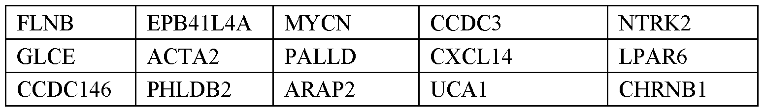

Table 4. Gene signature upregulated in MSS-CRC, serrated lesions, and tubular or tubulovillous adenoma

Table 5. Gene signature downregulated in MSS-CRC, serrated lesions, and tubular or tubulovillous adenoma

Table 6. Gene signature from RNA-seq upregulated in sessile serrated polyps (SSA/P)


Table 7. Transcriptomic signature of DKO intestinal epithelial cells


[0173] CRC patient samples (GSE5851, GSE33113, TCGA) were segregated into four subgroups: PRKCILowPRKCZLow, PRKCIHiPRKCZLow, PRKCILowPRKCZHi, and
PRKCIHiPRKCZHi based on the expression of PRKCI and PRKCZ. Median expression value of each gene was used as a cut off. PRKCILowPRKCZLow and PRKCIHiPRKCZH groups were compared by GSEA or survival analyses. To generate a Kaplan-Meier curves for TCGA patients, clinical and gene expression data of 369 patients with complete overall survival (OS) information were processed, and 5-year OS rate was compared between
PRKCILowPRKCZLow (n = 88) and PRKCIHiPRKCZH i (n = 87) groups. For GSE5851, disease free survival (DFS) rate was compared between PRKCILowPRKCZLow (n = 27) and
PRKCIHiPRKCZH (n = 27) groups.
[0174] Statistical analysis
[0175] Data are presented as the mean ± SEM. Statistical analysis was performed using GraphPad Prism 7. Significant differences between groups were determined using a Student’s t test (two-tailed unpaired) when the data met the normal distribution tested by D’Agostino test. If the data did not meet this test, a Mann-Whitney test was used. Differences in tumor incidence were analyzed by Chi-square test (Figure 4J). Differences in Kaplan-Meier plots were analyzed by Log-Rank (Mantel-Cox) test (Figure 1C, 7F, S7E). Differences in aPKC staining for human samples (Figure 7B) were analyzed by performing ANOVA multiple comparisons (Kruskal-Wallis test). The p-value indicates the difference between signal intensity (strong/moderate vs weak/negative) in normal, SSA/P, and TA. Differences in proportion of CRC patients in CCS1, CCS2, or CCS3 CRC subtype according to aPKC mRNA expression were analyzed by Fisher’s exact test for CCS3 vs CCS1/CCS2 (FIG.7E, FIG.14C, FIG.14D). The significance level for statistical testing was set at p < 0.05. Example 10. Characterizing the Effects of PVHA Hyaluronidase in a Novel Mouse Model of Colorectal Cancer
[0176] Epithelial tumor cells in the serrated (DKOIEC) mouse model display increased levels of CD44 and the stroma, that is highly activated, expressed hyaluronan (HA). HA is a ligand for CD44, which is a stem cell receptor. HA contributes to the barrier role of the stroma that makes immunotherapy ineffective for some patients. An experiment is performed to understand the effects of an inhibitor of HA in the DKOIEC mouse model to determine if tumorigenesis is inhibited without further treatment or upon co-treatment with an anti-PD-L1 therapy.
[0177]
 Villin-cre and Lgr5-EGFP-ires-creERT2 (Lgr5- creERT2) mice are acquired. and
 mice are crossed with Villin-cre mice to generate a mouse line with deletions in both aPKCs (DKOIEC) in IECs. Genotypes of DKOIEC mice are determined. Exemplary methods for determining a genotype include polymerase chain reaction (PCR).
[0178] DKOIEC mice are treated twice per week beginning at 16 weeks of age with pegvorhyaluronidase alfa (PVHA) and/or anti-PD-L1 treatment (e.g., anti-PD-L1 monoclonal antibody) for 4 weeks. Control DKOIEC mice are treated with a vehicle twice per week beginning at 16 weeks of age for 4 weeks. When co-treatment PVHA and anti-PD-L1 treatment is administered, the PVHA is administered to DKOIEC mice 24 hours prior to administration of the anti-PD-L1 treatment. Table 8 shows exemplary treatment regimens for 6 groups of DKOIEC mice.
Table 8. DKOIEC mice Exemplary Treatment Regimens

[0180] Throughout this disclosure, various embodiments are presented in a range format. It should be understood that the description in range format is merely for convenience and brevity and should not be construed as an inflexible limitation on the scope of any
embodiments. Accordingly, the description of a range should be considered to have specifically disclosed all the possible subranges as well of any dividual numerical values within that range to the tenth of the unit of the lower limit unless the context clearly dictates otherwise. For example, description of a range such as from 1 to 6 should be considered to have specifically disclosed subranges such as from 1 to 3, from 1 to 4, from 1 to 5, from 2 to 4, from 2 to 6, from 3 to 6 etc., as well of any dividual values within that range, for example, 1.1, 2, 2.3, 5, and 5.9. This applies regardless of the breadth of the range. The upper and lower limits of these intervening ranges may independently be included in the smaller ranges, and are also encompassed within the invention, subject to any specifically excluded limit in the stated range. Where the stated range includes one or both of the limits, ranges excluding either or both of those included limits are also included in the invention, unless the context clearly dictates otherwise.
[0181] The terminology used herein is for the purpose of describing particular embodiments only and is not intended to be limiting of any embodiment. As used herein, the singular forms“a,”“an” and“the” are intended to include the plural forms as well, unless the context clearly indicates otherwise. It will be further understood that the terms“comprises” and/or“comprising,” when used in this specification, specify the presence of stated features, integers, steps, operations, elements, and/or components, but do not preclude the presence or addition of one or more other features, integers, steps, operations, elements, components, and/or groups thereof. As used herein, the term“and/or” includes any and all combinations of one or more of the associated listed items.
[0182] While preferred embodiments of the present invention have been shown and described herein, it will be apparent to those skilled in the art that such embodiments are provided by way of example only. Numerous variations, changes, and substitutions will now occur to those skilled in the art without departing from the invention. It should be understood that various alternatives to the embodiments of the invention described herein may be employed in practicing the invention. It is intended that the following claims define the scope of the invention and that methods and structures within the scope of these claims and their equivalents be covered thereby.
Claims
1. A method of treating a subtype of a disease or condition in a subject by administering to the subject a therapeutically effective amount of an inhibitor of programmed death-ligand 1 (PD-L1) activity or expression, provided low levels of expression of PKCz and PKCƛ/i are detected in a biological sample obtained from the subject, as compared to levels of expression of PKCz and PKCƛ/i in an individual who does not have the disease or condition.
2. The method of claim 1, provided a high level of expression of hyaluronan is detected in a biological sample obtained from the subject, as compared to a level of expression of hyaluronan in an individual who does not have the subtype of the disease or condition.
3. The method of claim 2, wherein the high level of hyaluronan is detected by an assay comprising polymerase chain reaction (PCR), reverse transcription PCR (RT-PCR), deoxyribonucleic acid (DNA) sequencing, ribonucleic acid (RNA) sequencing, genotyping array, immunohistochemistry, enzyme-linked immunosorbent assay (ELISA), single-molecule array (Simoa), or a combination thereof.
4. The method of claim 2, wherein the high level of expression of hyaluronan comprises ribonucleic acid (RNA), deoxyribonucleic acid (DNA), or protein.
5. The method of claim 1, wherein the expression of PKCz and PKCƛ/i that is detected in the biological sample obtained from the subject is indicative of an increase in expression of hyaluronan in the biological sample, as compared to an individual who does not have the subtype of the disease or condition.
6. The method of claim 1, wherein the disease or condition comprises colorectal cancer, pancreatic cancer, inflammatory bowel disease, ulcerative colitis, Crohn’s disease, fibrosis, fibrostenosis, or a combination thereof.
7. The method of claim 1, wherein the subtype of the disease or condition comprises a
disease or condition characterized by an increase in hyaluronan in the biological sample obtained from the subject, as compared to the individual who does not have the subtype of the disease or condition.
8. The method of claim 1, wherein the subtype of the disease or condition comprises a disease or condition characterized by serrated polyps in an intestine of the subject.
9. The method of claim 8, wherein the serrated polyps comprise sessile serrated adenomas (SSAs).
10. The method of claim 1, wherein the subtype of the disease or condition comprises a
disease or condition characterized by serrated adenocarcinoma.
11. The method of claim 1, wherein the inhibitor of PD-L1 activity or expression comprises an antibody, or antigen-binding fragment.
12. The method of claim 1, wherein the inhibitor of PD-L1 activity or expression comprises a small molecule.
13. The method of claim 1, wherein the inhibitor of PD-L1 activity or expression comprises an antagonist of PD-L1.
14. The method of claim 1, wherein the inhibitor of PD-L1 activity or expression comprises an antagonist of programmed cell death protein 1 (PD-1).
15. The method of claim 1, wherein the inhibitor of PD-L1 activity or expression comprises an anti-PD-L1 antibody.
16. The method of claim 1, wherein the inhibitor of PD-L1 activity or expression comprises an anti-PD-1 antibody.
17. The method of claim 1, further comprising administering to the subject a therapeutically effective amount of an inhibitor of hyaluronan activity or expression.
18. The method of claim 17, wherein the inhibitor of hyaluronan activity or expression
comprises an antibody, or antigen-binding fragment.
19. The method of claim 17, wherein the inhibitor of hyaluronan activity or expression
comprises a small molecule.
20. The method of claim 17, wherein the inhibitor of hyaluronan activity or expression
comprises an inhibitor of hyaluronan-CD44 binding or agonism.
21. The method of claim 17, wherein the inhibitor of hyaluronan activity or expression comprises an antagonist of CD44.
22. The method of claim 17, wherein the inhibitor of hyaluronan activity or expression
comprises an antibody inhibitor of CD44 activity or expression.
23. The method of claim 17, wherein the inhibitor of hyaluronan activity or expression
comprises a small molecule inhibitor of CD44 activity or expression.
24. The method of claim 17, wherein the inhibitor of hyaluronan activity or expression
comprises an agonist of hyaluronidase activity or expression.
25. The method of claim 17, wherein the inhibitor of hyaluronan activity or expression
comprises Pegvorhyaluronidase alfa (PVHA).
26. The method of claim 17, wherein the inhibitor of hyaluronan activity or expression
comprises exogenous hyaluronidase.
27. The method of claim 17, wherein the inhibitor of hyaluronan activity or expression
comprises recombinant hyaluronidase.
28. The method of claim 1, further comprising administering to the subject a therapeutically effective amount of an inhibitor of transforming growth factor beta 1 (TGF-b1) activity or expression.
29. The method of claim 28, wherein the inhibitor of TGF- b1 activity or expression
comprises an antibody, or antigen-binding fragment.
30. The method of claim 28, wherein the inhibitor of TGF- b1 activity or expression
comprises a small molecule.
31. The method of claim 28, wherein the inhibitor of TGF- b1 activity or expression
comprises a small molecule inhibitor of a receptor of TGF- b1.
32. The method of claim 28, wherein the inhibitor of TGF- b1 activity or expression
comprises galunisertib.
33. The method of claim 1, wherein the biological sample comprises blood, blood plasma, sera, or tissue biopsy.
34. The method of claim 1, wherein the subject is a human.
35. The method of claim 1, wherein the subject is a mammal.
36. The method of claim 1, wherein the low levels of expression of PKCz and PKCƛ/i are detected by an assay comprising polymerase chain reaction (PCR), reverse transcription PCR (RT-PCR), deoxyribonucleic acid (DNA) sequencing, ribonucleic acid (RNA) sequencing, genotyping array, immunohistochemistry, enzyme-linked immunosorbent assay (ELISA), single-molecule array (Simoa), or a combination thereof.
37. The method of claim 1, wherein the levels of expression of PKCz and PKCƛ/i comprise ribonucleic acid (RNA), deoxyribonucleic acid (DNA), or protein.
38. A method of inhibiting or reducing programmed death-ligand 1 (PD-L1) activity or
expression in a subject suffering from a disease or condition, comprising:
identifying the subject as having low levels of expression of PKCz and PKCƛ/i , as compared to the levels of expression of PKCz and PKCƛ/i in an individual who does not have the disease or condition; and
administering to the subject a therapeutically effective amount of an inhibitor of PD- L1 activity or expression.
39. The method of claim 38, further comprising:
identifying the subject as having a high level of expression of hyaluronan, as
compared to the level of expression of hyaluronan in an individual who does not have the disease or condition.
40. The method of claim 39, wherein the level of expression of hyaluronan is identified by an assay comprising polymerase chain reaction (PCR), reverse transcription PCR (RT- PCR), deoxyribonucleic acid (DNA) sequencing, ribonucleic acid (RNA) sequencing, genotyping array, immunohistochemistry, enzyme-linked immunosorbent assay (ELISA), single-molecule array (Simoa), or a combination thereof.
41. The method of claim 39, wherein the level of expression of hyaluronan comprises
ribonucleic acid (RNA), deoxyribonucleic acid (DNA), or protein.
42. The method of claim 38, wherein the low levels of expression of PKCz and PKCƛ/i are indicative of an increase in expression of hyaluronan in the subject, as compared to an individual who does not have the disease or condition.
43. The method of claim 38, wherein the disease or condition comprises colorectal cancer (CRC), serrated CRC, pancreatic cancer, inflammatory bowel disease, ulcerative colitis, Crohn’s disease, fibrosis, fibrostenosis, or a combination thereof.
44. The method of claim 38, wherein the inhibitor of PD-L1 activity or expression comprises an antibody, or antigen-binding fragment.
45. The method of claim 38, wherein the inhibitor of PD-L1 activity or expression comprises a small molecule.
46. The method of claim 38, wherein the inhibitor of PD-L1 activity or expression comprises an antagonist of PD-L1.
47. The method of claim 38, wherein the inhibitor of PD-L1 activity or expression comprises an antagonist of programmed cell death protein 1 (PD-1).
48. The method of claim 38, wherein the inhibitor of PD-L1 activity or expression comprises an anti-PD-L1 antibody.
49. The method of claim 38, wherein the inhibitor of PD-L1 activity or expression comprises an anti-PD-1 antibody.
50. The method of claim 38, wherein the biological sample comprises blood, blood plasma, sera, or tissue biopsy.
51. The method of claim 38, wherein the subject is a human.
52. The method of claim 38, wherein the subject is a mammal.
53. The method of claim 38, wherein the levels of expression of PKCz and PKCƛ/iare
identified by an assay comprising polymerase chain reaction (PCR), reverse transcription PCR (RT-PCR), deoxyribonucleic acid (DNA) sequencing, ribonucleic acid (RNA) sequencing, genotyping array, immunohistochemistry, enzyme-linked immunosorbent assay (ELISA), single-molecule array (Simoa), or a combination thereof.
54. The method of claim 38, wherein the levels of expression of PKCz and PKCƛ/i comprise ribonucleic acid (RNA), deoxyribonucleic acid (DNA), or protein.
55. A method of treating a subtype of a disease or condition in a subject by administering to the subject a therapeutically effective amount of an inhibitor of hyaluronan activity or expression, provided low levels of expression of PKCz and PKCƛ/i are detected in a biological sample obtained from the subject, as compared to levels of expression of PKCz and PKCƛ/iin an individual who does not have the disease or condition.
56. The method of claim 55, wherein a high level of hyaluronan is detected in a biological sample obtained from the subject, as compared to the level of expression of hyaluronan in an individual who does not have the subtype of the disease or condition.
57. The method of claim 56, wherein the high level of hyaluronan is detected by an assay comprising polymerase chain reaction (PCR), reverse transcription PCR (RT-PCR), deoxyribonucleic acid (DNA) sequencing, ribonucleic acid (RNA) sequencing, genotyping array, immunohistochemistry, enzyme-linked immunosorbent assay (ELISA), single-molecule array (Simoa), or a combination thereof.
58. The method of claim 56, wherein the levels of expression of PKCz and PKCƛ/i are
detected comprise ribonucleic acid (RNA), deoxyribonucleic acid (DNA), or protein.
59. The method of claim 56, wherein the expression of PKCz and PCKƛ that are detected in the biological sample obtained from the subject are indicative of an increase in expression of hyaluronan in the biological sample, as compared to an individual who does not have the subtype of the disease or condition.
60. The method of claim 55, wherein the disease or condition comprises colorectal cancer, pancreatic cancer, inflammatory bowel disease, ulcerative colitis, Crohn’s disease, fibrosis, fibrostenosis, or a combination thereof.
61. The method of claim 55, wherein the subtype of the disease or condition comprises a disease or condition characterized by an increase in hyaluronan in the biological sample obtained from the subject, as compared to the individual who does not have the subtype of the disease or condition.
62. The method of claim 55, wherein the subtype of the disease or condition comprises a disease or condition characterized by serrated polyps in an intestine of the subject.
63. The method of claim 62, wherein the serrated polyps comprise sessile serrated adenomas (SSAs).
64. The method of claim 55, wherein the subtype of the disease or condition comprises a disease or condition characterized by serrated adenocarcinoma.
65. The method of claim 55, wherein the inhibitor of hyaluronan activity or expression
comprises a small molecule.
66. The method of claim 55, wherein the inhibitor of hyaluronan activity or expression
comprises an inhibitor of hyaluronan-CD44 binding or agonism.
67. The method of claim 55, wherein the inhibitor of hyaluronan activity or expression
comprises an antagonist of CD44.
68. The method of claim 55, wherein the inhibitor of hyaluronan activity or expression
comprises an antibody inhibitor of CD44 activity or expression.
69. The method of claim 55, wherein the inhibitor of hyaluronan activity or expression
comprises a small molecule inhibitor of CD44 activity or expression.
70. The method of claim 55, wherein the inhibitor of hyaluronan activity or expression
comprises an agonist of hyaluronidase activity or expression.
71. The method of claim 55, wherein the inhibitor of hyaluronan activity or expression
comprises Pegvorhyaluronidase alfa (PVHA).
72. The method of claim 55, wherein the inhibitor of hyaluronan activity or expression
comprises exogenous hyaluronidase.
73. The method of claim 55, wherein the inhibitor of hyaluronan activity or expression
comprises recombinant hyaluronidase.
74. The method of claim 55, further comprising administering to the subject a therapeutically effective amount of an inhibitor of transforming growth factor beta 1 (TGF-b1) activity or expression.
75. The method of claim 74, wherein the inhibitor of TGF- b1 activity or expression comprises an antibody, or antigen-binding fragment.
76. The method of claim 74, wherein the inhibitor of TGF- b1 activity or expression
comprises a small molecule.
77. The method of claim 74, wherein the inhibitor of TGF- b1 activity or expression
comprises a small molecule inhibitor of a receptor of TGF- b1.
78. The method of claim 74, wherein the inhibitor of TGF- b1 activity or expression
comprises galunisertib.
79. The method of claim 55, further comprising administering to the subject a therapeutically effective amount of an inhibitor of programmed death-ligand 1 (PD-L1) activity or expression.
80. The method of claim 79, wherein the inhibitor of PD-L1 activity or expression comprises an antibody, or antigen-binding fragment.
81. The method of claim 79, wherein the inhibitor of PD-L1 activity or expression comprises a small molecule.
82. The method of claim 79, wherein the inhibitor of PD-L1 activity or expression comprises an antagonist of PD-L1.
83. The method of claim 79, wherein the inhibitor of PD-L1 activity or expression comprises an antagonist of programmed cell death protein 1 (PD-1).
84. The method of claim 79, wherein the inhibitor of PD-L1 activity or expression comprises an anti-PD-L1 antibody.
85. The method of claim 79, wherein the inhibitor of PD-L1 activity or expression comprises an anti-PD-1 antibody.
86. The method of claim 55, wherein the biological sample comprises blood, blood plasma, sera, or tissue biopsy.
87. The method of claim 55, wherein the subject is a human.
88. The method of claim 55, wherein the subject is a mammal.
89. The method of claim 55, wherein the levels of expression of PKCz and PKCƛ/i are
detected by an assay comprising polymerase chain reaction (PCR), reverse transcription PCR (RT-PCR), deoxyribonucleic acid (DNA) sequencing, ribonucleic acid (RNA) sequencing, genotyping array, immunohistochemistry, enzyme-linked immunosorbent assay (ELISA), single-molecule array (Simoa), or a combination thereof.
90. The method of claim 55, wherein the levels of expression of PKCz and PKCƛ/i comprise ribonucleic acid (RNA), deoxyribonucleic acid (DNA), or protein.
91. A method of inhibiting or reducing hyaluronan activity or expression in a subject
suffering from a disease or condition, comprising:
identifying the subject as having low levels of expression of PKCz and PKCƛ/i , as compared to the levels of expression of PKCz and PKCƛ/i in an individual who does not have the disease or condition; and
administering to the subject a therapeutically effective amount of an inhibitor of
hyaluronan activity or expression.
92. The method of claim 91, further comprising:
identifying the subject as having a high level of expression of hyaluronan, as
compared to the level of expression of hyaluronan in an individual who does not have the disease or condition.
93. The method of claim 92, wherein the level of expression of hyaluronan is identified by an assay comprising polymerase chain reaction (PCR), reverse transcription PCR (RT- PCR), deoxyribonucleic acid (DNA) sequencing, ribonucleic acid (RNA) sequencing, genotyping array, immunohistochemistry, enzyme-linked immunosorbent assay (ELISA), single-molecule array (Simoa), or a combination thereof.
94. The method of claim 92, wherein the high level of expression of hyaluronan comprises ribonucleic acid (RNA), deoxyribonucleic acid (DNA), or protein.
95. The method of claim 91, wherein the low levels of expression of PKCz and PKCƛ/i are indicative of an increase in expression of hyaluronan in the subject, as compared to an individual who does not have the disease or condition.
96. The method of claim 91, wherein the disease or condition comprises colorectal cancer (CRC), serrated CRC, pancreatic cancer, inflammatory bowel disease, ulcerative colitis, Crohn’s disease, fibrosis, fibrostenosis, or a combination thereof.
97. The method of claim 91, wherein the inhibitor of hyaluronan activity or expression comprises a small molecule.
98. The method of claim 91, wherein the inhibitor of hyaluronan activity or expression
comprises an inhibitor of hyaluronan-CD44 binding or agonism.
99. The method of claim 91, wherein the inhibitor of hyaluronan activity or expression
comprises an antagonist of CD44.
100. The method of claim 90, wherein the inhibitor of hyaluronan activity or expression comprises an antibody inhibitor of CD44 activity or expression.
101. The method of claim 90, wherein the inhibitor of hyaluronan activity or expression comprises a small molecule inhibitor of CD44 activity or expression.
102. The method of claim 90, wherein the inhibitor of hyaluronan activity or expression comprises an agonist of hyaluronidase activity or expression.
103. The method of claim 90, wherein the inhibitor of hyaluronan activity or expression comprises Pegvorhyaluronidase alfa (PVHA).
104. The method of claim 90, wherein the inhibitor of hyaluronan activity or expression comprises exogenous hyaluronidase.
105. The method of claim 90, wherein the inhibitor of hyaluronan activity or expression comprises recombinant hyaluronidase.
106. The method of claim 90, further comprising administering to the subject a
therapeutically effective amount of an inhibitor of transforming growth factor beta 1 (TGF-b1) activity or expression.
107. The method of claim 105, wherein the inhibitor of TGF- b1 activity or expression comprises an antibody, or antigen-binding fragment.
108. The method of claim 105, wherein the inhibitor of TGF- b1 activity or expression comprises a small molecule.
109. The method of claim 105, wherein the inhibitor of TGF- b1 activity or expression comprises a small molecule inhibitor of a receptor of TGF- b1.
110. The method of claim 105, wherein the inhibitor of TGF- b1 activity or expression comprises galunisertib.
111. The method of claim 90, further comprising administering to the subject a
therapeutically effective amount of an inhibitor of programmed death-ligand 1 (PD-L1) activity or expression.
112. The method of claim 110, wherein the inhibitor of PD-L1 activity or expression comprises an antibody, or antigen-binding fragment.
113. The method of claim 110, wherein the inhibitor of PD-L1 activity or expression comprises a small molecule.
114. The method of claim 110, wherein the inhibitor of PD-L1 activity or expression comprises an antagonist of PD-L1.
115. The method of claim 110, wherein the inhibitor of PD-L1 activity or expression comprises an antagonist of programmed cell death protein 1 (PD-1).
116. The method of claim 110, wherein the inhibitor of PD-L1 activity or expression comprises an anti-PD-L1 antibody.
117. The method of claim 110, wherein the inhibitor of PD-L1 activity or expression comprises an anti-PD-1 antibody.
118. The method of claim 90, wherein the biological sample comprises blood, blood plasma, sera, or tissue biopsy.
119. The method of claim 90, wherein the subject is a human.
120. The method of claim 90, wherein the subject is a mammal.
121. The method of claim 90, wherein the levels of expression of PKCz and PKCƛ/i are identified by an assay comprising polymerase chain reaction (PCR), reverse transcription PCR (RT-PCR), deoxyribonucleic acid (DNA) sequencing, ribonucleic acid (RNA) sequencing, genotyping array, immunohistochemistry, enzyme-linked immunosorbent assay (ELISA), single-molecule array (Simoa), or a combination thereof.
122. The method of claim 90, wherein the levels of expression of PKCz and PKCƛ/i comprise ribonucleic acid (RNA), deoxyribonucleic acid (DNA), or protein.
123. The method of claim 90, wherein the inhibitor of hyaluronan activity or expression comprises an antibody, or antigen-binding fragment.
124. The method of claim 90, wherein the inhibitor of hyaluronan activity or expression comprises a small molecule.
125. The method of claim 90, wherein the inhibitor of hyaluronan activity or expression comprises an inhibitor of hyaluronan-CD44 binding or agonism.
126. The method of claim 90, wherein the inhibitor of hyaluronan activity or expression comprises an antagonist of CD44.
127. The method of claim 90, wherein the inhibitor of hyaluronan activity or expression comprises an antibody inhibitor of CD44 activity or expression.
128. The method of claim 90, wherein the inhibitor of hyaluronan activity or expression comprises a small molecule inhibitor of CD44 activity or expression.
129. The method of claim 90, wherein the inhibitor of hyaluronan activity or expression comprises an agonist of hyaluronidase activity or expression.
130. The method of claim 90, wherein the inhibitor of hyaluronan activity or expression comprises Pegvorhyaluronidase alfa (PVHA).
131. The method of claim 90, wherein the inhibitor of hyaluronan activity or expression comprises exogenous hyaluronidase.
132. The method of claim 90, wherein the inhibitor of hyaluronan activity or expression comprises recombinant hyaluronidase.
133. A method of ameliorating or preventing tumorigenesis in a subject with serrated colorectal cancer (CRC) by administering to the subject a therapeutically effective amount of an inhibitor of hyaluronan activity or expression, provided low levels of expression of PKCz and PKCƛ/i are detected in a biological sample obtained from the subject, as compared to levels of expression of PKCz and PKCƛ/i in an individual who does not have serrated CRC.
134. The method of claim 132, provided a high level of expression of hyaluronan is detected in a biological sample obtained from the subject, as compared to a level of expression of hyaluronan in an individual who does not have the CRC.
135. The method of claim 133, wherein the high level of expression of hyaluronan is detected by an assay comprising polymerase chain reaction (PCR), reverse transcription PCR (RT-PCR), deoxyribonucleic acid (DNA) sequencing, ribonucleic acid (RNA) sequencing, genotyping array, immunohistochemistry, enzyme-linked immunosorbent assay (ELISA), single-molecule array (Simoa), or a combination thereof.
136. The method of claim 133, wherein the high level of expression of hyaluronan
comprises ribonucleic acid (RNA), deoxyribonucleic acid (DNA), or protein.
137. The method of claim 132, wherein the low levels of expression of PKCz and PCKƛ that are detected in the biological sample obtained from the subject are indicative of an increase in expression of hyaluronan in the biological sample, as compared to an individual who does not have the serrated CRC.
138. The method of claim 132, wherein the inhibitor of hyaluronan activity or expression comprises a small molecule.
139. The method of claim 132, wherein the inhibitor of hyaluronan activity or expression comprises an inhibitor of hyaluronan-CD44 binding or agonism.
140. The method of claim 132, wherein the inhibitor of hyaluronan activity or expression comprises an antagonist of CD44.
141. The method of claim 132, wherein the inhibitor of hyaluronan activity or expression comprises an antibody inhibitor of CD44 activity or expression.
142. The method of claim 132, wherein the inhibitor of hyaluronan activity or expression comprises a small molecule inhibitor of CD44 activity or expression.
143. The method of claim 132, wherein the inhibitor of hyaluronan activity or expression comprises an agonist of hyaluronidase activity or expression.
144. The method of claim 132, wherein the inhibitor of hyaluronan activity or expression comprises Pegvorhyaluronidase alfa (PVHA).
145. The method of claim 132, wherein the inhibitor of hyaluronan activity or expression comprises exogenous hyaluronidase.
146. The method of claim 132, wherein the inhibitor of hyaluronan activity or expression comprises recombinant hyaluronidase.
147. The method of claim 132, further comprising administering to the subject a
therapeutically effective amount of an inhibitor of transforming growth factor beta 1 (TGF-b1) activity or expression.
148. The method of claim 145, wherein the inhibitor of TGF- b1 activity or expression comprises an antibody, or antigen-binding fragment.
149. The method of claim 145, wherein the inhibitor of TGF- b1 activity or expression comprises a small molecule.
150. The method of claim 145, wherein the inhibitor of TGF- b1 activity or expression comprises a small molecule inhibitor of a receptor of TGF- b1.
151. The method of claim 145, wherein the inhibitor of TGF- b1 activity or expression comprises galunisertib.
152. The method of claim 132, further comprising administering to the subject a
therapeutically effective amount of an inhibitor of programmed death-ligand 1 (PD-L1) activity or expression.
153. The method of claim 150, wherein the inhibitor of PD-L1 activity or expression comprises an antibody, or antigen-binding fragment.
154. The method of claim 150, wherein the inhibitor of PD-L1 activity or expression comprises a small molecule.
155. The method of claim 150, wherein the inhibitor of PD-L1 activity or expression
comprises an antagonist of PD-L1.
156. The method of claim 150, wherein the inhibitor of PD-L1 activity or expression
comprises an antagonist of programmed cell death protein 1 (PD-1).
157. The method of claim 150, wherein the inhibitor of PD-L1 activity or expression
comprises an anti-PD-L1 antibody.
158. The method of claim 150, wherein the inhibitor of PD-L1 activity or expression
comprises an anti-PD-1 antibody.
159. The method of claim 132, wherein the biological sample comprises blood, blood plasma, sera, or tissue biopsy.
160. The method of claim 132, wherein the subject is a human.
161. The method of claim 132, wherein the subject is a mammal.
162. The method of claim 132, wherein the low levels of expression of PKCz and PKCƛ/i are detected by an assay comprising polymerase chain reaction (PCR), reverse transcription PCR (RT-PCR), deoxyribonucleic acid (DNA) sequencing, ribonucleic acid (RNA) sequencing, genotyping array, immunohistochemistry, enzyme-linked
immunosorbent assay (ELISA), single-molecule array (Simoa), or a combination thereof.
163. The method of claim 132, wherein the levels of expression of PKCz and PKCƛ/i comprise ribonucleic acid (RNA), deoxyribonucleic acid (DNA), or protein.
164. A method of ameliorating or preventing tumorigenesis in a subject with serrated colorectal cancer (CRC) by, the method comprising:
identifying the subject as having low levels of expression of PKCz and PKCƛ/i , as compared to the levels of expression of PKCz and PKCƛ/i in an individual who does not have the serrated CRC; and
administering to the subject a therapeutically effective amount of an inhibitor of hyaluronan activity or expression.
165. The method of claim 162, further comprising:
identifying the subject as having a high level of expression of hyaluronan, as
compared to the level of expression of hyaluronan in an individual who does not have the CRC.
166. The method of claim 163, wherein the high level of expression of hyaluronan is identified by an assay comprising polymerase chain reaction (PCR), reverse transcription PCR (RT-PCR), deoxyribonucleic acid (DNA) sequencing, ribonucleic acid (RNA) sequencing, genotyping array, immunohistochemistry, enzyme-linked immunosorbent assay (ELISA), single-molecule array (Simoa), or a combination thereof.
167. The method of claim 163, wherein the level of expression of hyaluronan comprises ribonucleic acid (RNA), deoxyribonucleic acid (DNA), or protein.
168. The method of claim 162, wherein the low levels of expression of PKCz and PKCƛ/i are indicative of an increase in expression of hyaluronan in the subject, as compared to an individual who does not have the disease or condition.
169. The method of claim 162, wherein the biological sample comprises blood, blood plasma, sera, or tissue biopsy.
170. The method of claim 162, wherein the subject is a human.
171. The method of claim 162, wherein the subject is a mammal.
172. The method of claim 162, wherein the levels of expression of PKCz and PKCƛ/i are identified by an assay comprising polymerase chain reaction (PCR), reverse transcription PCR (RT-PCR), deoxyribonucleic acid (DNA) sequencing, ribonucleic acid (RNA) sequencing, genotyping array, immunohistochemistry, enzyme-linked immunosorbent assay (ELISA), single-molecule array (Simoa), or a combination thereof.
173. The method of claim 162, wherein the levels of expression of PKCz and PKCƛ/i comprise ribonucleic acid (RNA), deoxyribonucleic acid (DNA), or protein.
174. The method of claim 162, wherein the inhibitor of hyaluronan activity or expression comprises an antibody, or antigen-binding fragment.
175. The method of claim 162, wherein the inhibitor of hyaluronan activity or expression comprises a small molecule.
176. The method of claim 162, wherein the inhibitor of hyaluronan activity or expression comprises an inhibitor of hyaluronan-CD44 binding or agonism.
177. The method of claim 162, wherein the inhibitor of hyaluronan activity or expression comprises an antagonist of CD44.
178. The method of claim 162, wherein the inhibitor of hyaluronan activity or expression comprises an antibody inhibitor of CD44 activity or expression.
179. The method of claim 162, wherein the inhibitor of hyaluronan activity or expression comprises a small molecule inhibitor of CD44 activity or expression.
180. The method of claim 162, wherein the inhibitor of hyaluronan activity or expression comprises an agonist of hyaluronidase activity or expression.
181. The method of claim 162, wherein the inhibitor of hyaluronan activity or expression comprises Pegvorhyaluronidase alfa (PVHA).
182. The method of claim 162, wherein the inhibitor of hyaluronan activity or expression comprises exogenous hyaluronidase.
183. The method of claim 162, wherein the inhibitor of hyaluronan activity or expression comprises recombinant hyaluronidase.
184. The method of claim 162, further comprising administering to the subject a
therapeutically effective amount of an inhibitor of transforming growth factor beta 1 (TGF-b1) activity or expression.
185. The method of claim 182, wherein the inhibitor of TGF- b1 activity or expression comprises an antibody, or antigen-binding fragment.
186. The method of claim 182, wherein the inhibitor of TGF- b1 activity or expression comprises a small molecule.
187. The method of claim 182, wherein the inhibitor of TGF- b1 activity or expression comprises a small molecule inhibitor of a receptor of TGF- b1.
188. The method of claim 182, wherein the inhibitor of TGF- b1 activity or expression comprises galunisertib.
189. The method of claim 182, further comprising administering to the subject a
therapeutically effective amount of an inhibitor of programmed death-ligand 1 (PD-L1) activity or expression.
190. The method of claim 187, wherein the inhibitor of PD-L1 activity or expression comprises an antibody, or antigen-binding fragment.
191. The method of claim 187, wherein the inhibitor of PD-L1 activity or expression comprises a small molecule.
192. The method of claim 187, wherein the inhibitor of PD-L1 activity or expression comprises an antagonist of PD-L1.
193. The method of claim 187, wherein the inhibitor of PD-L1 activity or expression comprises an antagonist of programmed cell death protein 1 (PD-1).
194. The method of claim 187, wherein the inhibitor of PD-L1 activity or expression comprises an anti-PD-L1 antibody.
195. The method of claim 187, wherein the inhibitor of PD-L1 activity or expression comprises an anti-PD-1 antibody.
196. A method of diagnosing a subtype of colorectal cancer (CRC) in a subject in need thereof, the method comprising:
obtaining a biological sample from a subject in need thereof;
subjecting the biological sample to an assay suitable to detect levels of expression of PKCz and PKCƛ/i ; and
diagnosing the subject with serrated colorectal cancer (CRC), provided the levels of expression of PKCz and PKCƛ/i detected in the biological sample obtained from the subject are low, as compared to levels of expression of PKCz and PKCƛ/i in an individual that does not have the CRC.
197. The method of claim 194, further comprising:
subjecting the biological sample to an assay suitable to detect a colon cancer subtype (CCS), the CCS comprising chromosomal unstable phenotype (CCS1), microsatellite instability-high and CpG island methylator phenotypes
(MSI/CIMP+), or microsatellite stable phenotype (MSS); and diagnosing the subject with serrated CRC, provided the CCS detected comprises the MSS phenotype.
198. The method of claim 194, wherein the low levels of expression of PKCz and PKCƛ/i are indicative of an increase in expression of hyaluronan in the subject, as compared to an individual who does not have the disease or condition.
199. The method of claim 194, wherein the biological sample comprises blood, blood plasma, sera, or tissue biopsy.
200. The method of claim 194, wherein the subject is a human.
201. The method of claim 194, wherein the subject is a mammal.
202. The method of claim 194, wherein the assay suitable to detect the levels of expression of PKCz and PKCƛ/i comprises polymerase chain reaction (PCR), reverse transcription PCR (RT-PCR), deoxyribonucleic acid (DNA) sequencing, ribonucleic acid (RNA) sequencing, genotyping array, immunohistochemistry, enzyme-linked immunosorbent assay (ELISA), single-molecule array (Simoa), or a combination thereof.
203. The method of claim 194, wherein the levels of expression of PKCz and PKCƛ/i comprise ribonucleic acid (RNA), deoxyribonucleic acid (DNA), or protein.
204. The method of claim 194, further comprising treating the subject by administering a therapeutically effective amount of an inhibitor of transforming growth factor beta 1 (TGF-b1) activity or expression, provided the subject is diagnosed with serrated CRC.
205. The method of claim 202, wherein the inhibitor of TGF- b1 activity or expression comprises an antibody, or antigen-binding fragment.
206. The method of claim 202, wherein the inhibitor of TGF- b1 activity or expression comprises a small molecule.
207. The method of claim 202, wherein the inhibitor of TGF- b1 activity or expression comprises a small molecule inhibitor of a receptor of TGF- b1.
208. The method of claim 202, wherein the inhibitor of TGF- b1 activity or expression comprises galunisertib.
209. The method of claim 202, further comprising treating the subject by administering a therapeutically effective amount of an inhibitor of hyaluronan activity or expression, provided the subject is diagnosed with serrated CRC.
210. The method of claim 194, wherein the inhibitor of hyaluronan activity or expression comprises an antibody, or antigen-binding fragment.
211. The method of claim 208, wherein the inhibitor of hyaluronan activity or expression comprises a small molecule.
212. The method of claim 208, wherein the inhibitor of hyaluronan activity or expression comprises an inhibitor of hyaluronan-CD44 binding or agonism.
213. The method of claim 208, wherein the inhibitor of hyaluronan activity or expression comprises an antagonist of CD44.
214. The method of claim 208, wherein the inhibitor of hyaluronan activity or expression comprises an antibody inhibitor of CD44 activity or expression.
215. The method of claim 208, wherein the inhibitor of hyaluronan activity or expression comprises a small molecule inhibitor of CD44 activity or expression.
216. The method of claim 208, wherein the inhibitor of hyaluronan activity or expression comprises an agonist of hyaluronidase activity or expression.
217. The method of claim 208, wherein the inhibitor of hyaluronan activity or expression comprises Pegvorhyaluronidase alfa (PVHA).
218. The method of claim 208, wherein the inhibitor of hyaluronan activity or expression comprises exogenous hyaluronidase.
219. The method of claim 208, wherein the inhibitor of hyaluronan activity or expression comprises recombinant hyaluronidase.
220. The method of claim 194, further comprising treating the subject by administering a therapeutically effective amount of an inhibitor of programmed death-ligand 1 (PD-L1) activity or expression, provided the subject is diagnosed with serrated CRC.
221. The method of claim 218, wherein the inhibitor of PD-L1 activity or expression comprises an antibody, or antigen-binding fragment.
222. The method of claim 218, wherein the inhibitor of PD-L1 activity or expression comprises a small molecule.
223. The method of claim 218, wherein the inhibitor of PD-L1 activity or expression comprises an antagonist of PD-L1.
224. The method of claim 218, wherein the inhibitor of PD-L1 activity or expression comprises an antagonist of programmed cell death protein 1 (PD-1).
225. The method of claim 218, wherein the inhibitor of PD-L1 activity or expression comprises an anti-PD-L1 antibody.
226. The method of claim 218, wherein the inhibitor of PD-L1 activity or expression comprises an anti-PD-1 antibody.
227. A method of characterizing a subtype of colorectal cancer (CRC) in a subject, the method comprising:
obtaining a biological sample from a subject with colorectal cancer (CRC);
subjecting the biological sample to an assay suitable to detect levels of expression of PKCz and PKCƛ/i ; and
characterizing the CRC as serrated CRC, provided the levels of expression of PKCz and PKCƛ/i detected in the biological sample obtained from the subject are low, as compared to the levels of expression of PKCz and PKCƛ/i in an individual that does not have the CRC.
228. The method of claim 225, further comprising:
subjecting the biological sample to an assay suitable to detect a colon cancer subtype (CCS), the CCS comprising chromosomal unstable phenotype (CCS1),
microsatellite instability-high and CpG island methylator phenotypes
(MSI/CIMP+), or microsatellite stable phenotype (MSS); and characterizing the subtype of CRC as serrated CRC, provided the CCS detected
comprises the MSS phenotype.
229. The method of claim 225, wherein the low levels of expression of PKCz and PKCƛ/i are indicative of an increase in expression of hyaluronan in the subject, as compared to an individual who does not have the disease or condition.
230. The method of claim 225, wherein the biological sample comprises blood, blood plasma, sera, or tissue biopsy.
231. The method of claim 225, wherein the subject is a human.
232. The method of claim 225, wherein the subject is a mammal.
233. The method of claim 225, wherein the assay suitable to detect the levels of expression of PKCz and PKCƛ/i comprises polymerase chain reaction (PCR), reverse transcription PCR (RT-PCR), deoxyribonucleic acid (DNA) sequencing, ribonucleic acid (RNA) sequencing, genotyping array, immunohistochemistry, enzyme-linked immunosorbent assay (ELISA), single-molecule array (Simoa), or a combination thereof.
234. The method of claim 225, wherein the levels of expression of PKCz and PKCƛ/i comprise ribonucleic acid (RNA), deoxyribonucleic acid (DNA), or protein.
235. The method of claim 225, further comprising treating the subject by administering a therapeutically effective amount of an inhibitor of transforming growth factor beta 1 (TGF-b1) activity or expression, provided the CRC is characterized as serrated CRC.
236. The method of claim 233, wherein the inhibitor of TGF- b1 activity or expression comprises an antibody, or antigen-binding fragment.
237. The method of claim 233, wherein the inhibitor of TGF- b1 activity or expression comprises a small molecule.
238. The method of claim 233, wherein the inhibitor of TGF- b1 activity or expression comprises a small molecule inhibitor of a receptor of TGF- b1.
239. The method of claim 233, wherein the inhibitor of TGF- b1 activity or expression comprises galunisertib.
240. The method of claim 225, further comprising treating the subject by administering a therapeutically effective amount of an inhibitor of hyaluronan activity or expression, provided the CRC is characterized as serrated CRC.
241. The method of claim 239, wherein the inhibitor of hyaluronan activity or expression comprises an antibody, or antigen-binding fragment.
242. The method of claim 239, wherein the inhibitor of hyaluronan activity or expression comprises a small molecule.
243. The method of claim 239, wherein the inhibitor of hyaluronan activity or expression comprises an inhibitor of hyaluronan-CD44 binding or agonism.
244. The method of claim 239, wherein the inhibitor of hyaluronan activity or expression comprises an antagonist of CD44.
245. The method of claim 239, wherein the inhibitor of hyaluronan activity or expression comprises an antibody inhibitor of CD44 activity or expression.
246. The method of claim 239, wherein the inhibitor of hyaluronan activity or expression comprises a small molecule inhibitor of CD44 activity or expression.
247. The method of claim 239, wherein the inhibitor of hyaluronan activity or expression comprises an agonist of hyaluronidase activity or expression.
248. The method of claim 239, wherein the inhibitor of hyaluronan activity or expression comprises Pegvorhyaluronidase alfa (PVHA).
249. The method of claim 239, wherein the inhibitor of hyaluronan activity or expression comprises exogenous hyaluronidase.
250. The method of claim 239, wherein the inhibitor of hyaluronan activity or expression comprises recombinant hyaluronidase.
251. The method of claim 225, further comprising treating the subject by administering a therapeutically effective amount of an inhibitor of programmed death-ligand 1 (PD-L1) activity or expression, provided the CRC is characterized as being serrated CRC.
252. The method of claim 249, wherein the inhibitor of PD-L1 activity or expression
comprises an antibody, or antigen-binding fragment.
253. The method of claim 251, wherein the inhibitor of PD-L1 activity or expression
comprises a small molecule.
254. The method of claim 249, wherein the inhibitor of PD-L1 activity or expression
comprises an antagonist of PD-L1.
255. The method of claim 249, wherein the inhibitor of PD-L1 activity or expression
comprises an antagonist of programmed cell death protein 1 (PD-1).
256. The method of claim 249, wherein the inhibitor of PD-L1 activity or expression
comprises an anti-PD-L1 antibody.
257. The method of claim 249, wherein the inhibitor of PD-L1 activity or expression
comprises an anti-PD-1 antibody.
258. A method of determining whether a subject suffering from an inflammatory bowel disease (IBD) has, or will, develop serrated colorectal cancer (CRC), the method comprising:
obtaining a biological sample from the subject;
subjecting the biological sample to an assay suitable to detect levels of expression of PKCz and PKCƛ/i ; and
determining that the subject has, or will develop, serrated CRC, provided the levels of expression of PKCz and PKCƛ/i detected in the biological sample obtained from the subject are low, as compared to levels of expression of PKCz and PKCƛ/i in an individual that does not have CRC.
259. The method of claim 256, wherein the IBD comprises Crohn’s disease, ulcerative colitis, medically refractory ulcerative colitis, medically refractory or complicated form of Crohn’s disease, colitis, fibrosis, fibrostenosis, or a combination thereof.
260. The method of claim 256, wherein the low levels of expression of PKCz and PKCƛ/i are indicative of an increase in expression of hyaluronan in the subject, as compared to an individual who does not have the disease or condition.
261. The method of claim 256, wherein the biological sample comprises blood, blood
plasma, sera, or tissue biopsy.
262. The method of claim 256, wherein the subject is a human.
263. The method of claim 258, wherein the subject is a mammal.
264. The method of claim 256, wherein the assay suitable to detect levels of expression of PKCz and PKCƛ/i comprises polymerase chain reaction (PCR), reverse transcription PCR (RT-PCR), deoxyribonucleic acid (DNA) sequencing, ribonucleic acid (RNA) sequencing, genotyping array, immunohistochemistry, enzyme-linked immunosorbent assay (ELISA), single-molecule array (Simoa), or a combination thereof.
265. The method of claim 256, wherein the levels of expression of PKCz and PKCƛ/i
comprise ribonucleic acid (RNA), deoxyribonucleic acid (DNA), or protein.
266. The method of claim 256, further comprising treating the subject by administering a therapeutically effective amount of an inhibitor of transforming growth factor beta 1 (TGF-b1) activity or expression, provided the subject is determined to have, or will develop, serrated CRC.
267. The method of claim 264, wherein the inhibitor of TGF- b1 activity or expression comprises an antibody, or antigen-binding fragment.
268. The method of claim 264, wherein the inhibitor of TGF- b1 activity or expression comprises a small molecule.
269. The method of claim 264, wherein the inhibitor of TGF- b1 activity or expression comprises a small molecule inhibitor of a receptor of TGF- b1.
270. The method of claim 264, wherein the inhibitor of TGF- b1 activity or expression comprises galunisertib.
271. The method of claim 256, further comprising treating the subject by administering a therapeutically effective amount of an inhibitor of hyaluronan activity or expression, provided the subject is determined to have, or will develop, serrated CRC.
272. The method of claim 269, wherein the inhibitor of hyaluronan activity or expression comprises an antibody, or antigen-binding fragment.
273. The method of claim 269, wherein the inhibitor of hyaluronan activity or expression comprises a small molecule.
274. The method of claim 269, wherein the inhibitor of hyaluronan activity or expression comprises an inhibitor of hyaluronan-CD44 binding or agonism.
275. The method of claim 269, wherein the inhibitor of hyaluronan activity or expression comprises an antagonist of CD44.
276. The method of claim 269, wherein the inhibitor of hyaluronan activity or expression comprises an antibody inhibitor of CD44 activity or expression.
277. The method of claim 269, wherein the inhibitor of hyaluronan activity or expression comprises a small molecule inhibitor of CD44 activity or expression.
278. The method of claim 269, wherein the inhibitor of hyaluronan activity or expression comprises an agonist of hyaluronidase activity or expression.
279. The method of claim 269, wherein the inhibitor of hyaluronan activity or expression comprises Pegvorhyaluronidase alfa (PVHA).
280. The method of claim 269, wherein the inhibitor of hyaluronan activity or expression comprises exogenous hyaluronidase.
281. The method of claim 269, wherein the inhibitor of hyaluronan activity or expression comprises recombinant hyaluronidase.
282. The method of claim 256, further comprising treating the subject by administering a therapeutically effective amount of an inhibitor of programmed death-ligand 1 (PD-L1) activity or expression, provided the subject is determined to have, or will develop, serrated CRC.
283. The method of claim 280, wherein the inhibitor of PD-L1 activity or expression comprises an antibody, or antigen-binding fragment.
284. The method of claim 280, wherein the inhibitor of PD-L1 activity or expression comprises a small molecule.
285. The method of claim 280, wherein the inhibitor of PD-L1 activity or expression comprises an antagonist of PD-L1.
286. The method of claim 280, wherein the inhibitor of PD-L1 activity or expression comprises an antagonist of programmed cell death protein 1 (PD-1).
287. The method of claim 280, wherein the inhibitor of PD-L1 activity or expression comprises an anti-PD-L1 antibody.
288. The method of claim 280, wherein the inhibitor of PD-L1 activity or expression comprises an anti-PD-1 antibody.
289. A model of serrated colorectal cancer comprising:
a quantity of cells that do not express, or underexpress, PKCz and PKCƛ/i ;
an animal that has been injected with the quantity of the cells that do not express, or underexpress, PKCz and PKCƛ/i ; or
a transgenic animal with a transgene comprising a knockout or knockdown of Prkci and Prkcz.
290. The model of claim 287, wherein the quantity of cells and the transgenic animal are further characterized by a microsatellite stable phenotype.
291. The model of claim 287, wherein the quantity of cells and the transgenic animal are enriched for an expression of one or more genes in the Extracellular Signal-Regulated Kinase (ERK) pathway and/or Yes Associated Protein (YAP) pathway.
292. The model of claim 287, wherein the quantity of cells exhibit lower cell death, as compared to a quantity of cells that do not express, or underexpress, PKCz or PKCƛ/i .
293. The model of claim 287, wherein the transgenic animal exhibits lower cell death, as compared to a transgenic animal with a transgene comprising a knockout or knockdown of Prkci or Prkcz.
294. The model of claim 287, wherein the quantity of cells overexpress hyaluronan.
295. The model of claim 287, wherein the transgenic animal overexpresses hyaluronan in stroma of serrated tumors of the transgenic animal.
296. The model of claim 287, wherein the quantity of cells overexpress CD44.
297. The model of claim 287, wherein the transgenic animal overexpresses CD44.
298. The model of claim 287, wherein the quantity of cells underexpress stem cell
biomarkers comprising Olfm4, Ascl2, or Lgr5.
299. The model of claim 287, wherein the transgenic animal underexpresses a stem cell biomarker comprising Olfm4, Ascl2, or Lgr5.
300. The model of claim 287, wherein the transgenic animal underexpresses an interferon- responsive gene comprising Irf7, Oas1a, Isg15, or Cxcl10.
301. The model of claim 287, wherein the quantity of cells underexpresses an interferon- responsive gene comprising Irf7, Oas1a, Isg15, or Cxcl10.
302. The model of claim 287, wherein the transgenic animal comprises a mammal.
303. The model of claim 287, wherein the transgenic animal comprises a mouse.
304. The model of claim 287, wherein the transgenic animal comprises a rat.
305. The model of claim 287, wherein the transgenic animal comprises a monkey.
306. The model of claim 287, wherein the quantity of cells comprises an organoid.
307. The model of claim 287, wherein the quantity of cells comprises a 3D culture.
308. The model of claim 287, wherein the transgenic animal is produced by breeding a first animal comprising a transgene encoding a recombinase operably linked to a ubiquitous promoter in villus and crypt epithelial cells of a small and a large intestine, and a second
animal comprising a loxP-flanked genes comprising Prkci and Prkcz, to produce a transgenic animal with a knockout or knockdown of Prkci and Prkcz, respectively, the villus and the crypt epithelial cells of the small and the large intestine.
309. The model of claim 306, wherein the ubiquitous promoter comprises a vasa promoter, c-kit promoter, Dnd1 promoter, Dppa3 promoter, Fkbp6 promoter, Fragilis promoter, Fragilis-2 promoter, GDF-3 promoter, Mov10l1 promoter, Nanog promoter, Nanos2 promoter, Nanos3 promoter, oct3/4 promoter, Prdm1 promoter, Prdm14 promoter, Tex13 promoter, Tiar promoter or TNAP promoter.
310. A method of screening for modulators of serrated tumorigenesis, the method
comprising:
providing the model of serrated colorectal cancer of any one or claims 289-310;
contacting the model with a putative modulator of tumorigenesis; and
detecting one or more changes in one or more tumor-associated parameters in the
model.
311. The method of claim 308, wherein the tumor-associated parameter comprises
recruitment of CD8+ T cells to a serrated tumor, progression of a sessile serrated adenoma (SSA)to adenocarcinoma, a number of serrated tumors, a size of serrated tumors, a total load of serrated tumors, an increase in an expression of alpha-smooth muscle actin (aSMA), or collagen deposition, or a combination thereof.
312. The method of any one or claims 308-309, wherein the putative modulator negatively modulates tumorigenesis, provided an increase in CD8+ T cells to a serrated tumor is observed.
313. The method of any one or claims 308-310, wherein the putative modulator negatively modulates tumorigenesis, provided the progression of the SSA to adenocarcinoma is not observed.
314. The method of any one or claims 308-311, wherein the putative modulator negatively modulates tumorigenesis, provided that a decrease in a number of serrated tumors, as compared to the number of serrated tumors in the model before contacting the model with the putative modulator, is observed.
315. The method of any one or claims 308-312, wherein the putative modulator negatively modulates tumorigenesis, provided that a reduction in a size of serrated tumor, as compared to the size of serrated tumors in the model before contacting the model with the putative modulator, is observed.
316. The method of any one or claims 308-313, wherein the putative modulator negatively modulates tumorigenesis, provided that a decrease in a total load of serrated tumors, as compared to the total load of serrated tumors in the model before contacting the model with the putative modulator, is observed.
317. The method of any one or claims 308-314, wherein the putative modulator negatively modulates tumorigenesis, provided that an increase in expression of aSMA, as compared to the expression of aSMA in the model before contacting the model with the putative modulator, is not observed.
318. The method of any one or claims 308-315, wherein the putative modulator negatively modulates tumorigenesis, provided than an increase in collagen deposition in the model, as compared to collagen deposition in the model before contacting the model with the putative modulator, is not observed.
319. The method of any one or claims 308-316, wherein the putative modulator is selected from the group consisting of an inhibitor of programmed death-ligand 1 (PD-L1) activity or expression, an inhibitor of transforming growth factor beta 1 (TGF-b1) activity or expression, and an inhibitor of hyaluronan activity or expression.
320. The method of claim 317, wherein the inhibitor of PD-L1 activity or expression
comprises an antibody, or antigen-binding fragment.
321. The method of claim 317, wherein the inhibitor of PD-L1 activity or expression
comprises a small molecule.
322. The method of claim 317, wherein the inhibitor of PD-L1 activity or expression
comprises an antagonist of PD-L1.
323. The method of claim 317, wherein the inhibitor of PD-L1 activity or expression
comprises an antagonist of programmed cell death protein 1 (PD-1).
324. The method of claim 317, wherein the inhibitor of PD-L1 activity or expression comprises an anti-PD-L1 antibody.
325. The method of claim 317, wherein the inhibitor of PD-L1 activity or expression comprises an anti-PD-1 antibody.
326. The method of claim 317, wherein the inhibitor of hyaluronan activity or expression comprises an antibody, or antigen-binding fragment.
327. The method of claim 317, wherein the inhibitor of hyaluronan activity or expression comprises a small molecule.
328. The method of claim 317, wherein the inhibitor of hyaluronan activity or expression comprises an inhibitor of hyaluronan-CD44 binding or agonism.
329. The method of claim 317, wherein the inhibitor of hyaluronan activity or expression comprises an antagonist of CD44.
330. The method of claim 317, wherein the inhibitor of hyaluronan activity or expression comprises an antibody inhibitor of CD44 activity or expression.
331. The method of claim 317, wherein the inhibitor of hyaluronan activity or expression comprises a small molecule inhibitor of CD44 activity or expression.
332. The method of claim 317, wherein the inhibitor of hyaluronan activity or expression comprises an agonist of hyaluronidase activity or expression.
333. The method of claim 317, wherein the inhibitor of hyaluronan activity or expression comprises Pegvorhyaluronidase alfa (PVHA).
334. The method of claim 317, wherein the inhibitor of hyaluronan activity or expression comprises exogenous hyaluronidase.
335. The method of claim 317, wherein the inhibitor of hyaluronan activity or expression comprises recombinant hyaluronidase.
336. The method of claim 317, wherein the inhibitor of TGF- b1 activity or expression comprises an antibody, or antigen-binding fragment.
337. The method of claim 317, wherein the inhibitor of TGF- b1 activity or expression comprises a small molecule.
338. The method of claim 317, wherein the inhibitor of TGF- b1 activity or expression comprises a small molecule inhibitor of a receptor of TGF- b1.
339. The method of claim 317, wherein the inhibitor of TGF- b1 activity or expression comprises galunisertib.
340. A method of producing an animal model of serrated colorectal cancer comprising: providing an animal that is Prkci fl/fl and Prkcz fl/fl, wherein the animal has loxP sites flanking genes comprising Prkci and Prkcz; and
crossing the animal that is Prkci fl/fl and Prkcz fl/fl with an animal expressing a Cre recombinase operably linked to an ubiquitous promoter in villus and crypt epithelial cells of a small and a large intestine, thereby generating a transgenic animal with deletions in Pkcz and PKCƛ/i in the villus and the crypt epithelial cells of the small and the large intestine.
341. The method of claim 338, wherein the animal model comprises a mammal.
342. The method of claim 338, wherein the animal model comprises a mouse.
343. The method of claim 338, wherein the animal model comprises a rat.
344. The method of claim 338, wherein the animal model comprises a monkey.
345. The method of claim 338, wherein the animal model is characterized by a
microsatellite stable phenotype.
346. The method of claim 338, wherein the animal model is enriched for an expression of one or more genes in the Extracellular Signal-Regulated Kinase (ERK) pathway and/or Yes Associated Protein (YAP) pathway.
347. The method of claim 338, wherein the animal model exhibits lower cell death, as compared to an animal that does not express, or underexpress, PKCz or PKCƛ/i .
348. The method of claim 338, wherein the animal model exhibits lower cell death, as compared to an animal with a transgene comprising a knockout or knockdown of Prkci or Prkcz.
349. The method of claim 338, wherein the animal model overexpresses hyaluronan in stroma of serrated tumors of the animal.
350. The method of claim 338, wherein the animal model overexpresses CD44, as
compared to a normal animal.
351. The method of claim 338, wherein the animal model underexpresses a stem cell
biomarker comprising Olfm4, Ascl2, or Lgr5, as compared to a normal animal.
352. The method of claim 338, wherein the animal model underexpresses an interferon- responsive gene comprising Irf7, Oas1a, Isg15, or Cxcl10, as compared to a normal animal.
Priority Applications (2)
| Application Number | Priority Date | Filing Date | Title |
|---|---|---|---|
| US17/312,855 US20220177561A1 (en) | 2018-12-11 | 2019-12-10 | Models and Methods Useful for the Treatment of Serrated Colorectal Cancer |
| EP19895401.8A EP3893902A4 (en) | 2018-12-11 | 2019-12-10 | Models and methods useful for the treatment of serrated colorectal cancer |
Applications Claiming Priority (2)
| Application Number | Priority Date | Filing Date | Title |
|---|---|---|---|
| US201862778124P | 2018-12-11 | 2018-12-11 | |
| US62/778,124 | 2018-12-11 |
Publications (2)
| Publication Number | Publication Date |
|---|---|
| WO2020123543A2 true WO2020123543A2 (en) | 2020-06-18 |
| WO2020123543A3 WO2020123543A3 (en) | 2020-07-23 |
Family
ID=71077523
Family Applications (1)
| Application Number | Title | Priority Date | Filing Date |
|---|---|---|---|
| PCT/US2019/065529 WO2020123543A2 (en) | 2018-12-11 | 2019-12-10 | Models and methods useful for the treatment of serrated colorectal cancer |
Country Status (3)
| Country | Link |
|---|---|
| US (1) | US20220177561A1 (en) |
| EP (1) | EP3893902A4 (en) |
| WO (1) | WO2020123543A2 (en) |
Cited By (3)
| Publication number | Priority date | Publication date | Assignee | Title |
|---|---|---|---|---|
| WO2023044501A3 (en) * | 2021-09-20 | 2023-04-20 | Cornell University | Methods for treating a subtype of colorectal cancer |
| CN117186202A (en) * | 2023-11-08 | 2023-12-08 | 北京大学第三医院(北京大学第三临床医学院) | Novel cytokine CSBF and its use in dry eye treatment |
| EP4121098A4 (en) * | 2020-03-20 | 2024-01-17 | Sanford Burnham Prebys Medical Discovery Inst | Compositions and methods for treating serrated colorectal cancer |
Families Citing this family (1)
| Publication number | Priority date | Publication date | Assignee | Title |
|---|---|---|---|---|
| CN112220925A (en) * | 2020-09-29 | 2021-01-15 | 重庆医科大学附属第一医院 | Application of MST1 as drug target in preparation of drug for treating colorectal cancer |
Citations (4)
| Publication number | Priority date | Publication date | Assignee | Title |
|---|---|---|---|---|
| US20090170133A1 (en) | 2005-04-19 | 2009-07-02 | Irena Kirman | Intact IGFBP-3 as a Colon Cancer Risk Factor in Patients With Inflammatory Bowel Disease |
| WO2016033555A1 (en) | 2014-08-28 | 2016-03-03 | Halozyme, Inc. | Combination therapy with a hyaluronan-degrading enzyme and an immune checkpoint inhibitor |
| US20170051360A1 (en) | 2014-07-14 | 2017-02-23 | Genentech, Inc. | Diagnostic methods and compositions for treatment of glioblastoma |
| WO2017210119A1 (en) | 2016-06-03 | 2017-12-07 | Imclone Llc | Combination of ramucirumab and pembrolizumab for the treatment of certain cancers |
Family Cites Families (4)
| Publication number | Priority date | Publication date | Assignee | Title |
|---|---|---|---|---|
| AU6951098A (en) * | 1997-04-04 | 1998-10-30 | The Texas A & M University System | Noninvasive detection of colonic biomarkers using fecal messenger rna |
| WO2006119434A2 (en) * | 2005-05-02 | 2006-11-09 | University Of Southern California | DNA METHYLATION MARKERS ASSOCIATED WITH THE CpG ISLAND METHYLATOR PHENOTYPE (CIMP) IN HUMAN COLORECTAL CANCER |
| EP2909345A4 (en) * | 2012-10-16 | 2016-08-17 | Univ Utah Res Found | Compositions and methods for detecting sessile serrated adenomas/polyps |
| US20200071765A1 (en) * | 2016-01-19 | 2020-03-05 | The General Hospital Corporation | Cancer treatments and methods of selecting same |
-
2019
- 2019-12-10 US US17/312,855 patent/US20220177561A1/en active Pending
- 2019-12-10 WO PCT/US2019/065529 patent/WO2020123543A2/en unknown
- 2019-12-10 EP EP19895401.8A patent/EP3893902A4/en active Pending
Patent Citations (4)
| Publication number | Priority date | Publication date | Assignee | Title |
|---|---|---|---|---|
| US20090170133A1 (en) | 2005-04-19 | 2009-07-02 | Irena Kirman | Intact IGFBP-3 as a Colon Cancer Risk Factor in Patients With Inflammatory Bowel Disease |
| US20170051360A1 (en) | 2014-07-14 | 2017-02-23 | Genentech, Inc. | Diagnostic methods and compositions for treatment of glioblastoma |
| WO2016033555A1 (en) | 2014-08-28 | 2016-03-03 | Halozyme, Inc. | Combination therapy with a hyaluronan-degrading enzyme and an immune checkpoint inhibitor |
| WO2017210119A1 (en) | 2016-06-03 | 2017-12-07 | Imclone Llc | Combination of ramucirumab and pembrolizumab for the treatment of certain cancers |
Non-Patent Citations (11)
| Title |
|---|
| DUNG ET AL., NEJM, vol. 372, no. 6, 2015 |
| FESSLER ET AL., EMBO MOL. MED., vol. 8, no. 7, 2016 |
| LI ET AL., P LOS ONE, vol. 9, no. 5, 2014 |
| LLADO ET AL.: "Repression of Intestinal Stem Cell Function and Tumorigenesis through Direct Phosphorylation of (3-catenin and Yap by PKCζ", CELL REPORTS, vol. 10, 2015, pages 740 - 754, XP055725861, DOI: 10.1016/j.celrep.2015.01.007 |
| LLADO ET AL.: "Repression of Intestinal Stem Cell Function and Tumorigenesis through Direct Phosphorylation of P-catenin and Yap by PKC^", CELL REPORTS, vol. 10, 2015, pages 740 - 754, XP055725861, DOI: 10.1016/j.celrep.2015.01.007 |
| NAKANISHI ET AL., IMMUNITY, vol. 18, no. 49, 2018, pages 6 |
| NAKANISHI: "Control of Paneth Cell Fate, Intestinal Inflammation, and Tumorigenesis by PKC&/l.", CELL REPORTS, vol. 16, 2016, pages 3297 - 3310, XP055961471, DOI: 10.1016/j.celrep.2016.08.054 |
| See also references of EP3893902A4 |
| YEO ET AL., J. CANCER, vol. 8, 2017, pages 16 |
| YOSHIHAMA ET AL., CANCER SCI., vol. 104, no. 2, 2012 |
| ZHU ET AL., J. CLIN. EXP. PATHOL., vol. 8, no. 8, 2015 |
Cited By (4)
| Publication number | Priority date | Publication date | Assignee | Title |
|---|---|---|---|---|
| EP4121098A4 (en) * | 2020-03-20 | 2024-01-17 | Sanford Burnham Prebys Medical Discovery Inst | Compositions and methods for treating serrated colorectal cancer |
| WO2023044501A3 (en) * | 2021-09-20 | 2023-04-20 | Cornell University | Methods for treating a subtype of colorectal cancer |
| CN117186202A (en) * | 2023-11-08 | 2023-12-08 | 北京大学第三医院(北京大学第三临床医学院) | Novel cytokine CSBF and its use in dry eye treatment |
| CN117186202B (en) * | 2023-11-08 | 2024-02-02 | 北京大学第三医院(北京大学第三临床医学院) | Novel cytokine CSBF and its use in dry eye treatment |
Also Published As
| Publication number | Publication date |
|---|---|
| US20220177561A1 (en) | 2022-06-09 |
| WO2020123543A3 (en) | 2020-07-23 |
| EP3893902A2 (en) | 2021-10-20 |
| EP3893902A4 (en) | 2022-11-02 |
Similar Documents
| Publication | Publication Date | Title |
|---|---|---|
| US20220177561A1 (en) | Models and Methods Useful for the Treatment of Serrated Colorectal Cancer | |
| Witkiewicz et al. | Integrated patient-derived models delineate individualized therapeutic vulnerabilities of pancreatic cancer | |
| Han et al. | ATRX/EZH2 complex epigenetically regulates FADD/PARP1 axis, contributing to TMZ resistance in glioma | |
| Neuzillet et al. | State of the art and future directions of pancreatic ductal adenocarcinoma therapy | |
| Hudson et al. | Glioblastoma recurrence correlates with increased APE1 and polarization toward an immuno-suppressive microenvironment | |
| KR20110084533A (en) | Enzastaurin for the treatment of cancer | |
| US20210130906A1 (en) | Replication stress response biomarkers for immunotherapy response | |
| US20210186982A1 (en) | Methods and compositions for treating melanoma | |
| KR101936210B1 (en) | Biomarkers for Thyroid and Liver cancer and the use thereof | |
| WO2020176461A2 (en) | Methods and compositions for treating neuroendocrine prostate cancer | |
| US10106853B2 (en) | CUL4B as predictive biomarker for cancer treatment | |
| US9920373B2 (en) | Biomarkers of response to proteasome inhibitors | |
| US20200299783A1 (en) | Molecular signature for selecting lymphoma patients for treatment with ibrutinib | |
| US20230364205A1 (en) | Compositions and methods for treating serrated colorectal cancer | |
| US20220220565A1 (en) | Methods and compositions for treating melanoma | |
| US20220202811A1 (en) | Treatment for retinoic acid receptor-related orphan receptor &#404; (ror&#404;)-dependent cancers | |
| US20230416830A1 (en) | Methods and compositions for predicting and treating uveal melanoma | |
| AU2017340743A1 (en) | Large-scale epigenomic reprogramming links anabolic glucose metabolism to distant metastasis during the evolution of pancreatic cancer progression | |
| Wang et al. | Topoisomerase IIβ binding protein 1 serves as a novel prognostic biomarker for stage II-III colorectal cancer patients | |
| US20230076415A1 (en) | Methods and compositions for treating melanoma | |
| US20230366033A1 (en) | Methods for treating cancers with inhibitors targeting the role of grb2 in dna repair | |
| Oseni | Role of Interleukin-1 Receptor-Associated Kinases in Chronic Inflammation and Prostate Tumorigenesis | |
| Stewart | Identification of genetic determinants of ATR inhibitor sensitivity in ARID1A mutant ovarian clear cell carcinoma | |
| Menjivar | Arginase 1 is a Key Driver of Immune Suppression and a Therapeutic Target of Pancreatic Cancer | |
| WO2023044501A2 (en) | Methods for treating a subtype of colorectal cancer |
Legal Events
| Date | Code | Title | Description |
|---|---|---|---|
| 121 | Ep: the epo has been informed by wipo that ep was designated in this application |
Ref document number: 19895401 Country of ref document: EP Kind code of ref document: A2 |
|
| NENP | Non-entry into the national phase |
Ref country code: DE |
|
| ENP | Entry into the national phase |
Ref document number: 2019895401 Country of ref document: EP Effective date: 20210712 |
|
| 121 | Ep: the epo has been informed by wipo that ep was designated in this application |
Ref document number: 19895401 Country of ref document: EP Kind code of ref document: A2 |