WO2020113173A1 - Multiomic analysis of cell analytes using microfluidic systems - Google Patents
Multiomic analysis of cell analytes using microfluidic systems Download PDFInfo
- Publication number
- WO2020113173A1 WO2020113173A1 PCT/US2019/063866 US2019063866W WO2020113173A1 WO 2020113173 A1 WO2020113173 A1 WO 2020113173A1 US 2019063866 W US2019063866 W US 2019063866W WO 2020113173 A1 WO2020113173 A1 WO 2020113173A1
- Authority
- WO
- WIPO (PCT)
- Prior art keywords
- gdna
- fraction
- rna
- protein
- cell
- Prior art date
- Legal status (The legal status is an assumption and is not a legal conclusion. Google has not performed a legal analysis and makes no representation as to the accuracy of the status listed.)
- Ceased
Links
Classifications
-
- C—CHEMISTRY; METALLURGY
- C12—BIOCHEMISTRY; BEER; SPIRITS; WINE; VINEGAR; MICROBIOLOGY; ENZYMOLOGY; MUTATION OR GENETIC ENGINEERING
- C12Q—MEASURING OR TESTING PROCESSES INVOLVING ENZYMES, NUCLEIC ACIDS OR MICROORGANISMS; COMPOSITIONS OR TEST PAPERS THEREFOR; PROCESSES OF PREPARING SUCH COMPOSITIONS; CONDITION-RESPONSIVE CONTROL IN MICROBIOLOGICAL OR ENZYMOLOGICAL PROCESSES
- C12Q1/00—Measuring or testing processes involving enzymes, nucleic acids or microorganisms; Compositions therefor; Processes of preparing such compositions
- C12Q1/68—Measuring or testing processes involving enzymes, nucleic acids or microorganisms; Compositions therefor; Processes of preparing such compositions involving nucleic acids
- C12Q1/6806—Preparing nucleic acids for analysis, e.g. for polymerase chain reaction [PCR] assay
-
- B—PERFORMING OPERATIONS; TRANSPORTING
- B01—PHYSICAL OR CHEMICAL PROCESSES OR APPARATUS IN GENERAL
- B01L—CHEMICAL OR PHYSICAL LABORATORY APPARATUS FOR GENERAL USE
- B01L3/00—Containers or dishes for laboratory use, e.g. laboratory glassware; Droppers
- B01L3/50—Containers for the purpose of retaining a material to be analysed, e.g. test tubes
- B01L3/502—Containers for the purpose of retaining a material to be analysed, e.g. test tubes with fluid transport, e.g. in multi-compartment structures
- B01L3/5027—Containers for the purpose of retaining a material to be analysed, e.g. test tubes with fluid transport, e.g. in multi-compartment structures by integrated microfluidic structures, i.e. dimensions of channels and chambers are such that surface tension forces are important, e.g. lab-on-a-chip
- B01L3/502761—Containers for the purpose of retaining a material to be analysed, e.g. test tubes with fluid transport, e.g. in multi-compartment structures by integrated microfluidic structures, i.e. dimensions of channels and chambers are such that surface tension forces are important, e.g. lab-on-a-chip specially adapted for handling suspended solids or molecules independently from the bulk fluid flow, e.g. for trapping or sorting beads or physically stretching molecules
-
- C—CHEMISTRY; METALLURGY
- C12—BIOCHEMISTRY; BEER; SPIRITS; WINE; VINEGAR; MICROBIOLOGY; ENZYMOLOGY; MUTATION OR GENETIC ENGINEERING
- C12N—MICROORGANISMS OR ENZYMES; COMPOSITIONS THEREOF; PROPAGATING, PRESERVING, OR MAINTAINING MICROORGANISMS; MUTATION OR GENETIC ENGINEERING; CULTURE MEDIA
- C12N15/00—Mutation or genetic engineering; DNA or RNA concerning genetic engineering, vectors, e.g. plasmids, or their isolation, preparation or purification; Use of hosts therefor
- C12N15/09—Recombinant DNA-technology
- C12N15/10—Processes for the isolation, preparation or purification of DNA or RNA
- C12N15/1003—Extracting or separating nucleic acids from biological samples, e.g. pure separation or isolation methods; Conditions, buffers or apparatuses therefor
- C12N15/1006—Extracting or separating nucleic acids from biological samples, e.g. pure separation or isolation methods; Conditions, buffers or apparatuses therefor by means of a solid support carrier, e.g. particles, polymers
-
- B—PERFORMING OPERATIONS; TRANSPORTING
- B01—PHYSICAL OR CHEMICAL PROCESSES OR APPARATUS IN GENERAL
- B01L—CHEMICAL OR PHYSICAL LABORATORY APPARATUS FOR GENERAL USE
- B01L2200/00—Solutions for specific problems relating to chemical or physical laboratory apparatus
- B01L2200/06—Fluid handling related problems
- B01L2200/0647—Handling flowable solids, e.g. microscopic beads, cells, particles
- B01L2200/0668—Trapping microscopic beads
-
- B—PERFORMING OPERATIONS; TRANSPORTING
- B01—PHYSICAL OR CHEMICAL PROCESSES OR APPARATUS IN GENERAL
- B01L—CHEMICAL OR PHYSICAL LABORATORY APPARATUS FOR GENERAL USE
- B01L2300/00—Additional constructional details
- B01L2300/08—Geometry, shape and general structure
- B01L2300/0809—Geometry, shape and general structure rectangular shaped
- B01L2300/0816—Cards, e.g. flat sample carriers usually with flow in two horizontal directions
-
- B—PERFORMING OPERATIONS; TRANSPORTING
- B01—PHYSICAL OR CHEMICAL PROCESSES OR APPARATUS IN GENERAL
- B01L—CHEMICAL OR PHYSICAL LABORATORY APPARATUS FOR GENERAL USE
- B01L2400/00—Moving or stopping fluids
- B01L2400/08—Regulating or influencing the flow resistance
- B01L2400/084—Passive control of flow resistance
- B01L2400/086—Passive control of flow resistance using baffles or other fixed flow obstructions
-
- C—CHEMISTRY; METALLURGY
- C12—BIOCHEMISTRY; BEER; SPIRITS; WINE; VINEGAR; MICROBIOLOGY; ENZYMOLOGY; MUTATION OR GENETIC ENGINEERING
- C12M—APPARATUS FOR ENZYMOLOGY OR MICROBIOLOGY; APPARATUS FOR CULTURING MICROORGANISMS FOR PRODUCING BIOMASS, FOR GROWING CELLS OR FOR OBTAINING FERMENTATION OR METABOLIC PRODUCTS, i.e. BIOREACTORS OR FERMENTERS
- C12M23/00—Constructional details, e.g. recesses, hinges
- C12M23/02—Form or structure of the vessel
- C12M23/16—Microfluidic devices; Capillary tubes
Definitions
- the present disclosure relates to, inter alia , microfluidic and integrated microchannel technologies for use in systems, methods, and processes for multiomic analysis of cell analytes.
- cell analytes include, for example, total protein, plasma membrane protein, nuclear RNA and protein, cytosolic RNA and protein, chromatin, and genomic DNA.
- the present invention is directed to overcoming these and other deficiencies in the art.
- the present disclosure relates to, inter alia , microfluidic and integrated microchannel technologies for use in systems, methods, and processes for multiomic analysis of cell analytes.
- the present disclosure provides a method for isolating one or more distinct analyte component from a cell sample, where the one or more distinct analyte component includes: (i) a total protein fraction; (ii) a plasma membrane protein fraction; (iii) a total RNA fraction; (iv) a cytosolic RNA fraction; (v) a cytosolic protein fraction; (vi) a nuclear RNA fraction; (vii) a nuclear protein fraction; (viii) a chromatin fraction comprising genomic DNA (gDNA) regions of open chromatin; (ix) a gDNA markers fraction comprising epigenetic and regulatory markers of gDNA; (x) an amplified gDNA fraction; and/or (xi) a methylated gDNA fraction.
- This method involves the steps of: (a) providing a microfluidic device having a cell capture component and a nucleic acid entanglement component, where the cell capture component includes a cell capture array having a plurality of cell capture micropillars, where the nucleic acid entanglement component includes a nucleic acid entanglement array having a plurality of nucleic acid entanglement micropillars, and where the microfluidic device operates under continuous flow conditions; (b) capturing one or more cell in the cell capture array of the microfluidic device while being subjected to a continuous flow rate, where the capturing step involves introducing the one or more cell sample into the microfluidic device under sufficient hydrodynamic flow to entrap, by size exclusion, the one or more cell within the cell capture component; (c) treating the one or more captured cell with a sequential workflow procedure under conditions effective to separate one or more distinct analyte component therefrom; and (d) isolating the one or more distinct analyte component in a manner suitable for
- the present disclosure provides to a process for analyzing one or more distinct analyte component from a cell sample.
- the one or more distinct analyte component is as further described herein.
- This process involves the steps of: (i) performing the method for isolating one or more distinct analyte component from a cell sample so as to yield the one or more distinct analyte component; and (ii) conducting further testing and/or analysis of the one or more distinct analyte component.
- Such further testing and/or analysis can include any test or analytical procedure or protocol ordinarily used for any or all of the distinct analyte components described herein.
- Paragraph 1 A method for isolating one or more distinct analyte component from a cell sample, said method comprising the steps of: (a) providing a microfluidic device comprising a cell capture component and a nucleic acid entanglement component, wherein said cell capture component comprises a cell capture array comprising a plurality of cell capture micropillars, wherein said nucleic acid entanglement component comprises a nucleic acid entanglement array comprising a plurality of nucleic acid entanglement micropillars, and wherein the microfluidic device operates under continuous flow conditions; (b) capturing one or more cell in the cell capture array of the microfluidic device while being subjected to a continuous flow rate, said capturing step comprising introducing the one or more cell sample into the microfluidic device under sufficient hydrodynamic flow to entrap, by size exclusion, the one or more cell within the cell capture component; (c) treating the one or more captured cell with a sequential workflow procedure under conditions effective to separate
- Paragraph 2 The method according to Paragraph 1, wherein the sequential workflow procedure comprises implementing a separation protocol to separate up to six distinct classes of analyte component from the cell sample, wherein the distinct classes comprise: Class 1 : the total protein and plasma membrane protein fractions; Class 2: the total protein, total RNA, cytosolic RNA, and cytosolic protein fractions; Class 3 : the total protein, total RNA, nuclear RNA, and nuclear protein fractions; Class 4: the chromatin fraction; Class 5: the gDNA markers and amplified gDNA fractions; and Class 6: the gDNA markers and methylated gDNA fractions, wherein the sequential workflow procedure operates under the following rules: Rule A: only one distinct analyte component can be isolated per class; Rule B: isolation of one or more of the classes may start with any of the classes; Rule C: if more than one class is to be isolated, the order of isolation must proceed progressively along a classification gradient that includes, in sequence, Class 1, Class 2, Class 3, Class 4, Class 5,
- Paragraph 3 The method according to Paragraph 2, wherein the separation protocol is for separating the total protein fraction from the cell sample and comprises the step of:
- Paragraph 4 The method according to Paragraph 3, wherein the total protein treatment solution comprises a detergent, a salt, and a chelating agent.
- Paragraph 5 The method according to Paragraph 4, wherein the total protein treatment solution comprises a buffer containing Tris (10-50 mM, pH 7-8), NaCl (10-500 mM), and/or KC1 (10-500 mM), MgC12 (0-5mM), CaC12 (0-5mM), detergent (NP40 (0-1%), sodium deoxycholate (0-1%), or SDS (0-1%)), and EDTA (luM-lOmM).
- Paragraph 6 The method according to Paragraph 2, wherein the separation protocol is for separating the plasma membrane protein fraction from the cell sample and comprises the step of: flowing a plasma membrane protein treatment solution through the microfluidic device under continuous flow conditions effective to cleave proteins from plasma membranes of the one or more captured cell without lysing the plasma membranes, thereby causing the plasma membrane protein fraction to flow out of the microfluidic device.
- the separation protocol is for separating the plasma membrane protein fraction from the cell sample and comprises the step of: flowing a plasma membrane protein treatment solution through the microfluidic device under continuous flow conditions effective to cleave proteins from plasma membranes of the one or more captured cell without lysing the plasma membranes, thereby causing the plasma membrane protein fraction to flow out of the microfluidic device.
- Paragraph 7 The method according to Paragraph 6, wherein the plasma membrane protein treatment solution comprises a protease a salt, and a chelating agent.
- Paragraph 8 The method according to Paragraph 6, wherein the plasma membrane protein treatment solution comprises trypsin (0.01 - 0.5%) in phosphate buffered saline, with or without EDTA (lOuM - 2 mM), at pH 7-8.
- Paragraph 9 The method according to Paragraph 6, wherein the flowing of the plasma membrane protein treatment solution is for a period of time under flow ranging from 1- 30 minutes.
- Paragraph 10 The method according to Paragraph 6, wherein the stable incubation temperature ranges from 20-40 degrees Celsius.
- Paragraph 11 The method according to Paragraph 6, wherein the plasma membrane protein treatment solution comprises trypsin (0.01 - 0.5%) with or without EDTA (lOuM - 2 mM), at pH 7-8, under flow ranging from 1-30 minutes, wherein the stable incubation temperature ranges from 20-40 degrees Celsius.
- Paragraph 12 The method according to Paragraph 2, wherein the separation protocol is for separating the total RNA fraction from the cell sample and comprises the step of: flowing a total RNA treatment solution through the microfluidic device under continuous flow conditions effective to release the total RNA fraction from the one or more captured cell, thereby causing the total RNA fraction to flow out of the microfluidic device.
- Paragraph 13 The method according to Paragraph 12, wherein the total RNA treatment solution comprises a salt, a detergent, and an RNase inhibitor.
- Paragraph 14 The method according to Paragraph 12, wherein the total RNA treatment solution comprises a buffer containing Tris (pH 7-8, 1-50 mM), NaCl (25-500 mM), KC1 (25-500 mM), detergent (e.g. Triton X-100, 0.25-2%), and RNase inhibitor.
- Tris pH 7-8, 1-50 mM
- NaCl 25-500 mM
- KC1 25-500 mM
- detergent e.g. Triton X-100, 0.25-2%)
- RNase inhibitor e.g. Triton X-100, 0.25-25%
- Paragraph 15 The method according to Paragraph 2, wherein the separation protocol is for separating the cytosolic RNA fraction from the cell sample and comprises the step of: flowing a cytosolic RNA treatment solution through the microfluidic device under continuous flow conditions effective to release the cytosolic RNA fraction from the one or more captured cell, thereby causing the cytosolic RNA fraction to flow out of the microfluidic device.
- the separation protocol is for separating the cytosolic RNA fraction from the cell sample and comprises the step of: flowing a cytosolic RNA treatment solution through the microfluidic device under continuous flow conditions effective to release the cytosolic RNA fraction from the one or more captured cell, thereby causing the cytosolic RNA fraction to flow out of the microfluidic device.
- Paragraph 16 The method according to Paragraph 15, wherein the cytosolic RNA treatment solution comprises a detergent, a salt, an RNase inhibitor.
- Paragraph 17 The method according to Paragraph 15, wherein the cytosolic RNA treatment solution comprises a buffer containing Tris (pH 7-8, 1-25 mM), NaCl (10-300 mM) and/or KC1 (10-300 mM), detergent (e.g. Triton X-100, Tween 20, or NP40, or digitonin 0.1- 2%), with RNase inhibitors.
- Tris pH 7-8, 1-25 mM
- NaCl 10-300 mM
- KC1 10-300 mM
- detergent e.g. Triton X-100, Tween 20, or NP40, or digitonin 0.1- 2%
- Paragraph 18 The method according to Paragraph 2, wherein the separation protocol is for separating the cytosolic protein fraction from the cell sample and comprises the step of: flowing a cytosolic protein treatment solution through the microfluidic device under continuous flow conditions effective to release the cytosolic protein fraction from the one or more captured cell, thereby causing the cytosolic protein fraction to flow out of the microfluidic device.
- Paragraph 19 The method according to Paragraph 18, wherein the cytosolic protein treatment solution comprises a detergent, a salt, and a protease inhibitor.
- Paragraph 20 The method according to Paragraph 18, wherein the cytosolic protein treatment solution comprises a buffer containing Tris (pH 7-8, 1-50 mM), NaCl (10-250 mM) and/or KC1 (10-250 mM), detergent (e.g. Triton X-100, Tween 20, NP40, or digitonin 0.1-2%), with protease inhibitors.
- Paragraph 21 The method according to Paragraph 2, wherein the separation protocol is for separating the nuclear RNA fraction from the cell sample and comprises the step of:
- Paragraph 22 The method according to Paragraph 21, wherein the nuclear RNA treatment solution comprises a detergent, a salt, and an RNase inhibitor.
- Paragraph 23 The method according to Paragraph 21, wherein the nuclear RNA treatment solution comprises a buffer containing Tris (pH 7-8, 1-50 mM), NaCl (100-500 mM), and/or KC1 (100-500 mM), detergent (e.g. Triton X-100, 0.25-2%), and RNase inhibitor.
- Tris pH 7-8, 1-50 mM
- NaCl 100-500 mM
- KC1 100-500 mM
- detergent e.g. Triton X-100, 0.25-2%)
- RNase inhibitor e.g. Triton X-100, 0.25-25%
- Paragraph 24 The method according to Paragraph 2, wherein the separation protocol is for separating the nuclear protein fraction from the cell sample and comprises the step of: flowing a nuclear protein treatment solution through the microfluidic device under continuous flow conditions effective to release the nuclear protein fraction from the one or more captured cell, thereby causing the nuclear protein fraction to flow out of the microfluidic device.
- Paragraph 25 The method according to Paragraph 24, wherein the nuclear protein treatment solution comprises a detergent, a salt, a protease inhibitor, and a chelating agent.
- Paragraph 26 The method according to Paragraph 24, wherein the nuclear protein treatment solution comprises a buffer containing Tris (pH 7-8, 1-50 mM), NaCl (25-500 mM), KC1 (25-500 mM), MgC12 (0-5mM), CaC12 (0-5mM), EDTA (0-2mM), detergent (e.g. (NP40 (0-1%), sodium deoxycholate (0-1%)), or SDS (0-1%), and protease inhibitors.
- Tris pH 7-8, 1-50 mM
- NaCl 25-500 mM
- KC1 25-500 mM
- MgC12 (0-5mM
- CaC12 (0-5mM
- EDTA EDTA
- detergent e.g. (NP40 (0-1%), sodium deoxycholate (0-1%)
- SDS 0.-1%
- protease inhibitors e.g. (NP40 (0-1%), sodium deoxycholate (0-1%)
- Paragraph 27 The method according to Paragraph 2, wherein the separation protocol is for separating the chromatin fraction from the cell sample and comprises the step of: flowing a chromatin treatment solution through the microfluidic device under continuous flow conditions effective to release the chromatin fraction from the one or more captured cell, thereby causing the chromatin fraction to flow out of the microfluidic device.
- Paragraph 28 The method according to Paragraph 27, wherein the chromatin treatment solution comprises salts, urea, a detergent, a chelating agent, DTT, RNase inhibitor, and a protease inhibitor.
- Paragraph 29 The method according to Paragraph 28, wherein the flowing of the chromatin treatment solution is conducted under gentle heating for a period of time under flow sufficient to allow for activation of native Polymerase II at open chromatin sites to generate nascent RNA transcripts.
- Paragraph 30 The method according to Paragraph 29, wherein the gentle heating is at a temperature ranging from 25-39 degrees Celsius and for a period of time ranging from 1-20 minutes.
- Paragraph 31 The method according to Paragraph 2, wherein the separation protocol is for separating the gDNA markers fraction from the cell sample and comprises the step of: flowing a gDNA markers treatment solution through the microfluidic device under continuous flow conditions effective to release the gDNA markers fraction from the one or more captured cell, thereby causing the gDNA markers fraction to flow out of the microfluidic device.
- Paragraph 32 The method according to Paragraph 31, wherein the gDNA markers treatment solution comprises an enzyme and/or an antibody.
- Paragraph 33 The method according to Paragraph 32, wherein the gDNA markers treatment solution comprises a buffer that is intended to integrate Tn5 transposase into regions of open chromatin, or a buffer that is intended to bind to RNA Pol-II that is bound to DNA, or a buffer that is intended to digest regions sensitive to DNAsel, or a buffer that is intended to bind to specific proteins such as transcription factors that are bound to DNA, or a buffer that is intended to digest interlinking regions of nucleosomes, or a buffer that contains antibodies intended to bind to histones containing specification modifications such as lactylation.
- the gDNA markers treatment solution comprises a buffer that is intended to integrate Tn5 transposase into regions of open chromatin, or a buffer that is intended to bind to RNA Pol-II that is bound to DNA, or a buffer that is intended to digest regions sensitive to DNAsel, or a buffer that is intended to bind to specific proteins such as transcription factors that are bound to DNA, or
- Paragraph 34 The method according to Paragraph 2, wherein the separation protocol is for separating the amplified gDNA fraction from the cell sample and comprises the step of: flowing an amplified gDNA treatment solution through the microfluidic device under continuous flow conditions effective to release the amplified gDNA fraction from the one or more captured cell, thereby causing the amplified gDNA fraction to flow out of the microfluidic device.
- Paragraph 35 The method according to Paragraph 34, wherein the amplified gDNA treatment solution comprises a denaturant buffer, a neutralization buffer, a polymerase buffer, polymerase (such as Phi29), nucleotides, and primers.
- Paragraph 36 The method according to Paragraph 32, wherein the amplified gDNA treatment solution comprises solutions used with an isothermal amplification process such as Qiagen REPLI-g single cell kit (150343) or Qiagen REPLI-g UltraFast Mini kit (150033).
- an isothermal amplification process such as Qiagen REPLI-g single cell kit (150343) or Qiagen REPLI-g UltraFast Mini kit (150033).
- Paragraph 37 The method according to Paragraph 2, wherein the separation protocol is for separating the methylated gDNA fraction from the cell sample and comprises the step of: flowing an methylated gDNA treatment solution through the microfluidic device under continuous flow conditions effective to release the methylated gDNA fraction from the one or more captured cell, thereby causing the methylated gDNA fraction to flow out of the
- microfluidic device is a microfluidic device.
- Paragraph 38 The method according to Paragraph 37, wherein the methylated gDNA treatment solution comprises a restriction enzyme digest buffer (salts), restriction enzyme, oligonucleotides, and sodium bisulfite.
- Paragraph 39 The method according to Paragraph 38, wherein the methylated gDNA treatment solution comprises a restriction enzyme (1-500 El), a restriction enzyme buffer, and a sodium bisulfite conversion reagent.
- Paragraph 40 The method according to Paragraph 1, wherein said gDNA is single- stranded gDNA, double-stranded gDNA, or a combination of single- and -double-stranded gDNA.
- Paragraph 41 The method according to Paragraph 1, wherein the cell sample is a single cell or multiple cells.
- Paragraph 42 The method according to Paragraph 1, wherein the cell sample is selected from the group consisting of a cancer cell, a primary cell type isolated from tissue of a human, an animal, or a plant, and an immortalized cell line.
- Paragraph 43 The method according to Paragraph 1, wherein the flow rate ranges from between 0.001 nL/minute and 100 uL/minute.
- Paragraph 44 The method according to Paragraph 2, wherein said treating step comprises implementing a combination of the separation protocols to isolate one or more of the distinct analyte components for purposes of single or multiomic analysis thereof.
- Paragraph 45 The method according to any one of Paragraphs 2-44, wherein the one or more distinct analyte component comprises a collection of analytes selected from the group consisting of: (a) the plasma membrane protein, cytosolic RNA, nuclear RNA, chromatin, amplified gDNA, and methylated gDNA fractions isolated in sequential order; (b) the plasma membrane protein, cytosolic RNA, nuclear RNA, chromatin, amplified gDNA, and gDNA markers of gDNA fractions isolated in sequential order; (c) the plasma membrane protein, cytosolic protein, nuclear protein, chromatin, amplified gDNA, and methylated gDNA fractions isolated in sequential order; (d) the plasma membrane protein, cytosolic protein, nuclear protein,
- Paragraph 46 A process for analyzing one or more distinct analyte component from a cell sample, said process comprising the steps of: performing the method according to any one of Paragraphs 1-44 to yield one or more distinct analyte component from a cell sample; and conducting further testing and/or analysis of the one or more distinct analyte component.
- Paragraph 47 The process according to Paragraph 46, wherein the one or more distinct analyte component is a plasma membrane protein fraction, a total protein fraction, a cytosolic protein fraction, and/or a nuclear protein fraction that is subjected to further testing and/or analysis selected from the group consisting of mass spectrometry, immuno-based detection assays, and aptamer-based detection assays.
- the one or more distinct analyte component is a plasma membrane protein fraction, a total protein fraction, a cytosolic protein fraction, and/or a nuclear protein fraction that is subjected to further testing and/or analysis selected from the group consisting of mass spectrometry, immuno-based detection assays, and aptamer-based detection assays.
- Paragraph 48 The process according to Paragraph 46, wherein the one or more distinct analyte component is a total RNA fraction, a cytosolic RNA fraction, a nuclear RNA fraction, and/or a chromatin fraction that is subjected to further testing and/or analysis selected from the group consisting of transcriptome analysis via RNA-seq, targeted gene expression profiling via RT-PCR, RT-qPCR, or NanoString, and qualitative and quantitative
- characterization such as fragment analyzer, Qubit, or NanoDrop.
- Paragraph 49 The process according to Paragraph 46, wherein the one or more distinct analyte component is a gDNA markers fraction and/or an amplified gDNA fraction that is subjected to further testing and/or analysis selected from the group consisting of whole genome sequencing, exome sequencing, targeted re-sequencing, PCR, and qPCR.
- the one or more distinct analyte component is a gDNA markers fraction and/or an amplified gDNA fraction that is subjected to further testing and/or analysis selected from the group consisting of whole genome sequencing, exome sequencing, targeted re-sequencing, PCR, and qPCR.
- Paragraph 50 The process according to Paragraph 46, wherein the one or more distinct analyte component is a methylated gDNA fraction that is subjected to further testing and/or analysis selected from the group consisting of bisulfite conversion analysis, methylation specific PCR, methylation specific qPCR, reduced representation bisulfite sequencing, and whole genome bisulfite sequencing.
- Figure 1 is a schematic illustrating the six classes (i.e., Classes 1-6) of distinct analyte components of the present disclosure, and in which class or classes each analyte component is grouped.
- the downward arrow indicates the order of separation of each class of analyte in accordance with a multi-omic separation workflow.
- Figure 2 is a schematic of a graphical summary of some features of certain embodiments of the separation methods and analytical processes of the present disclosure. These separation methods and analytical processes can be used as part of single- and multi-omic workflows using the micropillar technology of the present disclosure.
- Figure 3 is a graph illustrating data from a HeLa transcriptome comparison involving an embodiment of a method and/or process of the present disclosure.
- the x-axis corresponds to number of genes quantified (log2 RPKM > 1).
- the y-axis corresponds to density of scRNA-seq.
- Figure 4 is a graph illustrating data from an NH3T3 transcriptome comparison involving an embodiment of a method and/or process of the present disclosure.
- the x-axis corresponds to number of genes quantified (log2 RPKM > 1).
- the y-axis corresponds to density of scRNA-seq.
- Figure 5 is a schematic of various multi-omic workflows involving certain embodiments of the methods and/or processes of the present disclosure.
- Figure 6 is a graph illustrating data of a shallow depth whole genome sequencing of gDNA from single HeLa cells isolated via a micropillar array according to embodiments of the present disclosure.
- Figure 7 is a graph illustrating data of an amplification uniformity of multi-cell gDNA isolated on micropillars using multiple displacement amplifications (MDAs) according to embodiments of the present disclosure.
- Figure 8 is a graph illustrating data of a comparison of number of genes detected from single HeLa cell RNA purified with micropillar technology and single HeLa cell RNA isolated via FACS according to embodiments of the present disclosure.
- Figures 9A-9C are micrographs and graphs illustrating data showing that gDNA trapped on micropillars can be recovered using restriction enzymes with high efficiency according to embodiments of the present disclosure.
- Figure 9A is a micrograph showing gDNA from lysed cells (multi-cell sample) that was trapped and visualized with PicoGreen.
- Figure 9B is a micrograph show that PicoGreen signal disappears post restriction enzyme treatment.
- Figure 9C is a graph indicating that -100% of expected gDNA that was trapped is recovered from the output port post digestion (calculated from # of cells that were trapped in array before lysis).
- Figures 10A-10B are graphs illustrating data showing that recovery of gDNA trapped on micropillars via restriction enzyme digestion of gDNA trapped on micropillars yields large fragments of gDNA according to embodiments of the present disclosure.
- Figure 10A is a graph showing data of HeLa gDNA purified through standard kits and digested with a restriction enzyme in a tube (using standard methods).
- Figure 10B is a graph showing data of gDNA from -100 cells that was isolated on micropillars and recovered via restriction digest (same enzyme as in Figure 10A) according to an embodiment of the present disclosure.
- the present disclosure relates to, inter alia , microfluidic and integrated microchannel technologies for use in systems, methods, and processes for multiomic analysis of cell analytes.
- the present disclosure relates to, inter alia , microfluidic and integrated microchannel technologies for use in systems and methods for on-chip analysis of nucleic acids and in cell processing systems while under flow conditions.
- the present disclosure relates to a method for isolating one or more distinct analyte component from a cell sample.
- the terms“method for separation,”“separation method,”“method for isolating,”“isolation method,” and the like may be used to identify an embodiment of the method for isolating one or more distinct analyte component from a cell sample.
- a“cell sample” can include a single cell or multiple cells from any source.
- the cell sample can be from, without limitation, a cancer cell, a primary cell type isolated from any tissue of a human, an animal, or a plant, and an immortalized cell line.
- the term“distinct analyte component” refers to one of eleven different analytes that are separated from a cell sample.
- the eleven distinct analyte components include the following: (i) total protein, which is also referred to herein as a total protein fraction; (ii) plasma membrane protein, which is also referred to herein as a plasma membrane protein fraction; (iii) total RNA, which is also referred to herein as a total RNA fraction; (iv) cytosolic RNA, which is also referred to herein as a cytosolic RNA fraction; (v) cytosolic protein, which is also referred to herein as a cytosolic protein fraction; (vi) nuclear RNA, which is also referred to herein as a nuclear RNA fraction; (vii) nuclear protein, which is also referred to herein as a nuclear protein fraction; (viii) chromatin, which is also referred to herein as a chromatin fraction, and which
- total protein fraction refers to all protein fractions within the cell or cells, including those found in the plasma membrane, cytosolic, and nuclear portions of the cell or cells.
- plasma membrane protein fraction refers to only protein residing on and/or within the plasma membrane, which excludes any protein residing in the cytosolic and nuclear portions of the cell or cells.
- total RNA fraction refers to all species of RNA within the cell/ or cells, including those found in the cytosolic and nuclear portions of the cell or cells.
- cytosolic RNA fraction refers to all species of RNA within the cytosolic fraction of the cell or cells, but excludes all RNA residing in the nuclear portion of the cell or cells.
- cytosolic protein fraction refers to all proteins within the cytosolic portion of the cell or cells, but excludes all proteins residing in the nuclear portion of the cell or cells.
- nuclear RNA fraction refers to all RNA species residing within the nuclear portion of the cell or cells, and excludes all RNA residing in the cytosolic portion of the cell or cells.
- nuclear protein fraction refers to all proteins within the nuclear portion of the cell or cells, but excludes all proteins residing in the cytosolic portion of the cell or cells.
- chromatin fraction refers to all regions of the genome consisting of euchromatin and/or is accessible to RNA polymerase II and excludes regions of the genome that consist of heterochromatin and/or are inaccessible to RNA polymerase II.
- gDNA markers fraction refers to regions of the genome that can be profiled due to their differential sensitivity and response to various treatments such as accessibility via transposase integration, sensitivity to DNAsel, sensitivity to micrococcal nuclease, antibody recognition to RNA Pol-II, transcription factors, or other distinguishing features of chromatin including but not limited to H3K27me3, H3K36me3, H3K9me2/3, H4K20me3, and H3K56Ac.
- amplified gDNA fraction refers to single-stranded and double-stranded DNA copies of the gDNA from the cell or cells generated through a genome amplification method such as Multiple Displacement Amplification (MDA) or the like.
- MDA Multiple Displacement Amplification
- methylated gDNA fraction refers to gDNA from the cell or cells, which contains CpG patterns due to 5-methylcytosines.
- the method for isolating one or more distinct analyte component from a cell sample involves the steps of: (a) providing a microfluidic device having a cell capture component and a nucleic acid entanglement component, where the cell capture component includes a cell capture array having a plurality of cell capture micropillars, where the nucleic acid entanglement component includes a nucleic acid
- entanglement array having a plurality of nucleic acid entanglement micropillars, and where the microfluidic device operates under continuous flow conditions; (b) capturing one or more cell in the cell capture array of the microfluidic device while being subjected to a continuous flow rate, where the capturing step involves introducing the one or more cell sample into the microfluidic device under sufficient hydrodynamic flow to entrap, by size exclusion, the one or more cell within the cell capture component; (c) treating the one or more captured cell with a sequential workflow procedure under conditions effective to separate one or more distinct analyte component therefrom; and (d) isolating the one or more distinct analyte component in a manner suitable for further testing and/or analysis thereof.
- Suitable flow rates for use in this method can include, without limitation, a flow rate ranging from between 0.001 nL/minute and 100 uL/minute.
- the treating step involves implementing a combination of the separation protocols to isolate one or more of the distinct analyte components for purposes of single or multiomic analysis thereof.
- the sequential workflow procedure involves implementing a separation protocol to separate up to six distinct classes of analyte component from the cell sample.
- the six distinct classes of analyte components include the following:
- Class 1 the total protein and plasma membrane protein fractions
- Class 2 the total protein, total RNA, cytosolic RNA, and cytosolic protein
- Class 3 the total protein, total RNA, nuclear RNA, and nuclear protein fractions
- Class 4 the chromatin fraction
- Class 5 the gDNA markers and amplified gDNA fractions
- Class 6 the gDNA markers and methylated gDNA fractions.
- the total protein, total RNA, and gDNA markers fractions span more than one class. Specifically, the total protein fraction is included in Classes 1, 2, and 3, the total RNA fraction is included in Classes 2 and 3, and the gDNA markers fraction is included in Classes 5 and 6. As discussed in more detail herein, the sequential workflow procedure operates under a set of rules in order to take into account the inclusion of the total protein, total RNA, and gDNA markers fractions spanning more than one class.
- RNA and total protein have been classified as belonging to both Class 2 and Class 3 (spanning class 2 and class 3), as they encompass both cytosolic and nuclear material.
- RNA cannot be separated from protein on the microfluidic device. For example, if one chooses to separate and collect cytosolic protein and then nuclear protein, those samples will also contain cytosolic RNA and nuclear RNA, respectively. While the micropillars of the microfluidic device of the present disclosure cannot separate protein from RNA, these protein and RNA analytes could in theory be further separated via off-chip methodologies. However, in the context of these protein and RNA analytes, the assumption is that the user will likely be targeting one analyte (RNA or protein) from Class 2 or Class 3.
- the sequential workflow procedure operates under the following rules:
- Rule B isolation of one or more of the classes may start with any of the classes
- Rule C if more than one class is to be isolated, the order of isolation must proceed progressively along a classification gradient that includes, in sequence, Class 1, Class 2, Class 3, Class 4, Class 5, and Class 6, where Class 1 represents the start of the classification gradient and Class 6 represents the end of the classification gradient;
- Rule D if more than one class is to be isolated, the classes to be isolated will begin with an initial class comprising the class closest to the start of the classification gradient and terminate with a terminal class comprising the class closest to the end of the classification gradient;
- the sequential workflow procedure can be conducted based on the above rules, as well as further guidelines. Suitable guidelines can include, without limitation, those discussed below.
- the method is suitable for isolating one or more analytes using a separation device such as the microfluidic device described herein.
- the sequential workflow procedure can be guided by the workflow shown in Figure 1. Informed by the information from Figure 1, there are certain guidelines that can be followed to practice the methods of isolation and processes of analyzing the analyte components according to the present disclosure.
- the isolation methods and analytical processes of the present disclosure can be conducted using a set of guidelines as follows: (i) up to six different classes of analytes can be recovered; (ii) more than one analyte cannot be isolated within a class (e.g., one cannot isolate nuclear protein and nuclear RNA because they are both in Class 3); (iii) the order of analyte isolation is important, as you cannot go in reverse order (for example, you cannot isolate amplified gDNA (Class 5) and then isolate total RNA (spanning Classes 2-3); and (iv) not all analytes must be recovered in order for the device to be useful (e.g., you can mix and match based on desired outcomes).
- the isolation methods and analytical processes of the present disclosure can be conducted using a set of guidelines such as the hierarchy rules for multi-omic workflows of genomic analytes, as follows: (i) a set of basic rules can govern the order and possible combinations by which multiple analytes can be separated and collected using the micropillar technology of the present disclosure; (ii) each level of analysis (analyte, single“omic”) is assigned a class (e.g., Class 1-6); (iii) once multiple cells or a single cell is captured, collections of analytes can be initiated at any class, but multi-omic collection of analytes must proceed directionally from Class 1 to Class 6 (see the direction of arrow in Figure 1); (iv) once a collection is initiated at a given class, analytes from subsequent classes (i.e., higher class number) can be sequentially collected for multi-omic processing; (v) any subsequent class can be skipped if desired (e.g., after collecting
- a set of basic rules can govern the order and
- various separation protocols can be used with the microfluidic device to separate the various distinct analyte components from the cell sample.
- Some suitable separation protocols for each analyte component are provided below.
- the separation protocol for separating the total protein fraction from the cell sample involves the step of: flowing a total protein treatment solution through the microfluidic device under continuous flow conditions effective to release the total protein fraction from the one or more captured cell, thereby causing the total protein fraction to flow out of the microfluidic device.
- the total protein treatment solution can include, without limitation, a detergent, a salt, and a chelating agent.
- the total protein treatment solution includes a buffer containing Tris (10-50 mM, pH 7-8), NaCl (10-500 mM), and/or KC1 (10-500 mM), MgC12 (0- 5mM), CaC12 (0-5mM), detergent (NP40 (0-1%), sodium deoxycholate (0-1%), or SDS (0-1%)), and EDTA (luM-lOmM).
- the separation protocol for separating the plasma membrane protein fraction from the cell sample involves the step of: flowing a plasma membrane protein treatment solution through the microfluidic device under continuous flow conditions effective to cleave proteins from plasma membranes of the one or more captured cell without lysing the plasma membranes, thereby causing the plasma membrane protein fraction to flow out of the microfluidic device.
- the plasma membrane protein treatment solution can include, without limitation, a protease, a salt, and a chelating agent.
- the plasma membrane protein treatment solution can include, without limitation, trypsin (0.01 - 0.5%) in phosphate buffered saline, with or without EDTA (lOuM - 2 mM), at pH 7-8.
- the flowing of the plasma membrane protein treatment solution can be, without limitation, for a period of time under flow ranging from 1-30 minutes.
- the stable incubation temperature can, without limitation, range from 20-40 degrees Celsius.
- the plasma membrane protein treatment solution can include, without limitation, trypsin (0.01 - 0.5%) with or without EDTA (lOuM - 2 mM), at pH 7-8, under flow ranging from 1-30 minutes, where the stable incubation temperature ranges from 20-40 degrees Celsius.
- the separation protocol for separating the total RNA fraction from the cell sample involves the step of: flowing a total RNA treatment solution through the microfluidic device under continuous flow conditions effective to release the total RNA fraction from the one or more captured cell, thereby causing the total RNA fraction to flow out of the microfluidic device.
- the total RNA treatment solution can include, without limitation, a salt, a detergent, and an RNase inhibitor.
- the total RNA treatment solution can include, without limitation, a buffer containing Tris (pH 7-8, 1-50 mM), NaCl (25-500 mM), KC1 (25-500 mM), detergent (e.g.
- Triton X-100 0.25-2%), and RNase inhibitor.
- Cytosolic RNA separation protocol involves the step of: flowing a cytosolic RNA treatment solution through the microfluidic device under continuous flow conditions effective to release the cytosolic RNA fraction from the one or more captured cell, thereby causing the cytosolic RNA fraction to flow out of the microfluidic device.
- this protocol can be used to lyse the plasma membrane, releasing the cytosolic RNA contents while leaving the nucleus intact, with the cytosolic RNA being collected from an output port of a microfluidic device of the present disclosure.
- the cytosolic RNA treatment solution can include, without limitation, a detergent, a salt, an RNase inhibitor.
- the cytosolic RNA treatment solution can include, without limitation, a buffer containing Tris (pH 7-8, 1-25 mM), NaCl (10-300 mM) and/or KC1 (10-300 mM), detergent (e.g. Triton X-100, Tween 20, or NP40, or digitonin 0.1-2%), with RNase inhibitors.
- Cytosolic protein separation protocol involves the step of: flowing a cytosolic protein treatment solution through the microfluidic device under continuous flow conditions effective to release the cytosolic protein fraction from the one or more captured cell, thereby causing the cytosolic protein fraction to flow out of the microfluidic device.
- this protocol can be used to lyse plasma membrane but not nuclear membrane, releasing cytosolic protein content to flow out of and be collected from an output port of a microfluidic device of the present disclosure.
- the cytosolic protein treatment solution can include, without limitation, a detergent, a salt, and a protease inhibitor.
- the cytosolic protein treatment solution can include, without limitation, a buffer containing Tris (pH 7-8, 1-50 mM), NaCl (10-250 mM) and/or KC1 (10-250 mM), detergent (e.g. Triton X-100, Tween 20, NP40, or digitonin 0.1-2%), with protease inhibitors.
- the separation protocol for separating the nuclear RNA fraction from the cell sample involves the step of: flowing a nuclear RNA treatment solution through the microfluidic device under continuous flow conditions effective to release the nuclear RNA fraction from the one or more captured cell, thereby causing the nuclear RNA fraction to flow out of the microfluidic device.
- the nuclear RNA treatment solution can include, without limitation, a detergent, a salt, and an RNase inhibitor.
- the nuclear RNA treatment solution can include, without limitation, a buffer containing Tris (pH 7-8, 1-50 mM), NaCl (100-500 mM), and/or KC1 (100- 500 mM), detergent (e.g. Triton X-100, 0.25-2%), and RNase inhibitor.
- the separation protocol for separating the nuclear protein fraction from the cell sample involves the step of: flowing a nuclear protein treatment solution through the microfluidic device under continuous flow conditions effective to release the nuclear protein fraction from the one or more captured cell, thereby causing the nuclear protein fraction to flow out of the microfluidic device.
- the nuclear protein treatment solution can include, without limitation, a detergent, a salt, a protease inhibitor, and a chelating agent.
- the nuclear protein treatment solution can include, without limitation, a buffer containing Tris (pH 7-8, 1-50 mM), NaCl (25-500 mM), KC1 (25-500 mM), MgC12 (0-5mM), CaC12 (0-5mM), EDTA (0-2mM), detergent (e.g. (NP40 (0-1%), sodium deoxycholate (0-1%)), or SDS (0-1%), and protease inhibitors.
- Chromatin separation protocol involves the step of: flowing a chromatin treatment solution through the microfluidic device under continuous flow conditions effective to release the chromatin fraction from the one or more captured cell, thereby causing the chromatin fraction to flow out of the microfluidic device.
- the chromatin treatment solution can include, without limitation, salts, urea, a detergent, a chelating agent, DTT, RNase inhibitor, and a protease inhibitor.
- the flowing of the chromatin treatment solution is conducted, without limitation, under gentle heating for a period of time under flow sufficient to allow for activation of native Polymerase II at open chromatin sites to generate nascent RNA transcripts.
- the gentle heating is, without limitation, at a temperature ranging from 25-39 degrees Celsius and for a period of time ranging from 1-20 minutes.
- the separation protocol for separating the gDNA markers fraction from the cell sample involves the step of: flowing a gDNA markers treatment solution through the microfluidic device under continuous flow conditions effective to release the gDNA markers fraction from the one or more captured cell, thereby causing the gDNA markers fraction to flow out of the microfluidic device.
- the gDNA markers treatment solution can include, without limitation, an enzyme and/or an antibody.
- the gDNA markers treatment solution can include, without limitation, a buffer that is intended to integrate Tn5 transposase into regions of open chromatin
- the gDNA markers treatment solution can include, without limitation, a buffer that is intended to bind to RNA Pol-II that is bound to DNA (see Sun H, Wu
- the gDNA markers treatment solution can include, without limitation, a buffer that is intended to digest regions sensitive to DNAsel (see Thurman, R., Rynes, E., Humbert, R. et al. The accessible chromatin landscape of the human
- the gDNA markers treatment solution can include, without limitation, a buffer that is intended to bind to specific proteins such as transcription factors that are bound to DNA (see Johnson DS, Mortazavi A, Myers RM, Wold B. Science. 2007 Jun 08; 316(5830): 1497-1502).
- the gDNA markers treatment solution can include, without limitation, a buffer that is intended to digest interlinking regions of nucleosomes (see Chereji, R.V., Bryson, T.D. & Henikoff, S Quantitative MNase-seq accurately maps nucleosome occupancy levels. Genome Biol 20, 198 (2019); Ramani V, Qiu R, Shendure J. High Sensitivity Profiling of Chromatin Structure by MNase-SSP. Cell Rep. 2019;26(9):2465-2476.e4).
- the gDNA markers treatment solution can include, without limitation, a buffer that contains antibodies intended to bind to histones containing specification modifications such as lactylation (see Zhang, D., Tang, Z., Huang, H. et al Metabolic regulation of gene expression by histone lactylation. Nature 574, 575-580 (2019)).
- the separation protocol for separating the amplified gDNA fraction from the cell sample involves the step of: flowing an amplified gDNA treatment solution through the microfluidic device under continuous flow conditions effective to release the amplified gDNA fraction from the one or more captured cell, thereby causing the amplified gDNA fraction to flow out of the microfluidic device.
- the amplified gDNA treatment solution can include, without limitation, a denaturant buffer, a neutralization buffer, a polymerase buffer, polymerase (such as Phi29), nucleotides, and primers.
- the amplified gDNA treatment solution can include, without limitation, solutions used with an isothermal amplification process such as Qiagen REPLI-g single cell kit (150343) or Qiagen REPLI-g UltraFast Mini kit (150033).
- the separation protocol for separating the methylated gDNA fraction from the cell sample involves the step of: flowing an methylated gDNA treatment solution through the microfluidic device under continuous flow conditions effective to release the methylated gDNA fraction from the one or more captured cell, thereby causing the methylated gDNA fraction to flow out of the microfluidic device.
- the methylated gDNA treatment solution can include, without limitation, a restriction enzyme digest buffer (salts), restriction enzyme,
- the methylated gDNA treatment solution can include, without limitation, a restriction enzyme (1-500 U), a restriction enzyme buffer, and a sodium bisulfite conversion reagent.
- the method of the present disclosure for isolating one or more distinct analyte component from a cell can be used to isolate various forms of gDNA.
- the method can be used to isolate single-stranded gDNA (ssgDNA), double-stranded gDNA (dsgNDA), or a combination of single- and -double-stranded gDNA.
- Various protocols for isolating the different forms of gDNA can include, without limitation, the following: (i) for dsgDNA, the following methods can be used to remove and recover the gDNA: restriction enzyme digestion (37C for 30’), sonication, heat (45-95C), DNAsel, increased flow rate (0.001 nL/min to 100 uL/min) or photocleaving; and (ii) for ssgDNA, the same methods can be applied except if a restriction enzyme digestion is used, a brief annealing step with short oligos complementary to the restrictions site is required to provide a double stranded template for the enzyme to recognize. This can be performed with '-5 wobble sites at the 5’ and 3’ ends of the oligo.
- analyte component includes a collection of analytes separated and isolated from the cell sample according to a particular sequence of analyte separation/isolation.
- the distinct analyte components are isolated from the cell sample in the following sequential order: the plasma membrane protein, cytosolic RNA, nuclear RNA, chromatin, amplified gDNA, and methylated gDNA fractions isolated in sequential order.
- the distinct analyte components are isolated from the cell sample in the following sequential order: the plasma membrane protein, cytosolic RNA, nuclear RNA, chromatin, amplified gDNA, and gDNA markers of gDNA fractions isolated in sequential order.
- the distinct analyte components are isolated from the cell sample in the following sequential order: the plasma membrane protein, cytosolic protein, nuclear protein, chromatin, amplified gDNA, and methylated gDNA fractions isolated in sequential order.
- the distinct analyte components are isolated from the cell sample in the following sequential order: the plasma membrane protein, cytosolic protein, nuclear protein, chromatin, and gDNA markers fractions isolated in sequential order.
- the distinct analyte components are isolated from the cell sample in the following sequential order: the plasma membrane protein, cytosolic protein, nuclear RNA, chromatin, amplified gDNA, and methylated gDNA fractions isolated in sequential order.
- the distinct analyte components are isolated from the cell sample in the following sequential order: the plasma membrane protein, cytosolic protein, nuclear RNA, chromatin, and gDNA markers fractions isolated in sequential order.
- the distinct analyte components are isolated from the cell sample in the following sequential order: the plasma membrane protein, cytosolic RNA, nuclear protein, chromatin, amplified gDNA, and methylated gDNA fractions isolated in sequential order.
- Combination 8 In one embodiment, the distinct analyte components are isolated from the cell sample in the following sequential order: the plasma membrane protein, cytosolic RNA, nuclear protein, chromatin, and gDNA markers fractions isolated in sequential order.
- Combination 9 In one embodiment, the distinct analyte components are isolated from the cell sample in the following sequential order: the plasma membrane protein, total RNA, chromatin, amplified gDNA, and methylated gDNA fractions isolated in sequential order.
- the distinct analyte components are isolated from the cell sample in the following sequential order: the plasma membrane protein, total RNA, chromatin, and gDNA markers fractions isolated in sequential order.
- the distinct analyte components are isolated from the cell sample in the following sequential order: the total protein, chromatin, amplified gDNA, and methylated gDNA fractions isolated in sequential order.
- Combination 12 In one embodiment, the distinct analyte components are isolated from the cell sample in the following sequential order: the total protein, chromatin, and gDNA markers fractions isolated in sequential order.
- the distinct analyte components are isolated from the cell sample in the following sequential order: the total RNA, chromatin, amplified gDNA, and methylated gDNA fractions isolated in sequential order.
- Combination 14 In one embodiment, the distinct analyte components are isolated from the cell sample in the following sequential order: the total RNA, chromatin, and gDNA markers fractions isolated in sequential order.
- the present disclosure relates to a process for analyzing one or more distinct analyte component from a cell sample.
- the one or more distinct analyte component is as further described herein.
- This process involves the steps of: (i) performing the method for isolating one or more distinct analyte component from a cell sample, thereby isolating the one or more distinct analyte component from the cell sample; and (ii) conducting further testing and/or analysis of the isolated one or more distinct analyte component.
- Such further testing and/or analysis can include any test or analytical procedure or protocol suitable for use in testing or analyzing the subject analyte component, the analyte component being one of those as defined herein.
- the terms“process for analysis” and“analytical process” and the like may be used to identify an embodiment of the process for analyzing one or more distinct analyte component from a cell sample.
- the one or more distinct analyte component is a plasma membrane protein fraction, a total protein fraction, a cytosolic protein fraction, and/or a nuclear protein fraction that is subjected to further testing and/or analysis that involves, without limitation, mass spectrometry, immuno-based detection assays, and aptamer-based detection assays.
- the one or more distinct analyte component is a total RNA fraction, a cytosolic RNA fraction, a nuclear RNA fraction, and/or a chromatin fraction that is subjected to further testing and/or analysis that involves, without limitation, transcriptome analysis via RNA-seq, targeted gene expression profiling via RT-PCR, RT-qPCR, or NanoString, and qualitative and quantitative characterization such as fragment analyzer,
- the one or more distinct analyte component is a gDNA markers fraction and/or an amplified gDNA fraction that is subjected to further testing and/or analysis that involves, without limitation, whole genome sequencing, exome sequencing, targeted re-sequencing, PCR, and qPCR.
- the one or more distinct analyte component is a methylated gDNA fraction that is subjected to further testing and/or analysis that involves, without limitation, bisulfite conversion analysis, methylation specific PCR, methylation specific qPCR, reduced representation bisulfite sequencing, and whole genome bisulfite sequencing.
- PCT/US2017/033885 (WO 2017/205304-A1), describe various microfluidic devices that can be used for capturing one or more selected cells and separating genomic DNA of these cells from other cellular components for analysis. Therefore, the disclosures contained in
- PCT/US2017/033789 (WO 2017/205267-A1) and PCT/US2017/033885 (WO 2017/205304-A1) can be used with the devices, systems, and methods of the present disclosure.
- PCT/US2017/033885 (WO 2017/205304-A1) utilize microfluidic devices containing
- microstructures to capture selected cells by size or affinity binding.
- genomic DNA By lysing the captured cells the genomic DNA can be immobilized in the device and separated from the other components of the lysed cells.
- RNA is washed downstream and can be removed from the outlet port. This can then be amplified to generate transcriptomic data.
- a lysis buffer can be identified that would not strip histones from the DNA, and allow us to analyze open chromatin status on the captured chromatin bound to the pillars.
- RNA, open chromatin, and genomic DNA are two aspects of the present disclosure.
- the present disclosure provides a method for performing multi-omic analysis of cells. During preliminary testing of embodiments of the methods and processes of the present disclosure, various experiments were performed.
- oligodT has also been used to physically separate mRNA from genomic DNA.
- These“G and T” experiments use oligo-dT beads to purify the mRNA.
- this is likely to result in loss of the picogram quantities of material within the cell, because such steps involve washes and pipetting.
- a second concept is that we could further extend the separation of cellular analytes on the device. As shown in Figure 5, it is possible to conceive of conditions where proteins on the surface are removed via some protease, and captured downstream. A lysis buffer that then lyses the extracellular membrane could then be flowed in, and allow cytosolic proteins and RNA to be captured. After adding a nuclear lysis buffer, chromatin could be analyzed. And after stripping chromatin, genomic DNA can then be analyzed. This would allow for several different -omic data to be obtained from the same cell.
- the separation increases the sensitivity of detection of separated analytes; and second, the device can be used to do multi-omic analysis of various analytes.
- the increase in sensitivity could aid in the analysis of any one isolated analyte (e.g. RNA only) or potentially the sensitivity of all analytes tested in a multi-omic analysis (e.g., genomic DNA and RNA).
- Figure 7 shows data of amplification uniformity of multi-cell gDNA isolated on micropillars using multiple displacement amplifications (MDAs) according to embodiments of the present disclosure. This data relates to the whole genome analyte (Class 5), and
- SOD1 when assayed with qPCR, SOD1 is roughly 0.5x as abundant as MAPK6, indicating that more copies of MAPK6 are present than SOD1 in the unamplified Hela genome.
- Figure 8 shows data of a comparison of number of genes detected from single HeLa cell RNA purified with micropillar technology and single HeLa cell RNA isolated via FACS according to embodiments of the present disclosure.
- This data relates to the total RNA analyte (Classes 2 and 3).
- the main points here are (1) RNA purified with the
- micropillars is demonstrated, (2) empty channels processed for RNA demonstrate low
- Figures 9A-9C show data illustrating that gDNA trapped on micropillars can be recovered using restriction enzymes with high efficiency according to embodiments of the present disclosure.
- This data supports analyte Class 6 (recovery of trapped gDNA from micropillars of the present disclosure for off-chip DNA analysis such as methylation analysis).
- Figure 9A shows gDNA from lysed cells (multi-cell sample) that was trapped and visualized with PicoGreen.
- Figure 9B shows that PicoGreen signal disappears post restriction enzyme treatment.
- Figures 10A-10B show data illustrating that recovery of gDNA trapped on micropillars via restriction enzyme digestion of gDNA trapped on micropillars yields large fragments of gDNA according to embodiments of the present disclosure. This data is in support of analyte Class 6 (off-chip DNA analysis such as methylation analysis).
- Figure 10A shows that HeLa gDNA purified through standard kits and digested with a restriction enzyme in a tube (standard methods) produces a range of small fragments ( ⁇ 190bp to 2kb) when run on a fragment analyzer.
- gDNA from -100 cells that was isolated on micropillars according to the present disclosure and recovered via restriction digest produces a broad spectrum of much large fragment sizes (-800 bp to 500kb). Notably absent from this approach are small fragments present in the tube based digest.
- This means that gDNA recovered from the micropillars of the present disclosure is well suited for any further type of gDNA analysis off-chip. For example, this range of fragment sizes is more than ideal for bisulfite sequencing analysis, but could be used for any other analysis that requires large fragments of gDNA such as long-read DNA sequencing.
Landscapes
- Chemical & Material Sciences (AREA)
- Health & Medical Sciences (AREA)
- Engineering & Computer Science (AREA)
- Life Sciences & Earth Sciences (AREA)
- Analytical Chemistry (AREA)
- Organic Chemistry (AREA)
- General Health & Medical Sciences (AREA)
- Genetics & Genomics (AREA)
- Zoology (AREA)
- Wood Science & Technology (AREA)
- Physics & Mathematics (AREA)
- Chemical Kinetics & Catalysis (AREA)
- Bioinformatics & Cheminformatics (AREA)
- Biomedical Technology (AREA)
- General Engineering & Computer Science (AREA)
- Biotechnology (AREA)
- Clinical Laboratory Science (AREA)
- Dispersion Chemistry (AREA)
- Hematology (AREA)
- Proteomics, Peptides & Aminoacids (AREA)
- Biochemistry (AREA)
- Biophysics (AREA)
- Molecular Biology (AREA)
- Microbiology (AREA)
- Fluid Mechanics (AREA)
- Immunology (AREA)
- Crystallography & Structural Chemistry (AREA)
- Plant Pathology (AREA)
- Measuring Or Testing Involving Enzymes Or Micro-Organisms (AREA)
Abstract
A method for isolating one or more distinct analyte component from a cell sample is disclosed, as well as processes for testing and analyzing the distinct analyte components. The distinct analyte components include: (i) a total protein fraction; (ii) a plasma membrane protein fraction; (iii) a total RNA fraction; (iv) a cytosolic RNA fraction; (v) a cytosolic protein fraction; (vi) a nuclear RNA fraction; (vii) a nuclear protein fraction; (viii) a chromatin fraction comprising genomic DNA (gDNA); (ix) a gDNA markers fraction. This method involves the use of microfluidic device having a cell capture component and a nucleic acid entanglement component, where the cell capture component includes a cell capture array having a plurality of cell capture micropillars, where the nucleic acid entanglement component includes a nucleic acid entanglement array having a plurality of nucleic acid entanglement micropillars, and where the microfluidic device operates under continuous flow conditions.
Description
MULTIOMIC ANALYSIS OF CELL ANALYTES USING MICROFLUIDIC SYSTEMS
CROSS-REFERENCE TO RELATED APPLICATIONS
[0001] This application claims priority benefit of U.S. Provisional Patent Application Serial No. 62/772,621, filed November 28, 2018, the disclosure of which is hereby incorporated by reference herein in its entirety.
FIELD OF THE DISCLOSURE
[0002] The present disclosure relates to, inter alia , microfluidic and integrated microchannel technologies for use in systems, methods, and processes for multiomic analysis of cell analytes.
BACKGROUND OF THE DISCLOSURE
[0003] Analysis of cell analytes is an important area in modern life sciences technologies. While there are a number of single and multi-omic technologies available, there continues to be a need for more efficient, sensitive, and comprehensive methods for studying cell analytes. Such cell analytes include, for example, total protein, plasma membrane protein, nuclear RNA and protein, cytosolic RNA and protein, chromatin, and genomic DNA.
[0004] Moreover, there is a need for new and improved technologies so as to aid in studying cancer and various genetic disorders, and particularly for detecting and understanding genetic mutations implicated in various cancers or other genetic disorders.
[0005] The present invention is directed to overcoming these and other deficiencies in the art.
SUMMARY
[0006] The present disclosure relates to, inter alia , microfluidic and integrated microchannel technologies for use in systems, methods, and processes for multiomic analysis of cell analytes.
[0007] In one aspect, the present disclosure provides a method for isolating one or more distinct analyte component from a cell sample, where the one or more distinct analyte component includes: (i) a total protein fraction; (ii) a plasma membrane protein fraction; (iii) a total RNA fraction; (iv) a cytosolic RNA fraction; (v) a cytosolic protein fraction; (vi) a nuclear RNA fraction; (vii) a nuclear protein fraction; (viii) a chromatin fraction comprising genomic DNA
(gDNA) regions of open chromatin; (ix) a gDNA markers fraction comprising epigenetic and regulatory markers of gDNA; (x) an amplified gDNA fraction; and/or (xi) a methylated gDNA fraction. This method involves the steps of: (a) providing a microfluidic device having a cell capture component and a nucleic acid entanglement component, where the cell capture component includes a cell capture array having a plurality of cell capture micropillars, where the nucleic acid entanglement component includes a nucleic acid entanglement array having a plurality of nucleic acid entanglement micropillars, and where the microfluidic device operates under continuous flow conditions; (b) capturing one or more cell in the cell capture array of the microfluidic device while being subjected to a continuous flow rate, where the capturing step involves introducing the one or more cell sample into the microfluidic device under sufficient hydrodynamic flow to entrap, by size exclusion, the one or more cell within the cell capture component; (c) treating the one or more captured cell with a sequential workflow procedure under conditions effective to separate one or more distinct analyte component therefrom; and (d) isolating the one or more distinct analyte component in a manner suitable for further testing and/or analysis thereof.
[0008] In another aspect, the present disclosure provides to a process for analyzing one or more distinct analyte component from a cell sample. The one or more distinct analyte component is as further described herein. This process involves the steps of: (i) performing the method for isolating one or more distinct analyte component from a cell sample so as to yield the one or more distinct analyte component; and (ii) conducting further testing and/or analysis of the one or more distinct analyte component. Such further testing and/or analysis can include any test or analytical procedure or protocol ordinarily used for any or all of the distinct analyte components described herein.
[0009] Various aspects of the present disclosure are also addressed by the following
Paragraphs 1-50 and in the noted combinations thereof, as follows:
[0010] Paragraph 1 : A method for isolating one or more distinct analyte component from a cell sample, said method comprising the steps of: (a) providing a microfluidic device comprising a cell capture component and a nucleic acid entanglement component, wherein said cell capture component comprises a cell capture array comprising a plurality of cell capture micropillars, wherein said nucleic acid entanglement component comprises a nucleic acid entanglement array comprising a plurality of nucleic acid entanglement micropillars, and wherein the microfluidic
device operates under continuous flow conditions; (b) capturing one or more cell in the cell capture array of the microfluidic device while being subjected to a continuous flow rate, said capturing step comprising introducing the one or more cell sample into the microfluidic device under sufficient hydrodynamic flow to entrap, by size exclusion, the one or more cell within the cell capture component; (c) treating the one or more captured cell with a sequential workflow procedure under conditions effective to separate one or more distinct analyte component therefrom, wherein said one or more distinct analyte component is selected from the group consisting of: (i) a total protein fraction; (ii) a plasma membrane protein fraction; (iii) a total RNA fraction; (iv) a cytosolic RNA fraction; (v) a cytosolic protein fraction; (vi) a nuclear RNA fraction; (vii) a nuclear protein fraction; (viii) a chromatin fraction comprising genomic DNA (gDNA) regions of open chromatin; (ix) a gDNA markers fraction comprising epigenetic and regulatory markers of gDNA; (x) an amplified gDNA fraction; and (xi) a methylated gDNA fraction; and (d) isolating the one or more distinct analyte component in a manner suitable for further testing and/or analysis thereof.
[0011] Paragraph 2: The method according to Paragraph 1, wherein the sequential workflow procedure comprises implementing a separation protocol to separate up to six distinct classes of analyte component from the cell sample, wherein the distinct classes comprise: Class 1 : the total protein and plasma membrane protein fractions; Class 2: the total protein, total RNA, cytosolic RNA, and cytosolic protein fractions; Class 3 : the total protein, total RNA, nuclear RNA, and nuclear protein fractions; Class 4: the chromatin fraction; Class 5: the gDNA markers and amplified gDNA fractions; and Class 6: the gDNA markers and methylated gDNA fractions, wherein the sequential workflow procedure operates under the following rules: Rule A: only one distinct analyte component can be isolated per class; Rule B: isolation of one or more of the classes may start with any of the classes; Rule C: if more than one class is to be isolated, the order of isolation must proceed progressively along a classification gradient that includes, in sequence, Class 1, Class 2, Class 3, Class 4, Class 5, and Class 6, where Class 1 represents the start of the classification gradient and Class 6 represents the end of the classification gradient; Rule D: if more than one class is to be isolated, the classes to be isolated will begin with an initial class comprising the class closest to the start of the classification gradient and terminate with a terminal class comprising the class closest to the end of the classification gradient; and Rule E: if more than one class is to be isolated, any intermediate
class falling between the initial class and the terminal class of the classification gradient may either be omitted or included from the isolating process.
[0012] Paragraph 3 : The method according to Paragraph 2, wherein the separation protocol is for separating the total protein fraction from the cell sample and comprises the step of:
flowing a total protein treatment solution through the microfluidic device under continuous flow conditions effective to release the total protein fraction from the one or more captured cell, thereby causing the total protein fraction to flow out of the microfluidic device.
[0013] Paragraph 4: The method according to Paragraph 3, wherein the total protein treatment solution comprises a detergent, a salt, and a chelating agent.
[0014] Paragraph 5: The method according to Paragraph 4, wherein the total protein treatment solution comprises a buffer containing Tris (10-50 mM, pH 7-8), NaCl (10-500 mM), and/or KC1 (10-500 mM), MgC12 (0-5mM), CaC12 (0-5mM), detergent (NP40 (0-1%), sodium deoxycholate (0-1%), or SDS (0-1%)), and EDTA (luM-lOmM).
[0015] Paragraph 6: The method according to Paragraph 2, wherein the separation protocol is for separating the plasma membrane protein fraction from the cell sample and comprises the step of: flowing a plasma membrane protein treatment solution through the microfluidic device under continuous flow conditions effective to cleave proteins from plasma membranes of the one or more captured cell without lysing the plasma membranes, thereby causing the plasma membrane protein fraction to flow out of the microfluidic device.
[0016] Paragraph 7: The method according to Paragraph 6, wherein the plasma membrane protein treatment solution comprises a protease a salt, and a chelating agent.
[0017] Paragraph 8: The method according to Paragraph 6, wherein the plasma membrane protein treatment solution comprises trypsin (0.01 - 0.5%) in phosphate buffered saline, with or without EDTA (lOuM - 2 mM), at pH 7-8.
[0018] Paragraph 9: The method according to Paragraph 6, wherein the flowing of the plasma membrane protein treatment solution is for a period of time under flow ranging from 1- 30 minutes.
[0019] Paragraph 10: The method according to Paragraph 6, wherein the stable incubation temperature ranges from 20-40 degrees Celsius.
[0020] Paragraph 11 : The method according to Paragraph 6, wherein the plasma membrane protein treatment solution comprises trypsin (0.01 - 0.5%) with or without EDTA (lOuM - 2
mM), at pH 7-8, under flow ranging from 1-30 minutes, wherein the stable incubation temperature ranges from 20-40 degrees Celsius.
[0021] Paragraph 12: The method according to Paragraph 2, wherein the separation protocol is for separating the total RNA fraction from the cell sample and comprises the step of: flowing a total RNA treatment solution through the microfluidic device under continuous flow conditions effective to release the total RNA fraction from the one or more captured cell, thereby causing the total RNA fraction to flow out of the microfluidic device.
[0022] Paragraph 13: The method according to Paragraph 12, wherein the total RNA treatment solution comprises a salt, a detergent, and an RNase inhibitor.
[0023] Paragraph 14: The method according to Paragraph 12, wherein the total RNA treatment solution comprises a buffer containing Tris (pH 7-8, 1-50 mM), NaCl (25-500 mM), KC1 (25-500 mM), detergent (e.g. Triton X-100, 0.25-2%), and RNase inhibitor.
[0024] Paragraph 15: The method according to Paragraph 2, wherein the separation protocol is for separating the cytosolic RNA fraction from the cell sample and comprises the step of: flowing a cytosolic RNA treatment solution through the microfluidic device under continuous flow conditions effective to release the cytosolic RNA fraction from the one or more captured cell, thereby causing the cytosolic RNA fraction to flow out of the microfluidic device.
[0025] Paragraph 16: The method according to Paragraph 15, wherein the cytosolic RNA treatment solution comprises a detergent, a salt, an RNase inhibitor.
[0026] Paragraph 17: The method according to Paragraph 15, wherein the cytosolic RNA treatment solution comprises a buffer containing Tris (pH 7-8, 1-25 mM), NaCl (10-300 mM) and/or KC1 (10-300 mM), detergent (e.g. Triton X-100, Tween 20, or NP40, or digitonin 0.1- 2%), with RNase inhibitors.
[0027] Paragraph 18: The method according to Paragraph 2, wherein the separation protocol is for separating the cytosolic protein fraction from the cell sample and comprises the step of: flowing a cytosolic protein treatment solution through the microfluidic device under continuous flow conditions effective to release the cytosolic protein fraction from the one or more captured cell, thereby causing the cytosolic protein fraction to flow out of the microfluidic device.
[0028] Paragraph 19: The method according to Paragraph 18, wherein the cytosolic protein treatment solution comprises a detergent, a salt, and a protease inhibitor.
[0029] Paragraph 20: The method according to Paragraph 18, wherein the cytosolic protein treatment solution comprises a buffer containing Tris (pH 7-8, 1-50 mM), NaCl (10-250 mM) and/or KC1 (10-250 mM), detergent (e.g. Triton X-100, Tween 20, NP40, or digitonin 0.1-2%), with protease inhibitors.
[0030] Paragraph 21 : The method according to Paragraph 2, wherein the separation protocol is for separating the nuclear RNA fraction from the cell sample and comprises the step of:
flowing a nuclear RNA treatment solution through the microfluidic device under continuous flow conditions effective to release the nuclear RNA fraction from the one or more captured cell, thereby causing the nuclear RNA fraction to flow out of the microfluidic device.
[0031] Paragraph 22: The method according to Paragraph 21, wherein the nuclear RNA treatment solution comprises a detergent, a salt, and an RNase inhibitor.
[0032] Paragraph 23 : The method according to Paragraph 21, wherein the nuclear RNA treatment solution comprises a buffer containing Tris (pH 7-8, 1-50 mM), NaCl (100-500 mM), and/or KC1 (100-500 mM), detergent (e.g. Triton X-100, 0.25-2%), and RNase inhibitor.
[0033] Paragraph 24: The method according to Paragraph 2, wherein the separation protocol is for separating the nuclear protein fraction from the cell sample and comprises the step of: flowing a nuclear protein treatment solution through the microfluidic device under continuous flow conditions effective to release the nuclear protein fraction from the one or more captured cell, thereby causing the nuclear protein fraction to flow out of the microfluidic device.
[0034] Paragraph 25: The method according to Paragraph 24, wherein the nuclear protein treatment solution comprises a detergent, a salt, a protease inhibitor, and a chelating agent.
[0035] Paragraph 26: The method according to Paragraph 24, wherein the nuclear protein treatment solution comprises a buffer containing Tris (pH 7-8, 1-50 mM), NaCl (25-500 mM), KC1 (25-500 mM), MgC12 (0-5mM), CaC12 (0-5mM), EDTA (0-2mM), detergent (e.g. (NP40 (0-1%), sodium deoxycholate (0-1%)), or SDS (0-1%), and protease inhibitors.
[0036] Paragraph 27: The method according to Paragraph 2, wherein the separation protocol is for separating the chromatin fraction from the cell sample and comprises the step of: flowing a chromatin treatment solution through the microfluidic device under continuous flow conditions effective to release the chromatin fraction from the one or more captured cell, thereby causing the chromatin fraction to flow out of the microfluidic device.
[0037] Paragraph 28: The method according to Paragraph 27, wherein the chromatin treatment solution comprises salts, urea, a detergent, a chelating agent, DTT, RNase inhibitor, and a protease inhibitor.
[0038] Paragraph 29: The method according to Paragraph 28, wherein the flowing of the chromatin treatment solution is conducted under gentle heating for a period of time under flow sufficient to allow for activation of native Polymerase II at open chromatin sites to generate nascent RNA transcripts.
[0039] Paragraph 30: The method according to Paragraph 29, wherein the gentle heating is at a temperature ranging from 25-39 degrees Celsius and for a period of time ranging from 1-20 minutes.
[0040] Paragraph 31 : The method according to Paragraph 2, wherein the separation protocol is for separating the gDNA markers fraction from the cell sample and comprises the step of: flowing a gDNA markers treatment solution through the microfluidic device under continuous flow conditions effective to release the gDNA markers fraction from the one or more captured cell, thereby causing the gDNA markers fraction to flow out of the microfluidic device.
[0041] Paragraph 32: The method according to Paragraph 31, wherein the gDNA markers treatment solution comprises an enzyme and/or an antibody.
[0042] Paragraph 33 : The method according to Paragraph 32, wherein the gDNA markers treatment solution comprises a buffer that is intended to integrate Tn5 transposase into regions of open chromatin, or a buffer that is intended to bind to RNA Pol-II that is bound to DNA, or a buffer that is intended to digest regions sensitive to DNAsel, or a buffer that is intended to bind to specific proteins such as transcription factors that are bound to DNA, or a buffer that is intended to digest interlinking regions of nucleosomes, or a buffer that contains antibodies intended to bind to histones containing specification modifications such as lactylation.
[0043] Paragraph 34: The method according to Paragraph 2, wherein the separation protocol is for separating the amplified gDNA fraction from the cell sample and comprises the step of: flowing an amplified gDNA treatment solution through the microfluidic device under continuous flow conditions effective to release the amplified gDNA fraction from the one or more captured cell, thereby causing the amplified gDNA fraction to flow out of the microfluidic device.
[0044] Paragraph 35: The method according to Paragraph 34, wherein the amplified gDNA treatment solution comprises a denaturant buffer, a neutralization buffer, a polymerase buffer, polymerase (such as Phi29), nucleotides, and primers.
[0045] Paragraph 36: The method according to Paragraph 32, wherein the amplified gDNA treatment solution comprises solutions used with an isothermal amplification process such as Qiagen REPLI-g single cell kit (150343) or Qiagen REPLI-g UltraFast Mini kit (150033).
[0046] Paragraph 37: The method according to Paragraph 2, wherein the separation protocol is for separating the methylated gDNA fraction from the cell sample and comprises the step of: flowing an methylated gDNA treatment solution through the microfluidic device under continuous flow conditions effective to release the methylated gDNA fraction from the one or more captured cell, thereby causing the methylated gDNA fraction to flow out of the
microfluidic device.
[0047] Paragraph 38: The method according to Paragraph 37, wherein the methylated gDNA treatment solution comprises a restriction enzyme digest buffer (salts), restriction enzyme, oligonucleotides, and sodium bisulfite.
[0048] Paragraph 39: The method according to Paragraph 38, wherein the methylated gDNA treatment solution comprises a restriction enzyme (1-500 El), a restriction enzyme buffer, and a sodium bisulfite conversion reagent.
[0049] Paragraph 40: The method according to Paragraph 1, wherein said gDNA is single- stranded gDNA, double-stranded gDNA, or a combination of single- and -double-stranded gDNA.
[0050] Paragraph 41 : The method according to Paragraph 1, wherein the cell sample is a single cell or multiple cells.
[0051] Paragraph 42: The method according to Paragraph 1, wherein the cell sample is selected from the group consisting of a cancer cell, a primary cell type isolated from tissue of a human, an animal, or a plant, and an immortalized cell line.
[0052] Paragraph 43 : The method according to Paragraph 1, wherein the flow rate ranges from between 0.001 nL/minute and 100 uL/minute.
[0053] Paragraph 44: The method according to Paragraph 2, wherein said treating step comprises implementing a combination of the separation protocols to isolate one or more of the distinct analyte components for purposes of single or multiomic analysis thereof.
[0054] Paragraph 45: The method according to any one of Paragraphs 2-44, wherein the one or more distinct analyte component comprises a collection of analytes selected from the group consisting of: (a) the plasma membrane protein, cytosolic RNA, nuclear RNA, chromatin, amplified gDNA, and methylated gDNA fractions isolated in sequential order; (b) the plasma membrane protein, cytosolic RNA, nuclear RNA, chromatin, amplified gDNA, and gDNA markers of gDNA fractions isolated in sequential order; (c) the plasma membrane protein, cytosolic protein, nuclear protein, chromatin, amplified gDNA, and methylated gDNA fractions isolated in sequential order; (d) the plasma membrane protein, cytosolic protein, nuclear protein, chromatin, and gDNA markers fractions isolated in sequential order; (e) the plasma membrane protein, cytosolic protein, nuclear RNA, chromatin, amplified gDNA, and methylated gDNA fractions isolated in sequential order; (f) the plasma membrane protein, cytosolic protein, nuclear RNA, chromatin, and gDNA markers fractions isolated in sequential order; (g) the plasma membrane protein, cytosolic RNA, nuclear protein, chromatin, amplified gDNA, and methylated gDNA fractions isolated in sequential order; (h) the plasma membrane protein, cytosolic RNA, nuclear protein, chromatin, and gDNA markers fractions isolated in sequential order; (i) the plasma membrane protein, total RNA, chromatin, amplified gDNA, and methylated gDNA fractions isolated in sequential order; (j) the plasma membrane protein, total RNA, chromatin, and gDNA markers fractions isolated in sequential order; (k) the total protein, chromatin, amplified gDNA, and methylated gDNA fractions isolated in sequential order; (1) the total protein, chromatin, and gDNA markers fractions isolated in sequential order; (m) the total RNA, chromatin, amplified gDNA, and methylated gDNA fractions isolated in sequential order; and (n) the total RNA, chromatin, and gDNA markers fractions isolated in sequential order.
[0055] Paragraph 46: A process for analyzing one or more distinct analyte component from a cell sample, said process comprising the steps of: performing the method according to any one of Paragraphs 1-44 to yield one or more distinct analyte component from a cell sample; and conducting further testing and/or analysis of the one or more distinct analyte component.
[0056] Paragraph 47: The process according to Paragraph 46, wherein the one or more distinct analyte component is a plasma membrane protein fraction, a total protein fraction, a cytosolic protein fraction, and/or a nuclear protein fraction that is subjected to further testing and/or analysis selected from the group consisting of mass spectrometry, immuno-based detection assays, and aptamer-based detection assays.
[0057] Paragraph 48: The process according to Paragraph 46, wherein the one or more distinct analyte component is a total RNA fraction, a cytosolic RNA fraction, a nuclear RNA fraction, and/or a chromatin fraction that is subjected to further testing and/or analysis selected from the group consisting of transcriptome analysis via RNA-seq, targeted gene expression profiling via RT-PCR, RT-qPCR, or NanoString, and qualitative and quantitative
characterization such as fragment analyzer, Qubit, or NanoDrop.
[0058] Paragraph 49: The process according to Paragraph 46, wherein the one or more distinct analyte component is a gDNA markers fraction and/or an amplified gDNA fraction that is subjected to further testing and/or analysis selected from the group consisting of whole genome sequencing, exome sequencing, targeted re-sequencing, PCR, and qPCR.
[0059] Paragraph 50: The process according to Paragraph 46, wherein the one or more distinct analyte component is a methylated gDNA fraction that is subjected to further testing and/or analysis selected from the group consisting of bisulfite conversion analysis, methylation specific PCR, methylation specific qPCR, reduced representation bisulfite sequencing, and whole genome bisulfite sequencing.
[0060] These and other objects, features, and advantages of this invention will become apparent from the following detailed description of the various aspects of the invention taken in conjunction with the accompanying drawings.
BRIEF DESCRIPTION OF THE DRAWINGS
[0061] Figure 1 is a schematic illustrating the six classes (i.e., Classes 1-6) of distinct analyte components of the present disclosure, and in which class or classes each analyte component is grouped. The downward arrow indicates the order of separation of each class of analyte in accordance with a multi-omic separation workflow.
[0062] Figure 2 is a schematic of a graphical summary of some features of certain embodiments of the separation methods and analytical processes of the present disclosure. These separation methods and analytical processes can be used as part of single- and multi-omic workflows using the micropillar technology of the present disclosure.
[0063] Figure 3 is a graph illustrating data from a HeLa transcriptome comparison involving an embodiment of a method and/or process of the present disclosure. The x-axis corresponds to number of genes quantified (log2 RPKM > 1). The y-axis corresponds to density of scRNA-seq.
[0064] Figure 4 is a graph illustrating data from an NH3T3 transcriptome comparison involving an embodiment of a method and/or process of the present disclosure. The x-axis corresponds to number of genes quantified (log2 RPKM > 1). The y-axis corresponds to density of scRNA-seq.
[0065] Figure 5 is a schematic of various multi-omic workflows involving certain embodiments of the methods and/or processes of the present disclosure.
[0066] Figure 6 is a graph illustrating data of a shallow depth whole genome sequencing of gDNA from single HeLa cells isolated via a micropillar array according to embodiments of the present disclosure.
[0067] Figure 7 is a graph illustrating data of an amplification uniformity of multi-cell gDNA isolated on micropillars using multiple displacement amplifications (MDAs) according to embodiments of the present disclosure.
[0068] Figure 8 is a graph illustrating data of a comparison of number of genes detected from single HeLa cell RNA purified with micropillar technology and single HeLa cell RNA isolated via FACS according to embodiments of the present disclosure.
[0069] Figures 9A-9C are micrographs and graphs illustrating data showing that gDNA trapped on micropillars can be recovered using restriction enzymes with high efficiency according to embodiments of the present disclosure. Figure 9A is a micrograph showing gDNA from lysed cells (multi-cell sample) that was trapped and visualized with PicoGreen. Figure 9B is a micrograph show that PicoGreen signal disappears post restriction enzyme treatment. Figure 9C is a graph indicating that -100% of expected gDNA that was trapped is recovered from the output port post digestion (calculated from # of cells that were trapped in array before lysis).
[0070] Figures 10A-10B are graphs illustrating data showing that recovery of gDNA trapped on micropillars via restriction enzyme digestion of gDNA trapped on micropillars yields large fragments of gDNA according to embodiments of the present disclosure. Figure 10A is a graph showing data of HeLa gDNA purified through standard kits and digested with a restriction enzyme in a tube (using standard methods). Figure 10B is a graph showing data of gDNA from -100 cells that was isolated on micropillars and recovered via restriction digest (same enzyme as in Figure 10A) according to an embodiment of the present disclosure.
DETAILED DESCRIPTION
[0071] The present disclosure relates to, inter alia , microfluidic and integrated microchannel technologies for use in systems, methods, and processes for multiomic analysis of cell analytes. In certain aspects, the present disclosure relates to, inter alia , microfluidic and integrated microchannel technologies for use in systems and methods for on-chip analysis of nucleic acids and in cell processing systems while under flow conditions.
Method for Isolating Distinct Analyte Components from Cells
[0072] In one aspect, the present disclosure relates to a method for isolating one or more distinct analyte component from a cell sample. As used herein, the terms“method for separation,”“separation method,”“method for isolating,”“isolation method,” and the like may be used to identify an embodiment of the method for isolating one or more distinct analyte component from a cell sample.
[0073] As used herein, a“cell sample” can include a single cell or multiple cells from any source. In certain embodiments, the cell sample can be from, without limitation, a cancer cell, a primary cell type isolated from any tissue of a human, an animal, or a plant, and an immortalized cell line.
[0074] As used herein, the term“distinct analyte component” refers to one of eleven different analytes that are separated from a cell sample. Specifically, the eleven distinct analyte components include the following: (i) total protein, which is also referred to herein as a total protein fraction; (ii) plasma membrane protein, which is also referred to herein as a plasma membrane protein fraction; (iii) total RNA, which is also referred to herein as a total RNA fraction; (iv) cytosolic RNA, which is also referred to herein as a cytosolic RNA fraction; (v) cytosolic protein, which is also referred to herein as a cytosolic protein fraction; (vi) nuclear RNA, which is also referred to herein as a nuclear RNA fraction; (vii) nuclear protein, which is also referred to herein as a nuclear protein fraction; (viii) chromatin, which is also referred to herein as a chromatin fraction, and which includes genomic DNA (gDNA) regions of open chromatin; (ix) gDNA markers, which is also referred to herein as a gDNA markers fraction, and which includes epigenetic and regulatory markers of gDNA; (x) amplified gDNA, which is also referred to herein as an amplified gDNA fraction; and (xi) methylated gDNA, which is also referred to herein as a methylated gDNA fraction.
[0075] Each of the eleven distinct analyte components are further defined below.
[0076] As used herein, the term“total protein fraction” refers to all protein fractions within the cell or cells, including those found in the plasma membrane, cytosolic, and nuclear portions of the cell or cells.
[0077] As used herein, the term“plasma membrane protein fraction” refers to only protein residing on and/or within the plasma membrane, which excludes any protein residing in the cytosolic and nuclear portions of the cell or cells.
[0078] As used herein, the term“total RNA fraction” refers to all species of RNA within the cell/ or cells, including those found in the cytosolic and nuclear portions of the cell or cells.
[0079] As used herein, the term“cytosolic RNA fraction” refers to all species of RNA within the cytosolic fraction of the cell or cells, but excludes all RNA residing in the nuclear portion of the cell or cells.
[0080] As used herein, the term“cytosolic protein fraction” refers to all proteins within the cytosolic portion of the cell or cells, but excludes all proteins residing in the nuclear portion of the cell or cells.
[0081] As used herein, the term“nuclear RNA fraction” refers to all RNA species residing within the nuclear portion of the cell or cells, and excludes all RNA residing in the cytosolic portion of the cell or cells.
[0082] As used herein, the term“nuclear protein fraction” refers to all proteins within the nuclear portion of the cell or cells, but excludes all proteins residing in the cytosolic portion of the cell or cells.
[0083] As used herein, the term“chromatin fraction” (also referred to herein as the “chromatin fraction comprising genomic DNA (gDNA) regions of open chromatin”) refers to all regions of the genome consisting of euchromatin and/or is accessible to RNA polymerase II and excludes regions of the genome that consist of heterochromatin and/or are inaccessible to RNA polymerase II.
[0084] As used herein, the term“gDNA markers fraction” (also referred to herein as the “gDNA markers fraction comprising epigenetic and regulatory markers of gDNA”) refers to regions of the genome that can be profiled due to their differential sensitivity and response to various treatments such as accessibility via transposase integration, sensitivity to DNAsel, sensitivity to micrococcal nuclease, antibody recognition to RNA Pol-II, transcription factors, or
other distinguishing features of chromatin including but not limited to H3K27me3, H3K36me3, H3K9me2/3, H4K20me3, and H3K56Ac.
[0085] As used herein, the term“amplified gDNA fraction” refers to single-stranded and double-stranded DNA copies of the gDNA from the cell or cells generated through a genome amplification method such as Multiple Displacement Amplification (MDA) or the like.
[0086] As used herein, the term“methylated gDNA fraction” refers to gDNA from the cell or cells, which contains CpG patterns due to 5-methylcytosines.
[0087] In accordance with the present disclosure, the method for isolating one or more distinct analyte component from a cell sample involves the steps of: (a) providing a microfluidic device having a cell capture component and a nucleic acid entanglement component, where the cell capture component includes a cell capture array having a plurality of cell capture micropillars, where the nucleic acid entanglement component includes a nucleic acid
entanglement array having a plurality of nucleic acid entanglement micropillars, and where the microfluidic device operates under continuous flow conditions; (b) capturing one or more cell in the cell capture array of the microfluidic device while being subjected to a continuous flow rate, where the capturing step involves introducing the one or more cell sample into the microfluidic device under sufficient hydrodynamic flow to entrap, by size exclusion, the one or more cell within the cell capture component; (c) treating the one or more captured cell with a sequential workflow procedure under conditions effective to separate one or more distinct analyte component therefrom; and (d) isolating the one or more distinct analyte component in a manner suitable for further testing and/or analysis thereof.
[0088] Suitable flow rates for use in this method can include, without limitation, a flow rate ranging from between 0.001 nL/minute and 100 uL/minute.
[0089] In certain embodiments, the treating step involves implementing a combination of the separation protocols to isolate one or more of the distinct analyte components for purposes of single or multiomic analysis thereof.
[0090] In one embodiment, the sequential workflow procedure involves implementing a separation protocol to separate up to six distinct classes of analyte component from the cell sample.
[0091] As used herein, the six distinct classes of analyte components include the following:
Class 1: the total protein and plasma membrane protein fractions;
Class 2: the total protein, total RNA, cytosolic RNA, and cytosolic protein
fractions;
Class 3: the total protein, total RNA, nuclear RNA, and nuclear protein fractions;
Class 4: the chromatin fraction;
Class 5: the gDNA markers and amplified gDNA fractions; and
Class 6: the gDNA markers and methylated gDNA fractions.
[0092] These classes are further illustrated in Figure 1. As shown in Figure 1, and as stated above, the total protein, total RNA, and gDNA markers fractions span more than one class. Specifically, the total protein fraction is included in Classes 1, 2, and 3, the total RNA fraction is included in Classes 2 and 3, and the gDNA markers fraction is included in Classes 5 and 6. As discussed in more detail herein, the sequential workflow procedure operates under a set of rules in order to take into account the inclusion of the total protein, total RNA, and gDNA markers fractions spanning more than one class.
[0093] Regarding the Class 2 and Class 3 analytes, total RNA and total protein have been classified as belonging to both Class 2 and Class 3 (spanning class 2 and class 3), as they encompass both cytosolic and nuclear material. Also, within Classes 2 and 3, in certain embodiments of the present disclosure, RNA cannot be separated from protein on the microfluidic device. For example, if one chooses to separate and collect cytosolic protein and then nuclear protein, those samples will also contain cytosolic RNA and nuclear RNA, respectively. While the micropillars of the microfluidic device of the present disclosure cannot separate protein from RNA, these protein and RNA analytes could in theory be further separated via off-chip methodologies. However, in the context of these protein and RNA analytes, the assumption is that the user will likely be targeting one analyte (RNA or protein) from Class 2 or Class 3.
[0094] In accordance with the present disclosure, in one embodiment of the method, the sequential workflow procedure operates under the following rules:
Rule A: only one distinct analyte component can be isolated per class;
Rule B: isolation of one or more of the classes may start with any of the classes;
Rule C: if more than one class is to be isolated, the order of isolation must proceed progressively along a classification gradient that includes, in sequence, Class 1, Class 2, Class 3, Class 4, Class 5, and Class 6, where Class 1 represents the start of the classification gradient and Class 6 represents the end of the classification gradient;
Rule D: if more than one class is to be isolated, the classes to be isolated will begin with an initial class comprising the class closest to the start of the classification gradient and terminate with a terminal class comprising the class closest to the end of the classification gradient; and
Rule E: if more than one class is to be isolated, any intermediate class falling
between the initial class and the terminal class of the classification gradient may either be omitted or included from the isolating process.
[0095] The sequential workflow procedure can be conducted based on the above rules, as well as further guidelines. Suitable guidelines can include, without limitation, those discussed below.
[0096] In accordance with the present disclosure, in certain embodiments, the method is suitable for isolating one or more analytes using a separation device such as the microfluidic device described herein.
[0097] In one embodiment, the sequential workflow procedure can be guided by the workflow shown in Figure 1. Informed by the information from Figure 1, there are certain guidelines that can be followed to practice the methods of isolation and processes of analyzing the analyte components according to the present disclosure.
[0098] For example, in one embodiment, the isolation methods and analytical processes of the present disclosure can be conducted using a set of guidelines as follows: (i) up to six different classes of analytes can be recovered; (ii) more than one analyte cannot be isolated within a class (e.g., one cannot isolate nuclear protein and nuclear RNA because they are both in Class 3); (iii) the order of analyte isolation is important, as you cannot go in reverse order (for example, you cannot isolate amplified gDNA (Class 5) and then isolate total RNA (spanning Classes 2-3); and (iv) not all analytes must be recovered in order for the device to be useful (e.g., you can mix and match based on desired outcomes).
[0099] As a further example, in another embodiment, the isolation methods and analytical processes of the present disclosure can be conducted using a set of guidelines such as the hierarchy rules for multi-omic workflows of genomic analytes, as follows: (i) a set of basic rules can govern the order and possible combinations by which multiple analytes can be separated and collected using the micropillar technology of the present disclosure; (ii) each level of analysis (analyte, single“omic”) is assigned a class (e.g., Class 1-6); (iii) once multiple cells or a single cell is captured, collections of analytes can be initiated at any class, but multi-omic collection of analytes must proceed directionally from Class 1 to Class 6 (see the direction of arrow in Figure 1); (iv) once a collection is initiated at a given class, analytes from subsequent classes (i.e., higher class number) can be sequentially collected for multi-omic processing; (v) any subsequent class can be skipped if desired (e.g., after collecting an analyte from Class 2, analytes from Classes 4 and 5 could be skipped to just collect analytes from Class 6); (vi) however, analytes from prior classes cannot be retrieved (i.e., one can never go backwards from a given class (e.g., one cannot start at Class 4 and then collect Class 2 analytes); and (vii) analytes from Classes 1-6 can be collected individually, or in any multi-omic combination, so long as the workflow does not violate the preceding rules.
[0100] In accordance with the present disclosure, various separation protocols can be used with the microfluidic device to separate the various distinct analyte components from the cell sample. Some suitable separation protocols for each analyte component are provided below.
[0101] Total protein separation protocol: In one embodiment, the separation protocol for separating the total protein fraction from the cell sample involves the step of: flowing a total protein treatment solution through the microfluidic device under continuous flow conditions effective to release the total protein fraction from the one or more captured cell, thereby causing the total protein fraction to flow out of the microfluidic device. In one embodiment, the total protein treatment solution can include, without limitation, a detergent, a salt, and a chelating agent. In a particular embodiment, the total protein treatment solution includes a buffer containing Tris (10-50 mM, pH 7-8), NaCl (10-500 mM), and/or KC1 (10-500 mM), MgC12 (0- 5mM), CaC12 (0-5mM), detergent (NP40 (0-1%), sodium deoxycholate (0-1%), or SDS (0-1%)), and EDTA (luM-lOmM).
[0102] Plasma membrane protein separation protocol: In one embodiment, the separation protocol for separating the plasma membrane protein fraction from the cell sample
involves the step of: flowing a plasma membrane protein treatment solution through the microfluidic device under continuous flow conditions effective to cleave proteins from plasma membranes of the one or more captured cell without lysing the plasma membranes, thereby causing the plasma membrane protein fraction to flow out of the microfluidic device. In one embodiment, the plasma membrane protein treatment solution can include, without limitation, a protease, a salt, and a chelating agent. In a particular embodiment, the plasma membrane protein treatment solution can include, without limitation, trypsin (0.01 - 0.5%) in phosphate buffered saline, with or without EDTA (lOuM - 2 mM), at pH 7-8. In a particular embodiment, the flowing of the plasma membrane protein treatment solution can be, without limitation, for a period of time under flow ranging from 1-30 minutes. In a particular embodiment, the stable incubation temperature can, without limitation, range from 20-40 degrees Celsius. More particularly, in one embodiment, the plasma membrane protein treatment solution can include, without limitation, trypsin (0.01 - 0.5%) with or without EDTA (lOuM - 2 mM), at pH 7-8, under flow ranging from 1-30 minutes, where the stable incubation temperature ranges from 20-40 degrees Celsius.
[0103] Total RNA separation protocol: In one embodiment, the separation protocol for separating the total RNA fraction from the cell sample involves the step of: flowing a total RNA treatment solution through the microfluidic device under continuous flow conditions effective to release the total RNA fraction from the one or more captured cell, thereby causing the total RNA fraction to flow out of the microfluidic device. In one embodiment, the total RNA treatment solution can include, without limitation, a salt, a detergent, and an RNase inhibitor. In a particular embodiment, the total RNA treatment solution can include, without limitation, a buffer containing Tris (pH 7-8, 1-50 mM), NaCl (25-500 mM), KC1 (25-500 mM), detergent (e.g.
Triton X-100, 0.25-2%), and RNase inhibitor.
[0104] Cytosolic RNA separation protocol: In one embodiment, the separation protocol for separating the cytosolic RNA fraction from the cell sample involves the step of: flowing a cytosolic RNA treatment solution through the microfluidic device under continuous flow conditions effective to release the cytosolic RNA fraction from the one or more captured cell, thereby causing the cytosolic RNA fraction to flow out of the microfluidic device. For example, this protocol can be used to lyse the plasma membrane, releasing the cytosolic RNA contents while leaving the nucleus intact, with the cytosolic RNA being collected from an output port of a
microfluidic device of the present disclosure. In one embodiment, the cytosolic RNA treatment solution can include, without limitation, a detergent, a salt, an RNase inhibitor. In one embodiment, the cytosolic RNA treatment solution can include, without limitation, a buffer containing Tris (pH 7-8, 1-25 mM), NaCl (10-300 mM) and/or KC1 (10-300 mM), detergent (e.g. Triton X-100, Tween 20, or NP40, or digitonin 0.1-2%), with RNase inhibitors.
[0105] Cytosolic protein separation protocol: In one embodiment, the separation protocol for separating the cytosolic protein fraction from the cell sample involves the step of: flowing a cytosolic protein treatment solution through the microfluidic device under continuous flow conditions effective to release the cytosolic protein fraction from the one or more captured cell, thereby causing the cytosolic protein fraction to flow out of the microfluidic device. For example, this protocol can be used to lyse plasma membrane but not nuclear membrane, releasing cytosolic protein content to flow out of and be collected from an output port of a microfluidic device of the present disclosure. In one embodiment, the cytosolic protein treatment solution can include, without limitation, a detergent, a salt, and a protease inhibitor. In one embodiment, the cytosolic protein treatment solution can include, without limitation, a buffer containing Tris (pH 7-8, 1-50 mM), NaCl (10-250 mM) and/or KC1 (10-250 mM), detergent (e.g. Triton X-100, Tween 20, NP40, or digitonin 0.1-2%), with protease inhibitors.
[0106] Nuclear RNA separation protocol: In one embodiment, the separation protocol for separating the nuclear RNA fraction from the cell sample involves the step of: flowing a nuclear RNA treatment solution through the microfluidic device under continuous flow conditions effective to release the nuclear RNA fraction from the one or more captured cell, thereby causing the nuclear RNA fraction to flow out of the microfluidic device. In one embodiment, the nuclear RNA treatment solution can include, without limitation, a detergent, a salt, and an RNase inhibitor. In one embodiment, the nuclear RNA treatment solution can include, without limitation, a buffer containing Tris (pH 7-8, 1-50 mM), NaCl (100-500 mM), and/or KC1 (100- 500 mM), detergent (e.g. Triton X-100, 0.25-2%), and RNase inhibitor.
[0107] Nuclear protein separation protocol: In one embodiment, the separation protocol for separating the nuclear protein fraction from the cell sample involves the step of: flowing a nuclear protein treatment solution through the microfluidic device under continuous flow conditions effective to release the nuclear protein fraction from the one or more captured cell, thereby causing the nuclear protein fraction to flow out of the microfluidic device. In one
embodiment, the nuclear protein treatment solution can include, without limitation, a detergent, a salt, a protease inhibitor, and a chelating agent. In one embodiment, the nuclear protein treatment solution can include, without limitation, a buffer containing Tris (pH 7-8, 1-50 mM), NaCl (25-500 mM), KC1 (25-500 mM), MgC12 (0-5mM), CaC12 (0-5mM), EDTA (0-2mM), detergent (e.g. (NP40 (0-1%), sodium deoxycholate (0-1%)), or SDS (0-1%), and protease inhibitors.
[0108] Chromatin separation protocol: In one embodiment, the separation protocol for separating the chromatin fraction from the cell sample involves the step of: flowing a chromatin treatment solution through the microfluidic device under continuous flow conditions effective to release the chromatin fraction from the one or more captured cell, thereby causing the chromatin fraction to flow out of the microfluidic device. In one embodiment, the chromatin treatment solution can include, without limitation, salts, urea, a detergent, a chelating agent, DTT, RNase inhibitor, and a protease inhibitor. In one embodiment, the flowing of the chromatin treatment solution is conducted, without limitation, under gentle heating for a period of time under flow sufficient to allow for activation of native Polymerase II at open chromatin sites to generate nascent RNA transcripts. In one embodiment, the gentle heating is, without limitation, at a temperature ranging from 25-39 degrees Celsius and for a period of time ranging from 1-20 minutes.
[0109] gDNA markers separation protocol: In one embodiment, the separation protocol for separating the gDNA markers fraction from the cell sample involves the step of: flowing a gDNA markers treatment solution through the microfluidic device under continuous flow conditions effective to release the gDNA markers fraction from the one or more captured cell, thereby causing the gDNA markers fraction to flow out of the microfluidic device. In one embodiment, the gDNA markers treatment solution can include, without limitation, an enzyme and/or an antibody.
[0110] In one embodiment, the gDNA markers treatment solution can include, without limitation, a buffer that is intended to integrate Tn5 transposase into regions of open chromatin
(see Buenrostro JI>, Wu B, Litzenburger IJM, et al. Single-cell chromatin accessibility reveals principles of regulatory variation. Nature. 2015;523(7561):486-490, Buenrostro JD, Wu B, Chang HY, GreenleafWJ. ATAC-seq: A Method for Assaying Chromatin Accessibility
Genome- Wide. Curr Protoc Mol 'Biol. 2015; 109:21.29.1-21.29.9. Published 2015 Jan 5.;
Buenrostro, J., Giresi, P., Zaba, L. et al. Transposition of native chromatin for fast and sensitive epigenomic profiling of open chromatin, DNA-binding proteins and nucleosome position. Nat Methods 10, 1213-1218 (2013)).
[0111] In one embodiment, the gDNA markers treatment solution can include, without limitation, a buffer that is intended to bind to RNA Pol-II that is bound to DNA (see Sun H, Wu
J, Wickramasingiie P, et al. Genome-wide mapping of RNA Pol-II promoter usage in mouse tissues by ChIP-seq. Nucleic Acids Res. 201 1 ,39(1 ): 190-201).
[0112] In one embodiment, the gDNA markers treatment solution can include, without limitation, a buffer that is intended to digest regions sensitive to DNAsel (see Thurman, R., Rynes, E., Humbert, R. et al. The accessible chromatin landscape of the human
genome. Nature 489, 75-82 (2012); Song L, Crawford GE DNase-seq: a high-resolution technique for mapping active gene regulatory elements across the genome from mammalian ceils. Cold Spring Harb Protoc. 2010;2010(2):pdb.prot5384).
[0113] In one embodiment, the gDNA markers treatment solution can include, without limitation, a buffer that is intended to bind to specific proteins such as transcription factors that are bound to DNA (see Johnson DS, Mortazavi A, Myers RM, Wold B. Science. 2007 Jun 08; 316(5830): 1497-1502).
[0114] In one embodiment, the gDNA markers treatment solution can include, without limitation, a buffer that is intended to digest interlinking regions of nucleosomes (see Chereji, R.V., Bryson, T.D. & Henikoff, S Quantitative MNase-seq accurately maps nucleosome occupancy levels. Genome Biol 20, 198 (2019); Ramani V, Qiu R, Shendure J. High Sensitivity Profiling of Chromatin Structure by MNase-SSP. Cell Rep. 2019;26(9):2465-2476.e4).
[0115] In one embodiment, the gDNA markers treatment solution can include, without limitation, a buffer that contains antibodies intended to bind to histones containing specification modifications such as lactylation (see Zhang, D., Tang, Z., Huang, H. et al Metabolic regulation of gene expression by histone lactylation. Nature 574, 575-580 (2019)).
[0116] Amplified gDNA separation protocol: In one embodiment, the separation protocol for separating the amplified gDNA fraction from the cell sample involves the step of: flowing an amplified gDNA treatment solution through the microfluidic device under continuous flow conditions effective to release the amplified gDNA fraction from the one or more captured cell, thereby causing the amplified gDNA fraction to flow out of the microfluidic device. In one
embodiment, the amplified gDNA treatment solution can include, without limitation, a denaturant buffer, a neutralization buffer, a polymerase buffer, polymerase (such as Phi29), nucleotides, and primers. In one embodiment, the amplified gDNA treatment solution can include, without limitation, solutions used with an isothermal amplification process such as Qiagen REPLI-g single cell kit (150343) or Qiagen REPLI-g UltraFast Mini kit (150033).
[0117] Methylated gDNA separation protocol: In one embodiment, the separation protocol for separating the methylated gDNA fraction from the cell sample involves the step of: flowing an methylated gDNA treatment solution through the microfluidic device under continuous flow conditions effective to release the methylated gDNA fraction from the one or more captured cell, thereby causing the methylated gDNA fraction to flow out of the microfluidic device. In one embodiment, the methylated gDNA treatment solution can include, without limitation, a restriction enzyme digest buffer (salts), restriction enzyme,
oligonucleotides, and sodium bisulfite. In one embodiment, the methylated gDNA treatment solution can include, without limitation, a restriction enzyme (1-500 U), a restriction enzyme buffer, and a sodium bisulfite conversion reagent.
[0118] In certain embodiments, the method of the present disclosure for isolating one or more distinct analyte component from a cell can be used to isolate various forms of gDNA. For example, the method can be used to isolate single-stranded gDNA (ssgDNA), double-stranded gDNA (dsgNDA), or a combination of single- and -double-stranded gDNA. Various protocols for isolating the different forms of gDNA can include, without limitation, the following: (i) for dsgDNA, the following methods can be used to remove and recover the gDNA: restriction enzyme digestion (37C for 30’), sonication, heat (45-95C), DNAsel, increased flow rate (0.001 nL/min to 100 uL/min) or photocleaving; and (ii) for ssgDNA, the same methods can be applied except if a restriction enzyme digestion is used, a brief annealing step with short oligos complementary to the restrictions site is required to provide a double stranded template for the enzyme to recognize. This can be performed with '-5 wobble sites at the 5’ and 3’ ends of the oligo.
[0119] In accordance with the method of the present disclosure, various combinations of distinct analyte components can be separated and isolated from the cell sample. Certain suitable combinations are described below, where the one or more distinct analyte component includes a
collection of analytes separated and isolated from the cell sample according to a particular sequence of analyte separation/isolation.
[0120] Combination 1: In one embodiment, the distinct analyte components are isolated from the cell sample in the following sequential order: the plasma membrane protein, cytosolic RNA, nuclear RNA, chromatin, amplified gDNA, and methylated gDNA fractions isolated in sequential order.
[0121] Combination 2: In one embodiment, the distinct analyte components are isolated from the cell sample in the following sequential order: the plasma membrane protein, cytosolic RNA, nuclear RNA, chromatin, amplified gDNA, and gDNA markers of gDNA fractions isolated in sequential order.
[0122] Combination 3: In one embodiment, the distinct analyte components are isolated from the cell sample in the following sequential order: the plasma membrane protein, cytosolic protein, nuclear protein, chromatin, amplified gDNA, and methylated gDNA fractions isolated in sequential order.
[0123] Combination 4: In one embodiment, the distinct analyte components are isolated from the cell sample in the following sequential order: the plasma membrane protein, cytosolic protein, nuclear protein, chromatin, and gDNA markers fractions isolated in sequential order.
[0124] Combination 5: In one embodiment, the distinct analyte components are isolated from the cell sample in the following sequential order: the plasma membrane protein, cytosolic protein, nuclear RNA, chromatin, amplified gDNA, and methylated gDNA fractions isolated in sequential order.
[0125] Combination 6: In one embodiment, the distinct analyte components are isolated from the cell sample in the following sequential order: the plasma membrane protein, cytosolic protein, nuclear RNA, chromatin, and gDNA markers fractions isolated in sequential order.
[0126] Combination 7: In one embodiment, the distinct analyte components are isolated from the cell sample in the following sequential order: the plasma membrane protein, cytosolic RNA, nuclear protein, chromatin, amplified gDNA, and methylated gDNA fractions isolated in sequential order.
[0127] Combination 8: In one embodiment, the distinct analyte components are isolated from the cell sample in the following sequential order: the plasma membrane protein, cytosolic RNA, nuclear protein, chromatin, and gDNA markers fractions isolated in sequential order.
[0128] Combination 9: In one embodiment, the distinct analyte components are isolated from the cell sample in the following sequential order: the plasma membrane protein, total RNA, chromatin, amplified gDNA, and methylated gDNA fractions isolated in sequential order.
[0129] Combination 10: In one embodiment, the distinct analyte components are isolated from the cell sample in the following sequential order: the plasma membrane protein, total RNA, chromatin, and gDNA markers fractions isolated in sequential order.
[0130] Combination 11: In one embodiment, the distinct analyte components are isolated from the cell sample in the following sequential order: the total protein, chromatin, amplified gDNA, and methylated gDNA fractions isolated in sequential order.
[0131] Combination 12: In one embodiment, the distinct analyte components are isolated from the cell sample in the following sequential order: the total protein, chromatin, and gDNA markers fractions isolated in sequential order.
[0132] Combination 13: In one embodiment, the distinct analyte components are isolated from the cell sample in the following sequential order: the total RNA, chromatin, amplified gDNA, and methylated gDNA fractions isolated in sequential order.
[0133] Combination 14: In one embodiment, the distinct analyte components are isolated from the cell sample in the following sequential order: the total RNA, chromatin, and gDNA markers fractions isolated in sequential order.
Process for Analyzing One or More Distinct Analyte Component from a Cell Sample
[0134] In another aspect, the present disclosure relates to a process for analyzing one or more distinct analyte component from a cell sample. The one or more distinct analyte component is as further described herein. This process involves the steps of: (i) performing the method for isolating one or more distinct analyte component from a cell sample, thereby isolating the one or more distinct analyte component from the cell sample; and (ii) conducting further testing and/or analysis of the isolated one or more distinct analyte component. Such further testing and/or analysis can include any test or analytical procedure or protocol suitable for use in testing or analyzing the subject analyte component, the analyte component being one of those as defined herein. As used herein, the terms“process for analysis” and“analytical process” and the like may be used to identify an embodiment of the process for analyzing one or more distinct analyte component from a cell sample.
[0135] In certain embodiments of this process, the one or more distinct analyte component is a plasma membrane protein fraction, a total protein fraction, a cytosolic protein fraction, and/or a nuclear protein fraction that is subjected to further testing and/or analysis that involves, without limitation, mass spectrometry, immuno-based detection assays, and aptamer-based detection assays.
[0136] In certain embodiments of this process, the one or more distinct analyte component is a total RNA fraction, a cytosolic RNA fraction, a nuclear RNA fraction, and/or a chromatin fraction that is subjected to further testing and/or analysis that involves, without limitation, transcriptome analysis via RNA-seq, targeted gene expression profiling via RT-PCR, RT-qPCR, or NanoString, and qualitative and quantitative characterization such as fragment analyzer,
Qubit, or NanoDrop.
[0137] In certain embodiments of this process, the one or more distinct analyte component is a gDNA markers fraction and/or an amplified gDNA fraction that is subjected to further testing and/or analysis that involves, without limitation, whole genome sequencing, exome sequencing, targeted re-sequencing, PCR, and qPCR.
[0138] In certain embodiments of this process, the one or more distinct analyte component is a methylated gDNA fraction that is subjected to further testing and/or analysis that involves, without limitation, bisulfite conversion analysis, methylation specific PCR, methylation specific qPCR, reduced representation bisulfite sequencing, and whole genome bisulfite sequencing.
Protocols for Separation and Analysis
[0139] Various protocols can be used in the separation methods and analytical processes of the present disclosure. As shown herein, there are eleven different and distinct analyte components, with these analytes being organized into six classes of analytes (Classes 1-6) (see Figure 1). Also as shown herein, there are a number of combinations of how the different analytes can be separated from a cell sample, and in which order (see Combinations 1-14 above). In accordance with the present disclosure, any subset of analytes for any of these combinations could be assayed based on which buffers are used and in what order. Thus, in accordance with the separation methods and analytical processes of the present disclosure, it is possible to obtain a subset of any of the analytes based on which sequence of buffers the user chooses. For reference purposes, suitable buffers/methods for separation and processes of analysis for each of
the analytes are disclosed in Table 1, although the methods and processes of the present disclosure are not meant to be limited to just those disclosed in Table 1.
Table 1


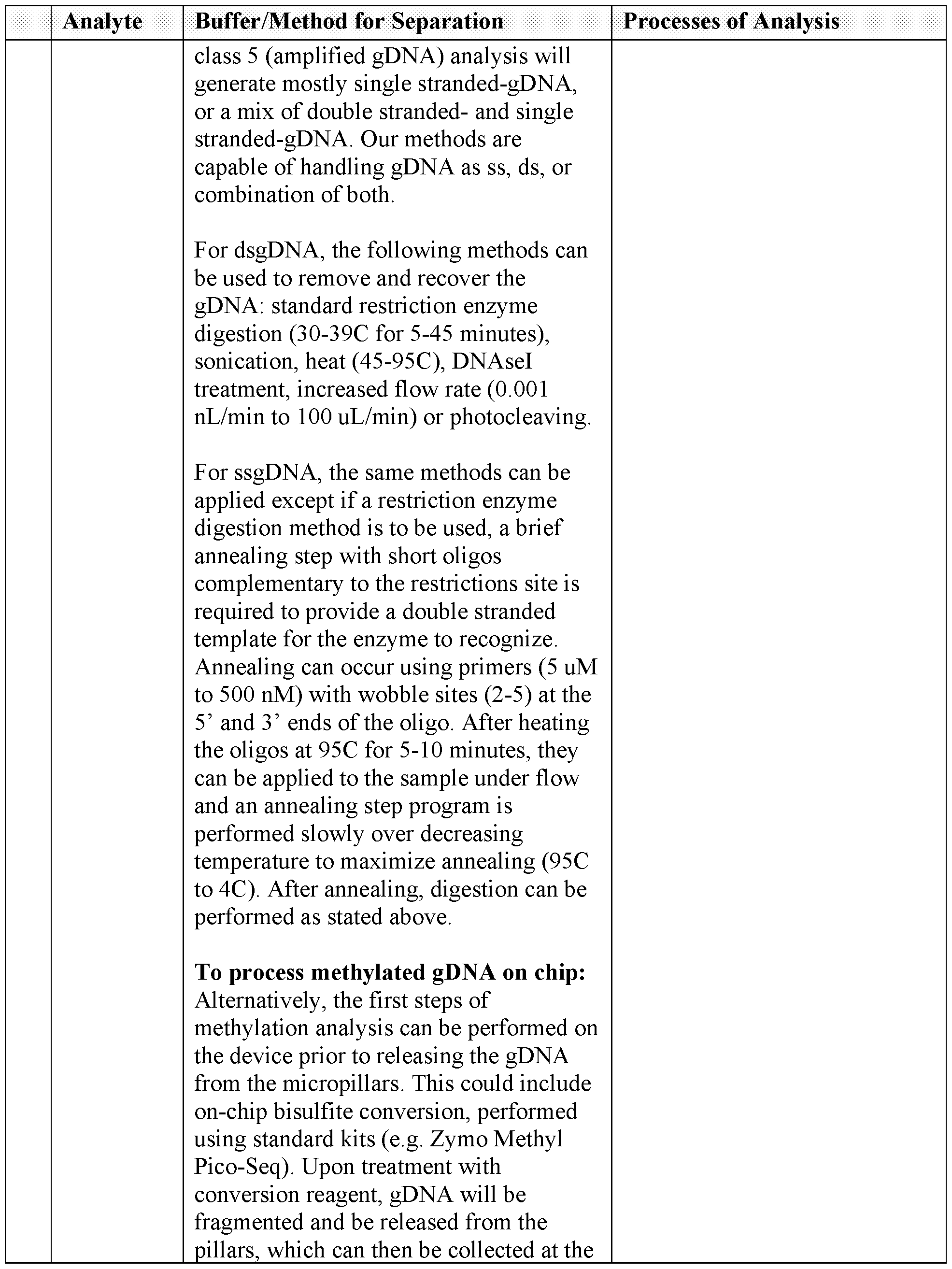


PCT/US2017/033885 (WO 2017/205304-A1), entitled“Single Cell Whole Genome
Amplification Via Micropillar Arrays Under Flow Conditions”; U.S. Patent No. 9,803,192 (US2015/0166987-A1), entitled“Programmable and Reconfigurable Microcolumn Affinity Chromatography Device, System, and Methods of Use Thereof’; and U.S. Patent No. 9,926,552, entitled“Microfluidic Device for Extracting, Isolating, and Analyzing DNA from Cells,” the disclosures of which are hereby incorporated herein by reference.
[0141] More specifically, PCT/US2017/033789 (WO 2017/205267-A1) and
PCT/US2017/033885 (WO 2017/205304-A1), describe various microfluidic devices that can be used for capturing one or more selected cells and separating genomic DNA of these cells from other cellular components for analysis. Therefore, the disclosures contained in
PCT/US2017/033789 (WO 2017/205267-A1) and PCT/US2017/033885 (WO 2017/205304-A1) can be used with the devices, systems, and methods of the present disclosure.
[0142] The devices described in PCT/US2017/033789 (WO 2017/205267-A1) and
PCT/US2017/033885 (WO 2017/205304-A1) utilize microfluidic devices containing
microstructures to capture selected cells by size or affinity binding. By lysing the captured cells the genomic DNA can be immobilized in the device and separated from the other components of the lysed cells.
[0143] In accordance with the present disclosure, similar devices to those described in PCT/US2017/033789 (WO 2017/205267-A1) and PCT/US2017/033885 (WO 2017/205304-A1) may be used to generate‘multi-omic’ information (multiple genomic-type data) from single cells. In certain embodiments, because of the device’s structure, after lysis of the captured cell(s), the RNA is washed downstream and can be removed from the outlet port. This can then be amplified to generate transcriptomic data. In accordance with the present disclosure, a lysis buffer can be identified that would not strip histones from the DNA, and allow us to analyze open chromatin status on the captured chromatin bound to the pillars. Then the histones could be removed by flowing in a harsher lysis buffer, and one would be able to amplify the remaining
genomic DNA. This concept of multi-omic analysis (RNA, open chromatin, and genomic DNA) on the device is one aspect of the present disclosure.
EXAMPLES
[0144] The following examples are intended to illustrate particular embodiments of the present disclosure, but are by no means intended to limit the scope of the present disclosure.
Example 1:
Preliminary Studies on Methods for Performing Multi-Omic Analysis of Cells
[0145] The present disclosure provides a method for performing multi-omic analysis of cells. During preliminary testing of embodiments of the methods and processes of the present disclosure, various experiments were performed.
[0146] In one preliminary study, unexpected results were obtained from initial RNA studies. As a test case, we flowed immortalized HeLa cells into the device and captured -50-100 cells (using an earlier iteration of the device that does not capture single cells). We lysed the cells and obtained the RNA from the output port. After amplification of the RNA using a commercial kit, the RNA was sequenced. We then mapped the RNA reads back to the reference human genome. For comparison, we used data from the ENCODE consortium, who had previously generated transcriptome data from bulk RNA samples. We also compared it against data from single cell HeLa and 3T3 datasets.
[0147] Based on preliminary finds, data indicated that we were surprisingly able to map more transcripts (10,000) onto the genome from the device data than the ENCODE dataset (7- 9,000), and far more than the single cell dataset (<2000; see Figure 3). This suggested that the device might be more sensitive than traditional approaches in detecting transcripts. We repeated the experiment with 3T3 cells, and included a‘tube’ control (see Figure 4). Here, we took -100 cells in a tube, lysed them in the tube and amplified them. We also lysed -100 cells on the device. We compared the sequence data against the ENCODE set and a published single cell 3T3 dataset. Both were better than single cell, but less than the ENCODE. However, what was interesting was that although the device transcriptome sequencing results had fewer reads than the tube sample, they mapped to more genes than the tube sample. This again is consistent with increased sensitivity on the device compared to traditional approaches.
[0148] The increased sensitivity was unexpected. It is contemplated that the reason for the increased sensitivity is because the device physically separates the RNA from the genomic DNA/chromatin with minimal manipulation.
[0149] This increase in sensitivity represents a novel advance over other current single cell approaches and even standard bulk RNA analysis. First, in regards to general transcriptome analysis, the standard approach for bulk RNA or single cell transcriptome studies is to use an oligodT primer to bind mRNA. However, analysis of single cell datasets clearly shows this approach can be highly inefficient. Low capture efficiency of mRNA in single cells has been known for several years to greatly limit the ability to detect the entire transcriptome of a cell.
This means that many transcripts may never be detected using the standard oligodT primer, limiting the usefulness of the resulting transcriptome data. Furthermore, our results in Figure 3 suggest this may also be the case for bulk RNA, as the ENCODE dataset detected fewer genes than using the device.
[0150] Furthermore, as new multi-omic approaches are being developed, oligodT has also been used to physically separate mRNA from genomic DNA. These“G and T” experiments use oligo-dT beads to purify the mRNA. However, this is likely to result in loss of the picogram quantities of material within the cell, because such steps involve washes and pipetting.
[0151] In contrast, by using the pillar-containing device we demonstrated our approach to separation results in very low loss of RNA. As evidenced in Figure 4, the data suggested that we can detect at least as many, if not more, transcripts than bulk RNA from the ENCODE set.
Further, as shown in Figure 5, we can also identify more genes than a simple tube based approach because more reads can be mapped using the device. This increase in sensitivity was unexpected, and represents a major improvement over current state of the art.
[0152] A second concept is that we could further extend the separation of cellular analytes on the device. As shown in Figure 5, it is possible to conceive of conditions where proteins on the surface are removed via some protease, and captured downstream. A lysis buffer that then lyses the extracellular membrane could then be flowed in, and allow cytosolic proteins and RNA to be captured. After adding a nuclear lysis buffer, chromatin could be analyzed. And after stripping chromatin, genomic DNA can then be analyzed. This would allow for several different -omic data to be obtained from the same cell.
[0153] We note that in addition to providing extended multi-omic analyses of single cells, that our method should also provide for highly increased sensitivity of detection for these other - omic approaches because of its simple separation design. Thus, we could potentially improve upon existing single cell proteomic analyses, or cytosolic analyses due to our increased sensitivity, regardless of whether these analyses are part of a multi-omic study. This extended multi-omic approach could therefore be of benefit by itself, any one -omic analysis could be enhanced due to increased sensitivity, or all the multi-omic data resulting from the device could be an improvement over standard approaches because of increased sensitivity.
[0154] It is contemplated that this is the basis for two separate new technologies: first, the separation increases the sensitivity of detection of separated analytes; and second, the device can be used to do multi-omic analysis of various analytes. The increase in sensitivity could aid in the analysis of any one isolated analyte (e.g. RNA only) or potentially the sensitivity of all analytes tested in a multi-omic analysis (e.g., genomic DNA and RNA).
Example 2:
Analysis of Cell Analytes Using Micropillar Technology
[0155] Various experiments have been conducted to isolate and analyze various types of analyte components from cell samples using the micropillar technology of the present disclosure. Data from these experiments are illustrated in Figures 6-10, as further discussed below.
[0156] Figure 6 shows data of shallow depth whole genome sequencing of gDNA from single HeLa cells isolated via a micropillar array according to embodiments of the present disclosure. This data relates to the whole genome analyte (Class 5) and is whole genome sequencing data from single Hela cells, demonstrating that whole genome amplification on the microfluidic device of the present disclosure from single cells can be recovered and mapped to the human genome (n=3).
[0157] Figure 7 shows data of amplification uniformity of multi-cell gDNA isolated on micropillars using multiple displacement amplifications (MDAs) according to embodiments of the present disclosure. This data relates to the whole genome analyte (Class 5), and
demonstrates the uniformity of whole genome amplification from multi-cell samples across 4 genetic loci using qPCR. The Y-axis is“relative amount” and the X-axis is 4 different genes tested. Every data point has been normalized to MAPK6 (Y=l, dotted line). There are two
forms of data on the graph, the open-colored circles, and the corresponding filled in colored circles/squares/triangles. The open-circles represent the“reference” uniformity of each gene compared to MAPK6 in unamplified gDNA purified off the device. Thus, for example, when assayed with qPCR, SOD1 is roughly 0.5x as abundant as MAPK6, indicating that more copies of MAPK6 are present than SOD1 in the unamplified Hela genome. Each filled in point represents a replicate from gDNA amplification on the device (n=5). The main point is that the mean values (black bars) match very closely to the mean reference value, indicating that the amplification on the microfluidic device of the present disclosure did not introduce biased amplifications caused by differential amplifications across different regions.
[0158] Figure 8 shows data of a comparison of number of genes detected from single HeLa cell RNA purified with micropillar technology and single HeLa cell RNA isolated via FACS according to embodiments of the present disclosure. This data relates to the total RNA analyte (Classes 2 and 3). These data are RNA-seq results showing the number of genes detected from single-cell total RNA purified from the device (n=l 1), an empty channel without any cells of the device (n=l), dilute bulk HeLa RNA mimicking a single cell (n=3), and single-cell total RNA from FACS isolated cells (n=8). The main points here are (1) RNA purified with the
micropillars is demonstrated, (2) empty channels processed for RNA demonstrate low
background, and (3) number of genes detected from single cells from RNA purified with the micropillars of the present disclosure is comparable to RNA from single cells isolated via FACS.
[0159] Figures 9A-9C show data illustrating that gDNA trapped on micropillars can be recovered using restriction enzymes with high efficiency according to embodiments of the present disclosure. This data supports analyte Class 6 (recovery of trapped gDNA from micropillars of the present disclosure for off-chip DNA analysis such as methylation analysis). Figure 9A shows gDNA from lysed cells (multi-cell sample) that was trapped and visualized with PicoGreen. Figure 9B shows that PicoGreen signal disappears post restriction enzyme treatment. Figure 9C indicates that -100% of expected gDNA that was trapped is recovered from the output port post digestion (calculated from # of cells that were trapped in array before lysis). Some values exceed 100% because expected yield is an estimate based on how much DNA there is per cell (n=2).
[0160] Figures 10A-10B show data illustrating that recovery of gDNA trapped on micropillars via restriction enzyme digestion of gDNA trapped on micropillars yields large
fragments of gDNA according to embodiments of the present disclosure. This data is in support of analyte Class 6 (off-chip DNA analysis such as methylation analysis). Figure 10A shows that HeLa gDNA purified through standard kits and digested with a restriction enzyme in a tube (standard methods) produces a range of small fragments (~190bp to 2kb) when run on a fragment analyzer. In contrast, as shown in Figure 10B, gDNA from -100 cells that was isolated on micropillars according to the present disclosure and recovered via restriction digest (same enzyme as used for generating the data in Figure 10A) produces a broad spectrum of much large fragment sizes (-800 bp to 500kb). Notably absent from this approach are small fragments present in the tube based digest. This means that gDNA recovered from the micropillars of the present disclosure is well suited for any further type of gDNA analysis off-chip. For example, this range of fragment sizes is more than ideal for bisulfite sequencing analysis, but could be used for any other analysis that requires large fragments of gDNA such as long-read DNA sequencing.
[0161] Illustrative embodiments of the processes, methods, and products of the present disclosure are described herein. It should be understood, however, that the description herein of the specific embodiments is not intended to limit the present disclosure to the particular forms disclosed but, on the contrary, the intention is to cover all modifications, equivalents, and alternatives falling within the spirit and scope of the invention by the appended claims. Thus, although the present invention has been described for the purpose of illustration, it is understood that such detail is solely for that purpose and variations can be made by those skilled in the art without departing from the spirit and scope of the invention which is defined by the following claims.
Claims
1. A method for isolating one or more distinct analyte component from a cell sample, said method comprising the steps of:
(a) providing a microfluidic device comprising a cell capture component and a nucleic acid entanglement component, wherein said cell capture component comprises a cell capture array comprising a plurality of cell capture micropillars, wherein said nucleic acid entanglement component comprises a nucleic acid entanglement array comprising a plurality of nucleic acid entanglement micropillars, and wherein the microfluidic device operates under continuous flow conditions;
(b) capturing one or more cell in the cell capture array of the microfluidic device while being subjected to a continuous flow rate, said capturing step comprising introducing the one or more cell sample into the microfluidic device under sufficient
hydrodynamic flow to entrap, by size exclusion, the one or more cell within the cell capture component;
(c) treating the one or more captured cell with a sequential workflow procedure under conditions effective to separate one or more distinct analyte component therefrom, wherein said one or more distinct analyte component is selected from the group consisting of:
(i) a total protein fraction;
(ii) a plasma membrane protein fraction;
(iii) a total RNA fraction;
(iv) a cytosolic RNA fraction;
(v) a cytosolic protein fraction;
(vi) a nuclear RNA fraction;
(vii) a nuclear protein fraction;
(viii) a chromatin fraction comprising genomic DNA (gDNA) regions of open chromatin;
(ix) a gDNA markers fraction comprising epigenetic and regulatory markers of gDNA;
(x) an amplified gDNA fraction; and
(xi) a methylated gDNA fraction; and
(d) isolating the one or more distinct analyte component in a manner suitable for further testing and/or analysis thereof.
2. The method according to claim 1, wherein the sequential workflow procedure comprises implementing a separation protocol to separate up to six distinct classes of analyte component from the cell sample, wherein the distinct classes comprise:
Class 1 : the total protein and plasma membrane protein fractions;
Class 2: the total protein, total RNA, cytosolic RNA, and cytosolic protein fractions;
Class 3 : the total protein, total RNA, nuclear RNA, and nuclear protein
fractions;
Class 4: the chromatin fraction;
Class 5: the gDNA markers and amplified gDNA fractions; and
Class 6: the gDNA markers and methylated gDNA fractions, wherein the sequential workflow procedure operates under the following rules:
Rule A: only one distinct analyte component can be isolated per class;
Rule B: isolation of one or more of the classes may start with any of the
classes;
Rule C: if more than one class is to be isolated, the order of isolation must proceed progressively along a classification gradient that includes, in sequence, Class 1, Class 2, Class 3, Class 4, Class 5, and Class 6, where Class 1 represents the start of the classification gradient and Class 6 represents the end of the classification gradient;
Rule D: if more than one class is to be isolated, the classes to be isolated will begin with an initial class comprising the class closest to the start of the classification gradient and terminate with a terminal class comprising the class closest to the end of the classification gradient; and
Rule E: if more than one class is to be isolated, any intermediate class falling between the initial class and the terminal class of the classification gradient may either be omitted or included from the isolating process.
3. The method according to claim 2, wherein the separation protocol is for separating the total protein fraction from the cell sample and comprises the step of:
flowing a total protein treatment solution through the microfluidic device under continuous flow conditions effective to release the total protein fraction from the one or more captured cell, thereby causing the total protein fraction to flow out of the microfluidic device.
4. The method according to claim 3, wherein the total protein treatment solution comprises a detergent, a salt, and a chelating agent.
5. The method according to claim 4, wherein the total protein treatment solution comprises a buffer containing Tris (10-50 mM, pH 7-8), NaCl (10-500 mM), and/or KC1 (10-500 mM), MgC12 (0-5mM), CaC12 (0-5mM), detergent (NP40 (0-1%), sodium deoxycholate (0-1%), or SDS (0-1%)), and EDTA (luM-lOmM).
6. The method according to claim 2, wherein the separation protocol is for separating the plasma membrane protein fraction from the cell sample and comprises the step of:
flowing a plasma membrane protein treatment solution through the microfluidic device under continuous flow conditions effective to cleave proteins from plasma membranes of the one or more captured cell without lysing the plasma membranes, thereby causing the plasma membrane protein fraction to flow out of the microfluidic device.
7. The method according to claim 6, wherein the plasma membrane protein treatment solution comprises a protease a salt, and a chelating agent.
8. The method according to claim 6, wherein the plasma membrane protein treatment solution comprises trypsin (0.01 - 0.5%) in phosphate buffered saline, with or without EDTA (lOuM - 2 mM), at pH 7-8.
9. The method according to claim 6, wherein the flowing of the plasma membrane protein treatment solution is for a period of time under flow ranging from 1-30 minutes.
10. The method according to claim 6, wherein the stable incubation temperature ranges from 20-40 degrees Celsius.
11. The method according to claim 6, wherein the plasma membrane protein treatment solution comprises trypsin (0.01 - 0.5%) with or without EDTA (lOuM - 2 mM), at pH 7-8, under flow ranging from 1-30 minutes, wherein the stable incubation temperature ranges from 20-40 degrees Celsius.
12. The method according to claim 2, wherein the separation protocol is for separating the total RNA fraction from the cell sample and comprises the step of:
flowing a total RNA treatment solution through the microfluidic device under continuous flow conditions effective to release the total RNA fraction from the one or more captured cell, thereby causing the total RNA fraction to flow out of the microfluidic device.
13. The method according to claim 12, wherein the total RNA treatment solution comprises a salt, a detergent, and an RNase inhibitor.
14. The method according to claim 12, wherein the total RNA treatment solution comprises a buffer containing Tris (pH 7-8, 1-50 mM), NaCl (25-500 mM), KC1 (25- 500 mM), detergent (e.g. Triton X-100, 0.25-2%), and RNase inhibitor.
15. The method according to claim 2, wherein the separation protocol is for separating the cytosolic RNA fraction from the cell sample and comprises the step of:
flowing a cytosolic RNA treatment solution through the microfluidic device under continuous flow conditions effective to release the cytosolic RNA fraction from the one or more captured cell, thereby causing the cytosolic RNA fraction to flow out of the microfluidic device.
16. The method according to claim 15, wherein the cytosolic RNA treatment solution comprises a detergent, a salt, an RNase inhibitor.
17. The method according to claim 15, wherein the cytosolic RNA treatment solution comprises a buffer containing Tris (pH 7-8, 1-25 mM), NaCl (10-300 mM) and/or KC1 (10-300 mM), detergent (e.g. Triton X-100, Tween 20, or NP40, or digitonin 0.1-2%), with RNase inhibitors.
18. The method according to claim 2, wherein the separation protocol is for separating the cytosolic protein fraction from the cell sample and comprises the step of:
flowing a cytosolic protein treatment solution through the microfluidic device under continuous flow conditions effective to release the cytosolic protein fraction from the one or more captured cell, thereby causing the cytosolic protein fraction to flow out of the microfluidic device.
19. The method according to claim 18, wherein the cytosolic protein treatment solution comprises a detergent, a salt, and a protease inhibitor.
20. The method according to claim 18, wherein the cytosolic protein treatment solution comprises a buffer containing Tris (pH 7-8, 1-50 mM), NaCl (10-250 mM) and/or KC1 (10-250 mM), detergent (e.g. Triton X-100, Tween 20, NP40, or digitonin 0.1-2%), with protease inhibitors.
21. The method according to claim 2, wherein the separation protocol is for separating the nuclear RNA fraction from the cell sample and comprises the step of:
flowing a nuclear RNA treatment solution through the microfluidic device under continuous flow conditions effective to release the nuclear RNA fraction from the one or more captured cell, thereby causing the nuclear RNA fraction to flow out of the microfluidic device.
22. The method according to claim 21, wherein the nuclear RNA treatment solution comprises a detergent, a salt, and an RNase inhibitor.
23. The method according to claim 21, wherein the nuclear RNA treatment solution comprises a buffer containing Tris (pH 7-8, 1-50 mM), NaCl (100-500 mM), and/or KC1 (100-500 mM), detergent (e.g. Triton X-100, 0.25-2%), and RNase inhibitor.
24. The method according to claim 2, wherein the separation protocol is for separating the nuclear protein fraction from the cell sample and comprises the step of:
flowing a nuclear protein treatment solution through the microfluidic device under continuous flow conditions effective to release the nuclear protein fraction from the one or more captured cell, thereby causing the nuclear protein fraction to flow out of the microfluidic device.
25. The method according to claim 24, wherein the nuclear protein treatment solution comprises a detergent, a salt, a protease inhibitor, and a chelating agent.
26. The method according to claim 24, wherein the nuclear protein treatment solution comprises a buffer containing Tris (pH 7-8, 1-50 mM), NaCl (25-500 mM), KC1 (25- 500 mM), MgC12 (0-5mM), CaC12 (0-5mM), EDTA (0-2mM), detergent (e.g. (NP40 (0-1%), sodium deoxycholate (0-1%)), or SDS (0-1%), and protease inhibitors.
27. The method according to claim 2, wherein the separation protocol is for separating the chromatin fraction from the cell sample and comprises the step of:
flowing a chromatin treatment solution through the microfluidic device under continuous flow conditions effective to release the chromatin fraction from the one or more captured cell, thereby causing the chromatin fraction to flow out of the microfluidic device.
28. The method according to claim 27, wherein the chromatin treatment solution comprises salts, urea, a detergent, a chelating agent, DTT, RNase inhibitor, and a protease inhibitor.
29. The method according to claim 28, wherein the flowing of the chromatin treatment solution is conducted under gentle heating for a period of time under flow sufficient to allow for activation of native Polymerase II at open chromatin sites to generate nascent RNA transcripts.
30. The method according to claim 29, wherein the gentle heating is at a temperature ranging from 25-39 degrees Celsius and for a period of time ranging from 1-20 minutes.
31. The method according to claim 2, wherein the separation protocol is for separating the gDNA markers fraction from the cell sample and comprises the step of:
flowing a gDNA markers treatment solution through the microfluidic device under continuous flow conditions effective to release the gDNA markers fraction from the one or more captured cell, thereby causing the gDNA markers fraction to flow out of the microfluidic device.
32. The method according to claim 31, wherein the gDNA markers treatment solution comprises an enzyme and/or an antibody.
33. The method according to claim 32, wherein the gDNA markers treatment solution comprises a buffer that is intended to integrate Tn5 transposase into regions of open chromatin, or a buffer that is intended to bind to RNA Pol-II that is bound to DNA, or a buffer that is intended to digest regions sensitive to DNAsel, or a buffer that is intended to bind to specific proteins such as transcription factors that are bound to DNA, or a buffer that is intended to digest interlinking regions of nucleosomes, or a buffer that contains antibodies intended to bind to histones containing specification modifications such as lactylation.
34. The method according to claim 2, wherein the separation protocol is for separating the amplified gDNA fraction from the cell sample and comprises the step of:
flowing an amplified gDNA treatment solution through the microfluidic device under continuous flow conditions effective to release the amplified gDNA fraction from the one
or more captured cell, thereby causing the amplified gDNA fraction to flow out of the microfluidic device.
35. The method according to claim 34, wherein the amplified gDNA treatment solution comprises a denaturant buffer, a neutralization buffer, a polymerase buffer, polymerase (such as Phi29), nucleotides, and primers.
36. The method according to claim 32, wherein the amplified gDNA treatment solution comprises solutions used with an isothermal amplification process such as Qiagen REPLI-g single cell kit (150343) or Qiagen REPLI-g UltraFast Mini kit (150033).
37. The method according to claim 2, wherein the separation protocol is for separating the methylated gDNA fraction from the cell sample and comprises the step of:
flowing an methylated gDNA treatment solution through the microfluidic device under continuous flow conditions effective to release the methylated gDNA fraction from the one or more captured cell, thereby causing the methylated gDNA fraction to flow out of the microfluidic device.
38. The method according to claim 37, wherein the methylated gDNA treatment solution comprises a restriction enzyme digest buffer (salts), restriction enzyme, oligonucleotides, and sodium bisulfite.
39. The method according to claim 38, wherein the methylated gDNA treatment solution comprises a restriction enzyme (1-500 El), a restriction enzyme buffer, and a sodium bisulfite conversion reagent.
40. The method according to claim 1, wherein said gDNA is single-stranded gDNA, double-stranded gDNA, or a combination of single- and -double-stranded gDNA.
41. The method according to claim 1, wherein the cell sample is a single cell or multiple cells.
42. The method according to claim 1, wherein the cell sample is selected from the group consisting of a cancer cell, a primary cell type isolated from tissue of a human, an animal, or a plant, and an immortalized cell line.
43. The method according to claim 1, wherein the flow rate ranges from between 0.001 nL/minute and 100 uL/minute.
44. The method according to claim 2, wherein said treating step comprises implementing a combination of the separation protocols to isolate one or more of the distinct analyte components for purposes of single or multiomic analysis thereof.
45. The method according to any one of claims 2-44, wherein the one or more distinct analyte component comprises a collection of analytes selected from the group consisting of:
(a) the plasma membrane protein, cytosolic RNA, nuclear RNA, chromatin, amplified gDNA, and methylated gDNA fractions isolated in sequential order;
(b) the plasma membrane protein, cytosolic RNA, nuclear RNA, chromatin, amplified gDNA, and gDNA markers of gDNA fractions isolated in sequential order;
(c) the plasma membrane protein, cytosolic protein, nuclear protein, chromatin, amplified gDNA, and methylated gDNA fractions isolated in sequential order;
(d) the plasma membrane protein, cytosolic protein, nuclear protein, chromatin, and gDNA markers fractions isolated in sequential order;
(e) the plasma membrane protein, cytosolic protein, nuclear RNA, chromatin, amplified gDNA, and methylated gDNA fractions isolated in sequential order;
(f) the plasma membrane protein, cytosolic protein, nuclear RNA, chromatin, and gDNA markers fractions isolated in sequential order;
(g) the plasma membrane protein, cytosolic RNA, nuclear protein, chromatin, amplified gDNA, and methylated gDNA fractions isolated in sequential order;
(h) the plasma membrane protein, cytosolic RNA, nuclear protein, chromatin, and gDNA markers fractions isolated in sequential order;
(i) the plasma membrane protein, total RNA, chromatin, amplified gDNA, and methylated gDNA fractions isolated in sequential order;
(j) the plasma membrane protein, total RNA, chromatin, and gDNA markers fractions isolated in sequential order;
(k) the total protein, chromatin, amplified gDNA, and methylated gDNA fractions isolated in sequential order;
(l) the total protein, chromatin, and gDNA markers fractions isolated in sequential order;
(m) the total RNA, chromatin, amplified gDNA, and methylated gDNA fractions isolated in sequential order; and
(n) the total RNA, chromatin, and gDNA markers fractions isolated in sequential order.
46. A process for analyzing one or more distinct analyte component from a cell sample, said process comprising the steps of:
performing the method according to any one of claims 1-44 to yield one or more distinct analyte component from a cell sample; and
conducting further testing and/or analysis of the one or more distinct analyte component.
47. The process according to claim 46, wherein the one or more distinct analyte component is a plasma membrane protein fraction, a total protein fraction, a cytosolic protein fraction, and/or a nuclear protein fraction that is subjected to further testing and/or analysis selected from the group consisting of mass spectrometry, immuno-based detection assays, and aptamer-based detection assays.
48. The process according to claim 46, wherein the one or more distinct analyte component is a total RNA fraction, a cytosolic RNA fraction, a nuclear RNA fraction, and/or a chromatin fraction that is subjected to further testing and/or analysis selected from the group consisting of transcriptome analysis via RNA-seq, targeted gene expression profiling via
RT-PCR, RT-qPCR, or NanoString, and qualitative and quantitative characterization such as fragment analyzer, Qubit, or NanoDrop.
49. The process according to claim 46, wherein the one or more distinct analyte component is a gDNA markers fraction and/or an amplified gDNA fraction that is subjected to further testing and/or analysis selected from the group consisting of whole genome sequencing, exome sequencing, targeted re-sequencing, PCR, and qPCR.
50. The process according to claim 46, wherein the one or more distinct analyte component is a methylated gDNA fraction that is subjected to further testing and/or analysis selected from the group consisting of bisulfite conversion analysis, methylation specific PCR, methylation specific qPCR, reduced representation bisulfite sequencing, and whole genome bisulfite sequencing.
Priority Applications (2)
| Application Number | Priority Date | Filing Date | Title |
|---|---|---|---|
| US17/298,533 US12338487B2 (en) | 2018-11-28 | 2019-11-29 | Multiomic analysis of cell analytes using microfluidic systems |
| US19/216,540 US20250283149A1 (en) | 2018-11-28 | 2025-05-22 | Multiomic analysis of cell analytes using microfluidic systems |
Applications Claiming Priority (2)
| Application Number | Priority Date | Filing Date | Title |
|---|---|---|---|
| US201862772621P | 2018-11-28 | 2018-11-28 | |
| US62/772,621 | 2018-11-28 |
Related Child Applications (2)
| Application Number | Title | Priority Date | Filing Date |
|---|---|---|---|
| US17/298,533 A-371-Of-International US12338487B2 (en) | 2018-11-28 | 2019-11-29 | Multiomic analysis of cell analytes using microfluidic systems |
| US19/216,540 Continuation US20250283149A1 (en) | 2018-11-28 | 2025-05-22 | Multiomic analysis of cell analytes using microfluidic systems |
Publications (1)
| Publication Number | Publication Date |
|---|---|
| WO2020113173A1 true WO2020113173A1 (en) | 2020-06-04 |
Family
ID=70852661
Family Applications (1)
| Application Number | Title | Priority Date | Filing Date |
|---|---|---|---|
| PCT/US2019/063866 Ceased WO2020113173A1 (en) | 2018-11-28 | 2019-11-29 | Multiomic analysis of cell analytes using microfluidic systems |
Country Status (2)
| Country | Link |
|---|---|
| US (2) | US12338487B2 (en) |
| WO (1) | WO2020113173A1 (en) |
Cited By (3)
| Publication number | Priority date | Publication date | Assignee | Title |
|---|---|---|---|---|
| WO2023055790A1 (en) * | 2021-09-29 | 2023-04-06 | Battelle Memorial Institute | Apparatus and methods for performing multiple omics analysis and processing analyte mixtures |
| EP4409284A4 (en) * | 2021-09-30 | 2025-08-13 | Georgia Tech Res Inst | ANALYSIS SYSTEM AND ANALYSIS METHODS |
| WO2026039392A1 (en) * | 2024-08-14 | 2026-02-19 | Inso Biosciences, Inc. | Methods and apparatus for extracting or separating nucleic acids from biological samples |
Citations (2)
| Publication number | Priority date | Publication date | Assignee | Title |
|---|---|---|---|---|
| US20090186776A1 (en) * | 2001-09-07 | 2009-07-23 | Webb Brian L | Microcolumn-platform based array for high-throughput analysis |
| US20180305682A1 (en) * | 2011-06-06 | 2018-10-25 | Cornell University | Microfluidic device for extracting, isolating, and analyzing dna from cells |
Family Cites Families (51)
| Publication number | Priority date | Publication date | Assignee | Title |
|---|---|---|---|---|
| US5304487A (en) | 1992-05-01 | 1994-04-19 | Trustees Of The University Of Pennsylvania | Fluid handling in mesoscale analytical devices |
| US6762059B2 (en) | 1999-08-13 | 2004-07-13 | U.S. Genomics, Inc. | Methods and apparatuses for characterization of single polymers |
| US6696022B1 (en) | 1999-08-13 | 2004-02-24 | U.S. Genomics, Inc. | Methods and apparatuses for stretching polymers |
| GB2355717A (en) | 1999-10-28 | 2001-05-02 | Amersham Pharm Biotech Uk Ltd | DNA isolation method |
| WO2001062887A1 (en) | 2000-02-23 | 2001-08-30 | Zyomyx, Inc. | Chips having elevated sample surfaces |
| CU23095A1 (en) | 2000-11-07 | 2005-11-18 | Cnic Ct Nac Investigaciones | PROCESS FOR QUICK TYPIFICATION OF MICROORGANISMS AND REAGENT GAME EMPLOYED |
| US6685841B2 (en) | 2001-02-14 | 2004-02-03 | Gabriel P. Lopez | Nanostructured devices for separation and analysis |
| US7390463B2 (en) | 2001-09-07 | 2008-06-24 | Corning Incorporated | Microcolumn-based, high-throughput microfluidic device |
| US6977148B2 (en) | 2001-10-15 | 2005-12-20 | Qiagen Gmbh | Multiple displacement amplification |
| US7655791B2 (en) | 2001-11-13 | 2010-02-02 | Rubicon Genomics, Inc. | DNA amplification and sequencing using DNA molecules generated by random fragmentation |
| IE20020619A1 (en) | 2001-12-31 | 2003-07-23 | Provost Fellows And Scholors O | An assay assembly |
| US20030162181A1 (en) | 2002-02-28 | 2003-08-28 | Eastman Kodak Company | DNA sequencing and gene identification |
| JP5695287B2 (en) | 2002-10-02 | 2015-04-01 | カリフォルニア インスティテュート オブ テクノロジー | Nucleic acid analysis of microfluids |
| WO2004067708A2 (en) | 2003-01-17 | 2004-08-12 | Sri International Et Al. | Device and method for fragmenting material by hydrodynamic shear |
| US7541166B2 (en) | 2003-09-19 | 2009-06-02 | Microfluidic Systems, Inc. | Sonication to selectively lyse different cell types |
| US7476326B2 (en) | 2003-09-26 | 2009-01-13 | Ahn Chong H | On-chip sample preparation for whole blood analysis |
| JP2006010529A (en) | 2004-06-25 | 2006-01-12 | Canon Inc | Magnetic particle separation device and separation method |
| US20080125330A1 (en) | 2004-07-01 | 2008-05-29 | Cornell Research Foundation, Inc. | Real-Time Pcr Detection of Microorganisms Using an Integrated Microfluidics Platform |
| GB0508983D0 (en) | 2005-05-03 | 2005-06-08 | Oxford Gene Tech Ip Ltd | Cell analyser |
| WO2007041340A2 (en) | 2005-09-30 | 2007-04-12 | The Trustees Of Columbia University In The City Of New York | Microfluidic cells with parallel arrays of individual dna molecules |
| WO2007050040A1 (en) | 2005-10-28 | 2007-05-03 | Agency For Science, Technology And Research | Immobilization unit and device for isolation of nucleic acid molecules |
| WO2007081385A2 (en) | 2006-01-11 | 2007-07-19 | Raindance Technologies, Inc. | Microfluidic devices and methods of use in the formation and control of nanoreactors |
| WO2008111990A1 (en) | 2006-06-14 | 2008-09-18 | Cellpoint Diagnostics, Inc. | Rare cell analysis using sample splitting and dna tags |
| US7807454B2 (en) | 2006-10-18 | 2010-10-05 | The Regents Of The University Of California | Microfluidic magnetophoretic device and methods for using the same |
| US8202691B2 (en) | 2008-01-25 | 2012-06-19 | Illumina, Inc. | Uniform fragmentation of DNA using binding proteins |
| KR100968524B1 (en) | 2008-04-11 | 2010-07-08 | 인싸이토 주식회사 | Micro-nanofluidic biochips for biological sample analysis |
| US8304185B2 (en) | 2009-07-17 | 2012-11-06 | Canon U.S. Life Sciences, Inc. | Methods and systems for DNA isolation on a microfluidic device |
| JP5836803B2 (en) | 2008-08-14 | 2015-12-24 | ナノストリング テクノロジーズ, インコーポレイテッド | Stable nanoreporter |
| JP2012504243A (en) | 2008-09-26 | 2012-02-16 | ザ ジェネラル ホスピタル コーポレイション | Particle capture |
| US7964978B1 (en) | 2008-10-06 | 2011-06-21 | Douglas Weissmann | Wind turbine having a blade ring using magnetic levitation |
| EP2350652A2 (en) | 2008-10-10 | 2011-08-03 | Cnrs-Dae | Cell sorting device |
| SG172965A1 (en) | 2009-01-13 | 2011-08-29 | Fluidigm Corp | Single-cell nucleic acid analysis |
| US20100190146A1 (en) | 2009-01-29 | 2010-07-29 | Bynum Magdalena A | Microfluidic Glycan Analysis |
| RU2559541C2 (en) | 2009-06-05 | 2015-08-10 | Интедженкс Инк. | Universal system for sample preparing and application in integrated system of analysis |
| WO2011017677A2 (en) | 2009-08-06 | 2011-02-10 | Cornell University | Device and methods for epigenetic analysis |
| WO2011038241A1 (en) | 2009-09-25 | 2011-03-31 | President And Fellows Of Harvard College | Nucleic acid amplification and sequencing by synthesis with fluorogenic nucleotides |
| TW201144805A (en) | 2010-06-08 | 2011-12-16 | Academia Sinica | Microfluidic device |
| US9365816B2 (en) | 2010-09-13 | 2016-06-14 | California Institute Of Technology | Handheld low pressure mechanical cell lysis device with single cell resolution |
| CN103889556A (en) | 2011-01-06 | 2014-06-25 | Gpb科学有限责任公司 | Circulating tumor cell capture on a microfluidic chip incorporating both affinity and size |
| CN116179323A (en) | 2011-05-27 | 2023-05-30 | 不列颠哥伦比亚大学 | Microfluidic cell capture and analysis device for high throughput analysis |
| US9670540B2 (en) | 2011-07-21 | 2017-06-06 | Cornell University | Methods and devices for DNA sequencing and molecular diagnostics |
| EP3285062B1 (en) | 2012-02-29 | 2019-08-07 | Fluidigm Corporation | Methods, systems, and devices for multiple single-cell capturing and processing using microfluidics |
| ES2660438T3 (en) | 2012-11-09 | 2018-03-22 | F. Hoffmann-La Roche Ag | In vitro capture and analysis of circulating tumor cells |
| WO2014153071A1 (en) | 2013-03-14 | 2014-09-25 | The Broad Institute, Inc. | Methods for quantitating dna using digital multiple displacement amplification |
| US9803192B2 (en) | 2013-10-04 | 2017-10-31 | Cornell University | Programmable and reconfigurable microcolumn affinity chromatography device, system, and methods of use thereof |
| TWI482857B (en) | 2013-10-21 | 2015-05-01 | Nat Univ Tsing Hua | Suitable body for glycosylated hemoglobin and heme and its application |
| US10466243B2 (en) | 2013-11-20 | 2019-11-05 | University Of Florida Research Foundation, Incorporated | Antibody and aptamer ensemble for cell isolation and enrichment |
| US10393726B2 (en) | 2015-03-23 | 2019-08-27 | The University Of North Carolina At Chapel Hill | Universal molecular processor for precision medicine |
| WO2017004463A1 (en) | 2015-07-01 | 2017-01-05 | Abbott Laboratories | Devices and methods for sample analysis |
| WO2017205304A1 (en) | 2016-05-22 | 2017-11-30 | Cornell University | Single cell whole genome amplification via micropillar arrays under flow conditions |
| US11602747B2 (en) | 2016-05-22 | 2023-03-14 | Cornell University | Multifunctional microfluidic device for capturing target cells and analyzing genomic DNA isolated from the target cells while under flow conditions |
-
2019
- 2019-11-29 US US17/298,533 patent/US12338487B2/en active Active
- 2019-11-29 WO PCT/US2019/063866 patent/WO2020113173A1/en not_active Ceased
-
2025
- 2025-05-22 US US19/216,540 patent/US20250283149A1/en active Pending
Patent Citations (2)
| Publication number | Priority date | Publication date | Assignee | Title |
|---|---|---|---|---|
| US20090186776A1 (en) * | 2001-09-07 | 2009-07-23 | Webb Brian L | Microcolumn-platform based array for high-throughput analysis |
| US20180305682A1 (en) * | 2011-06-06 | 2018-10-25 | Cornell University | Microfluidic device for extracting, isolating, and analyzing dna from cells |
Cited By (4)
| Publication number | Priority date | Publication date | Assignee | Title |
|---|---|---|---|---|
| WO2023055790A1 (en) * | 2021-09-29 | 2023-04-06 | Battelle Memorial Institute | Apparatus and methods for performing multiple omics analysis and processing analyte mixtures |
| US12629681B2 (en) | 2021-09-29 | 2026-05-19 | Battelle Memorial Institute | Apparatuses and methods for performing multiple omics analysis and processing analyte mixtures |
| EP4409284A4 (en) * | 2021-09-30 | 2025-08-13 | Georgia Tech Res Inst | ANALYSIS SYSTEM AND ANALYSIS METHODS |
| WO2026039392A1 (en) * | 2024-08-14 | 2026-02-19 | Inso Biosciences, Inc. | Methods and apparatus for extracting or separating nucleic acids from biological samples |
Also Published As
| Publication number | Publication date |
|---|---|
| US12338487B2 (en) | 2025-06-24 |
| US20250283149A1 (en) | 2025-09-11 |
| US20220010360A1 (en) | 2022-01-13 |
Similar Documents
| Publication | Publication Date | Title |
|---|---|---|
| Alberti et al. | Viral to metazoan marine plankton nucleotide sequences from the Tara Oceans expedition | |
| US20250283149A1 (en) | Multiomic analysis of cell analytes using microfluidic systems | |
| Trigodet et al. | High molecular weight DNA extraction strategies for long‐read sequencing of complex metagenomes | |
| JP2024149636A (en) | Nuclease-based RNA depletion | |
| Yilmaz et al. | Single cell genome sequencing | |
| O'Neil et al. | Ribosomal RNA depletion for efficient use of RNA‐seq capacity | |
| EP3161160B1 (en) | Methods of analyzing nucleic acids from individual cells or cell populations | |
| US20150275267A1 (en) | Method and kit for preparing a target rna depleted sample | |
| US8017332B2 (en) | Magnetic method for recovering nucleic acid from a mixed cell suspension | |
| WO2020186223A1 (en) | Sherlock assays for tick-borne diseases | |
| CN115176026A (en) | Method for sequencing RNA oligonucleotides | |
| Lee et al. | DNA extraction methods in forensic analysis | |
| US20240084360A1 (en) | Host depletion and microbial enrichment of a biological sample and related methods and systems | |
| CN116391046A (en) | Method for nucleic acid detection by oligohybridization and PCR-based amplification | |
| Gelderman et al. | Discovery of posttranscriptional regulatory RNAs using next generation sequencing technologies | |
| Blasdel et al. | Preparing cDNA libraries from lytic phage-infected cells for whole transcriptome analysis by RNA-Seq | |
| Wakimoto et al. | Isolation of single‐stranded DNA | |
| KR20250047294A (en) | Method for preparing a normalized nucleic acid sample, kit and device for use in said method | |
| Lai et al. | Detection and analysis of long noncoding RNAs | |
| US20210062180A1 (en) | Semi-automated research instrument system | |
| WO2021046462A2 (en) | Methods and systems for rna-seq profiling | |
| EP3456839A1 (en) | Method of enrichment of micro-organisms in a metagenomics workflow | |
| US20220220467A1 (en) | Method and kit for the purification of functional risc-associated small rnas | |
| Yabuki et al. | New barcoding primers for the efficient monitoring of diplonemid diversity | |
| EP3283646B1 (en) | Method for analysing nuclease hypersensitive sites. |
Legal Events
| Date | Code | Title | Description |
|---|---|---|---|
| 121 | Ep: the epo has been informed by wipo that ep was designated in this application |
Ref document number: 19889119 Country of ref document: EP Kind code of ref document: A1 |
|
| NENP | Non-entry into the national phase |
Ref country code: DE |
|
| 122 | Ep: pct application non-entry in european phase |
Ref document number: 19889119 Country of ref document: EP Kind code of ref document: A1 |
|
| WWG | Wipo information: grant in national office |
Ref document number: 17298533 Country of ref document: US |