US11602747B2 - Multifunctional microfluidic device for capturing target cells and analyzing genomic DNA isolated from the target cells while under flow conditions - Google Patents

Multifunctional microfluidic device for capturing target cells and analyzing genomic DNA isolated from the target cells while under flow conditions Download PDF

Info

Publication number
US11602747B2
US11602747B2 US16/303,658 US201716303658A US11602747B2 US 11602747 B2 US11602747 B2 US 11602747B2 US 201716303658 A US201716303658 A US 201716303658A US 11602747 B2 US11602747 B2 US 11602747B2
Authority
US
United States
Prior art keywords
microchannel
cell
microfluidic device
micropillars
genomic dna
Prior art date
Legal status (The legal status is an assumption and is not a legal conclusion. Google has not performed a legal analysis and makes no representation as to the accuracy of the status listed.)
Active, expires
Application number
US16/303,658
Other versions
US20210178393A1 (en
Inventor
Harold G. Craighead
Sarah J. REINHOLT
Current Assignee (The listed assignees may be inaccurate. Google has not performed a legal analysis and makes no representation or warranty as to the accuracy of the list.)
Cornell University
Original Assignee
Cornell University
Priority date (The priority date is an assumption and is not a legal conclusion. Google has not performed a legal analysis and makes no representation as to the accuracy of the date listed.)
Filing date
Publication date
Application filed by Cornell University filed Critical Cornell University
Priority to US16/303,658 priority Critical patent/US11602747B2/en
Assigned to CORNELL UNIVERSITY reassignment CORNELL UNIVERSITY ASSIGNMENT OF ASSIGNORS INTEREST (SEE DOCUMENT FOR DETAILS). Assignors: REINHOLT, Sarah J., CRAIGHEAD, HAROLD G.
Publication of US20210178393A1 publication Critical patent/US20210178393A1/en
Application granted granted Critical
Publication of US11602747B2 publication Critical patent/US11602747B2/en
Active legal-status Critical Current
Adjusted expiration legal-status Critical

Links

Images

Classifications

    • BPERFORMING OPERATIONS; TRANSPORTING
    • B01PHYSICAL OR CHEMICAL PROCESSES OR APPARATUS IN GENERAL
    • B01LCHEMICAL OR PHYSICAL LABORATORY APPARATUS FOR GENERAL USE
    • B01L3/00Containers or dishes for laboratory use, e.g. laboratory glassware; Droppers
    • B01L3/50Containers for the purpose of retaining a material to be analysed, e.g. test tubes
    • B01L3/502Containers for the purpose of retaining a material to be analysed, e.g. test tubes with fluid transport, e.g. in multi-compartment structures
    • B01L3/5027Containers for the purpose of retaining a material to be analysed, e.g. test tubes with fluid transport, e.g. in multi-compartment structures by integrated microfluidic structures, i.e. dimensions of channels and chambers are such that surface tension forces are important, e.g. lab-on-a-chip
    • B01L3/502753Containers for the purpose of retaining a material to be analysed, e.g. test tubes with fluid transport, e.g. in multi-compartment structures by integrated microfluidic structures, i.e. dimensions of channels and chambers are such that surface tension forces are important, e.g. lab-on-a-chip characterised by bulk separation arrangements on lab-on-a-chip devices, e.g. for filtration or centrifugation
    • BPERFORMING OPERATIONS; TRANSPORTING
    • B01PHYSICAL OR CHEMICAL PROCESSES OR APPARATUS IN GENERAL
    • B01LCHEMICAL OR PHYSICAL LABORATORY APPARATUS FOR GENERAL USE
    • B01L3/00Containers or dishes for laboratory use, e.g. laboratory glassware; Droppers
    • B01L3/50Containers for the purpose of retaining a material to be analysed, e.g. test tubes
    • B01L3/502Containers for the purpose of retaining a material to be analysed, e.g. test tubes with fluid transport, e.g. in multi-compartment structures
    • B01L3/5027Containers for the purpose of retaining a material to be analysed, e.g. test tubes with fluid transport, e.g. in multi-compartment structures by integrated microfluidic structures, i.e. dimensions of channels and chambers are such that surface tension forces are important, e.g. lab-on-a-chip
    • B01L3/502761Containers for the purpose of retaining a material to be analysed, e.g. test tubes with fluid transport, e.g. in multi-compartment structures by integrated microfluidic structures, i.e. dimensions of channels and chambers are such that surface tension forces are important, e.g. lab-on-a-chip specially adapted for handling suspended solids or molecules independently from the bulk fluid flow, e.g. for trapping or sorting beads, for physically stretching molecules
    • BPERFORMING OPERATIONS; TRANSPORTING
    • B01PHYSICAL OR CHEMICAL PROCESSES OR APPARATUS IN GENERAL
    • B01LCHEMICAL OR PHYSICAL LABORATORY APPARATUS FOR GENERAL USE
    • B01L2200/00Solutions for specific problems relating to chemical or physical laboratory apparatus
    • B01L2200/06Fluid handling related problems
    • B01L2200/0647Handling flowable solids, e.g. microscopic beads, cells, particles
    • B01L2200/0663Stretching or orienting elongated molecules or particles
    • BPERFORMING OPERATIONS; TRANSPORTING
    • B01PHYSICAL OR CHEMICAL PROCESSES OR APPARATUS IN GENERAL
    • B01LCHEMICAL OR PHYSICAL LABORATORY APPARATUS FOR GENERAL USE
    • B01L2200/00Solutions for specific problems relating to chemical or physical laboratory apparatus
    • B01L2200/06Fluid handling related problems
    • B01L2200/0647Handling flowable solids, e.g. microscopic beads, cells, particles
    • B01L2200/0668Trapping microscopic beads
    • BPERFORMING OPERATIONS; TRANSPORTING
    • B01PHYSICAL OR CHEMICAL PROCESSES OR APPARATUS IN GENERAL
    • B01LCHEMICAL OR PHYSICAL LABORATORY APPARATUS FOR GENERAL USE
    • B01L2300/00Additional constructional details
    • B01L2300/08Geometry, shape and general structure
    • B01L2300/0809Geometry, shape and general structure rectangular shaped
    • B01L2300/0816Cards, e.g. flat sample carriers usually with flow in two horizontal directions
    • BPERFORMING OPERATIONS; TRANSPORTING
    • B01PHYSICAL OR CHEMICAL PROCESSES OR APPARATUS IN GENERAL
    • B01LCHEMICAL OR PHYSICAL LABORATORY APPARATUS FOR GENERAL USE
    • B01L2300/00Additional constructional details
    • B01L2300/08Geometry, shape and general structure
    • B01L2300/0861Configuration of multiple channels and/or chambers in a single devices
    • B01L2300/0864Configuration of multiple channels and/or chambers in a single devices comprising only one inlet and multiple receiving wells, e.g. for separation, splitting
    • BPERFORMING OPERATIONS; TRANSPORTING
    • B01PHYSICAL OR CHEMICAL PROCESSES OR APPARATUS IN GENERAL
    • B01LCHEMICAL OR PHYSICAL LABORATORY APPARATUS FOR GENERAL USE
    • B01L2400/00Moving or stopping fluids
    • B01L2400/04Moving fluids with specific forces or mechanical means
    • B01L2400/0475Moving fluids with specific forces or mechanical means specific mechanical means and fluid pressure
    • B01L2400/0487Moving fluids with specific forces or mechanical means specific mechanical means and fluid pressure fluid pressure, pneumatics
    • BPERFORMING OPERATIONS; TRANSPORTING
    • B01PHYSICAL OR CHEMICAL PROCESSES OR APPARATUS IN GENERAL
    • B01LCHEMICAL OR PHYSICAL LABORATORY APPARATUS FOR GENERAL USE
    • B01L2400/00Moving or stopping fluids
    • B01L2400/08Regulating or influencing the flow resistance
    • B01L2400/084Passive control of flow resistance
    • B01L2400/086Passive control of flow resistance using baffles or other fixed flow obstructions

Definitions

  • the present application contains a Sequence Listing which has been submitted as an ASCII text file via EFS-Web and which is hereby incorporated by reference in its entirety.
  • Said ASCII text file is named “2020-08-30_SequenceListing_AnnexC_ST25_6377021US1.txt,” was created on Aug. 30, 2020, and has a size of 22,828 bytes.
  • the present invention relates to, inter alia, a microfluidic device for capturing target cells and analyzing genomic DNA isolated from the target cells while under flow conditions.
  • Cancer cells contain genetic mutations that allow them to escape the regulatory processes necessary for the healthy function of tissues and organs. 1-3 Moreover, there are numerous mechanisms for malignancy with different combinations of genetic mutations, and cancer cells are constantly evolving, 4 which makes cancer treatment difficult with varying levels of efficacy. Many assays have been developed that detect specific mutations, whereas some have been designed to detect all mutations via sequencing 5-10 Each of these approaches has advantages and disadvantages. 8 Most of these assays also require significant sample preparation to be performed in a bulk solution where the initial amount of genetic material is limited, some is lost in processing, and the remaining material is used up quickly. Therefore, an assay that incorporates sample preparation and enables the original genetic template to be reused would be highly advantageous.
  • Aptamers are short single-stranded nucleic acids with structures determined by their specific nucleotide sequence. These molecules bind with high affinity and specificity to their intended targets. Aptamers are typically discovered by an iterative process called Systematic Evolution of Ligands by EXponential enrichment (SELEX) in which they are selected from a very large, sequence-diverse library of nucleic acids (10 12 -10 16 unique sequences). 11-13 Cell-SELEX was developed more recently to select aptamers that bind specifically to a certain type of cell. 14,15 Using this technique, aptamers that bind specifically to cells of interest can be determined without any prior knowledge about the surface composition of the cells. Consequently, this method can be used to discover affinity ligands that bind to cancer cells. 16-18
  • CTCs circulating tumor cells
  • aptamer-based CTC capture is also being performed.
  • 19-24 Aptamers have been used over antibodies in these types of applications because of their increased robustness and ease of functionalization and oriented immobilization.
  • the Tan group has developed several devices for capturing CTCs in which the aptamers are biotinylated and simply immobilized within the device by binding to streptavidin adsorbed on the channel surface. 20,22,23,25,26
  • This device incorporated a fine micropillar array that captured megabase-long genomic DNA (gDNA) strands via physical entanglement.
  • gDNA megabase-long genomic DNA
  • the physical nature of this isolation enables the gDNA to be isolated without dependence on biochemical or electrostatic forces, making it available for downstream reactions. This also allows the gDNA to remain on the micropillar array during flow, which allows downstream analyses to be performed within the microdevice.
  • the present invention is directed to overcoming these and other deficiencies in the art.
  • the present invention provides, inter alia, a combination of microfluidic and aptamer technologies suitable for use in studying, analyzing, detecting, and treating various conditions and diseases.
  • the present invention provides a microfluidic device for aptamer-based cancer cell capture and genetic mutation detection, and the use of the microfluidic device for various applications.
  • the present invention provides a microfluidic device comprising: a cell microchannel and a nucleic acid microchannel that intersect to form a cell capture intersection region; a cell capture array comprising a plurality of cell capturing micropillars configured and arranged in a manner effective to capture one or more target cell when flowed through the cell microchannel, said cell capture array being located in the cell capture intersection region; and a nucleic acid entanglement array comprising a plurality of nucleic acid entanglement micropillars configured and arranged in a manner effective to physically entangle and maintain thereon genomic DNA isolated from the one or more target cell, said nucleic acid entanglement array being located in a portion of the nucleic acid microchannel that is adjacent to and downstream of the cell capture intersection region.
  • the microfluidic device is multi-functional in that it is effective for capturing said one or more target cell, isolating said genomic DNA from the one or more target cell, and analyzing said genomic DNA in a self-contained manner.
  • the microfluidic device of the present invention further comprises a first flow rate means for managing rate of flow of fluid through the cell microchannel and a second flow rate means for managing rate of flow of fluid through the nucleic acid microchannel.
  • the microfluidic device of the present invention further comprises a temperature controller for managing temperature of fluid and other contents contained within the cell microchannel and/or nucleic acid microchannel.
  • the present invention provides a method of isolating and maintaining genomic DNA of one or more target cell from a sample under flow for further analysis thereof.
  • This method involves the steps of: providing a microfluidic device as described herein; introducing a sample comprising one or more target cell into the cell microchannel at a flow rate effective to transport the one or more target cell to the cell capture array so as to capture the one or more target cell in the cell capturing micropillars by specific binding; lysing the one or more target cell by introducing lysing reagents through the nucleic acid microchannel at a flow rate effective to release genomic DNA from the one or more target cell without shearing the genomic DNA; and maintaining fluid flow within the nucleic acid microchannel at a flow rate effective to cause the released genomic DNA to become physically entangled and maintained within the nucleic acid entanglement array for further analysis thereof.
  • the present invention provides a method for conducting aptamer-based cancer cell capture and genomic DNA mutation analysis of genomic DNA isolated from one or more target cell.
  • This method includes the steps of: performing the steps described herein of the method of isolating and maintaining genomic DNA of one or more target cell from a sample under flow; and conducting aptamer-based cancer cell capture and genomic DNA mutation analysis of the genomic DNA isolated from one or more target cell while in a flow environment within the microfluidic device.
  • the present invention provides a method for amplifying individual genes of interest from the one or more target cell consecutively and collecting each amplification product separately.
  • This method includes the steps of: performing the steps described herein of the method of isolating and maintaining genomic DNA of one or more target cell from a sample under flow; and amplifying individual genes of interest from the genomic DNA entangled and maintained under flow within the nucleic acid entanglement array of the microfluidic device consecutively and collecting each amplification product separately.
  • the present invention provides a method for sequencing nucleic acids amplified from genomic DNA isolated from one or more target cell.
  • This method includes the steps of: performing the steps described herein of the method of isolating and maintaining genomic DNA of one or more target cell from a sample under flow; and sequencing the genomic DNA entangled and maintained under flow within the nucleic acid entanglement array of the microfluidic device.
  • the present invention provides a method for multiple displacement amplification (MDA) reactions of one or more nucleic acid sequence isolated from one or more target cell.
  • MDA multiple displacement amplification
  • This method includes the steps of: performing the steps described herein of the method of isolating and maintaining genomic DNA of one or more target cell from a sample under flow; and conducting multiple displacement amplification (MDA) reactions under flow using the genomic DNA entangled and maintained within the nucleic acid entanglement array of the microfluidic device.
  • the present invention relates to a novel microfluidic device that provides a platform for specifically capturing cancer cells and isolating the genomic DNA for specific amplification and sequence analysis.
  • nucleic acid aptamers that specifically bind to cancer cells are immobilized within a microchannel containing pillars to increase the number of collisions with the surface and improve capture efficiency.
  • the captured cells are then lysed and the genomic DNA is isolated via physical entanglement within a secondary micropillar array.
  • This type of isolation enables multiple consecutive rounds of isothermal amplification to be performed to amplify different individual genes separately, since the genomic template is retained on the micropillars between subsequent amplifications.
  • the amplified gene samples undergo Sanger sequencing, an inexpensive sequencing approach requiring a pure sample, to reveal the genetic sequence.
  • the resulting sequence information is compared against the known wildtype gene, and any mutations are identified.
  • This approach offers a way to monitor multiple genetic mutations in the same small population of cells, which is beneficial given the wide diversity in cancer cells, and requires very few cells to be extracted from the patient sample. With this capability for genetic monitoring, precision medicine should be more accessible for the diagnosis and treatment of cancer and other diseases.
  • microfluidic device of the present invention over the prior art is the combination of microfluidic aptamer-based cell capturing technology (e.g., high surface area microfluidic device for capturing selected cells by specific binding) with the elongation/capture/analysis of nucleic acids isolated from the captured cells (e.g., using small pillars or capture structures).
  • microfluidic aptamer-based cell capturing technology e.g., high surface area microfluidic device for capturing selected cells by specific binding
  • the elongation/capture/analysis of nucleic acids isolated from the captured cells e.g., using small pillars or capture structures.
  • the microfluidic device combines these technologies into a single, integrated device in a manner that is unique over the prior art technologies.
  • the microfluidic device of the present invention is unique over the prior art for a variety of reasons.
  • the design of a microfluidic device of the present invention is such that it can be fabricated as integrated unit, which is unique over the prior art which involves the use of separate devices, which have different requirements for their construction.
  • the operation of two, separate devices would require sample extraction from one device and then sample preparation and then insertion into the other device. This would be very inefficient and cause the loss of portions of the sample being studied, as well as opening the process up to contamination.
  • Being able to use the current devices of the prior art does not inform one how to operate an integrated unit such as the one of the present invention, where all processes must be carried out on a chip with no valves or separate sample processing devices between them.
  • the present invention provides a process for preparing a microfluidic device according to the present invention, said process comprising steps as disclosed and/or contemplated herein.
  • a device capable of specifically capturing cancer cells, isolating their gDNA, and amplifying specific genes for sequencing to determine the presence of any genetic mutations in those genes.
  • the cancer cells are captured using aptamers immobilized on the microchannel surface, and the gDNA is isolated via physical entanglement within a micropillar array.
  • MDA multiple displacement amplification
  • This amplification product undergoes sequencing, and the resulting sequence is compared to the known human genome to determine the presence of any genetic mutations. Identification of specific key mutations in a timely and cost-effective way would allow clinicians to better prescribe the most effective treatment options.
  • regular testing of multiple important genes is also beneficial for monitoring disease progression and determining future treatment. The ability to perform this regular testing in a cost-effective manner would encourage more frequent testing, which would likely improve overall treatment efficacy.
  • FIG. 1 Schematic of portions of one embodiment of the microfluidic device of the present disclosure.
  • the initial cell sample is injected into Inlet 1, flows through the capture region, and out Outlet 1.
  • the lysis buffer is injected into Inlet 2, and the genomic DNA is entangled in the micropillar array, while the rest of the lysate flows out of the device through Outlet 2.
  • FIG. 2 Schematic of one embodiment of a microfluidic device of the present disclosure.
  • the embodiment illustrates microfluidic channels for capture of different biological components from a sample.
  • FIG. 3 Microchannel design for capturing cancer cells and isolating their gDNA.
  • the device contains two orthogonal channels, the cell channel and the DNA channel. It also contains two micropillar arrays, the cell capture array located at the intersection of the channels (red box, scale bar: 200 ⁇ m) and the DNA isolation array located downstream of the cell capture array in the DNA channel (blue box, scale bar: 20 ⁇ m). An image of the device is also displayed in the upper right corner.
  • FIGS. 4 A- 4 F Cancer cell capture in microfluidic channels using aptamers. An illustration of the surface chemistry within the microchannels is shown on the left. All of the images shown are fluorescent images of the cell capture region.
  • FIG. 4 A and FIG. 4 B show the device without aptamers after flowing HeLa and CAOV-3 cells, respectively, through the microchannel.
  • FIG. 4 C and FIG. 4 D show the device containing aptamers after flowing Ect1/E6E7 and End1/E6E7 cells, respectively, through the microchannel.
  • FIG. 4 E and FIG. 4 F show the device containing aptamers after flowing HeLa and CAOV-3 cells, respectively, through the microchannel.
  • FIG. 4 C shows fluorescent images of the cell capture region without aptamers after flowing CAOV-3 cells through the microchannels.
  • the HeLa cells fluoresce due to GFP bound to their histones, and the CAOV-3, Ect1/E6E7, and End1/E6E7 cells were stained with calcein-AM.
  • FIGS. 5 A- 5 D Cancer cell lysis and isolation of gDNA via physical entanglement within the micropillar array.
  • FIG. 5 A Fluorescent image of HeLa cells bound to aptamers in the cell capture region.
  • FIG. 5 B Fluorescent image of gDNA from HeLa cells isolated by the micropillar array and stained with YOYO-1 dye.
  • FIG. 5 C Fluorescent image of CAOV-3 cells stained with calcein-AM and bound to aptamers in the cell capture region.
  • FIG. 5 D Fluorescent image of gDNA from CAOV-3 cells isolated by the micropillar array and stained with YOYO-1 dye.
  • FIGS. 6 A- 6 D Gel images of benchtop and on-chip demonstration of specific MDA of the TP53 gene and smaller gene fragments from PCR.
  • FIG. 6 A MDA product from reactions using 15 ng of purified HeLa gDNA. Different quantities of primers were tested, as well as negative controls containing no gDNA template. The MDA product was run on a 1% agarose gel and was approximately 10 kb.
  • FIG. 6 B PCR product from reactions amplifying a shorter TP53 gene fragment using 60% of the MDA product. The PCR product was run on a 2.5% agarose gel and was 130 nt in length.
  • FIG. 6 A MDA product from reactions using 15 ng of purified HeLa gDNA. Different quantities of primers were tested, as well as negative controls containing no gDNA template. The MDA product was run on a 1% agarose gel and was approximately 10 kb.
  • FIG. 6 B PCR product from reactions amplifying a shorter TP53 gene fragment using 60% of the M
  • FIG. 6 C On-chip MDA product from isolated HeLa and CAOV-3 cell gDNA verified using a 1% agarose gel. The ladder indicates that the product was around 10 kb in length.
  • FIG. 6 D PCR product verified via 8% PAGE. The ladder confirmed a product length of 130 bp.
  • FIGS. 7 A- 7 B Sanger sequencing results from HeLa and CAOV-3 cell TP53 gene fragments where a SNP is known to occur in CAOV-3 cells.
  • FIG. 7 A Wildtype sequence for the TP53 region is shown and was obtained from the International Agency for Research on Cancer. Sequencing results for the HeLa cell TP53 region are shown.
  • FIG. 7 B Mutant sequence for the known SNP in CAOV-3 cells where a C is replaced by a T. Sequencing results for the CAOV-3 cell TP53 region are shown. The specific base that is mutated is underlined.
  • FIG. 12 shows the wildtype sequence (SEQ ID NO:1) and the sequencing results for the TP53 fragments from HeLa (SEQ ID NO:2) and CAOV-3 (SEQ ID NO:3) cells.
  • FIG. 8 Benchtop MDA results for PTEN and BRCA2 genes. MDA reactions were performed using the primers for PTEN and BRCA2, and controls containing no gDNA and reactions containing 15 ng of gDNA extracted from CAOV-3 cells were performed. The reaction products were analyzed on a 1% agarose gel. The yellow arrow indicates the 10-kb MDA product band on the gel.
  • FIG. 9 Benchtop PCR results for PTEN and BRCA2 genes. PCR reactions were performed using the primers for PTEN and BRCA2, and controls containing no MDA product and reactions containing 60% of the MDA product were performed. The reaction products were analyzed on a 10% polyacrylamide gel. The yellow arrow indicates the 130-nt PCR product band on the gel.
  • FIG. 10 On-chip MDA results for PTEN and BRCA2 genes. MDA reactions were performed within channels containing isolated CAOV-3 gDNA using either PTEN or BRCA2 MDA primers. The MDA product was analyzed on a 1% agarose gel. The yellow arrows indicate the ⁇ 10-kb MDA product.
  • FIG. 11 Product from PCR reaction using 60% of the MDA product amplifying the PTEN and BRCA2 genes on-chip from CAOV-3 cells. PCR reactions were performed using the primers for PTEN and BRCA2, and controls containing no MDA product and reactions containing 60% of the MDA product were performed. The PCR products were analyzed on a 10% PAGE gel. The yellow arrows indicate the 130-bp PCR product.
  • FIG. 12 Sanger sequencing results indicating the point mutation in the TP53 gene in CAOV-3 cells.
  • the wildtype sequence (SEQ ID NO:1) for this fragment of the TP53 gene is shown at the top of the figure.
  • the upper plot shows the sequencing results for this gene fragment (SEQ ID NO:2) for HeLa cells.
  • the lower plot shows the sequencing results for this gene fragment (SEQ ID NO:3) for CAOV-3 cells.
  • the yellow box highlights the region where the mutation is located, and the base that is mutated is underlined in black. Some bases were not called by the sequencing software, but the bases can be determined by looking at the plot peaks.
  • the present invention relates to, inter alia, a combination of microfluidic and aptamer technologies suitable for use in studying, analyzing, detecting, and treating various conditions and diseases.
  • the present invention relates to a microfluidic device for aptamer-based cancer cell capture and genetic mutation detection, and the use of the microfluidic device for various applications.
  • the present invention provides a microfluidic device comprising: a cell microchannel and a nucleic acid microchannel that intersect to form a cell capture intersection region; a cell capture array comprising a plurality of cell capturing micropillars configured and arranged in a manner effective to capture one or more target cell when flowed through the cell microchannel, said cell capture array being located in the cell capture intersection region; and a nucleic acid entanglement array comprising a plurality of nucleic acid entanglement micropillars configured and arranged in a manner effective to physically entangle and maintain thereon genomic DNA isolated from the one or more target cell, said nucleic acid entanglement array being located in a portion of the nucleic acid microchannel that is adjacent to and downstream of the cell capture intersection region.
  • the microfluidic device is multi-functional in that it is effective for capturing said one or more target cell, isolating said genomic DNA from the one or more target cell, and analyzing said genomic DNA in a self-contained manner.
  • the cell capture array comprises one or more aptamer and/or another cell capture component specific to the one or more target cell.
  • the one or more aptamer and/or another cell capture component is concentrated in the cell capture intersection region, thereby enabling capture of the one or more target cell.
  • the nucleic acid entanglement array is effective to entangle and maintain the isolated genomic DNA for single amplification and/or multiple, consecutive amplifications of one or more nucleic acid sequence of interest contained on the isolated genomic DNA.
  • the one or more nucleic acid sequence of interest is a cancer gene. In other embodiments, the one or more target cell is a cancer cell.
  • the microfluidic device of the present invention further comprises a first flow rate means for managing rate of flow of fluid through the cell microchannel and a second flow rate means for managing rate of flow of fluid through the nucleic acid microchannel.
  • the first flow rate means comprises external valves at the nucleic acid microchannel inlet and outlet and the second flow rate means comprises external valves at the cell microchannel inlet and outlet.
  • the external valves are selected from the group consisting of two-way valves and four-way valves.
  • the microfluidic device of the present invention further comprises a temperature controller for managing temperature of fluid and other contents contained within the cell microchannel and/or nucleic acid microchannel.
  • the cell microchannel and the nucleic acid microchannel have a height ranging from between about 20 ⁇ m and about 40 ⁇ m.
  • the cell microchannel and the nucleic acid microchannel have a height of about 20, 21, 22, 23, 24, 25, 26, 27, 28, 29, 30, 31, 32, 33, 34, 35, 36, 37, 38, 39, or 40 ⁇ m.
  • the cell microchannel and the nucleic acid microchannel have a height of about 25 ⁇ m.
  • the cell channel has a width ranging from between about 500 ⁇ m and about 1500 ⁇ m.
  • the cell channel has a width of about 500, 510, 520, 530, 540, 550, 560, 570, 580, 590, 600, 600, 610, 620, 630, 640, 650, 660, 670, 680, 690, 700, 710, 720, 730, 740, 750, 760, 770, 780, 790, 800, 810, 820, 830, 840, 850, 860, 870, 880, 890, 900, 910, 920, 930, 940, 950, 960, 970, 980, 990, or 1000 ⁇ m.
  • the cell channel has a width of about 1000 ⁇ m.
  • the nucleic acid channel has a width ranging from between about 200 ⁇ m and about 1500 ⁇ m.
  • the nucleic acid channel has a width selected from the group consisting of about 200, 210, 220, 230, 240, 250, 260, 270, 280, 290, 300, 310, 320, 330, 340, 350, 360, 370, 380, 390, 400, 410, 420, 430, 440, 450, 460, 470, 480, 490, 500, 510, 520, 530, 540, 550, 560, 570, 580, 590, 600, 600, 610, 620, 630, 640, 650, 660, 670, 680, 690, 700, 710, 720, 730, 740, 750, 760, 770, 780, 790, 800, 810, 820, 830, 840, 850, 860, 870, 880, 890, 900, 910, 920, 930, 940, 950, 960, 970, 980, 990, 1000
  • the nucleic acid channel has a width selected from the group consisting of about 250 ⁇ m, 500 ⁇ m, and 1000 ⁇ m.
  • the cell capturing micropillars have a diameter ranging from between about 40 ⁇ m and about 60 ⁇ m.
  • the cell capturing micropillars have a diameter of about 40, 41, 42, 43, 44, 45, 46, 47, 48, 49, 50, 51, 52, 53, 54, 55, 56, 57, 58, 59, or 60 ⁇ m.
  • the cell capturing micropillars have a diameter of about 50 ⁇ m.
  • the cell capture array is ordered in a patterned array that is rotated by about 4° to maximize contact between the one or more target cell and microchannel surface.
  • the nucleic acid entanglement micropillars have a diameter ranging from between about 2 ⁇ m and about 10 ⁇ m.
  • the nucleic acid entanglement micropillars have a diameter of about 2.0, 2.5, 3.0, 3.5, 4.0, 4.5, 5.0, 5.5, 6.0, 6.5, 7.0, 7.5, 8.0, 8.5, 9.0, 9.5, or 10 ⁇ m.
  • the nucleic acid entanglement micropillars have a cross-sectional dimension of about 4 ⁇ m ⁇ 4 ⁇ m, wherein said nucleic acid entanglement micropillars are spaced in a gradient that begins with the micropillars being about 10 ⁇ m apart and ending with the micropillars being about 7 ⁇ m apart.
  • the present invention provides a method of isolating and maintaining genomic DNA of one or more target cell from a sample under flow for further analysis thereof.
  • This method involves the steps of: providing a microfluidic device as described herein; introducing a sample comprising one or more target cell into the cell microchannel at a flow rate effective to transport the one or more target cell to the cell capture array so as to capture the one or more target cell in the cell capturing micropillars by specific binding; lysing the one or more target cell by introducing lysing reagents through the nucleic acid microchannel at a flow rate effective to release genomic DNA from the one or more target cell without shearing the genomic DNA; and maintaining fluid flow within the nucleic acid microchannel at a flow rate effective to cause the released genomic DNA to become physically entangled and maintained within the nucleic acid entanglement array for further analysis thereof.
  • the one or more target cell is captured in the cell capture array due to specific contact with an aptamer or other capture component present in the cell capture array.
  • the one or more target cell is introduced into the cell microchannel and thereafter captured within the cell capture array under a flow rate ranging from between about 0.1 ⁇ L/minute and about 20 ⁇ L/minute.
  • the one or more target cell is introduced into the cell microchannel and thereafter captured within the cell capture array under a flow rate of about 0.1, 0.5, 1.0, 1.5, 2.0, 2.5, 3.0, 3.5, 4.0, 4.5, 5.0, 5.5, 6.0, 6.5, 7.0, 7.5, 8.0, 8.5, 9.0, 9.5, 10, 10.5, 11, 11.5, 12, 12.5, 13, 13.5, 14, 14.5, 15, 15.5, 16, 16.5, 17, 17.5, 18, 18.5, 19, 19.5, or 20 ⁇ L/minute.
  • the lysing reagents are introduced through the nucleic acid microchannel at a flow rate ranging from between about 0.1 ⁇ L/minute and about 2 ⁇ L/minute, thereby causing the release of the genomic DNA from the one or more target cell without shearing the genomic DNA.
  • the flow of fluid in the nucleic acid microchannel is maintained at a flow rate ranging from between about 0.05 ⁇ L/minute and about 2 ⁇ L/minute, thereby causing the released genomic DNA to become physically entangled and maintained within the nucleic acid entanglement array.
  • the one or more target cell is a cancer cell, although the present invention can be used for any type of cell of structure that contains DNA or genomic DNA.
  • the present invention provides a method for conducting aptamer-based cancer cell capture and genomic DNA mutation analysis of genomic DNA isolated from one or more target cell.
  • This method includes the steps of: performing the steps described herein of the method of isolating and maintaining genomic DNA of one or more target cell from a sample under flow; and conducting aptamer-based cancer cell capture and genomic DNA mutation analysis of the genomic DNA isolated from one or more target cell while in a flow environment within the microfluidic device.
  • the present invention provides a method for amplifying individual genes of interest from the one or more target cell consecutively and collecting each amplification product separately.
  • This method includes the steps of: performing the steps described herein of the method of isolating and maintaining genomic DNA of one or more target cell from a sample under flow; and amplifying individual genes of interest from the genomic DNA entangled and maintained under flow within the nucleic acid entanglement array of the microfluidic device consecutively and collecting each amplification product separately.
  • the present invention provides a method for sequencing nucleic acids amplified from genomic DNA isolated from one or more target cell.
  • This method includes the steps of: performing the steps described herein of the method of isolating and maintaining genomic DNA of one or more target cell from a sample under flow; and sequencing the genomic DNA entangled and maintained under flow within the nucleic acid entanglement array of the microfluidic device.
  • the present invention provides a method for multiple displacement amplification (MDA) reactions of one or more nucleic acid sequence isolated from one or more target cell.
  • MDA multiple displacement amplification
  • This method includes the steps of: performing the steps described herein of the method of isolating and maintaining genomic DNA of one or more target cell from a sample under flow; and conducting multiple displacement amplification (MDA) reactions under flow using the genomic DNA entangled and maintained within the nucleic acid entanglement array of the microfluidic device.
  • the MDA reactions involve using the same genomic DNA template isolated by the nucleic acid entanglement micropillars of the microfluidic device.
  • FIGS. 1 - 3 provide schematic views of illustrative embodiments and aspects of the microfluidic device of the present invention. While the aforementioned figures relate to and are further described below and in the examples also provided herein below, these figures are helpful in describing the microfluidic device and any related systems and methods in general terms.
  • FIG. 1 and FIG. 3 are schematic illustrations aspects of one embodiment of microfluidic device 10 of the present disclosure.
  • FIG. 1 and FIG. 3 illustrate microfluidic device 10 as having cell microchannel 20 and nucleic acid microchannel 30 , which intersect to form cell capture intersection region 40 .
  • Microfluidic device 10 is also shown to include cell capture array 50 , which includes a plurality of cell capturing micropillars 60 configured and arranged in a manner effective to capture one or more target cell when such cells are flowed through cell microchannel 20 .
  • Cell capture array 50 is located in cell capture intersection region 40 .
  • Microfluidic device 10 is also shown to include nucleic acid entanglement array 70 , which includes a plurality of nucleic acid entanglement micropillars 80 configured and arranged in a manner effective to physically entangle and maintain thereon genomic DNA isolated from the one or more target cell.
  • Nucleic acid entanglement array 70 is located in a portion of nucleic acid microchannel 30 that is adjacent to and downstream of cell capture intersection region 40 .
  • Microfluidic device 10 is multi-functional in that it is effective for capturing said one or more target cell, isolating said genomic DNA from the one or more target cell, and analyzing said genomic DNA in a self-contained manner.
  • FIG. 2 is a schematic of one embodiment of microfluidic device 10 of the present disclosure, which embodiment can be used for capturing different biological components from a sample.
  • microfluidic device 10 can be used to capture genomic DNA 90 from three, different types of DNA sources, i.e., cancer cells, bacteria cells, and viruses, but from a single sample (e.g., a single blood sample).
  • this embodiment of microfluidic device 10 illustrates three different cell capture intersection regions 40 .
  • Each cell capture intersection region 40 is formed by the intersection of three, separate nucleic acid microchannels 30 with different portions of cell microchannel 20 .
  • the first cell capture intersection region 40 includes a cell capture array having cell capturing micropillars that are specific for certain cancer cells, so that such cancer cells are captured in the first capture intersection region 40 and then lysed, with genomic DNA 90 from the captured and lysed cancer cells being released and physically entangled and maintained by nucleic acid entanglement micropillars 80 of nucleic acid entanglement array 70 at a position within the first nucleic acid microchannel 30 that is downstream of the first cell capture intersection region 40 .
  • the second cell capture intersection region 40 (in the middle) includes a cell capture array having cell capturing micropillars that are specific for certain bacteria cells, so that such bacteria cells are captured in the second capture intersection region 40 and then lysed, with genomic DNA 90 from the captured and lysed bacteria cells being released and physically entangled and maintained by nucleic acid entanglement micropillars 80 of nucleic acid entanglement array 70 at a position within the second nucleic acid microchannel 30 that is downstream of the second cell capture intersection region 40 .
  • the third cell capture intersection region 40 (on the right) includes a cell capture array having cell capturing micropillars that are specific for certain viruses, so that such viruses are captured in the third capture intersection region 40 and then lysed, with genomic DNA 90 from the captured and lysed viruses being released and physically entangled and maintained by nucleic acid entanglement micropillars 80 of nucleic acid entanglement array 70 at a position within the third nucleic acid microchannel 30 that is downstream of the third cell capture intersection region 40 .
  • microfluidic device of the present invention and various methods of use thereof are further described below. Certain terms are used that are interchangeable with the terms provided herein above and in the claims. Such interchangeable terminology is readily apparent to those of ordinary skill in the art.
  • the present invention includes a microfluidic device capable of specifically capturing rare cancer cells and isolating their genomic DNA for on-chip amplification and subsequent genetic sequencing.
  • the device is fabricated out of polydimethylsiloxane (PDMS) using a silicon mold.
  • PDMS polydimethylsiloxane
  • the PDMS is bonded to a glass substrate to form the microfluidic channels.
  • the device contains two microchannels intersecting at right angles with a pillar array at the intersection functionalized with single stranded DNA aptamers that serve as specific capture ligands for cancer cells.
  • the pillars are rotated 4° to increase the number of collisions cells undergo with the surface, thereby increasing the capture efficiency.
  • a secondary micropillar array with smaller pillars spaced closely together that will isolate the genomic DNA from the lysed captured cells. This DNA will remain entangled in the micropillar array through multiple isothermal amplification reactions. The amplification product will be extracted from the outlet of the device, and the DNA will be sequenced to identify any genetic mutations.
  • FIG. 1 is a schematic of one embodiment of the microfluidic device setup in accordance with the present invention.
  • two intersecting microchannels with an aptamer-functionalized pillar array at the intersection for specific cancer cell capture A finer micropillar array downstream for genomic DNA isolation via physical entanglement.
  • the initial cell sample is injected into Inlet 1, flows through the capture region, and out Outlet 1.
  • the lysis buffer is injected into Inlet 2, and the genomic DNA is entangled in the micropillar array, while the rest of the lysate flows out of the device through Outlet 2.
  • Aptamers are single stranded nucleic acids that are analogous to antibodies in that they are ligands with specific binding affinity to their target, but with several advantages over antibodies. Aptamers are chemically synthesized with no batch-to-batch variability, much less expensive, more robust, and more easily functionalized.
  • a previously-selected aptamer with specific affinity to surface species present on several types of cancer cells that will capture the rare cancer cells and filter them from a complex sample can be used (see Van Simaeys D, López-Colón D, Sefah K, Sutphen R, Jimenez E, Tan W (2010) Study of the molecular recognition of aptamers selected through ovarian cancer cell-SELEX. PLoS One 5:e13770. doi: 10.1371/journal.pone.0013770).
  • these genes can be individually amplified in separate multiple displacement amplification (MDA) reactions using the same genomic DNA template isolated by the micropillars. Following each amplification, the product can be extracted from the device through the outlet. These samples can be sequenced using Sanger sequencing, which requires pure samples with a single DNA sequence, and this can be accomplished using our device capable of separate consecutive amplification reactions on the same genomic DNA. The sequencing results can be compared to the known wildtype genes, and any mutations can be identified. This information can then be passed onto clinicians to make informed recommendations for the most appropriate and effective treatment for each patient.
  • MDA multiple displacement amplification
  • the present invention provides a microfluidic device for capturing selected cells and efficiently separating genomic DNA from other cellular components.
  • the present invention primarily relate to the processing and analysis of DNA extracted and immobilized in the device and treating the other cellular components, in other aspects, the present invention also relates to the use of a microfluidic device for separating the cellular components from the genomic DNA such as mitochondrial DNA or RNA.
  • microfluidic device of the present invention is an integrated device, as shown herein.
  • the integrated device of the present invention obviates the need for transferring the sample form one device to another and therefore reduces the possibilities for contamination of the sample.
  • the structures that are employed in the microfluidic device of the present invention for capturing the cells can be of many forms.
  • the structures can be coated with antibodies or aptamers and can include, without limitation, structures such as pillars, beads, flat surfaces, or patterned surfaces. Such methods for incorporating such features in microfluidics are known to those skilled in the art.
  • the ability to capture the DNA by mechanical entanglement with pillared structures allows the application of sequential chemical treatments and washing steps by flowing different solutions past the immobilized DNA.
  • the microfluidic device of the present invention can be constructed of various suitable materials.
  • the microfluidic device is constructed of transparent plastic.
  • the microfluidic device of the present invention can alternatively be made of glass or other optically transparent materials to permit optical microscopic imaging of captured cells and DNA. This would permit histological evaluation of cells by a Pathologist to combine conventional imaging and analysis of cells to be combined with our molecular diagnostics.
  • the microfluidic device of the present invention can include multiple stages of cell capture regions of different geometry and chemical coatings to capture different cell types or other biological entities such as cancer cells, shed cells from implanted tissue, parasites, pathogens, bacteria, viruses, and others.
  • FIG. 2 illustrates one non-limiting embodiment of a microfluidic device having a channel structure for capturing different biological entities for analysis.
  • the microfluidic device can be used to study various genes of interest, including, without limitation, genes of interest in cancer.
  • genes of interest in cancer include, without limitation, APC, BRCA1, BRCA2, CDK4, CMM1, HER2, MLH1, MSH2, p16, and Rb1.
  • the microfluidic device of the present invention can be used to study these and other cancer genes, as well as other genes not associated with cancer.
  • the microfluidic device of the present invention can be constructed so that it does not require internal valves to control the flow of fluid or other materials through the channels of the device.
  • internal valves can be used to control the flow.
  • tubing clamps or fluidic “plugs” can also be used to control the direction of flow in the microfluidic device of the present invention.
  • microfluidic device of the present invention can also use various types of cell capture components in addition to those specifically described herein.
  • antibodies or other recognition elements could be used in the same way as described herein with the same surface chemistry (e.g., biotinylated).
  • Such cell capture alternatives would work and could be incorporated into the microfluidic device.
  • microfluidic device of the present invention can be used to incorporate various technologies relating to microfluidic arrays, microfluidic cell capture, aptamer-based cell capture, nucleic acid elongation and capture, and the like.
  • Such compatible technologies described in the art can be found in various published U.S. patent applications, including, without limitation, the following (which are incorporated herein by reference in their entirety): US-2015/0291952, US-2015/0204859, US-2014/0121132, US-2014/0194313, and US-2015/0011425.
  • gDNA genomic DNA
  • This type of isolation allows the gDNA to be retained within the channel, and enables multiple types of analysis to be performed on the same gDNA template.
  • the amplified gene samples undergo sequencing, and the resulting sequence information is compared against the known wildtype gene to identify any mutations. Cervical and ovarian cancer cells have been tested for mutations in the TP53, PTEN, and BRCA2 genes using this technology.
  • This approach offers a way to monitor multiple genetic mutations in the same small population of cells, which is beneficial given the wide diversity in cancer cells, and requires very few cells to be extracted from the patient sample. With this capability for genetic monitoring, precision medicine should be more accessible for the treatment of cancer.
  • HeLa, CAOV-3, Ect1/E6E7, and End1/E6E7 cells were purchased from American Type Cell Culture (ATCC).
  • the HeLa and CAOV-3 cells were cultured in DMEM media (Life Technologies) supplemented with non-essential amino acids, 110 mg/L sodium pyruvate, 200 mM L-glutamine (Life Technologies), 1 ⁇ Pen Strep (Life Technologies), 26.8 mM HEPES, betamercaptoethanol, and containing 10% fetal bovine serum.
  • the Ect1/E6E7 and End1/E6E7 cells were cultured in keratinocyte-serum free media (Life Technologies) with 0.1 ng/mL human recombinant epidermal growth factor (Life Technologies), 0.05 mg/mL bovine pituitary extract (Life Technologies), and an additional 44.1 mg/L calcium chloride.
  • the cells Prior to use, the cells were trypsinized and resuspended in phosphate-buffered saline (PBS) binding buffer [1 ⁇ Dulbecco's PBS with calcium chloride and magnesium chloride (Fisher Scientific) containing 4.5 g/L glucose, 5 mM MgCl 2 , and 0.1% Pluronic F-68 non-ionic surfactant (Sigma Aldrich)].
  • PBS phosphate-buffered saline
  • HeLa cells contained GFP-conjugated histones, and CAOV-3, Ect1/E6E7, and End1/E6E7 cells were stained with calcein-AM (Thermo Fisher Scientific) that allowed them to be observed using fluorescence microscopy.
  • the devices used here were polydimethylsiloxane (PDMS) microchannels bonded to glass substrates.
  • the devices consisted of two orthogonal microchannels that contained two micropillar arrays: a cell capture array at the intersection of the two microchannels and a gDNA isolation array downstream of the cell capture array (see FIG. 3 ).
  • the PDMS channels were made via soft lithography using a silicon master mold.
  • the master mold was fabricated from a 4′′ silicon wafer via standard photolithography.
  • Microposit S1813 photoresist (Shipley) was spun onto silicon wafers and exposed to UV light using a contact mask aligner (ABM).
  • the exposed wafer was developed using 726MIF developer (Microchemicals) and the pattern was etched 25 ⁇ m into the silicon via Bosch process using a Unaxis SLR 770 deep reactive ion etching system (Unaxis USA Inc.).
  • a monolayer of (1H,1H,2H,2H-Perfluorooctyl)Trichlorosilane (FOTS) was deposited onto the etched silicon surface using an MVD100 molecular vapor deposition system (Applied Microstructures) to enable easy release of the PDMS from the silicon mold.
  • Sylgard 184 (Dow Corning) PDMS base resin was mixed in a 10:1 ratio with curing agent, and degassed in a vacuum oven at room temperature.
  • the PDMS was poured onto the master mold and baked at 140° C. for 1 hour.
  • the PDMS was allowed to cool to room temperature, and it was carefully peeled off of the mold.
  • Inlet and outlet holes were created using a 1 mm biopsy punch (Sklar Instruments).
  • An external 4-way L-type valve (IDEX Health and Science) was used at the DNA channel inlet and external 2-way valves were used at the cell channel inlet and both outlets to control the flow between the perpendicular channels.
  • DNA aptamers that were used to capture cancer cells were immobilized onto the surface of the microchannels via a streptavidin-biotin conjugation.
  • the channels were initially primed and cleaned with 100% ethanol and ultrapure water.
  • a 1 ⁇ PBS solution was pumped through the channels at 50 ⁇ L/min for 9 min.
  • a 1 mg/mL solution of streptavidin (Life Technologies) in PBS was prepared, and 120 ⁇ L were pumped through the cell channel (see FIG. 3 ) at 4 ⁇ L/min to immobilize streptavidin via adsorption onto the channel surface.
  • a blocking solution containing 1% bovine serum albumin (BSA), 1% Pluronic F-68, and 1% polyvinylpyrrolidone (PVP) K15 was prepared in 1 ⁇ PBS with calcium chloride and magnesium chloride, and 500 ⁇ L, of this solution was pumped through both the cell and DNA channels at 4 ⁇ L/min to block the surface of the channel and prevent non-specific adhesion of cells and other reagents.
  • BSA bovine serum albumin
  • PVP polyvinylpyrrolidone
  • MDA was used with specific primers targeting a fragment of the TP53, PTEN, or BRCA2 gene.
  • Table 1 contains the 40 short primers used for these amplifications.
  • the isolated gDNA was chemically denatured using Buffer DLB from the Repli-g Mini Kit (Qiagen) prepared according to the manufacturer's instructions. This solution was pumped through the DNA channel at 1 ⁇ L/min for 25 min. The gDNA was then neutralized using the Stop Solution from the Repli-g Mini Kit with the addition of 12 pmol of each MDA primer. This solution was pumped at 1 ⁇ L/min for 45 min.
  • An MDA reaction solution was prepared from the Repli-g Mitochondrial DNA Kit according to the manufacturer's instructions, and 15-30 pmol of each MDA primer was added to the reaction solution.
  • the microchannel device was placed on a hotplate set to 33° C., and 50 ⁇ L of MDA reaction solution was pumped through at 0.05 ⁇ L/min to perform the MDA reaction for approximately 16 hours.
  • the eluent was collected in an Eppendorf tube.
  • the gDNA was denatured again, neutralized, and the MDA reaction was performed as described above, but using different primers.
  • gDNA from HeLa and CAOV-3 cells was extracted using the Blood and Cell Culture DNA Mini Kit (Qiagen). Buffer D1 and Buffer N1 from the Repli-g Mini Kit (Qiagen) were prepared, and 15 ng of extracted gDNA were denatured and neutralized according to the manufacturer's instructions. An MDA reaction master mix was prepared using the buffer and DNA polymerase from the Repli-g Mitochondrial DNA Kit (Qiagen) and various concentrations of specific MDA primers (Table 1), and this solution was added to the denatured and neutralized gDNA. This reaction was incubated at 33° C. for 16 hours.
  • the MDA product was verified by running the product on a 1% agarose gel, and staining the gel with SYBR Gold (Thermo Fisher Scientific). Since the product was around 10 kilobases (kb), a smaller product must be made to enable sequencing. Thus, a PCR amplification was performed using 10-60% of the MDA product. The PCR product was verified and purified using 8-10% polyacrylamide gel electrophoresis (PAGE) stained with ethidium bromide or SYBR Gold. To purify the PCR product for sequencing, the 130-nt band was cut from the gel, crushed, suspended in 3 M NaAc pH 6.2, and incubated at 37° C. with mixing overnight.
  • PAGE polyacrylamide gel electrophoresis
  • the gel pieces were removed from the sample, the sample underwent phenol-chloroform extraction and ethanol precipitation, and the DNA was resuspended in DEPC water.
  • This purified PCR product was sequenced via Sanger Sequencing, and the sequence was compared to the known wildtype gene.
  • the assay presented here involves two main steps, cancer cell capture using aptamers and genomic DNA analysis.
  • the ability to perform these two step easily all within one device would significantly reduce contamination and sample loss, which is important when developing an assay for rare cells. Therefore, the device was designed to have two intersecting orthogonal microchannels: the cell channel where cancer cells are captured, and the DNA channel where the gDNA from the captured cells is isolated ( FIG. 3 ).
  • the cell channel was 1 mm wide, and the DNA channel was 250 ⁇ m, 500 ⁇ m, or 1 mm wide. The depth of the channels was ⁇ 25 ⁇ m.
  • the device also contained two micropillar arrays, one for cancer cell capture and one for gDNA isolation.
  • the cell capture array was located at the intersection of the two microchannels, and consisted of pillars 50 ⁇ m in diameter. This array was rotated by 4° to maximize the contact between the cells and the channel surface containing the aptamers, thereby improving the capture efficiency.
  • the DNA micropillar array was very similar to our previously-developed microarray for gDNA isolation. 17 The array presented here consisted of 4 ⁇ 4 ⁇ m pillars spaced in a gradient that started with the pillars 10 ⁇ m apart and finished with them 7 ⁇ m apart. These small pillars placed in such a fine array caused the gDNA to become physically entangled and remain within the array even under flow, while allowing the cellular debris to flow out of the device. This is highly advantageous and enables multiple types of analysis to be performed on the same gDNA template.
  • valves were needed at the inlets and outlets.
  • An external 4-way L-type valve was used at the DNA channel inlet to not only control flow, but also eliminate most of the dead volume caused by the length of tubing needed to reach the syringe pump while the device is either observed under a microscope or is incubated using a hotplate. This is necessary because high initial flowrates cannot be used after the gDNA is isolated on the micropillar array, as there is a risk of losing some of the entangled gDNA under the high flow conditions.
  • External 2-way shutoff valves were used at each of the outlets as well as the cell channel inlet to control the fluid flow. These types of valves were used because they do not disturb the fluid within the channels, whereas controlling the flow using tubing clamps causes significant flow during the clamping process, which can disturb the captured cells. This device design and setup enables this assay to be performed easily by the user.
  • the target cells were suspended in PBS binding buffer and pumped through the device with aptamers immobilized on the surface of the cell capture region containing a pillar array.
  • the cell capture was most efficient at a flowrate of 5 ⁇ L/min when the cells were freshly trypsinized and the channel depth was ⁇ 25 ⁇ m.
  • Several flowrates were tested ranging from 20 ⁇ L/min down to 0.1 ⁇ L/min. At high flowrates (>10 ⁇ L/min), the linear velocity of the cells was very high and the cells did not bind to the aptamers on the surface easily.
  • FIG. 4 A and FIG. 4 B show the cell capture region without aptamers, and indicate that HeLa and CAOV-3 cells do not non-specifically adhere to the surface of the device.
  • FIG. 4 C and FIG. 4 D show the cell capture region with aptamers immobilized, and demonstrate that Ect1/E6E7 and End1/E6E7 non-cancerous cells neither bind non-specifically to the device, nor bind to the aptamers.
  • FIG. 4 E and FIG. 4 F show that HeLa and CAOV-3 cancer cells bind to aptamers immobilized within the cell capture region of the device.
  • the cells were lysed through the DNA channel.
  • the cell contents flowed through the DNA micropillar array, and the gDNA was isolated on the pillars via physical entanglement.
  • the gDNA was stained with YOYO-1 dye, and the results are shown in FIG. 5 for both HeLa and CAOV-3 cells.
  • the gDNA from both types of cells was successfully isolated by the micropillar array and remained within the microchannel after multiple hours of flow.
  • This gDNA isolation technique has several advantages, including allowing the gDNA to be completely chemically available for any downstream reaction, and more importantly it enables the gDNA to be retained within the device even under flow. This enables multiple types of analyses to be performed on the same template gDNA, so a large amount of information can be gathered from a single small population of cells without necessitating substantial whole genome amplification, which can be biased and information can be lost.
  • MDA an isothermal amplification technique
  • ⁇ 29 a strand-displacing DNA polymerase
  • FIG. 6 A shows the successful amplification of TP53 from purified HeLa gDNA.
  • FIG. 8 shows the successful amplification of PTEN and BRCA2 from purified CAOV-3 gDNA. Shorter gene fragments were also produced via PCR from these MDA products ( FIG. 6 B and FIG. 9 ).
  • the isolated gDNA from HeLa and CAOV-3 cells was first chemically denatured to create single stranded DNA (ssDNA).
  • the DNA was then neutralized prior to the MDA reaction. Since the gDNA is entangled in the micropillar array, upon denaturation the two complimentary strands likely cannot diffuse and migrate away from each other as readily as they can in a bulk solution, so it is possible that some may rehybridize upon neutralization. This would prevent the MDA reaction from occurring efficiently. To reduce the likelihood of this occurring, the neutralization buffer was spiked with the MDA primers allowing the primers to bind to the ssDNA before it rehybridizes.
  • the MDA reaction solution containing additional primers and the DNA polymerase was pumped through the device and the amplification product was collected.
  • the MDA product from HeLa and CAOV-3 cell gDNA was analyzed on an agarose gel, and FIG. 6 C shows images of the gels after a successful MDA reaction.
  • the MDA product DNA strands were around 10 kb in length, and this is because the ⁇ 29 polymerase is known to extend for approximately 10 kb. 28,29
  • MDA is an advantageous technique to use not only because it is isothermal, but also because amplifying 10 kb strands allows large strands of gDNA to be amplified from cells. These strands can be completely sequenced inexpensively by performing a few PCRs to isolate different regions of the strand, and performing Sanger sequencing on these pure samples. Alternatively, the MDA product can be digested into shorter fragments and targeted next-generation sequencing can be performed.
  • the purified TP53 gene fragments from HeLa and CAOV-3 cells were sequenced via Sanger sequencing. This sequencing technique was chosen because it is fast, inexpensive, and can perform longer reads than other techniques. It also requires the sample to be pure, as only one read is performed.
  • the wildtype sequence for TP53 was obtained from the International Agency for Research on Cancer, and the known mutation in the TP53 gene in CAOV-3 cells was obtained from the mutation data for this cell line from ATCC.
  • CAOV-3 cells contain a homozygous single-nucleotide polymorphism (SNP), and this SNP (c.406C>T) is a nonsense mutation resulting in a truncated p53 protein.
  • SNP single-nucleotide polymorphism
  • FIG. 7 shows the wildtype and mutated nucleotide sequence and the sequencing results from HeLa ( FIG. 7 A ) and CAOV-3 ( FIG. 7 B ) cells for the region where the SNP occurred. These results show that we were able to successfully detect a point mutation in the CAOV-3 TP53 gene, while HeLa cells contained the wildtype sequence of this gene fragment. Analogous testing of all locations on this gene would allow us to determine additional genetic information about this gene in these two cancer cell lines. Additional genes that commonly contain mutations in cancer could also be tested in this way, since the gDNA is retained on the micropillars and is available for additional amplification reactions and analysis.
  • the device was designed to enable both the cell capture and gDNA isolation to occur within a single device to significantly reduce contamination and sample loss. Preventing sample loss is particularly important, since most cells of interest, such as circulating tumor cells, are rare and are generally found in low numbers.
  • the device was also designed to isolate the gDNA via physical entanglement within a micropillar array. This isolation technique allows the gDNA to be chemically available for downstream reactions. It also enables the gDNA to be retained within the microchannel even under flow, which enables multiple types of analyses to be performed on the same template gDNA.
  • FIGS. 8 - 12 Supplementary information is shown in FIGS. 8 - 12 , with the descriptions being provided herein above in the Brief Description of the Drawings section.
  • the microfluidic device of the present invention can be used for aptamer-based cell capture for various uses.
  • This example illustrates one protocol for preparing one embodiment of a microfluidic device of the present invention. As shown, this example describes a protocol of the preparation process, starting with the PDMS device fabrication and continuing through to the on-chip specific multiple displacement amplification (MDA) reaction.
  • MDA multiple displacement amplification

Abstract

The present invention relates to, inter alia, a microfluidic device for capturing target cells and analyzing genomic DNA isolated from the target cells while under flow conditions. The microfluidic device includes a cell microchannel and a nucleic acid microchannel that intersect in an orthogonal manner, thereby forming a cell capture intersection region. The microfluidic device also includes a cell capture array and a nucleic acid entanglement array. The cell capture array includes a plurality of cell capturing micropillars and is located in the cell capture intersection region. The nucleic acid entanglement array includes a plurality of nucleic acid entanglement micropillars that function to physically entangle and maintain thereon genomic DNA isolated from the one or more target cell, and is located in a portion of the nucleic acid microchannel that is adjacent to and downstream of the cell capture intersection region. Methods of using the microfluidic device are also disclosed.

Description

CROSS-REFERENCE TO RELATED APPLICATIONS
This application is a U.S. National Phase filing under 35 U.S.C. § 371 of International Application No. PCT/US2017/033789, filed May 22, 2017, and published as WO 2017/205267 A1 on Nov. 30, 2017, which claims priority benefit of U.S. Provisional Patent Application Ser. No. 62/339,924, filed May 22, 2016, the disclosures of which are hereby incorporated by reference herein in their entirety.
SEQUENCE LISTING
The present application contains a Sequence Listing which has been submitted as an ASCII text file via EFS-Web and which is hereby incorporated by reference in its entirety. Said ASCII text file is named “2020-08-30_SequenceListing_AnnexC_ST25_6377021US1.txt,” was created on Aug. 30, 2020, and has a size of 22,828 bytes.
GOVERNMENT RIGHTS STATEMENT
This invention was made with Government support under grant number DA030329 awarded by the National Institutes of Health. The United States Government has certain rights in the invention.
FIELD OF THE INVENTION
The present invention relates to, inter alia, a microfluidic device for capturing target cells and analyzing genomic DNA isolated from the target cells while under flow conditions.
BACKGROUND OF THE INVENTION
Cancer cells contain genetic mutations that allow them to escape the regulatory processes necessary for the healthy function of tissues and organs.1-3 Moreover, there are numerous mechanisms for malignancy with different combinations of genetic mutations, and cancer cells are constantly evolving,4 which makes cancer treatment difficult with varying levels of efficacy. Many assays have been developed that detect specific mutations, whereas some have been designed to detect all mutations via sequencing5-10 Each of these approaches has advantages and disadvantages.8 Most of these assays also require significant sample preparation to be performed in a bulk solution where the initial amount of genetic material is limited, some is lost in processing, and the remaining material is used up quickly. Therefore, an assay that incorporates sample preparation and enables the original genetic template to be reused would be highly advantageous.
Aptamers are short single-stranded nucleic acids with structures determined by their specific nucleotide sequence. These molecules bind with high affinity and specificity to their intended targets. Aptamers are typically discovered by an iterative process called Systematic Evolution of Ligands by EXponential enrichment (SELEX) in which they are selected from a very large, sequence-diverse library of nucleic acids (1012-1016 unique sequences).11-13 Cell-SELEX was developed more recently to select aptamers that bind specifically to a certain type of cell.14,15 Using this technique, aptamers that bind specifically to cells of interest can be determined without any prior knowledge about the surface composition of the cells. Consequently, this method can be used to discover affinity ligands that bind to cancer cells.16-18
Many assays and devices are being developed that capture and isolate circulating tumor cells (CTCs), and although antibodies are traditionally used, aptamer-based CTC capture is also being performed.19-24 Aptamers have been used over antibodies in these types of applications because of their increased robustness and ease of functionalization and oriented immobilization. The Tan group has developed several devices for capturing CTCs in which the aptamers are biotinylated and simply immobilized within the device by binding to streptavidin adsorbed on the channel surface.20,22,23,25,26
A device for extracting and purifying human chromosomal DNA from lysed cells was previously developed.27 This device incorporated a fine micropillar array that captured megabase-long genomic DNA (gDNA) strands via physical entanglement. The physical nature of this isolation enables the gDNA to be isolated without dependence on biochemical or electrostatic forces, making it available for downstream reactions. This also allows the gDNA to remain on the micropillar array during flow, which allows downstream analyses to be performed within the microdevice.
Genetic mutations in cancer cells are not only fundamental to the disease, but can also have tremendous impact on the efficacy of treatment. Identification of specific key mutations in a timely and cost-effective way would allow clinicians to better prescribe the most effective treatment options. Furthermore, cancer cells are constantly evolving, so regular testing of multiple important genes is also beneficial for monitoring disease progression and future treatment.
There is a need for new and improved technologies for studying cancer and particularly for detecting and understanding genetic mutations implicated in various cancers. This is also a need for additional methods of detecting and treating cancer.
The present invention is directed to overcoming these and other deficiencies in the art.
SUMMARY OF THE INVENTION
The present invention provides, inter alia, a combination of microfluidic and aptamer technologies suitable for use in studying, analyzing, detecting, and treating various conditions and diseases. In particular, the present invention provides a microfluidic device for aptamer-based cancer cell capture and genetic mutation detection, and the use of the microfluidic device for various applications.
In one aspect, the present invention provides a microfluidic device comprising: a cell microchannel and a nucleic acid microchannel that intersect to form a cell capture intersection region; a cell capture array comprising a plurality of cell capturing micropillars configured and arranged in a manner effective to capture one or more target cell when flowed through the cell microchannel, said cell capture array being located in the cell capture intersection region; and a nucleic acid entanglement array comprising a plurality of nucleic acid entanglement micropillars configured and arranged in a manner effective to physically entangle and maintain thereon genomic DNA isolated from the one or more target cell, said nucleic acid entanglement array being located in a portion of the nucleic acid microchannel that is adjacent to and downstream of the cell capture intersection region. The microfluidic device is multi-functional in that it is effective for capturing said one or more target cell, isolating said genomic DNA from the one or more target cell, and analyzing said genomic DNA in a self-contained manner.
In one embodiment, the microfluidic device of the present invention further comprises a first flow rate means for managing rate of flow of fluid through the cell microchannel and a second flow rate means for managing rate of flow of fluid through the nucleic acid microchannel.
In another embodiment, the microfluidic device of the present invention further comprises a temperature controller for managing temperature of fluid and other contents contained within the cell microchannel and/or nucleic acid microchannel.
In another aspect, the present invention provides a method of isolating and maintaining genomic DNA of one or more target cell from a sample under flow for further analysis thereof. This method involves the steps of: providing a microfluidic device as described herein; introducing a sample comprising one or more target cell into the cell microchannel at a flow rate effective to transport the one or more target cell to the cell capture array so as to capture the one or more target cell in the cell capturing micropillars by specific binding; lysing the one or more target cell by introducing lysing reagents through the nucleic acid microchannel at a flow rate effective to release genomic DNA from the one or more target cell without shearing the genomic DNA; and maintaining fluid flow within the nucleic acid microchannel at a flow rate effective to cause the released genomic DNA to become physically entangled and maintained within the nucleic acid entanglement array for further analysis thereof.
In another aspect, the present invention provides a method for conducting aptamer-based cancer cell capture and genomic DNA mutation analysis of genomic DNA isolated from one or more target cell. This method includes the steps of: performing the steps described herein of the method of isolating and maintaining genomic DNA of one or more target cell from a sample under flow; and conducting aptamer-based cancer cell capture and genomic DNA mutation analysis of the genomic DNA isolated from one or more target cell while in a flow environment within the microfluidic device.
In another aspect, the present invention provides a method for amplifying individual genes of interest from the one or more target cell consecutively and collecting each amplification product separately. This method includes the steps of: performing the steps described herein of the method of isolating and maintaining genomic DNA of one or more target cell from a sample under flow; and amplifying individual genes of interest from the genomic DNA entangled and maintained under flow within the nucleic acid entanglement array of the microfluidic device consecutively and collecting each amplification product separately.
In another aspect, the present invention provides a method for sequencing nucleic acids amplified from genomic DNA isolated from one or more target cell. This method includes the steps of: performing the steps described herein of the method of isolating and maintaining genomic DNA of one or more target cell from a sample under flow; and sequencing the genomic DNA entangled and maintained under flow within the nucleic acid entanglement array of the microfluidic device.
In another aspect, the present invention provides a method for multiple displacement amplification (MDA) reactions of one or more nucleic acid sequence isolated from one or more target cell. This method includes the steps of: performing the steps described herein of the method of isolating and maintaining genomic DNA of one or more target cell from a sample under flow; and conducting multiple displacement amplification (MDA) reactions under flow using the genomic DNA entangled and maintained within the nucleic acid entanglement array of the microfluidic device.
In one aspect, the present invention relates to a novel microfluidic device that provides a platform for specifically capturing cancer cells and isolating the genomic DNA for specific amplification and sequence analysis. In one embodiment of the present invention, in order to filter out rare cancer cells from a complex mixture containing a diversity of cells, nucleic acid aptamers that specifically bind to cancer cells are immobilized within a microchannel containing pillars to increase the number of collisions with the surface and improve capture efficiency. The captured cells are then lysed and the genomic DNA is isolated via physical entanglement within a secondary micropillar array. This type of isolation enables multiple consecutive rounds of isothermal amplification to be performed to amplify different individual genes separately, since the genomic template is retained on the micropillars between subsequent amplifications. The amplified gene samples undergo Sanger sequencing, an inexpensive sequencing approach requiring a pure sample, to reveal the genetic sequence. The resulting sequence information is compared against the known wildtype gene, and any mutations are identified. This approach offers a way to monitor multiple genetic mutations in the same small population of cells, which is beneficial given the wide diversity in cancer cells, and requires very few cells to be extracted from the patient sample. With this capability for genetic monitoring, precision medicine should be more accessible for the diagnosis and treatment of cancer and other diseases.
One advantage of the microfluidic device of the present invention over the prior art is the combination of microfluidic aptamer-based cell capturing technology (e.g., high surface area microfluidic device for capturing selected cells by specific binding) with the elongation/capture/analysis of nucleic acids isolated from the captured cells (e.g., using small pillars or capture structures). As shown, the microfluidic device combines these technologies into a single, integrated device in a manner that is unique over the prior art technologies.
The microfluidic device of the present invention is unique over the prior art for a variety of reasons. For example, the design of a microfluidic device of the present invention is such that it can be fabricated as integrated unit, which is unique over the prior art which involves the use of separate devices, which have different requirements for their construction. Further, the operation of two, separate devices (as in the case of the prior art) would require sample extraction from one device and then sample preparation and then insertion into the other device. This would be very inefficient and cause the loss of portions of the sample being studied, as well as opening the process up to contamination. Being able to use the current devices of the prior art does not inform one how to operate an integrated unit such as the one of the present invention, where all processes must be carried out on a chip with no valves or separate sample processing devices between them.
To date, there are no known reports in the prior art of the transfer of whole genomic DNA from one device to another as would be required in the use of the microfluidic device of the present invention, where cells are selectively captured, DNA extracted and moved to a separate region of a device for sequence specific selective amplification.
In accordance with another aspect, the present invention provides a process for preparing a microfluidic device according to the present invention, said process comprising steps as disclosed and/or contemplated herein.
In accordance with aspects of the present invention, there is provided a device capable of specifically capturing cancer cells, isolating their gDNA, and amplifying specific genes for sequencing to determine the presence of any genetic mutations in those genes. The cancer cells are captured using aptamers immobilized on the microchannel surface, and the gDNA is isolated via physical entanglement within a micropillar array. We developed a modified version of multiple displacement amplification (MDA), an isothermal DNA amplification technique, that amplifies a specific gene of interest. This amplification product undergoes sequencing, and the resulting sequence is compared to the known human genome to determine the presence of any genetic mutations. Identification of specific key mutations in a timely and cost-effective way would allow clinicians to better prescribe the most effective treatment options. Furthermore, since cancer cells are constantly evolving, regular testing of multiple important genes is also beneficial for monitoring disease progression and determining future treatment. The ability to perform this regular testing in a cost-effective manner would encourage more frequent testing, which would likely improve overall treatment efficacy.
These and other objects, features, and advantages of this invention will become apparent from the following detailed description of the various aspects of the invention taken in conjunction with the accompanying drawings.
BRIEF DESCRIPTION OF THE DRAWINGS
For the purpose of illustrating aspects of the present invention, there are depicted in the drawings certain embodiments of the invention. However, the invention is not limited to the precise arrangements and instrumentalities of the embodiments depicted in the drawings. Further, as provided, like reference numerals contained in the drawings are meant to identify similar or identical elements.
FIG. 1 : Schematic of portions of one embodiment of the microfluidic device of the present disclosure. Two intersecting microchannels with an aptamer-functionalized pillar array at the intersection for specific cancer cell capture. A finer micropillar array downstream for genomic DNA isolation via physical entanglement. The initial cell sample is injected into Inlet 1, flows through the capture region, and out Outlet 1. The lysis buffer is injected into Inlet 2, and the genomic DNA is entangled in the micropillar array, while the rest of the lysate flows out of the device through Outlet 2.
FIG. 2 : Schematic of one embodiment of a microfluidic device of the present disclosure. The embodiment illustrates microfluidic channels for capture of different biological components from a sample.
FIG. 3 : Microchannel design for capturing cancer cells and isolating their gDNA. The device contains two orthogonal channels, the cell channel and the DNA channel. It also contains two micropillar arrays, the cell capture array located at the intersection of the channels (red box, scale bar: 200 μm) and the DNA isolation array located downstream of the cell capture array in the DNA channel (blue box, scale bar: 20 μm). An image of the device is also displayed in the upper right corner.
FIGS. 4A-4F: Cancer cell capture in microfluidic channels using aptamers. An illustration of the surface chemistry within the microchannels is shown on the left. All of the images shown are fluorescent images of the cell capture region. FIG. 4A and FIG. 4B show the device without aptamers after flowing HeLa and CAOV-3 cells, respectively, through the microchannel. FIG. 4C and FIG. 4D show the device containing aptamers after flowing Ect1/E6E7 and End1/E6E7 cells, respectively, through the microchannel. FIG. 4E and FIG. 4F show the device containing aptamers after flowing HeLa and CAOV-3 cells, respectively, through the microchannel. FIG. 4C shows fluorescent images of the cell capture region without aptamers after flowing CAOV-3 cells through the microchannels. The HeLa cells fluoresce due to GFP bound to their histones, and the CAOV-3, Ect1/E6E7, and End1/E6E7 cells were stained with calcein-AM.
FIGS. 5A-5D: Cancer cell lysis and isolation of gDNA via physical entanglement within the micropillar array. FIG. 5A: Fluorescent image of HeLa cells bound to aptamers in the cell capture region. FIG. 5B: Fluorescent image of gDNA from HeLa cells isolated by the micropillar array and stained with YOYO-1 dye. FIG. 5C: Fluorescent image of CAOV-3 cells stained with calcein-AM and bound to aptamers in the cell capture region. FIG. 5D: Fluorescent image of gDNA from CAOV-3 cells isolated by the micropillar array and stained with YOYO-1 dye.
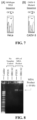
FIGS. 6A-6D: Gel images of benchtop and on-chip demonstration of specific MDA of the TP53 gene and smaller gene fragments from PCR. FIG. 6A: MDA product from reactions using 15 ng of purified HeLa gDNA. Different quantities of primers were tested, as well as negative controls containing no gDNA template. The MDA product was run on a 1% agarose gel and was approximately 10 kb. FIG. 6B: PCR product from reactions amplifying a shorter TP53 gene fragment using 60% of the MDA product. The PCR product was run on a 2.5% agarose gel and was 130 nt in length. FIG. 6C: On-chip MDA product from isolated HeLa and CAOV-3 cell gDNA verified using a 1% agarose gel. The ladder indicates that the product was around 10 kb in length. FIG. 6D: PCR product verified via 8% PAGE. The ladder confirmed a product length of 130 bp.
FIGS. 7A-7B: Sanger sequencing results from HeLa and CAOV-3 cell TP53 gene fragments where a SNP is known to occur in CAOV-3 cells. FIG. 7A: Wildtype sequence for the TP53 region is shown and was obtained from the International Agency for Research on Cancer. Sequencing results for the HeLa cell TP53 region are shown. FIG. 7B: Mutant sequence for the known SNP in CAOV-3 cells where a C is replaced by a T. Sequencing results for the CAOV-3 cell TP53 region are shown. The specific base that is mutated is underlined. FIG. 12 shows the wildtype sequence (SEQ ID NO:1) and the sequencing results for the TP53 fragments from HeLa (SEQ ID NO:2) and CAOV-3 (SEQ ID NO:3) cells.
FIG. 8 : Benchtop MDA results for PTEN and BRCA2 genes. MDA reactions were performed using the primers for PTEN and BRCA2, and controls containing no gDNA and reactions containing 15 ng of gDNA extracted from CAOV-3 cells were performed. The reaction products were analyzed on a 1% agarose gel. The yellow arrow indicates the 10-kb MDA product band on the gel.
FIG. 9 : Benchtop PCR results for PTEN and BRCA2 genes. PCR reactions were performed using the primers for PTEN and BRCA2, and controls containing no MDA product and reactions containing 60% of the MDA product were performed. The reaction products were analyzed on a 10% polyacrylamide gel. The yellow arrow indicates the 130-nt PCR product band on the gel.
FIG. 10 : On-chip MDA results for PTEN and BRCA2 genes. MDA reactions were performed within channels containing isolated CAOV-3 gDNA using either PTEN or BRCA2 MDA primers. The MDA product was analyzed on a 1% agarose gel. The yellow arrows indicate the ˜10-kb MDA product.
FIG. 11 : Product from PCR reaction using 60% of the MDA product amplifying the PTEN and BRCA2 genes on-chip from CAOV-3 cells. PCR reactions were performed using the primers for PTEN and BRCA2, and controls containing no MDA product and reactions containing 60% of the MDA product were performed. The PCR products were analyzed on a 10% PAGE gel. The yellow arrows indicate the 130-bp PCR product.
FIG. 12 : Sanger sequencing results indicating the point mutation in the TP53 gene in CAOV-3 cells. The wildtype sequence (SEQ ID NO:1) for this fragment of the TP53 gene is shown at the top of the figure. The upper plot shows the sequencing results for this gene fragment (SEQ ID NO:2) for HeLa cells. The lower plot shows the sequencing results for this gene fragment (SEQ ID NO:3) for CAOV-3 cells. The yellow box highlights the region where the mutation is located, and the base that is mutated is underlined in black. Some bases were not called by the sequencing software, but the bases can be determined by looking at the plot peaks.
DETAILED DESCRIPTION OF THE INVENTION
The present invention relates to, inter alia, a combination of microfluidic and aptamer technologies suitable for use in studying, analyzing, detecting, and treating various conditions and diseases.
More particularly, in one aspect, the present invention relates to a microfluidic device for aptamer-based cancer cell capture and genetic mutation detection, and the use of the microfluidic device for various applications.
In one aspect, the present invention provides a microfluidic device comprising: a cell microchannel and a nucleic acid microchannel that intersect to form a cell capture intersection region; a cell capture array comprising a plurality of cell capturing micropillars configured and arranged in a manner effective to capture one or more target cell when flowed through the cell microchannel, said cell capture array being located in the cell capture intersection region; and a nucleic acid entanglement array comprising a plurality of nucleic acid entanglement micropillars configured and arranged in a manner effective to physically entangle and maintain thereon genomic DNA isolated from the one or more target cell, said nucleic acid entanglement array being located in a portion of the nucleic acid microchannel that is adjacent to and downstream of the cell capture intersection region. The microfluidic device is multi-functional in that it is effective for capturing said one or more target cell, isolating said genomic DNA from the one or more target cell, and analyzing said genomic DNA in a self-contained manner.
In one embodiment of the microfluidic device of the present invention, the cell capture array comprises one or more aptamer and/or another cell capture component specific to the one or more target cell.
In another embodiment, the one or more aptamer and/or another cell capture component is concentrated in the cell capture intersection region, thereby enabling capture of the one or more target cell.
In another embodiment, the nucleic acid entanglement array is effective to entangle and maintain the isolated genomic DNA for single amplification and/or multiple, consecutive amplifications of one or more nucleic acid sequence of interest contained on the isolated genomic DNA.
In certain embodiments, the one or more nucleic acid sequence of interest is a cancer gene. In other embodiments, the one or more target cell is a cancer cell.
In one embodiment, the microfluidic device of the present invention further comprises a first flow rate means for managing rate of flow of fluid through the cell microchannel and a second flow rate means for managing rate of flow of fluid through the nucleic acid microchannel.
In another embodiment, the first flow rate means comprises external valves at the nucleic acid microchannel inlet and outlet and the second flow rate means comprises external valves at the cell microchannel inlet and outlet.
In one embodiment, the external valves are selected from the group consisting of two-way valves and four-way valves.
In another embodiment, the microfluidic device of the present invention further comprises a temperature controller for managing temperature of fluid and other contents contained within the cell microchannel and/or nucleic acid microchannel.
In one embodiment, the cell microchannel and the nucleic acid microchannel have a height ranging from between about 20 μm and about 40 μm.
In one embodiment, the cell microchannel and the nucleic acid microchannel have a height of about 20, 21, 22, 23, 24, 25, 26, 27, 28, 29, 30, 31, 32, 33, 34, 35, 36, 37, 38, 39, or 40 μm.
In one embodiment, the cell microchannel and the nucleic acid microchannel have a height of about 25 μm.
In one embodiment, the cell channel has a width ranging from between about 500 μm and about 1500 μm.
In one embodiment, the cell channel has a width of about 500, 510, 520, 530, 540, 550, 560, 570, 580, 590, 600, 600, 610, 620, 630, 640, 650, 660, 670, 680, 690, 700, 710, 720, 730, 740, 750, 760, 770, 780, 790, 800, 810, 820, 830, 840, 850, 860, 870, 880, 890, 900, 910, 920, 930, 940, 950, 960, 970, 980, 990, or 1000 μm.
In one embodiment, the cell channel has a width of about 1000 μm.
In one embodiment, the nucleic acid channel has a width ranging from between about 200 μm and about 1500 μm.
In one embodiment, the nucleic acid channel has a width selected from the group consisting of about 200, 210, 220, 230, 240, 250, 260, 270, 280, 290, 300, 310, 320, 330, 340, 350, 360, 370, 380, 390, 400, 410, 420, 430, 440, 450, 460, 470, 480, 490, 500, 510, 520, 530, 540, 550, 560, 570, 580, 590, 600, 600, 610, 620, 630, 640, 650, 660, 670, 680, 690, 700, 710, 720, 730, 740, 750, 760, 770, 780, 790, 800, 810, 820, 830, 840, 850, 860, 870, 880, 890, 900, 910, 920, 930, 940, 950, 960, 970, 980, 990, 1000, 1010, 1020, 1030, 1040, 1050, 1060, 1070, 1080, 1090, 1100, 1120, 1130, 1140, 1150, 1160, 1170, 1180, 1190, 1200, 1210, 1220, 1230, 1240, 1250, 1260, 1270, 1280, 1290, 1300, 1310, 1320, 1330, 1340, 1350, 1360, 1370, 1380, 1390, 1400, 1410, 1420, 1430, 1440, 1450, 1460, 1470, 1480, 1490, or 1500 μm.
In one embodiment, the nucleic acid channel has a width selected from the group consisting of about 250 μm, 500 μm, and 1000 μm.
In one embodiment, the cell capturing micropillars have a diameter ranging from between about 40 μm and about 60 μm.
In one embodiment, the cell capturing micropillars have a diameter of about 40, 41, 42, 43, 44, 45, 46, 47, 48, 49, 50, 51, 52, 53, 54, 55, 56, 57, 58, 59, or 60 μm.
In one embodiment, the cell capturing micropillars have a diameter of about 50 μm.
In one embodiment, the cell capture array is ordered in a patterned array that is rotated by about 4° to maximize contact between the one or more target cell and microchannel surface.
In one embodiment, the nucleic acid entanglement micropillars have a diameter ranging from between about 2 μm and about 10 μm.
In one embodiment, the nucleic acid entanglement micropillars have a diameter of about 2.0, 2.5, 3.0, 3.5, 4.0, 4.5, 5.0, 5.5, 6.0, 6.5, 7.0, 7.5, 8.0, 8.5, 9.0, 9.5, or 10 μm.
In one embodiment, the nucleic acid entanglement micropillars have a cross-sectional dimension of about 4 μm×4 μm, wherein said nucleic acid entanglement micropillars are spaced in a gradient that begins with the micropillars being about 10 μm apart and ending with the micropillars being about 7 μm apart.
In another aspect, the present invention provides a method of isolating and maintaining genomic DNA of one or more target cell from a sample under flow for further analysis thereof. This method involves the steps of: providing a microfluidic device as described herein; introducing a sample comprising one or more target cell into the cell microchannel at a flow rate effective to transport the one or more target cell to the cell capture array so as to capture the one or more target cell in the cell capturing micropillars by specific binding; lysing the one or more target cell by introducing lysing reagents through the nucleic acid microchannel at a flow rate effective to release genomic DNA from the one or more target cell without shearing the genomic DNA; and maintaining fluid flow within the nucleic acid microchannel at a flow rate effective to cause the released genomic DNA to become physically entangled and maintained within the nucleic acid entanglement array for further analysis thereof.
In one embodiment, the one or more target cell is captured in the cell capture array due to specific contact with an aptamer or other capture component present in the cell capture array.
In one embodiment, the one or more target cell is introduced into the cell microchannel and thereafter captured within the cell capture array under a flow rate ranging from between about 0.1 μL/minute and about 20 μL/minute.
In one embodiment, the one or more target cell is introduced into the cell microchannel and thereafter captured within the cell capture array under a flow rate of about 0.1, 0.5, 1.0, 1.5, 2.0, 2.5, 3.0, 3.5, 4.0, 4.5, 5.0, 5.5, 6.0, 6.5, 7.0, 7.5, 8.0, 8.5, 9.0, 9.5, 10, 10.5, 11, 11.5, 12, 12.5, 13, 13.5, 14, 14.5, 15, 15.5, 16, 16.5, 17, 17.5, 18, 18.5, 19, 19.5, or 20 μL/minute.
In one embodiment, the lysing reagents are introduced through the nucleic acid microchannel at a flow rate ranging from between about 0.1 μL/minute and about 2 μL/minute, thereby causing the release of the genomic DNA from the one or more target cell without shearing the genomic DNA.
In one embodiment, the flow of fluid in the nucleic acid microchannel is maintained at a flow rate ranging from between about 0.05 μL/minute and about 2 μL/minute, thereby causing the released genomic DNA to become physically entangled and maintained within the nucleic acid entanglement array.
In one embodiment, the one or more target cell is a cancer cell, although the present invention can be used for any type of cell of structure that contains DNA or genomic DNA.
In another aspect, the present invention provides a method for conducting aptamer-based cancer cell capture and genomic DNA mutation analysis of genomic DNA isolated from one or more target cell. This method includes the steps of: performing the steps described herein of the method of isolating and maintaining genomic DNA of one or more target cell from a sample under flow; and conducting aptamer-based cancer cell capture and genomic DNA mutation analysis of the genomic DNA isolated from one or more target cell while in a flow environment within the microfluidic device.
In another aspect, the present invention provides a method for amplifying individual genes of interest from the one or more target cell consecutively and collecting each amplification product separately. This method includes the steps of: performing the steps described herein of the method of isolating and maintaining genomic DNA of one or more target cell from a sample under flow; and amplifying individual genes of interest from the genomic DNA entangled and maintained under flow within the nucleic acid entanglement array of the microfluidic device consecutively and collecting each amplification product separately.
In another aspect, the present invention provides a method for sequencing nucleic acids amplified from genomic DNA isolated from one or more target cell. This method includes the steps of: performing the steps described herein of the method of isolating and maintaining genomic DNA of one or more target cell from a sample under flow; and sequencing the genomic DNA entangled and maintained under flow within the nucleic acid entanglement array of the microfluidic device.
In another aspect, the present invention provides a method for multiple displacement amplification (MDA) reactions of one or more nucleic acid sequence isolated from one or more target cell. This method includes the steps of: performing the steps described herein of the method of isolating and maintaining genomic DNA of one or more target cell from a sample under flow; and conducting multiple displacement amplification (MDA) reactions under flow using the genomic DNA entangled and maintained within the nucleic acid entanglement array of the microfluidic device.
In one embodiment, the MDA reactions involve using the same genomic DNA template isolated by the nucleic acid entanglement micropillars of the microfluidic device.
FIGS. 1-3 provide schematic views of illustrative embodiments and aspects of the microfluidic device of the present invention. While the aforementioned figures relate to and are further described below and in the examples also provided herein below, these figures are helpful in describing the microfluidic device and any related systems and methods in general terms.
FIG. 1 and FIG. 3 are schematic illustrations aspects of one embodiment of microfluidic device 10 of the present disclosure. In particular, FIG. 1 and FIG. 3 illustrate microfluidic device 10 as having cell microchannel 20 and nucleic acid microchannel 30, which intersect to form cell capture intersection region 40. Microfluidic device 10 is also shown to include cell capture array 50, which includes a plurality of cell capturing micropillars 60 configured and arranged in a manner effective to capture one or more target cell when such cells are flowed through cell microchannel 20. Cell capture array 50 is located in cell capture intersection region 40. Microfluidic device 10 is also shown to include nucleic acid entanglement array 70, which includes a plurality of nucleic acid entanglement micropillars 80 configured and arranged in a manner effective to physically entangle and maintain thereon genomic DNA isolated from the one or more target cell. Nucleic acid entanglement array 70 is located in a portion of nucleic acid microchannel 30 that is adjacent to and downstream of cell capture intersection region 40. Microfluidic device 10 is multi-functional in that it is effective for capturing said one or more target cell, isolating said genomic DNA from the one or more target cell, and analyzing said genomic DNA in a self-contained manner.
FIG. 2 is a schematic of one embodiment of microfluidic device 10 of the present disclosure, which embodiment can be used for capturing different biological components from a sample. As shown in FIG. 2 , microfluidic device 10 can be used to capture genomic DNA 90 from three, different types of DNA sources, i.e., cancer cells, bacteria cells, and viruses, but from a single sample (e.g., a single blood sample). As shown in FIG. 2 , this embodiment of microfluidic device 10 illustrates three different cell capture intersection regions 40. Each cell capture intersection region 40 is formed by the intersection of three, separate nucleic acid microchannels 30 with different portions of cell microchannel 20. In particular, as shown in FIG. 2 (moving from left to right along cell microchannel 20), there is cell capture intersection region 40 for cancer cell capture, cell capture intersection region 40 for bacteria cell capture, and cell capture intersection region 40 for virus capture.
As illustrated in FIG. 2 , a blood sample enters microfluidic device 10 at an input port of cell microchannel 20 (left end in FIG. 2 ). The first cell capture intersection region 40 includes a cell capture array having cell capturing micropillars that are specific for certain cancer cells, so that such cancer cells are captured in the first capture intersection region 40 and then lysed, with genomic DNA 90 from the captured and lysed cancer cells being released and physically entangled and maintained by nucleic acid entanglement micropillars 80 of nucleic acid entanglement array 70 at a position within the first nucleic acid microchannel 30 that is downstream of the first cell capture intersection region 40. The second cell capture intersection region 40 (in the middle) includes a cell capture array having cell capturing micropillars that are specific for certain bacteria cells, so that such bacteria cells are captured in the second capture intersection region 40 and then lysed, with genomic DNA 90 from the captured and lysed bacteria cells being released and physically entangled and maintained by nucleic acid entanglement micropillars 80 of nucleic acid entanglement array 70 at a position within the second nucleic acid microchannel 30 that is downstream of the second cell capture intersection region 40. The third cell capture intersection region 40 (on the right) includes a cell capture array having cell capturing micropillars that are specific for certain viruses, so that such viruses are captured in the third capture intersection region 40 and then lysed, with genomic DNA 90 from the captured and lysed viruses being released and physically entangled and maintained by nucleic acid entanglement micropillars 80 of nucleic acid entanglement array 70 at a position within the third nucleic acid microchannel 30 that is downstream of the third cell capture intersection region 40.
Aspects of the microfluidic device of the present invention and various methods of use thereof are further described below. Certain terms are used that are interchangeable with the terms provided herein above and in the claims. Such interchangeable terminology is readily apparent to those of ordinary skill in the art.
In accordance with one embodiment, the present invention includes a microfluidic device capable of specifically capturing rare cancer cells and isolating their genomic DNA for on-chip amplification and subsequent genetic sequencing. The device is fabricated out of polydimethylsiloxane (PDMS) using a silicon mold. The PDMS is bonded to a glass substrate to form the microfluidic channels. The device contains two microchannels intersecting at right angles with a pillar array at the intersection functionalized with single stranded DNA aptamers that serve as specific capture ligands for cancer cells. The pillars are rotated 4° to increase the number of collisions cells undergo with the surface, thereby increasing the capture efficiency. In one of the channels downstream from the cell capture zone, there is a secondary micropillar array with smaller pillars spaced closely together that will isolate the genomic DNA from the lysed captured cells. This DNA will remain entangled in the micropillar array through multiple isothermal amplification reactions. The amplification product will be extracted from the outlet of the device, and the DNA will be sequenced to identify any genetic mutations.
As noted above, FIG. 1 is a schematic of one embodiment of the microfluidic device setup in accordance with the present invention. As shown in FIG. 1 , two intersecting microchannels with an aptamer-functionalized pillar array at the intersection for specific cancer cell capture. A finer micropillar array downstream for genomic DNA isolation via physical entanglement. The initial cell sample is injected into Inlet 1, flows through the capture region, and out Outlet 1. The lysis buffer is injected into Inlet 2, and the genomic DNA is entangled in the micropillar array, while the rest of the lysate flows out of the device through Outlet 2.
Aptamers are single stranded nucleic acids that are analogous to antibodies in that they are ligands with specific binding affinity to their target, but with several advantages over antibodies. Aptamers are chemically synthesized with no batch-to-batch variability, much less expensive, more robust, and more easily functionalized. Here, a previously-selected aptamer with specific affinity to surface species present on several types of cancer cells that will capture the rare cancer cells and filter them from a complex sample can be used (see Van Simaeys D, López-Colón D, Sefah K, Sutphen R, Jimenez E, Tan W (2010) Study of the molecular recognition of aptamers selected through ovarian cancer cell-SELEX. PLoS One 5:e13770. doi: 10.1371/journal.pone.0013770).
To detect specific genetic mutations, these genes can be individually amplified in separate multiple displacement amplification (MDA) reactions using the same genomic DNA template isolated by the micropillars. Following each amplification, the product can be extracted from the device through the outlet. These samples can be sequenced using Sanger sequencing, which requires pure samples with a single DNA sequence, and this can be accomplished using our device capable of separate consecutive amplification reactions on the same genomic DNA. The sequencing results can be compared to the known wildtype genes, and any mutations can be identified. This information can then be passed onto clinicians to make informed recommendations for the most appropriate and effective treatment for each patient.
In various other embodiments, the present invention provides a microfluidic device for capturing selected cells and efficiently separating genomic DNA from other cellular components.
Further, although certain aspects of the present invention primarily relate to the processing and analysis of DNA extracted and immobilized in the device and treating the other cellular components, in other aspects, the present invention also relates to the use of a microfluidic device for separating the cellular components from the genomic DNA such as mitochondrial DNA or RNA.
One advantage of the microfluidic device of the present invention is that it is an integrated device, as shown herein. The integrated device of the present invention obviates the need for transferring the sample form one device to another and therefore reduces the possibilities for contamination of the sample.
The structures that are employed in the microfluidic device of the present invention for capturing the cells can be of many forms. For example, in certain embodiments, the structures can be coated with antibodies or aptamers and can include, without limitation, structures such as pillars, beads, flat surfaces, or patterned surfaces. Such methods for incorporating such features in microfluidics are known to those skilled in the art.
In accordance with the present invention, the ability to capture the DNA by mechanical entanglement with pillared structures allows the application of sequential chemical treatments and washing steps by flowing different solutions past the immobilized DNA.
The microfluidic device of the present invention can be constructed of various suitable materials. For example, in certain embodiments, the microfluidic device is constructed of transparent plastic. However, the microfluidic device of the present invention can alternatively be made of glass or other optically transparent materials to permit optical microscopic imaging of captured cells and DNA. This would permit histological evaluation of cells by a Pathologist to combine conventional imaging and analysis of cells to be combined with our molecular diagnostics.
In certain embodiments, the microfluidic device of the present invention can include multiple stages of cell capture regions of different geometry and chemical coatings to capture different cell types or other biological entities such as cancer cells, shed cells from implanted tissue, parasites, pathogens, bacteria, viruses, and others.
As described herein, FIG. 2 illustrates one non-limiting embodiment of a microfluidic device having a channel structure for capturing different biological entities for analysis.
The microfluidic device can be used to study various genes of interest, including, without limitation, genes of interest in cancer. For example, as shown in the examples section below, one such gene of interest is the TP53 gene. Additional examples of genes of interest in cancer include, without limitation, APC, BRCA1, BRCA2, CDK4, CMM1, HER2, MLH1, MSH2, p16, and Rb1. The microfluidic device of the present invention can be used to study these and other cancer genes, as well as other genes not associated with cancer.
The microfluidic device of the present invention can be constructed so that it does not require internal valves to control the flow of fluid or other materials through the channels of the device. Thus, in accordance with the present invention, there are multiple ways to control the flow through the device. For example, external valves can be used to control the flow. Alternatively, tubing clamps or fluidic “plugs” can also be used to control the direction of flow in the microfluidic device of the present invention.
The microfluidic device of the present invention can also use various types of cell capture components in addition to those specifically described herein. For example, antibodies or other recognition elements could be used in the same way as described herein with the same surface chemistry (e.g., biotinylated). Such cell capture alternatives would work and could be incorporated into the microfluidic device.
The microfluidic device of the present invention can be used to incorporate various technologies relating to microfluidic arrays, microfluidic cell capture, aptamer-based cell capture, nucleic acid elongation and capture, and the like. Such compatible technologies described in the art can be found in various published U.S. patent applications, including, without limitation, the following (which are incorporated herein by reference in their entirety): US-2015/0291952, US-2015/0204859, US-2014/0121132, US-2014/0194313, and US-2015/0011425.
EXAMPLES
The following examples are intended to illustrate particular embodiments of the present invention, but are by no means intended to limit the scope of the present invention.
Example 1 Microfluidic Device for Aptamer-Based Cancer Cell Capture and Gene Mutation Detection
Genetic mutations in cancer cells are not only fundamental to the disease, but can also have tremendous impact on the efficacy of treatment. Identification of specific key mutations in a timely and cost-effective way would allow clinicians to better prescribe the most effective treatment strategy. Here, we present a novel microfluidic device as a platform for specifically capturing cancer cells and isolating their genomic DNA (gDNA) for specific amplification and sequence analysis. To capture cancer cells within the device, nucleic acid aptamers that specifically bind to cancer cells are immobilized within a microchannel containing micropillars to increase capture efficiency. The captured cells are then lysed and the gDNA is isolated via physical entanglement within a secondary micropillar array. This type of isolation allows the gDNA to be retained within the channel, and enables multiple types of analysis to be performed on the same gDNA template. The amplified gene samples undergo sequencing, and the resulting sequence information is compared against the known wildtype gene to identify any mutations. Cervical and ovarian cancer cells have been tested for mutations in the TP53, PTEN, and BRCA2 genes using this technology. This approach offers a way to monitor multiple genetic mutations in the same small population of cells, which is beneficial given the wide diversity in cancer cells, and requires very few cells to be extracted from the patient sample. With this capability for genetic monitoring, precision medicine should be more accessible for the treatment of cancer.
Experimental
Cell Culture and Buffers:
HeLa, CAOV-3, Ect1/E6E7, and End1/E6E7 cells were purchased from American Type Cell Culture (ATCC). The HeLa and CAOV-3 cells were cultured in DMEM media (Life Technologies) supplemented with non-essential amino acids, 110 mg/L sodium pyruvate, 200 mM L-glutamine (Life Technologies), 1× Pen Strep (Life Technologies), 26.8 mM HEPES, betamercaptoethanol, and containing 10% fetal bovine serum. The Ect1/E6E7 and End1/E6E7 cells were cultured in keratinocyte-serum free media (Life Technologies) with 0.1 ng/mL human recombinant epidermal growth factor (Life Technologies), 0.05 mg/mL bovine pituitary extract (Life Technologies), and an additional 44.1 mg/L calcium chloride. Prior to use, the cells were trypsinized and resuspended in phosphate-buffered saline (PBS) binding buffer [1× Dulbecco's PBS with calcium chloride and magnesium chloride (Fisher Scientific) containing 4.5 g/L glucose, 5 mM MgCl2, and 0.1% Pluronic F-68 non-ionic surfactant (Sigma Aldrich)]. HeLa cells contained GFP-conjugated histones, and CAOV-3, Ect1/E6E7, and End1/E6E7 cells were stained with calcein-AM (Thermo Fisher Scientific) that allowed them to be observed using fluorescence microscopy.
Device Fabrication:
The devices used here were polydimethylsiloxane (PDMS) microchannels bonded to glass substrates. The devices consisted of two orthogonal microchannels that contained two micropillar arrays: a cell capture array at the intersection of the two microchannels and a gDNA isolation array downstream of the cell capture array (see FIG. 3 ). The PDMS channels were made via soft lithography using a silicon master mold. The master mold was fabricated from a 4″ silicon wafer via standard photolithography. Microposit S1813 photoresist (Shipley) was spun onto silicon wafers and exposed to UV light using a contact mask aligner (ABM). The exposed wafer was developed using 726MIF developer (Microchemicals) and the pattern was etched 25 μm into the silicon via Bosch process using a Unaxis SLR 770 deep reactive ion etching system (Unaxis USA Inc.). A monolayer of (1H,1H,2H,2H-Perfluorooctyl)Trichlorosilane (FOTS) was deposited onto the etched silicon surface using an MVD100 molecular vapor deposition system (Applied Microstructures) to enable easy release of the PDMS from the silicon mold.
Sylgard 184 (Dow Corning) PDMS base resin was mixed in a 10:1 ratio with curing agent, and degassed in a vacuum oven at room temperature. The PDMS was poured onto the master mold and baked at 140° C. for 1 hour. The PDMS was allowed to cool to room temperature, and it was carefully peeled off of the mold. Inlet and outlet holes were created using a 1 mm biopsy punch (Sklar Instruments). To complete the formation of the channels, the patterned PDMS and a microscope slide were treated with oxygen plasma for 5 min, and then bonded together. An external 4-way L-type valve (IDEX Health and Science) was used at the DNA channel inlet and external 2-way valves were used at the cell channel inlet and both outlets to control the flow between the perpendicular channels.
Surface Chemistry:
DNA aptamers that were used to capture cancer cells were immobilized onto the surface of the microchannels via a streptavidin-biotin conjugation. The channels were initially primed and cleaned with 100% ethanol and ultrapure water. A 1×PBS solution was pumped through the channels at 50 μL/min for 9 min. A 1 mg/mL solution of streptavidin (Life Technologies) in PBS was prepared, and 120 μL were pumped through the cell channel (see FIG. 3 ) at 4 μL/min to immobilize streptavidin via adsorption onto the channel surface. A blocking solution containing 1% bovine serum albumin (BSA), 1% Pluronic F-68, and 1% polyvinylpyrrolidone (PVP) K15 was prepared in 1×PBS with calcium chloride and magnesium chloride, and 500 μL, of this solution was pumped through both the cell and DNA channels at 4 μL/min to block the surface of the channel and prevent non-specific adhesion of cells and other reagents. Finally, biotinylated aptamers (Integrated DNA Technologies), which were previously selected (DOV4) to bind to several different types of cancer cells,18 were diluted to 10 μM in PBS binding buffer, and 180 μL were pumped through the DNA channel at 3 μL/min. This process enabled the aptamers to be concentrated in the cell capture region at the intersection of the two channels.
Cell Capture, Lysis, and gDNA Isolation:
Cells suspended in PBS binding buffer were pumped through the cell channel (see FIG. 3 ) at 1 μL/min until several cells were captured, as observed via fluorescence microscopy. A solution of 6 M guanidinium isothiocyanate (VWR) was pumped at 1 μL/min through the DNA channel until cell lysis was observed. By lysing the cells in the direction of the DNA micropillar array, the gDNA becomes physically entangled and remains in the channel even under flow. To verify the gDNA was captured and retained by the micropillar array, YOYO-1 dye was used to stain the DNA in the channel by flowing the dye through the DNA channel at 1 μL/min. Otherwise, following lysis, the gDNA was washed with 1× Tris-Ethylenediaminetetraacetic acid (TE) buffer to remove any remaining cellular debris and guanidinium isothiocyanate.
Primers and Specific Gene Amplification from gDNA:
To amplify a specific gene from gDNA isothermally, MDA was used with specific primers targeting a fragment of the TP53, PTEN, or BRCA2 gene. Table 1 contains the 40 short primers used for these amplifications. Initially, the isolated gDNA was chemically denatured using Buffer DLB from the Repli-g Mini Kit (Qiagen) prepared according to the manufacturer's instructions. This solution was pumped through the DNA channel at 1 μL/min for 25 min. The gDNA was then neutralized using the Stop Solution from the Repli-g Mini Kit with the addition of 12 pmol of each MDA primer. This solution was pumped at 1 μL/min for 45 min. An MDA reaction solution was prepared from the Repli-g Mitochondrial DNA Kit according to the manufacturer's instructions, and 15-30 pmol of each MDA primer was added to the reaction solution. The microchannel device was placed on a hotplate set to 33° C., and 50 μL of MDA reaction solution was pumped through at 0.05 μL/min to perform the MDA reaction for approximately 16 hours. The eluent was collected in an Eppendorf tube. For consecutive rounds of on-chip MDA gene amplification from the same gDNA, the gDNA was denatured again, neutralized, and the MDA reaction was performed as described above, but using different primers.
For benchtop control experiments, gDNA from HeLa and CAOV-3 cells was extracted using the Blood and Cell Culture DNA Mini Kit (Qiagen). Buffer D1 and Buffer N1 from the Repli-g Mini Kit (Qiagen) were prepared, and 15 ng of extracted gDNA were denatured and neutralized according to the manufacturer's instructions. An MDA reaction master mix was prepared using the buffer and DNA polymerase from the Repli-g Mitochondrial DNA Kit (Qiagen) and various concentrations of specific MDA primers (Table 1), and this solution was added to the denatured and neutralized gDNA. This reaction was incubated at 33° C. for 16 hours.
TABLE 1
Amplification Primer Sequences
MDA Primers for the TP53, PTEN, and BRCA2 Genes
Primer Name Sequence Primer Name Sequence
TP53 MDA FOR #1 TTG TGC CCT G TP53 MDA REV #1 GCC ATG GCG C
(SEQ ID NO: 4) (SEQ ID NO: 5)
TP53 MDA FOR #2 CAG TTG CTT T TP53 MDA REV #2 GCT GTG ACT G
(SEQ ID NO: 6) (SEQ ID NO: 7)
TP53 MDA FOR #3 GTT TCT TTG C TP53 MDA REV #3 CCT CAC AAC C
(SEQ ID NO: 8) (SEQ ID NO: 9)
TP53 MDA FOR #4 GGA GGT GCT T TP53 MDA REV #4 CGC TCA TGG T
(SEQ ID NO: 10) (SEQ ID NO: 11)
TP53 MDA FOR #5 TAG CTC GCT A TP53 MDA REV #5 CAC CAT CGC T
(SEQ ID NO: 12) (SEQ ID NO: 13)
TP53 MDA FOR #6 GTG TAG ACG C TP53 MDA REV #6 TCT CTC CAG C
(SEQ ID NO: 14) (SEQ ID NO: 15)
TP53 MDA FOR #7 CCT ATC TCA A TP53 MDA REV #7 CTG GGC AAC C
(SEQ ID NO: 16) (SEQ ID NO: 17)
TP53 MDA FOR #8 CCT GAG TGA C TP53 MDA REV #8 GAA TCA GAG G
(SEQ ID NO: 18) (SEQ ID NO: 19)
TP53 MDA FOR #9 ATC ACA CCA C TP53 MDA REV #9 ACC TAA GAG C
(SEQ ID NO: 20) (SEQ ID NO: 21)
TP53 MDA FOR #10 GGA GGC TGC A TP53 MDA REV #10 AGA TGC TGA G
(SEQ ID NO: 22) (SEQ ID NO: 23)
TP53 MDA FOR #11 ATC ACT TGA G TP53 MDA REV #11 TCC ACA CGC A
(SEQ ID NO: 24) (SEQ ID NO: 25)
TP53 MDA FOR #12 TAG GAG GCT G TP53 MDA REV #12 TGT TTC TGT C
(SEQ ID NO: 26) (SEQ ID NO: 27)
TP53 MDA FOR #13 CAC CTA TAG T TP53 MDA REV #13 CAC CAC ACT A
(SEQ ID NO: 28) (SEQ ID NO: 29)
TP53 MDA FOR #14 TAG CCA GGC A TP53 MDA REV #14 TCA GGC GGC T
(SEQ ID NO: 30) (SEQ ID NO: 31)
TP53 MDA FOR #15 ACC TCG TCT C TP53 MDA REV #15 CCC CAG TTG C
(SEQ ID NO: 32) (SEQ ID NO: 33)
TP53 MDA FOR #16 AGC CTG GGT A TP53 MDA REV #16 TTA ACC CCT C
(SEQ ID NO: 34) (SEQ ID NO: 35)
TP53 MDA FOR #17 GAG CCC AGG A TP53 MDA REV #17 AGG GCC ACT G
(SEQ ID NO: 36) (SEQ ID NO: 37)
TP53 MDA FOR #18 CCA AGG CAG G TP53 MDA REV #18 CCC CCC TACT
(SEQ ID NO: 38) (SEQ ID NO: 39)
TP53 MDA FOR #19 TCC CAG CAC T TP53 MDA REV #19 TAG GGA GGT C
(SEQ ID NO: 40) (SEQ ID NO: 41)
TP53 MDA FOR #20 TTG GTG GCT C TP53 MDA REV #20 TTG CAC ATC T
(SEQ ID NO: 42) (SEQ ID NO: 43)
PTEN MDA FOR #1 GTC CAG AGC C PTEN MDA REV #1 GCA GCC GCA G
(SEQ ID NO: 44) (SEQ ID NO: 45)
PTEN MDA FOR #2 AGC CGC CGC A PTEN MDA REV #2 GAA COT GGG A
(SEQ ID NO: 46) (SEQ ID NO: 47)
PTEN MDA FOR #3 AGT CGC TGC A PTEN MDA REV #3 TTC GCA TCC G
(SEQ ID NO: 48) (SEQ ID NO: 49)
PTEN MDA FOR #4 TGC TGA GGA G PTEN MDA REV #4 CTG ACC AGG G
(SEQ ID NO: 50) (SEQ ID NO: 51)
PTEN MDA FOR #5 GCC GCT GCC A PTEN MDA REV #5 CAC ACC CTA G
(SEQ ID NO: 52) (SEQ ID NO: 53)
PTEN MDA FOR #6 CTT CTC CCC A PTEN MDA REV #6 GCA ACC AGG C
(SEQ ID NO: 54) (SEQ ID NO: 55)
PTEN MDA FOR #7 CCG TTC GGA G PTEN MDA REV #7 CAA TCG GTG G
(SEQ ID NO: 56) (SEQ ID NO: 57)
PTEN MDA FOR #8 GAG GCC GCC G PTEN MDA REV #8 GGA AGA GAC C
(SEQ ID NO: 58) (SEQ ID NO: 59)
PTEN MDA FOR #9 CCC CCG TGG C PTEN MDA REV #9 CCG AGC TCG C
(SEQ ID NO: 60) (SEQ ID NO: 61)
PTEN MDA FOR #10 CCC GTC CGC C PTEN MDA REV #10 GCT CAA CCT C
(SEQ ID NO: 62) (SEQ ID NO: 63)
PTEN MDA FOR #11 CCC GGG CCG G PTEN MDA REV #11 GGG CCT AAG C
(SEQ ID NO: 64) (SEQ ID NO: 65)
PTEN MDA FOR #12 GGC GGC GGC A PTEN MDA REV #12 CGT GCA ACA C
(SEQ ID NO: 66) (SEQ ID NO: 67)
PTEN MDA FOR #13 GCC TCC AAC A PTEN MDA REV #13  GCA ACT CAG G
(SEQ ID NO: 68) (SEQ ID NO: 69)
PTEN MDA FOR #14 CGC CGG AGA G PTEN MDA REV #14 GCC CTC AAT G
(SEQ ID NO: 70) (SEQ ID NO: 71)
PTEN MDA FOR #15 GTC GCC TGT C PTEN MDA REV #15 CCC CAT AGG G
(SEQ ID NO: 72) (SEQ ID NO: 73)
PTEN MDA FOR #16 GCT CCA GGG A PTEN MDA REV #16 CTG GGA TCA G
(SEQ ID NO: 74) (SEQ ID NO: 75)
PTEN MDA FOR #17 CGG CGG CCG C PTEN MDA REV #17 CTG GCA TCA C
(SEQ ID NO: 76) (SEQ ID NO: 77)
PTEN MDA FOR #18 GGG AGA AGC G PTEN MDA REV #18 CGG TGA GCA G
(SEQ ID NO: 78) (SEQ ID NO: 79)
PTEN MDA FOR #19 CAC CTC CCG C PTEN MDA REV #19 TCA GAG GAC C
(SEQ ID NO: 80) (SEQ ID NO: 81)
PTEN MDA FOR #20 AGG CGC GGC G PTEN MDA REV #20 CCC CTC TTG C
(SEQ ID NO: 82) (SEQ ID NO: 83)
BRCA2 MDA FOR #1 CCT CAG TCA C BRCA2 MDA REV #1 GAC GTA CTG G
(SEQ ID NO: 84) (SEQ ID NO: 85)
BRCA2 MDA FOR #2 TCC AGC GCT T BRCA2 MDA REV #2 CCA CTA GAC G
(SEQ ID NO: 86) (SEQ ID NO: 87)
BRCA2 MDA FOR #3 GAT GGG ACT G BRCA2 MDA REV #3 AGG GGC CAG T
(SEQ ID NO: 88) (SEQ ID NO: 89)
BRCA2 MDA FOR #4 CCC ATC CTC A BRCA2 MDA REV #4 GGG GGT CCT A
(SEQ ID NO: 90) (SEQ ID NO: 91)
BRCA2 MDA FOR #5 TGG ACA GCA C BRCA2 MDA REV #5 CAC CAC GCC C
(SEQ ID NO: 92) (SEQ ID NO: 93)
BRCA2 MDA FOR #6 GGC TAG TGG C BRCA2 MDA REV #6 AAG TGC TGG G
(SEQ ID NO: 94) (SEQ ID NO: 95)
BRCA2 MDA FOR #7 ACT AGC CAC G BRCA2 MDA REV #7 TCC GCC CAC C
(SEQ ID NO: 96) (SEQ ID NO: 97)
BRCA2 MDA FOR #8 CTG GTT AGC G BRCA2 MDA REV #8 ACT TCT GGC C
(SEQ ID NO: 98) (SEQ ID NO: 99)
BRCA2 MDA FOR #9 CTC TGT CCC C BRCA2 MDA REV #9 GTT GGC CAG G
(SEQ ID NO: 100) (SEQ ID NO: 101)
BRCA2 MDA FOR #10 GCG GCG TTG G BRCA2 MDA REV #10 CAC GCA GCA C
(SEQ ID NO: 102) (SEQ ID NO: 103)
BRCA2 MDA FOR #11 TGC TGA TGG G BRCA2 MDA REV #11 GTA GCT GGG A
(SEQ ID NO: 104) (SEQ ID NO: 105)
BRCA2 MDA FOR #12 AAG CCC CTG G BRCA2 MDA REV #12 CTC CTG CCT C
(SEQ ID NO: 106) (SEQ ID NO: 107)
BRCA2 MDA FOR #13 GGG CAC GCT G BRCA2 MDA REV #13 CCT CCA GGG T
(SEQ ID NO: 108) (SEQ ID NO: 109)
BRCA2 MDA FOR #14 AGA TGA CGC C BRCA2 MDA REV #14 CTT GGC TCA C
(SEQ ID NO: 110) (SEQ ID NO: 111)
BRCA2 MDA FOR #15 CTT CCC TCC C BRCA2 MDA REV #15 TOG CCC AGG C
(SEQ ID NO: 112) (SEQ ID NO: 113)
BRCA2 MDA FOR #16 CCT TCA GCT C BRCA2 MDA REV #16 CAA CGT GAG G
(SEQ ID NO: 114) (SEQ ID NO: 115)
BRCA2 MDA FOR #17 GGC CCG GCC T BRCA2 MDA REV #17 GCG ACA TGG G
(SEQ ID NO: 116) (SEQ ID NO: 117)
BRCA2 MDA FOR #18 CTG GGT CTC C BRCA2 MDA REV #18 TCC ATG GTG G
(SEQ ID NO: 118) (SEQ ID NO: 119)
BRCA2 MDA FOR #19 CGA GCC CCG C BRCA2 MDA REV #19 CCA GTG GCT C
(SEQ ID NO: 120) (SEQ ID NO: 121)
BRCA2 MDA FOR #20 GCC CGA AGG C BRCA2 MDA REV #20 TGC TGC AAG G
(SEQ ID NO: 122) (SEQ ID NO: 123)
PCR Primers
Primer Name  Sequence
TP53 PCR FOR ACT TTC AAC TCT GTC TCC TTC CTC TTC CTA
(SEQ ID NO: 124)
TP53 PCR REV GGA CGC GGG TGC CG
(SEQ ID NO: 125)
PTEN PCR FOR AAG CGG CGG CAG AGC G
(SEQ ID NO: 126)
PTEN PCR REV TCT GGG AGC CTG TGG CTG AAG
(SEQ ID NO: 127)
BRCA2 PCR FOR ATG CAT CCC TGT GTA AGT GCA TTT TGG
(SEQ ID NO: 128)
BRCA2 PCR REV GCG TGT CTT AAA AAT TTC AAA AAA TGT TGG CCT CT
(SEQ ID NO: 129)

Gene Sequencing:
The MDA product was verified by running the product on a 1% agarose gel, and staining the gel with SYBR Gold (Thermo Fisher Scientific). Since the product was around 10 kilobases (kb), a smaller product must be made to enable sequencing. Thus, a PCR amplification was performed using 10-60% of the MDA product. The PCR product was verified and purified using 8-10% polyacrylamide gel electrophoresis (PAGE) stained with ethidium bromide or SYBR Gold. To purify the PCR product for sequencing, the 130-nt band was cut from the gel, crushed, suspended in 3 M NaAc pH 6.2, and incubated at 37° C. with mixing overnight. The gel pieces were removed from the sample, the sample underwent phenol-chloroform extraction and ethanol precipitation, and the DNA was resuspended in DEPC water. This purified PCR product was sequenced via Sanger Sequencing, and the sequence was compared to the known wildtype gene.
Results and Discussion
Microchannel Design:
The assay presented here involves two main steps, cancer cell capture using aptamers and genomic DNA analysis. The ability to perform these two step easily all within one device would significantly reduce contamination and sample loss, which is important when developing an assay for rare cells. Therefore, the device was designed to have two intersecting orthogonal microchannels: the cell channel where cancer cells are captured, and the DNA channel where the gDNA from the captured cells is isolated (FIG. 3 ). The cell channel was 1 mm wide, and the DNA channel was 250 μm, 500 μm, or 1 mm wide. The depth of the channels was ˜25 μm. The device also contained two micropillar arrays, one for cancer cell capture and one for gDNA isolation. The cell capture array was located at the intersection of the two microchannels, and consisted of pillars 50 μm in diameter. This array was rotated by 4° to maximize the contact between the cells and the channel surface containing the aptamers, thereby improving the capture efficiency.19 The DNA micropillar array was very similar to our previously-developed microarray for gDNA isolation.17 The array presented here consisted of 4×4 μm pillars spaced in a gradient that started with the pillars 10 μm apart and finished with them 7 μm apart. These small pillars placed in such a fine array caused the gDNA to become physically entangled and remain within the array even under flow, while allowing the cellular debris to flow out of the device. This is highly advantageous and enables multiple types of analysis to be performed on the same gDNA template.
To control the flow within the device, valves were needed at the inlets and outlets. An external 4-way L-type valve was used at the DNA channel inlet to not only control flow, but also eliminate most of the dead volume caused by the length of tubing needed to reach the syringe pump while the device is either observed under a microscope or is incubated using a hotplate. This is necessary because high initial flowrates cannot be used after the gDNA is isolated on the micropillar array, as there is a risk of losing some of the entangled gDNA under the high flow conditions. External 2-way shutoff valves were used at each of the outlets as well as the cell channel inlet to control the fluid flow. These types of valves were used because they do not disturb the fluid within the channels, whereas controlling the flow using tubing clamps causes significant flow during the clamping process, which can disturb the captured cells. This device design and setup enables this assay to be performed easily by the user.
HeLa and CAOV-3 Cell Capture and Genomic DNA Isolation:
The target cells were suspended in PBS binding buffer and pumped through the device with aptamers immobilized on the surface of the cell capture region containing a pillar array. The cell capture was most efficient at a flowrate of 5 μL/min when the cells were freshly trypsinized and the channel depth was ˜25 μm. Several flowrates were tested ranging from 20 μL/min down to 0.1 μL/min. At high flowrates (>10 μL/min), the linear velocity of the cells was very high and the cells did not bind to the aptamers on the surface easily. At low flowrates (<5 μL/min), the cells did bind to the aptamers, but the throughput was very low and there was increased non-specific adhesion of the cells to the device. Cells used from frozen stocks did not bind as readily, likely due to different surface compositions caused by the shock of a freeze/thaw cycle. With channels deeper than 25 μm, the cell capture took longer, since fewer collisions with the channel surface were likely occurring. To demonstrate using aptamers to specifically capture cancer cells within the microchannels, HeLa and CAOV-3 cancer cells were tested with and without aptamers immobilized on the surface, and Ect1/E6E7 and End1/E6E7 non-cancerous cells were tested with aptamers immobilized on the surface. FIG. 4A and FIG. 4B show the cell capture region without aptamers, and indicate that HeLa and CAOV-3 cells do not non-specifically adhere to the surface of the device. FIG. 4C and FIG. 4D show the cell capture region with aptamers immobilized, and demonstrate that Ect1/E6E7 and End1/E6E7 non-cancerous cells neither bind non-specifically to the device, nor bind to the aptamers. FIG. 4E and FIG. 4F show that HeLa and CAOV-3 cancer cells bind to aptamers immobilized within the cell capture region of the device. These results demonstrate the successful use of aptamers to specifically capture cancer cells.
After successfully capturing cancer cells within the cell capture region, the cells were lysed through the DNA channel. The cell contents flowed through the DNA micropillar array, and the gDNA was isolated on the pillars via physical entanglement. To verify that the gDNA was isolated and retained within the micropillar array, the gDNA was stained with YOYO-1 dye, and the results are shown in FIG. 5 for both HeLa and CAOV-3 cells. The gDNA from both types of cells was successfully isolated by the micropillar array and remained within the microchannel after multiple hours of flow. This gDNA isolation technique has several advantages, including allowing the gDNA to be completely chemically available for any downstream reaction, and more importantly it enables the gDNA to be retained within the device even under flow. This enables multiple types of analyses to be performed on the same template gDNA, so a large amount of information can be gathered from a single small population of cells without necessitating substantial whole genome amplification, which can be biased and information can be lost.20-22
Specific Isothermal Amplification of Genes from gDNA:
With the gDNA isolated and retained within the micropillar array, several types of genetic analyses can be performed. Here, we demonstrated the amplification of specific genes to determine their nucleotide sequence and discover any genetic mutations. MDA, an isothermal amplification technique, was used to reduce the complexity and cost of the device and enable straightforward device operation, since thermocycling is not necessary. MDA uses many short primers and a strand-displacing DNA polymerase (φ29) to make numerous copies of the template DNA isothermally. Whereas MDA is commonly used as a method for whole genome amplification,23-26 in this work we use MDA to amplify a specific gene from gDNA. To our knowledge, this is the first demonstration of using MDA to amplify a specific gene from gDNA. Forty 10-nt primers (20 forward and 20 reverse primers) were designed around regions of the TP53, PTEN, and BRCA2 genes, which are commonly mutated genes in cervical and ovarian cancer.27,30 This modified MDA technique for amplifying these genes from gDNA was initially verified in benchtop experiments using purified HeLa and CAOV-3 gDNA. FIG. 6A shows the successful amplification of TP53 from purified HeLa gDNA. FIG. 8 shows the successful amplification of PTEN and BRCA2 from purified CAOV-3 gDNA. Shorter gene fragments were also produced via PCR from these MDA products (FIG. 6B and FIG. 9 ).
The isolated gDNA from HeLa and CAOV-3 cells was first chemically denatured to create single stranded DNA (ssDNA). The DNA was then neutralized prior to the MDA reaction. Since the gDNA is entangled in the micropillar array, upon denaturation the two complimentary strands likely cannot diffuse and migrate away from each other as readily as they can in a bulk solution, so it is possible that some may rehybridize upon neutralization. This would prevent the MDA reaction from occurring efficiently. To reduce the likelihood of this occurring, the neutralization buffer was spiked with the MDA primers allowing the primers to bind to the ssDNA before it rehybridizes. The MDA reaction solution containing additional primers and the DNA polymerase was pumped through the device and the amplification product was collected. The MDA product from HeLa and CAOV-3 cell gDNA was analyzed on an agarose gel, and FIG. 6C shows images of the gels after a successful MDA reaction. The MDA product DNA strands were around 10 kb in length, and this is because the φ29 polymerase is known to extend for approximately 10 kb.28,29 To prepare the amplified gene sample for sequencing, it must be shortened to accommodate the sequencing technology. For Sanger sequencing, up to a few hundred bases can be sequenced. To create a shorter gene fragment from the HeLa and CAOV-3 MDA product, PCR was used to create 130-nt DNA strands, which were verified via PAGE (FIG. 6D). MDA is an advantageous technique to use not only because it is isothermal, but also because amplifying 10 kb strands allows large strands of gDNA to be amplified from cells. These strands can be completely sequenced inexpensively by performing a few PCRs to isolate different regions of the strand, and performing Sanger sequencing on these pure samples. Alternatively, the MDA product can be digested into shorter fragments and targeted next-generation sequencing can be performed.
Sequencing of the TP53 Gene Fragment:
The purified TP53 gene fragments from HeLa and CAOV-3 cells were sequenced via Sanger sequencing. This sequencing technique was chosen because it is fast, inexpensive, and can perform longer reads than other techniques. It also requires the sample to be pure, as only one read is performed. The wildtype sequence for TP53 was obtained from the International Agency for Research on Cancer, and the known mutation in the TP53 gene in CAOV-3 cells was obtained from the mutation data for this cell line from ATCC. CAOV-3 cells contain a homozygous single-nucleotide polymorphism (SNP), and this SNP (c.406C>T) is a nonsense mutation resulting in a truncated p53 protein. FIG. 7 shows the wildtype and mutated nucleotide sequence and the sequencing results from HeLa (FIG. 7A) and CAOV-3 (FIG. 7B) cells for the region where the SNP occurred. These results show that we were able to successfully detect a point mutation in the CAOV-3 TP53 gene, while HeLa cells contained the wildtype sequence of this gene fragment. Analogous testing of all locations on this gene would allow us to determine additional genetic information about this gene in these two cancer cell lines. Additional genes that commonly contain mutations in cancer could also be tested in this way, since the gDNA is retained on the micropillars and is available for additional amplification reactions and analysis.
Conclusions
We have developed a device and technique for capturing cancer cells using aptamers, and isolating their gDNA for genetic analysis. The device was designed to enable both the cell capture and gDNA isolation to occur within a single device to significantly reduce contamination and sample loss. Preventing sample loss is particularly important, since most cells of interest, such as circulating tumor cells, are rare and are generally found in low numbers. The device was also designed to isolate the gDNA via physical entanglement within a micropillar array. This isolation technique allows the gDNA to be chemically available for downstream reactions. It also enables the gDNA to be retained within the microchannel even under flow, which enables multiple types of analyses to be performed on the same template gDNA.
We demonstrated on-chip isothermal amplification of a specific gene and subsequent sequencing of this gene to determine any genetic mutations. We successfully detected a point mutation in the TP53 gene in CAOV-3 cells. Specifically, we developed a method using MDA to amplify a specific gene from whole gDNA. This technique allows for long strands of DNA to be produced that can cover an entire large gene up to 10 kb. Here, we used PCR to produce pure samples of smaller gene fragments that could be sequenced using Sanger sequencing, but other sequencing methods can be used where pure samples are not necessary as multiple reads are taken.
An assay like this can be extremely impactful when it comes to cancer diagnosis and treatment. If the mutations present in a specific patient's cancer cells can be identified, clinicians will be able to use this information to better diagnose the patient and better determine the most effective treatment options. This would truly enable precision medicine when it comes to cancer treatment, which is necessary to effectively treat patients considering the substantial diversity observed in cancer.
While we have described the application of our device primarily in terms of isolating and genetically analyzing cancer cells. The same device and approach can be applied to other improvements in bioanalytical applications. For example, the rapid identification of antibiotic resistant bacteria is of great importance for determining appropriate treatment in a timely manner. Our device could be employed to select by aptamer binding a particular type of bacteria from body fluid or a swab sample and with even a single or a few cells rapid genetic analysis could determine the strain of the bacteria and its antibiotic resistance. This would be much more rapid than culturing cells to determine the effective antibiotic treatment. Another example would be for forensic analysis where human cells could be isolated from a complex sample and even a few cells genetically analyzed to identify the genetic fingerprint of the individual from whom the cells originate. There are numerous other examples to which our device and approach could be applied such as environmental monitoring for invasive organisms, analyzing drinking water safety or detecting rejections of human medical transplants where the rapid isolation of selected cell types and identification of specific genetic traits would be valuable.
Supplementary information is shown in FIGS. 8-12 , with the descriptions being provided herein above in the Brief Description of the Drawings section.
Example 2 Various Embodiments of a Microfluidic Device for Aptamer-Based Cancer Cell Capture and Gene Mutation Detection
The microfluidic device of the present invention can be used for aptamer-based cell capture for various uses. This example illustrates one protocol for preparing one embodiment of a microfluidic device of the present invention. As shown, this example describes a protocol of the preparation process, starting with the PDMS device fabrication and continuing through to the on-chip specific multiple displacement amplification (MDA) reaction.
Surface Chemistry for HeLa Cell Capture in Microfluidic Devices
A. Fabrication of PDMS Devices
    • For each wafer, mix 50 g of PDMS base and 5 g of curing agent thoroughly
    • Degass the mixture completely in the vacuum oven
    • Make an Al foil boat around the wafer and pour the PDMS onto the wafer
    • Pop any bubbles that formed during the pouring process
    • Put the samples in the oven at cure at 140 C for >1 h
      Bonding the devices:
    • Once cured, SLOWLY and carefully peel the PDMS off of the wafer to prevent the pillars from ripping off
    • Cut out the devices individually to fit on a microscope slide and punch inlet and outlet holes using a 1-mm biopsy punch
    • Wash the PDMS thoroughly with soap and tap water and then DI water
    • Rinse the PDMS and a microscope slide thoroughly with 100% ethanol and DI water
    • Dry the PDMS and slide with nitrogen and place immediately into the plasma cleaner
    • Draw a vacuum on the plasma cleaner for about a minute and then turn the plasma on high
      • Adjust the door valve so that the plasma is a pink color (a blueish plasma means there is not enough oxygen)
    • Plasma treat for 10 min
    • Immediately take out the PDMS and slide and bond them, but do not push too hard as the channels can collapse
    • Cover the inlet and outlet holes with tape to prevent debris from entering the device
      B. Cleaning the Device
      To avoid having to clean with acid and base, use a device that has been bonded within 48 hours.
      With a device bonded within the last 48 hours,
    • Connect tubing to all inlets and outlets, and connect valves to all inlets and outlets
      • The valves at the inlets should have a third connection that allows dead volume to be pumped out into waste
    • Fill 2 1-mL syringes for each device with ethanol and connect them to both inlet valves
    • With the valves set to flow out to waste, pump 100 μL of ethanol through at 100 μL/min to flush out any bubbles
    • Set the valves to flow into the device and then flow 900 μL through both inlets at 30 μL/min
    • Fill a 1-mL syringe with ultrapure water
    • With the valves set to flow out to waste, pump 100 μL of water through the cell channel inlet valve at 100 μL/min to flush out any bubbles
    • Set the valve to flow into the device and then flow 1 mL of water through the cell channel at 50 μL/min
    • Repeat this for the DNA channel
    • Fill a 1-mL syringe with filter-sterilized 1×PBS
    • With the valves set to flow out to waste, pump 100 μL of PBS through the cell channel inlet valve at 100 μL/min to flush out any bubbles
    • Set the valve to flow into the device and then flow 450 μL of water through the cell channel at 50 μL/min
    • Repeat this for the DNA channel
      C. Surface Chemistry for Immobilizing Aptamers
    • Fill a 1-mL syringe with ˜200 μL, of 1 mg/mL streptavidin
    • With the valves set to flow out to waste, pump ˜60 μL, of streptavidin through the cell channel inlet valve at 80 μL/min to flush out any bubbles (stop flowing once bubbles are flushed out and the fluid stream can be seen in the waste tubing past the valve)
    • Set the valve to flow into the device and then flow 90 μL, of streptavidin through the cell channel inlet at 3 μL/min, and then incubate for 10-15 mins
    • Fill a 1-mL syringe with a blocking solution of 1% BSA, 1% Pluronic F-68, and 1% polyvinylpyrrolidone (PVP) K15 in PBS binding buffer (1×DPBS with Mg and Ca, 4.5 g/L glucose, and 5 mM MgCl2)
    • With the valves set to flow out to waste, pump 80 μL, of blocking solution through the cell channel inlet valve at 80 μL/min to flush out any bubbles
    • Set the valve to flow into the device and then flow 500 μL, of blocking solution through the cell channel inlet at 4 μL/min
    • Repeat for the DNA channel
    • Prepare an aptamer solution by combining 3 μL of 1 mM biotinylated DOV4 aptamer with 297 μL, of PBS binding buffer
      • Incubate this aptamer solution at 75° C. for 5 mins then allow to cool to RT
    • Fill a 1-mL syringe with this solution
    • With the valves set to flow out to waste, pump ˜60 μL, of aptamer solution through the DNA channel inlet valve at 80 μL/min to flush out any bubbles (stop flowing once bubbles are flushed out and the fluid stream can be seen in the waste tubing past the valve)
    • Set the valve to flow into the device and then flow 180 μL, of aptamer solution through the DNA channel inlet at 3 μL/min
      D. Preparing the HeLa Cells
      From culture:
    • Passage the cells as usual, but resuspend one pellet with 3 mL PBS binding buffer containing no DMSO
      • This can be used directly
        From frozen:
    • Thaw quickly an aliquot of cells in a 37° C. waterbath and transfer the cells to a falcon tube. Add 9 mL of PBS binding buffer and spin down the solution at 100×g for 5 mins
    • Take out supernatant and resuspend in 3 mL of PBS binding buffer
    • Repeat this washing 3 times to remove all of the DMSO
    • Resuspend the cells in 1 mL of PBS binding buffer and use directly in the device
      E. Capturing and Lysing HeLa Cells
    • Fill a 1-mL syringe with HeLa cell suspension
    • With the valves set to flow out to waste, pump 80 μL, of cell suspension through the cell channel inlet valve at 10 μL/min to flush out any bubbles
    • Set the valve to flow into the device and then flow cells at 1 μL/min and monitor capture using a fluorescence microscope (the HeLa cells have green fluorescent protein on their histones that enables them to fluoresce)
    • Once cells are captured, close the cell channel valves
    • Fill a 1-mL syringe with ˜200 μL, of 6M guanidinium thiocyanate in ultrapure water (this is the lysis solution)
    • With the valves set to flow out to waste, pump 80 μL, of the solution through the DNA channel inlet valve at 80 μL/min to flush out any bubbles
    • Set the valve to flow into the device and then flow solution at 1 μL/min and monitor lysis using a fluorescence microscope (look for disappearance of fluorescent whole cells)
      F. Performing Specific MDA Reaction
    • Flush out the lysis solution with 1×TE: Fill a 1-mL syringe with ˜200 μL, of 1×TE
    • With the valves set to flow out to waste, pump 80 μL, of the solution through the DNA channel inlet valve at 80 μL/min to flush out any bubbles
    • Set the valve to flow into the device and then flow 60 μL, of solution at 1 μL/min
    • Use the REPLI-g Mitochondrial DNA kit to provide the buffer and enzymes for the MDA reaction
    • Prepare the D1 denaturing buffer:
      • 54 μL, Reconstituted Buffer DLB
      • 192 μL, ultrapure Water
    • Fill a 1-mL syringe with D1 solution
    • With the valves set to flow out to waste, pump 80 μL, of the solution through the DNA channel inlet valve at 80 μL/min to flush out any bubbles
    • Set the valve to flow into the device and then flow 25 μL, of solution at 1 μL/min
    • Prepare the N1 Neutralizing Buffer
      • 36 μL, Stop Solution
      • 24 μL, MDA primer mix for the gene of interest
      • 180 μL ultrapure water
    • Fill a 1-mL syringe with N1 solution
    • With the valves set to flow out to waste, pump 80 μL, of the solution through the DNA channel inlet valve at 80 μL/min to flush out any bubbles
    • Set the valve to flow into the device and then flow 45 μL, of solution at 1 μL/min
    • Setup the device on a hotplate set to 3° C.
    • Setup a humidity chamber around the device to prevent evaporation
    • Prepare the MDA master mix
      • 75 μL ultrapure water
      • 24 μL, MDA primer mix
      • 121.5 μL REPLI-g reaction buffer
      • 4.5 μL DNA polymerase
    • Fill a 1-mL syringe with MDA reaction mix
    • With the valves set to flow out to waste, pump the solution through the DNA channel inlet valve at 80 μL/min to flush out any bubbles, and stop flowing once bubbles are flushed out and the fluid stream can be seen in the waste tubing past the valve
    • Set the valve to flow into the device and then flow 50 uL of solution at 0.05 uL/min and collect eluent in an Eppendorf tube
REFERENCES
Citation of a reference herein shall not be construed as an admission that such reference is prior art to the present invention. All references cited herein are hereby incorporated by reference in their entirety. Below is a listing of various references cited herein by Reference (Ref.) number:
  • 1 K. Vermeulen, D. R. Van Bockstaele and Z. N. Berneman, Cell Prolif., 2003, 36, 131-149.
  • 2 B. Vogelstein and K. W. Kinzler, Nat. Med., 2004, 10, 789-799.
  • 3 L. Coin, M. Marshall, T. Down, T. Hubbard, R. Wooster, N. Rahman and M. R. Stratton, Nat. Rev. Cancer, 2004, 4, 177-183.
  • 4 P. C. Nowell, Science, 1976, 194, 23-28.
  • 5 C. G. Mullighan, S. Goorha, I. Radtke, C. B. Miller, E. Coustan-Smith, J. D. Dalton, K. Girtman, S. Mathew, J. Ma, S. B. Pounds, X. Su, C.-H. Pui, M. V Relling, W. E. Evans, S. A. Shurtleff and J. R. Downing, Nature, 2007, 446, 758-64.
  • 6 A. Didelot, D. Le Corre, A. Luscan, A. Cazes, K. Pallier, J. F. Emile, P. Laurent-Puig and H. Blons, Exp. Mol. Pathol., 2012, 92, 275-280.
  • 7 M. N. Nikiforova, A. I. Wald, S. Roy, M. B. Durso and Y. E. Nikiforov, J. Clin. Endocrinol. Metab., 2013, 98, 1852-1860.
  • 8 G. Ellison, G. Zhu, A. Moulis, S. Dearden, G. Speake and R. McCormack, J. Clin. Pathol., 2013, 66, 79-89.
  • 9 P. N. Gray, C. L. M. Dunlop and A. M. Elliott, Cancers, 2015, 7, 1313-1332.
  • 10 D. T. Cheng, T. N. Mitchell, A. Zehir, R. H. Shah, R. Benayed, A. Syed, R. Chandramohan, Z. Y. Liu, H. H. Won, S. N. Scott, A. Rose Brannon, C. O'Reilly, J. Sadowska, J. Casanova, A. Yannes, J. F. Hechtman, J. Yao, W. Song, D. S. Ross, A. Oultache, S. Dogan, L. Borsu, M. Hameed, K. Nafa, M. E. Arcila, M. Ladanyi and M. F. Berger, J. Mol. Diagnostics, 2015, 17, 251-264.
  • 11 Y. Wan, Y. L. Liu, P. B. Allen, W. Asghar, M. A. I. Mahmood, J. F. Tan, H. Duhon, Y. T. Kim, A. D. Ellington and S. M. Iqbal, Lab Chip, 2012, 12, 4693-4701.
  • 12 J. A. Phillips, Y. Xu, Z. Xia, Z. H. Fan and W. Tan, Anal. Chem., 2009, 81, 1033-1039.
  • 13 F. Zheng, Y. Cheng, J. Wang, J. Lu, B. Zhang, Y. Zhao and Z. Gu, Adv. Mater., 2014, 26, 7333-7338.
  • 14 Y. Xu, J. A. Phillips, J. Yan, Q. Li, Z. H. Fan and W. Tan, Anal. Chem., 2009, 81, 7436-7442.
  • 15 W. Sheng, T. Chen, R. Kamath, X. Xiong, W. Tan and Z. H. Fan, Anal Chem, 2012, 84, 4199-4206.
  • 16 Q. Shen, L. Xu, L. Zhao, D. Wu, Y. Fan, Y. Zhou, W. H. Ouyang, X. Xu, Z. Zhang, M. Song, T. Lee, M. A. Garcia, B. Xiong, S. Hou, H. R. Tseng and X. Fang, Adv. Mater., 2013, 25, 2368-2373.
  • 17 J. J. Benitez, J. Topolancik, H. C. Tian, C. B. Wallin, D. R. Latulippe, K. Szeto, P. J. Murphy, B. R. Cipriany, S. L. Levy, P. D. Soloway and H. G. Craighead, Lab Chip, 2012, 12, 4848.
  • 18 D. Van Simaeys, D. Lopez-Colon, K. Sefah, R. Sutphen, E. Jimenez and W. Tan, PLoS One, 2010, 5, e13770.
  • 19 J. P. Gleghorn, E. D. Pratt, D. Denning, H. Liu, N. H. Bander, S. T. Tagawa, D. M. Nanus, P. A. Giannakakou and B. J. Kirby, Lab Chip, 2010, 10, 27-29.
  • 20 1 R. Pinard, A. de Winter, G. J. Sarkis, M. B. Gerstein, K. R. Tartaro, R. N. Plant, M. Egholm, J. M. Rothberg and J. H. Leamon, BMC Genomics, 2006, 7, 216.
  • 21 C.-Z. Zhang, V. A. Adalsteinsson, J. Francis, H. Cornils, J. Jung, C. Maire, K. L. Ligon, M. Meyerson and J. C. Love, Nat. Commun., 2015, 6, 1-10.
  • 22 J. Sabina and J. H. Leamon, in Whole Genome Amplifcation, ed. T. Kroneis, Springer, New York, 2015, vol. 1347, pp. 15-41.
  • 23 F. B. Dean, S. Hosono, L. Fang, X. Wu, A. F. Faruqi, P. Bray-Ward, Z. Sun, Q. Zong, Y. Du, J. Du, M. Driscoll, W. Song, S. F. Kingsmore, M. Egholm and R. S. Lasken, Proc. Natl. Acad. Sci. U.S.A, 2002, 99, 5261-6.
  • 24 C. Spits, C. Le Caignec, M. De Rycke, L. Van Haute, A. Van Steirteghem, I. Liebaers and K. Sermon, Nat. Protoc., 2006, 1, 1965-1970.
  • 25 J. G. Paez, M. Lin, R. Beroukhim, J. C. Lee, X. Zhao, D. J. Richter, S. Gabriel, P. Herman, H. Sasaki, D. Altshuler, C. Li, M. Meyerson and W. R. Sellers, Nucleic Acids Res., 2004, 32, e71.
  • 26 S. H. L. Mandey, M. S. Schneiders, J. A. Koster and H. R. Waterham, Hum. Mutat., 2006, 27, 796-802.
  • 27 M. Hollstein, K. Rice, M. S. Greenblatt, T. Soussi, R. Fuchs, T. Sørlie, E. Hovig, B. Smith-Sørensen, R. Montesano and C. C. Harris, Nucleic Acids Res., 1994, 22, 3551-5.
  • 28 S. J. Foster and B. J. Monahan, Fungal Genet. Biol., 2005, 42, 367-375.
  • 29 O. Alsmadi, F. Alkayal, D. Monies and B. F. Meyer, BMC Res. Notes, 2009, 2, 48.
  • 30 R. Kurman and I. Shih, Int. J. Gynecol. Pathol., 2008, 27, 151-160.
Although the present invention has been described for the purpose of illustration, it is understood that such detail is solely for that purpose and variations can be made by those skilled in the art without departing from the spirit and scope of the invention which is defined by the following claims.

Claims (26)

What is claimed is:
1. A microfluidic device comprising:
a first microchannel as a cell microchannel configured to receive a first fluid bearing at least one target cell and a second microchannel configured to receive a second fluid comprising a cell lysis buffer, wherein the first microchannel and the second microchannel intersect to define a cell capture intersection region at the intersection thereof, the first microchannel comprising a first portion upstream of the cell capture intersection region and a second portion downstream of the cell capture intersection region and the second microchannel further comprising a third portion upstream of the cell capture intersection region and a fourth portion downstream of the cell capture intersection region;
a cell capture array, disposed in the cell capture intersection region, comprising a first plurality of micropillars configured and arranged to capture the at least one target cell introduced into the cell capture array via a first fluid flowing from the first portion of the first microchannel to the second portion of the first microchannel, the first plurality of micropillars being configured and arranged to immobilize the at least one target cell within the cell capture intersection region; and
a nucleic acid entanglement array comprising a second plurality of micropillars configured and arranged in the fourth portion of the second microchannel cause entanglement thereon of genomic DNA isolated from at least one target cell immobilized within the cell capture intersection region, said second plurality of micropillars being located in a portion of the second microchannel that is adjacent to and downstream of the cell capture intersection region,
wherein the first plurality of micropillars include one or more lateral dimensions in a first range of lateral dimensions and wherein the second plurality of micropillars include one or more lateral dimensions in a second range of lateral dimensions, the lateral dimensions in the first range of lateral dimensions being greater than the lateral dimensions in the second range of lateral dimensions,
wherein the micropillars in the first plurality of micropillars are spaced apart from one another by a first spacing or by a spacing within a first range of spacings and wherein the micropillars in the second plurality of micropillars are spaced apart from one another by a second spacing or by a spacing within a second range of spacings, the first spacing and the second range of spacings being greater than the second spacing and the second range of spacings,
wherein said cell capture array, said nucleic acid entanglement array, said first microchannel and said second microchannel, in combination, provide a self-contained multi-functional microfluidic device constructed to capture at least one target cell, isolate genomic DNA from the at least one target cell, and facilitate analysis of such genomic DNA.
2. The microfluidic device according to claim 1, wherein said cell capture array comprises at least one aptamer or cell capture component specific to the at least one target cell.
3. The microfluidic device according to claim 2, wherein at least some of the first plurality of micropillars are functionalized with the at least one aptamer or cell capture component to facilitate capture of the at least one target cell.
4. The microfluidic device according to claim 1, wherein the second plurality of micropillars of said nucleic acid entanglement array are constructed to entangle and maintain isolated genomic DNA over one or more amplification reactions of one or more nucleic acid sequence of interest contained on the isolated genomic DNA.
5. The microfluidic device according to claim 4, wherein the one or more nucleic acid sequence of interest is a cancer gene.
6. The microfluidic device according to claim 1, wherein the at least one target cell is a cancer cell.
7. The microfluidic device according to claim 1 further comprising:
a first flow rate controller configured to control a rate of flow of the first fluid through the first microchannel and a second flow rate controller configured to control flow of the second fluid through the second microchannel.
8. The microfluidic device according to claim 7, wherein the first flow rate controller comprises at least one valve disposed in the first portion of the first microchannel, the second portion of the first microchannel, a point external to the first portion of the first microchannel, a point external to the second portion of the first microchannel, or a combination thereof, and wherein the second flow rate controller comprises at least one valve disposed in the third portion of the second microchannel, the fourth portion of the second microchannel, a point external to the third portion of the second microchannel, a point external to the fourth portion of the second microchannel, or a combination thereof.
9. The microfluidic device according to claim 8, wherein at least one of the first flow rate controller or the second flow rate controller comprises a two-way valve, a four-way valve, or a pressure source configured to cause a differential pressure across a respective one of the first microchannel or the second microchannel.
10. The microfluidic device according to claim 1 further comprising:
a temperature controller configured to manage a temperature of at least one of the first fluid, the second fluid, the first microchannel, the second microchannel, or the cell capture intersection region.
11. The microfluidic device according to claim 1, wherein at least one of the first microchannel, the second microchannel, or the cell capture intersection region have a height ranging from between about 20 μm and about 40 μm.
12. The microfluidic device according to claim 1, wherein at least one of the first microchannel, the second microchannel, or the cell capture intersection region have a height of about 25 μm.
13. The microfluidic device according to claim 1, wherein at least one of the first microchannel, the second microchannel, or the cell capture intersection region has a width ranging from between about 500 μm and about 1500 μm.
14. The microfluidic device according to claim 1, wherein the at least one of the first microchannel, the second microchannel, or the cell capture intersection region has a width of about 1000 μm.
15. The microfluidic device according to claim 1, wherein the second microchannel has a width ranging from between about 200 μm and about 1500 μm.
16. The microfluidic device according to claim 1, wherein the second microchannel has a width selected from the group consisting of about 250 μm, 500 μm, and 1000 μm.
17. The microfluidic device according to claim 1, wherein the first range of lateral dimensions for the first micropillars comprise diameters between about 40 μm and about 60 μm.
18. The microfluidic device according to claim 1, wherein the first micropillars are disposed in a patterned array that is rotated by about 4° relative to a flow direction of the first fluid from the first portion of the first microchannel to the second portion of the first microchannel to increase interaction between the at least one target cell and surfaces of the first micropillars.
19. The microfluidic device according to claim 1, wherein the second range of lateral dimensions for the second micropillars comprise diameters between about 2 μm and about 10 μm.
20. The microfluidic device according to claim 1, wherein the second micropillars have a cross-sectional dimension of about 4 μm×4 μm and wherein said second range of spacings of said second micropillars comprises a gradient of spacing ranging from about 10 μm at an upstream portion of the second micropillars to about 7 μm at a downstream portion of the second micropillars.
21. A method of isolating and maintaining genomic DNA of at least one target cell from a sample under flow for further analysis thereof, said method comprising the steps of:
providing a microfluidic device according to claim 1;
introducing a sample comprising at least one target cell into the first microchannel at a flow rate effective to transport the at least one target cell to the cell capture intersection region of the cell capture array so as to capture the at least one target cell in the first plurality of micropillars by specific binding;
lysing the one or more target cell by introducing lysing reagents through the second microchannel at a flow rate effective to release genomic DNA from the at least one target cell without shearing the genomic DNA; and
maintaining fluid flow within the second microchannel at a flow rate effective to cause the released genomic DNA to become physically entangled and maintained within the second plurality of micropillars of the nucleic acid entanglement array for further analysis thereof.
22. A method for conducting aptamer-based cancer cell capture and genomic DNA mutation analysis of genomic DNA isolated from at least one target cell, said method comprising the steps of:
performing the steps of the method according to claim 21; and
conducting aptamer-based cancer cell capture and genomic DNA mutation analysis of the genomic DNA isolated from the at least one target cell while in a flow environment within the microfluidic device.
23. A method for amplifying individual genes of interest from the at least one target cell consecutively and collecting each amplification product separately, said method comprising the steps of:
performing the steps of the method according to claim 21; and
amplifying individual genes of interest from the genomic DNA entangled and maintained under flow within the second plurality of micropillars of the nucleic acid entanglement array of the microfluidic device consecutively and collecting each amplification product separately.
24. A method for sequencing nucleic acids amplified from genomic DNA isolated from at least one target cell, said method comprising the steps of:
performing the steps of the method according to claim 21; and
sequencing the genomic DNA entangled and maintained under flow within the second plurality of micropillars of the nucleic acid entanglement array of the microfluidic device.
25. A method for multiple displacement amplification (MDA) reactions of one or more nucleic acid sequence isolated from one or more target cells, said method comprising the steps of:
performing the steps of the method according to claim 21; and
conducting multiple displacement amplification (MDA) reactions under flow using the genomic DNA entangled and maintained within the second plurality of micropillars of the nucleic acid entanglement array of the microfluidic device.
26. A microfluidic device comprising:
a first microchannel as a cell microchannel configured to receive a first flow comprising one or more target cells and a second microchannel as a nucleic acid microchannel configured to receive a second flow a cell lysis buffer to release genomic DNA from the one or more target cells, wherein the first microchannel and the second microchannel intersect to form a cell capture intersection region;
a cell capture array comprising a plurality of cell capturing micropillars configured to capture one or more target cell by an arrangement of the cell capturing micropillars and/or one or more capture ligands immobilized within the first microchannel containing cell capturing micropillars, said cell capture array being located in the cell capture intersection region within a first path of the first flow in the first microchannel; and
a nucleic acid entanglement array comprising a plurality of nucleic acid entanglement micropillars configured and arranged in a manner effective to physically entangle and maintain thereon genomic DNA isolated from the one or more target cell, said nucleic acid entanglement array being located in a portion of the second microchannel that is downstream of the cell capture intersection region and within a second path of the second flow in the second microchannel,
wherein said microfluidic device is multi-functional in that it is effective for capturing said one or more target cells via at least the first flow through the first path, isolating said genomic DNA from the one or more target cells via at least the second flow through the second path, and analyzing said genomic DNA in a self-contained manner.
US16/303,658 2016-05-22 2017-05-22 Multifunctional microfluidic device for capturing target cells and analyzing genomic DNA isolated from the target cells while under flow conditions Active 2038-03-12 US11602747B2 (en)

Priority Applications (1)

Application Number Priority Date Filing Date Title
US16/303,658 US11602747B2 (en) 2016-05-22 2017-05-22 Multifunctional microfluidic device for capturing target cells and analyzing genomic DNA isolated from the target cells while under flow conditions

Applications Claiming Priority (3)

Application Number Priority Date Filing Date Title
US201662339924P 2016-05-22 2016-05-22
US16/303,658 US11602747B2 (en) 2016-05-22 2017-05-22 Multifunctional microfluidic device for capturing target cells and analyzing genomic DNA isolated from the target cells while under flow conditions
PCT/US2017/033789 WO2017205267A1 (en) 2016-05-22 2017-05-22 Multifunctional microfluidic device for capturing target cells and analyzing genomic dna isolated from the target cells while under flow conditions

Related Parent Applications (1)

Application Number Title Priority Date Filing Date
PCT/US2017/033789 A-371-Of-International WO2017205267A1 (en) 2016-05-22 2017-05-22 Multifunctional microfluidic device for capturing target cells and analyzing genomic dna isolated from the target cells while under flow conditions

Related Child Applications (1)

Application Number Title Priority Date Filing Date
US18/113,553 Continuation US20230285967A1 (en) 2016-05-22 2023-02-23 Multifunctional microfluidic device for capturing target cells and analyzing genomic dna isolated from the target cells while under flow conditions

Publications (2)

Publication Number Publication Date
US20210178393A1 US20210178393A1 (en) 2021-06-17
US11602747B2 true US11602747B2 (en) 2023-03-14

Family

ID=60412575

Family Applications (2)

Application Number Title Priority Date Filing Date
US16/303,658 Active 2038-03-12 US11602747B2 (en) 2016-05-22 2017-05-22 Multifunctional microfluidic device for capturing target cells and analyzing genomic DNA isolated from the target cells while under flow conditions
US18/113,553 Pending US20230285967A1 (en) 2016-05-22 2023-02-23 Multifunctional microfluidic device for capturing target cells and analyzing genomic dna isolated from the target cells while under flow conditions

Family Applications After (1)

Application Number Title Priority Date Filing Date
US18/113,553 Pending US20230285967A1 (en) 2016-05-22 2023-02-23 Multifunctional microfluidic device for capturing target cells and analyzing genomic dna isolated from the target cells while under flow conditions

Country Status (2)

Country Link
US (2) US11602747B2 (en)
WO (1) WO2017205267A1 (en)

Families Citing this family (4)

* Cited by examiner, † Cited by third party
Publication number Priority date Publication date Assignee Title
CN108841774B (en) * 2018-05-02 2021-11-26 南开大学 Preparation of double-response type nano bionic interface and application of double-response type nano bionic interface in cell capture and on-demand lossless release
CN110272811B (en) * 2019-07-05 2022-06-21 大连海事大学 Single-cell surface partial-area magnetizing device and method based on double-column capture
CN110272823B (en) * 2019-07-05 2022-06-24 大连海事大学 Multi-cell surface partial-area magnetizing device and method based on micro-channel array
CN112718029B (en) * 2020-12-30 2022-02-01 西南交通大学 Method for improving capture precision of circulating tumor cells and microfluidic material

Citations (16)

* Cited by examiner, † Cited by third party
Publication number Priority date Publication date Assignee Title
US6977148B2 (en) 2001-10-15 2005-12-20 Qiagen Gmbh Multiple displacement amplification
US20080124779A1 (en) * 2006-10-18 2008-05-29 The Regents Of The University Of California Microfluidic magnetophoretic device and methods for usig the same
US7384561B2 (en) 2004-06-25 2008-06-10 Canon Kabushiki Kaisha Apparatus and method for separating magnetic particles
US20110301058A1 (en) * 2010-06-08 2011-12-08 Academia Sinica microfluidic device
US20120091235A1 (en) * 2010-09-13 2012-04-19 California Institute Of Technology Handheld low pressure mechanical cell lysis device with single cell resolution
US8562918B2 (en) 2009-06-05 2013-10-22 Integenx Inc. Universal sample preparation system and use in an integrated analysis system
US20140154703A1 (en) * 2011-01-06 2014-06-05 Alison Skelley Circulating tumor cell capture on a microfluidic chip incorporating both affinity and size
US20140194313A1 (en) * 2011-06-06 2014-07-10 Cornell University Microfluidic device for extracting, isolating, and analyzing dna from cells
US8871446B2 (en) 2002-10-02 2014-10-28 California Institute Of Technology Microfluidic nucleic acid analysis
US20150099642A1 (en) * 2011-07-21 2015-04-09 Board Of Supervisors Of Louisiana State University And Agricultural And Mechanical College Methods and devices for dna sequencing and molecular diagnostics
WO2015077441A2 (en) 2013-11-20 2015-05-28 University Of Florida Research Foundation, Incorporated Antibody and aptamer ensemble for cell isolation and enrichment
US9086406B2 (en) 2013-10-21 2015-07-21 National Tsing Hua University Hemoglobin A1c-specific aptamer, hemoglobin-specific aptamer, and applications thereof
US9128091B2 (en) 2008-09-26 2015-09-08 The General Hospital Corporation Capturing particles
US9250242B2 (en) 2012-11-09 2016-02-02 Roche Molecular Systems, Inc. In vitro capture and analysis of circulating tumor cells
US9506845B2 (en) 2012-02-29 2016-11-29 Fluidigm Corporation Methods, systems and devices for multiple single-cell capturing and processing using microfluidics
WO2017004463A1 (en) 2015-07-01 2017-01-05 Abbott Laboratories Devices and methods for sample analysis

Patent Citations (16)

* Cited by examiner, † Cited by third party
Publication number Priority date Publication date Assignee Title
US6977148B2 (en) 2001-10-15 2005-12-20 Qiagen Gmbh Multiple displacement amplification
US8871446B2 (en) 2002-10-02 2014-10-28 California Institute Of Technology Microfluidic nucleic acid analysis
US7384561B2 (en) 2004-06-25 2008-06-10 Canon Kabushiki Kaisha Apparatus and method for separating magnetic particles
US20080124779A1 (en) * 2006-10-18 2008-05-29 The Regents Of The University Of California Microfluidic magnetophoretic device and methods for usig the same
US9128091B2 (en) 2008-09-26 2015-09-08 The General Hospital Corporation Capturing particles
US8562918B2 (en) 2009-06-05 2013-10-22 Integenx Inc. Universal sample preparation system and use in an integrated analysis system
US20110301058A1 (en) * 2010-06-08 2011-12-08 Academia Sinica microfluidic device
US20120091235A1 (en) * 2010-09-13 2012-04-19 California Institute Of Technology Handheld low pressure mechanical cell lysis device with single cell resolution
US20140154703A1 (en) * 2011-01-06 2014-06-05 Alison Skelley Circulating tumor cell capture on a microfluidic chip incorporating both affinity and size
US20140194313A1 (en) * 2011-06-06 2014-07-10 Cornell University Microfluidic device for extracting, isolating, and analyzing dna from cells
US20150099642A1 (en) * 2011-07-21 2015-04-09 Board Of Supervisors Of Louisiana State University And Agricultural And Mechanical College Methods and devices for dna sequencing and molecular diagnostics
US9506845B2 (en) 2012-02-29 2016-11-29 Fluidigm Corporation Methods, systems and devices for multiple single-cell capturing and processing using microfluidics
US9250242B2 (en) 2012-11-09 2016-02-02 Roche Molecular Systems, Inc. In vitro capture and analysis of circulating tumor cells
US9086406B2 (en) 2013-10-21 2015-07-21 National Tsing Hua University Hemoglobin A1c-specific aptamer, hemoglobin-specific aptamer, and applications thereof
WO2015077441A2 (en) 2013-11-20 2015-05-28 University Of Florida Research Foundation, Incorporated Antibody and aptamer ensemble for cell isolation and enrichment
WO2017004463A1 (en) 2015-07-01 2017-01-05 Abbott Laboratories Devices and methods for sample analysis

Non-Patent Citations (16)

* Cited by examiner, † Cited by third party
Title
Benitez et al., "Microfluidic extraction, stretching and analysis of human chromosomal DNA from single cells," Lab Chip, 12:4848-4854 (2012).
Cabodi et al., "Entropic Recoil Separation of Long DNA Molecules," Anal. Chem., 74:5169-5174 (2002).
Chen et al., "Targeted isolation and analysis of single tumor cells with aptamer-encoded microwell array on microfluidic device," Lab Chip, 12:5180-5185 (2012).
Dickey et al., "Oligonucleotide aptamers: A next-generation technology for the capture and detection of circulating tumor cells," Methods, 97:94-103 (2016).
International Search Report, PCT/US2017/033789, dated Aug. 29, 2017.
Lin et al., "Assay of multiplex proteins from cell metabolism based on tunable aptamer and microchip electrophoresis," Biosensorsand Bioelectronics, 63:105-111 (2015).
Liu et al., "Rare cell chemiluminescence detection based on aptamer-specific capture in microfluidic channels," Biosensors and Bioelectronics, 28:438-442 (2011).
Martin et al., "Capturing cancer cells using aptamer-immobilized square capillary channels," Mol. BioSyst., 7:1720-1727 (2011).
Phillips et al., "Enrichment of Cancer Cells Using Aptamers Immobilized on a Microfluidic Channel," Anal. Chem., 81:1033-1039 (2009).
Shen et al., "Specific Capture and Release of Circulating Tumor Cells Using Aptamer-Modified Nanosubstrates," Adv. Mater., 25:2368-2373 (2013).
Sheng et al., "Aptamer-Enabled Efficient Isolation of Cancer Cells from Whole Blood Using a Microfluidic Device," Anal. Chem., 84:4199-4206 (2012).
Wan et al., "Capture, isolation and release of cancer cells with aptamer-functionalized glass bead array," Lab Chip, 12:4693-4701 (2012).
Written Opinion, PCT/US2017/033789, dated Aug. 29, 2017.
Xu et al., "Aptamer-Based Microfluidic Device for Enrichment, Sorting, and Detection of Multiple Cancer Cells," Anal. Chem., 81:7436-7442 (2009).
Zhang et al., "Aptamers selected by cell-SELEX for application in cancer studies," Bioanalysis, 2(5):907-918 (2010).
Zheng et al., "Aptamer-Functionalized Barcode Particles for the Capture and Detection of Multiple Types of Circulating Tumor Cells," Adv. Mater., 26:7333-7338 (2014).

Also Published As

Publication number Publication date
WO2017205267A1 (en) 2017-11-30
US20230285967A1 (en) 2023-09-14
US20210178393A1 (en) 2021-06-17

Similar Documents

Publication Publication Date Title
US20210189381A1 (en) Microfluidic device for extracting, isolating, and analyzing dna from cells
US20230285967A1 (en) Multifunctional microfluidic device for capturing target cells and analyzing genomic dna isolated from the target cells while under flow conditions
Bontoux et al. Integrating whole transcriptome assays on a lab-on-a-chip for single cell gene profiling
Zhang et al. Single-molecule DNA amplification and analysis using microfluidics
Benítez et al. Microfluidic extraction, stretching and analysis of human chromosomal DNA from single cells
Kumaresan et al. High-throughput single copy DNA amplification and cell analysis in engineered nanoliter droplets
CA2836608C (en) Microfluidic cell trap and assay apparatus for high-throughput analysis
EP1972688B1 (en) Method of amplifying nucleic acid from microorganism using nonplanar solid substrate
JP2007534313A (en) Nucleic acid purification chip
US20100129878A1 (en) Methods for nucleic acid amplification
WO2015009967A1 (en) Digital assay for quantifying and concentrating analytes
US10968474B2 (en) Devices for detecting target biological molecules from cells and viruses
KR20210130612A (en) Method and apparatus for target nucleic acid detection by simply extracting and analyzing nucleic acid
JP6113083B2 (en) Nucleic acid purification method based on microfluidic device
JP2006514826A (en) Lab-on-a-chip system for analyzing nucleic acids
Zimny et al. Hydrogel droplet single-cell processing: DNA purification, handling, release, and on-chip linearization
Mamaev et al. Method for automated extraction and purification of nucleic acids and its implementation in microfluidic system
CN115003415A (en) Microfluidic bead capture device and method for next generation sequencing library preparation
KR100813265B1 (en) A method of amplifying a nucleic acid from a microorgansim using a nonplanar solid substrate
US20220154173A1 (en) Compositions and Methods for Preparing Nucleic Acid Sequencing Libraries Using CRISPR/CAS9 Immobilized on a Solid Support
CN117651611A (en) High throughput analysis of biomolecules
Tai et al. An integrated microfluidic system capable of sample pretreatment and hybridization for microarrays
KR100813264B1 (en) A method of amplifying a nucleic acid from a microorgansim using a nonplanar solid substrate
Lee et al. Total microfluidic platform strategy for liquid biopsy
CN116710571A (en) Electrophoresis apparatus and method for next generation sequencing library preparation

Legal Events

Date Code Title Description
FEPP Fee payment procedure

Free format text: ENTITY STATUS SET TO UNDISCOUNTED (ORIGINAL EVENT CODE: BIG.); ENTITY STATUS OF PATENT OWNER: SMALL ENTITY

AS Assignment

Owner name: CORNELL UNIVERSITY, NEW YORK

Free format text: ASSIGNMENT OF ASSIGNORS INTEREST;ASSIGNORS:CRAIGHEAD, HAROLD G.;REINHOLT, SARAH J.;SIGNING DATES FROM 20190326 TO 20190401;REEL/FRAME:048915/0959

FEPP Fee payment procedure

Free format text: ENTITY STATUS SET TO SMALL (ORIGINAL EVENT CODE: SMAL); ENTITY STATUS OF PATENT OWNER: SMALL ENTITY

STPP Information on status: patent application and granting procedure in general

Free format text: NON FINAL ACTION MAILED

STPP Information on status: patent application and granting procedure in general

Free format text: RESPONSE TO NON-FINAL OFFICE ACTION ENTERED AND FORWARDED TO EXAMINER

STPP Information on status: patent application and granting procedure in general

Free format text: FINAL REJECTION MAILED

STPP Information on status: patent application and granting procedure in general

Free format text: RESPONSE AFTER FINAL ACTION FORWARDED TO EXAMINER

STPP Information on status: patent application and granting procedure in general

Free format text: NOTICE OF ALLOWANCE MAILED -- APPLICATION RECEIVED IN OFFICE OF PUBLICATIONS

STPP Information on status: patent application and granting procedure in general

Free format text: PUBLICATIONS -- ISSUE FEE PAYMENT RECEIVED

STPP Information on status: patent application and granting procedure in general

Free format text: PUBLICATIONS -- ISSUE FEE PAYMENT VERIFIED

STPP Information on status: patent application and granting procedure in general

Free format text: WITHDRAW FROM ISSUE AWAITING ACTION

STPP Information on status: patent application and granting procedure in general

Free format text: DOCKETED NEW CASE - READY FOR EXAMINATION

STPP Information on status: patent application and granting procedure in general

Free format text: NOTICE OF ALLOWANCE MAILED -- APPLICATION RECEIVED IN OFFICE OF PUBLICATIONS

STCF Information on status: patent grant

Free format text: PATENTED CASE