WO2020106876A2 - Compositions and methods for increasing fetal hemoglobin and treating sickle cell disease - Google Patents
Compositions and methods for increasing fetal hemoglobin and treating sickle cell diseaseInfo
- Publication number
- WO2020106876A2 WO2020106876A2 PCT/US2019/062461 US2019062461W WO2020106876A2 WO 2020106876 A2 WO2020106876 A2 WO 2020106876A2 US 2019062461 W US2019062461 W US 2019062461W WO 2020106876 A2 WO2020106876 A2 WO 2020106876A2
- Authority
- WO
- WIPO (PCT)
- Prior art keywords
- protein
- sequence
- target
- hbf
- pharmaceutical composition
- Prior art date
Links
Classifications
-
- C—CHEMISTRY; METALLURGY
- C12—BIOCHEMISTRY; BEER; SPIRITS; WINE; VINEGAR; MICROBIOLOGY; ENZYMOLOGY; MUTATION OR GENETIC ENGINEERING
- C12N—MICROORGANISMS OR ENZYMES; COMPOSITIONS THEREOF; PROPAGATING, PRESERVING, OR MAINTAINING MICROORGANISMS; MUTATION OR GENETIC ENGINEERING; CULTURE MEDIA
- C12N15/00—Mutation or genetic engineering; DNA or RNA concerning genetic engineering, vectors, e.g. plasmids, or their isolation, preparation or purification; Use of hosts therefor
- C12N15/09—Recombinant DNA-technology
- C12N15/63—Introduction of foreign genetic material using vectors; Vectors; Use of hosts therefor; Regulation of expression
- C12N15/67—General methods for enhancing the expression
-
- A—HUMAN NECESSITIES
- A61—MEDICAL OR VETERINARY SCIENCE; HYGIENE
- A61K—PREPARATIONS FOR MEDICAL, DENTAL OR TOILETRY PURPOSES
- A61K31/00—Medicinal preparations containing organic active ingredients
- A61K31/70—Carbohydrates; Sugars; Derivatives thereof
- A61K31/7088—Compounds having three or more nucleosides or nucleotides
- A61K31/7105—Natural ribonucleic acids, i.e. containing only riboses attached to adenine, guanine, cytosine or uracil and having 3'-5' phosphodiester links
-
- A—HUMAN NECESSITIES
- A61—MEDICAL OR VETERINARY SCIENCE; HYGIENE
- A61K—PREPARATIONS FOR MEDICAL, DENTAL OR TOILETRY PURPOSES
- A61K31/00—Medicinal preparations containing organic active ingredients
- A61K31/70—Carbohydrates; Sugars; Derivatives thereof
- A61K31/7088—Compounds having three or more nucleosides or nucleotides
- A61K31/713—Double-stranded nucleic acids or oligonucleotides
-
- A—HUMAN NECESSITIES
- A61—MEDICAL OR VETERINARY SCIENCE; HYGIENE
- A61K—PREPARATIONS FOR MEDICAL, DENTAL OR TOILETRY PURPOSES
- A61K38/00—Medicinal preparations containing peptides
- A61K38/16—Peptides having more than 20 amino acids; Gastrins; Somatostatins; Melanotropins; Derivatives thereof
- A61K38/43—Enzymes; Proenzymes; Derivatives thereof
- A61K38/46—Hydrolases (3)
- A61K38/465—Hydrolases (3) acting on ester bonds (3.1), e.g. lipases, ribonucleases
-
- A—HUMAN NECESSITIES
- A61—MEDICAL OR VETERINARY SCIENCE; HYGIENE
- A61P—SPECIFIC THERAPEUTIC ACTIVITY OF CHEMICAL COMPOUNDS OR MEDICINAL PREPARATIONS
- A61P7/00—Drugs for disorders of the blood or the extracellular fluid
-
- A—HUMAN NECESSITIES
- A61—MEDICAL OR VETERINARY SCIENCE; HYGIENE
- A61P—SPECIFIC THERAPEUTIC ACTIVITY OF CHEMICAL COMPOUNDS OR MEDICINAL PREPARATIONS
- A61P7/00—Drugs for disorders of the blood or the extracellular fluid
- A61P7/06—Antianaemics
-
- C—CHEMISTRY; METALLURGY
- C07—ORGANIC CHEMISTRY
- C07K—PEPTIDES
- C07K14/00—Peptides having more than 20 amino acids; Gastrins; Somatostatins; Melanotropins; Derivatives thereof
- C07K14/435—Peptides having more than 20 amino acids; Gastrins; Somatostatins; Melanotropins; Derivatives thereof from animals; from humans
- C07K14/46—Peptides having more than 20 amino acids; Gastrins; Somatostatins; Melanotropins; Derivatives thereof from animals; from humans from vertebrates
- C07K14/47—Peptides having more than 20 amino acids; Gastrins; Somatostatins; Melanotropins; Derivatives thereof from animals; from humans from vertebrates from mammals
-
- C—CHEMISTRY; METALLURGY
- C07—ORGANIC CHEMISTRY
- C07K—PEPTIDES
- C07K14/00—Peptides having more than 20 amino acids; Gastrins; Somatostatins; Melanotropins; Derivatives thereof
- C07K14/795—Porphyrin- or corrin-ring-containing peptides
- C07K14/805—Haemoglobins; Myoglobins
-
- C—CHEMISTRY; METALLURGY
- C12—BIOCHEMISTRY; BEER; SPIRITS; WINE; VINEGAR; MICROBIOLOGY; ENZYMOLOGY; MUTATION OR GENETIC ENGINEERING
- C12N—MICROORGANISMS OR ENZYMES; COMPOSITIONS THEREOF; PROPAGATING, PRESERVING, OR MAINTAINING MICROORGANISMS; MUTATION OR GENETIC ENGINEERING; CULTURE MEDIA
- C12N15/00—Mutation or genetic engineering; DNA or RNA concerning genetic engineering, vectors, e.g. plasmids, or their isolation, preparation or purification; Use of hosts therefor
- C12N15/09—Recombinant DNA-technology
- C12N15/11—DNA or RNA fragments; Modified forms thereof; Non-coding nucleic acids having a biological activity
- C12N15/113—Non-coding nucleic acids modulating the expression of genes, e.g. antisense oligonucleotides; Antisense DNA or RNA; Triplex- forming oligonucleotides; Catalytic nucleic acids, e.g. ribozymes; Nucleic acids used in co-suppression or gene silencing
-
- C—CHEMISTRY; METALLURGY
- C12—BIOCHEMISTRY; BEER; SPIRITS; WINE; VINEGAR; MICROBIOLOGY; ENZYMOLOGY; MUTATION OR GENETIC ENGINEERING
- C12N—MICROORGANISMS OR ENZYMES; COMPOSITIONS THEREOF; PROPAGATING, PRESERVING, OR MAINTAINING MICROORGANISMS; MUTATION OR GENETIC ENGINEERING; CULTURE MEDIA
- C12N15/00—Mutation or genetic engineering; DNA or RNA concerning genetic engineering, vectors, e.g. plasmids, or their isolation, preparation or purification; Use of hosts therefor
- C12N15/09—Recombinant DNA-technology
- C12N15/63—Introduction of foreign genetic material using vectors; Vectors; Use of hosts therefor; Regulation of expression
-
- C—CHEMISTRY; METALLURGY
- C12—BIOCHEMISTRY; BEER; SPIRITS; WINE; VINEGAR; MICROBIOLOGY; ENZYMOLOGY; MUTATION OR GENETIC ENGINEERING
- C12N—MICROORGANISMS OR ENZYMES; COMPOSITIONS THEREOF; PROPAGATING, PRESERVING, OR MAINTAINING MICROORGANISMS; MUTATION OR GENETIC ENGINEERING; CULTURE MEDIA
- C12N9/00—Enzymes; Proenzymes; Compositions thereof; Processes for preparing, activating, inhibiting, separating or purifying enzymes
- C12N9/14—Hydrolases (3)
- C12N9/16—Hydrolases (3) acting on ester bonds (3.1)
- C12N9/22—Ribonucleases RNAses, DNAses
-
- C—CHEMISTRY; METALLURGY
- C12—BIOCHEMISTRY; BEER; SPIRITS; WINE; VINEGAR; MICROBIOLOGY; ENZYMOLOGY; MUTATION OR GENETIC ENGINEERING
- C12N—MICROORGANISMS OR ENZYMES; COMPOSITIONS THEREOF; PROPAGATING, PRESERVING, OR MAINTAINING MICROORGANISMS; MUTATION OR GENETIC ENGINEERING; CULTURE MEDIA
- C12N2310/00—Structure or type of the nucleic acid
- C12N2310/10—Type of nucleic acid
- C12N2310/20—Type of nucleic acid involving clustered regularly interspaced short palindromic repeats [CRISPRs]
Definitions
- the present disclosure relates to targets, compositions and methods of inducing fetal hemoglobin (hemoglobin g (HBg) or HbF) expression in eiythroid cells.
- the present disclosure further relates to methods for treating patients suffering from diseases associated with blood cell disorders, such as Sickle Cell Disease (SCD) or b-thalassemias, including those where elevated expression of HbF protein can compensate for a mutant or defective hemoglobin b (HBB) gene, a mutant or defective HBB protein, or changes in HBB protein levels.
- SCD Sickle Cell Disease
- HBB hemoglobin b
- Hemoglobin is the critical protein involved in oxygen transport throughout the body of vertebrates. It is found in red blood cells and consists of two a subunits and two b-like subunits. The composition of hemoglobin is developmentally regulated, and the human genome encodes multiple versions of these proteins, which are expressed during distinct stages of development (Blobel et al, Exp Hematol 2015; Stamatoyannopoulos G, Exp Hematol 2005).
- fetal hemoglobin is composed of two subunits of hemoglobin g (HBg) and two subunits of hemoglobin a (HBa) and adult hemoglobin (HbA) is composed of two subunits of hemoglobin b (HBb) and two subunits of HBa.
- HbF fetal hemoglobin
- HbA adult hemoglobin
- HBg fetal stage of development
- HBb hemoglobin b
- LCR locus control region
- the five human b-like subunits are epsilon (HBE1; e), gammaG (HBG2; g), gammaA (HBG1, g), delta (HBD; S) and beta (HBB; b).
- HBE1 epsilon
- HBG2 gammaG
- HBG1, g delta
- HBB beta
- the HBE1 gene is expressed during embryonic development
- tire HBG1 and HBG2 genes are expression during fetal development
- HBD and HBB genes are expressed in adults.
- Red blood cell disorders like Sickle Cell Disease (SCD) and b-thalassemias are caused by alterations within the gene for the hemoglobin b (HBb) subunit.
- SCD Sickle Cell Disease
- b-thalassemias are caused by
- SCD affects millions of people worldwide and is the most common inherited blood disorder in the United States (70,000-80,000 Americans). SCD has a high incidence in African Americans, where it is estimated to occur in 1 in 500 individuals. SCD is an autosomal recessive disease caused by single homozygous mutations in both copies of the HBB gene (E6V) that result in a mutant hemoglobin protein called HbS (https://ghr.nlm.nih.gov/condition/sickle-cell-disease). Under deoxygenated conditions, the HbS protein polymerizes, which leads to abnormal red blood cell morphology. This abnormal morphology can lead to multiple pathologic symptoms including vaso-occlusion, pain crises, pulmonary hypertension, organ damage and stroke.
- E6V autosomal recessive disease caused by single homozygous mutations in both copies of the HBB gene (E6V) that result in a mutant hemoglobin protein called HbS (https://ghr.nlm.ni
- b-thalassemia is caused by mutations in the HBB gene and results in reduced hemoglobin production (https://ghr.nlm.nih.gov/condition/beta-thalassemia).
- the mutations in the HBB gene typically reduce the production of adult b-globin protein, which leads to low levels of adult hemoglobin, HbA. This leads to a shortage of red blood cells and a lack of oxygen distribution throughout the body.
- Patients with b-thalassemias can have weakness, fatigue and are at risk of developing abnormal blood clots. Thousands of infants are bom with b-thalassemia each year, and symptoms are typically detected within the first two years of life.
- a fetal ortholog of HBb, hemoglobin g can reverse disease-related pathophysiology in these disorders by also forming complexes wife the required hemoglobin a subunit (Paikari and Sheehan, Br J Haematol 2018; Lethe and Bauer, Lancet 2016).
- Expression of the fetal hemoglobin protein can reverse the SCD pathophysiology through inhibiting HbS polymerization and morphologically defective red blood cells.
- upregulation of either the HBG1 or HBG2 gene can compensate for mutant or defective adult HBb.
- hemoglobin g (HBg) is the proposed mechanism for compounds including Palmolidomide and Hydroxyurea and targets including EHMT1/EHMT2 and LSD 1 (Moutouh-de Parseval etal. J Clin Invest 2008; Letvin etal. NEJM 1984; Renneville et al. Blood 2015; Shi et al. Nature Med 2015).
- the present disclosure is based, in part, on the identification of novel targets for inducing fetal hemoglobin (hemoglobin g (HBg) or HbF) expression in erythroid cells.
- the present disclosure further relates to methods for treating patients suffering from diseases associated with blood cell disorders, such as Sickle Cell Disease (SCD) or b-thalassemias.
- SCD Sickle Cell Disease
- the present disclosure provides a method for increasing expression of a fetal hemoglobin (HbF) in a cell, comprising contacting a cell with an inhibitor of a target protein or protein complex that functions to regulate HbF expression.
- the HbF comprises hemoglobin gamma and hemoglobin alpha.
- the hemoglobin gamma comprises hemoglobin gamma G1 (HBG1) and/or or hemoglobin gamma G2 (HBG2).
- the target protein or protein complex regulates HbF expression via a molecular signaling pathway listed in Table 5.
- the molecular signaling pathway is selected from the group consisting of: glucagon signaling pathway, carbon metabolism, oxytocin signaling, glycolysis, gluconeogenesis, endocrine resistance, Gonadotropin-releasing hormone (GnRH) signaling, oocyte meiosis, fatty acid degradation, and inflammatory mediator regulation of Transient Receptor Potential (TRP) channels.
- the target protein is CUL3.
- the target protein is SPOP.
- the target protein is selected from those listed in Table 1, Table 2, Table 3, Table 4, Table 5, Table 6 or Table 7.
- the hit shows enriched expression in whole blood versus other tissues and cell types.
- the target protein (or hit) is expressed in late stage erythroid cells or listed in Table 7.
- the target protein is permanently or transiently associated with a multi-protein complex that regulates HbF expression.
- the multiprotein complex is selected from those listed in Table 3 or Table 4, and the target is selected from those listed in Table 3 or Table 4.
- CUL3 is permanently or transiently associated with the multi-protein complex.
- the multiprotein complex is selected from D(4) dopamine receptor (DRD4)-Kelch like protein 12 (KLH12)-CUL3, ubiquitin E3 ligase, coiled coil domain containing protein 22 (CCDC22)- COMM domain containing protein 8 (COMMD8)-CUL3, or Cullin associated NEDD8 dissociated protein (CAND1)-CUL3- E3 ubiquitin protein ligase RBXl (RBX).
- SPOP is permanently or transiently associated with the multi-protein complex.
- the multi-protein complex is a ubiquitin E3 ligase complex.
- the inhibitor targets a nucleotide sequence encoding the target protein or protein complex thereby inhibiting or preventing the expression of the target protein or protein complex.
- the nucleotide sequence encoding the target protein or protein complex is DNA or RNA.
- the nucleotide sequence encodes CUL3, and optionally comprises or consists of a nucleic acid encoding the amino acid sequence of SEQ ID NO: 108.
- the nucleotide sequence encodes SPOP, and optionally comprises or consists of a nucleic acid encoding the amino acid sequence of SEQ ID NO: 109.
- the inhibitor is selected from a group consisting of: a small molecule, a nucleic acid, a polypeptide, and a nucleoprotein complex, eg., which bind to a target protein or a polynucleotide sequence encoding the target protein, such as a gene or rtiRNA encoding the target protein.
- a target protein e.g., a polynucleotide sequence encoding the target protein
- an inhibitor or a target protein may inhibit the target protein by inhibiting the target protein directly, eg., by binding to the target protein, or by inhibiting expression of the target protein, eg., by binding to a polynucleotide encoding the target protein.
- the nucleic acid is selected from DNA, RNA, shRNA, siRNA, microRNA, gRNA, and antisense oligonucleotide.
- the polypeptide is selected from a protein, a peptide, a protein mimetic, a peptidomimetic, an antibody or functional fragment thereof, and an antibody -drag conjugate or a functional fragment thereof.
- the nucleoprotein complex is a ribonucleoprotein complex (RNP) comprising: a) a first sequence comprising a guide RNA (gRNA) that specifically binds a target sequence, wherein the target sequence comprises a regulator of HbF expression and b) a second sequence encoding a CRISPR-Cas protein wherein the CRISPR-Cas protein comprises a DNA-nuclease activity.
- the cell is a blood cell, eg., an erythrocyte.
- the contacting a cell occurs in vitro, in vivo, ex vivo, or in situ.
- the disclosure provides a pharmaceutical composition for increasing expression of fetal hemoglobin (HbF) comprising: an inhibitor of a target protein or protein complex that functions to regulate HbF expression, and a diluent, excipient, and carrier formulated for delivery to a patient in need thereof.
- HbF fetal hemoglobin
- the inhibitor is a small molecule, a nucleic acid, e.g., DNA, RNA, shRNA, siRNA, microRNA, gRNA, or antisense oligonucleotide., or a polypeptide, eg., a protein, a peptide, a protein mimetic, a peptidomimetic, an antibody or functional fragment thereof, or antibody-drag conjugate or a functional fragment thereof.
- the small molecule inhibitor targets CUL3.
- the CUL3 small molecule inhibitor is selected from MLN4924, suramin, or DI-591.
- the polypeptide specifically binds a regulator of HbF expression.
- the inhibitor is a ribonucleoprotein (RNP) complex comprising: a) a first sequence comprising a guide RNA (gRNA) that specifically binds a target sequence, wherein the target sequence comprises a regulator of HbF expression and b) a second sequence encoding a CRISPR-Cas protein wherein the CRISPR-Cas protein comprises a DNA- nuclease activity.
- the gRNA binds a gene encoding the regulator of HbF expression.
- tire target sequence is listed in any of Tables 1, 3-4, or 6-7.
- the gRNA comprises any one of the targets or sequences in Table 2, or a fragment thereof, or an antisense sequence of the target sequence or fragment thereof.
- the target sequence is CUL3.
- the target sequence is SPOP.
- the gRNA comprises any one of the sequences disclosed in Table 2.
- the gRNA binds a gene encoding CUL3, and optionally comprises or consists of GAGCATCTCAAACACAACGA (SEQ ID NO: 94), CGAGATCAAGTTGTACGTTA (SEQ ID NO: 95), or TCATCTACGGCAAACTCTAT (SEQ ID NO: 96).
- the gRNA binds a gene encoding SPOP, and optionally comprises or consists of TAACTTTAGCTTTTGCCGGG (SEQ P) NO: 91), CGGGCATATAGGTTTGTGCA (SEQ ID NO: 92), or GTITGCGAGTAAACCCCAAA (SEQ ID NO: 93).
- the first sequence comprising the gRNA comprises a sequence encoding a promoter capable of expressing the gRNA in a eukaryotic cell.
- the second sequence comprising the CRISPR-Cas protein comprises a sequence capable of expressing the CRISPR-Cas protein in a eukaryotic cell, e.g., a mammalian cell, such as a blood cell, e.g., an erythrocyte.
- the composition is delivered via a vector, e.g., a viral vector, such as an AAV.
- the disclosure provides a method of treating a disease or disorder associated with a defect in a hemoglobin protein activity or expression, comprising providing to a subject in need thereof the composition disclosed herein.
- the disease or disorder is a blood disorder, e.g., Sickle cell disease, b-thalassemia, b- thalessemia intermedia, b-thalessemia major, b-thalessemia minor, and Cooley’s anemia.
- the hemoglobin protein is selected from hemoglobin-alpha and hemoglobin-beta.
- the defect in the hemoglobin protein activity or expression results from a mutation, substitution, deletion, insertion, ftameshift, inversion, or transposition to a nucleotide sequence which encodes the hemoglobin protein.
- FIG. 1 is a schematic detailing the CRISPR pooled screen sample collection process. Samples were collected following puromycin selection (1), prior to FACs sorting (2) and after sorting for HbF high cells (3).
- FIG. 2 provides FACS sorting plots from the CRISPR screen with Library #1. FACs plots are shown for HUDEP2 cells with control sgGFP (dark gray) and CRISPR Library #1 flight gray).
- the left panel plots the level of HbF (X-axis) and b-Actin (Y -axis) for each event and the line“L” indicates the HbF threshold for HbF high cells.
- the right panel represents the same data in a one-dimensional plot showing the HbF levels (X-axis) and Events (Y -axis) and the line“C” indicates the HbF threshold for HbF high cells. Any cell above the HbF threshold was collected in the HbF high population.
- FIG. 1 provides FACS sorting plots from the CRISPR screen with Library #1. FACs plots are shown for HUDEP2 cells with control sgGFP (dark gray) and CRISPR Library #1 flight gray).
- the left panel plots the level of HbF (X-
- FIG. 3 provides FACS sorting plots from the CRISPR screen with Library #2. FACs plots are shown for HUDEP2 cells with control sgGFP (dark gray) and CRISPR Library #2 (light gray).
- the left panel plots the level of HbF (X-axis) and b-Actin (Y -axis) for each event and the line“L” indicates the HbF threshold for HbF high cells.
- the right panel represents the same data in a one-dimensional plot showing the HbF levels (X-axis) and Events (Y -axis) line “C” indicates the HbF threshold for HbF high cells. Any cell above the HbF threshold was collected in the HbF high population.
- FIG. 4A details a list of all bioinformatics analysis performed on the CRISPR screen data: Genome alignment (left panel), hit quantification (middle panel) and hit prioritization (right panel).
- FIG. 4B is a series of plots showing the distribution of guide abundance in different samples across two different screening libraries (Library #1, left; Library #2, right). Arrow indicate the peaks for the number of guides with a given abundance level at input, post-selection and following HBF+ve (HbF high positive sorted population)..
- FIG. 4C is a plot showing the distribution of z-scorc differences across samples for the Library #1. Squares indicate hits that help differentiation, and triangles indicate hits that impede differentiation.
- FIG. 5A is a heatmap showing all genes that have more than one enriched gRNA in initial Library #1 screening data.
- FIG. 5B is a plot detailing the overlap between Library # 1 and Library #2. The triangles correspond to genes that were called hits in both the screening libraries.
- FIG. 5C is an exemplary graph displaying Z-score (y-axis) vs. UBE2H gene locus (x- axis), indicating that 4 out of the 10 designed guides RNAs have a Z-score greater than 2.5.
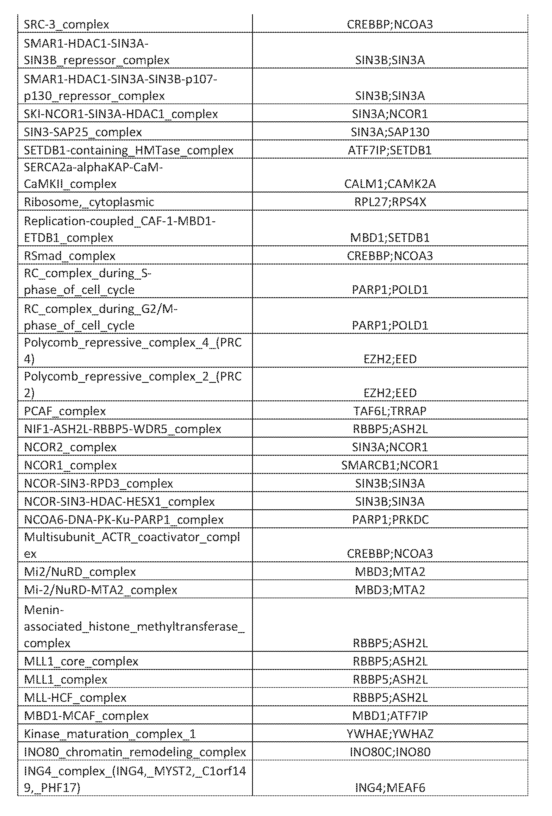
- FIG. 6 is chart detailing the number of hits for each of the indicated distinct biological complexes. Complex membership information was taken from the CORUM database.
- FIG. 7 A is a heatmap showing the expression z-score of CRISPR hits enriched in whole blood (32 out of 307 hits show highly enriched expression in whole blood versus other tissues and cell types, data source: GTEx). The 32 hits showing highly enriched expression in whole blood are listed in Table 7.
- FIG. 7B is a heatmap showing hits with“Late Erythroid” expression pattern (data source: DMAP).
- Hits with“Late Erythroid” expression include: CUL3, SAP130, PRPS1, NAP1L4, GCLC, CUL4A, GCDH, NEK1, HIRA, MST1, SPOP, GOLGA5, AUH, MAST3, CDKN1B, UBR2, MAP4K4, TAF10, HDGF, YWHAE, AMD1, EID1, HIF1AN, CDK8, DCK, FXR2, UQCRC1, TESK2, ADCK2, USP21, CAMK2D, FGFR1, PHC2, UBE2H, BPGM, SIRT2, S1RT3, NFYC, and CPT2.
- FIG. 7C is a hierarchical differentiation tree of UBE2H with exemplary“Late Erythroid” expression pattern.
- FIG. 8A is a series of images depicting HbF levels determined by HbF immunocytochemistry (ICC) using CRISPR Cas9-RNP-based loss of function. Cas9-RNP complexes were electroporated into proliferating CD34+ cells. Cells were then differentiated for 7 days down the erythroid lineage and HbF levels were quantified using HbF ICC. The percent F cells (top row) and mean HbF intensity (bottom row) were quantified for negative control, sgBCLUA, sgSPOP and sgCUL3.
- ICC HbF immunocytochemistry
- FIGS. 8B - 8E is a series of graph depicting HbF levels determined by HbF ICC using shRNA-based loss of function. Percent F cells (FIG. 8B and FIG. 8D) and mean HbF intensity (FIG. 8C and FIG. 8E) were quantified for individual shRNA constructs for negative control, shBCLl 1A, shSPOP and shCUL3.
- the present invention relates to targets, compositions and methods for increasing fetal hemoglobin (HbF) in erythroid cells, e.g., by increasing expression of hemoglobin g (HBg). This can occur through upregulation of hemoglobin g mRNA levels (e.g., HBG1 or HBG2) and/or upregulation of fetal hemoglobin protein (HBg) levels, which results in an elevation in HbF.
- HbF fetal hemoglobin
- the targets, compositions or methods can be used alone or in combination with another agent that upregulates HbF or targets symptoms of SCD or b-thalassemia, including but not limited to, vaso-occlusion and anemia.
- the term“approximately” or“about” refers to a range of values that fall within 25%, 20%, 19%, 18%, 17%, 16%, 15%, 14%, 13%, 12%, 11%, 10%, 9%, 8%, 7%, 6%, 5%, 4%, 3%, 2%, 1%, or less in either direction (greater than or less than) of the stated reference value unless otherwise stated or otherwise evident from the context (except where such number would exceed 100% of a possible value).
- administering refers herein to introducing an agent or composition into a subject or contacting an agent or composition with a cell and/or tissue.
- the present disclosure provides methods for increasing the amount of fetal hemoglobin (HbF) in a cell.
- the method comprises increasing expression of one or more components of HbF in a cell.
- the component of HbF is a hemoglobin g (HBg), e.g., human hemoglobin subunit gamma-1 (HBGl) or human hemoglobin subunit gamma-2 (HBG2).
- the component of fetal hemoglobin is a hemoglobin a (HBa), e.g., human hemoglobin subunit alpha- 1 (HBA1) or human hemoglobin subunit alpha-2 (HBA2).
- expression of both HBg and HBa is increased.
- the fetal hemoglobin comprises a human hemoglobin subunit gamma-1 (HBGl) having the protein sequence set forth in NCBI Reference Sequence: NP_000550.2 and shown below:
- the HBGl protein is encoded by the polynucleotide sequence set forth in NCBI Reference Sequence: NM_000559.2 and shown below':
- the fetal hemoglobin comprises a human hemoglobin subunit gamma-2 (HBG2) having the protein sequence set forth in NCBI Reference Sequence: NP_000175.1 and shown below:
- HBG2 human hemoglobin subunit gamma-2
- the HBG2 protein is encoded by the polynucleotide sequence set forth in NCBI Reference Sequence: NM_000184.2, NCBI Reference Sequence: NM_000184.3, or shown below:
- the fetal hemoglobin comprises a human hemoglobin subunit alpha-1 (HBA1) having the protein sequence set forth in NCBI Reference Sequence: NP_000549.1 and shown below:
- HBA1 human hemoglobin subunit alpha-1
- the HBA1 protein is encoded by the polynucleotide sequence set forth in NCBI Reference Sequence: NM_000558.4, NCBI Reference Sequence: NM_000558.5, or shown below:
- the fetal hemoglobin comprises a human hemoglobin subunit alpha-2 (HBA2) having the protein sequence set forth in NCBI Reference Sequence: NP_000508.1 and shown below:
- HBA2 human hemoglobin subunit alpha-2
- the HBA2 protein is encoded by the polynucleotide sequences set forth in NCBI Reference Sequence: NM_000517.4, NCBI Reference Sequence: NM_000517.6, or shown below:
- the fetal hemoglobin comprises two HBG1 and/or HBG2 proteins and two HBA1 and/or HBA2 proteins.
- the methods disclosed herein comprise contacting a cell with an inhibitor of a target gene, tnRNA or protein (which may collectively be referred to as“target”) disclosed herein, wherein inhibition of the target results in an increased amount of fetal hemoglobin in the cell, e.g., an erythroid or red blood cell.
- inhibition of the target results in an increased amount of HBG 1 or HBG2 in the cell.
- an amount of the inhibitor effective to result in increased levels of Hby and/or HbF is used.
- the methods comprise contacting a tissue, organ or organism, e.g., a mammal, with the inhibitor.
- CUL3 Cullin 3
- E3 ubiquitin ligase protein complexes that regulate the ubiquitination of target proteins leading to proteasomal degradation.
- CUL3-E3 ubiquitin ligase complexes regulate multiple cellular processes responsible for protein trafficking, stress response, cell cycle regulation, signal transduction, protein quality control, transcription, and DNA replication.
- the present disclosure provides methods for increasing the amount of fetal hemoglobin (HbF) in a cell by inhibiting or modulating the expression of CUL3.
- HbF fetal hemoglobin
- CUL3 comprises the protein sequence:
- the target gene, mRNA, or protein is Speckle-type POZ protein (SPOP).
- SPOP is associated with multiple E3 ubiquitin ligase complexes.
- the present disclosure provides methods for increasing the amount of fetal hemoglobin (HbF) in a cell by inhibiting or modulating the expression of SPOP.
- HbF fetal hemoglobin
- SPOP comprises the protein sequence:
- the term“inhibitor” may refer to any agent that inhibits the expression or activity of a target gene, mRNA and/or protein in a cell, tissue, organ, or subject.
- the expression level or activity of target mRNA and/or protein in a cell may be reduced via a variety of means, including but not limited to reducing the total amount of target protein or inhibiting one or more activity of the target protein.
- an inhibitor may inhibit the expression of a target gene, target mRNA, or a target protein, and/or an inhibitor may inhibit a biological activity of a target protein.
- the biological activity is kinase activity.
- an inhibitor may competitively bind to the ATP-binding site of a kinase and inhibit its kinase activity, or it may allosterically block the kinase activity.
- an inhibitor causes increased degradation of a target protein.
- the inhibitor inhibits any of the target genes or proteins identified in Table 1, Table 2, Table 6, Table 7, Table 8, or Table 9, or any component or subunit of any of the complexes identified in Table 3 or Table 4 or pathways identified in Table 5.
- Methods for determining the expression level or the activity of a target gene or polypeptide are known in the art and include, eg., RT-PCRand FACS.
- an inhibitor directly inhibits expression of or an activity of a target gene, mRNA, or protein, eg., it may directly bind to the target gene, mRNA or protein.
- the inhibitor indirectly inhibits expression of or an activity of a target gene, mRNA, or protein, eg., it may bind to and inhibit a protein that mediates expression of the target gene, mRNA, or protein (such as a transcription factor), or it may bind to and inhibit expression of an activity of another protein involved in the activity of the target protein (such as another protein present in a complex with the target protein).
- the inhibitor inhibits SPOP or a protein complex to which SPOP is permanently or transiently associated.
- the protein complex is an SPOP-associated E3 ubiquitin ligase complex.
- the complex comprises Core histone macro-H2A.l (H2AFY), SPOP, and CUL3; DNA damage-binding protein 1 (DDB1), DNA damage-binding protein 2 (DDB2), Cullin-4A (CUL4A), Cullin-4B (CUL4B), and E3 ubiquitin protein ligase RBX1 (RBX); or Polycomb complex protein BMI- 1 (BMI1), SPOP, and CUL3; SPOP, Death domain-associated protein 6 (DAXX), and CUL3; Core histone macro-H2 A .1 (H2AFY), SPOP, and CUL3; or BMI1, SPOP, and CUL3.
- the inhibitor inhibits one or more component of any of these complexes.
- DDB1 DNA damage-binding protein 1
- DDB2 DNA
- the inhibitor inhibits CUL3 or a protein complex to which CUL3 is permanently or transiently associated.
- the protein complex is a CUL3 -associated E3 ubiquitin ligase complex.
- the CUL3- associated protein complex is a D(4) dopamine receptor (DRD4)-Kelch like protein 12 (KLH12)-CUL3.
- the CUL3 -associated protein complex is a coiled coil domain containing protein 22 (CCDC22)-COMM domain containing protein 8 (COMMD8)-CUL3 complex.
- the CUL3 -associated protein complex is a Cullin associated NEDD8 dissociated protein (CAND1)-CUL3-E3 ubiquitin protein ligase RBX1 (RBX1).
- the complex comprises SPOP, Death domain- associated protein 6 (DAXX), and CUL3; Core histone macro-H2A.l (H2AFY), SPOP, and CUL3; DNA damage-binding protein 1 (DDB1), DNA damage-binding protein 1 (DDB2), Cullin-4A (CUL4A), Cullin-4B (CUL4B), and E3 ubiquitin-protein ligase RBX1 (RBX1); Polycomb complex protein BMI-1 (BMIl), SPOP, and CUL3; COP9 signalosome complex subunit 1 (CSN1), COP9 signalosome complex subunit 8 (CSN8), Hairy/enhancer-of-split related with YRPW motif protein 1 (HRT1), S -
- a method of increasing the amount of fetal hemoglobin in a cell, tissue, organ or subject comprises contacting the cell, tissue, organ, or subject with an agent that results in a reduced amount of one or more target genes, tnRNAs, or proteins in a cell.
- the agent inhibits the expression or activity of one or more target gene, mKNA, or polypeptide in a cell or tissue.
- the agent causes increased degradation of one or more target gene, rnRNA, or polypeptide.
- the cell or tissue is contacted with an amount of the agent effective to reduce the expression or activity of one or more target genes, mRNAs, or polypeptides in the cell or tissue.
- the cell or tissue is contacted with an amount of the agent effective to reduce the amount of active target protein in the cell or tissue.
- the cells are hematopoietic cells, e.g., red blood cells.
- the cells are terminally differentiated, e.g., terminally differentiated red blood cells.
- the cells comprise one or more mutations associated with a blood cell disorder, e.g., SCO or b-thalassemia.
- the cells have a reduced amount of functionally active HbA as compared to a control cell, e.g., a non-disease cell.
- the cells are associated with a blood cell disorder, e.g., SCD or b-thalassemia.
- the cells may be derived from or obtained from cells or tissue from a subject diagnosed with the blood cell disorder.
- the methods are practiced on a subject diagnosed with a blood cell disorder, e.g., SCD or b-thalassemia. Methods disclosed herein may be practiced in vitro or in vivo.
- the disclosure includes a method of treating or preventing a blood cell disease or disorder associated with reduced amounts of functionally active HbA (or total HbA) in a subject in need thereof, comprising providing to a subject an agent that inhibits the expression or activity of one or more target protein in the subject, or in certain cells or tissue of the subject, wherein the treatment results in an increased amount of HbF in the subject or one or more cells or tissues of the subject, e.g., hematopoietic cell, e.g., an erythrocyte or red blood cell.
- the agent is present in a pharmaceutical composition.
- the subject is provided with one or more (e.g., two, three, or more) agents that inhibits the expression or activity of one or more target protein in the subject, or in certain cells or tissue of the subject.
- the two or more agents inhibit the same target or target complex disclosed herein, whereas in other embodiments, the two or more agents inhibit different targets or target complexes disclosed herein.
- the cells are terminally differentiated, e.g., terminally differentiated red blood cells.
- the agent inhibits the expression or activity of the one or more target protein.
- the agent induces degradation of the one or more target protein.
- the agent inhibits activity' of the one or more target protein.
- the inhibitor reduces expression of one or more target genes, mRNAs or proteins in cells or tissue of the subject, e.g., hematopoietic cells, e.g., red blood cells.
- the inhibitor inhibits any of the target genes or proteins identified in Table 1, Table 2, Table 6, Table 7, Table 8, or Table 9, or any component or subunit of any of the complexes identified in Table 3 or Table 4 or pathways identified in Table
- the blood disease or disorder is selected from Sickle Cell Disease, b-thalassemia. Beta thalassemia trait or beta thalassemia minor, Thalassemia intermedia, Thalassemia major or Cooley’s Anemia.
- the pharmaceutical composition is provided to the subject parenterally.
- Inhibitors and/or other agents and compositions (e.g., inhibitors) described herein can be formulated in any manner suitable for a desired administration route (e.g., parenteral or oral administration).
- contacting an agent or composition with a cell and/or tissue is a result of administration of or providing an agent or composition to a subject.
- an agent or composition e.g., an inhibitor
- administration of a first agent or composition is followed by or occurs overlapping with or concurrently with the administration of a second agent or composition.
- the first and second agent or composition may be the same or they may be different.
- the first and second agents or compositions are administered by the same actor and/or in the same geographic location. In some embodiments, the first and second agents or compositions are administered by different actors and/or in different geographical locations. In some embodiments, multiple agents described herein are administered as a single composition.
- inhibitors may be administered or coadministered topically, orally, intraperitoneally, intravenously, intraarterially, transdermally, sublingually, intramuscularly, rectally, transbuccally, intranasally, liposomally, via inhalation, vaginally, intraoccularly, via local delivery (for example by catheter or stent), subcutaneously, intraadiposally, intraarticularly, intrathecally, transmucosally, pulmonary, or parenterally, for example, by injection, including subcutaneous, intradermal, intramuscular, intravenous, intraarterial, intracardiac, intrathecal, intraspinal, intracapsular, subcapsular, intraorbital, intraperitoneal, intratracheal, subcuticular, intraarticular, subarachnoid, and intrastemal; by implant of a depot or reservoir, for example, subcutaneous, intradermal, intramuscular, intravenous, intraarterial, intracardiac, intrathecal,
- Subjects includes animals (e.g., mammals, swine, fish, birds, insects etc.).
- subjects are mammals, particularly primates, especially humans.
- subjects are livestock such as cattle, sheep, goats, cows, swine, and the like; poultry such as chickens, ducks, geese, turkeys, and the like; and domesticated animals such as dogs and cats.
- subjects are rodents (e.g., mice, rats, hamsters), rabbits, primates, or swine such as inbred pigs and the like.
- the terms“subject” and“patient” are used interchangeably herein.
- tissue is an ensemble of similar cells from the same origin that together carry out a specific function.
- Methods disclosed herein may be practiced with any agent capable of inhibiting expression or activity of a target gene, mRNA or protein, e.g., an inhibitor of a gene, mRNA or protein, complex or pathway disclosed herein, e.g., in any of Tables 1-9.
- methods disclosed herein result in a decrease in an expression level or activity of a target gene, mRNA or protein in one or more cells or tissues (e.g., within a subject), e.g., as compared to the expression level or activity in control cells or tissue not contacted with the inhibitor, or a reference level.
- “Decrease” refers to a decrease of at least 5%, for example, at least 5, 6, 7, 8, 9, 10, 15, 20, 25, 30, 35, 40, 45, 50, 55, 60, 65, 70, 75, 80, 85, 90, 95, 99 or 100%, for example, as compared to the reference level.
- Decrease also means decreases by at least 1-fold, for example, 1, 2, 3, 4, 5, 6, 7, 8, 9, 10, 15, 20, 30, 40, 50, 60, 70, 80, 90, 100, 200, 500, 1000-fold or more, for example, as compared to the level of a reference or control cells or tissue.
- methods disclosed herein result in increased amounts of HbF or HBg in one or more cells or tissues (e.g., within a subject), e.g., as compared to the expression level or activity- in control cells or tissue not contacted with the inhibitor, or a reference level.
- methods disclosed herein result in increased expression of a hemoglobin gamma (e.g., HBG1 or HBG2) in one or more cells or tissues (e.g., within a subject), e.g., as compared to the expression level in control cells or tissue not contacted with the inhibitor, or a reference level.
- a hemoglobin gamma e.g., HBG1 or HBG2
- “Increase” refers to an increase of at least 5%, for example, at least 5, 6, 7, 8, 9, 10, 15, 20, 25, 30, 35, 40, 45, 50, 55, 60, 65, 70, 75, 80, 85, 90, 95, 99 or 100%, or an at least two-fold, three-fold, give-fold, ten-fold, 20-fold, 50-fold, 100-fold, 500-fold or 1000-fold increase, for example, as compared to the reference level or level in control cells or tissue.
- Methods described herein may be practiced using any type of inhibitor that results in a reduced amount or level of a target gene, mRNA or protein, e.g., in a cell or tissue, e.g., a cell or tissue in a subject.
- the inhibitor causes a reduction in active target protein, a reduction in total target protein, a reduction in target mRNA levels, and/or a reduction in target protein activity, e.g., in a cell or tissue contacted with the inhibitor.
- the reduction is at least 5%, at least 10%, at least 20%, at least 30%, at least 40%, at least 50%, at least 60%, at least 70%, at least 80%, or at least 90%, as compared to the level in the same type of cell or tissue not contacted with the inhibitor or a reference level.
- Methods of measuring total protein or mRNA levels, or activity, in a cell are known in the art.
- the inhibitor inhibits or reduces target protein activity or expression, e.g., mRNA and/or protein expression.
- the inhibitor causes increased degradation of the target protein, resulting in lower amounts of target protein in a cell or tissue .
- Inhibitors that may be used to practice the disclosed methods include but are not limited to agents that inhibit or reduce or decrease the expression or activity of a biomolecule, such as but not limited to a target gene, mRNA or protein.
- an inhibitor can cause increased degradation of the biomolecule.
- an inhibitor can inhibit a biomolecule by competitive, uncompetitive, or non-competitive means.
- inhibitors include, but are not limited to, nucleic acids, DNA, RNA, gRNA, shRNA, siRNA, modified mRNA (mRNA), microRNA (miRNA), proteins, protein mimetics, peptides, peptidomimetics, antibodies, small molecules, small organic molecules, inorganic molecules, chemicals, analogs that mimic the binding site of an enzyme, receptor, or other protein, e.g., that is involved in signal transduction, therapeutic agents, pharmaceutical compositions, drags, and combinations of these.
- the inhibitor can be a nucleic acid molecule including, but not limited to, siRNA that reduces the amount of functional protein in a cell. Accordingly, compounds or agents said to be“capable of inhibiting” a particular target protein comprise any type of inhibitor.
- an inhibitor comprises a nucleic acid that binds to a target gene or mRNA.
- a nucleic acid inhibitor may comprise a sequence complementary to a target polynucleotide sequence, or a region thereof, or an antisense thereof.
- a nucleic acid inhibitor comprises at least 8, at least 10, at least 12, at least 14, at least 16, at least 20, at least 24, or at least 30 nucleotide sequence corresponding to or complementary to a target polynucleotide sequence or antisense thereof.
- a nucleic acid inhibitor is an RNA interference or antisense RNA agent or a portion or mimetic thereof, or a morpholino, that decreases the expression of a target gene when administered to a cell.
- a nucleic acid inhibitor comprises at least a portion of a target nucleic acid molecule, or an ortholog thereof, or comprises at least a portion of the complementary strand of a target nucleic acid molecule.
- expression of a target gene is reduced by at least about 10%, at least about 25%, at least about 50%, at least about 75%, or even 90-100%.
- A“complementary'” nucleic acid sequence is a nucleic acid sequence capable of hybridizing with another nucleic acid sequence comprised of complementary nucleotide base pairs.
- hybridize is meant pair to form a double-stranded molecule between complementary nucleotide bases (e.g. , adenine (A) forms a base pair with thymine (T), as does guanine (G) with cytosine (C) in DNA) under suitable conditions of stringency.
- A complementary nucleotide bases
- T thymine
- G guanine
- C cytosine
- Antisense refers to a nucleic acid sequence, regardless of length, that is complementary to a nucleic acid sequence.
- antisense RNA refers to single stranded RNA molecules that can be introduced to an individual cell, tissue, or subject and results in decreased expression of a target gene through mechanisms that do not rely on endogenous gene silencing pathways.
- An antisense nucleic acid can contain a modified backbone, for example, phosphorothioate, phosphorodithioate, or others known in the art, or may contain non-natural intemucleoside linkages.
- Antisense nucleic acid can comprise, e.g., locked nucleic acids (LNA).
- RNA interference refers to the use of agents that decrease the expression of a target gene by degradation of a target mRNA through endogenous gene silencing pathways (e.g., Dicer and RNA-induced silencing complex (RISC)). RNA interference may be accomplished using various agents, including shRNA and siRNA.“Short hair-pin RNA” or“shRNA” refers to a double stranded, artificial RNA molecule with a hairpin turn that can be used to silence target gene expression via RNA interference (RNAi). Expression of shRNA in cells is typically accomplished by delivery of plasmids or through viral or bacterial vectors. shRNA is an advantageous mediator of RNAi in that it has a relatively low rate of degradation and turnover.
- RISC RNA-induced silencing complex
- siRNA Small interfering RNA
- RNAi RNA interference pathway
- an siRNA is 18, 19, 20, 21, 22, 23 or 24 nucleotides in length and has a 2 base overhang at its 3 1 end.
- siRNAs can be introduced to an individual cell and/or culture system and result in the degradation of target mRNA sequences.
- Morpholino refers to a modified nucleic acid oligomer wherein standard nucleic acid bases are bound to morpholine rings and are linked through phosphorodiamidate linkages. Similar to siRNA and shRNA, morpholinos bind to complementary mRNA sequences. However, morpholinos function through steric-inhibition of mRNA translation and alteration of mRNA splicing rather than targeting complementary mKNA sequences for degradation.
- a nucleic acid inhibitor is a messenger RNA that may be introduced into a cell, wherein it encodes a polypeptide inhibitor of a target disclosed herein.
- the mKNA is modified, e.g., to increase its stability or reduce its immunogenicity, e.g., by the incorporation of one or more modified nucleosides. Suitable modifications are known in the art.
- an inhibitor comprises an expression cassette that encodes a polynucleotide or polypeptide inhibitor of a target disclosed herein.
- the expression cassette is present in a gene therapy vector, for example a viral gene therapy vector.
- gene therapy vectors including viral gene therapy vectors are known in the art, including, for example, AAV-based gene therapy vectors.
- an inhibitor is a polypeptide inhibitor.
- a polypeptide inhibitor binds to a target polypeptide, thus inhibiting its activity, e.g., kinase activity.
- polypeptide inhibitors include any types of polypeptides (e.g., peptides and proteins), such as antibodies and fragments thereof.
- An“antibody” is an immunoglobulin (Ig) molecule capable of specific binding to a target, such as a carbohydrate, polynucleotide, lipid, or polypeptide, through at least one epitope recognition site, located in the variable region of the Ig molecule.
- a target such as a carbohydrate, polynucleotide, lipid, or polypeptide
- the term encompasses not only intact polyclonal or monoclonal antibodies, but also fragments thereof, such as dAb, Fab, Fab', F(ab')2, Fv, single chain (scFv), synthetic variants thereof, naturally occurring variants, fusion proteins comprising an antibody portion with an antigenbinding fragment of the required specificity, chimeric antibodies, nanobodies, and any other modified configuration of the immunoglobulin molecule that comprises an antigen-binding site or fragment of the required specificity.
- “Fragment” refers to a portion of a polypeptide or nucleic acid molecule. This portion contains, preferably, at least 10%, 20%, 30%, 40%, 50%, 60%, 70%, 80%, or 90% of the entire length of the reference nucleic acid molecule or polypeptide.
- a fragment may contain 10, 20, 30, 40, 50, 60, 70, 80, 90, or 100, 200, 300, 400, 500, 600, 700, 800, 900, or 1000 nucleotides or amino acids.
- A“functional fragment” of an antibody is a fragment that maintains one or more activities of the antibody, e.g., it binds the same epitope and or possesses a biological activity of the antibody.
- a functional fragment comprises the six CDRs present in the antibody.
- the inhibitor induces degradation of a target polypeptide.
- inhibitors include proteolysis targeting chimeras (PROTAC), which induce selective intracellular proteolysis of target proteins.
- PROTACs include functional domains, which may be covalently linked protein-binding molecules: one is capable of engaging an E3 ubiquitin ligase, and the other binds to the target protein meant for degradation. Recruitment of the E3 ligase to the target protein results in ubiquitination and subsequent degradation of the target protein by the proteasome.
- an inhibitor is a PROTAC that targets any of the targets disclosed herein.
- an inhibitor is a small molecule inhibitor, or a stereoisomer, enantiomer, diastereomer, isotopically-enriched, pro-drug, or pharmaceutically acceptable salt thereof.
- the small molecule inhibitor of a target protein or protein complex that functions to regulate HbF expression targets SPOP.
- the small molecule inhibitor of a target protein or protein complex that functions to regulate HbF expression targets CUL3.
- the CUL3 inhibitor is MLN4924 (CAS No: 905579-51-3), suramin (CAS NO: 145-63-1) or DI-591 (CAS No: 2245887-38-9).
- the inhibitor comprises one or more components of a gene editing system.
- a gene editing system refers to a protein, nucleic acid, or combination thereof that is capable of modifying a target locus of an endogenous DNA sequence when introduced into a cell.
- Numerous gene editing systems suitable for use in the methods of the present invention are known in the art including, but not limited to, zinc-finger nuclease systems, TALEN systems, and CRISPR/Cas systems.
- the gene editing system used in the methods described herein is a CRISPR (Clustered Regularly Interspaced Short Palindromic Repeats)/Cas (CRISPR Associated) nuclease system, which is an engineered nuclease system based on a bacterial system that can be used for mammalian genome engineering.
- the system comprises a CRISPR-associated endonuclease (for example, a Cas endonuclease) and a guide RNA (gRNA).
- the gRNA is comprised of two parts; a crispr-RNA (crRNA) that is specific for a target genomic DNA sequence, and a trans-activating RNA (tracrRNA) that facilitates endonuclease binding to the DNA at the targeted insertion site.
- crRNA crispr-RNA
- tracrRNA trans-activating RNA
- the crRNA and tracrRNA may be present in the same RNA oligonucleotide, referred to as a single guide-RNA (sgRNA).
- the crRNA and tracrRNA may be present as separate RNA oligonucleotides.
- the gRNA is comprised of a crRNA oligonucleotide and a tracrRNA oligonucleotide that associate to form a crRNA:tracrRNA duplex.
- the term“guide RNA” or“gRNA” refers to the combination of a tracrRNA and a crRNA, present as either an sgRNA or a crRNA:tracrRNA duplex.
- the CRISPR/Cas systems comprise a Cas protein, a crRNA, and a tracrRNA.
- the crRNA and tracrRNA are combined as a duplex RNA molecule to form a gRNA.
- the crRNA :tracrRNA duplex is formed in vitro prior to introduction to a cell.
- the crRNA and tracrRNA are introduced into a cell as separate RNA molecules and crRNA:tracrRNA duplex is then formed intracellularly.
- polynucleotides encoding the crRNA and tracrRNA are provided.
- the polynucleotides encoding the crRNA and tracrRNA are introduced into a cell and the crRNA and tracrRNA molecules are then transcribed intracellularly.
- the crRNA and tracrRNA are encoded by a single polynucleotides.
- the crRNA and tracrRNA are encoded by separate polynucleotides.
- a Cas endonuclease is directed to the target insertion site by the sequence specificity of the crRNA portion of the gRNA, which may include a protospacer motif (PAM) sequence near the target insertion site.
- PAM protospacer motif
- a variety of PAM sequences suitable for use with a particular endonuclease are known in the art (See e.g., Nat Methods. 2013 Nov; 10(11): 11 16-1121 and Sci Rep. 2014; 4: 5405).
- the specificity of a gRNA for a target locus is mediated by the crRNA sequence, which comprises a sequence of about 20 nucleotides that are complementary to the DNA sequence at a target locus, e.g., complementary to a target DNA sequence.
- the crRNA sequences used in the methods of the present invention are at least 90% complementary to a DNA sequence of a target locus.
- the crRNA sequences used in the methods of the present invention are at least 95%, 96%, 97%, 98%, or 99% complementary to a DNA sequence of a target locus.
- the crRNA sequences used in the methods of the present invention are 100% complementary to a DNA sequence of a target locus.
- the crRNA sequences described herein are designed to minimize off-target binding using algorithms known in the art (e.g., Cas-OFF finder) to identify target sequences that are unique to a particular target locus or target gene.
- the endonuclease is a Cas protein or ortholog. In some embodiments, the endonuclease is a Cas9 protein. In some embodiments, the Cas9 protein is derived from Streptococcus pyogenes (e.g., SpCas9), Staphylococcus aureus (e.g., SaCas9), or Neisseria meningitides (NmeCas9).
- Streptococcus pyogenes e.g., SpCas9
- Staphylococcus aureus e.g., SaCas9
- Neisseria meningitides Neisseria meningitides
- the Cas endonuclease is a Cas9 protein or a Cas9 ortholog and is selected from the group consisting of SpCas9, SpCas9-HFl, SpCas9-HF2, SpCas9-HF3, SpCas9-HF4, SaCas9, FnCpf, FnCas9, eSpCas9, and NmeCas9.
- the endonuclease is selected from the group consisting of C2C1, C2C3, Cpfl (also referred to as Casl2a), Casl, CaslB, Cas2, Cas3, Cas4, Cas5, Cas6, Cas7, Cas8, Cas9 (also known as Csnl and Csxl2), CaslO, Csyl, Csy2, Csy3, Csel, Cse2, Cscl, Csc2, Csa5, Csn2, Csm2, Csm3, Csm4, Csm5, Csm6, Cmrl, Cmr3, Cmr4, Cmr5, Cmr6, Csbl, Csb2, Csb3, Csxl7, Csxl4, CsxlO, Csxl6, CsaX, Csx3, Csxl, Csxl5, Csfl, Csf2, Csf3, and Csf4.
- the endonuclease
- compositions e.g., pharmaceutical compositions, comprising an inhibitor of a target disclosed herein, including any of the various classes of inhibitors described herein.
- the invention encompasses pharmaceutical compositions comprising an inhibitor and a pharmaceutically acceptable carrier, diluent or excipient. Any inert excipient that is commonly used as a carrier or diluent may be used in compositions of the present invention, such as sugars, polyalcohols, soluble polymers, salts and lipids. Sugars and polyalcohols which may be employed include, without limitation, lactose, sucrose, mannitol, and sorbitol.
- soluble polymers which may be employed are polyoxyethylene, poloxamers, polyvinylpyrrolidone, and dextran.
- Useful salts include, without limitation, sodium chloride, magnesium chloride, and calcium chloride.
- Lipids which may be employed include, without limitation, fatty acids, glycerol fatty acid esters, glycolipids, and phospholipids.
- compositions may further comprise binders (e.g., acacia, cornstarch, gelatin, carbomer, ethyl cellulose, guar gum, hydroxypropyl cellulose, hydroxypropyl methyl cellulose, povidone), disintegrating agents (e.g., cornstarch, potato starch, alginic acid, silicon dioxide, croscarmellose sodium, crospovidone, guar gum, sodium starch glycolate, Primogel), buffers (e.g., tris-HCL, acetate, phosphate) of various pH and ionic strength, additives such as albumin or gelatin to prevent absorption to surfaces, detergents (e.g., Tween 20, Tween 80, Plutonic F68, bile acid salts), protease inhibitors, surfactants (e.g., sodium lauryl sulfate), permeation enhancers, solubilizing agents (e.g., glycerol, poly
- the pharmaceutical compositions are prepared with carriers that will protect the inhibitor against rapid elimination from the body, such as a controlled release formulation, including implants and microencapsulated delivery systems.
- a controlled release formulation including implants and microencapsulated delivery systems.
- Biodegradable, biocompatible polymers can be used, such as ethylene vinyl acetate, polyanhydrides, polyglycolic acid, collagen, polyorthoesters, and polylactic acid. Methods for preparation of such formulations will be apparent to those skilled in the art.
- the materials can also be obtained commercially from Alza Corporation and Nova Pharmaceuticals, Inc.
- Liposomal suspensions (including liposomes targeted to infected cells with monoclonal antibodies to viral antigens) can also be used as pharmaceutically acceptable carriers. These can be prepared according to methods known to those skilled in fee art, for example, as described in U.S. Pat. No. 4,522,81 1.
- fee invention encompasses pharmaceutical compositions comprising any solid or liquid physical form of an inhibitor.
- fee inhibitor can be in a crystalline form, in amorphous form, and have any particle size.
- the particles may be micronized, or may be agglomerated, particulate granules, powders, oils, oily suspensions or any other form of solid or liquid physical form.
- solubilizing fee compounds may be used. Such methods are known to those of skill in this art, and include, but are not limited to, pH adjustment and salt formation, using co-solvents, such as ethanol, propylene glycol, polyethylene glycol (PEG) 300, PEG 400, DMA (10-30%), DMSO (10-20%), NMP (10-20%), using surfactants, such as polysorbate 80, polysorbate 20 (1-10% ), cremophor EL, Cremophor RH40, Cremophor RH60 (5-10% ), Pluronic F68/Poloxamer 188 (20-50%), Solutol HS15 (20-50%), Vitamin E TPGS, and d-a-tocopheryl PEG 1000 succinate (20-50%), using complexation such as HP b-CD and SBE b-CD (10-40%), and using advanced approaches such as micelles, addition of a polymer, nanoparticle suspensions, and liposome formation.
- co-solvents such as ethanol, propylene glyco
- Inhibitors may also be administered or coadministered in slow release dosage forms.
- Inhibitors may be in gaseous, liquid, semi-liquid or solid form, formulated in a manner suitable for fee route of administration to be used.
- suitable solid oral formulations include tablets, capsules, pills, granules, pellets, sachets and effervescent, powders, and the like.
- suitable liquid oral formulations include solutions, suspensions, dispersions, syrups, emulsions, oils and the like.
- reconstitution of a lyophilized powder is typically used.
- Suitable doses of the inhibitors for use in treating the diseases or disorders described herein can be determined by those skilled in the relevant art. Therapeutic doses are generally identified through a dose ranging study in humans based on preliminary evidence derived from the animal studies. Doses should be sufficient to result in a desired therapeutic benefit without causing unwanted side effects. Mode of administration, dosage forms and suitable pharmaceutical excipients can also be well used and adjusted by those skilled in the art. All changes and modifications are envisioned within the scope of the present patent application.
- the disclosure includes unit dosage forms of a pharmaceutical composition
- a pharmaceutical composition comprising an agent that inhibits expression or activity of a target polypeptide (or results in reduced levels of a target protein) and a pharmaceutically acceptable carrier, diluent or excipient, wherein the unit dosage form is effective to increase expression of a hemoglobin gamma in one or more tissue in a subject to whom the unit dosage form is administered.
- the unit dosage forms comprise an effective amount, an effective concentration, and/or an inhibitory concentration, of an inhibitor to treat a blood cell disease or disorder, e.g., one associated with mutant or aberrant hemoglobin beta, including any of the diseases or disorders disclosed herein, e.g., SCD or b-thalassemias.
- a blood cell disease or disorder e.g., one associated with mutant or aberrant hemoglobin beta, including any of the diseases or disorders disclosed herein, e.g., SCD or b-thalassemias.
- compositions include compositions of one or more inhibitors disclosed herein and one or more pharmaceutically acceptable carrier, excipient, or diluent.
- “Pharmaceutically acceptable” is employed herein to refer to those compounds, materials, compositions, and/or dosage forms which are, within the scope of sound medical judgment, suitable for use in contact with the tissues of human beings and animals without excessive toxicity, irritation, allergic response, or other problem or complication, commensurate with a reasonable benefit/risk ratio.
- “Pharmaceutically acceptable carrier” includes without limitation any adjuvant, carrier, excipient, glidant, sweetening agent, diluent, preservative, dye/colorant, flavor enhancer, surfactant, wetting agent, dispersing agent, suspending agent, stabilizer, isotonic agent, solvent, surfactant, and/or emulsifier which has been approved by the United States Food and Drag Administration as being acceptable for use in humans and/or domestic animals.
- Exemplary pharmaceutically acceptable carriers include, but are not limited to, to sugars, such as lactose, glucose and sucrose; starches, such as com starch and potato starch; cellulose, and its derivatives, such as sodium carboxymethyl cellulose, ethyl cellulose and cellulose acetate; tragacanth; malt; gelatin; talc; cocoa butter, waxes, animal and vegetable fats, paraffins, silicones, bentonites, silicic acid, zinc oxide; oils, such as peanut oil, cottonseed oil, safflower oil, sesame oil, olive oil, com oil and soybean oil; glycols, such as propylene glycol; polyols, such as glycerin, sorbitol, mannitol and polyethylene glycol; esters, such as ethyl oleate and ethyl laurate; agar; buffering agents, such as magnesium hydroxide and aluminum hydroxide; alginic acid; pyrogen-
- Effective amount refers to an amount of an agent effective in achieving a particular effect, e.g., increasing levels of fetal hemoglobin (or a hemoglobin gamma) in a cell, tissue, organ or subject. In certain embodiments, the increase is at least 10%, at least 20%, at least 30%, at least 40%, at least 50%, at least 60%, or at least 70%, as compared to the amount prior to or without treatment.
- an effective amount may be, e.g., an amount effective or sufficient to reduce one or more disease symptoms in the subject, e.g., a subject with sickle cell disease.
- Effective Concentration refers to the minimum concentration (mass/volume) of an agent and/or composition required to result in a particular physiological effect. As used herein, effective concentration typically refers to the concentration of an agent required to increase, activate, and/or enhance a particular physiological effect.
- inhibitory Concentration is the minimum concentration (mass/volume) of an agent required to inhibit a particular physiological effect. As used herein, inhibitory concentration typically refers to the concentration of an agent required to decrease, inhibit, and/or repress a particular physiological effect.
- an agent or compound described herein may be administered at a dosage from about 1 mg/kg to about 300 mg/kg. In another embodiment, an agent or compound described herein may be administered at a dosage from about 1 mg/kg to about 20 mg/kg. For example, the agent or compound may be administered to a subject at a dosage of 1, 2, 3, 4, 5, 6, 7, 8, 9, 10, 11, 12, 13, 14, 15, 16, 17, 18, 19 or 20 mg/kg, or within a range between any of the proceeding values, for example, between about 10 mg/kg and about 15 mg/kg, between about 6 mg/kg and about 12 mg/kg, and the like. In another embodiment, an agent or compound described herein is administered at a dosage of ⁇ 15 mg/kg.
- an agent or compound may be administered at 15 mg/kg per day for 7 days for a total of 105 mg/kg per week.
- a compound may be administered at 10 mg/kg twice per day for 7 days for a total of 140 mg/kg per week.
- the dosages described herein may refer to a single dosage, a daily dosage, or a weekly dosage.
- an agent or compound may be administered once per day.
- a compound may be administered twice per day.
- an agent or compound may be administered three times per day.
- a compound may be four times per day.
- an agent or compound described herein may be administered 1, 2, 3, 4, 5, 6, 7, 8, 9, 10, 11, 12, 13, 14, 15, 16, 17, 18, 19, 20, 21, 22, 23, or 24 times per week.
- the compound is administered once biweekly.
- an agent or compound described herein may be administered orally. In some embodiments, an agent or compound described herein may be administered orally at a dosage of ⁇ 15 mg/kg once per day.
- the actual dosage employed may be varied depending upon the requirements of the patient and the severity of the condition being treated. Determination of the proper dosage regimen for a particular situation is within the skill of the art. For convenience, the total daily dosage may be divided and administered in portions during the day as required.
- the dosage regimen utilizing the disclosed compound is selected in accordance with a variety of factors including type, species, age, weight, sex and medical condition of the patient; the severity' of the condition to be treated; the route of administration; the renal or hepatic function of the patient; and the particular disclosed compound employed.
- a physician or veterinarian of ordinary skill in the art can readily determine and prescribe the effective amount of the drug required to prevent, counter or arrest the progress of the condition.
- the amount and frequency of administration of the compounds of the invention and/or the pharmaceutically acceptable salts thereof will be regulated according to the judgment of the attending clinician considering such factors as age, condition and size of the patient as well as severity of the symptoms being treated.
- HUDEP2 cells an erythroid progenitor model derived from CD34+ cells isolated from human umbilical cord blood, was used as a cellular model to study HbF reactivation, because the HBB/HBb globin is tire predominant b-like globin expressed.
- a pool of CRISPR gRNAs was introduced into proliferating HUDEP2 cells via lentiviral delivery methods at an MOI ⁇ 0.1. Depending on the library construction, this was either a one-vector system (vector encoding both the gRNA and Cas9) or a two-vector system (vector encoding the gRNA). For the two-vector system, the lentiviral pool was delivered to HUDEP2 cells constitutively expressing Cas9 protein.
- HUDEP2 proliferation media StemCell Technologies; 50ng/ml SCF; 3 IU/tnl erythropoietin; luM dexamethasone; lug/ml doxycycline
- Selection in proliferation media + puromycin occurred for 2 days.
- HUDEP2 differentiation media Iscove’s Modified Dulbecco’s Medium; 1% L-glutamine; 2% Penicillin/streptomycin; 330 ug/ml holo-human transferrin; 2 IU/ml heparin; lOug/ml recombinant human insulin; 3 IU/ml erythropoietin; lOOng/ml SCF; 4% fetal calf serum) for 10 days.
- HUDEP2 differentiation media Iscove’s Modified Dulbecco’s Medium; 1% L-glutamine; 2% Penicillin/streptomycin; 330 ug/ml holo-human transferrin; 2 IU/ml heparin; lOug/ml recombinant human insulin; 3 IU/ml erythropoietin; lOOng/ml SCF; 4% fetal calf serum
- HbF fluorescence-activated cell sorting (FACs) assay (Invitrogen, HFH01) was used to isolate cells with elevated levels of HbF.
- HbF high cells were selected using HUDEP2 cells transduced with a negative control gRNA (sgGFP) as a gating threshold.
- sgGFP negative control gRNA
- Cells were also collected following the 3-day puromycin selection (post-selection sample) and prior to FACs sorting (FACs input sample) and used for downstream analyses to identify hits.
- Genomic DNA was isolated from HbF high isolated cells, post-selection sample, and FACs input sample.
- the gRNA present at in the genomic DNA was amplified using nested PCR amplification.
- the second round of PCR amplification was performed to also incorporate Illumina sequencing adaptors onto the sample.
- Illumina sequencing was done to quantify the gRNAs present in each sample.
- the gRNAs were identified using conserved identifiers and were subsequently mapped to the human reference genome to identify the gRNA target gene to provide the relationship between the target gene and genetic perturbation that led to HbF upregulation.
- FIG. 2 CRISPR Library #1
- FIG. 3 CRISPR Library #2
- the left panel plots the level of HbF (X-axis) and b- Actin (Y -axis) for each event
- the line“L” indicates the HbF threshold for HbF high cells.
- the right panel represents the same data in a one-dimensional plot showing the HbF levels (X- axis) and Events (Y -axis)
- the line“C” indicates the HbF threshold for HbF high cells. Any cell above the HbF threshold was collected in the HbF high population.
- the darker shaded cells at the left of each panel are HUDEP2 cells transduced with control sgGFP
- the fighter shaded cells at the right of each panel are HUDEP2 cells transduced with the CRISPR library.
- Illumina sequencing was used to sequence the libraries of gRNAs in the post-selection samples, FACs input samples, and HbF high samples. Each read was searched for the conserved identifiers either in the 5’ or the 3 ‘ regions, and only reads that contained the conserved identifiers were retained. The 20bp gRNA sequence between the conserved identifiers was extracted from the retained reads and mapped to the human genome (hgl9). A single retained read with a given gRNA represented one count for that gRNA in each sample. The counts were converted to RPM (reads per millions) to normalize for sequencing depth and to enable comparison across different gRNA libraries. The RPM for a gRNA was calculated as follows:
- N is the total number of reads in the library.
- Four different statistical methods were used to identify hits among the HbF high sample. The bioinformatics analysis performed using method 2 described below is summarized in FIG. 4A.
- FIG. 4B shows the distribution of guide abundance in different samples from two different screening libraries (Library #1 and Library #2), and FIG. 4C shows Z-score differences across samples for Library
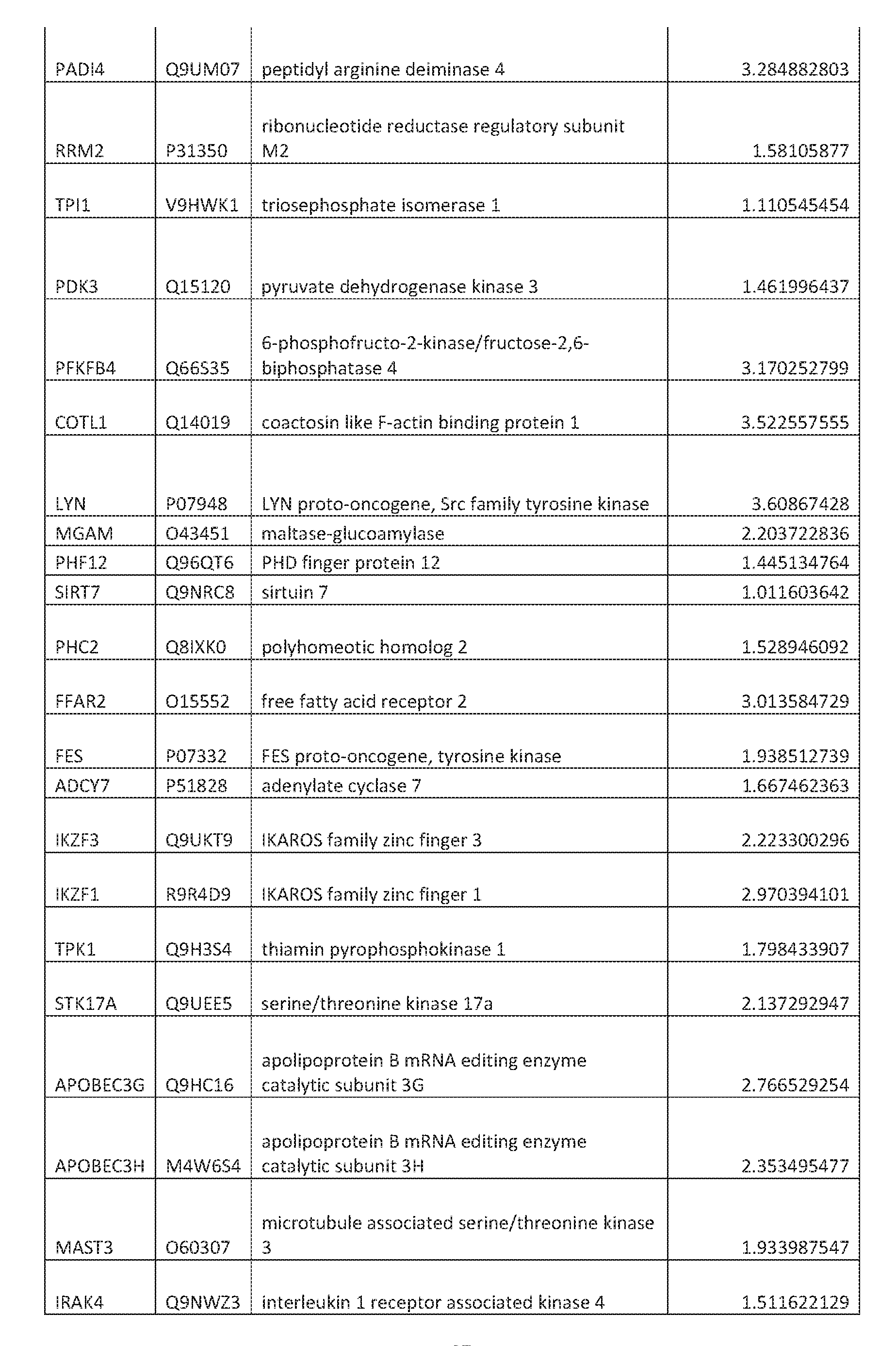
- Table 1 List of targets that upregulate HbF protein
- IDH3G P51553 isocitrate dehydrogenase 3 (NAD(+)) gamma
- RPS6KA4 075676 ribosomal protein S6 kinase A4
- ARID4A P29374 AT-rich interaction domain 4A
- PANK4 Q9NVE7 pantothenate kinase 4
- top targets were overlapped with KEGG pathway maps using the clusterProfiler R package. Top pathways are shown in Table 5 derived from hits identified using method 2.
- Hits identified using method 2 were prioritized based on their expression in blood tissue, relevant to SCO. This was performed using GTEx gene expression data from 15,598 samples across 31 different tissues (The GTEx Consortium, Nature Genetics). A mean Z-score was calculated to identify genes with high blood specific expression. The blood Z-scores for hits were calculated as follows:
- Table 9 provides a list of various components of complexes and pathways identified herein as targets for increasing expression of HbF. Any of these may be targeted according to any of the methods disclosed herein.
- SPOP and CUL3 were identified using pooled CRISPR screening in the HUDEP2 model as regulators of fetal hemoglobin expression.
- primary CD34+ cells from a healthy donor were used with CRISPR Cas9- and shRNA-mediated genetic perturbation approaches.
- the impact on HbF levels was studied in differentiated CD34+ cells using HbF immunocytochemistry (ICC) (FIG. 8A).
- HbF levels were determined by HbF ICC using CRISPR Cas9-RNP-based loss of function. Cas9-RNP complexes were electroporated into proliferating CD34+ cells. Cells were then differentiated for 7 days down the erythroid lineage and HbF levels were quantified using HbF ICC. Non-target guide RNAs were used as negative controls and guide RNAs targeting BCL11A were used as positive controls in this experimental design. Genetically perturbing SPOP and CUL3 using either CRISPR-Cas9 or shRNA led to elevated HbF levels, as measured by percent F cells within the population of differentiated erythroid cells or mean HbF levels per cell. The gRNAs used for SPOP were TAACTTTAGCTTTTGCCGGG (SEQ ID NO: 91), CGGGCATATAGGTTTGUGCA (SEQ ID NO: 92),
- GTTTGCGAGTAAACCCCAAA (SEQ ID NO: 93) and the gRNAs used for CUL3 were GAGCATCTCAAACACAACGA (SEQ ID NO: 94), CGAGATCAAGTTGTACGTTA (SEQ ID NO: 95), TCATCTACGGCAAACTCTAT (SEQ ID NO: 96) using the CRISPR Cas9-RNA method via electroporation.
- the Cas9-gRNA complexes were made
- shRNAs used for SPOP were
- HbF ICC allows for the quantification of percent F cell and HbF intensity on a per-cell basis.
- An F cell is an erythroid cell that has a detectable level of HbF beyond a defined threshold and the percent F cells is defined as the percent of cells among a population of cells that are defined as F cells.
- the percent F cells and mean HbF intensity cells were quantified for negative control, sgBCLl 1A, sgSPOP and sgCUL3. HbF levels determined by HbF ICC using shRNA-based loss of function.
- shRNA vectors were electroporated into proliferating CD34+ cells. Cells were then differentiated for 7 days down the erythroid lineage and HbF levels were quantified using ICC. The percent F cells (FIG. 8B and FIG. 8D ) and mean HbF intensity (FIG. 8C and FIG. 8E) were quantified for individual shRNA constructs for negative control, shBCLl 1A, shSPOP and shCUL3.
- CD34+ cells were expanded from thaw by seeding 100,000 viable cells/mL in a culture flask containing CD34+ Phase 1 Media comprised of IMDM, 100 ng/mL hSCF, 5 ng/ttiL IL-3, 3 IU/mL EPO, 250 ug/ttiL transferrin, 2.5% normal human serum, 1% pen/strep, 10 ng/mL heparin, 10 ug/mL insulin. The cells were supplemented by adding an additional IX culture volume of CD34+ Phase 1 Media on Day 3 after thaw. After 5 days of expansion, Primary CD34+ cells were transfected with RNP complex.
- TE buffer was used to resuspend lyophilized crRNA and tracrRNA.
- the crRNA and tracrRNA were added to annealing buffer and annealed in thermocycler. Multiple sgrRNAs per gene were pooled into a microcentrifuge tube. Each sgRNA was mixed with TrueCut Cas9 v2 and incubated for 10 minutes to generate RNP complex. After counting, 144,000 CD34+ cells were added to the transfection cuvette and combined with transfection solution (P3, RNP complex, glycerol). The cells were transfected using an Amaxa Nucleofector and then transferred to a 12-well plate with lmL of prewarmed Phase 1 media.
- Phase 1 media comprised of IMDM, 100 ng/mL hSCF, 5 ng/mL IL-3, 3 IU/mL EPO, 250 ug/mL transferrin, 2.5% normal human serum, 1% pen/strep, 10 ng/mL heparin, 10 ug/mL insulin.
- Phase 2 media comprised of IMDM, 100 ng/mL hSCF, 5 ng/mL IL-3, 3 IU/mL EPO, 250 ug/mL transferrin, 2.5% normal human serum, 1% pen/strep, 10 ng/mL heparin, 10 ug/mL insulin.
- Phase 2 media comprised of IMDM, 100 ng/mL hSCF, 5 ng/mL IL-3, 3 IU/mL EPO, 250 ug/mL transferrin, 2.5% normal human serum, 1% pen/strep, 10 ng/mL heparin, 10 ug
- the cells were washed three times with 25 pL of 0.1% tween in PBS. After washing, the cells were incubated overnight at 4°C with 25 pL of HbF-488 Primary Antibody (ThermoFisher MHFHOl-4) diluted 1:40 in 0.1% tween and Hoescht diluted 1:2000 in 0.1% tween. The next day the cells were again washed three times with 25 pL of 0.1% tween in PBS and foil sealed for imaging on the ThermoFisher Celllnsight CX7.
- ThermoFisher MHFHOl-488 Primary Antibody ThermoFisher MHFHOl-488 Primary Antibody
- Blobel GA Bodine D, Brand M, Crispino J, de Bruijn MF, Nathan D, Papayannopoulou T, Porcher C, Strouboulis J, Zon L, Higgs DR, Stamatoyannopoulos G, Engel JD.
- Fetal haemoglobin in sickle-cell disease from genetic epidemiology to new therapeutic strategies.
- Moutouh-de Parseval LA Veihelle D, Glezer E, Jensen-Pergakes K, Ferguson GD, Corral LG, Morris CL, Muller G, Brady H, Chan K.
- EHMT1 and EHMT2 inhibition induces fetal hemoglobin expression.
- Lvsine-specific demethvlase 1 is a therapeutic target for fetal hemoglobin induction.
- CORUM the comprehensive resource of mammalian protein complexes-2009.
Abstract
Description


Claims
Priority Applications (6)
| Application Number | Priority Date | Filing Date | Title |
|---|---|---|---|
| JP2021527930A JP2022513100A (en) | 2018-11-20 | 2019-11-20 | Compositions and Methods for Increasing Fetal Hemoglobin and Treating Sickle Cell Disease |
| CA3119781A CA3119781A1 (en) | 2018-11-20 | 2019-11-20 | Compositions and methods for increasing fetal hemoglobin and treating sickle cell disease |
| AU2019384547A AU2019384547A1 (en) | 2018-11-20 | 2019-11-20 | Compositions and methods for increasing fetal hemoglobin and treating sickle cell disease |
| MX2021005843A MX2021005843A (en) | 2018-11-20 | 2019-11-20 | Compositions and methods for increasing fetal hemoglobin and treating sickle cell disease. |
| US17/295,782 US20220017908A1 (en) | 2018-11-20 | 2019-11-20 | Compositions and methods for increasing fetal hemoglobin and treating sickle cell disease |
| EP19818447.5A EP3883597A2 (en) | 2018-11-20 | 2019-11-20 | Compositions and methods for increasing fetal hemoglobin and treating sickle cell disease |
Applications Claiming Priority (2)
| Application Number | Priority Date | Filing Date | Title |
|---|---|---|---|
| US201862769796P | 2018-11-20 | 2018-11-20 | |
| US62/769,796 | 2018-11-20 |
Publications (2)
| Publication Number | Publication Date |
|---|---|
| WO2020106876A2 true WO2020106876A2 (en) | 2020-05-28 |
| WO2020106876A3 WO2020106876A3 (en) | 2020-07-23 |
Family
ID=68848495
Family Applications (1)
| Application Number | Title | Priority Date | Filing Date |
|---|---|---|---|
| PCT/US2019/062461 WO2020106876A2 (en) | 2018-11-20 | 2019-11-20 | Compositions and methods for increasing fetal hemoglobin and treating sickle cell disease |
Country Status (7)
| Country | Link |
|---|---|
| US (1) | US20220017908A1 (en) |
| EP (1) | EP3883597A2 (en) |
| JP (1) | JP2022513100A (en) |
| AU (1) | AU2019384547A1 (en) |
| CA (1) | CA3119781A1 (en) |
| MX (1) | MX2021005843A (en) |
| WO (1) | WO2020106876A2 (en) |
Cited By (2)
| Publication number | Priority date | Publication date | Assignee | Title |
|---|---|---|---|---|
| US11291659B2 (en) | 2017-10-05 | 2022-04-05 | Fulcrum Therapeutics, Inc. | P38 kinase inhibitors reduce DUX4 and downstream gene expression for the treatment of FSHD |
| US11590111B2 (en) | 2019-03-15 | 2023-02-28 | Fulcrum Therapeutics, Inc. | Macrocyclic azolopyridine derivatives as EED and PRC2 modulators |
Families Citing this family (1)
| Publication number | Priority date | Publication date | Assignee | Title |
|---|---|---|---|---|
| WO2023034506A1 (en) * | 2021-09-01 | 2023-03-09 | Flagship Pioneering Innovations Vi, Llc | Methods and compositions for inducing fetal hemoglobin |
Citations (1)
| Publication number | Priority date | Publication date | Assignee | Title |
|---|---|---|---|---|
| US4522811A (en) | 1982-07-08 | 1985-06-11 | Syntex (U.S.A.) Inc. | Serial injection of muramyldipeptides and liposomes enhances the anti-infective activity of muramyldipeptides |
Family Cites Families (3)
| Publication number | Priority date | Publication date | Assignee | Title |
|---|---|---|---|---|
| BR112014010699B1 (en) * | 2011-11-03 | 2020-12-15 | Millennium Pharmaceuticals, Inc | USE OF A NAE INHIBITOR WITH A HYPOMETHILLATION AGENT TO TREAT HEMATOLOGICAL MALIGNITY AND KIT |
| EP3573618B1 (en) * | 2017-01-30 | 2023-08-16 | The Children's Hospital of Philadelphia | Compositions and methods for hemoglobin production |
| CA3059256C (en) * | 2017-04-10 | 2024-04-16 | The Regents Of The University Of Michigan | Covalent small molecule dcn1 inhibitors and therapeutic methods using the same |
-
2019
- 2019-11-20 WO PCT/US2019/062461 patent/WO2020106876A2/en unknown
- 2019-11-20 JP JP2021527930A patent/JP2022513100A/en active Pending
- 2019-11-20 EP EP19818447.5A patent/EP3883597A2/en active Pending
- 2019-11-20 US US17/295,782 patent/US20220017908A1/en active Pending
- 2019-11-20 CA CA3119781A patent/CA3119781A1/en active Pending
- 2019-11-20 AU AU2019384547A patent/AU2019384547A1/en active Pending
- 2019-11-20 MX MX2021005843A patent/MX2021005843A/en unknown
Patent Citations (1)
| Publication number | Priority date | Publication date | Assignee | Title |
|---|---|---|---|---|
| US4522811A (en) | 1982-07-08 | 1985-06-11 | Syntex (U.S.A.) Inc. | Serial injection of muramyldipeptides and liposomes enhances the anti-infective activity of muramyldipeptides |
Non-Patent Citations (26)
| Title |
|---|
| BLOBEL ET AL., EXP HEMATOL, 2015 |
| BLOBEL GABODINE DBRAND MCRISPINO JDE BRUIJN MFNATHAN DPAPAYANNOPOULOU TPORCHER CSTROUBOULIS JZON L, EXP HEMATOL., vol. 43, no. 10, October 2015 (2015-10-01), pages 821 - 37 |
| CHEMICAL ABSTRACTS, Columbus, Ohio, US; abstract no. 2245887-38-9 |
| GIURGIU M ET AL., NUCLEIC ACIDS RESEARCH |
| GIURGIU MREINHARD JBRAUNER BDUNGER-KALTENBACH IFOBO GFRISHMAN GMONTRONE CRUEPP A, NUCLEIC ACIDS RES., vol. 38, January 2010 (2010-01-01), pages D497 - 501 |
| LETTRE GBAUER DE, LANCET, vol. 387, no. 10037, 18 June 2016 (2016-06-18), pages 2554 - 64 |
| LETTREBAUER, LANCET, 2016 |
| LETVIN ET AL., NEJM, 1984 |
| LETVIN NLLINCH DCBEARDSLEY GPMCLNTYRE KWNATHAN DG, N ENGL J MED., vol. 310, no. 14, 5 April 1984 (1984-04-05), pages 869 - 73 |
| LI ET AL., BLOOD, 2002 |
| LI QPETERSON KRFANG XSTAMATOYANNOPOULOS G, BLOOD, vol. 100, no. 9, 1 November 2002 (2002-11-01), pages 3077 - 86 |
| MOUTOUH-DE PARSEVAL ET AL., J CLIN INVEST, 2008 |
| MOUTOUH-DE PARSEVAL LAVERHELLE DGLEZER EJENSEN-PERGAKES KFERGUSON GDCORRAL LGMORRIS CLMULLER GBRADY HCHAN K, J CLIN INVEST., vol. 118, no. 1, January 2008 (2008-01-01), pages 248 - 58 |
| NAT GENET., vol. 45, no. 6, June 2013 (2013-06-01), pages 580 - 5 |
| NAT METHODS, vol. 10, no. 11, November 2013 (2013-11-01), pages 1116 - 1121 |
| NOVERSHTERN NSUBRAMANIAN ALAWTON L.NRAYMOND H. MHAINING NMCCONKEY M. EHABIB NYOSEF NCHANG C. YSHAY T, CELL, vol. 144, no. 2, 21 January 2011 (2011-01-21), pages 296 - 309 |
| PAIKARI ASHEEHAN VA, BR J HAEMATOL., vol. 180, no. 2, January 2018 (2018-01-01), pages 189 - 200 |
| PAIKARISHEEHAN, BR J HAEMATOL, 2018 |
| RENNEVILLE AVAN GALEN PCANVER MCMCCONKEY MKRILL-BURGER JMDORFMAN DMHOLSON EBBERNSTEIN BEORKIN SHBAUER DE, BLOOD, vol. 126, no. 16, 15 October 2015 (2015-10-15), pages 1930 - 9 |
| RENNEVILLE ET AL., BLOOD, 2015 |
| SCI REP., vol. 4, 2014, pages 5405 |
| SHI ET AL., NATURE MED, 2015 |
| SHI LCUI SENGEL JDTANABE O, NATMED., vol. 19, no. 3, March 2013 (2013-03-01), pages 291 - 4 |
| STAMATOYANNOPOULOS G, EXP HEMATOL, 2005 |
| STAMATOYANNOPOULOS G., EXP HEMATOL., vol. 33, no. 3, March 2005 (2005-03-01), pages 259 - 71 |
| WAHL, G. M.S. L. BERGER, METHODS ENZYMOL., vol. 152, 1987, pages 507 |
Cited By (3)
| Publication number | Priority date | Publication date | Assignee | Title |
|---|---|---|---|---|
| US11291659B2 (en) | 2017-10-05 | 2022-04-05 | Fulcrum Therapeutics, Inc. | P38 kinase inhibitors reduce DUX4 and downstream gene expression for the treatment of FSHD |
| US11479770B2 (en) | 2017-10-05 | 2022-10-25 | Fulcrum Therapeutics, Inc. | Use of p38 inhibitors to reduce expression of DUX4 |
| US11590111B2 (en) | 2019-03-15 | 2023-02-28 | Fulcrum Therapeutics, Inc. | Macrocyclic azolopyridine derivatives as EED and PRC2 modulators |
Also Published As
| Publication number | Publication date |
|---|---|
| WO2020106876A3 (en) | 2020-07-23 |
| MX2021005843A (en) | 2021-10-01 |
| JP2022513100A (en) | 2022-02-07 |
| CA3119781A1 (en) | 2020-05-28 |
| AU2019384547A1 (en) | 2021-06-03 |
| US20220017908A1 (en) | 2022-01-20 |
| EP3883597A2 (en) | 2021-09-29 |
Similar Documents
| Publication | Publication Date | Title |
|---|---|---|
| EP3368689B1 (en) | Composition for modulating immune responses by use of immune cell gene signature | |
| US11186825B2 (en) | Compositions and methods for evaluating and modulating immune responses by detecting and targeting POU2AF1 | |
| US11180730B2 (en) | Compositions and methods for evaluating and modulating immune responses by detecting and targeting GATA3 | |
| US20190381105A1 (en) | STEM CELL MICROPARTICLES AND miRNA | |
| US20190262399A1 (en) | Compositions and methods for evaluating and modulating immune responses | |
| AU2019384547A1 (en) | Compositions and methods for increasing fetal hemoglobin and treating sickle cell disease | |
| US20160193252A1 (en) | STEM CELL MICROPARTICLES AND miRNA | |
| US20210079392A1 (en) | Inhibition of mir-22 mirna by apt-110 | |
| US20190239494A1 (en) | Animal model of brain tumor and manufacturing method of animal model | |
| KR20200066655A (en) | Use of p38 inhibitors to reduce expression of DUX4 | |
| Song et al. | circPTPN12/miR-21–5 p/∆ Np63α pathway contributes to human endometrial fibrosis | |
| US20210170051A1 (en) | Feedback enabled synthetic genes, target seed match cassettes, and their uses | |
| Ren et al. | Transcriptome analysis of breast muscle and liver in full-sibling hybrid broilers at different ages | |
| Odiba et al. | Gene therapy in PIDs, hemoglobin, ocular, neurodegenerative, and hemophilia B disorders | |
| US9951336B2 (en) | Composition for reducing senescence of cell or subject comprising smurf2 inhibitor and use thereof | |
| US20230304007A1 (en) | Compositions and methods of treatment of muscle disorders by targeting h19x-encoded non-coding rnas | |
| CN115961043A (en) | Use of substance binding to IGFBP1 or its target for the manufacture of a product for the diagnosis or treatment of multiple myeloma | |
| US20230232794A1 (en) | Animal model of brain tumor and manufacturing method of animal model | |
| US20230287427A1 (en) | Inhibition of lncExACT1 to Treat Heart Disease | |
| US20230151114A1 (en) | Compositions and methods of treatment of sickle cell anemia and beta-thalassemia | |
| US20210145783A1 (en) | Compositions and Methods for Treating Sickle Cell Disease | |
| WO2023230523A1 (en) | Modulation of satellite cell polarity and asymmetric cell division | |
| WO2024038287A1 (en) | Gene therapy for the treatment of argininosuccinate lyase deficiency | |
| WO2023183822A1 (en) | Hematopoietic loss of y chromosome leads to cardiac fibrosis and dysfunction and is associated with death due to heart failure | |
| WO2021016474A1 (en) | Ibd therapy by inhibition of drd3 in regulatory t cells |
Legal Events
| Date | Code | Title | Description |
|---|---|---|---|
| 121 | Ep: the epo has been informed by wipo that ep was designated in this application |
Ref document number: 19818447 Country of ref document: EP Kind code of ref document: A2 |
|
| ENP | Entry into the national phase |
Ref document number: 3119781 Country of ref document: CA |
|
| ENP | Entry into the national phase |
Ref document number: 2021527930 Country of ref document: JP Kind code of ref document: A |
|
| NENP | Non-entry into the national phase |
Ref country code: DE |
|
| ENP | Entry into the national phase |
Ref document number: 2019384547 Country of ref document: AU Date of ref document: 20191120 Kind code of ref document: A |
|
| ENP | Entry into the national phase |
Ref document number: 2019818447 Country of ref document: EP Effective date: 20210621 |