WO2020102422A1 - THERAPEUTIC SIRPα ANTIBODIES - Google Patents
THERAPEUTIC SIRPα ANTIBODIES Download PDFInfo
- Publication number
- WO2020102422A1 WO2020102422A1 PCT/US2019/061278 US2019061278W WO2020102422A1 WO 2020102422 A1 WO2020102422 A1 WO 2020102422A1 US 2019061278 W US2019061278 W US 2019061278W WO 2020102422 A1 WO2020102422 A1 WO 2020102422A1
- Authority
- WO
- WIPO (PCT)
- Prior art keywords
- amino acid
- acid sequence
- seq
- variable domain
- chain variable
- Prior art date
Links
- 102100029948 Tyrosine-protein phosphatase non-receptor type substrate 1 Human genes 0.000 title abstract description 3
- 101000863873 Homo sapiens Tyrosine-protein phosphatase non-receptor type substrate 1 Proteins 0.000 title abstract 2
- 230000001225 therapeutic effect Effects 0.000 title description 14
- 125000003275 alpha amino acid group Chemical group 0.000 claims abstract description 256
- 206010028980 Neoplasm Diseases 0.000 claims abstract description 80
- 238000000034 method Methods 0.000 claims abstract description 80
- 239000003814 drug Substances 0.000 claims abstract description 21
- 230000027455 binding Effects 0.000 claims description 229
- 108091007433 antigens Proteins 0.000 claims description 155
- 102000036639 antigens Human genes 0.000 claims description 155
- 239000000427 antigen Substances 0.000 claims description 154
- 210000004027 cell Anatomy 0.000 claims description 138
- 239000012634 fragment Substances 0.000 claims description 109
- 101150036449 SIRPA gene Proteins 0.000 claims description 92
- 230000004069 differentiation Effects 0.000 claims description 84
- 201000011510 cancer Diseases 0.000 claims description 48
- 108010047041 Complementarity Determining Regions Proteins 0.000 claims description 38
- 206010057249 Phagocytosis Diseases 0.000 claims description 37
- 210000001744 T-lymphocyte Anatomy 0.000 claims description 37
- 230000008782 phagocytosis Effects 0.000 claims description 37
- 238000000684 flow cytometry Methods 0.000 claims description 34
- 208000015914 Non-Hodgkin lymphomas Diseases 0.000 claims description 31
- 210000004881 tumor cell Anatomy 0.000 claims description 29
- 230000001965 increasing effect Effects 0.000 claims description 24
- 239000013610 patient sample Substances 0.000 claims description 22
- 102100024216 Programmed cell death 1 ligand 1 Human genes 0.000 claims description 18
- 102100040112 Tumor necrosis factor receptor superfamily member 10B Human genes 0.000 claims description 17
- 208000032839 leukemia Diseases 0.000 claims description 16
- 108060003951 Immunoglobulin Proteins 0.000 claims description 15
- 238000003556 assay Methods 0.000 claims description 15
- 102000018358 immunoglobulin Human genes 0.000 claims description 15
- -1 CD70 Proteins 0.000 claims description 14
- 102100040678 Programmed cell death protein 1 Human genes 0.000 claims description 14
- 208000037265 diseases, disorders, signs and symptoms Diseases 0.000 claims description 13
- 108010074708 B7-H1 Antigen Proteins 0.000 claims description 12
- 238000002965 ELISA Methods 0.000 claims description 12
- 102100034458 Hepatitis A virus cellular receptor 2 Human genes 0.000 claims description 12
- 102100028785 Tumor necrosis factor receptor superfamily member 14 Human genes 0.000 claims description 12
- 102100033728 Tumor necrosis factor receptor superfamily member 18 Human genes 0.000 claims description 12
- 102100040245 Tumor necrosis factor receptor superfamily member 5 Human genes 0.000 claims description 12
- 101000610604 Homo sapiens Tumor necrosis factor receptor superfamily member 10B Proteins 0.000 claims description 11
- 201000010099 disease Diseases 0.000 claims description 11
- 239000003446 ligand Substances 0.000 claims description 11
- 102100035943 HERV-H LTR-associating protein 2 Human genes 0.000 claims description 10
- 206010025323 Lymphomas Diseases 0.000 claims description 10
- 102100024213 Programmed cell death 1 ligand 2 Human genes 0.000 claims description 10
- 101710089372 Programmed cell death protein 1 Proteins 0.000 claims description 10
- 102100024586 Tumor necrosis factor ligand superfamily member 14 Human genes 0.000 claims description 10
- 229940045513 CTLA4 antagonist Drugs 0.000 claims description 9
- 206010009944 Colon cancer Diseases 0.000 claims description 9
- 102100039498 Cytotoxic T-lymphocyte protein 4 Human genes 0.000 claims description 9
- 208000031671 Large B-Cell Diffuse Lymphoma Diseases 0.000 claims description 9
- 102100035488 Nectin-2 Human genes 0.000 claims description 9
- 102100038077 CD226 antigen Human genes 0.000 claims description 8
- 102100024533 Carcinoembryonic antigen-related cell adhesion molecule 1 Human genes 0.000 claims description 8
- 101000884298 Homo sapiens CD226 antigen Proteins 0.000 claims description 8
- 101000801234 Homo sapiens Tumor necrosis factor receptor superfamily member 18 Proteins 0.000 claims description 8
- 102100034980 ICOS ligand Human genes 0.000 claims description 8
- 102100033486 Lymphocyte antigen 75 Human genes 0.000 claims description 8
- 108010065158 Tumor Necrosis Factor Ligand Superfamily Member 14 Proteins 0.000 claims description 8
- 102100024587 Tumor necrosis factor ligand superfamily member 15 Human genes 0.000 claims description 8
- 102100036856 Tumor necrosis factor receptor superfamily member 9 Human genes 0.000 claims description 8
- 102100038929 V-set domain-containing T-cell activation inhibitor 1 Human genes 0.000 claims description 8
- 230000000259 anti-tumor effect Effects 0.000 claims description 8
- 229960004641 rituximab Drugs 0.000 claims description 8
- 102100024263 CD160 antigen Human genes 0.000 claims description 7
- 101000761938 Homo sapiens CD160 antigen Proteins 0.000 claims description 7
- 239000003112 inhibitor Substances 0.000 claims description 7
- 230000003571 opsonizing effect Effects 0.000 claims description 7
- 102000005962 receptors Human genes 0.000 claims description 7
- 108020003175 receptors Proteins 0.000 claims description 7
- 102100022464 5'-nucleotidase Human genes 0.000 claims description 6
- 102100029822 B- and T-lymphocyte attenuator Human genes 0.000 claims description 6
- 208000032791 BCR-ABL1 positive chronic myelogenous leukemia Diseases 0.000 claims description 6
- 102100038078 CD276 antigen Human genes 0.000 claims description 6
- 102100032937 CD40 ligand Human genes 0.000 claims description 6
- 102100036008 CD48 antigen Human genes 0.000 claims description 6
- 208000010833 Chronic myeloid leukaemia Diseases 0.000 claims description 6
- 102000001301 EGF receptor Human genes 0.000 claims description 6
- 108060006698 EGF receptor Proteins 0.000 claims description 6
- 102100029722 Ectonucleoside triphosphate diphosphohydrolase 1 Human genes 0.000 claims description 6
- 108010007707 Hepatitis A Virus Cellular Receptor 2 Proteins 0.000 claims description 6
- 101001021491 Homo sapiens HERV-H LTR-associating protein 2 Proteins 0.000 claims description 6
- 101001137987 Homo sapiens Lymphocyte activation gene 3 protein Proteins 0.000 claims description 6
- 101000916644 Homo sapiens Macrophage colony-stimulating factor 1 receptor Proteins 0.000 claims description 6
- 101000830596 Homo sapiens Tumor necrosis factor ligand superfamily member 15 Proteins 0.000 claims description 6
- 101000851370 Homo sapiens Tumor necrosis factor receptor superfamily member 9 Proteins 0.000 claims description 6
- 108090000174 Interleukin-10 Proteins 0.000 claims description 6
- 102100028198 Macrophage colony-stimulating factor 1 receptor Human genes 0.000 claims description 6
- 208000025205 Mantle-Cell Lymphoma Diseases 0.000 claims description 6
- 208000033761 Myelogenous Chronic BCR-ABL Positive Leukemia Diseases 0.000 claims description 6
- 201000007224 Myeloproliferative neoplasm Diseases 0.000 claims description 6
- 108700030875 Programmed Cell Death 1 Ligand 2 Proteins 0.000 claims description 6
- 206010039491 Sarcoma Diseases 0.000 claims description 6
- 102100039360 Toll-like receptor 4 Human genes 0.000 claims description 6
- 102100033110 Toll-like receptor 8 Human genes 0.000 claims description 6
- 102100040653 Tryptophan 2,3-dioxygenase Human genes 0.000 claims description 6
- 108010079206 V-Set Domain-Containing T-Cell Activation Inhibitor 1 Proteins 0.000 claims description 6
- 229950002916 avelumab Drugs 0.000 claims description 6
- 230000001413 cellular effect Effects 0.000 claims description 6
- 206010073071 hepatocellular carcinoma Diseases 0.000 claims description 6
- 230000000527 lymphocytic effect Effects 0.000 claims description 6
- 108010043671 prostatic acid phosphatase Proteins 0.000 claims description 6
- 210000001519 tissue Anatomy 0.000 claims description 6
- 108010038940 CD48 Antigen Proteins 0.000 claims description 5
- 208000001333 Colorectal Neoplasms Diseases 0.000 claims description 5
- 201000003793 Myelodysplastic syndrome Diseases 0.000 claims description 5
- 206010035226 Plasma cell myeloma Diseases 0.000 claims description 5
- 206010060862 Prostate cancer Diseases 0.000 claims description 5
- 208000000236 Prostatic Neoplasms Diseases 0.000 claims description 5
- 230000004913 activation Effects 0.000 claims description 5
- 210000003719 b-lymphocyte Anatomy 0.000 claims description 5
- 210000002865 immune cell Anatomy 0.000 claims description 5
- 239000003550 marker Substances 0.000 claims description 5
- 239000008194 pharmaceutical composition Substances 0.000 claims description 5
- 239000004475 Arginine Substances 0.000 claims description 4
- 206010005003 Bladder cancer Diseases 0.000 claims description 4
- 206010006187 Breast cancer Diseases 0.000 claims description 4
- 208000026310 Breast neoplasm Diseases 0.000 claims description 4
- 102100025429 Butyrophilin-like protein 2 Human genes 0.000 claims description 4
- 101710140601 Butyrophilin-like protein 2 Proteins 0.000 claims description 4
- 101710185679 CD276 antigen Proteins 0.000 claims description 4
- 108010029697 CD40 Ligand Proteins 0.000 claims description 4
- 101150013553 CD40 gene Proteins 0.000 claims description 4
- 206010008342 Cervix carcinoma Diseases 0.000 claims description 4
- 206010014733 Endometrial cancer Diseases 0.000 claims description 4
- 206010014759 Endometrial neoplasm Diseases 0.000 claims description 4
- 208000032612 Glial tumor Diseases 0.000 claims description 4
- 206010018338 Glioma Diseases 0.000 claims description 4
- 208000017604 Hodgkin disease Diseases 0.000 claims description 4
- 208000021519 Hodgkin lymphoma Diseases 0.000 claims description 4
- 208000010747 Hodgkins lymphoma Diseases 0.000 claims description 4
- 101000981093 Homo sapiens Carcinoembryonic antigen-related cell adhesion molecule 1 Proteins 0.000 claims description 4
- 101000889276 Homo sapiens Cytotoxic T-lymphocyte protein 4 Proteins 0.000 claims description 4
- 101001012447 Homo sapiens Ectonucleoside triphosphate diphosphohydrolase 1 Proteins 0.000 claims description 4
- 101001068133 Homo sapiens Hepatitis A virus cellular receptor 2 Proteins 0.000 claims description 4
- 101001019455 Homo sapiens ICOS ligand Proteins 0.000 claims description 4
- 101001057504 Homo sapiens Interferon-stimulated gene 20 kDa protein Proteins 0.000 claims description 4
- 101001055144 Homo sapiens Interleukin-2 receptor subunit alpha Proteins 0.000 claims description 4
- 101001018034 Homo sapiens Lymphocyte antigen 75 Proteins 0.000 claims description 4
- 101001117312 Homo sapiens Programmed cell death 1 ligand 2 Proteins 0.000 claims description 4
- 101000611936 Homo sapiens Programmed cell death protein 1 Proteins 0.000 claims description 4
- 101001012157 Homo sapiens Receptor tyrosine-protein kinase erbB-2 Proteins 0.000 claims description 4
- 101000756805 Homo sapiens Repulsive guidance molecule B Proteins 0.000 claims description 4
- 101000633784 Homo sapiens SLAM family member 7 Proteins 0.000 claims description 4
- 101000596234 Homo sapiens T-cell surface protein tactile Proteins 0.000 claims description 4
- 101100100117 Homo sapiens TNFRSF10B gene Proteins 0.000 claims description 4
- 101000669447 Homo sapiens Toll-like receptor 4 Proteins 0.000 claims description 4
- 101000800483 Homo sapiens Toll-like receptor 8 Proteins 0.000 claims description 4
- 101000648507 Homo sapiens Tumor necrosis factor receptor superfamily member 14 Proteins 0.000 claims description 4
- 101710093458 ICOS ligand Proteins 0.000 claims description 4
- 102000003814 Interleukin-10 Human genes 0.000 claims description 4
- 108010038453 Interleukin-2 Receptors Proteins 0.000 claims description 4
- 102000010789 Interleukin-2 Receptors Human genes 0.000 claims description 4
- 102100026878 Interleukin-2 receptor subunit alpha Human genes 0.000 claims description 4
- 102000002698 KIR Receptors Human genes 0.000 claims description 4
- 108010043610 KIR Receptors Proteins 0.000 claims description 4
- 102000017578 LAG3 Human genes 0.000 claims description 4
- 101710157884 Lymphocyte antigen 75 Proteins 0.000 claims description 4
- 102100030301 MHC class I polypeptide-related sequence A Human genes 0.000 claims description 4
- 101710102605 MHC class I polypeptide-related sequence A Proteins 0.000 claims description 4
- 108010061593 Member 14 Tumor Necrosis Factor Receptors Proteins 0.000 claims description 4
- 208000034578 Multiple myelomas Diseases 0.000 claims description 4
- 101100407308 Mus musculus Pdcd1lg2 gene Proteins 0.000 claims description 4
- 102100038082 Natural killer cell receptor 2B4 Human genes 0.000 claims description 4
- 102100021462 Natural killer cells antigen CD94 Human genes 0.000 claims description 4
- 108060005251 Nectin Proteins 0.000 claims description 4
- 102100037589 OX-2 membrane glycoprotein Human genes 0.000 claims description 4
- 206010033128 Ovarian cancer Diseases 0.000 claims description 4
- 206010061535 Ovarian neoplasm Diseases 0.000 claims description 4
- 206010061902 Pancreatic neoplasm Diseases 0.000 claims description 4
- 101710094000 Programmed cell death 1 ligand 1 Proteins 0.000 claims description 4
- 102100030086 Receptor tyrosine-protein kinase erbB-2 Human genes 0.000 claims description 4
- 102100022814 Repulsive guidance molecule B Human genes 0.000 claims description 4
- 102100029198 SLAM family member 7 Human genes 0.000 claims description 4
- 208000000102 Squamous Cell Carcinoma of Head and Neck Diseases 0.000 claims description 4
- 230000006044 T cell activation Effects 0.000 claims description 4
- 102100024834 T-cell immunoreceptor with Ig and ITIM domains Human genes 0.000 claims description 4
- 102100035268 T-cell surface protein tactile Human genes 0.000 claims description 4
- 208000024770 Thyroid neoplasm Diseases 0.000 claims description 4
- 102100026224 Transmembrane and immunoglobulin domain-containing protein 2 Human genes 0.000 claims description 4
- 101710106438 Transmembrane and immunoglobulin domain-containing protein 2 Proteins 0.000 claims description 4
- 101710136122 Tryptophan 2,3-dioxygenase Proteins 0.000 claims description 4
- 101710187882 Tumor necrosis factor receptor superfamily member 18 Proteins 0.000 claims description 4
- 102100022203 Tumor necrosis factor receptor superfamily member 25 Human genes 0.000 claims description 4
- 101710187740 Tumor necrosis factor receptor superfamily member 25 Proteins 0.000 claims description 4
- 101710165474 Tumor necrosis factor receptor superfamily member 5 Proteins 0.000 claims description 4
- 208000007097 Urinary Bladder Neoplasms Diseases 0.000 claims description 4
- 208000006105 Uterine Cervical Neoplasms Diseases 0.000 claims description 4
- 108091008605 VEGF receptors Proteins 0.000 claims description 4
- 102000009484 Vascular Endothelial Growth Factor Receptors Human genes 0.000 claims description 4
- ODKSFYDXXFIFQN-UHFFFAOYSA-N arginine Natural products OC(=O)C(N)CCCNC(N)=N ODKSFYDXXFIFQN-UHFFFAOYSA-N 0.000 claims description 4
- 210000004204 blood vessel Anatomy 0.000 claims description 4
- 208000029742 colonic neoplasm Diseases 0.000 claims description 4
- 239000003862 glucocorticoid Substances 0.000 claims description 4
- 201000010536 head and neck cancer Diseases 0.000 claims description 4
- 208000014829 head and neck neoplasm Diseases 0.000 claims description 4
- 201000000459 head and neck squamous cell carcinoma Diseases 0.000 claims description 4
- 201000005249 lung adenocarcinoma Diseases 0.000 claims description 4
- 208000015486 malignant pancreatic neoplasm Diseases 0.000 claims description 4
- 208000002154 non-small cell lung carcinoma Diseases 0.000 claims description 4
- 201000002528 pancreatic cancer Diseases 0.000 claims description 4
- 208000008443 pancreatic carcinoma Diseases 0.000 claims description 4
- 229960001972 panitumumab Drugs 0.000 claims description 4
- 230000037361 pathway Effects 0.000 claims description 4
- 229960002087 pertuzumab Drugs 0.000 claims description 4
- 201000002510 thyroid cancer Diseases 0.000 claims description 4
- 208000029729 tumor suppressor gene on chromosome 11 Diseases 0.000 claims description 4
- 201000005112 urinary bladder cancer Diseases 0.000 claims description 4
- 206010046766 uterine cancer Diseases 0.000 claims description 4
- 229940124676 vascular endothelial growth factor receptor Drugs 0.000 claims description 4
- 102000002627 4-1BB Ligand Human genes 0.000 claims description 3
- 108010082808 4-1BB Ligand Proteins 0.000 claims description 3
- 208000031261 Acute myeloid leukaemia Diseases 0.000 claims description 3
- 208000037540 Alveolar soft tissue sarcoma Diseases 0.000 claims description 3
- 201000003076 Angiosarcoma Diseases 0.000 claims description 3
- 206010003571 Astrocytoma Diseases 0.000 claims description 3
- 208000010839 B-cell chronic lymphocytic leukemia Diseases 0.000 claims description 3
- 208000003950 B-cell lymphoma Diseases 0.000 claims description 3
- 102100024222 B-lymphocyte antigen CD19 Human genes 0.000 claims description 3
- 206010004593 Bile duct cancer Diseases 0.000 claims description 3
- 206010005949 Bone cancer Diseases 0.000 claims description 3
- 208000018084 Bone neoplasm Diseases 0.000 claims description 3
- 208000003174 Brain Neoplasms Diseases 0.000 claims description 3
- 208000001976 Endocrine Gland Neoplasms Diseases 0.000 claims description 3
- 208000000461 Esophageal Neoplasms Diseases 0.000 claims description 3
- 208000006168 Ewing Sarcoma Diseases 0.000 claims description 3
- 201000008808 Fibrosarcoma Diseases 0.000 claims description 3
- 208000022072 Gallbladder Neoplasms Diseases 0.000 claims description 3
- 208000001258 Hemangiosarcoma Diseases 0.000 claims description 3
- 101000980825 Homo sapiens B-lymphocyte antigen CD19 Proteins 0.000 claims description 3
- 208000018142 Leiomyosarcoma Diseases 0.000 claims description 3
- 206010058467 Lung neoplasm malignant Diseases 0.000 claims description 3
- 208000014767 Myeloproliferative disease Diseases 0.000 claims description 3
- 206010029260 Neuroblastoma Diseases 0.000 claims description 3
- 206010030155 Oesophageal carcinoma Diseases 0.000 claims description 3
- 208000007913 Pituitary Neoplasms Diseases 0.000 claims description 3
- 208000015634 Rectal Neoplasms Diseases 0.000 claims description 3
- 208000006265 Renal cell carcinoma Diseases 0.000 claims description 3
- 208000000453 Skin Neoplasms Diseases 0.000 claims description 3
- 208000005718 Stomach Neoplasms Diseases 0.000 claims description 3
- 201000008736 Systemic mastocytosis Diseases 0.000 claims description 3
- 208000029052 T-cell acute lymphoblastic leukemia Diseases 0.000 claims description 3
- 206010042971 T-cell lymphoma Diseases 0.000 claims description 3
- 208000027585 T-cell non-Hodgkin lymphoma Diseases 0.000 claims description 3
- 208000024313 Testicular Neoplasms Diseases 0.000 claims description 3
- 206010057644 Testis cancer Diseases 0.000 claims description 3
- 108010009583 Transforming Growth Factors Proteins 0.000 claims description 3
- 102000009618 Transforming Growth Factors Human genes 0.000 claims description 3
- 208000002495 Uterine Neoplasms Diseases 0.000 claims description 3
- 208000033559 Waldenström macroglobulinemia Diseases 0.000 claims description 3
- 230000001154 acute effect Effects 0.000 claims description 3
- 208000024447 adrenal gland neoplasm Diseases 0.000 claims description 3
- 208000008524 alveolar soft part sarcoma Diseases 0.000 claims description 3
- 229960003852 atezolizumab Drugs 0.000 claims description 3
- 208000026900 bile duct neoplasm Diseases 0.000 claims description 3
- 229960005395 cetuximab Drugs 0.000 claims description 3
- 208000006990 cholangiocarcinoma Diseases 0.000 claims description 3
- 239000003085 diluting agent Substances 0.000 claims description 3
- 229950009791 durvalumab Drugs 0.000 claims description 3
- 230000003511 endothelial effect Effects 0.000 claims description 3
- 201000004101 esophageal cancer Diseases 0.000 claims description 3
- 230000003325 follicular Effects 0.000 claims description 3
- 201000003444 follicular lymphoma Diseases 0.000 claims description 3
- 201000010175 gallbladder cancer Diseases 0.000 claims description 3
- 206010017758 gastric cancer Diseases 0.000 claims description 3
- 208000015419 gastrin-producing neuroendocrine tumor Diseases 0.000 claims description 3
- 201000000052 gastrinoma Diseases 0.000 claims description 3
- 208000005017 glioblastoma Diseases 0.000 claims description 3
- 231100000844 hepatocellular carcinoma Toxicity 0.000 claims description 3
- 210000003026 hypopharynx Anatomy 0.000 claims description 3
- 206010024627 liposarcoma Diseases 0.000 claims description 3
- 201000005202 lung cancer Diseases 0.000 claims description 3
- 208000020816 lung neoplasm Diseases 0.000 claims description 3
- 201000005243 lung squamous cell carcinoma Diseases 0.000 claims description 3
- 208000022006 malignant tumor of meninges Diseases 0.000 claims description 3
- 201000001441 melanoma Diseases 0.000 claims description 3
- 206010027191 meningioma Diseases 0.000 claims description 3
- 210000005036 nerve Anatomy 0.000 claims description 3
- 201000006958 oropharynx cancer Diseases 0.000 claims description 3
- 201000008968 osteosarcoma Diseases 0.000 claims description 3
- 239000000546 pharmaceutical excipient Substances 0.000 claims description 3
- 210000003635 pituitary gland Anatomy 0.000 claims description 3
- 208000031223 plasma cell leukemia Diseases 0.000 claims description 3
- 206010038038 rectal cancer Diseases 0.000 claims description 3
- 201000001275 rectum cancer Diseases 0.000 claims description 3
- 201000006845 reticulosarcoma Diseases 0.000 claims description 3
- 208000029922 reticulum cell sarcoma Diseases 0.000 claims description 3
- 201000009410 rhabdomyosarcoma Diseases 0.000 claims description 3
- 239000000523 sample Substances 0.000 claims description 3
- 210000004872 soft tissue Anatomy 0.000 claims description 3
- 201000011549 stomach cancer Diseases 0.000 claims description 3
- 206010042863 synovial sarcoma Diseases 0.000 claims description 3
- 201000003120 testicular cancer Diseases 0.000 claims description 3
- 206010044412 transitional cell carcinoma Diseases 0.000 claims description 3
- 102100026660 2-aminoethanethiol dioxygenase Human genes 0.000 claims description 2
- 102000004008 5'-Nucleotidase Human genes 0.000 claims description 2
- 102100031585 ADP-ribosyl cyclase/cyclic ADP-ribose hydrolase 1 Human genes 0.000 claims description 2
- 102100026423 Adhesion G protein-coupled receptor E5 Human genes 0.000 claims description 2
- 101150051188 Adora2a gene Proteins 0.000 claims description 2
- 102000004452 Arginase Human genes 0.000 claims description 2
- 108700024123 Arginases Proteins 0.000 claims description 2
- 101710144268 B- and T-lymphocyte attenuator Proteins 0.000 claims description 2
- 102100038080 B-cell receptor CD22 Human genes 0.000 claims description 2
- 108010017533 Butyrophilins Proteins 0.000 claims description 2
- 102000004555 Butyrophilins Human genes 0.000 claims description 2
- 102000003930 C-Type Lectins Human genes 0.000 claims description 2
- 108090000342 C-Type Lectins Proteins 0.000 claims description 2
- 102100024217 CAMPATH-1 antigen Human genes 0.000 claims description 2
- 101710203996 CD226 antigen Proteins 0.000 claims description 2
- 108010080422 CD39 antigen Proteins 0.000 claims description 2
- 102100032912 CD44 antigen Human genes 0.000 claims description 2
- 108010065524 CD52 Antigen Proteins 0.000 claims description 2
- 102000024905 CD99 Human genes 0.000 claims description 2
- 108060001253 CD99 Proteins 0.000 claims description 2
- 102100029382 CMRF35-like molecule 6 Human genes 0.000 claims description 2
- 101710190843 Carcinoembryonic antigen-related cell adhesion molecule 1 Proteins 0.000 claims description 2
- 102000007563 Galectins Human genes 0.000 claims description 2
- 108010046569 Galectins Proteins 0.000 claims description 2
- 102100040870 Glycine amidinotransferase, mitochondrial Human genes 0.000 claims description 2
- 101710149674 Glycine amidinotransferase, mitochondrial Proteins 0.000 claims description 2
- 102000003886 Glycoproteins Human genes 0.000 claims description 2
- 108090000288 Glycoproteins Proteins 0.000 claims description 2
- 101710203114 HERV-H LTR-associating protein 2 Proteins 0.000 claims description 2
- 101710083479 Hepatitis A virus cellular receptor 2 homolog Proteins 0.000 claims description 2
- 101000678236 Homo sapiens 5'-nucleotidase Proteins 0.000 claims description 2
- 101000777636 Homo sapiens ADP-ribosyl cyclase/cyclic ADP-ribose hydrolase 1 Proteins 0.000 claims description 2
- 101000718243 Homo sapiens Adhesion G protein-coupled receptor E5 Proteins 0.000 claims description 2
- 101000884305 Homo sapiens B-cell receptor CD22 Proteins 0.000 claims description 2
- 101000868273 Homo sapiens CD44 antigen Proteins 0.000 claims description 2
- 101000716130 Homo sapiens CD48 antigen Proteins 0.000 claims description 2
- 101000990034 Homo sapiens CMRF35-like molecule 6 Proteins 0.000 claims description 2
- 101001033233 Homo sapiens Interleukin-10 Proteins 0.000 claims description 2
- 101000998120 Homo sapiens Interleukin-3 receptor subunit alpha Proteins 0.000 claims description 2
- 101000934338 Homo sapiens Myeloid cell surface antigen CD33 Proteins 0.000 claims description 2
- 101100240346 Homo sapiens NECTIN2 gene Proteins 0.000 claims description 2
- 101000971513 Homo sapiens Natural killer cells antigen CD94 Proteins 0.000 claims description 2
- 101000581981 Homo sapiens Neural cell adhesion molecule 1 Proteins 0.000 claims description 2
- 101001084266 Homo sapiens Parathyroid hormone 2 receptor Proteins 0.000 claims description 2
- 101001117317 Homo sapiens Programmed cell death 1 ligand 1 Proteins 0.000 claims description 2
- 101000709472 Homo sapiens Sialic acid-binding Ig-like lectin 15 Proteins 0.000 claims description 2
- 101000884271 Homo sapiens Signal transducer CD24 Proteins 0.000 claims description 2
- 101000670986 Homo sapiens Symplekin Proteins 0.000 claims description 2
- 101000831007 Homo sapiens T-cell immunoreceptor with Ig and ITIM domains Proteins 0.000 claims description 2
- 101100046560 Homo sapiens TNFRSF14 gene Proteins 0.000 claims description 2
- 101000892398 Homo sapiens Tryptophan 2,3-dioxygenase Proteins 0.000 claims description 2
- 101000851865 Homo sapiens Tryptophan 5-hydroxylase 2 Proteins 0.000 claims description 2
- 101000611185 Homo sapiens Tumor necrosis factor receptor superfamily member 5 Proteins 0.000 claims description 2
- 101000851376 Homo sapiens Tumor necrosis factor receptor superfamily member 8 Proteins 0.000 claims description 2
- 101000955999 Homo sapiens V-set domain-containing T-cell activation inhibitor 1 Proteins 0.000 claims description 2
- 102000000521 Immunophilins Human genes 0.000 claims description 2
- 108010016648 Immunophilins Proteins 0.000 claims description 2
- 108700013161 Inducible T-Cell Co-Stimulator Proteins 0.000 claims description 2
- 102000053646 Inducible T-Cell Co-Stimulator Human genes 0.000 claims description 2
- 102100021317 Inducible T-cell costimulator Human genes 0.000 claims description 2
- 108090000723 Insulin-Like Growth Factor I Proteins 0.000 claims description 2
- 108010050904 Interferons Proteins 0.000 claims description 2
- 102100039068 Interleukin-10 Human genes 0.000 claims description 2
- 102100033493 Interleukin-3 receptor subunit alpha Human genes 0.000 claims description 2
- 108090001090 Lectins Proteins 0.000 claims description 2
- 102000004856 Lectins Human genes 0.000 claims description 2
- 102100020862 Lymphocyte activation gene 3 protein Human genes 0.000 claims description 2
- 108010058398 Macrophage Colony-Stimulating Factor Receptor Proteins 0.000 claims description 2
- 102100027754 Mast/stem cell growth factor receptor Kit Human genes 0.000 claims description 2
- 101100240347 Mus musculus Nectin2 gene Proteins 0.000 claims description 2
- 102100025243 Myeloid cell surface antigen CD33 Human genes 0.000 claims description 2
- 101150065403 NECTIN2 gene Proteins 0.000 claims description 2
- 101710141230 Natural killer cell receptor 2B4 Proteins 0.000 claims description 2
- 101710176298 Natural killer cells antigen CD94 Proteins 0.000 claims description 2
- 102000002356 Nectin Human genes 0.000 claims description 2
- 102100027347 Neural cell adhesion molecule 1 Human genes 0.000 claims description 2
- 101710187081 OX-2 membrane glycoprotein Proteins 0.000 claims description 2
- 108090000573 Osteocalcin Proteins 0.000 claims description 2
- 102100030869 Parathyroid hormone 2 receptor Human genes 0.000 claims description 2
- 102100029740 Poliovirus receptor Human genes 0.000 claims description 2
- 108010025832 RANK Ligand Proteins 0.000 claims description 2
- 102000014128 RANK Ligand Human genes 0.000 claims description 2
- 206010061481 Renal injury Diseases 0.000 claims description 2
- 108010047827 Sialic Acid Binding Immunoglobulin-like Lectins Proteins 0.000 claims description 2
- 102000007073 Sialic Acid Binding Immunoglobulin-like Lectins Human genes 0.000 claims description 2
- 102100034361 Sialic acid-binding Ig-like lectin 15 Human genes 0.000 claims description 2
- 102100038081 Signal transducer CD24 Human genes 0.000 claims description 2
- 102000013275 Somatomedins Human genes 0.000 claims description 2
- 101710123496 Spindolin Proteins 0.000 claims description 2
- 101710196623 Stimulator of interferon genes protein Proteins 0.000 claims description 2
- 229940126547 T-cell immunoglobulin mucin-3 Drugs 0.000 claims description 2
- 101710090983 T-cell immunoreceptor with Ig and ITIM domains Proteins 0.000 claims description 2
- 108010060804 Toll-Like Receptor 4 Proteins 0.000 claims description 2
- 108010060752 Toll-Like Receptor 8 Proteins 0.000 claims description 2
- 108060008682 Tumor Necrosis Factor Proteins 0.000 claims description 2
- 108090000138 Tumor necrosis factor ligand superfamily member 15 Proteins 0.000 claims description 2
- 101710178278 Tumor necrosis factor receptor superfamily member 10B Proteins 0.000 claims description 2
- 101710187780 Tumor necrosis factor receptor superfamily member 14 Proteins 0.000 claims description 2
- 102100036857 Tumor necrosis factor receptor superfamily member 8 Human genes 0.000 claims description 2
- 101710165434 Tumor necrosis factor receptor superfamily member 9 Proteins 0.000 claims description 2
- 229960000548 alemtuzumab Drugs 0.000 claims description 2
- SQVRNKJHWKZAKO-UHFFFAOYSA-N beta-N-Acetyl-D-neuraminic acid Natural products CC(=O)NC1C(O)CC(O)(C(O)=O)OC1C(O)C(O)CO SQVRNKJHWKZAKO-UHFFFAOYSA-N 0.000 claims description 2
- 229960003008 blinatumomab Drugs 0.000 claims description 2
- 229960002204 daratumumab Drugs 0.000 claims description 2
- 229960001251 denosumab Drugs 0.000 claims description 2
- 229960004497 dinutuximab Drugs 0.000 claims description 2
- 229960004137 elotuzumab Drugs 0.000 claims description 2
- 239000003966 growth inhibitor Substances 0.000 claims description 2
- 238000003364 immunohistochemistry Methods 0.000 claims description 2
- 102000006639 indoleamine 2,3-dioxygenase Human genes 0.000 claims description 2
- 108020004201 indoleamine 2,3-dioxygenase Proteins 0.000 claims description 2
- 229940076144 interleukin-10 Drugs 0.000 claims description 2
- 208000037806 kidney injury Diseases 0.000 claims description 2
- 238000002372 labelling Methods 0.000 claims description 2
- 239000002523 lectin Substances 0.000 claims description 2
- 210000000265 leukocyte Anatomy 0.000 claims description 2
- 208000037819 metastatic cancer Diseases 0.000 claims description 2
- 208000011575 metastatic malignant neoplasm Diseases 0.000 claims description 2
- 229960000513 necitumumab Drugs 0.000 claims description 2
- 229960003347 obinutuzumab Drugs 0.000 claims description 2
- 229960002450 ofatumumab Drugs 0.000 claims description 2
- 108010048507 poliovirus receptor Proteins 0.000 claims description 2
- SQVRNKJHWKZAKO-OQPLDHBCSA-N sialic acid Chemical compound CC(=O)N[C@@H]1[C@@H](O)C[C@@](O)(C(O)=O)OC1[C@H](O)[C@H](O)CO SQVRNKJHWKZAKO-OQPLDHBCSA-N 0.000 claims description 2
- 102000027257 transmembrane receptors Human genes 0.000 claims description 2
- 108091008578 transmembrane receptors Proteins 0.000 claims description 2
- 229960000575 trastuzumab Drugs 0.000 claims description 2
- 102000003390 tumor necrosis factor Human genes 0.000 claims description 2
- 230000002792 vascular Effects 0.000 claims description 2
- 102000052116 epidermal growth factor receptor activity proteins Human genes 0.000 claims 5
- 108700015053 epidermal growth factor receptor activity proteins Proteins 0.000 claims 5
- YOHYSYJDKVYCJI-UHFFFAOYSA-N n-[3-[[6-[3-(trifluoromethyl)anilino]pyrimidin-4-yl]amino]phenyl]cyclopropanecarboxamide Chemical compound FC(F)(F)C1=CC=CC(NC=2N=CN=C(NC=3C=C(NC(=O)C4CC4)C=CC=3)C=2)=C1 YOHYSYJDKVYCJI-UHFFFAOYSA-N 0.000 claims 4
- 102100022005 B-lymphocyte antigen CD20 Human genes 0.000 claims 3
- 101000897405 Homo sapiens B-lymphocyte antigen CD20 Proteins 0.000 claims 3
- 238000004519 manufacturing process Methods 0.000 claims 1
- 238000011282 treatment Methods 0.000 abstract description 19
- 239000000203 mixture Substances 0.000 abstract description 10
- 239000007787 solid Substances 0.000 abstract description 6
- 230000002265 prevention Effects 0.000 abstract description 5
- 230000002489 hematologic effect Effects 0.000 abstract description 4
- 210000002540 macrophage Anatomy 0.000 description 58
- 235000001014 amino acid Nutrition 0.000 description 44
- 150000001875 compounds Chemical class 0.000 description 41
- 150000001413 amino acids Chemical class 0.000 description 40
- LOKCTEFSRHRXRJ-UHFFFAOYSA-I dipotassium trisodium dihydrogen phosphate hydrogen phosphate dichloride Chemical compound P(=O)(O)(O)[O-].[K+].P(=O)(O)([O-])[O-].[Na+].[Na+].[Cl-].[K+].[Cl-].[Na+] LOKCTEFSRHRXRJ-UHFFFAOYSA-I 0.000 description 27
- 239000002953 phosphate buffered saline Substances 0.000 description 27
- 241001529936 Murinae Species 0.000 description 26
- 238000005516 engineering process Methods 0.000 description 26
- 102100035360 Cerebellar degeneration-related antigen 1 Human genes 0.000 description 22
- 101000868279 Homo sapiens Leukocyte surface antigen CD47 Proteins 0.000 description 22
- 102000044459 human CD47 Human genes 0.000 description 22
- 101000946889 Homo sapiens Monocyte differentiation antigen CD14 Proteins 0.000 description 21
- 239000012636 effector Substances 0.000 description 21
- 230000006870 function Effects 0.000 description 21
- 230000000694 effects Effects 0.000 description 20
- 230000003993 interaction Effects 0.000 description 20
- 102100033400 4F2 cell-surface antigen heavy chain Human genes 0.000 description 19
- 101000800023 Homo sapiens 4F2 cell-surface antigen heavy chain Proteins 0.000 description 19
- 102100035877 Monocyte differentiation antigen CD14 Human genes 0.000 description 19
- 210000004443 dendritic cell Anatomy 0.000 description 17
- 230000000903 blocking effect Effects 0.000 description 16
- 238000000338 in vitro Methods 0.000 description 16
- 230000004048 modification Effects 0.000 description 14
- 238000012986 modification Methods 0.000 description 14
- 241000283074 Equus asinus Species 0.000 description 11
- 101100533574 Oryza sativa subsp. japonica SIRP1 gene Proteins 0.000 description 11
- 210000004602 germ cell Anatomy 0.000 description 11
- 230000004044 response Effects 0.000 description 11
- 239000000243 solution Substances 0.000 description 11
- 102000007651 Macrophage Colony-Stimulating Factor Human genes 0.000 description 10
- 108010046938 Macrophage Colony-Stimulating Factor Proteins 0.000 description 10
- 230000010056 antibody-dependent cellular cytotoxicity Effects 0.000 description 10
- 108090000623 proteins and genes Proteins 0.000 description 10
- 238000002560 therapeutic procedure Methods 0.000 description 10
- 239000012114 Alexa Fluor 647 Substances 0.000 description 9
- 230000006052 T cell proliferation Effects 0.000 description 9
- 108010004469 allophycocyanin Proteins 0.000 description 9
- 238000013459 approach Methods 0.000 description 9
- HNDVDQJCIGZPNO-UHFFFAOYSA-N histidine Natural products OC(=O)C(N)CC1=CN=CN1 HNDVDQJCIGZPNO-UHFFFAOYSA-N 0.000 description 9
- 210000001616 monocyte Anatomy 0.000 description 9
- 101000935587 Homo sapiens Flavin reductase (NADPH) Proteins 0.000 description 8
- 238000002835 absorbance Methods 0.000 description 8
- 230000004540 complement-dependent cytotoxicity Effects 0.000 description 8
- 230000000875 corresponding effect Effects 0.000 description 8
- 102000013415 peroxidase activity proteins Human genes 0.000 description 8
- 108040007629 peroxidase activity proteins Proteins 0.000 description 8
- 239000000758 substrate Substances 0.000 description 8
- 241000392139 Astarte Species 0.000 description 7
- 108010087819 Fc receptors Proteins 0.000 description 7
- 102000009109 Fc receptors Human genes 0.000 description 7
- 125000000539 amino acid group Chemical group 0.000 description 7
- MHMNJMPURVTYEJ-UHFFFAOYSA-N fluorescein-5-isothiocyanate Chemical compound O1C(=O)C2=CC(N=C=S)=CC=C2C21C1=CC=C(O)C=C1OC1=CC(O)=CC=C21 MHMNJMPURVTYEJ-UHFFFAOYSA-N 0.000 description 7
- 230000035772 mutation Effects 0.000 description 7
- 108090000765 processed proteins & peptides Proteins 0.000 description 7
- 229940124597 therapeutic agent Drugs 0.000 description 7
- 241000699666 Mus <mouse, genus> Species 0.000 description 6
- 238000004458 analytical method Methods 0.000 description 6
- 230000005888 antibody-dependent cellular phagocytosis Effects 0.000 description 6
- 239000003795 chemical substances by application Substances 0.000 description 6
- 230000001747 exhibiting effect Effects 0.000 description 6
- 230000002401 inhibitory effect Effects 0.000 description 6
- 210000003819 peripheral blood mononuclear cell Anatomy 0.000 description 6
- 235000018102 proteins Nutrition 0.000 description 6
- 102000004169 proteins and genes Human genes 0.000 description 6
- 238000013207 serial dilution Methods 0.000 description 6
- KCXVZYZYPLLWCC-UHFFFAOYSA-N EDTA Chemical compound OC(=O)CN(CC(O)=O)CCN(CC(O)=O)CC(O)=O KCXVZYZYPLLWCC-UHFFFAOYSA-N 0.000 description 5
- 101000835928 Homo sapiens Signal-regulatory protein gamma Proteins 0.000 description 5
- 108010021625 Immunoglobulin Fragments Proteins 0.000 description 5
- 102000008394 Immunoglobulin Fragments Human genes 0.000 description 5
- 241001465754 Metazoa Species 0.000 description 5
- 108091028043 Nucleic acid sequence Proteins 0.000 description 5
- 230000000735 allogeneic effect Effects 0.000 description 5
- 239000012148 binding buffer Substances 0.000 description 5
- 230000001186 cumulative effect Effects 0.000 description 5
- 229940082789 erbitux Drugs 0.000 description 5
- 102000054510 human SIRPG Human genes 0.000 description 5
- 230000005764 inhibitory process Effects 0.000 description 5
- 210000004980 monocyte derived macrophage Anatomy 0.000 description 5
- 239000007790 solid phase Substances 0.000 description 5
- 230000009870 specific binding Effects 0.000 description 5
- 210000000130 stem cell Anatomy 0.000 description 5
- 239000004308 thiabendazole Substances 0.000 description 5
- STMRGLKPBJVVEG-UHFFFAOYSA-N 2-(2-oxopropyl)isoindole-1,3-dione Chemical compound C1=CC=C2C(=O)N(CC(=O)C)C(=O)C2=C1 STMRGLKPBJVVEG-UHFFFAOYSA-N 0.000 description 4
- 229920001213 Polysorbate 20 Polymers 0.000 description 4
- 108010076089 accutase Proteins 0.000 description 4
- 239000002246 antineoplastic agent Substances 0.000 description 4
- 230000008901 benefit Effects 0.000 description 4
- 238000013357 binding ELISA Methods 0.000 description 4
- 230000004071 biological effect Effects 0.000 description 4
- 239000005018 casein Substances 0.000 description 4
- BECPQYXYKAMYBN-UHFFFAOYSA-N casein, tech. Chemical compound NCCCCC(C(O)=O)N=C(O)C(CC(O)=O)N=C(O)C(CCC(O)=N)N=C(O)C(CC(C)C)N=C(O)C(CCC(O)=O)N=C(O)C(CC(O)=O)N=C(O)C(CCC(O)=O)N=C(O)C(C(C)O)N=C(O)C(CCC(O)=N)N=C(O)C(CCC(O)=N)N=C(O)C(CCC(O)=N)N=C(O)C(CCC(O)=O)N=C(O)C(CCC(O)=O)N=C(O)C(COP(O)(O)=O)N=C(O)C(CCC(O)=N)N=C(O)C(N)CC1=CC=CC=C1 BECPQYXYKAMYBN-UHFFFAOYSA-N 0.000 description 4
- 235000021240 caseins Nutrition 0.000 description 4
- 230000004663 cell proliferation Effects 0.000 description 4
- 239000011248 coating agent Substances 0.000 description 4
- 238000000576 coating method Methods 0.000 description 4
- 229940079593 drug Drugs 0.000 description 4
- 239000000975 dye Substances 0.000 description 4
- 230000028993 immune response Effects 0.000 description 4
- 230000001939 inductive effect Effects 0.000 description 4
- 230000015788 innate immune response Effects 0.000 description 4
- 208000014018 liver neoplasm Diseases 0.000 description 4
- 150000007523 nucleic acids Chemical group 0.000 description 4
- 210000005259 peripheral blood Anatomy 0.000 description 4
- 239000011886 peripheral blood Substances 0.000 description 4
- 235000010486 polyoxyethylene sorbitan monolaurate Nutrition 0.000 description 4
- 239000000256 polyoxyethylene sorbitan monolaurate Substances 0.000 description 4
- 230000009467 reduction Effects 0.000 description 4
- 241000894007 species Species 0.000 description 4
- 239000013589 supplement Substances 0.000 description 4
- 208000024891 symptom Diseases 0.000 description 4
- 241000282472 Canis lupus familiaris Species 0.000 description 3
- 229940076838 Immune checkpoint inhibitor Drugs 0.000 description 3
- 241000124008 Mammalia Species 0.000 description 3
- 230000005867 T cell response Effects 0.000 description 3
- 230000033289 adaptive immune response Effects 0.000 description 3
- 239000011230 binding agent Substances 0.000 description 3
- 230000001086 cytosolic effect Effects 0.000 description 3
- 230000001419 dependent effect Effects 0.000 description 3
- 238000010790 dilution Methods 0.000 description 3
- 239000012895 dilution Substances 0.000 description 3
- 150000002148 esters Chemical class 0.000 description 3
- 210000004408 hybridoma Anatomy 0.000 description 3
- 210000000987 immune system Anatomy 0.000 description 3
- 239000012274 immune-checkpoint protein inhibitor Substances 0.000 description 3
- 230000001976 improved effect Effects 0.000 description 3
- 201000007270 liver cancer Diseases 0.000 description 3
- 229920001184 polypeptide Polymers 0.000 description 3
- 102000004196 processed proteins & peptides Human genes 0.000 description 3
- 230000035755 proliferation Effects 0.000 description 3
- 230000005855 radiation Effects 0.000 description 3
- 238000009738 saturating Methods 0.000 description 3
- 210000002966 serum Anatomy 0.000 description 3
- 229940126586 small molecule drug Drugs 0.000 description 3
- 150000003384 small molecules Chemical class 0.000 description 3
- 238000006467 substitution reaction Methods 0.000 description 3
- 238000001356 surgical procedure Methods 0.000 description 3
- XLYOFNOQVPJJNP-UHFFFAOYSA-N water Chemical compound O XLYOFNOQVPJJNP-UHFFFAOYSA-N 0.000 description 3
- 230000003442 weekly effect Effects 0.000 description 3
- 108700028369 Alleles Proteins 0.000 description 2
- 201000009030 Carcinoma Diseases 0.000 description 2
- 102000004127 Cytokines Human genes 0.000 description 2
- 108090000695 Cytokines Proteins 0.000 description 2
- 241000701022 Cytomegalovirus Species 0.000 description 2
- 241000283086 Equidae Species 0.000 description 2
- 241000282326 Felis catus Species 0.000 description 2
- 108010017213 Granulocyte-Macrophage Colony-Stimulating Factor Proteins 0.000 description 2
- 102100039620 Granulocyte-macrophage colony-stimulating factor Human genes 0.000 description 2
- 102100026120 IgG receptor FcRn large subunit p51 Human genes 0.000 description 2
- 102000037982 Immune checkpoint proteins Human genes 0.000 description 2
- 108091008036 Immune checkpoint proteins Proteins 0.000 description 2
- 108010067060 Immunoglobulin Variable Region Proteins 0.000 description 2
- 102000017727 Immunoglobulin Variable Region Human genes 0.000 description 2
- 206010061218 Inflammation Diseases 0.000 description 2
- 108090000978 Interleukin-4 Proteins 0.000 description 2
- 208000009052 Precursor T-Cell Lymphoblastic Leukemia-Lymphoma Diseases 0.000 description 2
- 208000017414 Precursor T-cell acute lymphoblastic leukemia Diseases 0.000 description 2
- 102000002727 Protein Tyrosine Phosphatase Human genes 0.000 description 2
- 238000012300 Sequence Analysis Methods 0.000 description 2
- 230000009471 action Effects 0.000 description 2
- 230000006023 anti-tumor response Effects 0.000 description 2
- 229940034982 antineoplastic agent Drugs 0.000 description 2
- 239000003124 biologic agent Substances 0.000 description 2
- 229940125385 biologic drug Drugs 0.000 description 2
- 230000037396 body weight Effects 0.000 description 2
- 230000010261 cell growth Effects 0.000 description 2
- 238000002648 combination therapy Methods 0.000 description 2
- 238000007796 conventional method Methods 0.000 description 2
- 230000001472 cytotoxic effect Effects 0.000 description 2
- 230000006378 damage Effects 0.000 description 2
- 239000000032 diagnostic agent Substances 0.000 description 2
- 229940039227 diagnostic agent Drugs 0.000 description 2
- 208000035475 disorder Diseases 0.000 description 2
- 238000010494 dissociation reaction Methods 0.000 description 2
- 230000005593 dissociations Effects 0.000 description 2
- 210000003714 granulocyte Anatomy 0.000 description 2
- 230000036541 health Effects 0.000 description 2
- 102000046699 human CD14 Human genes 0.000 description 2
- 230000002998 immunogenetic effect Effects 0.000 description 2
- 238000009169 immunotherapy Methods 0.000 description 2
- 230000004054 inflammatory process Effects 0.000 description 2
- 210000005007 innate immune system Anatomy 0.000 description 2
- 238000001990 intravenous administration Methods 0.000 description 2
- 239000011159 matrix material Substances 0.000 description 2
- 230000001404 mediated effect Effects 0.000 description 2
- 210000000066 myeloid cell Anatomy 0.000 description 2
- 208000025113 myeloid leukemia Diseases 0.000 description 2
- 229960003301 nivolumab Drugs 0.000 description 2
- 239000002773 nucleotide Substances 0.000 description 2
- 125000003729 nucleotide group Chemical group 0.000 description 2
- 210000000056 organ Anatomy 0.000 description 2
- VYNDHICBIRRPFP-UHFFFAOYSA-N pacific blue Chemical compound FC1=C(O)C(F)=C2OC(=O)C(C(=O)O)=CC2=C1 VYNDHICBIRRPFP-UHFFFAOYSA-N 0.000 description 2
- 229960002621 pembrolizumab Drugs 0.000 description 2
- 108020000494 protein-tyrosine phosphatase Proteins 0.000 description 2
- 230000002829 reductive effect Effects 0.000 description 2
- 238000009097 single-agent therapy Methods 0.000 description 2
- 206010041823 squamous cell carcinoma Diseases 0.000 description 2
- 238000010186 staining Methods 0.000 description 2
- 230000000638 stimulation Effects 0.000 description 2
- 239000000126 substance Substances 0.000 description 2
- 230000002195 synergetic effect Effects 0.000 description 2
- 230000008685 targeting Effects 0.000 description 2
- 238000012360 testing method Methods 0.000 description 2
- 238000011269 treatment regimen Methods 0.000 description 2
- 108020000948 Antisense Oligonucleotides Proteins 0.000 description 1
- 208000023275 Autoimmune disease Diseases 0.000 description 1
- 208000011594 Autoinflammatory disease Diseases 0.000 description 1
- 101100463133 Caenorhabditis elegans pdl-1 gene Proteins 0.000 description 1
- 208000005443 Circulating Neoplastic Cells Diseases 0.000 description 1
- 102000000989 Complement System Proteins Human genes 0.000 description 1
- 108010069112 Complement System Proteins Proteins 0.000 description 1
- 241000111471 Convolvulus scoparius Species 0.000 description 1
- 108020004414 DNA Proteins 0.000 description 1
- 206010061818 Disease progression Diseases 0.000 description 1
- 206010059866 Drug resistance Diseases 0.000 description 1
- 206010017993 Gastrointestinal neoplasms Diseases 0.000 description 1
- 208000002250 Hematologic Neoplasms Diseases 0.000 description 1
- 102100031573 Hematopoietic progenitor cell antigen CD34 Human genes 0.000 description 1
- 241000282412 Homo Species 0.000 description 1
- 101000851181 Homo sapiens Epidermal growth factor receptor Proteins 0.000 description 1
- 101000777663 Homo sapiens Hematopoietic progenitor cell antigen CD34 Proteins 0.000 description 1
- 101000998950 Homo sapiens Immunoglobulin heavy variable 1-18 Proteins 0.000 description 1
- 101000998953 Homo sapiens Immunoglobulin heavy variable 1-2 Proteins 0.000 description 1
- 101000998952 Homo sapiens Immunoglobulin heavy variable 1-3 Proteins 0.000 description 1
- 101000998947 Homo sapiens Immunoglobulin heavy variable 1-46 Proteins 0.000 description 1
- 101001037147 Homo sapiens Immunoglobulin heavy variable 1-69 Proteins 0.000 description 1
- 101000998951 Homo sapiens Immunoglobulin heavy variable 1-8 Proteins 0.000 description 1
- 101001037152 Homo sapiens Immunoglobulin heavy variable 2-26 Proteins 0.000 description 1
- 101001037145 Homo sapiens Immunoglobulin heavy variable 2-5 Proteins 0.000 description 1
- 101001037151 Homo sapiens Immunoglobulin heavy variable 2-70 Proteins 0.000 description 1
- 101001037138 Homo sapiens Immunoglobulin heavy variable 3-11 Proteins 0.000 description 1
- 101001037142 Homo sapiens Immunoglobulin heavy variable 3-20 Proteins 0.000 description 1
- 101001037141 Homo sapiens Immunoglobulin heavy variable 3-21 Proteins 0.000 description 1
- 101001037140 Homo sapiens Immunoglobulin heavy variable 3-23 Proteins 0.000 description 1
- 101001037139 Homo sapiens Immunoglobulin heavy variable 3-30 Proteins 0.000 description 1
- 101000839662 Homo sapiens Immunoglobulin heavy variable 3-48 Proteins 0.000 description 1
- 101000839658 Homo sapiens Immunoglobulin heavy variable 3-66 Proteins 0.000 description 1
- 101000839659 Homo sapiens Immunoglobulin heavy variable 3-72 Proteins 0.000 description 1
- 101000839687 Homo sapiens Immunoglobulin heavy variable 3-74 Proteins 0.000 description 1
- 101001037144 Homo sapiens Immunoglobulin heavy variable 3-9 Proteins 0.000 description 1
- 101000839684 Homo sapiens Immunoglobulin heavy variable 4-31 Proteins 0.000 description 1
- 101000839679 Homo sapiens Immunoglobulin heavy variable 4-39 Proteins 0.000 description 1
- 101000839781 Homo sapiens Immunoglobulin heavy variable 4-59 Proteins 0.000 description 1
- 101000989062 Homo sapiens Immunoglobulin heavy variable 5-51 Proteins 0.000 description 1
- 101001138128 Homo sapiens Immunoglobulin kappa variable 1-12 Proteins 0.000 description 1
- 101001138126 Homo sapiens Immunoglobulin kappa variable 1-16 Proteins 0.000 description 1
- 101001138125 Homo sapiens Immunoglobulin kappa variable 1-17 Proteins 0.000 description 1
- 101001138123 Homo sapiens Immunoglobulin kappa variable 1-27 Proteins 0.000 description 1
- 101001138089 Homo sapiens Immunoglobulin kappa variable 1-39 Proteins 0.000 description 1
- 101001138133 Homo sapiens Immunoglobulin kappa variable 1-5 Proteins 0.000 description 1
- 101001138130 Homo sapiens Immunoglobulin kappa variable 1-9 Proteins 0.000 description 1
- 101001008333 Homo sapiens Immunoglobulin kappa variable 1D-16 Proteins 0.000 description 1
- 101001008329 Homo sapiens Immunoglobulin kappa variable 1D-33 Proteins 0.000 description 1
- 101001008313 Homo sapiens Immunoglobulin kappa variable 1D-39 Proteins 0.000 description 1
- 101001047627 Homo sapiens Immunoglobulin kappa variable 2-28 Proteins 0.000 description 1
- 101001047628 Homo sapiens Immunoglobulin kappa variable 2-29 Proteins 0.000 description 1
- 101001047629 Homo sapiens Immunoglobulin kappa variable 2-30 Proteins 0.000 description 1
- 101001008323 Homo sapiens Immunoglobulin kappa variable 2D-28 Proteins 0.000 description 1
- 101001008325 Homo sapiens Immunoglobulin kappa variable 2D-29 Proteins 0.000 description 1
- 101001047617 Homo sapiens Immunoglobulin kappa variable 3-11 Proteins 0.000 description 1
- 101001047619 Homo sapiens Immunoglobulin kappa variable 3-20 Proteins 0.000 description 1
- 101001008315 Homo sapiens Immunoglobulin kappa variable 3D-20 Proteins 0.000 description 1
- 101100477531 Homo sapiens SIRPA gene Proteins 0.000 description 1
- 101001074035 Homo sapiens Zinc finger protein GLI2 Proteins 0.000 description 1
- 241000701024 Human betaherpesvirus 5 Species 0.000 description 1
- 101710177940 IgG receptor FcRn large subunit p51 Proteins 0.000 description 1
- 108010054477 Immunoglobulin Fab Fragments Proteins 0.000 description 1
- 102000001706 Immunoglobulin Fab Fragments Human genes 0.000 description 1
- 108700005091 Immunoglobulin Genes Proteins 0.000 description 1
- 102100036884 Immunoglobulin heavy variable 1-18 Human genes 0.000 description 1
- 102100036887 Immunoglobulin heavy variable 1-2 Human genes 0.000 description 1
- 102100036886 Immunoglobulin heavy variable 1-3 Human genes 0.000 description 1
- 102100036888 Immunoglobulin heavy variable 1-46 Human genes 0.000 description 1
- 102100040232 Immunoglobulin heavy variable 1-69 Human genes 0.000 description 1
- 102100036885 Immunoglobulin heavy variable 1-8 Human genes 0.000 description 1
- 102100040230 Immunoglobulin heavy variable 2-26 Human genes 0.000 description 1
- 102100040235 Immunoglobulin heavy variable 2-5 Human genes 0.000 description 1
- 102100040233 Immunoglobulin heavy variable 2-70 Human genes 0.000 description 1
- 102100040222 Immunoglobulin heavy variable 3-11 Human genes 0.000 description 1
- 102100040218 Immunoglobulin heavy variable 3-20 Human genes 0.000 description 1
- 102100040217 Immunoglobulin heavy variable 3-21 Human genes 0.000 description 1
- 102100040220 Immunoglobulin heavy variable 3-23 Human genes 0.000 description 1
- 102100040219 Immunoglobulin heavy variable 3-30 Human genes 0.000 description 1
- 102100028320 Immunoglobulin heavy variable 3-48 Human genes 0.000 description 1
- 102100027821 Immunoglobulin heavy variable 3-66 Human genes 0.000 description 1
- 102100027820 Immunoglobulin heavy variable 3-72 Human genes 0.000 description 1
- 102100028305 Immunoglobulin heavy variable 3-74 Human genes 0.000 description 1
- 102100040234 Immunoglobulin heavy variable 3-9 Human genes 0.000 description 1
- 102100028310 Immunoglobulin heavy variable 4-31 Human genes 0.000 description 1
- 102100028312 Immunoglobulin heavy variable 4-39 Human genes 0.000 description 1
- 102100028405 Immunoglobulin heavy variable 4-59 Human genes 0.000 description 1
- 102100029414 Immunoglobulin heavy variable 5-51 Human genes 0.000 description 1
- 102100020773 Immunoglobulin kappa variable 1-12 Human genes 0.000 description 1
- 102100020946 Immunoglobulin kappa variable 1-16 Human genes 0.000 description 1
- 102100020945 Immunoglobulin kappa variable 1-17 Human genes 0.000 description 1
- 102100020902 Immunoglobulin kappa variable 1-27 Human genes 0.000 description 1
- 102100020910 Immunoglobulin kappa variable 1-39 Human genes 0.000 description 1
- 102100020769 Immunoglobulin kappa variable 1-5 Human genes 0.000 description 1
- 102100020770 Immunoglobulin kappa variable 1-9 Human genes 0.000 description 1
- 102100027462 Immunoglobulin kappa variable 1D-16 Human genes 0.000 description 1
- 102100027464 Immunoglobulin kappa variable 1D-33 Human genes 0.000 description 1
- 102100027404 Immunoglobulin kappa variable 1D-39 Human genes 0.000 description 1
- 102100022950 Immunoglobulin kappa variable 2-28 Human genes 0.000 description 1
- 102100022949 Immunoglobulin kappa variable 2-29 Human genes 0.000 description 1
- 102100022952 Immunoglobulin kappa variable 2-30 Human genes 0.000 description 1
- 102100027459 Immunoglobulin kappa variable 2D-28 Human genes 0.000 description 1
- 102100027458 Immunoglobulin kappa variable 2D-29 Human genes 0.000 description 1
- 102100022955 Immunoglobulin kappa variable 3-11 Human genes 0.000 description 1
- 102100022964 Immunoglobulin kappa variable 3-20 Human genes 0.000 description 1
- 102100027403 Immunoglobulin kappa variable 3D-20 Human genes 0.000 description 1
- 208000008839 Kidney Neoplasms Diseases 0.000 description 1
- OUYCCCASQSFEME-QMMMGPOBSA-N L-tyrosine Chemical compound OC(=O)[C@@H](N)CC1=CC=C(O)C=C1 OUYCCCASQSFEME-QMMMGPOBSA-N 0.000 description 1
- 206010024769 Local reaction Diseases 0.000 description 1
- 208000030289 Lymphoproliferative disease Diseases 0.000 description 1
- 206010027476 Metastases Diseases 0.000 description 1
- 208000032818 Microsatellite Instability Diseases 0.000 description 1
- 101100477532 Mus musculus Sirpa gene Proteins 0.000 description 1
- 241000699670 Mus sp. Species 0.000 description 1
- 206010061309 Neoplasm progression Diseases 0.000 description 1
- 108010023356 Nonmuscle Myosin Type IIA Proteins 0.000 description 1
- 108091034117 Oligonucleotide Proteins 0.000 description 1
- 108700026244 Open Reading Frames Proteins 0.000 description 1
- 229930012538 Paclitaxel Natural products 0.000 description 1
- 241000282320 Panthera leo Species 0.000 description 1
- 241000282376 Panthera tigris Species 0.000 description 1
- 208000037273 Pathologic Processes Diseases 0.000 description 1
- 241000288906 Primates Species 0.000 description 1
- 108010029485 Protein Isoforms Proteins 0.000 description 1
- 102000001708 Protein Isoforms Human genes 0.000 description 1
- 108020004511 Recombinant DNA Proteins 0.000 description 1
- 206010038389 Renal cancer Diseases 0.000 description 1
- 241000283984 Rodentia Species 0.000 description 1
- 101150001535 SRC gene Proteins 0.000 description 1
- 206010061934 Salivary gland cancer Diseases 0.000 description 1
- 206010041067 Small cell lung cancer Diseases 0.000 description 1
- 108020004459 Small interfering RNA Proteins 0.000 description 1
- NINIDFKCEFEMDL-UHFFFAOYSA-N Sulfur Chemical compound [S] NINIDFKCEFEMDL-UHFFFAOYSA-N 0.000 description 1
- 238000012338 Therapeutic targeting Methods 0.000 description 1
- 102100033019 Tyrosine-protein phosphatase non-receptor type 11 Human genes 0.000 description 1
- 101710116241 Tyrosine-protein phosphatase non-receptor type 11 Proteins 0.000 description 1
- 102100021657 Tyrosine-protein phosphatase non-receptor type 6 Human genes 0.000 description 1
- 101710128901 Tyrosine-protein phosphatase non-receptor type 6 Proteins 0.000 description 1
- 101710183617 Tyrosine-protein phosphatase non-receptor type substrate 1 Proteins 0.000 description 1
- 229940127174 UCHT1 Drugs 0.000 description 1
- 241000282458 Ursus sp. Species 0.000 description 1
- 206010047741 Vulval cancer Diseases 0.000 description 1
- 208000004354 Vulvar Neoplasms Diseases 0.000 description 1
- 208000027418 Wounds and injury Diseases 0.000 description 1
- 102100035558 Zinc finger protein GLI2 Human genes 0.000 description 1
- 230000001594 aberrant effect Effects 0.000 description 1
- 230000002159 abnormal effect Effects 0.000 description 1
- 238000009825 accumulation Methods 0.000 description 1
- 239000002253 acid Substances 0.000 description 1
- 150000007513 acids Chemical class 0.000 description 1
- 230000003213 activating effect Effects 0.000 description 1
- 210000005006 adaptive immune system Anatomy 0.000 description 1
- 230000004721 adaptive immunity Effects 0.000 description 1
- 239000000654 additive Substances 0.000 description 1
- 230000000996 additive effect Effects 0.000 description 1
- 230000002411 adverse Effects 0.000 description 1
- 230000001270 agonistic effect Effects 0.000 description 1
- 229940100198 alkylating agent Drugs 0.000 description 1
- 239000002168 alkylating agent Substances 0.000 description 1
- 229940045799 anthracyclines and related substance Drugs 0.000 description 1
- 239000003242 anti bacterial agent Substances 0.000 description 1
- 230000000340 anti-metabolite Effects 0.000 description 1
- 229940088710 antibiotic agent Drugs 0.000 description 1
- 229940125644 antibody drug Drugs 0.000 description 1
- 230000030741 antigen processing and presentation Effects 0.000 description 1
- 230000000890 antigenic effect Effects 0.000 description 1
- 229940100197 antimetabolite Drugs 0.000 description 1
- 239000002256 antimetabolite Substances 0.000 description 1
- 239000000074 antisense oligonucleotide Substances 0.000 description 1
- 238000012230 antisense oligonucleotides Methods 0.000 description 1
- 230000006907 apoptotic process Effects 0.000 description 1
- 230000006472 autoimmune response Effects 0.000 description 1
- 210000003651 basophil Anatomy 0.000 description 1
- 238000001815 biotherapy Methods 0.000 description 1
- 201000000053 blastoma Diseases 0.000 description 1
- 210000004556 brain Anatomy 0.000 description 1
- 239000000872 buffer Substances 0.000 description 1
- 210000004899 c-terminal region Anatomy 0.000 description 1
- 239000012830 cancer therapeutic Substances 0.000 description 1
- 230000024245 cell differentiation Effects 0.000 description 1
- 230000006037 cell lysis Effects 0.000 description 1
- 201000010881 cervical cancer Diseases 0.000 description 1
- 239000013043 chemical agent Substances 0.000 description 1
- 230000000973 chemotherapeutic effect Effects 0.000 description 1
- 238000002512 chemotherapy Methods 0.000 description 1
- 238000003501 co-culture Methods 0.000 description 1
- 238000011198 co-culture assay Methods 0.000 description 1
- 229940046044 combinations of antineoplastic agent Drugs 0.000 description 1
- 230000000295 complement effect Effects 0.000 description 1
- 238000004590 computer program Methods 0.000 description 1
- 230000001276 controlling effect Effects 0.000 description 1
- 230000002596 correlated effect Effects 0.000 description 1
- 125000000151 cysteine group Chemical group N[C@@H](CS)C(=O)* 0.000 description 1
- 231100000433 cytotoxic Toxicity 0.000 description 1
- 210000001151 cytotoxic T lymphocyte Anatomy 0.000 description 1
- 230000003013 cytotoxicity Effects 0.000 description 1
- 231100000135 cytotoxicity Toxicity 0.000 description 1
- 230000003247 decreasing effect Effects 0.000 description 1
- 230000003292 diminished effect Effects 0.000 description 1
- 230000005750 disease progression Effects 0.000 description 1
- KAKKHKRHCKCAGH-UHFFFAOYSA-L disodium;(4-nitrophenyl) phosphate;hexahydrate Chemical compound O.O.O.O.O.O.[Na+].[Na+].[O-][N+](=O)C1=CC=C(OP([O-])([O-])=O)C=C1 KAKKHKRHCKCAGH-UHFFFAOYSA-L 0.000 description 1
- 238000007876 drug discovery Methods 0.000 description 1
- 230000008030 elimination Effects 0.000 description 1
- 238000003379 elimination reaction Methods 0.000 description 1
- 201000008184 embryoma Diseases 0.000 description 1
- 201000003914 endometrial carcinoma Diseases 0.000 description 1
- 230000002357 endometrial effect Effects 0.000 description 1
- 210000003979 eosinophil Anatomy 0.000 description 1
- 238000001943 fluorescence-activated cell sorting Methods 0.000 description 1
- 239000007850 fluorescent dye Substances 0.000 description 1
- 102000034356 gene-regulatory proteins Human genes 0.000 description 1
- 108091006104 gene-regulatory proteins Proteins 0.000 description 1
- 125000003712 glycosamine group Chemical group 0.000 description 1
- 230000012010 growth Effects 0.000 description 1
- 230000035876 healing Effects 0.000 description 1
- 201000005787 hematologic cancer Diseases 0.000 description 1
- 210000003958 hematopoietic stem cell Anatomy 0.000 description 1
- 230000002440 hepatic effect Effects 0.000 description 1
- 210000003630 histaminocyte Anatomy 0.000 description 1
- 208000027706 hormone receptor-positive breast cancer Diseases 0.000 description 1
- 210000005260 human cell Anatomy 0.000 description 1
- 238000009396 hybridization Methods 0.000 description 1
- 230000002519 immonomodulatory effect Effects 0.000 description 1
- 230000001900 immune effect Effects 0.000 description 1
- 239000012642 immune effector Substances 0.000 description 1
- 230000002163 immunogen Effects 0.000 description 1
- 229940121354 immunomodulator Drugs 0.000 description 1
- 210000005008 immunosuppressive cell Anatomy 0.000 description 1
- 230000006872 improvement Effects 0.000 description 1
- 238000001727 in vivo Methods 0.000 description 1
- 238000011534 incubation Methods 0.000 description 1
- 230000008595 infiltration Effects 0.000 description 1
- 238000001764 infiltration Methods 0.000 description 1
- 208000027866 inflammatory disease Diseases 0.000 description 1
- 238000001802 infusion Methods 0.000 description 1
- 230000000977 initiatory effect Effects 0.000 description 1
- 238000002347 injection Methods 0.000 description 1
- 239000007924 injection Substances 0.000 description 1
- 230000000266 injurious effect Effects 0.000 description 1
- 208000014674 injury Diseases 0.000 description 1
- 238000003780 insertion Methods 0.000 description 1
- 230000037431 insertion Effects 0.000 description 1
- 238000001361 intraarterial administration Methods 0.000 description 1
- 238000007918 intramuscular administration Methods 0.000 description 1
- 238000007912 intraperitoneal administration Methods 0.000 description 1
- 238000007913 intrathecal administration Methods 0.000 description 1
- 230000002601 intratumoral effect Effects 0.000 description 1
- 238000007914 intraventricular administration Methods 0.000 description 1
- 229960005386 ipilimumab Drugs 0.000 description 1
- 201000010982 kidney cancer Diseases 0.000 description 1
- 230000002147 killing effect Effects 0.000 description 1
- 230000003902 lesion Effects 0.000 description 1
- 230000000670 limiting effect Effects 0.000 description 1
- 239000007788 liquid Substances 0.000 description 1
- 239000006193 liquid solution Substances 0.000 description 1
- 239000006194 liquid suspension Substances 0.000 description 1
- 238000011068 loading method Methods 0.000 description 1
- 230000007774 longterm Effects 0.000 description 1
- 210000004072 lung Anatomy 0.000 description 1
- 108091005452 macrophage Fc receptors Proteins 0.000 description 1
- 238000012423 maintenance Methods 0.000 description 1
- 230000014759 maintenance of location Effects 0.000 description 1
- 230000003211 malignant effect Effects 0.000 description 1
- 239000000463 material Substances 0.000 description 1
- 230000007246 mechanism Effects 0.000 description 1
- 230000009401 metastasis Effects 0.000 description 1
- 206010061289 metastatic neoplasm Diseases 0.000 description 1
- 108091070501 miRNA Proteins 0.000 description 1
- 239000002679 microRNA Substances 0.000 description 1
- 230000005012 migration Effects 0.000 description 1
- 238000013508 migration Methods 0.000 description 1
- 230000000394 mitotic effect Effects 0.000 description 1
- 238000012544 monitoring process Methods 0.000 description 1
- 230000004660 morphological change Effects 0.000 description 1
- 210000003205 muscle Anatomy 0.000 description 1
- 230000000869 mutational effect Effects 0.000 description 1
- 201000000050 myeloid neoplasm Diseases 0.000 description 1
- 230000015286 negative regulation of phagocytosis Effects 0.000 description 1
- 108010068617 neonatal Fc receptor Proteins 0.000 description 1
- 230000009826 neoplastic cell growth Effects 0.000 description 1
- 230000001613 neoplastic effect Effects 0.000 description 1
- 210000004498 neuroglial cell Anatomy 0.000 description 1
- 210000002569 neuron Anatomy 0.000 description 1
- 102000039446 nucleic acids Human genes 0.000 description 1
- 108020004707 nucleic acids Proteins 0.000 description 1
- 238000011275 oncology therapy Methods 0.000 description 1
- 230000014207 opsonization Effects 0.000 description 1
- 230000000242 pagocytic effect Effects 0.000 description 1
- 235000015927 pasta Nutrition 0.000 description 1
- 230000001575 pathological effect Effects 0.000 description 1
- 230000009054 pathological process Effects 0.000 description 1
- 201000002628 peritoneum cancer Diseases 0.000 description 1
- 210000001539 phagocyte Anatomy 0.000 description 1
- 230000000144 pharmacologic effect Effects 0.000 description 1
- 230000026731 phosphorylation Effects 0.000 description 1
- 238000006366 phosphorylation reaction Methods 0.000 description 1
- 230000004962 physiological condition Effects 0.000 description 1
- 230000001766 physiological effect Effects 0.000 description 1
- 102000040430 polynucleotide Human genes 0.000 description 1
- 108091033319 polynucleotide Proteins 0.000 description 1
- 239000002157 polynucleotide Substances 0.000 description 1
- 230000004492 positive regulation of T cell proliferation Effects 0.000 description 1
- 125000002924 primary amino group Chemical group [H]N([H])* 0.000 description 1
- 238000000159 protein binding assay Methods 0.000 description 1
- 230000009257 reactivity Effects 0.000 description 1
- 230000007115 recruitment Effects 0.000 description 1
- 230000001105 regulatory effect Effects 0.000 description 1
- 230000008439 repair process Effects 0.000 description 1
- 230000008261 resistance mechanism Effects 0.000 description 1
- 238000012552 review Methods 0.000 description 1
- 201000003804 salivary gland carcinoma Diseases 0.000 description 1
- 238000007790 scraping Methods 0.000 description 1
- 238000012216 screening Methods 0.000 description 1
- 230000035945 sensitivity Effects 0.000 description 1
- 230000019491 signal transduction Effects 0.000 description 1
- 238000002741 site-directed mutagenesis Methods 0.000 description 1
- 208000000587 small cell lung carcinoma Diseases 0.000 description 1
- 210000000329 smooth muscle myocyte Anatomy 0.000 description 1
- 239000007921 spray Substances 0.000 description 1
- 208000017572 squamous cell neoplasm Diseases 0.000 description 1
- 238000010561 standard procedure Methods 0.000 description 1
- 238000007619 statistical method Methods 0.000 description 1
- 230000004936 stimulating effect Effects 0.000 description 1
- 238000007920 subcutaneous administration Methods 0.000 description 1
- 230000004083 survival effect Effects 0.000 description 1
- 239000000725 suspension Substances 0.000 description 1
- 150000004579 taxol derivatives Chemical class 0.000 description 1
- 230000000699 topical effect Effects 0.000 description 1
- 102000035160 transmembrane proteins Human genes 0.000 description 1
- 108091005703 transmembrane proteins Proteins 0.000 description 1
- 230000005751 tumor progression Effects 0.000 description 1
- OUYCCCASQSFEME-UHFFFAOYSA-N tyrosine Natural products OC(=O)C(N)CC1=CC=C(O)C=C1 OUYCCCASQSFEME-UHFFFAOYSA-N 0.000 description 1
- 208000012991 uterine carcinoma Diseases 0.000 description 1
- 238000011179 visual inspection Methods 0.000 description 1
- 201000005102 vulva cancer Diseases 0.000 description 1
- 229940055760 yervoy Drugs 0.000 description 1
Classifications
-
- C—CHEMISTRY; METALLURGY
- C07—ORGANIC CHEMISTRY
- C07K—PEPTIDES
- C07K16/00—Immunoglobulins [IGs], e.g. monoclonal or polyclonal antibodies
- C07K16/18—Immunoglobulins [IGs], e.g. monoclonal or polyclonal antibodies against material from animals or humans
- C07K16/28—Immunoglobulins [IGs], e.g. monoclonal or polyclonal antibodies against material from animals or humans against receptors, cell surface antigens or cell surface determinants
- C07K16/2803—Immunoglobulins [IGs], e.g. monoclonal or polyclonal antibodies against material from animals or humans against receptors, cell surface antigens or cell surface determinants against the immunoglobulin superfamily
-
- A—HUMAN NECESSITIES
- A61—MEDICAL OR VETERINARY SCIENCE; HYGIENE
- A61K—PREPARATIONS FOR MEDICAL, DENTAL OR TOILETRY PURPOSES
- A61K39/00—Medicinal preparations containing antigens or antibodies
- A61K39/395—Antibodies; Immunoglobulins; Immune serum, e.g. antilymphocytic serum
- A61K39/39533—Antibodies; Immunoglobulins; Immune serum, e.g. antilymphocytic serum against materials from animals
- A61K39/3955—Antibodies; Immunoglobulins; Immune serum, e.g. antilymphocytic serum against materials from animals against proteinaceous materials, e.g. enzymes, hormones, lymphokines
-
- A—HUMAN NECESSITIES
- A61—MEDICAL OR VETERINARY SCIENCE; HYGIENE
- A61K—PREPARATIONS FOR MEDICAL, DENTAL OR TOILETRY PURPOSES
- A61K39/00—Medicinal preparations containing antigens or antibodies
- A61K39/395—Antibodies; Immunoglobulins; Immune serum, e.g. antilymphocytic serum
- A61K39/39533—Antibodies; Immunoglobulins; Immune serum, e.g. antilymphocytic serum against materials from animals
- A61K39/39558—Antibodies; Immunoglobulins; Immune serum, e.g. antilymphocytic serum against materials from animals against tumor tissues, cells, antigens
-
- A—HUMAN NECESSITIES
- A61—MEDICAL OR VETERINARY SCIENCE; HYGIENE
- A61K—PREPARATIONS FOR MEDICAL, DENTAL OR TOILETRY PURPOSES
- A61K45/00—Medicinal preparations containing active ingredients not provided for in groups A61K31/00 - A61K41/00
- A61K45/06—Mixtures of active ingredients without chemical characterisation, e.g. antiphlogistics and cardiaca
-
- A—HUMAN NECESSITIES
- A61—MEDICAL OR VETERINARY SCIENCE; HYGIENE
- A61K—PREPARATIONS FOR MEDICAL, DENTAL OR TOILETRY PURPOSES
- A61K9/00—Medicinal preparations characterised by special physical form
- A61K9/0012—Galenical forms characterised by the site of application
- A61K9/0019—Injectable compositions; Intramuscular, intravenous, arterial, subcutaneous administration; Compositions to be administered through the skin in an invasive manner
-
- A—HUMAN NECESSITIES
- A61—MEDICAL OR VETERINARY SCIENCE; HYGIENE
- A61P—SPECIFIC THERAPEUTIC ACTIVITY OF CHEMICAL COMPOUNDS OR MEDICINAL PREPARATIONS
- A61P35/00—Antineoplastic agents
-
- C—CHEMISTRY; METALLURGY
- C07—ORGANIC CHEMISTRY
- C07K—PEPTIDES
- C07K16/00—Immunoglobulins [IGs], e.g. monoclonal or polyclonal antibodies
- C07K16/18—Immunoglobulins [IGs], e.g. monoclonal or polyclonal antibodies against material from animals or humans
- C07K16/28—Immunoglobulins [IGs], e.g. monoclonal or polyclonal antibodies against material from animals or humans against receptors, cell surface antigens or cell surface determinants
- C07K16/2803—Immunoglobulins [IGs], e.g. monoclonal or polyclonal antibodies against material from animals or humans against receptors, cell surface antigens or cell surface determinants against the immunoglobulin superfamily
- C07K16/2827—Immunoglobulins [IGs], e.g. monoclonal or polyclonal antibodies against material from animals or humans against receptors, cell surface antigens or cell surface determinants against the immunoglobulin superfamily against B7 molecules, e.g. CD80, CD86
-
- C—CHEMISTRY; METALLURGY
- C07—ORGANIC CHEMISTRY
- C07K—PEPTIDES
- C07K16/00—Immunoglobulins [IGs], e.g. monoclonal or polyclonal antibodies
- C07K16/18—Immunoglobulins [IGs], e.g. monoclonal or polyclonal antibodies against material from animals or humans
- C07K16/28—Immunoglobulins [IGs], e.g. monoclonal or polyclonal antibodies against material from animals or humans against receptors, cell surface antigens or cell surface determinants
- C07K16/2863—Immunoglobulins [IGs], e.g. monoclonal or polyclonal antibodies against material from animals or humans against receptors, cell surface antigens or cell surface determinants against receptors for growth factors, growth regulators
-
- C—CHEMISTRY; METALLURGY
- C07—ORGANIC CHEMISTRY
- C07K—PEPTIDES
- C07K16/00—Immunoglobulins [IGs], e.g. monoclonal or polyclonal antibodies
- C07K16/18—Immunoglobulins [IGs], e.g. monoclonal or polyclonal antibodies against material from animals or humans
- C07K16/28—Immunoglobulins [IGs], e.g. monoclonal or polyclonal antibodies against material from animals or humans against receptors, cell surface antigens or cell surface determinants
- C07K16/2887—Immunoglobulins [IGs], e.g. monoclonal or polyclonal antibodies against material from animals or humans against receptors, cell surface antigens or cell surface determinants against CD20
-
- A—HUMAN NECESSITIES
- A61—MEDICAL OR VETERINARY SCIENCE; HYGIENE
- A61K—PREPARATIONS FOR MEDICAL, DENTAL OR TOILETRY PURPOSES
- A61K39/00—Medicinal preparations containing antigens or antibodies
- A61K2039/505—Medicinal preparations containing antigens or antibodies comprising antibodies
- A61K2039/507—Comprising a combination of two or more separate antibodies
-
- C—CHEMISTRY; METALLURGY
- C07—ORGANIC CHEMISTRY
- C07K—PEPTIDES
- C07K2317/00—Immunoglobulins specific features
- C07K2317/20—Immunoglobulins specific features characterized by taxonomic origin
- C07K2317/24—Immunoglobulins specific features characterized by taxonomic origin containing regions, domains or residues from different species, e.g. chimeric, humanized or veneered
-
- C—CHEMISTRY; METALLURGY
- C07—ORGANIC CHEMISTRY
- C07K—PEPTIDES
- C07K2317/00—Immunoglobulins specific features
- C07K2317/30—Immunoglobulins specific features characterized by aspects of specificity or valency
- C07K2317/31—Immunoglobulins specific features characterized by aspects of specificity or valency multispecific
-
- C—CHEMISTRY; METALLURGY
- C07—ORGANIC CHEMISTRY
- C07K—PEPTIDES
- C07K2317/00—Immunoglobulins specific features
- C07K2317/30—Immunoglobulins specific features characterized by aspects of specificity or valency
- C07K2317/33—Crossreactivity, e.g. for species or epitope, or lack of said crossreactivity
-
- C—CHEMISTRY; METALLURGY
- C07—ORGANIC CHEMISTRY
- C07K—PEPTIDES
- C07K2317/00—Immunoglobulins specific features
- C07K2317/50—Immunoglobulins specific features characterized by immunoglobulin fragments
- C07K2317/56—Immunoglobulins specific features characterized by immunoglobulin fragments variable (Fv) region, i.e. VH and/or VL
- C07K2317/565—Complementarity determining region [CDR]
-
- C—CHEMISTRY; METALLURGY
- C07—ORGANIC CHEMISTRY
- C07K—PEPTIDES
- C07K2317/00—Immunoglobulins specific features
- C07K2317/70—Immunoglobulins specific features characterized by effect upon binding to a cell or to an antigen
- C07K2317/73—Inducing cell death, e.g. apoptosis, necrosis or inhibition of cell proliferation
-
- C—CHEMISTRY; METALLURGY
- C07—ORGANIC CHEMISTRY
- C07K—PEPTIDES
- C07K2317/00—Immunoglobulins specific features
- C07K2317/70—Immunoglobulins specific features characterized by effect upon binding to a cell or to an antigen
- C07K2317/76—Antagonist effect on antigen, e.g. neutralization or inhibition of binding
-
- C—CHEMISTRY; METALLURGY
- C07—ORGANIC CHEMISTRY
- C07K—PEPTIDES
- C07K2317/00—Immunoglobulins specific features
- C07K2317/90—Immunoglobulins specific features characterized by (pharmaco)kinetic aspects or by stability of the immunoglobulin
- C07K2317/92—Affinity (KD), association rate (Ka), dissociation rate (Kd) or EC50 value
-
- Y—GENERAL TAGGING OF NEW TECHNOLOGICAL DEVELOPMENTS; GENERAL TAGGING OF CROSS-SECTIONAL TECHNOLOGIES SPANNING OVER SEVERAL SECTIONS OF THE IPC; TECHNICAL SUBJECTS COVERED BY FORMER USPC CROSS-REFERENCE ART COLLECTIONS [XRACs] AND DIGESTS
- Y02—TECHNOLOGIES OR APPLICATIONS FOR MITIGATION OR ADAPTATION AGAINST CLIMATE CHANGE
- Y02A—TECHNOLOGIES FOR ADAPTATION TO CLIMATE CHANGE
- Y02A50/00—TECHNOLOGIES FOR ADAPTATION TO CLIMATE CHANGE in human health protection, e.g. against extreme weather
- Y02A50/30—Against vector-borne diseases, e.g. mosquito-borne, fly-borne, tick-borne or waterborne diseases whose impact is exacerbated by climate change
Definitions
- anti-SIRPa antibodies which disrupt the interaction between SIRPa and CD47, enhance phagocytosis of tumor cells, cause immunomodulation of immune responses, and methods to generate anti-SIRPa antibodies and use anti-SIRPa antibodies as therapeutic agents for the prevention and treatment of hematological and solid and cancers.
- hormone receptor-positive breast cancer In particular, hormone receptor-positive breast cancer, colorectal cancer (non-microsatellite instability) and prostate cancer do not appear to be sensitive to this type of immune manipulation and could benefit from a different immunotherapy approach (Le 2015, Dirix 2015, Topalian 2012, Graff 2016).
- SIRP Signal Regulatory Protein
- CD 172a also known as CD 172a, BIT or SHPS- 1
- SIRPa is expressed mainly by hematopoietic cells, including macrophages, dendritic cells and granulocytes, and is also expressed on neurons, especially in the brain, glia, smooth muscle cells and endothelial and some tumor cells (Barclay and van den Berg 2014).
- SIPRa is a transmembrane protein with an extracellular domain containing three Ig-like domains and a cytoplasmic region that contains immunoreceptor tyrosine-based inhibitory motifs (ITIMs).
- the gene encoding human SIRPa is polymorphic with two common variants identified, SIRPaVl and SIRPaV2, with changes in surface amino acids, but that do not appear to affect binding to its ligand, cluster of differentiation 47 (CD47) (Barclay and van den Berg 2014).
- CD47 cluster of differentiation 47
- the interaction of SIRPa, expressed by myeloid cells, with CD47, expressed or overexpressed on many tumor cells as well as on normal cells, is an important immune checkpoint of the innate response that regulates myeloid functions that include adhesion, migration, activation and inhibitory activities.
- the CD47/SIRP0C interaction regulates macrophage and dendritic cell phagocytosis of target cells sending an inhibitory“don’t eat me signal” to the phagocyte.
- CD47 The binding of CD47 to SIRPa initiates an inhibitory signaling cascade resulting in inhibition of phagocytosis following phosphorylation of its cytoplasmic ITIMs (Oldenborg 2000, Oldenborg 2001, Okazawa 2005), recruitment and binding of SHP-1 and SHP-2, Src homology domain- containing protein tyrosine phosphatases (Veillette 1998, Oldenborg 2001), inhibition of non muscle myosin IIA and ultimately phagocytic function (Tsai and Discher 2008, Barclay and van den Berg 2014, Murata 2014, Veillette and Chen 2018, Matazaki 2009).
- CD47 An important corollary of the action of CD47 as a“don't eat me” signal is its role as a“marker of self’. This provides a significant hindrance to phagocytosis of self and blocks a subsequent autoimmune response (Oldenborg, 2002, Oldenborg 2004). Cancer cells use CD47 to mask themselves in “selfness” consequently evading both the innate and adaptive immune systems. Blocking the interaction SIRPa on innate immune cells such as macrophages and dendritic cells with CD47 on tumor cells has emerged as a viable target in cancer therapy.
- SIRP-g Another member of the SIRP family of paired receptors, SIRP-g, is selectively expressed on the surface of human (but not rodent) T-cells, has a short cytoplasmic region consisting of 4 amino acids. SIRP-g also binds to CD47 and appears to be important for mediating adhesion between T-cell and APC and for T-cell functions including proliferation and activation (Barclay and van den Berg 2014; and Piccio, 2005). Thus, blocking the interaction between SIRPa and CD47 but not between SIRP-g and CD47 may provide an advantage to protecting T-cell function.
- the present disclosure describes anti-SIRPa mAbs with distinct functional profiles.
- the antibodies of the disclosure are useful in various therapeutic methods for treating diseases and conditions associated with SIRPa in humans, including using anti-SIRPa mAbs as therapeutics for the prevention and treatment of solid and hematological cancers.
- the antibodies of the disclosure are also useful as diagnostics to determine the level of anti-SIRPa expression in tissue samples.
- Embodiments of the disclosure include isolated antibodies and immunologically active binding fragments thereof; pharmaceutical compositions comprising one or more of the anti-SIRPa monoclonal antibodies, preferably chimeric or humanized forms of said antibodies; and methods of therapeutic use of such anti-SIRPa monoclonal antibodies.
- the embodiments of the disclosure include the mAbs, or antigen-binding fragments thereof, which are defined by reference to specific structural characteristics, i.e., specified amino acid sequences of either the CDRs or entire heavy and light-chain variable domains or entire heavy- and light-chains. All of these antibodies disclosed herein bind to either SIRPa, SIRPg, or SIRPa and SIRPg.
- the monoclonal antibodies, or antigen binding fragments thereof may comprise at least one, usually at least three, CDR sequences as provided herein, usually in combination with framework sequences from a human variable region or as an isolated CDR peptide.
- an antibody comprises at least one light-chain comprising the three light-chain CDR sequences provided herein situated in a variable region framework, which may be, without limitation, a murine or human variable region framework, and at least one heavy-chain comprising the three heavy-chain CDR sequences provided herein situated in a variable region framework, which may be, without limitation, a human or murine variable region framework.
- the combinations of 6 CDRs include, but are not limited to, the combinations of variable heavy-chain CDR1 (HCDR1), variable heavy-chain CDR2 (HCDR2), variable heavy-chain CDR3 (HCDR3), variable light-chain CDR1 (LCDR1), variable light-chain CDR2 (LCDR2), and variable light-chain CDR3 (LCDR3) selected from:
- HCDR1 comprising SEQ ID NO:33
- HCDR2 comprising SEQ ID NO:34
- HCDR3 comprising SEQ ID NO:35
- LCDR1 comprising SEQ ID NO:l
- LCDR2 comprising SEQ ID NO:2
- LCDR3 comprising SEQ ID NO:3;
- HCDR1 comprising SEQ ID NO:36
- HCDR2 comprising SEQ ID NO:37
- HCDR3 comprising SEQ ID NO:38
- LCDR1 comprising SEQ ID NO:4, LCDR2 comprising SEQ ID NO:5, LCDR3 comprising SEQ ID NO:6;
- HCDR1 comprising SEQ ID NO:39
- HCDR2 comprising SEQ ID NO:40
- HCDR3 comprising SEQ ID NO:41
- LCDR1 comprising SEQ ID NO:7
- LCDR2 comprising SEQ ID NO:8, LCDR3 comprising SEQ ID NO:9;
- HCDR1 comprising SEQ ID NO:42, HCDR2 comprising SEQ ID NO:43, HCDR3 comprising SEQ ID NO:44, LCDR1 comprising SEQ ID NO: 10, LCDR2 comprising SEQ ID NO: 11, LCDR3 comprising SEQ ID NO: 12;
- HCDR1 comprising SEQ ID NO:45
- HCDR2 comprising SEQ ID NO:46
- HCDR3 comprising SEQ ID NO:47
- LCDR1 comprising SEQ ID NO: 13
- LCDR2 comprising SEQ ID NO: 14
- LCDR3 comprising SEQ ID NO: 15;
- HCDR1 comprising SEQ ID NO:48, HCDR2 comprising SEQ ID NO:49, HCDR3 comprising SEQ ID NO:50, LCDR1 comprising SEQ ID NO: 16, LCDR2 comprising SEQ ID NO: 17, LCDR3 comprising SEQ ID NO: 18;
- HCDR1 comprising SEQ ID NO:51
- HCDR2 comprising SEQ ID NO:52
- HCDR3 comprising SEQ ID NO:53
- LCDR1 comprising SEQ ID NO: 19
- LCDR2 comprising SEQ ID NO:20
- LCDR3 comprising SEQ ID NO:21.
- HCDR1 comprising SEQ ID NO:54
- HCDR2 comprising SEQ ID NO:55
- HCDR3 comprising SEQ ID NO:56
- LCDR1 comprising SEQ ID NO:22
- LCDR2 comprising SEQ ID NO:23
- LCDR3 comprising SEQ ID NO:24.
- HCDR1 comprising SEQ ID NO:57
- HCDR2 comprising SEQ ID NO:58
- HCDR3 comprising SEQ ID NO:59
- LCDR1 comprising SEQ ID NO:25
- LCDR2 comprising SEQ ID NO:26
- LCDR3 comprising SEQ ID NO:27.
- HCDR1 comprising SEQ ID NO:60
- HCDR2 comprising SEQ ID NO:61
- HCDR3 comprising SEQ ID NO:62
- LCDR1 comprising SEQ ID NO:28
- LCDR2 comprising SEQ ID NO:29
- LCDR3 comprising SEQ ID NO:30.
- HCDR1 comprising SEQ ID NO:42, HCDR2 comprising SEQ ID NO:43, HCDR3 comprising SEQ ID NO:44, LCDR1 comprising SEQ ID NO: 10, LCDR2 comprising SEQ ID NO:31, LCDR3 comprising SEQ ID NO: 12.
- HCDR1 comprising SEQ ID NO:42, HCDR2 comprising SEQ ID NO:43, HCDR3 comprising SEQ ID NO:44, LCDR1 comprising SEQ ID NO: 10, LCDR2 comprising SEQ ID NO:31, LCDR3 comprising SEQ ID NO:32.
- HCDR1 comprising SEQ ID NO:57
- HCDR2 comprising SEQ ID NO:58
- HCDR3 comprising SEQ ID NO:63
- LCDR1 comprising SEQ ID NO:25
- LCDR2 comprising SEQ ID NO:26
- LCDR3 comprising SEQ ID NO:27.
- the anti-SIRPa antibodies include antibodies or antigen binding fragments thereof, comprising a heavy-chain variable domain (VH) having an amino acid sequence selected from the amino acid sequences of: SEQ ID NO:81, SEQ ID NO:82, SEQ ID NO:83, SEQ ID NO:84, SEQ ID NO:85, SEQ ID NO:86, SEQ ID NO:87, SEQ ID NO:88, SEQ ID NO:89, SEQ ID NO:90, SEQ ID NO:91, SEQ ID NO:92, SEQ ID NO:93, SEQ ID NO:94, SEQ ID NO:95, SEQ ID NO:96, and SEQ ID NO:97, and amino acid sequences exhibiting at least 85%, 90%, 95%, 97%, 98%, or 99% sequence identity to one of the recited sequences.
- VH heavy-chain variable domain
- anti-SIRPa antibodies may comprise a light-chain variable domain (VL) having an amino acid sequence selected from the amino acid sequences of SEQ ID NO:64, SEQ ID NO:65, SEQ ID NO:66, SEQ ID NO:67, SEQ ID NO:68, SEQ ID NO:69, SEQ ID NO:70, SEQ ID NO:71, SEQ ID NO:72, SEQ ID NO:73, SEQ ID NO:74, SEQ ID NO:75, SEQ ID NO:76, SEQ ID NO:77, SEQ ID NO:78, SEQ ID NO:79, and SEQ ID NO:80, and amino acid sequences exhibiting at least 85%, 90%, 95%, 97%, 98%, or 99% sequence identity to one of the recited sequences.
- VL light-chain variable domain
- anti-SIRPa antibodies or antigen binding fragments thereof, are those comprising a combination of a heavy-chain variable domain (VH) and a light-chain variable domain (VL), wherein the combination is selected from:
- a heavy chain variable domain comprising the amino acid sequence of SEQ ID NO: 81 and a light chain variable domain comprising the amino acid sequence SEQ ID NO:64;
- a heavy chain variable domain comprising the amino acid sequence of SEQ ID NO: 82 and a light chain variable domain comprising the amino acid sequence SEQ ID NO: 65;
- a heavy chain variable domain comprising the amino acid sequence of SEQ ID NO: 83 and a light chain variable domain comprising the amino acid sequence SEQ ID NO:66;
- a heavy chain variable domain comprising the amino acid sequence of SEQ ID NO: 84 and a light chain variable domain comprising the amino acid sequence SEQ ID NO: 67;
- a heavy chain variable domain comprising the amino acid sequence of SEQ ID NO: 86 and a light chain variable domain comprising the amino acid sequence SEQ ID NO:69;
- a heavy chain variable domain comprising the amino acid sequence of SEQ ID NO: 87 and a light chain variable domain comprising the amino acid sequence SEQ ID NO:70;
- viii a heavy chain variable domain comprising the amino acid sequence of SEQ ID NO: 88 and a light chain variable domain comprising the amino acid sequence SEQ ID NO:71;
- a heavy chain variable domain comprising the amino acid sequence of SEQ ID NO: 89 and a light chain variable domain comprising the amino acid sequence SEQ ID NO:72;
- a heavy chain variable domain comprising the amino acid sequence of SEQ ID NO:90 and a light chain variable domain comprising the amino acid sequence SEQ ID NO:73
- xii a heavy chain variable domain comprising the amino acid sequence of SEQ ID NO:91 and a light chain variable domain comprising the amino acid sequence SEQ ID NO:75;
- xiii a heavy chain variable domain comprising the amino acid sequence of SEQ ID NO:91 and a light chain variable domain comprising the amino acid sequence SEQ ID NO:76;
- a heavy chain variable domain comprising the amino acid sequence of SEQ ID NO:92 and a light chain variable domain comprising the amino acid sequence SEQ ID NO:74;
- xv. a heavy chain variable domain comprising the amino acid sequence of SEQ ID NO:92 and a light chain variable domain comprising the amino acid sequence SEQ ID NO:75;
- a heavy chain variable domain comprising the amino acid sequence of SEQ ID NO:92 and a light chain variable domain comprising the amino acid sequence SEQ ID NO:76;
- xvii. a heavy chain variable domain comprising the amino acid sequence of SEQ ID NO:93 and a light chain variable domain comprising the amino acid sequence SEQ ID NO:74;
- xviii a heavy chain variable domain comprising the amino acid sequence of SEQ ID NO:93 and a light chain variable domain comprising the amino acid sequence SEQ ID NO:75;
- a heavy chain variable domain comprising the amino acid sequence of SEQ ID NO:93 and a light chain variable domain comprising the amino acid sequence SEQ ID NO:76;
- a heavy chain variable domain comprising the amino acid sequence of SEQ ID NO:94 and a light chain variable domain comprising the amino acid sequence SEQ ID NO:74;
- a heavy chain variable domain comprising the amino acid sequence of SEQ ID NO:94 and a light chain variable domain comprising the amino acid sequence SEQ ID NO:75;
- a heavy chain variable domain comprising the amino acid sequence of SEQ ID NO:94 and a light chain variable domain comprising the amino acid sequence SEQ ID NO:76;
- a heavy chain variable domain comprising the amino acid sequence of SEQ ID NO: 84 and a light chain variable domain comprising the amino acid sequence SEQ ID NO:77;
- a heavy chain variable domain comprising the amino acid sequence of SEQ ID NO:95 and a light chain variable domain comprising the amino acid sequence SEQ ID NO:78;
- a heavy chain variable domain comprising the amino acid sequence of SEQ ID NO:95 and a light chain variable domain comprising the amino acid sequence SEQ ID NO:79;
- a heavy chain variable domain comprising the amino acid sequence of SEQ ID NO:95 and a light chain variable domain comprising the amino acid sequence SEQ ID NO: 80;
- a heavy chain variable domain comprising the amino acid sequence of SEQ ID NO:96 and a light chain variable domain comprising the amino acid sequence SEQ ID NO:78;
- a heavy chain variable domain comprising the amino acid sequence of SEQ ID NO:96 and a light chain variable domain comprising the amino acid sequence SEQ ID NO: 80;
- a heavy chain variable domain comprising the amino acid sequence of SEQ ID NO:97 and a light chain variable domain comprising the amino acid sequence SEQ ID NO:78;
- a heavy chain variable domain comprising the amino acid sequence of SEQ ID NO:97 and a light chain variable domain comprising the amino acid sequence SEQ ID NO:79;
- xxxii a heavy chain variable domain comprising the amino acid sequence of SEQ ID NO:97 and a light chain variable domain comprising the amino acid sequence SEQ ID NO: 80;
- the anti-SIRPa antibodies or antigen binding fragments thereof may also comprise a combination of a heavy-chain variable domain and a light-chain variable domain wherein the heavy-chain variable domain comprises a VH sequence with at least 85% sequence identity, or at least 90% sequence identity, or at least 95% sequence identity, or at least 97%, 98% or 99% sequence identity, to the heavy chain amino acid sequences shown above in (i) to (xxxiii) and/or the light chain variable domain comprises a VL sequence with at least 85% sequence identity, or at least 90% sequence identity, or at least 95% sequence identity, or at least 97%, 98% or 99% sequence identity, to the light-chain amino acid sequences shown above in (i) to (xxxiii).
- the specific VH and VL pairings or combinations in parts (i) through (xxxiii) may be preserved for anti-SIRPa antibodies having VH and VL domain sequences with a particular percentage sequence identity to these reference sequences.
- the heavy-chain and/or light-chain variable domains of the antibodies or antigen binding fragments are defined by a particular percentage sequence identity to a reference sequence, the VH and/or VL domains may retain identical CDR sequences to those present in the reference sequence such that the variation is present only within the framework regions.
- the anti-SIRPa antibodies, or antigen binding fragments thereof are those comprising a combination of a heavy chain (HC) and a light chain (LC), wherein the combination is selected from:
- a heavy chain comprising the amino acid sequence of SEQ ID NO: 111 and a light chain comprising the amino acid sequence SEQ ID NO: 100.
- a heavy chain comprising the amino acid sequence of SEQ ID NO: 112 and a light chain comprising the amino acid sequence SEQ ID NO: 103; vii. a heavy chain comprising the amino acid sequence of SEQ ID NO: 113 and a light chain comprising the amino acid sequence SEQ ID NO: 101;
- viii a heavy chain comprising the amino acid sequence of SEQ ID NO: 113 and a light chain comprising the amino acid sequence SEQ ID NO: 102;
- xii a heavy chain comprising the amino acid sequence of SEQ ID NO: 114 and a light chain comprising the amino acid sequence SEQ ID NO: 103;
- xiii a heavy chain comprising the amino acid sequence of SEQ ID NO: 115 and a light chain comprising the amino acid sequence SEQ ID NO:101;
- a heavy chain comprising the amino acid sequence of SEQ ID NO: 115 and a light chain comprising the amino acid sequence SEQ ID NO: 102;
- xv. a heavy chain comprising the amino acid sequence of SEQ ID NO: 115 and a light chain comprising the amino acid sequence SEQ ID NO: 103;
- xvii. a heavy chain comprising the amino acid sequence of SEQ ID NO:l 17 and a light chain comprising the amino acid sequence SEQ ID NO: 105;
- xviii a heavy chain comprising the amino acid sequence of SEQ ID NO: 117 and a light chain comprising the amino acid sequence SEQ ID NO: 106;
- xix a heavy chain comprising the amino acid sequence of SEQ ID NO: 117 and a light chain comprising the amino acid sequence SEQ ID NO: 107;
- a heavy chain comprising the amino acid sequence of SEQ ID NO: 119 and a light chain comprising the amino acid sequence SEQ ID NO: 107; and xxvi. a heavy chain comprising the amino acid sequence of SEQ ID NO: 120 and a light chain comprising the amino acid sequence SEQ ID NO: 108.
- the anti-CD47 mAbs can be full length humanized antibodies with human frameworks and constant regions of the isotypes, IgA, IgD, IgE, IgG, and IgM, more particularly, IgGl, IgG2, IgG3, IgG4, and in some cases with various mutations to alter Fc receptor function or prevent Fab arm exchange or an antibody fragment, e.g., a F(ab')2 fragment, a F(ab) fragment, a single chain Fv fragment (scFv), etc., as disclosed herein.
- the anti-SIRPa mAbs or antigen-binding fragment thereof comprises an IgG isotype selected from IgGl, IgGl-N297Q, IgG2, IgG4, IgG4 S228P, IgG4 PE and variants thereof.
- the anti-SIRPa mAbs or antigen-binding fragment thereof binds human SIRPg in addition to human SIRPa.
- the anti-SIRPa mAbs or antigen-binding fragment thereof selectively binds human SIRPa.
- the anti-SIRPa mAbs or antigen-binding fragment thereof increases phagocytosis of human tumor cells.
- the anti-SIRPa mAbs as disclosed herein are multispecific antibodies that specifically bind to SIRPa and at least a second antigen, where the second antigen is a marker of a CD47-expressing cell.
- the second antigen of the multispecific antibody is selected from CD 19, CD20, CD22, CD24, CD25, CD30, CD33, CD40, CD44, HER2, CD52, CD56, CD70, CD96, CD97, CD99, CD123, CD279 (PD-1), CD117, C-Met, PTHR2, EGFR, RANKL, SLAMF7, PD-L1, CD38, CD19/CD3, HAVCR2 (TIM3), and GD2.
- the anti-SIRPa mAbs or antigen-binding fragment thereof increases phagocytosis of human tumor cells and are administered in combination with an opsonizing monoclonal antibody that targets an antigen on a tumor cell.
- the anti-SIRPa mAbs or antigen-binding fragment thereof increases phagocytosis of human tumor cells and are administered in combination with an opsonizing monoclonal antibody that targets an antigen on a tumor cell, wherein the opsonizing monoclonal antibody is chosen from rituximab (anti-CD20), trastuzumab (anti-HER2), alemtuzumab (anti-CD52), cetuximab (anti-EGFR), panitumumab (anti-EGFR), ofatumumab (anti-CD20), denosumab (anti-RANKL), pertuzumab (anti-HER2), panitumumab (EGFR), pertuzumab (HER2), elotuzumab (SLAMF7), atezolizumab (anti-PD-Ll), avelumab (anti- PD- Ll), durvalumab (anti-PD-Ll), necituximab (anti-CD
- the opsonizing monoclonal antibody targets CD20, EGFR, and PD-L1.
- the anti-SIRPa mAbs or antigen-binding fragment thereof exhibits anti-tumor activity.
- the anti-SIRPa mAbs or antigen-binding fragment thereof is administered in combination with an anti-CD47 antibody, wherein the anti-CD47 antibody is described in US Patent 10,239,945, and hereby incorporated by reference in its entirety.
- the anti-SIRPa mAbs or antigen-binding fragment thereof is administered in combination with an anti-EGFR antibody.
- the anti-SIRPa mAbs or antigen-binding fragment thereof is administered in combination with an anti-PD- 1 antibody.
- the anti-SIRPa mAbs or antigen-binding fragment thereof is administered in combination with an anti-CTLA-4 antibody.
- the disclosure provides a pharmaceutical composition
- a pharmaceutical composition comprising one or more of the anti-SIRPa mAbs or antigen-binding fragments disclosed herein, optionally in chimeric or humanized forms, and a pharmaceutically or physiologically acceptable carrier, diluent, or excipient.
- the anti-SIRPa mAbs or antigen-binding fragment thereof are for use in human therapy.
- the anti-SIRPa mAbs or antigen-binding fragment thereof are for use in preventing or treating cancer in a human patient.
- anti-SIRPa mAbs Prior to the present disclosure, there was a need to identify anti-SIRPa mAbs that possess the functional profiles as described herein.
- the anti-SIRPa mAbs of the present disclosure exhibit a combination of properties that render the mAbs particularly advantageous for use in human therapy, particularly in the prevention and/or treatment of solid and hematological cancers.
- the cancer is selected from leukemia, a lymphoma, multiple myeloma, ovarian cancer, breast cancer, endometrial cancer, colon cancer (colorectal cancer), rectal cancer, bladder cancer, urothelial cancer, lung cancer (non-small cell lung cancer, adenocarcinoma of the lung, squamous cell carcinoma of the lung), bronchial cancer, bone cancer, prostate cancer, pancreatic cancer, gastric cancer, hepatocellular carcinoma, gall bladder cancer, bile duct cancer, esophageal cancer, renal cell carcinoma, thyroid cancer, squamous cell carcinoma of the head and neck (head and neck cancer), testicular cancer, cancer of the endocrine gland, cancer of the adrenal gland, cancer of the pituitary gland, cancer of the skin, cancer of soft tissues, cancer of blood vessels, cancer of brain, cancer of nerves, cancer of eyes, cancer of meninges, cancer of oropharynx, cancer of hypopharynx,
- the leukemia is selected from leukemia is selected from the group consisting of systemic mastocytosis, acute lymphocytic (lymphoblastic) leukemia (ALL), T-cell - ALL, acute myeloid leukemia (AML), myelogenous leukemia, chronic lymphocytic leukemia (CLL), chronic myeloid leukemia (CML), myeloproliferative disorder / neoplasm, myelodysplastic syndrome, monocytic cell leukemia, and plasma cell leukemia; wherein said lymphoma is selected from the group consisting of histiocytic lymphoma and T- cell lymphoma, B cell lymphomas, including Hodgkin's lymphoma and non-Hodgkin's lymphoma, such as low grade/follicular non-Hodgkin's lymphoma (NHL), cell lymphoma (FCC), mantle cell lymphoma (MCL
- a method is disclosed to assay SIRPa expression in tumor and / or immune cells using an anti-SIRPa monoclonal antibody or antigen-binding fragment thereof, which specifically binds to an epitope within the sequence of SEQ ID NO: 121.
- the method comprises obtaining a patient sample, contacting the patient sample with an anti-SIRPa monoclonal antibody or antigen-binding fragment thereof, which specifically binds to an epitope within the sequence of SEQ ID NO: 121, and assaying for binding of the antibody to the patient sample, wherein binding of the antibody to the patient sample is diagnostic of SIRPa expression in a patient sample.
- a method is disclosed to assay SIRPg expression in tumor and or immune cells using an anti-SIRPa monoclonal antibody or antigen-binding fragment thereof, which specifically binds to an epitope within the sequence of SEQ ID NO: 122.
- the method comprises obtaining a patient sample, contacting the patient sample with an anti-SIRPg monoclonal antibody or antigen-binding fragment thereof, which specifically binds to an epitope within the sequence of SEQ ID NO: 122, and assaying for binding of the antibody to the patient sample, wherein binding of the antibody to the patient sample is diagnostic of SIRPg expression in a patient sample.
- the tumor is primary a cancer tumor or a metastatic cancer tumor.
- assaying for binding of the anti-SIRPa monoclonal antibody or antigen-binding fragment thereof to the patient sample utilizes immunohistochemistry labeling of a tissue sample, enzyme linked immunosorbent assay (ELISA), or flow cytometry.
- ELISA enzyme linked immunosorbent assay
- the method comprises tumor cells, and the assay comprises assaying for the binding of the anti-SIRPa monoclonal antibody or antigen-binding fragment thereof to tumor cells in the patient sample.
- FIG. 1A - FIG. IV Binding of anti-SIRP antibodies to human SIRPa. Binding of anti- SIRP antibodies to recombinant human SIRPa was determined by solid-phase ELISA. High- binding ELISA plates were coated with recombinant human SIRPa and increasing concentrations of anti-SIRP antibodies were added for 1 hour. Wells were washed and then incubated with HRP-labeled secondary antibody for 1 hour followed by addition of peroxidase substrate and the absorbance at 450nm was measured.
- FIG. 2 Binding of Hybridoma Derived mAbs (SIRP1, SIRP2, and SIRP3) to THP1 cells Expressing SIRPa. Binding of SIRP1, SIRP2, and SIRP3 to THP-1 monocytic cell line was determined. Cells were incubated with increasing concentrations of antibody for 1 hr. Cells were washed and then incubated with Alexaflour 647-labelled secondary antibody for 1 hr. Cells were washed and antibody binding measured using flow cytometry.
- FIG. 3A - FIG. 3V Binding of anti-SIRP antibodies to human SIRP gamma. Binding of anti-SIRP antibodies to recombinant human SIRP gamma (SIRPg) was determined by solid- phase ELISA. High-binding ELISA plates were coated with recombinant human SIRP gamma and increasing concentrations of anti-SIRP antibodies were added for 1 hour. Wells were washed and then incubated with HRP-labeled secondary antibody for 1 hour followed by addition of peroxidase substrate and the absorbance at 450nm was measured.
- SIRPg recombinant human SIRP gamma
- FIG 4A - FIG. 4B Binding of SIRP mAbs to Jurkat T cells Expressing SIRPg. Binding of SIRP1, SIRP2, SIRP3, SIRP4 and SIRP9 to Jurkat T-ALL cells was determined. Cells were incubated with increasing concentrations of antibody FIG. 4A; or 10 mg/ml of the anti-SIRP antibodies for 1 hr; FIG. 4B. Cells were washed and then incubated with Alexaflour 647-labelled secondary antibody for 1 hr. Cells were washed and antibody binding measured using flow cytometry.
- FIG.5A - FIG.5G Blocking of human CD47/SIRPa binding by anti-SIRP antibodies.
- the ability of anti-SIRP antibodies to block the interaction between CD47 and recombinant human SIRa was determined by solid-phase ELISA. High-binding ELISA plates were coated with recombinant human SIRPa and increasing concentrations of anti-SIRP antibodies were added for 1 hour. Wells were washed and then incubated with an Fc tagged human CD47 for 1 hours. Wells were washed and then incubated with an HRP-labeled secondary antibody for 1 hour followed by addition of peroxidase substrate and the absorbance at 450nm was measured.
- FIG.6A - FIG. 6H Blocking of human CD47/SIRPg binding by anti-SIRP antibodies.
- the ability of anti-SIRP antibodies to block the interaction between CD47 and recombinant human SIRPg was determined by solid-phase ELISA. High-binding ELISA plates were coated with recombinant human SIRPgand increasing concentrations of anti-SIRP antibodies were added for 1 hour. Wells were washed and then incubated with an Fc tagged human CD47 for 1 hours. Wells were washed and then incubated with an HRP-labeled secondary antibody for 1 hour followed by addition of peroxidase substrate and the absorbance at 450nm was measured.
- FIG.7A - FIG.7B Anti-SIRP antibodies enhance phagocytosis.
- Human macrophages were plated at a concentration of 3xl0 4 cells per well in a 96 well plate and allowed to adhere for 24 hours.
- FIG. 7A or 10 mg/ml of the anti-SIRP antibodies, FIG. 7B were added to the macrophage cultures and incubated at 37°C for 3 hours.
- Non-phagocytosed Jurkat cells were removed and macrophage cultures were washed. Macrophages were trypsinized and stained for CD14.
- Flow cytometry was used to determine the percentage of CD14 + /CFSE + cells in the total CD14 + population.
- FIG. 8A - FIG. 8J Anti-SIRP antibodies enhance phagocytosis in combination with anti-CD47 antibodies.
- Human macrophages were plated at a concentration of 3xl0 4 cells per well in a 96 well plate and allowed to adhere for 24 hours.
- 8xl0 4 CFSE (ImM) labeled human Jurkat T cells and increasing concentrations of anti-SIRP antibodies alone, anti-CD47 antibody alone, or a combination of anti-SIRP antibodies and anti-CD47 antibody were added to the macrophage cultures and incubated at 37°C for 3 hours.
- Non-phagocytosed Jurkat cells were removed and macrophage cultures were washed. Macrophages were trypsinized and stained for CD14. Flow cytometry was used to determine the percentage of CD14 + /CFSE + cells in the total CD14 + population.
- FIG. 9A - FIG. 9D Anti-SIRP antibodies enhance phagocytosis in combination with anti-CD20 antibodies.
- Human macrophages were plated at a concentration of 3xl0 4 cells per well in a 96 well plate and allowed to adhere for 24 hours.
- 8xl0 4 CFSE (ImM) labeled human RAJI lymphoma cells and increasing concentrations of anti-SIRP antibodies alone, the anti- CD20 antibody Rituxan alone, or a combination of anti-SIRP antibodies and Rituxan were added to the macrophage cultures and incubated at 37°C for 3 hours.
- Non-phagocytosed RAJI cells were removed and macrophage cultures were washed. Macrophages were trypsinized and stained for CD14. Flow cytometry was used to determine the percentage of CD14 + /CFSE + cells in the total CD14 + population.
- FIG. 10A - FIG. 10B Anti-SIRP antibodies enhance phagocytosis in combination with anti-EGFR and anti-PD-Ll antibodies.
- Human macrophages were plated at a concentration of 3xl0 4 cells per well in a 96 well plate and allowed to adhere for 24 hours.
- 8xl0 4 CFSE (ImM) labeled human FaDu HNSCC and increasing concentrations of anti-SIRP antibodies alone, the anti-EGFR antibody Erbitux alone, or anti-SIRP antibodies in combination with Erbitux or in combination with Avelumab were added to the macrophage cultures and incubated at 37°C for 3 hours.
- Non-phagocytosed FaDu cells were removed and macrophage cultures were washed. Macrophages were trypsinized and stained for CD 14. Flow cytometry was used to determine the percentage of CD14 + /CFSE + cells in the total CD14 + population.
- FIG. 11 Anti-SIRP antibodies bind to SIRPa on macrophages and dendritic cells. Binding of anti-SIRP antibodies to human macrophages or dendritic cells was determined. Human monocyte-derived macrophages were incubated with increasing concentrations of anti- SIRP antibodies for 1 hr. The cells were washed and then incubated with AF647-labelled secondary antibody for 45 min, washed and antibody binding measured using flow cytometry.
- FIG. 12A - FIG. 12C Anti-SIRP antibodies bind to SIRPg on naive and activated T cells. Binding of anti-SIRP antibodies to naive T cells (FIG. 12A and FIG. 12B) or activated T cells (FIG. 12C) following 3 -day activation on anti-CD3 coated plates was determined by flow cytometry. T cells were incubated with increasing concentrations of anti-SIRP antibodies for 1 h, cells were washed and FITC-labelled anti-mouse secondary antibody was added for 1 hr. Cells were washed and antibody binding measured using flow cytometry.
- FIG. 13 Blocking of human CD47/SIRPabinding by anti-SIRP antibodies on macrophages.
- the ability of anti-SIRP antibodies to block the interaction between recombinant human CD47 and macrophage expressed SIRPa was determined by flow cytometry.
- the Fc receptors on macrophages were blocked prior to incubation with 10 mg/ml of the anti-SIRP antibodies.
- Binding of soluble Fc tagged human CD47 (20 mg/ml) was measured using AF647- tagged anti-human secondary antibody.
- FIG. 14A - FIG. 14B Anti-SIRP antibodies do not inhibit T cell proliferation upon allogeneic dendritic cell stimulation. Effect of anti-SIRP antibodies on proliferation of T cells was determined by activating CellTrace Violet labelled human CD3 T cells with allogeneic human monocyte-derived dendritic cells at a 1:5 T celkDC ratio in the presence of 10 mg/ml anti-SIRP antibodies. Flow cytometry was used to determine the percentage of proliferated CD3 T cells following 6-7-day co-culture. The dotted line represents proliferation of gG4P control.
- FIG. 15 Anti-SIRP antibodies do not inhibit antigen recall response. Effect of anti- SIRP antibodies on T cell antigen recall responses was assessed using PBMC from human cytomegalovirus seropositive donor. CellTrace Violet dye-labelled PBMC were incubated with 10 mg/ml of anti-SIRP antibodies in the presence of increasing concentratons of CMV antigen for 5 days. T cell proliferation was determined by the dilution of the CellTrace Violet dye within the CD4+ T cell population using flow cytometry. DETAILED DESCRIPTION OF THE DISCLOSURE
- SIRPa and“Src homology 2 (SH2) domain-containing protein tyrosine phosphatase substrate 1 (SHPS-1)” are synonymous and may be used interchangeably.
- anti-SIRPa antibody refers to an antibody of the disclosure which is intended for use as a therapeutic or diagnostic agent, and specifically binds to SIRPa, in particular to a human SIRPa.
- anti-SIRP refer to an antibody of the disclosure which is intended for use as a therapeutic or diagnostic agent, and specifically binds to SIRPa, in particular to a human SIRPa, to one or both of two common variants identified, SIRPaVl and SIRPaV2, and / or SIRPg and antibody variants thereof.
- antibody refers to immunoglobulin molecules and immunologically active portions of immunoglobulin (Ig) molecules, i.e., molecules that contain an antigen binding site that specifically binds (immunoreacts with) an antigen.
- immunoglobulin immunoglobulin
- immunoglobulin immunoglobulin molecules
- immunologically active portions of immunoglobulin (Ig) molecules i.e., molecules that contain an antigen binding site that specifically binds (immunoreacts with) an antigen.
- specifically bind” or“immunoreacts” with or directed against is meant that the antibody reacts with one or more antigenic determinants of the desired antigen and does not react with other polypeptides or binds at a much lower affinity (K d > 10 -6 M).
- Antibodies include but are not limited to, polyclonal, monoclonal, chimeric, Fab fragments, Fab' fragments, F(ab')2 fragments, single chain Fv fragments
- the term "monoclonal antibody (mAh)" as applied to the present anti- SIRPa compounds refer to an antibody that is derived from a single copy or clone including, for example, any eukaryotic, prokaryotic, or phage clone, and not the method by which it is produced.
- Monoclonal antibodies of the present disclosure preferably exist in a homogeneous or substantially homogeneous population. Complete mAbs contain 2 heavy-chains and 2 light- chains.
- An "antibody fragment” refers to a molecule other than an intact antibody that comprises a portion of an intact antibody that binds the antigen to which the intact antibody binds.
- antibody fragments include but are not limited to Fv, Fab, Fab', Fab' -SH, F(ab')2; diabodies; linear antibodies; single-chain antibody molecules (e.g. scFv); and multi specific antibodies formed from antibody fragments.
- antibody compounds refers to mAbs and antigen-binding fragments thereof. Additional antibody compounds exhibiting similar functional properties according to the present disclosure can be generated by conventional methods. For example, mice can be immunized with human SIRPa or fragments thereof, the resulting antibodies can be recovered and purified, and determination of whether they possess binding and functional properties similar to or the same as the antibody compounds disclosed herein can be assessed by the methods disclosed in the Examples. Antigen-binding fragments can also be prepared by conventional methods. Methods for producing and purifying antibodies and antigen binding fragments are well known in the art and can be found, for example, in Harlow and Lane (1988) Antibodies, A Laboratory Manual, Cold Spring Harbor Laboratory Press, Cold Spring Harbor, New York, chapters 5-8 and 15.
- multispecific antibodies are e.g., bispecific, trispecific or tetraspecific antibodies.
- the multispecific antibodies target SIRPa and / or SIRPg and at least one other antigen binding specificity in one molecule.
- the multispecific antibodies may simultaneously target SIRPa and / or SIRPg and at least a second antigen (bispecific), or at least a second and third antigen (trispecific), or at least a second, third, and fourth antigen (tetraspecific), wherein the second antigen, third antigen, and fourth antigen is on a tumor cell as disclosed herein.
- Bispecific antibodies are antibodies which have two different antigen binding specificities in one molecule.
- Trispecific antibodies accordingly, are antibodies which have three different antigen-binding specificities in one molecule.
- Tetraspecific antibodies are antibodies which have four different antigen-binding specificities in one molecule.
- the anti-SIRPa antibodies as disclosed herein are bispecific antibodies targeting SIRPa and / or SIRPg, and a second antigen on a tumor cell as disclosed herein.
- the monoclonal antibodies encompass antibodies in which a portion of the heavy and/or light-chain is identical with, or homologous to, corresponding sequences in murine antibodies, in particular the murine CDRs, while the remainder of the chain(s) is (are) identical with, or homologous to, corresponding sequences in human antibodies.
- Other embodiments of the disclosure include antigen-binding fragments of these monoclonal antibodies that exhibit binding and biological properties similar or identical to the monoclonal antibodies.
- the antibodies of the present disclosure can comprise kappa or lambda light-chain constant regions, and heavy-chain IgA, IgD, IgE, IgG, or IgM constant regions, including those of IgG subclasses IgGl, IgG2, IgG3, and IgG4 and in some cases with various mutations to alter Fc receptor function.
- the monoclonal antibodies containing the presently disclosed murine CDRs can be prepared by any of the various methods known to those skilled in the art, including recombinant DNA methods.
- a full-length antibody as it exists naturally is a“Y” shaped immunoglobulin (Ig) molecule comprising four polypeptide chains: two identical heavy (H) chains and two identical light (L) chains, interconnected by disulfide bonds.
- the amino terminal portion of each chain termed the fragment antigen binding region (FAB), includes a variable region of about 100- 110 or more amino acids primarily responsible for antigen recognition via the complementarity determining regions (CDRs) contained therein.
- CDRs complementarity determining regions
- the carboxy-terminal portion of each chain defines a constant region (the“Fc” region) primarily responsible for effector function.
- Each light-chain variable region (LCVR) and heavy-chain variable region (HCVR) is composed of 3 CDRs and 4 FRs, arranged from amino-terminus to carboxy-terminus in the following order: FR1, CDR1, FR2, CDR2, FR3, CDR3, FR4.
- the 3 CDRs of the light-chain are referred to as “LCDR1, LCDR2, and LCDR3” and the 3 CDRs of the heavy-chain are referred to as “HCDR1, HCDR2, and HCDR3.”
- the CDRs contain most of the residues which form specific interactions with the antigen.
- the numbering and positioning of CDR amino acid residues within the LCVR and HCVR regions are in accordance with the well-known Rabat numbering convention Rabat et al. (1991) Sequences of Proteins of Immunological Interest, Fifth Edition. NIH Publication No. 91-3242.
- the “antigen-binding site” can also be defined as the “Hypervariable regions”,“HVRs”, or“HVs”, and refer to the structurally hypervariable regions of antibody variable domains as defined by Chothia and Lesk (Chothia and Lesk, Mol. Biol. 196:901-917, 1987). There are six HVRs, three in VH (HI, H2, H3) and three in VL (LI, L2, L3). CDRs as defined by Rabat were used herein except in H-CDR1, which is extended to include HI.
- Ig heavy-chains There are five types of mammalian immunoglobulin (Ig) heavy-chains, denoted by the Greek letters a (alpha), d (delta), e (epsilon), g (gamma), and m (mu), which define the class or isotype of an antibody as IgA, IgD, IgE, IgG, or IgM, respectively.
- IgG antibodies can be further divided into subclasses, for example, IgGl, IgG2, IgG3, and IgG4.
- Each heavy-chain type is characterized by a particular constant region with a sequence well known in the art.
- the constant region is identical in all antibodies of the same isotype but differs in antibodies of different isotypes.
- Heavy-chains g, a, and d have a constant region composed of three tandem immunoglobulin (Ig) domains, and a hinge region for added flexibility.
- Heavy-chains m and e have a constant region composed of four Ig domains.
- the hinge region is a flexible amino acid stretch that links the Fc and Fab portions of an antibody. This region contains cysteine residues that can form disulfide bonds, connecting two heavy-chains together.
- variable region of the heavy-chain differs in antibodies produced by different B cells but is the same for all antibodies produced by a single B cell or B cell clone.
- the variable region of each heavy-chain is approximately 110 amino acids long and is composed of a single Ig domain.
- light-chains are classified as kappa (k) or lambda (l), and are characterized by a particular constant region as known in the art.
- a light-chain has two successive domains: one variable domain at the amino-terminal end, and one constant domain at the carboxy-terminal end.
- Each antibody contains two light-chains that are always identical; only one type of light-chain, k or l, is present per antibody in mammals.
- the Fc region composed of two heavy-chains that contribute three or four constant domains depending on the class of the antibody, plays a role in modulating immune cell activity. By binding to specific proteins, the Fc region ensures that each antibody generates an appropriate immune response for a given antigen.
- the Fc region also binds to various cell receptors, such as Fc receptors, and other immune molecules, such as complement proteins. By doing this, it mediates different physiological effects, including opsonization, cell lysis, and degranulation of mast cells, basophils and eosinophils.
- epitope refers to a specific arrangement of amino acids located on a peptide or protein to which an antibody or antibody fragment binds.
- Epitopes often consist of a chemically active surface grouping of molecules such as amino acids or sugar side chains and have specific three-dimensional structural characteristics as well as specific charge characteristics.
- Epitopes can be linear, i.e., involving binding to a single sequence of amino acids, or conformational, i.e., involving binding to two or more sequences of amino acids in various regions of the antigen that may not necessarily be contiguous in the linear sequence.
- the terms“specifically binds”,“bind specifically”,“specific binding”, and the like as applied to the present antibody compounds refer to the ability of a specific binding agent (such as an antibody) to bind to a target molecular species in preference to binding to other molecular species with which the specific binding agent and target molecular species are admixed.
- a specific binding agent is said specifically to recognize a target molecular species when it can bind specifically to that target.
- binding affinity refers to the strength of binding of one molecule to another at a site on the molecule. If a particular molecule will bind to or specifically associate with another particular molecule, these two molecules are said to exhibit binding affinity for each other. Binding affinity is related to the association constant and dissociation constant for a pair of molecules, but it is not critical to the methods herein that these constants be measured or determined.
- affinities as used herein to describe interactions between molecules of the described methods are generally apparent affinities (unless otherwise specified) observed in empirical studies, which can be used to compare the relative strength with which one molecule (e.g., an antibody or other specific binding partner) will bind two other molecules (e.g., two versions or variants of a peptide).
- one molecule e.g., an antibody or other specific binding partner
- two other molecules e.g., two versions or variants of a peptide.
- sequence identity means the percentage of identical nucleotide or amino acid residues at corresponding positions in two or more sequences when the sequences are aligned to maximize sequence matching, i.e., considering gaps and insertions. Identity can be readily calculated by known methods, including but not limited to those described in: Computational Molecular Biology, Lesk, A. M., ed., Oxford University Press, New York, 1988; Biocomputing: Informatics and Genome Projects, Smith, D. W., ed., Academic Press, New York, 1993; Computer Analysis of Sequence Data, Part I, Griffin, A. M., and Griffin, H.
- Optimal alignment of sequences for comparison can be conducted, for example, by the local homology algorithm of Smith & Waterman, by the homology alignment algorithms, by the search for similarity method or, by computerized implementations of these algorithms (GAP, BESTFIT, PASTA, and TFASTA in the GCG Wisconsin Package, available from Accelrys, Inc., San Diego, California, United States of America), or by visual inspection. See generally, Altschul, S. F. et al., J. Mol. Biol. 215: 403-410 (1990) and Altschul et al. Nucl. Acids Res. 25: 3389-3402 (1997).
- BLAST algorithm One example of an algorithm that is suitable for determining percent sequence identity and sequence similarity is the BLAST algorithm, which is described in (Altschul, S., et al., NCBI NLM NIH Bethesda, Md. 20894; and Altschul, S., et al., J. Mol. Biol. 215: 403-410 (1990).
- Software for performing BLAST analyses is publicly available through the National Center for Biotechnology Information.
- This algorithm involves first identifying high scoring sequence pairs (HSPs) by identifying short words of length W in the query sequence, which either match or satisfy some positive- valued threshold score T when aligned with a word of the same length in a database sequence. T is referred to as the neighborhood word score threshold.
- HSPs high scoring sequence pairs
- These initial neighborhood word hits act as seeds for initiating searches to find longer HSPs containing them.
- the word hits are then extended in both directions along each sequence for as far as the cumulative alignment score can be increased. Cumulative scores are calculated using, for nucleotide sequences, the parameters M (reward score for a pair of matching residues; always; 0) and N (penalty score for mismatching residues; always; 0). For amino acid sequences, a scoring matrix is used to calculate the cumulative score. Extension of the word hits in each direction are halted when: the cumulative alignment score falls off by the quantity X from its maximum achieved value, the cumulative score goes to zero or below due to the accumulation of one or more negative-scoring residue alignments, or the end of either sequence is reached.
- the BLAST algorithm parameters W, T, and X determine the sensitivity and speed of the alignment.
- the BLASTP program uses as defaults a word length (W) of 3, an expectation (E) of 10, and the BLOSUM62 scoring matrix.
- the BLAST algorithm In addition to calculating percent sequence identity, the BLAST algorithm also performs a statistical analysis of the similarity between two sequences.
- One measure of similarity provided by the BLAST algorithm is the smallest sum probability (P(N)), which provides an indication of the probability by which a match between two nucleotide or amino acid sequences would occur by chance.
- P(N) the smallest sum probability
- a test nucleic acid sequence is considered similar to a reference sequence if the smallest sum probability in a comparison of the test nucleic acid sequence to the reference nucleic acid sequence is in one embodiment less than about 0.1, in another embodiment less than about 0.01, and in still another embodiment less than about 0.001.
- the terms“humanized”,“humanization”, and the like refer to grafting of the murine monoclonal antibody CDRs disclosed herein to human FRs and constant regions. Also encompassed by these terms are possible further modifications to the murine CDRs, and human FRs, by the methods disclosed in, for example, Kashmiri et al. (2005) Methods 36(l):25-34 and Hou et al. (2008) .1. Biochem. 144(1): 115-120, respectively, to improve various antibody properties, as discussed below.
- humanized antibodies refers to mAbs and antigen binding fragments thereof, including the antibody compounds disclosed herein, that have binding and functional properties according to the disclosure similar to those disclosed herein, and that have FRs and constant regions that are substantially human or fully human surrounding CDRs derived from a non-human antibody.
- FR or "framework sequence” refers to any one of FRs 1 to 4.
- Humanized antibodies and antigen binding fragments encompassed by the present disclosure include molecules wherein any one or more of FRs 1 to 4 is substantially or fully human, i.e., wherein any of the possible combinations of individual substantially or fully human FRs 1 to 4, is present. For example, this includes molecules in which FR1 and FR2, FR1 and FR3, FR1, FR2, and FR3, etc., are substantially or fully human.
- Substantially human frameworks are those that have at least 80% sequence identity to a known human germline framework sequence.
- the substantially human frameworks have at least 85%, at least 86%, at least 87%, at least 88%, at least 89%, at least 90%, at least 91%, at least 92%, at least 93%, at least 94%, at least 95%, at least 96%, at least 97%, at least 98%, or at least 99% sequence identity, to a framework sequence disclosed herein, or to a known human germline framework sequence.
- Fully human frameworks are those that are identical to a known human germline framework sequence.
- Human FR germline sequences can be obtained from the international ImMunoGeneTics (IMGT) database and from The Immunoglobulin FactsBookby Marie-Paule Lefranc and Gerard Lefranc, Academic Press, 2001, the contents of which are herein incorporated by reference in their entirety.
- IMGT ImMunoGeneTics
- the Immunoglobulin Facts Book is a compendium of the human germline immunoglobulin genes that are used to create the human antibody repertoire, and includes entries for 203 genes and 459 alleles, with a total of 837 displayed sequences.
- the individual entries comprise all the human immunoglobulin constant genes, and germline variable, diversity, and joining genes that have at least one functional or open reading frame allele, and which are localized in the three major loci.
- germline light-chain FRs can be selected from the group consisting of: IGKV3D-20, IGKV2-30, IGKV2-29, IGKV2-28, IGKV1-27, IGKV3-20, IGKV1-17, IGKV1-16, 1-6, IGKV1-5, IGKV1-12, IGKV1D-16, IGKV2D-28, IGKV2D-29, IGKV3-11, IGKV1-9, IGKV1-39, IGKV1D-39 and IGKV1D-33 and IGKJ1-5 and germline heavy-chain FRs can be selected from the group consisting of: IGHV1-2, IGHV1-18, IGHV1-46, IGHV1-69, IGHV2-5, IGHV2-26, IGHV2-70, IGHV1-3, IGHV1-8, IGHV3-9, IGHV3-11, IGHV3- 15, IGHV
- Substantially human FRs are those that have at least 80% sequence identity to a known human germline FR sequence.
- the substantially human frameworks have at least 85%, at least 90%, at least 95%, at least 96%, at least 97%, at least 98%, or at least 99% sequence identity, to a framework sequences disclosed herein, or to a known human germline framework sequence.
- CDRs encompassed by the present disclosure include not only those specifically disclosed herein, but also CDR sequences having sequence identities of at least 80%, at least 85%, at least 90%, at least 95%, at least 96%, at least 97%, at least 98%, or at least 99% sequence identity to a CDR sequence disclosed herein.
- CDRs encompassed by the present disclosure include not only those specifically disclosed herein, but also CDR sequences having 1, 2, 3, 4, or 5 amino acid changes at corresponding positions compared to CDR sequences disclosed herein.
- Such sequence identical, or amino acid modified, CDRs preferably bind to the antigen recognized by the intact antibody.
- Humanized antibodies in addition to those disclosed herein exhibiting similar functional properties according to the present disclosure can be generated using several different methods Almagro et al. Frontiers in Biosciences. Humanization of antibodies. (2008) Jan 1; 13:1619-33.
- the parent antibody compound CDRs are grafted into a human framework that has a high sequence identity with the parent antibody compound framework.
- sequence identity of the new framework will generally be at least 80%, at least 85%, at least 86%, at least 87%, at least 88%, at least 89%, at least 90%, at least 91%, at least 92%, at least 93%, at least 94%, at least 95%, at least 96%, at least 97%, at least 98%, or at least 99% sequence identical to the sequence of the corresponding framework in the parent antibody compound.
- frameworks having fewer than 100 amino acid residues one, two, three, four, five, six, seven, eight, nine, or ten amino acid residues can be changed. This grafting may result in a reduction in binding affinity compared to that of the parent antibody.
- the framework can be back-mutated to the parent framework at certain positions based on specific criteria disclosed by Queen et al. (1991) Proc. Natl. Acad. Sci. USA 88:2869. Additional references describing methods useful to generate humanized variants based on homology and back mutations include as described in vicieri et al. Bioinformatics. 2015 Feb l;31(3):434-435 and U.S. Patents 4,816,397, 5,225,539, and 5,693,761; and the method of Winter and co-workers (Jones et al. (1986) Nature 321:522-525; Riechmann et al. (1988) Nature 332:323-327; and Verhoeyen et al. (1988) Science 239:1534- 1536.
- the parent antibody compound CDRs are grafted into a human FR that has a high sequence identity with the parent antibody compound framework.
- the sequence identity of the new FR will generally be at least 80%, at least 85%, at least 90%, at least 95%, at least 96%, at least 97%, at least 98%, or at least 99% identical to the sequence of the corresponding FR in the parent antibody compound.
- FRs having fewer than 100 amino acid residues one, two, three, four, five, or more amino acid residues can be changed. This grafting may result in a reduction in binding affinity compared to that of the parent antibody.
- the FR can be back-mutated to the parent framework at certain positions based on specific criteria disclosed by Queen et al. (1991) Proc. Natl. Acad. Sci. USA 88:2869. Additional references describing methods useful to generate humanized variants based on homology and back mutations include as described in vicieri et al. Bioinformatics. 2015 Feb l;31(3):434-435 and U.S. Patents 4,816,397, 5,225,539, and 5,693,761; and the method of Winter and co-workers (Jones et al. (1986) Nature 321:522-525; Riechmann et al. (1988) Nature 332:323-327; and Verhoeyen et al. (1988) Science 239:1534- 1536.
- any side chain atom of a framework amino acid is within about 5-6 angstroms (center-to-center) of any atom of a CDR amino acid in a three-dimensional immunoglobulin model.
- Another approach to generating humanized antibodies exhibiting similar functional properties to the antibody compounds disclosed herein involves randomly mutating amino acids within the grafted CDRs without changing the framework and screening the resultant molecules for binding affinity and other functional properties that are as good as, or better than, those of the parent antibody compounds.
- Single mutations can also be introduced at each amino acid position within each CDR, followed by assessing the effects of such mutations on binding affinity and other functional properties. Single mutations producing improved properties can be combined to assess their effects in combination with one another.
- a combination of both of the foregoing approaches is possible. After CDR grafting, one can back-mutate specific FRs in addition to introducing amino acid changes in the CDRs. This methodology is described in Wu et al. (1999) J. Mol. Biol. 294: 151-162.
- amino acid substitution within the frameworks is restricted to one, two, three, four, or five positions within any one or more of the four light-chain and/or heavy- chain FRs disclosed herein.
- amino acid substitution within the CDRs is restricted to one, two, three, four, or five positions within any one or more of the three light-chain and/or heavy-chain CDRs. Combinations of the various changes within these FRs and CDRs described above are also possible.
- murine antibodies have been genetically manipulated to progressively replace their murine content with the amino acid residues present in their human counterparts by grafting their complementarity determining regions (CDRs) onto the variable light (VL) and variable heavy (VH) frameworks of human immunoglobulin molecules, while retaining those murine framework residues deemed essential for the integrity of the antigen combining site.
- CDRs complementarity determining regions
- VL variable light
- VH variable heavy
- the xenogeneic CDRs of the humanized antibodies may evoke anti- idiotypic (anti-id) response in patients.
- SDR grafting a procedure to humanize xenogeneic antibodies by grafting onto the human frameworks only the CDR residues most crucial in the antibody-ligand interaction, called“SDR grafting”, has been developed, wherein only the crucial specificity determining residues (SDRs) of CDRS are grafted onto the human frameworks.
- This procedure described inskyi et al. (2005) Methods 36(l):25-34, involves identification of SDRs through the help of a database of the three-dimensional structures of the antigen- antibody complexes of known structures, or by mutational analysis of the antibody-combining site.
- Embodiments of the present disclosure encompass antibodies created to avoid recognition by the human immune system containing CDRs disclosed herein in any combinatorial form such that contemplated mAbs can contain the set of CDRs from a single murine mAb disclosed herein, or light and heavy-chains containing sets of CDRs comprising individual CDRs derived from two or three of the disclosed murine mAbs.
- Such mAbs can be created by standard techniques of molecular biology and screened for desired activities using assays described herein. In this way, the disclosure provides a“mix and match” approach to create novel mAbs comprising a mixture of CDRs from the disclosed murine mAbs to achieve new, or improved, therapeutic activities.
- Monoclonal antibodies or antigen-binding fragments thereof encompassed by the present disclosure that "compete" with the molecules disclosed herein are those that bind human SIRPa at site(s) that are identical to, or overlapping with, the site(s) at which the present molecules bind. Competing monoclonal antibodies or antigen-binding fragments thereof can be identified, for example, via an antibody competition assay. For example, a sample of purified or partially purified human SIRPa extracellular domain can be bound to a solid support. Then, an antibody compound, or antigen binding fragment thereof, of the present disclosure and a monoclonal antibody or antigen-binding fragment thereof suspected of being able to compete with such disclosure antibody compound are added. One of the two molecules is labeled.
- the labeled compound and the unlabeled compound bind to separate and discrete sites on SIRPa, the labeled compound will bind to the same level whether or not the suspected competing compound is present. However, if the sites of interaction are identical or overlapping, the unlabeled compound will compete, and the amount of labeled compound bound to the antigen will be lowered. If the unlabeled compound is present in excess, very little, if any, labeled compound will bind.
- competing monoclonal antibodies or antigen-binding fragments thereof are those that decrease the binding of the present antibody compounds to SIRPa by about 50%, about 60%, about 70%, about 80%, about 85%, about 86%, about 87%, about 88%, about 89%, about 90%, about 91%, about 92%, about 93%, about 94%, about 95%, about 96%, about 97%, about 98%, or about 99%.
- Details of procedures for carrying out such competition assays are well known in the art and can be found, for example, in Harlow and Lane (1988) Antibodies, A Laboratory Manual, Cold Spring Harbor Laboratory Press, Cold Spring Harbor, N.Y. Such assays can be made quantitative by using purified antibodies.
- a standard curve is established by titrating one antibody against itself, i.e., the same antibody is used for both the label and the competitor.
- the capacity of an unlabeled competing monoclonal antibody or antigen-binding fragment thereof to inhibit the binding of the labeled molecule to the plate is titrated. The results are plotted, and the concentrations necessary to achieve the desired degree of binding inhibition are compared.
- mAbs or antigen-binding fragments thereof that compete with antibody compounds of the present disclosure in such competition assays possess the same or similar functional properties of the present antibody compounds can be determined via these methods in conjunction with the methods described in Examples 2-7, below.
- competing antibodies for use in the therapeutic methods encompassed herein possess biological activities as described herein in the range of from about 50% to about 100% or about 125%, or more, compared to that of the antibody compounds disclosed herein.
- competing antibodies possess about 50%, about 60%, about 70%, about 80%, about 85%, about 90%, about 91%, about 92%, about 93%, about 94%, about 95%, about 96%, about 97%, about 98%, about 99%, or identical biological activity compared to that of the antibody compounds disclosed herein as determined by the methods disclosed in the Examples presented below.
- the mAbs or antigen-binding fragments thereof or competing antibodies useful in the compositions and methods can be any of the isotypes described herein. Furthermore, any of these isotypes can comprise further amino acid modifications as follows.
- the monoclonal antibody or antigen-binding fragment thereof, or competing antibody described herein can be of the human IgGl isotype.
- the human IgGl constant region of the monoclonal antibody, antigen-binding fragment thereof, or competing antibody described herein can be modified to alter antibody half-life.
- Antibody half-life is regulated in large part by Fc-dependent interactions with the neonatal Fc receptor (Roopenian and Alikesh, 2007).
- the human IgGl constant region of the monoclonal antibody, antigen-binding fragment thereof, or competing antibody can be modified to increase half-life include, but are not limited to amino acid modifications N434A, T307A/E380A/N434A (Petkova et al., 2006, Yeung et al., 2009); M252Y/S254T/T256E (Dall’Acqua et al., 2006); T250Q/M428L (Hinton et al., 2006); and M428L/N434S (Zalevsky et al., 2010).
- amino acid modifications N434A, T307A/E380A/N434A Petkova et al., 2006, Yeung et al., 2009
- M252Y/S254T/T256E Dall’Acqua et al., 2006
- T250Q/M428L Hinton et al., 2006
- ADCC Antibody-Dependent Cellular Cytotoxicity
- CDC Complement-Dependent Cytotoxicity
- the human IgGl constant region of the monoclonal antibody, antigen-binding fragment thereof, or competing antibody described herein can be modified to decrease half-life and/or decrease endogenous IgG include, but are not limited to, amino acid modifications I253A (Petkova et al., 2006); P257I/N434H, D376V/N434H (Datta-Mannan et al., 2007); and M252Y/S254T/T256E/H433K/N434F (Vaccaro et al., 2005).
- the human IgGl constant region of the monoclonal antibody, antigen-binding fragment thereof, or competing antibody described herein can be modified to increase or decrease antibody effector functions.
- These antibody effector functions include, but are not limited to, Antibody-Dependent Cellular Cytotoxicity (ADCC), Complement-Dependent Cytotoxicity (CDC), Antibody-Dependent Cellular Phagocytosis (ADCP), Clq binding, and altered binding to Fc receptors.
- the human IgGl constant region of the monoclonal antibody, antigen-binding fragment thereof, or competing antibody described herein can be modified to increase antibody effector function include, but are not limited to amino acid modifications S298A/E333A/K334 (Shields et al., 2001); S239D/I332E and S239D/A330L/I332E (Lazar et al., 2006);
- F234L/R292P/Y300L, F234L/R292P/Y300L/P393L, and F243L/R292P/Y300L/V305I/P396L (Stevenhagen et al., 2007); G236A, G236A/S239D/I332E, and G236A/S239D/A330L/I332E (Richards et al., 2008); K326A/E333A, K326A/E333S and K326W/E333S (Idusogie et al., 2001); S267E and S267E/L328F (Smith et al., 2012); H268F/S324T, S267E/H268F, S267E/S234T, and S267E/H268F/S324T (Moore et al., 2010); S298G/T299A (Sazinsky e
- the human IgGl constant region of the monoclonal antibody, antigen-binding fragment thereof, or competing antibody described herein can be modified to decrease antibody effector function include, but are not limited to amino acid modifications N297A and N297Q (Bolt et al., 1993, Walker et al., 1989); L234A/L235A (Xu et al., 2000);
- the human IgGl constant region of the monoclonal antibody, antigen-binding fragment thereof, or competing antibody described herein can be modified to decrease antibody effector function include, but are not limited to amino acid modifications V234A/G237A (Cole et al.,
- P238D/E233D/P271G/A330R P238D/E233D/P271G/A330S
- P238D/G237D/H268D/P271G P238D/G237D/H268Q/P271G
- P238D/G237D/ P271G/A330R P238D/G237D/
- the monoclonal antibody or antigen-binding fragment thereof, or competing antibody described herein can be of the human IgG2 isotype.
- the human IgG2 constant region of the monoclonal antibody, antigen-binding fragment thereof, or competing antibody described herein can be modified to increase or decrease antibody effector functions.
- antibody effector functions include, but are not limited to, Antibody-Dependent Cellular Cytotoxicity (ADCC), Complement-Dependent Cytotoxicity (CDC), Antibody-Dependent Cellular Phagocytosis (ADCP), and Clq binding, and altered binding to Fc receptors.
- the human IgG2 constant region of the monoclonal antibody, antigen-binding fragment thereof, or a competing antibody described herein, can be modified to increase antibody effector function include, but are not limited to, the amino acid modification K326A/E333S
- the human IgG2 constant region of the monoclonal antibody, antigen-binding fragment thereof, or competing antibody described herein can be modified to decrease antibody effector function include, but are not limited to amino acid modifications V234A/G237A (Cole et al.,
- P238D/E233D/P271G/A330R P238D/E233D/P271G/A330S
- P238D/G237D/H268D/P271G P238D/G237D/H268Q/P271G
- P238D/G237D/ P271G/A330R P238D/G237D/
- P238D/G237D/H268 Q/P271 G/A330R P238D/G237D/H268D/P271G/A330S
- P238D/G237D/H268Q/P271G/A330S P238D/E233D/G237D/H268D/P271G/A330R
- the Fc region of a human IgG2 of the monoclonal antibody, antigen-binding fragment thereof, or competing antibody described herein can be modified to alter isoform and/or agonistic activity, include, but are not limited to amino acid modifications C127S (Cm domain), C232S, C233S, C232S/C233S, C236S, and C239S (White et al., 2015, Lightle et al., 2010).
- the Fc region of a human IgG2 of the monoclonal antibody, antigen-binding fragment thereof, or competing antibody described herein can be modified to exhibit diminished Fc R binding capacity but have conserved FcRn binding.
- These IgG Fc mutants enable therapeutic targeting of soluble or cell surface antigens while minimizing Fc-associated engagement of immune effector function and complement mediated cytotoxicity.
- the IgG2 Fc mutant comprises V234A, G237A, P238S according to the EU numbering system.
- the IgG2 Fc mutant comprises V234A, G237A, H268Q, or H268A, V309L, A330S, P331S, according to the EU numbering system.
- the IgG2 Fc mutant comprises V234A, G237A, P238S, H268A, V309L, A330S, P331S, and, optionally, P233S according to the EU numbering system.
- the monoclonal antibody or antigen-binding fragment thereof, or competing antibody described herein can be of the human IgG3 isotype.
- the human IgG3 constant region of the monoclonal antibody, or antigen binding fragment thereof wherein said human IgG3 constant region of the monoclonal antibody, or antigen-binding fragment thereof can be modified at one or more amino acid(s) to increase antibody half-life, Antibody-Dependent Cellular Cytotoxicity (ADCC), Complement- Dependent Cytotoxicity (CDC), or apoptosis activity.
- ADCC Antibody-Dependent Cellular Cytotoxicity
- CDC Complement- Dependent Cytotoxicity
- apoptosis activity apoptosis activity.
- the human IgG3 constant region of the monoclonal antibody, or antigen-binding fragment thereof wherein said human IgG3 constant region of the monoclonal antibody, or antigen-binding fragment thereof can be modified at amino acid R435H to increase antibody half-life.
- the monoclonal antibody or antigen-binding fragment thereof, or competing antibody described herein can be of the human IgG4 isotype.
- the human IgG4 constant region of the monoclonal antibody, antigen-binding fragment thereof, or competing antibody described herein can be modified to decrease antibody effector functions. These antibody effector functions include, but are not limited to, Antibody- Dependent Cellular Cytotoxicity (ADCC) and Antibody-Dependent Cellular Phagocytosis (ADCP).
- ADCC Antibody- Dependent Cellular Cytotoxicity
- ADCP Antibody-Dependent Cellular Phagocytosis
- the human IgG4 constant region of the monoclonal antibody, antigen-binding fragment thereof, or competing antibody described herein can be modified to prevent Fab arm exchange and/or decrease antibody effector function include, but are not limited to, amino acid modifications F234A/L235A (Alegre et al., 1994); S228P, L235E and S228P/L235E (Reddy et al., 2000).
- tumor refers to all neoplastic cell growth and proliferation, whether malignant or benign, and all pre-cancerous and cancerous cells and tissues.
- cancer refers to or describe the physiological condition in mammals that is typically characterized by aberrant cell growth/proliferation.
- cancers include, but are not limited to, carcinoma, lymphoma (i.e., Hodgkin’s and non- Hodgkin’s lymphoma), multiple myeloma, blastoma, sarcoma, and leukemia.
- cancers include squamous cell cancer, small-cell lung cancer, non-small cell lung cancer, adenocarcinoma of the lung, squamous carcinoma of the lung, cancer of the peritoneum, hepatocellular cancer, gastrointestinal cancer, pancreatic cancer, glioma, cervical cancer, ovarian cancer, liver cancer, bladder cancer, hepatoma, breast cancer, colon cancer, colorectal cancer, endometrial or uterine carcinoma, salivary gland carcinoma, kidney cancer, liver cancer, prostate cancer, vulvar cancer, thyroid cancer, hepatic carcinoma, leukemia and other lymphoproliferative disorders, and various types of head and neck cancer.
- the term“susceptible cancer” as used herein refers to a cancer, cells of which express CD47, IRPa, or CD47 and SIRPa and are responsive to treatment with an antibody or antigen binding fragment thereof, or competing antibody or antigen binding fragment thereof, from the present disclosure that prevent interaction between CD47 and SIRPa.
- autoimmune disease refers to when the body’s immune system turns against itself and mistakenly attacks healthy cells.
- inflammatory disease refers to a disease characterized by inflammation which is a fundamental pathologic process consisting of a dynamic complex of histologically apparent cytologic changes, cellular infiltration, and mediator release that occurs in the affected blood vessels and adjacent tissues in response to an injury or abnormal stimulation caused by a physical, chemical, or biologic agent, including the local reactions and resulting morphologic changes; the destruction or removal of the injurious material; and the responses that lead to repair and healing.
- autoinflammatory disease refers to a disease that results when the innate immune system causes inflammation for unknown reasons.
- treating means slowing, interrupting, arresting, controlling, stopping, reducing, or reversing the progression or severity of a sign, symptom, disorder, condition, or disease, but does not necessarily involve a total elimination of all disease -related signs, symptoms, conditions, or disorders.
- the term“treating” and the like refer to a therapeutic intervention that ameliorates a sign or symptom of a disease or pathological condition after it has begun to develop.
- an antibody compound of the present disclosure which, upon single or multiple dose administration to a patient or organ, provides the desired treatment or prevention.
- Therapeutically effective amounts of the present antibody compounds can also comprise an amount in the range of from about 0.1 mg/kg to about 150 mg/kg, from about 0.1 mg/kg to about 100 mg/kg, from about 0.1 mg/kg to about 50 mg/kg, or from about 0.05 mg/kg to about 10 mg/kg per single dose administered to a harvested organ or to a patient.
- Known antibody-based pharmaceuticals provide guidance in this respect.
- HerceptinTM is administered by intravenous infusion of a 21 mg/ml solution, with an initial loading dose of 4 mg/kg body weight and a weekly maintenance dose of 2 mg/kg body weight; RituxanTM is administered weekly at 375 mg/m 2 ; for example.
- a therapeutically effective amount for any individual patient can be determined by the health care provider by monitoring the effect of the antibody compounds on tumor regression, circulating tumor cells, tumor stem cells or anti-tumor responses. Analysis of the data obtained by these methods permits modification of the treatment regimen during therapy so that optimal amounts of antibody compounds of the present disclosure, whether employed alone or in combination with one another, or in combination with another therapeutic agent, or both, are administered, and so that the duration of treatment can be determined as well. In this way, the dosing/treatment regimen can be modified over the course of therapy so that the lowest amounts of antibody compounds used alone or in combination that exhibit satisfactory efficacy are administered, and so that administration of such compounds is continued only so long as is necessary to successfully treat the patient.
- Known antibody-based pharmaceuticals provide guidance relating to frequency of administration e.g., whether a pharmaceutical should be delivered daily, weekly, monthly, etc. Frequency and dosage may also depend on the severity of symptoms.
- antibody compounds of the present disclosure can be used as medicaments in human and veterinary medicine, administered by a variety of routes including, but not limited to, oral, intravenous, intramuscular, intra-arterial, intramedullary, intraperitoneal, intrathecal, intraventricular, transdermal, transcutaneous, topical, subcutaneous, intratumoral, intranasal, enteral, sublingual, intravaginal, intravesicular or rectal routes.
- the compositions can also be administered directly into a lesion such as a tumor. Dosage treatment may be a single dose schedule or a multiple dose schedule. Hypo sprays may also be used to administer the pharmaceutical compositions.
- the therapeutic compositions can be prepared as injectables, either as liquid solutions or suspensions.
- Solid forms suitable for solution in, or suspension in, liquid vehicles prior to injection can also be prepared.
- Veterinary applications include the treatment of companion/pet animals, such as cats and dogs; working animals, such as guide or service dogs, and horses; sport animals, such as horses and dogs; zoo animals, such as primates, cats such as lions and tigers, bears, etc.; and other valuable animals kept in captivity.
- compositions can be prepared by methods well known in the art. See, e.g., Remington: The Science and Practice of Pharmacy, 21 st Edition (2005), Lippincott Williams & Wilkins, Philadelphia, PA, and comprise one or more antibody compounds disclosed herein, and a pharmaceutically acceptable, for example, physiologically acceptable, carrier, diluent, or excipient.
- anti-SIRPa mAbs and antigen binding fragments thereof effective as cancer therapeutics which can be administered to patients, preferably parenterally, with susceptible hematologic cancers and solid tumors including, but not limited to, leukemias, including systemic mastocytosis, acute lymphocytic (lymphoblastic) leukemia (ALL), T-cell - ALL, acute myeloid leukemia (AML), myelogenous leukemia, chronic lymphocytic leukemia (CLL), chronic myeloid leukemia (CML), myeloproliferative disorder / neoplasm, monocytic cell leukemia, and plasma cell leukemia; multiple myeloma (MM); Waldenstrom's Macroglobulinemia; lymphomas, including histiocytic lymphoma and T-cell lymphoma, B cell lymphomas, including Hodgkin's lymphoma and non-Hodgkin's lymphoma
- combination therapies are often employed in cancer treatment as single-agent therapies or procedures may not be sufficient to treat or cure the disease or condition.
- Conventional cancer treatments often involve surgery, radiation treatment, a combination of cytotoxic drugs to achieve additive or synergistic effects, or combinations of any or all of these approaches.
- chemotherapeutic and biologic therapy combinations employ drugs that work via different mechanisms of action, increasing cancer cell control or killing, increasing the ability of the immune system to control cancer cell growth, reducing the likelihood of drug resistance during therapy, and minimizing possible overlapping toxicides by permitting the use of reduced doses of individual drugs.
- Classes of conventional anti-tumor and anti-neoplastic agents useful in the combination therapies encompassed by the present methods are disclosed in Goodman & Gilman’s The Pharmacological Basis of Therapeutics, Twelfth Edition (2010) L.L. Brunton, B.A. Chabner, and B. C. Knollmann Eds., Section VIII,“Chemotherapy of Neoplastic Diseases”, Chapters 60-63, pp. 1665-1770, McGraw-Hill, NY, include but are not limited to anthracy clines, platinums, taxols, topisomerase inhibitors, anti-metabolites, anti-tumor antibiotics, mitotic inhibitors, and alkylating agents.
- the methods of the present disclosure are related to treatment of cancer indications and further comprises treating the patient via surgery, radiation, and/or administering to a patient in need thereof an effective amount of a chemical small molecule or biologic drug including, but not limited to, a peptide, polypeptide, protein, nucleic acid therapeutic, conventionally used or currently being developed, to treat tumorous conditions.
- a chemical small molecule or biologic drug including, but not limited to, a peptide, polypeptide, protein, nucleic acid therapeutic, conventionally used or currently being developed, to treat tumorous conditions.
- the therapeutic methods disclosed and claimed herein include the use of the antibodies disclosed herein alone, and/or in combinations with one another, and/or with antigen-binding fragments thereof of the present disclosure that bind to SIRPa, and/or with competing antibodies exhibiting appropriate biological/therapeutic activity, as well, for example, all possible combinations of these antibody compounds to achieve the greatest treatment efficacy.
- the present therapeutic methods also encompass the use of these antibodies, antigen-binding fragments thereof, competing antibodies, and combinations thereof in further in combination with: (1) one or more anti-tumor therapeutic treatments selected from surgery, radiation, anti-tumor, and anti-neoplastic agents or combinations of any of these, or (2) one or more of anti-tumor biological agents or (3) equivalents of any of the foregoing of (1) or (2) as would be apparent to one of ordinary skill in the art, in appropriate combination(s) to achieve the desired therapeutic treatment effect for the particular indication.
- Antibodies and small molecule drugs that increase the immune response to cancer by modulating co-stimulatory or inhibitory interactions that influence the T-cell response to tumor antigens are also of particular interest in the context of the combination therapeutic methods encompassed herein and include, but are not limited to, other anti-SIRPa antibodies.
- Administration of therapeutic agents that bind to the SIRPa protein for example, antibodies or small molecules that bind to SIRPa and prevent interaction between CD47 and SIRPa, are administered to a patient, causing the clearance of cancer cells via phagocytosis.
- the therapeutic agent that binds to the SIRPa protein is combined with a therapeutic agent such as an antibody, a chemical small molecule or biologic drug which is directed against one or more additional cellular targets selected from CD47 (Cluster of Differentiation 47), CD70 (Cluster of Differentiation 70), CD200 (OX-2 membrane glycoprotein, Cluster of Differentiation 200), CD154 (Cluster of Differentiation 154, CD40L, CD40 ligand, Cluster of Differentiation 40 ligand), CD223 (Lymphocyte-activation gene 3, LAG3, Cluster of Differentiation 223), KIR (Killer-cell immunoglobulin-like receptors), GITR (TNFRSF18, glucocorticoid-induced TNFR-related protein, activation-inducible TNFR family receptor, AITR, Tumor necrosis factor receptor superfamily member 18), CD20 (Cluster of Differentiation 20), CD28 (Cluster of Differentiation 28), CD40 (Cluster of Differentiation 40, Bp50, CDW40, TN
- Carcinoembryonic antigen-related cell adhesion molecule 1 Carcinoembryonic antigen-related cell adhesion molecule 1, biliary glycoprotein, BGP, BGP1, BGPI, Cluster of Differentiation 66a), CD80 (B7-1, Cluster of Differentiation 80), CD94 (Cluster of Differentiation 94), NKG2A (Natural killer group 2A, killer cell lectin-like receptor subfamily D member 1, KLRD1), CD96 (Cluster of Differentiation 96, TActILE, T- cell activation increased late expression), CD112 (PVRL2, nectin, Poliovirus receptor-related
- CD115 CSF1R, Colony stimulating factor 1 receptor, macrophage colony- stimulating factor receptor, M-CSFR, Cluster of Differentiation 115
- CD205 DEC-205, LY75, Lymphocyte antigen 75, Cluster of Differentiation 205
- CD226 DNAM1, Cluster of Differentiation 226, DNAX Accessory Molecule-1, PTA1, platelet and T-cell activation antigen 1
- CD244 Cluster of Differentiation 244, Natural killer cell receptor 2B4
- CD262 D5, TrailR2, TRAIL-R2, Tumor necrosis factor receptor superfamily member 10b, TNFRSF10B, Cluster of Differentiation 262, KILLER, TRICK2, TRICKB, ZTNFR9, TRICK2A, TRICK2B
- CD284 Toll-like Receptor-4, TLR4, Cluster of Differentiation 284)
- CD288 Toll-like Receptor-2, TLR4, Cluster of Differentiation 284)
- CD288 Toll-like Receptor-2, T
- ERBITUX ® cetuximab, Bristol-Meyers Squibb
- EGFR human epidermal growth factor receptor
- RITUXAN ® (rituximab, Biogen IDEC / Genentech) is an example of an approved anti- CD20 antibody.
- YERVOY ® (ipilimumab; Bristol-Meyers Squibb) is an example of an approved anti- CTLA-4 antibody.
- KEYTRUDA ® pembrolizumab; Merck
- OPDIVO ® nivolumab; Bristol-Meyers Squibb Company
- TECENTRIQTM (atezolizumab; Roche) is an example of an approved anti-PD-Ll antibody.
- BAVENCIOTM (avelumab; Merck KGaA and Pfizer and Eli Lilly and Company) is an example of an approved anti-PD-Ll antibody.
- IMFINZITM (Durvalumab; Medimmune/AstraZeneca) is an example of an approved anti-PD-Ll antibody.
- Fc tagged human SIRPa (ACRO #SIG-H5251, genotype variant 1) is adsorbed to high- binding microtiter plates at a concentration of 1 mg/ml diluted in phosphate buffered saline (PBS) overnight at 4°C.
- PBS phosphate buffered saline
- the coating solution is removed, the wells are washed and then blocked with 75% casein in PBS containing 0.5% Tween 20 (PBST) for 60 minutes at room temperature while shaking. Blocking solution is removed, the wells are washed and incubated for 60 minutes at room temperature while shaking with either murine or human anti-SIRP mAbs diluted in PBST at a starting concentration of 30 mg/ml and reducing the concentration in 3-fold serial dilutions.
- Wells are washed three times with PBST and incubated for 60 minutes at room temperature while shaking with an HRP-labeled donkey anti-mouse or anti-human secondary antibody (Jackson ImmunoResearch Laboratories) diluted 1:10,000 in PBST. The wells are washed and then incubated with peroxidase substrate and the absorbance at 450nm measured. The apparent affinities were calculated using a non-linear fit model (GraphPad Prism).
- FIG. 1A - FIG. IV demonstrate representative binding curves for antibodies of the present disclosure.
- THP-1 cells were incubated for 60 min at 37°C with increasing concentrations of the mAbs diluted in PBS, pH 7.2. Cells were then washed with PBS and incubated for an additional hour with Alexa Fluor-647 labeled donkey anti-mouse antibody (Jackson ImmunoResearch Laboratories) in PBS. Cells were washed and binding analyzed using a C6 Accuri Flow Cytometer (Becton Dickinson).
- SIRPg SIRP gamma
- Fc tagged human SIRPg (ACRO #SIG-H5253) is adsorbed to high-binding microtiter plates at a concentration of 1 mg/ml in phosphate buffered saline (PBS) overnight at 4°C.
- PBS phosphate buffered saline
- the coating solution is removed, the wells are washed and then blocked with 75% casein in PBS containing 0.5% Tween 20 (PBST) for 60 minutes at room temperature while shaking. Blocking solution is removed, the wells are washed and incubated for 60 minutes at room temperature while shaking with anti-SIRP mAbs diluted in PBST at a starting concentration of 30 mg/ml and reducing the concentration in 3-fold serial dilutions.
- Wells are washed three times with PBST and incubated for 60 minutes at room temperature while shaking with an HRP labeled donkey anti-mouse or anti-human secondary antibody (Jackson ImmonResearch Laboratories) diluted 1:10,000 in PBST. The wells are washed and then incubated with peroxidase substrate and the absorbance at 450 nm determined. The apparent affinities were calculated using a non-linear fit model (GraphPad Prism).
- the soluble anti-SIRP m Abs SIRP2, SIRP3, SIRP4, SIRP5, SIRP6, SIRP7, SIRP9, SIRP10, SIRP11, SIRP12, SIRP16, SIRP17, SIRP18, SIRP20, SIRP21 and SIRP23 bound to human SIRP gamma with apparent affinities in the picomolar or nanomolar range.
- the anti-SIRP mAb SIRP1, SIRP8, SIRP13, SIRP14, SIRP15, SIRP19, and SIRP22 did not appreciably bind human SIRP gamma at mAb concentrations up to 30 mg/ml.
- FIG. 3A - FIG. 3V demonstrate representative binding curves derived from antibodies of the present disclosure.
- Jurkat cells were incubated for 60 min at 37°C, 5% CO 2 with increasing concentrations of the anti-SIRP mAbs diluted in phosphate buffered saline (PBS), pH 7.2. Cells were then washed with PBS and incubated for an additional hour with Alexa Fluor-647 labeled donkey anti-mouse antibody (Jackson ImmunoResearch Laboratories) in PBS. Cells were washed and binding analyzed using a C6 Accuri Flow Cytometer (Becton Dickinson).
- the cells were incubated for 1 h at 37°C with the saturating concentration of 10 mg/ml of SIRP mAbs in binding buffer containing 1 mM EDTA (Sigma Aldrich), 1% FBS (Biowest) in PBS (Corning). The cells were then washed and stained for 45 min under the same conditions with donkey anti-mouse IgG fluorescein isothiocyanate (FITC)-linked secondary antibody (Jackson ImmunoResearch Laboratories). The cells were then washed and analyzed by flow cytometry (Attune, Life Technologies).
- FITC donkey anti-mouse IgG fluorescein isothiocyanate
- SIRP3 bound to SIRPg expressing Jurkat cells whereas SIRP2 or SIRP1 exhibited no binding.
- SIRP9 bound to Jurkat cells at a concentration of 10 mg/ml, comparable to KWAR-23 which has previously been shown to bind to SIRPg whereas SIRP4 exhibited no binding to SIRPg on the Jurkat cells.
- HIS tagged human SIRPa ACRO #SIG-H5225
- PBST Tween 20
- Blocking solution is removed, the wells are washed and incubated for 60 minutes at room temperature while shaking with anti-SIRP mAbs diluted in PBST at a starting concentration of 30 mg/ml and reducing the concentration by 3-fold serial dilutions.
- Wells are washed three times with PBST and incubated for 60 minutes at room temperature while shaking with an FC tagged human CD47 (ACRO #CD7-H5256) at a concentration of 250 ng/ml in PBST.
- Wells are washed three times with PBST and incubated for 60 minutes at room temperature while shaking with an HRP labeled donkey anti-mouse or anti-human secondary antibody (Jackson ImmunoResearch Laboratories) diluted 1:20,000 in PBST.
- the wells are washed and then incubated with peroxidase substrate and the absorbance at 450 nm determined.
- the IC 50 was calculated using a non-linear fit model (GraphPad Prism).
- the soluble anti-SIRP mAbs SIRP2, SIRP3, SIRP4, and SIRP7 block the binding of human SIRPa to human CD47 with IC 50 values in the nanomolar range.
- the soluble anti-SIRP mAbs SIRP1, SIRP5, SIRP6, SIRP8, and SIRP10 were unable to block the binding of human SIRPa to human CD47 at mAb concentrations of up to 30 mg/ml.
- FIG. 5A - FIG. 5G demonstrates representative inhibition curves derived from antibodies of the present disclosure.
- Anti-SIRP Monoclonal Antibodies Block CD47/ SIRPg binding
- HIS tagged human CD47 (ACRO #CD7-H5227) is adsorbed to high-binding microtiter plates at a concentration of 2 mg/ml diluted in PBS overnight at 4°C.
- the coating solution is removed, the wells are washed and then blocked with 75% casein in PBS containing 0.5% Tween 20 (PBST) for 60 minutes at room temperature while shaking. Blocking solution is removed, the wells are washed and incubated for 60 minutes at room temperature while shaking with anti-SIRP mAbs diluted in PBST at a starting concentration of 30 mg/ml and reducing the concentration in 3 fold serial dilutions and 0.5 mg/ml of human SIRPg (ACRO# SIG-H5253).
- Wells are washed three times with PBST and incubated for 60 minutes at room temperature while shaking with an HRP labeled donkey anti-mouse or anti-human secondary antibody (Jackson ImmunoResearch Laboratories) diluted 1:20,000 in PBST. The wells are washed and then incubated with peroxidase substrate and the absorbance at 450nm determined. The IC 50 was calculated using a non-linear fit model (GraphPad Prism).
- the soluble anti-SIRP mAbs SIRP2, SIRP3, SIRP4, SIRP5, SIRP6, and SIRP7 block the binding of human SIRPg to human CD47 with IC 50 values in the nanomolar range.
- the soluble anti-SIRP mAbs SIRP1, SIRP8, SIRP9, and SIRP10 were unable to block the binding of human SIRPg to human CD47 at mAb concentrations up to 30 mg/ml.
- FIG. 6A - FIG. 6H demonstrates representative inhibition curves derived from antibodies of the present disclosure.
- Human monocyte-derived macrophages were derived from leukapheresis of healthy human peripheral blood and incubated in AIM-V media (Life Technologies) supplemented with 50 ng/ml M-CSF (Biolegend) for seven days.
- AIM-V media Life Technologies
- M-CSF Biolegend
- macrophages were re-plated at a concentration of 3x10 4 cells per well in 100 ml of AIM-V media supplemented with 50 ng/ml M-CSF in a 96-well plate and allowed to adhere for 24 hours.
- the targeted human cancer cells (Jurkat) were labeled with 1 mM 5(6)-Carboxyfluorescein diacetate N-succinimidyl ester (CFSE; Sigma Aldrich) and added to the macrophage cultures at a concentration of 8xl0 4 cells in 100 ml of AIM-V media without supplements.
- Anti-SIRP mAbs were added at various concentrations, FIG. 7A, or 10 mg/ml of the antibodies, FIG. 7B, immediately upon mixture of target and effector cells and allowed to incubate at 37°C for 3 hours. After 3 hours, all non- phagocytosed cells were removed, and the remaining cells washed three times with PBS.
- Cells were then incubated in Accutase (Stemcell Technologies) to detach macrophages, collected into microcentrifuge tubes, and incubated in 100 ng of allophycocyanin (APC) labeled CD 14 antibodies (BD biosciences) for 30 minutes, washed once, and analyzed by flow cytometry (Attune, Life Technologies) for the percentage of CD14 + cells that were also CFSE + , indicating complete phagocytosis.
- APC allophycocyanin
- the soluble anti-SIRP mAbs SIRP4, SIRP9, SIRP11, SIRP12, SIRP13, SIRP14, SIRP15, SIRP16, SIRP17, SIRP18, SIRP19, SIRP20, SIRP21, SIRP22 and SIRP23 induced phagocytosis of Jurkat cells by human macrophages as compared to a murine IgGl control antibody (Biolegend).
- soluble anti-SIRP mAbs SIRP1, SIRP2, SIRP3, SIRP7, SIRP8 and SIRP10 did not induce the phagocytosis of Jurkat cells by human macrophages.
- Anti-SIRP mAbs Induce Phagocytosis When Combined With an Anti-CD47 Antibody
- macrophages were re-plated at a concentration of 3xl0 4 cells per well in 100 ml of AIM-V media supplemented with 50 ng/ml M-CSF in a 96-well plate and allowed to adhere for 24 hours.
- the targeted human cancer cells Jurkat
- the targeted human cancer cells were labeled with 1 mM 5(6)-Carboxyfluorescein diacetate N-succinimidyl ester (CFSE; Sigma Aldrich) and added to the macrophage cultures at a concentration of 8xl0 4 cells in 100 m ⁇ of AIM-V media without supplements.
- Anti-SIRP mAbs alone, an anti-CD47 mAb (known to induce phagocytosis) alone, or anti-SIRP and anti-CD47 mAbs together were added at various concentrations immediately upon mixture of target and effector cells and allowed to incubate at 37°C for 3 hours. After 3 hours, all non-phagocytosed cells were removed, and the remaining cells washed three times with PBS.
- Cells were then incubated in Accutase (Stemcell Technologies) to detach macrophages, collected into microcentrifuge tubes, and incubated in 100 ng of allophycocyanin (APC) labeled CD14 antibodies (BD biosciences) for 30 minutes, washed once, and analyzed by flow cytometry (Attune, Life Technologies) for the percentage of CD14 + cells that were also CFSE + indicating complete phagocytosis.
- APC allophycocyanin
- Human monocyte-derived macrophages were derived from leukapheresis of healthy human peripheral blood and incubated in AIM-V media (Life Technologies) supplemented with 50 ng/ml M-CSF (Biolegend) for seven days.
- AIM-V media Life Technologies
- M-CSF Biolegend
- macrophages were re-plated at a concentration of 3xl0 4 cells per well in 100 ml of AIM-V media supplemented with 50 ng/ml M-CSF in a 96-well plate and allowed to adhere for 24 hours.
- the targeted human cancer cells were labeled with 1 mM 5(6)-Carboxyfluorescein diacetate N-succinimidyl ester (CFSE; Sigma Aldrich) and added to the macrophage cultures at a concentration of 8xl0 4 cells in 100 m ⁇ of AIM-V media without supplements.
- Anti-SIRP mAbs alone, an anti-CD20 mAb (Rituxan, Roche) alone, or anti-SIRP and anti-CD20 mAbs together were added at various concentrations immediately upon mixture of target and effector cells and allowed to incubate at 37°C for 3 hours.
- Human monocyte-derived macrophages were derived from leukapheresis of healthy human peripheral blood and incubated in AIM-V media (Life Technologies) supplemented with 50 ng/ml M-CSF (Biolegend) for seven days.
- AIM-V media Life Technologies
- M-CSF Biolegend
- macrophages were re-plated at a concentration of 3xl0 4 cells per well in 100 m ⁇ of AIM-V media supplemented with 50 ng/ml M-CSF in a 96-well plate and allowed to adhere for 24 hours.
- the targeted human cancer cells (FaDu or ES-2) were labeled with 1 mM 5(6)-Carboxyfluorescein diacetate N- succinimidyl ester (CFSE; Sigma Aldrich) and added to the macrophage cultures at a concentration of 8xl0 4 cells in 100 m ⁇ of AIM-V media without supplements.
- CFSE 5(6)-Carboxyfluorescein diacetate N- succinimidyl ester
- Anti-SIRP mAbs alone, an anti-EGFR mAb (Erbitux, Bristol-Myers Squibb) alone, an anti-PD-Ll mAb (Avelumab, Pfizer), or anti-SIRP and anti-EGFR mAbs together were added at various concentrations immediately upon mixture of target and effector cells and allowed to incubate at 37°C for 3 hours. After 3 hours, all non-phagocytosed cells were removed, and the remaining cells washed three times with PBS.
- Cells were then incubated in Accutase (Stemcell Technologies) to detach macrophages, collected into microcentrifuge tubes, and incubated in 100 ng of allophycocyanin (APC) labeled CD 14 antibodies (BD biosciences) for 30 minutes, washed once, and analyzed by flow cytometry (Attune, Life Technologies) for the percentage of CD14 + cells that were also CFSE + indicating complete phagocytosis.
- APC allophycocyanin
- soluble anti-SIRP mAb SIRP4 increased phagocytosis of FaDu cells by human macrophages to a greater degree when combined with anti-EGFR mAbs compared to either agent alone.
- soluble anti-SIRP mAb SIRP4 increased phagocytosis of ES-2 cells by human macrophages to a greater degree when combined with anti-PD-Ll mAbs compared to either agent alone.
- Human CD14 + monocytes isolated from peripheral blood mononuclear cells (Astarte Biologies) were differentiated in vitro for seven days into macrophages or dendritic cells.
- macrophage differentiation monocytes were incubated in AIM-V media (Life Technologies) supplemented with 50 ng/ml M-CSF (Biolegend) for seven days.
- dendritic cell differentiation monocytes were incubated in AIM-V media (Life Technologies) in the presence of 10% human AB serum (Valley Biomedical), 200 ng/ml GM-CSF (Biolegend) and 50 ng/ml IL-4 (Biolegend).
- the cells were incubated for 1 h at 37°C, 5% CO 2 with serial dilutions of SIRP mAbs in binding buffer containing 1 mM EDTA (Sigma Aldrich) and 1% FBS (Biowest) in PBS (Coming). The cells were then washed and stained for 45 min under the same conditions with donkey anti-mouse IgG fluorescein isothiocyanate (FITC)-linked secondary antibody (Jackson ImmunoResearch Laboratories).
- FITC donkey anti-mouse IgG fluorescein isothiocyanate
- the cells were subsequently stained with anti-CD 14 or anti-CD 11c conjugated to Alexa Fluor 647 fluorophore (Life Technologies and Biolegend, respectively) for 30 min on ice, washed and analyzed by flow cytometry (Attune, Life Technologies). Binding was assessed as the median FITC fluorescence intensity of CD14 + or CDllc + cells, subtracted from cells stained with the secondary antibody only.
- FIG. 11 demonstrates representative binding curves derived from the antibodies of the present disclosure. Table 5. Binding of anti-SIRP mAbs to Human Cells Expressing SIRPa.
- Human CD3 T cells isolated from peripheral blood mononuclear cells (Astarte Biologies) were incubated in 96-well V-bottom plates at 2.5x10 5 cells/well for 1 h at 37°C, 5% CO 2 with serial dilutions of SIRP mAbs in binding buffer containing 1 mM EDTA (Sigma Aldrich), 1% FBS (Biowest) in PBS (Coming). The cells were then washed and stained for 45 min under the same conditions with donkey anti-mouse IgG fluorescein isothiocyanate (EITC)- linked secondary antibody (Jackson ImmunoResearch Laboratories).
- EITC donkey anti-mouse IgG fluorescein isothiocyanate
- the cells were subsequently stained with anti-CD3 conjugated to 3-carboxy-6,8-difluoro-7-hydroxycoumarin (Pacific Blue) fluorophore (BioLegend) for 30 min on ice, washed and analyzed by flow cytometry (Attune, Life Technologies). Binding was assessed as the median FITC fluorescence intensity of CD3 + cells, subtracted from CD3 + cells stained with the secondary antibody only. All SIRP antibodies were generated in-house except for LSB2.20 (BioLegend).
- CD3 T cells were activated for 72 h in a 96-well flat-bottom plate coated with 10 mg/ml anti-CD3 (clone UCHT1; BioLegend), at 1x10 5 cells/well in the presence of 0.5 mg/ml anti-CD28 (clone CD28.2; BioLegend).
- FIG. 12A, FIG. 12B, and FIG. 12C demonstrate representative binding curves derived from antibodies of the present disclosure.
- Human T-ALL cells (Jurkat) were incubated at 2.5xl0 5 cells/well for 1 h at 37°C, 5% CO 2 with 10 mg/ml of anti-SIRP mAbs in binding buffer containing 1 mM EDTA (Sigma Aldrich), 1% FBS (Biowest) in PBS (Corning). Following this, soluble human IgGl Fc tagged human CD47 (ACRO #CD7-H5256) was added for a final concentration of 50 mg/ml and the cells incubated as previously for another 1 h. The cells were then washed extensively and stained for 45 min under the same conditions with donkey anti-human antibody conjugated to Alexa Fluor 647 (Jackson ImmunoResearch).
- the samples were analyzed by flow cytometry (Attune, Life Technologies). For analysis, background human IgGl Fc staining in the absence of soluble Fc tagged CD47 was subtracted from median Alexa Fluor 647 fluorescence intensity. Blocking was assessed as the reduction in background-corrected median fluorescence intensity of Alexa Fluor 647 in the presence of SIRP mAbs compared to murine IgGl (Biolegend, MOPC-21) control.
- Human CD14 + monocytes isolated from peripheral blood mononuclear cells (Astarte Biologies) were differentiated in vitro for seven days in AIM-V media (Life Technologies) supplemented with 50 ng/ml M-CSF (Biolegend). Macrophage Fc receptors were then blocked with human Fc receptor blocking solution (Biolegend) for 20 min at room temperature. The cells were then washed and incubated for 1 h at 37°C, 5% CO 2 with 10 mg/ml of anti-SIRP mAbs in binding buffer containing 1 mM EDTA (Sigma Aldrich), 1% FBS (Biowest) in PBS (Coming).
- soluble human IgGl Fc tagged human CD47 (ACRO #CD7-H5256) was added for a final concentration of 20 mg/ml and the cells incubated as previously for another 1 h. The cells were then washed extensively and stained for 45 min under the same conditions with donkey anti-human antibody conjugated to Alexa Fluor 647 (Jackson ImmunoResearch). The samples were analyzed by flow cytometry (Attune, Life Technologies). For analysis, background human IgGl Fc staining in the absence of soluble Fc tagged CD47 was subtracted from median Alexa Fluor 647 fluorescence intensity.
- Blocking was assessed as the reduction in background-corrected median fluorescence intensity of Alexa Fluor 647 in the presence of SIRP mAbs compared to murine IgGl (Biolegend, MOPC-21) control.
- SIRP mAbs compared to murine IgGl (Biolegend, MOPC-21) control.
- Four different monocyte donors were used in these assays with a minimum of three donors per antibody tested.
- the soluble anti-SIRP mAbs SIRP4 and SIRP9 block the binding of cell expressed SIRPa on macrophages to soluble human CD47.
- the OSE 18D5 mAb does not block the binding of cell expressed SIRPa to soluble human CD47.
- Human monocyte-derived dendritic cells were generated by incubating CD14 + monocytes (Astarte Biologies) in AIM-V medium (Life Technologies) supplemented with 10% human AB serum (Valley Biomedical), 200 ng/ml GM-CSF (Biolegend) and 50 ng/ml IL-4 (Biolegend) for six days, with addition of fresh, cytokine replete medium on Day 2.
- immature dendritic cells were re-plated onto a 96-well plate at a concentration of 1x10 5 cells per well.
- the anti-SIRP mAbs SIRP3, SIRP4, SIRP5, SIRP9, SIRP11, SIRP12, SIRP13, SIRP14, SIRP15, SIRP17, SIRP18, SIRP20, SIRP21, SIRP23 and OSE-18D5 had no significant effect on T cell proliferation compared to control antibody (Biolegend).
- KWAR-23 which blocks both SIRPa and SIRPg binding to CD47, inhibited T cell proliferation.
- Anti-SIRP mAbs Do Not Inhibit Antigen-Specific T Cell Recall Response [0222] To assess the effect of anti-SIRP mAbs on antigen recall response in T cells in vitro the following method was employed using flow cytometry.
- Anti-SIRP mAbs as well as an anti-CD47 mAb, clone B6H12, (Biolegend) were added immediately at the saturating concentration of 10 mg/ml immediately and the cells incubated at 37°C, 5% CO 2 for five days. T cell proliferation was measured by the dilution of the CellTraceTM Violet dye within the CD4 + cell population.
- the soluble anti-SIRP mAbs SIRP4, SIRP5 and SIRP9 did not inhibit the ability of T cells to elicit a CMV antigen recall response.
- the anti-CD47 antibody clone B6H12 which is known to inhibit T cell responses, reduced T cell proliferation compared to murine IgGl control antibody (Biolegend).
Landscapes
- Health & Medical Sciences (AREA)
- Chemical & Material Sciences (AREA)
- Immunology (AREA)
- Medicinal Chemistry (AREA)
- Life Sciences & Earth Sciences (AREA)
- Organic Chemistry (AREA)
- General Health & Medical Sciences (AREA)
- Public Health (AREA)
- Veterinary Medicine (AREA)
- Pharmacology & Pharmacy (AREA)
- Animal Behavior & Ethology (AREA)
- Genetics & Genomics (AREA)
- Proteomics, Peptides & Aminoacids (AREA)
- Biochemistry (AREA)
- Biophysics (AREA)
- Molecular Biology (AREA)
- Epidemiology (AREA)
- Engineering & Computer Science (AREA)
- Bioinformatics & Cheminformatics (AREA)
- Microbiology (AREA)
- Mycology (AREA)
- General Chemical & Material Sciences (AREA)
- Nuclear Medicine, Radiotherapy & Molecular Imaging (AREA)
- Chemical Kinetics & Catalysis (AREA)
- Oncology (AREA)
- Biomedical Technology (AREA)
- Endocrinology (AREA)
- Dermatology (AREA)
- Medicines Containing Antibodies Or Antigens For Use As Internal Diagnostic Agents (AREA)
- Peptides Or Proteins (AREA)
- Measuring Or Testing Involving Enzymes Or Micro-Organisms (AREA)
- Preparation Of Compounds By Using Micro-Organisms (AREA)
Abstract
Description


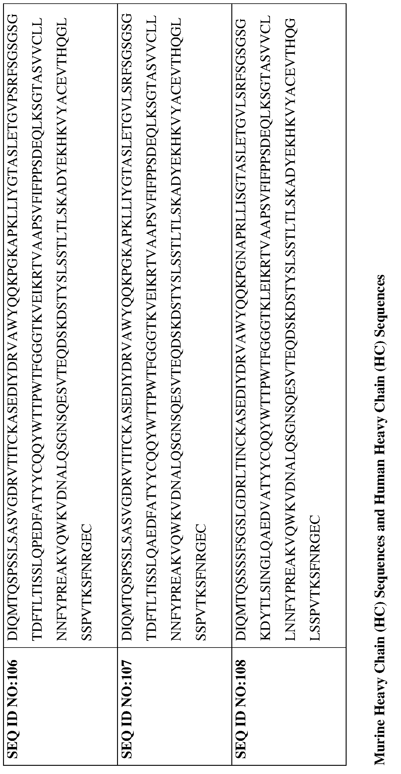
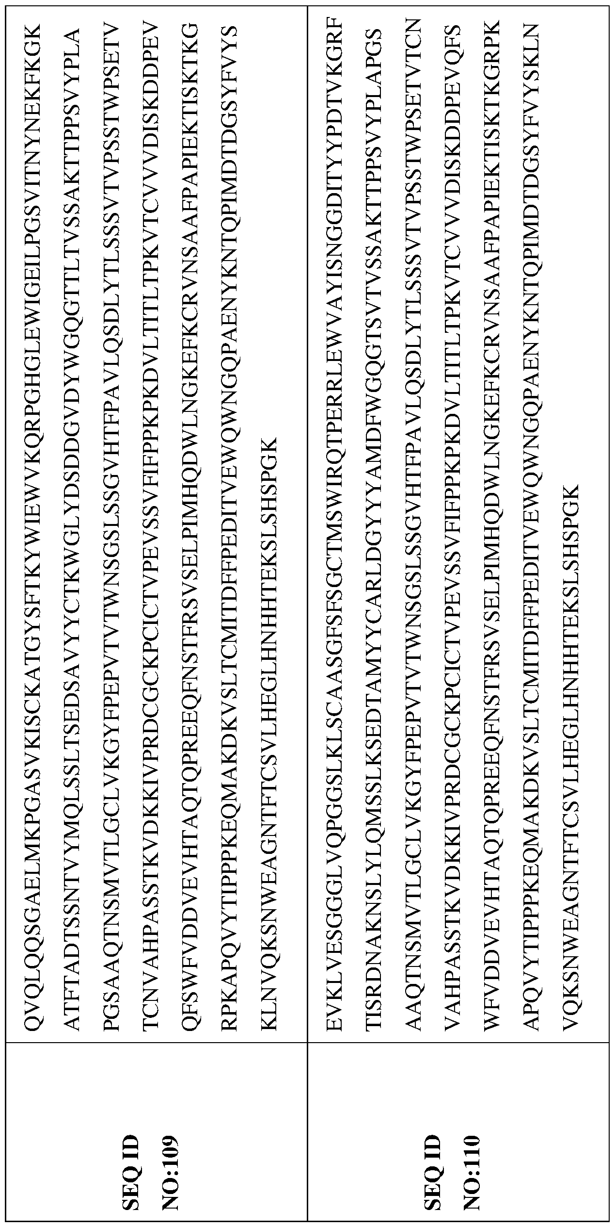


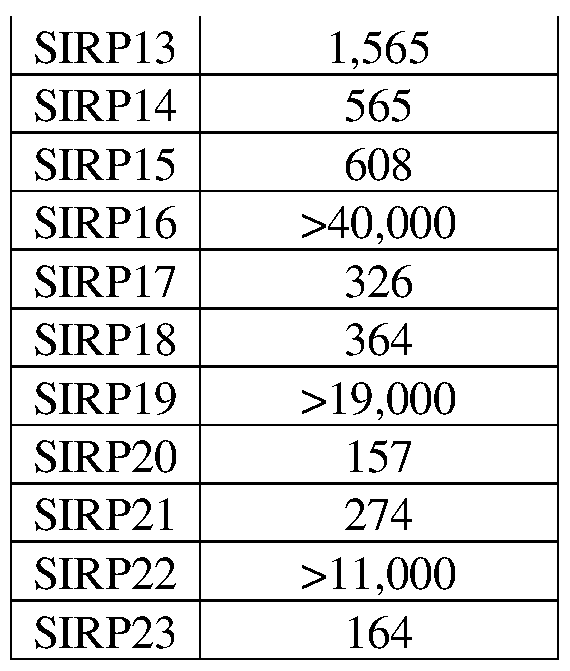


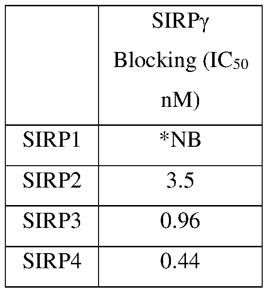


Claims


Priority Applications (6)
| Application Number | Priority Date | Filing Date | Title |
|---|---|---|---|
| JP2021525789A JP2022507295A (en) | 2018-11-14 | 2019-11-13 | Therapeutic SIRPα antibody |
| CN201980087951.8A CN113573728A (en) | 2018-11-14 | 2019-11-13 | Therapeutic SIRP alpha antibodies |
| CA3119748A CA3119748A1 (en) | 2018-11-14 | 2019-11-13 | Therapeutic sirp.alpha. antibodies |
| EP19885043.0A EP3880239A4 (en) | 2018-11-14 | 2019-11-13 | THERAPEUTIC SIRPalpha ANTIBODIES |
| AU2019380503A AU2019380503A1 (en) | 2018-11-14 | 2019-11-13 | Therapeutic SIRPα antibodies |
| BR112021009325-7A BR112021009325A2 (en) | 2018-11-14 | 2019-11-13 | MONOCLONAL ANTIBODY, OR ANTIGEN-BINDING FRAGMENT THEREOF; B-SPECIFIC ANTIBODY; METHOD TO PREVENT OR TREAT A SUSCEPTIBLE CANCER IN A HUMAN BEING; SIRP¿ EXPRESSION ASSAY METHOD IN TUMOR AND/OR IMMUNE CELLS; AND; PHARMACEUTICAL COMPOSITION |
Applications Claiming Priority (8)
| Application Number | Priority Date | Filing Date | Title |
|---|---|---|---|
| US201862767509P | 2018-11-14 | 2018-11-14 | |
| US62/767,509 | 2018-11-14 | ||
| US201962820718P | 2019-03-19 | 2019-03-19 | |
| US62/820,718 | 2019-03-19 | ||
| US201962886872P | 2019-08-14 | 2019-08-14 | |
| US62/886,872 | 2019-08-14 | ||
| US201962931746P | 2019-11-06 | 2019-11-06 | |
| US62/931,746 | 2019-11-06 |
Publications (1)
| Publication Number | Publication Date |
|---|---|
| WO2020102422A1 true WO2020102422A1 (en) | 2020-05-22 |
Family
ID=70730617
Family Applications (1)
| Application Number | Title | Priority Date | Filing Date |
|---|---|---|---|
| PCT/US2019/061278 WO2020102422A1 (en) | 2018-11-14 | 2019-11-13 | THERAPEUTIC SIRPα ANTIBODIES |
Country Status (8)
| Country | Link |
|---|---|
| US (2) | US11202828B2 (en) |
| EP (1) | EP3880239A4 (en) |
| JP (1) | JP2022507295A (en) |
| CN (1) | CN113573728A (en) |
| AU (1) | AU2019380503A1 (en) |
| BR (1) | BR112021009325A2 (en) |
| CA (1) | CA3119748A1 (en) |
| WO (1) | WO2020102422A1 (en) |
Cited By (6)
| Publication number | Priority date | Publication date | Assignee | Title |
|---|---|---|---|---|
| WO2022254379A1 (en) * | 2021-06-04 | 2022-12-08 | Boehringer Ingelheim International Gmbh | Anti-sirp-alpha antibodies |
| WO2023061084A1 (en) * | 2021-10-13 | 2023-04-20 | 宜明昂科生物医药技术(上海)股份有限公司 | Recombinant fusion protein targeting cd47 and cd24, and preparation and use thereof |
| CN118047874A (en) * | 2024-02-19 | 2024-05-17 | 北京润州生物科技有限公司 | Bispecific antibody and application of bispecific antibody and T cells in preparation of tumor treatment drugs |
| WO2024105180A1 (en) | 2022-11-16 | 2024-05-23 | Boehringer Ingelheim International Gmbh | Predictive efficacy biomarkers for anti-sirpa antibodies |
| WO2024200823A1 (en) | 2023-03-30 | 2024-10-03 | Ose Immunotherapeutics | Lipid-based nanoparticle targeted at activated immune cells for the expression of immune cell enhancing molecule and use thereof |
| WO2024200820A1 (en) | 2023-03-30 | 2024-10-03 | Ose Immunotherapeutics | Method of synthesis of targeted lipid nanoparticle and uses thereof |
Families Citing this family (7)
| Publication number | Priority date | Publication date | Assignee | Title |
|---|---|---|---|---|
| JOP20190009A1 (en) | 2016-09-21 | 2019-01-27 | Alx Oncology Inc | Antibodies against signal-regulatory protein alpha and methods of use |
| KR20200133376A (en) | 2018-03-21 | 2020-11-27 | 알렉소 온콜로지 인크. | Antibodies to signal-regulatory protein alpha and methods of use |
| CN112574310B (en) * | 2020-12-11 | 2023-05-05 | 浙江博锐生物制药有限公司 | anti-SIRP alpha antibodies and uses thereof |
| WO2022127901A1 (en) * | 2020-12-18 | 2022-06-23 | Lanova Medicines Development Co., Ltd. | BISPECIFIC ANTIBODIES TARGETING SIRPα AND PD-L1 |
| WO2022247933A1 (en) * | 2021-05-28 | 2022-12-01 | 百奥泰生物制药股份有限公司 | ANTI-SIRPα ANTIBODY AND USE THEREOF |
| CN117447593B (en) * | 2022-07-26 | 2024-06-25 | 北京东方百泰生物科技股份有限公司 | Anti-Siglec-15 monoclonal antibody |
| CN115819586B (en) * | 2022-10-13 | 2023-10-31 | 珠海市丽珠单抗生物技术有限公司 | Anti-human SIRP alpha monoclonal antibody and application thereof |
Citations (5)
| Publication number | Priority date | Publication date | Assignee | Title |
|---|---|---|---|---|
| US20130251733A1 (en) * | 2012-01-20 | 2013-09-26 | Genzyme Corporation | Anti-cxcr3 antibodies |
| US20140271478A1 (en) * | 2011-08-17 | 2014-09-18 | The Regents Of The University Of California | Antibodies that bind integrin alpha-v beta-8 |
| US20140302034A1 (en) * | 2010-12-08 | 2014-10-09 | Stem Centrx, Inc. | Novel modulators and methods of use |
| US20140314775A1 (en) * | 2011-11-16 | 2014-10-23 | Adrenomed Ag | Anti-adrenomedullin (adm) antibody or anti-adm antibody fragment of anti-adm non-ig scaffold for regulating the fluid balance in a patient having a chronic or acute disease |
| US20180312587A1 (en) * | 2017-04-13 | 2018-11-01 | Aduro Biotech Holdings, Europe B.V. | Anti-sirp alpha antibodies |
Family Cites Families (5)
| Publication number | Priority date | Publication date | Assignee | Title |
|---|---|---|---|---|
| AU4128089A (en) * | 1988-09-15 | 1990-03-22 | Rorer International (Overseas) Inc. | Monoclonal antibodies specific to human epidermal growth factor receptor and therapeutic methods employing same |
| CA2998644A1 (en) * | 2015-09-18 | 2017-03-23 | Arch Oncology, Inc. | Therapeutic cd47 antibodies |
| SG11201808465UA (en) * | 2016-04-14 | 2018-10-30 | Ose Immunotherapeutics | NEW ANTI-SIRPa ANTIBODIES AND THEIR THERAPEUTIC APPLICATIONS |
| JOP20190009A1 (en) * | 2016-09-21 | 2019-01-27 | Alx Oncology Inc | Antibodies against signal-regulatory protein alpha and methods of use |
| KR102642385B1 (en) | 2017-02-06 | 2024-03-04 | 오리오니스 바이오사이언시스 엔브이 | Targeted chimeric proteins and uses thereof |
-
2019
- 2019-11-13 JP JP2021525789A patent/JP2022507295A/en not_active Withdrawn
- 2019-11-13 US US16/682,893 patent/US11202828B2/en active Active
- 2019-11-13 CN CN201980087951.8A patent/CN113573728A/en not_active Withdrawn
- 2019-11-13 AU AU2019380503A patent/AU2019380503A1/en not_active Withdrawn
- 2019-11-13 BR BR112021009325-7A patent/BR112021009325A2/en not_active Application Discontinuation
- 2019-11-13 EP EP19885043.0A patent/EP3880239A4/en not_active Withdrawn
- 2019-11-13 WO PCT/US2019/061278 patent/WO2020102422A1/en unknown
- 2019-11-13 CA CA3119748A patent/CA3119748A1/en not_active Withdrawn
-
2021
- 2021-11-24 US US17/534,774 patent/US20220226469A1/en not_active Abandoned
Patent Citations (5)
| Publication number | Priority date | Publication date | Assignee | Title |
|---|---|---|---|---|
| US20140302034A1 (en) * | 2010-12-08 | 2014-10-09 | Stem Centrx, Inc. | Novel modulators and methods of use |
| US20140271478A1 (en) * | 2011-08-17 | 2014-09-18 | The Regents Of The University Of California | Antibodies that bind integrin alpha-v beta-8 |
| US20140314775A1 (en) * | 2011-11-16 | 2014-10-23 | Adrenomed Ag | Anti-adrenomedullin (adm) antibody or anti-adm antibody fragment of anti-adm non-ig scaffold for regulating the fluid balance in a patient having a chronic or acute disease |
| US20130251733A1 (en) * | 2012-01-20 | 2013-09-26 | Genzyme Corporation | Anti-cxcr3 antibodies |
| US20180312587A1 (en) * | 2017-04-13 | 2018-11-01 | Aduro Biotech Holdings, Europe B.V. | Anti-sirp alpha antibodies |
Non-Patent Citations (3)
| Title |
|---|
| DATABASE UniProtKB [online] 7 June 2017 (2017-06-07), "Uncharacterized protein", Database accession no. A0A1V2K656 * |
| RING ET AL.: "Anti-SIRPa antibody immunotherapy enhances neutrophil and macrophage antitumor activity", PROC NATL ACAD SCI USA., vol. 114, no. 49, 2017, pages E10578 - E10585, XP055429669 * |
| See also references of EP3880239A4 * |
Cited By (11)
| Publication number | Priority date | Publication date | Assignee | Title |
|---|---|---|---|---|
| WO2022254379A1 (en) * | 2021-06-04 | 2022-12-08 | Boehringer Ingelheim International Gmbh | Anti-sirp-alpha antibodies |
| US11572412B2 (en) | 2021-06-04 | 2023-02-07 | Boehringer Ingelheim International Gmbh | Anti-SIRP-alpha antibodies |
| JP2023530116A (en) * | 2021-06-04 | 2023-07-13 | ベーリンガー インゲルハイム インターナショナル ゲゼルシャフト ミット ベシュレンクテル ハフツング | anti-SIRP-alpha antibody |
| JP7436711B2 (en) | 2021-06-04 | 2024-02-22 | ベーリンガー インゲルハイム インターナショナル ゲゼルシャフト ミット ベシュレンクテル ハフツング | Anti-SIRP-alpha antibody |
| WO2023061084A1 (en) * | 2021-10-13 | 2023-04-20 | 宜明昂科生物医药技术(上海)股份有限公司 | Recombinant fusion protein targeting cd47 and cd24, and preparation and use thereof |
| US11891449B2 (en) | 2021-10-13 | 2024-02-06 | Immuneonco Biopharmaceuticals (Shanghai) Inc. | Recombinant fusion proteins targeting CD47 and CD24, preparation and use thereof |
| WO2024105180A1 (en) | 2022-11-16 | 2024-05-23 | Boehringer Ingelheim International Gmbh | Predictive efficacy biomarkers for anti-sirpa antibodies |
| WO2024200823A1 (en) | 2023-03-30 | 2024-10-03 | Ose Immunotherapeutics | Lipid-based nanoparticle targeted at activated immune cells for the expression of immune cell enhancing molecule and use thereof |
| WO2024200820A1 (en) | 2023-03-30 | 2024-10-03 | Ose Immunotherapeutics | Method of synthesis of targeted lipid nanoparticle and uses thereof |
| WO2024200826A1 (en) | 2023-03-30 | 2024-10-03 | Ose Immunotherapeutics | Lipid-based nanoparticle targeted at activated immune cells for the expression of immune cell inhibiting molecule and use thereof |
| CN118047874A (en) * | 2024-02-19 | 2024-05-17 | 北京润州生物科技有限公司 | Bispecific antibody and application of bispecific antibody and T cells in preparation of tumor treatment drugs |
Also Published As
| Publication number | Publication date |
|---|---|
| US20220226469A1 (en) | 2022-07-21 |
| AU2019380503A1 (en) | 2021-06-03 |
| BR112021009325A2 (en) | 2021-09-14 |
| EP3880239A1 (en) | 2021-09-22 |
| JP2022507295A (en) | 2022-01-18 |
| US11202828B2 (en) | 2021-12-21 |
| US20200297842A1 (en) | 2020-09-24 |
| EP3880239A4 (en) | 2022-11-23 |
| CA3119748A1 (en) | 2020-05-22 |
| CN113573728A (en) | 2021-10-29 |
Similar Documents
| Publication | Publication Date | Title |
|---|---|---|
| US11202828B2 (en) | Therapeutic SIRP-α antibodies | |
| US11692035B2 (en) | Therapeutic CD47 antibodies | |
| US20210070865A1 (en) | Therapeutic cd47 antibodies | |
| US20210324075A1 (en) | Therapeutic cd47 antibodies | |
| US20230279108A1 (en) | Therapeutic sirp-alpha antibodies | |
| JP7170331B2 (en) | Combination therapy for the treatment of solid and hematological cancers | |
| US20190309066A1 (en) | Combination therapy for the treatment of solid and hematological cancers | |
| RU2018126297A (en) | ANTIBODIES AIMED AT CD32B AND WAYS TO USE THEM | |
| US20220313819A1 (en) | Combination therapy for the treatment of solid and hematological cancers | |
| WO2021263085A2 (en) | Combination therapy for the treatment of solid and hematological cancers |
Legal Events
| Date | Code | Title | Description |
|---|---|---|---|
| 121 | Ep: the epo has been informed by wipo that ep was designated in this application |
Ref document number: 19885043 Country of ref document: EP Kind code of ref document: A1 |
|
| ENP | Entry into the national phase |
Ref document number: 2021525789 Country of ref document: JP Kind code of ref document: A |
|
| ENP | Entry into the national phase |
Ref document number: 3119748 Country of ref document: CA |
|
| NENP | Non-entry into the national phase |
Ref country code: DE |
|
| REG | Reference to national code |
Ref country code: BR Ref legal event code: B01A Ref document number: 112021009325 Country of ref document: BR |
|
| ENP | Entry into the national phase |
Ref document number: 2019380503 Country of ref document: AU Date of ref document: 20191113 Kind code of ref document: A |
|
| ENP | Entry into the national phase |
Ref document number: 2019885043 Country of ref document: EP Effective date: 20210614 |
|
| REG | Reference to national code |
Ref country code: BR Ref legal event code: B01E Ref document number: 112021009325 Country of ref document: BR Free format text: COM BASE NA PORTARIA 405 DE 21/12/2020, SOLICITA-SE QUE SEJA APRESENTADO, EM ATE 60 (SESSENTA) DIAS, NOVO CONTEUDO DE LISTAGEM DE SEQUENCIA POIS O CONTEUDO APRESENTADO NA PETICAO NO 270210061351 DE 06/08/2021 POSSUI INFORMACOES DIVERGENTES AO PEDIDO EM QUESTAO (DIVERGENCIA DE PRIORIDADE) |
|
| ENP | Entry into the national phase |
Ref document number: 112021009325 Country of ref document: BR Kind code of ref document: A2 Effective date: 20210513 |