WO2020095971A1 - Method for removing senescent cell, and method for preparing senescent cell - Google Patents
Method for removing senescent cell, and method for preparing senescent cell Download PDFInfo
- Publication number
- WO2020095971A1 WO2020095971A1 PCT/JP2019/043570 JP2019043570W WO2020095971A1 WO 2020095971 A1 WO2020095971 A1 WO 2020095971A1 JP 2019043570 W JP2019043570 W JP 2019043570W WO 2020095971 A1 WO2020095971 A1 WO 2020095971A1
- Authority
- WO
- WIPO (PCT)
- Prior art keywords
- cells
- cell
- senescent
- senescent cells
- bptes
- Prior art date
Links
Images


Classifications
-
- C—CHEMISTRY; METALLURGY
- C12—BIOCHEMISTRY; BEER; SPIRITS; WINE; VINEGAR; MICROBIOLOGY; ENZYMOLOGY; MUTATION OR GENETIC ENGINEERING
- C12N—MICROORGANISMS OR ENZYMES; COMPOSITIONS THEREOF; PROPAGATING, PRESERVING, OR MAINTAINING MICROORGANISMS; MUTATION OR GENETIC ENGINEERING; CULTURE MEDIA
- C12N5/00—Undifferentiated human, animal or plant cells, e.g. cell lines; Tissues; Cultivation or maintenance thereof; Culture media therefor
- C12N5/06—Animal cells or tissues; Human cells or tissues
- C12N5/0602—Vertebrate cells
- C12N5/0652—Cells of skeletal and connective tissues; Mesenchyme
- C12N5/0656—Adult fibroblasts
-
- A—HUMAN NECESSITIES
- A61—MEDICAL OR VETERINARY SCIENCE; HYGIENE
- A61K—PREPARATIONS FOR MEDICAL, DENTAL OR TOILETRY PURPOSES
- A61K31/00—Medicinal preparations containing organic active ingredients
- A61K31/33—Heterocyclic compounds
- A61K31/395—Heterocyclic compounds having nitrogen as a ring hetero atom, e.g. guanethidine or rifamycins
- A61K31/41—Heterocyclic compounds having nitrogen as a ring hetero atom, e.g. guanethidine or rifamycins having five-membered rings with two or more ring hetero atoms, at least one of which being nitrogen, e.g. tetrazole
- A61K31/433—Thidiazoles
-
- A—HUMAN NECESSITIES
- A61—MEDICAL OR VETERINARY SCIENCE; HYGIENE
- A61K—PREPARATIONS FOR MEDICAL, DENTAL OR TOILETRY PURPOSES
- A61K31/00—Medicinal preparations containing organic active ingredients
- A61K31/33—Heterocyclic compounds
- A61K31/395—Heterocyclic compounds having nitrogen as a ring hetero atom, e.g. guanethidine or rifamycins
- A61K31/435—Heterocyclic compounds having nitrogen as a ring hetero atom, e.g. guanethidine or rifamycins having six-membered rings with one nitrogen as the only ring hetero atom
-
- A—HUMAN NECESSITIES
- A61—MEDICAL OR VETERINARY SCIENCE; HYGIENE
- A61K—PREPARATIONS FOR MEDICAL, DENTAL OR TOILETRY PURPOSES
- A61K31/00—Medicinal preparations containing organic active ingredients
- A61K31/33—Heterocyclic compounds
- A61K31/395—Heterocyclic compounds having nitrogen as a ring hetero atom, e.g. guanethidine or rifamycins
- A61K31/495—Heterocyclic compounds having nitrogen as a ring hetero atom, e.g. guanethidine or rifamycins having six-membered rings with two or more nitrogen atoms as the only ring heteroatoms, e.g. piperazine or tetrazines
- A61K31/50—Pyridazines; Hydrogenated pyridazines
- A61K31/501—Pyridazines; Hydrogenated pyridazines not condensed and containing further heterocyclic rings
-
- A—HUMAN NECESSITIES
- A61—MEDICAL OR VETERINARY SCIENCE; HYGIENE
- A61K—PREPARATIONS FOR MEDICAL, DENTAL OR TOILETRY PURPOSES
- A61K31/00—Medicinal preparations containing organic active ingredients
- A61K31/655—Azo (—N=N—), diazo (=N2), azoxy (>N—O—N< or N(=O)—N<), azido (—N3) or diazoamino (—N=N—N<) compounds
-
- A—HUMAN NECESSITIES
- A61—MEDICAL OR VETERINARY SCIENCE; HYGIENE
- A61P—SPECIFIC THERAPEUTIC ACTIVITY OF CHEMICAL COMPOUNDS OR MEDICINAL PREPARATIONS
- A61P1/00—Drugs for disorders of the alimentary tract or the digestive system
- A61P1/16—Drugs for disorders of the alimentary tract or the digestive system for liver or gallbladder disorders, e.g. hepatoprotective agents, cholagogues, litholytics
-
- A—HUMAN NECESSITIES
- A61—MEDICAL OR VETERINARY SCIENCE; HYGIENE
- A61P—SPECIFIC THERAPEUTIC ACTIVITY OF CHEMICAL COMPOUNDS OR MEDICINAL PREPARATIONS
- A61P17/00—Drugs for dermatological disorders
- A61P17/02—Drugs for dermatological disorders for treating wounds, ulcers, burns, scars, keloids, or the like
-
- A—HUMAN NECESSITIES
- A61—MEDICAL OR VETERINARY SCIENCE; HYGIENE
- A61P—SPECIFIC THERAPEUTIC ACTIVITY OF CHEMICAL COMPOUNDS OR MEDICINAL PREPARATIONS
- A61P39/00—General protective or antinoxious agents
-
- A—HUMAN NECESSITIES
- A61—MEDICAL OR VETERINARY SCIENCE; HYGIENE
- A61P—SPECIFIC THERAPEUTIC ACTIVITY OF CHEMICAL COMPOUNDS OR MEDICINAL PREPARATIONS
- A61P43/00—Drugs for specific purposes, not provided for in groups A61P1/00-A61P41/00
-
- A—HUMAN NECESSITIES
- A61—MEDICAL OR VETERINARY SCIENCE; HYGIENE
- A61P—SPECIFIC THERAPEUTIC ACTIVITY OF CHEMICAL COMPOUNDS OR MEDICINAL PREPARATIONS
- A61P9/00—Drugs for disorders of the cardiovascular system
- A61P9/10—Drugs for disorders of the cardiovascular system for treating ischaemic or atherosclerotic diseases, e.g. antianginal drugs, coronary vasodilators, drugs for myocardial infarction, retinopathy, cerebrovascula insufficiency, renal arteriosclerosis
-
- C—CHEMISTRY; METALLURGY
- C12—BIOCHEMISTRY; BEER; SPIRITS; WINE; VINEGAR; MICROBIOLOGY; ENZYMOLOGY; MUTATION OR GENETIC ENGINEERING
- C12N—MICROORGANISMS OR ENZYMES; COMPOSITIONS THEREOF; PROPAGATING, PRESERVING, OR MAINTAINING MICROORGANISMS; MUTATION OR GENETIC ENGINEERING; CULTURE MEDIA
- C12N5/00—Undifferentiated human, animal or plant cells, e.g. cell lines; Tissues; Cultivation or maintenance thereof; Culture media therefor
- C12N5/0081—Purging biological preparations of unwanted cells
-
- C—CHEMISTRY; METALLURGY
- C12—BIOCHEMISTRY; BEER; SPIRITS; WINE; VINEGAR; MICROBIOLOGY; ENZYMOLOGY; MUTATION OR GENETIC ENGINEERING
- C12N—MICROORGANISMS OR ENZYMES; COMPOSITIONS THEREOF; PROPAGATING, PRESERVING, OR MAINTAINING MICROORGANISMS; MUTATION OR GENETIC ENGINEERING; CULTURE MEDIA
- C12N2501/00—Active agents used in cell culture processes, e.g. differentation
- C12N2501/70—Enzymes
- C12N2501/73—Hydrolases (EC 3.)
-
- C—CHEMISTRY; METALLURGY
- C12—BIOCHEMISTRY; BEER; SPIRITS; WINE; VINEGAR; MICROBIOLOGY; ENZYMOLOGY; MUTATION OR GENETIC ENGINEERING
- C12N—MICROORGANISMS OR ENZYMES; COMPOSITIONS THEREOF; PROPAGATING, PRESERVING, OR MAINTAINING MICROORGANISMS; MUTATION OR GENETIC ENGINEERING; CULTURE MEDIA
- C12N2501/00—Active agents used in cell culture processes, e.g. differentation
- C12N2501/999—Small molecules not provided for elsewhere
Definitions
- the present invention relates to a method for removing senescent cells from an individual. Furthermore, the present invention relates to a method for preparing purified senescent cells.
- Non-Patent Document 4 shows that the effect of senescent cells on healthy lifespan.
- p16 positive cells which are one of the biomarkers of senescent cells, tend to shorten their lifespan when accumulated in the body. It was suggested that removing the cells may extend the healthy lifespan of the individual.
- Non-Patent Document 6 a technique for stably preparing purified senescent cells has not been established.
- the present inventors aim to establish a method for purifying senescent cells, and a method to selectively kill or remove senescent cells and to identify a substance to be solved.
- Non-Patent Document 6 the inventors examined a method of preparing a purified senescent cell population (cell population containing only senescent cells) after inducing cell senescence, and activated p53 of G2 phase cells, and , Inhibiting the activity of PLK1 (polo-like kinase 1) has been found to enable purification of senescent cells.
- the inventors performed metabolome analysis of senescent cells by gas chromatography using the above-described purified senescent cell culture system.
- the conversion reaction of citric acid to isocitric acid was inhibited by the increase of active oxygen in senescent cells, and the production of ⁇ -ketoglutaric acid and the subsequent rotation of the citric acid cycle were related to the glutamine metabolic pathway (glutaminolysis). It was suggested that it might depend on Therefore, it was clarified that the selective inhibition of senescent cell death was induced by inhibiting glutaminolysis of senescent cells with a drug.
- the present invention has been completed based on the above findings.
- the present invention is the following (1) to (8).
- a senescent cell removing agent which is a drug for removing senescent cells in a living body, and which contains a glutaminase inhibitor as an active ingredient.
- the disease is arteriosclerosis, osteoporosis, cataract, glaucoma, dementia, Parkinson's disease, pulmonary fibrosis, chronic obstructive pulmonary disease, cancer, type 2 diabetes, chronic renal failure, cardiac hypertrophy, cirrhosis, sarcopenia or
- the pharmaceutical composition according to the above (3) which is a skin thinning.
- a method for preparing senescent cells which comprises the following steps (a) to (c).
- step (A) a step of synchronizing the cells with G2 phase
- step (B) a step of activating intracellular p53 protein synchronized with G2 phase
- step (c) a step of inhibiting intracellular PLK1 (polo-like kinase 1) activity after the treatment of step (b) (6)
- step (a) is a step of contacting the cells with a CDK1 (Cyclin-dependent kinase 1) activity inhibitor.
- step (b) is a step of contacting the cells with an Mdm2 protein inhibitor.
- (c) is a step of contacting a PLK1 activity inhibitor with the cells.
- senescent cells existing in the living body can be removed.
- the healthy life expectancy of an individual is expected to be extended, and it becomes possible to develop preventive and therapeutic methods and therapeutic agents for diseases associated with aging.
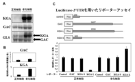
- Preparation of senescent cells A shows an example of a preparation schedule of 100% purified senescent cells. In addition, the proportion of SA- ⁇ -gal positive cells (B) in the cells 21 days after the start of preparation and the expression level of p16 mRNA are shown. Examination of the expression level of glutaminase in senescent cells. A shows the results of Western blotting using antibodies against KGA and GAC using cell extracts derived from cells after senescence induction (senescent cells) and cells before senescence induction (normal cells). B shows the results of measuring the KGA and GAC mRNA expression levels in senescent cells and normal cells by qPCR.
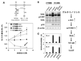
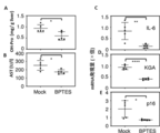
- A is a diagram schematically showing glutaminolysis (upper diagram) and the results of examining the effect of a glutaminase inhibitor (BPTES) on senescent cells and normal cells (lower diagram).
- B is a phosphorylation of S6K protein T389, S6K protein, in the presence or absence of BPTES or BPTES + DM-KG (transmembrane ⁇ -ketoglutarate) using cell extracts derived from senescent cells and normal cells.
- C is the result of measuring the mRNA expression levels of IL-6 and IL-8 in the presence or absence of BPTES or BPTE + DM-KG (membrane-permeable ⁇ -ketoglutarate) in senescent cells and normal cells. Role of glutaminolysis in pH regulation in senescent cells.
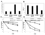
- A is the result of measuring the intracellular ammonia concentration of senescent cells and normal cells in the presence or absence of BPTES.
- B is the result of measuring intracellular pH of senescent cells and normal cells in the presence or absence of BPTES.
- C is the result of counting the number of senescent cells in the absence of BPTES, the presence of BPTES, the presence of BPTES + DUB or the presence of BPTES + CsA.
- D is the result of counting the number of senescent cells in the absence of BPTES, the presence of BPTES, the presence of BPTES + DUB or the presence of BPTES + CsA.
- the scale bar is 250 ⁇ m.
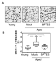
- B, C and D indicate the degree of glomerulosclerosis (B), serum urea concentration (C) and serum creatinine concentration (D), respectively. Data are shown as mean ⁇ standard deviation, box plots show median, interquartile range. The data were analyzed by One-way ANOVA, and then multiple comparisons were conducted using Tukey's multiple comparisons post hoc test.
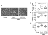
- the scale bar is 100 ⁇ m.
- B shows the area of the MT staining positive region. The value is shown as a relative value with the average value of Young's values being 1. Data are shown as mean ⁇ standard deviation, box plots show median, interquartile range.
- the scale bar is 100 ⁇ m.
- B, C and D show the area (B) of the MT staining positive region, the size of cardiomyocytes (C) and the heart weight (D), respectively.
- the scale bar is 50 ⁇ m.
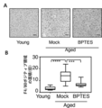
- B shows the area stained with the anti-F4 / 80 antibody as a relative value with the average value of Young's values being 1. Data are shown as mean ⁇ standard deviation, box plots show median, interquartile range. The statistical processing is the same as in FIG. *** P ⁇ 0.001, **** P ⁇ 0.0001.
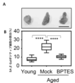
- B, C, D and E were stained with SA- ⁇ Gal positive area (B), adipose tissue weight (C), mean adipocyte diameter (D) and anti-F4 / 80 antibody, respectively.
- the area (E) is shown.
- Data are shown as mean ⁇ standard deviation, box plots show median, interquartile range. The statistical processing is the same as in FIG. ** P ⁇ 0.01, *** P ⁇ 0.001, **** P ⁇ 0.0001. Effects of glutaminolysis inhibitors on arteriosclerosis associated with obesity.
- B and C show the number of plaques in the aorta (B) and the percentage of damaged area in the aorta (C), respectively.
- the scale bar is 500 ⁇ m. Data are shown as mean ⁇ standard deviation, box plots show median, interquartile range. The statistical processing is the same as in FIG.
- the first embodiment of the present invention is an agent for removing senescent cells in a living body, which comprises an inhibitor of glutaminolysis (glutamine metabolic pathway), for example, a glutaminase inhibitor as an active ingredient, A cell depleting agent (hereinafter, also referred to as "aging cell depleting agent of the present invention").
- the “senescent cell removing agent” is an agent that induces cell death in senescent cells in vivo or in vitro and selectively removes senescent cells from a cell population containing senescent cells.
- senescent cells are cells whose cell growth or cell cycle is irreversibly arrested. Whether or not the cells are senescent cells can be evaluated using the characteristics of cell senescence as an index. Many previous studies have reported the characteristics of cell senescence, for example, increased expression of p16 (CDKN2A) protein, cell senescence-specific ⁇ -galactosidase (SA- ⁇ -gal) activity.
- CDKN2A cell senescence-specific ⁇ -galactosidase
- SA- ⁇ -gal cell senescence-specific ⁇ -galactosidase
- Elevated p21 (CDKN1A) protein expression Elevated p21 (CDKN1A) protein expression, elevated p19 protein expression, Senescence-associated heterochromatic foci (SAHF), DNA damage response (DDR) and senescence-associated secretory phenotype (senescence-associated) secretory phenotype: SASP) and the like (for details regarding the characteristics of cell aging, see, for example, Kuilman et al., Genes Dev 24: 2463-2479, 2010).
- SAHF Senescence-associated heterochromatic foci
- DDR DNA damage response
- SASP senescence-associated secretory phenotype
- Glutaminolysis is composed of several reaction steps. Above all, senescent cell-specific cell death can be efficiently induced by inhibiting the reaction step of producing glutamate from glutamine.
- the reaction that produces glutamic acid from glutamine is catalyzed by glutaminase (EC 13.5.1.2).
- glutaminase EC 13.5.1.2
- KGA is widely distributed throughout the body, whereas LGA is mainly present in the liver. KGA exists as two splice variants that differ only in the C-terminal region. The long form is called KGA as it is, and the short form is called GAC (glutaminase C).
- the glutaminase inhibitor used in the embodiment of the present invention may be any one as long as it inhibits at least the activity of KGA, and such an inhibitor can be easily selected by those skilled in the art.
- the glutaminase inhibitor is not particularly limited, for example, BPTES (bis-2- (5-phenylacetamido-1,3,4-thiadiazol-2-yl) ethyl sulfide: Bis-2- (5-phenylacetamido- 1,3,4-thiadiazol-2-yl) ethyl sulfide) (CAS No: 314045-39-1), DON (6-diazo-5-oxo-L-norleucine: 6-Diazo-5-oxo-L- norleucine) (CAS No: 51481-10-8), compound 968 (CAS No: 311795-38-7) and Examples include CB-839 (CAS No: 1439399-58-2).
- a protein or peptide such as a neutralizing antibody against KGA or a fragment thereof, a nucleic acid such as siRNA or miRNA for knocking out a gene (GLS1) encoding KGA may be used as a glutaminase inhibitor. ..
- the second embodiment of the present invention is a pharmaceutical composition (hereinafter also referred to as “pharmaceutical composition of the present invention”) containing the senescent cell removing agent of the present invention. Since the pharmaceutical composition of the present invention contains the senescent cell removing agent of the present invention as an active ingredient, when this is administered to a living body, senescent cells in the body are selectively killed or removed (see Examples). thing). Therefore, the pharmaceutical composition of the present invention is a disease that develops with extension of healthy life expectancy and aging, for example, arteriosclerosis, osteoporosis, cataract, glaucoma, dementia, Parkinson's disease, pulmonary fibrosis, chronic obstructive pulmonary disease.
- the pharmaceutical composition of the present invention may be administered in the form of a pharmaceutical composition containing one or more pharmaceutical additives in addition to the active ingredient (aging cell depleting agent). Moreover, you may mix
- the pharmaceutical composition of the present invention may be in a dosage form for oral or parenteral use, and is not particularly limited, but for example, tablets, capsules, granules, powders, syrups, suspensions, suppositories, Examples include ointments, creams, gels, patches, inhalants and injections.
- These preparations are prepared according to a conventional method.
- the liquid preparation may be dissolved or suspended in water or another suitable solvent before use.
- the tablets and granules may be coated by a known method.
- an injectable preparation it is prepared by dissolving the active ingredient in water, but it may be dissolved in physiological saline or glucose solution as necessary, and a buffer or a preservative may be added. ..
- an inorganic or organic substance, or a solid or liquid substance can be used as a pharmaceutical additive, and generally, for example, 0.1% by weight to 99.9% by weight, 1% by weight to the weight of the active ingredient. It can be blended in 95.0% by weight, or between 1% by weight and 90.0% by weight.
- additives for pharmaceuticals include lactose, glucose, mannitol, dextrin, cyclodextrin, starch, sucrose, magnesium aluminometasilicate, synthetic aluminum silicate, sodium carboxymethylcellulose, hydroxypropylstarch, calcium carboxymethylcellulose.
- Ion exchange resin methyl cellulose, gelatin, gum arabic, hydroxypropyl cellulose, hydroxypropyl methyl cellulose, polyvinyl pyrrolidone, polyvinyl alcohol, light anhydrous silicic acid, magnesium stearate, talc, tragacanth, bentonite, bee gum, titanium oxide, sorbitan fatty acid ester, Sodium lauryl sulfate, glycerin, fatty acid glycerin ester, purified lanolin, glycerogelatin, polyso Bate, macrogol, vegetable oils, waxes, liquid paraffin, white petrolatum, fluorocarbons, nonionic surfactants, propylene glycol or water and the like.
- an active ingredient and an excipient ingredient such as lactose, starch, crystalline cellulose, calcium lactate or silicic acid anhydride are mixed to prepare a powder, or if necessary, further.
- Wet or dry granulation is performed by adding a binder such as sucrose, hydroxypropylcellulose or polyvinylpyrrolidone, a disintegrating agent such as carboxymethylcellulose or carboxymethylcellulose calcium, to obtain granules.
- these powders and granules may be tableted as they are or by adding a lubricant such as magnesium stearate or talc.
- granules or tablets are coated with an enteric solvent base such as hydroxypropylmethyl cellulose phthalate or methacrylic acid-methyl methacrylate polymer to form an enteric preparation, or coated with ethyl cellulose, carnauba wax or hardened oil to form a sustained release preparation. You can also do it.
- an enteric solvent base such as hydroxypropylmethyl cellulose phthalate or methacrylic acid-methyl methacrylate polymer to form an enteric preparation
- ethyl cellulose, carnauba wax or hardened oil to form a sustained release preparation. You can also do it.
- powders or granules are filled in hard capsules, or the active ingredient is used as it is, or dissolved in glycerin, polyethylene glycol, sesame oil or olive oil, and then coated with gelatin to give soft capsules. be able to.
- an active ingredient may be added, if necessary, to hydrochloric acid, sodium hydroxide, lactose, lactic acid, sodium, a pH adjusting agent such as sodium monohydrogen phosphate or sodium dihydrogen phosphate, sodium chloride or glucose.
- a pH adjusting agent such as sodium monohydrogen phosphate or sodium dihydrogen phosphate, sodium chloride or glucose.
- Dissolve in distilled water for injection with isotonic agent aseptically filter and fill in ampoules, or add mannitol, dextrin, cyclodextrin or gelatin etc. and freeze-dry under vacuum to prepare a solution-soluble injection.
- reticine, polysorbate 80, polyoxyethylene hydrogenated castor oil or the like may be added to the active ingredient and emulsified in water to give an emulsion for injection.
- the active ingredient is moistened with a suppository base such as cocoa butter, tri-, di- and monoglycerides of fatty acids or polyethylene glycol, dissolved, poured into a mold and cooled, or the active ingredient is cooled. It may be dissolved in polyethylene glycol or soybean oil and then covered with a gelatin film or the like.
- a suppository base such as cocoa butter, tri-, di- and monoglycerides of fatty acids or polyethylene glycol
- the dose and the number of administrations of the pharmaceutical composition of the present invention are not particularly limited, depending on conditions such as the purpose of prevention and / or treatment of deterioration and progression of the disease to be treated, the type of disease, the weight and age of the patient, It can be appropriately selected according to the judgment of the doctor or pharmacist.
- the daily dose for adults in oral administration is about 0.01 to 1,000 mg (weight of active ingredient), which can be administered once or several times a day or every several days. ..
- the third embodiment of the present invention is a method for inducing cell death in senescent cells, which comprises inhibiting glutaminase activity in senescent cells in vitro or in vivo. Inhibition of glutaminase activity in senescent cells can also be carried out, for example, by bringing the above-mentioned glutaminase inhibitor into contact with senescent cells to bring them into the cells.
- a glutaminase inhibitor may be administered to the living body together with a pharmaceutically acceptable carrier or the like.
- the glutaminase inhibitor can also be administered in the form of the pharmaceutical composition described in the second embodiment above.
- the fourth embodiment of the present invention is a method for preventing or treating a disease that develops with aging, which comprises administering the pharmaceutical composition of the present invention or the agent for removing senescent cells of the present invention to a patient.
- treatment means to prevent or alleviate the progression and deterioration of the disease state in patients who have already developed a disease that develops with aging, thereby preventing or alleviating the progression and deterioration of the disease. It is a treatment for the purpose of Further, “prevention” means to prevent the onset of a disease that develops with aging in advance, and is a treatment for the purpose of preventing the onset of the disease in advance.
- the target of treatment and prevention is not limited to humans, and mammals other than humans, for example, mice, rats, dogs, cats, livestock such as cows, horses, sheep, primates such as monkeys, chimpanzees and gorillas, etc. And particularly preferably human.
- the fifth embodiment of the present invention is a method for preparing senescent cells, which comprises the following steps (a) to (c). (A) a step of synchronizing the cells with G2 phase, (B) a step of activating intracellular p53 protein synchronized with G2 phase, and (c) a step of inhibiting PLK1 (polo-like kinase 1) activity in the cell treated by the step (b)
- Whether it is an senescent cell is, for example, that the expression of p16 protein, p21 protein or p19 protein in the cell is significantly increased as compared with that in a normal cell, and cell senescence specific ⁇ -galactosidase (SA- ⁇ -gal ) Activity is significantly elevated compared to normal cells, inflammatory cytokines (eg, IL-6, IL-8, etc.), growth factors (eg, IGFBP7, etc.) and matrix metalloproteinases (matrix metalloproteinases: It can be evaluated by using a significant increase in secretion of molecules characteristic of SASP such as MMPs) as an index.
- SA- ⁇ -gal cell senescence specific ⁇ -galactosidase
- the cells that induce senescence may be derived from any animal as long as they are derived from mammals, and may be cells derived from any tissue.
- the basic medium for cell culture for preparing senescent cells may be any medium suitable for the cells to be cultured, and if necessary, antibiotics, protease inhibitors, etc. may be added. May be used. Regarding the culture conditions, CO 2 concentration and culture temperature suitable for the cells to be used can be adopted.
- the step (a) in the fifth embodiment is a step of synchronizing the cell cycle of the aging-inducing cell population with the G2 (gap2) phase.
- G2 G2
- a CDK1 Cyclin-dependent kinase 1
- an anticancer agent for example, irradiation with radiation, irradiation with ultraviolet rays and the like can be mentioned.
- a commercially available CDK inhibitor may be used for inhibiting CDK1 activity, and examples of the CDK1 inhibitor include RO3306 (CAS No: 872573-93-8), Roscovitine (CAS No: 186692-46-6) and Examples include BMI-1026 (CAS No: 477726-77-5).
- the CDK1 inhibitor can be used by adding it to the cell culture medium, etc., and the concentration to be used can be easily determined by conducting preliminary experiments with reference to the instruction manual of the supplier. it can.
- the concentration in the culture solution is, for example, 1 to 20 ⁇ M, preferably 5 to 10 ⁇ M, more preferably about 9 ⁇ M.
- the time for treating the cells is not particularly limited, but is, for example, 10 hours to 30 hours, preferably 15 hours to 25 hours, more preferably about 24 hours.
- the step (b) in the fifth embodiment is a step of performing a process for activating the p53 protein in the cells (population) synchronized with the G2 phase in the step (a).
- a method of activating intracellular p53 protein can be easily selected by those skilled in the art, and for example, Mdm2 protein (interacts with p53 protein and suppressively regulates p53 protein activity)
- Mdm2 protein interacts with p53 protein and suppressively regulates p53 protein activity
- addition of an anticancer agent, irradiation, ultraviolet irradiation, oxidative stress load and nutrient depletion can be mentioned.
- a commercially available inhibitor may be used to inhibit the activity of the Mdm2 protein, and examples of such an inhibitor include Nutlin-3a (CAS No: 675576-98-4), HLI373 (CAS No: 502137). -98-6), RG7388 (CAS No: 1229705-06-9), AMG-232 (CAS No: 1352066-68-2) and (MI-773 CAS No: 1303607-07-9). it can.
- the Mdm2 inhibitor can be used by adding it to a cell culture medium, etc., and the concentration to be used can be easily determined by conducting preliminary experiments with reference to the instruction manual of the supplier. it can.
- the concentration in the culture solution is, for example, 1 to 20 ⁇ M, preferably 5 to 15 ⁇ M, more preferably about 10 ⁇ M.
- the time for treating the cells is not particularly limited, but is, for example, 10 hours to 70 hours, preferably 30 hours to 60 hours, more preferably about 50 hours.
- the step (c) in the fifth embodiment is a step for inhibiting the PLK1 (polo-like kinase 1) activity in cells that activated the p53 protein in the G2 phase in steps (a) and (b). This is a process to be performed.
- a commercially available PLK1 activity inhibitor may be used. Examples of such an inhibitor include BI2536 (CAS No: 755038-02-9), GSK461364 ( CAS No: 929095-18-1), PCM-075 (CAS No: 1263293-37-3) and BI-6727 (CAS: 755038-65-4).
- PLK1 activity inhibitors can be used by adding them to the culture medium of cells, etc., and the concentration to be used can be easily determined by conducting preliminary experiments with reference to the instruction manual of the supplier. You can although not particularly limited, for example, when BI2536 is used as the PLK1 activity inhibitor, the concentration in the culture solution is, for example, 50 to 150 nM, preferably 75 to 120 nM, more preferably about 100 nM. When BI2536 is used, the time for treating the cells is not particularly limited, but is, for example, 7 days to 15 days, preferably 8 days to 12 days, more preferably about 9 days.
- ReverTra Ace qPCR RT kit (Takara) was used, and reverse transcription was performed on cDNA according to the attached protocol. Then, using the cDNA as a template, Power SYBR Green PCR Master Mix (Applied Biosystems) was used to perform qPCR analysis to measure the expression level of p16 mRNA.
- the primers for detecting the p16 mRNA expression level are shown below. Forward: 5'-CCCAACGCACCGAATAGTTA-3 '(SEQ ID NO: 1) Reverse: 5'- ACCAGCGTGTCCAGGAAG-3 '(SEQ ID NO: 2)
- the expression level of mRNA was corrected by the amount of GAPDH mRNA. As a result, p16 expression was significantly increased in the cells on the 21st day of culture. From the above results, it was confirmed that the senescent cell preparation method of the present invention can induce senescence in almost all cells.
- the reporter activity was measured using the cells 48 hours after the introduction and using the Dual-Glo Luciferase Assay System (Promega) according to the attached protocol.
- KGA-L long 3'UTR
- senescent cells increased rather. From this result, it was revealed that the stability of KGA mRNA through the 3′UTR region of the senescent cell glutaminase gene was increased (FIG. 2C).
- BPTES BPTES
- IL-6 and IL-8 which are major factors of the phenotype SASP that secretes large amounts of inflammatory cytokines and extracellular matrix degrading enzymes, which are one of the most important traits of senescent cells.
- Senescent cells and normal cells were cultured in BPTES (final concentration 2.5 ⁇ M) -added medium or normal medium at 37 ° C, 5% CO 2 for 24 hours, and ISOGEN II (Wako) was used from each cell. To prepare total RNA.
- qPCR After reverse transcription from total RNA to cDNA using SuperScript II cDNA synthesis kit (Invitrogen), qPCR to measure IL-6 and IL-8 expression levels using Power SYBR Green PCR Master Mix (Applied Biosystems) Analysis was performed.
- the primers for detecting the expression levels of IL-6 and IL-8 mRNA are shown below.
- IL-6 Forward 5'-CCAGGAGCCCAGCTATGAAC-3 '(SEQ ID NO: 3) Reverse: 5'-CCCAGGGAGAAGGCACTG-3 '(SEQ ID NO: 4)
- IL-8 Forward 5'-AAGGAAAACTGGGTGCAGAG-3 '(SEQ ID NO: 5) Reverse: 5'-ATTGCATCTGGCAACCCTAC-3 '(SEQ ID NO: 6) It was found that the administration of a final concentration of 2.5 ⁇ M BPTES, which has almost no effect on the survival of senescent cells, inhibits the mRNA expression of the major factors of SASP, IL-6 and IL-8 (FIG. 3C).
- Senescent cells and normal cells were cultured for 24 hours at 37 ° C. and 5% CO 2 in a medium supplemented with BPTES (final concentration 2.5 ⁇ M) or a normal medium.
- a cell extract was prepared from the cultured cells using Laemmli-buffer (2% SDS, 10% glycerol, 5% 2-mercaptoethanol, 0.002% bromophenol blue and 62.5 mM Tris HCl at pH 6.8).
- the cell extract (20-50 ⁇ g) was separated by SDS-PAGE and transferred to a PVDF membrane, then an antibody against the T389 phosphorylation site of S67K protein (CST), an antibody against S6K protein (CST) and an antibody against p16 protein ( abcam) and Western blotting was performed to detect by ECL method.
- CST S67K protein
- CST an antibody against S6K protein
- abcam an antibody against p16 protein
- Western blotting was performed to detect by ECL method.
- BPTES treatment inhibits phosphorylation of S6K protein T389, which is an indicator of mTOR activation, which is a major regulator of SASP (see FIG. 3B, S6KpT389, BPTES lane of senescent cells). .. It was also revealed that this inhibitory effect was rescued by administration of membrane-permeable ⁇ -ketoglutarate (DM-KG) (see FIG. 3B, S6KpT389, lane of DM
- Senescent cells and normal cells were seeded in a 10 cm culture dish so that each had about 50,000 cells.
- change to medium containing BPTES (final concentration 10 ⁇ M) or normal medium incubate at 37 ° C, 5% CO 2 for 24 hours, then use Ammonia assay kit (abcam) to The amount of ammonia was determined.
- Ammonia assay kit abcam
- senescent cells and normal cells were seeded on a 6-well plate so that each had about 50,000 cells.
- the medium was replaced with a medium supplemented with BPTES (final concentration 10 ⁇ M) or a normal medium, and cultured at 37 ° C., 5% CO 2 for 24 hours.
- HEPES solution containing DCECF-AM (Dojindo Co., Ltd.) at a final concentration of 3 ⁇ M at 37 ° C, 5% CO 2 for 10 minutes, wash with HEPES solution three times, and then luminescence with a plate reader. It was measured.
- the pH of the medium of BPTES-treated cells was adjusted to weak basicity (pH 8.0 or pH 8.5), and the cell viability was examined. Senescent cells and normal cells were seeded in a 6 cm culture dish at approximately 10,000 cells each.
- BPTES final concentration 10 ⁇ M
- the medium was exchanged with pH 7.4, pH 8.0 or pH 8.5, and after culturing at 37 ° C, 5% CO 2 for 24 hours, then for 24 hours.
- the cells were stained with trypan blue every other time, and the number of viable cells was counted using a hemocytometer. As a result, it was found that 90% or more of the cell death observed by BPTES treatment was reduced to about 30% under weakly basic pH conditions (Fig. 4D).
- the expression level of p16 mRNA which is a marker for senescent cells, was analyzed by qPCR using Power SYBR Green PCR Master Mix (Applied Biosystems). The expression amount of p16 mRNA was corrected by the value of GAPDH.
- the primers for detecting the expression amount of p16 mRNA are shown below. Forward: 5'-CCGCTGCAGACAGACTGG-3 '(SEQ ID NO: 7) Reverse: 5'-CCATCATCATCACCTGAATCG-3 '(SEQ ID NO: 8) All mice were kept in a specific pathogen-free environment and treated according to the guidelines for animal experiments of the Institute of Medical Science, University of Tokyo (the same applies to the experiments in 2-5).
- Kidney, lung, liver and heart were embedded with OCT compound and frozen sections were prepared, followed by hematoxylin / eosin (H & E). ), Tissue staining with Masson Trichrome (MT) reagent, Periodic Acid Schiff (PAS) reagent (Fisher Scientific), or anti-F4-80 antibody (CST) Immunostaining was performed with DAB (3,3'-Diaminobenzidine Tetrahydrochloride) (DAKO). After staining, the tissue section was observed under a microscope.
- MT Masson Trichrome
- PAS Periodic Acid Schiff
- CST anti-F4-80 antibody
- Renal glomerulosclerosis was assessed based on PAS-positive intensity and range in 40 glomeruli per individual.
- the serum urea concentration and creatinine concentration were measured using the Urea Assay kit (Abcam) or Creatinine Assay kit (Abcam), respectively.
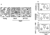
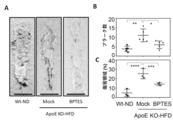
- FIG. 6A shows PAS staining results of glomeruli of young mice (Young), vehicle (Mock) or aged mice (Aged) to which BPTES (BPTES) was administered.
- the glomeruli of the control mice were more indurated than the glomeruli of the young mice, but the BPTES-administered mice improved the degree of glomerulosclerosis (FIG. 6B). It was also found that the concentrations of urea and creatinine in serum were also decreased by administration of BPTES (Figs. 6C and D).
- FIG. 7A shows the results of MT staining of lung tissue of young mice (Young) and aged mice (Aged) administered with vehicle or BPTES.
- FIG. 7B shows the degree of fibrosis when the MT-stained region was quantified by BZ-X analyzer (Keyence), the degree of fibrosis was improved in the lungs of BPTES-administered mice as compared with the lungs of control mice (Fig. 7B, top). ).
- MT tissue staining of the heart was performed to evaluate the degree of fibrosis (Fig. 8A).
- the heart weight was measured, and the size of cardiomyocytes was measured with a BZ-X analyzer (Keyence).
- the degree of fibrosis in the heart of BPTES-administered mice was improved as compared with the control mice (FIG. 8B).
- the cardiomyocyte size of BPTES-administered mice was smaller than that of control mice, and the heart weight was also lighter in BPTES-administered mice and comparable to that of young mice. (FIGS. 8C and D).
- a small piece of adipose tissue was collected in PBS, fixed with 2% formamide / 0.2% glutaraldehyde for 15 minutes, washed, and then freshly prepared SA- ⁇ gal staining solution (1 mg X-gal / ml, 40 mM citric acid / It was incubated with sodium phosphate pH 6.0, 5 mM potassium ferrocyanide, 5 mM potassium ferricyanide, 150 mM NaCl, 2 mM MgCl 2 ) at 37 ° C. for 12 hours. Then, the tissue piece was washed with PBS and pressed between slide glasses for microscopic observation.
- FIG. 10A shows the results of SA- ⁇ gal staining of white adipose tissue
- FIG. 10B shows the proportion of SA- ⁇ gal positive cells. This result showed that the accumulation of senescent cells stained with SA- ⁇ gal was improved by the glutaminolysis inhibitor.
- the amount of serum AST and the amount of hydroxyproline in liver were measured by AST assay kit (Abcam) and Hydroxyproline assay kit (Abcam), respectively.
- the expression levels of p16, KGA and IL-6 were measured by qPCR.
- the qPCR primers are shown below.
- the present invention provides a method for efficiently preparing purified senescent cells, an agent for removing senescent cells, an agent for preventing or treating a disease that develops with aging, and the like. Therefore, the present invention is expected to be used in the medical field.
Abstract
The present invention addresses the problem of providing: a method for killing or removing a senescent cell selectively; a method for identifying a substance; and a method for purifying a senescent cell. More specifically, the present invention provides: a senescent cell removing agent which can be used for the removal of a senescent cell in a living body and contains a glutaminase inhibitor as an active ingredient; and a pharmaceutical composition containing the removing agent. The present invention also provides a method for preparing a senescent cell, comprising the following steps (a) to (c): (a) synchronizing the cell at the G2 phase; (b) activating p53 protein in the cell that has been synchronized at the G2 phase; and (c) inhibiting the PLK1 (polo-like kinase 1) activity in the cell that had undergone the treatment of step (b).
Description
本発明は、個体から老化細胞を除去する方法に関する。さらに、本発明は、純化した老化細胞の調製方法に関する
The present invention relates to a method for removing senescent cells from an individual. Furthermore, the present invention relates to a method for preparing purified senescent cells.
超高齢を迎えた現代社会にとって、健康寿命の延長は現代科学の解決すべき最重要課題の一つである。健康寿命の延長には、医療システムの改革や生活習慣病、食習慣等の改善が有効な手段であることは明白である。しかし、健康寿命に対する抜本的な対応には、老化制御機構の俯瞰的理解と、老化に伴う加齢性疾病や、臓器・組織の機能低下を予防する技術の開発が必要不可欠である。
Extending the healthy life expectancy is one of the most important issues to be solved by modern science for the modern society, which has become extremely old. It is clear that reforming the medical system and improving lifestyle-related diseases and eating habits are effective means for prolonging healthy life expectancy. However, in order to drastically respond to healthy life expectancy, it is essential to have a bird's-eye view of the aging control mechanism and the development of technology to prevent age-related diseases and functional deterioration of organs and tissues due to aging.
これまで細胞老化は、細胞増殖の不可逆的な停止と考えられ、がん防御メカニズムの1つとして位置づけられてきたが、その一方で、細胞老化は、個体レベルの老化や老化に関連した疾患においても何らかの役割を果たしていると考えられるようになってきた。老化細胞は、炎症性サイトカイン、ケモカイン、マトリクスメタロプロテイナーゼおよび成長因子などを分泌することが示されており、この現象は、老化関連分泌表現型(senescence-associated secretory phenotype:SASP)と称され、老化関連疾患の発症に関連していることが示唆されている(非特許文献1~非特許文献3)。
Until now, cell aging has been thought to be an irreversible stop of cell proliferation and has been positioned as one of the cancer defense mechanisms.On the other hand, cell aging is associated with individual-level aging and diseases related to aging. Has come to be considered to play some role. Senescent cells have been shown to secrete inflammatory cytokines, chemokines, matrix metalloproteinases and growth factors. This phenomenon is called the senescence-associated secretory phenotype (SASP) and It has been suggested to be related to the development of related diseases (Non-patent documents 1 to 3).
近年、老化制御機構の解明に大きなパラダイムシフトが生じた。Bakerらは、早老性モデルマウスを用いた遺伝学的解析から、老齢個体から人工的に老化細胞を除去すると、動脈硬化や腎障害などの老年病の発症が有意に遅れ、さらには寿命そのものも延長することを報告した(非特許文献4)。さらに、非早老性マウスにおいて、老化細胞が健康寿命に及ぼす影響を調べたところ、老化細胞のバイオマーカーの1つであるp16陽性細胞が体内に蓄積すると寿命が短くなる傾向にあること、p16陽性細胞を除去すると個体の健康寿命が伸びる可能性があることなどが示唆された(非特許文献5)。従って、生体内に存在する老化細胞を選択的に死滅または除去できる薬剤の開発は、健康寿命の延長や加齢に伴う疾患(動脈硬化症や骨粗鬆症)の治療に対する新たな方法論の確立につながると考えられる。
In recent years, a big paradigm shift has occurred in the clarification of the aging control mechanism. Based on a genetic analysis using progeria model mice, Baker et al. Showed that artificial removal of senescent cells from aged individuals significantly delayed the onset of senile diseases such as arteriosclerosis and renal damage, and even the lifespan itself. It was reported to extend (Non-Patent Document 4). Furthermore, when the effect of senescent cells on healthy lifespan was examined in non-proliferative mice, p16 positive cells, which are one of the biomarkers of senescent cells, tend to shorten their lifespan when accumulated in the body. It was suggested that removing the cells may extend the healthy lifespan of the individual (Non-Patent Document 5). Therefore, the development of a drug that can selectively kill or remove senescent cells existing in the living body will lead to the establishment of new methodologies for prolonging healthy life span and treating diseases associated with aging (arteriosclerosis and osteoporosis). Conceivable.
ところで、細胞の老化は、様々なゲノムストレスにより誘導されるため、誘導刺激の種類に依存して特異的なシグナル経路が活性化されている。従って、全ての老化細胞に共通したトランスクリプトームや、メタボローム状態を明らかにするためには、外的刺激なく、かつ100%純化した老化細胞調製技術の開発が必要不可欠である。
これまでに発明者らは、細胞老化の誘導には、G2期においてp53が活性化されることが、必要十分であることを明らかにしている(非特許文献6)。しかしながら、現在のところ、純化した老化細胞を安定的に調製する技術の確立までには至っていない。 By the way, since cell senescence is induced by various genomic stresses, specific signal pathways are activated depending on the type of induction stimulus. Therefore, in order to clarify the transcriptome and metabolome state common to all senescent cells, it is essential to develop a technique for preparing senescent cells that is 100% purified without external stimulation.
So far, the inventors have revealed that activation of p53 in the G2 phase is necessary and sufficient for inducing cell senescence (Non-Patent Document 6). However, at present, a technique for stably preparing purified senescent cells has not been established.
上記事情に鑑み、本発明者らは、老化細胞を純化する方法の確立を目指すとともに、老化細胞を選択的に死滅または除去する方法および物質の同定を解決課題とする。
In view of the above circumstances, the present inventors aim to establish a method for purifying senescent cells, and a method to selectively kill or remove senescent cells and to identify a substance to be solved.
発明者らは、まず、老化細胞の純化を試みた。これまでに、細胞をG2期に同調した状態で、がん抑制タンパク質のp53を活性化することが細胞老化の誘導には必要十分であることを示している(非特許文献6)。発明者らは、この知見に基づいて細胞老化を誘導した上で、純化した老化細胞集団(老化細胞のみの細胞集団)を調製する方法を検討したところ、G2期細胞のp53を活性化し、さらに、PLK1(polo-like kinase 1)の活性を阻害すると、老化細胞の純化が可能であることを見い出した。
First, the inventors tried to purify senescent cells. So far, it has been shown that activation of the tumor suppressor protein p53 in a state in which cells are synchronized with the G2 phase is necessary and sufficient for inducing cell senescence (Non-Patent Document 6). Based on this finding, the inventors examined a method of preparing a purified senescent cell population (cell population containing only senescent cells) after inducing cell senescence, and activated p53 of G2 phase cells, and , Inhibiting the activity of PLK1 (polo-like kinase 1) has been found to enable purification of senescent cells.
さらに、発明者らは、上述の純化老化細胞の培養系を用いてガスクロマトグラフィー法による老化細胞のメタボローム解析を行った。その結果、老化細胞では活性酸素量の上昇によりクエン酸からイソクエン酸への変換反応が阻害されており、αケトグルタル酸の産生とそれ以降のクエン酸回路の回転がグルタミン代謝経路(グルタミノリシス)に依存している可能性が示唆された。そこで、老化細胞のグルタミノリシスを薬剤で阻害したところ、老化細胞に選択的な細胞死が誘導されることが明らかとなった。
本発明は以上の知見に基づいて完成されたものである。 Furthermore, the inventors performed metabolome analysis of senescent cells by gas chromatography using the above-described purified senescent cell culture system. As a result, the conversion reaction of citric acid to isocitric acid was inhibited by the increase of active oxygen in senescent cells, and the production of α-ketoglutaric acid and the subsequent rotation of the citric acid cycle were related to the glutamine metabolic pathway (glutaminolysis). It was suggested that it might depend on Therefore, it was clarified that the selective inhibition of senescent cell death was induced by inhibiting glutaminolysis of senescent cells with a drug.
The present invention has been completed based on the above findings.
すなわち、本発明は以下の(1)~(8)である。
(1)生体内の老化細胞を除去するための薬剤であって、グルタミナーゼ阻害剤を有効成分として含有する、老化細胞除去剤。
(2)前記グルタミナーゼが、KGA(Kidney-type glutaminase)であることを特徴とする上記(1)に記載の老化細胞除去剤。
(3)上記(1)または(2)に記載の老化細胞除去剤を含む、加齢に伴い発症する疾患の予防または治療のための医薬組成物。
(4)前記疾患が、動脈硬化症、骨粗鬆症、白内障、緑内障、認知症、パーキンソン病、肺線維症、慢性閉塞性肺疾患、癌、2型糖尿病、慢性腎不全、心肥大、肝硬変、サルコペニアまたは羸痩である上記(3)に記載の医薬組成物。
(5)老化細胞を調製する方法であって、以下の工程(a)~(c)を含む方法。
(a)細胞をG2期に同調させる工程、
(b)G2期に同調した細胞内のp53タンパク質を活性化する工程、および
(c)工程(b)の処理を行った細胞内のPLK1(polo-like kinase 1)活性を阻害する工程
(6)前記(a)が、前記細胞にCDK1(Cyclin-dependent kinase 1)活性阻害剤を接触させる工程である、上記(5)に記載の方法。
(7)前記(b)が、前記細胞にMdm2タンパク質阻害剤を接触させる工程である、上記(5)に記載の方法。
(8)前記(c)が、前記細胞にPLK1活性阻害剤を接触させる工程である、上記(5)に記載の方法。 That is, the present invention is the following (1) to (8).
(1) A senescent cell removing agent, which is a drug for removing senescent cells in a living body, and which contains a glutaminase inhibitor as an active ingredient.
(2) The senescent cell removing agent according to (1) above, wherein the glutaminase is KGA (Kidney-type glutaminase).
(3) A pharmaceutical composition for preventing or treating a disease that develops with aging, comprising the senescent cell removing agent according to (1) or (2) above.
(4) The disease is arteriosclerosis, osteoporosis, cataract, glaucoma, dementia, Parkinson's disease, pulmonary fibrosis, chronic obstructive pulmonary disease, cancer,type 2 diabetes, chronic renal failure, cardiac hypertrophy, cirrhosis, sarcopenia or The pharmaceutical composition according to the above (3), which is a skin thinning.
(5) A method for preparing senescent cells, which comprises the following steps (a) to (c).
(A) a step of synchronizing the cells with G2 phase,
(B) a step of activating intracellular p53 protein synchronized with G2 phase, and (c) a step of inhibiting intracellular PLK1 (polo-like kinase 1) activity after the treatment of step (b) (6) ) The method according to (5) above, wherein the step (a) is a step of contacting the cells with a CDK1 (Cyclin-dependent kinase 1) activity inhibitor.
(7) The method according to (5) above, wherein (b) is a step of contacting the cells with an Mdm2 protein inhibitor.
(8) The method according to (5) above, wherein (c) is a step of contacting a PLK1 activity inhibitor with the cells.
(1)生体内の老化細胞を除去するための薬剤であって、グルタミナーゼ阻害剤を有効成分として含有する、老化細胞除去剤。
(2)前記グルタミナーゼが、KGA(Kidney-type glutaminase)であることを特徴とする上記(1)に記載の老化細胞除去剤。
(3)上記(1)または(2)に記載の老化細胞除去剤を含む、加齢に伴い発症する疾患の予防または治療のための医薬組成物。
(4)前記疾患が、動脈硬化症、骨粗鬆症、白内障、緑内障、認知症、パーキンソン病、肺線維症、慢性閉塞性肺疾患、癌、2型糖尿病、慢性腎不全、心肥大、肝硬変、サルコペニアまたは羸痩である上記(3)に記載の医薬組成物。
(5)老化細胞を調製する方法であって、以下の工程(a)~(c)を含む方法。
(a)細胞をG2期に同調させる工程、
(b)G2期に同調した細胞内のp53タンパク質を活性化する工程、および
(c)工程(b)の処理を行った細胞内のPLK1(polo-like kinase 1)活性を阻害する工程
(6)前記(a)が、前記細胞にCDK1(Cyclin-dependent kinase 1)活性阻害剤を接触させる工程である、上記(5)に記載の方法。
(7)前記(b)が、前記細胞にMdm2タンパク質阻害剤を接触させる工程である、上記(5)に記載の方法。
(8)前記(c)が、前記細胞にPLK1活性阻害剤を接触させる工程である、上記(5)に記載の方法。 That is, the present invention is the following (1) to (8).
(1) A senescent cell removing agent, which is a drug for removing senescent cells in a living body, and which contains a glutaminase inhibitor as an active ingredient.
(2) The senescent cell removing agent according to (1) above, wherein the glutaminase is KGA (Kidney-type glutaminase).
(3) A pharmaceutical composition for preventing or treating a disease that develops with aging, comprising the senescent cell removing agent according to (1) or (2) above.
(4) The disease is arteriosclerosis, osteoporosis, cataract, glaucoma, dementia, Parkinson's disease, pulmonary fibrosis, chronic obstructive pulmonary disease, cancer,
(5) A method for preparing senescent cells, which comprises the following steps (a) to (c).
(A) a step of synchronizing the cells with G2 phase,
(B) a step of activating intracellular p53 protein synchronized with G2 phase, and (c) a step of inhibiting intracellular PLK1 (polo-like kinase 1) activity after the treatment of step (b) (6) ) The method according to (5) above, wherein the step (a) is a step of contacting the cells with a CDK1 (Cyclin-dependent kinase 1) activity inhibitor.
(7) The method according to (5) above, wherein (b) is a step of contacting the cells with an Mdm2 protein inhibitor.
(8) The method according to (5) above, wherein (c) is a step of contacting a PLK1 activity inhibitor with the cells.
本発明によれば、純化した老化細胞集団を効率的に調製することが可能である。
According to the present invention, it is possible to efficiently prepare a purified senescent cell population.
本発明によれば、生体内に存在する老化細胞を除去することができる。その結果、個体の健康寿命の延長が期待され、また、加齢に伴う疾患の予防および治療法や治療剤の開発が可能となる。
According to the present invention, senescent cells existing in the living body can be removed. As a result, the healthy life expectancy of an individual is expected to be extended, and it becomes possible to develop preventive and therapeutic methods and therapeutic agents for diseases associated with aging.
本発明の第1の実施形態は、生体内の老化細胞を除去するための薬剤であって、グルタミノリシス(グルタミン代謝経路)の阻害剤、例えば、グルタミナーゼ阻害剤を有効成分として含有する、老化細胞除去剤(以下「本発明の老化細胞除去剤」とも記載する)である。
ここで、「老化細胞除去剤」とは、in vivoまたはin vitroにおいて老化細胞に細胞死を誘導し、老化細胞を含む細胞集団から、老化細胞を選択的に除去する薬剤のことである。 The first embodiment of the present invention is an agent for removing senescent cells in a living body, which comprises an inhibitor of glutaminolysis (glutamine metabolic pathway), for example, a glutaminase inhibitor as an active ingredient, A cell depleting agent (hereinafter, also referred to as "aging cell depleting agent of the present invention").
Here, the “senescent cell removing agent” is an agent that induces cell death in senescent cells in vivo or in vitro and selectively removes senescent cells from a cell population containing senescent cells.
本発明の実施形態において、「老化細胞」とは、細胞増殖または細胞周期が不可逆的に停止した細胞のことである。細胞が老化細胞であるかどうかは、細胞老化の特徴を指標にして評価することができる。多くの先行研究によって、細胞老化の特徴が報告されており、例えば、p16(CDKN2A)タンパク質の発現上昇、細胞老化特異的β-ガラクトシダーゼ(senescence-associated β-galactosidase:SA-β-gal)の活性化、p21(CDKN1A)タンパク質の発現上昇、p19タンパク質の発現上昇、細胞老化特異的ヘテロクロマチン形成(Senescence-associated heterochromatic foci:SAHF)、DNA損傷応答(DDR)および老化関連分泌表現型(senescence-associated secretory phenotype:SASP)などを挙げることができる(細胞老化の特徴に関する、詳細については、例えば、Kuilmanら, Genes Dev 24:2463-2479, 2010などを参照のこと)。
In the embodiment of the present invention, “senescent cells” are cells whose cell growth or cell cycle is irreversibly arrested. Whether or not the cells are senescent cells can be evaluated using the characteristics of cell senescence as an index. Many previous studies have reported the characteristics of cell senescence, for example, increased expression of p16 (CDKN2A) protein, cell senescence-specific β-galactosidase (SA-β-gal) activity. , Elevated p21 (CDKN1A) protein expression, elevated p19 protein expression, Senescence-associated heterochromatic foci (SAHF), DNA damage response (DDR) and senescence-associated secretory phenotype (senescence-associated) secretory phenotype: SASP) and the like (for details regarding the characteristics of cell aging, see, for example, Kuilman et al., Genes Dev 24: 2463-2479, 2010).
発明者らは、前述のように、老化細胞のグルタミノリシスを阻害すると、老化細胞に選択的な細胞死が誘導されることを明らかにした。グルタミノリシスは、いくつかの反応段階によって構成されるが、なかでも、グルタミンからグルタミン酸を生成する反応段階を阻害することで老化細胞特異的な細胞死を効率的に誘導することができる。グルタミンからグルタミン酸を生成する反応は、グルタミナーゼ(EC 13.5.1.2)で触媒される。ほ乳類には、GLS1遺伝子にコードされる腎臓型のKGA(kidney-type glutaminase)とGLS2遺伝子にコードされる肝臓型のLGA(liver-type glutaminase)の2つが存在している。KGAは全身に広く分布しているのに対し、LGAは主として肝臓に存在している。また、KGAは、C末端領域のみが異なる2つのスプライスバリアントとして存在し、長いフォームをそのままKGAと称し、短いフォームはGAC(glutaminase C)と称される。
As described above, the inventors have revealed that inhibiting glutaminolysis in senescent cells induces selective cell death in senescent cells. Glutaminolysis is composed of several reaction steps. Above all, senescent cell-specific cell death can be efficiently induced by inhibiting the reaction step of producing glutamate from glutamine. The reaction that produces glutamic acid from glutamine is catalyzed by glutaminase (EC 13.5.1.2). There are two types of mammals, a kidney-type KGA (kidney-type glutaminase) encoded by the GLS1 gene and a liver-type LGA (liver-type glutaminase) encoded by the GLS2 gene. KGA is widely distributed throughout the body, whereas LGA is mainly present in the liver. KGA exists as two splice variants that differ only in the C-terminal region. The long form is called KGA as it is, and the short form is called GAC (glutaminase C).
本発明の実施形態で使用するグルタミナーゼ阻害剤は、少なくともKGAの活性を阻害するものであれば、いかなるものであってもよく、そのような阻害剤は、当業者であれば容易に選択することができる。当該グルタミナーゼ阻害剤として、特に限定はしないが、例えば、BPTES(ビス-2-(5- フェニルアセトアミド-1, 3, 4-チアジアゾール-2-イル)エチルスルフィド:Bis- 2-(5- phenylacetamido-1,3,4-thiadiazol-2-yl)ethyl sulfide)(CAS No:314045-39-1)、DON(6-ジアゾ-5-オキソ-L-ノルロイシン:6-Diazo-5-oxo-L-norleucine)(CAS No:51481-10-8)、compound 968(CAS No:311795-38-7)および
CB-839(CAS No:1439399-58-2)などを挙げることができる。また、これら以外にも、KGAに対する中和抗体またはその断片などのタンパク質もしくはペプチド、KGAをコードする遺伝子(GLS1)をノックアウトするためのsiRNAやmiRNAなどの核酸をグルタミナーゼ阻害剤として使用してもよい。 The glutaminase inhibitor used in the embodiment of the present invention may be any one as long as it inhibits at least the activity of KGA, and such an inhibitor can be easily selected by those skilled in the art. You can The glutaminase inhibitor is not particularly limited, for example, BPTES (bis-2- (5-phenylacetamido-1,3,4-thiadiazol-2-yl) ethyl sulfide: Bis-2- (5-phenylacetamido- 1,3,4-thiadiazol-2-yl) ethyl sulfide) (CAS No: 314045-39-1), DON (6-diazo-5-oxo-L-norleucine: 6-Diazo-5-oxo-L- norleucine) (CAS No: 51481-10-8), compound 968 (CAS No: 311795-38-7) and
Examples include CB-839 (CAS No: 1439399-58-2). In addition to these, a protein or peptide such as a neutralizing antibody against KGA or a fragment thereof, a nucleic acid such as siRNA or miRNA for knocking out a gene (GLS1) encoding KGA may be used as a glutaminase inhibitor. ..
本発明の第2の実施形態は、本発明の老化細胞除去剤を含む、医薬組成物(以下「本発明の医薬組成物」とも記載する)である。本発明の医薬組成物は、有効成分として本発明の老化細胞除去剤を含んでいるため、これを生体に投与すると、体内の老化細胞が選択的に死滅または除去される(実施例を参照のこと)。従って、本発明の医薬組成物は、健康寿命の延長および加齢に伴い発症する疾患、例えば、動脈硬化症、骨粗鬆症、白内障、緑内障、認知症、パーキンソン病、肺線維症、慢性閉塞性肺疾患、癌、2型糖尿病、慢性腎不全、心肥大、肝硬変、サルコペニアまたは羸痩などの予防または治療に効果を発揮することが期待できる。なお、ここに挙げた疾患はあくまでも例示であって、老化細胞の蓄積に起因する加齢または老化関連疾患はこれら以外の疾患も本発明の対象となることは言うまでもない。
本発明の医薬組成物は、有効成分(老化細胞除去剤)の他、1または2以上の製剤用添加物を含む医薬組成物の形態で投与してもよい。また、当該実施形態にかかる医薬組成物中には、公知の他の薬剤を併せて配合してもよい。 The second embodiment of the present invention is a pharmaceutical composition (hereinafter also referred to as “pharmaceutical composition of the present invention”) containing the senescent cell removing agent of the present invention. Since the pharmaceutical composition of the present invention contains the senescent cell removing agent of the present invention as an active ingredient, when this is administered to a living body, senescent cells in the body are selectively killed or removed (see Examples). thing). Therefore, the pharmaceutical composition of the present invention is a disease that develops with extension of healthy life expectancy and aging, for example, arteriosclerosis, osteoporosis, cataract, glaucoma, dementia, Parkinson's disease, pulmonary fibrosis, chronic obstructive pulmonary disease. , Cancer,type 2 diabetes, chronic renal failure, cardiac hypertrophy, liver cirrhosis, sarcopenia or skin irritation can be expected to exert an effect in prevention or treatment. Needless to say, the diseases mentioned here are merely examples, and diseases other than these, which are related to aging or aging caused by accumulation of senescent cells, are also the subject of the present invention.
The pharmaceutical composition of the present invention may be administered in the form of a pharmaceutical composition containing one or more pharmaceutical additives in addition to the active ingredient (aging cell depleting agent). Moreover, you may mix | blend a well-known other chemical | medical agent together in the pharmaceutical composition concerning the said embodiment.
本発明の医薬組成物は、有効成分(老化細胞除去剤)の他、1または2以上の製剤用添加物を含む医薬組成物の形態で投与してもよい。また、当該実施形態にかかる医薬組成物中には、公知の他の薬剤を併せて配合してもよい。 The second embodiment of the present invention is a pharmaceutical composition (hereinafter also referred to as “pharmaceutical composition of the present invention”) containing the senescent cell removing agent of the present invention. Since the pharmaceutical composition of the present invention contains the senescent cell removing agent of the present invention as an active ingredient, when this is administered to a living body, senescent cells in the body are selectively killed or removed (see Examples). thing). Therefore, the pharmaceutical composition of the present invention is a disease that develops with extension of healthy life expectancy and aging, for example, arteriosclerosis, osteoporosis, cataract, glaucoma, dementia, Parkinson's disease, pulmonary fibrosis, chronic obstructive pulmonary disease. , Cancer,
The pharmaceutical composition of the present invention may be administered in the form of a pharmaceutical composition containing one or more pharmaceutical additives in addition to the active ingredient (aging cell depleting agent). Moreover, you may mix | blend a well-known other chemical | medical agent together in the pharmaceutical composition concerning the said embodiment.
本発明の医薬組成物は、経口または非経口用の剤型であってもよく、特に限定はしないが、例えば、錠剤、カプセル剤、顆粒剤、散剤、シロップ剤、懸濁剤、座剤、軟膏、クリーム剤、ゲル剤、貼付剤、吸入剤または注射剤等が挙げられる。これらの製剤は常法に従って調製される。なお、液体製剤にあっては、用時、水または他の適当な溶媒に溶解または懸濁するものであってもよい。また、錠剤、顆粒剤は周知の方法でコーティングしてもよい。注射剤の場合には、有効成分を水に溶解させて調製されるが、必要に応じて生理食塩水あるいはブドウ糖溶液に溶解させてもよく、また、緩衝剤や保存剤を添加してもよい。
The pharmaceutical composition of the present invention may be in a dosage form for oral or parenteral use, and is not particularly limited, but for example, tablets, capsules, granules, powders, syrups, suspensions, suppositories, Examples include ointments, creams, gels, patches, inhalants and injections. These preparations are prepared according to a conventional method. The liquid preparation may be dissolved or suspended in water or another suitable solvent before use. The tablets and granules may be coated by a known method. In the case of an injectable preparation, it is prepared by dissolving the active ingredient in water, but it may be dissolved in physiological saline or glucose solution as necessary, and a buffer or a preservative may be added. ..
本発明の医薬組成物の製造に用いられる製剤用添加物の種類、有効成分に対する製剤用添加物の割合、あるいは、医薬組成物の製造方法は、その形態に応じて当業者が適宜選択することが可能である。製剤用添加物としては無機または有機物質、あるいは、固体または液体の物質を用いることができ、一般的には、有効成分重量に対して、例えば、0.1重量%~99.9重量%、1重量%~95.0重量%、または1重量%~90.0重量%の間で配合することができる。具体的には、製剤用添加物の例として乳糖、ブドウ糖、マンニット、デキストリン、シクロデキストリン、デンプン、蔗糖、メタケイ酸アルミン酸マグネシウム、合成ケイ酸アルミニウム、カルボキシメチルセルロースナトリウム、ヒドロキシプロピルデンプン、カルボキシメチルセルロースカルシウム、イオン交換樹脂、メチルセルロース、ゼラチン、アラビアゴム、ヒドロキシプロピルセルロース、ヒドロキシプロピルメチルセルロース、ポリビニルピロリドン、ポリビニルアルコール、軽質無水ケイ酸、ステアリン酸マグネシウム、タルク、トラガント、ベントナイト、ビーガム、酸化チタン、ソルビタン脂肪酸エステル、ラウリル硫酸ナトリウム、グリセリン、脂肪酸グリセリンエステル、精製ラノリン、グリセロゼラチン、ポリソルベート、マクロゴール、植物油、ロウ、流動パラフィン、白色ワセリン、フルオロカーボン、非イオン性界面活性剤、プロピレングルコールまたは水等が挙げられる。
Those skilled in the art can appropriately select the type of the additive for formulation used in the production of the pharmaceutical composition of the present invention, the ratio of the additive for formulation to the active ingredient, or the method for producing the pharmaceutical composition, depending on its form. Is possible. An inorganic or organic substance, or a solid or liquid substance can be used as a pharmaceutical additive, and generally, for example, 0.1% by weight to 99.9% by weight, 1% by weight to the weight of the active ingredient. It can be blended in 95.0% by weight, or between 1% by weight and 90.0% by weight. Specifically, examples of additives for pharmaceuticals include lactose, glucose, mannitol, dextrin, cyclodextrin, starch, sucrose, magnesium aluminometasilicate, synthetic aluminum silicate, sodium carboxymethylcellulose, hydroxypropylstarch, calcium carboxymethylcellulose. , Ion exchange resin, methyl cellulose, gelatin, gum arabic, hydroxypropyl cellulose, hydroxypropyl methyl cellulose, polyvinyl pyrrolidone, polyvinyl alcohol, light anhydrous silicic acid, magnesium stearate, talc, tragacanth, bentonite, bee gum, titanium oxide, sorbitan fatty acid ester, Sodium lauryl sulfate, glycerin, fatty acid glycerin ester, purified lanolin, glycerogelatin, polyso Bate, macrogol, vegetable oils, waxes, liquid paraffin, white petrolatum, fluorocarbons, nonionic surfactants, propylene glycol or water and the like.
経口投与用の固形製剤を製造するには、有効成分と賦形剤成分、例えば、乳糖、澱粉、結晶セルロース、乳酸カルシウムまたは無水ケイ酸などと混合して散剤とするか、さらに必要に応じて白糖、ヒドロキシプロピルセルロースまたはポリビニルピロリドンなどの結合剤、カルボキシメチルセルロースまたはカルボキシメチルセルロースカルシウムなどの崩壊剤などを加えて湿式または乾式造粒して顆粒剤とする。錠剤を製造するには、これらの散剤および顆粒剤をそのまま、あるいは、ステアリン酸マグネシウムまたはタルクなどの滑沢剤を加えて打錠すればよい。これらの顆粒または錠剤は、ヒドロキシプロピルメチルセルロースフタレート、メタクリル酸-メタクリル酸メチルポリマーなどの腸溶剤基剤で被覆して腸溶剤製剤、あるいは、エチルセルロース、カルナウバロウまたは硬化油などで被覆して持続性製剤とすることもできる。また、カプセル剤を製造するには、散剤または顆粒剤を硬カプセルに充填するか、有効成分をそのまま、あるいは、グリセリン、ポリエチレングリコール、ゴマ油またはオリーブ油などに溶解した後ゼラチンで被覆し軟カプセルとすることができる。
In order to produce a solid preparation for oral administration, an active ingredient and an excipient ingredient such as lactose, starch, crystalline cellulose, calcium lactate or silicic acid anhydride are mixed to prepare a powder, or if necessary, further. Wet or dry granulation is performed by adding a binder such as sucrose, hydroxypropylcellulose or polyvinylpyrrolidone, a disintegrating agent such as carboxymethylcellulose or carboxymethylcellulose calcium, to obtain granules. In order to produce tablets, these powders and granules may be tableted as they are or by adding a lubricant such as magnesium stearate or talc. These granules or tablets are coated with an enteric solvent base such as hydroxypropylmethyl cellulose phthalate or methacrylic acid-methyl methacrylate polymer to form an enteric preparation, or coated with ethyl cellulose, carnauba wax or hardened oil to form a sustained release preparation. You can also do it. In order to produce capsules, powders or granules are filled in hard capsules, or the active ingredient is used as it is, or dissolved in glycerin, polyethylene glycol, sesame oil or olive oil, and then coated with gelatin to give soft capsules. be able to.
注射剤を製造するには、有効成分を必要に応じて、塩酸、水酸化ナトリウム、乳糖、乳酸、ナトリウム、リン酸一水素ナトリウムまたはリン酸二水素ナトリウムなどのpH調整剤、塩化ナトリウムまたはブドウ糖などの等張化剤と共に注射用蒸留水に溶解し、無菌濾過してアンプルに充填するか、さらに、マンニトール、デキストリン、シクロデキストリンまたはゼラチンなどを加えて真空凍結乾燥し、用事溶解型の注射剤としてもよい。また、有効成分にレチシン、ポリソルベート80またはポリオキシエチレン硬化ヒマシ油などを加えて水中で乳化させ、注射剤用乳剤とすることもできる。
In order to produce an injection, an active ingredient may be added, if necessary, to hydrochloric acid, sodium hydroxide, lactose, lactic acid, sodium, a pH adjusting agent such as sodium monohydrogen phosphate or sodium dihydrogen phosphate, sodium chloride or glucose. Dissolve in distilled water for injection with isotonic agent, aseptically filter and fill in ampoules, or add mannitol, dextrin, cyclodextrin or gelatin etc. and freeze-dry under vacuum to prepare a solution-soluble injection. Good. Further, reticine, polysorbate 80, polyoxyethylene hydrogenated castor oil or the like may be added to the active ingredient and emulsified in water to give an emulsion for injection.
直腸投与剤を製造するには、有効成分をカカオ脂、脂肪酸のトリ、ジおよびモノグリセリドまたはポリエチレングリコールなどの座剤用基材と共に加湿して溶解し、型に流し込んで冷却するか、有効成分をポリエチレングリコールまたは大豆油などに溶解した後、ゼラチン膜等で被覆してもよい。
To produce a rectal preparation, the active ingredient is moistened with a suppository base such as cocoa butter, tri-, di- and monoglycerides of fatty acids or polyethylene glycol, dissolved, poured into a mold and cooled, or the active ingredient is cooled. It may be dissolved in polyethylene glycol or soybean oil and then covered with a gelatin film or the like.
本発明の医薬組成物の投与量および投与回数は特に限定されず、治療対象疾患の悪化・進展の防止および/または治療の目的、疾患の種類、患者の体重や年齢などの条件に応じて、医師または薬剤師の判断により適宜選択することが可能である。
一般的には、経口投与における成人1日あたりの投与量は0.01~1,000 mg(有効成分重量)程度であり、1日1回または数回に分けて、あるいは数日ごとに投与することができる。注射剤として用いる場合には、成人に対して1日量0.001~100mg(有効成分重量)を連続投与または間欠投与することが望ましい。 The dose and the number of administrations of the pharmaceutical composition of the present invention are not particularly limited, depending on conditions such as the purpose of prevention and / or treatment of deterioration and progression of the disease to be treated, the type of disease, the weight and age of the patient, It can be appropriately selected according to the judgment of the doctor or pharmacist.
In general, the daily dose for adults in oral administration is about 0.01 to 1,000 mg (weight of active ingredient), which can be administered once or several times a day or every several days. .. When used as an injection, it is desirable that the daily dose of 0.001 to 100 mg (weight of active ingredient) be continuously or intermittently administered to an adult.
本発明の第3の実施形態は、in vitroまたはin vivoにおいて、老化細胞内のグルタミナーゼ活性を阻害することを含む、老化細胞に細胞死を誘導する方法である。
老化細胞内のグルタミナーゼ活性の阻害は、例えば、前述のグルタミナーゼ阻害剤を老化細胞と接触させ、細胞内に侵入させる状態にすることでも実施することが可能である。例えば、生体内の老化細胞に細胞死を誘導する場合、グルタミナーゼ阻害剤を薬学的に許容される担体等とともに、生体に投与してもよい。この場合、グルタミナーゼ阻害剤を、上記第2の実施形態で記載した医薬組成物の形態で投与することもできる。 The third embodiment of the present invention is a method for inducing cell death in senescent cells, which comprises inhibiting glutaminase activity in senescent cells in vitro or in vivo.
Inhibition of glutaminase activity in senescent cells can also be carried out, for example, by bringing the above-mentioned glutaminase inhibitor into contact with senescent cells to bring them into the cells. For example, when inducing cell death in senescent cells in a living body, a glutaminase inhibitor may be administered to the living body together with a pharmaceutically acceptable carrier or the like. In this case, the glutaminase inhibitor can also be administered in the form of the pharmaceutical composition described in the second embodiment above.
本発明の第4の実施形態は、本発明の医薬組成物または本発明の老化細胞除去剤を患者に投与することを含む、加齢に伴い発症する疾患の予防もしくは治療方法である。
ここで「治療」とは、すでに加齢に伴い発症する疾患を発症した患者において、その病態の進行および悪化を阻止または緩和することを意味し、これにより当該疾患の進行および悪化を阻止または緩和することを目的とする処置のことである。
また、「予防」とは、加齢に伴い発症する疾患の発症を予め阻止することを意味し、これにより当該疾患の発症を予め阻止することを目的とする処置のことである。
また、治療および予防の対象はヒトに限定されず、ヒト以外の哺乳動物、例えば、マウス、ラット、イヌ、ネコのほか、ウシ、ウマ、ヒツジなど家畜、サル、チンパンジーやゴリラなどの霊長類等であってもよく、特に好ましくは、ヒトである。 The fourth embodiment of the present invention is a method for preventing or treating a disease that develops with aging, which comprises administering the pharmaceutical composition of the present invention or the agent for removing senescent cells of the present invention to a patient.
Here, "treatment" means to prevent or alleviate the progression and deterioration of the disease state in patients who have already developed a disease that develops with aging, thereby preventing or alleviating the progression and deterioration of the disease. It is a treatment for the purpose of
Further, "prevention" means to prevent the onset of a disease that develops with aging in advance, and is a treatment for the purpose of preventing the onset of the disease in advance.
Further, the target of treatment and prevention is not limited to humans, and mammals other than humans, for example, mice, rats, dogs, cats, livestock such as cows, horses, sheep, primates such as monkeys, chimpanzees and gorillas, etc. And particularly preferably human.
本発明の第5の実施形態は、老化細胞を調製する方法であって、以下の工程(a)~(c)を含む方法である。
(a)細胞をG2期に同調させる工程、
(b)G2期に同調した細胞内のp53タンパク質を活性化する工程、および
(c)工程(b)の処理を行った細胞内のPLK1(polo-like kinase 1)活性を阻害する工程 The fifth embodiment of the present invention is a method for preparing senescent cells, which comprises the following steps (a) to (c).
(A) a step of synchronizing the cells with G2 phase,
(B) a step of activating intracellular p53 protein synchronized with G2 phase, and (c) a step of inhibiting PLK1 (polo-like kinase 1) activity in the cell treated by the step (b)
老化細胞かどうかは、例えば、細胞内における、p16タンパク質、p21タンパク質またはp19タンパク質の発現が正常細胞と比較して有意に上昇していること、細胞老化特異的β-ガラクトシダーゼ(SA-β-gal)の活性が正常細胞と比較して有意に上昇していること、炎症性サイトカイン(例えば、IL-6やIL-8など)、増殖因子(例えば、IGFBP7など)およびマトリックスメタロプロテアーゼ(matrix metalloproteinases:MMPs)などSASPに特徴的な分子の分泌の有意な増加などを指標にして評価することができる。
Whether it is an senescent cell is, for example, that the expression of p16 protein, p21 protein or p19 protein in the cell is significantly increased as compared with that in a normal cell, and cell senescence specific β-galactosidase (SA-β-gal ) Activity is significantly elevated compared to normal cells, inflammatory cytokines (eg, IL-6, IL-8, etc.), growth factors (eg, IGFBP7, etc.) and matrix metalloproteinases (matrix metalloproteinases: It can be evaluated by using a significant increase in secretion of molecules characteristic of SASP such as MMPs) as an index.
老化を誘導する細胞は、ほ乳類由来の細胞であれば如何なる動物由来であってもよく、また、どのような組織由来の細胞であってもよい。老化細胞を調製するための細胞培養において基本となる培地は、培養する細胞に適したものであれば、如何なるものであってもよく、必要に応じて、抗生物質やプロテアーゼインヒビターなどを添加して使用してもよい。また、培養条件についても、用いる細胞に適したCO2濃度や培養温度を採用することができる。
The cells that induce senescence may be derived from any animal as long as they are derived from mammals, and may be cells derived from any tissue. The basic medium for cell culture for preparing senescent cells may be any medium suitable for the cells to be cultured, and if necessary, antibiotics, protease inhibitors, etc. may be added. May be used. Regarding the culture conditions, CO 2 concentration and culture temperature suitable for the cells to be used can be adopted.
第5の実施形態における工程(a)は、老化誘導する細胞集団の細胞周期をG2(gap2)期に同調させる処理を行う工程である。細胞をG2期に同調する方法は、当業者であれば、容易に適切な方法を選択することができるが、例えば、細胞をCDK1(Cyclin-dependent kinase 1)阻害剤で処理(例えば、細胞にCDK1阻害剤を接触させる)して、当該細胞内のCDK1活性を阻害する方法の他、抗がん剤の添加、放射線照射および紫外線の照射などを挙げることができる。CDK1活性阻害のために、市販のCDK阻害剤を使用してもよく、CDK1阻害剤として、例えば、RO3306(CAS No:872573-93-8)、Roscovitine(CAS No:186692-46-6)およびBMI-1026(CAS No:477726-77-5)などを挙げることができる。CDK1阻害剤は、細胞の培養液等に添加して用いることができ、使用濃度は、購入先の使用説明書などを参考にして、予備的な実験を行うことで、容易に決定することができる。特に限定はしないが、例えば、CDK1阻害剤として、RO3306を使用する場合、培養液中の濃度としては、例えば、1~20μM、好ましくは5~10μM、より好ましくは9μM程度である。また、RO3306を使用する場合、細胞を処理する時間は、特に限定はしないが、例えば、10時間~30時間、好ましくは15時間~25時間、より好ましくは24時間程度である。
The step (a) in the fifth embodiment is a step of synchronizing the cell cycle of the aging-inducing cell population with the G2 (gap2) phase. Those skilled in the art can easily select an appropriate method for synchronizing cells to G2 phase. For example, cells are treated with a CDK1 (Cyclin-dependent kinase 1) inhibitor (for example, In addition to a method of contacting with a CDK1 inhibitor) to inhibit the CDK1 activity in the cell, addition of an anticancer agent, irradiation with radiation, irradiation with ultraviolet rays and the like can be mentioned. A commercially available CDK inhibitor may be used for inhibiting CDK1 activity, and examples of the CDK1 inhibitor include RO3306 (CAS No: 872573-93-8), Roscovitine (CAS No: 186692-46-6) and Examples include BMI-1026 (CAS No: 477726-77-5). The CDK1 inhibitor can be used by adding it to the cell culture medium, etc., and the concentration to be used can be easily determined by conducting preliminary experiments with reference to the instruction manual of the supplier. it can. Although not particularly limited, for example, when RO3306 is used as the CDK1 inhibitor, the concentration in the culture solution is, for example, 1 to 20 μM, preferably 5 to 10 μM, more preferably about 9 μM. When RO3306 is used, the time for treating the cells is not particularly limited, but is, for example, 10 hours to 30 hours, preferably 15 hours to 25 hours, more preferably about 24 hours.
第5の実施形態における工程(b)は、工程(a)でG2期に同調した細胞(集団)内のp53タンパク質を活性化するための処理を行う工程である。細胞内のp53タンパク質を活性化する方法は、当業者であれば容易に選択することができるが、例えば、Mdm2タンパク質(p53タンパク質と相互作用して、p53タンパク質活性を抑制的に調節する)の活性を阻害する方法の他、抗がん剤の添加、放射線照射、紫外線照射、酸化的ストレス負荷および栄養枯渇などを挙げることができる。Mdm2タンパク質の活性を阻害するために、市販の阻害剤を使用してもよく、そのような阻害剤として、例えば、Nutlin-3a(CAS No:675576-98-4)、HLI373(CAS No:502137-98-6)、RG7388(CAS No:1229705-06-9)、AMG-232(CAS No:1352066-68-2)および(MI-773 CAS No:1303607-07-9)などを挙げることができる。Mdm2阻害剤は、細胞の培養液等に添加して用いることができ、使用濃度は、購入先の使用説明書などを参考にして、予備的な実験を行うことで、容易に決定することができる。特に限定はしないが、例えば、Mdm2阻害剤として、Nutlin-3aを使用する場合、培養液中の濃度としては、例えば、1~20μM、好ましくは5~15μM、より好ましくは10μM程度である。また、Nutlin-3aを使用する場合、細胞を処理する時間は、特に限定はしないが、例えば、10時間~70時間、好ましくは30時間~60時間、より好ましくは50時間程度である。
The step (b) in the fifth embodiment is a step of performing a process for activating the p53 protein in the cells (population) synchronized with the G2 phase in the step (a). A method of activating intracellular p53 protein can be easily selected by those skilled in the art, and for example, Mdm2 protein (interacts with p53 protein and suppressively regulates p53 protein activity) In addition to the method of inhibiting the activity, addition of an anticancer agent, irradiation, ultraviolet irradiation, oxidative stress load and nutrient depletion can be mentioned. A commercially available inhibitor may be used to inhibit the activity of the Mdm2 protein, and examples of such an inhibitor include Nutlin-3a (CAS No: 675576-98-4), HLI373 (CAS No: 502137). -98-6), RG7388 (CAS No: 1229705-06-9), AMG-232 (CAS No: 1352066-68-2) and (MI-773 CAS No: 1303607-07-9). it can. The Mdm2 inhibitor can be used by adding it to a cell culture medium, etc., and the concentration to be used can be easily determined by conducting preliminary experiments with reference to the instruction manual of the supplier. it can. Although not particularly limited, for example, when Nutlin-3a is used as the Mdm2 inhibitor, the concentration in the culture solution is, for example, 1 to 20 μM, preferably 5 to 15 μM, more preferably about 10 μM. When Nutlin-3a is used, the time for treating the cells is not particularly limited, but is, for example, 10 hours to 70 hours, preferably 30 hours to 60 hours, more preferably about 50 hours.
第5の実施形態における工程(c)は、工程(a)および(b)でG2期におよびp53タンパク質を活性化した細胞内のPLK1(polo-like kinase 1)活性を阻害するための処理を行う工程である。細胞内のPLK1活性化を阻害するために、例えば、市販のPLK1活性阻害剤を使用してもよく、そのような阻害剤として、例えば、BI2536(CAS No:755038-02-9)、GSK461364(CAS No:929095-18-1)、PCM-075(CAS No:1263293-37-3)およびBI-6727(CAS :755038-65-4)などを挙げることができる。PLK1活性阻害剤は、細胞の培養液等に添加して用いることができ、使用濃度は、購入先の使用説明書などを参考にして、予備的な実験を行うことで、容易に決定することができる。特に限定はしないが、例えば、PLK1活性阻害剤として、BI2536を使用する場合、培養液中の濃度としては、例えば、50~150nM、好ましくは75~120nM、より好ましくは100nM程度である。また、BI2536を使用する場合、細胞を処理する時間は、特に限定はしないが、例えば、7日間~15日間、好ましくは8日間~12日間、より好ましくは9日間程度である。
The step (c) in the fifth embodiment is a step for inhibiting the PLK1 (polo-like kinase 1) activity in cells that activated the p53 protein in the G2 phase in steps (a) and (b). This is a process to be performed. In order to inhibit intracellular PLK1 activation, for example, a commercially available PLK1 activity inhibitor may be used. Examples of such an inhibitor include BI2536 (CAS No: 755038-02-9), GSK461364 ( CAS No: 929095-18-1), PCM-075 (CAS No: 1263293-37-3) and BI-6727 (CAS: 755038-65-4). PLK1 activity inhibitors can be used by adding them to the culture medium of cells, etc., and the concentration to be used can be easily determined by conducting preliminary experiments with reference to the instruction manual of the supplier. You can Although not particularly limited, for example, when BI2536 is used as the PLK1 activity inhibitor, the concentration in the culture solution is, for example, 50 to 150 nM, preferably 75 to 120 nM, more preferably about 100 nM. When BI2536 is used, the time for treating the cells is not particularly limited, but is, for example, 7 days to 15 days, preferably 8 days to 12 days, more preferably about 9 days.
本明細書が英語に翻訳されて、単数形の「a」、「an」、および「the」の単語が含まれる場合、文脈から明らかにそうでないことが示されていない限り、単数のみならず複数のものも含むものとする。
以下に実施例を示してさらに本発明の説明を行うが、本実施例は、あくまでも本発明の実施形態の例示にすぎず、本発明の範囲を限定するものではない。 Where the specification is translated into English and includes the singular forms of the words “a”, “an”, and “the”, then not only the singular, unless the context clearly indicates otherwise Multiple items shall be included.
The present invention will be further described below with reference to examples, but the examples are merely examples of the embodiments of the present invention and do not limit the scope of the present invention.
1.老化細胞の調製方法
細胞老化誘導は様々なゲノムストレスにより誘導されるため、誘導刺激の種類に依存して特異的なシグナル経路が活性化されている。従って、全ての老化細胞に共通したトランスクリプトームや、メタボローム状態を明らかにするためには、外的刺激なく、かつ100%純化した老化細胞調製技術の開発が必要不可欠である。以下に、外的刺激なく100%純化老化細胞を安定的に長期培養できる培養系について詳述する。
正常ヒト線維芽細胞(HCA2)にRO3306(SIGMA -ALDRICH)(終濃度9 μM)を添加し、37℃、5% CO2、16時間培養した。次に、RO3306(終濃度9μM)およびNutlin-3a(SIGMA -ALDRICH)(終濃度10 μM)を添加した培養液中で、37℃、5% CO2、8時間培養した後、Nutlin-3a(終濃度10 μM)を添加した培養液中で、37℃、5% CO2、48時間培養した。3日おきにBI2536(SIGMA -ALDRICH)(終濃度100 nM)を添加した培地に交換しながら、37℃、5% CO2、9日間培養した。その後、3日おきに通常の培地に交換しながら、37℃、5% CO2、9日間培養することで、老化誘導を行った(図1A)。 1. Method for preparing senescent cells Since induction of cell senescence is induced by various genomic stresses, specific signal pathways are activated depending on the type of induction stimulus. Therefore, in order to clarify the transcriptome and metabolome state common to all senescent cells, it is essential to develop a technique for preparing senescent cells that is 100% purified without external stimulation. Hereinafter, a culture system that can stablyculture 100% purified senescent cells for a long period of time without external stimulation will be described in detail.
RO3306 (SIGMA-ALDRICH) (final concentration 9 μM) was added to normal human fibroblasts (HCA2) and cultured at 37 ° C., 5% CO 2 for 16 hours. Next, after culturing at 37 ° C., 5% CO 2 for 8 hours in a culture medium containing RO3306 (final concentration 9 μM) and Nutlin-3a (SIGMA-ALDRICH) (final concentration 10 μM), Nutlin-3a ( The cells were cultured in a culture medium containing a final concentration of 10 μM) at 37 ° C. and 5% CO 2 for 48 hours. The cells were cultured at 37 ° C, 5% CO 2 for 9 days while changing to a medium supplemented with BI2536 (SIGMA-ALDRICH) (final concentration 100 nM) every 3 days. After that, aging was performed by culturing at 37 ° C., 5% CO 2 for 9 days while changing the medium every 3 days (FIG. 1A).
細胞老化誘導は様々なゲノムストレスにより誘導されるため、誘導刺激の種類に依存して特異的なシグナル経路が活性化されている。従って、全ての老化細胞に共通したトランスクリプトームや、メタボローム状態を明らかにするためには、外的刺激なく、かつ100%純化した老化細胞調製技術の開発が必要不可欠である。以下に、外的刺激なく100%純化老化細胞を安定的に長期培養できる培養系について詳述する。
正常ヒト線維芽細胞(HCA2)にRO3306(SIGMA -ALDRICH)(終濃度9 μM)を添加し、37℃、5% CO2、16時間培養した。次に、RO3306(終濃度9μM)およびNutlin-3a(SIGMA -ALDRICH)(終濃度10 μM)を添加した培養液中で、37℃、5% CO2、8時間培養した後、Nutlin-3a(終濃度10 μM)を添加した培養液中で、37℃、5% CO2、48時間培養した。3日おきにBI2536(SIGMA -ALDRICH)(終濃度100 nM)を添加した培地に交換しながら、37℃、5% CO2、9日間培養した。その後、3日おきに通常の培地に交換しながら、37℃、5% CO2、9日間培養することで、老化誘導を行った(図1A)。 1. Method for preparing senescent cells Since induction of cell senescence is induced by various genomic stresses, specific signal pathways are activated depending on the type of induction stimulus. Therefore, in order to clarify the transcriptome and metabolome state common to all senescent cells, it is essential to develop a technique for preparing senescent cells that is 100% purified without external stimulation. Hereinafter, a culture system that can stably
RO3306 (SIGMA-ALDRICH) (final concentration 9 μM) was added to normal human fibroblasts (HCA2) and cultured at 37 ° C., 5% CO 2 for 16 hours. Next, after culturing at 37 ° C., 5% CO 2 for 8 hours in a culture medium containing RO3306 (final concentration 9 μM) and Nutlin-3a (SIGMA-ALDRICH) (
老化誘導前(培養0日)と誘導後(培養21日)の細胞を、Senescence β-Galactosidase Staining kit(CST)を用い、添付のプロトコルに従って染色した。
細胞が培養されたプレート毎に、ランダムな200個の細胞の染色の有無をカウントした。その結果、培養21日目には、カウントしたほぼすべての細胞においてSA-β-galの染色が認められた(図1B)。
また、同様の処理をした培養21日目の細胞から、RNeasy mini kit(Qiagen)を用い、添付のプロトコルに従ってtotal RNAを調製した。調製したtotal RNAを鋳型にして、 ReverTra Ace qPCR RT kit(Takara)を使用し、添付のプロトコルに従ってcDNAに逆転写した。次いで、cDNAを鋳型にして、Power SYBR Green PCR Master Mix (Applied Biosystems)を用いて、p16 mRNAの発現量を測定するためにqPCR解析を行った。p16mRNA発現量の検出のためのプライマーを以下に示す。
Forward:5’-CCCAACGCACCGAATAGTTA-3’(配列番号1)
Reverse:5’- ACCAGCGTGTCCAGGAAG-3’ (配列番号2)
mRNAの発現量は、GAPDH mRNAの量で補正した。その結果、培養21日目の細胞では、p16の発現が顕著に増大していた。
以上の結果から、本発明の老化細胞調製方法により、ほぼすべての細胞を老化誘導することができることが確認された。 Cells before senescence induction (day 0 of culture) and after induction (day 21 of culture) were stained according to the attached protocol using Senescence β-Galactosidase Staining kit (CST).
The presence or absence of random staining of 200 cells was counted for each plate in which the cells were cultured. As a result, on day 21 of culture, SA-β-gal staining was observed in almost all the counted cells (FIG. 1B).
In addition, total RNA was prepared from cells on the 21st day of culture which had been treated in the same manner, using the RNeasy mini kit (Qiagen) according to the attached protocol. Using the prepared total RNA as a template, ReverTra Ace qPCR RT kit (Takara) was used, and reverse transcription was performed on cDNA according to the attached protocol. Then, using the cDNA as a template, Power SYBR Green PCR Master Mix (Applied Biosystems) was used to perform qPCR analysis to measure the expression level of p16 mRNA. The primers for detecting the p16 mRNA expression level are shown below.
Forward: 5'-CCCAACGCACCGAATAGTTA-3 '(SEQ ID NO: 1)
Reverse: 5'- ACCAGCGTGTCCAGGAAG-3 '(SEQ ID NO: 2)
The expression level of mRNA was corrected by the amount of GAPDH mRNA. As a result, p16 expression was significantly increased in the cells on the 21st day of culture.
From the above results, it was confirmed that the senescent cell preparation method of the present invention can induce senescence in almost all cells.
細胞が培養されたプレート毎に、ランダムな200個の細胞の染色の有無をカウントした。その結果、培養21日目には、カウントしたほぼすべての細胞においてSA-β-galの染色が認められた(図1B)。
また、同様の処理をした培養21日目の細胞から、RNeasy mini kit(Qiagen)を用い、添付のプロトコルに従ってtotal RNAを調製した。調製したtotal RNAを鋳型にして、 ReverTra Ace qPCR RT kit(Takara)を使用し、添付のプロトコルに従ってcDNAに逆転写した。次いで、cDNAを鋳型にして、Power SYBR Green PCR Master Mix (Applied Biosystems)を用いて、p16 mRNAの発現量を測定するためにqPCR解析を行った。p16mRNA発現量の検出のためのプライマーを以下に示す。
Forward:5’-CCCAACGCACCGAATAGTTA-3’(配列番号1)
Reverse:5’- ACCAGCGTGTCCAGGAAG-3’ (配列番号2)
mRNAの発現量は、GAPDH mRNAの量で補正した。その結果、培養21日目の細胞では、p16の発現が顕著に増大していた。
以上の結果から、本発明の老化細胞調製方法により、ほぼすべての細胞を老化誘導することができることが確認された。 Cells before senescence induction (
The presence or absence of random staining of 200 cells was counted for each plate in which the cells were cultured. As a result, on day 21 of culture, SA-β-gal staining was observed in almost all the counted cells (FIG. 1B).
In addition, total RNA was prepared from cells on the 21st day of culture which had been treated in the same manner, using the RNeasy mini kit (Qiagen) according to the attached protocol. Using the prepared total RNA as a template, ReverTra Ace qPCR RT kit (Takara) was used, and reverse transcription was performed on cDNA according to the attached protocol. Then, using the cDNA as a template, Power SYBR Green PCR Master Mix (Applied Biosystems) was used to perform qPCR analysis to measure the expression level of p16 mRNA. The primers for detecting the p16 mRNA expression level are shown below.
Forward: 5'-CCCAACGCACCGAATAGTTA-3 '(SEQ ID NO: 1)
Reverse: 5'- ACCAGCGTGTCCAGGAAG-3 '(SEQ ID NO: 2)
The expression level of mRNA was corrected by the amount of GAPDH mRNA. As a result, p16 expression was significantly increased in the cells on the 21st day of culture.
From the above results, it was confirmed that the senescent cell preparation method of the present invention can induce senescence in almost all cells.
2.老化細胞特異的な細胞死の誘導
上記1.の方法で調製した老化細胞を使用して、RNA-sequencingによる老化細胞のトランスクリプトーム解析を行った。その結果、老化細胞では代謝関連遺伝子の発現が大きく変化していることが示唆された。さらに老化細胞の代謝特性を明らかにするために、GC-MSを用いたメタボローム解析を行った。その結果、老化細胞では正常細胞と異なり以下のような特徴を有することが分かった。
(i)クエン酸が顕著に蓄積していること。
(ii)イソクエン酸の量が顕著に減少していること。
(iii)αケトグルタル酸以降のクエン酸回路の各代謝物の量はほぼ変わらないこと。
上記の結果と一致して、老化細胞ではクエン酸からイソクエン酸への変換反応を担うアコニターゼの活性が顕著に低下していることが明らかになった。アコニターゼは活性酸素により酸化されることでその活性が阻害されることが知られている。実際に、老化細胞では活性酸素の量が顕著に上昇しており、また活性酸素の阻害剤を老化細胞に投与することで、アコニターゼ活性がかなりの程度回復することを見出した。
これらの結果から、老化細胞では活性酸素量の上昇によりクエン酸からイソクエン酸への変換反応が阻害されており、αケトグルタル酸の産生とそれ以降のクエン酸回路の回転がグルタミン代謝経路(グルタミノリシス)に依存している可能性が示唆された。そこでグルタミノリシス阻害剤が老化細胞の生存および機能制御に与える影響を解析した。 2. Induction of cell death specific tosenescent cells 1. Using the senescent cells prepared by the method described above, transcriptome analysis of senescent cells by RNA-sequencing was performed. As a result, it was suggested that the expression of metabolism-related genes was significantly changed in senescent cells. To clarify the metabolic characteristics of senescent cells, we performed metabolome analysis using GC-MS. As a result, it was found that senescent cells have the following characteristics, unlike normal cells.
(I) Significant accumulation of citric acid.
(Ii) The amount of isocitric acid is significantly reduced.
(Iii) The amount of each metabolite in the citric acid cycle after α-ketoglutaric acid should be almost unchanged.
Consistent with the above results, it was revealed that the activity of aconitase, which is responsible for the conversion reaction of citric acid to isocitric acid, was significantly reduced in senescent cells. It is known that the activity of aconitase is inhibited by being oxidized by active oxygen. In fact, it was found that the amount of active oxygen was significantly increased in senescent cells, and that administration of an active oxygen inhibitor to senescent cells restored aconitase activity to a considerable extent.
From these results, the conversion reaction of citric acid to isocitric acid was inhibited by the increase of the amount of active oxygen in senescent cells, and the production of α-ketoglutaric acid and the subsequent rotation of the citric acid cycle were related to the glutamine metabolic pathway (glutamine reductase). Cis) was suggested. Therefore, we analyzed the effect of glutaminolysis inhibitors on the survival and function control of senescent cells.
上記1.の方法で調製した老化細胞を使用して、RNA-sequencingによる老化細胞のトランスクリプトーム解析を行った。その結果、老化細胞では代謝関連遺伝子の発現が大きく変化していることが示唆された。さらに老化細胞の代謝特性を明らかにするために、GC-MSを用いたメタボローム解析を行った。その結果、老化細胞では正常細胞と異なり以下のような特徴を有することが分かった。
(i)クエン酸が顕著に蓄積していること。
(ii)イソクエン酸の量が顕著に減少していること。
(iii)αケトグルタル酸以降のクエン酸回路の各代謝物の量はほぼ変わらないこと。
上記の結果と一致して、老化細胞ではクエン酸からイソクエン酸への変換反応を担うアコニターゼの活性が顕著に低下していることが明らかになった。アコニターゼは活性酸素により酸化されることでその活性が阻害されることが知られている。実際に、老化細胞では活性酸素の量が顕著に上昇しており、また活性酸素の阻害剤を老化細胞に投与することで、アコニターゼ活性がかなりの程度回復することを見出した。
これらの結果から、老化細胞では活性酸素量の上昇によりクエン酸からイソクエン酸への変換反応が阻害されており、αケトグルタル酸の産生とそれ以降のクエン酸回路の回転がグルタミン代謝経路(グルタミノリシス)に依存している可能性が示唆された。そこでグルタミノリシス阻害剤が老化細胞の生存および機能制御に与える影響を解析した。 2. Induction of cell death specific to
(I) Significant accumulation of citric acid.
(Ii) The amount of isocitric acid is significantly reduced.
(Iii) The amount of each metabolite in the citric acid cycle after α-ketoglutaric acid should be almost unchanged.
Consistent with the above results, it was revealed that the activity of aconitase, which is responsible for the conversion reaction of citric acid to isocitric acid, was significantly reduced in senescent cells. It is known that the activity of aconitase is inhibited by being oxidized by active oxygen. In fact, it was found that the amount of active oxygen was significantly increased in senescent cells, and that administration of an active oxygen inhibitor to senescent cells restored aconitase activity to a considerable extent.
From these results, the conversion reaction of citric acid to isocitric acid was inhibited by the increase of the amount of active oxygen in senescent cells, and the production of α-ketoglutaric acid and the subsequent rotation of the citric acid cycle were related to the glutamine metabolic pathway (glutamine reductase). Cis) was suggested. Therefore, we analyzed the effect of glutaminolysis inhibitors on the survival and function control of senescent cells.
2-1.老化細胞におけるグルタミナーゼ
老化誘導前の細胞(正常細胞)および老化誘導後の細胞(老化細胞)から、Laemmli-buffer(2% SDS、10% glycerol、5% 2-mercaptoethanol、0.002% bromophenol blueおよび62.5 mM Tris HCl at pH 6.8)を用いて細胞抽出液を調製した。細胞抽出液(20-50 μg)をSDS-PAGE法により分離し、PVDF膜に転写後、抗KGA抗体、抗GAC抗体および抗GLS抗体(以上の抗体はProteintechから入手)を用いてウエスタンブロットを行い、ECL法により検出した。その結果、老化細胞ではグルタミンからグルタミン酸への変換反応を担うグルタミナーゼのアイソフォームの一つであるKGAの発現量が顕著に上昇していることが明らかになった(図2A)。 2-1. Glutaminase in senescent cells Laemmli-buffer (2% SDS, 10% glycerol, 5% 2-mercaptoethanol, 0.002% bromophenol blue and 62.5 mM) from cells before senescence induction (normal cells) and cells after senescence induction (senescent cells) A cell extract was prepared using Tris HCl at pH 6.8). Cell extract (20-50 μg) is separated by SDS-PAGE method, transferred to PVDF membrane, and then Western blot is performed using anti-KGA antibody, anti-GAC antibody and anti-GLS antibody (these antibodies are obtained from Proteintech). It was performed and detected by the ECL method. As a result, it was revealed that the expression level of KGA, which is one of the glutaminase isoforms responsible for the conversion reaction of glutamine to glutamate, was significantly increased in senescent cells (Fig. 2A).
老化誘導前の細胞(正常細胞)および老化誘導後の細胞(老化細胞)から、Laemmli-buffer(2% SDS、10% glycerol、5% 2-mercaptoethanol、0.002% bromophenol blueおよび62.5 mM Tris HCl at pH 6.8)を用いて細胞抽出液を調製した。細胞抽出液(20-50 μg)をSDS-PAGE法により分離し、PVDF膜に転写後、抗KGA抗体、抗GAC抗体および抗GLS抗体(以上の抗体はProteintechから入手)を用いてウエスタンブロットを行い、ECL法により検出した。その結果、老化細胞ではグルタミンからグルタミン酸への変換反応を担うグルタミナーゼのアイソフォームの一つであるKGAの発現量が顕著に上昇していることが明らかになった(図2A)。 2-1. Glutaminase in senescent cells Laemmli-buffer (2% SDS, 10% glycerol, 5% 2-mercaptoethanol, 0.002% bromophenol blue and 62.5 mM) from cells before senescence induction (normal cells) and cells after senescence induction (senescent cells) A cell extract was prepared using Tris HCl at pH 6.8). Cell extract (20-50 μg) is separated by SDS-PAGE method, transferred to PVDF membrane, and then Western blot is performed using anti-KGA antibody, anti-GAC antibody and anti-GLS antibody (these antibodies are obtained from Proteintech). It was performed and detected by the ECL method. As a result, it was revealed that the expression level of KGA, which is one of the glutaminase isoforms responsible for the conversion reaction of glutamine to glutamate, was significantly increased in senescent cells (Fig. 2A).
次に、正常細胞および老化細胞から調製したtotal RNAを鋳型として、 ReverTra Ace qPCR RT kitでcDNAに逆転写した後、Power SYBR Green PCR Master Mixを用いて、KGAおよびGACのmRNAの発現量をqPCRで解析した。各mRNAの値は、GAPDH mRNAの発現量で補正した。その結果、KGA mRNAの発現量が老化細胞において上昇していることが明らかになった(図2B)。
Next, after reverse transcribing to cDNA with ReverTraAce qPCR RT kit using total RNA prepared from normal cells and senescent cells as a template, the expression level of KGA and GAC mRNA was qPCR using PowerSYBR Green PCR Master Mix. Analyzed in. The value of each mRNA was corrected by the expression level of GAPDH mRNA. As a result, it was revealed that the expression level of KGA mRNA was elevated in senescent cells (Fig. 2B).
また、老化細胞におけるグルタミナーゼの発現上昇メカニズムを解明する目的で、グルタミナーゼ遺伝子の3’UTRをルシフェラーゼ遺伝子の下流につないだリポーターアッセイを行った。
Renilla luciferase遺伝子の下流にランダムな配列を挿入したcontrol、GAC遺伝子の3’UTRの2427bpを挿入したGAC、KGA遺伝子の3’UTRの2556bpを挿入したKGA-L、およびKGA遺伝子の3’UTRの325bpを挿入したKGA-Sのそれぞれのプラスミドを、4D-Nulecofector(Lonza)を用いて老化細胞および正常細胞に導入した。導入後48時間の細胞を用いて、Dual-Glo Luciferase Assay System(Promega)を用い、添付のプロトコルに従い、レポーター活性を測定した。その結果、mRNAの翻訳後制御に関わることが知られているKGA遺伝子のlong 3’UTR(KGA-L)を有するリポーター遺伝子の活性は、正常細胞において他のリポーターよりも活性が低下している一方、老化細胞ではむしろ増加することが分かった。この結果より、老化細胞グルタミナーゼ遺伝子の3’UTR領域を介したKGA mRNAの安定性が上昇していることが明らかになった(図2C)。 Moreover, in order to elucidate the mechanism of increased expression of glutaminase in senescent cells, we performed a reporter assay in which the 3'UTR of the glutaminase gene was connected downstream of the luciferase gene.
A control with a random sequence inserted downstream of the Renilla luciferase gene, GAC with 2427 bp of 3'UTR of GAC gene, KGA-L with 2556 bp of 3'UTR of KGA gene, and 3'UTR of KGA gene Each of the 325 bp inserted KGA-S plasmids was introduced into senescent cells and normal cells using 4D-Nulecofector (Lonza). The reporter activity was measured using the cells 48 hours after the introduction and using the Dual-Glo Luciferase Assay System (Promega) according to the attached protocol. As a result, the activity of the reporter gene having the long 3'UTR (KGA-L) of the KGA gene, which is known to be involved in post-translational regulation of mRNA, is lower than that of other reporters in normal cells. On the other hand, it was found that senescent cells increased rather. From this result, it was revealed that the stability of KGA mRNA through the 3′UTR region of the senescent cell glutaminase gene was increased (FIG. 2C).
2-2.老化細胞におけるグルタミノリシスの役割
老化細胞の生存がグルタミノリシスに依存している可能性を調べるために、グルタミナーゼの阻害剤の効果を検討した。
老化細胞および正常細胞を約10,000細胞になるように、6 cm 培養皿に播種した。次の日(0 day)にグルタミナーゼ阻害剤であるBPTES(終濃度10 μM)を添加した培地または通常の培地に交換し、24時間おきに細胞をトリパンブルー染色し、血球計測板を用いて生細胞数を計測した。その結果、BPTESの3日間投与により、正常細胞では細胞数の増加が認められる一方、老化細胞では細胞数が90%以上減少した。従って、グルタミナーゼ阻害剤のBPTESは、老化細胞に選択的に細胞死を誘導できることが明らかになった(図3A)。 2-2. Role of glutaminolysis in senescent cells To investigate the possibility that the survival of senescent cells depends on glutaminolysis, the effect of inhibitors of glutaminase was examined.
Senescent cells and normal cells were seeded on a 6 cm culture dish so that the number of cells was about 10,000. On the next day (0 day), the medium was replaced with a medium supplemented with the glutaminase inhibitor BPTES (final concentration 10 μM) or normal medium, and the cells were stained with trypan blue every 24 hours, and the cells were stained with a hemocytometer. The number of cells was counted. As a result, the administration of BPTES for 3 days showed an increase in the number of cells in normal cells, but decreased the number of cells in senescent cells by 90% or more. Therefore, it was revealed that the glutaminase inhibitor BPTES can selectively induce cell death in senescent cells (FIG. 3A).
老化細胞の生存がグルタミノリシスに依存している可能性を調べるために、グルタミナーゼの阻害剤の効果を検討した。
老化細胞および正常細胞を約10,000細胞になるように、6 cm 培養皿に播種した。次の日(0 day)にグルタミナーゼ阻害剤であるBPTES(終濃度10 μM)を添加した培地または通常の培地に交換し、24時間おきに細胞をトリパンブルー染色し、血球計測板を用いて生細胞数を計測した。その結果、BPTESの3日間投与により、正常細胞では細胞数の増加が認められる一方、老化細胞では細胞数が90%以上減少した。従って、グルタミナーゼ阻害剤のBPTESは、老化細胞に選択的に細胞死を誘導できることが明らかになった(図3A)。 2-2. Role of glutaminolysis in senescent cells To investigate the possibility that the survival of senescent cells depends on glutaminolysis, the effect of inhibitors of glutaminase was examined.
Senescent cells and normal cells were seeded on a 6 cm culture dish so that the number of cells was about 10,000. On the next day (0 day), the medium was replaced with a medium supplemented with the glutaminase inhibitor BPTES (
次に、老化細胞のもっとも重要な形質の1つである、炎症性サイトカインや細胞外基質分解酵素を大量に分泌する表現型SASPの主要因子IL-6およびIL-8の発現量に対する、BPTESの影響を調べた。老化細胞および正常細胞を、BPTES(終濃度2.5 μM)を添加した培地または通常の培地中にて、37℃、5%CO2で24時間培養し、各細胞から、ISOGEN II(Wako)を用いてtotal RNAを調製した。Total RNAから、SuperScript II cDNA synthesis kit(Invitrogen)を用いcDNAに逆転写したのち、Power SYBR Green PCR Master Mix(Applied Biosystems)を用いて、IL-6およびIL-8発現量を測定するためにqPCR解析を行った。IL-6およびIL-8 mRNA発現量の検出のためのプライマーを以下に示す。
IL-6
Forward:5'-CCAGGAGCCCAGCTATGAAC-3'(配列番号3)
Reverse:5'-CCCAGGGAGAAGGCACTG-3'(配列番号4)
IL-8
Forward:5'-AAGGAAAACTGGGTGCAGAG-3'(配列番号5)
Reverse:5'-ATTGCATCTGGCAACCCTAC-3'(配列番号6)
老化細胞の生存にほとんど影響を与えない終濃度2.5 μM BPTESの投与により、SASP の主要因子IL-6およびIL-8のmRNA発現が阻害されることが分かった(図3C)。 Next, the expression of BPTES against IL-6 and IL-8, which are major factors of the phenotype SASP that secretes large amounts of inflammatory cytokines and extracellular matrix degrading enzymes, which are one of the most important traits of senescent cells. I investigated the effect. Senescent cells and normal cells were cultured in BPTES (final concentration 2.5 μM) -added medium or normal medium at 37 ° C, 5% CO 2 for 24 hours, and ISOGEN II (Wako) was used from each cell. To prepare total RNA. After reverse transcription from total RNA to cDNA using SuperScript II cDNA synthesis kit (Invitrogen), qPCR to measure IL-6 and IL-8 expression levels using Power SYBR Green PCR Master Mix (Applied Biosystems) Analysis was performed. The primers for detecting the expression levels of IL-6 and IL-8 mRNA are shown below.
IL-6
Forward: 5'-CCAGGAGCCCAGCTATGAAC-3 '(SEQ ID NO: 3)
Reverse: 5'-CCCAGGGAGAAGGCACTG-3 '(SEQ ID NO: 4)
IL-8
Forward: 5'-AAGGAAAACTGGGTGCAGAG-3 '(SEQ ID NO: 5)
Reverse: 5'-ATTGCATCTGGCAACCCTAC-3 '(SEQ ID NO: 6)
It was found that the administration of a final concentration of 2.5 μM BPTES, which has almost no effect on the survival of senescent cells, inhibits the mRNA expression of the major factors of SASP, IL-6 and IL-8 (FIG. 3C).
IL-6
Forward:5'-CCAGGAGCCCAGCTATGAAC-3'(配列番号3)
Reverse:5'-CCCAGGGAGAAGGCACTG-3'(配列番号4)
IL-8
Forward:5'-AAGGAAAACTGGGTGCAGAG-3'(配列番号5)
Reverse:5'-ATTGCATCTGGCAACCCTAC-3'(配列番号6)
老化細胞の生存にほとんど影響を与えない終濃度2.5 μM BPTESの投与により、SASP の主要因子IL-6およびIL-8のmRNA発現が阻害されることが分かった(図3C)。 Next, the expression of BPTES against IL-6 and IL-8, which are major factors of the phenotype SASP that secretes large amounts of inflammatory cytokines and extracellular matrix degrading enzymes, which are one of the most important traits of senescent cells. I investigated the effect. Senescent cells and normal cells were cultured in BPTES (final concentration 2.5 μM) -added medium or normal medium at 37 ° C, 5% CO 2 for 24 hours, and ISOGEN II (Wako) was used from each cell. To prepare total RNA. After reverse transcription from total RNA to cDNA using SuperScript II cDNA synthesis kit (Invitrogen), qPCR to measure IL-6 and IL-8 expression levels using Power SYBR Green PCR Master Mix (Applied Biosystems) Analysis was performed. The primers for detecting the expression levels of IL-6 and IL-8 mRNA are shown below.
IL-6
Forward: 5'-CCAGGAGCCCAGCTATGAAC-3 '(SEQ ID NO: 3)
Reverse: 5'-CCCAGGGAGAAGGCACTG-3 '(SEQ ID NO: 4)
IL-8
Forward: 5'-AAGGAAAACTGGGTGCAGAG-3 '(SEQ ID NO: 5)
Reverse: 5'-ATTGCATCTGGCAACCCTAC-3 '(SEQ ID NO: 6)
It was found that the administration of a final concentration of 2.5 μM BPTES, which has almost no effect on the survival of senescent cells, inhibits the mRNA expression of the major factors of SASP, IL-6 and IL-8 (FIG. 3C).
さらに、SASP阻害に関する分子メカニズムについて解析を行った。老化細胞および正常細胞を、BPTES(終濃度2.5 μM)を添加した培地または通常の培地中にて、37℃、5% CO2で24時間培養した。培養した細胞からLaemmli-buffer(2% SDS、10% glycerol、5% 2-mercaptoethanol、0.002% bromophenol blueおよび62.5 mM Tris HCl at pH 6.8)を用いて細胞抽出液を調製した。細胞抽出液(20-50 μg)をSDS-PAGE法により分離し、PVDF膜に転写後、S67Kタンパク質のT389リン酸化部位に対する抗体(CST)、S6Kタンパク質に対する抗体(CST)およびp16タンパク質に対する抗体(abcam)を用いてウエスタンブロッティングを行いECL法により検出した。その結果、BPTES処理によりSASPの主要な制御因子であるmTOR活性化の指標となるS6Kタンパク質T389のリン酸化が阻害されることが分かった(図3B、S6KpT389、老化細胞のBPTESのレーンを参照)。この阻害効果は膜透過型αケトグルタル酸(DM-KG)の投与によりレスキューされることも明らかになった(図3B、S6KpT389、老化細胞のDM-KGのレーンを参照)。
Furthermore, the molecular mechanism of SASP inhibition was analyzed. Senescent cells and normal cells were cultured for 24 hours at 37 ° C. and 5% CO 2 in a medium supplemented with BPTES (final concentration 2.5 μM) or a normal medium. A cell extract was prepared from the cultured cells using Laemmli-buffer (2% SDS, 10% glycerol, 5% 2-mercaptoethanol, 0.002% bromophenol blue and 62.5 mM Tris HCl at pH 6.8). The cell extract (20-50 μg) was separated by SDS-PAGE and transferred to a PVDF membrane, then an antibody against the T389 phosphorylation site of S67K protein (CST), an antibody against S6K protein (CST) and an antibody against p16 protein ( abcam) and Western blotting was performed to detect by ECL method. As a result, it was found that BPTES treatment inhibits phosphorylation of S6K protein T389, which is an indicator of mTOR activation, which is a major regulator of SASP (see FIG. 3B, S6KpT389, BPTES lane of senescent cells). .. It was also revealed that this inhibitory effect was rescued by administration of membrane-permeable α-ketoglutarate (DM-KG) (see FIG. 3B, S6KpT389, lane of DM-KG of senescent cells).
阻害剤の検討に加えて、RNAi法を用いたグルタミナーゼの発現抑制によっても、同様の老化細胞に選択的な細胞死やSASPの抑制が確認された。
以上の結果から、グルタミノリシスの活性化が老化細胞の生存および機能発現に必須になっていることが明らかになった。 In addition to examination of inhibitors, suppression of glutaminase expression using the RNAi method also confirmed selective cell death and suppression of SASP for senescent cells.
From the above results, it was revealed that activation of glutaminolysis is essential for survival and functional expression of senescent cells.
2-3.老化細胞内のpH制御におけるグルタミノリシスの役割
BPTES(10 μM)処理による老化細胞選択的な細胞死の誘導は、膜透過型αケトグルタル酸の投与によりほとんどレスキューできなかったため、グルタミノリシスによる他の代謝産物が重要である可能性が考えられた。そして、KGA mRNAの安定化にはアシドーシスが深く関与しており、KGAを含めたグルタミナーゼはグルタミンからグルタミン酸に変換する際にアンモニア産生を行うことで、細胞内pHのホメオスタシスを制御している可能性が考えられた。そこで、アンモニア産生量および細胞内pHを解析した。 2-3. Role of glutaminolysis in pH control in senescent cells The induction of senescent cell-selective cell death by treatment with BPTES (10 μM) could hardly be rescued by the administration of membrane-penetrating α-ketoglutaric acid, and therefore it was not It was possible that the metabolites of P. Acidosis is deeply involved in the stabilization of KGA mRNA, and glutaminase including KGA may regulate intracellular homeostasis of intracellular pH by producing ammonia when converting glutamine to glutamate. Was thought. Therefore, the amount of ammonia produced and the intracellular pH were analyzed.
BPTES(10 μM)処理による老化細胞選択的な細胞死の誘導は、膜透過型αケトグルタル酸の投与によりほとんどレスキューできなかったため、グルタミノリシスによる他の代謝産物が重要である可能性が考えられた。そして、KGA mRNAの安定化にはアシドーシスが深く関与しており、KGAを含めたグルタミナーゼはグルタミンからグルタミン酸に変換する際にアンモニア産生を行うことで、細胞内pHのホメオスタシスを制御している可能性が考えられた。そこで、アンモニア産生量および細胞内pHを解析した。 2-3. Role of glutaminolysis in pH control in senescent cells The induction of senescent cell-selective cell death by treatment with BPTES (10 μM) could hardly be rescued by the administration of membrane-penetrating α-ketoglutaric acid, and therefore it was not It was possible that the metabolites of P. Acidosis is deeply involved in the stabilization of KGA mRNA, and glutaminase including KGA may regulate intracellular homeostasis of intracellular pH by producing ammonia when converting glutamine to glutamate. Was thought. Therefore, the amount of ammonia produced and the intracellular pH were analyzed.
老化細胞および正常細胞を、各々、約50,000細胞になるように、10 cm 培養皿に播種した。次の日にBPTES (終濃度10 μM)を添加した培地または通常の培地に交換し、37℃、5% CO2で、24時間培養したのち、Ammonia assay kit(abcam)を用いて、細胞中のアンモニア量を定量した。その結果、正常細胞と比較して、老化細胞においてアンモニア産生量が約4倍に上昇すること、BPTES処理により老化細胞で上昇していたアンモニア産生量が正常細胞と同程度まで抑制されることが分かった(図4A)。
Senescent cells and normal cells were seeded in a 10 cm culture dish so that each had about 50,000 cells. On the next day, change to medium containing BPTES (final concentration 10 μM) or normal medium, incubate at 37 ° C, 5% CO 2 for 24 hours, then use Ammonia assay kit (abcam) to The amount of ammonia was determined. As a result, compared with normal cells, the ammonia production in senescent cells increased about 4 times, and the ammonia production increased in senescent cells was suppressed to the same level as in normal cells by BPTES treatment. All right (Figure 4A).
次に、老化細胞および正常細胞を、各々、約50,000細胞になるように、6 well plateに播種した。次の日にBPTES(終濃度10 μM)を添加した培地または通常の培地に交換し、37℃、5% CO2で24時間培養した。培地を除き、終濃度3 μMのDCECF-AM(同仁化学)を添加したHEPES溶液で37℃、5% CO2で、10分培養し、HEPES溶液で3回洗浄したのち、プレートリーダーで発光を測定した。pH値の決定のために、終濃度10 μMのNigericinを添加したHEPES溶液(pH6.0~7.6)で処理した細胞の発光量で補正した。その結果、BPTES処理により老化細胞では細胞内pHが通常の約7.4から約6.0に低下することも明らかになった(図4B)。
Next, senescent cells and normal cells were seeded on a 6-well plate so that each had about 50,000 cells. On the next day, the medium was replaced with a medium supplemented with BPTES (final concentration 10 μM) or a normal medium, and cultured at 37 ° C., 5% CO 2 for 24 hours. After removing the medium, incubate with HEPES solution containing DCECF-AM (Dojindo Co., Ltd.) at a final concentration of 3 μM at 37 ° C, 5% CO 2 for 10 minutes, wash with HEPES solution three times, and then luminescence with a plate reader. It was measured. For the determination of the pH value, the luminescence of cells treated with HEPES solution (pH 6.0 to 7.6) supplemented with Nigericin at a final concentration of 10 μM was corrected. As a result, it was also clarified that the intracellular pH of senescent cells was lowered from the normal value of about 7.4 to about 6.0 by BPTES treatment (FIG. 4B).
以前の報告で、細胞内pHの低下は、BNIP3タンパク質によるミトコンドリア膜透過遷移孔(mitochondrial permeability transition pore:mPTP)の開口を介したアポトーシス非依存的な細胞死を引き起こすことが知られている。そこで、BPTES処理細胞に対するmPTP阻害剤(DUBおよびCsA)の影響を調べた。
老化細胞および正常細胞を、各々、約10,000細胞になるように、6 cm 培養皿に播種した。次の日に、BPTES(終濃度10 μM)を添加し培地、BPTES(終濃度10 μM)とDUB(終濃度10 μM)を添加した培地、BPTES(終濃度10 μM)とCsA(終濃度10 μM)を添加した培地または通常の培地に交換し、37℃、5%CO2で、24時間培養したのち、24時間おきに細胞をトリパンブルー染色し、血球計測板を用いて生細胞数を計測した。その結果、mPTP阻害剤で細胞を処理すると、BPTES処理で見られた90%以上の細胞死が20%程度まで減少することが分かった(図4C)。 In previous reports, it is known that the decrease of intracellular pH causes apoptosis-independent cell death through opening of mitochondrial permeability transition pore (mPTP) by BNIP3 protein. Therefore, the effect of mPTP inhibitors (DUB and CsA) on BPTES-treated cells was investigated.
Senescent cells and normal cells were seeded in a 6 cm culture dish at approximately 10,000 cells each. On the next day, medium containing BPTES (final concentration 10 μM), medium containing BPTES (final concentration 10 μM) and DUB (final concentration 10 μM), BPTES (final concentration 10 μM) and CsA (final concentration 10 μM) μM) -supplemented medium or normal medium, and after culturing at 37 ° C, 5% CO 2 for 24 hours, cells are stained with trypan blue every 24 hours, and the number of viable cells is determined using a hemocytometer. Measured. As a result, it was found that when cells were treated with an mPTP inhibitor, 90% or more of cell death observed in BPTES treatment was reduced to about 20% (FIG. 4C).
老化細胞および正常細胞を、各々、約10,000細胞になるように、6 cm 培養皿に播種した。次の日に、BPTES(終濃度10 μM)を添加し培地、BPTES(終濃度10 μM)とDUB(終濃度10 μM)を添加した培地、BPTES(終濃度10 μM)とCsA(終濃度10 μM)を添加した培地または通常の培地に交換し、37℃、5%CO2で、24時間培養したのち、24時間おきに細胞をトリパンブルー染色し、血球計測板を用いて生細胞数を計測した。その結果、mPTP阻害剤で細胞を処理すると、BPTES処理で見られた90%以上の細胞死が20%程度まで減少することが分かった(図4C)。 In previous reports, it is known that the decrease of intracellular pH causes apoptosis-independent cell death through opening of mitochondrial permeability transition pore (mPTP) by BNIP3 protein. Therefore, the effect of mPTP inhibitors (DUB and CsA) on BPTES-treated cells was investigated.
Senescent cells and normal cells were seeded in a 6 cm culture dish at approximately 10,000 cells each. On the next day, medium containing BPTES (
次に、BPTES処理細胞の培地のpHを弱塩基性(pH8.0またはpH8.5)にして細胞の生存率を調べた。
老化細胞および正常細胞を、各々、約10,000細胞になるように、6 cm 培養皿に播種した。次の日に、BPTES(終濃度10 μM)を添加し、pH7.4、pH8.0またはpH8.5の培地に交換し、37℃、5% CO2で、24時間培養したのち、24時間おきに細胞をトリパンブルー染色し、血球計測板を用いて生細胞数を計測した。その結果、BPTES処理で見られた90%以上の細胞死が、弱塩基性のpH条件下では、30%程度まで減少することが分かった(図4D)。 Next, the pH of the medium of BPTES-treated cells was adjusted to weak basicity (pH 8.0 or pH 8.5), and the cell viability was examined.
Senescent cells and normal cells were seeded in a 6 cm culture dish at approximately 10,000 cells each. On the next day, BPTES (final concentration 10 μM) was added, and the medium was exchanged with pH 7.4, pH 8.0 or pH 8.5, and after culturing at 37 ° C, 5% CO 2 for 24 hours, then for 24 hours. The cells were stained with trypan blue every other time, and the number of viable cells was counted using a hemocytometer. As a result, it was found that 90% or more of the cell death observed by BPTES treatment was reduced to about 30% under weakly basic pH conditions (Fig. 4D).
老化細胞および正常細胞を、各々、約10,000細胞になるように、6 cm 培養皿に播種した。次の日に、BPTES(終濃度10 μM)を添加し、pH7.4、pH8.0またはpH8.5の培地に交換し、37℃、5% CO2で、24時間培養したのち、24時間おきに細胞をトリパンブルー染色し、血球計測板を用いて生細胞数を計測した。その結果、BPTES処理で見られた90%以上の細胞死が、弱塩基性のpH条件下では、30%程度まで減少することが分かった(図4D)。 Next, the pH of the medium of BPTES-treated cells was adjusted to weak basicity (pH 8.0 or pH 8.5), and the cell viability was examined.
Senescent cells and normal cells were seeded in a 6 cm culture dish at approximately 10,000 cells each. On the next day, BPTES (
また、RNAi法を用いたBNIP3の発現抑制によっても、同様の老化細胞に選択的な細胞死の抑制が確認された。
以上の結果により、グルタミノリシスは、アンモニアの産生を介して、老化細胞のpHのホメオスタシスを制御することで、その生存を維持していると考えられた。 In addition, suppression of BNIP3 expression using the RNAi method was also confirmed to suppress cell death selective to similar senescent cells.
From the above results, it is considered that glutaminolysis maintains the survival by controlling the pH homeostasis of senescent cells through the production of ammonia.
2-4.グルタミノリシスの阻害による老化細胞の除去
BPTES処理により生体における老化細胞を除去できるか検討した。
96週齢のC57BL6/N系統の雄マウスにBPTES(12.5 mg/kg体重)を週に1回ずつ、1ヶ月間投与した。その後、マウスを解剖し、心臓、脳、腎臓および肝臓をホモジナイズして、RNAを抽出し、RNeasy mini kit(Qiagen)を用い、添付のプロトコルに従いtotal RNAを調製し、ReverTra Ace qPCR RT kit(Takara)を用いてcDNAに逆転写した。得られたcDNAを用い、Power SYBR Green PCR Master Mix (Applied Biosystems)を使用して、老化細胞のマーカーであるp16 mRNAの発現量をqPCRにより解析した。p16 mRNAの発現量は、GAPDHの値により補正した。p16 mRNA発現量の検出のためのプライマーを以下に示す。
Forward:5'-CCGCTGCAGACAGACTGG-3'(配列番号7)
Reverse:5'-CCATCATCATCACCTGAATCG-3'(配列番号8)
全てのマウスは特定の病原体フリーの環境で飼育し、東京大学医科学研究所動物実験ガイドラインに従って扱った(2-5.の実験も同様)。
解析の結果、心臓、脳、腎臓および肝臓において、BPTES処理によりp16 mRNAの発現低下が認められた(図5)。この結果は、グルタミナーゼ阻害剤により、生体内における老化細胞を除去できることを示している。 2-4. Removal of Senescent Cells by Inhibiting Glutaminolysis We examined whether BPTES treatment can eliminate senescent cells in the body.
BPTES (12.5 mg / kg body weight) was administered to 96-week-old male mice of C57BL6 / N strain once a week for one month. Then, the mouse is dissected, the heart, brain, kidney and liver are homogenized, RNA is extracted, total RNA is prepared according to the attached protocol using RNeasy mini kit (Qiagen), and ReverTra Ace qPCR RT kit (Takara ) Was used for reverse transcription into cDNA. Using the obtained cDNA, the expression level of p16 mRNA, which is a marker for senescent cells, was analyzed by qPCR using Power SYBR Green PCR Master Mix (Applied Biosystems). The expression amount of p16 mRNA was corrected by the value of GAPDH. The primers for detecting the expression amount of p16 mRNA are shown below.
Forward: 5'-CCGCTGCAGACAGACTGG-3 '(SEQ ID NO: 7)
Reverse: 5'-CCATCATCATCACCTGAATCG-3 '(SEQ ID NO: 8)
All mice were kept in a specific pathogen-free environment and treated according to the guidelines for animal experiments of the Institute of Medical Science, University of Tokyo (the same applies to the experiments in 2-5).
As a result of the analysis, a decrease in the expression of p16 mRNA was observed in the heart, brain, kidney and liver by the BPTES treatment (Fig. 5). This result indicates that the glutaminase inhibitor can remove senescent cells in vivo.
2-5.加齢または老化関連臓器機能障害に対するグルタミノリシス阻害効果
グルタミノリシスの阻害による老化細胞の除去が加齢または老化関連臓器機能障害を改善できるかどうか検討した。C57BL/6N雄マウス(8週齢(若いマウス)、76週齢(高齢マウス))にBPTES(0.25 mg/20 g/ 200 μl)またはビークル(10 % DMSO(コーン油中)/200 μ)を1月に2または3回腹腔内投与し、臓器および血液を採取した。
2-5-1.腎臓、肺、心臓および肝臓の加齢に伴う障害に対するグルタミノリシス阻害剤の効果
腎臓、肺、肝臓、心臓はOCT化合物で包埋し、凍結切片を作製した後、ヘマトキシリン/エオシン(H & E)による組織染色、マッソントリクローム(Masson Trichrome:MT)試薬、過ヨウ素酸シッフ(Periodic Acid Schiff:PAS)試薬(Fisher Scientific)を用いた組織染色、または抗F4-80抗体(CST)を用いてDAB(3,3'-Diaminobenzidine Tetrahydrochloride)(DAKO)で発色させる免疫染色を行った。染色後、組織切片を顕微鏡で観察した。 2-5. Glutaminolysis Inhibitory Effect on Aging- or Age-related Organ Dysfunction We investigated whether removal of senescent cells by inhibiting glutaminolysis could improve age- or age-related organ dysfunction. C57BL / 6N male mice (8-week-old (young mouse), 76-week-old (aged mouse)) with BPTES (0.25 mg / 20 g / 200 μl) or vehicle (10% DMSO (in corn oil) / 200 μ) It was intraperitoneally administered twice or three times a month, and organs and blood were collected.
2-5-1. Effects of glutaminolysis inhibitors on age-related disorders of kidney, lung, heart and liver. Kidney, lung, liver and heart were embedded with OCT compound and frozen sections were prepared, followed by hematoxylin / eosin (H & E). ), Tissue staining with Masson Trichrome (MT) reagent, Periodic Acid Schiff (PAS) reagent (Fisher Scientific), or anti-F4-80 antibody (CST) Immunostaining was performed with DAB (3,3'-Diaminobenzidine Tetrahydrochloride) (DAKO). After staining, the tissue section was observed under a microscope.
腎臓の糸球体硬化については、1個体あたり40の糸球体についてPAS陽性の強度および範囲に基づいて評価した。また、血清尿素濃度およびクレアチニン濃度は、各々、Urea Assay kit (Abcam) またはCreatinine Assay kit (Abcam)を用いて測定した。
図6Aに若いマウス(Young)、ビークル(Mock)またはBPTES(BPTES)を投与した高齢マウス(Aged)の糸球体のPAS染色結果を示す。コントロールマウスの糸球体は若いマウスの糸球体よりも硬化が進んでいるが、BPTESを投与したマウスでは、糸球体硬化の程度が改善された(図6B)。また、血清中の尿素およびクレアチニン濃度もBPTES投与によって低下することが分かった(図6CおよびD)。 Renal glomerulosclerosis was assessed based on PAS-positive intensity and range in 40 glomeruli per individual. The serum urea concentration and creatinine concentration were measured using the Urea Assay kit (Abcam) or Creatinine Assay kit (Abcam), respectively.
FIG. 6A shows PAS staining results of glomeruli of young mice (Young), vehicle (Mock) or aged mice (Aged) to which BPTES (BPTES) was administered. The glomeruli of the control mice were more indurated than the glomeruli of the young mice, but the BPTES-administered mice improved the degree of glomerulosclerosis (FIG. 6B). It was also found that the concentrations of urea and creatinine in serum were also decreased by administration of BPTES (Figs. 6C and D).
肺の線維化の程度は、MT染色で評価した。図7Aに若いマウス(Young)、ビークルまたはBPTESを投与した高齢マウス(Aged)の肺組織のMT染色結果を示す。また、MT染色された領域をBZ-X analyzer(Keyence)で定量したところ、コントロールマウスの肺と比較して、BPTESを投与したマウスの肺では線維化の程度が改善されていた(図7B上)。
The degree of lung fibrosis was evaluated by MT staining. FIG. 7A shows the results of MT staining of lung tissue of young mice (Young) and aged mice (Aged) administered with vehicle or BPTES. In addition, when the MT-stained region was quantified by BZ-X analyzer (Keyence), the degree of fibrosis was improved in the lungs of BPTES-administered mice as compared with the lungs of control mice (Fig. 7B, top). ).
心臓については、心臓の組織切片をMT染色して、線維化の程度を評価した(図8A)。また、心臓重量を計量し、心筋細胞のサイズはBZ-X analyzer(Keyence)で測定した。心臓に関しても、コントロールマウスと比較して、BPTESを投与したマウスの心臓における線維化の程度が改善されていた(図8B)。BPTES投与マウスの心筋細胞のサイズは、コントロールマウスよりも小さく、心臓重量もBPTES投与マウスの方が軽く若いマウスの心臓重量と同程度であった。(図8CおよびD)。
Regarding the heart, MT tissue staining of the heart was performed to evaluate the degree of fibrosis (Fig. 8A). In addition, the heart weight was measured, and the size of cardiomyocytes was measured with a BZ-X analyzer (Keyence). Regarding the heart, the degree of fibrosis in the heart of BPTES-administered mice was improved as compared with the control mice (FIG. 8B). The cardiomyocyte size of BPTES-administered mice was smaller than that of control mice, and the heart weight was also lighter in BPTES-administered mice and comparable to that of young mice. (FIGS. 8C and D).
加齢に伴う肝臓へのマクロファージ浸潤に対するBPTESの効果について検討した。肝臓の組織切片を抗F4/80抗体で免疫染色し(図9A)、マクロファージ浸潤の程度を評価した。コントロールマウスと比較して、BPTESを投与したマウスの肝臓におけるマクロファージ浸潤の程度が改善されていた(図9B)。
以上の結果から、腎臓、肺、心臓および肝臓の加齢に伴う障害は、グルタミノリシス阻害剤によって改善することが明らかとなった。 We investigated the effect of BPTES on the infiltration of macrophages into the liver with aging. Liver tissue sections were immunostained with anti-F4 / 80 antibody (Fig. 9A) to evaluate the degree of macrophage infiltration. Compared with control mice, the degree of macrophage infiltration in the liver of BPTES-administered mice was improved (Fig. 9B).
From the above results, it was revealed that age-related disorders of the kidney, lung, heart and liver are improved by the glutaminolysis inhibitor.
2-5-2.加齢に伴う白色脂肪組織への老化細胞の蓄積に対するグルタミノリシス阻害剤の効果
白色脂肪細胞組織に存在する老化細胞は、SA-βgal(senescence-associated beta-galactosidase)で染色し、染色された細胞の割合を算出した。
白色脂肪組織のin situ染色は、次のように行った。脂肪組織の小片をPBS中に回収し、2 % ホルムアミド/0.2 % グルタルアルデヒドで15分間固定し、洗浄後、新しく調製したSA-βgal染色液(1 mg X-gal/ml、40 mM citric acid/sodium phosphate pH 6.0、5 mM potassium ferrocyanide、5 mM potassium ferricyanide、150 mM NaCl、2 mM MgCl2)で、37℃、12時間インキュベートした。次いで、組織小片をPBSで洗浄し、スライドグラス間に押しつけて、顕微鏡観察を行った。
SA-βgalポジティブ細胞の存在割合は、核を指標として、全細胞の核数に対するポジティブ細胞の核数の比として算出した。
図10Aに白色脂肪組織のSA-βgal染色結果を、図10BにSA-β galポジティブ細胞の割合を示す。この結果から、SA-βgalで染色される老化細胞の蓄積がグルタミノリシス阻害剤によって改善することが示された。 2-5-2. Effect of glutaminolysis inhibitor on the accumulation of senescent cells in white adipose tissue with aging Senescence cells present in white adipocyte tissue were stained with SA-βgal (senescence-associated beta-galactosidase) and stained. The percentage of cells was calculated.
In situ staining of white adipose tissue was performed as follows. A small piece of adipose tissue was collected in PBS, fixed with 2% formamide / 0.2% glutaraldehyde for 15 minutes, washed, and then freshly prepared SA-βgal staining solution (1 mg X-gal / ml, 40 mM citric acid / It was incubated with sodium phosphate pH 6.0, 5 mM potassium ferrocyanide, 5 mM potassium ferricyanide, 150 mM NaCl, 2 mM MgCl 2 ) at 37 ° C. for 12 hours. Then, the tissue piece was washed with PBS and pressed between slide glasses for microscopic observation.
The abundance of SA-βgal positive cells was calculated as the ratio of the number of nuclei of positive cells to the number of nuclei of all cells using the nuclei as an index.
FIG. 10A shows the results of SA-β gal staining of white adipose tissue, and FIG. 10B shows the proportion of SA-β gal positive cells. This result showed that the accumulation of senescent cells stained with SA-βgal was improved by the glutaminolysis inhibitor.
2-5-3.肥満に伴う白色脂肪組織への老化細胞蓄積、マクロファージ浸潤、白色脂肪組織の肥大に対するグルタミノリシス阻害剤の効果
8週齢のオスのマウス(C57BL/6N)を高脂肪食(HFD32、CLEA Japan)または通常食で8週間飼育した。8週間の飼育期間のうち後半4週間に、BPTES(0.25 mg/20 g/ 200 μl)またはビークル(10 % DMSO(コーン油中)/200 μ)を、週3回腹腔内投与した。その後、マウスから白色脂肪組織を採取し、老化細胞の蓄積をSA-βgal 染色で、マクロファージ浸潤を抗F4/80抗体による免疫染色で検討した。SA-βgalによるin situ染色およびSA-β galポジティブ細胞の存在割合は、上記2-5-2.に記載のように行った。また、マクロファージ浸潤の程度は、上記2-5-1.に記載のように行った。脂肪細胞のサイズはBZ-X analyzer(Keyence)で測定した。
BPTESの投与により、肥満に伴う老化細胞の蓄積(図11A上およびB)およびマクロファージ浸潤の程度(図11A下およびE)が軽減された。また、肥満伴う脂肪細胞のサイズが縮小し(図11D)、白色脂肪組織の重量も軽減された(図11E)。
以上の結果から、肥満に伴う白色脂肪組織への老化細胞蓄積、マクロファージ浸潤、白色脂肪組織の肥大がグルタミノリシス阻害剤によって改善されることが示された。 2-5-3. Effects of glutaminolysis inhibitors on accumulation of senescent cells in white adipose tissue, macrophage infiltration and hypertrophy of white adipose tissue associated with obesity Eight week old male mice (C57BL / 6N) were fed with high fat (HFD32, CLEA Japan) Alternatively, they were fed with a normal diet for 8 weeks. BPTES (0.25 mg / 20 g / 200 μl) or vehicle (10% DMSO (in corn oil) / 200 μ) was intraperitoneally administered three times a week for the last four weeks of the 8-week breeding period. Then, white adipose tissue was collected from the mice, and the accumulation of senescent cells was examined by SA-βgal staining, and macrophage infiltration was examined by immunostaining with anti-F4 / 80 antibody. The in situ staining with SA-β gal and the abundance of SA-β gal positive cells are as described in 2-5-2. Was performed as described in. The degree of macrophage infiltration is determined according to the above 2-5-1. Was performed as described in. The size of adipocytes was measured by BZ-X analyzer (Keyence).
Administration of BPTES reduced the accumulation of senescent cells (FIG. 11A top and B) and the degree of macrophage infiltration (FIG. 11A bottom and E) associated with obesity. In addition, the size of adipocytes associated with obesity was reduced (FIG. 11D), and the weight of white adipose tissue was also reduced (FIG. 11E).
From the above results, it was shown that the accumulation of senescent cells in white adipose tissue, the infiltration of macrophages, and the enlargement of white adipose tissue associated with obesity are improved by the glutaminolysis inhibitor.
2-5-4.肥満に伴う動脈硬化に対するグルタミノリシス阻害剤の効果
C57BL/6J ApoEノックアウトマウス(8週齢)をアテローム生成食(D12108C、Research Diets Inc.)で8週間飼育した。8週間の飼育期間のうち後半4週間に、BPTES(0.25 mg/20 g/ 200 μl)またはビークル(10 % DMSO(コーン油中)/200 μ)を、週3回腹腔内投与した。また、C57BL/6J野生型雄マウス(8週齢)をコントロールとして通常食で飼育した。動脈弓以外の全大動脈は外膜脂肪をきれいに除去し、切開し、4 % パラホルムアミド中で、25℃、12時間にて平らに固定した。大動脈を70 % エタノールで5分間洗浄し、0.5 % Sudan IV(1:1のアセトン/エタノール中)中で5分間インキュベートした後、80 % エタノールで1分間、3回洗浄した。大動脈に生じたプラークは、Sudan IVで染色し、Sudan IVポジティブ領域の定量をImageJで行い、プラーク数を顕微鏡下で計数した。
その結果、BPTESを投与することにより、プラーク領域およびプラーク数の減少が確認された(図12A、BおよびC)。
以上の結果から、肥満に伴う動脈硬化がグルタミノリシス阻害剤によって改善されることが示された。 2-5-4. Effect of glutaminolysis inhibitor on arteriosclerosis associated with obesity C57BL / 6J ApoE knockout mice (8 weeks old) were bred for 8 weeks on an atherogenic diet (D12108C, Research Diets Inc.). BPTES (0.25 mg / 20 g / 200 μl) or vehicle (10% DMSO (in corn oil) / 200 μ) was intraperitoneally administered three times a week for the last four weeks of the 8-week breeding period. In addition, C57BL / 6J wild type male mice (8 weeks old) were raised on a normal diet as a control. All aortas other than the arterial arch were cleared of adventitial fat, dissected, and fixed flat in 4% paraformamide at 25 ° C for 12 hours. The aorta was washed with 70% ethanol for 5 minutes, incubated in 0.5% Sudan IV (in 1: 1 acetone / ethanol) for 5 minutes, and then washed with 80% ethanol for 1minute 3 times. The plaques generated in the aorta were stained with Sudan IV, Sudan IV positive areas were quantified with ImageJ, and the number of plaques was counted under a microscope.
As a result, it was confirmed that administration of BPTES reduced the plaque area and the number of plaques (FIGS. 12A, B and C).
From the above results, it was shown that arteriosclerosis associated with obesity is improved by the glutaminolysis inhibitor.
C57BL/6J ApoEノックアウトマウス(8週齢)をアテローム生成食(D12108C、Research Diets Inc.)で8週間飼育した。8週間の飼育期間のうち後半4週間に、BPTES(0.25 mg/20 g/ 200 μl)またはビークル(10 % DMSO(コーン油中)/200 μ)を、週3回腹腔内投与した。また、C57BL/6J野生型雄マウス(8週齢)をコントロールとして通常食で飼育した。動脈弓以外の全大動脈は外膜脂肪をきれいに除去し、切開し、4 % パラホルムアミド中で、25℃、12時間にて平らに固定した。大動脈を70 % エタノールで5分間洗浄し、0.5 % Sudan IV(1:1のアセトン/エタノール中)中で5分間インキュベートした後、80 % エタノールで1分間、3回洗浄した。大動脈に生じたプラークは、Sudan IVで染色し、Sudan IVポジティブ領域の定量をImageJで行い、プラーク数を顕微鏡下で計数した。
その結果、BPTESを投与することにより、プラーク領域およびプラーク数の減少が確認された(図12A、BおよびC)。
以上の結果から、肥満に伴う動脈硬化がグルタミノリシス阻害剤によって改善されることが示された。 2-5-4. Effect of glutaminolysis inhibitor on arteriosclerosis associated with obesity C57BL / 6J ApoE knockout mice (8 weeks old) were bred for 8 weeks on an atherogenic diet (D12108C, Research Diets Inc.). BPTES (0.25 mg / 20 g / 200 μl) or vehicle (10% DMSO (in corn oil) / 200 μ) was intraperitoneally administered three times a week for the last four weeks of the 8-week breeding period. In addition, C57BL / 6J wild type male mice (8 weeks old) were raised on a normal diet as a control. All aortas other than the arterial arch were cleared of adventitial fat, dissected, and fixed flat in 4% paraformamide at 25 ° C for 12 hours. The aorta was washed with 70% ethanol for 5 minutes, incubated in 0.5% Sudan IV (in 1: 1 acetone / ethanol) for 5 minutes, and then washed with 80% ethanol for 1
As a result, it was confirmed that administration of BPTES reduced the plaque area and the number of plaques (FIGS. 12A, B and C).
From the above results, it was shown that arteriosclerosis associated with obesity is improved by the glutaminolysis inhibitor.
2-5-5.非アルコール性脂肪肝炎に伴う肝機能悪化に対するグルタミノリシス阻害剤の効果
8週齢のオスのマウス(C57BL/6N)をコリン欠乏L-アミノ酸高脂肪食(A06071302、Research Diets Inc.)で8週間飼育した。8週間の飼育期間のうち後半4週間に、BPTES(0.25 mg/20 g/ 200 μl)またはビークル(10 % DMSO(コーン油中)/200 μ)を、週3回腹腔内投与した。血清AST量および肝臓中のヒドロキシプロリン量(OH-Pro)は、各々、AST assay kit(Abcam)およびHydroxyproline assay kit(Abcam)で測定した。また、p16、KGAおよびIL-6の発現量をqPCRで測定した。qPCRのプライマーを以下に示す。
p16
Forward:5’-CGCAGGTTCTTGGTCACTGT-3’(配列番号9)
Reverse:5’-TGTTCACGAAAGCCAGAGCG-3’(配列番号10)
KAG
Forward:5’-ACTGGAGATGTGTCTGCCCTCCGAAG-3’(配列番号11)
Reverse:5’-CCAAAGTGTAGTGCTTCATCCATGGGG-3’(配列番号12)
IL-6
Forward:5’-CCAAGAGGTGAGTGCTTCCC-3’(配列番号13)
Reverse:5’-CTGTTGTTCAGACTCTCTCCCT-3’(配列番号14)
BPTES投与により、血清ASTレベルおよび肝臓中のヒドロキシプロリンレベル(図13AおよびB)が低下し、p16、KGAおよびIL-6の発現量も低下することが確認された(図13C、DおよびE)。
以上の結果から、非アルコール性脂肪肝炎に伴う肝機能傷害がグルタミノリシス阻害剤によって改善されることが示された。 2-5-5. Effect of glutaminolysis inhibitors on liver function deterioration associated with non-alcoholic steatohepatitis 8-week-old male mice (C57BL / 6N) were choline-deficient L-amino acid high-fat diet (A06071302, Research Diets Inc.) for 8 weeks Reared. BPTES (0.25 mg / 20 g / 200 μl) or vehicle (10% DMSO (in corn oil) / 200 μ) was intraperitoneally administered three times a week for the last four weeks of the 8-week breeding period. The amount of serum AST and the amount of hydroxyproline in liver (OH-Pro) were measured by AST assay kit (Abcam) and Hydroxyproline assay kit (Abcam), respectively. In addition, the expression levels of p16, KGA and IL-6 were measured by qPCR. The qPCR primers are shown below.
p16
Forward: 5'-CGCAGGTTCTTGGTCACTGT-3 '(SEQ ID NO: 9)
Reverse: 5'-TGTTCACGAAAGCCAGAGCG-3 '(SEQ ID NO: 10)
KAG
Forward: 5'-ACTGGAGATGTGTCTGCCCTCCGAAG-3 '(SEQ ID NO: 11)
Reverse: 5'-CCAAAGTGTAGTGCTTCATCCATGGGG-3 '(SEQ ID NO: 12)
IL-6
Forward: 5'-CCAAGAGGTGAGTGCTTCCC-3 '(SEQ ID NO: 13)
Reverse: 5'-CTGTTGTTCAGACTCTCTCCCT-3 '(SEQ ID NO: 14)
It was confirmed that administration of BPTES lowered serum AST levels and liver hydroxyproline levels (FIGS. 13A and B), and also reduced the expression levels of p16, KGA and IL-6 (FIGS. 13C, D and E). ..
From the above results, it was shown that the hepatic dysfunction associated with non-alcoholic steatohepatitis is improved by the glutaminolysis inhibitor.
上記マウスを用いた実験結果を総合すると、グルタミノリシスの阻害による老化細胞の除去は、様々な加齢および老化関連臓器機能障害を改善することが示唆された。
Overall, the results of experiments using the above mice suggest that removal of senescent cells by inhibiting glutaminolysis improves various age-related and senescence-related organ dysfunctions.
本発明は、純化した老化細胞を効率的に調製する方法、ならびに、老化細胞除去剤および加齢に伴い発症する疾患の予防または治療剤等を提供する。従って、本発明は、医療分野における利用が期待される。
The present invention provides a method for efficiently preparing purified senescent cells, an agent for removing senescent cells, an agent for preventing or treating a disease that develops with aging, and the like. Therefore, the present invention is expected to be used in the medical field.
Claims (8)
- 生体内の老化細胞を除去するための薬剤であって、グルタミナーゼ阻害剤を有効成分として含有する、老化細胞除去剤。 Aging agent for removing senescent cells in vivo, which contains a glutaminase inhibitor as an active ingredient.
- 前記グルタミナーゼが、KGA(Kidney-type glutaminase)であることを特徴とする請求項1に記載の老化細胞除去剤。 The senescent cell removing agent according to claim 1, wherein the glutaminase is KGA (Kidney-typeglutaminase).
- 請求項1または2に記載の老化細胞除去剤を含む、加齢に伴い発症する疾患の予防または治療のための医薬組成物。 A pharmaceutical composition for preventing or treating a disease that develops with aging, comprising the senescent cell removing agent according to claim 1 or 2.
- 前記疾患が、動脈硬化症、骨粗鬆症、白内障、緑内障、認知症、パーキンソン病、肺線維症、慢性閉塞性肺疾患、癌、2型糖尿病、慢性腎不全、心肥大、肝硬変、サルコペニアまたは羸痩である請求項3に記載の医薬組成物。 The disease is arteriosclerosis, osteoporosis, cataract, glaucoma, dementia, Parkinson's disease, pulmonary fibrosis, chronic obstructive pulmonary disease, cancer, type 2 diabetes, chronic renal failure, cardiac hypertrophy, cirrhosis, sarcopenia or emaciation. The pharmaceutical composition according to claim 3.
- 老化細胞を調製する方法であって、以下の工程(a)~(c)を含む方法。
(a)細胞をG2期に同調させる工程、
(b)G2期に同調した細胞内のp53タンパク質を活性化する工程、および
(c)工程(b)の処理を行った細胞内のPLK1(polo-like kinase 1)活性を阻害する工程 A method for preparing senescent cells, comprising the following steps (a) to (c).
(A) a step of synchronizing the cells with G2 phase,
(B) a step of activating intracellular p53 protein synchronized with G2 phase, and (c) a step of inhibiting PLK1 (polo-like kinase 1) activity in the cell treated by the step (b) - 前記(a)が、前記細胞にCDK1(Cyclin-dependent kinase 1)活性阻害剤を接触させる工程である、請求項5に記載の方法。 The method according to claim 5, wherein the step (a) is a step of contacting the cells with a CDK1 (Cyclin-dependent kinase 1) activity inhibitor.
- 前記(b)が、前記細胞にMdm2タンパク質阻害剤を接触させる工程である、請求項5に記載の方法。 The method according to claim 5, wherein the step (b) is a step of contacting the cells with an Mdm2 protein inhibitor.
- 前記(c)が、前記細胞にPLK1活性阻害剤を接触させる工程である、請求項5に記載の方法。 The method according to claim 5, wherein the step (c) is a step of contacting the cell with a PLK1 activity inhibitor.
Priority Applications (4)
| Application Number | Priority Date | Filing Date | Title |
|---|---|---|---|
| JP2020555569A JPWO2020095971A1 (en) | 2018-11-08 | 2019-11-07 | Method of removing senescent cells and method of preparing senescent cells |
| EP19882083.9A EP3878470A4 (en) | 2018-11-08 | 2019-11-07 | Method for removing senescent cell, and method for preparing senescent cell |
| US17/291,687 US20220119768A1 (en) | 2018-11-08 | 2019-11-07 | Method for removing senescent cell, and method for preparing senescent cell |
| CN201980073708.0A CN112996541A (en) | 2018-11-08 | 2019-11-07 | Method for removing senescent cells and method for preparing senescent cells |
Applications Claiming Priority (2)
| Application Number | Priority Date | Filing Date | Title |
|---|---|---|---|
| JP2018-210300 | 2018-11-08 | ||
| JP2018210300 | 2018-11-08 |
Publications (1)
| Publication Number | Publication Date |
|---|---|
| WO2020095971A1 true WO2020095971A1 (en) | 2020-05-14 |
Family
ID=70612079
Family Applications (1)
| Application Number | Title | Priority Date | Filing Date |
|---|---|---|---|
| PCT/JP2019/043570 WO2020095971A1 (en) | 2018-11-08 | 2019-11-07 | Method for removing senescent cell, and method for preparing senescent cell |
Country Status (5)
| Country | Link |
|---|---|
| US (1) | US20220119768A1 (en) |
| EP (1) | EP3878470A4 (en) |
| JP (1) | JPWO2020095971A1 (en) |
| CN (1) | CN112996541A (en) |
| WO (1) | WO2020095971A1 (en) |
Cited By (3)
| Publication number | Priority date | Publication date | Assignee | Title |
|---|---|---|---|---|
| JP7016983B1 (en) | 2021-01-21 | 2022-02-07 | 株式会社ユーグレナ | GLS1 gene inhibitory cosmetic composition, GLS1 gene inhibitor, senescent cell removing cosmetic composition and senescent cell removing agent |
| WO2022251370A1 (en) * | 2021-05-26 | 2022-12-01 | Unity Biotechnology, Inc. | Methods of treating retinal vasculopathies |
| WO2023085787A1 (en) * | 2021-11-10 | 2023-05-19 | 주식회사 라이프신약 | Compositions for removing senescent cells and uses thereof |
Family Cites Families (4)
| Publication number | Priority date | Publication date | Assignee | Title |
|---|---|---|---|---|
| EP2141241A1 (en) * | 2008-07-03 | 2010-01-06 | Helmholtz-Zentrum für Infektionsforschung GmbH | Senescence cells and methods for its production |
| CN111393431A (en) * | 2014-01-06 | 2020-07-10 | 理森制药股份公司 | Glutaminase inhibitors |
| US10221459B2 (en) * | 2014-05-13 | 2019-03-05 | Case Western Reserve University | Compositions and methods of treating cancer harboring PIKC3A mutations |
| US20180193458A1 (en) * | 2015-07-08 | 2018-07-12 | Unity Biotechnology, Inc. | Compositions and methods for treating senescence-associated diseases and disorders |
-
2019
- 2019-11-07 WO PCT/JP2019/043570 patent/WO2020095971A1/en unknown
- 2019-11-07 CN CN201980073708.0A patent/CN112996541A/en active Pending
- 2019-11-07 EP EP19882083.9A patent/EP3878470A4/en active Pending
- 2019-11-07 JP JP2020555569A patent/JPWO2020095971A1/en active Pending
- 2019-11-07 US US17/291,687 patent/US20220119768A1/en active Pending
Non-Patent Citations (14)
| Title |
|---|
| BAKER ET AL., NATURE, vol. 479, 2011, pages 232 - 236 |
| BAKER ET AL., NATURE, vol. 530, 2016, pages 184 - 189 |
| CAMPISI ET AL., ANNU. REV. PHYSIOL., vol. 75, 2013, pages 685 - 705 |
| DU, K. ET AL.: "Hedgehog-YAP signaling pathway regulates glutaminolysis to control activation of hepatic stellate cells", GASTROENTEROLOGY, vol. 154, April 2018 (2018-04-01), pages 1465 - 1479, XP085372509 * |
| JOHMURA ET AL., MOL CELL, vol. 55, 2014, pages 73 - 84 |
| JOHMURA, Y. ET AL.: "Necessary and sufficient role for a mitosis skip in senescence induction", MOLECULAR CELL, vol. 55, 2014, pages 73 - 84, XP028862090, DOI: 10.1016/j.molcel.2014.05.003 * |
| KAMARAJAN, P. ET AL.: "Head and neck squamous cell carcInoma metabolism draws on glutaminolysis, and sternness is specifically regulated by glutaminolysis via aldehyde dehydrogenase", JOURNAL OF PROTEOME RESEARCH, vol. 16, 2017, pages 1315 - 1326, XP055707381, DOI: 10.1021/acs.jproteome.6b00936 * |
| KIM, H. J. ET AL.: "Downregulation of polo-like kinase 1 induces cellular senescence in human primary cells through a p53-dependent pathway", J GERONTOL A BIOL SCI MED SCI, vol. 68, no. 10, 2013, pages 1145 - 1156, XP055707386, DOI: 10.1093/gerona/glt017 * |
| KUILMAN ET AL., GENES DEV, vol. 24, 2010, pages 2463 - 2479 |
| See also references of EP3878470A4 |
| SHAPLESS ET AL., NATURE REV. CANCER, vol. 15, 2015, pages 397 - 408 |
| TAKEUCHI, H. ET AL.: "Tumor necrosis factor-alpha induces neurotoxicity via glutamate release from hemichannels of activated microglia in an autocrine manner", JOURNAL OF BIOLOGICAL CHEMISTRY, vol. 281, no. 30, 2006, pages 21362 - 21368, XP003015577, DOI: 10.1074/jbc.M600504200 * |
| UNTERLUGGAUER, H. ET AL.: "Premature senescence of human endothelial cells induced by inhibition of glutaminase", BIOGERONTOLOGY, vol. 9, 2008, pages 247 - 259, XP019599810 * |
| VAN DEURSEN, NATURE, vol. 509, 2014, pages 439 - 446 |
Cited By (5)
| Publication number | Priority date | Publication date | Assignee | Title |
|---|---|---|---|---|
| JP7016983B1 (en) | 2021-01-21 | 2022-02-07 | 株式会社ユーグレナ | GLS1 gene inhibitory cosmetic composition, GLS1 gene inhibitor, senescent cell removing cosmetic composition and senescent cell removing agent |
| WO2022158368A1 (en) * | 2021-01-21 | 2022-07-28 | 株式会社ユーグレナ | Gls1 gene-inhibiting cosmetic material composition, gls1 gene inhibitor, cosmetic material composition for senescent cell removal, and senescent cell removing agent |
| JP2022112464A (en) * | 2021-01-21 | 2022-08-02 | 株式会社ユーグレナ | Gls1 gene-inhibiting cosmetic material compositions, gls1 gene inhibitors, cosmetic material compositions for senescent cell removal and senescent cell removing agents |
| WO2022251370A1 (en) * | 2021-05-26 | 2022-12-01 | Unity Biotechnology, Inc. | Methods of treating retinal vasculopathies |
| WO2023085787A1 (en) * | 2021-11-10 | 2023-05-19 | 주식회사 라이프신약 | Compositions for removing senescent cells and uses thereof |
Also Published As
| Publication number | Publication date |
|---|---|
| CN112996541A (en) | 2021-06-18 |
| JPWO2020095971A1 (en) | 2021-10-07 |
| EP3878470A1 (en) | 2021-09-15 |
| US20220119768A1 (en) | 2022-04-21 |
| EP3878470A4 (en) | 2022-08-31 |
Similar Documents
| Publication | Publication Date | Title |
|---|---|---|
| WO2020095971A1 (en) | Method for removing senescent cell, and method for preparing senescent cell | |
| Wang et al. | Melatonin ameliorates aortic valve calcification via the regulation of circular RNA CircRIC3/miR‐204‐5p/DPP4 signaling in valvular interstitial cells | |
| Conduit et al. | A compartmentalized phosphoinositide signaling axis at cilia is regulated by INPP5E to maintain cilia and promote Sonic Hedgehog medulloblastoma | |
| JP6854766B2 (en) | Compositions and Methods Using Tyrosine Kinase Inhibitors | |
| ES2869463T3 (en) | Composition for the prevention or treatment of valve calcification containing a DPP-4 inhibitor | |
| Zou et al. | C/EBPβ knockdown protects cardiomyocytes from hypertrophy via inhibition of p65-NFκB | |
| US20150164901A1 (en) | Compounds, compositions and methods for treating or preventing neurodegenerative disorders | |
| JP2014224109A (en) | Skin barrier function improving agent | |
| KR20190013926A (en) | Compositions and methods for treating pulmonary vascular diseases | |
| US10113171B2 (en) | Methods for improving cognitive function via modulation of quinone reductase 2 | |
| Lin et al. | Loss of exosomal LncRNA HCG15 prevents acute myocardial ischemic injury through the NF-κB/p65 and p38 pathways | |
| Li et al. | Inhibitory effect of l-mimosine on bleomycin-induced pulmonary fibrosis in rats: role of eIF3a and p27 | |
| WO2013026021A2 (en) | Trka as a target for inhibiting app cleavage and/or progression of alzheimer's disease | |
| JP6918839B2 (en) | Methods and pharmaceutical compositions for treating microbiome dysregulation associated with circadian clock disturbances | |
| Bai et al. | Protocatechuic acid prevents isoproterenol‐induced heart failure in mice by downregulating kynurenine‐3‐monooxygenase | |
| Mahmutovic-Persson et al. | Capacity of capsazepinoids to relax human small airways and inhibit TLR3-induced TSLP and IFNβ production in diseased bronchial epithelial cells | |
| JP2019510061A (en) | Method of inhibiting high fat diet related disease | |
| WO2019180664A1 (en) | Method for preventing or modulating fibrosis and fibrotic response associated with the integrated stress response | |
| JP2006282639A (en) | Prostaglandin production inhibitor and anti-inflammatory preparation | |
| US10028973B2 (en) | Using DNA methyltransferase inhibitors to treat calcific aorta valve disease | |
| Nie et al. | miR‑30c reduces myocardial ischemia/reperfusion injury by targeting SOX9 and suppressing pyroptosis | |
| 劉洵 | Functional Analysis of Cancer-associated IDH mutations | |
| Sullivan | Transcriptional regulation by the BET proteins in the developing and adult brain | |
| JP2023159469A (en) | Pharmaceutical composition for preventing or treating obesity | |
| JP2023081768A (en) | Therapeutic agent for fatty liver, pharmaceutical composition, treatment method, and screening method |
Legal Events
| Date | Code | Title | Description |
|---|---|---|---|
| 121 | Ep: the epo has been informed by wipo that ep was designated in this application |
Ref document number: 19882083 Country of ref document: EP Kind code of ref document: A1 |
|
| ENP | Entry into the national phase |
Ref document number: 2020555569 Country of ref document: JP Kind code of ref document: A |
|
| NENP | Non-entry into the national phase |
Ref country code: DE |
|
| ENP | Entry into the national phase |
Ref document number: 2019882083 Country of ref document: EP Effective date: 20210608 |