TREATMENT METHODS
CROSS REFERENCE TO RELATED APPLICATIONS
[0001] This application claims the benefit of U.S. Provisional Application No. 62/752,288, filed October 29, 2018, U.S. Provisional Application No. 62/788,313, filed January 4, 2019, and U.S. Provisional Application No. 62/855,332, filed May 31, 2019, the contents of each of which are hereby incorporated by reference herein in their entirety.
BACKGROUND
[0002] Cancer is characterized by proliferation of abnormal cells. Many treatments include costly and painful surgeries and chemotherapies. Although there is a growing interest in cancer therapies that target cancerous cells using a patient’s own immune system, such therapies have had limited success.
SUMMARY
[0003] The present invention features, inter alia , methods of identifying and/or selecting a cancer subject for initiation, continuation, modification, and/or discontinuation of a cancer therapy.
[0004] Accordingly, one aspect of the disclosure featues a method of identifying a subject as a candidate for cancer therapy, the method comprising a) obtaining, providing, or generating a library comprising bacterial cells or beads comprising a plurality of tumor antigens, wherein each bacterial cell or bead of the library comprises a different tumor antigen; b) contacting the bacterial cells or beads with antigen presenting cells (APCs) from the subject, wherein the APCs internalize the bacterial cells or beads; c) contacting the APCs with lymphocytes from the subject, under conditions suitable for activation of lymphocytes by a tumor antigen presented by one or more APCs; d) determining whether one or more lymphocytes are activated by, or not responsive to, one or more tumor antigens presented by one or more APCs, e.g ., by assessing (e.g, detecting or measuring) a level (e.g, an increased or decreased level, relative to a control), of expression and/or secretion of one or more immune mediators; e) identifying one or more
tumor antigens as a stimulatory antigen and/or an inhibitory antigen; and f) generating a ratio of the number of stimulatory antigens to inhibitory antigens that represents the subject response profile; and g) comparing the subject response profile to a target response profile to select the subject as a candidate subject for initiation, continuation, modification, discontinuation or non initiation of a cancer therapy.
[0005] In some embodiments, the method further comprises generating the target response profile by a method comprising h) contacting the bacterial cells or beads with antigen presenting cells (APCs) from a target subject, wherein the APCs internalize the bacterial cells or beads; i) contacting the APCs with lymphocytes from the target subject, under conditions suitable for activation of lymphocytes by a tumor antigen presented by one or more APCs; j) determining whether one or more lymphocytes are activated by, or not responsive to, one or more tumor antigens presented by one or more APCs, e.g ., by assessing (e.g, detecting or measuring) a level (e.g, an increased or decreased level, relative to a control), of expression and/or secretion of one or more immune mediators; k) identifying one or more tumor antigens as a stimulatory antigen and/or inhibitory antigen; and 1) generating a ratio of the number of stimulatory antigens to inhibitory antigens that represents the target response profile.
[0006] In some embodiments the target response profile is from one or more target subjects who exhibit or previously exhibited at least one beneficial response to cancer. In some embodiments, the target response profile comprises a ratio of the number of stimulatory antigens to the number of inhibitory antigens that is at least 100: 1, 50: 1, 20: 1, 10: 1, 5: 1, 2:1, 1.5: 1, 1.4: 1, 1.2:1, 1.1 : 1 0.9: 1, 0.8: 1, 0.7: 1, 0.6: 1, or 0.5: 1. In some embodiments the beneficial response comprises a positive clinical response to a cancer therapy or combination of therapies. In some embodiments, the beneficial response comprises a spontaneous response to a cancer. In some embodiments, the beneficial response comprises clearance of a cancer, e.g. , a level of one or more clinical measures associated with clearance of a cancer. In some embodiments, the beneficial response comprises a lack of a relapse, recurrence, and/or metastasis of a cancer, e.g. , over a defined period of time (e.g, at least 1, 2, 3, 4, 5, 6, 7, 8, 9, 10, 11, 12 weeks, or at least 1, 2, 3, 4, 5, 6, 7, 8, 9, 10, 11, 12 months, or at least 1, 2, 3, 4, 5, 6, 7, 8, 9, 10, 11, 12 years). In some embodiments, the beneficial response comprises a positive cancer prognosis. In some embodiments, the beneficial response comprises a lack of one or more toxic responses and/or
side effects ( e.g ., one or more measurable toxic responses or side effects) to a cancer therapy or combination of therapies.
[0007] In some embodiments, the target response profile is from one or more target subjects who exhibit or previously exhibited one or more deleterious and/or non-beneficial response to cancer. In some embodiments, the target response profile comprises a ratio of the number of stimulatory antigens to the number of inhibitory antigens that is less than 5: 1, 2: 1, 1.5: 1, 1.4: 1, 1.2:1, 1.1 : 1 0.9: 1, 0.8: 1, 0.7:1, 0.6: 1, 0.5: 1, 0.25: 1, 0.125: 1, 0.01 :1, or 0.001 : 1. In some embodiments, the deleterious and/or non-beneficial response comprises a negative clinical response and/or a failure to respond, to a cancer therapy or combination of therapies. In some embodiments, the deleterious and/or non-beneficial response comprises a lack of clearance of a cancer, e.g., a level of one or more clinical measures associated with lack of clearance of a cancer. In some embodiments, the deleterious and/or non-beneficial response comprises at least one relapse, recurrence, and/or metastasis of a cancer. In some embodiments, the deleterious and/or non-beneficial response comprises a negative cancer prognosis. In some embodiments, the deleterious and/or non-beneficial response comprises one or more toxic responses and/or side effects (e.g, one or more measurable toxic responses and/or side effects) to a cancer therapy or combination of therapies.
[0008] In some embodiments, the method further comprises selecting the candidate subject for initiation of a cancer therapy or combination of cancer therapies. In some embodiments, the method further comprises selecting the candidate subject for continuation of a cancer therapy or combination of cancer therapies. In some embodiments, the method further comprises selecting the subject as a candidate subject if the subject response profile comprises ratio of the number of stimulatory antigens to the number of inhibitory antigens that is at least 100: 1, 50: 1, 20: 1, 10: 1, 5: 1, 2: 1, 1.5: 1, 1.4: 1, 1.2: 1, 1.1 : 1 0.9: 1, 0.8: 1, 0.7: 1, 0.6:1, or 0.5: 1.
[0009] In some embodiments, the method further comprises selecting the candidate subject for modification of a cancer therapy. In some embodiments, the method further comprises selecting the candidate subject for discontinuation or non-initiation of a cancer therapy. In some embodiments, the method further comprises selecting the subject as a candidate subject for modification, discontinuation, and/or non-initiation of a cancer therapy if the subject response profile comprises a ratio of the number of stimulatory antigens to the number of inhibitory
antigens that is less than 5:1, 2: 1, 1.5: 1, 1.4: 1, 1.2: 1, 1.1 : 1 0.9: 1, 0.8: 1, 0.7: 1, 0.6: 1, 0.5:1, 0.25: 1, 0.125:1, 0.01 : 1, or O.OOl :!.
[0010] In some embodiments, the method further comprises administering the cancer therapy or combination of cancer therapies to the candidate subject. In some embodiments, the method further comprises modifying the cancer therapy administered to the candidate subject. In some embodiments, the method further comprises discontinuing or not initiating the cancer therapy to the candidate subject.
[0011] Another aspect of the disclosure includes a method of identifying a subject as a candidate for cancer therapy, the method comprising a) obtaining, providing, or generating a library comprising bacterial cells or beads comprising a plurality of tumor antigens, wherein each bacterial cell or bead of the library comprises a different tumor antigen; b) contacting the bacterial cells or beads with antigen presenting cells (APCs) from the subject, wherein the APCs internalize the bacterial cells or beads; c) contacting the APCs with lymphocytes from the subject, under conditions suitable for activation of lymphocytes by a tumor antigen presented by one or more APCs; d) determining whether one or more lymphocytes are activated by, or not responsive to, one or more tumor antigens presented by one or more APCs, e.g ., by assessing ( e.g. , detecting or measuring) a level (e.g, an increased or decreased level, relative to a control), of expression and/or secretion of one or more immune mediators; e) identifying one or more tumor antigens as a stimulatory antigen and/or inhibitory antigen; and f) comparing the number of stimulatory antigens to the number of inhibitory antigens; and g) selecting the subject as a candidate subject for initiation, continuation, modification, discontinuation or non-initiation of a cancer therapy.
[0012] In some embodiments, the method further comprises selecting the candidate subject for initiation of a cancer therapy or combination of cancer therapies. In some embodiments, the method further comprises selecting the candidate subject for continuation of a cancer therapy or combination of cancer therapies. In some embodiments, the method further comprises selecting the subject as a candidate subject if the number of stimulatory antigens is at least one (e.g, at least 2, 3, 4, 5, 6, 7, 8, 9, 10, or more) and the number of inhibitory antigens is zero.
[0013] In some embodiments, the method further comprises selecting the candidate subject for modification of a cancer therapy. In some embodiments, the method further comprises selecting the candidate subject for discontinuation or non-initiation of a cancer therapy. In some
embodiments, the method further comprises selecting the subject as a candidate subject if the number of stimulatory antigens is zero and the number of inhibitory antigens is at least one ( e.g ., at least 2, 3, 4, 5, 6, 7, 8, 9, 10, or more).
[0014] In some embodiments, the method further comprises administering the cancer therapy or combination of cancer therapies to the candidate subject. In some embodiments, the method further comprises modifying the cancer therapy administered to the candidate subject. In some embodiments, the method further comprises discontinuing or not initiating the cancer therapy to the candidate subject.
BRIEF DESCRIPTION OF THE DRAWINGS
[0015] The present teachings described herein will be more fully understood from the following description of various illustrative embodiments, when read together with the accompanying drawings. It should be understood that the drawings described below are for illustration purposes only and are not intended to limit the scope of the present teachings in any way.
[0016] Figure l is a graph showing IFNy concentration secreted in supernatants by CD8+T cells (Panel A) and CD4+ T cells (Panel B) from a representative sample NEO-031.
[0017] Figure 2 is a graph showing IFNy concentration secreted in supernatants by CD8+T cells (Panel A) and CD4+ T cells (Panel B) from a representative sample NEO-KCC.
[0018] Figure 3 is a graph showing IFNy concentration secreted in supernatants by CD8+T cells (Panel A) and CD4+ T cells (Panel B) from a representative sample NEO-041.
[0019] Figure 4 is a graph showing IFNy concentration secreted in supernatants by CD8+T cells (Panel A) and CD4+ T cells (Panel B) from a representative sample NEO-027.
[0020] Figure 5 is a graph showing IFNy concentration secreted in supernatants by CD8+T cells (Panel A) and CD4+ T cells (Panel B) from a representative sample NEO-028.
[0021] Figure 6 is a graph showing the relative proportion of neoantigens that elicited stimulatory responses (y-axis) and inhibitory responses (x-axis) from T cells from patients that either exhibited a beneficial response (white circles), or exhibited a non-beneficial or deleterious response (black circles) to immunotherapy treatment. Circle size indicates tumor mutational burden (TMB).
[0022] Figure 7 is a graph showing the proportion of ATLAS-identified, patient-specific antigens that elicited stimulatory (y-axis) and inhibitory responses (x-axis) relative to the total number of candidate antigens screened by ATLAS.
[0023] Figure 8 is a graph showing combined patient data from Figure 6 and Figure 7. The graph shows the relative proportion of ATLAS-identified, patient-specific antigens that elicited stimulatory responses (y-axis) and inhibitory responses (x-axis).
[0024] Figure 9 is a bar graph showing the proportion of ATLAS-identified, patient-specific antigens that elicited stimulatory responses (black), inhibitory responses (white), or no response (gray). Panel A shows results for CD4+ T cells. Panel B shows results for CD8+ T cells.
[0025] Figure 10 is a graph showing combined patient data from Figure 6 and five additional patients. Each circle depicts the relative proportion of neoantigens that elicited stimulatory responses (y-axis) and inhibitory responses (x-axis) from T cells from an individual patient that either exhibited a beneficial response (white circle), or exhibited a non-beneficial or deleterious response (black circle) to immunotherapy treatment. Circle size indicates tumor mutational burden (TMB).
DEFINITIONS
[0026] Activate·. As used herein, a peptide presented by an antigen presenting cell (APC) “activates” a lymphocyte if lymphocyte activity is detectably modulated after exposure to the peptide presented by the APC under conditions that permit antigen-specific recognition to occur. Any indicator of lymphocyte activity can be evaluated to determine whether a lymphocyte is activated, e.g ., T cell proliferation, phosphorylation or dephosphorylation of a receptor, calcium flux, cytoskeletal rearrangement, increased or decreased expression and/or secretion,
modification, e.g., phosphorylation, or localization, of immune mediators such as cytokines or soluble mediators, increased or decreased expression (or modification, e.g., phosphorylation, or localization) of one or more cell surface markers, increased or decreased expression (or modification, e.g., phosphorylation, or localization) of one or more transcription factors, increased or decreased expression (or modification, e.g., phosphorylation, or localization) of one or more metabolic factors.
[0027] Administration. As used herein, the term“administration” typically refers to the administration of a composition to a subject or system. Those of ordinary skill in the art will be aware of a variety of routes that may, in appropriate circumstances, be utilized for administration to a subject, for example a human. For example, in some embodiments, administration may be systemic or local. In some embodiments, administration may be enteral or parenteral. In some embodiments, administration may be by injection ( e.g ., intramuscular, intravenous, or subcutaneous injection). In some embodiments, injection may involve bolus injection, drip, perfusion, or infusion. In some embodiments administration may be topical. Those skilled in the art will be aware of appropriate administration routes for use with particular therapies described herein, for example from among those listed on www.fda.gov, which include auricular (otic), buccal, conjunctival, cutaneous, dental, endocervical, endosinusial, endotracheal, enteral, epidural, extra-amniotic, extracorporeal, interstitial, intra-abdominal, intra-amniotic, intra- arterial, intra-articular, intrabiliary, intrabronchial, intrabursal, intracardiac, intracartilaginous, intracaudal, intracavernous, intracavitary, intracerebral, intraci sternal, intracorneal, intracoronal, intracorporus cavemosum, intradermal, intranodal, intradiscal, intraductal, intraduodenal, intradural, intraepidermal, intraesophageal, intragastic, intragingival, intralesional, intraluminal, intralymphatic, intramedullary, intrameningeal, intramuscular, intraocular, intraovarian, intrapericardial, intraperitoneal, intrapleural, intraprostatic, intrapulmonary, intrasinal, intraspinal, intrasynovial, intratendinous, intratesticular, intrathecal, intrathoracic, intratubular, intratumor, intratympanic, intrauterine, intravascular, intravenous, intravenous bolus, intravenous drip, intraventricular, intravitreal, laryngeal, nasal, nasogastric, ophthalmic, oral, oropharyngeal, parenteral, percutaneous, periarticular, peridural, perineural, periodontal, rectal, respiratory (e.g., inhalation), retrobulbar, soft tissue, subarachnoid, subconjunctival, subcutaneous, sublingual, submucosal, topical, transdermal, transmucosal, transplacental, transtracheal, ureteral, urethral, or vaginal. In some embodiments, administration may involve electro-osmosis, hemodialysis, infiltration, iontophoresis, irrigation, and/or occlusive dressing. In some embodiments, administration may involve dosing that is intermittent (e.g., a plurality of doses separated in time) and/or periodic (e.g., individual doses separated by a common period of time) dosing. In some embodiments, administration may involve continuous dosing.
[0028] Antigen. The term“antigen”, as used herein, refers to a molecule (e.g, a polypeptide) that elicits a specific immune response. Antigen-specific immunological responses, also known
as adaptive immune responses, are mediated by lymphocytes ( e.g ., T cells, B cells, NK cells) that express antigen receptors (e.g., T cell receptors, B cell receptors). In certain embodiments, an antigen is a T cell antigen, and elicits a cellular immune response. In certain embodiments, an antigen is a B cell antigen, and elicits a humoral (i.e., antibody) response. In certain
embodiments, an antigen is both a T cell antigen and a B cell antigen. As used herein, the term “antigen” encompasses both a full-length polypeptide as well as a portion or immunogenic fragment of the polypeptide, and a peptide epitope within the polypeptides (e.g, a peptide epitope bound by a Major Histocompatibility Complex (MHC) molecule (e.g, MHC class I, or MHC class II)). In some embodiments, an antigen is a tumor antigen (e.g, tumor specific antigen [TSA or neoantigen], tumor associated antigen [TAA], or cancer/testis antigen [CTA]).
In some embodiments, an antigen is a full-length polypeptide, or a fragment or peptide thereof.
[0029] Antigen presenting cell·. An“antigen presenting cell” or“APC” refers to a cell that presents peptides on MHC class I and/or MHC class II molecules for recognition by T cells.
APC include both professional APC (e.g, dendritic cells, macrophages, B cells), which have the ability to stimulate naive lymphocytes, and non-professional APC (e.g, fibroblasts, epithelial cells, endothelial cells, glial cells). In certain embodiments, APC are able to internalize (e.g, endocytose) members of a library (e.g, cells of a library of bacterial cells) that express heterologous polypeptides as candidate antigens.
[0030] Autolysin polypeptide : An“autolysin polypeptide” is a polypeptide that facilitates or mediates autolysis of a cell (e.g, a bacterial cell) that has been internalized by a eukaryotic cell. In some embodiments, an autolysin polypeptide is a bacterial autolysin polypeptide. Autolysin polypeptides include, and are not limited to, polypeptides whose sequences are disclosed in GenBank® under Acc. Nos. NP_388823.1, NP_266427. l, and P0AGC3.1.
[0031] Cancer : As used herein, the term“cancer” refers to a disease, disorder, or condition in which cells exhibit relatively abnormal, uncontrolled, and/or autonomous growth, so that they display an abnormally elevated proliferation rate and/or aberrant growth phenotype characterized by a significant loss of control of cell proliferation. In some embodiments, a cancer may be characterized by one or more tumors. Those skilled in the art are aware of a variety of types of cancer including, for example, adrenocortical carcinoma, astrocytoma, basal cell carcinoma, carcinoid, cardiac, cholangiocarcinoma, chordoma, chronic myeloproliferative neoplasms, craniopharyngioma, ductal carcinoma in situ, ependymoma, intraocular melanoma,
gastrointestinal carcinoid tumor, gastrointestinal stromal tumor (GIST), gestational trophoblastic disease, glioma, histiocytosis, leukemia ( e.g ., acute lymphoblastic leukemia (ALL), acute myeloid leukemia (AML), chronic lymphocytic leukemia (CLL), chronic myelogenous leukemia (CML), hairy cell leukemia, myelogenous leukemia, myeloid leukemia), lymphoma (e.g., Burkitt lymphoma [non-Hodgkin lymphoma], cutaneous T cell lymphoma, Hodgkin lymphoma, mycosis fungoides, Sezary syndrome, AIDS-related lymphoma, follicular lymphoma, diffuse large B-cell lymphoma), melanoma, merkel cell carcinoma, mesothelioma, myeloma (e.g, multiple myeloma), myelodysplastic syndrome, papillomatosis, paraganglioma, pheochromacytoma, pleuropulmonary blastoma, retinoblastoma, sarcoma (e.g, Ewing sarcoma, Kaposi sarcoma, osteosarcoma, rhabdomyosarcoma, uterine sarcoma, vascular sarcoma), Wilms’ tumor, and/or cancer of the adrenal cortex, anus, appendix, bile duct, bladder, bone, brain, breast, bronchus, central nervous system, cervix, colon, endometrium, esophagus, eye, fallopian tube, gall bladder, gastrointestinal tract, germ cell, head and neck, heart, intestine, kidney (e.g, Wilms’ tumor), larynx, liver, lung (e.g, non-small cell lung cancer, small cell lung cancer), mouth, nasal cavity, oral cavity, ovary, pancreas, rectum, skin, stomach, testes, throat, thyroid, penis, pharynx, peritoneum, pituitary, prostate, rectum, salivary gland, ureter, urethra, uterus, vagina, or vulva.
[0032] Cytolysin polypeptide. A“cytolysin polypeptide” is a polypeptide that has the ability to form pores in a membrane of a eukaryotic cell. A cytolysin polypeptide, when expressed in host cell (e.g, a bacterial cell) that has been internalized by a eukaryotic cell, facilitates release of host cell components (e.g, host cell macromolecules, such as host cell polypeptides) into the cytosol of the internalizing cell. In some embodiments, a cytolysin polypeptide is bacterial cytolysin polypeptide. In some embodiments, a cytolysin polypeptide is a cytoplasmic cytolysin polypeptide. Cytolysin polypeptides include, and are not limited to, polypeptides whose sequences are disclosed in U.S. Pat. No. 6,004,815, and in GenBank® under Acc. Nos.
NP_463733.l, NP_9796l4, NP_834769, YP_084586, YP_895748, YP_694620, YP_0l2823, NP_346351, YP_597752, BAB41212.2, NP_56l079. l, YP 001198769, and NP_35933 l. l.
[0033] Cytoplasmic cytolysin polypeptide : A“cytoplasmic cytolysin polypeptide” is a cytolysin polypeptide that has the ability to form pores in a membrane of a eukaryotic cell, and that is expressed as a cytoplasmic polypeptide in a bacterial cell. A cytoplasmic cytolysin polypeptide is not significantly secreted by a bacterial cell. Cytoplasmic cytolysin polypeptides can be provided by a variety of means. In some embodiments, a cytoplasmic cytolysin
polypeptide is provided as a nucleic acid encoding the cytoplasmic ccytolysin polypeptide. In some embodiments, a cytoplasmic cytolysin polypeptide is provided attached to a bead. In some embodiments, a cytoplasmic cytolysin polypeptide has a sequence that is altered relative to the sequence of a secreted cytolysin polypeptide ( e.g ., altered by deletion or alteration of a signal sequence to render it nonfunctional). In some embodiments, a cytoplasmic cytolysin polypeptide is cytoplasmic because it is expressed in a secretion-incompetent cell. In some embodiments, a cytoplasmic cytolysin polypeptide is cytoplasmic because it is expressed in a cell that does not recognize and mediate secretion of a signal sequence linked to the cytolysin polypeptide. In some embodiments, a cytoplasmic cytolysin polypeptide is a bacterial cytolysin polypeptide.
[0034] Heterologous : The term“heterologous”, as used herein to refer to genes or polypeptides, refers to a gene or polypeptide that does not naturally occur in the organism in which it is present and/or being expressed, and/or that has been introduced into the organism by the hand of man. In some embodiments, a heterologous polypeptide is a tumor antigen described herein.
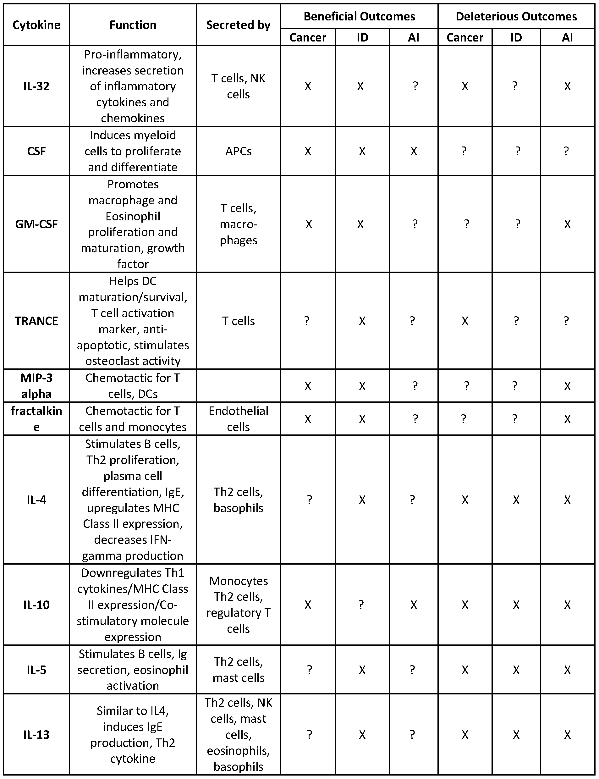
[0035] Immune mediator. As used herein, the term“immune mediator” refers to any molecule that affects the cells and processes involved in immune responses. Immune mediators include cytokines, chemokines, soluble proteins, transcription factors, metabolic factors, and cell surface markers.
[0036] Improve, increase, inhibit, stimulate, suppress, or reduce : As used herein, the terms “improve”,“increase”,“inhibit”,“stimulate”,“suppress”,“reduce”, or grammatical equivalents thereof, indicate values that are relative to a baseline or other reference measurement. In some embodiments, an appropriate reference measurement may be or comprise a measurement in a particular system (e.g, in a single individual) under otherwise comparable conditions absent presence of (e.g, prior to and/or after) a particular agent or treatment, or in presence of an appropriate comparable reference agent. The effect of a particular agent or treatment may be direct or indirect. In some embodiments, an appropriate reference measurement may be or may comprise a measurement in a comparable system known or expected to respond in a particular way, in presence of the relevant agent or treatment. In some embodiments, a peptide presented by an antigen presenting cell (APC)“stimulates” or is“stimulatory” to a lymphocyte if the lymphocyte is activated to a phenotype associated with beneficial responses, after exposure to the peptide presented by the APC under conditions that permit antigen-specific recognition to
occur, as observed by, e.g, T cell proliferation, phosphorylation or dephosphorylation of a receptor, calcium flux, cytoskeletal rearrangement, increased or decreased expression and/or secretion of immune mediators such as cytokines or soluble mediators, increased or decreased expression of one or more cell surface markers, relative to a control. In some embodiments, a peptide presented by an antigen presenting cell“suppresses”,“inhibits” or is“inhibitory” to a lymphocyte if the lymphocyte is activated to a phenotype associated with deleterious or non- beneficial responses, after exposure to the peptide presented by the APC under conditions that permit antigen-specific recognition to occur, as observed by, e.g. , phosphorylation or dephosphorylation of a receptor, calcium flux, cytoskeletal rearrangement, increased or decreased expression and/or secretion of immune mediators such as cytokines or soluble mediators, increased or decreased expression of one or more cell surface markers, relative to a control.
[0037] Inhibitory Antigerr. An“inhibitory antigen” is an antigen that inhibits, suppresses, impairs and/or reduces immune control of a tumor or cancer. In some embodiments, an inhibitory antigen promotes tumor growth, enables tumor growth, ameliorates tumor growth, activates tumor growth, accelerates tumor growth, and/or increases and/or enables tumor metastasis, and/or accelerates tumor growth. In some embodiments, an inhibitory antigen stimulates one or more lymphocyte responses that are deleterious or non-beneficial to a subject; and/or inhibits and/or suppresses one or more lymphocyte responses that are beneficial to a subject. In some embodiments, an inhibitory antigen is the target of one or more lymphocyte responses that are deleterious or non-beneficial to a subject; and/or inhibits and/or suppresses one or more lymphocyte responses that are beneficial to a subject.
[0038] In some embodiments, the inhibitory antigen is a tumor antigen (e.g, tumor specific antigen [TSA or neoantigen], tumor associated antigen [TAA], or cancer/testis antigen [CTA]). In some embodiments, the inhibitory antigen is a full-length polypeptide, or a fragment or peptide thereof.
[0039] Invasin polypeptide. An“invasin polypeptide” is a polypeptide that facilitates or mediates uptake of a cell (e.g, a bacterial cell) by a eukaryotic cell. Expression of an invasin polypeptide in a noninvasive bacterial cell confers on the cell the ability to enter a eukaryotic cell. In some embodiments, an invasin polypeptide is a bacterial invasin polypeptide. In some
embodiments, an invasin polypeptide is a Yersinia invasin polypeptide ( e.g . , a Yersinia invasin polypeptide comprising a sequence disclosed in GenBank® under Acc. No. YP 070195.1).
[0040] Listeriolysin O (LLO): The terms“listeriolysin O” or“LLO” refer to a listeriolysin O polypeptide of Listeria monocytogenes and truncated forms thereof that retain pore-forming ability (e.g, cytoplasmic forms of LLO, including truncated forms lacking a signal sequence). In some embodiments, an LLO is a cytoplasmic LLO. Exemplary LLO sequences are shown in Table 1, below.
[0041] Polypeptide. The term“polypeptide”, as used herein, generally has its art-recognized meaning of a polymer of at least three amino acids. Those of ordinary skill in the art will appreciate, however, that the term“polypeptide” is intended to be sufficiently general as to encompass not only polypeptides having the complete sequence recited herein (or in a reference or database specifically mentioned herein), but also to encompass polypeptides that represent functional fragments (i.e., fragments retaining at least one activity) and immunogenic fragments of such complete polypeptides. Moreover, those of ordinary skill in the art understand that protein sequences generally tolerate some substitution without destroying activity. Thus, any polypeptide that retains activity and shares at least about 30-40% overall sequence identity, often greater than about 50%, 60%, 70%, or 80%, and further usually including at least one region of much higher identity, often greater than 90% or even 95%, 96%, 97%, 98%, or 99% in one or more highly conserved regions, usually encompassing at least 3-4 and often up to 20 or more amino acids, with another polypeptide of the same class, is encompassed within the relevant term “polypeptide” as used herein. Other regions of similarity and/or identity can be determined by those of ordinary skill in the art by analysis of the sequences of various polypeptides.
[0042] Primary cells : As used herein,“primary cells” refers to cells from an organism that have not been immortalized in vitro. In some embodiments, primary cells are cells taken directly from a subject (e.g, a human). In some embodiments, primary cells are progeny of cells taken from a subject (e.g, cells that have been passaged in vitro). Primary cells include cells that have been stimulated to proliferate in culture.
[0043] Response: As used herein, in the context of a subject (a patient or experimental organism),“response”,“responsive”, or“responsiveness” refers to an alteration in a subject’s condition that occurs as a result of, or correlates with, treatment. In certain embodiments, a response is a beneficial response. In certain embodiments, a beneficial response can include
stabilization of a subject’s condition ( e.g ., prevention or delay of deterioration expected or typically observed to occur absent the treatment), amelioration (e.g., reduction in frequency and/or intensity) of one or more symptoms of the condition, and/or improvement in the prospects for cure of the condition, etc. In certain embodiments, for a subject who has cancer, a beneficial response can include: the subject has a positive clinical response to cancer therapy or a combination of therapies; the subject has a spontaneous response to a cancer; the subject is in partial or complete remission from cancer; the subject has cleared a cancer; the subject has not had a relapse, recurrence or metastasis of a cancer; the subject has a positive cancer prognosis; the subject has not experienced toxic responses or side effects to a cancer therapy or combination of therapies. In certain embodiments, for a subject who had cancer, the beneficial responses occurred in the past, or are ongoing.
[0044] In certain embodiments, a response is a deleterious or non-beneficial response. In certain embodiments, a deleterious or non-beneficial response can include deterioration of a subject’s condition, lack of amelioration (e.g, no reduction in frequency and/or intensity) of one or more symptoms of the condition, and/or degradation in the prospects for cure of the condition, etc. In certain embodiments, for a subject who has cancer, a deleterious or non-beneficial response can include: the subject has a negative clinical response to cancer therapy or a combination of therapies; the subject is not in remission from cancer; the subject has not cleared a cancer; the subject has had a relapse, recurrence or metastasis of a cancer; the subject has a negative cancer prognosis; the subject has experienced toxic responses or side effects to a cancer therapy or combination of therapies. In certain embodiments, for a subject who had cancer, the deleterious or non-beneficial responses occurred in the past, or are ongoing.
[0045] As used herein, in the context of a cell, organ, tissue, or cell component, e.g., a lymphocyte,“response”,“responsive”, or“responsiveness” refers to an alteration in cellular activity that occurs as a result of, or correlates with, administration of or exposure to an agent, e.g. a tumor antigen. In certain embodiments, a beneficial response can include increased expression and/or secretion of immune mediators associated with positive clinical responses or outcomes in a subject. In certain embodiments, a beneficial response can include decreased expression and/or secretion of immune mediators associated with negative clinical response or outcomes in a subject. In certain embodiments, a deleterious or non-beneficial response can include increased expression and/or secretion of immune mediators associated with negative
clinical responses or outcomes in a subject. In certain embodiments, a deleterious or non- beneficial response can include decreased expression and/or secretion of immune mediators associated with positive clinical responses or outcomes in a subject. In certain embodiments, a response is a clinical response. In certain embodiments, a response is a cellular response. In certain embodiments, a response is a direct response. In certain embodiments, a response is an indirect response. In certain embodiments,“non-response”,“non-responsive”, or“non responsiveness” mean minimal response or no detectable response. In certain embodiments, a “minimal response” includes no detectable response. In certain embodiments, presence, extent, and/or nature of response can be measured and/or characterized according to particular criteria.
In certain embodiments, such criteria can include clinical criteria and/or objective criteria. In certain embodiments, techniques for assessing response can include, but are not limited to, clinical examination, positron emission tomography, chest X-ray, CT scan, MRI, ultrasound, endoscopy, laparoscopy, presence or level of a particular marker in a sample, cytology, and/or histology. Where a response of interest is a response of a tumor to a therapy, ones skilled in the art will be aware of a variety of established techniques for assessing such response, including, for example, for determining tumor burden, tumor size, tumor stage, etc. Methods and guidelines for assessing response to treatment are discussed in Therasse et ah, J. Natl. Cancer Inst., 2000, 92(3):205-2l6; and Seymour et ah, Lancet Oncol., 2017, l8:el43-52. The exact response criteria can be selected in any appropriate manner, provided that when comparing groups of tumors, patients or experimental organism, and/or cells, organs, tissues, or cell components, the groups to be compared are assessed based on the same or comparable criteria for determining response rate. One of ordinary skill in the art will be able to select appropriate criteria.
[0046] Stimulatory Antigerr. A“stimulatory antigen” is an antigen that enhances, improves, increases and/or stimulates immune control of a tumor or cancer. In some embodiments, a stimulatory antigen is the target of an immune response that reduces, kills, shrinks, resorbs, and/or eradicates tumor growth; does not promote, enable, ameliorate, activate, and/or accelerate tumor growth; decreases tumor metastasis, and/or decelerates tumor growth. In some
embodiments, a stimulatory antigen inhibits and/or suppresses one or more lymphocyte responses that are deleterious or non-beneficial to a subject; and/or stimulates one or more lymphocyte responses that are beneficial to a subject.
[0047] Tumor As used herein, the term“tumor” refers to an abnormal growth of cells or tissue. In some embodiments, a tumor may comprise cells that are precancerous ( e.g ., benign), malignant, pre-metastatic, metastatic, and/or non-metastatic. In some embodiments, a tumor is associated with, or is a manifestation of, a cancer. In some embodiments, a tumor may be a disperse tumor or a liquid tumor. In some embodiments, a tumor may be a solid tumor.
PET ATT, ED DESCRIPTION
[0048] Recent advances in immune checkpoint inhibitor therapies such as ipilimumab, nivolumab, and pembrolizumab for cancer immunotherapy have resulted in dramatic efficacy in subjects suffering from NSCLC, among other indications. Nivolumab and pembroluzimab have been approved by the Food and Drug Administration (FDA) and European Medicines Agency (EMA) for use in patients with advanced NSCLC who have previously been treated with chemotherapy. They have solidified the importance of T cell responses in control of tumors. Neoantigens, potential cancer rejection antigens that are entirely absent from the normal human genome, are postulated to be relevant to tumor control; however, attempts to define them and their role in tumor clearance has been hindered by the paucity of available tools to define them in a biologically relevant and unbiased way (Schumacher and Schreiber, 2015 Science 348:69-74, Gilchuk et al., 2015 Curr Opin Immunol 34:43-51)
[0049] Taking non-small cell lung carcinoma (NSCLC) as an example, whole exome sequencing of NSCLC tumors from patients treated with pembrolizumab showed that higher non-synonymous mutation burden in tumors was associated with improved objective response, durable clinical benefit, and progression-free survival (Rizvi et al, (2015) Science 348(6230): 124-8). In this study, the median non-synonymous mutational burden of the discovery cohort was 209 and of the validation cohort was 200. However, simply because a mutation was identified by sequencing, does not mean that the epitope it creates can be recognized by a T cell or serves as a protective antigen for T cell responses (Gilchuk et al., 2015 Curr Opin Immunol 34:43-51), making the use of the word neoantigen somewhat of a misnomer. With 200 or more potential targets of T cells in NSCLC, it is not feasible to test every predicted epitope to determine which of the mutations serve as neoantigens, and which neoantigens are associated with clinical evidence of tumor control. Recently, a study by McGranahan et al., showed that clonal neoantigen burden and overall survival in primary lung adenocarcinomas are related.
However, even enriching for clonal neoantigens results in potential antigen targets ranging from 50 to approximately 400 (McGranahan et al, 2016 Science 351 : 1463-69). Similar findings have been described for melanoma patients who have responded to ipilimumab therapy (Snyder et al, 2015 NEJM; Van Allen et al, 2015 Science) and in patients with mismatch-repair deficient colorectal cancer who were treated with pembrolizumab (Le et al, 2015 NEJM).
[0050] The present disclosure provides methods and systems for the rapid identification of tumor antigens ( e.g . , tumor specific antigens (TSAs, or neoantigens), tumor associated antigens (TAAs), or cancer/testis antigens (CTAs)) that elicit T cell responses and particularly that elicit human T cell responses, as well as polypeptides that are potential tumor antigens. For purposes of this disclosure,“tumor antigens” includes both tumor antigens and potential tumor antigens.
As described herein, methods of the present disclosure identified stimulatory tumor antigens that were not identified by known algorithms. Further, methods of the present disclosure identified suppressive and/or inhibitory tumor antigens that are not identifiable by known algorithms. Methods of the present disclosure also identified polypeptides that are potential tumor antigens, i.e., polypeptides that activate T cells of non-cancerous subjects, but not T cells of subjects suffering from cancer. The present disclosure also provides methods of selecting tumor antigens and potential tumor antigens, methods of using the selected tumor antigens and potential tumor antigens, immunogenic compositions comprising the selected tumor antigens and potential tumor antigens, and methods of manufacturing immunogenic compositions. The present disclosure also provides methods of evaluating an immune response in a cancer subject, e.g., for identifying or selecting subjects for initiation, continuation, modification, and/or discontinuation of cancer therapy.
Library generation
[0051] A library is a collection of members (e.g, cells or non-cellular particles, such as virus particles, liposomes, or beads (e.g., beads coated with polypeptides, such as in vitro translated polypeptides, e.g, affinity beads, e.g, antibody coated beads, or NTA-Ni beads bound to polypeptides of interest). According to the present disclosure, members of a library include (e.g, internally express or carry) polypeptides of interest described herein. In some
embodiments, members of a library are cells that internally express polypeptides of interest described herein. In some embodiments, members of a library which are particles carry, and/or
are bound to, polypeptides of interest. Use of a library in an assay system allows simultaneous evaluation in vitro of cellular responses to multiple candidate antigens. According to the present disclosure, a library is designed to be internalized by human antigen presenting cells so that peptides from library members, including peptides from internally expressed polypeptides of interest, are presented on MHC molecules of the antigen presenting cells for recognition by T cells.
[0052] Libraries can be used in assays that detect peptides presented by human MHC class I and MHC class II molecules. Polypeptides expressed by the internalized library members are digested in intracellular endocytic compartments ( e.g ., phagosomes, endosomes, lysosomes) of the human cells and presented on MHC class II molecules, which are recognized by human CD4+ T cells. In some embodiments, library members include a cytolysin polypeptide, in addition to a polypeptide of interest. In some embodiments, library members include an invasin polypeptide, in addition to the polypeptide of interest. In some embodiments, library members include an autolysin polypeptide, in addition to the polypeptide of interest. In some
embodiments, library members are provided with cells that express a cytolysin polypeptide (i.e., the cytolysin and polypeptide of interest are not expressed in the same cell, and an antigen presenting cell is exposed to members that include the cytolysin and members that include the polypeptide of interest, such that the antigen presenting cell internalizes both, and such that the cytolysin facilitates delivery of polypeptides of interest to the MHC class I pathway of the antigen presenting cell). A cytolysin polypeptide can be constitutively expressed in a cell, or it can be under the control of an inducible expression system (e.g., an inducible promoter). In some embodiments, a cytolysin is expressed under the control of an inducible promoter to minimize cytotoxicity to the cell that expresses the cytolysin.
[0053] Once internalized by a human cell, a cytolysin polypeptide perforates intracellular compartments in the human cell, allowing polypeptides expressed by the library members to gain access to the cytosol of the human cell. Polypeptides released into the cytosol are presented on MHC class I molecules, which are recognized by CD8+ T cells.
[0054] A library can include any type of cell or particle that can be internalized by and deliver a polypeptide of interest (and a cytolysin polypeptide, in applications where a cytolysin polypeptide is desirable) to, antigen presenting cells for use in methods described herein.
Although the term“cell” is used throughout the present specification to refer to a library
member, it is understood that, in some embodiments, the library member is a non-cellular particle, such as a virus particle, liposome, or bead. In some embodiments, members of the library include polynucleotides that encode the polypeptide of interest (and cytolysin
polypeptide), and can be induced to express the polypeptide of interest (and cytolysin
polypeptide) prior to, and/or during internalization by antigen presenting cells.
[0055] In some embodiments, the cytolysin polypeptide is heterologous to the library cell in which it is expressed, and facilitates delivery of polypeptides expressed by the library cell into the cytosol of a human cell that has internalized the library cell. Cytolysin polypeptides include bacterial cytolysin polypeptides, such as listeriolysin O (LLO), streptolysin O (SLO), and perfringolysin O (PFO). Additional cytolysin polypeptides are described in U.S. Pat. 6,004,815. In certain embodiments, library members express LLO. In some embodiments, a cytolysin polypeptide is not significantly secreted by the library cell (e.g, less than 20%, 10%, 5%, or 1% of the cytolysin polypeptide produced by the cell is secreted). For example, the cytolysin polypeptide is a cytoplasmic cytolysin polypeptide, such as a cytoplasmic LLO polypeptide (e.g, a form of LLO which lacks the N-terminal signal sequence, as described in Higgins et aI., Moί Microbiol. 31(6): 1631-1641, 1999). Exemplary cytolysin polypeptide sequences are shown in Table 1. The listeriolysin O (D3-25) sequence shown in the second row of Table 1 has a deletion of residues 3-25, relative to the LLO sequence in shown in the first row of Table 1, and is a cytoplasmic LLO polypeptide. In some embodiments, a cytolysin is expressed constitutively in a library host cell. In other embodiments, a cytolysin is expressed under the control of an inducible promoter. Cytolysin polypeptides can be expressed from the same vector, or from a different vector, as the polypeptide of interest in a library cell.
Table 1. Exemplary Cytolysin Polypeptides


[0056] In some embodiments, a library member ( e.g ., a library member which is a bacterial cell) includes an invasin that facilitates uptake by the antigen presenting cell. In some embodiments, a library member includes an autolysin that facilitates autolysis of the library member within the antigen presenting cell. In some embodiments, a library member includes both an invasin and an autolysin. In some embodiments, a library member which is an E. coli cell includes an invasin and/or an autolysin. In various embodiments, library cells that express an invasin and/or autolysin are used in methods that also employ non-professional antigen presenting cells or antigen presenting cells that are from cell lines. Isberg et al. ( Cell , 1987, 50:769-778), Sizemore et al. ( Science , 1995, 270:299-302) and Courvalin et al. ( C.R . Acad. Sci. Paris , 1995, 318: 1207-12) describe expression of an invasin to effect endocytosis of bacteria by target cells. Autolysins are described by Cao et al, Infect. Immun. 1998, 66(6): 2984-2986; Margot et al., J. Bacteriol. 1998, 180(3 ): 749-752; Buist et al., Appl. Environ. Microbiol, 1997, 63(7):2722-2728; Yamanaka et al., FEMS Microbiol. Lett., 1997, 150(2): 269-275; Romero et al. , FEMS Microbiol. Lett. , 1993, l08(l):87-92; Betzner and Keck, Mol. Gen. Genet., 1989, 219(3): 489-491; Lubitz et al., J. Bacterial., 1984, l59(l):385-387; and Tomasz et al., J.
Bacterial., 1988, 170(12): 5931-5934. In some embodiments, an autolysin has a feature that permits delayed lysis, e.g., the autolysin is temperature-sensitive or time-sensitive (see, e.g, Chang et al., 1995, J. Bact. Ill, 3283-3294; Raab et al, 1985, J. Mol. Biol. 19, 95-105; Gerds et al, 1995, Mol. Microbiol. 17, 205-210). Useful cytolysins also include addiction
(poison/antidote) autolysins, (see, e.g, Magnuson R, et al, 1996, J. Biol. Chem. 271(31), 18705- 18710; Smith A S, et al, 1991, Mol. Microbiol. 26(5), 961-970).
[0057] In some embodiments, members of the library include bacterial cells. In certain embodiments, the library includes non-pathogenic, non-virulent bacterial cells. Examples of bacteria for use as library members include E. coli, mycobacteria, Listeria monocytogenes, Shigella flexneri, Bacillus subtilis, or Salmonella.
[0058] In some embodiments, members of the library include eukaryotic cells (e.g, yeast cells). In some embodiments, members of the library include viruses (e.g, bacteriophages). In some embodiments, members of the library include liposomes. Methods for preparing liposomes that include a cytolysin and other agents are described in Kyung-Dall et al., U.S. Pat. No.
5,643,599. In some embodiments, members of the library include beads. Methods for preparing
libraries comprised of beads are described, e.g, in Lam et al., Nature 354: 82-84, 1991, U.S. Pat. Nos. 5,510,240 and 7,262,269, and references cited therein.
[0059] In certain embodiments, a library is constructed by cloning polynucleotides encoding polypeptides of interest, or portions thereof, into vectors that express the polypeptides of interest in cells of the library. The polynucleotides can be synthetically synthesized. The
polynucleotides can be cloned by designing primers that amplify the polynucleotides. Primers can be designed using available software, such as Primer3Plus (available the following URL: bioinformatics.nl/cgi-bin/primer3plus/primer3plus.cgi; see Rozen and Skaletsky, In: Krawetz S, Misener S (eds) Bioinformatics Methods and Protocols: Methods in Molecular Biology. Humana Press, Totowa, NJ, pp. 365-386, 2000). Other methods for designing primers are known to those of skill in the art. In some embodiments, primers are constructed so as to produce polypeptides that are truncated, and/or lack hydrophobic regions (e.g, signal sequences or transmembrane regions) to promote efficient expression. The location of predicted signal sequences and predicted signal sequence cleavage sites in a given open reading frame (ORF) sequence can be determined using available software, see, e.g, Dyrlov et al, J. Mol. Biol., 340:783-795, 2004, and the following URL: cbs.dtu.dk/services/SignalP/). For example, if a signal sequence is predicted to occur at the N-terminal 20 amino acids of a given polypeptide sequence, a primer is designed to anneal to a coding sequence downstream of the nucleotides encoding the N-terminal 20 amino acids, such that the amplified sequence encodes a product lacking this signal sequence.
[0060] Primers can also be designed to include sequences that facilitate subsequent cloning steps. ORFs can be amplified directly from genomic DNA (e.g, genomic DNA of a tumor cell), or from polynucleotides produced by reverse transcription (RT-PCR) of mRNAs expressed by the tumor cell. RT-PCR of mRNA is useful, e.g, when the genomic sequence of interest contains intronic regions. PCR-amplified ORFs are cloned into an appropriate vector, and size, sequence, and expression of ORFs can be verified prior to use in immunological assays.
[0061] In some embodiments, a polynucleotide encoding a polypeptide of interest is linked to a sequence encoding a tag (e.g, an N-terminal or C-terminal epitope tag) or a reporter protein (e.g., a fluorescent protein). Epitope tags and reporter proteins facilitate purification of expressed polypeptides, and can allow one to verify that a given polypeptide is properly expressed in a library host cell, e.g., prior to using the cell in a screen. Useful epitope tags include, for example, a polyhistidine (His) tag, a V5 epitope tag from the P and V protein of
paramyxovirus, a hemagglutinin (HA) tag, a myc tag, and others. In some embodiments, a polynucleotide encoding a polypeptide of interest is fused to a sequence encoding a tag which is a known antigenic epitope ( e.g ., an MHC class I- and/or MHC class II-restricted T cell epitope of a model antigen such as an ovalbumin), and which can be used to verify that a polypeptide of interest is expressed and that the polypeptide-tag fusion protein is processed and presented in antigen presentation assays. In some embodiments a tag includes a T cell epitope of a murine T cell (e.g., a murine T cell line). In some embodiments, a polynucleotide encoding a polypeptide of interest is linked to a tag that facilitates purification and a tag that is a known antigenic epitope. Useful reporter proteins include naturally occurring fluorescent proteins and their derivatives, for example, Green Fluorescent Protein (Aequorea Victoria ) and Neon Green (Branchiostoma lanceolatum). Panels of synthetically derived fluorescent and chromogenic proteins are also available from commercial sources.
[0062] Polynucleotides encoding a polypeptide of interest are cloned into an expression vector for introduction into library host cells. Various vector systems are available to facilitate cloning and manipulation of polynucleotides, such as the Gateway® Cloning system (Invitrogen). As is known to those of skill in the art, expression vectors include elements that drive production of polypeptides of interest encoded by a polynucleotide in library host cells (e.g, promoter and other regulatory elements). In some embodiments, polypeptide expression is controlled by an inducible element (e.g, an inducible promoter, e.g, an IPTG- or arabinose- inducible promoter, or an IPTG-inducible phage T7 RNA polymerase system, a lactose (lac) promoter, a tryptophan (trp) promoter, a tac promoter, a trc promoter, a phage lambda promoter, an alkaline phosphatase ( phoA ) promoter, to give just a few examples; see Cantrell, Meth. in Mol. Biol., 235:257-276, Humana Press, Casali and Preston, Eds.). In some embodiments, polypeptides are expressed as cytoplasmic polypeptides. In some embodiments, the vector used for polypeptide expression is a vector that has a high copy number in a library host cell. In some embodiments, the vector used for expression has a copy number that is more than 25, 50, 75, 100, 150, 200, or 250 copies per cell. In some embodiments, the vector used for expression has a ColEl origin of replication. Useful vectors for polypeptide expression in bacteria include pET vectors (Novagen), Gateway® pDEST vectors (Invitrogen), pGEX vectors (Amersham Biosciences), pPRO vectors (BD
Biosciences), pB AD vectors (Invitrogen), pLEX vectors (Invitrogen), pMAL™ vectors (New England BioLabs), pGEMEX vectors (Promega), and pQE vectors (Qiagen). Vector systems for
producing phage libraries are known and include Novagen T7Select® vectors, and New England Biolabs Ph.D.™ Peptide Display Cloning System.
[0063] In some embodiments, library host cells express (either constitutively, or when induced, depending on the selected expression system) a polypeptide of interest to at least 10%, 20%, 30%, 40%, 50%, 60%, or 70% of the total cellular protein. In some embodiments, the level a polypeptide available in or on a library member ( e.g ., cell, virus particle, liposome, bead) is such that antigen presenting cells exposed to a sufficient quantity of the library members are presented on MHC molecules polypeptide epitopes at a density that is comparable to the density presented by antigen presenting cells pulsed with purified peptides.
[0064] Methods for efficient, large-scale production of libraries are available. For example, site-specific recombinases or rare-cutting restriction enzymes can be used to transfer
polynucleotides between expression vectors in the proper orientation and reading frame (Walhout et al., Meth. Enzymol. 328:575-592, 2000; Marsischky et al. , Genome Res. 14:2020-202, 2004; Blommel et al., Protein Expr. Purif. 47:562-570, 2006).
[0065] For production of liposome libraries, expressed polypeptides (e.g., purified or partially purified polypeptides) can be entrapped in liposomal membranes, e.g, as described in Wassef et al., U.S. Pat. No. 4,863,874; Wheatley et al., U.S. Pat. No. 4,921,757; Huang et al., U.S. Pat. No. 4,925,661; or Martin et al., U.S. Pat. No. 5,225,212.
[0066] A library can be designed to include full length polypeptides and/or portions of polypeptides. Expression of full-length polypeptides maximizes epitopes available for presentation by a human antigen presenting cell, thereby increasing the likelihood of identifying an antigen. However, in some embodiments, it is useful to express portions of polypeptides, or polypeptides that are otherwise altered, to achieve efficient expression. For example, in some embodiments, polynucleotides encoding polypeptides that are large (e.g, greater than 1,000 amino acids), that have extended hydrophobic regions, signal peptides, transmembrane domains, or domains that cause cellular toxicity, are modified (e.g, by C-terminal truncation, N-terminal truncation, or internal deletion) to reduce cytotoxicity and permit efficient expression a library cell, which in turn facilitates presentation of the encoded polypeptides on human cells. Other types of modifications, such as point mutations or codon optimization, may also be used to enhance expression.
[0067] The number of polypeptides included in a library can be varied. For example, in some embodiments, a library can be designed to express polypeptides from at least 5%, 10%, 15%, 20%, 25%, 35%, 40%, 45%, 50%, 55%, 60%, 70%, 75%, 80%, 85%, 90%, 95%, 97%, 98%, 99%, or more, of ORFs in a target cell (e.g, tumor cell). In some embodiments, a library expresses at least 10, 15, 20, 25, 30, 40, 50, 75, 100, 150, 200, 250, 300, 350, 400, 450, 500, 550, 600, 650, 700, 750, 800, 850, 900, 950, 1000, 2500, 5000, 10,000, or more different polypeptides of interest, each of which may represent a polypeptide encoded by a single full length
polynucleotide or portion thereof.
[0068] In some embodiments, assays may focus on identifying antigens that are secreted polypeptides, cell surface-expressed polypeptides, or virulence determinants, e.g. , to identify antigens that are likely to be targets of both humoral and cell mediated immune responses.
[0069] In addition to polypeptides of interest, libraries can include tags or reporter proteins that allow one to easily purify, analyze, or evaluate MHC presentation, of the polypeptide of interest. In some embodiments, polypeptides expressed by a library include C-terminal tags that include both an MHC class I and an MHC class II-restricted T cell epitope from a model antigen, such as chicken ovalbumin (OVA). Library protein expression and MHC presentation is validated using these epitopes. In some embodiments, the epitopes are OVA247-265 and OVA258- 265 respectfully, corresponding to positions in the amino acid sequence found in GenBank® under Acc. No. NP 990483. Expression and presentation of linked ORFs can be verified with antigen presentation assays using T cell hybridomas (e.g, B3Z T hybridoma cells, which are H2-Kb restricted, and KZO T hybridoma cells, which are H2-Ak restricted) that specifically recognize these epitopes.
[0070] Sets of library members (e.g, bacterial cells) can be provided on an array (e.g, on a solid support, such as a 96-well plate) and separated such that members in each location express a different polypeptide of interest, or a different set of polypeptides of interest.
[0071] Methods of using library members for identifying T cell antigens are described in detail below. In addition to these methods, library members also have utility in assays to identify B cell antigens. For example, lysate prepared from library members that include polypeptides of interest can be used to screen a sample comprising antibodies (e.g, a serum sample) from a subject (e.g, a subject who has been exposed to an infectious agent of interest, a subject who has cancer, and/or a control subject), to determine whether antibodies present in the subject react
with the polypeptide of interest. Suitable methods for evaluating antibody reactivity are known and include, e.g, ELISA assays.
Polypeptides of Interest
[0072] In some embodiments, methods and compositions described herein can be used to identify and/or detect immune responses to a polypeptide of interest. In some embodiments, a polypeptide of interest is encoded by an ORF from a target tumor cell, and members of a library include ( e.g ., internally express or carry) ORFs from a target tumor cell. In some such embodiments, a library can be used in methods described herein to assess immune responses to one or more polypeptides of interest encoded by one or more ORFs. In some embodiments, methods of the disclosure identify one or more polypeptides of interest as stimulatory antigens (e.g., that stimulate an immune response, e.g., a T cell response, e.g, expression and/or secretion of one or more immune mediators). In some embodiments, methods of the disclosure identify one or more polypeptides of interest as antigens or potential antigens that have minimal or no effect on an immune response (e.g., expression and/or secretion of one or more immune mediators). In some embodiments, methods of the disclosure identify one or more polypeptides of interest as inhibitory and/or suppressive antigens (e.g., that inhibit, suppress, down-regulate, impair, and/or prevent an immune response, e.g., a T cell response, e.g., expression and/or secretion of one or more immune mediators). In some embodiments, methods of the disclosure identify one or more polypeptides of interest as tumor antigens or potential tumor antigens, e.g., tumor specific antigens (TSAs, or neoantigens), tumor associated antigens (TAAs), or cancer/testis antigens (CTAs).
[0073] In some embodiments, a polypeptide of interest is a putative tumor antigen, and methods and compositions described herein can be used to identify and/or detect immune responses to one or more putative tumor antigens. For example, members of a library include (e.g, internally express or carry) putative tumor antigens (e.g., a polypeptide previously identified (e.g., by a third party) as a tumor antigen, e.g., identified as a tumor antigen using a method other than a method of the present disclosure). In some embodiments, a putative tumor antigen is a tumor antigen described herein. In some such embodiments, such libraries can be used to assess whether and/or the extent to which such putative tumor antigen mediates an immune response. In some embodiments, methods of the disclosure identify one or more
putative tumor antigens as stimulatory antigens. In some embodiments, methods of the disclosure identify one or more putative tumor antigens as antigens that have minimal or no effect on an immune response. In some embodiments, methods of the disclosure identify one or more putative tumor antigens as inhibitory and/or suppressive antigens.
[0074] In some embodiments, a polypeptide of interest is a pre-selected tumor antigen, and methods and compositions described herein can be used to identify and/or detect immune responses to one or more pre-selected tumor antigens. For example, in some embodiments, members of a library include ( e.g ., internally express or carry) one or more polypeptides identified as tumor antigens using a method of the present disclosure and/or using a method other than a method of the present disclosure. In some such embodiments, such libraries can be used to assess whether and/or the extent to which such tumor antigens mediate an immune response by an immune cell from one or more subjects (e.g., a subject who has cancer and/or a control subject) to obtain one or more response profiles described herein. In some embodiments, methods of the disclosure identify one or more pre-selected tumor antigens as stimulatory antigens for one or more subjects. In some embodiments, methods of the disclosure identify one or more pre-selected tumor antigens as antigens that have minimal or no effect on an immune response for one or more subjects. In some embodiments, methods of the disclosure identify one or more pre-selected tumor antigens as inhibitory and/or suppressive antigens for one or more subjects.
[0075] In some embodiments, a polypeptide of interest is a known tumor antigen, and methods and compositions described herein can be used to identify and/or detect immune responses to one or more known tumor antigens. For example, in some embodiments, members of a library include (e.g, internally express or carry) one or more polypeptides identified as a tumor antigen using a method of the present disclosure and/or using a method other than a method of the present disclosure. In some such embodiments, such libraries can be used to assess whether and/or the extent to which such tumor antigens mediate an immune response by an immune cell from one or more subjects (e.g., a subject who has cancer and/or a control subject) to obtain one or more response profiles described herein. In some embodiments, methods of the disclosure identify one or more known tumor antigens as stimulatory antigens for one or more subjects. In some embodiments, methods of the disclosure identify one or more known tumor antigens as antigens that have minimal or no effect on an immune response for one
or more subjects. In some embodiments, methods of the disclosure identify one or more known tumor antigens as inhibitory and/or suppressive antigens for one or more subjects.
[0076] In some embodiments, a polypeptide of interest is a potential tumor antigen, and methods and compositions described herein can be used to identify and/or detect immune responses to one or more potential tumor antigens. For example, in some embodiments, members of a library include ( e.g ., internally express or carry) one or more polypeptides identified as being of interest, e.g., encoding mutations associated with a tumor, using a method of the present disclosure and/or using a method other than a method of the present disclosure. In some such embodiments, such libraries can be used to assess whether and/or the extent to which such polypeptides mediate an immune response by an immune cell from one or more subjects (e.g., a subject who has cancer and/or a control subject) to obtain one or more response profiles described herein. In some embodiments, methods of the disclosure identify one or more polypeptides as stimulatory antigens for one or more subjects. In some embodiments, methods of the disclosure identify one or more polypeptides as antigens that have minimal or no effect on an immune response for one or more subjects. In some embodiments, methods of the disclosure identify one or more polypeptides as inhibitory and/or suppressive antigens for one or more subjects.
Tumor Antigens
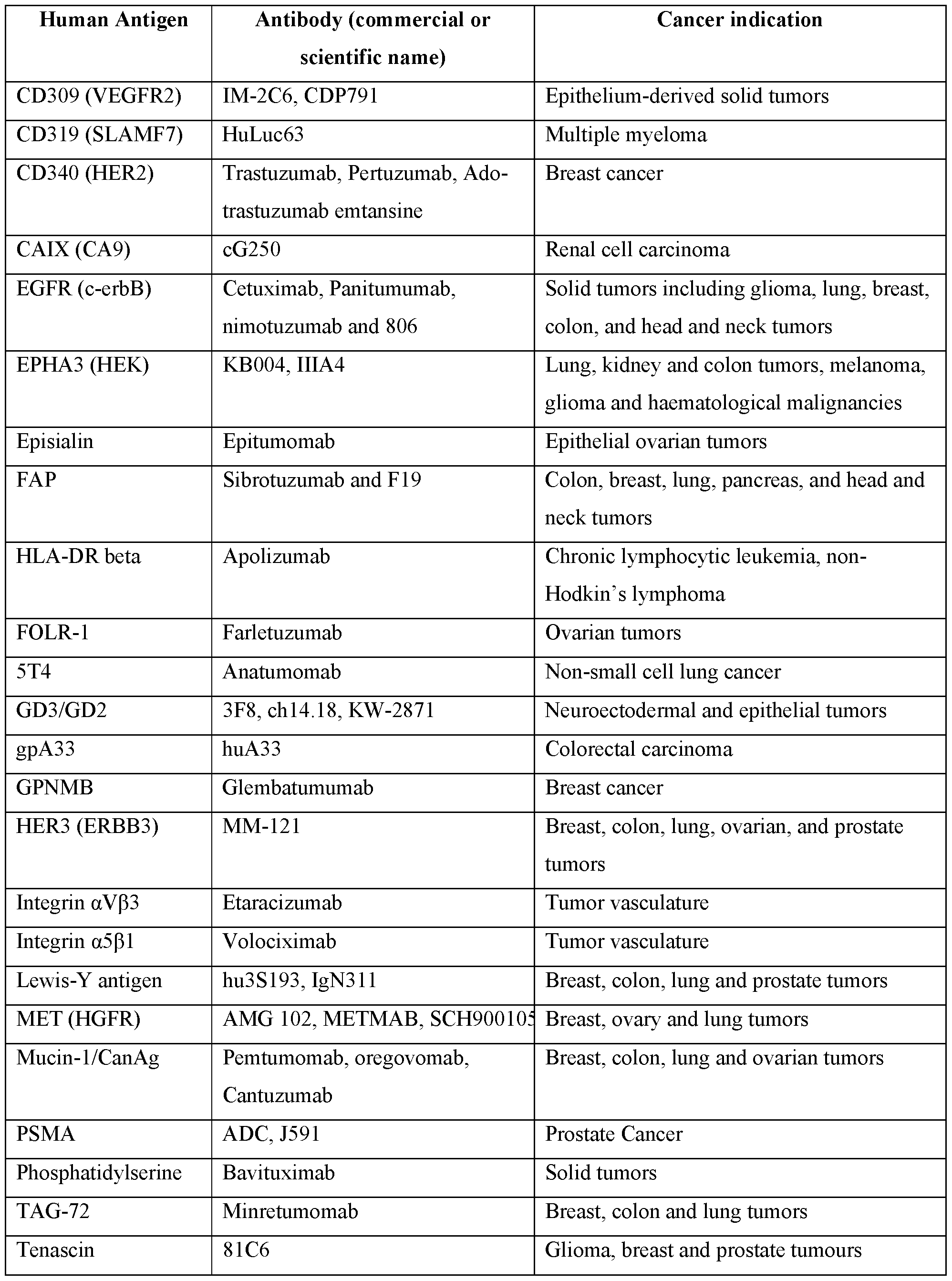
[0077] Polypeptides of interest used in methods and systems described herein include tumor antigens amd potential tumor antigens, e.g., tumor specific antigens (TSAs, or neoantigens), tumor associated antigens (TAAs), and/or cancer/testis antigens (CTAs). Exemplary tumor antigens include, e.g., MART-l/MelanA (MART -I or MLANA), gplOO (Pmel 17 or SILV), tyrosinase, TRP-l, TRP-2, MAGE-l, MAGE-3 (also known as HIP8), BAGE, GAGE-l, GAGE- 2, p 15, Calcitonin, Calretinin, Carcinoembryonic antigen (CEA), Chromogranin, Cytokeratin, Desmin, Epithelial membrane protein (EMA), Factor VIII, Glial fibrillary acidic protein (GFAP), Gross cystic disease fluid protein (GCDFP-15), HMB-45, Human chorionic gonadotropin (hCG), inhibin, lymphocyte marker, MART-l (Melan-A), Myo Dl, muscle-specific actin (MSA), neurofilament, neuron-specific enolase (NSE), placental alkaline phosphatase (PLAP), prostate- specific antigen, PTPRC (CD45), S100 protein, smooth muscle actin (SMA), synaptophysin, thyroglobulin, thyroid transcription factor-l, Tumor M2-PK, vimentin, p53, Ras, HER-2/neu,
BCR-ABL, E2A-PRL, H4-RET, IGH-IGK, MYL-RAR, Epstein Barr virus antigens (e.g., EBNA1), human papillomavirus (HPV) antigen E6 or E7 (HPV E6 or HPV E7), TSP-180, MAGE-4, MAGE-5, MAGE-6, RAGE, NY-ESO-l (also known as CTAG1B), erbB, pl85erbB2, pl80erbB-3, c-met, nm-23Hl, PSA, TAG-72, CA 19-9, CA 72-4, CAM 17.1, NuMa, K-ras, beta-Catenin, CDK4, Mum-l, p 15, p 16, 43-9F, 5T4, 79lTgp72, alpha-fetoprotein (AFP), beta- HCG, BCA225, BTAA, CA 125, CA l5-3\CA 27.29VBCAA, CA 195, CA 242, CA-50, CAM43, CD68\Pl, CO-029, FGF-5, G250, Ga733\EpCAM, HTgp-l75, M344, MA-50, MG7-Ag,
MO VI 8, NB/70K, NY-CO-l, RCAS1, SDCCAG16, TA-90\Mac-2 binding protein\cyclophilin C-associated protein, TAAL6, TAG72, TLP, MUC16, ILl3Ra2, FRa, VEGFR2, Lewis Y, FAP, EphA2, CEACAM5, EGFR, CA6, CA9, GPNMB, EGP1, FOLR1, endothelial receptor,
STEAP1, SLC44A4, Nectin-4, AGS-16, guanalyl cyclase C, METC-l, CFC1B, integrin alpha 3 chain (of a3bl, a laminin receptor chain), TPS, CD19, CD20, CD22, CD30, CD31, CD72,
CD 180, CD171 (L1CAM), CD123, CD133, CD138, CD37, CD70, CD79a, CD79b, CD56, CD74, CD 166, CD71, CD34, CD99, CD117, CD80, CD28, CD13, CD15, CD25, CD10, CLL- 1/CLEC12A, ROR1, Glypican 3 (GPC3), Mesothelin, CD33/IL3Ra, c-Met, PSCA, PSMA, Glycolipid F77, EGFRvIII, BCMA, GD-2, PSAP, prostein (also known as P501 S), PSMA, Survivin (also known as BIRC5), and MAGE- A3, MAGEA2, MAGEA4, MAGEA6, MAGEA9, MAGEA10, MAGEA12, BIRC5, CDH3, CEACAM3, CGB_isoform2, ELK4, ERBB2, HPSE1, HPSE2, KRAS i soform 1 , KRAS_isoform2, MUC1, SMAD4, TERT,2. TERT.3, TGFBR2, EGAG9_i soform 1, TP53, CGB isoforml, IMPDH2, LCK, angiopoietin-l (Angl) (also known as ANGPT1), XIAP (also known as BIRC4), galectin-3 (also known as LGALS3), VEGF-A (also known as VEGF), ATP6S1 (also known as ATP6AP1), MAGE-A1, cIAP-l (also known as BIRC2), macrophage migration inhibitory factor (MIF), galectin-9 (also known as LGALS9), progranulin PGRN (also known as granulin), OGFR, MLIAP (also known as BIRC7), TBX4 (also known as ICPPS, SPS or T-Box4), secretory leukocyte protein inhibitor (Slpi) (also known as antileukoproteinase), Ang2 (also known as ANGPT2), galectin-l (also known as LGALS1), TRP-2 (also known as DCT), hTERT (telomerase reverse transcriptase) tyrosinase-related protein 1 (TRP-l, TYRP1), NOR-90/UBF-2 (also known as UBTF), LGMN, SPA17, PRTN3, TRRAP l, TRRAP 2, TRRAP 3, TRRAP 4, MAGEC2, PRAME, SOX 10, RAC1, HRAS, GAGE4, AR, CYP1B1, MMP8, TYR, PDGFRB, KLK3, PAX3, PAX5, ST3GAL5, PLAC1, RhoC, MYCN, REG3A, CSAG2, CTAG2-la, CTAG2-lb, PAGE4, BRAF, GRM3, ERBB4,
KIT, MAPK1, MFI2, SART3, ST8SIA1, WDR46, AKAP-4, RGS5, FOSL1, PRM2, ACRBP, CTCFL, CSPG4, CCNB1, MSLN, WT1, SSX2, KDR, ANKRD30A, MAGED1, MAP3K9, XAGE1B, PREX2, CD276, TEK, AIM1, ALK, FOLH1, GRIN2A MAP3K5 and one or more isoforms of any preceding tumor antigens. Exemplary tumor antigens are provided in the accompanying list of sequences.
[0078] Tumor specific antigens (TSAs, or neoantigens) are tumor antigens that are not encoded in normal host genome (see, e.g ., Yarchoan et al., Nat. Rev. Cancer. 2017 Feb 24. doi: l0. l038/nrc.20l6.l54; Gubin et al., J. Clin. Invest. 125:3413-3421 (2015)). In some
embodiments, TSAs arise from somatic mutations and/or other genetic alterations. In some embodiments, TSAs arise from missense or in-frame mutations. In some embodiments, TSAs arise from frame-shift mutations or loss-of-stop-codon mutations. In some embodiments, TSAs arise from insertion or deletion mutations. In some embodiments, TSAs arise from duplication or repeat expansion mutations. In some embodiments, TSAs arise from splice variants or improper splicing. In some embodiments, TSAs arise from gene fusions. In some embodiments, TSAs arise from translocations. In some embodiments, TSAs include oncogenic viral proteins. For example, as with Merkel cell carcinoma (MCC) associated with the Merkel cell
polyomavirus (MCPy V) and cancers of the cervix, oropharynx and other sites associated with the human papillomavirus (HPV), TSAs include proteins encoded by viral open reading frames. For purposes of this disclosure, the terms“mutation” and“mutations” encompass all mutations and genetic alterations that may give rise to an antigen encoded in the genome of a cancer or tumor cell of a subject, but not in a normal or non-cancerous cell of the same subject. In some embodiments, TSAs are specific (personal) to a subject. In some embodiments, TSAs are shared by more than one subject, e.g. , less than 1%, 1-3%, 1-5%, 1-10% , or more of subjects suffering from a cancer. In some embodiments, TSAs shared by more than one subject may be known or pre-selected.
[0079] In some embodiments, a TSA is encoded by an open reading frame from a virus. For example, a library can be designed to express polypeptides from one of the following viruses: an immunodeficiency virus (e.g, a human immunodeficiency virus (HIV), e.g, HIV-l, HIV-2), a hepatitis virus (e.g, hepatitis B virus (HBV), hepatitis C virus (HCV), hepatitis A virus, non-A and non-B hepatitis virus), a herpes virus (e.g, herpes simplex virus type I (HSV-l), HSV-2, Varicella-zoster virus, Epstein Barr virus, human cytomegalovirus, human herpesvirus 6 (HHV-
6), HHV-7, HHV-8), a poxvirus (e.g., variola, vaccinia, monkeypox, Molluscum contagiosum virus), an influenza virus, a human papilloma virus, adenovirus, rhinovirus, coronavirus, respiratory syncytial virus, rabies virus, coxsackie virus, human T cell leukemia virus (types I, II and III), parainfluenza virus, paramyxovirus, poliovirus, rotavirus, rhinovirus, rubella virus, measles virus, mumps virus, adenovirus, yellow fever virus, Norwalk virus, West Nile virus, a Dengue virus, Severe Acute Respiratory Syndrome Coronavirus (SARS-CoV), bunyavirus, Ebola virus, Marburg virus, Eastern equine encephalitis virus, Venezuelan equine encephalitis virus, Japanese encephalitis virus, St. Louis encephalitis virus, Junin virus, Lassa virus, and Lymphocytic choriomeningitis virus. Libraries for other viruses can also be produced and used according to methods described herein.
[0080] Tumor specific antigens are known in the art, any of which can be used in methods described herein. In some embodiments, gene sequences encoding polypeptides that are potential or putative neoantigens are determined by sequencing the genome and/or exome of tumor tissue and healthy tissue from a subject having cancer using next generation sequencing technologies. In some embodiments, genes that are selected based on their frequency of mutation and ability to encode a potential or putative neoantigen are sequenced using next- generation sequencing technology. Next-generation sequencing applies to genome sequencing, genome resequencing, transcriptome profiling (RNA-Seq), DNA-protein interactions (ChlP- sequencing), and epigenome characterization (de Magalhaes et al. (2010) Ageing Research Reviews 9 (3): 315-323; Hall N (2007) J. Exp. Biol. 209 (Pt 9): 1518-1525; Church (2006) Sci. Am. 294 (1): 46-54; ten Bosch et al. (2008) Journal of Molecular Diagnostics 10 (6): 484-492; Tucker T et al. (2009) The American Journal of Human Genetics 85 (2): 142-154). Next- generation sequencing can be used to rapidly reveal the presence of discrete mutations such as coding mutations in individual tumors, e.g., single amino acid changes (e.g., missense mutations, in-frame mutations) or novel stretches of amino acids generated by frame-shift insertions, deletions, gene fusions, read-through mutations in stop codons, duplication or repeat expansion mutations, and translation of splice variants or improperly spliced introns, and translocations (e.g.,“neoORFs”).
[0081] Another method for identifying potential or putative neoantigens is direct protein sequencing. Protein sequencing of enzymatic digests using multidimensional MS techniques (MSn) including tandem mass spectrometry (MS/MS)) can also be used to identify neoantigens.
Such proteomic approaches can be used for rapid, highly automated analysis (see, e.g, Gevaert et al., Electrophoresis 21 : 1145-1154 (2000)). High-throughput methods for de novo sequencing of unknown proteins can also be used to analyze the proteome of a subject’s tumor to identify expressed potential or putative neoantigens. For example, meta shotgun protein sequencing may be used to identify expressed potential or putative neoantigens (see e.g. , Guthals et al. (2012) Molecular and Cellular Proteomics 11(10): 1084-96).
[0082] Potential or putative neoantigens may also be identified using MHC multimers to identify neoantigen-specific T cell responses. For example, high-throughput analysis of neoantigen-specific T cell responses in patient samples may be performed using MHC tetramer- based screening techniques (see e.g. , Hombrink et al. (2011) PLoS One; 6(8): e22523; Hadrup et al. (2009) Nature Methods, 6(7):520-26; van Rooij et al. (2013) Journal of Clinical Oncology, 31 : 1-4; and Heemskerk et al. (2013) EMBO Journal, 32(2): 194-203).
[0083] In some embodiments, one or more known or pre-selected tumor specific antigens, or one or more potential or putative tumor specific antigens identified using one of these methods, can be included in a library described herein.
[0084] Tumor associated antigens (TAAs) include proteins encoded in a normal genome (see, e.g. , Ward et al., Adv. Immunol. 130:25-74 (2016)). In some embodiments, TAAs are either normal differentiation antigens or aberrantly expressed normal proteins. Overexpressed normal proteins that possess growth/survival -promoting functions, such as Wilms tumor 1 (WT1) (Ohminami et al., Blood 95:286-293 (2000)) or Her2/neu (Kawashima et al., Cancer Res. 59:431-435 (1999)), are TAAs that directly participate in the oncogenic process. Post- translational modifications, such as phosphorylation, of proteins may also lead to formation of TAAs (Doyle, J. Biol. Chem. 281 :32676-32683 (2006); Cobbold, Sci. Transl. Med. 5:203ral25 (2013)). TAAs are generally shared by more than one subject, e.g. , less than 1%, 1-3%, 1-5%, 1- 10%, 1-20%, or more of subjects suffering from a cancer. In some embodiments, TAAs are known or pre-selected tumor antigens. In some embodiments, with respect to an individual subject, TAAs are potential or putative tumor antigens. Cancer/testis antigens (CTAs) are expressed by various tumor types and by reproductive tissues (for example, testes, fetal ovaries and trophoblasts) but have limited or no detectable expression in other normal tissues in the adult and are generally not presented on normal reproductive cells, because these tissues do not express MHC class I molecules (see, e.g. , Coulie et al., Nat. Rev. Cancer 14:135-146 (2014);
Simpson et al., Nat. Rev. Cancer 5:615-625 (2005); Scanlan et al., Immunol. Rev. 188:22-32
(2002)). Library Screens
Human Cells for Antigen Presentation
[0085] The present invention provides, inter alia , compositions and methods for identifying tumor antigens recognized by human immune cells. Human antigen presenting cells express ligands for antigen receptors and other immune activation molecules on human lymphocytes. Given differences in MHC peptide binding specificities and antigen processing enzymes between species, antigens processed and presented by human cells are more likely to be physiologically relevant human antigens in vivo than antigens identified in non-human systems. Accordingly, methods of identifying these antigens employ human cells to present candidate tumor antigen polypeptides. Any human cell that internalizes library members and presents polypeptides expressed by the library members on MHC molecules can be used as an antigen presenting cell according to the present disclosure. In some embodiments, human cells used for antigen presentation are primary human cells. The cells can include peripheral blood mononuclear cells (PBMC) of a human. In some embodiments, peripheral blood cells are separated into subsets ( e.g ., subsets comprising dendritic cells, macrophages, monocytes, B cells, or combinations thereof) prior to use in an antigen presentation assay. In some embodiments, a subset of cells that expresses MHC class II is selected from peripheral blood. In one example, a cell population including dendritic cells is isolated from peripheral blood. In some embodiments, a subset of dendritic cells is isolated (e.g., plasmacytoid, myeloid, or a subset thereof). Human dendritic cell markers include CDlc, CDla, CD303, CD304, CD141, and CD209. Cells can be selected based on expression of one or more of these markers (e.g, cells that express CD303, CDlc, and CD141).
[0086] Dendritic cells can be isolated by positive selection from peripheral blood using commercially available kits (e.g, from Miltenyi Biotec Inc.). In some embodiments, the dendritic cells are expanded ex vivo prior to use in an assay. Dendritic cells can also be produced by culturing peripheral blood cells under conditions that promote differentiation of monocyte precursors into dendritic cells in vitro. These conditions typically include culturing the cells in the presence of cytokines such as GM-CSF and IL-4 (see, e.g, Inaba et al, Isolation of dendritic cells, Curr. Protoc. Immunol. May; Chapter 3: Unit 3.7, 2001). Procedures for in vitro expansion of hematopoietic stem and progenitor cells (e.g, taken from bone marrow or
peripheral blood), and differentiation of these cells into dendritic cells in vitro , is described in U.S. Pat. No. 5,199,942, and U.S. Pat. Pub. 20030077263. Briefly, CD34+ hematopoietic stem and progenitor cells are isolated from peripheral blood or bone marrow and expanded in vitro in culture conditions that include one or more of Flt3-L, IL-l, IL-3, and c-kit ligand.
[0087] In some embodiments, immortalized cells that express human MHC molecules (e.g, human cells, or non-human cells that are engineered to express human MHC molecules) are used for antigen presentation. For example, assays can employ COS cells transfected with human MHC molecules or HeLa cells.
[0088] In some embodiments, both the antigen presenting cells and immune cells used in the method are derived from the same subject (e.g, autologous T cells and APC are used). In these embodiments, it can be advantageous to sequentially isolate subsets of cells from peripheral blood of the subject, to maximize the yield of cells available for assays. For example, one can first isolate CD4+ and CD8+ T cell subsets from the peripheral blood. Next, dendritic cells (DC) are isolated from the T cell-depleted cell population. The remaining T- and DC-depleted cells are used to supplement the DC in assays, or are used alone as antigen presenting cells. In some embodiments, DC are used with T- and DC-depleted cells in an assay, at a ratio of 1 :2, 1 :3, 1 :4, or 1 :5. In some embodiments, the antigen presenting cells and immune cells used in the method are derived from different subjects (e.g, heterologous T cells and APC are used).
[0089] Antigen presenting cells can be isolated from sources other than peripheral blood.
For example, antigen presenting cells can be taken from a mucosal tissue (e.g, nose, mouth, bronchial tissue, tracheal tissue, the gastrointestinal tract, the genital tract (e.g, vaginal tissue), or associated lymphoid tissue), peritoneal cavity, lymph nodes, spleen, bone marrow, thymus, lung, liver, kidney, neuronal tissue, endocrine tissue, or other tissue, for use in screening assays. In some embodiments, cells are taken from a tissue that is the site of an active immune response (e.g, an ulcer, sore, or abscess). Cells may be isolated from tissue removed surgically, via lavage, or other means.
[0090] Antigen presenting cells useful in methods described herein are not limited to “professional” antigen presenting cells. In some embodiments, non-professional antigen presenting cells can be utilized effectively in the practice of methods of the present disclosure. Non-professional antigen presenting cells include fibroblasts, epithelial cells, endothelial cells,
neuronal/glial cells, lymphoid or myeloid cells that are not professional antigen presenting cells e.g ., T cells, neutrophils), muscle cells, liver cells, and other types of cells.
[0091] Antigen presenting cells are cultured with library members that express a polypeptide of interest (and, if desired, a cytolysin polypeptide) under conditions in which the antigen presenting cells internalize, process and present polypeptides expressed by the library members on MHC molecules. In some embodiments, library members are killed or inactivated prior to culture with the antigen presenting cells. Cells or viruses can be inactivated by any appropriate agent (e.g., fixation with organic solvents, irradiation, freezing). In some embodiments, the library members are cells that express ORFs linked to a tag (e.g, a tag which comprises one or more known T cell epitopes) or reporter protein, expression of which has been verified prior to the culturing.
[0092] In some embodiments, antigen presenting cells are incubated with library members at 37°C for between 30 minutes and 5 hours (e.g, for 45 min. to 1.5 hours). After the incubation, the antigen presenting cells can be washed to remove library members that have not been internalized. In certain embodiments, the antigen presenting cells are non-adherent, and washing requires centrifugation of the cells. The washed antigen presenting cells can be incubated at 37°C for an additional period of time (e.g, 30 min. to 2 hours) prior to exposure to lymphocytes, to allow antigen processing. In some embodiments, it is desirable to fix and kill the antigen presenting cells prior to exposure to lymphocytes (e.g, by treating the cells with 1%
paraformaldehyde).
[0093] The antigen presenting cell and library member numbers can be varied, so long as the library members provide quantities of polypeptides of interest sufficient for presentation on MHC molecules. In some embodiments, antigen presenting cells are provided in an array, and are contacted with sets of library cells, each set expressing a different polypeptide of interest. In certain embodiments, each location in the array includes l x l 03 - l x l 06 antigen presenting cells, and the cells are contacted with l x l 03 - l x l 08 library cells which are bacterial cells.
[0094] In any of the embodiments described herein, antigen presenting cells can be freshly isolated, maintained in culture, and/or thawed from frozen storage prior to incubation with library cells, or after incubation with library cells.
Human Lymphocytes
[0095] In methods of the present disclosure, human lymphocytes are tested for antigen- specific reactivity to antigen presenting cells, e.g ., antigen presenting cells that have been incubated with libraries expressing polypeptides of interest as described above. The methods of the present disclosure permit rapid identification of human antigens using pools of lymphocytes isolated from an individual, or progeny of the cells. The detection of antigen-specific responses does not rely on laborious procedures to isolate individual T cell clones. In some embodiments, the human lymphocytes are primary lymphocytes. In some embodiments, human lymphocytes are NKT cells, gamma-delta T cells, or NK cells. Just as antigen presenting cells may be separated into subsets prior to use in antigen presentation assays, a population of lymphocytes having a specific marker or other feature can be used. In some embodiments, a population of T lymphocytes is isolated. In some embodiments, a population of CD4+ T cells is isolated. In some embodiments, a population of CD8+ T cells is isolated. CD8+ T cells recognize peptide antigens presented in the context of MHC class I molecules. Thus, in some embodiments, the CD8+ T cells are used with antigen presenting cells that have been exposed to library host cells that co-express a cytolysin polypeptide, in addition to a polypeptide of interest. T cell subsets that express other cell surface markers may also be isolated, e.g. , to provide cells having a particular phenotype. These include CLA (for skin-homing T cells), CD25, CD30, CD69,
CD 154 (for activated T cells), CD45RO (for memory T cells), CD294 (for Th2 cells), g/d TCR- expressing cells, CD3 and CD56 (for NK T cells). Other subsets can also be selected.
[0096] Lymphocytes can be isolated, and separated, by any means known in the art (e.g, using antibody -based methods such as those that employ magnetic bead separation, panning, or flow cytometry). Reagents to identify and isolate human lymphocytes and subsets thereof are well known and commercially available.
[0097] Lymphocytes for use in methods described herein can be isolated from peripheral blood mononuclear cells, or from other tissues in a human. In some embodiments, lymphocytes are taken from tumors, lymph nodes, a mucosal tissue (e.g, nose, mouth, bronchial tissue, tracheal tissue, the gastrointestinal tract, the genital tract (e.g, vaginal tissue), or associated lymphoid tissue), peritoneal cavity, spleen, thymus, lung, liver, kidney, neuronal tissue, endocrine tissue, peritoneal cavity, bone marrow, or other tissues. In some embodiments, cells
are taken from a tissue that is the site of an active immune response ( e.g ., an ulcer, sore, or abscess). Cells may be isolated from tissue removed surgically, via lavage, or other means.
[0098] Lymphocytes taken from an individual can be maintained in culture or frozen until use in antigen presentation assays. In some embodiments, freshly isolated lymphocytes can be stimulated in vitro by antigen presenting cells exposed to library cells as described above. In some embodiments, these lymphocytes exhibit detectable stimulation without the need for prior non-antigen specific expansion. However, primary lymphocytes also elicit detectable antigen- specific responses when first stimulated non-specifically in vitro. Thus, in some embodiments, lymphocytes are stimulated to proliferate in vitro in a non-antigen specific manner, prior to use in an antigen presentation assay. Lymphocytes can also be stimulated in an antigen-specific manner prior to use in an antigen presentation assay. In some embodiments, cells are stimulated to proliferate by a library (e.g., prior to use in an antigen presentation assay that employs the library). Expanding cells in vitro provides greater numbers of cells for use in assays. Primary T cells can be stimulated to expand, e.g, by exposure to a polyclonal T cell mitogen, such as phytohemagglutinin or concanavalin, by treatment with antibodies that stimulate proliferation, or by treatment with particles coated with the antibodies. In some embodiments, T cells are expanded by treatment with anti-CD2, anti-CD3, and anti-CD28 antibodies. In some
embodiments, T cells are expanded by treatment with interleukin-2. In some embodiments, lymphocytes are thawed from frozen storage and expanded (e.g., stimulated to proliferate, e.g., in a non-antigen specific manner or in an antigen-specific manner) prior to contacting with antigen presenting cells. In some embodiments, lymphocytes are thawed from frozen storage and are not expanded prior to contacting with antigen presenting cells. In some embodiments, lymphocytes are freshly isolated and expanded (e.g., stimulated to proliferate, e.g, in a non-antigen specific manner or in an antigen-specific manner) prior to contacting with antigen presenting cells.
Antigen Presentation Assays
[0099] In antigen presentation assays, T cells are cultured with antigen presenting cells prepared according to the methods described above, under conditions that permit T cell recognition of peptides presented by MHC molecules on the antigen presenting cells. In some embodiments, T cells are incubated with antigen presenting cells at 37°C for between 12-48 hours (e.g, for 24 hours). In some embodiments, T cells are incubated with antigen presenting cells at 37°C for 3, 4, 5, 6, 7, or 8 days. Numbers of antigen presenting cells and T cells can be
varied. In some embodiments, the ratio of T cells to antigen presenting cells in a given assay is 1 : 10, 1 :5, 1 :2, 1 : 1, 2: 1, 5: 1, 10: 1, 20: 1, 25: 1, 30: 1, 32: 1, 35:1 or 40: 1. In some embodiments, antigen presenting cells are provided in an array ( e.g ., in a 96-well plate), wherein cells in each location of the array have been contacted with sets of library cells, each set including a different polypeptide of interest. In certain embodiments, each location in the array includes 1 x 10³ - 1 x 10⁶ antigen presenting cells, and the cells are contacted with 1 c 103 - 1 c 106 T cells.
[0100] After T cells have been incubated with antigen presenting cells, cultures are assayed for activation. Lymphocyte activation can be detected by any means known in the art, e.g., T cell proliferation, phosphorylation or dephosphorylation of a receptor, calcium flux, cytoskeletal rearrangement, increased or decreased expression and/or secretion of immune mediators such as cytokines or soluble mediators, increased or decreased expression of one or more cell surface markers. In some embodiments, culture supernatants are harvested and assayed for increased and/or decreased expression and/or secretion of one or more polypeptides associated with activation, e.g, a cytokine, soluble mediator, cell surface marker, or other immune mediator. In some embodiments, the one or more cytokines are selected from TRAIL, IFN-gamma, IL- 12r70, IL-2, TNF-alpha, MIP1 -alpha, MIPl-beta, CXCL9, CXCL10, MCP1, RANTES, IL-l beta, IL-4, IL-6, IL-8, IL-9, IL-10, IL-13, IL-15, CXCL11, IL-3, IL-5, IL-17, IL-18, IL-21, IL- 22, IL-23A, IL-24, IL-27, IL-31, IL-32, TGF-beta, CSF, GM-CSF, TRANCE (also known as RANK L), MIP3 -alpha, and fractalkine. In some embodiments, the one or more soluble mediators are selected from granzyme A, granzyme B, sFas, sFasL, perforin, and granulysin. In some embodiments, the one or more cell surface markers are selected from CD 107a, CD 107b, CD25, CD69, CD45RA, CD45RO, CD137 (4-1BB), CD44, CD62L, CD27, CCR7, CD154 (CD40L), KLRG-l, CD71, HLA-DR, CD 122 (IL-2RB), CD28, IL7Ra (CD 127), CD38, CD26, CD 134 (OX-40), CTLA-4 (CD152), LAG-3, TIM-3 (CD366), CD39, PD1 (CD279), FoxP3, TIGIT, CD 160, BTLA, 2B4 (CD244), and KLRG1. In some embodiments, the one or more other immune mediators are selected from CXCR5, IDO, PD-L1, CD33, CD1 lb, LAG3, CXCR3, t-bet, GATA-3, GITR, CD39, CD73, CD57, TCF1, Akt, SLAMF6, BCL-6, FoxOl, TOX, IRF4, and CD30. Cytokine secretion in culture supernatants can be detected, e.g, by ELISA, bead array, e.g, with a Luminex® analyzer. Cytokine production can also be assayed by RT-PCR of mRNA isolated from the T cells, or by ELISPOT analysis of cytokines released by the T cells. In some embodiments, proliferation of T cells in the cultures is determined (e.g, by
detecting 3H thymidine incorporation). In some embodiments, target cell lysis is determined ( e.g ., by detecting T cell dependent lysis of antigen presenting cells labeled with Na2 51Cr04). Target cell lysis assays are typically performed with CD8+ T cells. Protocols for these detection methods are known. See, e.g., Current Protocols In Immunology , John E. Coligan el al. (eds), Wiley and Sons, New York, N.Y., 2007. One of skill in the art understands that appropriate controls are used in these detection methods, e.g. , to adjust for non-antigen specific background activation, to confirm the presenting capacity of antigen presenting cells, and to confirm the viability of lymphocytes.
[0101] In some embodiments, antigen presenting cells and lymphocytes used in the method are from the same individual. In some embodiments, antigen presenting cells and lymphocytes used in the method are from different individuals.
[0102] In some embodiments, antigen presentation assays are repeated using lymphocytes from the same individual that have undergone one or more previous rounds of exposure to antigen presenting cells, e.g. , to enhance detection of responses, or to enhance weak initial responses. In some embodiments, antigen presentation assays are repeated using antigen presenting cells from the same individual that have undergone one or more previous rounds of exposure to a library, e.g. , to enhance detection of responses, or to enhance weak initial responses. In some embodiments, antigen presentation assays are repeated using lymphocytes from the same individual that have undergone one or more previous rounds of exposure to antigen presenting cells, and antigen presenting cells from the same individual that have undergone one or more previous rounds of exposure to a library, e.g. , to enhance detection of responses, or to enhance weak initial responses. In some embodiments, antigen presentation assays are repeated using antigen presenting cells and lymphocytes from different individuals, e.g. , to identify antigens recognized by multiple individuals, or compare reactivities that differ between individuals.
Methods of Identifying Tumor Antigens
[0103] One advantage of methods described herein is their ability to identify clinically relevant human antigens. Humans that have cancer may have lymphocytes that specifically recognize tumor antigens, which are the product of an adaptive immune response arising from prior exposure. In some embodiments, these cells are present at a higher frequency than cells
from an individual who does not have cancer, and/or the cells are readily reactivated when re- exposed to the proper antigenic stimulus ( e.g ., the cells are“memory” cells). Thus, humans that have or have had cancer are particularly useful donors of cells for identifying antigens in vitro. The individual may be one who has recovered from cancer. In some embodiments, the individual has been recently diagnosed with cancer (e.g., the individual was diagnosed less than one year, three months, two months, one month, or two weeks, prior to isolation of lymphocytes and/or antigen presenting cells from the individual). In some embodiments, the individual was first diagnosed with cancer more than three months, six months, or one year prior to isolation of lymphocytes and/or antigen presenting cells.
[0104] In some embodiments, lymphocytes are screened against antigen presenting cells that have been contacted with a library of cells whose members express or carry polypeptides of interest, and the lymphocytes are from an individual who has not been diagnosed with cancer. In some embodiments, such lymphocytes are used to determine background (i.e., non-antigen- specific) reactivities. In some embodiments, such lymphocytes are used to identify antigens, reactivity to which exists in non-cancer individuals.
[0105] Cells from multiple donors (e.g., multiple subjects who have cancer) can be collected and assayed in methods described herein. In some embodiments, cells from multiple donors are assayed in order to determine if a given tumor antigen is reactive in a broad portion of the population, or to identify multiple tumor antigens that can be later combined to produce an immunogenic composition that will be effective in a broad portion of the population.
[0106] Antigen presentation assays are useful in the context of both infectious and non- infectious diseases. The methods described herein are applicable to any context in which a rapid evaluation of human cellular immunity is beneficial. In some embodiments, antigenic reactivity to polypeptides that are differentially expressed by neoplastic cells (e.g, tumor cells) is evaluated. Sets of nucleic acids differentially expressed by neoplastic cells have been identified using established techniques such as subtractive hybridization. Methods described herein can be used to identify antigens that were functional in a subject in which an anti -tumor immune response occurred. In other embodiments, methods are used to evaluate whether a subject has lymphocytes that react to a tumor antigen or set of tumor antigens.
[0107] In some embodiments, antigen presentation assays are used to examine reactivity to autoantigens in cells of an individual, e.g, an individual predisposed to, or suffering from, an
autoimmune condition. Such methods can be used to provide diagnostic or prognostic indicators of the individual’s disease state, or to identify autoantigens. For these assays, in some embodiments, libraries that include an array of human polypeptides are prepared. In some embodiments, libraries that include polypeptides from infectious agents which are suspected of eliciting cross-reactive responses to autoantigens are prepared. For examples of antigens from infectious agents thought to elicit cross-reactive autoimmune responses, see Barzilai et al ., Curr Opin Rheumatol. , l9(6):636-43, 2007; Ay ada et al, Ann N Y Acad Scl, 1108:594-602, 2007; Oroum et al, Mol Immunol, 45(1): 180-9, 2008; and Bach, J Autoimmun., 25 Suppl:74-80, 2005.
[0108] As discussed, the present disclosure includes methods in which polypeptides of interest are included in a library ( e.g ., expressed in library cells or carried in or on particles or beads). After members of the library are internalized by antigen presenting cells, the
polypeptides of interest are proteolytically processed within the antigen presenting cells, and peptide fragments of the polypeptides are presented on MHC molecules expressed in the antigen presenting cells. The identity of the polypeptide that stimulates a human lymphocyte in an assay described herein can be determined from examination of the set of library cells that were provided to the antigen presenting cells that produced the stimulation. In some embodiments, it is useful to map the epitope within the polypeptide that is bound by MHC molecules to produce the observed stimulation. This epitope, or the longer polypeptide from which it is derived (both of which are referred to as an“antigen” herein) can form the basis for an immunogenic composition, or for an antigenic stimulus in future antigen presentation assays.
[0109] Methods for identifying peptides bound by MHC molecules are known. In some embodiments, epitopes are identified by generating deletion mutants of the polypeptide of interest and testing these for the ability to stimulate lymphocytes. Deletions that lose the ability to stimulate lymphocytes, when processed and presented by antigen presenting cells, have lost the peptide epitope. In some embodiments, epitopes are identified by synthesizing peptides corresponding to portions of the polypeptide of interest and testing the peptides for the ability to stimulate lymphocytes (e.g., in antigen presentation assays in which antigen presenting cells are pulsed with the peptides). Other methods for identifying MHC bound peptides involve lysis of the antigen presenting cells that include the antigenic peptide, affinity purification of the MHC molecules from cell lysates, and subsequent elution and analysis of peptides from the MHC (Falk, K. et al. Nature 351 :290, 1991, and U.S. Pat. No. 5,989,565).
[0110] In other embodiments, it is useful to identify the clonal T cell receptors that have been expanded in response to the antigen. Clonal T cell receptors are identified by DNA sequencing of the T cell receptor repertoire (Howie et al, 2015 Sci Trans Med 7:301). By identifying TCR specificity and function, TCRs can be transfected into other cell types and used in functional studies or for novel immunotherapies. In other embodiments, it is useful to identify and isolate T cells responsive to a tumor antigen in a subject. The isolated T cells can be expanded ex vivo and administered to a subject for cancer therapy or prophylaxis.
Methods of Identifying Immune Responses of a Subject
[0111] The disclosure provides methods of identifying one or more immune responses of a subject ( e.g ., a test subject, or a target subject). In some embodiments, one or more immune responses of a subject (e.g., a test subject or a target subject) are determined by a) providing a library described herein that includes a panel of tumor antigens (e.g., known tumor antigens, tumor antigens described herein, or tumor antigens, potential tumor antigens, and/or other polypeptides of interest identified using a method described herein); b) contacting the library with antigen presenting cells from the subject; c) contacting the antigen presenting cells with lymphocytes from the subject; and d) determining whether one or more lymphocytes are stimulated by, inhibited and/or suppressed by, activated by, or non-responsive to one or more tumor antigens presented by one or more antigen presenting cells. In some embodiments, the library includes about 1, 3, 5, 10, 15, 20, 25, 30, 40, 50, 60, 70, 80, 90, 100, or more tumor antigens.
[0112] In some embodiments, a test subject is (i) a cancer subject who has not received a cancer therapy; (ii) a cancer subject who has not responded and/or is not responding and/or has responded negatively, clinically to a cancer therapy; or (iii) a subject who has not been diagnosed with a cancer.
[0113] In some embodiments, a target subject is (i) a cancer subject who responds or has responded positively clinically (“responsive subject”) to a cancer therapy; (ii) a cancer subject who has not responded and/or is not responding and/or has responded negatively, clinically (“non-responsive subject”) to a cancer therapy; (iii) a cancer subject who responds or has responded spontaneously to a cancer (“spontaneous target subject”); or (vi) a subject who has not been diagnosed with a cancer (“normal subject”).
[0114] In some embodiments, lymphocyte stimulation, non-stimulation, inhibition and/or suppression, activation, and/or non-responsiveness is determined by assessing levels of one or more expressed or secreted cytokines or other immune mediators described herein. In some embodiments, levels of one or more expressed or secreted cytokines that is at least 20%, 40%, 60%, 80%, 100%, 120%, 140%, 160%, 180%, 200% or more, higher than a control level indicates lymphocyte stimulation. In some embodiments, a level of one or more expressed or secreted cytokines that is at least 1, 2, 3, 4 or 5 standard deviations greater than the mean of a control level indicates lymphocyte stimulation. In some embodiments, a level of one or more expressed or secreted cytokines that is at least 1, 2, 3, 4 or 5 median absolute deviations (MADs) greater than a median response level to a control indicates lymphocyte stimulation. In some embodiments, a control is a negative control, for example, a clone expressing Neon Green (NG). In some embodiments, a level of one or more expressed or secreted cytokines that is at least 20%, 40%, 60%, 80%, 100%, 120%, 140%, 160%, 180%, 200% or more, lower than a control level indicates lymphocyte inhibition and/or suppression. In some embodiments, a level of one or more expressed or secreted cytokines that is at least 1, 2, 3, 4 or 5 standard deviations lower than the mean of a control level indicates lymphocyte inhibition and/or suppression. In some embodiments, a level of one or more expressed or secreted cytokines that is at least 1, 2, 3, 4 or 5 median absolute deviations (MADs) lower than a median response level to a control indicates lymphocyte inhibition and/or suppression. In some embodiments, a control is a negative control, for example, a clone expressing Neon Green (NG). In some embodiments, levels of one or more expressed or secreted cytokines that is at least 20%, 40%, 60%, 80%, 100%, 120%, 140%,
160%, 180%, 200% or more, higher or lower than a control level indicates lymphocyte activation. In some embodiments, a level of one or more expressed or secreted cytokines that is at least 1, 2, 3, 4 or 5 standard deviations greater or lower than the mean of a control level indicates lymphocyte activation. In some embodiments, a level of one or more expressed or secreted cytokines that is at least 1, 2, 3, 4 or 5 median absolute deviations (MADs) greater or lower than a median response level to a control indicates lymphocyte activation. In some embodiments, a control is a negative control, for example, a clone expressing Neon Green (NG). In some embodiments, a level of one or more expressed or secreted cytokines that is within about 20%, 15%, 10%, 5%, or less, of a control level indicates lymphocyte non-responsiveness or non stimulation. In some embodiments, a level of one or more expressed or secreted cytokines that is
less than 1 or 2 standard deviations higher or lower than the mean of a control level indicates lymphocyte non-responsiveness or non-stimulation. In some embodiments, a level of one or more expressed or secreted cytokines that is less than 1 or 2 median absolute deviations (MADs) higher or lower than a median response level to a control indicates lymphocyte non
responsiveness or non-stimulation. In some embodiments, a subject response profile can include a quantification, identification, and/or representation of a panel of different cytokines (e.g., 1, 2, 3, 4, 5, 6, 7, 8, 9, 10, 12, 14, 16, 18, 20, or more cytokines) and of the total number of tumor antigens (e.g, of all or a portion of different tumor antigens from the library) that stimulate, do not stimulate, inhibit and/or suppress, activate, or have no or minimal effect on production, expression or secretion of each member of the panel of cytokines.
Method of Obtaining a Subject Response Profile
[0115] The disclosure provides methods for obtaining a subject response profile from a test subject (a“subject response profile”).
[0116] In some embodiments, the subject response profile of a test subject is obtained by a) providing a library described herein that includes a panel of tumor antigens ( e.g ., known tumor antigens, tumor antigens described herein, or tumor antigens, potential tumor antigens, and/or other polypeptides of interest identified using a method described herein); b) contacting the library with antigen presenting cells from the test subject; c) contacting the antigen presenting cells with lymphocytes from the test subject; and d) determining whether one or more
lymphocytes are stimulated by, inhibited and/or suppressed by, activated by, or non-responsive to one or more tumor antigens presented by one or more antigen presenting cells, to obtain the subject response profile. In some embodiments, the library includes about 1, 3, 5, 10, 15, 20, 25, 30, 40, 50, 60, 70, 80, 90, 100, 150, 200, 250, 500, 1000, or more tumor antigens.
[0117] The subject response profile can include a quantification, identification, and/or representation of all or a portion of the panel of tumor antigens, identified by the methods of the disclosure, that stimulate lymphocytes, that do not stimulate lymphocytes, that inhibit and/or suppress lymphocytes, that activate lymphocytes, or to which lymphocytes are non-responsive.
In some embodiments, the subject response profile further includes a quantification,
identification, and/or representation of the level of expression or secretion of one or more immune mediators, e.g., one or more cytokines, cell surface markers, or other immune mediators.
[0118] In some embodiments, the subject response profile includes a quantification, identification, and/or representation of all or a portion of the panel of tumor antigens, identified by the methods of the disclosure, that stimulate expression or secretion of one or more immune mediators, that inhibit and/or suppress expression or secretion of one or more immune mediators, and/or which do not, or minimally, affect expression or secretion of immune mediators. In some embodiments, the subject response profile further includes a quantification, identification, and/or representation of the level of expression or secretion of one or more immune mediators, e.g., one or more cytokines or cell surface markers.
[0119] In some embodiments, a subject response profile includes a ratio of (i) the number of tumor antigens that stimulate the level of expression and/or secretion of one or more immune mediators associated with at least one beneficial response (and/or that reduce the level of expression and/or secretion of at least one immune mediator associated with at least one deleterious or non-beneficial response) to (ii) the number of tumor antigens that inhibit and/or suppress the level of expression and/or secretion of one or more immune mediators associated with at least one beneficial response (and/or that stimulate the level of expression and/or secretion of at least one immune mediator associated with at least one deleterious or non- beneficial response).
[0120] In some embodiments, a subject response profile includes (i) an absolute number of tumor antigens that stimulate the level of expression and/or secretion of one or more immune mediators associated with at least one beneficial response (and/or that reduce the level of expression and/or secretion of at least one immune mediator associated with at least one deleterious or non-beneficial response) and/or (ii) the absolute number of tumor antigens that inhibit and/or suppress the level of expression and/or secretion of one or more immune mediators associated with at least one beneficial response (and/or that stimulate the level of expression and/or secretion of at least one immune mediator associated with at least one deleterious or non- beneficial response).
Methods of Obtaining a Target Response Profile
[0121] In some embodiments, a subject response profile is compared to a corresponding response profile from a target subject, e.g. a cancer subject who responds and/or has responded clinically to acancer therapy; a cancer subject who does not and/or has not responded clinically
to a cancer therapy; a subject who has, or has had, spontaneous response to a cancer; or a subject who has not been diagnosed with a cancer (a“target response profile” of a target subject).
[0122] The disclosure provides methods for obtaining a target response profile from a target subject. The target response profile of a target subject is obtained by a) providing a library described herein that includes all or a portion of the same panel of tumor antigens ( e.g ., known tumor antigens, tumor antigens described herein, or tumor antigens, potential tumor antigens, and/or other polypeptides of interest identified using a method described herein) used to generate the subject response profile; b) contacting the library with antigen presenting cells from the target subject; c) contacting the antigen presenting cells with lymphocytes from the targetsubject; and d) determining whether one or more lymphocytes are stimulated by, inhibited and/or suppressed by, activated by, or non-responsive to, one or more tumor antigens presented by one or more antigen presenting cells, to obtain the target response profile.
[0123] The target response profile includes a quantification, identification, and/or representation of the immune response of cells from the target subject to the same panel of tumor antigens included in the subject response profile.
[0124] In some embodiments, the target response profile includes a quantification, identification, and/or representation of all or a portion of the panel of tumor antigens that stimulate lymphocytes, that do not stimulate lymphocytes, that inhibit and/or suppress lymphocytes, that activate lymphocytes, and/or to which lymphocytes are non-responsive. In some embodiments, the subject response profile further includes a quantification, identification, and/or representation of the level of expression or secretion of one or more immune mediators, e.g., one or more cytokines, cell surface markers, or other immune mediators.
[0125] In some embodiments, the target response profile includes a quantification, identification, and/or representation of all or a portion of the panel of tumor antigens identified by the methods of the disclosure, that stimulate expression and/or secretion of one or more immune mediators, that inhibit and/or suppress expression or secretion of one or more immune mediators, and/or which do not, or minimally, affect expression and/or secretion of immune mediators. In some embodiments, the subject response profile further includes a quantification, identification, and/or representation of the level of expression or secretion of one or more immune mediators, e.g., one or more cytokines, cell surface markers, or other immune mediators.
[0126] In some embodiments, a target response profile includes a ratio of (i) the number of tumor antigens that stimulate the level of expression and/or secretion of one or more immune mediators associated with at least one beneficial response (and/or that reduce the level of expression and/or secretion of at least one immune mediator associated with at least one deleterious or non-beneficial response) to (ii) the number of tumor antigens that inhibit and/or suppress the level of expression and/or secretion of one or more immune mediators associated with at least one beneficial response (and/or that stimulate the level of expression and/or secretion of at least one immune mediator associated with at least one deleterious or non- beneficial response).
[0127] In some embodiments, a target response profile includes (i) an absolute number of tumor antigens that stimulate the level of expression and/or secretion of one or more immune mediators associated with at least one beneficial response (and/or that reduce the level of expression and/or secretion of at least one immune mediator associated with at least one deleterious or non-beneficial response) and/or (ii) the absolute number of tumor antigens that inhibit and/or suppress the level of expression and/or secretion of one or more immune mediators associated with at least one beneficial response (and/or that stimulate the level of expression and/or secretion of at least one immune mediator associated with at least one deleterious or non- beneficial response).
Comparison of a Subject Response Profile to a Target Response Profile
Lymphocytes
[0128] In some embodiments, a subject response profile is similar to the target response profile if the identified tumor antigens that stimulate lymphocytes in the subject response profile differ by no more than 1, 2, 3, 4, 5, 10, 15, 20, or 25 from the identified tumor antigens that stimulate lymphocytes in the target response profile; if the identified tumor antigens that do not stimulate lymphocytes in the subject response profile differ by no more than 1, 2, 3, 4, 5, 10, 15, 20, or 25 from the identified tumor antigens that do not stimulate lymphocytes in the target response profile; if the identified tumor antigens that inhibit and/or suppress lymphocytes in the subject response profile differ by no more than 1, 2, 3, 4, 5, 10, 15, 20, or 25 from the identified tumor antigens that inhibit and/or suppress lymphocytes in the target response profile; if the
identified tumor antigens that activate lymphocytes in the subject response profile differ by no more than 1, 2, 3, 4, 5, 10, 15, 20, or 25 from the identified tumor antigens that activate lymphocytes in the target response profile; and/or if the identified tumor antigens that do not stimulate lymphocytes or to which lymphocytes are non-responsive in the subject response profile differ by no more than 1, 2, 3, 4, 5, 10, 15, 20, or 25 from the identified tumor antigens to which lymphocytes are not, or are minimally, responsive in the target response profile.
[0129] In some embodiments, a subject response profile is dissimilar from the target response profile if the identified tumor antigens that stimulate lymphocytes in the subject response profile differ by more than 5, 6, 7, 8, 9, 10, 20, or more, from the identified tumor antigens that stimulate lymphocytes in the target response profile; if the identified tumor antigens that do not stimulate lymphocytes in the subject response profile differ by more than 5, 6, 7, 8, 9, 10, 20, or more, from the identified tumor antigens that do not stimulate lymphocytes in the target response profile; if the identified tumor antigens that inhibit and/or suppress lymphocytes in the subject response profile differ by more than 5, 6, 7, 8, 9, 10, 20, or more, from the identified tumor antigens that inhibit and/or suppress lymphocytes in the target response profile; if the identified tumor antigens that activate lymphocytes in the subject response profile differ by more than 5, 6, 7, 8, 9, 10, 20, or more, from the identified tumor antigens that activate lymphocytes in the target response profile; and/or if the identified tumor antigens that do not stimulate lymphocytes or to which lymphocytes are non-responsive in the subject response profile differ by more than 5, 6, 7, 8, 9, 10, 20, or more, from the identified tumor antigens to which lymphocytes are not, or are minimally, responsive in the target response profile.
[0130] In some embodiments, a subject response profile is similar to the target response profile if the identified tumor antigens that stimulate lymphocytes in the subject response profile differ by no more than 1%, 2%, 3%, 4%, 5%, 10%, 15%, 20%, or 25% from the identified tumor antigens that stimulate lymphocytes in the target response profile; if the identified tumor antigens that do not stimulate lymphocytes in the subject response profile differ by no more than 1%, 2%, 3%, 4%, 5%, 10%, 15%, 20%, or 25% from the identified tumor antigens that do not stimulate lymphocytes in the target response profile; if the identified tumor antigens that inhibit and/or suppress lymphocytes in the subject response profile differ by no more than 1%, 2%, 3%, 4%, 5%, 10%, 15%, 20%, or 25% from the identified tumor antigens that inhibit and/or suppress lymphocytes in the target response profile; if the identified tumor antigens that activate
lymphocytes in the subject response profile differ by no more than 1%, 2%, 3%, 4%, 5%, 10%, 15%, 20%, or 25% from the identified tumor antigens that activate lymphocytes in the target response profile; and/or if the identified tumor antigens that do not stimulate lymphocytes or to which lymphocytes are non-responsive in the subject response profile differ by no more than 1%, 2%, 3%, 4%, 5%, 10%, 15%, 20%, or 25% from the identified tumor antigens to which lymphocytes are not, or are minimally, responsive in the target response profile.
[0131] In some embodiments, a subject response profile is dissimilar from the target response profile if the identified tumor antigens that stimulate lymphocytes in the subject response profile differ by more than 5%, 6%, 7%, 8%, 9%, 10%, 20%, or more, from the identified tumor antigens that stimulate lymphocytes in the target response profile if the identified tumor antigens that do not stimulate lymphocytes in the subject response profile differ by more than 5%, 6%, 7%, 8%, 9%, 10%, 20% ,or more, from the identified tumor antigens that do not stimulate lymphocytes in the target response profile; and/or if the identified tumor antigens that inhibit and/or suppress lymphocytes in the subject response profile differ by more than 5%, 6%, 7%, 8%, 9%, 10%, 20%, or more, from the identified tumor antigens that inhibit and/or suppress lymphocytes in the target response profile; if the identified tumor antigens that activate lymphocytes in the subject response profile differ by more than 5%, 6%, 7%, 8%, 9%, 10%, 20%, or more, from the identified tumor antigens that activate lymphocytes in the target response profile; and/or if the identified tumor antigens that do not stimulate lymphocytes or to which lymphocytes are non-responsive in the subject response profile differ by more than 5%, 6%, 7%, 8%, 9%, 10%, 20%, or more, from the identified tumor antigens to which lymphocytes are not, or are minimally, responsive in the target response profile.
[0132] In some embodiments, methods described herein include comparing (a) a subject response profile that includes the ratio of (i) the number of tumor antigens that stimulate the level of expression and/or secretion of one or more immune mediators associated with at least one beneficial response (and/or the number of tumor antigens that inhibit and/or suppress the level of expression and/or secretion of at least one immune mediator associated with at least one deleterious or non-beneficial response) to (ii) the number of tumor antigens that inhibit and/or suppress the level of expression and/or secretion of one or more immune mediators associated with at least one beneficial response (and/or the number of tumor antigens that stimulate the level of expression and/or secretion of at least one immune mediator associated with at least one
deleterious or non-beneficial response); with (b) a target response profile that includes a ratio of (iii) the number of tumor antigens that stimulate the level of expression and/or secretion of one or more immune mediators associated with a beneficial response (and/or the number of tumor antigens that inhibit and/or suppress the level of expression and/or secretion of one or more immune mediators associated with at least one deleterious or non-beneficial response) to (iv) the number of tumor antigens that inhibit and/or suppress the level of expression and/or secretion of one or more immune mediators associated with at least one beneficial response (and/or the number of tumor antigens that stimulate the level of expression and/or secretion of at least one immune mediator associated with at least one deleterious or non-beneficial response).
[0133] In some embodiments, the (a) absolute number of tumor antigens that stimulate the level of expression and/or secretion of one or more immune mediators associated with at least one beneficial response (and/or the absolute number of tumor antigens that inhibit and/or suppress the level of expression and/or secretion of at least one immune mediator associated with at least one deleterious or non-beneficial response) of a subject response profile is compared to (b) the absolute number of tumor antigens that inhibit and/or suppress the level of expression and/or secretion of one or more immune mediators associated with at least one beneficial response (and/or the absolute number of tumor antigens that stimulate the level of expression and/or secretion of at least one immune mediator associated with at least one deleterious or non- beneficial response) of a target response profile.
Cytokines and Cell-Surface Markers
[0134] In some embodiments, the target response profile can include a quantification, identification, and/or representation of one or more cytokines, cell surface markers, or other immune mediators and the total number of tumor antigens ( e.g ., of the same tumor antigens included in the subject response profile) that stimulate, do not stimulate, inhibit and/or suppress, or have no or minimal effect on cytokine production, expression and/or secretion. In some embodiments, the target response profile can include a quantification, identification, and/or representation of a panel of different cytokines (e.g., 1, 2, 3, 4, 5, 6, 7, 8, 9, 10, 12, 14, 16, 18, 20, or more (e.g., all) of the cytokines included in the subject response profile) and the total number of tumor antigens (e.g, of the same tumor antigens included in the subject response profile) that stimulate, do not stimulate, inhibit and/or suppress, or have no or minimal effect on production, expression and/or secretion of the panel of cytokines.
[0135] In some embodiments, a subject response profile is similar to the target response profile if the total number of antigens that stimulate expression and/or secretion of one or more cytokines, cell surface markers, or other immune mediators included in the subject response profile differs by no more than 1, 2, 3, 4, 5, 10, 15, 20, or 25 from the total number of antigens that stimulate the same one or more cytokines, cell surface markers, or other immune mediators included in the target response profile; if the total number of antigens that do not stimulate expression and/or secretion of one or more cytokines, cell surface markers, or other immune mediators included in the subject response profile differs by no more than 1, 2, 3, 4, 5, 10, 15,
20, or 25 from the total number of antigens that do not stimulate the same one or more cytokines, cell surface markers, or other immune mediators included in the target response profile; if the total number of antigens that inhibit and/or suppress one or more cytokines, cell surface markers, or other immune mediators included in the subject response profile differs by no more than 1, 2, 3, 4, 5, 10, 15, 20, or 25 from the total number of antigens that inhibit and/or suppress expression and/or secretion of the same one or more cytokines, cell surface markers, or other immune mediators included in the target response profile; and/or if the total number of antigens that have no or minimal effect on expression and/or secretion of one or more cytokines, cell surface markers, or other immune mediators included in the subject response profile differs by no more than 1, 2, 3, 4, 5, 10, 15, 20, or 25 from the total number of antigens that that have no or minimal effect on the same one or more cytokines, cell surface markers, or other immune mediators included in the target response profile.
[0136] In some embodiments, a subject response profile is dissimilar from the target response profile if the total number of antigens that stimulate expression and/or secretion of one or more cytokines, cell surface markers, or other immune mediators included in the subject response profile differs by more than 5, 6, 7, 8, 9, 10, 20, or more, from the total number of antigens that stimulate the same one or more cytokines, cell surface markers, or other immune mediators included in the target response profile; if the total number of antigens that do not stimulate expression and/or secretion of one or more cytokines, cell surface markers, or other immune mediators included in the subject response profile differs by more than 5, 6, 7, 8, 9, 10, 20, or more, from the total number of antigens that do not stimulate the same one or more cytokines, cell surface markers, or other immune mediators included in the target response profile; if the total number of antigens that inhibit and/or suppress expression and/or secretion of
one or more cytokines, cell surface markers, or other immune mediators included in the subject response profile differs by more than 5, 6, 7, 8, 9, 10, 20, or more, from the total number of antigens that inhibit and/or suppress the same one or more cytokines, cell surface markers, or other immune mediators included in the target response profile; and/or if the total number of antigens that have no or minimal effect on expression and/or secretion of one or more cytokines, cell surface markers, or other immune mediators included in the subject response profile differs by more than 5, 6, 7, 8, 9, 10, 20, or more, from the total number of antigens that that have no or minimal effect on the same one or more cytokines, cell surface markers, or other immune mediators included in the target response profile.
[0137] The foregoing methods apply to subject response profiles and target response profiles obtained with libraries encoding polypeptides that are potential tumor antigens, as well as tumor antigens.
Methods of Identifying/Selecting Subjects for Cancer Therapy
[0138] The disclosure provides methods of identifying a test subject, e.g.., a cancer subject, for initiation, continuation, modification, and/or discontinuation or in some cases non-initiation of a cancer therapy (e.g, a cancer therapy described herein). Generally, such methods include comparing one or more immune responses of a cancer subject who has not received a cancer therapy (or who has not responded and/or is not responding and/or has responded negatively, clinically to a cancer therapy) to one or more immune responses of a target subject, who may be: (i) a cancer subject who responds or has responded positively clinically (“responsive subject”) to the cancer therapy; (ii) a cancer subject who has not responded and/or is not responding and/or has responded negatively, clinically (“non-responsive subject”) to the cancer therapy; (iii) a cancer subject who responds or has responded spontaneously to a cancer (“spontaneous subject”); and/or (vi) a subject who has not been diagnosed with a cancer (“normal subject”).
[0139] One or more immune responses of the test subject that are the same or similar to one or more immune responses of a responsive subject and/or dissimilar to one or more immune responses of a non-responsive subject indicates that the test subject should initiate and/or continue and/or modify (e.g., increase and/or combine with one or more other modalities) the cancer therapy. One or more immune responses of the test subject that are dissimilar to one or more immune responses of a responsive subject and/or similar to (or same as) one or more
immune responses of a non-responsive subject indicates that the cancer subject should not initiate and/or should discontinue and/or should modify ( e.g ., reduce and/or combine with one or more other modalities) the cancer therapy, and/or should initiate an alternative cancer therapy, or in some cases, no cancer therapy.
[0140] In some embodiments, a subject response profile that is similar to a target response profile (of a responsive subject) indicates the test subject should initiate and/or continue and/or modify (e.g., increase and/or combine with one or more other modalities) the cancer therapy. In some embodiments, methods described herein include selecting a test subject for initiation and/or continuation and/or modification (e.g., increase and/or combine with one or more other modalities) of the cancer therapy if the subject response profile is similar to a target response profile (of a responsive subject). In some embodiments, methods described herein include initiating and/or continuing and/or modifying (e.g., increasing and/or combining with one or more other modalities) administration of the cancer therapy to a test subject if the subject response profile is similar to a target response profile (of a responsive subject). In some embodiments, methods described herein include administering the cancer therapy to a test subject if the subject response profile is similar to a target response profile (of a responsivet subject). In some embodiments, methods described herein include modifying (e.g., increasing and/or combining with one or more other modalities) administration of the cancer therapy to a test subject if the subject response profile is similar to a target response profile (of a responsive subject).
[0141] In some embodiments, a subject response profile that is dissimilar to a target response profile (of a responsive subject) indicates the test subject should not initiate and/or should modify (e.g., reduce and/or combine with one or more other modalities) and/or should discontinue the cancer therapy, and/or should initiate an alternative cancer therapy. In some embodiments, methods described herein include not selecting a test subject for initiation and/or selecting a test subject for modification (e.g., reduction and/or combination with one or more other modalities) and/or discontinuation of the cancer therapy and/or initiation of an alternative cancer therapy, if the subject response profile is dissimilar to a target response profile (of a responsive subject). In some embodiments, methods described herein include not initiating and/or modifying (e.g., reducing and/or combining with one or more other modalities) and/or discontinuing administration of the cancer therapy to a test subject and/or initiation of an
alternative cancer therapy, if the subject response profile is dissimilar to a target response profile (of a responsive subject). In some embodiments, methods described herein include not administering the cancer therapy to a test subject if the subject response profile is dissimilar to a target response profile (of a responsive subject). In some embodiments, methods described herein include modifying ( e.g ., reducing and/or combining with one or more other modalities) administration of the cancer therapy to a test subject if the subject response profile is dissimilar to a target response profile (of a responsive subject). In some embodiments, methods described herein include administering an alternative cancer therapy to a test subject if the subject response profile is dissimilar to a target response profile (of a responsive subject).
[0142] In some embodiments, a subject response profile is compared to a corresponding response profile from a cancer subject who has not responded and/or is not responding and/or responds negatively, clinically to the cancer therapy (a“target response profile” of a non- responsive subject). In some embodiments, the target response profile (of a non-responsive subject) is obtained by providing a library described herein that includes all or a portion of the same panel of tumor antigens (e.g., known tumor antigens, tumor antigens described herein or identified using a method described herein) used to generate the subject response profile;
contacting the library with antigen presenting cells from the non-responsive subject; contacting the antigen presenting cells with lymphocytes from the non-responsive subject; and determining whether one or more lymphocytes are stimulated, inhibited and/or suppressed by, or non- responsive to, one or more tumor antigens presented by one or more antigen presenting cells.
The target response profile (of a non-responsive subject) includes a quantification, identification, and/or representation of the immune response of cells from the non-responsive cancer subject to the same panel of tumor antigens included in the subject response profile.
[0143] Methods for comparing a subject response profile to a target response profile, and parameters for determining similarity and dissimilarily of a subject response profile to a target response profile are provided in the disclosure.
[0144] In some embodiments, the target response profile (of a non-responsive subject) includes a quantification, identification, and/or representation of all or a portion of the panel of tumor antigens that stimulate lymphocytes, that do not stimulate lymphocytes, and/or that inhibit and/or suppress lymphocytes. In some embodiments, a subject response profile is similar to the target response profile (of a nonresponsive subject) if the identified tumor antigens that stimulate
lymphocytes in the subject response profile differ by no more than 1, 2, 3, 4, 5, 10, 15, 20, or 25 from the identified tumor antigens that stimulate lymphocytes in the target response profile (of a nonresponsive subject); if the identified tumor antigens that do not stimulate lymphocytes in the subject response profile differ by no more than 1, 2, 3, 4, 5, 10, 15, 20, or 25 from the identified tumor antigens that do not stimulate lymphocytes in the target response profile (of a
nonresponsive subject); and/or if the identified tumor antigens that inhibit and/or suppress lymphocytes in the subject response profile differ by no more than 1, 2, 3, 4, 5, 10, 15, 20, or 25 from the identified tumor antigens that inhibit and/or suppress lymphocytes in the target response profile (of a nonresponsive subject). In some embodiments, a subject response profile is dissimilar from the target response profile if the identified tumor antigens that stimulate lymphocytes in the subject response profile differ by more than 5, 6, 7, 8, 9, 10, 20, or more, from the identified tumor antigens that stimulate lymphocytes in the target response profile (of a nonresponsive subject); if the identified tumor antigens that do not stimulate lymphocytes in the subject response profile differ by more than 5, 6, 7, 8, 9, 10, 20, or more, from the identified tumor antigens that do not stimulate lymphocytes in the target response profile (of a
nonresponsive subject); and/or if the identified tumor antigens that inhibit and/or suppress lymphocytes in the subject response profile differ by more than 5, 6, 7, 8, 9, 10, 20, or more, from the identified tumor antigens that inhibit and/or suppress lymphocytes in the target response profile (of a nonresponsive subject). In some embodiments, a subject response profile is similar to the target response profile (of a nonresponsive subject) if the identified tumor antigens that stimulate lymphocytes in the subject response profile differ by no more than 1%, 2%, 3%, 4%, 5%, 10%, 15%, 20%, or 25% from the identified tumor antigens that stimulate lymphocytes in the target response profile (of a nonresponsive subject); if the identified tumor antigens that do not stimulate lymphocytes in the subject response profile differ by no more than 1%, 2%, 3%, 4%, 5%, 10%, 15%, 20%, or 25% from the identified tumor antigens that do not stimulate lymphocytes in the target response profile (of a nonresponsive subject); and/or if the identified tumor antigens that inhibit and/or suppress lymphocytes in the subject response profile differ by no more than 1%, 2%, 3%, 4%, 5%, 10%, 15%, 20%, or 25% from the identified tumor antigens that inhibit and/or suppress lymphocytes in the target response profile (of a non-responsive subject). In some embodiments, a subject response profile is dissimilar from the target response profile (of a non-responsive subject) if the identified tumor antigens that stimulate lymphocytes
in the subject response profile differ by more than 5%, 6%, 7%, 8%, 9%, 10%, 20%, or more, from the identified tumor antigens that stimulate lymphocytes in the target response profile (of a non-responsive subject); if the identified tumor antigens that do not stimulate lymphocytes in the subject response profile differ by more than 5%, 6%, 7%, 8%, 9%, 10%, 20%, or more, from the identified tumor antigens that do not stimulate lymphocytes in the target response profile (of a nonresponsive subject); and/or if the identified tumor antigens that inhibit and/or suppress lymphocytes in the subject response profile differ by more than 5%, 6%, 7%, 8%, 9%, 10%,
20%, or more, from the identified tumor antigens that inhibit and/or suppress lymphocytes in the target response profile (of a non-responsive subject).
[0145] In some embodiments, the target response profile (of a non-responsive subject) can include a quantification, identification, and/or representation of one or more cytokines, cell surface markers, or other immune mediators and the total number of tumor antigens ( e.g ., of the same tumor antigens included in the subject response profile) that stimulate, do not stimulate, and/or inhibit and/or suppress production, modification, localization, expression and/or secretion of the one or more cytokines, cell surface markers, or other immune mediators. In some embodiments, the target response profile (of a nonresponsive subject) can include a
quantification, identification, and/or representation of a panel of different cytokines, cell surface markers, or other immune mediators (e.g., 1, 2, 3, 4, 5, 6, 7, 8, 9, 10, 12, 14, 16, 18, 20, or more (e.g, all), of the cytokines, cell surface markers, or other immune mediators included in the subject response profile) and the total number of tumor antigens (e.g., of the same tumor antigens included in the subject response profile) that stimulate, do not stimulate, and/or inhibit and/or suppress production, modification, localization, expression and/or secretion of the panel of cytokines, cell surface markers, or other immune mediators. In some embodiments, a subject response profile is similar to the target response profile (of a nonresponsive subject) if the total number of antigens that stimulate one or more cytokines, cell surface markers, or other immune mediators included in the subject response profile differs by no more than 1, 2, 3, 4, 5, 10, 15,
20, or 25 from the total number of antigens that stimulate the same one or more cytokines, cell surface markers, or other immune mediators included in the target response profile (of a non responsive subject); if the total number of antigens that do not stimulate one or more cytokines, cell surface markers, or other immune mediators included in the subject response profile differs by no more than 1, 2, 3, 4, 5, 10, 15, 20, or 25 from the total number of antigens that do not
stimulate the same one or more cytokines, cell surface markers, or other immune mediators included in the target response profile (of a nonresponsive subject); and/or if the total number of antigens that inhibit and/or suppress one or more cytokines, cell surface markers, or other immune mediators included in the subject response profile differs by no more than 1, 2, 3, 4,
5,10, 15, 20, or 25 from the total number of antigens that inhibit and/or suppress the same one or more cytokines, cell surface markers, or other immune mediators included in the target response profile (of a non-responsive subject). In some embodiments, a subject response profile is dissimilar from the target response profile (of a non-responsive subject) if the total number of antigens that stimulate one or more cytokines, cell surface markers, or other immune mediators included in the subject response profile differs by more than 5, 6, 7, 8, 9, 10, or more, from the total number of antigens that stimulate the same one or more cytokines, cell surface markers, or other immune mediators included in the target response profile (of a non-responsive subject); if the total number of antigens that not stimulate one or more cytokines, cell surface markers, or other immune mediators included in the subject response profile differs by more than 5, 6, 7, 8,
9, 10, or more, from the total number of antigens that do not stimulate the same one or more cytokines, cell surface markers, or other immune mediators included in the target response profile (of a non-responsive subject); and/or if the total number of antigens that inhibit and/or suppress one or more cytokines, cell surface markers, or other immune mediators included in the subject response profile differs by more than 5, 6, 7, 8, 9, 10, 20, or more, from the total number of antigens that inhibit and/or suppress the same one or more cytokines, cell surface markers, or other immune mediators included in the target response profile (of a non-responsive subject).
[0146] In some embodiments, a subject response profile that is dissimilar to a target response profile (of a non-responsive subject) indicates the test subject should initiate and/or continue and/or modify ( e.g ., increase and/or combine with one or more other modalities) the cancer therapy. In some embodiments, methods described herein include selecting a test subject for initiation and/or continuation and/or modification of (e.g., increasing and/or combining with one or more other modalities) the cancer therapy if the subject response profile is dissimilar to a target response profile (of a non-responsive subject). In some embodiments, methods described herein include initiating and/or continuing and/or modifying (e.g, increasing and/or combining with one or more other modalities) administration of the cancer therapy to a test subject if the subject response profile is dissimilar to a target response profile (of a non-responsive subject). In
some embodiments, methods described herein include administering the cancer therapy to a test subject if the subject response profile is dissimilar to a target response profile (of a non- responsive subject). In some embodiments, methods described herein include modifying ( e.g ., increasing and/or combining with one or more other modalities) administration of the cancer therapy to a test subject if the subject response profile is dissimilar to a target response profile (of a non-responsive subject).
[0147] In some embodiments, a subject response profile that is similar to a target response profile (of a non-responsive subject) indicates the test subject should not initiate, and/or should modify (e.g., reduce and/or combine with one or more other modalities), and/or should discontinue the cancer therapy, and/or should initiate an alternative cancer therapy. In some embodiments, methods described herein include not selecting a test subject for initiation and/or selecting a test subject for modification (e.g, reduction and/or combination with one or more other modalities) and/or discontinuation of the cancer therapy and/or initiation of an alternative cancer therapy, if the subject response profile is similar to a target response profile (of a non- responsive subject). In some embodiments, methods described herein include not initiating and/or modifying (e.g, reducing and/or combining with one or more other modalities) and/or discontinuing administration of the cancer therapy to a test subject and/or initiating an alternative cancer therapy, if the subject response profile is similar to a target response profile (of a non- responsive subject). In some embodiments, methods described herein include not administering the cancer therapy to a test subject if the subject response profile is similar to a target response profile (of a non-responsive subject). In some embodiments, methods described herein include modifying (e.g, reducing and/or combining with one or more other modalities) administration of the cancer therapy to a test subject if the subject response profile is similar to a target response profile (of a non-responsive subject). In some embodiments, methods described herein include administering an alternative cancer therapy to a test subject if the subject response profile is similar to a target response profile (of a non-responsive subject).
[0148] In some embodiments, a subject response profile described herein is compared to one or more (e.g, 1, 2, 3, 4, 5, 6, 7, 8, 9, 10, or more) target response profiles of one or more responsive subjects and/or of one or more (e.g, 1, 2, 3, 4, 5, 6, 7, 8, 9, 10, or more) non- responsive subjects. In some embodiments, a target response profile described herein (e.g., of a responsive subject or non-responsive subject) includes an average of one or more immune
responses (described herein) from a population of responsive or non-responsive subjects, respectively. In some embodiments, one or more ( e.g ., 1, 2, 3, 4, 5, 6, 7, 8, 9, 10, or more) subject response profiles of the test subject are obtained (e.g., before, during, and/or after initiation, modification, and/or discontinuation of administration of the cancer therapy).
[0149] In some embodiments, methods described herein include comparing (a) a subject response profile that includes the ratio of (i) the number of tumor antigens that stimulate the level of expression and/or secretion of one or more immune mediators associated with at least one beneficial response (and/or the number of tumor antigens that inhibit and/or suppress the level of expression and/or secretion of at least one immune mediator associated with at least one deleterious or non-beneficial response) to (ii) the number of tumor antigens that inhibit and/or suppress the level of expression and/or secretion of one or more immune mediators associated with at least one beneficial response (and/or the number of tumor antigens that stimulate the level of expression and/or secretion of at least one immune mediator associated with at least one deleterious or non-beneficial response); with (b) a target response profile that includes a ratio (or a range of ratios) of (iii) the number of tumor antigens that stimulate the level of expression and/or secretion of one or more immune mediators associated with a beneficial response (and/or the number of tumor antigens that inhibit and/or suppress the level of expression and/or secretion of one or more immune mediators associated with at least one deleterious or non-beneficial response) to (iv) the number of tumor antigens that inhibit and/or suppress the level of expression and/or secretion of one or more immune mediators associated with at least one beneficial response (and/or the number of tumor antigens that stimulate the level of expression and/or secretion of at least one immune mediator associated with at least one deleterious or non- beneficial response), and selecting a subject if the subject ratio differs from the target ratio (or target range of ratios) by no more than a factor of 10, 5, 4, 3, 2, 1.5, or 1.
[0150] In some embodiments, a subject is selected if the ratio of (i) the number of tumor antigens that stimulate the level of expression and/or secretion of one or more immune mediators associated with at least one beneficial response (and/or the number of tumor antigens that inhibit and/or suppress the level of expression and/or secretion of at least one immune mediator associated with at least one deleterious or non-beneficial response) to (ii) the number of tumor antigens that inhibit and/or suppress the level of expression and/or secretion of one or more immune mediators associated with at least one beneficial response (and/or the number of tumor
antigens that stimulate the level of expression and/or secretion of at least one immune mediator associated with at least one deleterious or non-beneficial response) of a subject response profile is at least 100: 1, 50:1, 20: 1, 10: 1, 5: 1, 2: 1, 1.5: 1, 1.4: 1, 1.2: 1, 1.1 : 1 0.9:1, 0.8: 1, 0.7: 1, 0.6: 1,
0.5:1, 0.2: 1 or 0.1 : 1. In some embodiments, such a ratio is an index used to select a subject for treatment. In some embodiments, the ratio is used with other data to calculate an index used to select a subject for treatment.
[0151] In some embodiments, a subject is selected if the subject response profile does not include any tumor antigens that inhibit and/or suppress the level of expression and/or secretion of one or more immune mediators associated with at least one beneficial response (and/or the number of tumor antigens that stimulate the level of expression and/or secretion of at least one immune mediator associated with at least one deleterious or non-beneficial response), and includes at least one ( e.g ., at least 2, 3, 4, 5, 6, 7, 8, 9, 10, or more) tumor antigen that stimulates the level of expression and/or secretion of one or more immune mediators associated with at least one beneficial response (and/or the number of tumor antigens that inhibit and/or suppress the level of expression and/or secretion of at least one immune mediator associated with at least one deleterious or non-beneficial response) . In some embodiments, such values of (i) the number of tumor antigens that stimulate the level of expression and/or secretion of one or more immune mediators associated with at least one beneficial response (and/or the number of tumor antigens that inhibit and/or suppress the level of expression and/or secretion of at least one immune mediator associated with at least one deleterious or non-beneficial response) and (ii) the number of tumor antigens that inhibit and/or suppress the level of expression and/or secretion of one or more immune mediators associated with at least one beneficial response (and/or the number of tumor antigens that stimulate the level of expression and/or secretion of at least one immune mediator associated with at least one deleterious or non-beneficial response) is an index used to select a subject for treatment. In some embodiments, the value is used with other data to calculate an index used to select a subject for treatment.
[0152] In some embodiments, methods described herein include comparing (a) a subject response profile that includes the ratio of (i) the number of tumor antigens that stimulate the level of expression and/or secretion of one or more immune mediators associated with at least one beneficial response (and/or the number of tumor antigens that inhibit and/or suppress the level of expression and/or secretion of at least one immune mediator associated with at least one
deleterious or non-beneficial response) to (ii) the number of tumor antigens that inhibit and/or suppress the level of expression and/or secretion of one or more immune mediators associated with at least one beneficial response (and/or the number of tumor antigens that stimulate the level of expression and/or secretion of at least one immune mediator associated with at least one deleterious or non-beneficial response); with (b) a target response profile that includes a ratio (or range of ratios) of (iii) the number of tumor antigens that stimulate the level of expression and/or secretion of one or more immune mediators associated with a beneficial response (and/or the number of tumor antigens that inhibit and/or suppress the level of expression and/or secretion of one or more immune mediators associated with at least one deleterious or non-beneficial response) to (iv) the number of tumor antigens that inhibit and/or suppress the level of expression and/or secretion of one or more immune mediators associated with at least one beneficial response (and/or the number of tumor antigens that stimulate the level of expression and/or secretion of at least one immune mediator associated with at least one deleterious or non- beneficial response), and not selecting a subject if the subject ratio differs from the target ratio (or target range of ratios) by more than a factor of 5, 6, 7, 8, 9, 10, 15, 20, or more.
[0153] In some embodiments, a subject is not selected if the ratio of (i) the number of tumor antigens that stimulate the level of expression and/or secretion of one or more immune mediators associated with at least one beneficial response (and/or the number of tumor antigens that inhibit and/or suppress the level of expression and/or secretion of at least one immune mediator associated with at least one deleterious or non-beneficial response) to (ii) the number of tumor antigens that inhibit and/or suppress the level of expression and/or secretion of one or more immune mediators associated with at least one beneficial response (and/or the number of tumor antigens that stimulate the level of expression and/or secretion of at least one immune mediator associated with at least one deleterious or non-beneficial response) of a subject response profile less than 5: 1, 2: 1, 1.5:1, 1.4: 1, 1.2: 1, 1.1 :1 0.9: 1, 0.8: 1, 0.7: 1, 0.6: 1, 0.5: 1, 0.25: 1, 0.125: 1,
0.01 :1, or 0.001 : 1.
[0154] In some embodiments, a subject is not selected if the subject response profile does not include any tumor antigens that stimulate the level of expression and/or secretion of one or more immune mediators associated with at least one beneficial response (and/or the number of tumor antigens that inhibit and/or suppress the level of expression and/or secretion of at least one immune mediator associated with at least one deleterious or non-beneficial response), and
includes at least one ( e.g ., at least 2, 3, 4, 5, 6, 7, 8, 9, 10, or more) tumor antigen that stimulates the level of expression and/or secretion of one or more immune mediators associated with at least one beneficial response (and/or the number of tumor antigens that inhibit and/or suppress the level of expression and/or secretion of at least one immune mediator associated with at least one deleterious or non-beneficial response) .
[0155] In some embodiments, frequency of stimulatory antigens and/or frequency of inhibitory antigens can be determined/calculated/measured. For example, a percentage of stimulatory antigens (e.g., relative to the total number of antigens tested/assayed) and/or a percentage of inhibitory antigens (e.g, relative to the total number of antigens tested/assayed) can be determined. In some embodiments, a relationship of frequency of stimulatory antigens to tumor mutational burden (TMB) and/or a relationship of frequency of inhibitory antigens to TMB can be determined from a number of subjects (e.g, 2, 3, 4, 5, 6, 7, 8, 9, 10, 15, 20, 30, or more subjects), e.g, to derive a“response score” for each subject. In some embodiments, a “response index” can be derived from such response scores, such as response scores from subjects exhibiting a beneficial response and/or response scores from subjects exhibiting a non- beneficial or deleterious response. In some embodiments, such a response index can be used to determine whether a subject (e.g, a test subject) will exhibit a beneficial response, and/or a non- beneficial or deleterious response. In some embodiments, a response score can be determined for a subject (e.g, a test subject) and compared to such response index. In some embodiments, a response score for the test subject that is higher than the response index indicates that the test subject will exhibit a beneficial response. In some embodiments, a response score for the test subject that is lower than the response index indicates that the test subject will exhibit a non- beneficial or deleterious response.
Methods of Selecting Tumor Antigens and Methods of Inducing an Immune Response in a Subject
[0156] In general, immune responses can be usefully defined in terms of their integrated, functional end-effects. Dhabar et al. (2014) have proposed that immune responses can be categorized as being immunoprotective, immunopathological, and immunoregulatory/inhibitory. While these categories provide useful constructs with which to organize ideas, an overall in vivo immune response is likely to consist of several types of responses with varying amounts of
dominance from each category. Immunoprotective or beneficial responses are defined as responses that promote efficient wound healing, eliminate infections and cancer, and mediate vaccine-induced immunological memory. These responses are associated with cytokines and mediators such as IFN-gamma, IL-12, IL-2, Granzyme B, CD107, etc. Immunopathological or deleterious responses are defined as those that are directed against self (autoimmune disease like multiple sclerosis, arthritis, lupus) or innocuous antigens (asthma, allergies) and responses involving chronic, non-resolving inflammation. These responses can also be associated with molecules that are implicated in immunoprotective responses, but also include immune mediators such as TNF-alpha, IL-10, IL-13, IL-17, IL-4, IgE, histamine, etc. Immunoregulatory responses are defined as those that involve immune cells and factors that regulate (mostly down- regulate) the function of other immune cells. Recent studies suggest that there is an arm of the immune system that functions to inhibit immune responses. For example, regulatory
CD4+CD25+FoxP3+ T cells, IL-10, and TGF-beta, among others have been shown to have immunoregulatory/inhibitory functions. The physiological function of these factors is to keep pro-inflammatory, allergic, and autoimmune responses in check, but they may also suppress anti tumor immunity and be indicative of negative prognosis for cancer. In the context of tumors, the expression of co-stimulatory molecules often decreases, and the expression of co-inhibitory ligands increases. MHC molecules are often down-regulated on tumor cells, favoring their escape. The tumor micro-environment, including stromal cells, tumor associated immune cells, and other cell types, produce many inhibitory factors, such as, IL-10, TGF-b, and IDO.
Inhibitory immune cells, including T regs, Trl cells, immature DCs (iDCs), pDCs, and MDSC can be found in the tumor microenvironment. (Y Li UT GSBS Thesis 2016). Examples of mediators and their immune effects are shown in Table 2.
Table 2: Immune Mediators
ID = Infectious disease
I A = Autoimmune disease
[0157] In some embodiments, a tumor antigen stimulates one or more lymphocyte responses that are beneficial to the subject. In some embodiments, a tumor antigen inhibits and/or suppresses one or more lymphocyte responses that are deleterious or non-beneficial to the subject. Examples of immune responses that may lead to beneficial anti -turn or responses include but are not limited to 1) cytotoxic CD8+ T cells which can effectively kill cancer cells and release the mediators perforin and/or granzymes to drive tumor cell death; and 2) CD4+ Thl T cells which play an important role in host defense and can secrete IL-2, IFN-gamma and TNF-alpha. These are induced by IL-12, IL-2, and IFN gamma among other cytokines.
[0158] In some embodiments, a tumor antigen stimulates one or more lymphocyte responses that are deleterious or non-beneficial to the subject. In some embodiments, a tumor antigen inhibits and/or suppresses one or more lymphocyte responses that are beneficial to the subject. Examples of immune responses that may lead to deleterious or non-beneficial anti-
tumor responses include but are not limited to 1) T regulatory cells which are a population of T cells that can suppress an immune response and secrete immunosuppressive cytokines such as TGF-beta and IL-10 and express the molecules CD25 and FoxP3; and 2) Th2 cells which target responses against allergens but are not productive against cancer. These are induced by increased IL-4 and IL-10 and can secrete IL-4, IL-5, IL-6, IL-9 and IL-13.
[0159] The disclosure provides methods and systems for identifying and selecting tumor antigens. In some embodiments, methods and systems described herein can identify and select one or more tumor antigens to which one or more immune responses are stimulated in a cancer subject who has not received a cancer therapy (or who has not responded and/or is not responding, clinically to a cancer therapy). In some embodiments, methods and systems described herein can identify and select one or more tumor antigens to which one or more immune responses are not stimulated in a cancer subject who has not received a cancer therapy (or who has not responded and/or is not responding, clinically to a cancer therapy). In some embodiments, methods and systems described herein can identify and select one or more tumor antigens to which one or more immune responses are inhibited and/or suppressed in a cancer subject who has not received a cancer therapy (or who has not responded and/or is not responding, clinically to a cancer therapy). In some embodiments, methods and systems described herein can identify and select one or more tumor antigens which elicit no or minimal immune responses in a cancer subject who has not received a cancer therapy (or who has not responded and/or is not responding, clinically to a cancer therapy).
[0160] In some embodiments, a composition comprising the one or more selected tumor antigens is administered to a cancer subject before, during, and/or after administration of a cancer therapy.
[0161] The disclosure provides methods for selecting tumor antigens identified by the methods herein based on comparison of a subject response profile to a target response profile. The disclosure also provides methods for selecting (or de-selecting) tumor antigens identified by the methods herein, based on association with desirable or beneficial responses. The disclosure also provides methods for selecting (or de-selecting) tumor antigens identified by the methods herein, based on association with undesirable, deleterious or non-beneficial responses. In some embodiments, the methods for selecting tumor antigens are combined. The methods may be combined in any order, e.g. selection may be carried out by comparison of a subject response
profile to a target response profile, followed by selection based on association with a desirable (or undesirable) response; or, selection may be carried out based on association with a desirable (or undesirable) response, followed by comparison of the subject response profile to a target response profile.
[0162] Methods for identifying tumor antigens and potential tumor antigens are provided herein. Methods for generating or obtaining a subject response profile are provided herein. Methods for generating or obtaining a target response profile, e.g. a population-based or composite target response profile, are provided herein. Methods for comparison of a subject response profile to a target response profile are provided herein. Methods for determining whether a subject response profile is similar to a target response profile are provided herein.
[0163] In some embodiments, a subject response profile and target response profile are generated or obtained using the same plurality of polypeptides of interest. In some
embodiments, a subject response profile and target response profile are generated or obtained using the same plurality of tumor antigens.
[0164] The target response profile includes a quantification, identification, and/or representation of one or more tumor antigens that stimulate lymphocytes, that do not stimulate lymphocytes, that inhibit and/or suppress lymphocytes, that activate lymphocytes, and/or to which lymphocytes are non-responsive.
[0165] In some embodiments, one or more tumor antigens are identified as inhibiting and/or suppressing lymphocytes in the test subject (e.g., identified from the subject response profile), and the same one or more tumor antigens are identified as stimulating lymphocytes in the target subject (e.g., identified from the target response profile). In some embodiments, one or more tumor antigens are identified as stimulating lymphocytes in the test subject (e.g., identified from the subject response profile) and the same one or more tumor antigens are identified as inhibiting and/or suppressing lymphocytes in the target subject (e.g., identified from the target response profile). In some embodiments, one or more tumor antigens or potential tumor antigens are identified as eliciting minimal or no response from lymphocytes in the test subject (e.g., identified from the subject response profile), and the same one or more tumor antigens are identified as stimulating, or inhibiting and/or suppressing lymphocytes in the target subject (e.g., identified from the target response profile). In some embodiments, one or more tumor antigens are identified as stimulating, or inhibiting and/or suppressing, lymphocytes in the test subject
(e.g., identified from the subject response profile), and the same one or more tumor antigens are identified as eliciting minimal or no response from lymphocytes in the target subject (e.g., identified from the target response profile).
[0166] Tumor antigens may be identified and/or selected on the basis of similarity or dissimilarity of a subject response profile to a target response profile. Tumor antigens may be identified and/or selected (or de-selected) based on association with desirable or beneficial responses. Tumor antigens may be identified and/or selected (or de-selected) based on association with undesirable, deleterious or non-beneficial responses. Tumor antigens may be identified and/or selected (or de-selected) based on a combination of the preceding methods, applied in any order.
All Positive Responders
[0167] In some embodiments, a subject response profile is compared to a corresponding response profile from a cancer subject who responds and/or has responded clinically to a cancer therapy (a“target response profile” of a responsive subject described herein). In some embodiments, a subject response profile is compared to a target response profile from a target subject who has not been diagnosed with cancer. In some embodiments, a subject response profile is compared to a target response profile from a target subject who has (or had) a beneficial response to cancer. In some embodiments, the subject has (or had) a positive clinical response to a cancer therapy or combination of therapies. In some embodiments, the subject had a spontaneous response to a cancer. In some embodiments, the subject is in partial or complete remission from cancer. In some embodiments, the subject has cleared a cancer. In some embodiments, the subject has not had a relapse, recurrence or metastasis of a cancer. In some embodiments, the subject has a positive cancer prognosis. In some embodiments, the subject has not experienced toxic responses or side effects to a cancer therapy or combination of therapies.
[0168] In some embodiments, one or more tumor antigens of the subject response profile which elicit responses that are different from, or dissimilar to, responses elicited by the same tumor antigens of the target response profile are selected. In some embodiments, one or more tumor antigens are selected (or de-selected) based on association with desirable or beneficial immune responses. In some embodiments, one or more tumor antigens are selected (or de- selected) based on association with undesirable, deleterious, or non-beneficial immune responses.
[0169] Responses whereby tumor antigens or immunogenic fragments thereof (i) stimulate lymphocyte responses that are beneficial to the subject, (ii) stimulate expression of cytokines that are beneficial to the subject, (iii) inhibit and/or suppress lymphocyte responses that are deleterious or non-beneficial to the subject, or (iv) inhibit and/or suppress expression of cytokines that are deleterious or non-beneficial to the subject, are termed“beneficial responses”.
[0170] In some embodiments, a selected tumor antigen stimulates one or more lymphocyte responses that are beneficial to the subject. In some embodiments, a selected tumor antigen inhibits and/or suppresses one or more lymphocyte responses that are deleterious or non- beneficial to the subject.
[0171] In some embodiments, a selected tumor antigen increases expression and/or secretion of cytokines that are beneficial to the subject. In some embodiments, a selected tumor antigen inhibits and/or suppresses expression of cytokines that are deleterious or non-beneficial to the subject.
[0172] In some embodiments, administration of one or more selected tumor antigens to the subject elicits an immune response of the subject. In some embodiments, administration of one or more selected tumor antigens to the subject elicits a beneficial immune response of the subject. In some embodiments, administration of one or more selected tumor antigens to the subject elicits a beneficial response of the subject. In some embodiments, administration of one or more selected tumor antigens to the subject improves clinical response of the subject to a cancer therapy.
All Negative Responders
[0173] In some embodiments, a subject response profile is compared to a corresponding response profile from a cancer subject who does not respond and/or has not responded clinically to a cancer therapy (a“target response profile” of a non-responsive subject described herein). In some embodiments, a subject response profile is compared to a target response profile from a target subject who has (or had) a deleterious or non-beneficial response to cancer. In some embodiments, the subject has (or had) a negative clinical response to a cancer therapy or combination of therapies. In some embodiments, the subject has not cleared a cancer. In some embodiments, the subject has had a relapse, recurrence or metastasis of a cancer. In some embodiments, the subject has a negative cancer prognosis. In some embodiments, the subject has experienced toxic responses or side effects to a cancer therapy or combination of therapies.
[0174] Responses whereby tumor antigens or immunogenic fragments thereof (i) stimulate lymphocyte responses that are deleterious or not beneficial to the subject, (ii) stimulate expression of cytokines that are deleterious or not beneficial to the subject, (iii) inhibit and/or suppress lymphocyte responses that are beneficial to the subject, or (iv) inhibit and/or suppress expression of cytokines that are beneficial to the subject, are termed“deleterious or non- beneficial responses”.
[0175] In some embodiments, one or more tumor antigens of the subject response profile which elicit responses that are the same as, or similar to, responses elicited by the same tumor antigens of the target response profile are selected. In some embodiments, one or more tumor antigens are selected (or de-selected) based on association with desirable or beneficial immune responses. In some embodiments, one or more tumor antigens are selected (or de-selected) based on association with undesirable, deleterious, or non-beneficial immune responses.
[0176] In some embodiments, a selected tumor antigen stimulates one or more lymphocyte responses that are deleterious or non-beneficial to the subject. In some embodiments, a selected tumor antigen inhibits and/or suppresses one or more lymphocyte responses that are beneficial to the subject.
[0177] In some embodiments, a selected tumor antigen increases expression and/or secretion of cytokines that are deleterious or non-beneficial to the subject. In some embodiments, a selected tumor antigen inhibits and/or suppresses expression of cytokines that are beneficial to the subject.
[0178] In some embodiments, the one or more tumor antigens are de-selected by the methods herein.
[0179] In some embodiments, the one or more selected tumor antigens are excluded from administration to a subject.
Methods of Selecting Potential Tumor Antigens
[0180] In well-established tumors, activation of endogenous anti-tumor T cell responses is often insufficient to result in complete tumor regression. Moreover, T cells that have been educated in the context of the tumor microenvironment sometimes are sub-optimally activated, have low avidity, and ultimately fail to recognize the tumor cells that express antigen. In addition, tumors are complex and comprise numerous cell types with varying degrees of
expression of mutated genes, making it difficult to generate polyclonal T cell responses that are adequate to control tumor growth. As a result, researchers in the field have proposed that it is important in cancer subjects to identify the mutations that are“potential tumor antigens” in addition to those that are confirmed in the cancer subject to be recognized by their T cells.
[0181] There are currently no reliable methods of identifying potential tumor antigens in a comprehensive way. Computational methods have been developed in an attempt to predict what is an antigen, however there are many limitations to these approaches. First, modeling epitope prediction and presentation needs to take into account the greater than 12,000 HLA alleles encoding MHC molecules, with each subject expressing as many as 14 of them, all with different epitope affinities. Second, the vast majority of predicted epitopes fail to be found presented by tumors when they are evaluated using mass spectrometry. Third, the predictive algorithms do not take into account T cell recognition of the antigen, and the majority of predicted epitopes are incapable of eliciting T cell responses even when they are present. Finally, the second arm of cellular immunity, the CD4+ T cell subset, is often overlooked; the majority of in silico tools focus on MHC class I binders. The tools for predicting MHC class II epitopes are under developed and more variable.
[0182] The present disclosure provides methods to a) identify polypeptides that are potential tumor antigens in antigen presentation assays of the disclosure, and b) select polypeptides on the basis of their antigenic potential. The methods are performed without making predictions about what could be a target of T cell responses or presented by MHC, and without the need for deconvolution. The methods can be expanded to explore antigenic potential in healthy subjects who share the same MHC alleles as a subject, to identify those potential tumor antigens that would be most suitable to include in an immunogenic composition or vaccine formulation. The methods ensure that the potential tumor antigen is processed and presented in the context of subject MHC molecules, and that T cells can respond to the potential tumor antigen if they are exposed to the potential tumor antigen under the right conditions ( e.g ., in the context of a vaccine with a strong danger signal from an adjuvant or delivery system).
[0183] The preceding methods for selection of tumor antigens may be applied to selection of potential tumor antigens, that is, polypeptides encoding one or more mutations present or expressed in a cancer or tumor cell of a subject.
Immunogenic Compositions and Uses thereof
[0184] The present disclosure provides compositions that include a tumor antigen or tumor antigens identified or selected by methods described herein, nucleic acids encoding the tumor antigens, and methods of using the compositions. In some embodiments, a composition includes tumor antigens that are peptides 8-40 amino acids, 8-60 amino acids, 8-100. 8-150, or 8-200 amino acids in length ( e.g ., MHC binding peptides, e.g, peptides 23-29, 24-28, 25-27, 8-30, 8- 29, 8-28, 8-27, 8-26, 8-25, 8-24, 8-23, 8-22, 8-21, 8-20, 8-15, 8-12 amino acids in length). In some embodiments, a composition includes one or more tumor antigens that are about 70%,
75%, 80%, 85%, 90%, 95%, 96%, 97%, 98%, 99% or 100% of the length of the full-length polypeptides. In some embodiments, a composition includes one or more tumor antigens that are truncated by about 1, 2, 3, 4, 5, 10, 15, 20, 25, 30, 35, 40, 45, 50, or more amino acids, relative to the full-length polypeptides. The compositions can include tumor antigens that are, or that comprise, MHC class I-binding peptides, MHC class II-binding peptides, or both MHC class I and MHC class II-binding peptides. Compositions can include a single tumor antigen, or multiple tumor antigens. In some embodiments, a composition includes a set of two, three, four, five, six, seven, eight, nine, ten, or more tumor antigens. In some embodiments, a composition includes ten, fifteen, twenty, twenty-five, thirty, or more tumor antigens. In some embodiments, the tumor antigens or peptides are provided as one or more fusion proteins. In some
embodiments, a composition comprises nucleic acids encoding the tumor antigens or peptides.
In some embodiments, the nucleic acids encoding the tumor antigens or peptides are provided as one or more fusion constructs.
[0185] The present disclosure provides immunogenic compositions comprising any combination of two or three TAAs: HPSE1 (SEQ ID NO: 6), HPSE2 (SEQ ID NO: 7), and/or SMAD4 (SEQ ID NO: 8).
[0186] HPSE encodes Heparinase, an endoglycosidase that cleaves heparan sulfate proteoglycans (HSPGs) into heparan sulfate side chains and core proteoglycans. HPSE participates in extracellular matrix (ECM) degradation and remodeling. There is a single functional heparinase: HPSE isoform 1 (HPSE1), a 543 amino acid protein. The splice variant HPSE isoform 2 (HPSE2) has no enzymatic activity, but may regulate HPSE1 activity. The active protein form of HPSE 1 is a heterodimer of 8 and 50kDa subunits which are non- covalently linked. The TIM barrel fold domain contains the active site, and the C-terminal
domain of the protein is involved in nonenzymatic signaling and secretory functions. Potential T-cell epitopes within HPSE have been described (Tang. In vitro and ex vivo evaluation of a multi-epitope heparinase vaccine for various malignancies. Cancer Sci 105 (2014) 9-17). The protein sequences of HPSE1 and HPSE2 may be found by searching in the publicly available database, ETniProt (on the World Wide Web, at http://www.uniprot.org/uniprot/Q9Y25l) and http://www.uniprot.org/uniprot/Q8WWQ2 respectively). The DNA sequence of HPSE1 and HPSE2 may be found by searching in the publicly available database, Entrez (on the World Wide Web https://www.ncbi.nlm.nih.gov/gene/l0855 and https://www.ncbi.nlm.nih.gov/gene/60495 respectively).
[0187] SMAD4 encodes Mothers against decapentap!egic homolog 4, a signal transduction protein and tumor suppressor gene, which is a central mediator of downstream transcriptional output in TGFb signaling pathways. SMAD4 is a 552 amino acid, 60.4 KDa protein. SMAD4 exists as a monomer in the absence of TGF-beta activation, and a heterodimer on TGF-beta activation. 8MAD4 is composed of two molecules of a C-terminal ly phosphorylated R-SMAD molecule, SMAD2 or SMAD3, and one molecule of SMAD4 to form the transcriptional active SMAD2/SMAD3-SMAD4 complex. SMAD4 regulates transcription of a number of target genes through binding to DNA, recognizing an 8-bp palindromic sequence (GTCTAGAC) called the Smad-binding element (SEE). The protein acts as a tumor suppressor and inhibits epithelial cell proliferation. The protein and DNA sequences of SMAD4 may be found by searching in the publicly available databases, UniProt and Entrez (on the World Wide Web, at
http://www.uniprot.org/uniprot/Ql3485 and https://www.ncbi.nlm.nih.gov/gene/4089 respectively).
[0188] The disclosure also provides nucleic acids encoding the tumor antigens. The nucleic acids can be used to produce expression vectors, e.g ., for recombinant production of the tumor antigens, or for nucleic acid-based administration in vivo (e.g, DNA vaccination).
[0189] In some embodiments, tumor antigens are used in diagnostic assays. For these assays, compositions including the tumor antigens can be provided in kits, e.g, for detecting antibody reactivity, or cellular reactivity, in a sample from an individual.
[0190] In some embodiments, tumor antigen compositions are used to induce an immune response in a subject. In some embodiments, the subject is a human. In some embodiments, the subject is a non-human animal. The tumor antigen compositions can be used to raise antibodies
(e.g., in a non-human animal, such as a mouse, rat, hamster, or goat), e.g, for use in diagnostic assays, and for therapeutic applications. For an example of a therapeutic use, a tumor antigen discovered by a method described herein may be a potent T cell and/or B cell antigen.
Preparations of antibodies may be produced by immunizing a subject with the tumor antigen and isolating antiserum from the subject. Methods for eliciting high titers of high affinity, antigen- specific antibodies, and for isolating the tumor antigen-specific antibodies from antisera, are known in the art. In some embodiments, the tumor antigen compositions are used to raise monoclonal antibodies, e.g. , human monoclonal antibodies.
[0191] In some embodiments, a tumor antigen composition is used to induce an immune response in a human subject to provide a therapeutic response. In some embodiments, a tumor antigen composition is used to induce an immune response in a human subject that redirects an undesirable immune response. In some embodiments, a tumor antigen composition elicits an immune response that causes the subject to have a positive clinical response described herein, e.g. , as compared to a subject who has not been administered the tumor antigen composition. In some embodiments, a tumor antigen composition elicits an immune response that causes the subject to have an improved clinical response, e.g., as compared to a subject who has not been administered the tumor antigen composition. In some embodiments, a tumor antigen composition is used to induce an immune response in a human subject for palliative effect. The response can be complete or partial therapy.
[0192] In some embodiments, a tumor antigen composition is used to induce an immune response in a human subject to provide a prophylactic response. The response can be complete or partial protection.
[0193] In some embodiments, immunogenicity of a tumor antigen is evaluated in vivo. In some embodiments, humoral responses to a tumor antigen are evaluated (e.g, by detecting antibody titers to the administered tumor antigen). In some embodiments, cellular immune responses to a tumor antigen are evaluated, e.g, by detecting the frequency of antigen-specific cells in a sample from the subject (e.g, by staining T cells from the subject with MHC/peptide tetramers containing the antigenic peptide, to detect antigen-specific T cells, or by detecting antigen-specific cells using an antigen presentation assay such as an assay described herein). In some embodiments, the ability of a tumor antigen or antigens to elicit protective or therapeutic
immunity is evaluated in an animal model. In some embodiments, the ability of a tumor antigen or antigens to stimulate or to suppress and/or inhibit immunity is evaluated in an animal model.
[0194] In some embodiments, the composition includes a pharmaceutically acceptable carrier or excipient. An immunogenic composition may also include an adjuvant for enhancing the immunogenicity of the formulation, ( e.g ., oil in water, incomplete Freund's adjuvant, aluminum phosphate, aluminum hydroxide, saponin adjuvants, toll-like receptor agonists, or muramyl dipeptides). Other adjuvants are known in the art.
[0195] In some embodiments, an immunogenic composition includes a tumor antigen linked to a carrier protein. Examples of carrier proteins include, e.g., toxins and toxoids (chemical or genetic), which may or may not be mutant, such as anthrax toxin, PA and DNI (PharmAthene, Inc.), diphtheria toxoid (Massachusetts State Biological Labs; Serum Institute of India, Ltd.) or CRM 197, tetanus toxin, tetanus toxoid (Massachusetts State Biological Labs; Serum Institute of India, Ltd.), tetanus toxin fragment Z, exotoxin A or mutants of exotoxin A of Pseudomonas aeruginosa, bacterial flagellin, pneumolysin, an outer membrane protein of Neisseria
meningitidis (strain available from the ATCC (American Type Culture Collection, Manassas, Va.)), Pseudomonas aeruginosa Hcpl protein, E. coli heat labile enterotoxin, shiga-like toxin, human LTB protein, a protein extract from whole bacterial cells, and any other protein that can be cross-linked by a linker. Other useful carrier proteins include high density lipoprotein (HDL), bovine serum albumin (BSA), P40, and chicken riboflavin. Many carrier proteins are
commercially available (e.g, from Sigma Aldrich.).
[0196] In some embodiments, an immunogenic composition including a tumor antigen identified by a method described herein is used in conjunction with an available vaccine. For example, an antigen identified as described herein can be used as a supplemental component of a vaccine formulation, or as a boosting antigen in a vaccination protocol.
[0197] In some embodiments, an immunogenic composition is in a volume of about 0.5 mL for subcutaneous injection, 0.1 mL for intradermal injection, or 0.002-0.02 mL for percutaneous administration. A 0.5 ml dose of the composition may contain approximately 2-500 ug of the tumor antigen.
[0198] In some embodiments an immunogenic composition is administered parenterally (for instance, by subcutaneous, intramuscular, intravenous, or intradermal injection). In some
embodiments, delivery by a means that physically penetrates the dermal layer is used ( e.g ., a needle, airgun, or abrasion).
[0199] In some embodiments, an immunogenic composition is administered to a subject, e.g., by intramuscular injection, intradermal injection, or transcutaneous immunization with appropriate immune adjuvants. Compositions can be administered, one or more times, often including a second administration designed to boost an immune response in a subject. The frequency and quantity of dosage of the composition can vary depending on the specific activity of the composition and clinical response of the subject, and can be determined by routine experimentation.
[0200] The formulations of immunogenic compositions can be provided in unit-dose or multi-dose containers, for example, sealed ampoules and vials and may be stored in a freeze- dried (lyophilized) condition requiring only the addition of the sterile liquid carrier immediately prior to use.
Production of Tumor Antigens
[0201] A tumor antigen suitable for use in any method or composition of the disclosure may be produced by any available means, such as recombinantly or synthetically (see, e.g, Jaradat Amino Acids 50:39-68 (2018); Behrendt et al., J. Pept. Sci. 22:4-27 (2016)). For example, a tumor antigen may be recombinantly produced by utilizing a host cell system engineered to express a tumor antigen-encoding nucleic acid. Alternatively or additionally, a tumor antigen may be produced by activating endogenous genes. Alternatively or additionally, a tumor antigen may be partially or fully prepared by chemical synthesis.
[0202] Where proteins are recombinantly produced, any expression system can be used. To give but a few examples, known expression systems include, for example, E.coli, egg, baculovirus, plant, yeast, or mammalian cells.
[0203] In some embodiments, recombinant tumor antigen suitable for the present invention are produced in mammalian cells. Non-limiting examples of mammalian cells that may be used in accordance with the present invention include BALB/c mouse myeloma line (NSO/l, ECACC No: 85110503); human retinoblasts (PER.C6, CruCell, Leiden, The Netherlands); monkey kidney CV1 line transformed by SV40 (COS-7, ATCC CRL 1651); human embryonic kidney line (HEK293 or 293 cells subcloned for growth in suspension culture, Graham et al., J. Gen
Virol., 36:59,1977); human fibrosarcoma cell line ( e.g ., HT1080); baby hamster kidney cells (BHK21, ATCC CCL 10); Chinese hamster ovary cells +/-DHFR (CHO, Urlaub and Chasin, Proc. Natl. Acad. Sci. USA, 77:4216, 1980); mouse sertoli cells (TM4, Mather, Biol. Reprod., 23:243-251, 1980); monkey kidney cells (CV1 ATCC CCL 70); African green monkey kidney cells (VERO-76, ATCC CRL-l 587); human cervical carcinoma cells (HeLa, ATCC CCL 2); canine kidney cells (MDCK, ATCC CCL 34); buffalo rat liver cells (BRL 3 A, ATCC CRL 1442); human lung cells (W138, ATCC CCL 75); human liver cells (Hep G2, HB 8065); mouse mammary tumor (MMT 060562, ATCC CCL51); TRI cells (Mather et ah, Annals N.Y. Acad. Sci., 383:44-68, 1982); MRC 5 cells; FS4 cells; and a human hepatoma line (Hep G2).
[0204] In some embodiments, the present invention provides recombinant tumor antigen produced from human cells. In some embodiments, the present invention provides recombinant tumor antigen produced from CHO cells or HT1080 cells.
[0205] Typically, cells that are engineered to express a recombinant tumor antigen may comprise a transgene that encodes a recombinant tumor antigen described herein. It should be appreciated that the nucleic acids encoding recombinant tumor antigen may contain regulatory sequences, gene control sequences, promoters, non-coding sequences and/or other appropriate sequences for expressing the recombinant tumor antigen. Typically, the coding region is operably linked with one or more of these nucleic acid components.
[0206] The coding region of a transgene may include one or more silent mutations to optimize codon usage for a particular cell type. For example, the codons of a tumor antigen transgene may be optimized for expression in a vertebrate cell. In some embodiments, the codons of a tumor antigen transgene may be optimized for expression in a mammalian cell. In some embodiments, the codons of a tumor antigen transgene may be optimized for expression in a human cell.
Methods of Manufacturing Immunogenic Compositions
[0207] In some embodiments, the disclosure provides methods of manufacturing an immunogenic composition for administration to a subject in need thereof, the method comprising: a) providing, preparing, or obtaining a plurality of antigenic compositions comprising a plurality of antigens, each composition comprising a different antigen; b) providing, preparing, or obtaining a target response profile, wherein the target response profile
comprises a representation of the level of expression and/or secretion of one or more immune mediators associated ( e.g ., determined, measured, observed) with the plurality of antigens; c) providing, preparing, or obtaining a subject response profile, wherein the subject response profile comprises a representation of the level of expression and/or secretion of one or more immune mediators associated (e.g., determined, measured, observed) with the plurality of antigens; d) comparing the target response profile to the subject response profile; e) selecting one or more antigens based on the comparison; and f) formulating at least a portion of one or more antigenic compositions comprising the one or more selected antigens as a pharmaceutical composition.
[0208] In some instances, about 1, 2, 5, 10, 20, 40 , 60 , 80, 100, 150, 200 or more, antigenic compositions are provided, prepared, or obtained. For example, a plurality of antigens can be produced using a method described herein, e.g, recombinantly or synthetically. The antigens can be provided in a suitable composition, such as a solution or lyophilized composition. In some instances, the antigens are synthetically produced. In some instances, a synthetically produced antigen remains attached to a solid support. In some instances, formulating an antigen includes aliquoting a portion of the antigenic composition, reconstituting at least a portion of a lyophilized antigenic composition, and/or releasing a synthetically produced antigen from a solid support.
[0209] Antigenic compositions may be prepared or obtained and stored in a variety of forms, such as in a suspension, in solution, or lyophilized. Antigenic compositions may be stored at a temperature ranging from less than -80 °C to about room temperature, for example at about -80 °C, about -20 °C, about -15 °C, about -10 °C, about 4 °C or at about room temperature. In some embodiments, antigenic compositions may include a carrier, excipient, stabilizer, preservative and/or adjuvant.
[0210] A plurality of antigens can be derived from a target response profile wherein the target response profile comprises a representation of the level of expression and/or secretion of one or more immune mediators associated with (e.g, determined, measured, observed) with the plurality of antigens.
[0211] A plurality of antigens can be derived from a subject response profile wherein the subject response profile comprises a representation of the level of expression and/or secretion of one or more immune mediators associated with (e.g, determined, measured, observed) with the plurality of antigens.
[0212] In some embodiments, a target response profile and subject response profile are compared and one or more antigens are selected based on the comparision. In some
embodiments, one or more antigens are selected that increase expression or secretion of immune mediators associated with a beneficial response to cancer, and/or one or more antigens that inhibit and/or suppress expression or secretion of immune mediators associated with deleterious or not beneficial responses to cancer. The selected antigens, or a portion of the selected antigens may be formulated as a pharmaceutical composition.
Cancer and Cancer Therapy
[0213] The present disclosure provides methods and systems related to subjects having or diagnosed with cancer, such as a tumor. In some embodiments, a tumor is or comprises a hematologic malignancy, including but not limited to, acute lymphoblastic leukemia, acute myeloid leukemia, chronic lymphocytic leukemia, chronic myelogenous leukemia, hairy cell leukemia, AIDS-related lymphoma, Hodgkin lymphoma, non-Hodgkin lymphoma, Langerhans cell histiocytosis, multiple myeloma, or myeloproliferative neoplasms.
[0214] In some embodiments, a tumor is or comprises a solid tumor, including but not limited to breast carcinoma, a squamous cell carcinoma, a colon cancer, a head and neck cancer, ovarian cancer, a lung cancer, mesothelioma, a genitourinary cancer, a rectal cancer, a gastric cancer, or an esophageal cancer.
[0215] In some particular embodiments, a tumor is or comprises an advanced tumor, and/or a refractory tumor. In some embodiments, a tumor is characterized as advanced when certain pathologies are observed in a tumor ( e.g ., in a tissue sample, such as a biopsy sample, obtained from a tumor) and/or when cancer patients with such tumors are typically considered not to be candidates for conventional chemotherapy. In some embodiments, pathologies characterizing tumors as advanced can include tumor size, altered expression of genetic markers, invasion of adjacent organs and/ or lymph nodes by tumor cells. In some embodiments, a tumor is characterized as refractory when patients having such a tumor are resistant to one or more known therapeutic modalities (e.g., one or more conventional chemotherapy regimens) and/or when a particular patient has demonstrated resistance (e.g, lack of responsiveness) to one or more such known therapeutic modalities.
[0216] In some embodiments, the present disclosure provides methods and systems related to cancer therapy. The present disclosure is not limited to any specific cancer therapy, and any known or developed cancer therapy is encompassed by the present disclosure. Known cancer therapies include, e.g ., administration of chemotherapeutic agents, radiation therapy, surgical excision, chemotherapy following surgical excision of tumor, adjuvant therapy, localized hypothermia or hyperthermia, anti-tumor antibodies, and anti-angiogenic agents. In some embodiments, cancer and/or adjuvant therapy includes a TLR agonist (e.g, CpG, Poly I:C, etc., see, e.g, Wittig et al., Crit. Rev. Oncol. Hematol. 94:31-44 (2015); Huen et al., Curr. Opin. Oncol. 26:237-44 (2014); Kaczanowska et al., J. Leukoc. Biol. 93:847-863 (2013)), a STING agonist (see, e.g, US20160362441; US20140329889; Fu et al., Sci. Transl. Med. 7:283ra52 (2015); and WO2014189805), a non-specific stimulus of innate immunity, and/or dendritic cells, or administration of GM-CSF, Interleukin- 12, Interleukin-7, Flt-3, or other cytokines. In some embodiments, the cancer therapy is or comprises oncolytic virus therapy, e.g., talimogene leherparepvec. (see, e.g., Fukuhara et al., Cancer Sci. 107: 1373-1379 (2016)). In some embodiments, the cancer therapy is or comprises bi-specific antibody therapy (e.g, Choi et al, 2011 Expert Opin Biol Ther, Huehls et al, 2015, Immunol and Cell Biol). In some
embodiments, the cancer therapy is or comprises cellular therapy such as chimeric antigen receptor T (CAR-T) cells, TCR-transduced T cells, dendritic cells, tumor infiltrating
lymphocytes (TIL), or natural killer (NK) cells (e.g., as reviewed in Sharpe and Mount, 2015, Dis Model Mech 8:337-50).
[0217] Anti-tumor antibody therapies (i.e., therapeutic regimens that involve administration of one or more anti-tumor antibody agents) are rapidly becoming the standard of care for treatment of many tumors. Antibody agents have been designed or selected to bind to tumor antigens, particularly those expressed on tumor cell surfaces. Various review articles have been published that describe useful anti-tumor antibody agents (see, for example, Adler et al., Hematol. Oncol. Clin. North Am. 26:447-81 (2012); Li et al., Drug Discov. Ther. 7: 178-84 (2013); Scott et al., Cancer Immun. 12: 14 (2012); and Sliwkowski et al., Science 341 : 1192-1198 (2013)). The below Table 3 presents a non-comprehensive list of certain human antigens targeted by known, available antibody agents, and notes certain cancer indications for which the antibody agents have been proposed to be useful:
Table 3:
[0218] In some embodiments, a cancer therapy is or comprises immune checkpoint blockade therapy (see, e.g ., Martin-Liberal et ah, Cancer Treat. Rev. 54:74-86 (2017); Menon et ah, Cancers (Basel) 8:106 (2016)), or immune suppression blockade therapy. Certain cancer cells thrive by taking advantage of immune checkpoint pathways as a major mechanism of immune resistance, particularly with respect to T cells that are specific for tumor antigens. For example, certain cancer cells may overexpress one or more immune checkpoint proteins responsible for inhibiting a cytotoxic T cell response. Thus, immune checkpoint blockade therapy may be administered to overcome the inhibitory signals and permit and/or augment an immune attack against cancer cells. Immune checkpoint blockade therapy may facilitate immune cell responses against cancer cells by decreasing, inhibiting, or abrogating signaling by negative immune response regulators (e.g, CTLA-4). In some embodiments, a cancer therapy or may stimulate or enhance signaling of positive regulators of immune response (e.g, CD28).
[0219] Examples of immune checkpoint blockade and immune suppression blockade therapy include agents targeting one or more of A2AR, B7-H4, BTLA, CTLA-4, CD28, CD40, CD 137, GITR, IDO, KIR, LAG-3, PD-l, PD-L1, 0X40, TIM-3, and VISTA. Specific examples of immune checkpoint blockade agents include the following monoclonal antibodies: ipilimumab (targets CTLA-4); tremelimumab (targets CTLA-4); atezolizumab (targets PD-L1);
pembrolizumab (targets PD-l); nivolumab (targets PD-l); avelumab; durvalumab; and cemiplimab.
[0220] Specific examples of immune suppression blockade agents include: Vista (B7-H5, v-domain Ig suppressor of T cell activation) inhibitors; Lag-3 (lymphocyte-activation gene 3, CD223) inhibitors; IDO (indolemamine-pyrrole-2,3,-dioxygenase-l,2) inhibitors; KIR receptor family (killer cell immunoglobulin-like receptor) inhibitors; CD47 inhibitors; and Tigit (T cell immunoreceptor with Ig and ITIM domain) inhibitors.
[0221] In some embodiments, a cancer therapy is or comprises immune activation therapy. Specific examples of immune activators include: CD40 agonists; GITR (glucocorticoid-induced TNF-R-related protein, CD357) agonists; 0X40 (CD134) agonists; 4-1BB (CD137) agonists; ICOS (inducible T cell stimulator); CD278 agonists; IL-2 (interleukin 2) agonists; and interferon agonists.
[0222] In some embodiments, cancer therapy is or comprises a combination of one or more immune checkpoint blockade agents, immune suppression blockade agents, and/or immune activators, or a combination of one or more immune checkpoint blockade agents, immune suppression blockade agents, and/or immune activators, and other cancer therapies.
[0223] As discussed herein, in some embodiments, the present disclosure provides methods and systems related to subjects who do not respond and/or have not responded; or respond and/or have responded ( e.g ., clinically responsive, e.g ., clinically positively responsive or clinically negatively responsive) to a cancer therapy. In some embodiments, subjects respond and/or have responded positively clinically to a cancer therapy. In some embodiments, subjects respond and/or have responded negatively clinically to a cancer therapy. In some embodiments, subjects do not respond and/or have not responded (e.g, clinically non-responsive) to a cancer therapy.
[0224] Whether a subject responds positively, responds negatively, and/or fails to respond to a cancer therapy can be measured and/or characterized according to particular criteria. In certain embodiments, such criteria can include clinical criteria and/or objective criteria. In certain embodiments, techniques for assessing response can include, but are not limited to, clinical examination, positron emission tomography, chest X-ray, CT scan, MRI, ultrasound, endoscopy, laparoscopy, presence or level of a particular marker in a sample, cytology, and/or histology. A positive response, a negative response, and/or no response, of a tumor to a therapy can be assessed by ones skilled in the art using a variety of established techniques for assessing such response, including, for example, for determining one or more of tumor burden, tumor size, tumor stage, etc. Methods and guidelines for assessing response to treatment are discussed in Therasse et ah, J. Natl. Cancer Inst., 2000, 92(3):205-2l6; and Seymour et ah, Lancet Oncol., 2017, l8:el43-52.
[0225] In some embodiments, a responsive subject exhibits a decrease in tumor burden, tumor size, and/or tumor stage upon administration of a cancer therapy. In some embodiments, a non-responsive subject does not exhibit a decrease in tumor burden, tumor size, or tumor stage
upon administration of a cancer therapy. In some embodiments, a non-responsive subject exhibits an increase in tumor burden, tumor size, or tumor stage upon administration of a cancer therapy.
[0226] In some embodiments, a cancer subject is identified and/or selected for
administration of a cancer therapy as described herein. In some embodiments, the cancer therapy is administered to the subject. In some embodiments, upon administration of the cancer therapy, the subject exhibits a positive clinical response to the cancer therapy, e.g ., exhibits an
improvement based on one or more clinical and/or objective criteria (e.g, exhibits a decrease in tumor burden, tumor size, and/or tumor stage). In some embodiments, the clinical response is more positive than a clinical response to the cancer therapy administered to a cancer subject who is identified (using a method described herein) as a cancer subject who should not initiate, and/or should modify (e.g, reduce and/or combine with one or more other modalities), and/or should discontinue the cancer therapy, and/or should initiate an alternative cancer therapy.
[0227] Methods described herein can include preparing and/or providing a report, such as in electronic, web-based, or paper form. The report can include one or more outputs from a method described herein, e.g, a subject response profile described herein. In some embodiments, a report is generated, such as in paper or electronic form, which identifies the presence or absence of one or more tumor antigens (e.g, one or more stimulatory and/or inhibitory and/or suppressive tumor antigens, or tumor antigens to which lymphocytes are not responsive, described herein) for a cancer patient, and optionally, a recommended course of cancer therapy. In some embodiments, the report includes an identifier for the cancer patient. In one
embodiment, the report is in web-based form.
[0228] In some embodiments, additionally or alternatively, a report includes information on prognosis, resistance, or potential or suggested therapeutic options. The report can include information on the likely effectiveness of a therapeutic option, the acceptability of a therapeutic option, or the advisability of applying the therapeutic option to a cancer patient, e.g, identified in the report. For example, the report can include information, or a recommendation, on the administration of a cancer therapy, e.g, the administration of a pre-selected dosage or in a pre- selected treatment regimen, e.g., in combination with one or more alternative cancer therapies, to the patient. The report can be delivered, e.g, to an entity described herein, within 7, 14, 21, 30,
or 45 days from performing a method described herein. In some embodiments, the report is a personalized cancer treatment report.
[0229] In some embodiments, a report is generated to memorialize each time a cancer subject is tested using a method described herein. The cancer subject can be reevaluated at intervals, such as every month, every two months, every six months or every year, or more or less frequently, to monitor the subject for responsiveness to a cancer therapy and/or for an improvement in one or more cancer symptoms, e.g. , described herein. In some embodiments, the report can record at least the treatment history of the cancer subject.
[0230] In one embodiment, the method further includes providing a report to another party. The other party can be, for example, the cancer subject, a caregiver, a physician, an oncologist, a hospital, clinic, third-party payor, insurance company or a government office.
[0231] All publications, patent applications, patents, and other references mentioned herein are incorporated by reference in their entirety. In addition, the materials, methods, and examples are illustrative only and not intended to be limiting. Unless otherwise defined, all technical and scientific terms used herein have the same meaning as commonly understood by one of ordinary skill in the art to which this invention belongs. Although methods and materials similar or equivalent to those described herein can be used in the practice or testing of the present invention, suitable methods and materials are described herein.
[0232] The disclosure is further illustrated by the following examples. The examples are provided for illustrative purposes only. They are not to be construed as limiting the scope or content of the disclosure in any way.
EXAMPLES
Example 1. Immune responses to neoantigens identified using ATLAS™ in NSCLC patients
Generation of the ATLAS™ neoantigen libraries
ATLAS™ (Genocea Biosciences) was applied to screen the nearly complete complement of mutations identified in tumors of consented non-small cell lung carcinoma (NSCLC) patients treated with pembrolizumab, nivolumab, bevacizumab, radiation therapy, conventional cytotoxic
chemotherapy, or combinations thereof, as noted in the table below. Individualized ATLAS™ libraries were built that expressed the great majority of mutations unique to each patient. Each clone contained 41-113 amino acids with the mutation positioned near the center of the construct and sequence-verified. Each clone was recombinantly expressed in E. coli. For NEO-KCC, protein expression was verified using a surrogate T cell assay (the B3Z hybridoma) which recognizes the C57BL/6 mouse T cell epitope SIINFEKL, which is inserted at the C-terminus of each open reading frame, upstream of the stop codon. Proteins that induced B3Z responses that exceeded 5% of the positive control (the minimal SIINFEKL epitope pulsed onto antigen presenting cells) were considered expressed. For NEO-027, NEO-028, and NEO-031, protein expression was validated using a SIINFEKL tag placed in the same location as for NEO-KCC, then interrogated via Western blot. Approximately 10% of the clones from these libraries were validated for expression. For NEO-041, a recombinant red fluorescent protein (RFP) was cloned at the C-terminus of the peptide fragment and was used to validate peptide expression for all clones with a fluorescence intensity of twice the background control.
ATLAS™ library screening
Peripheral blood samples were collected from each patient. Peripheral blood
mononuclear cells (PBMC) were enriched by density gradient centrifugation. CD4+ and CD8+ T cells were sorted using antibody-conjugated magnetic beads and non-specifically expanded
with anti-CD3 and anti-CD28 stimulation. Monocytes were differentiated into dendritic cells (MDDC).
CD4+ and CD8+ T cells from Day 0 and Day 42 (after 3rd injection) of treatment were screened against ATLAS™ library clones, as well as against 20 negative control clones expressing Neon Green (NG). Library clones were screened in duplicate using 2,000 MDDC and 80,000 T cells, at an E. colkMDDC ratio of 250: 1. After 24h incubation, assay supernatants were harvested and stored at -80°C. Supernatant cytokines were analyzed using a Meso Scale Discovery V-PLEX Proinflammatory Panel 1 (human) Kit.
Data analysis
For NEO-KCC, responsive neoantigens were defined as those whose mean observed cytokine responses were greater than two median absolute deviations from the median cytokine response of the control protein Neon Green. For NEO-027 and NEO-028, responsive neoantigens were defined as those whose mean observed cytokine responses were greater than two median absolute deviations from the median cytokine response of all antigens in the library. For NEO-031 and NEO-041, a mixed effects model was fit, which generates an estimate of the mean and standard deviation of the background control protein (Neon Green) cytokine response. Responsive neoantigens were defined as those whose mean observed cytokine responses were greater than two residual standard deviations from the model-based mean estimated response of the control protein Neon Green. For all figures, points above the top dotted line indicate neoantigens that stimulate T cell responses, as measured by cytokine response. Points below the lower dotted line indicate neoantigens that suppress and/or inhibit T cell responses, as measured by cytokine response.
Figure 1 shows IFNy concentration per neoantigen, normalized to controls, for CD8+ and CD4+ T cells (top and bottom panels respectively) obtained from sample NEO-031. Table A summarizes the number of neoantigens eliciting stimulatory and inhibitory responses, as measured by IFNy or IFNy + TNFa concentration, and the ratio of stimulatory to inhibitory neoantigens.
Table A: Sample NEO-031 (deleterious or non-beneficial response to radiation and CPI )
Figure 2 shows IFNy concentration per neoantigen, normalized to controls, for CD8+ and CD4+ T cells (top and bottom panels respectively) obtained from sample NEO-KCC. Table B summarizes the number of neoantigens eliciting stimulatory and inhibitory responses, as measured by IFNy or IFNy + TNFa concentration, and the ratio of stimulatory to inhibitory neoantigens.
Table B: Sample NEO-KCC (deleterious or non-beneficial response to chemo , CPI super- responder)
Figure 3 shows IFNy concentration per neoantigen, normalized to controls, for CD8+ and CD4+ T cells (top and bottom panels respectively) obtained from sample NEO-041. Table C summarizes the number of neoantigens eliciting stimulatory and inhibitory responses, as measured by IFNy or IFNy + TNFa concentration, and, where applicable, the ratio of stimulatory to inhibitory neoantigens.
Table C: Sample NEQ-041 (deleterious or non-beneficial response to CPI )
Figure 4 shows IFNy concentration per neoantigen, normalized to controls, for CD8+ and CD4+ T cells (top and bottom panels respectively) obtained from sample NEO-027. Table D summarizes the number of neoantigens eliciting stimulatory and inhibitory responses, as measured by IFNy or IFNy + TNFa concentration, and, where applicable, the ratio of stimulatory to inhibitory neoantigens.
Table D: Sample NEO-027 (deleterious or non-beneficial response to chemo )
Figure 5 shows IFNy concentration per neoantigen, normalized to controls, for CD8+ and CD4+ T cells (top and bottom panels respectively) obtained from sample NEO-028. Table E summarizes the number of neoantigens eliciting stimulatory and inhibitory responses, as measured by IFNy or IFNy + TNFa concentration, and the ratio of stimulatory to inhibitory neoantigens.
Table E: Sample NEQ-028 (deleterious or non-beneficial response to chemo )
Comparative results for representative samples are presented in Tables F, G, and H.
Table F : Comparison of Samples NEO-031 (deleterious or non-beneficial response to radiation and CPI) and NEO-KCC (deleterious or non-beneficial response to chemo, CPI super-responder)
Table G: Comparison of Samples NEO-031 (deleterious or non-beneficial response to radiation and CPI) and NEO-028 (deleterious or non-beneficial response to chemo)
Table H: Comparison of Samples NEO-041 (deleterious or non-beneficial response to CPI) and
NEO-027 (deleterious or non-beneficial response to chemo)
Example 2. Comparison of Stimulatory to Inhibitory Antigen Ratios in Patients
Peripheral blood mononuclear cells were enriched from nine subjects prior to immunotherapy treatment. From the same patients, a tumor biopsy and saliva were collected, and the exomes sequenced to identify the novel mutations in the tumors. For each subject, a unique ATLAS library was generated expressing each of the identified mutations from their tumor, and then interrogated using their antigen presenting cells and T cells. Data were normalized to negative controls in each ATLAS plate, and the relative proportion of neoantigens that elicited stimulatory responses (y-axis) and inhibitory responses (x-axis) were determined, and are represented in Figure 6 (the circle size represents the tumor mutational burden (TMB)). Patients were followed after therapy to determine whether they exhibited a beneficial response (complete response, CR or partial response, PR or stable disease, SD) or exhibited a non- beneficial or deleterious response (progressive disease). Patients who exhibited a beneficial response are
indicated by white circles and patients who exhibited a non-beneficial or deleterious response are represented by black circles. These data suggest that the relative proportion of inhibitory to stimulatory neoantigen-specific T cell responses within each patient (as indicated by the diagonal line in Figure 6), predicts beneficial response (or non-beneficial or deleterious response) to immunotherapy. Suprisingly, this relative proportion of inhibitory to stimulatory neoantigen-specific T cell responses appears to be a better predictor than TMB, since patients with few mutations (small circles in Figure 6) exhibited beneficial responses, while some patients with a larger tumor mutational burden (larger circles in Figure 6) did not.
Example 3. Comparison of Stimulatory to Inhibitory Antigen Ratios in Patient Cohort II
Peripheral blood mononuclear cells (PBMCs) were collected from each patient in the evaluation portion of a Phase l/2a clinical trial for GEN-009, a personalized adjuvanted vaccine that is being developed for the treatment of solid tumors. The patients had completed treatment with curative intent for their disease (cutaneous melanoma, NSCLC, SCCHN, or urothelial
carcinoma) and had no evidence of disease (NED) at the start of their evaluation for the GEN- 009 trial. A tumor biopsy and saliva were also collected from each patient, and the exomes
sequenced to identify the novel mutations in the tumors. A unique ATLAS library was generated expressing each of the identified mutations from each patient’s tumor, and then interrogated
using that patient’s antigen presenting cells and T cells. Briefly, monocytes, CD4+ and CD8+ T cells were sorted from each patient’s PBMCs. Monocytes were derived into dendritic cells
(MDDC) and T cells were non-specifically expanded. MDDC were pulsed in duplicate, with E.
coli expressing each of the patient’s tumor-specific mutations in an ordered array, then washed.
Sorted T cells were added to the wells and incubated overnight. The next day, cytokine (åFNy and TNF-alpha) levels in the supernatants were measured using a Meso-Scale Discovery assay.
Data were normalized to negative controls in each ATLAS plate. Patients were followed during the evaluation phase (approx. 16 weeks) to determine whether they continued to exhibit a
beneficial response to their prior treatment (i.e. maintained NED), or exhibited a non-beneficial or deleterious response (disease progression). Table 4 summarizes the tumor mutational burden
(TMB; mutations/Mb of DNA), the total number of candidate antigens (neoantigens) screened by ATLAS, the number of ATLAS-identified, patient specific neoantigens eliciting stimulatory or
inhibitory responses as measured by IFNy and/or TNF-a secretion, the ratio of stimulatory to inhibitory antigens, and the clinical status of each patient at conclusion of the evaluation phase.
Table 4: Patient cohort II
Figure 7 shows the proportion of ATLAS-identified, patient-specific antigens that elicited stimulatory and inhibitory responses relative to the total number of candidate neoantigens screened by ATLAS. Each patient is represented by a circle. The relative proportion of candidate antigens that elicited a stimulatory response (stimulatory antigens) is indicated by the position of the circle on the y-axis. The relative proportion of candidate antigens that elicited an inhibitory response (inhibitory antigens) is indicated by the position of the circle on the x-axis. The circle size represents the tumor mutational burden (TMB). Patients who exhibited a beneficial clinical response (or response to be determined) are represented by open circles;
patients who exhibited a non-beneficial or deleterious clinical response are represented by filled circles.
Figure 8 shows combined patient data from Figure 6 and Figure 7. As in Figures 6 and 7, each patient is represented by a circle. The relative proportion of ATLAS-identified, patient- specific antigens that elicited stimulatory responses is indicated by the position of the circle on the y-axis. The relative proportion of ATLAS-identified, patient-specific antigens that elicited inhibitory responses is indicated by the position of the circle on the x-axis. The circle size represents the tumor mutational burden (TMB). Patients who exhibited a beneficial clinical response ( e.g ., complete response, partial response, stable disease, or no evidence of disease) at the time of assessment (or response to be determined), are indicated by open circles. Patients who exhibited a non-beneficial or deleterious clinical response (disease progression) are represented by filled circles.
Figure 9 is a graph showing the proportion of ATLAS-identified, patient-specific antigens that elicited stimulatory responses (black), inhibitory responses (white), or no response (gray) in bar format. Each patient is represented by a bar. Panel A shows results for CD4+ T cells. Panel B shows results for CD8+ T cells.
Figure 10 is a graph showing combined patient data from Figure 6 and five additional patients shown in Table 5 below. Each circle depicts the relative proportion of neoantigens that elicited
stimulatory responses (y-axis) and inhibitory responses (x-axis) from T cells from an individual patient that either exhibited a beneficial response (open circle), or exhibited a non-beneficial or deleterious response (black circle) to immunotherapy treatment. A gray circle denotes unknown outcome. Circle size indicates tumor mutational burden (TMB).
Table 5: Patient cohort III
The data suggest that a high proportion of stimulatory to inhibitory antigen-specific T cell responses in a patient (circles above the diagonal line in Figures 6-8 and 10) correlates to beneficial clinical response to cancer therapy, including immunotherapy. In contrast, a low proportion of stimulatory to inhibitory antigen-specific T cell responses, or the absence of stimulatory antigens, correlates to non-beneficial or deleterious clinical response. Surprisingly, the relative proportion of stimulatory to inhibitory antigen-specific T cell responses appears to be a better correlation to clinical response than TMB, since patients with relatively low TMB (small circles in Figures 6-8 and 10) exhibited beneficial responses, while some patients with a larger TMB (larger circles in Figures 6-8 and 10) did not.
LISTING OF SEQUENCES
Heparanase isoform 1, preproprotein, NP 001092010.1, NP 006656.2 (SEQ ID NO:
6)
1 mllrskpalp pplmllllgp lgplspgalp rpaqaqdvvd ldfftqeplh lvspsflsvt
61 idanlatdpr flillgspkl rtlarglspa ylrfggtktd flifdpkkes tfeersywqs
121 qvnqdickyg sippdveekl rlewpyqeql llrehyqkkf knstysrssv dvlytfancs
181 gldlifglna llrtadlqwn ssnaqllldy csskgynisw elgnepnsfl kkadifings
241 qlgedfiqlh kllrkstfkn aklygpdvgq prrktakmlk sflkaggevi dsvtwhhyyl
301 ngrtatkedf lnpdvldifi ssvqkvfqvv estrpgkkvw lgetssaygg gapllsdtfa
361 agfmwldklg lsarmgievv mrqvffgagn yhlvdenfdp lpdywlsllf kklvgtkvlm
421 asvqgskrrk lrvylhctnt dnprykegdl tlyainlhnv tkylrlpypf snkqvdkyll
481 rplgphglls ksvqlngltl kmvddqtlpp lmekplrpgs slglpafsys ffvirnakva
541 aci
Heparanase isoform 2, preproprotein, NP 001159970.1 (SEQ ID NO: 7)
1 mllrskpalp pplmllllgp lgplspgalp rpaqaqdvvd ldfftqeplh lvspsflsvt
61 idanlatdpr flillgspkl rtlarglspa ylrfggtktd flifdpkkes tfeersywqs
121 qvnqdickyg sippdveekl rlewpyqeql llrehyqkkf knstysrssv dvlytfancs
181 gldlifglna llrtadlqwn ssnaqllldy csskgynisw elgnepnsfl kkadifings
241 qlgedfiqlh kllrkstfkn aklygpdvgq prrktakmlk sflkaggevi dsvtwhhyyl
301 ngrtatkedf lnpdvldifi ssvqkvfqdy wlsllfkklv gtkvlmasvq gskrrklrvy
361 lhctntdnpr ykegdltlya inlhnvtkyl rlpypfsnkq vdkyllrplg phgllsksvq
421 lngltlkmvd dqtlpplmek plrpgsslgl pafsysffvi rnakvaaci
SMAD family member 4 , mothers against decapentaplegic homolog 4, NP 005350.1 (SEQ ID NO: 8)
1 mdnmsitntp tsndaclsiv hslmchrqgg esetfakrai eslvkklkek kdeldslita
61 ittngahpsk cvtiqrtldg rlqvagrkgf phviyarlwr wpdlhknelk hvkycqyafd
121 lkcdsvcvnp yhyervvspg idlsgltlqs napssmmvkd eyvhdfegqp slsteghsiq
181 tiqhppsnra stetystpal lapsesnats tanfpnipva stsqpasilg gshsegllqi
241 asgpqpgqqq ngftgqpaty hhnstttwtg srtapytpnl phhqnghlqh hppmpphpgh
301 ywpvhnelaf qppisnhpap eywcsiayfe mdvqvgetfk vpsscpivtv dgyvdpsggd
361 rfclgqlsnv hrteaierar lhigkgvqle ckgegdvwvr clsdhavfvq syyldreagr
421 apgdavhkiy psayikvfdl rqchrqmqqq aataqaaaaa qaaavagnip gpgsvggiap
481 aislsaaagi gvddlrrlci lrmsfvkgwg pdyprqsike tpcwieihlh ralqlldevl
541 htmpiadpqp Id
Cadherin 3, isoform 1 preproprotein, NP 001784.2
1 mglprgplas llllqvcwlq caasepcrav freaevtlea ggaeqepgqa lgkvfmgcpg
61 qepalfstdn ddftvrnget vqerrslker nplkifpskr ilrrhkrdwv vapisvpeng
121 kgpfpqrlnq lksnkdrdtk ifysitgpga dsppegvfav eketgwllln kpldreeiak
181 yelfghavse ngasvedpmn isiivtdqnd hkpkftqdtf rgsvlegvlp gtsvmqvtat
241 deddaiytyn gvvaysihsq epkdphdlmf tihrstgtis vissgldrek vpeytltiqa
301 tdmdgdgstt tavavveild andnapmfdp qkyeahvpen avghevqrlt vtdldapnsp
361 awratylimg gddgdhftit thpesnqgil ttrkgldfea knqhtlyvev tneapfvlkl
421 ptstativvh vedvneapvf vppskvvevq egiptgepvc vytaedpdke nqkisyrilr
481 dpagwlamdp dsgqvtavgt ldredeqfvr nniyevmvla mdngsppttg tgtllltlid
541 vndhgpvpep rqiticnqsp vrqvlnitdk dlsphtspfq aqltddsdiy wtaevneegd
601 tvvlslkkfl kqdtydvhls lsdhgnkeql tviratvcdc hghvetcpgp wkggfilpvl
661 gavlallfll lvllllvrkk rkikeplllp eddtrdnvfy ygeegggeed qdyditqlhr
721 glearpevvl rndvaptiip tpmyrprpan pdeignfiie nlkaantdpt appydtllvf
781 dyegsgsdaa slssltssas dqdqdydyln ewgsrfkkla dmygggedd
Cadherin 3, isoform 2 precursor, NP 001304124.1
1 mglprgplas llllqvcwlq caasepcrav freaevtlea ggaeqepgqa lgkvfmgcpg
61 qepalfstdn ddftvrnget vqerrslker nplkifpskr ilrrhkrdwv vapisvpeng 121 kgpfpqrlnq lksnkdrdtk ifysitgpga dsppegvfav eketgwllln kpldreeiak 181 yelfghavse ngasvedpmn isiivtdqnd hkpkftqdtf rgsvlegvlp gtsvmqvtat 241 deddaiytyn gvvaysihsq epkdphdlmf tihrstgtis vissgldrek vpeytltiqa 301 tdmdgdgstt tavavveild andnapmfdp qkyeahvpen avghevqrlt vtdldapnsp 361 awratylimg gddgdhftit thpesnqgil ttrkgldfea knqhtlyvev tneapfvlkl 421 ptstativvh vedvneapvf vppskvvevq egiptgepvc vytaedpdke nqkisyrilr 481 dpagwlamdp dsgqvtavgt ldredeqfvr nniyevmvla mdngsppttg tgtllltlid 541 vndhgpvpep rqiticnqsp vrqvlnitdk dlsphtspfq aqltddsdiy wtaevneegd 601 tvvlslkkfl kqdtydvhls lsdhgnkeql tviratvcdc hghvetcpgp wkggfilpvl 661 gavlallfll lvllllvrkk rkikeplllp eddtrdnvfy ygeegggeed qdyditqlhr 721 glearpevvl rndvaptiip tpmyrprpan pdeignfiie grgergsqrg ngglqlargr 781 trrs
Cadherin 3, isoform 3 NP_001304125.1
1 mgcpgqepal fstdnddftv rngetvqerr slkernplki fpskrilrrh krdwvvapis 61 vpengkgpfp qrlnqlksnk drdtkifysi tgpgadsppe gvfaveketg wlllnkpldr 121 eeiakyelfg havsengasv edpmnisiiv tdqndhkpkf tqdtfrgsvl egvlpgtsvm 181 qvtatdedda iytyngvvay sihsqepkdp hdlmftihrs tgtisvissg ldrekvpeyt 241 ltiqatdmdg dgstttavav veildandna pmfdpqkyea hvpenavghe vqrltvtdld 301 apnspawrat ylimggddgd hftitthpes nqgilttrkg ldfeaknqht lyvevtneap 361 fvlklptsta tivvhvedvn eapvfvppsk vvevqegipt gepvcvytae dpdkenqkis 421 yrilrdpagw lamdpdsgqv tavgtldred eqfvrnniye vmvlamdngs ppttgtgtll 481 ltlidvndhg pvpeprqiti cnqspvrqvl nitdkdlsph tspfqaqltd dsdiywtaev 541 neegdtvvls lkkflkqdty dvhlslsdhg nkeqltvira tvcdchghve tcpgpwkggf 601 ilpvlgavla llflllvlll lvrkkrkike plllpeddtr dnvfyygeeg ggeedqdydi 661 tqlhrglear pevvlrndva ptiiptpmyr prpanpdeig nfiienlkaa ntdptappyd 721 tllvfdyegs gsdaaslssl tssasdqdqd ydylnewgsr fkkladmygg gedd
Chorionic gonadotropin beta subunit 3, precursor, NP 000728.1
1 memfqgllll lllsmggtwa skeplrprcr pinatlavek egcpvcitvn tticagycpt
61 mtrvlqgvlp alpqvvcnyr dvrfesirlp gcprgvnpvv syavalscqc alcrrsttdc
121 ggpkdhpltc ddprfqdsss skapppslps psrlpgpsdt pilpq
Chorionic gonadotropin beta subunit 5, precursor, NP 149032.1
1 memfqgllll lllsmggtwa skeplrprcr pinatlavek egcpvcitvn tticagycpt
61 mtrvlqgvlp alpqvvcnyr dvrfesirlp gcprgvnpvv syavalscqc alcrrsttdc
121 ggpkdhpltc ddprfqdsss skapppslps psrlpgpsdt pilpq
Cytochrome c oxidase assembly factor 1 homolog, isoform a, NP 001308126.1, NP_001308127.1, NP_001308128.1, NP_001308129.1, NP_001337853.1,
NP_001337854.1, NP_001337855.1, NP_001337856.1, NP_060694.2
1 mmwqkyagsr rsmplgaril fhgvfyaggf aivyyliqkf hsralyykla veqlqshpea
61 qealgpplni hylklidren fvdivdaklk ipvsgskseg llyvhssrgg pfqrwhldev
121 flelkdgqqi pvfklsgeng devkke
Cytochrome c oxidase assembly factor 1 homolog, isoform b, NP 001308130.1
1 mplgarilfh gvfyaggfai vyyliqkfhs ralyyklave qlqshpeaqe algpplnihy 61 lklidrenfv divdaklkip vsgsksegll yvhssrggpf qrwhldevfl elkdgqqipv 121 fklsgengde vkke
Cytochrome c oxidase assembly factor 1 homolog, isoform c, NP 001308131.1, NP_001308132.1, NP_001308133.1, NP_001308134.1
1 mmwqkyagsr rsmplgaril fhgvfyaggf aivyyliqsk ypasrlrpdl llacscssir 61 gnt
Cytochrome c oxidase assembly factor 1 homolog, isoform d, NP 001337857.1
1 mqeaggqclw eqgsfstvcs mpgalplcit sfkfhsraly yklaveqlqs hpeaqealgp
61 plnihylkli drenfvdivd aklkipvsgs ksegllyvhs srggpfqrwh ldevflelkd
121 gqqipvfkls gengdevkke
Estrogen receptor binding site associated, antigen, 9, NP 001265867.1,
NP_004206.1, NP_936056.1, NP_001308129.1,
1 maitqfrlfk fctclatvfs flkrlicrsg rgrklsgdqi tlpttvdyss vpkqtdveew
61 tswdedapts vkieggngnv atqqnsleql epdyfkdmtp tirktqkivi kkreplnfgi
121 pdgstgfssr laatqdlpfi hqsselgdld twqentnawe eeedaawqae evlrqqklad
181 rekraaeqqr kkmekeaqrl mkkeqnkigv kls
ETS transcription factor, isoform a, NP 001964.2
1 mdsaitlwqf llqllqkpqn khmicwtsnd gqfkllqaee varlwgirkn kpnmnydkls
61 ralryyyvkn iikkvngqkf vykfvsypei lnmdpmtvgr iegdceslnf sevsssskdv
121 enggkdkppq pgaktssrnd yihsglyssf tlnslnssnv klfklikten paeklaekks
181 pqeptpsvik fvttpskkpp vepvaatisi gpsispssee tiqaletlvs pklpsleapt
241 sasnvmtafa ttppissipp lqepprtpsp plsshpdidt didsvasqpm elpenlslep
301 kdqdsvllek dkvnnssrsk kpkglelapt lvitssdpsp lgilspslpt asltpaffsq
361 tpiiltpspl lssihfwstl spvaplspar lqgantlfqf psvlnshgpf tlsgldgpst
421 pgpfspdlqk t
ETS transcription factor, isoform b, NP 068567.1
1 mdsaitlwqf llqllqkpqn khmicwtsnd gqfkllqaee varlwgirkn kpnmnydkls
61 ralryyyvkn iikkvngqkf vykfvsypei lnmdpmtvgr iegdceslnf sevsssskdv
121 enggkdkppq pgaktssrnd yihsglyssf tlnslnssnv klfklikten paeklaekks
181 pqeptpsvik fvttpskkpp vepvaatisi gpsispssee tiqaletlvs pklpsleapt
241 sasnvmtafa ttppissipp lqepprtpsp plsshpdidt didsvasqpm elpenlslep
301 kdqdsvllek dkvnnssrsk kpkglelapt lvitssdpsp lgilspslpt asltpaffsq
361 vacslfmvsp llsficpfkq iqnlytqvcf lllrfvlerl cvtvm
Receptor tyrosine-pr tein kinas ; erbB-2, i; oform a pn :cursor, NP 004439.2
1 melaalcrwg lllallppga astqvctgtd mklrlpaspe thldmlrhly qgcqvvqgnl
61 eltylptnas Is flqdiqev qgyvliahnq vrqvplqrlr ivrgtqlfed nyalavldng
121 dplnnttpvt gaspgglrel qlrslteilk ggvliqrnpq lcyqdtilwk difhknnqla
181 ltlidtnrsr achpcspmck gsrcwgesse dcqsltrtvc aggcarckgp lptdccheqc
241 aagctgpkhs dclaclhfnh sgicelhcpa lvtyntdtfe smpnpegryt fgascvtacp
301 ynylstdvgs ctlvcplhnq evtaedgtqr cekcskpcar vcyglgmehl revravtsan
361 iqefagckki fgslaflpes fdgdpasnta plqpeqlqvf etleeitgyl yisawpdslp
421 dlsvfqnlqv irgrilhnga ysltlqglgi swlglrslre lgsglalihh nthlcfvhtv
481 pwdqlfrnph qallhtanrp edecvgegla chqlcarghc wgpgptqcvn csqflrgqec
541 veecrvlqgl preyvnarhc lpchpecqpq ngsvtcfgpe adqcvacahy kdppfcvarc
601 psgvkpdlsy mpiwkfpdee gacqpcpinc thscvdlddk gcpaeqrasp ltsiisavvg
661 illvvvlgvv fgilikrrqq kirkytmrrl lqetelvepl tpsgampnqa qmrilketel
721 rkvkvlgsga fgtvykgiwi pdgenvkipv aikvlrents pkankeilde ayvmagvgsp
781 yvsrllgicl tstvqlvtql mpygclldhv renrgrlgsq dllnwcmqia kgmsyledvr
841 lvhrdlaarn vlvkspnhvk itdfglarll dideteyhad ggkvpikwma lesilrrrft
901 hqsdvwsygv tvwelmtfga kpydgipare ipdllekger lpqppictid vymimvkcwm
961 idsecrprfr elvsefsrma rdpqrfvviq nedlgpaspl dstfyrslle dddmgdlvda
1021 eeylvpqqgf fcpdpapgag gmvhhrhrss strsgggdlt lglepseeea prsplapseg
1081 agsdvfdgdl gmgaakglqs lpthdpsplq rysedptvpl psetdgyvap ltcspqpeyv
1141 nqpdvrpqpp spregplpaa rpagatlerp ktlspgkngv vkdvfafgga venpeyltpq
1201 ggaapqphpp pafspafdnl yywdqdpper gappstfkgt ptaenpeylg ldvpv
Receptor tyrosine-protein kinase erbB-2, isoform b, NP 001005862.1
1 mklrlpaspe thldmlrhly qgcqvvqgnl eltylptnas lsflqdiqev qgyvliahnq
61 vrqvplqrlr ivrgtqlfed nyalavldng dplnnttpvt gaspgglrel qlrslteilk
121 ggvliqrnpq lcyqdtilwk difhknnqla ltlidtnrsr achpcspmck gsrcwgesse
181 dcqsltrtvc aggcarckgp lptdccheqc aagctgpkhs dclaclhfnh sgicelhcpa
241 lvtyntdtfe smpnpegryt fgascvtacp ynylstdvgs ctlvcplhnq evtaedgtqr
301 cekcskpcar vcyglgmehl revravtsan iqefagckki fgslaflpes fdgdpasnta
361 plqpeqlqvf etleeitgyl yisawpdslp dlsvfqnlqv irgrilhnga ysltlqglgi
421 swlglrslre lgsglalihh nthlcfvhtv pwdqlfrnph qallhtanrp edecvgegla
481 chqlcarghc wgpgptqcvn csqflrgqec veecrvlqgl preyvnarhc lpchpecqpq
541 ngsvtcfgpe adqcvacahy kdppfcvarc psgvkpdlsy mpiwkfpdee gacqpcpinc
601 thscvdlddk gcpaeqrasp ltsiisavvg illvvvlgvv fgilikrrqq kirkytmrrl
661 lqetelvepl tpsgampnqa qmrilketel rkvkvlgsga fgtvykgiwi pdgenvkipv
721 aikvlrents pkankeilde ayvmagvgsp yvsrllgicl tstvqlvtql mpygclldhv
781 renrgrlgsq dllnwcmqia kgmsyledvr lvhrdlaarn vlvkspnhvk itdfglarll
841 dideteyhad ggkvpikwma lesilrrrft hqsdvwsygv tvwelmtfga kpydgipare
901 ipdllekger lpqppietid vymimvkcwm idsecrprfr elvsefsrma rdpqrfvviq
961 nedlgpaspl dstfyrslle dddmgdlvda eeylvpqqgf fcpdpapgag gmvhhrhrss
1021 strsgggdlt lglepseeea prsplapseg agsdvfdgdl gmgaakglqs lpthdpsplq
1081 rysedptvpl psetdgyvap ltcspqpeyv nqpdvrpqpp spregplpaa rpagatlerp
1141 ktlspgkngv vkdvfafgga venpeyltpq ggaapqphpp pafspafdnl yywdqdpper
1201 gappstfkgt ptaenpeylg ldvpv
Receptor tyrosine-pn 'tein kinas : erbB-2, i: oform c, N: '_001276865 1
1 mprgswkpqv ctgtdmklrl paspethldm lrhlyqgcqv vqgnleltyl ptnasls flq
61 diqevqgyvl iahnqvrqvp lqrlrivrgt qlfednyala vldngdplnn ttpvtgaspg
121 glrelqlrsl teilkggvli qrnpqlcyqd tilwkdifhk nnqlaltlid tnrsrachpc
181 spmckgsrcw gessedcqsl trtvcaggca rckgplptdc cheqcaagct gpkhsdclac
241 lhfnhsgice lhcpalvtyn tdtfesmpnp egrytfgasc vtacpynyls tdvgsctlvc
301 plhnqevtae dgtqrcekcs kpcarvcygl gmehlrevra vtsaniqefa gckkifgsla
361 flpes fdgdp asntaplqpe qlqvfetlee itgylyisaw pdslpdlsvf qnlqvirgri
421 lhngaysltl qglgiswlgl rslrelgsgl alihhnthlc fvhtvpwdql frnphqallh
481 tanrpedecv geglachqlc arghcwgpgp tqcvncsqf1 rgqecveecr vlqglpreyv
541 narhclpchp eeqpqngsvt cfgpeadqcv acahykdppf cvarcpsgvk pdlsympiwk
601 fpdeegacqp cpincthscv dlddkgcpae qraspltsii savvgillvv vlgvvfgili
661 krrqqkirky tmrrllqete lvepltpsga mpnqaqmril ketelrkvkv lgsgafgtvy
721 kgiwipdgen vkipvaikvl rentspkank eildeayvma gvgspyvsrl lgicltstvq
781 lvtqlmpygc lldhvrenrg rlgsqdllnw cmqiakgmsy ledvrlvhrd laarnvlvks
841 pnhvkitdfg larlldidet eyhadggkvp ikwmalesil rrrfthqsdv wsygvtvwel
901 mtfgakpydg ipareipdll ekgerlpqpp ictidvymim vkcwmidsec rprfrelvse
961 fsrmardpqr fvviqnedlg paspldstfy rslledddmg dlvdaeeylv pqqgffcpdp
1021 apgaggmvhh rhrssstrsg ggdltlglep seeeaprspl apsegagsdv fdgdlgmgaa
1081 kglqslpthd psplqrysed ptvplpsetd gyvapltcsp qpeyvnqpdv rPqPPsPreg
1141 plpaarpaga tlerpktlsp gkngvvkdvf afggavenpe yltpqggaap qphpppafsp
1201 afdnlyywdq dppergapps tfkgtptaen peylgldvpv
Receptor tyrosine-pn tein kinas ; erbB-2, i; oform d pn :cursor, NP 001276866.1
1 melaalcrwg lllallppga astqvctgtd mklrlpaspe thldmlrhly qgcqvvqgnl
61 eltylptnas Is flqdiqev qgyvliahnq vrqvplqrlr ivrgtqlfed nyalavldng
121 dplnnttpvt gaspgglrel qlrslteilk ggvliqrnpq lcyqdtilwk difhknnqla
181 ltlidtnrsr achpcspmck gsrcwgesse dcqsltrtvc aggcarckgp lptdccheqc
241 aagctgpkhs dclaclhfnh sgicelhcpa lvtyntdtfe smpnpegryt fgascvtacp
301 ynylstdvgs ctlvcplhnq evtaedgtqr cekcskpcar vcyglgmehl revravtsan
361 iqefagckki fgslaflpes fdgdpasnta plqpeqlqvf etleeitgyl yisawpdslp
421 dlsvfqnlqv irgrilhnga ysltlqglgi swlglrslre lgsglalihh nthlcfvhtv
481 pwdqlfrnph qallhtanrp edecvgegla chqlcarghc wgpgptqcvn csqflrgqec
541 veecrvlqgl preyvnarhc lpchpecqpq ngsvtcfgpe adqcvacahy kdppfcvarc
601 psgvkpdlsy mpiwkfpdee gacqpcpinc thscvdlddk gcpaeqrasp ltsiisavvg
661 illvvvlgvv fgilikrrqq kirkytmrrl lqetelvepl tpsgampnqa qmrilketel
721 rkvkvlgsga fgtvykgiwi pdgenvkipv aikvlrents pkankeilde ayvmagvgsp
781 yvsrllgicl tstvqlvtql mpygclldhv renrgrlgsq dllnwcmqia kgmsyledvr
841 lvhrdlaarn vlvkspnhvk itdfglarll dideteyhad ggkvpikwma lesilrrrft
901 hqsdvwsygv tvwelmtfga kpydgipare ipdllekger lpqppietid vymimvkcwm
961 idsecrprfr elvsefsrma rdpqrfvviq nedlgpaspl dstfyrslle dddmgdlvda
1021 eeylvpqqgf fcpdpapgag gmvhhrhrss strnm
Receptor tyrosine-protein kinase erbB-2. isoform e. NP 001276867.1
1 mklrlpaspe thldmlrhly qgcqvvqgnl eltylptnas lsflqdiqev qgyvliahnq
61 vrqvplqrlr ivrgtqlfed nyalavldng dplnnttpvt gaspgglrel qlrslteilk
121 ggvliqrnpq lcyqdtilwk difhknnqla ltlidtnrsr achpcspmck gsrcwgesse
181 dcqsltrtvc aggcarckgp lptdccheqc aagctgpkhs dclaclhfnh sgicelhcpa
241 lvtyntdtfe smpnpegryt fgascvtacp ynylstdvgs ctlvcplhnq evtaedgtqr
301 cekcskpcar vcyglgmehl revravtsan iqefagckki fgslaflpes fdgdpasnta
361 plqpeqlqvf etleeitgyl yisawpdslp dlsvfqnlqv irgrilhnga ysltlqglgi
421 swlglrslre lgsglalihh nthlcfvhtv pwdqlfrnph qallhtanrp edecvgegla
481 chqlcarghc wgpgptqcvn csqflrgqec veecrvlqgl preyvnarhc lpchpecqpq
541 ngsvtcfgpe adqcvacahy kdppfcvarc psgvkpdlsy mpiwkfpdee gacqpcpinc
601 ths
Inosine monophosphate dehydrogenase 2 , NP 000875.2
1 madylisggt syvpddglta qqlfncgdgl tyndflilpg yidftadqvd ltsaltkkit 61 lktplvsspm dtvteagmai amaltggigf ihhnctpefq anevrkvkky eqgfitdpvv 121 lspkdrvrdv feakarhgfc gipitdtgrm gsrlvgiiss rdidflkeee hdcfleeimt 181 kredlvvapa gitlkeanei lqrskkgklp ivneddelva iiartdlkkn rdyplaskda 241 kkqllcgaai gtheddkyrl dllaqagvdv vvldssqgns ifqinmikyi kdkypnlqvi 301 ggnvvtaaqa knlidagvda lrvgmgsgsi citqevlacg rpqatavykv seyarrfgvp 361 viadggiqnv ghiakalalg astvmmgsll aatteapgey ffsdgirlkk yrgmgsldam 421 dkhlssqnry fseadkikva qgvsgavqdk gsihkfvpyl iagiqhscqd igaksltqvr 481 ammysgelkf ekrtssaqve ggvhslhsye krlf
KRAS proto-oncogene, GTPase, isoform a, NP 203524.1
1 mteyklvvvg aggvgksalt iqliqnhfvd eydptiedsy rkqvvidget clldildtag 61 qeeysamrdq ymrtgegflc vfainntksf edihhyreqi krvkdsedvp mvlvgnkcdl 121 psrtvdtkqa qdlarsygip fietsaktrq rvedafytlv reirqyrlkk iskeektpgc 181 vkikkciim
KRAS proto-oncogene, GTPase, isoform b, NP 004976.2
1 mteyklvvvg aggvgksalt iqliqnhfvd eydptiedsy rkqvvidget clldildtag 61 qeeysamrdq ymrtgegflc vfainntksf edihhyreqi krvkdsedvp mvlvgnkcdl 121 psrtvdtkqa qdlarsygip fietsaktrq gvddafytlv reirkhkekm skdgkkkkkk 181 sktkcvim
Transforming growth factor beta receptor 2 isoform A precursor,
NP_001020 H8.1
1 mgrgllrglw plhivlwtri astipphvqk sdvemeaqkd eiicpscnrt ahplrhinnd
61 mivtdnngav kfpqlckfcd vrfstcdnqk scmsncsits icekpqevcv avwrkndeni
121 tletvchdpk lpyhdfiled aaspkcimke kkkpgetffm cscssdecnd niifseeynt
181 snpdlllvif qvtgisllpp lgvaisviii fycyrvnrqq klsstwetgk trklmefseh
241 caiileddrs disstcanni nhntellpie ldtlvgkgrf aevykaklkq ntseqfetva
301 vkifpyeeya swktekdifs dinlkhenil qfltaeerkt elgkqywlit afhakgnlqe
361 yltrhviswe dlrklgssla rgiahlhsdh tpcgrpkmpi vhrdlkssni lvkndltccl
421 cdfglslrld ptlsvddlan sgqvgtarym apevlesrmn lenves fkqt dvysmalvlw
481 emtsrcnavg evkdyeppfg skvrehpcve smkdnvlrdr grpeips fwl nhqgiqmvce
541 tltecwdhdp earltaqcva erfselehld rlsgrscsee kipedgslnt tk
Transforming growth factor beta receptor 2, isoform B precursor, NP 003233.4
1 mgrgllrglw plhivlwtri astipphvqk svnndmivtd nngavkfpql ckfcdvrfst 61 cdnqkscmsn csitsicekp qevcvavwrk ndenitletv chdpklpyhd filedaaspk
121 cimkekkkpg etffmcscss decndniifs eeyntsnpdl llvifqvtgi sllpplgvai
181 sviiifycyr vnrqqklsst wetgktrklm efsehcaiil eddrsdisst canninhnte
241 llpieldtlv gkgrfaevyk aklkqntseq fetvavkifp yeeyaswkte kdifsdinlk
301 henilqflta eerktelgkq ywlitafhak gnlqeyltrh viswedlrkl gsslargiah
361 lhsdhtpcgr pkmpivhrdl kssnilvknd ltcclcdfgl slrldptlsv ddlansgqvg
421 tarymapevl esrmnlenve s fkqtdvysm alvlwemtsr cnavgevkdy eppfgskvre
481 hpcvesmkdn vlrdrgrpei ps fwlnhqgi qmvcetltec wdhdpearlt aqcvaerfse
541 lehldrlsgr scseekiped gslnttk
Actinin alpha 4, isoform 1, NP 004915.2
1 mvdyhaanqs yqygpssagn gaggggsmgd ymaqeddwdr dllldpawek qqrktftawc 61 nshlrkagtq ienidedfrd glklmlllev isgerlpkpe rgkmrvhkin nvnkaldfia 121 skgvklvsig aeeivdgnak mtlgmiwtii lrfaiqdisv eetsakegll lwcqrktapy 181 knvnvqnfhi swkdglafna lihrhrpeli eydklrkddp vtnlnnafev aekyldipkm 241 ldaedivnta rpdekaimty vssfyhafsg aqkaetaanr ickvlavnqe nehlmedyek 301 lasdllewir rtipwledrv pqktiqemqq kledfrdyrr vhkppkvqek cqleinfntl 361 qtklrlsnrp afmpsegkmv sdinngwqhl eqaekgyeew llneirrler ldhlaekfrq 421 kasiheawtd gkeamlkhrd yetatlsdik alirkheafe sdlaahqdrv eqiaaiaqel 481 neldyydshn vntrcqkicd qwdalgslth srrealekte kqleaidqlh leyakraapf 541 nnwmesamed lqdmfivhti eeieglisah dqfkstlpda drereailai hkeaqriaes 601 nhiklsgsnp yttvtpqiin skwekvqqlv pkrdhallee qskqqsnehl rrqfasqanv 661 vgpwiqtkme eigrisiemn gtledqlshl kqyersivdy kpnldlleqq hqliqealif 721 dnkhtnytme hirvgweqll ttiartinev enqiltrdak gisqeqmqef ras fnhfdkd 781 hggalgpeef kaclislgyd vendrqgeae fnrimslvdp nhsglvtfqa fidfmsrett 841 dtdtadqvia s fkvlagdkn fitaeelrre lppdqaeyci armapyqgpd avpgaldyks 901 fstalygesd 1
Actinin alpha 4, isoform 2, NP 001308962.1
1 mvdyhaanqs yqygpssagn gaggggsmgd ymaqeddwdr dllldpawek qqrktftawc 61 nshlrkagtq ienidedfrd glklmlllev isgerlpkpe rgkmrvhkin nvnkaldfia 121 skgvklvsig aeeivdgnak mtlgmiwtii lrfaiqdisv eetsakegll lwcqrktapy 181 knvnvqnfhi swkdglafna lihrhrpeli eydklrkddp vtnlnnafev aekyldipkm 241 ldaedivgtl rpdekaimty vscfyhafsg aqkaetaanr ickvlavnqe nehlmedyek 301 lasdllewir rtipwledrv pqktiqemqq kledfrdyrr vhkppkvqek cqleinfntl 361 qtklrlsnrp afmpsegkmv sdinngwqhl eqaekgyeew llneirrler ldhlaekfrq 421 kasiheawtd gkeamlkhrd yetatlsdik alirkheafe sdlaahqdrv eqiaaiaqel 481 neldyydshn vntrcqkicd qwdalgslth srrealekte kqleaidqlh leyakraapf 541 nnwmesamed lqdmfivhti eeieglisah dqfkstlpda drereailai hkeaqriaes 601 nhiklsgsnp yttvtpqiin skwekvqqlv pkrdhallee qskqqsnehl rrqfasqanv 661 vgpwiqtkme eigrisiemn gtledqlshl kqyersivdy kpnldlleqq hqliqealif 721 dnkhtnytme hirvgweqll ttiartinev enqiltrdak gisqeqmqef ras fnhfdkk 781 qtgsmdsddf rallistgys lgeaefnrim slvdpnhsgl vtfqafidfm srettdtdta 841 dqvias fkvl agdknfitae elrrelppdq aeyciarmap yqgpdavpga ldyks fstal 901 ygesdl
Activin A receptor type 1, NP 001096.1, NP 001104537.1, NP 001334592.1, NP_001334593.1, NP_001334594.1, NP_001334595.1, NP_001334596.1
1 mvdgvmilpv limialpsps medekpkvnp klymcvcegl scgnedhceg qqcfsslsin
61 dgfhvyqkgc fqvyeqgkmt cktppspgqa veccqgdwcn rnitaqlptk gksfpgtqnf
121 hlevgliils vvfavcllac llgvalrkfk rrnqerlnpr dveygtiegl ittnvgdstl
181 adlldhscts gsgsglpflv qrtvarqitl lecvgkgryg evwrgswqge nvavkifssr
241 dekswfrete lyntvmlrhe nilgfiasdm tsrhsstqlw lithyhemgs lydylqlttl
301 dtvsclrivl siasglahlh ieifgtqgkp aiahrdlksk nilvkkngqc ciadlglavm
361 hsqstnqldv gnnprvgtkr ymapevldet iqvdcfdsyk rvdiwafglv lwevarrmvs
421 ngivedykpp fydvvpndps fedmrkvvcv dqqrpnipnr wfsdptltsl aklmkecwyq
481 npsarltalr ikktltkidn sldklktdc
Alcohol dehydrogenase 1C (class I), gamma polypeptide, NP 000660.1 1 mstagkvikc kaavlwelkk pfsieeveva ppkahevrik mvaagicrsd ehvvsgnlvt 61 plpvilghea agivesvgeg vttvkpgdkv iplftpqcgk cricknpesn yclkndlgnp 121 rgtlqdgtrr ftcsgkpihh fvgvstfsqy tvvdenavak idaasplekv cligcgfstg 181 ygsavkvakv tpgstcavfg lggvglsvvm gckaagaari iavdinkdkf akakelgate 241 cinpqdykkp iqevlkemtd ggvdfsfevi grldtmmasl lccheacgts vivgvppdsq 301 nlsinpmlll tgrtwkgaif ggfkskesvp klvadfmakk fsldalitni lpfekinegf 361 dllrsgksir tvltf
Adenosine A2a receptor, NP 000666.2, NP 001265426.1, NP 001265427.1
NP_001265428.1, NP_001265429.1
1 mpimgssvyi tvelaiavla ilgnvlvcwa vwlnsnlqnv tnyfvvslaa adiavgvlai 61 pfaitistgf caachgclfi acfvlvltqs sifsllaiai dryiairipl rynglvtgtr 121 akgiiaicwv lsfaigltpm lgwnncgqpk egknhsqgcg egqvaclfed vvpmnymvyf 181 nffacvlvpl llmlgvylri flaarrqlkq mesqplpger arstlqkevh aakslaiivg 241 lfalcwlplh iincftffcp dcshaplwlm ylaivlshtn svvnpfiyay rirefrqtfr 301 kiirshvlrq qepfkaagts arvlaahgsd geqvslrlng hppgvwangs aphperrpng 361 yalglvsggs aqesqgntgl pdvellshel kgvcpeppgl ddplaqdgag vs
Rho guani Le nucleoti ie exchange factor 16, NP 055263.:
1 maqrhsdssl eekllghrfh selrldaggn pasglpmvrg sprvrddaaf qpqvpappqp
61 rppgheepwp ivlstespaa lklgtqqlip kslavaskak tparhqs fga avlsreaarr
121 dpkllpaps f slddmdvdkd pggmlrrnlr nqsyraamkg lgkpggqgda iqlspklqal
181 aeepsqphtr spaknkktlg rkrghkgs fk ddpqlyqeiq erglntsqes dddildesss
241 pegtqkvdat ivvksyrpaq vtwsqlpevv elgildqlst eerkrqeamf eiltsefsyq
301 hslsilveef lqskelratv tqmehhhlfs nildvlgasq rffedleqrh kaqvlvedis
361 dileehaekh fhpyiaycsn evyqqrtlqk lissnaafre alreierrpa cgglpmls f1
421 ilpmqrvtrl pllmdtlclk tqghseryka asralkaisk lvrqcnegah rmermeqmyt
481 lhtqldfskv kslplisasr wllkrgelfl veetglfrki asrptcylfl fndvlvvtkk
541 kseesymvqd yaqmnhiqve kiepselplp gggnrsssvp hpfqvtllrn segrqeqlll
601 ssdsasdrar wivalthser qwqglsskgd lpqveitkaf fakqadevtl qqadvvlvlq
661 qedgwlyger lrdgetgwfp edfarfitsr vavegnvrrm erlrvetdv
B-cell linker, isoform 1, NP 037446.1
1 mdklnkitvp asqklrqlqk mvhdiknneg gimnkikklk vkappsvprr dyasespade 61 eeqwsddfds dyenpdehsd semyvmpaee naddsyeppp veqetrpvhp alpfargeyi 121 dnrssqrhsp pfsktlpskp swpsekarlt stlpaltalq kpqvppkpkg lledeadyvv 181 pvedndenyi hptesssppp ekapmvnrst kpnsstpasp pgtasgrnsg awetkspppa 241 apsplpragk kpttplkttp vasqqnassv ceekpipaer hrgsshrqea vqspvfppaq 301 kqihqkpipl prfteggnpt vdgplpsfss nstiseqeag vlckpwyaga cdrksaeeal 361 hrsnkdgsfl irkssghdsk qpytlvvffn krvynipvrf ieatkqyalg rkkngeeyfg 421 svaeiirnhq hsplvlidsq nntkdstrlk yavkvs
B-cell linker, isoform 2, NP 001107566.1
1 mdklnkitvp asqklrqlqk mvhdiknneg gimnkikklk vkappsvprr dyasespade 61 eeqwsddfds dyenpdehsd semyvmpaee naddsyeppp veqetrpvhp alpfargeyi 121 dnrssqrhsp pfsktlpskp swpsekarlt stlpaltalq kpqvppkpkg lledeadyvv 181 pvedndenyi hptesssppp ekgrnsgawe tkspppaaps plpragkkpt tplkttpvas 241 qqnassvcee kpipaerhrg sshrqeavqs pvfppaqkqi hqkpiplprf teggnptvdg 301 plpsfssnst iseqeagvlc kpwyagacdr ksaeealhrs nkdgs flirk ssghdskqpy 361 tlvvffnkrv ynipvrfiea tkqyalgrkk ngeeyfgsva eiirnhqhsp lvlidsqnnt 421 kdstrlkyav kvs
B-cell linker, isoform 3, NP 001245369.1
1 mdklnkitvp asqklrqlqk mvhdiknneg gimnkikklk vkappsvprr dyasespade 61 eeqwsddfds dyenpdehsd semyvmpaee naddsyeppp veqetrpvhp alpfargeyi 121 dnrssqrhsp pfsktlpskp swpsekarlt stlpaltalq kpqvppkpkg lledeadyvv
181 pvedndenyi hptesssppp ekapmvnrst kpnsstpasp pgtasgrnsg awetkspppa
241 apsplpragk kpttplkttp vasqqnassv ceekpipaer hrgsshrqea vqspvfppaq
301 kqihqkpipl prfteggnpt vdgplpsfss nstiseqeag vlckpwyaga cdrksaeeal
361 hrsnkyfgsv aeiirnhqhs plvlidsqnn tkdstrlkya vkvs
B-cell linker, isoform 4, NP 001245370.1
1 mdklnkitvp asqklrqlqk mvhdiknneg gimnkikklk vkappsvprr dyasespade 61 eeqwsddfds dyenpdehsd semyvmpaee naddsyeppp veqetrpvhp alpfargeyi 121 dnrssqrhsp pfsktlpskp swpsekarlt stlpaltalq kpqvppkpkg lledeadyvv 181 pvedndenyi hptesssppp ekgrnsgawe tkspppaaps plpragkkpt tplkttpvas 241 qqnassvcee kpipaerhrg sshrqeavqs pvfppaqkqi hqkpiplprf teggnptvdg 301 plpsfssnst iseqeagvlc kpwyagacdr ksaeealhrs nkyfgsvaei irnhqhsplv 361 lidsqnntkd strlkyavkv s
B-cell linker, isoform 5, NP 001245371.1
1 mdklnkitvp asqklrqlqk mvhdiknneg gimnkikklk vkappsvprr dyasespade 61 eeqwsddfds dyenpdehsd semyvmpaee naddsyeppp veqetrpvhp alpfargtas 121 grnsgawetk spppaapspl pragkkpttp lkttpvasqq nassvceekp ipaerhrgss 181 hrqeavqspv fppaqkqihq kpiplprfte ggnptvdgpl psfssnstis eqeagvlckp 241 wyagacdrks aeealhrsnk yfgsvaeiir nhqhsplvli dsqnntkdst rlkyavkvs
Basonuclin 1. isoform a. NP 001708.3
1 mrrrppsrgg rgaararetr rqprhrsgrr maeaisctln cscqsfkpgk inhrqcdqck
61 hgwvahalsk lrippmypts qveivqsnvv fdisslmlyg tqaipvrlki lldrlfsvlk
121 qdevlqilha ldwtlqdyir gyvlqdasgk vldhwsimts eeevatlqqf lrfgetksiv
181 elmaiqekee qsiiippsta nvdirafies cshrssslpt pvdkgnpssi hpfenlisnm
241 tfmlpfqffn plppaligsl peqymleqgh dqsqdpkqev hgpfpdssfl tssstpfqve
301 kdqclncpda itkkedsthl sdsssynivt kfertqlspe akvkpernsl gtkkgrvfct
361 acektfydkg tlkihynavh lkikhkctie gcnmvfsslr srnrhsanpn prlhmpmnrn
421 nrdkdlrnsl nlassenykc pgftvtspdc rpppsypgsg edskgqpafp nigqngvlfp
481 nlktvqpvlp fyrspatpae vantpgilps lpllsssipe qlisnempfd alpkkksrks
541 smpikiekea veianekrhn lssdedmplq vvsedeqeac spqshrvsee qhvqsgglgk
601 pfpegerpch resviessga isqtpeqath nsereteqtp alimvpreve dgghehyftp
661 gmepqvpfsd ymelqqrlla gglfsalsnr gmafpcleds kelehvgqha larqieenrf
721 qcdickktfk nacsvkihhk nmhvkemhtc tvegcnatfp srrsrdrhss nlnlhqkals
781 qealessedh fraayllkdv akeayqdvaf tqqasqtsvi fkgtsrmgsl vypitqvhsa
841 slesynsgpl segtildlst tssmksesss hsswdsdgvs eegtvlmeds dgncegsslv
901 pgedeypicv lmekadqsla slpsglpitc hlcqktysnk gtfrahyktv hlrqlhkckv
961 pgcntmfssv rsrnrhsqnp nlhkslassp shlq
Basonuclin 1. isoform b. NP 001288135.1
1 mrcrnmffs f kaslcgcgaa tapsltaisc tlncscqsfk pgkinhrqcd qckhgwvaha
61 lsklrippmy ptsqveivqs nvvfdisslm lygtqaipvr lkilldrlfs vlkqdevlqi
121 lhaldwtlqd yirgyvlqda sgkvldhwsi mtseeevatl qqflrfgetk sivelmaiqe
181 keeqsiiipp stanvdiraf iescshrsss lptpvdkgnp ssihpfenli snmtfmlpfq
241 ffnplppali gslpeqymle qghdqsqdpk qevhgpfpds sfltssstpf qvekdqclnc
301 pdaitkkeds thlsdsssyn ivtkfertql speakvkper nslgtkkgrv fctacektfy
361 dkgtlkihyn avhlkikhkc tiegcnmvfs slrsrnrhsa npnprlhmpm nrnnrdkdlr
421 nslnlassen ykcpgftvts pdcrpppsyp gsgedskgqp afpnigqngv lfpnlktvqp
481 vlpfyrspat paevantpgi lpslpllsss ipeqlisnem pfdalpkkks rkssmpikie
541 keaveianek rhnlssdedm plqvvsedeq eacspqshrv seeqhvqsgg lgkpfpeger
601 pchresvies sgaisqtpeq athnserete qtpalimvpr evedgghehy ftpgmepqvp
661 fsdymelqqr llagglfsal snrgmafpcl edskelehvg qhalarqiee nrfqcdickk
721 tfknacsvki hhknmhvkem htctvegcna tfpsrrsrdr hssnlnlhqk alsqealess
781 edhfraayll kdvakeayqd vaftqqasqt svifkgtsrm gslvypitqv hsaslesyns
841 gplsegtild lsttssmkse ssshsswdsd gvseegtvlm edsdgncegs slvpgedeyp
901 icvlmekadq slaslpsglp itchlcqkty snkgtfrahy ktvhlrqlhk ckvpgcntmf
961 ssvrsrnrhs qnpnlhksla sspshlq
BPI fold containing family A member 1, precursor, NP 001230122.1,
NP_057667.1, NP_570913.1
1 mfqtgglivf ygllaqtmaq fgglpvpldq tlplnvnpal plsptglags ltnalsngll
61 sggllgilen lplldilkpg ggtsggllgg llgkvtsvip glnniidikv tdpqllelgl
121 vqspdghrly vtiplgiklq vntplvgasl lrlavkldit aeilavrdkq erihlvlgdc
181 thspgslqis lldglgplpi qglldsltgi lnkvlpelvq gnvcplvnev lrglditlvh
241 divnmlihgl qfvikv
Calcium voltage-gated channel auxiliary subunit beta 3, isoform 1,
NP_000716 2
1 myddsyvpgf edseagsads ytsrpsldsd vsleedresa rrevesqaqq qlerakhkpv
61 afavrtnvsy cgvldeecpv qgsgvnfeak dflhikekys ndwwigrlvk eggdiafips
121 pqrlesirlk qeqkarrsgn psslsdignr rspppslakq kqkqaehvpp ydvvpsmrpv
181 vlvgpslkgy evtdmmqkal fdflkhrfdg risitrvtad lslakrsvln npgkrtiier
241 ssarssiaev qseierifel akslqlvvld adtinhpaql aktslapiiv fvkvsspkvl
301 qrlirsrgks qmkhltvqmm aydklvqcpp es fdvilden qledacehla eylevywrat
361 hhpapgpgll gppsaipglq nqqllgerge ehsplerdsl mpsdeasess rqawtgssqr
421 ssrhleedya dayqdlyqph rqhtsglpsa nghdpqdrll aqdsehnhsd rnwqrnrpwp
481 kdsy
Calcium voltage-gated channel auxiliary subunit beta 3, isoform 2
NP_001193844.1
1 myddsyvpgf edseagsads ytsrpsldsd vsleedresa rrevesqaqq qlerakkysn 61 dwwigrlvke ggdiafipsp qrlesirlkq eqkarrsgnp sslsdignrr spppslakqk 121 qkqaehvppy dvvpsmrpvv lvgpslkgye vtdmmqkalf dflkhrfdgr isitrvtadl 181 slakrsvlnn pgkrtiiers sarssiaevq seierifela kslqlvvlda dtinhpaqla 241 ktslapiivf vkvsspkvlq rlirsrgksq mkhltvqmma ydklvqcppe s fdvildenq 301 ledacehlae ylevywrath hpapgpgllg ppsaipglqn qqllgergee hsplerdslm 361 psdeasessr qawtgssqrs srhleedyad ayqdlyqphr qhtsglpsan ghdpqdrlla 421 qdsehnhsdr nwqrnrpwpk dsy
Calcium voltage-gated channel auxiliary subunit beta 3, isoform 3,
NP_001193845.1
1 msfsdssatf llnegsadsy tsrpsldsdv sleedresar revesqaqqq lerakhkpva 61 favrtnvsyc gvldeecpvq gsgvnfeakd flhikekysn dwwigrlvke ggdiafipsp 121 qrlesirlkq eqkarrsgnp sslsdignrr spppslakqk qkqaehvppy dvvpsmrpvv 181 lvgpslkgye vtdmmqkalf dflkhrfdgr isitrvtadl slakrsvlnn pgkrtiiers 241 sarssiaevq seierifela kslqlvvlda dtinhpaqla ktslapiivf vkvsspkvlq 301 rlirsrgksq mkhltvqmma ydklvqcppe s fdvildenq ledacehlae ylevywrath 361 hpapgpgllg ppsaipglqn qqllgergee hsplerdslm psdeasessr qawtgssqrs 421 srhleedyad ayqdlyqphr qhtsglpsan ghdpqdrlla qdsehnhsdr nwqrnrpwpk 481 dsy
Calcium voltage-gated channel auxiliary subunit beta 3, isoform 4
NP_001193 46.1
1 megsadsyts rpsldsdvsl eedresarre vesqaqqqle rakhkpvafa vrtnvsycgv
61 ldeecpvqgs gvnfeakdf1 hikekysndw wigrlvkegg diafipspqr lesirlkqeq
121 karrsgnpss lsdignrrsp ppslakqkqk qaehvppydv vpsmrpvvlv gpslkgyevt
181 dmmqkalfdf lkhrfdgris itrvtadlsl akrsvlnnpg krtiierssa rssiaevqse
241 ierifelaks lqlvvldadt inhpaqlakt slapiivfvk vsspkvlqrl irsrgksqmk
301 hltvqmmayd klvqcppes f dvildenqle dacehlaeyl evywrathhp apgpgiigpp
361 saipglqnqq llgergeehs plerdslmps deasessrqa wtgssqrssr hleedyaday
421 qdlyqphrqh tsglpsangh dpqdrllaqd sehnhsdrnw qrnrpwpkds y
Caspase 3, preproprotein, NP 001341706.1, NP 001341707.1, NP 004346.3 NP_116786.1
1 mentensvds ksiknlepki ihgsesmdsg isldnsykmd ypemglciii nnknfhkstg 61 mtsrsgtdvd aanlretfrn lkyevrnknd ltreeivelm rdvskedhsk rssfvcvlls 121 hgeegiifgt ngpvdlkkit nffrgdrcrs ltgkpklfii qacrgteldc gietdsgvdd 181 dmachkipve adflyaysta pgyyswrnsk dgswfiqslc amlkqyadkl efmhiltrvn 241 rkvatefesf sfdatfhakk qipcivsmlt kelyfyh
Caspase 3, isoform b, NP 001341708.1, NP001341709.1
1 mdsgisldns ykmdypemgl ciiinnknfh kstgmtsrsg tdvdaanlre tfrnlkyevr
61 nkndltreei velmrdvske dhskrssfvc vllshgeegi ifgtngpvdl kkitnffrgd
121 rcrsltgkpk lfiiqacrgt eldcgietds gvdddmachk ipveadflya ystapgyysw
181 rnskdgswfi qslcamlkqy adklefmhil trvnrkvate fes fs fdatf hakkqipciv
241 smltkelyfy h
Caspase 3, isoform c, NP 001341710.1, NP001341711.1
1 mentensvds ksiknlepki ihgsesmdsg isldnsykmd ypemglciii nnknfhkstg
61 mtsrsgtdvd aanlretfrn lkyevrnknd ltreeivelm rdvskedhsk rssfvcvlls
121 hgeegiifgt ngpvdlkkit nffrgdrcrs ltgkpklfii qviilgeiqr mapgsssrfv
181 pc
Caspase 3, isoform d, NP 001341712.1
1 msdalikvsm entensvdsk siknlepkii hgsesmdsgi sldnsykmdy pemglciiin
61 nknfhkstgm tsrsgtdvda anlretfrnl kyevrnkndl treeivelmr dvskedhskr
121 ssfvcvllsh geegiifgtn gpvdlkkitn ffrgdrcrsl tgkpklfiiq viilgeiqrm
181 apgsssrfvp c
Caspase 3, isoform e, NP 001341713.1
1 mdsgisldns ykmdypemgl ciiinnknfh kstgmtsrsg tdvdaanlre tfrnlkyevr 61 nkndltreei velmrdvske dhskrssfvc vllshgeegi ifgtngpvdl kkitnffrgd 121 rcrsltgkpk lfiiqviilg eiqrmapgss srfvpc
Caveolin 1, isoform alpha, NP 001744.2
1 msggkyvdse ghlytvpire qgniykpnnk amadelsekq vydahtkeid lvnrdpkhln
61 ddvvkidfed viaepegths fdgiwkasft tftvtkywfy rllsalfgip maliwgiyfa
121 ilsflhiwav vpciksflie iqcisrvysi yvhtvcdplf eavgkifsnv rinlqkei
Caveolin 1, isoform beta, NP 001166366.1, NP 001166367.1, NP 001166368.1
1 madelsekqv ydahtkeidl vnrdpkhlnd dvvkidfedv iaepegthsf dgiwkasftt 61 ftvtkywfyr llsalfgipm aliwgiyfai lsflhiwavv pciksfliei qcisrvysiy 121 vhtvcdplfe avgkifsnvr inlqkei
Cadherin 1, isoform 1 preproprotein, NP 004351.1
1 mgpwsrslsa lllllqvssw lcqepepchp gfdaesytft vprrhlergr vlgrvnfedc
61 tgrqrtayfs ldtrfkvgtd gvitvkrplr fhnpqihflv yawdstyrkf stkvtlntvg
121 hhhrppphqa svsgiqaell tfpnsspglr rqkrdwvipp iscpenekgp fpknlvqiks
181 nkdkegkvfy sitgqgadtp pvgvfiiere tgwlkvtepl dreriatytl fshavssngn
241 avedpmeili tvtdqndnkp eftqevfkgs vmegalpgts vmevtatdad ddvntynaai
301 aytilsqdpe lpdknmftin rntgvisvvt tgldresfpt ytlvvqaadl qgeglsttat
361 avitvtdtnd nppifnptty kgqvpenean vvittlkvtd adapntpawe avytilnddg
421 gqfvvttnpv nndgilktak gldfeakqqy ilhvavtnvv pfevslttst atvtvdvldv
481 neapifvppe krvevsedfg vgqeitsyta qepdtfmeqk ityriwrdta nwleinpdtg
541 aistraeldr edfehvknst ytaliiatdn gspvatgtgt lllilsdvnd napipeprti
601 ffcernpkpq viniidadlp pntspftael thgasanwti qyndptqesi ilkpkmalev
661 gdykinlklm dnqnkdqvtt levsvcdceg aagvcrkaqp veaglqipai lgilggilal
721 lililllllf lrrravvkep llppeddtrd nvyyydeegg geedqdfdls qlhrgldarp
781 evtrndvapt lmsvprylpr panpdeignf idenlkaadt dptappydsl lvfdyegsgs
841 eaaslsslns sesdkdqdyd ylnewgnrfk kladmyggge dd
Cadherin isoform : precursor NP_001304 13.1
1 mgpwsrslsa lllllqvssw lcqepepchp gfdaesytft vprrhlergr vlgrvnfedc
61 tgrqrtayfs ldtrfkvgtd gvitvkrplr fhnpqihflv yawdstyrkf stkvtlntvg
121 hhhrppphqa svsgiqaell tfpnsspglr rqkrdwvipp iscpenekgp fpknlvqiks
181 nkdkegkvfy sitgqgadtp pvgvfHere tgwlkvtepl dreriatytl fshavssngn
241 avedpmeili tvtdqndnkp eftqevfkgs vmegalpgts vmevtatdad ddvntynaai
301 aytilsqdpe lpdknmftin rntgvisvvt tgldres fpt ytlvvqaadl qgeglsttat
361 avitvtdtnd nppifnpttg ldfeakqqyi lhvavtnvvp fevslttsta tvtvdvldvn
421 eapifvppek rvevsedfgv gqeitsytaq epdtfmeqki tyriwrdtan wleinpdtga
481 istraeldre dfehvknsty taliiatdng spvatgtgtl llilsdvndn apipeprtif
541 fcernpkpqv iniidadlpp ntspftaelt hgasanwtiq yndptqesii lkpkmalevg
601 dykinlklmd nqnkdqvttl evsvcdcega agvcrkaqpv eaglqipail gilggilall
661 ililllllfl rrravvkepl lppeddtrdn vyyydeeggg eedqdfdlsq lhrgldarpe
721 vtrndvaptl msvprylprp anpdeignfi denlkaadtd ptappydsll vfdyegsgse
781 aaslsslnss esdkdqdydy lnewgnrfkk ladmyggged d
Cadherin isoform : NP_00130· 114.1
1 meqkityriw rdtanwlein pdtgaistra eldredfehv knstytalii atdngspvat
61 gtgtlllils dvndnapipe prtiffcern pkpqviniid adlppntspf taelthgasa
121 nwtiqyndpt qesiilkpkm alevgdykin lklmdnqnkd qvttlevsvc dcegaagvcr
181 kaqpveaglq ipailgilgg ilallilill lllflrrrav vkepllpped dtrdnvyyyd
241 eegggeedqd fdlsqlhrgl darpevtrnd vaptlmsvpr ylprpanpde ignfidenlk
301 aadtdptapp ydsllvfdye gsgseaasls slnssesdkd qdydylnewg nrfkkladmy
361 gggedd
Cadherin isoform , NP_00130 115.1
1 malevgdyki nlklmdnqnk dqvttlevsv cdcegaagvc rkaqpveagl qipailgilg
61 gilallilil llllflrrra vvkepllppe ddtrdnvyyy deegggeedq dfdlsqlhrg
121 ldarpevtrn dvaptlmsvp rylprpanpd eignfidenl kaadtdptap pydsllvfdy
181 egsgseaasl sslnssesdk dqdydylnew gnrfkkladm ygggedd
Cytochrome c oxidase subunit 8C, NP 892016.1
1 mpllrgrcpa rrhyrrlall glqpaprfah sgpprqrpls aaemavglvv ffttfltpaa 61 yvlgnlkqfr rn
Carnitine palmitoyltransferase 1A, isoform 1, NP 001867.2
1 maeahqavaf qftvtpdgid lrlshealrq iylsglhswk kkfirfkngi itgvypasps
61 swlivvvgvm ttmyakidps lgiiakinrt letancmssq tknvvsgvlf gtglwvaliv
121 tmryslkvll syhgwmfteh gkmsratkiw mgmvkifsgr kpmlysfqts lprlpvpavk
181 dtvnrylqsv rplmkeedfk rmtalaqdfa vglgprlqwy lklkswwatn yvsdwweeyi
241 ylrgrgplmv nsnyyamdll yilpthiqaa ragnaihail lyrrkldree ikpirllgst
301 iplcsaqwer mfntsripge etdtiqhmrd skhivvyhrg ryfkvwlyhd grllkpreme
361 qqmqrildnt sepqpgearl aaltagdrvp warcrqayfg rgknkqslda vekaaffvtl
421 deteegyrse dpdtsmdsya ksllhgrcyd rwfdksftfv vfkngkmgln aehswadapi
481 vahlweyvms idslqlgyae dghckgdinp nipyptrlqw dipgecqevi etslntanll
541 andvdfhsfp fvafgkgiik kcrtspdafv qlalqlahyk dmgkfcltye asmtrlfreg
601 rtetvrsctt escdfvramv dpaqtveqrl klfklasekh qhmyrlamtg sgidrhlfcl
661 yvvskylave spflkevlse pwrlstsqtp qqqvelfdle nnpeyvssgg gfgpvaddgy
721 gvsyilvgen linfhisskf scpetdshrf grhlkeamtd iitlfglssn skk
Carnitine palmitoyltransferase 1A, isoform 2, NP 001027017.1
1 maeahqavaf qftvtpdgid lrlshealrq iylsglhswk kkfirfkngi itgvypasps
61 swlivvvgvm ttmyakidps lgiiakinrt letancmssq tknvvsgvlf gtglwvaliv
121 tmryslkvll syhgwmfteh gkmsratkiw mgmvkifsgr kpmlysfqts lprlpvpavk
181 dtvnrylqsv rplmkeedfk rmtalaqdfa vglgprlqwy lklkswwatn yvsdwweeyi
241 ylrgrgplmv nsnyyamdll yilpthiqaa ragnaihail lyrrkldree ikpirllgst
301 iplcsaqwer mfntsripge etdtiqhmrd skhivvyhrg ryfkvwlyhd grllkpreme
361 qqmqrildnt sepqpgeari aaltagdrvp warcrqayfg rgknkqslda vekaaffvtl
421 deteegyrse dpdtsmdsya ksllhgrcyd rwfdks ftfv vfkngkmgln aehswadapi
481 vahlweyvms idslqlgyae dghckgdinp nipyptrlqw dipgecqevi etslntanll
541 andvdfhs fp fvafgkgiik kcrtspdafv qlalqlahyk dmgkfcltye asmtrlfreg
601 rtetvrsctt escdfvramv dpaqtveqrl klfklasekh qhmyrlamtg sgidrhlfcl
661 yvvskylave spflkevlse pwrlstsqtp qqqvelfdle nnpeyvssgg gfgpvaddgy
721 gvsyilvgen linfhisskf scpetgiisq gpssdt
Cancer/testis antigen 1A, NP 640343.1
1 mqaegrgtgg stgdadgpgg pgipdgpggn aggpgeagat ggrgprgaga arasgpggga 61 prgphggaas glngccrcga rgpesrllef ylampfatpm eaelarrsla qdapplpvpg 121 vllkeftvsg niltirltaa dhrqlqlsis sclqqlsllm witqcflpvf laqppsgqrr
C-X-C motif chemokine ligand 13, NP 006410.1
1 mkfistslll mllvsslspv qgvlevyyts lrcrcvqess vfiprrfidr iqilprgngc 61 prkeiivwkk nksivcvdpq aewiqrmmev lrkrssstlp vpvfkrkip
Diacylglycerol kinase eta, isof >rm 1, NP 0 1191433.1, NP_690874.2
1 magaggqhhp pgaaggaaag agaavtsaaa sagpgedssd seaeqegpqk lirkvstsgq 61 irtktsikeg qllkqtssfq rwkkryfklr grtlyyakds kslifdevdl sdasvaeast 121 knannsftii tpfrrlmlca enrkemedwi sslksvqtre pyevaqfnve hfsgmhnwya 181 csharptfcn vcreslsgvt shglscevck fkahkrcavr atnnckwttl asigkdiied 241 edgvamphqw legnlpvsak cavcdktcgs vlrlqdwkcl wcktmvhtac kdlyhpicpl 301 gqckvsiipp ialnstdsdg fcratfs fcv spllvfvnsk sgdnqgvkf1 rrfkqllnpa 361 qvfdlmnggp hlglrlfqkf dnfrilvcgg dgsvgwvlse idklnlnkqc qlgvlplgtg 421 ndlarvlgwg gsydddtqlp qilekleras tkmldrwsim tyelklppka sllpgppeas 481 eefymtiyed svathltkil nsdehavvis saktlcetvk dfvakvekty dktlenavva 541 davaskcsvl nekleqllqa lhtdsqaapv lpglsplive edavesssee slgeskeqlg 601 ddvtkpssqk avkpreimlr anslkkavrq vieeagkvmd dptvhpcepa nqssdydste 661 tdeskeeakd dgakesitvk taprspdara syghsqtdsv pgpavaaske nlpvlntrii 721 cpglraglaa siagssiink mllanidpfg atpfidpdld svdgysekcv mnnyfgigld 781 akislefnnk reehpekcrs rtknlmwygv lgtrellqrs yknleqrvql ecdgqyiplp 841 slqgiavlni psyaggtnfw ggtkeddifa aps fddkile vvaifdsmqm avsrviklqh 901 hriaqcrtvk itifgdegvp vqvdgeawvq ppgiikivhk nraqmltrdr afestlkswe 961 dkqkcdsgkp vlrthlyihh aidlateevs qmqlcsqaae elitricdaa tihclleqel 1021 ahavnacsha lnkanprcpe sltrdtatei ainvkalyne tesllvgrvp lqlespheer 1081 vsnalhsvev elqklteipw lyyilhpned eeppmdctkr nnrstvfriv pkfkkekvqk 1141 qktssqpgsg dtesgscean spgn
Diacylglycerol kinase eta. isoform 2. NP 821077.1
1 magaggqhhp pgaaggaaag agaavtsaaa sagpgedssd seaeqegpqk lirkvstsgq 61 irtktsikeg qllkqtssfq rwkkryfklr grtlyyakds kslifdevdl sdasvaeast 121 knannsftii tpfrrlmlca enrkemedwi sslksvqtre pyevaqfnve hfsgmhnwya 181 csharptfcn vcreslsgvt shglscevck fkahkrcavr atnnckwttl asigkdiied 241 edgvamphqw legnlpvsak cavcdktcgs vlrlqdwkcl wcktmvhtac kdlyhpicpl 301 gqckvsiipp ialnstdsdg fcratfs fcv spllvfvnsk sgdnqgvkf1 rrfkqllnpa 361 qvfdlmnggp hlglrlfqkf dnfrilvcgg dgsvgwvlse idklnlnkqc qlgvlplgtg 421 ndlarvlgwg gsydddtqlp qilekleras tkmldrwsim tyelklppka sllpgppeas 481 eefymtiyed svathltkil nsdehavvis saktlcetvk dfvakvekty dktlenavva 541 davaskcsvl nekleqllqa lhtdsqaapv lpglsplive edavesssee slgeskeqlg 601 ddvtkpssqk avkpreimlr anslkkavrq vieeagkvmd dptvhpcepa nqssdydste 661 tdeskeeakd dgakesitvk taprspdara syghsqtdsv pgpavaaske nlpvlntrii 721 cpglraglaa siagssiink mllanidpfg atpfidpdld svdgysekcv mnnyfgigld 781 akislefnnk reehpekcrs rtknlmwygv lgtrellqrs yknleqrvql ecdgqyiplp 841 slqgiavlni psyaggtnfw ggtkeddifa aps fddkile vvaifdsmqm avsrviklqh
901 hriaqcrtvk itifgdegvp vqvdgeawvq ppgiikivhk nraqmltrdr afestlkswe
961 dkqkcdsgkp vlrthlyihh aidlateevs qmqlcsqaae elitricdaa tihclleqel
1021 ahavnacsha lnkanprcpe sltrdtatei ainvkalyne tesllvgrvp lqlespheer
1081 vsnalhsvev elqklteipw lyyilhpned eeppmdctkr nnrstvfriv pkfkkekvqk
1141 qktssqpvqk wgteevaawl dllnlgeykd ifirhdirga ellhlerrdl kdlgipkvgh
1201 vkrilqgike lgrstpqsev
Diacylglycerol kinase eta, isof rm 3, NP_0 1191434.1
1 mlcaenrkem edwisslksv qtrepyevaq fnvehfsgmh nwyacsharp tfcnvcresl 61 sgvtshglsc evckfkahkr cavratnnck wttlasigkd iiededgvam phqwlegnlp 121 vsakcavcdk tcgsvlrlqd wkclwcktmv htackdlyhp icplgqckvs iippialnst 181 dsdgfcratf s fcvspllvf vnsksgdnqg vkflrrfkql lnpaqvfdlm nggphlglrl 241 fqkfdnfril vcggdgsvgw vlseidklnl nkqcqlgvlp lgtgndlarv lgwggsyddd 301 tqlpqilekl erastkmldr wsimtyelkl ppkasllpgp peaseefymt iyedsvathl 361 tkilnsdeha vvissaktlc etvkdfvakv ektydktlen avvadavask csvlnekleq 421 llqalhtdsq aapvlpglsp liveedaves sseeslgesk eqlgddvtkp ssqkavkpre 481 imlranslkk avrqvieeag kvmddptvhp cepanqssdy dstetdeske eakddgakes 541 itvktaprsp darasyghsq tdsvpgpava askenlpvln triicpglra glaasiagss 601 iinkmllani dpfgatpfid pdldsvdgys ekcvmnnyfg igldakisle fnnkreehpe 661 kcrsrtknlm wygvlgtrel lqrsyknleq rvqlecdgqy iplpslqgia vlnipsyagg 721 tnfwggtked difaapsfdd kilevvaifd smqmavsrvi klqhhriaqc rtvkitifgd 781 egvpvqvdge awvqppgiik ivhknraqml trdrafestl kswedkqkcd sgkpvlrthl 841 yihhaidlat eevsqmqlcs qaaeelitri cdaatihcll eqelahavna cshalnkanp 901 rcpesltrdt ateiainvka lynetesllv grvplqlesp heervsnalh svevelqklt 961 eipwlyyilh pnedeeppmd ctkrnnrstv frivpkfkke kvqkqktssq pvqkwgteev 1021 aawldllnlg eykdifirhd irgaellhle rrdlkntvge krdtkengkh mdlgipkvgh 1081 vkrilqgike lgrstpqsev
Diacylglycerol kinase eta, isof >rm 4, NP_0 1191435.1
1 mlcaenrkem edwisslksv qtrepyevaq fnvehfsgmh nwyacsharp tfcnvcresl 61 sgvtshglsc evckfkahkr cavratnnck wttlasigkd iiededgvam phqwlegnlp 121 vsakcavcdk tcgsvlrlqd wkclwcktmv htackdlyhp icplgqckvs iippialnst 181 dsdgfcratf s fcvspllvf vnsksgdnqg vkflrrfkql lnpaqvfdlm nggphlglrl 241 fqkfdnfril vcggdgsvgw vlseidklnl nkqcqlgvlp lgtgndlarv lgwggsyddd 301 tqlpqilekl erastkmldr wsimtyelkl ppkasllpgp peaseefymt iyedsvathl 361 tkilnsdeha vvissaktlc etvkdfvakv ektydktlen avvadavask csvlnekleq 421 llqalhtdsq aapvlpglsp liveedaves sseeslgesk eqlgddvtkp ssqkavkpre 481 imlranslkk avrqvieeag kvmddptvhp cepanqssdy dstetdeske eakddgakes 541 itvktaprsp darasyghsq tdsvpgpava askenlpvln triicpglra glaasiagss 601 iinkmllani dpfgatpfid pdldsvdgys ekcvmnnyfg igldakisle fnnkreehpe 661 kcrsrtknlm wygvlgtrel lqrsyknleq rvqlecdgqy iplpslqgia vlnipsyagg 721 tnfwggtked difaapsfdd kilevvaifd smqmavsrvi klqhhriaqc rtvkitifgd 781 egvpvqvdge awvqppgiik ivhknraqml trdrafestl kswedkqkcd sgkpvlrthl 841 yihhaidlat eevsqmqlcs qaaeelitri cdaatihcll eqelahavna cshalnkanp 901 rcpesltrdt ateiainvka lynetesllv grvplqlesp heervsnalh svevelqklt 961 eipwlyyilh pnedeeppmd ctkrnnrstv frivpkfkke kvqkqktssq pvqkwgteev 1021 aawldllnlg eykdifirhd irgaellhle rrdlkdlgip kvghvkrilq gikelgrstp 1081 qsev
Diacylglycerol kinase eta, isof rm 5, NP_0 1284358.1
1 mwnisqgctt gtpaptpdpp svtcaervf1 esppmacpak vhtackdlyh picplgqckv 61 siippialns tdsdgfcrat fs fcvspllv fvnsksgdnq gvkflrrfkq llnpaqvfdl 121 mnggphlglr lfqkfdnfri lvcggdgsvg wvlseidkln lnkqcqlgvl plgtgndlar 181 vlgwggsydd dtqlpqilek lerastkmld rwsimtyelk lppkasllpg ppeaseefym 241 tiyedsvath ltkilnsdeh avvissaktl cetvkdfvak vektydktle navvadavas 301 kcsvlnekle qllqalhtds qaapvlpgls pliveedave ssseeslges keqlgddvtk 361 pssqkavkpr eimlranslk kavrqvieea gkvmddptvh pcepanqssd ydstetdesk
421 eeakddgake sitvktaprs pdarasyghs qtdsvpgpav aaskenlpvl ntriicpglr
481 aglaasiags siinkmllan idpfgatpfi dpdldsvdgy sekcvmnnyf gigldakisl
541 efnnkreehp ekcrsrtknl mwygvlgtre llqrsyknle qrvqlecdgq yiplpslqgi
601 avlnipsyag gtnfwggtke ddifaapsfd dkilevvaif dsmqmavsrv iklqhhriaq
661 crtvkitifg degvpvqvdg eawvqppgii kivhknraqm ltrdrafest lkswedkqkc
721 dsgkpvlrth lyihhaidla teevsqmqlc sqaaeelitr icdaatihcl leqelahavn
781 acshalnkan prcpesltrd tateiainvk alynetesll vgrvplqles pheervsnal
841 hsvevelqkl teipwlyyil hpnedeeppm dctkrnnrst vfrivpkfkk ekvqkqktss
901 qpgsgdtesg sceanspgn
Eukaryotic translation elongation factor 2, NP_001952 1
1 mvnftvdqir aimdkkanir nmsviahvdh gkstltdslv ckagiiasar agetrftdtr
61 kdeqerciti kstaislfye lsendlnfik qskdgagfli nlidspghvd fssevtaalr
121 vtdgalvvvd cvsgvcvqte tvlrqaiaer ikpvlmmnkm drallelqle peelyqtfqr
181 ivenvnviis tygegesgpm gnimidpvlg tvgfgsglhg waftlkqfae myvakfaakg
241 egqlgpaera kkvedmmkkl wgdryfdpan gkfsksatsp egkklprtfc qlildpifkv
301 fdaimnfkke etakliekld ikldsedkdk egkpllkavm rrwlpagdal lqmitihlps
361 pvtaqkyrce llyegppdde aamgikscdp kgplmmyisk mvptsdkgrf yafgrvfsgl
421 vstglkvrim gpnytpgkke dlylkpiqrt ilmmgryvep iedvpcgniv glvgvdqflv
481 ktgtittfeh ahnmrvmkfs vspvvrvave aknpadlpkl veglkrlaks dpmvqciiee
541 sgehiiagag elhleiclkd leedhacipi kksdpvvsyr etvseesnvl clskspnkhn
601 rlymkarpfp dglaedidkg evsarqelkq rarylaekye wdvaearkiw cfgpdgtgpn
661 iltditkgvq ylneikdsvv agfqwatkeg alceenmrgv rfdvhdvtlh adaihrgggq
721 iiptarrcly asvltaqprl mepiylveiq cpeqvvggiy gvlnrkrghv feesqvagtp
781 mfvvkaylpv nes fgftadl rsntggqafp qcvfdhwqil pgdpfdnssr psqvvaetrk
841 rkglkegipa ldnfldkl
Eukaryotic translation initiation factor 5A, isoform A, NP 001137232.1
1 mcgtggtdsk trrpphrasf lkrleskplk maddldfetg dagasatfpm qcsalrkngf 61 vvlkgrpcki vemstsktgk hghakvhlvg idiftgkkye dicpsthnmd vpnikrndfq 121 ligiqdgyls llqdsgevre dlrlpegdlg keieqkydcg eeilitvlsa mteeaavaik 181 amak
Eukaryotic translation initiation factor 5A, isoform B , NP_001137233.1, NP_001137234.1, NP_001961.1
1 maddldfetg dagasatfpm qcsalrkngf vvlkgrpcki vemstsktgk hghakvhlvg 61 idiftgkkye dicpsthnmd vpnikrndfq ligiqdgyls llqdsgevre dlrlpegdlg 121 keieqkydcg eeilitvlsa mteeaavaik amak
Fibronectin 1, isoform 1 precursor, NP 997647.1
1 mlrgpgpgll llavqclgta vpstgasksk rqaqqmvqpq spvavsqskp gcydngkhyq
61 inqqwertyl gnalvctcyg gsrgfncesk peaeetcfdk ytgntyrvgd tyerpkdsmi
121 wdctcigagr grisctianr cheggqsyki gdtwrrphet ggymlecvcl gngkgewtck
181 piaekcfdha agtsyvvget wekpyqgwmm vdctclgegs gritctsrnr cndqdtrtsy
241 rigdtwskkd nrgnllqcic tgngrgewkc erhtsvqtts sgsgpftdvr aavyqpqphp
301 qpppyghcvt dsgvvysvgm qwlktqgnkq mlctclgngv scqetavtqt yggnsngepc
361 vlpftyngrt fyscttegrq dghlwcstts nyeqdqkysf ctdhtvlvqt rggnsngalc
421 hfpflynnhn ytdctsegrr dnmkwcgttq nydadqkfgf cpmaaheeic ttnegvmyri
481 gdqwdkqhdm ghmmrctcvg ngrgewtcia ysqlrdqciv dditynvndt fhkrheeghm
541 lnctcfgqgr grwkcdpvdq cqdsetgtfy qigdswekyv hgvryqcycy grgigewhcq
601 plqtypsssg pvevfitetp sqpnshpiqw napqpshisk yilrwrpkns vgrwkeatip
661 ghlnsytikg lkpgvvyegq lisiqqyghq evtrfdfttt ststpvtsnt vtgettpfsp
721 lvatsesvte itassfvvsw vsasdtvsgf rveyelseeg depqyldlps tatsvnipdl
781 lpgrkyivnv yqisedgeqs lilstsqtta pdappdptvd qvddtsivvr wsrpqapitg
841 yrivyspsve gsstelnlpe tansvtlsdl qpgvqyniti yaveenqest pvviqqettg
901 tprsdtvpsp rdlqfvevtd vkvtimwtpp esavtgyrvd vipvnlpgeh gqrlpisrnt
961 faevtglspg vtyyfkvfav shgreskplt aqqttkldap tnlqfvnetd stvlvrwtpp
1021 raqitgyrlt vgltrrgqpr qynvgpsvsk yplrnlqpas eytvslvaik gnqespkatg
1081 vfttlqpgss ippyntevte ttivitwtpa prigfklgvr psqggeapre vtsdsgsivv
1141 sgltpgveyv ytiqvlrdgq erdapivnkv vtplspptnl hleanpdtgv ltvswerstt
1201 pditgyritt tptngqqgns leevvhadqs sctfdnlspg leynvsvytv kddkesvpis
1261 dtiipevpql tdls fvditd ssiglrwtpl nsstiigyri tvvaagegip ifedfvdssv
1321 gyytvtglep gidydisvit linggesapt tltqqtavpp ptdlrftnig pdtmrvtwap
1381 ppsidltnf1 vryspvknee dvaelsisps dnavvltnll pgteyvvsvs svyeqhestp
1441 lrgrqktgld sptgidfsdi tans ftvhwi apratitgyr irhhpehfsg rpredrvphs
1501 rnsitltnlt pgteyvvsiv alngreespl ligqqstvsd vprdlevvaa tptslliswd
1561 apavtvryyr itygetggns pvqeftvpgs kstatisglk pgvdytitvy avtgrgdspa
1621 sskpisinyr teidkpsqmq vtdvqdnsis vkwlpssspv tgyrvtttpk ngpgptktkt
1681 agpdqtemti eglqptveyv vsvyaqnpsg esqplvqtav tnidrpkgla ftdvdvdsik
1741 iawespqgqv sryrvtyssp edgihelfpa pdgeedtael qglrpgseyt vsvvalhddm
1801 esqpligtqs taipaptdlk ftqvtptsls aqwtppnvql tgyrvrvtpk ektgpmkein
1861 lapdsssvvv sglmvatkye vsvyalkdtl tsrpaqgvvt tlenvspprr arvtdatett
1921 itiswrtkte titgfqvdav pangqtpiqr tikpdvrsyt itglqpgtdy kiylytlndn
1981 arsspvvida staidapsnl rflattpnsl lvswqpprar itgyiikyek pgspprevvp
2041 rprpgvteat itglepgtey tiyvialknn qksepligrk ktdelpqlvt lphpnlhgpe
2101 ildvpstvqk tpfvthpgyd tgngiqlpgt sgqqpsvgqq mifeehgfrr ttppttatpi
2161 rhrprpyppn vgeeiqighi predvdyhly phgpglnpna stgqealsqt tiswapfqdt
2221 seyiischpv gtdeeplqfr vpgtstsatl tgltrgatyn iivealkdqq rhkvreevvt
2281 vgnsvnegln qptddscfdp ytvshyavgd ewermsesgf kllcqclgfg sghfrcdssr
2341 wchdngvnyk igekwdrqge ngqmmsctcl gngkgefkcd pheatcyddg ktyhvgeqwq
2401 keylgaicsc tcfggqrgwr cdncrrpgge pspegttgqs ynqysqryhq rtntnvncpi
2461 ecfmpldvqa dredsre
Fibronectin 1, isoform 3 precursor, NP_002 017.1
1 mlrgpgpgll llavqclgta vpstgasksk rqaqqmvqpq spvavsqskp gcydngkhyq 61 inqqwertyl gnalvctcyg gsrgfncesk peaeetcfdk ytgntyrvgd tyerpkdsmi 121 wdctcigagr grisctianr cheggqsyki gdtwrrphet ggymlecvcl gngkgewtck 181 piaekcfdha agtsyvvget wekpyqgwmm vdctclgegs gritctsrnr cndqdtrtsy 241 rigdtwskkd nrgnllqcic tgngrgewkc erhtsvqtts sgsgpftdvr aavyqpqphp 301 qpppyghcvt dsgvvysvgm qwlktqgnkq mlctclgngv scqetavtqt yggnsngepc 361 vlpftyngrt fyscttegrq dghlwcstts nyeqdqkys f ctdhtvlvqt rggnsngalc 421 hfpflynnhn ytdctsegrr dnmkwcgttq nydadqkfgf cpmaaheeic ttnegvmyri 481 gdqwdkqhdm ghmmrctcvg ngrgewtcia ysqlrdqciv dditynvndt fhkrheeghm 541 lnctcfgqgr grwkcdpvdq cqdsetgtfy qigdswekyv hgvryqcycy grgigewhcq 601 plqtypsssg pvevfitetp sqpnshpiqw napqpshisk yilrwrpkns vgrwkeatip 661 ghlnsytikg lkpgvvyegq lisiqqyghq evtrfdfttt ststpvtsnt vtgettpfsp 721 lvatsesvte itassfvvsw vsasdtvsgf rveyelseeg depqyldlps tatsvnipdl 781 lpgrkyivnv yqisedgeqs lilstsqtta pdappdptvd qvddtsivvr wsrpqapitg 841 yrivyspsve gsstelnlpe tansvtlsdl qpgvqyniti yaveenqest pvviqqettg 901 tprsdtvpsp rdlqfvevtd vkvtimwtpp esavtgyrvd vipvnlpgeh gqrlpisrnt 961 faevtglspg vtyyfkvfav shgreskplt aqqttkldap tnlqfvnetd stvlvrwtpp 1021 raqitgyrlt vgltrrgqpr qynvgpsvsk yplrnlqpas eytvslvaik gnqespkatg 1081 vfttlqpgss ippyntevte ttivitwtpa prigfklgvr psqggeapre vtsdsgsivv 1141 sgltpgveyv ytiqvlrdgq erdapivnkv vtplspptnl hleanpdtgv ltvswerstt 1201 pditgyritt tptngqqgns leevvhadqs sctfdnlspg leynvsvytv kddkesvpis 1261 dtiipavppp tdlrftnigp dtmrvtwapp psidltnflv ryspvkneed vaelsispsd 1321 navvltnllp gteyvvsvss vyeqhestpl rgrqktglds ptgidfsdit ans ftvhwia 1381 pratitgyri rhhpehfsgr predrvphsr nsitltnltp gteyvvsiva lngreespll 1441 igqqstvsdv prdlevvaat ptslliswda pavtvryyri tygetggnsp vqeftvpgsk 1501 statisglkp gvdytitvya vtgrgdspas skpisinyrt eidkpsqmqv tdvqdnsisv 1561 kwlpssspvt gyrvtttpkn gpgptktkta gpdqtemtie glqptveyvv svyaqnpsge 1621 sqplvqtavt nidrpkglaf tdvdvdsiki awespqgqvs ryrvtysspe dgihelfpap 1681 dgeedtaelq glrpgseytv svvalhddme sqpligtqst aipaptdlkf tqvtptslsa 1741 qwtppnvqlt gyrvrvtpke ktgpmkeinl apdsssvvvs glmvatkyev svyalkdtlt
1801 srpaqgvvtt lenvspprra rvtdatetti tiswrtktet itgfqvdavp angqtpiqrt
1861 ikpdvrsyti tglqpgtdyk iylytlndna rsspvvidas taidapsnlr flattpnsll
1921 vswqpprari tgyiikyekp gspprevvpr prpgvteati tglepgteyt iyvialknnq
1981 ksepligrkk tdelpqlvtl phpnlhgpei ldvpstvqkt pfvthpgydt gngiqlpgts
2041 gqqpsvgqqm ifeehgfrrt tppttatpir hrprpyppnv gqealsqtti swapfqdtse
2101 yiischpvgt deeplqfrvp gtstsatltg ltrgatynii vealkdqqrh kvreevvtvg
2161 nsvneglnqp tddscfdpyt vshyavgdew ermsesgfkl lcqclgfgsg hfrcdssrwc
2221 hdngvnykig ekwdrqgeng qmmsctclgn gkgefkcdph eatcyddgkt yhvgeqwqke
2281 ylgaicsctc fggqrgwrcd ncrrpggeps pegttgqsyn qysqryhqrt ntnvncpiec
2341 fmpldvqadr edsre
Fibronectin 1, isoform 4 precursor, NP 997643.1
1 mlrgpgpgll llavqclgta vpstgasksk rqaqqmvqpq spvavsqskp gcydngkhyq
61 inqqwertyl gnalvctcyg gsrgfncesk peaeetcfdk ytgntyrvgd tyerpkdsmi
121 wdctcigagr grisctianr cheggqsyki gdtwrrphet ggymlecvcl gngkgewtck
181 piaekcfdha agtsyvvget wekpyqgwmm vdctclgegs gritctsrnr cndqdtrtsy
241 rigdtwskkd nrgnllqcic tgngrgewkc erhtsvqtts sgsgpftdvr aavyqpqphp
301 qpppyghcvt dsgvvysvgm qwlktqgnkq mlctclgngv scqetavtqt yggnsngepc
361 vlpftyngrt fyscttegrq dghlwcstts nyeqdqkysf ctdhtvlvqt rggnsngalc
421 hfpflynnhn ytdctsegrr dnmkwcgttq nydadqkfgf cpmaaheeic ttnegvmyri
481 gdqwdkqhdm ghmmrctcvg ngrgewtcia ysqlrdqciv dditynvndt fhkrheeghm
541 lnctcfgqgr grwkcdpvdq cqdsetgtfy qigdswekyv hgvryqcycy grgigewhcq
601 plqtypsssg pvevfitetp sqpnshpiqw napqpshisk yilrwrpkns vgrwkeatip
661 ghlnsytikg lkpgvvyegq lisiqqyghq evtrfdfttt ststpvtsnt vtgettpfsp
721 lvatsesvte itassfvvsw vsasdtvsgf rveyelseeg depqyldlps tatsvnipdl
781 lpgrkyivnv yqisedgeqs lilstsqtta pdappdptvd qvddtsivvr wsrpqapitg
841 yrivyspsve gsstelnlpe tansvtlsdl qpgvqyniti yaveenqest pvviqqettg
901 tprsdtvpsp rdlqfvevtd vkvtimwtpp esavtgyrvd vipvnlpgeh gqrlpisrnt
961 faevtglspg vtyyfkvfav shgreskplt aqqttkldap tnlqfvnetd stvlvrwtpp
1021 raqitgyrlt vgltrrgqpr qynvgpsvsk yplrnlqpas eytvslvaik gnqespkatg
1081 vfttlqpgss ippyntevte ttivitwtpa prigfklgvr psqggeapre vtsdsgsivv
1141 sgltpgveyv ytiqvlrdgq erdapivnkv vtplspptnl hleanpdtgv ltvswerstt
1201 pditgyritt tptngqqgns leevvhadqs sctfdnlspg leynvsvytv kddkesvpis
1261 dtiipavppp tdlrftnigp dtmrvtwapp psidltnflv ryspvkneed vaelsispsd
1321 navvltnllp gteyvvsvss vyeqhestpl rgrqktglds ptgidfsdit ans ftvhwia
1381 pratitgyri rhhpehfsgr predrvphsr nsitltnltp gteyvvsiva lngreespll
1441 igqqstvsdv prdlevvaat ptslliswda pavtvryyri tygetggnsp vqeftvpgsk
1501 statisglkp gvdytitvya vtgrgdspas skpisinyrt eidkpsqmqv tdvqdnsisv
1561 kwlpssspvt gyrvtttpkn gpgptktkta gpdqtemtie glqptveyvv svyaqnpsge
1621 sqplvqtavt nidrpkglaf tdvdvdsiki awespqgqvs ryrvtysspe dgihelfpap
1681 dgeedtaelq glrpgseytv svvalhddme sqpligtqst aipaptdlkf tqvtptslsa
1741 qwtppnvqlt gyrvrvtpke ktgpmkeinl apdsssvvvs glmvatkyev svyalkdtlt
1801 srpaqgvvtt lenvspprra rvtdatetti tiswrtktet itgfqvdavp angqtpiqrt
1861 ikpdvrsyti tglqpgtdyk iylytlndna rsspvvidas taidapsnlr flattpnsll
1921 vswqpprari tgyiikyekp gspprevvpr prpgvteati tglepgteyt iyvialknnq
1981 ksepligrkk tvqktpfvth pgydtgngiq lpgtsgqqps vgqqmifeeh gfrrttpptt
2041 atpirhrprp yppnvgqeal sqttiswapf qdtseyiisc hpvgtdeepl qfrvpgtsts
2101 atltgltrga tyniivealk dqqrhkvree vvtvgnsvne glnqptddsc fdpytvshya
2161 vgdewermse sgfkllcqcl gfgsghfrcd ssrwchdngv nykigekwdr qgengqmmsc
2221 tclgngkgef kcdpheatcy ddgktyhvge qwqkeylgai csctcfggqr gwrcdncrrp
2281 ggepspegtt gqsynqysqr yhqrtntnvn cpiecfmpld vqadredsre
Fibronectin 1, isoform 5 precursor, NP 997641.1
1 mlrgpgpgll llavqclgta vpstgasksk rqaqqmvqpq spvavsqskp gcydngkhyq
61 inqqwertyl gnalvctcyg gsrgfncesk peaeetcfdk ytgntyrvgd tyerpkdsmi
121 wdctcigagr grisctianr cheggqsyki gdtwrrphet ggymlecvcl gngkgewtck
181 piaekcfdha agtsyvvget wekpyqgwmm vdctclgegs gritctsrnr cndqdtrtsy
241 rigdtwskkd nrgnllqcic tgngrgewkc erhtsvqtts sgsgpftdvr aavyqpqphp
301 qpppyghcvt dsgvvysvgm qwlktqgnkq mlctclgngv scqetavtqt yggnsngepc
361 vlpftyngrt fyscttegrq dghlwcstts nyeqdqkys f ctdhtvlvqt rggnsngalc
421 hfpflynnhn ytdctsegrr dnmkwcgttq nydadqkfgf cpmaaheeic ttnegvmyri
481 gdqwdkqhdm ghmmrctcvg ngrgewtcia ysqlrdqciv dditynvndt fhkrheeghm
541 lnctcfgqgr grwkcdpvdq cqdsetgtfy qigdswekyv hgvryqcycy grgigewhcq
601 plqtypsssg pvevfitetp sqpnshpiqw napqpshisk yilrwrpkns vgrwkeatip
661 ghlnsytikg lkpgvvyegq lisiqqyghq evtrfdfttt ststpvtsnt vtgettpfsp
721 lvatsesvte itassfvvsw vsasdtvsgf rveyelseeg depqyldlps tatsvnipdl
781 lpgrkyivnv yqisedgeqs lilstsqtta pdappdptvd qvddtsivvr wsrpqapitg
841 yrivyspsve gsstelnlpe tansvtlsdl qpgvqyniti yaveenqest pvviqqettg
901 tprsdtvpsp rdlqfvevtd vkvtimwtpp esavtgyrvd vipvnlpgeh gqrlpisrnt
961 faevtglspg vtyyfkvfav shgreskplt aqqttkldap tnlqfvnetd stvlvrwtpp
1021 raqitgyrlt vgltrrgqpr qynvgpsvsk yplrnlqpas eytvslvaik gnqespkatg
1081 vfttlqpgss ippyntevte ttivitwtpa prigfklgvr psqggeapre vtsdsgsivv
1141 sgltpgveyv ytiqvlrdgq erdapivnkv vtplspptnl hleanpdtgv ltvswerstt
1201 pditgyritt tptngqqgns leevvhadqs sctfdnlspg leynvsvytv kddkesvpis
1261 dtiipavppp tdlrftnigp dtmrvtwapp psidltnflv ryspvkneed vaelsispsd
1321 navvltnllp gteyvvsvss vyeqhestpl rgrqktglds ptgidfsdit ans ftvhwia
1381 pratitgyri rhhpehfsgr predrvphsr nsitltnltp gteyvvsiva lngreespll
1441 igqqstvsdv prdlevvaat ptslliswda pavtvryyri tygetggnsp vqeftvpgsk
1501 statisglkp gvdytitvya vtgrgdspas skpisinyrt eidkpsqmqv tdvqdnsisv
1561 kwlpssspvt gyrvtttpkn gpgptktkta gpdqtemtie glqptveyvv svyaqnpsge
1621 sqplvqtavt tipaptdlkf tqvtptslsa qwtppnvqlt gyrvrvtpke ktgpmkeinl
1681 apdsssvvvs glmvatkyev svyalkdtlt srpaqgvvtt lenvspprra rvtdatetti
1741 tiswrtktet itgfqvdavp angqtpiqrt ikpdvrsyti tglqpgtdyk iylytlndna
1801 rsspvvidas taidapsnlr flattpnsll vswqpprari tgyiikyekp gspprevvpr
1861 prpgvteati tglepgteyt iyvialknnq ksepligrkk tdelpqlvtl phpnlhgpei
1921 ldvpstvqkt pfvthpgydt gngiqlpgts gqqpsvgqqm ifeehgfrrt tppttatpir
1981 hrprpyppnv geeiqighip redvdyhlyp hgpglnpnas tgqealsqtt iswapfqdts
2041 eyiischpvg tdeeplqfrv pgtstsatlt gltrgatyni ivealkdqqr hkvreevvtv
2101 gnsvneglnq ptddscfdpy tvshyavgde wermsesgfk llcqclgfgs ghfrcdssrw
2161 chdngvnyki gekwdrqgen gqmmsctclg ngkgefkcdp heatcyddgk tyhvgeqwqk
2221 eylgaicsct cfggqrgwrc dncrrpggep spegttgqsy nqysqryhqr tntnvncpie
2281 cfmpldvqad redsre
Fibronectin 1, isoform 6 precursor, NP 997639.1
1 mlrgpgpgll llavqclgta vpstgasksk rqaqqmvqpq spvavsqs kp gcydngkhyq
61 inqqwertyl gnalvctcyg gsrgfncesk peaeetcfdk ytgntyrvgd tyerpkdsmi
121 wdctcigagr grisctianr cheggqsyki gdtwrrphet ggymlecvcl gngkgewtck
181 piaekcfdha agtsyvvget wekpyqgwmm vdctclgegs gritctsrnr cndqdtrtsy
241 rigdtwskkd nrgnllqcic tgngrgewkc erhtsvqtts sgsgpftdvr aavyqpqphp
301 qpppyghcvt dsgvvysvgm qwlktqgnkq mlctclgngv scqetavtqt yggnsngepc
361 vlpftyngrt fyscttegrq dghlwcstts nyeqdqkysf ctdhtvlvqt rggnsngalc
421 hfpflynnhn ytdctsegrr dnmkwcgttq nydadqkfgf cpmaaheeic ttnegv yri
481 gdqwdkqhdm ghmmrctcvg ngrgewtcia ysqlrdqciv dditynvndt fhkrheeghm
541 lnctcfgqgr grwkcdpvdq cqdsetgtfy qigdswekyv hgvryqcycy grgigewhcq
601 plqtypsssg pvevfitetp sqpnshpiqw napqpshisk yilrwrpkns vgrwkeatip
661 ghlnsytikg lkpgvvyegq lisiqqyghq evtrfdfttt ststpvtsnt vtgettpfsp
721 lvatsesvte itassfvvsw vsasdtvsgf rveyelseeg depqyldlps tatsvnipdl
781 lpgrkyivnv yqisedgeqs lilstsqtta pdappdptvd qvddtsivvr wsrpqapitg
841 yrivyspsve gsstelnlpe tansvtlsdl qpgvqyniti yaveenqest pvviqqettg
901 tprsdtvpsp rdlqfvevtd vkvtimwtpp esavtgyrvd vipvnlpgeh gqrlpisrnt
961 faevtglspg vtyyfkvfav shgreskplt aqqttkldap tnlqfvnetd stvlvrwtpp
1021 raqitgyrlt vgltrrgqpr qynvgpsvsk yplrnlqpas eytvslvaik gnqespkatg
1081 vfttlqpgss ippyntevte ttivitwtpa prigfklgvr psqggeapre vtsdsgsivv
1141 sgltpgveyv ytiqvlrdgq erdapivnkv vtplspptnl hleanpdtgv ltvswerstt
1201 pditgyritt tptngqqgns leevvhadqs sctfdnlspg leynvsvytv kddkesvpis
1261 dtiipavppp tdlrftnigp dtmrvtwapp psidltnflv ryspvkneed vaelsispsd
1321 navvltnllp gteyvvsvss vyeqhestpl rgrqktglds ptgidfsdit ans ftvhwia
1381 pratitgyri rhhpehfsgr predrvphsr nsitltnltp gteyvvsiva lngreespll
1441 igqqstvsdv prdlevvaat ptslliswda pavtvryyri tygetggnsp vqeftvpgsk
1501 statisglkp gvdytitvya vtgrgdspas skpisinyrt eidkpsqmqv tdvqdnsisv
1561 kwlpssspvt gyrvtttpkn gpgptktkta gpdqtemtie glqptveyvv svyaqnpsge
1621 sqplvqtavt tipaptdlkf tqvtptslsa qwtppnvqlt gyrvrvtpke ktgpmkeinl
1681 apdsssvvvs glmvatkyev svyalkdtlt srpaqgvvtt lenvspprra rvtdatetti
1741 tiswrtktet itgfqvdavp angqtpiqrt ikpdvrsyti tglqpgtdyk iylytlndna
1801 rsspvvidas taidapsnlr flattpnsll vswqpprari tgyiikyekp gspprevvpr
1861 prpgvteati tglepgteyt iyvialknnq ksepligrkk tgqealsqtt iswapfqdts
1921 eyiischpvg tdeeplqfrv pgtstsatlt gltrgatyni ivealkdqqr hkvreevvtv
1981 gnsvneglnq ptddscfdpy tvshyavgde wermsesgfk llcqclgfgs ghfrcdssrw
2041 chdngvnyki gekwdrqgen gqmmsctclg ngkgefkcdp heatcyddgk tyhvgeqwqk
2101 eylgaicsct cfggqrgwrc dncrrpggep spegttgqsy nqysqryhqr tntnvncpie
2161 cfmpldvqad redsre
Fibronectin 1, isoform 7 precursor, NP 473375.2
1 mlrgpgpgll llavqclgta vpstgasksk rqaqqmvqpq spvavsqskp gcydngkhyq
61 inqqwertyl gnalvctcyg gsrgfncesk peaeetcfdk ytgntyrvgd tyerpkdsmi
121 wdctcigagr grisctianr cheggqsyki gdtwrrphet ggymlecvcl gngkgewtck
181 piaekcfdha agtsyvvget wekpyqgwmm vdctclgegs gritctsrnr cndqdtrtsy
241 rigdtwskkd nrgnllqcic tgngrgewkc erhtsvqtts sgsgpftdvr aavyqpqphp
301 qpppyghcvt dsgvvysvgm qwlktqgnkq mlctclgngv scqetavtqt yggnsngepc
361 vlpftyngrt fyscttegrq dghlwcstts nyeqdqkysf ctdhtvlvqt rggnsngalc
421 hfpflynnhn ytdctsegrr dnmkwcgttq nydadqkfgf cpmaaheeic ttnegvmyri
481 gdqwdkqhdm ghmmrctcvg ngrgewtcia ysqlrdqciv dditynvndt fhkrheeghm
541 lnctcfgqgr grwkcdpvdq cqdsetgtfy qigdswekyv hgvryqcycy grgigewhcq
601 plqtypsssg pvevfitetp sqpnshpiqw napqpshisk yilrwrpvsi pprnlgy
Fibronectin 1, isoform 8 precursor, NP_0012 93058.1
1 mlrgpgpgll llavqclgta vpstgasksk rqaqqmvqpq spvavsqskp gcydngkhyq 61 inqqwertyl gnalvctcyg gsrgfncesk peaeetcfdk ytgntyrvgd tyerpkdsmi 121 wdctcigagr grisctianr cheggqsyki gdtwrrphet ggymlecvcl gngkgewtck 181 piaekcfdha agtsyvvget wekpyqgwmm vdctclgegs gritctsrnr cndqdtrtsy 241 rigdtwskkd nrgnllqcic tgngrgewkc erhtsvqtts sgsgpftdvr aavyqpqphp 301 qpppyghcvt dsgvvysvgm qwlktqgnkq mlctclgngv scqetavtqt yggnsngepc 361 vlpftyngrt fyscttegrq dghlwcstts nyeqdqkys f ctdhtvlvqt rggnsngalc 421 hfpflynnhn ytdctsegrr dnmkwcgttq nydadqkfgf cpmaaheeic ttnegvmyri 481 gdqwdkqhdm ghmmrctcvg ngrgewtcia ysqlrdqciv dditynvndt fhkrheeghm 541 lnctcfgqgr grwkcdpvdq cqdsetgtfy qigdswekyv hgvryqcycy grgigewhcq 601 plqtypsssg pvevfitetp sqpnshpiqw napqpshisk yilrwrpkns vgrwkeatip 661 ghlnsytikg lkpgvvyegq lisiqqyghq evtrfdfttt ststpvtsnt vtgettpfsp 721 lvatsesvte itassfvvsw vsasdtvsgf rveyelseeg depqyldlps tatsvnipdl 781 lpgrkyivnv yqisedgeqs lilstsqtta pdappdptvd qvddtsivvr wsrpqapitg 841 yrivyspsve gsstelnlpe tansvtlsdl qpgvqyniti yaveenqest pvviqqettg 901 tprsdtvpsp rdlqfvevtd vkvtimwtpp esavtgyrvd vipvnlpgeh gqrlpisrnt 961 faevtglspg vtyyfkvfav shgreskplt aqqttkldap tnlqfvnetd stvlvrwtpp 1021 raqitgyrlt vgltrrgqpr qynvgpsvsk yplrnlqpas eytvslvaik gnqespkatg 1081 vfttlqpgss ippyntevte ttivitwtpa prigfklgvr psqggeapre vtsdsgsivv 1141 sgltpgveyv ytiqvlrdgq erdapivnkv vtplspptnl hleanpdtgv ltvswerstt 1201 pditgyritt tptngqqgns leevvhadqs sctfdnlspg leynvsvytv kddkesvpis 1261 dtiipevpql tdls fvditd ssiglrwtpl nsstiigyri tvvaagegip ifedfvdssv 1321 gyytvtglep gidydisvit linggesapt tltqqtavpp ptdlrftnig pdtmrvtwap 1381 ppsidltnfl vryspvknee dvaelsisps dnavvltnll pgteyvvsvs svyeqhestp 1441 lrgrqktgld sptgidfsdi tans ftvhwi apratitgyr irhhpehfsg rpredrvphs
1501 rnsitltnlt pgteyvvsiv alngreespl ligqqstvsd vprdlevvaa tptslliswd
1561 apavtvryyr itygetggns pvqeftvpgs kstatisglk pgvdytitvy avtgrgdspa
1621 sskpisinyr teidkpsqmq vtdvqdnsis vkwlpssspv tgyrvtttpk ngpgptktkt
1681 agpdqtemti eglqptveyv vsvyaqnpsg esqplvqtav tnidrpkgla ftdvdvdsik
1741 iawespqgqv sryrvtyssp edgihelfpa pdgeedtael qglrpgseyt vsvvalhddm
1801 esqpligtqs taipaptdlk ftqvtptsls aqwtppnvql tgyrvrvtpk ektgpmkein
1861 lapdsssvvv sglmvatkye vsvyalkdtl tsrpaqgvvt tlenvspprr arvtdatett
1921 itiswrtkte titgfqvdav pangqtpiqr tikpdvrsyt itglqpgtdy kiylytlndn
1981 arsspvvida staidapsnl rflattpnsl lvswqpprar itgyiikyek pgspprevvp
2041 rprpgvteat itglepgtey tiyvialknn qksepligrk ktdelpqlvt lphpnlhgpe
2101 ildvpstvqk tpfvthpgyd tgngiqlpgt sgqqpsvgqq mifeehgfrr ttppttatpi
2161 rhrprpyppn vgqealsqtt iswapfqdts eyiischpvg tdeeplqfrv pgtstsatlt
2221 gltrgatyni ivealkdqqr hkvreevvtv gnsvneglnq ptddscfdpy tvshyavgde
2281 wermsesgfk llcqclgfgs ghfrcdssrw chdngvnyki gekwdrqgen gqmmsctclg
2341 ngkgefkcdp heatcyddgk tyhvgeqwqk eylgaicsct cfggqrgwrc dncrrpggep
2401 spegttgqsy nqysqryhqr tntnvncpie cfmpldvqad redsre
Fibronectin 1, isoform 9 precursor, NP 001293059.1
1 mlrgpgpgll llavqclgta vpstgasksk rqaqqmvqpq spvavsqskp gcydngkhyq
61 inqqwertyl gnalvctcyg gsrgfncesk peaeetcfdk ytgntyrvgd tyerpkdsmi
121 wdctcigagr grisctianr cheggqsyki gdtwrrphet ggymlecvcl gngkgewtck
181 piaekcfdha agtsyvvget wekpyqgwmm vdctclgegs gritctsrnr cndqdtrtsy
241 rigdtwskkd nrgnllqcic tgngrgewkc erhtsvqtts sgsgpftdvr aavyqpqphp
301 qpppyghcvt dsgvvysvgm qwlktqgnkq mlctclgngv scqetavtqt yggnsngepc
361 vlpftyngrt fyscttegrq dghlwcstts nyeqdqkysf ctdhtvlvqt rggnsngalc
421 hfpflynnhn ytdctsegrr dnmkwcgttq nydadqkfgf cpmaaheeic ttnegv yri
481 gdqwdkqhdm ghmmrctcvg ngrgewtcia ysqlrdqciv dditynvndt fhkrheeghm
541 lnctcfgqgr grwkcdpvdq cqdsetgtfy qigdswekyv hgvryqcycy grgigewhcq
601 plqtypsssg pvevfitetp sqpnshpiqw napqpshisk yilrwrpkns vgrwkeatip
661 ghlnsytikg lkpgvvyegq lisiqqyghq evtrfdfttt ststpvtsnt vtgettpfsp
721 lvatsesvte itassfvvsw vsasdtvsgf rveyelseeg depqyldlps tatsvnipdl
781 lpgrkyivnv yqisedgeqs lilstsqtta pdappdptvd qvddtsivvr wsrpqapitg
841 yrivyspsve gsstelnlpe tansvtlsdl qpgvqyniti yaveenqest pvviqqettg
901 tprsdtvpsp rdlqfvevtd vkvtimwtpp esavtgyrvd vipvnlpgeh gqrlpisrnt
961 faevtglspg vtyyfkvfav shgreskplt aqqttkldap tnlqfvnetd stvlvrwtpp
1021 raqitgyrlt vgltrrgqpr qynvgpsvsk yplrnlqpas eytvslvaik gnqespkatg
1081 vfttlqpgss ippyntevte ttivitwtpa prigfklgvr psqggeapre vtsdsgsivv
1141 sgltpgveyv ytiqvlrdgq erdapivnkv vtplspptnl hleanpdtgv ltvswerstt
1201 pditgyritt tptngqqgns leevvhadqs sctfdnlspg leynvsvytv kddkesvpis
1261 dtiipevpql tdlsfvditd ssiglrwtpl nsstiigyri tvvaagegip ifedfvdssv
1321 gyytvtglep gidydisvit linggesapt tltqqtavpp ptdlrftnig pdtmrvtwap
1381 ppsidltnfl vryspvknee dvaelsisps dnavvltnll pgteyvvsvs svyeqhestp
1441 lrgrqktgld sptgidfsdi tansftvhwi apratitgyr irhhpehfsg rpredrvphs
1501 rnsitltnlt pgteyvvsiv alngreespl ligqqstvsd vprdlevvaa tptslliswd
1561 apavtvryyr itygetggns pvqeftvpgs kstatisglk pgvdytitvy avtgrgdspa
1621 sskpisinyr teidkpsqmq vtdvqdnsis vkwlpssspv tgyrvtttpk ngpgptktkt
1681 agpdqtemti eglqptveyv vsvyaqnpsg esqplvqtav ttipaptdlk ftqvtptsls
1741 aqwtppnvql tgyrvrvtpk ektgpmkein lapdsssvvv sglmvatkye vsvyalkdtl
1801 tsrpaqgvvt tlenvspprr arvtdatett itiswrtkte titgfqvdav pangqtpiqr
1861 tikpdvrsyt itglqpgtdy kiylytlndn arsspvvida staidapsnl rflattpnsl
1921 lvswqpprar itgyiikyek pgspprevvp rprpgvteat itglepgtey tiyvialknn
1981 qksepligrk ktgqealsqt tiswapfqdt seyiischpv gtdeeplqfr vpgtstsatl
2041 tgltrgatyn iivealkdqq rhkvreevvt vgnsvnegln qptddscfdp ytvshyavgd
2101 ewermsesgf kllcqclgfg sghfrcdssr wchdngvnyk igekwdrqge ngqmmsctcl
2161 gngkgefkcd pheatcyddg ktyhvgeqwq keylgaicsc tcfggqrgwr cdncrrpgge
2221 pspegttgqs ynqysqryhq rtntnvncpi ecfmpldvqa dredsre
Fibronectin 1, isoform 10 precursor, NP 001293060.1
1 mlrgpgpgll llavqclgta vpstgasksk rqaqqmvqpq spvavsqskp gcydngkhyq 61 inqqwertyl gnalvctcyg gsrgfncesk peaeetcfdk ytgntyrvgd tyerpkdsmi 121 wdctcigagr grisctianr cheggqsyki gdtwrrphet ggymlecvcl gngkgewtck 181 piaekcfdha agtsyvvget wekpyqgwmm vdctclgegs gritctsrnr cndqdtrtsy 241 rigdtwskkd nrgnllqcic tgngrgewkc erhtsvqtts sgsgpftdvr aavyqpqphp 301 qpppyghcvt dsgvvysvgm qwlktqgnkq mlctclgngv scqetavtqt yggnsngepc 361 vlpftyngrt fyscttegrq dghlwcstts nyeqdqkys f ctdhtvlvqt rggnsngalc 421 hfpflynnhn ytdctsegrr dnmkwcgttq nydadqkfgf cpmaaheeic ttnegvmyri 481 gdqwdkqhdm ghmmrctcvg ngrgewtcia ysqlrdqciv dditynvndt fhkrheeghm 541 lnctcfgqgr grwkcdpvdq cqdsetgtfy qigdswekyv hgvryqcycy grgigewhcq 601 plqtypsssg pvevfitetp sqpnshpiqw napqpshisk yilrwrpkns vgrwkeatip 661 ghlnsytikg lkpgvvyegq lisiqqyghq evtrfdfttt ststpvtsnt vtgettpfsp 721 lvatsesvte itassfvvsw vsasdtvsgf rveyelseeg depqyldlps tatsvnipdl 781 lpgrkyivnv yqisedgeqs lilstsqtta pdappdptvd qvddtsivvr wsrpqapitg 841 yrivyspsve gsstelnlpe tansvtlsdl qpgvqyniti yaveenqest pvviqqettg 901 tprsdtvpsp rdlqfvevtd vkvtimwtpp esavtgyrvd vipvnlpgeh gqrlpisrnt 961 faevtglspg vtyyfkvfav shgreskplt aqqttkldap tnlqfvnetd stvlvrwtpp 1021 raqitgyrlt vgltrrgqpr qynvgpsvsk yplrnlqpas eytvslvaik gnqespkatg 1081 vfttlqpgss ippyntevte ttivitwtpa prigfklgvr psqggeapre vtsdsgsivv 1141 sgltpgveyv ytiqvlrdgq erdapivnkv vtplspptnl hleanpdtgv ltvswerstt 1201 pditgyritt tptngqqgns leevvhadqs sctfdnlspg leynvsvytv kddkesvpis 1261 dtiipavppp tdlrftnigp dtmrvtwapp psidltnflv ryspvkneed vaelsispsd 1321 navvltnllp gteyvvsvss vyeqhestpl rgrqktglds ptgidfsdit ans ftvhwia 1381 pratitgyri rhhpehfsgr predrvphsr nsitltnltp gteyvvsiva lngreespll 1441 igqqstvsdv prdlevvaat ptslliswda pavtvryyri tygetggnsp vqeftvpgsk 1501 statisglkp gvdytitvya vtgrgdspas skpisinyrt eidkpsqmqv tdvqdnsisv 1561 kwlpssspvt gyrvtttpkn gpgptktkta gpdqtemtie glqptveyvv svyaqnpsge 1621 sqplvqtavt tipaptdlkf tqvtptslsa qwtppnvqlt gyrvrvtpke ktgpmkeinl 1681 apdsssvvvs glmvatkyev svyalkdtlt srpaqgvvtt lenvspprra rvtdatetti 1741 tiswrtktet itgfqvdavp angqtpiqrt ikpdvrsyti tglqpgtdyk iylytlndna 1801 rsspvvidas taidapsnlr flattpnsll vswqpprari tgyiikyekp gspprevvpr 1861 prpgvteati tglepgteyt iyvialknnq ksepligrkk tdelpqlvtl phpnlhgpei 1921 ldvpstvqkt pfvthpgydt gngiqlpgts gqqpsvgqqm ifeehgfrrt tppttatpir 1981 hrprpyppnv gqealsqtti swapfqdtse yiischpvgt deeplqfrvp gtstsatltg 2041 ltrgatynii vealkdqqrh kvreevvtvg nsvneglnqp tddscfdpyt vshyavgdew 2101 ermsesgfkl lcqclgfgsg hfrcdssrwc hdngvnykig ekwdrqgeng qmmsctclgn 2161 gkgefkcdph eatcyddgkt yhvgeqwqke ylgaicsctc fggqrgwrcd ncrrpggeps 2221 pegttgqsyn qysqryhqrt ntnvncpiec fmpldvqadr edsre
Fibronectin 1, isoform 11 precursor, NP 00 1293061.1
1 mlrgpgpgll llavqclgta vpstgasksk rqaqqmvqpq spvavsqskp gcydngkhyq 61 inqqwertyl gnalvctcyg gsrgfncesk peaeetcfdk ytgntyrvgd tyerpkdsmi 121 wdctcigagr grisctianr cheggqsyki gdtwrrphet ggymlecvcl gngkgewtck 181 piaekcfdha agtsyvvget wekpyqgwmm vdctclgegs gritctsrnr cndqdtrtsy 241 rigdtwskkd nrgnllqcic tgngrgewkc erhtsvqtts sgsgpftdvr aavyqpqphp 301 qpppyghcvt dsgvvysvgm qwlktqgnkq mlctclgngv scqetavtqt yggnsngepc 361 vlpftyngrt fyscttegrq dghlwcstts nyeqdqkys f ctdhtvlvqt rggnsngalc 421 hfpflynnhn ytdctsegrr dnmkwcgttq nydadqkfgf cpmaaheeic ttnegvmyri 481 gdqwdkqhdm ghmmrctcvg ngrgewtcia ysqlrdqciv dditynvndt fhkrheeghm 541 lnctcfgqgr grwkcdpvdq cqdsetgtfy qigdswekyv hgvryqcycy grgigewhcq 601 plqtypsssg pvevfitetp sqpnshpiqw napqpshisk yilrwrpkns vgrwkeatip 661 ghlnsytikg lkpgvvyegq lisiqqyghq evtrfdfttt ststpvtsnt vtgettpfsp 721 lvatsesvte itassfvvsw vsasdtvsgf rveyelseeg depqyldlps tatsvnipdl 781 lpgrkyivnv yqisedgeqs lilstsqtta pdappdptvd qvddtsivvr wsrpqapitg 841 yrivyspsve gsstelnlpe tansvtlsdl qpgvqyniti yaveenqest pvviqqettg 901 tprsdtvpsp rdlqfvevtd vkvtimwtpp esavtgyrvd vipvnlpgeh gqrlpisrnt
961 faevtglspg vtyyfkvfav shgreskplt aqqttkldap tnlqfvnetd stvlvrwtpp
1021 raqitgyrlt vgltrrgqpr qynvgpsvsk yplrnlqpas eytvslvaik gnqespkatg
1081 vfttlqpgss ippyntevte ttivitwtpa prigfklgvr psqggeapre vtsdsgsivv
1141 sgltpgveyv ytiqvlrdgq erdapivnkv vtplspptnl hleanpdtgv ltvswerstt
1201 pditgyritt tptngqqgns leevvhadqs sctfdnlspg leynvsvytv kddkesvpis
1261 dtiipavppp tdlrftnigp dtmrvtwapp psidltnflv ryspvkneed vaelsispsd
1321 navvltnllp gteyvvsvss vyeqhestpl rgrqktglds ptgidfsdit ans ftvhwia
1381 pratitgyri rhhpehfsgr predrvphsr nsitltnltp gteyvvsiva lngreespll
1441 igqqstvsdv prdlevvaat ptslliswda pavtvryyri tygetggnsp vqeftvpgsk
1501 statisglkp gvdytitvya vtgrgdspas skpisinyrt eidkpsqmqv tdvqdnsisv
1561 kwlpssspvt gyrvtttpkn gpgptktkta gpdqtemtie glqptveyvv svyaqnpsge
1621 sqplvqtavt tipaptdlkf tqvtptslsa qwtppnvqlt gyrvrvtpke ktgpmkeinl
1681 apdsssvvvs glmvatkyev svyalkdtlt srpaqgvvtt lenvspprra rvtdatetti
1741 tiswrtktet itgfqvdavp angqtpiqrt ikpdvrsyti tglqpgtdyk iylytlndna
1801 rsspvvidas taidapsnlr flattpnsll vswqpprari tgyiikyekp gspprevvpr
1861 prpgvteati tglepgteyt iyvialknnq ksepligrkk tvqktpfvth pgydtgngiq
1921 lpgtsgqqps vgqqmifeeh gfrrttpptt atpirhrprp yppnvgqeal sqttiswapf
1981 qdtseyiisc hpvgtdeepl qfrvpgtsts atltgltrga tyniivealk dqqrhkvree
2041 vvtvgnsvne glnqptddsc fdpytvshya vgdewermse sgfkllcqcl gfgsghfred
2101 ssrwchdngv nykigekwdr qgengqmmsc tclgngkgef kcdpheatcy ddgktyhvge
2161 qwqkeylgai csctcfggqr gwrcdncrrp ggepspegtt gqsynqysqr yhqrtntnvn
2221 cpiecfmpld vqadredsre
Major histocompatibility complex, class II, DR beta 1, precursor,
NP_001230894.1
1 mvclrlpggs cmavltvtlm vlssplalag dtrprfleys tsechffngt ervryldryf 61 hnqeenvrfd sdvgefravt elgrpdaeyw nsqkdlleqk rgrvdnycrh nygvves ftv
121 qrrvhpkvtv ypsktqplqh hnllvcsvsg fypgsievrw frngqeektg vvstglihng
181 dwtfqtlvml etvprsgevy tcqvehpsvt spltvewrar sesaqskmls gvggfvlgll
241 flgaglfiyf rnqkghsglq prgfls
Major histocompatibility complex, class II, DR beta 1, precursor,
NP_001346122.1
1 mvclklpggs cmaaltvtlm vlssplalag dtqprflwqg kykchffngt ervqflerlf
61 ynqeefvrfd sdvgeyravt elgrpvaesw nsqkdiledr rgqvdtvcrh nygvgesftv
121 qrrvhpevtv ypaktqplqh hnllvcsvsg fypgsievrw frngqeekag vvstgliqng
181 dwtfqtlvml etvprsgevy tcqvehpsvm spltvewrar sesaqskmls gvggfvlgll
241 flgaglfiyf rnqkghsglq ptgfls
Major histocompatibility complex, class II, DR beta 1, precursor,
NP_001346123.1
1 mvclkfpggs cmaaltvtlm vlssplalag dtrprfleqv khechffngt ervrfldryf
61 yhqeeyvrfd sdvgeyravt elgrpdaeyw nsqkdlleqr raevdtycrh nygvves ftv
121 qrrvypevtv ypaktqplqh hnllvcsvng fypgsievrw frngqeektg vvstgliqng
181 dwtfqtlvml etvprsgevy tcqvehpslt spltvewrar sesaqskmls gvggfvlgll
241 flgaglfiyf rnqkghsglq ptgfls
Major histocompatibility complex, class II, DR beta 1, precursor, NP 002115.2
1 mvclklpggs cmtaltvtlm vlssplalsg dtrprflwqp krechffngt ervrfldryf 61 ynqeesvrfd sdvgefravt elgrpdaeyw nsqkdileqa raavdtycrh nygvvesftv
121 qrrvqpkvtv ypsktqplqh hnllvcsvsg fypgsievrw flngqeekag mvstgliqng
181 dwtfqtlvml etvprsgevy tcqvehpsvt spltvewrar sesaqskmls gvggfvlgll
241 flgaglfiyf rnqkghsglq ptgfls
Major histocompatibility complex, class II, DR beta 5, precursor, NP 002116.2
1 mvclklpggs ymakltvtlm vlssplalag dtrprflqqd kyechffngt ervrflhrdi 61 ynqeedlrfd sdvgeyravt elgrpdaeyw nsqkdfledr raavdtycrh nygvgesftv
121 qrrvepkvtv ypartqtlqh hnllvcsvng fypgsievrw frnsqeekag vvstgliqng 181 dwtfqtlvml etvprsgevy tcqvehpsvt spltvewraq sesaqskmls gvggfvlgll 241 flgaglfiyf knqkghsglh ptglvs
Hydroxysteroid 17-beta dehydrogenase 3, NP 000188.1
1 mgdvleqffi ltgllvclac lakcvrfsrc vllnywkvlp ks flrsmgqw avitgagdgi 61 gkaysfelak rglnvvlisr tlekleaiat eierttgrsv kiiqadftkd diyehikekl 121 agleigilvn nvgmlpnllp shflnapdei qslihcnits vvkmtqlilk hmesrqkgli 181 lnissgialf pwplysmysa skafvcafsk alqeeykake viiqvltpya vstamtkyln 241 tnvitktade fvkeslnyvt iggetcgcla heilagflsl ipawafysga fqrlllthyv 301 aylklntkvr
Insulin degrading enzyme, isoform 1, NP_004 960.2
1 mryrlawllh palpstfrsv lgarlppper lcgfqkktys kmnnpaikri gnhitksped 61 kreyrglela ngikvllisd pttdkssaal dvhigslsdp pniaglshfc ehmlflgtkk 121 ypkeneysqf lsehagssna ftsgehtnyy fdvshehleg aldrfaqff1 cplfdesckd 181 revnavdseh eknvmndawr lfqlekatgn pkhpfskfgt gnkytletrp nqegidvrqe 241 llkfhsayys snlmavcvlg reslddltnl vvklfseven knvplpefpe hpfqeehlkq 301 lykivpikdi rnlyvtfpip dlqkyyksnp ghylghligh egpgsllsel kskgwvntlv 361 ggqkegargf mffiinvdlt eegllhvedi ilhmfqyiqk lraegpqewv fqeckdlnav 421 afrfkdkerp rgytskiagi lhyypleevl taeylleefr pdliemvldk lrpenvrvai 481 vsksfegktd rteewygtqy kqeaipdevi kkwqnadlng kfklptknef iptnfeilpl 541 ekeatpypal ikdtamsklw fkqddkfflp kaclnfeffs pfayvdplhc nmaylylell 601 kdslneyaya aelaglsydl qntiygmyls vkgyndkqpi llkkiiekma tfeidekrfe 661 iikeaymrsl nnfraeqphq hamyylrllm tevawtkdel kealddvtlp rlkafipqll 721 srlhieallh gnitkqaalg imqmvedtli ehahtkpllp sqlvryrevq lpdrgwfvyq 781 qrnevhnncg ieiyyqtdmq stsenmflei fcqiisepcf ntlrtkeqlg yivfsgprra 841 ngiqglrfii qsekpphyle srveaflitm eksiedmtee afqkhiqala irrldkpkkl 901 saecakywge iisqqynfdr dntevaylkt ltkediikfy kemlavdapr rhkvsvhvla 961 remdscpvvg efpcqndinl sqapalpqpe viqnmtefkr glplfplvkp hinfmaakl
Insulin degrading enzyme, isoform 2, NP 001159418.1
1 msklwfkqdd kfflpkacln feffspfayv dplhcnmayl ylellkdsln eyayaaelag
61 lsydlqntiy gmylsvkgyn dkqpillkki iekmatfeid ekrfeiikea ymrslnnfra
121 eqphqhamyy lrllmtevaw tkdelkeald dvtlprlkaf ipqllsrlhi eallhgnitk
181 qaalgimqmv edtliehaht kpllpsqlvr yrevqlpdrg wfvyqqrnev hnncgieiyy
241 qtdmqstsen mflelfcqii sepcfntlrt keqlgyivfs gprrangiqg lrfiiqsekp
301 phylesrvea flitmeksie dmteeafqkh iqalairrld kpkklsaeca kywgeiisqq
361 ynfdrdntev aylktltked iikfykemla vdaprrhkvs vhvlaremds cpvvgefpcq
421 ndinlsqapa lpqpeviqnm tefkrglplf plvkphinfm aakl
Insulin degrading enzyme, isoform 3, NP_00 1309722.1
1 mryrlawllh palpstfrsv lgarlppper lcgfqkktys kmnnpaikri gnhitksped 61 kreyrglela ngikvllisd pttdkssaal dvhigslsdp pniaglshfc ehmlflgtkk 121 ypkeneysqf lsehagssna ftsgehtnyy fdvshehleg aldrfaqff1 cplfdesckd 181 revnavdseh eknvmndawr lfqlekatgn pkhpfskfgt gnkytletrp nqegidvrqe 241 llkfhsayys snlmavcvlg reslddltnl vvklfseven knvplpefpe hpfqeehlkq 301 lykivpikdi rnlyvtfpip dlqkyyksnp ghylghligh egpgsllsel kskgwvntlv 361 ggqkegargf mffiinvdlt eegllhvedi ilhmfqyiqk lraegpqewv fqeckdlnav 421 afrfkdkerp rgytskiagi lhyypleevl taeylleefr pdliemvldk lrpenvrvai 481 vsksfegktd rteewygtqy kqeaipdevi kkwqnadlng kfklptknef iptnfeilpl 541 ekeatpypal ikdtamsklw fkqddkfflp kaclnfeffs ryiyadplhc nmtylfirll 601 kddlkeytya arlsglsygi asgmnaills vkgyndkqpi llkkiiekma tfeidekrfe 661 iikeaymrsl nnfraeqphq hamyylrllm tevawtkdel kealddvtlp rlkafipqll 721 srlhieallh gnitkqaalg imqmvedtli ehahtkpllp sqlvryrevq lpdrgwfvyq 781 qrnevhnncg ieiyyqtdmq stsenmflei fcqiisepcf ntlrtkeqlg yivfsgprra 841 ngiqglrfii qsekpphyle srveaflitm eksiedmtee afqkhiqala irrldkpkkl
901 saecakywge iisqqynfdr dntevaylkt ltkediikfy kemlavdapr rhkvsvhvla 961 remdscpvvg efpcqndinl sqapalpqpe viqnmtefkr glplfplvkp hinfmaakl
Insulin degrading enzyme, isoform 4, NP 001309723.1
1 mryrlawllh palpstfrsv lgarlppper lcgfqkktys kmnnpaikri gnhitksped 61 kreyrglela ngikvllisd pttdkssaal dvhigslsdp pniaglshfc ehmlflgtkk 121 ypkeneysqf lsehagssna ftsgehtnyy fdvshehleg aldrfaqff1 cplfdesckd 181 revnavdseh eknvmndawr lfqlekatgn pkhpfskfgt greslddltn lvvklfseve 241 nknvplpefp ehpfqeehlk qlykivpikd irnlyvtfpi pdlqkyyksn pghylghlig 301 hegpgsllse lkskgwvntl vggqkegarg fmffiinvdl teegllhved iilhmfqyiq 361 klraegpqew vfqeckdlna vafrfkdker prgytskiag ilhyypleev ltaeylleef 421 rpdliemvld klrpenvrva ivsksfegkt drteewygtq ykqeaipdev ikkwqnadln 481 gkfklptkne fiptnfeilp lekeatpypa likdtamskl wfkqddkff1 pkaclnfeff 541 spfayvdplh cnmaylylel lkdslneyay aaelaglsyd lqntiygmyl svkgyndkqp 601 illkkiiekm atfeidekrf eiikeaymrs lnnfraeqph qhamyylrll mtevawtkde 661 lkealddvtl prlkafipql lsrlhieall hgnitkqaal gimqmvedtl iehahtkpll 721 psqlvryrev qlpdrgwfvy qqrnevhnnc gieiyyqtdm qstsenmfle Ifcqiisepc 781 fntlrtkeql gyivfsgprr angiqglrfi iqsekpphyl esrveaflit meksiedmte 841 eafqkhiqal airrldkpkk lsaecakywg eiisqqynfd rdntevaylk tltkediikf 901 ykemlavdap rrhkvsvhvl aremdscpvv gefpcqndin lsqapalpqp eviqnmtefk 961 rglplfplvk phinfmaakl
Insulin degrading enzyme, isoform 5, NP_00 1309724.1, NP 001309725.1
1 mnnpaikrig nhitkspedk reyrglelan gikvllisdp ttdkssaald vhigslsdpp 61 niaglshfce hmlflgtkky pkeneysqf1 sehagssnaf tsgehtnyyf dvshehlega 121 ldrfaqfflc plfdesckdr evnavdsehe knvmndawrl fqlekatgnp khpfskfgtg 181 nkytletrpn qegidvrqel lkfhsayyss nlmavcvlgr eslddltnlv vklfsevenk 241 nvplpefpeh pfqeehlkql ykivpikdir nlyvtfpipd lqkyyksnpg hylghlighe 301 gpgsllselk skgwvntlvg gqkegargfm ffiinvdlte egllhvedii lhmfqyiqkl 361 raegpqewvf qeckdlnava frfkdkerpr gytskiagil hyypleevlt aeylleefrp 421 dliemvldkl rpenvrvaiv sksfegktdr teewygtqyk qeaipdevik kwqnadlngk 481 fklptknefi ptnfeilple keatpypali kdtamsklwf kqddkfflpk aclnfeffsp 541 fayvdplhcn maylylellk dslneyayaa elaglsydlq ntiygmylsv kgyndkqpil 601 lkkiiekmat feidekrfei ikeaymrsln nfraeqphqh amyylrllmt evawtkdelk 661 ealddvtlpr lkafipqlls rlhieallhg nitkqaalgi mqmvedtlie hahtkpllps 721 qlvryrevql pdrgwfvyqq rnevhnncgi eiyyqtdmqs tsenmflelf cqiisepcfn 781 tlrtkeqlgy ivfsgprran giqglrfiiq sekpphyles rveaflitme ksiedmteea 841 fqkhiqalai rrldkpkkls aecakywgei isqqynfdrd ntevaylktl tkediikfyk 901 emlavdaprr hkvsvhvlar emdscpvvge fpcqndinls qapalpqpev iqnmtefkrg 961 lplfplvkph infmaakl
Insulin degrading enzyme, isoform 6, NP 001309726.1
1 msklwfkqdd kfflpkacln feffsryiya dplhcnmtyl firllkddlk eytyaarlsg
61 lsygiasgmn aillsvkgyn dkqpillkki iekmatfeid ekrfeiikea ymrslnnfra
121 eqphqhamyy lrllmtevaw tkdelkeald dvtlprlkaf ipqllsrlhi eallhgnitk
181 qaalgimqmv edtliehaht kpllpsqlvr yrevqlpdrg wfvyqqrnev hnncgieiyy
241 qtdmqstsen mflelfcqii sepcfntlrt keqlgyivfs gprrangiqg lrfiiqsekp
301 phylesrvea flitmeksie dmteeafqkh iqalairrld kpkklsaeca kywgeiisqq
361 ynfdrdntev aylktltked iikfykemla vdaprrhkvs vhvlaremds cpvvgefpcq
421 ndinlsqapa lpqpeviqnm tefkrglplf plvkphinfm aakl
Indoleamine 2,3-dioxygenase 1, NP_002155.1
1 mahamenswt iskeyhidee vgfalpnpqe nlpdfyndwm fiakhlpdli esgqlrerve 61 klnmlsidhl tdhksqrlar lvlgcitmay vwgkghgdvr kvlprniavp ycqlskklel 121 ppilvyadcv lanwkkkdpn kpltyenmdv lfsfrdgdcs kgfflvsllv eiaaasaikv 181 iptvfkamqm qerdtllkal leiascleka lqvfhqihdh vnpkaffsvl riylsgwkgn 241 pqlsdglvye gfwedpkefa ggsagqssvf qcfdvllgiq qtaggghaaq flqdmrrymp
301 pahrnflcsl esnpsvrefv lskgdaglre aydacvkalv slrsyhlqiv tkyilipasq 361 qpkenktsed pskleakgtg gtdlmnflkt vrstteksll keg
Insulin like growth factor binding protein 5, precursor, NP 000590.1
1 mvlltavlll laayagpaqs lgsfvhcepc dekalsmcpp splgcelvke pgcgccmtca 61 laegqscgvy tercaqglrc lprqdeekpl hallhgrgvc lneksyreqv kierdsrehe 121 epttsemaee tyspkifrpk htriselkae avkkdrrkkl tqskfvggae ntahpriisa 181 pemrqeseqg pcrrhmeasl qelkasprmv pravylpncd rkgfykrkqc kpsrgrkrgi 241 cwcvdkygmk lpgmeyvdgd fqchtfdssn
Insulin like growth factor binding protein 7, isoform 1 precursor
NP_001544.1
1 merpslrall lgaaglllll lplssssssd tcgpcepasc pplpplgcll getrdacgcc 61 pmcargegep cggggagrgy capgmecvks rkrrkgkaga aaggpgvsgv cvcksrypvc 121 gsdgttypsg cqlraasqra esrgekaitq vskgtceqgp sivtppkdiw nvtgaqvyls 181 cevigiptpv liwnkvkrgh ygvqrtellp gdrdnlaiqt rggpekhevt gwvlvsplsk 241 edageyecha snsqgqasas akitvvdalh eipvkkgega el
Insulin like growth factor binding protein 7, isoform 2 precursor,
NP_001240764.1
1 merpslrall lgaaglllll lplssssssd tcgpcepasc pplpplgcll getrdacgcc 61 pmcargegep cggggagrgy capgmecvks rkrrkgkaga aaggpgvsgv cvcksrypvc 121 gsdgttypsg cqlraasqra esrgekaitq vskgtceqgp sivtppkdiw nvtgaqvyls 181 cevigiptpv liwnkvkrgh ygvqrtellp gdrdnlaiqt rggpekhevt gwvlvsplsk 241 edageyecha snsqgqasas akitvvdalh eipvkkgtq
Potassium two pore domain channel subfamily K member 1, NP 002236.1
1 mlqslagssc vrlverhrsa wcfgflvlgy llylvfgavv fssvelpyed llrqelrklk 61 rrfleehecl seqqleqflg rvleasnygv svlsnasgnw nwdftsalff astvlsttgy 121 ghtvplsdgg kafciiysvi gipftllflt avvqritvhv trrpvlyfhi rwgfskqvva 181 ivhavllgfv tvscfffipa avfsvleddw nflesfyfcf islstiglgd yvpgegynqk 241 frelykigit cylllgliam lvvletfcel helkkfrkmf yvkkdkdedq vhiiehdqls 301 fssitdqaag mkedqkqnep fvatqssacv dgpanh
Lysosomal associated membrane protein 3, precursor, NP 055213.2
1 mprqlsaaaa lfaslavilh dgsqmrakaf petrdysqpt aaatvqdikk pvqqpakqap
61 hqtlaarfmd ghitfqtaat vkiptttpat tkntattspi tytlvttqat pnnshtappv
121 tevtvgpsla pyslpptitp pahttgtsss tvshttgntt qpsnqttlpa tlsialhkst
181 tgqkpvqpth apgttaaahn ttrtaapast vpgptlapqp ssvktgiyqv lngsrlcika
241 emgiqlivqd kesvfsprry fnidpnatqa sgncgtrksn lllnfqggfv nltftkdees
301 yyisevgayl tvsdpetiyq gikhavvmfq tavghs fkcv seqslqlsah lqvkttdvql
361 qafdfeddhf gnvdecssdy tivlpvigai vvglclmgmg vykirlrcqs sgyqri
MAGE family member B2, NP 002355.2
1 mprgqksklr arekrrkard etrglnvpqv teaeeeeapc csssvsggaa ssspaagipq 61 epqrapttaa aaaagvsstk skkgakshqg eknasssqas tstkspsedp ltrksgslvq 121 fllykykikk svtkgemlki vgkrfrehfp eilkkasegl svvfglelnk vnpnghtytf 181 idkvdltdee sllsswdfpr rkllmpllgv iflngnsate eeiweflnml gvydgeehsv 241 fgepwklitk dlvqekyley kqvpssdppr fqflwgpray aetskmkvle flakvngttp 301 cafpthyeea lkdeekagv
Mitogen-activated protein kinase 13, NP 002745.1
1 mslirkkgfy kqdvnktawe lpktyvspth vgsgaygsvc saidkrsgek vaikklsrpf 61 qseifakray rellllkhmq henviglldv ftpasslrnf ydfylvmpfm qtdlqkimgm 121 efseekiqyl vyqmlkglky ihsagvvhrd lkpgnlavne dcelkildfg larhadaemt 181 gyvvtrwyra pevilswmhy nqtvdiwsvg cimaemltgk tlfkgkdyld qltqilkvtg
241 vpgtefvqkl ndkaaksyiq slpqtprkdf tqlfpraspq aadllekmle ldvdkrltaa 301 qalthpffep frdpeeetea qqpfddsleh ekltvdewkq hiykeivnfs piarkdsrrr 361 sgmkl
Macrophage receptor with collagenous structure, NP 006761.1
1 mrnkkilked ellsetqqaa fhqiamepfe invpkpkrrn gvnfslavvv iylilltaga 61 gllvvqvlnl qarlrvlemy flndtlaaed spsfsllqsa hpgehlaqga srlqvlqaql 121 twvrvshehl lqrvdnftqn pgmfrikgeq gapglqghkg amgmpgapgp pgppaekgak 181 gamgrdgatg psgpqgppgv kgeaglqgpq gapgkqgatg tpgpqgekgs kgdggligpk 241 getgtkgekg dlglpgskgd rgmkgdagvm gppgaqgskg dfgrpgppgl agfpgakgdq 301 gqpglqgvpg ppgavghpga kgepgsagsp graglpgspg spgatglkgs kgdtglqgqq 361 grkgesgvpg pagvkgeqgs pglagpkgap gqagqkgdqg vkgssgeqgv kgekgergen 421 svsvrivgss nrgraevyys gtwgticdde wqnsdaivfc rmlgyskgra lykvgagtgq 481 iwldnvqcrg testlwsctk nswghhdcsh eedagvecsv
Malic enzyme 1, NADP-dependent malic enzyme , NP_002386.1
1 mepeaprrrh thqrgylltr nphlnkdlaf tleerqqlni hgllppsfns qeiqvlrvvk 61 nfehlnsdfd rylllmdlqd rneklfyrvl tsdiekfmpi vytptvglac qqyslvfrkp 121 rglfitihdr ghiasvlnaw pedvikaivv tdgerilglg dlgcngmgip vgklalytac 181 ggmnpqeclp vildvgtene ellkdplyig lrqrrvrgse yddfldefme avsskygmnc 241 liqfedfanv nafrllnkyr nqyctfnddi qgtasvavag llaalritkn klsdqtilfq 301 gageaalgia hlivmaleke glpkekaikk iwlvdskgli vkgrasltqe kekfahehee 361 mknleaivqe ikptaligva aiggafseqi lkdmaafner piifalsnpt skaecsaeqc 421 ykitkgraif asgspfdpvt lpngqtlypg qgnnsyvfpg valgvvacgl rqitdniflt 481 taeviaqqvs dkhleegrly pplntirdvs lkiaekivkd ayqektatvy pepqnkeafv 541 rsqmystdyd qilpdcyswp eevqkiqtkv dq
Migration and invasion inhibitory protein, NP_068752.2
1 mveaeelaql rllnlellrq lwvgqdavrr svaraasess lessssynse tpstpetsst
61 slstscprgr ssvwgppdac rgdlrdvars gvaslppakc qhqeslgrpr phsapslgts
121 slrdpepsgr lgdpgpqeaq tprsilaqqs klskprvtfs eesavpkrsw rlrpylgydw
181 iagsldtsss itsqpeaffs klqefretnk eecicshpep qlpglressg sgveedhecv
241 ycyrvnrrlf pvpvdpgtpc rlcrtprdqq gpgtlaqpah vrvsiplsil epphryhihr
301 rksfdasdtl alprhcllgw difppkseks saprnldlws svsaeaqhqk lsgtsspfhp
361 aspmqmlppt ptwsvpqvpr phvprqkp
Matrix metallopeptidase 12, macrophage metalloelastase preproprotein, NP_002417.2
1 mkfllilllq atasgalpln sstsleknnv lfgerylekf ygleinklpv tkmkysgnlm
61 kekiqemqhf lglkvtgqld tstlemmhap rcgvpdvhhf rempggpvwr khyityrinn
121 ytpdmnredv dyairkafqv wsnvtplkfs kintgmadil vvfargahgd fhafdgkggi
181 lahafgpgsg iggdahfded efwtthsggt nlfltavhei ghslglghss dpkavmfpty
241 kyvdintfrl saddirgiqs lygdpkenqr lpnpdnsepa lcdpnlsfda vttvgnkiff
301 fkdrffwlkv serpktsvnl isslwptlps gieaayeiea rnqvflfkdd kywlisnlrp
361 epnypksihs fgfpnfvkki daavfnprfy rtyffvdnqy wryderrqmm dpgypklitk
421 nfqgigpkid avfysknkyy yffqgsnqfe ydfllqritk tlksnswfgc
Matrix metallopeptidase 7, matrilysin preproprotein, NP 002414.1
1 mrltvlcavc llpgslalpl pqeaggmsel qweqaqdylk rfylydsetk nansleaklk 61 emqkffglpi tgmlnsrvie imqkprcgvp dvaeyslfpn spkwtskvvt yrivsytrdl
121 phitvdrlvs kalnmwgkei plhfrkvvwg tadimigfar gahgdsypfd gpgntlahaf
181 apgtglggda hfdederwtd gsslginfly aathelghsl gmghssdpna vmyptygngd
241 pqnfklsqdd ikgiqklygk rsnsrkk
Myelin protein zero like 1, myelin protein zero-like protein 1 isoform a precursor, NP 003944.1
1 maasagagav iaapdsrrwl wsvlaaalgl ltagvsalev ytpkeifvan gtqgkltckf
61 kststtgglt svswsfqpeg adttvsffhy sqgqvylgny ppfkdriswa gdldkkdasi 121 nienmqfihn gtyicdvknp pdivvqpghi rlyvvekenl pvfpvwvvvg ivtavvlglt 181 llismilavl yrrknskrdy tgcstsesls pvkqaprksp sdteglvksl psgshqgpvi 241 yaqldhsggh hsdkinkses vvyadirkn
Myelin protein zero like 1, myelin protein zero-like protein 1 isoform b precursor, NP_078845.3
1 maasagagav iaapdsrrwl wsvlaaalgl ltagvsalev ytpkeifvan gtqgkltckf 61 kststtgglt svswsfqpeg adttvsffhy sqgqvylgny ppfkdriswa gdldkkdasi 121 nienmqfihn gtyicdvknp pdivvqpghi rlyvvekenl pvfpvwvvvg ivtavvlglt 181 llismilavl yrrknskrdy tgaqsymhs
Myelin protein zero like 1, myelin protein zero-like protein 1 isoform c precursor, NP 001139663.1
1 maasagagav iaapdsrrwl wsvlaaalgl ltagvsalev ytpkeifvan gtqgkltckf 61 kststtgglt svswsfqpeg adttvsgpvi yaqldhsggh hsdkinkses vvyadirkn
Macrophage scavenger receptor 1, macrophage scavenger receptor types I and II isoform type 1, NP 619729.1
1 meqwdhfhnq qedtdscses vkfdarsmta llppnpknsp slqeklks fk aalialyllv 61 favlipligi vaaqllkwet kncsvsstna nditqsltgk gndseeemrf qevfmehmsn 121 mekriqhild meanlmdteh fqnfsmttdq rfndillqls tlfssvqghg naideisksl 181 islnttlldl qlnienlngk iqentfkqqe eiskleervy nvsaeimamk eeqvhleqei 241 kgevkvlnni tndlrlkdwe hsqtlrnitl iqgppgppge kgdrgptges gprgfpgpig 301 ppglkgdrga igfpgsrglp gyagrpgnsg pkgqkgekgs gntltpftkv rlvggsgphe 361 grveilhsgq wgticddrwe vrvgqvvcrs lgypgvqavh kaahfgqgtg piwlnevfcf 421 gressieeck irqwgtracs hsedagvtct 1
Macrophage scavenger receptor 1, macrophage scavenger receptor types I and II isoform type 2, NP 002436.1
1 meqwdhfhnq qedtdscses vkfdarsmta llppnpknsp slqeklksfk aalialyllv 61 favlipligi vaaqllkwet kncsvsstna nditqsltgk gndseeemrf qevfmehmsn 121 mekriqhild meanlmdteh fqnfsmttdq rfndillqls tlfssvqghg naideisksl 181 islnttlldl qlnienlngk iqentfkqqe eiskleervy nvsaeimamk eeqvhleqei 241 kgevkvlnni tndlrlkdwe hsqtlrnitl iqgppgppge kgdrgptges gprgfpgpig 301 ppglkgdrga igfpgsrglp gyagrpgnsg pkgqkgekgs gntlrpvqlt dhiragps
Macrophage scavenger receptor 1, macrophage scavenger receptor types I and II isoform type 3, NP 619730.1
1 meqwdhfhnq qedtdscses vkfdarsmta llppnpknsp slqeklks fk aalialyllv 61 favlipligi vaaqllkwet kncsvsstna nditqsltgk gndseeemrf qevfmehmsn 121 mekriqhild meanlmdteh fqnfsmttdq rfndillqls tlfssvqghg naideisksl 181 islnttlldl qlnienlngk iqentfkqqe eiskleervy nvsaeimamk eeqvhleqei 241 kgevkvlnni tndlrlkdwe hsqtlrnitl iqgppgppge kgdrgptges gprgfpgpig 301 ppglkgdrga igfpgsrglp gyagrpgnsg pkgqkgekgs gntlstgpiw lnevfcfgre 361 ssieeckirq wgtracshse dagvtctl
Myoneurin isoform A NP 001172047.1, NP 061127.1
1 mqyshhcehl lerlnkqrea gflcdctivi gefqfkahrn vlas fseyfg aiyrstsenn
61 vfldqsqvka dgfqkllefi ytgtlnldsw nvkeihqaad ylkveevvtk ckikmedfaf
121 ianpssteis sitgnielnq qtclltlrdy nnreksevst dliqanpkqg alakkssqtk
181 kkkkafnspk tgqnktvqyp sdilenasve lfldanklpt pvveqvaqin dnseleltsv
241 ventfpaqdi vhtvtvkrkr gksqpncalk ehsmsniasv kspyeaensg eeldqryska
301 kpmcntcgkv fseasslrrh mrihkgvkpy vchlcgkaft qcnqlkthvr thtgekpykc
361 elcdkgfaqk cqlvfhsrmh hgeekpykcd vcnlqfatss nlkiharkhs gekpyvcdrc
421 gqrfaqastl tyhvrrhtge kpyvcdtcgk afavssslit hsrkhtgekp yicgicgks f
481 issgelnkhf rshtgerpfi celcgnsytd iknlkkhktk vhsgadktld ssaedhtlse
541 qdsiqkspls etmdvkpsdm tlplalplgt edhhmllpvt dtqsptsdtl lrstvngyse 601 pqliflqqly
Myoneurin, isoform B, NP 001172048.1
1 mqyshhcehl lerlnkqrea gflcdctivi gefqfkahrn vlas fseyfg aiyrstsenn 61 vfldqsqvka dgfqkllefi ytgtlnldsw nvkeihqaad ylkveevvtk ckikmedfaf 121 ianpssteis sitgnielnq qtclltlrdy nnreksevst dliqanpkqg alakkssqtk 181 kkkkafnspk tgqnktvqyp sdilenasve lfldanklpt pvveqvaqin dnseleltsv 241 ventfpaqdi vhtvtvkrkr gksqpncalk ehsmsniasv kspyeaensg eeldqryska 301 kpmcntcgkv fseasslrrh mrihkgvkpy vchlcgkaft qcnqlkthvr thtgekpykc 361 elcdkgfaqk cqlvfhsrmh hgeekpykcd vcnlqfatss nlkiharkhs gekpyvcdrc 421 gqrfaqastl tyhvrrhtge kpyvcdtcgk afavssslit hsrkhtgekp yicgicgks f 481 issgelnkhf rshtgadktl dssaedhtls eqdsiqkspl setmdvkpsd mtlplalplg 541 tedhhmllpv tdtqsptsdt llrstvngys epqliflqql y
N-acetylglucosamine kinase, isoform 1, NP 060037.3
1 mrtrtgsqla arevtgsgav prqlegrrcq agrdanggts sdgsssmaai yggvegggtr 61 sevllvsedg kilaeadgls tnhwligtdk cverinemvn rakrkagvdp lvplrslgls 121 lsggdqedag rilieelrdr fpylsesyli ttdaagsiat atpdggvvli sgtgsncrli 181 npdgsesgcg gwghmmgdeg saywiahqav kivfdsidnl eaaphdigyv kqamfhyfqv 241 pdrlgilthl yrdfdkcrfa gfcrkiaega qqgdplsryi frkagemlgr hivavlpeid 301 pvlfqgkigl pilcvgsvwk swellkegfl laltqgreiq aqnffssftl mklrhssalg 361 gaslgarhig hllpmdysan aiafysytfs
N-acetylglucosamine kinase, isoform 2, NP 001317354.1, NP 001317355.1
1 mvnrakrkag vdplvplrsl glslsggdqe dagrilieel rdrfpylses ylittdaags
61 iatatpdggv vlisgtgsnc rlinpdgses gcggwghmmg degsaywiah qavkivfdsi
121 dnleaaphdi gyvkqamfhy fqvpdrlgil thlyrdfdkc rfagfcrkia egaqqgdpls
181 ryifrkagem lgrhivavlp eidpvlfqgk iglpilcvgs vwkswellke gfllaltqgr
241 eiqaqnffss ftlmklrhss alggaslgar highllpmdy sanaiafysy tfs
Napsin A aspartic peptidase, preproprotein, NP 004842.1
1 mspppllqpl llllpllnve psgatlirip lhrvqpgrri lnllrgwrep aelpklgaps
61 pgdkpifvpl snyrdvqyfg eiglgtppqn ftvafdtgss nlwvpsrrch ffsvpcwlhh
121 rfdpkasssf qangtkfaiq ygtgrvdgil sedkltiggi kgasvifgea lwepslvfaf
181 ahfdgilglg fpilsvegvr ppmdvlveqg lldkpvfsfy lnrdpeepdg gelvlggsdp
241 ahyippltfv pvtvpaywqi hmervkvgpg ltlcakgcaa ildtgtslit gpteeiralh
301 aaiggiplla geyiilcsei pklpavsfll ggvwfnltah dyviqttrng vrlclsgfqa
361 ldvpppagpf wilgdvflgt yvavfdrgdm kssarvglar artrgadlgw getaqaqfpg
Nuclear transcription factor Y subunit gamma, isoform 1, NP 001136060.1
1 msteggfggt sssdaqqslq sfwprvmeei rnltvkdfrv qelplarikk imkldedvkm
61 isaeapvlfa kaaqifitel tlrawihted nkrrtlqrnd iamaitkfdq fdflidivpr
121 delkppkrqe evrqsvtpae pvqyyftlaq qptavqvqgq qqgqqttsst ttiqpgqiii
181 aqpqqgqttp vtmqvgegqq vqivqaqpqg qaqqaqsgtg qtmqvmqqii tntgeiqqip
241 vqlnagqlqy irlaqpvsgt qvvqgqiqtl atnaqqgqrn asqgkprrcl ketlqitqte
301 vqqgqqqfsq ftdgqqlyqi qqvtmpagqd laqpmfiqsa nqpsdgqapq vtgd
Nuclear transcription factor Y subunit gamma, isoform 2, NP 055038.2
1 msteggfggt sssdaqqslq sfwprvmeei rnltvkdfrv qelplarikk imkldedvkm
61 isaeapvlfa kaaqifitel tlrawihted nkrrtlqrnd iamaitkfdq fdflidivpr
121 delkppkrqe evrqsvtpae pvqyyftlaq qptavqvqgq qqgqqttsst ttiqpgqiii
181 aqpqqgqttp vtmqvgegqq vqivqaqpqg qaqqaqsgtg qtmqvmqqii tntgeiqqip
241 vqlnagqlqy irlaqpvsgt qvvqgqiqtl atnaqqitqt evqqgqqqfs qftdgqqlyq
301 iqqvtmpagq dlaqpmfiqs anqpsdgqap qvtgd
Nuclear transcription factor Y subunit gamma, isoform 3, NP 001136059.1
1 msteggfggt sssdaqqslq sfwprvmeei rnltvkdfrv qelplarikk imkldedvkm
61 isaeapvlfa kaaqifitel tlrawihted nkrrtlqrnd iamaitkfdq fdflidivpr
121 delkppkrqe evrqsvtpae pvqyyftlaq qptavqvqgq qqgqqttsst ttiqpgqiii
181 aqpqqgqttp vtmqvgegqq vqivqaqpqg qaqqaqsgtg qtmqvmqqii tntgeiqqip
241 vqlnagqlqy irlaqpvsgt qvvqgqiqtl atnaqqitqt evqqgqqqfs qftdgqlyqi
301 qqvtmpagqd laqpmfiqsa nqpsdgqapq vtgd
Nuclear transcription factor Y subunit gamma, isoform 4, NP_001136061.1
1 msteggfggt sssdaqqslq sfwprvmeei rnltvkdfrv qelplarikk imkldedvkr 61 ndiamaitkf dqfdflidiv prdelkppkr qeevrqsvtp aepvqyyftl aqqptavqvq 121 gqqqgqqtts stttiqpgqi iiaqpqqgqt tpvtmqvgeg qqvqivqaqp qgqaqqaqsg 181 tgqtmqvmqq iitntgeiqq ipvqlnagql qyirlaqpvs gtqvvqgqiq tlatnaqqit 241 qtevqqgqqq fsqftdgqql yqiqqvtmpa gqdlaqpmfi qsanqpsdgq apqvtgd
Nuclear transcription factor Y subunit gamma, isoform 5, NP_001136062.1
1 msteggfggt sssdaqqslq sfwprvmeei rnltvkdfrv qelplarikk imkldedvkm 61 isaeapvlfa kaaqifitel tlrawihted nkrrtlqrnd iamaitkfdq fdflidivpr 121 delkppkrqe evrqsvtpae pvqyyftlaq qptavqvqgq qqgqqttsst ttiqpgqiii 181 aqpqqgqtmq vmqqiitntg eiqqipvqln agqlqyirla qpvsgtqvvq gqiqtlatna 241 qqitqtevqq gqqqfsqftd gqqlyqiqqv tmpagqdlaq pmfiqsanqp sdgqapqvtg 301 d
Nuclear transcription factor Y subunit gamma, isoform 6, NP 001295043.1
1 msteggfggt sssdaqqslq sfwprvmeei rnltvkdfrv qelplarikk imkldedvkm 61 isaeapvlfa kaaqifitel tlrawihted nkrrtlqrnd iamaitkfdq fdflidivpr 121 delkppkrqe evrqsvtpae pvqyyftlaq qptavqvqgq qqgqqttsst ttiqpgqiii 181 aqpqqgqttp vtmqvgegqq vqivqaqpqg qaqqaqsgtg qtmqvmqqii tntgeiqqip 241 vqlnagqlqy irlaqpvsgt qvvqgqiqtl atnaqqgqrn asqgkprrcl ketlqitqte 301 vqqgqqqfsq ftdgqrnsvq qarvseltge aeprevkatg nstpctsslp tthppshrag 361 ascvccsqpq qsstspppsd alqwvvvevs gtpnqlethr elhaplpgmt slsplhpsqq 421 lyqiqqvtmp agqdlaqpmf iqsanqpsdg qapqvtgd
Nuclear transcription factor Y subunit gamma, isoform 7, NP 001295044.1
1 msteggfggt sssdaqqslq sfwprvmeei rnltvkdfrv qelplarikk imkldedvkm 61 isaeapvlfa kaaqifitel tlrawihted nkrrtlqrnd iamaitkfdq fdflidivpr 121 delkppkrqe evrqsvtpae pvqyyftlaq qptavqvqgq qqgqqttsst ttiqpgqiii 181 aqpqqgqttp vtmqvgegqq vqivqaqpqg qaqqaqsgtg qtmqvmqqii tntgeiqqip 241 vqlnagqlqy irlaqpvsgt qvvqgqiqtl atnaqqitqt evqqgqqqfs qftdgqrnsv 301 qqarvseltg eaeprevkat gnstpctssl ptthppshra gascvccsqp qqsstsppps 361 dalqwvvvev sgtpnqleth relhaplpgm tslsplhpsq qlyqiqqvtm pagqdlaqpm 421 fiqsanqpsd gqapqvtgd
NFKB repressing factor, isoform 1, NP_001166958.1
1 mgfmlplifr ysprlmekil qmaegidige mpsydlvlsk pskgqkrhls tcdgqnppkk 61 qagskfharp rfepvhfvas sskderqedp ygpqtkevne qthfasmprd iyqdytqds f 121 siqdgnsqyc dssgfiltkd qpvtanmyfd sgnpapstts qqansqstpe pspsqtfpes 181 vvaekqyfie kltatiwknl snpemtsgsd kinytymltr ciqacktnpe yiyaplkeip 241 padipknkkl ltdgyacevr cqniylttgy agskngsrdr atelavkllq krievrvvrr 301 kfkhtfgedl vvcqigmssy efppalkppe dlvvlgkdas gqpifnasak hwtnfviten 361 andaigilnn sasfnkmsie ykyemmpnrt wrcrvflqdh claegygtkk tskhaaadea 421 lkilqktqpt ypsvkssqch tgssprgsgk kkdikdlvvy enssnpvctl ndtaqfnrmt 481 veyvyermtg lrwkckvile seviaeavgv kktvkyeaag eavktlkktq ptvinnlkkg 541 avedvisrne iqgrsaeeay kqqikednig nqllrkmgwt ggglgksgeg irepisvkeq 601 hkreglgldv ervnkiakrd ieqiirnyar seshtdltfs reltnderkq ihqiaqkygl 661 kskshgvghd rylvvgrkrr kedlldqlkq egqvghyelv mpqan
NFKB repressing factor, isoform 2, NP 001166959.1 NP 060014.2
1 mekilqmaeg idigempsyd lvlskpskgq krhlstcdgq nppkkqagsk fharprfepv 61 hfvassskde rqedpygpqt kevneqthfa smprdiyqdy tqds fsiqdg nsqycdssgf 121 iltkdqpvta nmyfdsgnpa psttsqqans qstpepspsq tfpesvvaek qyfiekltat 181 iwknlsnpem tsgsdkinyt ymltrciqac ktnpeyiyap lkeippadip knkklltdgy 241 acevrcqniy lttgyagskn gsrdratela vkllqkriev rvvrrkfkht fgedlvvcqi 301 gmssyefppa lkppedlvvl gkdasgqpif nasakhwtnf vitenandai gilnnsas fn 361 kmsieykyem mpnrtwrcrv flqdhclaeg ygtkktskha aadealkilq ktqptypsvk 421 ssqchtgssp rgsgkkkdik dlvvyenssn pvctlndtaq fnrmtveyvy ermtglrwkc 481 kvilesevia eavgvkktvk yeaageavkt lkktqptvin nlkkgavedv isrneiqgrs 541 aeeaykqqik ednignqllr kmgwtggglg ksgegirepi svkeqhkreg lgldvervnk 601 iakrdieqii rnyarsesht dltfsreltn derkqihqia qkyglksksh gvghdrylvv 661 grkrrkedll dqlkqegqvg hyelvmpqan
Plasminogen activator, urokinase, urokinase -type plasminogen activator isoform 1 preproprotein, NP 002649.1
1 mrallarlll cvlvvsdskg snelhqvpsn cdclnggtcv snkyfsnihw cncpkkfggq 61 hceidksktc yegnghfyrg kastdtmgrp clpwnsatvl qqtyhahrsd alqlglgkhn 121 ycrnpdnrrr pwcyvqvglk plvqecmvhd cadgkkpssp peelkfqcgq ktlrprfkii 181 ggefttienq pwfaaiyrrh rggsvtyvcg gslispcwvi sathcfidyp kkedyivylg 241 rsrlnsntqg emkfevenli lhkdysadtl ahhndiallk irskegrcaq psrtiqticl 301 psmyndpqfg tsceitgfgk enstdylype qlkmtvvkli shrecqqphy ygsevttkml 361 caadpqwktd scqgdsggpl vcslqgrmtl tgivswgrgc alkdkpgvyt rvshflpwir 421 shtkeengla 1
Plasminogen activator, urokinase, urokinase-type plasminogen activator isoform 2, NP 001138503.1
1 mvfhlrtrye qancdclngg tcvsnkyfsn ihwcncpkkf ggqhceidks ktcyegnghf
61 yrgkastdtm grpclpwnsa tvlqqtyhah rsdalqlglg khnycrnpdn rrrpwcyvqv
121 glkplvqecm vhdcadgkkp ssppeelkfq cgqktlrprf kiiggeftti enqpwfaaiy
181 rrhrggsvty vcggslispc wvisathcfi dypkkedyiv ylgrsrlnsn tqgemkfeve
241 nlilhkdysa dtlahhndia llkirskegr caqpsrtiqt iclpsmyndp qfgtsceitg
301 fgkenstdyl ypeqlkmtvv klishrecqq phyygsevtt kmlcaadpqw ktdscqgdsg
361 gplvcslqgr mtltgivswg rgcalkdkpg vytrvshflp wirshtkeen glal
Plasminogen activator, urokinase, urokinase-type plasminogen activator isoform 3, NP 001306120.1
1 mgrpclpwns atvlqqtyha hrsdalqlgl gkhnycrnpd nrrrpwcyvq vglkplvqec
61 mvhdcadgkk pssppeelkf qcgqktlrpr fkiiggeftt ienqpwfaai yrrhrggsvt
121 yvcggslisp cwvisathcf idypkkedyi vylgrsrlns ntqgemkfev enlilhkdys
181 adtlahhndi allkirskeg rcaqpsrtiq ticlpsmynd pqfgtsceit gfgkenstdy
241 lypeqlkmtv vklishrecq qphyygsevt tkmlcaadpq wktdscqgds ggplvcslqg
301 rmtltgivsw grgcalkdkp gvytrvshfl pwirshtkee nglal
Receptor tyrosine kinase like orphan receptor 1, inactive tyrosine-protein kinase transmembrane receptor ROR1 isoform 1 precursor, NP 005003.2
1 mhrprrrgtr ppllallaal llaargaaaq etelsvsael vptsswniss elnkdsyltl
61 depmnnitts lgqtaelhck vsgnppptir wfkndapvvq eprrlsfrst iygsrlrirn
121 ldttdtgyfq cvatngkevv sstgvlfvkf gppptaspgy sdeyeedgfc qpyrgiacar
181 fignrtvyme slhmqgeien qitaaftmig tsshlsdkcs qfaipslchy afpycdetss
241 vpkprdlcrd eceilenvlc qteyifarsn pmilmrlklp ncedlpqpes peaancirig
301 ipmadpinkn hkcynstgvd yrgtvsvtks grqcqpwnsq yphthtftal rfpelngghs
361 ycrnpgnqke apwcftlden fksdlcdipa cdskdskekn kmeilyilvp svaiplaial
421 lffficvcrn nqksssapvq rqpkhvrgqn vemsmlnayk pkskakelpl savrfmeelg
481 ecafgkiykg hlylpgmdha qlvaiktlkd ynnpqqwtef qqeaslmael hhpnivcllg
541 avtqeqpvcm lfeyinqgdl heflimrsph sdvgcssded gtvkssldhg dflhiaiqia
601 agmeylsshf fvhkdlaarn iligeqlhvk isdlglsrei ysadyyrvqs ksllpirwmp
661 peaimygkfs sdsdiwsfgv vlweifsfgl qpyygfsnqe viemvrkrql lpcsedcppr
721 myslmtecwn eipsrrprfk dihvrlrswe glsshtsstt psggnattqt tslsaspvsn
781 lsnprypnym fpsqgitpqg qiagfigppi pqnqrfipin gypippgyaa fpaahyqptg
841 pprviqhcpp pksrspssas gststghvts lpssgsnqea nipllphmsi pnhpggmgit
901 vfgnksqkpy kidskqasll gdanihghte smisael
Receptor tyrosine kinase like orphan receptor 1, inactive tyrosine-protein kinase transmembrane receptor ROR1 isoform 2 precursor, NP 001077061.1
1 mhrprrrgtr ppllallaal llaargaaaq etelsvsael vptsswniss elnkdsyltl
61 depmnnitts lgqtaelhck vsgnppptir wfkndapvvq eprrlsfrst iygsrlrirn
121 ldttdtgyfq cvatngkevv sstgvlfvkf gppptaspgy sdeyeedgfc qpyrgiacar
181 fignrtvyme slhmqgeien qitaaftmig tsshlsdkcs qfaipslchy afpycdetss
241 vpkprdlcrd eceilenvlc qteyifarsn pmilmrlklp ncedlpqpes peaancirig
301 ipmadpinkn hkcynstgvd yrgtvsvtks grqcqpwnsq yphthtftal rfpelngghs
361 ycrnpgnqke apwcftlden fksdlcdipa cgk
Runt related transcription factor 1, runt-related transcription factor 1 isoform AMLla, NP 001116079.1
1 mripvdasts rrftppstal spgkmsealp lgapdagaal agklrsgdrs mvevladhpg
61 elvrtdspnf lcsvlpthwr cnktlpiafk vvalgdvpdg tlvtvmagnd enysaelrna
121 taamknqvar fndlrfvgrs grgksftlti tvftnppqva tyhraikitv dgpreprrhr
181 qklddqtkpg slsfserlse leqlrrtamr vsphhpaptp npraslnhst afnpqpqsqm
241 qeedtapwrc
Runt related transcription factor 1, runt-related transcription factor 1 isoform AMLlb, NP 001 001890.1
1 mripvdasts rrftppstal spgkmsealp lgapdagaal agklrsgdrs mvevladhpg 61 elvrtdspnf lcsvlpthwr cnktlpiafk vvalgdvpdg tlvtvmagnd enysaelrna 121 taamknqvar fndlrfvgrs grgks ftlti tvftnppqva tyhraikitv dgpreprrhr 181 qklddqtkpg slsfserlse leqlrrtamr vsphhpaptp npraslnhst afnpqpqsqm 241 qdtrqiqpsp pwsydqsyqy lgsiaspsvh patpispgra sgmttlsael ssrlstapdl 301 tafsdprqfp alpsisdprm hypgaftysp tpvtsgigig msamgsatry htylpppypg 361 ssqaqggpfq asspsyhlyy gasagsyqfs mvggersppr ilppctnast gsallnpslp 421 nqsdvveaeg shsnsptnma psarleeavw rpy
Runt related transcription factor 1, runt-related transcription factor 1 isoform AMLlc, NP 001745.2
1 masdsifesf psypqcfmre cilgmnpsrd vhdastsrrf tppstalspg kmsealplga 61 pdagaalagk lrsgdrsmve vladhpgelv rtdspnflcs vlpthwrcnk tlpiafkvva 121 lgdvpdgtlv tvmagndeny saelrnataa mknqvarfnd lrfvgrsgrg ks ftltitvf 181 tnppqvatyh raikitvdgp reprrhrqkl ddqtkpgsls fserlseleq lrrtamrvsp 241 hhpaptpnpr aslnhstafn pqpqsqmqdt rqiqpsppws ydqsyqylgs iaspsvhpat 301 pispgrasgm ttlsaelssr lstapdltaf sdprqfpalp sisdprmhyp gaftysptpv 361 tsgigigmsa mgsatryhty lpppypgssq aqggpfqass psyhlyygas agsyqfsmvg 421 gerspprilp pctnastgsa llnpslpnqs dvveaegshs nsptnmapsa rleeavwrpy
Surfactant protein Al, pulmonary surfactant-associated protein Al isoform 1 precursor, NP 001158116.1, NP 001158119.1, NP 005402.3
1 mwlcplalnl ilmaasgavc evkdvcvgsp gipgtpgshg lpgrdgrdgl kgdpgppgpm
61 gppgempcpp gndglpgapg ipgecgekge pgergppglp ahldeelqat lhdfrhqilq
121 trgalslqgs imtvgekvfs sngqsitfda iqeacaragg riavprnpee neaiasfvkk
181 yntyayvglt egpspgdfry sdgtpvnytn wyrgepagrg keqcvemytd gqwndrncly
241 srlticef
Surfactant protein Al, pulmonary surfactant-associated protein A1 isoform 2 precursor, NP 001087239.2
1 mrpcqvpgaa tgpramwlcp lalnlilmaa sgavcevkdv cvgspgipgt pgshglpgrd 61 grdglkgdpg ppgpmgppge mpcppgndgl pgapgipgec gekgepgerg ppglpahlde
121 elqatlhdfr hqilqtrgal slqgsimtvg ekvfssngqs itfdaiqeac araggriavp 181 rnpeeneaia sfvkkyntya yvgltegpsp gdfrysdgtp vnytnwyrge pagrgkeqcv 241 emytdgqwnd rnclysrlti cef
Surfactant protein Al, pulmonary surfactant-associated protein A1 isoform 3 precursor, NP 001158117.1
1 mrpcqvpgaa tgpramwlcp lalnlilmaa sgavcevkdv cvgtpgipge cgekgepger
61 gppglpahld eelqatlhdf rhqilqtrga lslqgsimtv gekvfssngq sitfdaiqea
121 caraggriav prnpeeneai asfvkkynty ayvgltegps pgdfrysdgt pvnytnwyrg
181 epagrgkeqc vemytdgqwn drnclysrlt icef
Surfactant protein Al, pulmonary surfactant-associated protein Al isoform 4 precursor, NP_001158118.1
1 mwlcplalnl ilmaasgavc evkdvcvgtp gipgecgekg epgergppgl pahldeelqa
61 tlhdfrhqil qtrgalslqg simtvgekvf ssngqsitfd aiqeacarag griavprnpe
121 eneaiasfvk kyntyayvgl tegpspgdfr ysdgtpvnyt nwyrgepagr gkeqcvemyt
181 dgqwndrncl ysrlticef
Surfactant protein A2 , pulmonary surfactant-associated protein A2 isoform 1 precursor, NP 001092138.1, NP 001307742.1
1 mwlcplaltl ilmaasgaac evkdvcvgsp gipgtpgshg lpgrdgrdgv kgdpgppgpm
61 gppgetpcpp gnnglpgapg vpgergekge agergppglp ahldeelqat lhdfrhqilq
121 trgalslqgs imtvgekvfs sngqsitfda iqeacaragg riavprnpee neaiasfvkk
181 yntyayvglt egpspgdfry sdgtpvnytn wyrgepagrg keqcvemytd gqwndrncly
241 srlticef
Surfactant protein A2 , pulmonary surfactant-associated protein A2 isoform 2 precursor, NP 001307743.1
1 mpgaatgpra mwlcplaltl ilmaasgaac evkdvcvgsp gipgtpgshg lpgrdgrdgv 61 kgdpgppgpm gppgetpcpp gnnglpgapg vpgergekge agergppglp ahldeelqat 121 lhdfrhqilq trgalslqgs imtvgekvfs sngqsitfda iqeacaragg riavprnpee 181 neaiasfvkk yntyayvglt egpspgdfry sdgtpvnytn wyrgepagrg keqcvemytd 241 gqwndrncly srlticef
Surfactant protein B, pulmonary surfactant-associated protein B precursor NP_000533.3, NP_942140.2
1 mhqagypgcr gamaeshllq wlllllptlc gpgtaawtts slacaqgpef wcqsleqalq 61 cralghclqe vwghvgaddl cqecedivhi lnkmakeaif qdtmrkfleq ecnvlplkll 121 mpqcnqvldd yfplvidyfq nqtdsngicm hlglcksrqp epeqepgmsd plpkplrdpl 181 pdplldklvl pvlpgalqar pgphtqdlse qqfpiplpyc wlcralikri qamipkgala 241 vavaqvcrvv plvaggicqc laerysvill dtllgrmlpq lvcrlvlrcs mddsagprsp 301 tgewlprdse chlcmsvttq agnsseqaip qamlqacvgs wldrekckqf veqhtpqllt 361 lvprgwdaht tcqalgvcgt mssplqcihs pdl
Surfactant protein C, pulmonary surfactant-associated protein C isoform 1 precursor, NP 001165881.1, NP 003009.2
1 mdvgskevlm esppdysaap rgrfgipccp vhlkrllivv vvvvlivvvi vgallmglhm 61 sqkhtemvle msigapeaqq rlalsehlvt tatfsigstg lvvydyqqll iaykpapgtc 121 cyimkiapes ipslealtrk vhnfqmecsl qakpavptsk lgqaegrdag sapsggdpaf 181 lgmavstlcg evplyyi
Surfactant protein C, pulmonary surfactant-associated protein C isoform 2 precursor, NP 001165828.1, NP 001304707.1, NP 001304709.1
1 mdvgskevlm esppdysaap rgrfgipccp vhlkrllivv vvvvlivvvi vgallmglhm 61 sqkhtemvle msigapeaqq rlalsehlvt tatfsigstg lvvydyqqll iaykpapgtc 121 cyimkiapes ipslealtrk vhnfqakpav ptsklgqaeg rdagsapsgg dpaflgmavs
181 tlcgevplyy i
Surfactant protein C, pulmonary surfactant associated protein C : soform 3 precursor, NP 001304708.1
1 mdvgskevlm esppvlemsi gapeaqqrla lsehlvttat fsigstglvv ydyqqlliay 61 kpapgtccyi mkiapesips lealtrkvhn fqmecslqak pavptsklgq aegrdagsap 121 sggdpaflgm avstlcgevp lyyi
Surfactant protein D, pulmonary surfactant associated protein D ] recursor, NP_003010.4
1 mllfllsalv lltqplgyle aemktyshrt mpsactlvmc ssvesglpgr dgrdgregpr 61 gekgdpglpg aagqagmpgq agpvgpkgdn gsvgepgpkg dtgpsgppgp pgvpgpagre 121 gplgkqgnig pqgkpgpkge agpkgevgap gmqgsagarg lagpkgergv pgergvpgnt 181 gaagsagamg pqgspgargp pglkgdkgip gdkgakgesg lpdvaslrqq vealqgqvqh 241 lqaafsqykk velfpngqsv gekifktagf vkpfteaqll ctqaggqlas prsaaenaal 301 qqlvvaknea aflsmtdskt egkftyptge slvysnwapg epnddggsed cveiftngkw 361 ndracgekrl vvcef
Solute carrier family 2 member 5, solute carrier family 2, facilitated glucose transporter member 5 isoform 1, NP 001315548.1, NP 003030.1
1 meqqdqsmke grltlvlala tliaafgssf qygynvaavn spallmqqfy netyygrtge
61 fmedfpltll wsvtvsmfpf ggfigsllvg plvnkfgrkg allfnnifsi vpailmgcsr
121 vatsfeliii srllvgicag vssnvvpmyl gelapknlrg algvvpqlfi tvgilvaqif
181 glrnllanvd gwpillgltg vpaalqllll pffpespryl liqkkdeaaa kkalqtlrgw
241 dsvdrevaei rqedeaekaa gfisvlklfr mrslrwqlls iivlmggqql sgvnaiyyya
301 dqiylsagvp eehvqyvtag tgavnvvmtf cavfvvellg rrlllllgfs icliaccvlt
361 aalalqdtvs wmpyisivcv isyvighalg pspipallit eiflqssrps afmvggsvhw
421 lsnftvglif pfiqeglgpy sfivfavicl lttiyifliv petkaktfie inqiftkmnk
481 vsevypekee lkelppvtse q
Solute carrier family 2 member 5, solute carrier family 2, facilitated glucose transporter member 5 isoform 2, NP 001129057.1
1 meqqdqsmke grltlvlala tliaafgssf qygynvaavn spallmqqfy netyygrtge
61 fmedfpltll wsvtvsmfpf ggfigsllvg plvnkfgrkg allfnnifsi vpailmgcsr
121 vatsfeliii srllvgicag vssnvvpmyl gelapknlrg algvvpqlfi tvgilvaqif
181 glrnllanvd gefrtsrehp hpftttlgpl lvfqshhhrt glsadwsllt gwmslggpsc
241 pept
Solute carrier family 2 member 5, solute carrier family 2, facilitated glucose transporter member 5 isoform 3, NP 001315549.1
1 mgttwllstp qhwtgefmed fpltllwsvt vsmfpfggfi gsllvgplvn kfgrkgallf
61 nnifsivpai lmgcsrvats feliiisrll vgicagvssn vvpmylgela pknlrgalgv
121 vpqlfitvgi lvaqifglrn llanvdgwpi llgltgvpaa lqllllpffp esprylliqk
181 kdeaaakkal qtlrgwdsvd revaeirqed eaekaagfis vlklfrmrsl rwqllsiivl
241 mggqqlsgvn aiyyyadqiy lsagvpeehv qyvtagtgav nvvmtfcavf vvellgrrll
301 lllgfsicli accvltaala lqdtvswmpy isivcvisyv ighalgpspi palliteifl
361 qssrpsafmv ggsvhwlsnf tvglifpfiq eglgpysfiv faviclltti yiflivpetk
421 aktfieinqi ftkmnkvsev ypekeelkel ppvtseq
Solute carrier family 2 member 5, solute carrier family 2, facilitated glucose transporter member 5 isoform 4, NP 001315550.1
1 mylgelapkn lrgalgvvpq lfitvgilva qifglrnlla nvdgwpillg ltgvpaalql
61 lllpffpesp rylliqkkde aaakkalqtl rgwdsvdrev aeirqedeae kaagfisvlk
121 lfrmrslrwq llsiivlmgg qqlsgvnaiy yyadqiylsa gvpeehvqyv tagtgavnvv
181 mtfcavfvve llgrrlllll gfsicliacc vltaalalqd tvswmpyisi vcvisyvigh
241 algpspipal liteiflqss rpsafmvggs vhwlsnftvg lifpfiqegl gpysfivfav
301 icllttiyif livpetkakt fieinqiftk mnkvsevype keelkelppv tseq
Sperm associated antigen 9, C-Jun-amino-terminal kinase-interacting protein 4 isoform 1, NP 001124000.1
1 meledgvvyq eepggsgavm servsglags iyreferlig rydeevvkel mplvvavlen
61 ldsvfaqdqe hqvelellrd dneqlitqye rekalrkhae ekfiefedsq eqekkdlqtr
121 veslesqtrq lelkaknyad qisrleerea elkkeynalh qrhtemihny mehlertklh
181 qlsgsdqles tahsrirker pislgifplp agdglltpda qkggetpgse qwkfqelsqp
241 rshtslkvsn spepqkaveq edelsdvsqg gskattpast ansdvatipt dtplkeeneg
301 fvkvtdapnk seiskhievq vaqetrnvst gsaeneekse vqaiiestpe ldmdkdlsgy
361 kgsstptkgi enkafdrnte slfeelssag sgligdvdeg adllgmgrev enlilentql
421 letknalniv kndliakvde ltcekdvlqg eleavkqakl kleeknrele eelrkaraea
481 edarqkakdd ddsdiptaqr krftrvemar vlmernqyke rlmelqeavr wtemirasre
541 npamqekkrs siwqffsrlf ssssnttkkp eppvnlkyna ptshvtpsvk krsstlsqlp
601 gdkskafdfl seeteaslas rreqkreqyr qvkahvqked grvqafgwsl pqkykqvtng
661 qgenkmknlp vpvylrplde kdtsmklwca vgvnlsggkt rdggsvvgas vfykdvagld
721 tegskqrsas qssldkldqe lkeqqkelkn qeelsslvwi ctsthsatkv liidavqpgn
781 ildsftvcns hvlciasvpg aretdypage dlsesgqvdk aslcgsmtsn ssaetdsllg
841 gitvvgcsae gvtgaatsps tngaspvmdk ppemeaense vdenvptaee ateategnag
901 saedtvdisq tgvytehvft dplgvqiped lspvyqssnd sdaykdqisv lpneqdlvre
961 eaqkmssllp tmwlgaqngc lyvhssvaqw rkclhsiklk dsilsivhvk givlvaladg
1021 tlaifhrgvd gqwdlsnyhl ldlgrphhsi rcmtvvhdkv wcgyrnkiyv vqpkamkiek
1081 sfdahprkes qvrqlawvgd gvwvsirlds tlrlyhahty qhlqdvdiep yvskmlgtgk
1141 lgfsfvrita lmvscnrlwv gtgngviisi pltetnktsg vpgnrpgsvi rvygdensdk
1201 vtpgtfipyc smahaqlcfh ghrdavkffv avpgqvispq ssssgtdltg dkagpsaqep
1261 gsqtplksml visggegyid frmgdegges ellgedlple psvtkaersh livwqvmygn
1321 e
Sperm associated antigen 9, C-Jun-amino-terminal kinase-interacting protein 4 isoform 2, NP 001123999.1
1 meledgvvyq eepggsgavm servsglags iyreferlig rydeevvkel mplvvavlen
61 ldsvfaqdqe hqvelellrd dneqlitqye rekalrkhae ekfiefedsq eqekkdlqtr
121 veslesqtrq lelkaknyad qisrleerea elkkeynalh qrhtemihny mehlertklh
181 qlsgsdqles tahsrirker pislgifplp agdglltpda qkggetpgse qwkfqelsqp
241 rshtslkdel sdvsqggska ttpastansd vatiptdtpl keenegfvkv tdapnkseis
301 khievqvaqe trnvstgsae neeksevqai iestpeldmd kdlsgykgss tptkgienka
361 fdrnteslfe elssagsgli gdvdegadll gmgrevenli lentqlletk nalnivkndl
421 iakvdeltce kdvlqgelea vkqaklklee knreleeelr karaeaedar qkakddddsd
481 iptaqrkrft rvemarvlme rnqykerlme lqeavrwtem irasrenpam qekkrssiwq
541 fvptrfsrlf ssssnttkkp eppvnlkyna ptshvtpsvk krsstlsqlp gdkskafdfl
601 seeteaslas rreqkreqyr qvkahvqked grvqafgwsl pqkykqvtng qgenkmknlp
661 vpvylrplde kdtsmklwca vgvnlsggkt rdggsvvgas vfykdvagld tegskqrsas
721 qssldkldqe lkeqqkelkn qeelsslvwi ctsthsatkv liidavqpgn ilds ftvcns
781 hvlciasvpg aretdypage dlsesgqvdk aslcgsmtsn ssaetdsllg gitvvgcsae
841 gvtgaatsps tngaspvmdk ppemeaense vdenvptaee ateategnag saedtvdisq
901 tgvytehvft dplgvqiped lspvyqssnd sdaykdqisv lpneqdlvre eaqkmssllp
961 tmwlgaqngc lyvhssvaqw rkclhsiklk dsilsivhvk givlvaladg tlaifhrgvd
1021 gqwdlsnyhl ldlgrphhsi rcmtvvhdkv wcgyrnkiyv vqpkamkiek s fdahprkes
1081 qvrqlawvgd gvwvsirlds tlrlyhahty qhlqdvdiep yvskmlgtgk lgfsfvrita
1141 lmvscnrlwv gtgngviisi pltetnktsg vpgnrpgsvi rvygdensdk vtpgtfipyc
1201 smahaqlcfh ghrdavkffv avpgqvispq ssssgtdltg dkagpsaqep gsqtplksml
1261 visggegyid frmgdegges ellgedlple psvtkaersh livwqvmygn
Sperm associated antigen 9, C-Jun-amino-terminal kinase-interacting protein 4 isoform 3, NP 003962.3
1 meledgvvyq eepggsgavm servsglags iyreferlig rydeevvkel mplvvavlen 61 ldsvfaqdqe hqvelellrd dneqlitqye rekalrkhae ekfiefedsq eqekkdlqtr 121 veslesqtrq lelkaknyad qisrleerea elkkeynalh qrhtemihny mehlertklh
181 qlsgsdqles tahsrirker pislgifplp agdglltpda qkggetpgse qwkfqelsqp
241 rshtslkdel sdvsqggska ttpastansd vatiptdtpl keenegfvkv tdapnkseis
301 khievqvaqe trnvstgsae neeksevqai iestpeldmd kdlsgykgss tptkgienka
361 fdrnteslfe elssagsgli gdvdegadll gmgrevenli lentqlletk nalnivkndl
421 iakvdeltce kdvlqgelea vkqaklklee knreleeelr karaeaedar qkakddddsd
481 iptaqrkrft rvemarvlme rnqykerlme lqeavrwtem irasrenpam qekkrssiwq
541 ffsrlfssss nttkkpeppv nlkynaptsh vtpsvkkrss tlsqlpgdks kafdflseet
601 easlasrreq kreqyrqvka hvqkedgrvq afgwslpqky kqvtngqgen kmknlpvpvy
661 lrpldekdts mklwcavgvn lsggktrdgg svvgasvfyk dvagldtegs kqrsasqssl
721 dkldqelkeq qkelknqeel sslvwictst hsatkvliid avqpgnilds ftvcnshvlc
781 iasvpgaret dypagedlse sgqvdkaslc gsmtsnssae tdsllggitv vgcsaegvtg
841 aatspstnga spvmdkppem eaensevden vptaeeatea tegnagsaed tvdisqtgvy
901 tehvftdplg vqipedlspv yqssndsday kdqisvlpne qdlvreeaqk mssllptmwl
961 gaqngclyvh ssvaqwrkcl hsiklkdsil sivhvkgivl valadgtlai fhrgvdgqwd
1021 lsnyhlldlg rphhsircmt vvhdkvwcgy rnkiyvvqpk amkieks fda hprkesqvrq
1081 lawvgdgvwv sirldstlrl yhahtyqhlq dvdiepyvsk mlgtgklgfs fvritalmvs
1141 cnrlwvgtgn gviisiplte tnktsgvpgn rpgsvirvyg densdkvtpg tfipycsmah
1201 aqlcfhghrd avkffvavpg qvispqssss gtdltgdkag psaqepgsqt plksmlvisg
1261 gegyidfrmg deggesellg edlplepsvt kaershlivw qvmygne
Sperm associated antigen 9, C-Jun-amino-terminal kinase-interacting protein 4 isoform 4, NP 001238900.1
1 mspgcmllfv fgfvggavvi nsailvslsv lllvhfsist gvpaltqnlp rilrkerpis
61 lgifplpagd glltpdaqkg getpgseqwk fqelsqprsh tslkdelsdv sqggskattp
121 astansdvat iptdtplkee negfvkvtda pnkseiskhi evqvaqetrn vstgsaenee
181 ksevqaiies tpeldmdkdl sgykgsstpt kgienkafdr nteslfeels sagsgligdv
241 degadllgmg revenlilen tqlletknal nivkndliak vdeltcekdv lqgeleavkq
301 aklkleeknr eleeelrkar aeaedarqka kddddsdipt aqrkrftrve marvlmernq
361 ykerlmelqe avrwtemira srenpamqek krssiwqffs rlfssssntt kkpeppvnlk
421 ynaptshvtp svkkrsstls qlpgdkskaf dflseeteas lasrreqkre qyrqvkahvq
481 kedgrvqafg wslpqkykqv tngqgenkmk nlpvpvylrp ldekdtsmkl wcavgvnlsg
541 gktrdggsvv gasvfykdva gldtegskqr sasqssldkl dqelkeqqke lknqeelssl
601 vwictsthsa tkvliidavq pgnildsftv cnshvlcias vpgaretdyp agedlsesgq
661 vdkaslcgsm tsnssaetds llggitvvgc saegvtgaat spstngaspv mdkppemeae
721 nsevdenvpt aeeateateg nagsaedtvd isqtgvyteh vftdplgvqi pedlspvyqs
781 sndsdaykdq isvlpneqdl vreeaqkmss llptmwlgaq ngclyvhssv aqwrkclhsi
841 klkdsilsiv hvkgivlval adgtlaifhr gvdgqwdlsn yhlldlgrph hsircmtvvh
901 dkvwcgyrnk iyvvqpkamk ieksfdahpr kesqvrqlaw vgdgvwvsir ldstlrlyha
961 htyqhlqdvd iepyvskmlg tgklgfsfvr italmvscnr lwvgtgngvi isipltetvi
1021 lhqgrllglr anktsgvpgn rpgsvirvyg densdkvtpg tfipycsmah aqlcfhghrd
1081 avkffvavpg qvispqssss gtdltgdkag psaqepgsqt plksmlvisg gegyidfrmg
1141 deggesellg edlplepsvt kaershlivw qvmygne
SGT1 homolog, MIS12 kinetochore complex assembly cochaperone, protein SGT1 homolog isoform A, NP 006695.1
1 maaaaagtat sqrffqsfsd alidedpqaa leeltkaleq kpddaqyycq raychillgn 61 ycvavadakk slelnpnnst amlrkgicey heknyaaale tftegqklds adanfsvwik 121 rcqeaqngse sevwthqski kydwyqtesq vvitlmiknv qkndvnvefs ekelsalvkl 181 psgedynlkl ellhpiipeq stfkvlstki eiklkkpeav rweklegqgd vptpkqfvad 241 vknlypsssp ytrnwdklvg eikeeeknek legdaalnrl fqqiysdgsd evkramnks f 301 mesggtvlst nwsdvgkrkv einppddmew kky
SGT1 homolog, MIS12 kinetochore complex assembly cochaperone, protein SGT1 homolog isoform B, NP 001124384.1
1 maaaaagtat sqrffqsfsd alidedpqaa leeltkaleq kpddaqyycq raychillgn
61 ycvavadakk slelnpnnst amlrkgicey heknyaaale tftegqkldi etgfhrvgqa
121 glqlltssdp paldsqsagi tgadanfsvw ikrcqeaqng sesevwthqs kikydwyqte
181 sqvvitlmik nvqkndvnve fsekelsalv klpsgedynl klellhpiip eqstfkvlst 241 kieiklkkpe avrweklegq gdvptpkqfv advknlypss spytrnwdkl vgeikeeekn 301 eklegdaaln rlfqqiysdg sdevkramnk sfmesggtvl stnwsdvgkr kveinppddm 361 ewkky
SGT1 homolog, MIS12 kinetochore complex assembly cochaperone, protein SGT1 homolog isoform C, NP 001307760.1
1 mlsqkevava dakkslelnp nnstamlrkg iceyheknya aaletftegq kldsadanfs
61 vwikrcqeaq ngsesevwth qskikydwyq tesqvvitlm iknvqkndvn vefsekelsa
121 lvklpsgedy nlklellhpi ipeqstfkvl stkieiklkk peavrwekle gqgdvptpkq
181 fvadvknlyp ssspytrnwd klvgeikeee kneklegdaa lnrlfqqiys dgsdevkram
241 nksfmesggt vlstnwsdvg krkveinppd dmewkky
Sulfotransferase family 1C member 2, sulfotransferase 1C2 isoform a, NP_001047.1
1 maltsdlgkq iklkevegtl lqpatvdnws qiqsfeakpd dllictypka gttwiqeivd 61 mieqngdvek cqraiiqhrh pfiewarppq psgvekakam psprilkthl stqllppsfw 121 ennckflyva rnakdcmvsy yhfqrmnhml pdpgtweeyf etfingkvvw gswfdhvkgw 181 wemkdrhqil flfyedikrd pkheirkvmq fmgkkvdetv ldkivqetsf ekmkenpmtn 241 rstvsksild qsissfmrkg tvgdwknhft vaqnerfdei yrrkmegtsi nfcmel
Sulfotransferase family 1C member 2, sulfotransferase 1C2 isoform b
NP_789795.1
1 maltsdlgkq iklkevegtl lqpatvdnws qiqsfeakpd dllictypka gttwiqeivd 61 mieqngdvek cqraiiqhrh pfiewarppq psetgfhhva qaglkllsss nppastsqsa 121 kitdllppsf wennckflyv arnakdcmvs yyhfqrmnhm lpdpgtweey fetfingkvv 181 wgswfdhvkg wwemkdrhqi lflfyedikr dpkheirkvm qfmgkkvdet vldkivqets 241 fekmkenpmt nrstvsksil dqsissfmrk gtvgdwknhf tvaqnerfde iyrrkmegts 301 infcmel
Transmembrane protein 52B, isoform 1, NP 694567.1
1 mswrpqpcci sscclttdwv hlwyiwllvv igallllcgl tslcfrcccl srqqngedgg 61 pppcevtvia fdhdstlqst itslqsvfgp aarrilavah shsslgqlps sldtlpgyee 121 alhmsrftva mcgqkapdlp pvpeekqlpp tekestrivd swn
Transmembrane protein 52B, isoform 2 precursor, NP 001073283.1
1 mgvrvhvvaa sallyfills gtrceencgn pehclttdwv hlwyiwllvv igallllcgl 61 tslcfrcccl srqqngedgg pppcevtvia fdhdstlqst itslqsvfgp aarrilavah 121 shsslgqlps sldtlpgyee alhmsrftva mcgqkapdlp pvpeekqlpp tekestrivd 181 swn
Exportin , NP_05583' .3
1 madhvqslaq lenlckqlye ttdtttrlqa ekalveftns pdclskcqll lergsssysq
61 llaatcltkl vsrtnnplpl eqridirnyv lnylatrpkl atfvtqaliq lyaritklgw
121 fdcqkddyvf rnaitdvtrf lqdsveycii gvtilsqltn einqadtthp ltkhrkiass
181 frdsslfdif tlscnllkqa sgknlnlnde sqhgllmqll klthnclnfd figtstdess
241 ddlctvqipt swrsafldss tlqlffdlyh sipps fspiv lsclvqiasv rrslfnnaer
301 akflshlvdg vkrilenpqs lsdpnnyhef crllarlksn yqlgelvkve nypevirlia
361 nftvtslqhw efapnsvhyl lslwqrlaas vpyvkateph mletytpevt kayitsrles
421 vhiilrdgle dpledtglvq qqldqlstig rceyektcal lvqlfdqsaq syqellqsas
481 aspmdiavqe grltwlvyii gaviggrvs f astdeqdamd gelvcrvlql mnltdsrlaq
541 agneklelam Is ffeqfrki yigdqvqkss klyrrlsevl glndetmvls vfigkiitnl
601 kywgrcepit sktlqllndl sigyssvrkl vklsavqfml nnhtsehfs f lginnqsnlt
661 dmrcrttfyt algrllmvdl gededqyeqf mlpltaafea vaqmfstns f neqeakrtlv
721 glvrdlrgia fafnakts fm mlfewiypsy mpilqraiel wyhdpacttp vlklmaelvh
781 nrsqrlqfdv sspngillfr etskmitmyg nriltlgevp kdqvyalklk gisicfsmlk
841 aalsgsyvnf gvfrlygdda ldnalqtfik lllsiphsdl ldypklsqsy ysllevltqd
901 hmnfiaslep hvimyilssi segltaldtm vctgccscld hivtylfkql srstkkrttp 961 lnqesdrflh imqqhpemiq qmlstvlnii ifedcrnqws msrpllglil lnekyfsdlr 1021 nsivnsqppe kqqamhlcfe nlmegiernl ltknrdrftq nlsafrrevn dsmknstygv 1081 nsndmms
YES proto-oncogene 1, Src family tyrosine kinase, tyrosine-protein kinase Yes, NP_005424.1
1 mgcikskenk spaikyrpen tpepvstsvs hygaepttvs pcpsssakgt avnfsslsmt
61 pfggssgvtp fggasssfsv vpssypaglt ggvtifvaly dyearttedl sfkkgerfqi
121 inntegdwwe arsiatgkng yipsnyvapa dsiqaeewyf gkmgrkdaer lllnpgnqrg
181 iflvresett kgayslsird wdeirgdnvk hykirkldng gyyittraqf dtlqklvkhy
241 tehadglchk lttvcptvkp qtqglakdaw eipreslrle vklgqgcfge vwmgtwngtt
301 kvaiktlkpg tmmpeaflqe aqimkklrhd klvplyavvs eepiyivtef mskgslldfl
361 kegdgkylkl pqlvdmaaqi adgmayierm nyihrdlraa nilvgenlvc kiadfglarl
421 iedneytarq gakfpikwta peaalygrft iksdvwsfgi lqtelvtkgr vpypgmvnre
481 vleqvergyr mpcpqgcpes lhelmnlcwk kdpderptfe yiqsfledyf tatepqyqpg
541 enl
Coiled-coil domain containing 80, coiled-coil domain-containing 80 precursor
NP_955805.1, NP_955806.1
1 mtwrmgprft mllamwlvcg sephphatir gshggrkvpl vspdssrpar flrhtgrsrg 61 ierstleepn lqplqrrrsv pvlrlarpte pparsdinga avrpeqrpaa rgspremird 121 egssarsrml rfpsgssspn Has fagknr vwvisaphas egyyrlmmsl lkddvycela 181 erhiqqivlf hqageeggkv rritsegqil eqpldpslip klms flklek gkfgmvllkk 241 tlqveerypy pvrleamyev idqgpirrie kirqkgfvqk ckasgvegqv vaegndgggg 301 agrpslgsek kkedprraqv pptresrvkv lrklaatapa lpqppstpra ttlppapatt 361 vtrstsravt vaarpmttta fpttqrpwtp spshrppttt evitarrpsv senlyppsrk 421 dqhrerpqtt rrpskatsle s ftnapptti sepstraagp grfrdnrmdr rehghrdpnv 481 vpgppkpake kppkkkaqdk ilsneyeeky dlsrptasql edelqvgnvp lkkakeskkh 541 eklekpekek kkkmknenad kllksekqmk ksekkskqek ekskkkkggk teqdgyqkpt 601 nkhftqspkk svadllgsfe gkrrlllita pkaennmyvq qrdeyles fc kmatrkisvi 661 tifgpvnnst mkidhfqldn ekpmrvvdde dlvdqrlise lrkeygmtyn dffmvltdvd 721 lrvkqyyevp itmksvfdli dtfqsrikdm ekqkkegivc kedkkqslen flsrfrwrrr 781 llvisapnde dwaysqqlsa lsgqacnfgl rhitilkllg vgeevggvle lfpingssvv 841 eredvpahlv kdirnyfqvs peyfsmllvg kdgnvkswyp spmwsmvivy dlidsmqlrr 901 qemaiqqslg mrcpedeyag ygyhsyhqgy qdgyqddyrh hesyhhgypy
Acrosin-binding protein precursor NP 115878.2
1 mrkpaagflp sllkvlllpl apaaaqdstq astpgsplsp teyerffall tptwkaettc 61 rlrathgcrn ptlvqldqye nhglvpdgav csnlpyaswf es fcqfthyr csnhvyyakr 121 vlcsqpvsil spntlkeiea saevspttmt spisphftvt erqtfqpwpe rlsnnveell 181 qsslslggqe qapehkqeqg vehrqeptqe hkqeegqkqe eqeeeqeeeg kqeegqgtke 241 greavsqlqt dsepkfhses lssnpssfap rvrevestpm imeniqelir saqeidemne 301 iydensywrn qnpgsllqlp hteallvlcy siventciit ptakawkyme eeilgfgksv 361 cdslgrrhms tcalcdfcsl kleqchseas lqrqqcdtsh ktpfvsplla sqslsignqv 421 gspesgrfyg ldlygglhmd fwcarlatkg cedvrvsgwl qtefIs fqdg dfptkicdtd 481 yiqypnycsf ksqqclmrnr nrkvsrmrcl qnetysalsp gksedvvlrw sqefstltlg 541 qfg
Alpha-fetoprotein, isoform 1 NP 001125.1
1 mkwvesifli fllnftesrt lhrneygias ildsyqctae isladlatif faqfvqeaty 61 kevskmvkda ltaiekptgd eqssgclenq lpafleelch ekeilekygh sdccsqseeg 121 rhncflahkk ptpasiplfq vpepvtscea yeedretfmn kfiyeiarrh pflyaptill 181 waarydkiip scckaenave cfqtkaatvt kelresslln qhacavmknf gtrtfqaitv 241 tklsqkftkv nfteiqklvl dvahvhehcc rgdvldclqd gekimsyics qqdtlsnkit 301 eccklttler gqciihaend ekpeglspnl nrflgdrdfn qfssgeknif lasfvheysr 361 rhpqlavsvi lrvakgyqel lekcfqtenp lecqdkgeee lqkyiqesqa lakrscglfq
421 klgeyylqna flvaytkkap qltsselmai trkmaataat ccqlsedkll acgegaadii 481 ighlcirhem tpvnpgvgqc ctssyanrrp cfsslvvdet yvppafsddk fifhkdlcqa 541 qgvalqtmkq eflinlvkqk pqiteeqlea viadfsglle kccqgqeqev cfaeegqkli 601 sktraalgv
Alpha-fetoprotein, isoform 2 NP 001341646.1
1 mnkfiyeiar rhpflyapti llwaarydki ipscckaena vecfqtkaat vtkelressl 61 lnqhacavmk nfgtrtfqai tvtklsqkft kvnfteiqkl vldvahvheh ccrgdvldcl 121 qdgerimsyi csqqdtlsnk iteccklttl ergqciihae ndekpeglsp nlnrflgdrd 181 fnqfssgekn iflasfvhey srrhpqlavs vilrvakgyq ellekcfqte nplecqdkge 241 eelqkyiqes qalakrscgl fqklgeyylq naflvaytkk apqltsselm aitrkmaata 301 atccqlsedk llacgegaad iiighlcirh emtpvnpgvg qcctssyanr rpcfsslvvd 361 etyvppafsd dkfifhkdlc qaqgvalqtm kqeflinlvk qkpqiteeql eaviadfsgl 421 lekccqgqeq evcfaeegqk lisktraalg v
Absent in melanoma 1 protein NP 001615.2
1 mplsppaqgd pgepspcrpp kkhttfhlwr skkkqqpapp dcgvfvphpl papagearal
61 dvvdgkyvvr dsqefplhcg esqffhttse algslllesg ifkksraqpp ednrrkpvlg
121 klgtlftagr rrnsrngles ptrsnakpls pkdvvaspkl peresersrs qssqlkqtdt
181 seegsprenp reaegelpes ggpaappdae lsprwsssaa avavqqchen dspqleplea
241 egepfpdatt takqlhsspg nssrqenaet parspgedas pgagheqeaf lgvrgapgsp
301 tqerpagglg eapngapsvc aeegslgprn arsqppkgas dlpgeppaeg aahtassaqa
361 dctarpkgha hpakvltldi ylsktegaqv depvvitpra edcgdwddme krssgrrsgr
421 rrgsqkstds pgadaelpes aarddavfdd evapnaasdn asaekkvksp raaldggvas
481 aaspeskpsp gtkgqlrges drskqpppas sptkrkgrsr aleavpappa sgprapakes
541 ppkrvpdpsp vtkgtaaesg eeaaraipre lpvksssllp eikpehkrgp lpnhfngrae
601 ggrsrelgra agapgasdad glkprnhfgv grstvttkvt lpakpkhvel nlktpknlds
661 lgnehnpfsq pvhkgntatk islfenkrtn ssprhtdirg qrntpasskt fvgraklnla
721 kkakemeqpe kkvmpnspqn gvlvketaie tkvtvseeei lpatrgmngd ssenqalgpq
781 pnqddkadvq tdagclsepv asalipvkdh kllekedsea adskslvlen vtdtaqdipt
841 tvdtkdlppt ampkpqhtfs dsqspaessp gpslslsapa pgdvpkdtcv qspissfpct
901 dlkvsenhkg cvlpvsrqnn ekmpllelgg ettpplster speavgsecp srvlvqvrsf
961 vlpvestqdv ssqvipesse vrevqlptch snepevvsva scappqeevl gnehshctae
1021 laaksgpqvi ppasektlpi qaqsqgsrtp lmaessptns pssgnhlatp qrpdqtvtng
1081 qdspasllni sagsddsvfd sssdmekfte iikqmdsavc mpmkrkkarm pnspaphfam
1141 ppihedhlek vfdpkvftfg lgkkkesqpe mspalhlmqn ldtksklrpk rasaeqsvlf
1201 kslhtntngn seplvmpein dkenrdvtng gikrsrleks alfssllssl pqdkifspsv
1261 tsvntmttaf stsqngslsq ssvsqptteg appcglnkeq snllpdnslk vfnfnsssts
1321 hsslkspshm ekypqkektk edldsrsnlh lpetkfsels klknddmeka nhiesviksn
1381 lpncansdtd fmglfkssry dpsis fsgms lsdtmtlrgs vqnklnprpg kvviysepdv
1441 sekcievfsd iqdcsswsls pvilikvvrg cwilyeqpnf eghsipleeg elelsglwgi
1501 edilerheea esdkpvvigs irhvvqdyrv shidlftepe glgilssyfd dteemqgfgv
1561 mqktcsmkvh wgtwliyeep gfqgvpfile pgeypdlsfw dteeayigsm rplkmggrkv
1621 efptdpkvvv yekpffegkc veletgmcs f vmeggeteea tgddhlpfts vgsmkvlrgi
1681 wvayekpgft ghqylleege yrdwkawggy ngelqslrpi lgdfsnahmi myseknfgsk
1741 gssidvlgiv anlketgygv ktqsinvlsg vwvayenpdf tgeqyildkg fyts fedwgg
1801 knckissvqp iclds ftgpr rrnqihlfse pqfqghsqs f eettsqidds fstkscrvsg
1861 gswvvydgen ftgnqyvlee ghypclsamg cppgatfksl rfidvefsep tiilferedf
1921 kgkkielnae tvnlrslgfn tqirsvqvig giwvtyeygs yrgrqflisp aevpnwyefs
1981 gcrqigslrp fvqkriyfrl rnkatglfms tngnledlkl lriqvmedvg addqiwiyqe
2041 gcikcriaed ccltivgslv tsgsklglal dqnadsqfws lksdgriysk lkpnlvldik
2101 ggtqydqnhi ilntvskekf tqvweamvly t
A-kinase anchoring protein 4, isoform 1 NP 003877.2
1 mmaysdttmm sddidwlrsh rgvckvdlyn pegqqdqdrk vicfvdvstl nvedkdykda 61 assssegnln lgsleekeii vikdtekkdq sktegsvclf kqapsdpvsv lnwllsdlqk 121 yalgfqhals pststckhkv gdtegeyhra ssencysvya dqvnidylmn rpqnlrlemt
181 aakntnnnqs psappakpps tqravispdg ecsiddls fy vnrlsslviq mahkeikekl
241 egkskclhhs icpspgnker isprtpaski asemayeave ltaaemrgtg eesreggqks
301 flyselsnks ksgdkqmsqr eskefadsis kglmvyanqv asdmmvslmk tlkvhssgkp
361 ipasvvlkrv llrhtkeivs dlidscmknl hnitgvlmtd sdfvsavkrn lfnqwkqnat
421 dimeamlkrl vsaligeeke tksqslsyas lkagshdpkc rnqslefstm kaemkerdkg
481 kmksdpcksl tsaekvgehi lkegltiwnq kqgnsckvat kacsnkdekg ekinastdsl
541 akdlivsalk liqyhltqqt kgkdtceedc pgstmgymaq stqyekcggg qsakalsvkq
601 leshrapgps tcqkenqhld sqkmdmsniv lmliqkllne npfkcedpce genkcsepra
661 skaasmsnrs dkaeeqcqeh qeldctsgmk qangqfidkl vesvmklcli makysndgaa
721 laeleeqaas ankpnfrgtr cihsgampqn yqdslghevi vnnqcstnsl qkqlqavlqw
781 iaasqfnvpm lyfmgdkdgq leklpqvsak aaekgysvgg llqevmkfak erqpdeavgk
841 varkqlldwl lanl
A-kinase anchoring p otein 4, i: oform 2 NP 647450.1
1 msddidwlrs hrgvckvdly npegqqdqdr kvicfvdvst lnvedkdykd aassssegnl
61 nlgsleekei ivikdtekkd qsktegsvcl fkqapsdpvs vlnwllsdlq kyalgfqhal
121 spststckhk vgdtegeyhr assencysvy adqvnidylm nrpqnlrlem taakntnnnq
181 spsappakpp stqravispd gecsiddls f yvnrlsslvi qmahkeikek legkskclhh
241 sicpspgnke risprtpask iasemayeav eltaaemrgt geesreggqk s flyselsnk
301 sksgdkqmsq reskefadsi skglmvyanq vasdmmvslm ktlkvhssgk pipasvvlkr
361 vllrhtkeiv sdlidscmkn lhnitgvlmt dsdfvsavkr nlfnqwkqna tdimeamlkr
421 lvsaligeek etksqslsya slkagshdpk crnqslefst mkaemkerdk gkmksdpcks
481 ltsaekvgeh ilkegltiwn qkqgnsckva tkacsnkdek gekinastds lakdlivsal
541 kliqyhltqq tkgkdtceed cpgstmgyma qstqyekcgg gqsakalsvk qleshrapgp
601 stcqkenqhl dsqkmdmsni vlmliqklln enpfkcedpc egenkcsepr askaasmsnr
661 sdkaeeqcqe hqeldctsgm kqangqfidk lvesvmklcl imakysndga alaeleeqaa
721 sankpnfrgt rcihsgampq nyqdslghev ivnnqcstns lqkqlqavlq wiaasqfnvp
781 mlyfmgdkdg qleklpqvsa kaaekgysvg gllqevmkfa kerqpdeavg kvarkqlldw
841 llanl
ALK tryrosine kinase receptor isoform 1 NP 004295.2
1 mgaigllwll plllstaavg sgmgtgqrag spaagpplqp replsysrlq rkslavdfvv 61 pslfrvyard lllppsssel kagrpeargs laldcapllr llgpapgvsw tagspapaea 121 rtlsrvlkgg svrklrrakq lvlelgeeai legcvgppge aavgllqfnl selfswwirq 181 gegrlrirlm pekkasevgr egrlsaaira sqprllfqif gtghsslesp tnmpspspdy 241 ftwnltwimk ds fpflshrs ryglecs fdf pceleysppl hdlrnqswsw rripseeasq 301 mdlldgpgae rskemprgsf lllntsadsk htilspwmrs ssehctlavs vhrhlqpsgr 361 yiaqllphne aareillmpt pgkhgwtvlq grigrpdnpf rvaleyissg nrslsavdff 421 alkncsegts pgskmalqss ftcwngtvlq lgqacdfhqd caqgedesqm crklpvgfyc 481 nfedgfcgwt qgtlsphtpq wqvrtlkdar fqdhqdhall lsttdvpase satvtsatfp 541 apiksspcel rmswlirgvl rgnvslvlve nktgkeqgrm vwhvaayegl slwqwmvlpl 601 ldvsdrfwlq mvawwgqgsr aivafdnisi sldcyltisg edkilqntap ksrnlfernp 661 nkelkpgens prqtpifdpt vhwlfttcga sgphgptqaq cnnayqnsnl svevgsegpl 721 kgiqiwkvpa tdtysisgyg aaggkggknt mmrshgvsvl gifnlekddm lyilvgqqge 781 dacpstnqli qkvcigennv ieeeirvnrs vhewaggggg gggatyvfkm kdgvpvplii 841 aaggggrayg aktdtfhper lennssvlgl ngnsgaaggg ggwndntsll wagkslqega 901 tgghscpqam kkwgwetrgg fggggggcss ggggggyigg naasnndpem dgedgvs fis 961 plgilytpal kvmeghgevn ikhylncshc evdechmdpe shkvicfcdh gtvlaedgvs 1021 civsptpeph lplslilsvv tsalvaalvl afsgimivyr rkhqelqamq melqspeykl 1081 sklrtstimt dynpnycfag ktssisdlke vprknitlir glghgafgev yegqvsgmpn 1141 dpsplqvavk tlpevcseqd eldflmeali iskfnhqniv rcigvslqsl prfillelma 1201 ggdlksflre trprpsqpss lamldllhva rdiacgcqyl eenhfihrdi aarnclltcp 1261 gpgrvakigd fgmardiyra syyrkggcam lpvkwmppea fmegiftskt dtws fgvllw 1321 eifslgympy psksnqevle fvtsggrmdp pkncpgpvyr imtqcwqhqp edrpnfaiil 1381 erieyctqdp dvintalpie ygplveeeek vpvrpkdpeg vppllvsqqa kreeerspaa 1441 ppplpttssg kaakkptaae isvrvprgpa vegghvnmaf sqsnppselh kvhgsrnkpt 1501 slwnptygsw ftekptkknn piakkephdr gnlglegsct vppnvatgrl pgasllleps
1561 sltanmkevp lfrlrhfpcg nvnygyqqqg lpleaatapg aghyedtilk sknsmnqpgp
ALK tyrosin kinese receptor, isoform 2 NP 001340694.1
1 mqmelqspey klsklrtsti mtdynpnycf agktssisdl kevprknitl irglghgafg 61 evyegqvsgm pndpsplqva vktlpevcse qdeldflmea liiskfnhqn ivrcigvslq 121 slprfillel maggdlksfl retrprpsqp sslamldllh vardiacgcq yleenhfihr 181 diaarncllt cpgpgrvaki gdfgmardiy rasyyrkggc amlpvkwmpp eafmegifts 241 ktdtwsfgvl lweifslgym pypsksnqev lefvtsggrm dppkncpgpv yrimtqcwqh 301 qpedrpnfai ilerieyctq dpdvintalp ieygplveee ekvpvrpkdp egvppllvsq 361 qakreeersp aappplptts sgkaakkpta aeisvrvprg pavegghvnm afsqsnppse 421 lhkvhgsrnk ptslwnptyg swftekptkk nnpiakkeph drgnlglegs ctvppnvatg 481 rlpgasllle pssltanmke vplfrlrhfp cgnvnygyqq qglpleaata pgaghyedti 541 lksknsmnqp gp
Angiopoietin-2 , isoform a NP 001138.1
1 mwqivfftls cdlvlaaayn nfrksmdsig kkqyqvqhgs csytfllpem dncrsssspy 61 vsnavqrdap leyddsvqrl qvlenimenn tqwlmkleny iqdnmkkemv eiqqnavqnq 121 tavmieigtn llnqtaeqtr kltdveaqvl nqttrlelql lehslstnkl ekqildqtse 181 inklqdknsf lekkvlamed khiiqlqsik eekdqlqvlv skqnsiieel ekkivtatvn 241 nsvlqkqqhd lmetvnnllt mmstsnsakd ptvakeeqis frdcaevfks ghttngiytl 301 tfpnsteeik aycdmeaggg gwtiiqrred gsvdfqrtwk eykvgfgnps geywlgnefv 361 sqltnqqryv lkihlkdweg neayslyehf ylsseelnyr ihlkgltgta gkissisqpg 421 ndfstkdgdn dkcickcsqm ltggwwfdac gpsnlngmyy pqrqntnkfn gikwyywkgs 481 gyslkattmm irpadf
Angiopoietin-2, isoform b NP 001112359.1
1 mwqivfftls cdlvlaaayn nfrksmdsig kkqyqvqhgs csytfllpem dncrsssspy 61 vsnavqrdap leyddsvqrl qvlenimenn tqwlmkleny iqdnmkkemv eiqqnavqnq 121 tavmieigtn llnqtaeqtr kltdveaqvl nqttrlelql lehslstnkl ekqildqtse 181 inklqdknsf lekkvlamed khiiqlqsik eekdqlqvlv skqnsiieel ekkivtatvn 241 nsvlqkqqhd lmetvnnllt mmstsnskdp tvakeeqis f rdcaevfksg httngiytlt 301 fpnsteeika ycdmeagggg wtiiqrredg svdfqrtwke ykvgfgnpsg eywlgnefvs 361 qltnqqryvl kihlkdwegn eayslyehfy lsseelnyri hlkgltgtag kissisqpgn 421 dfstkdgdnd kcickcsqml tggwwfdacg psnlngmyyp qrqntnkfng ikwyywkgsg 481 yslkattmmi rpadf
Angiopoietin-2, isoform c NP 001112360.1
1 mwqivfftls cdlvlaaayn nfrksmdsig kkqyqvqhgs csytfllpem dncrsssspy 61 vsnavqrdap leyddsvqrl qvlenimenn tqwlmkvlnq ttrlelqlle hslstnklek 121 qildqtsein klqdknsfle kkvlamedkh iiqlqsikee kdqlqvlvsk qnsiieelek 181 kivtatvnns vlqkqqhdlm etvnnlltmm stsnsakdpt vakeeqis fr dcaevfksgh 241 ttngiytltf pnsteeikay cdmeaggggw tiiqrredgs vdfqrtwkey kvgfgnpsge 301 ywlgnefvsq ltnqqryvlk ihlkdwegne ayslyehfyl sseelnyrih lkgltgtagk 361 issisqpgnd fstkdgdndk cickcsqmlt ggwwfdacgp snlngmyypq rqntnkfngi 421 kwyywkgsgy slkattmmir padf
Angiopoietin-1, isoform 1 precursor NP 001137.2
1 mtvflsfafl aailthigcs nqrrspensg rrynriqhgq caytfilpeh dgncresttd 61 qyntnalqrd aphvepdfss qklqhlehvm enytqwlqkl enyivenmks emaqiqqnav 121 qnhtatmlei gtsllsqtae qtrkltdvet qvlnqtsrle iqllenslst yklekqllqq 181 tneilkihek nsllehkile megkhkeeld tlkeekenlq glvtrqtyii qelekqlnra 241 ttnnsvlqkq qlelmdtvhn lvnlctkegv llkggkreee kpfrdcadvy qagfnksgiy 301 tiyinnmpep kkvfcnmdvn gggwtviqhr edgsldfqrg wkeykmgfgn psgeywlgne 361 fifaitsqrq ymlrielmdw egnraysqyd rfhignekqn yrlylkghtg tagkqsslil 421 hgadfstkda dndncmckca lmltggwwfd acgpsnlngm fytagqnhgk lngikwhyfk 481 gpsyslrstt mmirpldf
Angiopoietin-1, isoform 2 precursor NP 001186788.1
1 mtvflsfafl aailthigcs nqrrspensg rrynriqhgq caytfilpeh dgncresttd 61 qyntnalqrd aphvepdfss qklqhlehvm enytqwlqkl enyivenmks emaqiqqnav 121 qnhtatmlei gtsllsqtae qtrkltdvet qvlnqtsrle iqllenslst yklekqllqq 181 tneilkihek nsllehkile megkhkeeld tlkeekenlq glvtrqtyii qelekqlnra 241 ttnnsvlqkq qlelmdtvhn lvnlctkevl lkggkreeek pfrdcadvyq agfnksgiyt 301 iyinnmpepk kvfcnmdvng ggwtviqhre dgsldfqrgw keykmgfgnp sgeywlgnef 361 ifaitsqrqy mlrielmdwe gnraysqydr fhignekqny rlylkghtgt agkqsslilh 421 gadfstkdad ndncmckcal mltggwwfda cgpsnlngmf ytagqnhgkl ngikwhyfkg 481 psyslrsttm mirpldf
Angiopoietin-1, isoform 3 precursor NP 001300980.1
1 megkhkeeld tlkeekenlq glvtrqtyii qelekqlnra ttnnsvlqkq qlelmdtvhn 61 lvnlctkegv llkggkreee kpfrdcadvy qagfnksgiy tiyinnmpep kkvfcnmdvn 121 gggwtviqhr edgsldfqrg wkeykmgfgn psgeywlgne fifaitsqrq ymlrielmdw 181 egnraysqyd rfhignekqn yrlylkghtg tagkqsslil hgadfstkda dndncmckca 241 lmltggwwfd acgpsnlngm fytagqnhgk lngikwhyfk gpsyslrstt mmirpldf
Ankyrin repeat domain -containing protein 30A NP 443723.2
1 mtkrkktinl niqdaqkrta lhwacvnghe evvtflvdrk cqldvldgeh rtplmkalqc
61 hqeacanili dsgadinlvd vygntalhya vyseilsvva kllshgavie vhnkasltpl
121 llsitkrseq ivefllikna nanavnkykc talmlavchg sseivgmllq qnvdvfaadi
181 cgvtaehyav tcgfhhiheq imeyirklsk nhqntnpegt sagtpdeaap laertpdtae
241 slvektpdea aplvertpdt aeslvektpd eaaslvegts dkiqclekat sgkfeqsaee
301 tpreitspak etsekftwpa kgrprkiawe kkedtpreim spaketsekf twaakgrprk
361 iawekketpv ktgcvarvts nktkvlekgr skmiacptke sstkasandq rfpseskqee
421 deeyscdsrs lfessakiqv cipesiyqkv meinreveep pkkpsafkpa iemqnsvpnk
481 afelkneqtl radpmfppes kqkdyeensw dseslcetvs qkdvclpkat hqkeidking
541 kleespnkdg llkatcgmkv siptkalelk dmqtfkaepp gkpsafepat emqksvpnka
601 lelkneqtlr adeilpsesk qkdyeenswd teslcetvsq kdvclpkaah qkeidkingk
661 legspvkdgl lkancgmkvs iptkalelmd mqtfkaeppe kpsafepaie mqksvpnkal
721 elkneqtlra deilpseskq kdyeesswds eslcetvsqk dvclpkathq keidkingkl
781 eespdndgf1 kapcrmkvsi ptkalelmdm qtfkaeppek psafepaiem qksvpnkale
841 lkneqtlrad qmfpseskqk kveenswdse slretvsqkd vcvpkathqk emdkisgkle
901 dstslskild tvhscerare lqkdhceqrt gkmeqmkkkf cvlkkklsea keiksqlenq
961 kvkweqelcs vrltlnqeee krrnadilne kireelgrie eqhrkelevk qqleqalriq
1021 dielksvesn lnqvshthen enyllhencm lkkeiamlkl eiatlkhqyq ekenkyfedi
1081 kilkeknael qmtlklkees ltkrasqysg qlkvliaent mltsklkekq dkeileaeie
1141 shhprlasav qdhdqivtsr ksqepafhia gdaclqrkmn vdvsstiynn evlhqplsea
1201 qrkskslkin lnyagdalre ntlvsehaqr dqretqcqmk eaehmyqneq dnvnkhteqq
1261 esldqklfql qsknmwlqqq lvhahkkadn kskitidihf lerkmqhhll kekneeifny
1321 nnhlknriyq yekekaeten
Androgen receptor, i; oform 1 N '_000035.2
1 mevqlglgrv yprppsktyr gafqnlfqsv reviqnpgpr hpeaasaapp gasllllqqq
61 qqqqqqqqqq qqqqqqqqqq etsprqqqqq qgedgspqah rrgptgylvl deeqqpsqpq
121 salechperg cvpepgaava askglpqqlp appdeddsaa pstlsllgpt fpglsscsad
181 lkdilseast mqllqqqqqe avsegsssgr areasgapts skdnylggts tisdnakelc
241 kavsvsmglg vealehlspg eqlrgdcmya pllgvppavr ptpcaplaec kgsllddsag
301 kstedtaeys pfkggytkgl egeslgcsgs aaagssgtle lpstlslyks galdeaaayq
361 srdyynfpla lagppppppp phpharikle npldygsawa aaaaqcrygd laslhgagaa
421 gpgsgspsaa assswhtlft aeegqlygpc gggggggggg gggggggggg gggeagavap
481 ygytrppqgl agqesdftap dvwypggmvs rvpypsptcv ksemgpwmds ysgpygdmrl
541 etardhvlpi dyyfppqktc licgdeasgc hygaltcgsc kvffkraaeg kqkylcasrn
601 dctidkfrrk ncpscrlrkc yeagmtlgar klkklgnlkl qeegeasstt spteettqkl
661 tvshiegyec qpiflnvlea iepgvvcagh dnnqpds faa llsslnelge rqlvhvvkwa
721 kalpgfrnlh vddqmaviqy swmglmvfam gwrs ftnvns rmlyfapdlv fneyrmhksr
781 mysqcvrmrh lsqefgwlqi tpqeflcmka lllfsiipvd glknqkffde lrmnyikeld 841 riiackrknp tscsrrfyql tklldsvqpi arelhqftfd llikshmvsv dfpemmaeii 901 svqvpkilsg kvkpiyfhtq
Androgen receptor, isoform 2 NP 001011645.1
1 milwlhslet ardhvlpidy yfppqktcli cgdeasgchy galtcgsckv ffkraaegkq 61 kylcasrndc tidkfrrknc pscrlrkcye agmtlgarkl kklgnlklqe egeassttsp 121 teettqkltv shiegyecqp iflnvleaie pgvvcaghdn nqpdsfaall sslnelgerq 181 lvhvvkwaka lpgfrnlhvd dqmaviqysw mglmvfamgw rsftnvnsrm lyfapdlvfn
241 eyrmhksrmy sqcvrmrhls qefgwlqitp qeflcmkall lfsiipvdgl knqkffdelr
301 mnyikeldri iackrknpts csrrfyqltk lldsvqpiar elhqftfdll ikshmvsvdf
361 pemmaeiisv qvpkilsgkv kpiyfhtq
Androgen receptor, isoform 3 NP 001334990.1
1 mevqlglgrv yprppsktyr gafqnlfqsv reviqnpgpr hpeaasaapp gasllllqqq
61 qqqqqqqqqq qqqqqqqqqq etsprqqqqq qgedgspqah rrgptgylvl deeqqpsqpq
121 salechperg cvpepgaava askglpqqlp appdeddsaa pstlsllgpt fpglsscsad 181 lkdilseast mqllqqqqqe avsegsssgr areasgapts skdnylggts tisdnakelc 241 kavsvsmglg vealehlspg eqlrgdcmya pllgvppavr ptpcaplaec kgsllddsag 301 kstedtaeys pfkggytkgl egeslgcsgs aaagssgtle lpstlslyks galdeaaayq 361 srdyynfpla lagppppppp phpharikle npldygsawa aaaaqcrygd laslhgagaa 421 gpgsgspsaa assswhtlft aeegqlygpc gggggggggg gggggggggg gggeagavap 481 ygytrppqgl agqesdftap dvwypggmvs rvpypsptcv ksemgpwmds ysgpygdmrl 541 etardhvlpi dyyfppqktc licgdeasgc hygaltcgsc kvffkraaeg kqkylcasrn 601 dctidkfrrk ncpscrlrkc yeagmtlgek frvgnckhlk mtrp
Androgen receptor, isoform 4 NP 001334992.1
1 mevqlglgrv yprppsktyr gafqnlfqsv reviqnpgpr hpeaasaapp gasllllqqq 61 qqqqqqqqqq qqqqqqqqqq etsprqqqqq qgedgspqah rrgptgylvl deeqqpsqpq 121 salechperg cvpepgaava askglpqqlp appdeddsaa pstlsllgpt fpglsscsad 181 lkdilseast mqllqqqqqe avsegsssgr areasgapts skdnylggts tisdnakelc 241 kavsvsmglg vealehlspg eqlrgdcmya pllgvppavr ptpcaplaec kgsllddsag 301 kstedtaeys pfkggytkgl egeslgcsgs aaagssgtle lpstlslyks galdeaaayq 361 srdyynfpla lagppppppp phpharikle npldygsawa aaaaqcrygd laslhgagaa 421 gpgsgspsaa assswhtlft aeegqlygpc gggggggggg gggggggggg gggeagavap 481 ygytrppqgl agqesdftap dvwypggmvs rvpypsptcv ksemgpwmds ysgpygdmrl 541 etardhvlpi dyyfppqktc licgdeasgc hygaltcgsc kvffkraaeg kqkylcasrn 601 dctidkfrrk ncpscrlrkc yeagmtlgaa vvvserilrv fgvsewlp
Androgen receptor, isoform 5 NP 001334993.1
1 mevqlglgrv yprppsktyr gafqnlfqsv reviqnpgpr hpeaasaapp gasllllqqq 61 qqqqqqqqqq qqqqqqqqqq etsprqqqqq qgedgspqah rrgptgylvl deeqqpsqpq 121 salechperg cvpepgaava askglpqqlp appdeddsaa pstlsllgpt fpglsscsad 181 lkdilseast mqllqqqqqe avsegsssgr areasgapts skdnylggts tisdnakelc 241 kavsvsmglg vealehlspg eqlrgdcmya pllgvppavr ptpcaplaec kgsllddsag 301 kstedtaeys pfkggytkgl egeslgcsgs aaagssgtle lpstlslyks galdeaaayq 361 srdyynfpla lagppppppp phpharikle npldygsawa aaaaqcrygd laslhgagaa 421 gpgsgspsaa assswhtlft aeegqlygpc gggggggggg gggggggggg gggeagavap 481 ygytrppqgl agqesdftap dvwypggmvs rvpypsptcv ksemgpwmds ysgpygdmrn 541 trrkrlwkli irsinscics pretevpvrq qk
ATPase H+ transporting accessory protein 1 NP_001174.2
1 mmaamatarv rmgprcaqal wrmpwlpvfl slaaaaaaaa aeqqvplvlw ssdrdlwapa
61 adtheghits dlqlstyldp alelgprnvl lflqdklsie dftayggvfg nkqdsafsnl
121 enaldlapss lvlpavdwya vstlttylqe klgasplhvd latlrelkln aslpalllir
181 lpytassglm aprevltgnd evigqvlstl ksedvpytaa ltavrpsrva rdvavvaggl
241 grqllqkqpv spvihppvsy ndtaprilfw aqnfsvaykd qwedltpltf gvqelnltgs
301 fwnds farls ltyerlfgtt vtfkfilanr lypvsarhwf tmerlevhsn gsvayfnasq
361 vtgpsiys fh ceyvsslskk gsllvartqp spwqmmlqdf qiqafnvmge qfsyasdcas
421 ffspgiwmgl ltslfmlfif tyglhmilsl ktmdrfddhk gptisltqiv
B melanoma antigen 1 precursor NP 001178.1
1 maaravflal saqllqarlm keespvvswr lepedgtalc fif
BCR/ABL fusion protein e!4ab NG 050673.1
1 gcacctgcag ggagggcagg cagctagcct gaaggctgat ccccccttcc tgttagcact
61 tttgatggga ctagtggact ttggttcaga aggaagagct atgcttgtta gggcctcttg
121 tctcctccca ggagtggaca aggtgggtta ggagcagttt ctccctgagt ggctgctgct
181 gggtggttga ggagatgcac ggcttctgtt cctagtcaca aggctgcagc agacgctcct
241 cagatgctct gtgccttgga tctggcccca ctcccgtcct cccagccctc ctctcctcca
301 gctacctgcc agccggcact tttggtcaag ctgttttgca ttcactgttg cacatatgct
361 cagtcacaca cacagcatac gctatgcaca tgtgtccaca cacaccccac ccacatccca
421 catcaccccg accccctctg ctgtccttgg aaccttatta cacttcgagt cactggtttg
481 cctgtattgt gaaaccagct ggatcctgag atccccaaga cagaaatcat gatgagtatg
541 tttttggccc atgacactgg cttaccttgt gccaggcaga tggcagccac acagtgtcca
601 ccggatggtt gattttgaag cagagttagc ttgtcacctg cctccctttc ccgggacaac
661 agaagctgac ctctttgatc tcttgcgcag atgatgagtc tccggggctc tatgggtttc
721 tgaatgtcat cgtccactca gccactggat ttaagcagag ttcaagtaag tactggtttg
781 gggaggaggg ttgcagcggc cgagccaggg tctccaccca ggaaggactc atcgggcagg
841 gtgtggggaa acagggaggt tgttcagatg accacgggac acctttgacc ctggccgctg
901 tggagtgttt gtgctggttg atgccttctg ggtgtggaat tgtttttccc ggagtggcct
961 ctgccctctc ccctagcctg tctcagatcc tgggagctgg tgagctgccc cctgcaggtg
1021 gatcgagtaa ttgcaggggt ttggcaagga ctttgacaga catccccagg ggtgcccggg
1081 agtgtggggt ccaagccagg agggctgtca gcagtgcacc ttcaccccac agcagagcag
1141 atttggctgc tctgtcgagc tggatggata ctactttttt tttcctttcc ctctaagtgg
1201 gggtctcccc cagctactgg agctgtcaga acagtgaagg ctggtaacac atgagttgca
1261 ctgtgtaagt ttctcgaggc cgggcgcagt ggctcatgcc tgtaatccca gcactttggg
1321 aggctgaggc aggtggatcg cttgagctca ggagttggag accagcctga ccaacatggt
1381 gaaaccctgt gtctactaaa aatacaaaga ttagccgggc taggcagtgg gcacctgtaa
1441 tcacaactgc ttgggaggct gagggaagag aatcgcttga acccaggagg cggaggttgc
1501 agtgagccga gcttgtgcca ctgcattcca gcctgggcga cagagcaaga ctccgcctca
1561 aaaaaaaaaa aaaaaagttc ctagaaacag caaaatgtgg agacagaaag cttaccaggg
1621 attgttgggg aatggggttg ggagagagga ctaactgcag atgaacccaa gggggacttt
1681 ttaggtgaga gcagtgtcgt gaaaagactg tggtgctgtt tgcgctcaca tttacatttc
1741 ctaaaattct ttaaacccta cacttggaat ggatgaatta catgacatgc agattgcacc
1801 ttcataacat aatctttctc ctgggcccct gtctctggct gcctcataaa cgctggtgtt
1861 tccctcgtgg gcctccctgc atccctgcat ctcctcccgg gtcctgtctg tgagcaatac
1921 agcgtgacac cctacgctgc cccgtggtcc cgggcttgtc tctccttgcc tccctgttac
1981 ctttctttct atctcttcct tgccccgtgc actcaacctt gcatccccaa accaaaccta
2041 ttattcatgg accccaaact tgttcctctt atgtcctgtc cctttgaggg gcaccaccat
2101 ccacccgcat ggccaagcca gaaaccgtgg tctgctctcc ctccgttaaa tgccattctc
2161 catcagtgag gcttcttagt catctctggc tgcctggcca ggccctggct gtggcctcct
2221 ccctggtctt tgtagctctg gatatccctg cagaaagggt ccccactacc aggcctctcc
2281 atccccagtc tcaggtagtt tttctaaaat gcaaacccca ccctgcaact taccgcccac
2341 agcccagccc actcttctcc aggcctcgcc tccctccctt ccccctgcac cccacgactt
2401 ctccagcact gagctgcttc ctgtgcccca cagtggcctg gagtcccctt tgccttaact
2461 ctttgcccca tagtacagcg gggtctgctc tgattgtagg ggcttcccac atcccccagg
2521 atggctgccc tctgctgtgg catcactgtg taacaatggc gtgtacacct ctctgtcccc
2581 accagtgcag ggcccttctc atcgtagggg ctttagctgg ggtttgtgga tcgactgagt
2641 gaacgaatgt tgtgggaagt cccgtttccc agccgcaccc agggaaattc cacagagcgg
2701 gcaggggcat cgcatgaggt gctggtgttc acgccagacc acaattaggt gtttaatttt
2761 taaaaagaaa gttacaacct ttttttttta tttttatttt ttctgattct gcaaataaca
2821 cctgctctta cagaccatgt gggtgatgtg gaaaagacct gtgaccttct ccatgtccac
2881 ttctccccac agatctgtac tgcaccctgg aggtggattc ctttgggtat tttgtgaata
2941 aagcaaagac gcgcgtctac agggacacag ctgagcca
Serine/threonine-protein kinase B-raf. isoform 1 NP 004324.2
1 maalsggggg gaepgqalfn gdmepeagag agaaassaad paipeevwni kqmikltqeh 61 iealldkfgg ehnppsiyle ayeeytskld alqqreqqll eslgngtdfs vsssasmdtv 121 tsssssslsv lpsslsvfqn ptdvarsnpk spqkpivrvf lpnkqrtvvp arcgvtvrds 181 lkkalmmrgl ipeccavyri qdgekkpigw dtdiswltge elhvevlenv pltthnfvrk 241 tfftlafcdf crkllfqgfr cqtcgykfhq rcstevplmc vnydqldllf vskffehhpi 301 pqeeaslaet altsgsspsa pasdsigpqi ltspspsksi pipqpfrpad edhrnqfgqr 361 drsssapnvh intiepvnid dlirdqgfrg dggsttglsa tppaslpgsl tnvkalqksp 421 gpqrerksss ssedrnrmkt lgrrdssddw eipdgqitvg qrigsgs fgt vykgkwhgdv 481 avkmlnvtap tpqqlqafkn evgvlrktrh vnillfmgys tkpqlaivtq wcegsslyhh 541 lhiietkfem iklidiarqt aqgmdylhak siihrdlksn niflhedltv kigdfglatv 601 ksrwsgshqf eqlsgsilwm apevirmqdk npys fqsdvy afgivlyelm tgqlpysnin 661 nrdqiifmvg rgylspdlsk vrsncpkamk rlmaeclkkk rderplfpqi lasiellars 721 lpkihrsase pslnragfqt edfslyacas pktpiqaggy gafpvh
Serine/threonine-protein kinase B-raf isoform 2 NP 001341538.1
1 maalsggggg gaepgqalfn gdmepeagag agaaassaad paipeevwni kqmikltqeh 61 iealldkfgg ehnppsiyle ayeeytskld alqqreqqll eslgngtdfs vsssasmdtv 121 tsssssslsv lpsslsvfqn ptdvarsnpk spqkpivrvf lpnkqrtvvp arcgvtvrds 181 lkkalmmrgl ipeccavyri qdgekkpigw dtdiswltge elhvevlenv pltthnfvrk 241 tfftlafcdf crkllfqgfr cqtcgykfhq rcstevplmc vnydqldllf vskffehhpi 301 pqeeaslaet altsgsspsa pasdsigpqi ltspspsksi pipqpfrpad edhrnqfgqr 361 drsssapnvh intiepvnid dlirdqgfrg dggsttglsa tppaslpgsl tnvkalqksp 421 gpqrerksss ssedrnrmkt lgrrdssddw eipdgqitvg qrigsgs fgt vykgkwhgdv 481 avkmlnvtap tpqqlqafkn evgvlrktrh vnillfmgys tkpqlaivtq wcegsslyhh 541 lhiietkfem iklidiarqt aqgmdylhak siihrdlksn niflhedltv kigdfglatv 601 ksrwsgshqf eqlsgsilwm apevirmqdk npys fqsdvy afgivlyelm tgqlpysnin 661 nrdqiifmvg rgylspdlsk vrsncpkamk rlmaeclkkk rderplfpqi lasiellars 721 lpkihrsase pslnragfqt edfslyacas pktpiqaggy gefaafk
Carbonic nhydrase 9 precursor ] P_001207.2
1 maplcpspwl pllipapapg ltvqlllsll llvpvhpqrl prmqedsplg ggssgeddpl
61 geedlpseed spreedppge edlpgeedlp geedlpevkp kseeegslkl edlptveapg
121 dpqepqnnah rdkegddqsh wryggdppwp rvspacagrf qspvdirpql aafcpalrpl
181 ellgfqlppl pelrlrnngh svqltlppgl emalgpgrey ralqlhlhwg aagrpgseht
241 veghrfpaei hvvhlstafa rvdealgrpg glavlaafle egpeensaye qllsrleeia
301 eegsetqvpg ldisallpsd fsryfqyegs lttppcaqgv iwtvfnqtvm lsakqlhtls
361 dtlwgpgdsr lqlnfratqp lngrvieas f pagvdsspra aepvqlnscl aagdilalvf
421 gllfavtsva flvqmrrqhr rgtkggvsyr paevaetga
G/mitotic-specific cyclin-Bl, isoform 1 NP 114172.1
1 malrvtrnsk inaenkakin magakrvpta paatskpglr prtalgdign kvseqlqakm
61 pmkkeakpsa tgkvidkklp kplekvpmlv pvpvsepvpe pepepepepv keeklspepi
121 lvdtaspspm etsgcapaee dlcqafsdvi lavndvdaed gadpnlcsey vkdiyaylrq
181 leeeqavrpk yllgrevtgn mrailidwlv qvqmkfrllq etmymtvsii drfmqnncvp
241 kkmlqlvgvt amfiaskyee myppeigdfa fvtdntytkh qirqmemkil ralnfglgrp
301 lplhflrras kigevdveqh tlakylmelt mldydmvhfp psqiaagafc lalkildnge
361 wtptlqhyls yteesllpvm qhlaknvvmv nqgltkhmtv knkyatskha kistlpqlns
421 alvqdlakav akv
G/mitotic-specific cyclin-Bl, isoform 2 NP 001341773.1
1 malrvtrnsk inaenkakin magakrvpta paatskpglr prtalgdign kvseqlqakm 61 pmkkeakpsa tgkvidkklp kplekvpmlv pvpvsepvpe pepepepepv keeklspepi 121 lvdtaspspm etsgcapaee dlcqafsdvi lavndvdaed gadpnlcsey vkdiyaylrq 181 leeeqavrpk yllgrevtgn mrailidwlv qvqmkfrllq etmymtvsii drfmqnncvp
241 kkmlqlvgvt amfiaskyee myppeigdfa fvtdntytkh qirqmemkil ralnfglgrp 301 lplhflrras kigevdveqh tlakylmelt mldydmvhfp psqiaagafc lalkildnge 361 wtvknkyats khakistlpq lnsalvqdla kavakv
G/mitotic-specific cyclin-Bl, isoform 3 NP 001341774.1
1 malrvtrnsk inaenkakin magakrvpta paatskpglr prtalgdign kvseqlqakm
61 pmkkeakpsa tgkvidkklp kplekvpmlv pvpvsepvpe pepepepepv keeklspepi
121 lvdtaspspm etsgcapaee dlcqafsdvi lavndvdaed gadpnlcsey vkdiyaylrq
181 lenncvpkkm lqlvgvtamf iaskyeemyp peigdfafvt dntytkhqir qmemkilral
241 nfglgrplpl hflrraskig evdveqhtla kylmeltmld ydmvhfppsq iaagafclal
301 kildngewtp tlqhylsyte esllpvmqhl aknvvmvnqg ltkhmtvknk yatskhakis
361 tlpqlnsalv qdlakavakv
CD276, isoform a precursor NP 001019907.1
1 mlrrrgspgm gvhvgaalga lwfcltgale vqvpedpvva lvgtdatlcc s fspepgfsi 61 aqlnliwqlt dtkqlvhsfa egqdqgsaya nrtalfpdll aqgnaslrlq rvrvadegs f 121 tcfvsirdfg saavslqvaa pyskpsmtle pnkdlrpgdt vtitcssyqg ypeaevfwqd 181 gqgvpltgnv ttsqmaneqg lfdvhsilrv vlgangtysc lvrnpvlqqd ahssvtitpq 241 rsptgavevq vpedpvvalv gtdatlrcsf spepgfslaq lnliwqltdt kqlvhs fteg 301 rdqgsayanr talfpdllaq gnaslrlqrv rvadegs ftc fvsirdfgsa avslqvaapy 361 skpsmtlepn kdlrpgdtvt itcssyrgyp eaevfwqdgq gvpltgnvtt sqmaneqglf 421 dvhsvlrvvl gangtysclv rnpvlqqdah gsvtitgqpm tfppealwvt vglsvclial 481 lvalafvcwr kikqsceeen agaedqdgeg egsktalqpl khsdskeddg qeia
CD276, isoform b precursor NP 001316557.1, NP_079516.1
1 mlrrrgspgm gvhvgaalga lwfcltgale vqvpedpvva lvgtdatlcc s fspepgfsi 61 aqlnliwqlt dtkqlvhsfa egqdqgsaya nrtalfpdll aqgnaslrlq rvrvadegs f 121 tcfvsirdfg saavslqvaa pyskpsmtle pnkdlrpgdt vtitcssyrg ypeaevfwqd 181 gqgvpltgnv ttsqmaneqg lfdvhsvlrv vlgangtysc lvrnpvlqqd ahgsvtitgq 241 pmtfppealw vtvglsvcli allvalafvc wrkikqscee enagaedqdg egegsktalq 301 plkhsdsked dgqeia
CD276, isoform c NP 001316558.1
1 mtlepnkdlr pgdtvtitcs syqgypeaev fwqdgqgvpl tgnvttsqma neqglfdvhs 61 ilrvvlgang tysclvrnpv lqqdahssvt itpqrsptga vevqvpedpv valvgtdatl 121 rcsfspepgf slaqlnliwq ltdtkqlvhs ftegrdqgsa yanrtalfpd llaqgnaslr 181 lqrvrvadeg sftcfvsird fgsaavslqv aapyskpsmt lepnkdlrpg dtvtitcssy 241 rgypeaevfw qdgqgvpltg nvttsqmane qglfdvhsvl rvvlgangty sclvrnpvlq 301 qdahgsvtit gqpmtfppea lwvtvglsvc liallvalaf vcwrkikqsc eeenagaedq 361 dgegegskta lqplkhsdsk eddgqeia
Carcinoembryonic antigen-related cell adhesion molecule 3, isoform 1 precursor NP 001806.2
1 mgppsasphr ecipwqglll tasllnfwnp pttaklties mplsvaegke vlllvhnlpq 61 hlfgyswykg ervdgnsliv gyvigtqqat pgaaysgret iytnaslliq nvtqndigfy 121 tlqviksdlv neeatgqfhv yqenapglpv gavagivtgv lvgvalvaal vcflllaktg 181 rtsiqrdlke qqpqalapgr gpshssafsm splstaqapl pnprtaasiy eellkhdtni 241 ycrmdhkaev as
Carcinoembryonic antigen-related cell adhesion molecule 3, isoform 2 precursor NP 001264092.1
1 mgppsasphr ecipwqglll tasllnfwnp pttaklties mplsvaegke vlllvhnlpq 61 hlfgyswykg ervdgnsliv gyvigtqqat pgaaysgret iytnaslliq nvtqndigfy 121 tlqviksdlv neeatgqfhv yqenapglpv gavagivtgv lvgvalvaal vcflllaktg 181 rpwslpqlcl ldvpslhcpg pptqpqdssf hi
Carcinoembryonic antigen-related cell adhesion molecule 5, isoform 1 preprotein NP 001278413.1, NP 004354.3
1 mespsapphr wcipwqrlll taslltfwnp pttaklties tpfnvaegke vlllvhnlpq
61 hlfgyswykg ervdgnrqii gyvigtqqat pgpaysgrei iypnaslliq niiqndtgfy
121 tlhviksdlv neeatgqfrv ypelpkpsis snnskpvedk davaftcepe tqdatylwwv
181 nnqslpvspr lqlsngnrtl tlfnvtrndt asykcetqnp vsarrsdsvi lnvlygpdap
241 tisplntsyr sgenlnlsch aasnppaqys wfvngtfqqs tqelfipnit vnnsgsytcq
301 ahnsdtglnr ttvttitvya eppkpfitsn nsnpvededa valtcepeiq nttylwwvnn
361 qslpvsprlq lsndnrtltl lsvtrndvgp yecgiqnels vdhsdpviln vlygpddpti
421 spsytyyrpg vnlslschaa snppaqyswl idgniqqhtq elfisnitek nsglytcqan
481 nsasghsrtt vktitvsael pkpsissnns kpvedkdava ftcepeaqnt tylwwvngqs
541 lpvsprlqls ngnrtltlfn vtrndarayv cgiqnsvsan rsdpvtldvl ygpdtpiisp
601 pdssylsgan lnlschsasn pspqysw:rin gipqqhtqvl fiakitpnnn gtyacfvsnl
661 atgrnnsivk sitvsasgts pglsagatvg imigvlvgva li
Carcinoembryonic antigen-related cell adhesion molecule 5 isoform 2 preprotein NP 001295327.1
1 mespsapphr wcipwqrlll taslltfwnp pttaklties tpfnvaegke vlllvhnlpq
61 hlfgyswykg ervdgnrqii gyvigtqqat pgpaysgrei iypnaslliq niiqndtgfy
121 tlhviksdlv neeatgqfrv ypelpkpsis snnskpvedk davaftcepe tqdatylwwv
181 nnqslpvspr lqlsngnrtl tlfnvtrndt asykcetqnp vsarrsdsvi lnvlygpdap
241 tisplntsyr sgenlnlsch aasnppaqys wfvngtfqqs tqelfipnit vnnsgsytcq
301 ahnsdtglnr ttvttitvye ppkpfitsnn snpvededav altcepeiqn ttylwwvnnq
361 slpvsprlql sndnrtltll svtrndvgpy ecgiqnelsv dhsdpvilnv lygpddptis
421 psytyyrpgv nlslschaas nppaqyswli dgniqqhtqe lfisnitekn sglytcqann
481 sasghsrttv ktitvsaelp kpsissnnsk pvedkdavaf tcepeaqntt ylwwvngqsl
541 pvsprlqlsn gnrtltlfnv trndarayvc giqnsvsanr sdpvtldvly gpdtpiispp
601 dssylsganl nlschsasnp spqyswring ipqqhtqvlf iakitpnnng tyacfvsnla
661 tgrnnsivks itvsasgtsp glsagatvgi migvlvgval i
Baculoviral IAP repeat containing 2, isoform 1 NP 001157.1, NP 001243092.1
1 mhktasqrlf pgpsyqniks imedstilsd wtnsnkqkmk ydfscelyrm stystfpagv
61 pvserslara gfyytgvndk vkcfccglml dnwklgdspi qkhkqlypsc sfiqnlvsas
121 lgstskntsp mrnsfahsls ptlehsslfs gsysslspnp lnsravedis ssrtnpysya
181 msteearflt yhmwpltfls pselaragfy yigpgdrvac facggklsnw epkddamseh
241 rrhfpncpfl ensletlrfs isnlsmqtha armrtfmywp ssvpvqpeql asagfyyvgr
301 nddvkcfccd gglrcwesgd dpwvehakwf prceflirmk gqefvdeiqg ryphlleqll
361 stsdttgeen adppiihfgp gesssedavm mntpvvksal emgfnrdlvk qtvqskiltt
421 genyktvndi vsallnaede kreeekekqa eemasddlsl irknrmalfq qltcvlpild
481 nllkanvink qehdiikqkt qiplqareli dtilvkgnaa anifknclke idstlyknlf
541 vdknmkyipt edvsglslee qlrrlqeert ckvcmdkevs vvfipcghlv vcqecapslr
601 kcpicrgiik gtvrtfls
Baculoviral IAP repeat containing 2, isoform 2 NP 001243095.1
1 mstystfpag vpvserslar agfyytgvnd kvkcfccglm ldnwklgdsp iqkhkqlyps 61 csfiqnlvsa slgstsknts pmrnsfahsl sptlehsslf sgsysslspn plnsravedi 121 sssrtnpysy amsteearfl tyhmwpltfl spselaragf yyigpgdrva cfacggklsn 181 wepkddamse hrrhfpncpf lensletlrf sisnlsmqth aarmrtfmyw pssvpvqpeq 241 lasagfyyvg rnddvkcfcc dgglrcwesg ddpwvehakw fprceflirm kgqefvdeiq 301 gryphlleql lstsdttgee nadppiihfg pgesssedav mmntpvvksa lemgfnrdlv 361 kqtvqskilt tgenyktvnd ivsallnaed ekreeekekq aeemasddls lirknrmalf
421 qqltcvlpil dnllkanvin kqehdiikqk tqiplqarel idtilvkgna aanifknclk
481 eidstlyknl fvdknmkyip tedvsglsle eqlrrlqeer tckvcmdkev svvfipcghl
541 vvcqecapsl rkcpicrgii kgtvrtfls
Chondrosarcoma-associated gene 2/3 protein, isoform XI XP 006724920.1
1 mwmgliqlve gvkrkdqgfl ekefyhktni kmrceflacw paftvlgeaw rdqvdwsrll
61 rdtglvkmsr kprassplsn nhpptpkrrg sgrhplnpgp ealskfprqp grekgpikev 121 pgtkgsp
Chondrosarcoma-associated gene 2/3 protein, isoform X2 XP 016885512.1
1 mwmgliqlve gvkrkdqgfl ekefyhktni kmrceflacw paftvlgeaw rdqvdwsrll 61 rdtglvkmsr kprassplsn nhpptpkrfp rqpgrekgpi kevpgtkgsp
Chondroitin sulfate proteoglycan 4 precursor NP_00188i .2
1 mqsgprpplp apglalaltl tmlarlasaa s ffgenhlev pvataltdid lqlqfstsqp
61 eallllaagp adhlllqlys grlqvrlvlg qeelrlqtpa etllsdsiph tvvltvvegw
121 atlsvdgfIn assavpgapl evpyglfvgg tgtlglpylr gtsrplrgcl haatlngrsl
181 lrpltpdvhe gcaeefsasd dvalgfsgph slaafpawgt qdegtleftl ttqsrqapla
241 fqaggrrgdf iyvdifeghl ravvekgqgt vllhnsvpva dgqphevsvh inahrleisv
301 dqypthtsnr gvlsyleprg slllggldae asrhlqehrl gltpeatnas llgcmedlsv
361 ngqrrglrea lltrnmaagc rleeeeyedd ayghyeafst lapeawpame lpepcvpepg
421 lppvfanftq lltisplvva eggtawlewr hvqptldlme aelrksqvlf svtrgarhge
481 leldipgaqa rkmftlldvv nrkarfihdg sedtsdqlvl evsvtarvpm psclrrgqty
541 llpiqvnpvn dpphiifphg slmvilehtq kplgpevfqa ydpdsacegl tfqvlgtssg
601 lpverrdqpg epatefsere leagslvyvh rggpaqdltf rvsdglqasp patlkvvair
661 paiqihrstg lrlaqgsamp ilpanlsvet navgqdvsvl frvtgalqfg elqkqgaggv
721 egaewwatqa fhqrdveqgr vrylstdpqh haydtvenla levqvgqeil snls fpvtiq
781 ratvwmlrle plhtqntqqe tlttahleat leeagpsppt fhyevvqapr kgnlqlqgtr
841 lsdgqgftqd diqagrvtyg ataraseave dtfrfrvtap pyfsplytfp ihiggdpdap
901 vltnvllvvp eggegvlsad hlfvkslnsa sylyevmerp rhgrlawrgt qdkttmvts f
961 tnedllrgrl vyqhddsett eddipfvatr qgessgdmaw eevrgvfrva iqpvndhapv
1021 qtisrifhva rggrrllttd dvafsdadsg fadaqlvltr kdllfgsiva vdeptrpiyr
1081 ftqedlrkrr vlfvhsgadr gwiqlqvsdg qhqatallev qasepylrva ngsslvvpqg
1141 gqgtidtavl hldtnldirs gdevhyhvta gprwgqlvra gqpatafsqq dlldgavlys
1201 hngslsprdt mafsveagpv htdatlqvti alegplaplk lvrhkkiyvf qgeaaeirrd
1261 qleaaqeavp padivfsvks ppsagylvmv srgaladepp sldpvqs fsq eavdtgrvly
1321 lhsrpeawsd afsldvasgl gaplegvlve levlpaaipl eaqnfsvpeg gsltlappll
1381 rvsgpyfptl lglslqvlep pqhgalqked gpqartlsaf swrmveeqli ryvhdgsetl
1441 tds fvlmana semdrqshpv aftvtvlpvn dqppilttnt glqmwegata pipaealrst
1501 dgdsgsedlv ytieqpsngr vvlrgapgte vrs ftqaqld gglvlfshrg tldggfrfrl
1561 sdgehtspgh ffrvtaqkqv llslkgsqtl tvcpgsvqpl ssqtlrasss agtdpqllly
1621 rvvrgpqlgr lfhaqqdstg ealvnftqae vyagnilyeh emppepfwea hdtlelqlss
1681 ppardvaatl avavs feaac pqrpshlwkn kglwvpegqr aritvaalda snllasvpsp
1741 qrsehdvlfq vtqfpsrgql lvseeplhag qphflqsqla agqlvyahgg ggtqqdgfhf
1801 rahlqgpaga svagpqtsea faitvrdvne rppqpqasvp lrltrgsrap israqlsvvd
1861 pdsapgeiey evqraphngf lslvggglgp vtrftqadvd sgrlafvang ssvagifqls
1921 msdgaspplp mslavdilps aievqlrapl evpqalgrss lsqqqlrvvs dreepeaayr
1981 liqgpqyghl lvggrptsaf sqfqidqgev vfaftnfsss hdhfrvlala rgvnasavvn
2041 vtvrallhvw aggpwpqgat lrldptvlda gelanrtgsv prfrllegpr hgrvvrvpra
2101 rtepggsqlv eqftqqdled grlglevgrp egrapgpagd sltlelwaqg vppavasldf
2161 atepynaarp ysvallsvpe aarteagkpe sstptgepgp masspepava kggfIs flea
2221 nmfsviipmc lvllllalil pllfylrkrn ktgkhdvqvl takprnglag dtetfrkvep
2281 gqaipltavp gqgpppggqp dpellqfert pnpalkngqy wv
Cancer/te tis antigen 2 isoform LAGE-la NP 758965.2
1 mqaegrgtgg tgdadgpgg pgipdgpggn aggpgeagat ggrgprgaga arasgprgga
61 prgphggaas qdgrepega rrpdsrllel hitmpfsspm eaelvrrils rdaaplprpg
121 avlkdftvsg : llfirltaa dhrqlqlsis sclqqlsllm witqcflpvf laqapsgqrr
Cancer/testis antigen 2 isoform LAGE-lb NP_066274.2
1 mqaegrgtgg stgdadgpgg pgipdgpggn aggpgeagat ggrgprgaga arasgprgga 61 prgphggaas aqdgrcpcga rrpdsrllel hitmpfsspm eaelvrrils rdaaplprpg 121 avlkdftvsg nllfmsvrdq dregagrmrv vgwglgsasp egqkardlrt pkhkvseqrp
181 gtpgppppeg aqgdgcrgva fnvmfsaphi
Transcriptional repressor CTCFL, isoform 1 NP 001255969.1. NP 001255970.1
NP_542185.2
1 maateisvls eqftkikele lmpekglkee ekdgvcrekd hrspseleae rtsgafqdsv 61 leeevelvla pseesekyil tlqtvhftse avelqdmsll siqqqegvqv wqqpgpgll 121 wleegprqsl qqcvaisiqq elyspqemev lqfhaleenv mvasedskla vslaettgli 181 kleeeqeknq llaertkeql ffvetmsgde rsdeivltvs nsnveeqedq ptagqadaek 241 akstknqrkt kgakgtfhcd vcmftssrms s fnrhmktht sekphlchlc lktfrtvtll 301 rnhvnthtgt rpykcndcnm afvtsgelvr hrrykhthek pfkcsmckya sveasklkrh 361 vrshtgerpf qccqcsyasr dtyklkrhmr thsgekpyec hichtrftqs gtmkihilqk 421 hgenvpkyqc phcatiiark sdlrvhmrnl haysaaelkc rycsavfher yaliqhqkth 481 knekrfkckh csyackqerh mtahirthtg ekpftclscn kefrqkqlln ahfrkyhdan 541 fiptvykcsk cgkgfsrwin lhrhsekcgs geaksaasgk grrtrkrkqt ilkeatkgqk 601 eaakgwkeaa ngdeaaaeea sttkgeqfpg emfpvacret tarvkeevde gvtcemllnt 661 mdk
Transcriptional repressor CTCFL, isoform 2 NP_0012559' 1.1
1 maateisvls eqftkikele lmpekglkee ekdgvcrekd hrspseleae rtsgafqdsv 61 leeevelvla pseesekyil tlqtvhftse avelqdmsll siqqqegvqv wqqpgpgll 121 wleegprqsl qqcvaisiqq elyspqemev lqfhaleenv mvasedskla vslaettgli 181 kleeeqeknq llaertkeql ffvetmsgde rsdeivltvs nsnveeqedq ptagqadaek 241 akstknqrkt kgakgtfhcd vcmftssrms s fnrhmktht sekphlchlc lktfrtvtll 301 rnhvnthtgt rpykcndcnm afvtsgelvr hrrykhthek pfkcsmckya sveasklkrh 361 vrshtgerpf qccqcsyasr dtyklkrhmr thsgekpyec hichtrftqs gtmkihilqk 421 hgenvpkyqc phcatiiark sdlrvhmrnl haysaaelkc rycsavfher yaliqhqkth 481 knekrfkckh csyackqerh mtahirthtg ekpftclscn kefrqkqlln ahfrkyhdan 541 fiptvykcsk cgkgfsrwin lhrhsekcgs geaksaasgk grrtrkrkqt ilkeatkgqk 601 eaakgwkeaa ngdaaaeeas ttkgeqfpge mfpvacrett arvkeevdeg vtcemllntm 661 dk
Transcriptional repressor CTCFL, isoform 3 NP 001255972.1
1 maateisvls eqftkikele lmpekglkee ekdgvcrekd hrspseleae rtsgafqdsv 61 leeevelvla pseesekyil tlqtvhftse avelqdmsll siqqqegvqv wqqpgpgll 121 wleegprqsl qqcvaisiqq elyspqemev lqfhaleenv mvasedskla vslaettgli 181 kleeeqeknq llaertkeql ffvetmsgde rsdeivltvs nsnveeqedq ptagqadaek 241 akstknqrkt kgakgtfhcd vcmftssrms s fnrhmktht sekphlchlc lktfrtvtll 301 rnhvnthtgt rpykcndcnm afvtsgelvr hrrykhthek pfkcsmckya sveasklkrh 361 vrshtgerpf qccqcsyasr dtyklkrhmr thsgekpyec hichtrftqs gtmkihilqk 421 hgenvpkyqc phcatiiark sdlrvhmrnl haysaaelkc rycsavfher yaliqhqkth 481 knekrfkckh csyackqerh mtahirthtg ekpftclscn kefrqkqlln ahfrkyhdan 541 fiptvykcsk cgkgfsrwin lhrhsekcgs geaksaasgk grrtrkrkqt ilkeatkgqk 601 eaakgwkeaa ngdeaaaeea sttkgeqfpg emfpvacret tarvkeevde gvtcemllnt 661 mdnsagctgr mmlvsawllg rpqetynqgr rrrgsrrvtw
Transcriptional repressor CTCFL, isoform 4 NP 001255973.1
1 maateisvls eqftkikele lmpekglkee ekdgvcrekd hrspseleae rtsgafqdsv 61 leeevelvla pseesekyil tlqtvhftse avelqdmsll siqqqegvqv wqqpgpgll 121 wleegprqsl qqcvaisiqq elyspqemev lqfhaleenv mvasedskla vslaettgli 181 kleeeqeknq llaertkeql ffvetmsgde rsdeivltvs nsnveeqedq ptagqadaek 241 akstknqrkt kgakgtfhcd vcmftssrms s fnrhmktht sekphlchlc lktfrtvtll 301 rnhvnthtgt rpykcndcnm afvtsgelvr hrrykhthek pfkcsmckya sveasklkrh 361 vrshtgerpf qccqcsyasr dtyklkrhmr thsgekpyec hichtrftqs gtmkihilqk 421 hgenvpkyqc phcatiiark sdlrvhmrnl haysaaelkc rycsavfher yaliqhqkth 481 knekrfkckh csyackqerh mtahirthtg ekpftclscn kefrqkqlln ahfrkyhdan 541 fiptvykcsk cgkgfsrwin lhrhsekcgs geaksaasgk grrtrkrkqt ilkeatkgqk 601 eaakgwkeaa ngdgvisahr nlcllgssds hasvsgagit darhhawliv llflvemgfy
661 hvshs
Transcriptional repressor CTCFL, isoform 5 NP 001255974.1
1 maateisvls eqftkikele lmpekglkee ekdgvcrekd hrspseleae rtsgafqdsv 61 leeevelvla pseesekyil tlqtvhftse avelqdmsll siqqqegvqv wqqpgpgll 121 wleegprqsl qqcvaisiqq elyspqemev lqfhaleenv mvasedskla vslaettgli 181 kleeeqeknq llaertkeql ffvetmsgde rsdeivltvs nsnveeqedq ptagqadaek 241 akstknqrkt kgakgtfhcd vcmftssrms s fnrhmktht sekphlchlc lktfrtvtll 301 rnhvnthtgt rpykcndcnm afvtsgelvr hrrykhthek pfkcsmckya sveasklkrh 361 vrshtgerpf qccqcsyasr dtyklkrhmr thsgekpyec hichtrftqs gtmkihilqk 421 hgenvpkyqc phcatiiark sdlrvhmrnl haysaaelkc rycsavfher yaliqhqkth 481 knekrfkckh csyackqerh mtahirthtg ekpftclscn kefrqkqlln ahfrkyhdan 541 fiptvykcsk cgkgfsrwil wvgnsevael ggpgsgpllr lqsgcppglh hpkaglgped 601 plpgqlrhtt agtglssllq gplcraa
Transcriptional repressor CTCFL, isoform 6 NP 001255975.1
1 maateisvls eqftkikele lmpekglkee ekdgvcrekd hrspseleae rtsgafqdsv 61 leeevelvla pseesekyil tlqtvhftse avelqdmsll siqqqegvqv wqqpgpgll 121 wleegprqsl qqcvaisiqq elyspqemev lqfhaleenv mvasedskla vslaettgli 181 kleeeqeknq llaertkeql ffvetmsgde rsdeivltvs nsnveeqedq ptagqadaek 241 akstknqrkt kgakgtfhcd vcmftssrms s fnrhmktht sekphlchlc lktfrtvtll 301 rnhvnthtgt rpykcndcnm afvtsgelvr hrrykhthek pfkcsmckya sveasklkrh 361 vrshtgerpf qccqcsyasr dtyklkrhmr thsgvhmrnl haysaaelkc rycsavfher 421 yaliqhqkth knekrfkckh csyackqerh mtahirthtg ekpftclscn kefrqkqlln 481 ahfrkyhdan fiptvykcsk cgkgfsrwin lhrhsekcgs geaksaasgk grrtrkrkqt 541 ilkeatkgqk eaakgwkeaa ngdeaaaeea sttkgeqfpg emfpvacret tarvkeevde 601 gvtcemllnt mdk
Transcriptional repressor CTCFL, isoform 7 NP_00125597 6.1
1 maateisvls eqftkikele lmpekglkee ekdgvcrekd hrspseleae rtsgafqdsv 61 leeevelvla pseesekyil tlqtvhftse avelqdmsll siqqqegvqv wqqpgpgll 121 wleegprqsl qqcvaisiqq elyspqemev lqfhaleenv mvasedskla vslaettgli 181 kleeeqeknq llaertkeql ffvetmsgde rsdeivltvs nsnveeqedq ptagqadaek 241 akstknqrkt kgakgtfhcd vcmftssrms s fnrhmktht sekphlchlc lktfrtvtll 301 rnhvnthtgt rpykcndcnm afvtsgelvr hrrykhthek pfkcsmckya sveasklkrh 361 vrshtgerpf qccqcsyasr dtyklkrhmr thsgekpyec hichtrftqs gtmkihilqk 421 hgenvpkyqc phcatiiark sdlrvhmrnl haysaaelkc rycsavfher yaliqhqkth 481 knekrfkckh csyackqerh mtahirthtg ekpftclscn kefrqkqlln ahfrkyhdan 541 fiptvykcsk cgkgfsrwit skwsglkpqt fit
Transcriptional repressor CTCFL, isoform 8 NP_00125597 7.1
1 maateisvls eqftkikele lmpekglkee ekdgvcrekd hrspseleae rtsgafqdsv 61 leeevelvla pseesekyil tlqtvhftse avelqdmsll siqqqegvqv wqqpgpgll 121 wleegprqsl qqcvaisiqq elyspqemev lqfhaleenv mvasedskla vslaettgli 181 kleeeqeknq llaertkeql ffvetmsgde rsdeivltvs nsnveeqedq ptagqadaek 241 akstknqrkt kgakgtfhcd vcmftssrms s fnrhmktht sekphlchlc lktfrtvtll 301 rnhvnthtgt rpykcndcnm afvtsgelvr hrrykhthek pfkcsmckya sveerhmtah 361 irthtgekpf tclscnkcfr qkqllnahfr kyhdanfipt vykcskcgkg fsrwilwvgn 421 sevaelggpg sgpllrlqsg cppglhhpka glgpedplpg qlrhttagtg lssllqgplc 481 raa
Transcriptional repressor CTCFL, isoform 9 NP_001255978.1
1 msgdersdei vltvsnsnve eqedqptagq adaekakstk nqrktkgakg tfhcdvcmft 61 ssrmssfnrh mkthtsekph lchlclktfr tvtllrnhvn thtgtrpykc ndcnmafvts 121 gelvrhrryk hthekpfkcs mckyasveas klkrhvrsht gerpfqccqc syasrdtykl 181 krhmrthsge kpyechicht rftqsgtmki hilqkhgenv pkyqcphcat iiarksdlrv 241 hmrnlhaysa aelkcrycsa vfheryaliq hqkthknekr fkckhcsyac kqerhmtahi
301 rthtgekpft clscnkcfrq kqllnahfrk yhdanfiptv ykcskcgkgf srwinlhrhs 361 ekcgsgeaks aasgkgrrtr krkqtilkea tkgqkeaakg wkeaangdgv isahrnlcll 421 gssdshasvs gagitdarhh awlivllflv emgfyhvshs
Transcriptional repressor CTCFL, isoform 10 NP 001255979.1
1 msgdersdei vltvsnsnve eqedqptagq adaekakstk nqrktkgakg tfhcdvcmft 61 ssrmssfnrh mkthtsekph lchlclktfr tvtllrnhvn thtgtrpykc ndcnmafvts 121 gelvrhrryk hthekpfkcs mckyasveas klkrhvrsht gerpfqccqc syasrdtykl 181 krhmrthsge kpyechicht rftqsgtmki hilqkhgenv pkyqcphcat iiarksdlrv 241 hmrnlhaysa aelkcrycsa vfheryaliq hqkthknekr fkckhcsyac kqerhmtahi 301 rthtgekpft clscnkcfrq kqllnahfrk yhdanfiptv ykcskcgkgf srwilwvgns 361 evaelggpgs gpllrlqsgc ppglhhpkag lgpedplpgq lrhttagtgl ssllqgplcr 421 aa
Transcriptional repressor CTCFL, isoform 11 NP_001255980.1, NP_001255981.1
1 maateisvls eqftkikele lmpekglkee ekdgvcrekd hrspseleae rtsgafqdsv 61 leeevelvla pseesekyil tlqtvhftse avelqdmsll siqqqegvqv vvqqpgpgll 121 wleegprqsl qqcvaisiqq elyspqemev lqfhaleenv mvasedskla vslaettgli 181 kleeeqeknq llaertkeql ffvetmsgde rsdeivltvs nsnveeqedq ptagqadaek 241 akstknqrkt kgakgtfhcd vcmftssrms sfnrhmktht sekphlchlc lktfrtvtll 301 rnhvnthtgt rpykcndcnm afvtsgelvr hrrykhthek pfkcsmckya svevkpfldl 361 klhgilveaa vqvtpsvtns ricykqafyy sykiyagnnm hsll
Transcriptional repressor CTCFL isoform 12 NP 001255983.1
1 mftssrmssf nrhmkthtse kphlchlclk tfrtvtllrn hvnthtgtrp ykcndcnmaf 61 vtsgelvrhr rykhthekpf kcsmckyasv easklkrhvr shtgerpfqc cqcsyasrdt 121 yklkrhmrth sgekpyechi chtrftqsgt mkihilqkhg envpkyqcph catiiarksd 181 lrvhmrnlha ysaaelkcry csavfherya liqhqkthkn ekrfkckhcs yackqerhmt 241 ahirthtgek pftclscnkc frqkqllnah frkyhdanfi ptvykcskcg kgfsrwinlh 301 rhsekcgsge aksaasgkgr rtrkrkqtil keatkgqkea akgwkeaang dgvisahrnl 361 cllgssdsha svsgagitda rhhawlivll flvemgfyhv shs
Transcriptional repressor CTCFL, isoform 13 NP 001255984.1
1 mftssrmssf nrhmkthtse kphlchlclk tfrtvtllrn hvnthtgtrp ykcndcnmaf 61 vtsgelvrhr rykhthekpf kcsmckyasv easklkrhvr shtgerpfqc cqcsyasrdt 121 yklkrhmrth sgekpyechi chtrftqsgt mkihilqkhg envpkyqcph catiiarksd 181 lrvhmrnlha ysaaelkcry csavfherya liqhqkthkn ekrfkckhcs yackqerhmt 241 ahirthtgek pftclscnkc frqkqllnah frkyhdanfi ptvykcskcg kgfsrwvly
Cytochrome P450 1B1 NP 000095.2
1 mgtslspndp wplnplsiqq ttlllllsvl atvhvgqrll rqrrrqlrsa ppgpfawpli 61 gnaaavgqaa hlsfarlarr ygdvfqirlg scpivvlnge raihqalvqq gsafadrpaf 121 asfrvvsggr smafghyseh wkvqrraahs mmrnfftrqp rsrqvleghv lsearelval 181 lvrgsadgaf ldprpltvva vanvmsavcf gcryshddpe frellshnee fgrtvgagsl 241 vdvmpwlqyf pnpvrtvfre feqlnrnfsn fildkflrhc eslrpgaapr dmmdafilsa 301 ekkaagdshg ggarldlenv patitdifga sqdtlstalq wllllftryp dvqtrvqael 361 dqvvgrdrlp cmgdqpnlpy vlaflyeamr fssfvpvtip hattantsvl gyhipkdtvv 421 fvnqwsvnhd plkwpnpenf dparfldkdg linkdltsrv mifsvgkrrc igeelskmql 481 flfisilahq cdfranpnep akmnfsyglt ikpks fkvnv tlresmelld savqnlqake 541 tcq
Epidermal growth factor receptor, isoform a precursor NP_005219.2
1 mrpsgtagaa llallaalcp asraleekkv cqgtsnkltq lgtfedhfls lqrmfnncev
61 vlgnleityv qrnydlsflk tiqevagyvl ialntverip lenlqiirgn myyensyala
121 vlsnydankt glkelpmrnl qeilhgavrf snnpalcnve siqwrdivss dflsnmsmdf
181 qnhlgscqkc dpscpngscw gageencqkl tkiicaqqcs grcrgkspsd cchnqcaagc
241 tgpresdclv crkfrdeatc kdtcpplmly npttyqmdvn pegkysfgat cvkkcprnyv
301 vtdhgscvra cgadsyemee dgvrkckkce gperkvengi gigefkdsls inatnikhfk
361 nctsisgdlh ilpvafrgds fthtppldpq eldilktvke itgflliqaw penrtdlhaf
421 enleiirgrt kqhgqfslav vslnitslgl rslkeisdgd viisgnknlc yantinwkkl
481 fgtsgqktki isnrgensck atgqvchalc spegewgpep rdcvscrnvs rgrecvdkcn
541 llegeprefv enseciqchp eclpqamnit ctgrgpdnci qcahyidgph cvktcpagvm
601 genntlvwky adaghvchlc hpnctygctg pglegcptng pkipsiatgm vgalllllvv
661 algiglfmrr rhivrkrtlr rllqerelve pltpsgeapn qallrilket efkkikvlgs
721 gafgtvykgl wipegekvki pvaikelrea tspkankeil deayvmasvd nphvcrllgi
781 cltstvqlit qlmpfgelid yvrehkdnig sqyllnwcvq iakgmnyled rrlvhrdlaa
841 rnvlvktpqh vkitdfglak llgaeekeyh aeggkvpikw malesilhri ythqsdvwsy
901 gvtvwelmtf gskpydgipa seissilekg erlpqppict idvymimvkc wmidadsrpk
961 freliiefsk mardpqrylv iqgdermhlp sptdsnfyra lmdeedmddv vdadeylipq
1021 qgffsspsts rtpllsslsa tsnnstvaci drnglqscpi keds flqrys sdptgalted
1081 siddtflpvp eyinqsvpkr pagsvqnpvy hnqplnpaps rdphyqdphs tavgnpeyln
1141 tvqptcvnst fdspahwaqk gshqisldnp dyqqdffpke akpngifkgs taenaeylrv
1201 apqssefiga
Epidermal growth factor receptor, isoform b precursor NP_958439.
1 mrpsgtagaa llallaalcp asraleekkv cqgtsnkltq lgtfedhfIs lqrmfnncev
61 vlgnleityv qrnydlsflk tiqevagyvl ialntverip lenlqiirgn myyensyala
121 vlsnydankt glkelpmrnl qeilhgavrf snnpalcnve siqwrdivss dflsnmsmdf
181 qnhlgscqkc dpsepngsew gageencqkl tkiicaqqcs grcrgkspsd cchnqcaagc
241 tgpresdclv erkfrdeate kdtcpplmly npttyqmdvn pegkys fgat cvkkcprnyv
301 vtdhgscvra cgadsyemee dgvrkckkce gperkvengi gigefkdsls inatnikhfk
361 nctsisgdlh ilpvafrgds fthtppldpq eldilktvke itgflliqaw penrtdlhaf
421 enleiirgrt kqhgqfslav vslnitslgl rslkeisdgd viisgnknlc yantinwkkl
481 fgtsgqktki isnrgensck atgqvchalc spegewgpep rdcvscrnvs rgrecvdkcn
541 llegeprefv enseciqchp eclpqamnit ctgrgpdnci qcahyidgph cvktcpagvm
601 genntlvwky adaghvchlc hpnctygs
Epidermal growth factor receptor, isoform c precursor NP_958440.1
1 mrpsgtagaa llallaalcp asraleekkv cqgtsnkltq lgtfedhfls lqrmfnncev
61 vlgnleityv qrnydlsflk tiqevagyvl ialntverip lenlqiirgn myyensyala
121 vlsnydankt glkelpmrnl qeilhgavrf snnpalcnve siqwrdivss dflsnmsmdf
181 qnhlgscqkc dpsepngsew gageencqkl tkiicaqqcs grcrgkspsd cchnqcaagc
241 tgpresdclv erkfrdeate kdtcpplmly npttyqmdvn pegkysfgat cvkkcprnyv
301 vtdhgscvra cgadsyemee dgvrkckkce gperkvengi gigefkdsls inatnikhfk
361 nctsisgdlh ilpvafrgds fthtppldpq eldilktvke itgls
Epidermal growth fac or recepto isoform < precursor NP_958441.
1 mrpsgtagaa llallaalcp asraleekkv cqgtsnkltq lgtfedhfIs lqrmfnncev
61 vlgnleityv qrnydlsflk tiqevagyvl ialntverip lenlqiirgn myyensyala
121 vlsnydankt glkelpmrnl qeilhgavrf snnpalcnve siqwrdivss dflsnmsmdf
181 qnhlgscqkc dpsepngsew gageencqkl tkiicaqqcs grcrgkspsd cchnqcaagc
241 tgpresdclv erkfrdeate kdtcpplmly npttyqmdvn pegkys fgat cvkkcprnyv
301 vtdhgscvra cgadsyemee dgvrkckkce gperkvengi gigefkdsls inatnikhfk
361 nctsisgdlh ilpvafrgds fthtppldpq eldilktvke itgflliqaw penrtdlhaf
421 enleiirgrt kqhgqfslav vslnitslgl rslkeisdgd viisgnknlc yantinwkkl
481 fgtsgqktki isnrgensck atgqvchalc spegewgpep rdcvscrnvs rgrecvdkcn
541 llegeprefv enseciqchp eclpqamnit ctgrgpdnci qcahyidgph cvktcpagvm
601 genntlvwky adaghvchlc hpnctygpgn eslkamlfcl fklsscnqsn dgsvshqsgs
661 paaqesclgw ipsllpsefq lgwggcshlh awpsasviit assch
Epidermal growth factor receptor, isoform e precursor NP_001333826.1
1 mrpsgtagaa llallaalcp asraleekkv cqgtsnkltq lgtfedhfls lqrmfnncev
61 vlgnleityv qrnydlsflk tiqevagyvl ialntverip lenlqiirgn myyensyala
121 vlsnydankt glkelpmrnl qgqkcdpscp ngsewgagee ncqkltkiic aqqcsgrcrg
181 kspsdcchnq caagctgpre sdclvcrkfr deatekdtep plmlynptty qmdvnpegky
241 sfgatcvkkc prnyvvtdhg sevraegads yemeedgvrk ckkcegpcrk vcngigigef
301 kdslsinatn ikhfknctsi sgdlhilpva frgdsfthtp pldpqeldil ktvkeitgf1
361 liqawpenrt dlhafenlei irgrtkqhgq fslavvslni tslglrslke isdgdviisg
421 nknlcyanti nwkklfgtsg qktkiisnrg ensckatgqv chalcspegc wgpeprdcvs
481 crnvsrgrec vdkcnllege prefvensec iqchpeclpq amnitctgrg pdnciqcahy
541 idgphcvktc pagvmgennt lvwkyadagh vchlchpnct ygctgpgleg cptngpkips
601 iatgmvgall lllvvalgig lfmrrrhivr krtlrrllqe relvepltps geapnqallr
661 ilketefkki kvlgsgafgt vykglwipeg ekvkipvaik elreatspka nkeildeayv
721 masvdnphvc rllgicltst vqlitqlmpf gclldyvreh kdnigsqyll nwcvqiakgm
781 nyledrrlvh rdlaarnvlv ktpqhvkitd fglakllgae ekeyhaeggk vpikwmales
841 ilhriythqs dvwsygvtvw elmtfgskpy dgipaseiss ilekgerlpq ppictidvym
901 imvkcwmida dsrpkfreli iefskmardp qrylviqgde rmhlpsptds nfyralmdee
961 dmddvvdade ylipqqgffs spstsrtpll sslsatsnns tvacidrngl qscpikedsf
1021 lqryssdptg altedsiddt flpvpgewlv wkqscsstss thsaaaslqc psqvlppasp
1081 egetvadlqt q
Epidermal growth fac :or recepto isoform
precursor NP_0013338: 7.1
1 mrpsgtagaa llallaalcp asraleekkv cqgtsnkltq lgtfedhfIs lqrmfnncev
61 vlgnleityv qrnydls flk tiqevagyvl ialntverip lenlqiirgn myyensyala
121 vlsnydankt glkelpmrnl qeilhgavrf snnpalcnve siqwrdivss dflsnmsmdf
181 qnhlgscqkc dpscpngscw gageencqkl tkiicaqqcs grcrgkspsd cchnqcaagc
241 tgpresdclv crkfrdeatc kdtcpplmly npttyqmdvn pegkys fgat cvkkcprnyv
301 vtdhgscvra cgadsyemee dgvrkckkce gperkvengi gigefkdsls inatnikhfk
361 nctsisgdlh ilpvafrgds fthtppldpq eldilktvke itgflliqaw penrtdlhaf
421 enleiirgrt kqhgqfslav vslnitslgl rslkeisdgd viisgnknlc yantinwkkl
481 fgtsgqktki isnrgensck atgqvchalc spegewgpep rdcvscrnvs rgrecvdkcn
541 llegeprefv enseciqchp eclpqamnit ctgrgpdnci qcahyidgph cvktcpagvm
601 genntlvwky adaghvchlc hpnctygctg pglegcptng pkipsiatgm vgalllllvv
661 algiglfmrr rhivrkrtlr rllqerelve pltpsgeapn qallrilket efkkikvlgs
721 gafgtvykgl wipegekvki pvaikelrea tspkankeil deayvmasvd nphvcrllgi
781 cltstvqlit qlmpfgelid yvrehkdnig sqyllnwcvq iakgmnyled rrlvhrdlaa
841 rnvlvktpqh vkitdfglak llgaeekeyh aeggkvpikw malesilhri ythqsdvwsy
901 gvtvwelmtf gskpydgipa seissilekg erlpqppict idvymimvkc wmidadsrpk
961 freliiefsk mardpqrylv iqgdermhlp sptdsnfyra lmdeedmddv vdadeylipq
1021 qgffsspsts rtpllsslsa tsnnstvaci drnglqscpi keds flqrys sdptgalted
1081 siddtflpvp gewlvwkqsc sstssthsaa aslqcpsqvl ppaspegetv adlqtq
Epidermal growth fac or recepto isoform precursor NP 001333828.1
1 mrpsgtagaa llallaalcp asraleekkv cqgtsnkltq lgtfedhfIs lqrmfnncev
61 vlgnleityv qrnydls flk tiqevagyvl ialntverip lenlqiirgn myyensyala
121 vlsnydankt glkelpmrnl qgqkcdpscp ngsewgagee ncqkltkiic aqqcsgrcrg
181 kspsdcchnq caagctgpre sdclvcrkfr deatekdtep plmlynptty qmdvnpegky
241 s fgatcvkkc prnyvvtdhg sevraegads yemeedgvrk ckkcegpcrk vcngigigef
301 kdslsinatn ikhfknetsi sgdlhilpva frgds fthtp pldpqeldil ktvkeitgf1
361 liqawpenrt dlhafenlei irgrtkqhgq fslavvslni tslglrslke isdgdviisg
421 nknlcyanti nwkklfgtsg qktkiisnrg ensckatgqv chalcspegc wgpeprdcvs
481 crnvsrgrec vdkcnllege prefvensec iqchpeclpq amnitctgrg pdnciqcahy
541 idgphcvktc pagvmgennt lvwkyadagh vchlchpnct ygctgpgleg cptngpkips
601 iatgmvgall lllvvalgig lfmrrrhivr krtlrrllqe relvepltps geapnqallr
661 ilketefkki kvlgsgafgt vykglwipeg ekvkipvaik elreatspka nkeildeayv
721 masvdnphvc rllgicltst vqlitqlmpf gclldyvreh kdnigsqyll nwcvqiakgm
781 nyledrrlvh rdlaarnvlv ktpqhvkitd fglakllgae ekeyhaeggk vpikwmales
841 ilhriythqs dvwsygvtvw elmtfgskpy dgipaseiss ilekgerlpq ppictidvym
901 imvkcwmida dsrpkfreli iefskmardp qrylviqgde rmhlpsptds nfyralmdee
961 dmddvvdade ylipqqgffs spstsrtpll sslsatsnns tvacidrngl qscpikedsf
1021 lqryssdptg altedsiddt flpvpeyinq svpkrpagsv qnpvyhnqpl npapsrdphy
1081 qdphstavgn peylntvqpt cvnstfdspa hwaqkgshqi sldnpdyqqd ffpkeakpng 1141 ifkgstaena eylrvapqss efiga
Epidermal growth fac or recepto: isoform ] NP_001333 29.1
1 mfnncevvlg nleityvqrn ydls flktiq evagyvlial ntveriplen lqiirgnmyy
61 ensyalavls nydanktglk elpmrnlqei lhgavrfsnn palcnvesiq wrdivssdfl
121 snmsmdfqnh lgscqkcdps cpngscwgag eencqkltki icaqqcsgrc rgkspsdcch
181 nqcaagctgp resdclvcrk frdeatckdt cpplmlynpt tyqmdvnpeg kys fgatcvk
241 kcprnyvvtd hgscvracga dsyemeedgv rkckkcegpc rkvcngigig efkdslsina
301 tnikhfknot sisgdlhilp vafrgds fth tppldpqeld ilktvkeitg flliqawpen
361 rtdlhafenl eiirgrtkqh gqfslavvsl nitslglrsl keisdgdvii sgnknlcyan
421 tinwkklfgt sgqktkiisn rgensckatg qvchalcspe gcwgpeprdc vscrnvsrgr
481 ecvdkcnlle geprefvens eciqchpecl pqamnitctg rgpdnciqca hyidgphcvk
541 tcpagvmgen ntlvwkyada ghvchlchpn ctygctgpgl egcptngpki psiatgmvga
601 lllllvvalg iglfmrrrhi vrkrtlrrll qerelveplt psgeapnqal lrilketefk
661 kikvlgsgaf gtvykglwip egekvkipva ikelreatsp kankeildea yvmasvdnph
721 vcrllgiclt stvqlitqlm pfgclldyvr ehkdnigsqy llnwcvqiak gmnyledrrl
781 vhrdlaarnv lvktpqhvki tdfglakllg aeekeyhaeg gkvpikwmal esilhriyth
841 qsdvwsygvt vwelmtfgsk pydgipasei ssilekgerl pqppictidv ymimvkcwmi
901 dadsrpkfre 1iiefs kmar dpqrylviqg dermhlpspt dsnfyralmd eedmddvvda
961 deylipqqgf fsspstsrtp llsslsatsn nstvacidrn glqscpiked sflqryssdp
1021 tgaltedsid dtflpvpeyi nqsvpkrpag svqnpvyhnq plnpapsrdp hyqdphstav
1081 gnpeylntvq ptcvnstfds pahwaqkgsh qisldnpdyq qdffpkeakp ngifkgstae
1141 naeylrvapq ssefiga
Epidermal growth fac or recepto: isoform : precursor NP_0013338' 0.1
1 mrpsgtagaa llallaalcp asraleekkg nyvvtdhgsc vracgadsye meedgvrkck
61 kcegpcrkvc ngigigefkd slsinatnik hfknctsisg dlhilpvafr gds fthtppl
121 dpqeldilkt vkeitgfHi qawpenrtdl hafenleiir grtkqhgqfs lavvslnits
181 lglrslkeis dgdviisgnk nlcyantinw kklfgtsgqk tkiisnrgen sckatgqvch
241 alcspegcwg peprdcvscr nvsrgrecvd kcnllegepr efvenseciq chpeclpqam
301 nitctgrgpd nciqcahyid gphcvktcpa gvmgenntlv wkyadaghvc hlchpnctyg
361 otgpglegcp tngpkipsia tgmvgallll lvvalgiglf mrrrhivrkr tlrrllqere
421 lvepltpsge apnqallril ketefkkikv lgsgafgtvy kglwipegek vkipvaikel
481 reatspkank eildeayvma svdnphvcrl lgicltstvq litqlmpfgc lldyvrehkd
541 nigsqyllnw cvqiakgmny ledrrlvhrd laarnvlvkt pqhvkitdfg lakllgaeek
601 eyhaeggkvp ikwmalesil hriythqsdv wsygvtvwel mtfgskpydg ipaseissil
661 ekgerlpqpp ictidvymim vkcwmidads rpkfreliie fskmardpqr ylviqgderm
721 hlpsptdsnf yralmdeedm ddvvdadeyl ipqqgffssp stsrtpllss lsatsnnstv
781 acidrnglqs cpikeds flq ryssdptgal tedsiddtf1 pvpeyinqsv pkrpagsvqn
841 pvyhnqplnp apsrdphyqd phstavgnpe ylntvqptcv nstfdspahw aqkgshqisl
901 dnpdyqqdff pkeakpngif kgstaenaey lrvapqssef iga
Epithelial cell adhesion molecule NP 002345.2
1 mappqvlafg lllaaatatf aaaqeecvce nyklavncfv nnnrqcqcts vgaqntvics
61 klaakclvmk aemngsklgr rakpegalqn ndglydpdcd esglfkakqc ngtsmcwcvn
121 tagvrrtdkd teitcservr tywiiielkh karekpydsk slrtalqkei ttryqldpkf
181 itsilyennv itidlvqnss qktqndvdia dvayyfekdv kgeslfhskk mdltvngeql
241 dldpgqtliy yvdekapefs mqglkagvia vivvvviavv agivvlvisr kkrmakyeka
301 eikemgemhr elna
Ephrin type-A receptor 2, isoform 1 precursor NP 004422.2
1 melqaaracf allwgcalaa aaaaqgkevv lldfaaagge lgwlthpygk gwdlmqnimn 61 dmpiymysvc nvmsgdqdnw lrtnwvyrge aerifielkf tvrdcnsfpg gasscketfn 121 lyyaesdldy gtnfqkrlft kidtiapdei tvssdfearh vklnveersv gpltrkgfyl 181 afqdigacva llsvrvyykk cpellqglah fpetiagsda pslatvagtc vdhavvppgg 241 eeprmhcavd gewlvpigqc lcqagyekve dacqacspgf fkfeasespc lecpehtlps
301 pegatscece egffrapqdp asmpctrpps aphyltavgm gakvelrwtp pqdsggredi
361 vysvtceqcw pesgecgpce asvrysepph gltrtsvtvs dlephmnytf tvearngvsg
421 lvtsrsfrta svsinqtepp kvrlegrstt slsvswsipp pqqsrvwkye vtyrkkgdsn
481 synvrrtegf svtlddlapd ttylvqvqal tqegqgagsk vhefqtlspe gsgnlavigg
541 vavgvvlllv lagvgffihr rrknqrarqs pedvyfskse qlkplktyvd phtyedpnqa
601 vlkftteihp scvtrqkvig agefgevykg mlktssgkke vpvaiktlka gytekqrvdf
661 lgeagimgqf shhniirleg viskykpmmi iteymengal dkflrekdge fsvlqlvgml
721 rgiaagmkyl anmnyvhrdl aarnilvnsn lvckvsdfgl srvleddpea tyttsggkip
781 irwtapeais yrkftsasdv ws fgivmwev mtygerpywe lsnhevmkai ndgfrlptpm
841 dcpsaiyqlm mqcwqqerar rpkfadivsi ldklirapds lktladfdpr vsirlpstsg
901 segvpfrtvs ewlesikmqq ytehfmaagy taiekvvqmt nddikrigvr lpghqkriay
961 sllglkdqvn tvgipi
Ephrin type-A receptor 2, isoform 2 NP 001316019.1
1 mqnimndmpi ymysvcnvms gdqdnwlrtn wvyrgeaeri fielkftvrd cnsfpggass
61 cketfnlyya esdldygtnf qkrlftkidt iapdeitvss dfearhvkln veersvgplt
121 rkgfylafqd igacvallsv rvyykkcpel lqglahfpet iagsdapsla tvagtcvdha
181 vvppggeepr mhcavdgewl vpigqclcqa gyekvedacq acspgffkfe asespclecp
241 ehtlpspega tsceceegff rapqdpasmp ctrppsaphy ltavgmgakv elrwtppqds
301 ggredivysv tceqcwpesg ecgpceasvr ysepphgltr tsvtvsdlep hmnytftvea
361 rngvsglvts rsfrtasvsi nqteppkvrl egrsttslsv swsipppqqs rvwkyevtyr
421 kkgdsnsynv rrtegfsvtl ddlapdttyl vqvqaltqeg qgagskvhef qtlspegsgn
481 laviggvavg vvlllvlagv gffihrrrkn qrarqspedv yfskseqlkp lktyvdphty
541 edpnqavlkf tteihpscvt rqkvigagef gevykgmlkt ssgkkevpva iktlkagyte
601 kqrvdflgea gimgqfshhn iirlegvisk ykpmmiitey mengaldkfl rekdgefsvl
661 qlvgmlrgia agmkylanmn yvhrdlaarn ilvnsnlvck vsdfglsrvl eddpeatytt
721 sggkipirwt apeaisyrkf tsasdvwsfg ivmwevmtyg erpywelsnh evmkaindgf
781 rlptpmdcps aiyqlmmqcw qqerarrpkf adivsildkl irapdslktl adfdprvsir
841 lpstsgsegv pfrtvsewle sikmqqyteh fmaagytaie kvvqmtnddi krigvrlpgh
901 qkriaysllg lkdqvntvgi pi
Receptor- :yrosine-pn tein kinas ; erbB-2, i; oform a pn :cursor NP 004439.2
1 melaalcrwg lllallppga astqvctgtd mklrlpaspe thldmlrhly qgcqvvqgnl
61 eltylptnas Is flqdiqev qgyvliahnq vrqvplqrlr ivrgtqlfed nyalavldng
121 dplnnttpvt gaspgglrel qlrslteilk ggvliqrnpq lcyqdtilwk difhknnqla
181 ltlidtnrsr achpcspmck gsrcwgesse dcqsltrtvc aggcarckgp lptdccheqc
241 aagctgpkhs dclaclhfnh sgicelhcpa lvtyntdtfe smpnpegryt fgascvtacp
301 ynylstdvgs ctlvcplhnq evtaedgtqr cekcskpcar vcyglgmehl revravtsan
361 iqefagckki fgslaflpes fdgdpasnta plqpeqlqvf etleeitgyl yisawpdslp
421 dlsvfqnlqv irgrilhnga ysltlqglgi swlglrslre lgsglalihh nthlcfvhtv
481 pwdqlfrnph qallhtanrp edecvgegla chqlcarghc wgpgptqcvn csqflrgqec
541 veecrvlqgl preyvnarhc lpchpecqpq ngsvtcfgpe adqcvacahy kdppfcvarc
601 psgvkpdlsy mpiwkfpdee gacqpcpinc thscvdlddk gcpaeqrasp ltsiisavvg
661 illvvvlgvv fgilikrrqq kirkytmrrl lqetelvepl tpsgampnqa qmrilketel
721 rkvkvlgsga fgtvykgiwi pdgenvkipv aikvlrents pkankeilde ayvmagvgsp
781 yvsrllgicl tstvqlvtql mpygclldhv renrgrlgsq dllnwcmqia kgmsyledvr
841 lvhrdlaarn vlvkspnhvk itdfglarll dideteyhad ggkvpikwma lesilrrrft
901 hqsdvwsygv tvwelmtfga kpydgipare ipdllekger lpqppietid vymimvkcwm
961 idsecrprfr elvsefsrma rdpqrfvviq nedlgpaspl dstfyrslle dddmgdlvda
1021 eeylvpqqgf fcpdpapgag gmvhhrhrss strsgggdlt lglepseeea prsplapseg
1081 agsdvfdgdl gmgaakglqs lpthdpsplq rysedptvpl psetdgyvap ltcspqpeyv
1141 nqpdvrpqpp spregplpaa rpagatlerp ktlspgkngv vkdvfafgga venpeyltpq
1201 ggaapqphpp pafspafdnl yywdqdpper gappstfkgt ptaenpeylg ldvpv
Receptor-tyrosine-protein kinase erbB-2, isoform b NP 001005862.1
1 mklrlpaspe thldmlrhly qgcqvvqgnl eltylptnas lsflqdiqev qgyvliahnq 61 vrqvplqrlr ivrgtqlfed nyalavldng dplnnttpvt gaspgglrel qlrslteilk
121 ggvliqrnpq lcyqdtilwk difhknnqla ltlidtnrsr achpcspmck gsrcwgesse
181 dcqsltrtvc aggcarckgp lptdccheqc aagctgpkhs dclaclhfnh sgicelhcpa
241 lvtyntdtfe smpnpegryt fgascvtacp ynylstdvgs ctlvcplhnq evtaedgtqr
301 cekcskpcar vcyglgmehl revravtsan iqefagckki fgslaflpes fdgdpasnta
361 plqpeqlqvf etleeitgyl yisawpdslp dlsvfqnlqv irgrilhnga ysltlqglgi
421 swlglrslre lgsglalihh nthlcfvhtv pwdqlfrnph qallhtanrp edecvgegla
481 chqlcarghc wgpgptqcvn csqflrgqec veecrvlqgl preyvnarhc lpchpecqpq
541 ngsvtcfgpe adqcvacahy kdppfcvarc psgvkpdlsy mpiwkfpdee gacqpcpinc
601 thscvdlddk gcpaeqrasp ltsiisavvg illvvvlgvv fgilikrrqq kirkytmrrl
661 lqetelvepl tpsgampnqa qmrilketel rkvkvlgsga fgtvykgiwi pdgenvkipv
721 aikvlrents pkankeilde ayvmagvgsp yvsrllgicl tstvqlvtql mpygclldhv
781 renrgrlgsq dllnwcmqia kgmsyledvr lvhrdlaarn vlvkspnhvk itdfglarll
841 dideteyhad ggkvpikwma lesilrrrft hqsdvwsygv tvwelmtfga kpydgipare
901 ipdllekger lpqppictid vymimvkcwm idsecrprfr elvsefsrma rdpqrfvviq
961 nedlgpaspl dstfyrslle dddmgdlvda eeylvpqqgf fcpdpapgag gmvhhrhrss
1021 strsgggdlt lglepseeea prsplapseg agsdvfdgdl gmgaakglqs lpthdpsplq
1081 rysedptvpl psetdgyvap ltcspqpeyv nqpdvrpqpp spregplpaa rpagatlerp
1141 ktlspgkngv vkdvfafgga venpeyltpq ggaapqphpp pafspafdnl yywdqdpper
1201 gappstfkgt ptaenpeylg ldvpv
Receptor-tyrosine-protein kinase erbB-2, isoform c NP 001276865.
1 mprgswkpqv ctgtdmklrl paspethldm lrhlyqgcqv vqgnleltyl ptnasls flq 61 diqevqgyvl iahnqvrqvp lqrlrivrgt qlfednyala vldngdplnn ttpvtgaspg 121 glrelqlrsl teilkggvli qrnpqlcyqd tilwkdifhk nnqlaltlid tnrsrachpc 181 spmckgsrcw gessedcqsl trtvcaggca rckgplptdc cheqcaagct gpkhsdclac 241 lhfnhsgice lhcpalvtyn tdtfesmpnp egrytfgasc vtacpynyls tdvgsctlvc 301 plhnqevtae dgtqrcekcs kpcarvcygl gmehlrevra vtsaniqefa gckkifgsla 361 flpesfdgdp asntaplqpe qlqvfetlee itgylyisaw pdslpdlsvf qnlqvirgri 421 lhngaysltl qglgiswlgl rslrelgsgl alihhnthlc fvhtvpwdql frnphqallh 481 tanrpedecv geglachqlc arghcwgpgp tqcvncsqfl rgqecveecr vlqglpreyv 541 narhclpchp ecqpqngsvt cfgpeadqcv acahykdppf cvarcpsgvk pdlsympiwk 601 fpdeegacqp cpincthscv dlddkgcpae qraspltsii savvgillvv vlgvvfgili 661 krrqqkirky tmrrllqete lvepltpsga mpnqaqmril ketelrkvkv lgsgafgtvy 721 kgiwipdgen vkipvaikvl rentspkank eildeayvma gvgspyvsrl lgicltstvq 781 lvtqlmpygc lldhvrenrg rlgsqdllnw cmqiakgmsy ledvrlvhrd laarnvlvks 841 pnhvkitdfg larlldidet eyhadggkvp ikwmalesil rrrfthqsdv wsygvtvwel 901 mtfgakpydg ipareipdll ekgerlpqpp ictidvymim vkcwmidsec rprfrelvse 961 fsrmardpqr fvviqnedlg paspldstfy rslledddmg dlvdaeeylv pqqgffcpdp 1021 apgaggmvhh rhrssstrsg ggdltlglep seeeaprspl apsegagsdv fdgdlgmgaa 1081 kglqslpthd psplqrysed ptvplpsetd gyvapltcsp qpeyvnqpdv rpqppspreg 1141 plpaarpaga tlerpktlsp gkngvvkdvf afggavenpe yltpqggaap qphpppafsp 1201 afdnlyywdq dppergapps tfkgtptaen peylgldvpv
Receptor-tyrosine-protein kinase erbB-2, i; oform d NP 001276866.1
1 melaalcrwg lllallppga astqvctgtd mklrlpaspe thldmlrhly qgcqvvqgnl 61 eltylptnas lsflqdiqev qgyvliahnq vrqvplqrlr ivrgtqlfed nyalavldng 121 dplnnttpvt gaspgglrel qlrslteilk ggvliqrnpq lcyqdtilwk difhknnqla 181 ltlidtnrsr achpcspmck gsrcwgesse dcqsltrtvc aggcarckgp lptdccheqc 241 aagctgpkhs dclaclhfnh sgicelhcpa lvtyntdtfe smpnpegryt fgascvtacp 301 ynylstdvgs ctlvcplhnq evtaedgtqr cekcskpcar vcyglgmehl revravtsan 361 iqefagckki fgslaflpes fdgdpasnta plqpeqlqvf etleeitgyl yisawpdslp 421 dlsvfqnlqv irgrilhnga ysltlqglgi swlglrslre lgsglalihh nthlcfvhtv 481 pwdqlfrnph qallhtanrp edecvgegla chqlcarghc wgpgptqcvn csqflrgqec 541 veecrvlqgl preyvnarhc lpchpecqpq ngsvtcfgpe adqcvacahy kdppfcvarc 601 psgvkpdlsy mpiwkfpdee gacqpcpinc thscvdlddk gcpaeqrasp ltsiisavvg 661 illvvvlgvv fgilikrrqq kirkytmrrl lqetelvepl tpsgampnqa qmrilketel 721 rkvkvlgsga fgtvykgiwi pdgenvkipv aikvlrents pkankeilde ayvmagvgsp
781 yvsrllgicl tstvqlvtql mpygclldhv renrgrlgsq dllnwcmqia kgmsyledvr
841 lvhrdlaarn vlvkspnhvk itdfglarll dideteyhad ggkvpikwma lesilrrrft
901 hqsdvwsygv tvwelmtfga kpydgipare ipdllekger lpqppictid vymimvkcwm
961 idsecrprfr elvsefsrma rdpqrfvviq nedlgpaspl dstfyrslle dddmgdlvda
1021 eeylvpqqgf fcpdpapgag gmvhhrhrss strnm
Receptor-tyrosine-protein kinase erbB-2, isoform e NP 001276867.1
1 mklrlpaspe thldmlrhly qgcqvvqgnl eltylptnas lsflqdiqev qgyvliahnq
61 vrqvplqrlr ivrgtqlfed nyalavldng dplnnttpvt gaspgglrel qlrslteilk
121 ggvliqrnpq lcyqdtilwk difhknnqla ltlidtnrsr achpcspmck gsrcwgesse
181 dcqsltrtvc aggcarckgp lptdccheqc aagctgpkhs dclaclhfnh sgicelhcpa
241 lvtyntdtfe smpnpegryt fgascvtacp ynylstdvgs ctlvcplhnq evtaedgtqr
301 cekcskpcar vcyglgmehl revravtsan iqefagckki fgslaflpes fdgdpasnta
361 plqpeqlqvf etleeitgyl yisawpdslp dlsvfqnlqv irgrilhnga ysltlqglgi
421 swlglrslre lgsglalihh nthlcfvhtv pwdqlfrnph qallhtanrp edecvgegla
481 chqlcarghc wgpgptqcvn csqflrgqec veecrvlqgl preyvnarhc lpchpecqpq
541 ngsvtcfgpe adqcvacahy kdppfcvarc psgvkpdlsy mpiwkfpdee gacqpcpinc
601 ths
Receptor tyrosine-protein kinase erbB-4, isoform JM-a CVT-1 precursor
NP_005226 1
1 mkpatglwvw vsllvaagtv qpsdsqsvca gtenklssls dleqqyralr kyyencevvm
61 gnleitsieh nrdls flrsv revtgyvlva lnqfrylple nlriirgtkl yedryalaif
121 lnyrkdgnfg lqelglknlt eilnggvyvd qnkflcyadt ihwqdivrnp wpsnltlvst
181 ngssgcgrch ksctgrcwgp tenhcqtltr tvcaeqcdgr cygpyvsdcc hrecaggcsg
241 pkdtdcfacm nfndsgacvt qcpqtfvynp ttfqlehnfn akytygafcv kkcphnfvvd
301 ssscvracps skmeveengi kmckpctdic pkacdgigtg slmsaqtvds snidkfinct
361 kingnliflv tgihgdpyna ieaidpekln vfrtvreitg flniqswppn mtdfsvfsnl
421 vtiggrvlys glsllilkqq gitslqfqsl keisagniyi tdnsnlcyyh tinwttlfst
481 inqrivirdn rkaenctaeg mvcnhlcssd gcwgpgpdqc lscrrfsrgr iciescnlyd
541 gefrefengs icvecdpqce kmedglltch gpgpdnctkc shfkdgpncv ekcpdglqga
601 nsfifkyadp drechpchpn ctqgcngpts hdciyypwtg hstlpqhart pliaagvigg
661 lfilvivglt favyvrrksi kkkralrrfl etelvepltp sgtapnqaql rilketelkr
721 vkvlgsgafg tvykgiwvpe getvkipvai kilnettgpk anvefmdeal imasmdhphl
781 vrllgvclsp tiqlvtqlmp hgclleyvhe hkdnigsqll lnwcvqiakg mmyleerrlv
841 hrdlaarnvl vkspnhvkit dfglarlleg dekeynadgg kmpikwmale cihyrkfthq
901 sdvwsygvti welmtfggkp ydgiptreip dllekgerlp qppictidvy mvmvkcwmid
961 adsrpkfkel aaefsrmard pqrylviqgd drmklpspnd skffqnllde edledmmdae
1021 eylvpqafni pppiytsrar idsnrseigh spppaytpms gnqfvyrdgg faaeqgvsvp
1081 yraptstipe apvaqgatae ifddsccngt lrkpvaphvq edsstqrysa dptvfapers
1141 prgeldeegy mtpmrdkpkq eylnpveenp fvsrrkngdl qaldnpeyhn asngppkaed
1201 eyvneplyln tfantlgkae ylknnilsmp ekakkafdnp dywnhslppr stlqhpdylq
1261 eystkyfykq ngrirpivae npeylsefsl kpgtvlpppp yrhrntvv
Receptor tyrosine-protein kinase erbB-4, i oform JM-a/ CVT-2 precursor
NP_001036064.1
1 mkpatglwvw vsllvaagtv qpsdsqsvca gtenklssls dleqqyralr kyyencevvm 61 gnleitsieh nrdls flrsv revtgyvlva lnqfrylple nlriirgtkl yedryalaif 121 lnyrkdgnfg lqelglknlt eilnggvyvd qnkflcyadt ihwqdivrnp wpsnltlvst 181 ngssgcgrch ksctgrcwgp tenhcqtltr tvcaeqcdgr cygpyvsdcc hrecaggcsg 241 pkdtdcfacm nfndsgacvt qcpqtfvynp ttfqlehnfn akytygafcv kkcphnfvvd 301 ssscvracps skmeveengi kmckpctdic pkacdgigtg slmsaqtvds snidkfinct 361 kingnliflv tgihgdpyna ieaidpekln vfrtvreitg flniqswppn mtdfsvfsnl 421 vtiggrvlys glsllilkqq gitslqfqsl keisagniyi tdnsnlcyyh tinwttlfst 481 inqrivirdn rkaenctaeg mvcnhlcssd gcwgpgpdqc lscrrfsrgr iciescnlyd 541 gefrefengs icvecdpqce kmedglltch gpgpdnctkc shfkdgpncv ekcpdglqga 601 nsfifkyadp drechpchpn ctqgcngpts hdciyypwtg hstlpqhart pliaagvigg
661 lfilvivglt favyvrrksi kkkralrrf1 etelvepltp sgtapnqaql rilketelkr 721 vkvlgsgafg tvykgiwvpe getvkipvai kilnettgpk anvefmdeal imasmdhphl 781 vrllgvclsp tiqlvtqlmp hgclleyvhe hkdnigsqll lnwcvqiakg mmyleerrlv 841 hrdlaarnvl vkspnhvkit dfglarlleg dekeynadgg kmpikwmale cihyrkfthq 901 sdvwsygvti welmtfggkp ydgiptreip dllekgerlp qppictidvy mvmvkcwmid 961 adsrpkfkel aaefsrmard pqrylviqgd drmklpspnd skffqnllde edledmmdae 1021 eylvpqafni pppiytsrar idsnrnqfvy rdggfaaeqg vsvpyrapts tipeapvaqg 1081 ataeifddsc cngtlrkpva phvqedsstq rysadptvfa persprgeld eegymtpmrd 1141 kpkqeylnpv eenpfvsrrk ngdlqaldnp eyhnasngpp kaedeyvnep lylntfanti 1201 gkaeylknni lsmpekakka fdnpdywnhs lpprstlqhp dylqeystky fykqngrirp 1261 ivaenpeyls efslkpgtvl ppppyrhrnt vv
Prolyl endopeptidase FAP, isofo m 1 NP_004 51.2
1 mktwvkivfg vatsavlall vmcivlrpsr vhnseentmr altlkdilng tfsyktffpn 61 wisgqeylhq sadnnivlyn ietgqsytil snrtmksvna snyglspdrq fvylesdysk 121 lwrysytaty yiydlsngef vrgnelprpi qylcwspvgs klayvyqnni ylkqrpgdpp 181 fqitfngren kifngipdwv yeeemlatky alwwspngkf layaefndtd ipviaysyyg 241 deqyprtini pypkagaknp vvrifiidtt ypayvgpqev pvpamiassd yyfswltwvt 301 dervclqwlk rvqnvsvlsi cdfredwqtw dcpktqehie esrtgwaggf fvstpvfsyd 361 aisyykifsd kdgykhihyi kdtvenaiqi tsgkweaini frvtqdslfy ssnefeeypg 421 rrniyrisig syPPskkcvt chlrkercqy ytas fsdyak yyalvcygpg ipistlhdgr 481 tdqeikilee nkelenalkn iqlpkeeikk levdeitlwy kmilppqfdr skkyplliqv 541 yggpcsqsvr svfavnwisy laskegmvia lvdgrgtafq gdkllyavyr klgvyevedq 601 itavrkfiem gfidekriai wgwsyggyvs slalasgtgl fkcgiavapv ssweyyasvy 661 terfmglptk ddnlehykns tvmaraeyfr nvdyllihgt addnvhfqns aqiakalvna 721 qvdfqamwys dqnhglsgls tnhlythmth flkqcfslsd
Prolyl endopeptidase FAP, isoform 2 NP 001278736.1
1 mktwvkivfg vatsavlall vmcivlrpsr vhnseentmr altlkdilng tfsyktffpn 61 wisgqeylhq sadnnivlyn ietgqsytil snrtmlwrys ytatyyiydl sngefvrgne 121 lprpiqylcw spvgsklayv yqnniylkqr pgdppfqitf ngrenkifng ipdwvyeeem 181 latkyalwws pngkflayae fndtdipvia ysyygdeqyp rtinipypka gaknpvvrif 241 iidttypayv gpqevpvpam iassdyyfsw ltwvtdervc lqwlkrvqnv svlsicdfre 301 dwqtwdcpkt qehieesrtg waggffvstp vfsydaisyy kifsdkdgyk hihyikdtve 361 naiqitsgkw eainifrvtq dslfyssnef eeypgrrniy risigsypps kkcvtchlrk 421 ercqyytasf sdyakyyalv cygpgipist lhdgrtdqei kileenkele nalkniqlpk 481 eeikklevde itlwykmilp pqfdrskkyp lliqvyggpc sqsvrsvfav nwisylaske 541 gmvialvdgr gtafqgdkll yavyrklgvy evedqitavr kfiemgfide kriaiwgwsy 601 ggyvsslala sgtglfkcgi avapvsswey yasvyterfm glptkddnle hyknstvmar 661 aeyfrnvdyl lihgtaddnv hfqnsaqiak alvnaqvdfq amwysdqnhg lsglstnhly 721 thmthflkqc fslsd
Glutamate carboxypeptidase 2, isoform 1 NP 004467.1
1 mwnllhetds avatarrprw lcagalvlag gffllgflfg wfikssneat nitpkhnmka
61 fldelkaeni kkflynftqi phlagteqnf qlakqiqsqw kefgldsvel ahydvllsyp
121 nkthpnyisi inedgneifn tslfeppppg yenvsdivpp fsafspqgmp egdlvyvnya
181 rtedffkler dmkincsgki viarygkvfr gnkvknaqla gakgvilysd padyfapgvk
241 sypdgwnlpg ggvqrgniln lngagdpltp gypaneyayr rgiaeavglp sipvhpigyy
301 daqkllekmg gsappdsswr gslkvpynvg pgftgnfstq kvkmhihstn evtriynvig
361 tlrgavepdr yvilgghrds wvfggidpqs gaavvheivr s fgtlkkegw rprrtilfas
421 wdaeefgllg stewaeensr llqergvayi nadssiegny tlrvdctplm yslvhnltke
481 lkspdegfeg kslyeswtkk spspefsgmp risklgsgnd fevffqrlgi asgrarytkn
541 wetnkfsgyp lyhsvyetye lvekfydpmf kyhltvaqvr ggmvfelans ivlpfdcrdy
601 avvlrkyadk iysismkhpq emktysvsfd slfsavknft eiaskfserl qdfdksnpiv
661 lrmmndqlmf lerafidplg lpdrpfyrhv iyapsshnky ages fpgiyd alfdieskvd
721 pskawgevkr qiyvaaftvq aaaetlseva
Glutamate carboxypeptidase 2, isoform 2 NP 001014986.1
1 mwnllhetds avatarrprw lcagalvlag gffllgflfg wfikssneat nitpkhnmka
61 fldelkaeni kkflynftqi phlagteqnf qlakqiqsqw kefgldsvel ahydvllsyp
121 nkthpnyisi inedgneifn tslfeppppg yenvsdivpp fsafspqgmp egdlvyvnya
181 rtedffkler dmkincsgki viarygkvfr gnkvknaqla gakgvilysd padyfapgvk
241 sypdgwnlpg ggvqrgniln lngagdpltp gypaneyayr rgiaeavglp sipvhpigyy
301 daqkllekmg gsappdsswr gslkvpynvg pgftgnfstq kvkmhihstn evtriynvig
361 tlrgavepdr yvilgghrds wvfggidpqs gaavvheivr sfgtlkkegw rprrtilfas
421 wdaeefgllg stewaeensr llqergvayi nadssiegny tlrvdctplm yslvhnltke
481 lkspdegfeg kslyeswtkk spspefsgmp risklgsgnd fevffqrlgi asgrarytkn
541 wetnkfsgyp lyhsvyetye lvekfydpmf kyhltvaqvr ggmvfelans ivlpfdcrdy
601 avvlrkyadk iysismkhpq emktysvsfd slfsavknft eiaskfserl qdfdkskhvi
661 yapsshnkya gesfpgiyda lfdieskvdp skawgevkrq iyvaaftvqa aaetlseva
Glutamate carboxypeptidase 2, isoform 3 NP 001180400.1
1 mtagssyplf laayactgcl aerlgwfiks sneatnitpk hnmkafldel kaenikkfly
61 nftqiphlag teqnfqlakq iqsqwkefgl dsvelahydv llsypnkthp nyisiinedg
121 neifntslfe ppppgyenvs divppfsafs pqgmpegdlv yvnyartedf fklerdmkin
181 csgkiviary gkvfrgnkvk naqlagakgv ilysdpadyf apgvksypdg wnlpgggvqr
241 gnilnlngag dpltpgypan eyayrrgiae avglpsipvh pigyydaqkl lekmggsapp
301 dsswrgslkv pynvgpgftg nfstqkvkmh ihstnevtri ynvigtlrga vepdryvilg
361 ghrdswvfgg idpqsgaavv heivrs fgtl kkegwrprrt ilfaswdaee fgllgstewa
421 eensrllqer gvayinadss iegnytlrvd ctplmyslvh nltkelkspd egfegkslye
481 swtkkspspe fsgmpriskl gsgndfevff qrlgiasgra rytknwetnk fsgyplyhsv
541 yetyelvekf ydpmfkyhlt vaqvrggmvf elansivlpf dcrdyavvlr kyadkiysis
601 mkhpqemkty svsfdslfsa vknfteiask fserlqdfdk snpivlrmmn dqlmfleraf
661 idplglpdrp fyrhviyaps shnkyages f pgiydalfdi eskvdpskaw gevkrqiyva
721 aftvqaaaet lseva
Glutamate carboxypeptidase 2, isoform 4 NP 001180401.1
1 mtagssyplf laayactgcl aerlgwfiks sneatnitpk hnmkafldel kaenikkfly
61 nftqiphlag teqnfqlakq iqsqwkefgl dsvelahydv llsypnkthp nyisiinedg
121 neifntslfe ppppgyenvs divppfsafs pqgmpegdlv yvnyartedf fklerdmkin
181 csgkiviary gkvfrgnkvk naqlagakgv ilysdpadyf apgvksypdg wnlpgggvqr
241 gnilnlngag dpltpgypan eyayrrgiae avglpsipvh pigyydaqkl lekmggsapp
301 dsswrgslkv pynvgpgftg nfstqkvkmh ihstnevtri ynvigtlrga vepdryvilg
361 ghrdswvfgg idpqsgaavv heivrsfgtl kkegwrprrt ilfaswdaee fgllgstewa
421 eensrllqer gvayinadss iegnytlrvd ctplmyslvh nltkelkspd egfegkslye
481 swtkkspspe fsgmpriskl gsgndfevff qrlgiasgra rytknwetnk fsgyplyhsv
541 yetyelvekf ydpmfkyhlt vaqvrggmvf elansivlpf dcrdyavvlr kyadkiysis
601 mkhpqemkty svsfdslfsa vknfteiask fserlqdfdk skhviyapss hnkyages fp
661 giydalfdie skvdpskawg evkrqiyvaa ftvqaaaetl seva
Glutamate carboxypeptidase 2, isoform 5 NP 001180402.1
1 mggsappdss wrgslkvpyn vgpgftgnfs tqkvkmhihs tnevtriynv igtlrgavep
61 dryvilgghr dswvfggidp qsgaavvhei vrsfgtlkke gwrprrtilf aswdaeefgl
121 lgstewaeen srllqergva yinadssieg nytlrvdctp lmyslvhnlt kelkspdegf
181 egkslyeswt kkspspefsg mprisklgsg ndfevffqrl giasgraryt knwetnkfsg
241 yplyhsvyet yelvekfydp mfkyhltvaq vrggmvfela nsivlpfdcr dyavvlrkya
301 dkiysismkh pqemktysvs fdslfsavkn fteiaskfse rlqdfdksnp ivlrmmndql
361 mflerafidp lglpdrpfyr hviyapsshn kyagesfpgi ydalfdiesk vdpskawgev
421 krqiyvaaft vqaaaetlse va
Glutamate carboxypeptidase 2, isoform 6 NP 001338165.1
1 mkafldelka enikkflynf tqiphlagte qnfqlakqiq sqwkefglds velahydvll
61 sypnkthpny isiinedgne ifntslfepp ppgyenvsdi vppfsafspq gmpegdlvyv
121 nyartedffk lerdmkincs gkiviarygk vfrgnkvkna qlagakgvil ysdpadyfap
181 gvksypdgwn lpgggvqrgn ilnlngagdp ltpgypaney ayrrgiaeav glpsipvhpi 241 gyydaqklle kmggsappds swrgslkvpy nvgpgftgnf stqkvkmhih stnevtriyn 301 vigtlrgave pdryvilggh rdswvfggid pqsgaavvhe ivrs fgtlkk egwrprrtil 361 faswdaeefg llgstewaee nsrllqergv ayinadssie gnytlrvdct plmyslvhnl 421 tkelkspdeg fegkslyesw tkkspspefs gmprisklgs gndfevffqr lgiasgrary 481 tknwetnkfs gyplyhsvye tyelvekfyd pmfkyhltva qvrggmvfel ansivlpfdc 541 rdyavvlrky adkiysismk hpqemktysv sfdslfsavk nfteiaskfs erlqdfdksk 601 hviyapsshn kyages fpgi ydalfdiesk vdpskawgev krqiyvaaft vqaaaetlse 661 va
Fos-related antigen 1, isoform 1 NP 005429.1
1 mfrdfgepgp ssgngggygg paqppaaaqa aqqkfhlvps intmsgsqel qwmvqphflg
61 pssyprplty pqysppqprp gviralgppp gvrrrpceqi speeeerrrv rrernklaaa
121 kcrnrrkelt dflqaetdkl edeksglqre ieelqkqker lelvleahrp ickipegake
181 gdtgstsgts sppapcrpvp cislspgpvl epealhtptl mttpsltpft pslvftypst
241 pepcasahrk sssssgdpss dplgsptlla 1
Fos-related antigen 1, isoform 2 NP 001287773.1
1 mfrdfgepgp ssgngggygg paqppaaaqa aqqkfhlvps intmsgsqel qwmvqphflg
61 pssyprplty pqysppqprp gviralgppp gvrrrpceqe tdkledeksg lqreieelqk
121 qkerlelvle ahrpickipe gakegdtgst sgtssppapc rpvpcislsp gpvlepealh
181 tptlmttpsl tpftpslvft ypstpepcas ahrksssssg dpssdplgsp tllal
Fos-related antigen 1, isoform 3 NP 001287784.1
1 mfrdfgepgp ssgngggygg paqppaaaqa aqqkfhlvps intmsgsqel qwmvqphflg
61 pssyprplty pqysppqprp gviralgppp gvrrrpceqp ggrgappska raeqagcgqv
121 qepeegtdrl paggd
Fos-related antigen 1, isoform 4 NP 001287785.1
1 mfrdfgepgp ssgngggygg paqppaaaqa aqqispeeee rrrvrrernk laaakcrnrr
61 keltdflqae tdkledeksg lqreieelqk qkerlelvle ahrpickipe gakegdtgst
121 sgtssppapc rpvpcislsp gpvlepealh tptlmttpsl tpftpslvft ypstpepcas
181 ahrksssssg dpssdplgsp tllal
Fos-related antigen 1, isoform 5 NP 001287786.1
1 mfrdfgepgp ssgngggygg paqppaaaqa aqqetdkled eksglqreie elqkqkerle
61 lvleahrpic kipegakegd tgstsgtssp papcrpvpci slspgpvlep ealhtptlmt
121 tpsltpftps lvftypstpe pcasahrkss sssgdpssdp lgsptllal
G antigen 1 NP 001035753.1
1 mswrgrstyy wprprryvqp pemigpmrpe qfsdevepat peegepatqr qdpaaaqege
61 degasagqgp kpeadsqeqg hpqtgceced gpdgqemdpp npeevktpee gegqsqc
G antigen 121 NP 001465.1
1 mswrgrstyy wprprryvqp pemigpmrpe qfsdevepat peegepatqr qdpaaaqege
61 degasagqgp kpeadsqeqg hpqtgceced gpdgqemdpp npeevktpee gekqsqc
Galectin-1 NP 002296.1
1 macglvasnl nlkpgeclrv rgevapdaks fvlnlgkdsn nlclhfnprf nahgdantiv 61 cnskdggawg teqreavfpf qpgsvaevci tfdqanltvk lpdgyefkfp nrlnleainy 121 maadgdfkik cvafd
Galectin-3 isoform 1 NP 002297.2
1 madnfslhda lsgsgnpnpq gwpgawgnqp agaggypgas ypgaypgqap pgaypgqapp 61 gaypgapgay pgapapgvyp gppsgpgayp ssgqpsatga ypatgpygap agplivpynl 121 plpggvvprm litilgtvkp nanrialdfq rgndvafhfn prfnennrrv ivcntkldnn 181 wgreerqsvf pfesgkpfki qvlvepdhfk vavndahllq ynhrvkklne isklgisgdi
241 dltsasytmi
Galectin-3, isoform 3 NP 001344607.1
1 mhsktpcgcf kpwkmadnfs lhdalsgsgn pnpqgwpgaw gnqpagaggy pgasypgayp
61 gqappgaypg qappgaypga pgaypgapap gvypgppsgp gaypssgqps atgaypatgp
121 ygapagpliv pynlplpggv vprmlitilg tvkpnanria ldfqrgndva fhfnprfnen
181 nrrvivcntk ldnnwgreer qsvfpfesgk pfkiqvlvep dhfkvavnda hllqynhrvk
241 klneisklgi sgdidltsas ytmi
Galectin-9 short NP 002299.2
1 mafsgsqapy lspavpfsgt iqgglqdglq itvngtvlss sgtrfavnfq tgfsgndiaf 61 hfnprfedgg yvvcntrqng swgpeerkth mpfqkgmpfd lcflvqssdf kvmvngilfv 121 qyfhrvpfhr vdtisvngsv qlsyis fqpp gvwpanpapi tqtvihtvqs apgqmfstpa 181 ippmmyphpa ypmpfittil gglypsksil lsgtvlpsaq rfhinlcsgn hiafhlnprf 241 denavvrntq idnswgseer slprkmpfvr gqs fsvwilc eahclkvavd gqhlfeyyhr 301 lrnlptinrl evggdiqlth vqt
Galectin- 9 long NP 033665.1
1 mafsgsqapy lspavpfsgt iqgglqdglq itvngtvlss sgtrfavnfq tgfsgndiaf
61 hfnprfedgg yvvcntrqng swgpeerkth mpfqkgmpfd lcflvqssdf kvmvngilfv
121 qyfhrvpfhr vdtisvngsv qlsyis fqnp rtvpvqpafs tvpfsqpvcf pprprgrrqk
181 ppgvwpanpa pitqtvihtv qsapgqmfst paippmmyph paypmpfitt ilgglypsks
241 illsgtvlps aqrfhinlcs gnhiafhlnp rfdenavvrn tqidnswgse erslprkmpf
301 vrgqs fsvwi lceahclkva vdgqhlfeyy hrlrnlptin rlevggdiql thvqt
Galectin- | isoform 3 NP_0013170 2.1
1 mafsgsqapy lspavpfsgt iqgglqdglq itvngtvlss sgtrfavnfq tgfsgndiaf
61 hfnprfedgg yvvcntrqng swgpeerkth mpfqkgmpfd lcflvqssdf kvmvngilfv
121 qyfhrvpfhr vdtisvngsv qlsyis fqpp gvwpanpapi tqtvihtvqs apgqmfstpa
181 ippmmyphpa ypmpfittil gglypsksil lsgtvlpsaq rcgscvklta srwpwmvstc
241 lnttia
Premelanosome protein , isoform preprotein NP_001186983.1
1 mdlvlkrcll hlavigalla vgatkvprnq dwlgvsrqlr tkawnrqlyp ewteaqrldc 61 wrggqvslkv sndgptliga nas fsialnf pgsqkvlpdg qviwvnntii ngsqvwggqp 121 vypqetddac ifpdggpcps gswsqkrs fv yvwktwgqyw qvlggpvsgl sigtgramlg 181 thtmevtvyh rrgsrsyvpl ahsssaftit dqvpfsvsvs qlraldggnk hflrnqpltf 241 alqlhdpsgy laeadlsytw dfgdssgtli sralvvthty lepgpvtaqv vlqaaiplts 301 cgsspvpgtt dghrptaeap nttagqvptt evvgttpgqa ptaepsgtts vqvpttevis 361 tapvqmptae stgmtpekvp vsevmgttla emstpeatgm tpaevsivvl sgttaaqvtt 421 tewvettare lpipepegpd assimstesi tgslgplldg tatlrlvkrq vpldcvlyry 481 gs fsvtldiv qgiesaeilq avpsgegdaf eltvscqggl pkeacmeiss pgcqppaqrl 541 cqpvlpspac qlvlhqilkg gsgtyclnvs ladtnslavv stqlimpvpg illtgqeagl 601 gqvplivgil lvlmavvlas liyrrrlmkq dfsvpqlphs sshwlrlpri fcscpigens 661 pllsgqqv
Premelanosome protein , isoform 2 precursor NP_001186982.1
1 mdlvlkrcll hlavigalla vgatkgsqvw ggqpvypqet ddacifpdgg pcpsgswsqk 61 rsfvyvwktw gqywqvlggp vsglsigtgr amlgthtmev tvyhrrgsrs yvplahsssa 121 ftitdqvpfs vsvsqlrald ggnkhflrnq pltfalqlhd psgylaeadl sytwdfgdss 181 gtlisralvv thtylepgpv taqvvlqaai pltscgsspv pgttdghrpt aeapnttagq 241 vpttevvgtt pgqaptaeps gttsvqvptt evistapvqm ptaestgmtp ekvpvsevmg 301 ttlaemstpe atgmtpaevs ivvlsgttaa qvtttewvet tarelpipep egpdassims 361 tesitgslgp lldgtatlrl vkrqvpldcv lyrygsfsvt ldivqgiesa eilqavpsge 421 gdafeltvsc qgglpkeacm eisspgcqpp aqrlcqpvlp spacqlvlhq ilkggsgtyc 481 lnvsladtns lavvstqlim pgqeaglgqv plivgillvl mavvlasliy rrrlmkqdfs 541 vpqlphsssh wlrlprifcs cpigenspll sgqqv
Premelanosome protein, isoform : preprotem NP 008859.1
1 mdlvlkrcll hlavigalla vgatkvprnq dwlgvsrqlr tkawnrqlyp ewteaqrldc 61 wrggqvslkv sndgptliga nasfsialnf pgsqkvlpdg qviwvnntii ngsqvwggqp 121 vypqetddac ifpdggpcps gswsqkrsfv yvwktwgqyw qvlggpvsgl sigtgramlg 181 thtmevtvyh rrgsrsyvpl ahsssaftit dqvpfsvsvs qlraldggnk hflrnqpltf 241 alqlhdpsgy laeadlsytw dfgdssgtli sralvvthty lepgpvtaqv vlqaaiplts 301 cgsspvpgtt dghrptaeap nttagqvptt evvgttpgqa ptaepsgtts vqvpttevis 361 tapvqmptae stgmtpekvp vsevmgttla emstpeatgm tpaevsivvl sgttaaqvtt 421 tewvettare lpipepegpd assimstesi tgslgplldg tatlrlvkrq vpldcvlyry 481 gsfsvtldiv qgiesaeilq avpsgegdaf eltvscqggl pkeacmeiss pgcqppaqrl 541 cqpvlpspac qlvlhqilkg gsgtyclnvs ladtnslavv stqlimpgqe aglgqvpliv 601 gillvlmavv lasliyrrrl mkqdfsvpql phssshwlrl prifcscpig enspllsgqq 661 v
Premelanosome protein, isoform
preprotein NP 001307050.1
1 mdlvlkrcll hlavigalla vgatkvprnq dwlgvsrqlr tkawnrqlyp ewteaqrldc 61 wrggqvslkv sndgptliga nasfsialnf pgsqkvlpdg qviwvnntii ngsqvwggqp 121 vypqetddac ifpdggpcps gswsqkrsfv yvwktwgqyw qvlggpvsgl sigtgramlg 181 thtmevtvyh rrgsrsyvpl ahsssaftit dqvpfsvsvs qlraldggnk hflrnqpltf 241 alqlhdpsgy laeadlsytw dfgdssgtli sralvvthty lepgpvtaqv vlqaaiplts 301 cgsspvpgtt dghrptaeap nttagqvptt evvgttpgqa ptaepsgtts vqvpttevis 361 tapvqmptae staaqvttte wvettarelp ipepegpdas simstesitg slgplldgta 421 tlrlvkrqvp ldcvlyrygs fsvtldivqg iesaeilqav psgegdafel tvscqgglpk 481 eacmeisspg cqppaqrlcq pvlpspacql vlhqilkggs gtyclnvsla dtnslavvst 541 qlimpvpgil ltgqeaglgq vplivgillv lmavvlasli yrrrlmkqdf svpqlphsss 601 hwlrlprifc scpigenspl lsgqqv
Premelanosome protein, isoform ' preprotein NP 001307051.1
1 mdlvlkrcll hlavigalla vgatkvprnq dwlgvsrqlr tkawnrqlyp ewteaqrldc 61 wrggqvslkv sndgptliga nasfsialnf pgsqkvlpdg qviwvnntii ngsqvwggqp 121 vypqetddac ifpdggpcps gswsqkrsfv yvwktwgqyw qvlggpvsgl sigtgramlg 181 thtmevtvyh rrgsrsyvpl ahsssaftit dqvpfsvsvs qlraldggnk hflrnqpltf 241 alqlhdpsgy laeadlsytw dfgdssgtli sralvvthty lepgpvtaqv vlqaaiplts 301 cgsspvpgtt dghrptaeap nttagqvptt evvgttpgqa ptaepsgtts vqvpttevis 361 tapvqmptae staaqvttte wvettarelp ipepegpdas simstesitg slgplldgta 421 tlrlvkrqvp ldcvlyrygs fsvtldivqg iesaeilqav psgegdafel tvscqgglpk 481 eacmeisspg cqppaqrlcq pvlpspacql vlhqilkggs gtyclnvsla dtnslavvst 541 qlimpgqeag lgqvplivgi llvlmavvla sliyrrrlmk qdfsvpqlph ssshwlrlpr 601 ifcscpigen spllsgqqv
Glutamate receptor ionotropic, NMDA 2A, isoform 1 precursor NP 000824.1,
NP_001127 79.1
1 mgrvgywtll vlpallvwrg papsaaaekg ppalniavml ghshdvtere lrtlwgpeqa
61 aglpldvnvv allmnrtdpk slithvcdlm sgarihglvf gddtdqeava qmldfissht
121 fvpilgihgg asmimadkdp tstffqfgas iqqqatvmlk imqdydwhvf slvttifpgy
181 refis fvktt vdns fvgwdm qnvitldts f edaktqvqlk kihssvilly cskdeavlil
241 searslgltg ydffwivpsl vsgntelipk efpsglisvs yddwdyslea rvrdgigilt
301 taassmlekf syipeakasc ygqmerpevp mhtlhpfmvn vtwdgkdls f teegyqvhpr
361 lvvivlnkdr ewekvgkwen htlslrhavw pryks fsdce pddnhlsivt leeapfvive
421 didpltetcv rntvpcrkfv kinnstnegm nvkkcckgfc idilkklsrt vkftydlylv
481 tngkhgkkvn nvwngmigev vyqravmavg sltineerse vvdfsvpfve tgisvmvsrs
541 ngtvspsaf1 epfsasvwvm mfvmllivsa iavfvfeyfs pvgynrnlak gkaphgps ft
601 igkaiwllwg lvfnnsvpvq npkgttskim vsvwaffavi flasytanla afmiqeefvd
661 qvtglsdkkf qrphdysppf rfgtvpngst ernirnnypy mhqymtkfnq kgvedalvsl
721 ktgkldafiy daavlnykag rdegcklvti gsgyifattg ygialqkgsp wkrqidlall
781 qfvgdgemee letlwltgic hneknevmss qldidnmagv fymlaaamal slitfiwehl
841 fywklrfcft gvcsdrpgll fsisrgiysc ihgvhieekk kspdfnltgs qsnmlkllrs
901 aknissmsnm nssrmdspkr aadfiqrgsl imdmvsdkgn lmysdnrs fq gkesifgdnm
961 nelqtfvanr qkdnlnnyvf qgqhpltlne snpntvevav steskansrp rqlwkksvds
1021 irqdslsqnp vsqrdeatae nrthslkspr ylpeemahsd isetsnratc hrepdnsknh
1081 ktkdnfkrsv askypkdcse vertylktks ssprdkiyti dgekepgfhl dppqfvenvt
1141 lpenvdfpdp yqdpsenfrk gdstlpmnrn plhneeglsn ndqyklyskh ftlkdkgsph
1201 setseryrqn sthcrsclsn mptysghftm rspfkcdacl rmgnlydide dqmlqetgnp
1261 atgeqvyqqd waqnnalqlq knklrisrqh sydnivdkpr eldlsrpsrs islkdrerll
1321 egnfygslfs vpssklsgkk sslfpqgled skrsksllpd htsdnpflhs hrddqrlvig
1381 rcpsdpykhs lpsqavndsy lrsslrstas ycsrdsrghn dvyisehvmp yaanknnmys
1441 tprvlnscsn rrvykkmpsi esdv
Glutamate receptor ionotropic, NMDA 2A, isoform 2 precursor NP 00 1127880.1
1 mgrvgywtll vlpallvwrg papsaaaekg ppalniavml ghshdvtere lrtlwgpeqa 61 aglpldvnvv allmnrtdpk slithvcdlm sgarihglvf gddtdqeava qmldfissht 121 fvpilgihgg asmimadkdp tstffqfgas iqqqatvmlk imqdydwhvf slvttifpgy 181 refisfvktt vdnsfvgwdm qnvitldtsf edaktqvqlk kihssvilly cskdeavlil 241 searslgltg ydffwivpsl vsgntelipk efpsglisvs yddwdyslea rvrdgigilt 301 taassmlekf syipeakasc ygqmerpevp mhtlhpfmvn vtwdgkdlsf teegyqvhpr 361 lvvivlnkdr ewekvgkwen htlslrhavw pryksfsdce pddnhlsivt leeapfvive 421 didpltetcv rntvpcrkfv kinnstnegm nvkkcckgfc idilkklsrt vkftydlylv 481 tngkhgkkvn nvwngmigev vyqravmavg sltineerse vvdfsvpfve tgisvmvsrs 541 ngtvspsafl epfsasvwvm mfvmllivsa iavfvfeyfs pvgynrnlak gkaphgps ft 601 igkaiwllwg lvfnnsvpvq npkgttskim vsvwaffavi flasytanla afmiqeefvd 661 qvtglsdkkf qrphdysppf rfgtvpngst ernirnnypy mhqymtkfnq kgvedalvsl 721 ktgkldafiy daavlnykag rdegcklvti gsgyifattg ygialqkgsp wkrqidlall 781 qfvgdgemee letlwltgic hneknevmss qldidnmagv fymlaaamal slitfiwehl 841 fywklrfcft gvcsdrpgll fsisrgiysc ihgvhieekk kspdfnltgs qsnmlkllrs 901 aknissmsnm nssrmdspkr aadfiqrgsl imdmvsdkgn lmysdnrs fq gkesifgdnm 961 nelqtfvanr qkdnlnnyvf qgqhpltlne snpntvevav steskansrp rqlwkksvds 1021 irqdslsqnp vsqrdeatae nrthslkspr ylpeemahsd isetsnratc hrepdnsknh 1081 ktkdnfkrsv askypkdcse vertylktks ssprdkiyti dgekepgfhl dppqfvenvt 1141 lpenvdfpdp yqdpsenfrk gdstlpmnrn plhneeglsn ndqyklyskh ftlkdkgsph 1201 setseryrqn sthcrsclsn mptysghftm rspfkcdacl rmgnlydide dqmlqetgmt 1261 nawllgdapr tltntrchpr r
Metabotropic glutamate receptor 3 precursor NP 000831.2
1 mkmltrlqvl tlalfskgfl lslgdhnflr reikiegdlv lgglfpinek gtgteecgri
61 nedrgiqrle amlfaidein kddyllpgvk lgvhildtcs rdtyaleqsl efvrasltkv
121 deaeymcpdg syaiqenipl liagviggsy ssvsiqvanl lrlfqipqis yastsaklsd
181 ksrydyfart vppdfyqaka maeilrffnw tyvstvaseg dygetgieaf eqearlrnic
241 iataekvgrs nirksydsvi rellqkpnar vvvlfmrsdd sreliaaasr anasftwvas
301 dgwgaqesii kgsehvayga itlelasqpv rqfdryfqsl npynnhrnpw frdfweqkfq
361 cslqnkrnhr rvcdkhlaid ssnyeqeski mfvvnavyam ahalhkmqrt lcpnttklcd
421 amkildgkkl ykdyllkinf tapfnpnkda dsivkfdtfg dgmgrynvfn fqnvggkysy
481 lkvghwaetl sldvnsihws rnsvptsqcs dpcapnemkn mqpgdvccwi cipcepyeyl
541 adeftcmdcg sgqwptadlt gcydlpedyi rwedawaigp vtiaclgfmc tcmvvtvfik
601 hnntplvkas grelcyillf gvglsycmtf ffiakpspvi calrrlglgs sfaicysall
661 tktnciarif dgvkngaqrp kfispssqvf iclglilvqi vmvsvwlile apgtrrytla
721 ekretvilkc nvkdssmlis ltydvilvil ctvyafktrk cpenfneakf igftmyttci
781 iwlaflpify vtssdyrvqt ttmcisvsls gfvvlgclfa pkvhiilfqp qknvvthrlh
841 lnrfsvsgtg ttysqssast yvptvcngre vldsttssl
HPV E6 concoprotein, NP 041325.1
1 mhqkrtamfq dpqerprklp qlctelqtti hdiilecvyc kqqllrrevy dfafrdlciv 61 yrdgnpyavc dkclkfyski seyrhycysl ygttleqqyn kplcdllirc incqkplcpe 121 ekqrhldkkq rfhnirgrwt grcmsccrss rtrretql
HPV E7 Oncoprotein, NP 041326.1
1 mhgdtptlhe ymldlqpett dlycyeqlnd sseeedeidg pagqaepdra hynivtfcck 61 cdstlrlcvq sthvdirtle dllmgtlgiv cpicsqkp
GTPase HRas, isoform 1 NP_001123914.1, NP_005334.1
1 mteyklvvvg aggvgksalt iqliqnhfvd eydptiedsy rkqvvidget clldildtag 61 qeeysamrdq ymrtgegflc vfainntksf edihqyreqi krvkdsddvp mvlvgnkcdl 121 aartvesrqa qdlarsygip yietsaktrq gvedafytlv reirqhklrk lnppdesgpg 181 cmsckcvls
GTPase HRas, isoform 3 NP 001304983.1
1 mtcpwcwwgt svtwlhalwn lgrlrtspea tasptsrprp rpgraaalal apapgpsgtp 61 rdpcdpaapr agvedafytl vreirqhklr klnppdesgp gcmsckcvls
GTPase HRas, isoform 2 NP_789765.1
1 mteyklvvvg aggvgksalt iqliqnhfvd eydptiedsy rkqvvidget clldildtag 61 qeeysamrdq ymrtgegflc vfainntksf edihqyreqi krvkdsddvp mvlvgnkcdl 121 aartvesrqa qdlarsygip yietsaktrq gsrsgsssss gtlwdppgpm
Vascular endothelial growth fac or recepto 2 precurs r NP_00224 .1
1 mqskvllava lwlcvetraa svglpsvsld lprlsiqkdi ltikanttlq itcrgqrdld
61 wlwpnnqsgs eqrvevtecs dglfcktlti pkvigndtga ykcfyretdl asviyvyvqd
121 yrspfiasvs dqhgvvyite nknktvvipc lgsisnlnvs lcarypekrf vpdgnriswd
181 skkgftipsy misyagmvfc eakindesyq simyivvvvg yriydvvlsp shgielsvge
241 klvlnctart elnvgidfnw eypsskhqhk klvnrdlktq sgsemkkfIs tltidgvtrs
301 dqglytcaas sglmtkknst fvrvhekpfv afgsgmeslv eatvgervri pakylgyppp
361 eikwykngip lesnhtikag hvltimevse rdtgnytvil tnpiskekqs hvvslvvyvp
421 pqigekslis pvdsyqygtt qtltctvyai ppphhihwyw qleeecanep sqavsvtnpy
481 pceewrsved fqggnkievn knqfaliegk nktvstlviq aanvsalykc eavnkvgrge
541 rvis fhvtrg peitlqpdmq pteqesvslw ctadrstfen ltwyklgpqp lpihvgelpt
601 pvcknldtlw klnatmfsns tndilimelk naslqdqgdy vclaqdrktk krhcvvrqlt
661 vlervaptit gnlenqttsi gesievscta sgnpppqimw fkdnetlved sgivlkdgnr
721 nltirrvrke deglytcqac svlgcakvea ffiiegaqek tnleiiilvg taviamffwl
781 llviilrtvk ranggelktg ylsivmdpde lpldehcerl pydaskwefp rdrlklgkpl
841 grgafgqvie adafgidkta tcrtvavkml kegathsehr almselkili highhlnvvn
901 llgactkpgg plmvivefck fgnlstylrs krnefvpykt kgarfrqgkd yvgaipvdlk
961 rrldsitssq ssassgfvee kslsdveeee apedlykdf1 tlehlicys f qvakgmefla
1021 srkcihrdla arnillsekn vvkicdfgla rdiykdpdyv rkgdarlplk wmapetifdr
1081 vytiqsdvws fgvllweifs lgaspypgvk ideefcrrlk egtrmrapdy ttpemyqtml
1141 dcwhgepsqr ptfselvehl gnllqanaqq dgkdyivlpi setlsmeeds glslptspvs
1201 cmeeeevcdp kfhydntagi sqylqnskrk srpvsvktfe dipleepevk vipddnqtds
1261 gmvlaseelk tledrtklsp sfggmvpsks resvasegsn qtsgyqsgyh sddtdttvys
1321 seeaellkli eigvqtgsta qilqpdsgtt lssppv
Mast/ stem cell growth acor receptor KIT, isoform 1 precursor NP 000213.1
1 mrgargawdf lcvlllllrv qtgssqpsvs pgepsppsih pgksdlivrv gdeirllctd
61 pgfvkwtfei ldetnenkqn ewitekaeat ntgkytctnk hglsnsiyvf vrdpaklflv
121 drslygkedn dtlvrcpltd pevtnyslkg cqgkplpkdl rfipdpkagi miksvkrayh
181 rlclhcsvdq egksvlsekf ilkvrpafka vpvvsvskas yllregeeft vtctikdvss
241 svystwkren sqtklqekyn swhhgdfnye rqatltissa rvndsgvfmc yanntfgsan
301 vtttlevvdk gfinifpmin ttvfvndgen vdliveyeaf pkpehqqwiy mnrtftdkwe
361 dypksenesn iryvselhlt rlkgteggty tflvsnsdvn aaiafnvyvn tkpeiltydr
421 lvngmlqcva agfpeptidw yfcpgteqrc sasvlpvdvq tlnssgppfg klvvqssids
481 safkhngtve ckayndvgkt sayfnfafkg nnkeqihpht lftplligfv ivagmmciiv
541 miltykylqk pmyevqwkvv eeingnnyvy idptqlpydh kwefprnrls fgktlgagaf
601 gkvveatayg liksdaamtv avkmlkpsah lterealmse lkvlsylgnh mnivnllgac
661 tiggptlvit eyccygdlln flrrkrds fi cskqedhaea alyknllhsk esscsdstne
721 ymdmkpgvsy vvptkadkrr svrigsyier dvtpaimedd elaldledll s fsyqvakgm
781 aflaskncih rdlaarnill thgritkicd fglardiknd snyvvkgnar lpvkwmapes
841 ifncvytfes dvwsygiflw elfslgsspy pgmpvdskfy kmikegfrml spehapaemy
901 dimktcwdad plkrptfkqi vqliekqise stnhiysnla ncspnrqkpv vdhsvrinsv
961 gstasssqpl lvhddv
Mast/ stem cell growth acor rece] tor KIT, i oform 2 pn :cursor NP 101087241.1
1 mrgargawdf lcvlllllrv qtgssqpsvs pgepsppsih pgksdlivrv gdeirllctd
61 pgfvkwtfei ldetnenkqn ewitekaeat ntgkytctnk hglsnsiyvf vrdpaklflv
121 drslygkedn dtlvrcpltd pevtnyslkg cqgkplpkdl rfipdpkagi miksvkrayh
181 rlclhcsvdq egksvlsekf ilkvrpafka vpvvsvskas yllregeeft vtctikdvss
241 svystwkren sqtklqekyn swhhgdfnye rqatltissa rvndsgvfmc yanntfgsan
301 vtttlevvdk gfinifpmin ttvfvndgen vdliveyeaf pkpehqqwiy mnrtftdkwe
361 dypksenesn iryvselhlt rlkgteggty tflvsnsdvn aaiafnvyvn tkpeiltydr
421 lvngmlqcva agfpeptidw yfcpgteqrc sasvlpvdvq tlnssgppfg klvvqssids
481 safkhngtve ckayndvgkt sayfnfafke qihphtlftp lligfvivag mmciivmilt
541 ykylqkpmye vqwkvveein gnnyvyidpt qlpydhkwef prnrls fgkt lgagafgkvv
601 eataygliks daamtvavkm lkpsahlter ealmselkvl sylgnhmniv nllgactigg
661 ptlviteycc ygdllnflrr krdsficskq edhaeaalyk nllhskessc sdstneymdm
721 kpgvsyvvpt kadkrrsvri gsyierdvtp aimeddelal dledlls fsy qvakgmafla
781 skncihrdla arnillthgr itkicdfgla rdikndsnyv vkgnarlpvk wmapesifnc
841 vytfesdvws ygiflwelfs lgsspypgmp vdskfykmik egfrmlspeh apaemydimk
901 tcwdadplkr ptfkqivqli ekqisestnh iysnlancsp nrqkpvvdhs vrinsvgsta
961 sssqpllvhd dv
Plasma kallikrein isoform 1 preprotein NP 001639.1
1 mwvpvvfltl svtwigaapl ilsrivggwe cekhsqpwqv lvasrgravc ggvlvhpqwv
61 ltaahcirnk svillgrhsl fhpedtgqvf qvshsfphpl ydmsllknrf lrpgddsshd
121 lmllrlsepa eltdavkvmd lptqepalgt tcyasgwgsi epeefltpkk lqcvdlhvis
181 ndvcaqvhpq kvtkfmlcag rwtggkstcs gdsggplvcn gvlqgitswg sepcalperp
241 slytkvvhyr kwikdtivan p
Plasma kallikrein isoform 3 preprotein NP 001025218.1
1 mwvpvvfltl svtwigaapl ilsrivggwe cekhsqpwqv lvasrgravc ggvlvhpqwv
61 ltaahcirnk svillgrhsl fhpedtgqvf qvshsfphpl ydmsllknrf lrpgddsshd
121 lmllrlsepa eltdavkvmd lptqepalgt tcyasgwgsi epeefltpkk lqcvdlhvis
181 ndvcaqvhpq kvtkfmlcag rwtggkstcs wviliteltm palpmvlhgs lvpwrggv
Plasma kallikrein isoform 4 preprotein NP 001025219.1
1 mwvpvvfltl svtwigaapl ilsrivggwe cekhsqpwqv lvasrgravc ggvlvhpqwv
61 ltaahcirkp gddsshdlml lrlsepaelt davkvmdlpt qepalgttcy asgwgsiepe
121 efltpkklqc vdlhvisndv caqvhpqkvt kfmlcagrwt ggkstcsgds ggplvcngvl
181 qgitswgsep calperpsly tkvvhyrkwi kdtivanp
Tyrosine-protein kinase LCK, isoform a NP 001036236.1, NP_005347.3
1 mgcgcsshpe ddwmenidvc enchypivpl dgkgtllirn gsevrdplvt yegsnppasp 61 lqdnlvialh syepshdgdl gfekgeqlri leqsgewwka qslttgqegf ipfnfvakan 121 slepepwffk nlsrkdaerq llapgnthgs fliresesta gsfslsvrdf dqnqgevvkh 181 ykirnldngg fyispritfp glhelvrhyt nasdglctrl srpcqtqkpq kpwwedewev 241 pretlklver lgagqfgevw mgyynghtkv avkslkqgsm spdaflaean lmkqlqhqrl 301 vrlyavvtqe piyiiteyme ngslvdflkt psgikltink lldmaaqiae gmafieerny 361 ihrdlraani lvsdtlscki adfglarlie dneytarega kfpikwtape ainygtftik 421 sdvwsfgill teivthgrip ypgmtnpevi qnlergyrmv rpdncpeely qlmrlcwker 481 pedrptfdyl rsvledffta tegqyqpqp
Tyrosine-protein kinase LCK, isoform b NP 001317397.1
1 mgcgcsshpe ddwmenidvc enchypivpl dgkgtllirn gsevrdplvt yegsnppasp
61 lqdnlvialh syepshdgdl gfekgeqlri leqsgewwka qslttgqegf ipfnfvakan
121 slepepwffk nlsrkdaerq llapgnthgs fliresesta gs fslsvrdf dqnqgevvkh
181 ykirnldngg fyispritfp glhelvrhyt ryynghtkva vkslkqgsms pdaflaeanl
241 mkqlqhqrlv rlyavvtqep iyiiteymen gslvdflktp sgikltinkl ldmaaqiaeg
301 mafieernyi hrdlraanil vsdtlsckia dfglarlied neytaregak fpikwtapea
361 inygtftiks dvws fgillt eivthgripy pgmtnpeviq nlergyrmvr pdncpeelyq
421 lmrlcwkerp edrptfdylr svledfftat egqyqpqp
Legumain preprotein NP 001008530.1, NP 005597.3
1 mvwkvavfls valgigavpi ddpedggkhw vvivagsngw ynyrhqadac hayqiihrng
61 ipdeqivvmm yddiaysedn ptpgivinrp ngtdvyqgvp kdytgedvtp qnflavlrgd
121 aeavkgigsg kvlksgpqdh vfiyftdhgs tgilvfpned lhvkdlneti hymykhkmyr
181 kmvfyieace sgsmmnhlpd ninvyattaa npressyacy ydekrstylg dwysvnwmed
241 sdvedltket lhkqyhlvks htntshvmqy gnktistmkv mqfqgmkrka sspvplppvt
301 hldltpspdv pltimkrklm ntndleesrq lteeiqrhld arhlieksvr kivsllaase
361 aeveqllser apltghscyp eallhfrthc fnwhsptyey alrhlyvlvn lcekpyplhr
421 iklsmdhvcl ghy
Macrophage migration inhibitory factor NP 002406.1
1 mpmfivntnv prasvpdgfl seltqqlaqa tgkppqyiav hvvpdqlmaf ggssepcalc 61 slhsigkigg aqnrsyskll cgllaerlri spdrvyinyy dmnaanvgwn nstfa
MAGE family member A1 NP 004979.3
1 msleqrslhc kpeealeaqq ealglvcvqa atssssplvl gtleevptag stdppqspqg 61 asafpttinf trqrqpsegs ssreeegpst scileslfra vitkkvadlv gflllkyrar 121 epvtkaemle sviknykhcf peifgkases lqlvfgidvk eadptghsyv lvtclglsyd 181 gllgdnqimp ktgfliivlv miamegghap eeeiweelsv mevydgrehs aygeprkllt 241 qdlvqekyle yrqvpdsdpa ryeflwgpra laetsyvkvl eyvikvsarv rfffpslrea 301 alreeeegv
Melanoma-associated antigen 10 NP 001011543.2, NP 001238757.1, NP 066386.2
1 mprapkrqrc mpeedlqsqs etqglegaqa plaveedass ststsssfps sfpsssssss
61 sscyplipst peevsaddet pnppqsaqia csspsvvasl pldqsdegss sqkeespstl
121 qvlpdseslp rseidekvtd lvqfllfkyq mkepitkaei lesvirnyed hfpllfseas
181 ecmllvfgid vkevdptghs fvlvtslglt ydgmlsdvqs mpktgilili lsiifiegyc
241 tpeeviweal nmmglydgme hliygeprkl ltqdwvqeny leyrqvpgsd paryeflwgp
301 rahaeirkms llkflakvng sdprsfplwy eealkdeeer aqdriattdd ttamasasss
361 atgsfsype
Melanoma-associated antigen 12 NP 001159858.1, NP 001159859.1, NP 005358.2
1 mpleqrsqhc kpeegleaqg ealglvgaqa pateeqetas ssstlvevtl revpaaesps
61 pphspqgast lpttinytlw sqsdegssne eqegpstfpd letsfqvals rkmaelvhfl
121 llkyrarepf tkaemlgsvi rnfqdffpvi fskaseylql vfgievvevv righlyilvt
181 clglsydgll gdnqivpktg lliivlaiia kegdcapeek iweelsvlea sdgredsvfa
241 hprklltqdl vqenyleyrq vpgsdpacye flwgpralve tsyvkvlhhl lkisggphis
301 ypplhewafr egee
Melanoma-associated antigen 2 NP 001269430.1, NP 001269431.1, NP 001269433.1, NP_001269434.1, NP_005352.1, NP 786884.1, NP_786885.1
1 mpleqrsqhc kpeeglearg ealglvgaqa pateeqqtas ssstlvevtl gevpaadsps
61 pphspqgass fsttinytlw rqsdegssnq eeegprmfpd lesefqaais rkmvelvhfl
121 llkyrarepv tkaemlesvl rncqdffpvi fskaseylql vfgievvevv pishlyilvt
181 clglsydgll gdnqvmpktg lliivlaiia iegdcapeek iweelsmlev fegredsvfa
241 hprkllmqdl vqenyleyrq vpgsdpacye flwgpralie tsyvkvlhht lkiggephis
301 ypplheralr egee
MAGE family member A3 NP 005353.1
1 mpleqrsqhc kpeeglearg ealglvgaqa pateeqeaas ssstlvevtl gevpaaespd 61 ppqspqgass lpttmnyplw sqsyedssnq eeegpstfpd lesefqaals rkvaelvhf1 121 llkyrarepv tkaemlgsvv gnwqyffpvi fskassslql vfgielmevd pighlyifat 181 clglsydgll gdnqimpkag lliivlaiia regdcapeek iweelsvlev fegredsilg 241 dpkklltqhf vqenyleyrq vpgsdpacye flwgpralve tsyvkvlhhm vkisggphis 301 ypplhewvlr egee
Melanoma-associated , ntigen 4 NP_001011548 1, NP_001011549.1, NP 001011550.1, NP_002353.3
1 msseqksqhc kpeegveaqe ealglvgaqa ptteeqeaav ssssplvpgt leevpaaesa 61 gppqspqgas alpttisftc wrqpnegsss qeeegpstsp daeslfreal snkvdelahf 121 llrkyrakel vtkaemlerv iknykrcfpv ifgkaseslk mifgidvkev dpasntytlv 181 tclglsydgl lgnnqifpkt glliivlgti amegdsasee eiweelgvmg vydgrehtvy 241 geprklltqd wvqenyleyr qvpgsnpary eflwgprala etsyvkvleh vvrvnarvri 301 aypslreaal leeeegv
Melanoma-associated antigen 6 NP 005354.1, NP_787064.1
1 mpleqrsqhc kpeeglearg ealglvgaqa pateeqeaas ssstlvevtl gevpaaespd 61 ppqspqgass lpttmnyplw sqsyedssnq eeegpstfpd lesefqaals rkvaklvhf1 121 llkyrarepv tkaemlgsvv gnwqyffpvi fskasdslql vfgielmevd pighvyifat 181 clglsydgll gdnqimpktg fliiilaiia kegdcapeek iweelsvlev fegredsifg 241 dpkklltqyf vqenyleyrq vpgsdpacye flwgpralie tsyvkvlhhm vkisggpris 301 ypllhewalr egee
Melanoma-associated antigen 9 NP 005356.1
1 msleqrsphc kpdedleaqg edlglmgaqe ptgeeeetts ssdskeeevs aagsssppqs 61 pqggasssis vyytlwsqfd egsssqeeee psssvdpaql efmfqealkl kvaelvhf11 121 hkyrvkepvt kaemlesvik nykryfpvif gkasefmqvi fgtdvkevdp aghsyilvta 181 lglscdsmlg dghsmpkaal liivlgvilt kdncapeevi wealsvmgvy vgkehmfyge 241 prklltqdwv qenyleyrqv pgsdpahyef lwgskahaet syekvinylv mlnarepicy 301 pslyeevlge eqegv
Melanoma-associated antigen C2 NP 057333.1
1 mppvpgvpfr nvdndsptsv eledwvdaqh ptdeeeeeas sasstlylvf spssfstsss 61 lilggpeeee vpsgvipnlt esipssppqg ppqgpsqspl ssccssfsws sfseesssqk 121 gedtgtcqgl pdsessftyt ldekvaelve flllkyeaee pvteaemlmi vikykdyfpv 181 ilkrarefme llfglaliev gpdhfcvfan tvgltdegsd degmpensll iiilsvifik 241 gncaseeviw evlnavgvya grehfvygep relltkvwvq ghyleyrevp hssppyyefl 301 wgprahsesi kkkvleflak lnntvpssfp swykdalkdv eervqatidt addatvmase 361 slsvmssnvs fse
Melanoma- ssociated . ntigen Dl, isoform a ] p_00100533: 1
1 maqkmdcgag llgfqnpdac ravchplpqp pastlplsaf ptlcdppysq lrdppavlsc
61 yctplgaspa paeasvedsa llmqtlmeai qiseapptnq ataaaspqss qpptanemad
121 iqvsaaaarp ksafkvqnat tkgpngvydf sqahnakdvp ntqpkaafks qnatpkgpna
181 aydfsqaatt gelaanksem afkaqnattk vgpnatynfs qslnandlan srpktpfkaw
241 ndttkaptad tqtqnvnqak matsqadiet dpgisepdga taqtsadgsq aqnlesrtii
301 rgkrtrkinn lnveenssgd qrraplaagt wrsapvpvtt qnppgappnv lwqtplawqn
361 psgwqnqtar qtpparqspp arqtppawqn pvawqnpviw pnpviwqnpv iwpnpivwpg
421 pvvwpnplaw qnppgwqtpp gwqtppgwqg ppdwqgppdw plppdwplpp dwplptdwpl
481 ppdwipadwp ippdwqnlrp spnlrpspns rasqnpgaaq prdvallqer anklvkylml
541 kdytkvpikr semlrdiire ytdvypeiie racfvlekkf giqlkeidke ehlyilistp
601 eslagilgtt kdtpklglll vilgvi fmng nraseavlwe alrkmglrpg vrhpllgdlr
661 klltyefvkq kyldyrrvpn snppeyeflw glrsyhetsk mkvlrfiaev qkrdprdwta
721 qfmeaadeal daldaaaaea earaeartrm gigdeavsgp wswddiefel ltwdeegdfg
781 dpwsripftf waryhqnars rfpqtfagpi igpggtasan faanfgaigf fwve
Melanoma- ssociated . ntigen Dl, isoform b NP 001005332.1, NP 008917.3
1 maqkmdcgag llgfqaeasv edsallmqtl meaiqiseap ptnqataaas pqssqpptan
61 emadiqvsaa aarpksafkv qnattkgpng vydfsqahna kdvpntqpka afksqnatpk
121 gpnaaydfsq aattgelaan ksemafkaqn attkvgpnat ynfsqslnan dlansrpktp
181 fkawndttka ptadtqtqnv nqakmatsqa dietdpgise pdgataqtsa dgsqaqnles
241 rtiirgkrtr kinnlnveen ssgdqrrapl aagtwrsapv pvttqnppga ppnvlwqtpl
301 awqnpsgwqn qtarqtppar qspparqtpp awqnpvawqn pviwpnpviw qnpviwpnpi
361 vwpgpvvwpn plawqnppgw qtppgwqtpp gwqgppdwqg ppdwplppdw plppdwplpt
421 dwplppdwip adwpippdwq nlrpspnlrp spnsrasqnp gaaqprdval lqeranklvk
481 ylmlkdytkv pikrsemlrd iireytdvyp eiieracfvl ekkfgiqlke idkeehlyil
541 istpeslagi lgttkdtpkl glllvilgvi fmngnrasea vlwealrkmg lrpgvrhpll
601 gdlrklltye fvkqkyldyr rvpnsnppey eflwglrsyh etskmkvlrf iaevqkrdpr
661 dwtaqfmeaa dealdaldaa aaeaearaea rtrmgigdea vsgpwswddi efelltwdee
721 gdfgdpwsri pftfwaryhq narsrfpqtf agpiigpggt asanfaanfg aigffwve
Mitogen-activated protein kinase kinase kinase 5 NP 005914.1
1 msteadegit fsvppfapsg fctipeggic rrggaaavge geehqlpppp pgs fwnvesa
61 aapgigcpaa tssssatrgr gssvgggsrr ttvayvinea sqgqlvvaes ealqslreac
121 etvgatletl hfgkldfget tvldrfynad iavvemsdaf rqpslfyhlg vres fsmann
181 iilycdtnsd slqslkeiic qkntmctgny tfvpymitph nkvyccdssf mkgltelmqp
241 nfelllgpic lplvdrfiql lkvaqasssq yfresilndi rkarnlytgk elaaelarir
301 qrvdnievlt adivinllls yrdiqdydsi vklvetlekl ptfdlashhh vkfhyafaln
361 rrnlpgdrak aldimipmvq segqvasdmy clvgriykdm fldsnftdte srdhgaswfk
421 kafeseptlq sginyavlll aaghqfessf elrkvgvkls sllgkkgnle klqsywevgf
481 flgasvland hmrviqasek Ifklktpawy lksivetili ykhfvkltte qpvakqelvd
541 fwmdflveat ktdvtvvrfp vlileptkiy qpsylsinne veektisiwh vlpddkkgih
601 ewnfsassvr gvsiskfeer ccflyvlhns ddfqiyfcte lhckkffemv ntiteekgrs
661 teegdcesdl leydyeyden gdrvvlgkgt ygivyagrdl snqvriaike iperdsrysq
721 plheeialhk hlkhknivqy lgs fsengfi kifmeqvpgg slsallrskw gplkdneqti
781 gfytkqileg lkylhdnqiv hrdikgdnvl intysgvlki sdfgtskrla ginpctetft
841 gtlqymapei idkgprgygk aadiwslgct iiematgkpp fyelgepqaa mfkvgmfkvh
901 peipesmsae akafilkcfe pdpdkracan dllvdeflkv sskkkktqpk lsalsagsne
961 ylrsislpvp vlvedtssss eygsvspdte lkvdpfs fkt rakscgerdv kgirtlflgi
1021 pdenfedhsa ppspeekdsg ffmlrkdser ratlhrilte dqdkivrnlm eslaqgaeep
1081 klkwehittl iaslrefvrs tdrkiiattl sklkleldfd shgisqvqvv lfgfqdavnk
1141 vlrnhnikph wmfaldsiir kavqtaitil vpelrphfsi asesdtadqe dldveddhee
1201 qpsnqtvrrp qaviedavat sgvstlsstv shdsqsahrs lnvqlgrmki etnrlleelv
1261 rkekelqall hraieekdqe ikhlklksqp ieipelpvfh lnssgtnted seltdwlrvn
1321 gadedtisrf laedytlldv lyyvtrddlk clrlrggmlc tlwkaiidfr nkqt
Mitogen-activated protein kinase kinase kinase 9. isoform 1 NP 149132.2
1 mepsrallgc lasaaaaapp gedgagagae eeeeeeeeaa aavgpgelgc daplpywtav 61 feyeaagede ltlrlgdvve vlskdsqvsg degwwtgqln qrvgifpsny vtprsafssr 121 cqpggedpsc yppiqlleid faeltleeii giggfgkvyr afwigdevav kaarhdpded 181 isqtienvrq eaklfamlkh pniialrgvc lkepnlclvm efarggplnr vlsgkrippd 241 ilvnwavqia rgmnylhdea ivpiihrdlk ssnililqkv engdlsnkil kitdfglare 301 whrttkmsaa gtyawmapev irasmfskgs dvwsygvllw elltgevpfr gidglavayg 361 vamnklalpi pstcpepfak lmedcwnpdp hsrpsftnil dqlttieesg ffempkds fh 421 clqdnwkhei qemfdqlrak ekelrtweee ltraalqqkn qeellrrreq elaereidil 481 erelniiihq lcqekprvkk rkgkfrksrl klkdgnrisl psdfqhkftv qasptmdkrk 541 slinsrsspp asptiiprlr aiqltpgess ktwgrssvvp keegeeeekr apkkkgrtwg 601 pgtlgqkela sgdegspqrr ekanglstps esphfhlglk slvdgykqws ssapnlvkgp 661 rsspalpgft slmemallaa swvvpidiee dedsegpgsg esrlqhspsq sylcipfprg 721 edgdgpssdg iheeptpvns atstpqltpt nslkrggahh rrcevallgc gavlaatglg 781 fdlleagkcq llpleepepp areekkrreg lfqrssrprr stsppsrklf kkeepmlllg 841 dpsasltlls lssisecnst rsllrsdsde ivvyempvsp veapplspct hnplvnvrve
901 rfkrdpnqsl tpthvtlttp sqpsshrrtp sdgalkpetl lasrspssng lspspgagml
961 ktpspsrdpg efprlpdpnv vfpptprrwn tqqdstlerp ktleflprpr psanrqrldp
1021 wwfvspshar stspanssst etpsnldscf asssstveer pglpallpfq agplpptert
1081 lldldaegqs qdstvplcra elnthrpapy eiqqefws
Mitogen-activated protein kinase kinase kinase 9, isoform 2 NP 001271159.1
1 mepsrallgc lasaaaaapp gedgagagae eeeeeeeeaa aavgpgelgc daplpywtav
61 feyeaagede ltlrlgdvve vlskdsqvsg degwwtgqln qrvgifpsny vtprsafssr
121 cqpggedpsc yppiqlleid faeltleeii giggfgkvyr afwigdevav kaarhdpded
181 isqtienvrq eaklfamlkh pniialrgvc lkepnlclvm efarggplnr vlsgkrippd
241 ilvnwavqia rgmnylhdea ivpiihrdlk ssnililqkv engdlsnkil kitdfglare
301 whrttkmsaa gtyawmapev irasmfskgs dvwsygvllw elltgevpfr gidglavayg
361 vamnklalpi pstcpepfak lmedcwnpdp hsrpsftnil dqlttieesg ffempkdsfh
421 clqdnwkhei qemfdqlrak ekelrtweee ltraalqqkn qeellrrreq elaereidil
481 erelniiihq lcqekprvkk rkgkfrksrl klkdgnrisl psdfqhkftv qasptmdkrk
541 slinsrsspp asptiiprlr aiqltpgess ktwgrssvvp keegeeeekr apkkkgrtwg
601 pgtlgqkela sgdegspqrr ekanglstps esphfhlglk slvdgykqws ssapnlvkgp
661 rsspalpgft slmemededs egpgsgesrl qhspsqsylc ipfprgedgd gpssdgihee
721 ptpvnsatst pqltptnslk rggahhrrce vallgcgavl aatglgfdll eagkcqllpl
781 eepepparee kkrreglfqr ssrprrstsp psrklfkkee pmlllgdpsa sltllslssi
841 secnstrsll rsdsdeivvy empvspveap plspcthnpl vnvrverfkr dpnqsltpth
901 vtlttpsqps shrrtpsdga lkpetllasr spssnglsps pgagmlktps psrdpgefpr
961 lpdpnvvfpp tprrwntqqd stlerpktle flprprpsan rqrldpwwfv spsharstsp
1021 anssstetps nldscfasss stveerpglp allpfqagpl pptertlldl daegqsqdst
1081 vplcraelnt hrpapyeiqq efws
Mitogen-activated protein kinase kinase kinase 9, isoform 3 NP 001271160.1
1 meltgleval vlilqkveng dlsnkilkit dfglarewhr ttkmsaagty awmapevira
61 smfskgsdvw sygvllwell tgevpfrgid glavaygvam nklalpipst cpepfaklme
121 dcwnpdphsr psftnildql ttieesgffe mpkdsfhclq dnwkheiqem fdqlrakeke
181 lrtweeeltr aalqqknqee llrrreqela ereidilere lniiihqlcq ekprvkkrkg
241 kfrksrlklk dgnrislpsd fqhkftvqas ptmdkrksli nsrssppasp tiiprlraiq
301 cetvsqiswg qntqghlspa lsshrlvqac sihnfchlss tmciymhilt pgessktwgr
361 ssvvpkeege eeekrapkkk grtwgpgtlg qkelasgdeg lkslvdgykq wsssapnlvk
421 gprsspalpg ftslmemall aaswvvpidi eededsegpg sgesrlqhsp sqsylcipfp
481 rgedgdgpss dgiheeptpv nsatstpqlt ptnslkrgga hhrrcevall gcgavlaatg
541 lgfdlleagk cqllpleepe ppareekkrr eglfqrssrp rrstsppsrk lfkkeepmll
601 lgdpsasltl lslssisecn strsllrsds deivvyempv spveapplsp cthnplvnvr
661 verfkrdpnq sltpthvtlt tpsqpsshrr tpsdgalkpe tllasrspss nglspspgag
721 mlktpspsrd pgefprlpdp nvvfpptprr wntqqdstle rpktleflpr prpsanrqrl
781 dpwwfvspsh arstspanss stetpsnlds cfasssstve erpglpallp fqagplppte
841 rtlldldaeg qsqdstvplc raelnthrpa pyeiqqefws
Mitogen-activated protein kinase kinase kinase 9, isoform 4 NP 001271161.1
1 msaagtyawm apevirasmf skgsdvwsyg vllwelltge vpfrgidgla vaygvamnkl
61 alpipstcpe pfaklmedcw npdphsrpsf tnildqltti eesgffempk dsfhclqdnw
121 kheiqemfdq lrakekelrt weeeltraal qqknqeellr rreqelaere idilerelni
181 iihqlcqekp rvkkrkgkfr ksrlklkdgn rislpsdfqh kftvqasptm dkrkslinsr
241 ssppasptii prlraiqcet vsqiswgqnt qghlspalss hrlvqacsih nfchlsstmc
301 iymhiltpge ssktwgrssv vpkeegeeee krapkkkgrt wgpgtlgqke lasgdeglks
361 lvdgykqwss sapnlvkgpr sspalpgfts lmemallaas wvvpidieed edsegpgsge
421 srlqhspsqs ylcipfprge dgdgpssdgi heeptpvnsa tstpqltptn slkrggahhr
481 rcevallgcg avlaatglgf dlleagkcql lpleepeppa reekkrregl fqrssrprrs
541 tsppsrklfk keepmlllgd psasltllsl ssisecnstr sllrsdsdei vvyempvspv
601 eapplspcth nplvnvrver fkrdpnqslt pthvtlttps qpsshrrtps dgalkpetll
661 asrspssngl spspgagmlk tpspsrdpge fprlpdpnvv fpptprrwnt qqdstlerpk
721 tleflprprp sanrqrldpw wfvspshars tspanssste tpsnldscfa sssstveerp
781 glpallpfqa gplpptertl ldldaegqsq dstvplcrae lnthrpapye iqqefws
Mitogen-activated protin kinase 1 NP_002736.3, NP_620407.1
1 maaaaaagag pemvrgqvfd vgprytnlsy igegaygmvc saydnvnkvr vaikkispfe 61 hqtycqrtlr eikillrfrh eniigindii raptieqmkd vyivqdlmet dlykllktqh 121 lsndhicyfl yqilrglkyi hsanvlhrdl kpsnlllntt cdlkicdfgl arvadpdhdh 181 tgflteyvat rwyrapeiml nskgytksid iwsvgcilae mlsnrpifpg khyldqlnhi 241 lgilgspsqe dlnciinlka rnyllslphk nkvpwnrlfp nadskaldll dkmltfnphk 301 rieveqalah pyleqyydps depiaeapfk fdmelddlpk eklkelifee tarfqpgyrs
Melan-A NP 005502.1
1 mpredahfiy gypkkghghs yttaeeaagi giltvilgvl lligcwycrr rngyralmdk 61 slhvgtqcal trrcpqegfd hrdskvslqe kncepvvpna ppayeklsae qspppysp
Melanotrans ferrin, isoform 1 preprotein NP 005920.2
1 mrgpsgalwl llalrtvlgg mevrwcatsd peqhkcgnms eafreagiqp sllcvrgtsa 61 dhcvqliaaq eadaitldgg aiyeagkehg lkpvvgevyd qevgtsyyav avvrrsshvt 121 idtlkgvksc htginrtvgw nvpvgylves grlsvmgcdv lkavsdyfgg scvpgagets 181 yseslcrlcr gdssgegvcd kspleryydy sgafrclaeg agdvafvkhs tvlentdgkt 241 lpswgqalls qdfellcrdg sradvtewrq chlarvpaha vvvradtdgg lifrllnegq 301 rlfshegssf qmfsseaygq kdllfkdsts elvpiatqty eawlgheylh amkgllcdpn 361 rlppylrwcv lstpeiqkcg dmavafrrqr lkpeiqcvsa kspqhcmeri qaeqvdavtl 421 sgediytagk tyglvpaage hyapedssns yyvvavvrrd sshaftldel rgkrschagf 481 gspagwdvpv galiqrgfir pkdcdvltav seffnascvp vnnpknypss lcalcvgdeq 541 grnkcvgnsq eryygyrgaf rclvenagdv afvrhttvfd ntnghnsepw aaelrsedye 601 llcpngarae vsqfaacnla qipphavmvr pdtniftvyg lldkaqdlfg ddhnkngfkm 661 fdssnyhgqd llfkdatvra vpvgekttyr gwlgldyvaa legmssqqcs gaaapapgap 721 llplllpala arllppal
Melanotrans ferrin, isoform 2 precursor NP 201573.1
1 mrgpsgalwl llalrtvlgg mevrwcatsd peqhkcgnms eafreagiqp sllcvrgtsa 61 dhcvqliaaq eadaitldgg aiyeagkehg lkpvvgevyd qevgtsyyav avvrrsshvt 121 idtlkgvksc htginrtvgw nvpvgylves grlsvmgcdv lkavsdyfgg scvpgagets 181 yseslcrlcr gdssgegvcd kspleryydy sgafrclaeg agdvafvkhs tvlentdesp 241 srrqtwtrse eeegecpahe earrtmrssa gqawkwapvh rpqdesdkge fgkraksrdm 301 lg
Baculoviral IAP repeat containing 7, isoform alpha NP 647478.1
1 mgpkdsakcl hrgpqpshwa agdgptqerc gprslgspvl gldtcrawdh vdgqilgqlr 61 plteeeeeeg agatlsrgpa fpgmgseelr lasfydwplt aevppellaa agffhtghqd 121 kvrcffcygg lqswkrgddp wtehakwfps cqfllrskgr dfvhsvqeth sqllgswdpw 181 eepedaapva psvpasgype lptprrevqs esaqepggvs paeaqrawwv leppgardve 241 aqlrrlqeer tckvcldrav sivfvpcghl vcaecapglq lcpicrapvr srvrtfls
Baculoviral IAP repeat containing 7, isoform beta NP 071444.1
1 mgpkdsakcl hrgpqpshwa agdgptqerc gprslgspvl gldtcrawdh vdgqilgqlr 61 plteeeeeeg agatlsrgpa fpgmgseelr lasfydwplt aevppellaa agffhtghqd 121 kvrcffcygg lqswkrgddp wtehakwfps cqfllrskgr dfvhsvqeth sqllgswdpw 181 eepedaapva psvpasgype lptprrevqs esaqepgard veaqlrrlqe ertckvcldr 241 avsivfvpcg hlvcaecapg lqlcpicrap vrsrvrtfls
Neutrophil collagenase, isoform 1 preprotein NP 002415.1
1 mfslktlpfl lllhvqiska fpvsskeknt ktvqdylekf yqlpsnqyqs trkngtnviv 61 eklkemqrff glnvtgkpne etldmmkkpr cgvpdsggfm ltpgnpkwer tnltyrirny 121 tpqlseaeve raikdafelw svaspliftr isqgeadini afyqrdhgdn spfdgpngil 181 ahafqpgqgi ggdahfdaee twtntsanyn lflvaahefg hslglahssd pgalmypnya 241 fretsnyslp qddidgiqai yglssnpiqp tgpstpkpcd psltfdaitt lrgeilffkd
301 ryfwrrhpql qrvemnfisl fwpslptgiq aayedfdrdl iflfkgnqyw alsgydilqg 361 ypkdisnygf pssvqaidaa vfyrsktyff vndqfwrydn qrqfmepgyp ksisgafpgi 421 eskvdavfqq ehffhvfsgp ryyafdliaq rvtrvargnk wlncryg
Neutrophil collagenase, isoform 2 NP_001291370.1, NP_001291371.1
1 mqqipqeksi ndylekfyql psnqyqstrk ngtnvivekl kemqrffgln vtgkpneetl 61 dmmkkprcgv pdsggfmltp gnpkwertnl tyrirnytpq lseaeverai kdafelwsva 121 spliftrisq geadiniafy qrdhgdnspf dgpngilaha fqpgqgiggd ahfdaeetwt 181 ntsanynlfl vaahefghsl glahssdpga lmypnyafre tsnyslpqdd idgiqaiygl 241 ssnpiqptgp stpkpcdpsl tfdaittlrg eilffkdryf wrrhpqlqrv emnfislfwp 301 slptgiqaay edfdrdlifl fkgnqywals gydilqgypk disnygfpss vqaidaavfy 361 rsktyffvnd qfwrydnqrq fmepgypksi sgafpgiesk vdavfqqehf fhvfsgpryy 421 afdliaqrvt rvargnkwln cryg
Mesothelin , isoform 1 preprotein NP 001170826.1, NP 005814.2
1 malptarpll gscgtpalgs llfllfslgw vqpsrtlage tgqeaapldg vlanppniss
61 lsprqllgfp caevsglste rvrelavala qknvklsteq lrclahrlse ppedldalpl
121 dlllflnpda fsgpqactrf fsritkanvd llprgaperq rllpaalacw gvrgsllsea
181 dvralgglac dlpgrfvaes aevllprlvs cpgpldqdqq eaaraalqgg gppygppstw
241 svstmdalrg llpvlgqpii rsipqgivaa wrqrssrdps wrqpertilr prfrrevekt
301 acpsgkkare ideslifykk weleacvdaa llatqmdrvn aipftyeqld vlkhkldely
361 pqgypesviq hlgylflkms pedirkwnvt sletlkalle vnkghemspq vatlidrfvk
421 grgqldkdtl dtltafypgy lcslspeels svppssiwav rpqdldtcdp rqldvlypka
481 rlafqnmngs eyfvkiqsfl ggaptedlka lsqqnvsmdl atfmklrtda vlpltvaevq
541 kllgphvegl kaeerhrpvr dwilrqrqdd ldtlglglqg gipngylvld lsmqealsgt
601 pcllgpgpvl tvlalllast la
Mesothelin, isoform 2 preprotein NP_037536.2
1 malptarpll gscgtpalgs llfllfslgw vqpsrtlage tgqeaapldg vlanppniss 61 lsprqllgfp caevsglste rvrelavala qknvklsteq lrclahrlse ppedldalpl 121 dlllflnpda fsgpqactrf fsritkanvd llprgaperq rllpaalacw gvrgsllsea 181 dvralgglac dlpgrfvaes aevllprlvs cpgpldqdqq eaaraalqgg gppygppstw 241 svstmdalrg llpvlgqpii rsipqgivaa wrqrssrdps wrqpertilr prfrrevekt 301 acpsgkkare ideslifykk weleacvdaa llatqmdrvn aipftyeqld vlkhkldely 361 pqgypesviq hlgylflkms pedirkwnvt sletlkalle vnkghemspq aprrplpqva 421 tlidrfvkgr gqldkdtldt ltafypgylc slspeelssv ppssiwavrp qdldtcdprq 481 ldvlypkarl afqnmngsey fvkiqsflgg aptedlkals qqnvsmdlat fmklrtdavl 541 pltvaevqkl lgphveglka eerhrpvrdw ilrqrqddld tlglglqggi pngylvldls 601 mqealsgtpc llgpgpvltv lalllastla
Mucin-1, isoform 1 precursor NP 002447.4
1 mtpgtqspff llllltvltv vtgsghasst pggeketsat qrssvpsste knalstgvs f 61 fflsfhisnl qfnssledps tdyyqelqrd isemflqiyk qggflglsni kfrpgsvvvq 121 ltlafregti nvhdvetqfn qykteaasry nltisdvsvs dvpfpfsaqs gagvpgwgia 181 llvlvcvlva laivyliala vcqcrrknyg qldifpardt yhpmseypty hthgryvpps 241 stdrspyekv sagnggssls ytnpavaats anl
Mucin-1, isoform 2 precursor NP 001018016.1
1 mtpgtqspff llllltvlta ttapkpatvv tgsghasstp ggeketsatq rssvpsstek 61 nafnssledp stdyyqelqr disemflqiy kqggflglsn ikfrpgsvvv qltlafregt 121 invhdvetqf nqykteaasr ynltisdvsv sdvpfpfsaq sgagvpgwgi allvlvcvlv 181 alaivylial avcqcrrkny gqldifpard tyhpmseypt yhthgryvpp sstdrspyek 241 vsagnggssl sytnpavaat sanl
Mucin-1, isoform 3 precursor NP 001018017.1
1 mtpgtqspff llllltvltv vtgsghasst pggeketsat qrssvpsste knafnssled 61 pstdyyqelq rdisemflqi ykqggflgls nikfrpgsvv vqltlafreg tinvhdvetq
121 fnqykteaas rynltisdvs vsdvpfpfsa qsgagvpgwg iallvlvcvl valaivylia 181 lavcqcrrkn ygqldifpar dtyhpmseyp tyhthgryvp psstdrspye kvsagnggss 241 lsytnpavaa tsanl
Mucin-1, isoform 5 precursor NP 001037855.1
1 mtpgtqspff llllltvltv vtgsghasst pggeketsat qrssvpsste knaipapttt 61 kscretflkc fcrfinkgvf waspilssvs dvpfpfsaqs gagvpgwgia llvlvcvlva 121 laivyliala vcqcrrknyg qldifpardt yhpmseypty hthgryvpps stdrspyekv 181 sagnggssls ytnpavaats anl
Mucin-1, isoform 6 precursor NP 001037856.1
1 mtpgtqspff llllltvltv vtgsghasst pggeketsat qrssvpsste knafnssled 61 pstdyyqelq rdisemavcq crrknygqld ifpardtyhp mseyptyhth gryvppsstd 121 rspyekvsag nggsslsytn pavaatsanl
Mucin-1, isoform 7 precursor NP 001037857.1
1 mtpgtqspff llllltvlta ttapkpatvv tgsghasstp ggeketsatq rssvpsstek 61 nafnssledp stdyyqelqr disemavcqc rrknygqldi fpardtyhpm seyptyhthg 121 ryvppsstdr spyekvsagn ggsslsytnp avaatsanl
Mucin-1, isoform 8 precursor NP 001037858.1
1 mtpgtqspff llllltvltv vtgsghasst pggeketsat qrssvpsste knaipapttt 61 kscretflkc fcrfinkgvf waspilssvw gwgarlghra agaglcsgca ghclshclgc 121 lsvppkelra aghlsspgyl psyervphlp hpwalcap
Mucin-1, isoform 9 precursor NP 001191214.1
1 mtpgtqspff llllltvltv vtgsghasst pggeketsat qrssvpsste knavsmtssv 61 lsshspgsgs sttqgqdvtl apatepasgs aatwgqdvts vpvtrpalgs ttppahdvts 121 apdnkpapgs tappahgvts apdtrpapgs tappahgvts apdnrpalgs tappvhnvts 181 asgsasgsas tlvhngtsar atttpaskst pfsipshhsd tpttlashst ktdassthhs 241 tvppltssnh stspqlstgv sffflsfhis nlqfnssled pstdyyqelq rdisemflqi 301 ykqggflgls nikfrpgsvv vqltlafreg tinvhdvetq fnqykteaas rynltisdvs 361 vsdvpfpfsa qsgagvpgwg iallvlvcvl valaivylia lavcqcrrkn ygqldifpar 421 dtyhpmseyp tyhthgryvp psstdrspye kvsagnggss lsytnpavaa tsanl
Mucin-1, isoform 10 precursor NP 001191215.1
1 mtpgtqspff llllltvlta ttapkpatvv tgsghasstp ggeketsatq rssvpsstek 61 navsmtssvl sshspgsgss ttqgqdvtla patepasgsa atwgqdvtsv pvtrpalgst 121 tppahdvtsa pdnkpapgst appahgvtsa pdtrpapgst appahgvtsa pdnrpalgst 181 appvhnvtsa sgsasgsast lvhngtsara tttpaskstp fsipshhsdt pttlashstk 241 tdassthhst vppltssnhs tspqlstgvs ffflsfhisn lqfnssledp stdyyqelqr 301 disemflqiy kqggflglsn ikfrpgsvvv qltlafregt invhdvetqf nqykteaasr 361 ynltisdvsv sdvpfpfsaq sgagvpgwgi allvlvcvlv alaivylial avcqcrrkny 421 gqldifpard tyhpmseypt yhthgryvpp sstdrspyek vsagnggssl sytnpavaat 481 sanl
Mucin-1, isoform 11 precursor NP 001191216.1
1 mtpgtqspff llllltvlta ttapkpatvv tgsghasstp ggeketsatq rssvpsstek 61 nalstgvsff flsfhisnlq fnssledpst dyyqelqrdi semflqiykq ggflglsnik 121 frpgsvvvql tlafregtin vhdvetqfnq ykteaasryn ltisdvsvsd vpfpfsaqsg 181 agvpgwgial lvlvcvlval aivylialav cqcrrknygq ldifpardty hpmseyptyh 241 thgryvppss tdrspyekvs agnggsslsy tnpavaatsa nl
Mucin-1, isoform 12 precursor NP 001191217.1
1 mtpgtqspff llllltvlta ttapkpatvv tgsghasstp ggeketsatq rssvpsstek 61 nafnssledp stdyyqelqr disemflqiy kqggflglsn ikfrpgsvvv qltlafregt 121 invhdvetqf nqykteaasr ynltisdvsv wgwgarlghr aagaglcsgc aghclshclg
181 clsvppkelr aaghlsspgy lpsyervphl phpwalcap
Mucin-1, isoform 13 precursor NP 001191218.1
1 mtpgtqspff llllltvlta ttapkpatvv tgsghasstp ggeketsatq rssvpsstek 61 naiykqggfl glsnikfrpg svvvqltlaf regtinvhdv etqfnqykte aasrynltis 121 dvsvsdvpfp fsaqsgagvp gwgiallvlv cvlvalaivy lialavcqcr rknygqldif 181 pardtyhpms eyptyhthgr yvppsstdrs pyekvsagng gsslsytnpa vaatsanl
Mucin-1, isoform 14 precursor NP 001191219.1
1 mtpgtqspff llllltvltg geketsatqr ssvpsstekn aiykqggflg lsnikfrpgs 61 vvvqltlafr egtinvhdve tqfnqyktea asrynltisd vsvsdvpfpf saqsgagvpg 121 wgiallvlvc vlvalaivyl ialavcqcrr knygqldifp ardtyhpmse yptyhthgry 181 vppsstdrsp yekvsagngg sslsytnpav aatsanl
Mucin-1, isoform 15 precursor NP 001191220.1
1 mtpgtqspff llllltvlta ttapkpatvv tgsghasstp ggeketsatq rssvpsstek 61 naflqiykqg gflglsnikf rpgsvvvqlt lafregtinv hdvetqfnqy kteaasrynl 121 tisdvsvsdv pfpfsaqsga gvpgwgiall vlvcvlvala ivylialavc qcrrknygql 181 difpardtyh pmseyptyht hgryvppsst drspyekvsa gnggsslsyt npavaatsan 241 1
Mucin-1, isoform 16 precursor NP 001191221.1
1 mtpgtqspff llllltvlta ttapkpatvv tgsghasstp ggeketsatq rssvpsstek 61 naipaptttk scretflkwp gsvvvqltla fregtinvhd vetqfnqykt eaasrynlti 121 sdvsvsdvpf pfsaqsgagv pgwgiallvl vcvlvalaiv ylialavcqc rrknygqldi 181 fpardtyhpm seyptyhthg ryvppsstdr spyekvsagn ggsslsytnp avaatsanl
Mucin-1, isoform 17 precursor NP 001191222.1
1 mtpgtqspff llllltvltv vtgsghasst pggeketsat qrssvpsste knalstgvs f 61 fflsfhisnl qfnssledps tdyyqelqrd isemflqiyk qggflglsni kfrpgsvvvq 121 ltlafregti nvhdvetqfn qykteaasry nltisdvsgc lsvppkelra aghlsspgyl 181 psyervphlp hpwalcap
Mucin-1, isoform 18 precursor NP 001191223.1
1 mtpgtqspff llllltvltv vtgsghasst pggeketsat qrssvpsste knaipapttt 61 kscretflkw pgsvvvqltl afregtinvh dvetqfnqyk teaasrynlt isdvsvsdvp 121 fpfsaqsgag vpgwgiallv lvcvlvalai vylialavcq crrknygqld ifpardtyhp 181 mseyptyhth gryvppsstd rspyekvsag nggsslsytn pavaatsanl
Mucin-1, isoform 19 precursor NP 001191224.1
1 mtpgtqspff llllltvlta ttapkpatvv tgsghasstp ggeketsatq rssvpsstek 61 nafnssledp stdyyqelqr disemsgagv pgwgiallvl vcvlvalaiv ylialavcqc 121 rrknygqldi fpardtyhpm seyptyhthg ryvppsstdr spyekvsagn ggsslsytnp 181 avaatsanl
Mucin-1, isoform 20 precursor NP 001191225.1
1 mtpgtqspff llllltvlta ttapkpatvv tgsghasstp ggeketsatq rssvpsstek 61 naipaptttk scretflkcf crfinkgvfw aspilssvsd vpfpfsaqsg agvpgwgial 121 lvlvcvlval aivylialav cqcrrknygq ldifpardty hpmseyptyh thgryvppss 181 tdrspyekvs agnggsslsy tnpavaatsa nl
Mucin-1, isoform 21 precursor NP 001191226.1
1 mtpgtqspff llllltvlta ttapkpatvv tgsghasstp ggeketsatq rssvpsstek 61 nalstgvsff flsfhisnlq fnssledpst dyyqelqrdi semavcqcrr knygqldifp 121 ardtyhpmse yptyhthgry vppsstdrsp yekvsagngg sslsytnpav aatsanl
N-myc proto-oncogene protein, isoform 1 NP 001280157.1, NP 005369.2
1 mpscststmp gmicknpdle fdslqpcfyp deddfyfggp dstppgediw kkfellptpp 61 lspsrgfaeh sseppswvte mllenelwgs paeedafgig glggltpnpv ilqdcmwsgf 121 sareklerav seklqhgrgp ptagstaqsp gagaaspagr ghggaagagr agaalpaela 181 hpaaecvdpa vvfpfpvnkr epapvpaapa sapaagpava sgagiaapag apgvapprpg 241 grqtsggdhk alstsgedtl sdsddeddee edeeeeidvv tvekrrsssn tkavttftit 301 vrpknaalgp graqsselil krclpihqqh nyaapspyve sedappqkki kseasprplk 361 svippkaksl sprnsdseds errrnhnile rqrrndlrss fltlrdhvpe lvknekaakv 421 vilkkateyv hslqaeehql llekeklqar qqqllkkieh arte
N-myc proto-oncogene protein, isoform 2 NP 001280160.1
1 mrgapgncvg aeqalarrkr aqtvairghp rppgppgdtr aesppdplqs agddeddeee 61 deeeeidvvt vekrrsssnt kavttftitv rpknaalgpg raqsselilk rclpihqqhn 121 yaapspyves edappqkkik seasprplks vippkaksls prnsdsedse rrrnhniler 181 qrrndlrssf ltlrdhvpel vknekaakvv ilkkateyvh slqaeehqll lekeklqarq 241 qqllkkieha rtc
N-myc proto-oncogene protein, isoform 3 NP 001280162.1
1 mrgapgncvg aeqalarrkr aqtvairghp rppgppgdtr aesppdplqs agvlevgagp 61 rlprppregs tpgiktngae rspqspagrr adaellhvhh aghdlqeprp rv
Cancer/testis antigen IB NP 001318.1
1 mqaegrgtgg stgdadgpgg pgipdgpggn aggpgeagat ggrgprgaga arasgpggga 61 prgphggaas glngccrcga rgpesrllef ylampfatpm eaelarrsla qdapplpvpg 121 vllkeftvsg niltirltaa dhrqlqlsis sclqqlsllm witqcflpvf laqppsgqrr
Opioid growth factor receptor NP 031372.2
1 mddpdcdstw eedeedaeda ededcedgea agardadagd edeeseepra arpssfqsrm 61 tgsrnwratr dmcryrhnyp dlverdcngd tpnls fyrne irflpngcfi edilqnwtdn 121 ydllednhsy iqwlfplrep gvnwhakplt lrevevfkss qeiqerlvra yelmlgfygi 181 rledrgtgtv graqnyqkrf qnlnwrshnn lritrilksl gelglehfqa plvrffleet 241 lvrrelpgvr qsaldyfmfa vrcrhqrrql vhfawehfrp rckfvwgpqd klrrfkpssl 301 phplegsrkv eeegspgdpd heastqgrtc gpehskgggr vdegpqprsv epqdagpler 361 sqgdeagghg edrpeplspk eskkrklels rreqpptepg pqsaseveki alnlegcals 421 qgslrtgtqe vggqdpgeav qpcrqplgar vadkvrkrrk vdegagdsaa vasggaqtla 481 lagspapsgh pkaghsengv eedtegrtgp kegtpgspse tpgpspagpa gdepaespse 541 tpgprpagpa gdepaespse tpgprpagpa gdepaespse tpgpspagpt rdepaespse 601 tpgprpagpa gdepaespse tpgprpagpa gdepaespse tpgpspagpt rdepakagea 661 aelqdaeves saksgkp
P antigen family member 4 NP 001305806.1, NP 008934.1
1 msarvrsrsr grgdgqeapd vvafvapges qqeepptdnq diepgqereg tppieerkve 61 gdcqemdlek trsergdgsd vkektppnpk haktkeagdg qp
Paired box protein Pax-3, isoform PAX3a NP 000429.2
1 mttlagavpr mmrpgpgqny prsgfplevs tplgqgrvnq lggvfingrp lpnhirhkiv
61 emahhgirpc visrqlrvsh gcvskilcry qetgsirpga iggskpkqvt tpdvekkiee
121 ykrenpgmfs weirdkllkd avcdrntvps vssisrilrs kfgkgeeeea dlerkeaees
181 ekkakhsidg ilsergkrwr lgrrtcwvtw rasas
Paired box protein Pax-3, isoform PAX3i NP 001120838.1
1 mttlagavpr mmrpgpgqny prsgfplevs tplgqgrvnq lggvfingrp lpnhirhkiv
61 emahhgirpc visrqlrvsh gcvskilcry qetgsirpga iggskpkvtt pdvekkieey
121 krenpgmfsw eirdkllkda vcdrntvpsv ssisrilrsk fgkgeeeead lerkeaeese
181 kkakhsidgi lserasapqs degsdidsep dlplkrkqrr srttftaeql eelerafert
241 hypdiytree laqrakltea rvqvwfsnrr arwrkqagan qlmafnhlip ggfpptampt
301 lptyqlsets yqptsipqav sdpsstvhrp qplppstvhq stipsnpdss sayclpstrh
361 gfssytdsfv ppsgpsnpmn ptignglspq vmglltnhgg vphqpqtdya lspltgglep
421 tttvsascsq rldhmkslds lptsqsycpp tysttgysmd pvtgyqygqy gqsafhylkp 481 dia
Paired box protein Pax-3, isoform PAX3b NP 039230.1
1 mttlagavpr mmrpgpgqny prsgfplevs tplgqgrvnq lggvfingrp lpnhirhkiv
61 emahhgirpc visrqlrvsh gcvskilcry qetgsirpga iggskpkqvt tpdvekkiee
121 ykrenpgmfs weirdkllkd avcdrntvps vssisrilrs kfgkgeeeea dlerkeaees
181 ekkakhsidg ilsergkalv sgvssh
Paired box protein Pax-3, isoform PAX3 NP 852122.1
1 mttlagavpr mmrpgpgqny prsgfplevs tplgqgrvnq lggvfingrp lpnhirhkiv
61 emahhgirpc visrqlrvsh gcvskilcry qetgsirpga iggskpkqvt tpdvekkiee
121 ykrenpgmfs weirdkllkd avcdrntvps vssisrilrs kfgkgeeeea dlerkeaees
181 ekkakhsidg ilserasapq sdegsdidse pdlplkrkqr rsrttftaeq leelerafer
241 thypdiytre elaqraklte arvqvwfsnr rarwrkqaga nqlmafnhli pggfpptamp
301 tlptyqlset syqptsipqa vsdpsstvhr pqplppstvh qstipsnpds ssayclpstr
361 hgfssytdsf vppsgpsnpm nptignglsp qvmglltnhg gvphqpqtdy alspltggle
421 ptttvsascs qrldhmksld slptsqsycp ptysttgysm dpvtgyqygq ygqskpwtf
Paired box protein Pax-3, isoform PAX3d NP 852123.1
1 mttlagavpr mmrpgpgqny prsgfplevs tplgqgrvnq lggvfingrp lpnhirhkiv
61 emahhgirpc visrqlrvsh gcvskilcry qetgsirpga iggskpkqvt tpdvekkiee
121 ykrenpgmfs weirdkllkd avcdrntvps vssisrilrs kfgkgeeeea dlerkeaees
181 ekkakhsidg ilserasapq sdegsdidse pdlplkrkqr rsrttftaeq leelerafer
241 thypdiytre elaqraklte arvqvwfsnr rarwrkqaga nqlmafnhli pggfpptamp
301 tlptyqlset syqptsipqa vsdpsstvhr pqplppstvh qstipsnpds ssayclpstr
361 hgfssytdsf vppsgpsnpm nptignglsp qvmglltnhg gvphqpqtdy alspltggle
421 ptttvsascs qrldhmksld slptsqsycp ptysttgysm dpvtgyqygq ygqsafhylk
481 pdia
Paired box protein Pax-3, isoform PAX3e NP 852124.1
1 mttlagavpr mmrpgpgqny prsgfplevs tplgqgrvnq lggvfingrp lpnhirhkiv
61 emahhgirpc visrqlrvsh gcvskilcry qetgsirpga iggskpkqvt tpdvekkiee
121 ykrenpgmfs weirdkllkd avcdrntvps vssisrilrs kfgkgeeeea dlerkeaees
181 ekkakhsidg ilserasapq sdegsdidse pdlplkrkqr rsrttftaeq leelerafer
241 thypdiytre elaqraklte arvqvwfsnr rarwrkqaga nqlmafnhli pggfpptamp
301 tlptyqlset syqptsipqa vsdpsstvhr pqplppstvh qstipsnpds ssayclpstr
361 hgfssytdsf vppsgpsnpm nptignglsp qvmglltnhg gvphqpqtdy alspltggle
421 ptttvsascs qrldhmksld slptsqsycp ptysttgysm dpvtgyqygq ygqsafhylk
481 pdiawfqill ntfdkssgee edleq
Paired box protein Pax-3, isoform PAX3h NP 852125.1
1 mttlagavpr mmrpgpgqny prsgfplevs tplgqgrvnq lggvfingrp lpnhirhkiv
61 emahhgirpc visrqlrvsh gcvskilcry qetgsirpga iggskpkqvt tpdvekkiee
121 ykrenpgmfs weirdkllkd avcdrntvps vssisrilrs kfgkgeeeea dlerkeaees
181 ekkakhsidg ilserasapq sdegsdidse pdlplkrkqr rsrttftaeq leelerafer
241 thypdiytre elaqraklte arvqvwfsnr rarwrkqaga nqlmafnhli pggfpptamp
301 tlptyqlset syqptsipqa vsdpsstvhr pqplppstvh qstipsnpds ssayclpstr
361 hgfssytdsf vppsgpsnpm nptignglsp qvpfiissqi slgfks f
Paired box protein Pax-3, isoform PAX3g NP 852126.1
1 mttlagavpr mmrpgpgqny prsgfplevs tplgqgrvnq lggvfingrp lpnhirhkiv 61 emahhgirpc visrqlrvsh gcvskilcry qetgsirpga iggskpkqvt tpdvekkiee 121 ykrenpgmfs weirdkllkd avcdrntvps vssisrilrs kfgkgeeeea dlerkeaees 181 ekkakhsidg ilserasapq sdegsdidse pdlplkrkqr rsrttftaeq leelerafer 241 thypdiytre elaqraklte arvqvwfsnr rarwrkqaga nqlmafnhli pggfpptamp 301 tlptyqlset syqptsipqa vsdpsstvhr pqplppstvh qstipsnpds ssayclpstr
361 hgfssytdsf vppsgpsnpm nptignglsp qvpfiissqi srk
Paired box protein Pax-5, isoform 1 NP 057953.1
1 mdleknyptp rtsrtghggv nqlggvfvng rplpdvvrqr ivelahqgvr pcdisrqlrv
61 shgcvskilg ryyetgsikp gviggskpkv atpkvvekia eykrqnptmf aweirdrlla
121 ervcdndtvp svssinriir tkvqqppnqp vpasshsivs tgsvtqvssv stdsagssys
181 isgilgitsp sadtnkrkrd egiqespvpn ghslpgrdfl rkqmrgdlft qqqlevldrv
241 ferqhysdif tttepikpeq tteysamasl agglddmkan lasptpadig ssvpgpqsyp
301 ivtgrdlast tlpgypphvp pagqgsysap tltgmvpgse fsgspyshpq yssyndswrf
361 pnpgllgspy yysaaargaa ppaaataydr h
Paired box protein Pax-5, isoform 2 NP 001267476.1
1 mdleknyptp rtsrtghggv nqlggvfvng rplpdvvrqr ivelahqgvr pcdisrqlrv
61 shgcvskilg ryyetgsikp gviggskpkv atpkvvekia eykrqnptmf aweirdrlla
121 ervcdndtvp svssinriir tkvqqppnqp vpasshsivs tgsvtqvssv stdsagssys
181 isgilgitsp sadtnkrkrd egiqespvpn ghslpgrdfl rkqmrgdlft qqqlevldrv
241 ferqhysdif tttepikpeq tteysamasl agglddmkan lasptpadig ssvpgpqsyp
301 ivtgsefsgs pyshpqyssy ndswrfpnpg llgspyyysa aargaappaa ataydrh
Paired box protein Pax-5, isoform 3 NP 001267477.1
1 mdleknyptp rtsrtghggv nqlggvfvng rplpdvvrqr ivelahqgvr pcdisrqlrv
61 shgcvskilg ryyetgsikp gviggskpkv atpkvvekia eykrqnptmf aweirdrlla
121 ervcdndtvp svssinriir tkvqqppnqp vpasshsivs tgsvtqvssv stdsagssys
181 isgilgitsp sadtnkrkrd egiqespvpn ghslpgrdfl rkqmrgdlft qqqlevldrv
241 ferqhysdif tttepikpeq tteysamasl agglddmkan lasptpadig ssvpgpqsyp
301 ivtgrdlast tlpgypphvp pagqgsysap tltgmvpgsp yyysaaarga appaaatayd
361 rh
Paired box protein Pax-5, isoform 4 NP 001267478.1
1 mdleknyptp rtsrtghggv nqlggvfvng rplpdvvrqr ivelahqgvr pcdisrqlrv
61 shgcvskilg ryyetgsikp gviggskpkv atpkvvekia eykrqnptmf aweirdrlla
121 ervcdndtvp svssinriir tkvqqppnqp vpasshsivs tgsvtqvssv stdsagssys
181 isgilgitsp sadtnkrkrd egiqespvpn ghslpgrdfl rkqmrgdlft qqqlevldrv
241 ferqhysdif tttepikpeq gvsfpgvpta tlsiprtttp ggsptrgcla pptiialppe
301 epphlqpplp mtvtdpwsqa gtkh
Paired box protein Pax-5, isoform 5 NP 001267479.1
1 mdleknyptp rtsrtghggv nqlggvfvng rplpdvvrqr ivelahqgvr pcdisrqlrv
61 shgcvskilg ryyetgsikp gviggskpkv atpkvvekia eykrqnptmf aweirdrlla
121 ervcdndtvp svssinriir tkvqqppnqp vpasshsivs tgsvtqvssv stdsagssys
181 isgilgitsp sadtnkrkrd egiqespvpn ghslpgrdfl rkqmrgdlft qqqlevldrv
241 ferqhysdif tttepikpeq apptiialpp eepphlqppl pmtvtdpwsq agtkh
Paired box protein Pax-5, isoform 6 NP 001267480.1
1 mfaweirdrl laervcdndt vpsvssinri irtkvqqppn qpvpasshsi vstgsvtqvs
61 svstdsagss ysisgilgit spsadtnkrk rdegiqespv pnghslpgrd flrkqmrgdl
121 ftqqqlevld rvferqhysd iftttepikp eqtteysama slagglddmk anlasptpad
181 igssvpgpqs ypivtgspyy ysaaargaap paaataydrh
Paired box protein Pax-5, isoform 7 NP 001267481.1
1 mdleknyptp rtsrtghggv nqlggvfvng rplpdvvrqr ivelahqgvr pcdisrqlrv
61 shgcvskilg ryyetgsikp gviggskpkv atpkvvekia eykrqnptmf aweirdrlla
121 ervcdndtvp svssinriir tkvqqppnqp vpasshsivs tgsvtqvssv stdsagssys
181 isgilgitsp sadtnkrkrd egiqespvpn ghslpgrdfl rkqmrgdlft qqqlevldrv
241 ferqhysdif tttepikpeq tteysamasl agglddmkan lasptpadig ssvpgpqsyp
301 ivtgspyyys aaargaappa aataydrh
Paired box protein Pax-5, isoform 8 NP 001267482.1
1 mdleknyptp rtsrtghggv nqlggvfvng rplpdvvrqr ivelahqgvr pcdisrqlrv
61 shgcvskilg ryyetgsikp gviggskpkv atpkvvekia eykrqnptmf aweirdrlla
121 ervcdndtvp svssinriir tkvqqppnqp vpasshsigi qespvpnghs lpgrdflrkq
181 mrgdlftqqq levldrvfer qhysdifttt epikpeqtte ysamaslagg lddmkanlas
241 ptpadigssv pgpqsypivt grdlasttlp gypphvppag qgsysaptlt gmvpgspyyy
301 saaargaapp aaataydrh
Paired box protein Pax-5, isoform 9 NP 001267483.1
1 mdleknyptp rtsrtghggv nqlggvfvng rplpdvvrqr ivelahqgvr pcdisrqlrv
61 shgcvskilg ryyetgsikp gviggskpkv atpkvvekia eykrqnptmf aweirdrlla
121 ervcdndtvp svssinriir tkvqqppnqp vpasshsigi qespvpnghs lpgrdflrkq
181 mrgdlftqqq levldrvfer qhysdifttt epikpeqtte ysamaslagg lddmkanlas
241 ptpadigssv pgpqsypivt grdlasttlp gypphvppag qgsysaptlt gmvpgsefsg
301 spyshpqyss yndswrfpnp gllgspyyys aaargaappa aataydrh
Paired box protein Pax-5, isoform 10 NP 001267484.1
1 mdleknyptp rtsrtghggv nqlggvfvng rplpdvvrqr ivelahqgvr pcdisrqlrv
61 shgcvskilg riirtkvqqp pnqpvpassh sivstgsvtq vssvstdsag ssysisgilg
121 itspsadtnk rkrdegiqes pvpnghslpg rdflrkqmrg dlftqqqlev ldrvferqhy
181 sdiftttepi kpeqtteysa maslaggldd mkanlasptp adigssvpgp qsypivtgse
241 fsgspyshpq yssyndswrf pnpgllgspy yysaaargaa ppaaataydr h
Paired box protein Pax-5, isoform 11 NP 001267485.1
1 mfaweirdrl laervcdndt vpsvssinri irtkvqqppn qpvpasshsi vstgsvtqvs
61 svstdsagss ysisgilgit spsadtnkrk rdegiqespv pnghslpgrd flrkqmrgdl
121 ftqqqlevld rvferqhysd iftttepikp eqtteysama slagglddmk anlasptpad
181 igssvpgpqs ypivtgrdla sttlpgypph vppagqgsys aptltgmvpg sefsgspysh
241 pqyssyndsw rfpnpgllgs pyyysaaarg aappaaatay drh
Platelet-derived growth factor receptor beta, isoform 1 NP 002600.1
1 mrlpgampal alkgelllls lllllepqis qglvvtppgp elvlnvsstf vltcsgsapv 61 vwermsqepp qemakaqdgt fssvltltnl tgldtgeyfc thndsrglet derkrlyifv 121 pdptvgflpn daeelfiflt eiteitipcr vtdpqlvvtl hekkgdvalp vpydhqrgfs 181 gifedrsyic kttigdrevd sdayyvyrlq vssinvsvna vqtvvrqgen itlmcivign 241 evvnfewtyp rkesgrlvep vtdflldmpy hirsilhips aeledsgtyt cnvtesvndh 301 qdekainitv vesgyvrllg evgtlqfael hrsrtlqvvf eayppptvlw fkdnrtlgds 361 sageialstr nvsetryvse ltlvrvkvae aghytmrafh edaevqls fq lqinvpvrvl 421 elseshpdsg eqtvrcrgrg mpqpniiwsa crdlkrcpre lpptllgnss eeesqletnv 481 tyweeeqefe vvstlrlqhv drplsvrctl rnavgqdtqe vivvphslpf kvvvisaila 541 lvvltiisli ilimlwqkkp ryeirwkvie svssdgheyi yvdpmqlpyd stwelprdql 601 vlgrtlgsga fgqvveatah glshsqatmk vavkmlksta rssekqalms elkimshlgp 661 hlnvvnllga ctkggpiyii teycrygdlv dylhrnkhtf lqhhsdkrrp psaelysnal 721 pvglplpshv sltgesdggy mdmskdesvd yvpmldmkgd vkyadiessn ymapydnyvp 781 sapertcrat linespvlsy mdlvgfsyqv angmeflask ncvhrdlaar nvlicegklv 841 kicdfglard imrdsnyisk gstflplkwm apesifnsly ttlsdvws fg illweiftlg 901 gtpypelpmn eqfynaikrg yrmaqpahas deiyeimqkc weekfeirpp fsqlvlller 961 llgegykkky qqvdeeflrs dhpailrsqa rlpgfhglrs pldtssvlyt avqpnegdnd 1021 yiiplpdpkp evadegpleg spslasstln evntsstisc dsplepqdep epepqlelqv 1081 epepeleqlp dsgcpaprae aeds f1
Platelet-derived growth factor receptor beta, isoform 2 NP_001341945.1
1 msqeppqema kaqdgtfssv ltltnltgld tgeyfcthnd srgletderk rlyifvpdpt
61 vgflpndaee lfiflteite itipcrvtdp qlvvtlhekk gdvalpvpyd hqrgfsgife
121 drsyicktti gdrevdsday yvyrlqvssi nvsvnavqtv vrqgenitlm civignevvn
181 fewtyprkes grlvepvtdf lldmpyhirs ilhipsaele dsgtytcnvt esvndhqdek
241 ainitvvesg yvrllgevgt lqfaelhrsr tlqvvfeayp pptvlwfkdn rtlgdssage
301 ialstrnvse tryvseltlv rvkvaeaghy tmrafhedae vqls fqlqin vpvrvlelse
361 shpdsgeqtv rcrgrgmpqp niiwsacrdl krcprelppt llgnsseees qletnvtywe
421 eeqefevvst lrlqhvdrpl svrctlrnav gqdtqevivv phslpfkvvv isailalvvl
481 tiisliilim lwqkkpryei rwkviesvss dgheyiyvdp mqlpydstwe lprdqlvlgr
541 tlgsgafgqv veatahglsh sqatmkvavk mlkstarsse kqalmselki mshlgphlnv
601 vnllgactkg gpiyiiteyc rygdlvdylh rnkhtflqhh sdkrrppsae lysnalpvgl
661 plpshvsltg esdggymdms kdesvdyvpm ldmkgdvkya diessnymap ydnyvpsape
721 rtcratline spvlsymdlv gfsyqvangm eflaskncvh rdlaarnvli cegklvkicd
781 fglardimrd snyiskgstf lplkwmapes ifnslyttls dvws fgillw eiftlggtpy
841 pelpmneqfy naikrgyrma qpahasdeiy eimqkcweek feirppfsql vlllerllge
901 gykkkyqqvd eeflrsdhpa ilrsqarlpg fhglrspldt ssvlytavqp negdndyiip
961 lpdpkpevad egplegspsl asstlnevnt sstiscdspl epqdepepep qlelqvepep
1021 eleqlpdsgc papraeaeds fl
Platelet-derived growth factor receptor beta, isoform 3 NP 001341946.1
1 mitnvaflvs lrteatsakp plgtgrwilm ptmstdsrvs plsglmlsrv ssmvsvnav
61 qtvvrqgeni tlmcivigne vvnfewtypr kesgrlvepv tdflldmpyh irsilhipsa
121 eledsgtytc nvtesvndhq dekainitvv esgyvrllge vgtlqfaelh rsrtlqvvfe
181 ayppptvlwf kdnrtlgdss ageialstrn vsetryvsel tlvrvkvaea ghytmrafhe
241 daevqls fql qinvpvrvle lseshpdsge qtvrcrgrgm pqpniiwsac rdlkrcprel
301 pptllgnsse eesqletnvt yweeeqefev vstlrlqhvd rplsvrctlr navgqdtqev
361 ivvphslpfk vvvisailal vvltiislii limlwqkkpr yeirwkvies vssdgheyiy
421 vdpmqlpyds twelprdqlv lgrtlgsgaf gqvveatahg lshsqatmkv avkmlkstar
481 ssekqalmse lkimshlgph lnvvnllgac tkggpiyiit eycrygdlvd ylhrnkhtf1
541 qhhsdkrrpp saelysnalp vglplpshvs ltgesdggym dmskdesvdy vpmldmkgdv
601 kyadiessny mapydnyvps apertcratl inespvlsym dlvgfsyqva ngmeflaskn
661 cvhrdlaarn vlicegklvk icdfglardi mrdsnyiskg stflplkwma pesifnslyt
721 tlsdvws fgi llweiftlgg tpypelpmne qfynaikrgy rmaqpahasd eiyeimqkcw
781 eekfeirppf sqlvlllerl lgegykkkyq qvdeeflrsd hpailrsqar lpgfhglrsp
841 ldtssvlyta vqpnegdndy iiplpdpkpe vadegplegs pslasstlne vntsstiscd
901 splepqdepe pepqlelqve pepeleqlpd sgcpapraea eds f1
Placenta-specific protein 1 precursor NP 001303816.1, NP_001303817.1, NP_001303818.1, NP_068568.1
1 mkvfkfiglm illtsafsag sgqspmtvlc sidwfmvtvh pfmlnndvcv hfhelhlglg 61 cppnhvqpha yqftyrvtec girakavsqd mviysteihy sskgtpskfv ipvscaapqk 121 spwltkpcsm rvasksrata qkdekcyevf slsqssqrpn cdcppcvfse eehtqvpchq 181 agaqeaqplq pshfldised wslhtddmig sm
Melanoma antigen preferentially expressed in tumors, isoform a
NP_001278644.1, NP_001278645.1, NP 006106.1, NP 996836.1, NP 996837.1, NP_996838.1, NP_996839.1
1 merrrlwgsi qsryismsvw tsprrlvela gqsllkdeal aiaalellpr elfpplfmaa 61 fdgrhsqtlk amvqawpftc lplgvlmkgq hlhletfkav ldgldvllaq evrprrwklq 121 vldlrknshq dfwtvwsgnr aslys fpepe aaqpmtkkrk vdglsteaeq pfipvevlvd 181 lflkegacde lfsyliekvk rkknvlrlcc kklkifampm qdikmilkmv qldsiedlev 241 tctwklptla kfspylgqmi nlrrlllshi hassyispek eeqyiaqfts qflslqclqa 301 lyvdslfflr grldqllrhv mnpletlsit ncrlsegdvm hlsqspsvsq lsvlslsgvm 361 ltdvspeplq allerasatl qdlvfdecgi tddqllallp slshcsqltt Is fygnsisi 421 salqsllqhl iglsnlthvl ypvplesyed ihgtlhlerl aylharlrel lcelgrpsmv 481 wlsanpcphc gdrtfydpep ilcpcfmpn
Melanoma antigen preferentially expressed in tumors, isoform b
NP_001278646.1 , NP_001278648.1, NP_001305055.1, NP_001305056.1
1 msvwtsprrl velagqsllk dealaiaale llprelfppl fmaafdgrhs qtlkamvqaw 61 pftclplgvl mkgqhlhlet fkavldgldv llaqevrprr wklqvldlrk nshqdfwtvw 121 sgnraslysf pepeaaqpmt kkrkvdglst eaeqpfipve vlvdlflkeg acdelfsyli
181 ekvkrkknvl rlcckklkif ampmqdikmi lkmvqldsie dlevtctwkl ptlakfspyl
241 gqminlrrll lshihassyi spekeeqyia qftsqflslq clqalyvdsl fflrgrldql
301 lrhvmnplet lsitncrlse gdvmhlsqsp svsqlsvlsl sgvmltdvsp eplqallera
361 satlqdlvfd ecgitddqll allpslshcs qlttls fygn sisisalqsl lqhliglsnl
421 thvlypvple syedihgtlh lerlaylhar lrellcelgr psmvwlsanp cphcgdrtfy
Phosphatidylinositol 3 , 4 , 5-triphosphate-dependent Rac exchanger
isoform a NP 079146.2
1 msedsrgdsr aesakdlekq lrlrvcvlse lqkterdyvg tleflvsaf1 hrmnqcaask 61 vdknvteetv kmlfsniedi lavhkeflkv veeclhpepn aqqevgtcf1 hfkdkfriyd 121 eycsnhekaq klllelnkir tirtfllncm llggrkntdv plegylvtpi qrickyplil 181 kellkrtprk hsdyaavmea lqamkavcsn ineakrqmek levleewqsh iegwegsnit 241 dtctemlmcg vllkissgni qervfflfdn llvyckrkhr rlknskastd ghrylfrgri 301 ntevmevenv ddgtadfhss ghivvngwki hntaknkwfv cmaktpeekh ewfeailker 361 errkglklgm eqdtwvmise qgeklykmmc rqgnlikdrk rklttfpkcf lgsefvswll 421 eigeihrpee gvhlgqalle ngiihhvtdk hqfkpeqmly rfryddgtfy prnemqdvis 481 kgvrlycrlh slftpvirdk dyhlrtyksv vmanklidwl iaqgdcrtre eamifgvglc 541 dngfmhhvle ksefkdepll frffsdeeme gsnmkhrlmk hdlkvvenvi akslliksne 601 gsygfgledk nkvpiiklve kgsnaemagm evgkkifain gdlvfmrpfn evdcflkscl 661 nsrkplrvlv stkpretvki pdsadglgfq irgfgpsvvh avgrgtvaaa aglhpgqcii 721 kvnginvske thasviahvt acrkyrrptk qdsiqwvyns iesaqedlqk shskppgdea 781 gdafdckvee vidkfntmai idgkkehvsl tvdnvhleyg vvyeydstag ikcnvvekmi 841 epkgffslta kilealaksd ehfvqnctsl nslneviptd lqskfsales eriehlcqri 901 ssykkfsrvl knrawptfkq akskisplhs sdfcptnchv nvmevsypkt stslgsafgv 961 qldsrkhnsh dkenksseqg klspmvyiqh tittmaapsg lslgqqdghg lryllkeedl 1021 etqdiyqkll gklqtalkev emcvcqiddl lssityspkl erktsegiip tdsdnekger 1081 nskrvcfnva gdeqedsghd tisnrdsysd cnsnrnsias ftsicssqcs syfhsdemds 1141 gdelplsvri shdkqdkihs clehlfsqvd sitnllkgqa vvrafdqtky ltpgrglqef 1201 qqemepklsc pkrlrlhikq dpwnlpssvr tlaqnirkfv eevkcrllla lleysdsetq 1261 lrrdmvfcqt lvatvcafse qlmaalnqmf dnskenemet weasrrwldq ianagvlfhf 1321 qsllspnltd eqamledtlv alfdlekvsf yfkpseeepl vanvpltyqa egsrqalkvy 1381 fyidsyhfeq lpqrlknggg fkihpvlfaq alesmegyyy rdnvsveefq aqinaaslek 1441 vkqynqklra fyldksnspp nstskaayvd klmrplnald elyrlvas fi rskrtaacan 1501 tacsasgvgl lsvsselcnr lgachiimcs sgvhrctlsv tleqaiilar shglppryim 1561 qatdvmrkqg arvqntaknl gvrdrtpqsa prlyklcepp ppagee
Phosphatidylinositol 3 , 4 , 5-triphosphate-dependent Rac exchanger
isoform b NP 079446.3
1 msedsrgdsr aesakdlekq lrlrvcvlse lqkterdyvg tleflvsaf1 hrmnqcaask 61 vdknvteetv kmlfsniedi lavhkeflkv veeclhpepn aqqevgtcf1 hfkdkfriyd 121 eycsnhekaq klllelnkir tirtfllncm llggrkntdv plegylvtpi qrickyplil 181 kellkrtprk hsdyaavmea lqamkavcsn ineakrqmek levleewqsh iegwegsnit 241 dtctemlmcg vllkissgni qervfflfdn llvyckrkhr rlknskastd ghrylfrgri 301 ntevmevenv ddgtadfhss ghivvngwki hntaknkwfv cmaktpeekh ewfeailker 361 errkglklgm eqdtwvmise qgeklykmmc rqgnlikdrk rklttfpkcf lgsefvswll 421 eigeihrpee gvhlgqalle ngiihhvtdk hqfkpeqmly rfryddgtfy prnemqdvis 481 kgvrlycrlh slftpvirdk dyhlrtyksv vmanklidwl iaqgdcrtre eamifgvglc 541 dngfmhhvle ksefkdepll frffsdeeme gsnmkhrlmk hdlkvvenvi akslliksne 601 gsygfgledk nkvpiiklve kgsnaemagm evgkkifain gdlvfmrpfn evdcflkscl 661 nsrkplrvlv stkpretvki pdsadglgfq irgfgpsvvh avgrgtvaaa aglhpgqcii 721 kvnginvske thasviahvt acrkyrrptk qdsiqwvyns iesaqedlqk shskppgdea 781 gdafdckvee vidkfntmai idgkkehvsl tvdnvhleyg vvyeydstag ikcnvvekmi 841 epkgffslta kilealaksd ehfvqnctsl nslneviptd lqskfsales eriehlcqri 901 ssykkvqase rfynftarha vwehs fdlhs vsstfpvpvt meflllpppl lgisqdgrqh 961 cipedlpsqe mllaerapv
Protamine-2, isoform 1 NP_002753.2
1 mvryrvrsls ershevyrqq lhgqeqghhg qeeqglspeh vevyerthgq shyrrrhcsr 61 rrlhrihrrq hrscrrrkrr scrhrrrhrr gcrtrkrtcr rh
Protamine-2, isoform 2 NP_001273285.1
1 mvryrvrsls ershevyrqq lhgqeqghhg qeeqglspeh vevyerthgq shyrrrhcsr 61 rrlhrihrrq hrscrrrkrr scrhrrrhrr eslgdplnqn flsqkaaepg rehaegtklp 121 gpltpswklr ksrpkhqvrp
Protamine-2, isoform 3 NP_001273286.1
1 mvryrvrsls ershevyrqq lhgqeqghhg qeeqglspeh vevyerthgq shyrrrhcsr 61 rrlhrihrrq hrscrrh
Protamine-2, isoform 4 NP_001273287.1
1 mvryrvrsls ershevyrqq lhgqeqghhg qeeqglspeh vevyerthgq shyrrrhcsr 61 rrlhrihrrq hrscrrrkrr scrhrrrhrr epgrehaegt klpgpltpsw klrksrpkhq 121 vrp
Protamine-2, isoform 5 NP_001273288.1
1 mvryrvrsls ershevyrqq lhgqeqghhg qeeqglspeh vevyerthgq shyrrrhcsr 61 rrlhrihrrq hrscrrrkrr scrhrrrhrr glpapppcpa cp
Progranulin NP_002078.1
1 mwtlvswval taglvagtrc pdgqfcpvac cldpggasys ccrplldkwp ttlsrhlggp 61 cqvdahcsag hsciftvsgt ssccpfpeav acgdghhccp rgfhcsadgr scfqrsgnns 121 vgaiqcpdsq fecpdfstcc vmvdgswgcc pmpqascced rvhccphgaf cdlvhtrcit 181 ptgthplakk lpaqrtnrav alsssvmcpd arsrcpdgst ccelpsgkyg ccpmpnatcc 241 sdhlhccpqd tvcdliqskc lskenattdl ltklpahtvg dvkcdmevsc pdgytccrlq 301 sgawgccpft qavccedhih ccpagftcdt qkgtceqgph qvpwmekapa hlslpdpqal 361 krdvpcdnvs scpssdtccq ltsgewgccp ipeavccsdh qhccpqgytc vaegqcqrgs 421 eivaglekmp arraslshpr digcdqhtsc pvgqtccpsl ggswaccqlp havccedrqh 481 ccpagytcnv karscekevv saqpatflar sphvgvkdve cgeghfchdn qtccrdnrqg 541 waccpyrqgv ccadrrhccp agfrcaargt kclrreaprw daplrdpalr qll
Myeloblastin precursor NP 002768.3
1 mahrppspal asvllallls gaaraaeivg gheaqphsrp ymaslqmrgn pgshfcggtl 61 ihpsfvltaa hclrdipqrl vnvvlgahnv rtqeptqqhf svaqvflnny daenklndvl 121 liqlsspanl sasvatvqlp qqdqpvphgt qclamgwgrv gahdppaqvl qelnvtvvtf 181 fcrphnictf vprrkagicf gdsggplicd giiqgids fv iwgcatrlfp dfftrvalyv 241 dwirstlrrv eakgrp
Prostate stem cell antigen preportein NP 005663.2
1 maglalqpgt allcysckaq vsnedclqve nctqlgeqcw tariravgll tviskgcsln 61 cvddsqdyyv gkknitccdt dlcnasgaha lqpaaailal lpalglllwg pgql
Ras-related C3 botulinum toxin substrate 1 isoform Raclb NP 061485.1
1 mqaikcvvvg dgavgktcll isyttnafpg eyiptvfdny sanvmvdgkp vnlglwdtag 61 qedydrlrpl sypqtvgety gkditsrgkd kpiadvflic fslvspasfe nvrakwypev 121 rhhcpntpii lvgtkldlrd dkdtieklke kkltpitypq glamakeiga vkylecsalt 181 qrglktvfde airavlcppp vkkrkrkcll 1
Regenerating islet-derived protein 3-alpha precursor NP 002571.1,
NP_620354.1 , NP_620355.1
1 mlppmalpsv swmllsclml lsqvqgeepq relpsarirc pkgskaygsh cyalflspks 61 wtdadlacqk rpsgnlvsvl sgaegsfvss lvksignsys yvwiglhdpt qgtepngegw 121 ewsssdvmny fawernpsti sspghcasls rstaflrwkd yncnvrlpyv ckftd
Regulator of G-protein signaling 5, isoform 1 NP 003608.1
1 mckglaalph sclerakeik iklgillqkp dsvgdlvipy nekpekpakt qktsldealq
61 wrdsldkllq nnyglasfks flksefseen lefwiacedy kkikspakma ekakqiyeef
121 iqteapkevn idhftkditm knlvepslss fdmaqkriha lmekdslprf vrsefyqeli
181 k
Regulator of G-protein signaling 5, isoform 2 NP 001182232.1, NP 001241677.1
1 maekakqiye efiqteapke vnidhftkdi tmknlvepsl ssfdmaqkri halmekdslp 61 rfvrsefyqe lik
Regulator of G-protein signaling 5, isoform 3 NP 001241678.1
1 mckglaalph sclerakeik iklgillqkp dsvgdlvipy nekpekpakt qktsldealq
61 wrdsldkllq nnyglasfks flksefseen lefwiacedy kkikspakma ekakqiyeef
121 iqteapkevg lwvnidhftk ditmknlvep slssfdmaqk rihalmekds lprfvrsefy
181 qelik
Rho-related GTP-bindmg protein RhoC orecursor NP 001036143.1
NP_001036 44.1, NP_7 6886.1
1 maairkklvi vgdgacgktc llivfskdqf pevyvptvfe nyiadievdg kqvelalwdt
61 agqedydrlr plsypdtdvi lmcfsidspd slenipekwt pevkhfcpnv piilvgnkkd
121 lrqdehtrre lakmkqepvr seegrdmanr isafgylecs aktkegvrev fematraglq
181 vrknkrrrgc pil
Sarcoma a: Ltigen 1 NP 061136.2
1 mqasplqtsq ptppeelhaa ayvftndgqq mrsdevnlva tghqskkkhs rkskrhsssk
61 rrksmsswld kqedaavths iceerinngq pvadnvlsta ppwpdatiah nireermeng
121 qsrtdkvlst appqlvhmaa agipsmstrd lhstvthnir eermengqpq pdnvlstgpt
181 glinmaatpi pamsardlya tvthnvceqk menvqpapdn vlltlrprri nmtdtgispm
241 strdpyatit ynvpeekmek gqpqpdnils tastglinva gagtpaistn glystvphnv
301 ceekmendqp qpnnvlstvq pviiyltatg ipgmntrdqy atithnvcee rvvnnqplps
361 nalstvlpgl aylatadmpa mstrdqhati ihnlreekkd nsqptpdnvl savtpelinl
421 agagippmst rdqyatvnhh vhearmengq rkqdnvlsnv lsglinmaga sipamssrdl
481 yatithsvre ekmesgkpqt dkvisndapq lghmaaggip smstkdlyat vtqnvheerm
541 ennqpqpsyd lstvlpglty ltvagipams trdqyatvth nvheekikng qaasdnvfst
601 vppafinmaa tgvssmstrd qyaavthnir eekinnsqpa pgnilstapp wlrhmaaagi
661 sstitrdlyv tathsvheek mtngqqapdn slstvppgci nlsgagiscr strdlyatvi
721 hdiqeeemen dqtppdgfIs nsdspelinm tghcmppnal ds fshdftsl skdellykpd
781 snefavgtkn ysvsagdppv tvmslvetvp ntpqispama kkinddikyq lmkevrrfgq
841 nyerifille evqgsmkvkr qfveftikea arfkkvvliq qlekalkeid shchlrkvkh
901 mrkr
Squamous cell carcinoma antigen recognized by T-cells 3 NP 055521.1
1 mataaetsas epeaeskagp kadgeedevk aartrrkvls ravaaatykt mgpawdqqee
61 gvsesdgdey amassaessp geyeweydee eeknqleier leeqlsinvy dynchvdlir
121 llrlegeltk vrmarqkmse ifplteelwl ewlhdeisma qdgldrehvy dlfekavkdy
181 icpniwleyg qysvggigqk gglekvrsvf eralssvglh mtkglalwea yrefesaive
241 aarlekvhsl frrqlaiply dmeatfaeye ewsedpipes viqnynkalq qlekykpyee
301 allqaeaprl aeyqayidfe mkigdpariq liferalven clvpdlwiry sqyldrqlkv
361 kdlvlsvhnr airncpwtva lwsryllame rhgvdhqvis vtfekalnag fiqatdyvei
421 wqayldylrr rvdfkqdssk eleelraaft raleylkqev eerfnesgdp scvimqnwar
481 iearlcnnmq karelwdsim trgnakyanm wleyynlera hgdtqhcrka lhravqctsd
541 ypehvcevll tmertegsle dwdiavqkte trlarvneqr mkaaekeaal vqqeeekaeq
601 rkraraekka lkkkkkirgp ekrgadedde kewgddeeeq pskrrrvens ipaagetqnv
661 evaagpagkc aavdveppsk qkekaaslkr dmpkvlhdss kdsitvfvsn lpysmqepdt
721 klrplfeacg evvqirpifs nrgdfrgycy vefkeeksal qalemdrksv egrpmfvspc
781 vdksknpdfk vfrystslek hklfisglpf sctkeeleei ckahgtvkdl rlvtnragkp
841 kglayveyen esqasqavmk mdgmtikeni ikvaisnppq rkvpekpetr kapggpmllp
901 qtygargkgr tqlsllpral qrpsaaapqa engpaaapav aapaateapk msnadfaklf 961 Irk
Secretory leukocyte protein inhibitor NP 003055.1
1 mkssglfpfl vllalgtlap wavegsgksf kagvcppkks aqclrykkpe cqsdwqcpgk 61 krccpdtcgi kcldpvdtpn ptrrkpgkcp vtygqclmln ppnfcemdgq ckrdlkccmg 121 mcgkscvspv ka
Transcription factor SOX-10 NP 008872.1
1 maeeqdlsev elspvgseep rclspgsaps lgpdgggggs glraspgpge lgkvkkeqqd 61 geadddkfpv cireavsqvl sgydwtlvpm pvrvngasks kphvkrpmna fmvwaqaarr 121 kladqyphlh naelsktlgk lwrllnesdk rpfieeaerl rmqhkkdhpd ykyqprrrkn 181 gkaaqgeaec pggeaeqggt aaiqahyksa hldhrhpgeg spmsdgnpeh psgqshgppt 241 ppttpktelq sgkadpkrdg rsmgeggkph idfgnvdige ishevmsnme tfdvaeldqy 301 lppnghpghv ssysaagygl gsalavasgh sawiskppgv alptvsppgv dakaqvktet 361 agpqgpphyt dqpstsqiay tslslphygs afpsisrpqf dysdhqpsgp yyghsgqasg 421 lysafsymgp sqrplytais dpspsgpqsh spthweqpvy ttlsrp
Sperm surface protein Spl7 NP 059121.1
1 msipfsnthy ripqgfgnll egltreilre qpdnipafaa ayfesllekr ektnfdpaew 61 gskvedrfyn nhafeeqepp eksdpkqees qisgkeeets vtildsseed kekeevaavk 121 iqaafrghia reeakkmktn slqneekeen k
Protein SSX2, isoform a NP 003138.3
1 mngddafarr ptvgaqipek iqkafddiak yfskeewekm kasekifyvy mkrkyeamtk 61 lgfkatlppf mcnkraedfq gndldndpnr gnqverpqmt fgrlqgispk impkkpaeeg 121 ndseevpeas gpqndgkelc ppgkpttsek ihersgnrea qekeerrgta hrwssqnthn 181 igrfslstsm gavhgtpkti thnrdpkggn mpgptdcvre nsw
Protein SSX2, isoform b NP 783629.1
1 mngddafarr ptvgaqipek iqkafddiak yfskeewekm kasekifyvy mkrkyeamtk 61 lgfkatlppf mcnkraedfq gndldndpnr gnqverpqmt fgrlqgispk impkkpaeeg 121 ndseevpeas gpqndgkelc ppgkpttsek ihersgpkrg ehawthrlre rkqlviyeei 181 sdpeedde
Protein SSX2, isoform c NP 001265626.1
1 mngddafarr ptvgaqipek iqkafddiak yfskeewekm kasekifyvy mkrkyeamtk 61 lgfkatlppf mcnkraedfq gndldndpnr gnqverpqmt fgrlqgispk impkkpaeeg 121 ndseevpeas gpqndgkelc ppgkpttsek ihersgnrea qekeerrgta hrwssqnthn 181 igpkrgehaw thrlrerkql viyeeisdpe edde
Lactosylceramide alpha-2, 3-sialyltransferase, isoform 1 NP_003887.3
1 mrtkaagcae rrplqprtea aaapagramp seytyvklrs dcsrpslqwy traqskmrrp
61 slllkdilkc tllvfgvwil yilklnytte ecdmkkmhyv dpdhvkraqk yaqqvlqkec
121 rpkfaktsma llfehrysvd llpfvqkapk dseaeskydp pfgfrkfssk vqtllellpe
181 hdlpehlkak tcrrcvvigs ggilhglelg htlnqfdvvi rlnsapvegy sehvgnktti
241 rmtypegapl sdleyysndl fvavlfksvd fnwlqamvkk etlpfwvrlf fwkqvaekip
301 lqpkhfriln pviiketafd ilqysepqsr fwgrdknvpt igviavvlat hlcdevslag
361 fgydlnqprt plhyfdsqcm aamnfqtmhn vttetkfllk lvkegvvkdl sggidref
Lactosylceramide alpha-2, 3-sialyltransferase, isoform 2 NP_001035902.1
1 masvpmpsey tyvklrsdcs rpslqwytra qskmrrpsll lkdilkctll vfgvwilyil
61 klnytteecd mkkmhyvdpd hvkraqkyaq qvlqkecrpk faktsmallf ehrysvdllp
121 fvqkapkdse aeskydppfg frkfsskvqt llellpehdl pehlkaktcr rcvvigsggi
181 lhglelghtl nqfdvvirln sapvegyseh vgnkttirmt ypegaplsdl eyysndlfva
241 vlfksvdfnw lqamvkketl pfwvrlffwk qvaekiplqp khfrilnpvi iketafdilq
301 ysepqsrfwg rdknvptigv iavvlathlc devslagfgy dlnqprtplh yfdsqcmaam
361 nfqtmhnvtt etkfllklvk egvvkdlsgg idref
Lactosylceramide alpha-2, 3-sialyltransferase, isoform 3 NP 001341152.1,
NP_001341153.1, NP_001341155.1, NP_001341162.1, NP_001341163.1,
NP_001341177.1
1 mallfehrys vdllpfvqka pkdseaesky dppfgfrkfs skvqtllell pehdlpehlk
61 aktcrrcvvi gsggilhgle lghtlnqfdv virlnsapve gysehvgnkt tirmtypega
121 plsdleyysn dlfvavlfks vdfnwlqamv kketlpfwvr lffwkqvaek iplqpkhfri
181 lnpviiketa fdilqysepq srfwgrdknv ptigviavvl athlcdevsl agfgydlnqp
241 rtplhyfdsq cmaamnfqtm hnvttetkfl lklvkegvvk dlsggidref
Lactosylceramide alpha-2, 3-sialyltransferase, isoform 4 NP 001341156.1 NP_001341158.1, NP_001341167.1
1 mpseytyvkl rsdcsrpslq wytraqskmr rpslllkdil kctllvfgvw ilyilklnyt 61 teecdmkkmh yvdpdhvkra qkyaqqvlqk ecrpkfakts mallfehrys vdllpfvqka 121 pkdseaesky dppfgfrkfs skvqtllell pehdlpehlk aktcrrcvvi gsggilhgle 181 lghtlnqfdv virlnsapve gysehvgnkt tirmtypega plsdleyysn dlfvavlfks 241 vdfnwlqamv kketlpfwvr lffwkqvaek iplqpkhfri lnpviiketa fdilqysepq 301 srfwgrdknv ptigviavvl athlcdevsl agfgydlnqp rtplhyfdsq cmaamnfqtm 361 hnvttetkfl lklvkegvvk dlsggidref
Lactosylceramide alpha-2, 3-sialyltransferase, isoform 5 NP_001341176.1
1 mtypegapls dleyysndlf vavlfksvdf nwlqamvkke tlpfwvrlff wkqvaekipl
61 qpkhfrilnp viiketafdi lqysepqsrf wgrdknvpti gviavvlath lcdevslagf
121 gydlnqprtp lhyfdsqcma amnfqtmhnv ttetkfllkl vkegvvkdls ggidref
Alpha-N-acetylneuraminide alpha-2, 8-sialyltransferase, isoform 1 NP_003025.1
1 mspcgrarrq tsrgamavla wkfprtrlpm gasalcvvvl cwlyifpvyr lpnekeivqg 61 vlqqgtawrr nqtaarafrk qmedccdpah lfamtkmnsp mgksmwydge flys ftidns 121 tyslfpqatp fqlplkkcav vgnggilkks gcgrqidean fvmrcnlppl sseytkdvgs 181 ksqlvtanps iirqrfqnll wsrktfvdnm kiynhsyiym pafsmktgte pslrvyytls 241 dvganqtvlf anpnflrsig kfwksrgiha krlstglflv saalglceev aiygfwpfsv 301 nmheqpishh yydnvlpfsg fhampeeflq lwylhkigal rmqldpcedt slqpts
Alpha-N-acetylneuraminide alpha-2, 8-sialyltrans ferase isoform 2
NP_001291379.1
1 mtgsfythsp ltiqltlssh rcnlpplsse ytkdvgsksq lvtanpsiir qrfqnllwsr 61 ktfvdnmkiy nhsyiympaf smktgtepsl rvyytlsdvg anqtvlfanp nflrsigkfw 121 ksrgihakrl stglflvsaa lglceevaiy gfwpfsvnmh eqpishhyyd nvlpfsgfha 181 mpeeflqlwy lhkigalrmq ldpcedtslq pts
Survivin, isoform 1 NP 001159.2
1 mgaptlppaw qpflkdhris tfknwpfleg cactpermae agfihcpten epdlaqcffc
61 fkelegwepd ddpieehkkh ssgcaflsvk kqfeeltlge flkldrerak nkiaketnnk
121 kkefeetaek vrraieqlaa md
Survivin, isoform 2 NP 001012270.1
1 mgaptlppaw qpflkdhris tfknwpfleg cactpermae agfihcpten epdlaqcffc
61 fkelegwepd ddpmqrkpti rrknlrklrr kcavpssswl pwieasgrsc lvpewlhhfq
121 glfpgatslp vgplams
Survivin, isoform 3 NP 001012271.1
1 mgaptlppaw qpflkdhris tfknwpfleg cactpermae agfihcpten epdlaqcffc
61 fkelegwepd ddpigpgtva yacntstlgg rggritreeh kkhssgcafl svkkqfeelt
121 lgeflkldre raknkiaket nnkkkefeet aekvrraieq laamd
T-box 4, isoform 1 NP 001308049.1
1 mlqdkglses eeafrapgpa lgeasaanap epalaapgls gaalgsppgp gadvvaaaaa
61 eqtienikvg lhekelwkkf heagtemiit kagrrmfpsy kvkvtgmnpk tkyillidiv
121 paddhrykfc dnkwmvagka epampgrlyv hpdspatgah wmrqlvs fqk lkltnnhldp
181 fghiilnsmh kyqprlhivk adennafgsk ntafcthvfp ets fisvtsy qnhkitqlki
241 ennpfakgfr gsddsdlrva rlqskeypvi sksimrqrli spqlsatpdv gpllgthqal
301 qhyqhengah sqlaepqdlp lstfptqrds slfyhclkrr adgtrhldlp ckrsyleaps
361 svgedhyfrs pppydqqmls psycsevtpr eacmysgsgp eiagvsgvdd lpppplscnm
421 wtsvspytsy svqtmetvpy qpfpthftat tmmprlptls aqssqppgna hfsvynqlsq
481 sqvrergpsa s fprerglpq gcerkppsph lnaaneflys qtfslsress lqyhsgmgtv
541 enwtdg
T-box 4, isoform 2 NP 060958.2
1 mlqdkglses eeafrapgpa lgeasaanap epalaapgls gaalgsppgp gadvvaaaaa 61 eqtienikvg lhekelwkkf heagtemiit kagrrmfpsy kvkvtgmnpk tkyillidiv 121 paddhrykfc dnkwmvagka epampgrlyv hpdspatgah wmrqlvs fqk lkltnnhldp 181 fghiilnsmh kyqprlhivk adennafgsk ntafcthvfp ets fisvtsy qnhkitqlki 241 ennpfakgfr gsddsdlrva rlqskeypvi sksimrqrli spqlsatpdv gpllgthqal 301 qhyqhengah sqlaepqdlp lstfptqrds slfyhclkrr dgtrhldlpc krsyleapss 361 vgedhyfrsp ppydqqmlsp sycsevtpre acmysgsgpe iagvsgvddl pppplscnmw 421 tsvspytsys vqtmetvpyq pfpthftatt mmprlptlsa qssqppgnah fsvynqlsqs 481 qvrergpsas fprerglpqg cerkppsphl naaneflysq tfslsressl qyhsgmgtve 541 nwtdg
Angiopoietin-1 receptor, isoform 1 NP_000450.2
1 mdslaslvlc gvslllsgtv egamdlilin slplvsdaet sltciasgwr phepitigrd 61 fealmnqhqd plevtqdvtr ewakkvvwkr ekaskingay fcegrvrgea irirtmkmrq 121 qasflpatlt mtvdkgdnvn isfkkvlike edaviykngs fihsvprhev pdilevhlph 181 aqpqdagvys aryiggnlft saftrlivrr ceaqkwgpec nhlctacmnn gvchedtgec 241 icppgfmgrt cekacelhtf grtckercsg qegcksyvfc lpdpygcsca tgwkglqcne 301 achpgfygpd cklrcscnng emcdrfqgcl cspgwqglqc eregiprmtp kivdlpdhie 361 vnsgkfnpic kasgwplptn eemtlvkpdg tvlhpkdfnh tdhfsvaift ihrilppdsg 421 vwvcsvntva gmvekpfnis vkvlpkplna pnvidtghnf avinissepy fgdgpikskk 481 llykpvnhye awqhiqvtne ivtlnylepr teyelcvqlv rrgeggeghp gpvrrfttas 541 iglppprgln llpksqttln ltwqpifpss eddfyvever rsvqksdqqn ikvpgnltsv 601 llnnlhpreq yvvrarvntk aqgewsedlt awtlsdilpp qpenikisni thssaviswt 661 ildgysissi tirykvqgkn edqhvdvkik natitqyqlk glepetayqv difaennigs 721 snpafshelv tlpesqapad lgggkmllia ilgsagmtcl tvllafliil qlkranvqrr 781 maqafqnvre epavqfnsgt lalnrkvknn pdptiypvld wndikfqdvi gegnfgqvlk 841 arikkdglrm daaikrmkey askddhrdfa gelevlcklg hhpniinllg acehrgylyl 901 aieyaphgnl ldflrksrvl etdpafaian stastlssqq llhfaadvar gmdylsqkqf 961 ihrdlaarni lvgenyvaki adfglsrgqe vyvkktmgrl pvrwmaiesl nysvyttnsd 1021 vwsygvllwe ivslggtpyc gmtcaelyek lpqgyrlekp lncddevydl mrqcwrekpy 1081 erpsfaqilv slnrmleerk tyvnttlyek ftyagidcsa eeaa
Angiopoietin-1 receptor, isoform 2 NP 001277006.1
1 mdslaslvlc gvslllsgtv egamdlilin slplvsdaet sltciasgwr phepitigrd 61 fealmnqhqd plevtqdvtr ewakkvvwkr ekaskingay fcegrvrgea irirtmkmrq 121 qasflpatlt mtvdkgdnvn isfkkvlike edaviykngs fihsvprhev pdilevhlph 181 aqpqdagvys aryiggnlft saftrlivrr ceaqkwgpec nhlctacmnn gvchedtgec 241 icppgfmgrt cekacelhtf grtckercsg qegcksyvfc lpdpygcsca tgwkglqcne 301 giprmtpkiv dlpdhievns gkfnpickas gwplptneem tlvkpdgtvl hpkdfnhtdh 361 fsvaiftihr ilppdsgvwv csvntvagmv ekpfnisvkv lpkplnapnv idtghnfavi 421 nissepyfgd gpikskklly kpvnhyeawq hiqvtneivt lnyleprtey elcvqlvrrg 481 eggeghpgpv rrfttasigl ppprglnllp ksqttlnltw qpifpssedd fyveverrsv 541 qksdqqnikv pgnltsvlln nlhpreqyvv rarvntkaqg ewsedltawt lsdilppqpe 601 nikisniths saviswtild gysissitir ykvqgknedq hvdvkiknat itqyqlkgle 661 petayqvdif aennigssnp afshelvtlp esqapadlgg gkmlliailg sagmtcltvl
721 lafliilqlk ranvqrrmaq afqnvreepa vqfnsgtlal nrkvknnpdp tiypvldwnd
781 ikfqdvigeg nfgqvlkari kkdglrmdaa ikrmkeyask ddhrdfagel evlcklghhp
841 niinllgace hrgylylaie yaphgnlldf lrksrvletd pafaiansta stlssqqllh
901 faadvargmd ylsqkqfihr dlaarnilvg enyvakiadf glsrgqevyv kktmgrlpvr
961 wmaieslnys vyttnsdvws ygvllweivs lggtpycgmt caelyeklpq gyrlekplnc
1021 ddevydlmrq cwrekpyerp s faqilvsln rmleerktyv nttlyekfty agidcsaeea
1081 a
Angiopoietin-1 receptor, isoform 3 NP 001277007.1
1 mdslaslvlc gvslllsasf lpatltmtvd kgdnvnisfk kvlikeedav iykngs fihs 61 vprhevpdil evhlphaqpq dagvysaryi ggnlftsaft rlivrrceaq kwgpecnhlc 121 tacmnngvch edtgecicpp gfmgrtceka celhtfgrtc kercsgqegc ksyvfclpdp 181 ygcscatgwk glqcnegipr mtpkivdlpd hievnsgkfn pickasgwpl ptneemtlvk 241 pdgtvlhpkd fnhtdhfsva iftihrilpp dsgvwvcsvn tvagmvekpf nisvkvlpkp 301 lnapnvidtg hnfaviniss epyfgdgpik skkllykpvn hyeawqhiqv tneivtlnyl 361 eprteyelcv qlvrrgegge ghpgpvrrft tasiglpppr glnllpksqt tlnltwqpif 421 psseddfyve verrsvqksd qqnikvpgnl tsvllnnlhp reqyvvrarv ntkaqgewse 481 dltawtlsdi lppqpeniki snithssavi swtildgysi ssitirykvq gknedqhvdv 541 kiknatitqy qlkglepeta yqvdifaenn igssnpafsh elvtlpesqa padlgggkml 601 liailgsagm tcltvllafl iilqlkranv qrrmaqafqn reepavqfns gtlalnrkvk 661 nnpdptiypv ldwndikfqd vigegnfgqv lkarikkdgl rmdaaikrmk eyaskddhrd 721 fagelevlck lghhpniinl lgacehrgyl ylaieyaphg nlldflrksr vletdpafai 781 anstastlss qqllhfaadv argmdylsqk qfihrdlaar nilvgenyva kiadfglsrg 841 qevyvkktmg rlpvrwmaie slnysvyttn sdvwsygvll weivslggtp ycgmtcaely 901 eklpqgyrle kplncddevy dlmrqcwrek pyerpsfaqi lvslnrmlee rktyvnttly 961 ekftyagidc saeeaa
Telomeras ; reverse t: anscriptas , isoform NP_937983 2
1 mpraprcrav rsllrshyre vlplatfvrr lgpqgwrlvq rgdpaafral vaqclvcvpw
61 darpppaaps frqvsclkel varvlqrlce rgaknvlafg falldgargg ppeafttsvr
121 sylpntvtda lrgsgawgll lrrvgddvlv hllarcalfv lvapscayqv cgpplyqlga
181 atqarpppha sgprrrlgce rawnhsvrea gvplglpapg arrrggsasr slplpkrprr
241 gaapepertp vgqgswahpg rtrgpsdrgf cvvsparpae eatslegals gtrhshpsvg
301 rqhhagppst srpprpwdtp cppvyaetkh flyssgdkeq lrpsfllssl rpsltgarrl
361 vetiflgsrp wmpgtprrlp rlpqrywqmr plflellgnh aqcpygvllk thcplraavt
421 paagvcarek pqgsvaapee edtdprrlvq llrqhsspwq vygfvraclr rlvppglwgs
481 rhnerrflrn tkkfislgkh aklslqeltw kmsvrdcawl rrspgvgcvp aaehrlreei
541 lakflhwlms vyvvellrs f fyvtettfqk nrlffyrksv wsklqsigir qhlkrvqlre
601 lseaevrqhr earpalltsr lrfipkpdgl rpivnmdyvv gartfrrekr aerltsrvka
661 Ifsvlnyera rrpgllgasv lglddihraw rtfvlrvraq dpppelyfvk vdvtgaydti
721 pqdrltevia siikpqntyc vrryavvqka ahghvrkafk shvstltdlq pymrqfvahl
781 qetsplrdav vieqssslne assglfdvfl rfmchhavri rgksyvqcqg ipqgsilstl
841 lcslcygdme nklfagirrd glllrlvddf llvtphltha ktflrtlvrg vpeygcvvnl
901 rktvvnfpve dealggtafv qmpahglfpw cgllldtrtl evqsdyssya rtsirasltf
961 nrgfkagrnm rrklfgvlrl kchslfldlq vnslqtvctn iykilllqay rfhacvlqlp
1021 fhqqvwknpt fflrvisdta slcysilkak nagmslgakg aagplpseav qwlchqaf11
1081 kltrhrvtyv pllgslrtaq tqlsrklpgt tltaleaaan palpsdfkti Id
Telomerase reverse transcriptase, isoform 2 NP 001180305.1
1 mpraprcrav rsllrshyre vlplatfvrr lgpqgwrlvq rgdpaafral vaqclvcvpw 61 darpppaaps frqvsclkel varvlqrlce rgaknvlafg falldgargg ppeafttsvr 121 sylpntvtda lrgsgawgll lrrvgddvlv hllarcalfv lvapscayqv cgpplyqlga 181 atqarpppha sgprrrlgce rawnhsvrea gvplglpapg arrrggsasr slplpkrprr 241 gaapepertp vgqgswahpg rtrgpsdrgf cvvsparpae eatslegals gtrhshpsvg 301 rqhhagppst srpprpwdtp cppvyaetkh flyssgdkeq lrpsfllssl rpsltgarrl 361 vetiflgsrp wmpgtprrlp rlpqrywqmr plflellgnh aqcpygvllk thcplraavt 421 paagvcarek pqgsvaapee edtdprrlvq llrqhsspwq vygfvraclr rlvppglwgs
481 rhnerrflrn tkkfislgkh aklslqeltw kmsvrdcawl rrspgvgcvp aaehrlreei
541 lakflhwlms vyvvellrs f fyvtettfqk nrlffyrksv wsklqsigir qhlkrvqlre
601 lseaevrqhr earpalltsr lrfipkpdgl rpivnmdyvv gartfrrekr aerltsrvka
661 Ifsvlnyera rrpgllgasv lglddihraw rtfvlrvraq dpppelyfvk vdvtgaydti
721 pqdrltevia siikpqntyc vrryavvqka ahghvrkafk shvstltdlq pymrqfvahl
781 qetsplrdav vieqssslne assglfdvfl rfmchhavri rgksyvqcqg ipqgsilstl
841 lcslcygdme nklfagirrd glllrlvddf llvtphltha ktflsyarts irasltfnrg
901 fkagrnmrrk lfgvlrlkch slfldlqvns lqtvctniyk illlqayrfh acvlqlpfhq
961 qvwknptff1 rvisdtaslc ysilkaknag mslgakgaag plpseavqwl chqafllklt
1021 rhrvtyvpll gslrtaqtql srklpgttlt aleaaanpal psdfktild
Cellular tumor antigen p53, isoform a NP 000537.3, NP 001119584.1
1 meepqsdpsv epplsqetfs dlwkllpenn vlsplpsqam ddlmlspddi eqwftedpgp
61 deaprmpeaa ppvapapaap tpaapapaps wplsssvpsq ktyqgsygfr lgflhsgtak
121 svtctyspal nkmfcqlakt cpvqlwvdst pppgtrvram aiykqsqhmt evvrrcphhe
181 rcsdsdglap pqhlirvegn lrveylddrn tfrhsvvvpy eppevgsdct tihynymcns
241 scmggmnrrp iltiitleds sgnllgrnsf evrvcacpgr drrteeenlr kkgephhelp
301 pgstkralpn ntssspqpkk kpldgeyftl qirgrerfem frelnealel kdaqagkepg
361 gsrahsshlk skkgqstsrh kklmfktegp dsd
Cellular tumor antigen p53, isoform b NP 001119586.1
1 meepqsdpsv epplsqetfs dlwkllpenn vlsplpsqam ddlmlspddi eqwftedpgp
61 deaprmpeaa ppvapapaap tpaapapaps wplsssvpsq ktyqgsygfr lgflhsgtak
121 svtctyspal nkmfcqlakt cpvqlwvdst pppgtrvram aiykqsqhmt evvrrcphhe
181 rcsdsdglap pqhlirvegn lrveylddrn tfrhsvvvpy eppevgsdct tihynymcns
241 scmggmnrrp iltiitleds sgnllgrnsf evrvcacpgr drrteeenlr kkgephhelp
301 pgstkralpn ntssspqpkk kpldgeyftl qdqtsfqken c
Cellular tumor antigen p53, isoform c NP 001119585.1
1 meepqsdpsv epplsqetfs dlwkllpenn vlsplpsqam ddlmlspddi eqwftedpgp
61 deaprmpeaa ppvapapaap tpaapapaps wplsssvpsq ktyqgsygfr lgflhsgtak
121 svtctyspal nkmfcqlakt cpvqlwvdst pppgtrvram aiykqsqhmt evvrrcphhe
181 rcsdsdglap pqhlirvegn lrveylddrn tfrhsvvvpy eppevgsdct tihynymcns
241 scmggmnrrp iltiitleds sgnllgrnsf evrvcacpgr drrteeenlr kkgephhelp
301 pgstkralpn ntssspqpkk kpldgeyftl qmlldlrwcy flinss
Cellular tumor antigen p53, isoform d NP 001119587.1
1 mfcqlaktcp vqlwvdstpp pgtrvramai ykqsqhmtev vrrcphherc sdsdglappq
61 hlirvegnlr veylddrntf rhsvvvpyep pevgsdctti hynymcnssc mggmnrrpil
121 tiitledssg nllgrnsfev rvcacpgrdr rteeenlrkk gephhelppg stkralpnnt
181 ssspqpkkkp ldgeyftlqi rgrerfemfr elnealelkd aqagkepggs rahsshlksk
241 kgqstsrhkk lmfktegpds d
Cellular tumor antigen p53, isoform e NP 001119588.1
1 mfcqlaktcp vqlwvdstpp pgtrvramai ykqsqhmtev vrrcphherc sdsdglappq
61 hlirvegnlr veylddrntf rhsvvvpyep pevgsdctti hynymcnssc mggmnrrpil
121 tiitledssg nllgrnsfev rvcacpgrdr rteeenlrkk gephhelppg stkralpnnt
181 ssspqpkkkp ldgeyftlqd qtsfqkenc
Cellular tumor antigen p53, isoform f NP 001119589.1
1 mfcqlaktcp vqlwvdstpp pgtrvramai ykqsqhmtev vrrcphherc sdsdglappq
61 hlirvegnlr veylddrntf rhsvvvpyep pevgsdctti hynymcnssc mggmnrrpil
121 tiitledssg nllgrnsfev rvcacpgrdr rteeenlrkk gephhelppg stkralpnnt
181 ssspqpkkkp ldgeyftlqm lldlrwcyfl inss
Cellular tumor antigen p53, isoform g NP 001119590.1, NP 001263689.1, NP 001263690.1
1 mddlmlspdd ieqwftedpg pdeaprmpea appvapapaa ptpaapapap swplsssvps
61 qktyqgsygf rlgflhsgta ksvtctyspa lnkmfcqlak tcpvqlwvds tpppgtrvra
121 maiykqsqhm tevvrrcphh ercsdsdgla ppqhlirveg nlrveylddr ntfrhsvvvp
181 yeppevgsdc ttihynymcn sscmggmnrr piltiitled ssgnllgrns fevrvcacpg
241 rdrrteeenl rkkgephhel ppgstkralp nntssspqpk kkpldgeyft lqirgrerfe
301 mfrelneale lkdaqagkep ggsrahsshl kskkgqstsr hkklmfkteg pdsd
Cellular tumor antigen p53, isoform h NP 001263624.1
1 mddlmlspdd ieqwftedpg pdeaprmpea appvapapaa ptpaapapap swplsssvps
61 qktyqgsygf rlgflhsgta ksvtctyspa lnkmfcqlak tcpvqlwvds tpppgtrvra
121 maiykqsqhm tevvrrcphh ercsdsdgla ppqhlirveg nlrveylddr ntfrhsvvvp
181 yeppevgsdc ttihynymcn sscmggmnrr piltiitled ssgnllgrns fevrvcacpg
241 rdrrteeenl rkkgephhel ppgstkralp nntssspqpk kkpldgeyft lqmlldlrwc
301 yflinss
Cellular tumor antigen p53, isoform i NP 001263625.1
1 mddlmlspdd ieqwftedpg pdeaprmpea appvapapaa ptpaapapap swplsssvps
61 qktyqgsygf rlgflhsgta ksvtctyspa lnkmfcqlak tcpvqlwvds tpppgtrvra
121 maiykqsqhm tevvrrcphh ercsdsdgla ppqhlirveg nlrveylddr ntfrhsvvvp
181 yeppevgsdc ttihynymcn sscmggmnrr piltiitled ssgnllgrns fevrvcacpg
241 rdrrteeenl rkkgephhel ppgstkralp nntssspqpk kkpldgeyft lqdqts fqke
301 nc
Cellular tumor antigen p53, isoform j NP 001263626.1
1 maiykqsqhm tevvrrcphh ercsdsdgla ppqhlirveg nlrveylddr ntfrhsvvvp
61 yeppevgsdc ttihynymcn sscmggmnrr piltiitled ssgnllgrns fevrvcacpg
121 rdrrteeenl rkkgephhel ppgstkralp nntssspqpk kkpldgeyft lqirgrerfe
181 mfrelneale lkdaqagkep ggsrahsshl kskkgqstsr hkklmfkteg pdsd
Cellular tumor antigen p53, isoform k NP 001263627.1
1 maiykqsqhm tevvrrcphh ercsdsdgla ppqhlirveg nlrveylddr ntfrhsvvvp
61 yeppevgsdc ttihynymcn sscmggmnrr piltiitled ssgnllgrns fevrvcacpg
121 rdrrteeenl rkkgephhel ppgstkralp nntssspqpk kkpldgeyft lqdqts fqke
181 nc
Cellular tumor antigen p53, isoform 1 NP 001263628.1
1 maiykqsqhm tevvrrcphh ercsdsdgla ppqhlirveg nlrveylddr ntfrhsvvvp
61 yeppevgsdc ttihynymcn sscmggmnrr piltiitled ssgnllgrns fevrvcacpg
121 rdrrteeenl rkkgephhel ppgstkralp nntssspqpk kkpldgeyft lqmlldlrwc
181 yflinss
Dopachrome tautomerase, isoform 1 NP_001913.2
1 msplwwgfll sclgckilpg aqgqfprvcm tvdslvnkec cprlgaesan vcgsqqgrgq 61 ctevradtrp wsgpyilrnq ddrelwprkf fhrtckctgn fagyncgdck fgwtgpncer 121 kkppvirqni hslspqereq flgaldlakk rvhpdyvitt qhwlgllgpn gtqpqfancs 181 vydffvwlhy ysvrdtllgp grpyraidfs hqgpafvtwh ryhllclerd lqrlignes f 241 alpywnfatg rnecdvctdq lfgaarpddp tlisrnsrfs swetvcdsld dynhlvtlcn 301 gtyegllrrn qmgrnsmklp tlkdirdcls lqkfdnppff qnstfs frna legfdkadgt 361 ldsqvmslhn lvhsflngtn alphsaandp ifvvlhsftd aifdewmkrf nppadawpqe 421 lapighnrmy nmvpffppvt neelfltsdq lgysyaidlp vsveetpgwp ttllvvmgtl 481 valvglfvll aflqyrrlrk gytplmethl sskryteea
Dopachrome tautomerase, isoform 2 NP_001123361.1
1 msplwwgfll sclgckilpg aqgqfprvcm tvdslvnkec cprlgaesan vcgsqqgrgq 61 ctevradtrp wsgpyilrnq ddrelwprkf fhrtckctgn fagyncgdck fgwtgpncer 121 kkppvirqni hslspqereq flgaldlakk rvhpdyvitt qhwlgllgpn gtqpqfancs 181 vydffvwlhy ysvrdtllgp grpyraidfs hqgpafvtwh ryhllclerd lqrlignes f
241 alpywnfatg rnecdvctdq lfgaarpddp tlisrnsrfs swetvcdsld dynhlvtlcn
301 gtyegllrrn qmgrnsmklp tlkdirdcls lqkfdnppff qnstfs frna legfdkadgt
361 ldsqvmslhn lvhs flngtn alphsaandp ifvvisnrll ynattnileh vrkekatkel
421 pslhvlvlhs ftdaifdewm krfnppadaw pqelapighn rmynmvpffp pvtneelflt
481 sdqlgysyai dlpvsveetp gwpttllvvm gtlvalvglf vllaflqyrr lrkgytplme
541 thlsskryte ea
Dopachrome tautomerase, isoform 3 NP_001309111.1, NP_001309112.1,
NP_001309113.1, NP_001309114.1
1 mgrnsmklpt lkdirdclsl qkfdnppffq nstfsfrnal egfdkadgtl dsqvmslhnl 61 vhsflngtna lphsaandpi fvvlhsftda ifdewmkrfn ppadawpqel apighnrmyn 121 mvpffppvtn eelfltsdql gysyaidlpv sveetpgwpt tllvvmgtlv alvglfvlla 181 flqyrrlrkg ytplmethls skryteea
Dopachrome tautomerase, isoform 4, NP_001309115.1
1 mllgiqrqmk crlrsdvtkr leedehvnth spmrrgnfag yncgdckfgw tgpncerkkp 61 pvirqnihsl spqereqflg aldlakkrvh pdyvittqhw lgllgpngtq pqfancsvyd 121 ffvwlhyysv rdtllgpgrp yraidfshqg pafvtwhryh llclerdlqr lignesfalp 181 ywnfatgrne cdvctdqlfg aarpddptli srnsrfsswe tvcdslddyn hlvtlcngty 241 egllrrnqmg rnsmklptlk dirdclslqk fdnppffqns tfsfrnaleg fdkadgtlds 301 qvmslhnlvh sflngtnalp hsaandpifv vlhsftdaif dewmkrfnpp adawpqelap 361 ighnrmynmv pffppvtnee lfltsdqlgy syaidlpvsv eetpgwpttl lvvmgtlval 421 vglfvllafl qyrrlrkgyt plmethlssk ryteea
Transformation/transcription domain associated protein , isoform 1
NP_001231 -09.1
1 mafvatqgat vvdqttlmkk ylqfvaaltd vntpdetklk mmqevsenfe nvtsspqyst
61 flehiiprf1 tflqdgevqf lqekpaqqlr klvleiihri ptnehlrpht knvlsvmfrf
121 leteneenvl iclriiielh kqfrppitqe ihhfldfvkq iykelpkvvn ryfenpqvip
181 entvpppemv gmittiavkv nperedsetr thsiiprgsl slkvlaelpi ivvlmyqlyk
241 lnihnvvaef vplimntiai qvsaqarqhk lynkelyadf iaaqiktls f layiiriyqe
301 lvtkysqqmv kgmlqllsnc paetahlrke lliaakhilt telrnqfipc mdklfdesil
361 igsgytaret lrplaystla dlvhhvrqhl plsdlslavq lfakniddes lpssiqtmsc
421 klllnlvdci rskseqesgn grdvlmrmle vfvlkfhtia ryqlsaifkk ckpqselgav
481 eaalpgvpta paapgpapsp apvpappppp pppppatpvt papvppfekq gekdkedkqt
541 fqvtdcrslv ktlvcgvkti twgitsckap geaqfipnkq lqpketqiyi klvkyamqal
601 diyqvqiagn gqtyirvanc qtvrmkeeke vlehfagvft mmnpltfkei fqttvpymve
661 risknyalqi vans flanpt tsalfatilv eylldrlpem gsnvelsnly lklfklvfgs
721 vslfaaeneq mlkphlhkiv nssmelaqta kepynyf111 ralfrsiggg shdllyqef1
781 pllpnllqgl nmlqsglhkq hmkdlfvelc ltvpvrlssl lpylpmlmdp lvsalngsqt
841 lvsqglrtle lcvdnlqpdf lydhiqpvra elmqalwrtl rnpadsishv ayrvlgkfgg
901 snrkmlkesq klhyvvtevq gpsitvefsd ckaslqlpme kaietaldcl ksantepyyr
961 rqawevikcf lvammsledn khalyqllah pnftektipn viishrykaq dtparktfeq
1021 altgafmsav ikdlrpsalp fvaslirhyt mvavaqqcgp fllpcyqvgs qpstamfhse
1081 engskgmdpl vlidaiaicm ayeekelcki gevalavifd vasiilgske racqlplfsy
1141 iverlcaccy eqawyaklgg vvsikflmer lpltwvlqnq qtflkallfv mmdltgevsn
1201 gavamakttl eqllmrcatp lkdeeraeei vaaqeks fhh vthdlvrevt spnstvrkqa
1261 mhslqvlaqv tgksvtvime phkevlqdmv ppkkhllrhq panaqiglme gntfcttlqp
1321 rlftmdlnvv ehkvfytell nlceaedsal tklpcykslp slvplriaal nalaacnylp
1381 qsrekiiaal fkalnstnse lqeageacmr kflegatiev dqihthmrpl lmmlgdyrsl
1441 tlnvvnrlts vtrlfpnsfn dkfcdqmmqh lrkwmevvvi thkggqrsdg nesisecgrc
1501 plspfcqfee mkicsaiinl fhlipaapqt lvkpllevvm kteramliea gspfreplik
1561 fltrhpsqtv elfmmeatln dpqwsrmfms flkhkdarpl rdvlaanpnr fitlllpgga
1621 qtavrpgsps tstmrldlqf qaikiisiiv knddswlasq hslvsqlrrv wvsenfqerh
1681 rkenmaatnw kepkllaycl lnyckrnygd iellfqllra ftgrflcnmt flkeymeeei
1741 pknysiaqkr alffrfvdfn dpnfgdelka kvlqhilnpa flys fekgeg eqllgppnpe
1801 gdnpesitsv fitkvldpek qadmldslri yllqyatllv ehaphhihdn nknrnsklrr
1861 lmtfawpcll skacvdpack ysghlllahi iakfaihkki vlqvfhsllk ahamearaiv
1921 rqamailtpa vparmedghq mlthwtrkii veeghtvpql vhilhlivqh fkvyypvrhh
1981 lvqhmvsamq rlgftpsvti eqrrlavdls evvikwelqr ikdqqpdsdm dpnssgegvn
2041 svsssikrgl svdsaqevkr frtatgaisa vfgrsqslpg adsllakpid kqhtdtvvnf
2101 lirvacqvnd ntntagspge vlsrrcvnll ktalrpdmwp kselklqwfd kllmtveqpn
2161 qvnygnictg levls flltv lqspailssf kplqrgiaac mtcgntkvlr avhsllsrlm
2221 sifptepsts svaskyeele clyaavgkvi yegltnyeka tnanpsqlfg tlmilksacs
2281 nnpsyidrli svfmrslqkm vrehlnpqaa sgsteatsgt selvmlslel vktrlavmsm
2341 emrknfiqai ltsliekspd akilravvki veewvknnsp maanqtptlr eksillvkmm
2401 tyiekrfped lelnaqfldl vnyvyrdetl sgseltakle paflsglrca qplirakffe
2461 vfdnsmkrrv yerllyvtcs qnweamgnhf wikqcielll avcekstpig tscqgamlps
2521 itnvinlads hdraafamvt hvkqeprere nseskeedve idielapgdq tstpktkels
2581 ekdignqlhm ltnrhdkfId tlrevktgal lsafvqlchi sttlaektwv qlfprlwkil
2641 sdrqqhalag eispflcsgs hqvqrdcqps alncfveams qcvppipirp cvlkylgkth
2701 nlwfrstlml ehqafekgls lqikpkqtte fyeqesitpp qqeildslae lysllqeedm
2761 waglwqkrck ysetataiay eqhgffeqaq esyekamdka kkehersnas paifpeyqlw
2821 edhwircske lnqwealtey gqskghinpy lvlecawrvs nwtamkealv qvevscpkem
2881 awkvnmyrgy laichpeeqq Is fierlvem asslairewr rlphvvshvh tpllqaaqqi
2941 ielqeaaqin aglqptnlgr nnslhdmktv vktwrnrlpi vsddlshwss ifmwrqhhyq
3001 gkptwsgmhs ssivtayens sqhdpssnna mlgvhasasa iiqygkiark qglvnvaldi
3061 lsrihtiptv pivdcfqkir qqvkcylqla gvmgknecmq gleviestnl kyftkemtae
3121 fyalkgmfla qinkseeank afsaavqmhd vlvkawamwg dylenifvke rqlhlgvsai
3181 tcylhacrhq nesksrkyla kvlwlls fdd dkntladavd kycigvppiq wlawipqllt
3241 clvgsegkll lnlisqvgrv ypqavyfpir tlyltlkieq reryksdpgp iratapmwrc
3301 srimhmqrel hptllssleg ivdqmvwfre nwheevlrql qqglakcysv afeksgavsd
3361 akitphtlnf vkklvstfgv glenvsnvst mfssaasesl arraqataqd pvfqklkgqf
3421 ttdfdfsvpg smklhnlisk lkkwikilea ktkqlpkff1 ieekcrflsn fsaqtaevei
3481 pgeflmpkpt hyyikiarfm prveivqkhn taarrlyirg hngkiypylv mndacltesr
3541 reervlqllr llnpclekrk ettkrhlfft vprvvavspq mrlvednpss lslveiykqr
3601 cakkgiehdn pisryydrla tvqargtqas hqvlrdilke vqsnmvprsm lkewalhtfp
3661 natdywtfrk mftiqlalig faefvlhlnr lnpemlqiaq dtgklnvayf rfdindatgd
3721 ldanrpvpfr ltpnisefIt tigvsgplta smiavarcfa qpnfkvdgil ktvlrdeiia
3781 whkktqedts splsaagqpe nmdsqqlvsl vqkavtaimt rlhnlaqfeg geskvntlva
3841 aansldnlcr mdpawhpwl
Transformation/transcription domain associated protein, isoform 2 NP_003487.1
1 mafvatqgat vvdqttlmkk ylqfvaaltd vntpdetklk mmqevsenfe nvtsspqyst
61 flehiiprfl tflqdgevqf lqekpaqqlr klvleiihri ptnehlrpht knvlsvmfrf
121 leteneenvl iclriiielh kqfrppitqe ihhfldfvkq iykelpkvvn ryfenpqvip
181 entvpppemv gmittiavkv nperedsetr thsiiprgsl slkvlaelpi ivvlmyqlyk
241 lnihnvvaef vplimntiai qvsaqarqhk lynkelyadf iaaqiktlsf layiiriyqe
301 lvtkysqqmv kgmlqllsnc paetahlrke lliaakhilt telrnqfipc mdklfdesil
361 igsgytaret lrplaystla dlvhhvrqhl plsdlslavq lfakniddes lpssiqtmsc
421 klllnlvdci rskseqesgn grdvlmrmle vfvlkfhtia ryqlsaifkk ckpqselgav
481 eaalpgvpta paapgpapsp apvpappppp pppppatpvt papvppfekq gekdkedkqt
541 fqvtdcrslv ktlvcgvkti twgitsckap geaqfipnkq lqpketqiyi klvkyamqal
601 diyqvqiagn gqtyirvanc qtvrmkeeke vlehfagvft mmnpltfkei fqttvpymve
661 risknyalqi vansflanpt tsalfatilv eylldrlpem gsnvelsnly lklfklvfgs
721 vslfaaeneq mlkphlhkiv nssmelaqta kepynyflll ralfrsiggg shdllyqef1 781 pllpnllqgl nmlqsglhkq hmkdlfvelc ltvpvrlssl lpylpmlmdp lvsalngsqt 841 lvsqglrtle lcvdnlqpdf lydhiqpvra elmqalwrtl rnpadsishv ayrvlgkfgg
901 snrkmlkesq klhyvvtevq gpsitvefsd ckaslqlpme kaietaldcl ksantepyyr
961 rqawevikcf lvammsledn khalyqllah pnftektipn viishrykaq dtparktfeq
1021 altgafmsav ikdlrpsalp fvaslirhyt mvavaqqcgp fllpcyqvgs qpstamfhse
1081 engskgmdpl vlidaiaicm ayeekelcki gevalavifd vasiilgske racqlplfsy
1141 iverlcaccy eqawyaklgg vvsikflmer lpltwvlqnq qtflkallfv mmdltgevsn
1201 gavamakttl eqllmrcatp lkdeeraeei vaaqeksfhh vthdlvrevt spnstvrkqa
1261 mhslqvlaqv tgksvtvime phkevlqdmv ppkkhllrhq panaqiglme gntfcttlqp
1321 rlftmdlnvv ehkvfytell nlceaedsal tklpcykslp slvplriaal nalaacnylp
1381 qsrekiiaal fkalnstnse lqeageacmr kflegatiev dqihthmrpl lmmlgdyrsl
1441 tlnvvnrlts vtrlfpnsfn dkfcdqmmqh lrkwmevvvi thkggqrsdg nemkicsaii
1501 nlfhlipaap qtlvkpllev vmkteramli eagspfrepl ikfltrhpsq tvelfmmeat
1561 lndpqwsrmf ms flkhkdar plrdvlaanp nrfitlllpg gaqtavrpgs pststmrldl
1621 qfqaikiisi ivknddswla sqhslvsqlr rvwvsenfqe rhrkenmaat nwkepkllay
1681 cllnyckrny gdiellfqll raftgrflcn mtflkeymee eipknysiaq kralffrfvd
1741 fndpnfgdel kakvlqhiln paflys fekg egeqllgppn pegdnpesit svfitkvldp
1801 ekqadmldsl riyllqyatl lvehaphhih dnnknrnskl rrlmtfawpc llskacvdpa
1861 ckysghllla hiiakfaihk kivlqvfhsl lkahameara ivrqamailt pavparmedg
1921 hqmlthwtrk iiveeghtvp qlvhilhliv qhfkvyypvr hhlvqhmvsa mqrlgftpsv
1981 tieqrrlavd lsevvikwel qrikdqqpds dmdpnssgeg vnsvsssikr glsvdsaqev
2041 krfrtatgai savfgrsqsl pgadsllakp idkqhtdtvv nflirvacqv ndntntagsp
2101 gevlsrrcvn llktalrpdm wpkselklqw fdkllmtveq pnqvnygnic tglevls f11
2161 tvlqspails s fkplqrgia acmtcgntkv lravhsllsr lmsifpteps tssvaskyee
2221 leclyaavgk viyegltnye katnanpsql fgtlmilksa csnnpsyidr lisvfmrslq
2281 kmvrehlnpq aasgsteats gtselvmlsl elvktrlavm smemrknfiq ailtslieks
2341 pdakilravv kiveewvknn spmaanqtpt lreksillvk mmtyiekrfp edlelnaqf1
2401 dlvnyvyrde tlsgseltak lepaflsglr caqplirakf fevfdnsmkr rvyerllyvt
2461 csqnweamgn hfwikqciel llavcekstp igtscqgaml psitnvinla dshdraafam
2521 vthvkqepre renseskeed veidielapg dqtstpktke lsekdignql hmltnrhdkf
2581 ldtlrevktg allsafvqlc histtlaekt wvqlfprlwk ilsdrqqhal ageispflcs
2641 gshqvqrdcq psalncfvea msqcvppipi rpcvlkylgk thnlwfrstl mlehqafekg
2701 lslqikpkqt tefyeqesit ppqqeildsl aelysllqee dmwaglwqkr ckysetatai
2761 ayeqhgffeq aqesyekamd kakkehersn aspaifpeyq lwedhwircs kelnqwealt
2821 eygqskghin pylvlecawr vsnwtamkea lvqvevscpk emawkvnmyr gylaichpee
2881 qqls fierlv emasslaire wrrlphvvsh vhtpllqaaq qiielqeaaq inaglqptnl
2941 grnnslhdmk tvvktwrnrl pivsddlshw ssifmwrqhh yqaivtayen ssqhdpssnn
3001 amlgvhasas aiiqygkiar kqglvnvald ilsrihtipt vpivdcfqki rqqvkcylql
3061 agvmgknecm qgleviestn lkyftkemta efyalkgmf1 aqinkseean kafsaavqmh
3121 dvlvkawamw gdylenifvk erqlhlgvsa itcylhacrh qnesksrkyl akvlwlls fd
3181 ddkntladav dkycigvppi qwlawipqll tclvgsegkl llnlisqvgr vypqavyfpi
3241 rtlyltlkie qreryksdpg piratapmwr csrimhmqre lhptllssle givdqmvwfr
3301 enwheevlrq lqqglakcys vafeksgavs dakitphtln fvkklvstfg vglenvsnvs
3361 tmfssaases larraqataq dpvfqklkgq fttdfdfsvp gsmklhnlis klkkwikile
3421 aktkqlpkff lieekcrfIs nfsaqtaeve ipgeflmpkp thyyikiarf mprveivqkh
3481 ntaarrlyir ghngkiypyl vmndacltes rreervlqll rllnpclekr kettkrhlff
3541 tvprvvavsp qmrlvednps slslveiykq rcakkgiehd npisryydrl atvqargtqa
3601 shqvlrdilk evqsnmvprs mlkewalhtf pnatdywtfr kmftiqlali gfaefvlhln
3661 rlnpemlqia qdtgklnvay frfdindatg dldanrpvpf rltpnisef1 ttigvsgplt
3721 asmiavarcf aqpnfkvdgi lktvlrdeii awhkktqedt ssplsaagqp enmdsqqlvs
3781 lvqkavtaim trlhnlaqfe ggeskvntlv aaansldnlc rmdpawhpwl
Tyrosinas ; precursor NP_000363.
1 mllavlycll ws fqtsaghf pracvssknl mekeccppws gdrspcgqls grgscqnill
61 snaplgpqfp ftgvddresw psvfynrtcq csgnfmgfnc gnckfgfwgp ncterrllvr
121 rnifdlsape kdkffayltl akhtissdyv ipigtygqmk ngstpmfndi niydlfvwmh
181 yyvsmdallg gseiwrdidf aheapaflpw hrlfllrweq eiqkltgden ftipywdwrd
241 aekcdictde ymggqhptnp nllspas ffs swqivcsrle eynshqslcn gtpegplrrn
301 pgnhdksrtp rlpssadvef clsltqyesg smdkaanfs f rntlegfasp ltgiadasqs
361 smhnalhiym ngtmsqvqgs andpifllhh afvdsifeqw lrrhrplqev ypeanapigh
421 nresymvpfi plyrngdffi sskdlgydys ylqdsdpds f qdyiksyleq asriwswllg
481 aamvgavlta llaglvsllc rhkrkqlpee kqpllmeked yhslyqshl
Vascular endothelial growth factor A, isoform a NP 001020537.2
1 mtdrqtdtap spsyhllpgr rrtvdaaasr gqgpepapgg gvegvgargv alklfvqllg
61 csrfggavvr ageaepsgaa rsassgreep qpeegeeeee keeergpqwr lgarkpgswt
121 geaavcadsa paarapqala rasgrggrva rrgaeesgpp hspsrrgsas ragpgraset
181 mnfllswvhw slalllylhh akwsqaapma egggqnhhev vkfmdvyqrs ychpietlvd
241 ifqeypdeie yifkpscvpl mrcggccnde glecvptees nitmqimrik phqgqhigem
301 s flqhnkcec rpkkdrarqe kksvrgkgkg qkrkrkksry kswsvyvgar cclmpwslpg
361 phpcgpcser rkhlfvqdpq tckcsckntd srckarqlel nertcrcdkp rr
Vascular endothelial growth factor A, isof rm b NP 003367.4
1 mtdrqtdtap spsyhllpgr rrtvdaaasr gqgpepapgg gvegvgargv alklfvqllg 61 csrfggavvr ageaepsgaa rsassgreep qpeegeeeee keeergpqwr lgarkpgswt 121 geaavcadsa paarapqala rasgrggrva rrgaeesgpp hspsrrgsas ragpgraset 181 mnfllswvhw slalllylhh akwsqaapma egggqnhhev vkfmdvyqrs ychpietlvd 241 ifqeypdeie yifkpscvpl mrcggccnde glecvptees nitmqimrik phqgqhigem 301 sflqhnkcec rpkkdrarqe kksvrgkgkg qkrkrkksry kswsvpcgpc serrkhlfvq 361 dpqtckcsck ntdsrckarq lelnertcrc dkprr
Vascular endothelial growth factor A, isof rm c NP_001020538.2
1 mtdrqtdtap spsyhllpgr rrtvdaaasr gqgpepapgg gvegvgargv alklfvqllg 61 csrfggavvr ageaepsgaa rsassgreep qpeegeeeee keeergpqwr lgarkpgswt 121 geaavcadsa paarapqala rasgrggrva rrgaeesgpp hspsrrgsas ragpgraset 181 mnfllswvhw slalllylhh akwsqaapma egggqnhhev vkfmdvyqrs ychpietlvd 241 ifqeypdeie yifkpscvpl mrcggccnde glecvptees nitmqimrik phqgqhigem 301 sflqhnkcec rpkkdrarqe kksvrgkgkg qkrkrkksrp cgpcserrkh lfvqdpqtck 361 csckntdsrc karqlelner tcrcdkprr
Vascular endothelial growth factor A isoform d NP 001020539.2
1 mtdrqtdtap spsyhllpgr rrtvdaaasr gqgpepapgg gvegvgargv alklfvqllg 61 csrfggavvr ageaepsgaa rsassgreep qpeegeeeee keeergpqwr lgarkpgswt 121 geaavcadsa paarapqala rasgrggrva rrgaeesgpp hspsrrgsas ragpgraset 181 mnfllswvhw slalllylhh akwsqaapma egggqnhhev vkfmdvyqrs ychpietlvd 241 ifqeypdeie yifkpscvpl mrcggccnde glecvptees nitmqimrik phqgqhigem 301 sflqhnkcec rpkkdrarqe npcgpcserr khlfvqdpqt ckcsckntds rckarqleln 361 ertcrcdkpr r
Vascular endothelial growth factor A, isoform e NP 001020540.2
1 mtdrqtdtap spsyhllpgr rrtvdaaasr gqgpepapgg gvegvgargv alklfvqllg 61 csrfggavvr ageaepsgaa rsassgreep qpeegeeeee keeergpqwr lgarkpgswt 121 geaavcadsa paarapqala rasgrggrva rrgaeesgpp hspsrrgsas ragpgraset 181 mnfllswvhw slalllylhh akwsqaapma egggqnhhev vkfmdvyqrs ychpietlvd 241 ifqeypdeie yifkpscvpl mrcggccnde glecvptees nitmqimrik phqgqhigem 301 sflqhnkcec rpkkdrarqe npcgpcserr khlfvqdpqt ckcsckntds rckm
Vascular endothelial growth factor A, isoform f NP 001020541.2
1 mtdrqtdtap spsyhllpgr rrtvdaaasr gqgpepapgg gvegvgargv alklfvqllg 61 csrfggavvr ageaepsgaa rsassgreep qpeegeeeee keeergpqwr lgarkpgswt 121 geaavcadsa paarapqala rasgrggrva rrgaeesgpp hspsrrgsas ragpgraset 181 mnfllswvhw slalllylhh akwsqaapma egggqnhhev vkfmdvyqrs ychpietlvd 241 ifqeypdeie yifkpscvpl mrcggccnde glecvptees nitmqimrik phqgqhigem 301 sflqhnkcec rpkkdrarqe kcdkprr
Vascular endothelial growth factor A, isoform g NP 001028928.1
1 mtdrqtdtap spsyhllpgr rrtvdaaasr gqgpepapgg gvegvgargv alklfvqllg 61 csrfggavvr ageaepsgaa rsassgreep qpeegeeeee keeergpqwr lgarkpgswt 121 geaavcadsa paarapqala rasgrggrva rrgaeesgpp hspsrrgsas ragpgraset 181 mnfllswvhw slalllylhh akwsqaapma egggqnhhev vkfmdvyqrs ychpietlvd 241 ifqeypdeie yifkpscvpl mrcggccnde glecvptees nitmqimrik phqgqhigem 301 sflqhnkcec rpkkdrarqe npcgpcserr khlfvqdpqt ckcsckntds rckarqleln
361 ertcrsltrk d
Vascular endothelial growth factor A, isoform h NP 001165093.1
1 mtdrqtdtap spsyhllpgr rrtvdaaasr gqgpepapgg gvegvgargv alklfvqllg 61 csrfggavvr ageaepsgaa rsassgreep qpeegeeeee keeergpqwr lgarkpgswt 121 geaavcadsa paarapqala rasgrggrva rrgaeesgpp hspsrrgsas ragpgraset 181 mnfllswvhw slalllylhh akwsqaapma egggqnhhev vkfmdvyqrs ychpietlvd 241 ifqeypdeie yifkpscvpl mrcggccnde glecvptees nitmqimrik phqgqhigem 301 sflqhnkcec rcdkprr
Vascular endothelial growth factor A, isoform i NP 001165094.1
1 mnfllswvhw slalllylhh akwsqaapma egggqnhhev vkfmdvyqrs ychpietlvd 61 ifqeypdeie yifkpscvpl mrcggccnde glecvptees nitmqimrik phqgqhigem 121 sflqhnkcec rpkkdrarqe kksvrgkgkg qkrkrkksry kswsvyvgar cclmpwslpg 181 phpcgpcser rkhlfvqdpq tckcsckntd srckarqlel nertcrcdkp rr
Vascular endothelial growth factor A, isoform j NP 001165095.1
1 mnfllswvhw slalllylhh akwsqaapma egggqnhhev vkfmdvyqrs ychpietlvd 61 ifqeypdeie yifkpscvpl mrcggccnde glecvptees nitmqimrik phqgqhigem 121 sflqhnkcec rpkkdrarqe kksvrgkgkg qkrkrkksry kswsvpcgpc serrkhlfvq 181 dpqtckcsck ntdsrckarq lelnertcrc dkprr
Vascular endothelial growth factor A, isoform k NP 001165096.1
1 mnfllswvhw slalllylhh akwsqaapma egggqnhhev vkfmdvyqrs ychpietlvd 61 ifqeypdeie yifkpscvpl mrcggccnde glecvptees nitmqimrik phqgqhigem 121 sflqhnkcec rpkkdrarqe kksvrgkgkg qkrkrkksrp cgpcserrkh lfvqdpqtck 181 csckntdsrc karqlelner tcrcdkprr
Vascular endothelial growth factor A, isoform 1 NP 001165097.1
1 mnfllswvhw slalllylhh akwsqaapma egggqnhhev vkfmdvyqrs ychpietlvd 61 ifqeypdeie yifkpscvpl mrcggccnde glecvptees nitmqimrik phqgqhigem 121 sflqhnkcec rpkkdrarqe npcgpcserr khlfvqdpqt ckcsckntds rckarqleln 181 ertcrcdkpr r
Vascular endothelial growth factor A, isoform m NP 001165098.1
1 mnfllswvhw slalllylhh akwsqaapma egggqnhhev vkfmdvyqrs ychpietlvd 61 ifqeypdeie yifkpscvpl mrcggccnde glecvptees nitmqimrik phqgqhigem 121 sflqhnkcec rpkkdrarqe npcgpcserr khlfvqdpqt ckcsckntds rckm
Vascular endothelial growth factor A, isoform n NP 001165099.1
1 mnfllswvhw slalllylhh akwsqaapma egggqnhhev vkfmdvyqrs ychpietlvd 61 ifqeypdeie yifkpscvpl mrcggccnde glecvptees nitmqimrik phqgqhigem 121 sflqhnkcec rpkkdrarqe kcdkprr
Vascular endothelial growth factor A, isoform o NP 001165100.1
1 mnfllswvhw slalllylhh akwsqaapma egggqnhhev vkfmdvyqrs ychpietlvd 61 ifqeypdeie yifkpscvpl mrcggccnde glecvptees nitmqimrik phqgqhigem 121 sflqhnkcec rpkkdrarqe npcgpcserr khlfvqdpqt ckcsckntds rckarqleln 181 ertcrsltrk d
Vascular endothelial growth factor A, isoform p NP 001165101.1
1 mnfllswvhw slalllylhh akwsqaapma egggqnhhev vkfmdvyqrs ychpietlvd 61 ifqeypdeie yifkpscvpl mrcggccnde glecvptees nitmqimrik phqgqhigem 121 sflqhnkcec rcdkprr
Vascular endothelial growth factor A, isoform q NP 001191313.1
1 mnfllswvhw slalllylhh akwsqaapma egggqnhhev vkfmdvyqrs ychpietlvd 61 ifqeypdeie yifkpscvpl mrcggccnde glecvptees nitmqimrik phqgqhigem 121 sflqhnkcec rpkkdrarqe kksvrgkgkg qkrkrkksry kswsvcdkpr r
Vascular endothelial growth factor A, isoform r NP 001191314.1
1 mtdrqtdtap spsyhllpgr rrtvdaaasr gqgpepapgg gvegvgargv alklfvqllg 61 csrfggavvr ageaepsgaa rsassgreep qpeegeeeee keeergpqwr lgarkpgswt 121 geaavcadsa paarapqala rasgrggrva rrgaeesgpp hspsrrgsas ragpgraset 181 mnfllswvhw slalllylhh akwsqaapma egggqnhhev vkfmdvyqrs ychpietlvd 241 ifqeypdeie yifkpscvpl mrcggccnde glecvptees nitmqimrik phqgqhigem 301 sflqhnkcec rpkkdrarqe kksvrgkgkg qkrkrkksry kswsvcdkpr r
Vascular endothelial growth factor A, isoform s NP 001273973.1
1 maegggqnhh evvkfmdvyq rsychpietl vdifqeypde ieyifkpscv plmrcggccn 61 deglecvpte esnitmqimr ikphqgqhig emsflqhnkc ecrpkkdrar qenpcgpcse 121 rrkhlfvqdp qtckcscknt dsrckarqle lnertcrcdk prr
Vascular endothelial growth factor A, isoform VEGF-Ax precursor
NP_001303939.1
1 mnfllswvhw slalllylhh akwsqaapma egggqnhhev vkfmdvyqrs ychpietlvd
61 ifqeypdeie yifkpscvpl mrcggccnde glecvptees nitmqimrik phqgqhigem 121 sflqhnkcec rpkkdrarqe npcgpcserr khlfvqdpqt ckcsckntds rckarqleln 181 ertcrcdkpr rsagqeegas lrvsgtrslt rkd
WD repeat-containing protein 46, isoform 1 NP_005443.3
1 metapkpgkd vppkkdklqt krkkprrywe eetvpttaga spgpprnkkn relrpqrpkn 61 ayilkksris kkpqvpkkpr ewknpesqrg lsgtqdpfpg papvpvevvq kfcridksrk 121 lphskaktrs rlevaeaeee etsikaarse lllaeepgfl egedgedtak icqadiveav 181 diasaakhfd lnlrqfgpyr lnysrtgrhl afggrrghva aldwvtkklm ceinvmeavr 241 dirflhseal lavaqnrwlh iydnqgielh cirrcdrvtr leflpfhfll atasetgfIt 301 yldvsvgkiv aalnaragrl dvmsqnpyna vihlghsngt vslwspamke plakilchrg 361 gvravavdst gtymatsgld hqlkifdlrg tyqplstrtl phgaghlafs qrgllvagmg 421 dvvniwagqg kasppsleqp ylthrlsgpv hglqfcpfed vlgvghtggi tsmlvpgage 481 pnfdglesnp yrsrkqrqew evkallekvp aelicldpra laevdvisle qgkkeqierl 541 gydpqakapf qpkpkqkgrs staslvkrkr kvmdeehrdk vrqslqqqhh keakakptga 601 rpsaldrfvr
WD repeat-containing protein 46, isoform 2 NP 001157739.1
1 metapkpgkd vppkkdklqt krkkprewkn pesqrglsgt qdpfpgpapv pvevvqkfcr 61 idksrklphs kaktrsrlev aeaeeeetsi kaarsellla eepgfleged gedtakicqa 121 diveavdias aakhfdlnlr qfgpyrlnys rtgrhlafgg rrghvaaldw vtkklmcein 181 vmeavrdirf lhseallava qnrwlhiydn qgielhcirr cdrvtrlef1 pfhfllatas 241 etgfltyldv svgkivaaln aragrldvms qnpynavihl ghsngtvslw spamkeplak 301 ilchrggvra vavdstgtym atsgldhqlk ifdlrgtyqp lstrtlphga ghlafsqrgl 361 lvagmgdvvn iwagqgkasp psleqpylth rlsgpvhglq fcpfedvlgv ghtggitsml 421 vpgagepnfd glesnpyrsr kqrqewevka llekvpaeli cldpralaev dvisleqgkk 481 eqierlgydp qakapfqpkp kqkgrsstas lvkrkrkvmd eehrdkvrqs lqqqhhkeak 541 akptgarpsa ldrfvr
Wilms tumor protein, isoform A NP 0 00369.4
1 mdflllqdpa stcvpepasq htlrsgpgcl qqpeqqgvrd pggiwaklga aeasaerlqg 61 rrsrgasgse pqqmgsdvrd lnallpavps lgggggcalp vsgaaqwapv ldfappgasa 121 ygslggpapp papppppppp phs fikqeps wggaepheeq clsaftvhfs gqftgtagac 181 rygpfgpppp sqassgqarm fpnapylpsc lesqpairnq gystvtfdgt psyghtpshh 241 aaqfpnhsfk hedpmgqqgs lgeqqysvpp pvygchtptd sctgsqalll rtpyssdnly 301 qmtsqlecmt wnqmnlgatl kghstgyesd nhttpilcga qyrihthgvf rgiqdvrrvp
361 gvaptlvrsa setsekrpfm caypgcnkry fklshlqmhs rkhtgekpyq cdfkdcerrf 421 srsdqlkrhq rrhtgvkpfq cktcqrkfsr sdhlkthtrt htgekpfscr wpscqkkfar 481 sdelvrhhnm hqrnmtklql al
Wilms tumor protein, isoform B NP 077742.3
1 mdflllqdpa stcvpepasq htlrsgpgcl qqpeqqgvrd pggiwaklga aeasaerlqg 61 rrsrgasgse pqqmgsdvrd lnallpavps lgggggcalp vsgaaqwapv ldfappgasa 121 ygslggpapp papppppppp phsfikqeps wggaepheeq clsaftvhfs gqftgtagac 181 rygpfgpppp sqassgqarm fpnapylpsc lesqpairnq gystvtfdgt psyghtpshh 241 aaqfpnhsfk hedpmgqqgs lgeqqysvpp pvygchtptd sctgsqalll rtpyssdnly 301 qmtsqlecmt wnqmnlgatl kgvaagssss vkwtegqsnh stgyesdnht tpilcgaqyr 361 ihthgvfrgi qdvrrvpgva ptlvrsaset sekrpfmcay pgcnkryfkl shlqmhsrkh 421 tgekpyqcdf kdcerrfsrs dqlkrhqrrh tgvkpfqckt cqrkfsrsdh lkthtrthtg 481 ekpfscrwps cqkkfarsde lvrhhnmhqr nmtklqlal
Wilms tumor protein, isoform D NP 077744.4
1 mdflllqdpa stcvpepasq htlrsgpgcl qqpeqqgvrd pggiwaklga aeasaerlqg 61 rrsrgasgse pqqmgsdvrd lnallpavps lgggggcalp vsgaaqwapv ldfappgasa 121 ygslggpapp papppppppp phsfikqeps wggaepheeq clsaftvhfs gqftgtagac 181 rygpfgpppp sqassgqarm fpnapylpsc lesqpairnq gystvtfdgt psyghtpshh 241 aaqfpnhsfk hedpmgqqgs lgeqqysvpp pvygchtptd sctgsqalll rtpyssdnly 301 qmtsqlecmt wnqmnlgatl kgvaagssss vkwtegqsnh stgyesdnht tpilcgaqyr 361 ihthgvfrgi qdvrrvpgva ptlvrsaset sekrpfmcay pgcnkryfkl shlqmhsrkh 421 tgekpyqcdf kdcerrfsrs dqlkrhqrrh tgvkpfqckt cqrkfsrsdh lkthtrthtg 481 ktsekpfscr wpscqkkfar sdelvrhhnm hqrnmtklql al
Wilms tumor protein, isoform E NP_001185480.1
1 mekgystvtf dgtpsyghtp shhaaqfpnh sfkhedpmgq qgslgeqqys vpppvygcht 61 ptdsctgsqa lllrtpyssd nlyqmtsqle cmtwnqmnlg atlkgvaags sssvkwtegq 121 snhstgyesd nhttpilcga qyrihthgvf rgiqdvrrvp gvaptlvrsa setsekrpfm 181 caypgcnkry fklshlqmhs rkhtgekpyq cdfkdcerrf srsdqlkrhq rrhtgvkpfq 241 cktcqrkfsr sdhlkthtrt htgekpfscr wpscqkkfar sdelvrhhnm hqrnmtklql 301 al
Wilms tumor protein, isoform F NP_001185481.1
1 mekgystvtf dgtpsyghtp shhaaqfpnh sfkhedpmgq qgslgeqqys vpppvygcht 61 ptdsctgsqa lllrtpyssd nlyqmtsqle cmtwnqmnlg atlkghstgy esdnhttpil 121 cgaqyrihth gvfrgiqdvr rvpgvaptlv rsasetsekr pfmcaypgcn kryfklshlq 181 mhsrkhtgek pyqcdfkdce rrfsrsdqlk rhqrrhtgvk pfqcktcqrk fsrsdhlkth 241 trthtgktse kpfscrwpsc qkkfarsdel vrhhnmhqrn mtklqlal
X antigen family member 1, isoform a NP 001091063.2
1 mespkkknqq lkvgilhlgs rqkkiriqlr sqcatwkvic kscisqtpgi nldlgsgvkv 61 kiipkeehck mpeageeqpq v
X antigen family member 1, isoform d NP 001091065.1
1 mespkkknqq lkvgilhlgs rqkkiriqlr sqvlgremrd megdlqelhq sntgdksgfg 61 frrqgednt
X-linked inhibitor of apoptosis NP_001158.2 , NP_001191330.1
1 mtfnsfegsk tcvpadinke eefveefnrl ktfanfpsgs pvsastlara gflytgegdt 61 vrcfschaav drwqygdsav grhrkvspnc rfingfylen satqstnsgi qngqykveny 121 lgsrdhfald rpsethadyl lrtgqvvdis dtiyprnpam yseearlksf qnwpdyahlt 181 prelasagly ytgigdqvqc fccggklknw epcdrawseh rrhfpncffv lgrnlnirse 241 sdavssdrnf pnstnlprnp smadyearif tfgtwiysvn keqlaragfy algegdkvkc 301 fhcgggltdw kpsedpweqh akwypgckyl leqkgqeyin nihlthslee clvrttektp 361 sltrriddti fqnpmvqeai rmgfsfkdik kimeekiqis gsnykslevl vadlvnaqkd
421 smqdessqts lqkeisteeq lrrlqeeklc kicmdrniai vfvpcghlvt ckqcaeavdk 481 cpmcytvitf kqkifms
EQUIVALENTS
[0233] It is to be understood that while the disclosure has been described in conjunction with the detailed description thereof, the foregoing description is intended to illustrate and not limit the scope of the invention, which is defined by the scope of the appended claims. Other aspects, advantages, and modifications are within the scope of the following claims: