WO2020079595A1 - Use of anti-fam19a5 antibodies - Google Patents
Use of anti-fam19a5 antibodies Download PDFInfo
- Publication number
- WO2020079595A1 WO2020079595A1 PCT/IB2019/058794 IB2019058794W WO2020079595A1 WO 2020079595 A1 WO2020079595 A1 WO 2020079595A1 IB 2019058794 W IB2019058794 W IB 2019058794W WO 2020079595 A1 WO2020079595 A1 WO 2020079595A1
- Authority
- WO
- WIPO (PCT)
- Prior art keywords
- seq
- amino acid
- fam19a5
- acid sequence
- antibody
- Prior art date
Links
Classifications
-
- A—HUMAN NECESSITIES
- A61—MEDICAL OR VETERINARY SCIENCE; HYGIENE
- A61K—PREPARATIONS FOR MEDICAL, DENTAL OR TOILETRY PURPOSES
- A61K9/00—Medicinal preparations characterised by special physical form
- A61K9/0012—Galenical forms characterised by the site of application
- A61K9/0048—Eye, e.g. artificial tears
-
- A—HUMAN NECESSITIES
- A61—MEDICAL OR VETERINARY SCIENCE; HYGIENE
- A61P—SPECIFIC THERAPEUTIC ACTIVITY OF CHEMICAL COMPOUNDS OR MEDICINAL PREPARATIONS
- A61P27/00—Drugs for disorders of the senses
- A61P27/02—Ophthalmic agents
-
- C—CHEMISTRY; METALLURGY
- C07—ORGANIC CHEMISTRY
- C07K—PEPTIDES
- C07K16/00—Immunoglobulins [IGs], e.g. monoclonal or polyclonal antibodies
- C07K16/18—Immunoglobulins [IGs], e.g. monoclonal or polyclonal antibodies against material from animals or humans
- C07K16/24—Immunoglobulins [IGs], e.g. monoclonal or polyclonal antibodies against material from animals or humans against cytokines, lymphokines or interferons
-
- A—HUMAN NECESSITIES
- A61—MEDICAL OR VETERINARY SCIENCE; HYGIENE
- A61K—PREPARATIONS FOR MEDICAL, DENTAL OR TOILETRY PURPOSES
- A61K39/00—Medicinal preparations containing antigens or antibodies
- A61K2039/505—Medicinal preparations containing antigens or antibodies comprising antibodies
-
- C—CHEMISTRY; METALLURGY
- C07—ORGANIC CHEMISTRY
- C07K—PEPTIDES
- C07K2317/00—Immunoglobulins specific features
- C07K2317/30—Immunoglobulins specific features characterized by aspects of specificity or valency
- C07K2317/33—Crossreactivity, e.g. for species or epitope, or lack of said crossreactivity
-
- C—CHEMISTRY; METALLURGY
- C07—ORGANIC CHEMISTRY
- C07K—PEPTIDES
- C07K2317/00—Immunoglobulins specific features
- C07K2317/50—Immunoglobulins specific features characterized by immunoglobulin fragments
- C07K2317/56—Immunoglobulins specific features characterized by immunoglobulin fragments variable (Fv) region, i.e. VH and/or VL
-
- C—CHEMISTRY; METALLURGY
- C07—ORGANIC CHEMISTRY
- C07K—PEPTIDES
- C07K2317/00—Immunoglobulins specific features
- C07K2317/70—Immunoglobulins specific features characterized by effect upon binding to a cell or to an antigen
- C07K2317/76—Antagonist effect on antigen, e.g. neutralization or inhibition of binding
-
- C—CHEMISTRY; METALLURGY
- C07—ORGANIC CHEMISTRY
- C07K—PEPTIDES
- C07K2317/00—Immunoglobulins specific features
- C07K2317/90—Immunoglobulins specific features characterized by (pharmaco)kinetic aspects or by stability of the immunoglobulin
- C07K2317/92—Affinity (KD), association rate (Ka), dissociation rate (Kd) or EC50 value
Definitions
- the present disclosure provides methods for the treatment of retinopathies (e.g ., diabetic retinopathy) and/or macular degeneration (e.g., age-related macular degeneration) in a subject (e.g, a human) using antagonist against family with sequence similarity 19, member A5 (FAM19A5) (e.g, anti-FAMl9A5 antibody) or a composition comprising such antagonists.
- retinopathies e.g ., diabetic retinopathy
- macular degeneration e.g., age-related macular degeneration
- FAM19A5 antagonist against family with sequence similarity 19, member A5
- FAM19A5 e.g, anti-FAMl9A5 antibody
- Retinopathies such as diabetic retinopathy (DR) (also known as diabetic eye disease), result from damage to the small blood vessels and neurons of the retina.
- DR diabetic retinopathy
- DR is the most common cause of vision loss in the working-age population (e.g, aged 20 to 74) around the world.
- the incidence of DR has dramatically increased over the years. Roughly, about 50 percent of diabetic patients develop DR after 10 years, 70 percent after 20 years and 90 percent after 30 years of onset of the diabetes.
- Age-related macular degeneration is another retinopathy that affects the eye, and more specifically, the central part (i.e., macula) of the eye. AMD is thought to be caused by hardening of the arteries that nourish the retina, which deprives the retinal tissue of oxygen and thus, leading to central vision deterioration. Gheorghe A., et al ., Rom J Ophthalmol 59(2):74-77 (2015). AMD is the most common type of macular degeneration with approximately 11 million individuals being affected in the United States alone, and with a global prevalence of 170 million. AMD is the leading cause of visual disability in the industrialized world and the third leading cause globally. Pennington K.L.
- AMD can be managed through various treatment options that include vitamin supplements (e.g ., antioxidants), dietary changes (e.g, low fat diets), anti-angiogenic drugs (e.g, anti-VEGF antibody), laser therapy, surgery (e.g, submacular surgery), and retinal translocation.
- vitamin supplements e.g ., antioxidants
- dietary changes e.g, low fat diets
- anti-angiogenic drugs e.g, anti-VEGF antibody
- laser therapy e.g, anti-VEGF antibody
- surgery e.g, submacular surgery
- retinal translocation e.g., acular surgery
- a method of improving a retinal potential in a retina of a subject in need thereof comprising administering the subject an antagonist against a family with sequence similarity 19, member A5 (“FAM19A5") protein (“FAM19A5 antagonist”) compared to a reference (e.g ., corresponding value in the subject prior to FAM19A5 antagonist administration or in the subject without the FAM19A5 antagonist administration).
- the improvement in retinal potential comprises an increase in A-wave, B-wave, oscillatory potential, or any combination thereof.
- the subject has a disease or condition associated with a decreased retinal potential in the retina.
- the disease or condition is a diabetic retinopathy.
- the disease or condition is an age-related macular degeneration in the retina.
- Also provided herein is a method for treating a retinopathy and/or maculopathy in a subject in need thereof comprising administering to the subject a FAM19A5 antagonist.
- the retinopathy is a diabetic retinopathy.
- the diabetic retinopathy is a nonproliferative diabetic retinopathy (NPDR).
- the diabetic retinopathy is a proliferative diabetic retinopathy.
- the diabetic retinopathy is associated with a decreased retinal potential, loss of pericytes, acellular capillary formation, vascular congestion, or any combination thereof within the retina of the subject.
- the maculopathy is a diabetic macular edema.
- the diabetic macular edema is associated with a decreased retinal potential, loss of pericytes, acellular capillary formation, vascular congestion, or any combination thereof within the retina of the subject.
- the FAM19A5 antagonist improves a retinal potential of the subject compared to a reference (e.g., corresponding value in the subject prior to FAM19A5 antagonist administration or in the subject without the FAM19A5 antagonist administration).
- the improvement in retinal potential comprises an increase in A-wave, B-wave, oscillatory potential, or any combination thereof.
- the FAM19A5 antagonist reduces the loss of pericytes within the retina of the subject.
- the FAM19A5 antagonist reduces and/or inhibits acellular capillary formation within the retina of the subject.
- the FAM19A5 antagonist reduces and/or inhibits vascular congestion within a nerve fiber layer of the retina of the subject.
- the present disclosure also provide a method for treating a macular degeneration in a subject in need thereof comprising administering to the subject an antagonist against a FAM19A5 protein.
- the macular degeneration is an age-related macular degeneration.
- the age-related macular degeneration is dry (atrophic) macular degeneration. In other aspects, the age-related macular degeneration is wet (neovascular or exudative) macular degeneration. In certain aspects, the age-related macular degeneration is (i) early AMD, (ii) intermediate AMD, or (iii) late or advanced AMD (geographic atrophy). In further aspects, the age-related macular degeneration is associated with accumulation or aggregation of drusen, neovascularization, decreased retinal potential, astrocyte formation, or any combination thereof within the retina of the subject.
- the FAM19A5 antagonist reduces and/or inhibits neovascularization within the retina of the subject, as evidenced by decreased CD31 and/or VEGF expression.
- the FAM19A5 antagonist is capable of reducing and/or inhibiting neovascularization within the retina of a mouse having laser induced choroidal neovascularization, as evidenced by decreased CD31 and/or VEGF expression.
- the FAM19A5 antagonist reduces expression of platelet-derived growth factor (PDGF) within a nerve fiber layer and a ganglion cell layer of the retina of the subject compared to a reference (e.g ., corresponding value in the subject prior to FAM19A5 antagonist administration or in the subject without the FAM19A5 antagonist administration).
- PDGF platelet-derived growth factor
- the FAM19A5 antagonist improve the retinal potential of the subject compared to a reference (e.g., corresponding value in the subject prior to FAM19A5 antagonist administration or in the subject without the FAM19A5 antagonist administration).
- the improvement in retinal potential comprises an increase in B-wave.
- the FAM19A5 antagonist is capable of reducing and/or inhibiting the accumulation or aggregation of drusen within the retina of the subject.
- the FAM19A5 antagonist is an antisense oligonucleotide, siRNA, shRNA, miRNA, dsRNA targeting FAM19A5, aptamer, PNA, or a vector including the same.
- the FAM19A5 antagonist is an antibody, or an antigen-binding portion thereof, that specifically binds to the FAM19A5 protein ("anti-FAMl9A5 antibody").
- anti-FAMl9A5 antibody exhibits a property selected from: (a) binds to soluble human FAM19A5 with a KD of 10 nM or less as measured by enzyme-linked immunosorbent assay (ELISA); (b) binds to membrane bound human FAM19A5 with a KD of 10 nM or less as measured by ELISA; and (c) both (a) and (b).
- the anti-FAMl9A5 antibody cross-competes for binding to a human FAM19A5 epitope with a reference antibody, wherein the reference antibody is selected from the group consisting of the antibodies in TABLES 2-5.
- the reference antibody comprises heavy chain CDR1, CDR2, and CDR3 and light chain CDR1, CDR2, and CDR3, (i) wherein the heavy chain CDR1 comprises the amino acid sequence set forth as SEQ ID NO: 14, the heavy chain CDR2 comprises the amino acid sequence set forth as SEQ ID NO: 15, the heavy chain CDR3 comprises the amino acid sequence set forth as SEQ ID NO: 16, the light chain CDR1 comprises the amino acid sequence set forth as SEQ ID NO: 26, the light chain CDR2 comprises the amino acid sequence set forth as SEQ ID NO: 27, and the light chain CDR3 comprises the amino acid sequence set forth as SEQ ID NO: 28; or (ii) wherein the heavy chain CDR1 comprises the amino acid sequence set forth as SEQ ID NO: 17, the heavy chain CDR2 comprises the amino acid sequence set forth as SEQ ID NO: 18, the heavy chain CDR3 comprises the amino acid sequence set forth as SEQ ID NO: 19, the light chain CDR1 comprises the amino acid sequence set forth as SEQ ID NO: 29, the light chain CDR2 comprises the
- the anti-FAMl9A5 antibody specifically binds to the same
- the reference antibody comprises heavy chain CDR1, CDR2, and CDR3 and light chain CDR1, CDR2, and CDR3, (i) wherein the heavy chain CDR1 comprises the amino acid sequence set forth as SEQ ID NO: 14, the heavy chain CDR2 comprises the amino acid sequence set forth as SEQ ID NO: 15, the heavy chain CDR3 comprises the amino acid sequence set forth as SEQ ID NO: 16, the light chain CDR1 comprises the amino acid sequence set forth as SEQ ID NO: 26, the light chain CDR2 comprises the amino acid sequence set forth as SEQ ID NO: 27, and the light chain CDR3 comprises the amino acid sequence set forth as SEQ ID NO: 28; or (ii) wherein the heavy chain CDR1 comprises the amino acid sequence set forth as SEQ ID NO: 17, the heavy chain CDR2 comprises the amino acid sequence set forth as SEQ ID NO: 18, the heavy chain CDR3 comprises the amino acid sequence set forth as SEQ ID NO:
- the anti-FAMl9A5 antibody specifically binds to at least one
- FAM19A5 epitope which is SEQ ID NO: 6 or SEQ ID NO: 9.
- the anti-FAMl9A5 antibody comprises a heavy chain CDR1,
- the heavy chain CDR1 comprises a CDR1 selected from the group consisting of CDRls in TABLE 2;
- the heavy chain CDR2 comprises a CDR2 selected from the group consisting of CDR2s in TABLE 2;
- the heavy chain CDR3 comprises a CDR3 selected from the group consisting of CDR3s in TABLE 2;
- the light chain CDR1 comprises a CDR1 selected from the group consisting of CDRls in TABLE 3;
- the light chain CDR2 comprises a CDR2 selected from the group consisting of CDR2s in TABLE 3; and/or (vi) wherein the light chain CDR3 comprises a CDR3 selected from the group consisting of CDR3s in TABLE 3.
- the anti-FAMl9A5 antibody comprises a heavy chain CDR1,
- the heavy chain CDR1 comprises the amino acid sequence set forth as SEQ ID NO: 14 or SEQ ID NO: 17;
- the heavy chain CDR2 comprises the amino acid sequence set forth as SEQ ID NO: 15 or SEQ ID NO: 18;
- the heavy chain CDR3 comprises the amino acid sequence set forth as SEQ ID NO: 16 or SEQ ID NO: 19;
- the light chain CDR1 comprises the amino acid sequence set forth as SEQ ID NO: 26 or SEQ ID NO: 29;
- the light chain CDR2 comprises the amino acid sequence set forth as SEQ ID NO: 27 or SEQ ID NO: 30; and/or (vi) wherein the light chain CDR3 comprises the amino acid sequence set forth as SEQ ID NO: 28 or SEQ ID NO: 31.
- the anti-FAMl9A5 antibody comprises a heavy chain CDR1,
- the anti-FAMl9A5 antibody comprises a heavy chain CDR1, CDR2, and CDR3, and a light chain CDR1, CDR2, and CDR3, wherein the heavy chain CDR1, CDR2, and CDR3 comprises the amino acid sequence set forth as SEQ ID NOs: 17, 18, and 19, respectively, and the light chain CDR1, CDR2, and CDR3 comprises the amino acid sequence set forth as SEQ ID NOs: 29, 30, and 31, respectively.
- the anti-FAMl9A5 antibody comprises a heavy chain variable region (VH) and a light chain variable region (VL), (i) wherein the VH comprises the amino acid sequence set forth as SEQ ID NO: 36 or SEQ ID NO: 37; or (ii) wherein the VL comprises the amino acid sequence set forth as SEQ ID NO: 40 or SEQ ID NO: 41.
- the VH comprises the amino acid sequence of SEQ ID NO: 36 and the VL comprises the amino acid sequence set forth as SEQ ID NO: 40.
- the VH comprises the amino acid sequence of SEQ ID NO: 37 and the VL comprises the amino acid sequence set forth as SEQ ID NO: 41.
- the anti-FAMl9A5 antibody comprises a heavy chain variable region(VH) and a light chain variable region (VL), wherein the VH comprises an amino acid sequence which is at least about 80%, at least about 85%, at least about 90%, at least about 95%, at least about 96%, at least about 97%, at least about 98%, at least about 99%, or about 100% identical to the amino acid sequence set forth as SEQ ID NO: 36 or SEQ ID NO: 37.
- the anti-FAMl9A5 antibody comprises a heavy chain variable region(VH) and a light chain variable region (VL), wherein the VL comprises an amino acid sequence which is at least about 80%, at least about 85%, at least about 90%, at least about 95%, at least about 96%, at least about 97%, at least about 98%, at least about 99%, or about 100% identical to the amino acid sequence set forth as SEQ ID NO: 40 or SEQ ID NO: 41.
- the anti-FAMl9A5 antibody comprises a heavy chain variable region(VH) and a light chain variable region (VL), (i) wherein the VH comprises an amino acid sequence which is at least about 80%, at least about 85%, at least about 90%, at least about 95%, at least about 96%, at least about 97%, at least about 98%, at least about 99%, or about 100% identical to the amino acid sequence set forth as SEQ ID NO: 36 or SEQ ID NO: 37; and (ii) wherein the VL comprises an amino acid sequence which is at least about 80%, at least about 85%, at least about 90%, at least about 95%, at least about 96%, at least about 97%, at least about 98%, at least about 99%, or about 100% identical to the amino acid sequence set forth as SEQ ID NO: 40 or SEQ ID NO: 41.
- the anti-FAMl9A5 antibody comprises a Fab, a Fab', a F(ab')2, a
- the anti-FAMl9A5 antibody is selected from the group consisting of an IgGl, an IgG2, an IgG3, an IgG4, and a variant thereof. In some aspects, the anti-FAMl9A5 antibody is an IgG2, an IgG4, or a combination thereof. In further aspects, the anti-FAMl9A5 antibody comprises an IgG2/IgG4 isotype antibody. In some aspects, the anti-FAMl9A5 antibody comprises a constant region without the Fc function. In some aspects, the anti-FAMl9A5 antibody is a chimeric antibody, a human antibody, or a humanized antibody.
- the FAM19A5 antagonist is linked to a molecule having a second binding moiety, thereby forming a bispecific molecule. In some aspects, the FAM19A5 antagonist is linked to an agent, thereby forming an immunoconjugate.
- the FAM19A5 antagonist disclosed herein is formulated with a pharmaceutically acceptable carrier.
- the FAM19A5 antagonist is administered via an intraocular administration.
- the intraocular administration comprises intravitreal administration.
- the FAM19A5 antagonist is administered via intravenous administration.
- methods disclosed herein further comprise administering a therapeutic agent.
- the subject being treated with the present disclosure is a human.
- the present disclosure also provides a pharmaceutical composition for preventing or treating retinopathy and/or maculopathy comprising a pharmaceutically effective amount of an antagonist against a FAM19A5 protein.
- FIGs. 1A, 1B, and 1C show the effect of anti-FAMl9A5 antibody on recovery of retinal potential in 25-week old db/db mice.
- the mice were treated with either human IgG control antibody ("IgG") or anti-FAMl9A5 antibody (clones "l-65”or “3-2").
- Normal (i.e ., non-diabetic) mice were used as controls.
- the mice from each of the groups were exposed to white light at three different flash intensities (-1, 0.2, and 0.8 log cds/m 2 ) and then, the A-wave (pV) (FIG. 1A), B-wave (pV) (FIG. 1B), and oscillatory potential (pV) (FIG. 1C) were measured. Data are shown as mean ⁇ S.D.
- FIGs. 2A and 2B show the retinal microvascular protection effect of anti-
- FIG. 2A shows a microscopic image (lOOx magnification) after periodic acid-Schiff (PAS) staining of a representative retinal region from a mouse in each of the different treatment groups: (i) normal ⁇ i.e., non-diabetic) (upper left panel), (ii) human IgG control (upper right panel), and (iii) anti-FAMl9A5 antibody (clone 1-65, bottom left panel; clone 3-2, bottom right panel).
- the black arrows indicate endothelial cells, the white arrows indicate pericytes, and the gray arrows indicate acellular capillary.
- E/P ratio the ratio of endothelial cells to pericytes
- FIG. 2C shows a comparison of the number of acellular capillary observed within the entire analyzed retinal area of mice from the different treatment groups. Data are shown as mean ⁇ S.D. "*" above the bars indicates a statistically significant difference (p ⁇ 0.05) compared to IgG-treated group.
- FIG. 2D shows a comparison of the length of the acellular capillaries observed.
- FIG. 3 shows the effect of anti-FAMl9A5 antibody treatment on vascular congestion within the retina of 25-week old db/db mice using H&E staining.
- the mice were treated with either human IgG control antibody ("IgG,” upper right panel) or anti- FAM19A5 antibody (clone " 1-65,” bottom left panel, or clone "3-2,” bottom right panel). Normal (i.e., non-diabetic) healthy mice (upper left panel) were used as controls.
- the arrowheads indicate examples of vascular congestion.
- the different retinal layers shown include: (i) ganglion cell layer (GCL), (ii) inner plexiform layer (IPL), (iii) inner nuclear layer (INL), (iv) outer nuclear layer (ONL), and (v) retinal pigment epithelium (PE).
- FIG. 4 shows the retinal cell protection effect of anti-FAMl9A5 antibody treatment in 25-week old db/db mice using TETNEL assay.
- the mice were treated with either human IgG control antibody ("IgG") or anti-FAMl9A5 antibody (clones " 1-65" or "3-2"). Normal (i.e., non-diabetic) healthy mice were used as controls.
- the red arrowheads under the middle column (“TETNEL”) indicate retinal cells undergoing apoptosis.
- the intensity of the TETNEL staining correlates with degree of apoptosis ( e.g ., reduced intensity indicates partial apoptosis).
- the different retinal layers shown include: (i) ganglion cell layer (GCL), (ii) inner nuclear layer (INL), (iii) outer nuclear layer (ONL), and (iv) retinal pigment epithelium (PE).
- FIG. 5 shows the inhibitory effect of anti-FAMl9A5 antibody on neovascularization within the retina of 25-week old db/db mice using immunohistochemistry to stain for CD31 and VEGF expression.
- the mice were treated with either human IgG control antibody ("IgG") or anti-FAMT9A5 antibody (clones " 1- 65"or "3-2"). Normal (i.e., non-diabetic) healthy mice were used as controls.
- the arrowheads indicate positive CD31 (top row) or positive VEGF (bottom row) staining.
- FIG. 6A shows a picture of an image-guided laser system, such as that used in
- FIG. 6B shows an exemplary image of a retina exposed to the laser from the image-guided laser system shown in FIG. 6A.
- FIGs. 7A and 7B show the inhibitory effect of anti-FAMl9A5 antibody (clone 3-
- FIG. 7A shows representative retinal image showing the four wet AMD-like lesions (indicated by arrows in the left column images) in mice treated with human IgG antibody (top row) or anti-FAMl9A5 (clone 3-2) (bottom row) both on the day of CNV induction ("Day 0") and on day 13 post-CNV induction.
- FIG. 7A shows representative retinal image showing the four wet AMD-like lesions (indicated by arrows in the left column images) in mice treated with human IgG antibody (top row) or anti-FAMl9A5 (clone 3-2) (bottom row) both on the day of CNV induction ("Day 0") and on day 13 post-CNV induction.
- CTF corrected total fluorescence
- FIGs. 8 A, 8B, and 8C show the inhibitory effect of anti-FAMl9A5 antibody (clone 3-2) on choroidal neovascularization (CNV) using optical coherence tomography (OCT) analysis.
- CNV-induced mice that received human IgG antibody were used as controls.
- FIG. 8A provides representative retinal image showing the four wet AMD-like lesions (numbered as 1, 2, 3, and 4) in mice treated with human IgG antibody (top row) or anti-FAMl9A5 (clone 3-2) (bottom row) at day 13 post CNV induction.
- the columns labeled as 1, 2, 3, and 4 provide the OCT image for the corresponding lesion shown in the image to the far left.
- FIG. 8B shows the scale bar used to quantitate the size of the lesions.
- FIG. 8C provides a comparison of the size of the CNV lesions observed at day 13 post CNV induction in mice treated with human IgG control antibody or with anti-FAMl9A5 antibody (clone 3-2). Data are shown as both mean and individually. "***" indicates a statistically significant difference (p ⁇ 0.001) compared to the IgG-treated group.
- FIG. 9 shows the effect of anti-FAMl9A5 antibody on recovery of B-wave value in CNV-induced mice.
- the CNV-induce mice were treated with either human IgG control antibody ("IgG") or anti-FAMl9A5 antibody (clone "3-2"). Normal (i.e., non-diabetic) mice were used as controls. Data are shown as mean ⁇ S.D. "*" indicates a statistically significant difference (p ⁇ 0.05) compared to the IgG-treated group.
- FIGs. 10A and 10B show the inhibitory effect of anti-FAMl9A5 antibody (clone 3-2) on choroidal neovascularization (CNV).
- FIG. 10A identifies the different retinal layers using H&E analysis (left image) or OCT analysis (right image). The layers shown include: (i) retinal nerve fiber layer/ganglion cell layer (RNFL/GCL); (ii) inner plexiform layer; (iii) inner nuclear layer; (iv) outer plexiform layers; (v) outer nuclear layer, (vi) photoreceptor inner segment/outer segment (IS/OS), and (vii) retinal pigment epithelium (RPE).
- RPE retinal pigment epithelium
- FIG. 10B shows representative H&E images of the relevant cell layers of the retina in mice from the different groups: (i) normal healthy animals, (ii) CNV-induced mice treated with human IgG antibody, and (iii) CNV-induced mice treated with anti- FAM19A5 antibody. Arrowheads indicate areas of choroidal neovascularization.
- FIG. 11 shows the inhibitory effect of anti-FAMl9A5 antibody on PDGF expression within both the nerve fiber layer and the ganglion cell layer of the retina using IHC analysis.
- the CNV-induce mice were treated with either human IgG control antibody ("IgG") or anti-FAMl9A5 antibody (clone "3-2"). Normal (i.e., non-diabetic) mice were used as controls.
- the arrowheads indicate positive PDGF staining.
- FIG. 12 shows the inhibitory effect of anti-FAMl9A5 antibody on the formation of oligomeric Ab and drusen in APP/PS-l/tau transgenic mice (model for dementia) using immunofluorescence microscopy.
- the APP/PS-l/tau transgenic mice were untreated (middle row) or treated with anti-FAMl9A5 antibody (bottom row).
- Naive wild-type C57BL/6 mice were used as a negative control (top row).
- Column labeled "H33342” stains for nucleic acid
- QD525-oAP shows oligomeric Ab.
- Column labeled "647 Alexa Phalloidin” shows drusen formation, with exemplary drusen indicated by the arrows.
- Column labeled "merged” shows merged images of the above three columns for each of treatment groups.
- FIGs. 13A-13C show a comparison of the retinal microvascular protection effect of anti-FAMl9A5 antibody (clone 1-30) after intravitreal or intravenous administration in a diabetic retinopathy mouse model.
- the mice were administered with one of the following: (i) human IgG control antibody ("human IgG"), (ii) aflibercept, (iii) anti- FAM19A5 antibody intravitreally (“IVT”), or (iv) anti-FAMl9A5 antibody intravenously (“IV”).
- IV anti-FAMl9A5 antibody intravenously
- FIG. 13 A provides a microscopic image (lOOx magnification) after periodic acid-Schiff (PAS) staining of a representative retinal region from a mouse in each of the different treatment groups.
- the black arrows indicate endothelial cells
- the white arrows indicate pericytes
- the black arrowheads indicate acellular capillary.
- FIG. 13B provides a comparison of the number of acellular capillary observed within the entire analyzed retinal area of mice from the different treatment groups.
- FIG. 13C provides a comparison of the ratio of endothelial cells to pericytes ("E/P ratio") observed within the entire analyzed retinal area of mice from the different treatment groups.
- FIG. 14 shows the inhibitory effect of anti-FAMl9A5 antibody on vascular congestion formation within the retina of diabetic retinopathy mice after intravenous and intravitreal administration.
- the treatment groups are the same as those described in FIGs. 13A-13C.
- the arrowheads indicate examples of vascular congestion.
- FIG. 15 shows the retinal cell protection effect of anti-FAMl9A5 antibody when administered intravitreally or intravenously measured using TUNEL assay.
- the treatment groups are the same as those described in FIGs. 13A-13C.
- the arrowheads under the middle column (“TUNEL") indicate retinal cells undergoing apoptosis.
- the intensity of the TUNEL staining correlates with degree of apoptosis ( e.g ., reduced intensity indicates partial apoptosis).
- FIGs. 16A and 16B show the inhibitory effect of anti-FAMl9A5 antibody on neovascularization within the retina of diabetic retinopathy mice after intravitreal and intravenous administration.
- the treatment groups are the same as those described in FIGs. 13A-13C.
- Neovascularization was measured using VEGF (FIG. 16A) and CD31 (FIG. 16B) expression.
- the arrowheads indicate positive VEGF or CD31 staining.
- a or “an” entity refers to one or more of that entity; for example, “an antibody,” is understood to represent one or more antibodies.
- an antibody is understood to represent one or more antibodies.
- the terms “a” (or “an”), “one or more,” and “at least one” can be used interchangeably herein.
- retinopathy refers to a disease, inflammation, or damage to the retina (i.e., tissue lining the inner surface of the back of the eye that captures images that pass through the cornea and lens.).
- DR diabetic retinopathy
- DR retinopathy caused by complications associated with diabetes.
- DR can be asymptomatic, cause mild vision problems, or result in blindness.
- DR is the result of microvascular retinal changes. Hyperglycemia-induced intramural pericyte death and thickening of the basement membrane can lead to incompetence of the vascular walls. These damages can change the formation of the blood-retinal barrier and also make the retinal blood vessels become more permeable.
- the pericyte death can be caused when "hyperglycemia persistently activates protein kinase C-d (PKC-d, encoded by Prkcd) and p38 mitogen-activated protein kinase (MAPK) to increase the expression of a previously unknown target of PKC-d signaling, Src homology -2 domain-containing phosphatase- 1 (SHP-l), a protein tyrosine phosphatase.
- PKC-d protein kinase C-d
- MAPK mitogen-activated protein kinase
- SHP-l Src homology -2 domain-containing phosphatase- 1
- This signaling cascade can lead to PDGF receptor-dephosphorylation and a reduction in downstream signaling from this receptor, resulting in pericyte apoptosis.
- Small blood vessels such as those in the eye, can be especially vulnerable to poor control over blood sugar. An overaccumulation of glucose and/or fructose can damage
- DR can be categorized into two separate stages. Wu L., el al ., World J Diabetes 4(6):290-294 (2013). The first stage, called non-proliferative diabetic retinopathy (NPDR), is associated with early diabetic retinopathy. NPDR generally has no apparent symptoms or is associated with minor vision distortion, resulting from blood vessels leaking fluid into the surrounding tissues. The only way to detect NPDR is by fundus photography, in which microaneurysms (microscopic blood-filled bulges in the artery walls) can be seen. If left untreated, DR patients can progress into the second and more advanced stage, called proliferative diabetic retinopathy (PDR).
- PDR proliferative diabetic retinopathy
- PDR is characterized by abnormal new blood vessel formation (i.e., neovascularization), which can burst and bleed, causing blurred vision.
- Other symptoms of PDR include, but are not limited to, spots or dark string floating in one's vision ("floaters"), fluctuating vision, impaired color vision, dark or empty areas in one's vision, pain, patchy vision, and complete vision loss.
- diabetes retinopathy comprises all types of diabetic retinopathy including, but not limited to, non-proliferative diabetic retinopathy (NPDR), proliferative diabetic retinopathy (PDR), diabetic maculopathy, and diabetic macular edema.
- NPDR non-proliferative diabetic retinopathy
- PDR proliferative diabetic retinopathy
- diabetic maculopathy diabetic macular edema
- PDR occurs after the onset of NPRD (e.g, subject first diagnosed with NPRD and the disease progresses to PDR). In other aspects, PDR occurs independent of NPRD.
- the term "diabetic retinopathy” also comprises all types of diabetic retinopathy regardless of the cause and any and all symptoms of diabetic retinopathy. Non-limiting examples of risk factors for diabetic retinopathy include: duration of diabetes, genetics, excessive alcohol consumption, tobacco use, hypertension, obesity, dyslipidemia, high cholesterol, renal diseases, pregnancy, and kidney impairment.
- the term "maculopathy” refers to any pathological condition of the macula, an area at the center of the retina that is associated with highly sensitive, accurate vision.
- the term “maculopathy” and “retinopathy” can be used interchangeably (i.e., where only the macula is affected).
- the maculopathy is a diabetic maculopathy.
- Diabetic maculopathy occurs when the macula becomes affected by the retinal changes caused by diabetes.
- the term refers to two distinct eye diseases: diabetic macular edema and diabetic ischemic maculopathy.
- the two types of maculopathy are often comorbid, that is; an individual with macular edema also often has ischemic maculopathy.
- Ischemic maculopathy occurs in conjunction with macular edema and can occur even when macular edema is mild.
- the retinal changes associated with diabetic maculopathy comprises decreased retinal potential, loss of pericytes, acellular capillary formation, vascular congestion, impaired vascular function, vascular leakage, vascular occlusion, tissue swelling (edema), tissue ischemia, or any combination thereof within the retina of the subject.
- acellular capillary refers to capillary-sized vessel tubes having no nuclei anywhere along their length.
- vascular congestion refers to a type of vascular injury and is an important factor in the pathogenesis of the various ocular conditions disclosed herein (e.g ., diabetic macular edema).
- vascular congestion is associated with a buildup of fluid within the vascular tissue (e.g., blood within the blood vessels).
- vascular congestion can be caused by hyperglycemia (i.e., high blood sugar).
- the term "macular degeneration” refers to any number of disorders and conditions in which the macula degenerates or loses functional activity.
- the degeneration or loss of functional activity can arise as a result of, for example, cell death, decreased cell proliferation, loss of normal biological function, or a combination of the foregoing.
- Macular degeneration can lead to and/or manifest as alterations in the structural integrity of the cells and/or extracellular matrix of the macula, alteration in normal cellular and/or extracellular matrix architecture, and/or the loss of function of macular cells.
- the cells can be any cell type normally present in or near the macula including RPE cells, photoreceptors, and/or capillary endothelial cells.
- Age-related macular degeneration is the most common of the macular degeneration, but the term "macular degeneration” does not necessarily exclude macular degeneration in patients who are not the elderly.
- macular degenerations include: age- related macular degeneration (wet or dry); Best macular dystrophy, Sorsby fundus dystrophy, Mallatia Leventinese, Doyne honeycomb retinal dystrophy, Stargardt disease (also called Stargardt macular dystrophy, juvenile macular degeneration, or fundus flavimaculatus), and pigment epithelial detachment associated macular degeneration.
- AMD age-related macular degeneration
- drusen buildup of extracellular proteins, such as amyloid beta, and lipids
- RPE retinal pigment epithelium
- RPE retinal pigment epithelium
- AMD can be categorized into three primary stages: (i) early, (ii) intermediate, and (iii) advanced or late.
- the early stage AMD is characterized by the presence of either several small (e.g ., less than about 63 microns in diameter) drusen or a few medium-sized (e.g., between about 63 to 124 microns in diameter) drusen.
- the intermediate stage is characterized by the presence of many medium-sized drusen or one or more large (e.g, more than about 125 microns in diameter) drusen.
- advanced or late stage AMD is characterized by large areas of damage to the retinal tissue, causing central blind spots and the eventual loss of central vision. Based on the type of damage (e.g, presence or absence of neovascularization), advanced or late stage AMD can be further divided into two subtypes: (i) geographic atrophy (also called atrophic AMD) and (ii) wet AMD (also called neovascular or exudative AMD).
- geographic atrophy also called atrophic AMD
- wet AMD also called neovascular or exudative AMD
- age-related macular degeneration comprises both dry AMD and wet AMD.
- age-related macular degeneration also comprises all types of age-related macular degeneration regardless of the cause and any and all symptoms of age-related macular degeneration.
- symptoms associated with macular degeneration include: loss of central vision, distortion, decreased contrast sensitivity, blurred vision, difficulty adapting to low light level, sudden onset and rapid worsening of symptoms, and decreased color vision.
- macular degeneration e.g, age-related macular degeneration
- macular edema i.e., swelling of the macula resulting from collection of fluid and protein deposits on or under the macula.
- dry AMD also referred to as atrophic age-related macular degeneration or non-exudative AMD
- atrophic age-related macular degeneration or non-exudative AMD refers to all forms of AMD that are not wet (neovascular) AMD. This includes early and intermediate forms of AMD, as well as the advanced form of dry AMD known as geographic atrophy. Dry AMD patients tend to have minimal symptoms in the earlier stages; visual function loss occurs more often if the condition advances to geographic atrophy.
- wet AMD also referred to as neovascular age-related macular degeneration or exudative AMD
- retinal condition characterized by the presence of retinal neovascularization and is the most advanced form of AMD.
- wet AMD blood vessels grow from the choriocapillaris through defects in Bruch's membrane, and in some cases the underlying retinal pigment epithelium (choroidal neovascularization or angiogenesis).
- neovascularization refers to the growth of new, abnormal blood vessels in different parts of the eyes that can result in bleeding and cause loss of vision.
- choroidal neovascularization refers to the abnormal growth of new blood vessels in the choroid (i.e., vascular layer of the eye, containing connective tissues, and lying between the retina and the sclera).
- the new blood vessels can grow through the retinal pigment epithelium (RPE) and choroid, and into the retina, and can cause damage to visual function due to blood and lipid leakage.
- RPE retinal pigment epithelium
- retinal neovascularization refers to the abnormal development, proliferation, and/or growth of blood vessels on or in the retina, e.g ., on the retinal surface.
- Retinal neovascularization can develop in numerous retinopathies, such as those associated with retinal ischemia and/or inflammatory diseases (e.g, diabetic retinopathy, sickle cell retinopathy, Eales disease, ocular ischemic syndrome, carotid cavernous fistula, familial exudative vitreoretinopathy, hyperviscosity syndrome, idiopathic occlusive arteriolitis, radiation retinopathy, retinal vein occlusion, retinal artery occlusion, retinal embolism, birdshot retinochoroidopathy, retinal vasculitis, sarcoidosis, toxoplasmosis, and uveitis, choroidal melanoma, chronic retinal detachment, anterior ischemic optic neuro
- inflammatory diseases e
- Methods of detecting neovascularization include, but are not limited to, measuring the expression of CD31 (also known as platelet endothelial cell adhesion molecule, PECAM-l) and vascular endothelial growth factor (VEGF) in a tissue. See, e.g. , Schluter A., et al, BMC Cancer l8(l):272 (2016).
- CD31 also known as platelet endothelial cell adhesion molecule, PECAM-l
- VEGF vascular endothelial growth factor
- the "retinal pigment epithelium” is single monolayer of epithelial cells, located in the back of the vertebrate eye, between the choroidal blood supply (choriocapillaris) and the neuroretina.
- the RPE acts as one of the components of the blood-retinal barrier, and RPE cells play vital roles in maintaining the visual cycle, in photoreceptor outer segment phagocytosis, and in transport of nutrients, metabolic waste products, ions, and fluid between the distal retina and the choriocapillaris.
- intraocular refers to within or under an ocular tissue.
- intraocular administration refers to any administration that can deliver a composition (e.g., anti-FAMl9A5 antibodies disclosed herein) to a sub-Tenon, subconjunctival, suprachoroidal, intravitreal and like locations of the eye.
- intraocular administration comprises intravitreal administration.
- ocular condition refers to a disease, aliment, or condition that affects or involves the eye or a part or region of the eye, such as a retinal disease.
- ocular conditions that can be treated with the present disclosure include diabetic retinopathy and age-related macular degeneration.
- Other ocular conditions that can treated with compositions and methods disclosed herein comprises diabetic macular edema, anterior ischemic optic neuropathy (AION), non-arteritic anterior ischemic optic neuropathy (NAION), retinal vein occlusion, retinal artery occlusion, or combinations thereof.
- AION anterior ischemic optic neuropathy
- NAION non-arteritic anterior ischemic optic neuropathy
- retinal vein occlusion retinal vein occlusion
- retinal artery occlusion or combinations thereof.
- eye includes the eyeball and the tissues and fluids which constitute the eyeball (e.g, sclera, cornea, anterior and posterior chambers, iris/pupil, lens, vitreous humor, retina, fovea, macula, and choroid), the periocular muscles (such as the oblique and rectus muscles), and the portion of the optic nerve which is within or adjacent to the eyeball.
- tissues and fluids which constitute the eyeball (e.g, sclera, cornea, anterior and posterior chambers, iris/pupil, lens, vitreous humor, retina, fovea, macula, and choroid)
- periocular muscles such as the oblique and rectus muscles
- the portion of the optic nerve which is within or adjacent to the eyeball.
- optical nerve damage refers to an alteration of the normal structure or function of the optic nerve.
- the alteration of the normal structure or function of the optic nerve can be the result of any disease, disorder, or injury, including glaucoma.
- An alteration of the normal function of the optic nerve includes any alteration of the ability of the optic nerve to function appropriately, such as transmit visual information from the retina to the brain.
- An alteration in function can manifest itself, for example, as loss of visual field, impaired central visual acuity, abnormal color vision, and so forth.
- Optic nerve damage can include optic nerve damage to one or both optic nerves of a subject.
- treat refers to any type of intervention or process performed on, or administering an active agent to, the subject with the objective of reversing, alleviating, ameliorating, inhibiting, or slowing down or preventing the progression, development, severity or recurrence of a symptom, complication, condition or biochemical indicia associated with a disease.
- Treatment can be of a subject having a disease or a subject who does not have a disease (e.g ., for prophylaxis).
- administering refers to the physical introduction of a therapeutic agent or a composition comprising a therapeutic agent to a subject, using any of the various methods and delivery systems known to those skilled in the art.
- the different routes of administration for antibodies described herein include intravenous, intraperitoneal, intramuscular, subcutaneous, spinal or other parenteral routes of administration, for example by injection or infusion.
- parenteral administration means modes of administration other than enteral and topical administration, usually by injection, and includes, without limitation, intravitreal, intravenous, intraperitoneal, intramuscular, intraarterial, intrathecal, intralymphatic, intralesional, intracapsular, intraorbital, intracardiac, intradermal, transtracheal, pulmonary, subcutaneous, subcuticular, intraarticular, subcapsular, subarachnoid, intraventricle, intravitreal, epidural, and intrasternal injection and infusion, as well as in vivo electroporation.
- an antibody described herein can be administered via a non-parenteral route, such as a topical, epidermal or mucosal route of administration, for example, intranasally, orally, vaginally, rectally, sublingually or topically.
- Administering can also be performed, for example, once, a plurality of times, and/or over one or more extended periods.
- terapéucoma refers to an amount of a drug, alone or in combination with another therapeutic agent, effective to "treat” a disease or disorder in a subject or reduce the risk, potential, possibility or occurrence of a disease or disorder (e.g ., a glaucoma).
- a “therapeutically effective amount” includes an amount of a drug or a therapeutic agent that provides some improvement or benefit to a subject having or at risk of having a disease or disorder (e.g., a glaucoma disclosed herein).
- a “therapeutically effective” amount is an amount that reduces the risk, potential, possibility or occurrence of a disease or provides disorder or some alleviation, mitigation, and/or reduces at least one indicator (e.g, increased inflammation), and/or decrease in at least one clinical symptom of a disease or disorder.
- the term "subject” includes any human or non-human animal.
- non-human animal includes all vertebrates, e.g, mammals and non-mammals, such as non-human primates, sheep, dog, cow, chickens, amphibians, reptiles, etc.
- FAM19A5 Family with sequence similarity 19, member A5" or "FAM19A5" refers to a protein that belongs to the TAFA family (also known as FAM19 family) of five highly homologous proteins. Tang T. Y. et al., Genomics 83(4):727-34 (2004). These proteins contain conserved cysteine residues at fixed positions, and are distantly related to macrophage inflammatory protein 1 -alpha (MIP-l -alpha), a member of the CC- chemokine family. The TAFA proteins are expressed in specific regions of the brain and the spinal cord. These proteins are believed to be generated and secreted by adult neural stem cells in neurogenesis processes. FAM19A5 is also known as TAFA5 or Chemokine- like protein TAFA-5.
- FAM19A5 is believed to be important for the development, differentiation, formation of a complete central nervous system. FAM19A5 also plays a significant role in the pathogenesis of many central nervous system damage and/or degenerative brain diseases (e.g, Huntington's disease, Parkinson's disease, Alzheimer's disease, cerebrospinal damage, strokes, and brain tumors). Upon damage to the central nervous system, neural stem cells produce FAM19A5, which induces the differentiation of normal astrocytes into reactive astrocytes.
- central nervous system damage and/or degenerative brain diseases e.g, Huntington's disease, Parkinson's disease, Alzheimer's disease, cerebrospinal damage, strokes, and brain tumors.
- neural stem cells Upon damage to the central nervous system, neural stem cells produce FAM19A5, which induces the differentiation of normal astrocytes into reactive astrocytes.
- FAM19A5 e.g, anti- FAM19A5 antibody
- FAM19A5 antagonists may not be a viable treatment option. Wang et al, Circulation l38(l):48-63 (2016).
- FAM19A5 is located on chromosome 22.
- Human FAM19A5 protein is believed to exist as both membrane bound and soluble (secreted) forms.
- Isoform 1 is believed to be a membrane protein with one transmembrane region.
- Isoform 2 which was reported in Tang T. Y. et al., Genomics 83(4):727-34 (2004) as a secreted protein (soluble), contains a signal peptide at amino acid positions 1-25.
- Isoform 3 is predicted based on EST data. Below are the amino acid sequences of the three known human FAM19A5 isoforms.
- Isoform 1 (UniProt: Q7Z5A7-1, transmembrane protein): this isoform has been chosen as the canonical sequence.
- FAM19A5 includes any variants or isoforms of FAM19A5 which are naturally expressed by cells. Accordingly, antibodies described herein can cross-react with different isoforms in the same species (e.g ., different isoforms of human FAM19A5), or cross-react with FAM19A5 from species other than human (e.g., mouse FAM19A5). Alternatively, the antibodies can be specific for human FAM19A5 and cannot exhibit any cross-reactivity with other species. FAM19A5, or any variants and isoforms thereof, can either be isolated from cells or tissues which naturally express them or be recombinantly produced. The polynucleotide encoding human FAM19A5 has the GenBank Accession No. BC039396 and the following sequence:
- the term "antagonist against a FAM19A5 protein” refers to all antagonists that suppress and/or neutralize the expression and/or activity of the FAM19A5 protein.
- Such antagonist can be a peptide, nucleic acid, or a compound. More specifically, the antagonist can be an antisense-oligonucleotide, siRNA, shRNA, miRNA, dsRNA, aptamer, PNA (peptide nucleic acid) targeting FAM19A5, or a vector including the same.
- the antagonist can be an antibody, or an antigen-binding portion thereof, that specifically binds to the FAM19A5 protein.
- antibody and “antibodies” are terms of art and can be used interchangeably herein and refer to a molecule with an antigen binding site that specifically binds an antigen.
- the terms as used herein include whole antibodies and any antigen binding fragments (i.e., "antigen-binding portions") or single chains thereof.
- An “antibody” refers, in one aspect, to a glycoprotein comprising at least two heavy (H) chains and two light (L) chains inter-connected by disulfide bonds, or an antigen-binding portion thereof.
- an “antibody” refers to a single chain antibody comprising a single variable domain, e.g ., VHH domain.
- Each heavy chain is comprised of a heavy chain variable region (abbreviated herein as VH) and a heavy chain constant region.
- the heavy chain constant region is comprised of three domains, CH1, CH2 and CH3.
- each light chain is comprised of a light chain variable region (abbreviated herein as VL) and a light chain constant region.
- the light chain constant region is comprised of one domain, CL.
- the VH and VL regions can be further subdivided into regions of hypervariability, termed complementarity determining regions (CDR), interspersed with regions that are more conserved, termed framework regions (FR).
- CDR complementarity determining regions
- Each VH and VL is composed of three CDRs and four FRs, arranged from amino-terminus to carboxy- terminus in the following order: FR1, CDR1, FR2, CDR2, FR3, CDR3, and FR4.
- the variable regions of the heavy and light chains contain a binding domain that interacts with an antigen.
- the constant regions of the antibodies can mediate the binding of the immunoglobulin to host tissues or factors, including various cells of the immune system (e.g ., effector cells) and the first component (Clq) of the classical complement system.
- Rabat numbering and like terms are recognized in the art and refer to a system of numbering amino acid residues in the heavy and light chain variable regions of an antibody, or an antigen-binding portion thereof.
- the CDRs of an antibody can be determined according to the Rabat numbering system (see, e.g., Rabat EA & Wu TT (1971) Ann NY Acad Sci 190: 382-391 and Rabat EA et al, (1991) Sequences of Proteins of Immunological Interest, Fifth Edition, U.S. Department of Health and Human Services, NIH Publication No. 91-3242).
- CDRs within an antibody heavy chain molecule are typically present at amino acid positions 31 to 35, which optionally can include one or two additional amino acids, following 35 (referred to in the Rabat numbering scheme as 35 A and 35B) (CDR1), amino acid positions 50 to 65 (CDR2), and amino acid positions 95 to 102 (CDR3).
- CDR1 amino acid positions 31 to 35
- CDR2 amino acid positions 50 to 65
- CDR3 amino acid positions 95 to 102
- CDRs within an antibody light chain molecule are typically present at amino acid positions 24 to 34 (CDR1), amino acid positions 50 to 56 (CDR2), and amino acid positions 89 to 97 (CDR3).
- the CDRs of the antibodies described herein have been determined according to the Rabat numbering scheme.
- amino acid position numbering as in Rabat refers to the numbering system used for heavy chain variable domains or light chain variable domains of the compilation of antibodies in Rabat et al, Sequences of Proteins of Immunological Interest, 5th Ed. Public Health Service, National Institutes of Health, Bethesda, Md. (1991). ETsing this numbering system, the actual linear amino acid sequence can contain fewer or additional amino acids corresponding to a shortening of, or insertion into, a FW or CDR of the variable domain.
- a heavy chain variable domain can include a single amino acid insert (residue 52a according to Kabat) after residue 52 of H2 and inserted residues ( e.g ., residues 82a, 82b, and 82c, etc. according to Kabat) after heavy chain FW residue 82. See TABLE 1B.
- the Kabat numbering of residues can be determined for a given antibody by alignment at regions of homology of the sequence of the antibody with a "standard” Kabat numbered sequence. Chothia refers instead to the location of the structural loops (Chothia and Lesk, J Mol. Biol. 196:901-917 (1987)).
- the end of the Chothia CDR-H1 loop when numbered using the Kabat numbering convention varies between H32 and H34 depending on the length of the loop (this is because the Kabat numbering scheme places the insertions at H35A and H35B; if neither 35A nor 35B is present, the loop ends at 32; if only 35A is present, the loop ends at 33; if both 35A and 35B are present, the loop ends at 34).
- the AbM hypervariable regions represent a compromise between the Kabat CDRs and Chothia structural loops, and are used by Oxford Molecular's AbM antibody modeling software.
- IMGT immunoglobulin variable regions
- CDR the numbering system for the immunoglobulin variable regions. See, e.g., Lefranc, M.P. et al. , Dev. Comp. Immunol. 27: 55-77(2003), which is herein incorporated by reference.
- the IMGT numbering system was based on an alignment of more than 5,000 sequences, structural data, and characterization of hypervariable loops and allows for easy comparison of the variable and CDR regions for all species.
- VH-CDR1 is at positions 26 to 35
- VH-CDR2 is at positions 51 to 57
- VH-CDR3 is at positions 93 to 102
- VL-CDR1 is at positions 27 to 32
- VL-CDR2 is at positions 50 to 52
- VL-CDR3 is at positions 89 to 97.
- variable domains both heavy chain and light chain
- light chain constant region amino acid sequence is that set forth in Rabat 1991.
- Antibodies can be of any type (e.g ., IgG, IgE, IgM, IgD, IgA, or IgY), any class
- Immunoglobulins e.g., IgD, IgG2, IgG3, IgG4, IgAl, or IgA2
- any subclass e.g, lgGl, IgG2, IgG3, and IgG4 in humans; and lgGl, IgG2a, IgG2b, and IgG3 in mice
- Immunoglobulins e.g, lgGl, exist in several allotypes, which differ from each other in at most a few amino acids.
- An antibody disclosed herein can be from any of the commonly known isotypes, classes, subclasses, or allotypes.
- the antibodies described herein are of the lgGl, IgG2, IgG3, or IgG4 subclass or any hybrid thereof. In certain aspects, the antibodies are of the human lgGl subclass or the human IgG2 or human IgG4 subclass.
- Antibody includes, by way of example, both naturally-occurring and non- naturally-occurring antibodies; monoclonal and polyclonal antibodies; chimeric and humanized antibodies; human and non-human antibodies; wholly synthetic antibodies; single chain antibodies; monospecific antibodies; multispecific antibodies (including bispecific antibodies); tetrameric antibodies comprising two heavy chain and two light chain molecules; an antibody light chain monomer; an antibody heavy chain monomer; an antibody light chain dimer, an antibody heavy chain dimer; an antibody light chain- antibody heavy chain pair; intrabodies; heteroconjugate antibodies; monovalent antibodies; camelized antibodies; affybodies; anti-idiotypic (anti-id) antibodies (including, e.g, anti- anti-id antibodies), and single-domain antibodies (sdAbs), which include binding molecules consisting of a single monomeric variable antibody domain that are fully capable of antigen binding (e.g, a VH domain or a VL domain). Harmen M. M. and Haard H. J . Appl Micro
- antigen-binding portion of an antibody refers to one or more fragments of an antibody that retain the ability to specifically bind to an antigen (e.g, human FAM19A5).
- fragments are, for example between about 8 and about 1500 amino acids in length, suitably between about 8 and about 745 amino acids in length, suitably about 8 to about 300, for example about 8 to about 200 amino acids, or about 10 to about 50 or 100 amino acids in length. It has been shown that the antigen binding function of an antibody can be performed by fragments of a full-length antibody.
- binding fragments encompassed within the term "antigen-binding portion" of an antibody include (i) a Fab fragment, a monovalent fragment consisting of the VL, VH, CL, and CH1 domains; (ii) a F(ab')2 fragment, a bivalent fragment comprising two Fab fragments linked by a disulfide bridge at the hinge region; (iii) a Fd fragment consisting of the VH and CH1 domains; (iv) a Fv fragment consisting of the VL and VH domains of a single arm of an antibody, and disulfide-linked Fvs (sdFv); (v) a dAb fragment (Ward et al, (1989) Nature 341 :544- 546), which consists of a VH domain; and (vi) an isolated complementarity determining region (CDR) or (vii) a combination of two or more isolated CDRs
- the two domains of the Fv fragment, VL and VH are coded for by separate genes, they can be joined, using recombinant methods, by a synthetic linker that enables them to be made as a single protein chain in which the VL and VH regions pair to form monovalent molecules (known as single chain Fv (scFv)); see, e.g., Bird et al. (1988) Science 242:423-426; and Huston et al. (1988) Proc. Natl. Acad. Sci. USA 85:5879-5883).
- single chain Fv single chain Fv
- Such single chain antibodies are also intended to be encompassed within the term "antigen-binding portion" of an antibody.
- variable region typically refers to a portion of an antibody, generally, a portion of a light or heavy chain, typically about the amino-terminal 110 to 120 amino acids in the mature heavy chain and about 90 to 115 amino acids in the mature light chain, which differ extensively in sequence among antibodies and are used in the binding and specificity of a particular antibody for its particular antigen.
- CDRs complementarity determining regions
- FR framework regions
- variable region is a human variable region.
- variable region comprises rodent or murine CDRs and human framework regions (FRs).
- FRs human framework regions
- the variable region is a primate (e.g ., non- human primate) variable region.
- the variable region comprises rodent or murine CDRs and primate (e.g., non-human primate) framework regions (FRs).
- HC heavy chain
- HC when used in reference to an antibody can refer to any distinct type, e.g, alpha (a), delta (d), epsilon (e), gamma (g) and mu (m), based on the amino acid sequence of the constant domain, which give rise to IgA, IgD, IgE, IgG and IgM classes of antibodies, respectively, including subclasses of IgG, e.g, IgGl, IgG2, IgG3 and IgG4.
- the term "light chain” when used in reference to an antibody can refer to any distinct type, e.g, kappa (K) or lambda (l) based on the amino acid sequence of the constant domains. Light chain amino acid sequences are well known in the art. In specific aspects, the light chain is a human light chain.
- VL and “VL domain” are used interchangeably to refer to the light chain variable region of an antibody.
- VH and "VH domain” are used interchangeably to refer to the heavy chain variable region of an antibody.
- constant region and “constant domain” are interchangeable and have its meaning common in the art.
- the constant region is an antibody portion, e.g, a carboxyl terminal portion of a light and/or heavy chain which is not directly involved in binding of an antibody to antigen but which can exhibit various effector functions, such as interaction with the Fc receptor.
- the constant region of an immunoglobulin molecule generally has a more conserved amino acid sequence relative to an immunoglobulin variable domain.
- an "Fc region” fragment crystallizable region or “Fc domain” or “Fc” refers to the C- terminal region of the heavy chain of an antibody that mediates the binding of the immunoglobulin to host tissues or factors, including binding to Fc receptors located on various cells of the immune system (e.g, effector cells) or to the first component (Clq) of the classical complement system.
- a Fc region comprises the constant region of an antibody excluding the first constant region immunoglobulin domain (e.g, CH1 or CL).
- the Fc region comprises two identical protein fragments, derived from the second (CH2) and third (CH3) constant domains of the antibody's two heavy chains; IgM and IgE Fc regions comprise three heavy chain constant domains (CH domains 2-4) in each polypeptide chain.
- the Fc region comprises immunoglobulin domains Cy2 and Cy3 and the hinge between Cyl and Cy2.
- the human IgG heavy chain Fc region is usually defined to stretch from an amino acid residue at position C226 or P230 (or amino acid between these two amino acids) to the carboxy-terminus of the heavy chain, wherein the numbering is according to the EU index as in Kabat.
- the CH2 domain of a human IgG Fc region extends from about amino acid 231 to about amino acid 340, whereas the CH3 domain is positioned on C-terminal side of a Cm domain in a Fc region, i.e., it extends from about amino acid 341 to about amino acid 447 of an IgG.
- the Fc region can be a native sequence Fc, including any allotypic variant, or a variant Fc (e.g, a non-naturally-occurring Fc).
- Fc can also refer to this region in isolation or in the context of a Fc-comprising protein polypeptide such as a "binding protein comprising a Fc region," also referred to as an "Fc fusion protein” (e.g, an antibody or immunoadhesion).
- a "native sequence Fc region” or “native sequence Fc” comprises an amino acid sequence that is identical to the amino acid sequence of a Fc region found in nature.
- Native sequence human Fc regions include a native sequence human IgGl Fc region; native sequence human IgG2 Fc region; native sequence human IgG3 Fc region; and native sequence human IgG4 Fc region as well as naturally-occurring variants thereof.
- Native sequence Fc includes the various allotypes of Fes (see, e.g., Jefferis el al. (2009) mAbs 1 : 1; Vidarsson G. et al. Front Immunol. 5:520 (published online Oct. 20, 2014)).
- Fc receptor or “FcR” is a receptor that binds to the Fc region of an immunoglobulin.
- FcRs that bind to an IgG antibody comprise receptors of the FcyR family, including allelic variants and alternatively spliced forms of these receptors.
- the FcyR family consists of three activating (FcyRI, FcyRIII, and FcyRIV in mice; FcyRIA, FcyRIIA, and FcyRIIIA in humans) and one inhibitory (FcyRIIB) receptor.
- Human IgGl binds to most human Fc receptors and elicits the strongest Fc effector functions.
- the constant region can be manipulated, e.g ., by recombinant technology, to eliminate one or more effector functions.
- An "effector function” refers to the interaction of an antibody Fc region with a Fc receptor or ligand, or a biochemical event that results therefrom.
- exemplary “effector functions” include Clq binding, complement dependent cytotoxicity (CDC), Fc receptor binding, FcyR-mediated effector functions such as ADCC and antibody dependent cell-mediated phagocytosis (ADCP), and down regulation of a cell surface receptor (e.g, the B cell receptor; BCR).
- Such effector functions generally require the Fc region to be combined with a binding domain (e.g, an antibody variable domain).
- the term "a constant region without the Fc function” include constant regions with reduced or without one or more effector functions mediated by Fc region.
- Effector functions of an antibody can be reduced or avoided by different approaches. Effector functions of an antibody can be reduced or avoided by using antibody fragments lacking the Fc region (e.g, such as a Fab, F(ab')2, single chain Fv (scFv), or a sdAb consisting of a monomeric VH or VL domain). Alternatively, the so- called aglycosylated antibodies can be generated by removing sugars that are linked to particular residues in the Fc region to reduce the effector functions of an antibody while retaining other valuable attributes of the Fc region (e.g, prolonged half-life and heterodimerization).
- Fc region single chain Fv
- Aglycosylated antibodies can be generated by, for example, deleting or altering the residue the sugar is attached to, removing the sugars enzymatically, producing the antibody in cells cultured in the presence of a glycosylation inhibitor, or by expressing the antibody in cells unable to glycosylate proteins (e.g ., bacterial host cells). See, e.g., U.S. Pub. No. 20120100140.
- Another approach is to employ Fc regions from an IgG subclass that have reduced effector function, for example, IgG2 and IgG4 antibodies are characterized by having lower levels of Fc effector functions than IgGl and IgG3.
- antibodies with reduced or without Fc effector functions can be prepared by generating, e.g, a chimeric Fc region which comprises a CH2 domain from an IgG antibody of the IgG4 isotype and a CH3 domain from an IgG antibody of the IgGl isotype, or a chimeric Fc region which comprises hinge region from IgG2 and CH2 region from IgG4 (see, e.g., Lau C. et al. J. Immunol. 191 :4769-4777 (2013)), or a Fc region with mutations that result in altered Fc effector functions, e.g, reduced or no Fc functions.
- Such Fc regions with mutations are known in the art. See, e.g, U.S. Pub. No. 20120100140 and U.S. and PCT applications cited therein and An et al. mAbs 1 :6, 572- 579 (2009); the disclosure of which are incorporated by reference to their entirety.
- a “hinge”, “hinge domain” or “hinge region” or “antibody hinge region” refers to the domain of a heavy chain constant region that joins the CH1 domain to the CH2 domain and includes the upper, middle, and lower portions of the hinge (Roux et al. J. Immunol. 1998 161 :4083).
- the hinge provides varying levels of flexibility between the binding and effector regions of an antibody and also provides sites for intermolecular disulfide bonding between the two heavy chain constant regions.
- a hinge starts at Glu2l6 and ends at Gly237 for all IgG isotypes (Roux et al, 1998 J Immunol 161 :4083).
- CH1 domain refers to the heavy chain constant region linking the variable domain to the hinge in a heavy chain constant domain. As used herein, a CH1 domain starts at A118 and ends at V215.
- CH1 domain includes wildtype CH1 domains, as well as naturally existing variants thereof ( e.g . , allotypes).
- CH1 domain sequences of IgGl, IgG2, IgG3, and IgG4 are known in the art. See, e.g., Kabat EA et al, (1991) supra and Vidarsson G. et al. Front Immunol. 5:520 (published online Oct. 20, 2014).
- Exemplary CH1 domains include CH1 domains with mutations that modify a biological activity of an antibody, e.g, half-life, e.g, described in ET.S. Pub. No. 20120100140 and ET.S. patents and publications and PCT publications cited therein.
- CH2 domain refers to the heavy chain constant region linking the hinge to the CH3 domain in a heavy chain constant domain. As used herein, a CH2 domain starts at P238 and ends at K340.
- CH2 domain includes wildtype CH2 domains, as well as naturally existing variants thereof (e.g, allotypes).
- CH2 domain sequences of IgGl, IgG2, IgG3, and IgG4 are known in the art. See, e.g., Kabat EA et al, (1991) supra and Vidarsson G. et al. Front Immunol. 5:520 (published online Oct. 20, 2014).
- CH2 domains include CH2 domains with mutations that modify a biological activity of an antibody, e.g, half-life and/or reduced Fc effector function, e.g., described in ET.S. Pub. No. 20120100140 and ET.S. patents and publications and PCT publications cited therein.
- CEB domain refers to the heavy chain constant region that is C- terminal to the CH2 domain in a heavy chain constant domain.
- a CH3 domain starts at G341 and ends at K447.
- CEB domain includes wildtype CH3 domains, as well as naturally existing variants thereof (e.g, allotypes).
- CH3 domain sequences of IgGl, IgG2, IgG3, and IgG4 are known in the art. See, e.g., Kabat EA et al, (1991) supra and Vidarsson G. et al. Front Immunol. 5:520 (published online Oct. 20, 2014).
- CH3 domains include CH3 domains with mutations that modify a biological activity of an antibody, e.g, half-life, e.g, described in ET.S. Pub. No. 20120100140 and ET.S. patents and publications and PCT publications cited therein.
- isotype refers to the antibody class (e.g, IgGl, IgG2, IgG3,
- IgG4, IgM, IgAl, IgA2, IgD, and IgE antibody that is encoded by the heavy chain constant region genes.
- Allotype refers to naturally-occurring variants within a specific isotype group, which variants differ in a few amino acids (see, e.g., Jefferis et al. (2009) mAbs 1 : 1). Antibodies described herein can be of any allotype. Allotypes of IgGl, IgG2, IgG3, and IgG4 are known in the art. See, e.g. , Kabat EA et al, (1991) supra ; Vidarsson G. et al. Front Immunol. 5:520 (published online Oct. 20, 2014); and Lefranc MP, mAbs 1 :4, 1- 7(2009).
- an "isolated antibody,” as used herein, is intended to refer to an antibody which is substantially free of other antibodies having different antigenic specificities (e.g, an isolated antibody that specifically binds to FAM19A5 is substantially free of antibodies that specifically bind antigens other than FAM19A5).
- An isolated antibody that specifically binds to an epitope of FAM19A5 can, however, have cross-reactivity to other FAM19A5 proteins from different species.
- Binding affinity generally refers to the strength of the sum total of non-covalent interactions between a single binding site of a molecule (e.g, an antibody) and its binding partner (e.g, an antigen). Unless indicated otherwise, as used herein, "binding affinity” refers to intrinsic binding affinity which reflects a 1 : 1 interaction between members of a binding pair (e.g, antibody and antigen).
- the affinity of a molecule X for its partner Y can generally be represented by the dissociation constant (KD). Affinity can be measured and/or expressed in a number of ways known in the art, including, but not limited to, equilibrium dissociation constant (KD), and equilibrium association constant (KA).
- the KD is calculated from the quotient of k 0ff /k 0 n and is expressed as a molar concentration (M), whereas KA is calculated from the quotient of k 0 n/k 0ff .
- k 0 n refers to the association rate constant of, e.g, an antibody to an antigen
- k 0ff refers to the dissociation of, e.g, an antibody to an antigen.
- the k 0 n and k 0ff can be determined by techniques known to one of ordinary skill in the art, such as immunoassays (e.g, enzyme-linked immunosorbent assay (ELISA)), BIACORETM or kinetic exclusion assay (KINEXA ® ).
- immunoassays e.g, enzyme-linked immunosorbent assay (ELISA)
- BIACORETM e.g., BIACORETM
- KINEXA ® kinetic exclusion assay
- the terms “specifically binds,” “specifically recognizes,” “specific binding,” “selective binding,” and “selectively binds,” are analogous terms in the context of antibodies and refer to molecules (e.g, antibodies) that bind to an antigen (e.g, epitope or immune complex) as such binding is understood by one skilled in the art.
- a molecule that specifically binds to an antigen can bind to other peptides or polypeptides, generally with lower affinity as determined by, e.g ., immunoassays, BIACORETM, KINEXA ® 3000 instrument (Sapidyne Instruments, Boise, ID), or other assays known in the art.
- molecules that specifically bind to an antigen bind to the antigen with a KA that is at least 2 logs, 2.5 logs, 3 logs, 4 logs or greater than the KA when the molecules bind to another antigen.
- Antibodies typically bind specifically to their cognate antigen with high affinity, reflected by a dissociation constant (KD) of 10 5 to 10 11 M or less. Any KD greater than about 10 4 M is generally considered to indicate nonspecific binding.
- KD dissociation constant
- an antibody that "binds specifically" to an antigen refers to an antibody that binds to the antigen and substantially identical antigens with high affinity, which means having a KD of 10 7 M or less, preferably 10 8 M or less, even more preferably 10 9 M or less, and most preferably between 10 8 M and 10 10 M or less, when determined by, e.g. , immunoassays (e.g, ELISA) or surface plasmon resonance (SPR) technology in a BIACORETM 2000 instrument using the predetermined antigen, but does not bind with high affinity to unrelated antigens.
- immunoassays e.g, ELISA
- SPR surface plasmon resonance
- antigen refers to any natural or synthetic immunogenic substance, such as a protein, peptide, or hapten.
- An antigen can be FAM19A5 or a fragment thereof.
- an "epitope” is a term in the art and refers to a localized region of an antigen to which an antibody can specifically bind.
- An epitope can be, for example, contiguous amino acids of a polypeptide (linear or contiguous epitope) or an epitope can, for example, come together from two or more non-contiguous regions of a polypeptide or polypeptides (conformational, non-linear, discontinuous, or non-contiguous epitope).
- Epitopes formed from contiguous amino acids are typically, but not always, retained on exposure to denaturing solvents, whereas epitopes formed by tertiary folding are typically lost on treatment with denaturing solvents.
- An epitope typically includes at least 1, 2, 3, 4, 5, 6, 7, 8, 9, 10, 11, 12, 13, 14, 15, or 20 amino acids in a unique spatial conformation.
- Methods for determining what epitopes are bound by a given antibody i.e., epitope mapping
- epitope mapping include, for example, immunoblotting and immunoprecipitation assays, wherein overlapping or contiguous peptides from (e.g, from FAM19A5) are tested for reactivity with a given antibody (e.g, anti-FAMl9A5 antibody).
- Methods of determining spatial conformation of epitopes include techniques in the art and those described herein, for example, x-ray crystallography, 2-dimensional nuclear magnetic resonance and HDX-MS (see, e.g, Epitope Mapping Protocols in Methods in Molecular Biology, Vol. 66, G. E. Morris, Ed. (1996)).
- the epitope to which an antibody binds can be determined by, e.g. , NMR spectroscopy, X-ray diffraction crystallography studies, ELISA assays, hydrogen/deuterium exchange coupled with mass spectrometry (e.g, liquid chromatography electrospray mass spectrometry), array-based oligo-peptide scanning assays, and/or mutagenesis mapping (e.g, site-directed mutagenesis mapping).
- crystallization can be accomplished using any of the known methods in the art (e.g, Giege R et al, (1994) Acta Crystallogr D Biol Crystallogr 50(Pt 4): 339-350; McPherson A (1990) Eur J Biochem 189: 1-23; Chayen NE (1997) Structure 5: 1269- 1274; McPherson A (1976) J Biol Chem 251 : 6300-6303).
- Antibody antigen crystals can be studied using well known X-ray diffraction techniques and can be refined using computer software such as X-PLOR (Yale ETniversity, 1992, distributed by Molecular Simulations, Inc.; see, e.g, Meth Enzymol (1985) volumes 114 & 115, eds Wyckoff HW et al.;, ET.S. 2004/0014194), and BETSTER (Bricogne G (1993) Acta Crystallogr D Biol Crystallogr 49(Pt 1): 37-60; Bricogne G (1997) Meth Enzymol 216N.
- X-PLOR Yale ETniversity, 1992, distributed by Molecular Simulations, Inc.; see, e.g, Meth Enzymol (1985) volumes 114 & 115, eds Wyckoff HW et al.;, ET.S. 2004/0014194
- BETSTER Bricogne G (1993) Acta Crystallogr D Biol Crystallog
- Mutagenesis mapping studies can be accomplished using any method known to one of skill in the art. See, e.g., Champe M et al, (1995) J Biol Chem 270: 1388-1394 and Cunningham BC & Wells JA (1989) Science 244: 1081-1085 for a description of mutagenesis techniques, including alanine scanning mutagenesis techniques.
- epitopope mapping refers to the process of identification of the molecular determinants for antibody-antigen recognition.
- the term "binds to the same epitope" with reference to two or more antibodies means that the antibodies bind to the same segment of amino acid residues, as determined by a given method.
- Techniques for determining whether antibodies bind to the "same epitope on FAM19A5" with the antibodies described herein include, for example, epitope mapping methods, such as, x-ray analyses of crystals of antigen: antibody complexes which provides atomic resolution of the epitope and hydrogen/deuterium exchange mass spectrometry (HDX-MS).
- Other methods monitor the binding of the antibody to antigen fragments or mutated variations of the antigen where loss of binding due to a modification of an amino acid residue within the antigen sequence is often considered an indication of an epitope component.
- Antibodies that "compete with another antibody for binding to a target” refer to antibodies that inhibit (partially or completely) the binding of the other antibody to the target. Whether two antibodies compete with each other for binding to a target, i.e., whether and to what extent one antibody inhibits the binding of the other antibody to a target, can be determined using known competition experiments. In certain aspects, an antibody competes with, and inhibits binding of another antibody to a target by at least 10%, 20%, 30%, 40%, 50%, 60%, 70%, 80%, 90% or 100%. The level of inhibition or competition can be different depending on which antibody is the "blocking antibody” ( i.e ., the cold antibody that is incubated first with the target).
- blocking antibody i.e ., the cold antibody that is incubated first with the target.
- Competing antibodies bind to the same epitope, an overlapping epitope or to adjacent epitopes ( e.g ., as evidenced by steric hindrance).
- a "bispecific” or “bifunctional antibody” is an artificial hybrid antibody having two different heavy /light chain pairs and two different binding sites.
- Bispecific antibodies can be produced by a variety of methods including fusion of hybridomas or linking of Fab' fragments. See, e.g. , Songsivilai & Lachmann, Clin. Exp. Immunol. 79:315-321 (1990); Kostelny et al., J. Immunol. 148, 1547-1553 (1992).
- the term "monoclonal antibody,” as used herein, refers to an antibody that displays a single binding specificity and affinity for a particular epitope or a composition of antibodies in which all antibodies display a single binding specificity and affinity for a particular epitope. Accordingly, the term “human monoclonal antibody” refers to an antibody or antibody composition that display(s) a single binding specificity and which has variable and optional constant regions derived from human germline immunoglobulin sequences.
- human monoclonal antibodies are produced by a hybridoma which includes a B cell obtained from a transgenic non-human animal, e.g., a transgenic mouse, having a genome comprising a human heavy chain transgene and a light chain transgene fused to an immortalized cell.
- recombinant human antibody includes all human antibodies that are prepared, expressed, created or isolated by recombinant means, such as
- antibodies isolated from a host cell transformed to express the antibody e.g, from a transfectoma
- antibodies isolated from a recombinant, combinatorial human antibody library and (d) antibodies prepared, expressed, created or isolated by any other means that involve splicing of human immunoglobulin gene sequences to other DNA sequences.
- recombinant human antibodies comprise variable and constant regions that utilize particular human germline immunoglobulin sequences are encoded by the germline genes, but include subsequent rearrangements and mutations which occur, for example, during antibody maturation.
- germline genes e.g., Lonberg (2005) Nature Biotech.
- variable region contains the antigen binding domain, which is encoded by various genes that rearrange to form an antibody specific for a foreign antigen.
- variable region can be further modified by multiple single amino acid changes (referred to as somatic mutation or hypermutation) to increase the affinity of the antibody to the foreign antigen.
- the constant region will change in further response to an antigen (i.e., isotype switch). Therefore, the rearranged and somatically mutated nucleic acid molecules that encode the light chain and heavy chain immunoglobulin polypeptides in response to an antigen cannot have sequence identity with the original nucleic acid molecules, but instead will be substantially identical or similar ( i.e ., have at least 80% identity).
- a “human” antibody refers to an antibody having variable regions in which both the framework and CDR regions are derived from human germline immunoglobulin sequences. Furthermore, if the antibody contains a constant region, the constant region also is derived from human germline immunoglobulin sequences.
- the antibodies described herein can include amino acid residues not encoded by human germline immunoglobulin sequences ( e.g ., mutations introduced by random or site- specific mutagenesis in vitro or by somatic mutation in vivo).
- the term “human antibody”, as used herein is not intended to include antibodies in which CDR sequences derived from the germline of another mammalian species, such as a mouse, have been grafted onto human framework sequences.
- the terms "human” antibodies and “fully human” antibodies are used synonymously.
- a “humanized” antibody refers to an antibody in which some, most or all of the amino acids outside the CDR domains of a non-human antibody are replaced with corresponding amino acids derived from human immunoglobulins. In some aspects of a humanized form of an antibody, some, most or all of the amino acids outside the CDR domains have been replaced with amino acids from human immunoglobulins, whereas some, most or all amino acids within one or more CDR regions are unchanged. Small additions, deletions, insertions, substitutions or modifications of amino acids are permissible as long as they do not abrogate the ability of the antibody to bind to a particular antigen. A "humanized” antibody retains an antigenic specificity similar to that of the original antibody.
- a "chimeric antibody” refers to an antibody in which the variable regions are derived from one species and the constant regions are derived from another species, such as an antibody in which the variable regions are derived from a mouse antibody and the constant regions are derived from a human antibody.
- cross-reacts refers to the ability of an antibody described herein to bind to FAM19A5 from a different species.
- an antibody described herein that binds human FAM19A5 can also bind another species of FAM19A5 (e.g., mouse FAM19A5).
- cross-reactivity can be measured by detecting a specific reactivity with purified antigen in binding assays (e.g, SPR, ELISA) or binding to, or otherwise functionally interacting with, cells physiologically expressing FAM19A5.
- Methods for determining cross-reactivity include standard binding assays as described herein, for example, by BIACORETM surface plasmon resonance (SPR) analysis using a BIACORETM 2000 SPR instrument (Biacore AB, Uppsala, Sweden), or flow cytometric techniques.
- SPR surface plasmon resonance
- naturally-occurring refers to the fact that an object can be found in nature.
- a polypeptide or polynucleotide sequence that is present in an organism (including viruses) that can be isolated from a source in nature and which has not been intentionally modified by man in the laboratory is naturally-occurring.
- a "polypeptide” refers to a chain comprising at least two consecutively linked amino acid residues, with no upper limit on the length of the chain.
- One or more amino acid residues in the protein can contain a modification such as, but not limited to, glycosylation, phosphorylation or disulfide bond formation.
- a “protein” can comprise one or more polypeptides.
- nucleic acid molecule is intended to include DNA molecules and RNA molecules.
- a nucleic acid molecule can be single-stranded or double-stranded, and can be cDNA.
- vector is intended to refer to a nucleic acid molecule capable of transporting another nucleic acid to which it has been linked.
- plasmid refers to a circular double stranded DNA loop into which additional DNA segments can be ligated.
- viral vector Another type of vector is a viral vector, wherein additional DNA segments can be ligated into the viral genome.
- Certain vectors are capable of autonomous replication in a host cell into which they are introduced (e.g ., bacterial vectors having a bacterial origin of replication and episomal mammalian vectors).
- vectors e.g., non-episomal mammalian vectors
- vectors can be integrated into the genome of a host cell upon introduction into the host cell, and thereby are replicated along with the host genome.
- certain vectors are capable of directing the expression of genes to which they are operatively linked.
- Such vectors are referred to herein as "recombinant expression vectors" (or simply, "expression vectors”).
- expression vectors of utility in recombinant DNA techniques are often in the form of plasmids.
- plasmid and vector can be used interchangeably as the plasmid is the most commonly used form of vector.
- viral vectors e.g ., replication defective retroviruses, adenoviruses and adeno-associated viruses
- recombinant host cell (or simply “host cell”), as used herein, is intended to refer to a cell that comprises a nucleic acid that is not naturally present in the cell, and can be a cell into which a recombinant expression vector has been introduced. It should be understood that such terms are intended to refer not only to the particular subject cell but to the progeny of such a cell. Because certain modifications can occur in succeeding generations due to either mutation or environmental influences, such progeny cannot, in fact, be identical to the parent cell, but are still included within the scope of the term "host cell” as used herein.
- the term "linked” refers to the association of two or more molecules.
- the linkage can be covalent or non-covalent.
- the linkage also can be genetic (i.e., recombinantly fused). Such linkages can be achieved using a wide variety of art recognized techniques, such as chemical conjugation and recombinant protein production.
- terapéuticaally effective amount refers to an amount of a drug, alone or in combination with another therapeutic agent, effective to "treat” a disease or disorder in a subject or reduce the risk, potential, possibility or occurrence of a disease or disorder (e.g., diabetic retinopathy or age-related macular degeneration).
- a “therapeutically effective amount” includes an amount of a drug or a therapeutic agent (e.g, anti-FAMl9A5 antibodies disclosed herein) that provides some improvement or benefit to a subject having or at risk of having a disease or disorder (e.g, diabetic retinopathy or age-related macular degeneration).
- a “therapeutically effective” amount is an amount that reduces the risk, potential, possibility, or occurrence of a disease or disorder or provides some alleviation, mitigation, and/or reduces at least one indicator, and/or decrease in at least one clinical symptom of a disease or disorder (e.g, diabetic retinopathy or age-related macular degeneration).
- methods disclosed herein comprise administering to the subject an antagonist against FAM19A5 protein ("FAM19A5 antagonist").
- the FAM19A5 antagonist is an antisense oligonucleotide, siRNA, shRNA, miRNA, dsRNA targeting FAM19A5, aptamer, PNA, or a vector including the same.
- the FAM19A5 antagonist is an antibody, or an antigen-binding portion thereof, that specifically binds to the FAM19A5 protein ("an anti-FAMl9A5 antibody”), a polynucleotide encoding the anti-FAMl9A5 antibody, or a vector comprising the polynucleotide thereof.
- the anti-FAMl9A5 antibody binds to FAM19A5 protein and reduces FAM19A5 activity.
- retinopathy e.g., diabetic retinopathy
- macular degeneration e.g., macular degeneration
- a FAM19A5 antagonist disclosed herein can treat a retinopathy and/or macular degeneration by reducing, alleviating, and/or reversing one or more of the properties described above.
- a FAM19A5 antagonist is capable of neutralizing a FAM19A5 protein, e.g., a FAM19A5 neutralizing agent.
- a FAM19A5 antagonist is an anti-FAMl9A5 antibody.
- the anti-FAMl9A5 antibody is a neutralizing antibody.
- a FAM19A5 antagonist can improve a retinal potential in a subject (e.g, having diabetic retinopathy or age-related macular degeneration).
- an improved retinal potential comprises an increase in the values for A-wave, B- wave, and/or oscillatory potential, compared to a reference (e.g ., corresponding value in a DR or AMD subject not treated with a FAM19A5 antagonist disclosed herein).
- the values for A-wave, B-wave, and/or oscillatory potential is increased by at least about 5%, 10%, 20%, 30%, 40%, 50%, 60%, 70%, 80%, 90%, or 100% or more, compared to a reference.
- a FAM19A5 antagonist disclosed herein can reduce or prevent the loss of pericytes within the retina of a subject (e.g, having diabetic retinopathy or age-related macular degeneration).
- the loss of pericytes within the retina is reduced by at least about 5%, 10%, 20%, 30%, 40%, 50%, 60%, 70%, 80%, 90%, or 100%, compared to a reference (e.g, corresponding value in a DR or AMD subject not treated with a FAM19A5 antagonist disclosed herein).
- a FAM19A5 antagonist of the present disclosure can reduce and/or inhibit acellular capillary formation within the retina of a subject (e.g, having diabetic retinopathy or age-related macular degeneration).
- the formation of acellular capillaries is reduced and/or inhibited by at least about 5%, 10%, 20%, 30%, 40%, 50%, 60%, 70%, 80%, 90%, or 100%, compared to a reference (e.g, corresponding value in a DR or AMD subject not treated with a FAM19A5 antagonist disclosed herein).
- a FAM19A5 antagonist e.g, anti-FAMl9A5 antibody
- vascular congestion within the retina is reduced and/or inhibited by at least about 5%, 10%, 20%, 30%, 40%, 50%, 60%, 70%, 80%, 90%, or 100%, compared to a reference (e.g, corresponding value in a DR or AMD subject not treated with a FAM19A5 antagonist disclosed herein).
- a FAM19A5 antagonist disclosed herein can reduce and/or inhibit apoptosis of retinal cells within a subject (e.g, having diabetic retinopathy or age- related macular degeneration).
- the frequency of retinal cells undergoing apoptosis is decreased by at least about 5%, 10%, 20%, 30%, 40%, 50%, 60%, 70%, 80%, 90%, or 100%, compared to a reference (e.g, corresponding value in a DR or AMD subject not treated with a FAM19A5 antagonist disclosed herein).
- a FAM19A5 antagonist can reduce and/or inhibit neovascularization (e.g ., retinal and/or choroidal) within the retina of a subject (e.g, having diabetic retinopathy or age-related macular degeneration).
- neovascularization e.g., retinal and/or choroidal
- neovascularization within the retina is reduced and/or inhibited by at least about 5%, 10%, 20%, 30%, 40%, 50%, 60%, 70%, 80%, 90%, or 100%, compared to a reference (e.g, corresponding value in a DR or AMD subject not treated with a FAM19A5 antagonist disclosed herein).
- a FAM19A5 antagonist reduces and/or inhibits the expression of
- PDGF within the retina of a subject e.g, having diabetic retinopathy or age-related macular degeneration.
- the expression of PDGF within the retina is reduced and/or inhibited by at least about 5%, 10%, 20%, 30%, 40%, 50%, 60%, 70%, 80%, 90%, or 100%, compared to a reference (e.g, corresponding value in a DR or AMD subject not treated with a FAM19A5 antagonist disclosed herein).
- the reduced and/or inhibited expression of PDGF is associated with decreased astrocyte formation within the retina of the subject.
- the term “reference,” as used herein, refers to a corresponding subject (e.g, having diabetic retinopathy or age-related macular degeneration) who did not receive a composition disclosed herein (e.g, FAM19A5 antagonist, e.g, anti- FAM19A5 antibody).
- the term “reference” can also refer to a same subject (e.g, having diabetic retinopathy or age-related macular degeneration) but prior to the administration of a composition disclosed herein.
- the term “reference” refers to an average of a population of subjects (e.g, having diabetic retinopathy or age-related macular degeneration).
- retinopathy that can be treated with the current disclosure comprises diabetic retinopathy.
- diabetic retinopathy is non proliferative diabetic retinopathy (NPDR).
- diabetic retinopathy is proliferative diabetic retinopathy (PDR).
- diabetic retinopathy is diabetic maculopathy.
- diabetic retinopathy is diabetic macular edema.
- diabetic retinopathy is any retinopathy associated with an ischemic damage within the retina.
- macular degeneration that can be treated with the present disclosure comprises age-related macular degeneration (AMD).
- age- related macular degeneration is early AMD.
- age-related macular degeneration is intermediate AMD.
- age-related macular degeneration is late or advanced AMD (i.e., geographic atrophy).
- age-related macular degeneration is dry (non-exudative) AMD.
- age-related macular degeneration is wet (neovascular or exudative) AMD.
- subject that can be treated with the present methods is a nonhuman animal, such as a rat or a mouse. In other aspects, subject that can be treated with the methods disclosed herein is a human.
- a FAM19A5 antagonist e.g ., anti-FAMl9A5 antibody
- a bispecific molecule e.g., an immunoconjugate, or a composition thereof disclosed herein
- intraocular administration comprises intravitreal administration.
- a FAM19A5 antagonist for administration of a FAM19A5 antagonist, or a composition thereof, disclosed herein (e.g, anti-FAMl9A5 antibody), dosage ranges from about 0.0001 to 100 mg/kg.
- FAM19A5 antagonist or a composition thereof, can be administered in combination with one or more additional agent (e.g, therapeutic agent) for treating a retinopathy (e.g, diabetic retinopathy) and/or macular degeneration (e.g, age-related macular degeneration), e.g, standard therapy, e.g, (i) corticosteroids, (ii) anti- angiogenic inhibitors (e.g, anti-VEGF inhibitors), (iii) laser treatment (e.g, laser photocoagulation), (iv) antioxidants.
- additional agent e.g, therapeutic agent for treating a retinopathy (e.g, diabetic retinopathy) and/or macular degeneration (e.g, age-related macular degeneration), e.g, standard therapy, e.g, (i) corticosteroids, (ii) anti- angiogenic inhibitors (e.g, anti-VEGF inhibitors), (iii) laser treatment (e.g, laser photocoagulation
- FAM19A5 antagonist or a composition thereof, include, but are not limited to, surgery (e.g, vitrectomy) and controlling modifiable risk factors (e.g, lowering glucose and cholesterol levels, stopping tobacco use).
- surgery e.g, vitrectomy
- modifiable risk factors e.g, lowering glucose and cholesterol levels, stopping tobacco use
- a FAM19A5 antagonist useful for the present disclosure is an antisense oligonucleotide, siRNA, shRNA, miRNA, dsRNA, aptamer, PNA (peptide nucleic acid) that specifically targets FAM19A5, or a vector including the same.
- a FAM19A5 antagonist is an antibody, or an antigen-binding portion thereof, that specifically binds to the FAM19A5 protein ("anti-FAMl9A5 antibody”), a polynucleotide encoding the anti-FAMl9A5 antibody, or a vector comprising the polynucleotide thereof.
- Antibodies that are useful in the methods disclosed herein include monoclonal antibodies, which are characterized by particular functional features or properties.
- the antibodies specifically bind human FAM19A5, including soluble FAM19A5 and membrane bound FAM19A5.
- the antibodies described herein also (a) binds to soluble human FAM19A5 with a KD of 10 nM or less; (b) binds to membrane bound human FAM19A5 with a KD of 10 nM or less; or both (a) and (b).
- the anti-FAMl9A5 antibody or antigen binding portion thereof specifically binds to soluble human FAM19A5 or membrane-bound human with high affinity, for example, with a KD of 10 7 M or less, 10 8 M (10 nM) or less, 10 9 M (1 nM) or less, 10 10 M (0.1 nM) or less, l0 u M or less, or 10 12 M or less, e.g., 10 12 M to 10 7 M, 10 11 M to 10 7 M, 10 10 M to 10 7 M, or 10 9 M to 10 7 M, e.g.
- Standard assays to evaluate the binding ability of the antibody toward human FAM19A5 of various species are known in the art, including for example, ELISAs, Western blots, and RIAs. Suitable assays are described in detail in the Examples.
- the binding kinetics (e.g, binding affinity) of the antibodies also can be assessed by standard assays known in the art, such as by ELISA, BIACORETM analysis, or KINEXA ® .
- the anti-FAMl9A5 antibody or antigen binding portion thereof binds to soluble human FAM19A5 with a KD, e.g, as determined by ELISA, of 10 7 M or less, 10 8 M (10 nM) or less, 10 9 M (1 nM) or less, 10 10 M or less, 10 12 M to 10 7 M, 10 1 1 M to 1 O 7 M, lO 10 M to 10 7 M, 10 9 M to 10 7 M, or 10 8 M to 10 7 M.
- a KD e.g, as determined by ELISA, of 10 7 M or less, 10 8 M (10 nM) or less, 10 9 M (1 nM) or less, 10 10 M or less, 10 12 M to 10 7 M, 10 1 1 M to 1 O 7 M, lO 10 M to 10 7 M, 10 9 M to 10 7 M, or 10 8 M to 10 7 M.
- the anti-FAMl9A5 antibody or antigen binding portion thereof binds to soluble FAM19A5 with a KD of 10 nM or less, e.g., between 0.1 and 10 nM, between 0.1 and 5 nM, between 0.1 and 1 nM, between 0.5 and 10 nM, between 0.5 and 5 nM, between 0.5 and 1 nM, between 1 and 10 nM, between 1 and 5 nM, or between 5 and 10 nM.
- the anti-FAMl9A5 antibody or antigen binding portion thereof specifically binds to soluble human FAM19A5 with a KD of about 1 pM, 2 pM, 3 pM, 4 pM, 5 pM, 6 pM, 7 pM, 8 pM, 9 pM, 10 pM, 20 pM, 30 pM, 40 pM, 50 pM, 60 pM, 70 pM, 80 pM, 90 pM, 100 pM, 200 pM, 300 pM, 400 pM, 500 pM, 600 pM, 700 pM, 800 pM, or 900 pM, or about 1 nM, 2 nM, 3 nM, 4 nM, 5 nM, 6 nM, 7 nM, 8 nM, or 9 nM, or about 10 nM, 20 nM, 30 nM, 40 nM, 50 nM, 60 nM, 60
- the anti-FAMl9A5 antibody or antigen binding portion thereof binds to membrane-bound human with a KD, e.g, as determined by ELISA, of 10 7 M or less, 10 8 M (10 nM) or less, 10 9 M (1 nM) or less, 10 10 M or less, 10 12 M to 10 7 M, 10 1 1 M to 10 7 M, 10 10 M to 10 7 M, 10 9 M to 10 7 M, or 10 8 M to 10 7 M.
- a KD e.g, as determined by ELISA, of 10 7 M or less, 10 8 M (10 nM) or less, 10 9 M (1 nM) or less, 10 10 M or less, 10 12 M to 10 7 M, 10 1 1 M to 10 7 M, 10 10 M to 10 7 M, 10 9 M to 10 7 M, or 10 8 M to 10 7 M.
- the anti-FAMl9A5 antibody or antigen binding portion thereof specifically binds to membrane-bound human FAM19A5 with a KD of 10 nM or less as determined by ELISA, e.g., between 0.1 and 10 nM, between 0.1 and 5 nM, between 0.1 and 1 nM, between 0.5 and 10 nM, between 0.5 and 5 nM, between 0.5 and 1 nM, between 1 and 10 nM, between 1 and 5 nM, or between 5 and 10 nM.
- the anti-FAMl9A5 antibody or antigen binding portion thereof binds to membrane-bound human FAM19A5 with a KD of about 1 pM, 2 pM, 3 pM, 4 pM, 5 pM, 6 pM, 7 pM, 8 pM, 9 pM, 10 pM, 20 pM, 30 pM, 40 pM, 50 pM, 60 pM, 70 pM, 80 pM, 90 pM, 100 pM, 200 pM, 300 pM, 400 pM, 500 pM, 600 pM, 700 pM, 800 pM, or 900 pM, or about 1 nM, 2 nM, 3 nM, 4 nM, 5 nM, 6 nM, 7 nM, 8 nM, or 9 nM, or about 10 nM, 20 nM, 30 nM, 40 nM, 50 nM, 60 nM, 60
- the anti-FAMl9A5 antibody or antigen binding portion thereof of useful in the methods disclosed herewith thereof cross-competes for binding to (or inhibits binding of) a human FAM19A5 epitope with an anti-FAMl9A5 antibody comprising CDRs or variable regions disclosed herein.
- anti-FAMl9A5 antibodies or antigen binding portions thereof inhibit binding of a reference antibody comprising heavy chain CDR1, CDR2, and CDR3, and light chain CDR1, CDR2, and CDR3, (i) wherein the heavy chain CDR1, CDR2, and CDR3 of the reference antibody comprise the amino acid sequence of SEQ ID NO: 11, SEQ ID NO: 12, and SEQ ID NO: 13, respectively, and light chain CDR1, CDR2, and CDR3 of the reference antibody comprise the amino acid sequence of SEQ ID NO: 23, SEQ ID NO: 24, and SEQ ID NO: 25; (ii) wherein the heavy chain CDR1 comprises the amino acid sequence of SEQ ID NO: 14, the heavy chain CDR2 comprises the amino acid sequence of SEQ ID NO: 15, and the heavy chain CDR3 comprises the amino acid sequence of SEQ ID NO: 16, the light chain CDR1 comprises the amino acid sequence of SEQ ID NO: 26, the light chain CDR2 comprises the amino acid sequence of SEQ ID NO: 27, and the light chain CDR3 comprises the
- the reference antibody comprises (a) heavy and light chain variable region sequences comprising SEQ ID NOs: 35 and 39, respectively; (b) heavy and light chain variable region sequences comprising SEQ ID NOs: 36 and 40, respectively; (c) heavy and light chain variable region sequences comprising SEQ ID NOs: 37 and 41, respectively; (d) heavy and light chain variable region sequences comprising SEQ ID NOs: 38 and 42, respectively; (e) heavy and light chain variable region sequences comprising SEQ ID NOs: 155 and 166, respectively; (f) heavy and light chain variable region sequences comprising SEQ ID NOs: 156 and 167, respectively; (g) heavy and light chain variable region sequences comprising SEQ ID NOs: 157 and 168, respectively; (h) heavy and light chain variable region sequences comprising SEQ ID NOs: 158 and 169, respectively; (i) heavy and light chain variable region sequences comprising SEQ ID NOs: 159 and 170, respectively; (j) heavy and light chain variable
- the anti-FAMl9A5 antibody or antigen binding portion thereof inhibits binding of such a reference antibody to human FAM19A5 by at least 10%, 20%, 30%, 40%, 50%, 60%, 70%, 80%, or 90% or by 100%.
- Competing antibodies bind to the same epitope, an overlapping epitope or to adjacent epitopes ( e.g ., as evidenced by steric hindrance). Whether two antibodies compete with each other for binding to a target can be determined using competition experiments known in the art such as RIA and EIA.
- the anti-FAMl9A5 antibody or antigen binding portions thereof bind to the same FAM19A5 epitope as a reference antibody disclosed herein comprising heavy chain CDR1, CDR2, and CDR3 and light chain CDR1, CDR2, and CDR3, (i) wherein the heavy chain CDR1 comprises the amino acid sequence of SEQ ID NO: 11, the heavy chain CDR2 comprises the amino acid sequence of SEQ ID NO: 12, the heavy chain CDR3 comprises the amino acid sequence of SEQ ID NO: 13, the light chain CDR1 comprises the amino acid sequence of SEQ ID NO: 23, the light chain CDR2 comprises the amino acid sequence of SEQ ID NO: 24, and the light chain CDR3 comprises the amino acid sequence of SEQ ID NO: 25; (ii) wherein the heavy chain CDR1 comprises the amino acid sequence of SEQ ID NO: 14, the heavy chain CDR2 comprises the amino acid sequence of SEQ ID NO: 15, the heavy chain CDR3 comprises the amino acid sequence of SEQ ID NO: 16, the light chain CDR1 comprises the amino acid sequence of SEQ ID
- the reference antibody comprises (a) heavy and light chain variable region sequences comprising SEQ ID NOs: 35 and 39, respectively; (b) heavy and light chain variable region sequences comprising SEQ ID NOs: 36 and 40, respectively; (c) heavy and light chain variable region sequences comprising SEQ ID NOs: 37 and 41, respectively; (d) heavy and light chain variable region sequences comprising SEQ ID NOs: 38 and 42, respectively; (e) heavy and light chain variable region sequences comprising SEQ ID NOs: 155 and 166, respectively; (f) heavy and light chain variable region sequences comprising SEQ ID NOs: 156 and 167, respectively; (g) heavy and light chain variable region sequences comprising SEQ ID NOs: 157 and 168, respectively; (h) heavy and light chain variable region sequences comprising SEQ ID NOs: 158 and 169, respectively; (i) heavy and light chain variable region sequences comprising SEQ ID NOs: 159 and 170, respectively; (j) heavy and light chain variable
- Techniques for determining whether two antibodies bind to the same epitope include, e.g ., epitope mapping methods, such as, x-ray analyses of crystals of antigemantibody complexes which provides atomic resolution of the epitope and hydrogen/deuterium exchange mass spectrometry (HDX-MS), methods monitoring the binding of the antibody to antigen fragments or mutated variations of the antigen, where loss of binding due to a modification of an amino acid residue within the antigen sequence is often considered an indication of an epitope component, computational combinatorial methods for epitope mapping.
- epitope mapping methods such as, x-ray analyses of crystals of antigemantibody complexes which provides atomic resolution of the epitope and hydrogen/deuterium exchange mass spectrometry (HDX-MS)
- HDX-MS hydrogen/deuterium exchange mass spectrometry
- methods monitoring the binding of the antibody to antigen fragments or mutated variations of the antigen where loss of binding due to a modification of an amino acid residue within the
- an anti-FAMl9A5 antibody or antigen binding portion thereof that would be useful in the methods disclosed herewith can bind to at least one epitope of mature human FAM19A5, as determined, e.g. , by binding of the antibodies to fragments of human FAM19A5.
- anti-FAMl9A5 antibodies or antigen binding portions thereof bind to a fragment located within the amino acid sequence of TLDRDSSQPRRTIARQTARC (SEQ ID NO: 6 or amino acid residues 42 to 61 of SEQ ID NO: 2), e.g. , an epitope having at least 3, 4, 5, 6, 7, 8, 9, 10, 11, 12, 13, 14, 15, 16, 17, 18, 19, or 20 amino acids of SEQ ID NO: 6.
- anti-FAMl9A5 antibodies or antigen binding portion thereof bind to SEQ ID NO: 6 at one or more amino acids corresponding to amino acid residues 46 to 51 (i.e., DSSQPR), e.g, amino acid residues 46, 50, and 52 ( i.e ., D— P-R), e.g. , amino acid residues 46, 47, 48, and 50 ( i.e ., DSS-P) of SEQ ID NO: 2.
- DSSQPR amino acid residues 46 to 51
- D— P-R amino acid residues 46, 50, and 52
- amino acid residues 46, 47, 48, and 50 i.e ., DSS-P
- anti-FAMl9A5 antibodies or antigen binding portions thereof bind to a fragment located within the amino acid sequence of CDMLPCLEGEGCDLLiNRSG (SEQ ID NO: 9 or amino acids 90 to 109 of SEQ ID NO: 2), e.g., an epitope having at least 3, 4, 5, 6, 7, 8, 9, 10, 11, 12, 13, 14, 15, 16, 17, 18, 19, or 20 amino acids of SEQ ID NO: 9.
- anti-FAMl9A5 antibodies or antigen binding portion thereof bind to SEQ ID NO: 9 at one or more amino acids residues 99 to 107 (i.e., EGCDLLINR), e.g., amino acid residues 102, 103, 105, and 107 (i.e., DL-I-R), e.g., amino acid residues 99, 100, 102, 103, 105, and 107 (i.e., EG-DL-I-R), e.g., amino acid residues 99, 100, and 107 (i.e., EG - R) of SEQ ID NO: 4.
- EGCDLLINR amino acid residues 102, 103, 105, and 107
- DL-I-R amino acid residues 99, 100, 102, 103, 105, and 107
- EG-DL-I-R amino acid residues 99, 100, and 107
- amino acid residues 99, 100, and 107 i.e., EG
- the at least one epitope has the amino acid sequence that is at least 90%, at least about 95%, at least about 96%, at least about 97%, at least about 98%, at least about 99%, or about 100% identical to SEQ ID NO: 6. In some aspects, the at least one epitope has the amino acid sequence that is at least 90%, at least about 95%, at least about 96%, at least about 97%, at least about 98%, at least about 99%, or about 100% identical to SEQ ID NO: 9.
- the anti-FAMl9A5 antibody or antigen binding portion thereof binds to a human FAM19A5 epitope only, which is SEQ ID NO: 5, 6, 7, 8, 9, or 10, or a fragment located within the amino acid sequence of SEQ ID NO: 5, 6, 7, 8, 9, or 10, e.g, an epitope having 1, 2, 3, 4, 5, 6, 7, 8, 9, 10, 11, 12, 13, 14, 15, 16, 17, 18, 19, or 20 amino acids of SEQ ID NO: 5, 6, 7, 8, 9, or 10.
- the anti-FAMl9A5 antibody or antigen binding portion thereof of the present disclosure binds to SEQ ID NO: 6 or a fragment thereof in its native conformation (i.e., un-denatured). In some aspects, the anti-FAMl9A5 antibody or antigen binding portion thereof of the present disclosure binds to SEQ ID NO: 9 or a fragment thereof in its native conformation (i.e., un-denatured). In other aspects, the at least one epitope has the amino acid sequence that is at least 90%, at least about 95%, at least about 96%, at least about 97%, at least about 98%, at least about 99%, or about 100% identical to SEQ ID NO: 5.
- the anti- FAM19A5 antibody or antigen binding portion thereof binds to both glycosylated and unglycosylated human FAM19A5.
- the anti-FAMl9A5 antibody or antigen binding portion thereof further binds to one or more additional FAM19A5 epitopes. Therefore, certain anti- FAM19A5 antibodies or antigen binding portions thereof bind to an epitope of SEQ ID NO: 6 and an additional epitope or an epitope of SEQ ID NO: 9 and an additional epitope. Other anti-FAMl9A5 antibodies or antigen binding portions thereof can bind to an epitope of SEQ ID NO: 5, SEQ ID NO: 9, and an additional epitope.
- anti-FAMl9A5 antibodies or antigen binding portions thereof bind to an epitope of SEQ ID NO: 6, an epitope of SEQ ID NO: 10, and an additional epitope.
- the one or more additional FAM19A5 epitopes are selected from QLAAGTCEIVTLDR (SEQ ID NO: 5, epitope Fl), TLDRDSSQPRRTIARQTARC (SEQ ID NO: 6, epitope F2), TARCACRKGQiAGTTRARPA (SEQ ID NO: 7, epitope F3), ARPACVDARIIKTKQWCDML (SEQ ID NO: 8, epitope F4), CDMLPCLEGEGCDLLINRSG (SEQ ID NO: 9, epitope F5), or NRSGWTCTQPGGRiKTTTVs (SEQ ID NO: 10, epitope F6), or a fragment located within the amino acid sequence of SEQ ID NO: 5, SEQ ID NO: 6, SEQ ID NO: 7, SEQ ID NO: 8, SEQ ID
- a fragment located within the amino acid sequence of SEQ ID NO: 5, SEQ ID NO: 6, SEQ ID NO: 7, SEQ ID NO: 8, SEQ ID NO: 9, or SEQ ID NO: 10 includes a fragment having 1, 2, 3, 4, 5, 6, 7, 8, 9, 10, 11, 12, 13, 14, 15, 16, 17, 18, 19, or 20 amino acids of any of SEQ ID NO: 5, SEQ ID NO: 6, SEQ ID NO: 7, SEQ ID NO: 8, SEQ ID NO: 9, or SEQ ID NO: 10.
- the one or more additional FAM19A5 epitopes are selected from SEQ ID NO: 5, 6, 7, 8, 9, or 10, a fragment located within the amino acid sequence of SEQ ID NO: 5, 6, 7, 8, 9, or 10, e.g ., a fragment having 1, 2, 3, 4, 5, 6, 7, 8, 9, 10, 11, 12, 13, 14, 15, 16, 17, 18, 19, or 20 amino acids of SEQ ID NO: 5, 6, 7, 8, 9, or 10, or any combination thereof.
- the anti-FAMl9A5 antibody or antigen binding portion thereof of the disclosure binds to any of the one or more additional epitopes in their native conformation (i.e., un-denatured).
- the anti-FAMl9A5 antibody or antigen binding portion thereof binds to both glycosylated and unglycosylated of the one or more additional FAM19A5 epitopes.
- anti-FAMl9A5 antibodies or antigen binding portions thereof bind to at least one FAM19A5 epitope identified as EP2, EP4, and/or EP8, wherein EP2 comprises, consists essentially of, or consists of the amino acids DSSQP (SEQ ID NO: 66), wherein EP4 comprises, consists essentially of, or consists of the amino acids ARCACRK (SEQ ID NO: 68), and wherein EP8 comprises, consists essentially of, or consists of the amino acids TCTQPGGR (SEQ ID NO: 72).
- EP2 comprises, consists essentially of, or consists of the amino acids DSSQP (SEQ ID NO: 66)
- EP4 comprises, consists essentially of, or consists of the amino acids ARCACRK (SEQ ID NO: 68)
- EP8 comprises, consists essentially of, or consists of the amino acids TCTQPGGR (SEQ ID NO: 72).
- the at least one epitope has the amino acid sequence that is at least 90%, at least about 95%, at least about 96%, at least about 97%, at least about 98%, at least about 99%, or about 100% identical to EP2, EP4, or EP8.
- anti-FAMl9A5 antibodies or antigen binding portion thereof only bind to EP2
- anti-FAMl9A5 antibodies or antigen binding portion thereof bind to EP4 and EP8.
- the anti-FAMl9A5 antibody, or antigen binding portion thereof binds to at least one FAM19A5 epitope identified as EP6, EP7, or EP8, wherein EP6 comprises the amino acids KTKQWCDML (SEQ ID NO: 70), wherein EP7 comprises the amino acids GCDLLINR (SEQ ID NO: 71), and wherein EP8 comprises the amino acids TCTQPGGR (SEQ ID NO: 72).
- the at least one epitope has the amino acid sequence that is at least 90%, at least about 95%, at least about 96%, at least about 97%, at least about 98%, at least about 99%, or about 100% identical to EP6, EP7, or EP8.
- the anti-FAMl9A5 antibody, or antigen binding portion thereof only binds to EP6, EP7, or EP8. In some aspects, the anti-FAMl9A5 antibody, or antigen binding portion thereof, binds to EP6, EP7, and EP8. In some aspects, the anti-FAMl9A5 antibody, or antigen binding portion thereof, binds to EP7 and EP8. In some aspects, the anti-FAMl9A5 antibody, or antigen binding portion thereof, binds to EP7.
- anti-FAMl9A5 antibodies or antigen binding portion thereof bind to one or more FAM19A5 epitopes selected from the group consisting of SEQ ID NO: 65, SEQ ID NO: 66, SEQ ID NO: 67, SEQ ID NO: 68, SEQ ID NO: 69, SEQ ID NO: 70, SEQ ID NO: 71, SEQ ID NO: 72, and any combinations thereof.
- an antibody or antigen binding portion thereof that binds to FAM19A5 (e.g ., human FAM19A5) with a 20%, 25%, 30%, 35%, 40%, 45%, 50%, 55%, 60%, 65%, 70%, 75%, 80%, 85%, 90%, 95% or higher affinity than to another protein in the FAM19A family as measured by, e.g., a immunoassay (e.g, ELISA), surface plasmon resonance, or kinetic exclusion assay.
- FAM19A5 e.g ., human FAM19A5
- FAM19A5 e.g ., human FAM19A5
- an antibody or antigen binding portion thereof that binds to FAM19A5 (e.g., human FAM19A5) with no cross reactivity with another protein in the FAM19A family as measured by, e.g. , an immunoassay.
- the anti-FAMl9A5 antibodies are not native antibodies or are not naturally-occurring antibodies.
- the anti-FAMl9A5 antibodies have post- translational modifications that are different from those of antibodies that are naturally- occurring, such as by having more, less or a different type of post-translational modification.
- antibodies that can be used in the methods disclosed herein are antibodies, e.g. , monoclonal antibodies, having the CDR and/or variable region sequences disclosed herein, as well as antibodies having at least 80% identity (e.g, at least 85%, at least 90%, at least 95%, or at least 99% identity) to their variable region or CDR sequences.
- the amino acid sequences for the VH and VL CDRs for the different anti- FAM19A5 antibodies are provided in Tables 2 and 3, respectively.
- the CDRs for the following antibodies were identified using the Rabat numbering scheme (see supra) 1- 65, 3-2, 2-13, 1-28, P2-C12, 13B4, 13F7, 15A9, P1-A03, P1-A08, P1-F02, P2-A01, P2- A03, P2-F07, P2-F11, SS01-13, SS0l-l3-s5, and S5-2.GKNG.
- the CDRs for the following antibodies were identified using the IMGT numbering system (see supra): 1- 7A-IT, Low-PI, 1-30, 1-17, 1-32, 4-11, 6-10, 2-13D, 2-13D-37, 2-13D-37-1.5W-41, and 2-13D-37-3W-16.
- the VH and VL amino acid sequences of different anti-FAMl9A5 antibodies of the present disclosure are provided in Tables 4 and 5, respectively.
- an isolated anti-FAMl9A5 antibody, or an antigen-binding portion thereof comprising heavy and light chain variable regions, wherein the heavy chain variable region comprises the amino acid sequence of SEQ ID NOs: 35-38, 155-165, or 232-240 (see Table 4).
- the isolated anti- FAM19A5 antibody, or an antigen binding portion thereof comprises the CDRs of the heavy chain variable region selected from the group consisting of SEQ ID NOs: 35-38, 155-165, or 232-240 ( see Table 4).
- the isolated anti-FAMl9A5 antibody, or an antigen binding portion thereof comprises the CDRs of the light chain variable region selected from the group consisting of SEQ ID NOs: 39-42, 166-176, or 241-250 ( see Table 5).
- the isolated anti-FAMl9A5 antibody, or an antigen binding portion thereof comprises the CDRs of the heavy chain variable region selected from the group consisting of SEQ ID NOs: 35-38, 155-165, or 232-240 and the CDRs of the light chain variable region selected from the group consisting of SEQ ID NOs: 39-42, 166-176, or 241-250.
- an isolated anti-FAMl9A5 antibody comprising heavy and light chain variable regions, (i) wherein the heavy chain variable region comprises the amino acid sequence of SEQ ID NO: 35 and wherein the light chain variable region comprises the amino acid sequence of SEQ ID NO: 39; (ii) wherein the heavy chain variable region comprises the amino acid sequence of SEQ ID NO: 36 and wherein the light chain variable region comprises the amino acid sequence of SEQ ID NO: 40; (iii) wherein the heavy chain variable region comprises the amino acid sequence of SEQ ID NO: 37 and wherein the light chain variable region comprises the amino acid sequence of SEQ ID NO: 41; (iv) wherein the heavy chain variable region comprises the amino acid sequence of SEQ ID NO: 38 and wherein the light chain variable region comprises the amino acid sequence of SEQ ID NO: 42; (v) wherein the heavy chain variable region comprises the amino acid sequence of SEQ ID NO: 155 and wherein the light chain variable region comprises the amino acid sequence of SEQ ID NO
- an isolated anti-FAMl9A5 antibody comprises a heavy chain variable region (VH) and a light chain variable region (VL), wherein the VH comprises a VH sequence as set forth in Table 4, and the VL comprises a VL sequence as set forth in Table 5.
- an isolated anti-FAMl9A5 antibody or an antigen-binding portion thereof, comprising a heavy chain variable region and a light chain variable region, wherein the heavy chain variable region comprises an amino acid sequence which is at least about 80%, at least about 85%, at least about 90%, at least about 95%, at least about 96%, at least about 97%, at least about 98%, at least about 99%, or about 100% identical to the amino acid sequence set forth as SEQ ID NOs: 35-38, 155-165, or 232- 240 ( see Table 4).
- an isolated anti-FAMl9A5 antibody or an antigen binding portion thereof, comprising a heavy chain variable region and a light chain variable region, wherein the light chain variable region comprises an amino acid sequence which is at least about 80%, at least about 85%, at least about 90%, at least about 95%, at least about 96%, at least about 97%, at least about 98%, at least about 99%, or about 100% identical to the amino acid sequence set forth as SEQ ID NOs: 39-42, 166-176, or 241-250 ( see Table 5).
- an isolated anti-FAMl9A5 antibody comprising heavy and light chain variable regions, wherein the heavy chain variable region comprises an amino acid sequence which is at least about 80%, at least about 85%, at least about 90%, at least about 95%, at least about 96%, at least about 97%, at least about 98%, at least about 99%, or about 100% identical to the amino acid sequence set forth as SEQ ID NOs: 35-38, 155-165, or 232-240 ( see Table 4), and wherein the light chain variable region comprises an amino acid sequence which is at least about 80%, at least about 85%, at least about 90%, at least about 95%, at least about 96%, at least about 97%, at least about 98%, at least about 99%, or about 100% identical to the amino acid sequence set forth as SEQ ID NOs: 39-42, 166-176, or 241-250 ( see Table 5).
- the disclosure provides an isolated anti-FAMl9A5 antibody, or an antigen -binding portion thereof, comprising:
- the anti-FAMl9A5 antibody or antigen-binding portion thereof of the present disclosure comprises (i) the heavy chain CDR1, CDR2 and CDR3 of 2-13, or combinations thereof, and/or the light chain CDR1, CDR2, and CDR3 of 2-13, or any combinations thereof; (ii) the heavy chain CDR1, CDR2 and CDR3 of 3-2, or combinations thereof, and/or the light chain CDR1, CDR2, and CDR3 of 3-2, or any combinations thereof; (iii) the heavy chain CDR1, CDR2 and CDR3 of 1-65, or combinations thereof, and/or the light chain CDR1, CDR2, and CDR3 of 1-65, or any combinations thereof; (iv) the heavy chain CDR1, CDR2 and CDR3 of 1-28, or combinations thereof, and/or the light chain CDR1, CDR2, and CDR3 of 1-28, or any combinations thereof; (v) the heavy chain CDR1, CDR2, and CDR3 of P2-C12, or combinations
- amino acid sequences of the VH CDR1, CDR2, and CDR3 for the different anti-FAMl9A5 antibodies disclosed herein are provided in Table 2.
- amino acid sequences of the VL CDR1, CDR2, and CDR3 for the different anti-FAMl9A5 antibodies disclosed herein are provided in Table 3.
- the anti-FAMl9A5 antibody or antigen-binding portion thereof of the disclosure which specifically binds to human FAM19A5, comprises:
- VH CDR1 comprising the amino acid sequence of SEQ ID NO: 11;
- VH CDR2 comprising the amino acid sequence of SEQ ID NO: 12;
- VH CDR3 comprising the amino acid sequence of SEQ ID NO: 13.
- the antibody or antigen-binding portion thereof comprises one, two, or all three of the VH CDRs above.
- the anti-FAMl9A5 antibody or antigen-binding portion thereof, which specifically binds to human FAM19A5 comprises:
- VL CDR1 comprising the amino acid sequence of SEQ ID NO: 23;
- VL CDR2 comprising the amino acid sequence of SEQ ID NO: 24;
- VL CDR3 comprising the amino acid sequence of SEQ ID NO: 25.
- the antibody or antigen-binding portion thereof comprises one, two, or all three of the VL CDRs above.
- the anti-FAMl9A5 antibody or antigen-binding portion thereof, which specifically binds to human FAM19A5, comprise: (a) a VH CDR1 comprising the amino acid sequence of SEQ ID NO: 11;
- VH CDR2 comprising the amino acid sequence of SEQ ID NO: 12;
- VH CDR3 comprising the amino acid sequence of SEQ ID NO: 13;
- VL CDR1 comprising the amino acid sequence of SEQ ID NO: 23;
- VL CDR2 comprising the amino acid sequence of SEQ ID NO: 24;
- VL CDR3 comprising the amino acid sequence of SEQ ID NO: 25.
- the anti-FAMl9A5 antibody or antigen-binding portion thereof of the disclosure which specifically binds to human FAM19A5, comprises:
- VH CDR1 comprising the amino acid sequence of SEQ ID NO: 14;
- VH CDR2 comprising the amino acid sequence of SEQ ID NO: 15;
- VH CDR3 comprising the amino acid sequence of SEQ ID NO: 16.
- the antibody or antigen-binding portion thereof comprises one, two, or all three of the VH CDRs above.
- the anti-FAMl9A5 antibody or antigen-binding portion thereof, which specifically binds to human FAM19A5 comprises:
- VL CDR1 comprising the amino acid sequence of SEQ ID NO: 26;
- VL CDR2 comprising the amino acid sequence of SEQ ID NO: 27;
- the antibody or antigen-binding portion thereof comprises one, two, or all three of the VL CDRs above.
- the anti-FAMl9A5 antibody or antigen-binding portion thereof, which specifically binds to human FAM19A5 comprise:
- VH CDR1 comprising the amino acid sequence of SEQ ID NO: 14;
- VH CDR2 comprising the amino acid sequence of SEQ ID NO: 15;
- VH CDR3 comprising the amino acid sequence of SEQ ID NO: 16;
- VL CDR1 comprising the amino acid sequence of SEQ ID NO: 26
- VL CDR2 comprising the amino acid sequence of SEQ ID NO: 27
- VL CDR3 comprising the amino acid sequence of SEQ ID NO: 28.
- the anti-FAMl9A5 antibody or antigen-binding portion thereof of the disclosure which specifically binds to human FAM19A5, comprises:
- VH CDR1 comprising the amino acid sequence of SEQ ID NO: 17;
- VH CDR2 comprising the amino acid sequence of SEQ ID NO: 18;
- VH CDR3 comprising the amino acid sequence of SEQ ID NO: 19.
- the antibody or antigen-binding portion thereof comprises one, two, or all three of the VH CDRs above.
- the anti-FAMl9A5 antibody or antigen-binding portion thereof, which specifically binds to human FAM19A5 comprises:
- VL CDR1 comprising the amino acid sequence of SEQ ID NO: 29;
- VL CDR2 comprising the amino acid sequence of SEQ ID NO: 30;
- VL CDR3 comprising the amino acid sequence of SEQ ID NO: 31.
- the antibody or antigen-binding portion thereof comprises one, two, or all three of the VL CDRs above.
- the anti-FAMl9A5 antibody or antigen-binding portion thereof, which specifically binds to human FAM19A5 comprise:
- VH CDR1 comprising the amino acid sequence of SEQ ID NO: 17;
- VH CDR2 comprising the amino acid sequence of SEQ ID NO: 18;
- VH CDR3 comprising the amino acid sequence of SEQ ID NO: 19;
- VL CDR1 comprising the amino acid sequence of SEQ ID NO: 29;
- VL CDR2 comprising the amino acid sequence of SEQ ID NO: 30;
- VL CDR3 comprising the amino acid sequence of SEQ ID NO: 31.
- the anti-FAMl9A5 antibody or antigen-binding portion thereof of the disclosure which specifically binds to human FAM19A5, comprises: (a) a VH CDR1 comprising the amino acid sequence of SEQ ID NO: 20; and/or
- VH CDR2 comprising the amino acid sequence of SEQ ID NO: 21;
- VH CDR3 comprising the amino acid sequence of SEQ ID NO: 22.
- the antibody or antigen-binding portion thereof comprises one, two, or all three of the VH CDRs above.
- the anti-FAMl9A5 antibody or antigen-binding portion thereof, which specifically binds to human FAM19A5 comprises:
- VL CDR1 comprising the amino acid sequence of SEQ ID NO: 32;
- VL CDR2 comprising the amino acid sequence of SEQ ID NO: 33;
- VL CDR3 comprising the amino acid sequence of SEQ ID NO: 34.
- the antibody or antigen-binding portion thereof comprises one, two, or all three of the VL CDRs above.
- the anti-FAMl9A5 antibody or antigen-binding portion thereof, which specifically binds to human FAM19A5 comprise:
- VH CDR1 comprising the amino acid sequence of SEQ ID NO: 20;
- VH CDR2 comprising the amino acid sequence of SEQ ID NO: 21;
- VH CDR3 comprising the amino acid sequence of SEQ ID NO: 22;
- VL CDR1 comprising the amino acid sequence of SEQ ID NO: 32;
- VL CDR2 comprising the amino acid sequence of SEQ ID NO: 33;
- VL CDR3 comprising the amino acid sequence of SEQ ID NO: 34.
- the anti-FAMl9A5 antibody or antigen-binding portion thereof comprises one, two, three, four, five, or six of the CDRs above.
- a VH domain, or one or more CDRs thereof, described herein can be linked to a constant domain for forming a heavy chain, e.g ., a full length heavy chain.
- a VL domain, or one or more CDRs thereof, described herein can be linked to a constant domain for forming a light chain, e.g. , a full length light chain.
- a full length heavy chain and full length light chain combine to form a full length antibody.
- the light chain of an antibody described herein is a kappa light chain.
- the light chain of an antibody described herein is a lambda light chain.
- the light chain of an antibody described herein is a human kappa light chain or a human lambda light chain.
- an antibody described herein, which specifically binds to a FAM19A5 polypeptide comprises a light chain which comprises any VL or VL CDR amino acid sequences described herein, and wherein the constant region of the light chain comprises the amino acid sequence of a human kappa light chain constant region.
- a FAM19A5 polypeptide e.g, human FAM19A5 polypeptide
- the constant region of the light chain comprises the amino acid sequence of a human lambda light chain constant region.
- the heavy chain of an antibody described herein can be an alpha (a), delta (d), epsilon (e), gamma (g) or mu (m) heavy chain.
- the heavy chain of an antibody described can comprise a human alpha (a), delta (d), epsilon (e), gamma (g) or mu (m) heavy chain.
- an antibody described herein which specifically binds to FAM19A5 (e.g, human FAM19A5), comprises a heavy chain which comprises a VH or VH CDR amino acid sequence described herein, and wherein the constant region of the heavy chain comprises the amino acid sequence of a human gamma (g) heavy chain constant region.
- an antibody described herein, which specifically binds to FAM19A5 comprises a heavy chain which comprises a VH or VH CDR amino acid sequence disclosed herein, and wherein the constant region of the heavy chain comprises the amino acid of a human heavy chain described herein or known in the art.
- FAM19A5 (e.g ., human FAM19A5) comprises a VL domain and a VH domain comprising the VH or VH CDRs and VL and VL CDRs described herein, and wherein the constant regions comprise the amino acid sequences of the constant regions of an IgG, IgE, IgM, IgD, IgA or IgY immunoglobulin molecule, or a human IgG, IgE, IgM, IgD, IgA or IgY immunoglobulin molecule.
- an antibody described herein which specifically binds to FAM19A5 (e.g., human FAM19A5) comprises a VL domain and a VH domain comprising any amino acid sequences described herein, and wherein the constant regions comprise the amino acid sequences of the constant regions of an IgG, IgE, IgM, IgD, IgA or IgY immunoglobulin molecule, any subclass (e.g, IgGl, IgG2, IgG3, IgG4, IgAl and IgA2) of immunoglobulin molecule.
- the constant regions comprise the amino acid sequences of the constant regions of a human IgG, which are naturally-occurring, including subclasses (e.g, IgGl, IgG2, IgG3 or IgG4), and allotypes (e.g, Glm, G2m, G3m, and nG4m) and variants thereof. See, e.g, Vidarsson G. et al. Front Immunol. 5:520 (published online Oct. 20, 2014) and Jefferis R. and Lefranc MP, mAbs 1 :4, 1-7(2009).
- the constant regions comprise the amino acid sequences of the constant regions of a human IgGl, IgG2, IgG3, or IgG4, or variants thereof.
- the anti-FAMl9A5 antibody or antigen-binding portion thereof disclosed herein does not have Fc effector functions, e.g, complement-dependent cytotoxicity (CDC) and/or antibody-dependent cellular phagocytosis (ADCP). Effector functions are mediated by the Fc region and the residues most proximal to the hinge region in the CH2 domain of the Fc region are responsible for effector functions of antibodies as it contains a largely overlapping binding site for Clq (complement) and IgG-Fc receptors (FcyR) on effector cells of the innate immune system. Also, IgG2 and IgG4 antibodies have lower levels of Fc effector functions than IgGl and IgG3 antibodies.
- Fc effector functions e.g, complement-dependent cytotoxicity (CDC) and/or antibody-dependent cellular phagocytosis (ADCP). Effector functions are mediated by the Fc region and the residues most proximal to the hinge region in the CH2 domain of the Fc region
- Effector functions of an antibody can be reduced or avoided by different approaches known in the art, including (1) using antibody fragments lacking the Fc region (e.g, such as a Fab, F(ab')2, single chain Fv (scFv), or a sdAb consisting of a monomeric VH or VL domain); (2) generating aglycosylated antibodies, which can be generated by, for example, deleting or altering the residue the sugar is attached to, removing the sugars enzymatically, producing the antibody in cells cultured in the presence of a glycosylation inhibitor, or by expressing the antibody in cells unable to glycosylate proteins (e.g ., bacterial host cells, see, e.g., U.S. Pub. No.
- Fc region e.g., such as a Fab, F(ab')2, single chain Fv (scFv), or a sdAb consisting of a monomeric VH or VL domain
- Fc regions from an IgG subclass that have reduced effector function e.g, a Fc region from IgG2 or IgG4 antibodies or a chimeric Fc region comprising a CH2 domain from IgG2 or IgG4 antibodies, see, e.g., U.S. Pub. No. 20120100140 and Lau C. et al. J. Immunol. 191 :4769- 4777 (2013)
- (4) generating a Fc region with mutations that result in reduced or no Fc functions See e.g, U.S. Pub. No. 20120100140 and U.S. and PCT applications cited therein and An et al. mAbs 1 :6, 572-579 (2009).
- the anti-FAMl9A5 antibody or antigen-binding portion thereof disclosed herein is a Fab, a Fab', a F(ab')2, a Fv, a single chain Fv (scFv), or sdAb consisting of a monomeric VH or VL domain.
- Fab fragment of Fab
- Fab' fragment of Fab
- F(ab')2 fragment of Fab
- Fv single chain Fv
- sdAb single chain Fv
- Such antibody fragments are well known in the art and are described supra.
- the anti-FAMl9A5 antibody or antigen-binding portion thereof disclosed herein comprises a Fc region with reduced or no Fc effector function.
- the constant regions comprise the amino acid sequences of the Fc region of a human IgG2 or IgG4, in some aspects, the anti-FAMl9A5 antibody is of an IgG2/IgG4 isotype.
- the anti-FAMl9A5 antibody comprises a chimeric Fc region which comprises a CH2 domain from an IgG antibody of the IgG4 isotype and a CH3 domain from an IgG antibody of the IgGl isotype, or a chimeric Fc region which comprises a hinge region from IgG2 and a CH2 region from IgG4, or a Fc region with mutations that result in reduced or no Fc functions.
- Fc regions with reduced or no Fc effector function include those known in the art. See e.g., Lau C. et al. J. Immunol.
- nucleic acid molecules that encode any one of the antibodies or antigen-binding portions thereof described herein.
- the nucleic acids can be present in whole cells, in a cell lysate, or in a partially purified or substantially pure form.
- a nucleic acid is "isolated” or “rendered substantially pure” when purified away from other cellular components or other contaminants, e.g, other cellular nucleic acids (e.g ., other chromosomal DNA, e.g, the chromosomal DNA that is linked to the isolated DNA in nature) or proteins, by standard techniques, including alkaline/SDS treatment, CsCl banding, column chromatography, restriction enzymes, agarose gel electrophoresis and others well known in the art. See , F. Ausubel, el al ., ed. (1987) Current Protocols in Molecular Biology, Greene Publishing and Wiley Interscience, New York.
- a nucleic acid described herein can be, for example, DNA or RNA and can or cannot contain intronic sequences.
- the nucleic acid is a cDNA molecule.
- Nucleic acids described herein can be obtained using standard molecular biology techniques.
- hybridomas e.g, hybridomas prepared from transgenic mice carrying human immunoglobulin genes as described further below
- cDNAs encoding the light and heavy chains of the antibody made by the hybridoma can be obtained by standard PCR amplification or cDNA cloning techniques.
- nucleic acid encoding the antibody can be recovered from the library.
- nucleic acids molecules described herein are those encoding the VH and
- VL sequences of the various anti-FAMl9A5 antibodies of the present disclosure are set forth in SEQ ID NOs: 43-46 and 177.
- Exemplary DNA sequences encoding the VL sequences of such antibodies are set forth in SEQ ID NOs: 47-50 and 178.
- a method for making an anti-FAMl9A5 antibody as disclosed herewith can comprise expressing the relevant heavy chain and light chain of the antibody in a cell line comprising the nucleotide sequences encoding the heavy and light chains with a signal peptide, e.g., SEQ ID NOs: 43 and 47, SEQ ID NOs: 44 and 48, SEQ ID NOs: 45 and 49, SEQ ID NOs: 46 and 50, SEQ ID NOs: 177 and 178, respectively.
- Host cells comprising these nucleotide sequences are encompassed herein.
- VH and VL segments are obtained, these DNA fragments can be further manipulated by standard recombinant DNA techniques, for example to convert the variable region genes to full-length antibody chain genes, to Fab fragment genes or to a scFv gene.
- a VL- or VH-encoding DNA fragment is operatively linked to another DNA fragment encoding another protein, such as an antibody constant region or a flexible linker.
- the term "operatively linked”, as used in this context, is intended to mean that the two DNA fragments are joined such that the amino acid sequences encoded by the two DNA fragments remain in-frame.
- the isolated DNA encoding the VH region can be converted to a full-length heavy chain gene by operatively linking the VH-encoding DNA to another DNA molecule encoding heavy chain constant regions (hinge, CH1, CH2 and/or CH3).
- heavy chain constant regions hinge, CH1, CH2 and/or CH3.
- sequences of human heavy chain constant region genes are known in the art (see, e.g., Kabat, E. A., el al. (1991) Sequences of Proteins of Immunological Interest, Fifth Edition, U.S. Department of Health and Human Services, NIH Publication No. 91-3242) and DNA fragments encompassing these regions can be obtained by standard PCR amplification.
- the heavy chain constant region can be an IgGl, IgG2, IgG3, IgG4, IgA, IgE, IgM or IgD constant region, for example, an IgG2 and/or IgG 4 constant region.
- the VH-encoding DNA can be operatively linked to another DNA molecule encoding only the heavy chain CH1 constant region.
- the isolated DNA encoding the VL region can be converted to a full-length light chain gene (as well as a Fab light chain gene) by operatively linking the VL-encoding DNA to another DNA molecule encoding the light chain constant region, CL.
- the sequences of human light chain constant region genes are known in the art (see, e.g, Kabat, E. A., et al. (1991) Sequences of Proteins of Immunological Interest, Fifth Edition, U.S. Department of Health and Human Services, NIH Publication No. 91-3242) and DNA fragments encompassing these regions can be obtained by standard PCR amplification.
- the light chain constant region can be a kappa or lambda constant region.
- the VH- and VL-encoding DNA fragments are operatively linked to another fragment encoding a flexible linker, e.g ., encoding the amino acid sequence (Gly4 -Ser)3, such that the VH and VL sequences can be expressed as a contiguous single-chain protein, with the VL and VH regions joined by the flexible linker (see, e.g., Bird et al. (1988) Science 242:423-426; Huston el al. (1988) Proc. Natl. Acad. Sci. USA 85:5879-5883; McCafferty et al, (1990) Nature 348:552-554).
- a flexible linker e.g ., encoding the amino acid sequence (Gly4 -Ser)3
- the present disclosure provides a vector comprising an isolated nucleic acid molecule comprising a nucleotide sequence encoding an antibody or antigen binding portion thereof.
- the vectors can be used for gene therapy.
- Suitable vectors for the disclosure include expression vectors, viral vectors, and plasmid vectors.
- the vector is a viral vector.
- an expression vector refers to any nucleic acid construct which contains the necessary elements for the transcription and translation of an inserted coding sequence, or in the case of an RNA viral vector, the necessary elements for replication and translation, when introduced into an appropriate host cell.
- Expression vectors can include plasmids, phagemids, viruses, and derivatives thereof.
- Expression vectors of the disclosure can include polynucleotides encoding the antibody or antigen-binding portion thereof described herein.
- the coding sequences for the antibody or antigen-binding portion thereof is operably linked to an expression control sequence.
- two nucleic acid sequences are operably linked when they are covalently linked in such a way as to permit each component nucleic acid sequence to retain its functionality.
- a coding sequence and a gene expression control sequence are said to be operably linked when they are covalently linked in such a way as to place the expression or transcription and/or translation of the coding sequence under the influence or control of the gene expression control sequence.
- Two DNA sequences are said to be operably linked if induction of a promoter in the 5' gene expression sequence results in the transcription of the coding sequence and if the nature of the linkage between the two DNA sequences does not (1) result in the introduction of a frame-shift mutation, (2) interfere with the ability of the promoter region to direct the transcription of the coding sequence, or (3) interfere with the ability of the corresponding RNA transcript to be translated into a protein.
- a gene expression sequence would be operably linked to a coding nucleic acid sequence if the gene expression sequence were capable of effecting transcription of that coding nucleic acid sequence such that the resulting transcript is translated into the desired antibody or antigen-binding portion thereof.
- Viral vectors include, but are not limited to, nucleic acid sequences from the following viruses: retrovirus, such as Moloney murine leukemia virus, Harvey murine sarcoma virus, murine mammary tumor virus, and Rous sarcoma virus; lentivirus; adenovirus; adeno-associated virus; SV40-type viruses; polyomaviruses; Epstein-Barr viruses; papilloma viruses; herpes virus; vaccinia virus; polio virus; and RNA virus such as a retrovirus.
- retrovirus such as Moloney murine leukemia virus, Harvey murine sarcoma virus, murine mammary tumor virus, and Rous sarcoma virus
- lentivirus adenovirus
- adeno-associated virus SV40-type viruses
- polyomaviruses Epstein-Barr viruses
- papilloma viruses herpes virus
- vaccinia virus vaccinia virus
- Non-cytopathic viruses include retroviruses, the life cycle of which involves reverse transcription of genomic viral RNA into DNA with subsequent proviral integration into host cellular DNA. Retroviruses have been approved for human gene therapy trials. Most useful are those retroviruses that are replication- deficient (i.e., capable of directing synthesis of the desired proteins, but incapable of manufacturing an infectious particle). Such genetically altered retroviral expression vectors have general utility for the high efficiency transduction of genes in vivo.
- the virus is an adeno-associated virus, a double-stranded DNA virus.
- the adeno-associated virus can be engineered to be replication-deficient and is capable of infecting a wide range of cell types and species. It further has advantages such as heat and lipid solvent stability; high transduction frequencies in cells of diverse lineages, including hematopoietic cells; and lack of superinfection inhibition thus allowing multiple series of transductions.
- the adeno-associated virus can integrate into human cellular DNA in a site-specific manner, thereby minimizing the possibility of insertional mutagenesis and variability of inserted gene expression characteristic of retroviral infection.
- adeno-associated virus infections have been followed in tissue culture for greater than 100 passages in the absence of selective pressure, implying that the adeno-associated virus genomic integration is a relatively stable event.
- the adeno-associated virus can also function in an extrachromosomal fashion.
- the vector is derived from lentivirus.
- the vector is a vector of a recombinant lentivirus capable of infecting non-dividing cells.
- the lentiviral genome and the proviral DNA typically have the three genes found in retroviruses: gag, pol and env, which are flanked by two long terminal repeat (LTR) sequences.
- the gag gene encodes the internal structural (matrix, capsid and nucleocapsid) proteins; the pol gene encodes the RNA-directed DNA polymerase (reverse transcriptase), a protease and an integrase; and the env gene encodes viral envelope glycoproteins.
- the 5' and 3' LTR's serve to promote transcription and polyadenylation of the virion RNA's.
- the LTR contains all other cis-acting sequences necessary for viral replication.
- Lentiviruses have additional genes including vif, vpr, tat, rev, vpu, nef and vpx (in HIV-l, HIV-2 and/or SIV).
- Adjacent to the 5' LTR are sequences necessary for reverse transcription of the genome (the tRNA primer binding site) and for efficient encapsidation of viral RNA into particles (the Psi site). If the sequences necessary for encapsidation (or packaging of retroviral RNA into infectious virions) are missing from the viral genome, the cis defect prevents encapsidation of genomic RNA.
- the disclosure provides a method of producing a recombinant lentivirus capable of infecting a non-dividing cell comprising transfecting a suitable host cell with two or more vectors carrying the packaging functions, namely gag, pol and env, as well as rev and tat.
- vectors lacking a functional tat gene are desirable for certain applications.
- a first vector can provide a nucleic acid encoding a viral gag and a viral pol and another vector can provide a nucleic acid encoding a viral env to produce a packaging cell.
- Introducing a vector providing a heterologous gene, herein identified as a transfer vector, into that packaging cell yields a producer cell which releases infectious viral particles carrying the foreign gene of interest.
- the second vector can provide a nucleic acid encoding a viral envelope (env) gene.
- env viral envelope
- the env gene can be derived from nearly any suitable virus, including retroviruses.
- the env protein is an amphotropic envelope protein which allows transduction of cells of human and other species.
- retroviral-derived env genes include, but are not limited to: Moloney murine leukemia virus (MoMuLV or MMLV), Harvey murine sarcoma virus (HaMuSV or HSV), murine mammary tumor virus (MuMTV or MMTV), gibbon ape leukemia virus (GaLV or GALV), human immunodeficiency virus (HIV) and Rous sarcoma virus (RSV).
- Other env genes such as Vesicular stomatitis virus (VSV) protein G (VSV G), that of hepatitis viruses and of influenza also can be used.
- VSV Vesicular stomatitis virus
- VSV G Vesicular stomatitis virus
- the vector providing the viral env nucleic acid sequence is associated operably with regulatory sequences described elsewhere herein.
- the vector includes a lentiviral vector in which the HIV virulence genes env, vif, vpr, vpu and nef were deleted without compromising the ability of the vector to transduce non-dividing cells.
- the vector includes a lentiviral vector which comprises a deletion of the U3 region of the 3' LTR.
- the deletion of the U3 region can be the complete deletion or a partial deletion.
- the lentiviral vector of the disclosure comprising the FVIII nucleotide sequence described herein can be transfected in a cell with (a) a first nucleotide sequence comprising a gag, a pol, or gag and pol genes and (b) a second nucleotide sequence comprising a heterologous env gene; wherein the lentiviral vector lacks a functional tat gene.
- the cell is further transfected with a fourth nucleotide sequence comprising a rev gene.
- the lentiviral vector lacks functional genes selected from vif, vpr, vpu, vpx and nef, or a combination thereof.
- a lentiviral vector comprises one or more nucleotide sequences encoding a gag protein, a Rev-response element, a central polypurine track (cPPT), or any combination thereof.
- cPPT central polypurine track
- Plasmid vectors have been extensively described in the art and are well-known to those of skill in the art. See , e.g ., Sambrook et al ., Molecular Cloning: A Laboratory Manual, Second Edition, Cold Spring Harbor Laboratory Press, 1989. In the last few years, plasmid vectors have been found to be particularly advantageous for delivering genes to cells in vivo because of their inability to replicate within and integrate into a host genome. These plasmids, however, having a promoter compatible with the host cell, can express a peptide from a gene operably encoded within the plasmid.
- Plasmids available from commercial suppliers include pBR322, pLTCl8, pLTCl9, various pcDNA plasmids, pRC/CMV, various pCMV plasmids, pSV40, and pBlueScript. Additional examples of specific plasmids include pcDNA3.l, catalog number V79020; pcDNA3.l/hygro, catalog number V87020; pcDNA4/myc-His, catalog number V86320; and pBudCE4. l, catalog number V53220, all from Invitrogen (Carlsbad, CA.). Other plasmids are well-known to those of ordinary skill in the art. Additionally, plasmids can be custom designed using standard molecular biology techniques to remove and/or add specific fragments of DNA.
- Antibodies or fragments thereof that immunospecifically bind to FAM19A5 can be produced by any method known in the art for the synthesis of antibodies, for example, by chemical synthesis or by recombinant expression techniques.
- the methods described herein employs, unless otherwise indicated, conventional techniques in molecular biology, microbiology, genetic analysis, recombinant DNA, organic chemistry, biochemistry, PCR, oligonucleotide synthesis and modification, nucleic acid hybridization, and related fields within the skill of the art. These techniques are described, for example, in the references cited herein and are fully explained in the literature.
- an antibody described herein is an antibody (e.g ., recombinant antibody) prepared, expressed, created or isolated by any means that involves creation, e.g., via synthesis, genetic engineering of DNA sequences.
- an antibody comprises sequences (e.g, DNA sequences or amino acid sequences) that do not naturally exist within the antibody germline repertoire of an animal or mammal (e.g, human) in vivo.
- compositions comprising an antibody or antigen-binding portion thereof described herein having the desired degree of purity in a physiologically acceptable carrier, excipient or stabilizer (Remington's Pharmaceutical Sciences (1990) Mack Publishing Co., Easton, PA).
- Acceptable carriers, excipients, or stabilizers are nontoxic to recipients at the dosages and concentrations employed, and include buffers such as phosphate, citrate, and other organic acids; antioxidants including ascorbic acid and methionine; preservatives (such as octadecyldimethylbenzyl ammonium chloride; hexamethonium chloride; benzalkonium chloride, benzethonium chloride; phenol, butyl or benzyl alcohol; alkyl parabens such as methyl or propyl paraben; catechol; resorcinol; cyclohexanol; 3-pentanol; and m-cresol); low molecular weight (less than about 10 residues) polypeptides; proteins, such as serum albumin, gelatin, or immunoglobulins; hydrophilic polymers such as polyvinylpyrrolidone; amino acids such as glycine, glutamine, asparagine, histidine,
- compositions comprise an antibody or antigen binding portion thereof, a bispecific molecule, or a immunoconjugate described herein, and optionally one or more additional prophylactic or therapeutic agents, in a pharmaceutically acceptable carrier.
- pharmaceutical compositions comprise an effective amount of an antibody or antigen-binding portion thereof described herein, and optionally one or more additional prophylactic of therapeutic agents, in a pharmaceutically acceptable carrier.
- the antibody is the only active ingredient included in the pharmaceutical composition.
- Pharmaceutical compositions described herein can be useful in reducing a FAM19A5 activity and thereby treat an ocular disease, disorder, or condition, such as a glaucoma.
- Pharmaceutically acceptable carriers used in parenteral preparations include aqueous vehicles, nonaqueous vehicles, antimicrobial agents, isotonic agents, buffers, antioxidants, local anesthetics, suspending and dispersing agents, emulsifying agents, sequestering or chelating agents and other pharmaceutically acceptable substances.
- aqueous vehicles include Sodium Chloride Injection, Ringers Injection, Isotonic Dextrose Injection, Sterile Water Injection, Dextrose and Lactated Ringers Injection.
- Nonaqueous parenteral vehicles include fixed oils of vegetable origin, cottonseed oil, com oil, sesame oil and peanut oil.
- Antimicrobial agents in bacteriostatic or fungistatic concentrations can be added to parenteral preparations packaged in multiple-dose containers which include phenols or cresols, mercurials, benzyl alcohol, chlorobutanol, methyl and propyl p-hydroxybenzoic acid esters, thimerosal, benzalkonium chloride and benzethonium chloride.
- Isotonic agents include sodium chloride and dextrose.
- Buffers include phosphate and citrate.
- Antioxidants include sodium bisulfate.
- Local anesthetics include procaine hydrochloride.
- Suspending and dispersing agents include sodium carboxymethylcelluose, hydroxypropyl methylcellulose and polyvinylpyrrolidone.
- Emulsifying agents include Polysorbate 80 (TWEEN ® 80).
- a sequestering or chelating agent of metal ions includes EDTA.
- Pharmaceutical carriers also include ethyl alcohol, polyethylene glycol and propylene glycol for water miscible vehicles; and sodium hydroxide, hydrochloric acid, citric acid or lactic acid for pH adjustment.
- a pharmaceutical composition of the present disclosure can be formulated for any route of administration to a subject.
- pharmaceutical compositions disclosed herein can be delivered via any route that can deliver the composition to a sub- Tenon, subconjunctival, suprachoroidal, intravitreal, and like locations within the eye.
- a pharmaceutical composition disclosed herein is delivered to a subject via ocular administration.
- ocular administration comprises intravitreal administration.
- Parenteral administration characterized by either subcutaneous, intramuscular, intraperitoneal, or intravenous injection, is also contemplated herein.
- injectables can be prepared in conventional forms, either as liquid solutions or suspensions, solid forms suitable for solution or suspension in liquid prior to injection, or as emulsions.
- the injectables, solutions and emulsions also contain one or more excipients. Suitable excipients are, for example, water, saline, dextrose, glycerol or ethanol.
- the pharmaceutical compositions to be administered can also contain minor amounts of non-toxic auxiliary substances such as wetting or emulsifying agents, pH buffering agents, stabilizers, solubility enhancers, and other such agents, such as for example, sodium acetate, sorbitan monolaurate, triethanolamine oleate and cyclodextrins.
- Preparations for parenteral administration of an antibody include sterile solutions ready for injection, sterile dry soluble products, such as lyophilized powders, ready to be combined with a solvent just prior to use, including hypodermic tablets, sterile suspensions ready for injection, sterile dry insoluble products ready to be combined with a vehicle just prior to use and sterile emulsions.
- the solutions can be either aqueous or nonaqueous.
- suitable carriers include physiological saline or phosphate buffered saline (PBS), and solutions containing thickening and solubilizing agents, such as glucose, polyethylene glycol, and polypropylene glycol and mixtures thereof.
- PBS physiological saline or phosphate buffered saline
- Topical mixtures comprising an antibody are prepared as described for the local and systemic administration.
- the resulting mixture can be a solution, suspension, emulsions or the like and can be formulated as creams, gels, ointments, emulsions, solutions, elixirs, lotions, suspensions, tinctures, pastes, foams, aerosols, irrigations, sprays, suppositories, bandages, dermal patches or any other formulations suitable for topical administration.
- An antibody or antigen-binding portion thereof described herein can be formulated as an aerosol for topical application, such as by inhalation (see, e.g., U.S. Patent Nos. 4,044,126, 4,414,209 and 4,364,923, which describe aerosols for delivery of a steroid useful for treatment of inflammatory diseases, particularly asthma).
- These formulations for administration to the respiratory tract can be in the form of an aerosol or solution for a nebulizer, or as a microfme powder for insufflations, alone or in combination with an inert carrier such as lactose.
- the particles of the formulation will, in one aspect, have diameters of less than 50 microns, in one aspect less than 10 microns.
- An antibody or antigen-binding portion thereof described herein can be formulated for local or topical application, such as for topical application to the skin and mucous membranes, such as in the eye, in the form of gels, creams, and lotions and for application to the eye or for intraci sternal or intraspinal application.
- Topical administration is contemplated for transdermal delivery and also for administration to the eyes or mucosa, or for inhalation therapies.
- Nasal solutions of the antibody alone or in combination with other pharmaceutically acceptable excipients can also be administered.
- Transdermal patches including iontophoretic and electrophoretic devices, are well known to those of skill in the art, and can be used to administer an antibody.
- such patches are disclosed in U.S. Patent Nos. 6,267,983, 6,261,595, 6,256,533, 6,167,301, 6,024,975, 6,010715, 5,985,317, 5,983,134, 5,948,433, and 5,860,957.
- a pharmaceutical composition comprising an antibody or antigen-binding portion thereof described herein is a lyophilized powder, which can be reconstituted for administration as solutions, emulsions and other mixtures. It can also be reconstituted and formulated as solids or gels.
- the lyophilized powder is prepared by dissolving an antibody or antigen-binding portion thereof described herein, or a pharmaceutically acceptable derivative thereof, in a suitable solvent.
- the lyophilized powder is sterile.
- the solvent can contain an excipient which improves the stability or other pharmacological component of the powder or reconstituted solution, prepared from the powder.
- Excipients that can be used include, but are not limited to, dextrose, sorbitol, fructose, corn syrup, xylitol, glycerin, glucose, sucrose or other suitable agent.
- the solvent can also contain a buffer, such as citrate, sodium or potassium phosphate or other such buffer known to those of skill in the art at, in one aspect, about neutral pH.
- sterile filtration of the solution followed by lyophilization under standard conditions known to those of skill in the art provides the desired formulation.
- the resulting solution will be apportioned into vials for lyophilization. Each vial will contain a single dosage or multiple dosages of the compound.
- the lyophilized powder can be stored under appropriate conditions, such as at about 4°C to room temperature.
- Reconstitution of this lyophilized powder with water for injection provides a formulation for use in parenteral administration.
- the lyophilized powder is added to sterile water or other suitable carrier. The precise amount depends upon the selected compound. Such amount can be empirically determined.
- the antibodies or antigen-binding portions thereof, the bispecific molecule, or the immunoconjugate described herein and other compositions provided herein can also be formulated to be targeted to a particular tissue, receptor, or other area of the body of the subject to be treated. Many such targeting methods are well known to those of skill in the art. All such targeting methods are contemplated herein for use in the instant compositions. For non-limiting examples of targeting methods, see, e.g., U.S. Patent Nos.
- the anti-FAMl9A5 antibody or antigen binding portion thereof described herein can be used to treat a glaucoma and/or reduce, ameliorate, or inhibit inflammation associated with a glaucoma.
- compositions to be used for in vivo administration can be sterile. This is readily accomplished by filtration through, e.g, sterile filtration membranes.
- kits comprising one or more antibodies described herein, or antigen-binding portions thereof, bispecific molecules, or immunoconjugates thereof.
- a pharmaceutical pack or kit comprising one or more containers filled with one or more of the ingredients of the pharmaceutical compositions described herein, such as one or more antibodies provided herein or an antigen-binding portion thereof, optional an instructing for use.
- the kits contain a pharmaceutical composition described herein and any prophylactic or therapeutic agent, such as those described herein.
- mice type 2 diabetic db/db mice (B6.BKS(D)-Leprdb/j) (25-week old) were used. Briefly, the mice were stabilized for one week, and then, either human IgG control antibody or an anti-FAMl9A5 antibody (1-65 or 3-2 clones) were intravitreally administered to the mice (total of 7 doses at 4 pg/eye every 2 weeks) using a micro pump system. Normal ⁇ i.e., non-diabetic wild-type mice were used as controls. Then, the retinal potential was assessed using electroretinography (ERG).
- ERP electroretinography
- ERG is a diagnostic test that measures the electrical activity generated by various cells in the retina (i.e., retinal potential), including the photoreceptors (rods and cones), inner retinal cells (bipolar and amacrine cells), and the ganglion cells.
- retinal potential the electrical activity generated by various cells in the retina
- the photoreceptors rods and cones
- inner retinal cells bipolar and amacrine cells
- ganglion cells i.e., ganglion cells.
- OPs superimposed higher-frequency oscillations
- the A-wave is dominated by the collective response of the rods and the scotopic B-wave by the response of rod bipolar neurons.
- the OPs arise within the inner plexiform layer, where the bipolar, amacrine, and ganglion cells interact. Wilsey et al. , Curr Opin Ophthalmol 27(2): 118-124 (2016). By assessing these values, it is possible to assess the health of the different cells found within the retina.
- the values of the amplitude for the A-wave (from the trough of the A-wave to the baseline), B-wave (from the trough of the A-wave to the peak of the B-wave), and the oscillatory potential (OP) (amplitude and time of the small oscillations observed on the ascending limb of the B- wave) were measured.
- mice that received the anti-FAMl9A5 antibody had improved A-wave, B-wave, and oscillatory potential compared to mice that received the control IgG antibody. This indicated that the animals that received the anti-FAMl9A5 antibody had recovered normal retina function.
- FIG. 2 A provides a microscopic image (lOOx magnification) of a representative region of the retina from animals belonging to the different treatment groups.
- mice that received the anti-FAMl9A5 antibody (clone 1-65 or 3-2) both the ratio of the endothelial cells to pericytes (E/P ratio) and the frequency and length of the acellular capillaries detected were similar to that of the normal healthy animals.
- FIGs. 2B-2D In contrast, mice treated with the human IgG control antibody had a much higher E/P ratio, suggesting pericyte loss.
- FIG. 2B Human IgG control mice also had greater number of acellular capillary formation (FIG. 2C) and the acellular capillaries were much longer (FIG. 2D). This result suggested that anti-FAMl9A5 antibody can prevent pericyte loss and protect retinal microvessels from acellular capillary formation in a diabetic retinopathy model.
- mice treated with anti-FAMl9A5 antibody also had much reduced vascular congestion in the nerve fiber layer of the retina compared to mice that received the human IgG control antibody (upper right panel).
- the TUNEL assay demonstrated that the both the nerve fiber layer and the inner nuclear layer of retina from mice treated with anti-FAMl9A5 antibody had less cells undergoing apoptosis, compared to the corresponding layers in the retina from mice treated with the human IgG control antibody.
- mice treated with anti-FAMl9A5 antibody had much reduced CD31 and VEGF staining. This result suggested that anti-FAMl9A5 antibody can effectively block neovascularization in a diabetic retinopathy model.
- anti-FAMl9A5 antibody can improve many of the underlying pathologies associated with retinopathies, such as diabetic retinopathy, and thereby, treat the diseases.
- CNV mouse induced choroidal neovascularization
- mice were anesthetized, and using a Phoenix green laser photocoagulator, wet AMD- like lesions were induced at four areas of the optic nerve with a laser (240 mW, 70 ms duration).
- FIG. 6 either human IgG control antibody or anti-FAMl9A5 antibody were intravitreally administered to the mice (4 pg/eye) on the day of CNV induction (/. ., day 0) and on days 3 and 7 post CNV induction.
- FFA Fundus fluorescein angiography
- CTF integrated density - (area of selected lesion x mean fluorescence of background reading)
- OCT image guided optical coherence tomography
- retinal potential was assessed in the mice from Example 3 using electroretinography (ERG). Briefly, the pupil of the eyes of the animals from the different treatment groups were enlarged with a topical treatment of 2.5% phenylephrine hydrochloride. The animals were then anesthetized, and electrodes were attached to the skin, tail, and the cornea. The retina was stimulated with a single flash of white light (0.9 log cd sec/m 2 ). To assess the electrical activity (i.e., "retinal potential difference") within the eyes from the different animals, the B-wave values were measured as described earlier in Example 1.
- ERP electroretinography
- CNV-induced mice treated with anti-FAMl9A5 antibody had improved B-wave retinal potential compared to mice from the human IgG antibody group. This result suggested that the administration of anti-FAMl9A5 antibody to the CNV-induced mice had recovered normal retina function.
- drusen are tiny yellow or white accumulations of extracellular material (primarily proteins and lipids) that build up between Bruch's membrane and the retinal pigment epithelium (RPE) of the eye.
- RPE retinal pigment epithelium
- anti- FAM19A5 antibody To assess whether the administration of anti- FAM19A5 antibody can inhibit drusen formation, male APP/PS-l/tau transgenic dementia mice (C57BL/6; 4 months old) were used. Briefly, an anti-FAMl9A5 antibody was intravenously administered to the mice at a dose of 0.7 mg/kg, twice a week, for a total of 8 weeks. Age-matched non-treated naive wild-type C57BL/6 and APP/PS-l/tau transgenic mice were used as controls.
- the animals were sacrificed and the eyes harvested from the animals. Once harvested, the eyes were fixed in 4% paraformaldehyde (PFA) for 24 hours at 4°C. Afterwards, the eyes were placed in plastic molds with OCT compound (Lot no. 686794, Sigma, USA) and frozen. The frozen eye tissues were then sliced (25 pm thickness) using a microtome at - 20°C (Cryostats CM1860, Leica, Germany). The sliced tissue sections were then mounted onto slides and stained with oligomeric Ab and Alexa 647 phalloidin to observe for drusen formation.
- PFA paraformaldehyde
- anti-FAMl9A5 antibody can inhibit the formation of oligomeric Ab and drusen within the retina, including the surrounding tissues.
- anti-FAMl9A5 antibody can also improve many of the underlying pathologies associated with macular degeneration, such as age-related macular degeneration, and thereby, treat the disease.
- Example 7
- type 2 diabetic db/db mice e.g ., those described in Example 1 were used. Briefly, animals were stabilized and then treated with one of the following: (i) human IgG antibody, (ii) aflibercept (EYLEA®, Regeneron Pharmaceuticals), or (iii) anti-FAMl9A5 antibody (clone 1-30).
- the human IgG antibody and aflibercept were used as negative and positive controls, respectively.
- Naive i.e., animals without diabetic retinopathy
- the different treatment regimens were administered either intravitreally to the vitreous cavity of the mice using a micro pump system (total of 7 doses at 2 pL/eye every 2 weeks) or intravenously via tail vein injection(total of 7 doses at 100 pL every 2 weeks).
- the treatment groups are set forth below in Table 8.
- mice were monitored periodically for their overall health, e.g., by measuring blood glucose level in the serum and/or body weight.
- mice were euthanized and both of the eyes were harvested from the animals. Once harvested, the eyes were prepared on an OCT block, and then fixed for trypsin digest analysis, Hematoxylin-Eosin Dyeing (H&E staining), TETNEL analysis, and immunofluorescence staining, as described in Example 2 and detailed further below. Trypsin digest analysis was performed to assess the microvessels for pericytes loss and acellular capillary formation (i.e., markers of early diabetic retinopathy). H&E staining, TETNEL analysis, and immunofluorescence staining was performed to analyze vascular congestion, vascular marker (CD31 and VEGF), and retinal cell protection effect.
- H&E staining Hematoxylin-Eosin Dyeing
- TETNEL analysis TETNEL analysis
- immunofluorescence staining was performed
- the slides with mounted frozen tissue section were dried at room temperature for about an hour. Once dried, the slides were bathed in 1% paraformaldehyde for 10 minutes, and then washed twice for 5 minutes with PBS. Next, the slides were submerged in equilibration buffer for 10 minutes. Then, in a humidified chamber, the tissue samples were processed with TdT enzyme at 37 °C for two hours. The slides were then washed four times for two minutes with PBS. The stained slides were allowed to completely dry at room temperature. Once dried, the tissue samples were stained with DAPI and observed with a fluorescent microscope (LEICA DM 2500).
- the slides with mounted frozen tissue section were dried at room temperature for about an hour. Once dried, the tissue sample was treated with 5% (v/v) Triton X-100 solution dissolved in PBS, and washed three times for five minutes with PBS. Next, the tissues were blocked with normal donkey serum and 3 %(v/v) BSA, 0.5% Triton X-100 solution for one hour. Afterwards, the tissues were stained with anti- VEGF and/or anti-CD3 l primary antibodies (diluted 1 :300 in PBS with 3% (v/v) BSA and 0.5% triton X-100 solution) overnight at 4°C. Next day, the samples were washed three times for five minutes with PBS.
- tissue samples were incubated with the secondary antibody (Alexa Fluor 555 Donkey anti-Rabbit IgG, diluted 1 :500 in PBS with 3% (v/v) BSA and 0.5% triton X-100 solution) for one hour at room temperature.
- the samples were subsequently washed three times for five minutes with PBS and stained with DAPI, and observed with a fluorescence microscope (LEICA DM 2500).
- E/P ratio i.e., ratio of endolthelial cells to pericytes
- anti-FAMl9A5 antibody in both the intravenous and intravitreal administration groups
- Anti-FAMl9A5 administration appeared to have greater effect on the E/P ratio even when compared to the positive control group (i.e., animals treated with aflibercept) and resembled ratios observed in naive (i.e., healthy) animals.
- the greatest effect was observed with intravitreal administration but noticeable therapeutic effect was also observed with intravenous administration. Similar results were observed when the number of acellular capillaries were measured in the eyes of the animals (see Table 9 and FIG. 13B).
- the route of administration also did not appear to have a significant impact on the ability of the anti-FAMl9A5 antibody to reduce vascular congestion and to protect retinal cells from damage caused by diabetes (see FIG. 14).
- the TUNEL assay showed that both the nerve fiber layer and the inner nuclear layer of retina from mice treated with anti-FAMl9A5 antibody intravenously had less cells undergoing apoptosis, compared to the corresponding layers in the retina from mice treated with the human IgG control antibody.
- the effects were comparable to animals treated with anti- FAM19A5 antibody intravitreally (see FIGs. 14 and 15, IVT v. IV groups).
- Example 2 i.e., with intravitreal administration
- anti-FAMl9A5 antibody can have therapeutic effects on diabetic retinopathy after both intravenous and intravitreal administration.
Abstract
Description


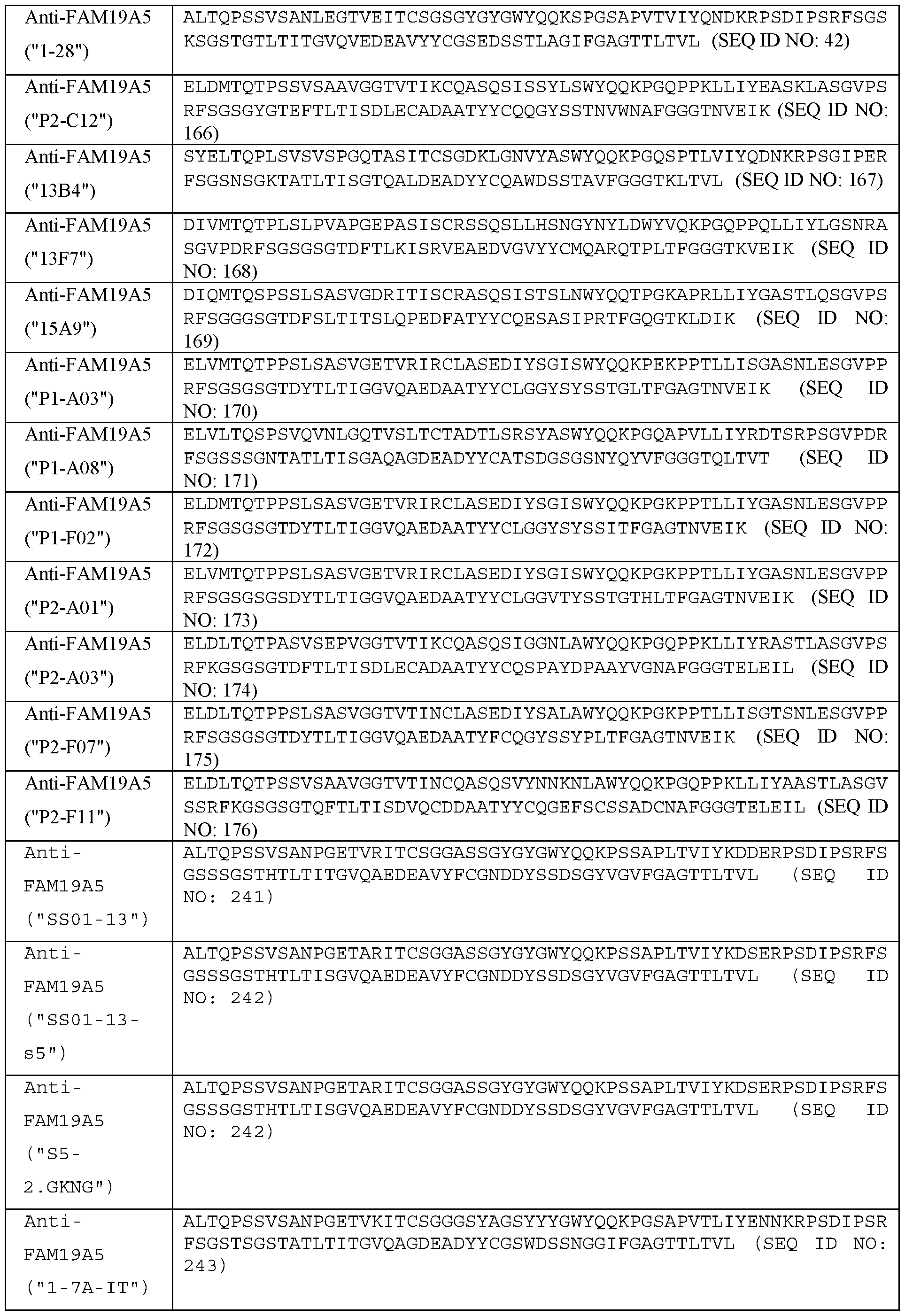


Claims
Priority Applications (8)
| Application Number | Priority Date | Filing Date | Title |
|---|---|---|---|
| CA3115797A CA3115797A1 (en) | 2018-10-16 | 2019-10-15 | Use of anti-fam19a5 antibodies |
| KR1020207034809A KR102648762B1 (en) | 2018-10-16 | 2019-10-15 | Uses of anti-FAM19A5 antibodies |
| CN201980075558.7A CN113166237A (en) | 2018-10-16 | 2019-10-15 | Use of anti-FAM 19a5 antibodies |
| AU2019363368A AU2019363368A1 (en) | 2018-10-16 | 2019-10-15 | Use of anti-FAM19A5 antibodies |
| JP2020564578A JP7211610B2 (en) | 2018-10-16 | 2019-10-15 | Uses of anti-FAM19A5 antibody |
| US17/285,861 US20210347872A1 (en) | 2018-10-16 | 2019-10-15 | Use of anti-fam19a5 antibodies |
| EP19874728.9A EP3867272A4 (en) | 2018-10-16 | 2019-10-15 | Use of anti-fam19a5 antibodies |
| JP2022212795A JP2023041682A (en) | 2018-10-16 | 2022-12-29 | Use of anti-fam19a5 antibodies |
Applications Claiming Priority (2)
| Application Number | Priority Date | Filing Date | Title |
|---|---|---|---|
| US201862746194P | 2018-10-16 | 2018-10-16 | |
| US62/746,194 | 2018-10-16 |
Publications (1)
| Publication Number | Publication Date |
|---|---|
| WO2020079595A1 true WO2020079595A1 (en) | 2020-04-23 |
Family
ID=70283396
Family Applications (1)
| Application Number | Title | Priority Date | Filing Date |
|---|---|---|---|
| PCT/IB2019/058794 WO2020079595A1 (en) | 2018-10-16 | 2019-10-15 | Use of anti-fam19a5 antibodies |
Country Status (8)
| Country | Link |
|---|---|
| US (1) | US20210347872A1 (en) |
| EP (1) | EP3867272A4 (en) |
| JP (2) | JP7211610B2 (en) |
| KR (1) | KR102648762B1 (en) |
| CN (1) | CN113166237A (en) |
| AU (1) | AU2019363368A1 (en) |
| CA (1) | CA3115797A1 (en) |
| WO (1) | WO2020079595A1 (en) |
Cited By (1)
| Publication number | Priority date | Publication date | Assignee | Title |
|---|---|---|---|---|
| WO2022121966A1 (en) * | 2020-12-10 | 2022-06-16 | Wuxi Biologics (Shanghai) Co., Ltd. | An antibody against p-cadherin and uses thereof |
Families Citing this family (1)
| Publication number | Priority date | Publication date | Assignee | Title |
|---|---|---|---|---|
| WO2019003164A1 (en) | 2017-06-27 | 2019-01-03 | Neuracle Science Co., Ltd. | Use of anti-fam19a5 antibodies for treating cancers |
Citations (3)
| Publication number | Priority date | Publication date | Assignee | Title |
|---|---|---|---|---|
| US20090221670A1 (en) * | 2005-05-11 | 2009-09-03 | Aarhus Universitet | Method for diagnosis and treatment of a mental disease |
| US20150118230A1 (en) * | 2012-02-15 | 2015-04-30 | Korea University Research And Business Foundation | Pharmaceutical Use of FAM19A5 Involved in Regulating Gliogenesis |
| WO2018083538A1 (en) * | 2016-11-07 | 2018-05-11 | Neuracle Scienc3 Co., Ltd. | Anti-family with sequence similarity 19, member a5 antibodies and method of use thereof |
Family Cites Families (1)
| Publication number | Priority date | Publication date | Assignee | Title |
|---|---|---|---|---|
| KR101802411B1 (en) * | 2015-02-17 | 2017-11-29 | 울산대학교 산학협력단 | Composition for preventing or treating of obesity comprising FAM19A5 and screening method for agent for treatment of obesity using the same |
-
2019
- 2019-10-15 AU AU2019363368A patent/AU2019363368A1/en active Pending
- 2019-10-15 KR KR1020207034809A patent/KR102648762B1/en active IP Right Grant
- 2019-10-15 EP EP19874728.9A patent/EP3867272A4/en active Pending
- 2019-10-15 JP JP2020564578A patent/JP7211610B2/en active Active
- 2019-10-15 CN CN201980075558.7A patent/CN113166237A/en active Pending
- 2019-10-15 CA CA3115797A patent/CA3115797A1/en active Pending
- 2019-10-15 US US17/285,861 patent/US20210347872A1/en active Pending
- 2019-10-15 WO PCT/IB2019/058794 patent/WO2020079595A1/en unknown
-
2022
- 2022-12-29 JP JP2022212795A patent/JP2023041682A/en active Pending
Patent Citations (3)
| Publication number | Priority date | Publication date | Assignee | Title |
|---|---|---|---|---|
| US20090221670A1 (en) * | 2005-05-11 | 2009-09-03 | Aarhus Universitet | Method for diagnosis and treatment of a mental disease |
| US20150118230A1 (en) * | 2012-02-15 | 2015-04-30 | Korea University Research And Business Foundation | Pharmaceutical Use of FAM19A5 Involved in Regulating Gliogenesis |
| WO2018083538A1 (en) * | 2016-11-07 | 2018-05-11 | Neuracle Scienc3 Co., Ltd. | Anti-family with sequence similarity 19, member a5 antibodies and method of use thereof |
Non-Patent Citations (2)
| Title |
|---|
| WANG, Y. ET AL.: "Novel adipokine, FAM19A5, inhibits neointima formation after injury through sphingosine-1-phosphate receptor 2", CIRCULATION, vol. 138, 3 July 2018 (2018-07-03), pages 48 - 63, XP055652588, DOI: 10.1161/CIRCULATIONAHA.117.032398 * |
| YILMAZ, G. ET AL.: "Induction of neuro-protective/regenerative genes in stem cells infiltrating post-ischemic brain tissue", EXPERIMENTAL & TRANSLATIONAL STROKE MEDICINE, vol. 2, no. 1, 28 May 2010 (2010-05-28), pages 1 - 10, XP021085086 * |
Cited By (1)
| Publication number | Priority date | Publication date | Assignee | Title |
|---|---|---|---|---|
| WO2022121966A1 (en) * | 2020-12-10 | 2022-06-16 | Wuxi Biologics (Shanghai) Co., Ltd. | An antibody against p-cadherin and uses thereof |
Also Published As
| Publication number | Publication date |
|---|---|
| KR20200142586A (en) | 2020-12-22 |
| AU2019363368A1 (en) | 2021-05-13 |
| CN113166237A (en) | 2021-07-23 |
| JP7211610B2 (en) | 2023-01-24 |
| JP2021532733A (en) | 2021-12-02 |
| EP3867272A4 (en) | 2022-08-03 |
| US20210347872A1 (en) | 2021-11-11 |
| JP2023041682A (en) | 2023-03-24 |
| EP3867272A1 (en) | 2021-08-25 |
| CA3115797A1 (en) | 2020-04-23 |
| KR102648762B1 (en) | 2024-03-19 |
Similar Documents
| Publication | Publication Date | Title |
|---|---|---|
| AU2008334637B2 (en) | Antigen binding proteins | |
| TWI809147B (en) | Anti-sortilin antibodies and methods of use thereof | |
| US11746149B2 (en) | Use of anti-family with sequence similarity 19, member A5 antibodies for the treatment of glaucoma | |
| US11155613B2 (en) | Use of anti-FAM19A5 antibodies for treating fibrosis | |
| JP2023041682A (en) | Use of anti-fam19a5 antibodies | |
| US20220064277A1 (en) | Use of anti-fam19a5 antibodies for treating atherosclerosis | |
| US20240101681A1 (en) | Methods of use of anti-sortilin antibodies | |
| US20230104540A1 (en) | Use of anti-fam19a1 antagonists for treating central nervous system diseases | |
| KR20230025674A (en) | HIF-1 alpha inhibitors | |
| EA046146B1 (en) | ANTIBODIES AGAINST SORTILIN AND METHODS OF THEIR APPLICATION |
Legal Events
| Date | Code | Title | Description |
|---|---|---|---|
| 121 | Ep: the epo has been informed by wipo that ep was designated in this application |
Ref document number: 19874728 Country of ref document: EP Kind code of ref document: A1 |
|
| ENP | Entry into the national phase |
Ref document number: 2020564578 Country of ref document: JP Kind code of ref document: A |
|
| ENP | Entry into the national phase |
Ref document number: 20207034809 Country of ref document: KR Kind code of ref document: A |
|
| ENP | Entry into the national phase |
Ref document number: 3115797 Country of ref document: CA |
|
| NENP | Non-entry into the national phase |
Ref country code: DE |
|
| ENP | Entry into the national phase |
Ref document number: 2019363368 Country of ref document: AU Date of ref document: 20191015 Kind code of ref document: A |
|
| ENP | Entry into the national phase |
Ref document number: 2019874728 Country of ref document: EP Effective date: 20210517 |