WO2020045652A1 - 皮膚細胞の細胞活性又は肌状態の決定方法、並びに皮膚細胞賦活剤のスクリーニング方法 - Google Patents
皮膚細胞の細胞活性又は肌状態の決定方法、並びに皮膚細胞賦活剤のスクリーニング方法 Download PDFInfo
- Publication number
- WO2020045652A1 WO2020045652A1 PCT/JP2019/034218 JP2019034218W WO2020045652A1 WO 2020045652 A1 WO2020045652 A1 WO 2020045652A1 JP 2019034218 W JP2019034218 W JP 2019034218W WO 2020045652 A1 WO2020045652 A1 WO 2020045652A1
- Authority
- WO
- WIPO (PCT)
- Prior art keywords
- cell
- adhesion
- cadherin
- cells
- skin
- Prior art date
- Legal status (The legal status is an assumption and is not a legal conclusion. Google has not performed a legal analysis and makes no representation as to the accuracy of the status listed.)
- Ceased
Links
Images



Classifications
-
- A—HUMAN NECESSITIES
- A01—AGRICULTURE; FORESTRY; ANIMAL HUSBANDRY; HUNTING; TRAPPING; FISHING
- A01K—ANIMAL HUSBANDRY; AVICULTURE; APICULTURE; PISCICULTURE; FISHING; REARING OR BREEDING ANIMALS, NOT OTHERWISE PROVIDED FOR; NEW BREEDS OF ANIMALS
- A01K67/00—Rearing or breeding animals, not otherwise provided for; New or modified breeds of animals
- A01K67/027—New or modified breeds of vertebrates
-
- C—CHEMISTRY; METALLURGY
- C12—BIOCHEMISTRY; BEER; SPIRITS; WINE; VINEGAR; MICROBIOLOGY; ENZYMOLOGY; MUTATION OR GENETIC ENGINEERING
- C12Q—MEASURING OR TESTING PROCESSES INVOLVING ENZYMES, NUCLEIC ACIDS OR MICROORGANISMS; COMPOSITIONS OR TEST PAPERS THEREFOR; PROCESSES OF PREPARING SUCH COMPOSITIONS; CONDITION-RESPONSIVE CONTROL IN MICROBIOLOGICAL OR ENZYMOLOGICAL PROCESSES
- C12Q1/00—Measuring or testing processes involving enzymes, nucleic acids or microorganisms; Compositions therefor; Processes of preparing such compositions
- C12Q1/02—Measuring or testing processes involving enzymes, nucleic acids or microorganisms; Compositions therefor; Processes of preparing such compositions involving viable microorganisms
-
- G—PHYSICS
- G01—MEASURING; TESTING
- G01N—INVESTIGATING OR ANALYSING MATERIALS BY DETERMINING THEIR CHEMICAL OR PHYSICAL PROPERTIES
- G01N33/00—Investigating or analysing materials by specific methods not covered by groups G01N1/00 - G01N31/00
- G01N33/15—Medicinal preparations ; Physical properties thereof, e.g. dissolubility
-
- G—PHYSICS
- G01—MEASURING; TESTING
- G01N—INVESTIGATING OR ANALYSING MATERIALS BY DETERMINING THEIR CHEMICAL OR PHYSICAL PROPERTIES
- G01N33/00—Investigating or analysing materials by specific methods not covered by groups G01N1/00 - G01N31/00
- G01N33/48—Biological material, e.g. blood, urine; Haemocytometers
- G01N33/50—Chemical analysis of biological material, e.g. blood, urine; Testing involving biospecific ligand binding methods; Immunological testing
Definitions
- the present invention relates to a method for determining the cell activity and skin condition of skin cells, and a method for screening a skin cell activator by using a change in the cell activity as an index.
- intercellular adhesion mainly includes tight junctions, fixed junctions, and gap junctions depending on the type of cell tumor, and the types of adhesion proteins involved are different.
- dermal fibroblasts were thought to be scattered through binding to the extracellular matrix.
- the cells of each organ present in the living body do not always maintain a certain activity, and the activity fluctuates depending on external factors and internal factors such as stress.
- the skin constitutes the outermost layer of the living body, so it is susceptible to external factors such as temperature changes, ultraviolet irradiation, physical stimuli, and internal factors such as aging and stress. It is known to affect.
- the cell activity decreases, the cell division ability of the cell decreases, and the secretion of a factor called an aging factor may adversely affect surrounding cells.
- the production amount of extracellular matrix molecules such as collagen and elastin decreases. Depletion of extracellular matrix molecules in the dermis layer causes sagging of the skin, which is a cosmetic problem.
- the beauty field or the medical field it is required to measure the cell activity of the skin and to perform care according to the cell activity, and it is required to appropriately determine the cell activity and the skin condition of the skin.
- a screening system for a cell activator capable of enhancing the cell activity can be established.
- the present inventors have conducted studies on the association between dermal fibroblasts and cell activity, and surprisingly found that the adhesion state in dermal fibroblasts and the expression level of type I collagen gene I found an association. Furthermore, when the cell adhesion factor affecting the expression of the type I collagen gene was examined, it was found that cadherin 2 (CDH2) contributed to the expression of the type I collagen gene, and the present invention was reached.
- CDH2 cadherin 2
- the present invention relates to: [1] A method for determining the cell activity of dermal fibroblasts using the adhesion between dermal fibroblasts as an index. [2] The method according to item 1, wherein the adhesion is determined by a cell process. [3] The method according to item 2, wherein the protrusion is determined by a cell circularity. [4] The method according to any one of items 1 to 3, wherein the adhesion is adhesion via cadherin-2. [5] The method according to any one of items 1 to 4, wherein the cell activity is a collagen-producing ability. [6] A method for determining a skin condition using, as an index, the adhesion between dermal fibroblasts in a skin sample collected from the skin.
- a method for screening a skin cell activator Culturing the fibroblasts in a candidate drug-supplemented medium or a non-supplemented control medium, The step of determining the adhesion between fibroblasts in the control group without the addition of the candidate drug and the group with the addition of the candidate drug, and a candidate added to the group in which the adhesion between fibroblasts is enhanced as compared to the control group. Determining the drug as a cell activator, The screening method, comprising: [12] The method according to item 11, wherein the density of the culture of fibroblasts is adjusted to 0.5 to 8.0 ⁇ 10 3 cells / cm 2 .
- cadherin-2 mediated cell adhesion is controlled by adjusting the seeding density of dermal fibroblasts.
- a three-dimensional skin model comprising collagen gel and dermal fibroblasts and having enhanced tension, wherein 1 to 6 cadherin-2 mediated cell adhesions are present per cell. .
- the present invention makes it possible to determine cell activity in skin. This makes it possible to provide skin care according to the cell activity.
- a method for screening a cell activator can be provided by confirming a change in cell activity.
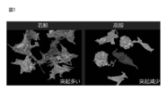
- FIG. 1 is a diagram showing remodeling of fibroblasts in the dermis of a young subject and an elderly subject by observing them with a three-dimensional electron microscope (SBF-SEM). 2, 0.25 ⁇ 10 4 cells /Ml,0.5 ⁇ 10 4 cells /Ml,1.0 ⁇ 10 4 cells /Ml,2.0 ⁇ 10 4 cells / ml, and 4.0 ⁇ 10
- the micrograph after adding 2.5 ml of the cell suspension prepared at 4 cells / ml and culturing for 2 days is shown, and the degree of contact at each concentration is shown.
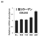
- FIG. 3 is a graph comparing the expression levels of type I collagen at each contact degree.
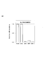
- FIG. 4 shows the effect of suppressing gene expression by siRNA for each gene.
- FIG. 1 is a diagram showing remodeling of fibroblasts in the dermis of a young subject and an elderly subject by observing them with a three-dimensional electron microscope (SBF-SEM). 2, 0.25 ⁇ 10 4 cells /Ml,0.5 ⁇ 10 4 cells
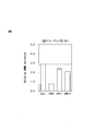
- FIG. 5 shows changes in the expression level of type I collagen when gene expression was suppressed by siRNA for each gene.
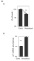
- FIG. 6 shows changes in the cell growth rate (A) and p21 gene expression (B) when the expression of cadherin 2 gene was suppressed in cultured dermal fibroblasts.
- FIG. 7 shows the shape change when the expression of cadherin-2 gene is suppressed in cultured dermal fibroblasts. Due to the suppression of gene expression of cadherin 2 (Knockout), adhesion between cells was not observed, the shape was rounded, and ⁇ -Gal, an age marker, was stained.
- the present invention relates to a method for determining the cell activity of dermal fibroblasts using the adhesion between dermal fibroblasts as an index.
- ⁇ Cell activity of dermal fibroblasts refers to various activities of dermal fibroblasts.
- the dermis is mainly composed of a stromal component and a cellular component, and furthermore, vessels and nerves are arranged.
- the cellular activity of dermal fibroblasts refers to the ability to produce stromal components.
- Cellular activity is also related to the degree of aging of cells, and as aging progresses, cellular activity decreases.
- reduced cellular activity can secrete substances called aging factors, which can adversely affect surrounding cells.
- cellular activity refers to the ability to produce stromal components, and more preferably to the ability to produce collagen.
- the stromal component of the dermis is mainly composed of collagen fibers, elastic fibers, and matrix. Therefore, by enhancing the cell activity of dermal fibroblasts, the amount of production of at least one, preferably two, and more preferably all of the collagen fibers, elastic fibers and matrix is increased.
- Collagen fibers are composed of collagen. About 20 types of collagen molecules are known due to differences in the molecular structure of the ⁇ chain.
- the collagen may be any collagen, but type I collagen, type III collagen, type V collagen, type IV, type VII, type 17 collagen mainly present in the dermis is more preferable, and more preferably, Type I collagen, type III collagen, type V collagen, most preferably type I collagen which accounts for about 80% in the dermis.
- the amount of collagen fiber production can be determined by measuring the amount of collagen production or expression.
- the main component of the elastic fiber is elastin, and fibrillin is formed around elastin.
- the production amount of elastic fibers can be determined by measuring the production amount or expression amount of at least one of elastin or fibrillin.
- the substrate is mainly composed of glycoprotein, proteoglycan, etc. as an extracellular matrix.
- the glycoprotein is a protein containing sugar, such as fibronectin in the dermis. Fibronectin binds to a cell surface protein, functions as a cell scaffold, and can also bind to other macromolecules such as collagen.
- Proteoglycans are macromolecules in which glycosaminoglycans are bound to axis proteins. In the dermis, proteoglycans mainly contain hyaluronic acid and dermatan sulfate as glycosaminoglycans. Proteoglycans serve primarily to retain water in the dermis.
- Dermal fibroblasts are fibroblasts present in the dermis and are cells that produce stromal components in the dermis. The dermis is filled with stromal components and dermal fibroblasts are thought to be scattered throughout the dermis.
- integrins expressed in the cell membrane of dermal fibroblasts contribute to supporting dermal fibroblasts in the dermis by binding to fibronectin, which is a stromal component.
- dermal fibroblasts adhered to each other, and furthermore, a connection was found between the adhesion between dermal fibroblasts and the cell activity of dermal fibroblasts. Specifically, senescent dermal fibroblasts have less or no adhesion between cells.
- Adhesion between dermal fibroblasts can be judged from the image of the sample in a micrograph.
- Adhesion between dermal cells can be determined from a two-dimensional photograph or from a three-dimensional photograph. More preferably, observation can be performed using a three-dimensional electron microscope (SBF-SEM).
- Adhesion can be quantified by the number of cells to which one cell adheres, the number of adhesion points of one cell, the adhesion area to which one cell adheres, or the ratio of contact cells per plate.
- the sample may be a biological sample, an organ-cultured sample, a three-dimensional skin model sample, or an in vitro sample.
- cell activity can be determined by the number of attachment points.
- the cell activity when the number of adhesion points is 0, it can be determined that the cell activity is low. When the number of adhesion points is 1 to 2, the cell activity is determined to be moderate, and when the number of adhesion points is 3 or more, the cell activity is high. Can be determined. Cell activity can also be determined based on the ratio of adherent cells per unit area. If it is 10% or less, it can be determined that the cell activity is low. If the number of adherent cells is 50% or less, it is moderate. And the cell activity can be determined to be high when the number of cells to be adhered is 50% or more.
- Adhesion between dermal fibroblasts can be determined based on proteins involved in cell adhesion, as another embodiment, in addition to judgment from images in micrographs.
- the protein involved in cell adhesion may be any cell adhesion protein.
- Cell adhesion proteins include, for example, cadherin superfamily proteins. Specific examples include E-cadherin, N-cadherin, P-cadherin, T-cadherin, VE-cadherin, and the like. From the viewpoint of expression in dermal fibroblasts, N-cadherin (cadherin 2) is particularly preferred. , Cadherin 6, cadherin 11, and cadherin 13 are preferred.
- the adhesion between dermal fibroblasts can be measured, or by simply measuring the amount of protein and gene expression of the cell adhesion protein, dermal fibroblasts can be measured. Adhesion between them can be determined.
- the inventors examined the relationship between the suppression of gene expression of cell adhesion protein in dermal fibroblasts and the activity of dermal fibroblasts. Specifically, by suppressing the expression of cadherin-2 gene expressed in dermal fibroblasts, the expression of type I collagen by dermal fibroblasts was suppressed (FIG. 5). Type I collagen changes its expression level due to cell adhesion (FIG. 3) and serves as an indicator of dermal fibroblast cell activity. Since the activity of dermal fibroblasts was reduced by suppressing the gene expression of cadherin 2 which is a cell adhesion protein, cadherin 2 can be used as an indicator of cell adhesion. Cadherin 2 is cadherin, also known as N-cadherin, which is known to be present in nerve cells. Cadherin 2 present in the cell membrane of each cell binds to each other by homophilic binding.
- Adhesion between dermal fibroblasts is performed by cell projections extending from dermal fibroblasts. Therefore, the adhesion between dermal fibroblasts can be detected by detecting whether they are adhered by cell projections.
- the presence of cell processes reduces the circularity of the cells. Therefore, adhesion can also be evaluated based on the circularity of dermal fibroblasts.
- the cell circularity can be calculated using ImageJ software, and in a cell in a human body, 50% or more can be determined to have no adhesion, and 50% or less can be determined to have adhesion.
- On cultured cells 80% or more is judged as non-contact, 50-80% is judged as light contact, 30-50% as moderate contact, 10-30% as high contact, and 10% or less as excessive contact. can do.
- a biological sample is a skin sample collected from the skin of a subject.
- One embodiment of the present invention relates to a method for determining a skin condition by using an adhesion between dermal fibroblasts as an index using a skin sample.
- the skin condition particularly refers to a skin condition caused by the dermis.
- elastic fibers and collagen fibers contribute to elasticity, while the matrix contributes to water retention. Therefore, skin conditions caused by the dermis mainly include sagging, wrinkling, and beaming.
- Slack, wrinkles, and beams can be determined to be normal if they adhere to an average of two to three cells per cell, and can be determined to be zero to an average of one to three cells per cell. When adhered to two cells, it can be determined that there is much sagging or wrinkling and that the beam is inferior.
- Skin condition can also be represented as skin age.
- adhesion can be determined from cell density. As an example, when 2.5 ml is seeded at a density of 0.25 ⁇ 10 4 cells / ml per mm dish of a 6-well plate, it can be determined that there is no contact. When 2.5 ml was seeded at a density of 0.5 ⁇ 10 4 cells / ml per 6-well plate, mild contact can be determined. Medium contact can be determined when seeding 2.5 ml per mm dish of a 6-well plate at a density of 1.0 ⁇ 10 4 cells / ml. When 2.5 ml is seeded at a density of 2.0 ⁇ 10 4 cells / ml per mm dish of a 6-well plate, high contact can be determined.
- the subject may be any mammal, for example, a human, monkey, pig, cow, horse, mouse, etc., but is preferably a human for cosmetic purposes.
- fibroblasts may be derived from any species, but are preferably human for cosmetic purposes.
- a skin care or treatment step that enhances the activity of dermal fibroblasts is performed on a subject that is determined to have low cell adhesion of dermal fibroblasts with low cell-cell adhesion.
- administration of a known dermal cell activator or treatment of activation of dermal fibroblasts or skin stem cells can be performed.
- the screening method for a skin cell activator of the present invention enables screening of a skin cell activator by examining the effect of a candidate drug on the adhesion between fibroblasts. More specifically, the method for screening a skin cell activator of the present invention comprises the following: Culturing the fibroblasts in a candidate drug-supplemented medium or a non-supplemented control medium, The step of determining the adhesion between fibroblasts in the control group without the addition of the candidate drug and the group with the addition of the candidate drug, and a candidate added to the group in which the adhesion between fibroblasts is enhanced as compared to the control group. Determining the drug as a cell activator, including.
- the control group is a culture that was tested under the same conditions as the candidate drug-added group, except that no candidate drug was included. For the control group, experiments may be performed in parallel under the same conditions as the candidate drug added group, or separate experiments may be performed.
- the candidate drug a substance contained in a compound library, a drug library, a cosmetic material library, an extract library, or the like can be used.
- a skin cell activator refers to an agent capable of enhancing the cell activity of skin cells, particularly dermal cells, through enhancing cell-cell adhesion in dermal cells.
- the skin cell activator promotes cell proliferation and expression of interstitial components, and can particularly promote collagen production.
- the skin cell activator selected by the screening method of the present invention can also be referred to as a cell adhesion protein expression promoter.
- the cadherin family protein particularly the cadherin 2 protein expression promoter.
- the skin cell activator selected by the screening method of the present invention can be incorporated into cosmetics or pharmaceuticals.
- the skin cell activators selected in the present invention can also be referred to as collagen production promoters, wrinkle improvers, sagging improvers, and beam improvers.
- the adhesion can be determined by the same method as the method used for measuring the cell activity. Specifically, observation of cell adhesion and projection shape under a microscope is performed. When determining adhesion based on the shape, the adhesion can be measured by determining the circularity of the cells. At the time of observation under a microscope, by specifically staining a cell adhesion protein, for example, cadherin 2, adhesion between cells can be more accurately determined. In another embodiment, screening can be performed by using the amount of cell adhesion protein or the amount of gene expression as an index of cell-cell adhesion.
- the culturing of the fibroblasts can be performed by a usual method of culturing fibroblasts.
- the cells can be cultured at a density at which no contact or excessive contact is achieved, for example, 0.1 to 10 ⁇ 10 3 cells / cm 2 .
- the density at which no contact to high contact is achieved eg, 0.5-8.0 ⁇ 10 3 cells / cm 2 , more preferably the density at which moderate contact is achieved, eg, 2.0-4.
- a screening method using a medium in which the amount of a growth factor is reduced is used. Can also be performed.
- the present invention relates to a method for creating a three-dimensional skin model in which tension is adjusted.
- the three-dimensional skin model is a culture containing a stromal component and a cellular component, and has a structure imitating the dermis of a living body.
- the three-dimensional skin model includes at least one of a collagen fiber, an elastic fiber, and an extracellular matrix as a stromal component, and particularly includes collagen as a collagen fiber. Elastin and fibrin may be included as elastic fibers. Extracellular cells may also contain glycoproteins and proteoglycans as matrix components.
- a three-dimensional skin model can be created by dispersing and culturing dermal fibroblasts in a collagen-containing medium. A relationship between the expression of the cadherin 2 gene and the contraction or maturation of collagen could be found. Therefore, when creating a three-dimensional skin model, the firmness of the three-dimensional skin model can be adjusted by controlling cell adhesion via cadherin 2.
- Cadherin 2 mediated cell adhesion can be controlled by regulating the cadherin 2 gene.
- the cadherin 2 gene can suppress the expression or inhibit the function of the cadherin 2 gene by knockdown preparation, siRNA, antibody, and the like. It can also be used as an accelerator.
- cadherin-2-mediated cell adhesion can be controlled by adjusting the seeding density of dermal fibroblasts in the stromal component.
- the seeding density can be represented by the number of cells per volume of the gel, and three-dimensional by seeding at 50,000 cells / cm 3 or more, preferably 100,000 cells / cm 3 or more, more preferably 200,000 cells / cm 3 or more. It can enhance the firmness of the skin model.
- Still another embodiment of the present invention may relate to a three-dimensional skin model with enhanced firmness created by the method for creating a three-dimensional skin model of the present invention.
- a three-dimensional skin model may include dermal fibroblasts in which cadherin-2 gene expression is increased, and collagen gel as a stromal component.
- Dermal fibroblasts with increased cadherin 2 gene expression may include a vector for increasing cadherin 2 gene expression.
- Such a three-dimensional skin model has an average of 2-3 cell adhesions per cell, preferably 3-5 cells per cell, and such cell adhesion is preferably cadherin-2 mediated cell adhesion.
- the cells contained in the three-dimensional skin model exist at a density of 50,000 cells / cm 3 or more, preferably 100,000 cells / cm 3 or more, and more preferably 200,000 cells / cm 3 or more. By achieving such a cell density, the firmness of the three-dimensional skin model can be increased.
- the firmness of the three-dimensional skin model can be measured by using a commercially available skin tension measuring instrument such as a cute meter or a derma torque meter.
- the three-dimensional skin model of the present invention may have a tension of 0.1 to 1.0, more preferably 0.2 to 0.8, in terms of Ur / Uf. preferable.
- the method of measuring skin tension using a cute meter can be implemented by referring to Skin Research and Technology (2010); 16: 332-338.
- Serial block face scanning electron microscope Human skin samples were fixed in a buffered solution of 2% glutaraldehyde and 2% paraformaldehyde at 4 ° C. for several days. The sample was treated with phosphate buffered saline of 2% osmium tetroxide (Nisshin EM, Tokyo, Japan) and 1.5% potassium ferrocyanide (Wako Pure Chemical Industries, Ltd.) at 4 ° C. for 1 hour and 1% Treat with thiocarbohydrazide (Sigma, St.
- SBF-SEM Serial block face scanning electron microscope
- the expression of cell adhesion protein was suppressed in cultured fibroblasts by the siRNA method.
- CDH2, CDH11 and CDH13 siRNAs were obtained from Qiagen.
- the expression of each gene was suppressed in cultured dermal fibroblasts using these siRNAs according to a standard method.
- 0.5 ml of a cell suspension having a density of 1 ⁇ 10 4 cells / ml was seeded in each well of a 24-well plate (area: 2 cm 2 ) and cultured for 2 days at 37 ° C. in a 5% CO 2 atmosphere.
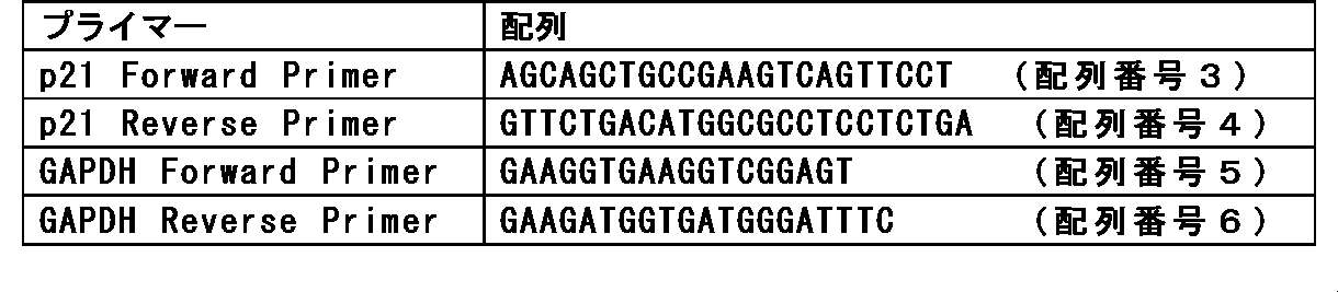
- a dermal fibroblast in which the expression of cadherin 2 is suppressed and a control dermal fibroblast are each 0.5 ml of a cell suspension having a density of 1 ⁇ 10 4 cells / ml in a 24-well plate (area: 2 cm 2 ). And cultured for 2 days at 37 ° C. in a 5% CO 2 atmosphere. Cultured cells were counted and compared for cell proliferation (FIG. 6A). In addition, gene expression of p21 which is a CDK family inhibitory protein was measured for each cultured cell by RT-PCR using the following primers.
- the cells were stained with X-Gal according to ⁇ -galactosidase activity using a cell aging assay kit (Biovision) and photographed under a microscope (FIG. 7).
- the cultured dermal fibroblasts had a flattened shape and adhesion between cells was observed.
- suppression of cadherin 2 gene expression (Knockout) reduced the adhesion between cells, The shape was spherical compared to the control.
- intracellular ⁇ -galactosidase activity which is an indicator of cell aging, was high.
- Collagen gel contraction activity Fibroblasts were seeded in 75 cm 2 flasks at 3 ⁇ 10 5 cells and cultured in DMEM containing 10% FBS for 12 hours. Next, CDH2 siRNA obtained from Qiagen was introduced according to a standard method, and cells were collected 24 hours later. Cells into which the siRNA had been introduced were mixed with the collagen gel at 2 ⁇ 10 5 cells / ml (final concentration) to give a 0.1% collagen gel, seeded, and cultured for 24 hours. Twenty-four hours later, an image was taken from above the plate, and the contraction of the collagen gel was measured (FIG. 8). It was shown that suppression of CDH2 gene expression reduced collagen contraction activity. Collagen increases its elasticity as fibroblasts attract and contract. It was shown that when the expression of the CDH2 gene is suppressed, the contraction of collagen is prevented, which also leads to a decrease in sagging of the skin.
- Dermal fibroblasts were prepared at a concentration of 1.0 ⁇ 10 4 cells / ml in DMEN medium, and 2.5 ml was seeded on a 6-well plate. After culturing for 2 days, the medium was replaced with a medium containing the candidate drug and a control medium without the candidate drug, respectively, and culturing was further continued for 2 days. The culture was fixed with PFA, reacted with an anti-cadherin 2 rabbit antibody (Cell Signaling), and then reacted with a fluorescently labeled anti-rabbit IgG antibody (Cell Signaling).
- Cell Signaling an anti-cadherin 2 rabbit antibody
- the candidate agent can be screened as a skin cell activator.
- Dermal fibroblasts were prepared at a concentration of 1.0 ⁇ 10 4 cells / ml in DMEN medium, and 2.5 ml was seeded on a 6-well plate. After culturing for 2 days, the medium was replaced with a medium containing the candidate drug and a control medium without the candidate drug, respectively, and culturing was further continued for 2 days.
- the cadherin-2 gene expression or protein level in the culture can be measured and, if increased relative to a control, screened as a skin cell activator.
- the skin cell activator thus obtained can also be referred to as a cadherin 2 gene expression promoter.
Landscapes
- Life Sciences & Earth Sciences (AREA)
- Health & Medical Sciences (AREA)
- Chemical & Material Sciences (AREA)
- Engineering & Computer Science (AREA)
- Molecular Biology (AREA)
- Immunology (AREA)
- Biochemistry (AREA)
- Zoology (AREA)
- Organic Chemistry (AREA)
- General Health & Medical Sciences (AREA)
- Physics & Mathematics (AREA)
- Analytical Chemistry (AREA)
- Biotechnology (AREA)
- Hematology (AREA)
- General Physics & Mathematics (AREA)
- Food Science & Technology (AREA)
- Pathology (AREA)
- Proteomics, Peptides & Aminoacids (AREA)
- Urology & Nephrology (AREA)
- Microbiology (AREA)
- Biophysics (AREA)
- Wood Science & Technology (AREA)
- Bioinformatics & Cheminformatics (AREA)
- Medicinal Chemistry (AREA)
- Biomedical Technology (AREA)
- Environmental Sciences (AREA)
- Animal Husbandry (AREA)
- Biodiversity & Conservation Biology (AREA)
- Animal Behavior & Ethology (AREA)
- Genetics & Genomics (AREA)
- Cell Biology (AREA)
- General Engineering & Computer Science (AREA)
- Pharmacology & Pharmacy (AREA)
- Measuring Or Testing Involving Enzymes Or Micro-Organisms (AREA)
- Investigating Or Analysing Biological Materials (AREA)
- Micro-Organisms Or Cultivation Processes Thereof (AREA)
- Medicines That Contain Protein Lipid Enzymes And Other Medicines (AREA)
Abstract
真皮線維芽細胞の細胞活性を測定することを目的とする。また、細胞活性の変化をもとに、細胞賦活剤をスクリーニングする方法を提供することを目的とする。 真皮線維芽細胞同士の接着を指標とすることで、細胞活性の測定が可能となる。また、真皮線維芽細胞同士の接着を指標として、細胞賦活剤をスクリーニングする方法が可能となる。
Description
本発明は、皮膚細胞の細胞活性や肌状態の決定方法、並びに当該細胞活性の変化を指標とすることによる、皮膚細胞賦活剤のスクリーニング方法に関する。
血液細胞などの浮遊性の細胞を除いて、細胞は、生体内において、細胞同士又は細胞外マトリクスへと結合して存在している。細胞間接着としては、その細胞腫に応じて、密着結合、固定結合、及びギャップ結合が主に挙げられ、それぞれ関与する接着タンパク質の種類が異なる。真皮において、真皮線維芽細胞は細胞外マトリクスへの結合を介して散在して存在すると考えられていた。
生体内に存在する各臓器の細胞は、常に一定の活性を保っているわけではなく、外部要因やストレスなどの内的要因に応じて、その活性は変動する。特に皮膚は、生体の最外層を構成していることから、温度変化、紫外線照射、物理的刺激などの外部要因の影響を受けやすく、また加齢やストレスなどの内的要因も皮膚の細胞活性に影響を与えることが知られている。細胞活性が低下すると、自身の細胞分裂能力が低下するとともに、老化因子と呼ばれる因子を分泌することで、周囲の細胞に対しても悪影響を及ぼしうる。また、細胞活性が悪化した真皮細胞では、コラーゲンやエラスチンといった細胞外マトリクス分子の産生量が低下する。真皮層における細胞外マトリクス分子の低下は、皮膚のたるみを引き起こし、美容上の問題となる。
美容分野又は医療分野では、皮膚の細胞活性を測定し、細胞活性に応じたケアを行うことが求められており、皮膚の細胞活性や肌状態を適切に決定することが求められている。また細胞活性を決定することが可能になると、細胞活性を亢進可能な細胞賦活剤のスクリーニング系の確立が可能になる。
本発明者らは、真皮線維芽細胞同士の接着と、細胞活性との関連性について研究を行ったところ、驚くべきことに真皮線維芽細胞における接着状態と、I型コラーゲン遺伝子の発現レベルとの関連を見出した。さらに、I型コラーゲン遺伝子の発現に影響を及ぼす細胞接着因子について検討を行ったところ、カドヘリン2(CDH2)が、I型コラーゲン遺伝子の発現に寄与することを見出し、本発明に至った。
したがって、本発明は以下に関する:
[1] 真皮線維芽細胞同士の接着を指標とした、真皮線維芽細胞の細胞活性の決定方法。
[2] 前記接着を、細胞突起により決定する、項目1に記載の方法。
[3] 前記突起を、細胞円形率により決定する、項目2に記載の方法。
[4] 前記接着が、カドヘリン2を介した接着である、項目1~3のいずれか一項に記載の方法。
[5] 前記細胞活性が、コラーゲン産生能である、項目1~4のいずれか一項に記載の方法。
[6] 皮膚から採取された皮膚試料において、真皮線維芽細胞同士の接着を指標とした、肌状態の決定方法。
[7] 前記接着を、細胞突起により決定する、項目6に記載の方法。
[8] 前記突起を、細胞円形率により決定する、項目7に記載の方法。
[9] 前記接着が、カドヘリン2を介した接着である、項目6~8のいずれか一項に記載の方法。
[10] 前記細胞活性が、コラーゲン産生能である、項目6~9のいずれか一項に記載の方法。
[11] 皮膚細胞賦活剤のスクリーニング方法であって、
線維芽細胞を、候補薬剤添加培地又は非添加対照培地中で培養する工程、
候補薬剤非添加の対照群と、候補薬剤添加群とにおいて、線維芽細胞同士の接着を決定する工程、及び
対照群に比較して、線維芽細胞同士の接着が亢進された群に添加した候補薬剤を、細胞賦活剤として決定する工程、
を含む、前記スクリーニング方法。
[12] 線維芽細胞の培養物の密度を、0.5~8.0×10³細胞/cm2で調製することを特徴とする、項目11に記載の方法。
[13] 前記接着を、細胞突起により決定する、項目11又は12に記載の方法。
[14] 前記突起を、細胞円形率により決定する、項目11又は12に記載の方法。
[15] 前記接着が、カドヘリン2を介した接着である、項目11~14のいずれか一項に記載の方法。
[16] コラーゲンゲル及び真皮線維芽細胞を含み、ハリが調節された3次元皮膚モデルの作成方法であって、前記真皮線維芽細胞において、カドヘリン2を介した細胞接着を制御することを含む、前記方法。
[17] カドヘリン2を介した細胞接着が、カドヘリン2遺伝子の発現調節により制御される、項目16に記載の方法。
[18] カドヘリン2を介した細胞接着が、真皮線維芽細胞の播種密度を調節することにより制御される、項目16に記載の方法。
[19] コラーゲンゲル及び真皮線維芽細胞を含み、ハリが強化された3次元皮膚モデルであって、カドヘリン2を介した細胞接着が、1細胞あたり1~6個存在する、前記3次元皮膚モデル。
[20] 前記3次元皮膚モデルが、キュートメーターを用いて弾性を測定した場合に、Ur/Uf値が0.1~1.0の弾性を有する、項目19に記載の3次元皮膚モデル。
[21] カドヘリン2の遺伝子発現を増大された真皮線維芽細胞と、コラーゲンゲルとを含む、ハリが強化された3次元皮膚モデル。
[22] 前記真皮線維芽細胞が、カドヘリン2遺伝子発現増大するためのベクターを含む、項目21に記載の3次元皮膚モデル。
[23] 前記3次元皮膚モデルが、キュートメーターを用いて弾性を測定した場合に、Ur/Uf値が0.1~1.0の弾性を有する、項目21又は22に記載の3次元皮膚モデル。
本発明により、皮膚における細胞活性を決定することが可能になる。これにより、細胞活性に応じたスキンケアの提供が可能になる。また、細胞活性の変化を確認することにより細胞賦活剤のスクリーニング方法を提供することができる。
本発明は、真皮線維芽細胞同士の接着を指標とした、真皮線維芽細胞の細胞活性の決定方法に関する。
真皮線維芽細胞の細胞活性は、真皮線維芽細胞が有する各種の活性のことをいう。真皮は、主に間質成分と細胞成分から構成され、さらに脈管や神経が配置されている。真皮線維芽細胞の活性が高くなると、真皮線維芽細胞の分裂頻度や間質成分の産生量が高くなり、結果として真皮層の厚さが厚くなるとともにその弾力も高くなる。したがって、本発明の1の態様では、真皮線維芽細胞の細胞活性は、間質成分の産生能力を指す。細胞活性は、細胞の加齢度とも関係しており、加齢が進むと、細胞活性が低下する。さらに、細胞活性が低下していると、老化因子とよばれる、周囲の細胞に悪影響を及ぼす物質を分泌しうる。本発明の1の態様では、細胞活性は、間質成分の産生能力を指し、より好ましくは、コラーゲンの産生能力を指す。
真皮の間質成分は、膠原線維、弾性線維、及び基質から主に構成されている。したがって、真皮線維芽細胞の細胞活性が亢進することにより、膠原線維、弾性線維、及び基質のうちの少なくとも1、好ましくは2、さらに好ましくは全ての成分の産生量が増大する。
膠原線維は、コラーゲンにより構成されている。コラーゲン分子は、α鎖の分子構造の違いにより、20種ほどが知られている。本発明において、コラーゲンは、任意のコラーゲンであってよいが、真皮に主に存在するI型コラーゲン、III型コラーゲン、V型コラーゲン、IV型、VII型、17型コラーゲンが好ましく、より好ましくは、I型コラーゲン、III型コラーゲン、V型コラーゲンであり、最も好ましくは真皮において約80%を占めるI型コラーゲンである。膠原線維の産生量は、コラーゲンの産生量又は発現量を測定することにより決定しうる。
弾性線維の主成分は、エラスチンであり、さらにフィブリリンがエラスチンの周囲を取り巻いて構成される。弾性線維の産生量は、エラスチン又はフィブリリンの少なくとも1の産生量又は発現量を測定することにより決定しうる。
基質は、主に細胞外マトリクスとして、糖タンパクや、プロテオグリカンなどにより構成される。糖タンパクとしては、糖を含むタンパク質で、真皮中ではフィブロネクチンなどが挙げられ、フィブロネクチンは、細胞表面タンパク質と結合し、細胞足場として機能するとともに、コラーゲンなどの他の高分子とも結合しうる。プロテオグリカンは、軸タンパク質にグリコサミノグリカンが結合した巨大分子であり、真皮中では、グリコサミノグリカンとしてヒアルロン酸やデルマタン硫酸が主に含まれる。プロテオグリカンは、主に真皮中での水分保持の機能を果たす。
真皮線維芽細胞は、真皮中に存在する線維芽細胞であり、真皮中の間質成分を産生する細胞である。真皮は、間質成分で満たされており、真皮線維芽細胞は真皮中で散在して存在すると考えられている。特に真皮線維芽細胞の細胞膜に発現するインテグリンは、間質成分であるフィブロネクチンと結合することで、真皮線維芽細胞を真皮中で支えることに寄与する。一方で、真皮線維芽細胞同士が接着していることが見いだされ、さらに真皮線維芽細胞同士の接着と、真皮線維芽細胞の細胞活性との間に関連が見いだされた。具体的に老化した真皮線維芽細胞では、細胞同士の接着は少なくなるか、又はなくなる。
真皮線維芽細胞同士の接着は、試料を顕微鏡写真における画像から判断することができる。真皮細胞同士の接着は、2次元写真から判定することもできるし、3次元写真から判定することができる。より好ましくは、3次元電子顕微鏡(SBF-SEM)を用いて観察をすることができる。接着は、1の細胞が接着する細胞の数、1の細胞が有する接着点の数、1の細胞が接着する接着面積、又はプレート当たりの接触細胞の割合により、定量することができる。試料は、生体試料、器官培養された試料、3次元皮膚モデルの試料、又はインビトロの試料であってもよい。一例として、接着点の数により、細胞活性を決定することができる。例えば、接着点の数が0の場合、細胞活性が低いと判定でき、接着点の数が、1~2を、中程度の細胞活性と判定し、3以上の接着点を、細胞活性が高いと判定することができる。単位面積当たりの接着する細胞の割合基づいて、細胞活性を判定することもでき、10%以下の場合、細胞活性が低いと判定でき、接着する細胞の数が50%以下の場合に、中程度の細胞活性と判定し、接着する細胞の数が50%以上の場合に、細胞活性が高いと判定することができる。
真皮線維芽細胞同士の接着は、顕微鏡写真における画像からの判断の他に、別の態様として、細胞接着に関与するタンパク質に基づいて判断することもできる。細胞接着に関与するタンパク質としては、任意の細胞接着タンパク質であってよい。細胞接着タンパク質としては、一例としてカドヘリンスーパーファミリーのタンパク質が挙げられる。具体的には、E‐カドヘリン、N‐カドヘリン、P‐カドヘリン、T‐カドヘリン、VE‐カドヘリンなどが挙げられるが、真皮線維芽細胞において発現がみられる観点から、特にN‐カドヘリン(カドヘリン2)、カドヘリン6、カドヘリン11、カドヘリン13が好ましい。免疫染色により細胞接着タンパク質の存在を確認することにより、真皮線維芽細胞同士の接着を測定することができるし、単に細胞接着タンパク質のタンパク質量や遺伝子発現量を測定することにより、真皮線維芽細胞同士の接着を判断しうる。
本発明者らにより、真皮線維芽細胞における細胞接着タンパク質の遺伝子発現抑制と、真皮線維芽細胞の活性との関係が調べられた。具体的に、真皮線維芽細胞に発現しているカドヘリン2の遺伝子発現を抑制することにより、真皮線維芽細胞によるI型コラーゲンの発現が抑制された(図5)。I型コラーゲンは、細胞接着によりその発現量が変化し(図3)、真皮線維芽細胞の細胞活性の指標となる。細胞接着タンパク質であるカドヘリン2の遺伝子発現の抑制により、真皮線維芽細胞の活性が低下したことから、カドヘリン2を細胞接着の指標として使用しうる。カドヘリン2は、N-カドヘリンとしても知られ、神経細胞での存在が知られているカドヘリンである。各細胞の細胞膜に存在するカドヘリン2がホモフィリックに結合することで、細胞間を接着させる。
真皮線維芽細胞同士の接着は、真皮線維芽細胞から伸びた細胞突起により接着される。よって、真皮線維芽細胞同士の接着は、細胞突起により接着されているかを検出することで検出することができる。細胞突起が存在すると、細胞の円形率が低くなる。したがって、真皮線維芽細胞の円形率をもとに接着を評価することもできる。細胞円形率は、ImageJソフトウェアを用いて計算することができ、ヒトの生体内の細胞では、50%以上を接着がないと判定し、50%以下を接着があると判定できる。また、培養細胞上では、80%以上を接触無しと判定し、50~80%を軽度接触、30~50%を中程度接触、10~30%を高度接触、10%以下を過度接触と判定することができる。
生体試料は、対象の皮膚から採取された皮膚試料である。本発明の1の態様では、皮膚試料を用いて真皮線維芽細胞同士の接着を指標とすることで、肌状態を決定する方法に関する。肌状態とは、特に真皮に起因する肌の状態をいう。真皮の間質成分のうち、弾性繊維や膠原繊維は弾力に寄与し、一方で基質は、水分保持に寄与する。したがって、真皮に起因する肌状態は、主にたるみ、しわ、はりが挙げられる。一例として、対象から取得された皮膚試料中の真皮線維芽細胞において、1の細胞あたり平均で3~5個の細胞と接着している場合に、たるみやしわが少なく、はりが優れていると決定することができ、1の細胞あたり平均で2~3個の細胞と接着している場合に、たるみ、しわ、はりが普通であると決定することができ、1の細胞あたり平均で0~2個の細胞と接着している場合に、たるみやしわが多く、はりが劣ると決定することができる。肌状態を、肌年齢として表すこともできる。
インビトロの試料の場合、接着は、細胞密度から判定することができる。一例として、0.25×10⁴細胞/mlの密度で、6穴プレートmmディッシュあたり2.5mlを播種した場合を、接触無しと判定することができる。0.5×104細胞/mlの密度で、6穴プレートあたり2.5mlを播種した場合を、軽度接触と判定することができる。1.0×104細胞/mlの密度で、6穴プレートmmディッシュあたり2.5mlを播種した場合を、中程度接触と判定することができる。2.0×104細胞/mlの密度で、6穴プレートmmディッシュあたり2.5mlを播種した場合を、高度接触と判定することができる。4.0×104細胞/mlの密度で、6穴プレートあたり2.5mlを播種した場合を、過度接触と判定することができる。したがって、0~1.0×103細胞/cm2の密度を接触無し、1.0~2.0×103細胞/cm2の密度を軽度接触、2.0~4.0×103細胞/cm2を中程度接触と判定することができ、4.0~8.0×103細胞/cm2を高度接触と判定することができ、8.0×103細胞/cm2以上を過度接触と判定することができる。使用するディッシュ及び播種細胞数からの顕微鏡画像に応じ、適宜接触を判定することができる。
本発明において、対象は、任意の哺乳動物、例えば、ヒト、サル、ブタ、ウシ、ウマ、マウスなどであってよいが、美容上の目的から好ましくはヒトである。スクリーニング方法に用いる場合、線維芽細胞であれば、任意の種由来であってよいが、美容上の目的から好ましくはヒトである。
本発明の対象において、真皮線維芽細胞における細胞間の接着が少なく皮膚細胞の細胞活性が低いと判定された対象に対しては、真皮線維芽細胞の活性を高めるスキンケアや治療工程が行われる。真皮線維芽細胞の活性を高めるために、公知の真皮細胞賦活剤の投与や、真皮線維芽細胞又は皮膚幹細胞の活性化の処置が行われうる。
本発明の別の態様は、皮膚細胞賦活剤のスクリーニング方法に関する。本発明の皮膚細胞賦活剤のスクリーニング方法は、線維芽細胞同士の接着について、候補薬剤の影響を調べることで、皮膚細胞賦活剤をスクリーニングが可能になる。より具体的に、本発明の皮膚細胞賦活剤のスクリーニング方法は、以下の:
線維芽細胞を、候補薬剤添加培地又は非添加対照培地中で培養する工程、
候補薬剤非添加の対照群と、候補薬剤添加群とにおいて、線維芽細胞同士の接着を決定する工程、及び
対照群に比較して、線維芽細胞同士の接着が亢進された群に添加した候補薬剤を、細胞賦活剤として決定する工程、
を含む。
線維芽細胞を、候補薬剤添加培地又は非添加対照培地中で培養する工程、
候補薬剤非添加の対照群と、候補薬剤添加群とにおいて、線維芽細胞同士の接着を決定する工程、及び
対照群に比較して、線維芽細胞同士の接着が亢進された群に添加した候補薬剤を、細胞賦活剤として決定する工程、
を含む。
対照群は、候補薬剤を含まないことを除いて、候補薬剤添加群と同一条件で実験された培養物である。対照群については、候補薬剤添加群と同じ条件下で並行して実験がされてもよいし、別途実験が行われていてもよい。候補薬剤は、化合物ライブラリー、医薬品ライブラリー、化粧品素材ライブラリー、抽出物ライブラリーなどに含まれる物質を使用することができる。
皮膚細胞賦活剤は、真皮細胞において細胞間接着を亢進させることを介して、皮膚細胞、特に真皮細胞の細胞活性を亢進できる薬剤をいう。皮膚細胞賦活剤により、細胞増殖や間質成分の発現促進が図られ、特にコラーゲン産生の促進することができる。本発明のスクリーニング方法により選択された皮膚細胞賦活剤は、細胞接着タンパク質の発現促進剤ということもできる。細胞接着タンパク質の中でも、カドヘリンファミリーのタンパク質、特にカドヘリン2タンパク質の発現促進剤ということができる。本発明のスクリーニング方法により選択された皮膚細胞賦活剤は化粧品又は医薬品に配合することができる。本発明において選択された皮膚細胞賦活剤は、コラーゲン産生促進剤、しわ改善剤、たるみ改善剤、はり改善剤ということもできる。
スクリーニング方法において、接着の決定は細胞活性の測定方法にもちいた手法と同じ手法により決定することができる。具体的に、顕微鏡下での細胞の接着や突起形状の観察が行われる。形状に基づいて、接着を判定する場合、細胞の円形率を求めることで接着を測定することができる。顕微鏡下の観察の際に、細胞接着タンパク質、例えばカドヘリン2を特異的に染色することでより正確に細胞同士の接着を判定することができる。また別の態様では、細胞同士の接着の指標として、細胞接着タンパク質の量又は遺伝子発現量を使用することで、スクリーニングすることもできる。
線維芽細胞の培養は、線維芽細胞を培養する通常の方法で行われうる。接着を判定する観点から、接触無し~過度接触が達成される密度、例えば0.1~10×103細胞/cm2で培養されうる。好ましくは、接触無し~高度接触が達成される密度、例えば0.5~8.0×103細胞/cm2であり、より好ましくは中程度接触が達成される密度、例えば2.0~4.0×103細胞/cm2の細胞培養物を用いることが好ましい。また、一の態様では、細胞が増殖することで、細胞の接触状態の判別が難しくなることから、増殖が抑制された条件下、例えば増殖因子の量を低減させた培地を用いて、スクリーニング方法を行うこともできる。
別の態様では、本発明は、ハリが調節された3次元皮膚モデルの作成方法に関する。3次元皮膚モデルは、間質成分と細胞成分とを含む培養物であり、生体の真皮を模した構造を有する。3次元皮膚モデルは、間質成分として、膠原繊維、弾性繊維、細胞外マトリックスのうちの少なくとも1を含み、特に膠原繊維としてコラーゲンを含む。弾性繊維として、エラスチンやフィブリンを含んでもよい。細胞外もマトリクス成分として、糖タンパク質やプロテオグリカンを含んでもよい。一例として、コラーゲン含有培地中で、真皮線維芽細胞を分散させて培養することで、3次元皮膚モデルを作成することができる。カドヘリン2遺伝子の発現と、コラーゲンの収縮又は成熟との関係が見出すことができた。したがって、3次元皮膚モデルの作成の際に、カドヘリン2を介した細胞接着を制御することで、3次元皮膚モデルのハリを調節することができる。
カドヘリン2を介した細胞接着は、カドヘリン2遺伝子の調節により制御することができる。具体的に、カドヘリン2遺伝子は、ノックダウンの作成、siRNA、抗体などにより、発現抑制や機能阻害することができ、さらには上述のスクリーニング方法にて得られた皮膚賦活剤をカドヘリン2遺伝子の活性促進剤として使用することもできる。
さらに別の態様では、間質成分における真皮線維芽細胞の播種密度を調節することにより、カドヘリン2を介した細胞接着を制御することができる。播種密度が高くなるほど、細胞間の接着数が増加する。この接着は主にカドヘリン2を介した接着である。播種密度は、ゲルの体積あたりの細胞数で表すことができ、50000個/cm3以上、好ましくは100000個/cm3以上、さらに好ましくは200000個/cm3以上で播種することにより、3次元皮膚モデルのハリを高めることができる。
本発明のさらに別の態様では、本発明の3次元皮膚モデルの作成方法により作成されたハリが高められた3次元皮膚モデルに関してもよい。このような3次元皮膚モデルは、カドヘリン2の遺伝子発現を増大された真皮線維芽細胞と、間質成分としてコラーゲンゲルとを含んでもよい。カドヘリン2の遺伝子発現を増大された真皮線維芽細胞は、カドヘリン2遺伝子発現を増大するためのベクターを含んでいてもよい。このような3次元皮膚モデルは、細胞接着の数が、1細胞あたり平均で2~3個、好ましくは3~5個であり、かかる細胞接着は好ましくはカドヘリン2を介した細胞接着である。また、3次元皮膚モデルに含まれる細胞は、50000個/cm3以上、好ましくは100000個/cm3以上、さらに好ましくは200000個/cm3以上の密度で存在する。このような細胞密度を達成することで、3次元皮膚モデルのハリを高めることができる。3次元皮膚モデルのハリは、キュートメーター、ダーマトルクメーターなどの市販の皮膚の張り測定器を用いることで測定することができる。一例として、キュートメーターを用いた場合に、本発明の3次元皮膚モデルが、Ur/Ufの値で0.1~1.0、より好ましくは0.2~0.8の張りを有することが好ましい。キュートメーターを用いた皮膚の張りの測定方法については、Skin Research and Technology (2010); 16: 332-338を参照すること実行することができる。
本明細書において言及される全ての文献はその全体が引用により本明細書に取り込まれる。
以下に説明する本発明の実施例は例示のみを目的とし、本発明の技術的範囲を限定するものではない。本発明の技術的範囲は特許請求の範囲の記載によってのみ限定される。本発明の趣旨を逸脱しないことを条件として、本発明の変更、例えば、本発明の構成要件の追加、削除及び置換を行うことができる。
3次元顕微鏡による観察
若齢対象(20歳)及び高齢対象(80歳)の皮膚切片を、導電性樹脂で処理し、3次元電子顕微鏡(SBF-SEM)観察に供した。3次元リモデリングした図を示す(図1)。SBF‐SEMによるサンプル調製及び観察方法は下記のとおりである。
若齢対象(20歳)及び高齢対象(80歳)の皮膚切片を、導電性樹脂で処理し、3次元電子顕微鏡(SBF-SEM)観察に供した。3次元リモデリングした図を示す(図1)。SBF‐SEMによるサンプル調製及び観察方法は下記のとおりである。
シリアルブロックフェイス走査電子顕微鏡(SBF-SEM)
ヒト皮膚サンプルを、2%グルタルアルデヒド及び2%パラホルムアルデヒドの緩衝溶液中で4℃で数日間固定した。サンプルを2%四酸化オスミウム(Nisshin EM, Tokyo, Japan)及び1.5%フェロシアン化カリウム (Wako Pure Chemical Industries, Ltd.)のリン酸緩衝生理食塩水で4℃にて1時間処理し、1%チオカルボヒドラジド(Sigma, St. Louis, Mo, USA)で室温にて20分間処理し、2%四酸化オスミウム水溶液で30分間室温にて処理し、2%酢酸ウラニル溶液で4℃12時間以上処理し、そして室温で1時間処理した。サンプルを、0.67%硝酸鉛(pH5.0~5.5、TAAB、Berkshire,UK)、0.03M L-アスパラギン酸(Nacalai Tesque, Kyoto, Japan)の溶液中で65℃にて30分間インキュベートした。次にサンプルを段階的エタノール系列で脱水し、脱水アセトンで処理し、35℃にて、Quetol812エポキシ樹脂(Nisshin EM, Tokyo, Japan)を浸透させ、そしてKejen black powder含有Quetol812(Nguyen et al. Sci Rep. 2016)に包埋した。樹脂を70℃で7夜超インキュベートして、重合化を確実にした。3Viewチャンバー内ウルトラマイクトロームシステム(Gatan, Pleasanton, CA)を備えた電界放出走査型電子顕微鏡 (Merlin, Carl Zeiss, Oberkochen, Germany)を用いてSBF-SEMでの観察を行った。80~90μm×80~90μm幅(11~12nm/ピクセル)で、60μmの深さにわたり80nmのステップで連続イメージシーケンスを取得した。連続イメージを、FIJI(https://fiji.sc/)を用いて処理した。Amira (Maxnet Co., Ltd, Tokyo, Japan)を用いて、セグメンテーション及び3次元リコンストラクションを行った。
ヒト皮膚サンプルを、2%グルタルアルデヒド及び2%パラホルムアルデヒドの緩衝溶液中で4℃で数日間固定した。サンプルを2%四酸化オスミウム(Nisshin EM, Tokyo, Japan)及び1.5%フェロシアン化カリウム (Wako Pure Chemical Industries, Ltd.)のリン酸緩衝生理食塩水で4℃にて1時間処理し、1%チオカルボヒドラジド(Sigma, St. Louis, Mo, USA)で室温にて20分間処理し、2%四酸化オスミウム水溶液で30分間室温にて処理し、2%酢酸ウラニル溶液で4℃12時間以上処理し、そして室温で1時間処理した。サンプルを、0.67%硝酸鉛(pH5.0~5.5、TAAB、Berkshire,UK)、0.03M L-アスパラギン酸(Nacalai Tesque, Kyoto, Japan)の溶液中で65℃にて30分間インキュベートした。次にサンプルを段階的エタノール系列で脱水し、脱水アセトンで処理し、35℃にて、Quetol812エポキシ樹脂(Nisshin EM, Tokyo, Japan)を浸透させ、そしてKejen black powder含有Quetol812(Nguyen et al. Sci Rep. 2016)に包埋した。樹脂を70℃で7夜超インキュベートして、重合化を確実にした。3Viewチャンバー内ウルトラマイクトロームシステム(Gatan, Pleasanton, CA)を備えた電界放出走査型電子顕微鏡 (Merlin, Carl Zeiss, Oberkochen, Germany)を用いてSBF-SEMでの観察を行った。80~90μm×80~90μm幅(11~12nm/ピクセル)で、60μmの深さにわたり80nmのステップで連続イメージシーケンスを取得した。連続イメージを、FIJI(https://fiji.sc/)を用いて処理した。Amira (Maxnet Co., Ltd, Tokyo, Japan)を用いて、セグメンテーション及び3次元リコンストラクションを行った。
培養実験
ヒトの皮膚試料から、定法にしたがってヒト初代培養線維芽細胞を取得した。細胞の数を計測し、(10% FBSを含む)DMEM培地中に、1mlあたり、0.25×10⁴細胞(接触無し)、0.5×10⁴細胞(軽度接触)、1.0×10⁴細胞(中程度接触)、2.0×10⁴細胞(高度接触)、及び4.0×10⁴細胞(過度接触)に調製した。かかる細胞懸濁液を、6穴プレート(ファルコン社製)に2.5ml滴下し、37℃5%CO2雰囲気下で2日間培養した。培養後の細胞の顕微鏡写真を図2に示す。
ヒトの皮膚試料から、定法にしたがってヒト初代培養線維芽細胞を取得した。細胞の数を計測し、(10% FBSを含む)DMEM培地中に、1mlあたり、0.25×10⁴細胞(接触無し)、0.5×10⁴細胞(軽度接触)、1.0×10⁴細胞(中程度接触)、2.0×10⁴細胞(高度接触)、及び4.0×10⁴細胞(過度接触)に調製した。かかる細胞懸濁液を、6穴プレート(ファルコン社製)に2.5ml滴下し、37℃5%CO2雰囲気下で2日間培養した。培養後の細胞の顕微鏡写真を図2に示す。
接着の程度の違いに伴う遺伝子発現の変化
接触度の異なる培養物から細胞を回収し、マイクロアレイ解析に付したところ、細胞接触度に応じて、I型コラーゲンの発現が変化することを見出した(データ未掲載)。そこで、下記のプライマーを用いたリアルタイムPCRにより、I型コラーゲンの発現量を決定した(図3)。発現量の決定には、内部標準として、GAPDH遺伝子の発現を用いた。

接触度の異なる培養物から細胞を回収し、マイクロアレイ解析に付したところ、細胞接触度に応じて、I型コラーゲンの発現が変化することを見出した(データ未掲載)。そこで、下記のプライマーを用いたリアルタイムPCRにより、I型コラーゲンの発現量を決定した(図3)。発現量の決定には、内部標準として、GAPDH遺伝子の発現を用いた。
I型コラーゲンの発現に寄与する因子を決定することを目的として、培養線維芽細胞において、細胞接着タンパク質の発現をsiRNA法により抑制した。CDH2、CDH11、及びCDH13のsiRNAを、キアゲン社より入手した。これらのsiRNAを定法にしたがって用いて培養真皮線維芽細胞において各遺伝子の発現を抑制した。1×10⁴細胞/mlの密度の細胞懸濁液0.5mlを24穴プレート(面積:2cm2)の各ウェルに播種し、2日間37℃5%CO2雰囲気下で培養した。培養線維芽細胞を採取し、各細胞接着タンパク質の遺伝子発現をリアルタイムPCRで決定し、目的タンパク質の発現抑制を確認した(図4)。次に、採取された細胞において、I型コラーゲンの発現を測定したところ、CDH2の遺伝子発現を抑制した際に、I型コラーゲンの発現量が低下することが示された(図5)。
カドヘリン2発現が抑制された真皮線維芽細胞と、対照の真皮線維芽細胞とを、それぞれ1×10⁴細胞/mlの密度の細胞懸濁液0.5mlを24穴プレート(面積:2cm2)の各ウェルに播種し、2日間37℃5%CO2雰囲気下で培養した。培養細胞を計数し、細胞の増殖性について比較した(図6A)。また、各培養細胞についてCDKファミリーの阻害タンパク質であるp21の遺伝子発現を、下記のプライマーを用いてRT-PCRにより測定した。さらに、細胞老化アッセイキット(Biovision社)を用いて、X-Galによりβガラクトシダーゼ活性に応じて細胞を染色し、顕微鏡下で撮影した(図7)。対照細胞では、培養真皮線維芽細胞は扁平な形状であり、細胞同士の接着がみられたが、カドヘリン2の遺伝子発現の抑制(Knockout)により、細胞同士の接着が減少し、またそれにともない細胞形状が対照に比較して球形を示した。また、カドヘリン2の遺伝子発現の抑制された真皮線維芽細胞において、細胞老化の指標となる細胞内βガラクトシダーゼ活性が高かった。
カドヘリン2の遺伝子発現が抑制された真皮線維芽細胞では、コラーゲン遺伝子の発現及び細胞増殖がともに抑制された。また、カドヘリン2の遺伝子発現が抑制された真皮線維芽細胞では、サイクリンの抑制により細胞周期の停止を引き起こすp21の遺伝子発現が増加することが示され、さらに細胞の老化の指標である細胞内βガラクトシダーゼ活性が高いことも示された。以上の結果より、細胞接着タンパク質であるカドヘリン2を介して、真皮線維芽細胞が、細胞間の接触を形成しており、このような接触が失われることで、細胞が老化し、細胞活性が低下することが示された。
コラーゲンゲル収縮活性
線維芽細胞を、3×10⁵細胞となるように75cm2フラスコに播種し、10%FBS含有DMEM中で12時間培養を行った。次に、キアゲン社より入手したCDH2のsiRNAを定法に従って導入し、24時間後に細胞を回収した。0.1%コラーゲンゲルとなるように、siRNAを導入された細胞を2×10⁵細胞/ml(終濃度)でコラーゲンゲルと混合して播種し、24時間培養した。24時間後にプレート上から撮影し、コラーゲンゲルの収縮を測定した(図8)。CDH2の遺伝子発現の抑制により、コラーゲン収縮活性が低下していることが示された。コラーゲンは、線維芽細胞がひきつけて、収縮することにより弾力が増加する。CDH2遺伝子の発現が抑制されると、コラーゲンの収縮が妨げられることから、皮膚のたるみやはりの減少につながることが示された。
線維芽細胞を、3×10⁵細胞となるように75cm2フラスコに播種し、10%FBS含有DMEM中で12時間培養を行った。次に、キアゲン社より入手したCDH2のsiRNAを定法に従って導入し、24時間後に細胞を回収した。0.1%コラーゲンゲルとなるように、siRNAを導入された細胞を2×10⁵細胞/ml(終濃度)でコラーゲンゲルと混合して播種し、24時間培養した。24時間後にプレート上から撮影し、コラーゲンゲルの収縮を測定した(図8)。CDH2の遺伝子発現の抑制により、コラーゲン収縮活性が低下していることが示された。コラーゲンは、線維芽細胞がひきつけて、収縮することにより弾力が増加する。CDH2遺伝子の発現が抑制されると、コラーゲンの収縮が妨げられることから、皮膚のたるみやはりの減少につながることが示された。
スクリーニング実験
真皮線維芽細胞を、DMEN培地中に1.0×10⁴細胞/mlの濃度で調製し、2.5mlを、6穴プレートに播種した。2日間培養後、候補薬剤を含む培地、候補薬剤非添加の対照培地で、培地をそれぞれ置換し、さらに2日間培養を行った。培養物を、PFAで固定し、抗カドヘリン2ラビット抗体(セルシグナリング社))を反応させ、次いで蛍光標識された抗ラビットIgG抗体(セルシグナリング社)を反応させた。蛍光顕微鏡下で観察を行い、カドヘリン2の発現を確認するとともに、細胞の突起部の形態を確認し、1細胞あたりの細胞の接着数を確認した。対照と比較して、接着数が増加した場合に、候補薬剤を皮膚細胞賦活剤としてスクリーニングすることができる。
真皮線維芽細胞を、DMEN培地中に1.0×10⁴細胞/mlの濃度で調製し、2.5mlを、6穴プレートに播種した。2日間培養後、候補薬剤を含む培地、候補薬剤非添加の対照培地で、培地をそれぞれ置換し、さらに2日間培養を行った。培養物を、PFAで固定し、抗カドヘリン2ラビット抗体(セルシグナリング社))を反応させ、次いで蛍光標識された抗ラビットIgG抗体(セルシグナリング社)を反応させた。蛍光顕微鏡下で観察を行い、カドヘリン2の発現を確認するとともに、細胞の突起部の形態を確認し、1細胞あたりの細胞の接着数を確認した。対照と比較して、接着数が増加した場合に、候補薬剤を皮膚細胞賦活剤としてスクリーニングすることができる。
スクリーニング実験
真皮線維芽細胞を、DMEN培地中に1.0×10⁴細胞/mlの濃度で調製し、2.5mlを、6穴プレートに播種した。2日間培養後、候補薬剤を含む培地、候補薬剤非添加の対照培地で、培地をそれぞれ置換し、さらに2日間培養を行った。培養物におけるカドヘリン2の遺伝子発現又はタンパク質量を測定し、対照と比較して増加した場合に、皮膚細胞賦活剤としてスクリーニングすることができる。このようにして取得された皮膚細胞賦活剤は、カドヘリン2遺伝子発現促進剤ということもできる。
真皮線維芽細胞を、DMEN培地中に1.0×10⁴細胞/mlの濃度で調製し、2.5mlを、6穴プレートに播種した。2日間培養後、候補薬剤を含む培地、候補薬剤非添加の対照培地で、培地をそれぞれ置換し、さらに2日間培養を行った。培養物におけるカドヘリン2の遺伝子発現又はタンパク質量を測定し、対照と比較して増加した場合に、皮膚細胞賦活剤としてスクリーニングすることができる。このようにして取得された皮膚細胞賦活剤は、カドヘリン2遺伝子発現促進剤ということもできる。
Claims (23)
- 真皮線維芽細胞同士の接着を指標とした、真皮線維芽細胞の細胞活性の決定方法。
- 前記接着を、細胞突起により決定する、請求項1に記載の方法。
- 前記突起を、細胞円形率により決定する、請求項2に記載の方法。
- 前記接着が、カドヘリン2を介した接着である、請求項1~3のいずれか一項に記載の方法。
- 前記細胞活性が、コラーゲン産生能である、請求項1~4のいずれか一項に記載の方法。
- 皮膚から採取された皮膚試料において、真皮線維芽細胞同士の接着を指標とした、肌状態の決定方法。
- 前記接着を、細胞突起により決定する、請求項6に記載の方法。
- 前記突起を、細胞円形率により決定する、請求項7に記載の方法。
- 前記接着が、カドヘリン2を介した接着である、請求項6~8のいずれか一項に記載の方法。
- 前記細胞活性が、コラーゲン産生能である、請求項6~9のいずれか一項に記載の方法。
- 皮膚細胞賦活剤のスクリーニング方法であって、
線維芽細胞を、候補薬剤添加培地又は非添加対照培地中で培養する工程、
候補薬剤非添加の対照群と、候補薬剤添加群とにおいて、線維芽細胞同士の接着を決定する工程、及び
対照群に比較して、線維芽細胞同士の接着が亢進された群に添加した候補薬剤を、細胞賦活剤として決定する工程、
を含む、前記スクリーニング方法。 - 線維芽細胞の培養物の密度を、0.5~8.0×10³細胞/cm2で調製することを特徴とする、請求項11に記載の方法。
- 前記接着を、細胞突起により決定する、請求項11又は12に記載の方法。
- 前記突起を、細胞円形率により決定する、請求項11又は12に記載の方法。
- 前記接着が、カドヘリン2を介した接着である、請求項11~14のいずれか一項に記載の方法。
- コラーゲンゲル及び真皮線維芽細胞を含み、ハリが調節された3次元皮膚モデルの作成方法であって、前記真皮線維芽細胞において、カドヘリン2を介した細胞接着を制御することを含む、前記方法。
- カドヘリン2を介した細胞接着が、カドヘリン2遺伝子の発現調節により制御される、請求項16に記載の方法。
- カドヘリン2を介した細胞接着が、真皮線維芽細胞の播種密度を調節することにより制御される、請求項16に記載の方法。
- コラーゲンゲル及び真皮線維芽細胞を含み、ハリが強化された3次元皮膚モデルであって、カドヘリン2を介した細胞接着が、1細胞あたり1~6個存在する、前記3次元皮膚モデル。
- 前記3次元皮膚モデルが、キュートメーターを用いて弾性を測定した場合に、Ur/Uf値が0.1~1.0の弾性を有する、請求項19に記載の3次元皮膚モデル。
- カドヘリン2の遺伝子発現を増大された真皮線維芽細胞と、コラーゲンゲルとを含む、ハリが強化された3次元皮膚モデル。
- 前記真皮線維芽細胞が、カドヘリン2遺伝子発現増大するためのベクターを含む、請求項21に記載の3次元皮膚モデル。
- 前記3次元皮膚モデルが、キュートメーターを用いて弾性を測定した場合に、Ur/Uf値が0.1~1.0の弾性を有する、請求項21又は22に記載の3次元皮膚モデル。
Priority Applications (1)
| Application Number | Priority Date | Filing Date | Title |
|---|---|---|---|
| JP2020539643A JP7383619B2 (ja) | 2018-08-31 | 2019-08-30 | 皮膚細胞の細胞活性又は肌状態の決定方法、並びに皮膚細胞賦活剤のスクリーニング方法 |
Applications Claiming Priority (2)
| Application Number | Priority Date | Filing Date | Title |
|---|---|---|---|
| JP2018-163923 | 2018-08-31 | ||
| JP2018163923 | 2018-08-31 |
Publications (1)
| Publication Number | Publication Date |
|---|---|
| WO2020045652A1 true WO2020045652A1 (ja) | 2020-03-05 |
Family
ID=69643869
Family Applications (1)
| Application Number | Title | Priority Date | Filing Date |
|---|---|---|---|
| PCT/JP2019/034218 Ceased WO2020045652A1 (ja) | 2018-08-31 | 2019-08-30 | 皮膚細胞の細胞活性又は肌状態の決定方法、並びに皮膚細胞賦活剤のスクリーニング方法 |
Country Status (3)
| Country | Link |
|---|---|
| JP (1) | JP7383619B2 (ja) |
| TW (1) | TW202020448A (ja) |
| WO (1) | WO2020045652A1 (ja) |
Cited By (2)
| Publication number | Priority date | Publication date | Assignee | Title |
|---|---|---|---|---|
| JPWO2021005801A1 (ja) * | 2019-07-11 | 2021-01-14 | ||
| JPWO2022071459A1 (ja) * | 2020-09-30 | 2022-04-07 |
Citations (2)
| Publication number | Priority date | Publication date | Assignee | Title |
|---|---|---|---|---|
| JP2006262806A (ja) * | 2005-03-24 | 2006-10-05 | Shiseido Co Ltd | 表皮細胞及び真皮線維芽細胞増殖作用の検出方法とその検出装置、並びにケミカルピーリング剤とそのスクリーニング方法、表皮細胞及び真皮線維芽細胞の賦活方法 |
| JP2015513905A (ja) * | 2012-03-27 | 2015-05-18 | コーダ セラピューティクス, インコーポレイテッド | カドヘリン調節に基づく組成物および処置 |
-
2019
- 2019-08-30 WO PCT/JP2019/034218 patent/WO2020045652A1/ja not_active Ceased
- 2019-08-30 TW TW108131378A patent/TW202020448A/zh unknown
- 2019-08-30 JP JP2020539643A patent/JP7383619B2/ja active Active
Patent Citations (2)
| Publication number | Priority date | Publication date | Assignee | Title |
|---|---|---|---|---|
| JP2006262806A (ja) * | 2005-03-24 | 2006-10-05 | Shiseido Co Ltd | 表皮細胞及び真皮線維芽細胞増殖作用の検出方法とその検出装置、並びにケミカルピーリング剤とそのスクリーニング方法、表皮細胞及び真皮線維芽細胞の賦活方法 |
| JP2015513905A (ja) * | 2012-03-27 | 2015-05-18 | コーダ セラピューティクス, インコーポレイテッド | カドヘリン調節に基づく組成物および処置 |
Non-Patent Citations (2)
| Title |
|---|
| FANG, CHIEN-LIANG ET AL.: "Dermal lipogenesis Inhibits Adiponectin Production in human Dermal Fibroblasts While Exogenous Adiponectin Administration Prevents against UVA-Induced Dermal Matrix Degradation in Human Skin", INTERNATIONAL JOURNAL OF MOLECULAR SCIENCES, vol. 17, no. 7, 14 July 2016 (2016-07-14), pages 1129, XP055696791 * |
| KIDA, NAOKO: "Bio-cosmetology -Development and application of new materials- , Improvement of skin elasticity through NAO extract", BIO INDUSTRY, vol. 25, no. 10, 2008, pages 14 - 20 * |
Cited By (3)
| Publication number | Priority date | Publication date | Assignee | Title |
|---|---|---|---|---|
| JPWO2021005801A1 (ja) * | 2019-07-11 | 2021-01-14 | ||
| JPWO2022071459A1 (ja) * | 2020-09-30 | 2022-04-07 | ||
| WO2022071459A1 (ja) * | 2020-09-30 | 2022-04-07 | テルモ株式会社 | 老化細胞判定システム |
Also Published As
| Publication number | Publication date |
|---|---|
| TW202020448A (zh) | 2020-06-01 |
| JPWO2020045652A1 (ja) | 2021-08-26 |
| JP7383619B2 (ja) | 2023-11-20 |
Similar Documents
| Publication | Publication Date | Title |
|---|---|---|
| Rosselli-Murai et al. | Periostin responds to mechanical stress and tension by activating the MTOR signaling pathway | |
| Salo et al. | A novel human leiomyoma tissue derived matrix for cell culture studies | |
| Marinkovic et al. | One size does not fit all: developing a cell-specific niche for in vitro study of cell behavior | |
| Akasaka et al. | Diversity of mechanisms underlying latent TGF-β activation in recessive dystrophic epidermolysis bullosa | |
| Moyle et al. | Three-dimensional niche stiffness synergizes with Wnt7a to modulate the extent of satellite cell symmetric self-renewal divisions | |
| Schminke et al. | Laminins and nidogens in the pericellular matrix of chondrocytes: their role in osteoarthritis and chondrogenic differentiation | |
| Zorin et al. | Diffuse colonies of human skin fibroblasts in relation to cellular senescence and proliferation | |
| Papageorgiou et al. | Expanded skeletal stem and progenitor cells promote and participate in induced bone regeneration at subcritical BMP-2 dose | |
| Rothdiener et al. | Stretching human mesenchymal stromal cells on stiffness-customized collagen type I generates a smooth muscle marker profile without growth factor addition | |
| EP3889247A1 (en) | Pigmentation skin model and method for producing same, and method for evaluating factor for treating or preventing pigmentation of skin | |
| Strash et al. | Human Erbb2-induced Erk activity robustly stimulates cycling and functional remodeling of rat and human cardiomyocytes | |
| Shamis et al. | iPSC-derived fibroblasts demonstrate augmented production and assembly of extracellular matrix proteins | |
| Morrison et al. | Plasticity and recovery of skeletal muscle satellite cells during limb regeneration | |
| Cirka et al. | Reproducible in vitro model for dystrophic calcification of cardiac valvular interstitial cells: insights into the mechanisms of calcific aortic valvular disease | |
| WO2015015655A1 (ja) | フックス角膜内皮ジストロフィ不死化細胞株およびその作製法 | |
| JP7383619B2 (ja) | 皮膚細胞の細胞活性又は肌状態の決定方法、並びに皮膚細胞賦活剤のスクリーニング方法 | |
| Moreno et al. | Human amniotic membrane modulates collagen production and deposition in vitro | |
| Da Costa et al. | Engineered endothelium model enables recapitulation of vascular function and early atherosclerosis development | |
| de Andrés et al. | A bioengineered tumor matrix-based scaffold for the evaluation of melatonin efficacy on head and neck squamous cancer stem cells | |
| Zhang et al. | Rps6ka2 enhances iMSC chondrogenic differentiation to attenuate knee osteoarthritis through articular cartilage regeneration in mice | |
| Bondarenko et al. | Effect of vascular endothelial growth factor and erythropoietin on functional activity of fibroblasts and multipotent mesenchymal stromal cells | |
| Riva et al. | Generation of human epidermal constructs on a collagen layer alone | |
| Carr et al. | Dermal fibroblasts influence the expression profile of 14-3-3 proteins in human keratinocytes | |
| Kyriakides | Hao Xing, Yaqing Huang 2, 7, Britta H. Kunkemoeller 2, 7, Peter J. Dahl 3, 6, Ohvia Muraleetharan, Nikhil S. Malvankar 3, 6, Michael P. Murrell & | |
| KR20130019459A (ko) | 양수-유래 중간엽 줄기세포의 증식방법 및 이를 포함하는 이식용 구조체의 제조방법 |
Legal Events
| Date | Code | Title | Description |
|---|---|---|---|
| 121 | Ep: the epo has been informed by wipo that ep was designated in this application |
Ref document number: 19854134 Country of ref document: EP Kind code of ref document: A1 |
|
| ENP | Entry into the national phase |
Ref document number: 2020539643 Country of ref document: JP Kind code of ref document: A |
|
| NENP | Non-entry into the national phase |
Ref country code: DE |
|
| 122 | Ep: pct application non-entry in european phase |
Ref document number: 19854134 Country of ref document: EP Kind code of ref document: A1 |