WO2020045281A1 - Novel nuclease domain and uses thereof - Google Patents
Novel nuclease domain and uses thereof Download PDFInfo
- Publication number
- WO2020045281A1 WO2020045281A1 PCT/JP2019/033045 JP2019033045W WO2020045281A1 WO 2020045281 A1 WO2020045281 A1 WO 2020045281A1 JP 2019033045 W JP2019033045 W JP 2019033045W WO 2020045281 A1 WO2020045281 A1 WO 2020045281A1
- Authority
- WO
- WIPO (PCT)
- Prior art keywords
- nucleic acid
- foki
- sequence
- seq
- zfa36
- Prior art date
Links
Images


Classifications
-
- C—CHEMISTRY; METALLURGY
- C12—BIOCHEMISTRY; BEER; SPIRITS; WINE; VINEGAR; MICROBIOLOGY; ENZYMOLOGY; MUTATION OR GENETIC ENGINEERING
- C12N—MICROORGANISMS OR ENZYMES; COMPOSITIONS THEREOF; PROPAGATING, PRESERVING, OR MAINTAINING MICROORGANISMS; MUTATION OR GENETIC ENGINEERING; CULTURE MEDIA
- C12N15/00—Mutation or genetic engineering; DNA or RNA concerning genetic engineering, vectors, e.g. plasmids, or their isolation, preparation or purification; Use of hosts therefor
- C12N15/09—Recombinant DNA-technology
- C12N15/10—Processes for the isolation, preparation or purification of DNA or RNA
- C12N15/102—Mutagenizing nucleic acids
-
- C—CHEMISTRY; METALLURGY
- C12—BIOCHEMISTRY; BEER; SPIRITS; WINE; VINEGAR; MICROBIOLOGY; ENZYMOLOGY; MUTATION OR GENETIC ENGINEERING
- C12N—MICROORGANISMS OR ENZYMES; COMPOSITIONS THEREOF; PROPAGATING, PRESERVING, OR MAINTAINING MICROORGANISMS; MUTATION OR GENETIC ENGINEERING; CULTURE MEDIA
- C12N9/00—Enzymes; Proenzymes; Compositions thereof; Processes for preparing, activating, inhibiting, separating or purifying enzymes
- C12N9/14—Hydrolases (3)
- C12N9/16—Hydrolases (3) acting on ester bonds (3.1)
- C12N9/22—Ribonucleases RNAses, DNAses
-
- C—CHEMISTRY; METALLURGY
- C12—BIOCHEMISTRY; BEER; SPIRITS; WINE; VINEGAR; MICROBIOLOGY; ENZYMOLOGY; MUTATION OR GENETIC ENGINEERING
- C12N—MICROORGANISMS OR ENZYMES; COMPOSITIONS THEREOF; PROPAGATING, PRESERVING, OR MAINTAINING MICROORGANISMS; MUTATION OR GENETIC ENGINEERING; CULTURE MEDIA
- C12N15/00—Mutation or genetic engineering; DNA or RNA concerning genetic engineering, vectors, e.g. plasmids, or their isolation, preparation or purification; Use of hosts therefor
- C12N15/09—Recombinant DNA-technology
- C12N15/63—Introduction of foreign genetic material using vectors; Vectors; Use of hosts therefor; Regulation of expression
- C12N15/79—Vectors or expression systems specially adapted for eukaryotic hosts
- C12N15/82—Vectors or expression systems specially adapted for eukaryotic hosts for plant cells, e.g. plant artificial chromosomes (PACs)
- C12N15/8201—Methods for introducing genetic material into plant cells, e.g. DNA, RNA, stable or transient incorporation, tissue culture methods adapted for transformation
- C12N15/8213—Targeted insertion of genes into the plant genome by homologous recombination
-
- C—CHEMISTRY; METALLURGY
- C12—BIOCHEMISTRY; BEER; SPIRITS; WINE; VINEGAR; MICROBIOLOGY; ENZYMOLOGY; MUTATION OR GENETIC ENGINEERING
- C12N—MICROORGANISMS OR ENZYMES; COMPOSITIONS THEREOF; PROPAGATING, PRESERVING, OR MAINTAINING MICROORGANISMS; MUTATION OR GENETIC ENGINEERING; CULTURE MEDIA
- C12N15/00—Mutation or genetic engineering; DNA or RNA concerning genetic engineering, vectors, e.g. plasmids, or their isolation, preparation or purification; Use of hosts therefor
- C12N15/09—Recombinant DNA-technology
- C12N15/63—Introduction of foreign genetic material using vectors; Vectors; Use of hosts therefor; Regulation of expression
- C12N15/79—Vectors or expression systems specially adapted for eukaryotic hosts
- C12N15/85—Vectors or expression systems specially adapted for eukaryotic hosts for animal cells
-
- C—CHEMISTRY; METALLURGY
- C12—BIOCHEMISTRY; BEER; SPIRITS; WINE; VINEGAR; MICROBIOLOGY; ENZYMOLOGY; MUTATION OR GENETIC ENGINEERING
- C12N—MICROORGANISMS OR ENZYMES; COMPOSITIONS THEREOF; PROPAGATING, PRESERVING, OR MAINTAINING MICROORGANISMS; MUTATION OR GENETIC ENGINEERING; CULTURE MEDIA
- C12N15/00—Mutation or genetic engineering; DNA or RNA concerning genetic engineering, vectors, e.g. plasmids, or their isolation, preparation or purification; Use of hosts therefor
- C12N15/09—Recombinant DNA-technology
- C12N15/87—Introduction of foreign genetic material using processes not otherwise provided for, e.g. co-transformation
- C12N15/90—Stable introduction of foreign DNA into chromosome
- C12N15/902—Stable introduction of foreign DNA into chromosome using homologous recombination
- C12N15/907—Stable introduction of foreign DNA into chromosome using homologous recombination in mammalian cells
-
- C—CHEMISTRY; METALLURGY
- C07—ORGANIC CHEMISTRY
- C07K—PEPTIDES
- C07K2319/00—Fusion polypeptide
- C07K2319/80—Fusion polypeptide containing a DNA binding domain, e.g. Lacl or Tet-repressor
-
- C—CHEMISTRY; METALLURGY
- C07—ORGANIC CHEMISTRY
- C07K—PEPTIDES
- C07K2319/00—Fusion polypeptide
- C07K2319/80—Fusion polypeptide containing a DNA binding domain, e.g. Lacl or Tet-repressor
- C07K2319/81—Fusion polypeptide containing a DNA binding domain, e.g. Lacl or Tet-repressor containing a Zn-finger domain for DNA binding
-
- C—CHEMISTRY; METALLURGY
- C12—BIOCHEMISTRY; BEER; SPIRITS; WINE; VINEGAR; MICROBIOLOGY; ENZYMOLOGY; MUTATION OR GENETIC ENGINEERING
- C12N—MICROORGANISMS OR ENZYMES; COMPOSITIONS THEREOF; PROPAGATING, PRESERVING, OR MAINTAINING MICROORGANISMS; MUTATION OR GENETIC ENGINEERING; CULTURE MEDIA
- C12N15/00—Mutation or genetic engineering; DNA or RNA concerning genetic engineering, vectors, e.g. plasmids, or their isolation, preparation or purification; Use of hosts therefor
- C12N15/09—Recombinant DNA-technology
- C12N15/63—Introduction of foreign genetic material using vectors; Vectors; Use of hosts therefor; Regulation of expression
- C12N15/70—Vectors or expression systems specially adapted for E. coli
-
- C—CHEMISTRY; METALLURGY
- C12—BIOCHEMISTRY; BEER; SPIRITS; WINE; VINEGAR; MICROBIOLOGY; ENZYMOLOGY; MUTATION OR GENETIC ENGINEERING
- C12N—MICROORGANISMS OR ENZYMES; COMPOSITIONS THEREOF; PROPAGATING, PRESERVING, OR MAINTAINING MICROORGANISMS; MUTATION OR GENETIC ENGINEERING; CULTURE MEDIA
- C12N2800/00—Nucleic acids vectors
- C12N2800/80—Vectors containing sites for inducing double-stranded breaks, e.g. meganuclease restriction sites
Definitions
- the present invention relates to a novel nuclease domain and an artificial nuclease that includes the nuclease domain.
- ZFN zinc finger nuclease
- transcription activator-like effector nuclease Transcription activator-like effector nuclease
- TALEN transcription activator-like effector nuclease
- CRISPR / Cas Clustered regularly interspaced short palindromic repeat / CRISPR-CRISPR
- NHEJ non-homologous end-joining
- HR homologous recombination
- the genome editing technology capable of performing gene disruption and the like using the mechanism possessed by cells is being widely used in life science such as disease research and application to agricultural products.
- a FokI nuclease domain (hereinafter also referred to as “FokI-ND”) is generally used as a nucleic acid cleavage domain.
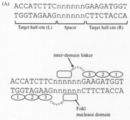
- FokI-ND has been shown to have the ability to bind to nucleic acids, but does not recognize the nucleotide sequence and cleaves the target nucleic acid by forming a dimer. Therefore, ZFN and TALEN require two target sequences for the nucleic acid binding domain, and FokI-ND acts in a spacer sequence sandwiched between the two target sequences, resulting in DSB.
- the two target sequences are referred to as target half-site L and target half-site R, respectively.
- ZFN is composed of FokI-ND, a nucleic acid binding domain consisting of zinc-finger protein (ZFP), and a linker (intermolecular linker) connecting them.
- ZFN is strongly restricted by the length of the spacer sequence and the nature of the linker, and requires some contrivance in its design.
- modification of the length of a spacer sequence or a linker has been reported (Non-Patent Document 1).
- the present inventors have searched for a nuclease domain different from FokI-ND, which is currently used as a standard for a nucleic acid cleavage domain of a genome editing enzyme, and found that the cleavage activity, specificity, cytotoxicity, target sequence selectivity, etc.
- the present inventors have found a novel nuclease domain that surpasses the existing FokI-ND in terms of functionality, and have completed the present invention.
- an artificial nucleic acid cleavage comprising a nuclease domain and a nucleic acid binding domain, which is a polypeptide comprising the amino acid sequence shown at positions 391 to 585 of SEQ ID NO: 1 or 389 to 579 of SEQ ID NO: 3, or a mutant polypeptide thereof enzyme.
- the artificial nucleic acid-cleaving enzyme according to (1) further comprising a linker located between the nuclease domain and the nucleic acid binding domain.
- An isolated nucleic acid comprising a nucleic acid sequence encoding the artificial nucleic acid-cleaving enzyme according to any one of (1) to (3).
- the method includes introducing the artificial nucleic acid-cleaving enzyme according to any one of (1) to (3), the nucleic acid according to (4), or the vector according to (5) into a cell. , A method of modifying a target nucleic acid.
- the artificial nucleic acid-cleaving enzyme, the nucleic acid, or the vector comprises two or more artificial nucleic acid-cleaving enzymes, a nucleic acid encoding the two or more artificial nucleic acid-cleaving enzymes, or the nucleic acid, a transcription product thereof, or a translation product thereof.
- the method according to the above (6) which is a vector comprising: (8) Modification of a target nucleic acid comprising the artificial nucleic acid-cleaving enzyme according to any one of (1) to (3), the nucleic acid according to (4), or the vector according to (5). Kit for.
- the artificial nucleic acid-cleaving enzyme, the nucleic acid, or the vector is two or more artificial nucleic acid-cleaving enzymes, the nucleic acid encoding the two or more artificial nucleic acid-cleaving enzymes, or the nucleic acid, its transcript, or its translation product
- the kit according to (8) which is a vector comprising: (10) A polypeptide comprising the amino acid sequence shown at positions 391 to 585 of SEQ ID NO: 1 or 389 to 579 of SEQ ID NO: 3, or a mutant polypeptide thereof.
- An isolated nucleic acid comprising a nucleic acid sequence encoding the polypeptide according to (10) or a mutant polypeptide thereof.
- a vector comprising the nucleic acid according to (11) or a transcription product or translation product thereof.
- the nuclease domain according to the present invention provides a nucleic acid cleaving action superior to conventional FokI-ND in functionality such as cleavage activity, specificity, and selectivity of a target sequence.
- a nucleic acid cleaving enzyme that has high activity and flexibility in target sequence and specifically cleaves a target sequence is provided.
- Such a nucleic acid cleaving enzyme is very useful as a genome editing tool.
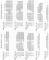
- FIG. 1 shows an amino acid sequence alignment of FokI nuclease domain homolog. Amino acid sequences were aligned using MAFFT.
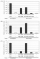
- Fig. 4 shows the cleavage activity of a nuclease domain on a target genome. CHO-K1 cells transfected with ZFA36-ND were incubated at 37 ° C., harvested after 72 hours, and then a genomic DNA template was prepared.
- the PCR product containing the ZFA36 target site was cleaved with GeneArt ⁇ Genomic ⁇ Clearage ⁇ Detection ⁇ Kit. Cleavage fragments were analyzed by electrophoresis (MultiNA system). Cleavage efficiency was calculated from the molarity of the uncleaved band and the larger cleaved fragment. In the figure, “FokI” means FokI-ND.
- the results of the cleavage activity test on the target genome of the nuclease domain performed in Example 3 are shown in terms of cleavage efficiency. 4 shows the results of Western blot analysis showing the expression level of ZFA36-ND. AcV5 tag-ZFA36-ND was expressed by HEK293T cells.
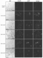
- FIG. 4 shows an LSM image of ZFA36-ND-EGFP in HEK293T cells. It is a microscope picture of a ZFA36-ND-EGFP overexpression cell. DNA was visualized by DAPI staining. Scale bar: 40 ⁇ m.
- “FokI” means FokI-ND.
- Figure 3 shows the reporter activity profiles of ZFA36-ND linker variants at different spacer lengths. 10 shows the results of the SSA reporter assay performed in Example 5.
- “FokI” means FokI-ND.
- Figure 2 shows the reporter activity profile of ZFA36-ND-Sharkey mutant.
- “FokI” means FokI-ND.
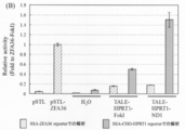
- Figure 4 shows nuclease activity in ZFN alone with heterodimeric mutation as measured by a cell-based SSA assay.
- HEK293T cells were co-transfected with each ZFN expression plasmid, reporter plasmid and reference plasmid to perform a dual luciferase assay. After transfection, cells were incubated for 24 hours, lysed, and analyzed for luciferase activity.
- FIG. 1 (A) hPGRN_ZFL1-6bp-hPGRN_ZFL1 reporter. (B) ZFA36-6bp-ZFA36 reporter.
- “FokI” means FokI-ND.
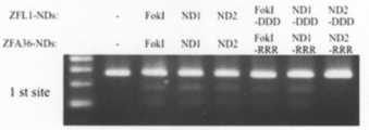
- Figure 3 shows analysis of homo- and hetero-dimer cleavage reactions by Cel-I assay.
- CHO-K1 cells transfected with each ZFN plasmid were incubated at 37 ° C. for 72 hours.
- genomic DNA was prepared from the transfected cells, and a fragment containing the ZFA36-ZFL1 target site was amplified.
- the PCR product was cut with GeneArt ⁇ Genomic ⁇ Clearage ⁇ Detection ⁇ Kit.
- the cleaved fragments were analyzed by agarose gel electrophoresis.
- “FokI” means FokI-ND.
- Figure 5 shows nuclease activity of hybrid ZFN as measured by a cell-based SSA assay.
- HEK293T cell based SSA assay HEK293T cells were co-transfected with each pair of ZFN expression plasmid, reporter plasmid and reference plasmid to perform a dual luciferase assay. After transfection, cells were incubated for 24 hours, lysed, and analyzed for luciferase activity. White and gray bars indicate the absence of ZFA36-FokI and ZFN, respectively.
- A ZFL1-FokI-DDD and appropriate nuclease domain.
- B ZFL1-ND1-DDD and a suitable nuclease domain.
- C ZFL1-ND2-DDD and a suitable nuclease domain.
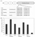
- FIG. 4 shows the results of detecting the nuclease activity of ZF-ND1 and ZF-FokI introduced into plant cells by electroporation using an SSA reporter assay.
- 12B is a graph of the result of FIG. 12A. The results of detecting the nuclease activity of ZF-ND1 and ZF-FokI introduced into plant cells by the protein transduction method using an SSA reporter assay are shown.
- nuclease domain 1 having a nuclease activity superior to FokI-ND (hereinafter, “ND1”)
- nuclease domain 2 hereinafter, referred to as "ND2"
- the amino acid sequence of the full-length protein containing ND1 (derived from Bacillus sp. SGD-V-76) is shown in SEQ ID NO: 1, and its nucleotide sequence is shown in SEQ ID NO: 2.
- the amino acid sequence of the full-length protein (derived from botulinum) containing ND2 is shown in SEQ ID NO: 3, and its nucleotide sequence is shown in SEQ ID NO: 4.
- ND1 is typically a partial peptide corresponding to positions 391 to 585 of SEQ ID NO: 1
- ND2 is typically a partial peptide corresponding to positions 389 to 579 of SEQ ID NO: 3.
- ND1 has 70% amino acid sequence identity with FokI-ND
- ND2 has 57% amino acid sequence identity with FokI-ND, but is a novel polypeptide isolated for the first time in the present invention.
- the present invention relates to a nuclease domain which is a polypeptide comprising the amino acid sequence shown at positions 391 to 585 of SEQ ID NO: 1 or 389 to 579 of SEQ ID NO: 3 or a mutant polypeptide thereof (hereinafter, the nuclease of the present invention) Domain).
- the nuclease domain of the present invention includes a nuclease domain that is a polypeptide consisting of the amino acid sequence shown at positions 391 to 585 of SEQ ID NO: 1 or 389 to 579 of SEQ ID NO: 3 or a mutant polypeptide thereof.
- the present invention relates to a polypeptide comprising the amino acid sequence shown at positions 391 to 585 of SEQ ID NO: 1 or 389 to 579 of SEQ ID NO: 3 or a variant polypeptide thereof, for example, positions 391 to 585 of SEQ ID NO: 1.
- a polypeptide consisting of the amino acid sequence shown at positions 389 to 579 of SEQ ID NO: 3 or a mutant polypeptide thereof as a nuclease domain.
- mutant polypeptide of a polypeptide comprising the amino acid sequence represented by positions 391 to 585 of SEQ ID NO: 1 or positions 389 to 579 of SEQ ID NO: 3 refers to positions 391 to 585 of SEQ ID NO: 1 or SEQ ID NO: No. 3 which has an amino acid sequence similar to the amino acid sequence shown at positions 389 to 579 and has nuclease activity.
- mutant polypeptide one or several amino acid residues are substituted, deleted, inserted or added in the amino acid sequence shown at positions 391 to 585 of SEQ ID NO: 1 or 389 to 579 of SEQ ID NO: 3.
- polypeptide having nuclease activity wherein one or several amino acids are substituted or deleted in the amino acid sequence shown at positions 391 to 585 of SEQ ID NO: 1 or 389 to 579 of SEQ ID NO: 3.
- a polypeptide consisting of a lost, inserted or added amino acid sequence and having nuclease activity can be mentioned.
- “1 to several” means, for example, 1 to 10, preferably 1 to 6, and for example, 1, 2, 3, 4, or 5.
- the variant polypeptide has at least 70%, at least 75%, at least 80%, or more, the amino acid sequence shown in positions 391-585 of SEQ ID NO: 1 or 389-579 of SEQ ID NO: 3. It may be a polypeptide comprising an amino acid sequence having at least 85%, at least 90%, at least 95%, at least 98% sequence identity and having nuclease activity, for example, positions 391-585 of SEQ ID NO: 1.
- a nuclease comprising an amino acid sequence encoded by a nucleotide sequence having a sequence identity of 65%, at least 70%, at least 75%, at least 80%, at least 85%, at least 90%, at least 95%, at least 98%; At least 50%, at least 55%, at least 60%, at least 65%, at least 70% of the polypeptide having activity and the nucleotide sequence shown at positions 1171-1755 of SEQ ID NO: 2 or 1165-1737 of SEQ ID NO: 4.
- At least 75%, less 80% even at least 85%, at least 90%, at least 95% consists of the amino acid sequence encoded by a nucleotide sequence having at least 98% sequence identity, and include polypeptides having nuclease activity.
- sequence identity of an amino acid sequence is determined by comparing two sequences aligned so as to maximize the sequence identity. Methods for determining a numerical value (%) of sequence identity are known to those skilled in the art. Any algorithm known to those skilled in the art (eg, BLAST algorithm, FASTA algorithm, etc.) may be used as an algorithm for obtaining optimal alignment and sequence identity.
- sequence analysis software such as BLASTP or FASTA.
- the above mutant polypeptide preferably has aspartic acid (hereinafter referred to as “D483”) at a position corresponding to position 483 in the amino acid sequence of FokI, and / or a position corresponding to position 487 in the amino acid sequence of FokI. May have an amino acid sequence having arginine (hereinafter, referred to as “R487”).
- the above mutant polypeptide preferably has aspartic acid, aspartic acid, and lysine (hereinafter, referred to as “D450” and “lysine”, respectively) at positions corresponding to positions 450, 467, and 469 in the amino acid sequence of FokI. D467 “and” K469 ").
- the variant polypeptide comprises an amino acid sequence having D483 and / or R487 and having D450, D467 and K469.
- D483 and R487 are amino acid residues that are considered to be essential when FokI-ND forms a dimer having cleavage activity, and are retained in the amino acid sequences of ND1 and ND2.
- analysis using amino acid substitution has revealed that D450 and D467 are involved in nucleic acid cleavage.
- sequences including K469 in the vicinity of these amino acids are motifs found in EcoRI and EcoRV, and it has been revealed that these amino acids are involved in the phosphodiester bond cleavage activity.
- D450, D467 and K469 are retained in the amino acid sequences of ND1 and ND2.
- the above mutant polypeptide has an aspartic acid (aspartic acid) at a position corresponding to positions 483, 487 and 496 in the amino acid sequence of FokI such that the nuclease domain of the present invention forms a heterodimer upon cleavage of nucleic acid.
- D483, D487 and D496 (hereinafter also referred to as "DDD mutant")
- arginine at a position corresponding to positions 483, 487 and 537 in the amino acid sequence of FokI.
- R483, R487, and R537 hereinafter, also referred to as “RRR-type mutant”.
- polypeptide of the present invention comprising the amino acid sequence shown at positions 391 to 585 of SEQ ID NO: 1 or 389 to 579 of SEQ ID NO: 3 or a mutant polypeptide thereof usually has a chain length of 500 amino acids or less. 400 amino acids or less, 300 amino acids or less, or 200 amino acids or less.
- the mutant polypeptide also includes a polypeptide containing a modified amino acid and / or an unnatural amino acid.
- the modified amino acids include, but are not limited to, for example, methylation, esterification, amidation, acetylation, alkylation, halogenation, and the like.
- the modified amino acids and unnatural amino acids can be introduced by known methods.
- the nuclease domain of the present invention has nuclease activity equal to or higher than that of FokI-ND.
- the nuclease domain of the present invention can be at least 0.8-fold, at least 0.9-fold, at least 1-fold, at least 1.3-fold, at least 1.5-fold, at least 1.8-fold, as compared to FokI-ND.
- Nuclease activity can be confirmed by a method known in the art.
- a nuclease domain into the ZFN system and measuring a nucleic acid cleavage activity, for example, a DNA cleavage activity by the SSA (single-strand annealing) method and the Cel-I method.
- SSA single-strand annealing
- the nuclease domain of the present invention has different properties from FokI-ND in flexibility in selecting a linker in an artificial nucleic acid-cleaving enzyme and flexibility in selecting a spacer serving as a target cleavage site (Example 5). ). Further, even when a Shakey mutation known to cause an increase in activity in FokI-ND was introduced into the nuclease domain of the present invention, no increase in activity was observed (Example 6). Further, ND1 of the present invention, unlike FokI-ND, did not decrease the nuclease activity even when a heterodimer was formed (Example 7). Thus, the nuclease domain of the present invention is a nuclease domain that has superior and / or different properties than conventional FokI-ND.
- the nuclease domain of the present invention may be an isolated nuclease domain.
- the present invention also provides an isolated nucleic acid encoding a nuclease domain of the present invention.
- isolated refers to a state separated from the natural world or a living body.
- the artificial nuclease of the present invention comprises the nuclease domain and the nucleic acid binding domain of the present invention.
- the artificial nuclease binds to a target sequence on the nucleic acid via the nucleic acid binding domain and cleaves the nucleic acid at the target cleavage site by the nuclease domain. Therefore, the artificial nuclease is a sequence-specific nuclease that can specifically cleave a target site in a nucleic acid.
- nucleic acid includes both DNA and RNA.
- nucleic acid cleaved by the artificial nucleic acid-cleaving enzyme of the present invention include double-stranded DNA, single-stranded DNA, and single-stranded RNA.
- DNA include, but are not limited to, eukaryotic nuclear genomic DNA, mitochondrial DNA, plastid DNA, prokaryotic genomic DNA, phage DNA, and plasmid DNA.
- the artificial nuclease of the present invention cleaves double-stranded DNA on the genome.
- RNA examples include, but are not limited to, a single-stranded RNA, a double-stranded RNA, and a DNA-RNA hybrid double-strand composed of a single-stranded DNA and a single-stranded RNA.
- the nucleic acid binding domain may be any protein domain that specifically binds to any nucleic acid sequence (target sequence), and includes, for example, but not limited to, zinc finger, TALE, CRISPR / Cas (Cas protein and guide RNA). Complex), Pentatriceptide @ repeat (PPR) and the like or a combination thereof.
- CRISPR / Cas include CRISPR-Cas9, CRISPR-Cpf1 (Cas12a), CRISPR-Cas12b, CRISPR-CasX (Cas12e), CRISPR-Cas14, CRISPR-Cas3, and the like.
- the nucleic acid binding domain containing a zinc finger may preferably contain two or more zinc fingers, but is not limited thereto, for example, a zinc finger array composed of 3 to 9 zinc fingers (hereinafter, “ZFA”). ").
- ZFA zinc finger array composed of 3 to 9 zinc fingers
- One zinc finger is known to recognize a three-base sequence, for example, a zinc finger that recognizes GNN, ANN, or CNN is known.
- CRISPR / Cas preferably, Cas having inactivated nuclease activity, for example, dCas (Cas having inactivated nuclease activity, for example, dCas9) is used.
- the nucleic acid binding domain is appropriately designed by those skilled in the art to bind to a desired target sequence.
- CRISPR / Cas means a complex of guide RNA and Cas protein, and the guide RNA recognizes and binds to a target sequence.
- the nuclease domain and the nucleic acid binding domain may be directly linked or may be linked via a linker.
- the linker is composed of, for example, two or more amino acid residues and is not particularly limited in length, but is, for example, 2 to 20 amino acids in length, for example, 2, 4, 6, 8, 9, 12, or It may be 16 or 20 amino acids long.
- the type of the linker is also not particularly limited, and examples thereof include TGAAAARA, GS, RPGEKP, and TGPGAAAARA.
- the linker and the length and type of the linker are appropriately selected by those skilled in the art in consideration of the type of the nucleic acid binding domain and the like. It is known that the nuclease activity of the conventional FokI-ND-containing ZFN is greatly affected by the linker sequence connecting ZF and FokI-ND. However, in the case of the artificial nucleic acid-cleaving enzyme of the present invention, it is higher than that of the conventional ZFN. Thus, the nuclease activity is less dependent on the linker sequence. Particularly, in ND1, almost no decrease in nuclease activity was observed by changing the linker sequence (Example 5). Therefore, the artificial nucleic acid-cleaving enzyme of the present invention has high flexibility of the linker sequence.
- the target sequence of the artificial nucleic acid-cleaving enzyme of the present invention is any sequence on the nucleic acid.
- the target sequence is preferably two sequences sandwiching a spacer sequence.
- the length of the spacer sequence, the length of the target sequence, and the base sequence are not particularly limited. Those skilled in the art can appropriately set a target sequence consisting of a desired length and a base sequence.
- the two target sequences may be palindromic to each other or non-palindromic. If the two target sequences are nonpalindromic sequences, two types of artificial nucleases targeting each sequence are prepared.
- a sequence located near the protospacer proximity motif (PAM) sequence is selected as a target sequence. That is, the target sequence is set such that the PAM is outside of both target sequences or in the spacer sequence.
- the target sequence may be a double-stranded sequence or a single-stranded sequence.
- the term “near” includes both an adjacent position and a nearby position.
- the nuclease domain when the nucleic acid binding domain binds to the target sequence on the nucleic acid, the nuclease domain cleaves the nucleic acid at the target cleavage site.
- the target cleavage site is in or near the spacer sequence adjacent to the target sequence.
- the length of the spacer sequence is not particularly limited, but is, for example, 1 to 20 bp, preferably 3 to 8 bp, and more preferably 5 to 7 bp. In the case of the conventional FokI-ND-containing ZFN, the optimal spacer length is 6 bp, and it is known that the nuclease activity is greatly affected by the spacer length.
- the flexibility of the spacer length is higher than that of the conventional ZFN.
- the artificial nucleic acid-cleaving enzyme of the present invention shows a high cleavage activity even for a short spacer or a long spacer, for example, by changing the linker length (Example 5).
- the linker length (Example 5)
- the cleavage activity of a spacer shorter or longer than the optimum spacer length is significantly reduced. Since the spacer length defines the distance between recognition sequences, the artificial nucleic acid-cleaving enzyme of the present invention has a wide range of target cleavage sites and recognition sequences due to the high flexibility of the spacer length.
- the artificial nucleic acid-cleaving enzyme of the present invention can be produced in vitro or in vivo by a method known in the art. For example, a method for artificial synthesis based on amino acid sequence information, a nucleic acid encoding an artificial nucleic acid-cleaving enzyme of the present invention, or an artificial nucleic acid encoding a nuclease domain and a nucleic acid binding domain of the present invention, and an appropriate expression vector And then introducing it into an appropriate host cell to express the artificial nucleic acid cleaving enzyme in the cell.
- RNA encoding the artificial nucleic acid-cleaving enzyme of the present invention is synthesized by in vitro transcription, introduced into a suitable host cell, and the artificial nucleic acid-cleaving enzyme is allowed to act in the cell.
- the expression vector is not particularly limited, and may be selected from vectors used in the art. For example, a plasmid vector, a virus vector, a phage vector, a phagemid vector, a BAC vector, a YAC vector, a MAC vector, a HAC vector and the like can be mentioned.
- the host cell is not particularly limited, and prokaryotes such as Escherichia coli, actinomycetes, and archaebacteria, and eukaryotes such as yeast, sea urchin, silkworm, zebrafish, mouse, rat, frog, tobacco, Arabidopsis, rice, and culture. Cells and the like.
- nucleic acid comprising a nucleic acid sequence encoding the artificial nuclease of the present invention, and a transcription product and a translation product of the nucleic acid.
- the nucleic acid may be an isolated nucleic acid comprising a nucleic acid sequence encoding the artificial nucleic acid-cleaving enzyme of the present invention. Also, codon optimization may be performed.
- nucleic acid includes both DNA and RNA.
- NHEJ non-homologous end-joining repair
- HR recombination repair
- a method of modifying a target nucleic acid comprising introducing into a cell the artificial nuclease or the nucleic acid encoding the artificial nuclease of the invention.
- the above enzyme or nucleic acid may be a transcription product of the nucleic acid or a translation product thereof.
- the nucleic acid may also have been codon optimized to be highly expressed in the cell.
- alteration includes deletion, insertion, or substitution of at least one nucleotide, or a combination thereof.
- the term “mutation" with respect to nucleic acids means nucleotide deletion, insertion, or substitution.
- the introduction of the above enzyme or nucleic acid into cells may be physical introduction or introduction via virus or organism infection, and various methods known in the art can be used.
- the physical introduction method include, but are not limited to, an electroporation method, a particle gun method, a microinjection method, a lipofection method, and a protein transduction method.
- the method of introduction via virus or organism infection include, but are not limited to, virus transduction, Agrobacterium method, phage infection, conjugation, and the like.
- a vector is appropriately used.
- the vector may be appropriately selected according to the type of cells to be introduced, and is not particularly limited.
- Examples thereof include a plasmid vector, a virus vector, a phage vector, a phagemid vector, a BAC vector, a YAC vector, a MAC vector, and a HAC vector.
- vectors such as liposomes and lentiviruses capable of carrying translation products (proteins) and transcripts (mRNA), and peptides such as cell-permeable peptides capable of introducing fused or bound molecules into cells Vector.
- a vector containing a nucleic acid encoding the artificial nucleic acid-cleaving enzyme of the present invention or a vector containing a transcription product of the nucleic acid or a translation product thereof.
- the vector containing the transcription product of the nucleic acid or the translation product thereof includes those in which the nucleic acid is transcribed in the vector containing the nucleic acid, and those in which the translation product is generated.
- the above-mentioned cells may be either prokaryotic cells or eukaryotic cells, and are not particularly limited.
- bacteria, archaebacteria, yeast, plant cells, insect cells, animal cells (eg, human cells, non-human cells, non-mammalian vertebrate cells, invertebrate cells, etc.) can be mentioned.
- the cell may also be an in vivo cell or an isolated cell, and may be a primary cell or a cultured cell.
- the cells may also be somatic, germ cells, or stem cells.
- prokaryotes from which the above cells are derived include, for example, Escherichia coli, actinomycetes, archaea, and the like.
- eukaryotes from which the cells are derived include, for example, yeast, mushrooms, fungi such as mold, sea urchins, humans, echinoderms such as sea cucumber, silkworms, insects such as flies, tuna, thailand, puffer fish, Fishes such as zebrafish, rodents such as mice, rats, guinea pigs, hamsters and squirrels, artiodactyls such as cattle, boars, pigs, sheep and goats, horseshoes such as horses, reptiles such as lizards, frogs etc.
- Amphibians such as rabbits, cats such as dogs, cats, ferrets, birds such as chickens, ostriches, quails, tobacco, Arabidopsis, rice, corn, bananas, peanuts, sunflowers, tomatoes, oilseed rape, wheat, barley, potatoes , Soybean, cotton, carnation and the like.
- animal cells include embryonic cells of embryos at each stage (eg, 1-cell stage embryo, 2-cell stage embryo, 4-cell stage embryo, 8-cell stage embryo, 16-cell stage embryo, morula embryo, etc.) Stem cells such as induced pluripotent stem (iPS) cells, embryonic stem (ES) cells, hematopoietic stem cells; fibroblasts, hematopoietic cells, neurons, muscle cells, bone cells, hepatocytes, pancreatic cells, brain cells, kidney cells Somatic cells such as cells; fertilized eggs and the like.
- iPS induced pluripotent stem
- ES embryonic stem
- fibroblasts hematopoietic stem cells
- neurons hematopoietic stem cells
- muscle cells hematopoietic stem cells
- hepatocytes pancreatic cells
- brain cells hepatocytes
- Somatic cells such as cells; fertilized eggs and the like.
- the target nucleic acid may be DNA of any gene or extragenic region in the genomic DNA in the cell.
- the nucleic acid binding domain contained in the artificial nucleic acid-cleaving enzyme of the present invention binds to a sequence near the target sequence (selected as a target sequence of the nucleic acid binding domain).
- the artificial nucleic acid cleaving enzyme of the present invention is designed.
- the desired sequence in the target nucleic acid is cleaved, for example, the expression of the gene is reduced or eliminated, or the function of the gene is reduced or the function is not expressed.
- a donor polynucleotide may be introduced into a cell in addition to the artificial nucleic acid-cleaving enzyme described above.
- the donor polynucleotide contains at least one donor sequence that contains the modification that one wants to introduce into the target cleavage site.
- a donor polynucleotide can be appropriately designed by those skilled in the art based on a technique known in the art.
- a donor polynucleotide is present in the target nucleic acid modification method of the present invention, homologous recombination repair occurs at the target cleavage site, and the donor polynucleotide is inserted into the site or the site is replaced with a donor sequence. As a result, the desired modification is introduced at the target cleavage site.
- the target cleavage site is repaired mainly by non-homologous end joining (NHEJ). Because NHEJ is error prone, deletion, insertion, or substitution of at least one nucleotide, or a combination thereof, can occur during repair of the cleavage. Thus, the nucleic acid is modified at the target cleavage site.
- NHEJ non-homologous end joining
- two or more kinds of the above-mentioned artificial nucleic acid-cleaving enzymes may be introduced into cells.
- the two or more artificial nucleases include two or more artificial nucleases having different nucleic acid binding domains, two or more artificial nucleases having different nuclease domains, both nuclease domain and nucleic acid binding domain.
- Examples include two or more different artificial nucleic acid-cleaving enzymes or two or more artificial nucleic acid-cleaving enzymes having different linker sequences.
- Two or more artificial nucleic acid-cleaving enzymes having different nucleic acid binding domains include two or more artificial nucleic acid-cleaving enzymes having different target sequences to which the nucleic acid-binding domain binds, and two or more artificial nucleic acid-cleaving enzymes having different nucleic acid-binding domains.
- Enzymes are included, and one example is two or more artificial nucleic acid cleavage enzymes having different nuclease domains and different target sequences to which the nucleic acid binding domains bind. For example, when a palindrome sequence does not exist in a region to be modified on a nucleic acid, two types of artificial nucleic acid cleaving enzymes each targeting a different sequence may be used.
- a mutation that promotes heterodimer formation of the nuclease domain may be further introduced into the nuclease domains of the two kinds of artificial nucleic acid-cleaving enzymes. This can increase the recognition sequences of the artificial nucleic acid-cleaving enzyme and lower the probability of off-target.
- Such mutations are, for example, substitutions with D483, D487 and D496 (hereinafter also referred to as “DDD type mutations”) and substitutions with R483, R487 and R537 (hereinafter also referred to as “RRR type mutations”).
- a DDD-type mutation is introduced into the nuclease domain of one artificial nucleic acid-cleaving enzyme, and an RRR-type mutation is introduced into the nuclease domain of the other artificial nucleic-acid-cleaving enzyme.
- the nuclease domain sequence before mutation introduction already has any of the amino acid residues of the above-mentioned DDD mutation and RRR mutation, only the other amino acid substitutions may be performed.
- Two types of nuclease domains into which a pair of mutations eg, DDD type mutation and RRR type mutation that promote the formation of such a heterodimer are introduced may be the same nuclease domains except for the above mutations.
- it may be a nuclease domain that is different from before the introduction of the mutation.
- an artificial nuclease that includes a DDD mutant of ND1 and an artificial nuclease that includes an RRR mutant of ND1 may be used in the method of the present invention.
- an artificial nucleic acid cleaving enzyme containing a DDD mutant of ND2 and an artificial nucleic acid cleaving enzyme containing an RRR mutant of ND1 may be used in the method of the present invention.
- a combination of the DDD mutant of ND1 and the RRR mutant of ND1 or the combination of the DDD mutant of ND2 and the RRR mutant of ND1 exhibited particularly high specificity and nuclease activity. (Example 8).
- Kit for modifying a target nucleic acid, the artificial nucleic acid-cleaving enzyme of the present invention, a nucleic acid encoding the artificial nucleic acid-cleaving enzyme, or a vector containing the nucleic acid, a transcription product of the nucleic acid or a translation product thereof.
- Kits are provided. Further, the kit may include two or more kinds of artificial nucleic acid-cleaving enzymes as described above. Furthermore, the kit may include a nucleic acid encoding the two or more artificial nucleic acid-cleaving enzymes, or a vector containing the nucleic acid, a transcription product of the nucleic acid, or a translation product thereof.
- the kit of the present invention comprises one or two artificial nucleic acid-cleaving enzymes, a nucleic acid encoding the one or two artificial nucleic-acid-cleaving enzymes, or the nucleic acid, a transcription product of the nucleic acid, or a translation product thereof. Containing vectors.
- the two or more artificial nucleic acid-cleaving enzymes may be, for example, an artificial nucleic acid-cleaving enzyme containing a DDD-type mutant of ND1 and an artificial nucleic acid-cleaving enzyme containing an RRR-type mutant of ND1.
- the two or more artificial nucleic acid-cleaving enzymes may be, for example, an artificial nucleic acid-cleaving enzyme containing a DDD-type mutant of ND2 and an artificial nucleic acid-cleaving enzyme containing an RRR-type mutant of ND1.
- the kit may further comprise one or more additional reagents, such as dilution buffers, reconstitution solutions, wash buffers, nucleic acid transfer reagents, protein transfer reagents, control reagents. But not limited thereto. Usually, an instruction manual is attached to the kit.
- HEK293T cells were maintained in a D-MEM medium (Wako Pure Chemical Industries) containing 10% FBS (Difco), 1% streptomycin-penicillin (Wako Pure Chemical Industries), and 1% NEAA (Difco).
- D-MEM medium Wako Pure Chemical Industries
- CHO-K1 cells were maintained with Ham's-F12 (Difco) containing 10% FBS, 1% streptomycin-penicillin, 5% kanamycin (Wako Pure Chemical Industries).
- the culture temperature was maintained at 37 ° C. and a carbon dioxide concentration of 5%, and the temperature was followed when indicated.
- each medium described above was used unless otherwise described.
- Example 1 Search for a novel nuclease domain Based on the amino acid sequence at the C-terminal side of FokI, a natural material having homology was searched for by Protein-protein BLAST. In the obtained search results, four types of candidates were arbitrarily selected from those having a homology with FokI-ND of 35% to 70%.
- ND1 amino acid sequence identity 70%; full length amino acid sequence: SEQ ID NO: 1; corresponding nucleotide sequence: SEQ ID NO: 2), ND2 (amino acid sequence identity 57%; full length amino acid sequence: SEQ ID NO: 3) Corresponding nucleotide sequence: SEQ ID NO: 4), ND3 (amino acid sequence identity 49%; full length amino acid sequence: SEQ ID NO: 5; corresponding nucleotide sequence: SEQ ID NO: 6), ND4 (amino acid sequence identity 45%; full length amino acid sequence: sequence) No. 7; corresponding base sequence: SEQ ID NO: 8).
- ND1 to ND4 are also involved in the cleavage of amino acids (D483, R487), which are considered to be essential when FokI-ND forms a dimer having cleavage activity, and the cleavage of DNA phosphoryl diester bonds by hydrolysis.
- the amino acids putatively involved (D450, D467 and K469) were retained (FIG. 1B). Therefore, ND1 to ND4 were considered to have nuclease activity.
- Example 2 Cleavage activity of novel nuclease domain (SSA method) To evaluate the nuclease activity of ND1 to ND4, a ZFN plasmid (pSTL-ZFA36-ND) containing each of ND1 to ND4 was prepared, and the cleavage activity of each novel nuclease domain was measured by an SSA reporter assay.
- SSA method Cleavage activity of novel nuclease domain
- ZFN plasmid The nucleotide sequences of ND1 to ND4 were codon-optimized and artificially synthesized by IDT.
- the ZFN plasmid (pSTL-ZFA36) used a construct prepared by Ochiai et al. (Ochiai et al. (2010) Targeted mutagenesis in the sea urchin embryo using zinc-finger nucleases. Genes to Cells 15: 875-885).
- PCR was performed with PrimeSTAR Max (Takara) using each nuclease domain replacement primer pair. PCR was performed at 98 ° C.
- FIG. 1A shows a schematic view of the obtained ZFA (zinc finger array) 36-ND.
- the ZFA36-ND has a TGAAAARA linker.
- SSA reporter assay 7.2 ⁇ L of plasmid solution containing 120 ng of SSA reporter plasmid, 240 ng of each of the above pSTL-ZFA36-ND plasmids, and 24 ng of reference plasmid pRL-CMV (Promega) (plasmid for standardizing transduction efficiency) was prepared.
- the SSA reporter plasmid was prepared according to a known method such that a target sequence of a ZF array (ZFA36) containing a 6 bp spacer sequence was inserted so as to be sandwiched between overlapping regions of Luc sequences (Ochiai et al.). . (2010) Targeted mutagenesis in the sea urchin embryo using zinc-finger nucleases.
- the HEK293T cells were dissociated from a 100 mm tissue culture dish (Iwaki) by trypsinization, centrifuged at 1000 rpm for 3 minutes, the supernatant was removed, and the cells were suspended again in 5 mL of the culture solution. 10 ⁇ L of the cell suspension was collected and the number of cells was counted. From the obtained cell number, a culture solution was added so that the cell number in the cell suspension was 6 ⁇ 10 5 cells / mL. 100 ⁇ L of the cell suspension was added to the 96-well plate left at room temperature for 30 minutes as described above. Thereafter, the 96-well plate was transferred to 37 ° C. at a carbon dioxide concentration of 5% and cultured to perform transduction.
- ND1 and ND2 are novel nuclease domains derived from natural materials that surpass the FokI-ND cleavage activity conventionally used in ZFN.
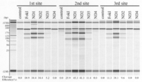
- Example 3 Cleavage activity of novel nuclease domain on target genome (Cel-I method)
- the genome of the novel nuclease domain in cultured cells was determined by Cel-I assay.
- the mutation introduction rate to DNA was analyzed.
- FokI-ND, ND3 and ND4 were similarly analyzed.
- Cel-I assay In cells transduced with a genome editing tool such as TALEN or ZFN, cleavage of the target DNA by the nuclease domain of the cells and repair of the cleavage site by a repair mechanism existing in the cell are performed. In this process, a repair error occurs.
- a region containing the target sequence of the genomic DNA is amplified by PCR, a mixed product of a PCR fragment of a wild-type allele and a PCR fragment containing a mutation is obtained.
- a double-stranded DNA containing a mismatch consisting of a PCR fragment of the wild-type allele and the allele containing the mutation is generated, which is cleaved by a mismatch-specific endonuclease (Cel-I nuclease). Mutagenesis can be evaluated by detecting the cleavage pattern.
- the CHO-K1 cells were dissociated by trypsin treatment, centrifuged at 1000 rpm for 3 minutes, the supernatant was removed, and the cells were suspended in a 10 mL culture solution. The number of cells was measured using 10 ⁇ L of the cell suspension, a culture solution was added so that the number of cells became 1 ⁇ 10 5 cells / mL, and the cells were cultured overnight at 37 ° C. and a carbon dioxide concentration of 5%.
- Each ZFN plasmid prepared in Example 2 was added to 500 ⁇ L of Opti-MEM (Difco) at 800 ng and left at room temperature for 5 minutes.
- 32 ⁇ L of Lipofectamine @ LTX was added to 500 ⁇ L of Opti-MEM, and the whole amount was mixed with an Opti-MEM medium containing the above plasmid, and left at room temperature for 30 minutes.
- the culture solution of the CHO-K1 cells grown overnight was replaced with 4 mL of Opti-MEM.
- the total amount of the plasmid solution left at room temperature for 30 minutes was added to the cell culture solution, and the cells were cultured overnight at 37 ° C. and a carbon dioxide concentration of 5%.
- the culture solution was changed from Opti-MEM to 10 mL of the culture solution, and the culture was further continued at 37 ° C. and 5% carbon dioxide concentration. Further, after 24 hours, puromycin (Wako Pure Chemical Industries) was added to a final concentration of 500 ⁇ g / mL. Thereafter, cultivation was performed for 3 days every day while changing the culture medium and adding puromycin.
- Genomic DNA was prepared from the obtained cells by either a DNA purification kit (Qiagen) or a treatment with a cell lysis buffer and a proteolytic agent (Protein degrader) contained in GeneArt ⁇ Genomic ⁇ Clearage ⁇ Detection ⁇ Kit (Life ⁇ Technologies). Using the prepared genomic DNA as a template, PCR was performed using KOD Fx Neo (ToYoBo) or AmpliTaq Gold (Life Technologies). In the case of KOD Fx Neo, after heating at 95 ° C.
- the target was amplified by performing 40 cycles of 95 ° C. for 30 seconds, 60 ° C. for 30 seconds, and 68 ° C. for 30 seconds.
- AmpliTaq @ Gold after the treatment at 95 ° C. for 10 minutes, 40 cycles of 95 ° C. for 10 seconds, 60 ° C. for 30 seconds, and 72 ° C. for 40 seconds are performed, and finally the treatment is performed at 72 ° C. for 7 minutes.
- the target was amplified. An equal amount of each of the obtained PCR products was subjected to a cleavage reaction using a detection enzyme according to a manual using GeneArtGenomic ⁇ Clearage ⁇ Detection ⁇ Kit.
- cleavage fragments were detected by agarose electrophoresis or MultiNA (Shimadzu Corporation). Cleavage efficiency was calculated from the molarity of the uncleaved band and the larger cleaved fragment.
- Table 1 shows candidate sequences of ZFN genomic targets (ZFA36-ZFA36) in CHO-K1 cells used for the analysis. Table 1 also shows candidate sequences of the ZFN genomic target (ZFL1-ZFA36) used in Example 7 described later.
- the target half site (L) was completely identical to the target sequence of the ZF array.
- the target half site (R) did not completely match the target sequence of the ZF array, and a mismatch was present on the 3 'side.
- the cleavage efficiency of the novel nuclease domains ND1 and ND2 is about 1.5 times higher than that of FokI-ND, as in the result of the luciferase reporter analysis. (FIGS. 2A and B).
- the mutation introduction activity of ND1 was higher than that of ND2 in all analyzed target candidate sequences.
- ND1 and ND2 show higher activity than the conventional FokI-ND for the target genomic sequence in vivo, similarly to the result of the reporter analysis using luciferase luminescence of Example 2 , A novel nuclease domain derived from a natural material.
- ND3 although the mutagenesis activity was changed depending on the target candidate sequence, it was about 25 to 45% of the activity of FokI-ND. In addition, in ND4, no mutagenesis to the detectable target candidate sequence was observed in any of the analyzed target candidate sequences.
- Example 4 Analysis of the amount of accumulation of the novel nuclease domain in the cell
- the mutation introduction rate of the novel nuclease domain is higher than that of FokI-ND because the amount of accumulation in the cell is larger than that of FokI-ND.
- the amount of accumulation of each nuclease domain was analyzed by adding a tag to ZFN and using an antibody that recognizes the tag.
- a construct in which a new AcV5 tag was added to the N-terminus of ZFN was prepared and used for analysis.
- the cultured HEK293T cells were treated with trypsin, centrifuged, and the supernatant was removed.
- the obtained precipitate is suspended in PBS, and a sample treatment solution consisting of 2% SDS, 10% glycerol, 10 mM ⁇ DTT, 0.005% BPB and 62.5 mM Tris-HCl (pH 6.8) is added, and the mixture is heated at 95 ° C. for 5 minutes. Boiled. Thereafter, the nucleic acid and the like were cut by ultrasonic treatment.
- the obtained protein sample was subjected to protein quantification using a protein assay kit (Bio-Rad) using BSA as an external standard.
- Each sample prepared to be 10 ⁇ g was separated by SDS-PAGE, and transferred to a PVDF membrane by semi-dry transfer.
- the PVDF membrane after transfer was blocked with PBS containing 5% skim milk, and incubated with ⁇ -tubulin antibody (Abcam) and AcV5-tag antibody (Sigma) diluted 2000 times. After washing with PBST, the plate was incubated with a 2000-fold diluted goat anti-mouse IgG antibody conjugate HRP (Bio-Rad). After washing with PBST, light was emitted using Supersignal West Dura Extended Duration Substrate (Thermo Fisher) and exposed to an X-ray film.
- the amount of FokI-ND, ND1 and ND2 nuclease domains accumulated in the cells is the largest in FokI-ND, and then in ND1, ND2 It was suggested that they were accumulated in this order.
- the signal derived from the AcV5 tag was obtained significantly stronger than that of FokI-ND or the like, indicating that the amount of ND3 accumulated in the cells was larger than that of FokI-ND or the like.
- PCR PCR was performed using each pSTL-ZFA36-ND prepared in Example 2 as a template, and after In-Fusion reaction, E. coli was transformed to obtain each ZFN plasmid having EGFP added to the C-terminal side of the ZF array.
- a 7.2 ⁇ L plasmid solution containing 240 ng of the ZFN plasmid was prepared.
- the plasmid solution was transduced into HEK293T cells in the same manner as in the SSA assay described in Example 2, and cultured at 37 ° C. at a carbon dioxide concentration of 5% for 3 days.
- HEK293T cells were applied to collagen-treated 24-well glass bodom plates.
- FokI-ND-EGFP When FokI-ND-EGFP was expressed, some cells showed a large amount of GFP fluorescence in the cytoplasm, and some cells also showed nuclei in the nucleus.
- ND1-EGFP When ND1-EGFP was expressed, GFP-derived fluorescence was observed in more cells than in FokI-ND, but the intensity of each fluorescence was weaker than in FokI-ND.
- ND2-EGFP it was found that in the observed cells, the number of cells having GFP-derived fluorescence was smaller than that in FokI-ND, but was localized in the cytoplasm and nucleus.
- ND3-EGFP When ND3-EGFP was expressed, GFP-derived fluorescence was observed in the nucleus and cytoplasm as in FokI-ND and the like.
- Example 5 Selectivity analysis of target sequence of novel nuclease domain
- modification of the linker sequence between the ZF array and the nuclease domain was attempted.
- modification of the linker sequence results in a difference in the length of a spacer sequence exhibiting cleavage activity and in the toxicity given to cells. Therefore, in addition to the conventional linker sequence TGAAAARA linker used in the analysis so far, a GS linker, an RPGEKP linker, and a ZFN vector modified to a TGPGAAARA linker were constructed and used for the analysis.
- FokI-ND When the length of the spacer sequence is 6 bp, FokI-ND has 20% or less when modified to a GS linker or RPGEKP linker compared to the cleavage activity when a conventional linker (TGAAARA) is used to connect a ZF array and a nuclease domain. Activity decreased. On the other hand, when modified to TGPGAAARA linker, the FokI-ND cleavage activity was about 90% before the linker modification (FIG. 5A).
- ⁇ ND1 when the spacer sequence was 6 bp, had no effect on the cleavage activity even when the linker sequence was modified, and had about 90% of the cleavage activity of FokI-ND before modification of the linker sequence.
- ND2 when the spacer sequence was 6 bp, the cleavage activity was reduced to about 60% when the GS linker sequence was modified and to about 35% when modified to the RPGEKP linker sequence, compared to the FokI-ND cleavage activity before the linker sequence modification. . From these results, it has been clarified that, as reported so far, even if the length of the spacer sequence is the same, the linker sequence used in the analysis affects the nuclease domain cleavage activity.
- the cleavage activity of FokI-ND is reduced to about 40% with the conventional linker sequence (TGAAARA linker) as compared with the case of 6 bp, but is reduced by about 50% with the GS linker. Cleavage activity remained.
- the FokI-ND cleavage activity was about 20% of that of the conventional linker sequence, and almost no cleavage was possible.
- ND1 when the spacer sequence was 5 bp, the cleavage activity remained high when modified to a GS linker. However, the cleavage activity was reduced to about 30% with the conventional linker sequence, and the cleavage activity was changed to other linker sequences. Then, it became clear that it could hardly be cut.
- ND2 showed almost no cleavage activity even when the linker sequence was modified when the length of the spacer sequence was 5 bp.
- the influence of the linker sequence was large, and it was considered that the shorter the linker sequence, the more easily the cleavage activity was maintained.
- FokI-ND showed a cleavage activity only when modified to a TGPGAAARA linker, but this activity was due to the cleavage of a conventional linker sequence with a spacer sequence of 6 bp in length. It was about 50% of the activity. When converted to another linker sequence, it showed little cleavage activity.
- ND1 was found to show no cleavage activity when modified to a GS linker sequence when the length of the spacer sequence was set to 7 bp, but to show about 50% or more cleavage activity when modified to another linker sequence.
- the TGPGAAATA linker sequence was considered to be higher than the FokI-ND cleavage activity of interest.
- ND2 showed a cleavage activity of about 40% with a conventional linker sequence, but showed little cleavage activity when modified to another linker sequence. When the length of the spacer sequence was long, the longer the linker sequence, the higher the cleavage activity tended to be.
- ND1 is less susceptible to the modification of the linker sequence and has a lower restriction on the spacer sequence than FokI-ND, and exhibits higher cleavage activity than FokI-ND. This indicates that ND1 is a nuclease domain that is more versatile than FokI-ND in terms of target sequence selectivity and the like.
- ZFN has reported on the modification of FokI-ND. It has also been reported that the nuclease domain itself is changed from FokI-ND to an endonuclease such as PvuII or I-TevI.
- the modified ZFN using I-TevI has a higher cleavage activity than FokI-ND, but has a spacer sequence of about 30 bp due to the ZF array, the intramolecular linker and DNA binding motif present in I-TevI. , And it becomes longer than that of FokI.
- PvuII the spacer sequence is longer.
- the DNA sequence acting on ZFN is also considered to be longer and more susceptible to restriction by the base sequence.
- the nuclease domain of the present invention when used in ZFN, has a high mutagenesis rate despite having the same protein structure as conventional FokI-ND, and the target DNA sequence acting on ZFN is reduced to 24 bp in total. .
- the nuclease domain of the present invention is less restricted by the nucleotide sequence than I-TevI or the like, and can be easily handled as a simpler genome editing tool.
- Example 6 Cleavage activity of a novel nuclease domain into which a Sharkey mutation has been introduced
- An amino acid substitution (Sharkey mutation) that increases the cleavage activity when introduced into FokI-ND is known (Guo, J., Gaj, T. and Barbas, CF, 3rd. (2010) Directed evolution of an enhanced and highly efficient FokI cleavage domain for zinc finger nucleases. J Mol Biol, 400, 96-107). Therefore, in order to further enhance the cleavage activity in ND1 and ND2 of the novel nuclease domains, an attempt was made to introduce the same amino acid substitution as the Sharkey mutation, respectively.
- ND1 the amino acids corresponding to the positions of S418 and K441 of FokI-ND, which are the sites of the amino acids necessary for the introduction of the Sharkey mutation, were the same as FokI-ND, S424 and K447, respectively.
- the amino acid corresponding to the position of S418 of FokI-ND was the same as the Sharkey mutation (P422), but the amino acid corresponding to the position of K441 of FokI-ND was S445. Therefore, ND1 and ND2 in which the same amino acid substitution as in the Sharkey mutation was introduced at the above amino acid positions were designated as ND1-Sharkey and ND2-Sharkey, respectively.
- Example 7 Cleavage activity using heterodimeric nuclease domain
- FokI-ND forms a dimer.
- FokI-ND can be functionally converted into a DDD / RRR type mutant or the like that shows cleavage activity only during heterodimer formation by substituting a specific amino acid. Therefore, further analysis was performed to clarify whether it is possible to modify the functions of the novel nuclease domains ND1 and ND2 so as to maintain the cleavage activity.
- the target half site-L and the target half site-R which are the target sequences of the ZF array, have different sequences.
- a construct was constructed using a ZF array having a target sequence different from the ZF array used in the previous examples.
- hPGRN_ZFL1 was artificially synthesized by IDT (hereinafter, also referred to as “ZFL1”).
- ZFL1 PCR was performed using PrimeSTAR @ Max with each pSTL-ZFA36-ND as a template using a ZF array replacement primer pair.
- the obtained PCR product and hPGRN_ZFL1 were reacted using In-Fusion to transform Escherichia coli. Plasmids were extracted from the obtained transformants, and the nucleotide sequence was confirmed, whereby each pSTL-hPGRN_ZFL1-ND plasmid of interest was obtained.
- primers were prepared using each pSTL-hPGRN_ZFL1-ND as a template. PCR was performed with PrimeSTAR @ Max using a pair. In addition, in order to convert the function to the RRR type, PCR was similarly performed using each pSTL-ZFA36-ND as a template. Each of the obtained PCR products was transformed into E. coli after the In-Fusion reaction.
- Plasmids were extracted from each of the obtained transformants, and after confirming the sequence, target constructs (hereinafter, also referred to as “DDD-type mutant ZFL1-ND” and “RRR-type mutant ZFA36-ND,” respectively) were prepared.
- DDD-type mutant ZFL1-ND and each RRR-type ZFA36-ND
- an SSA reporter assay was performed as described in Example 2, and the nuclease activity of each ZFN was measured.
- the amount of each ZFN plasmid in 7.2 ⁇ L of the plasmid solution used in the SSA reporter assay was changed to 120 ng for the target half site L and 120 ng for the target half site R.
- nuclease activity was measured by Cel-I assay in the same manner as described in Example 3.
- a homodimer was formed using a construct in which ZFL1 was linked to a nuclease domain, and the cleavage activity of the target sequence (ZFL1-ZFA36) on the reporter was measured.
- ND1 had a cleavage activity of about 40%.
- FokI-ND and ND2 showed almost no cleavage activity (FIG. 8A).
- ZFA36 ZF array
- cleavage activity was also observed in FokI-ND and ND2. They were about 35% and about 50%, respectively.
- ND1 had higher cleavage activity, about 90% (FIG. 8A).
- ND1 and ND2 were analyzed in the same manner as when analyzed only with ZFA36. Became higher than FokI-ND (FIG. 8A). This also indicates that ND1 and ND2 have high cleavage activity and are universal regardless of the ZF array.
- a target sequence ZFL1-ZFL1 or ZFA36-ZFA36
- the cleavage activity was measured.
- a DDD mutation was introduced into each nuclease domain linked to ZFL1
- an RRR mutation was introduced into each nuclease domain linked to ZFA36.
- DDD mutation ZFL1-ND and RRR mutation ZFA36-ND were found. In each case, almost no cleavage activity was observed alone (FIGS. 7A and 7B). This demonstrates that homodimers cannot be formed in the novel nuclease domains ND1 and ND2 by introducing an amino acid substitution to DDD / RRR as in FokI-ND.
- ND1 is capable of functional conversion to a heterodimeric form that maintains high activity, and that conversion to a heterodimeric form can significantly alter the structure of ND2. , Suggesting that their respective structural properties are different from FokI-ND.
- Example 8 Effect on cleavage activity using combination of heterodimeric nuclease domains Exhibiting higher cleavage activity than conventional FokI-ND by changing the combination of nuclease domains during heterodimer formation Further analysis was performed to determine whether or not it became possible.
- DDD-type mutant ND2 shows high cleavage activity only in combination with RRR-type mutant ND1, and RRR-type mutant FokI-ND or RRR-type mutant ND2 In the combination with, the cleavage activity was almost lost (FIG. 9C). As a result, it was clarified that when the RRR-type mutant ND2 was made into a heterodimer type, the cleavage activity was lost regardless of the combination of nuclease domains.
- Example 9 Analysis of Mutagenesis by ZFN at Base Sequence Level It was examined what kind of change occurs in the base sequence due to mutations introduced into the target sequence by ZFN.
- coli transformants were randomly selected to extract each plasmid.
- the M13-f primer was added so that the obtained plasmid had a final concentration of 400 ng and 400 ng, and PCR and sequence analysis were requested to Fasmac. From the obtained sequence, the sequence of the mutant sequence of the target sequence in each ZFN and the ratio thereof were calculated.
- ND1 2 clones and 2 clones were obtained for each of the 17 clones analyzed. In addition, three clones with four base insertions were obtained, and six clones with two base insertions were obtained most in this analysis.
- ND2 out of the 16 clones analyzed, one clone with one base insertion was obtained, and two clones with two base insertions and five clones with the most four base insertions were obtained. In addition, one clone having a large insertion of 71 bases was obtained.
- ND3 unlike the first site, a mutation was introduced into the target sequence at the second site, and two clones were inserted, two bases deleted, and eight bases deleted in each of the 16 clones analyzed.
- all of the 14 clones that could be analyzed did not show mutations in the target sequence at the 2nd site as well as at the 1st site.
- Example 10 Cleavage activity of a novel nuclease domain in combination with a nucleic acid binding domain other than ZFA It is determined whether the novel nuclease domain of the present invention has a higher cleavage activity than FokI-ND even when a nucleic acid binding domain other than ZFA is used.
- an artificial nucleic acid cleaving enzyme containing ND1 and TALE was prepared, and its cleavage activity was measured.
- TALE-FokI-ND and TALE-ND1 were produced using Platinum TALEN (doi: 10.1038 / srep03379) technology developed in the laboratory of the inventor, and their cleavage activities were evaluated by SSA reporter assay. did.
- a plasmid was extracted from the obtained transformant, and a target TALE-ND1 destination vector (a vector in which FokI-ND in ptCMV-153 / 47-VR-NG was replaced with ND1). ) Got.
- constructs for TALEN (TALE-FokI) retaining conventional FokI-ND, constructs (TALE-ROSA26-FokI and TALE-HPRT1-FokI) incorporating a nucleic acid binding module were similarly prepared.
- SSA reporter plasmid those containing the respective target sequences (SSA-CHO-ROSA26 reporter and SSA-CHO-HPRT1 reporter) were prepared by a conventional method.
- the target sequences for the ROSA26 locus and the HPRT1 locus are shown in Table 4 below.
- TALE-FokI-ND and TALE-ND1 The TALEN plasmids (TALE-FokI-ND and TALE-ND1) prepared in the above (2) and incorporating a nucleic acid binding module for recognizing the target sequence, and the target sequence HEK293T cells were transduced with Lipofectamine LTX using the integrated SSA reporter plasmid, and SSA reporter assays were performed in the same manner as in Example 2 using the cells 24 hours after transduction.
- measurement was similarly performed using a ZFN plasmid containing FokI-ND (pSTL-ZFA36) and a plasmid pSTL expressing only FokI-ND.
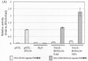
- the cleavage activity of each TALE-ND was calculated as a relative activity when the value of the cleavage activity of ZFA36-FokI for a reporter containing the target sequence of ZFA36 was set to 1.
- FIGS. 11A and 11B show the results obtained when the sequence derived from the ROSA26 locus was targeted, and FIG. 11B shows the results obtained when the sequence derived from the HPRT1 locus was targeted.
- TALE-ROSA26-FokI in which the nuclease domain of Platinum @ TALEN is a conventional FokI-ND, has a reporter cleavage activity targeting a sequence derived from the ROSA26 locus, and ZFA36 targeting a ZFA36 reporter. -It was revealed that it had an activity close to the cleavage activity of FokI.
- TALE-ROSA26-ND1 in which the nuclease domain of Platinum @ TALEN was replaced with ND1
- the cleavage activity of a reporter targeting a sequence derived from the ROSA26 locus is about twice as high as that of TALE-ROSA26-FokI. It became clear.
- TALE-ND1 when targeting a sequence derived from the ROSA26 locus of the endogenous genome of CHO-K1 cells, TALE-ND1 has a higher cleavage activity than TALE-FokI, whose nuclease domain is a conventional FokI-ND. Indicated.
- TALE-ND1 using ND1 as the nuclease domain of Platinum TALEN exhibits higher cleavage activity than TALE-FokI having a conventional FokI-ND.
- novel nuclease domain of the present invention is a nuclease domain having more excellent cleavage activity than FokI-ND even when other nucleic acid binding domains are used in addition to ZFN.
- novel nuclease domain of the present invention has the following main features.
- ND1 and ND2 of the present invention have a remarkably higher cleavage activity than FokI-ND.
- ND1 shows a cleavage activity exceeding that of FokI-ND in ZF arrays recognizing different sequences and in TALE, the effect of increasing the cleavage activity by ND1 is universal.
- ND1 shows the same level of cleavage activity as a 6 bp spacer even with a 5 bp or 7 bp spacer. Therefore, when ND1 is dimerized, FokI- It has higher flexibility than ND and can relax the restriction of the target sequence.
- Example 11 Preparation of protein in Escherichia coli (1) Construction of expression vector for ZF-ND1 and ZF-FokI The XhoI site and SalI site of the pET-MCS plasmid containing a His tag at the N-terminus were cut to obtain a ZF-ND1 fragment. Inserted.
- the ZF-ND1 fragment contains a nuclear localization signal NLS, a Zinc-Finger (hereinafter abbreviated as ZF), and a nuclease domain ND1.
- ZF Zinc-Finger
- ND1 nuclease domain ND1.
- ZF Zinc-Finger
- ZFL1 which recognizes a DNA sequence GAAGGTGAC of 12 base pairs. Therefore, pET-ZF (ZFA36A) ND1 and pET-ZF (ZFL1) ND1 were prepared as plasmids expressing ZF (ZFA36A) -ND1 and ZF (ZFL1) -ND1, respectively.
- plasmids in which the nuclease domain of the restriction enzyme FokI (hereinafter abbreviated as FokI) was inserted instead of ND1, pET-ZF (ZFA36A) -FokI, and pET-ZF (ZFL1) -FokI were prepared.
- FokI the nuclease domain of the restriction enzyme
- ZFA36A pET-ZF
- ZFL1 pET-ZF
- E. coli was dissolved in a lysis buffer (20 mM Tris-HCl, 500 mM NaCl, 10% glycerol, 10 mM imidazole, 1 mM benzylsulfonyl fluoride, 1 mM dithiothreitol, pH 8.0). And dissolved. Subsequently, the protein was adsorbed to a nickel NTA column, and impurities were removed using a washing buffer (20 mM Tris-HCl, 500 mM NaCl, 10% glycerol, 20 mM imidazole, pH 8.0).
- the protein was eluted using an elution buffer (20 mM Tris-HCl, 500 mM NaCl, 10% glycerol, 500 mM imidazole, pH 8.0). A part of the eluted protein was dissolved in SDS sample buffer for analysis of protein yield. The eluted protein was purified using gel filtration chromatography HiPrep 16/60 Sephacryl S-200 HR (GE healthcare, Illinois, USA) and buffer A (20 mM HEPES, 150 mM NaCl, 10% glycerol, pH 7.4), After freezing instantly using liquid nitrogen, it was stored at -80 ° C.
- the protein fraction eluted with imidazole and the finally purified protein were dissolved in SDS sample buffer and subjected to electrophoresis by SDS-PAGE.
- SDS-PAGE a known concentration of BSA protein is electrophoresed in another lane, the gel after PAGE is stained with Coomassie, and the band of the target protein molecular weight is quantified by ChemiDoc XRS +, and the protein yield is analyzed from the molecular weight of the protein. did.
- Example 12 Activity of ZF-ND1 and ZF-Fok1 in plant cells (1) Construction of reporter cells To evaluate the activity of ZF-ND1 or ZF-FokI, SSA (single-strand annealing after DNA cutting) was performed. When chain annealing was induced, a reporter cell was constructed in which a full-length 2.1 kb GUS ( ⁇ -glucoronidase: glucuronidase) gene was expressed. Specifically, 1.8 kb from the upstream of the GUS gene, a recognition sequence of ZFA36 (GAAGCTGGT), and 0.8 kb from the downstream of the GUS gene were introduced into pCAMBIA1305.2 (Marker Gene).
- GUS ⁇ -glucoronidase: glucuronidase
- 1.8 kb from the upstream of the GUS gene and 0.8 kb from the downstream of the GUS gene have 500 bp as a common sequence (overlap region).
- This vector was transformed into an Arabidopsis thaliana T87 cultured cell line (RIKEN) via Agrobacterium, and the established cell line was designated as T87-GUUS (ZFA36). Since the expression of GUS can be easily visualized and detected using X-Gluc which is a blue staining reagent, introduction of ZF-ND1 or ZF-FokI into the nucleus and induction of SSA by detection of GUS protein Activity can be assessed.
- Example 11 Introduction by electroporation
- T87-GUUS (ZFA36) cells introduction of ZF-ND1 and ZF-FokI was attempted by electroporation into T87 cells having a cell wall.
- NEPA21 Type II sold by Nepagene was used for electroporation.
- Electroporation was performed using an Opti-MEMI buffer, and the GUS activity was evaluated two days later. As a result, as shown in FIG. 12, when electroporation was performed using Opti-MEMI, a very large number of cells expressed GUS.
- the SSA-inducing activity was about twice as high as that using the ZF-FokI protein.
- ZF-ND1 has higher activity than ZF-FokI even when introduced as a protein as well as a gene expression vector, and that ZF-ND1 is not only used in animal cells but also in plant cells. Also had higher activity than ZF-FokI.
- the ZF (ZFA36A) -ND1 protein and the ZF (ZFA36A) -FokI protein were incorporated by the protein transduction method, although the ratio was smaller than in the electroporation method, and genome editing was possible. It became clear that it was.
- the activity of ZF (ZFA36A) -ND1 was equal to or higher than that of ZF (ZFA36A) -FokI.
- novel nuclease domain of the present invention has excellent properties different from the nuclease domain of FokI, and the artificial nuclease containing the nuclease domain of the present invention is very useful as a genome editing tool.
Abstract
Description
(1)配列番号1の391~585位または配列番号3の389~579位に示されるアミノ酸配列を含むポリペプチドまたはその変異体ポリペプチドである、ヌクレアーゼドメイン、および
核酸結合ドメイン
を含む人工核酸切断酵素。
(2)前記ヌクレアーゼドメインと前記核酸結合ドメインとの間に位置するリンカーをさらに含む、前記(1)記載の人工核酸切断酵素。
(3)前記核酸結合ドメインが、ジンクフィンガー、TALE、CRISPR/Cas、またはPPRを含む、前記(1)または(2)に記載の人工核酸切断酵素。
(4)前記(1)~(3)のいずれか1項に記載の人工核酸切断酵素をコードする核酸配列を含む単離された核酸。
(5)前記(4)に記載の核酸またはその転写産物もしくは翻訳産物を含むベクター。
(6)前記(1)~(3)のいずれか1項に記載の人工核酸切断酵素、前記(4)に記載の核酸、または前記(5)に記載のベクターを細胞に導入することを含む、標的核酸を改変する方法。
(7)前記人工核酸切断酵素、核酸、またはベクターが、2種以上の人工核酸切断酵素、該2種以上の人工核酸切断酵素をコードする核酸、または該核酸、その転写産物、もしくはその翻訳産物を含むベクターである、前記(6)に記載の方法。
(8)前記(1)~(3)のいずれか1項に記載の人工核酸切断酵素、前記(4)に記載の核酸、または前記(5)に記載のベクターを含む、標的核酸の改変のためのキット。
(9)前記人工核酸切断酵素、核酸、またはベクターが、2種以上の人工核酸切断酵素、該2種以上の人工核酸切断酵素をコードする核酸、または該核酸、その転写産物、もしくはその翻訳産物を含むベクターである、前記(8)に記載のキット。
(10)配列番号1の391~585位または配列番号3の389~579位に示されるアミノ酸配列を含むポリペプチドまたはその変異体ポリペプチド。
(11)
前記(10)に記載のポリペプチドまたはその変異体ポリペプチドをコードする核酸配列を含む単離された核酸。
(12)
前記(11)に記載の核酸またはその転写産物もしくは翻訳産物を含むベクター。 That is, the present invention provides the following aspects.
(1) An artificial nucleic acid cleavage comprising a nuclease domain and a nucleic acid binding domain, which is a polypeptide comprising the amino acid sequence shown at positions 391 to 585 of SEQ ID NO: 1 or 389 to 579 of SEQ ID NO: 3, or a mutant polypeptide thereof enzyme.
(2) The artificial nucleic acid-cleaving enzyme according to (1), further comprising a linker located between the nuclease domain and the nucleic acid binding domain.
(3) The artificial nucleic acid-cleaving enzyme according to (1) or (2), wherein the nucleic acid binding domain includes zinc finger, TALE, CRISPR / Cas, or PPR.
(4) An isolated nucleic acid comprising a nucleic acid sequence encoding the artificial nucleic acid-cleaving enzyme according to any one of (1) to (3).
(5) A vector containing the nucleic acid according to (4) or a transcription product or translation product thereof.
(6) The method includes introducing the artificial nucleic acid-cleaving enzyme according to any one of (1) to (3), the nucleic acid according to (4), or the vector according to (5) into a cell. , A method of modifying a target nucleic acid.
(7) The artificial nucleic acid-cleaving enzyme, the nucleic acid, or the vector comprises two or more artificial nucleic acid-cleaving enzymes, a nucleic acid encoding the two or more artificial nucleic acid-cleaving enzymes, or the nucleic acid, a transcription product thereof, or a translation product thereof. The method according to the above (6), which is a vector comprising:
(8) Modification of a target nucleic acid comprising the artificial nucleic acid-cleaving enzyme according to any one of (1) to (3), the nucleic acid according to (4), or the vector according to (5). Kit for.
(9) The artificial nucleic acid-cleaving enzyme, the nucleic acid, or the vector is two or more artificial nucleic acid-cleaving enzymes, the nucleic acid encoding the two or more artificial nucleic acid-cleaving enzymes, or the nucleic acid, its transcript, or its translation product The kit according to (8), which is a vector comprising:
(10) A polypeptide comprising the amino acid sequence shown at positions 391 to 585 of SEQ ID NO: 1 or 389 to 579 of SEQ ID NO: 3, or a mutant polypeptide thereof.
(11)
An isolated nucleic acid comprising a nucleic acid sequence encoding the polypeptide according to (10) or a mutant polypeptide thereof.
(12)
A vector comprising the nucleic acid according to (11) or a transcription product or translation product thereof.
FokI-NDよりも高性能の新規ヌクレアーゼドメインを開発するため、FokI-NDのアミノ酸配列を元にして、Protein-protein BLASTにより相同性のある天然素材の検索を行った。FokI-NDとの相同性が35%~70%の範囲に存在する相同配列から数種類を選択し、さらに解析を進め、FokI-NDより優れたヌクレアーゼ活性を有するヌクレアーゼドメイン1(以下、「ND1」という)およびヌクレアーゼドメイン2(以下、「ND2」という)を見出した。 1. In order to develop a novel nuclease domain having higher performance than the novel nuclease domain FokI-ND, a homologous natural material was searched by Protein-protein BLAST based on the amino acid sequence of FokI-ND. Several kinds of homologous sequences having homology with FokI-ND in the range of 35% to 70% were selected, further analyzed, and
本発明の人工核酸切断酵素は、本発明のヌクレアーゼドメインおよび核酸結合ドメインを含む。該人工核酸切断酵素は、核酸結合ドメインを介して核酸上の標的配列に結合し、ヌクレアーゼドメインによって標的切断部位で核酸を切断する。したがって、該人工核酸切断酵素は、核酸中の標的部位を特異的に切断することができる、配列特異的核酸切断酵素である。 2. Artificial Nucleic Acid Cleavage Enzyme The artificial nuclease of the present invention comprises the nuclease domain and the nucleic acid binding domain of the present invention. The artificial nuclease binds to a target sequence on the nucleic acid via the nucleic acid binding domain and cleaves the nucleic acid at the target cleavage site by the nuclease domain. Therefore, the artificial nuclease is a sequence-specific nuclease that can specifically cleave a target site in a nucleic acid.
本発明の人工核酸切断酵素を細胞に導入すると、該酵素により該細胞内の核酸上の標的部位が切断され、該切断は次いで、非相同末端連結修復(NHEJ)または相同組換え修復(HR)等によって修復される。この際、主な修復経路となるNHEJによる修復において、該切断部位に1以上の変異が挿入され、該核酸が改変される。したがって、本発明のさらなる態様において、本発明の人工核酸切断酵素、または該人工核酸切断酵素をコードする核酸を細胞に導入することを含む、標的核酸を改変する方法が提供される。上記の酵素または核酸は、該核酸の転写産物またはその翻訳産物であってもよい。上記核酸はまた、上記細胞中で高発現するように、コドン最適化を行っていてもよい。なお、本明細書において、「改変」には、少なくとも1つのヌクレオチドの欠失、挿入、または置換、あるいはそれらの組み合わせが含まれる。また、本明細書において、核酸に関して「変異」なる用語を用いる場合、ヌクレオチドの欠失、挿入、または置換を意味する。 3. Method for Modifying Target Nucleic Acid When the artificial nucleic acid-cleaving enzyme of the present invention is introduced into a cell, the enzyme cleaves a target site on the nucleic acid in the cell, and the cleavage is then carried out by non-homologous end-joining repair (NHEJ) or homologous It is repaired by recombination repair (HR) or the like. At this time, in the repair by NHEJ, which is the main repair pathway, one or more mutations are inserted into the cleavage site, and the nucleic acid is modified. Accordingly, in a further aspect of the present invention there is provided a method of modifying a target nucleic acid comprising introducing into a cell the artificial nuclease or the nucleic acid encoding the artificial nuclease of the invention. The above enzyme or nucleic acid may be a transcription product of the nucleic acid or a translation product thereof. The nucleic acid may also have been codon optimized to be highly expressed in the cell. As used herein, the term "alteration" includes deletion, insertion, or substitution of at least one nucleotide, or a combination thereof. Also, as used herein, the term "mutation" with respect to nucleic acids means nucleotide deletion, insertion, or substitution.
本発明のさらなる態様において、本発明の人工核酸切断酵素、該人工核酸切断酵素をコードする核酸、あるいは該核酸、該核酸の転写産物またはその翻訳産物を含むベクターを含む、標的核酸改変のためのキットが提供される。さらに、該キットは、上記したような2種以上の人工核酸切断酵素を含んでいてもよい。さらに、該キットは、該2種以上の人工核酸切断酵素をコードする核酸、あるいは該核酸、該核酸の転写産物またはその翻訳産物を含むベクターを含んでいてもよい。好ましくは、本発明のキットは、1種または2種の人工核酸切断酵素、該1種または2種の人工核酸切断酵素をコードする核酸、あるいは該核酸、該核酸の転写産物またはその翻訳産物を含むベクターを含む。上記2種以上の人工核酸切断酵素は、例えば、ND1のDDD型変異体を含む人工核酸切断酵素とND1のRRR型変異体を含む人工核酸切断酵素であってもよい。さらに、上記2種以上の人工核酸切断酵素は、例えば、ND2のDDD型変異体を含む人工核酸切断酵素とND1のRRR型変異体を含む人工核酸切断酵素であってもよい。該キットは、一つまたは複数の追加の試薬をさらに含む場合があり、追加の試薬としては、例えば、希釈緩衝液、再構成溶液、洗浄緩衝液、核酸導入試薬、タンパク質導入試薬、対照試薬が挙げられるが、これらに制限されない。通常、キットには取扱説明書が添付される。 4. Kit In a further aspect of the present invention, for modifying a target nucleic acid, the artificial nucleic acid-cleaving enzyme of the present invention, a nucleic acid encoding the artificial nucleic acid-cleaving enzyme, or a vector containing the nucleic acid, a transcription product of the nucleic acid or a translation product thereof. Kits are provided. Further, the kit may include two or more kinds of artificial nucleic acid-cleaving enzymes as described above. Furthermore, the kit may include a nucleic acid encoding the two or more artificial nucleic acid-cleaving enzymes, or a vector containing the nucleic acid, a transcription product of the nucleic acid, or a translation product thereof. Preferably, the kit of the present invention comprises one or two artificial nucleic acid-cleaving enzymes, a nucleic acid encoding the one or two artificial nucleic-acid-cleaving enzymes, or the nucleic acid, a transcription product of the nucleic acid, or a translation product thereof. Containing vectors. The two or more artificial nucleic acid-cleaving enzymes may be, for example, an artificial nucleic acid-cleaving enzyme containing a DDD-type mutant of ND1 and an artificial nucleic acid-cleaving enzyme containing an RRR-type mutant of ND1. Further, the two or more artificial nucleic acid-cleaving enzymes may be, for example, an artificial nucleic acid-cleaving enzyme containing a DDD-type mutant of ND2 and an artificial nucleic acid-cleaving enzyme containing an RRR-type mutant of ND1. The kit may further comprise one or more additional reagents, such as dilution buffers, reconstitution solutions, wash buffers, nucleic acid transfer reagents, protein transfer reagents, control reagents. But not limited thereto. Usually, an instruction manual is attached to the kit.
HEK293T細胞は、10%FBS(Difco)、1%ストレプトマイシン-ペニシンリン(和光純薬工業)、1%NEAA(Difco)を含むD-MEM培地(和光純薬工業)で維持した。CHO-K1細胞は、10%FBS、1%ストレプトマイシン-ペニシリン、5%カナマイシン(和光純薬工業)を含むHam’s-F12(Difco)で維持した。以下、培養温度について特に記載がない場合、培養細胞は、37℃、二酸化炭素濃度5%で維持し、記載がある場合にはその温度に従った。また、培養液についても、記載がない場合は上記のそれぞれの培地を使用した。 <Cell culture>
HEK293T cells were maintained in a D-MEM medium (Wako Pure Chemical Industries) containing 10% FBS (Difco), 1% streptomycin-penicillin (Wako Pure Chemical Industries), and 1% NEAA (Difco). CHO-K1 cells were maintained with Ham's-F12 (Difco) containing 10% FBS, 1% streptomycin-penicillin, 5% kanamycin (Wako Pure Chemical Industries). Hereinafter, unless otherwise specified, the culture temperature was maintained at 37 ° C. and a carbon dioxide concentration of 5%, and the temperature was followed when indicated. In addition, as for the culture solution, each medium described above was used unless otherwise described.
FokIのC末端側のアミノ酸配列を元にして、Protein-protein BLASTにより相同性のある天然素材の検索を行った。得られた検索結果において、FokI-NDとの相同性が35%~70%のものの中から任意に4種類の候補を選んだ。それらを相同性の高い配列からND1(アミノ酸配列同一性70%;全長アミノ酸配列:配列番号1;対応塩基配列:配列番号2)、ND2(アミノ酸配列同一性57%;全長アミノ酸配列:配列番号3;対応塩基配列:配列番号4)、ND3(アミノ酸配列同一性49%;全長アミノ酸配列:配列番号5;対応塩基配列:配列番号6)、ND4(アミノ酸配列同一性45%;全長アミノ酸配列:配列番号7;対応塩基配列:配列番号8)と称する。 Example 1: Search for a novel nuclease domain Based on the amino acid sequence at the C-terminal side of FokI, a natural material having homology was searched for by Protein-protein BLAST. In the obtained search results, four types of candidates were arbitrarily selected from those having a homology with FokI-ND of 35% to 70%. From the highly homologous sequences, ND1 (amino acid sequence identity 70%; full length amino acid sequence: SEQ ID NO: 1; corresponding nucleotide sequence: SEQ ID NO: 2), ND2 (amino acid sequence identity 57%; full length amino acid sequence: SEQ ID NO: 3) Corresponding nucleotide sequence: SEQ ID NO: 4), ND3 (amino acid sequence identity 49%; full length amino acid sequence: SEQ ID NO: 5; corresponding nucleotide sequence: SEQ ID NO: 6), ND4 (amino acid sequence identity 45%; full length amino acid sequence: sequence) No. 7; corresponding base sequence: SEQ ID NO: 8).
ND1~ND4のヌクレアーゼ活性を評価するため、ND1~ND4の各々を含むZFNプラスミド(pSTL-ZFA36-ND)を作製し、SSAレポーターアッセイにより各新規ヌクレアーゼドメインの切断活性を測定した。 Example 2: Cleavage activity of novel nuclease domain (SSA method)
To evaluate the nuclease activity of ND1 to ND4, a ZFN plasmid (pSTL-ZFA36-ND) containing each of ND1 to ND4 was prepared, and the cleavage activity of each novel nuclease domain was measured by an SSA reporter assay.
ND1~ND4の塩基配列はコドン最適化を行い、IDT社により人工合成を行った。ZFNプラスミド(pSTL-ZFA36)は、落合らが作製したコンストラクトを使用した(Ochiai et al.(2010) Targeted mutagenesis in the sea urchin embryo using zinc-finger nucleases. Genes to Cells 15:875-885)。pSTL-ZFA36のFokI-NDを新たに同定したND1~ND4と置換するため、各ヌクレアーゼドメイン置換用プライマーペアーでPrimeSTAR Max(Takara)によりPCRを行った。PCRは98℃で2分処理後、98℃で10秒、60℃で5秒、68℃で40秒という反応を40サイクル行った。得られたPCR産物および、人工合成したND1~ND4をIn-Fusion(Clontech)を用いて反応させ、大腸菌に形質転換した。得られた形質転換体からプラスミドを抽出し、塩基配列を確認することで、目的とする各pSTL-ZFA36-NDプラスミドを得た。得られたZFA(ジンクフィンガーアレイ)36-NDの模式図を図1Aに示す。該ZFA36-NDは、TGAAARAリンカーを有する。 (1) Construction of ZFN plasmid The nucleotide sequences of ND1 to ND4 were codon-optimized and artificially synthesized by IDT. The ZFN plasmid (pSTL-ZFA36) used a construct prepared by Ochiai et al. (Ochiai et al. (2010) Targeted mutagenesis in the sea urchin embryo using zinc-finger nucleases. Genes to Cells 15: 875-885). In order to replace FokI-ND of pSTL-ZFA36 with newly identified ND1 to ND4, PCR was performed with PrimeSTAR Max (Takara) using each nuclease domain replacement primer pair. PCR was performed at 98 ° C. for 2 minutes, and then 40 cycles of 98 ° C. for 10 seconds, 60 ° C. for 5 seconds, and 68 ° C. for 40 seconds were performed. The obtained PCR product and artificially synthesized ND1 to ND4 were reacted using In-Fusion (Clontech) to transform Escherichia coli. Plasmids were extracted from the obtained transformants, and the nucleotide sequence was confirmed, whereby each of the desired pSTL-ZFA36-ND plasmids was obtained. FIG. 1A shows a schematic view of the obtained ZFA (zinc finger array) 36-ND. The ZFA36-ND has a TGAAAARA linker.
SSAレポータープラスミド120ng、上記の各pSTL-ZFA36-NDプラスミド240ng、参照プラスミドpRL-CMV(Promega)(形質導入効率を標準化するためのプラスミド)24ngを含む7.2μLのプラスミド溶液を調製した。該SSAレポータープラスミドは、Luc配列のオーバーラップ領域に挟まれるように、6bpのスペーサー配列を含むZFアレイ(ZFA36)の標的配列が挿入されるように、既知の方法にしたがって調製した(Ochiai et al.(2010) Targeted mutagenesis in the sea urchin embryo using zinc-finger nucleases. Genes to Cells 15:875-885)。ポリリジンコート処理済みの96穴マルチウェルプレート(Iwaki)に、25μLの無血清D-MEM培養液を加え、6μLのプラスミド溶液を混合した。ここに無血清のD-MEM培養液25μL当たり0.7μLのLipofectamine LTX(Life Technologies)を加えた溶液を25.7μL加え、30分室温で放置した。この間に、HEK293T細胞を100mmの組織培養皿(Iwaki)からトリプシン処理により解離させ、1000rpmで3分遠心後、上清を取り除き、再度5mLの培養液で懸濁した。10μLの細胞懸濁液を分取し、細胞数を計測した。得られた細胞数から、細胞懸濁液中の細胞数が6×10⁵細胞/mLとなるように、培養液を加えて調整した。該細胞懸濁液100μLを、上記の30分間室温で放置した96穴プレートに加えた。その後、該96穴プレートを37℃、二酸化炭素濃度5%に移して培養することにより、形質導入を行った。 (2) SSA reporter assay 7.2 μL of plasmid solution containing 120 ng of SSA reporter plasmid, 240 ng of each of the above pSTL-ZFA36-ND plasmids, and 24 ng of reference plasmid pRL-CMV (Promega) (plasmid for standardizing transduction efficiency) Was prepared. The SSA reporter plasmid was prepared according to a known method such that a target sequence of a ZF array (ZFA36) containing a 6 bp spacer sequence was inserted so as to be sandwiched between overlapping regions of Luc sequences (Ochiai et al.). . (2010) Targeted mutagenesis in the sea urchin embryo using zinc-finger nucleases. Genes to Cells 15: 875-885). To a polylysine-coated 96-well multiwell plate (Iwaki), 25 μL of serum-free D-MEM culture solution was added, and 6 μL of the plasmid solution was mixed. 25.7 μL of a solution containing 0.7 μL of Lipofectamine LTX (Life Technologies) per 25 μL of serum-free D-MEM culture solution was added thereto, and the mixture was allowed to stand at room temperature for 30 minutes. During this time, the HEK293T cells were dissociated from a 100 mm tissue culture dish (Iwaki) by trypsinization, centrifuged at 1000 rpm for 3 minutes, the supernatant was removed, and the cells were suspended again in 5 mL of the culture solution. 10 μL of the cell suspension was collected and the number of cells was counted. From the obtained cell number, a culture solution was added so that the cell number in the cell suspension was 6 × 10 5 cells / mL. 100 μL of the cell suspension was added to the 96-well plate left at room temperature for 30 minutes as described above. Thereafter, the 96-well plate was transferred to 37 ° C. at a carbon dioxide concentration of 5% and cultured to perform transduction.
結果を図1Cに示す。図1Cから明らかなように、ND1およびND2は、従来使用されてきたFokI-NDに比べ、切断活性が20%以上も高かった。一方、ND3およびND4では切断活性が低く、FokI-NDの約2割程度しか持っていないことが明らかとなった。 (3) Results The results are shown in FIG. 1C. As is clear from FIG. 1C, ND1 and ND2 had a cleavage activity of 20% or more higher than that of FokI-ND conventionally used. On the other hand, it was revealed that ND3 and ND4 had low cleavage activity and had only about 20% of FokI-ND.
実施例2でヌクレアーゼ活性を有することを確認した新規ヌクレアーゼドメインND1およびND2が生体において、ゲノムDNAにどの程度変異導入をもたらすのか確認するため、Cel-Iアッセイにより、新規ヌクレアーゼドメインの培養細胞におけるゲノムDNAへの変異導入率の解析を行った。比較対照として、FokI-ND、ND3およびND4も同様に解析した。 Example 3: Cleavage activity of novel nuclease domain on target genome (Cel-I method)
In order to confirm to what extent the novel nuclease domains ND1 and ND2 confirmed to have nuclease activity in Example 2 cause mutation in genomic DNA in vivo, the genome of the novel nuclease domain in cultured cells was determined by Cel-I assay. The mutation introduction rate to DNA was analyzed. As a control, FokI-ND, ND3 and ND4 were similarly analyzed.
TALENやZFNなどのゲノム編集ツールを形質導入した細胞では、それらが持つヌクレアーゼドメインによる標的DNAの切断と細胞内に存在する修復機構による切断部位の修復が行われるが、この過程で修復エラーが生じる。このゲノムDNAの標的配列を含む領域をPCRで増幅すると、野生型アレルのPCR断片と変異を含むPCR断片の混合産物が得られる。これら産物を乖離後再会合させると野生型アレルと変異を含むアレルのPCR断片からなるミスマッチを含む二本鎖DNAが生じ、これがミスマッチ特異的エンドヌクレアーゼ(Cel-Iヌクレアーゼ)で切断される。該切断パターンを検出することで、変異導入を評価できる。 (1) Cel-I assay In cells transduced with a genome editing tool such as TALEN or ZFN, cleavage of the target DNA by the nuclease domain of the cells and repair of the cleavage site by a repair mechanism existing in the cell are performed. In this process, a repair error occurs. When a region containing the target sequence of the genomic DNA is amplified by PCR, a mixed product of a PCR fragment of a wild-type allele and a PCR fragment containing a mutation is obtained. When these products are separated and then reassociated, a double-stranded DNA containing a mismatch consisting of a PCR fragment of the wild-type allele and the allele containing the mutation is generated, which is cleaved by a mismatch-specific endonuclease (Cel-I nuclease). Mutagenesis can be evaluated by detecting the cleavage pattern.
MultiNAによる泳動の結果を図2に示す。解析に用いたCHO-K1細胞におけるZFNのゲノム標的(ZFA36-ZFA36)候補配列を表1に示す。また、後記する実施例7で使用するZFNのゲノム標的(ZFL1-ZFA36)候補配列も表1に示す。 (2) Results The results of the electrophoresis with MultiNA are shown in FIG. Table 1 shows candidate sequences of ZFN genomic targets (ZFA36-ZFA36) in CHO-K1 cells used for the analysis. Table 1 also shows candidate sequences of the ZFN genomic target (ZFL1-ZFA36) used in Example 7 described later.

新規ヌクレアーゼドメインの変異導入率がFokI-NDよりも高いのは、細胞内に蓄積されている量がFokI-NDよりも多いことにより、見かけの変異導入率が高くなっているのかを明らかにするため、ZFNにタグを付加し、それを認識する抗体を用いることで、各ヌクレアーゼドメインの蓄積量を解析した。この解析を行うため、ZFNのN末端に新たにAcV5タグを付加したコンストラクトを作製し、解析に用いた。 Example 4: Analysis of the amount of accumulation of the novel nuclease domain in the cell The mutation introduction rate of the novel nuclease domain is higher than that of FokI-ND because the amount of accumulation in the cell is larger than that of FokI-ND. To clarify whether the mutation introduction rate was high, the amount of accumulation of each nuclease domain was analyzed by adding a tag to ZFN and using an antibody that recognizes the tag. In order to perform this analysis, a construct in which a new AcV5 tag was added to the N-terminus of ZFN was prepared and used for analysis.
細胞内におけるZFNの蓄積量を解析するため、実施例2で作製した各pSTL-ZFA36-NDを鋳型にPCRを行い、In-Fusion反応後、大腸菌へ形質転換し、ZFアレイのN末端側にAcV5タグが付加された各ZFNプラスミドを得た。該ZFNプラスミド240ngを含有する7.2μLのプラスミド溶液を調製した。該プラスミド溶液は、実施例2に記載のSSAアッセイと同様にしてHEK293T細胞へ形質導入し、37℃、二酸化炭素濃度5%で3日間培養した。培養液は、毎日交換した。 (1) Analysis of intracellular accumulation (Western blot analysis)
In order to analyze the amount of ZFN accumulated in the cells, PCR was performed using each of the pSTL-ZFA36-ND prepared in Example 2 as a template, and after the In-Fusion reaction, E. coli was transformed. Each ZFN plasmid to which an AcV5 tag was added was obtained. A 7.2 μL plasmid solution containing 240 ng of the ZFN plasmid was prepared. The plasmid solution was transduced into HEK293T cells in the same manner as in the SSA assay described in Example 2, and cultured at 37 ° C. at a carbon dioxide concentration of 5% for 3 days. The culture medium was changed every day.
結果を図3に示す。内部標準としてα-チューブリンに対するモノクローナル抗体を用いて、細胞破砕液中のタンパク質量の比較を行ったところ、ZFNを形質導入していないコントロール細胞において、チューブリンの蓄積量が多くなっていた。各ヌクレアーゼドメインを含むZFNを発現させた場合には、FokI-NDとND1がやや少なく見えるが、ほぼ等量のチューブリン蓄積量が示され、ほぼ等量のタンパク質のSDS-PAGEによる分離およびPVDF膜への転写が示された。 (2) Results The results are shown in FIG. When the amount of protein in the cell lysate was compared using a monoclonal antibody against α-tubulin as an internal standard, the amount of tubulin accumulated was large in control cells not transduced with ZFN. When ZFN containing each nuclease domain was expressed, FokI-ND and ND1 seemed to be slightly less, but almost the same amount of tubulin accumulated, and almost the same amount of protein was separated by SDS-PAGE and PVDF. Transfer to the membrane was indicated.
さらに、切断活性は失ってしまうが、各ZFNの細胞内での蓄積量と局在について明らかにするために、C末端にEGFPを融合したコンストラクトを作製した。作製したコンストラクトを用いて、HEK293T細胞へ形質導入し、共焦点レーザー顕微鏡でGFP由来の蛍光を観察することでZFNの発現量を、同時に細胞内局在を明らかにするため、核をDAPI染色し解析した。 (3) Analysis of intracellular accumulation and localization (expression analysis of ZFN-EGFP)
Further, although the cleavage activity was lost, a construct in which EGFP was fused to the C-terminus was prepared to clarify the amount and localization of each ZFN in cells. Using the prepared construct, HEK293T cells were transduced, and the nucleus was stained with DAPI to clarify the ZFN expression level and the intracellular localization by observing GFP-derived fluorescence with a confocal laser microscope. Analyzed.
結果を図4に示す。ZFNを発現させていないネガティブコントロールでは、GFP由来の蛍光は見られず、DAPIによる核酸由来の蛍光のみが観察された。各ヌクレアーゼドメインにEGFPを融合したZFNを導入した細胞では、GFP由来の蛍光強度に違いが見られるものの、GFP由来の蛍光が細胞質および核で観察できた。 (4) Results The results are shown in FIG. In the negative control in which ZFN was not expressed, no fluorescence derived from GFP was observed, and only the fluorescence derived from nucleic acid due to DAPI was observed. In cells into which ZFN in which EGFP was fused to each nuclease domain was introduced, although GFP-derived fluorescence intensity was different, GFP-derived fluorescence was observed in the cytoplasm and nucleus.
抗体による解析およびGFPの蛍光による解析結果から、FokI-NDとND1およびND2は同程度細胞内に蓄積していることが明らかとなった。このことは、新規ヌクレアーゼドメインであるND1とND2の切断活性が高いのは、細胞内における蓄積量が多いためではなく、切断活性がFokI-NDよりも高いためであることが明らかになり、ND1およびND2が、従来ZFNなどで使用されていたFokI-NDをしのぐ切断活性を有する天然素材由来のヌクレアーゼドメインであることを意味している。 (5) Conclusion From the results of the analysis with the antibody and the analysis with the fluorescence of GFP, it was revealed that FokI-ND and ND1 and ND2 were accumulated in cells to the same extent. This indicates that the high cleavage activity of the novel nuclease domains ND1 and ND2 is not due to the large amount of accumulation in cells, but because the cleavage activity is higher than that of FokI-ND. And ND2 are nuclease domains derived from natural materials having a cleavage activity that surpasses FokI-ND conventionally used in ZFN and the like.
ND1およびND2のさらなる有用性を検証するため、ZFアレイとヌクレアーゼドメイン間のリンカー配列の改変を試みた。従来のZFNでは、リンカー配列の改変を行うと、切断活性を示すスペーサー配列の長さや細胞に与える毒性が異なることが報告されている。そこで、これまでの解析に使用してきた従来型リンカー配列であるTGAAARAリンカーの他、GSリンカー、RPGEKPリンカーおよびTGPGAAARAリンカーに改変したZFNベクターを構築し解析に用いた。 Example 5: Selectivity analysis of target sequence of novel nuclease domain In order to verify the further usefulness of ND1 and ND2, modification of the linker sequence between the ZF array and the nuclease domain was attempted. In conventional ZFNs, it has been reported that modification of the linker sequence results in a difference in the length of a spacer sequence exhibiting cleavage activity and in the toxicity given to cells. Therefore, in addition to the conventional linker sequence TGAAAARA linker used in the analysis so far, a GS linker, an RPGEKP linker, and a ZFN vector modified to a TGPGAAARA linker were constructed and used for the analysis.
TGAAARAリンカーを有するZFNベクターとして、実施例2で作製した各pSTL-ZFA36-NDを用いた。GSリンカー、RPGEKPリンカーおよびTGPGAAARAリンカーの選択はHandelら(2009)(非特許文献1)、Wilsonら(2013)(Wilson et al. (2013) Expanding the Repertoire of Target Sites for Zinc Nuclease-mediated Genome modification. Mol. Ther-Nucleic Acids 2:e88)、およびNomuraら(2012)(Nomura et al. (2012) Effects of DNA Binding of the Zinc Finger and Linkers for Domain Fusion on the Catalytic Activity of Sequence-Specific Chimeric Recombinase Determined by a Facile Fluorescent System. Biochemistry. 51:1510-1517)の報告を参考にした。各リンカーを改変したZFNベクターは、pSTL-ZFA36-NDを鋳型に各リンカー改変用プライマーペアーでPrimeSTAR Max(Takara)によりPCRを行った。得られたPCR産物をIn-Fusion(Clontech)を用いて反応させ、大腸菌に形質転換した。得られた形質転換体からプラスミドを抽出し、塩基配列を確認することで、目的とするプラスミドを得た。さらに、スペーサーの長さを5bpまたは7bpに改変したSSAレポータープラスミドを用いて、実施例2に記載したようにSSAレポーターアッセイを行い、各ZFNのヌクレアーゼ活性を測定した。 (1) Experimental method Each pSTL-ZFA36-ND prepared in Example 2 was used as a ZFN vector having a TGAAAARA linker. Selection of the GS linker, the RPGEKP linker and the TGPGAAARA linker is described in Handel et al. (2009) (Non-patent Document 1), Wilson et al. (2013) (Wilson et al. (2013) Expanding the Repertoire of Target Sites for Zinc Nuclease-mediated Genome modification. Mol. Ther-Nucleic Acids 2: e88), and Nomura et al. (2012) (Nomura et al. (2012) Effects of DNA Binding of the Zinc Finger and Linkers for Domain Fusion on the Catalytic Activity of Sequence-Specific Chimeric Recombinase Determined by a Facile Fluorescent System. Biochemistry. 51: 1510-1517) was referred to. The ZFN vector modified with each linker was subjected to PCR by PrimeSTAR Max (Takara) using pSTL-ZFA36-ND as a template and a primer pair for modifying each linker. The obtained PCR product was reacted using In-Fusion (Clontech) and transformed into Escherichia coli. A plasmid was extracted from the obtained transformant, and the nucleotide sequence was confirmed to obtain a desired plasmid. Further, using the SSA reporter plasmid with the spacer length modified to 5 bp or 7 bp, an SSA reporter assay was performed as described in Example 2, and the nuclease activity of each ZFN was measured.
結果を図5に示す。なお、切断活性はいずれも、従来型リンカーを含み、スペーサー配列6bpのZFA-FokI-NDの切断活性を100%とした場合の相対活性で示す。 (2) Results The results are shown in FIG. In addition, all the cleavage activities are shown as relative activities when the cleavage activity of ZFA-FokI-ND of a spacer sequence of 6 bp containing a conventional linker is defined as 100%.
FokI-NDに導入すると切断活性が高くなるアミノ酸置換(Sharkey変異)が知られている(Guo, J., Gaj, T. and Barbas, C.F., 3rd. (2010) Directed evolution of an enhanced and highly efficient FokI cleavage domain for zinc finger nucleases. J Mol Biol, 400, 96-107)。そこで、新規ヌクレアーゼドメインのND1とND2において、より切断活性を高めることを目的として、Sharkey変異と同じアミノ酸置換をそれぞれ導入することを試みた。 Example 6: Cleavage activity of a novel nuclease domain into which a Sharkey mutation has been introduced An amino acid substitution (Sharkey mutation) that increases the cleavage activity when introduced into FokI-ND is known (Guo, J., Gaj, T. and Barbas, CF, 3rd. (2010) Directed evolution of an enhanced and highly efficient FokI cleavage domain for zinc finger nucleases. J Mol Biol, 400, 96-107). Therefore, in order to further enhance the cleavage activity in ND1 and ND2 of the novel nuclease domains, an attempt was made to introduce the same amino acid substitution as the Sharkey mutation, respectively.
Sharkey変異(FokI-NDにおけるS418PおよびK441Eアミノ酸置換、および他のヌクレアーゼドメインにおける対応するアミノ酸置換)を導入した各ヌクレアーゼドメインを含むZFNベクターは、Guoらの報告を参考に各pSTL-ZFA36-NDを鋳型にして、Sharkey変異導入用プライマーペアーでPrimeSTAR Max(Takara)によりPCRを行った。得られたPCR産物をIn-Fusion(Clontech)を用いて反応させ、大腸菌に形質転換した。得られた形質転換体からプラスミドを抽出し、塩基配列を確認することで、目的とするプラスミドを得た。実施例2に記載したようにSSAレポーターアッセイを行い、各ZFNのヌクレアーゼ活性を測定した。Sharkey変異を有しないコントロールとして、実施例2で作製した各pSTL-ZFA36-NDを用いた。 (1) Experimental method A ZFN vector containing each nuclease domain into which a Sharkey mutation (S418P and K441E amino acid substitutions in FokI-ND and corresponding amino acid substitutions in other nuclease domains) were introduced was obtained by referring to the report by Guo et al. Using -ZFA36-ND as a template, PCR was performed with PrimeStar Max (Takara) using a primer pair for Sharkey mutagenesis. The obtained PCR product was reacted using In-Fusion (Clontech) and transformed into Escherichia coli. A plasmid was extracted from the obtained transformant, and the nucleotide sequence was confirmed to obtain a desired plasmid. The SSA reporter assay was performed as described in Example 2 to determine the nuclease activity of each ZFN. Each pSTL-ZFA36-ND prepared in Example 2 was used as a control having no Sharkey mutation.
FokI-ND、Sharkey変異、ND1およびND2のアミノ酸配列を比較することで、Sharkey変異でもたらされたアミノ酸置換部位を特定した(図6)。結果を図6に示す。なお、切断活性はいずれも、Sharkey変異を有しないZFA-FokI-NDの切断活性を1とした場合の相対活性で示す。 (2) Results By comparing the amino acid sequences of FokI-ND, Sharkey mutation, and ND1 and ND2, the amino acid substitution site caused by the Sharkey mutation was identified (FIG. 6). FIG. 6 shows the results. In addition, all the cleavage activities are shown as relative activities when the cleavage activity of ZFA-FokI-ND having no Shakey mutation is set to 1.
ZFNにより標的配列を切断する時、FokI-NDは二量体を形成している。さらに、FokI-NDは、特定のアミノ酸を置換することでヘテロ二量体形成時のみ切断活性を示すDDD型/RRR型等の変異体へ機能変換が可能であることが知られている。そこで、新規ヌクレアーゼドメインのND1とND2において、切断活性を維持するような機能改変が可能となるかを明らかにするため、さらに解析を行った。 Example 7: Cleavage activity using heterodimeric nuclease domain When a target sequence is cleaved by ZFN, FokI-ND forms a dimer. Furthermore, it is known that FokI-ND can be functionally converted into a DDD / RRR type mutant or the like that shows cleavage activity only during heterodimer formation by substituting a specific amino acid. Therefore, further analysis was performed to clarify whether it is possible to modify the functions of the novel nuclease domains ND1 and ND2 so as to maintain the cleavage activity.
ND1およびND2にヘテロ二量体型への機能変換を行うため、表2に示すように、該当するアミノ酸部位に置換を導入した。なお、表中、「FokI」はFokI-NDを意味する。表中、アミノ酸ナンバーは、各ヌクレアーゼドメインの全長アミノ酸に基づく。 (1) Experimental Method As shown in Table 2, substitutions were introduced at the corresponding amino acid sites in order to convert ND1 and ND2 into a heterodimeric form. In the table, “FokI” means FokI-ND. In the table, the amino acid numbers are based on the full length amino acids of each nuclease domain.
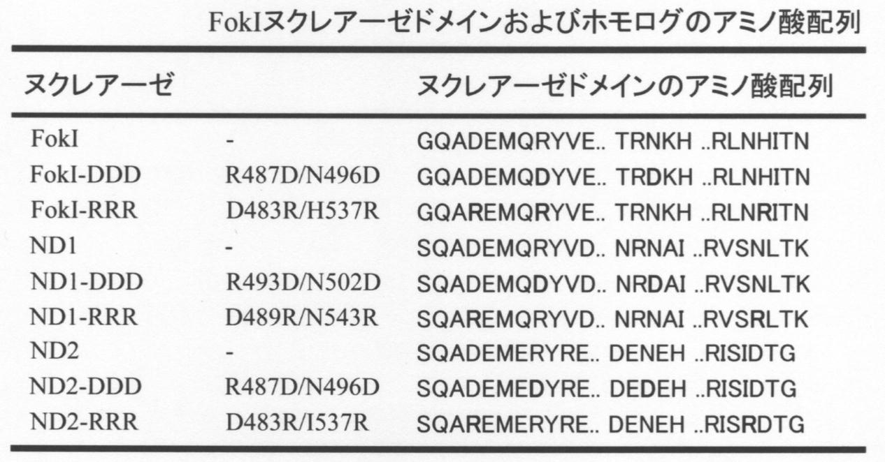
SSAレポーターアッセイの結果を図7および図8Aに示す。切断活性は、これまでのレポーター解析と同様、6bpスペーサー配列とし、ZFA36が左右二つの標的配列を認識するZFA36-FokIの切断活性に対する相対活性で算出した。Cel-Iアッセイの結果を図8Bに示す。 (2) Results The results of the SSA reporter assay are shown in FIGS. 7 and 8A. The cleavage activity was calculated as a relative activity with respect to the cleavage activity of ZFA36-FokI, which is a 6 bp spacer sequence and ZFA36 recognizes two target sequences on the left and right, as in the previous reporter analysis. The results of the Cel-I assay are shown in FIG. 8B.
ヘテロ二量体形成時のヌクレアーゼドメインの組合せを変えることにより、従来型のFokI-NDよりも高切断活性を示すことが可能となるかについて、さらに解析を行った。 Example 8: Effect on cleavage activity using combination of heterodimeric nuclease domains Exhibiting higher cleavage activity than conventional FokI-ND by changing the combination of nuclease domains during heterodimer formation Further analysis was performed to determine whether or not it became possible.
ZFNによる標的配列への変異導入により塩基配列にどのような変化が生じるのかを調べた。 Example 9: Analysis of Mutagenesis by ZFN at Base Sequence Level It was examined what kind of change occurs in the base sequence due to mutations introduced into the target sequence by ZFN.
CHO-K1細胞のゲノム配列上に存在するZFA36の標的配列(1stサイトおよび2ndサイト)に対し、ZFNによる変異導入がどのような塩基配列の変化をもたらすのか明らかにするため、標的配列を含むゲノム領域のシークエンス解析を以下のようにして行った。実施例3に記載のCel-Iアッセイと同様にして、各ZFNを形質導入したCHO-K1ゲノムDNAを鋳型にして、KOD Fx Neoを用いてPCRを行った。得られたPCR産物はTArget clone plus(Toyobo)により、PCR産物へのA付加後、TAクローニングを行った。該PCR産物を大腸菌でクローニングし、独立した大腸菌形質転換体を無作為に、少なくとも16個以上選び、それぞれのプラスミドを抽出した。得られたプラスミドを400ngと終農度6.4pmolとなるように、M13-fプライマーを加え、Fasmac社にPCRとシークエンス解析を依頼した。得られたシークエンス配列から、各ZFNにおける標的配列の変異型シークエンスの配列とその割合を算出した。 (1) Sequence analysis In order to clarify what kind of base sequence change is caused by ZFN mutation in the target sequence (1st site and 2nd site) of ZFA36 present on the genome sequence of CHO-K1 cells, Sequence analysis of the genomic region containing the target sequence was performed as follows. In the same manner as in the Cel-I assay described in Example 3, PCR was performed using KOD Fx Neo with CHO-K1 genomic DNA transduced with each ZFN as a template. The obtained PCR product was subjected to TA cloning after the addition of A to the PCR product using TA Target clone plus (Toyobo). The PCR product was cloned in E. coli, and at least 16 independent E. coli transformants were randomly selected to extract each plasmid. The M13-f primer was added so that the obtained plasmid had a final concentration of 400 ng and 400 ng, and PCR and sequence analysis were requested to Fasmac. From the obtained sequence, the sequence of the mutant sequence of the target sequence in each ZFN and the ratio thereof were calculated.
結果を図10に示す。ZFA36-ZFA36の1stサイトでは、FokI-NDでは、解析した17クローンのうち、1塩基挿入が1クローン、4塩基挿入が3クローン得られた。ND1では解析できた14クローンのうち、1塩基挿入、2塩基欠失および8塩基欠失がそれぞれ1クローン存在した。さらに、2塩基挿入かつ1塩基欠失となる変異が2クローン得られ、4塩基挿入となる変異が6クローンと今回の解析では最も多く得られた。ND2では解析できた13クローンのうち、1塩基挿入、2塩基挿入、2塩基欠失および4塩基欠失がそれぞれ1クローンずつ得られた。解析できたND3の15クローンおよびND4の16クローンでは、今回のZFA36-ZFA36 1stサイトの解析では、塩基の挿入や欠失は認められなかった。また、ZFA36標的配列の2ndサイトにおける各ヌクレアーゼドメインの変異導入を解析したところ、FokIでは解析した20クローンのうち、2塩基挿入、5塩基挿入および2塩基欠失がそれぞれ1クローンずつ得られ、230塩基を越す挿入も1クローン得られた。またZFA36 1stサイトと同様、4塩基挿入の変異が最も多く、3クローン得られた。ND1では解析した17クローンのうち、2塩基欠失および4塩基欠失がそれぞれ2クローンずつ得られた。この他、4塩基挿入が3クローン得られ、2塩基挿入が本解析では最も多く6クローン得られた。ND2では解析した16クローンのうち、1塩基挿入が1クローン得られた他、2塩基挿入が2クローン、4塩基挿入が解析した中で最も多い5クローン得られた。また、71塩基という大きな塩基の挿入が1クローン得られた。ND3では、1stサイトと異なり2ndサイトでは標的配列への変異導入が存在しており、解析した16クローンに、2塩基挿入、2塩基欠失および8塩基欠失がそれぞれ1クローンずつ得られた。ND4においては、解析できた14クローンは、全て1stサイトと同様2ndサイトでも標的配列への変異導入は見られなかった。 (2) Results The results are shown in FIG. At the first site of ZFA36-ZFA36, one clone of one base insertion and three clones of four base insertion were obtained from 17 clones analyzed in FokI-ND. In the 14 clones that could be analyzed in ND1, one clone had one base inserted, two bases deleted and eight bases deleted. In addition, two clones having two base insertions and one base deletion were obtained, and six clones having four base insertions were obtained most frequently in the present analysis. In ND2, out of the 13 clones that could be analyzed, one base insertion, two base insertion, two base deletion, and four base deletion were obtained one by one. Of the 15 clones of ND3 and 16 clones of ND4 that could be analyzed, no base insertion or deletion was observed in this analysis of the ZFA36-ZFA36 1st site. Further, when the mutation introduction of each nuclease domain at the 2nd site of the ZFA36 target sequence was analyzed, FokI resulted in 1 clone each of 2 base insertions, 5 bases insertion and 2 base deletions among the 20 clones analyzed. One clone was also obtained that exceeded the base. In addition, as in the case of the ZFA36 1st site, the mutation with the 4-base insertion was the largest and 3 clones were obtained. In ND1, 2 clones and 2 clones were obtained for each of the 17 clones analyzed. In addition, three clones with four base insertions were obtained, and six clones with two base insertions were obtained most in this analysis. In ND2, out of the 16 clones analyzed, one clone with one base insertion was obtained, and two clones with two base insertions and five clones with the most four base insertions were obtained. In addition, one clone having a large insertion of 71 bases was obtained. In ND3, unlike the first site, a mutation was introduced into the target sequence at the second site, and two clones were inserted, two bases deleted, and eight bases deleted in each of the 16 clones analyzed. In ND4, all of the 14 clones that could be analyzed did not show mutations in the target sequence at the 2nd site as well as at the 1st site.

本発明の新規ヌクレアーゼドメインがZFA以外の核酸結合ドメインを用いてもFokI-NDより高い切断活性を有するのかを明らかにするために、ND1とTALEとを含む人工核酸切断酵素を作製し、その切断活性を測定した。具体的には、発明者の研究室で開発したPlatinum TALEN(doi:10.1038/srep03379)技術を用いてTALE-FokI-NDおよびTALE-ND1を作製し、これらの切断活性をSSAレポーターアッセイ法により評価した。 Example 10: Cleavage activity of a novel nuclease domain in combination with a nucleic acid binding domain other than ZFA It is determined whether the novel nuclease domain of the present invention has a higher cleavage activity than FokI-ND even when a nucleic acid binding domain other than ZFA is used. For this purpose, an artificial nucleic acid cleaving enzyme containing ND1 and TALE was prepared, and its cleavage activity was measured. Specifically, TALE-FokI-ND and TALE-ND1 were produced using Platinum TALEN (doi: 10.1038 / srep03379) technology developed in the laboratory of the inventor, and their cleavage activities were evaluated by SSA reporter assay. did.
核酸結合モジュール未挿入のPlatinum TALEN用デスティネーションベクターであるptCMV-153/47-VR-NG(https://www.addgene.org/50704/)を鋳型とし、ptTALE-ヌクレアーゼドメイン置換用プライマーペア(ベクター増幅用)を用いてPrimeStar Maxにより増幅し、ベクターフラグメントを得た。また、ptTALE-ヌクレアーゼドメイン置換用プライマーペア(インサート増幅用)を用いてND1のインサートフラグメントを得た。これらのベクターフラグメントとインサートフラグメントを混合し、In-Fusion反応を行った。大腸菌を形質転換した後、得られた形質転換体からプラスミドを抽出し、目的とするTALE-ND1のデスティネーションベクター(ptCMV-153/47-VR-NG中のFokI-NDをND1に置換したベクター)を得た。 (1) Construction of destination vector of TALE-ND1 ptCMV-153 / 47-VR-NG (https://www.addgene.org/50704/) which is a destination vector for Platinum TALEN without inserting a nucleic acid binding module was used. It was amplified by PrimeStar Max using a ptTALE-nuclease domain replacement primer pair (for vector amplification) as a template to obtain a vector fragment. In addition, an insert fragment of ND1 was obtained using a ptTALE-nuclease domain replacement primer pair (for insert amplification). These vector fragments and insert fragments were mixed, and an In-Fusion reaction was performed. After transforming Escherichia coli, a plasmid was extracted from the obtained transformant, and a target TALE-ND1 destination vector (a vector in which FokI-ND in ptCMV-153 / 47-VR-NG was replaced with ND1). ) Got.
上記(1)で得られたデスティネーションベクターに、CHO-K1細胞のROSA26遺伝子座およびHPRT1遺伝子座の配列を認識させるための核酸結合モジュールを、Golden Gate法により組み込んだ(doi:10.1038/srep03379に記載された手法に準拠)。これを用いて大腸菌を形質転換し、得られた形質転換体から目的とするプラスミド(TALE-ROSA26-ND1およびTALE-HPRT1-ND1)を得た。また、従来型のFokI-NDを保持するTALEN(TALE-FokI)についても同様にして核酸結合モジュールを組み込んだコンストラクト(TALE-ROSA26-FokIおよびTALE-HPRT1-FokI)を調製した。SSAレポータープラスミドについては、それぞれの標的配列を含むもの(SSA-CHO-ROSA26レポーターおよびSSA-CHO-HPRT1レポーター)を常法により調製した。ROSA26遺伝子座およびHPRT1遺伝子座の標的配列を下記の表4に示す。 (2) Integration of nucleic acid binding module and construction of SSA reporter plasmid A nucleic acid binding module for recognizing the ROSA26 locus and the HPRT1 locus sequence of CHO-K1 cells in the destination vector obtained in (1) above. And the Golden Gate method (based on the method described in doi: 10.1038 / srep03379). Using this, Escherichia coli was transformed, and the desired plasmids (TALE-ROSA26-ND1 and TALE-HPRT1-ND1) were obtained from the resulting transformants. In addition, for TALEN (TALE-FokI) retaining conventional FokI-ND, constructs (TALE-ROSA26-FokI and TALE-HPRT1-FokI) incorporating a nucleic acid binding module were similarly prepared. Regarding the SSA reporter plasmid, those containing the respective target sequences (SSA-CHO-ROSA26 reporter and SSA-CHO-HPRT1 reporter) were prepared by a conventional method. The target sequences for the ROSA26 locus and the HPRT1 locus are shown in Table 4 below.
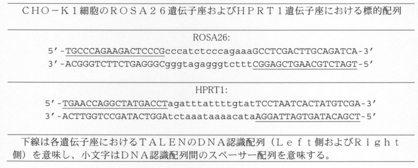
上記(2)で作製した、標的配列を認識させるための核酸結合モジュールを組み込んだTALENプラスミド(TALE-FokI-ND、およびTALE-ND1)、および標的配列を組み込んだSSAレポータープラスミドを用いて、HEK293T細胞にLipofectamine LTXを用いて形質導入を行い、形質導入後24時間の細胞を用いて、実施例2と同様にSSAレポーターアッセイを行った。対照として、FokI-NDを含むZFNプラスミド(pSTL-ZFA36)およびFokI-NDのみを発現するプラスミドpSTLを用いて同様に測定した。各TALE-NDの切断活性を、ZFA36の標的配列を含むレポーターに対するZFA36-FokIの切断活性の値を1とした時の相対活性として算出した。 (3) Introduction into cells and reporter assay The TALEN plasmids (TALE-FokI-ND and TALE-ND1) prepared in the above (2) and incorporating a nucleic acid binding module for recognizing the target sequence, and the target sequence HEK293T cells were transduced with Lipofectamine LTX using the integrated SSA reporter plasmid, and SSA reporter assays were performed in the same manner as in Example 2 using the cells 24 hours after transduction. As a control, measurement was similarly performed using a ZFN plasmid containing FokI-ND (pSTL-ZFA36) and a plasmid pSTL expressing only FokI-ND. The cleavage activity of each TALE-ND was calculated as a relative activity when the value of the cleavage activity of ZFA36-FokI for a reporter containing the target sequence of ZFA36 was set to 1.
結果を図11Aおよび図11Bに示す。図11Aには、ROSA26遺伝子座由来の配列を標的とした時の結果、図11Bには、HPRT1遺伝子座由来の配列を標的とした時の結果が示される。 (4) Results The results are shown in FIGS. 11A and 11B. FIG. 11A shows the results obtained when the sequence derived from the ROSA26 locus was targeted, and FIG. 11B shows the results obtained when the sequence derived from the HPRT1 locus was targeted.
(1)ZF-ND1およびZF-FokIの発現ベクターの構築
N末端にHisタグを含むpET-MCSプラスミドのXhoIサイトおよびSalIサイトを切断し、ZF-ND1断片を挿入した。ZF-ND1断片には、核局在シグナルNLS、Zinc-Finger(以下ZFと省略)、およびヌクレアーゼドメインND1を含む。ZFとしては、2種類の配列を認識するドメインを用いた。一つはZFA36であり、12塩基対のDNA配列GAAGATGGTを認識する。もう一つはZFL1であり、12塩基対のDNA配列GAAGGTGACを認識する。したがって、ZF(ZFA36A)-ND1、およびZF(ZFL1)-ND1を発現するプラスミドとして、pET-ZF(ZFA36A)ND1、およびpET-ZF(ZFL1)ND1をそれぞれ作成した。 Example 11: Preparation of protein in Escherichia coli (1) Construction of expression vector for ZF-ND1 and ZF-FokI The XhoI site and SalI site of the pET-MCS plasmid containing a His tag at the N-terminus were cut to obtain a ZF-ND1 fragment. Inserted. The ZF-ND1 fragment contains a nuclear localization signal NLS, a Zinc-Finger (hereinafter abbreviated as ZF), and a nuclease domain ND1. As the ZF, a domain recognizing two types of sequences was used. One is ZFA36, which recognizes the DNA sequence GAAGATGGGT of 12 base pairs. The other is ZFL1, which recognizes a DNA sequence GAAGGTGAC of 12 base pairs. Therefore, pET-ZF (ZFA36A) ND1 and pET-ZF (ZFL1) ND1 were prepared as plasmids expressing ZF (ZFA36A) -ND1 and ZF (ZFL1) -ND1, respectively.
ZF-ND1またはZF-FokIを発現するpETプラスミド、並びにpRARE2(ストラタジーン社)を大腸菌株BL21(DE3)へ形質転換し、カナマイシンおよびクロラムフェニコールを含むLB培地にて培養した。形質転換した大腸菌を37℃でOD(600nmにおける吸光度)が0.6になるまで培養した後、18°Cにて1時間培養した。イソプロピルβ-D-1-チオガラクトピラノシド(IPTG)を終濃度0.1mMになるように添加し、タンパク質の発現を誘導した。さらに18°Cにて17時間振盪培養した後、大腸菌を溶解緩衝液(20mM Tris-HCl、500mM NaCl、10% グリセロール、10mM イミダゾール、1mM フッ化ベンジルスルホニル、1mM ジチオスレイトール、pH8.0)を用いて溶解した。続いてニッケルNTAカラムにタンパク質を吸着させ、洗浄緩衝液(20mM Tris-HCl、500mM NaCl、10% グリセロール、20mM イミダゾール、pH8.0)を用いて夾雑物を除いた。タンパク質の溶出は溶出緩衝液(20mM Tris-HCl、500mM NaCl、10% グリセロール、500mM イミダゾール、pH8.0)を用いて行った。溶出したタンパク質の一部は、タンパク質収量の解析のために、SDSサンプルバッファーに溶解させた。溶出したタンパク質はゲルろ過クロマトグラフィーHiPrep 16/60 Sephacryl S-200 HR(GE healthcare、アメリカ合衆国、イリノイ)および緩衝液A(20mM HEPES、150mM NaCl、10% グリセロール、pH7.4)を用いて精製し、液体窒素を用いて瞬時に凍結した後、-80℃で保存した。タンパク質収量の解析のため、イミダゾール溶出したタンパク質画分および、最終精製されたタンパク質をSDSサンプルバッファーに溶解させてSDS-PAGEにより電気泳動を行なった。電気泳動の際には、既知濃度のBSAタンパク質を別レーンに泳動し、PAGE後のゲルをクマシー染色して目的のタンパク質分子量のバンドをChemiDoc XRS+により定量化し、タンパク質の分子量からタンパク質の収量を解析した。 (2) Expression and purification of ZF-ND1 protein in Escherichia coli A pET plasmid expressing ZF-ND1 or ZF-FokI and pRARE2 (Stratagene) were transformed into Escherichia coli strain BL21 (DE3), and kanamycin and chlorampheni were transformed. The cells were cultured in an LB medium containing Cole. The transformed E. coli was cultured at 37 ° C. until the OD (absorbance at 600 nm) reached 0.6, and then cultured at 18 ° C. for 1 hour. Isopropyl β-D-1-thiogalactopyranoside (IPTG) was added to a final concentration of 0.1 mM to induce protein expression. After further shaking culture at 18 ° C. for 17 hours, E. coli was dissolved in a lysis buffer (20 mM Tris-HCl, 500 mM NaCl, 10% glycerol, 10 mM imidazole, 1 mM benzylsulfonyl fluoride, 1 mM dithiothreitol, pH 8.0). And dissolved. Subsequently, the protein was adsorbed to a nickel NTA column, and impurities were removed using a washing buffer (20 mM Tris-HCl, 500 mM NaCl, 10% glycerol, 20 mM imidazole, pH 8.0). The protein was eluted using an elution buffer (20 mM Tris-HCl, 500 mM NaCl, 10% glycerol, 500 mM imidazole, pH 8.0). A part of the eluted protein was dissolved in SDS sample buffer for analysis of protein yield. The eluted protein was purified using gel
ZFNタンパク質を大腸菌で発現精製する際に、タンパク質収量が低いことが問題であった。ZFNタンパク質の根本的な性質として、可溶性が低く、凝集してしまうことが原因として考えられる。従来技術であるFokIと比較して、新規ND1のタンパク質の可溶性がどの程度か、検証した。 (3) Results When expressing and purifying the ZFN protein in Escherichia coli, there was a problem that the protein yield was low. It is considered that the fundamental properties of the ZFN protein are low solubility and aggregation. The degree of solubility of the novel ND1 protein in comparison with FokI, which is a conventional technique, was examined.
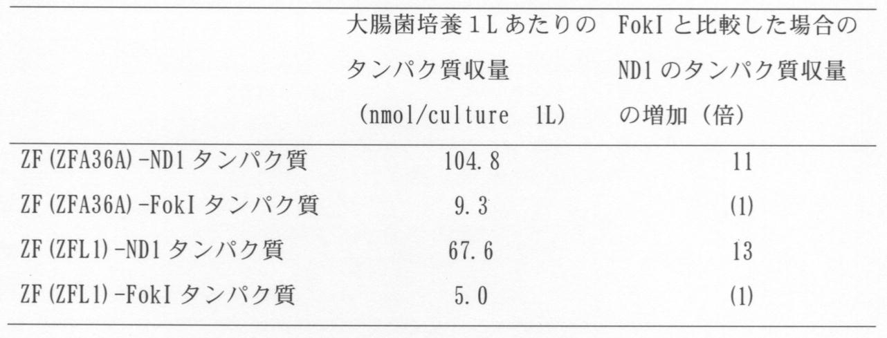
(1)レポーター細胞の構築
ZF-ND1またはZF-FokIの活性を評価するため、DNA切断後のSSA(single-strand annealing: 一本鎖アニーリング)が誘導された場合に、完全長2.1kbのGUS(β-glucuronidase: グルクロニダーゼ)遺伝子が発現するレポーター細胞を構築した。具体的には、GUS遺伝子の上流から1.8kb、ZFA36の認識配列(GAAGCTGGT)、およびGUS遺伝子の下流から0.8kbをpCAMBIA1305.2(Marker Gene社)へ導入した。上記GUS遺伝子の上流から1.8kb、およびGUS遺伝子の下流から0.8kbは、500bpを共通配列(オーバーラップ領域)として有する。このベクターをアグロバクテリウムを介して、シロイヌナズナT87培養細胞株(理化学研究所)へ形質転換し、樹立した細胞株をT87-GUUS(ZFA36)とした。GUSの発現は青色の染色試薬であるX-Glucを用いて容易に可視化・検出することが可能であるため、GUSタンパク質の検出によりZF-ND1またはZF-FokIの核内への導入およびSSA誘導活性を評価することができる。 Example 12: Activity of ZF-ND1 and ZF-Fok1 in plant cells (1) Construction of reporter cells To evaluate the activity of ZF-ND1 or ZF-FokI, SSA (single-strand annealing after DNA cutting) was performed. When chain annealing was induced, a reporter cell was constructed in which a full-length 2.1 kb GUS (β-glucoronidase: glucuronidase) gene was expressed. Specifically, 1.8 kb from the upstream of the GUS gene, a recognition sequence of ZFA36 (GAAGCTGGT), and 0.8 kb from the downstream of the GUS gene were introduced into pCAMBIA1305.2 (Marker Gene). 1.8 kb from the upstream of the GUS gene and 0.8 kb from the downstream of the GUS gene have 500 bp as a common sequence (overlap region). This vector was transformed into an Arabidopsis thaliana T87 cultured cell line (RIKEN) via Agrobacterium, and the established cell line was designated as T87-GUUS (ZFA36). Since the expression of GUS can be easily visualized and detected using X-Gluc which is a blue staining reagent, introduction of ZF-ND1 or ZF-FokI into the nucleus and induction of SSA by detection of GUS protein Activity can be assessed.
実施例11において作成したタンパク質をエレクトロポレーション法により導入した。樹立したT87-GUUS(ZFA36)細胞を用いて、細胞壁を有する状態のT87細胞に対してエレクトロポレーション法によるZF-ND1およびZF-FokIの導入を試みた。エレクトロポレーションにはネッパジーン社より販売されているNEPA21 Type IIを使用した。Opti-MEMIバッファーを用いてエレクトロポレーションを行い、2日後にGUS活性の評価を行った。その結果、図12に示すように、Opti-MEMIを用いてエレクトロポレーションした場合、非常に多くの細胞がGUSを発現していた。ZF-ND1タンパク質を用いた実験では、ZF-FokIタンパク質を用いた場合と比較してSSA誘導活性が2倍程度高いことがわかった。本実験では、ZF-ND1が遺伝子発現ベクターとしてだけではなくタンパク質として導入した場合にも、ZF-FokIと比較して高い活性を有すること、および、ZF-ND1が動物細胞だけでなく植物細胞においてもZF-FokIと比較して高い活性を有することが明らかとなった。 (2) Introduction by electroporation The protein prepared in Example 11 was introduced by electroporation. Using the established T87-GUUS (ZFA36) cells, introduction of ZF-ND1 and ZF-FokI was attempted by electroporation into T87 cells having a cell wall. NEPA21 Type II sold by Nepagene was used for electroporation. Electroporation was performed using an Opti-MEMI buffer, and the GUS activity was evaluated two days later. As a result, as shown in FIG. 12, when electroporation was performed using Opti-MEMI, a very large number of cells expressed GUS. In an experiment using the ZF-ND1 protein, it was found that the SSA-inducing activity was about twice as high as that using the ZF-FokI protein. In this experiment, it was confirmed that ZF-ND1 has higher activity than ZF-FokI even when introduced as a protein as well as a gene expression vector, and that ZF-ND1 is not only used in animal cells but also in plant cells. Also had higher activity than ZF-FokI.
実施例11で作成したタンパク質を用いて、プロテイントランスダクション法による導入を試みた。すなわち細胞壁を有するT87-GUUS(ZFA36)に対して、最終濃度0.1-2μMのZF(ZFA36A)-ND1タンパク質もしくはZF(ZFA36A)-FokIタンパク質を添加した後、1時間後に培養培地で洗浄し、3日間の培養を行なった。タンパク質を添加時には、プロテアーゼ処理等の生化学的な処理や、エレクトロポレーション等の物理化学的な処理は、まったく行っていない。したがって、タンパク質の自発的な取り込みを期待したものである。その結果、図13に示す通り、エレクトロポレーション法と比較して少ない割合ではあるが、プロテイントランスダクション法によりZF(ZFA36A)-ND1タンパク質およびZF(ZFA36A)-FokIタンパク質が取り込まれてゲノム編集可能であることが明らかとなった。プロテイントランスダクション法を用いた場合、ZF(ZFA36A)-ND1の活性はZF(ZFA36A)-FokIを比較して、同等もしくはそれ以上の活性であった。 (3) Introduction by the protein transduction method Using the protein prepared in Example 11, introduction by the protein transduction method was attempted. That is, ZF (ZFA36A) -ND1 protein or ZF (ZFA36A) -FokI protein at a final concentration of 0.1-2 μM was added to T87-GUUS (ZFA36) having a cell wall, and then washed with a culture medium one hour later. 3 days of culture. At the time of protein addition, no biochemical treatment such as protease treatment or physicochemical treatment such as electroporation was performed at all. Therefore, it is expected that the protein will be spontaneously incorporated. As a result, as shown in FIG. 13, the ZF (ZFA36A) -ND1 protein and the ZF (ZFA36A) -FokI protein were incorporated by the protein transduction method, although the ratio was smaller than in the electroporation method, and genome editing was possible. It became clear that it was. When the protein transduction method was used, the activity of ZF (ZFA36A) -ND1 was equal to or higher than that of ZF (ZFA36A) -FokI.
SEQ ID NO:10; ND1 substitution reverse primer
SEQ ID NO:11; ND2 substitution forward primer
SEQ ID NO:12; ND2 substitution reverse primer
SEQ ID NO:13; ND3 substitution forward primer
SEQ ID NO:14; ND3 substitution reverse primer
SEQ ID NO:15; ND4 substitution forward primer
SEQ ID NO:16; ND4 substitution reverse primer
SEQ ID NO:17; forward primer for linker alteration, ZFN-FokI-GS-F
SEQ ID NO:18; reverse primer for linker alteration, ZFN-GS-inverse-R
SEQ ID NO:19; forward primer for linker alteration, ZFN-ND1-GS-F
SEQ ID NO:20; reverse primer for linker alteration, ZFN-GS-inverse-R
SEQ ID NO:21; forward primer for linker alteration, ZFN-ND2-GS-F
SEQ ID NO:22; reverse primer for linker alteration, ZFN-GS-inverse-R
SEQ ID NO:23; forward primer for linker alteration, ZFN-FokI-RPGEKP-F
SEQ ID NO:24; reverse primer for linker alteration, ZFN-RPGEKP-R
SEQ ID NO:25; forward primer for linker alteration, ZFN-ND1-RPGEKP-F
SEQ ID NO:26; reverse primer for linker alteration, ZFN-RPGEKP-R
SEQ ID NO:27; forward primer for linker alteration, ZFN-ND2-RPGEKP-F
SEQ ID NO:28; reverse primer for linker alteration, ZFN-RPGEKP-R
SEQ ID NO:29; forward primer for linker alteration, ZFN-FokI-TGPGAAARA-F
SEQ ID NO:30; reverse primer for linker alteration, ZFN-FokI-TGPGAAARA-R
SEQ ID NO:31; forward primer for linker alteration, ZFN-ND1-TGPGAAARA-F
SEQ ID NO:32; reverse primer for linker alteration, ZFN-FokI-TGPGAAARA-R
SEQ ID NO:33; forward primer for linker alteration, ZFN-ND2-TGPGAAARA-F
SEQ ID NO:34; reverse primer for linker alteration, ZFN-FokI-TGPGAAARA-R
SEQ ID NO:35; forward primer for Sharkey mutation introduction in FokI-ND
SEQ ID NO:36; reverse primer for Sharkey mutation introduction in FokI-ND
SEQ ID NO:37; forward primer for Sharkey mutation introduction in ND1
SEQ ID NO:38; reverse primer for Sharkey mutation introduction in ND1
SEQ ID NO:39; forward primer for Sharkey mutation introduction in ND2
SEQ ID NO:40; reverse primer for Sharkey mutation introduction in ND2
SEQ ID NO:41; ZFA substitution forward primer
SEQ ID NO:42; ZFA substitution reverse primer
SEQ ID NO:43; forward primer for FokI-DDD mutation introduction
SEQ ID NO:44; reverse primer for FokI-DDD mutation introduction
SEQ ID NO:45; forward primer for ND1-DDD mutation introduction
SEQ ID NO:46; reverse primer for ND1-DDD mutation introduction
SEQ ID NO:47; forward primer for ND2-DDD mutation introduction
SEQ ID NO:48; reverse primer for ND2-DDD mutation introduction
SEQ ID NO:49; forward primer for FokI-RRR vector mutation introduction
SEQ ID NO:50; reverse primer for FokI-RRR vector mutation introduction
SEQ ID NO:51; forward primer for FokI-RRR insert mutation introduction
SEQ ID NO:52; reverse primer for FokI-RRR insert mutation introduction
SEQ ID NO:53; forward primer for ND1-RRR vector mutation introduction
SEQ ID NO:54; reverse primer for ND1-RRR vector mutation introduction
SEQ ID NO:55; forward primer for ND1-RRR insert mutation introduction
SEQ ID NO:56; reverse primer for ND1-RRR insert mutation introduction
SEQ ID NO:57; forward primer for ND2-RRR vector mutation introduction
SEQ ID NO:58; reverse primer for ND2-RRR vector mutation introduction
SEQ ID NO:59; forward primer for ND2-RRR insert mutation introduction
SEQ ID NO:60; reverse primer for ND2-RRR insert mutation introduction
SEQ ID NO:61; M13-F primer
SEQ ID NO:62; CHO-K1 ZFA36-ZFA36 target 1st site
SEQ ID NO:63; CHO-K1 ZFA36-ZFA36 target 2nd site
SEQ ID NO:64; CHO-K1 ZFA36-ZFA36 target 3rd site
SEQ ID NO:65; CHO-K1 ZFL1-ZFA36 target 1st site
SEQ ID NO:66; CHO-K1 ZFL1-ZFA36 target 2nd site
SEQ ID NO:67; FokI-DDD amino acid sequence
SEQ ID NO:68; FokI-RRR amino acid sequence
SEQ ID NO:69; ND1-DDD amino acid sequence
SEQ ID NO:70; ND1-RRR amino acid sequence
SEQ ID NO:71; ND2-DDD amino acid sequence
SEQ ID NO:72; ND2-RRR amino acid sequence
SEQ ID NO:73; FokI-S418P/K441E amino acid sequence
SEQ ID NO:74; ND1-S418P/K441E amino acid sequence
SEQ ID NO:75; ND2-S441E amino acid sequence
SEQ ID NO:76; FokI-cleaved CHO-K1 ZFA36-ZFA36 target 1st site
SEQ ID NO:77; FokI-cleaved CHO-K1 ZFA36-ZFA36 target 1st site
SEQ ID NO:78; FokI-cleaved CHO-K1 ZFA36-ZFA36 target 1st site
SEQ ID NO:79; ND1-cleaved CHO-K1 ZFA36-ZFA36 target 1st site
SEQ ID NO:80; ND1-cleaved CHO-K1 ZFA36-ZFA36 target 1st site
SEQ ID NO:81; ND1-cleaved CHO-K1 ZFA36-ZFA36 target 1st site
SEQ ID NO:82; ND1-cleaved CHO-K1 ZFA36-ZFA36 target 1st site
SEQ ID NO:83; ND1-cleaved CHO-K1 ZFA36-ZFA36 target 1st site
SEQ ID NO:84; ND1-cleaved CHO-K1 ZFA36-ZFA36 target 1st site
SEQ ID NO:85; ND2-cleaved CHO-K1 ZFA36-ZFA36 target 1st site
SEQ ID NO:86; ND2-cleaved CHO-K1 ZFA36-ZFA36 target 1st site
SEQ ID NO:87; ND2-cleaved CHO-K1 ZFA36-ZFA36 target 1st site
SEQ ID NO:88; ND2-cleaved CHO-K1 ZFA36-ZFA36 target 1st site
SEQ ID NO:89; ND2-cleaved CHO-K1 ZFA36-ZFA36 target 1st site
SEQ ID NO:90; ND3-cleaved CHO-K1 ZFA36-ZFA36 target 1st site
SEQ ID NO:91; ND4-cleaved CHO-K1 ZFA36-ZFA36 target 1st site
SEQ ID NO:92; FokI-cleaved CHO-K1 ZFA36-ZFA36 target 2nd site
SEQ ID NO:93; FokI-cleaved CHO-K1 ZFA36-ZFA36 target 2nd site
SEQ ID NO:94; FokI-cleaved CHO-K1 ZFA36-ZFA36 target 2nd site
SEQ ID NO:95; FokI-cleaved CHO-K1 ZFA36-ZFA36 target 2nd site
SEQ ID NO:96; FokI-cleaved CHO-K1 ZFA36-ZFA36 target 2nd site
SEQ ID NO:97; FokI-cleaved CHO-K1 ZFA36-ZFA36 target 2nd site
SEQ ID NO:98; ND1-cleaved CHO-K1 ZFA36-ZFA36 target 2nd site
SEQ ID NO:99; ND1-cleaved CHO-K1 ZFA36-ZFA36 target 2nd site
SEQ ID NO:100; ND1-cleaved CHO-K1 ZFA36-ZFA36 target 2nd site
SEQ ID NO:101 ND1-cleaved CHO-K1 ZFA36-ZFA36 target 2nd site
SEQ ID NO:102 ND1-cleaved CHO-K1 ZFA36-ZFA36 target 2nd site
SEQ ID NO:103; ND2-cleaved CHO-K1 ZFA36-ZFA36 target 2nd site
SEQ ID NO:104; ND2-cleaved CHO-K1 ZFA36-ZFA36 target 2nd site
SEQ ID NO:105; ND2-cleaved CHO-K1 ZFA36-ZFA36 target 2nd site
SEQ ID NO:106; ND2-cleaved CHO-K1 ZFA36-ZFA36 target 2nd site
SEQ ID NO:107; ND2-cleaved CHO-K1 ZFA36-ZFA36 target 2nd site
SEQ ID NO:108; ND3-cleaved CHO-K1 ZFA36-ZFA36 target 2nd site
SEQ ID NO:109; ND3-cleaved CHO-K1 ZFA36-ZFA36 target 2nd site
SEQ ID NO:110; ND3-cleaved CHO-K1 ZFA36-ZFA36 target 2nd site
SEQ ID NO:111; ND3-cleaved CHO-K1 ZFA36-ZFA36 target 2nd site
SEQ ID NO:112; ND4-cleaved CHO-K1 ZFA36-ZFA36 target 2nd site
SEQ ID NO:113; forward primer for vector amplification for ptTALE-ND substitution, ptTALEN-inverse-F
SEQ ID NO:114; reverse primer for vector amplification for ptTALE-ND substitution, NC-inverse-R_2
SEQ ID NO:115; forward primer for insert amplification for ptTALE-ND substitution, ND1-NC-F_v2
SEQ ID NO:116; reverse primer for insert amplification for ptTALE-ND substitution, ND1-ptTALEN-R
SEQ ID NO:117; target sequence on ROSA26 locus in CHO-K1 cell
SEQ ID NO:118; target sequence on HPRT1 locus in CHO-K1 cell
SEQ ID NO:119; ZF(ZFA36A)-ND1 amino acid sequence
SEQ ID NO:120; ZF(ZFL1)-ND1 amino acid sequence
SEQ ID NO:121; ZF(ZFA36A)-FokI amino acid sequence
SEQ ID NO:122; ZF(ZFL1)-FokI amino acid sequence SEQ ID NO: 9; ND1 substitution forward primer
SEQ ID NO: 10; ND1 substitution reverse primer
SEQ ID NO: 11; ND2 substitution forward primer
SEQ ID NO: 12; ND2 substitution reverse primer
SEQ ID NO: 13; ND3 substitution forward primer
SEQ ID NO: 14; ND3 substitution reverse primer
SEQ ID NO: 15; ND4 substitution forward primer
SEQ ID NO: 16; ND4 substitution reverse primer
SEQ ID NO: 17; forward primer for linker alteration, ZFN-FokI-GS-F
SEQ ID NO: 18; reverse primer for linker alteration, ZFN-GS-inverse-R
SEQ ID NO: 19; forward primer for linker alteration, ZFN-ND1-GS-F
SEQ ID NO: 20; reverse primer for linker alteration, ZFN-GS-inverse-R
SEQ ID NO: 21; forward primer for linker alteration, ZFN-ND2-GS-F
SEQ ID NO: 22; reverse primer for linker alteration, ZFN-GS-inverse-R
SEQ ID NO: 23; forward primer for linker alteration, ZFN-FokI-RPGEKP-F
SEQ ID NO: 24; reverse primer for linker alteration, ZFN-RPGEKP-R
SEQ ID NO: 25; forward primer for linker alteration, ZFN-ND1-RPGEKP-F
SEQ ID NO: 26; reverse primer for linker alteration, ZFN-RPGEKP-R
SEQ ID NO: 27; forward primer for linker alteration, ZFN-ND2-RPGEKP-F
SEQ ID NO: 28; reverse primer for linker alteration, ZFN-RPGEKP-R
SEQ ID NO: 29; forward primer for linker alteration, ZFN-FokI-TGPGAAARA-F
SEQ ID NO: 30; reverse primer for linker alteration, ZFN-FokI-TGPGAAARA-R
SEQ ID NO: 31; forward primer for linker alteration, ZFN-ND1-TGPGAAARA-F
SEQ ID NO: 32; reverse primer for linker alteration, ZFN-FokI-TGPGAAARA-R
SEQ ID NO: 33; forward primer for linker alteration, ZFN-ND2-TGPGAAARA-F
SEQ ID NO: 34; reverse primer for linker alteration, ZFN-FokI-TGPGAAARA-R
SEQ ID NO: 35; forward primer for Sharkey mutation introduction in FokI-ND
SEQ ID NO: 36; reverse primer for Sharkey mutation introduction in FokI-ND
SEQ ID NO: 37; forward primer for Sharkey mutation introduction in ND1
SEQ ID NO: 38; reverse primer for Sharkey mutation introduction in ND1
SEQ ID NO: 39; forward primer for Sharkey mutation introduction in ND2
SEQ ID NO: 40; reverse primer for Sharkey mutation introduction in ND2
SEQ ID NO: 41; ZFA substitution forward primer
SEQ ID NO: 42; ZFA substitution reverse primer
SEQ ID NO: 43; forward primer for FokI-DDD mutation introduction
SEQ ID NO: 44; reverse primer for FokI-DDD mutation introduction
SEQ ID NO: 45; forward primer for ND1-DDD mutation introduction
SEQ ID NO: 46; reverse primer for ND1-DDD mutation introduction
SEQ ID NO: 47; forward primer for ND2-DDD mutation introduction
SEQ ID NO: 48; reverse primer for ND2-DDD mutation introduction
SEQ ID NO: 49; forward primer for FokI-RRR vector mutation introduction
SEQ ID NO: 50; reverse primer for FokI-RRR vector mutation introduction
SEQ ID NO: 51; forward primer for FokI-RRR insert mutation introduction
SEQ ID NO: 52; reverse primer for FokI-RRR insert mutation introduction
SEQ ID NO: 53; forward primer for ND1-RRR vector mutation introduction
SEQ ID NO: 54; reverse primer for ND1-RRR vector mutation introduction
SEQ ID NO: 55; forward primer for ND1-RRR insert mutation introduction
SEQ ID NO: 56; reverse primer for ND1-RRR insert mutation introduction
SEQ ID NO: 57; forward primer for ND2-RRR vector mutation introduction
SEQ ID NO: 58; reverse primer for ND2-RRR vector mutation introduction
SEQ ID NO: 59; forward primer for ND2-RRR insert mutation introduction
SEQ ID NO: 60; reverse primer for ND2-RRR insert mutation introduction
SEQ ID NO: 61; M13-F primer
SEQ ID NO: 62; CHO-K1 ZFA36-ZFA36 target 1st site
SEQ ID NO: 63; CHO-K1 ZFA36-ZFA36 target 2nd site
SEQ ID NO: 64; CHO-K1 ZFA36-ZFA36 target 3rd site
SEQ ID NO: 65; CHO-K1 ZFL1-ZFA36 target 1st site
SEQ ID NO: 66; CHO-K1 ZFL1-ZFA36 target 2nd site
SEQ ID NO: 67; FokI-DDD amino acid sequence
SEQ ID NO: 68; FokI-RRR amino acid sequence
SEQ ID NO: 69; ND1-DDD amino acid sequence
SEQ ID NO: 70; ND1-RRR amino acid sequence
SEQ ID NO: 71; ND2-DDD amino acid sequence
SEQ ID NO: 72; ND2-RRR amino acid sequence
SEQ ID NO: 73; FokI-S418P / K441E amino acid sequence
SEQ ID NO: 74; ND1-S418P / K441E amino acid sequence
SEQ ID NO: 75; ND2-S441E amino acid sequence
SEQ ID NO: 76; FokI-cleaved CHO-K1 ZFA36-ZFA36 target 1st site
SEQ ID NO: 77; FokI-cleaved CHO-K1 ZFA36-ZFA36 target 1st site
SEQ ID NO: 78; FokI-cleaved CHO-K1 ZFA36-ZFA36 target 1st site
SEQ ID NO: 79; ND1-cleaved CHO-K1 ZFA36-ZFA36 target 1st site
SEQ ID NO: 80; ND1-cleaved CHO-K1 ZFA36-ZFA36 target 1st site
SEQ ID NO: 81; ND1-cleaved CHO-K1 ZFA36-ZFA36 target 1st site
SEQ ID NO: 82; ND1-cleaved CHO-K1 ZFA36-ZFA36 target 1st site
SEQ ID NO: 83; ND1-cleaved CHO-K1 ZFA36-ZFA36 target 1st site
SEQ ID NO: 84; ND1-cleaved CHO-K1 ZFA36-ZFA36 target 1st site
SEQ ID NO: 85; ND2-cleaved CHO-K1 ZFA36-ZFA36 target 1st site
SEQ ID NO: 86; ND2-cleaved CHO-K1 ZFA36-ZFA36 target 1st site
SEQ ID NO: 87; ND2-cleaved CHO-K1 ZFA36-ZFA36 target 1st site
SEQ ID NO: 88; ND2-cleaved CHO-K1 ZFA36-ZFA36 target 1st site
SEQ ID NO: 89; ND2-cleaved CHO-K1 ZFA36-ZFA36 target 1st site
SEQ ID NO: 90; ND3-cleaved CHO-K1 ZFA36-ZFA36 target 1st site
SEQ ID NO: 91; ND4-cleaved CHO-K1 ZFA36-ZFA36 target 1st site
SEQ ID NO: 92; FokI-cleaved CHO-K1 ZFA36-ZFA36 target 2nd site
SEQ ID NO: 93; FokI-cleaved CHO-K1 ZFA36-ZFA36 target 2nd site
SEQ ID NO: 94; FokI-cleaved CHO-K1 ZFA36-ZFA36 target 2nd site
SEQ ID NO: 95; FokI-cleaved CHO-K1 ZFA36-ZFA36 target 2nd site
SEQ ID NO: 96; FokI-cleaved CHO-K1 ZFA36-ZFA36 target 2nd site
SEQ ID NO: 97; FokI-cleaved CHO-K1 ZFA36-ZFA36 target 2nd site
SEQ ID NO: 98; ND1-cleaved CHO-K1 ZFA36-ZFA36 target 2nd site
SEQ ID NO: 99; ND1-cleaved CHO-K1 ZFA36-ZFA36 target 2nd site
SEQ ID NO: 100; ND1-cleaved CHO-K1 ZFA36-ZFA36 target 2nd site
SEQ ID NO: 101 ND1-cleaved CHO-K1 ZFA36-ZFA36 target 2nd site
SEQ ID NO: 102 ND1-cleaved CHO-K1 ZFA36-ZFA36 target 2nd site
SEQ ID NO: 103; ND2-cleaved CHO-K1 ZFA36-ZFA36 target 2nd site
SEQ ID NO: 104; ND2-cleaved CHO-K1 ZFA36-ZFA36 target 2nd site
SEQ ID NO: 105; ND2-cleaved CHO-K1 ZFA36-ZFA36 target 2nd site
SEQ ID NO: 106; ND2-cleaved CHO-K1 ZFA36-ZFA36 target 2nd site
SEQ ID NO: 107; ND2-cleaved CHO-K1 ZFA36-ZFA36 target 2nd site
SEQ ID NO: 108; ND3-cleaved CHO-K1 ZFA36-ZFA36 target 2nd site
SEQ ID NO: 109; ND3-cleaved CHO-K1 ZFA36-ZFA36 target 2nd site
SEQ ID NO: 110; ND3-cleaved CHO-K1 ZFA36-ZFA36 target 2nd site
SEQ ID NO: 111; ND3-cleaved CHO-K1 ZFA36-ZFA36 target 2nd site
SEQ ID NO: 112; ND4-cleaved CHO-K1 ZFA36-ZFA36 target 2nd site
SEQ ID NO: 113; forward primer for vector amplification for ptTALE-ND substitution, ptTALEN-inverse-F
SEQ ID NO: 114; reverse primer for vector amplification for ptTALE-ND substitution, NC-inverse-R_2
SEQ ID NO: 115; forward primer for insert amplification for ptTALE-ND substitution, ND1-NC-F_v2
SEQ ID NO: 116; reverse primer for insert amplification for ptTALE-ND substitution, ND1-ptTALEN-R
SEQ ID NO: 117; target sequence on ROSA26 locus in CHO-K1 cell
SEQ ID NO: 118; target sequence on HPRT1 locus in CHO-K1 cell
SEQ ID NO: 119; ZF (ZFA36A) -ND1 amino acid sequence
SEQ ID NO: 120; ZF (ZFL1) -ND1 amino acid sequence
SEQ ID NO: 121; ZF (ZFA36A) -FokI amino acid sequence
SEQ ID NO: 122; ZF (ZFL1) -FokI amino acid sequence
Claims (12)
- 配列番号1の391~585位または配列番号3の389~579位に示されるアミノ酸配列を含むポリペプチドまたはその変異体ポリペプチドである、ヌクレアーゼドメイン、および
核酸結合ドメイン
を含む人工核酸切断酵素。 An artificial nucleic acid cleaving enzyme comprising a nuclease domain and a nucleic acid binding domain, which is a polypeptide comprising the amino acid sequence shown at positions 391 to 585 of SEQ ID NO: 1 or 389 to 579 of SEQ ID NO: 3, or a mutant polypeptide thereof. - 前記ヌクレアーゼドメインと前記核酸結合ドメインとの間に位置するリンカーをさらに含む、請求項1記載の人工核酸切断酵素。 The artificial nucleic acid cleaving enzyme according to claim 1, further comprising a linker located between the nuclease domain and the nucleic acid binding domain.
- 前記核酸結合ドメインが、ジンクフィンガー、TALE、CRISPR/Cas、またはPPRを含む、請求項1または2に記載の人工核酸切断酵素。 人工 The artificial nucleic acid cleaving enzyme according to claim 1 or 2, wherein the nucleic acid binding domain comprises zinc finger, TALE, CRISPR / Cas, or PPR.
- 請求項1~3のいずれか1項に記載の人工核酸切断酵素をコードする核酸配列を含む単離された核酸。 [4] An isolated nucleic acid comprising a nucleic acid sequence encoding the artificial nucleic acid-cleaving enzyme according to any one of [1] to [3].
- 請求項4に記載の核酸またはその転写産物もしくは翻訳産物を含むベクター。 <4> A vector comprising the nucleic acid according to claim 4 or a transcription product or translation product thereof.
- 請求項1~3のいずれか1項に記載の人工核酸切断酵素、請求項4に記載の核酸、または請求項5に記載のベクターを細胞に導入することを含む、標的核酸を改変する方法。 方法 A method for modifying a target nucleic acid, comprising introducing the artificial nucleic acid-cleaving enzyme according to any one of claims 1 to 3, the nucleic acid according to claim 4, or the vector according to claim 5 into a cell.
- 前記人工核酸切断酵素、核酸、またはベクターが、2種以上の人工核酸切断酵素、該2種以上の人工核酸切断酵素をコードする核酸、または該核酸、その転写産物、もしくはその翻訳産物を含むベクターである、請求項6に記載の方法。 The artificial nucleic acid-cleaving enzyme, nucleic acid, or vector comprises two or more artificial nucleic acid-cleaving enzymes, a nucleic acid encoding the two or more artificial nucleic acid-cleaving enzymes, or a vector containing the nucleic acid, a transcription product thereof, or a translation product thereof. The method of claim 6, wherein
- 請求項1~3のいずれか1項に記載の人工核酸切断酵素、請求項4に記載の核酸、または請求項5に記載のベクターを含む、標的核酸の改変のためのキット。 A kit for modifying a target nucleic acid, comprising the artificial nucleic acid-cleaving enzyme according to any one of claims 1 to 3, the nucleic acid according to claim 4, or the vector according to claim 5.
- 前記人工核酸切断酵素、核酸、またはベクターが、2種以上の人工核酸切断酵素、該2種以上の人工核酸切断酵素をコードする核酸、または該核酸、その転写産物、もしくはその翻訳産物を含むベクターである、請求項8に記載のキット。 The artificial nucleic acid-cleaving enzyme, nucleic acid, or vector comprises two or more artificial nucleic acid-cleaving enzymes, a nucleic acid encoding the two or more artificial nucleic acid-cleaving enzymes, or a vector containing the nucleic acid, a transcription product thereof, or a translation product thereof. The kit according to claim 8, which is:
- 配列番号1の391~585位または配列番号3の389~579位に示されるアミノ酸配列を含むポリペプチドまたはその変異体ポリペプチド。 (4) A polypeptide comprising the amino acid sequence shown at positions 391 to 585 of SEQ ID NO: 1 or 389 to 579 of SEQ ID NO: 3, or a mutant polypeptide thereof.
- 請求項10に記載のポリペプチドまたはその変異体ポリペプチドをコードする核酸配列を含む単離された核酸。 An isolated nucleic acid comprising a nucleic acid sequence encoding the polypeptide according to claim 10 or a mutant polypeptide thereof.
- 請求項11に記載の核酸またはその転写産物もしくは翻訳産物を含むベクター。 A vector comprising the nucleic acid according to claim 11, or a transcription product or a translation product thereof.
Priority Applications (11)
| Application Number | Priority Date | Filing Date | Title |
|---|---|---|---|
| US17/271,481 US20210332339A1 (en) | 2018-08-27 | 2019-08-23 | Novel nuclease domain and use thereof |
| MX2021002250A MX2021002250A (en) | 2018-08-27 | 2019-08-23 | Novel nuclease domain and uses thereof. |
| CA3110471A CA3110471A1 (en) | 2018-08-27 | 2019-08-23 | Novel nuclease domain and use thereof |
| JP2020539416A JP6892721B2 (en) | 2018-08-27 | 2019-08-23 | New nuclease domain and its use |
| BR112021003257-6A BR112021003257A2 (en) | 2018-08-27 | 2019-08-23 | nuclease domain and its use |
| SG11202102030RA SG11202102030RA (en) | 2018-08-27 | 2019-08-23 | Novel nuclease domain and uses thereof |
| AU2019333722A AU2019333722A1 (en) | 2018-08-27 | 2019-08-23 | Novel nuclease domain and uses thereof |
| KR1020217008871A KR20210062639A (en) | 2018-08-27 | 2019-08-23 | New nuclease domain and its use |
| CN201980071352.7A CN112930398A (en) | 2018-08-27 | 2019-08-23 | Novel nuclease domain and use thereof |
| EP19853370.5A EP3845651A4 (en) | 2018-08-27 | 2019-08-23 | Novel nuclease domain and uses thereof |
| IL281114A IL281114A (en) | 2018-08-27 | 2021-02-25 | Novel nuclease domain and use thereof |
Applications Claiming Priority (2)
| Application Number | Priority Date | Filing Date | Title |
|---|---|---|---|
| JP2018158710 | 2018-08-27 | ||
| JP2018-158710 | 2018-08-27 |
Publications (1)
| Publication Number | Publication Date |
|---|---|
| WO2020045281A1 true WO2020045281A1 (en) | 2020-03-05 |
Family
ID=69644252
Family Applications (1)
| Application Number | Title | Priority Date | Filing Date |
|---|---|---|---|
| PCT/JP2019/033045 WO2020045281A1 (en) | 2018-08-27 | 2019-08-23 | Novel nuclease domain and uses thereof |
Country Status (12)
| Country | Link |
|---|---|
| US (1) | US20210332339A1 (en) |
| EP (1) | EP3845651A4 (en) |
| JP (1) | JP6892721B2 (en) |
| KR (1) | KR20210062639A (en) |
| CN (1) | CN112930398A (en) |
| AU (1) | AU2019333722A1 (en) |
| BR (1) | BR112021003257A2 (en) |
| CA (1) | CA3110471A1 (en) |
| IL (1) | IL281114A (en) |
| MX (1) | MX2021002250A (en) |
| SG (1) | SG11202102030RA (en) |
| WO (1) | WO2020045281A1 (en) |
Cited By (1)
| Publication number | Priority date | Publication date | Assignee | Title |
|---|---|---|---|---|
| WO2023038020A1 (en) | 2021-09-07 | 2023-03-16 | 国立大学法人千葉大学 | Composition for modifying nucleic acid sequence, and method for modifying target site of nucleic acid sequence |
Families Citing this family (1)
| Publication number | Priority date | Publication date | Assignee | Title |
|---|---|---|---|---|
| JP2022500052A (en) * | 2018-09-18 | 2022-01-04 | サンガモ セラピューティクス, インコーポレイテッド | Programmed cell death 1 (PD1) specific nuclease |
Citations (2)
| Publication number | Priority date | Publication date | Assignee | Title |
|---|---|---|---|---|
| WO2014175284A1 (en) * | 2013-04-22 | 2014-10-30 | 国立大学法人九州大学 | Dna binding protein using ppr motif, and use thereof |
| US9410134B2 (en) | 2011-06-07 | 2016-08-09 | Helmholtz Zentrum München—Deutsches Forschungszentrum für Gesundheit und Umwelt | Protein having nuclease activity, fusion proteins and uses thereof |
Family Cites Families (2)
| Publication number | Priority date | Publication date | Assignee | Title |
|---|---|---|---|---|
| AU2014312295C1 (en) * | 2013-08-28 | 2020-08-13 | Sangamo Therapeutics, Inc. | Compositions for linking DNA-binding domains and cleavage domains |
| WO2018039448A1 (en) * | 2016-08-24 | 2018-03-01 | Sangamo Therapeutics, Inc. | Engineered target specific nucleases |
-
2019
- 2019-08-23 KR KR1020217008871A patent/KR20210062639A/en unknown
- 2019-08-23 AU AU2019333722A patent/AU2019333722A1/en active Pending
- 2019-08-23 JP JP2020539416A patent/JP6892721B2/en active Active
- 2019-08-23 SG SG11202102030RA patent/SG11202102030RA/en unknown
- 2019-08-23 MX MX2021002250A patent/MX2021002250A/en unknown
- 2019-08-23 CN CN201980071352.7A patent/CN112930398A/en active Pending
- 2019-08-23 WO PCT/JP2019/033045 patent/WO2020045281A1/en active Application Filing
- 2019-08-23 CA CA3110471A patent/CA3110471A1/en active Pending
- 2019-08-23 BR BR112021003257-6A patent/BR112021003257A2/en unknown
- 2019-08-23 EP EP19853370.5A patent/EP3845651A4/en active Pending
- 2019-08-23 US US17/271,481 patent/US20210332339A1/en active Pending
-
2021
- 2021-02-25 IL IL281114A patent/IL281114A/en unknown
Patent Citations (2)
| Publication number | Priority date | Publication date | Assignee | Title |
|---|---|---|---|---|
| US9410134B2 (en) | 2011-06-07 | 2016-08-09 | Helmholtz Zentrum München—Deutsches Forschungszentrum für Gesundheit und Umwelt | Protein having nuclease activity, fusion proteins and uses thereof |
| WO2014175284A1 (en) * | 2013-04-22 | 2014-10-30 | 国立大学法人九州大学 | Dna binding protein using ppr motif, and use thereof |
Non-Patent Citations (9)
| Title |
|---|
| [online] 4 December 2015 (2015-12-04), retrieved from NCBI Database accession no. LNQP01000031.1 * |
| DATABASE Nucleotide [online] 15 March 2017 (2017-03-15), "[Clostridium] thermoalcaliphilum strain DSM 7309 CLOTH_contig000003, w", XP055695530, retrieved from NCBI Database accession no. MZGW01000003. 1 * |
| DATABASE Protein [online] 4 December 2015 (2015-12-04), "hypothetical protein AS180_10220 [Bacillus sp. SGD-V-76]", XP055695520, retrieved from NCBI Database accession no. KSU87972.1 * |
| GUO, J.GAJ, T.BARBAS, C. F.: "Directed evolution of an enhanced and highly efficient FokI cleavage domain for zinc finger nucleases", J MOL BIOL, vol. 400, 2010, pages 96 - 107, XP055491626, DOI: 10.1016/j.jmb.2010.04.060 |
| HANDEL, E. M .ALWIN, S.CATHOMEN, T .: "Expanding or restricting the target site repertoire of zinc-finger nucleases: the inter-domain linker as a major determinant of target site selectivity", MOL THER, vol. 17, 2009, pages 104 - 111, XP002551960, DOI: 10.1038/mt.2008.233 |
| NOMURA ET AL.: "Effects of DNA Binding of the Zinc Finger and Linkers for Domain Fusion on the Catalytic Activity of Sequence-Specific Chimeric Recombinase Determined by a Facile Fluorescent System", BIOCHEMISTRY, vol. 51, 2012, pages 1510 - 1517 |
| OCHIAI ET AL.: "Targeted mutagenesis in the sea urchin embryo using zinc-finger nucleases", GENES TO CELLS, vol. 15, 2010, pages 875 - 885 |
| See also references of EP3845651A4 |
| WILSON ET AL.: "Expanding the Repertoire of Target Sites for Zinc Nuclease-mediated Genome modification", MOL. THER-NUCLEIC ACIDS, vol. 2, 2013, pages e88, XP055329625, DOI: 10.1038/mtna.2013.13 |
Cited By (1)
| Publication number | Priority date | Publication date | Assignee | Title |
|---|---|---|---|---|
| WO2023038020A1 (en) | 2021-09-07 | 2023-03-16 | 国立大学法人千葉大学 | Composition for modifying nucleic acid sequence, and method for modifying target site of nucleic acid sequence |
Also Published As
| Publication number | Publication date |
|---|---|
| BR112021003257A2 (en) | 2021-05-18 |
| JPWO2020045281A1 (en) | 2021-04-08 |
| EP3845651A4 (en) | 2022-06-22 |
| IL281114A (en) | 2021-04-29 |
| SG11202102030RA (en) | 2021-04-29 |
| AU2019333722A1 (en) | 2021-04-15 |
| KR20210062639A (en) | 2021-05-31 |
| MX2021002250A (en) | 2021-05-27 |
| US20210332339A1 (en) | 2021-10-28 |
| CN112930398A (en) | 2021-06-08 |
| JP6892721B2 (en) | 2021-06-23 |
| EP3845651A1 (en) | 2021-07-07 |
| CA3110471A1 (en) | 2020-03-05 |
Similar Documents
| Publication | Publication Date | Title |
|---|---|---|
| US20210403861A1 (en) | Nucleotide-specific recognition sequences for designer tal effectors | |
| US10844361B2 (en) | Site-specific enzymes and methods of use | |
| JP7469285B2 (en) | Use of nucleosome-interacting protein domains to enhance targeted genome modification | |
| JP2023123755A5 (en) | ||
| JP7109547B2 (en) | An engineered Cas9 system for eukaryotic genome modification | |
| AU2018229489A1 (en) | Crispr-based genome modification and regulation | |
| JP2016523084A (en) | Target integration | |
| WO2020045281A1 (en) | Novel nuclease domain and uses thereof | |
| JP2017525355A (en) | Mutants of bacteriophage lambda integrase | |
| US20140271602A1 (en) | Nucleotide-specific recognition sequences for designer tal effectors | |
| JP2013094148A (en) | Novel dna binding domain and novel dna breaking enzyme including the same | |
| JP2023145672A (en) | Efficient ppr protein production method and use thereof | |
| CA3142303A1 (en) | Ppr protein causing less aggregation and use of the same | |
| JP7356739B2 (en) | Nucleic acid binding protein | |
| KR20130069188A (en) | Zinc finger nuclease for targeting myostatin and use thereof | |
| KR101528812B1 (en) | Inhibition method of SETDB1 HMTase expression |
Legal Events
| Date | Code | Title | Description |
|---|---|---|---|
| 121 | Ep: the epo has been informed by wipo that ep was designated in this application |
Ref document number: 19853370 Country of ref document: EP Kind code of ref document: A1 |
|
| ENP | Entry into the national phase |
Ref document number: 2020539416 Country of ref document: JP Kind code of ref document: A |
|
| ENP | Entry into the national phase |
Ref document number: 3110471 Country of ref document: CA |
|
| WWE | Wipo information: entry into national phase |
Ref document number: 281114 Country of ref document: IL |
|
| NENP | Non-entry into the national phase |
Ref country code: DE |
|
| REG | Reference to national code |
Ref country code: BR Ref legal event code: B01A Ref document number: 112021003257 Country of ref document: BR |
|
| ENP | Entry into the national phase |
Ref document number: 2019853370 Country of ref document: EP Effective date: 20210329 |
|
| ENP | Entry into the national phase |
Ref document number: 2019333722 Country of ref document: AU Date of ref document: 20190823 Kind code of ref document: A |
|
| ENP | Entry into the national phase |
Ref document number: 112021003257 Country of ref document: BR Kind code of ref document: A2 Effective date: 20210222 |