WO2020041633A1 - Non-invasive spinal cord stimulation for nerve root palsy, cauda equina syndrome, and restoration of upper extremity function - Google Patents

Non-invasive spinal cord stimulation for nerve root palsy, cauda equina syndrome, and restoration of upper extremity function Download PDF

Info

Publication number
WO2020041633A1
WO2020041633A1 PCT/US2019/047777 US2019047777W WO2020041633A1 WO 2020041633 A1 WO2020041633 A1 WO 2020041633A1 US 2019047777 W US2019047777 W US 2019047777W WO 2020041633 A1 WO2020041633 A1 WO 2020041633A1
Authority
WO
WIPO (PCT)
Prior art keywords
stimulation
spinal cord
region
days
magnetic
Prior art date
Application number
PCT/US2019/047777
Other languages
French (fr)
Inventor
Daniel C. Lu
Original Assignee
The Regents Of The University Of California
Priority date (The priority date is an assumption and is not a legal conclusion. Google has not performed a legal analysis and makes no representation as to the accuracy of the date listed.)
Filing date
Publication date
Application filed by The Regents Of The University Of California filed Critical The Regents Of The University Of California
Priority to CA3110463A priority Critical patent/CA3110463A1/en
Priority to JP2021510130A priority patent/JP2021534877A/en
Priority to US17/270,402 priority patent/US20210187278A1/en
Priority to EP19852797.0A priority patent/EP3840638A4/en
Publication of WO2020041633A1 publication Critical patent/WO2020041633A1/en

Links

Classifications

    • AHUMAN NECESSITIES
    • A61MEDICAL OR VETERINARY SCIENCE; HYGIENE
    • A61BDIAGNOSIS; SURGERY; IDENTIFICATION
    • A61B5/00Measuring for diagnostic purposes; Identification of persons
    • A61B5/48Other medical applications
    • A61B5/4836Diagnosis combined with treatment in closed-loop systems or methods
    • AHUMAN NECESSITIES
    • A61MEDICAL OR VETERINARY SCIENCE; HYGIENE
    • A61BDIAGNOSIS; SURGERY; IDENTIFICATION
    • A61B5/00Measuring for diagnostic purposes; Identification of persons
    • A61B5/103Detecting, measuring or recording devices for testing the shape, pattern, colour, size or movement of the body or parts thereof, for diagnostic purposes
    • A61B5/11Measuring movement of the entire body or parts thereof, e.g. head or hand tremor, mobility of a limb
    • AHUMAN NECESSITIES
    • A61MEDICAL OR VETERINARY SCIENCE; HYGIENE
    • A61BDIAGNOSIS; SURGERY; IDENTIFICATION
    • A61B5/00Measuring for diagnostic purposes; Identification of persons
    • A61B5/24Detecting, measuring or recording bioelectric or biomagnetic signals of the body or parts thereof
    • A61B5/316Modalities, i.e. specific diagnostic methods
    • A61B5/369Electroencephalography [EEG]
    • AHUMAN NECESSITIES
    • A61MEDICAL OR VETERINARY SCIENCE; HYGIENE
    • A61BDIAGNOSIS; SURGERY; IDENTIFICATION
    • A61B5/00Measuring for diagnostic purposes; Identification of persons
    • A61B5/24Detecting, measuring or recording bioelectric or biomagnetic signals of the body or parts thereof
    • A61B5/316Modalities, i.e. specific diagnostic methods
    • A61B5/389Electromyography [EMG]
    • AHUMAN NECESSITIES
    • A61MEDICAL OR VETERINARY SCIENCE; HYGIENE
    • A61NELECTROTHERAPY; MAGNETOTHERAPY; RADIATION THERAPY; ULTRASOUND THERAPY
    • A61N1/00Electrotherapy; Circuits therefor
    • A61N1/02Details
    • A61N1/04Electrodes
    • A61N1/0404Electrodes for external use
    • A61N1/0408Use-related aspects
    • A61N1/0456Specially adapted for transcutaneous electrical nerve stimulation [TENS]
    • AHUMAN NECESSITIES
    • A61MEDICAL OR VETERINARY SCIENCE; HYGIENE
    • A61NELECTROTHERAPY; MAGNETOTHERAPY; RADIATION THERAPY; ULTRASOUND THERAPY
    • A61N1/00Electrotherapy; Circuits therefor
    • A61N1/02Details
    • A61N1/04Electrodes
    • A61N1/05Electrodes for implantation or insertion into the body, e.g. heart electrode
    • A61N1/0551Spinal or peripheral nerve electrodes
    • AHUMAN NECESSITIES
    • A61MEDICAL OR VETERINARY SCIENCE; HYGIENE
    • A61NELECTROTHERAPY; MAGNETOTHERAPY; RADIATION THERAPY; ULTRASOUND THERAPY
    • A61N1/00Electrotherapy; Circuits therefor
    • A61N1/02Details
    • A61N1/04Electrodes
    • A61N1/06Electrodes for high-frequency therapy
    • AHUMAN NECESSITIES
    • A61MEDICAL OR VETERINARY SCIENCE; HYGIENE
    • A61NELECTROTHERAPY; MAGNETOTHERAPY; RADIATION THERAPY; ULTRASOUND THERAPY
    • A61N1/00Electrotherapy; Circuits therefor
    • A61N1/18Applying electric currents by contact electrodes
    • A61N1/32Applying electric currents by contact electrodes alternating or intermittent currents
    • A61N1/36Applying electric currents by contact electrodes alternating or intermittent currents for stimulation
    • A61N1/36014External stimulators, e.g. with patch electrodes
    • A61N1/36017External stimulators, e.g. with patch electrodes with leads or electrodes penetrating the skin
    • AHUMAN NECESSITIES
    • A61MEDICAL OR VETERINARY SCIENCE; HYGIENE
    • A61NELECTROTHERAPY; MAGNETOTHERAPY; RADIATION THERAPY; ULTRASOUND THERAPY
    • A61N1/00Electrotherapy; Circuits therefor
    • A61N1/18Applying electric currents by contact electrodes
    • A61N1/32Applying electric currents by contact electrodes alternating or intermittent currents
    • A61N1/36Applying electric currents by contact electrodes alternating or intermittent currents for stimulation
    • A61N1/36014External stimulators, e.g. with patch electrodes
    • A61N1/3603Control systems
    • A61N1/36034Control systems specified by the stimulation parameters
    • AHUMAN NECESSITIES
    • A61MEDICAL OR VETERINARY SCIENCE; HYGIENE
    • A61NELECTROTHERAPY; MAGNETOTHERAPY; RADIATION THERAPY; ULTRASOUND THERAPY
    • A61N1/00Electrotherapy; Circuits therefor
    • A61N1/18Applying electric currents by contact electrodes
    • A61N1/32Applying electric currents by contact electrodes alternating or intermittent currents
    • A61N1/36Applying electric currents by contact electrodes alternating or intermittent currents for stimulation
    • A61N1/3605Implantable neurostimulators for stimulating central or peripheral nerve system
    • A61N1/3606Implantable neurostimulators for stimulating central or peripheral nerve system adapted for a particular treatment
    • A61N1/36062Spinal stimulation
    • AHUMAN NECESSITIES
    • A61MEDICAL OR VETERINARY SCIENCE; HYGIENE
    • A61NELECTROTHERAPY; MAGNETOTHERAPY; RADIATION THERAPY; ULTRASOUND THERAPY
    • A61N1/00Electrotherapy; Circuits therefor
    • A61N1/18Applying electric currents by contact electrodes
    • A61N1/32Applying electric currents by contact electrodes alternating or intermittent currents
    • A61N1/36Applying electric currents by contact electrodes alternating or intermittent currents for stimulation
    • A61N1/3605Implantable neurostimulators for stimulating central or peripheral nerve system
    • A61N1/3606Implantable neurostimulators for stimulating central or peripheral nerve system adapted for a particular treatment
    • A61N1/36067Movement disorders, e.g. tremor or Parkinson disease
    • AHUMAN NECESSITIES
    • A61MEDICAL OR VETERINARY SCIENCE; HYGIENE
    • A61NELECTROTHERAPY; MAGNETOTHERAPY; RADIATION THERAPY; ULTRASOUND THERAPY
    • A61N1/00Electrotherapy; Circuits therefor
    • A61N1/18Applying electric currents by contact electrodes
    • A61N1/32Applying electric currents by contact electrodes alternating or intermittent currents
    • A61N1/36Applying electric currents by contact electrodes alternating or intermittent currents for stimulation
    • A61N1/3605Implantable neurostimulators for stimulating central or peripheral nerve system
    • A61N1/3606Implantable neurostimulators for stimulating central or peripheral nerve system adapted for a particular treatment
    • A61N1/36071Pain
    • AHUMAN NECESSITIES
    • A61MEDICAL OR VETERINARY SCIENCE; HYGIENE
    • A61NELECTROTHERAPY; MAGNETOTHERAPY; RADIATION THERAPY; ULTRASOUND THERAPY
    • A61N1/00Electrotherapy; Circuits therefor
    • A61N1/18Applying electric currents by contact electrodes
    • A61N1/32Applying electric currents by contact electrodes alternating or intermittent currents
    • A61N1/36Applying electric currents by contact electrodes alternating or intermittent currents for stimulation
    • A61N1/3605Implantable neurostimulators for stimulating central or peripheral nerve system
    • A61N1/36128Control systems
    • A61N1/36189Control systems using modulation techniques
    • A61N1/36192Amplitude modulation
    • AHUMAN NECESSITIES
    • A61MEDICAL OR VETERINARY SCIENCE; HYGIENE
    • A61NELECTROTHERAPY; MAGNETOTHERAPY; RADIATION THERAPY; ULTRASOUND THERAPY
    • A61N1/00Electrotherapy; Circuits therefor
    • A61N1/18Applying electric currents by contact electrodes
    • A61N1/32Applying electric currents by contact electrodes alternating or intermittent currents
    • A61N1/36Applying electric currents by contact electrodes alternating or intermittent currents for stimulation
    • A61N1/3605Implantable neurostimulators for stimulating central or peripheral nerve system
    • A61N1/36128Control systems
    • A61N1/36189Control systems using modulation techniques
    • A61N1/36196Frequency modulation
    • AHUMAN NECESSITIES
    • A61MEDICAL OR VETERINARY SCIENCE; HYGIENE
    • A61NELECTROTHERAPY; MAGNETOTHERAPY; RADIATION THERAPY; ULTRASOUND THERAPY
    • A61N2/00Magnetotherapy
    • A61N2/004Magnetotherapy specially adapted for a specific therapy
    • A61N2/006Magnetotherapy specially adapted for a specific therapy for magnetic stimulation of nerve tissue
    • AHUMAN NECESSITIES
    • A61MEDICAL OR VETERINARY SCIENCE; HYGIENE
    • A61NELECTROTHERAPY; MAGNETOTHERAPY; RADIATION THERAPY; ULTRASOUND THERAPY
    • A61N2/00Magnetotherapy
    • A61N2/02Magnetotherapy using magnetic fields produced by coils, including single turn loops or electromagnets

Definitions

  • spinal diseases are among the most frequent causes of discomfort and disability in patients in the United States and elsewhere around the world, frequently requiring surgical intervention for relief.
  • Back and/or leg pain resulting from spinal disease is frequently the result of spinal stenoses that result from narrowing of one or more nerve passages in the spine, most often in the upper (cervical) spine or the lower (lumbar) spine.
  • Such narrowing can apply pressure to the spinal nerves which can cause a variety of symptoms, including pain, cramping, numbness in the legs, back, neck, shoulders, or arms.
  • a spinal nerve such as a transversing nerve, an exiting nerve, or nerves of the cauda equina.
  • each of these treatment protocols generally requires surgical access to the spine which in turn requires cutting and displacing major muscles and ligaments surrounding the spine. Such procedures are necessarily performed under a general anesthesia and may require hospital stays. Recovery times vary from weeks to months and extensive rehabilitation is usually necessary.
  • electrical stimulation means application of an electrical signal that may be either excitatory or inhibitory to a muscle or neuron and/or to groups of neurons and/or intemeurons. It will be understood that an electrical signal may be applied to one or more electrodes with one or more return electrodes.
  • magnetic stimulation As used herein "magnetic stimulation”, “electromagnetic spinal cord stimulation” or “EMSS” refers to the use of a varying magnetic field to induce an electrical signal, e.g., in a neuron, that may be either excitatory or inhibitory to a muscle or neuron and/or to groups of neurons and/or intemeurons.
  • transcutaneous stimulation or“transcutaneous electrical stimulation” or“cutaneous electrical stimulation” refers to electrical stimulation applied to the skin, and, as typically used herein refers to electrical stimulation applied to the skin in order to effect stimulation of the spinal cord or a region thereof.
  • transcutaneous electrical spinal cord stimulation may also be referred to as“tSCS”.
  • pcEmc refers to painless cutaneous electrical stimulation.
  • motor complete when used with respect to a spinal cord injury indicates that there is no motor function below the lesion, (e.g., no movement can be voluntarily induced in muscles innervated by spinal segments below the spinal lesion.
  • monopolar stimulation refers to stimulation between a local electrode and a common distant return electrode.
  • co-administering refers to administration of the transcutaneous electrical stimulation and/or epidural electrical stimulation and/or pharmaceutical such that various modalities can simultaneously achieve a physiological effect on the subject.
  • the administered modalities need not be administered together, either temporally or at the same site.
  • the various“treatment” modalities are administered at different times.
  • administration of one can precede administration of the other (e.g, drug before electrical and/or magnetic stimulation or vice versa).
  • Simultaneous physiological effect need not necessarily require presence of drug and the electrical and/or magnetic stimulation at the same time or the presence of both stimulation modalities at the same time.
  • all the modalities are administered essentially simultaneously.
  • spinal cord stimulation includes stimulation of any spinal nervous tissue, including spinal neurons, accessory neuronal cells, nerves, nerve roots, nerve fibers, or tissues, that are associated with the spinal cord. It is contemplated that spinal cord stimulation may comprise stimulation of one or more areas associated with a cervical vertebral segment.
  • spinal nervous tissue refers to nerves, neurons, neuroglial cells, glial cells, neuronal accessory cells, nerve roots, nerve fibers, nerve rootlets, parts of nerves, nerve bundles, mixed nerves, sensory fibers, motor fibers, dorsal root, ventral root, dorsal root ganglion, spinal ganglion, ventral motor root, general somatic afferent fibers, general visceral afferent fibers, general somatic efferent fibers, general visceral efferent fibers, grey matter, white matter, the dorsal column, the lateral column, and/or the ventral column associated with the spinal cord.
  • Spinal nervous tissue includes“spinal nerve roots,” that comprise any one or more of the 31 pairs of nerves that emerge from the spinal cord.
  • Spinal nerve roots may be cervical nerve roots, thoracic nerve roots, and lumbar nerve roots.
  • methods are provided for applying spinal cord stimulation with and without selective pharmaceuticals to improve motor function, and/or to improve motor control, and/or to improve sensory function, and/or to reduce pain in subjects with nerve root palsies (e.g., radiculopathies including, but not limited to cauda equina syndrome) and/or to improve motor function of upper and/or lower extremities in subjects with impaired extremity motor function due to spinal cord or brain injury or pathology.
  • nerve root palsies e.g., radiculopathies including, but not limited to cauda equina syndrome
  • Various embodiments contemplated herein may include, but need not be limited to, one or more of the following:
  • Embodiment 1 A method of treating a nerve root disorder (radiculopathy) in a subject, said method comprising:
  • Embodiment 2 The method of embodiment 1, wherein said nerve root disorder results from a spinal stenosis.
  • Embodiment 3 The method of embodiment 2, wherein said nerve root disorder results from a spinal stenosis that applies pressure to a transversing spinal nerve, an exiting spinal nerve, or a nerve of the cauda equine.
  • Embodiment 4 The method of embodiment 2, wherein said nerve root disorder results from a stenosis in the lumbar spine.
  • Embodiment 5 The method of embodiment 1, wherein said radiculopathy comprises an upper limb radiculopathy.
  • Embodiment 6 The method of embodiment 4, wherein said upper limb radiculopathy comprises a C7 radiculopathy.
  • Embodiment 7 The method of embodiment 4, wherein said upper limb radiculopathy comprises a C6 radiculopathy.
  • Embodiment 8 The method of embodiment 4, wherein said nerve root disorder comprises a C5 palsy.
  • Embodiment 9 The method of embodiment 8, wherein said nerve root palsy comprises a palsy following anterior cervical discectomy and fusion (ACDF).
  • ACDF anterior cervical discectomy and fusion
  • Embodiment 10 The method of embodiment 1, wherein said radiculopathy comprises a lower limb radiculopathy.
  • Embodiment 11 The method of embodiment 10, wherein said radiculopathy comprises an L4 radiculopathy.
  • Embodiment 12 The method of embodiment 10, wherein said radiculopathy comprises an L5 radiculopathy.
  • Embodiment 13 The method of embodiment 10, wherein said radiculopathy comprises an Sl radiculopathy.
  • Embodiment 14 The method of embodiment 1, wherein said nerve root palsy comprises cauda equina syndrome.
  • Embodiment 15 The method of embodiment 14, wherein said nerve root palsy comprises cauda equine syndrome due to herniation of lumbar intervertebral discs.
  • Embodiment 16 The method of embodiment 14, wherein said nerve root palsy comprises cauda equine syndrome due to abnormal growths (tumor or cancer) adjacent to the lower spinal cord.
  • Embodiment 17 The method of embodiment 14, wherein said nerve root palsy comprises cauda equine syndrome due to localized infection near the spinal cord.
  • Embodiment 18 The method of embodiment 14, wherein said nerve root palsy comprises cauda equine syndrome due to epidural abscess, and/or localized bleeding (epidural hematoma) causing pressure on the spinal cord in the low back.
  • Embodiment 19 The method according to any one of embodiments 1-18, wherein said method restores strength.
  • Embodiment 20 The method according to any one of embodiments 1-19, wherein said method restores motor function.
  • Embodiment 21 The method according to any one of embodiments 1-20, wherein said method restores locomotion.
  • Embodiment 22 The method according to any one of embodiments 1-21, wherein said method restores continence.
  • Embodiment 23 The method according to any one of embodiments 1-22, wherein said method restores sexual function.
  • Embodiment 24 The method according to any one of embodiments 1-23, wherein said method reduces or eliminates pain associated with said nerve root disorder.
  • Embodiment 25 The method according to any one of embodiments 1-18, wherein said neuromodulating comprises neuromodulating a dorsal surface of the brainstem or spinal cord.
  • Embodiment 26 The method according to any one of embodiments 1-18, wherein said neuromodulating comprises neuromodulating a ventral surface of the brainstem or spinal cord.
  • Embodiment 27 The method according to any one of embodiments 1-26, wherein said method comprises administering transcutaneous stimulation to the suboccipital and/or brain stem or a region thereof, to the cervical spinal cord or a region thereof, and/or the thoracic spinal cord or a region thereof, and/or the lumbar spinal cord or a region thereof.
  • Embodiment 28 The method of embodiment 27, wherein said method comprises administering transcutaneous stimulation to the spinal cord in the suboccipital or brain stem or a region thereof.
  • Embodiment 29 The method according to any one of embodiments 27-28, wherein said method comprises administering transcutaneous stimulation to the cervical spinal cord or a region thereof.
  • Embodiment 30 The method of embodiment 29, wherein said transcutaneous electrical stimulation is applied over one or more regions straddling or spanning a region selected from the group consisting of C0-C1, C0-C2, C0-C3, C0-C4, C0-C5, C0-C6, C0-C7, C0-T1, Cl-Cl, C1-C2, C1-C3, C1-C4, C1-C7, C1-C6, C1-C7, Cl-Tl, C2-C2, C2-C3, C2- C4, C2-C5, C2-C6, C2-C7, C2-T1, C3-C3, C3-C4, C3-C5, C3-C6, C3-C7, C3-T1, C4-C4, C4-C5, C4-C6, C4-C7, C4-T1, C5-C5, C5-C6, C5-C7, C5-T1, C6-C6, C6-C7, C6-T1, C7-C7, and C7-T1.
  • Embodiment 31 The method of embodiment 30, wherein said transcutaneous electrical stimulation is applied over a cervical region comprising or consisting of a region comprising motor neurons the functions of which are deficient.
  • Embodiment 32 The method of embodiment 30, wherein said transcutaneous electrical stimulation is applied over a region comprising or consisting of C4-C5 or a region therein.
  • Embodiment 33 The method of embodiment 32, wherein said transcutaneous electrical stimulation is applied at C5.
  • Embodiment 34 The method according to any one of embodiments 27-33, wherein said method comprises administering transcutaneous stimulation to the thoracic spinal cord or a region thereof.
  • Embodiment 35 The method of embodiment 34, wherein said transcutaneous electrical stimulation is applied over one or more regions straddling or spanning a region selected from the group consisting of Tl-Tl, T1-T2, T1-T3, T1-T4, T1-T5, T1-T6, T1-T7, T1-T8, T1-T9, T1-T10, Tl-Tl 1, T1-T12, T2-T2, T2-T3, T2-T4, T2-T5, T2-T6, T2-T7, T2- T8, T2-T9, T2-T10, T2-T11, T2-T12, T3-T3, T3-T4, T3-T5, T3-T6, T3-T7, T3-T8, T3-T9, T3-T10, T3-T11, T3-T12, T4-T4, T4-T5, T4-T6, T4-T7, T4-T8, T4-T9, T4-T10, T3-T11
  • Embodiment 36 The method of embodiment 35, wherein said transcutaneous electrical stimulation is applied over a thoracic region comprising or consisting of a region comprising motor neurons the functions of which are deficient.
  • Embodiment 37 The according to any one of embodiments 27-36, wherein said method comprises administering transcutaneous stimulation to the lumbar and/or sacral spinal cord or a region thereof.
  • Embodiment 38 The method of embodiment 37, wherein said transcutaneous electrical stimulation is applied over one or more regions straddling or spanning a region selected from the group consisting of Ll-Ll, L1-L2 , L1-L3, L1-L4, L1-L5, Ll-Sl, L1-S2, L1-S3, L1-S4, L1-S5, L2-L2 , L2-L3, L2-L4, L2-L5, L2-S1, L2-S2, L2-S3, L2-S4, L2-S5, L3-L3, L3-L4, L3-L5, L3-S1, L3-S2, L3-S3, L3-S4, L3-S5, L4-L4, L4-L5, L4-S1, L4-S2, L4-S3, L4-S4, L4-S5, L5-L5 , L5-S1, L5-S2, L5-S3, L5-S4, L5-S5, Sl-Sl, S1-S
  • Embodiment 39 The method of embodiment 37, wherein said transcutaneous electrical stimulation is applied over a lumbar and/or sacral region comprising or consisting of a region comprising motor neurons that the functions of which are deficient.
  • Embodiment 40 The method of embodiment 35, wherein said transcutaneous electrical stimulation is applied over one the coccyx.
  • Embodiment 41 The according to any one of embodiments 27-40, wherein said transcutaneous stimulation is at a frequency of at least about 1 Hz, or at least about 2 Hz, or at least about 3 Hz, or at least about 4 Hz, or at least about 5 Hz, or at least about 10 Hz, or at least about 20 Hz or at least about 30 Hz or at least about 40 Hz or at least about 50 Hz or at least about 60 Hz or at least about 70 Hz or at least about 80 Hz or at least about 90 Hz or at least about 100 Hz, or at least about 200 Hz, or at least about 300 Hz, or at least about 400 Hz, or at least about 500 Hz, or at least about 1 kHz, or at least about 1.5 kHz, or at least about 2 kHz, or at least about 2.5 kHz, or at least about 5 kHz
  • Embodiment 42 The according to any one of embodiments 27-40, wherein said transcutaneous stimulation is at a frequency of about 30 Hz.
  • Embodiment 43 The method according to any one of embodiments 27-42, wherein said transcutaneous stimulation is at an intensity ranging from about 5 mA or about 10 mA up to about 500 mA, or from about 5 mA or about 10 mA up to about 400 mA, or from about 5 mA or about 10 mA up to about 300 mA, or from about 5 mA or about 10 mA up to about 200 mA, or from about 5 mA or about 10 mA to up about 150 mA, or from about 5 mA or about 10 mA up to about 50 mA, or from about 5 mA or about 10 mA up to about 100 mA, or from about 5 mA or about 10 mA up to about 80 mA, or from about 5 mA or about 10 mA up to about 60 mA, or from about 5 mA or about 10 mA up to about 50 mA.
  • Embodiment 44 The method according to any one of embodiments 27-43, wherein said transcutaneous stimulation comprises administering pulses having a width that ranges from about 1 msec, or from about 5 msec, or from about 10 msec, or from about 20 msec up to about 1,000 msec, or up to about 700 msec, or up to about 500 msec, or up to about 450 msec, or up to about 400 msec, or up to about 350 msec, or up to about 300 msec, or up to about 250 msec, or up to about 200 msec, or up to about 150 msec, or up to about 100 msec.
  • Embodiment 45 The method of embodiment 44, wherein said transcutaneous stimulation comprises administering pulses having a width that ranges from about 50, or from about 100, or from about 200, up to about 1000, or up to about 500, or up to about 400, or up to about 300, or up to about 250 microseconds.
  • Embodiment 46 The method of embodiment 44, wherein said transcutaneous stimulation comprises administering pulses having a width that ranges from about 200 to 500 microseconds, or is about 210 microseconds, or is about 450 microseconds.
  • Embodiment 47 The method according to any one of embodiments 27-46, wherein said transcutaneous stimulation is superimposed on a high frequency carrier signal.
  • Embodiment 48 The method of embodiment 47, wherein said high frequency carrier signal ranges from about 3 kHz, or about 5 kHz, or about 8 kHz up to about 100 kHz, or up to about 50 kHz, or up to about 40 kHz, or up to about 30 kHz, or up to about 20 kHz, or up to about 15 kHz.
  • Embodiment 49 The method of embodiment 48, wherein said high frequency carrier signal is about 15 kHz.
  • Embodiment 50 The method according to any one of embodiments 47-49, wherein said carrier frequency amplitude ranges from about 30 mA, or about 40 mA, or about 50 mA, or about 60 mA, or about 70 mA, or about 80 mA up to about 300 mA, or up to about 200 mA, or up to about 150 mA.
  • Embodiment 51 The method according to any one of embodiments 47-50, wherein said transcutaneous electrical stimulation is applied using a portable stimulator.
  • Embodiment 52 The method according to any one of embodiments 27-51, wherein said transcutaneous electrical stimulation is applied using a needle electrode.
  • Embodiment 53 The method of embodiment 52, wherein said needle electrode comprises a plurality of electrically conductive needles.
  • Embodiment 54 The method according to any one of embodiments 52-53, wherein needles comprising said needle electrode are of sufficient length to penetrate at least 70%, or at least 80%, or at least 90%), or at least 100%> through the stratum comeum of the skin when the electrode is attached to the surface of a human over the spinal cord.
  • Embodiment 55 The method according to any one of embodiments 52-54, wherein needles comprising said needle electrode are of a length that does not substantially penetrate subcutaneous tissue below the stratum comeum.
  • Embodiment 56 The method according to any one of embodiments 1-55, wherein said method comprises administering epidural stimulation to the suboccipital spinal cord or brainstem or a region thereof, and/or to the cervical spinal cord or a region thereof, and/or the thoracic spinal cord or a region thereof, and/or the lumbar spinal cord or a region thereof.
  • Embodiment 57 The method of embodiment 56, wherein said method comprises administering epidural electrical stimulation to the suboccipital spinal cord or brainstem, or a region thereof.
  • Embodiment 58 The method according to any one of embodiments 1-26, wherein said method comprises neuromodulating the suboccipital spinal cord or brainstem or a region thereof, and/or the cervical spinal cord or a region thereof, and/or the thoracic spinal cord or a region thereof, and/or the lumbar spinal cord or a region thereof, of said subject with a magnetic stimulator.
  • Embodiment 59 The method of embodiment 58, wherein said method comprises administering magnetic neural stimulation to the suboccipital spinal cord or brainstem or a region thereof, and/or to the cervical spinal cord or a region thereof.
  • Embodiment 60 The method of embodiment 59, wherein said method comprises administering magnetic neural stimulation to the suboccipital spinal cord or brainstem or a region thereof.
  • Embodiment 61 The method of embodiment 59, wherein said method comprises administering magnetic neural stimulation to a region selected from the group consisting of C0-C1, C0-C2, C0-C3, C0-C4, C0-C5, C0-C6, C0-C7, C0-T1, Cl-Cl, C1-C2, C1-C3, C1-C4, C1-C7, C1-C6, C1-C7, Cl-Tl, C2-C2, C2-C3, C2-C4, C2-C5, C2-C6, C2- C7, C2-T1, C3-C3, C3-C4, C3-C5, C3-C6, C3-C7, C3-T1, C4-C4, C4-C5, C4-C6, C4-C7, C4-T1, C5-C5, C5-C6, C5-C7, C5-T1, C6-C6, C6-C7, C6-T1, C7-C7, and C7-T1.
  • Embodiment 62 The method of embodiment 59, wherein said magnetic stimulation is applied over a suboccipital spinal cord or brainstem or a region thereof, and/or to cervical spinal cord or a region thereof comprising or consisting of a region comprising motor neurons that the functions of which are deficient.
  • Embodiment 63 The method according to any one of embodiments 58-62, wherein said method comprises administering magnetic neural stimulation to the thoracic spinal cord or a region thereof.
  • Embodiment 64 The method of embodiment 63, wherein said method comprises administering magnetic neural stimulation to a region selected from the group consisting of Tl-Tl, T1-T2, T1-T3, T1-T4, T1-T5, T1-T6, T1-T7, T1-T8, T1-T9, T1-T10, Tl-Tl l, T1-T12, T2-T2, T2-T3, T2-T4, T2-T5, T2-T6, T2-T7, T2-T8, T2-T9, T2-T10, T2- Tl l, T2-T12, T3-T3, T3-T4, T3-T5, T3-T6, T3-T7, T3-T8, T3-T9, T3-T10, T3-T11, T3-T12, T4-T4, T4-T5, T4-T6, T4-T7, T4-T8, T4-T9, T4-T10, T4-T11, T3-
  • Embodiment 65 The method of embodiment 64, wherein said magnetic stimulation is applied over thoracic spinal cord or a region thereof comprising or consisting of a region comprising motor neurons that the functions of which are deficient.
  • Embodiment 66 The method according to any one of embodiments 58-65, wherein said method comprises administering magnetic neural stimulation to the lumbar and/or sacral spinal cord or a region thereof.
  • Embodiment 67 The method of embodiment 66, wherein said method comprises administering magnetic neural stimulation to a region selected from the group consisting of Ll-Ll, L1-L2 , L1-L3, L1-L4, L1-L5, Ll-Sl, L1-S2, L1-S3, L1-S4, L1-S5, L2- L2 , L2-L3, L2-L4, L2-L5, L2-S1, L2-S2, L2-S3, L2-S4, L2-S5, L3-L3, L3-L4, L3-L5, L3- Sl, L3-S2, L3-S3, L3-S4, L3-S5, L4-L4, L4-L5, L4-S1, L4-S2, L4-S3, L4-S4, L4-S5, L5-L5 , L5-S1, L5-S2, L5-S3, L5-S4, L5-S5, Sl-Sl, S1-S2, S1-S3, L5-S
  • Embodiment 68 The method of embodiment 66, wherein said magnetic stimulation is applied over lumbar and/or sacral spinal cord or a region thereof comprising or consisting of a region comprising motor neurons that the functions of which are deficient.
  • Embodiment 69 The method of embodiment 66, wherein said method comprises administering magnetic neural stimulation to the coccyx.
  • Embodiment 70 The method according to any one of embodiments 58-69, wherein said stimulation is monophasic.
  • Embodiment 71 The method according to any one of embodiments 58-69, wherein said stimulation is biphasic.
  • Embodiment 72 The method according to any one of embodiments 58-69, wherein said stimulation is polyphasic.
  • Embodiment 73 The method according to any one of embodiments 58-72, wherein said magnetic stimulation produces a magnetic field of at least 1 tesla, or at least 2 tesla, or at least 3 tesla, or at least 4 tesla.
  • Embodiment 74 The method according to any one of embodiments 58-73, wherein said magnetic stimulation is at a frequency of at least about 1 Hz, or at least about 2 Hz, or at least about 3 Hz, or at least about 4 Hz, or at least about 5 Hz, or at least about 10 Hz, or at least about 20 Hz or at least about 30 Hz or at least about 40 Hz or at least about 50 Hz or at least about 60 Hz or at least about 70 Hz or at least about 80 Hz or at least about 90 Hz or at least about 100 Hz, or at least about 200 Hz, or at least about 300 Hz, or at least about 400 Hz, or at least about 500 Hz.
  • Embodiment 75 The method according to any one of embodiments 58-73, wherein said magnetic stimulation is at a frequency ranging from about 1 Hz, or from about 2 Hz, or from about 3 Hz, or from about 4 Hz, or from about 5 Hz, or from about 10 Hz, or from about 10 Hz, or from about 10 Hz, up to about 500 Hz, or up to about 400 Hz, or up to about 300 Hz, or up to about 200 Hz up to about 100 Hz, or up to about 90 Hz, or up to about 80 Hz, or up to about 60 Hz, or up to about 40 Hz, or from about 3 Hz or from about 5 Hz up to about 80 Hz, or from about 5 Hz to about 60 Hz, or up to about 30 Hz.
  • Embodiment 76 The method according to any one of embodiments 58-75, wherein said magnetic stimulation is applied using a single coil stimulator.
  • Embodiment 77 The method according to any one of embodiments 58-75, wherein said magnetic stimulation is applied using a double coil stimulator.
  • Embodiment 78 The method according to any one of embodiments 58-77, wherein said treatment is repeated.
  • Embodiment 79 The method of embodiment 78, where the onset of the treatment results is delayed and/or increases with multiple treatments.
  • Embodiment 80 The method according to any one of embodiments 78-79, wherein said treatment is repeated daily, or every 2 days, or every 3 days, or every 4 days, or every 5 days, or every 6 days, or every 7 days, or every 8 days, or every 9 days, or every 10 days, or every 11 days, or every 12 days, or every 13 days, or every 14 days.
  • Embodiment 81 The method according to any one of embodiments 78-80, wherein the treatment is repeated over a period of at least 1 week, or at least two weeks, or at least 3 weeks, or at least 4 weeks, or at least 5 weeks, or at least 6 weeks, or at least 7 weeks, or at least 8 weeks, or at least 9 weeks, or at least 10 weeks, or at least 11 weeks, or at least 12 weeks, or at least 4 months, or at least 5 months, or at least 6 months, or at least 7 months, or at least 8 months, or at least 9 months, or at least 10 months, or at least 11 months, or at least 12 months.
  • Embodiment 82 The method according to any one of embodiments 78-81, wherein treatment of said subject with said magnetic stimulation facilitates reduction of pain and/or improvement in strength, and/or improvement in motor control at a later time without magnetic stimulation.
  • Embodiment 83 The method according to any one of embodiments 78-82, wherein said treatment is repeated daily, or every 2 days, or every 3 days, or every 4 days, or every 5 days, or every 6 days, or every 7 days, or every 8 days, or every 9 days, or every 10 days, or every 11 days, or every 12 days, or every 13 days, or every 14 days until the subject obtains reduction of pain and/or improvement in strength at a later time without magnetic stimulation.
  • Embodiment 84 The method of embodiment 83, wherein the frequency of treatment is reduced after the subject obtains persistent reduction in pain and/or improvement in strength, and/or improvement in locomotor control in the absence of the magnetic stimulation.
  • Embodiment 85 The method of embodiment 84, wherein the frequency of treatment is reduced to a level sufficient to maintain persistent reduction in pain and/or improvement in strength, and/or improvement in motor control.
  • Embodiment 86 The method of embodiment 85, wherein the frequency of treatment is reduced to every three days, or to a weekly treatment, or to about every 10 days, or to about every 2 weeks.
  • Embodiment 87 The method according to any one of embodiments 1-26, wherein said method comprises neuromodulating the brain stem and/or suboccipital spinal cord, and/or the cervical spinal cord, and/or the thoracic spinal cord, and/or the lumbar spinal cord, of said subject by administering epidural stimulation to the brain stem and/or suboccipital spinal cord, and/or cervical spinal cord or a region thereof, and/or thoracic spinal cord or a region thereof, and/or the lumbar spinal cord or a region thereof at a frequency and intensity sufficient to restore function lost by said nerve root disorder.
  • Embodiment 88 The method of embodiment 87, wherein said epidural electrical stimulation is applied to the brainstem or cervical spinal cord or to a region thereof.
  • Embodiment 89 The method of embodiment 88, wherein said epidural electrical stimulation is applied to one or more regions straddling or spanning a region selected from the group consisting of C0-C1, C0-C2, C0-C3, C0-C4, C0-C5, C0-C6, C0-C7, C0-T1, Cl-Cl, C1-C2, C1-C3, C1-C4, C1-C7, C1-C6, C1-C7, Cl-Tl, C2-C2, C2-C3, C2- C4, C2-C5, C2-C6, C2-C7, C2-T1, C3-C3, C3-C4, C3-C5, C3-C6, C3-C7, C3-T1, C4-C4, C4-C5, C4-C6, C4-C7, C4-T1, C5-C5, C5-C6, C5-C7, C5-T1, C6-C6, C6-C7, C6-T1, Cl-Cl, and C7
  • Embodiment 90 The method of embodiment 89, wherein said epidural stimulation is applied over the cervical spinal cord or a region thereof comprising or consisting of a region comprising motor neurons that the functions of which are deficient.
  • Embodiment 91 The method according to any one of embodiments 88-90, wherein said epidural stimulation is applied at a region comprising C4-C6 or a region therein ( e.g ., for a deltoid palsy).
  • Embodiment 92 The method according to any one of embodiments 88-90, wherein said epidural stimulation is applied at a region comprising C5-C6, or a region therein (e.g., for a biceps palsy).
  • Embodiment 93 The method according to any one of embodiments 88-90, wherein said stimulation is applied at C5.
  • Embodiment 94 The method of embodiment 89, wherein said epidural electrical stimulation is applied over a region comprising or consisting of C2-C3 or a region therein.
  • Embodiment 95 The method of embodiment 94, wherein said epidural electrical stimulation is applied at C3.
  • Embodiment 96 The method according to any one of embodiments 56-95, wherein said method comprises administering epidural stimulation to the thoracic spinal cord or a region thereof.
  • Embodiment 97 The method of embodiment 96, wherein said epidural electrical stimulation is applied to one or more regions straddling or spanning a region selected from the group consisting of Tl-Tl, T1-T2, T1-T3, T1-T4, T1-T5, T1-T6, T1-T7, T1-T8, T1-T9, T1-T10, Tl-Tl l, T1-T12, T2-T2, T2-T3, T2-T4, T2-T5, T2-T6, T2-T7, T2- T8, T2-T9, T2-T10, T2-T11, T2-T12, T3-T3, T3-T4, T3-T5, T3-T6, T3-T7, T3-T8, T3-T9, T3-T10, T3-T11, T3-T12, T4-T4, T4-T5, T4-T6, T4-T7, T4-T8, T4-T9, T4-T10,
  • Embodiment 98 The method of embodiment 96, wherein said epidural stimulation is applied over the thoracic spinal cord or a region thereof comprising or consisting of a region comprising motor neurons that the functions of which are deficient.
  • Embodiment 99 The method according to any one of embodiments 56-97, wherein said method comprises administering epidural stimulation to the lumbar spinal cord or a region thereof.
  • Embodiment 100 The method of embodiment 99, wherein said epidural electrical stimulation is applied to one or more regions straddling or spanning a region selected from the group consisting of Ll-Ll, L1-L2 , L1-L3, L1-L4, L1-L5, Ll-Sl, L1-S2, L1-S3, L1-S4, L1-S5, L2-L2 , L2-L3, L2-L4, L2-L5, L2-S1, L2-S2, L2-S3, L2-S4, L2-S5, L3-L3, L3-L4, L3-L5, L3-S1, L3-S2, L3-S3, L3-S4, L3-S5, L4-L4, L4-L5, L4-S1, L4-S2, L4-S3, L4-S4, L4-S5, L5-L5 , L5-S1, L5-S2, L5-S3, L5-S4, L5-S5, Sl-Sl, S1-
  • Embodiment 101 The method of embodiment 100, wherein said epidural stimulation is applied over the lumbar and/or sacral spinal cord or a region thereof comprising or consisting of a region comprising motor neurons that the functions of which are deficient.
  • Embodiment 102 The method according to any one of embodiments 56-101, wherein said transcutaneous stimulation is at a frequency of at least about 1 Hz, or at least about 2 Hz, or at least about 3 Hz, or at least about 4 Hz, or at least about 5 Hz, or at least about 10 Hz, or at least about 20 Hz or at least about 30 Hz or at least about 40 Hz or at least about 50 Hz or at least about 60 Hz or at least about 70 Hz or at least about 80 Hz or at least about 90 Hz or at least about 100 Hz, or at least about 200 Hz, or at least about 300 Hz, or at least about 400 Hz, or at least about 500 Hz, or at least about 1 kHz, or at least about 1.5 kHz, or at least about 2 kHz, or at least about 2.5 kHz, or at least about 5 kHz, or at least about 10 kHz.
  • Embodiment 103 The method of embodiment 102, wherein said epidural stimulation is at a frequency that avoids paresthesias.
  • Embodiment 104 The method according to any one of embodiments 56-103, wherein said epidural stimulation is at about 30 Hz plus or minus about 10 Hz, or plus or minus about 5 Hz, or plus or minus 2 Hz, or is at about 30 Hz.
  • Embodiment 105 The method according to any one of embodiments 56-104, wherein said epidural stimulation is at an amplitude ranging from 0.5 mA, or from about 1 mA, or from about 2 mA, or from about 3 mA, or from about 4 mA, or from about 5 mA up to about 50 mA, or up to about 30 mA, or up to about 20 mA, or up to about 15 mA, or from about 5 mA to about 20 mA, or from about 5 mA up to about 15 mA.
  • Embodiment 106 The method according to any one of embodiments 56-105, wherein stimulation comprises pulsing having a pulse width that ranges from about 100 ps up to about 1 ms or up to about 800 ps, or up to about 600 ps, or up to about 500 ps, or up to about 400 ps, or up to about 300 ps, or up to about 200 ps, or up to about 100 ps, or from about 150 ps up to about 600 ps, or from about 200 ps up to about 500 ps, or from about 200 ps up to about 400 ps, or is about 200 ps.
  • Embodiment 107 The method according to any one of embodiments 56-106, wherein said epidural stimulation is applied to the dorsal (posterior) column.
  • Embodiment 108 The method of embodiment 107, wherein said epidural stimulation is applied to the lateral portion of said dorsal (posterior) column.
  • Embodiment 109 The method according to any one of embodiments 56-108, wherein epidural stimulation is applied to a dorsal root.
  • Embodiment 110 The method of embodiment 109, wherein epidural stimulation is applied to a dorsal root at the point of entry.
  • Embodiment 111 The method according to any one of embodiments 56-110, wherein epidural stimulation is applied to a ventral (anterior) column.
  • Embodiment 112 The method of embodiment 111, wherein said epidural stimulation is applied to a lateral portion of said column.
  • Embodiment 113 The method according to any one of embodiments 56-112, wherein epidural stimulation is applied to a ventral root.
  • Embodiment 114 The method of embodiment 113, wherein said epidural stimulation is applied to a ventral root at the point of entry.
  • Embodiment 115 The method according to any one of embodiments 56-114, wherein said epidural stimulation is not applied to a medial portion of a dorsal column.
  • Embodiment 116 The method according to any one of embodiments 56-115, wherein said epidural stimulation is applied via a permanently implanted electrode array.
  • Embodiment 117 The method of embodiment 116, wherein said electrode array comprises a plurality of electrodes disposed on a flexible backing.
  • Embodiment 118 The method of embodiment 117, wherein said electrode array provides at least 2 channels, or at least 4 channels, or at least 8 channels, or at least 12 channels, or at least 16 channels, or at least 20 channels, or at least 24 channels, or at least 28 channels, or at least 32 channels, or at least 36 channels, or at least 40 channels, or at least 40 channels, or at least 48 channels, or at least 52 channels, or at least 56 channels, or at least 60 channels, or at least or 64 channels.
  • Embodiment 119 The method according to any one of embodiments 117-
  • said electrode array comprises a plurality of electrodes disposed on a backing comprising parylene or silicon.
  • Embodiment 120 The method according to any one of embodiments 117-
  • said electrode array is a parylene based microelectrode implant.
  • Embodiment 121 A method of restoring motor function to the upper extremities and/or to the lower extremities in a subject having impaired motor function of an extremity due to spinal cord or brain injury or pathology, said method comprising:
  • Embodiment 122 The method of embodiment 121, wherein said method restores motor function to an upper extremity.
  • Embodiment 123 The method of embodiment 121, wherein said method restores motor function to a lower extremity.
  • Embodiment 124 The method according to any one of embodiments 121-
  • Embodiment 125 The method according to any one of embodiments 121-
  • Embodiment 126 The method according to any one of embodiments 121-
  • Embodiment 127 The method according to any one of embodiments 121- 126, wherein said method reduces or eliminates pain associated with said disorder.
  • Embodiment 128 The method according to any one of embodiments 121-
  • neuromodulating comprises neuromodulating a dorsal surface of the brainstem or spinal cord.
  • Embodiment 129 The method according to any one of embodiments 121- 128, wherein said neuromodulating comprises neuromodulating a ventral surface of the brainstem or spinal cord.
  • Embodiment 130 The method according to any one of embodiments 121-
  • Embodiment 131 The method according to any one of embodiments 121- 130, wherein said method improves hand motor control.
  • Embodiment 132 The method according to any one of embodiments 121-
  • Embodiment 133 The method according to any one of embodiments 121- 132, wherein said method improves arm reach action as measured in an arm reach action test
  • Embodiment 134 The method according to any one of embodiments 121-
  • Embodiment 135 The method according to any one of embodiments 121-
  • Embodiment 136 The method according to any one of embodiments 121-
  • said method comprises administering magnetic neural stimulation to the suboccipital spinal cord or brainstem or a region thereof, and/or to the cervical spinal cord or a region thereof.
  • Embodiment 137 The method of embodiment 136, wherein said method comprises administering magnetic neural stimulation to the suboccipital spinal cord or brainstem or a region thereof.
  • Embodiment 138 The method of embodiment 136, wherein said method comprises administering magnetic neural stimulation to a region selected from the group consisting of C0-C1, C0-C2, C0-C3, C0-C4, C0-C5, C0-C6, C0-C7, C0-T1, Cl-Cl, C1-C2, C1-C3, C1-C4, C1-C7, C1-C6, C1-C7, Cl-Tl, C2-C2, C2-C3, C2-C4, C2-C5, C2-C6, C2- C7, C2-T1, C3-C3, C3-C4, C3-C5, C3-C6, C3-C7, C3-T1, C4-C4, C4-C5, C4-C6, C4-C7, C4-T1, C5-C5, C5-C6, C5-C7, C5-T1, C6-C6, C6-C7, C6-T1, Cl-Cl, and C7-T1.
  • Embodiment 139 The method according to any one of embodiments 121-
  • said method comprises administering magnetic neural stimulation to the thoracic spinal cord or a region thereof.
  • Embodiment 140 The method of embodiment 139, wherein said method comprises administering magnetic neural stimulation to a region selected from the group consisting of Tl-Tl, T1-T2, T1-T3, T1-T4, T1-T5, T1-T6, T1-T7, T1-T8, T1-T9, T1-T10, Tl-Tl l, T1-T12, T2-T2, T2-T3, T2-T4, T2-T5, T2-T6, T2-T7, T2-T8, T2-T9, T2-T10, T2- Tl l, T2-T12, T3-T3, T3-T4, T3-T5, T3-T6, T3-T7, T3-T8, T3-T9, T3-T10, T3-T11, T3-T12, T4-T4, T4-T5, T4-T6, T4-T7, T4-T8, T4-T9, T4-T10, T4-T11, T3-
  • Embodiment 141 The method according to any one of embodiments 121-
  • Embodiment 142 The method of embodiment 141, wherein said method comprises administering magnetic neural stimulation to a region selected from the group consisting of Ll-Ll, L1-L2 , L1-L3, L1-L4, L1-L5, Ll-Sl, L1-S2, L1-S3, L1-S4, L1-S5, L2- L2 , L2-L3, L2-L4, L2-L5, L2-S1, L2-S2, L2-S3, L2-S4, L2-S5, L3-L3, L3-L4, L3-L5, L3- Sl, L3-S2, L3-S3, L3-S4, L3-S5, L4-L4, L4-L5, L4-S1, L4-S2, L4-S3, L4-S4, L4-S5, L5-L5 , L5-S1, L5-S2, L5-S3, L4-S4, L4-S5, L5-L5 , L5-S1, L5-S2, L5-
  • Embodiment 143 The method of embodiment 141, wherein said method comprises administering magnetic neural stimulation to the coccyx.
  • Embodiment 144 The method according to any one of embodiments 121-
  • Embodiment 145 The method according to any one of embodiments 121-
  • Embodiment 146 The method according to any one of embodiments 121-
  • said stimulation is polyphasic.
  • Embodiment 147 The method according to any one of embodiments 121-
  • said magnetic stimulation produces a magnetic field of at least 1 tesla, or at least 2 tesla, or at least 3 tesla, or at least 4 tesla.
  • Embodiment 148 The method according to any one of embodiments 121-
  • said magnetic stimulation is at a frequency of at least about 1 Hz, or at least about 2 Hz, or at least about 3 Hz, or at least about 4 Hz, or at least about 5 Hz, or at least about 10 Hz, or at least about 20 Hz or at least about 30 Hz or at least about 40 Hz or at least about 50 Hz or at least about 60 Hz or at least about 70 Hz or at least about 80 Hz or at least about 90 Hz or at least about 100 Hz, or at least about 200 Hz, or at least about 300 Hz, or at least about 400 Hz, or at least about 500 Hz.
  • Embodiment 149 The method according to any one of embodiments 121-
  • Embodiment 150 The method according to any one of embodiments 121-
  • Embodiment 151 The method according to any one of embodiments 121-
  • Embodiment 152 The method according to any one of embodiments 121-
  • Embodiment 153 The method of embodiment 152, wherein said treatment is repeated daily, or every 2 days, or every 3 days, or every 4 days, or every 5 days, or every 6 days, or every 7 days, or every 8 days, or every 9 days, or every 10 days, or every 11 days, or every 12 days, or every 13 days, or every 14 days.
  • Embodiment 154 The method according to any one of embodiments 152-
  • the treatment is repeated over a period of at least 1 week, or at least two weeks, or at least 3 weeks, or at least 4 weeks, or at least 5 weeks, or at least 6 weeks, or at least 7 weeks, or at least 8 weeks, or at least 9 weeks, or at least 10 weeks, or at least 11 weeks, or at least 12 weeks, or at least 4 months, or at least 5 months, or at least 6 months, or at least 7 months, or at least 8 months, or at least 9 months, or at least 10 months, or at least 11 months, or at least 12 months.
  • Embodiment 155 The method according to any one of embodiments 152-
  • Embodiment 156 The method of embodiment 155, wherein the persistent restoration of motor function comprises one or more of an improvement in hand strength, an improvement in hand or arm locomotor control, an improvement in SCIM, and improvement in modified Ashworth score; and improvement in ARAT, and an improvement in ISNCSCI score.
  • Embodiment 157 The method according to any one of embodiments 152-
  • Embodiment 158 The method of embodiment 157, wherein the frequency of treatment is reduced after the subject obtains persistent improvement in one or more parameters of improved motor function in the absence of the magnetic stimulation.
  • Embodiment 159 The method of embodiment 158, wherein the frequency of treatment is reduced to a level sufficient to maintain persistent improvement in motor function.
  • Embodiment 160 The method of embodiment 159, wherein the frequency of treatment is reduced to every three days, or to a weekly treatment, or to about every 10 days, or to about every 2 weeks.
  • Embodiment 161 The method according to any one of embodiments 121-
  • said spinal cord or brain injury or pathology comprises a spinal cord injury.
  • Embodiment 162 The method of embodiment 161, wherein said spinal cord injury is clinically classified as motor complete.
  • Embodiment 163 The method of embodiment 161, wherein said spinal cord injury is clinically classified as motor incomplete.
  • Embodiment 164 T The method according to any one of embodiments 121-
  • spinal cord or brain injury or pathology comprises an ischemic brain injury.
  • Embodiment 165 The method of embodiment 164, wherein said ischemic brain injury is brain injury from stroke or acute trauma.
  • Embodiment 166 The method according to any one of embodiments 121-
  • spinal cord or brain injury or pathology comprises a neurodegenerative pathology.
  • Embodiment 167 The method of embodiment 166, wherein said
  • neurodegenerative pathology is associated with a condition selected from the group consisting of stroke, Parkinson's disease, Huntington's disease, Alzheimer's disease, amyotrophic lateral sclerosis (ALS), primary lateral sclerosis (PLS), dystonia, and cerebral palsy.
  • a condition selected from the group consisting of stroke, Parkinson's disease, Huntington's disease, Alzheimer's disease, amyotrophic lateral sclerosis (ALS), primary lateral sclerosis (PLS), dystonia, and cerebral palsy.
  • Embodiment 168 The method according to any one of embodiments 1-167, wherein said subject is a human.
  • Embodiment 169 The method according to any one of embodiments 1-168, wherein the stimulation is under control of the subject.
  • Embodiment 170 The method according to any one of embodiments 1-168, wherein the stimulation is under control medical care personnel.
  • Embodiment 171 The method according to any one of embodiments 1-170, wherein said method further comprises administering at least one monoaminergic agonist to said subject.
  • Embodiment 172 The method of embodiment 171, wherein said at least one monoaminergic agonist comprises an agent selected from the group consisting of a serotonergic drug, a dopaminergic drug, a noradrenergic drug, a GABAergic drug, and a glycinergic drug.
  • said at least one monoaminergic agonist comprises an agent selected from the group consisting of a serotonergic drug, a dopaminergic drug, a noradrenergic drug, a GABAergic drug, and a glycinergic drug.
  • Embodiment 173 The method of embodiment 172, wherein said agent is selected from the group consisting of 8-hydroxy-2-(di-n-propylamino)tetralin (8-OH-DPAT), 4-(benzodioxan-5-yl) 1 -(indan-2-yl)piperazine (S 15535), N- ⁇ 2-[4-(2-methoxyphenyl)- 1 - piperazinyl]ethyl ⁇ -N- (2-pyridinyl)cyclo-hexanecarboxamide (WAY 100.635), Quipazine, Ketanserin, 4-amino-(6-chloro-2-pyridyl)-l piperidine hydrochloride (SR 57227A),
  • Embodiment 174 The method of embodiment 172, wherein said
  • buspirone BUS
  • Embodiment 175 A portable stimulator configured to induce epidural and/or transcutaneous electrical stimulation and/or magnetic stimulation of a subject according to any one of embodiments 1-170.
  • Embodiment 176 A stimulator configured to induce epidural and/or transcutaneous electrical stimulation and/or magnetic stimulation in to a subject in combination with a monoaminergic for use in the treatment of a nerve root disorder
  • Embodiment 177 The stimulator of embodiment 176, wherein said stimulator is configured for use in a method according to any one of embodiments 1-174.
  • Embodiment 178 The stimulator according to any one of embodiments 176-
  • said at least one monoaminergic agonist comprises an agent selected from the group consisting of a serotonergic drug, a dopaminergic drug, a noradrenergic drug, a GABAergic drug, and a glycinergic drug.
  • Embodiment 179 The stimulator of embodiment 178, wherein said agent is selected from the group consisting of 8-hydroxy-2-(di-n-propylamino)tetralin (8-OH-DPAT), 4-(benzodioxan-5-yl) 1 -(indan-2-yl)piperazine (S 15535), N- ⁇ 2-[4-(2-methoxyphenyl)- 1 - piperazinyl]ethyl ⁇ -N- (2-pyridinyl)cyclo-hexanecarboxamide (WAY 100.635), Quipazine, Ketanserin, 4-amino-(6-chloro-2-pyridyl)-l piperidine hydrochloride (SR 57227A),
  • Embodiment 180 The stimulator of embodiment 178, wherein said monoaminergic agonist is buspirone.
  • Embodiment 181 A system comprising: a stimulator configured to induce epidural and/or transcutaneous electrical stimulation and/or magnetic stimulation in the brain stem and/or suboccipital spinal cord, and/or the cervical spinal region and/or in the thoracic spinal region, and/or in the lumbar spinal region of a subject at a frequency and amplitude that mitigates or eliminates one or more symptoms associated with a a nerve root disorder (radiculopathy) in a mammal.
  • a stimulator configured to induce epidural and/or transcutaneous electrical stimulation and/or magnetic stimulation in the brain stem and/or suboccipital spinal cord, and/or the cervical spinal region and/or in the thoracic spinal region, and/or in the lumbar spinal region of a subject at a frequency and amplitude that mitigates or eliminates one or more symptoms associated with a a nerve root disorder (radiculopathy) in a mammal.
  • a nerve root disorder radiculopathy
  • Embodiment 182 The system of embodiment 181, wherein said system comprises an implanted (e.g surgically implanted), epidural stimulation device.
  • implanted e.g surgically implanted
  • epidural stimulation device e.g., epidural stimulation
  • Embodiment 183 The system of embodiment 181, wherein said system comprises a temporary implanted device by percutaneous insertion of leads.
  • Embodiment 184 The system according to any one of embodiments 181-183, wherein said system is configured for home use.
  • Figure 1 shows a schematic illustration of one illustrative embodiment of a magnetic nerve stimulator.
  • Figure 2 illustrates motor strength in patients with nerve root palsy treated with transcutaneous electrical stimulation.
  • Four subjects with chronic lumbar nerve root palsy (L5 or SI) were treated with transcutaneous electrical stimulation over the course of 1-2 months. Motor strength assessed demonstrated significant improvement in in all subjects. (P ⁇ 0.005 in pre- to post- comparisons, by two-tailed t-test. T12 and L5 or Sl stimulation locations. 30 Hz stimulation frequency).
  • Figure 3 shows cervical radiographs of representative SCI subjects Prior to
  • FIG. 4 shows that MagStim wand orientation is important.
  • SCEPs Spinal Cord Evoked Potentials
  • a zero-degree orientation is best suited for these studies. Other orientations have substantial attenuation in responses.
  • Device MagPro (MagVenture, Atlanta) with Cool-B35 Butterfly Coil and 10s 5Hz biphasic stimulation.
  • FIG. 5 Panel 5 illustrates magnetic neuromodulation of the cervical cord in SCI.
  • Five subjects with stable SCI >l year) were evaluated with a battery of tests once a week for 3 months to establish a pre-treatment baseline, with the last month shown here (Pre).
  • Subjects were then treated weekly with EMSS and tested weekly for a month (Treat).
  • Subjects were then tested weekly for a month without treatment to determine the durability of the treatment (Post).
  • Panel A Handgrip. This is a direct measure of the force generated by subjects with their dominant hand. Individuals all had improved performance of various magnitudes.
  • Panel A’ Subjects had an average of 5-fold improvements in strength that were highly significant.
  • Panel B Spinal Cord Independence Measure (SCIM) is a 17- item measure of 0-100 with a Minimally Clinical Important Difference of 4 points. There may be two classes of response in this measure.
  • Panel B’ Subjects had ⁇ 30 point increase indicating a robust clinical improvement which was significant.
  • Panel C Modified Ashworth is a measure of spasticity of 1-4 on ten muscles for a 40-point max. All subjects had improvement in overall spasticity in arms and legs, although subject C had modest improvements.
  • Panel C’ Average spasticity was reduced by half.
  • Panel D Arm Reach Action Test (ARAT) is a 19-item measure of 0-60. Panel D’: Subjects had -50% increase in performance on this measure.
  • ARAT Arm Reach Action Test
  • Panel E International Standards for Neurological Classification of Spinal Cord Injury (ISNCSCI) upper extremity motor exam of five muscles in each arm on a scale for 0-5 for a 50-point max. Minimally Clinical Important Difference is 1 point. Panel E’: Subjects had ⁇ 30% improvements. Panel F: Columbia Suicide Survey. Subject C had substantial suicidality that was reduced. Panel F’ : no subjects reported suicidality by the last month of the study. Two Tailed Students T-test with Bonferroni post- hoc correction. * p ⁇ 0.05; ** p ⁇ 0.01; *** p ⁇ 0.001. Device: MagPro (MagVenture, Atlanta) with Cool-B35 Butterfly Coil and Biphasic stimulation at 30Hz.
  • ISNCSCI Spinal Cord Injury
  • FIG. 6 panels A-B', shows that BUS + EMSS can act rapidly.
  • EMSS was conducted on day 3, 5, and 7 of BUS treatment.
  • Panel A In grip strength, a rapid and significant increase in grip was seen even in the first session.
  • Panel A’ By the last session, as summarized, there was a robust increase in grip strength both before and after stimulation.
  • Panel B In a measure of precision hand movements where a subject follows a sine wave on a screen by moving a pointer, substantial increases were seen in the first session.
  • Panel B’ Although not reaching significance, these data appear to trend towards improvement.
  • Device MagPro (MagVenture, Atlanta) with Cool-B35 Butterfly Coil and Biphasic stimulation at 30Hz.
  • EMSS treatment on spinal cord evoked potentials is shown.
  • Subjects with stable cervical SCI were evaluated for their ability to produce Spinal Cord Evoked Potentials (SCEPs)with EMSS pre- and post-treatment.
  • SCEPs Spinal Cord Evoked Potentials
  • the Y-axis indicates the size of the evoked potential measured by EMG at the relevant muscle.
  • the X-axis is increasing stimulation intensity with EMSS.
  • two subjects were evaluated before treatment. Both subjects have some activity at all the motor pools evaluated, although not apparent at this scale.
  • the same two subjects were evaluated post-treatment.
  • a large treatment effect is seen in the SCEPs reflecting changes in spinal cord circuitry related to motor function.
  • This technique can be used to measure the inherent segmental responsiveness of the cord. This technique does not require volitional control of the segmental levels in question and is therefore well suited to evaluating subjects with paralysis.
  • Figure 8 shows that training and EMSS produce a long-lasting therapeutic effect.
  • Pre intervention baseline grip strength was fairly low as would be expected in this subject population.
  • substantial improvements were measured.
  • These results continued to improve in the absence of EMSS at 1 month post-intervention and remained stable for the 2-month evaluation.
  • the therapeutic effect appears to be waning; however, these points are not statistically distinct from the 1 and 2 month points.
  • a 4-month improvement is of high utility.
  • Two Tailed Students T-test with Bonferroni post-hoc correction. N l0, ** p ⁇ .01; *** p ⁇ .001.
  • Figure 9 illustrates parameters that can be varied using a transcutaneous stimulator (e.g ., a portable stimulator) as described herein.
  • the parameters that can be controlled/varied include stimulation frequency, stimulation amplitude, stimulation pulse width.
  • the carrier frequency, and/or carrier pulse width can be controlled.
  • the carrier signal can be turned off while leaving a stimulation signal on.
  • Figure 10 shows an anterolateral view of the spinal cord illustrating sites of epidural stimulation.
  • Figure 11 illustrates a schematic of one embodiment of a portable stimulator:
  • An iphone or android platform can communicate by bluetooth or WiFi with the stimulator with the output signal as described in Figure 9.
  • Figure 12 illustrates a practical demonstration of the stimulator design.
  • Figure 13 shows the results of transcutaneous magnetic stimulation in a post operative patient with C5 nerve root palsy.
  • Figure 14 shows the percent muscle strength improvement in 6 patients with nerve root palsy treated with transcutaneous magnetic stimulation.
  • Figure 15 shows the percent of responders at treatment sessions.
  • Serotonin agonist medication such as buspirone and/or selective serotonin reuptake inhibitors (e.g., fluoxetine) can be used as tool to further activate the spinal network to improve motor function.
  • serotonin reuptake inhibitors e.g., fluoxetine
  • stimulation of the spinal cord or regions thereof can effectively improve motor function and/or reduce pain in subjects with nerve root palsies (e.g., radiculopathies including, but not limited to cauda equina syndrome). It was also discovered that stimulation of the spinal cord or regions thereof can effectively improve motor function of upper and/or lower extremities and can restore motor function (partially or fully) in subjects with impaired extremity motor function due to spinal cord or brain injury or pathology.
  • nerve root palsies e.g., radiculopathies including, but not limited to cauda equina syndrome.
  • non-invasive stimulation e.g., transcutaneous and magnetic stimulation were particular effective and well tolerated.
  • Electromagnetic Spinal Cord Stimulation (EMSS) over repeated treatment periods provided persistent improvement even in the absence of the stimulation.
  • methods of treating nerve root palsies involve neuromodulating the brain stem and/or suboccipital spinal cord, and/or the cervical spinal cord or a region thereof, and/or the thoracic spinal cord or a region thereof, and/or the lumbar spinal cord or a region thereof, of the subject with a magnetic stimulator at a frequency and intensity sufficient to regulate and/or to restore function lost by the nerve root disorder; and/or neuromodulating the brain stem and/or suboccipital spinal cord, and/or the cervical spinal cord, and/or the thoracic spinal cord, and/or the lumbar spinal cord, of the subject by administering transcutaneous stimulation to the brain stem and/or suboccipital spinal cord, and/or the cervical spinal cord or a region thereof, and/or the thoracic spinal cord or a region thereof, and/or the lumbar spinal cord or a region thereof at a frequency and intensity sufficient to restore function lost by the nerve root disorder; and/
  • the neuromodulation by is at a frequency and amplitude sufficient to restore function lost by the nerve root disorder.
  • this restoration of function can entail a reduction in pain, and/or an increase in strength, and/or an increase in motor control, and/or in improvement in sensory function.
  • the transcutaneous stimulation and/or magnetic stimulation, and/or epidural stimulation is according to the various stimulation parameters described below.
  • the nerve root palsy to be treated comprises a cervical radiculopathy, and/or a lumbar radiculopathy, and/or a sacral radiculopathy.
  • the most common nerve root palsies are C5, L5, and Sl, however the methods described herein are not limited to the treatment of these palsies.
  • the radiculopathy is a cervical radiculopathy and, in particular embodiments the cervical radiculopathy comprises a C5 radiculopathy (e.g., in which pain is typically found along the lateral brachium of the affected side of the arm and C5 innervated muscle and/or weakness may be found i.e.
  • rhomboids deltoid etc.
  • a C6 radiculopathy e.g., in which pain is typically found along the lateral antebrachium of the affected arm and/or C6 innervated muscles are weak i.e. forearm pronator and supinators, wrist extensors etc.
  • C7 radiculopathies e.g, in which pain is typically found along the middle finger of the affected arm and/or C7 innervated muscle weakness is found (e.g., wrist flexors, finger extensors etc.).
  • the nerve root palsy to be treated includes peripheral nerve dysfunction or neuropathies such as: brachial plexus neuropathy (e.g., brachial neuritis, Parsonnage Turner syndrome), ulnar neuropathy, median neuropathy, radial neuropathy, axillary nerve injuries, suprascapular neuropathy, obturator neuropathy, lumbosacral plexus neuropathy, femoral neuropathy, common peroneal neuropathy (e.g., superficial, common, deep), tibial neuropathy, thoracic outlet syndrome, anterior interosseous neuropathy, musculocutaneous neuropathy, amyloid neuropathy, uremic neuropathy, occipital neuropathy, demyelinating polyradiculoneuropathy (Guillain-Barre syndrome), drug-induced neuropathies, diabetic neuropathy, alcoholic neuropathy, HIY neuropathy, etc.
  • peripheral nerve dysfunction having one or more traumatic causes may be treated.
  • the radiculopathies include, but are not limited to lower limb radiculopathies. Such radiculopathies are often on a single nerve root, and the cause is typically a herniated intervertebral disc. Typically symptoms include, but are not limited to one or more of scoliosis, paraspinal muscle contracture, and the reduction of lumbar lordosis.
  • the lower limb radiculopathies include, but are not limited to an L4 radiculopathy (e.g ., pain is typically located on the front of the thigh and shin, an can further radiate towards the inner ankle, sometimes the medial toe, and/or can be characterized by failure of the quadriceps muscle and reflex weakness, an L5 radiculopathy (e.g., typically characterized by pain radiating to the side of the thigh and lower leg towards the back of the foot and toes 1-3), and an Sl radiculopathy (e.g., typically characterized by pain radiating to the posterior side of the thigh and lower leg to the ankle side, sometimes up to the fourth toe and/or weakness of the gluteal muscles, and/or difficulty standing on toes.
  • L4 radiculopathy e.g ., pain is typically located on the front of the thigh and shin, an can further radiate towards the inner ankle, sometimes the medial toe, and/or can be characterized by failure of the
  • the radiculopathy comprises cauda equina syndrome.
  • Cauda equina syndrome is a condition due to damage to the bundle of nerves below the end of the spinal cord known as the cauda equina. Symptoms include low back pain, pain that radiates down the leg, numbness around the anus, and loss of bowel or bladder control.
  • causes of cauda equina include, but are not limited to a ruptured disk in the lumbar area (the most common cause), spinal stenosis, a spinal lesion or malignant tumor, a spinal infection, inflammation, hemorrhage, or fracture, a complication from a severe lumbar spine injury such as a car crash, fall, gunshot, or stabbing, and a birth defect such as an arteriovenous malformation.
  • the neuromodulation of the spinal cord is sufficient to partially or to fully ameliorate any one or more of these symptoms.
  • Electromagnetic Spinal Cord Stimulation is effective to partially or fully restore motor control of upper extremities and/or of lower extremities in subjects having impaired motor function of an extremity due to spinal cord or brain injury or pathology. Moreover it was surprisingly discovered that magnetic stimulation over repeated treatment periods provided persistent improvement even in the absence of the stimulation. [0237] Many spinal cord injuries involve the cervical spine, yet they are often incomplete and the local spinal cord circuitry for arm and hand control may be spared.
  • circuitry distal to the injury site can be neuromodulated to regain function (as in the case of locomotion) to develop a rehabilitative strategy to re-enable significant volitional control of hand function (see, e.g., Example 2).
  • the cost-savings can include reduced assisted daily care costs, increased employment, and improved quality of life-especially for incomplete SCI patients who account for the majority of SCI patients.
  • the methods described herein can provide a partial solution to the US$40 billion/year care and $5.5 billion/year lost productivity costs.
  • the methods described herein can improve the lives of patients with chronic or new SCI and other forms of nervous system damage or disease (e.g., stroke, multiple sclerosis).
  • nervous system damage or disease e.g., stroke, multiple sclerosis.
  • Each muscle of the arm and hand has a dedicated motor pool that develops in a distinct manner.
  • the cervical enlargement in addition to Renshaw cells and la interneurons, there are at least 20 distinct intemeurons that comprise 11 classes of interneurons organized by their expression of transcription factors, axon projection patterns, and a predominant neurotransmitter.
  • the molecular and cellular complexity of the cervical spinal cord reflects the local sensorimotor neural processing that integrates sensation (several tactile modalities, several pain modalities, and proprioception from Golgi tendon organs and intrafusile fibers), ascending sensory information from elsewhere in the body and descending sensory ( e.g . vestibular), involuntary motor (e.g. extrapyramidal) and voluntary motor commands.
  • EMG activity is evoked at the muscles innervated by the cord segment(s) subjected to stimulation.
  • this serves as an important assessment tool of local spinal circuitry to define muscles for which at least segmental spinal motor function is present, independent of ascending or descending connections.
  • our neuromodulation strategy utilizes this spinal capacity to activate muscles.
  • SCEP stimulation parameters spinal level, stimulus intensity and stimulation frequency
  • Other tests of residual spinal function include, but are not limited to: Assessing sensory evoked responses from the ulnar nerve and determining whether a) the activity is reflected in the spinal cord, and b) whether the information reaches the cortex.
  • the motor cortex can be stimulated with transcranial magnetic motor evoked potentials (tcmMEP) on both sides of the cortex to determine how much motor information passes through the injury site.
  • tcmMEP transcranial magnetic motor evoked potentials
  • somatosensory and motor evoked potentials can be obtained before and after the intervention to determine whether spinal electromagnetic stimulation alters the effectiveness of communication across the site of injury. This is an especially useful assessment because the AIS classification is crude and does not capture the subtleties of anatomically incomplete injuries, and we expect spinal stimulation to enhance communication across the site of injury.
  • the neuromodulation produced by magnetic stimulation is sufficient to partially or fully restore upper or lower extremity motor function.
  • restoration of motor function is indicated by an improvement in one or more parameters that can include, but are not limited to increase in SCEP efficiency (lowest input energy and highest EMG output response) in the muscles of the more dominant limb ( e.g ., hand) post-injury, grip strength, hand control, box and block, MusicGlove, ArmeoSpring, ARAT, GRASSP, ISNCSCI, CE1E, modified Ashworth Scale, Penn Spasm Frequency, VAS-spasticity) and quality of life (C-SSRS, SCIM3), tcmMEP, SEP, and the like.
  • SCEP efficiency lowest input energy and highest EMG output response
  • stimulation of the spinal cord with devices that impart a magnetic field can improve and/or restore motor function in a subject having impaired motor function of an extremity due to spinal cord or brain injury or pathology (e.g ., SCI, brain injury, Parkinson's disease, Huntington's disease, Alzheimer's disease, amyotrophic lateral sclerosis (ALS), primary lateral sclerosis (PLS), dystonia, cerebral palsy, etc.).
  • spinal cord or brain injury or pathology e.g ., SCI, brain injury, Parkinson's disease, Huntington's disease, Alzheimer's disease, amyotrophic lateral sclerosis (ALS), primary lateral sclerosis (PLS), dystonia, cerebral palsy, etc.
  • magnetic stimulation can be used to treat a nerve root disorder (radiculopathy).
  • magnetic spinal cord stimulation can be achieved by generating a rapidly changing magnetic field to induce a current at the nerve(s) of interest (e.g., the spinal cord).
  • Effective stimulation typically utilizes current transients of about 10 8 A/s or greater discharged through a stimulating coil.
  • the discharge current flowing through the stimulating coil generates magnetic lines of force.
  • a current is generated in that tissue, whether skin, bone, muscle or neural; if the induced current is of sufficient amplitude and duration such that the cell membrane is depolarized, neuromuscular tissue will be stimulated in the same manner as conventional electrical stimulation.
  • Magnetic stimulation of the brainstem/suboccipital spinal cord or a region thereof Magnetic stimulation of the brainstem/suboccipital spinal cord or a region thereof.
  • the methods described herein involve magnetic stimulation of the brainstem and/or suboccipital region. This can be accomplished by application of a magnetic stimulator (e.g., a magnetic stimulator wand) to the suboccipital region of the neck and stimulation can be according to any of the magnetic stimulation parameters described herein.
  • a magnetic stimulator e.g., a magnetic stimulator wand
  • stimulation can be accomplished by transcranial magnetic stimulation applied, e.g., to the base of the skull.
  • the methods described herein involve magnetic stimulation of the cervical spine or a region of the cervical spine of the subject.
  • Illustrative regions include, but are not limited to, one or more regions straddling or spanning a region selected from the group consisting of C0-C1, C0-C2, C0-C3, C0-C4, C0-C5, C0-C6, C0-C7, C0-T1, Cl-Cl, C1-C2, C1-C3, C1-C4, C1-C7, C1-C6, C1-C7, Cl-Tl, C2-C2, C2-C3, C2- C4, C2-C5, C2-C6, C2-C7, C2-T1, C3-C3, C3-C4, C3-C5, C3-C6, C3-C7, C3-T1, C4-C4, C4-C5, C4-C6, C4-C7, C4-T1, C5-C5, C5-C6, C5-C7, C5-T1, C6-C6, C6
  • the methods described herein involve magnetic stimulation of the thoracic spine or a region of the thoracic spine of the subject.
  • Illustrative regions include, but are not limited to, one or more regions straddling or spanning a region selected from the group consisting of Tl-Tl, T1-T2, T1-T3, T1-T4, T1-T5, T1-T6, T1-T7, T1-T8, T1-T9, T1-T10, Tl-Tl 1, T1-T12, T2-T2, T2-T3, T2-T4, T2-T5, T2-T6, T2-T7, T2- T8, T2-T9, T2-T10, T2-T11, T2-T12, T3-T3, T3-T4, T3-T5, T3-T6, T3-T7, T3-T8, T3-T9, T3-T10, T3-T11, T3-T12, T4-T4, T4-T5, T4-T5, T4-T
  • Magnetic Stimulation of the lumbar spinal cord and/or coccyx Magnetic Stimulation of the lumbar spinal cord and/or coccyx.
  • the methods described herein involve magnetic stimulation of the thoracic spine or a region of the thoracic spine of the subject.
  • Illustrative regions include, but are not limited to, one or more regions straddling or spanning a region selected from the group consisting of of Ll-Ll, L1-L2 , L1-L3, L1-L4, L1-L5, Ll-Sl, L1-S2, L1-S3, L1-S4, L1-S5, L2-L2 , L2-L3, L2-L4, L2-L5, L2-S1, L2-S2, L2-S3, L2-S4, L2-S5, L3-L3, L3-L4, L3-L5, L3-S1, L3-S2, L3-S3, L3-S4, L3-S5, L4-L4, L4-L5, L4-S1, L4-S2, L4-S3, L4-S4, L4-S5, L5-L5 , L5-S1, L5-S1, L5-S
  • stimulation can be applied to the coccyx, e.g., by application of a transcutaneous stimulation electrode to the coccyx, i.e. over the small, triangular bone at the base of the spinal column.
  • magnetic stimulators can be used for stimulation of the spinal cord to restore upper or lower extremity motor function and/or to treat a nerve root disorder (radiculopathy). Since the magnetic field strength falls off with the square of the distance from the stimulating coil, the stimulus strength is at its highest close to the coil surface.
  • the stimulation characteristics of the magnetic pulse such as depth of penetration, strength and accuracy, depend on the rise time, peak electrical energy transferred to the coil and the spatial distribution of the field.
  • the rise time and peak coil energy are governed by the electrical characteristics of the magnetic stimulator and stimulating coil, whereas the spatial distribution of the induced electric field depends on the coil geometry and the anatomy of the region of induced current flow.
  • the magnetic nerve stimulator will produce a field strength up to about 10 tesla, or up to about 8 tesla, or up to about 6 tesla, or up to about 5 tesla, or up to about 4 tesla, or up to about 3 tesla, or up to about 2 tesla, or up to about 1 tesla.
  • the nerve stimulator produces pulses with a duration from about 100 ps up to about 10 ms, or from about 100 ps up to about 1 ms.
  • the magnetic stimulation is at a frequency of at least about 1 Hz, or at least about 2 Hz, or at least about 3 Hz, or at least about 4 Hz, or at least about 5 Hz, or at least about 10 Hz, or at least about 20 Hz or at least about 30 Hz or at least about 40 Hz or at least about 50 Hz or at least about 60 Hz or at least about 70 Hz or at least about 80 Hz or at least about 90 Hz or at least about 100 Hz, or at least about 200 Hz, or at least about 300 Hz, or at least about 400 Hz, or at least about 500 Hz.
  • the magnetic stimulation is at a frequency ranging from about 1 Hz, or from about 2 Hz, or from about 3 Hz, or from about 4 Hz, or from about 5 Hz, or from about 10 Hz, or from about 10 Hz, or from about 10 Hz, up to about 500 Hz, or up to about 400 Hz, or up to about 300 Hz, or up to about 200 Hz up to about 100 Hz, or up to about 90 Hz, or up to about 80 Hz, or up to about 60 Hz, or up to about 40 Hz, or from about 3 Hz or from about 5 Hz up to about 80 Hz, or from about 5 Hz to about 60 Hz, or up to about 30 Hz.
  • the magnetic stimulation is applied at the midline of spinal cord.
  • the magnetic stimulation produces a magnetic field of at least about 0.5 tesla, or at least about 0.6 tesla, or at least about 0.7 tesla, or at least about 0.8 tesla, or at least about 0.9 tesla, or at least about 1 tesla, or at least about 2 tesla, or at least about 3 tesla, or at least about 4 tesla, or at least about 5 tesla.
  • the magnetic stimulation is at a frequency of at least about 0.5 Hz, 1 Hz, or at least about 2 Hz, or at least about 3 Hz, or at least about 4 Hz, or at least about 5 Hz, or at least about 10 Hz, or at least about 20 Hz or at least about 30 Hz or at least about 40 Hz or at least about 50 Hz or at least about 60 Hz or at least about 70 Hz or at least about 80 Hz or at least about 90 Hz or at least about 100 Hz, or at least about 200 Hz, or at least about 300 Hz, or at least about 400 Hz, or at least about 500 Hz.
  • the magnetic stimulation is at a frequency and amplitude sufficient to restore motor function in an extremity (e.g ., as indicated by an improvement in SCEP efficiency (lowest input energy and highest EMG output response) in the muscles of the more dominant limb (e.g., hand) post-injury, grip strength, hand control, box and block, MusicGlove, ArmeoSpring, ARAT, GRASSP, ISNCSCI, CUE, modified Ashworth Scale, Penn Spasm Frequency, VAS-spasticity) and quality of life (C-SSRS, SCIM3), tcmMEP, SEP, and the like.
  • the magnetic stimulation is at a frequency and amplitude sufficient to reduce pain and/or to increase strength and/or to improve motor control in a subject with a nerve root palsy.
  • the persistent improvement comprises an improvement in motor function of a limb (e.g., as indicated by an improvement in SCEP efficiency (lowest input energy and highest EMG output response).
  • the improvement in motor function can comprise an improvement in motor function in the muscles of an extremity of a limb (e.g., the more dominant limb) (e.g., hand) such as post injury (e.g., as characterized by grip strength, hand control, box and block, MusicGlove, ArmeoSpring, ARAT, GRASSP, ISNCSCI, CUE, modified Ashworth Scale, Penn Spasm Frequency, VAS-spasticity), and/or improvement in quality of life (e.g., as characterized by C-SSRS, SCIM3), and/or improvement in tcmMEP, SEP, and the like.
  • a limb e.g., the more dominant limb
  • post injury e.g., as characterized by grip strength, hand control, box and block, MusicGlove, ArmeoSpring, ARAT, GRASSP, ISNCSCI, CUE, modified Ashworth Scale, Penn Spasm Frequency, VAS-spasticity
  • the persistent improvement comprises a reduction in pain, and/or an increase in strength, and/or an improvement in motor control, and/or an improvement in sensory function in a subject.
  • a single treatment of magnetic stimulation comprises 1, or 2, or 3, or 4, or 5, or 6, or 7, or 8, or 9, or 10 or more continuous stimulation periods.
  • the continuous stimulation periods range in duration from about 10 sec, or from about 20 sec, or from about 3 sec or from about 40 sec, or from about 50 sec, or from about 1 min, or from about 2 minutes up to about 10 minutes, or up to about 8 minutes, or up to about 6 minutes. In certain embodiments the continuous stimulation periods are about 4 minutes in duration. In certain embodiments the delay between continuous stimulation periods ranges from about 2 sec, or from about 5 sec, or from about 10 sec, or from about 15 sec, or from about 20 sec up to about 5 minutes, or up to about 4 minutes, or up to about 3 minutes, or up to about 2 minutes, or up to about 1 min, or up to about 45 sec, or up to about 30 sec. In certain embodiments the delay between continuous stimulation periods is about 30 sec.
  • Repeating the treatment can progressively improve motor function in an upper and/or lower extremity and/or can improve strength, motor control and/or reduce pain associated with a nerve root palsy. Accordingly, in certain embodiments the treatment is repeated ( e.g ., repeated daily, or every 2 days, or every 3 days, or every 4 days, or every 5 days, or every 6 days, or every 7 days, or every 8 days, or every 9 days, or every 10 days, or every 11 days, or every 12 days, or every 13 days, or every 14 days).
  • repeated e.g ., repeated daily, or every 2 days, or every 3 days, or every 4 days, or every 5 days, or every 6 days, or every 7 days, or every 8 days, or every 9 days, or every 10 days, or every 11 days, or every 12 days, or every 13 days, or every 14 days).
  • the treatment is repeated over a period of at least 1 week, or at least two weeks, or at least 3 weeks, or at least 4 weeks, or at least 5 weeks, or at least 6 weeks, or at least 7 weeks, or at least 8 weeks, or at least 9 weeks, or at least 10 weeks, or at least 11 weeks, or at least 12 weeks, or at least 4 months, or at least 5 months, or at least 6 months, or at least 7 months, or at least 8 months, or at least 9 months, or at least 10 months, or at least 11 months, or at least 12 months.
  • the treatment is repeated daily, or every 2 days, or every 3 days, or every 4 days, or every 5 days, or every 6 days, or every 7 days, or every 8 days, or every 9 days, or every 10 days, or every 11 days, or every 12 days, or every 13 days, or every 14 days until the subject experiences a reduction of pain and/or improvement in strength, and/or improvement in motor control, e.g., at a later time without magnetic stimulation in treatment of nerve root palsies, or an improvement in motor function of an extremity (e.g., as indicated by an improvement in SCEP efficiency (lowest input energy and highest EMG output response) in the muscles of the more dominant limb (e.g., hand) post-injury, grip strength, hand control, box and block, MusicGlove, ArmeoSpring, ARAT, GRASSP, ISNCSCI, CUE, modified Ashworth Scale, Penn Spasm Frequency, VAS-spasticity) and quality of life (C-SSRS, SCIM3), tc
  • the treatment is repeated daily, or every 2 days, or every 3 days, or every 4 days, or every 5 days, or every 6 days, or every 7 days, or every 8 days, or every 9 days, or every 10 days, or every 11 days, or every 12 days, or every 13 days, or every 14 days until the subject obtains their maximal reduction of pain and/or improvement in strength, and/or improvement in motor control, and/or improvement in sensory function in treatment of a nerve root palsy, or a maximum improvement in motor function of an extremity in treatment of impaired motor function of an extremity.
  • the frequency of treatment can be reduced to a "maintenance" level.
  • the frequency of treatment can be reduced to a level sufficient to maintain a reduction of pain and/or improvement in strength, and/or improvement in motor control, and/or to improve sensory function, e.g., at a later time without magnetic stimulation in treatment of nerve root palsies, or an improvement in motor function of an extremity (e.g, as indicated by an improvement in SCEP efficiency (lowest input energy and highest EMG output response) in the muscles of the more dominant limb (e.g., hand) post-injury, grip strength, hand control, box and block, MusicGlove, ArmeoSpring, ARAT, GRASSP, ISNCSCI, CUE, modified Ashworth Scale, Penn Spasm Frequency, VAS-spasticity) and quality of life (C-SSRS, SCIM3), tcmMEP,
  • transcutaneous electrical stimulation of the spinal cord is utilized to treat a nerve root palsy, e.g., to achieve a reduction of pain and/or improvement in strength, and/or improvement in motor control, and/or improvement in sensory function in a subject with a nerve root palsy.
  • the transcutaneous stimulation can be of one or more regions of the cervical spinal cord, and/or one or more regions of the thoracic spinal cord, and/or one or more regions of the lumbar spinal cord [0274]
  • the location of the electrode(s) and their stimulation parameters can be important in treating one or more effects of the nerve root palsy.
  • Use of surface electrode(s), as described herein, facilitates selection or alteration of particular stimulation sites as well as the application of a wide variety of stimulation parameters. Additionally, surface stimulation can be used to optimize location for an implantable electrode or electrode array for epidural stimulation.
  • the methods described herein involve transcutaneous electrical stimulation of the brainstem and/or suboccipital region. This can be accomplished by application of one or more transcutaneous stimulation electrode(s), e.g., as described herein, over the base of the skull and/or over the suboccipital region and the transcutaneous stimulation can be applied using the transcutaneous stimulation parameters described herein.
  • the suboccipital region refers to a region of the neck bounded by the following three muscles of the suboccipital group of muscles: 1) Rectus capitis posterior major - above and medially; 2) Obliquus capitis superior - above and laterally; and 3) Obliquus capitis inferior - below and laterally.
  • the methods described herein involve transcutaneous electrical stimulation of the cervical spine or a region of the cervical spine.
  • Illustrative regions include, but are not limited to one or more regions straddling or spanning a region selected from the group consisting of C0-C1, C0-C2, C0-C3, C0-C4, C0-C5, C0-C6, C0-C7, C0-T1, Cl-Cl, C1-C2, C1-C3, C1-C4, C1-C7, C1-C6, C1-C7, Cl-Tl, C2-C2, C2-C3, C2- C4, C2-C5, C2-C6, C2-C7, C2-T1, C3-C3, C3-C4, C3-C5, C3-C6, C3-C7, C3-T1, C4-C4, C4-C5, C4-C6, C4-C7, C4-T1, C5-C5, C5-C6, C5-C7, C5-T1, C6-C6, C6-C
  • the transcutaneous stimulation is applied at a region comprising C2-C4 or a region therein. In certain embodiments the stimulation is applied at C3 Transcutaneous electrical stimulation of the thoracic spine or a region thereof
  • the methods described herein additionally or alternatively involve transcutaneous electrical stimulation of the thoracic spine (e.g ., spinal cord) or a region of the thoracic spine (spinal cord) of the subject.
  • Illustrative regions include, but are not limited to, one or more regions straddling or spanning a region selected from the group consisting of Tl-Tl, T1-T2, T1-T3, T1-T4, T1-T5, T1-T6, T1-T7, T1-T8, Tl- T9, T1-T10, Tl-Tl 1, T1-T12, T2-T2, T2-T3, T2-T4, T2-T5, T2-T6, T2-T7, T2-T8, T2-T9, T2-T10, T2-T11, T2-T12, T3-T3, T3-T4, T3-T5, T3-T6, T3-T7, T3-T8, T3-T9, T3-T10, T3
  • Transcutaneous electrical stimulation of the lumbar spine or a region thereof, and/or coccyx is provided.
  • the methods described herein additionally or alternatively involve transcutaneous electrical stimulation of the thoracic spine (e.g., spinal cord) or a region of the thoracic spine (spinal cord) of the subject.
  • Illustrative regions include, but are not limited to, one or more regions straddling or spanning a region selected from the group consisting of Ll-Ll, L1-L2 , L1-L3, L1-L4, L1-L5, Ll-Sl, L1-S2, L1-S3, Ll- S4, L1-S5, L2-L2 , L2-L3, L2-L4, L2-L5, L2-S1, L2-S2, L2-S3, L2-S4, L2-S5, L3-L3, L3- L4, L3-L5, L3-S1, L3-S2, L3-S3, L3-S4, L3-S5, L4-L4, L4-L5, L4-S1, L4-S2, L4-S3, L4-S4,
  • transcutaneous stimulation can be applied to the coccyx, e.g., by application of one or more transcutaneous stimulation electrode(s) over the small, triangular bone at the base of the spinal column.
  • the transcutaneous electrical stimulation is applied paraspinally over a lumbar region identified above or to a region thereof, e.g., over a region spanning L2 to L3).
  • Transcutaneous electrical stimulation parameters are included in the transcutaneous electrical stimulation.
  • the transcutaneous stimulation is at a frequency of at least about 1 Hz, or at least about 2 Hz, or at least about 3 Hz, or at least about 4 Hz, or at least about 5 Hz, or at least about 10 Hz, or at least about 20 Hz or at least about 30 Hz or at least about 40 Hz or at least about 50 Hz or at least about 60 Hz or at least about 70 Hz or at least about 80 Hz or at least about 90 Hz or at least about 100 Hz, or at least about 200 Hz, or at least about 300 Hz, or at least about 400 Hz, or at least about 500 Hz, or at least about 1 kHz, or at least about 1.5 kHz, or at least about 2 kHz, or at least about 2.5 kHz, or at least about 5 kHz, or at least about 10 kHz, or up to about 25 kHz, or up to about 50 kHz, or up to about 100 kHz.
  • the transcutaneous stimulation is at a frequency ranging from about 1 Hz, or from about 2 Hz, or from about 3 Hz, or from about 4 Hz, or from about 5 Hz, or from about 10 Hz, or from about 10 Hz, or from about 10 Hz, up to about 500 Hz, or up to about 400 Hz, or up to about 300 Hz, or up to about 200 Hz up to about 100 Hz, or up to about 90 Hz, or up to about 80 Hz, or up to about 60 Hz, or up to about 40 Hz, or from about 3 Hz or from about 5 Hz up to about 80 Hz, or from about 5 Hz to about 60 Hz, or up to about 30 Hz.
  • the transcutaneous stimulation is applied at an intensity ranging from about 5 mA or about 10 mA up to about 500 mA, or from about 5 mA or about 10 mA up to about 400 mA, or from about 5 mA or about 10 mA up to about 300 mA, or from about 5 mA or about 10 mA up to about 200 mA, or from about 5 mA or about 10 mA to up about 150 mA, or from about 5 mA or about 10 mA up to about 50 mA, or from about 5 mA or about 10 mA up to about 100 mA, or from about 5 mA or about 10 mA up to about 80 mA, or from about 5 mA or about 10 mA up to about 60 mA, or from about 5 mA or about 10 mA up to about 50 mA.
  • the transcutaneous stimulation is applied stimulation comprises pulses having a width that ranges from about 100 ps up to about 1 ms or up to about 800 ps, or up to about 600 ps, or up to about 500 ps, or up to about 400 ps, or up to about 300 ps, or up to about 200 ps, or up to about 100 ps, or from about 150 ps up to about 600 ps, or from about 200 ps up to about 500 ps, or from about 200 ps up to about 400 ps.
  • the transcutaneous stimulation is at a frequency, pulse width, and amplitude sufficient to achieve a reduction of pain and/or improvement in strength, and/or improvement in motor control, and/or improvement in sensory function in a subject with a nerve root palsy.
  • the transcutaneous stimulation is superimposed on a high frequency carrier signal (see, e.g., Figure 9).
  • the high frequency carrier signal ranges from about 1 kHz, or about 3 kHz, or about 5 kHz, or about 8 kHz up to about 30 kHz, or up to about 20 kHz, or up to about 15 kHz.
  • the carrier signal is about 10 kHz.
  • the carrier frequency amplitude ranges from about 30 mA, or about 40 mA, or about 50 mA, or about 60 mA, or about 70 mA, or about 80 mA up to about 300 mA, or up to about 200 mA, or up to about 150 mA.
  • the transcutaneous stimulation is applied using any of a number different types of electrodes.
  • electrodes include, but are not limited to metal plate electrodes, carbon electrodes, textile electrodes, hydrogel electrodes, needle electrodes, and the like skin (see, e.g., Keller & Kuhn (2008) J. Automatic Control., 18(2): 34-45).
  • the electrodes can be adhered using, e.g., tape or other adherent, or in other embodiments, the electrodes are self-adhering.
  • Metal plate electrodes include, but are not limited to metal plate electrodes covered by fabric tissue.
  • the metal plate is fabricated from a biocompatible material.
  • stainless steel or silver/ silver chloride electrodes are used.
  • the fabric tissue can be cotton but is often a polymer textile material that has a certain degree of elasticity and doesn’t wear out fast. Spongy materials have also been used and recommended (see, e.g., Falk et al. (1983) N. Engl. J. Med. 309: 1166-1168).
  • the fabric can be made conductive with water or electrode gel. It equally distributes the current over the skin in order to prevent skin burns. Care has to be taken that the electrode does not dry out.
  • Electrodes for transcutaneous stimulation use a gel to contact a conductive member with the subject's skin (see, e.g., Keller & Kuhn (2008) J. Automatic Control., 18(2): 34-45).
  • the electrode is typically built in a multi-layer configuration, consisting of multiple layers of hydrogel.
  • the skin interface layer often includes an electrically conductive gel with relatively low peel strength for removably contacting the subject's skin. It has a wet feeling and can be removed relatively easily from the skin.
  • the conductive gel is made from co polymers derived from polymerization, e.g. of acrylic acid and N-vinylpyrrolidone.
  • a second hydrogel layer connects the substrate (a low resistive material like carbon rubber or a wire mesh) with the skin hydrogel layer.
  • This second conductive gel layer has a relatively high peel strength that provides good adhesion to the substrate.
  • carbon loaded silicon electrodes can be used (see, e.g., Baker, D. R. McNeal, L. A. Benton, B. R. Bowman, and R. L. Waters, Neuromuscular electrical stimulation: a practical guide, 3 ed. USA: Rehabilitation Engineering Program, Los Amigos Research and Education Institute, Collinso Los Amigos Medical Center, 1993;
  • the transcutaneous electrical stimulation can be applied via textile electrodes.
  • the textile electrodes can consist of multiple fabric layers (see, e.g, Keller, et al. (2006) Conf. Proc. IEEE Eng. Med. Biol. Soc. 1 : 194-197).
  • the fabric layer facing the skin holds embroidered electrode pads made of plasma coated metallized yarn. Because of the thin metal coating (e.g, ⁇ 25 nm coating particles obtained using a plasma process) the yarn keeps its textile properties and can be embroidered. Silver coatings proved to be most stable and survived 30 washings.
  • a second layer contains the embroidered electrode wiring made from the same materials and was designed such that no short circuits are produced between the pads when stitched together (Id).
  • the transcutaneous electrical stimulation can be applied via one or more needle electrodes, e.g., as described in PCT Patent Pub No: WO 2017/024276 (PCT/US2016/045898).
  • needle electrodes comprise one or more commonly a plurality of electrically conductive solid microprojections (or where the needles are hollow, they are closed at the tip), where the needles (microprojections) have a tip dimension/diameter small enough to facilitate penetration of the stratum comeum on the skin (e.g, less than about 10 pm), where the needles have a length greater than about 20 pm and where the electrically conductive solid needles are electrically coupled to one or more electrical leads.
  • needles with tip size of several mih or smaller, and a shaft length of 50 pm or more can be used for transcutaneous electrical stimulation electrodes.
  • a single electrode unit, consisting of 5x5 to 30x30 needles, is about one centimeter in diameter.
  • the needle electrodes can provide low impedance transcutaneous stimulation without using a conductive gel or cream.
  • the needle electrodes comprise one or a plurality of electrically conductive needles, where the needles are solid, or wherein the needles are hollow and have a closed tip, where the needles having an average tip diameter less than about 10 pm and an average length greater than about 10 pm or greater than about 20 pm were the electrically conductive needles can be electrically coupled to one or more electrical leads.
  • the needle electrode comprises at least about 10 needles, or at least about 15 needles, or at least about 20 needles, or at least about 25 needles, or at least about 30 needles, or at least about 40 needles, or at least about 50 needles, or at least about 100 needles, or at least about 200 needles, or at least about 300 needles, or at least about 400 needles, or at least about 500 needles, or at least about 600 needles, or at least about 700 needles, or at least about 800 needles, or at least about 900 needles, or at least about 1000 needles.
  • the needle(s) comprising the needle electrode are of sufficient length to penetrate at least 70%, or at least 80%, or at least 90%), or at least 100%> through the stratum corneum of the skin when the electrode is attached to the surface of a human over the spinal cord. In certain embodiments, the needle(s)s are of a length that does not substantially penetrate subcutaneous tissue below the stratum corneum.
  • the average length of needle(s) comprising the needle electrode ranges from about 1 pm up to about 100 pm, or from about 1 pm up to about 80 pm, or from about 1 pm up to about 50 pm, or from about 1 pm up to about 30 pm, or from about 1 pm up to about 20 pm , or is at least about 30 pm, or at least about 40 pm, or at least about 50 pm, or at least about 60 pm, or at least about 70 pm.
  • the average length of the needle(s) is less than about 200 pm, or less than about 150 pm, or less than about 100 pm. In certain embodiments the average length of the needle(s) ranges from about 40 to about 60 pm.
  • the average length of the needle(s) is about 50 pm.
  • the tip of the needle(s) ranges in diameter (or maximum cross-sectional dimension) from about 0.1 pm up to about 10 pm, or from about 0.5 pm up to about 6 pm, or from about 1 mih up to about 4 pm.
  • the average separation between two adjacent needles ranges from about 0.01 mm up to about 1 mm, or about 0.05 mm up to about 0.5 mm, or about 0.1 mm up to about 0.4 mm, or up to about 0.3 mm, or up to about 0.2 mm.
  • the average separation between two adjacent needles ranges from about 0.15 mm up to about 0.25 mm.
  • the needles are disposed in an area of about 1 cm 2 or less, or about 0.8 cm 2 or less, or about 0.6 cm 2 or less, or about 0.5 cm 2 or less, or about 0.4 cm 2 or less, or about 0.3 cm 2 or less, or about 0.2 cm 2 or less, or about 0.1 cm 2 or less.
  • the needles are disposed in an area of about 2 mm or about 3 mm, or about 4 mm, or about 5 mm, or about 6 mm, or about 7 mm or about 8 mm, or about 9 mm, or about 10 mm by about 2 mm or about 3 mm, or about 4 mm, or about 5 mm, or about 6 mm, or about 7 mm or about 8 mm, or about 9 mm, or about 10 mm.
  • the electrode comprises about 20 x about 20 needles in an area about 4 x 4 mm.
  • Electrodes for transcutaneous electrical stimulation are illustrative and non-limiting. Using the teaching provided herein, numerous other electrodes and/or electrode configurations will be available to one of skill in the art.
  • epidural stimulation of the spinal cord is utilized to treat a nerve root palsy, e.g., to achieve a reduction of pain and/or improvement in strength, and/or improvement in motor control, and/or improvement in sensory function in a subject with a nerve root palsy.
  • the epidural stimulation can be of one or more regions of the cervical spinal cord, and/or one or more regions of the thoracic spinal cord, and/or one or more regions of the lumbar spinal cord.
  • the epidural stimulation can be used alone or in combination with transcutaneous and/or with magnetic stimulation.
  • Epidural stimulation of the brainstem/suboccipital spinal cord or a region thereof Epidural stimulation of the brainstem/suboccipital spinal cord or a region thereof.
  • the methods described herein involve epidural electrical stimulation of the brainstem and/or suboccipital region
  • the suboccipital region refers to a region of the neck bounded by the following three muscles of the suboccipital group of muscles: 1) Rectus capitis posterior major - above and medially; 2) Obliquus capitis superior - above and laterally; and 3) Obliquus capitis inferior - below and laterally.
  • stimulation can be via, inter alia, an implanted electrode or electrode array.
  • the methods described herein involve epidural electrical stimulation of the cervical spine (e.g ., spinal cord) or a region of the cervical spine (spinal cord) of the subject.
  • Illustrative regions include, but are not limited to, one or more regions straddling or spanning a region selected from the group consisting of C0-C1, C0-C2, C0-C3, C0-C4, C0-C5, C0-C6, C0-C7, C0-T1, Cl-Cl, C1-C2, C1-C3, C1-C4, C1-C7, Cl- C6, C1-C7, Cl-Tl, C2-C2, C2-C3, C2-C4, C2-C5, C2-C6, C2-C7, C2-T1, C3-C3, C3-C4, C3-C5, C3-C6, C3-C7, C3-T1, C4-C4, C4-C5, C4-C6, C4-C7, C4-T1, C5-C5, C5-C5, C5-C5, C
  • the epidural stimulation is applied paraspinally over a cervical region identified above (e.g., over vertebrae spanning CO to C8 or a region thereof, e.g., over a region spanning C2 to C4).
  • the epidural stimulation is applied at a region comprising C2-C4 or a region therein. In certain embodiments the stimulation is applied at C3.
  • the epidural stimulation is applied to the dorsal
  • the epidural stimulation is alternatively or additionally applied to a dorsal root, and in certain embodiments to a dorsal root at the point of entry (see, e.g., Figure 10).
  • the epidural stimulation is alternatively or additionally applied to a ventral (anterior) column and in certain embodiments to a lateral portion of the ventral column (see, e.g., Figure 10).
  • the epidural stimulation is alternatively or additionally applied to a ventral root and in certain embodiments to a ventral root at the point of entry.
  • the cervical epidural stimulation can be via, inter alia, an implanted electrode or electrode array and/or by use of one or more needle electrodes.
  • the methods described herein additionally or alternatively involve epidural electrical stimulation of the thoracic spine (e.g ., spinal cord) or a region of the thoracic spine (spinal cord) of the subject.
  • Illustrative regions include, but are not limited to, one or more regions straddling or spanning a region selected from the group consisting of Tl-Tl, T1-T2, T1-T3, T1-T4, T1-T5, T1-T6, T1-T7, T1-T8, T1-T9, T1-T10, Tl-Tl l, T1-T12, T2-T2, T2-T3, T2-T4, T2-T5, T2-T6, T2-T7, T2-T8, T2-T9, T2-T10, T2- Tl l, T2-T12, T3-T3, T3-T4, T3-T5, T3-T6, T3-T7, T3-T8, T3-T9, T3-
  • the epidural stimulation is applied paraspinally over a thoracic region identified above (e.g., over vertebrae spanning T1-T12, or a region thereof, e.g., over a region spanning T2 to T3).
  • the epidural stimulation is applied to the dorsal
  • the epidural stimulation is alternatively or additionally applied to a dorsal root, and in certain embodiments to a dorsal root at the point of entry.
  • the epidural stimulation is alternatively or additionally applied to a ventral (anterior) column and in certain embodiments to a lateral portion of the ventral column.
  • the epidural stimulation is alternatively or additionally applied to a ventral root and in certain embodiments to a ventral root at the point of entry.
  • the thoracic epidural stimulation can be via, inter alia , an implanted electrode or electrode array and/or by use of one or more needle electrodes. Epidural stimulation of the lumbar spine or a region thereof.
  • the methods described herein additionally or alternatively involve epidural electrical stimulation of the thoracic spine (e.g ., spinal cord) or a region of the thoracic spine (spinal cord) of the subject.
  • Illustrative regions include, but are not limited to, one or more regions straddling or spanning a region selected from the group consisting of Ll-Ll, L1-L2 , L1-L3, L1-L4, L1-L5, Ll-Sl, L1-S2, L1-S3, L1-S4, L1-S5, L2- L2 , L2-L3, L2-L4, L2-L5, L2-S1, L2-S2, L2-S3, L2-S4, L2-S5, L3-L3, L3-L4, L3-L5, L3- Sl, L3-S2, L3-S3, L3-S4, L3-S5, L4-L4, L4-L5, L4-S1, L4-S2, L4-S3, L
  • the epidural stimulation is applied paraspinally over a lumbar region identified above or to a region thereof, e.g., over a region spanning L2 to L3).
  • the epidural stimulation is applied to the dorsal
  • the epidural stimulation is alternatively or additionally applied to a dorsal root, and in certain embodiments to a dorsal root at the point of entry.
  • the epidural stimulation is alternatively or additionally applied to a ventral (anterior) column and in certain embodiments to a lateral portion of the ventral column.
  • the epidural stimulation is alternatively or additionally applied to a ventral root and in certain embodiments to a ventral root at the point of entry.
  • the lumbar epidural stimulation can be via, inter alia, an implanted electrode or electrode array and/or by use of one or more needle electrodes.
  • the epidural stimulation is at a frequency of at least about 1 Hz, or at least about 2 Hz, or at least about 3 Hz, or at least about 4 Hz, or at least about 5 Hz, or at least about 10 Hz, or at least about 20 Hz or at least about 30 Hz or at least about 40 Hz or at least about 50 Hz or at least about 60 Hz or at least about 70 Hz or at least about 80 Hz or at least about 90 Hz or at least about 100 Hz, or at least about 200 Hz, or at least about 300 Hz, or at least about 400 Hz, or at least about 500 Hz, or at least about 1 kHz, or at least about 1.5 kHz, or at least about 2 kHz, or at least about 2.5 kHz, or at least about 5 kHz, or at least about 10 kHz, or up to about 25 kHz, or up to about 50 kHz, or up to about 100 kHz.
  • the epidural is at 10
  • the epidural stimulation is at a frequency ranging from about 1 Hz, or from about 2 Hz, or from about 3 Hz, or from about 4 Hz, or from about 5 Hz, or from about 10 Hz, or from about 10 Hz, or from about 10 Hz, up to about 500 Hz, or up to about 400 Hz, or up to about 300 Hz, or up to about 200 Hz up to about 100 Hz, or up to about 90 Hz, or up to about 80 Hz, or up to about 60 Hz, or up to about 40 Hz, or from about 3 Hz or from about 5 Hz up to about 80 Hz, or from about 5 Hz to about 60 Hz, or up to about 30 Hz.
  • the epidural stimulation is at an amplitude ranging from 0.5 mA, or from about 1 mA, or from about 2 mA, or from about 3 mA, or from about 4 mA, or from about 5 mA up to about 50 mA, or up to about 30 mA, or up to about 20 mA, or up to about 15 mA, or from about 5 mA to about 20 mA, or from about 5 mA up to about 15 mA.
  • the epidural stimulation is with pulses having a pulse width ranging from about 100 ps up to about 1 ms or up to about 800 ps, or up to about 600 ps, or up to about 500 ps, or up to about 400 ps, or up to about 300 ps, or up to about 200 ps, or up to about 100 ps, or from about 150 ps up to about 600 ps, or from about 200 ps up to about 500 ps, or from about 200 ps up to about 400 ps.
  • the epidural stimulation is applied via a permanently implanted electrode array (e.g ., a typical density electrode array, a high density electrode array, etc.).
  • a permanently implanted electrode array e.g ., a typical density electrode array, a high density electrode array, etc.
  • the epidural electrical stimulation is administered via a high-density epidural stimulating array (e.g., as described in PCT Publication No:
  • the high-density electrode arrays are prepared using microfabrication technology to place numerous electrodes in an array configuration on a flexible substrate.
  • epidural array fabrication methods for retinal stimulating arrays can be used in the methods described herein (see, e.g., Maynard (2001) Annu. Rev. Biomed. Eng., 3 : 145-168; Weiland and Humayun (2005) IEEE Eng. Med. Biol. Mag., 24(5): 14-21, and U.S. Patent Publications 2006/0003090 and 2007/0142878).
  • the stimulating arrays comprise one or more biocompatible metals (e.g., gold, platinum, chromium, titanium, iridium, tungsten, and/or oxides and/or alloys thereof) disposed on a flexible material.
  • biocompatible metals e.g., gold, platinum, chromium, titanium, iridium, tungsten, and/or oxides and/or alloys thereof.
  • Flexible materials can be selected from parylene A, parylene C, parylene AM, parylene F, parylene N, parylene D, silicon, other flexible substrate materials, or combinations thereof.
  • Parylene has the lowest water permeability of available microfabrication polymers, is deposited in a uniquely conformal and uniform manner, has previously been classified by the FDA as a United States Pharmacopeia (USP) Class VI biocompatible material (enabling its use in chronic implants) (Wolgemuth, Medical Device and Diagnostic Industry, 22(8): 42-49 (2000)), and has flexibility characteristics (Young's modulus ⁇ 4 GPa (Rodger and Tai (2005) IEEE Eng. Med. Biology, 24(5): 52-57)), lying in between those of PDMS (often considered too flexible) and most polyimides (often considered too stiff). Finally, the tear resistance and elongation at break of parylene are both large, minimizing damage to electrode arrays under surgical manipulation.
  • the preparation and parylene microelectrode arrays suitable for use in the epidural stimulation methods described herein is described in PCT Publication No:
  • WO/2012/ 100260 PCT/US2012/022257).
  • Another suitable microelectrode array is the NEUROPORT® microelectrode array (Cyberkinetics Neurotechnology Systems Inc., Boston, MA) which consists of 96 platinum microelectrodes, arranged in a 10 x 10 array without electrodes at the corners, affixed to a 4 mm 2 silicon base.
  • an electrode array is utilized that has a configuration that provides a 32 channel dorsal electrode type A, e.g., substantially as illustrated in Figure 4A of PCT Application No: PCT/US2018/015098.
  • an electrode array is utilized that has a configuration that provides a configuration that is a 48 channel dorsal electrode type B, e.g., substantially as illustrated in Figure 4B in PCT Application No: PCT/US2018/015098.
  • an electrode array is utilized that has a configuration that provides an 8 channel ventral dual electrode type C, e.g., substantially as illustrated in Figure 4C of PCT Application No: PCT/US2018/015098.
  • an electrode array is utilized that has a configuration that provides an 8 channel ventral dual electrode type C, e.g., substantially as illustrated in Figure 4C of PCT Application No: PCT/US2018/015098.
  • the electrode array has an inferolateral exiting electrode tail).
  • the electrode array may be implanted using any of a number of methods (e.g., a laminectomy procedure) well known to those of skill in the art.
  • electrical energy is delivered through electrodes positioned external to the dura layer surrounding the spinal cord.
  • Stimulation on the surface of the cord is also contemplated, for example, stimulation may be applied to the dorsal columns as well as to the dorsal root entry zone.
  • the electrodes are carried by two primary vehicles: a percutaneous lead and a laminotomy lead.
  • Percutaneous leads can typically comprise two or more, spaced electrodes (e.g ., equally spaced electrodes), that are placed above the dura layer, e.g., through the use of a Touhy-like needle.
  • the Touhy- like needle can be passed through the skin, between desired vertebrae, to open above the dura layer.
  • An example of an eight-electrode percutaneous lead is an OCTRODE® lead manufactured by Advanced Neuromodulation Systems, Inc.
  • Laminotomy leads typically have a paddle configuration and typically possess a plurality of electrodes (for example, two, four, eight, sixteen. 24, or 32) arranged in one or more columns.
  • a plurality of electrodes for example, two, four, eight, sixteen. 24, or 32
  • An example of an eight-electrode, two column laminotomy lead is a
  • the implanted laminotomy leads are transversely centered over the physiological midline of a subject. In such position, multiple columns of electrodes are well suited to administer electrical energy on either side of the midline to create an electric field that traverses the midline.
  • a multi-column laminotomy lead enables reliable positioning of a plurality of electrodes, and in particular, a plurality of electrode rows that do not readily deviate from an initial implantation position.
  • Laminotomy leads are typically implanted in a surgical procedure.
  • the surgical procedure, or partial laminectomy typically involves the resection and removal of certain vertebral tissue to allow both access to the dura and proper positioning of a laminotomy lead.
  • the laminotomy lead offers a stable platform that is further capable of being sutured in place.
  • the surgical procedure can involve the resection and removal of certain vertebral tissue to allow both access to the dura and proper positioning of a laminotomy lead. Depending on the position of insertion, however, access to the dura may only require a partial removal of the ligamentum flavum at the insertion site.
  • two or more laminotomy leads are positioned within the epidural space of C1-C7 as identified above. The leads may assume any relative position to one another.
  • the electrode array is disposed on the nerve roots and/or the ventral surface. Electrode arrays can be inserted into the ventral and/or nerve root area via a laminotomy procedure.
  • the arrays are operably linked to control circuitry that permits selection of electrode(s) to activate/stimulate and/or that controls frequency, and/or pulse width, and/or amplitude of stimulation.
  • the electrode selection, frequency, amplitude, and pulse width are independently selectable, e.g., at different times, different electrodes can be selected. At any time, different electrodes can provide different stimulation frequencies and/or amplitudes.
  • different electrodes or all electrodes can be operated in a monopolar mode and/or a bipolar mode, using constant current or constant voltage delivery of the stimulation. In certain embodiments time-varying current and/or time-varying voltage may be utilized.
  • the electrodes can also be provided with implantable control circuitry and/or an implantable power source.
  • the implantable control circuitry can be programmed/reprogrammed by use of an external device (e.g, using a handheld device that communicates with the control circuitry through the skin). The programming can be repeated as often as necessary.
  • epidural electrode stimulation systems described herein are intended to be illustrative and non-limiting. Using the teachings provided herein, alternative epidural stimulation systems and methods will be available to one of skill in the art.
  • Any present or future developed stimulation system capable of providing an electrical signal to one or more regions of the spinal cord may be used in accordance with the teachings provided herein.
  • Electrical stimulation systems e.g, pulse generator(s)
  • pulse generator(s) can be used with both transcutaneous stimulation and epidural stimulation.
  • the system may comprise an external pulse generator for use with either a transcutaneous stimulation system or an epidural system.
  • the system may comprise an implantable pulse generator to produce a number of stimulation pulses that are sent to a region in proximity to the spinal cord by insulated leads coupled to the spinal cord by one or more electrodes and/or an electrode array to provide epidural stimulation.
  • the one or more electrodes or one or more electrodes comprising the electrode array may be attached to separate conductors included within a single lead. Any known or future developed lead useful for applying an electrical stimulation signal in proximity to a subject’s spinal cord may be used.
  • the leads may be conventional percutaneous leads, such as PISCES® model 3487A sold by Medtronic, Inc. In some embodiments, it may be desirable to employ a paddle-type lead.
  • one internal pulse generator may be an ITREL® II or Synergy pulse generator available from Medtronic, Inc, Advanced Neuromodulation Systems, Inc.'s GENESISTM pulse generator, or Advanced Bionics Corporation's PRECISIONTM pulse generator.
  • systems can employ a programmer coupled via a conductor to a radio frequency antenna. This system permits attending medical personnel to select the various pulse output options after implant using radio frequency communications. While, in certain embodiments, the system employs fully implanted elements, systems employing partially implanted elements may also be used in accordance with the teachings provided herein.
  • a control module is operably coupled to a signal generation module and instructs the signal generation module regarding the signal to be generated. For example, at any given time or period of time, the control module may instruct the signal generation module to generate an electrical signal having a specified pulse width, frequency, intensity (current or voltage), etc.
  • the control module may be preprogrammed prior to implantation or receive instructions from a programmer (or another source) through any known or future developed mechanism, such as telemetry.
  • the control module may include or be operably coupled to memory to store instructions for controlling the signal generation module and may contain a processor for controlling which instructions to send to signal generation module and the timing of the instructions to be sent to signal generation module.
  • leads are operably coupled to a signal generation module such that a stimulation pulse generated by signal generation module may be delivered via electrodes.
  • Electrodes which typically are cathodes
  • a return electrode which typically is an anode
  • a return electrode such as a ground or other reference electrode can be located on same lead as a stimulation electrode.
  • a return electrode may be located at nearly any location, whether in proximity to the stimulation electrode or at a more remote part of the body, such as at a metallic case of a pulse generator.
  • any number of one or more return electrodes may be employed. For example, there can be a respective return electrode for each cathode such that a distinct cathode/anode pair is formed for each cathode.
  • the independent electrodes or electrodes of electrode arrays are operably linked to control circuitry that permits selection of electrode(s) to activate/stimulate and/or controls frequency, and/or pulse width, and/or amplitude of stimulation.
  • the electrode selection, frequency, amplitude, and pulse width are independently selectable, e.g ., at different times, different electrodes can be selected. At any time, different electrodes can provide different stimulation frequencies and/or amplitudes.
  • different electrodes or all electrodes can be operated in a monopolar mode and/or a bipolar mode, using, e.g., constant current or constant voltage delivery of the stimulation.
  • a control module is operably coupled to a signal generation module and instructs the signal generation module regarding the signal to be generated. For example, at any given time or period of time, the control module may instruct the signal generation module to generate an electrical signal having a specified pulse width, frequency, intensity (current or voltage), etc.
  • the control module may be preprogrammed prior to use or receive instructions from a programmer (or another source).
  • the pulse generator/controller is configurable by software and the control parameters may be programmed/entered locally, or downloaded as appropriate/necessary from a remote site.
  • the pulse generator/controller may include or be operably coupled to memory to store instructions for controlling the stimulation signal(s) and may contain a processor for controlling which instructions to send for signal generation and the timing of the instructions to be sent.
  • transcutaneous or epidural stimulation it will be understood that any number of one or more leads may be employed.
  • electrodes per lead may be employed.
  • Stimulation pulses are applied to electrodes (which typically are cathodes) with respect to a return electrode (which typically is an anode) to induce a desired area of excitation of electrically excitable tissue in one or more regions of the spine.
  • a return electrode such as a ground or other reference electrode can be located on same lead as a stimulation electrode.
  • a return electrode may be located at nearly any location, whether in proximity to the stimulation electrode or at a more remote part of the body, such as at a metallic case of a pulse generator.
  • any number of one or more return electrodes may be employed. For example, there can be a respective return electrode for each cathode such that a distinct cathode/anode pair is formed for each cathode.
  • Portable stimulator for transcutaneous electrical stimulation of the spinal cord is Portable stimulator for transcutaneous electrical stimulation of the spinal cord.
  • a portable stimulator is provided for transcutaneous spinal cord stimulation.
  • the portable stimulator is miniaturized, rechargeable battery operated, highly configurable and can be provided to stimulate one channel or multiple channels (e.g ., 2, 3, 4, 5, 6, 7, 8, 12, 16, etc.) channels independently, or synchronously, or a combination of both (e.g., a subset of channels are synchronized while other channels are independent).
  • the portable stimulator is small and operated with a rechargeable battery so that it can be worn by a patient.
  • the portable stimulator is highly configurable.
  • the portable stimulator can be voice operated and/or operated using a app on a tablet, computer, or cell phone.
  • This device can be used to apply a stimulus to the spinal cord (cervical, thoracic, lumbar, etc.) to improve motor function, and/or to improve motor control, and/or to improve sensory function, and/or to reduce pain in subjects with nerve root palsies (e.g, radiculopathies including, but not limited to cauda equina syndrome), and/or to improve motor function of upper and/or lower extremities in subjects with impaired extremity motor function due to spinal cord or brain injury or pathology, e.g., as described herein.
  • nerve root palsies e.g, radiculopathies including, but not limited to cauda equina syndrome
  • the stimulator can have adjustments for stimulation primary frequency, amplitude, and pulse width in addition to carrier frequency, amplitude, and pulse width.
  • the device can be manufactured in different numbers of channels (e.g, from 1 up to 16 or 32) with independent controls for each channel or controls to operate multiple channels in consort. In certain embodiments the number of channels controlled by a particular control set can be set by software and/or by hardware.
  • FIG. 11 and 12 Examples of a portable stimulator are shown in Figures 11 and 12. These examples are illustrative and non-limiting. Using the teaching provided herein, numerous other portable stimulators suitable for transcutaneous stimulation in the methods described herein will be available to one of skill in the art.
  • an electrical stimulator configured for transcutaneous electrical stimulation is provided where the said stimulator is portable, and battery powered; and the stimulator provides a signal for transcutaneous electrical stimulation superimposed on a high frequency carrier signal.
  • the stimulator provides user control over one or more parameters stimulation parameters, for example, stimulation frequency, and/or stimulation amplitude, and/or stimulation pulse width, and/or carrier frequency, and/or carrier frequency pulse width, and the like.
  • stimulation parameters for example, stimulation frequency, and/or stimulation amplitude, and/or stimulation pulse width, and/or carrier frequency, and/or carrier frequency pulse width, and the like.
  • any one or more of these parameters are under the user's (patient's) control.
  • any one or more of these parameters are under the control of a health care provider.
  • the stimulator is configured to provide a stimulation frequency ranging from about 0.5 Hz up to about 200 Hz. In certain embodiments the stimulator is configured to provide a stimulation amplitude ranging from about 1 mA up to to about 1000 mA. In certain embodiments the stimulator is configured to provide a stimulation pulse width ranging from about 0.1 msec up to about 100 msec. In certain embodiments the stimulator is configured to provide a carrier signal frequency ranging from about 1 kHz up to about 100 kHz. In certain embodiments the stimulator is configured to provide a carrier signal pulse width compatible with the above-described stimulation and carrier parameters.
  • the stimulator is controlled by one or more controls on the stimulator.
  • the stimulator is remotely controlled (e.g, from a remote site and/or in accordance with a medical plan.
  • the stimulator is controlled (e.g ., one or more stimulation parameters are controlled) by an application on a computer, a smartphone, or a tablet.
  • the stimulator is voice controlled.
  • Magnetic nerve stimulators are well known to those of skill in the art.
  • Stimulation is achieved by generating a rapidly changing magnetic field to induce a current at the nerve of interest.
  • Effective nerve stimulation typically requires a current transient of about 10 8 A/s.
  • this current is obtained by switching the current through an electronic switching component (e.g., a thyristor or an insulated gate bipolar transistor (IGBT)).
  • an electronic switching component e.g., a thyristor or an insulated gate bipolar transistor (IGBT)
  • FIG. 1 schematically shows one illustrative, but non-limiting embodiment of a magnetic stimulator.
  • magnetic nerve stimulator 100 comprises two parts: a high current pulse generator producing discharge currents of, e.g., 5,000 amps or more; and a stimulating coil 110 producing magnetic pulses (e.g., with field strengths up to 4, 6, 8, or even 10 tesla) and with a pulse duration typically ranging from about 100 ps to lms or more, depending on the stimulator type.
  • a voltage (power) source 102 e.g., a battery
  • the control circuitry is operated via a controller interface 116 that can receive user input and/optionally signals from a user and/or from external monitors and adjust stimulus parameters in response to variations/changes in those signals.
  • the discharge current flows through the coils inducing a magnetic flux. It is the rate of change of the magnetic field that causes the electrical current within tissue to be generated, and therefore a fast discharge time is important to stimulator efficiency.
  • the magnetic field is simply the means by which an electrical current is generated within the tissue, and that it is the electrical current, and not the magnetic field, that causes the depolarization of the cell membrane and thus the stimulation of the target nerve.
  • the stimulus strength is at its highest close to the coil surface.
  • the stimulation characteristics of the magnetic pulse such as depth of penetration, strength and accuracy, depend on the rise time, peak electrical energy transferred to the coil and the spatial distribution of the field.
  • the rise time and peak coil energy are governed by the electrical characteristics of the magnetic stimulator and stimulating coil, whereas the spatial distribution of the induced electric field depends on the coil geometry and the anatomy of the region of induced current flow.
  • the stimulating coils typically consist of one or more well-insulated copper windings, together with temperature sensors and safety switches.
  • single coils are effective in stimulating the human motor cortex and spinal nerve roots.
  • circular coils with a mean diameter of 80-100mm have remained the most widely used
  • the induced tissue current is near z on the central axis of the coil and increases to a maximum in a ring under the mean diameter of coil.
  • Double coils utilize two windings, normally placed side by side. Typically double coils range from very small flat coils to large contoured versions.
  • the main advantage of double coils over circular coils is that the induced tissue current is at its maximum directly under the center where the two windings meet, giving a more accurately defined area of stimulation.
  • the use of an angled butterfly coil may provide improved effects of stimulation.
  • the stimulating pulse may be monophasic, symmetrical biphasic (with or without an interphase gap), asymmetric biphasic (with or without an interphase gap), or symmetric or asymmetric polyphasic (e.g., burst stimulation having a particular burst duration and carrier frequency).
  • asymmetric biphasic with or without an interphase gap
  • symmetric or asymmetric polyphasic e.g., burst stimulation having a particular burst duration and carrier frequency.
  • Each of these has its own properties and so may be useful in particular circumstances.
  • monophasic systems are generally employed; for rapid rate stimulators, biphasic systems are used as energy must be recovered from each pulse in order to help fund the next.
  • Polyphasic stimulators are believed to have a role in a number of therapeutic applications.
  • Magnetic stimulators are also commercially availed from a number of vendors, e.g., MAGVENTURE®, MAGSTIM®, and the like.
  • a method for identifying a target location for stimulation in a patient may include stimulating the spinal cord at one or more trial target locations, measuring the evoked motor response(s) elicited by stimulating tissue at each of the one or more trial target locations, and identifying the trial target location where stimulation evokes the greatest motor response.
  • a stimulator e.g., electric stimulator such as electrodes, magnetic stimulator such as magnetic wand, or other suitable stimulator
  • stimulation may be delivered to the C5 location at a suitable frequency (e.g., lHz).
  • the muscle evoked response to the stimulation may be assessed and then observed in the limb contralateral to the weakened or paralyzed muscle.
  • the stimulation and motor response assessment may be repeated as the position of the stimulator is finely adjusted (e.g., moved to various trial target locations within the general region), until the trial target location resulting in the largest evoked response is identified.
  • trial target location resulting in the largest evoked motor response may be considered a suitable target location for the stimulation treatment in order to rehabilitate the weakened or paralyzed muscle and/or ipsilateral nerve root.
  • the transcutaneous and/or epidural and/or magnetic stimulation methods described herein are used in conjunction with various pharmacological agents, particularly pharmacological agents that have neuromodulatory activity (e.g., are monoamergic).
  • various pharmacological agents particularly pharmacological agents that have neuromodulatory activity (e.g., are monoamergic).
  • neuromodulatory activity e.g., are monoamergic.
  • serotonergic e.g., are monoamergic
  • dopaminergic, and/or noradrenergic, and/or GABAergic, and/or glycinergic drugs is contemplated. These agents can be used in conjunction with epidural stimulation and/or transcutaneous stimulation and/or magnetic stimulation as described above. This combined approach can help to put the spinal cord in an optimal physiological state for
  • the drugs are administered systemically, while in other embodiments, the drugs are administered locally, e.g., to particular regions of the spinal cord.
  • Drugs that modulate the excitability of the spinal neuromotor networks include, but are not limited to combinations of noradrenergic, serotonergic, GABAergic, and glycinergic receptor agonists and antagonists.
  • Dosages of at least one drug or agent can be between about 0.001 mg/kg and about 10 mg/kg, between about 0.01 mg/kg and about 10 mg/kg, between about 0.01 mg/kg and about 1 mg/kg, between about 0.1 mg/kg and about 10 mg/kg, between about 5 mg/kg and about 10 mg/kg, between about 0.01 mg/kg and about 5 mg/kg, between about 0.001 mg/kg and about 5 mg/kg, or between about 0.05 mg/kg and about 10 mg/kg.
  • Drugs or agents can be delivery by injection (e.g., subcutaneously, intravenously, intramuscularly), orally, rectally, or inhaled.
  • Illustrative pharmacological agents include, but are not limited to, agonists and antagonists to one or more combinations of serotonergic: 5-HT1 A, 5-HT2A, 5-HT3, and 5HT7 receptors; to noradrenergic alpha 1 and 2 receptors; and to dopaminergic Dl and D2 receptors (see, e.g., Table 1).
  • suitable pharmacological agents may include selective serotonin reuptake inhibitors (SSRI) such as fluoxetine, etc.
  • SSRI selective serotonin reuptake inhibitors
  • transcutaneous electrical stimulation and/or epidural electrical stimulation and/or magnetic stimulation and/or the use of neuromodulatory agents to improve motor function, and/or to improve motor control, and/or to improve sensory function, and/or to reduce pain in subjects with nerve root palsies (e.g ., radiculopathies including, but not limited to cauda equina syndrome) and/or to improve motor function of upper and/or lower extremities in subjects with impaired extremity motor function due to spinal cord or brain injury or pathology will be available to one of skill in the art.
  • Figure 2 illustrates improvements in motor strength in patients with nerve root palsy treated with transcutaneous electrical stimulation.
  • Four subjects with chronic lumbar nerve root palsy (L5 or Sl) were treated with transcutaneous electrical stimulation over the course of 1-2 months. Motor strength assessed demonstrated significant improvement in in all subjects. (P ⁇ 0.005 in pre- to post- comparisons, by two-tailed t-test. T12 and L5 or SI stimulation locations. 30 Hz stimulation frequency).
  • FIG 3 shows cervical radiographs of representative SCI subjects prior to magnetic stimulation (EMSS).
  • Subjects in the preliminary group have a variety of cervical instrumentation for stabilization after SCI as evidenced by lateral (A, B, C) and anterior- posterior (A’, B’, C’) views. Patients have anterior instrumentation (A, A’), posterior instrumentation (B, B’), or both (C, C’). While some earlier instrumentation, such as austenitic 316 stainless steel, is ferromagnetic, most modem day instrumentation is composed of Ti6A17Nb titanium that is non-ferromagnetic, therefore the instrumentation should not interfere with magnetic energy. Furthermore, the majority of instrumentation does not obscure the dorsal aspect of the spinal cord (see B, D, F), which allows electromagnetic neuromodulation to access the dorsal spinal cord. We have found that subjects with instrumentation can benefit from EMSS.
  • Figure 4 shows that MagStim wand orientation may be important. Subjects with stable cervical SCI (>l year) and implanted stabilization titanium hardware were evaluated for their ability to produce Spinal Cord Evoked Potentials (SCEPs) with three orientations of the stimulation wand. A zero-degree orientation was best suited for these studies.
  • SCEPs Spinal Cord Evoked Potentials
  • Figure 5 illustrates magnetic neuromodulation of the cervical cord in SCI.
  • Panel A shows a direct measure of the handgrip force generated by subjects with their dominant hand. Individuals all had improved performance of various magnitudes. As shown in panel A’, subjects had an average of 5-fold improvements in strength that were highly significant.
  • Panel B shows Spinal Cord Independent Measure (SCIM), a l7-item measure of 0-100 with a Minimally Clinical Important Difference of 4 points. There may be two classes of response in this measure.
  • SCIM Spinal Cord Independence Measure
  • Panel B’ shows that subjects had ⁇ 30 point increase indicating a robust clinical improvement which was significant.
  • Panel C shows a modified Ashworth score that is a measure of spasticity of 1-4 on ten muscles for a 40-point max. All subjects had improvement in overall spasticity in arms and legs, although subject C had modest improvements. Panel C’ shows that average spasticity was reduced by half.
  • Panel D shows the results of an Arm Reach Action Test (ARAT) which is a l9-item measure of 0-60. As shown in panel D’, subjects had ⁇ 50% increase in performance on this measure.
  • Panel E shows the International Standards for Neurological Classification of Spinal Cord Injury (ISNCSCI) upper extremity motor exam of five muscles in each arm on a scale for 0-5 for a 50-point max.
  • ISNCSCI International Standards for Neurological Classification of Spinal Cord Injury
  • Figure 7 shows the effect of EMSS treatment on spinal cord evoked potentials.
  • Subjects with stable cervical SCI were evaluated for their ability to produce Spinal Cord Evoked Potentials (SCEPs) with EMSS pre- and post-treatment.
  • the Y-axis indicates the size of the evoked potential measured by EMG at the relevant muscle.
  • the X-axis is increasing stimulation intensity with EMSS.
  • the top panel illustrates evaluation of two subjects before treatment. Both subjects have some activity at all the motor pools evaluated, although not apparent at this scale.
  • the bottom panel illustrates evaluation of the same two subjects post treatment. As shown in the bottom panel, a large treatment effect is seen in the SCEPs reflecting changes in spinal cord circuitry related to motor function.
  • This technique can be used to measure the inherent segmental responsiveness of the cord. This technique does not require volitional control of the segmental levels in question and is therefore well suited to evaluating subjects with paralysis.
  • Figure 8 shows that training and EMSS may produce a long-lasting therapeutic effect.
  • subjects administered with spinal stimulation demonstrated acute recovery of lower extremity volitional movement.
  • Subject implanted with cervical epidural electrodes demonstrated acute recovery of lower extremity volitional movement with cervical epidural stimulation (lower panel) which was not present when the stimulation was off (upper panel).
  • Figure 13 illustrates results of treatment of a 49 year-old patient with right
  • the patient was treated with Sham treatment, followed by transcutaneous magnetic stimulation treatment.
  • Sham treatment the patient was subjected to weekly treatment with sham coil that lasted 3 months. No increase in muscle strength was observed as a result of the Sham treatment.
  • Transcutaneous magnetic stimulation treatment was subsequently conducted weekly (15 minute treatment) over the course of 3 months with a transcutaneous magnetic stimulator (C5 spinal cord segment, 30 Hz, 60-70% intensity of 2 Tesla field strength; biphasic, single pulse, 250 ps) in conjunction with maximal voluntary contraction effort of the deltoid muscle. Assessment of muscle strength was made at l-month and 3-months.
  • the patient demonstrated a greater deltoid muscle motor score relative to both Pre-Treatment and Sham assessments.
  • the patient demonstrated an even greater deltoid muscle motor score relative to the l-month assessment.
  • treatment ceased, and the patient was examined again at a six months after discontinuation of treatment.
  • the patient retained the same increased deltoid muscle motor score at the 6-month assessment as at the 3-month assessment.
  • the repeated transcutaneous magnetic stimulation treatment provided a persistent improvement in muscle strength for the patient.
  • Figure 14 illustrates results of sham and actual stimulation treatment of subjects with nerve root palsy secondary to treated disc herniation, trauma, or intraoperative injury (at spinal levels of C5, C7, L5, SI). Grading of muscle strength at various timepoints was conducted with standardized motor strength grading of full strength being 5/5 strength on an 8 point scale (0-5, with 4-, 4, 4+). Prior to commencement of sham and actual treatment, patients underwent intensive rehabilitation of the affected muscle for a period of three months to ensure proper conditioning of all muscles. After establishing a baseline muscle strength (Baseline), the subjects underwent sham treatment with sham stimulation for 3 months and were subjected to muscle strength assessment following sham treatment (Sham).
  • Baseline Baseline

Abstract

In various embodiments, methods are provided to improve motor function, and/or to improve motor control, and/or to improve sensory function, and/or to reduce pain in subjects with nerve root palsies (e.g., radiculopathies including, but not limited to cauda equina syndrome). In certain embodiments, methods are provided for the magnetic stimulation of the spinal cord or regions thereof to improve motor function of upper and/or lower extremities in subjects with impaired extremity motor function due to spinal cord or brain injury or pathology.

Description

NON-INVASIVE SPINAL CORD STIMULATION FOR NERVE ROOT PALSY, CAUDA EQUINA SYNDROME, AND RESTORATION OF
UPPER EXTREMITY FUNCTION
CROSS-REFERENCE TO RELATED APPLICATIONS
[0001] This application claims priority to and benefit of 62/772,022, filed on
November 27, 2018, and of USSN 62/722,095, filed on August 23, 2018, both of which are incorporated herein by reference in their entirety for all purposes.
STATEMENT OF GOVERNMENTAL SUPPORT
[ Not Applicable ] BACKGROUND
[0002] Spinal diseases are among the most frequent causes of discomfort and disability in patients in the United States and elsewhere around the world, frequently requiring surgical intervention for relief. Back and/or leg pain resulting from spinal disease is frequently the result of spinal stenoses that result from narrowing of one or more nerve passages in the spine, most often in the upper (cervical) spine or the lower (lumbar) spine. Such narrowing can apply pressure to the spinal nerves which can cause a variety of symptoms, including pain, cramping, numbness in the legs, back, neck, shoulders, or arms.
In some cases, there will be a loss of sensation and/or motor function in the arms or legs and in other cases, bladder or bowel function can be adversely impacted.
[0003] Pain in the lower back and legs often arises from spinal stenosis in the lumbar spine when the spinal canal or foramen (the area where nerve roots exit the spinal canal) is stenosed applying pressure to a spinal nerve, such as a transversing nerve, an exiting nerve, or nerves of the cauda equina.
[0004] The risk of C5 palsies occurring following anterior, posterior, or
circumferential spine surgery varies from 0% to 30% (see, e.g., Currier (2012) Spine: E328- 334). Although there are multiple theories as to the etiology of these injuries, cord migration with resultant traction injury to the C5 nerve roots, particularly following surgery at the C4- C5 level, predominates. Despite the increased availability of multiple treatment strategies, if postoperative magnetic resonance (MR) studies show no new focal lesion (e.g. hematomas/other), most would recommend nonoperative management as the majority of deficits spontaneously resolve over 3-24 postoperative months.
[0005] While mild symptoms of spinal stenosis in the lumbar region and elsewhere can frequently be treated with pain relievers, physical therapy, braces, or other non-surgical approaches, more severe cases frequently require surgical intervention. Conventional surgical interventions include laminotomy and medial facetectomy, where small portions of the lamina and superior articular process are removed to relieve pressure on the traversing nerve roots. Foraminotomy is an alternative procedure which removes a small portion of the superior articular process and lamina to enlarge the space surrounding the exiting nerve roots.
[0006] While often effective, each of these treatment protocols generally requires surgical access to the spine which in turn requires cutting and displacing major muscles and ligaments surrounding the spine. Such procedures are necessarily performed under a general anesthesia and may require hospital stays. Recovery times vary from weeks to months and extensive rehabilitation is usually necessary.
[0007] There are no known commercially available products that attempts to access the nerve root or spinal cord non-invasively to treat injury related to nerve roots (nerve root palsy or cauda equine syndrome). There are devices such as FES units that attempts to access the local site or muscles to treat weakness. However the spinal cord or nerve roots are not accessed and generally results are not effective and disappointing.
[0008] Similarly, little progress has been made in developing any intervention that will meaningfully enhance upper limb function after a spinal cord injury (SCI). The current state of upper limb management for SCI patients is not ideal. There is complex
neurophysiology related to the control of the upper limb orchestrated at the cervical spinal cord level. Yet the present-day solution is to address the symptoms of SCI at the muscles of the upper limb or bypass them. This will not likely yield meaningful recovery of arm and hand function after SCI because the muscles do not possess the complex sensorimotor processing ability necessary to perform coordinated volitional movements.
[0009] Many spinal cord injuries involve the cervical spine, yet they are often incomplete and the local spinal cord circuitry for arm and hand control may be spared.
Importantly, there is no current treatment that can restore hand strength, and even incremental improvements in hand function can have a substantial impact in the lives of those with tetraplegia. DEFINITIONS
[0010] As used herein "electrical stimulation" or "stimulation" means application of an electrical signal that may be either excitatory or inhibitory to a muscle or neuron and/or to groups of neurons and/or intemeurons. It will be understood that an electrical signal may be applied to one or more electrodes with one or more return electrodes.
[0011] As used herein "magnetic stimulation", "electromagnetic spinal cord stimulation" or "EMSS" refers to the use of a varying magnetic field to induce an electrical signal, e.g., in a neuron, that may be either excitatory or inhibitory to a muscle or neuron and/or to groups of neurons and/or intemeurons.
[0012] The term“transcutaneous stimulation” or“transcutaneous electrical stimulation” or“cutaneous electrical stimulation” refers to electrical stimulation applied to the skin, and, as typically used herein refers to electrical stimulation applied to the skin in order to effect stimulation of the spinal cord or a region thereof. The term“transcutaneous electrical spinal cord stimulation” may also be referred to as“tSCS”. The term“pcEmc” refers to painless cutaneous electrical stimulation.
[0013] The term“motor complete” when used with respect to a spinal cord injury indicates that there is no motor function below the lesion, (e.g., no movement can be voluntarily induced in muscles innervated by spinal segments below the spinal lesion.
[0014] The term“monopolar stimulation” refers to stimulation between a local electrode and a common distant return electrode.
[0015] The term "co-administering", "concurrent administration", "administering in conjunction with" or "administering in combination" when used, for example with respect to transcutaneous electrical stimulation, epidural electrical stimulation, and pharmaceutical administration, refers to administration of the transcutaneous electrical stimulation and/or epidural electrical stimulation and/or pharmaceutical such that various modalities can simultaneously achieve a physiological effect on the subject. The administered modalities need not be administered together, either temporally or at the same site. In some
embodiments, the various“treatment” modalities are administered at different times. In some embodiments, administration of one can precede administration of the other (e.g, drug before electrical and/or magnetic stimulation or vice versa). Simultaneous physiological effect need not necessarily require presence of drug and the electrical and/or magnetic stimulation at the same time or the presence of both stimulation modalities at the same time. In some embodiments, all the modalities are administered essentially simultaneously.
[0016] The phrase“spinal cord stimulation” as used herein includes stimulation of any spinal nervous tissue, including spinal neurons, accessory neuronal cells, nerves, nerve roots, nerve fibers, or tissues, that are associated with the spinal cord. It is contemplated that spinal cord stimulation may comprise stimulation of one or more areas associated with a cervical vertebral segment.
[0017] As used herein,“spinal nervous tissue” refers to nerves, neurons, neuroglial cells, glial cells, neuronal accessory cells, nerve roots, nerve fibers, nerve rootlets, parts of nerves, nerve bundles, mixed nerves, sensory fibers, motor fibers, dorsal root, ventral root, dorsal root ganglion, spinal ganglion, ventral motor root, general somatic afferent fibers, general visceral afferent fibers, general somatic efferent fibers, general visceral efferent fibers, grey matter, white matter, the dorsal column, the lateral column, and/or the ventral column associated with the spinal cord. Spinal nervous tissue includes“spinal nerve roots,” that comprise any one or more of the 31 pairs of nerves that emerge from the spinal cord. Spinal nerve roots may be cervical nerve roots, thoracic nerve roots, and lumbar nerve roots.
SUMMARY
[0018] In various embodiments, methods are provided for applying spinal cord stimulation with and without selective pharmaceuticals to improve motor function, and/or to improve motor control, and/or to improve sensory function, and/or to reduce pain in subjects with nerve root palsies (e.g., radiculopathies including, but not limited to cauda equina syndrome) and/or to improve motor function of upper and/or lower extremities in subjects with impaired extremity motor function due to spinal cord or brain injury or pathology.
[0019] Various embodiments contemplated herein may include, but need not be limited to, one or more of the following:
[0020] Embodiment 1 : A method of treating a nerve root disorder (radiculopathy) in a subject, said method comprising:
[0021] neuromodulating the brain stem and/or suboccipital spinal cord, and/or the cervical spinal cord or a region thereof, and/or the thoracic spinal cord or a region thereof, and/or the lumbar spinal cord or a region thereof, of said subject with a magnetic stimulator at a frequency and intensity sufficient to regulate and/or to restore function lost by said nerve root disorder; and/or [0022] neuromodulating the brain stem and/or suboccipital spinal cord, and/or the cervical spinal cord, and/or the thoracic spinal cord, and/or the lumbar spinal cord, of said subject by administering transcutaneous electrical stimulation to the brain stem and/or suboccipital spinal cord, and/or the cervical spinal cord or a region thereof, and/or the thoracic spinal cord or a region thereof, and/or the lumbar spinal cord or a region thereof at a frequency and intensity sufficient to restore function lost by said nerve root disorder; and/or [0023] neuromodulating the brain stem and/or suboccipital spinal cord, and/or the cervical spinal cord, and/or the thoracic spinal cord, and/or the lumbar spinal cord, of said subject by administering epidural electrical stimulation to the brain stem and/or suboccipital spinal cord, and/or cervical spinal cord or a region thereof, and/or thoracic spinal cord or a region thereof, and/or the lumbar spinal cord or a region thereof at a frequency and intensity sufficient to restore function lost by said nerve root disorder.
[0024] Embodiment 2: The method of embodiment 1, wherein said nerve root disorder results from a spinal stenosis.
[0025] Embodiment 3: The method of embodiment 2, wherein said nerve root disorder results from a spinal stenosis that applies pressure to a transversing spinal nerve, an exiting spinal nerve, or a nerve of the cauda equine.
[0026] Embodiment 4: The method of embodiment 2, wherein said nerve root disorder results from a stenosis in the lumbar spine.
[0027] Embodiment 5: The method of embodiment 1, wherein said radiculopathy comprises an upper limb radiculopathy.
[0028] Embodiment 6: The method of embodiment 4, wherein said upper limb radiculopathy comprises a C7 radiculopathy.
[0029] Embodiment 7: The method of embodiment 4, wherein said upper limb radiculopathy comprises a C6 radiculopathy.
[0030] Embodiment 8: The method of embodiment 4, wherein said nerve root disorder comprises a C5 palsy.
[0031] Embodiment 9: The method of embodiment 8, wherein said nerve root palsy comprises a palsy following anterior cervical discectomy and fusion (ACDF).
[0032] Embodiment 10: The method of embodiment 1, wherein said radiculopathy comprises a lower limb radiculopathy. [0033] Embodiment 11 : The method of embodiment 10, wherein said radiculopathy comprises an L4 radiculopathy.
[0034] Embodiment 12: The method of embodiment 10, wherein said radiculopathy comprises an L5 radiculopathy.
[0035] Embodiment 13: The method of embodiment 10, wherein said radiculopathy comprises an Sl radiculopathy.
[0036] Embodiment 14: The method of embodiment 1, wherein said nerve root palsy comprises cauda equina syndrome.
[0037] Embodiment 15: The method of embodiment 14, wherein said nerve root palsy comprises cauda equine syndrome due to herniation of lumbar intervertebral discs.
[0038] Embodiment 16: The method of embodiment 14, wherein said nerve root palsy comprises cauda equine syndrome due to abnormal growths (tumor or cancer) adjacent to the lower spinal cord.
[0039] Embodiment 17: The method of embodiment 14, wherein said nerve root palsy comprises cauda equine syndrome due to localized infection near the spinal cord.
[0040] Embodiment 18: The method of embodiment 14, wherein said nerve root palsy comprises cauda equine syndrome due to epidural abscess, and/or localized bleeding (epidural hematoma) causing pressure on the spinal cord in the low back.
[0041] Embodiment 19: The method according to any one of embodiments 1-18, wherein said method restores strength.
[0042] Embodiment 20: The method according to any one of embodiments 1-19, wherein said method restores motor function.
[0043] Embodiment 21 : The method according to any one of embodiments 1-20, wherein said method restores locomotion.
[0044] Embodiment 22: The method according to any one of embodiments 1-21, wherein said method restores continence.
[0045] Embodiment 23: The method according to any one of embodiments 1-22, wherein said method restores sexual function.
[0046] Embodiment 24: The method according to any one of embodiments 1-23, wherein said method reduces or eliminates pain associated with said nerve root disorder. [0047] Embodiment 25: The method according to any one of embodiments 1-18, wherein said neuromodulating comprises neuromodulating a dorsal surface of the brainstem or spinal cord.
[0048] Embodiment 26: The method according to any one of embodiments 1-18, wherein said neuromodulating comprises neuromodulating a ventral surface of the brainstem or spinal cord.
[0049] Embodiment 27: The method according to any one of embodiments 1-26, wherein said method comprises administering transcutaneous stimulation to the suboccipital and/or brain stem or a region thereof, to the cervical spinal cord or a region thereof, and/or the thoracic spinal cord or a region thereof, and/or the lumbar spinal cord or a region thereof.
[0050] Embodiment 28: The method of embodiment 27, wherein said method comprises administering transcutaneous stimulation to the spinal cord in the suboccipital or brain stem or a region thereof.
[0051] Embodiment 29: The method according to any one of embodiments 27-28, wherein said method comprises administering transcutaneous stimulation to the cervical spinal cord or a region thereof.
[0052] Embodiment 30: The method of embodiment 29, wherein said transcutaneous electrical stimulation is applied over one or more regions straddling or spanning a region selected from the group consisting of C0-C1, C0-C2, C0-C3, C0-C4, C0-C5, C0-C6, C0-C7, C0-T1, Cl-Cl, C1-C2, C1-C3, C1-C4, C1-C7, C1-C6, C1-C7, Cl-Tl, C2-C2, C2-C3, C2- C4, C2-C5, C2-C6, C2-C7, C2-T1, C3-C3, C3-C4, C3-C5, C3-C6, C3-C7, C3-T1, C4-C4, C4-C5, C4-C6, C4-C7, C4-T1, C5-C5, C5-C6, C5-C7, C5-T1, C6-C6, C6-C7, C6-T1, C7-C7, and C7-T1.
[0053] Embodiment 31 : The method of embodiment 30, wherein said transcutaneous electrical stimulation is applied over a cervical region comprising or consisting of a region comprising motor neurons the functions of which are deficient.
[0054] Embodiment 32: The method of embodiment 30, wherein said transcutaneous electrical stimulation is applied over a region comprising or consisting of C4-C5 or a region therein.
[0055] Embodiment 33: The method of embodiment 32, wherein said transcutaneous electrical stimulation is applied at C5. [0056] Embodiment 34: The method according to any one of embodiments 27-33, wherein said method comprises administering transcutaneous stimulation to the thoracic spinal cord or a region thereof.
[0057] Embodiment 35: The method of embodiment 34, wherein said transcutaneous electrical stimulation is applied over one or more regions straddling or spanning a region selected from the group consisting of Tl-Tl, T1-T2, T1-T3, T1-T4, T1-T5, T1-T6, T1-T7, T1-T8, T1-T9, T1-T10, Tl-Tl 1, T1-T12, T2-T2, T2-T3, T2-T4, T2-T5, T2-T6, T2-T7, T2- T8, T2-T9, T2-T10, T2-T11, T2-T12, T3-T3, T3-T4, T3-T5, T3-T6, T3-T7, T3-T8, T3-T9, T3-T10, T3-T11, T3-T12, T4-T4, T4-T5, T4-T6, T4-T7, T4-T8, T4-T9, T4-T10, T4-T11, T4- T12, T5-T5, T5-T6, T5-T7, T5-T8, T5-T9, T5-T10, T5-T11, T5-T12, T6-T6, T6-T7, T6-T8, T6-T9, T6-T10, T6-T11, T6-T12, T7-T7, T7-T8, T7-T9, T7-T10, T7-T11, T7-T12, T8-T8, T8-T9, T8-T10, T8-T11, T8-T12, T9-T9, T9-T10, T9-T11, T9-T12, T10-T10, T10-T11, T10- T12, Tl l-Tl l, T11-T12, and T12-T12.
[0058] Embodiment 36: The method of embodiment 35, wherein said transcutaneous electrical stimulation is applied over a thoracic region comprising or consisting of a region comprising motor neurons the functions of which are deficient.
[0059] Embodiment 37: The according to any one of embodiments 27-36, wherein said method comprises administering transcutaneous stimulation to the lumbar and/or sacral spinal cord or a region thereof.
[0060] Embodiment 38: The method of embodiment 37, wherein said transcutaneous electrical stimulation is applied over one or more regions straddling or spanning a region selected from the group consisting of Ll-Ll, L1-L2 , L1-L3, L1-L4, L1-L5, Ll-Sl, L1-S2, L1-S3, L1-S4, L1-S5, L2-L2 , L2-L3, L2-L4, L2-L5, L2-S1, L2-S2, L2-S3, L2-S4, L2-S5, L3-L3, L3-L4, L3-L5, L3-S1, L3-S2, L3-S3, L3-S4, L3-S5, L4-L4, L4-L5, L4-S1, L4-S2, L4-S3, L4-S4, L4-S5, L5-L5 , L5-S1, L5-S2, L5-S3, L5-S4, L5-S5, Sl-Sl, S1-S2, S1-S3, Sl- S4, S1-S5, S2-S2, S2-S3, S2-S4, S2-S5, S3-S3, S3-S4, S3-S5, S4-S4, S4-S5, and S5-S6.
[0061] Embodiment 39: The method of embodiment 37, wherein said transcutaneous electrical stimulation is applied over a lumbar and/or sacral region comprising or consisting of a region comprising motor neurons that the functions of which are deficient.
[0062] Embodiment 40: The method of embodiment 35, wherein said transcutaneous electrical stimulation is applied over one the coccyx. [0063] Embodiment 41 : The according to any one of embodiments 27-40, wherein said transcutaneous stimulation is at a frequency of at least about 1 Hz, or at least about 2 Hz, or at least about 3 Hz, or at least about 4 Hz, or at least about 5 Hz, or at least about 10 Hz, or at least about 20 Hz or at least about 30 Hz or at least about 40 Hz or at least about 50 Hz or at least about 60 Hz or at least about 70 Hz or at least about 80 Hz or at least about 90 Hz or at least about 100 Hz, or at least about 200 Hz, or at least about 300 Hz, or at least about 400 Hz, or at least about 500 Hz, or at least about 1 kHz, or at least about 1.5 kHz, or at least about 2 kHz, or at least about 2.5 kHz, or at least about 5 kHz, or at least about 10 kHz.
[0064] Embodiment 42: The according to any one of embodiments 27-40, wherein said transcutaneous stimulation is at a frequency of about 30 Hz.
[0065] Embodiment 43: The method according to any one of embodiments 27-42, wherein said transcutaneous stimulation is at an intensity ranging from about 5 mA or about 10 mA up to about 500 mA, or from about 5 mA or about 10 mA up to about 400 mA, or from about 5 mA or about 10 mA up to about 300 mA, or from about 5 mA or about 10 mA up to about 200 mA, or from about 5 mA or about 10 mA to up about 150 mA, or from about 5 mA or about 10 mA up to about 50 mA, or from about 5 mA or about 10 mA up to about 100 mA, or from about 5 mA or about 10 mA up to about 80 mA, or from about 5 mA or about 10 mA up to about 60 mA, or from about 5 mA or about 10 mA up to about 50 mA.
[0066] Embodiment 44: The method according to any one of embodiments 27-43, wherein said transcutaneous stimulation comprises administering pulses having a width that ranges from about 1 msec, or from about 5 msec, or from about 10 msec, or from about 20 msec up to about 1,000 msec, or up to about 700 msec, or up to about 500 msec, or up to about 450 msec, or up to about 400 msec, or up to about 350 msec, or up to about 300 msec, or up to about 250 msec, or up to about 200 msec, or up to about 150 msec, or up to about 100 msec.
[0067] Embodiment 45: The method of embodiment 44, wherein said transcutaneous stimulation comprises administering pulses having a width that ranges from about 50, or from about 100, or from about 200, up to about 1000, or up to about 500, or up to about 400, or up to about 300, or up to about 250 microseconds.
[0068] Embodiment 46: The method of embodiment 44, wherein said transcutaneous stimulation comprises administering pulses having a width that ranges from about 200 to 500 microseconds, or is about 210 microseconds, or is about 450 microseconds. [0069] Embodiment 47: The method according to any one of embodiments 27-46, wherein said transcutaneous stimulation is superimposed on a high frequency carrier signal.
[0070] Embodiment 48: The method of embodiment 47, wherein said high frequency carrier signal ranges from about 3 kHz, or about 5 kHz, or about 8 kHz up to about 100 kHz, or up to about 50 kHz, or up to about 40 kHz, or up to about 30 kHz, or up to about 20 kHz, or up to about 15 kHz.
[0071] Embodiment 49: The method of embodiment 48, wherein said high frequency carrier signal is about 15 kHz.
[0072] Embodiment 50: The method according to any one of embodiments 47-49, wherein said carrier frequency amplitude ranges from about 30 mA, or about 40 mA, or about 50 mA, or about 60 mA, or about 70 mA, or about 80 mA up to about 300 mA, or up to about 200 mA, or up to about 150 mA.
[0073] Embodiment 51 : The method according to any one of embodiments 47-50, wherein said transcutaneous electrical stimulation is applied using a portable stimulator.
[0074] Embodiment 52: The method according to any one of embodiments 27-51, wherein said transcutaneous electrical stimulation is applied using a needle electrode.
[0075] Embodiment 53: The method of embodiment 52, wherein said needle electrode comprises a plurality of electrically conductive needles.
[0076] Embodiment 54: The method according to any one of embodiments 52-53, wherein needles comprising said needle electrode are of sufficient length to penetrate at least 70%, or at least 80%, or at least 90%), or at least 100%> through the stratum comeum of the skin when the electrode is attached to the surface of a human over the spinal cord.
[0077] Embodiment 55: The method according to any one of embodiments 52-54, wherein needles comprising said needle electrode are of a length that does not substantially penetrate subcutaneous tissue below the stratum comeum.
[0078] Embodiment 56: The method according to any one of embodiments 1-55, wherein said method comprises administering epidural stimulation to the suboccipital spinal cord or brainstem or a region thereof, and/or to the cervical spinal cord or a region thereof, and/or the thoracic spinal cord or a region thereof, and/or the lumbar spinal cord or a region thereof. [0079] Embodiment 57: The method of embodiment 56, wherein said method comprises administering epidural electrical stimulation to the suboccipital spinal cord or brainstem, or a region thereof.
[0080] Embodiment 58: The method according to any one of embodiments 1-26, wherein said method comprises neuromodulating the suboccipital spinal cord or brainstem or a region thereof, and/or the cervical spinal cord or a region thereof, and/or the thoracic spinal cord or a region thereof, and/or the lumbar spinal cord or a region thereof, of said subject with a magnetic stimulator.
[0081] Embodiment 59: The method of embodiment 58, wherein said method comprises administering magnetic neural stimulation to the suboccipital spinal cord or brainstem or a region thereof, and/or to the cervical spinal cord or a region thereof.
[0082] Embodiment 60: The method of embodiment 59, wherein said method comprises administering magnetic neural stimulation to the suboccipital spinal cord or brainstem or a region thereof.
[0083] Embodiment 61 : The method of embodiment 59, wherein said method comprises administering magnetic neural stimulation to a region selected from the group consisting of C0-C1, C0-C2, C0-C3, C0-C4, C0-C5, C0-C6, C0-C7, C0-T1, Cl-Cl, C1-C2, C1-C3, C1-C4, C1-C7, C1-C6, C1-C7, Cl-Tl, C2-C2, C2-C3, C2-C4, C2-C5, C2-C6, C2- C7, C2-T1, C3-C3, C3-C4, C3-C5, C3-C6, C3-C7, C3-T1, C4-C4, C4-C5, C4-C6, C4-C7, C4-T1, C5-C5, C5-C6, C5-C7, C5-T1, C6-C6, C6-C7, C6-T1, C7-C7, and C7-T1.
[0084] Embodiment 62: The method of embodiment 59, wherein said magnetic stimulation is applied over a suboccipital spinal cord or brainstem or a region thereof, and/or to cervical spinal cord or a region thereof comprising or consisting of a region comprising motor neurons that the functions of which are deficient.
[0085] Embodiment 63: The method according to any one of embodiments 58-62, wherein said method comprises administering magnetic neural stimulation to the thoracic spinal cord or a region thereof.
[0086] Embodiment 64: The method of embodiment 63, wherein said method comprises administering magnetic neural stimulation to a region selected from the group consisting of Tl-Tl, T1-T2, T1-T3, T1-T4, T1-T5, T1-T6, T1-T7, T1-T8, T1-T9, T1-T10, Tl-Tl l, T1-T12, T2-T2, T2-T3, T2-T4, T2-T5, T2-T6, T2-T7, T2-T8, T2-T9, T2-T10, T2- Tl l, T2-T12, T3-T3, T3-T4, T3-T5, T3-T6, T3-T7, T3-T8, T3-T9, T3-T10, T3-T11, T3-T12, T4-T4, T4-T5, T4-T6, T4-T7, T4-T8, T4-T9, T4-T10, T4-T11, T4-T12, T5-T5, T5-T6, 15- TV, T5-T8, T5-T9, T5-T10, T5-T11, T5-T12, T6-T6, T6-T7, T6-T8, T6-T9, T6-T10, T6-T11, T6-T12, T7-T7, T7-T8, T7-T9, T7-T10, T7-T11, T7-T12, T8-T8, T8-T9, T8-T10, T8-T11, T8-T12, T9-T9, T9-T10, T9-T11, T9-T12, T10-T10, T10-T11, T10-T12, Tl l-Tl l, T11-T12, and T12-T12.
[0087] Embodiment 65: The method of embodiment 64, wherein said magnetic stimulation is applied over thoracic spinal cord or a region thereof comprising or consisting of a region comprising motor neurons that the functions of which are deficient.
[0088] Embodiment 66: The method according to any one of embodiments 58-65, wherein said method comprises administering magnetic neural stimulation to the lumbar and/or sacral spinal cord or a region thereof.
[0089] Embodiment 67: The method of embodiment 66, wherein said method comprises administering magnetic neural stimulation to a region selected from the group consisting of Ll-Ll, L1-L2 , L1-L3, L1-L4, L1-L5, Ll-Sl, L1-S2, L1-S3, L1-S4, L1-S5, L2- L2 , L2-L3, L2-L4, L2-L5, L2-S1, L2-S2, L2-S3, L2-S4, L2-S5, L3-L3, L3-L4, L3-L5, L3- Sl, L3-S2, L3-S3, L3-S4, L3-S5, L4-L4, L4-L5, L4-S1, L4-S2, L4-S3, L4-S4, L4-S5, L5-L5 , L5-S1, L5-S2, L5-S3, L5-S4, L5-S5, Sl-Sl, S1-S2, S1-S3, S1-S4, S1-S5, S2-S2, S2-S3, S2- S4, S2-S5, S3-S3, S3-S4, S3-S5, S4-S4, S4-S5, and S5-S6.
[0090] Embodiment 68: The method of embodiment 66, wherein said magnetic stimulation is applied over lumbar and/or sacral spinal cord or a region thereof comprising or consisting of a region comprising motor neurons that the functions of which are deficient.
[0091] Embodiment 69: The method of embodiment 66, wherein said method comprises administering magnetic neural stimulation to the coccyx.
[0092] Embodiment 70: The method according to any one of embodiments 58-69, wherein said stimulation is monophasic.
[0093] Embodiment 71 : The method according to any one of embodiments 58-69, wherein said stimulation is biphasic.
[0094] Embodiment 72: The method according to any one of embodiments 58-69, wherein said stimulation is polyphasic. [0095] Embodiment 73: The method according to any one of embodiments 58-72, wherein said magnetic stimulation produces a magnetic field of at least 1 tesla, or at least 2 tesla, or at least 3 tesla, or at least 4 tesla.
[0096] Embodiment 74: The method according to any one of embodiments 58-73, wherein said magnetic stimulation is at a frequency of at least about 1 Hz, or at least about 2 Hz, or at least about 3 Hz, or at least about 4 Hz, or at least about 5 Hz, or at least about 10 Hz, or at least about 20 Hz or at least about 30 Hz or at least about 40 Hz or at least about 50 Hz or at least about 60 Hz or at least about 70 Hz or at least about 80 Hz or at least about 90 Hz or at least about 100 Hz, or at least about 200 Hz, or at least about 300 Hz, or at least about 400 Hz, or at least about 500 Hz.
[0097] Embodiment 75: The method according to any one of embodiments 58-73, wherein said magnetic stimulation is at a frequency ranging from about 1 Hz, or from about 2 Hz, or from about 3 Hz, or from about 4 Hz, or from about 5 Hz, or from about 10 Hz, or from about 10 Hz, or from about 10 Hz, up to about 500 Hz, or up to about 400 Hz, or up to about 300 Hz, or up to about 200 Hz up to about 100 Hz, or up to about 90 Hz, or up to about 80 Hz, or up to about 60 Hz, or up to about 40 Hz, or from about 3 Hz or from about 5 Hz up to about 80 Hz, or from about 5 Hz to about 60 Hz, or up to about 30 Hz.
[0098] Embodiment 76: The method according to any one of embodiments 58-75, wherein said magnetic stimulation is applied using a single coil stimulator.
[0099] Embodiment 77: The method according to any one of embodiments 58-75, wherein said magnetic stimulation is applied using a double coil stimulator.
[0100] Embodiment 78: The method according to any one of embodiments 58-77, wherein said treatment is repeated.
[0101] Embodiment 79: The method of embodiment 78, where the onset of the treatment results is delayed and/or increases with multiple treatments.
[0102] Embodiment 80: The method according to any one of embodiments 78-79, wherein said treatment is repeated daily, or every 2 days, or every 3 days, or every 4 days, or every 5 days, or every 6 days, or every 7 days, or every 8 days, or every 9 days, or every 10 days, or every 11 days, or every 12 days, or every 13 days, or every 14 days.
[0103] Embodiment 81 : The method according to any one of embodiments 78-80, wherein the treatment is repeated over a period of at least 1 week, or at least two weeks, or at least 3 weeks, or at least 4 weeks, or at least 5 weeks, or at least 6 weeks, or at least 7 weeks, or at least 8 weeks, or at least 9 weeks, or at least 10 weeks, or at least 11 weeks, or at least 12 weeks, or at least 4 months, or at least 5 months, or at least 6 months, or at least 7 months, or at least 8 months, or at least 9 months, or at least 10 months, or at least 11 months, or at least 12 months.
[0104] Embodiment 82: The method according to any one of embodiments 78-81, wherein treatment of said subject with said magnetic stimulation facilitates reduction of pain and/or improvement in strength, and/or improvement in motor control at a later time without magnetic stimulation.
[0105] Embodiment 83: The method according to any one of embodiments 78-82, wherein said treatment is repeated daily, or every 2 days, or every 3 days, or every 4 days, or every 5 days, or every 6 days, or every 7 days, or every 8 days, or every 9 days, or every 10 days, or every 11 days, or every 12 days, or every 13 days, or every 14 days until the subject obtains reduction of pain and/or improvement in strength at a later time without magnetic stimulation.
[0106] Embodiment 84: The method of embodiment 83, wherein the frequency of treatment is reduced after the subject obtains persistent reduction in pain and/or improvement in strength, and/or improvement in locomotor control in the absence of the magnetic stimulation.
[0107] Embodiment 85: The method of embodiment 84, wherein the frequency of treatment is reduced to a level sufficient to maintain persistent reduction in pain and/or improvement in strength, and/or improvement in motor control.
[0108] Embodiment 86: The method of embodiment 85, wherein the frequency of treatment is reduced to every three days, or to a weekly treatment, or to about every 10 days, or to about every 2 weeks.
[0109] Embodiment 87: The method according to any one of embodiments 1-26, wherein said method comprises neuromodulating the brain stem and/or suboccipital spinal cord, and/or the cervical spinal cord, and/or the thoracic spinal cord, and/or the lumbar spinal cord, of said subject by administering epidural stimulation to the brain stem and/or suboccipital spinal cord, and/or cervical spinal cord or a region thereof, and/or thoracic spinal cord or a region thereof, and/or the lumbar spinal cord or a region thereof at a frequency and intensity sufficient to restore function lost by said nerve root disorder. [0110] Embodiment 88: The method of embodiment 87, wherein said epidural electrical stimulation is applied to the brainstem or cervical spinal cord or to a region thereof.
[0111] Embodiment 89: The method of embodiment 88, wherein said epidural electrical stimulation is applied to one or more regions straddling or spanning a region selected from the group consisting of C0-C1, C0-C2, C0-C3, C0-C4, C0-C5, C0-C6, C0-C7, C0-T1, Cl-Cl, C1-C2, C1-C3, C1-C4, C1-C7, C1-C6, C1-C7, Cl-Tl, C2-C2, C2-C3, C2- C4, C2-C5, C2-C6, C2-C7, C2-T1, C3-C3, C3-C4, C3-C5, C3-C6, C3-C7, C3-T1, C4-C4, C4-C5, C4-C6, C4-C7, C4-T1, C5-C5, C5-C6, C5-C7, C5-T1, C6-C6, C6-C7, C6-T1, Cl-Cl, and C7-T1.
[0112] Embodiment 90: The method of embodiment 89, wherein said epidural stimulation is applied over the cervical spinal cord or a region thereof comprising or consisting of a region comprising motor neurons that the functions of which are deficient.
[0113] Embodiment 91 : The method according to any one of embodiments 88-90, wherein said epidural stimulation is applied at a region comprising C4-C6 or a region therein ( e.g ., for a deltoid palsy).
[0114] Embodiment 92: The method according to any one of embodiments 88-90, wherein said epidural stimulation is applied at a region comprising C5-C6, or a region therein (e.g., for a biceps palsy).
[0115] Embodiment 93: The method according to any one of embodiments 88-90, wherein said stimulation is applied at C5.
[0116] Embodiment 94: The method of embodiment 89, wherein said epidural electrical stimulation is applied over a region comprising or consisting of C2-C3 or a region therein.
[0117] Embodiment 95: The method of embodiment 94, wherein said epidural electrical stimulation is applied at C3.
[0118] Embodiment 96: The method according to any one of embodiments 56-95, wherein said method comprises administering epidural stimulation to the thoracic spinal cord or a region thereof.
[0119] Embodiment 97: The method of embodiment 96, wherein said epidural electrical stimulation is applied to one or more regions straddling or spanning a region selected from the group consisting of Tl-Tl, T1-T2, T1-T3, T1-T4, T1-T5, T1-T6, T1-T7, T1-T8, T1-T9, T1-T10, Tl-Tl l, T1-T12, T2-T2, T2-T3, T2-T4, T2-T5, T2-T6, T2-T7, T2- T8, T2-T9, T2-T10, T2-T11, T2-T12, T3-T3, T3-T4, T3-T5, T3-T6, T3-T7, T3-T8, T3-T9, T3-T10, T3-T11, T3-T12, T4-T4, T4-T5, T4-T6, T4-T7, T4-T8, T4-T9, T4-T10, T4-T11, T4- T12, T5-T5, T5-T6, T5-T7, T5-T8, T5-T9, T5-T10, T5-T11, T5-T12, T6-T6, T6-T7, T6-T8, T6-T9, T6-T10, T6-T11, T6-T12, T7-T7, T7-T8, T7-T9, T7-T10, T7-T11, T7-T12, T8-T8, T8-T9, T8-T10, T8-T11, T8-T12, T9-T9, T9-T10, T9-T11, T9-T12, T10-T10, T10-T11, T10- T12, Tl l-Tl l, T11-T12, and T12-T12.
[0120] Embodiment 98: The method of embodiment 96, wherein said epidural stimulation is applied over the thoracic spinal cord or a region thereof comprising or consisting of a region comprising motor neurons that the functions of which are deficient.
[0121] Embodiment 99: The method according to any one of embodiments 56-97, wherein said method comprises administering epidural stimulation to the lumbar spinal cord or a region thereof.
[0122] Embodiment 100: The method of embodiment 99, wherein said epidural electrical stimulation is applied to one or more regions straddling or spanning a region selected from the group consisting of Ll-Ll, L1-L2 , L1-L3, L1-L4, L1-L5, Ll-Sl, L1-S2, L1-S3, L1-S4, L1-S5, L2-L2 , L2-L3, L2-L4, L2-L5, L2-S1, L2-S2, L2-S3, L2-S4, L2-S5, L3-L3, L3-L4, L3-L5, L3-S1, L3-S2, L3-S3, L3-S4, L3-S5, L4-L4, L4-L5, L4-S1, L4-S2, L4-S3, L4-S4, L4-S5, L5-L5 , L5-S1, L5-S2, L5-S3, L5-S4, L5-S5, Sl-Sl, S1-S2, S1-S3, Sl- S4, S1-S5, S2-S2, S2-S3, S2-S4, S2-S5, S3-S3, S3-S4, S3-S5, S4-S4, S4-S5, and S5-S6.
[0123] Embodiment 101 : The method of embodiment 100, wherein said epidural stimulation is applied over the lumbar and/or sacral spinal cord or a region thereof comprising or consisting of a region comprising motor neurons that the functions of which are deficient.
[0124] Embodiment 102: The method according to any one of embodiments 56-101, wherein said transcutaneous stimulation is at a frequency of at least about 1 Hz, or at least about 2 Hz, or at least about 3 Hz, or at least about 4 Hz, or at least about 5 Hz, or at least about 10 Hz, or at least about 20 Hz or at least about 30 Hz or at least about 40 Hz or at least about 50 Hz or at least about 60 Hz or at least about 70 Hz or at least about 80 Hz or at least about 90 Hz or at least about 100 Hz, or at least about 200 Hz, or at least about 300 Hz, or at least about 400 Hz, or at least about 500 Hz, or at least about 1 kHz, or at least about 1.5 kHz, or at least about 2 kHz, or at least about 2.5 kHz, or at least about 5 kHz, or at least about 10 kHz.
[0125] Embodiment 103 : The method of embodiment 102, wherein said epidural stimulation is at a frequency that avoids paresthesias.
[0126] Embodiment 104: The method according to any one of embodiments 56-103, wherein said epidural stimulation is at about 30 Hz plus or minus about 10 Hz, or plus or minus about 5 Hz, or plus or minus 2 Hz, or is at about 30 Hz.
[0127] Embodiment 105: The method according to any one of embodiments 56-104, wherein said epidural stimulation is at an amplitude ranging from 0.5 mA, or from about 1 mA, or from about 2 mA, or from about 3 mA, or from about 4 mA, or from about 5 mA up to about 50 mA, or up to about 30 mA, or up to about 20 mA, or up to about 15 mA, or from about 5 mA to about 20 mA, or from about 5 mA up to about 15 mA.
[0128] Embodiment 106: The method according to any one of embodiments 56-105, wherein stimulation comprises pulsing having a pulse width that ranges from about 100 ps up to about 1 ms or up to about 800 ps, or up to about 600 ps, or up to about 500 ps, or up to about 400 ps, or up to about 300 ps, or up to about 200 ps, or up to about 100 ps, or from about 150 ps up to about 600 ps, or from about 200 ps up to about 500 ps, or from about 200 ps up to about 400 ps, or is about 200 ps.
[0129] Embodiment 107: The method according to any one of embodiments 56-106, wherein said epidural stimulation is applied to the dorsal (posterior) column.
[0130] Embodiment 108: The method of embodiment 107, wherein said epidural stimulation is applied to the lateral portion of said dorsal (posterior) column.
[0131] Embodiment 109: The method according to any one of embodiments 56-108, wherein epidural stimulation is applied to a dorsal root.
[0132] Embodiment 110: The method of embodiment 109, wherein epidural stimulation is applied to a dorsal root at the point of entry.
[0133] Embodiment 111 : The method according to any one of embodiments 56-110, wherein epidural stimulation is applied to a ventral (anterior) column.
[0134] Embodiment 112: The method of embodiment 111, wherein said epidural stimulation is applied to a lateral portion of said column. [0135] Embodiment 113 : The method according to any one of embodiments 56-112, wherein epidural stimulation is applied to a ventral root.
[0136] Embodiment 114: The method of embodiment 113, wherein said epidural stimulation is applied to a ventral root at the point of entry.
[0137] Embodiment 115: The method according to any one of embodiments 56-114, wherein said epidural stimulation is not applied to a medial portion of a dorsal column.
[0138] Embodiment 116: The method according to any one of embodiments 56-115, wherein said epidural stimulation is applied via a permanently implanted electrode array.
[0139] Embodiment 117: The method of embodiment 116, wherein said electrode array comprises a plurality of electrodes disposed on a flexible backing.
[0140] Embodiment 118: The method of embodiment 117, wherein said electrode array provides at least 2 channels, or at least 4 channels, or at least 8 channels, or at least 12 channels, or at least 16 channels, or at least 20 channels, or at least 24 channels, or at least 28 channels, or at least 32 channels, or at least 36 channels, or at least 40 channels, or at least 40 channels, or at least 48 channels, or at least 52 channels, or at least 56 channels, or at least 60 channels, or at least or 64 channels.
[0141] Embodiment 119: The method according to any one of embodiments 117-
118, wherein said electrode array comprises a plurality of electrodes disposed on a backing comprising parylene or silicon.
[0142] Embodiment 120: The method according to any one of embodiments 117-
118, wherein said electrode array is a parylene based microelectrode implant.
[0143] Embodiment 121 : A method of restoring motor function to the upper extremities and/or to the lower extremities in a subject having impaired motor function of an extremity due to spinal cord or brain injury or pathology, said method comprising:
[0144] neuromodulating the brain stem and/or suboccipital spinal cord, and/or the cervical spinal cord or a region thereof, and/or the thoracic spinal cord or a region thereof, and/or the lumbar spinal cord or a region thereof, of said subject with a magnetic stimulator at a frequency and intensity sufficient to partially or to fully restore motor function in the upper extremities and/or in the lower extremities.
[0145] Embodiment 122: The method of embodiment 121, wherein said method restores motor function to an upper extremity. [0146] Embodiment 123 : The method of embodiment 121, wherein said method restores motor function to a lower extremity.
[0147] Embodiment 124: The method according to any one of embodiments 121-
123, wherein said method restores strength.
[0148] Embodiment 125: The method according to any one of embodiments 121-
124, wherein said method restores motor function.
[0149] Embodiment 126: The method according to any one of embodiments 121-
125, wherein said method restores locomotion.
[0150] Embodiment 127: The method according to any one of embodiments 121- 126, wherein said method reduces or eliminates pain associated with said disorder.
[0151] Embodiment 128: The method according to any one of embodiments 121-
127, wherein said neuromodulating comprises neuromodulating a dorsal surface of the brainstem or spinal cord.
[0152] Embodiment 129: The method according to any one of embodiments 121- 128, wherein said neuromodulating comprises neuromodulating a ventral surface of the brainstem or spinal cord.
[0153] Embodiment 130: The method according to any one of embodiments 121-
129, wherein said method improves handgrip strength.
[0154] Embodiment 131 : The method according to any one of embodiments 121- 130, wherein said method improves hand motor control.
[0155] Embodiment 132: The method according to any one of embodiments 121-
131, wherein said method improves spasticity n arms and/or legs as measured using a modified Ashworth scale.
[0156] Embodiment 133 : The method according to any one of embodiments 121- 132, wherein said method improves arm reach action as measured in an arm reach action test
(ARAT).
[0157] Embodiment 134: The method according to any one of embodiments 121-
133, wherein method improves a score in an upper extremity motor exam according to the International Standards for Neurological Classification of Spinal Cord Injury (ISNCSCI). [0158] Embodiment 135: The method according to any one of embodiments 121-
134, wherein said method produces a reduction in suicidality.
[0159] Embodiment 136: The method according to any one of embodiments 121-
135, wherein said method comprises administering magnetic neural stimulation to the suboccipital spinal cord or brainstem or a region thereof, and/or to the cervical spinal cord or a region thereof.
[0160] Embodiment 137: The method of embodiment 136, wherein said method comprises administering magnetic neural stimulation to the suboccipital spinal cord or brainstem or a region thereof.
[0161] Embodiment 138: The method of embodiment 136, wherein said method comprises administering magnetic neural stimulation to a region selected from the group consisting of C0-C1, C0-C2, C0-C3, C0-C4, C0-C5, C0-C6, C0-C7, C0-T1, Cl-Cl, C1-C2, C1-C3, C1-C4, C1-C7, C1-C6, C1-C7, Cl-Tl, C2-C2, C2-C3, C2-C4, C2-C5, C2-C6, C2- C7, C2-T1, C3-C3, C3-C4, C3-C5, C3-C6, C3-C7, C3-T1, C4-C4, C4-C5, C4-C6, C4-C7, C4-T1, C5-C5, C5-C6, C5-C7, C5-T1, C6-C6, C6-C7, C6-T1, Cl-Cl, and C7-T1.
[0162] Embodiment 139: The method according to any one of embodiments 121-
138, wherein said method comprises administering magnetic neural stimulation to the thoracic spinal cord or a region thereof.
[0163] Embodiment 140: The method of embodiment 139, wherein said method comprises administering magnetic neural stimulation to a region selected from the group consisting of Tl-Tl, T1-T2, T1-T3, T1-T4, T1-T5, T1-T6, T1-T7, T1-T8, T1-T9, T1-T10, Tl-Tl l, T1-T12, T2-T2, T2-T3, T2-T4, T2-T5, T2-T6, T2-T7, T2-T8, T2-T9, T2-T10, T2- Tl l, T2-T12, T3-T3, T3-T4, T3-T5, T3-T6, T3-T7, T3-T8, T3-T9, T3-T10, T3-T11, T3-T12, T4-T4, T4-T5, T4-T6, T4-T7, T4-T8, T4-T9, T4-T10, T4-T11, T4-T12, T5-T5, T5-T6, T5- T7, T5-T8, T5-T9, T5-T10, T5-T11, T5-T12, T6-T6, T6-T7, T6-T8, T6-T9, T6-T10, T6-T11, T6-T12, T7-T7, T7-T8, T7-T9, T7-T10, T7-T11, T7-T12, T8-T8, T8-T9, T8-T10, T8-T11, T8-T12, T9-T9, T9-T10, T9-T11, T9-T12, T10-T10, T10-T11, T10-T12, Tl l-Tl l, T11-T12, and T12-T12.
[0164] Embodiment 141 : The method according to any one of embodiments 121-
140, wherein said method comprises administering magnetic neural stimulation to the lumbar spinal cord or a region thereof. [0165] Embodiment 142: The method of embodiment 141, wherein said method comprises administering magnetic neural stimulation to a region selected from the group consisting of Ll-Ll, L1-L2 , L1-L3, L1-L4, L1-L5, Ll-Sl, L1-S2, L1-S3, L1-S4, L1-S5, L2- L2 , L2-L3, L2-L4, L2-L5, L2-S1, L2-S2, L2-S3, L2-S4, L2-S5, L3-L3, L3-L4, L3-L5, L3- Sl, L3-S2, L3-S3, L3-S4, L3-S5, L4-L4, L4-L5, L4-S1, L4-S2, L4-S3, L4-S4, L4-S5, L5-L5 , L5-S1, L5-S2, L5-S3, L5-S4, L5-S5, Sl-Sl, S1-S2, S1-S3, S1-S4, S1-S5, S2-S2, S2-S3, S2- S4, S2-S5, S3-S3, S3-S4, S3-S5, S4-S4, S4-S5, and S5-S6.
[0166] Embodiment 143 : The method of embodiment 141, wherein said method comprises administering magnetic neural stimulation to the coccyx.
[0167] Embodiment 144: The method according to any one of embodiments 121-
143, wherein said stimulation is monophasic.
[0168] Embodiment 145: The method according to any one of embodiments 121-
143, wherein said stimulation is biphasic.
[0169] Embodiment 146: The method according to any one of embodiments 121-
143, wherein said stimulation is polyphasic.
[0170] Embodiment 147: The method according to any one of embodiments 121-
146, wherein said magnetic stimulation produces a magnetic field of at least 1 tesla, or at least 2 tesla, or at least 3 tesla, or at least 4 tesla.
[0171] Embodiment 148: The method according to any one of embodiments 121-
147, wherein said magnetic stimulation is at a frequency of at least about 1 Hz, or at least about 2 Hz, or at least about 3 Hz, or at least about 4 Hz, or at least about 5 Hz, or at least about 10 Hz, or at least about 20 Hz or at least about 30 Hz or at least about 40 Hz or at least about 50 Hz or at least about 60 Hz or at least about 70 Hz or at least about 80 Hz or at least about 90 Hz or at least about 100 Hz, or at least about 200 Hz, or at least about 300 Hz, or at least about 400 Hz, or at least about 500 Hz.
[0172] Embodiment 149: The method according to any one of embodiments 121-
147, wherein said magnetic stimulation is at a frequency ranging from about 1 Hz, or from about 2 Hz, or from about 3 Hz, or from about 4 Hz, or from about 5 Hz, or from about 10 Hz, or from about 10 Hz, or from about 10 Hz, up to about 500 Hz, or up to about 400 Hz, or up to about 300 Hz, or up to about 200 Hz up to about 100 Hz, or up to about 90 Hz, or up to about 80 Hz, or up to about 60 Hz, or up to about 40 Hz, or from about 3 Hz or from about 5 Hz up to about 80 Hz, or from about 5 Hz to about 60 Hz, or up to about 30 Hz. [0173] Embodiment 150: The method according to any one of embodiments 121-
149, wherein said magnetic stimulation is applied using a single coil stimulator.
[0174] Embodiment 151 : The method according to any one of embodiments 121-
149, wherein said magnetic stimulation is applied using a double coil stimulator.
[0175] Embodiment 152: The method according to any one of embodiments 121-
151, wherein said treatment is repeated.
[0176] Embodiment 153 : The method of embodiment 152, wherein said treatment is repeated daily, or every 2 days, or every 3 days, or every 4 days, or every 5 days, or every 6 days, or every 7 days, or every 8 days, or every 9 days, or every 10 days, or every 11 days, or every 12 days, or every 13 days, or every 14 days.
[0177] Embodiment 154: The method according to any one of embodiments 152-
153, wherein the treatment is repeated over a period of at least 1 week, or at least two weeks, or at least 3 weeks, or at least 4 weeks, or at least 5 weeks, or at least 6 weeks, or at least 7 weeks, or at least 8 weeks, or at least 9 weeks, or at least 10 weeks, or at least 11 weeks, or at least 12 weeks, or at least 4 months, or at least 5 months, or at least 6 months, or at least 7 months, or at least 8 months, or at least 9 months, or at least 10 months, or at least 11 months, or at least 12 months.
[0178] Embodiment 155: The method according to any one of embodiments 152-
154, wherein treatment of said subject with the restoration of motor function persists at a later time without magnetic stimulation.
[0179] Embodiment 156: The method of embodiment 155, wherein the persistent restoration of motor function comprises one or more of an improvement in hand strength, an improvement in hand or arm locomotor control, an improvement in SCIM, and improvement in modified Ashworth score; and improvement in ARAT, and an improvement in ISNCSCI score.
[0180] Embodiment 157: The method according to any one of embodiments 152-
156, wherein said treatment is repeated daily, or every 2 days, or every 3 days, or every 4 days, or every 5 days, or every 6 days, or every 7 days, or every 8 days, or every 9 days, or every 10 days, or every 11 days, or every 12 days, or every 13 days, or every 14 days until the subject obtains a persistent improvement in motor function. [0181] Embodiment 158: The method of embodiment 157, wherein the frequency of treatment is reduced after the subject obtains persistent improvement in one or more parameters of improved motor function in the absence of the magnetic stimulation.
[0182] Embodiment 159: The method of embodiment 158, wherein the frequency of treatment is reduced to a level sufficient to maintain persistent improvement in motor function.
[0183] Embodiment 160: The method of embodiment 159, wherein the frequency of treatment is reduced to every three days, or to a weekly treatment, or to about every 10 days, or to about every 2 weeks.
[0184] Embodiment 161 : The method according to any one of embodiments 121-
160, wherein said spinal cord or brain injury or pathology comprises a spinal cord injury.
[0185] Embodiment 162: The method of embodiment 161, wherein said spinal cord injury is clinically classified as motor complete.
[0186] Embodiment 163 : The method of embodiment 161, wherein said spinal cord injury is clinically classified as motor incomplete.
[0187] Embodiment 164: T The method according to any one of embodiments 121-
160, wherein said spinal cord or brain injury or pathology comprises an ischemic brain injury.
[0188] Embodiment 165: The method of embodiment 164, wherein said ischemic brain injury is brain injury from stroke or acute trauma.
[0189] Embodiment 166: The method according to any one of embodiments 121-
160, wherein said spinal cord or brain injury or pathology comprises a neurodegenerative pathology.
[0190] Embodiment 167: The method of embodiment 166, wherein said
neurodegenerative pathology is associated with a condition selected from the group consisting of stroke, Parkinson's disease, Huntington's disease, Alzheimer's disease, amyotrophic lateral sclerosis (ALS), primary lateral sclerosis (PLS), dystonia, and cerebral palsy.
[0191] Embodiment 168: The method according to any one of embodiments 1-167, wherein said subject is a human. [0192] Embodiment 169: The method according to any one of embodiments 1-168, wherein the stimulation is under control of the subject.
[0193] Embodiment 170: The method according to any one of embodiments 1-168, wherein the stimulation is under control medical care personnel.
[0194] Embodiment 171 : The method according to any one of embodiments 1-170, wherein said method further comprises administering at least one monoaminergic agonist to said subject.
[0195] Embodiment 172: The method of embodiment 171, wherein said at least one monoaminergic agonist comprises an agent selected from the group consisting of a serotonergic drug, a dopaminergic drug, a noradrenergic drug, a GABAergic drug, and a glycinergic drug.
[0196] Embodiment 173 : The method of embodiment 172, wherein said agent is selected from the group consisting of 8-hydroxy-2-(di-n-propylamino)tetralin (8-OH-DPAT), 4-(benzodioxan-5-yl) 1 -(indan-2-yl)piperazine (S 15535), N-{2-[4-(2-methoxyphenyl)- 1 - piperazinyl]ethyl}-N- (2-pyridinyl)cyclo-hexanecarboxamide (WAY 100.635), Quipazine, Ketanserin, 4-amino-(6-chloro-2-pyridyl)-l piperidine hydrochloride (SR 57227A),
Ondanesetron, Buspirone, Methoxamine, Prazosin, Clonidine, Yohimbine, 6-chloro-l- phenyl-2,3,4,5-tetrahydro-lH-3-benzazepine-7,8-diol (SKF-81297), 7-chl oro-3 -methyl- 1- phenyl-l,2,4,5-tetrahydro-3-benzazepin-8-ol (SCH-23390), Quinpirole, and Eticlopride.
[0197] Embodiment 174: The method of embodiment 172, wherein said
monoaminergic agonist is buspirone (BUS).
[0198] Embodiment 175: A portable stimulator configured to induce epidural and/or transcutaneous electrical stimulation and/or magnetic stimulation of a subject according to any one of embodiments 1-170.
[0199] Embodiment 176: A stimulator configured to induce epidural and/or transcutaneous electrical stimulation and/or magnetic stimulation in to a subject in combination with a monoaminergic for use in the treatment of a nerve root disorder
(radiculopathy) in a mammal.
[0200] Embodiment 177: The stimulator of embodiment 176, wherein said stimulator is configured for use in a method according to any one of embodiments 1-174. [0201] Embodiment 178: The stimulator according to any one of embodiments 176-
177, wherein said at least one monoaminergic agonist comprises an agent selected from the group consisting of a serotonergic drug, a dopaminergic drug, a noradrenergic drug, a GABAergic drug, and a glycinergic drug.
[0202] Embodiment 179: The stimulator of embodiment 178, wherein said agent is selected from the group consisting of 8-hydroxy-2-(di-n-propylamino)tetralin (8-OH-DPAT), 4-(benzodioxan-5-yl) 1 -(indan-2-yl)piperazine (S 15535), N-{2-[4-(2-methoxyphenyl)- 1 - piperazinyl]ethyl}-N- (2-pyridinyl)cyclo-hexanecarboxamide (WAY 100.635), Quipazine, Ketanserin, 4-amino-(6-chloro-2-pyridyl)-l piperidine hydrochloride (SR 57227A),
Ondanesetron, Buspirone, Methoxamine, Prazosin, Clonidine, Yohimbine, 6-chloro-l- phenyl-2,3,4,5-tetrahydro-lH-3-benzazepine-7,8-diol (SKF-81297), 7-chl oro-3 -methyl- 1- phenyl-l,2,4,5-tetrahydro-3-benzazepin-8-ol (SCH-23390), Quinpirole, and Eticlopride.
[0203] Embodiment 180: The stimulator of embodiment 178, wherein said monoaminergic agonist is buspirone.
[0204] Embodiment 181 : A system comprising: a stimulator configured to induce epidural and/or transcutaneous electrical stimulation and/or magnetic stimulation in the brain stem and/or suboccipital spinal cord, and/or the cervical spinal region and/or in the thoracic spinal region, and/or in the lumbar spinal region of a subject at a frequency and amplitude that mitigates or eliminates one or more symptoms associated with a a nerve root disorder (radiculopathy) in a mammal.
[0205] Embodiment 182: The system of embodiment 181, wherein said system comprises an implanted ( e.g surgically implanted), epidural stimulation device.
[0206] Embodiment 183 : The system of embodiment 181, wherein said system comprises a temporary implanted device by percutaneous insertion of leads.
[0207] Embodiment 184: The system according to any one of embodiments 181-183, wherein said system is configured for home use.
BRIEF DESCRIPTION OF THE DRAWINGS
[0208] Figure 1 shows a schematic illustration of one illustrative embodiment of a magnetic nerve stimulator.
[0209] Figure 2 illustrates motor strength in patients with nerve root palsy treated with transcutaneous electrical stimulation. Four subjects with chronic lumbar nerve root palsy (L5 or SI) were treated with transcutaneous electrical stimulation over the course of 1-2 months. Motor strength assessed demonstrated significant improvement in in all subjects. (P < 0.005 in pre- to post- comparisons, by two-tailed t-test. T12 and L5 or Sl stimulation locations. 30 Hz stimulation frequency).
[0210] Figure 3 shows cervical radiographs of representative SCI subjects Prior to
EMSS. Subjects (N=l0) in the preliminary group have a variety of cervical instrumentation for stabilization after SCI as evidenced by lateral (A, B, C) and anterior-posterior (A’, B’, C’) views. Patients have anterior instrumentation (A, A’), posterior instrumentation (B, B’), or both (C, C’).
[0211] Figure 4 shows that MagStim wand orientation is important. Subjects (N=5) with stable cervical SCI (>l year) and implanted stabilization titanium hardware were evaluated for their ability to produce Spinal Cord Evoked Potentials (SCEPs) with three orientations of the stimulation wand. A zero-degree orientation is best suited for these studies. Other orientations have substantial attenuation in responses. Two-tailed Students T- test with Bonferroni post-hoc correction. * p <.05. Device: MagPro (MagVenture, Atlanta) with Cool-B35 Butterfly Coil and 10s 5Hz biphasic stimulation.
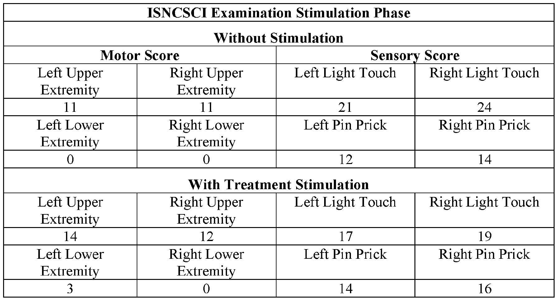
[0212] Figure 5, panels A-F', illustrates magnetic neuromodulation of the cervical cord in SCI. Five subjects with stable SCI (>l year) were evaluated with a battery of tests once a week for 3 months to establish a pre-treatment baseline, with the last month shown here (Pre). Subjects were then treated weekly with EMSS and tested weekly for a month (Treat). Subjects were then tested weekly for a month without treatment to determine the durability of the treatment (Post). Panel A: Handgrip. This is a direct measure of the force generated by subjects with their dominant hand. Individuals all had improved performance of various magnitudes. Panel A’: Subjects had an average of 5-fold improvements in strength that were highly significant. Panel B: Spinal Cord Independence Measure (SCIM) is a 17- item measure of 0-100 with a Minimally Clinical Important Difference of 4 points. There may be two classes of response in this measure. Panel B’ : Subjects had ~30 point increase indicating a robust clinical improvement which was significant. Panel C: Modified Ashworth is a measure of spasticity of 1-4 on ten muscles for a 40-point max. All subjects had improvement in overall spasticity in arms and legs, although subject C had modest improvements. Panel C’: Average spasticity was reduced by half. Panel D: Arm Reach Action Test (ARAT) is a 19-item measure of 0-60. Panel D’: Subjects had -50% increase in performance on this measure. Panel E: International Standards for Neurological Classification of Spinal Cord Injury (ISNCSCI) upper extremity motor exam of five muscles in each arm on a scale for 0-5 for a 50-point max. Minimally Clinical Important Difference is 1 point. Panel E’: Subjects had ~ 30% improvements. Panel F: Columbia Suicide Survey. Subject C had substantial suicidality that was reduced. Panel F’ : no subjects reported suicidality by the last month of the study. Two Tailed Students T-test with Bonferroni post- hoc correction. * p <0.05; ** p <0.01; *** p <0.001. Device: MagPro (MagVenture, Atlanta) with Cool-B35 Butterfly Coil and Biphasic stimulation at 30Hz.
[0213] Figure 6, panels A-B', shows that BUS + EMSS can act rapidly. In a separate cohort, we established baseline function for 7 weeks followed by treatment with BUS treatment. EMSS was conducted on day 3, 5, and 7 of BUS treatment. Panel A: In grip strength, a rapid and significant increase in grip was seen even in the first session. Panel A’: By the last session, as summarized, there was a robust increase in grip strength both before and after stimulation. Panel B: In a measure of precision hand movements where a subject follows a sine wave on a screen by moving a pointer, substantial increases were seen in the first session. Panel B’ : Although not reaching significance, these data appear to trend towards improvement. Two Tailed Students T-test with Bonferroni post-hoc correction. * p <0.05; ** p <0.01; *** p <0.001. Device: MagPro (MagVenture, Atlanta) with Cool-B35 Butterfly Coil and Biphasic stimulation at 30Hz.
[0214] Figure 7 shows that SCEPs improve after EMSS intervention. The effect of
EMSS treatment on spinal cord evoked potentials is shown. Subjects with stable cervical SCI were evaluated for their ability to produce Spinal Cord Evoked Potentials (SCEPs)with EMSS pre- and post-treatment. The Y-axis indicates the size of the evoked potential measured by EMG at the relevant muscle. The X-axis is increasing stimulation intensity with EMSS. In the right panels, two subjects were evaluated before treatment. Both subjects have some activity at all the motor pools evaluated, although not apparent at this scale. In the left panels the same two subjects were evaluated post-treatment. A large treatment effect is seen in the SCEPs reflecting changes in spinal cord circuitry related to motor function. This technique can be used to measure the inherent segmental responsiveness of the cord. This technique does not require volitional control of the segmental levels in question and is therefore well suited to evaluating subjects with paralysis.
[0215] Figure 8 shows that training and EMSS produce a long-lasting therapeutic effect. Time course of grip strength in stable (>l year) cervical SCI subjects (N=10). Pre intervention baseline grip strength was fairly low as would be expected in this subject population. After one month of training using a 120 second 30Hz stimulation, substantial improvements were measured. These results continued to improve in the absence of EMSS at 1 month post-intervention and remained stable for the 2-month evaluation. At months 3 and 4, the therapeutic effect appears to be waning; however, these points are not statistically distinct from the 1 and 2 month points. A 4-month improvement is of high utility. Two Tailed Students T-test with Bonferroni post-hoc correction. N=l0, ** p <.01; *** p <.001. Device: MagPro (MagVenture, Atlanta) with Cool-B35 Butterfly Coil and Biphasic stimulation at 30Hz.
[0216] Figure 9 illustrates parameters that can be varied using a transcutaneous stimulator ( e.g ., a portable stimulator) as described herein. In certain embodiments the parameters that can be controlled/varied include stimulation frequency, stimulation amplitude, stimulation pulse width. In certain embodiments the carrier frequency, and/or carrier pulse width can be controlled. In certain embodiments the carrier signal can be turned off while leaving a stimulation signal on.
[0217] Figure 10 shows an anterolateral view of the spinal cord illustrating sites of epidural stimulation.
[0218] Figure 11 illustrates a schematic of one embodiment of a portable stimulator:
An iphone or android platform can communicate by bluetooth or WiFi with the stimulator with the output signal as described in Figure 9.
[0219] Figure 12 illustrates a practical demonstration of the stimulator design.
[0220] Figure 13 shows the results of transcutaneous magnetic stimulation in a post operative patient with C5 nerve root palsy.
[0221] Figure 14 shows the percent muscle strength improvement in 6 patients with nerve root palsy treated with transcutaneous magnetic stimulation.
[0222] Figure 15 shows the percent of responders at treatment sessions.
DETAILED DESCRIPTION
[0223] It was determined that the following can be leveraged to regain motor function in spinal cord injured subjects which can be broadened to include any subjects with injury to the central nervous system or degenerative neuromotor conditions (stroke, TBI, MS, ALS, Parkinson’s disease, Alzheimer’s disease):
[0224] 1 Stimulation with devices that imparts an electrical or magnetic field (frequency range from 5-100 Hz) of the cervical, thoracic, and lumbar spinal cord, nerve roots, or combinations thereof can restore arm and leg movement;
[0225] 2 With training and repetition, the gains with stimulation can be hardwired and present even without stimulation; and
[0226] 3 Serotonin agonist medication such as buspirone and/or selective serotonin reuptake inhibitors (e.g., fluoxetine) can be used as tool to further activate the spinal network to improve motor function.
[0227] In particular, it was discovered that stimulation of the spinal cord or regions thereof can effectively improve motor function and/or reduce pain in subjects with nerve root palsies (e.g., radiculopathies including, but not limited to cauda equina syndrome). It was also discovered that stimulation of the spinal cord or regions thereof can effectively improve motor function of upper and/or lower extremities and can restore motor function (partially or fully) in subjects with impaired extremity motor function due to spinal cord or brain injury or pathology.
[0228] Moreover it was surprisingly discovered that non-invasive stimulation, e.g., transcutaneous and magnetic stimulation were particular effective and well tolerated.
Moreover, it was demonstrates that Electromagnetic Spinal Cord Stimulation (EMSS) over repeated treatment periods provided persistent improvement even in the absence of the stimulation.
Treatment of nerve root palsies and/or radiculopathies including cauda equina syndrome.
[0229] In various embodiments methods of treating nerve root palsies are contemplated. In certain embodiments the methods involve neuromodulating the brain stem and/or suboccipital spinal cord, and/or the cervical spinal cord or a region thereof, and/or the thoracic spinal cord or a region thereof, and/or the lumbar spinal cord or a region thereof, of the subject with a magnetic stimulator at a frequency and intensity sufficient to regulate and/or to restore function lost by the nerve root disorder; and/or neuromodulating the brain stem and/or suboccipital spinal cord, and/or the cervical spinal cord, and/or the thoracic spinal cord, and/or the lumbar spinal cord, of the subject by administering transcutaneous stimulation to the brain stem and/or suboccipital spinal cord, and/or the cervical spinal cord or a region thereof, and/or the thoracic spinal cord or a region thereof, and/or the lumbar spinal cord or a region thereof at a frequency and intensity sufficient to restore function lost by the nerve root disorder; and/or neuromodulating the brain stem and/or suboccipital spinal cord, and/or the cervical spinal cord, and/or the thoracic spinal cord, and/or the lumbar spinal cord, of the subject by administering epidural stimulation to the brain stem and/or suboccipital spinal cord, and/or cervical spinal cord or a region thereof, and/or thoracic spinal cord or a region thereof, and/or the lumbar spinal cord or a region thereof at a frequency and intensity sufficient to restore function lost by the nerve root disorder.
[0230] In various embodiments the neuromodulation by is at a frequency and amplitude sufficient to restore function lost by the nerve root disorder. In certain
embodiments this restoration of function can entail a reduction in pain, and/or an increase in strength, and/or an increase in motor control, and/or in improvement in sensory function. In certain embodiments the transcutaneous stimulation and/or magnetic stimulation, and/or epidural stimulation is according to the various stimulation parameters described below.
[0231] In certain embodiments the nerve root palsy to be treated comprises a cervical radiculopathy, and/or a lumbar radiculopathy, and/or a sacral radiculopathy. The most common nerve root palsies are C5, L5, and Sl, however the methods described herein are not limited to the treatment of these palsies. In certain embodiments the radiculopathy is a cervical radiculopathy and, in particular embodiments the cervical radiculopathy comprises a C5 radiculopathy (e.g., in which pain is typically found along the lateral brachium of the affected side of the arm and C5 innervated muscle and/or weakness may be found i.e.
rhomboids, deltoid etc.), a C6 radiculopathy (e.g., in which pain is typically found along the lateral antebrachium of the affected arm and/or C6 innervated muscles are weak i.e. forearm pronator and supinators, wrist extensors etc.) and C7 radiculopathies (e.g, in which pain is typically found along the middle finger of the affected arm and/or C7 innervated muscle weakness is found (e.g., wrist flexors, finger extensors etc.).
[0232] In certain embodiments, the nerve root palsy to be treated includes peripheral nerve dysfunction or neuropathies such as: brachial plexus neuropathy (e.g., brachial neuritis, Parsonnage Turner syndrome), ulnar neuropathy, median neuropathy, radial neuropathy, axillary nerve injuries, suprascapular neuropathy, obturator neuropathy, lumbosacral plexus neuropathy, femoral neuropathy, common peroneal neuropathy (e.g., superficial, common, deep), tibial neuropathy, thoracic outlet syndrome, anterior interosseous neuropathy, musculocutaneous neuropathy, amyloid neuropathy, uremic neuropathy, occipital neuropathy, demyelinating polyradiculoneuropathy (Guillain-Barre syndrome), drug-induced neuropathies, diabetic neuropathy, alcoholic neuropathy, HIY neuropathy, etc. Furthermore, peripheral nerve dysfunction having one or more traumatic causes may be treated.
[0233] In certain embodiments the radiculopathies include, but are not limited to lower limb radiculopathies. Such radiculopathies are often on a single nerve root, and the cause is typically a herniated intervertebral disc. Typically symptoms include, but are not limited to one or more of scoliosis, paraspinal muscle contracture, and the reduction of lumbar lordosis. In certain embodiments the lower limb radiculopathies include, but are not limited to an L4 radiculopathy ( e.g ., pain is typically located on the front of the thigh and shin, an can further radiate towards the inner ankle, sometimes the medial toe, and/or can be characterized by failure of the quadriceps muscle and reflex weakness, an L5 radiculopathy (e.g., typically characterized by pain radiating to the side of the thigh and lower leg towards the back of the foot and toes 1-3), and an Sl radiculopathy (e.g., typically characterized by pain radiating to the posterior side of the thigh and lower leg to the ankle side, sometimes up to the fourth toe and/or weakness of the gluteal muscles, and/or difficulty standing on toes.
[0234] In some instances the radiculopathy comprises cauda equina syndrome.
Cauda equina syndrome is a condition due to damage to the bundle of nerves below the end of the spinal cord known as the cauda equina. Symptoms include low back pain, pain that radiates down the leg, numbness around the anus, and loss of bowel or bladder control.
Onset may be rapid or gradual. Causes of cauda equina include, but are not limited to a ruptured disk in the lumbar area (the most common cause), spinal stenosis, a spinal lesion or malignant tumor, a spinal infection, inflammation, hemorrhage, or fracture, a complication from a severe lumbar spine injury such as a car crash, fall, gunshot, or stabbing, and a birth defect such as an arteriovenous malformation.
[0235] In certain embodiments the neuromodulation of the spinal cord is sufficient to partially or to fully ameliorate any one or more of these symptoms.
Restoration of Upper and/or Lower Extremity function.
[0236] It was also discovered that Electromagnetic Spinal Cord Stimulation (EMSS) is effective to partially or fully restore motor control of upper extremities and/or of lower extremities in subjects having impaired motor function of an extremity due to spinal cord or brain injury or pathology. Moreover it was surprisingly discovered that magnetic stimulation over repeated treatment periods provided persistent improvement even in the absence of the stimulation. [0237] Many spinal cord injuries involve the cervical spine, yet they are often incomplete and the local spinal cord circuitry for arm and hand control may be spared. We have leveraged this fact, and the observation that in spinal cord injured states, circuitry distal to the injury site can be neuromodulated to regain function (as in the case of locomotion) to develop a rehabilitative strategy to re-enable significant volitional control of hand function (see, e.g., Example 2).
[0238] In particular, we used non-invasive electromagnetic stimulation of the spinal cord (EMSS) to improve motor performance in 10 subjects with motor complete injury (see, e.g., Figure 5) and demonstrated significant and functionally important improvements in hand function.
[0239] Prior to the results shown herein, it was unknown whether non-invasive neuromodulation with EMSS could be used to enable spared function of cervical neuromotor networks, e.g., neural networks related to upper limb function in animals and humans. The potential impact of the therapeutic interventions described herein on the lives of individuals with tetraplegia cannot be overestimated. Development of non-invasive stimulation to activate spared, but ineffective, spinal cord sensorimotor networks related to upper limb function in humans represents a paradigm shift in the rehabilitative approach to upper limb paralysis as a result of SCI and potentially other neurologic injuries, diseases, or stroke. By re-enabling spared functions, the cost-savings can include reduced assisted daily care costs, increased employment, and improved quality of life-especially for incomplete SCI patients who account for the majority of SCI patients. The methods described herein can provide a partial solution to the US$40 billion/year care and $5.5 billion/year lost productivity costs.
[0240] Of the 10 million people in the US living with paralysis, 15,000 are the result of a SCI each year. The cost of the first year of care can range from $322,000-$986,000, with lifetime costs of $l.4-4M for someone injured at 25 years of age.
[0241] The methods described herein can improve the lives of patients with chronic or new SCI and other forms of nervous system damage or disease (e.g., stroke, multiple sclerosis).
[0242] Importantly, there is currently no effective treatment to improve hand function after chronic cervical SCI. However, even without full restoration of hand function, incremental improvements can make a substantial impact in the lives of those with tetraplegia (www.ada.gov). For example, in a patient with minimal hand function (< 5N grip force), an increase in grip strength to 10 N will allow the subject to operate a wheelchair joystick, computer keyboard, self-care, and open/close doors.
[0243] Such improvements in hand function allow the patient to be more independent, less reliant on caregivers, and will provide psychologically benefit.
[0244] Using non-invasive, Electromagnetic Spinal Cord Stimulation (EMSS,
MagPro XI 00, MagVenture, Atlanta, GA, USA) to improve volitional upper limb function in individuals with spinal cord injury (SCI), we observed improved upper limb function and quality of life scores in 10 subjects undergoing cervical EMSS. EMSS is widely available and gaining adoption, produces clinical and functional improvements for extended periods of time (~4 months), is compatible with spinal fixation implants, and is simple and safe to administer. Small functional improvements (which were readily detectable and functionally significant in the first 10 patients), even if they require follow-up EMSS treatments, justify this approach.
[0245] Because upper limb movement is a complex motor behavior that relies on voluntary motor functions and involuntary sensory functions, recovery of spared spinal cord function can be a highly preferred, safe, and cost effective rehabilitation strategy It is difficult to replicate these complex, coordinated functions by electrically stimulating the nerves and muscles of the upper limb. In fact, this approach has not yielded substantial restoration of upper limb function. Our non-invasive approach is designed to deliver low- intensity, sub-threshold neuromodulation with EMSS to rehabilitate spared sensorimotor circuitries in the spinal cord that control the upper limb and that appear to persist in patients with incomplete spinal cord injury (SCI). Without being bound to a particular theory, we believe that combined with upper limb sensorimotor rehabilitative training, that previously ineffective, spared descending inputs can achieve depolarization of spinal neurons that, in turn, permit volitional upper limb motor control. This can result in plastic changes in the spinal networks controlling hand function that persist in the absence of stimulation for several months, perhaps longer.
[0246] Each muscle of the arm and hand has a dedicated motor pool that develops in a distinct manner. At the cervical enlargement, in addition to Renshaw cells and la interneurons, there are at least 20 distinct intemeurons that comprise 11 classes of interneurons organized by their expression of transcription factors, axon projection patterns, and a predominant neurotransmitter. The molecular and cellular complexity of the cervical spinal cord reflects the local sensorimotor neural processing that integrates sensation (several tactile modalities, several pain modalities, and proprioception from Golgi tendon organs and intrafusile fibers), ascending sensory information from elsewhere in the body and descending sensory ( e.g . vestibular), involuntary motor (e.g. extrapyramidal) and voluntary motor commands.
[0247] The SCI and rehabilitation fields have only a rudimentary understanding of what types of information reach the cervical enlargement and the cellular targets of these inputs. Rational approaches to‘reverse-engineer’ the spinal circuitry have been modeled for lumbar locomotion yet unresolved issues remain, especially for the upper limb. Despite this complexity, as shown in Example 2, we can reliably measure the functional output of the motor circuitry with Spinal Cord Evoked Potentials (SCEPs) even in paralyzed subjects with SCI. After a brief magnetic stimulation (10s 5 Hz parallel to the spinal axis) is applied to the skin overlying the cord to slightly depolarize the motor neurons via a“black box” combination of sensory neuron, intemeuron, and motor neuron activity, EMG activity is evoked at the muscles innervated by the cord segment(s) subjected to stimulation. In the context of SCI, this serves as an important assessment tool of local spinal circuitry to define muscles for which at least segmental spinal motor function is present, independent of ascending or descending connections. In certain embodiments our neuromodulation strategy utilizes this spinal capacity to activate muscles.
[0248] Although the majority of cervical spinal cord injuries are at C5-6 and incomplete, injuries of the spinal cord are highly heterogeneous in the size and involvement of local circuitries and fibers of passage connecting spinal cord regions rostral and caudal to the injury . Because of this heterogeneity, the SCEP measurements analyzed empirically and by machine learning algorithms can be used to define stimuli that are effective in evoking a spinal cord motor output. Typically a subject well suited to the methods described herein can produce an SCEP in one or more hand muscle in the subject’s more useful hand (digit and wrist flexors and extensors of the hand that has more residual function after injury).
Functionally, this means that the subject has motor neurons in the spine susceptible to activation and residual or dormant circuits present in the spinal cord post-injury that are amenable to neuromodulation. These SCEP stimulation parameters (spinal level, stimulus intensity and stimulation frequency) can be determined for the subject and applied.
[0249] This provides an empirical approach to improving function. In the face of neural complexity and heterogeneous injuries, we believe that the best solution is to assist the local circuitry to become active in the context of useful motor tasks (sensorimotor rehabilitative training). Our preliminary data suggest that various forms of neuromodulation in the form of sensory feedback, drugs, and electromagnetic stimulation, enable the spared circuitry to recover clinically relevant function at unprecedented and clinically meaningful levels.
[0250] Other tests of residual spinal function that can be performed include, but are not limited to: Assessing sensory evoked responses from the ulnar nerve and determining whether a) the activity is reflected in the spinal cord, and b) whether the information reaches the cortex. The motor cortex can be stimulated with transcranial magnetic motor evoked potentials (tcmMEP) on both sides of the cortex to determine how much motor information passes through the injury site. In certain embodiments, somatosensory and motor evoked potentials can be obtained before and after the intervention to determine whether spinal electromagnetic stimulation alters the effectiveness of communication across the site of injury. This is an especially useful assessment because the AIS classification is crude and does not capture the subtleties of anatomically incomplete injuries, and we expect spinal stimulation to enhance communication across the site of injury.
[0251] While the discussion above focuses on upper extremity function, similar approaches can be taken to partially or to fully restore lower extremity motor function.
[0252] In certain embodiments the neuromodulation produced by magnetic stimulation is sufficient to partially or fully restore upper or lower extremity motor function.
[0253] In certain embodiments restoration of motor function is indicated by an improvement in one or more parameters that can include, but are not limited to increase in SCEP efficiency (lowest input energy and highest EMG output response) in the muscles of the more dominant limb ( e.g ., hand) post-injury, grip strength, hand control, box and block, MusicGlove, ArmeoSpring, ARAT, GRASSP, ISNCSCI, CE1E, modified Ashworth Scale, Penn Spasm Frequency, VAS-spasticity) and quality of life (C-SSRS, SCIM3), tcmMEP, SEP, and the like.
Maenetic stimulation
[0254] As illustrated herein in Example 2, it was discovered that that stimulation of the spinal cord with devices that impart a magnetic field (e.g., at a frequency range from about 0.5 Hz up to about 100 Hz) can improve and/or restore motor function in a subject having impaired motor function of an extremity due to spinal cord or brain injury or pathology ( e.g ., SCI, brain injury, Parkinson's disease, Huntington's disease, Alzheimer's disease, amyotrophic lateral sclerosis (ALS), primary lateral sclerosis (PLS), dystonia, cerebral palsy, etc.). Similarly, magnetic stimulation can be used to treat a nerve root disorder (radiculopathy).
[0255] In various embodiments magnetic spinal cord stimulation can be achieved by generating a rapidly changing magnetic field to induce a current at the nerve(s) of interest (e.g., the spinal cord). Effective stimulation typically utilizes current transients of about 108 A/s or greater discharged through a stimulating coil. The discharge current flowing through the stimulating coil generates magnetic lines of force. As the lines of force cut through tissue, a current is generated in that tissue, whether skin, bone, muscle or neural; if the induced current is of sufficient amplitude and duration such that the cell membrane is depolarized, neuromuscular tissue will be stimulated in the same manner as conventional electrical stimulation.
Magnetic stimulation of the brainstem/suboccipital spinal cord or a region thereof.
[0256] In various embodiments, the methods described herein involve magnetic stimulation of the brainstem and/or suboccipital region. This can be accomplished by application of a magnetic stimulator (e.g., a magnetic stimulator wand) to the suboccipital region of the neck and stimulation can be according to any of the magnetic stimulation parameters described herein.
[0257] In certain embodiments stimulation can be accomplished by transcranial magnetic stimulation applied, e.g., to the base of the skull.
Magnetic Stimulation of the cervical spinal cord.
[0258] In various embodiments, the methods described herein involve magnetic stimulation of the cervical spine or a region of the cervical spine of the subject. Illustrative regions include, but are not limited to, one or more regions straddling or spanning a region selected from the group consisting of C0-C1, C0-C2, C0-C3, C0-C4, C0-C5, C0-C6, C0-C7, C0-T1, Cl-Cl, C1-C2, C1-C3, C1-C4, C1-C7, C1-C6, C1-C7, Cl-Tl, C2-C2, C2-C3, C2- C4, C2-C5, C2-C6, C2-C7, C2-T1, C3-C3, C3-C4, C3-C5, C3-C6, C3-C7, C3-T1, C4-C4, C4-C5, C4-C6, C4-C7, C4-T1, C5-C5, C5-C6, C5-C7, C5-T1, C6-C6, C6-C7, C6-T1, C7-C7, and C7-T1. Magnetic Stimulation of the thoracic spinal cord.
[0259] In various embodiments, the methods described herein involve magnetic stimulation of the thoracic spine or a region of the thoracic spine of the subject. Illustrative regions include, but are not limited to, one or more regions straddling or spanning a region selected from the group consisting of Tl-Tl, T1-T2, T1-T3, T1-T4, T1-T5, T1-T6, T1-T7, T1-T8, T1-T9, T1-T10, Tl-Tl 1, T1-T12, T2-T2, T2-T3, T2-T4, T2-T5, T2-T6, T2-T7, T2- T8, T2-T9, T2-T10, T2-T11, T2-T12, T3-T3, T3-T4, T3-T5, T3-T6, T3-T7, T3-T8, T3-T9, T3-T10, T3-T11, T3-T12, T4-T4, T4-T5, T4-T6, T4-T7, T4-T8, T4-T9, T4-T10, T4-T11, T4- T12, T5-T5, T5-T6, T5-T7, T5-T8, T5-T9, T5-T10, T5-T11, T5-T12, T6-T6, T6-T7, T6-T8, T6-T9, T6-T10, T6-T11, T6-T12, T7-T7, T7-T8, T7-T9, T7-T10, T7-T11, T7-T12, T8-T8, T8-T9, T8-T10, T8-T11, T8-T12, T9-T9, T9-T10, T9-T11, T9-T12, T10-T10, T10-T11, T10- T12, Tl l-Tl l, T11-T12, and T12-T12.
Magnetic Stimulation of the lumbar spinal cord and/or coccyx.
[0260] In various embodiments, the methods described herein involve magnetic stimulation of the thoracic spine or a region of the thoracic spine of the subject. Illustrative regions include, but are not limited to, one or more regions straddling or spanning a region selected from the group consisting of of Ll-Ll, L1-L2 , L1-L3, L1-L4, L1-L5, Ll-Sl, L1-S2, L1-S3, L1-S4, L1-S5, L2-L2 , L2-L3, L2-L4, L2-L5, L2-S1, L2-S2, L2-S3, L2-S4, L2-S5, L3-L3, L3-L4, L3-L5, L3-S1, L3-S2, L3-S3, L3-S4, L3-S5, L4-L4, L4-L5, L4-S1, L4-S2, L4-S3, L4-S4, L4-S5, L5-L5 , L5-S1, L5-S2, L5-S3, L5-S4, L5-S5, Sl-Sl, S1-S2, S1-S3, Sl- S4, S1-S5, S2-S2, S2-S3, S2-S4, S2-S5, S3-S3, S3-S4, S3-S5, S4-S4, S4-S5, and S5-S6.
[0261] In certain embodiments, in addition, or as an alternative to the above- identified locations, stimulation can be applied to the coccyx, e.g., by application of a transcutaneous stimulation electrode to the coccyx, i.e. over the small, triangular bone at the base of the spinal column.
Magnetic stimulation parameters.
[0262] As noted above, magnetic stimulators can be used for stimulation of the spinal cord to restore upper or lower extremity motor function and/or to treat a nerve root disorder (radiculopathy). Since the magnetic field strength falls off with the square of the distance from the stimulating coil, the stimulus strength is at its highest close to the coil surface. The stimulation characteristics of the magnetic pulse, such as depth of penetration, strength and accuracy, depend on the rise time, peak electrical energy transferred to the coil and the spatial distribution of the field. The rise time and peak coil energy are governed by the electrical characteristics of the magnetic stimulator and stimulating coil, whereas the spatial distribution of the induced electric field depends on the coil geometry and the anatomy of the region of induced current flow.
[0263] In various embodiments the magnetic nerve stimulator will produce a field strength up to about 10 tesla, or up to about 8 tesla, or up to about 6 tesla, or up to about 5 tesla, or up to about 4 tesla, or up to about 3 tesla, or up to about 2 tesla, or up to about 1 tesla. In certain embodiments the nerve stimulator produces pulses with a duration from about 100 ps up to about 10 ms, or from about 100 ps up to about 1 ms.
[0264] In certain embodiments the magnetic stimulation is at a frequency of at least about 1 Hz, or at least about 2 Hz, or at least about 3 Hz, or at least about 4 Hz, or at least about 5 Hz, or at least about 10 Hz, or at least about 20 Hz or at least about 30 Hz or at least about 40 Hz or at least about 50 Hz or at least about 60 Hz or at least about 70 Hz or at least about 80 Hz or at least about 90 Hz or at least about 100 Hz, or at least about 200 Hz, or at least about 300 Hz, or at least about 400 Hz, or at least about 500 Hz.
[0265] In certain embodiments the magnetic stimulation is at a frequency ranging from about 1 Hz, or from about 2 Hz, or from about 3 Hz, or from about 4 Hz, or from about 5 Hz, or from about 10 Hz, or from about 10 Hz, or from about 10 Hz, up to about 500 Hz, or up to about 400 Hz, or up to about 300 Hz, or up to about 200 Hz up to about 100 Hz, or up to about 90 Hz, or up to about 80 Hz, or up to about 60 Hz, or up to about 40 Hz, or from about 3 Hz or from about 5 Hz up to about 80 Hz, or from about 5 Hz to about 60 Hz, or up to about 30 Hz.
[0266] In certain embodiments the magnetic stimulation is applied at the midline of spinal cord. In various embodiments the magnetic stimulation produces a magnetic field of at least about 0.5 tesla, or at least about 0.6 tesla, or at least about 0.7 tesla, or at least about 0.8 tesla, or at least about 0.9 tesla, or at least about 1 tesla, or at least about 2 tesla, or at least about 3 tesla, or at least about 4 tesla, or at least about 5 tesla. In certain embodiments the magnetic stimulation is at a frequency of at least about 0.5 Hz, 1 Hz, or at least about 2 Hz, or at least about 3 Hz, or at least about 4 Hz, or at least about 5 Hz, or at least about 10 Hz, or at least about 20 Hz or at least about 30 Hz or at least about 40 Hz or at least about 50 Hz or at least about 60 Hz or at least about 70 Hz or at least about 80 Hz or at least about 90 Hz or at least about 100 Hz, or at least about 200 Hz, or at least about 300 Hz, or at least about 400 Hz, or at least about 500 Hz.
[0267] In certain embodiments, the magnetic stimulation is at a frequency and amplitude sufficient to restore motor function in an extremity ( e.g ., as indicated by an improvement in SCEP efficiency (lowest input energy and highest EMG output response) in the muscles of the more dominant limb (e.g., hand) post-injury, grip strength, hand control, box and block, MusicGlove, ArmeoSpring, ARAT, GRASSP, ISNCSCI, CUE, modified Ashworth Scale, Penn Spasm Frequency, VAS-spasticity) and quality of life (C-SSRS, SCIM3), tcmMEP, SEP, and the like. In certain embodiments, the magnetic stimulation is at a frequency and amplitude sufficient to reduce pain and/or to increase strength and/or to improve motor control in a subject with a nerve root palsy.
Repeated magnetic stimulation treatments to provide persistent improvement.
[0268] More surprisingly, it was discovered that repeated treatments with magnetic stimulation can, over time, produce a persistent improvement in a patient in the subsequent absence of the magnetic stimulation. In certain embodiments, the persistent improvement comprises an improvement in motor function of a limb (e.g., as indicated by an improvement in SCEP efficiency (lowest input energy and highest EMG output response). For example, the improvement in motor function can comprise an improvement in motor function in the muscles of an extremity of a limb (e.g., the more dominant limb) (e.g., hand) such as post injury (e.g., as characterized by grip strength, hand control, box and block, MusicGlove, ArmeoSpring, ARAT, GRASSP, ISNCSCI, CUE, modified Ashworth Scale, Penn Spasm Frequency, VAS-spasticity), and/or improvement in quality of life (e.g., as characterized by C-SSRS, SCIM3), and/or improvement in tcmMEP, SEP, and the like. In certain
embodiments, the persistent improvement comprises a reduction in pain, and/or an increase in strength, and/or an improvement in motor control, and/or an improvement in sensory function in a subject.
[0269] In certain embodiments there is no apparent effect of stimulation while it’s being administered. There can be a delay in response of at least 3 treatment sessions to observe a positive effect. Also importantly, the stimulation does not need to be on to realize the positive effect. There can also be a slow decay of function after treatment and in certain embodiments, multiple treatments are administered to observe effect and decay of the treatment effect can be observed after approximately 2 weeks of no treatment. [0270] Accordingly, in various embodiments a single treatment of magnetic stimulation comprises 1, or 2, or 3, or 4, or 5, or 6, or 7, or 8, or 9, or 10 or more continuous stimulation periods. In various embodiments the continuous stimulation periods range in duration from about 10 sec, or from about 20 sec, or from about 3 sec or from about 40 sec, or from about 50 sec, or from about 1 min, or from about 2 minutes up to about 10 minutes, or up to about 8 minutes, or up to about 6 minutes. In certain embodiments the continuous stimulation periods are about 4 minutes in duration. In certain embodiments the delay between continuous stimulation periods ranges from about 2 sec, or from about 5 sec, or from about 10 sec, or from about 15 sec, or from about 20 sec up to about 5 minutes, or up to about 4 minutes, or up to about 3 minutes, or up to about 2 minutes, or up to about 1 min, or up to about 45 sec, or up to about 30 sec. In certain embodiments the delay between continuous stimulation periods is about 30 sec.
[0271] Repeating the treatment can progressively improve motor function in an upper and/or lower extremity and/or can improve strength, motor control and/or reduce pain associated with a nerve root palsy. Accordingly, in certain embodiments the treatment is repeated ( e.g ., repeated daily, or every 2 days, or every 3 days, or every 4 days, or every 5 days, or every 6 days, or every 7 days, or every 8 days, or every 9 days, or every 10 days, or every 11 days, or every 12 days, or every 13 days, or every 14 days). In certain embodiments the treatment is repeated over a period of at least 1 week, or at least two weeks, or at least 3 weeks, or at least 4 weeks, or at least 5 weeks, or at least 6 weeks, or at least 7 weeks, or at least 8 weeks, or at least 9 weeks, or at least 10 weeks, or at least 11 weeks, or at least 12 weeks, or at least 4 months, or at least 5 months, or at least 6 months, or at least 7 months, or at least 8 months, or at least 9 months, or at least 10 months, or at least 11 months, or at least 12 months. In certain embodiments the treatment is repeated daily, or every 2 days, or every 3 days, or every 4 days, or every 5 days, or every 6 days, or every 7 days, or every 8 days, or every 9 days, or every 10 days, or every 11 days, or every 12 days, or every 13 days, or every 14 days until the subject experiences a reduction of pain and/or improvement in strength, and/or improvement in motor control, e.g., at a later time without magnetic stimulation in treatment of nerve root palsies, or an improvement in motor function of an extremity (e.g., as indicated by an improvement in SCEP efficiency (lowest input energy and highest EMG output response) in the muscles of the more dominant limb (e.g., hand) post-injury, grip strength, hand control, box and block, MusicGlove, ArmeoSpring, ARAT, GRASSP, ISNCSCI, CUE, modified Ashworth Scale, Penn Spasm Frequency, VAS-spasticity) and quality of life (C-SSRS, SCIM3), tcmMEP, SEP, and the like) e.g., at a later time without magnetic stimulation in treatment of impaired motor function of an extremity. In certain embodiments the treatment is repeated daily, or every 2 days, or every 3 days, or every 4 days, or every 5 days, or every 6 days, or every 7 days, or every 8 days, or every 9 days, or every 10 days, or every 11 days, or every 12 days, or every 13 days, or every 14 days until the subject obtains their maximal reduction of pain and/or improvement in strength, and/or improvement in motor control, and/or improvement in sensory function in treatment of a nerve root palsy, or a maximum improvement in motor function of an extremity in treatment of impaired motor function of an extremity.
[0272] In certain embodiments, once the desired level of improvement is achieved, the frequency of treatment can be reduced to a "maintenance" level. Typically, the frequency of treatment can be reduced to a level sufficient to maintain a reduction of pain and/or improvement in strength, and/or improvement in motor control, and/or to improve sensory function, e.g., at a later time without magnetic stimulation in treatment of nerve root palsies, or an improvement in motor function of an extremity (e.g, as indicated by an improvement in SCEP efficiency (lowest input energy and highest EMG output response) in the muscles of the more dominant limb (e.g., hand) post-injury, grip strength, hand control, box and block, MusicGlove, ArmeoSpring, ARAT, GRASSP, ISNCSCI, CUE, modified Ashworth Scale, Penn Spasm Frequency, VAS-spasticity) and quality of life (C-SSRS, SCIM3), tcmMEP,
SEP, and the like) e.g., at a later time without magnetic stimulation in treatment of impaired motor function of an extremity. It should be understood that in certain embodiments, repeated treatments with other forms of stimulation such as those described herein (e.g., electrical stimulation) may similarly produce a persistent improvement in a patient in the subsequent absence of the stimulation.
Transcutaneous electrical stimulation.
[0273] In various embodiments transcutaneous electrical stimulation of the spinal cord (e.g., one or more regions of the cervical spinal cord, and/or the thoracic spinal cord, and/or the lumbar spinal cord) is utilized to treat a nerve root palsy, e.g., to achieve a reduction of pain and/or improvement in strength, and/or improvement in motor control, and/or improvement in sensory function in a subject with a nerve root palsy. In certain embodiments the transcutaneous stimulation can be of one or more regions of the cervical spinal cord, and/or one or more regions of the thoracic spinal cord, and/or one or more regions of the lumbar spinal cord [0274] The location of the electrode(s) and their stimulation parameters can be important in treating one or more effects of the nerve root palsy. Use of surface electrode(s), as described herein, facilitates selection or alteration of particular stimulation sites as well as the application of a wide variety of stimulation parameters. Additionally, surface stimulation can be used to optimize location for an implantable electrode or electrode array for epidural stimulation.
Transcutaneous stimulation of the brainstem/suboccipital spinal cord or a region thereof.
[0275] In various embodiments, the methods described herein involve transcutaneous electrical stimulation of the brainstem and/or suboccipital region. This can be accomplished by application of one or more transcutaneous stimulation electrode(s), e.g., as described herein, over the base of the skull and/or over the suboccipital region and the transcutaneous stimulation can be applied using the transcutaneous stimulation parameters described herein. As used herein the suboccipital region refers to a region of the neck bounded by the following three muscles of the suboccipital group of muscles: 1) Rectus capitis posterior major - above and medially; 2) Obliquus capitis superior - above and laterally; and 3) Obliquus capitis inferior - below and laterally.
Transcutaneous electrical stimulation of the cervical spine or a region thereof
[0276] In certain embodiments, the methods described herein involve transcutaneous electrical stimulation of the cervical spine or a region of the cervical spine. Illustrative regions include, but are not limited to one or more regions straddling or spanning a region selected from the group consisting of C0-C1, C0-C2, C0-C3, C0-C4, C0-C5, C0-C6, C0-C7, C0-T1, Cl-Cl, C1-C2, C1-C3, C1-C4, C1-C7, C1-C6, C1-C7, Cl-Tl, C2-C2, C2-C3, C2- C4, C2-C5, C2-C6, C2-C7, C2-T1, C3-C3, C3-C4, C3-C5, C3-C6, C3-C7, C3-T1, C4-C4, C4-C5, C4-C6, C4-C7, C4-T1, C5-C5, C5-C6, C5-C7, C5-T1, C6-C6, C6-C7, C6-T1, C7-C7, and C7-T1.
[0277] In certain embodiments the transcutaneous stimulation is applied at a region comprising C2-C4 or a region therein. In certain embodiments the stimulation is applied at C3 Transcutaneous electrical stimulation of the thoracic spine or a region thereof
[0278] In various embodiments, the methods described herein additionally or alternatively involve transcutaneous electrical stimulation of the thoracic spine ( e.g ., spinal cord) or a region of the thoracic spine (spinal cord) of the subject. Illustrative regions include, but are not limited to, one or more regions straddling or spanning a region selected from the group consisting of Tl-Tl, T1-T2, T1-T3, T1-T4, T1-T5, T1-T6, T1-T7, T1-T8, Tl- T9, T1-T10, Tl-Tl 1, T1-T12, T2-T2, T2-T3, T2-T4, T2-T5, T2-T6, T2-T7, T2-T8, T2-T9, T2-T10, T2-T11, T2-T12, T3-T3, T3-T4, T3-T5, T3-T6, T3-T7, T3-T8, T3-T9, T3-T10, T3- Tl l, T3-T12, T4-T4, T4-T5, T4-T6, T4-T7, T4-T8, T4-T9, T4-T10, T4-T11, T4-T12, T5-T5, T5-T6, T5-T7, T5-T8, T5-T9, T5-T10, T5-T11, T5-T12, T6-T6, T6-T7, T6-T8, T6-T9, T6- T10, T6-T11, T6-T12, T7-T7, T7-T8, T7-T9, T7-T10, T7-T11, T7-T12, T8-T8, T8-T9, T8- T10, T8-T11, T8-T12, T9-T9, T9-T10, T9-T11, T9-T12, T10-T10, T10-T11, T10-T12, Tl l- Tl l, T11-T12, and T12-T12.
Transcutaneous electrical stimulation of the lumbar spine or a region thereof, and/or coccyx.
[0279] In various embodiments, the methods described herein additionally or alternatively involve transcutaneous electrical stimulation of the thoracic spine (e.g., spinal cord) or a region of the thoracic spine (spinal cord) of the subject. Illustrative regions include, but are not limited to, one or more regions straddling or spanning a region selected from the group consisting of Ll-Ll, L1-L2 , L1-L3, L1-L4, L1-L5, Ll-Sl, L1-S2, L1-S3, Ll- S4, L1-S5, L2-L2 , L2-L3, L2-L4, L2-L5, L2-S1, L2-S2, L2-S3, L2-S4, L2-S5, L3-L3, L3- L4, L3-L5, L3-S1, L3-S2, L3-S3, L3-S4, L3-S5, L4-L4, L4-L5, L4-S1, L4-S2, L4-S3, L4-S4, L4-S5, L5-L5 , L5-S1, L5-S2, L5-S3, L5-S4, L5-S5, Sl-Sl, S1-S2, S1-S3, S1-S4, S1-S5, S2- S2, S2-S3, S2-S4, S2-S5, S3-S3, S3-S4, S3-S5, S4-S4, S4-S5, and S5-S6.
[0280] In certain embodiments, in addition, or as an alternative to the above- identified locations, transcutaneous stimulation can be applied to the coccyx, e.g., by application of one or more transcutaneous stimulation electrode(s) over the small, triangular bone at the base of the spinal column.
[0281] In certain embodiments the transcutaneous electrical stimulation is applied paraspinally over a lumbar region identified above or to a region thereof, e.g., over a region spanning L2 to L3). Transcutaneous electrical stimulation parameters.
[0282] In certain embodiments the transcutaneous stimulation is at a frequency of at least about 1 Hz, or at least about 2 Hz, or at least about 3 Hz, or at least about 4 Hz, or at least about 5 Hz, or at least about 10 Hz, or at least about 20 Hz or at least about 30 Hz or at least about 40 Hz or at least about 50 Hz or at least about 60 Hz or at least about 70 Hz or at least about 80 Hz or at least about 90 Hz or at least about 100 Hz, or at least about 200 Hz, or at least about 300 Hz, or at least about 400 Hz, or at least about 500 Hz, or at least about 1 kHz, or at least about 1.5 kHz, or at least about 2 kHz, or at least about 2.5 kHz, or at least about 5 kHz, or at least about 10 kHz, or up to about 25 kHz, or up to about 50 kHz, or up to about 100 kHz.
[0283] In certain embodiments the transcutaneous stimulation is at a frequency ranging from about 1 Hz, or from about 2 Hz, or from about 3 Hz, or from about 4 Hz, or from about 5 Hz, or from about 10 Hz, or from about 10 Hz, or from about 10 Hz, up to about 500 Hz, or up to about 400 Hz, or up to about 300 Hz, or up to about 200 Hz up to about 100 Hz, or up to about 90 Hz, or up to about 80 Hz, or up to about 60 Hz, or up to about 40 Hz, or from about 3 Hz or from about 5 Hz up to about 80 Hz, or from about 5 Hz to about 60 Hz, or up to about 30 Hz.
[0284] In certain embodiments the transcutaneous stimulation is applied at an intensity ranging from about 5 mA or about 10 mA up to about 500 mA, or from about 5 mA or about 10 mA up to about 400 mA, or from about 5 mA or about 10 mA up to about 300 mA, or from about 5 mA or about 10 mA up to about 200 mA, or from about 5 mA or about 10 mA to up about 150 mA, or from about 5 mA or about 10 mA up to about 50 mA, or from about 5 mA or about 10 mA up to about 100 mA, or from about 5 mA or about 10 mA up to about 80 mA, or from about 5 mA or about 10 mA up to about 60 mA, or from about 5 mA or about 10 mA up to about 50 mA.
[0285] In certain embodiments the transcutaneous stimulation is applied stimulation comprises pulses having a width that ranges from about 100 ps up to about 1 ms or up to about 800 ps, or up to about 600 ps, or up to about 500 ps, or up to about 400 ps, or up to about 300 ps, or up to about 200 ps, or up to about 100 ps, or from about 150 ps up to about 600 ps, or from about 200 ps up to about 500 ps, or from about 200 ps up to about 400 ps.
[0286] In certain embodiments the transcutaneous stimulation is at a frequency, pulse width, and amplitude sufficient to achieve a reduction of pain and/or improvement in strength, and/or improvement in motor control, and/or improvement in sensory function in a subject with a nerve root palsy.
[0287] In certain embodiments the transcutaneous stimulation is superimposed on a high frequency carrier signal (see, e.g., Figure 9). In certain embodiments the high frequency carrier signal ranges from about 1 kHz, or about 3 kHz, or about 5 kHz, or about 8 kHz up to about 30 kHz, or up to about 20 kHz, or up to about 15 kHz. In certain embodiments the carrier signal is about 10 kHz. In certain embodiments the carrier frequency amplitude ranges from about 30 mA, or about 40 mA, or about 50 mA, or about 60 mA, or about 70 mA, or about 80 mA up to about 300 mA, or up to about 200 mA, or up to about 150 mA.
Transcutaneous stimulation electrodes.
[0288] In certain embodiments the transcutaneous stimulation is applied using any of a number different types of electrodes. Such electrodes include, but are not limited to metal plate electrodes, carbon electrodes, textile electrodes, hydrogel electrodes, needle electrodes, and the like skin (see, e.g., Keller & Kuhn (2008) J. Automatic Control., 18(2): 34-45). In various embodiments, the electrodes can be adhered using, e.g., tape or other adherent, or in other embodiments, the electrodes are self-adhering.
[0289] Metal plate electrodes include, but are not limited to metal plate electrodes covered by fabric tissue. Typically the metal plate is fabricated from a biocompatible material. Often stainless steel or silver/ silver chloride electrodes are used. The fabric tissue can be cotton but is often a polymer textile material that has a certain degree of elasticity and doesn’t wear out fast. Spongy materials have also been used and recommended (see, e.g., Falk et al. (1983) N. Engl. J. Med. 309: 1166-1168). In certain embodiments, the fabric can be made conductive with water or electrode gel. It equally distributes the current over the skin in order to prevent skin burns. Care has to be taken that the electrode does not dry out.
In the best case (if completely dry) such a dried out electrode isolates the metal plate from the skin. But while drying out, unequally distributed electrical fields under the electrodes may cause skin burns. The electrodes are typically fixed to the skin with elastic straps (see, e.g., Ijezerman et al. (1996) J. Rehab. Sci. 9: 86-89).
[0290] Self-adhesive electrodes for transcutaneous stimulation use a gel to contact a conductive member with the subject's skin (see, e.g., Keller & Kuhn (2008) J. Automatic Control., 18(2): 34-45). The electrode is typically built in a multi-layer configuration, consisting of multiple layers of hydrogel. The skin interface layer often includes an electrically conductive gel with relatively low peel strength for removably contacting the subject's skin. It has a wet feeling and can be removed relatively easily from the skin. In various illustrative, but non-limiting embodiments, the conductive gel is made from co polymers derived from polymerization, e.g. of acrylic acid and N-vinylpyrrolidone. In various illustrative embodiments, a second hydrogel layer connects the substrate (a low resistive material like carbon rubber or a wire mesh) with the skin hydrogel layer. This second conductive gel layer has a relatively high peel strength that provides good adhesion to the substrate.
[0291] In certain embodiments, carbon loaded silicon electrodes can be used (see, e.g., Baker, D. R. McNeal, L. A. Benton, B. R. Bowman, and R. L. Waters, Neuromuscular electrical stimulation: a practical guide, 3 ed. USA: Rehabilitation Engineering Program, Los Amigos Research and Education Institute, Rancho Los Amigos Medical Center, 1993;
Nathan (1989) J. Automatic Control, 18(2): 35-45; Patterson & Lockwood (1993) IEEE Trans on Neural Systems and Rehabilitation, 1 : 59-62; and the like).
[0292] In certain embodiments, the transcutaneous electrical stimulation can be applied via textile electrodes. In one illustrative, but non-limiting embodiment, the textile electrodes can consist of multiple fabric layers (see, e.g, Keller, et al. (2006) Conf. Proc. IEEE Eng. Med. Biol. Soc. 1 : 194-197). In certain embodiments, the fabric layer facing the skin holds embroidered electrode pads made of plasma coated metallized yarn. Because of the thin metal coating (e.g, <25 nm coating particles obtained using a plasma process) the yarn keeps its textile properties and can be embroidered. Silver coatings proved to be most stable and survived 30 washings. A second layer contains the embroidered electrode wiring made from the same materials and was designed such that no short circuits are produced between the pads when stitched together (Id).
[0293] In certain embodiments, the transcutaneous electrical stimulation can be applied via one or more needle electrodes, e.g., as described in PCT Patent Pub No: WO 2017/024276 (PCT/US2016/045898). As described therein, needle electrodes comprise one or more commonly a plurality of electrically conductive solid microprojections (or where the needles are hollow, they are closed at the tip), where the needles (microprojections) have a tip dimension/diameter small enough to facilitate penetration of the stratum comeum on the skin (e.g, less than about 10 pm), where the needles have a length greater than about 20 pm and where the electrically conductive solid needles are electrically coupled to one or more electrical leads. In one illustrative, but non-limiting embodiment, needles with tip size of several mih or smaller, and a shaft length of 50 pm or more can be used for transcutaneous electrical stimulation electrodes. In one illustrative, but non-limiting embodiment, a single electrode unit, consisting of 5x5 to 30x30 needles, is about one centimeter in diameter.
Multiple electrode units can be further combined into an electrode array, e.g., when larger electrode areas are needed (for example, for the retum/ground electrodes). The needle electrodes can provide low impedance transcutaneous stimulation without using a conductive gel or cream. In certain embodiments the needle electrodes comprise one or a plurality of electrically conductive needles, where the needles are solid, or wherein the needles are hollow and have a closed tip, where the needles having an average tip diameter less than about 10 pm and an average length greater than about 10 pm or greater than about 20 pm were the electrically conductive needles can be electrically coupled to one or more electrical leads. In certain embodiments, the needle electrode comprises at least about 10 needles, or at least about 15 needles, or at least about 20 needles, or at least about 25 needles, or at least about 30 needles, or at least about 40 needles, or at least about 50 needles, or at least about 100 needles, or at least about 200 needles, or at least about 300 needles, or at least about 400 needles, or at least about 500 needles, or at least about 600 needles, or at least about 700 needles, or at least about 800 needles, or at least about 900 needles, or at least about 1000 needles. In certain embodiments, the needle(s) comprising the needle electrode are of sufficient length to penetrate at least 70%, or at least 80%, or at least 90%), or at least 100%> through the stratum corneum of the skin when the electrode is attached to the surface of a human over the spinal cord. In certain embodiments, the needle(s)s are of a length that does not substantially penetrate subcutaneous tissue below the stratum corneum. In certain embodiments the average length of needle(s) comprising the needle electrode ranges from about 1 pm up to about 100 pm, or from about 1 pm up to about 80 pm, or from about 1 pm up to about 50 pm, or from about 1 pm up to about 30 pm, or from about 1 pm up to about 20 pm , or is at least about 30 pm, or at least about 40 pm, or at least about 50 pm, or at least about 60 pm, or at least about 70 pm. In certain embodiments the average length of the needle(s) is less than about 200 pm, or less than about 150 pm, or less than about 100 pm. In certain embodiments the average length of the needle(s) ranges from about 40 to about 60 pm.
[0294] In certain embodiments the average length of the needle(s) is about 50 pm. In certain embodiments the tip of the needle(s) ranges in diameter (or maximum cross-sectional dimension) from about 0.1 pm up to about 10 pm, or from about 0.5 pm up to about 6 pm, or from about 1 mih up to about 4 pm. In certain embodiments the average separation between two adjacent needles ranges from about 0.01 mm up to about 1 mm, or about 0.05 mm up to about 0.5 mm, or about 0.1 mm up to about 0.4 mm, or up to about 0.3 mm, or up to about 0.2 mm. In certain embodiments the average separation between two adjacent needles ranges from about 0.15 mm up to about 0.25 mm. In certain embodiments the needles are disposed in an area of about 1 cm2 or less, or about 0.8 cm2 or less, or about 0.6 cm2 or less, or about 0.5 cm2 or less, or about 0.4 cm2 or less, or about 0.3 cm2 or less, or about 0.2 cm2 or less, or about 0.1 cm2 or less. In certain embodiments the needles are disposed in an area of about 2 mm or about 3 mm, or about 4 mm, or about 5 mm, or about 6 mm, or about 7 mm or about 8 mm, or about 9 mm, or about 10 mm by about 2 mm or about 3 mm, or about 4 mm, or about 5 mm, or about 6 mm, or about 7 mm or about 8 mm, or about 9 mm, or about 10 mm. In certain embodiments the electrode comprises about 20 x about 20 needles in an area about 4 x 4 mm.
[0295] The foregoing electrodes for transcutaneous electrical stimulation are illustrative and non-limiting. Using the teaching provided herein, numerous other electrodes and/or electrode configurations will be available to one of skill in the art.
Epidural stimulation
[0296] In various embodiments epidural stimulation of the spinal cord is utilized to treat a nerve root palsy, e.g., to achieve a reduction of pain and/or improvement in strength, and/or improvement in motor control, and/or improvement in sensory function in a subject with a nerve root palsy. In certain embodiments the epidural stimulation can be of one or more regions of the cervical spinal cord, and/or one or more regions of the thoracic spinal cord, and/or one or more regions of the lumbar spinal cord. In various embodiments the epidural stimulation can be used alone or in combination with transcutaneous and/or with magnetic stimulation.
Epidural stimulation of the brainstem/suboccipital spinal cord or a region thereof.
[0297] In various embodiments, the methods described herein involve epidural electrical stimulation of the brainstem and/or suboccipital region As used herein the suboccipital region refers to a region of the neck bounded by the following three muscles of the suboccipital group of muscles: 1) Rectus capitis posterior major - above and medially; 2) Obliquus capitis superior - above and laterally; and 3) Obliquus capitis inferior - below and laterally. In various embodiments, stimulation can be via, inter alia, an implanted electrode or electrode array.
Epidural stimulation of the cervical spine or a region thereof
[0298] In various embodiments, the methods described herein involve epidural electrical stimulation of the cervical spine ( e.g ., spinal cord) or a region of the cervical spine (spinal cord) of the subject. Illustrative regions include, but are not limited to, one or more regions straddling or spanning a region selected from the group consisting of C0-C1, C0-C2, C0-C3, C0-C4, C0-C5, C0-C6, C0-C7, C0-T1, Cl-Cl, C1-C2, C1-C3, C1-C4, C1-C7, Cl- C6, C1-C7, Cl-Tl, C2-C2, C2-C3, C2-C4, C2-C5, C2-C6, C2-C7, C2-T1, C3-C3, C3-C4, C3-C5, C3-C6, C3-C7, C3-T1, C4-C4, C4-C5, C4-C6, C4-C7, C4-T1, C5-C5, C5-C6, C5- C7, C5-T1, C6-C6, C6-C7, C6-T1, C7-C7, and C7-T1.
[0299] In certain embodiments the epidural stimulation is applied paraspinally over a cervical region identified above (e.g., over vertebrae spanning CO to C8 or a region thereof, e.g., over a region spanning C2 to C4).
[0300] In certain embodiments the epidural stimulation is applied at a region comprising C2-C4 or a region therein. In certain embodiments the stimulation is applied at C3.
[0301] In certain embodiments the epidural stimulation is applied to the dorsal
(posterior) column (see, e.g., Figure 10) and in certain embodiments to the lateral portion of the dorsal (posterior) column as shown in Figure 10.
[0302] In certain embodiments the epidural stimulation is alternatively or additionally applied to a dorsal root, and in certain embodiments to a dorsal root at the point of entry (see, e.g., Figure 10).
[0303] In certain embodiments the epidural stimulation is alternatively or additionally applied to a ventral (anterior) column and in certain embodiments to a lateral portion of the ventral column (see, e.g., Figure 10).
[0304] In certain embodiments the epidural stimulation is alternatively or additionally applied to a ventral root and in certain embodiments to a ventral root at the point of entry.
[0305] In various embodiments, the cervical epidural stimulation can be via, inter alia, an implanted electrode or electrode array and/or by use of one or more needle electrodes. Epidural stimulation of the thoracic spine or a region thereof
[0306] In various embodiments, the methods described herein additionally or alternatively involve epidural electrical stimulation of the thoracic spine ( e.g ., spinal cord) or a region of the thoracic spine (spinal cord) of the subject. Illustrative regions include, but are not limited to, one or more regions straddling or spanning a region selected from the group consisting of Tl-Tl, T1-T2, T1-T3, T1-T4, T1-T5, T1-T6, T1-T7, T1-T8, T1-T9, T1-T10, Tl-Tl l, T1-T12, T2-T2, T2-T3, T2-T4, T2-T5, T2-T6, T2-T7, T2-T8, T2-T9, T2-T10, T2- Tl l, T2-T12, T3-T3, T3-T4, T3-T5, T3-T6, T3-T7, T3-T8, T3-T9, T3-T10, T3-T11, T3-T12, T4-T4, T4-T5, T4-T6, T4-T7, T4-T8, T4-T9, T4-T10, T4-T11, T4-T12, T5-T5, T5-T6, T5- T7, T5-T8, T5-T9, T5-T10, T5-T11, T5-T12, T6-T6, T6-T7, T6-T8, T6-T9, T6-T10, T6-T11, T6-T12, T7-T7, T7-T8, T7-T9, T7-T10, T7-T11, T7-T12, T8-T8, T8-T9, T8-T10, T8-T11, T8-T12, T9-T9, T9-T10, T9-T11, T9-T12, T10-T10, T10-T11, T10-T12, Tl l-Tl l, T11-T12, and T12-T12.
[0307] In certain embodiments the epidural stimulation is applied paraspinally over a thoracic region identified above (e.g., over vertebrae spanning T1-T12, or a region thereof, e.g., over a region spanning T2 to T3).
[0308] In certain embodiments the epidural stimulation is applied to the dorsal
(posterior) column and in certain embodiments to the lateral portion of the dorsal (posterior) column.
[0309] In certain embodiments the epidural stimulation is alternatively or additionally applied to a dorsal root, and in certain embodiments to a dorsal root at the point of entry.
[0310] In certain embodiments the epidural stimulation is alternatively or additionally applied to a ventral (anterior) column and in certain embodiments to a lateral portion of the ventral column.
[0311] In certain embodiments the epidural stimulation is alternatively or additionally applied to a ventral root and in certain embodiments to a ventral root at the point of entry.
[0312] In various embodiments, the thoracic epidural stimulation can be via, inter alia , an implanted electrode or electrode array and/or by use of one or more needle electrodes. Epidural stimulation of the lumbar spine or a region thereof.
[0313] In various embodiments, the methods described herein additionally or alternatively involve epidural electrical stimulation of the thoracic spine ( e.g ., spinal cord) or a region of the thoracic spine (spinal cord) of the subject. Illustrative regions include, but are not limited to, one or more regions straddling or spanning a region selected from the group consisting of Ll-Ll, L1-L2 , L1-L3, L1-L4, L1-L5, Ll-Sl, L1-S2, L1-S3, L1-S4, L1-S5, L2- L2 , L2-L3, L2-L4, L2-L5, L2-S1, L2-S2, L2-S3, L2-S4, L2-S5, L3-L3, L3-L4, L3-L5, L3- Sl, L3-S2, L3-S3, L3-S4, L3-S5, L4-L4, L4-L5, L4-S1, L4-S2, L4-S3, L4-S4, L4-S5, L5-L5 , L5-S1, L5-S2, L5-S3, L5-S4, L5-S5, Sl-Sl, S1-S2, S1-S3, S1-S4, S1-S5, S2-S2, S2-S3, S2- S4, S2-S5, S3-S3, S3-S4, S3-S5, S4-S4, S4-S5, and S5-S6.
[0314] In certain embodiments the epidural stimulation is applied paraspinally over a lumbar region identified above or to a region thereof, e.g., over a region spanning L2 to L3).
[0315] In certain embodiments the epidural stimulation is applied to the dorsal
(posterior) column (and in certain embodiments to the lateral portion of the dorsal (posterior) column.
[0316] In certain embodiments the epidural stimulation is alternatively or additionally applied to a dorsal root, and in certain embodiments to a dorsal root at the point of entry.
[0317] In certain embodiments the epidural stimulation is alternatively or additionally applied to a ventral (anterior) column and in certain embodiments to a lateral portion of the ventral column.
[0318] In certain embodiments the epidural stimulation is alternatively or additionally applied to a ventral root and in certain embodiments to a ventral root at the point of entry.
[0319] In various embodiments, the lumbar epidural stimulation can be via, inter alia, an implanted electrode or electrode array and/or by use of one or more needle electrodes.
Epidural stimulation parameters.
[0320] In certain embodiments, the epidural stimulation is at a frequency of at least about 1 Hz, or at least about 2 Hz, or at least about 3 Hz, or at least about 4 Hz, or at least about 5 Hz, or at least about 10 Hz, or at least about 20 Hz or at least about 30 Hz or at least about 40 Hz or at least about 50 Hz or at least about 60 Hz or at least about 70 Hz or at least about 80 Hz or at least about 90 Hz or at least about 100 Hz, or at least about 200 Hz, or at least about 300 Hz, or at least about 400 Hz, or at least about 500 Hz, or at least about 1 kHz, or at least about 1.5 kHz, or at least about 2 kHz, or at least about 2.5 kHz, or at least about 5 kHz, or at least about 10 kHz, or up to about 25 kHz, or up to about 50 kHz, or up to about 100 kHz. In certain embodiments the epidural is at 10 kHz plus or minus 3 kHz, or plus or minus 2 kHz, or plus or minus 1 kHz, or is about 10 kHz, or is 10 kHz.
[0321] In certain embodiments, the epidural stimulation is at a frequency ranging from about 1 Hz, or from about 2 Hz, or from about 3 Hz, or from about 4 Hz, or from about 5 Hz, or from about 10 Hz, or from about 10 Hz, or from about 10 Hz, up to about 500 Hz, or up to about 400 Hz, or up to about 300 Hz, or up to about 200 Hz up to about 100 Hz, or up to about 90 Hz, or up to about 80 Hz, or up to about 60 Hz, or up to about 40 Hz, or from about 3 Hz or from about 5 Hz up to about 80 Hz, or from about 5 Hz to about 60 Hz, or up to about 30 Hz.
[0322] In certain embodiments, the epidural stimulation is at an amplitude ranging from 0.5 mA, or from about 1 mA, or from about 2 mA, or from about 3 mA, or from about 4 mA, or from about 5 mA up to about 50 mA, or up to about 30 mA, or up to about 20 mA, or up to about 15 mA, or from about 5 mA to about 20 mA, or from about 5 mA up to about 15 mA.
[0323] In certain embodiments, the epidural stimulation is with pulses having a pulse width ranging from about 100 ps up to about 1 ms or up to about 800 ps, or up to about 600 ps, or up to about 500 ps, or up to about 400 ps, or up to about 300 ps, or up to about 200 ps, or up to about 100 ps, or from about 150 ps up to about 600 ps, or from about 200 ps up to about 500 ps, or from about 200 ps up to about 400 ps.
[0324] In certain embodiments, the epidural stimulation is applied via a permanently implanted electrode array ( e.g ., a typical density electrode array, a high density electrode array, etc.).
[0325] In certain embodiments, the epidural electrical stimulation is administered via a high-density epidural stimulating array (e.g., as described in PCT Publication No:
WO/2012/094346 (PCT/US2012/020112). In certain embodiments, the high-density electrode arrays are prepared using microfabrication technology to place numerous electrodes in an array configuration on a flexible substrate. In some embodiments, epidural array fabrication methods for retinal stimulating arrays can be used in the methods described herein (see, e.g., Maynard (2001) Annu. Rev. Biomed. Eng., 3 : 145-168; Weiland and Humayun (2005) IEEE Eng. Med. Biol. Mag., 24(5): 14-21, and U.S. Patent Publications 2006/0003090 and 2007/0142878). In various embodiments, the stimulating arrays comprise one or more biocompatible metals (e.g., gold, platinum, chromium, titanium, iridium, tungsten, and/or oxides and/or alloys thereof) disposed on a flexible material. Flexible materials can be selected from parylene A, parylene C, parylene AM, parylene F, parylene N, parylene D, silicon, other flexible substrate materials, or combinations thereof. Parylene has the lowest water permeability of available microfabrication polymers, is deposited in a uniquely conformal and uniform manner, has previously been classified by the FDA as a United States Pharmacopeia (USP) Class VI biocompatible material (enabling its use in chronic implants) (Wolgemuth, Medical Device and Diagnostic Industry, 22(8): 42-49 (2000)), and has flexibility characteristics (Young's modulus ~4 GPa (Rodger and Tai (2005) IEEE Eng. Med. Biology, 24(5): 52-57)), lying in between those of PDMS (often considered too flexible) and most polyimides (often considered too stiff). Finally, the tear resistance and elongation at break of parylene are both large, minimizing damage to electrode arrays under surgical manipulation. The preparation and parylene microelectrode arrays suitable for use in the epidural stimulation methods described herein is described in PCT Publication No:
WO/2012/ 100260 (PCT/US2012/022257). Another suitable microelectrode array is the NEUROPORT® microelectrode array (Cyberkinetics Neurotechnology Systems Inc., Boston, MA) which consists of 96 platinum microelectrodes, arranged in a 10 x 10 array without electrodes at the corners, affixed to a 4 mm2 silicon base.
[0326] In certain illustrative, but non-limiting, embodiments an electrode array is utilized that has a configuration that provides a 32 channel dorsal electrode type A, e.g., substantially as illustrated in Figure 4A of PCT Application No: PCT/US2018/015098. In certain illustrative, but non-limiting, embodiments an electrode array is utilized that has a configuration that provides a configuration that is a 48 channel dorsal electrode type B, e.g., substantially as illustrated in Figure 4B in PCT Application No: PCT/US2018/015098. In certain illustrative, but non-limiting, embodiments an electrode array is utilized that has a configuration that provides an 8 channel ventral dual electrode type C, e.g., substantially as illustrated in Figure 4C of PCT Application No: PCT/US2018/015098. In certain
embodiments the electrode array has an inferolateral exiting electrode tail).
[0327] The electrode array may be implanted using any of a number of methods (e.g., a laminectomy procedure) well known to those of skill in the art. For example, in some embodiments, electrical energy is delivered through electrodes positioned external to the dura layer surrounding the spinal cord. Stimulation on the surface of the cord (subdurally) is also contemplated, for example, stimulation may be applied to the dorsal columns as well as to the dorsal root entry zone. In certain embodiments the electrodes are carried by two primary vehicles: a percutaneous lead and a laminotomy lead. Percutaneous leads can typically comprise two or more, spaced electrodes ( e.g ., equally spaced electrodes), that are placed above the dura layer, e.g., through the use of a Touhy-like needle. For insertion, the Touhy- like needle can be passed through the skin, between desired vertebrae, to open above the dura layer. An example of an eight-electrode percutaneous lead is an OCTRODE® lead manufactured by Advanced Neuromodulation Systems, Inc.
[0328] Laminotomy leads typically have a paddle configuration and typically possess a plurality of electrodes (for example, two, four, eight, sixteen. 24, or 32) arranged in one or more columns. An example of an eight-electrode, two column laminotomy lead is a
LAMITRODE® 44 lead manufactured by Advanced Neuromodulation Systems, Inc. In certain embodiments the implanted laminotomy leads are transversely centered over the physiological midline of a subject. In such position, multiple columns of electrodes are well suited to administer electrical energy on either side of the midline to create an electric field that traverses the midline. A multi-column laminotomy lead enables reliable positioning of a plurality of electrodes, and in particular, a plurality of electrode rows that do not readily deviate from an initial implantation position.
[0329] Laminotomy leads are typically implanted in a surgical procedure. The surgical procedure, or partial laminectomy, typically involves the resection and removal of certain vertebral tissue to allow both access to the dura and proper positioning of a laminotomy lead. The laminotomy lead offers a stable platform that is further capable of being sutured in place.
[0330] In the context of conventional spinal cord stimulation, the surgical procedure, or partial laminectomy, can involve the resection and removal of certain vertebral tissue to allow both access to the dura and proper positioning of a laminotomy lead. Depending on the position of insertion, however, access to the dura may only require a partial removal of the ligamentum flavum at the insertion site. In certain embodiments, two or more laminotomy leads are positioned within the epidural space of C1-C7 as identified above. The leads may assume any relative position to one another. [0331] In certain embodiments the electrode array is disposed on the nerve roots and/or the ventral surface. Electrode arrays can be inserted into the ventral and/or nerve root area via a laminotomy procedure.
[0332] In various embodiments, the arrays are operably linked to control circuitry that permits selection of electrode(s) to activate/stimulate and/or that controls frequency, and/or pulse width, and/or amplitude of stimulation. In various embodiments, the electrode selection, frequency, amplitude, and pulse width are independently selectable, e.g., at different times, different electrodes can be selected. At any time, different electrodes can provide different stimulation frequencies and/or amplitudes. In various embodiments, different electrodes or all electrodes can be operated in a monopolar mode and/or a bipolar mode, using constant current or constant voltage delivery of the stimulation. In certain embodiments time-varying current and/or time-varying voltage may be utilized.
[0333] In certain embodiments, the electrodes can also be provided with implantable control circuitry and/or an implantable power source. In various embodiments, the implantable control circuitry can be programmed/reprogrammed by use of an external device (e.g, using a handheld device that communicates with the control circuitry through the skin). The programming can be repeated as often as necessary.
[0334] The epidural electrode stimulation systems described herein are intended to be illustrative and non-limiting. Using the teachings provided herein, alternative epidural stimulation systems and methods will be available to one of skill in the art.
Stimulators and Stimulation Systems.
Electrical stimulators.
[0335] Any present or future developed stimulation system capable of providing an electrical signal to one or more regions of the spinal cord may be used in accordance with the teachings provided herein. Electrical stimulation systems (e.g, pulse generator(s)) can be used with both transcutaneous stimulation and epidural stimulation.
[0336] In various embodiments, the system may comprise an external pulse generator for use with either a transcutaneous stimulation system or an epidural system. In other embodiments the system may comprise an implantable pulse generator to produce a number of stimulation pulses that are sent to a region in proximity to the spinal cord by insulated leads coupled to the spinal cord by one or more electrodes and/or an electrode array to provide epidural stimulation. In certain embodiments the one or more electrodes or one or more electrodes comprising the electrode array may be attached to separate conductors included within a single lead. Any known or future developed lead useful for applying an electrical stimulation signal in proximity to a subject’s spinal cord may be used. For example, the leads may be conventional percutaneous leads, such as PISCES® model 3487A sold by Medtronic, Inc. In some embodiments, it may be desirable to employ a paddle-type lead.
[0337] Any known or future developed external or implantable pulse generator may be used in accordance with the teachings provided herein. For example, one internal pulse generator may be an ITREL® II or Synergy pulse generator available from Medtronic, Inc, Advanced Neuromodulation Systems, Inc.'s GENESIS™ pulse generator, or Advanced Bionics Corporation's PRECISION™ pulse generator.
[0338] In certain embodiments systems can employ a programmer coupled via a conductor to a radio frequency antenna. This system permits attending medical personnel to select the various pulse output options after implant using radio frequency communications. While, in certain embodiments, the system employs fully implanted elements, systems employing partially implanted elements may also be used in accordance with the teachings provided herein.
[0339] In one illustrative, but non-limiting system, a control module is operably coupled to a signal generation module and instructs the signal generation module regarding the signal to be generated. For example, at any given time or period of time, the control module may instruct the signal generation module to generate an electrical signal having a specified pulse width, frequency, intensity (current or voltage), etc. The control module may be preprogrammed prior to implantation or receive instructions from a programmer (or another source) through any known or future developed mechanism, such as telemetry. The control module may include or be operably coupled to memory to store instructions for controlling the signal generation module and may contain a processor for controlling which instructions to send to signal generation module and the timing of the instructions to be sent to signal generation module. [0340] In various embodiments, leads are operably coupled to a signal generation module such that a stimulation pulse generated by signal generation module may be delivered via electrodes.
[0341] While in certain embodiments, two leads are utilized, it will be understood that any number of one or more leads may be employed. In addition, it will be understood that any number of one or more electrodes per lead may be employed. Stimulation pulses are applied to electrodes (which typically are cathodes) with respect to a return electrode (which typically is an anode) to induce a desired area of excitation of electrically excitable tissue in a region of the spine. A return electrode such as a ground or other reference electrode can be located on same lead as a stimulation electrode. However, it will be understood that a return electrode may be located at nearly any location, whether in proximity to the stimulation electrode or at a more remote part of the body, such as at a metallic case of a pulse generator. It will be further understood that any number of one or more return electrodes may be employed. For example, there can be a respective return electrode for each cathode such that a distinct cathode/anode pair is formed for each cathode.
[0342] In various embodiments, the independent electrodes or electrodes of electrode arrays are operably linked to control circuitry that permits selection of electrode(s) to activate/stimulate and/or controls frequency, and/or pulse width, and/or amplitude of stimulation. In various embodiments, the electrode selection, frequency, amplitude, and pulse width are independently selectable, e.g ., at different times, different electrodes can be selected. At any time, different electrodes can provide different stimulation frequencies and/or amplitudes. In various embodiments, different electrodes or all electrodes can be operated in a monopolar mode and/or a bipolar mode, using, e.g., constant current or constant voltage delivery of the stimulation.
[0343] In one illustrative but non-limiting system a control module is operably coupled to a signal generation module and instructs the signal generation module regarding the signal to be generated. For example, at any given time or period of time, the control module may instruct the signal generation module to generate an electrical signal having a specified pulse width, frequency, intensity (current or voltage), etc. The control module may be preprogrammed prior to use or receive instructions from a programmer (or another source). Thus, in certain embodiments the pulse generator/controller is configurable by software and the control parameters may be programmed/entered locally, or downloaded as appropriate/necessary from a remote site. [0344] In certain embodiments the pulse generator/controller may include or be operably coupled to memory to store instructions for controlling the stimulation signal(s) and may contain a processor for controlling which instructions to send for signal generation and the timing of the instructions to be sent.
[0345] While in certain embodiments, two leads are utilized to provide
transcutaneous or epidural stimulation, it will be understood that any number of one or more leads may be employed. In addition, it will be understood that any number of one or more electrodes per lead may be employed. Stimulation pulses are applied to electrodes (which typically are cathodes) with respect to a return electrode (which typically is an anode) to induce a desired area of excitation of electrically excitable tissue in one or more regions of the spine. A return electrode such as a ground or other reference electrode can be located on same lead as a stimulation electrode. However, it will be understood that a return electrode may be located at nearly any location, whether in proximity to the stimulation electrode or at a more remote part of the body, such as at a metallic case of a pulse generator. It will be further understood that any number of one or more return electrodes may be employed. For example, there can be a respective return electrode for each cathode such that a distinct cathode/anode pair is formed for each cathode.
Portable stimulator for transcutaneous electrical stimulation of the spinal cord.
[0346] In certain embodiments a portable stimulator is provided for transcutaneous spinal cord stimulation. In various embodiments the portable stimulator is miniaturized, rechargeable battery operated, highly configurable and can be provided to stimulate one channel or multiple channels ( e.g ., 2, 3, 4, 5, 6, 7, 8, 12, 16, etc.) channels independently, or synchronously, or a combination of both (e.g., a subset of channels are synchronized while other channels are independent).
[0347] In various embodiments the portable stimulator is small and operated with a rechargeable battery so that it can be worn by a patient. In certain embodiments the portable stimulator is highly configurable. In certain embodiments the portable stimulator can be voice operated and/or operated using a app on a tablet, computer, or cell phone.
[0348] This device can be used to apply a stimulus to the spinal cord (cervical, thoracic, lumbar, etc.) to improve motor function, and/or to improve motor control, and/or to improve sensory function, and/or to reduce pain in subjects with nerve root palsies (e.g, radiculopathies including, but not limited to cauda equina syndrome), and/or to improve motor function of upper and/or lower extremities in subjects with impaired extremity motor function due to spinal cord or brain injury or pathology, e.g., as described herein.
[0349] In certain embodiments (e.g., as described below), the stimulator can have adjustments for stimulation primary frequency, amplitude, and pulse width in addition to carrier frequency, amplitude, and pulse width. The device can be manufactured in different numbers of channels (e.g, from 1 up to 16 or 32) with independent controls for each channel or controls to operate multiple channels in consort. In certain embodiments the number of channels controlled by a particular control set can be set by software and/or by hardware.
[0350] Examples of a portable stimulator are shown in Figures 11 and 12. These examples are illustrative and non-limiting. Using the teaching provided herein, numerous other portable stimulators suitable for transcutaneous stimulation in the methods described herein will be available to one of skill in the art.
[0351] In certain illustrative, but non-limiting embodiments, an electrical stimulator configured for transcutaneous electrical stimulation is provided where the said stimulator is portable, and battery powered; and the stimulator provides a signal for transcutaneous electrical stimulation superimposed on a high frequency carrier signal. In certain
embodiments the stimulator provides user control over one or more parameters stimulation parameters, for example, stimulation frequency, and/or stimulation amplitude, and/or stimulation pulse width, and/or carrier frequency, and/or carrier frequency pulse width, and the like. In certain embodiments any one or more of these parameters are under the user's (patient's) control. In certain embodiments any one or more of these parameters are under the control of a health care provider.
[0352] In certain embodiments the stimulator is configured to provide a stimulation frequency ranging from about 0.5 Hz up to about 200 Hz. In certain embodiments the stimulator is configured to provide a stimulation amplitude ranging from about 1 mA up to to about 1000 mA. In certain embodiments the stimulator is configured to provide a stimulation pulse width ranging from about 0.1 msec up to about 100 msec. In certain embodiments the stimulator is configured to provide a carrier signal frequency ranging from about 1 kHz up to about 100 kHz. In certain embodiments the stimulator is configured to provide a carrier signal pulse width compatible with the above-described stimulation and carrier parameters.
[0353] In certain embodiments the stimulator is controlled by one or more controls on the stimulator. In certain embodiments the stimulator is remotely controlled (e.g, from a remote site and/or in accordance with a medical plan. In certain embodiments the stimulator is controlled ( e.g ., one or more stimulation parameters are controlled) by an application on a computer, a smartphone, or a tablet. In certain embodiments the stimulator is voice controlled.
Magnetic stimulators.
[0354] Magnetic nerve stimulators are well known to those of skill in the art.
Stimulation is achieved by generating a rapidly changing magnetic field to induce a current at the nerve of interest. Effective nerve stimulation typically requires a current transient of about 108 A/s. In certain embodiments this current is obtained by switching the current through an electronic switching component (e.g., a thyristor or an insulated gate bipolar transistor (IGBT)).
[0355] Figure 1 schematically shows one illustrative, but non-limiting embodiment of a magnetic stimulator. As shown therein, magnetic nerve stimulator 100 comprises two parts: a high current pulse generator producing discharge currents of, e.g., 5,000 amps or more; and a stimulating coil 110 producing magnetic pulses (e.g., with field strengths up to 4, 6, 8, or even 10 tesla) and with a pulse duration typically ranging from about 100 ps to lms or more, depending on the stimulator type. As illustrated in Figure 1, a voltage (power) source 102 (e.g., a battery) charges a capacitor 106 via charging circuitry 104 under the control of control circuitry 114 (e.g., a microprocessor) that accepts information such as the capacitor voltage, power set by the user, and various safety interlocks 112 within the equipment to ensure proper operation, and the capacitor is then connected to the coil via an electronic switching component 108 when the stimulus is to be applied. The control circuitry is operated via a controller interface 116 that can receive user input and/optionally signals from a user and/or from external monitors and adjust stimulus parameters in response to variations/changes in those signals.
[0356] When activated, the discharge current flows through the coils inducing a magnetic flux. It is the rate of change of the magnetic field that causes the electrical current within tissue to be generated, and therefore a fast discharge time is important to stimulator efficiency.
[0357] As noted earlier the magnetic field is simply the means by which an electrical current is generated within the tissue, and that it is the electrical current, and not the magnetic field, that causes the depolarization of the cell membrane and thus the stimulation of the target nerve.
[0358] Since the magnetic field strength falls off with the square of the distance from the stimulating coil, the stimulus strength is at its highest close to the coil surface. The stimulation characteristics of the magnetic pulse, such as depth of penetration, strength and accuracy, depend on the rise time, peak electrical energy transferred to the coil and the spatial distribution of the field. The rise time and peak coil energy are governed by the electrical characteristics of the magnetic stimulator and stimulating coil, whereas the spatial distribution of the induced electric field depends on the coil geometry and the anatomy of the region of induced current flow.
[0359] The stimulating coils typically consist of one or more well-insulated copper windings, together with temperature sensors and safety switches.
[0360] In certain embodiments the use of single coils is contemplated. Single coils are effective in stimulating the human motor cortex and spinal nerve roots. To date, circular coils with a mean diameter of 80-100mm have remained the most widely used
magnetic stimulation. In the case of circular coils the induced tissue current is near z on the central axis of the coil and increases to a maximum in a ring under the mean diameter of coil.
[0361] A notable improvement in coil design has been that of the double coil (also termed butterfly or figure eight coil). Double coils utilize two windings, normally placed side by side. Typically double coils range from very small flat coils to large contoured versions. The main advantage of double coils over circular coils is that the induced tissue current is at its maximum directly under the center where the two windings meet, giving a more accurately defined area of stimulation. In certain embodiments, the use of an angled butterfly coil may provide improved effects of stimulation.
[0362] The stimulating pulse may be monophasic, symmetrical biphasic (with or without an interphase gap), asymmetric biphasic (with or without an interphase gap), or symmetric or asymmetric polyphasic (e.g., burst stimulation having a particular burst duration and carrier frequency). Each of these has its own properties and so may be useful in particular circumstances. For neurology, single pulse, monophasic systems are generally employed; for rapid rate stimulators, biphasic systems are used as energy must be recovered from each pulse in order to help fund the next. Polyphasic stimulators are believed to have a role in a number of therapeutic applications. [0363] Descriptions of magnetic nerve stimulators can be found, inter alia , in U.S. patent publications US 2009/0108969 Al, US 2013/0131753 Al, US 2012/0101326 Al, IN U.S. Patent Nos: US 8,172,742, US 6,086,525, US 5,066,272, US 6,500,110, US 8,676,324, and the like. Magnetic stimulators are also commercially availed from a number of vendors, e.g., MAGVENTURE®, MAGSTIM®, and the like.
Identifying location of target location for stimulation
[0364] In certain embodiments, a method for identifying a target location for stimulation in a patient may include stimulating the spinal cord at one or more trial target locations, measuring the evoked motor response(s) elicited by stimulating tissue at each of the one or more trial target locations, and identifying the trial target location where stimulation evokes the greatest motor response. For example, a stimulator (e.g., electric stimulator such as electrodes, magnetic stimulator such as magnetic wand, or other suitable stimulator) may be placed over the general spinal region associated with the weakened or paralyzed muscle. For example, for deltoid muscle weakness, stimulation may be delivered to the C5 location at a suitable frequency (e.g., lHz). The muscle evoked response to the stimulation may be assessed and then observed in the limb contralateral to the weakened or paralyzed muscle. The stimulation and motor response assessment may be repeated as the position of the stimulator is finely adjusted (e.g., moved to various trial target locations within the general region), until the trial target location resulting in the largest evoked response is identified. Thus, trial target location resulting in the largest evoked motor response may be considered a suitable target location for the stimulation treatment in order to rehabilitate the weakened or paralyzed muscle and/or ipsilateral nerve root.
Use of neuromodulatory agents.
[0365] In certain embodiments, the transcutaneous and/or epidural and/or magnetic stimulation methods described herein are used in conjunction with various pharmacological agents, particularly pharmacological agents that have neuromodulatory activity (e.g., are monoamergic). In certain embodiments, the use of various serotonergic, and/or
dopaminergic, and/or noradrenergic, and/or GABAergic, and/or glycinergic drugs is contemplated. These agents can be used in conjunction with epidural stimulation and/or transcutaneous stimulation and/or magnetic stimulation as described above. This combined approach can help to put the spinal cord in an optimal physiological state for
neuromodulation utilizing the methods described herein. [0366] In certain embodiments, the drugs are administered systemically, while in other embodiments, the drugs are administered locally, e.g., to particular regions of the spinal cord. Drugs that modulate the excitability of the spinal neuromotor networks include, but are not limited to combinations of noradrenergic, serotonergic, GABAergic, and glycinergic receptor agonists and antagonists.
[0367] Dosages of at least one drug or agent can be between about 0.001 mg/kg and about 10 mg/kg, between about 0.01 mg/kg and about 10 mg/kg, between about 0.01 mg/kg and about 1 mg/kg, between about 0.1 mg/kg and about 10 mg/kg, between about 5 mg/kg and about 10 mg/kg, between about 0.01 mg/kg and about 5 mg/kg, between about 0.001 mg/kg and about 5 mg/kg, or between about 0.05 mg/kg and about 10 mg/kg.
[0368] Drugs or agents can be delivery by injection (e.g., subcutaneously, intravenously, intramuscularly), orally, rectally, or inhaled.
[0369] Illustrative pharmacological agents include, but are not limited to, agonists and antagonists to one or more combinations of serotonergic: 5-HT1 A, 5-HT2A, 5-HT3, and 5HT7 receptors; to noradrenergic alpha 1 and 2 receptors; and to dopaminergic Dl and D2 receptors (see, e.g., Table 1). In certain embodiments, suitable pharmacological agents may include selective serotonin reuptake inhibitors (SSRI) such as fluoxetine, etc.
Table 1. Illustrative pharmacological agents.
Figure imgf000065_0001
Figure imgf000066_0001
[0370] The foregoing methods are intended to be illustrative and non-limiting. Using the teachings provided herein, other methods involving transcutaneous electrical stimulation and/or epidural electrical stimulation and/or magnetic stimulation and/or the use of neuromodulatory agents to improve motor function, and/or to improve motor control, and/or to improve sensory function, and/or to reduce pain in subjects with nerve root palsies ( e.g ., radiculopathies including, but not limited to cauda equina syndrome) and/or to improve motor function of upper and/or lower extremities in subjects with impaired extremity motor function due to spinal cord or brain injury or pathology will be available to one of skill in the art. EXAMPLES
[0371] The following examples are offered to illustrate, but not to limit the claimed invention.
Example 1
Transcutaneous Electrical Stimulation for Treatment of Lumbar Nerve Root Palsy
[0372] Figure 2 illustrates improvements in motor strength in patients with nerve root palsy treated with transcutaneous electrical stimulation. Four subjects with chronic lumbar nerve root palsy (L5 or Sl) were treated with transcutaneous electrical stimulation over the course of 1-2 months. Motor strength assessed demonstrated significant improvement in in all subjects. (P < 0.005 in pre- to post- comparisons, by two-tailed t-test. T12 and L5 or SI stimulation locations. 30 Hz stimulation frequency).
Example 2
Restoration of Upper Extremity Function using Magnetic Stimulation.
[0373] Figure 3 shows cervical radiographs of representative SCI subjects prior to magnetic stimulation (EMSS). Subjects in the preliminary group have a variety of cervical instrumentation for stabilization after SCI as evidenced by lateral (A, B, C) and anterior- posterior (A’, B’, C’) views. Patients have anterior instrumentation (A, A’), posterior instrumentation (B, B’), or both (C, C’). While some earlier instrumentation, such as austenitic 316 stainless steel, is ferromagnetic, most modem day instrumentation is composed of Ti6A17Nb titanium that is non-ferromagnetic, therefore the instrumentation should not interfere with magnetic energy. Furthermore, the majority of instrumentation does not obscure the dorsal aspect of the spinal cord (see B, D, F), which allows electromagnetic neuromodulation to access the dorsal spinal cord. We have found that subjects with instrumentation can benefit from EMSS.
[0374] Figure 4 shows that MagStim wand orientation may be important. Subjects with stable cervical SCI (>l year) and implanted stabilization titanium hardware were evaluated for their ability to produce Spinal Cord Evoked Potentials (SCEPs) with three orientations of the stimulation wand. A zero-degree orientation was best suited for these studies.
[0375] Figure 5 illustrates magnetic neuromodulation of the cervical cord in SCI.
Ten subjects with stable SCI (>l year) were evaluated with a battery of tests once a week for 3 months to establish a pre-treatment baseline, with the last month shown here (Pre). Subjects were then treated weekly with EMSS and tested weekly for a month (Treat). Subjects were then tested weekly for a month without treatment to determine the durability of the treatment (Post). Panel A shows a direct measure of the handgrip force generated by subjects with their dominant hand. Individuals all had improved performance of various magnitudes. As shown in panel A’, subjects had an average of 5-fold improvements in strength that were highly significant. Panel B shows Spinal Cord Independence Measure (SCIM), a l7-item measure of 0-100 with a Minimally Clinical Important Difference of 4 points. There may be two classes of response in this measure. Panel B’ shows that subjects had ~30 point increase indicating a robust clinical improvement which was significant. Panel C shows a modified Ashworth score that is a measure of spasticity of 1-4 on ten muscles for a 40-point max. All subjects had improvement in overall spasticity in arms and legs, although subject C had modest improvements. Panel C’ shows that average spasticity was reduced by half. Panel D shows the results of an Arm Reach Action Test (ARAT) which is a l9-item measure of 0-60. As shown in panel D’, subjects had ~50% increase in performance on this measure. Panel E shows the International Standards for Neurological Classification of Spinal Cord Injury (ISNCSCI) upper extremity motor exam of five muscles in each arm on a scale for 0-5 for a 50-point max. Minimally Clinical Important Difference is 1 point. As shown in panel E’, subjects had ~ 30% improvements. Panel F shows the results of Columbia Suicide Survey. Subject C had substantial suicidality that was reduced. As shown in panel F’, no subjects reported suicidality by the last month of the study. [0376] Figure 6 shows that BUS + EMSS treatment can act rapidly. In a separate cohort, baseline function was established for 7 weeks followed by treatment with BUS treatment. EMSS was conducted on day 3, 5, and 7 of BUS treatment. As shown in panel A, a rapid and significant increase in grip strength was seen even in the first session. By the last session, as summarized in panel A', there was a robust increase in grip strength both before and after stimulation. In a measure of precision hand movements where a subject follows a sine wave on a screen by moving a pointer, substantial increases were seen in the first session as shown in panel B. Although not reaching significance, these data appear to trend towards improvement as shown in panel B'.
[0377] Figure 7 shows the effect of EMSS treatment on spinal cord evoked potentials.
Subjects with stable cervical SCI were evaluated for their ability to produce Spinal Cord Evoked Potentials (SCEPs) with EMSS pre- and post-treatment. The Y-axis indicates the size of the evoked potential measured by EMG at the relevant muscle. The X-axis is increasing stimulation intensity with EMSS. The top panel illustrates evaluation of two subjects before treatment. Both subjects have some activity at all the motor pools evaluated, although not apparent at this scale. The bottom panel illustrates evaluation of the same two subjects post treatment. As shown in the bottom panel, a large treatment effect is seen in the SCEPs reflecting changes in spinal cord circuitry related to motor function. This technique can be used to measure the inherent segmental responsiveness of the cord. This technique does not require volitional control of the segmental levels in question and is therefore well suited to evaluating subjects with paralysis.
[0378] Figure 8 shows that training and EMSS may produce a long-lasting therapeutic effect. Specifically, Figure 9 illustrates grip strength in stable (>l year) cervical SCI subjects (N=10) over time, from pre-intervention to 4 months post-intervention. Pre intervention baseline grip strength was fairly low as would be expected in this subject population After one month of training using a 120 second 30 Hz stimulation, substantial improvements were measured. These results continued to improve in the absence of EMSS at 1 month post-intervention and remained stable for the 2-month evaluation. At months 3 and 4, the therapeutic effect appears to be waning; however, these points are not statistically distinct from the 1 and 2 month points. A 4-month improvement is of high utility. Example 3
Restoration of Lower Extremity Function
[0379] As shown in Table 2, subjects administered with spinal stimulation demonstrated acute recovery of lower extremity volitional movement. Table 2. Subject implanted with cervical epidural electrodes demonstrated acute recovery of lower extremity volitional movement with cervical epidural stimulation (lower panel) which was not present when the stimulation was off (upper panel).
Figure imgf000069_0001
Example 4
Treatment of Nerve Root Palsy
[0380] Figure 13 illustrates results of treatment of a 49 year-old patient with right
(dominant- side) C5 deltoid nerve root palsy sustained after surgical intervention. Prior to treatment (Pre-Treatment), the patient was examined and muscle strength was graded three times over the course of 3 weeks to document stability of function and determine a baseline Pre-Treatment deltoid muscle motor score. Additionally, the patient was subjected to daily standardized physical therapy treatment of the deltoid muscle to ensure full conditioning.
This was continued throughout the study. The patient was treated with Sham treatment, followed by transcutaneous magnetic stimulation treatment. During Sham treatment, the patient was subjected to weekly treatment with sham coil that lasted 3 months. No increase in muscle strength was observed as a result of the Sham treatment. Transcutaneous magnetic stimulation treatment was subsequently conducted weekly (15 minute treatment) over the course of 3 months with a transcutaneous magnetic stimulator (C5 spinal cord segment, 30 Hz, 60-70% intensity of 2 Tesla field strength; biphasic, single pulse, 250 ps) in conjunction with maximal voluntary contraction effort of the deltoid muscle. Assessment of muscle strength was made at l-month and 3-months. At the l-month assessment, the patient demonstrated a greater deltoid muscle motor score relative to both Pre-Treatment and Sham assessments. At the 3 -month assessment, the patient demonstrated an even greater deltoid muscle motor score relative to the l-month assessment. After three months, treatment ceased, and the patient was examined again at a six months after discontinuation of treatment. As shown in Figure 13, the patient retained the same increased deltoid muscle motor score at the 6-month assessment as at the 3-month assessment. Thus, the repeated transcutaneous magnetic stimulation treatment provided a persistent improvement in muscle strength for the patient.
[0381] Figure 14 illustrates results of sham and actual stimulation treatment of subjects with nerve root palsy secondary to treated disc herniation, trauma, or intraoperative injury (at spinal levels of C5, C7, L5, SI). Grading of muscle strength at various timepoints was conducted with standardized motor strength grading of full strength being 5/5 strength on an 8 point scale (0-5, with 4-, 4, 4+). Prior to commencement of sham and actual treatment, patients underwent intensive rehabilitation of the affected muscle for a period of three months to ensure proper conditioning of all muscles. After establishing a baseline muscle strength (Baseline), the subjects underwent sham treatment with sham stimulation for 3 months and were subjected to muscle strength assessment following sham treatment (Sham).
Subsequently, the subjects underwent actual treatment for 3 months and were subjected to muscle strength assessment following actual treatment (Treat). In actual treatment, patients were subjected to a stimulation protocol (30 Hz, 60-70% intensity of 2 Tesla field strength; biphasic, single pulse, 250 ps) with stimulation applied to the relevant spinal level of affected nerve root for each patient. Subsequently, each patient was subjected to strength assessment after 3 months of discontinuation of treatment (Post-Treat). As shown in Figure 14, actual treatment stimulation significantly improved strength in all subjects while sham stimulation had no effect (see, Figure 14). This effect was even present 3 months after withdrawal of treatment, thereby demonstrating a persistent improvement in muscle strength after repeated transcutaneous magnetic stimulation treatment was discontinued. P<0.00l by one-way ANOVA; post-hoc Tukey. **P < 0.0l .
[0382] The data in Figure 14 was further analyzed for timing of response to treatment.
All subjects responded to treatment after 3-6 treatment sessions with majority of patients responding with at least 6 treatment sessions (50%), which may suggest a delayed effect of stimulation treatment as the result of repeated or cumulative stimulation treatment. Response to treatment was determined to be at least 1 grade change in motor score (see, Figure 15).
[0383] It is understood that the examples and embodiments described herein are for illustrative purposes only and that various modifications or changes in light thereof will be suggested to persons skilled in the art and are to be included within the spirit and purview of this application and scope of the appended claims. All publications, patents, and patent applications cited herein are hereby incorporated by reference in their entirety for all purposes.

Claims

CLAIMS What is claimed is:
1. A method of treating a nerve root disorder (radiculopathy) in a subject, said method comprising:
neuromodulating the brain stem and/or suboccipital spinal cord, and/or the cervical spinal cord or a region thereof, and/or the thoracic spinal cord or a region thereof, and/or the lumbar spinal cord or a region thereof, of said subject with a magnetic stimulator at a frequency and intensity sufficient to regulate and/or to restore function lost by said nerve root disorder; and/or
neuromodulating the brain stem and/or suboccipital spinal cord, and/or the cervical spinal cord, and/or the thoracic spinal cord, and/or the lumbar spinal cord, of said subject by administering transcutaneous electrical stimulation to the brain stem and/or suboccipital spinal cord, and/or the cervical spinal cord or a region thereof, and/or the thoracic spinal cord or a region thereof, and/or the lumbar spinal cord or a region thereof at a frequency and intensity sufficient to restore function lost by said nerve root disorder; and/or neuromodulating the brain stem and/or suboccipital spinal cord, and/or the cervical spinal cord, and/or the thoracic spinal cord, and/or the lumbar spinal cord, of said subject by administering epidural stimulation to the brain stem and/or suboccipital spinal cord, and/or cervical spinal cord or a region thereof, and/or thoracic spinal cord or a region thereof, and/or the lumbar spinal cord or a region thereof at a frequency and intensity sufficient to restore function lost by said nerve root disorder.
2. The method of claim 1, wherein said nerve root disorder results from a spinal stenosis.
3. The method of claim 2, wherein said nerve root disorder results from a spinal stenosis that applies pressure to a transversing spinal nerve, an exiting spinal nerve, or a nerve of the cauda equine.
4. The method of claim 2, wherein said nerve root disorder results from a stenosis in the lumbar spine.
5. The method of claim 1, wherein said radiculopathy comprises an upper limb radiculopathy.
6. The method of claim 4, wherein said upper limb radiculopathy comprises a C7 radiculopathy.
7. The method of claim 4, wherein said upper limb radiculopathy comprises a C6 radiculopathy.
8. The method of claim 4, wherein said nerve root disorder comprises a
C5 palsy.
9. The method of claim 8, wherein said nerve root palsy comprises a palsy following anterior cervical discectomy and fusion (ACDF).
10 The method of claim 1, wherein said radiculopathy comprises a lower limb radiculopathy.
11. The method of claim 10, wherein said radiculopathy comprises an L4 radiculopathy.
12. The method of claim 10, wherein said radiculopathy comprises an L5 radiculopathy.
13. The method of claim 10, wherein said radiculopathy comprises an Sl radiculopathy.
14. The method of claim 1, wherein said nerve root palsy comprises cauda equina syndrome.
15. The method of claim 14, wherein said nerve root palsy comprises cauda equine syndrome due to herniation of lumbar intervertebral discs.
16. The method of claim 14, wherein said nerve root palsy comprises cauda equine syndrome due to abnormal growths (tumor or cancer) adjacent to the lower spinal cord.
17. The method of claim 14, wherein said nerve root palsy comprises cauda equine syndrome due to localized infection near the spinal cord.
18. The method of claim 14, wherein said nerve root palsy comprises cauda equine syndrome due to epidural abscess, and/or localized bleeding (epidural hematoma) causing pressure on the spinal cord in the low back.
19. The method according to any one of claims 1-18, wherein said method restores strength.
20. The method according to any one of claims 1-19, wherein said method restores motor function.
21. The method according to any one of claims 1-20, wherein said method restores locomotion.
22. The method according to any one of claims 1-21, wherein said method restores continence.
23. The method according to any one of claims 1-22, wherein said method restores sexual function.
24. The method according to any one of claims 1-23, wherein said method reduces or eliminates pain associated with said nerve root disorder.
25. The method according to any one of claims 1-18, wherein said neuromodulating comprises neuromodulating a dorsal surface of the brainstem or spinal cord.
26. The method according to any one of claims 1-18, wherein said neuromodulating comprises neuromodulating a ventral surface of the brainstem or spinal cord.
27. The method according to any one of claims 1-26, wherein said method comprises administering transcutaneous stimulation to the suboccipital and/or brain stem or a region thereof, to the cervical spinal cord or a region thereof, and/or the thoracic spinal cord or a region thereof, and/or the lumbar spinal cord or a region thereof.
28. The method of claim 27, wherein said method comprises administering transcutaneous stimulation to the spinal cord in the suboccipital or brain stem or a region thereof.
29. The method according to any one of claims 27-28, wherein said method comprises administering transcutaneous stimulation to the cervical spinal cord or a region thereof.
30. The method of claim 29, wherein said transcutaneous electrical stimulation is applied over one or more regions straddling or spanning a region selected from the group consisting of C0-C1, C0-C2, C0-C3, C0-C4, C0-C5, C0-C6, C0-C7, C0-T1, Cl- Cl, C1-C2, C1-C3, C1-C4, C1-C7, C1-C6, C1-C7, Cl-Tl, C2-C2, C2-C3, C2-C4, C2-C5, C2-C6, C2-C7, C2-T1, C3-C3, C3-C4, C3-C5, C3-C6, C3-C7, C3-T1, C4-C4, C4-C5, C4- C6, C4-C7, C4-T1, C5-C5, C5-C6, C5-C7, C5-T1, C6-C6, C6-C7, C6-T1, C7-C7, and C7- Tl.
31. The method of claim 30, wherein said transcutaneous electrical stimulation is applied over a cervical region comprising or consisting of a region comprising motor neurons the functions of which are deficient.
32. The method of claim 30, wherein said transcutaneous electrical stimulation is applied over a region comprising or consisting of C4-C5 or a region therein.
33. The method of claim 32, wherein said transcutaneous electrical stimulation is applied at C5.
34. The method according to any one of claims 27-33, wherein said method comprises administering transcutaneous stimulation to the thoracic spinal cord or a region thereof.
35. The method of claim 34, wherein said transcutaneous electrical stimulation is applied over one or more regions straddling or spanning a region selected from the group consisting of Tl-Tl, T1-T2, T1-T3, T1-T4, T1-T5, T1-T6, T1-T7, T1-T8, T1-T9, T1-T10, Tl-Tl 1, T1-T12, T2-T2, T2-T3, T2-T4, T2-T5, T2-T6, T2-T7, T2-T8, T2-T9, T2- T10, T2-T11, T2-T12, T3-T3, T3-T4, T3-T5, T3-T6, T3-T7, T3-T8, T3-T9, T3-T10, T3-T11, T3-T12, T4-T4, T4-T5, T4-T6, T4-T7, T4-T8, T4-T9, T4-T10, T4-T11, T4-T12, T5-T5, T5- T6, T5-T7, T5-T8, T5-T9, T5-T10, T5-T11, T5-T12, T6-T6, T6-T7, T6-T8, T6-T9, T6-T10, T6-T11, T6-T12, T7-T7, T7-T8, T7-T9, T7-T10, T7-T11, T7-T12, T8-T8, T8-T9, T8-T10, T8-T11, T8-T12, T9-T9, T9-T10, T9-T11, T9-T12, T10-T10, T10-T11, T10-T12, Tl l-Tl l, T11-T12, and T12-T12.
36. The method of claim 35, wherein said transcutaneous electrical stimulation is applied over a thoracic region comprising or consisting of a region comprising motor neurons the functions of which are deficient.
37. The according to any one of claims 27-36, wherein said method comprises administering transcutaneous stimulation to the lumbar and/or sacral spinal cord or a region thereof.
38. The method of claim 37, wherein said transcutaneous electrical stimulation is applied over one or more regions straddling or spanning a region selected from the group consisting of Ll-Ll, L1-L2 , L1-L3, L1-L4, L1-L5, Ll-Sl, L1-S2, L1-S3, L1-S4, L1-S5, L2-L2 , L2-L3, L2-L4, L2-L5, L2-S1, L2-S2, L2-S3, L2-S4, L2-S5, L3-L3, L3-L4, L3-L5, L3-S1, L3-S2, L3-S3, L3-S4, L3-S5, L4-L4, L4-L5, L4-S1, L4-S2, L4-S3, L4-S4, L4- S5, L5-L5 , L5-S1, L5-S2, L5-S3, L5-S4, L5-S5, Sl-Sl, S1-S2, S1-S3, S1-S4, S1-S5, S2-S2, S2-S3, S2-S4, S2-S5, S3-S3, S3-S4, S3-S5, S4-S4, S4-S5, and S5-S6.
39. The method of claim 37, wherein said transcutaneous electrical stimulation is applied over a lumbar and/or sacral region comprising or consisting of a region comprising motor neurons that the functions of which are deficient.
40. The method of claim 35, wherein said transcutaneous electrical stimulation is applied over one the coccyx.
41. The according to any one of claims 27-40, wherein said transcutaneous stimulation is at a frequency of at least about 1 Hz, or at least about 2 Hz, or at least about 3 Hz, or at least about 4 Hz, or at least about 5 Hz, or at least about 10 Hz, or at least about 20 Hz or at least about 30 Hz or at least about 40 Hz or at least about 50 Hz or at least about 60 Hz or at least about 70 Hz or at least about 80 Hz or at least about 90 Hz or at least about 100 Hz, or at least about 200 Hz, or at least about 300 Hz, or at least about 400 Hz, or at least about 500 Hz, or at least about 1 kHz, or at least about 1.5 kHz, or at least about 2 kHz, or at least about 2.5 kHz, or at least about 5 kHz, or at least about 10 kHz.
42. The according to any one of claims 27-40, wherein said transcutaneous stimulation is at a frequency of about 30 Hz.
43. The method according to any one of claims 27-42, wherein said transcutaneous stimulation is at an intensity ranging from about 5 mA or about 10 mA up to about 500 mA, or from about 5 mA or about 10 mA up to about 400 mA, or from about 5 mA or about 10 mA up to about 300 mA, or from about 5 mA or about 10 mA up to about 200 mA, or from about 5 mA or about 10 mA to up about 150 mA, or from about 5 mA or about 10 mA up to about 50 mA, or from about 5 mA or about 10 mA up to about 100 mA, or from about 5 mA or about 10 mA up to about 80 mA, or from about 5 mA or about 10 mA up to about 60 mA, or from about 5 mA or about 10 mA up to about 50 mA.
44. The method according to any one of claims 27-43, wherein said transcutaneous stimulation comprises administering pulses having a width that ranges from about 1 msec, or from about 5 msec, or from about 10 msec, or from about 20 msec up to about 1,000 msec, or up to about 700 msec, or up to about 500 msec, or up to about 450 msec, or up to about 400 msec, or up to about 350 msec, or up to about 300 msec, or up to about 250 msec, or up to about 200 msec, or up to about 150 msec, or up to about 100 msec.
45. The method of claim 44, wherein said transcutaneous stimulation comprises administering pulses having a width that ranges from about 50, or from about 100, or from about 200, up to about 1000, or up to about 500, or up to about 400, or up to about 300, or up to about 250 microseconds.
46. The method of claim 44, wherein said transcutaneous stimulation comprises administering pulses having a width that ranges from about 200 to 500
microseconds, or is about 210 microseconds, or is about 450 microseconds.
47. The method according to any one of claims 27-46, wherein said transcutaneous stimulation is superimposed on a high frequency carrier signal.
48. The method of claim 47, wherein said high frequency carrier signal ranges from about 3 kHz, or about 5 kHz, or about 8 kHz up to about 100 kHz, or up to about 50 kHz, or up to about 40 kHz, or up to about 30 kHz, or up to about 20 kHz, or up to about 15 kHz.
49. The method of claim 48, wherein said high frequency carrier signal is about 15 kHz.
50. The method according to any one of claims 47-49, wherein said carrier frequency amplitude ranges from about 30 mA, or about 40 mA, or about 50 mA, or about 60 mA, or about 70 mA, or about 80 mA up to about 300 mA, or up to about 200 mA, or up to about 150 mA.
51. The method according to any one of claims 47-50, wherein said transcutaneous electrical stimulation is applied using a portable stimulator.
52. The method according to any one of claims 27-51, wherein said transcutaneous electrical stimulation is applied using a needle electrode.
53. The method of claim 52, wherein said needle electrode comprises a plurality of electrically conductive needles.
54. The method according to any one of claims 52-53, wherein needles comprising said needle electrode are of sufficient length to penetrate at least 70%, or at least 80%, or at least 90%), or at least 100%> through the stratum comeum of the skin when the electrode is attached to the surface of a human over the spinal cord.
55. The method according to any one of claims 52-54, wherein needles comprising said needle electrode are of a length that does not substantially penetrate subcutaneous tissue below the stratum corneum.
56. The method according to any one of claims 1-55, wherein said method comprises administering epidural stimulation to the suboccipital spinal cord or brainstem or a region thereof, and/or to the cervical spinal cord or a region thereof, and/or the thoracic spinal cord or a region thereof, and/or the lumbar spinal cord or a region thereof.
57. The method of claim 56, wherein said method comprises administering epidural electrical stimulation to the suboccipital spinal cord or brainstem, or a region thereof.
58. The method according to any one of claims 1-26, wherein said method comprises neuromodulating the suboccipital spinal cord or brainstem or a region thereof, and/or the cervical spinal cord or a region thereof, and/or the thoracic spinal cord or a region thereof, and/or the lumbar spinal cord or a region thereof, of said subject with a magnetic stimulator.
59. The method of claim 58, wherein said method comprises administering magnetic neural stimulation to the suboccipital spinal cord or brainstem or a region thereof, and/or to the cervical spinal cord or a region thereof.
60. The method of claim 59, wherein said method comprises administering magnetic neural stimulation to the suboccipital spinal cord or brainstem or a region thereof.
61. The method of claim 59, wherein said method comprises administering magnetic neural stimulation to a region selected from the group consisting of C0-C1, C0-C2, C0-C3, C0-C4, C0-C5, C0-C6, C0-C7, C0-T1, Cl-Cl, C1-C2, C1-C3, C1-C4, C1-C7, Cl- C6, C1-C7, Cl-Tl, C2-C2, C2-C3, C2-C4, C2-C5, C2-C6, C2-C7, C2-T1, C3-C3, C3-C4, C3-C5, C3-C6, C3-C7, C3-T1, C4-C4, C4-C5, C4-C6, C4-C7, C4-T1, C5-C5, C5-C6, C5- C7, C5-T1, C6-C6, C6-C7, C6-T1, C7-C7, and C7-T1.
62. The method of claim 59, wherein said magnetic stimulation is applied over a suboccipital spinal cord or brainstem or a region thereof, and/or to cervical spinal cord or a region thereof comprising or consisting of a region comprising motor neurons that the functions of which are deficient.
63. The method according to any one of claims 58-62, wherein said method comprises administering magnetic neural stimulation to the thoracic spinal cord or a region thereof.
64. The method of claim 63, wherein said method comprises administering magnetic neural stimulation to a region selected from the group consisting of Tl-Tl, T1-T2, T1-T3, T1-T4, T1-T5, T1-T6, T1-T7, T1-T8, T1-T9, T1-T10, Tl-Tl 1, T1-T12, T2-T2, T2- T3, T2-T4, T2-T5, T2-T6, T2-T7, T2-T8, T2-T9, T2-T10, T2-T11, T2-T12, T3-T3, T3-T4, T3-T5, T3-T6, T3-T7, T3-T8, T3-T9, T3-T10, T3-T11, T3-T12, T4-T4, T4-T5, T4-T6, T4- T7, T4-T8, T4-T9, T4-T10, T4-T11, T4-T12, T5-T5, T5-T6, T5-T7, T5-T8, T5-T9, T5-T10, T5-T11, T5-T12, T6-T6, T6-T7, T6-T8, T6-T9, T6-T10, T6-T11, T6-T12, T7-T7, T7-T8, T7- T9, T7-T10, T7-T11, T7-T12, T8-T8, T8-T9, T8-T10, T8-T11, T8-T12, T9-T9, T9-T10, T9- Tl l, T9-T12, T10-T10, T10-T11, T10-T12, Tl l-Tl l, T11-T12, and T12-T12.
65. The method of claim 64, wherein said magnetic stimulation is applied over thoracic spinal cord or a region thereof comprising or consisting of a region comprising motor neurons that the functions of which are deficient.
66. The method according to any one of claims 58-65, wherein said method comprises administering magnetic neural stimulation to the lumbar and/or sacral spinal cord or a region thereof.
67. The method of claim 66, wherein said method comprises administering magnetic neural stimulation to a region selected from the group consisting of Ll-Ll, L1-L2 , L1-L3, L1-L4, L1-L5, Ll-Sl, L1-S2, L1-S3, L1-S4, L1-S5, L2-L2 , L2-L3, L2-L4, L2-L5, L2-S1, L2-S2, L2-S3, L2-S4, L2-S5, L3-L3, L3-L4, L3-L5, L3-S1, L3-S2, L3-S3, L3-S4, L3- S5, L4-L4, L4-L5, L4-S1, L4-S2, L4-S3, L4-S4, L4-S5, L5-L5 , L5-S1, L5-S2, L5-S3, L5- S4, L5-S5, Sl-Sl, S1-S2, S1-S3, S1-S4, S1-S5, S2-S2, S2-S3, S2-S4, S2-S5, S3-S3, S3-S4, S3-S5, S4-S4, S4-S5, and S5-S6.
68. The method of claim 66, wherein said magnetic stimulation is applied over lumbar and/or sacral spinal cord or a region thereof comprising or consisting of a region comprising motor neurons that the functions of which are deficient.
69. The method of claim 66, wherein said method comprises administering magnetic neural stimulation to the coccyx.
70. The method according to any one of claims 58-69, wherein said stimulation is monophasic.
71. The method according to any one of claims 58-69, wherein said stimulation is biphasic.
72. The method according to any one of claims 58-69, wherein said stimulation is polyphasic.
73. The method according to any one of claims 58-72, wherein said magnetic stimulation produces a magnetic field of at least 1 tesla, or at least 2 tesla, or at least 3 tesla, or at least 4 tesla.
74. The method according to any one of claims 58-73, wherein said magnetic stimulation is at a frequency of at least about 1 Hz, or at least about 2 Hz, or at least about 3 Hz, or at least about 4 Hz, or at least about 5 Hz, or at least about 10 Hz, or at least about 20 Hz or at least about 30 Hz or at least about 40 Hz or at least about 50 Hz or at least about 60 Hz or at least about 70 Hz or at least about 80 Hz or at least about 90 Hz or at least about 100 Hz, or at least about 200 Hz, or at least about 300 Hz, or at least about 400 Hz, or at least about 500 Hz.
75. The method according to any one of claims 58-73, wherein said magnetic stimulation is at a frequency ranging from about 1 Hz, or from about 2 Hz, or from about 3 Hz, or from about 4 Hz, or from about 5 Hz, or from about 10 Hz, or from about 10 Hz, or from about 10 Hz, up to about 500 Hz, or up to about 400 Hz, or up to about 300 Hz, or up to about 200 Hz up to about 100 Hz, or up to about 90 Hz, or up to about 80 Hz, or up to about 60 Hz, or up to about 40 Hz, or from about 3 Hz or from about 5 Hz up to about 80 Hz, or from about 5 Hz to about 60 Hz, or up to about 30 Hz.
76. The method according to any one of claims 58-75, wherein said magnetic stimulation is applied using a single coil stimulator.
77. The method according to any one of claims 58-75, wherein said magnetic stimulation is applied using a double coil stimulator.
78. The method according to any one of claims 58-77, wherein said treatment is repeated.
79. The method of claim 78, where the onset of the treatment results is delayed and/or increases with multiple treatments.
80. The method according to any one of claims 78-79, wherein said treatment is repeated daily, or every 2 days, or every 3 days, or every 4 days, or every 5 days, or every 6 days, or every 7 days, or every 8 days, or every 9 days, or every 10 days, or every
11 days, or every 12 days, or every 13 days, or every 14 days.
81. The method according to any one of claims 78-80, wherein the treatment is repeated over a period of at least 1 week, or at least two weeks, or at least 3 weeks, or at least 4 weeks, or at least 5 weeks, or at least 6 weeks, or at least 7 weeks, or at least 8 weeks, or at least 9 weeks, or at least 10 weeks, or at least 11 weeks, or at least 12 weeks, or at least 4 months, or at least 5 months, or at least 6 months, or at least 7 months, or at least 8 months, or at least 9 months, or at least 10 months, or at least 11 months, or at least
12 months.
82. The method according to any one of claims 78-81, wherein treatment of said subject with said magnetic stimulation facilitates reduction of pain and/or
improvement in strength, and/or improvement in motor control at a later time without magnetic stimulation.
83. The method according to any one of claims 78-82, wherein said treatment is repeated daily, or every 2 days, or every 3 days, or every 4 days, or every 5 days, or every 6 days, or every 7 days, or every 8 days, or every 9 days, or every 10 days, or every 11 days, or every 12 days, or every 13 days, or every 14 days until the subject obtains reduction of pain and/or improvement in strength at a later time without magnetic stimulation.
84. The method of claim 83, wherein the frequency of treatment is reduced after the subject obtains persistent reduction in pain and/or improvement in strength, and/or improvement in locomotor control in the absence of the magnetic stimulation.
85. The method of claim 84, wherein the frequency of treatment is reduced to a level sufficient to maintain persistent reduction in pain and/or improvement in strength, and/or improvement in motor control.
86. The method of claim 85, wherein the frequency of treatment is reduced to every three days, or to a weekly treatment, or to about every 10 days, or to about every 2 weeks.
87. The method according to any one of claims 1-26, wherein said method comprises neuromodulating the brain stem and/or suboccipital spinal cord, and/or the cervical spinal cord, and/or the thoracic spinal cord, and/or the lumbar spinal cord, of said subject by administering epidural stimulation to the brain stem and/or suboccipital spinal cord, and/or cervical spinal cord or a region thereof, and/or thoracic spinal cord or a region thereof, and/or the lumbar spinal cord or a region thereof at a frequency and intensity sufficient to restore function lost by said nerve root disorder.
88. The method of claim 87, wherein said epidural electrical stimulation is applied to the brainstem or cervical spinal cord or to a region thereof.
89. The method of claim 88, wherein said epidural electrical stimulation is applied to one or more regions straddling or spanning a region selected from the group consisting of C0-C1, C0-C2, C0-C3, C0-C4, C0-C5, C0-C6, C0-C7, C0-T1, Cl-Cl, C1-C2, C1-C3, C1-C4, C1-C7, C1-C6, C1-C7, Cl-Tl, C2-C2, C2-C3, C2-C4, C2-C5, C2-C6, C2- C7, C2-T1, C3-C3, C3-C4, C3-C5, C3-C6, C3-C7, C3-T1, C4-C4, C4-C5, C4-C6, C4-C7, C4-T1, C5-C5, C5-C6, C5-C7, C5-T1, C6-C6, C6-C7, C6-T1, C7-C7, and C7-T1.
90. The method of claim 89, wherein said epidural stimulation is applied over the cervical spinal cord or a region thereof comprising or consisting of a region comprising motor neurons that the functions of which are deficient.
91. The method according to any one of claims 88-90, wherein said epidural stimulation is applied at a region comprising C4-C6 or a region therein ( e.g ., for a deltoid palsy).
92. The method according to any one of claims 88-90, wherein said epidural stimulation is applied at a region comprising C5-C6, or a region therein (e.g., for a biceps palsy).
93. The method according to any one of claims 88-90, wherein said stimulation is applied at C5.
94. The method of claim 89, wherein said epidural electrical stimulation is applied over a region comprising or consisting of C2-C3 or a region therein.
95. The method of claim 94, wherein said epidural electrical stimulation is applied at C3.
96. The method according to any one of claims 56-95, wherein said method comprises administering epidural stimulation to the thoracic spinal cord or a region thereof.
97. The method of claim 96, wherein said epidural electrical stimulation is applied to one or more regions straddling or spanning a region selected from the group consisting of Tl-Tl, T1-T2, T1-T3, T1-T4, T1-T5, T1-T6, T1-T7, T1-T8, T1-T9, T1-T10, Tl-Tl l, T1-T12, T2-T2, T2-T3, T2-T4, T2-T5, T2-T6, T2-T7, T2-T8, T2-T9, T2-T10, T2- Tl l, T2-T12, T3-T3, T3-T4, T3-T5, T3-T6, T3-T7, T3-T8, T3-T9, T3-T10, T3-T11, T3-T12, T4-T4, T4-T5, T4-T6, T4-T7, T4-T8, T4-T9, T4-T10, T4-T11, T4-T12, T5-T5, T5-T6, T5- T7, T5-T8, T5-T9, T5-T10, T5-T11, T5-T12, T6-T6, T6-T7, T6-T8, T6-T9, T6-T10, T6-T11, T6-T12, T7-T7, T7-T8, T7-T9, T7-T10, T7-T11, T7-T12, T8-T8, T8-T9, T8-T10, T8-T11, T8-T12, T9-T9, T9-T10, T9-T11, T9-T12, T10-T10, T10-T11, T10-T12, Tl l-Tl l, T11-T12, and T12-T12.
98. The method of claim 96, wherein said epidural stimulation is applied over the thoracic spinal cord or a region thereof comprising or consisting of a region comprising motor neurons that the functions of which are deficient.
99. The method according to any one of claims 56-97, wherein said method comprises administering epidural stimulation to the lumbar spinal cord or a region thereof.
100. The method of claim 99, wherein said epidural electrical stimulation is applied to one or more regions straddling or spanning a region selected from the group consisting of Ll-Ll, L1-L2 , L1-L3, L1-L4, L1-L5, Ll-Sl, L1-S2, L1-S3, L1-S4, L1-S5, L2- L2 , L2-L3, L2-L4, L2-L5, L2-S1, L2-S2, L2-S3, L2-S4, L2-S5, L3-L3, L3-L4, L3-L5, L3- Sl, L3-S2, L3-S3, L3-S4, L3-S5, L4-L4, L4-L5, L4-S1, L4-S2, L4-S3, L4-S4, L4-S5, L5-L5
, L5-S1, L5-S2, L5-S3, L5-S4, L5-S5, Sl-Sl, S1-S2, S1-S3, S1-S4, S1-S5, S2-S2, S2-S3, S2- S4, S2-S5, S3-S3, S3-S4, S3-S5, S4-S4, S4-S5, and S5-S6.
101. The method of claim 100, wherein said epidural stimulation is applied over the lumbar and/or sacral spinal cord or a region thereof comprising or consisting of a region comprising motor neurons that the functions of which are deficient.
102. The method according to any one of claims 56-101, wherein said transcutaneous stimulation is at a frequency of at least about 1 Hz, or at least about 2 Hz, or at least about 3 Hz, or at least about 4 Hz, or at least about 5 Hz, or at least about 10 Hz, or at least about 20 Hz or at least about 30 Hz or at least about 40 Hz or at least about 50 Hz or at least about 60 Hz or at least about 70 Hz or at least about 80 Hz or at least about 90 Hz or at least about 100 Hz, or at least about 200 Hz, or at least about 300 Hz, or at least about 400 Hz, or at least about 500 Hz, or at least about 1 kHz, or at least about 1.5 kHz, or at least about 2 kHz, or at least about 2.5 kHz, or at least about 5 kHz, or at least about 10 kHz.
103. The method of claim 102, wherein said epidural stimulation is at a frequency that avoids paresthesias.
104. The method according to any one of claims 56-103, wherein said epidural stimulation is at about 30 Hz plus or minus about 10 Hz, or plus or minus about 5 Hz, or plus or minus 2 Hz, or is at about 30 Hz.
105. The method according to any one of claims 56-104, wherein said epidural stimulation is at an amplitude ranging from 0.5 mA, or from about 1 mA, or from about 2 mA, or from about 3 mA, or from about 4 mA, or from about 5 mA up to about 50 mA, or up to about 30 mA, or up to about 20 mA, or up to about 15 mA, or from about 5 mA to about 20 mA, or from about 5 mA up to about 15 mA.
106. The method according to any one of claims 56-105, wherein stimulation comprises pulsing having a pulse width that ranges from about 100 ps up to about 1 ms or up to about 800 ps, or up to about 600 ps, or up to about 500 ps, or up to about 400 ps, or up to about 300 ps, or up to about 200 ps, or up to about 100 ps, or from about 150 ps up to about 600 ps, or from about 200 ps up to about 500 ps, or from about 200 ps up to about 400 ps, or is about 200 ps.
107. The method according to any one of claims 56-106, wherein said epidural stimulation is applied to the dorsal (posterior) column.
108. The method of claim 107, wherein said epidural stimulation is applied to the lateral portion of said dorsal (posterior) column.
109. The method according to any one of claims 56-108, wherein epidural stimulation is applied to a dorsal root.
110. The method of claim 109, wherein epidural stimulation is applied to a dorsal root at the point of entry.
111. The method according to any one of claims 56-110, wherein epidural stimulation is applied to a ventral (anterior) column.
112. The method of claim 111, wherein said epidural stimulation is applied to a lateral portion of said column.
113. The method according to any one of claims 56-112, wherein epidural stimulation is applied to a ventral root.
114. The method of claim 113, wherein said epidural stimulation is applied to a ventral root at the point of entry.
115. The method according to any one of claims 56-114, wherein said epidural stimulation is not applied to a medial portion of a dorsal column.
116. The method according to any one of claims 56-115, wherein said epidural stimulation is applied via a permanently implanted electrode array.
117. The method of claim 116, wherein said electrode array comprises a plurality of electrodes disposed on a flexible backing.
118. The method of claim 117, wherein said electrode array provides at least 2 channels, or at least 4 channels, or at least 8 channels, or at least 12 channels, or at least 16 channels, or at least 20 channels, or at least 24 channels, or at least 28 channels, or at least 32 channels, or at least 36 channels, or at least 40 channels, or at least 40 channels, or at least 48 channels, or at least 52 channels, or at least 56 channels, or at least 60 channels, or at least or 64 channels.
119. The method according to any one of claims 117-118, wherein said electrode array comprises a plurality of electrodes disposed on a backing comprising parylene or silicon.
120. The method according to any one of claims 117-118, wherein said electrode array is a parylene based microelectrode implant.
121. A method of restoring motor function to the upper extremities and/or to the lower extremities in a subject having impaired motor function of an extremity due to spinal cord or brain injury or pathology, said method comprising:
neuromodulating the brain stem and/or suboccipital spinal cord, and/or the cervical spinal cord or a region thereof, and/or the thoracic spinal cord or a region thereof, and/or the lumbar spinal cord or a region thereof, of said subject with a magnetic stimulator at a frequency and intensity sufficient to partially or to fully restore motor function in the upper extremities and/or in the lower extremities.
122. The method of claim 121, wherein said method restores motor function to an upper extremity.
123. The method of claim 121, wherein said method restores motor function to a lower extremity.
124. The method according to any one of claims 121-123, wherein said method restores strength.
125. The method according to any one of claims 121-124, wherein said method restores motor function.
126. The method according to any one of claims 121-125, wherein said method restores locomotion.
127. The method according to any one of claims 121-126, wherein said method reduces or eliminates pain associated with said disorder.
128. The method according to any one of claims 121-127, wherein said neuromodulating comprises neuromodulating a dorsal surface of the brainstem or spinal cord.
129. The method according to any one of claims 121-128, wherein said neuromodulating comprises neuromodulating a ventral surface of the brainstem or spinal cord.
130. The method according to any one of claims 121-129, wherein said method improves handgrip strength.
131. The method according to any one of claims 121-130, wherein said method improves hand motor control.
132. The method according to any one of claims 121-131, wherein said method improves spasticity n arms and/or legs as measured using a modified Ashworth scale.
133. The method according to any one of claims 121-132, wherein said method improves arm reach action as measured in an arm reach action test (ARAT).
134. The method according to any one of claims 121-133, wherein method improves a score in an upper extremity motor exam according to the International Standards for Neurological Classification of Spinal Cord Injury (ISNCSCI).
135. The method according to any one of claims 121-134, wherein said method produces a reduction in suicidality.
136. The method according to any one of claims 121-135, wherein said method comprises administering magnetic neural stimulation to the suboccipital spinal cord or brainstem or a region thereof, and/or to the cervical spinal cord or a region thereof.
137. The method of claim 136, wherein said method comprises administering magnetic neural stimulation to the suboccipital spinal cord or brainstem or a region thereof.
138. The method according to any one of claims of claim 136, wherein said method comprises administering magnetic neural stimulation to a region selected from the group consisting of C0-C1, C0-C2, C0-C3, C0-C4, C0-C5, C0-C6, C0-C7, C0-T1, Cl-Cl, C1-C2, C1-C3, C1-C4, C1-C7, C1-C6, C1-C7, Cl-Tl, C2-C2, C2-C3, C2-C4, C2-C5, C2- C6, C2-C7, C2-T1, C3-C3, C3-C4, C3-C5, C3-C6, C3-C7, C3-T1, C4-C4, C4-C5, C4-C6, C4-C7, C4-T1, C5-C5, C5-C6, C5-C7, C5-T1, C6-C6, C6-C7, C6-T1, C7-C7, and C7-T1.
139. The method according to any one of claims 121-138, wherein said method comprises administering magnetic neural stimulation to the thoracic spinal cord or a region thereof.
140. The method of claim 139, wherein said method comprises administering magnetic neural stimulation to a region selected from the group consisting of Tl-Tl, T1-T2, T1-T3, T1-T4, T1-T5, T1-T6, T1-T7, T1-T8, T1-T9, T1-T10, Tl-Tl l, Tl- T12, T2-T2, T2-T3, T2-T4, T2-T5, T2-T6, T2-T7, T2-T8, T2-T9, T2-T10, T2-T11, T2-T12, T3-T3, T3-T4, T3-T5, T3-T6, T3-T7, T3-T8, T3-T9, T3-T10, T3-T11, T3-T12, T4-T4, T4- T5, T4-T6, T4-T7, T4-T8, T4-T9, T4-T10, T4-T11, T4-T12, T5-T5, T5-T6, T5-T7, T5-T8, T5-T9, T5-T10, T5-T11, T5-T12, T6-T6, T6-T7, T6-T8, T6-T9, T6-T10, T6-T11, T6-T12, T7-T7, T7-T8, T7-T9, T7-T10, T7-T11, T7-T12, T8-T8, T8-T9, T8-T10, T8-T11, T8-T12, T9-T9, T9-T10, T9-T11, T9-T12, T10-T10, T10-T11, T10-T12, Tl l-Tl l, T11-T12, and T12-T12.
141. The method according to any one of claims 121-140, wherein said method comprises administering magnetic neural stimulation to the lumbar spinal cord or a region thereof.
142. The method of claim 141, wherein said method comprises administering magnetic neural stimulation to a region selected from the group consisting of Ll-Ll, L1-L2 , L1-L3, L1-L4, L1-L5, Ll-Sl, L1-S2, L1-S3, L1-S4, L1-S5, L2-L2 , L2-L3, L2-L4, L2-L5, L2-S1, L2-S2, L2-S3, L2-S4, L2-S5, L3-L3, L3-L4, L3-L5, L3-S1, L3-S2, L3-S3, L3-S4, L3-S5, L4-L4, L4-L5, L4-S1, L4-S2, L4-S3, L4-S4, L4-S5, L5-L5 , L5-S1, L5-S2, L5-S3, L5-S4, L5-S5, Sl-Sl, S1-S2, S1-S3, S1-S4, S1-S5, S2-S2, S2-S3, S2-S4, S2- S5, S3-S3, S3-S4, S3-S5, S4-S4, S4-S5, and S5-S6.
143. The method of claim 141, wherein said method comprises administering magnetic neural stimulation to the coccyx.
144. The method according to any one of claims 121-143, wherein said stimulation is monophasic.
145. The method according to any one of claims 121-143, wherein said stimulation is biphasic.
146. The method according to any one of claims 121-143, wherein said stimulation is polyphasic.
147. The method according to any one of claims 121-146, wherein said magnetic stimulation produces a magnetic field of at least 1 tesla, or at least 2 tesla, or at least 3 tesla, or at least 4 tesla.
148. The method according to any one of claims 121-147, wherein said magnetic stimulation is at a frequency of at least about 1 Hz, or at least about 2 Hz, or at least about 3 Hz, or at least about 4 Hz, or at least about 5 Hz, or at least about 10 Hz, or at least about 20 Hz or at least about 30 Hz or at least about 40 Hz or at least about 50 Hz or at least about 60 Hz or at least about 70 Hz or at least about 80 Hz or at least about 90 Hz or at least about 100 Hz, or at least about 200 Hz, or at least about 300 Hz, or at least about 400 Hz, or at least about 500 Hz.
149. The method according to any one of claims 121-147, wherein said magnetic stimulation is at a frequency ranging from about 1 Hz, or from about 2 Hz, or from about 3 Hz, or from about 4 Hz, or from about 5 Hz, or from about 10 Hz, or from about 10 Hz, or from about 10 Hz, up to about 500 Hz, or up to about 400 Hz, or up to about 300 Hz, or up to about 200 Hz up to about 100 Hz, or up to about 90 Hz, or up to about 80 Hz, or up to about 60 Hz, or up to about 40 Hz, or from about 3 Hz or from about 5 Hz up to about 80 Hz, or from about 5 Hz to about 60 Hz, or up to about 30 Hz.
150. The method according to any one of claims 121-149, wherein said magnetic stimulation is applied using a single coil stimulator.
151. The method according to any one of claims 121-149, wherein said magnetic stimulation is applied using a double coil stimulator.
152. The method according to any one of claims 121-151, wherein said treatment is repeated.
153. The method of claim 152, wherein said treatment is repeated daily, or every 2 days, or every 3 days, or every 4 days, or every 5 days, or every 6 days, or every 7 days, or every 8 days, or every 9 days, or every 10 days, or every 11 days, or every 12 days, or every 13 days, or every 14 days.
154. The method according to any one of claims 152-153, wherein the treatment is repeated over a period of at least 1 week, or at least two weeks, or at least 3 weeks, or at least 4 weeks, or at least 5 weeks, or at least 6 weeks, or at least 7 weeks, or at least 8 weeks, or at least 9 weeks, or at least 10 weeks, or at least 11 weeks, or at least 12 weeks, or at least 4 months, or at least 5 months, or at least 6 months, or at least 7 months, or at least 8 months, or at least 9 months, or at least 10 months, or at least 11 months, or at least 12 months.
155. The method according to any one of claims 152-154, wherein treatment of said subject with the restoration of motor function persists at a later time without magnetic stimulation.
156. The method of claim 155, wherein the persistent restoration of motor function comprises one or more of an improvement in hand strength, an improvement in hand or arm locomotor control, an improvement in SCIM, and improvement in modified Ashworth score; and improvement in ARAT, and an improvement in ISNCSCI score.
157. The method according to any one of claims 152- 156, wherein said treatment is repeated daily, or every 2 days, or every 3 days, or every 4 days, or every 5 days, or every 6 days, or every 7 days, or every 8 days, or every 9 days, or every 10 days, or every 11 days, or every 12 days, or every 13 days, or every 14 days until the subject obtains a persistent improvement in motor function.
158. The method of claim 157, wherein the frequency of treatment is reduced after the subject obtains persistent improvement in one or more parameters of improved motor function in the absence of the magnetic stimulation.
159. The method of claim 158, wherein the frequency of treatment is reduced to a level sufficient to maintain persistent improvement in motor function.
160. The method of claim 159, wherein the frequency of treatment is reduced to every three days, or to a weekly treatment, or to about every 10 days, or to about every 2 weeks.
161. The method according to any one of claims 121-160, wherein said spinal cord or brain injury or pathology comprises a spinal cord injury.
162. The method of claim 161, wherein said spinal cord injury is clinically classified as motor complete.
163. The method of claim 161, wherein said spinal cord injury is clinically classified as motor incomplete.
164. T The method according to any one of claims 121-160, wherein said spinal cord or brain injury or pathology comprises an ischemic brain injury.
165. The method of claim 170, wherein said ischemic brain injury is brain injury from stroke or acute trauma.
166. The method according to any one of claims 121-160, wherein said spinal cord or brain injury or pathology comprises a neurodegenerative pathology.
167. The method of claim 166, wherein said neurodegenerative pathology is associated with a condition selected from the group consisting of stroke, Parkinson's disease, Huntington's disease, Alzheimer's disease, amyotrophic lateral sclerosis (ALS), primary lateral sclerosis (PLS), dystonia, and cerebral palsy.
168. The method according to any one of claims 1-167, wherein said subject is a human.
169. The method according to any one of claims 1-168, wherein the stimulation is under control of the subject.
170. The method according to any one of claims 1-168, wherein the stimulation is under control medical care personnel.
171. The method according to any one of claims 1-170, wherein said method further comprises administering at least one monoaminergic agonist to said subject.
172. The method of claim 171, wherein said at least one monoaminergic agonist comprises an agent selected from the group consisting of a serotonergic drug, a dopaminergic drug, a noradrenergic drug, a GABAergic drug, and a glycinergic drug.
173. The method of claim 172, wherein said agent is selected from the group consisting of 8-hydroxy-2-(di-n-propylamino)tetralin (8-OH-DPAT), 4-(benzodioxan- 5-yl)l-(indan-2-yl)piperazine (SI 5535), N-{2-[4-(2-methoxyphenyl)-l-piperazinyl]ethyl}-N- (2-pyridinyl)cyclo-hexanecarboxamide (WAY 100.635), Quipazine, Ketanserin, 4-amino-(6- chloro-2-pyridyl)-l piperidine hydrochloride (SR 57227 A), Ondanesetron, Buspirone,
Methoxamine, Prazosin, Clonidine, Yohimbine, 6-chloro-l-phenyl-2,3,4,5-tetrahydro-lH-3- benzazepine-7,8-diol (SKF-81297), 7-chloro-3-methyl-l-phenyl-l,2,4,5-tetrahydro-3- benzazepin-8-ol (SCH-23390), Quinpirole, and Eticlopride.
174. The method of claim 172, wherein said monoaminergic agonist is buspirone (BUS).
175. A portable stimulator configured to induce epidural and/or transcutaneous electrical stimulation and/or magnetic stimulation of a subject according to any one of claims 1-170.
176. A stimulator configured to provide epidural and/or transcutaneous electrical stimulation and/or magnetic stimulation to a subject for the treatment of a nerve root disorder (radiculopathy) in a mammal.
177. The stimulator of claim 176, wherein said stimulator is configured for use in a method according to any one of claims 1-174.
178. The stimulator according to any one of claims 176-177, wherein said at least one monoaminergic agonist comprises an agent selected from the group consisting of a serotonergic drug, a dopaminergic drug, a noradrenergic drug, a GABAergic drug, and a glycinergic drug.
179. The stimulator of claim 178, wherein said agent is selected from the group consisting of 8-hydroxy-2-(di-n-propylamino)tetralin (8-OH-DPAT), 4-(benzodioxan- 5-yl)l-(indan-2-yl)piperazine (SI 5535), N-{2-[4-(2-methoxyphenyl)-l-piperazinyl]ethyl}-N- (2-pyridinyl)cyclo-hexanecarboxamide (WAY 100.635), Quipazine, Ketanserin, 4-amino-(6- chloro-2-pyridyl)-l piperidine hydrochloride (SR 57227 A), Ondanesetron, Buspirone, Methoxamine, Prazosin, Clonidine, Yohimbine, 6-chloro-l-phenyl-2,3,4,5-tetrahydro-lH-3- benzazepine-7,8-diol (SKF-81297), 7-chloro-3-methyl-l-phenyl-l,2,4,5-tetrahydro-3- benzazepin-8-ol (SCH-23390), Quinpirole, and Eticlopride.
180. The stimulator of claim 178, wherein said monoaminergic agonist is buspirone.
181. A system comprising:
a stimulator configured to induce epidural and/or transcutaneous electrical stimulation and/or magnetic stimulation in the brain stem and/or suboccipital spinal cord, and/or the cervical spinal region and/or in the thoracic spinal region, and/or in the lumbar spinal region of a subject at a frequency and amplitude that mitigates or eliminates one or more symptoms associated with a a nerve root disorder (radiculopathy) in a mammal.
182. The system of claim 181, wherein said system comprises an implanted ( e.g ., surgically implanted), epidural stimulation device.
183. The system of claim 181, wherein said system comprises a temporary implanted device by percutaneous insertion of leads.
184. The system according to any one of claims 181-183, wherein said system is configured for home use.
PCT/US2019/047777 2018-08-23 2019-08-22 Non-invasive spinal cord stimulation for nerve root palsy, cauda equina syndrome, and restoration of upper extremity function WO2020041633A1 (en)

Priority Applications (4)

Application Number Priority Date Filing Date Title
CA3110463A CA3110463A1 (en) 2018-08-23 2019-08-22 Non-invasive spinal cord stimulation for nerve root palsy, cauda equina syndrome, and restoration of upper extremity function
JP2021510130A JP2021534877A (en) 2018-08-23 2019-08-22 Non-invasive spinal cord stimulation for radiculopathy, cauda equina syndrome, and recovery of upper limb function
US17/270,402 US20210187278A1 (en) 2018-08-23 2019-08-22 Non-invasive spinal cord stimulation for nerve root palsy, cauda equina syndrome, and restoration of upper extremity function
EP19852797.0A EP3840638A4 (en) 2018-08-23 2019-08-22 Non-invasive spinal cord stimulation for nerve root palsy, cauda equina syndrome, and restoration of upper extremity function

Applications Claiming Priority (4)

Application Number Priority Date Filing Date Title
US201862722095P 2018-08-23 2018-08-23
US62/722,095 2018-08-23
US201862772022P 2018-11-27 2018-11-27
US62/772,022 2018-11-27

Publications (1)

Publication Number Publication Date
WO2020041633A1 true WO2020041633A1 (en) 2020-02-27

Family

ID=69591447

Family Applications (1)

Application Number Title Priority Date Filing Date
PCT/US2019/047777 WO2020041633A1 (en) 2018-08-23 2019-08-22 Non-invasive spinal cord stimulation for nerve root palsy, cauda equina syndrome, and restoration of upper extremity function

Country Status (5)

Country Link
US (1) US20210187278A1 (en)
EP (1) EP3840638A4 (en)
JP (1) JP2021534877A (en)
CA (1) CA3110463A1 (en)
WO (1) WO2020041633A1 (en)

Cited By (15)

* Cited by examiner, † Cited by third party
Publication number Priority date Publication date Assignee Title
CN111329466A (en) * 2020-03-13 2020-06-26 黄光� Cervical motor nerve root conduction time measuring device and method
US10751533B2 (en) 2014-08-21 2020-08-25 The Regents Of The University Of California Regulation of autonomic control of bladder voiding after a complete spinal cord injury
US10773074B2 (en) 2014-08-27 2020-09-15 The Regents Of The University Of California Multi-electrode array for spinal cord epidural stimulation
US10806927B2 (en) 2011-11-11 2020-10-20 The Regents Of The University Of California Transcutaneous spinal cord stimulation: noninvasive tool for activation of locomotor circuitry
US11097122B2 (en) 2015-11-04 2021-08-24 The Regents Of The University Of California Magnetic stimulation of the spinal cord to restore control of bladder and/or bowel
US11123312B2 (en) 2013-09-27 2021-09-21 The Regents Of The University Of California Engaging the cervical spinal cord circuitry to re-enable volitional control of hand function in tetraplegic subjects
WO2022020045A1 (en) * 2020-07-22 2022-01-27 Nexalin Technology, Inc. Transcranial alternating current dynamic frequency stimulation (tacs) system and method for alzheimers and dementia
US11298533B2 (en) 2015-08-26 2022-04-12 The Regents Of The University Of California Concerted use of noninvasive neuromodulation device with exoskeleton to enable voluntary movement and greater muscle activation when stepping in a chronically paralyzed subject
US11400284B2 (en) 2013-03-15 2022-08-02 The Regents Of The University Of California Method of transcutaneous electrical spinal cord stimulation for facilitation of locomotion
RU2795076C1 (en) * 2022-12-15 2023-04-28 Государственное бюджетное учреждение здравоохранения Московской области "Московский областной научно-исследовательский клинический институт им. М.Ф. Владимирского" (ГБУЗ МО МОНИКИ им. М.Ф. Владимирского) Method for the treatment of exacerbation of radiculopathy of lumbosacral localization
US11691015B2 (en) 2017-06-30 2023-07-04 Onward Medical N.V. System for neuromodulation
US11839766B2 (en) 2019-11-27 2023-12-12 Onward Medical N.V. Neuromodulation system
US11872397B2 (en) 2020-07-22 2024-01-16 Nexalin Technology, Inc. Transcranial alternating current dynamic frequency stimulation (TACS) system
US11944806B2 (en) 2020-07-22 2024-04-02 Nexalin Technology, Inc. Transcranial alternating current dynamic frequency stimulation method for anxiety, depression, and insomnia (ADI)
US11957910B2 (en) 2011-01-03 2024-04-16 California Institute Of Technology High density epidural stimulation for facilitation of locomotion, posture, voluntary movement, and recovery of autonomic, sexual, vasomotor, and cognitive function after neurological injury

Families Citing this family (3)

* Cited by examiner, † Cited by third party
Publication number Priority date Publication date Assignee Title
DE18205821T1 (en) 2018-11-13 2020-12-24 Gtx Medical B.V. CONTROL SYSTEM FOR MOTION RECONSTRUCTION AND / OR RECOVERY FOR A PATIENT
EP3695878B1 (en) 2019-02-12 2023-04-19 ONWARD Medical N.V. A system for neuromodulation
WO2023049765A2 (en) * 2021-09-21 2023-03-30 The Regents Of The University Of California Atf3 as a biomarker of central nervous system injury

Citations (4)

* Cited by examiner, † Cited by third party
Publication number Priority date Publication date Assignee Title
WO2007007058A1 (en) * 2005-07-07 2007-01-18 Isis Innovation Limited Method and apparatus for regulating blood pressure
US20130123659A1 (en) * 2011-11-10 2013-05-16 Innovative Surgical Solutions, Llc Method of assessing neural function
US20160121114A1 (en) * 2009-03-20 2016-05-05 ElectroCore, LLC Non-invasive magnetic or electrical nerve stimulation to treat gastroparesis, functional dyspepsia, and other functional gastrointestinal disorders
US20170165497A1 (en) * 2015-11-04 2017-06-15 The Regents Of The University Of California Magnetic stimulation of the spinal cord to restore locomotor function and/or control of bladder and bowel

Family Cites Families (6)

* Cited by examiner, † Cited by third party
Publication number Priority date Publication date Assignee Title
US6516227B1 (en) * 1999-07-27 2003-02-04 Advanced Bionics Corporation Rechargeable spinal cord stimulator system
EP1904154B1 (en) * 2005-06-09 2011-01-26 Medtronic, Inc. Implantable medical lead
WO2006135751A2 (en) * 2005-06-09 2006-12-21 Medtronic, Inc. Combination therapy including peripheral nerve field stimulation
US9327121B2 (en) * 2011-09-08 2016-05-03 Nevro Corporation Selective high frequency spinal cord modulation for inhibiting pain, including cephalic and/or total body pain with reduced side effects, and associated systems and methods
US10092750B2 (en) * 2011-11-11 2018-10-09 Neuroenabling Technologies, Inc. Transcutaneous neuromodulation system and methods of using same
EP3331602A4 (en) * 2015-08-06 2019-01-16 The Regents of The University of California Electrode array for transcutaneous electrical stimulation of the spinal cord and uses thereof

Patent Citations (4)

* Cited by examiner, † Cited by third party
Publication number Priority date Publication date Assignee Title
WO2007007058A1 (en) * 2005-07-07 2007-01-18 Isis Innovation Limited Method and apparatus for regulating blood pressure
US20160121114A1 (en) * 2009-03-20 2016-05-05 ElectroCore, LLC Non-invasive magnetic or electrical nerve stimulation to treat gastroparesis, functional dyspepsia, and other functional gastrointestinal disorders
US20130123659A1 (en) * 2011-11-10 2013-05-16 Innovative Surgical Solutions, Llc Method of assessing neural function
US20170165497A1 (en) * 2015-11-04 2017-06-15 The Regents Of The University Of California Magnetic stimulation of the spinal cord to restore locomotor function and/or control of bladder and bowel

Non-Patent Citations (3)

* Cited by examiner, † Cited by third party
Title
KAPETANAKIS, S ET AL.: "Cauda Equina Syndrome Due to Lumbar Disc Herniation: a Review of Literature", FOLIA MEDICA, vol. 59, no. 4, 2017, XP055690101, DOI: 10.1515/folmed-2017-0038 *
See also references of EP3840638A4 *
WANG, T ET AL.: "Incidence of C5 nerve root palsy after cervical surgery", MEDICINE, vol. 96, no. 45, 2017, XP055689925 *

Cited By (16)

* Cited by examiner, † Cited by third party
Publication number Priority date Publication date Assignee Title
US11957910B2 (en) 2011-01-03 2024-04-16 California Institute Of Technology High density epidural stimulation for facilitation of locomotion, posture, voluntary movement, and recovery of autonomic, sexual, vasomotor, and cognitive function after neurological injury
US10806927B2 (en) 2011-11-11 2020-10-20 The Regents Of The University Of California Transcutaneous spinal cord stimulation: noninvasive tool for activation of locomotor circuitry
US11400284B2 (en) 2013-03-15 2022-08-02 The Regents Of The University Of California Method of transcutaneous electrical spinal cord stimulation for facilitation of locomotion
US11123312B2 (en) 2013-09-27 2021-09-21 The Regents Of The University Of California Engaging the cervical spinal cord circuitry to re-enable volitional control of hand function in tetraplegic subjects
US10751533B2 (en) 2014-08-21 2020-08-25 The Regents Of The University Of California Regulation of autonomic control of bladder voiding after a complete spinal cord injury
US10773074B2 (en) 2014-08-27 2020-09-15 The Regents Of The University Of California Multi-electrode array for spinal cord epidural stimulation
US11298533B2 (en) 2015-08-26 2022-04-12 The Regents Of The University Of California Concerted use of noninvasive neuromodulation device with exoskeleton to enable voluntary movement and greater muscle activation when stepping in a chronically paralyzed subject
US11097122B2 (en) 2015-11-04 2021-08-24 The Regents Of The University Of California Magnetic stimulation of the spinal cord to restore control of bladder and/or bowel
US11691015B2 (en) 2017-06-30 2023-07-04 Onward Medical N.V. System for neuromodulation
US11839766B2 (en) 2019-11-27 2023-12-12 Onward Medical N.V. Neuromodulation system
CN111329466B (en) * 2020-03-13 2022-11-11 首都医科大学附属复兴医院 Cervical motor nerve root conduction time measuring device and method
CN111329466A (en) * 2020-03-13 2020-06-26 黄光� Cervical motor nerve root conduction time measuring device and method
WO2022020045A1 (en) * 2020-07-22 2022-01-27 Nexalin Technology, Inc. Transcranial alternating current dynamic frequency stimulation (tacs) system and method for alzheimers and dementia
US11872397B2 (en) 2020-07-22 2024-01-16 Nexalin Technology, Inc. Transcranial alternating current dynamic frequency stimulation (TACS) system
US11944806B2 (en) 2020-07-22 2024-04-02 Nexalin Technology, Inc. Transcranial alternating current dynamic frequency stimulation method for anxiety, depression, and insomnia (ADI)
RU2795076C1 (en) * 2022-12-15 2023-04-28 Государственное бюджетное учреждение здравоохранения Московской области "Московский областной научно-исследовательский клинический институт им. М.Ф. Владимирского" (ГБУЗ МО МОНИКИ им. М.Ф. Владимирского) Method for the treatment of exacerbation of radiculopathy of lumbosacral localization

Also Published As

Publication number Publication date
JP2021534877A (en) 2021-12-16
US20210187278A1 (en) 2021-06-24
EP3840638A4 (en) 2022-05-18
EP3840638A1 (en) 2021-06-30
CA3110463A1 (en) 2020-02-27

Similar Documents

Publication Publication Date Title
US20210187278A1 (en) Non-invasive spinal cord stimulation for nerve root palsy, cauda equina syndrome, and restoration of upper extremity function
US20220161042A1 (en) Magnetic stimulation of the spinal cord to restore control of bladder and/or bowel
US11123312B2 (en) Engaging the cervical spinal cord circuitry to re-enable volitional control of hand function in tetraplegic subjects
US20210236837A1 (en) Transcutaneous electrical and/or magnetic spinal stimulation for bladder or bowel control in subjects without cns injury
AU2019240656B2 (en) Systems and methods for transcutaneous direct current block to alter nerve conduction
Bamford et al. Intraspinal microstimulation for the recovery of function following spinal cord injury
Lyons et al. An investigation of the effect of electrode size and electrode location on comfort during stimulation of the gastrocnemius muscle
US20100016929A1 (en) Method and system for controlled nerve ablation
CN114144226A (en) Percutaneous spinal cord electrical nerve modulator and application thereof
US20200155865A1 (en) Accessing spinal networks to address sexual dysfunction
JP2013532021A (en) Incontinence treatment
Wark et al. Selective activation of the muscles of micturition using intrafascicular stimulation of the pudendal nerve
Tai et al. Multimicroelectrode stimulation within the cat L6 spinal cord: Influences of electrode combinations and stimulus interleave time on knee joint extension torque
RU2578860C1 (en) Method of treatment of spinal cord injuries
You et al. Treating spinal cord injury with implanted spinal cord stimulators
Prochazka et al. Spinal cord and rootlets
Chin Transcutaneous electrical nerve stimulation (TENS): effects of duration of stimulation on antinociception in man
Day et al. SUNDAY DECEMBER 9, 2007
AU2011266852A1 (en) Incontinence treatment

Legal Events

Date Code Title Description
121 Ep: the epo has been informed by wipo that ep was designated in this application

Ref document number: 19852797

Country of ref document: EP

Kind code of ref document: A1

ENP Entry into the national phase

Ref document number: 3110463

Country of ref document: CA

Ref document number: 2021510130

Country of ref document: JP

Kind code of ref document: A

NENP Non-entry into the national phase

Ref country code: DE

ENP Entry into the national phase

Ref document number: 2019852797

Country of ref document: EP

Effective date: 20210323