WO2020013271A1 - 抗肥満物質のスクリーニング方法及び抗肥満物質のスクリーニング用キット - Google Patents
抗肥満物質のスクリーニング方法及び抗肥満物質のスクリーニング用キット Download PDFInfo
- Publication number
- WO2020013271A1 WO2020013271A1 PCT/JP2019/027480 JP2019027480W WO2020013271A1 WO 2020013271 A1 WO2020013271 A1 WO 2020013271A1 JP 2019027480 W JP2019027480 W JP 2019027480W WO 2020013271 A1 WO2020013271 A1 WO 2020013271A1
- Authority
- WO
- WIPO (PCT)
- Prior art keywords
- gene
- pgc
- mipep
- strain
- substance
- Prior art date
- Legal status (The legal status is an assumption and is not a legal conclusion. Google has not performed a legal analysis and makes no representation as to the accuracy of the status listed.)
- Ceased
Links
Images






Classifications
-
- C—CHEMISTRY; METALLURGY
- C12—BIOCHEMISTRY; BEER; SPIRITS; WINE; VINEGAR; MICROBIOLOGY; ENZYMOLOGY; MUTATION OR GENETIC ENGINEERING
- C12Q—MEASURING OR TESTING PROCESSES INVOLVING ENZYMES, NUCLEIC ACIDS OR MICROORGANISMS; COMPOSITIONS OR TEST PAPERS THEREFOR; PROCESSES OF PREPARING SUCH COMPOSITIONS; CONDITION-RESPONSIVE CONTROL IN MICROBIOLOGICAL OR ENZYMOLOGICAL PROCESSES
- C12Q1/00—Measuring or testing processes involving enzymes, nucleic acids or microorganisms; Compositions therefor; Processes of preparing such compositions
- C12Q1/02—Measuring or testing processes involving enzymes, nucleic acids or microorganisms; Compositions therefor; Processes of preparing such compositions involving viable microorganisms
-
- C—CHEMISTRY; METALLURGY
- C12—BIOCHEMISTRY; BEER; SPIRITS; WINE; VINEGAR; MICROBIOLOGY; ENZYMOLOGY; MUTATION OR GENETIC ENGINEERING
- C12Q—MEASURING OR TESTING PROCESSES INVOLVING ENZYMES, NUCLEIC ACIDS OR MICROORGANISMS; COMPOSITIONS OR TEST PAPERS THEREFOR; PROCESSES OF PREPARING SUCH COMPOSITIONS; CONDITION-RESPONSIVE CONTROL IN MICROBIOLOGICAL OR ENZYMOLOGICAL PROCESSES
- C12Q1/00—Measuring or testing processes involving enzymes, nucleic acids or microorganisms; Compositions therefor; Processes of preparing such compositions
- C12Q1/68—Measuring or testing processes involving enzymes, nucleic acids or microorganisms; Compositions therefor; Processes of preparing such compositions involving nucleic acids
- C12Q1/6844—Nucleic acid amplification reactions
- C12Q1/686—Polymerase chain reaction [PCR]
-
- C—CHEMISTRY; METALLURGY
- C12—BIOCHEMISTRY; BEER; SPIRITS; WINE; VINEGAR; MICROBIOLOGY; ENZYMOLOGY; MUTATION OR GENETIC ENGINEERING
- C12Q—MEASURING OR TESTING PROCESSES INVOLVING ENZYMES, NUCLEIC ACIDS OR MICROORGANISMS; COMPOSITIONS OR TEST PAPERS THEREFOR; PROCESSES OF PREPARING SUCH COMPOSITIONS; CONDITION-RESPONSIVE CONTROL IN MICROBIOLOGICAL OR ENZYMOLOGICAL PROCESSES
- C12Q1/00—Measuring or testing processes involving enzymes, nucleic acids or microorganisms; Compositions therefor; Processes of preparing such compositions
- C12Q1/68—Measuring or testing processes involving enzymes, nucleic acids or microorganisms; Compositions therefor; Processes of preparing such compositions involving nucleic acids
- C12Q1/6897—Measuring or testing processes involving enzymes, nucleic acids or microorganisms; Compositions therefor; Processes of preparing such compositions involving nucleic acids involving reporter genes operably linked to promoters
-
- G—PHYSICS
- G01—MEASURING; TESTING
- G01N—INVESTIGATING OR ANALYSING MATERIALS BY DETERMINING THEIR CHEMICAL OR PHYSICAL PROPERTIES
- G01N33/00—Investigating or analysing materials by specific methods not covered by groups G01N1/00 - G01N31/00
- G01N33/15—Medicinal preparations ; Physical properties thereof, e.g. dissolubility
-
- G—PHYSICS
- G01—MEASURING; TESTING
- G01N—INVESTIGATING OR ANALYSING MATERIALS BY DETERMINING THEIR CHEMICAL OR PHYSICAL PROPERTIES
- G01N33/00—Investigating or analysing materials by specific methods not covered by groups G01N1/00 - G01N31/00
- G01N33/48—Biological material, e.g. blood, urine; Haemocytometers
- G01N33/50—Chemical analysis of biological material, e.g. blood, urine; Testing involving biospecific ligand binding methods; Immunological testing
-
- C—CHEMISTRY; METALLURGY
- C12—BIOCHEMISTRY; BEER; SPIRITS; WINE; VINEGAR; MICROBIOLOGY; ENZYMOLOGY; MUTATION OR GENETIC ENGINEERING
- C12N—MICROORGANISMS OR ENZYMES; COMPOSITIONS THEREOF; PROPAGATING, PRESERVING, OR MAINTAINING MICROORGANISMS; MUTATION OR GENETIC ENGINEERING; CULTURE MEDIA
- C12N15/00—Mutation or genetic engineering; DNA or RNA concerning genetic engineering, vectors, e.g. plasmids, or their isolation, preparation or purification; Use of hosts therefor
- C12N15/09—Recombinant DNA-technology
- C12N15/63—Introduction of foreign genetic material using vectors; Vectors; Use of hosts therefor; Regulation of expression
Definitions
- the present invention relates to a method for screening an anti-obesity substance and a kit for screening an anti-obesity substance.
- Obesity is defined as an excess of body fat.
- the main cause of obesity is an imbalance between calorie intake and calorie consumption.
- calorie intake exceeds calorie consumption, surplus calories are accumulated in the body as fat, and eventually obesity occurs.
- Obesity also causes lifestyle-related diseases such as diabetes, hypertension, and hyperlipidemia. For this reason, prevention and improvement of obesity are considered to be extremely important in reducing the risk of lifestyle-related diseases.
- Non-Patent Document 1 reports that mitochondrial biosynthesis is enhanced via Pgc-1 ⁇ in a white adipose tissue-selective and Srebp-1c-dependent manner by calorie intake restriction (CR).
- Non-Patent Document 2 reports that the expression of Mipep is enhanced by restriction of calorie intake (CR), and that Sip3 is processed by Mipep to become an active form.
- CR calorie intake
- the present invention has been proposed in view of the above, and has as its object to provide a method for screening an anti-obesity substance and a kit for screening an anti-obesity substance.
- the present inventors have conducted intensive studies to solve the above problems, and as a result, have found that a substance that enhances the expression of both the Pgc-1 ⁇ gene and the Mipep gene is promising as an anti-obesity substance. It was completed. More specifically, the present invention is as follows.
- a method for screening an anti-obesity substance comprising selecting a substance that enhances the expression of Pgc-1 ⁇ gene and Mipep gene.
- ⁇ 2> a step of bringing a test substance into contact with cells expressing the Pgc-1 ⁇ gene and the Mipep gene; Selecting the test substance as an anti-obesity substance, when the expression levels of the Pgc-1 ⁇ gene and the Mipep gene in the cells are increased as compared with the case where the test substance is not contacted; ⁇ 1> The method for screening an anti-obesity substance according to ⁇ 1>.
- ⁇ 3> As the cells, a cell having a Srebp-1c gene and a cell having no Srebp-1c gene are used, ⁇ 2> The method according to ⁇ 2>, wherein when the expression levels of the Pgc-1 ⁇ gene and the Mipep gene are increased only in cells having the Srebp-1c gene by contacting the test substance, the test substance is selected as an anti-obesity substance.
- a screening method for anti-obesity substances A screening method for anti-obesity substances.
- ⁇ 4> a first transformant having a first reporter gene operably linked downstream of a Pgc-1 ⁇ gene promoter, and a second reporter operably linked downstream of a Mipep gene promoter Contacting a test substance with a second transformant having the gene; Selecting the test substance as an anti-obesity substance, when the expression levels of the first reporter gene and the second reporter gene are increased as compared with the case where the test substance is not contacted; ⁇ 1> The method for screening an anti-obesity substance according to ⁇ 1>.
- ⁇ 5> As each of the first transformant and the second transformant, a transformant having a Srebp-1c gene and a transformant having no Srebp-1c gene are used, When the expression levels of the first reporter gene and the second reporter gene are increased only in a transformant having the Srebp-1c gene by contacting the test substance, the test substance is used as an anti-obesity substance.
- the method for screening an anti-obesity substance according to ⁇ 4> which is selected.
- a kit for screening an anti-obesity substance comprising at least one selected from the following (a) to (c): (A) primers for amplifying each of the Pgc-1 ⁇ gene and the Mipep gene, (B) a probe for detecting each of the Pgc-1 ⁇ gene and the Mipep gene, (C) Antibodies to each of the Pgc-1 ⁇ protein and the Mipep protein.
- a kit for screening an anti-obesity substance comprising at least one selected from the following (d) and (e): (D) a first vector having a promoter of the Pgc-1 ⁇ gene and a first reporter gene operably linked downstream of the promoter, and a promoter of the Mipep gene; A second vector having a ligated second reporter gene; (E) a first transformant having a first reporter gene operably linked downstream of the Pgc-1 ⁇ gene promoter, and a second reporter operably linked downstream of the Mipep gene promoter A second transformant having the gene.
- a method for screening an anti-obesity substance and a kit for screening an anti-obesity substance can be provided.
- FIG. 9 is a diagram showing the result of analyzing.
- the Pgc-1 ⁇ OE strain overexpressing the Pgc-1 ⁇ gene
- the Mipep OE strain overexpressing the Mipep gene
- the Mock cell as a control
- the method for screening an anti-obesity substance according to the present embodiment includes selecting a substance that enhances the expression of the Pgc-1 ⁇ gene and the Mipep gene. As shown in the examples described below, by increasing the expression of both the Pgc-1 ⁇ gene and the Mipep gene, the amount of accumulated lipid droplets in white adipocytes is significantly reduced. Therefore, a substance that enhances the expression of both the Pgc-1 ⁇ gene and the Mipep gene is considered to be promising as an anti-obesity substance.
- the Pgc-1 ⁇ gene and the Mipep gene are both publicly known, and their nucleotide sequence information and amino acid sequence information can be obtained from the GenBank database of the National Center for Biotechnology Information (NCBI).
- NCBI National Center for Biotechnology Information
- the nucleotide sequence and amino acid sequence of human Pgc-1 ⁇ are shown in SEQ ID NOs: 1 and 2.
- the nucleotide sequence and amino acid sequence of human Mipep are shown in SEQ ID NOs: 3 and 4.
- the expression levels of the Pgc-1 ⁇ gene and the Mipep gene may be measured, or the expression levels of the reporter gene may be measured by a reporter assay.
- the expression levels of the Pgc-1 ⁇ gene and the Mipep gene may be measured, or the expression levels of the reporter gene may be measured by a reporter assay.
- the screening method includes a step of contacting a test substance with cells expressing the Pgc-1 ⁇ gene and the Mipep gene (hereinafter, also referred to as a “contact step”), as compared with a case where the test substance is not contacted. Selecting the test substance as an anti-obesity substance when the expression levels of the Pgc-1 ⁇ gene and the Mipep gene in the cell increase (hereinafter, also referred to as “selection step”).
- a test substance is brought into contact with cells expressing the Pgc-1 ⁇ gene and the Mipep gene.
- the cells to be contacted with the test substance are not particularly limited as long as they express the Pgc-1 ⁇ gene and the Mipep gene. As shown in Examples described later, the expression of Pgc-1 ⁇ gene and Mipep gene in white adipose tissue is controlled by Srebp-1c gene. For this reason, it is preferable that the cells to be contacted with the test substance are mammalian cells expressing the Srebp-1c gene in addition to the Pgc-1 ⁇ gene and the Mipep gene.
- the method of bringing the test substance into contact with the cells is not particularly limited, and examples include a method of culturing the cells in a medium to which the test substance is added.
- the test substance is selected as an anti-obesity substance.
- the expression level at the transcription level (mRNA level) or the expression level at the translation level (protein level) may be measured. That is, the expression level of the gene includes both the level of the transcript and the level of the translation product.
- the method for measuring the amount of mRNA is not particularly limited as long as it can measure the amount of mRNA of the target gene (Pgc-1 ⁇ gene or Mipep gene).
- the target gene Pgc-1 ⁇ gene or Mipep gene.
- RT-PCR quantitative PCR
- Northern blot DNA A microarray method and the like can be mentioned.
- the method for measuring the amount of protein is not particularly limited as long as it can specifically measure the protein encoded by the target gene (Pgc-1 ⁇ gene or Mipep gene), and examples thereof include Western blotting and enzyme immunoassay. No.
- the test substance can be selected as an anti-obesity substance.
- the expression of the Pgc-1 ⁇ gene and the Mipep gene in white adipose tissue is controlled by the Srebp-1c gene. Therefore, by using a cell having the Srebp-1c gene and a cell not having the Srebp-1c gene as the cells to be brought into contact with the test substance, by contacting the test substance, Pgc can be obtained only in the cells having the Srebp-1c gene.
- the test substance may be selected as an anti-obesity substance. This makes it possible to obtain an anti-obesity substance having an action similar to the caloric intake restriction (CR).
- the screening method comprises a first transformant having a first reporter gene operably linked downstream of a Pgc-1 ⁇ gene promoter, and a first transformant downstream of a Mipep gene promoter.
- a step of contacting a test substance with a second transformant having a second reporter gene which is linked to the test substance (hereinafter, also referred to as a “contacting step”); Selecting the test substance as an anti-obesity substance when the expression levels of the first reporter gene and the second reporter gene increase (hereinafter, also referred to as “selection step”).
- a test substance is brought into contact with a second transformant having two reporter genes.
- the first transformant has a first reporter gene operably linked downstream of the Pgc-1 ⁇ gene promoter. Further, the second transformant has a second reporter gene operably linked downstream of the promoter of the Mipep gene.
- “functionally linked” means that the reporter gene is expressed under the control of the promoter of the target gene in response to a molecule that naturally induces the expression of the target gene (Pgc-1 ⁇ gene or Mipep gene). Means that they are linked together.
- the first reporter gene and the second reporter gene are not particularly limited, and any reporter gene known in the field of biotechnology can be used.
- the reporter gene include a luciferase gene, a GFP (green fluorescent protein) gene, a GUS ( ⁇ -glucuronidase) gene, and a CAT (chloramphenicol acetyltransferase) gene.
- the first transformant can be obtained by introducing a first vector having a Pgc-1 ⁇ gene promoter and a first reporter gene operably linked downstream of the promoter into a host cell. it can.
- the second transformant can be obtained by introducing a second vector having a promoter of the Mipep gene and a second reporter gene operably linked downstream of the promoter into a host cell. it can.
- the host cell is not particularly limited, but is preferably a mammalian cell expressing the Srebp-1c gene.
- the first transformant and the second transformant are not limited to separate transformants, but may be single transformants.
- a Pgc-1 ⁇ gene promoter, a first reporter gene operably linked downstream of the promoter, a Mipep gene promoter, and a second reporter gene operably linked downstream of the promoter By introducing a vector having the following into a host cell, a single transformant can be obtained.
- the first transformant and the second transformant are a single transformant, identifiable heterologous genes are used as the first reporter gene and the second reporter gene.
- the method for contacting the test substance with the first transformant and the second transformant is not particularly limited.
- the first transformant and the second transformant are cultured in a medium to which the test substance is added. Method.
- the test substance is selected as an anti-obesity substance.
- the expression levels of the first reporter gene and the second reporter gene can be measured by a method according to the type of the reporter gene. For example, if the reporter gene is a luciferase gene, the activity of the expression product, luciferase, can be detected by fluorescence. If the reporter gene is a GFP gene, GFP as an expression product can be detected with fluorescence. When the reporter gene is a GUS gene, the activity of GUS, which is an expression product, can be detected by coloring with glucuron or the like. If the reporter gene is a CAT gene, the activity of CAT, an expression product, can be detected by acetylation of chloramphenicol.
- the test substance can be selected as an anti-obesity substance.
- the expression of the Pgc-1 ⁇ gene and the Mipep gene in white adipose tissue is controlled by the Srebp-1c gene. Therefore, a transformant having the Srebp-1c gene and a transformant having no Srebp-1c gene are used as the first transformant and the second transformant, respectively, and the test substance is brought into contact therewith.
- the test substance may be selected as an anti-obesity substance. This makes it possible to obtain an anti-obesity substance having an action similar to the caloric intake restriction (CR).
- an in vivo assay may be performed. For example, by administering the obtained anti-obesity substance to a Pgc-1 ⁇ -deficient mouse and a Mipep-deficient mouse, and comparing its reactivity with a wild-type mouse, it is possible to evaluate the effectiveness of the anti-obesity substance at an animal level. it can.
- the anti-obesity substance screening kit according to the present embodiment may be used when measuring the expression levels of the Pgc-1 ⁇ gene and the Mipep gene as in the screening method according to the first aspect described above. Like the screening method according to the second aspect described above, it may be used when measuring the expression level of a reporter gene by a reporter assay.
- each embodiment will be described.
- the screening kit according to the first aspect includes at least one selected from the following (a) to (c).
- the primer in the above (a) can be designed based on each base sequence of the target gene (Pgc-1 ⁇ or Mipep), and can be prepared through each step of synthesis and purification.
- the length (number of bases) of the primer is not particularly limited, but is preferably about 15 to 40 bases in order to enable specific annealing.
- a polynucleotide comprising a continuous partial sequence of each nucleotide sequence of the target gene Pgc-1 ⁇ or Mipep
- a polynucleotide comprising a continuous partial sequence of a complementary sequence to each nucleotide sequence of the target gene Nucleotides are used.
- the length (number of bases) of the probe is not particularly limited, but is preferably 15 bases or more in order to enable specific hybridization.
- These probes can be appropriately labeled using a labeling substance. Fluorescent dyes, radioisotopes, enzymes, avidin, biotin and the like can be used as the labeling substance.
- the probe may be used by immobilizing it on a solid support such as a microarray.
- the method for preparing the microarray is not particularly limited. For example, a method of directly synthesizing a probe on the surface of a solid support (on-chip method), a method of binding a previously prepared probe to the surface of a solid support, or the like may be employed. it can.
- the antibody in the above (c) may be any of a polyclonal antibody and a monoclonal antibody, and may be an active fragment of the antibody. Active fragments include F (ab ') 2 , Fab', Fab, Fv, and the like. These antibodies can be appropriately labeled using a labeling substance. As a labeling substance, an enzyme, a radioisotope, a fluorescent dye, or the like can be used.
- the screening kit according to the second aspect includes at least one selected from the following (d) and (e).
- the first vector and the second vector may appropriately have an enhancer, a splicing signal, a polyA addition signal, a selection marker, and the like.
- the selection marker include an ampicillin resistance gene and a neomycin resistance gene.
- the first vector and the second vector are not limited to separate vectors, and may be a single vector.
- the first vector and the second vector include a Pgc-1 ⁇ gene promoter, a first reporter gene operably linked downstream of the promoter, a Mipep gene promoter, and a downstream of the promoter. It may be a single vector having a second reporter gene operably linked thereto.
- the first transformant and the second transformant in the above (e) are not limited to separate transformants, and may be a single transformant.
- a Pgc-1 ⁇ gene promoter, a first reporter gene operably linked downstream of the promoter, a Mipep gene promoter, and a second reporter gene operably linked downstream of the promoter By introducing a vector having the following into a host cell, a single transformant can be obtained.
- ⁇ Test Example 1> (Production of model mouse and collection of white adipose tissue) From Srebp-1c +/ ⁇ (B6; 129S6-Srebf1 tm1Mbr ) mouse (G. Liang et al., J. Biol. Chem., 2002, 277, pp. 9520-9528 ) purchased from the Jackson Laboratory, USA. By crossing, Srebp-1c + / + mice (hereinafter, also referred to as “WT mice”) and Srebp-1c ⁇ / ⁇ mice (hereinafter, also referred to as “KO mice”) were produced.
- WT mice Srebp-1c + / + mice
- Srebp-1c ⁇ / ⁇ mice hereinafter, also referred to as “KO mice”.
- the animals were bred for 12 hours in a light-dark cycle under the SPF environment in the animal experiment facility of Tokyo University of Science, and were fed a sufficient amount of CRF-1 (Oriental Yeast Kogyo Co., Ltd.) as a feed to the free-feeding group (AL group).
- the food intake of the AL group was measured, and the calorie intake restricted group (CR group) was given 70% of the amount before turning off.
- Caloric intake restriction (CR) was started at the age of 3 months, and a total of four groups, a WT-AL group, a WT-CR group, a KO-AL group, and a KO-CR group, were prepared.
- the animals were euthanized by isoflurane overanaesthesia. After euthanasia, the white adipose tissue around the testis was collected, and the organ was frozen at -80 ° C after rapid freezing with liquid nitrogen.
- the PCR product was separated by 2.4% agarose gel electrophoresis, treated with a 0.01% ethidium bromide solution for 15 minutes, and then a specific band was visualized using a lumino image analyzer LAS3000 (Fujifilm Corporation). .
- the expression level of each mRNA was corrected based on the expression level of Tbp (TATA-binding protein).
- the primers used are shown in Table 1 below.
- FIG. 1A and FIG. 1B in the wild-type (WT) mouse, the mRNA expression level of Mipep and Pgc-1 ⁇ significantly increased due to restriction of caloric intake (CR).
- Srebp-1c knockout (KO) mice mRNA expression levels of Mipep and Pgc-1 ⁇ did not increase even by restriction of caloric intake (CR).
- the primers used for the amplification of the Pgc-1 ⁇ gene and the Mipep gene are as shown in Table 1 above.
- the primers used for amplification of the Srebp-1c gene and the Rps18 gene are shown in Table 2 below.
- FIG. 2 shows the mRNA expression levels of Pgc-1 ⁇ , Srebp-1c, and Mipep in wild-type MEF cells and MEF cells in which the Pgc-1 ⁇ gene was knocked out.
- FIG. 2 shows the mRNA expression levels of Pgc-1 ⁇ , Srebp-1c, and Mipep in wild-type MEF cells and MEF cells in which the Pgc-1 ⁇ gene was knocked out.
- Srebp-1c and Mipep mRNA between wild-type MEF cells and MEF cells in which the Pgc-1 ⁇ gene was knocked out. From these results and the results of Test Example 1, it was suggested that the enhancement of the expression of the Mipep gene and the Pgc-1 ⁇ gene due to intake calorie restriction (CR) is independent of each other.
- CR intake calorie restriction
- Mipep overexpression vector and Pgc-1 ⁇ overexpression vector Using cDNA of mouse white adipose tissue as a template, Mipep cDNA was amplified by PCR using the primers shown in Table 3 below and Primestar (Toyobo Co., Ltd.).
- the pMXs-Mipep-Puro vector was prepared by cleaving the PCR product with EcoR I and Xho I and ligating it with the pMXs-AMNN-Puro vector that had been cut with EcoR I and Xho I.
- the pcDNA-f Pgc-1 vector purchased from Addgene was cut with EcoR ⁇ V and Pme ⁇ I. The obtained DNA fragment was treated with EcoRII, blunted by Klenow treatment, and ligated with pMXs-Neo vector dephosphorylated by CIP treatment to prepare a pMXs-Pgc-1 ⁇ -Neo vector.
- the Plat-E cells into which the pMXs-Mipep-Puro vector was introduced were cultured for 16 hours, replaced with a new medium, and further cultured for 2 days, after which the medium was collected.
- the collected medium was filtered through a Millex-GV filter (Millipore), 4 ⁇ PEG (32% @ PEG6000, 0.4M NaCl, 40 mM @HEPES, pH 7.4) of 1/3 of the collected medium was added. Thereafter, the mixture was centrifuged at 3000 rpm for 30 minutes, the supernatant was removed, and the mixture was further centrifuged at 3000 rpm for 5 minutes.
- OPTI-MEM Life Technologies Japan
- OPTI-MEM Life Technologies Japan
- the obtained suspension was added to the medium of 3T3-L1 cells, and the pMXs-Mipep-Puro vector was introduced into the 3T3-L1 cells by infecting with the retrovirus.
- all the processes were performed on ice from the step of filtering the medium. From 24 hours after the infection, selection was performed for 5 to 7 days in a medium containing 2 ⁇ g / mL @ puromycin to produce 3T3-L1 cells overexpressing the Mipep gene (hereinafter, also referred to as “Mipep ® OE strain”). .
- Plat-E cells into which the pMXs-Pgc-1 ⁇ -Neo vector had been introduced were cultured for 16 hours, replaced with a new medium, and further cultured for 2 days, after which the medium was recovered.
- the collected medium was filtered through a Millex-GV filter (Millipore), 4 ⁇ PEG (32% @ PEG6000, 0.4M NaCl, 40 mM @HEPES, pH 7.4) of 1/3 of the collected medium was added. Thereafter, the mixture was centrifuged at 3000 rpm for 30 minutes, the supernatant was removed, and the mixture was further centrifuged at 3000 rpm for 5 minutes.
- OPTI-MEM Life Technologies Japan
- the obtained suspension was added to the medium of the Mipep @ OE strain, and the pMXs-Pgc-1 ⁇ -Neo vector was introduced into the Mipep @ OE strain by infecting with the retrovirus.
- all the processes were performed on ice from the step of filtering the medium. From 24 hours after the infection, selection was performed for 5 to 7 days in a medium containing 1 mg / mL @ G418 (Cayman) and 0.4 ⁇ g / mL @ puromycin, whereby 3T3- overexpressing both the Miepep gene and the Pgc-1 ⁇ gene was performed.
- L1 cells (hereinafter, also referred to as “DOE strain”) were prepared. After the selection, the cells were cultured and maintained in a medium containing 200 ⁇ g / mL @ G418 (Cayman) and 0.4 ⁇ g / mL @ puromycin.
- 3T3-L1 cells overexpressing the Pgc-1 ⁇ gene (hereinafter, also referred to as "Pgc-1 ⁇ @ OE strain"), and 3T3-L1 cells into which empty vectors have been introduced ( Hereinafter, also referred to as “Mock cells”).
- 3T3-L1 cells (DOE strain, Pgc-1 ⁇ OE strain, Mipep OE strain, and Mock cells) were seeded at a cell density of 1.5 ⁇ 10 4 cells / cm 2 and reached 100% confluence two days later. On day 0.
- the cells were induced to adipocytes by culturing in a maintenance medium containing 500 ⁇ M 3-isobutyl-1-methylxanthine and 1 ⁇ M dexamethasone, and thereafter, every 2 days, 10 ⁇ g / mL insulin and 50 nM triiodosilo. It was induced to mature adipocytes by culturing in a maintenance medium containing nin. In the subsequent experiments, cells on day 12 after the start of differentiation induction were used.
- ⁇ Test Example 4> (Analysis of protein expression levels of Pgc-1 ⁇ and Mipep in DOE strain, Pgc-1 ⁇ OE strain, Mipep OE strain, and Mock cells) From the cells (DOE strain, Pgc-1 ⁇ OE strain, Mipep OE strain, and Mock cells) on day 12 after the initiation of differentiation induction, SDS sample buffer (50 mM tris-HCl (pH 6.8), 2% SDS, 3M urea, After extracting the protein using 6% glycerol), the protein concentration was measured using the BCA Protein Assay Kit (Thermo).
- the protein concentration was adjusted to 1 ⁇ g / ⁇ L, and a mixed solution of 0.5% BPB and 100% 2-mercaptoethanol was added to 1/10 of the protein solution, followed by incubation at 95 ° C. for 5 minutes.
- a mouse anti-PGC-1 ⁇ monoclonal antibody (Calbiochem) was used as a primary antibody, and an HRP-labeled goat anti-mouse IgG (H + L) antibody (Jackson Immunological Research) was used as a secondary antibody.
- HRP-labeled goat anti-mouse IgG (H + L) antibody (Jackson Immunological Research) was used as a secondary antibody.
- Mapep protein a rabbit anti-MIPEP polyclonal antibody (Santa @ Cruz) was used as a primary antibody, and an HRP-labeled goat anti-rabbit IgG (H + L) antibody (Jackson Immunological @ Research) was used as a secondary antibody.
- a rabbit anti-COX4 polyclonal antibody (Cell @ Signaling) was used as a primary antibody
- a rabbit anti-MDH2 polyclonal antibody Cell @ Signaling
- a Sirt3 protein was used for detection of Cox4 protein
- Rabbit anti-SIRT3 polyclonal antibody (Cell @ Signaling) was used as a primary antibody.
- an HRP-labeled goat anti-rabbit IgG (H + L) antibody Jackson Immunological Research
- Tfam protein is a transcription factor of mitochondrial DNA
- Tom20 protein is a protein present on the outer membrane of mitochondria.
- a rabbit anti-TFAM polyclonal antibody (Protein Tech) was used as a primary antibody, and an HRP-labeled goat anti-rabbit IgG (H + L) antibody (Jackson Immunological Research) was used as a secondary antibody.
- HRP-labeled goat anti-rabbit IgG (H + L) antibody Jackson Immunological Research
- Tom20 protein a mouse anti-TOM20 monoclonal antibody (Sigma) was used as a primary antibody, and an HRP-labeled goat anti-mouse IgG (H + L) antibody (Jackson Immunological Research) was used as a secondary antibody.
- ⁇ Test Example 5> (Analysis of CS (citrate synthase) activity in DOE strain, Pgc-1 ⁇ OE strain, Mipep OE strain, and Mock cells) From the cells (DOE strain, Pgc-1 ⁇ OE strain, Mipep OE strain, and Mock cells) on day 12 after the initiation of differentiation induction, 200 ⁇ L of homogenization buffer (50 mM tris-HCl (pH 7.4), 150 mM NaCl, Protein was extracted using a% phosphatase inhibitor cocktail, 5 mM EDTA, 1% protease inhibitor cocktail, 1% Triton X-100, 0.05% sodium deoxycholate). The sample solution after the extraction was diluted 1 / 16-fold by adding a homogenization buffer and used for measurement.
- homogenization buffer 50 mM tris-HCl (pH 7.4), 150 mM NaCl
- Protein was extracted using a% phosphatase inhibitor cocktail, 5 mM ED
- reaction solution 0.1 mM @ 5,5′-dithiobis (2-nitrobenzoic acid), 0.5 mM Acetyl CoA, 0.1% Triton X-100, 100 mM Tris-HCl (pH 8.0)
- the sample solution was added and incubated at 25 ° C. for 5 minutes. After the absorbance baseline was stabilized, 10 ⁇ L of 10 mM oxaloacetic acid was added, and the absorbance at 412 nm was measured at 30 ° C.
- the time constant was determined from the slope of the linear portion of the graph with the time taken on the horizontal axis and the natural logarithm of the value obtained by subtracting the blank from the absorbance at each time taken on the vertical axis, and the value obtained by dividing the rate constant k by the amount of protein was used as the CS. Active.
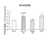
- FIG. 6 shows CS activities in the DOE strain, the Pgc-1 ⁇ OE strain, the Mipep OE strain, and the Mock cells. As shown in FIG. 6, the CS activity showed an increasing tendency in correlation with the overexpression of the Pgc-1 ⁇ gene.
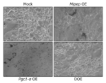
- FIG. 7A shows a microscopic image of DOE strain, Pgc-1 ⁇ @ OE strain, Mipep @ OE strain, and Mock cells stained with Oil Red @ O staining solution
- the DOE strain, the Pgc-1 ⁇ OE strain, and the Mipep OE strain all significantly reduced the amount of accumulated lipid droplets, particularly the DOE strain. there were.
- the DOE strain had the same level of mitochondrial-related protein expression (FIGS. 4A to 4C, 5A, and 5B) and CS activity (FIG.
- the primers used for amplification of the Pgc-1 ⁇ gene, the Mipep gene, and the Rps18 gene are as shown in Tables 1 and 2 above.
- the primers used for the amplification of the Perilipin gene, Ppar ⁇ gene, and Adipoq gene are shown in Table 4 below.
- FIG. 8 shows the mRNA expression levels of Pgc-1 ⁇ , Mipep, Perilipin, Ppar ⁇ , and Adipoq in the DOE strain, the Pgc-1 ⁇ OE strain, the Mipep OE strain, and the Mock cells.
- the mRNA expression levels of Perilipin, Ppar ⁇ , and Adipoq hardly changed in any of the cell lines. From these results and the results of Test Example 6, it was suggested that the decrease in the amount of accumulated lipid droplets in the DOE strain was not due to the decrease in the differentiation signal, but to the overexpression of the Pgc-1 ⁇ gene and the Mipep gene.
- ⁇ Test Example 8> (Analysis of Perilipin protein expression level in DOE strain, Pgc-1 ⁇ OE strain, Mipep OE strain, and Mock cells) In the same manner as in Test Example 4, the protein expression level of Perilipin in the DOE strain, the Pgc-1 ⁇ OE strain, the Mipep OE strain, and the Mock cells was analyzed by Western blot. Perilipin protein is a protein present on the surface of lipid droplets. As a control, the protein expression level of Lamin B1 was also analyzed.
- Perilipin protein a rabbit anti-Perilipin monoclonal antibody (Cell Signaling) was used as a primary antibody, and for detection of Lamin ⁇ B1 protein, a rabbit anti-Lamin ⁇ B1 polyclonal antibody (Medical & Biochemical Laboratories) was used as a primary antibody. did. In each case, an HRP-labeled goat anti-rabbit IgG (H + L) antibody (Jackson Immunological Research) was used as a secondary antibody.
- H + L HRP-labeled goat anti-rabbit IgG
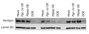
- FIG. 9 shows the protein expression levels of Perilipin in the DOE strain, the Pgc-1 ⁇ OE strain, the Mipep OE strain, and the Mock cells. As shown in FIG. 9, in the DOE strain, the protein expression level of Perilipin was significantly reduced.
Landscapes
- Chemical & Material Sciences (AREA)
- Life Sciences & Earth Sciences (AREA)
- Health & Medical Sciences (AREA)
- Engineering & Computer Science (AREA)
- Organic Chemistry (AREA)
- Zoology (AREA)
- Wood Science & Technology (AREA)
- Proteomics, Peptides & Aminoacids (AREA)
- Immunology (AREA)
- Molecular Biology (AREA)
- Biochemistry (AREA)
- Physics & Mathematics (AREA)
- General Health & Medical Sciences (AREA)
- Analytical Chemistry (AREA)
- Biophysics (AREA)
- Biotechnology (AREA)
- Microbiology (AREA)
- Genetics & Genomics (AREA)
- Bioinformatics & Cheminformatics (AREA)
- General Engineering & Computer Science (AREA)
- Pathology (AREA)
- Urology & Nephrology (AREA)
- Hematology (AREA)
- Chemical Kinetics & Catalysis (AREA)
- Biomedical Technology (AREA)
- Medicinal Chemistry (AREA)
- General Physics & Mathematics (AREA)
- Food Science & Technology (AREA)
- Cell Biology (AREA)
- Pharmacology & Pharmacy (AREA)
- Measuring Or Testing Involving Enzymes Or Micro-Organisms (AREA)
- Micro-Organisms Or Cultivation Processes Thereof (AREA)
- Investigating Or Analysing Biological Materials (AREA)
Abstract
Pgc-1α遺伝子及びMipep遺伝子の発現を亢進させる物質を選択することを含む抗肥満物質のスクリーニング方法、及びそのスクリーニング方法に用いられるスクリーニング用キットを提供する。スクリーニングに際しては、Pgc-1α遺伝子及びMipep遺伝子の発現量を測定してもよく、レポーターアッセイによりレポーター遺伝子の発現量を測定してもよい。
Description
本発明は、抗肥満物質のスクリーニング方法及び抗肥満物質のスクリーニング用キットに関する。
肥満は、体脂肪が過剰に蓄積した状態と定義されている。肥満の主な原因は、摂取カロリーと消費カロリーとのアンバランスにあり、摂取カロリーが消費カロリーを上回ると、余剰カロリーが脂肪となって体内に蓄積され、やがて肥満となる。肥満は、糖尿病、高血圧、高脂血症等の生活習慣病の原因ともなる。このため、肥満の予防及び改善は、生活習慣病のリスクを低減する上で極めて重要であると考えられる。
ところで、近年、摂取カロリー制限(caloric restriction;CR)により、エネルギー代謝に関係するミトコンドリアの生合成や機能が亢進することが報告されている。例えば、非特許文献1では、摂取カロリー制限(CR)により、白色脂肪組織選択的かつSrebp-1c依存的に、Pgc-1αを介してミトコンドリア生合成が亢進することが報告されている。また、非特許文献2では、摂取カロリー制限(CR)によりMipepの発現が亢進することや、MipepによりSirt3がプロセシングを受けて活性型となることが報告されている。
N.Fujii et al.,Aging Cell,2017,16(3),pp.508-517
M.Kobayashi et al.,FEBS Letters,2017,591(24),pp.4067-4073
摂取カロリー制限(CR)は、肥満の予防及び改善に有効であると考えられるが、継続的に摂取カロリー制限(CR)を行うことは一般に困難である。このため、摂取カロリー制限(CR)による表現型を模倣可能な抗肥満物質の探索が望まれる。
本発明は、上記に鑑みて提案されたものであり、抗肥満物質のスクリーニング方法及び抗肥満物質のスクリーニング用キットを提供することを課題とする。
本発明者らは、上記課題を解決するために鋭意検討を重ねた結果、Pgc-1α遺伝子及びMipep遺伝子の両者の発現を亢進させる物質が抗肥満物質として有望であることを見出し、本発明を完成するに至った。より具体的には、本発明は以下のとおりである。
<1> Pgc-1α遺伝子及びMipep遺伝子の発現を亢進させる物質を選択することを含む抗肥満物質のスクリーニング方法。
<2> Pgc-1α遺伝子及びMipep遺伝子を発現する細胞に被験物質を接触させる工程と、
前記被験物質を接触させない場合に比べて、前記細胞におけるPgc-1α遺伝子及びMipep遺伝子の発現量が増加する場合に、該被験物質を抗肥満物質として選択する工程と、
を含む<1>に記載の抗肥満物質のスクリーニング方法。
前記被験物質を接触させない場合に比べて、前記細胞におけるPgc-1α遺伝子及びMipep遺伝子の発現量が増加する場合に、該被験物質を抗肥満物質として選択する工程と、
を含む<1>に記載の抗肥満物質のスクリーニング方法。
<3> 前記細胞として、Srebp-1c遺伝子を有する細胞とSrebp-1c遺伝子を有しない細胞とを使用し、
前記被験物質を接触させることにより、Srebp-1c遺伝子を有する細胞においてのみPgc-1α遺伝子及びMipep遺伝子の発現量が増加する場合に、該被験物質を抗肥満物質として選択する<2>に記載の抗肥満物質のスクリーニング方法。
前記被験物質を接触させることにより、Srebp-1c遺伝子を有する細胞においてのみPgc-1α遺伝子及びMipep遺伝子の発現量が増加する場合に、該被験物質を抗肥満物質として選択する<2>に記載の抗肥満物質のスクリーニング方法。
<4> Pgc-1α遺伝子のプロモーターの下流に機能的に連結された第1のレポーター遺伝子を有する第1の形質転換体、及びMipep遺伝子のプロモーターの下流に機能的に連結された第2のレポーター遺伝子を有する第2の形質転換体に、被験物質を接触させる工程と、
前記被験物質を接触させない場合に比べて、前記第1のレポーター遺伝子及び前記第2のレポーター遺伝子の発現量が増加する場合に、該被験物質を抗肥満物質として選択する工程と、
を含む<1>に記載の抗肥満物質のスクリーニング方法。
前記被験物質を接触させない場合に比べて、前記第1のレポーター遺伝子及び前記第2のレポーター遺伝子の発現量が増加する場合に、該被験物質を抗肥満物質として選択する工程と、
を含む<1>に記載の抗肥満物質のスクリーニング方法。
<5> 前記第1の形質転換体及び前記第2の形質転換体のそれぞれとして、Srebp-1c遺伝子を有する形質転換体とSrebp-1c遺伝子を有しない形質転換体とを使用し、
前記被験物質を接触させることにより、Srebp-1c遺伝子を有する形質転換体においてのみ前記第1のレポーター遺伝子及び前記第2のレポーター遺伝子の発現量が増加する場合に、該被験物質を抗肥満物質として選択する<4>に記載の抗肥満物質のスクリーニング方法。
前記被験物質を接触させることにより、Srebp-1c遺伝子を有する形質転換体においてのみ前記第1のレポーター遺伝子及び前記第2のレポーター遺伝子の発現量が増加する場合に、該被験物質を抗肥満物質として選択する<4>に記載の抗肥満物質のスクリーニング方法。
<6> 下記(a)~(c)から選択される少なくとも1つを含む抗肥満物質のスクリーニング用キット:
(a)Pgc-1α遺伝子及びMipep遺伝子のそれぞれを増幅するためのプライマー、
(b)Pgc-1α遺伝子及びMipep遺伝子のそれぞれを検出するためのプローブ、
(c)Pgc-1αタンパク質及びMipepタンパク質のそれぞれに対する抗体。
(a)Pgc-1α遺伝子及びMipep遺伝子のそれぞれを増幅するためのプライマー、
(b)Pgc-1α遺伝子及びMipep遺伝子のそれぞれを検出するためのプローブ、
(c)Pgc-1αタンパク質及びMipepタンパク質のそれぞれに対する抗体。
<7> 下記(d)及び(e)から選択される少なくとも1つを含む抗肥満物質のスクリーニング用キット:
(d)Pgc-1α遺伝子のプロモーターと、該プロモーターの下流に機能的に連結された第1のレポーター遺伝子とを有する第1のベクター、及びMipep遺伝子のプロモーターと、該プロモーターの下流に機能的に連結された第2のレポーター遺伝子とを有する第2のベクター、
(e)Pgc-1α遺伝子のプロモーターの下流に機能的に連結された第1のレポーター遺伝子を有する第1の形質転換体、及びMipep遺伝子のプロモーターの下流に機能的に連結された第2のレポーター遺伝子を有する第2の形質転換体。
(d)Pgc-1α遺伝子のプロモーターと、該プロモーターの下流に機能的に連結された第1のレポーター遺伝子とを有する第1のベクター、及びMipep遺伝子のプロモーターと、該プロモーターの下流に機能的に連結された第2のレポーター遺伝子とを有する第2のベクター、
(e)Pgc-1α遺伝子のプロモーターの下流に機能的に連結された第1のレポーター遺伝子を有する第1の形質転換体、及びMipep遺伝子のプロモーターの下流に機能的に連結された第2のレポーター遺伝子を有する第2の形質転換体。
本発明によれば、抗肥満物質のスクリーニング方法及び抗肥満物質のスクリーニング用キットを提供することができる。
<抗肥満物質のスクリーニング方法>
本実施形態に係る抗肥満物質のスクリーニング方法は、Pgc-1α遺伝子及びMipep遺伝子の発現を亢進させる物質を選択することを含む。後述する実施例に示すとおり、Pgc-1α遺伝子及びMipep遺伝子の両者の発現を亢進させることにより、白色脂肪細胞における脂肪滴蓄積量が顕著に減少する。したがって、Pgc-1α遺伝子及びMipep遺伝子の両者の発現を亢進させる物質は、抗肥満物質として有望であると考えられる。
本実施形態に係る抗肥満物質のスクリーニング方法は、Pgc-1α遺伝子及びMipep遺伝子の発現を亢進させる物質を選択することを含む。後述する実施例に示すとおり、Pgc-1α遺伝子及びMipep遺伝子の両者の発現を亢進させることにより、白色脂肪細胞における脂肪滴蓄積量が顕著に減少する。したがって、Pgc-1α遺伝子及びMipep遺伝子の両者の発現を亢進させる物質は、抗肥満物質として有望であると考えられる。
なお、Pgc-1α遺伝子及びMipep遺伝子はいずれも公知であり、それぞれの塩基配列情報及びアミノ酸配列情報は米国生物工学情報センター(NCBI)のGenBankデータベースから入手可能である。一例として、ヒトPgc-1αの塩基配列及びアミノ酸配列を配列番号1、2に示す。また、ヒトMipepの塩基配列及びアミノ酸配列を配列番号3、4に示す。
スクリーニングに際しては、Pgc-1α遺伝子及びMipep遺伝子の発現量を測定してもよく、レポーターアッセイによりレポーター遺伝子の発現量を測定してもよい。以下、それぞれの態様について説明する。
第1の態様に係るスクリーニング方法は、Pgc-1α遺伝子及びMipep遺伝子を発現する細胞に被験物質を接触させる工程(以下、「接触工程」ともいう。)と、被験物質を接触させない場合に比べて、細胞におけるPgc-1α遺伝子及びMipep遺伝子の発現量が増加する場合に、該被験物質を抗肥満物質として選択する工程(以下、「選択工程」ともいう。)と、を含む。
まず、接触工程では、Pgc-1α遺伝子及びMipep遺伝子を発現する細胞に被験物質を接触させる。
被験物質を接触させる細胞としては、Pgc-1α遺伝子及びMipep遺伝子を発現するものであれば特に制限されない。後述する実施例に示すとおり、白色脂肪組織におけるPgc-1α遺伝子及びMipep遺伝子の発現は、Srebp-1c遺伝子の制御を受けている。このため、被験物質を接触させる細胞としては、Pgc-1α遺伝子及びMipep遺伝子のほかにSrebp-1c遺伝子を発現する哺乳動物由来の細胞であることが好ましい。
細胞に被験物質を接触させる方法は特に制限されず、例えば、被験物質を添加した培地で細胞を培養する方法が挙げられる。
次いで、選択工程では、被験物質を接触させない場合に比べて、細胞におけるPgc-1α遺伝子及びMipep遺伝子の発現量が増加する場合に、該被験物質を抗肥満物質として選択する。
Pgc-1α遺伝子及びMipep遺伝子の発現量を測定する際には、転写レベルの発現量(mRNA量)を測定してもよく、翻訳レベルの発現量(タンパク質量)を測定してもよい。すなわち、遺伝子の発現量には、転写産物の量と翻訳産物の量との両方が含まれる。
mRNA量の測定方法としては、対象遺伝子(Pgc-1α遺伝子又はMipep遺伝子)のmRNA量を測定できる方法であれば特に制限されず、例えば、RT-PCR法、定量PCR法、ノーザンブロット法、DNAマイクロアレイ法等が挙げられる。
タンパク質量の測定方法としては、対象遺伝子(Pgc-1α遺伝子又はMipep遺伝子)がコードするタンパク質を特異的に測定できる方法であれば特に制限されず、例えば、ウェスタンブロット法、酵素免疫測定法等が挙げられる。
上記のようにPgc-1α遺伝子及びMipep遺伝子の発現量を測定した結果、被験物質を接触させない場合に比べて、Pgc-1α遺伝子及びMipep遺伝子の発現量が有意に増加している場合には、該被験物質を抗肥満物質として選択することができる。
なお、上記のとおり、白色脂肪組織におけるPgc-1α遺伝子及びMipep遺伝子の発現は、Srebp-1c遺伝子の制御を受けている。このため、被験物質を接触させる細胞として、Srebp-1c遺伝子を有する細胞とSrebp-1c遺伝子を有しない細胞とを使用し、被験物質を接触させることにより、Srebp-1c遺伝子を有する細胞においてのみPgc-1α遺伝子及びMipep遺伝子の発現量が増加する場合に、該被験物質を抗肥満物質として選択するようにしてもよい。これにより、摂取カロリー制限(CR)に近似した作用を有する抗肥満物質を得ることが可能となる。
一方、第2の態様に係るスクリーニング方法は、Pgc-1α遺伝子のプロモーターの下流に機能的に連結された第1のレポーター遺伝子を有する第1の形質転換体、及びMipep遺伝子のプロモーターの下流に機能的に連結された第2のレポーター遺伝子を有する第2の形質転換体に、被験物質を接触させる工程(以下、「接触工程」ともいう。)と、被験物質を接触させない場合に比べて、第1のレポーター遺伝子及び第2のレポーター遺伝子の発現量が増加する場合に、該被験物質を抗肥満物質として選択する工程(以下、「選択工程」ともいう。)と、を含む。
まず、接触工程では、Pgc-1α遺伝子のプロモーターの下流に機能的に連結された第1のレポーター遺伝子を有する第1の形質転換体、及びMipep遺伝子のプロモーターの下流に機能的に連結された第2のレポーター遺伝子を有する第2の形質転換体に、被験物質を接触させる。
第1の形質転換体は、Pgc-1α遺伝子のプロモーターの下流に機能的に連結された第1のレポーター遺伝子を有する。また、第2の形質転換体は、Mipep遺伝子のプロモーターの下流に機能的に連結された第2のレポーター遺伝子を有する。
ここで、「機能的に連結された」とは、天然において対象遺伝子(Pgc-1α遺伝子又はMipep遺伝子)の発現を誘導する分子に応答して、対象遺伝子のプロモーターの制御下にレポーター遺伝子が発現するように連結されていることを意味する。
第1のレポーター遺伝子及び第2のレポーター遺伝子としては、特に制限されず、生命工学分野で知られている任意のレポーター遺伝子を使用することができる。レポーター遺伝子としては、例えば、ルシフェラーゼ遺伝子、GFP(緑色蛍光タンパク質)遺伝子、GUS(β-グルクロニダーゼ)遺伝子、CAT(クロラムフェニコールアセチルトランスフェラーゼ)遺伝子等が挙げられる。
第1の形質転換体は、Pgc-1α遺伝子のプロモーターと、該プロモーターの下流に機能的に連結された第1のレポーター遺伝子とを有する第1のベクターを宿主細胞に導入することによって得ることができる。また、第2の形質転換体は、Mipep遺伝子のプロモーターと、該プロモーターの下流に機能的に連結された第2のレポーター遺伝子とを有する第2のベクターを宿主細胞に導入することによって得ることができる。
宿主細胞としては特に制限されないが、Srebp-1c遺伝子を発現する哺乳動物由来の細胞であることが好ましい。
なお、第1の形質転換体及び第2の形質転換体は、別個の形質転換体に限定されるものではなく、単一の形質転換体であってもよい。例えば、Pgc-1α遺伝子のプロモーターと、該プロモーターの下流に機能的に連結された第1のレポーター遺伝子と、Mipep遺伝子のプロモーターと、該プロモーターの下流に機能的に連結された第2のレポーター遺伝子とを有するベクターを宿主細胞に導入することによって、単一の形質転換体を得ることができる。第1の形質転換体及び第2の形質転換体を単一の形質転換体とする場合、第1のレポーター遺伝子及び第2のレポーター遺伝子としては、識別可能な異種の遺伝子が使用される。
第1の形質転換体及び第2の形質転換体に被験物質を接触させる方法は特に制限されず、例えば、被験物質を添加した培地で第1の形質転換体及び第2の形質転換体を培養する方法が挙げられる。
次いで、選択工程では、被験物質を接触させない場合に比べて、第1のレポーター遺伝子及び第2のレポーター遺伝子の発現量が増加する場合に、該被験物質を抗肥満物質として選択する。
第1のレポーター遺伝子及び第2のレポーター遺伝子の発現量は、レポーター遺伝子の種類に応じた方法で測定することができる。例えば、レポーター遺伝子がルシフェラーゼ遺伝子であれば、発現産物であるルシフェラーゼの活性を、蛍光をもって検出することができる。レポーター遺伝子がGFP遺伝子であれば、発現産物であるGFPを、蛍光をもって検出することができる。レポーター遺伝子がGUS遺伝子であれば、発現産物であるGUSの活性を、グルクロン等の発色をもって検出することができる。レポーター遺伝子がCAT遺伝子であれば、発現産物であるCATの活性を、クロラムフェニコールのアセチル化をもって検出することができる。
上記のように第1のレポーター遺伝子及び第2のレポーター遺伝子の発現量を測定した結果、被験物質を接触させない場合に比べて、第1のレポーター遺伝子及び第2のレポーター遺伝子の発現量が有意に増加している場合には、該被験物質を抗肥満物質として選択することができる。
なお、上記のとおり、白色脂肪組織におけるPgc-1α遺伝子及びMipep遺伝子の発現は、Srebp-1c遺伝子の制御を受けている。このため、第1の形質転換体及び第2の形質転換体のそれぞれとして、Srebp-1c遺伝子を有する形質転換体とSrebp-1c遺伝子を有しない形質転換体とを使用し、被験物質を接触させることにより、Srebp-1c遺伝子を有する形質転換体においてのみ第1のレポーター遺伝子及び第2のレポーター遺伝子の発現量が増加する場合に、該被験物質を抗肥満物質として選択するようにしてもよい。これにより、摂取カロリー制限(CR)に近似した作用を有する抗肥満物質を得ることが可能となる。
以上のようにして、細胞レベルでPgc-1α遺伝子及びMipep遺伝子の発現を亢進する抗肥満物質が得られた後には、in vivoでのアッセイを行ってもよい。例えば、得られた抗肥満物質をPgc-1α欠損マウス及びMipep欠損マウスに投与し、その反応性を野生型マウスと比較することにより、該抗肥満物質の有効性を動物レベルで評価することができる。
<抗肥満物質のスクリーニング用キット>
本実施形態に係る抗肥満物質のスクリーニング用キットは、上述した第1の態様に係るスクリーニング方法のように、Pgc-1α遺伝子及びMipep遺伝子の発現量を測定する際に使用してもよく、上述した第2の態様に係るスクリーニング方法のように、レポーターアッセイによりレポーター遺伝子の発現量を測定する際に使用してもよい。以下、それぞれの態様について説明する。
本実施形態に係る抗肥満物質のスクリーニング用キットは、上述した第1の態様に係るスクリーニング方法のように、Pgc-1α遺伝子及びMipep遺伝子の発現量を測定する際に使用してもよく、上述した第2の態様に係るスクリーニング方法のように、レポーターアッセイによりレポーター遺伝子の発現量を測定する際に使用してもよい。以下、それぞれの態様について説明する。
第1の態様に係るスクリーニング用キットは、下記(a)~(c)から選択される少なくとも1つを含む。
(a)Pgc-1α遺伝子及びMipep遺伝子のそれぞれを増幅するためのプライマー。
(b)Pgc-1α遺伝子及びMipep遺伝子のそれぞれを検出するためのプローブ。
(c)Pgc-1αタンパク質及びMipepタンパク質のそれぞれに対する抗体。
(a)Pgc-1α遺伝子及びMipep遺伝子のそれぞれを増幅するためのプライマー。
(b)Pgc-1α遺伝子及びMipep遺伝子のそれぞれを検出するためのプローブ。
(c)Pgc-1αタンパク質及びMipepタンパク質のそれぞれに対する抗体。
上記(a)におけるプライマーは、対象遺伝子(Pgc-1α又はMipep)の各塩基配列に基づき設計し、合成及び精製の各工程を経て調製することができる。プライマーの長さ(塩基数)は特に制限されないが、特異的なアニーリングを可能とするため、15~40塩基程度とすることが好ましい。
上記(b)におけるプローブとしては、対象遺伝子(Pgc-1α又はMipep)の各塩基配列の連続する部分配列からなるポリヌクレオチド、又は対象遺伝子の各塩基配列に対する相補配列の連続する部分配列からなるポリヌクレオチドが用いられる。プローブの長さ(塩基数)は特に制限されないが、特異的なハイブリッド形成を可能とするため、15塩基以上とすることが好ましい。これらのプローブは、標識物質を用いて適宜標識することができる。標識物質としては、蛍光色素、放射性同位体、酵素、アビジン、ビオチン等を用いることができる。
プローブは、マイクロアレイ等の固相担体に固定化して用いてもよい。マイクロアレイの作製方法は特に制限されず、例えば、固相担体表面で直接プローブを合成する方法(オン・チップ法)や、予め調製したプローブを固相担体表面に結合する方法等を採用することができる。
上記(c)における抗体は、ポリクローナル抗体及びモノクローナル抗体のいずれであってもよく、抗体の活性フラグメントであってもよい。活性フラグメントとしては、F(ab’)2、Fab’、Fab、Fv等が挙げられる。これらの抗体は、標識物質を用いて適宜標識することができる。標識物質としては、酵素、放射性同位体、蛍光色素等を用いることができる。
一方、第2の態様に係るスクリーニング用キットは、下記(d)及び(e)から選択される少なくとも1つを含む。
(d)Pgc-1α遺伝子のプロモーターと、該プロモーターの下流に機能的に連結された第1のレポーター遺伝子とを有する第1のベクター、及びMipep遺伝子のプロモーターと、該プロモーターの下流に機能的に連結された第2のレポーター遺伝子とを有する第2のベクター。
(e)Pgc-1α遺伝子のプロモーターの下流に機能的に連結された第1のレポーター遺伝子を有する第1の形質転換体、及びMipep遺伝子のプロモーターの下流に機能的に連結された第2のレポーター遺伝子を有する第2の形質転換体。
(d)Pgc-1α遺伝子のプロモーターと、該プロモーターの下流に機能的に連結された第1のレポーター遺伝子とを有する第1のベクター、及びMipep遺伝子のプロモーターと、該プロモーターの下流に機能的に連結された第2のレポーター遺伝子とを有する第2のベクター。
(e)Pgc-1α遺伝子のプロモーターの下流に機能的に連結された第1のレポーター遺伝子を有する第1の形質転換体、及びMipep遺伝子のプロモーターの下流に機能的に連結された第2のレポーター遺伝子を有する第2の形質転換体。
上記(d)における第1のレポーター遺伝子及び第2のレポーター遺伝子については上述したとおりであるため、詳細な説明を省略する。第1のベクター及び第2のベクターは、エンハンサー、スプライシングシグナル、ポリA付加シグナル、選択マーカー等を適宜有していてもよい。選択マーカーとしては、アンピシリン耐性遺伝子、ネオマイシン耐性遺伝子等が挙げられる。
第1のベクター及び第2のベクターは、別個のベクターに限定されるものではなく、単一のベクターであってもよい。例えば、第1のベクター及び第2のベクターは、Pgc-1α遺伝子のプロモーターと、該プロモーターの下流に機能的に連結された第1のレポーター遺伝子と、Mipep遺伝子のプロモーターと、該プロモーターの下流に機能的に連結された第2のレポーター遺伝子とを有する単一のベクターであってもよい。
上記(e)における第1の形質転換体及び第2の形質転換体は、別個の形質転換体に限定されるものではなく、単一の形質転換体であってもよい。例えば、Pgc-1α遺伝子のプロモーターと、該プロモーターの下流に機能的に連結された第1のレポーター遺伝子と、Mipep遺伝子のプロモーターと、該プロモーターの下流に機能的に連結された第2のレポーター遺伝子とを有するベクターを宿主細胞に導入することによって、単一の形質転換体を得ることができる。
以下、本発明の実施例を説明するが、本発明の範囲はこれらの実施例に限定されるものではない。なお、有意差検定は、試験例1、2ではスチューデントのt検定により行い、試験例4~6ではテューキーのt検定により行った(*P<0.05;**P<0.01;***P<0.001)。
<試験例1>
(モデルマウスの作製及び白色脂肪組織の採取)
米国のジャクソン研究所より購入したSrebp-1c+/-(B6;129S6-Srebf1tm1Mbr)マウス(G.Liang et al.,J.Biol.Chem.,2002,277,pp.9520-9528)から、交配によりSrebp-1c+/+マウス(以下、「WTマウス」ともいう。)及びSrebp-1c-/-マウス(以下、「KOマウス」ともいう。)を作製した。東京理科大学薬学部動物実験施設内のSPF環境下、明暗サイクル12時間で飼育し、自由摂食群(AL群)には飼料としてCRF-1(オリエンタル酵母工業(株))を十分量与えた。AL群の摂食量を測定し、摂取カロリー制限群(CR群)にはその70%量を消灯前に与えた。3ヶ月齢より摂取カロリー制限(CR)を開始し、WT-AL群、WT-CR群、KO-AL群、及びKO-CR群の計4群を作製した。8.5ヶ月齢から10ヶ月齢において体重を測定後、イソフルランによる過麻酔で安楽死させた。安楽死後、精巣周囲の白色脂肪組織を採取し、臓器は液体窒素による急速凍結後、-80℃で保存した。
(モデルマウスの作製及び白色脂肪組織の採取)
米国のジャクソン研究所より購入したSrebp-1c+/-(B6;129S6-Srebf1tm1Mbr)マウス(G.Liang et al.,J.Biol.Chem.,2002,277,pp.9520-9528)から、交配によりSrebp-1c+/+マウス(以下、「WTマウス」ともいう。)及びSrebp-1c-/-マウス(以下、「KOマウス」ともいう。)を作製した。東京理科大学薬学部動物実験施設内のSPF環境下、明暗サイクル12時間で飼育し、自由摂食群(AL群)には飼料としてCRF-1(オリエンタル酵母工業(株))を十分量与えた。AL群の摂食量を測定し、摂取カロリー制限群(CR群)にはその70%量を消灯前に与えた。3ヶ月齢より摂取カロリー制限(CR)を開始し、WT-AL群、WT-CR群、KO-AL群、及びKO-CR群の計4群を作製した。8.5ヶ月齢から10ヶ月齢において体重を測定後、イソフルランによる過麻酔で安楽死させた。安楽死後、精巣周囲の白色脂肪組織を採取し、臓器は液体窒素による急速凍結後、-80℃で保存した。
(Mipep及びPgc-1αのmRNA発現量の解析)
-80℃で凍結保存していた白色脂肪組織からISOGEN II((株)ニッポンジーン)を用いて全RNAを抽出した。PrimeScript RT-PCR Kit(タカラバイオ(株))を用いて全RNAの逆転写反応を行い、cDNAを得た。その後、THUNDERBIRD SYBR qPCR Mix(東洋紡(株))及びBlend Taq Plus(東洋紡(株))を用いたリアルタイムRT-PCRにより目的遺伝子(Mipep又はPgc-1α)を増幅させた。PCR産物は2.4%アガロースゲル電気泳動により分離し、0.01%エチジウムブロマイド溶液で15分間処理した後に、ルミノ・イメージアナライザーLAS3000(富士フイルム(株))を用いて特異的バンドを可視化した。なお、各mRNA発現量は、Tbp(TATA-binding protein)のmRNA発現量により補正した。使用したプライマーを下記表1に示す。
-80℃で凍結保存していた白色脂肪組織からISOGEN II((株)ニッポンジーン)を用いて全RNAを抽出した。PrimeScript RT-PCR Kit(タカラバイオ(株))を用いて全RNAの逆転写反応を行い、cDNAを得た。その後、THUNDERBIRD SYBR qPCR Mix(東洋紡(株))及びBlend Taq Plus(東洋紡(株))を用いたリアルタイムRT-PCRにより目的遺伝子(Mipep又はPgc-1α)を増幅させた。PCR産物は2.4%アガロースゲル電気泳動により分離し、0.01%エチジウムブロマイド溶液で15分間処理した後に、ルミノ・イメージアナライザーLAS3000(富士フイルム(株))を用いて特異的バンドを可視化した。なお、各mRNA発現量は、Tbp(TATA-binding protein)のmRNA発現量により補正した。使用したプライマーを下記表1に示す。

WT-AL群、WT-CR群、KO-AL群、及びKO-CR群の白色脂肪組織におけるMipep及びPgc-1αのmRNA発現量を図1A、図1Bに示す(n=3~5)。図1A、図1Bに示すとおり、野生型(WT)マウスにおいては、摂取カロリー制限(CR)によって、Mipep及びPgc-1αのmRNA発現量が有意に増加した。一方、Srebp-1cノックアウト(KO)マウスにおいては、摂取カロリー制限(CR)によっても、Mipep及びPgc-1αのmRNA発現量は増加しなかった。この結果から、摂取カロリー制限(CR)によるMipep遺伝子及びPgc-1α遺伝子の発現亢進は、Srebp-1c依存的であることが示唆された。
<試験例2>
(Pgc-1α遺伝子をノックアウトしたマウス胚線維芽細胞(MEF細胞)の作製)
米国のジャクソン研究所より購入したPgc-1α+/flox(B6N;129(FVB)-PPARGC1atm2.1Brap)マウス(J.Lin et al.,Cell,2004,119,pp.121-135)とCAG-CREマウスとの交配によりPgc-1α+/-マウスを得た。Pgc-1α+/-マウス同士を交配させ、妊娠後13.5日~15.5日のマウスの子宮から胎児を取り出し、PBSで洗浄した後、頭、尾、手足、臓器を取り除き、0.05%trypsin-EDTAを含有するPBSを用いて37℃で10分間消化した。その後、70μmセルストレーナー(Becton Dickinson)で余分な細胞を除くことにより、Pgc-1α遺伝子をノックアウトしたMEF細胞を作製した。
(Pgc-1α遺伝子をノックアウトしたマウス胚線維芽細胞(MEF細胞)の作製)
米国のジャクソン研究所より購入したPgc-1α+/flox(B6N;129(FVB)-PPARGC1atm2.1Brap)マウス(J.Lin et al.,Cell,2004,119,pp.121-135)とCAG-CREマウスとの交配によりPgc-1α+/-マウスを得た。Pgc-1α+/-マウス同士を交配させ、妊娠後13.5日~15.5日のマウスの子宮から胎児を取り出し、PBSで洗浄した後、頭、尾、手足、臓器を取り除き、0.05%trypsin-EDTAを含有するPBSを用いて37℃で10分間消化した。その後、70μmセルストレーナー(Becton Dickinson)で余分な細胞を除くことにより、Pgc-1α遺伝子をノックアウトしたMEF細胞を作製した。
(野生型のMEF細胞及びPgc-1α遺伝子をノックアウトしたMEF細胞におけるPgc-1α、Srebp-1c、及びMipepのmRNA発現量の解析)
野生型のMEF細胞及びPgc-1α遺伝子をノックアウトしたMEF細胞から全RNAを抽出し、試験例1と同様にして、Pgc-1α、Srebp-1c、及びMipep遺伝子のmRNA発現量をリアルタイムRT-PCRにより確認した。なお、各mRNA発現量は、Rps18(ribosomal protein S18)のmRNA発現量により補正した。Pgc-1α遺伝子及びMipep遺伝子の増幅に使用したプライマーは上記表1のとおりである。Srebp-1c遺伝子及びRps18遺伝子の増幅に使用したプライマーを下記表2に示す。
野生型のMEF細胞及びPgc-1α遺伝子をノックアウトしたMEF細胞から全RNAを抽出し、試験例1と同様にして、Pgc-1α、Srebp-1c、及びMipep遺伝子のmRNA発現量をリアルタイムRT-PCRにより確認した。なお、各mRNA発現量は、Rps18(ribosomal protein S18)のmRNA発現量により補正した。Pgc-1α遺伝子及びMipep遺伝子の増幅に使用したプライマーは上記表1のとおりである。Srebp-1c遺伝子及びRps18遺伝子の増幅に使用したプライマーを下記表2に示す。
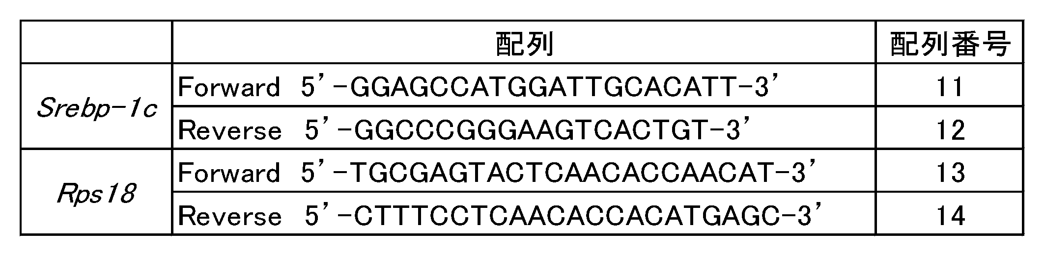
野生型のMEF細胞及びPgc-1α遺伝子をノックアウトしたMEF細胞におけるPgc-1α、Srebp-1c、及びMipepのmRNA発現量を図2に示す。図2に示すとおり、野生型のMEF細胞とPgc-1α遺伝子をノックアウトしたMEF細胞とで、Srebp-1c及びMipepのmRNA発現量に差は認められなかった。この結果及び試験例1の結果から、摂取カロリー制限(CR)によるMipep遺伝子及びPgc-1α遺伝子の発現亢進は、互いに非依存的であることが示唆された。
<試験例3>
(Mipep過剰発現用ベクター及びPgc-1α過剰発現用ベクターの作製)
マウス白色脂肪組織のcDNAを鋳型とし、下記表3に示すプライマー及びPrime star(東洋紡(株))を用いたPCRによりMipepのcDNAを増幅させた。PCR産物をEcoR I及びXho Iで切断し、EcoR I及びXho Iで切断済みのpMXs-AMNN-Puroベクターとライゲーションすることにより、pMXs-Mipep-Puroベクターを作製した。
(Mipep過剰発現用ベクター及びPgc-1α過剰発現用ベクターの作製)
マウス白色脂肪組織のcDNAを鋳型とし、下記表3に示すプライマー及びPrime star(東洋紡(株))を用いたPCRによりMipepのcDNAを増幅させた。PCR産物をEcoR I及びXho Iで切断し、EcoR I及びXho Iで切断済みのpMXs-AMNN-Puroベクターとライゲーションすることにより、pMXs-Mipep-Puroベクターを作製した。

また、Addgeneより購入したpcDNA-f:Pgc-1ベクターをEcoR V及びPme Iで切断した。得られたDNA断片を、EcoR I処理後にKlenow処理により平滑化し、CIP処理により脱リン酸化したpMXs-Neoベクターとライゲーションすることにより、pMXs-Pgc-1α-Neoベクターを作製した。
(Mipep過剰発現用ベクター及びPgc-1α過剰発現用ベクターのマウス白色脂肪前駆細胞株3T3-L1へのトランスフェクション)
14.5μg/mLのベクター(pMXs-Mipep-Puroベクター又はpMXs-Pgc-1α-Neoベクター)を含む0.25M CaCl2溶液と、1.5mM Na2HPO4を含む2×HBS(280mM NaCl、50mM HEPES、pH7.1)とを等量で少しずつ混合し、その後すぐにボルテックスミキサーで15秒間撹拌し、続いて室温で15~30分間放置した。得られた反応液でレトロウイルスパッケージング細胞Plat-Eを処理することにより、Plat-E細胞にベクターを導入した。
14.5μg/mLのベクター(pMXs-Mipep-Puroベクター又はpMXs-Pgc-1α-Neoベクター)を含む0.25M CaCl2溶液と、1.5mM Na2HPO4を含む2×HBS(280mM NaCl、50mM HEPES、pH7.1)とを等量で少しずつ混合し、その後すぐにボルテックスミキサーで15秒間撹拌し、続いて室温で15~30分間放置した。得られた反応液でレトロウイルスパッケージング細胞Plat-Eを処理することにより、Plat-E細胞にベクターを導入した。
次いで、pMXs-Mipep-Puroベクターを導入したPlat-E細胞を16時間培養した後、新しい培地に交換し、さらに2日間培養した後、培地を回収した。回収した培地をMillex-GVフィルター(Millipore)にて濾過した後、回収した培地の1/3量の4×PEG(32% PEG6000、0.4M NaCl、40mM HEPES、pH7.4)を加えた。その後、3000rpmで30分間遠心し、上清を除去した後、さらに3000rpmで5分間遠心した。上清をできるだけ除去した後、沈殿物にOPTI-MEM(ライフテクノロジーズジャパン(株))を加えて懸濁した。得られた懸濁液を3T3-L1細胞の培地に加え、レトロウイルスを感染させることにより、pMXs-Mipep-Puroベクターを3T3-L1細胞に導入した。なお、培地をフィルター濾過する工程からは全て氷上にて行った。感染の24時間後から2μg/mL ピューロマイシンを含む培地で5~7日間セレクションを行うことにより、Mipep遺伝子を過剰発現する3T3-L1細胞(以下、「Mipep OE株」ともいう。)を作製した。
次いで、pMXs-Pgc-1α-Neoベクターを導入したPlat-E細胞を16時間培養した後、新しい培地に交換し、さらに2日間培養した後、培地を回収した。回収した培地をMillex-GVフィルター(Millipore)にて濾過した後、回収した培地の1/3量の4×PEG(32% PEG6000、0.4M NaCl、40mM HEPES、pH7.4)を加えた。その後、3000rpmで30分間遠心し、上清を除去した後、さらに3000rpmで5分間遠心した。上清をできるだけ除去した後、沈殿物にOPTI-MEM(ライフテクノロジーズジャパン(株))を加えて懸濁した。得られた懸濁液をMipep OE株の培地に加え、レトロウイルスを感染させることにより、pMXs-Pgc-1α-NeoベクターをMipep OE株に導入した。なお、培地をフィルター濾過する工程からは全て氷上にて行った。感染の24時間後から1mg/mL G418(Cayman)及び0.4μg/mL ピューロマイシンを含む培地で5~7日間セレクションを行うことにより、Mipep遺伝子及びPgc-1α遺伝子の両者を過剰発現する3T3-L1細胞(以下、「DOE株」ともいう。)を作製した。セレクション後は、200μg/mL G418(Cayman)及び0.4μg/mL ピューロマイシンを含む培地で培養、維持した。
また、Mipep OE株及びDOE株の他に、Pgc-1α遺伝子を過剰発現する3T3-L1細胞(以下、「Pgc-1α OE株」ともいう。)、及び空ベクターを導入した3T3-L1細胞(以下、「Mock細胞」ともいう。)を作製した。
(DOE株、Pgc-1α OE株、Mipep OE株、及びMock細胞の成熟白色脂肪細胞への分化誘導)
3T3-L1細胞(DOE株、Pgc-1α OE株、Mipep OE株、及びMock細胞)を1.5×10⁴個/cm2の細胞密度で播種し、2日後に100%コンフルエントに達した時点を0日目とした。0日目から2日間は500μM 3-イソブチル-1-メチルキサンチン及び1μM デキサメタゾンを含む維持培地で培養することにより脂肪細胞へと誘導し、その後は2日おきに10μg/mL インスリン及び50nM トリヨードサイロニンを含む維持培地で培養することにより成熟脂肪細胞へと誘導した。以降の実験では、分化誘導開始後12日目の細胞を使用した。
3T3-L1細胞(DOE株、Pgc-1α OE株、Mipep OE株、及びMock細胞)を1.5×10⁴個/cm2の細胞密度で播種し、2日後に100%コンフルエントに達した時点を0日目とした。0日目から2日間は500μM 3-イソブチル-1-メチルキサンチン及び1μM デキサメタゾンを含む維持培地で培養することにより脂肪細胞へと誘導し、その後は2日おきに10μg/mL インスリン及び50nM トリヨードサイロニンを含む維持培地で培養することにより成熟脂肪細胞へと誘導した。以降の実験では、分化誘導開始後12日目の細胞を使用した。
<試験例4>
(DOE株、Pgc-1α OE株、Mipep OE株、及びMock細胞におけるPgc-1α及びMipepのタンパク質発現量の解析)
分化誘導開始後12日目の細胞(DOE株、Pgc-1α OE株、Mipep OE株、及びMock細胞)から、SDSサンプルバッファー(50mM tris-HCl(pH6.8)、2% SDS、3M 尿素、6% グリセロール)を用いてタンパク質を抽出した後、BCA Protein Assay Kit(Thermo)によりタンパク質濃度を測定した。タンパク質濃度が1μg/μLになるように調節し、0.5% BPB及び100% 2-メルカプトエタノールの等量混合溶液をタンパク質溶液の1/10量加えた後、95℃で5分間インキュベートしてサンプルを調製した。調製したサンプルは、8%又は10%アクリルアミドゲルを用いたSDS-PAGEにて分離し、ニトロセルロースメンブレンに転写した。メンブレンをブロッキング溶液(2.5% スキムミルク及び0.25% BSAをTTBSで溶解)中で1時間、室温でインキュベートした。その後、一次抗体反応液(Immuno shot reagent I(コスモ・バイオ(株)):ブロッキング溶液=2:1)中で一晩、4℃で反応させた。反応後、メンブレンを十分量のTTBSで5分×2回、10分×2回洗浄し、二次抗体反応液(Immuno shot reagent II(コスモ・バイオ(株)):ブロッキング溶液=2:1)中で1時間、室温で反応させた。反応後、再びTTBSでメンブレンを洗浄し、ImmunoStar LD(富士フイルム和光純薬(株))を用いた発光をChemiDoc MP(Bio-rad)にて検出した。検出したタンパク質量は、CBB染色の結果により補正した。
(DOE株、Pgc-1α OE株、Mipep OE株、及びMock細胞におけるPgc-1α及びMipepのタンパク質発現量の解析)
分化誘導開始後12日目の細胞(DOE株、Pgc-1α OE株、Mipep OE株、及びMock細胞)から、SDSサンプルバッファー(50mM tris-HCl(pH6.8)、2% SDS、3M 尿素、6% グリセロール)を用いてタンパク質を抽出した後、BCA Protein Assay Kit(Thermo)によりタンパク質濃度を測定した。タンパク質濃度が1μg/μLになるように調節し、0.5% BPB及び100% 2-メルカプトエタノールの等量混合溶液をタンパク質溶液の1/10量加えた後、95℃で5分間インキュベートしてサンプルを調製した。調製したサンプルは、8%又は10%アクリルアミドゲルを用いたSDS-PAGEにて分離し、ニトロセルロースメンブレンに転写した。メンブレンをブロッキング溶液(2.5% スキムミルク及び0.25% BSAをTTBSで溶解)中で1時間、室温でインキュベートした。その後、一次抗体反応液(Immuno shot reagent I(コスモ・バイオ(株)):ブロッキング溶液=2:1)中で一晩、4℃で反応させた。反応後、メンブレンを十分量のTTBSで5分×2回、10分×2回洗浄し、二次抗体反応液(Immuno shot reagent II(コスモ・バイオ(株)):ブロッキング溶液=2:1)中で1時間、室温で反応させた。反応後、再びTTBSでメンブレンを洗浄し、ImmunoStar LD(富士フイルム和光純薬(株))を用いた発光をChemiDoc MP(Bio-rad)にて検出した。検出したタンパク質量は、CBB染色の結果により補正した。
なお、Pgc-1αタンパク質の検出には、一次抗体としてマウス抗PGC-1αモノクローナル抗体(Calbiochem)を使用し、二次抗体としてHRP標識ヤギ抗マウスIgG(H+L)抗体(Jackson Immunological Research)を使用した。また、Mipepタンパク質の検出には、一次抗体としてウサギ抗MIPEPポリクローナル抗体(Santa Cruz)を使用し、二次抗体としてHRP標識ヤギ抗ウサギIgG(H+L)抗体(Jackson Immunological Research)を使用した。
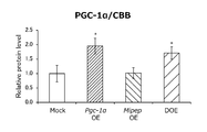
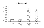
DOE株、Pgc-1α OE株、Mipep OE株、及びMock細胞におけるPgc-1α及びMipepのタンパク質発現量を図3A、図3Bに示す(n=4)。図3A、図3Bに示すとおり、DOE株においては、Pgc-1αタンパク質及びMipepタンパク質の過剰発現が確認された。
(DOE株、Pgc-1α OE株、Mipep OE株、及びMock細胞におけるCox4、Mdh2、及びSirt3のタンパク質発現量の解析)
上記と同様にして、DOE株、Pgc-1α OE株、Mipep OE株、及びMock細胞におけるCox4、Mdh2、及びSirt3のタンパク質発現量を、ウェスタンブロットにより解析した。Cox4タンパク質、Mdh2タンパク質、及びSirt3タンパク質はいずれも、Mipepタンパク質の基質である。
上記と同様にして、DOE株、Pgc-1α OE株、Mipep OE株、及びMock細胞におけるCox4、Mdh2、及びSirt3のタンパク質発現量を、ウェスタンブロットにより解析した。Cox4タンパク質、Mdh2タンパク質、及びSirt3タンパク質はいずれも、Mipepタンパク質の基質である。
なお、Cox4タンパク質の検出には、一次抗体としてウサギ抗COX4ポリクローナル抗体(Cell Signaling)を使用し、Mdh2タンパク質の検出には、一次抗体としてウサギ抗MDH2ポリクローナル抗体(Cell Signaling)を使用し、Sirt3タンパク質の検出には、一次抗体としてウサギ抗SIRT3ポリクローナル抗体(Cell Signaling)を使用した。いずれも二次抗体としてはHRP標識ヤギ抗ウサギIgG(H+L)抗体(Jackson Immunological Research)を使用した。
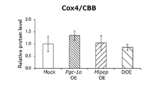
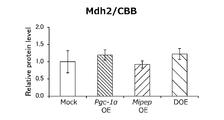
DOE株、Pgc-1α OE株、Mipep OE株、及びMock細胞におけるCox4、Mdh2、及びSirt3のタンパク質発現量を図4A~図4Cに示す(n=4)。図4A~図4Cに示すとおり、Sirt3のタンパク質発現量はPgc-1α遺伝子の過剰発現に相関して有意に増加したが、Cox4及びMdh2のタンパク質発現量はいずれの細胞株においても殆ど変化していなかった。
(DOE株、Pgc-1α OE株、Mipep OE株、及びMock細胞におけるTfam及びTom20のタンパク質発現量の解析)
上記と同様にして、DOE株、Pgc-1α OE株、Mipep OE株、及びMock細胞におけるTfam及びTom20のタンパク質発現量を、ウェスタンブロットにより解析した。Tfamタンパク質はミトコンドリアDNAの転写因子であり、Tom20タンパク質はミトコンドリアの外膜に存在するタンパク質である。
上記と同様にして、DOE株、Pgc-1α OE株、Mipep OE株、及びMock細胞におけるTfam及びTom20のタンパク質発現量を、ウェスタンブロットにより解析した。Tfamタンパク質はミトコンドリアDNAの転写因子であり、Tom20タンパク質はミトコンドリアの外膜に存在するタンパク質である。
なお、Tfamタンパク質の検出には、一次抗体としてウサギ抗TFAMポリクローナル抗体(Protein Tech)を使用し、二次抗体としてHRP標識ヤギ抗ウサギIgG(H+L)抗体(Jackson Immunological Research)を使用した。また、Tom20タンパク質の検出には、一次抗体としてマウス抗TOM20モノクローナル抗体(Sigma)を使用し、二次抗体としてHRP標識ヤギ抗マウスIgG(H+L)抗体(Jackson Immunological Research)を使用した。
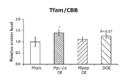
DOE株、Pgc-1α OE株、Mipep OE株、及びMock細胞におけるTfam及びTom20のタンパク質発現量を図5A、図5Bに示す(n=4)。図5A、図5Bに示すとおり、Tfamのタンパク質発現量はPgc-1α遺伝子の過剰発現に相関して増加傾向を示したが、Tom20のタンパク質発現量はいずれの細胞株においても殆ど変化していなかった。
<試験例5>
(DOE株、Pgc-1α OE株、Mipep OE株、及びMock細胞におけるCS(citrate syntase)活性の解析)
分化誘導開始後12日目の細胞(DOE株、Pgc-1α OE株、Mipep OE株、及びMock細胞)から、200μLのホモゲナイゼーションバッファー(50mM tris-HCl(pH7.4)、150mM NaCl、1% ホスファターゼ阻害剤カクテル、5mM EDTA、1% プロテアーゼ阻害剤カクテル、1% Triton X-100、0.05% デオキシコール酸ナトリウム)を用いてタンパク質を抽出した。抽出後のサンプル溶液は、ホモゲナイゼーションバッファーを加えて1/16倍に希釈し、測定に用いた。
(DOE株、Pgc-1α OE株、Mipep OE株、及びMock細胞におけるCS(citrate syntase)活性の解析)
分化誘導開始後12日目の細胞(DOE株、Pgc-1α OE株、Mipep OE株、及びMock細胞)から、200μLのホモゲナイゼーションバッファー(50mM tris-HCl(pH7.4)、150mM NaCl、1% ホスファターゼ阻害剤カクテル、5mM EDTA、1% プロテアーゼ阻害剤カクテル、1% Triton X-100、0.05% デオキシコール酸ナトリウム)を用いてタンパク質を抽出した。抽出後のサンプル溶液は、ホモゲナイゼーションバッファーを加えて1/16倍に希釈し、測定に用いた。
180μLの反応液(0.1mM 5,5’-ジチオビス(2-ニトロ安息香酸)、0.5mM アセチルCoA、0.1% Triton X-100、100mM Tris-HCl(pH8.0))に10μLのサンプル溶液を加え、25℃で5分間インキュベートした。吸光度のベースラインを安定させた後、10mM オキサロ酢酸を10μL加え、30℃で波長412nmにおける吸光度測定を行った。時間を横軸とし、各時間の吸光度からブランクを引いた値の自然対数を縦軸としたグラフの直線部分の傾きから速度定数kを求め、その速度定数kをタンパク質量で割った値をCS活性とした。
DOE株、Pgc-1α OE株、Mipep OE株、及びMock細胞におけるCS活性を図6に示す。図6に示すとおり、Pgc-1α遺伝子の過剰発現に相関してCS活性は増加傾向を示した。
<試験例6>
(DOE株、Pgc-1α OE株、Mipep OE株、及びMock細胞における脂肪滴蓄積の解析)
90mgのOil Red O(Sigma)を30mLの100% イソプロパノールに溶解した。この溶液を超純水で60%に希釈した後、Millex-GVフィルター(Millipore)にて濾過し、Oil Red O染色液とした。分化誘導開始後12日目の細胞(DOE株、Pgc-1α OE株、Mipep OE株、及びMock細胞)をPBSで洗浄した後、10% ホルマリンを含むPBSを用いて10分間固定した。超純水で洗浄し、60% イソプロパノールで1分間処理した後、Oil Red O染色液で20分間染色した。超純水で3回洗浄し、撮影用のサンプルとした。さらに、超純水を除去して完全に乾燥させた後、100% イソプロパノール中で1時間振盪し、脂肪滴を染色したOil Red Oを全て抽出した。抽出溶液を適宜希釈した後、波長490nmの吸光度を測定し、脂肪滴量の指標とした。
(DOE株、Pgc-1α OE株、Mipep OE株、及びMock細胞における脂肪滴蓄積の解析)
90mgのOil Red O(Sigma)を30mLの100% イソプロパノールに溶解した。この溶液を超純水で60%に希釈した後、Millex-GVフィルター(Millipore)にて濾過し、Oil Red O染色液とした。分化誘導開始後12日目の細胞(DOE株、Pgc-1α OE株、Mipep OE株、及びMock細胞)をPBSで洗浄した後、10% ホルマリンを含むPBSを用いて10分間固定した。超純水で洗浄し、60% イソプロパノールで1分間処理した後、Oil Red O染色液で20分間染色した。超純水で3回洗浄し、撮影用のサンプルとした。さらに、超純水を除去して完全に乾燥させた後、100% イソプロパノール中で1時間振盪し、脂肪滴を染色したOil Red Oを全て抽出した。抽出溶液を適宜希釈した後、波長490nmの吸光度を測定し、脂肪滴量の指標とした。
DOE株、Pgc-1α OE株、Mipep OE株、及びMock細胞をOil Red O染色液で染色したときの顕微鏡像を図7Aに示し、脂肪滴蓄積量の測定結果を図7Bに示す(n=3)。図7A、図7Bに示すとおり、Mock細胞と比較して、DOE株、Pgc-1α OE株、及びMipep OE株の全てで脂肪滴蓄積量が有意に減少し、特にDOE株における減少が顕著であった。DOE株は、ミトコンドリア関連タンパク質の発現量(図4A~図4C、図5A、図5B)やCS活性(図6)がPgc-1α OE株と同程度であったにも関わらず、Pgc-1α OE株に比べて脂肪滴蓄積量が有意に減少していた。この結果から、Pgc-1α遺伝子及びMipep遺伝子の両者の発現を亢進させる物質が抗肥満物質になり得ることが示唆された。
<試験例7>
(DOE株、Pgc-1α OE株、Mipep OE株、及びMock細胞の分化の確認)
分化誘導開始後12日目の細胞(DOE株、Pgc-1α OE株、Mipep OE株、及びMock細胞)から全RNAを抽出し、試験例1と同様にして、Pgc-1α、Mipep、Perilipin、Pparγ、及びAdipoqのmRNA発現量をリアルタイムRT-PCRにより確認した。なお、各mRNA発現量は、Rps18のmRNA発現量により補正した。Pgc-1α遺伝子、Mipep遺伝子、及びRps18遺伝子の増幅に使用したプライマーは上記表1及び表2のとおりである。Perilipin遺伝子、Pparγ遺伝子、及びAdipoq遺伝子の増幅に使用したプライマーを下記表4に示す。
(DOE株、Pgc-1α OE株、Mipep OE株、及びMock細胞の分化の確認)
分化誘導開始後12日目の細胞(DOE株、Pgc-1α OE株、Mipep OE株、及びMock細胞)から全RNAを抽出し、試験例1と同様にして、Pgc-1α、Mipep、Perilipin、Pparγ、及びAdipoqのmRNA発現量をリアルタイムRT-PCRにより確認した。なお、各mRNA発現量は、Rps18のmRNA発現量により補正した。Pgc-1α遺伝子、Mipep遺伝子、及びRps18遺伝子の増幅に使用したプライマーは上記表1及び表2のとおりである。Perilipin遺伝子、Pparγ遺伝子、及びAdipoq遺伝子の増幅に使用したプライマーを下記表4に示す。

DOE株、Pgc-1α OE株、Mipep OE株、及びMock細胞におけるPgc-1α、Mipep、Perilipin、Pparγ、及びAdipoqのmRNA発現量を図8に示す。図8に示すとおり、Perilipin、Pparγ、及びAdipoqのmRNA発現量はいずれの細胞株においても殆ど変化していなかった。この結果及び試験例6の結果から、DOE株における脂肪滴蓄積量の減少は、分化シグナルの低下によるものではなく、Pgc-1α遺伝子及びMipep遺伝子の過剰発現によるものであることが示唆された。
<試験例8>
(DOE株、Pgc-1α OE株、Mipep OE株、及びMock細胞におけるPerilipinのタンパク質発現量の解析)
試験例4と同様にして、DOE株、Pgc-1α OE株、Mipep OE株、及びMock細胞におけるPerilipinのタンパク質発現量を、ウェスタンブロットにより解析した。Perilipinタンパク質は脂肪滴表面に存在するタンパク質である。コントロールとして、Lamin B1のタンパク質発現量も解析した。
(DOE株、Pgc-1α OE株、Mipep OE株、及びMock細胞におけるPerilipinのタンパク質発現量の解析)
試験例4と同様にして、DOE株、Pgc-1α OE株、Mipep OE株、及びMock細胞におけるPerilipinのタンパク質発現量を、ウェスタンブロットにより解析した。Perilipinタンパク質は脂肪滴表面に存在するタンパク質である。コントロールとして、Lamin B1のタンパク質発現量も解析した。
なお、Perilipinタンパク質の検出には、一次抗体としてウサギ抗Perilipinモノクローナル抗体(Cell Signaling)を使用し、Lamin B1タンパク質の検出には、一次抗体としてウサギ抗Lamin B1ポリクローナル抗体(Medical & Biochemical Laboratories)を使用した。いずれも、二次抗体としてはHRP標識ヤギ抗ウサギIgG(H+L)抗体(Jackson Immunological Research)を使用した。
DOE株、Pgc-1α OE株、Mipep OE株、及びMock細胞におけるPerilipinのタンパク質発現量を図9に示す。図9に示すとおり、DOE株においては、Perilipinのタンパク質発現量が顕著に減少していた。
Claims (7)
- Pgc-1α遺伝子及びMipep遺伝子の発現を亢進させる物質を選択することを含む抗肥満物質のスクリーニング方法。
- Pgc-1α遺伝子及びMipep遺伝子を発現する細胞に被験物質を接触させる工程と、
前記被験物質を接触させない場合に比べて、前記細胞におけるPgc-1α遺伝子及びMipep遺伝子の発現量が増加する場合に、該被験物質を抗肥満物質として選択する工程と、
を含む請求項1に記載の抗肥満物質のスクリーニング方法。 - 前記細胞として、Srebp-1c遺伝子を有する細胞とSrebp-1c遺伝子を有しない細胞とを使用し、
前記被験物質を接触させることにより、Srebp-1c遺伝子を有する細胞においてのみPgc-1α遺伝子及びMipep遺伝子の発現量が増加する場合に、該被験物質を抗肥満物質として選択する請求項2に記載の抗肥満物質のスクリーニング方法。 - Pgc-1α遺伝子のプロモーターの下流に機能的に連結された第1のレポーター遺伝子を有する第1の形質転換体、及びMipep遺伝子のプロモーターの下流に機能的に連結された第2のレポーター遺伝子を有する第2の形質転換体に、被験物質を接触させる工程と、
前記被験物質を接触させない場合に比べて、前記第1のレポーター遺伝子及び前記第2のレポーター遺伝子の発現量が増加する場合に、該被験物質を抗肥満物質として選択する工程と、
を含む請求項1に記載の抗肥満物質のスクリーニング方法。 - 前記第1の形質転換体及び前記第2の形質転換体のそれぞれとして、Srebp-1c遺伝子を有する形質転換体とSrebp-1c遺伝子を有しない形質転換体とを使用し、
前記被験物質を接触させることにより、Srebp-1c遺伝子を有する形質転換体においてのみ前記第1のレポーター遺伝子及び前記第2のレポーター遺伝子の発現量が増加する場合に、該被験物質を抗肥満物質として選択する請求項4に記載の抗肥満物質のスクリーニング方法。 - 下記(a)~(c)から選択される少なくとも1つを含む抗肥満物質のスクリーニング用キット:
(a)Pgc-1α遺伝子及びMipep遺伝子のそれぞれを増幅するためのプライマー、
(b)Pgc-1α遺伝子及びMipep遺伝子のそれぞれを検出するためのプローブ、
(c)Pgc-1αタンパク質及びMipepタンパク質のそれぞれに対する抗体。 - 下記(d)及び(e)から選択される少なくとも1つを含む抗肥満物質のスクリーニング用キット:
(d)Pgc-1α遺伝子のプロモーターと、該プロモーターの下流に機能的に連結された第1のレポーター遺伝子とを有する第1のベクター、及びMipep遺伝子のプロモーターと、該プロモーターの下流に機能的に連結された第2のレポーター遺伝子とを有する第2のベクター、
(e)Pgc-1α遺伝子のプロモーターの下流に機能的に連結された第1のレポーター遺伝子を有する第1の形質転換体、及びMipep遺伝子のプロモーターの下流に機能的に連結された第2のレポーター遺伝子を有する第2の形質転換体。
Applications Claiming Priority (2)
| Application Number | Priority Date | Filing Date | Title |
|---|---|---|---|
| JP2018-133627 | 2018-07-13 | ||
| JP2018133627A JP2021166471A (ja) | 2018-07-13 | 2018-07-13 | 抗肥満物質のスクリーニング方法及び抗肥満物質のスクリーニング用キット |
Publications (1)
| Publication Number | Publication Date |
|---|---|
| WO2020013271A1 true WO2020013271A1 (ja) | 2020-01-16 |
Family
ID=69141580
Family Applications (1)
| Application Number | Title | Priority Date | Filing Date |
|---|---|---|---|
| PCT/JP2019/027480 Ceased WO2020013271A1 (ja) | 2018-07-13 | 2019-07-11 | 抗肥満物質のスクリーニング方法及び抗肥満物質のスクリーニング用キット |
Country Status (2)
| Country | Link |
|---|---|
| JP (1) | JP2021166471A (ja) |
| WO (1) | WO2020013271A1 (ja) |
Citations (2)
| Publication number | Priority date | Publication date | Assignee | Title |
|---|---|---|---|---|
| JP2011036223A (ja) * | 2009-08-18 | 2011-02-24 | Nagasaki Univ | カロリー制限模倣物のスクリーニング方法 |
| WO2017052340A1 (ko) * | 2015-09-25 | 2017-03-30 | 고려대학교 산학협력단 | 운동유사효과 유도용 약학 조성물 |
-
2018
- 2018-07-13 JP JP2018133627A patent/JP2021166471A/ja active Pending
-
2019
- 2019-07-11 WO PCT/JP2019/027480 patent/WO2020013271A1/ja not_active Ceased
Patent Citations (2)
| Publication number | Priority date | Publication date | Assignee | Title |
|---|---|---|---|---|
| JP2011036223A (ja) * | 2009-08-18 | 2011-02-24 | Nagasaki Univ | カロリー制限模倣物のスクリーニング方法 |
| WO2017052340A1 (ko) * | 2015-09-25 | 2017-03-30 | 고려대학교 산학협력단 | 운동유사효과 유도용 약학 조성물 |
Non-Patent Citations (5)
| Title |
|---|
| ANDERSON, R. M. ET AL.: "Dynamic regulation of PGC-1 a localization and turnover implicates mitochondrial adaptation in calorie restriction and the stress response", AGING CELL, vol. 7, 2008, pages 101 - 111, XP055675620 * |
| FUJII, N. ET AL.: "Sterol regulatory element- binding protein-lc orchestrates metabolic remodeling of white adipose tissue by caloric restriction", AGING CELL, vol. 16, 2017, pages 508 - 517, XP055675618 * |
| HIGAMI, YOSHIKAZU: "Comprehensive mitochondrial quality control and its collapse in metabolic diseases", GRANTS-ON-AID FOR SCIENTIFIC RESEARCH, FINAL RESEARCH REPORTON GRANTS-IN-AID FOR SCIENTIFIC RESEARCH, 6 June 2016 (2016-06-06), pages 1 - 4, Retrieved from the Internet <URL:http://kaken.nii.ac.jp/ja/file/KAKENHI-PROJECT-26670193/26670193seika.pdf> [retrieved on 20190926] * |
| KOBAYASHI, M. ET AL.: "Mitochondrial intermediate peptidase is a novel regulator of sirtuin-3 activation by caloric restriction", FEBS LETTERS, vol. 591, 28 November 2017 (2017-11-28), pages 4067 - 4073, XP055675616 * |
| MARTIN-MONTALVO, ALEJANDRO ET AL.: "Mitochondrial Metabolic Reprogramming Induced by Calorie Restriction", ANTIOXIDANTS & REDOX SIGNALING, vol. 19, no. 3, 2013, pages 310 - 320, XP055675624 * |
Also Published As
| Publication number | Publication date |
|---|---|
| JP2021166471A (ja) | 2021-10-21 |
Similar Documents
| Publication | Publication Date | Title |
|---|---|---|
| Li et al. | Histone H4K12 lactylation promotes malignancy progression in triple-negative breast cancer through SLFN5 downregulation | |
| Grohmann et al. | Characterization of Ighmbp2 in motor neurons and implications for the pathomechanism in a mouse model of human spinal muscular atrophy with respiratory distress type 1 (SMARD1) | |
| Hosking et al. | Sox7 and Sox17 are strain-specific modifiers of the lymphangiogenic defects caused by Sox18 dysfunction in mice | |
| Shi et al. | ARRDC1 and ARRDC3 act as tumor suppressors in renal cell carcinoma by facilitating YAP1 degradation | |
| Sriram et al. | Expression of a novel brain specific isoform of C3G is regulated during development | |
| Du et al. | SMARCA2 is regulated by NORFA–miR-29c, a novel pathway that controls granulosa cell apoptosis and is related to female fertility | |
| Wang et al. | Identification of WNT4 alternative splicing patterns and effects on proliferation of granulosa cells in goat | |
| JPWO2016152352A1 (ja) | メラノーマ特異的バイオマーカー及びその利用 | |
| US20060068370A1 (en) | Liver specific chimeric regulatory sequence and use thereof | |
| Huang et al. | MAPKAP1 orchestrates macrophage polarization and lipid metabolism in fatty liver-enhanced colorectal cancer | |
| Mubiru et al. | A Variant of the alpha‐methyl‐acyl‐CoA racemase gene created by a deletion in exon 5 and its expression in prostate cancer | |
| WO2020013271A1 (ja) | 抗肥満物質のスクリーニング方法及び抗肥満物質のスクリーニング用キット | |
| Wang et al. | Involvement of fission yeast Pdc2 in RNA degradation and P-body function | |
| Li et al. | Transcription activation of SPINK4 by ELF-1 augments progression of colon cancer by regulating biological behaviors | |
| CN119020412A (zh) | Usp13基因的应用 | |
| CN113498440A (zh) | 新型胰腺癌上皮间质转化标记物 | |
| RU2662358C2 (ru) | Способы биологических исследований с использованием клеток для контроля качества нутрицевтических продуктов и лекарственных средств | |
| CN104087663B (zh) | Prl-1基因在制备诊断和/或治疗肝癌产品中的应用 | |
| WO2011145722A1 (ja) | Aimの機能を制御する化合物のスクリーニング方法 | |
| Siddiqui et al. | An in vitro study on the role of hepatitis B virus X protein C-terminal truncation in liver disease development | |
| CN107779503A (zh) | 阿尔茨海默相关的差异表达基因及其应用 | |
| Xiao et al. | TIG1 triggers placental senescence in preeclampsia through LMNA/p53 axis activation | |
| US20220221457A1 (en) | Marker for detecting proliferation of stem cell and high-efficiency proliferation method of stem cell using same | |
| Kimura‐Nagano et al. | Stat stimulates histone H 3 K 4 methylation via KDM5 inhibition in adult stem cells of budding tunicates | |
| Parkar et al. | New Δ246p53 isoform responds to DNA damage to enhance p53 functions in senescence and tumour suppression |
Legal Events
| Date | Code | Title | Description |
|---|---|---|---|
| 121 | Ep: the epo has been informed by wipo that ep was designated in this application |
Ref document number: 19833154 Country of ref document: EP Kind code of ref document: A1 |
|
| NENP | Non-entry into the national phase |
Ref country code: DE |
|
| 122 | Ep: pct application non-entry in european phase |
Ref document number: 19833154 Country of ref document: EP Kind code of ref document: A1 |
|
| NENP | Non-entry into the national phase |
Ref country code: JP |