WO2020006357A1 - Methods for diagnosis and treatment of type 1 diabetes - Google Patents
Methods for diagnosis and treatment of type 1 diabetes Download PDFInfo
- Publication number
- WO2020006357A1 WO2020006357A1 PCT/US2019/039732 US2019039732W WO2020006357A1 WO 2020006357 A1 WO2020006357 A1 WO 2020006357A1 US 2019039732 W US2019039732 W US 2019039732W WO 2020006357 A1 WO2020006357 A1 WO 2020006357A1
- Authority
- WO
- WIPO (PCT)
- Prior art keywords
- amyloid
- coli
- mammal
- bacteria
- microbiota
- Prior art date
Links
Classifications
-
- A—HUMAN NECESSITIES
- A61—MEDICAL OR VETERINARY SCIENCE; HYGIENE
- A61K—PREPARATIONS FOR MEDICAL, DENTAL OR TOILETRY PURPOSES
- A61K35/00—Medicinal preparations containing materials or reaction products thereof with undetermined constitution
- A61K35/66—Microorganisms or materials therefrom
- A61K35/74—Bacteria
-
- A—HUMAN NECESSITIES
- A61—MEDICAL OR VETERINARY SCIENCE; HYGIENE
- A61K—PREPARATIONS FOR MEDICAL, DENTAL OR TOILETRY PURPOSES
- A61K31/00—Medicinal preparations containing organic active ingredients
- A61K31/65—Tetracyclines
-
- A—HUMAN NECESSITIES
- A61—MEDICAL OR VETERINARY SCIENCE; HYGIENE
- A61K—PREPARATIONS FOR MEDICAL, DENTAL OR TOILETRY PURPOSES
- A61K31/00—Medicinal preparations containing organic active ingredients
- A61K31/70—Carbohydrates; Sugars; Derivatives thereof
- A61K31/7004—Monosaccharides having only carbon, hydrogen and oxygen atoms
-
- A—HUMAN NECESSITIES
- A61—MEDICAL OR VETERINARY SCIENCE; HYGIENE
- A61K—PREPARATIONS FOR MEDICAL, DENTAL OR TOILETRY PURPOSES
- A61K31/00—Medicinal preparations containing organic active ingredients
- A61K31/70—Carbohydrates; Sugars; Derivatives thereof
- A61K31/7088—Compounds having three or more nucleosides or nucleotides
- A61K31/713—Double-stranded nucleic acids or oligonucleotides
-
- A—HUMAN NECESSITIES
- A61—MEDICAL OR VETERINARY SCIENCE; HYGIENE
- A61K—PREPARATIONS FOR MEDICAL, DENTAL OR TOILETRY PURPOSES
- A61K33/00—Medicinal preparations containing inorganic active ingredients
- A61K33/06—Aluminium, calcium or magnesium; Compounds thereof, e.g. clay
-
- A—HUMAN NECESSITIES
- A61—MEDICAL OR VETERINARY SCIENCE; HYGIENE
- A61K—PREPARATIONS FOR MEDICAL, DENTAL OR TOILETRY PURPOSES
- A61K35/00—Medicinal preparations containing materials or reaction products thereof with undetermined constitution
- A61K35/66—Microorganisms or materials therefrom
- A61K35/74—Bacteria
- A61K35/741—Probiotics
- A61K35/744—Lactic acid bacteria, e.g. enterococci, pediococci, lactococci, streptococci or leuconostocs
- A61K35/745—Bifidobacteria
-
- A—HUMAN NECESSITIES
- A61—MEDICAL OR VETERINARY SCIENCE; HYGIENE
- A61K—PREPARATIONS FOR MEDICAL, DENTAL OR TOILETRY PURPOSES
- A61K35/00—Medicinal preparations containing materials or reaction products thereof with undetermined constitution
- A61K35/66—Microorganisms or materials therefrom
- A61K35/74—Bacteria
- A61K35/741—Probiotics
- A61K35/744—Lactic acid bacteria, e.g. enterococci, pediococci, lactococci, streptococci or leuconostocs
- A61K35/747—Lactobacilli, e.g. L. acidophilus or L. brevis
-
- A—HUMAN NECESSITIES
- A61—MEDICAL OR VETERINARY SCIENCE; HYGIENE
- A61K—PREPARATIONS FOR MEDICAL, DENTAL OR TOILETRY PURPOSES
- A61K35/00—Medicinal preparations containing materials or reaction products thereof with undetermined constitution
- A61K35/66—Microorganisms or materials therefrom
- A61K35/76—Viruses; Subviral particles; Bacteriophages
-
- A—HUMAN NECESSITIES
- A61—MEDICAL OR VETERINARY SCIENCE; HYGIENE
- A61K—PREPARATIONS FOR MEDICAL, DENTAL OR TOILETRY PURPOSES
- A61K38/00—Medicinal preparations containing peptides
- A61K38/04—Peptides having up to 20 amino acids in a fully defined sequence; Derivatives thereof
- A61K38/08—Peptides having 5 to 11 amino acids
-
- A—HUMAN NECESSITIES
- A61—MEDICAL OR VETERINARY SCIENCE; HYGIENE
- A61K—PREPARATIONS FOR MEDICAL, DENTAL OR TOILETRY PURPOSES
- A61K38/00—Medicinal preparations containing peptides
- A61K38/16—Peptides having more than 20 amino acids; Gastrins; Somatostatins; Melanotropins; Derivatives thereof
- A61K38/17—Peptides having more than 20 amino acids; Gastrins; Somatostatins; Melanotropins; Derivatives thereof from animals; from humans
- A61K38/1703—Peptides having more than 20 amino acids; Gastrins; Somatostatins; Melanotropins; Derivatives thereof from animals; from humans from vertebrates
- A61K38/1709—Peptides having more than 20 amino acids; Gastrins; Somatostatins; Melanotropins; Derivatives thereof from animals; from humans from vertebrates from mammals
-
- A—HUMAN NECESSITIES
- A61—MEDICAL OR VETERINARY SCIENCE; HYGIENE
- A61K—PREPARATIONS FOR MEDICAL, DENTAL OR TOILETRY PURPOSES
- A61K38/00—Medicinal preparations containing peptides
- A61K38/16—Peptides having more than 20 amino acids; Gastrins; Somatostatins; Melanotropins; Derivatives thereof
- A61K38/43—Enzymes; Proenzymes; Derivatives thereof
- A61K38/46—Hydrolases (3)
- A61K38/465—Hydrolases (3) acting on ester bonds (3.1), e.g. lipases, ribonucleases
-
- A—HUMAN NECESSITIES
- A61—MEDICAL OR VETERINARY SCIENCE; HYGIENE
- A61K—PREPARATIONS FOR MEDICAL, DENTAL OR TOILETRY PURPOSES
- A61K39/00—Medicinal preparations containing antigens or antibodies
- A61K39/0005—Vertebrate antigens
- A61K39/0008—Antigens related to auto-immune diseases; Preparations to induce self-tolerance
-
- A—HUMAN NECESSITIES
- A61—MEDICAL OR VETERINARY SCIENCE; HYGIENE
- A61K—PREPARATIONS FOR MEDICAL, DENTAL OR TOILETRY PURPOSES
- A61K39/00—Medicinal preparations containing antigens or antibodies
- A61K39/02—Bacterial antigens
- A61K39/025—Enterobacteriales, e.g. Enterobacter
- A61K39/0258—Escherichia
-
- A—HUMAN NECESSITIES
- A61—MEDICAL OR VETERINARY SCIENCE; HYGIENE
- A61P—SPECIFIC THERAPEUTIC ACTIVITY OF CHEMICAL COMPOUNDS OR MEDICINAL PREPARATIONS
- A61P3/00—Drugs for disorders of the metabolism
- A61P3/08—Drugs for disorders of the metabolism for glucose homeostasis
- A61P3/10—Drugs for disorders of the metabolism for glucose homeostasis for hyperglycaemia, e.g. antidiabetics
-
- C—CHEMISTRY; METALLURGY
- C07—ORGANIC CHEMISTRY
- C07K—PEPTIDES
- C07K16/00—Immunoglobulins [IGs], e.g. monoclonal or polyclonal antibodies
- C07K16/12—Immunoglobulins [IGs], e.g. monoclonal or polyclonal antibodies against material from bacteria
- C07K16/1203—Immunoglobulins [IGs], e.g. monoclonal or polyclonal antibodies against material from bacteria from Gram-negative bacteria
- C07K16/1228—Immunoglobulins [IGs], e.g. monoclonal or polyclonal antibodies against material from bacteria from Gram-negative bacteria from Enterobacteriaceae (F), e.g. Citrobacter, Serratia, Proteus, Providencia, Morganella, Yersinia
- C07K16/1232—Immunoglobulins [IGs], e.g. monoclonal or polyclonal antibodies against material from bacteria from Gram-negative bacteria from Enterobacteriaceae (F), e.g. Citrobacter, Serratia, Proteus, Providencia, Morganella, Yersinia from Escherichia (G)
-
- C—CHEMISTRY; METALLURGY
- C12—BIOCHEMISTRY; BEER; SPIRITS; WINE; VINEGAR; MICROBIOLOGY; ENZYMOLOGY; MUTATION OR GENETIC ENGINEERING
- C12N—MICROORGANISMS OR ENZYMES; COMPOSITIONS THEREOF; PROPAGATING, PRESERVING, OR MAINTAINING MICROORGANISMS; MUTATION OR GENETIC ENGINEERING; CULTURE MEDIA
- C12N15/00—Mutation or genetic engineering; DNA or RNA concerning genetic engineering, vectors, e.g. plasmids, or their isolation, preparation or purification; Use of hosts therefor
- C12N15/09—Recombinant DNA-technology
- C12N15/11—DNA or RNA fragments; Modified forms thereof; Non-coding nucleic acids having a biological activity
- C12N15/113—Non-coding nucleic acids modulating the expression of genes, e.g. antisense oligonucleotides; Antisense DNA or RNA; Triplex- forming oligonucleotides; Catalytic nucleic acids, e.g. ribozymes; Nucleic acids used in co-suppression or gene silencing
-
- C—CHEMISTRY; METALLURGY
- C12—BIOCHEMISTRY; BEER; SPIRITS; WINE; VINEGAR; MICROBIOLOGY; ENZYMOLOGY; MUTATION OR GENETIC ENGINEERING
- C12Q—MEASURING OR TESTING PROCESSES INVOLVING ENZYMES, NUCLEIC ACIDS OR MICROORGANISMS; COMPOSITIONS OR TEST PAPERS THEREFOR; PROCESSES OF PREPARING SUCH COMPOSITIONS; CONDITION-RESPONSIVE CONTROL IN MICROBIOLOGICAL OR ENZYMOLOGICAL PROCESSES
- C12Q1/00—Measuring or testing processes involving enzymes, nucleic acids or microorganisms; Compositions therefor; Processes of preparing such compositions
- C12Q1/68—Measuring or testing processes involving enzymes, nucleic acids or microorganisms; Compositions therefor; Processes of preparing such compositions involving nucleic acids
- C12Q1/6876—Nucleic acid products used in the analysis of nucleic acids, e.g. primers or probes
- C12Q1/6883—Nucleic acid products used in the analysis of nucleic acids, e.g. primers or probes for diseases caused by alterations of genetic material
-
- C—CHEMISTRY; METALLURGY
- C12—BIOCHEMISTRY; BEER; SPIRITS; WINE; VINEGAR; MICROBIOLOGY; ENZYMOLOGY; MUTATION OR GENETIC ENGINEERING
- C12Q—MEASURING OR TESTING PROCESSES INVOLVING ENZYMES, NUCLEIC ACIDS OR MICROORGANISMS; COMPOSITIONS OR TEST PAPERS THEREFOR; PROCESSES OF PREPARING SUCH COMPOSITIONS; CONDITION-RESPONSIVE CONTROL IN MICROBIOLOGICAL OR ENZYMOLOGICAL PROCESSES
- C12Q1/00—Measuring or testing processes involving enzymes, nucleic acids or microorganisms; Compositions therefor; Processes of preparing such compositions
- C12Q1/68—Measuring or testing processes involving enzymes, nucleic acids or microorganisms; Compositions therefor; Processes of preparing such compositions involving nucleic acids
- C12Q1/6876—Nucleic acid products used in the analysis of nucleic acids, e.g. primers or probes
- C12Q1/6888—Nucleic acid products used in the analysis of nucleic acids, e.g. primers or probes for detection or identification of organisms
- C12Q1/689—Nucleic acid products used in the analysis of nucleic acids, e.g. primers or probes for detection or identification of organisms for bacteria
-
- C—CHEMISTRY; METALLURGY
- C12—BIOCHEMISTRY; BEER; SPIRITS; WINE; VINEGAR; MICROBIOLOGY; ENZYMOLOGY; MUTATION OR GENETIC ENGINEERING
- C12Q—MEASURING OR TESTING PROCESSES INVOLVING ENZYMES, NUCLEIC ACIDS OR MICROORGANISMS; COMPOSITIONS OR TEST PAPERS THEREFOR; PROCESSES OF PREPARING SUCH COMPOSITIONS; CONDITION-RESPONSIVE CONTROL IN MICROBIOLOGICAL OR ENZYMOLOGICAL PROCESSES
- C12Q1/00—Measuring or testing processes involving enzymes, nucleic acids or microorganisms; Compositions therefor; Processes of preparing such compositions
- C12Q1/70—Measuring or testing processes involving enzymes, nucleic acids or microorganisms; Compositions therefor; Processes of preparing such compositions involving virus or bacteriophage
-
- G—PHYSICS
- G01—MEASURING; TESTING
- G01N—INVESTIGATING OR ANALYSING MATERIALS BY DETERMINING THEIR CHEMICAL OR PHYSICAL PROPERTIES
- G01N33/00—Investigating or analysing materials by specific methods not covered by groups G01N1/00 - G01N31/00
- G01N33/48—Biological material, e.g. blood, urine; Haemocytometers
- G01N33/50—Chemical analysis of biological material, e.g. blood, urine; Testing involving biospecific ligand binding methods; Immunological testing
- G01N33/68—Chemical analysis of biological material, e.g. blood, urine; Testing involving biospecific ligand binding methods; Immunological testing involving proteins, peptides or amino acids
- G01N33/6893—Chemical analysis of biological material, e.g. blood, urine; Testing involving biospecific ligand binding methods; Immunological testing involving proteins, peptides or amino acids related to diseases not provided for elsewhere
-
- A—HUMAN NECESSITIES
- A61—MEDICAL OR VETERINARY SCIENCE; HYGIENE
- A61K—PREPARATIONS FOR MEDICAL, DENTAL OR TOILETRY PURPOSES
- A61K39/00—Medicinal preparations containing antigens or antibodies
- A61K2039/505—Medicinal preparations containing antigens or antibodies comprising antibodies
-
- A—HUMAN NECESSITIES
- A61—MEDICAL OR VETERINARY SCIENCE; HYGIENE
- A61K—PREPARATIONS FOR MEDICAL, DENTAL OR TOILETRY PURPOSES
- A61K39/00—Medicinal preparations containing antigens or antibodies
- A61K2039/58—Medicinal preparations containing antigens or antibodies raising an immune response against a target which is not the antigen used for immunisation
-
- C—CHEMISTRY; METALLURGY
- C12—BIOCHEMISTRY; BEER; SPIRITS; WINE; VINEGAR; MICROBIOLOGY; ENZYMOLOGY; MUTATION OR GENETIC ENGINEERING
- C12N—MICROORGANISMS OR ENZYMES; COMPOSITIONS THEREOF; PROPAGATING, PRESERVING, OR MAINTAINING MICROORGANISMS; MUTATION OR GENETIC ENGINEERING; CULTURE MEDIA
- C12N2310/00—Structure or type of the nucleic acid
- C12N2310/10—Type of nucleic acid
- C12N2310/14—Type of nucleic acid interfering N.A.
-
- C—CHEMISTRY; METALLURGY
- C12—BIOCHEMISTRY; BEER; SPIRITS; WINE; VINEGAR; MICROBIOLOGY; ENZYMOLOGY; MUTATION OR GENETIC ENGINEERING
- C12Q—MEASURING OR TESTING PROCESSES INVOLVING ENZYMES, NUCLEIC ACIDS OR MICROORGANISMS; COMPOSITIONS OR TEST PAPERS THEREFOR; PROCESSES OF PREPARING SUCH COMPOSITIONS; CONDITION-RESPONSIVE CONTROL IN MICROBIOLOGICAL OR ENZYMOLOGICAL PROCESSES
- C12Q2600/00—Oligonucleotides characterized by their use
- C12Q2600/118—Prognosis of disease development
-
- C—CHEMISTRY; METALLURGY
- C12—BIOCHEMISTRY; BEER; SPIRITS; WINE; VINEGAR; MICROBIOLOGY; ENZYMOLOGY; MUTATION OR GENETIC ENGINEERING
- C12Q—MEASURING OR TESTING PROCESSES INVOLVING ENZYMES, NUCLEIC ACIDS OR MICROORGANISMS; COMPOSITIONS OR TEST PAPERS THEREFOR; PROCESSES OF PREPARING SUCH COMPOSITIONS; CONDITION-RESPONSIVE CONTROL IN MICROBIOLOGICAL OR ENZYMOLOGICAL PROCESSES
- C12Q2600/00—Oligonucleotides characterized by their use
- C12Q2600/156—Polymorphic or mutational markers
-
- C—CHEMISTRY; METALLURGY
- C12—BIOCHEMISTRY; BEER; SPIRITS; WINE; VINEGAR; MICROBIOLOGY; ENZYMOLOGY; MUTATION OR GENETIC ENGINEERING
- C12Q—MEASURING OR TESTING PROCESSES INVOLVING ENZYMES, NUCLEIC ACIDS OR MICROORGANISMS; COMPOSITIONS OR TEST PAPERS THEREFOR; PROCESSES OF PREPARING SUCH COMPOSITIONS; CONDITION-RESPONSIVE CONTROL IN MICROBIOLOGICAL OR ENZYMOLOGICAL PROCESSES
- C12Q2600/00—Oligonucleotides characterized by their use
- C12Q2600/172—Haplotypes
-
- G—PHYSICS
- G01—MEASURING; TESTING
- G01N—INVESTIGATING OR ANALYSING MATERIALS BY DETERMINING THEIR CHEMICAL OR PHYSICAL PROPERTIES
- G01N2800/00—Detection or diagnosis of diseases
- G01N2800/04—Endocrine or metabolic disorders
- G01N2800/042—Disorders of carbohydrate metabolism, e.g. diabetes, glucose metabolism
Definitions
- Prevention and treatment methods comprise (i) inactivation of amyloid-producing bacteria within microbiota and/or (ii) inactivation of amyloid-producing bacteria getting from the outer environment to microbiota, gastrointestinal tract, bodily fluid(s) or tissue(s) of a mammal and/or (iii) inactivation of Type-1 Diabetes associated microbial product (T1DAMP) production/release by microbiota and/or (iv) inactivation of bacteria-derived T1DAMP present in microbiota, bodily fluid(s) or tissue(s) of a mammal and/or (v) inhibiting release of bacteria-derived T1DAMP from biofilm and/or bacteria to gastrointestinal tract, bodily fluid(s) or tissue(s) of a mammal and/or (vi) inhibiting entry of bacteria-derived T1DAMP to microbiota, gastrointestinal tract, bodily fluid(s) or tissue
- the microbiota of the human intestinal tract is comprised of bacteria, fungi, and viruses, including bacteriophages. This highly diverse and complex ecosystem is characterized by dynamic stability of each of its components in the context of the host organism.
- the human gut contains approximately 10 13 bacteria, which >10 times of the number of human cells (Dalmasso, M. et al., 2014).
- Intestinal barrier dysfunction or disruption is characterized by the translocation of macromolecules, bacteria or their toxins to the lamina intestinal, which is implicated in the pathogenesis of numerous diseases (Maes M. et al., 2012).
- An abnormally permeable mucosal barrier is associated with various pathologies including inflammatory bowel disease, Crohn’s disease, neurodegenerative diseases, diabetes type 1, some types of cancers, cardiovascular disorders, rheumatoid arthritis, etc. (Tlaskalova- Hogenová, H. et al., 2011; Berk, M. et al., 2013 Fasano 2012).
- biofilms Bacteria in the human gut live within surface-associated microbial communities named biofilms, which are characterized by the presence of self-produced extracellular matrix (ECM) and surface film that protect microorganisms for the outer environment (Costerton et al., 1999).
- ECM extracellular matrix
- surface film that protect microorganisms for the outer environment.
- ECM consists of different biomolecules including extracellular nucleic acids, polysaccharides and proteins, and several microorganisms within human microbiome predominantly among the members of Enterobactericeae family possess amyloid proteins that can form so called curli fibers within ECM as well that provide them with unique mechanical properties and representing an important step during biofilm formation (Gallo, P.M. et al., 2015).
- Type 1 Diabetes is an autoimmune disorder driven by T cell-mediated destruction of the insulin-secreting b–cells of the pancreatic islets that often manifests during childhood.
- T1D is an autoimmune disorder driven by T cell-mediated destruction of the insulin-secreting b–cells of the pancreatic islets that often manifests during childhood.
- HLA human leukocyte antigens
- the major genetic determinants of T1D are polymorphisms of class II HLA genes inherited from both parents that are the key for the development of T1D predisposing over 60% of its familial clustering.
- T1D usually has a long pre-diabetic period, named seroconversion.
- Seroconversion is characterized by the presence of autoantibodies to antigens of the pancreatic b cells or insulin without progression to T1D.
- a few factors are explored as triggers of the seroconversion; however what they have in common is that they lead to the death of islet cells, which in turn leads to formation of b-cell antigens, activation of dendric cells (DC) and antigen presentation.
- DC dendric cells
- a method for preventing or treating Type 1 Diabetes (T1D) or consequences thereof in a mammal in need thereof comprising one or more of (i) inactivating amyloid-producing bacteria within microbiota in the mammal, (ii) preventing amyloid-producing bacteria from entering the microbiota, the gastrointestinal tract, a bodily fluid or a tissue of the mammal from an environment outside the mammal, (iii) inactivating a T1D-associated microbial product (T1DAMP) that is released by microbiota of the mammal, (iv) inactivating a T1DAMP present, a bodily fluid or a tissue of the mammal, (v) inhibiting release of a T1DAMP from a biofilm and/or bacteria in the gastrointestinal tract, a bodily fluid or a tissue of the mammal, (vi) inhibiting entry of T1DAMP to microbiota, the gastrointestinal tract, a bodily
- the mammal expresses a T1D susceptible HLA allele.
- the T1D susceptible HLA allele is selected from an HLA allele having a DR4-DQ8 and/or DRB1 haplotype, an HLA allele having a DR3-DQ2 haplotype, HLA allele DQB1*02/*0302-DRB1*0404, HLA allele DQB1*0302/*0501-DRB1*0401, and DQB1*0302/*04-DRB1*0401*.
- the mammal comprises an increased amount of E. coli or Salmonella in the gastrointestinal tract as compared to a second mammal that does not develop T1D, and optionally wherein the mammal and the second mammal both express a T1D susceptible HLA allele, and optionally wherein the mammal and the second mammal both express the same T1D susceptible HLA allele.
- the second mammal is age-matched and/or gender-matched to the mammal comprising the increased amount of E. coli or Salmonella in the gastrointestinal tract.
- the method comprises administering to the mammal E. coli in a manner effective to populate the microbiota with the E. coli, wherein the administered E. coli do not produce an amyloid protein or produce a reduced amount of the amyloid protein as compared to a wild-type E. coli.
- the administered E. coli comprises a mutation in a gene encoding for an amyloid protein.
- the method comprises administering to the mammal E. coli that comprises a mutation in a sequence regulating expression of a gene encoding for an amyloid protein.
- inactivation of amyloid-producing bacteria within microbiota comprises preventing transfer of amyloid-producing bacteria to the mammal from a mother of the mammal during birth or breastfeeding.
- the inactivation of amyloid-producing bacteria within microbiota comprises administering to the mammal a microorganism or a by-product of the microorganism, wherein the microorganism or the byproduct is effective to prevent colonization of the amyloid-producing bacteria in the gastrointestinal tract of the mammal.
- the microorganism is from an order selected from Bacteroidales, Lactobacillales, Erysipelotrichales, Coriobacteriales, Clostridiales, Bacillales, and Bifidobacteriales.
- the microorganism is a non-amyloid-producing strain of bacteria or a strain of bacteria that synthesizes a reduced amount of amyloid.
- T1D Type 1 Diabetes
- the method further comprises vaccination of the mammal against E. coli or Salmonella.
- the method further comprises vaccination of the mammal against Enterobacteriales bacteria.
- the method further comprises vaccination of the mammal against E. coli or Salmonella.
- the inactivation of amyloid-producing bacteria within microbiota comprises i) colonization of gastrointestinal microbiota of the mammal with non- amyloid-producing bacteria and/or ii) administering an anti-amyloid antibody to the mammal.
- the inactivation of amyloid-producing bacteria within microbiota comprises editing one or more genes in the genome of the amyloid-producing bacteria, wherein the inactivation inhibits adhesion of the amyloid-producing bacteria, wherein the one or more genes are selected from afa-dra, daaD, tsh, vat, ibeA, fyuA, mat, sfa- foc, malX, pic, irp2, papC, fimH; PapAH papEF, bmaE, sfa/focDE, papC, focG, sfaI, sfa II, sfaS, aah, aidA, fasA, faeG, bfpA, eaeA, Paa, fasA, faeG, fedA, fanC, sfaY, and a gene in the Cpx pathway.
- the inactivation of the amyloid-producing bacteria within microbiota comprises administering to the mammal a composition comprising one or more of fosfomycin, Doxycycline, Ciprofloxacin, Trimethoprim/sulfamethoxazole, Levofloxacin, Amoxicillin, Aztreonam, Nitrofurantoin, Ceftriaxone, imipenem, and Rifaximin, a FimH antagonist, and a pilicide.
- the FimH antagonist is an n-Heptyl a-D-mannose glycopolymer, methyl R-D-mannoside, or a thiazolylmannoside.
- the pilicide is N-(2-aminoethyl)-2-aminoethyl-N-(2-aminoethyl)-2-aminoethyl-N-(2-aminoethyl)-2-aminoethyl-N-(2-aminoethyl)-2-aminoethyl
- the mammal expresses one or more of T1D-susceptible HLA alleles.
- the T1D-susceptible HLA allele is a DR4-DQ8 haplotype, an HLA allele having a DR3-DQ2 haplotype, HLA allele DQB1*02/*0302-DRB1*0404, HLA allele DQB1*0302/*0501-DRB1*0401, and DQB1*0302/*04-DRB1*0401*.
- the method comprises (i) inactivating amyloid-producing bacteria within microbiota in the mammal, and wherein the microbiota is gut microbiota.
- the method comprises (iii) inactivating a T1D-associated microbial product (T1DAMP) that is released by microbiota of the mammal, and wherein the microbiota is gut microbiota.
- T1DAMP T1D-associated microbial product
- the method comprises (vi) inhibiting entry of T1DAMP to microbiota, the gastrointestinal tract, a bodily fluid or a tissue of the mammal, (vii) inhibiting the triggering of T1D by bacteria, T1DAMP derived from the bacteria, or a complex comprising the bacteria or the T1DAMP, and wherein the microbiota is gastrointestinal microbiota.
- (b) (i) detecting increased intestinal permeability in the mammal as compared to the intestinal permeability in one or more mammals from an age-matched and sex-matched reference population, and/or (ii) detecting an increased level of E. coli or Salmonella in gastrointestinal microbiota of the mammal, wherein the E. coli or Salmonella release a T1D- associated microbial product (T1DAMP) that enters the bloodstream of the mammal and binds to a b- cell or dendritic cells expressing a Toll-like 2 receptor or a Toll-like 9 receptor and promotes death of the b- cell to thereby increase susceptibility to T1D.
- T1DAMP T1D- associated microbial product
- the T1D susceptible HLA allele is selected from an HLA allele having a DR4-DQ8 haplotype, an HLA allele having a DR3-DQ2 haplotype, HLA allele DQB1*02/*0302-DRB1*0404, HLA allele DQB1*0302/*0501-DRB1*0401, and HLA allele DQB1*0302/*04-DRB1*0401*.
- the detection of an increased level of E. coli comprises performing one or more of the assays selected from i) analysis of 16S rRNA from the E. coli, ii) PCR of a nucleic acid from the E. coli, iii) sequencing of a gene from the E. coli, iv) a metagenomic assay, v) cultivation of the E. coli, and vi) biochemical identification of the E. coli.
- a method for determining susceptibility to T1D in a mammal comprising detecting the level of E. coli bacteriophages in the gastrointestinal microbiota of the mammal by performing one or more of assays selected from i) PCR of a nucleic acid from the E. coli bacteriophages, ii) sequencing of a gene from the E. coli bacteriophages, iii) a metagenomic assay, iv) cultural identification of the E. coli bacteriophages, and v) biochemical identification of the E. coli bacteriophages.
- the mammal is a human.
- the detecting of an increased level of E. coli and/or an increase or decrease in the level of E. coli bacteriophages and/or a microbial inducer of E. coli prophages in the gastrointestinal microbiota is conducted at 100-200 days after birth of the human.
- the detecting is conducted at 130-180 days after birth of the human.
- the method further comprises detecting a decrease in the level of E. coli and/or an increase or a decrease in the level of E. coli bacteriophages and/or microbial inducers of E. coli prophages subsequent to the detecting of the increased level of E. coli.
- the mammal is a human and the detecting of a decrease in the level of E. coli is performed when the human is between 9 months of age and 30 months of age.
- a method for detecting susceptibility to T1D in a mammal comprising detecting the amount of T1DAMP in feces from the mammal, where an increased level of T1DAMP in the feces indicates an increased likelihood to T1DAMP binding to b- cell or dendritic cell expressing a Toll-like 2 receptor or a Toll-like 9 receptor and promotes death of the b cell to thereby increase susceptibility to T1D.
- the T1DAMP is an amyloid protein, a bacterial amyloid protein, an amyloid-like protein, a bacterial amyloid curli protein, an amyloid precursor, a bacterial curli, an amyloid-DNA complex, an amyloid-nucleic acid complex, or a bacterial DNA.
- the mammal is human.
- compositions for preventing or treating Type 1 Diabetes comprising one or more of a microorganism or a by- product of the microorganism, an anti-amyloid antibody, fosfomycin, Doxycycline, Ciprofloxacin, Trimethoprim/sulfamethoxazole, Levofloxacin, Amoxicillin, Aztreonam, Nitrofurantoin, Ceftriaxone, imipenem, and Rifaximin, a FimH antagonist, and a pilicide.
- the microorganism is from an order selected from Bacteroidales, Lactobacillales, Erysipelotrichales, Coriobacteriales, Clostridiales, Bacillales, and Bifidobacteriales.
- the FimH antagonist is an n-Heptyl a-D-mannose glycopolymer, methyl R-D-mannoside, or a thiazolylmannoside.
- the pilicide is selected from
- compositions comprising an antagonist of an amyloid- producing bacteria, the composition comprising a defined microbial consortia of amyloid- producing bacteria antagonists selected from the group consisting of Bacteroidetes, Firmicutes, Proteobacteria, Verrucomicrobiae, and Actinobacteria, and where the composition is formulated for oral administration, for parenteral administration by nasogastric tube, or administration by colonoscopy.
- the amyloid- producing bacteria are Enterobacteriales bacteria or E. coli.
- compositions comprising an antagonist of an amyloid- producing bacteria, the composition comprising a microorganism, an excipient, and a defined microbial consortia of non-amyloid producing strains selected from the group Enterobacteriales and/or E. coli, and where the composition is formulated for oral administration, for parenteral administration by nasogastric tube, or administration by colonoscopy.
- the amyloid-producing bacteria are Enterobacteriales bacteria or E. coli.
- composition comprising a fecal or non-fecal microbiome transplantation material and an antagonist of an amyloid-producing bacteria, where the transplantation material comprises one or more microorganisms belong to any one of Actinomycetales, Bacteroidales, Flavobacteriales, Bacillales, Lactobacillales, Clostridiales, Erysipelotrichales, Selenomonadales, Fusobacteriales, Neisseriales, Campylobacterales or Pasteurellales.
- the amyloid-producing bacteria are Enterobacteriales bacteria or E. coli.
- compositions for the inactivating amyloid-producing bacteria within microbiota by inhibition of curli assembly and their effects on macroorganisms by Transthyretin as anti-a-sheet inhibitors, parthenolides, benzoquinone derivatives, (2-(12-hydroxydodeca-5,10-diynyl)-3,5,6-trimethyl-p-benzoquinone, 2,3,5- trimethyl-6-(12-hydroxy-5,10-dodecadiynyl)-1,4-benzoquinone), or tafamidis, where the composition is formulated for oral administration, parenteral administration by nasogastric tube, or administration by colonoscopy, or IV.
- a method of preventing a T1DAMP effect on macroorganisms comprising administering an effective amount of deoxyribonuclease to a subject by oral administration, parenteral administration by nasogastric tube, or IV, or administration by colonoscopy, wherein the composition is administered once per day, more than once per day, once per week, multiple times per week, once per month, or multiple times per month for a year, for 2 years, 3 years, 4, years, 5 years, from 5 years to 20 years, or for a period exceeding 20 years.
- compositions comprising one or more anti-amyloid- producing bacterial antibodies, where the composition is formulated for oral administration, for parenteral administration by nasogastric tube, or for administration by colonoscopy.
- the composition is formulated for administration to a subject once, once per day, multiple times per day, once per week, multiple times per week, once per month, or multiple times per month for a year, for 2 years, 3 years, 4, years, 5 years, from 5 years to 20 years, or for a period exceeding 20 years, optionally wherein the composition is formulated for administration before or after the development of T1D autoimmunity.
- compositions comprising one or more antibodies against bacterial amyloid protein and/or DNA-amyloid complexes and/or their components, where the composition is formulated for oral administration, for parenteral administration by nasogastric tube, or IV, or for administration by colonoscopy.
- the composition is formulated for administration to a subject once, once per day, multiple times per day, once per week, multiple times per week, once per month, or multiple times per month for a year, for 2 years, 3 years, 4, years, 5 years, from 5 years to 20 years, optionally wherein the composition is formulated for administration before or after the development of T1D autoimmunity.
- the composition is administered to the mammal before or after the development of T1D autoimmunity.
- a composition comprising siRNA effective against components of amyloid proteins, in the appropriate excipients and are administered before or after the development of T1D autoimmunity.
- the siRNA is against CsgA and CsgB.
- composition comprising vaccine comprising conjugates of antigens to serotypes of an amyloid-producing bacterium within the microbiota of a patient, where the bacteria belongs to Bacteroidetes, Firmicutes, Proteobacteria, Verrucomicrobiae, and Actinobacteria, before or after the development of T1D autoimmunity.
- T1D Type 1 Diabetes
- T1D-associated microbial product T1DAMP
- the T1DAMP is an amyloid protein, a bacterial amyloid protein, an amyloid-like protein, a bacterial amyloid curli protein, an amyloid precursor, a bacterial curli, an amyloid-DNA complex, an amyloid-nucleic acid complex, an amyloid-nuclei acid complex, or a bacterial DNA.
- the bacterial DNA is either genomic DNA or extracellular DNA.
- a method for preventing or treating Type 1 Diabetes in a subject comprising administering to the subject a composition effective to inactivate bacterial amyloid-DNA complexes, extracellular nucleic acids, and/or extracellular amyloid, where the composition comprises one or more of an antibody, a nuclease, a protease, an intercalator, and an oligonucleotide.
- a method for preventing or treating Type 1 Diabetes in a mammalian subject comprising vaccinating the subject against amyloid- producing bacteria, amyloid-DNA complexes, amyloid, and/or extracellular nucleic acids.
- the amyloid-producing bacteria are Enterobacteriales bacteria or E. coli.
- a method for preventing or treating Type 1 Diabetes in a mammalian subject comprising vaccinating the subject against bacteriophages of amyloid-producing bacteria.
- Figure 1 shows a model of how in some mammals who have susceptibility to Type 1 Diabetes, such as by expressing certain HLA markers associated with development of Type 1 Diabetes, the induction of E. coli prophages lead to the death of E. coli populations, releasing curli from the destroyed biofilms and due to the increased intestinal permeability, by which curli complexes enter the blood stream and interplay with to pancreatic b-cells (triggering both Toll-like 2 and Toll-like 9 receptors). Such binding can lead to b- cell death. Such b- cell death is correlated with alteration of to the pancreas, such as amyloidosis of the pancreas and particularly amyloid formation in the Islet of Langerhans. Amyloidosis may be promoted by entry into the bloodstream of amyloid proteins from E. coli and other bacteria, such as Salmonella, present in the intestine.
- Figures 2A and 2B show a difference in the abundance of E. coli before and after seroconversion across groups.
- Figure 2A is a bar graph of the comparison of the absolute abundance of E. coli across different samples depending on autoantibody development.
- each row pair is an individual sample before and after appearance of autoantibodies.
- the white bars are median E. coli abundances before seroconversion.
- the black bars are median E. coli abundances after seroconversion.
- Figure 2B shows a groupwise comparison, with white bars as median E. coli abundances before seroconversion, and black bars as median E. coli abundances after seroconversion.
- control group seroconversion time was determined as 540 days that reflected an artificial benchmark of the medium time to the appearance of autoantibodies in case groups.
- Seroconvertors p 0.031
- Control p 0.677.
- Figure 3 shows a comparison of the abundance of E. coli across different samples. Faecal bacterial communities were analysed by high-throughput Illumina Hiseq2000 sequencing. The plots show the relative abundance of the E. coli for each group at different time periods. E. coli abundance across different time points was compared using two-tailed Mann–Whitney U test. Statistically significant variation between selected conditions (*p ⁇ 0.05, **p ⁇ 0.01 and*** p ⁇ 0.005). Each of the four columns (0, 300; 300, 600; 600, 900; and 900, 1300) shows three boxes. The left box corresponds to controls, the middle box to seroconverters, and the right box to T1D. The small dots correspond to individual samples while the horizontal lines correspond to species averages.
- Figure 4 shows the association between the dynamics of the disappearance of E. coli and seroconversion. Each row represents an individual, with each symbol indicating a single stool sample. The X-axis indicates age at sample collection. E. coli abundance is >0.01. The light gray circles indicate presence of E. coli, and the dark gray circles indicate disappearance or presumed disappearance (over 50-fold reduction when compared with initial abundance) of E. coli. Diamonds represent the time point at which autoantibodies were detected.
- Figure 5 shows the absolute abundances of E. coli across samples. Fecal bacterial communities were analyzed by high-throughput Illumina Hiseq2500 sequencing. Threshold for E. coli abundance is >0.01. Each row represents an individual, with E. coli abundance in each stool sample (indicated with different symbols) indicated with an approximate value from 5 to 60, ranging from zero (low abundance) to 60 (maximum abundance) next to each circle. Samples with no E. coli are marked with white circles. The X-axis indicates age at sample collection. The diamond sign represents the time of seroconversion detected.
- FIG. 6 shows the association between E. coli phage/E. coli ratio across samples and seroconversion.
- Each row represents an individual, with each symbol representing the phage/bacterial ratio, calculated as E. coli phage abundance normalized to that of the E. coli abundance, of a sample. Abundance is represented by values ranging from zero (lowest ratio) to 200 (maximum ratio) next to each circle. Samples in which E. coli phages but no bacterial cells were present are marked with white circles. Samples with total E. coli disappearance or presumed disappearance with over 50-fold decrease in E. coli are indicated by circles. The X- axis indicates age at sample collection. The diamond symbol represents the time point at which seroconversion was detected.
- Figure 7 shows that prophage induction promotes amyloid release from E. coli in vitro.
- T1DAMP means a product from a microbe that is associated with increased risk in development of, or severity of, Type 1 Diabetes.
- exemplary T1DAMPs include, but are not limited to, bacterial amyloid, an amyloid-like protein, a bacterial amyloid curli protein, an amyloid precursor, a bacterial curli, an amyloid-DNA complex, an amyloid-nucleic acid complex, or bacterial DNA.
- amyloid precursor means a protein that is a precursor of an amyloid formation.
- bacterial curli means a combination of amyloid proteins and one or more nucleic acids.
- the nucleic acids may be both intracellular and extracellular.
- the term“amyloid-nucleic acid complex” means a complex of amyloid protein and one or more nucleic acids.
- the term“amyloid-DNA complex” means a complex of amyloid protein and DNA.
- bacterial DNA includes both intracellular bacterial DNA and extracellular bacterial DNA.
- microbiota is used herein to refer to microorganisms (e.g., bacteria, archaea, fungi, protozoa) and viruses (e.g., phages and eukaryotic viruses) present in a host animal or human (e.g., in the gastrointestinal tract, skin, oral cavity, vagina, etc.). Microbiota exerts a significant influence on health and well-being of the host.
- microorganisms e.g., bacteria, archaea, fungi, protozoa
- viruses e.g., phages and eukaryotic viruses
- the invention provides a method for preventing or treating a microbiota disease or consequences thereof in a mammal in need thereof.
- the microbiota disease may be Type 1 diabetes (T1D).
- the method comprises one or more of (i) inactivation of amyloid-producing bacteria within microbiota, (ii) inactivation of amyloid- producing bacteria and/or prevention of amyloid-producing bacteria from getting to microbiota from the outer environment, gastrointestinal tract, bodily fluid(s) or tissue(s) of the mammal, (iii) inactivation of T1DAMP production/release by microbiota bacteria, (iv) inactivation of bacteria-derived T1DAMP present in microbiota, bodily fluid(s) or tissue(s) of the mammal, (v) inhibition of release of bacteria-derived T1DAMP from biofilm and/or bacteria to gastrointestinal tract, bodily fluid(s) or tissue(s) of the mammal, (vi) inhibition of entry of bacteria-derived T
- Inactivation of a T1DAMP can occur by one or more of modification of the T1DAMP, destruction of the T1DAMP, inhibition of the activity of the T1DAMP, and inhibition of the binding of the T1DAMP to its target.
- Triggering of T1D can result in one or more of seroconversion (which is indicated by the appearance of autoantibodies), development of Type 1 diabetes, development of insulitis, and an increase in the severity of insulitis.
- the invention provides a method for diagnosing a microbiota disease or consequences thereof in a mammal in need thereof, said method comprising detecting one or more of (i) whether the mammal expresses at least one T1D susceptible HLA allele having an HLA haplotype, (ii) increased intestinal permeability in the mammal relative to the intestinal permeability in a normal patient, or (iii) an increased level of E. coli in one or more portions of the gut of the mammal.
- the HLA allele having an HLA haplotype is an HLA allele having a DR4-DQ8 haplotype, an HLA allele having a DR3-DQ2 haplotype, HLA allele DQB1*02/*0302-DRB1*0404, HLA allele DQB1*0302/*0501-DRB1*0401, or HLA allele DQB1*0302/*04-DRB1*0401*.
- the initiation phase of T1D occurs in the intestine, where in subjects predisposed to T1D, E. coli prophage induction leads to the disruption of the E. coli biofilms, and release of curli-DNA complexes. See Figure 1. These amyloid complexes may pass through the intestinal barrier and can lead to the seroconversion and T1D by different ways. (Subjects with certain HLA alleles may have increased intestinal permeability.) Amyloid released by E. coli stimulates IAPP deposition in pancreas that leads to a beta-cell destruction caused by IAPP aggregation or acts as a b-cell autoantigen.
- Curli DNA complexes activate TLR2 and TLR9 in b-cells, triggering production of TI IFN and chemokines, thus contributing to local inflammatory reaction (insulitis) and triggering apoptotic pathway through proapoptotic protein BIM activation (not shown) and leading to the formation of b-cell autoantigens.
- the exposure of B cells to b-cell autoantigens derived due to islet cells apoptosis/late stage apoptosis may lead to the production of b-cell-targeting autoantibodies.
- Bacterial amyloid forms curli fibers, which are highly ordered cross-beta amyloid b-sheets composed of CsgA, the major subunit of the fibril, and a minor subunit, CsgB.
- CsgA and CsgB are co-secreted across the plasma membrane.
- CsgB nucleates and attaches CsgA to the surface of bacterial cell.
- soluble CsgA polymerizes with the cell surface bound CsgA, forming the core of the amyloid b-sheet secondary structure.
- Bacterial amyloid can also form complexes with DNA (“curli DNA complexes”).
- curli DNA complexes and b-cell autoantigens activate TLR2 and TLR9 in dendritic cells (DC) that produce high amounts of proinflammatory cytokines including IL-6, TNF-a and type I IFNs.
- DCs that are activated by amyloid and b-cell autoantigens promote presentation of the islet cells antigens to T cells.
- the DCs activate CD4+ T cells, diabetogenic cytotoxic CD8+ T cells, and macrophages.
- the DCs also activate natural killer cells (NK) which promote killing of b-cells through production of cytokines, cytolytic granules, TNF and reactive oxygen species.
- NK natural killer cells
- T1D autoimmunity for the first time suggested to be triggered in the intestine in HLA-predisposed subjects with elevated E. coli abundance.
- E. coli prophage induction may lead to the disruption of the E. coli biofilm and release of curli- DNA complexes.
- These amyloid complexes can pass through the impaired intestinal barrier and/or act through the Peyer patches, and might lead to seroconversion and T1D in different ways.
- amyloid released by E. coli might stimulate IAPP deposition in the pancreas, which could lead to b-cell destruction caused by IAPP aggregation, or the amyloid could act as a b-cell autoantigen.
- curli DNA complexes might activate TLR2 and TLR9 in b-cells, which could trigger the production of type I IFN and chemokines, thus contributing to local inflammatory reaction (insulitis) and triggering an apoptotic pathway through proapoptotic protein BIM activation and leading to the formation of b-cell autoantigens.
- the exposure of B cells to b-cell autoantigens derived from islet-cell apoptosis/late-stage apoptosis could lead to the production of b-cell-targeting autoantibodies.
- curli DNA complexes might activate TLR2 and TLR9 in dendritic cells (DCs), which produce large amounts of proinflammatory cytokines, including IL-6, TNF-a, and type I IFN.
- DCs dendritic cells
- the DCs activated by amyloid and b-cell autoantigens could then promote the presentation of islet-cell antigens to T cells.
- the DCs could then activate CD4+ T and diabetogenic cytotoxic CD8+ T cells, macrophages, and natural killer cells, which could promote the killing of b-cells through the production of cytokines, cytolytic granules, TNF, and reactive oxygen species.
- E. coli might trigger autoimmunity in susceptible children, highlighting the need to pay specific attention to the relationships between amyloid-producing bacteria and their bacteriophages in genetically susceptible hosts.
- the present demonstration of the role of E. coli-derived amyloid in the progression of T1D allows development of novel diagnostics and interventional approaches.
- the inactivation of amyloid-producing bacteria within microbiota is performed in a patient having at least one T1D susceptible HLA allele having an HLA haplotype.
- the HLA allele having an HLA haplotype is an HLA allele having a DR4-DQ8 haplotype, an HLA allele having a DR3-DQ2 haplotype, HLA allele DQB1*02/*0302-DRB1*0404, HLA allele DQB1*0302/*0501-DRB1*0401, or HLA allele DQB1*0302/*04-DRB1*0401*.
- amyloid-producing bacteria may be associated with curli biogenesis.
- prevention of colonization with amyloid-producing bacteria and/or inactivation of amyloid-producing bacteria within microbiota comprises colonizing the microbiota with modified strains of amyloid-producing bacteria, such as non-amyloid- producing strains or strains that synthesize reduced amounts of amyloid, for example those in which bacteria belong to Enterobacteriales.
- prevention of colonization with amyloid-producing bacteria and/or inactivation of amyloid-producing bacteria within microbiota comprises colonizing the microbiota with modified strains of amyloid-producing bacteria, such as non-amyloid- producing strains or strains that synthesize reduced amounts of amyloid, for example those in which bacteria belong to Bacillales.
- prevention of colonization with amyloid-producing bacteria and/or inactivation of amyloid-producing bacteria within microbiota comprises colonizing the microbiota with modified strains of amyloid-producing bacteria, such as non-amyloid- producing strains or strains that synthesize reduced amounts of amyloid, for example those in which bacteria belong to E. coli.
- prevention of colonization with amyloid-producing bacteria and/or inactivation of amyloid-producing bacteria within microbiota comprises colonizing the microbiota with modified strains of amyloid-producing bacteria, such as non-amyloid- producing strains or strains that synthesize reduced amounts of amyloid, for example those in which bacteria belong to E. coli of any of the four phylogenetic groups designated as“A,” “B1,”“B2,” and“D.”
- inactivation of amyloid-producing bacteria within microbiota comprises the prevention of colonization with amyloid-producing bacteria by the use of antagonistic microorganisms or their by-products, including antagonists of Enterobacteriaceae.
- inactivation of amyloid-producing bacteria within microbiota comprises the prevention of colonization with amyloid-producing bacteria by the use of antagonistic microorganisms or their by-products, including antagonists of amyloid- producing bacteria, such as members of Bifidobacteriaceae vs. Enterobacteriaceae ; Lactobacillaceae vs. Enterobacteriaceae.
- inactivation of amyloid-producing bacteria within microbiota comprises decreasing colonization with amyloid-producing bacteria by the use of antagonistic microorganisms or their by-products including antagonists of amyloid-producing bacteria, such as: members of Bifidobacteriaceae vs. Enterobacteriaceae; Lactobacillaceae vs. Enterobacteriaceae.
- inactivation of amyloid-producing bacteria within microbiota comprises preventing or decreasing colonization with amyloid-producing bacteria by the use of antagonistic microorganisms (including those that were previously unculturable) or their by-products of amyloid-producing bacteria, of Bifidobacteriaceae vs. Enterobacteriaceae; Lactobacillaceae vs. Enterobacteriaceae.
- the method comprises inactivating amyloid-producing bacteria within microbiota by triggering mutations and editing bacterial genomes, which can lead to alterations in the transcription of and/or expression of amyloid associated genes and genes that affect curli production, including non-limiting examples of Bap, CsgA, CsgB, FabB, FapC, fapF, Fab E, hfq, nagA, TasA, TapA , AgrD, PapD, WaaC, WaaA, WaaE, IpcA; Cell envelope biogenesis, outer membrane genes, e.g., csgE, csgF, csgG, cusB, galU, lpp, mdoH, mltA, mltB , nlpD, ompC, ompF, rcsF, pal, rfe,
- Posttranslational modification, protein turnover, chaperone genes e.g., ccmA, clpA, clpP, clpX, dnaK, lon, sspA, surA, yfgC, yjjW, yncG;
- Inorganic ion transport and metabolism genes e.g., cpxP, cysC, cysI, ddpD, dps, fepB, fepC, fepD, fepG, fes, mdfA, mdoG, nhaA, yoeE
- Signal transduction mechanism genes e.g., arcA, clpX, cpxA, cpxP, cpxR, crp, cusR, dksA, envZ, fhlA, gmr, kdpD, narQ, ompR, qseC, rseA, rstA, rstB, uspE, ydaM, yedV, yeiL, zraR; Translation, ribosome structure, and biogenesis genes, e.g., efp, miaA, pcnB, poxA, rbfA, rimK, rimM, rplA, rpsF, rpsT, rsgA, srmB, truB;
- Transcription genes e.g., aaeR, arcA, asnC, cpxR, crp, cra, csgD, cusR, cysB, cytR, dksA, fliT, fhlA, flgM, gcvA, greA, hdfR, hfq, ihfA, ihfB, mlrA, mtlR, nagK, nanK, nusB, ompR, perR, purR, puuR, rcsB, rffC, rpoN, rpoS, rpoZ, rstA, sdiA, srlR, treR, waaH, xapR, ydcI, yieP, ynaK
- DNA replication, recombination, and repair genes e.g., atl, dam, dnaG, dnaT, ihfA, ihfB, nudC, nudL, priA, rnhA, rppH;
- CRISPR CRISPR
- the method comprises inactivating amyloid-producing bacteria within microbiota by triggering mutations and editing of bacterial genomes leading to alterations of the activity of amyloid associated genes, with examples including but not limited to the regulation of csgDEFG operon, fapABCDEF operon, adrA, csgD, RpoS (s S), Crl, MlrA, H-NS, IHF, or tapA-sipW-tasA.
- the method comprises inactivating amyloid-producing bacteria within microbiota by triggering mutations and editing of bacterial genomes leading to alterations of the activity of amyloid associated genes, for example with siRNA administered orally (different formulations including protection from the negative effect of the gastrointestinal tract, e.g., nanoparticles having intraintestinal release), intravenously, intraperitoneally, intranasally etc.
- the method comprises inactivating bacterial-derived amyloids within microbiota by alteration of fibrillation nucleators (with nonlimiting examples including CsgB, FapB, TapA, and AgrD).
- fibrillation nucleators with nonlimiting examples including CsgB, FapB, TapA, and AgrD.
- the method comprises inactivating amyloid-producing bacteria within microbiota by inhibition of curli assembly within a biofilm.
- microbial-derived T1DAMP is derived from bacteria or fungi.
- the method comprises inactivating amyloid-producing bacteria within microbiota by induction of curli disassembly.
- the method comprises inactivating amyloid-producing bacteria within microbiota by the induction of proteasome-mediated degradation and autophagy.
- the method comprises inactivating amyloid-producing bacteria within microbiota by inhibition of curli assembly by Transthyretin, as anti-a-sheet inhibitors, parthenolides, benzoquinone derivatives, AA-861 (2-(12-hydroxydodeca-5,10- diynyl)- 3,5,6-trimethyl-p-benzoquinone, 2,3,5-trimethyl6-(12-hydroxy-5,10-dodecadiynyl)- 1,4-benzoquinone) (CAS registry number 80809-81-0), or tafamidis.
- AA-861 may be obtained from Sigma under product number A 3711.
- the method comprises replacing the microbiota with fecal microbiota transplantation (FMT) or non-fecal microbiota transplantation (non-FMT).
- FMT fecal microbiota transplantation
- non-FMT non-fecal microbiota transplantation
- the method comprises preventing seroconversion and T1D development in hosts with HLA alleles associated with T1D by the fecal microbiota transplantation (FMT) or non-fecal microbiota transplantation (non-FMT) with controlled and reduced numbers of amyloid-producing bacteria.
- FMT fecal microbiota transplantation
- non-FMT non-fecal microbiota transplantation
- the method comprises preventing T1DAMP (e.g., curli) formation by administering one or more nucleases, to the mammal.
- the nuclease may be a deoxyribonuclease.
- the method comprises amyloid and amyloid-DNA complex release to intestinal lamina propria, biological fluids and tissues is prevented.
- the release can lead to an increase of intestinal permeability, affecting for example, tight junction proteins, adherens junctions, Zonula occludens proteins, claudin, and occludin.
- the method comprises amyloid and amyloid-DNA complexes release to intestinal lamina propria, biological fluids and tissues is prevented by the regulation of an altered gut barrier with the use of agents, such as probiotics and nutritional formulas.
- the method comprises prevention of T1DAMP (e.g., amyloid and amyloid-DNA complexes) release to intestinal lamina intestinal, biological fluids and tissues is done by modification of barrier permeability such as mucosal permeability, intestinal permeability, to microbial-derived amyloid and its complexes by the modification of genes responsible for the intestinal permeability such as claudin-1, ZO-1, and occludin, LAMB1, HNF4a, GNA12, ECM-1, CARD15, FABP, Cldn1, Cldn8, Cldn14, Cldn15, Ocln, Gjb3, Il1b, Il18, Traf6, Casp3, Srd5a2, Gsta2, RT1Db1, RT1DMb, RT1Ba, RT1Da, RT1Da.
- T1DAMP e.g., amyloid and amyloid-DNA complexes
- the method comprises prevention of T1DAMP circulation by vaccinating a mammal against bacterial-amyloid and/or amyloid complexes, wherein the complexes are derived from Enterobacteriales.
- the method comprises prevention of amyloid circulation by vaccinating a mammal against bacterial-amyloid and/or amyloid complexes, wherein the complexes are derived from E. coli.
- the disease is type 2 diabetes, metabolic syndrome, autism, amyotrophic lateral sclerosis, multiple sclerosis, Alzheimer’s disease, Parkinson’s disease, systemic lupus erythematodes, rheumatoid arthritis, Huntington disease, ataxias, bipolar disorder, schizophrenia, depressive disorder, chronic fatigue syndrome, atherosclerosis, obesity, Gout, Hashimoto’s thyroiditis, dementias, amyloidosis, taupathias, demyelinating polyneuropathies, Grave’s disease, thyroiditis, myasthenias, cardiomyopathy, atherosclerosis, polyneuropathy, and amyloidosis.
- Non-limiting examples of the methods which can be used for the quantitative and/or qualitative analysis of amyloid-producing bacteria and/or component(s) thereof in any of the above methods for determining likelihood include, e.g., cultural microbiology methods (including those used for isolation and cultivation phages), Western blotting, ELISA, liquid biopsy methods, liquid chromatography and mass spectrometry (LC/MS) analysis, genetic methods (e.g., DNA or RNA sequencing, including high-throughput methods such as, e.g., Sanger sequencing, single-molecule real-time sequencing, ion semiconductor sequencing, sequencing by synthesis, sequencing by ligation, nanopore sequencing, pyrosequencing, large-scale sequencing, whole genome sequencing, DNA nanoball sequencing, Heliscope single molecule sequencing, single molecule real time (SMRT) sequencing, Tunnelling currents DNA sequencing, sequencing by hybridization, sequencing with mass spectrometry, microfluidic Sanger sequencing, microscopy-based techniques, RNAP sequencing, in vitro virus high-throughput sequencing),
- the computational modeling and simulation methods are those used for determining predisposition of the alterations of microbiota following a specific challenge.
- the prevention of colonization with amyloid-producing bacteria and/or inactivation of amyloid-producing bacteria within microbiota lead to a prevention of T-cell mediated autoimmune response.
- the prevention of colonization with amyloid-producing bacteria and/or inactivation of amyloid-producing bacteria within microbiota lead to a prevention of Islet amyloid polypeptide deposition.
- bacterial-derived amyloid within microbiota is inactivated by altering activity (e.g., triggering mutations, editing of bacterial genomes leading to alterations of the activity, altering the number, increased expression) of proteins with anti- amyloid chaperoning activity, with CsgC as a non-limiting example.
- altering activity e.g., triggering mutations, editing of bacterial genomes leading to alterations of the activity, altering the number, increased expression
- CsgC as a non-limiting example.
- a bacterial-derived amyloid within microbiota is inactivated by prevention of T1DAMP polymerization.
- bacterial-derived amyloid within microbiota is inactivated by prevention CsgA polymerization.
- the bacterial-derived amyloid is an amyloid-like protein.
- the amyloid-complexes comprise a nucleic acid.
- the invention provides a method for diagnosing risk of T1D in a mammal, said method comprising assessing the risk of T1D in the mammal.
- assessing the risk of T1D is done by the evaluation of the abundance of amyloid-producing bacteria is done along with the analysis of high-risk HLA genes or other factors associated with an increased genetic risk.
- assessing the risk of T1D is done by the evaluation of the abundance of amyloid-producing bacteria.
- assessing the risk of T1D is done by the evaluation of the abundance of amyloid-producing bacteria together with increased genetic risk.
- assessing the risk of T1D is done by the evaluation of the abundance of amyloid-producing bacteria together with evaluation of an altered immune and/or autoimmune response.
- T1D is the identification of the antibodies against bacterial amyloid and/or curly and/or other microbial components, with the non-limiting example of anti-dsDNA and anti-chromatin autoantibody.
- assessing the risk of T1D is done by the evaluation of the abundance of amyloid-producing bacteria together with evaluation of an altered immune and/or autoimmune response and the presence of autoantibodies associated with T1D and destruction of pancreatic b- cells.
- assessing the risk of seroconversion or T1D is done by the evaluation of the abundance of amyloid-producing bacteria including but not limited to Enterobacteriales and/or E. coli and is done with or without of the analysis of the presence of high-risk HLA genes or other genetic or non- genetic factors associated with the triggering or development of seroconversion and T1D.
- assessing the risk of seroconversion or T1D is done by the evaluation of the abundance of amyloid-producing bacteria belonging or not-belonging to Enterobacteriales and/or E. coli and/or bacteriophages (e.g. prophages) associated with them and/or bacteria that are inducers of these prophages and/or bacteria that are antagonists of amyloid-producing bacteria and/or microbial synergists of amyloid-producing bacteria and is done as a screening across general population or some part of it to assess risk for T1D in the general population before clinical onset of T1D.
- the evaluation can be performed on a human subject from day 1 of birth up to 15 years of age.
- the analysis can be performed weekly, monthly, every two months, every three months, every four months, every six months, every nine months, or annually, from example.
- the part of the general population may be a specific population, such as a population known or thought to be predisposed to T1D or a population whose members have certain HLA alleles.
- assessing the risk of seroconversion or T1D is done by the evaluation of the abundance of amyloid-producing bacteria belonging or not-belonging to Enterobacteriales and/or E. coli and/or bacteriophages (e.g. prophages) associated with them and/or bacteria that are inducers of these prophages and/or bacteria that are antagonists of amyloid-producing bacteria and/or microbial synergists of amyloid-producing bacteria and is done as a screening across general population or some part of it to assess risk for T1D in the general population anytime starting from birth up to 20 years.
- the evaluation can be performed on a human subject from day 1 of birth up to 15 years of age.
- the analysis can be performed weekly, monthly, every two months, every three months, every four months, every six months, every nine months, or annually, from example.
- the part of the general population may be a specific population, such as a population known or thought to be predisposed to T1D or a population whose members have certain HLA alleles.
- assessing the risk of seroconversion or T1D is done by the evaluation of the abundance of amyloid-producing bacteria belonging or not-belonging to Enterobacteriales and/or E. coli and/or bacteriophages (e.g. prophages) associated with them and/or bacteria that are inducers of these prophages and/or bacteria that are antagonists of amyloid-producing bacteria and/or microbial synergists of amyloid-producing bacteria and is done as a screening across general population or some part of it to assess risk for T1D in the general population starting from birth up to 3 months.
- the evaluation can be performed on a human subject from day 1 of birth up to 15 years of age.
- the analysis can be performed weekly, monthly, every two months, every three months, every four months, every six months, every nine months, or annually, from example.
- the part of the general population may be a specific population, such as a population known or thought to be predisposed to T1D or a population whose members have certain HLA alleles.
- assessing the risk of seroconversion or T1D is done by the evaluation of the abundance of amyloid-producing bacteria belonging or not-belonging to Enterobacteriales and/or E. coli and/or bacteriophages (e.g. prophages) associated with them and/or bacteria that are inducers of these prophages and/or bacteria that are antagonists of amyloid-producing bacteria and/or microbial synergists of amyloid-producing bacteria and is done as a screening across general population or some part of it to assess risk for T1D in the general population starting from birth up to 6 months.
- the evaluation can be performed on a human subject from day 1 of birth up to 15 years of age.
- the analysis can be performed weekly, monthly, every two months, every three months, every four months, every six months, every nine months, or annually, from example.
- the part of the general population may be a specific population, such as a population known or thought to be predisposed to T1D or a population whose members have certain HLA alleles.
- assessing the risk of seroconversion or T1D is done by the evaluation of the abundance of amyloid-producing bacteria belonging or not-belonging to Enterobacteriales and/or E. coli and/or bacteriophages (e.g. prophages) associated with them and/or bacteria that are inducers of these prophages and/or bacteria that are antagonists of amyloid-producing bacteria and/or microbial synergists of amyloid-producing bacteria and is done as a screening across general population or some part of it to assess risk for T1D in the general population starting from birth up to 1 year.
- the evaluation can be performed on a human subject from day 1 of birth up to 15 years of age.
- the analysis can be performed weekly, monthly, every two months, every three months, every four months, every six months, every nine months, or annually, from example.
- the part of the general population may be a specific population, such as a population known or thought to be predisposed to T1D or a population whose members have certain HLA alleles.
- assessing the risk of seroconversion or T1D is done by the evaluation of the abundance of amyloid-producing bacteria belonging or not-belonging to Enterobacteriales and/or E. coli and/or bacteriophages (e.g. prophages) associated with them and/or bacteria that are inducers of these prophages and/or bacteria that are antagonists of amyloid-producing bacteria and/or microbial synergists of amyloid-producing bacteria and is done as a screening across general population or some part of it to assess risk for T1D in the general population starting from birth up to 2 years.
- the evaluation can be performed on a human subject from day 1 of birth up to 15 years of age.
- the analysis can be performed weekly, monthly, every two months, every three months, every four months, every six months, every nine months, or annually, from example.
- the part of the general population may be a specific population, such as a population known or thought to be predisposed to T1D or a population whose members have certain HLA alleles.
- assessing the risk of seroconversion or T1D is done by the evaluation of the abundance of amyloid-producing bacteria belonging or not-belonging to Enterobacteriales and/or E. coli and/or bacteriophages (e.g. prophages) associated with them and/or bacteria that are inducers of these prophages and/or bacteria that are antagonists of amyloid-producing bacteria and/or microbial synergists of amyloid-producing bacteria and is done as a screening across general population or some part of it to assess risk for T1D in the general population starting from birth up to 3 years.
- the evaluation can be performed on a human subject from day 1 of birth up to 15 years of age.
- the analysis can be performed weekly, monthly, every two months, every three months, every four months, every six months, every nine months, or annually, from example.
- the part of the general population may be a specific population, such as a population known or thought to be predisposed to T1D or a population whose members have certain HLA alleles.
- assessing the risk of seroconversion or T1D is done by the evaluation of the abundance of amyloid-producing bacteria belonging or not-belonging to Enterobacteriales and/or E. coli and/or bacteriophages (e.g. prophages) associated with them and/or bacteria that are inducers of these prophages and/or bacteria that are antagonists of amyloid-producing bacteria and/or microbial synergists of amyloid-producing bacteria and is done as a screening across general population or some part of it to assess risk for T1D in the general population starting from birth up to 5 years.
- the evaluation can be performed on a human subject from day 1 of birth up to 15 years of age.
- the analysis can be performed weekly, monthly, every two months, every three months, every four months, every six months, every nine months, or annually, from example.
- the part of the general population may be a specific population, such as a population known or thought to be predisposed to T1D or a population whose members have certain HLA alleles.
- assessing the risk of seroconversion or T1D is done by the evaluation of the abundance of amyloid-producing bacteria belonging or not-belonging to Enterobacteriales and/or E. coli and/or bacteriophages (e.g.
- prophages associated with them and/or bacteria that are inducers of these prophages and/or bacteria that are antagonists of amyloid-producing bacteria and/or microbial synergists of amyloid-producing bacteria and is done as a screening across general population or some part of it to assess risk for T1D in the general population starting from 1 st month up to 3 months.
- the evaluation can be performed on a human subject from day 1 of birth up to 15 years of age.
- the analysis can be performed weekly, monthly, every two months, every three months, every four months, every six months, every nine months, or annually, from example.
- the part of the general population may be a specific population, such as a population known or thought to be predisposed to T1D or a population whose members have certain HLA alleles.
- assessing the risk of seroconversion or T1D is done by the evaluation of the abundance of amyloid-producing bacteria belonging or not-belonging to Enterobacteriales and/or E. coli and/or bacteriophages (e.g. prophages) associated with them and/or bacteria that are inducers of these prophages and/or bacteria that are antagonists of amyloid-producing bacteria and/or microbial synergists of amyloid-producing bacteria and is done as a screening across general population or some part of it to assess risk for T1D in the general population starting from 1 st month up to 6 months.
- the evaluation can be performed on a human subject from day 1 of birth up to 15 years of age.
- the analysis can be performed weekly, monthly, every two months, every three months, every four months, every six months, every nine months, or annually, from example.
- the part of the general population may be a specific population, such as a population known or thought to be predisposed to T1D or a population whose members have certain HLA alleles.
- assessing the risk of seroconversion or T1D is done by the evaluation of the abundance of amyloid-producing bacteria belonging or not-belonging to Enterobacteriales and/or E. coli and/or bacteriophages (e.g. prophages) associated with them and/or bacteria that are inducers of these prophages and/or bacteria that are antagonists of amyloid-producing bacteria and/or microbial synergists of amyloid-producing bacteria and is done as a screening across general population or some part of it to assess risk for T1D in the general population starting from 1 st month up to 1 year.
- the evaluation can be performed on a human subject from day 1 of birth up to 15 years of age.
- the analysis can be performed weekly, monthly, every two months, every three months, every four months, every six months, every nine months, or annually, from example.
- the part of the general population may be a specific population, such as a population known or thought to be predisposed to T1D or a population whose members have certain HLA alleles.
- assessing the risk of seroconversion or T1D is done by the evaluation of the abundance of amyloid-producing bacteria belonging or not-belonging to Enterobacteriales and/or E. coli and/or bacteriophages (e.g. prophages) associated with them and/or bacteria that are inducers of these prophages and/or bacteria that are antagonists of amyloid-producing bacteria and/or microbial synergists of amyloid-producing bacteria and is done as a screening across general population or some part of it to assess risk for T1D in the general population starting from 1 st month up to 2 years.
- the evaluation can be performed on a human subject from day 1 of birth up to 15 years of age.
- the analysis can be performed weekly, monthly, every two months, every three months, every four months, every six months, every nine months, or annually, from example.
- the part of the general population may be a specific population, such as a population known or thought to be predisposed to T1D or a population whose members have certain HLA alleles.
- assessing the risk of seroconversion or T1D is done by the evaluation of the abundance of amyloid-producing bacteria belonging or not-belonging to Enterobacteriales and/or E. coli and/or bacteriophages (e.g. prophages) associated with them and/or bacteria that are inducers of these prophages and/or bacteria that are antagonists of amyloid-producing bacteria and/or microbial synergists of amyloid-producing bacteria and is done as a screening across general population or some part of it to assess risk for T1D in the general population starting from 1 st month up to 3 years.
- the evaluation can be performed on a human subject from day 1 of birth up to 15 years of age.
- the analysis can be performed weekly, monthly, every two months, every three months, every four months, every six months, every nine months, or annually, from example.
- the part of the general population may be a specific population, such as a population known or thought to be predisposed to T1D or a population whose members have certain HLA alleles.
- assessing the risk of seroconversion or T1D is done by the evaluation of the abundance of amyloid-producing bacteria belonging or not-belonging to Enterobacteriales and/or E. coli and/or bacteriophages (e.g. prophages) associated with them and/or bacteria that are inducers of these prophages and/or bacteria that are antagonists of amyloid-producing bacteria and/or microbial synergists of amyloid-producing bacteria and is done as a screening across general population or some part of it to assess risk for T1D in the general population starting from 1 st month up to 5 years.
- the evaluation can be performed on a human subject from day 1 of birth up to 15 years of age.
- the analysis can be performed weekly, monthly, every two months, every three months, every four months, every six months, every nine months, or annually, from example.
- the part of the general population may be a specific population, such as a population known or thought to be predisposed to T1D or a population whose members have certain HLA alleles.
- assessing the risk of seroconversion or T1D is done by the evaluation of the abundance of amyloid-producing bacteria belonging or not-belonging to Enterobacteriales and/or E. coli and/or bacteriophages (e.g. prophages) associated with them and/or bacteria that are inducers of these prophages and/or bacteria that are antagonists of amyloid-producing bacteria and/or microbial synergists of amyloid-producing bacteria and is done as a screening across general population or some part of it to assess risk for T1D in the general population starting from 3rd month up to 12 months.
- the evaluation can be performed on a human subject from day 1 of birth up to 15 years of age.
- the analysis can be performed weekly, monthly, every two months, every three months, every four months, every six months, every nine months, or annually, from example.
- the part of the general population may be a specific population, such as a population known or thought to be predisposed to T1D or a population whose members have certain HLA alleles.
- assessing the risk of seroconversion or T1D is done by the evaluation of the abundance of amyloid-producing bacteria belonging or not-belonging to Enterobacteriales and/or E. coli and/or bacteriophages (e.g. prophages) associated with them and/or bacteria that are inducers of these prophages and/or bacteria that are antagonists of amyloid-producing bacteria and/or microbial synergists of amyloid-producing bacteria and is done as a screening across general population or some part of it to assess risk for T1D in the general population starting from 3rd month up to 24 months.
- the evaluation can be performed on a human subject from day 1 of birth up to 15 years of age.
- the analysis can be performed weekly, monthly, every two months, every three months, every four months, every six months, every nine months, or annually, from example.
- the part of the general population may be a specific population, such as a population known or thought to be predisposed to T1D or a population whose members have certain HLA alleles.
- assessing the risk of seroconversion or T1D is done by the evaluation of the abundance of amyloid-producing bacteria belonging or not-belonging to Enterobacteriales and/or E. coli and/or bacteriophages (e.g. prophages) associated with them and/or bacteria that are inducers of these prophages and/or bacteria that are antagonists of amyloid-producing bacteria and/or microbial synergists of amyloid-producing bacteria and is done as a screening across general population or some part of it to assess risk for T1D in the general population starting from third month up to 3 years.
- the evaluation can be performed on a human subject from day 1 of birth up to 15 years of age.
- the analysis can be performed weekly, monthly, every two months, every three months, every four months, every six months, every nine months, or annually, from example.
- the part of the general population may be a specific population, such as a population known or thought to be predisposed to T1D or a population whose members have certain HLA alleles.
- assessing the risk of seroconversion or T1D is done by the evaluation of the abundance of amyloid-producing bacteria belonging or not-belonging to Enterobacteriales and/or E. coli and/or bacteriophages (e.g. prophages) associated with them and/or bacteria that are inducers of these prophages and/or bacteria that are antagonists of amyloid-producing bacteria and/or microbial synergists of amyloid-producing bacteria and is done as a screening across general population or some part of it to assess risk for T1D in the general population starting from 3rd month up to 5 years.
- the evaluation can be performed on a human subject from day 1 of birth up to 15 years of age.
- the analysis can be performed weekly, monthly, every two months, every three months, every four months, every six months, every nine months, or annually, from example.
- the part of the general population may be a specific population, such as a population known or thought to be predisposed to T1D or a population whose members have certain HLA alleles.
- assessing the risk of seroconversion or T1D is done by the evaluation of the abundance of amyloid-producing bacteria belonging or not-belonging to Enterobacteriales and/or E. coli and/or bacteriophages (e.g. prophages) associated with them and/or bacteria that are inducers of these prophages and/or bacteria that are antagonists of amyloid-producing bacteria and/or microbial synergists of amyloid-producing bacteria and is done as a screening across general population or some part of it to assess risk for T1D in the general population starting from 6th month up to 12 months.
- the evaluation can be performed on a human subject from day 1 of birth up to 15 years of age.
- the analysis can be performed weekly, monthly, every two months, every three months, every four months, every six months, every nine months, or annually, from example.
- the part of the general population may be a specific population, such as a population known or thought to be predisposed to T1D or a population whose members have certain HLA alleles.
- assessing the risk of seroconversion or T1D is done by the evaluation of the abundance of amyloid-producing bacteria belonging or not-belonging to Enterobacteriales and/or E. coli and/or bacteriophages (e.g. prophages) associated with them and/or bacteria that are inducers of these prophages and/or bacteria that are antagonists of amyloid-producing bacteria and/or microbial synergists of amyloid-producing bacteria and is done as a screening across general population or some part of it to assess risk for T1D in the general population starting from 6th month up to 24 months.
- the evaluation can be performed on a human subject from day 1 of birth up to 15 years of age.
- the analysis can be performed weekly, monthly, every two months, every three months, every four months, every six months, every nine months, or annually, from example.
- the part of the general population may be a specific population, such as a population known or thought to be predisposed to T1D or a population whose members have certain HLA alleles.
- assessing the risk of seroconversion or T1D is done by the evaluation of the abundance of amyloid-producing bacteria belonging or not-belonging to Enterobacteriales and/or E. coli and/or bacteriophages (e.g. prophages) associated with them and/or bacteria that are inducers of these prophages and/or bacteria that are antagonists of amyloid-producing bacteria and/or microbial synergists of amyloid-producing bacteria and is done as a screening across general population or some part of it to assess risk for T1D in the general population starting from 6th month up to 3 years.
- the evaluation can be performed on a human subject from day 1 of birth up to 15 years of age.
- the analysis can be performed weekly, monthly, every two months, every three months, every four months, every six months, every nine months, or annually, from example.
- the part of the general population may be a specific population, such as a population known or thought to be predisposed to T1D or a population whose members have certain HLA alleles.
- assessing the risk of seroconversion or T1D is done by the evaluation of the abundance of amyloid-producing bacteria belonging or not-belonging to Enterobacteriales and/or E. coli and/or bacteriophages (e.g. prophages) associated with them and/or bacteria that are inducers of these prophages and/or bacteria that are antagonists of amyloid-producing bacteria and/or microbial synergists of amyloid-producing bacteria and is done as a screening across general population or some part of it to assess risk for T1D in the general population starting from 6th month up to 5 years.
- the evaluation can be performed on a human subject from day 1 of birth up to 15 years of age.
- the analysis can be performed weekly, monthly, every two months, every three months, every four months, every six months, every nine months, or annually, from example.
- the part of the general population may be a specific population, such as a population known or thought to be predisposed to T1D or a population whose members have certain HLA alleles.
- assessing the risk of seroconversion or T1D is done by the evaluation of the abundance of amyloid-producing bacteria belonging or not-belonging to Enterobacteriales and/or E. coli and/or bacteriophages (e.g. prophages) associated with them and/or bacteria that are inducers of these prophages and/or bacteria that are antagonists of amyloid-producing bacteria and/or microbial synergists of amyloid-producing bacteria and is done as a screening across general population or some part of it to assess risk for T1D in the general population starting from the ninth month up to 12 months.
- the evaluation can be performed on a human subject from day 1 of birth up to 15 years of age.
- the analysis can be performed weekly, monthly, every two months, every three months, every four months, every six months, every nine months, or annually, from example.
- the part of the general population may be a specific population, such as a population known or thought to be predisposed to T1D or a population whose members have certain HLA alleles.
- assessing the risk of seroconversion or T1D is done by the evaluation of the abundance of amyloid-producing bacteria belonging or not-belonging to Enterobacteriales and/or E. coli and/or bacteriophages (e.g. prophages) associated with them and/or bacteria that are inducers of these prophages and/or bacteria that are antagonists of amyloid-producing bacteria and/or microbial synergists of amyloid-producing bacteria and is done as a screening across general population or some part of it to assess risk for T1D in the general population starting from the ninth month up to 24 months.
- the evaluation can be performed on a human subject from day 1 of birth up to 15 years of age.
- the analysis can be performed weekly, monthly, every two months, every three months, every four months, every six months, every nine months, or annually, from example.
- the part of the general population may be a specific population, such as a population known or thought to be predisposed to T1D or a population whose members have certain HLA alleles.
- assessing the risk of seroconversion or T1D is done by the evaluation of the abundance of amyloid-producing bacteria belonging or not-belonging to Enterobacteriales and/or E. coli and/or bacteriophages (e.g. prophages) associated with them and/or bacteria that are inducers of these prophages and/or bacteria that are antagonists of amyloid-producing bacteria and/or microbial synergists of amyloid-producing bacteria and is done as a screening across general population or some part of it to assess risk for T1D in the general population starting from the ninth month up to 3 years.
- the evaluation can be performed on a human subject from day 1 of birth up to 15 years of age.
- the analysis can be performed weekly, monthly, every two months, every three months, every four months, every six months, every nine months, or annually, from example.
- the part of the general population may be a specific population, such as a population known or thought to be predisposed to T1D or a population whose members have certain HLA alleles.
- assessing the risk of seroconversion or T1D is done by the evaluation of the abundance of amyloid-producing bacteria belonging or not-belonging to Enterobacteriales and/or E. coli and/or bacteriophages (e.g. prophages) associated with them and/or bacteria that are inducers of these prophages and/or bacteria that are antagonists of amyloid-producing bacteria and/or microbial synergists of amyloid-producing bacteria and is done as a screening across general population or some part of it to assess risk for T1D in the general population starting from ninth month up to 5 years.
- the evaluation can be performed on a human subject from day 1 of birth up to 15 years of age.
- the analysis can be performed weekly, monthly, every two months, every three months, every four months, every six months, every nine months, or annually, from example.
- the part of the general population may be a specific population, such as a population known or thought to be predisposed to T1D or a population whose members have certain HLA alleles.
- assessing the risk of seroconversion or T1D is done by the evaluation of the abundance of amyloid-producing bacteria belonging or not-belonging to Enterobacteriales and/or E. coli and/or bacteriophages (e.g. prophages) associated with them and/or bacteria that are inducers of these prophages and/or bacteria that are antagonists of amyloid-producing bacteria and/or microbial synergists of amyloid-producing bacteria and is done as a screening across general population or some part of it to assess risk for T1D in the general population starting from 12th month up to 24 months.
- the evaluation can be performed on a human subject from day 1 of birth up to 15 years of age.
- the analysis can be performed weekly, monthly, every two months, every three months, every four months, every six months, every nine months, or annually, from example.
- the part of the general population may be a specific population, such as a population known or thought to be predisposed to T1D or a population whose members have certain HLA alleles.
- assessing the risk of seroconversion or T1D is done by the evaluation of the abundance of amyloid-producing bacteria belonging or not-belonging to Enterobacteriales and/or E. coli and/or bacteriophages (e.g. prophages) associated with them and/or bacteria that are inducers of these prophages and/or bacteria that are antagonists of amyloid-producing bacteria and/or microbial synergists of amyloid-producing bacteria and is done as a screening across general population or some part of it to assess risk for T1D in the general population starting from 12th month up to 3 years.
- the evaluation can be performed on a human subject from day 1 of birth up to 15 years of age.
- the analysis can be performed weekly, monthly, every two months, every three months, every four months, every six months, every nine months, or annually, for example.
- the part of the general population may be a specific population, such as a population known or thought to be predisposed to T1D or a population whose members have certain HLA alleles.
- assessing the risk of seroconversion or T1D is done by the evaluation of the abundance of amyloid-producing bacteria belonging or not-belonging to Enterobacteriales and/or E. coli and/or bacteriophages (e.g. prophages) associated with them and/or bacteria that are inducers of these prophages and/or bacteria that are antagonists of amyloid-producing bacteria and/or microbial synergists of amyloid-producing bacteria and is done as a screening across general population or some part of it to assess risk for T1D in the general population starting from 12th month up to 5 years.
- the evaluation can be performed on a human subject from day 1 of birth up to 15 years of age.
- the analysis can be performed weekly, monthly, every two months, every three months, every four months, every six months, every nine months, or annually, from example.
- the part of the general population may be a specific population, such as a population known or thought to be predisposed to T1D or a population whose members have certain HLA alleles.
- assessing the risk of seroconversion or T1D is done by the evaluation of the abundance of amyloid-producing bacteria belonging or not-belonging to Enterobacteriales and/or E. coli and is done in population having genetic susceptibility and/or certain HLA alleles to assess risk for T1D at any time and any frequency, starting from day 1 of birth up to 15 years.
- assessing the risk of seroconversion or T1D is done by the evaluation of the presence of amyloid-producing bacteria belonging or not-belonging to Enterobacteriales and/or E. coli in biological fluids (blood, serum, cerebrospinal fluid, urine, saliva) and is done with other microbiological, genetic, immunological analysis (e.g. non-limiting examples of ILs, IFNs, antibodies levels), analysis of intestinal permeability to assess risk for T1D at any time and any frequency starting from day 1 of birth up to 15 years.
- biological fluids blood, serum, cerebrospinal fluid, urine, saliva
- immunological analysis e.g. non-limiting examples of ILs, IFNs, antibodies levels
- assessing the risk of seroconversion or T1D is done by the evaluation of the presence of genes associated with formation of microbial amyloid belonging or not-belonging to Enterobacteriales and/or E. coli in biological fluids (blood, serum, cerebrospinal fluid, urine, saliva) and is done with other microbiological, genetic or immunological analysis to assess risk for T1D at any time and any frequency starting from day 1 of birth up to 15 years.
- assessing the risk of seroconversion or T1D by the evaluation of the amount of total bacterial amyloid in feces and/or in biological fluids is done alone or in combination with other microbiological, genetic or immunological analysis to assess risk for T1D at any time and any frequency starting from day 1 of birth up to 15 years.
- assessing the risk of seroconversion or T1D by the evaluation of the bacterial amyloid belonging to Enterobacteriales and/or E. coli bacteria amyloid in feces and/or in biological fluids is done alone or in combination with other microbiological, genetic or immunological analysis to assess risk for T1D at any time and any frequency starting from day 1 of birth up to 15 years.
- assessing the risk of seroconversion or T1D is done by the evaluation of the abundance of amyloid-producing bacteria belonging or not-belonging to Enterobacteriales and/or E. coli is done by evaluating the presence of anti-amyloid antibodies in human bodily fluids.
- assessing the risk of seroconversion or T1D is done by the evaluation of the presence of amyloid-producing bacteria belonging or not-belonging to Enterobacteriales and/or E. coli in biological fluids (e.g., blood, serum, cerebrospinal fluid, urine, saliva) and tissues and is done with other microbiological, genetic, immune cells particularities (e.g., non-limiting examples of dendritic cells (DC), natural killer (NK) cells, NKT cells, lymphocytes, macrophages and their components) at any time and any frequency starting from day 1 of birth up to 15 years.
- biological fluids e.g., blood, serum, cerebrospinal fluid, urine, saliva
- other microbiological, genetic, immune cells particularities e.g., non-limiting examples of dendritic cells (DC), natural killer (NK) cells, NKT cells, lymphocytes, macrophages and their components
- assessing the risk of seroconversion or T1D is performed by the evaluation of the abundance of amyloid- producing bacteria belonging or not-belonging to Enterobacteriales and/or E. coli and/or bacteriophages (e.g. prophages) associated with them, and/or bacteria that are inducers of these prophages and/or bacteria that are antagonists of amyloid-producing bacteria and/or microbial synergists of amyloid-producing bacteria.
- the method comprises screening across general population, or some part of the population, to assess risk for T1D in the general population before clinical onset of T1D.
- the evaluation can be performed on a human subject from day 1 of birth up to 15 years of age.
- the analysis can be performed weekly, monthly, every two months, every three months, every four months, every six months, every nine months, or annually, from example.
- the part of the general population may be a specific population, such as a population known or thought to be predisposed to T1D or a population whose members have certain T1D susceptible genes with the non-limiting examples of ERBB3, IFIH1, PTPN22, CLEC16A, CTLA4, SH2B3, IL18RAP, COBL, PTPN22, CTLA4, AIRE, FoxP3, STAT3, IFIH1, HIP14, ERBB3, Ins-VNTR, and IDDM 2.
- the diagnosis of amyloid-producing bacteria is done within microbiomes that include, but are not limited to the oral microbiome, skin microbiome, intestinal (small and large) microbiome, and the fecal microbiome.
- assessing the risk of seroconversion to autoimmunity and T1D is done by the evaluation of the abundance of amyloid-producing bacteria and/or T1DAMP, in a patient together with evaluation at least one T1D susceptible HLA allele having an HLA haplotype (with a non-limiting example of e.g.
- the method comprises preventing the interaction of amyloid-producing bacteria belonging or not-belonging to Enterobacteriales and/or E. coli and their components in biological fluids and tissues with immune cells.
- immune cells include, but are not limited to, dendritic cells (DC), natural killer (NK) cells, NKT cells, lymphocytes, macrophages and their components.
- treatment and prevention of seroconversion or T1D comprises preventing the interaction of amyloid-producing bacteria belonging or not-belonging to Enterobacteriales and/or E. coli and their components and/or associated bacteriophages together with (i) drugs intended to affect immune cells that destroy the pancreatic beta-cells (e.g., eplizumab, teplizumab, otelixizumab); or (ii) that affect interferon alpha or its production; or (iii) immunotherapies that can arrest the decline in C-peptide; or (iv) immunotherapies that can decrease cytokines; or (v) stem cells therapies, or (vi) inhibiting T- cell activation and helper T-cell IL-2 production.
- immune cells include, but are not limited to, dendritic cells (DC), natural killer (NK) cells, NKT cells, lymphocytes, macrophages and their components).
- the method may be performed in a patient having specific alterations of amyloid-producing bacteria that belong or do not belong to Enterobacteriales and/or E. coli and/or bacteriophages (e.g. prophages) associated with them, together with evaluation at least one T1D susceptible HLA allele having an HLA haplotype with a specific SNPs with a non-limiting examples of SNP rs689, rs231775 in CTLA-4-AA, rs3757247 in BATCH2 T allele and a combination thereof.
- the method may be performed in a patient having specific alterations of amyloid-producing bacteria belonging or not-belonging to Enterobacteriales and/or E. coli and/or bacteriophages (e.g. prophages) associated with them together with evaluation of body weight index, first degree relatives having T1D, a high rate of weight gain per year, glucagon-like peptide 1 receptor ILs levels, dendric cells analysis, glucose tests, A1C analysis, C peptides levels and other phenotypic and physiological data.
- E. coli and/or bacteriophages e.g. prophages
- the method comprises the evaluation of the abundance of amyloid-producing bacteria or their components within feces, saliva, urine, small intestine, large intestine, oral cavity, skin.
- assessing the risk of T1D comprises the evaluation of the absolute abundance of amyloid-producing bacteria belonging or not-belonging to Enterobacteriales and/or E. coli and/or bacteriophages.
- assessing the risk of T1D comprises the evaluation of the relative abundance of amyloid-producing bacteria belonging or not-belonging to Enterobacteriales and/or E. coli and/or bacteriophages. In one embodiment of the above methods of diagnosis, assessing the risk of T1D comprises the evaluation of the abundance of amyloid-producing bacteria that belong or do not belong to Enterobacteriales and/or E. coli and/or bacteriophages together with the analysis of other bacteria, fungi, viruses, archaea and protozoa in the gut.
- assessing the risk of T1D comprises the evaluation of the abundance of amyloid-producing bacteria belonging or not-belonging to Enterobacteriales and/or E. coli and/or bacteriophages together with (i) the analysis of other bacteria, and/or fungi, and/or viruses, and/or archaea and/or protozoa in the gut (ii) genomic methods and (iii) phenotypic analysis.
- assessing the risk of seroconversion of the autoimmunity and T1D onset comprises the evaluation of the abundance of amyloid-producing bacteria or their components together with proteome, transciptome analysis and or other microbiome alterations (e.g. alterations of Actinomycetales, Bifidobacteriales).
- assessing the risk of seroconversion of the autoimmunity and T1D onset comprises the evaluation of the abundance of amyloid-producing bacteria by the presence of specific to these bacteria nucleic acid fragments, cell wall components, by genomic or microbiological analysis.
- assessing the risk of seroconversion of the autoimmunity and T1D onset comprises the evaluation of the abundance of amyloid-producing bacteria by the presence of their bacteriophages by the evaluation of their nucleic acids by genomic methods or by evaluation of their titers microbiological analysis.
- assessing the risk of seroconversion of the autoimmunity and T1D onset comprises the evaluation of the abundance of amyloid-producing bacteria and their components.
- T1D onset is delayed.
- T1DAMP e.g. amyloid
- Prevention or inhibition of T1DAMP formation may be performed by using, modifying, and/or administering E. coli isolated from the patient.
- E. coli can be sequenced.
- the cagA and cagB genes can be identified.
- An siRNA can be developed to target and inactivate cagA or cagB in the isolated E. coli.
- the isolated E. coli for example with inactivated cagA or cagB, can be administered to the patient, e.g., in a manner effective to recolonize the gut with the isolated E. coli.
- T1D may comprise contacting E. coli with a CRISPR enzyme to prevent both lysogenic infection and induction of prophages.
- Prevention of T1D may comprise introducing mutations to bacteria, e.g., E. coli, that inactivate the prophage induction.
- Prevention of T1D may comprise introducing mutations that inactivate the E. coli prophage induction.
- the method comprises a combination of any above described methods associated with modification of T1DAMP and/or amyloid-comprising complexes, to block TLRs (e.g. TLR2 and TLR9), with various known therapies for the prevention and treatment of T1D, including administration of insulin, antivirals, antibiotics.
- the methods may comprise producing bacteria belonging or not-belonging to Enterobacteriales and/or E. coli, and combining the produced bacteria with administration of the agents effective to decrease intestinal permeability.
- the method comprises taking amyloid-producing bacteria belonging to Bacteroidetes, Firmicutes, Proteobacteria, Verrucomicrobiae, and Actinobacteria, taken from the microbiota of the patient, and modifying the bacteria ex vivo so as to prevent formation or release of bacterial amyloid and/or DNA-amyloid complexes.
- assessing the risk of seroconversion to autoimmunity and T1D is done by the evaluation of the abundance of amyloid-producing bacteria, in a patient together with evaluation at least one T1D susceptible HLA allele having an HLA haplotype (with a non-limiting example of e.g.
- assessing the risk of seroconversion of the autoimmunity and T1D onset is done by the evaluation of the abundance of amyloid-producing bacteria, within feces, saliva, urine, small intestine, large intestine.
- assessing the risk of seroconversion of the autoimmunity and T1D onset is done by the evaluation of the abundance of amyloid-producing bacteria together with proteome, transciptome analysis and/or other microbiome alterations (e.g. alterations of Actinomycetales, Bifidobacteriales).
- assessing the risk of seroconversion of the autoimmunity and T1D onset is done by the evaluation of the abundance of amyloid-producing bacteria by the presence of specific to these bacteria DNA fragments, cell walls, by genetic or microbiological analysis.
- assessing the risk of seroconversion of the autoimmunity and T1D onset is done by the evaluation of the abundance of amyloid-producing bacteria by the presence of their bacteriophages by the evaluation of their nucleic acids by genetic or evaluation of their titers microbiological analysis.
- Gout Hashimoto’s thyroiditis, dementias, amyloidosis, taupathias, demyelinating polyneuropathies, Grave’s disease, thyroiditis, myasthenias, cardiomyopathy, atherosclerosis, polyneuropathy, amyloidosis.
- Gout Hashimoto’s thyroiditis, dementias, amyloidosis, taupathias, demyelinating polyneuropathies, Grave’s disease, thyroiditis, myasthenias, cardiomyopathy, atherosclerosis, polyneuropathy, amyloidosis.
- TLR 2 4, 7, 9 are used in individuals with an increased amount of amyloid- producing bacteria belonging or not-belonging to Enterobacteriales and/or E. coli.
- TLR 2 4, 7, 9 are used in individuals with an increased amount of bacteriophage nucleic acids of amyloid-producing bacteria belonging or not-belonging to Enterobacteriales and/or E. coli.
- TLR 2, 4, 7, 9 are used in individuals with an increased amount of amyloid- producing bacteria belonging or not-belonging to Enterobacteriales and/or E. coli.
- TLR 2, 4, 7, 9 are used in individuals with an increased amount of amyloid- producing bacteria belonging or not-belonging to Enterobacteriales and/or E. coli.
- TLR 2 4, 7, 9 are used in individuals with an increased amount of amyloid- producing bacteria belonging or not-belonging to Enterobacteriales and/or E. coli.
- TLR 2 4, 7, 9 are used in individuals with an increased amount of bacteriophage nucleic acids of amyloid-producing bacteria belonging or not-belonging to Enterobacteriales and/or E. coli.
- decrease of the abundance or elimination of amyloid- producing bacteria belonging or not-belonging to Enterobacteriales and/or E. coli is conducted by the method comprises replacing the microbiota with fecal microbiota transplantation (FMT) or non-fecal microbiota transplantation (non-FMT).
- FMT fecal microbiota transplantation
- non-FMT non-fecal microbiota transplantation
- compositions comprising of bacterial spore population (wherein the spore population is in vegetative or spore form from natural sources including but not limited to feces, soil) or a subset of a microbial composition enriched with bacterial spores from 2-fold to 10,000-fold
- bacteriocins and/or colicins, and/or microcins against amyloid-producing bacteria belonging or not-belonging to Enterobacteriales and/or E. coli are used.
- bacteria-producing bacteriocins and/or colicins, and/or microcins against amyloid-producing bacteria belonging or not-belonging to Enterobacteriales and/or E. coli are used for colonization of the gut.
- bacteria producing bacteriocins and/or colicins, and/or microcins against amyloid-producing bacteria belonging or not-belonging to Enterobacteriales and/or E. coli are used for colonization of the gut following antibiotic usage to decrease bacterial intestinal growth.
- genetically modified bacteria producing bacteriocins and/or colicins, and/or microcins against amyloid-producing bacteria belonging or not- belonging to Enterobacteriales and/or E. coli are used for colonization of the gut.
- synthetic bacteria producing bacteriocins and/or colicins, and/or microcins against amyloid-producing bacteria belonging or not-belonging to Enterobacteriales and/or E. coli are used for colonization of the gut.
- the methods comprise administration of bacteria belonging to Enterobacteriales not producing amyloid, but producing bacteriocins and/or colicins, and/or microcins against amyloid-producing bacteria belonging or not-belonging to Enterobacteriales and/or E. coli are used for colonization of the gut.
- the methods comprise administration of genetically engineered E. coli lacking amyloid production (wherein the administration of genetically engineered E. coli is done once; two times; 1 time a week; two times a week; three times a week; for times a week; five times a week; six times a week; seven times a week; from 1 to 10 times daily; by intermittent courses; constantly; and E. coli is given by oral, IV, rectal administration).
- the methods comprise administration of genetically engineered bacterial antagonistis of E.coli (wherein the administration of genetically engineered microorganisms is done once; two times; 1 time a week; two times a week; three times a week; for times a week; five times a week; six times a week; seven times a week; from 1 to 10 times daily; by intermittent courses; constantly; and is given by oral, IV, rectal administration).
- the method comprises replacing the microbiota with fecal microbiota transplantation (FMT) or non-fecal microbiota transplantation (non-FMT).
- FMT fecal microbiota transplantation
- non-FMT non-fecal microbiota transplantation
- E. coli antagonists such as Bifidobacterium.
- Bifidobacterium include, but are not limted to, Bifidobacterium spp.
- the method comprises administration of genetically engineered E. coli lacking amyloid production (wherein the administration of genetically engineered E. coli is done once; two times; 1 time a week; two times a week; three times a week; for times a week; five times a week; six times a week; seven times a week; from 1 to 10 times daily; by intermittent courses; constantly; and E.coli is given by oral, IV, rectal administration).
- the method comprises regulation of amyloidogenic and amyloid-associated genes activity within gut by Enterobacteriales bacteria and/or E. coli.
- Examples of such animals include, but are not limited to, NOD mice.
- the inhibition of the release is realized through prevention of prophages activation leading to the destruction of amyloid-producing bacteria biofilms, wherein prophages belong with a non-limiting examples to: Caudovirales– Myoviridae: Muvirus, Peduovirinae, Tevenvirinae, unclassified Myoviridae; Caudovirales– Podoviridae: Sepvirinae, Sepvirinae, unclassified Podoviridae; Caudovirales –Siphoviridae: Guernseyvirinae, Lambdavirus, Lambdavirus, Nonagvirus, unclassified Siphoviridae.
- the inactivation of amyloid-producing bacteria within microbiota is performed in a patient having at least one T1D susceptible HLA allele having an HLA haplotype (with a non-limiting example of e.g.
- the inactivation of amyloid-producing bacteria within microbiota is performed in a patient having at least one T1D susceptible HLA allele having an HLA haplotype (with a non-limiting example of e.g.
- T1DAMP e.g. amyloid
- T1DAMP e.g. amyloid
- T1DAMP e.g. amyloid
- formation may be performed in a patient together with evaluation at least one T1D susceptible HLA allele having an HLA haplotype (with a non-limiting example of e.g.
- a method for diagnosing, preventing, treating or diagnosis seroconversion and/or T1D consequences thereof comprised of (i) inactivation of amyloid-producing bacteria thereof within microbiota and/or (ii) inactivation of amyloid-producing bacteria getting from the outer environment to microbiota, gastrointestinal tract, bodily fluid(s) or tissue(s) of a mammal and/or (iii) inactivation of T1DAMP production by microbiota bacteria and/or (iv) inactivation of bacteria-derived T1DAMP present in microbiota, bodily fluid(s) or tissue(s) of the mammal and/or (v) inhibiting release of bacteria-derived T1DAMP from biofilm and/or bacteria to gastrointestinal tract, bodily fluid(s) or tissue(s) of the mammal and/or (vi) inhibiting entry of bacteria-derived T1DAMP to microbiota, gastrointestinal tract, bodily fluid(s) or tissue(s) of the mammal and/or (vi) inhibit
- composition of embodiment 6 wherein said strain(s) is from one or more of Bacteroidales, Lactobacillales, Erysipelotrichales, Coriobacteriales, Clostridiales, Bacillales, and Bifidobacteriales orders.
- composition of embodiment 6 wherein said strain(s) is from one or more of B. infantis, lactate and SCFAs.
- a composition for the decrease of colonization with amyloid-producing bacteria comprising one or more of fosfomycin, Doxycycline, Ciprofloxacin, Trimethoprim/sulfamethoxazole, Levofloxacin, Amoxicillin, Aztreonam, Nitrofurantoin, Ceftriaxone, imipenem, and Rifaximin.
- a composition comprising FimH antagonists, such as n-Heptyl a-D-mannose glycopolymers, methyl R-D-mannoside, thiazolylmannosides leading to an alteration of amyloid-producing bacteria adhesion.
- composition of embodiment 32 further comprising FimH antagonists, such as n- Heptyl a-D-mannose glycopolymers, methyl R-D-mannoside, thiazolylmannosides leading to an alteration of amyloid-producing bacteria adhesion.
- FimH antagonists such as n- Heptyl a-D-mannose glycopolymers, methyl R-D-mannoside, thiazolylmannosides leading to an alteration of amyloid-producing bacteria adhesion.
- a composition comprising a pilicide, such as n-Heptyl a-D-mannose glycopolymers, thiazolylmannosides leading to an alteration of amyloid-producing bacteria adhesion.
- a pilicide such as n-Heptyl a-D-mannose glycopolymers, thiazolylmannosides leading to an alteration of amyloid-producing bacteria adhesion.
- a pilicide such as n-Heptyl a-D-mannose glycopolymers, thiazolylmannosides leading to an alteration of amyloid-producing bacteria adhesion.
- composition comprising one or more pilicides effective to alter amyloid-producing bacteria adhesion, such as but not limited to one or more of the following exemplary ring- fused 2-pyridones:
- compositions for use in the method of any one of embodiments 1, 45, and 50 comprising one or more sorbents, such as Diosmectite, Di-Tri Octahedral Smectite, coals, colloidal dioxide silica, polymethylsiloxane polyhydrate, leading to an adhesion of amyloid and amyloid-DNA complexes.
- sorbents such as Diosmectite, Di-Tri Octahedral Smectite, coals, colloidal dioxide silica, polymethylsiloxane polyhydrate, leading to an adhesion of amyloid and amyloid-DNA complexes.
- compositions for use in the method of embodiment 1 or 45 wherein the composition is effective for inactivation of bacterial-derived T1DAMP within microbiota, and wherein the composition comprises deoxyribonuclease for prevention of amyloid-DNA complex formation.
- a nuclease for prevention of amyloid-DNA complex formation e.g., T7 Endonuclease I, Mung Bean Nuclease, Nuclease BAL-31, Nuclease
- a curlicide such as one or more of the following non-limiting examples of curlicides: ring-fused 2-pyridone, bicycl
- the inhibition of the release comprises prevention of prophage activation, wherein prevention of prophage activation is effective to destroy amyloid-producing bacteria biofilms, optionally wherein the prophage is Caudovirales Myoviridae (such as Muvirus, Peduovirinae, Tevenvirinae, unclassified Myoviridae), Caudovirales Podoviridae (such as Sepvirinae, Sepvirinae, unclassified Podoviridae), Caudovirales Siphoviridae (such as Guernseyvirinae, Lambdavirus, Lambdavirus, Nonagvirus, or unclassified Siphoviridae).
- Myoviridae such as Muvirus, Peduovirinae, Tevenvirinae, unclassified Myoviridae
- Caudovirales Podoviridae such as Sepvirinae, Sepvirinae, unclassified Podoviridae
- compositions for embodiment 1, 68, or 69, wherein the composition comprises one or more of glycylglycyl-L-valyl-L-leucyl-L-valyl-L-glutaminyl-L-prolyl-glycine (“Larazotide”); methyl 7-[(1R,2R,3R)-3-hydroxy-2-[(E)-4-hydroxy-4-methyloct-1-enyl]-5- oxocyclopentyl]heptanoate; L-Glutamine; fructooligosaccharides; Vitamin D; lipoic acid; polyphenolic compounds; and tryptophan indoles to reduce the intestinal permeability to bacterial amyloid.
- Larazotide glycylglycyl-L-valyl-L-leucyl-L-valyl-L-glutaminyl-L-prolyl-glycine
- Larazotide glycy
- TLR are TR2 and TLR9.
- CRISPR gene-editing tools
- the method of embodiment 1 wherein the evaluation of predisposable to seroconversion and T1D comprises quantitative and/or qualitative analysis of bacterial- amyloid and/or amyloid complexes in microbiota and/or biological fluids and tissues.
- the evaluation of predisposable to seroconversion and T1D is done by the quantitative and/or qualitative analysis of bacteriophages of amyloid-producing bacteria in microbiota. In various of the above aspects and embodiments, the evaluation of predisposable to seroconversion and T1D is done by the quantitative and/or qualitative analysis of bacteriophages of amyloid-producing bacteria in microbiota and quantitative and/or qualitative analysis of amyloid-producing bacteria.
- the evaluation of predisposable to seroconversion and T1D is done by the quantitative and/or qualitative analysis of bacteriophages of amyloid-producing bacteria in microbiota and quantitative and/or qualitative analysis of bacteria. In various of the above aspects and embodiments, the evaluation of predisposable to seroconversion and T1D is done by the quantitative and/or qualitative analysis of bacteriophages of amyloid-producing bacteria in microbiota and quantitative and/or qualitative analysis of bacteria and/or HLA alleles. In various of the above aspects and embodiments, the evaluation of predisposable to seroconversion and T1D is done by the quantitative and/or qualitative analysis of bacteriophages of Enterobacteriales and/or E.
- the evaluation of predisposable to seroconversion and T1D is done by the quantitative and/or qualitative analysis of bacteriophages of Enterobacteriales and/or E. coli and quantitative and/or qualitative analysis of Enterobacteriales and/or E. coli. In various of the above aspects and embodiments, the evaluation of predisposable to seroconversion and T1D is done by the quantitative and/or qualitative analysis of bacteriophages of Enterobacteriales and/or E. coli and quantitative and/or qualitative analysis of bacteria.
- the evaluation of predisposable to seroconversion and T1D is done by the quantitative and/or qualitative analysis of bacteriophages of Enterobacteriales and/or E. coli and quantitative and/or qualitative analysis of bacteria and/or HLA alleles.
- the therapeutic agent is administered according to a dosage regimen.
- Each therapeutic agent may be administered daily, every two days, every three days, every four days, every five days, every week, every two weeks, every three weeks, and every month.
- Therapeutic agents may be administered together or separately. When multiple therapeutic agents are administered, each may be administered according to a separate dosage regimen. For example, one agent is administered daily and another administered weekly.
- the bacteria are added to drinking water and administered once to the mammal.
- bacteria in drinking water is administered daily to the mammal, every two days to the mammal, every three days to the mammal, every four days to the mammal, every week to the mammal, every two weeks to the mammal, every three weeks to the mammal, or every four weeks to the mammal.
- the bacteria are from Lactobacillales, e.g., L. plantarum.
- the bacteria are from Bifidobacterium, e.g., Bifidobacterium breve.
- the bacteria are E. coli, such as but not limited to, E.
- n-Heptyl a-D-mannose or Doxycycline is undertaken after four days, after one week, after 10 days, after two weeks, or after three weeks from administration of the bacteria.
- one or more of silicate of aluminum, a nuclease (e.g., DNAse I or T7 Endonuclease I), Larazotide, an antibody against amyloid protein, and transthyretin are administered to the mammal.
- siRNA is administered to the mammal.
- the siRNA may be against CsgA or against CsgB.
- An siRNA against CsgA may be administered in combination with an siRNA against CsgB.
- the siRNA is administered weekly, every 10 days, every two weeks, or every three weeks.
- the quantitative and/or qualitative analysis of amyloid-producing bacteria in microbiota and/or agents associated with amyloid- producing bacteria and/or HLA alleles are used for the diagnosis and the presence of particular autoantibodies, with non-limiting examples of IAA, GADA, IA2A, ZNT8A or ICA used to determine patient population who will develop T1D or ketoacidosis.
- Example 1 Colonization with amyloid-producing bacteria or the administration of bacterial curli leads to seroconversion and T1D development in animal model.
- E. coli VT-55 overnight cultures were grown in LB with shaking (200 rpm) at 37 °C. The overnight cultures were then diluted 1:100 in YESCA broth with 2% (v/v) DMSO to enhance curli formation [86], and grown in a water bath at 26 °C for 24 hours with shaking (200 rpm). Bacterial pellets were collected and resuspended in 10 mM Tris-HCl at pH 8.0 and treated with 0.1 mg/mL RNase A and 0.1 mg/mL DNase I (all from Sigma).
- mice were injected i.p. with PBS (vehicle), or 25 mg or 50 mg isolated bacterial amyloid once a week. Acceleration of T1D development was accessed by measuring blood glucose in a manner consistent with diabetes monitoring, using a FreeStyle Lite meter and test strips (Abbott). Blood glucose was evaluated weekly via a tail vein. T1D development was defined as occurring after two consecutive blood glucose measurements of at least 250 mg/dL.
- Control group 1 received vehicle
- Experimental group 50 50 mg curli-DNA E. coli VT-55
- Example 2 Colonization with amyloid-producing bacteria lead to seroconversion and T1D development in animal model.
- non-obese diabetic mice 4 weeks old (females) (Jackson Laboratory, Bar Harbor, ME, USA) were housed and bred under specific pathogen-free conditions. Mice were maintained in a temperature controlled (22 ⁇ 2 °C) animal facility with a 12 hour light/dark cycle and were allowed free access to food and acidified drinking water.
- temperature controlled 22 ⁇ 2 °C
- mice were exposed to antibiotic treatment before the transfer of E. coli starting at three weeks of age and the mice receiving bacteria were treated with ampicillin (1 g/L) in the drinking water for the next 4 weeks.
- Bacterial Strains that were transferred include:
- E. coli strain VT-55 - a wild-type, amyloid-producing strain E. coli strain VT-55 - a wild-type, amyloid-producing strain.
- E. coli strain VT-58-mut (mutant, not producing amyloid) had an unmarked deletion of csgBA, was used as a negative control.
- E. coli VT-55 and E. coli VT-58-mut were administered with oropharyngeal gavage to 8 weeks old mice at 108 CFU, 2 times a week. Acceleration of T1D development was accessed by measuring blood glucose in a manner consistent with diabetes monitoring, using a FreeStyle Lite meter and test strips (Abbott). Blood glucose was evaluated weekly via a tail vein. T1D development was defined as occurring after two consecutive blood glucose measurements of at least 250 mg/dL.
- Control group 1 received vehicle
- Control group 2 E. coli VT-58-mut
- Example 3 Effect of different factors to prevent T1D development in animal model following colonization with amyloid-producing bacteria.
- non-obese diabetic mice 8 weeks old (Jackson Laboratory, Bar Harbor, ME, USA) were housed and bred under specific pathogen-free conditions. Mice were maintained in a temperature controlled (22 ⁇ 2 °C) animal facility with a 12 hour light/dark cycle and were allowed free access to food and acidified drinking water. To increase the efficacy of E. coli transfer, all animals were exposed to antibiotic treatment before the transfer of E. coli. Starting at three weeks of age, recipient mice were treated with ampicillin (1g/L) in the drinking water for the next 4 weeks. The bacterial strains used: E. coli strain VT-55 - a wild-type, amyloid-producing strain.
- E. coli VT-55 bacteria were administered with oropharyngeal gavage to 8 week old female mice at 10 8 CFU, 2 times a week. Acceleration of T1D development was accessed by measuring blood glucose in a manner consistent with diabetes monitoring, using a FreeStyle Lite meter and test strips (Abbott). Blood glucose was evaluated weekly via a tail vein. T1D development was defined as occurring after two consecutive blood glucose measurements of at least 250 mg/dL.
- TTR mouse Transthyretin
- TTR was expressed in E. coli VT-243.
- the TTR protein was precipitated from the supernatant of a 48 hour culture of E. coli VT-243 after centrifugation (12,000 ⁇ g, 20 minutes 4 °C) of the lysed cells in the gradient 40–90% ammonium sulfate fraction (1.8– 3.6 M).
- the precipitate that contained TTR was dialyzed overnight against 10 mM sodium phosphate buffer, pH 7.5, 100 mM KCl, 1 mM EDTA.
- SiRNA were developed against CsgA and CsgB using an online tool (https://www.invivogen.com/sirnawizard/).
- E. coli CsgA sequence (>NC_000913.3:1104447-1104902 Escherichia coli str.)
- Nanoparticles were prepared by dissolving the 306O13 lipidoid, cholesterol, DSPC, and PEG2000-DMG with final molar ratio of 50:38.5:10:1.5 in a 90% ethanol enriched with 10% 10nM sodium citrate. Particles were formed following vortexing of equal volumes of siRNA solution with the lipid solution and subsequent dilution in PBS. Mice were orally gavaged with nanoparticles loaded with siRNA at a dose of 5 mg/kg.
- DSPC distearoyl-sn-glycerol-3- phosphocholine
- PEG2000-DMG all Sigma
- the EcVac vaccine consisted of conjugates containing the O-antigens of serotypes O6 of E. coli VT-55.
- O-antigen surface polysaccharide was conjugated to detoxified P. aeruginosa exotoxin A carrier protein as described previously [Riddle MS, Kaminski RW, Di Paolo C, et al., Safety and immunogenicity of a candidate bioconjugate vaccine against Shigella flexneri 2a administered to healthy adults: a single-blind, randomized phase I study. Clin. Vaccine Immunol., 2016, 23: 908–17].
- the suspension for iv administration was made in TBS, (pH 7.4; 25 mmol Tris, 137 mmol NaCl, 2.8 mmol KCl).
- Antibodies to E. coli amyloid were made in TBS, (pH 7.4; 25 mmol Tris, 137 mmol NaCl, 2.8 mmol KCl).
- mice were immunized (10 ⁇ g) intraperitoneally and boosted (1 ⁇ g) subcutaneously with the E. coli amyloid isolated as previously described. Splenocytes were removed under sterile conditions, washed and hybridized to murine plasmacytoma parent cell line SP2/OAg14 using 40% polyethylene glycol and a spleen cell to plasmacytoma cell ratio of 5:1. Cells were plated in 96-well microtiter (Sarstedt) plates at 100,000 cells/per well with DMEM supplemented with 10% fetal bovine serum. Following 48h, cells were supplemented with hypoxanthine (100 PM) containing medium.
- Splenocytes were removed under sterile conditions, washed and hybridized to murine plasmacytoma parent cell line SP2/OAg14 using 40% polyethylene glycol and a spleen cell to plasmacytoma cell ratio of 5:1.
- Cells were plated in 96-well microtiter (Sarstedt) plates at 100,000 cells/per
- Example 4 Effect of different factors to prevent T1D development in animal model before colonization with amyloid-producing bacteria.
- mice 8 week old non-obese diabetic mice (NOD/ShiLtJ; Jackson Laboratory, Bar Harbor, ME, USA) were housed and bred under specific pathogen-free conditions. Mice were maintained in a temperature controlled (22 ⁇ 2 °C) animal facility with a 12 h light/dark cycle and were allowed free access to food and acidified drinking water. To increase the efficacy of E. coli transfer, all animals were exposed to antibiotic treatment before the transfer of E. coli. Starting at three weeks of age, recipient mice were treated with ampicillin (1g/L) in the drinking water for the next 4 weeks.
- NOD/ShiLtJ Jackson Laboratory, Bar Harbor, ME, USA
- E. coli strain VT-55 is a wild-type, amyloid-producing strain– was used for the colonization of mice following their exposure to different factors. E. coli VT-55 were administered with oropharyngeal gavage to 9 week old NOD mice at 10 8 CFU, 2 times a week.
- T1D development Acceleration of T1D development was accessed by measuring blood glucose in a manner consistent with diabetes monitoring, using a FreeStyle Lite meter and test strips (Abbott). Blood glucose was evaluated weekly via a tail vein. T1D development was defined as occurring after two consecutive blood glucose measurements of at least 250 mg/dL.
- Example 5 Amyloid-producing bacteria community composition in T1D and seroconversion
- DNA sequencing data were generated by Illumina Hiseq2500 paired-end shotgun mediated sequencing, which was used with an average of ⁇ 2.5Gb per sample.
- Short sequence reads were retrieved from NCBI SRA. Sequences were quality-filtered to remove adaptor contamination, low-quality reads with minimum quality score cut-off of 20, sequences with ⁇ 45 nucleotides, and human DNA. In the analysis of bacteria, sequences were grouped into OTUs with a 97% threshold of pairwise identity (Edgar, R. et al., 2010).
- the QIIME pipeline was used for quality filtering of bacterial DNA sequences, chimera removal (by the USEARCH software), taxonomic assignment, and calculation of a-diversity, as previously described (Caporaso, J. et al., 2010; Cox, L. et al., 2014). Downstream data analysis and calculation of diversity metrics were performed in R3.3.2 using ggplot2 and phyloseq libraries; DESeq2 was used to calculate logarithm of fold change (Love, M. et al., 2014).
- Example 6 Role of bacteriophages in the depletion of amyloid-producing bacteria community in T1D and seroconversion
- DNA sequencing data were generated by Illumina Hiseq2500 paired-end shotgun sequencing with an average of ⁇ 2.5Gb per sample. Sequences were quality-filtered to remove adaptor contamination and low-quality reads by using a minimum quality score cut-off of 20, sequences with ⁇ 45 nucleotides, and human DNA. In the analysis of bacteria, sequences were grouped into OTUs with a 97% threshold of pairwise identity (Edgar, R. et al., 2010). The following phages were identified across studied population.
- Example 7 Role of prophage induction in the release of amyloid from E. coli biofilms.
- Table 11A Effect of prophage induction in MG1655 wild-type lysogenic strain on PFU numbers.
- Table 11B Effect of prophage induction in MG1655 wild-type lysogenic strain on CFU numbers.
- Example 9 Autoimmunity Development is Associated with an Alteration in Amyloid- producing Bacterial Abundance
- High-throughput shotgun sequencing was performed on the Illumina HiSeq 2500 platform, generating ⁇ 2.5 Gb of sequence per sample with 101-bp paired-end reads. Human contamination was removed with the BMTagger (ftp://ftp.ncbi.nlm.nih.gov/pub/agarwala/bmtagger/). Bacterial and phage contents were quantified separately using the SRA shotgun metagenomic sequencing data. Bacterial content was quantified by taxa directly from SRA reads using Metaphalan (v. 2.0), which maps sequence reads to a database of predefined clade-specific marker genes (Segata et al., 2012).
- ACE Chao 1 richness estimator
- Shannon and Simpson indexes were calculated using the phyloseq R library (McMurdie and Holmes, 2013). Differences in a-diversities between datasets were examined by the Mann-Whitney test; p ⁇ 0.05 was considered statistically significant.
- Amyloid-producing bacteria were represented by E. coli, Staphylococcus aureus, and Salmonella spp. (Barnhart and Chapman 2006; Schwartz and Boles 2013). Among these amyloid-producing bacteria, E. coli was the major group, while the other curli-producing bacteria were identified only in a single sample and only at a few collection times and thus were disregarded in subsequent analysis, as indicated in the following list of all phages identified in this study: Escherichia Stx1-converting recombinant phage HUN/2013;
- E. coli before the detection of autoantibodies, and disappearance of E. coli prior to the diagnosis of diabetes (Table 14).
- E. coli disappeared in all patients, except E003989 and T013815.
- data were difficult to interpret because of the long period between sample collections (556 days between the last sample taken before seroconversion and the first sample after seroconversion).
- E. coli was absent in the first sample following seroconversion; thus, it is reasonable to assume that it had disappeared before autoantibody appearance.
- E. coli disappeared at some time points; however, this was followed by a restoration of the E. coli population in all cases, which was not regularly seen in case subjects.
- Example 10 E. coli Bacteriophages as Signatures Associated with Seroconversion
- E. coli As identified were E. coli as T1D-discriminative bacteria, the relationships between this microorganism and bacteriophages of E. coli were then studied, as phages are known as main regulators of bacterial populations. First, 63 E. coli phage species were detected in case and control groups. Possible differences in E. coli phages were evaluated using a- and b- diversities in the pre-seroconversion period. For the samples from the control group, the medium time to seroconversion in case groups (540 days) was set as an artificial benchmark.
- E. coli phages were observed to be lysogenic, whereas strictly virulent lytic phages, such as Enterobacteria phage IME10 or Enterobacteria phage 9g, were found only in a few samples.
- strictly virulent lytic phages such as Enterobacteria phage IME10 or Enterobacteria phage 9g.
- the predominance of lysogenic E. coli phages clarifies why a-diversity indices revealed trends of decreased evenness and diversity of phages in case groups compared to controls in pre-seroconversion samples, which most likely reflected lower E. coli abundance in controls.
- E. coli phage/E. coli bacterial cell ratio which represents the“lytic potential” ( Figure 6) (Waller et al., 2014), was analyzed.
- E. coli strain MG1655 was used as a host for prophage induction experiments (Jensen 1993). Bacteria were subcultured from freezer stocks onto Mueller–Hinton agar plates (Oxoid) and incubated at 37 °C overnight. All subsequent liquid subcultures were derived from colonies isolated from these plates and were grown in Mueller–Hinton broth (Oxoid). Bacteriophage l from the inventors’ collection was employed.
- TM buffer (10 mM Tris- HCl, 10 mM MgSO 4 , pH 7.2) at 4 °C.
- E. coli lysogenic strains were obtained by infection of E. coli MG1655 with the phage and titration of cells on Mueller–Hinton agar plates as previously described (Maniatis et al. 1989).
- Mitomycin C Sigma- Aldrich
- 0.1 M phosphate buffer pH 7.2
- 0.22-mm-pore filter Millipore Corp., Bedford, MA
- coli biofilms was provoked by the addition of mitomycin C (Sigma) to a final concentration 1 mg/ml to each well. The plates were further incubated at 37 °C and samples were taken at 4, 6, 8, and 10 h following prophage induction.
- the E. coli phage abundance was first normalized to that of E. coli.
- the phage/bacteria ratio reflects whether or not a prophage is stably integrated within the host bacterial genome (Tetz et al., 2018).
- a low ratio indicates that prophage is most likely absent in the genomes of part of the bacterial host population, whereas a high ratio suggests active, productive phage-induced bacterial lysis.
- the phage/bacteria ratio increased in subjects from all groups prior to the decrease in E. coli abundance, indicating that productive phage infection was the cause of E. coli depletion.
- a statistically significant elevation in the phage/bacteria ratio was observed before the appearance of autoantibodies in the T1D and seroconverter groups.
- PFU plaque forming units
- CFU colony-forming units
- CFU and PFU were determined as follows. To estimate CFU, biofilms were thoroughly scraped (Tetz et al., 2009). Well contents were aspirated, placed in 1 ml of isotonic phosphate buffer (0.15 M, pH 7.2), and the total CFU number was determined by serial dilution and plating on Mueller–Hinton agar. The number of phage virions produced after induction was estimated by phage titration (using strain MG1655 as a host) and phage plaque assay. Following induction, at the indicated times 200-ml samples of biofilms were collected.
- phage titer number of phages per ml was determined using the double agar overlay assay method as described previously (Yuan et al., 2012). To this end, 2.5 ml of each titration point of phage lysate was spotted on Mueller-Hinton agar (Oxoid). Then, a mixture of 1 ml of E.
- the inventors evaluated the effect of prophage induction on the amount of amyloid fibers released into the biofilm supernatant.
- Supernatant CR depletion assays were performed as follows. The amount of bacterial amyloid following prophage induction was measured in the supernatant of E. coli MG1655 biofilms. Biofilms were obtained as described above. Biofilm supernatant was taken at 4, 6, 8, and 10 h following prophage induction. To isolate aggregated amyloid fibers from the supernatant, the supernatant was centrifuged at 10,000 ⁇ g, filtered through a 0.22 ⁇ m filter to separate bacterial cells, and treated with proteinase K to eliminate contaminating proteins.
- McMurdie, PJ and Holmes, S, phyloseq an R package for reproducible interactive analysis and graphics of microbiome census data. PLoS One, 2013, 8(4):e61217.
Abstract
Methods for preventing, treating or diagnosing Type 1 Diabetes (T1D) are described. Amyloid-producing bacteria within microbiota are inactivated. Amyloid-producing bacteria are inactivated in microbiota, gastrointestinal tract, bodily fluids or tissues. Type-l Diabetes associated microbial product production or release by microbiota is prevented. Also described is inactivation of bacteria-derived T1DAMP present in microbiota, bodily fluids or tissues of a mammal. Release of bacteria-derived T1DAMP from biofilm or bacteria to gastrointestinal tract is inhibited. Entry of bacteria-derived T1DAMP to microbiota, gastrointestinal tract, bodily fluids or tissues of a mammal is inhibited.
Description
METHODS FOR DIAGNOSIS AND TREATMENT OF TYPE 1 DIABETES CROSS-REFERENCE TO RELATED APPLICATIONS
[0001] This application claims priority to U.S. Provisional Application No.62/692,204, filed June 29, 2018, and U.S. Provisional Application No. 62/737,029, filed September 26, 2018, both of which applications are herein incorporated by reference in their entireties.
FIELD OF THE INVENTION
[0002] Provided herein are methods for preventing, treating or diagnosing Type 1 Diabetes (T1D). Prevention and treatment methods comprise (i) inactivation of amyloid-producing bacteria within microbiota and/or (ii) inactivation of amyloid-producing bacteria getting from the outer environment to microbiota, gastrointestinal tract, bodily fluid(s) or tissue(s) of a mammal and/or (iii) inactivation of Type-1 Diabetes associated microbial product (T1DAMP) production/release by microbiota and/or (iv) inactivation of bacteria-derived T1DAMP present in microbiota, bodily fluid(s) or tissue(s) of a mammal and/or (v) inhibiting release of bacteria-derived T1DAMP from biofilm and/or bacteria to gastrointestinal tract, bodily fluid(s) or tissue(s) of a mammal and/or (vi) inhibiting entry of bacteria-derived T1DAMP to microbiota, gastrointestinal tract, bodily fluid(s) or tissue(s) of a mammal and/or (vii) inhibiting effect of bacteria-derived T1DAMP and/or its complexes to trigger T1D.
BACKGROUND OF THE INVENTION
[0003] The microbiota of the human intestinal tract is comprised of bacteria, fungi, and viruses, including bacteriophages. This highly diverse and complex ecosystem is characterized by dynamic stability of each of its components in the context of the host organism. The human gut contains approximately 1013 bacteria, which >10 times of the number of human cells (Dalmasso, M. et al., 2014).
[0004] Growing evidence suggests that alterations of the intestinal microbiota are critical pathogenic factors that trigger various polyaetiological diseases associated with increased intestinal permeability and chronic inflammation (Bosi E. et al., 2006; Vaarala O et al., 2008).
[0005] Intestinal barrier dysfunction or disruption, known as“leaky gut” syndrome, is characterized by the translocation of macromolecules, bacteria or their toxins to the lamina propria, which is implicated in the pathogenesis of numerous diseases (Maes M. et al., 2012). An abnormally permeable mucosal barrier is associated with various pathologies including inflammatory bowel disease, Crohn’s disease, neurodegenerative diseases, diabetes type 1,
some types of cancers, cardiovascular disorders, rheumatoid arthritis, etc. (Tlaskalova- Hogenová, H. et al., 2011; Berk, M. et al., 2013 Fasano 2012).
[0006] Bacteria in the human gut live within surface-associated microbial communities named biofilms, which are characterized by the presence of self-produced extracellular matrix (ECM) and surface film that protect microorganisms for the outer environment (Costerton et al., 1999). ECM consists of different biomolecules including extracellular nucleic acids, polysaccharides and proteins, and several microorganisms within human microbiome predominantly among the members of Enterobactericeae family possess amyloid proteins that can form so called curli fibers within ECM as well that provide them with unique mechanical properties and representing an important step during biofilm formation (Gallo, P.M. et al., 2015).
[0007] Type 1 Diabetes (T1D) is an autoimmune disorder driven by T cell-mediated destruction of the insulin-secreting b–cells of the pancreatic islets that often manifests during childhood. There are many factors associated with the development of T1D, and the susceptibility to T1D is conferred by a combination of genetic and environmental factors. The strongest susceptibility alleles for T1D are certain human leukocyte antigens (HLA), which determine a central role for autoreactive T cells in pathogenesis. The major genetic determinants of T1D are polymorphisms of class II HLA genes inherited from both parents that are the key for the development of T1D predisposing over 60% of its familial clustering. However only around 10% of children carrying HLA risk alleles will develop T1D, which indicates a large role of nongenetic environmental factors in the initiation of disease (Noble et al, 2011; Knip et al., 2005).
[0008] T1D usually has a long pre-diabetic period, named seroconversion. Seroconversion is characterized by the presence of autoantibodies to antigens of the pancreatic b cells or insulin without progression to T1D. A few factors are explored as triggers of the seroconversion; however what they have in common is that they lead to the death of islet cells, which in turn leads to formation of b-cell antigens, activation of dendric cells (DC) and antigen presentation.
SUMMARY OF THE INVENTION
[0009] There is a need in to develop methods for prophylaxis, therapy and diagnosis of T1D. The present invention addresses these and other needs by providing methods and compositions for reducing the exposure of host organism to bacterial amyloid.
[0010] In one aspect is provided a method for preventing or treating Type 1 Diabetes (T1D) or consequences thereof in a mammal in need thereof, said method comprising one or more
of (i) inactivating amyloid-producing bacteria within microbiota in the mammal, (ii) preventing amyloid-producing bacteria from entering the microbiota, the gastrointestinal tract, a bodily fluid or a tissue of the mammal from an environment outside the mammal, (iii) inactivating a T1D-associated microbial product (T1DAMP) that is released by microbiota of the mammal, (iv) inactivating a T1DAMP present, a bodily fluid or a tissue of the mammal, (v) inhibiting release of a T1DAMP from a biofilm and/or bacteria in the gastrointestinal tract, a bodily fluid or a tissue of the mammal, (vi) inhibiting entry of T1DAMP to microbiota, the gastrointestinal tract, a bodily fluid or a tissue of the mammal, (vii) inhibiting the triggering of T1D by bacteria, T1DAMP derived from the bacteria, or a complex comprising the bacteria or the T1DAMP, (viii) administering to the mammal E. coli that do not produce an amyloid protein, (ix) administering to the mammal a pilocene or a curlicide, and (x) inhibiting the activity of a bacteriophage (xi) ) inhibiting the prophage inductors.
[0011] In some embodiments, the mammal expresses a T1D susceptible HLA allele.
[0012] In some embodiments, the T1D susceptible HLA allele is selected from an HLA allele having a DR4-DQ8 and/or DRB1 haplotype, an HLA allele having a DR3-DQ2 haplotype, HLA allele DQB1*02/*0302-DRB1*0404, HLA allele DQB1*0302/*0501-DRB1*0401, and DQB1*0302/*04-DRB1*0401*.
[0013] In some embodiments, the mammal comprises an increased amount of E. coli or Salmonella in the gastrointestinal tract as compared to a second mammal that does not develop T1D, and optionally wherein the mammal and the second mammal both express a T1D susceptible HLA allele, and optionally wherein the mammal and the second mammal both express the same T1D susceptible HLA allele.
[0014] In some embodiments, the second mammal is age-matched and/or gender-matched to the mammal comprising the increased amount of E. coli or Salmonella in the gastrointestinal tract.
[0015] In some embodiments, the method comprises administering to the mammal E. coli in a manner effective to populate the microbiota with the E. coli, wherein the administered E. coli do not produce an amyloid protein or produce a reduced amount of the amyloid protein as compared to a wild-type E. coli.
[0016] In some embodiments, the administered E. coli comprises a mutation in a gene encoding for an amyloid protein.
[0017] In some embodiments, the method comprises administering to the mammal E. coli that comprises a mutation in a sequence regulating expression of a gene encoding for an amyloid protein.
[0018] In some embodiments, inactivation of amyloid-producing bacteria within microbiota comprises preventing transfer of amyloid-producing bacteria to the mammal from a mother of the mammal during birth or breastfeeding.
[0019] In some embodiments, the inactivation of amyloid-producing bacteria within microbiota comprises administering to the mammal a microorganism or a by-product of the microorganism, wherein the microorganism or the byproduct is effective to prevent colonization of the amyloid-producing bacteria in the gastrointestinal tract of the mammal.
[0020] In some embodiments, the microorganism is from an order selected from Bacteroidales, Lactobacillales, Erysipelotrichales, Coriobacteriales, Clostridiales, Bacillales, and Bifidobacteriales.
[0021] In some embodiments, the microorganism is a non-amyloid-producing strain of bacteria or a strain of bacteria that synthesizes a reduced amount of amyloid.
[0022] In another aspect is provided a method for preventing or treating Type 1 Diabetes (T1D) or consequences thereof in a mammal in need thereof, the method comprising vaccination of the mammal against Enterobacteriales bacteria.
[0023] In some embodiments, the method further comprises vaccination of the mammal against E. coli or Salmonella.
[0024] In some embodiments, the method further comprises vaccination of the mammal against Enterobacteriales bacteria.
[0025] In some embodiments, the method further comprises vaccination of the mammal against E. coli or Salmonella.
[0026] In some embodiments, the inactivation of amyloid-producing bacteria within microbiota comprises i) colonization of gastrointestinal microbiota of the mammal with non- amyloid-producing bacteria and/or ii) administering an anti-amyloid antibody to the mammal.
[0027] In some embodiments, the inactivation of amyloid-producing bacteria within microbiota comprises editing one or more genes in the genome of the amyloid-producing bacteria, wherein the inactivation inhibits adhesion of the amyloid-producing bacteria, wherein the one or more genes are selected from afa-dra, daaD, tsh, vat, ibeA, fyuA, mat, sfa- foc, malX, pic, irp2, papC, fimH; PapAH papEF, bmaE, sfa/focDE, papC, focG, sfaI, sfa II, sfaS, aah, aidA, fasA, faeG, bfpA, eaeA, Paa, fasA, faeG, fedA, fanC, sfaY, and a gene in the Cpx pathway.
[0028] In some embodiments, the inactivation of the amyloid-producing bacteria within microbiota comprises administering to the mammal a composition comprising one or more of fosfomycin, Doxycycline, Ciprofloxacin, Trimethoprim/sulfamethoxazole, Levofloxacin,
Amoxicillin, Aztreonam, Nitrofurantoin, Ceftriaxone, imipenem, and Rifaximin, a FimH antagonist, and a pilicide.
[0029] In some embodiments, the FimH antagonist is an n-Heptyl a-D-mannose glycopolymer, methyl R-D-mannoside, or a thiazolylmannoside.
[0030] In some embodiments, the pilicide is
[0031] i) 7-(1-naphthylmethyl)-5-oxo-8-phenyl-2,3,6,7-tetrahydro-5H-[1,3]thiazolo[3,2- a]pyridine-3-carboxylic acid
[0032] ii) 8-cyclopropyl-7-(1-naphthylmethyl)-5-oxo-2,3,6,7-tetrahydro-5H- [1,3]thiazolo[3,2-a]pyridine-3-carboxylic acid
[0033] iii) 7-(1-naphthylmethyl)-5-oxo-8-pentyl-2,3,6,7-tetrahydro-5H-[1,3]thiazolo[3,2- a]pyridine-3-carboxylic acid
[0034] iv) 8-(4-bromophenyl)-7-(1-naphthylmethyl)-5-oxo-2,3,6,7-tetrahydro-5H- [1,3]thiazolo[3,2-a]pyridine-3-carboxylic acid
[0035] v) 7-(1-naphthylmethyl)-5-oxo-8-phenyl-2,3-dihydro-5H-[1,3]thiazolo[3,2-a]pyridine- 3-carboxylic acid, lithium salt
[0036] vi) 8-cyclopropyl-7-(1-naphthylmethyl)-5-oxo-2,3-dihydro-5H-[1,3]thiazolo[3,2- a]pyridine-3-carboxylic acid, lithium salt
[0037] vii) 7-methyl-5-oxo-8-phenyl-2,3-dihydro-5H-[1,3]thiazolo[3,2-a]pyridine-3- carboxylic acid, lithium salt
[0038] viii) 6-dimethylaminomethyl-7-(1-naphthylmethyl)-5-oxo-8-phenyl-2,3-dihydro- 5H-[1,3]thiazolo[3,2-a]pyridine-3-carboxylic acid, lithium salt
[0039] ix) 6-morpholinomethyl-7-(1-naphthylmethyl)-5-oxo-8-phenyl-2,3-dihydro-5H- [1,3]thiazolo[3,2-a]pyridine-3-carboxylic acid, lithium salt
[0040] x) 8-cyclopropyl-6-morpholinomethyl-7-(1-naphthylmethyl)-5-oxo-2,3-dihydro- 5H-[1,3]thiazolo[3,2-a]pyridine-3-carboxylic acid, lithium salt, or
[0041] xi) 6-dimethylaminomethyl-7-methyl-5-oxo-8-phenyl-2,3-dihydro-5H- [1,3]thiazolo[3,2-a]pyridine-3-carboxylic acid, lithium salt.
[0042] In some embodiments, the mammal expresses one or more of T1D-susceptible HLA alleles.
[0043] In some embodiments, the T1D-susceptible HLA allele is a DR4-DQ8 haplotype, an HLA allele having a DR3-DQ2 haplotype, HLA allele DQB1*02/*0302-DRB1*0404, HLA allele DQB1*0302/*0501-DRB1*0401, and DQB1*0302/*04-DRB1*0401*.
[0044] In some embodiments, the method comprises (i) inactivating amyloid-producing bacteria within microbiota in the mammal, and wherein the microbiota is gut microbiota.
[0045] In some embodiments, the method comprises (iii) inactivating a T1D-associated microbial product (T1DAMP) that is released by microbiota of the mammal, and wherein the microbiota is gut microbiota.
[0046] In some embodiments, the method comprises (vi) inhibiting entry of T1DAMP to microbiota, the gastrointestinal tract, a bodily fluid or a tissue of the mammal, (vii) inhibiting the triggering of T1D by bacteria, T1DAMP derived from the bacteria, or a complex comprising the bacteria or the T1DAMP, and wherein the microbiota is gastrointestinal microbiota.
[0047] In another aspect is provided a method for determining susceptibility to T1D in a mammal, the method comprising
[0048] (a) detecting the expression in the mammal of at least one T1D susceptible HLA allele, and
[0049] (b) (i) detecting increased intestinal permeability in the mammal as compared to the intestinal permeability in one or more mammals from an age-matched and sex-matched reference population, and/or (ii) detecting an increased level of E. coli or Salmonella in gastrointestinal microbiota of the mammal, wherein the E. coli or Salmonella release a T1D- associated microbial product (T1DAMP) that enters the bloodstream of the mammal and binds to a b- cell or dendritic cells expressing a Toll-like 2 receptor or a Toll-like 9 receptor and promotes death of the b- cell to thereby increase susceptibility to T1D.
[0050] In some embodiments, the T1D susceptible HLA allele is selected from an HLA allele having a DR4-DQ8 haplotype, an HLA allele having a DR3-DQ2 haplotype, HLA allele DQB1*02/*0302-DRB1*0404, HLA allele DQB1*0302/*0501-DRB1*0401, and HLA allele DQB1*0302/*04-DRB1*0401*.
[0051] In some embodiments, the detection of an increased level of E. coli comprises performing one or more of the assays selected from i) analysis of 16S rRNA from the E. coli, ii) PCR of a nucleic acid from the E. coli, iii) sequencing of a gene from the E. coli, iv) a metagenomic assay, v) cultivation of the E. coli, and vi) biochemical identification of the E. coli.
[0052] In another aspect is provided a method for determining susceptibility to T1D in a mammal, said method comprising detecting the level of E. coli bacteriophages in the gastrointestinal microbiota of the mammal by performing one or more of assays selected from i) PCR of a nucleic acid from the E. coli bacteriophages, ii) sequencing of a gene from the E. coli bacteriophages, iii) a metagenomic assay, iv) cultural identification of the E. coli bacteriophages, and v) biochemical identification of the E. coli bacteriophages.
[0053] In some embodiments, the mammal is a human.
[0054] In some embodiments, the detecting of an increased level of E. coli and/or an increase or decrease in the level of E. coli bacteriophages and/or a microbial inducer of E. coli prophages in the gastrointestinal microbiota is conducted at 100-200 days after birth of the human.
[0055] In some embodiments, the detecting is conducted at 130-180 days after birth of the human.
[0056] In some embodiments, the method further comprises detecting a decrease in the level of E. coli and/or an increase or a decrease in the level of E. coli bacteriophages and/or microbial inducers of E. coli prophages subsequent to the detecting of the increased level of E. coli. In some embodiments, the mammal is a human and the detecting of a decrease in the level of E. coli is performed when the human is between 9 months of age and 30 months of age.
[0057] In another aspect is provided a method for detecting susceptibility to T1D in a mammal, said method comprising detecting the amount of T1DAMP in feces from the mammal, where an increased level of T1DAMP in the feces indicates an increased likelihood to T1DAMP binding to b- cell or dendritic cell expressing a Toll-like 2 receptor or a Toll-like 9 receptor and promotes death of the b cell to thereby increase susceptibility to T1D.
[0058] In some embodiments, the T1DAMP is an amyloid protein, a bacterial amyloid protein, an amyloid-like protein, a bacterial amyloid curli protein, an amyloid precursor, a bacterial curli, an amyloid-DNA complex, an amyloid-nucleic acid complex, or a bacterial DNA.
[0059] In some embodiments, the mammal is human.
[0060] In another aspect is provided a composition for preventing or treating Type 1 Diabetes, wherein the composition comprises one or more of a microorganism or a by- product of the microorganism, an anti-amyloid antibody, fosfomycin, Doxycycline, Ciprofloxacin, Trimethoprim/sulfamethoxazole, Levofloxacin, Amoxicillin, Aztreonam, Nitrofurantoin, Ceftriaxone, imipenem, and Rifaximin, a FimH antagonist, and a pilicide.
[0061] In some embodiments, the microorganism is from an order selected from Bacteroidales, Lactobacillales, Erysipelotrichales, Coriobacteriales, Clostridiales, Bacillales, and Bifidobacteriales.
[0062] In some embodiments, the FimH antagonist is an n-Heptyl a-D-mannose glycopolymer, methyl R-D-mannoside, or a thiazolylmannoside.
[0063] In some embodiments, the pilicide is selected from
[0064] i) 7-(1-naphthylmethyl)-5-oxo-8-phenyl-2,3,6,7-tetrahydro-5H-[1,3]thiazolo[3,2- a]pyridine-3-carboxylic acid
[0065] ii) 8-cyclopropyl-7-(1-naphthylmethyl)-5-oxo-2,3,6,7-tetrahydro-5H- [1,3]thiazolo[3,2-a]pyridine-3-carboxylic acid
[0066] iii) 7-(1-naphthylmethyl)-5-oxo-8-pentyl-2,3,6,7-tetrahydro-5H-[1,3]thiazolo[3,2- a]pyridine-3-carboxylic acid
[0067] iv) 8-(4-bromophenyl)-7-(1-naphthylmethyl)-5-oxo-2,3,6,7-tetrahydro-5H- [1,3]thiazolo[3,2-a]pyridine-3-carboxylic acid
[0068] v) 7-(1-naphthylmethyl)-5-oxo-8-phenyl-2,3-dihydro-5H-[1,3]thiazolo[3,2-a]pyridine- 3-carboxylic acid, lithium salt
[0069] vi) 8-cyclopropyl-7-(1-naphthylmethyl)-5-oxo-2,3-dihydro-5H-[1,3]thiazolo[3,2- a]pyridine-3-carboxylic acid, lithium salt
[0070] vii) 7-methyl-5-oxo-8-phenyl-2,3-dihydro-5H-[1,3]thiazolo[3,2-a]pyridine-3- carboxylic acid, lithium salt
[0071] viii) 6-dimethylaminomethyl-7-(1-naphthylmethyl)-5-oxo-8-phenyl-2,3-dihydro- 5H-[1,3]thiazolo[3,2-a]pyridine-3-carboxylic acid, lithium salt
[0072] ix) 6-morpholinomethyl-7-(1-naphthylmethyl)-5-oxo-8-phenyl-2,3-dihydro-5H- [1,3]thiazolo[3,2-a]pyridine-3-carboxylic acid, lithium salt
[0073] x) 8-cyclopropyl-6-morpholinomethyl-7-(1-naphthylmethyl)-5-oxo-2,3-dihydro- 5H-[1,3]thiazolo[3,2-a]pyridine-3-carboxylic acid, lithium salt, and
[0074] xi) 6-dimethylaminomethyl-7-methyl-5-oxo-8-phenyl-2,3-dihydro-5H- [1,3]thiazolo[3,2-a]pyridine-3-carboxylic acid, lithium salt.
[0075] In another aspect is provided a composition comprising an antagonist of an amyloid- producing bacteria, the composition comprising a defined microbial consortia of amyloid- producing bacteria antagonists selected from the group consisting of Bacteroidetes, Firmicutes, Proteobacteria, Verrucomicrobiae, and Actinobacteria, and where the composition is formulated for oral administration, for parenteral administration by nasogastric tube, or administration by colonoscopy. In some embodiments, the amyloid- producing bacteria are Enterobacteriales bacteria or E. coli.
[0076] In another aspect is provided a composition comprising an antagonist of an amyloid- producing bacteria, the composition comprising a microorganism, an excipient, and a defined microbial consortia of non-amyloid producing strains selected from the group Enterobacteriales and/or E. coli, and where the composition is formulated for oral
administration, for parenteral administration by nasogastric tube, or administration by colonoscopy. In some embodiments, the amyloid-producing bacteria are Enterobacteriales bacteria or E. coli.
[0077] In another aspect is provided a composition comprising a fecal or non-fecal microbiome transplantation material and an antagonist of an amyloid-producing bacteria, where the transplantation material comprises one or more microorganisms belong to any one of Actinomycetales, Bacteroidales, Flavobacteriales, Bacillales, Lactobacillales, Clostridiales, Erysipelotrichales, Selenomonadales, Fusobacteriales, Neisseriales, Campylobacterales or Pasteurellales. In some embodiments, the amyloid-producing bacteria are Enterobacteriales bacteria or E. coli.
[0078] In another aspect is provided a composition for the inactivating amyloid-producing bacteria within microbiota by inhibition of curli assembly and their effects on macroorganisms by Transthyretin, as anti-a-sheet inhibitors, parthenolides, benzoquinone derivatives, (2-(12-hydroxydodeca-5,10-diynyl)-3,5,6-trimethyl-p-benzoquinone, 2,3,5- trimethyl-6-(12-hydroxy-5,10-dodecadiynyl)-1,4-benzoquinone), or tafamidis, where the composition is formulated for oral administration, parenteral administration by nasogastric tube, or administration by colonoscopy, or IV.
[0079] In another aspect is provided a method of preventing a T1DAMP effect on macroorganisms, the method comprising administering an effective amount of deoxyribonuclease to a subject by oral administration, parenteral administration by nasogastric tube, or IV, or administration by colonoscopy, wherein the composition is administered once per day, more than once per day, once per week, multiple times per week, once per month, or multiple times per month for a year, for 2 years, 3 years, 4, years, 5 years, from 5 years to 20 years, or for a period exceeding 20 years.
[0080] In another aspect is provided a composition comprising one or more anti-amyloid- producing bacterial antibodies, where the composition is formulated for oral administration, for parenteral administration by nasogastric tube, or for administration by colonoscopy. In some embodiments, the composition is formulated for administration to a subject once, once per day, multiple times per day, once per week, multiple times per week, once per month, or multiple times per month for a year, for 2 years, 3 years, 4, years, 5 years, from 5 years to 20 years, or for a period exceeding 20 years, optionally wherein the composition is formulated for administration before or after the development of T1D autoimmunity.
[0081] In another aspect is provided a composition comprising one or more antibodies against bacterial amyloid protein and/or DNA-amyloid complexes and/or their components,
where the composition is formulated for oral administration, for parenteral administration by nasogastric tube, or IV, or for administration by colonoscopy. In some embodiments, the composition is formulated for administration to a subject once, once per day, multiple times per day, once per week, multiple times per week, once per month, or multiple times per month for a year, for 2 years, 3 years, 4, years, 5 years, from 5 years to 20 years, optionally wherein the composition is formulated for administration before or after the development of T1D autoimmunity. In some embodiments, the composition is administered to the mammal before or after the development of T1D autoimmunity. In another aspect is provided a composition comprising siRNA effective against components of amyloid proteins, in the appropriate excipients and are administered before or after the development of T1D autoimmunity. In some embodiments, the siRNA is against CsgA and CsgB.
[0082] In another aspect is provided a composition comprising vaccine comprising conjugates of antigens to serotypes of an amyloid-producing bacterium within the microbiota of a patient, where the bacteria belongs to Bacteroidetes, Firmicutes, Proteobacteria, Verrucomicrobiae, and Actinobacteria, before or after the development of T1D autoimmunity.
[0083] In another aspect is provided a method for preventing, delaying or treating Type 1 Diabetes (T1D) or consequences thereof in a mammal in need thereof, said method comprising one or more of
(i) inactivating amyloid-producing bacteria within microbiota in the mammal;
(ii) preventing amyloid-producing bacteria from entering the microbiota, the gastrointestinal tract, a bodily fluid or a tissue of the mammal, from an environment outside the mammal;
(iii) preventing amyloid-producing bacteria to release amyloid, DNA, a complex of an amyloid or DNA to microbiota, the gastrointestinal tract, bodily fluids or a tissue of the mammal;
(iv) inactivating a T1D-associated microbial product (T1DAMP) that is released by microbiota of the mammal;
(v) inactivating a T1DAMP present, a bodily fluid or a tissue of the mammal;
(vi) inhibiting release of a T1DAMP from a biofilm and/or bacteria in the gastrointestinal tract, a bodily fluid or a tissue of the mammal;
(vii) inhibiting entry of T1DAMP to microbiota, the gastrointestinal tract, a bodily fluid or a tissue of the mammal;
(viii) inhibiting the triggering of T1D by bacteria, T1DAMP derived from the bacteria,
or a complex comprising the bacteria or the T1DAMP;
(ix) administering to the mammal E. coli that do not produce an amyloid protein;
(x) administering to the mammal a pilocene or a curlicide; and
(xi) inhibiting the activity of a bacteriophage (xii) inhibiting the prophage inductors.
[0084] In various embodiments of the above aspects, the T1DAMP is an amyloid protein, a bacterial amyloid protein, an amyloid-like protein, a bacterial amyloid curli protein, an amyloid precursor, a bacterial curli, an amyloid-DNA complex, an amyloid-nucleic acid complex, an amyloid-nuclei acid complex, or a bacterial DNA. In some embodiments, the bacterial DNA is either genomic DNA or extracellular DNA.
[0085] In another aspect is provided a method for preventing or treating Type 1 Diabetes in a subject, the method comprising administering to the subject a composition effective to inactivate bacterial amyloid-DNA complexes, extracellular nucleic acids, and/or extracellular amyloid, where the composition comprises one or more of an antibody, a nuclease, a protease, an intercalator, and an oligonucleotide.
[0086] In another aspect is provided a method for preventing or treating Type 1 Diabetes in a mammalian subject, the method comprising vaccinating the subject against amyloid- producing bacteria, amyloid-DNA complexes, amyloid, and/or extracellular nucleic acids. In some embodiments, the amyloid-producing bacteria are Enterobacteriales bacteria or E. coli.
[0087] In another aspect is provided a method for preventing or treating Type 1 Diabetes in a mammalian subject, the method comprising vaccinating the subject against bacteriophages of amyloid-producing bacteria.
BRIEF DESCRIPTION OF THE DRAWINGS
[0088] Figure 1 shows a model of how in some mammals who have susceptibility to Type 1 Diabetes, such as by expressing certain HLA markers associated with development of Type 1 Diabetes, the induction of E. coli prophages lead to the death of E. coli populations, releasing curli from the destroyed biofilms and due to the increased intestinal permeability, by which curli complexes enter the blood stream and interplay with to pancreatic b-cells (triggering both Toll-like 2 and Toll-like 9 receptors). Such binding can lead to b- cell death. Such b- cell death is correlated with alteration of to the pancreas, such as amyloidosis of the pancreas and particularly amyloid formation in the Islet of Langerhans. Amyloidosis may be promoted by entry into the bloodstream of amyloid proteins from E. coli and other bacteria, such as Salmonella, present in the intestine.
[0089] Figures 2A and 2B show a difference in the abundance of E. coli before and after
seroconversion across groups. Figure 2A is a bar graph of the comparison of the absolute abundance of E. coli across different samples depending on autoantibody development. In Figure 2A, each row pair is an individual sample before and after appearance of autoantibodies. The white bars are median E. coli abundances before seroconversion. The black bars are median E. coli abundances after seroconversion. Figure 2B shows a groupwise comparison, with white bars as median E. coli abundances before seroconversion, and black bars as median E. coli abundances after seroconversion. For the control group seroconversion time was determined as 540 days that reflected an artificial benchmark of the medium time to the appearance of autoantibodies in case groups. The significance of the comparison before versus after appearance of autoantibodies was determined by paired t test, Case (TID and seroconverters groups) p=0.022; TID p=0.068; Seroconvertors p=0.031; Control p= 0.677. The amount of gut amyloid-producing E. coli shifts significantly after the seroconversion, in Seroconversion and T1D groups, whereas these microorganisms remain relatively stable in control groups throughout observation period.
[0090] Figure 3 shows a comparison of the abundance of E. coli across different samples. Faecal bacterial communities were analysed by high-throughput Illumina Hiseq2000 sequencing. The plots show the relative abundance of the E. coli for each group at different time periods. E. coli abundance across different time points was compared using two-tailed Mann–Whitney U test. Statistically significant variation between selected conditions (*p < 0.05, **p < 0.01 and*** p < 0.005). Each of the four columns (0, 300; 300, 600; 600, 900; and 900, 1300) shows three boxes. The left box corresponds to controls, the middle box to seroconverters, and the right box to T1D. The small dots correspond to individual samples while the horizontal lines correspond to species averages.
[0091] Figure 4 shows the association between the dynamics of the disappearance of E. coli and seroconversion. Each row represents an individual, with each symbol indicating a single stool sample. The X-axis indicates age at sample collection. E. coli abundance is >0.01. The light gray circles indicate presence of E. coli, and the dark gray circles indicate disappearance or presumed disappearance (over 50-fold reduction when compared with initial abundance) of E. coli. Diamonds represent the time point at which autoantibodies were detected.
[0092] Figure 5 shows the absolute abundances of E. coli across samples. Fecal bacterial communities were analyzed by high-throughput Illumina Hiseq2500 sequencing. Threshold for E. coli abundance is >0.01. Each row represents an individual, with E. coli abundance in each stool sample (indicated with different symbols) indicated with an approximate value from 5 to 60, ranging from zero (low abundance) to 60 (maximum abundance) next to each
circle. Samples with no E. coli are marked with white circles. The X-axis indicates age at sample collection. The diamond sign represents the time of seroconversion detected.
[0093] Figure 6 shows the association between E. coli phage/E. coli ratio across samples and seroconversion. Each row represents an individual, with each symbol representing the phage/bacterial ratio, calculated as E. coli phage abundance normalized to that of the E. coli abundance, of a sample. Abundance is represented by values ranging from zero (lowest ratio) to 200 (maximum ratio) next to each circle. Samples in which E. coli phages but no bacterial cells were present are marked with white circles. Samples with total E. coli disappearance or presumed disappearance with over 50-fold decrease in E. coli are indicated by circles. The X- axis indicates age at sample collection. The diamond symbol represents the time point at which seroconversion was detected.
[0094] Figure 7 shows that prophage induction promotes amyloid release from E. coli in vitro. CR absorbance upon binding to amyloid from intact E. coli supernatant (dashed line) and from E. coli supernatant after prophage induction (solid line). Both curves are approximately equal to zero at the isosbestic point (403 nm), and the maximum difference in absorbance was observed at 541 nm. Both spectra were normalized to the absorbance spectrum of free CR.
DETAILED DESCRIPTION OF THE INVENTION
Definitions
[0095] Unless defined otherwise, technical and scientific terms used herein have the same meaning as commonly understood by one of ordinary skill in the art to which this invention belongs.
[0096] As used herein, the term“Type-1 Diabetes associated microbial product” or “T1DAMP” means a product from a microbe that is associated with increased risk in development of, or severity of, Type 1 Diabetes. Exemplary T1DAMPs include, but are not limited to, bacterial amyloid, an amyloid-like protein, a bacterial amyloid curli protein, an amyloid precursor, a bacterial curli, an amyloid-DNA complex, an amyloid-nucleic acid complex, or bacterial DNA.
[0097] As used herein, the term“amyloid precursor” means a protein that is a precursor of an amyloid formation.
[0098] As used herein, the term“bacterial curli” means a combination of amyloid proteins and one or more nucleic acids. The nucleic acids may be both intracellular and extracellular.
[0099] As used herein, the term“amyloid-nucleic acid complex” means a complex of amyloid protein and one or more nucleic acids. As used herein, the term“amyloid-DNA
complex” means a complex of amyloid protein and DNA.
[00100] As used herein, the term“bacterial DNA” includes both intracellular bacterial DNA and extracellular bacterial DNA.
[00101] The term“microbiota” is used herein to refer to microorganisms (e.g., bacteria, archaea, fungi, protozoa) and viruses (e.g., phages and eukaryotic viruses) present in a host animal or human (e.g., in the gastrointestinal tract, skin, oral cavity, vagina, etc.). Microbiota exerts a significant influence on health and well-being of the host.
[00102] In one aspect, the invention provides a method for preventing or treating a microbiota disease or consequences thereof in a mammal in need thereof. The microbiota disease may be Type 1 diabetes (T1D). The method comprises one or more of (i) inactivation of amyloid-producing bacteria within microbiota, (ii) inactivation of amyloid- producing bacteria and/or prevention of amyloid-producing bacteria from getting to microbiota from the outer environment, gastrointestinal tract, bodily fluid(s) or tissue(s) of the mammal, (iii) inactivation of T1DAMP production/release by microbiota bacteria, (iv) inactivation of bacteria-derived T1DAMP present in microbiota, bodily fluid(s) or tissue(s) of the mammal, (v) inhibition of release of bacteria-derived T1DAMP from biofilm and/or bacteria to gastrointestinal tract, bodily fluid(s) or tissue(s) of the mammal, (vi) inhibition of entry of bacteria-derived T1DAMP to microbiota, gastrointestinal tract, bodily fluid(s) or tissue(s) of the mammal, and (vii) inhibition of the effect of bacteria-derived and/or its complexes to trigger T1D.
[00103] Inactivation of a T1DAMP can occur by one or more of modification of the T1DAMP, destruction of the T1DAMP, inhibition of the activity of the T1DAMP, and inhibition of the binding of the T1DAMP to its target.
[00104] Triggering of T1D can result in one or more of seroconversion (which is indicated by the appearance of autoantibodies), development of Type 1 diabetes, development of insulitis, and an increase in the severity of insulitis.
[00105] In another aspect, the invention provides a method for diagnosing a microbiota disease or consequences thereof in a mammal in need thereof, said method comprising detecting one or more of (i) whether the mammal expresses at least one T1D susceptible HLA allele having an HLA haplotype, (ii) increased intestinal permeability in the mammal relative to the intestinal permeability in a normal patient, or (iii) an increased level of E. coli in one or more portions of the gut of the mammal. In some embodiments, the HLA allele having an HLA haplotype is an HLA allele having a DR4-DQ8 haplotype, an HLA allele having a DR3-DQ2 haplotype, HLA allele DQB1*02/*0302-DRB1*0404, HLA
allele DQB1*0302/*0501-DRB1*0401, or HLA allele DQB1*0302/*04-DRB1*0401*.
[00106] Without wishing to be bound by theory, the initiation phase of T1D occurs in the intestine, where in subjects predisposed to T1D, E. coli prophage induction leads to the disruption of the E. coli biofilms, and release of curli-DNA complexes. See Figure 1. These amyloid complexes may pass through the intestinal barrier and can lead to the seroconversion and T1D by different ways. (Subjects with certain HLA alleles may have increased intestinal permeability.) Amyloid released by E. coli stimulates IAPP deposition in pancreas that leads to a beta-cell destruction caused by IAPP aggregation or acts as a b-cell autoantigen. Curli DNA complexes activate TLR2 and TLR9 in b-cells, triggering production of TI IFN and chemokines, thus contributing to local inflammatory reaction (insulitis) and triggering apoptotic pathway through proapoptotic protein BIM activation (not shown) and leading to the formation of b-cell autoantigens. The exposure of B cells to b-cell autoantigens derived due to islet cells apoptosis/late stage apoptosis may lead to the production of b-cell-targeting autoantibodies.
[00107] Bacterial amyloid forms curli fibers, which are highly ordered cross-beta amyloid b-sheets composed of CsgA, the major subunit of the fibril, and a minor subunit, CsgB. CsgA and CsgB are co-secreted across the plasma membrane. CsgB nucleates and attaches CsgA to the surface of bacterial cell. In turn, soluble CsgA polymerizes with the cell surface bound CsgA, forming the core of the amyloid b-sheet secondary structure. Bacterial amyloid can also form complexes with DNA (“curli DNA complexes”).
[00108] Without wishing to be bound by theory, curli DNA complexes and b-cell autoantigens activate TLR2 and TLR9 in dendritic cells (DC) that produce high amounts of proinflammatory cytokines including IL-6, TNF-a and type I IFNs. DCs that are activated by amyloid and b-cell autoantigens promote presentation of the islet cells antigens to T cells. The DCs activate CD4+ T cells, diabetogenic cytotoxic CD8+ T cells, and macrophages. The DCs also activate natural killer cells (NK) which promote killing of b-cells through production of cytokines, cytolytic granules, TNF and reactive oxygen species.
[00109] This study revealed an association between the development of autoantibodies in children with HLA-conferred susceptibility and initially high gut E. coli abundance, followed by the disappearance of E. coli due to prophage induction. Further, these findings suggest that amyloid released from these amyloid-producing bacteria might be involved in the initiation of autoimmunity (see, e.g., Figure 1).
[00110] The initial phase of T1D autoimmunity for the first time suggested to be triggered in the intestine in HLA-predisposed subjects with elevated E. coli abundance. E.
coli prophage induction may lead to the disruption of the E. coli biofilm and release of curli- DNA complexes. These amyloid complexes can pass through the impaired intestinal barrier and/or act through the Peyer patches, and might lead to seroconversion and T1D in different ways.
[0100] Without wishing to be bound by theory, the amyloid released by E. coli might stimulate IAPP deposition in the pancreas, which could lead to b-cell destruction caused by IAPP aggregation, or the amyloid could act as a b-cell autoantigen.
[0101] Without wishing to be bound by theory, curli DNA complexes might activate TLR2 and TLR9 in b-cells, which could trigger the production of type I IFN and chemokines, thus contributing to local inflammatory reaction (insulitis) and triggering an apoptotic pathway through proapoptotic protein BIM activation and leading to the formation of b-cell autoantigens. The exposure of B cells to b-cell autoantigens derived from islet-cell apoptosis/late-stage apoptosis could lead to the production of b-cell-targeting autoantibodies.
[0102] Without wishing to be bound by theory, curli DNA complexes (with or without b-cell autoantigens) might activate TLR2 and TLR9 in dendritic cells (DCs), which produce large amounts of proinflammatory cytokines, including IL-6, TNF-a, and type I IFN. The DCs activated by amyloid and b-cell autoantigens could then promote the presentation of islet-cell antigens to T cells. The DCs could then activate CD4+ T and diabetogenic cytotoxic CD8+ T cells, macrophages, and natural killer cells, which could promote the killing of b-cells through the production of cytokines, cytolytic granules, TNF, and reactive oxygen species.
[0103] The analysis described herein reveals a correlation between (i) an initially high level of amyloid-producing E. coli in the intestine, followed by their depletion, and (ii) the initiation of autoimmunity and T1D progression. The diabetogenic role of E. coli prophages was supported by the fact that the activation of E. coli prophages with mitomycin C resulted in pronounced amyloid release from preformed microbial biofilms in vitro. Together with the data from metagenomics analysis, these findings suggest the same process might occur in the gut of children who develop autoimmunity and T1D. The findings described herein for the first time suggest that curli released by E. coli might trigger autoimmunity in susceptible children, highlighting the need to pay specific attention to the relationships between amyloid-producing bacteria and their bacteriophages in genetically susceptible hosts. The present demonstration of the role of E. coli-derived amyloid in the progression of T1D allows development of novel diagnostics and interventional approaches.
[0104] In one embodiment of the above aspects relating to preventing or treating a
microbiota disease, the inactivation of amyloid-producing bacteria within microbiota is performed in a patient having at least one T1D susceptible HLA allele having an HLA haplotype. In some embodiments, the HLA allele having an HLA haplotype is an HLA allele having a DR4-DQ8 haplotype, an HLA allele having a DR3-DQ2 haplotype, HLA allele DQB1*02/*0302-DRB1*0404, HLA allele DQB1*0302/*0501-DRB1*0401, or HLA allele DQB1*0302/*04-DRB1*0401*.
[0105] The amyloid-producing bacteria may be associated with curli biogenesis.
[0106] In one embodiment of the above methods for preventing or treating a microbiota disease or consequences thereof, prevention of colonization with amyloid-producing bacteria and/or inactivation of amyloid-producing bacteria within microbiota comprises colonizing the microbiota with modified strains of amyloid-producing bacteria, such as non-amyloid- producing strains or strains that synthesize reduced amounts of amyloid, for example those in which bacteria belong to Enterobacteriales.
[0107] In one embodiment of the above methods for preventing or treating a microbiota disease or consequences thereof, prevention of colonization with amyloid-producing bacteria and/or inactivation of amyloid-producing bacteria within microbiota comprises colonizing the microbiota with modified strains of amyloid-producing bacteria, such as non-amyloid- producing strains or strains that synthesize reduced amounts of amyloid, for example those in which bacteria belong to Bacillales.
[0108] In one embodiment of the above methods for preventing or treating a microbiota disease or consequences thereof, prevention of colonization with amyloid-producing bacteria and/or inactivation of amyloid-producing bacteria within microbiota comprises colonizing the microbiota with modified strains of amyloid-producing bacteria, such as non-amyloid- producing strains or strains that synthesize reduced amounts of amyloid, for example those in which bacteria belong to E. coli.
[0109] In one embodiment of the above methods for preventing or treating a microbiota disease or consequences thereof, prevention of colonization with amyloid-producing bacteria and/or inactivation of amyloid-producing bacteria within microbiota comprises colonizing the microbiota with modified strains of amyloid-producing bacteria, such as non-amyloid- producing strains or strains that synthesize reduced amounts of amyloid, for example those in which bacteria belong to E. coli of any of the four phylogenetic groups designated as“A,” “B1,”“B2,” and“D.”
[0110] In one embodiment of the above methods for preventing or treating a microbiota disease or consequences thereof, inactivation of amyloid-producing bacteria within
microbiota comprises the prevention of colonization with amyloid-producing bacteria by the use of antagonistic microorganisms or their by-products, including antagonists of Enterobacteriaceae.
[0111] In one embodiment of the above methods for preventing or treating a microbiota disease or consequences thereof, inactivation of amyloid-producing bacteria within microbiota comprises the prevention of colonization with amyloid-producing bacteria by the use of antagonistic microorganisms or their by-products, including antagonists of amyloid- producing bacteria, such as members of Bifidobacteriaceae vs. Enterobacteriaceae ; Lactobacillaceae vs. Enterobacteriaceae.
[0112] In one embodiment of the above methods for preventing or treating a microbiota disease or consequences thereof, inactivation of amyloid-producing bacteria within microbiota comprises decreasing colonization with amyloid-producing bacteria by the use of antagonistic microorganisms or their by-products including antagonists of amyloid-producing bacteria, such as: members of Bifidobacteriaceae vs. Enterobacteriaceae; Lactobacillaceae vs. Enterobacteriaceae.
[0113] In one embodiment of the above methods for preventing or treating a microbiota disease or consequences thereof, inactivation of amyloid-producing bacteria within microbiota comprises preventing or decreasing colonization with amyloid-producing bacteria by the use of antagonistic microorganisms (including those that were previously unculturable) or their by-products of amyloid-producing bacteria, of Bifidobacteriaceae vs. Enterobacteriaceae; Lactobacillaceae vs. Enterobacteriaceae.
[0114] In one embodiment of the above methods for preventing or treating a microbiota disease or consequences thereof, the method comprises inactivating amyloid-producing bacteria within microbiota by triggering mutations and editing bacterial genomes, which can lead to alterations in the transcription of and/or expression of amyloid associated genes and genes that affect curli production, including non-limiting examples of Bap, CsgA, CsgB, FabB, FapC, fapF, Fab E, hfq, nagA, TasA, TapA , AgrD, PapD, WaaC, WaaA, WaaE, IpcA; Cell envelope biogenesis, outer membrane genes, e.g., csgE, csgF, csgG, cusB, galU, lpp, mdoH, mltA, mltB , nlpD, ompC, ompF, rcsF, pal, rfe, rffA, rffT, slp, tolc, waaC, waaD, waaE, waaF, waaG, waaP, wzxE, ycgV; Cell motility and secretion genes, e.g., cpxP, flgM, fliI, tolA, ycbR;
Posttranslational modification, protein turnover, chaperone genes, e.g., ccmA, clpA, clpP, clpX, dnaK, lon, sspA, surA, yfgC, yjjW, yncG;
Inorganic ion transport and metabolism genes, e.g., cpxP, cysC, cysI, ddpD, dps, fepB, fepC, fepD, fepG, fes, mdfA, mdoG, nhaA, yoeE
Signal transduction mechanism genes, e.g., arcA, clpX, cpxA, cpxP, cpxR, crp, cusR, dksA, envZ, fhlA, gmr, kdpD, narQ, ompR, qseC, rseA, rstA, rstB, uspE, ydaM, yedV, yeiL, zraR; Translation, ribosome structure, and biogenesis genes, e.g., efp, miaA, pcnB, poxA, rbfA, rimK, rimM, rplA, rpsF, rpsT, rsgA, srmB, truB;
Transcription genes, e.g., aaeR, arcA, asnC, cpxR, crp, cra, csgD, cusR, cysB, cytR, dksA, fliT, fhlA, flgM, gcvA, greA, hdfR, hfq, ihfA, ihfB, mlrA, mtlR, nagK, nanK, nusB, ompR, perR, purR, puuR, rcsB, rffC, rpoN, rpoS, rpoZ, rstA, sdiA, srlR, treR, waaH, xapR, ydcI, yieP, ynaK
DNA replication, recombination, and repair genes, e.g., atl, dam, dnaG, dnaT, ihfA, ihfB, nudC, nudL, priA, rnhA, rppH;
Autotransporter genes, e.g., Ag43;
with different methods, including, but not limited to CRISPR.
[0115] In one embodiment of the above methods for preventing or treating a microbiota disease or consequences thereof, the method comprises inactivating amyloid-producing bacteria within microbiota by triggering mutations and editing of bacterial genomes leading to alterations of the activity of amyloid associated genes, with examples including but not limited to the regulation of csgDEFG operon, fapABCDEF operon, adrA, csgD, RpoS (s S), Crl, MlrA, H-NS, IHF, or tapA-sipW-tasA.
[0116] In one embodiment of the above methods for preventing or treating a microbiota disease or consequences thereof, the method comprises inactivating amyloid-producing bacteria within microbiota by triggering mutations and editing of bacterial genomes leading to alterations of the activity of amyloid associated genes, for example with siRNA administered orally (different formulations including protection from the negative effect of the gastrointestinal tract, e.g., nanoparticles having intraintestinal release), intravenously, intraperitoneally, intranasally etc.
[0117] In one embodiment of the above methods for preventing or treating a microbiota disease or consequences thereof, the method comprises inactivating bacterial-derived amyloids within microbiota by alteration of fibrillation nucleators (with nonlimiting examples including CsgB, FapB, TapA, and AgrD).
[0118] In one embodiment of the above methods for preventing or treating a microbiota disease or consequences thereof, the method comprises inactivating amyloid-producing bacteria within microbiota by inhibition of curli assembly within a biofilm.
[0119] In one embodiment of the above methods microbial-derived T1DAMP is derived from bacteria or fungi.
[0120] In one embodiment of the above methods for preventing or treating a microbiota disease or consequences thereof, the method comprises inactivating amyloid-producing bacteria within microbiota by induction of curli disassembly.
[0121] In one embodiment of the above methods for preventing or treating a microbiota disease or consequences thereof, the method comprises inactivating amyloid-producing bacteria within microbiota by the induction of proteasome-mediated degradation and autophagy.
[0122] In one embodiment of the above methods for preventing or treating a microbiota disease or consequences thereof, the method comprises inactivating amyloid-producing bacteria within microbiota by inhibition of curli assembly by Transthyretin, as anti-a-sheet inhibitors, parthenolides, benzoquinone derivatives, AA-861 (2-(12-hydroxydodeca-5,10- diynyl)- 3,5,6-trimethyl-p-benzoquinone, 2,3,5-trimethyl6-(12-hydroxy-5,10-dodecadiynyl)- 1,4-benzoquinone) (CAS registry number 80809-81-0), or tafamidis. AA-861 may be obtained from Sigma under product number A 3711.
[0123] In one embodiment of the above methods for preventing or treating a microbiota disease or consequences thereof, the method comprises replacing the microbiota with fecal microbiota transplantation (FMT) or non-fecal microbiota transplantation (non-FMT).
[0124] In one embodiment of the above methods for preventing or treating a microbiota disease or consequences thereof, the method comprises preventing seroconversion and T1D development in hosts with HLA alleles associated with T1D by the fecal microbiota transplantation (FMT) or non-fecal microbiota transplantation (non-FMT) with controlled and reduced numbers of amyloid-producing bacteria.
[0125] In one embodiment of the above methods for preventing or treating a microbiota disease or consequences thereof, the method comprises preventing T1DAMP (e.g., curli) formation by administering one or more nucleases, to the mammal. The nuclease may be a deoxyribonuclease.
[0126] In one embodiment of the above methods for preventing or treating Type 1 Diabetes by prevention of the effects of inactivation of amyloid-producing bacteria and/or T1DAMP on protein with prion-like properties misfolding and/or deposition.
[0127] In one embodiment of the above methods for preventing or treating Type 1 Diabetes by prevention of the effects of inactivation of amyloid-producing bacteria and/or T1DAMP on protein misfolding and/or deposition of Islet amyloid polypeptide.
[0128] In one embodiment of the above methods for preventing or treating a microbiota disease or consequences thereof, the method comprises amyloid and amyloid-DNA complex release to intestinal lamina propria, biological fluids and tissues is prevented. The release can lead to an increase of intestinal permeability, affecting for example, tight junction proteins, adherens junctions, Zonula occludens proteins, claudin, and occludin.
[0129] In one embodiment of the above methods for preventing or treating a microbiota disease or consequences thereof, the method comprises amyloid and amyloid-DNA complexes release to intestinal lamina propria, biological fluids and tissues is prevented by the regulation of an altered gut barrier with the use of agents, such as probiotics and nutritional formulas.
[0130] In one embodiment of the above methods for preventing or treating a microbiota disease or consequences thereof, the method comprises prevention of T1DAMP (e.g., amyloid and amyloid-DNA complexes) release to intestinal lamina propria, biological fluids and tissues is done by modification of barrier permeability such as mucosal permeability, intestinal permeability, to microbial-derived amyloid and its complexes by the modification of genes responsible for the intestinal permeability such as claudin-1, ZO-1, and occludin, LAMB1, HNF4a, GNA12, ECM-1, CARD15, FABP, Cldn1, Cldn8, Cldn14, Cldn15, Ocln, Gjb3, Il1b, Il18, Traf6, Casp3, Srd5a2, Gsta2, RT1Db1, RT1DMb, RT1Ba, RT1Da, RT1Da.
[0131] In one embodiment of the above methods for preventing or treating a microbiota disease or consequences thereof, the method comprises prevention of T1DAMP circulation by vaccinating a mammal against bacterial-amyloid and/or amyloid complexes, wherein the complexes are derived from Enterobacteriales.
[0132] In one embodiment of the above methods for preventing or treating a microbiota disease or consequences thereof, the method comprises prevention of amyloid circulation by vaccinating a mammal against bacterial-amyloid and/or amyloid complexes, wherein the complexes are derived from E. coli.
[0133] In one embodiment of the above methods for preventing or treating a microbiota disease as the combination of analysis and/or replacement therapy of amyloid-producing bacteria belonging or not-belonging to Enterobacteriales and/or E. coli with the treatments affecting immune components of the patients e.g. anti-CTLA4 Ig or a TGF-b neutralizing antibody or other methods, reducing the suppression by the CD4+ CD25+ Foxp3+ T cells and other components of the autoimmune response.
[0134] Also provided are methods for diagnosis, preventing and treatment of disease triggering and progression. In various embodiments, the disease is type 2 diabetes, metabolic
syndrome, autism, amyotrophic lateral sclerosis, multiple sclerosis, Alzheimer’s disease, Parkinson’s disease, systemic lupus erythematodes, rheumatoid arthritis, Huntington disease, ataxias, bipolar disorder, schizophrenia, depressive disorder, chronic fatigue syndrome, atherosclerosis, obesity, Gout, Hashimoto’s thyroiditis, dementias, amyloidosis, taupathias, demyelinating polyneuropathies, Grave’s disease, thyroiditis, myasthenias, cardiomyopathy, atherosclerosis, polyneuropathy, and amyloidosis.
[0135] In another aspect is provided a method of diagnosis of seroconversion and T1D susceptible hosts with quantitative and/or qualitative analysis of amyloid-producing bacteria.
[0136] Non-limiting examples of the methods which can be used for the quantitative and/or qualitative analysis of amyloid-producing bacteria and/or component(s) thereof in any of the above methods for determining likelihood include, e.g., cultural microbiology methods (including those used for isolation and cultivation phages), Western blotting, ELISA, liquid biopsy methods, liquid chromatography and mass spectrometry (LC/MS) analysis, genetic methods (e.g., DNA or RNA sequencing, including high-throughput methods such as, e.g., Sanger sequencing, single-molecule real-time sequencing, ion semiconductor sequencing, sequencing by synthesis, sequencing by ligation, nanopore sequencing, pyrosequencing, large-scale sequencing, whole genome sequencing, DNA nanoball sequencing, Heliscope single molecule sequencing, single molecule real time (SMRT) sequencing, Tunnelling currents DNA sequencing, sequencing by hybridization, sequencing with mass spectrometry, microfluidic Sanger sequencing, microscopy-based techniques, RNAP sequencing, in vitro virus high-throughput sequencing), proteomic methods (e.g., determining amino acid composition, identification by mass spectrometry, predicting from DNA/RNA sequences, transcriptome analysis), metagenomic methods (e.g., Shotgun metagenomics, high- throughput sequencing, bioinformatics), computational modeling and simulation methods (e.g., metabolic modeling due to the availability of genome-scale metabolic models, software tools for automatically generating models from metagenomic data, flux balance analysis, dynamic modeling of the intestinal microbiota, Lotka-Volterra equations, multi-species modeling approaches, Computational Modeling of Intestinal Host-Microbiota Interactome), data analysis (e.g., principal coordinate analysis, community metabolism, metatranscriptomics, analysis of microbiomes), simple simulation, and any combination thereof as well as mathematical models used to describe biological systems (e.g., Next- generation Sequencing Simulator for Metagenomics (NeSSM), combining complete genomes currently available, a community composition table, and sequencing parameters, [Jia, B., Xuan, L., Cai, K., Hu, Z., Ma, L., & Wei, C. (2013) PLoS One, 8(10), e75448]; SParse
InversE Covariance Estimation for Ecological Association Inference [SPIEC-EASI]; R package dealing with microbiome association [OmiSA]; Parallel-META 3; MethaPlAn. In some embodiments, the computational modeling and simulation methods are those used for determining predisposition of the alterations of microbiota following a specific challenge.
[0137] In one embodiment of the above methods for preventing or treating a microbiota disease or consequences thereof, the prevention of colonization with amyloid-producing bacteria and/or inactivation of amyloid-producing bacteria within microbiota lead to a delayed seroconversion.
[0138] In one embodiment of the above methods for preventing or treating a microbiota disease or consequences thereof, the prevention of colonization with amyloid-producing bacteria and/or inactivation of amyloid-producing bacteria within microbiota lead to the prevention of seroconversion.
[0139] In one embodiment of the above methods for preventing or treating a microbiota disease or consequences thereof, the prevention of colonization with amyloid-producing bacteria and/or inactivation of amyloid-producing bacteria within microbiota lead to a prevention of T1D-associated islet alterations.
[0140] In one embodiment of the above methods for preventing or treating a microbiota disease or consequences thereof, the prevention of colonization with amyloid-producing bacteria and/or inactivation of amyloid-producing bacteria within microbiota lead to a prevention of T-cell mediated autoimmune response.
[0141] In one embodiment of the above methods for preventing or treating a microbiota disease or consequences thereof, the prevention of colonization with amyloid-producing bacteria and/or inactivation of amyloid-producing bacteria within microbiota lead to a prevention of Islet amyloid polypeptide deposition.
[0142] In one embodiment of the above methods for preventing or treating a microbiota disease or consequences thereof, bacterial-derived amyloid within microbiota is inactivated by altering activity (e.g., triggering mutations, editing of bacterial genomes leading to alterations of the activity, altering the number, increased expression) of proteins with anti- amyloid chaperoning activity, with CsgC as a non-limiting example.
[0143] In one embodiment of the above methods for preventing or treating a microbiota disease or consequences thereof, a bacterial-derived amyloid within microbiota is inactivated by prevention of T1DAMP polymerization.
[0144] In one embodiment of the above methods for preventing or treating a microbiota disease or consequences thereof, bacterial-derived amyloid within microbiota is inactivated
by prevention CsgA polymerization.
[0145] In various embodiments of the above methods for preventing or treating a microbiota disease or consequences thereof, the bacterial-derived amyloid is an amyloid-like protein.
[0146] In various embodiments of the above methods for preventing or treating a microbiota disease or consequences thereof, the amyloid-complexes comprise a nucleic acid.
[0147] In various embodiments of the above methods for preventing or treating a microbiota disease or consequences thereof, alteration of amyloid-producing bacteria affects insulitis severity.
[0148] In another aspect, the invention provides a method for diagnosing risk of T1D in a mammal, said method comprising assessing the risk of T1D in the mammal.
[0149] In one embodiment of the above methods of diagnosis, assessing the risk of T1D is done by the evaluation of the abundance of amyloid-producing bacteria is done along with the analysis of high-risk HLA genes or other factors associated with an increased genetic risk.
[0150] In one embodiment of the above methods of diagnosis, assessing the risk of T1D is done by the evaluation of the abundance of amyloid-producing bacteria.
[0151] In one embodiment of the above methods of diagnosis, assessing the risk of T1D is done by the evaluation of the abundance of amyloid-producing bacteria together with increased genetic risk.
[0152] In one embodiment of the above methods of diagnosis, assessing the risk of T1D is done by the evaluation of the abundance of amyloid-producing bacteria together with evaluation of an altered immune and/or autoimmune response.
[0153] In one embodiment of the above methods for diagnosis of T1D is the identification of the antibodies against bacterial amyloid and/or curly and/or other microbial components, with the non-limiting example of anti-dsDNA and anti-chromatin autoantibody. In one embodiment of the above methods of diagnosis, assessing the risk of T1D is done by the evaluation of the abundance of amyloid-producing bacteria together with evaluation of an altered immune and/or autoimmune response and the presence of autoantibodies associated with T1D and destruction of pancreatic b- cells.
[0154] In one embodiment of the above methods of diagnosis, assessing the risk of seroconversion or T1D is done by the evaluation of the abundance of amyloid-producing bacteria including but not limited to Enterobacteriales and/or E. coli and is done with or without of the analysis of the presence of high-risk HLA genes or other genetic or non- genetic factors associated with the triggering or development of seroconversion and T1D.
[0155] In one embodiment of the above methods of diagnosis, assessing the risk of
seroconversion or T1D is done by the evaluation of the abundance of amyloid-producing bacteria belonging or not-belonging to Enterobacteriales and/or E. coli and/or bacteriophages (e.g. prophages) associated with them and/or bacteria that are inducers of these prophages and/or bacteria that are antagonists of amyloid-producing bacteria and/or microbial synergists of amyloid-producing bacteria and is done as a screening across general population or some part of it to assess risk for T1D in the general population before clinical onset of T1D. The evaluation can be performed on a human subject from day 1 of birth up to 15 years of age. The analysis can be performed weekly, monthly, every two months, every three months, every four months, every six months, every nine months, or annually, from example. The part of the general population may be a specific population, such as a population known or thought to be predisposed to T1D or a population whose members have certain HLA alleles.
[0156] In one embodiment of the above methods of diagnosis, assessing the risk of seroconversion or T1D is done by the evaluation of the abundance of amyloid-producing bacteria belonging or not-belonging to Enterobacteriales and/or E. coli and/or bacteriophages (e.g. prophages) associated with them and/or bacteria that are inducers of these prophages and/or bacteria that are antagonists of amyloid-producing bacteria and/or microbial synergists of amyloid-producing bacteria and is done as a screening across general population or some part of it to assess risk for T1D in the general population anytime starting from birth up to 20 years. The evaluation can be performed on a human subject from day 1 of birth up to 15 years of age. The analysis can be performed weekly, monthly, every two months, every three months, every four months, every six months, every nine months, or annually, from example. The part of the general population may be a specific population, such as a population known or thought to be predisposed to T1D or a population whose members have certain HLA alleles.
[0157] In one embodiment of the above methods of diagnosis, assessing the risk of seroconversion or T1D is done by the evaluation of the abundance of amyloid-producing bacteria belonging or not-belonging to Enterobacteriales and/or E. coli and/or bacteriophages (e.g. prophages) associated with them and/or bacteria that are inducers of these prophages and/or bacteria that are antagonists of amyloid-producing bacteria and/or microbial synergists of amyloid-producing bacteria and is done as a screening across general population or some part of it to assess risk for T1D in the general population starting from birth up to 3 months. The evaluation can be performed on a human subject from day 1 of birth up to 15 years of age. The analysis can be performed weekly, monthly, every two months, every three months,
every four months, every six months, every nine months, or annually, from example. The part of the general population may be a specific population, such as a population known or thought to be predisposed to T1D or a population whose members have certain HLA alleles.
[0158] In one embodiment of the above methods of diagnosis, assessing the risk of seroconversion or T1D is done by the evaluation of the abundance of amyloid-producing bacteria belonging or not-belonging to Enterobacteriales and/or E. coli and/or bacteriophages (e.g. prophages) associated with them and/or bacteria that are inducers of these prophages and/or bacteria that are antagonists of amyloid-producing bacteria and/or microbial synergists of amyloid-producing bacteria and is done as a screening across general population or some part of it to assess risk for T1D in the general population starting from birth up to 6 months. The evaluation can be performed on a human subject from day 1 of birth up to 15 years of age. The analysis can be performed weekly, monthly, every two months, every three months, every four months, every six months, every nine months, or annually, from example. The part of the general population may be a specific population, such as a population known or thought to be predisposed to T1D or a population whose members have certain HLA alleles.
[0159] In one embodiment of the above methods of diagnosis, assessing the risk of seroconversion or T1D is done by the evaluation of the abundance of amyloid-producing bacteria belonging or not-belonging to Enterobacteriales and/or E. coli and/or bacteriophages (e.g. prophages) associated with them and/or bacteria that are inducers of these prophages and/or bacteria that are antagonists of amyloid-producing bacteria and/or microbial synergists of amyloid-producing bacteria and is done as a screening across general population or some part of it to assess risk for T1D in the general population starting from birth up to 1 year. The evaluation can be performed on a human subject from day 1 of birth up to 15 years of age. The analysis can be performed weekly, monthly, every two months, every three months, every four months, every six months, every nine months, or annually, from example. The part of the general population may be a specific population, such as a population known or thought to be predisposed to T1D or a population whose members have certain HLA alleles.
[0160] In one embodiment of the above methods of diagnosis, assessing the risk of seroconversion or T1D is done by the evaluation of the abundance of amyloid-producing bacteria belonging or not-belonging to Enterobacteriales and/or E. coli and/or bacteriophages (e.g. prophages) associated with them and/or bacteria that are inducers of these prophages and/or bacteria that are antagonists of amyloid-producing bacteria and/or microbial
synergists of amyloid-producing bacteria and is done as a screening across general population or some part of it to assess risk for T1D in the general population starting from birth up to 2 years. The evaluation can be performed on a human subject from day 1 of birth up to 15 years of age. The analysis can be performed weekly, monthly, every two months, every three months, every four months, every six months, every nine months, or annually, from example. The part of the general population may be a specific population, such as a population known or thought to be predisposed to T1D or a population whose members have certain HLA alleles.
[0161] In one embodiment of the above methods of diagnosis, assessing the risk of seroconversion or T1D is done by the evaluation of the abundance of amyloid-producing bacteria belonging or not-belonging to Enterobacteriales and/or E. coli and/or bacteriophages (e.g. prophages) associated with them and/or bacteria that are inducers of these prophages and/or bacteria that are antagonists of amyloid-producing bacteria and/or microbial synergists of amyloid-producing bacteria and is done as a screening across general population or some part of it to assess risk for T1D in the general population starting from birth up to 3 years. The evaluation can be performed on a human subject from day 1 of birth up to 15 years of age. The analysis can be performed weekly, monthly, every two months, every three months, every four months, every six months, every nine months, or annually, from example. The part of the general population may be a specific population, such as a population known or thought to be predisposed to T1D or a population whose members have certain HLA alleles.
[0162] In one embodiment of the above methods of diagnosis, assessing the risk of seroconversion or T1D is done by the evaluation of the abundance of amyloid-producing bacteria belonging or not-belonging to Enterobacteriales and/or E. coli and/or bacteriophages (e.g. prophages) associated with them and/or bacteria that are inducers of these prophages and/or bacteria that are antagonists of amyloid-producing bacteria and/or microbial synergists of amyloid-producing bacteria and is done as a screening across general population or some part of it to assess risk for T1D in the general population starting from birth up to 5 years. The evaluation can be performed on a human subject from day 1 of birth up to 15 years of age. The analysis can be performed weekly, monthly, every two months, every three months, every four months, every six months, every nine months, or annually, from example. The part of the general population may be a specific population, such as a population known or thought to be predisposed to T1D or a population whose members have certain HLA alleles.
[0163] In one embodiment of the above methods of diagnosis, assessing the risk of seroconversion or T1D is done by the evaluation of the abundance of amyloid-producing bacteria belonging or not-belonging to Enterobacteriales and/or E. coli and/or bacteriophages (e.g. prophages) associated with them and/or bacteria that are inducers of these prophages and/or bacteria that are antagonists of amyloid-producing bacteria and/or microbial synergists of amyloid-producing bacteria and is done as a screening across general population or some part of it to assess risk for T1D in the general population starting from 1st month up to 3 months. The evaluation can be performed on a human subject from day 1 of birth up to 15 years of age. The analysis can be performed weekly, monthly, every two months, every three months, every four months, every six months, every nine months, or annually, from example. The part of the general population may be a specific population, such as a population known or thought to be predisposed to T1D or a population whose members have certain HLA alleles.
[0164] In one embodiment of the above methods of diagnosis, assessing the risk of seroconversion or T1D is done by the evaluation of the abundance of amyloid-producing bacteria belonging or not-belonging to Enterobacteriales and/or E. coli and/or bacteriophages (e.g. prophages) associated with them and/or bacteria that are inducers of these prophages and/or bacteria that are antagonists of amyloid-producing bacteria and/or microbial synergists of amyloid-producing bacteria and is done as a screening across general population or some part of it to assess risk for T1D in the general population starting from 1st month up to 6 months. The evaluation can be performed on a human subject from day 1 of birth up to 15 years of age. The analysis can be performed weekly, monthly, every two months, every three months, every four months, every six months, every nine months, or annually, from example. The part of the general population may be a specific population, such as a population known or thought to be predisposed to T1D or a population whose members have certain HLA alleles.
[0165] In one embodiment of the above methods of diagnosis, assessing the risk of seroconversion or T1D is done by the evaluation of the abundance of amyloid-producing bacteria belonging or not-belonging to Enterobacteriales and/or E. coli and/or bacteriophages (e.g. prophages) associated with them and/or bacteria that are inducers of these prophages and/or bacteria that are antagonists of amyloid-producing bacteria and/or microbial synergists of amyloid-producing bacteria and is done as a screening across general population or some part of it to assess risk for T1D in the general population starting from 1st month up to 1 year. The evaluation can be performed on a human subject from day 1 of birth up to 15 years of
age. The analysis can be performed weekly, monthly, every two months, every three months, every four months, every six months, every nine months, or annually, from example. The part of the general population may be a specific population, such as a population known or thought to be predisposed to T1D or a population whose members have certain HLA alleles.
[0166] In one embodiment of the above methods of diagnosis, assessing the risk of seroconversion or T1D is done by the evaluation of the abundance of amyloid-producing bacteria belonging or not-belonging to Enterobacteriales and/or E. coli and/or bacteriophages (e.g. prophages) associated with them and/or bacteria that are inducers of these prophages and/or bacteria that are antagonists of amyloid-producing bacteria and/or microbial synergists of amyloid-producing bacteria and is done as a screening across general population or some part of it to assess risk for T1D in the general population starting from 1st month up to 2 years. The evaluation can be performed on a human subject from day 1 of birth up to 15 years of age. The analysis can be performed weekly, monthly, every two months, every three months, every four months, every six months, every nine months, or annually, from example. The part of the general population may be a specific population, such as a population known or thought to be predisposed to T1D or a population whose members have certain HLA alleles.
[0167] In one embodiment of the above methods of diagnosis, assessing the risk of seroconversion or T1D is done by the evaluation of the abundance of amyloid-producing bacteria belonging or not-belonging to Enterobacteriales and/or E. coli and/or bacteriophages (e.g. prophages) associated with them and/or bacteria that are inducers of these prophages and/or bacteria that are antagonists of amyloid-producing bacteria and/or microbial synergists of amyloid-producing bacteria and is done as a screening across general population or some part of it to assess risk for T1D in the general population starting from 1st month up to 3 years. The evaluation can be performed on a human subject from day 1 of birth up to 15 years of age. The analysis can be performed weekly, monthly, every two months, every three months, every four months, every six months, every nine months, or annually, from example. The part of the general population may be a specific population, such as a population known or thought to be predisposed to T1D or a population whose members have certain HLA alleles.
[0168] In one embodiment of the above methods of diagnosis, assessing the risk of seroconversion or T1D is done by the evaluation of the abundance of amyloid-producing bacteria belonging or not-belonging to Enterobacteriales and/or E. coli and/or bacteriophages (e.g. prophages) associated with them and/or bacteria that are inducers of these prophages
and/or bacteria that are antagonists of amyloid-producing bacteria and/or microbial synergists of amyloid-producing bacteria and is done as a screening across general population or some part of it to assess risk for T1D in the general population starting from 1st month up to 5 years. The evaluation can be performed on a human subject from day 1 of birth up to 15 years of age. The analysis can be performed weekly, monthly, every two months, every three months, every four months, every six months, every nine months, or annually, from example. The part of the general population may be a specific population, such as a population known or thought to be predisposed to T1D or a population whose members have certain HLA alleles.
[0169] In one embodiment of the above methods of diagnosis, assessing the risk of seroconversion or T1D is done by the evaluation of the abundance of amyloid-producing bacteria belonging or not-belonging to Enterobacteriales and/or E. coli and/or bacteriophages (e.g. prophages) associated with them and/or bacteria that are inducers of these prophages and/or bacteria that are antagonists of amyloid-producing bacteria and/or microbial synergists of amyloid-producing bacteria and is done as a screening across general population or some part of it to assess risk for T1D in the general population starting from 3rd month up to 12 months. The evaluation can be performed on a human subject from day 1 of birth up to 15 years of age. The analysis can be performed weekly, monthly, every two months, every three months, every four months, every six months, every nine months, or annually, from example. The part of the general population may be a specific population, such as a population known or thought to be predisposed to T1D or a population whose members have certain HLA alleles.
[0170] In one embodiment of the above methods of diagnosis, assessing the risk of seroconversion or T1D is done by the evaluation of the abundance of amyloid-producing bacteria belonging or not-belonging to Enterobacteriales and/or E. coli and/or bacteriophages (e.g. prophages) associated with them and/or bacteria that are inducers of these prophages and/or bacteria that are antagonists of amyloid-producing bacteria and/or microbial synergists of amyloid-producing bacteria and is done as a screening across general population or some part of it to assess risk for T1D in the general population starting from 3rd month up to 24 months. The evaluation can be performed on a human subject from day 1 of birth up to 15 years of age. The analysis can be performed weekly, monthly, every two months, every three months, every four months, every six months, every nine months, or annually, from example. The part of the general population may be a specific population, such as a population known or thought to be predisposed to T1D or a population whose
members have certain HLA alleles.
[0171] In one embodiment of the above methods of diagnosis, assessing the risk of seroconversion or T1D is done by the evaluation of the abundance of amyloid-producing bacteria belonging or not-belonging to Enterobacteriales and/or E. coli and/or bacteriophages (e.g. prophages) associated with them and/or bacteria that are inducers of these prophages and/or bacteria that are antagonists of amyloid-producing bacteria and/or microbial synergists of amyloid-producing bacteria and is done as a screening across general population or some part of it to assess risk for T1D in the general population starting from third month up to 3 years. The evaluation can be performed on a human subject from day 1 of birth up to 15 years of age. The analysis can be performed weekly, monthly, every two months, every three months, every four months, every six months, every nine months, or annually, from example. The part of the general population may be a specific population, such as a population known or thought to be predisposed to T1D or a population whose members have certain HLA alleles.
[0172] In one embodiment of the above methods of diagnosis, assessing the risk of seroconversion or T1D is done by the evaluation of the abundance of amyloid-producing bacteria belonging or not-belonging to Enterobacteriales and/or E. coli and/or bacteriophages (e.g. prophages) associated with them and/or bacteria that are inducers of these prophages and/or bacteria that are antagonists of amyloid-producing bacteria and/or microbial synergists of amyloid-producing bacteria and is done as a screening across general population or some part of it to assess risk for T1D in the general population starting from 3rd month up to 5 years. The evaluation can be performed on a human subject from day 1 of birth up to 15 years of age. The analysis can be performed weekly, monthly, every two months, every three months, every four months, every six months, every nine months, or annually, from example. The part of the general population may be a specific population, such as a population known or thought to be predisposed to T1D or a population whose members have certain HLA alleles.
[0173] In one embodiment of the above methods of diagnosis, assessing the risk of seroconversion or T1D is done by the evaluation of the abundance of amyloid-producing bacteria belonging or not-belonging to Enterobacteriales and/or E. coli and/or bacteriophages (e.g. prophages) associated with them and/or bacteria that are inducers of these prophages and/or bacteria that are antagonists of amyloid-producing bacteria and/or microbial synergists of amyloid-producing bacteria and is done as a screening across general population or some part of it to assess risk for T1D in the general population starting from 6th
month up to 12 months. The evaluation can be performed on a human subject from day 1 of birth up to 15 years of age. The analysis can be performed weekly, monthly, every two months, every three months, every four months, every six months, every nine months, or annually, from example. The part of the general population may be a specific population, such as a population known or thought to be predisposed to T1D or a population whose members have certain HLA alleles.
[0174] In one embodiment of the above methods of diagnosis, assessing the risk of seroconversion or T1D is done by the evaluation of the abundance of amyloid-producing bacteria belonging or not-belonging to Enterobacteriales and/or E. coli and/or bacteriophages (e.g. prophages) associated with them and/or bacteria that are inducers of these prophages and/or bacteria that are antagonists of amyloid-producing bacteria and/or microbial synergists of amyloid-producing bacteria and is done as a screening across general population or some part of it to assess risk for T1D in the general population starting from 6th month up to 24 months. The evaluation can be performed on a human subject from day 1 of birth up to 15 years of age. The analysis can be performed weekly, monthly, every two months, every three months, every four months, every six months, every nine months, or annually, from example. The part of the general population may be a specific population, such as a population known or thought to be predisposed to T1D or a population whose members have certain HLA alleles.
[0175] In one embodiment of the above methods of diagnosis, assessing the risk of seroconversion or T1D is done by the evaluation of the abundance of amyloid-producing bacteria belonging or not-belonging to Enterobacteriales and/or E. coli and/or bacteriophages (e.g. prophages) associated with them and/or bacteria that are inducers of these prophages and/or bacteria that are antagonists of amyloid-producing bacteria and/or microbial synergists of amyloid-producing bacteria and is done as a screening across general population or some part of it to assess risk for T1D in the general population starting from 6th month up to 3 years. The evaluation can be performed on a human subject from day 1 of birth up to 15 years of age. The analysis can be performed weekly, monthly, every two months, every three months, every four months, every six months, every nine months, or annually, from example. The part of the general population may be a specific population, such as a population known or thought to be predisposed to T1D or a population whose members have certain HLA alleles.
[0176] In one embodiment of the above methods of diagnosis, assessing the risk of seroconversion or T1D is done by the evaluation of the abundance of amyloid-producing
bacteria belonging or not-belonging to Enterobacteriales and/or E. coli and/or bacteriophages (e.g. prophages) associated with them and/or bacteria that are inducers of these prophages and/or bacteria that are antagonists of amyloid-producing bacteria and/or microbial synergists of amyloid-producing bacteria and is done as a screening across general population or some part of it to assess risk for T1D in the general population starting from 6th month up to 5 years. The evaluation can be performed on a human subject from day 1 of birth up to 15 years of age. The analysis can be performed weekly, monthly, every two months, every three months, every four months, every six months, every nine months, or annually, from example. The part of the general population may be a specific population, such as a population known or thought to be predisposed to T1D or a population whose members have certain HLA alleles.
[0177] In one embodiment of the above methods of diagnosis, assessing the risk of seroconversion or T1D is done by the evaluation of the abundance of amyloid-producing bacteria belonging or not-belonging to Enterobacteriales and/or E. coli and/or bacteriophages (e.g. prophages) associated with them and/or bacteria that are inducers of these prophages and/or bacteria that are antagonists of amyloid-producing bacteria and/or microbial synergists of amyloid-producing bacteria and is done as a screening across general population or some part of it to assess risk for T1D in the general population starting from the ninth month up to 12 months. The evaluation can be performed on a human subject from day 1 of birth up to 15 years of age. The analysis can be performed weekly, monthly, every two months, every three months, every four months, every six months, every nine months, or annually, from example. The part of the general population may be a specific population, such as a population known or thought to be predisposed to T1D or a population whose members have certain HLA alleles.
[0178] In one embodiment of the above methods of diagnosis, assessing the risk of seroconversion or T1D is done by the evaluation of the abundance of amyloid-producing bacteria belonging or not-belonging to Enterobacteriales and/or E. coli and/or bacteriophages (e.g. prophages) associated with them and/or bacteria that are inducers of these prophages and/or bacteria that are antagonists of amyloid-producing bacteria and/or microbial synergists of amyloid-producing bacteria and is done as a screening across general population or some part of it to assess risk for T1D in the general population starting from the ninth month up to 24 months. The evaluation can be performed on a human subject from day 1 of birth up to 15 years of age. The analysis can be performed weekly, monthly, every two months, every three months, every four months, every six months, every nine months, or
annually, from example. The part of the general population may be a specific population, such as a population known or thought to be predisposed to T1D or a population whose members have certain HLA alleles.
[0179] In one embodiment of the above methods of diagnosis, assessing the risk of seroconversion or T1D is done by the evaluation of the abundance of amyloid-producing bacteria belonging or not-belonging to Enterobacteriales and/or E. coli and/or bacteriophages (e.g. prophages) associated with them and/or bacteria that are inducers of these prophages and/or bacteria that are antagonists of amyloid-producing bacteria and/or microbial synergists of amyloid-producing bacteria and is done as a screening across general population or some part of it to assess risk for T1D in the general population starting from the ninth month up to 3 years. The evaluation can be performed on a human subject from day 1 of birth up to 15 years of age. The analysis can be performed weekly, monthly, every two months, every three months, every four months, every six months, every nine months, or annually, from example. The part of the general population may be a specific population, such as a population known or thought to be predisposed to T1D or a population whose members have certain HLA alleles.
[0180] In one embodiment of the above methods of diagnosis, assessing the risk of seroconversion or T1D is done by the evaluation of the abundance of amyloid-producing bacteria belonging or not-belonging to Enterobacteriales and/or E. coli and/or bacteriophages (e.g. prophages) associated with them and/or bacteria that are inducers of these prophages and/or bacteria that are antagonists of amyloid-producing bacteria and/or microbial synergists of amyloid-producing bacteria and is done as a screening across general population or some part of it to assess risk for T1D in the general population starting from ninth month up to 5 years. The evaluation can be performed on a human subject from day 1 of birth up to 15 years of age. The analysis can be performed weekly, monthly, every two months, every three months, every four months, every six months, every nine months, or annually, from example. The part of the general population may be a specific population, such as a population known or thought to be predisposed to T1D or a population whose members have certain HLA alleles.
[0181] In one embodiment of the above methods of diagnosis, assessing the risk of seroconversion or T1D is done by the evaluation of the abundance of amyloid-producing bacteria belonging or not-belonging to Enterobacteriales and/or E. coli and/or bacteriophages (e.g. prophages) associated with them and/or bacteria that are inducers of these prophages and/or bacteria that are antagonists of amyloid-producing bacteria and/or microbial
synergists of amyloid-producing bacteria and is done as a screening across general population or some part of it to assess risk for T1D in the general population starting from 12th month up to 24 months. The evaluation can be performed on a human subject from day 1 of birth up to 15 years of age. The analysis can be performed weekly, monthly, every two months, every three months, every four months, every six months, every nine months, or annually, from example. The part of the general population may be a specific population, such as a population known or thought to be predisposed to T1D or a population whose members have certain HLA alleles.
[0182] In one embodiment of the above methods of diagnosis, assessing the risk of seroconversion or T1D is done by the evaluation of the abundance of amyloid-producing bacteria belonging or not-belonging to Enterobacteriales and/or E. coli and/or bacteriophages (e.g. prophages) associated with them and/or bacteria that are inducers of these prophages and/or bacteria that are antagonists of amyloid-producing bacteria and/or microbial synergists of amyloid-producing bacteria and is done as a screening across general population or some part of it to assess risk for T1D in the general population starting from 12th month up to 3 years. The evaluation can be performed on a human subject from day 1 of birth up to 15 years of age. The analysis can be performed weekly, monthly, every two months, every three months, every four months, every six months, every nine months, or annually, for example. The part of the general population may be a specific population, such as a population known or thought to be predisposed to T1D or a population whose members have certain HLA alleles.
[0183] In one embodiment of the above methods of diagnosis, assessing the risk of seroconversion or T1D is done by the evaluation of the abundance of amyloid-producing bacteria belonging or not-belonging to Enterobacteriales and/or E. coli and/or bacteriophages (e.g. prophages) associated with them and/or bacteria that are inducers of these prophages and/or bacteria that are antagonists of amyloid-producing bacteria and/or microbial synergists of amyloid-producing bacteria and is done as a screening across general population or some part of it to assess risk for T1D in the general population starting from 12th month up to 5 years. The evaluation can be performed on a human subject from day 1 of birth up to 15 years of age. The analysis can be performed weekly, monthly, every two months, every three months, every four months, every six months, every nine months, or annually, from example. The part of the general population may be a specific population, such as a population known or thought to be predisposed to T1D or a population whose members have certain HLA alleles.
[0184] In one embodiment of the above methods of diagnosis, assessing the risk of seroconversion or T1D is done by the evaluation of the abundance of amyloid-producing bacteria belonging or not-belonging to Enterobacteriales and/or E. coli and is done in population having genetic susceptibility and/or certain HLA alleles to assess risk for T1D at any time and any frequency, starting from day 1 of birth up to 15 years.
[0185] In one embodiment of the above methods of diagnosis, assessing the risk of seroconversion or T1D is done by the evaluation of the presence of amyloid-producing bacteria belonging or not-belonging to Enterobacteriales and/or E. coli in biological fluids (blood, serum, cerebrospinal fluid, urine, saliva) and is done with other microbiological, genetic, immunological analysis (e.g. non-limiting examples of ILs, IFNs, antibodies levels), analysis of intestinal permeability to assess risk for T1D at any time and any frequency starting from day 1 of birth up to 15 years.
[0186] In one embodiment of the above methods of diagnosis, assessing the risk of seroconversion or T1D is done by the evaluation of the presence of genes associated with formation of microbial amyloid belonging or not-belonging to Enterobacteriales and/or E. coli in biological fluids (blood, serum, cerebrospinal fluid, urine, saliva) and is done with other microbiological, genetic or immunological analysis to assess risk for T1D at any time and any frequency starting from day 1 of birth up to 15 years.
[0187] In one embodiment of the above methods of diagnosis, assessing the risk of seroconversion or T1D by the evaluation of the amount of total bacterial amyloid in feces and/or in biological fluids (blood, serum, cerebrospinal fluid, urine, saliva) is done alone or in combination with other microbiological, genetic or immunological analysis to assess risk for T1D at any time and any frequency starting from day 1 of birth up to 15 years.
[0188] In one embodiment of the above methods of diagnosis, assessing the risk of seroconversion or T1D by the evaluation of the bacterial amyloid belonging to Enterobacteriales and/or E. coli bacteria amyloid in feces and/or in biological fluids (blood, serum, cerebrospinal fluid, urine, saliva) is done alone or in combination with other microbiological, genetic or immunological analysis to assess risk for T1D at any time and any frequency starting from day 1 of birth up to 15 years.
[0189] In one embodiment of the above methods of diagnosis, assessing the risk of seroconversion or T1D is done by the evaluation of the abundance of amyloid-producing bacteria belonging or not-belonging to Enterobacteriales and/or E. coli is done by evaluating the presence of anti-amyloid antibodies in human bodily fluids.
[0190] In one embodiment of the above methods of diagnosis, assessing the risk of
seroconversion or T1D is done by the evaluation of the presence of amyloid-producing bacteria belonging or not-belonging to Enterobacteriales and/or E. coli in biological fluids (e.g., blood, serum, cerebrospinal fluid, urine, saliva) and tissues and is done with other microbiological, genetic, immune cells particularities (e.g., non-limiting examples of dendritic cells (DC), natural killer (NK) cells, NKT cells, lymphocytes, macrophages and their components) at any time and any frequency starting from day 1 of birth up to 15 years.
[0191] In some embodiments of the above methods of diagnosis, assessing the risk of seroconversion or T1D is performed by the evaluation of the abundance of amyloid- producing bacteria belonging or not-belonging to Enterobacteriales and/or E. coli and/or bacteriophages (e.g. prophages) associated with them, and/or bacteria that are inducers of these prophages and/or bacteria that are antagonists of amyloid-producing bacteria and/or microbial synergists of amyloid-producing bacteria. The method comprises screening across general population, or some part of the population, to assess risk for T1D in the general population before clinical onset of T1D. The evaluation can be performed on a human subject from day 1 of birth up to 15 years of age. The analysis can be performed weekly, monthly, every two months, every three months, every four months, every six months, every nine months, or annually, from example. The part of the general population may be a specific population, such as a population known or thought to be predisposed to T1D or a population whose members have certain T1D susceptible genes with the non-limiting examples of ERBB3, IFIH1, PTPN22, CLEC16A, CTLA4, SH2B3, IL18RAP, COBL, PTPN22, CTLA4, AIRE, FoxP3, STAT3, IFIH1, HIP14, ERBB3, Ins-VNTR, and IDDM 2.
[0192] In some embodiments of the above methods of diagnosis, assessing the risk of seroconversion or T1D, the diagnosis of amyloid-producing bacteria is done within microbiomes that include, but are not limited to the oral microbiome, skin microbiome, intestinal (small and large) microbiome, and the fecal microbiome.
[0193] In one embodiment of the above aspects relating to diagnosis, assessing the risk of seroconversion to autoimmunity and T1D is done by the evaluation of the abundance of amyloid-producing bacteria and/or T1DAMP, in a patient together with evaluation at least one T1D susceptible HLA allele having an HLA haplotype (with a non-limiting example of e.g. HLA-DRB1–HLA-DQA1–HLA-DQB1; DR3; DQB1*0201; DR4-DQ8; DR3-DQ2; DR4, DQB1*0302; DR3, DQB1*0201 DR4 DRB1*0301-DQA1*0501-DQB1*0201; DRB1*0405-DQA1*0301-DQB1*0302, DRB1*0401-DQA1*0301-DQB*0302, DRB1*0402-DQA1*0301-DQB1*0302; DRB1*0404-DQA1*0301-DQB1*0302;
DRB1*0801-DQB1*0401-DQB1*0402 (OR 1.25); DRB1*1501-DQA1*0102-DQB1*0602; DRB1*1401-DQA1*0101-DQB1*0503; DRB1*0701-DQA1*0201-DQB1*0303; DRB1 *03:01- DQB1 *02:01- DQA1 *05:01; DRB1 *03:01- DQB1 *02:01- DQA1 *05:01/ DRB1 *04- DQB1*03:02- DQA1 *03; HLA-DR3/HLA-DR4; HLA- DRB1*03:01–HLA-DQA1*05:01–HLA-DQB1*02:01 and HLADRB1*04:04–HLA- DQA1*03:01–HLA-DQB1*03:02; HLA-DRB1*04:01–HLA-DQA1*03:01– HLA- DQB1*03:02 and HLA-DRB1*07:01–HLA-DQA1*02:01–HLA-DQB1*02:02) and/or other genes associated with T1D susceptibility (with a non-limiting example of e.g. ERBB3, IFIH1, PTPN22, CLEC16A, CTLA4, SH2B3, IL18RAP, COBL, HLA-DRB1, HLA-DQA2, INS, IL2RA, IFIH1, PPARG, KCNJ11, TCF7L2, PHTF1-PTPN22, ERBB3, C12orf30, SUOX- IKZF4, UBASH3A, PTPN2, EDG7, BACH2, GLIS3, RASGRP1, STAT4, STAT3, ERAP1, TNFAIP3, KIF5A/PIP4K2C, MHC, C10orf59, SH2B3, IL2RA, IL27, C6orf173, IL2, ORMDL3, CD69, IL10, IFIH1, BACH2, CTSH, PRKCQ, C1QTNF6, PGM1, KIAA0746, C6orf173, L2R, INS, C14orf181, PRKD2, HERC2, IFNG, IL26, DLK1, TYK2, IFIH1, SH2B3, ERBB3, CTSH, CENPW, SKAP2, PRKCQ, RNLS, SIRPG, CTRB2, LMO7, EFR3B, 6q27, TNFRSF11B, LOC100128081, FOSL2, HTR1A, RFN180, CUX2, AIRE, FoxP3, STAT3, IFIH1, HIP14, ERBB3, Ins-VNTR, IDDM 2.
[0194] Also provided is a method of treatment and prevention of seroconversion or T1D. The method comprises preventing the interaction of amyloid-producing bacteria belonging or not-belonging to Enterobacteriales and/or E. coli and their components in biological fluids and tissues with immune cells. Examples of immune cells include, but are not limited to, dendritic cells (DC), natural killer (NK) cells, NKT cells, lymphocytes, macrophages and their components.
[0195] In some embodiments, treatment and prevention of seroconversion or T1D comprises preventing the interaction of amyloid-producing bacteria belonging or not-belonging to Enterobacteriales and/or E. coli and their components and/or associated bacteriophages together with (i) drugs intended to affect immune cells that destroy the pancreatic beta-cells (e.g., eplizumab, teplizumab, otelixizumab); or (ii) that affect interferon alpha or its production; or (iii) immunotherapies that can arrest the decline in C-peptide; or (iv) immunotherapies that can decrease cytokines; or (v) stem cells therapies, or (vi) inhibiting T- cell activation and helper T-cell IL-2 production. Examples of immune cells include, but are not limited to, dendritic cells (DC), natural killer (NK) cells, NKT cells, lymphocytes, macrophages and their components).
[0196] In one embodiment of the above aspects relating to diagnosis or prevention, or
treatment of seroconversion and T1D development, the method may be performed in a patient having specific alterations of amyloid-producing bacteria that belong or do not belong to Enterobacteriales and/or E. coli and/or bacteriophages (e.g. prophages) associated with them, together with evaluation at least one T1D susceptible HLA allele having an HLA haplotype with a specific SNPs with a non-limiting examples of SNP rs689, rs231775 in CTLA-4-AA, rs3757247 in BATCH2 T allele and a combination thereof.
[0197] In one embodiment of the above aspects relating to diagnosis or prevention, or treatment of seroconversion and T1D development, the method may be performed in a patient having specific alterations of amyloid-producing bacteria belonging or not-belonging to Enterobacteriales and/or E. coli and/or bacteriophages (e.g. prophages) associated with them together with evaluation of body weight index, first degree relatives having T1D, a high rate of weight gain per year, glucagon-like peptide 1 receptor ILs levels, dendric cells analysis, glucose tests, A1C analysis, C peptides levels and other phenotypic and physiological data.
[0198] In one embodiment of the above aspects relating to diagnosis, assessing the risk of seroconversion of the autoimmunity and T1D onset, the method comprises the evaluation of the abundance of amyloid-producing bacteria or their components within feces, saliva, urine, small intestine, large intestine, oral cavity, skin. In one embodiment of the above methods of diagnosis, assessing the risk of T1D comprises the evaluation of the absolute abundance of amyloid-producing bacteria belonging or not-belonging to Enterobacteriales and/or E. coli and/or bacteriophages. In one embodiment of the above methods of diagnosis, assessing the risk of T1D comprises the evaluation of the relative abundance of amyloid-producing bacteria belonging or not-belonging to Enterobacteriales and/or E. coli and/or bacteriophages. In one embodiment of the above methods of diagnosis, assessing the risk of T1D comprises the evaluation of the abundance of amyloid-producing bacteria that belong or do not belong to Enterobacteriales and/or E. coli and/or bacteriophages together with the analysis of other bacteria, fungi, viruses, archaea and protozoa in the gut. In one embodiment of the above methods of diagnosis, assessing the risk of T1D comprises the evaluation of the abundance of amyloid-producing bacteria belonging or not-belonging to Enterobacteriales and/or E. coli and/or bacteriophages together with (i) the analysis of other bacteria, and/or fungi, and/or viruses, and/or archaea and/or protozoa in the gut (ii) genomic methods and (iii) phenotypic analysis.
[0199] In one embodiment of the above aspects relating to diagnosis prevention and treatment, assessing the risk of seroconversion of the autoimmunity and T1D onset comprises
the evaluation of the abundance of amyloid-producing bacteria or their components together with proteome, transciptome analysis and or other microbiome alterations (e.g. alterations of Actinomycetales, Bifidobacteriales). In one embodiment of the above aspects relating to diagnosis prevention and treatment, assessing the risk of seroconversion of the autoimmunity and T1D onset comprises the evaluation of the abundance of amyloid-producing bacteria by the presence of specific to these bacteria nucleic acid fragments, cell wall components, by genomic or microbiological analysis. In one embodiment of the above aspects relating to diagnosis, prevention and treatment, assessing the risk of seroconversion of the autoimmunity and T1D onset comprises the evaluation of the abundance of amyloid-producing bacteria by the presence of their bacteriophages by the evaluation of their nucleic acids by genomic methods or by evaluation of their titers microbiological analysis. In one embodiment of the above aspects relating to diagnosis, prevention and treatment, assessing the risk of seroconversion of the autoimmunity and T1D onset comprises the evaluation of the abundance of amyloid-producing bacteria and their components.
[0200] In various embodiments of the above aspects, T1D onset is delayed.
[0201] Prevention or inhibition of T1DAMP (e.g. amyloid) formation may be performed by inhibiting b-sheet transition of soluble amyloid. Prevention or inhibition of T1DAMP formation may be performed by using, modifying, and/or administering E. coli isolated from the patient. For example, E. coli can be sequenced. The cagA and cagB genes can be identified. An siRNA can be developed to target and inactivate cagA or cagB in the isolated E. coli. The isolated E. coli, for example with inactivated cagA or cagB, can be administered to the patient, e.g., in a manner effective to recolonize the gut with the isolated E. coli.
[0202] Prevention of T1D may comprise contacting E. coli with a CRISPR enzyme to prevent both lysogenic infection and induction of prophages.
[0203] Prevention of T1D may comprise introducing mutations to bacteria, e.g., E. coli, that inactivate the prophage induction.
[0204] Prevention of T1D may comprise introducing mutations that inactivate the E. coli prophage induction.
[0205] In various embodiments of the above methods relating to treatment or prevention of T1D, the method comprises a combination of any above described methods associated with modification of T1DAMP and/or amyloid-comprising complexes, to block TLRs (e.g. TLR2 and TLR9), with various known therapies for the prevention and treatment of T1D, including administration of insulin, antivirals, antibiotics. The methods may comprise producing bacteria belonging or not-belonging to Enterobacteriales and/or E. coli, and combining the
produced bacteria with administration of the agents effective to decrease intestinal permeability.
[0206] In one embodiment of the above methods relating to treatment or prevention of T1D, the method comprises taking amyloid-producing bacteria belonging to Bacteroidetes, Firmicutes, Proteobacteria, Verrucomicrobiae, and Actinobacteria, taken from the microbiota of the patient, and modifying the bacteria ex vivo so as to prevent formation or release of bacterial amyloid and/or DNA-amyloid complexes.
[0207] In one embodiment of the above aspects relating to diagnosis, assessing the risk of seroconversion to autoimmunity and T1D is done by the evaluation of the abundance of amyloid-producing bacteria, in a patient together with evaluation at least one T1D susceptible HLA allele having an HLA haplotype (with a non-limiting example of e.g. HLA-DRB1–HLA-DQA1–HLA-DQB1; DR3; DQB1*0201; DR4-DQ8; DR3-DQ2; DR4, DQB1*0302; DR3; DQB1*0201; DR4 DRB1*0301-DQA1*0501-DQB1*0201; DRB1*0405-DQA1*0301-DQB1*0302, DRB1*0401-DQA1*0301-DQB*0302, DRB1*0402-DQA1*0301-DQB1*0302; DRB1*0404-DQA1*0301-DQB1*0302; DRB1*0801-DQB1*0401-DQB1*0402 (OR 1.25); DRB1*1501-DQA1*0102-DQB1*0602; DRB1*1401-DQA1*0101-DQB1*0503; DRB1*0701-DQA1*0201-DQB1*0303; DRB1 *03:01- DQB1 *02:01- DQA1 *05:01; DRB1 *03:01- DQB1 *02:01- DQA1 *05:01/ DRB1 *04- DQB1*03:02- DQA1 *03; HLA-DR3/HLA-DR4; HLA- DRB1*03:01–HLA-DQA1*05:01–HLA-DQB1*02:01 and HLADRB1*04:04–HLA- DQA1*03:01–HLA-DQB1*03:02; HLA-DRB1*04:01–HLA-DQA1*03:01– HLA- DQB1*03:02 and HLA-DRB1*07:01–HLA-DQA1*02:01–HLA-DQB1*02:02) and/or other genes associated with T1D susceptibility (with a non-limiting example of ERBB3, IFIH1, PTPN22, CLEC16A, CTLA4, SH2B3, IL18RAP, COBL, HLA-DRB1, HLA-DQA2, INS, IL2RA, IFIH1, PPARG, KCNJ11, TCF7L2, PHTF1-PTPN22, ERBB3, C12orf30, SUOX- IKZF4, UBASH3A, PTPN2, EDG7, BACH2, GLIS3, RASGRP1, STAT4, STAT3, ERAP1, TNFAIP3, KIF5A/PIP4K2C, MHC, C10orf59, SH2B3, IL2RA, IL27, C6orf173, IL2, ORMDL3, CD69, IL10, IFIH1, BACH2, CTSH, PRKCQ, C1QTNF6, PGM1, KIAA0746, C6orf173, L2R, INS, C14orf181, PRKD2, HERC2, IFNG, IL26, DLK1, TYK2, IFIH1, SH2B3, ERBB3, CTSH, CENPW, SKAP2, PRKCQ, RNLS, SIRPG, CTRB2, LMO7, EFR3B, 6q27, TNFRSF11B, LOC100128081, FOSL2, HTR1A, RFN180, CUX2, AIRE, FoxP3, STAT3, IFIH1, HIP14, ERBB3, Ins-VNTR, and IDDM 2).
[0208] In one embodiment of the above aspects relating to diagnosis, assessing the risk of seroconversion of the autoimmunity and T1D onset is done by the evaluation of the
abundance of amyloid-producing bacteria, within feces, saliva, urine, small intestine, large intestine.
[0209] In one embodiment of the above aspects relating to diagnosis, prevention and treatment, assessing the risk of seroconversion of the autoimmunity and T1D onset is done by the evaluation of the abundance of amyloid-producing bacteria together with proteome, transciptome analysis and/or other microbiome alterations (e.g. alterations of Actinomycetales, Bifidobacteriales).
[0210] In one embodiment of the above aspects relating to diagnosis, prevention and treatment, assessing the risk of seroconversion of the autoimmunity and T1D onset is done by the evaluation of the abundance of amyloid-producing bacteria by the presence of specific to these bacteria DNA fragments, cell walls, by genetic or microbiological analysis.
[0211] In one embodiment of the above aspects relating to diagnosis, prevention and treatment, assessing the risk of seroconversion of the autoimmunity and T1D onset is done by the evaluation of the abundance of amyloid-producing bacteria by the presence of their bacteriophages by the evaluation of their nucleic acids by genetic or evaluation of their titers microbiological analysis.
[0212] In one embodiment of the above aspects relating to diagnosis, prevention and treatment, assessing the risk of seroconversion of the autoimmunity and T1D onset is done by the evaluation of the abundance of amyloid-producing bacteria
[0213] In one embodiment of the above aspects relating to diagnosis of autism, amyotrophic lateral sclerosis, multiple sclerosis, systemic lupus erythematosus, Huntington disease, type 2 diabetes wherein the detecting an increased level of amyloid-producing bacteria including with a non-limiting example to Enterobacteriales and/or E. coli bacteria amyloid and/or their bacteriophages and/or microbial inducers of E. coli prophages in the gastrointestinal microbiota is conducted alone and in combination with other genetic, environmental, epigenetic factors. These include, but are not limited to Gout, Hashimoto’s thyroiditis, dementias, amyloidosis, taupathias, demyelinating polyneuropathies, Grave’s disease, thyroiditis, myasthenias, cardiomyopathy, atherosclerosis, polyneuropathy, amyloidosis.
[0214] In one embodiment of the above aspects relating to diagnosis and treatment of autism, amyotrophic lateral sclerosis, multiple sclerosis, systemic lupus erythematosus, Huntington disease, type 2 diabetes wherein the detecting an increased level of amyloid-producing bacteria and their components (e.g., to Enterobacteriales and/or E. coli bacteria amyloid and/or their bacteriophages and/or microbial inducers of their prophages in the gastrointestinal microbiota) is conducted alone and in combination with other genetic,
environmental, epigenetic factors and viral infection (e.g., rubella, enteroviruses). These include, but are not limited to, Gout, Hashimoto’s thyroiditis, dementias, amyloidosis, taupathias, demyelinating polyneuropathies, Grave’s disease, thyroiditis, myasthenias, cardiomyopathy, atherosclerosis, polyneuropathy, amyloidosis.
[0215] In one embodiment of the above aspects relating to diagnosis of systemic lupus erythematosus wherein the detecting an increased level of amyloid-producing E. coli and/or their bacteriophages and/or microbial inducers of E. coli prophages in the gastrointestinal microbiota is conducted.
[0216] In one embodiment of the above aspects relating to prevention and treatment of autism, amyotrophic lateral sclerosis, multiple sclerosis, Huntington disease, type 2 diabetes wherein the decrease of amyloid-producing bacteria including with a non-limiting example to Enterobacteriales and/or E. coli bacteria and/or their bacteriophages and/or decrease of microbial inducers of E. coli prophages in the gastrointestinal microbiota is conducted.
[0217] In one embodiment of the above aspects relating to prevention and treatment of systemic lupus erythematosus, wherein the decrease of amyloid-producing E. coli in the gastrointestinal microbiota is conducted.
[0218] In one embodiment of the above methods for preventing seroconversion to autoimmunity and T1D development using anti-TLR antibodies against, with a non-limiting examples of TLR 2, 4, 7, 9 are used in individuals with an increased amount of amyloid- producing bacteria belonging or not-belonging to Enterobacteriales and/or E. coli.
[0219] In one embodiment of the above methods for preventing seroconversion to autoimmunity and T1D development using anti-TLR antibodies against, with a non-limiting examples of TLR 2, 4, 7, 9 are used in individuals with an increased amount of bacteriophage nucleic acids of amyloid-producing bacteria belonging or not-belonging to Enterobacteriales and/or E. coli.
[0220] In one embodiment of the above methods for preventing seroconversion to autoimmunity and T1D development using anti-TLR antibodies against, with a non-limiting examples of TLR 2, 4, 7, 9 are used in individuals according to a longitudinal analysis had an increased amyloid-producing bacteria belonging or not-belonging to Enterobacteriales and/or E. coli, followed by their decrease over 2 fold within the next 12 months.
[0221] In one embodiment of the above methods for preventing seroconversion to autoimmunity and T1D development using anti-TLR antibodies against, with a non-limiting examples of TLR 2, 4, 7, 9 are used in individuals with an increased amount of amyloid- producing bacteria belonging or not-belonging to Enterobacteriales and/or E. coli.
[0222] In one embodiment of the above methods for preventing seroconversion to autoimmunity and T1D development using anti-TLR antibodies against, with a non-limiting examples of TLR 2, 4, 7, 9 are used in individuals with an increased amount of amyloid- producing bacteria belonging or not-belonging to Enterobacteriales and/or E. coli.
[0223] In one embodiment of the above methods for preventing seroconversion to autoimmunity and T1D development using anti-TLR antibodies against, with a non-limiting examples of TLR 2, 4, 7, 9 are used in individuals with an increased amount of amyloid- producing bacteria belonging or not-belonging to Enterobacteriales and/or E. coli.
[0224] In one embodiment of the above methods for preventing seroconversion to autoimmunity and T1D development using anti-TLR antibodies against, with a non-limiting examples of TLR 2, 4, 7, 9 are used in individuals with an increased amount of bacteriophage nucleic acids of amyloid-producing bacteria belonging or not-belonging to Enterobacteriales and/or E. coli.
[0225] In one embodiment of the above methods for preventing seroconversion to autoimmunity and T1D development using anti-TLR antibodies against, with a non-limiting examples of TLR 2, 4, 7, 9 are used in individuals according to a longitudinal analysis had an increased amyloid-producing bacteria belonging or not-belonging to Enterobacteriales and/or E. coli, followed by their decrease over 1.5 fold within the next 12 months.
[0226] In one embodiment of the above methods for preventing seroconversion to autoimmunity and T1D development in individuals according to a longitudinal analysis had an increased amyloid-producing bacteria belonging or not-belonging to Enterobacteriales and/or E. coli, followed by their decrease over 2 fold within the next 12 months.
[0227] In one embodiment of the above methods for preventing seroconversion to autoimmunity and T1D development bacteriocins and/or colicins, and/or microcins against amyloid-producing bacteria belonging or not-belonging to Enterobacteriales and/or E. coli are used.
[0228] In one embodiment of the above methods for preventing seroconversion to autoimmunity and T1D development bacteria producing bacteriocins and/or colicins, and/or microcins against amyloid-producing bacteria belonging or not-belonging to Enterobacteriales and/or E. coli are used for colonization of the gut.
[0229] In one embodiment of the above methods for preventing seroconversion to autoimmunity and T1D development bacteria producing bacteriocins and/or colicins, and/or microcins against amyloid-producing bacteria belonging or not-belonging to Enterobacteriales and/or E. coli are used for colonization of the gut following antibiotic usage
to decrease bacterial intestinal growth.
[0230] In one embodiment of the above methods for preventing seroconversion to autoimmunity and T1D development bacteria producing bacteriocins and/or colicins, and/or microcins against amyloid-producing bacteria belonging or not-belonging to Enterobacteriales and/or E. coli are used for colonization of the gut following bacteriocins and/or colicins, and/or microcins usage to decrease Enterobacteriales intestinal growth.
[0231] In one embodiment of the above methods for preventing seroconversion to autoimmunity and T1D development genetically modified bacteria producing bacteriocins and/or colicins, and/or microcins against amyloid-producing bacteria belonging or not- belonging to Enterobacteriales and/or E. coli are used for colonization of the gut.
[0232] In one embodiment of the above methods for preventing seroconversion to autoimmunity and T1D development synthetic bacteria producing bacteriocins and/or colicins, and/or microcins against amyloid-producing bacteria belonging or not-belonging to Enterobacteriales and/or E. coli are used for colonization of the gut.
[0233] In one embodiment of the above methods for preventing seroconversion to autoimmunity and T1D development by administration of bacteria belonging to Enterobacteriales not producing amyloid, but producing bacteriocins and/or colicins, and/or microcins against amyloid-producing bacteria belonging or not-belonging to Enterobacteriales and/or E. coli are used for colonization of the gut.
[0234] In one embodiment of the above methods for preventing seroconversion to autoimmunity and T1D development by administration genetically engineered E. coli lacking amyloid production (wherein the administration of genetically engineered E. coli is done once; two times; 1 time a week; two times a week; three times a week; for times a week; five times a week; six times a week; seven times a week; from 1 to 10 times daily; by intermittent courses; constantly; and E. coli is given by oral, IV, rectal administration).
[0235] In one embodiment of the above methods for preventing seroconversion to autoimmunity and T1D development, decrease of the abundance or elimination of amyloid- producing bacteria belonging or not-belonging to Enterobacteriales and/or E. coli is conducted by the method comprises replacing the microbiota with fecal microbiota transplantation (FMT) or non-fecal microbiota transplantation (non-FMT).
[0236] In one embodiment of the above methods for preventing seroconversion to autoimmunity and T1D development, decrease of the abundance or elimination of amyloid- producing bacteria, belonging or not-belonging to Enterobacteriales and/or E. coli is conducted by the composition comprising of bacterial spore population (wherein the spore
population is in vegetative or spore form from natural sources including but not limited to feces, soil) or a subset of a microbial composition enriched with bacterial spores from 2-fold to 10,000-fold
[0237] In one embodiment of the above methods for preventing seroconversion to autoimmunity and T1D development, decrease of the abundance or elimination of amyloid- producing bacteria belonging or not-belonging to Enterobacteriales and/or E. coli is conducted by regulation of amyloid-producing genes activity within gut E. coli.
[0238] In one embodiment of the above methods for preventing seroconversion to autoimmunity and T1D development, individuals according to a longitudinal analysis had an increased amyloid-producing bacteria belonging or not-belonging to Enterobacteriales and/or E. coli, followed by their decrease over 1.5-fold within the next 1 day, or 2 days, or 7 days, or 14 days, or 30 days, or any time up to 5 years.
[0239] In one embodiment of the above methods for preventing seroconversion to autoimmunity and T1D development, bacteriocins and/or colicins, and/or microcins against amyloid-producing bacteria belonging or not-belonging to Enterobacteriales and/or E. coli are used.
[0240] In one embodiment of the above methods for preventing seroconversion to autoimmunity and T1D development, bacteria-producing bacteriocins and/or colicins, and/or microcins against amyloid-producing bacteria belonging or not-belonging to Enterobacteriales and/or E. coli are used for colonization of the gut.
[0241] In one embodiment of the above methods for preventing seroconversion to autoimmunity and T1D development, bacteria producing bacteriocins and/or colicins, and/or microcins against amyloid-producing bacteria belonging or not-belonging to Enterobacteriales and/or E. coli are used for colonization of the gut following antibiotic usage to decrease bacterial intestinal growth.
[0242] In one embodiment of the above methods for preventing seroconversion to autoimmunity and T1D development, genetically modified bacteria producing bacteriocins and/or colicins, and/or microcins against amyloid-producing bacteria belonging or not- belonging to Enterobacteriales and/or E. coli are used for colonization of the gut.
[0243] In one embodiment of the above methods for preventing seroconversion to autoimmunity and T1D development, synthetic bacteria producing bacteriocins and/or colicins, and/or microcins against amyloid-producing bacteria belonging or not-belonging to Enterobacteriales and/or E. coli are used for colonization of the gut.
[0244] In one embodiment of the above methods for preventing seroconversion to
autoimmunity and T1D development, the methods comprise administration of bacteria belonging to Enterobacteriales not producing amyloid, but producing bacteriocins and/or colicins, and/or microcins against amyloid-producing bacteria belonging or not-belonging to Enterobacteriales and/or E. coli are used for colonization of the gut.
[0245] In one embodiment of the above methods for preventing seroconversion to autoimmunity and T1D development, the methods comprise administration of genetically engineered E. coli lacking amyloid production (wherein the administration of genetically engineered E. coli is done once; two times; 1 time a week; two times a week; three times a week; for times a week; five times a week; six times a week; seven times a week; from 1 to 10 times daily; by intermittent courses; constantly; and E. coli is given by oral, IV, rectal administration).
[0246] In one embodiment of the above methods for preventing seroconversion to autoimmunity and T1D development, the methods comprise administration of genetically engineered bacterial antagonistis of E.coli (wherein the administration of genetically engineered microorganisms is done once; two times; 1 time a week; two times a week; three times a week; for times a week; five times a week; six times a week; seven times a week; from 1 to 10 times daily; by intermittent courses; constantly; and is given by oral, IV, rectal administration).
[0247] In one embodiment of the above methods for preventing seroconversion to autoimmunity and T1D development decrease of the abundance or elimination of amyloid- producing bacteria belonging or not-belonging to Enterobacteriales and/or E. coli is conducted by the method comprises replacing the microbiota with fecal microbiota transplantation (FMT) or non-fecal microbiota transplantation (non-FMT).
[0248] In one embodiment of the above methods for preventing seroconversion to autoimmunity and T1D development, decrease of the abundance or elimination of amyloid- producing bacteria belonging or not-belonging to Enterobacteriales and/or E. coli is conducted by replacing the microbiota with fecal microbiota transplantation (FMT) or non- fecal microbiota transplantation (non-FMT) enriched with E.coli antagonists such as Bifidobacterium with the non-limiting example of Bifidobacterium spp.
[0249] In one embodiment of the above methods for preventing seroconversion to autoimmunity and T1D by administration of E. coli antagonists such as Bifidobacterium. Examples of Bifidobacterium include, but are not limted to, Bifidobacterium spp.
[0250] In one embodiment of the above methods for preventing seroconversion to autoimmunity and T1D development, the method comprises administration of genetically
engineered E. coli lacking amyloid production (wherein the administration of genetically engineered E. coli is done once; two times; 1 time a week; two times a week; three times a week; for times a week; five times a week; six times a week; seven times a week; from 1 to 10 times daily; by intermittent courses; constantly; and E.coli is given by oral, IV, rectal administration).
[0251] In one embodiment of the above methods for preventing seroconversion to autoimmunity and T1D development, the method comprises regulation of amyloidogenic and amyloid-associated genes activity within gut by Enterobacteriales bacteria and/or E. coli.
[0252] In one embodiment of the above methods for preventing seroconversion to autoimmunity and T1D development by elimination/knocking down of amyloidogenic and amyloid-associated genes activity within gut by Enterobacteriales bacteria and/or E. coli.
[0253] Also provided are methods for development of animal models to study T1D and other disease, the methods comprising the administration of bacterial amyloid or amyloid- producing bacteria belonging or not-belonging to Enterobacteriales and/or E. coli and/or their components to genetically relevant animals. Examples of such animals include, but are not limited to, NOD mice.
[0254] In various embodiments, the inhibition of the release is realized through prevention of prophages activation leading to the destruction of amyloid-producing bacteria biofilms, wherein prophages belong with a non-limiting examples to: Caudovirales– Myoviridae: Muvirus, Peduovirinae, Tevenvirinae, unclassified Myoviridae; Caudovirales– Podoviridae: Sepvirinae, Sepvirinae, unclassified Podoviridae; Caudovirales –Siphoviridae: Guernseyvirinae, Lambdavirus, Lambdavirus, Nonagvirus, unclassified Siphoviridae.
[0255] In one embodiment of the above aspects relating to preventing or treating sercoconversion to autoimmunity in T1D, the inactivation of amyloid-producing bacteria within microbiota is performed in a patient having at least one T1D susceptible HLA allele having an HLA haplotype (with a non-limiting example of e.g. HLA-DRB1–HLA-DQA1– HLA-DQB1; DR3; DR4; DQB1*0201; DR4-DQ8; DR3-DQ2; DR4, DQB1*0302; DR3, DQB1*0201; DRB1*0301-DQA1*0501-DQB1*0201; DRB1*0405-DQA1*0301- DQB1*0302, DRB1*0401-DQA1*0301-DQB*0302, DRB1*0402-DQA1*0301- DQB1*0302; DRB1*0404-DQA1*0301-DQB1*0302; DRB1*0801-DQB1*0401- DQB1*0402 (OR 1.25); DRB1*1501-DQA1*0102-DQB1*0602; DRB1*1401-DQA1*0101- DQB1*0503; DRB1*0701-DQA1*0201-DQB1*0303; DRB1 *03:01- DQB1 *02:01- DQA1 *05:01; DRB1 *03:01- DQB1 *02:01- DQA1 *05:01/ DRB1 *04- DQB1*03:02- DQA1 *03; HLA-DR3/HLA-DR4; HLA-DRB1*03:01–HLA-DQA1*05:01–HLA-
DQB1*02:01 and HLADRB1*04:04–HLA-DQA1*03:01–HLA-DQB1*03:02; HLA- DRB1*04:01–HLA-DQA1*03:01– HLA-DQB1*03:02 and HLA-DRB1*07:01–HLA- DQA1*02:01–HLA-DQB1*02:02) and/or other genes associated with T1D susceptibility (with a non-limiting example of e.g. ERBB3, IFIH1, PTPN22, CLEC16A, CTLA4, SH2B3, IL18RAP, COBL, HLA-DRB1, HLA-DQA2, INS, IL2RA, IFIH1, PPARG, KCNJ11, TCF7L2, PHTF1-PTPN22, ERBB3, C12orf30, SUOX-IKZF4, UBASH3A, PTPN2, EDG7, BACH2, GLIS3, RASGRP1, STAT4, STAT3, ERAP1, TNFAIP3, KIF5A/PIP4K2C, MHC, C10orf59, SH2B3, IL2RA, IL27, C6orf173, IL2, ORMDL3, CD69, IL10, IFIH1, BACH2, CTSH, PRKCQ, C1QTNF6, PGM1, KIAA0746, C6orf173, L2R, INS, C14orf181, PRKD2, HERC2, IFNG, IL26, DLK1, TYK2, IFIH1, SH2B3, ERBB3, CTSH, CENPW, SKAP2, PRKCQ, RNLS, SIRPG, CTRB2, LMO7, EFR3B, 6q27, TNFRSF11B, LOC100128081, FOSL2, HTR1A, RFN180, CUX2, AIRE, FoxP3, STAT3, IFIH1, HIP14, ERBB3, Ins- VNTR, and IDDM 2.
[0256] In one embodiment of the above aspects relating to preventing or treating sercoconversion to autoimmunity in T1D, the inactivation of amyloid-producing bacteria within microbiota is performed in a patient having at least one T1D susceptible HLA allele having an HLA haplotype (with a non-limiting example of e.g. HLA-DRB1–HLA-DQA1– HLA-DQB1; DR3; DQB1*0201; DR4-DQ8; DR3-DQ2; DR4, DQB1*0302; DR3, DQB1*0201 DR4 DRB1*0301-DQA1*0501-DQB1*0201; DRB1*0405-DQA1*0301- DQB1*0302, DRB1*0401-DQA1*0301-DQB*0302, DRB1*0402-DQA1*0301- DQB1*0302; DRB1*0404-DQA1*0301-DQB1*0302; DRB1*0801-DQB1*0401- DQB1*0402 (OR 1.25); DRB1*1501-DQA1*0102-DQB1*0602; DRB1*1401-DQA1*0101- DQB1*0503; DRB1*0701-DQA1*0201-DQB1*0303; DRB1 *03:01- DQB1 *02:01- DQA1 *05:01; DRB1 *03:01- DQB1 *02:01- DQA1 *05:01/ DRB1 *04- DQB1*03:02- DQA1 *03; HLA-DR3/HLA-DR4; HLA-DRB1*03:01–HLA-DQA1*05:01–HLA- DQB1*02:01 and HLADRB1*04:04–HLA-DQA1*03:01–HLA-DQB1*03:02; HLA- DRB1*04:01–HLA-DQA1*03:01– HLA-DQB1*03:02 and HLA-DRB1*07:01–HLA- DQA1*02:01–HLA-DQB1*02:02) and/or other genes associated with T1D susceptibility (with a non-limiting example of e.g. ERBB3, IFIH1, PTPN22, CLEC16A, CTLA4, SH2B3, IL18RAP, COBL, HLA-DRB1, HLA-DQA2, INS, IL2RA, IFIH1, PPARG, KCNJ11, TCF7L2, PHTF1-PTPN22, ERBB3, C12orf30, SUOX-IKZF4, UBASH3A, PTPN2, EDG7, BACH2, GLIS3, RASGRP1, STAT4, STAT3, ERAP1, TNFAIP3, KIF5A/PIP4K2C, MHC, C10orf59, SH2B3, IL2RA, IL27, C6orf173, IL2, ORMDL3, CD69, IL10, IFIH1, BACH2, CTSH, PRKCQ, C1QTNF6, PGM1, KIAA0746, C6orf173, L2R, INS, C14orf181, PRKD2,
HERC2, IFNG, IL26, DLK1, TYK2, IFIH1, SH2B3, ERBB3, CTSH, CENPW, SKAP2, PRKCQ, RNLS, SIRPG, CTRB2, LMO7, EFR3B, 6q27, TNFRSF11B, LOC100128081, FOSL2, HTR1A, RFN180, CUX2 , AIRE, FoxP3, STAT3, IFIH1, HIP14, ERBB3, Ins- VNTR, and IDDM 2.
[0257] In one embodiment of the above aspects relating to prevention, or inhibition of T1DAMP (e.g. amyloid) formation may be performed in a patient together with evaluation at least one T1D susceptible HLA allele having an HLA haplotype (with a non-limiting example of e.g. HLA-DRB1–HLA-DQA1–HLA-DQB1; DR3; DQB1*0201; DR4-DQ8; DR3-DQ2; DR4, DQB1*0302; DR3, DQB1*0201; DR4; DRB1*0301-DQA1*0501- DQB1*0201; DRB1*0405-DQA1*0301-DQB1*0302, DRB1*0401-DQA1*0301- DQB*0302, DRB1*0402-DQA1*0301-DQB1*0302; DRB1*0404-DQA1*0301- DQB1*0302; DRB1*0801-DQB1*0401-DQB1*0402 (OR 1.25); DRB1*1501-DQA1*0102- DQB1*0602; DRB1*1401-DQA1*0101-DQB1*0503; DRB1*0701-DQA1*0201- DQB1*0303; DRB1 *03:01- DQB1 *02:01- DQA1 *05:01; DRB1 *03:01- DQB1 *02:01- DQA1 *05:01/ DRB1 *04- DQB1*03:02- DQA1 *03; HLA-DR3/HLA-DR4; HLA- DRB1*03:01–HLA-DQA1*05:01–HLA-DQB1*02:01 and HLADRB1*04:04–HLA- DQA1*03:01–HLA-DQB1*03:02; HLA-DRB1*04:01–HLA-DQA1*03:01– HLA- DQB1*03:02 and HLA-DRB1*07:01–HLA-DQA1*02:01–HLA-DQB1*02:02) and/or other genes associated with T1D susceptibility (with a non-limiting example of e.g. ERBB3, IFIH1, PTPN22, CLEC16A, CTLA4, SH2B3, IL18RAP, COBL, HLA-DRB1, HLA-DQA2, INS, IL2RA, IFIH1, PPARG, KCNJ11, TCF7L2, PHTF1-PTPN22, ERBB3, C12orf30, SUOX- IKZF4, UBASH3A, PTPN2, EDG7, BACH2, GLIS3, RASGRP1, STAT4, STAT3, ERAP1, TNFAIP3, KIF5A/PIP4K2C, MHC, C10orf59, SH2B3, IL2RA, IL27, C6orf173, IL2, ORMDL3, CD69, IL10, IFIH1, BACH2, CTSH, PRKCQ, C1QTNF6, PGM1, KIAA0746, C6orf173, L2R, INS, C14orf181, PRKD2, HERC2, IFNG, IL26, DLK1, TYK2, IFIH1, SH2B3, ERBB3, CTSH, CENPW, SKAP2, PRKCQ, RNLS, SIRPG, CTRB2, LMO7, EFR3B, 6q27, TNFRSF11B, LOC100128081, FOSL2, HTR1A, RFN180, CUX2, AIRE, FoxP3, STAT3, IFIH1, HIP14, ERBB3, Ins-VNTR, and IDDM 2.
[0258] In one embodiment of the above aspects relating to prevention, or inhibition of T1DAMP (e.g. amyloid), formation may be performed in a patient together with evaluation at least one T1D susceptible HLA allele having an HLA haplotype (with a non-limiting example of e.g. HLA-DRB1–HLA-DQA1–HLA-DQB1; DR3; DQB1*0201; DR4-DQ8; DR3-DQ2; DR4, DQB1*0302; DR3, DQB1*0201 DR4 DRB1*0301-DQA1*0501- DQB1*0201; DRB1*0405-DQA1*0301-DQB1*0302, DRB1*0401-DQA1*0301-
DQB*0302, DRB1*0402-DQA1*0301-DQB1*0302; DRB1*0404-DQA1*0301- DQB1*0302; DRB1*0801-DQB1*0401-DQB1*0402 (OR 1.25); DRB1*1501-DQA1*0102- DQB1*0602; DRB1*1401-DQA1*0101-DQB1*0503; DRB1*0701-DQA1*0201- DQB1*0303; DRB1 *03:01- DQB1 *02:01- DQA1 *05:01; DRB1 *03:01- DQB1 *02:01- DQA1 *05:01/ DRB1 *04- DQB1*03:02- DQA1 *03; HLA-DR3/HLA-DR4; HLA- DRB1*03:01–HLA-DQA1*05:01–HLA-DQB1*02:01 and HLADRB1*04:04–HLA- DQA1*03:01–HLA-DQB1*03:02; HLA-DRB1*04:01–HLA-DQA1*03:01– HLA- DQB1*03:02 and HLA-DRB1*07:01–HLA-DQA1*02:01–HLA-DQB1*02:02) and/or other genes associated with T1D susceptibility (with a non-limiting example of e.g. ERBB3, IFIH1, PTPN22, CLEC16A, CTLA4, SH2B3, IL18RAP, COBL, HLA-DRB1, HLA-DQA2, INS, IL2RA, IFIH1, PPARG, KCNJ11, TCF7L2, PHTF1-PTPN22, ERBB3, C12orf30, SUOX- IKZF4, UBASH3A, PTPN2, EDG7, BACH2, GLIS3, RASGRP1, STAT4, STAT3, ERAP1, TNFAIP3, KIF5A/PIP4K2C, MHC, C10orf59, SH2B3, IL2RA, IL27, C6orf173, IL2, ORMDL3, CD69, IL10, IFIH1, BACH2, CTSH, PRKCQ, C1QTNF6, PGM1, KIAA0746, C6orf173, L2R, INS, C14orf181, PRKD2, HERC2, IFNG, IL26, DLK1, TYK2, IFIH1, SH2B3, ERBB3, CTSH, CENPW, SKAP2, PRKCQ, RNLS, SIRPG, CTRB2, LMO7, EFR3B, 6q27, TNFRSF11B, LOC100128081, FOSL2, HTR1A, RFN180, CUX2, AIRE, FoxP3, STAT3, IFIH1, HIP14, ERBB3, Ins-VNTR, IDDM 2.
Additional Embodiments
1. A method for diagnosing, preventing, treating or diagnosis seroconversion and/or T1D consequences thereof, said method comprised of (i) inactivation of amyloid-producing bacteria thereof within microbiota and/or (ii) inactivation of amyloid-producing bacteria getting from the outer environment to microbiota, gastrointestinal tract, bodily fluid(s) or tissue(s) of a mammal and/or (iii) inactivation of T1DAMP production by microbiota bacteria and/or (iv) inactivation of bacteria-derived T1DAMP present in microbiota, bodily fluid(s) or tissue(s) of the mammal and/or (v) inhibiting release of bacteria-derived T1DAMP from biofilm and/or bacteria to gastrointestinal tract, bodily fluid(s) or tissue(s) of the mammal and/or (vi) inhibiting entry of bacteria-derived T1DAMP to microbiota, gastrointestinal tract, bodily fluid(s) or tissue(s) of the mammal and/or (vii) inhibiting effect of bacteria-derived T1DAMP and/or its complexes to trigger T1D.
2. The method of embodiment 1, wherein the treatment or prevention of T1D is effective to prevent colonization of the mammal with amyloid-producing bacteria.
3. The method of embodiment 1 or 2, wherein the inactivation of amyloid-producing bacteria within microbiota comprises prevention of colonization with amyloid-producing bacteria starting from the birth up to 50 years old.
4. The method of embodiment 1 or 2, wherein the inactivation of amyloid-producing bacteria within microbiota is performed in a mammal expressing T1D-susceptible HLA alleles.
5. The method of embodiment 1 or 2, wherein the inactivation of amyloid-producing bacteria within microbiota comprises prevention of transfer of amyloid-producing bacteria from parents, including those happening during childbirth and breastfeeding.
6. The method of embodiment 1 or 2, wherein the inactivation of amyloid-producing bacteria within microbiota comprises prevention of colonization with amyloid-producing bacteria by the use of microorganisms or their by-products including antagonists of amyloid- producing bacteria.
7. The composition of embodiment 6 wherein said strain(s) is from one or more of Bacteroidales, Lactobacillales, Erysipelotrichales, Coriobacteriales, Clostridiales, Bacillales, and Bifidobacteriales orders.
8. The composition of embodiment 6 wherein said strain(s) is from one or more of B. infantis, lactate and SCFAs.
9. The method of embodiment 1 or 2, wherein the inactivation of amyloid-producing bacteria within microbiota comprises prevention of colonization with amyloid-producing bacteria by the use for the colonization of modified strains of these microorganisms characterized by being non- amyloid-producing strains or synthesis of the reduced amounts of amyloid.
10. The method of embodiment 1 or 2, wherein the inactivation of amyloid-producing bacteria within microbiota comprises the prevention of colonization with amyloid-producing bacteria by the use of one or more chemical, physical, or biological agents.
11. The method of embodiment 1 or 2, wherein the inactivation of amyloid-producing bacteria within microbiota comprises the microbiota replacement.
12. The method of embodiment 1 or 2, wherein the inactivation of amyloid-producing bacteria within microbiota comprises the prevention of colonization with amyloid-producing bacteria by the alteration of bacterial adhesion.
13. The method of embodiment 1 or 2, wherein the inactivation of amyloid-producing bacteria within microbiota comprises the prevention of colonization with amyloid-producing bacteria by n-Heptyl such as a-D-mannose glycopolymers, thiazolylmannosides.
14. The method of embodiment 1 or 2, wherein the inactivation of amyloid-producing bacteria within microbiota comprises the prevention of colonization with amyloid-producing bacteria by transthyretin.
15. The method of embodiment 1 or 2, wherein the inactivation of amyloid-producing bacteria within microbiota comprises the prevention of colonization with amyloid-producing bacteria by human transthyretin and/or engineered nontetramer-forming monomer (M-TTR, F87M/L110M).
16. The method of embodiment 1, further comprising vaccination performed against amyloid-producing bacteria.
17. The method of embodiment 1 or 13, wherein the prevention, inactivating or colonization of gut with amyloid-producing bacteria within microbiota comprises the prevention of colonization with amyloid-producing bacteria by vaccination or immunization of the mammal against Enterobacteriales bacteria.
18. The method of embodiment 1 or 5, wherein the prevention, inactivating or colonization of gut with amyloid-producing bacteria within microbiota comprises the prevention of colonization with amyloid-producing bacteria by vaccination or immunization of the mammal against E. coli, wherein the vaccination is performed in a mammal comprising a T1D susceptible HLA allele or an increased amount of E. coli as compared to a mammal not susceptible to T1D.
19. The method of embodiment 1 wherein the prevention of amyloid, inactivating or colonization of gut with amyloid-producing bacteria within microbiota wherein the therapeutic agents are anti-amyloid-producing bacterial antibodies.
20. The method of embodiment 1 and 2, wherein the inactivation of amyloid-producing bacteria within microbiota comprises the prevention of colonization with amyloid-producing bacteria by the alteration of one or more receptors present on the surface of the amyloid- producing bacteria or the receptors of the macroorganisms or hormones and other mediators associated with higher colonization of amyloid-producing bacteria.
21. The method of embodiment 1, wherein the prevention of T1D is by alteration of prenatal immunity against amyloid-producing bacteria and T1DAMP.
22. The method of any one of embodiments 1 to 20, wherein the inactivation of gut with amyloid-producing bacteria within microbiota is performed in a mammal comprising a T1D susceptible HLA allele or an increased amount of E. coli as compared to a mammal not susceptible to T1D.
23. The method of embodiment 1, wherein the treatment or prevention of T1D comprises decrease of colonization with amyloid-producing bacteria.
24. The method of embodiment 1 or 21, wherein the inactivation of amyloid-producing bacteria within microbiota comprises the decrease of colonization with amyloid-producing bacteria by the use of chemical, physical, biological agents, the use of microorganisms or their by-products including antagonists of amyloid-producing bacteria.
25. The method of embodiment 1 or 21, wherein the comprises the use of antimicrobials active against amyloid-producing bacteria.
26. The method of embodiment 1 or 21, wherein the inactivation of amyloid-producing bacteria within microbiota comprises the alteration of bacterial adhesion.
27. The method of embodiment 1 or 21, wherein the inactivation of amyloid-producing bacteria within microbiota by genome editing, triggering mutations and editing of bacterial genomes leading to alterations of bacterial adhesion, including, but limited to those associated with E. coli pili, with the methods not limited to CRISPR.
28. The method of embodiment 1 or 21, wherein the inactivation of amyloid-producing bacteria within microbiota comprises triggering mutations and editing in a bacterial genome one or more of afa-dra, daaD, tsh, vat, ibeA, fyuA, mat, sfa-foc, malX, pic, irp2, papC, fimH; PapAH papEF, bmaE, sfa/focDE, papC, focG, sfaI, sfa II, sfaS, aah, aidA, fasA, faeG, bfpA, eaeA, Paa, fasA, faeG, fedA, fanC, genes; sfaY; Cpx pathway, wherein the inactivation is effective to alter bacterial adhesion.
29. The method of embodiment 1 or 19, wherein the inactivation of amyloid-producing bacteria within microbiota comprises triggering mutations and editing of bacterial genomes leading to alterations of bacterial adhesion, including, but limited to those associated with E. coli pili, with the methods not limited to CRISPR.
30. The method of any one of embodiments 21 to 29, wherein the inactivation of amyloid- producing bacteria within microbiota comprises vaccinating a mammal against Enterobacteriales, wherein the mammal comprises a T1D-susceptible HLA allele, or an increased amount of E. coli as compared to a mammal not susceptible to T1D.
31. The method of embodiment 1 or 21, wherein the inactivation of amyloid-producing bacteria within microbiota by the use of compounds that regulate pilus biogenesis.
32. A composition for the decrease of colonization with amyloid-producing bacteria, the composition comprising one or more of fosfomycin, Doxycycline, Ciprofloxacin, Trimethoprim/sulfamethoxazole, Levofloxacin, Amoxicillin, Aztreonam, Nitrofurantoin, Ceftriaxone, imipenem, and Rifaximin.
33. A composition comprising FimH antagonists, such as n-Heptyl a-D-mannose glycopolymers, methyl R-D-mannoside, thiazolylmannosides leading to an alteration of amyloid-producing bacteria adhesion.
34. The composition of embodiment 32, further comprising FimH antagonists, such as n- Heptyl a-D-mannose glycopolymers, methyl R-D-mannoside, thiazolylmannosides leading to an alteration of amyloid-producing bacteria adhesion.
35. The composition of embodiment 1 wherein the inactivation of amyloid-producing bacteria within microbiota wherein the therapeutic agents are anti-amyloid-producing bacterial antibodies.
36. A composition comprising a pilicide, such as n-Heptyl a-D-mannose glycopolymers, thiazolylmannosides leading to an alteration of amyloid-producing bacteria adhesion.
37. The composition of any one of embodiments 33-35, further comprising a pilicide, such as n-Heptyl a-D-mannose glycopolymers, thiazolylmannosides leading to an alteration of amyloid-producing bacteria adhesion.
38. A composition comprising one or more pilicides effective to alter amyloid-producing bacteria adhesion, such as but not limited to one or more of the following exemplary ring- fused 2-pyridones:
7-(1-naphthylmethyl)-5-oxo-8-phenyl-2,3,6,7-tetrahydro-5H-[1,3]thiazolo[3,2- a]pyridine-3-carboxylic acid
8-cyclopropyl-7-(1-naphthylmethyl)-5-oxo-2,3,6,7-tetrahydro-5H-[1,3]thiazolo[3,2- a]pyridine-3-carboxylic acid
7-(1-naphthylmethyl)-5-oxo-8-pentyl-2,3,6,7-tetrahydro-5H-[1,3]thiazolo[3,2- a]pyridine-3-carboxylic acid
8-(4-bromophenyl)-7-(1-naphthylmethyl)-5-oxo-2,3,6,7-tetrahydro-5H- [1,3]thiazolo[3,2-a]pyridine-3-carboxylic acid
7-(1-naphthylmethyl)-5-oxo-8-phenyl-2,3-dihydro-5H-[1,3]thiazolo[3,2-a]pyridine-3- carboxylic acid, lithium salt
8-cyclopropyl-7-(1-naphthylmethyl)-5-oxo-2,3-dihydro-5H-[1,3]thiazolo[3,2- a]pyridine-3-carboxylic acid, lithium salt
7-methyl-5-oxo-8-phenyl-2,3-dihydro-5H-[1,3]thiazolo[3,2-a]pyridine-3-carboxylic acid, lithium salt
6-dimethylaminomethyl-7-(1-naphthylmethyl)-5-oxo-8-phenyl-2,3-dihydro-5H- [1,3]thiazolo[3,2-a]pyridine-3-carboxylic acid, lithium salt
6-morpholinomethyl-7-(1-naphthylmethyl)-5-oxo-8-phenyl-2,3-dihydro-5H-
[1,3]thiazolo[3,2-a]pyridine-3-carboxylic acid, lithium salt
8-cyclopropyl-6-morpholinomethyl-7-(1-naphthylmethyl)-5-oxo-2,3-dihydro-5H- [1,3]thiazolo[3,2-a]pyridine-3-carboxylic acid, lithium salt
6-dimethylaminomethyl-7-methyl-5-oxo-8-phenyl-2,3-dihydro-5H-[1,3]thiazolo[3,2- a]pyridine-3-carboxylic acid, lithium salt.
39. The method of any one of embodiments 21 to 31, wherein the prevention, inactivating or colonization of gut with amyloid-producing bacteria within microbiota is performed in a mammal comprising T1D susceptible HLA alleles or an increased amount of E. coli as compared to a mammal not susceptible to T1D.
40. The method of embodiment 1, wherein the treatment or prevention of T1D comprises inactivation of amyloid and amyloid-associated complexes production by microbiota bacteria within a human.
41. The method of embodiment 1 or 40, wherein the inactivation of bacterial-derived amyloid within microbiota comprises triggering mutations and editing of bacterial genomes leading to alterations of amyloid-associated genes.
42. The method of embodiment 1 or 40, wherein the inactivation of bacterial-derived amyloid within microbiota by triggering mutations and editing of bacterial genomes leading to alterations of the activity of amyloid associated genes and decrease of amyloid synthesis and/or formation.
43. The method of embodiment 1 or 40, wherein the inactivation of bacterial-derived amyloid within microbiota by triggering mutations and editing of bacterial genomes leading to alterations of the activity of amyloid associated genes and decrease of amyloid synthesis and/or formation is done ex vivo in E. coli isolated from patient and then, modified strains are used for the recolonization.
44. The method of embodiment 1 or 40, wherein the inactivation of bacterial-derived amyloid within microbiota by altering activity (e.g. triggering mutations, editing of bacterial genomes leading to alterations of the activity, regulating gene activity, altering the number) of proteins with anti-amyloid chaperoning activity.
45. The method of any one of embodiments 40 to 44, wherein the inactivation of amyloid and T1DAMP is performed within microbiota of the human, and wherein the human comprises a T1D-susceptible HLA allele or an increased amount of E. coli as compared to a mammal not susceptible to T1D.
46. The method of embodiment 1, wherein the inactivation of T1DAMP and/or amyloid biogenesis present in microbiota, body fluid(s) or tissue(s) of the mammal.
47. The method of embodiment 1, wherein the inactivation of bacterial-derived T1DAMP within microbiota by inhibition of amyloid and/or curli assembly.
48. The method of embodiment 1 or 45, wherein the inactivation of bacterial-derived T1DAMP within microbiota by induction of T1DAMP (e.g., amyloid and/or curli) disassembly and /or dissociation.
49. The method of embodiment 1 or 45, wherein the inactivation of bacterial-derived T1DAMP within microbiota by prevention of amyloid aggregation including formation of b- structure.
50. The method of embodiment 1 or 45, wherein the inactivation of bacterial-derived amyloid within microbiota by prevention of amyloid and/or its complexes release from the biofilms.
51. The method of embodiment 1 or 45, wherein the inactivation of bacterial-derived T1DAMP within microbiota by binding amyloid and/or its complexes.
52. The method of embodiment 1 or 45, wherein the inactivation of bacterial-derived T1DAMP within microbiota by preventing curli formation by prevention formation of amyloid-DNA complexes.
53. The method of embodiment 1 or 45, wherein the inactivation of bacterial-derived T1DAMP within microbiota by destruction of curli, affecting components of amyloid-DNA complexes.
54. The method of embodiment 1 or 42, wherein the prevention of bacterial-derived T1DAMP is achieved by vaccination.
55. A composition for use in the method of any one of embodiments 1, 45, and 50, the composition comprising one or more sorbents, such as Diosmectite, Di-Tri Octahedral Smectite, coals, colloidal dioxide silica, polymethylsiloxane polyhydrate, leading to an adhesion of amyloid and amyloid-DNA complexes.
56. A composition for use in the method of embodiment 1 or 45, wherein the composition is effective for inactivation of bacterial-derived T1DAMP within microbiota, and wherein the composition comprises deoxyribonuclease for prevention of amyloid-DNA complex formation.
57. A composition for use in the method of embodiment 1 or 45, wherein the composition is effective for inactivation of bacterial-derived T1DAMP within microbiota, and wherein the composition is comprised a nuclease for prevention of amyloid-DNA complex formation, e.g., T7 Endonuclease I, Mung Bean Nuclease, Nuclease BAL-31, Nuclease P, Deoxyribonuclease IV, Deoxyribonuclease I, Deoxyribonuclease II, frequently cutting
restriction enzymes, e.g. EcoRI, HindIII, HaeIII, endonucleases with no proteolytic activity, e.g. Benzonase nuclease and the like.
58. A composition for use in the method of embodiment 1, further comprising an anti- amyloid antibody, an anti- amyloid-DNA complex antibody, or an anti-bacterial-DNA antibody.
59. A composition for use in the method of embodiment 1 or 42, wherein the composition is effective for inactivation of bacterial-derived T1DAMP within microbiota, and wherein the composition comprises an antibody that specifically reacts against any part and/or component of bacterial curli.
60. A composition for use in the method of embodiment 1 or 42, wherein the composition is effective for inactivation of bacterial-derived T1DAMP within microbiota and inhibition of amyloid biogenesis of the bacterial-derived T1DAMP within the microbiota, wherein the composition comprises a curlicide, such as one or more of the following non-limiting examples of curlicides: ring-fused 2-pyridone, bicyclic 2-pyridones, AA-861 (2-(12- hydroxydodeca-5,10-diynyl)- 3,5,6-trimethyl-p-benzoquinone, 2,3,5-trimethyl6-(12-hydroxy- 5,10-dodecadiynyl)-1,4-benzoquinone), tafamidis, epi-gallocatechine gallate, rifapentine, Vitisin B,
8-cyclopropyl-7-(1-naphthylmethyl)-5-oxo-2,3-dihydro-5H-[1,3]thiazolo[3,2- a]pyridine-3-carboxylic acid
7-(1-naphthylmethyl)-5-oxo-8-(3-trifluoromethylphenyl)-2,3-dihydro-5H- [1,3]thiazolo[3,2-a]pyridine-3-carboxylic acid, lithium salt
7-(1-naphthylmethyl)-5-oxo-8-phenyl-2,3-dihydro-5H-[1,3]thiazolo[3,2-a]pyridine-3- carboxylic acid
N3-phenyl-8-cyclopropyl-7-(1-naphthylmethyl)-5-oxo-2,3-dihydro-5H- [1,3]thiazolo[3,2-a]pyridine-3-carboxamide
8-cyclopropyl-7-[3-(3,5-dimethyl-1H-2-pyrrolyl)-3-(3,5-dimethyl-2H-2- pyrrolyliden)propyl]-5-oxo-2,3-dihydro-5H-[1,3]thiazolo[3,2-a]pyridine-3-carboxylic acid, N,N’-difluoroboronium
8-(3,4-difluorophenyl)-7-(1-naphthylmethyl)-5-oxo-2,3-dihydro-5H-[1,3]thiazolo[3,2- a]pyridine-3-carboxylic acid
8-(4-bromophenyl)-7-(1-naphthylmethyl)-5-oxo-2,3-dihydro-5H-[1,3]thiazolo[3,2- a]pyridine-3-carboxylic acid
7-(1-naphthylmethyl)-5-oxo-8-phenyl-2,3,6,7-tetrahydro-5H-[1,3]thiazolo[3,2- a]pyridine-3-carboxylic acid
8-cyclopropyl-7-(1-naphthylmethyl)-5-oxo-2,3,6,7-tetrahydro-5H-[1,3]thiazolo[3,2- a]pyridine-3-carboxylic acid
7-(1-naphthylmethyl)-5-oxo-8-pentyl-2,3,6,7-tetrahydro-5H-[1,3]thiazolo[3,2- a]pyridine-3-carboxylic acid
8-(4-bromophenyl)-7-(1-naphthylmethyl)-5-oxo-2,3,6,7-tetrahydro-5H- [1,3]thiazolo[3,2-a]pyridine-3-carboxylic acid
7-(1-naphthylmethyl)-5-oxo-8-phenyl-2,3-dihydro-5H-[1,3]thiazolo[3,2-a]pyridine-3- carboxylic acid, lithium salt
8-cyclopropyl-7-(1-naphthylmethyl)-5-oxo-2,3-dihydro-5H-[1,3]thiazolo[3,2- a]pyridine-3-carboxylic acid, lithium salt
7-methyl-5-oxo-8-phenyl-2,3-dihydro-5H-[1,3]thiazolo[3,2-a]pyridine-3-carboxylic acid, lithium salt
6-dimethylaminomethyl-7-(1-naphthylmethyl)-5-oxo-8-phenyl-2,3-dihydro-5H- [1,3]thiazolo[3,2-a]pyridine-3-carboxylic acid, lithium salt
6-morpholinomethyl-7-(1-naphthylmethyl)-5-oxo-8-phenyl-2,3-dihydro-5H- [1,3]thiazolo[3,2-a]pyridine-3-carboxylic acid, lithium salt
8-cyclopropyl-6-morpholinomethyl-7-(1-naphthylmethyl)-5-oxo-2,3-dihydro-5H- [1,3]thiazolo[3,2-a]pyridine-3-carboxylic acid, lithium salt
6-dimethylaminomethyl-7-methyl-5-oxo-8-phenyl-2,3-dihydro-5H-[1,3]thiazolo[3,2- a]pyridine-3-carboxylic acid, lithium salt.
61. The method of any one of embodiments 46 to 54, wherein the method is performed in a mammal comprising a T1D susceptible HLA allele or an increased amount of E. coli as compared to a mammal not susceptible to T1D.
62. The method of embodiment 1, wherein the inhibiting release of bacteria-derived T1DAMP to microbiota, gastrointestinal tract, body fluid(s) or tissue(s) of the mammal. 63. The method of embodiment 1 or 61, wherein the inhibition of the release is realized through the prevention of bacteriophages-induced destruction of amyloid-producing bacteria biofilms.
64. The method of embodiment 1 or 61, wherein the inhibition of the release is realized through prevention of prophages activation leading to the destruction of amyloid-producing bacteria biofilms.
65. The method of embodiment 1 or 61, wherein the inhibition of the release happens in biofilms formed by Enterobacteriales.
66. The method of embodiment 1 or 61, wherein the inhibition of the release happens in
biofilms formed by E. coli.
67. The method of embodiment 1 or 61, wherein the inhibition of the release comprises prevention of prophage activation, wherein prevention of prophage activation is effective to destroy amyloid-producing bacteria biofilms, optionally wherein the prophage is Caudovirales Myoviridae (such as Muvirus, Peduovirinae, Tevenvirinae, unclassified Myoviridae), Caudovirales Podoviridae (such as Sepvirinae, Sepvirinae, unclassified Podoviridae), Caudovirales Siphoviridae (such as Guernseyvirinae, Lambdavirus, Lambdavirus, Nonagvirus, or unclassified Siphoviridae).
68. The method of any one of embodiments 61 to 66, wherein is performed in a mammal comprising a T1D susceptible HLA allele or an increased amount of E. coli as compared to a mammal not susceptible to T1D.
69. The method of embodiment 1, wherein the inhibiting entry of bacteria-derived T1DAMP to microbiota, gastrointestinal tract, body fluid(s) or tissue(s) of the mammal comprises modification of barrier permeability such as mucosal permeability, intestinal permeability, induction of a mucin synthesis to microbial-derived amyloid and its complexes. 70. A composition for use in the method of embodiment 1, 68, or 69, wherein the composition comprises one or more members of Clostridium, Eubacterium, Staphylococcus, Coprococcus, Lactobacillus, Faecalibacterium, Catenibacterium, Collinsella, Blautia, Bifidobacterium, Dorea, and Prevotella genera.
71. The composition for embodiment 1, 68, or 69, wherein the composition comprises one or more of glycylglycyl-L-valyl-L-leucyl-L-valyl-L-glutaminyl-L-prolyl-glycine (“Larazotide”); methyl 7-[(1R,2R,3R)-3-hydroxy-2-[(E)-4-hydroxy-4-methyloct-1-enyl]-5- oxocyclopentyl]heptanoate; L-Glutamine; fructooligosaccharides; Vitamin D; lipoic acid; polyphenolic compounds; and tryptophan indoles to reduce the intestinal permeability to bacterial amyloid.
72. The method of embodiment 1, wherein the inhibiting effect of bacteria-derived T1DAMP to trigger T1D.
73. The method of embodiment 1 or 71, wherein TLR are blocked to bacterial amyloid and/or their complexes.
74. The method of embodiment 1 or 71, wherein TLR are TR2 and TLR9.
75. The composition for use in the method of embodiment 1 or 71, wherein the composition comprises oligonucleotides/suppressive oligonucleotides (non-limiting example: 5’-TGCTTGCAAGCTTGCAAGCA-3’) are used, and/or gene-editing tools (CRISPR) to inactivate TLR from the effect of bacterial amyloids and/or their complexes.
76. The method of embodiment 1 or 71, wherein the composition of embodiment 75 is used for vaccinating a mammal against bacterial-amyloid and/or amyloid complexes.
77. The method of embodiment 1 wherein the evaluation of predisposable to seroconversion and T1D comprises quantitative and/or qualitative analysis of amyloid- producing bacteria in microbiota.
78. The method of embodiment 1 wherein the evaluation of predisposable to seroconversion and T1D comprises quantitative and/or qualitative analysis of bacterial- amyloid and/or amyloid complexes in microbiota and/or biological fluids and tissues.
79. The method of embodiment 1 wherein the evaluation of predisposable to seroconversion and T1D comprises quantitative and/or qualitative analysis of bacterial- amyloid and/or amyloid complexes in microbiota and/or biological fluids and tissues.
80. The method of embodiment 1 wherein the evaluation of predisposable to seroconversion and T1D comprises quantitative and/or qualitative analysis of Enterobacteriales components thereof in microbiota and/or biological fluids and tissues. 81. The method of embodiment 1 wherein the evaluation of predisposable to seroconversion and T1D comprises quantitative and/or qualitative analysis of E. coli components thereof in microbiota and/or biological fluids and tissues.
82. The method of embodiment 1 wherein the evaluation of predisposable to seroconversion and T1D comprises quantitative and/or qualitative analysis of E. coli components thereof and alteration of microbiota associated with E. coli abundance alterations in microbiota and/or biological fluids and tissues.
83. The method of embodiment 1 wherein the evaluation of predisposable to seroconversion and T1D is done by the quantitative and/or qualitative analysis of bacterial amyloid in microbiota and/or biological fluids and tissues.
84. The method of any one of embodiments 1 and 76-83, comprising analyzing T1D susceptible HLA alleles.
85. The method of any one of embodiments 1 and 76-83, comprising evaluating in addition to Genetic Risk Scores for Type 1 Diabetes Prediction and Diagnosis; to Composite T1D prediction scores and or accurate T1D predictive model.
86. The method of any one of embodiments 1 and 76-83, wherein the method is performed as a screening across all or part of a general population.
87. The method of any one of embodiments 1 and 76-83, wherein the method is performed as a screening acros
 comprising members from birth up to 20 years of age.
88. The method of any one of embodiments 1 and 76-83, wherein the method is performed to evaluate in connection with an endpoint in a clinical trial.
comprising members from birth up to 20 years of age.
88. The method of any one of embodiments 1 and 76-83, wherein the method is performed to evaluate in connection with an endpoint in a clinical trial.
89. The method of any one of embodiments 1 and 76-83, wherein the method is performed to evaluate in connection with a patient population to be enrolled in a clinical trial. 90. The method of any one of embodiments 1 and 76-83, wherein the method is performed to evaluate in connection with the development of preventive medicines.
91. The method of embodiment 1, wherein the method is used together with administration of one or more immunosuppressants to the mammal.
92. The method of embodiment 1, wherein the method is used together with interventions with autoimmunity prior to T1D development.
93. The method of embodiment 1, wherein the method is used together with the use of insulin for the prevention of T1D.
94. The method of embodiment 1, wherein the prevention of T1D is by alteration of human receptors to T1DAMP.
95. The method of embodiment 1, wherein the quantitative and/or qualitative analysis of amyloid-producing bacteria in microbiota and/or phages and components thereof associated with amyloid-producing bacteria and/or HLA alleles and/or PTPNN2 are used for the diagnosis and to determine the time onset of seroconversion, T1D or its severity.
96. The method of embodiment 1, wherein the evaluation of predisposable to seroconversion and T1D is done by the quantitative and/or qualitative analysis of bacteriophages amyloid-producing bacteria in microbiota and/or phages and components thereof and/or HLA alleles and/or antibodies are used for the diagnosis as biomarkers and can be used as an endpoint in clinical trials as a surrogate marker for the high risk of type 1 diabetes, and prevention of T1D development.
97. The method of embodiment 1, wherein the quantitative and/or qualitative analysis of amyloid-producing bacteria in microbiota and/or phages and components thereof associated with amyloid-producing bacteria and/or HLA alleles are used for the diagnosis as biomarkers and can be used as an endpoint in clinical trials as a surrogate marker preventive endpoint for t1d and seroconversion.
98. The method of embodiment 1, wherein the quantitative and/or qualitative analysis of amyloid-producing bacteria in microbiota and/or ages associated with amyloid-producing bacteria and/or HLA alleles are used for the diagnosis as biomarkers and can be used as an endpoint in clinical trials as a surrogate marker prognostic endpoint for T1D.
99. The method of embodiment 1, wherein the quantitative and/or qualitative analysis of
amyloid-producing bacteria in microbiota and/or ages associated with amyloid-producing bacteria and/or HLA alleles are used for the diagnosis as biomarkers and can be used in for Mechanism of Action; Drug Target Selection; Stratification; Patient Selection; Enrichment; Dose Selection; Safety Assessment; Efficacy Assessment; Molecular Pathways Leading to Disease; Preclinical Safety Assessment; Mechanism of Action; Dose Selection.
100. The method of embodiment 1, wherein the prevention of T1D is by prevention of Enterobacteriales and/or E. coli prophages induction.
101. The method of embodiment 1, wherein the prevention of T1D is by prevention of Enterobacteriales and/or E. coli prophages induction is done by the prevention of the appearance of elevated levels of mitomycin-C, H202, streptonigrin, alcohols, alterations of the pH, Isopropyl b-D-1-thiogalactopyranoside, or TorI in the gut.
102. The method of embodiment 1, wherein the prevention of T1D is by prevention of Enterobacteriales and/or E. coli prophages induction is done by the prevention of the appearance of bacteria producing mitomycin-C, H202, streptonigrin, alcohols, alterations of pH and other SOS-induction stress inductors with a non-limiting example of bacteria belonging to Actinomycetales/Actinomycetaceae.
[0259] In various of the above aspects and embodiments, the evaluation of predisposable to seroconversion and T1D is done by the quantitative and/or qualitative analysis of bacteriophages of amyloid-producing bacteria in microbiota. In various of the above aspects and embodiments, the evaluation of predisposable to seroconversion and T1D is done by the quantitative and/or qualitative analysis of bacteriophages of amyloid-producing bacteria in microbiota and quantitative and/or qualitative analysis of amyloid-producing bacteria. In various of the above aspects and embodiments, the evaluation of predisposable to seroconversion and T1D is done by the quantitative and/or qualitative analysis of bacteriophages of amyloid-producing bacteria in microbiota and quantitative and/or qualitative analysis of bacteria. In various of the above aspects and embodiments, the evaluation of predisposable to seroconversion and T1D is done by the quantitative and/or qualitative analysis of bacteriophages of amyloid-producing bacteria in microbiota and quantitative and/or qualitative analysis of bacteria and/or HLA alleles. In various of the above aspects and embodiments, the evaluation of predisposable to seroconversion and T1D is done by the quantitative and/or qualitative analysis of bacteriophages of Enterobacteriales and/or E. coli. In various of the above aspects and embodiments, the evaluation of predisposable to seroconversion and T1D is done by the quantitative and/or qualitative analysis of bacteriophages of Enterobacteriales and/or E. coli and quantitative and/or
qualitative analysis of Enterobacteriales and/or E. coli. In various of the above aspects and embodiments, the evaluation of predisposable to seroconversion and T1D is done by the quantitative and/or qualitative analysis of bacteriophages of Enterobacteriales and/or E. coli and quantitative and/or qualitative analysis of bacteria. In various of the above aspects and embodiments, the evaluation of predisposable to seroconversion and T1D is done by the quantitative and/or qualitative analysis of bacteriophages of Enterobacteriales and/or E. coli and quantitative and/or qualitative analysis of bacteria and/or HLA alleles. In various embodiments of methods of treatment described herein, the therapeutic agent is administered according to a dosage regimen. Each therapeutic agent may be administered daily, every two days, every three days, every four days, every five days, every week, every two weeks, every three weeks, and every month. Therapeutic agents may be administered together or separately. When multiple therapeutic agents are administered, each may be administered according to a separate dosage regimen. For example, one agent is administered daily and another administered weekly.
[0260] In some embodiments, the bacteria are added to drinking water and administered once to the mammal. In some embodiments, bacteria in drinking water is administered daily to the mammal, every two days to the mammal, every three days to the mammal, every four days to the mammal, every week to the mammal, every two weeks to the mammal, every three weeks to the mammal, or every four weeks to the mammal. In various embodiments, the bacteria are from Lactobacillales, e.g., L. plantarum. In various embodiments, the bacteria are from Bifidobacterium, e.g., Bifidobacterium breve. In various embodiments, the bacteria are E. coli, such as but not limited to, E. coli strain VT-58-mut and E. coli strain VT-55. In various embodiments, oral administration of n-Heptyl a-D-mannose or Doxycycline is undertaken after four days, after one week, after 10 days, after two weeks, or after three weeks from administration of the bacteria. In various embodiments, one or more of silicate of aluminum, a nuclease (e.g., DNAse I or T7 Endonuclease I), Larazotide, an antibody against amyloid protein, and transthyretin are administered to the mammal. In some embodiments, siRNA is administered to the mammal. The siRNA may be against CsgA or against CsgB. An siRNA against CsgA may be administered in combination with an siRNA against CsgB. In various embodiments, the siRNA is administered weekly, every 10 days, every two weeks, or every three weeks.
[0261] In various of the above aspects and embodiments, the quantitative and/or qualitative analysis of amyloid-producing bacteria in microbiota and/or agents associated with amyloid- producing bacteria and/or HLA alleles are used for the diagnosis and the presence of
particular autoantibodies, with non-limiting examples of IAA, GADA, IA2A, ZNT8A or ICA used to determine patient population who will develop T1D or ketoacidosis.
EXAMPLES
[0262] The present invention is also described and demonstrated by way of the following examples. However, the use of these and other examples anywhere in the specification is illustrative only and in no way limits the scope and meaning of the invention or of any exemplified term. Likewise, the invention is not limited to any particular preferred embodiments described here. Indeed, many modifications and variations of the invention may be apparent to those skilled in the art upon reading this specification, and such variations can be made without departing from the invention in spirit or in scope. The invention is therefore to be limited only by the terms of the appended claims along with the full scope of equivalents to which those claims are entitled.
Example 1: Colonization with amyloid-producing bacteria or the administration of bacterial curli leads to seroconversion and T1D development in animal model.
[0263] Or this experiment, bacterial amyloid and curli-DNA complexes were isolated and purified from wild-type, amyloid-producing E. coli strain VT-55 as follows. E. coli VT-55 overnight cultures were grown in LB with shaking (200 rpm) at 37 °C. The overnight cultures were then diluted 1:100 in YESCA broth with 2% (v/v) DMSO to enhance curli formation [86], and grown in a water bath at 26 °C for 24 hours with shaking (200 rpm). Bacterial pellets were collected and resuspended in 10 mM Tris-HCl at pH 8.0 and treated with 0.1 mg/mL RNase A and 0.1 mg/mL DNase I (all from Sigma). 1 mM MgCl2 was added for 20 minutes at 37 °C. Then, bacterial cells were broken by two sets of freezing and sonication (30% amplification for 30 seconds, repeated in triplicate). Lysozyme (1 mg/mL; Sigma) was added following incubation at 37 °C for 60 minutes. 1% SDS was added, and samples were incubated for 25 minutes at 37 °C with shaking (200 rpm). Then the fibers were pelleted by centrifugation at 10000 rpm for 10 minutes (Eppendorf). The supernatant was discharged, and the pellet was resuspended in 10 mL Tris-HCL, pH 8 with boiling for 10 minutes at 100 °C. Digestion with RNase A, DNase I, and lysozyme was then performed, followed by boiling. Fibers were pelleted 10000 rpm for 10 minutes (Eppendorf), and boiled in 2X SDS- PAGE buffer and run on a 12% running stacking gel for 6 hours at 20 mA. The curli fibers that accumulated at the top of the gel were collected and washed three times with sterile water and then extracted by washing twice with 95% ethanol. The fibers were sonicated at 30% amplitude for 30 seconds to disrupt any large aggregates. The amount and concentration of the curli fibers was determined using BCA reagent according to the manufacturer's
protocol (Novagen).
[0264] Obtained curli were then used in an animal study. In this experiment, 4-week-old non-obese diabetic (NOD/ShiLtJ) mice (Jackson Laboratory, Bar Harbor, ME, USA) were housed and bred under specific pathogen-free conditions. Mice were maintained in a temperature controlled (22 ± 2 °C) animal facility with a 12 hour light/dark cycle. Mice were allowed free access to food and acidified drinking water.
[0265] Mice were injected i.p. with PBS (vehicle), or 25 mg or 50 mg isolated bacterial amyloid once a week. Acceleration of T1D development was accessed by measuring blood glucose in a manner consistent with diabetes monitoring, using a FreeStyle Lite meter and test strips (Abbott). Blood glucose was evaluated weekly via a tail vein. T1D development was defined as occurring after two consecutive blood glucose measurements of at least 250 mg/dL.
The groups tested were as follows:
Control group 1: received vehicle
Experimental group 25: 25 mg curli-DNA E. coli VT-55
Experimental group 50: 50 mg curli-DNA E. coli VT-55
[0266] Administration of bacterial amyloid-DNA complexes had a significant effect on T1D development, as shown in Table 1.
Table 1. T1D incidence

[0267] The earliest time of disease onset was evaluated, as shown in Table 2.
Table 2. Earliest time of disease onset

[0268] The data presented herein indicate that animals from the“Experimental group 25” and “Experimental group 50” had a higher incidence of T1D as compared to the control group. Bacterial curli-DNA complexes promoted T1D development in a dose dependent manner.
Control groups demonstrated a T1D incidence approximating 20% by 16 weeks of age, with an incidence of 50% by 24 weeks of age.
Example 2: Colonization with amyloid-producing bacteria lead to seroconversion and T1D development in animal model.
[0269] For this study non-obese diabetic mice (NOD/ShiLtJ) 4 weeks old (females) (Jackson Laboratory, Bar Harbor, ME, USA) were housed and bred under specific pathogen-free conditions. Mice were maintained in a temperature controlled (22 ± 2 °C) animal facility with a 12 hour light/dark cycle and were allowed free access to food and acidified drinking water. To increase the efficacy of E. coli transfer, all animals were exposed to antibiotic treatment before the transfer of E. coli starting at three weeks of age and the mice receiving bacteria were treated with ampicillin (1 g/L) in the drinking water for the next 4 weeks.
[0270] Bacterial Strains that were transferred include:
E. coli strain VT-55 - a wild-type, amyloid-producing strain.
E. coli strain VT-58-mut (mutant, not producing amyloid) had an unmarked deletion of csgBA, was used as a negative control.
[0271] E. coli VT-55 and E. coli VT-58-mut were administered with oropharyngeal gavage to 8 weeks old mice at 108 CFU, 2 times a week. Acceleration of T1D development was accessed by measuring blood glucose in a manner consistent with diabetes monitoring, using a FreeStyle Lite meter and test strips (Abbott). Blood glucose was evaluated weekly via a tail vein. T1D development was defined as occurring after two consecutive blood glucose measurements of at least 250 mg/dL.
[0272] The groups tested were as follows:
Control group 1: received vehicle
Control group 2: E. coli VT-58-mut
Experimental group: E. coli VT-55
Table 1. T1D incidence

[0273] We also evaluated the earliest time of disease onset (Table 2).
Table 2. Earliest time of disease onset
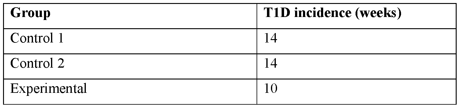
[0274] The data demonstrate that colonization with amyloid-producing bacteria promote T1D development, while colonization with mutated non-amyloid-producing bacteria do not affect the disease state. Animals from the experimental group developed higher T1D compared to Control groups. Both Control 1 and Control 2 groups demonstrated a T1D incidence approximating 20% by 16 weeks of age, with an incidence of 50% by 24 weeks of age. Colonization of the gut with amyloid-producing E. coli had a significant effect on T1D development: 20% had T1D by 12 weeks of age, with an incidence of 50% by 16 weeks of age. Moreover, animals from the Experimental group had an earlier time of disease onset as compared to the Control groups.
Example 3: Effect of different factors to prevent T1D development in animal model following colonization with amyloid-producing bacteria.
[0275] For this study non-obese diabetic mice (NOD/ShiLtJ) 8 weeks old (Jackson Laboratory, Bar Harbor, ME, USA) were housed and bred under specific pathogen-free conditions. Mice were maintained in a temperature controlled (22 ± 2 °C) animal facility with a 12 hour light/dark cycle and were allowed free access to food and acidified drinking water. To increase the efficacy of E. coli transfer, all animals were exposed to antibiotic treatment before the transfer of E. coli. Starting at three weeks of age, recipient mice were treated with ampicillin (1g/L) in the drinking water for the next 4 weeks. The bacterial strains used: E. coli strain VT-55 - a wild-type, amyloid-producing strain.
[0276] E. coli VT-55 bacteria were administered with oropharyngeal gavage to 8 week old female mice at 108 CFU, 2 times a week. Acceleration of T1D development was accessed by measuring blood glucose in a manner consistent with diabetes monitoring, using a FreeStyle Lite meter and test strips (Abbott). Blood glucose was evaluated weekly via a tail vein. T1D development was defined as occurring after two consecutive blood glucose measurements of at least 250 mg/dL.
[0277] Details for the experiments are listed in Table 3, if not stated below.
Transthyretin
[0278] To obtain mouse Transthyretin (TTR), a cDNA mouse TTR clone was subcloned into pMMHa vector at the NdeI and KpnI restriction sites.
TTR clone sequence:


[0279] TTR was expressed in E. coli VT-243. The TTR protein was precipitated from the supernatant of a 48 hour culture of E. coli VT-243 after centrifugation (12,000 × g, 20 minutes 4 °C) of the lysed cells in the gradient 40–90% ammonium sulfate fraction (1.8– 3.6 M). The precipitate that contained TTR was dialyzed overnight against 10 mM sodium phosphate buffer, pH 7.5, 100 mM KCl, 1 mM EDTA. The same volume of 200 mM sodium acetate buffer (pH 4.4, 100 mM KCl, 1 mM EDTA) was added to achieve a final pH of 4.5, and the mixture was then incubated for 18 hours at 37 °C. Following centrifugation (45 min, 30,000 × g, 4 °C), the supernatant was gel filtrated (Superdex 75) to elute phosphate buffer. siRNA against CsgA and CsgB
[0280] SiRNA were developed against CsgA and CsgB using an online tool (https://www.invivogen.com/sirnawizard/).
E. coli CsgA sequence (>NC_000913.3:1104447-1104902 Escherichia coli str.)


[0281] The sequence GATTGACCAGACAGGAGATTA (SEQ ID NO: 15) was used for siRNA.
[0282] To develop nanoparticles with siRNA, we used cholesterol, distearoyl-sn-glycerol-3- phosphocholine (DSPC) and PEG2000-DMG (all Sigma). Nanoparticles were prepared by dissolving the 306O13 lipidoid, cholesterol, DSPC, and PEG2000-DMG with final molar ratio of 50:38.5:10:1.5 in a 90% ethanol enriched with 10% 10nM sodium citrate. Particles were formed following vortexing of equal volumes of siRNA solution with the lipid solution and subsequent dilution in PBS. Mice were orally gavaged with nanoparticles loaded with siRNA at a dose of 5 mg/kg.
Vaccination against amyloid-producing E. coli
[0283] The EcVac vaccine consisted of conjugates containing the O-antigens of serotypes O6 of E. coli VT-55. O-antigen surface polysaccharide was conjugated to detoxified P. aeruginosa exotoxin A carrier protein as described previously [Riddle MS, Kaminski RW, Di Paolo C, et al., Safety and immunogenicity of a candidate bioconjugate vaccine against Shigella flexneri 2a administered to healthy adults: a single-blind, randomized phase I study. Clin. Vaccine Immunol., 2016, 23: 908–17]. The suspension for iv administration was made in TBS, (pH 7.4; 25 mmol Tris, 137 mmol NaCl, 2.8 mmol KCl).
Antibodies to E. coli amyloid
[0284] Balb/c mice were immunized (10 µg) intraperitoneally and boosted (1 µg) subcutaneously with the E. coli amyloid isolated as previously described. Splenocytes were removed under sterile conditions, washed and hybridized to murine plasmacytoma parent cell line SP2/OAg14 using 40% polyethylene glycol and a spleen cell to plasmacytoma cell ratio of 5:1. Cells were plated in 96-well microtiter (Sarstedt) plates at 100,000 cells/per well with DMEM supplemented with 10% fetal bovine serum. Following 48h, cells were supplemented with hypoxanthine (100 PM) containing medium. After three washes with PBS-1% BSA, the presence of specific antibody binding was detected by ELISA. Serotype specific wells were cloned. Pristine-primed BALB/c mice were IP injected with 1 x 10⁷ hybridoma cells. Ascites fluid was collected 14 days later, centrifuged (5000g, 20 min, Eppendorf) and stored at - 80°C before use. Ascites fluid was filtered with affi-Gel protein A column (BioRad Laboratories) and fractions with antibodies were dialyzed against PBS and filtered through a 0.2 µm filter (Milipore). Antibodies were concentrated by ammonium sulfate with subsequent dialysis against PBS.
[0285] The groups used in the experiments are listed in Table 3.


Table 4. T1D incidence


[0287] The data demonstrate that proposed therapeutic methods can prevent triggering of T1D by amyloid-bacteria.
Example 4: Effect of different factors to prevent T1D development in animal model before colonization with amyloid-producing bacteria.
[0288] For this study, 8 week old non-obese diabetic mice (NOD/ShiLtJ; Jackson Laboratory, Bar Harbor, ME, USA) were housed and bred under specific pathogen-free conditions. Mice were maintained in a temperature controlled (22 ± 2 °C) animal facility with a 12 h light/dark cycle and were allowed free access to food and acidified drinking water. To increase the efficacy of E. coli transfer, all animals were exposed to antibiotic treatment before the transfer of E. coli. Starting at three weeks of age, recipient mice were treated with ampicillin (1g/L) in the drinking water for the next 4 weeks.
Bacterial Strain:
[0289] E. coli strain VT-55 is a wild-type, amyloid-producing strain– was used for the colonization of mice following their exposure to different factors. E. coli VT-55 were administered with oropharyngeal gavage to 9 week old NOD mice at 108 CFU, 2 times a week.
[0290] Acceleration of T1D development was accessed by measuring blood glucose in a manner consistent with diabetes monitoring, using a FreeStyle Lite meter and test strips (Abbott). Blood glucose was evaluated weekly via a tail vein. T1D development was defined as occurring after two consecutive blood glucose measurements of at least 250 mg/dL.
[0291] Details for the experiments are listed in Table 5 if not stated below. All compositions were administered to 8 weeks animals, 1 week prior to colonization with E. coli VT-55.
Table 5. Effect of different factors to prevent T1D development in animal model before colonization with amyloid-producing E. coli


[0292] Results are presented in Table 6.
Table 6. T1D incidence

[0293] The data demonstrate that the use of listed agents allowed to prevent the triggering of T1D by amyloid-producing E. coli colonization.
Example 5: Amyloid-producing bacteria community composition in T1D and seroconversion
[0294] To explore the amyloid-producing bacteria and phagobiota community structure associated with T1D, shotgun metagenomics sequencing data of the faecal microbiome from
11 children who exhibited autoantibodies (seven who developed autoantibodies with no progression to T1D, i.e. the “seroconverter” cohort), and the other four who were seroconverted and developed T1D (the“T1D” cohort). Both groups were named“Case”, and 8 non-seroconverted control individuals were used. The patients were all carrying HLA types associated with diabetes. Microbiome analysis was performed at different timepoints of life.
[0295] Samples of the microbiota from each patient were subjected to sequencing and processing. DNA sequencing data were generated by Illumina Hiseq2500 paired-end shotgun mediated sequencing, which was used with an average of ~2.5Gb per sample. Short sequence reads were retrieved from NCBI SRA. Sequences were quality-filtered to remove adaptor contamination, low-quality reads with minimum quality score cut-off of 20, sequences with < 45 nucleotides, and human DNA. In the analysis of bacteria, sequences were grouped into OTUs with a 97% threshold of pairwise identity (Edgar, R. et al., 2010).
[0296] All reads were assembled de novo in each SRA file. The assembled contigs were tested against MetaPhlAn, which operates by mapping sequence reads to a database of predefined clade-specific marker genes, and a custom method (Segata, N. et al., 2012; Hao, Yuhan, 2017). The resulting counts were normalized for total marker gene length and outliers, yielding profiles of the presence/absence and abundance of marker genes, and clade- relative abundance (Franzosa, E. et al., 2014).
[0297] The QIIME pipeline was used for quality filtering of bacterial DNA sequences, chimera removal (by the USEARCH software), taxonomic assignment, and calculation of a-diversity, as previously described (Caporaso, J. et al., 2010; Cox, L. et al., 2014). Downstream data analysis and calculation of diversity metrics were performed in R3.3.2 using ggplot2 and phyloseq libraries; DESeq2 was used to calculate logarithm of fold change (Love, M. et al., 2014).
[0298] All fecal analysis at the first sampling date had E. coli in their feces (Table 6). The association of disappearance of E. coli and development of seroconversion and/or T1D is shown in Table 7.
Table 6.


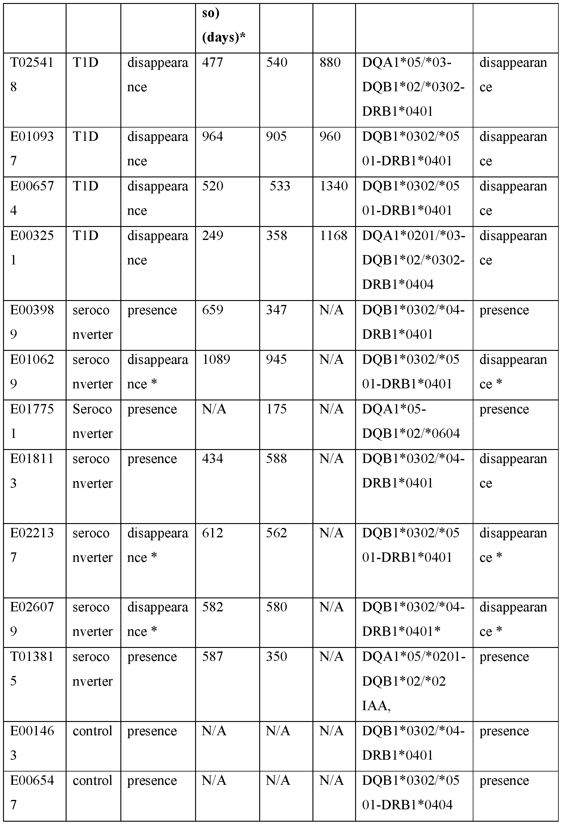

time E. coli abundance declined by > 2 log compared with previous maximum abundance in this patient and reflects eradication (complete disappearance) or presumable eradication. Table 8. Association of E. coli presence and disappearance with seroconversion


[0299] The data demonstrate correlations between the depletion of E. coli and seroconversion. A positive correlation was detected between the appearance of autoantibodies and eradication of E. coli in T1D group, and majority of seroconverters; but no such a correlation was detected in control group. Three of four T1D patients displayed an eradication (or an episode of eradication) of E. coli prior to the detection of autoantibodies. Moreover, all patients within this group had an eradication of E. coli prior to the diabetes diagnosis. Data received suggest that the alterations of amyloid-producing bacteria abundance in T1D group that happened before seroconversion was a signature associated with diseases able to distinguish T1D disease state. The observed alterations in intestinal E. coli population might lead to the release of curli fibers and DNA-amyloid complexes that act as a pro-diabetic factor triggering the seroconversion.
Example 6: Role of bacteriophages in the depletion of amyloid-producing bacteria community in T1D and seroconversion
[0300] The same fecal material was used from the patients listed in Example 5. Samples of the microbiota from each patient were subjected to sequencing and processing. DNA sequencing data were generated by Illumina Hiseq2500 paired-end shotgun sequencing with an average of ~2.5Gb per sample. Sequences were quality-filtered to remove adaptor contamination and low-quality reads by using a minimum quality score cut-off of 20,
sequences with < 45 nucleotides, and human DNA. In the analysis of bacteria, sequences were grouped into OTUs with a 97% threshold of pairwise identity (Edgar, R. et al., 2010). The following phages were identified across studied population.
Table 9. List of Bacteriophages with non-zero Signaling Values


[0301] Association between E. coli bacteriophages abundance and the depletion of E. coli is presented in Table 10.
Table 10. Association of E. coli phages with the disappearance of E. coli in studied population.
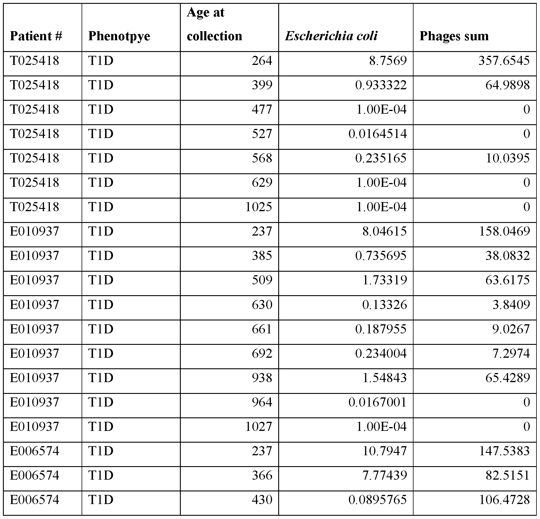


suggesting that the depletion of E. coli was a result of E. coli prophage induction.
[0303] The obtained data revealed previously overlooked particularities of phagobiota in T1D, suggesting the primary role of prophages induction in E. coli depletion and association with seroconversion in patients who would develop T1D.
Example 7: Role of prophage induction in the release of amyloid from E. coli biofilms.
[0304] This example demonstrates that the death of E. coli populations could be due to prophage induction that leads to T1DAMP production. We used 48h old E. coli biofilms formed by amyloid-producing E. coli VT-55. Bacteriophages l (from our collection) were employed. E. coli lysogenic strains were obtained by infection of host bacteria with phage. Prophages were induced with 1 mg/ml mitomycin C (Sigma). To confirm lytic development of bacteriophage we measured an increase in the number of plaque forming units (PFU) and the reduction of bacterial colony-forming units CFU numbers, throughout the 4 h to 10 h period (Table 2). The PFU number in E. coli biofilm was determined by the production of plaques on control E. coli culture.
Table 11A. Effect of prophage induction in MG1655 wild-type lysogenic strain on PFU numbers.


[0305] The data demonstrate the appearance of phages in the biofilm starting from 4h following prophage induction and with the maximum number of PFU at 10h that coincides with the decrease of CFU.
[0306] Next, the effect of prophage induction on E. coli biofilm to amyloid release was evaluated. The amount of amyloid fibers in the biofilm supernatant was determined following prophage induction. Biofilm supernatant was collected and centrifuged at 5000 rpm (Eppendorf 5113) to sediment cells. Supernatant was filtered with 0.25 mM filter and heated to eliminate contaminating proteins by incubating in a boiling water bath for 10 minutes, under the assumption that the aggregated b-sheet amyloid is heat resistant. To quantify the amount of aggregated amyloid, its binding to the amyloid diagnostic dye Congo Red (CR) was evaluated (Table 12). CR added to a final concentration of 10 mg/mL at pH 4.5 was tested using a BioRad UV/Vis spectrophotometer at 25°C. After 5 minutes of equilibration, the absorbance spectra were recorded from 400 to 600 nm. Each trace represents the average of 3 accumulated spectra.
Table 12. Effect of prophage induction on the release of bacterial amyloid from biofilm

[0308] The correlation between the certain HLA haplotype, and the presence or disappearance of E. coli prior to the seroconversion was studied. The HLA types of the patients referred to in Tables 7 and 8 above were determined and listed below, along with whether E. coli were present or disappeared prior to seroconversion.
Table 13. The association of HLA allele, seroconversion and E. coli disappearance.


[0309] Data received demonstrate that the following HLA alleles DQB1*02/*0302- DRB1*0404 and DQB1*0302/*0501-DRB1*0401, DQB1*0302/*04-DRB1*0401* had the highest association with seroconversion and significant alteration of E. coli abundance.
Example 9: Autoimmunity Development is Associated with an Alteration in Amyloid- producing Bacterial Abundance
[0310] To explore the potential link between the abundance of amyloid-producing bacteria and T1D-associated seroconversion, longitudinal shotgun metagenomics sequencing data of the fecal microbiome from 10 children who exhibited autoantibodies (six seroconverters and four children who developed T1D) and eight non-seroconverted HLA-matched control individuals was used. Data was also used from a prospective, longitudinal cohort study by Kostic et al. of 16 HLA-matched infants followed from birth until 3 years of age (Kostic et al., 2015). The children, from Finland and Estonia, were recruited to the study between
September 2008 and August 2010 (available at http://www.diabimmune.org/). The study generated shotgun metagenomics sequencing data of the fecal microbiome from 10 children who exhibited autoantibodies (six who developed serum autoantibodies with no progression to T1D, and four who were seroconverted and developed T1D) and eight non-seroconverted control individuals (Kostic et al., 2015). Inclusion criteria were: presence of HLA DR-DQ alleles associated with T1D development. Data on diabetes-associated autoantibodies are presented in the original study.HiSeq-2500 sequencing was performed on a sample from each individual, producing an average of ~2.5 Gb per sample.
[0311] High-throughput shotgun sequencing was performed on the Illumina HiSeq 2500 platform, generating ~2.5 Gb of sequence per sample with 101-bp paired-end reads. Human contamination was removed with the BMTagger (ftp://ftp.ncbi.nlm.nih.gov/pub/agarwala/bmtagger/). Bacterial and phage contents were quantified separately using the SRA shotgun metagenomic sequencing data. Bacterial content was quantified by taxa directly from SRA reads using Metaphalan (v. 2.0), which maps sequence reads to a database of predefined clade-specific marker genes (Segata et al., 2012). All bacterial taxa with relative abundances <0.01 in all samples were excluded from statistical analysis. Phage content was assessed using a custom method. First, reads from each SRA file were assembled de novo into contigs with metaSPAdes (v.3.11.1) (Nurk et al., 2017). Then, contigs >200 bp were aligned to the EBI collection of phage genomes (https://www.ebi.ac.uk/genomes/phage.html) by BLAST, with a threshold e-value <1e-5 and alignment length >50% of contig length. All of the original reads were then re-mapped with Bowtie2 (v. 2.3.4.1) to the contigs with good phage BLAST matches in order to increase sensitivity and to more accurately count the abundance of reads from each type of phage (Langmead and Salzberg, 2012). Phage read counts per contig were combined per phage genome (taxa) and normalized to relative abundance. A detection threshold of two reads per sample (>90% identity to the phage genome) was used, based on a previous report (Hao et al., 2018).
[0312] Statistical analysis of the microbial community composition and differential abundance was undertaken. The QIIME pipeline (v1.9.1) was used for quality filtering of bacterial and bacteriophage DNA sequences, chimera removal (with USEARCH software), taxonomic assignment, and calculation of a-diversity, as previously described (Caporaso et al. 2010; Tetz et al., 2017). Downstream data analysis and calculation of diversity metrics were conducted in R v3.5.1, using ggplot2 and phyloseq libraries; DESeq2 was used to calculate logarithm of fold change. Bacterial and bacteriophage communities at the genus,
family, and species levels were characterized based on a- and b-diversities. a-Diversity indices (ACE, Chao 1 richness estimator, Shannon and Simpson indexes) were calculated using the phyloseq R library (McMurdie and Holmes, 2013). Differences in a-diversities between datasets were examined by the Mann-Whitney test; p < 0.05 was considered statistically significant.
[0313] Differences among groups where two variables exist (phenotype and time point) were analyzed by two-way ANOVA and Tukey’s multiple comparison test. When one variable was compared, an unpaired two-tailed t-test was used. Data were visualized using multidimensional scaling (MDS).
[0314] Correlations between E. coli disappearance, Bifidobacterium abundance, breastfeeding and antibiotic usage were assessed pairwise using the Jaccard similarity index (Jaccard, 1912). Differences were considered statistically significant at p < 0.05.
[0315] Amyloid-producing bacteria were represented by E. coli, Staphylococcus aureus, and Salmonella spp. (Barnhart and Chapman 2006; Schwartz and Boles 2013). Among these amyloid-producing bacteria, E. coli was the major group, while the other curli-producing bacteria were identified only in a single sample and only at a few collection times and thus were disregarded in subsequent analysis, as indicated in the following list of all phages identified in this study: Escherichia Stx1-converting recombinant phage HUN/2013;
Escherichia phage Pollock; Escherichia phage phi191; Escherichia phage TL- 2011b; Escherichia phage TL-2011c; Escherichia phage HK639;Escherichia phage HK75, ENA|HM173637|HM173637.1 Escherichia phage N15 Escherichia phage HK022 Escherichia phage T; Escherichia phage D108; Escherichia phage K1G; Escherichia phage K1H; Escherichia phage K1ind1; Escherichia phage K1ind2; Escherichia phage K1ind3; Escherichia phage EC6; Escherichia phage 1720a-02; Enterobacteria phage cdtI; Enterobacteria phage P2; Enterobacteria phage HK97 Enterobacteria phage Mu; Enterobacteria phage 933W; Enterobacteria phage P7; Enterobacteria phage VT2-Sakai proviral DNA; Enterobacteria phage AR1; Enterobacteria phage phiP27;
Enterobacteria phage BP-4795; Enterobacteria phage RB10; Enterobacteria phage P88;
Enterobacteria phage JenP1; Enterobacteria phage 9g; Enterobacteria phage mEpX1; Enterobacteria phage mEp460, Enterobacterial phage mEp390; Enterobacteria phage mEp237; Enterobacteria phage mEp235; Enterobacterial phage mEp234; Enterobacterial phage mEp213; Enterobacteria phage mEp043 c-1;Enterobacteria phage HK106;
Enterobacteria phage HK140; Enterobacteria phage HK225;Enterobacteria phage HK446; Enterobacteria phage HK542; Enterobacteria phage HK544,Enterobacteria phage
HK630; Enterobacteria phage HK633; ENA|xEnterobacteria phage mEpX2, Enterobacteria phage IME10, Enterobacteria phage IME10, Enterobacteria phage lambda, Enterobacteria phage 2851, Enterobacteria phage YYZ-2008, Enterobacteria phage DE3, Enterobacteria phage phi80, Bacteriophage P4, Bacteriophage 186; Bacteriophage If1; Stx2- converting phage 86 ; Stx2 converting phage I DNA, Stx2 converting phage II DNA,Stx2-converting phage 1717, Stx2 converting phage vB_EcoP_24B.
[0316] The association between E. coli abundance, disease phenotype, and collection time were first compared using a two-tailed Mann–Whitney U test (Figure 4). The samples were clustered into time-point bins of 300 days, and the dynamics of E. coli abundance were studied over 1300 days within and among patient groups. E. coli abundances demonstrated a different dynamic relationship across time for case and control groups. E. coli tended to disappear in T1D and seroconverters over the time, whereas in controls, E. coli abundance tended to increase and did not change significantly over time. In both the seroconverter and T1D groups (case groups), a statistically significant decrease in E. coli abundance was found when comparing the first 0-300 day period and the following 300-600, 600-900, 900-1300 time bins (p < 0.05). Notably, the initial abundance of E. coli was significantly higher in cases than in controls at 0–300 days. The case groups responded differently over time, and importantly, data from the control group revealed that there was no variation in E. coli associated with age.
[0317] To further define the E. coli-related association with autoimmunity, an analyses of paired sample sets comparing the E. coli abundance before and after appearance of the autoantibodies across groups was next conducted, with the results shown in Figures 2A-2B. For the control group the inventors modulated the time of seroconversion using an artificial benchmark of the medium time to seroconversion in case groups that was 540 days (Table 14).
Table 14: Association of disappearance of E. coli and development of seroconversion and/or T1D


[0318] Three of four T1D patients showed a disappearance of E. coli prior to the detection of autoantibodies. One patient (E010937) from T1D group who retained the presence of E. coli following seroconversion showed over a 60-fold decrease in E. coli abundance between the first sample collection and the first sample after seroconversion, demonstrating a dramatic drop in the E. coli population (Fig. 5A). The same trend was noted in Seroconverters group with total disappearance of E. coli or a trend to the decrease of its abundance prior to the detection of autoantibodies. The only patient who had higher E. coli abundance following detected autoantibodies was E022137; however, this patient had a total E. coli disappearance before seroconversion with a restoration of E. coli abundance in the following time periods.
[0319] When the absolute abundance of E. coli sequences was compared“before” and“after” the appearance of antibodies within a subgroups (Fig. 2B), only Case (combing both
Seroconvertors and T1D) and Seroconvertors groups have reached statistically significance (p=0.022 and p=0.031) (Fig.2B). In T1D group, although the abundance of E. coli was lower following the autoantibodies appearance in all children, the difference was not statistically significant (p= 0.068), most likely due to a smaller sample group size.
[0320] There was no correlation between the abundance of E. coli in control groups and the appearance of autoantibodies (p= 0.677). The result further supports that the decrease of amyloid-producing bacteria abundance in T1D group was likely to be associated with disease and not with an age change.
[0321] To explore the relation between E. coli abundance and autoimmunity further, the inventors next analyzed whether alterations in the abundance of E. coli could be associated with the development of autoantibodies in more detail, and whether or not E. coli abundance could distinguish the T1D disease state. To this end, the appearance and disappearance of E. coli was studied in each patient in dynamic association with the appearance of autoantibodies (Figure 6). A positive correlation was found between the total disappearance or presumed disappearance (defined as a >50-fold decrease) in E. coli and autoantibody appearance in the T1D and seroconverter groups, but not in controls. All T1D patients showed disappearance or an episode of disappearance of E. coli before the detection of autoantibodies, and disappearance of E. coli prior to the diagnosis of diabetes (Table 14). In seroconverters, E. coli disappeared in all patients, except E003989 and T013815. For patient E10629, data were difficult to interpret because of the long period between sample collections (556 days between the last sample taken before seroconversion and the first sample after seroconversion). In any case, E. coli was absent in the first sample following seroconversion; thus, it is reasonable to assume that it had disappeared before autoantibody appearance. Notably, in certain control patients, E. coli disappeared at some time points; however, this was followed by a restoration of the E. coli population in all cases, which was not regularly seen in case subjects.
[0322] Next, changes in the absolute abundance of E. coli were examined in each patient individually among different periods, with the data presented as a heatmap in Figure 5. Alterations in E. coli abundance were detected within children from the control group, followed by restoration of the E. coli population to the levels equal to or above the pre- seroconversion levels, whereas restoration of the E. coli population to preceding levels was noted in only a few children from the case groups. This more detailed analysis has also revealed that in patient T013815, although E. coli did not disappear prior to the seroconversion, the appearance of autoantibodies occurred during the period when E. coli
started to significantly decrease from the highly elevated abundance compared to other patients. Notably, the initial abundance of E. coli in T1D and seroconverter groups was significantly higher than that in the control group, which confirmed the data presented in Figure 3. These results suggest that the initial higher abundance of E. coli and the decrease of amyloid-producing bacteria abundance in case groups were signatures associated with autoimmunity and disease progression.
[0323] In addition, the inventors studied the potential correlation between the disappearance of E. coli and certain HLA alleles or the appearance of particular autoantibodies, IAA, GADA, IA2A, ZNT8A or ICA, in case groups. No correlations were observed (data not shown). No association was detected between E. coli disappearance and breastfeeding duration, Bifidobacterium abundance, or antibiotic usage, which are known to influence Enterobacteriaceae (Candela et al., 2008).
Example 10: E. coli Bacteriophages as Signatures Associated with Seroconversion
[0324] As identified were E. coli as T1D-discriminative bacteria, the relationships between this microorganism and bacteriophages of E. coli were then studied, as phages are known as main regulators of bacterial populations. First, 63 E. coli phage species were detected in case and control groups. Possible differences in E. coli phages were evaluated using a- and b- diversities in the pre-seroconversion period. For the samples from the control group, the medium time to seroconversion in case groups (540 days) was set as an artificial benchmark. Phage richness was statistically different between control and seroconversion individuals as indicated by ACE and Chao1 indexes (ACE: p = 0.0181; Chao1: p = 0.0137), but was not statistically significantly different between control and T1D, most likely because of the small T1D patient cohort (ACE: p = 0.6086; Chao1: p = 0.6359). E. coli phage diversity tended to be lower in the control subjects than in seroconverters (Shannon, p = 0.1526; Simpson, p = 0.5437), and was significantly lower in controls vs. T1D cases (Shannon, p = 0.0055; Simpson, p = 0.0248) (Mann–Whitney test).
[0325] Nearly all E. coli phages were observed to be lysogenic, whereas strictly virulent lytic phages, such as Enterobacteria phage IME10 or Enterobacteria phage 9g, were found only in a few samples. The predominance of lysogenic E. coli phages clarifies why a-diversity indices revealed trends of decreased evenness and diversity of phages in case groups compared to controls in pre-seroconversion samples, which most likely reflected lower E. coli abundance in controls.
[0326] Next, the E. coli phage/E. coli bacterial cell ratio, which represents the“lytic potential” (Figure 6) (Waller et al., 2014), was analyzed. E. coli strain MG1655 was used as
a host for prophage induction experiments (Jensen 1993). Bacteria were subcultured from freezer stocks onto Mueller–Hinton agar plates (Oxoid) and incubated at 37 °C overnight. All subsequent liquid subcultures were derived from colonies isolated from these plates and were grown in Mueller–Hinton broth (Oxoid). Bacteriophage l from the inventors’ collection was employed. Bacteriophage suspensions were routinely stored in TM buffer (10 mM Tris- HCl, 10 mM MgSO4, pH 7.2) at 4 °C. E. coli lysogenic strains were obtained by infection of E. coli MG1655 with the phage and titration of cells on Mueller–Hinton agar plates as previously described (Maniatis et al. 1989). For prophage induction, Mitomycin C (Sigma- Aldrich) was prepared in 0.1 M phosphate buffer (pH 7.2), filtered through a 0.22-mm-pore filter (Millipore Corp., Bedford, MA), and stored at 4 °C (Georgopoulos et al., 2002). A modified protocol according to Reyrolle et al. designed for prophage induction on 96-well plates was used (Reyrolle et al., 1982; McDonald et al., 2010). Twenty microliters of an overnight E. coli MG1655 culture, with an absorbance at 600 nm of 0.2, was dispensed in each well of a 96-well flat-bottom polystyrene tissue culture microtiter plate (Sarstedt, Numbrecht, Germany), after which 180 ml of MHB (Oxoid) was added. The plates were incubated at 37 °C for 48 h. Induction of prophages in the E. coli biofilms was provoked by the addition of mitomycin C (Sigma) to a final concentration 1 mg/ml to each well. The plates were further incubated at 37 °C and samples were taken at 4, 6, 8, and 10 h following prophage induction.
[0327] To this end, the E. coli phage abundance was first normalized to that of E. coli. In theory, the phage/bacteria ratio reflects whether or not a prophage is stably integrated within the host bacterial genome (Tetz et al., 2018). A low ratio indicates that prophage is most likely absent in the genomes of part of the bacterial host population, whereas a high ratio suggests active, productive phage-induced bacterial lysis. The phage/bacteria ratio increased in subjects from all groups prior to the decrease in E. coli abundance, indicating that productive phage infection was the cause of E. coli depletion. Notably, a statistically significant elevation in the phage/bacteria ratio was observed before the appearance of autoantibodies in the T1D and seroconverter groups.
[0328] To evaluate the implication of the increase in the phage/bacteria ratio as a driving force behind E. coli depletion further, the inventors analyzed which phages had a correlation with the depletion of E. coli abundance across most subjects. The most frequently found lysogenic phages with an inverse relationship with E. coli abundance are representatives of the Peduovirinae subfamily and of the unclassified Lambdavirus subfamily within the Caudovirales order, as shown in Tables 15 and 16 below.
Table 15

Table 16


[0329] The increase in number of the lysogenic E. coli phages along with the decrease in E. coli abundance indicated that there was a productive bacteriophage infection that led to bacterial host death and the release of phage progeny. These data revealed previously overlooked particularities of the phagobiota in T1D, suggesting a primary role of induction of certain E. coli prophages in E. coli depletion, and an association with autoimmunity and T1D development.
Example 11: Role of Prophage Induction in the Release of Amyloid from E. coli Biofilms
[0330] The inventors evaluated how the die-off of E. coli populations due to prophage induction could lead to amyloid release. The inventors used 48-h old E. coli biofilms with confirmed curli expression. Curli formation on 48-h colonies was visible with the naked eye on petri dishes (data not shown). Prophages were induced with mitomycin C. Lytic bacteriophage development was confirmed as an increase in plaque forming units (PFU) and a reduction in colony-forming units (CFU).
[0331] CFU and PFU were determined as follows. To estimate CFU, biofilms were thoroughly scraped (Tetz et al., 2009). Well contents were aspirated, placed in 1 ml of isotonic phosphate buffer (0.15 M, pH 7.2), and the total CFU number was determined by serial dilution and plating on Mueller–Hinton agar. The number of phage virions produced after induction was estimated by phage titration (using strain MG1655 as a host) and phage plaque assay. Following induction, at the indicated times 200-ml samples of biofilms were collected. Then, 30 ml of chloroform was added to each sample, the mixture was vortexed and centrifuged at 3000 ×g for 5 min in a microcentrifuge (Eppendorf 5415D). A supernatant fraction of the bacterial lysate was further used. The phage titer (number of phages per ml) was determined using the double agar overlay assay method as described previously (Yuan et al., 2012). To this end, 2.5 ml of each titration point of phage lysate was spotted on Mueller-Hinton agar (Oxoid). Then, a mixture of 1 ml of E. coli MG1655 culture and 2 ml of 0.7% nutrient agar (heated to 45 °C) supplemented with MgSO4 and CaCl2 (to a final concentration of 10 mM each) was poured over the plate. Plates were incubated at 37 °C overnight. Each experiment was repeated in triplicate.
[0332] The presence of phage in E. coli biofilm was confirmed by the production of plaques on a non-induced E. coli control culture between 4 h and 10 h. The effect of prophage induction in E. coli biofilm on CFU and PFU is summarized in Table 17.
Table 17. Effect of prophage induction in MG1655 wild-type lysogenic strain on PFU and CFU


[0333] Phages started to appear in the biofilm as of 4 h after prophage induction, with maximum PFU at 8 h, which coincided with a decrease in CFU.
[0334] Next, the inventors evaluated the effect of prophage induction on the amount of amyloid fibers released into the biofilm supernatant. Supernatant CR depletion assays were performed as follows. The amount of bacterial amyloid following prophage induction was measured in the supernatant of E. coli MG1655 biofilms. Biofilms were obtained as described above. Biofilm supernatant was taken at 4, 6, 8, and 10 h following prophage induction. To isolate aggregated amyloid fibers from the supernatant, the supernatant was centrifuged at 10,000 × g, filtered through a 0.22 µm filter to separate bacterial cells, and treated with proteinase K to eliminate contaminating proteins. CR (Sigma-Aldrich) was added to a final concentration of 10 mg/ml from a filtered stock solution of 1 mg/ml (Reichhardt et al., 2015). After 5 min of equilibration, absorbance spectra were recorded from 400 to 600 nm (SmartSpec 3000, Bio-Rad). Each trace represents the average of 5 accumulated spectra. For all samples, spectra of corresponding nutrient MHB solutions with CR were used as blanks.
[0335] To quantify the amount of aggregated amyloid, its binding to the amyloid-diagnostic dye was evaluated by a Congo red (CR) assay (Reichhardt et al., 2015). A significant increase in CR absorbance was observed upon binding to supernatant 8 h following prophage induction as compared with control samples (Figure 7). Like for other curli fibers, heating of the E. coli supernatant induced a spectral shift of the CR solution, with a maximum difference in absorbance between CR alone and CR bound to amyloid fibers at ~541 nm (Chapman et al., 2002). These data suggested that the phage-mediated lysis of E. coli led to the release of amyloid aggregates into the biofilm.
[0336] The present invention is not to be limited in scope by the specific embodiments described herein. Indeed, various modifications of the invention in addition to those described herein will become apparent to those skilled in the art from the foregoing description and the accompanying figures. Such modifications are intended to fall within the scope of the appended claims. It is further to be understood that all values are approximate, and are provided for description.
[0337] Patents, patent applications, publications, product descriptions, and protocols are cited throughout this application, the disclosures of which are incorporated herein by reference in their entireties for all purposes.
References 1. Bosi E, Molteni L, Radaelli MG, Folini L, Fermo I, Bazzigaluppi E, Piemonti L, Pastore MR, Paroni R. Increased intestinal permeability precedes clinical onset of type 1 diabetes. Diabetologia.2006 Dec 1;49(12):2824-7.
2. Vaarala O, Atkinson MA, Neu J. The“perfect storm” for type 1 diabetes: the complex interplay between intestinal microbiota, gut permeability, and mucosal immunity. Diabetes. 2008 Oct 1;57(10):2555-62.
3. Maes M, Kubera M, Leunis JC, Berk M. Increased IgA and IgM responses against gut commensals in chronic depression: further evidence for increased bacterial translocation or leaky gut. Journal of affective disorders.2012 Dec 1;141(1):55-62.
4. Tlaskalová-Hogenová, Helena, Renata Štěpánková, Hana Kozáková, Tomáš Hudcovic, Luca Vannucci, Ludmila Tučková, Pavel Rossmann et al. "The role of gut microbiota (commensal bacteria) and the mucosal barrier in the pathogenesis of inflammatory and autoimmune diseases and cancer: contribution of germ-free and gnotobiotic animal models of human diseases." Cellular & molecular immunology 8, no.2 (2011): 110.
5. Berk M, Williams LJ, Jacka FN, O’Neil A, Pasco JA, Moylan S, Allen NB, Stuart AL, Hayley AC, Byrne ML, Maes M. So depression is an inflammatory disease, but where does the inflammation come from? BMC medicine.2013 Dec;11(1):200.
6. Fasano A. Zonulin, regulation of tight junctions, and autoimmune diseases. Annals of the New York Academy of Sciences.2012 Jul 1;1258(1):25-33.
7. Costerton JW, Stewart PS, Greenberg EP. Bacterial biofilms: a common cause of persistent infections. Science.1999 May 21;284(5418):1318-22.
8. Barnhart MM, Chapman MR. Curli biogenesis and function. Annu. Rev. Microbiol. 2006 Oct 13;60:131-47.
9. Noble JA, Valdes AM. Genetics of the HLA region in the prediction of type 1 diabetes. Current diabetes reports.2011 Dec 1;11(6):533.
10. Knip M, Veijola R, Virtanen SM, Hyöty H, Vaarala O, Åkerblom HK. Environmental triggers and determinants of type 1 diabetes. Diabetes.2005 Dec 1;54(suppl 2):S125-36. 11. Gallo, PM et al., Amyloid-DNA composites of bacterial biofilms stimulate autoimmunity. Immunity.2015, 42(6):1171-1184.
12. Kostic AD et al., The dynamics of the human infant gut microbiome in development and in progression toward type 1 diabetes. Cell Host Microbe.2015, 17(2):260-273.
13. Segata, N et al., The dynamics of the human infant gut microbiome in development and in progression toward type 1 diabetes. Nat. Methods.2012, 9(8):811-814.
14. Caporaso, JG et al., QIIME allows analysis of high-throughput community sequencing data. Nat. Methods, 2010, 7(5):335-336.
15. Cox, L et al., Altering the intestinal microbiota during a critical developmental window has lasting metabolic consequences. Cell, 2014, 158(4):705-721.
16. Love, M et al., Moderated estimation of fold change and dispersion for RNA-seq data with DESeq2. Genome Biol., 2014, 15(12):550.
17. Edgar, R et al., Search and clustering orders of magnitude faster than BLAST. Bioinformatics, 26(19):2460-2461.
18. McMurdie, PJ and Holmes, S, phyloseq: an R package for reproducible interactive analysis and graphics of microbiome census data. PLoS One, 2013, 8(4):e61217.
19. Schwartz, K and Boles, BR, Microbial amyloids--functions and interactions within the host.2013, 16(1):93-99.
20. Tetz, GV et al., Effect of DNase and antibiotics on biofilm characteristics. 2009, 53(3):1204-1209.
21. Reichhardt, C et al., Congo Red Interactions with Curli-Producing E. coli and Native Curli Amyloid Fibers. PLoS One, 2015, 10(10):e0140388.
22. Chapman, MR et al., Role of Escherichia coli curli operons in directing amyloid fiber formation. Science, 2002, 295(5556):851-855.
23. Yuan, Y et al., Genome characteristics of a novel phage from Bacillus thuringiensis showing high similarity with phage from Bacillus cereus. PLoS One, 2012, 7(5):e37557. 24. Waller, AS et al., Classification and quantification of bacteriophage taxa in human gut metagenomes. ISME J., 2014, 8(7):1391-1402.
25. Jensen, KF, The Escherichia coli K-12 "wild types" W3110 and MG1655 have an rph frameshift mutation that leads to pyrimidine starvation due to low pyrE expression levels. J. Bacteriol., 1993, 175(11):3401-3407.
26. Reyrolle, J et al., Lysogenic strains of lactic Acid streptococci and lytic spectra of their temperate bacteriophages. Appl. Environ. Microbiol., 1982, 43(2):349-356.
27. McDonald, JE et al., High-throughput method for rapid induction of prophages from lysogens and its application in the study of Shiga Toxin-encoding Escherichia coli strains. Appl. Environ. Microbiol., 2010, 76(7):2360-2365.
28. Tetz, G et al., Parkinson's disease and bacteriophages as its overlooked contributors. Sci. Rep., 2018, 8(1):10812.
29. Candela M et al., Plasminogen-dependent proteolytic activity in Bifidobacterium lactis. Microbiology, 2008, 154:2457-2462.
Claims
1. A method for preventing or treating Type 1 Diabetes (T1D) or consequences thereof in a mammal in need thereof, said method comprising one or more of (i) inactivating amyloid- producing bacteria within microbiota in the mammal, (ii) preventing amyloid-producing bacteria from entering the microbiota, the gastrointestinal tract, a bodily fluid or a tissue of the mammal from an environment outside the mammal, (iii) inactivating a T1D-associated microbial product (T1DAMP) that is released by microbiota of the mammal, (iv) inactivating a T1DAMP present, a bodily fluid or a tissue of the mammal, (v) inhibiting release of a T1DAMP from a biofilm and/or bacteria in the gastrointestinal tract, a bodily fluid or a tissue of the mammal, (vi) inhibiting entry of T1DAMP to microbiota, the gastrointestinal tract, a bodily fluid or a tissue of the mammal, (vii) inhibiting the triggering of T1D by bacteria, T1DAMP derived from the bacteria, or a complex comprising the bacteria or the T1DAMP, (viii) administering to the mammal E. coli that do not produce an amyloid protein, (ix) administering to the mammal a pilocene or a curlicide, and (x) inhibiting the activity of a bacteriophage (xi) ) inhibiting the prophage inductors.
2. The method of claim 1, wherein the mammal expresses a T1D susceptible HLA allele.
3. The method of claim 2, wherein the T1D susceptible HLA allele is selected from an HLA allele having a DR4-DQ8 and/or DRB1 haplotype, an HLA allele having a DR3-DQ2 haplotype, HLA allele DQB1*02/*0302-DRB1*0404, HLA allele DQB1*0302/*0501- DRB1*0401, and
DQB1*0302/*04-DRB1*0401*.
4. The method of any of claims 1-3, wherein the mammal comprises an increased amount of E. coli or Salmonella in the gastrointestinal tract as compared to a second mammal that does not develop T1D, and optionally wherein the mammal and the second mammal both express a T1D susceptible HLA allele, and optionally wherein the mammal and the second mammal both express the same T1D susceptible HLA allele.
5. The method of claim 4, wherein the second mammal is age-matched and/or gender- matched to the mammal comprising the increased amount of E. coli or Salmonella in the gastrointestinal tract.
6. The method of any one of claims 1-4, wherein the method comprises administering to the mammal E. coli in a manner effective to populate the microbiota with the E. coli, wherein the administered E. coli do not produce an amyloid protein or produce a reduced amount of the amyloid protein as compared to a wild-type E. coli.
7. The method of claim 6, wherein the administered E. coli comprises a mutation in a gene encoding for an amyloid protein.
8 The method of claim 6, wherein the method comprises administering to the mammal E. coli that comprises a mutation in a sequence regulating expression of a gene encoding for an amyloid protein.
9. The method of any one of claims 1-6, wherein inactivation of amyloid-producing bacteria within microbiota comprises preventing transfer of amyloid-producing bacteria to the mammal from a mother of the mammal during birth or breastfeeding.
10. The method of any one of claims 1-6, wherein the inactivation of amyloid-producing bacteria within microbiota comprises administering to the mammal a microorganism or a by- product of the microorganism, wherein the microorganism or the byproduct is effective to prevent colonization of the amyloid-producing bacteria in the gastrointestinal tract of the mammal.
11. The method of claim 11, wherein said microorganism is from an order selected from Bacteroidales, Lactobacillales, Erysipelotrichales, Coriobacteriales, Clostridiales, Bacillales, and Bifidobacteriales.
12. The method of claim 11, wherein the microorganism is a non-amyloid-producing strain of bacteria or a strain of bacteria that synthesizes a reduced amount of amyloid.
13. A method for preventing or treating Type 1 Diabetes (T1D) or consequences thereof in a mammal in need thereof, said method comprising vaccination of the mammal against Enterobacteriales bacteria.
14. The method of claim 13, further comprising vaccination of the mammal against E. coli or Salmonella.
15. The method of any one of claims 1-12, further comprising vaccination of the mammal against Enterobacteriales bacteria.
16. The method of claim 15, further comprising vaccination of the mammal against E. coli or Salmonella.
17. The method any one of claims 1-12 and 15, wherein the inactivation of amyloid- producing bacteria within microbiota comprises i) colonization of gastrointestinal microbiota of the mammal with non-amyloid-producing bacteria and/or ii) administering an anti-amyloid antibody to the mammal.
18. The method any one of claims 1-12 and 15-17, wherein the inactivation of amyloid- producing bacteria within microbiota comprises editing one or more genes in the genome of the amyloid-producing bacteria, wherein the inactivation inhibits adhesion of the amyloid- producing bacteria, wherein the one or more genes are selected from afa- dra, daaD, tsh, vat, ibeA, fyuA, mat, sfa-foc, malX, pic, irp2, papC, fimH; PapAH papEF, bmaE, sfa/focDE, papC, focG, sfaI, sfa II, sfaS, aah, aidA, fasA, faeG, bfpA, eaeA, Paa, fasA, faeG, fedA, fanC, sfaY, and a gene in the Cpx pathway.
19. The method any one of claims 1-12 and 15-17, wherein the inactivation of the amyloid-producing bacteria within microbiota comprises administering to the mammal a composition comprising one or more of fosfomycin, Doxycycline, Ciprofloxacin, Trimethoprim/sulfamethoxazole, Levofloxacin, Amoxicillin, Aztreonam, Nitrofurantoin, Ceftriaxone, imipenem, and Rifaximin, a FimH antagonist, and a pilicide.
20. The method of claim 19, wherein the FimH antagonist is an n-Heptyl a-D-mannose glycopolymer, methyl R-D-mannoside, or a thiazolylmannoside.
21. The method of claim 19, wherein the pilicide is selected from
i) 7-(1-naphthylmethyl)-5-oxo-8-phenyl-2,3,6,7-tetrahydro-5H-[1,3]thiazolo[3,2- a]pyridine-3-carboxylic acid
ii) 8-cyclopropyl-7-(1-naphthylmethyl)-5-oxo-2,3,6,7-tetrahydro-5H- [1,3]thiazolo[3,2-a]pyridine-3-carboxylic acid
iii) 7-(1-naphthylmethyl)-5-oxo-8-pentyl-2,3,6,7-tetrahydro-5H-[1,3]thiazolo[3,2- a]pyridine-3-carboxylic acid
iv) 8-(4-bromophenyl)-7-(1-naphthylmethyl)-5-oxo-2,3,6,7-tetrahydro-5H- [1,3]thiazolo[3,2-a]pyridine-3-carboxylic acid
v) 7-(1-naphthylmethyl)-5-oxo-8-phenyl-2,3-dihydro-5H-[1,3]thiazolo[3,2- a]pyridine-3-carboxylic acid, lithium salt
vi) 8-cyclopropyl-7-(1-naphthylmethyl)-5-oxo-2,3-dihydro-5H-[1,3]thiazolo[3,2- a]pyridine-3-carboxylic acid, lithium salt
vii) 7-methyl-5-oxo-8-phenyl-2,3-dihydro-5H-[1,3]thiazolo[3,2-a]pyridine-3- carboxylic acid, lithium salt
viii) 6-dimethylaminomethyl-7-(1-naphthylmethyl)-5-oxo-8-phenyl-2,3-dihydro- 5H-[1,3]thiazolo[3,2-a]pyridine-3-carboxylic acid, lithium salt
ix) 6-morpholinomethyl-7-(1-naphthylmethyl)-5-oxo-8-phenyl-2,3-dihydro-5H- [1,3]thiazolo[3,2-a]pyridine-3-carboxylic acid, lithium salt
x) 8-cyclopropyl-6-morpholinomethyl-7-(1-naphthylmethyl)-5-oxo-2,3-dihydro- 5H-[1,3]thiazolo[3,2-a]pyridine-3-carboxylic acid, lithium salt, and
xi) 6-dimethylaminomethyl-7-methyl-5-oxo-8-phenyl-2,3-dihydro-5H- [1,3]thiazolo[3,2-a]pyridine-3-carboxylic acid, lithium salt.
22. The method of any one of claims 1-21, wherein the mammal expresses one or more of T1D-susceptible HLA alleles.
23. The method of claim 22, wherein the T1D-susceptible HLA allele is a DR4-DQ8 haplotype, an HLA allele having a DR3-DQ2 haplotype, HLA allele DQB1*02/*0302- DRB1*0404, HLA allele DQB1*0302/*0501-DRB1*0401, and DQB1*0302/*04- DRB1*0401*.
24. The method of any one of claims 1-22, comprising (i) inactivating amyloid-producing bacteria within microbiota in the mammal, and wherein the microbiota is gut microbiota.
25. The method of any one of claims 1-22 comprising (iii) inactivating a T1D-associated microbial product (T1DAMP) that is released by microbiota of the mammal, and wherein the
microbiota is gut microbiota.
26. The method of any one of claims 1-22 comprising (vi) inhibiting entry of T1DAMP to microbiota, the gastrointestinal tract, a bodily fluid or a tissue of the mammal, (vii) inhibiting the triggering of T1D by bacteria, T1DAMP derived from the bacteria, or a complex comprising the bacteria or the T1DAMP, and wherein the microbiota is gastrointestinal microbiota.
27. A method for determining susceptibility to T1D in a mammal, said method comprising (a) detecting the expression in the mammal of at least one T1D susceptible HLA allele, and (b) (i) detecting increased intestinal permeability in the mammal as compared to the intestinal permeability in one or more mammals from an age-matched and sex-matched reference population, and/or (ii) detecting an increased level of E. coli or Salmonella in gastrointestinal microbiota of the mammal, wherein the E. coli or Salmonella release a T1D-associated microbial product (T1DAMP) that enters the bloodstream of the mammal and binds to a b- cell or dendritic cells expressing a Toll-like 2 receptor or a Toll-like 9 receptor and promotes death of the b- cell to thereby increase susceptibility to T1D.
28. The method of claim 27, wherein the T1D susceptible HLA allele is selected from an HLA allele having a DR4-DQ8 haplotype, an HLA allele having a DR3-DQ2 haplotype, HLA allele DQB1*02/*0302-DRB1*0404, HLA allele DQB1*0302/*0501-DRB1*0401, and HLA allele DQB1*0302/*04-DRB1*0401*.
29. The method of claim 27 or claim 28, wherein the detection of an increased level of E. coli comprises performing one or more of the assays selected from i) analysis of 16S rRNA from the E. coli, ii) PCR of a nucleic acid from the E. coli, iii) sequencing of a gene from the E. coli, iv) a metagenomic assay, v) cultivation of the E. coli, and vi) biochemical identification of the E. coli.
30. A method for determining susceptibility to T1D in a mammal, said method comprising detecting the level of E. coli bacteriophages in the gastrointestinal microbiota of the mammal by performing one or more of assays selected from i) PCR of a nucleic acid from the E. coli bacteriophages, ii) sequencing of a gene from the E. coli bacteriophages, iii) a metagenomic assay, iv) cultural identification of the E. coli bacteriophages, and v) biochemical
identification of the E. coli bacteriophages.
31. The method of any one of claims 1-30, wherein the mammal is a human.
32. The method of claim 31, wherein the detecting of an increased level of E. coli and/or an increase or decrease in the level of E. coli bacteriophages and/or a microbial inducer of E. coli prophages in the gastrointestinal microbiota is conducted at 100-200 days after birth of the human.
33. The method of claim 32, wherein the detecting is conducted at 130-180 days after birth of the human.
34. The method of any one of claims 27 and 31-33, further comprising detecting a decrease in the level of E. coli and/or an increase or a decrease in the level of E. coli bacteriophages and/or microbial inducers of E. coli prophages subsequent to the detecting of the increased level of E. coli.
35. The method of claim 34, wherein the mammal is a human and wherein the detecting of a decrease in the level of E. coli is performed when the human is between 9 months of age and 30 months of age.
36. A method for detecting susceptibility to T1D in a mammal, said method comprising detecting the amount of T1DAMP in feces from the mammal, wherein an increased level of T1DAMP in the feces indicates an increased likelihood to T1DAMP binding to b- cell or dendritic cell expressing a Toll-like 2 receptor or a Toll-like 9 receptor and promotes death of the b cell to thereby increase susceptibility to T1D.
37. The method of any one of claims 1-36, wherein the T1DAMP is an amyloid protein, a bacterial amyloid protein, an amyloid-like protein, a bacterial amyloid curli protein, an amyloid precursor, a bacterial curli, an amyloid-DNA complex, an amyloid-nucleic acid complex, or a bacterial DNA.
38. The method of claim 36 or claim 37, wherein the mammal is human.
39. A composition for preventing or treating Type 1 Diabetes, wherein the composition comprises one or more of a microorganism or a by-product of the microorganism, an anti- amyloid antibody, fosfomycin, Doxycycline, Ciprofloxacin, Trimethoprim/sulfamethoxazole, Levofloxacin, Amoxicillin, Aztreonam, Nitrofurantoin, Ceftriaxone, imipenem, and Rifaximin, a FimH antagonist, and a pilicide.
40. The composition of claim 39, wherein said microorganism is from an order selected from Bacteroidales, Lactobacillales, Erysipelotrichales, Coriobacteriales, Clostridiales, Bacillales, and Bifidobacteriales.
41. The composition of claim 39 or claim 40, wherein the FimH antagonist is an n-Heptyl a-D-mannose glycopolymer, methyl R-D-mannoside, or a thiazolylmannoside.
42. The composition of any of claims 39 to 41, wherein the pilicide is selected from
i) 7-(1-naphthylmethyl)-5-oxo-8-phenyl-2,3,6,7-tetrahydro-5H-[1,3]thiazolo[3,2- a]pyridine-3-carboxylic acid
ii) 8-cyclopropyl-7-(1-naphthylmethyl)-5-oxo-2,3,6,7-tetrahydro-5H- [1,3]thiazolo[3,2-a]pyridine-3-carboxylic acid
iii) 7-(1-naphthylmethyl)-5-oxo-8-pentyl-2,3,6,7-tetrahydro-5H-[1,3]thiazolo[3,2- a]pyridine-3-carboxylic acid
iv) 8-(4-bromophenyl)-7-(1-naphthylmethyl)-5-oxo-2,3,6,7-tetrahydro-5H- [1,3]thiazolo[3,2-a]pyridine-3-carboxylic acid
v) 7-(1-naphthylmethyl)-5-oxo-8-phenyl-2,3-dihydro-5H-[1,3]thiazolo[3,2- a]pyridine-3-carboxylic acid, lithium salt
vi) 8-cyclopropyl-7-(1-naphthylmethyl)-5-oxo-2,3-dihydro-5H-[1,3]thiazolo[3,2- a]pyridine-3-carboxylic acid, lithium salt
vii) 7-methyl-5-oxo-8-phenyl-2,3-dihydro-5H-[1,3]thiazolo[3,2-a]pyridine-3- carboxylic acid, lithium salt
viii) 6-dimethylaminomethyl-7-(1-naphthylmethyl)-5-oxo-8-phenyl-2,3-dihydro- 5H-[1,3]thiazolo[3,2-a]pyridine-3-carboxylic acid, lithium salt
ix) 6-morpholinomethyl-7-(1-naphthylmethyl)-5-oxo-8-phenyl-2,3-dihydro-5H- [1,3]thiazolo[3,2-a]pyridine-3-carboxylic acid, lithium salt
x) 8-cyclopropyl-6-morpholinomethyl-7-(1-naphthylmethyl)-5-oxo-2,3-dihydro- 5H-[1,3]thiazolo[3,2-a]pyridine-3-carboxylic acid, lithium salt, and
xi) 6-dimethylaminomethyl-7-methyl-5-oxo-8-phenyl-2,3-dihydro-5H- [1,3]thiazolo[3,2-a]pyridine-3-carboxylic acid, lithium salt.
43. A composition comprising an antagonist of an amyloid-producing bacteria, the composition comprising a defined microbial consortia of amyloid-producing bacteria antagonists selected from the group consisting of Bacteroidetes, Firmicutes, Proteobacteria, Verrucomicrobiae, and Actinobacteria, wherein the composition is formulated for oral administration, for parenteral administration by nasogastric tube, or administration by colonoscopy.
44. The composition of claim 43, wherein the amyloid-producing bacteria are Enterobacteriales bacteria or E. coli.
45. A composition comprising an antagonist of an amyloid-producing bacteria, the composition comprising a microorganism, an excipient, and a defined microbial consortia of non-amyloid producing strains selected from the group Enterobacteriales and/or E. coli, wherein the composition is formulated for oral administration, for parenteral administration by nasogastric tube, or administration by colonoscopy.
46. The composition of claim 45, wherein the amyloid-producing bacteria are Enterobacteriales bacteria or E. coli.
47. A composition comprising a fecal or non-fecal microbiome transplantation material and an antagonist of an amyloid-producing bacteria, wherein the transplantation material comprises one or more microorganisms belong to any one of Actinomycetales, Bacteroidales, Flavobacteriales, Bacillales, Lactobacillales, Clostridiales, Erysipelotrichales, Selenomonadales, Fusobacteriales, Neisseriales, Campylobacterales or Pasteurellales.
48. The composition of claim 47, wherein the amyloid-producing bacteria are Enterobacteriales bacteria or E. coli.
49. A composition for the inactivating amyloid-producing bacteria within microbiota by inhibition of curli assembly and their effects on macroorganisms by Transthyretin, as anti-a- sheet inhibitors, parthenolides, benzoquinone derivatives, (2-(12-hydroxydodeca-5,10-
diynyl)- 3,5,6-trimethyl-p-benzoquinone, 2,3,5-trimethyl-6-(12-hydroxy-5,10-dodecadiynyl)- 1,4-benzoquinone), or tafamidis, wherein the composition is formulated for oral administration, parenteral administration by nasogastric tube, or IV, or administration by colonoscopy.
50. A method of preventing a T1DAMP effect on macroorganisms, the method comprising administering an effective amount of deoxyribonuclease to a subject by oral administration, parenteral administration by nasogastric tube, IV, or administration by colonoscopy, wherein the composition is administered once per day, more than once per day, once per week, multiple times per week, once per month, or multiple times per month for a year, for 2 years, 3 years, 4, years, 5 years, from 5 years to 20 years, or for a period exceeding 20 years.
51. A composition comprising one or more anti-amyloid-producing bacterial antibodies, wherein the composition is formulated for oral administration, for parenteral administration by nasogastric tube, or IV, or for administration by colonoscopy.
52. The composition of claim 51, wherein the composition is formulated for administration to a subject once, once per day, multiple times per day, once per week, multiple times per week, once per month, or multiple times per month for a year, for 2 years, 3 years, 4, years, 5 years, from 5 years to 20 years, or for a period exceeding 20 years, optionally wherein the composition is formulated for administration before or after the development of T1D autoimmunity.
53. A composition comprising one or more antibodies against bacterial amyloid protein and/or DNA-amyloid complexes and/or their components, wherein the composition is formulated for oral administration, for parenteral administration by nasogastric tube, or for administration by colonoscopy or IV.
54. The composition of claim 53, wherein the composition is formulated for administration to a subject once, once per day, multiple times per day, once per week, multiple times per week, once per month, or multiple times per month for a year, for 2 years, 3 years, 4, years, 5 years, from 5 years to 20 years, optionally wherein the composition is formulated for administration before or after the development of T1D autoimmunity.
55. A method for vaccinating a mammal against bacterial-amyloid and/or amyloid complexes, the method comprising administering to the mammal the composition of any one of claims 49-54 before or after the development of T1D autoimmunity.
56. A composition comprising siRNA effective against components of amyloid proteins, in the appropriate excipients and are administered before or after the development of T1D autoimmunity.
57. The composition of claim 56, wherein the siRNA is against CsgA and CsgB.
58. A composition comprising a vaccine, wherein the vaccine comprises conjugates of antigens to serotypes of an amyloid-producing bacterium within the microbiota of a patient, wherein the bacteria belongs to Bacteroidetes, Firmicutes, Proteobacteria, Verrucomicrobiae, and Actinobacteria, before or after the development of T1D autoimmunity.
59. A method for preventing, delaying or treating Type 1 Diabetes (T1D) or consequences thereof in a mammal in need thereof, said method comprising one or more of
(i) inactivating amyloid-producing bacteria within microbiota in the mammal;
(ii) preventing amyloid-producing bacteria from entering the microbiota, the gastrointestinal tract, a bodily fluid or a tissue of the mammal, from an environment outside the mammal;
(iii) preventing amyloid-producing bacteria to release amyloid, DNA, a complex of an amyloid or DNA to microbiota, the gastrointestinal tract, bodily fluids or a tissue of the mammal;
(iv) inactivating a T1D-associated microbial product (T1DAMP) that is released by microbiota of the mammal;
(v) inactivating a T1DAMP present, a bodily fluid or a tissue of the mammal;
(vi) inhibiting release of a T1DAMP from a biofilm and/or bacteria in the gastrointestinal tract, a bodily fluid or a tissue of the mammal;
(vii) inhibiting entry of T1DAMP to microbiota, the gastrointestinal tract, a bodily fluid or a tissue of the mammal;
(viii) inhibiting the triggering of T1D by bacteria, T1DAMP derived from the bacteria, or a complex comprising the bacteria or the T1DAMP;
(ix) administering to the mammal E. coli that do not produce an amyloid protein;
(x) administering to the mammal a pilocene or a curlicide; and
(xi) inhibiting the activity of a bacteriophage (xii) inhibiting the prophage inductors.
60. The method of any one of claims 1-12, 25, 26, 36-38, 50 and 59, wherein the T1DAMP is an amyloid protein, a bacterial amyloid protein, an amyloid-like protein, a bacterial amyloid curli protein, an amyloid precursor, a bacterial curli, an amyloid-DNA complex, an amyloid-nucleic acid complex, or a bacterial DNA.
61. The method of claim 60, wherein the bacterial DNA is either genomic DNA or extracellular DNA.
62. A method for preventing or treating Type 1 Diabetes in a subject, the method comprising administering to the subject a composition effective to inactivate bacterial amyloid-DNA complexes, extracellular nucleic acids, and/or extracellular amyloid, wherein the composition comprises one or more of an antibody, a nuclease, a protease, an intercalator, and an oligonucleotide.
63. A method for preventing or treating Type 1 Diabetes in a mammalian subject, the method comprising vaccinating the subject against amyloid-producing bacteria, amyloid-DNA complexes, amyloid, and/or extracellular nucleic acids.
64. The method of claim 63, wherein the amyloid-producing bacteria are Enterobacteriales bacteria or E. coli.
65. A method for preventing or treating Type 1 Diabetes in a mammalian subject, the method comprising vaccinating the subject against bacteriophages of amyloid-producing bacteria.
Priority Applications (2)
| Application Number | Priority Date | Filing Date | Title |
|---|---|---|---|
| US17/256,428 US11918610B2 (en) | 2018-06-29 | 2019-06-28 | Methods for diagnosis and treatment of type 1 diabetes |
| EP19826448.3A EP3852774A4 (en) | 2018-06-29 | 2019-06-28 | Methods for diagnosis and treatment of type 1 diabetes |
Applications Claiming Priority (4)
| Application Number | Priority Date | Filing Date | Title |
|---|---|---|---|
| US201862692204P | 2018-06-29 | 2018-06-29 | |
| US62/692,204 | 2018-06-29 | ||
| US201862737029P | 2018-09-26 | 2018-09-26 | |
| US62/737,029 | 2018-09-26 |
Publications (1)
| Publication Number | Publication Date |
|---|---|
| WO2020006357A1 true WO2020006357A1 (en) | 2020-01-02 |
Family
ID=68984612
Family Applications (1)
| Application Number | Title | Priority Date | Filing Date |
|---|---|---|---|
| PCT/US2019/039732 WO2020006357A1 (en) | 2018-06-29 | 2019-06-28 | Methods for diagnosis and treatment of type 1 diabetes |
Country Status (3)
| Country | Link |
|---|---|
| US (1) | US11918610B2 (en) |
| EP (1) | EP3852774A4 (en) |
| WO (1) | WO2020006357A1 (en) |
Cited By (2)
| Publication number | Priority date | Publication date | Assignee | Title |
|---|---|---|---|---|
| WO2022240915A1 (en) * | 2021-05-10 | 2022-11-17 | The Regents Of The University Of Colorado, A Body Corporate | Engineered hla alleles for treating autoimmunity |
| US11932867B2 (en) | 2017-04-28 | 2024-03-19 | National Jewish Health | Methods of treating rheumatoid arthritis using RNA-guided genome editing of HLA gene |
Families Citing this family (1)
| Publication number | Priority date | Publication date | Assignee | Title |
|---|---|---|---|---|
| WO2023015258A2 (en) | 2021-08-06 | 2023-02-09 | Food Rx And Al, Inc. | Methods and systems for multi-omic interventions |
Citations (2)
| Publication number | Priority date | Publication date | Assignee | Title |
|---|---|---|---|---|
| US20130183284A1 (en) * | 2003-07-14 | 2013-07-18 | Cls Therapeutics Limited | Method for Treating Systemic Bacterial, Fungal and Protozoan Infection |
| US20140271701A1 (en) * | 2011-10-26 | 2014-09-18 | Universita' Degli Studi Di Sassari | Use of mycobacterium avium paratuberculosis peptides to diagnose type 1 diabetes |
Family Cites Families (18)
| Publication number | Priority date | Publication date | Assignee | Title |
|---|---|---|---|---|
| US6417178B1 (en) * | 1994-07-19 | 2002-07-09 | University Of Pittsburgh | Amyloid binding nitrogen-linked compounds for the antemortem diagnosis of alzheimer's disease, in vivo imaging and prevention of amyloid deposits |
| US5955287A (en) | 1995-12-29 | 1999-09-21 | Fernandez-Pol; Jose Alberto | Method of determining level of biological substances elevated in the presence of cancer and other neoplasms |
| US6528269B1 (en) | 1998-06-22 | 2003-03-04 | Case Western Reserve University | Immunological agents specific for prion protein (PRP) |
| US6201104B1 (en) | 1998-12-04 | 2001-03-13 | Entremed, Inc. | Angiogenesis—inhibiting protein binding peptides and proteins and methods of use |
| AU2001249098A1 (en) | 2000-03-10 | 2001-09-24 | Lifespan Biosciences, Inc. | Nucleic acid sequences associated with aging, particularly skin aging |
| AUPQ899700A0 (en) | 2000-07-25 | 2000-08-17 | Borody, Thomas Julius | Probiotic recolonisation therapy |
| CA2478962A1 (en) | 2002-03-15 | 2003-09-25 | Attenuon, Llc | Cell surface tropomyosin as a target of angiogenesis inhibition |
| RU2269356C2 (en) | 2003-07-14 | 2006-02-10 | Дмитрий Дмитриевич Генкин | Method for treating oncological patients |
| EP1882188A2 (en) | 2005-04-20 | 2008-01-30 | Board of Regents, The University of Texas System | Ultrasensitive detection of prions by automated protein misfolding cyclic amplification |
| WO2007054120A1 (en) | 2005-11-09 | 2007-05-18 | Bayer Schering Pharma Aktiengesellschaft | Identification and characterization of function-blocking anti-ed-b-fibronectin antibodies |
| TWM306139U (en) | 2006-03-27 | 2007-02-11 | Wei-Renn Wang | Antisepsis filtering vase for water purifier |
| EP2615932B1 (en) | 2010-09-14 | 2021-09-08 | Pimi Agro Cleantech Ltd. (Israel) | Methods of treating edible matter and substrates therefor |
| EP2527431A1 (en) | 2011-05-26 | 2012-11-28 | bioMérieux S.A. | Novel Listeria bacteriophage tailspike protein and uses thereof |
| GB201402139D0 (en) | 2014-02-07 | 2014-03-26 | Fixed Phage Ltd | Treatment of topical and systemic bacterial infections |
| CN108289932A (en) | 2015-05-22 | 2018-07-17 | D·D·格恩金 | Extracellular dna is as neurodegenerative therapy target |
| WO2017042831A2 (en) | 2015-09-07 | 2017-03-16 | Isense Solution Private Limited | A system and method of measuring productivity and longevity of human beings based on analysis of their genes in a biological and functional context |
| ITUB20169847A1 (en) | 2016-01-05 | 2017-07-05 | Wellmicro S R L | METHOD FOR ASSESSING THE HEALTH STATUS OF AN INDIVIDUAL |
| JP2020529432A (en) * | 2017-08-04 | 2020-10-08 | アキシャル バイオセラピューティクス, インク.Axial Biotherapeutics, Inc. | Microbial-induced amyloid inhibitors |
-
2019
- 2019-06-28 EP EP19826448.3A patent/EP3852774A4/en active Pending
- 2019-06-28 WO PCT/US2019/039732 patent/WO2020006357A1/en unknown
- 2019-06-28 US US17/256,428 patent/US11918610B2/en active Active
Patent Citations (2)
| Publication number | Priority date | Publication date | Assignee | Title |
|---|---|---|---|---|
| US20130183284A1 (en) * | 2003-07-14 | 2013-07-18 | Cls Therapeutics Limited | Method for Treating Systemic Bacterial, Fungal and Protozoan Infection |
| US20140271701A1 (en) * | 2011-10-26 | 2014-09-18 | Universita' Degli Studi Di Sassari | Use of mycobacterium avium paratuberculosis peptides to diagnose type 1 diabetes |
Non-Patent Citations (6)
| Title |
|---|
| BRITTNEY NICOLE NEWBY: "Type 1 interferons promote a diabetogenic microenvironment in type 1 diabetes", PHD DISSERTATION, 1 January 2017 (2017-01-01), pages 1 - 150, XP055773684 * |
| GEORGE TETZ; TETZ VICTOR: "Bacteriophages as New Human Viral Pathogens", MICROORGANISMS, vol. 6, no. 2, 54, 16 June 2018 (2018-06-16), pages 1 - 12, XP055667197, DOI: 10.3390/microorganisms6020054 * |
| PAOLA ZACCONE; TIM RAINE; STEPHANE SIDOBRE; MITCHELL KRONENBERG; PIETRO MASTROENI; ANNE COOKE: "Salmonella typhimurium infection halts development of type 1 diabetes in NOD mice", INFLAMMATION AND INNATE IMMUNITY, vol. 34, no. 11, 11 October 2004 (2004-10-11), pages 3246 - 3256, XP055667190, ISSN: 0014-2980, DOI: 10.1002/eji.200425285 * |
| PAUL M GALLO; GLENN J RAPSINSKI; PAUL WILSON R; GERTRUDE O OPPONG; UMA SRIRAM; MARK GOULIAN; BETTINA BUTTARO; ROBERTO CARICCHIO; : "Amyloid-DNA Composites of Bacterial Biofilms Stimulate Autoimmunity", IMMUNITY, vol. 42, no. 6, 16 June 2015 (2015-06-16), pages 1171 - 1184, XP055667200, ISSN: 1074-7613, DOI: :10.1016/j.immuni.2015.06.002 * |
| See also references of EP3852774A4 * |
| TURSI SARAH A., LEE ERNEST Y., MEDEIROS NICOLE J., LEE MICHAEL H., NICASTRO LAUREN K., BUTTARO BETTINA, GALLUCCI STEFANIA, WILSON : "Bacterial amyloid curli acts as a carrier for DNA to elicit an autoimmune response via TLR2 and TLR9", PLOS PATHOGENS, vol. 13, no. 4, e1006315, 14 April 2017 (2017-04-14), pages 1 - 25, XP055773680, DOI: 10.1371/journal.ppat.1006315 * |
Cited By (2)
| Publication number | Priority date | Publication date | Assignee | Title |
|---|---|---|---|---|
| US11932867B2 (en) | 2017-04-28 | 2024-03-19 | National Jewish Health | Methods of treating rheumatoid arthritis using RNA-guided genome editing of HLA gene |
| WO2022240915A1 (en) * | 2021-05-10 | 2022-11-17 | The Regents Of The University Of Colorado, A Body Corporate | Engineered hla alleles for treating autoimmunity |
Also Published As
| Publication number | Publication date |
|---|---|
| US11918610B2 (en) | 2024-03-05 |
| US20210275600A1 (en) | 2021-09-09 |
| EP3852774A4 (en) | 2022-03-23 |
| EP3852774A1 (en) | 2021-07-28 |
Similar Documents
| Publication | Publication Date | Title |
|---|---|---|
| Schirmer et al. | Microbial genes and pathways in inflammatory bowel disease | |
| Tetz et al. | Type 1 diabetes: an association between autoimmunity, the dynamics of gut amyloid-producing E. coli and their phages | |
| Wlodarska et al. | Indoleacrylic acid produced by commensal peptostreptococcus species suppresses inflammation | |
| Schirmer et al. | Linking the human gut microbiome to inflammatory cytokine production capacity | |
| Huus et al. | Commensal bacteria modulate immunoglobulin A binding in response to host nutrition | |
| Ueda et al. | Identification of Faecalibacterium prausnitzii strains for gut microbiome-based intervention in Alzheimer’s-type dementia | |
| Kamdar et al. | Genetic and metabolic signals during acute enteric bacterial infection alter the microbiota and drive progression to chronic inflammatory disease | |
| Rehaume et al. | ZAP‐70 genotype disrupts the relationship between microbiota and host, leading to spondyloarthritis and ileitis in SKG mice | |
| Yang et al. | Survival in hostile territory: the microbiota of the stomach | |
| Berry et al. | Phylotype-level 16S rRNA analysis reveals new bacterial indicators of health state in acute murine colitis | |
| US11918610B2 (en) | Methods for diagnosis and treatment of type 1 diabetes | |
| Naha et al. | KPC-2-producing Klebsiella pneumoniae ST147 in a neonatal unit: clonal isolates with differences in colistin susceptibility attributed to AcrAB-TolC pump | |
| Naama et al. | Autophagy controls mucus secretion from intestinal goblet cells by alleviating ER stress | |
| Mendez et al. | Variability in genomic and virulent properties of Porphyromonas gingivalis strains isolated from healthy and severe chronic periodontitis individuals | |
| Anderson et al. | NOD2 influences trajectories of intestinal microbiota recovery after antibiotic perturbation | |
| Oliveira et al. | Isolation of Helicobacter pylori from the intestinal mucosa of patients with Crohn's disease | |
| US20180250350A1 (en) | Pharmaceutical compositions for preventing or treating inflammatory bowel diseases | |
| Zhao et al. | Identification and characterization of a serious multidrug resistant Stenotrophomonas maltophilia strain in China | |
| Oberc et al. | Convergence of external Crohn’s disease risk factors on intestinal bacteria | |
| Etefia et al. | Virulence markers, phylogenetic evolution, and molecular techniques of uropathogenic Escherichia coli | |
| Parra-Flores et al. | Genomic characterization of Cronobacter spp. and Salmonella spp. strains isolated from powdered infant formula in Chile | |
| Bessho et al. | Systemic exposure to bacterial amyloid curli alters the gut mucosal immune response and the microbiome, exacerbating Salmonella-induced arthritis | |
| Rasinkangas et al. | Characterization of highly mucus-adherent non-GMO derivatives of Lacticaseibacillus rhamnosus GG | |
| Zangara et al. | Characterizing the Pathogenic Potential of Crohn’s Disease-Associated Adherent-Invasive Escherichia coli | |
| Münch et al. | Pulsed antibiotic treatments of gnotobiotic mice manifest in complex bacterial community dynamics and resistance effects |
Legal Events
| Date | Code | Title | Description |
|---|---|---|---|
| 121 | Ep: the epo has been informed by wipo that ep was designated in this application |
Ref document number: 19826448 Country of ref document: EP Kind code of ref document: A1 |
|
| NENP | Non-entry into the national phase |
Ref country code: DE |