WO2019189453A1 - DRUG CONJUGATES OF cMET MONOCLONAL BINDING AGENTS, AND USES THEREOF - Google Patents
DRUG CONJUGATES OF cMET MONOCLONAL BINDING AGENTS, AND USES THEREOF Download PDFInfo
- Publication number
- WO2019189453A1 WO2019189453A1 PCT/JP2019/013345 JP2019013345W WO2019189453A1 WO 2019189453 A1 WO2019189453 A1 WO 2019189453A1 JP 2019013345 W JP2019013345 W JP 2019013345W WO 2019189453 A1 WO2019189453 A1 WO 2019189453A1
- Authority
- WO
- WIPO (PCT)
- Prior art keywords
- binding agent
- drug conjugate
- amino acid
- cdr
- cmet
- Prior art date
Links
- 239000011230 binding agent Substances 0.000 title claims abstract description 487
- 239000003814 drug Substances 0.000 title claims description 187
- 229940079593 drug Drugs 0.000 title claims description 183
- 206010028980 Neoplasm Diseases 0.000 claims abstract description 131
- 201000011510 cancer Diseases 0.000 claims abstract description 87
- YUOCYTRGANSSRY-UHFFFAOYSA-N pyrrolo[2,3-i][1,2]benzodiazepine Chemical compound C1=CN=NC2=C3C=CN=C3C=CC2=C1 YUOCYTRGANSSRY-UHFFFAOYSA-N 0.000 claims abstract description 70
- 239000003053 toxin Substances 0.000 claims abstract description 70
- 231100000765 toxin Toxicity 0.000 claims abstract description 70
- 239000000203 mixture Substances 0.000 claims abstract description 51
- 230000027455 binding Effects 0.000 claims description 310
- 125000003275 alpha amino acid group Chemical group 0.000 claims description 230
- 125000005647 linker group Chemical group 0.000 claims description 127
- 210000004027 cell Anatomy 0.000 claims description 99
- 239000000126 substance Substances 0.000 claims description 91
- 238000000034 method Methods 0.000 claims description 85
- 108090000765 processed proteins & peptides Proteins 0.000 claims description 67
- 239000008194 pharmaceutical composition Substances 0.000 claims description 61
- 102000004196 processed proteins & peptides Human genes 0.000 claims description 59
- 229920001184 polypeptide Polymers 0.000 claims description 56
- 235000001014 amino acid Nutrition 0.000 claims description 52
- 108010047041 Complementarity Determining Regions Proteins 0.000 claims description 48
- 150000001413 amino acids Chemical class 0.000 claims description 41
- 208000037265 diseases, disorders, signs and symptoms Diseases 0.000 claims description 30
- 230000001613 neoplastic effect Effects 0.000 claims description 30
- 208000035475 disorder Diseases 0.000 claims description 28
- 125000006239 protecting group Chemical group 0.000 claims description 25
- 230000015556 catabolic process Effects 0.000 claims description 23
- 238000006731 degradation reaction Methods 0.000 claims description 23
- 238000006467 substitution reaction Methods 0.000 claims description 19
- 201000009030 Carcinoma Diseases 0.000 claims description 17
- 230000004048 modification Effects 0.000 claims description 15
- 238000012986 modification Methods 0.000 claims description 15
- 125000001997 phenyl group Chemical group [H]C1=C([H])C([H])=C(*)C([H])=C1[H] 0.000 claims description 14
- 206010006187 Breast cancer Diseases 0.000 claims description 13
- 208000026310 Breast neoplasm Diseases 0.000 claims description 13
- 235000018417 cysteine Nutrition 0.000 claims description 13
- 238000012217 deletion Methods 0.000 claims description 13
- 230000037430 deletion Effects 0.000 claims description 13
- 210000004408 hybridoma Anatomy 0.000 claims description 13
- 239000000556 agonist Substances 0.000 claims description 11
- XUJNEKJLAYXESH-UHFFFAOYSA-N cysteine Natural products SCC(N)C(O)=O XUJNEKJLAYXESH-UHFFFAOYSA-N 0.000 claims description 11
- 239000000546 pharmaceutical excipient Substances 0.000 claims description 11
- 125000002496 methyl group Chemical group [H]C([H])([H])* 0.000 claims description 10
- 206010027476 Metastases Diseases 0.000 claims description 9
- 125000000217 alkyl group Chemical group 0.000 claims description 9
- 230000012010 growth Effects 0.000 claims description 9
- 230000009401 metastasis Effects 0.000 claims description 9
- 241000282693 Cercopithecidae Species 0.000 claims description 8
- 208000005718 Stomach Neoplasms Diseases 0.000 claims description 8
- 239000003085 diluting agent Substances 0.000 claims description 8
- 206010073071 hepatocellular carcinoma Diseases 0.000 claims description 8
- 239000000654 additive Substances 0.000 claims description 7
- 230000006229 amino acid addition Effects 0.000 claims description 7
- 239000012634 fragment Substances 0.000 claims description 7
- 206010017758 gastric cancer Diseases 0.000 claims description 7
- 210000004185 liver Anatomy 0.000 claims description 7
- 208000014018 liver neoplasm Diseases 0.000 claims description 7
- 208000000587 small cell lung carcinoma Diseases 0.000 claims description 7
- 201000011549 stomach cancer Diseases 0.000 claims description 7
- 125000003396 thiol group Chemical group [H]S* 0.000 claims description 7
- 125000005913 (C3-C6) cycloalkyl group Chemical group 0.000 claims description 6
- 206010009944 Colon cancer Diseases 0.000 claims description 6
- 208000001333 Colorectal Neoplasms Diseases 0.000 claims description 6
- 206010033128 Ovarian cancer Diseases 0.000 claims description 6
- 230000034994 death Effects 0.000 claims description 6
- 210000003236 esophagogastric junction Anatomy 0.000 claims description 6
- 208000005017 glioblastoma Diseases 0.000 claims description 6
- 230000036210 malignancy Effects 0.000 claims description 6
- 208000002154 non-small cell lung carcinoma Diseases 0.000 claims description 6
- 125000004193 piperazinyl group Chemical group 0.000 claims description 6
- 208000029729 tumor suppressor gene on chromosome 11 Diseases 0.000 claims description 6
- 230000035899 viability Effects 0.000 claims description 6
- 206010061902 Pancreatic neoplasm Diseases 0.000 claims description 5
- 206010035226 Plasma cell myeloma Diseases 0.000 claims description 5
- 206010039491 Sarcoma Diseases 0.000 claims description 5
- 208000009956 adenocarcinoma Diseases 0.000 claims description 5
- 125000003342 alkenyl group Chemical group 0.000 claims description 5
- 208000037828 epithelial carcinoma Diseases 0.000 claims description 5
- 229910052739 hydrogen Inorganic materials 0.000 claims description 5
- 125000004435 hydrogen atom Chemical group [H]* 0.000 claims description 5
- 208000015486 malignant pancreatic neoplasm Diseases 0.000 claims description 5
- 201000002528 pancreatic cancer Diseases 0.000 claims description 5
- 208000008443 pancreatic carcinoma Diseases 0.000 claims description 5
- 206010041823 squamous cell carcinoma Diseases 0.000 claims description 5
- 125000000229 (C1-C4)alkoxy group Chemical group 0.000 claims description 4
- 206010052360 Colorectal adenocarcinoma Diseases 0.000 claims description 4
- 208000008839 Kidney Neoplasms Diseases 0.000 claims description 4
- 206010058467 Lung neoplasm malignant Diseases 0.000 claims description 4
- 206010025323 Lymphomas Diseases 0.000 claims description 4
- 206010061535 Ovarian neoplasm Diseases 0.000 claims description 4
- 208000033781 Thyroid carcinoma Diseases 0.000 claims description 4
- 208000024770 Thyroid neoplasm Diseases 0.000 claims description 4
- 208000007097 Urinary Bladder Neoplasms Diseases 0.000 claims description 4
- 230000000996 additive effect Effects 0.000 claims description 4
- 230000006907 apoptotic process Effects 0.000 claims description 4
- 201000008275 breast carcinoma Diseases 0.000 claims description 4
- 208000006990 cholangiocarcinoma Diseases 0.000 claims description 4
- 231100000844 hepatocellular carcinoma Toxicity 0.000 claims description 4
- 201000006866 hypopharynx cancer Diseases 0.000 claims description 4
- 208000020816 lung neoplasm Diseases 0.000 claims description 4
- 201000001142 lung small cell carcinoma Diseases 0.000 claims description 4
- 201000000050 myeloid neoplasm Diseases 0.000 claims description 4
- 230000017074 necrotic cell death Effects 0.000 claims description 4
- 230000002611 ovarian Effects 0.000 claims description 4
- 239000007787 solid Substances 0.000 claims description 4
- 201000002510 thyroid cancer Diseases 0.000 claims description 4
- 208000013077 thyroid gland carcinoma Diseases 0.000 claims description 4
- 230000007704 transition Effects 0.000 claims description 4
- DJQYYYCQOZMCRC-UHFFFAOYSA-N 2-aminopropane-1,3-dithiol Chemical group SCC(N)CS DJQYYYCQOZMCRC-UHFFFAOYSA-N 0.000 claims description 3
- 125000004195 4-methylpiperazin-1-yl group Chemical group [H]C([H])([H])N1C([H])([H])C([H])([H])N(*)C([H])([H])C1([H])[H] 0.000 claims description 3
- 206010052747 Adenocarcinoma pancreas Diseases 0.000 claims description 3
- 206010005003 Bladder cancer Diseases 0.000 claims description 3
- KXDHJXZQYSOELW-UHFFFAOYSA-N Carbamic acid Chemical group NC(O)=O KXDHJXZQYSOELW-UHFFFAOYSA-N 0.000 claims description 3
- 206010051066 Gastrointestinal stromal tumour Diseases 0.000 claims description 3
- 206010029260 Neuroblastoma Diseases 0.000 claims description 3
- 206010052399 Neuroendocrine tumour Diseases 0.000 claims description 3
- 208000007571 Ovarian Epithelial Carcinoma Diseases 0.000 claims description 3
- 206010051949 Peritoneal sarcoma Diseases 0.000 claims description 3
- 206010038389 Renal cancer Diseases 0.000 claims description 3
- 206010068771 Soft tissue neoplasm Diseases 0.000 claims description 3
- 208000003721 Triple Negative Breast Neoplasms Diseases 0.000 claims description 3
- 125000003368 amide group Chemical group 0.000 claims description 3
- RTZKZFJDLAIYFH-UHFFFAOYSA-N ether Substances CCOCC RTZKZFJDLAIYFH-UHFFFAOYSA-N 0.000 claims description 3
- 210000005002 female reproductive tract Anatomy 0.000 claims description 3
- 201000006585 gastric adenocarcinoma Diseases 0.000 claims description 3
- 201000011243 gastrointestinal stromal tumor Diseases 0.000 claims description 3
- 230000000968 intestinal effect Effects 0.000 claims description 3
- 201000010982 kidney cancer Diseases 0.000 claims description 3
- 201000007270 liver cancer Diseases 0.000 claims description 3
- 201000005202 lung cancer Diseases 0.000 claims description 3
- 201000001441 melanoma Diseases 0.000 claims description 3
- 208000016065 neuroendocrine neoplasm Diseases 0.000 claims description 3
- 201000011519 neuroendocrine tumor Diseases 0.000 claims description 3
- 201000002094 pancreatic adenocarcinoma Diseases 0.000 claims description 3
- 201000002530 pancreatic endocrine carcinoma Diseases 0.000 claims description 3
- 201000005825 prostate adenocarcinoma Diseases 0.000 claims description 3
- 201000004846 retroperitoneal sarcoma Diseases 0.000 claims description 3
- 208000022679 triple-negative breast carcinoma Diseases 0.000 claims description 3
- 201000005112 urinary bladder cancer Diseases 0.000 claims description 3
- 208000000461 Esophageal Neoplasms Diseases 0.000 claims description 2
- 206010030155 Oesophageal carcinoma Diseases 0.000 claims description 2
- 229910006069 SO3H Inorganic materials 0.000 claims description 2
- 125000001559 cyclopropyl group Chemical group [H]C1([H])C([H])([H])C1([H])* 0.000 claims description 2
- 201000004101 esophageal cancer Diseases 0.000 claims description 2
- 125000006238 prop-1-en-1-yl group Chemical group [H]\C(*)=C(/[H])C([H])([H])[H] 0.000 claims description 2
- 125000000020 sulfo group Chemical group O=S(=O)([*])O[H] 0.000 claims description 2
- 108700012359 toxins Proteins 0.000 abstract description 60
- 238000011282 treatment Methods 0.000 abstract description 24
- 239000000562 conjugate Substances 0.000 description 117
- 239000000427 antigen Substances 0.000 description 98
- 108091007433 antigens Proteins 0.000 description 97
- 102000036639 antigens Human genes 0.000 description 97
- 229940049595 antibody-drug conjugate Drugs 0.000 description 56
- 239000000611 antibody drug conjugate Substances 0.000 description 47
- 230000014509 gene expression Effects 0.000 description 43
- 229940024606 amino acid Drugs 0.000 description 38
- 241000699670 Mus sp. Species 0.000 description 27
- 241000699666 Mus <mouse, genus> Species 0.000 description 26
- 238000001727 in vivo Methods 0.000 description 25
- 102100035360 Cerebellar degeneration-related antigen 1 Human genes 0.000 description 23
- 241000700159 Rattus Species 0.000 description 22
- 239000013642 negative control Substances 0.000 description 20
- 239000002609 medium Substances 0.000 description 19
- 230000000694 effects Effects 0.000 description 18
- 230000021615 conjugation Effects 0.000 description 17
- 235000018102 proteins Nutrition 0.000 description 17
- 102000004169 proteins and genes Human genes 0.000 description 17
- 108090000623 proteins and genes Proteins 0.000 description 17
- 241000124008 Mammalia Species 0.000 description 16
- 230000000670 limiting effect Effects 0.000 description 16
- 239000008014 pharmaceutical binder Substances 0.000 description 15
- 239000003755 preservative agent Substances 0.000 description 15
- 239000000463 material Substances 0.000 description 13
- 239000002953 phosphate buffered saline Substances 0.000 description 13
- 229960001251 denosumab Drugs 0.000 description 12
- 210000002966 serum Anatomy 0.000 description 12
- 238000002965 ELISA Methods 0.000 description 11
- -1 monoclonal antibody Substances 0.000 description 11
- 102000005962 receptors Human genes 0.000 description 11
- 108020003175 receptors Proteins 0.000 description 11
- 230000009870 specific binding Effects 0.000 description 11
- 230000002335 preservative effect Effects 0.000 description 10
- 230000001225 therapeutic effect Effects 0.000 description 10
- 241001465754 Metazoa Species 0.000 description 9
- 238000003556 assay Methods 0.000 description 9
- 238000005516 engineering process Methods 0.000 description 9
- 230000006870 function Effects 0.000 description 9
- 230000004927 fusion Effects 0.000 description 9
- 102000004190 Enzymes Human genes 0.000 description 8
- 108090000790 Enzymes Proteins 0.000 description 8
- 102000003745 Hepatocyte Growth Factor Human genes 0.000 description 8
- 108090000100 Hepatocyte Growth Factor Proteins 0.000 description 8
- 102000035195 Peptidases Human genes 0.000 description 8
- 108091005804 Peptidases Proteins 0.000 description 8
- 239000004365 Protease Substances 0.000 description 8
- RWSXRVCMGQZWBV-WDSKDSINSA-N glutathione Chemical compound OC(=O)[C@@H](N)CCC(=O)N[C@@H](CS)C(=O)NCC(O)=O RWSXRVCMGQZWBV-WDSKDSINSA-N 0.000 description 8
- 238000007792 addition Methods 0.000 description 7
- 230000003013 cytotoxicity Effects 0.000 description 7
- 231100000135 cytotoxicity Toxicity 0.000 description 7
- 238000002784 cytotoxicity assay Methods 0.000 description 7
- 231100000263 cytotoxicity test Toxicity 0.000 description 7
- 230000007423 decrease Effects 0.000 description 7
- 238000002347 injection Methods 0.000 description 7
- 239000007924 injection Substances 0.000 description 7
- 239000003446 ligand Substances 0.000 description 7
- 241000894007 species Species 0.000 description 7
- 230000004614 tumor growth Effects 0.000 description 7
- 102100024193 Mitogen-activated protein kinase 1 Human genes 0.000 description 6
- 125000000539 amino acid group Chemical group 0.000 description 6
- 230000037396 body weight Effects 0.000 description 6
- 210000004899 c-terminal region Anatomy 0.000 description 6
- 238000006243 chemical reaction Methods 0.000 description 6
- 125000000151 cysteine group Chemical group N[C@@H](CS)C(=O)* 0.000 description 6
- 231100000433 cytotoxic Toxicity 0.000 description 6
- 230000001472 cytotoxic effect Effects 0.000 description 6
- 238000009472 formulation Methods 0.000 description 6
- 210000003128 head Anatomy 0.000 description 6
- 235000002639 sodium chloride Nutrition 0.000 description 6
- 210000001519 tissue Anatomy 0.000 description 6
- 239000003981 vehicle Substances 0.000 description 6
- MFRNYXJJRJQHNW-DEMKXPNLSA-N (2s)-2-[[(2r,3r)-3-methoxy-3-[(2s)-1-[(3r,4s,5s)-3-methoxy-5-methyl-4-[methyl-[(2s)-3-methyl-2-[[(2s)-3-methyl-2-(methylamino)butanoyl]amino]butanoyl]amino]heptanoyl]pyrrolidin-2-yl]-2-methylpropanoyl]amino]-3-phenylpropanoic acid Chemical compound CN[C@@H](C(C)C)C(=O)N[C@@H](C(C)C)C(=O)N(C)[C@@H]([C@@H](C)CC)[C@H](OC)CC(=O)N1CCC[C@H]1[C@H](OC)[C@@H](C)C(=O)N[C@H](C(O)=O)CC1=CC=CC=C1 MFRNYXJJRJQHNW-DEMKXPNLSA-N 0.000 description 5
- CIWBSHSKHKDKBQ-JLAZNSOCSA-N Ascorbic acid Chemical compound OC[C@H](O)[C@H]1OC(=O)C(O)=C1O CIWBSHSKHKDKBQ-JLAZNSOCSA-N 0.000 description 5
- 241000283707 Capra Species 0.000 description 5
- 102100022623 Hepatocyte growth factor receptor Human genes 0.000 description 5
- 108090000412 Protein-Tyrosine Kinases Proteins 0.000 description 5
- 102000004022 Protein-Tyrosine Kinases Human genes 0.000 description 5
- 108010089836 Proto-Oncogene Proteins c-met Proteins 0.000 description 5
- 241000283984 Rodentia Species 0.000 description 5
- 241000700605 Viruses Species 0.000 description 5
- 150000001720 carbohydrates Chemical group 0.000 description 5
- 239000003795 chemical substances by application Substances 0.000 description 5
- 230000000052 comparative effect Effects 0.000 description 5
- 238000002649 immunization Methods 0.000 description 5
- 230000003053 immunization Effects 0.000 description 5
- 230000005764 inhibitory process Effects 0.000 description 5
- 230000002132 lysosomal effect Effects 0.000 description 5
- 108010059074 monomethylauristatin F Proteins 0.000 description 5
- 210000005170 neoplastic cell Anatomy 0.000 description 5
- 108020004707 nucleic acids Proteins 0.000 description 5
- 150000007523 nucleic acids Chemical class 0.000 description 5
- 102000039446 nucleic acids Human genes 0.000 description 5
- 229920000642 polymer Polymers 0.000 description 5
- 230000004083 survival effect Effects 0.000 description 5
- 238000012360 testing method Methods 0.000 description 5
- WRMNZCZEMHIOCP-UHFFFAOYSA-N 2-phenylethanol Chemical compound OCCC1=CC=CC=C1 WRMNZCZEMHIOCP-UHFFFAOYSA-N 0.000 description 4
- 208000024893 Acute lymphoblastic leukemia Diseases 0.000 description 4
- 241000282472 Canis lupus familiaris Species 0.000 description 4
- 241000196324 Embryophyta Species 0.000 description 4
- DHMQDGOQFOQNFH-UHFFFAOYSA-N Glycine Chemical compound NCC(O)=O DHMQDGOQFOQNFH-UHFFFAOYSA-N 0.000 description 4
- MHAJPDPJQMAIIY-UHFFFAOYSA-N Hydrogen peroxide Chemical compound OO MHAJPDPJQMAIIY-UHFFFAOYSA-N 0.000 description 4
- PEEHTFAAVSWFBL-UHFFFAOYSA-N Maleimide Chemical compound O=C1NC(=O)C=C1 PEEHTFAAVSWFBL-UHFFFAOYSA-N 0.000 description 4
- 241001529936 Murinae Species 0.000 description 4
- WCUXLLCKKVVCTQ-UHFFFAOYSA-M Potassium chloride Chemical compound [Cl-].[K+] WCUXLLCKKVVCTQ-UHFFFAOYSA-M 0.000 description 4
- 239000008186 active pharmaceutical agent Substances 0.000 description 4
- 210000000481 breast Anatomy 0.000 description 4
- 239000002775 capsule Substances 0.000 description 4
- 238000003776 cleavage reaction Methods 0.000 description 4
- 230000002496 gastric effect Effects 0.000 description 4
- 229960003180 glutathione Drugs 0.000 description 4
- 230000013595 glycosylation Effects 0.000 description 4
- 238000006206 glycosylation reaction Methods 0.000 description 4
- 239000008187 granular material Substances 0.000 description 4
- 229940127121 immunoconjugate Drugs 0.000 description 4
- 230000003834 intracellular effect Effects 0.000 description 4
- 238000010253 intravenous injection Methods 0.000 description 4
- 210000004962 mammalian cell Anatomy 0.000 description 4
- 238000004519 manufacturing process Methods 0.000 description 4
- 239000003550 marker Substances 0.000 description 4
- LXCFILQKKLGQFO-UHFFFAOYSA-N methylparaben Chemical compound COC(=O)C1=CC=C(O)C=C1 LXCFILQKKLGQFO-UHFFFAOYSA-N 0.000 description 4
- 229940126619 mouse monoclonal antibody Drugs 0.000 description 4
- 238000007911 parenteral administration Methods 0.000 description 4
- 238000003359 percent control normalization Methods 0.000 description 4
- 230000026731 phosphorylation Effects 0.000 description 4
- 238000006366 phosphorylation reaction Methods 0.000 description 4
- 229920001223 polyethylene glycol Polymers 0.000 description 4
- 239000000843 powder Substances 0.000 description 4
- 230000008569 process Effects 0.000 description 4
- QELSKZZBTMNZEB-UHFFFAOYSA-N propylparaben Chemical compound CCCOC(=O)C1=CC=C(O)C=C1 QELSKZZBTMNZEB-UHFFFAOYSA-N 0.000 description 4
- 238000000159 protein binding assay Methods 0.000 description 4
- 150000003839 salts Chemical class 0.000 description 4
- 230000007017 scission Effects 0.000 description 4
- 238000012216 screening Methods 0.000 description 4
- 230000011664 signaling Effects 0.000 description 4
- FAPWRFPIFSIZLT-UHFFFAOYSA-M sodium chloride Inorganic materials [Na+].[Cl-] FAPWRFPIFSIZLT-UHFFFAOYSA-M 0.000 description 4
- GEHJYWRUCIMESM-UHFFFAOYSA-L sodium sulfite Chemical compound [Na+].[Na+].[O-]S([O-])=O GEHJYWRUCIMESM-UHFFFAOYSA-L 0.000 description 4
- 230000008685 targeting Effects 0.000 description 4
- 230000000699 topical effect Effects 0.000 description 4
- 231100000331 toxic Toxicity 0.000 description 4
- 230000002588 toxic effect Effects 0.000 description 4
- 210000004881 tumor cell Anatomy 0.000 description 4
- 208000014697 Acute lymphocytic leukaemia Diseases 0.000 description 3
- 241000894006 Bacteria Species 0.000 description 3
- WVDDGKGOMKODPV-UHFFFAOYSA-N Benzyl alcohol Chemical compound OCC1=CC=CC=C1 WVDDGKGOMKODPV-UHFFFAOYSA-N 0.000 description 3
- FBPFZTCFMRRESA-FSIIMWSLSA-N D-Glucitol Natural products OC[C@H](O)[C@H](O)[C@@H](O)[C@H](O)CO FBPFZTCFMRRESA-FSIIMWSLSA-N 0.000 description 3
- FBPFZTCFMRRESA-KVTDHHQDSA-N D-Mannitol Chemical compound OC[C@@H](O)[C@@H](O)[C@H](O)[C@H](O)CO FBPFZTCFMRRESA-KVTDHHQDSA-N 0.000 description 3
- FBPFZTCFMRRESA-JGWLITMVSA-N D-glucitol Chemical compound OC[C@H](O)[C@@H](O)[C@H](O)[C@H](O)CO FBPFZTCFMRRESA-JGWLITMVSA-N 0.000 description 3
- KCXVZYZYPLLWCC-UHFFFAOYSA-N EDTA Chemical compound OC(=O)CN(CC(O)=O)CCN(CC(O)=O)CC(O)=O KCXVZYZYPLLWCC-UHFFFAOYSA-N 0.000 description 3
- 108010010803 Gelatin Proteins 0.000 description 3
- 108060003951 Immunoglobulin Proteins 0.000 description 3
- 241000282567 Macaca fascicularis Species 0.000 description 3
- 229930195725 Mannitol Natural products 0.000 description 3
- 239000002202 Polyethylene glycol Substances 0.000 description 3
- 208000006664 Precursor Cell Lymphoblastic Leukemia-Lymphoma Diseases 0.000 description 3
- 241000288906 Primates Species 0.000 description 3
- DNIAPMSPPWPWGF-UHFFFAOYSA-N Propylene glycol Chemical compound CC(O)CO DNIAPMSPPWPWGF-UHFFFAOYSA-N 0.000 description 3
- 206010041067 Small cell lung cancer Diseases 0.000 description 3
- 230000004913 activation Effects 0.000 description 3
- 239000004480 active ingredient Substances 0.000 description 3
- 239000002671 adjuvant Substances 0.000 description 3
- 230000002411 adverse Effects 0.000 description 3
- HAXFWIACAGNFHA-UHFFFAOYSA-N aldrithiol Chemical compound C=1C=CC=NC=1SSC1=CC=CC=N1 HAXFWIACAGNFHA-UHFFFAOYSA-N 0.000 description 3
- 238000004458 analytical method Methods 0.000 description 3
- 230000010056 antibody-dependent cellular cytotoxicity Effects 0.000 description 3
- 239000003963 antioxidant agent Substances 0.000 description 3
- 235000006708 antioxidants Nutrition 0.000 description 3
- 238000013459 approach Methods 0.000 description 3
- 229960000686 benzalkonium chloride Drugs 0.000 description 3
- CADWTSSKOVRVJC-UHFFFAOYSA-N benzyl(dimethyl)azanium;chloride Chemical compound [Cl-].C[NH+](C)CC1=CC=CC=C1 CADWTSSKOVRVJC-UHFFFAOYSA-N 0.000 description 3
- 238000013357 binding ELISA Methods 0.000 description 3
- 238000001574 biopsy Methods 0.000 description 3
- 239000000872 buffer Substances 0.000 description 3
- 125000002915 carbonyl group Chemical group [*:2]C([*:1])=O 0.000 description 3
- 150000001875 compounds Chemical class 0.000 description 3
- 230000009260 cross reactivity Effects 0.000 description 3
- 230000001086 cytosolic effect Effects 0.000 description 3
- 238000002474 experimental method Methods 0.000 description 3
- 238000000684 flow cytometry Methods 0.000 description 3
- 108020001507 fusion proteins Proteins 0.000 description 3
- 102000037865 fusion proteins Human genes 0.000 description 3
- 229920000159 gelatin Polymers 0.000 description 3
- 235000019322 gelatine Nutrition 0.000 description 3
- 235000011852 gelatine desserts Nutrition 0.000 description 3
- 102000018358 immunoglobulin Human genes 0.000 description 3
- 238000000338 in vitro Methods 0.000 description 3
- 238000010348 incorporation Methods 0.000 description 3
- 230000001939 inductive effect Effects 0.000 description 3
- 238000001802 infusion Methods 0.000 description 3
- 239000004615 ingredient Substances 0.000 description 3
- 238000011081 inoculation Methods 0.000 description 3
- 210000003734 kidney Anatomy 0.000 description 3
- 230000002147 killing effect Effects 0.000 description 3
- 208000032839 leukemia Diseases 0.000 description 3
- 210000004072 lung Anatomy 0.000 description 3
- 239000000594 mannitol Substances 0.000 description 3
- 235000010355 mannitol Nutrition 0.000 description 3
- 210000004379 membrane Anatomy 0.000 description 3
- 239000012528 membrane Substances 0.000 description 3
- 230000011278 mitosis Effects 0.000 description 3
- 238000010369 molecular cloning Methods 0.000 description 3
- 230000035772 mutation Effects 0.000 description 3
- 230000009826 neoplastic cell growth Effects 0.000 description 3
- 230000003287 optical effect Effects 0.000 description 3
- 229920003023 plastic Polymers 0.000 description 3
- 239000004033 plastic Substances 0.000 description 3
- 125000002924 primary amino group Chemical group [H]N([H])* 0.000 description 3
- 210000003491 skin Anatomy 0.000 description 3
- 239000011780 sodium chloride Substances 0.000 description 3
- 239000000243 solution Substances 0.000 description 3
- 239000000600 sorbitol Substances 0.000 description 3
- 235000010356 sorbitol Nutrition 0.000 description 3
- 210000002784 stomach Anatomy 0.000 description 3
- 238000003860 storage Methods 0.000 description 3
- 229910052717 sulfur Inorganic materials 0.000 description 3
- 208000024891 symptom Diseases 0.000 description 3
- 239000003826 tablet Substances 0.000 description 3
- 150000003573 thiols Chemical class 0.000 description 3
- 230000001988 toxicity Effects 0.000 description 3
- 231100000419 toxicity Toxicity 0.000 description 3
- 210000004291 uterus Anatomy 0.000 description 3
- 210000003462 vein Anatomy 0.000 description 3
- 230000004580 weight loss Effects 0.000 description 3
- YBJHBAHKTGYVGT-ZKWXMUAHSA-N (+)-Biotin Chemical group N1C(=O)N[C@@H]2[C@H](CCCCC(=O)O)SC[C@@H]21 YBJHBAHKTGYVGT-ZKWXMUAHSA-N 0.000 description 2
- NFGXHKASABOEEW-UHFFFAOYSA-N 1-methylethyl 11-methoxy-3,7,11-trimethyl-2,4-dodecadienoate Chemical compound COC(C)(C)CCCC(C)CC=CC(C)=CC(=O)OC(C)C NFGXHKASABOEEW-UHFFFAOYSA-N 0.000 description 2
- 206010069754 Acquired gene mutation Diseases 0.000 description 2
- 208000031261 Acute myeloid leukaemia Diseases 0.000 description 2
- 208000016683 Adult T-cell leukemia/lymphoma Diseases 0.000 description 2
- IJGRMHOSHXDMSA-UHFFFAOYSA-N Atomic nitrogen Chemical compound N#N IJGRMHOSHXDMSA-UHFFFAOYSA-N 0.000 description 2
- 241000283690 Bos taurus Species 0.000 description 2
- 241000282465 Canis Species 0.000 description 2
- 229920000623 Cellulose acetate phthalate Polymers 0.000 description 2
- GHXZTYHSJHQHIJ-UHFFFAOYSA-N Chlorhexidine Chemical compound C=1C=C(Cl)C=CC=1NC(N)=NC(N)=NCCCCCCN=C(N)N=C(N)NC1=CC=C(Cl)C=C1 GHXZTYHSJHQHIJ-UHFFFAOYSA-N 0.000 description 2
- BWGNESOTFCXPMA-UHFFFAOYSA-N Dihydrogen disulfide Chemical compound SS BWGNESOTFCXPMA-UHFFFAOYSA-N 0.000 description 2
- OJIYIVCMRYCWSE-UHFFFAOYSA-M Domiphen bromide Chemical compound [Br-].CCCCCCCCCCCC[N+](C)(C)CCOC1=CC=CC=C1 OJIYIVCMRYCWSE-UHFFFAOYSA-M 0.000 description 2
- 241000282326 Felis catus Species 0.000 description 2
- 108010024636 Glutathione Proteins 0.000 description 2
- PEDCQBHIVMGVHV-UHFFFAOYSA-N Glycerine Chemical compound OCC(O)CO PEDCQBHIVMGVHV-UHFFFAOYSA-N 0.000 description 2
- 239000004471 Glycine Substances 0.000 description 2
- 241000282412 Homo Species 0.000 description 2
- OAKJQQAXSVQMHS-UHFFFAOYSA-N Hydrazine Chemical compound NN OAKJQQAXSVQMHS-UHFFFAOYSA-N 0.000 description 2
- DGAQECJNVWCQMB-PUAWFVPOSA-M Ilexoside XXIX Chemical compound C[C@@H]1CC[C@@]2(CC[C@@]3(C(=CC[C@H]4[C@]3(CC[C@@H]5[C@@]4(CC[C@@H](C5(C)C)OS(=O)(=O)[O-])C)C)[C@@H]2[C@]1(C)O)C)C(=O)O[C@H]6[C@@H]([C@H]([C@@H]([C@H](O6)CO)O)O)O.[Na+] DGAQECJNVWCQMB-PUAWFVPOSA-M 0.000 description 2
- 108010067060 Immunoglobulin Variable Region Proteins 0.000 description 2
- 102000017727 Immunoglobulin Variable Region Human genes 0.000 description 2
- KDXKERNSBIXSRK-UHFFFAOYSA-N Lysine Natural products NCCCCC(N)C(O)=O KDXKERNSBIXSRK-UHFFFAOYSA-N 0.000 description 2
- 239000004472 Lysine Substances 0.000 description 2
- 208000033776 Myeloid Acute Leukemia Diseases 0.000 description 2
- HSHXDCVZWHOWCS-UHFFFAOYSA-N N'-hexadecylthiophene-2-carbohydrazide Chemical compound CCCCCCCCCCCCCCCCNNC(=O)c1cccs1 HSHXDCVZWHOWCS-UHFFFAOYSA-N 0.000 description 2
- 230000004988 N-glycosylation Effects 0.000 description 2
- 208000015914 Non-Hodgkin lymphomas Diseases 0.000 description 2
- 241000283973 Oryctolagus cuniculus Species 0.000 description 2
- 208000033766 Prolymphocytic Leukemia Diseases 0.000 description 2
- ZTHYODDOHIVTJV-UHFFFAOYSA-N Propyl gallate Chemical compound CCCOC(=O)C1=CC(O)=C(O)C(O)=C1 ZTHYODDOHIVTJV-UHFFFAOYSA-N 0.000 description 2
- 108010029485 Protein Isoforms Proteins 0.000 description 2
- 102000001708 Protein Isoforms Human genes 0.000 description 2
- 108010025832 RANK Ligand Proteins 0.000 description 2
- 102000014128 RANK Ligand Human genes 0.000 description 2
- 102000009203 Sema domains Human genes 0.000 description 2
- 108050000099 Sema domains Proteins 0.000 description 2
- 238000012300 Sequence Analysis Methods 0.000 description 2
- 229920001800 Shellac Polymers 0.000 description 2
- VYPSYNLAJGMNEJ-UHFFFAOYSA-N Silicium dioxide Chemical compound O=[Si]=O VYPSYNLAJGMNEJ-UHFFFAOYSA-N 0.000 description 2
- DWAQJAXMDSEUJJ-UHFFFAOYSA-M Sodium bisulfite Chemical compound [Na+].OS([O-])=O DWAQJAXMDSEUJJ-UHFFFAOYSA-M 0.000 description 2
- 229920002472 Starch Polymers 0.000 description 2
- NINIDFKCEFEMDL-UHFFFAOYSA-N Sulfur Chemical compound [S] NINIDFKCEFEMDL-UHFFFAOYSA-N 0.000 description 2
- 208000031673 T-Cell Cutaneous Lymphoma Diseases 0.000 description 2
- 208000033559 Waldenström macroglobulinemia Diseases 0.000 description 2
- 240000008042 Zea mays Species 0.000 description 2
- 235000002017 Zea mays subsp mays Nutrition 0.000 description 2
- 230000001154 acute effect Effects 0.000 description 2
- 201000006966 adult T-cell leukemia Diseases 0.000 description 2
- 230000001270 agonistic effect Effects 0.000 description 2
- 150000001412 amines Chemical class 0.000 description 2
- 150000003862 amino acid derivatives Chemical class 0.000 description 2
- 239000003708 ampul Substances 0.000 description 2
- 238000010171 animal model Methods 0.000 description 2
- 230000003078 antioxidant effect Effects 0.000 description 2
- 229960005070 ascorbic acid Drugs 0.000 description 2
- 230000008901 benefit Effects 0.000 description 2
- WPYMKLBDIGXBTP-UHFFFAOYSA-N benzoic acid Chemical compound OC(=O)C1=CC=CC=C1 WPYMKLBDIGXBTP-UHFFFAOYSA-N 0.000 description 2
- 210000004369 blood Anatomy 0.000 description 2
- 239000008280 blood Substances 0.000 description 2
- 210000004556 brain Anatomy 0.000 description 2
- RYYVLZVUVIJVGH-UHFFFAOYSA-N caffeine Chemical compound CN1C(=O)N(C)C(=O)C2=C1N=CN2C RYYVLZVUVIJVGH-UHFFFAOYSA-N 0.000 description 2
- 235000014633 carbohydrates Nutrition 0.000 description 2
- 229910052799 carbon Inorganic materials 0.000 description 2
- 239000013592 cell lysate Substances 0.000 description 2
- 229920002301 cellulose acetate Polymers 0.000 description 2
- 229940081734 cellulose acetate phthalate Drugs 0.000 description 2
- 239000002738 chelating agent Substances 0.000 description 2
- 239000003153 chemical reaction reagent Substances 0.000 description 2
- 239000003638 chemical reducing agent Substances 0.000 description 2
- 229960003260 chlorhexidine Drugs 0.000 description 2
- OSASVXMJTNOKOY-UHFFFAOYSA-N chlorobutanol Chemical compound CC(C)(O)C(Cl)(Cl)Cl OSASVXMJTNOKOY-UHFFFAOYSA-N 0.000 description 2
- HVYWMOMLDIMFJA-DPAQBDIFSA-N cholesterol Chemical compound C1C=C2C[C@@H](O)CC[C@]2(C)[C@@H]2[C@@H]1[C@@H]1CC[C@H]([C@H](C)CCCC(C)C)[C@@]1(C)CC2 HVYWMOMLDIMFJA-DPAQBDIFSA-N 0.000 description 2
- 238000000576 coating method Methods 0.000 description 2
- 210000001072 colon Anatomy 0.000 description 2
- 239000003086 colorant Substances 0.000 description 2
- 230000000295 complement effect Effects 0.000 description 2
- 230000009827 complement-dependent cellular cytotoxicity Effects 0.000 description 2
- 238000004590 computer program Methods 0.000 description 2
- 239000000356 contaminant Substances 0.000 description 2
- 238000004132 cross linking Methods 0.000 description 2
- 201000007241 cutaneous T cell lymphoma Diseases 0.000 description 2
- 150000001945 cysteines Chemical class 0.000 description 2
- 238000013461 design Methods 0.000 description 2
- 238000001514 detection method Methods 0.000 description 2
- LOKCTEFSRHRXRJ-UHFFFAOYSA-I dipotassium trisodium dihydrogen phosphate hydrogen phosphate dichloride Chemical compound P(=O)(O)(O)[O-].[K+].P(=O)(O)([O-])[O-].[Na+].[Na+].[Cl-].[K+].[Cl-].[Na+] LOKCTEFSRHRXRJ-UHFFFAOYSA-I 0.000 description 2
- 201000010099 disease Diseases 0.000 description 2
- AFOSIXZFDONLBT-UHFFFAOYSA-N divinyl sulfone Chemical compound C=CS(=O)(=O)C=C AFOSIXZFDONLBT-UHFFFAOYSA-N 0.000 description 2
- 231100000673 dose–response relationship Toxicity 0.000 description 2
- 208000028715 ductal breast carcinoma in situ Diseases 0.000 description 2
- 239000000839 emulsion Substances 0.000 description 2
- 239000002702 enteric coating Substances 0.000 description 2
- 230000002255 enzymatic effect Effects 0.000 description 2
- 210000003238 esophagus Anatomy 0.000 description 2
- 150000002148 esters Chemical class 0.000 description 2
- BEFDCLMNVWHSGT-UHFFFAOYSA-N ethenylcyclopentane Chemical compound C=CC1CCCC1 BEFDCLMNVWHSGT-UHFFFAOYSA-N 0.000 description 2
- 239000000835 fiber Substances 0.000 description 2
- 239000000796 flavoring agent Substances 0.000 description 2
- 238000002866 fluorescence resonance energy transfer Methods 0.000 description 2
- 238000001943 fluorescence-activated cell sorting Methods 0.000 description 2
- 239000008273 gelatin Substances 0.000 description 2
- 239000011521 glass Substances 0.000 description 2
- 201000009277 hairy cell leukemia Diseases 0.000 description 2
- 125000005179 haloacetyl group Chemical group 0.000 description 2
- 230000036541 health Effects 0.000 description 2
- 229960002163 hydrogen peroxide Drugs 0.000 description 2
- 235000010979 hydroxypropyl methyl cellulose Nutrition 0.000 description 2
- 239000001866 hydroxypropyl methyl cellulose Substances 0.000 description 2
- 229920003088 hydroxypropyl methyl cellulose Polymers 0.000 description 2
- 230000003993 interaction Effects 0.000 description 2
- 238000007912 intraperitoneal administration Methods 0.000 description 2
- 238000001990 intravenous administration Methods 0.000 description 2
- 206010073095 invasive ductal breast carcinoma Diseases 0.000 description 2
- 201000010985 invasive ductal carcinoma Diseases 0.000 description 2
- 206010073096 invasive lobular breast carcinoma Diseases 0.000 description 2
- 238000002372 labelling Methods 0.000 description 2
- 239000000314 lubricant Substances 0.000 description 2
- 238000004020 luminiscence type Methods 0.000 description 2
- 125000003588 lysine group Chemical group [H]N([H])C([H])([H])C([H])([H])C([H])([H])C([H])([H])C([H])(N([H])[H])C(*)=O 0.000 description 2
- HQKMJHAJHXVSDF-UHFFFAOYSA-L magnesium stearate Chemical compound [Mg+2].CCCCCCCCCCCCCCCCCC([O-])=O.CCCCCCCCCCCCCCCCCC([O-])=O HQKMJHAJHXVSDF-UHFFFAOYSA-L 0.000 description 2
- 229910052751 metal Inorganic materials 0.000 description 2
- 239000002184 metal Substances 0.000 description 2
- 229920003145 methacrylic acid copolymer Polymers 0.000 description 2
- 235000010270 methyl p-hydroxybenzoate Nutrition 0.000 description 2
- 239000004292 methyl p-hydroxybenzoate Substances 0.000 description 2
- 229960002216 methylparaben Drugs 0.000 description 2
- 239000003607 modifier Substances 0.000 description 2
- 239000000178 monomer Substances 0.000 description 2
- 201000005962 mycosis fungoides Diseases 0.000 description 2
- 125000004123 n-propyl group Chemical group [H]C([H])([H])C([H])([H])C([H])([H])* 0.000 description 2
- 239000002773 nucleotide Substances 0.000 description 2
- 125000003729 nucleotide group Chemical group 0.000 description 2
- 210000001672 ovary Anatomy 0.000 description 2
- 239000005022 packaging material Substances 0.000 description 2
- 210000000496 pancreas Anatomy 0.000 description 2
- FJKROLUGYXJWQN-UHFFFAOYSA-N papa-hydroxy-benzoic acid Natural products OC(=O)C1=CC=C(O)C=C1 FJKROLUGYXJWQN-UHFFFAOYSA-N 0.000 description 2
- 244000052769 pathogen Species 0.000 description 2
- 125000000538 pentafluorophenyl group Chemical group FC1=C(F)C(F)=C(*)C(F)=C1F 0.000 description 2
- 238000002823 phage display Methods 0.000 description 2
- 230000003285 pharmacodynamic effect Effects 0.000 description 2
- 229940100467 polyvinyl acetate phthalate Drugs 0.000 description 2
- 239000001267 polyvinylpyrrolidone Substances 0.000 description 2
- 229920000036 polyvinylpyrrolidone Polymers 0.000 description 2
- 235000013855 polyvinylpyrrolidone Nutrition 0.000 description 2
- 239000013641 positive control Substances 0.000 description 2
- 239000001103 potassium chloride Substances 0.000 description 2
- 235000011164 potassium chloride Nutrition 0.000 description 2
- 238000002360 preparation method Methods 0.000 description 2
- 208000025638 primary cutaneous T-cell non-Hodgkin lymphoma Diseases 0.000 description 2
- 230000035755 proliferation Effects 0.000 description 2
- 235000010232 propyl p-hydroxybenzoate Nutrition 0.000 description 2
- 239000004405 propyl p-hydroxybenzoate Substances 0.000 description 2
- 229960003415 propylparaben Drugs 0.000 description 2
- 238000011002 quantification Methods 0.000 description 2
- 230000009467 reduction Effects 0.000 description 2
- 230000002829 reductive effect Effects 0.000 description 2
- 230000001105 regulatory effect Effects 0.000 description 2
- 238000011160 research Methods 0.000 description 2
- YGSDEFSMJLZEOE-UHFFFAOYSA-N salicylic acid Chemical compound OC(=O)C1=CC=CC=C1O YGSDEFSMJLZEOE-UHFFFAOYSA-N 0.000 description 2
- 239000004208 shellac Substances 0.000 description 2
- 229940113147 shellac Drugs 0.000 description 2
- 235000013874 shellac Nutrition 0.000 description 2
- ZLGIYFNHBLSMPS-ATJNOEHPSA-N shellac Chemical compound OCCCCCC(O)C(O)CCCCCCCC(O)=O.C1C23[C@H](C(O)=O)CCC2[C@](C)(CO)[C@@H]1C(C(O)=O)=C[C@@H]3O ZLGIYFNHBLSMPS-ATJNOEHPSA-N 0.000 description 2
- 235000015424 sodium Nutrition 0.000 description 2
- 235000010267 sodium hydrogen sulphite Nutrition 0.000 description 2
- 235000010265 sodium sulphite Nutrition 0.000 description 2
- 239000002904 solvent Substances 0.000 description 2
- 230000037439 somatic mutation Effects 0.000 description 2
- 235000010199 sorbic acid Nutrition 0.000 description 2
- 239000004334 sorbic acid Substances 0.000 description 2
- 229940075582 sorbic acid Drugs 0.000 description 2
- 210000000952 spleen Anatomy 0.000 description 2
- 210000000130 stem cell Anatomy 0.000 description 2
- 238000007920 subcutaneous administration Methods 0.000 description 2
- 239000000758 substrate Substances 0.000 description 2
- 239000011593 sulfur Substances 0.000 description 2
- 238000002198 surface plasmon resonance spectroscopy Methods 0.000 description 2
- 238000011285 therapeutic regimen Methods 0.000 description 2
- RTKIYNMVFMVABJ-UHFFFAOYSA-L thimerosal Chemical compound [Na+].CC[Hg]SC1=CC=CC=C1C([O-])=O RTKIYNMVFMVABJ-UHFFFAOYSA-L 0.000 description 2
- 229940033663 thimerosal Drugs 0.000 description 2
- CWERGRDVMFNCDR-UHFFFAOYSA-N thioglycolic acid Chemical compound OC(=O)CS CWERGRDVMFNCDR-UHFFFAOYSA-N 0.000 description 2
- 208000008732 thymoma Diseases 0.000 description 2
- 239000012049 topical pharmaceutical composition Substances 0.000 description 2
- 238000013519 translation Methods 0.000 description 2
- LWIHDJKSTIGBAC-UHFFFAOYSA-K tripotassium phosphate Chemical compound [K+].[K+].[K+].[O-]P([O-])([O-])=O LWIHDJKSTIGBAC-UHFFFAOYSA-K 0.000 description 2
- 230000005747 tumor angiogenesis Effects 0.000 description 2
- XLYOFNOQVPJJNP-UHFFFAOYSA-N water Substances O XLYOFNOQVPJJNP-UHFFFAOYSA-N 0.000 description 2
- 239000001993 wax Substances 0.000 description 2
- 230000003442 weekly effect Effects 0.000 description 2
- 238000012447 xenograft mouse model Methods 0.000 description 2
- GVJHHUAWPYXKBD-IEOSBIPESA-N α-tocopherol Chemical compound OC1=C(C)C(C)=C2O[C@@](CCC[C@H](C)CCC[C@H](C)CCCC(C)C)(C)CCC2=C1C GVJHHUAWPYXKBD-IEOSBIPESA-N 0.000 description 2
- ZJIFDEVVTPEXDL-UHFFFAOYSA-N (2,5-dioxopyrrolidin-1-yl) hydrogen carbonate Chemical compound OC(=O)ON1C(=O)CCC1=O ZJIFDEVVTPEXDL-UHFFFAOYSA-N 0.000 description 1
- AASBXERNXVFUEJ-UHFFFAOYSA-N (2,5-dioxopyrrolidin-1-yl) propanoate Chemical compound CCC(=O)ON1C(=O)CCC1=O AASBXERNXVFUEJ-UHFFFAOYSA-N 0.000 description 1
- JPNBVWIRDQVGAC-UHFFFAOYSA-N (2-nitrophenyl) hydrogen carbonate Chemical class OC(=O)OC1=CC=CC=C1[N+]([O-])=O JPNBVWIRDQVGAC-UHFFFAOYSA-N 0.000 description 1
- JNYAEWCLZODPBN-JGWLITMVSA-N (2r,3r,4s)-2-[(1r)-1,2-dihydroxyethyl]oxolane-3,4-diol Chemical class OC[C@@H](O)[C@H]1OC[C@H](O)[C@H]1O JNYAEWCLZODPBN-JGWLITMVSA-N 0.000 description 1
- DYIOSHGVFJTOAR-JGWLITMVSA-N (2r,3r,4s,5r)-6-sulfanylhexane-1,2,3,4,5-pentol Chemical compound OC[C@@H](O)[C@@H](O)[C@H](O)[C@@H](O)CS DYIOSHGVFJTOAR-JGWLITMVSA-N 0.000 description 1
- REYLLNRLWCBKCM-YFKPBYRVSA-N (2s)-2-acetamido-4-sulfanylbutanoic acid Chemical compound CC(=O)N[C@H](C(O)=O)CCS REYLLNRLWCBKCM-YFKPBYRVSA-N 0.000 description 1
- 125000003837 (C1-C20) alkyl group Chemical group 0.000 description 1
- 125000006273 (C1-C3) alkyl group Chemical group 0.000 description 1
- AGBQKNBQESQNJD-SSDOTTSWSA-N (R)-lipoic acid Chemical compound OC(=O)CCCC[C@@H]1CCSS1 AGBQKNBQESQNJD-SSDOTTSWSA-N 0.000 description 1
- JCIIKRHCWVHVFF-UHFFFAOYSA-N 1,2,4-thiadiazol-5-amine;hydrochloride Chemical compound Cl.NC1=NC=NS1 JCIIKRHCWVHVFF-UHFFFAOYSA-N 0.000 description 1
- VAZJLPXFVQHDFB-UHFFFAOYSA-N 1-(diaminomethylidene)-2-hexylguanidine Polymers CCCCCCN=C(N)N=C(N)N VAZJLPXFVQHDFB-UHFFFAOYSA-N 0.000 description 1
- RHEJCPIREFCJNF-UHFFFAOYSA-M 1-[2-(2,4-dichlorophenyl)-2-[(2,4-dichlorophenyl)methoxy]ethyl]-3-(2-phenylethyl)imidazol-1-ium;chloride Chemical compound [Cl-].ClC1=CC(Cl)=CC=C1COC(C=1C(=CC(Cl)=CC=1)Cl)C[N+]1=CN(CCC=2C=CC=CC=2)C=C1 RHEJCPIREFCJNF-UHFFFAOYSA-M 0.000 description 1
- IXPNQXFRVYWDDI-UHFFFAOYSA-N 1-methyl-2,4-dioxo-1,3-diazinane-5-carboximidamide Chemical compound CN1CC(C(N)=N)C(=O)NC1=O IXPNQXFRVYWDDI-UHFFFAOYSA-N 0.000 description 1
- IIZPXYDJLKNOIY-JXPKJXOSSA-N 1-palmitoyl-2-arachidonoyl-sn-glycero-3-phosphocholine Chemical compound CCCCCCCCCCCCCCCC(=O)OC[C@H](COP([O-])(=O)OCC[N+](C)(C)C)OC(=O)CCC\C=C/C\C=C/C\C=C/C\C=C/CCCCC IIZPXYDJLKNOIY-JXPKJXOSSA-N 0.000 description 1
- SPSPIUSUWPLVKD-UHFFFAOYSA-N 2,3-dibutyl-6-methylphenol Chemical compound CCCCC1=CC=C(C)C(O)=C1CCCC SPSPIUSUWPLVKD-UHFFFAOYSA-N 0.000 description 1
- QAQJMLQRFWZOBN-UHFFFAOYSA-N 2-(3,4-dihydroxy-5-oxo-2,5-dihydrofuran-2-yl)-2-hydroxyethyl hexadecanoate Chemical compound CCCCCCCCCCCCCCCC(=O)OCC(O)C1OC(=O)C(O)=C1O QAQJMLQRFWZOBN-UHFFFAOYSA-N 0.000 description 1
- VYMHBQQZUYHXSS-UHFFFAOYSA-N 2-(3h-dithiol-3-yl)pyridine Chemical group C1=CSSC1C1=CC=CC=N1 VYMHBQQZUYHXSS-UHFFFAOYSA-N 0.000 description 1
- QKNYBSVHEMOAJP-UHFFFAOYSA-N 2-amino-2-(hydroxymethyl)propane-1,3-diol;hydron;chloride Chemical compound Cl.OCC(N)(CO)CO QKNYBSVHEMOAJP-UHFFFAOYSA-N 0.000 description 1
- AYKYXWQEBUNJCN-UHFFFAOYSA-N 3-methylfuran-2,5-dione Chemical compound CC1=CC(=O)OC1=O AYKYXWQEBUNJCN-UHFFFAOYSA-N 0.000 description 1
- QXZGLTYKKZKGLN-UHFFFAOYSA-N 4-(2,5-dioxopyrrolidin-1-yl)oxy-4-oxobutanoic acid Chemical compound OC(=O)CCC(=O)ON1C(=O)CCC1=O QXZGLTYKKZKGLN-UHFFFAOYSA-N 0.000 description 1
- MOMKYJPSVWEWPM-UHFFFAOYSA-N 4-(chloromethyl)-2-(4-methylphenyl)-1,3-thiazole Chemical compound C1=CC(C)=CC=C1C1=NC(CCl)=CS1 MOMKYJPSVWEWPM-UHFFFAOYSA-N 0.000 description 1
- 241000251468 Actinopterygii Species 0.000 description 1
- 206010000830 Acute leukaemia Diseases 0.000 description 1
- 206010067484 Adverse reaction Diseases 0.000 description 1
- 102000002260 Alkaline Phosphatase Human genes 0.000 description 1
- 108020004774 Alkaline Phosphatase Proteins 0.000 description 1
- 102000008102 Ankyrins Human genes 0.000 description 1
- 108010049777 Ankyrins Proteins 0.000 description 1
- 244000303258 Annona diversifolia Species 0.000 description 1
- 235000002198 Annona diversifolia Nutrition 0.000 description 1
- 239000004475 Arginine Substances 0.000 description 1
- 239000004261 Ascorbyl stearate Substances 0.000 description 1
- DCXYFEDJOCDNAF-UHFFFAOYSA-N Asparagine Natural products OC(=O)C(N)CC(N)=O DCXYFEDJOCDNAF-UHFFFAOYSA-N 0.000 description 1
- 206010003571 Astrocytoma Diseases 0.000 description 1
- 108090001008 Avidin Proteins 0.000 description 1
- 208000010839 B-cell chronic lymphocytic leukemia Diseases 0.000 description 1
- 102100022005 B-lymphocyte antigen CD20 Human genes 0.000 description 1
- 238000011729 BALB/c nude mouse Methods 0.000 description 1
- 208000032791 BCR-ABL1 positive chronic myelogenous leukemia Diseases 0.000 description 1
- 239000005711 Benzoic acid Substances 0.000 description 1
- BVKZGUZCCUSVTD-UHFFFAOYSA-M Bicarbonate Chemical compound OC([O-])=O BVKZGUZCCUSVTD-UHFFFAOYSA-M 0.000 description 1
- 102000004506 Blood Proteins Human genes 0.000 description 1
- 108010017384 Blood Proteins Proteins 0.000 description 1
- BTBUEUYNUDRHOZ-UHFFFAOYSA-N Borate Chemical compound [O-]B([O-])[O-] BTBUEUYNUDRHOZ-UHFFFAOYSA-N 0.000 description 1
- 206010055113 Breast cancer metastatic Diseases 0.000 description 1
- 201000011057 Breast sarcoma Diseases 0.000 description 1
- UXVMQQNJUSDDNG-UHFFFAOYSA-L Calcium chloride Chemical compound [Cl-].[Cl-].[Ca+2] UXVMQQNJUSDDNG-UHFFFAOYSA-L 0.000 description 1
- KXDHJXZQYSOELW-UHFFFAOYSA-M Carbamate Chemical compound NC([O-])=O KXDHJXZQYSOELW-UHFFFAOYSA-M 0.000 description 1
- OKTJSMMVPCPJKN-UHFFFAOYSA-N Carbon Chemical compound [C] OKTJSMMVPCPJKN-UHFFFAOYSA-N 0.000 description 1
- 102000000496 Carboxypeptidases A Human genes 0.000 description 1
- 108010080937 Carboxypeptidases A Proteins 0.000 description 1
- 208000005623 Carcinogenesis Diseases 0.000 description 1
- 102000005572 Cathepsin A Human genes 0.000 description 1
- 108010059081 Cathepsin A Proteins 0.000 description 1
- 102000004225 Cathepsin B Human genes 0.000 description 1
- 108090000712 Cathepsin B Proteins 0.000 description 1
- 102000003908 Cathepsin D Human genes 0.000 description 1
- 108090000258 Cathepsin D Proteins 0.000 description 1
- 102000004178 Cathepsin E Human genes 0.000 description 1
- 108090000611 Cathepsin E Proteins 0.000 description 1
- 102000004176 Cathepsin F Human genes 0.000 description 1
- 108090000610 Cathepsin F Proteins 0.000 description 1
- 102000004173 Cathepsin G Human genes 0.000 description 1
- 108090000617 Cathepsin G Proteins 0.000 description 1
- 241000700199 Cavia porcellus Species 0.000 description 1
- 108010001857 Cell Surface Receptors Proteins 0.000 description 1
- 102000000844 Cell Surface Receptors Human genes 0.000 description 1
- 206010008342 Cervix carcinoma Diseases 0.000 description 1
- LZZYPRNAOMGNLH-UHFFFAOYSA-M Cetrimonium bromide Chemical compound [Br-].CCCCCCCCCCCCCCCC[N+](C)(C)C LZZYPRNAOMGNLH-UHFFFAOYSA-M 0.000 description 1
- 208000005243 Chondrosarcoma Diseases 0.000 description 1
- 108091026890 Coding region Proteins 0.000 description 1
- 108091035707 Consensus sequence Proteins 0.000 description 1
- 229920002261 Corn starch Polymers 0.000 description 1
- 229920000858 Cyclodextrin Polymers 0.000 description 1
- 102000015833 Cystatin Human genes 0.000 description 1
- 102000005927 Cysteine Proteases Human genes 0.000 description 1
- 108010005843 Cysteine Proteases Proteins 0.000 description 1
- ZAKOWWREFLAJOT-CEFNRUSXSA-N D-alpha-tocopherylacetate Chemical compound CC(=O)OC1=C(C)C(C)=C2O[C@@](CCC[C@H](C)CCC[C@H](C)CCCC(C)C)(C)CCC2=C1C ZAKOWWREFLAJOT-CEFNRUSXSA-N 0.000 description 1
- CIWBSHSKHKDKBQ-DUZGATOHSA-N D-araboascorbic acid Natural products OC[C@@H](O)[C@H]1OC(=O)C(O)=C1O CIWBSHSKHKDKBQ-DUZGATOHSA-N 0.000 description 1
- WQZGKKKJIJFFOK-QTVWNMPRSA-N D-mannopyranose Chemical compound OC[C@H]1OC(O)[C@@H](O)[C@@H](O)[C@@H]1O WQZGKKKJIJFFOK-QTVWNMPRSA-N 0.000 description 1
- 108020004414 DNA Proteins 0.000 description 1
- 229920002307 Dextran Polymers 0.000 description 1
- 229920001353 Dextrin Polymers 0.000 description 1
- 239000004375 Dextrin Substances 0.000 description 1
- YXHKONLOYHBTNS-UHFFFAOYSA-N Diazomethane Chemical compound C=[N+]=[N-] YXHKONLOYHBTNS-UHFFFAOYSA-N 0.000 description 1
- 239000003109 Disodium ethylene diamine tetraacetate Substances 0.000 description 1
- ZGTMUACCHSMWAC-UHFFFAOYSA-L EDTA disodium salt (anhydrous) Chemical compound [Na+].[Na+].OC(=O)CN(CC([O-])=O)CCN(CC(O)=O)CC([O-])=O ZGTMUACCHSMWAC-UHFFFAOYSA-L 0.000 description 1
- 241000283086 Equidae Species 0.000 description 1
- 241000283073 Equus caballus Species 0.000 description 1
- LFQSCWFLJHTTHZ-UHFFFAOYSA-N Ethanol Chemical compound CCO LFQSCWFLJHTTHZ-UHFFFAOYSA-N 0.000 description 1
- IAYPIBMASNFSPL-UHFFFAOYSA-N Ethylene oxide Chemical compound C1CO1 IAYPIBMASNFSPL-UHFFFAOYSA-N 0.000 description 1
- 208000006168 Ewing Sarcoma Diseases 0.000 description 1
- 239000001116 FEMA 4028 Substances 0.000 description 1
- 102100027286 Fanconi anemia group C protein Human genes 0.000 description 1
- 102000009109 Fc receptors Human genes 0.000 description 1
- 108010087819 Fc receptors Proteins 0.000 description 1
- 102000016359 Fibronectins Human genes 0.000 description 1
- 108010067306 Fibronectins Proteins 0.000 description 1
- 201000008808 Fibrosarcoma Diseases 0.000 description 1
- 241000287828 Gallus gallus Species 0.000 description 1
- 208000032612 Glial tumor Diseases 0.000 description 1
- 206010018338 Glioma Diseases 0.000 description 1
- WQZGKKKJIJFFOK-GASJEMHNSA-N Glucose Natural products OC[C@H]1OC(O)[C@H](O)[C@@H](O)[C@@H]1O WQZGKKKJIJFFOK-GASJEMHNSA-N 0.000 description 1
- 229920002527 Glycogen Polymers 0.000 description 1
- 102000003886 Glycoproteins Human genes 0.000 description 1
- 108090000288 Glycoproteins Proteins 0.000 description 1
- 241000238631 Hexapoda Species 0.000 description 1
- 208000017604 Hodgkin disease Diseases 0.000 description 1
- 208000010747 Hodgkins lymphoma Diseases 0.000 description 1
- 241001272567 Hominoidea Species 0.000 description 1
- 101000897405 Homo sapiens B-lymphocyte antigen CD20 Proteins 0.000 description 1
- 108010001336 Horseradish Peroxidase Proteins 0.000 description 1
- 241000282596 Hylobatidae Species 0.000 description 1
- 206010020751 Hypersensitivity Diseases 0.000 description 1
- 102000006496 Immunoglobulin Heavy Chains Human genes 0.000 description 1
- 108010019476 Immunoglobulin Heavy Chains Proteins 0.000 description 1
- 102000013463 Immunoglobulin Light Chains Human genes 0.000 description 1
- 108010065825 Immunoglobulin Light Chains Proteins 0.000 description 1
- 208000005726 Inflammatory Breast Neoplasms Diseases 0.000 description 1
- 206010021980 Inflammatory carcinoma of the breast Diseases 0.000 description 1
- LPHGQDQBBGAPDZ-UHFFFAOYSA-N Isocaffeine Natural products CN1C(=O)N(C)C(=O)C2=C1N(C)C=N2 LPHGQDQBBGAPDZ-UHFFFAOYSA-N 0.000 description 1
- 208000007766 Kaposi sarcoma Diseases 0.000 description 1
- PWKSKIMOESPYIA-BYPYZUCNSA-N L-N-acetyl-Cysteine Chemical compound CC(=O)N[C@@H](CS)C(O)=O PWKSKIMOESPYIA-BYPYZUCNSA-N 0.000 description 1
- ODKSFYDXXFIFQN-BYPYZUCNSA-P L-argininium(2+) Chemical compound NC(=[NH2+])NCCC[C@H]([NH3+])C(O)=O ODKSFYDXXFIFQN-BYPYZUCNSA-P 0.000 description 1
- 239000002211 L-ascorbic acid Substances 0.000 description 1
- 235000000069 L-ascorbic acid Nutrition 0.000 description 1
- 239000011786 L-ascorbyl-6-palmitate Substances 0.000 description 1
- 235000000072 L-ascorbyl-6-palmitate Nutrition 0.000 description 1
- DCXYFEDJOCDNAF-REOHCLBHSA-N L-asparagine Chemical compound OC(=O)[C@@H](N)CC(N)=O DCXYFEDJOCDNAF-REOHCLBHSA-N 0.000 description 1
- ZDXPYRJPNDTMRX-VKHMYHEASA-N L-glutamine Chemical compound OC(=O)[C@@H](N)CCC(N)=O ZDXPYRJPNDTMRX-VKHMYHEASA-N 0.000 description 1
- ROHFNLRQFUQHCH-YFKPBYRVSA-N L-leucine Chemical compound CC(C)C[C@H](N)C(O)=O ROHFNLRQFUQHCH-YFKPBYRVSA-N 0.000 description 1
- KDXKERNSBIXSRK-YFKPBYRVSA-N L-lysine Chemical compound NCCCC[C@H](N)C(O)=O KDXKERNSBIXSRK-YFKPBYRVSA-N 0.000 description 1
- 208000006404 Large Granular Lymphocytic Leukemia Diseases 0.000 description 1
- 208000018142 Leiomyosarcoma Diseases 0.000 description 1
- 101710180643 Leishmanolysin Proteins 0.000 description 1
- ROHFNLRQFUQHCH-UHFFFAOYSA-N Leucine Natural products CC(C)CC(N)C(O)=O ROHFNLRQFUQHCH-UHFFFAOYSA-N 0.000 description 1
- 102000019298 Lipocalin Human genes 0.000 description 1
- 108050006654 Lipocalin Proteins 0.000 description 1
- 206010073099 Lobular breast carcinoma in situ Diseases 0.000 description 1
- 108060001084 Luciferase Proteins 0.000 description 1
- 239000005089 Luciferase Substances 0.000 description 1
- 208000031422 Lymphocytic Chronic B-Cell Leukemia Diseases 0.000 description 1
- 101710171912 Lysosomal aspartic protease Proteins 0.000 description 1
- 101150105382 MET gene Proteins 0.000 description 1
- 241000282553 Macaca Species 0.000 description 1
- 241000282560 Macaca mulatta Species 0.000 description 1
- 208000004059 Male Breast Neoplasms Diseases 0.000 description 1
- 206010073059 Malignant neoplasm of unknown primary site Diseases 0.000 description 1
- 208000035490 Megakaryoblastic Acute Leukemia Diseases 0.000 description 1
- 208000002030 Merkel cell carcinoma Diseases 0.000 description 1
- 206010027406 Mesothelioma Diseases 0.000 description 1
- 229920000168 Microcrystalline cellulose Polymers 0.000 description 1
- 208000034578 Multiple myelomas Diseases 0.000 description 1
- 241000699660 Mus musculus Species 0.000 description 1
- 206010029266 Neuroendocrine carcinoma of the skin Diseases 0.000 description 1
- 229910019142 PO4 Inorganic materials 0.000 description 1
- 208000025618 Paget disease of nipple Diseases 0.000 description 1
- 208000024024 Paget disease of the nipple Diseases 0.000 description 1
- 241000282579 Pan Species 0.000 description 1
- 241001494479 Pecora Species 0.000 description 1
- 208000027190 Peripheral T-cell lymphomas Diseases 0.000 description 1
- 206010057249 Phagocytosis Diseases 0.000 description 1
- 208000002163 Phyllodes Tumor Diseases 0.000 description 1
- 229920002413 Polyhexanide Polymers 0.000 description 1
- 229920001213 Polysorbate 20 Polymers 0.000 description 1
- 241000282405 Pongo abelii Species 0.000 description 1
- ONIBWKKTOPOVIA-UHFFFAOYSA-N Proline Natural products OC(=O)C1CCCN1 ONIBWKKTOPOVIA-UHFFFAOYSA-N 0.000 description 1
- 206010060862 Prostate cancer Diseases 0.000 description 1
- 229940124158 Protease/peptidase inhibitor Drugs 0.000 description 1
- 108020004511 Recombinant DNA Proteins 0.000 description 1
- 102000007056 Recombinant Fusion Proteins Human genes 0.000 description 1
- 108010008281 Recombinant Fusion Proteins Proteins 0.000 description 1
- 201000000582 Retinoblastoma Diseases 0.000 description 1
- 102000000395 SH3 domains Human genes 0.000 description 1
- 108050008861 SH3 domains Proteins 0.000 description 1
- 201000010208 Seminoma Diseases 0.000 description 1
- MTCFGRXMJLQNBG-UHFFFAOYSA-N Serine Natural products OCC(N)C(O)=O MTCFGRXMJLQNBG-UHFFFAOYSA-N 0.000 description 1
- 102000007562 Serum Albumin Human genes 0.000 description 1
- 108010071390 Serum Albumin Proteins 0.000 description 1
- VMHLLURERBWHNL-UHFFFAOYSA-M Sodium acetate Chemical compound [Na+].CC([O-])=O VMHLLURERBWHNL-UHFFFAOYSA-M 0.000 description 1
- UIIMBOGNXHQVGW-DEQYMQKBSA-M Sodium bicarbonate-14C Chemical compound [Na+].O[14C]([O-])=O UIIMBOGNXHQVGW-DEQYMQKBSA-M 0.000 description 1
- 208000021712 Soft tissue sarcoma Diseases 0.000 description 1
- 244000061456 Solanum tuberosum Species 0.000 description 1
- 235000002595 Solanum tuberosum Nutrition 0.000 description 1
- 208000000102 Squamous Cell Carcinoma of Head and Neck Diseases 0.000 description 1
- 108010090804 Streptavidin Proteins 0.000 description 1
- 229930006000 Sucrose Natural products 0.000 description 1
- CZMRCDWAGMRECN-UGDNZRGBSA-N Sucrose Chemical compound O[C@H]1[C@H](O)[C@@H](CO)O[C@@]1(CO)O[C@@H]1[C@H](O)[C@@H](O)[C@H](O)[C@@H](CO)O1 CZMRCDWAGMRECN-UGDNZRGBSA-N 0.000 description 1
- 241000282887 Suidae Species 0.000 description 1
- 101000844753 Sulfolobus acidocaldarius (strain ATCC 33909 / DSM 639 / JCM 8929 / NBRC 15157 / NCIMB 11770) DNA-binding protein 7d Proteins 0.000 description 1
- 208000031672 T-Cell Peripheral Lymphoma Diseases 0.000 description 1
- 201000008717 T-cell large granular lymphocyte leukemia Diseases 0.000 description 1
- 201000009365 Thymic carcinoma Diseases 0.000 description 1
- 108090000848 Ubiquitin Proteins 0.000 description 1
- 102000044159 Ubiquitin Human genes 0.000 description 1
- 208000006105 Uterine Cervical Neoplasms Diseases 0.000 description 1
- 235000005824 Zea mays ssp. parviglumis Nutrition 0.000 description 1
- 235000016383 Zea mays subsp huehuetenangensis Nutrition 0.000 description 1
- 229920002494 Zein Polymers 0.000 description 1
- 238000000367 ab initio method Methods 0.000 description 1
- 230000002159 abnormal effect Effects 0.000 description 1
- IYKJEILNJZQJPU-UHFFFAOYSA-N acetic acid;butanedioic acid Chemical compound CC(O)=O.OC(=O)CCC(O)=O IYKJEILNJZQJPU-UHFFFAOYSA-N 0.000 description 1
- ZUAAPNNKRHMPKG-UHFFFAOYSA-N acetic acid;butanedioic acid;methanol;propane-1,2-diol Chemical compound OC.CC(O)=O.CC(O)CO.OC(=O)CCC(O)=O ZUAAPNNKRHMPKG-UHFFFAOYSA-N 0.000 description 1
- 229960004308 acetylcysteine Drugs 0.000 description 1
- 239000002253 acid Substances 0.000 description 1
- 125000003647 acryloyl group Chemical group O=C([*])C([H])=C([H])[H] 0.000 description 1
- 230000003213 activating effect Effects 0.000 description 1
- 208000021841 acute erythroid leukemia Diseases 0.000 description 1
- 208000013593 acute megakaryoblastic leukemia Diseases 0.000 description 1
- 208000020700 acute megakaryocytic leukemia Diseases 0.000 description 1
- 208000020990 adrenal cortex carcinoma Diseases 0.000 description 1
- 210000004100 adrenal gland Anatomy 0.000 description 1
- 208000007128 adrenocortical carcinoma Diseases 0.000 description 1
- 230000006838 adverse reaction Effects 0.000 description 1
- 150000001299 aldehydes Chemical group 0.000 description 1
- 229910001508 alkali metal halide Inorganic materials 0.000 description 1
- 150000008045 alkali metal halides Chemical class 0.000 description 1
- 208000026935 allergic disease Diseases 0.000 description 1
- 230000007815 allergy Effects 0.000 description 1
- 229940087168 alpha tocopherol Drugs 0.000 description 1
- AGBQKNBQESQNJD-UHFFFAOYSA-N alpha-Lipoic acid Natural products OC(=O)CCCCC1CCSS1 AGBQKNBQESQNJD-UHFFFAOYSA-N 0.000 description 1
- 150000001408 amides Chemical class 0.000 description 1
- 125000003277 amino group Chemical group 0.000 description 1
- 230000033115 angiogenesis Effects 0.000 description 1
- 239000005557 antagonist Substances 0.000 description 1
- 230000000181 anti-adherent effect Effects 0.000 description 1
- 230000001093 anti-cancer Effects 0.000 description 1
- 239000003911 antiadherent Substances 0.000 description 1
- 230000005875 antibody response Effects 0.000 description 1
- 238000011319 anticancer therapy Methods 0.000 description 1
- 239000004599 antimicrobial Substances 0.000 description 1
- 239000003125 aqueous solvent Substances 0.000 description 1
- 239000008135 aqueous vehicle Substances 0.000 description 1
- ODKSFYDXXFIFQN-UHFFFAOYSA-N arginine Natural products OC(=O)C(N)CCCNC(N)=N ODKSFYDXXFIFQN-UHFFFAOYSA-N 0.000 description 1
- 235000010323 ascorbic acid Nutrition 0.000 description 1
- 239000011668 ascorbic acid Substances 0.000 description 1
- 229960001230 asparagine Drugs 0.000 description 1
- 235000009582 asparagine Nutrition 0.000 description 1
- 208000006673 asthma Diseases 0.000 description 1
- 230000003305 autocrine Effects 0.000 description 1
- 150000001541 aziridines Chemical class 0.000 description 1
- 210000003719 b-lymphocyte Anatomy 0.000 description 1
- 239000011324 bead Substances 0.000 description 1
- 229960001950 benzethonium chloride Drugs 0.000 description 1
- UREZNYTWGJKWBI-UHFFFAOYSA-M benzethonium chloride Chemical compound [Cl-].C1=CC(C(C)(C)CC(C)(C)C)=CC=C1OCCOCC[N+](C)(C)CC1=CC=CC=C1 UREZNYTWGJKWBI-UHFFFAOYSA-M 0.000 description 1
- 235000010233 benzoic acid Nutrition 0.000 description 1
- 229960004365 benzoic acid Drugs 0.000 description 1
- 229960001574 benzoxonium chloride Drugs 0.000 description 1
- 235000019445 benzyl alcohol Nutrition 0.000 description 1
- 229960004217 benzyl alcohol Drugs 0.000 description 1
- WQZGKKKJIJFFOK-VFUOTHLCSA-N beta-D-glucose Chemical compound OC[C@H]1O[C@@H](O)[C@H](O)[C@@H](O)[C@@H]1O WQZGKKKJIJFFOK-VFUOTHLCSA-N 0.000 description 1
- 102000005936 beta-Galactosidase Human genes 0.000 description 1
- 108010005774 beta-Galactosidase Proteins 0.000 description 1
- WHGYBXFWUBPSRW-FOUAGVGXSA-N beta-cyclodextrin Chemical compound OC[C@H]([C@H]([C@@H]([C@H]1O)O)O[C@H]2O[C@@H]([C@@H](O[C@H]3O[C@H](CO)[C@H]([C@@H]([C@H]3O)O)O[C@H]3O[C@H](CO)[C@H]([C@@H]([C@H]3O)O)O[C@H]3O[C@H](CO)[C@H]([C@@H]([C@H]3O)O)O[C@H]3O[C@H](CO)[C@H]([C@@H]([C@H]3O)O)O3)[C@H](O)[C@H]2O)CO)O[C@@H]1O[C@H]1[C@H](O)[C@@H](O)[C@@H]3O[C@@H]1CO WHGYBXFWUBPSRW-FOUAGVGXSA-N 0.000 description 1
- 235000011175 beta-cyclodextrine Nutrition 0.000 description 1
- 229960004853 betadex Drugs 0.000 description 1
- 150000004283 biguanides Chemical class 0.000 description 1
- 230000031018 biological processes and functions Effects 0.000 description 1
- 230000033228 biological regulation Effects 0.000 description 1
- 229960002685 biotin Drugs 0.000 description 1
- 235000020958 biotin Nutrition 0.000 description 1
- 239000011616 biotin Substances 0.000 description 1
- 125000004057 biotinyl group Chemical group [H]N1C(=O)N([H])[C@]2([H])[C@@]([H])(SC([H])([H])[C@]12[H])C([H])([H])C([H])([H])C([H])([H])C([H])([H])C(*)=O 0.000 description 1
- DSEORJACOQDMQX-UHFFFAOYSA-N bis(2,3,4-trichlorophenyl) carbonate Chemical compound ClC1=C(Cl)C(Cl)=CC=C1OC(=O)OC1=CC=C(Cl)C(Cl)=C1Cl DSEORJACOQDMQX-UHFFFAOYSA-N 0.000 description 1
- 230000000903 blocking effect Effects 0.000 description 1
- 210000000601 blood cell Anatomy 0.000 description 1
- 210000000988 bone and bone Anatomy 0.000 description 1
- 210000001185 bone marrow Anatomy 0.000 description 1
- 201000007476 breast mucinous carcinoma Diseases 0.000 description 1
- 201000000135 breast papillary carcinoma Diseases 0.000 description 1
- 239000007853 buffer solution Substances 0.000 description 1
- 239000004067 bulking agent Substances 0.000 description 1
- SNCZNSNPXMPCGN-UHFFFAOYSA-N butanediamide Chemical group NC(=O)CCC(N)=O SNCZNSNPXMPCGN-UHFFFAOYSA-N 0.000 description 1
- CZBZUDVBLSSABA-UHFFFAOYSA-N butylated hydroxyanisole Chemical compound COC1=CC=C(O)C(C(C)(C)C)=C1.COC1=CC=C(O)C=C1C(C)(C)C CZBZUDVBLSSABA-UHFFFAOYSA-N 0.000 description 1
- 235000010354 butylated hydroxytoluene Nutrition 0.000 description 1
- 229960001948 caffeine Drugs 0.000 description 1
- VJEONQKOZGKCAK-UHFFFAOYSA-N caffeine Natural products CN1C(=O)N(C)C(=O)C2=C1C=CN2C VJEONQKOZGKCAK-UHFFFAOYSA-N 0.000 description 1
- 239000001110 calcium chloride Substances 0.000 description 1
- 229910001628 calcium chloride Inorganic materials 0.000 description 1
- 235000011148 calcium chloride Nutrition 0.000 description 1
- 230000036952 cancer formation Effects 0.000 description 1
- 230000005907 cancer growth Effects 0.000 description 1
- 208000035269 cancer or benign tumor Diseases 0.000 description 1
- 150000001718 carbodiimides Chemical class 0.000 description 1
- 125000003178 carboxy group Chemical group [H]OC(*)=O 0.000 description 1
- 150000007942 carboxylates Chemical class 0.000 description 1
- 231100000504 carcinogenesis Toxicity 0.000 description 1
- 239000011111 cardboard Substances 0.000 description 1
- 239000000969 carrier Substances 0.000 description 1
- 239000006143 cell culture medium Substances 0.000 description 1
- 230000005754 cellular signaling Effects 0.000 description 1
- 230000007541 cellular toxicity Effects 0.000 description 1
- 235000010980 cellulose Nutrition 0.000 description 1
- 229920002678 cellulose Polymers 0.000 description 1
- 239000001913 cellulose Substances 0.000 description 1
- 201000010881 cervical cancer Diseases 0.000 description 1
- 210000003679 cervix uteri Anatomy 0.000 description 1
- 229960002798 cetrimide Drugs 0.000 description 1
- 229960001927 cetylpyridinium chloride Drugs 0.000 description 1
- YMKDRGPMQRFJGP-UHFFFAOYSA-M cetylpyridinium chloride Chemical compound [Cl-].CCCCCCCCCCCCCCCC[N+]1=CC=CC=C1 YMKDRGPMQRFJGP-UHFFFAOYSA-M 0.000 description 1
- 230000008859 change Effects 0.000 description 1
- 238000012512 characterization method Methods 0.000 description 1
- 229960004926 chlorobutanol Drugs 0.000 description 1
- 235000012000 cholesterol Nutrition 0.000 description 1
- 229940107161 cholesterol Drugs 0.000 description 1
- 150000001860 citric acid derivatives Chemical class 0.000 description 1
- 238000005352 clarification Methods 0.000 description 1
- 238000010367 cloning Methods 0.000 description 1
- 230000004186 co-expression Effects 0.000 description 1
- 235000019868 cocoa butter Nutrition 0.000 description 1
- 229940110456 cocoa butter Drugs 0.000 description 1
- 238000004737 colorimetric analysis Methods 0.000 description 1
- 238000004040 coloring Methods 0.000 description 1
- 239000008139 complexing agent Substances 0.000 description 1
- 238000005094 computer simulation Methods 0.000 description 1
- 238000009833 condensation Methods 0.000 description 1
- 230000005494 condensation Effects 0.000 description 1
- 230000001268 conjugating effect Effects 0.000 description 1
- 230000006552 constitutive activation Effects 0.000 description 1
- 235000005822 corn Nutrition 0.000 description 1
- 201000011063 cribriform carcinoma Diseases 0.000 description 1
- 239000013078 crystal Substances 0.000 description 1
- 208000017763 cutaneous neuroendocrine carcinoma Diseases 0.000 description 1
- 125000000753 cycloalkyl group Chemical group 0.000 description 1
- 108050004038 cystatin Proteins 0.000 description 1
- UFULAYFCSOUIOV-UHFFFAOYSA-N cysteamine Chemical compound NCCS UFULAYFCSOUIOV-UHFFFAOYSA-N 0.000 description 1
- 229940127089 cytotoxic agent Drugs 0.000 description 1
- 239000002254 cytotoxic agent Substances 0.000 description 1
- 230000003247 decreasing effect Effects 0.000 description 1
- 230000001419 dependent effect Effects 0.000 description 1
- 239000003599 detergent Substances 0.000 description 1
- 238000011161 development Methods 0.000 description 1
- 230000018109 developmental process Effects 0.000 description 1
- 235000019425 dextrin Nutrition 0.000 description 1
- 239000000032 diagnostic agent Substances 0.000 description 1
- 229940039227 diagnostic agent Drugs 0.000 description 1
- 238000002405 diagnostic procedure Methods 0.000 description 1
- 235000014113 dietary fatty acids Nutrition 0.000 description 1
- 208000018554 digestive system carcinoma Diseases 0.000 description 1
- 239000000539 dimer Substances 0.000 description 1
- 150000002016 disaccharides Chemical class 0.000 description 1
- 239000007884 disintegrant Substances 0.000 description 1
- 235000019301 disodium ethylene diamine tetraacetate Nutrition 0.000 description 1
- 239000002270 dispersing agent Substances 0.000 description 1
- 238000004090 dissolution Methods 0.000 description 1
- 229960001859 domiphen bromide Drugs 0.000 description 1
- 238000001647 drug administration Methods 0.000 description 1
- 230000002900 effect on cell Effects 0.000 description 1
- 230000013020 embryo development Effects 0.000 description 1
- 230000017793 embryonic organ development Effects 0.000 description 1
- 238000004945 emulsification Methods 0.000 description 1
- 239000003995 emulsifying agent Substances 0.000 description 1
- 230000001804 emulsifying effect Effects 0.000 description 1
- 210000000750 endocrine system Anatomy 0.000 description 1
- 239000002158 endotoxin Substances 0.000 description 1
- 230000002708 enhancing effect Effects 0.000 description 1
- 238000009505 enteric coating Methods 0.000 description 1
- 210000002615 epidermis Anatomy 0.000 description 1
- 235000010350 erythorbic acid Nutrition 0.000 description 1
- 239000004318 erythorbic acid Substances 0.000 description 1
- 235000019441 ethanol Nutrition 0.000 description 1
- 125000001495 ethyl group Chemical group [H]C([H])([H])C([H])([H])* 0.000 description 1
- 210000003527 eukaryotic cell Anatomy 0.000 description 1
- 210000001508 eye Anatomy 0.000 description 1
- 239000003925 fat Substances 0.000 description 1
- 235000019197 fats Nutrition 0.000 description 1
- 239000000194 fatty acid Substances 0.000 description 1
- 229930195729 fatty acid Natural products 0.000 description 1
- 150000004665 fatty acids Chemical class 0.000 description 1
- 239000000945 filler Substances 0.000 description 1
- 235000019634 flavors Nutrition 0.000 description 1
- 239000011888 foil Substances 0.000 description 1
- 235000003599 food sweetener Nutrition 0.000 description 1
- 210000002683 foot Anatomy 0.000 description 1
- 210000001035 gastrointestinal tract Anatomy 0.000 description 1
- 239000008103 glucose Substances 0.000 description 1
- ZDXPYRJPNDTMRX-UHFFFAOYSA-N glutamine Natural products OC(=O)C(N)CCC(N)=O ZDXPYRJPNDTMRX-UHFFFAOYSA-N 0.000 description 1
- 150000004676 glycans Chemical class 0.000 description 1
- 235000011187 glycerol Nutrition 0.000 description 1
- 229940096919 glycogen Drugs 0.000 description 1
- 150000002334 glycols Chemical class 0.000 description 1
- 229940093915 gynecological organic acid Drugs 0.000 description 1
- 210000004247 hand Anatomy 0.000 description 1
- 208000014829 head and neck neoplasm Diseases 0.000 description 1
- 201000000459 head and neck squamous cell carcinoma Diseases 0.000 description 1
- 208000027706 hormone receptor-positive breast cancer Diseases 0.000 description 1
- 229920001477 hydrophilic polymer Polymers 0.000 description 1
- 125000002887 hydroxy group Chemical group [H]O* 0.000 description 1
- AVXURJPOCDRRFD-UHFFFAOYSA-N hydroxylamine group Chemical group NO AVXURJPOCDRRFD-UHFFFAOYSA-N 0.000 description 1
- 229940031704 hydroxypropyl methylcellulose phthalate Drugs 0.000 description 1
- 229920003132 hydroxypropyl methylcellulose phthalate Polymers 0.000 description 1
- 229920000639 hydroxypropylmethylcellulose acetate succinate Polymers 0.000 description 1
- 150000002463 imidates Chemical class 0.000 description 1
- ZCTXEAQXZGPWFG-UHFFFAOYSA-N imidurea Chemical compound O=C1NC(=O)N(CO)C1NC(=O)NCNC(=O)NC1C(=O)NC(=O)N1CO ZCTXEAQXZGPWFG-UHFFFAOYSA-N 0.000 description 1
- 230000001900 immune effect Effects 0.000 description 1
- 230000028993 immune response Effects 0.000 description 1
- 238000003018 immunoassay Methods 0.000 description 1
- 238000003119 immunoblot Methods 0.000 description 1
- 230000005847 immunogenicity Effects 0.000 description 1
- 229940072221 immunoglobulins Drugs 0.000 description 1
- 238000011532 immunohistochemical staining Methods 0.000 description 1
- 230000001976 improved effect Effects 0.000 description 1
- 230000006872 improvement Effects 0.000 description 1
- 238000000099 in vitro assay Methods 0.000 description 1
- 230000036512 infertility Effects 0.000 description 1
- 201000004653 inflammatory breast carcinoma Diseases 0.000 description 1
- 229910052500 inorganic mineral Inorganic materials 0.000 description 1
- 210000000936 intestine Anatomy 0.000 description 1
- 239000007926 intracavernous injection Substances 0.000 description 1
- 238000007917 intracranial administration Methods 0.000 description 1
- 238000007918 intramuscular administration Methods 0.000 description 1
- 239000007928 intraperitoneal injection Substances 0.000 description 1
- 238000007913 intrathecal administration Methods 0.000 description 1
- 201000002696 invasive tubular breast carcinoma Diseases 0.000 description 1
- 150000002500 ions Chemical class 0.000 description 1
- 230000001788 irregular Effects 0.000 description 1
- 229940026239 isoascorbic acid Drugs 0.000 description 1
- 239000012948 isocyanate Substances 0.000 description 1
- 150000002513 isocyanates Chemical class 0.000 description 1
- 238000005304 joining Methods 0.000 description 1
- 150000002576 ketones Chemical class 0.000 description 1
- 235000010445 lecithin Nutrition 0.000 description 1
- 239000000787 lecithin Substances 0.000 description 1
- 229940067606 lecithin Drugs 0.000 description 1
- 231100000518 lethal Toxicity 0.000 description 1
- 230000001665 lethal effect Effects 0.000 description 1
- 150000002632 lipids Chemical class 0.000 description 1
- 235000019136 lipoic acid Nutrition 0.000 description 1
- 206010024627 liposarcoma Diseases 0.000 description 1
- 239000007788 liquid Substances 0.000 description 1
- 239000007937 lozenge Substances 0.000 description 1
- 208000026534 luminal B breast carcinoma Diseases 0.000 description 1
- 201000005296 lung carcinoma Diseases 0.000 description 1
- 208000025036 lymphosarcoma Diseases 0.000 description 1
- 235000019359 magnesium stearate Nutrition 0.000 description 1
- 235000009973 maize Nutrition 0.000 description 1
- 201000003175 male breast cancer Diseases 0.000 description 1
- 208000010907 male breast carcinoma Diseases 0.000 description 1
- FPYJFEHAWHCUMM-UHFFFAOYSA-N maleic anhydride Chemical compound O=C1OC(=O)C=C1 FPYJFEHAWHCUMM-UHFFFAOYSA-N 0.000 description 1
- 125000005439 maleimidyl group Chemical group C1(C=CC(N1*)=O)=O 0.000 description 1
- 230000003211 malignant effect Effects 0.000 description 1
- 208000026037 malignant tumor of neck Diseases 0.000 description 1
- 208000027202 mammary Paget disease Diseases 0.000 description 1
- 238000005259 measurement Methods 0.000 description 1
- 230000010534 mechanism of action Effects 0.000 description 1
- 230000001404 mediated effect Effects 0.000 description 1
- 208000030163 medullary breast carcinoma Diseases 0.000 description 1
- 206010027191 meningioma Diseases 0.000 description 1
- 229960003151 mercaptamine Drugs 0.000 description 1
- 208000011645 metastatic carcinoma Diseases 0.000 description 1
- 206010061289 metastatic neoplasm Diseases 0.000 description 1
- MYWUZJCMWCOHBA-VIFPVBQESA-N methamphetamine Chemical compound CN[C@@H](C)CC1=CC=CC=C1 MYWUZJCMWCOHBA-VIFPVBQESA-N 0.000 description 1
- IWVKTOUOPHGZRX-UHFFFAOYSA-N methyl 2-methylprop-2-enoate;2-methylprop-2-enoic acid Chemical compound CC(=C)C(O)=O.COC(=O)C(C)=C IWVKTOUOPHGZRX-UHFFFAOYSA-N 0.000 description 1
- IQSHMXAZFHORGY-UHFFFAOYSA-N methyl prop-2-enoate;2-methylprop-2-enoic acid Chemical compound COC(=O)C=C.CC(=C)C(O)=O IQSHMXAZFHORGY-UHFFFAOYSA-N 0.000 description 1
- 244000005700 microbiome Species 0.000 description 1
- 229940016286 microcrystalline cellulose Drugs 0.000 description 1
- 235000019813 microcrystalline cellulose Nutrition 0.000 description 1
- 239000008108 microcrystalline cellulose Substances 0.000 description 1
- 230000003278 mimic effect Effects 0.000 description 1
- 235000010755 mineral Nutrition 0.000 description 1
- 239000011707 mineral Substances 0.000 description 1
- 239000002480 mineral oil Substances 0.000 description 1
- 235000010446 mineral oil Nutrition 0.000 description 1
- 239000008185 minitablet Substances 0.000 description 1
- 238000002156 mixing Methods 0.000 description 1
- 238000012544 monitoring process Methods 0.000 description 1
- 150000002772 monosaccharides Chemical class 0.000 description 1
- PJUIMOJAAPLTRJ-UHFFFAOYSA-N monothioglycerol Chemical compound OCC(O)CS PJUIMOJAAPLTRJ-UHFFFAOYSA-N 0.000 description 1
- 210000000214 mouth Anatomy 0.000 description 1
- 210000004400 mucous membrane Anatomy 0.000 description 1
- 210000003205 muscle Anatomy 0.000 description 1
- 238000002703 mutagenesis Methods 0.000 description 1
- 231100000350 mutagenesis Toxicity 0.000 description 1
- 208000025113 myeloid leukemia Diseases 0.000 description 1
- KQTSOJHOCCWAEH-UHFFFAOYSA-N n'-(2,5-dioxopyrrolidin-1-yl)butanediamide Chemical compound NC(=O)CCC(=O)NN1C(=O)CCC1=O KQTSOJHOCCWAEH-UHFFFAOYSA-N 0.000 description 1
- 239000002105 nanoparticle Substances 0.000 description 1
- 210000003739 neck Anatomy 0.000 description 1
- 229910052757 nitrogen Inorganic materials 0.000 description 1
- 230000009871 nonspecific binding Effects 0.000 description 1
- 210000001331 nose Anatomy 0.000 description 1
- 238000011580 nude mouse model Methods 0.000 description 1
- 230000002246 oncogenic effect Effects 0.000 description 1
- 150000007524 organic acids Chemical class 0.000 description 1
- 235000005985 organic acids Nutrition 0.000 description 1
- 239000011368 organic material Substances 0.000 description 1
- 201000008968 osteosarcoma Diseases 0.000 description 1
- 229940043515 other immunoglobulins in atc Drugs 0.000 description 1
- 230000002018 overexpression Effects 0.000 description 1
- 230000003647 oxidation Effects 0.000 description 1
- 238000007254 oxidation reaction Methods 0.000 description 1
- 125000005430 oxychloro group Chemical group 0.000 description 1
- 229910052760 oxygen Inorganic materials 0.000 description 1
- 125000004430 oxygen atom Chemical group O* 0.000 description 1
- 239000003002 pH adjusting agent Substances 0.000 description 1
- 238000004806 packaging method and process Methods 0.000 description 1
- 238000012856 packing Methods 0.000 description 1
- 239000000123 paper Substances 0.000 description 1
- 239000011087 paperboard Substances 0.000 description 1
- 244000045947 parasite Species 0.000 description 1
- 230000001717 pathogenic effect Effects 0.000 description 1
- 230000007170 pathology Effects 0.000 description 1
- 230000035515 penetration Effects 0.000 description 1
- 239000000137 peptide hydrolase inhibitor Substances 0.000 description 1
- 230000008782 phagocytosis Effects 0.000 description 1
- 239000008063 pharmaceutical solvent Substances 0.000 description 1
- 230000000144 pharmacologic effect Effects 0.000 description 1
- 210000003800 pharynx Anatomy 0.000 description 1
- 229940067107 phenylethyl alcohol Drugs 0.000 description 1
- 229940096826 phenylmercuric acetate Drugs 0.000 description 1
- VUXSPDNLYQTOSY-UHFFFAOYSA-N phenylmercuric borate Chemical compound OB(O)O[Hg]C1=CC=CC=C1 VUXSPDNLYQTOSY-UHFFFAOYSA-N 0.000 description 1
- 229960000247 phenylmercuric borate Drugs 0.000 description 1
- PDTFCHSETJBPTR-UHFFFAOYSA-N phenylmercuric nitrate Chemical compound [O-][N+](=O)O[Hg]C1=CC=CC=C1 PDTFCHSETJBPTR-UHFFFAOYSA-N 0.000 description 1
- RDBMUARQWLPMNW-UHFFFAOYSA-N phosphanylmethanol Chemical compound OCP RDBMUARQWLPMNW-UHFFFAOYSA-N 0.000 description 1
- 235000021317 phosphate Nutrition 0.000 description 1
- 150000003013 phosphoric acid derivatives Chemical class 0.000 description 1
- 229910052698 phosphorus Inorganic materials 0.000 description 1
- 229920001983 poloxamer Polymers 0.000 description 1
- 229920000768 polyamine Polymers 0.000 description 1
- 235000010486 polyoxyethylene sorbitan monolaurate Nutrition 0.000 description 1
- 239000000256 polyoxyethylene sorbitan monolaurate Substances 0.000 description 1
- 235000010482 polyoxyethylene sorbitan monooleate Nutrition 0.000 description 1
- 239000000244 polyoxyethylene sorbitan monooleate Substances 0.000 description 1
- 229920001282 polysaccharide Polymers 0.000 description 1
- 239000005017 polysaccharide Substances 0.000 description 1
- 229920000136 polysorbate Polymers 0.000 description 1
- 229940068977 polysorbate 20 Drugs 0.000 description 1
- 229940068968 polysorbate 80 Drugs 0.000 description 1
- 229920000053 polysorbate 80 Polymers 0.000 description 1
- 229940068965 polysorbates Drugs 0.000 description 1
- 229920002744 polyvinyl acetate phthalate Polymers 0.000 description 1
- 238000012809 post-inoculation Methods 0.000 description 1
- 239000001508 potassium citrate Substances 0.000 description 1
- 229960002635 potassium citrate Drugs 0.000 description 1
- QEEAPRPFLLJWCF-UHFFFAOYSA-K potassium citrate (anhydrous) Chemical compound [K+].[K+].[K+].[O-]C(=O)CC(O)(CC([O-])=O)C([O-])=O QEEAPRPFLLJWCF-UHFFFAOYSA-K 0.000 description 1
- 235000011082 potassium citrates Nutrition 0.000 description 1
- 229910000160 potassium phosphate Inorganic materials 0.000 description 1
- 235000011009 potassium phosphates Nutrition 0.000 description 1
- 229920001592 potato starch Polymers 0.000 description 1
- 239000002243 precursor Substances 0.000 description 1
- 239000000955 prescription drug Substances 0.000 description 1
- 238000011165 process development Methods 0.000 description 1
- 210000001236 prokaryotic cell Anatomy 0.000 description 1
- 230000002035 prolonged effect Effects 0.000 description 1
- 230000001737 promoting effect Effects 0.000 description 1
- 239000000473 propyl gallate Substances 0.000 description 1
- 235000010388 propyl gallate Nutrition 0.000 description 1
- 229940075579 propyl gallate Drugs 0.000 description 1
- PXGPLTODNUVGFL-JZFBHDEDSA-N prostaglandin F2beta Chemical compound CCCCC[C@H](O)\C=C\[C@H]1[C@H](O)C[C@@H](O)[C@@H]1C\C=C/CCCC(O)=O PXGPLTODNUVGFL-JZFBHDEDSA-N 0.000 description 1
- 230000017854 proteolysis Effects 0.000 description 1
- 239000002096 quantum dot Substances 0.000 description 1
- 150000003856 quaternary ammonium compounds Chemical class 0.000 description 1
- 230000008707 rearrangement Effects 0.000 description 1
- 108091008598 receptor tyrosine kinases Proteins 0.000 description 1
- 102000027426 receptor tyrosine kinases Human genes 0.000 description 1
- 238000003259 recombinant expression Methods 0.000 description 1
- 238000011084 recovery Methods 0.000 description 1
- 230000003252 repetitive effect Effects 0.000 description 1
- 230000010076 replication Effects 0.000 description 1
- 239000011347 resin Substances 0.000 description 1
- 229920005989 resin Polymers 0.000 description 1
- 210000002345 respiratory system Anatomy 0.000 description 1
- 201000009410 rhabdomyosarcoma Diseases 0.000 description 1
- 229960004641 rituximab Drugs 0.000 description 1
- 229960004889 salicylic acid Drugs 0.000 description 1
- 208000011571 secondary malignant neoplasm Diseases 0.000 description 1
- 230000028327 secretion Effects 0.000 description 1
- 229960004540 secukinumab Drugs 0.000 description 1
- 229950003422 sepazonium chloride Drugs 0.000 description 1
- 238000002864 sequence alignment Methods 0.000 description 1
- 230000009450 sialylation Effects 0.000 description 1
- 230000019491 signal transduction Effects 0.000 description 1
- 229910052710 silicon Inorganic materials 0.000 description 1
- 239000010703 silicon Substances 0.000 description 1
- 239000000377 silicon dioxide Substances 0.000 description 1
- 208000020352 skin basal cell carcinoma Diseases 0.000 description 1
- 206010040882 skin lesion Diseases 0.000 description 1
- 231100000444 skin lesion Toxicity 0.000 description 1
- 150000003384 small molecules Chemical class 0.000 description 1
- 239000011734 sodium Substances 0.000 description 1
- 229910052708 sodium Inorganic materials 0.000 description 1
- 239000001632 sodium acetate Substances 0.000 description 1
- 235000017281 sodium acetate Nutrition 0.000 description 1
- 235000010413 sodium alginate Nutrition 0.000 description 1
- 239000000661 sodium alginate Substances 0.000 description 1
- 229940005550 sodium alginate Drugs 0.000 description 1
- 239000001509 sodium citrate Substances 0.000 description 1
- NLJMYIDDQXHKNR-UHFFFAOYSA-K sodium citrate Chemical compound O.O.[Na+].[Na+].[Na+].[O-]C(=O)CC(O)(CC([O-])=O)C([O-])=O NLJMYIDDQXHKNR-UHFFFAOYSA-K 0.000 description 1
- 235000011083 sodium citrates Nutrition 0.000 description 1
- FQENQNTWSFEDLI-UHFFFAOYSA-J sodium diphosphate Chemical compound [Na+].[Na+].[Na+].[Na+].[O-]P([O-])(=O)OP([O-])([O-])=O FQENQNTWSFEDLI-UHFFFAOYSA-J 0.000 description 1
- 229940079827 sodium hydrogen sulfite Drugs 0.000 description 1
- 235000019983 sodium metaphosphate Nutrition 0.000 description 1
- 229960001922 sodium perborate Drugs 0.000 description 1
- 239000001488 sodium phosphate Substances 0.000 description 1
- 229910000162 sodium phosphate Inorganic materials 0.000 description 1
- 235000011008 sodium phosphates Nutrition 0.000 description 1
- 229940048086 sodium pyrophosphate Drugs 0.000 description 1
- 229940001482 sodium sulfite Drugs 0.000 description 1
- AKHNMLFCWUSKQB-UHFFFAOYSA-L sodium thiosulfate Chemical compound [Na+].[Na+].[O-]S([O-])(=O)=S AKHNMLFCWUSKQB-UHFFFAOYSA-L 0.000 description 1
- 235000019345 sodium thiosulphate Nutrition 0.000 description 1
- YKLJGMBLPUQQOI-UHFFFAOYSA-M sodium;oxidooxy(oxo)borane Chemical compound [Na+].[O-]OB=O YKLJGMBLPUQQOI-UHFFFAOYSA-M 0.000 description 1
- 210000004872 soft tissue Anatomy 0.000 description 1
- 239000002594 sorbent Substances 0.000 description 1
- 238000001179 sorption measurement Methods 0.000 description 1
- 210000004988 splenocyte Anatomy 0.000 description 1
- 239000003381 stabilizer Substances 0.000 description 1
- 239000008107 starch Substances 0.000 description 1
- 235000019698 starch Nutrition 0.000 description 1
- 239000012089 stop solution Substances 0.000 description 1
- 239000005720 sucrose Substances 0.000 description 1
- 150000005846 sugar alcohols Chemical class 0.000 description 1
- 239000004094 surface-active agent Substances 0.000 description 1
- 239000000375 suspending agent Substances 0.000 description 1
- 239000000725 suspension Substances 0.000 description 1
- 238000013268 sustained release Methods 0.000 description 1
- 239000012730 sustained-release form Substances 0.000 description 1
- 239000003765 sweetening agent Substances 0.000 description 1
- 229920001059 synthetic polymer Polymers 0.000 description 1
- 230000009885 systemic effect Effects 0.000 description 1
- 230000002381 testicular Effects 0.000 description 1
- 210000001550 testis Anatomy 0.000 description 1
- 235000019818 tetrasodium diphosphate Nutrition 0.000 description 1
- 239000001577 tetrasodium phosphonato phosphate Substances 0.000 description 1
- 229940124597 therapeutic agent Drugs 0.000 description 1
- 238000002560 therapeutic procedure Methods 0.000 description 1
- 150000007970 thio esters Chemical class 0.000 description 1
- 229960002663 thioctic acid Drugs 0.000 description 1
- YODZTKMDCQEPHD-UHFFFAOYSA-N thiodiglycol Chemical compound OCCSCCO YODZTKMDCQEPHD-UHFFFAOYSA-N 0.000 description 1
- 229950006389 thiodiglycol Drugs 0.000 description 1
- 229940035024 thioglycerol Drugs 0.000 description 1
- NBOMNTLFRHMDEZ-UHFFFAOYSA-N thiosalicylic acid Chemical compound OC(=O)C1=CC=CC=C1S NBOMNTLFRHMDEZ-UHFFFAOYSA-N 0.000 description 1
- 229940103494 thiosalicylic acid Drugs 0.000 description 1
- 210000000115 thoracic cavity Anatomy 0.000 description 1
- 229960000984 tocofersolan Drugs 0.000 description 1
- 229940042585 tocopherol acetate Drugs 0.000 description 1
- 239000012443 tonicity enhancing agent Substances 0.000 description 1
- 238000011200 topical administration Methods 0.000 description 1
- 230000009261 transgenic effect Effects 0.000 description 1
- 230000032258 transport Effects 0.000 description 1
- ODLHGICHYURWBS-LKONHMLTSA-N trappsol cyclo Chemical compound CC(O)COC[C@H]([C@H]([C@@H]([C@H]1O)O)O[C@H]2O[C@@H]([C@@H](O[C@H]3O[C@H](COCC(C)O)[C@H]([C@@H]([C@H]3O)O)O[C@H]3O[C@H](COCC(C)O)[C@H]([C@@H]([C@H]3O)O)O[C@H]3O[C@H](COCC(C)O)[C@H]([C@@H]([C@H]3O)O)O[C@H]3O[C@H](COCC(C)O)[C@H]([C@@H]([C@H]3O)O)O3)[C@H](O)[C@H]2O)COCC(O)C)O[C@@H]1O[C@H]1[C@H](O)[C@@H](O)[C@@H]3O[C@@H]1COCC(C)O ODLHGICHYURWBS-LKONHMLTSA-N 0.000 description 1
- 125000005591 trimellitate group Chemical group 0.000 description 1
- LENZDBCJOHFCAS-UHFFFAOYSA-N tris Chemical compound OCC(N)(CO)CO LENZDBCJOHFCAS-UHFFFAOYSA-N 0.000 description 1
- RYFMWSXOAZQYPI-UHFFFAOYSA-K trisodium phosphate Chemical compound [Na+].[Na+].[Na+].[O-]P([O-])([O-])=O RYFMWSXOAZQYPI-UHFFFAOYSA-K 0.000 description 1
- GPRLSGONYQIRFK-MNYXATJNSA-N triton Chemical compound [3H+] GPRLSGONYQIRFK-MNYXATJNSA-N 0.000 description 1
- 229960000281 trometamol Drugs 0.000 description 1
- 230000005748 tumor development Effects 0.000 description 1
- 210000003454 tympanic membrane Anatomy 0.000 description 1
- 238000002604 ultrasonography Methods 0.000 description 1
- 210000003708 urethra Anatomy 0.000 description 1
- 210000003932 urinary bladder Anatomy 0.000 description 1
- 210000002229 urogenital system Anatomy 0.000 description 1
- 208000037965 uterine sarcoma Diseases 0.000 description 1
- 230000009278 visceral effect Effects 0.000 description 1
- 210000001260 vocal cord Anatomy 0.000 description 1
- 239000011534 wash buffer Substances 0.000 description 1
- 239000000080 wetting agent Substances 0.000 description 1
- 229940100445 wheat starch Drugs 0.000 description 1
- 230000029663 wound healing Effects 0.000 description 1
- 238000002424 x-ray crystallography Methods 0.000 description 1
- 239000005019 zein Substances 0.000 description 1
- 229940093612 zein Drugs 0.000 description 1
- 239000002076 α-tocopherol Substances 0.000 description 1
- 235000004835 α-tocopherol Nutrition 0.000 description 1
Images


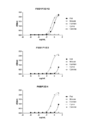

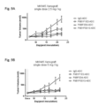
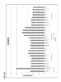


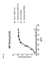

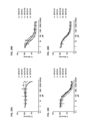


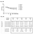


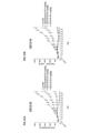
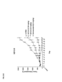

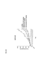


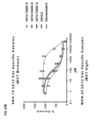

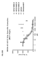

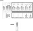

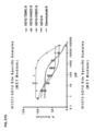

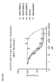


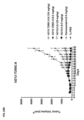

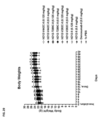


Classifications
-
- A—HUMAN NECESSITIES
- A61—MEDICAL OR VETERINARY SCIENCE; HYGIENE
- A61K—PREPARATIONS FOR MEDICAL, DENTAL OR TOILETRY PURPOSES
- A61K47/00—Medicinal preparations characterised by the non-active ingredients used, e.g. carriers or inert additives; Targeting or modifying agents chemically bound to the active ingredient
- A61K47/50—Medicinal preparations characterised by the non-active ingredients used, e.g. carriers or inert additives; Targeting or modifying agents chemically bound to the active ingredient the non-active ingredient being chemically bound to the active ingredient, e.g. polymer-drug conjugates
- A61K47/51—Medicinal preparations characterised by the non-active ingredients used, e.g. carriers or inert additives; Targeting or modifying agents chemically bound to the active ingredient the non-active ingredient being chemically bound to the active ingredient, e.g. polymer-drug conjugates the non-active ingredient being a modifying agent
- A61K47/68—Medicinal preparations characterised by the non-active ingredients used, e.g. carriers or inert additives; Targeting or modifying agents chemically bound to the active ingredient the non-active ingredient being chemically bound to the active ingredient, e.g. polymer-drug conjugates the non-active ingredient being a modifying agent the modifying agent being an antibody, an immunoglobulin or a fragment thereof, e.g. an Fc-fragment
- A61K47/6801—Drug-antibody or immunoglobulin conjugates defined by the pharmacologically or therapeutically active agent
- A61K47/6803—Drugs conjugated to an antibody or immunoglobulin, e.g. cisplatin-antibody conjugates
-
- A—HUMAN NECESSITIES
- A61—MEDICAL OR VETERINARY SCIENCE; HYGIENE
- A61K—PREPARATIONS FOR MEDICAL, DENTAL OR TOILETRY PURPOSES
- A61K31/00—Medicinal preparations containing organic active ingredients
- A61K31/33—Heterocyclic compounds
- A61K31/395—Heterocyclic compounds having nitrogen as a ring hetero atom, e.g. guanethidine or rifamycins
- A61K31/55—Heterocyclic compounds having nitrogen as a ring hetero atom, e.g. guanethidine or rifamycins having seven-membered rings, e.g. azelastine, pentylenetetrazole
- A61K31/551—Heterocyclic compounds having nitrogen as a ring hetero atom, e.g. guanethidine or rifamycins having seven-membered rings, e.g. azelastine, pentylenetetrazole having two nitrogen atoms, e.g. dilazep
- A61K31/5513—1,4-Benzodiazepines, e.g. diazepam or clozapine
- A61K31/5517—1,4-Benzodiazepines, e.g. diazepam or clozapine condensed with five-membered rings having nitrogen as a ring hetero atom, e.g. imidazobenzodiazepines, triazolam
-
- A—HUMAN NECESSITIES
- A61—MEDICAL OR VETERINARY SCIENCE; HYGIENE
- A61K—PREPARATIONS FOR MEDICAL, DENTAL OR TOILETRY PURPOSES
- A61K47/00—Medicinal preparations characterised by the non-active ingredients used, e.g. carriers or inert additives; Targeting or modifying agents chemically bound to the active ingredient
- A61K47/50—Medicinal preparations characterised by the non-active ingredients used, e.g. carriers or inert additives; Targeting or modifying agents chemically bound to the active ingredient the non-active ingredient being chemically bound to the active ingredient, e.g. polymer-drug conjugates
- A61K47/51—Medicinal preparations characterised by the non-active ingredients used, e.g. carriers or inert additives; Targeting or modifying agents chemically bound to the active ingredient the non-active ingredient being chemically bound to the active ingredient, e.g. polymer-drug conjugates the non-active ingredient being a modifying agent
- A61K47/62—Medicinal preparations characterised by the non-active ingredients used, e.g. carriers or inert additives; Targeting or modifying agents chemically bound to the active ingredient the non-active ingredient being chemically bound to the active ingredient, e.g. polymer-drug conjugates the non-active ingredient being a modifying agent the modifying agent being a protein, peptide or polyamino acid
- A61K47/64—Drug-peptide, drug-protein or drug-polyamino acid conjugates, i.e. the modifying agent being a peptide, protein or polyamino acid which is covalently bonded or complexed to a therapeutically active agent
- A61K47/6415—Toxins or lectins, e.g. clostridial toxins or Pseudomonas exotoxins
-
- A—HUMAN NECESSITIES
- A61—MEDICAL OR VETERINARY SCIENCE; HYGIENE
- A61K—PREPARATIONS FOR MEDICAL, DENTAL OR TOILETRY PURPOSES
- A61K47/00—Medicinal preparations characterised by the non-active ingredients used, e.g. carriers or inert additives; Targeting or modifying agents chemically bound to the active ingredient
- A61K47/50—Medicinal preparations characterised by the non-active ingredients used, e.g. carriers or inert additives; Targeting or modifying agents chemically bound to the active ingredient the non-active ingredient being chemically bound to the active ingredient, e.g. polymer-drug conjugates
- A61K47/51—Medicinal preparations characterised by the non-active ingredients used, e.g. carriers or inert additives; Targeting or modifying agents chemically bound to the active ingredient the non-active ingredient being chemically bound to the active ingredient, e.g. polymer-drug conjugates the non-active ingredient being a modifying agent
- A61K47/68—Medicinal preparations characterised by the non-active ingredients used, e.g. carriers or inert additives; Targeting or modifying agents chemically bound to the active ingredient the non-active ingredient being chemically bound to the active ingredient, e.g. polymer-drug conjugates the non-active ingredient being a modifying agent the modifying agent being an antibody, an immunoglobulin or a fragment thereof, e.g. an Fc-fragment
- A61K47/6801—Drug-antibody or immunoglobulin conjugates defined by the pharmacologically or therapeutically active agent
- A61K47/6803—Drugs conjugated to an antibody or immunoglobulin, e.g. cisplatin-antibody conjugates
- A61K47/68035—Drugs conjugated to an antibody or immunoglobulin, e.g. cisplatin-antibody conjugates the drug being a pyrrolobenzodiazepine
-
- A—HUMAN NECESSITIES
- A61—MEDICAL OR VETERINARY SCIENCE; HYGIENE
- A61K—PREPARATIONS FOR MEDICAL, DENTAL OR TOILETRY PURPOSES
- A61K47/00—Medicinal preparations characterised by the non-active ingredients used, e.g. carriers or inert additives; Targeting or modifying agents chemically bound to the active ingredient
- A61K47/50—Medicinal preparations characterised by the non-active ingredients used, e.g. carriers or inert additives; Targeting or modifying agents chemically bound to the active ingredient the non-active ingredient being chemically bound to the active ingredient, e.g. polymer-drug conjugates
- A61K47/51—Medicinal preparations characterised by the non-active ingredients used, e.g. carriers or inert additives; Targeting or modifying agents chemically bound to the active ingredient the non-active ingredient being chemically bound to the active ingredient, e.g. polymer-drug conjugates the non-active ingredient being a modifying agent
- A61K47/68—Medicinal preparations characterised by the non-active ingredients used, e.g. carriers or inert additives; Targeting or modifying agents chemically bound to the active ingredient the non-active ingredient being chemically bound to the active ingredient, e.g. polymer-drug conjugates the non-active ingredient being a modifying agent the modifying agent being an antibody, an immunoglobulin or a fragment thereof, e.g. an Fc-fragment
- A61K47/6835—Medicinal preparations characterised by the non-active ingredients used, e.g. carriers or inert additives; Targeting or modifying agents chemically bound to the active ingredient the non-active ingredient being chemically bound to the active ingredient, e.g. polymer-drug conjugates the non-active ingredient being a modifying agent the modifying agent being an antibody, an immunoglobulin or a fragment thereof, e.g. an Fc-fragment the modifying agent being an antibody or an immunoglobulin bearing at least one antigen-binding site
- A61K47/6849—Medicinal preparations characterised by the non-active ingredients used, e.g. carriers or inert additives; Targeting or modifying agents chemically bound to the active ingredient the non-active ingredient being chemically bound to the active ingredient, e.g. polymer-drug conjugates the non-active ingredient being a modifying agent the modifying agent being an antibody, an immunoglobulin or a fragment thereof, e.g. an Fc-fragment the modifying agent being an antibody or an immunoglobulin bearing at least one antigen-binding site the antibody targeting a receptor, a cell surface antigen or a cell surface determinant
-
- A—HUMAN NECESSITIES
- A61—MEDICAL OR VETERINARY SCIENCE; HYGIENE
- A61P—SPECIFIC THERAPEUTIC ACTIVITY OF CHEMICAL COMPOUNDS OR MEDICINAL PREPARATIONS
- A61P35/00—Antineoplastic agents
-
- C—CHEMISTRY; METALLURGY
- C07—ORGANIC CHEMISTRY
- C07K—PEPTIDES
- C07K16/00—Immunoglobulins [IGs], e.g. monoclonal or polyclonal antibodies
- C07K16/18—Immunoglobulins [IGs], e.g. monoclonal or polyclonal antibodies against material from animals or humans
- C07K16/28—Immunoglobulins [IGs], e.g. monoclonal or polyclonal antibodies against material from animals or humans against receptors, cell surface antigens or cell surface determinants
- C07K16/2863—Immunoglobulins [IGs], e.g. monoclonal or polyclonal antibodies against material from animals or humans against receptors, cell surface antigens or cell surface determinants against receptors for growth factors, growth regulators
-
- A—HUMAN NECESSITIES
- A61—MEDICAL OR VETERINARY SCIENCE; HYGIENE
- A61K—PREPARATIONS FOR MEDICAL, DENTAL OR TOILETRY PURPOSES
- A61K39/00—Medicinal preparations containing antigens or antibodies
- A61K2039/505—Medicinal preparations containing antigens or antibodies comprising antibodies
-
- C—CHEMISTRY; METALLURGY
- C07—ORGANIC CHEMISTRY
- C07K—PEPTIDES
- C07K2317/00—Immunoglobulins specific features
- C07K2317/20—Immunoglobulins specific features characterized by taxonomic origin
- C07K2317/24—Immunoglobulins specific features characterized by taxonomic origin containing regions, domains or residues from different species, e.g. chimeric, humanized or veneered
-
- C—CHEMISTRY; METALLURGY
- C07—ORGANIC CHEMISTRY
- C07K—PEPTIDES
- C07K2317/00—Immunoglobulins specific features
- C07K2317/30—Immunoglobulins specific features characterized by aspects of specificity or valency
- C07K2317/33—Crossreactivity, e.g. for species or epitope, or lack of said crossreactivity
-
- C—CHEMISTRY; METALLURGY
- C07—ORGANIC CHEMISTRY
- C07K—PEPTIDES
- C07K2317/00—Immunoglobulins specific features
- C07K2317/90—Immunoglobulins specific features characterized by (pharmaco)kinetic aspects or by stability of the immunoglobulin
- C07K2317/92—Affinity (KD), association rate (Ka), dissociation rate (Kd) or EC50 value
Definitions
- Embodiments of the invention relate to cMET binding agents that are conjugated to pyrrolobenzodiazepine toxins, composition thereof and uses thereof.
- cMET hepatocyte growth factor receptor
- HGFR hepatocyte growth factor receptor
- cMET a protein that in humans is encoded by the MET gene (MET proto-oncogene, receptor tyrosine kinase).
- cMET is a single-pass cell surface receptor that possesses tyrosine kinase activity.
- the primary single chain precursor protein of the MET translation product is post-translationally cleaved to produce an alpha and a beta subunit, which are disulfide linked to form a mature cell surface cMET receptor.
- cMET is expressed on cells of epithelial origin, as well as stem cells, progenitor cells and other cell types (e.g., various cancer cell types).
- Hepatocyte growth factor/Scatter Factor (HGF/SF) and its splicing isoforms (NK1, NK2) have been identified as ligands of cMET.
- cMET is thought to be essential for normal embryonic development, organogenesis and wound healing.
- Abnormal cMET expression and/or activity is associated with certain neoplastic disorders and cancers (e.g., cancers of kidney, liver, stomach, breast, and brain) where cMET is implicated in tumor growth, angiogenesis, and metastasis.
- neoplastic disorders and cancers e.g., cancers of kidney, liver, stomach, breast, and brain
- the overexpression of cMET as well as its autocrine activation by co-expression of its ligand are also implicated in oncogenesis.
- novel anti-cMET binding agents e.g., a monoclonal antibody
- a cytotoxic payload e.g., a cytotoxic payload
- pharmaceutical compositions thereof e.g., a cytotoxic payload, pharmaceutical compositions thereof and methods of using the same.
- a binding agent-drug conjugate comprising a binding agent and a payload, where the payload comprises a pyrrolobenzodiazepine toxin and the binding agent specifically binds to mesenchymal epithelial transition factor (cMET).
- a payload comprises a linking group where the pyrrolobenzodiazepine toxin is covalently linked to the linking group, and the linking group is covalently linked to the binding agent.
- a binding agent is a monoclonal antibody, or antigen binding portion thereof.
- a binding agent-drug conjugate comprising a binding agent and a payload
- the binding agent comprises: (i) two or more of a CDR-L1, a CDR-L2 and a CDR-L3 which are polypeptide sequences of a light chain complementarity determining region (CDR-L), wherein the CDR-L1 is selected from the amino acid sequences of SEQ ID NOs: 1-15, the CDR-L2 is selected from the amino acid sequences of SEQ ID NOs: 16-25, and the CDR-L3 is selected from the amino acid sequences of SEQ ID NOs: 26-36, and (ii) two or more of a CDR-H1, a CDR-H2 and a CDR-H3 which are polypeptide sequences of a heavy chain complementarity determining region (CDR-H), wherein the CDR-H1 is selected from the amino acid sequences of SEQ ID NOs: 50-61, the CDR-H2 is selected
- a pyrrolobenzodiazepine toxin comprises the structure of chemical formula I: wherein Z 1 and Z 2 are both N; Z 3 and Z 4 are both C; n is 1 to 12; each of R 3 and R 4 are independently H, or a C1-4 alkoxyl; and each of R 1 and R 2 are independently selected from the group consisting of H, C 1-5 alkyl, C 3-6 cycloalkyl, C 2-5 alkenyl, and a phenyl optionally substituted with R 5 , wherein R 5 is selected from the group consisting of -NH 2 , -NHR 6 , and a piperazinyl substituted with R 7 having the structure R 6 comprises the linking group, and R 7 is H, or a C 1-5 alkyl; X 1 is null, a protecting group, or comprises the linking group; X 2 is null, a protecting group, or comprises the linking group; only one of X 1 , X 2 , R 1 , and R 2
- n is 3 or 5.
- R 3 and R 4 are both -O-CH 3 .
- R 2 is a cyclopropyl.
- R 2 is phenyl substituted with 4-methylpiperazin-1-yl or phenyl substituted with R 5 , where R 5 is -NHR 6 and R 6 comprises the linking group.
- the linking group is attached to the pyrrolobenzodiazepine toxin by a carbamate group or an amide group.
- a linking group comprises the structure of chemical formula A: wherein the asterisk indicates the point of attachment to a pyrrolobenzodiazepine toxin; the wavy line indicates the point of attachment to the binding agent; m is 1 to 20; q is 1 to 10; and E is a connecting group.
- m is 4 or 8 and q is 0, 1 or 2. In some embodiments, m is 8 and q is 2.
- a linking group comprises the structure of chemical formula B: wherein the asterisk indicates the point of attachment to the pyrrolobenzodiazepine toxin; the wavy line indicates the point of attachment to the binding agent; E comprises a connecting group; v is 0 to 10; and u is 0 or 1; wherein when u is 1, t is 1 to 10.
- v is 1.
- u is 1, and t is 8.
- u is 0, and v is 4.
- a binding agent is connected to E by a thioether bond formed between a cysteine thiol residue of the binding agent and E.
- E comprises the structure of chemical formula C: wherein the wavy line indicates the point of attachment to a binding agent and the double asterisk indicates a point of attachment to the linking group.
- the protecting group has the following structure (D): wherein the asterisk indicates a point of attachment to a pyrrolobenzodiazepine toxin; and w is 1 to 5. In some embodiments, w is 2. In some embodiments, a protecting group is a cleavable protecting group.
- a binding agent-drug conjugate comprising a binding agent and a payload, where the binding agent specifically binds to an extracellular domain of mesenchymal epithelial transition factor (cMET), the payload is covalently linked to the binding agent, and the payload comprises a structure selected from the group consisting of chemical formulas II, III, V, VI and VII, where chemical formula II comprises the structure: wherein m is 8 and the wavy line indicates the point of attachment to a binding agent; chemical formula III comprises the structure: wherein m is 8, p is 2 or 3, X 2 is a protecting group and the wavy line indicates the point of attachment to a binding agent; chemical formula V comprises the structure: wherein m is 8, and the wavy line indicates a point of attachment to a binding agent; chemical formula VI comprises the structure: wherein t is 8, v is 1 and the wavy line indicates a point of attachment to a binding agent; and chemical formula VII comprises the structure: wherein the wavy line
- the protecting group of X 2 has the following structure (D): wherein the asterisk indicates the point of attachment to the payload; and w is 1 to 5.
- a binding agent-drug conjugate comprising a monoclonal antibody, or antigen binding portion thereof, and a payload, wherein the monoclonal antibody, or antigen binding portion thereof, comprises a CDR-L1 selected from the amino acid sequences of SEQ ID NOs: 2, 4, 6, 8, 10, 12 and 14, a CDR-L2 selected from the amino acid sequences of SEQ ID NOs: 17, 19, 21, 23 and 25, a CDR-L3 selected from the amino acid sequences of SEQ ID NOs: 27, 29, 31, 33 and 35, a CDR-H1 selected from the amino acid sequences of SEQ ID NOs: 51, 53, 55, 57 and 59, a CDR-H2 is selected from the amino acid sequences of SEQ ID NOs: 63, 65, 67, 69, 73 and 75, and a CDR-H3 selected from the amino acid sequences of SEQ ID NOs: 80, 82, 84, 86, 88, 91 and 93.
- a binding agent of a binding agent-drug conjugate described herein comprises a CDR-L1 comprising the amino acid sequence of SEQ ID NO: 10 or 14; a CDR-L2 comprising the amino acid sequence of SEQ ID NO: 25; a CDR-L3 comprising the amino acid sequence of SEQ ID NO: 35; a CDR-H1 comprising the amino acid sequence of SEQ ID NO: 59; a CDR-H2 comprising the amino acid sequence of SEQ ID NO: 71; and a CDR-H3 comprising the amino acid sequence of SEQ ID NO: 88.
- a binding agent of a binding agent-drug conjugate described herein comprises a CDR-L1 comprising the amino acid sequence of SEQ ID NO: 9 or 15; a CDR-L2 comprising the amino acid sequence of SEQ ID NO: 24; a CDR-L3 comprising the amino acid sequence of SEQ ID NO: 34; a CDR-H1 comprising the amino acid sequence of SEQ ID NO: 58; a CDR-H2 comprising the amino acid sequence of SEQ ID NO: 70 or 78; and a CDR-H3 comprising the amino acid sequence of SEQ ID NO: 87.
- a binding agent comprises a variable light chain region comprising an amino acid sequence selected from the amino acid sequences of SEQ ID NOs: 37-44. In some embodiments, a binding agent comprises a variable light chain sequence having at least 90% sequence identity, or 100% sequence identity to any one of the amino acid sequences of SEQ ID NOs: 45-49. In some embodiments, a binding agent comprises a variable light chain sequence having at least 90% sequence identity to any one or SEQ ID NOs: 37-49, wherein the variable light chain sequence has one to ten, or one to five amino acid modifications selected from an amino acid addition, an amino acid deletion and an amino acid substitution.
- a binding agent comprises a variable heavy chain region comprising an amino acid sequence selected from the amino acid sequences of SEQ ID NOs: 94-103. In some embodiments, a binding agent comprises a variable heavy chain region having at least 90% sequence identity, or 100% sequence identity to any one of the amino acid sequences of SEQ ID NOs: 104-108. In some embodiments, a binding agent comprises a variable heavy chain region having at least 90% sequence identity to any one of the amino acid sequences of SEQ ID NOs: 94-108, wherein the variable heavy chain sequence has one to ten, or one to five amino acid modifications selected from an amino acid addition, an amino acid deletion and an amino acid substitution.
- a binding agent comprises a variable light chain sequence having at least 90% sequence identity to any one or SEQ ID NOs: 37-49, and a variable heavy chain sequence having at least 90% sequence identity to any one of the amino acid sequences of SEQ ID NOs: 94-108.
- a binding agent, monoclonal antibody, or antigen binding portion thereof is chimeric or humanized.
- a binding agent comprises one or more humanized or human framework region and/or one or more mouse framework regions.
- a binding agent, monoclonal antibody, or antigen binding portion thereof comprises: a heavy chain and light chain each having CDRs that are the same as respective CDRs of heavy chain and light chain of an antibody produced by hybridoma cell line F6B1P3D12 deposited with ATCC on March 20, 2019.
- a binding agent that specifically binds to cMET, or a portion thereof comprises an antigen binding portion of an antibody or a single chain antibody.
- a binding agent of a binding agent-drug conjugate comprises a Fab, Fab’, F(ab’)2, Fv or scFV fragment of an antibody.
- a binding agent binds specifically to a mammalian cMET. In certain embodiments, a binding agent binds specifically to a human cMET, monkey cMET and/or rat cMET. In some embodiments, a binding agent specifically binds to the extracellular domain of a wild type or variant cMET. In some embodiments, a binding agent that specifically binds to cMET is a binding agent that induces internalization and/or degradation of cMET on a human cancer cell. In some embodiments, a binding agent that specifically binds to cMET is a binding agent that is not a cMET agonist. Accordingly, a cMET binding agent that is not a cMET agonist is a binding agent, that upon binding to a cell-surface cMET, does not substantially induce signaling through the cell-surface cMET.
- a pharmaceutical composition comprising the binding agent-drug conjugate described herein and a pharmaceutically acceptable excipient, diluent, additive or carrier.
- a binding agent-drug conjugate blocks, inhibits, ameliorates, abrogates, or suppresses growth, viability or metastasis of the cancer.
- a binding agent-drug conjugate induces death, necrosis or apoptosis of some or all of the cancer.
- a neoplastic disorder or cancer comprises a carcinoma, sarcoma, neuroblastoma, glioblastoma, myeloma, lymphoma, melanoma or a solid or soft tissue tumor.
- a neoplastic disorder or cancer comprises a bladder cancer, breast cancer, colorectal cancer, gastric cancer, pancreatic cancer (e.g., exocrine pancreatic cancer and pancreatic neuroendocrine cancer), esophageal cancer, liver cancer, hepatocellular cancer, hypopharynx cancer, lung cancer, adenocarcinoma, ovarian cancer or renal cancer.
- a neoplastic disorder or cancer comprises a pancreatic adenocarcinoma, colorectal adenocarcinoma, small intestinal malignancy, cholangiocarcinoma, non-small cell lung cancer (NSCLC), thyroid carcinoma, esophageal or esophagogastric junction (EGJ) cancer, gastric adenocarcinoma, liver hepatocellular carcinoma, head and neck squamous carcinoma, female genital tract malignancy, breast carcinoma, lung small cell carcinoma, ovarian surface epithelial carcinoma, retroperitoneal or peritoneal sarcoma, prostatic adenocarcinoma, neuroendocrine tumor, gastrointestinal stromal tumor, glioblastoma or non-epithelial ovarian cancer.
- NSCLC non-small cell lung cancer
- EGJ esophageal or esophagogastric junction
- gastric adenocarcinoma gastric adenocar
- a cancer that can be treated by a method described herein is a cancer comprising a cell (e.g., a malignant or neoplastic cell) that expresses a cMET polypeptide (e.g., cMET, e.g., expressed on the cell surface).
- a cell e.g., a malignant or neoplastic cell
- a cMET polypeptide e.g., cMET, e.g., expressed on the cell surface
- Fig. 1 shows a summary of the work flow used for generation of monoclonal antibodies (exemplary binding agents) that bind specifically to cMET.
- the lead monoclonal Ab P3D12 was generated from a mouse immunized with recombinant intact extracellular domain of cMET fused to human Fc.
- Fig. 2 shows an immunization scheme used to generate monoclonal antibodies (exemplary binding agents) that bind specifically to cMET.
- Mice were initially immunized by intraperitoneal injection (i.p.) with 100 ⁇ g of a human cMET-Fc fusion protein (cMET-Fc), or 50 to 100 ⁇ g of a KLH conjugated cMET peptide in Freund’s Complete Adjuvant (CFA) as indicated.
- cMET-Fc comprises the extracellular domain of human cMET fused to an Fc portion of an antibody.
- cMET peptides were strategically selected from a portion of the cMET extracellular domain.
- Immunized mice received one or more booster immunization comprising 25 or 50 ⁇ g of cMET-Fc or peptide in Incomplete Freund’s adjuvant (IFA) as indicated. Some mice received repetitive immunizations at multiple sites (RIMMS). Immunizations included Met-Fc fusions, peptides, traditional and RIMMS. Spleens of immunized mice were obtained and fused to a suitable fusion partner. Over 20,000 hybridoma clones were obtained and screened.
- Fig. 3 shows a 3D structure of the MET SEMA domain bound with a Fab of an agonist Met-mAb antibody (5D5 Fab) and HGF beta subunit (HGF ⁇ or HGFb).
- the bottom arrow indicates the position of a portion of cMET used to design peptide 3.
- Fig. 4 shows characterization results from an exemplary fusion (FUSION 6B1, plate 3).
- Anti-cMET hybridomas were selected, in part, for the presence of specific binding to cMET as assayed by ELISA (see column labeled “MET Binding ELISA OD450nm”) and for ability to induce internalization of cMET on human cancer cell lines as measured by flow cytometry (see column labeled “FACS Geom. Mean”).
- a FACS Geom. Mean value lower than a negative control indicates internalization of cMET.
- the arrow indicates a lead hybridoma F6B1P3D12.
- Fig. 5 shows the results of a MET degradation assay.
- Anti-cMET antibodies isolated from the indicated wells were tested and selected for their ability to induce degradation of cMET on human cancer cell lines as measured by Mesoscale (MSD) cMET protein quantification. Relative values of Met degradation are indicated on the y-axis as % control (percent of control). Values lower than 100% control (negative control level) indicate internalization and degradation of cMET. Degradation indicates not just internalization but lysosomal trafficking, an important property for an antibody drug conjugate. The arrow indicates the results for a lead hybridoma F6B1P3D12. Fig.
- FIG. 6 shows the results of a phospho-ERK assay (P-ERK assay) that measures agonist activity of anti-cMET antibodies by indirectly measuring the phosphorylation of ERK induced by binding of an anti-cMET antibody to cMET on a cell surface.
- P-ERK assay phospho-ERK assay
- Anti-cMET antibodies produced from various anti-cMET hybridomas were tested at 6 ⁇ g/ml or 30 ⁇ g/ml (as indicated on x-axis), and selected according to their inability to induce significant phosphorylation of ERK (i.e., their ability not to induce proliferation, i.e., absence of agonist activity).
- a lead monoclonal antibody (mAb) P3D12 is indicated by the arrow.
- Fig. 7 shows results of six cMET monoclonal antibodies (mAbs) that were assayed by ELISA for binding to human, monkey (Cynomolgus Macaque, “Cyno”), canine, rat, and mouse cMET. All monoclonal antibodies bound human and monkey cMET. P3D12 demonstrated significant cross-reactivity with rat cMET. Various concentrations of each antibody are indicated on the x-axis. Relative amounts bound are indicated on the y-axis (OD450nm).
- Fig. 7-1 provides results with three monoclonal antibodies.
- Fig. 7-2 provides results with the other three monoclonal antibodies.
- Fig. 8A shows an alignment of the amino acid sequences of the light chain variable regions of nine mouse monoclonal anti-cMET antibodies, the names of which are indicated to the left of each sequence. SEQ ID NOs: are indicated to the right of each sequence. Amino acid sequences of the light chain variable regions of LC F6B1P1E2 and F6BP3E2 are 100% identical. Also, amino acid sequences of the light chain variable regions of LC F6B1P3D12 and F6B1P3E9 are 100% identical.
- Fig. 8B shows an alignment of the amino acid sequences of the heavy chain variable regions of nine mouse monoclonal anti-cMET antibodies, the names of which are indicated to the left of each sequence. SEQ ID NOs: are indicated to the right of each sequence. Amino acid sequences of the heavy chain variable regions of F6B1P3D12H7913 and F6B1P3E9 are 100% identical. Also, amino acid sequences of the heavy chain variable regions of F6B1P1E2H7819 and F6BP3E2 are 100% identical.
- Fig. 9A and 9B show the results of an in vivo xenograft mouse model evaluating the efficacy of the indicated anti-cMET antibody drug conjugates (ADC) using the MKN45 tumor model (a cMET+ gastric cancer model) in nude mice.
- Animals were treated once with ADCs at 2.5 mg/kg (9A) or 5.0 mg/kg (9B).
- the efficacy of each drug conjugated anti-cMET binding agent is compared to PBS or an unrelated non-targeting monoclonal antibody (IgG-ADC).
- Tumor volume (y-axis) was measured at various time points after inoculation (y-axis, Days (post inoculation)). Inhibition of tumor growth indicates positive efficacy.
- Anti-cMET binding agents and the non-targeting control monoclonal antibody (IgG) were conjugated to monomethyl auristatin F (MMAF).
- MMAF monomethyl auristatin F
- Fig. 10 shows the results of a phospho-ERK assay (P-ERK assay) that measures agonist activity of anti-cMET antibodies by indirectly measuring the phosphorylation of ERK induced by binding of an anti-cMET antibody to cMET on a cell surface.
- the amount of phosphorylated ERK (shown as % of control, y-axis) detected in cell lysates after treatment of viable cells with an anti-cMET antibody is shown.
- the constant regions of the heavy and light chains of an isolated mouse monoclonal antibody designated as P3D12 were replaced with antibody constant regions of a human IgG1 (P3D12( hIgG1)), or a human IgG2 (P3D12(hIgG2)) as indicated on the x-axis.
- Each antibody was tested at 0.00064 ⁇ g/ml, 0.0032 ⁇ g/ml, 0.016 ⁇ g/ml, 0.08 ⁇ g/ml, 0.4 ⁇ g/ml, 2 ⁇ g/ml, 10 ⁇ g/ml and 50 ⁇ g/ml as indicated on the x-axis.
- “Control” indicates an untreated negative control.
- HGF(EC90) is a positive control and indicates cells treated with Hepatocyte Growth Factor (HGF), the natural ligand of cMET receptor. This data indicates that the human IgG2 isotype does not display detectable agonist activity.
- Fig. 11 shows the results of a MET degradation assay.
- Degradation is a measure of internalization of the cMET receptor upon antibody binding.
- Chimeric anti-cMET antibodies were tested for their ability to induce degradation of cMET on human cancer cell lines as measured by Mesoscale (MSD) cMET protein quantification.
- Relative values of Met degradation are indicated on the y-axis as % control (percent of control). Values lower than 100% control indicate internalization and degradation of cMET.
- Chimeric antibodies were generated by replacing the constant regions of the heavy and light chains of an isolated mouse monoclonal antibody designated as P3D12 (P3D12(mouse)) with antibody constant regions of a human IgG1 (P3D12( hIgG1)), or a human IgG2 (P3D12(hIgG2)) as indicated on the x-axis.
- P3D12 P3D12(mouse)
- P3D12(hIgG1) human IgG1
- P3D12(hIgG2) human IgG2
- Each antibody was tested at 0.00064 ⁇ g/ml, 0.0032 ⁇ g/ml, 0.016 ⁇ g/ml, 0.08 ⁇ g/ml, 0.4 ⁇ g/ml, 2 ⁇ g/ml, 10 ⁇ g/ml and 50 ⁇ g/ml as indicated on the x-axis.
- Chimeric cMET antibodies of P3D12 showed similar internalization/degrad
- Fig. 12 shows a flow chart of process development for testing and selection of lead anti-cMET monoclonal binding agents.
- Fig. 13 shows an alignment of five humanized light chain variable regions of humanized versions of the murine anti-cMET clone P3D12, the names and SEQ ID NOs: of which are indicated to the left of each sequence.
- Five independent methods were used to humanize the murine anti-cMET mAbs. Two of the methods gave the same result, therefore there are four different light chains shown.
- Fig. 14 shows an alignment of five humanized heavy chain variable regions of humanized versions of the murine clone P3D12, the names and SEQ ID NOs: of which are indicated to the left of each sequence.
- Five independent methods were used to humanize the murine anti-cMET mAb. Two of the methods gave the same result, therefore there are four different light chains (see Fig. 13) and four different heavy chains which can make a combination of 16 different binding agents.
- Fig. 15 shows the results of an in vivo xenograft mouse model testing the efficacy of the indicated humanized anti-cMET antibody drug conjugates (ADC) at 2.5 mg/kg (2.5 mpk), or at 5 mg/kg (5 mpk) as demonstrated using an MKN45 tumor model (a cMET+ gastric cancer model). Animals were treated once with the indicated ADCs at 2.5 or 5.0 mg/kg.
- the efficacy of each anti-cMET binding agent is compared to PBS or the non-targeting monoclonal antibody Rituximab (Retux), which is an anti-cancer monoclonal antibody that targets CD20, which is primarily found on the surface of immune system B cells. Tumor volume (y-axis) was measured at various time points (y-axis) after inoculation. Inhibition of tumor growth indicates positive efficacy.
- Anti-cMET binding agents were conjugated to monomethyl auristatin F (MMAF).
- MMAF monomethyl auristatin F
- Fig. 16 shows binding of anti-cMET monoclonal binding agent hD12 that has heavy and light chain variable region of SEQ ID NOs 108 and 47 and a constant region of human IgG2, and a negative control antibody (Denosumab), that does not bind cMET to cMET-Fc and mutant cMET (E168D) Fc recombinant fusion proteins.
- the E168D mutation is a somatic mutation found in small cell lung cancer (SCLC). The mutation is located in the Sema domain and leads to constitutive activation of the cMET receptor. Abundance of somatic mutations of cMET are very low. E168D occurs in 0.8% to 3% of SCLC patients.
- a binding ELISA was performed with human cMET or E168D cMET extracellular domains fused to human IgG1 Fc.
- cMET proteins were coated on a plate overnight and the samples titrated and detected with a goat anti-human IgG (H+L)-HRP.
- EC50s were determined using sigmoidal dose response fit.
- Fig. 17A shows the results of an ELISA-based cMET binding assay.
- five representative antibody-drug conjugates i.e., hD12-VI, hD12-II, hD12-VII, hD12-IV and hD12-XI (see antibody nomenclature explained in Example 3)
- hD12-VI, hD12-II, hD12-VII, hD12-IV and hD12-XI see antibody nomenclature explained in Example 3
- a negative control antibody that does not bind cMET, was conjugated to each of the five different payloads (i.e., payloads of chemical formula VI, II, IV, VII and XI, indicated as Denosumab-VI, Denosumab-II, Denosumab-IV, Denosumab-VII, and Denosumab-XI, respectively) and tested as a negative control.
- Fig. 17B shows IC50 values for each of the antibodies tested in Fig. 17A.
- Fig. 18A-18D show the results of a cytotoxicity assay for five representative antibody-drug conjugates (hD12-VI, hD12-II, hD12-VII, hD12-IV and hD12-XI) on four cell lines that express different amounts of cMET on their cell surface (SNU-1, no expression of cMET (Fig. 18A), SNU-16, medium expression of cMET (Fig. 18B), SNU-620, high expression of cMET (Fig. 18C) and MKN-45, high expression of cMET (Fig. 18D)). Percent survival is indicated on the y-axis and the amount of antibody drug conjugate added is indicated on the x-axis (pM).
- Fig. 19A-19E show the results of a cytotoxicity assay for five representative antibody-drug conjugates (hD12-VI, hD12-II, hD12-VII, hD12-IV and hD12-XI) on five cell lines that express different amount of cMET on their cell surface (H441, medium expression of cMET (Fig. 19A), H1373, medium expression of cMET (Fig. 19B), H1975, medium expression of cMET (Fig. 19C), SNU-5, high expression of cMET (Fig. 19D), and H1573, medium expression of cMET (Fig. 19E)).
- Fig. 19-1 provides Figures 19A-19D.
- Figure 19E is provided here. Percent survival is indicated on the y-axis and the amount of antibody drug conjugate added is indicated on the x-axis (pM).
- Fig. 19F shows comparative IC50 values.
- Figure 20 shows the results of an in vivo xenograft study.
- Mice were injected with H1975 tumor cells (medium expression of cMET) and treated with either hD12-XI (Fig. 20A, 20B and 20C), hD12-VI (Fig. 20A, 20B and 20D), hD12-II (Fig. 20A, 20B and 20E), hD12-VII (Fig. 20A, 20B and 20F), and hD12-IV (Fig. 20A, 20B and 20G), and tumor volume (y-axis, Figs. 20 A, and 20C-20G) or body weight (Fig. 20B) was determined over time (i.e., days, x-axis).
- Fig. 20-1 provides Figure 20A.
- Fig. 20-2 provides Figure 20B.
- Fig. 20-3 provides Figure 20C.
- Fig. 20-4 provides Figure 20D.
- Fig. 20-5 provides Figure 20E
- Fig. 20-6 provides Figure 20F.
- Fig. 20-7 provides Figure 20G
- Figure 21 shows the results of an in vivo xenograft study. Mice were injected with H1373 tumor cells (medium expression of cMET) and treated with either hD12-XI (Fig. 21A), hD12-VI (Fig. 21B), hD12-II (Fig. 21C), hD12-VII (Fig. 21D), and hD12-IV (Fig. 21E) and tumor volume (y-axis) was determined over time (i.e., days, x-axis). A negative control antibody (Denosumab) conjugated to II was tested as a negative control.
- Fig. 21-1 provides Figure 21A and Figure 21B.
- Fig. 21-2 provides Figure 21C.
- Figure 21-3 provides Figure 21D.
- Figure 21-4 provides Figure 21E
- Figure 22 shows the body weight (y-axis) of the mice treated in Figure 21 over time (x-axis).
- Figure 23 shows the serum concentration (y-axis) of hD12-XI, hD12-VI, hD12-II, hD12-VII, hD12-IV, and Denosumab-II after i.v. injection in mice. Time (hours) after injection is indicated on the x-axis. The concentration of each antibody-drug conjugate was determined by ELISA.
- Figure 24 shows the results of an ELISA-based cMET binding assay showing the relative affinity of hD12 and four variants of hD12 (hD12-T289C, hD12-V442C and hD12-V282C) that are covalently linked to the payload II, for plate-bound human cMET.
- hD12-II was stochastically linked to the payload at random sulfhydryl groups.
- hD12-T289C-II, hD12-V442C-II and hD12-V282C-II were site-specially linked to the payload at the position of the variant cysteine residue (i.e., T289C, V442C and V282C, respectively).
- the antibody drug conjugates were tested at increasing concentrations (x-axis, concentration of antibody-drug conjugate ( ⁇ g/ml)) for their ability to bind plate-bound human cMET. Relative binding strength is indicated by luminescence (y-axis).
- a negative control antibody (Denosumab-II) was tested as a negative control.
- Fig. 24B shows IC50 values for each of the antibodies tested in Fig. 24A.
- Figure 25 shows the results of a cytotoxicity assay for the antibody conjugates of Fig. 24 (i.e., hD12-II, hD12-T289C-II, hD12-V442C-II and hD12-V282C-II) on four cell lines that express different amounts of cMET on their cell surface (SNU-1, no expression of cMET (Fig. 25A), SNU-16, medium expression of cMET (Fig. 25B), SNU-620, high expression of cMET (Fig. 25C), MKN-45, high expression of cMET (Fig. 25D), and N87, low expression of cMET (Fig. 25E)).
- Fig. 25F Percent survival is indicated on the y-axis and the amount of antibody drug conjugate added is indicated on the x-axis (pM).
- the results are summarized in Fig. 25F which shows comparative IC50 values.
- the concentrations of antibody drug conjugate tested are indicated in Fig. 25G.
- Fig. 25-1 provides Figure 25A.
- Fig. 25-2 provides Figure 25B.
- Fig. 25-3 provides Figure 25C.
- Fig. 25-4 provides Figure 25D.
- Fig. 25-5 provides Figure 25E.
- Fig. 25-6 provides Figure 25F and Figure 25G.
- Figure 26A shows the results of a cytotoxicity assay for the antibody conjugates of Fig. 24 (i.e., hD12-II, hD12-T289C-II, hD12-V442C-II and hD12-V282C-II) on a high cMET expressing cell line (i.e., SNU-5). Percent survival is indicated on the y-axis and the amount of antibody drug conjugate added is indicated on the x-axis (pM). The results are summarized in Fig. 26B which shows comparative IC50 values. The concentrations of antibody drug conjugate tested are indicated in Fig. 26C.
- Figure 27 shows the results of a cytotoxicity assay for the antibody conjugates of Fig. 24 (i.e., hD12-II, hD12-T289C-II, hD12-V442C-II and hD12-V282C-II) on three cell lines expressing medium amounts of cMET on their cell surface (H1373, Fig. 27A, H1573, Fig. 27B, and H1975, Fig. 27C). Percent survival is indicated on the y-axis and the amount of antibody drug conjugate added is indicated on the x-axis (pM). The concentrations of antibody drug conjugate tested are indicated in Fig. 27D. The results are summarized in Fig. 27E which shows comparative IC50 values.
- Fig. 27-1 provides Figure 27A.
- Fig. 27-2 provides Figure 27B.
- Fig. 27-3 provides Figure 27C.
- Fig. 27-4 provides Figure 27D and Figure 27E.
- Figure 28 shows the results of an in vivo xenograft study.
- Mice were injected with H1975 tumor cells (medium expression of cMET) and treated with hD12-V442C-II (Fig. 28A), hD12-T289C-II (Fig. 28B) and hD12-V282C-II (Fig. 28C) at the indicated concentrations.
- Mice were treated with stochastically conjugated hD12-II as a positive control.
- Mice were treated with vehicle (PBS) or Denosumab-II as negative controls.
- Tumor volume (y-axis) was determined over time (i.e., days, x-axis).
- Fig. 28-1 provides Figure. 28A.
- Fig. 28-2 provides Figure 28B.
- Fig. 28-3 provides Figure 28C.
- Figure 29 shows the body weight (y-axis) of the mice treated in Figure 28 over time (x-axis).
- Figure 30 shows the serum concentration (y-axis) of hD12-V442C-II (Fig. 28A), hD12-T289C-II (Fig. 28B) and hD12-V282C-II (Fig. 28C) after i.v. injection in rats. Time (hours) after injection is indicated on the x-axis. The concentration of each antibody-drug conjugate was determined by ELISA.
- Figure 31 shows a table summarizing the pharmacokinetic data obtained from the experiment of Fig. 30.
- Figure 32 shows the results of an in vivo patient-derived xenograft (PDX) study using human primary gastric cancer tissue (Fig. 32A), human primary colorectal cancer tissue (Fig. 32B) and human head & neck cancer tissue (Fig. 32C). Percentage of tumor growth inhibition (TGI%) is shown on the y-axis and the concentration of the antibody conjugate (hD12-T289C-II) administered is shown on the x-axis. TGI% was calculated as described in Example 12.
- Figure 33A shows immunohistochemical staining of a cross-section of gastric cancer PDX model GA3121 for human cMET protein.
- Figure 33B shows an amplified view of the area shown in the white inset of Figure 33A.
- Figure 33C shows the results of tumor growth inhibition of an in vivo patient-derived xenograft (PDX) model GA3121.
- PDX in vivo patient-derived xenograft
- Each data point represents 1 group consisting of 10 mice. Mice were treated with vehicle (PBS) or sekukinumab-II as negative controls. Tumor volume (y-axis) was determined over time (i.e., days, x-axis).
- binding agent-drug conjugates comprising a binding agent (e.g., a monoclonal antibody, or antigen binding portion thereof) and a payload (e.g., a cytotoxic payload).
- a binding agent is a novel monoclonal antibody, or an antigen binding portion thereof, that binds specifically to cMET.
- a payload comprises a pyrrolobenzodiazepine (PBD) toxin and a specific linking group.
- PBD pyrrolobenzodiazepine
- the binding agents disclosed herein are novel, not only in the amino acid sequences of their antigen binding regions (e.g., heavy and light chain variable regions), but also in their functional characteristics.
- the anti-cMET binding agents described herein possess a combination of distinct characteristics not found in other cMET antibodies.
- the anti-cMET binding agents described herein do not induce significant signaling (e.g., receptor tyrosine kinase activity) from a cMET receptor upon binding. Accordingly, upon binding, the cMET binding agents described herein do not induce undesirable oncogenic activity upon binding (e.g., growth, proliferation, metastasis or angiogenesis).
- anti-cMET binding agents described herein can induce cMET degradation and are internalized after binding.
- any toxic payload attached to the anti-cMET binding agent is brought into the interior of a target cell thereby reducing off-target, non-specific toxicity of the payload.
- This feature also allows for the activity of a toxic payload to be controlled or regulated.
- a toxic payload described herein is substantially inactive until contacted with an intracellular protease.
- certain anti-cMET binding agents described herein cross-react with non-human primates, rat and/or mouse which allows for the antibody drug conjugates to be tested and optimized using non-human animal models.
- the anti-cMET binding agents described herein are soluble, display pro-longed half-life in vivo and are stable upon storage.
- a number of cytotoxic payloads are known which can be attached to an antibody using a known linker to generate an antibody drug conjugate (ADC)(e.g., see US 2014/0120118, US 2014/0127239, US 2016/0250344, US 2016/0250345 and Tiberghien, et al., (2016) ACS Medicinal Chemistry Letters 7 (11):983-987).
- ADC antibody drug conjugate
- the biochemical functionalities of an antibody binding agent often changes after conjugation to a payload.
- the biochemical characteristics of a payload after conjugation to an antibody are not always predictable.
- the in vivo activity of a known payload may differ significantly, ranging from lethal to no therapeutic effect, depending on the type of linker used and where the linker is attached to the antibody. Therefore, generating an antibody drug conjugate by combining a specific cMET binding agent with an ideal linker, a selected toxic payload and an optimal conjugation site of linker to antibody to provide for optimal delivery of the toxin, while substantially limiting adverse events (e.g., off-target toxicity), and maintaining the desirable bio-functional properties of the binding agent, is extremely challenging, time consuming and requires a substantial inventive effort.
- the ADCs presented herein provide for a unique combination of toxins, linkers and novel cMET binding agents that result in highly effective ADCs that provide for optimal therapeutic effects while substantially reducing or eliminating off-target toxicity.
- cMET MET is used synonymously herein with the term “cMET”.
- cMET is also known as hepatocyte growth factor receptor (HGFR).
- Human cMET e.g., SEQ ID NO:109
- SEQ ID NO:109 comprises an immature polypeptide sequence of 1390 amino acids and includes an N-terminal single sequence from amino acids 1-24, an extracellular domain of human cMET from about amino acid 24-932, a transmembrane domain from about amino acid 933 to 955 and a cytoplasmic domain at about amino acid 956 to 1390, numbered from the N-terminus to the C-terminus.
- cMET polypeptide sequence derived from a suitable mammalian species.
- a human cMET polypeptide may comprise several known variants (e.g., see URL:http://www.uniprot.org/uniprot/P08581, as accessed on May 5, 2016, which cMET variants and alternative sequences disclosed therein are incorporated herein by reference).
- Non-limiting examples of naturally occurring variants of a human cMET include amino acid substitutions at 143, 150, 156, 168, 238, 316, 320, 375, 385, 773, 970, 991, and/or 992 of human cMET (SEQ ID NO: 109).
- cMET or a cMET extracellular domain comprises an E to D substitution at position 168 of human cMET, referred to herein as E168D.
- cMET or a cMET extracellular domain comprises an N to S substitution at position 375 of human cMET, referred to herein as N375S.
- cMET is a mammalian cMET. In some embodiments cMET is a primate cMET. In some embodiments cMET is a human cMET. In some embodiments cMET is a monkey cMET. In some embodiments cMET is a rodent cMET (e.g., rat and/or mouse). In some embodiments cMET is a canine cMET (e.g., a dog cMET).
- a mammalian cMET are provided in Example 5 and/or in a sequence listing of this application.
- an extracellular domain of cMET comprises an N-terminal portion of a cMET polypeptide that is typically expressed on the cell surface of an intact mammalian cell.
- An extracellular domain of cMET may comprise two or more polypeptide chains derived from a MET translation product.
- an extracellular domain of cMET can be expressed in a soluble and/or a non-membrane bound form that lacks a cytoplasmic and/or transmembrane domain.
- an extracellular domain of cMET is expressed, isolated and/or purified as a fusion protein.
- the extracellular domain of a mammalian cMET can be engineered and expressed as a fusion protein comprising an Fc portion of an immunoglobulin (e.g., cMET-Fc).
- cMET and/or the extracellular domain of cMET comprises one or more amino acid additions, deletions or substitutions.
- a cMET polypeptide may be at least 80%, at least 85%, at least 90% or at least 95% to a cMET polypeptide disclosed herein.
- a cMET polypeptide comprises a portion of (e.g., a sub-sequence of) a cMET protein.
- a portion of a cMET comprises an extracellular domain of cMET, or a portion thereof.
- a binding agent comprises or consists of one or more polypeptides or one or more proteins that bind specifically to cMET or a portion thereof.
- a binding agent comprises or consists of a protein that binds specifically to cMET or a portion thereof.
- a binding agent often comprises at least one antigen binding portion (i.e. a binding portion).
- An antigen binding portion of a binding agent is that portion that binds specifically to an antigen.
- a binding portion of a binding agent comprises or consists of a single polypeptide (e.g., single chain antibody).
- a binding portion of a binding agent comprises or consists of two polypeptides.
- a binding portion of a binding agent comprises or consists of 2, 3, 4 or more polypeptides.
- a binding agent comprises one or more structural portions (e.g., scaffolds, structural polypeptides, constant regions and/or framework regions).
- a binding agent, or binding portion thereof is attached to a substrate (e.g., a polymer, a non-organic material, silicon, a bead, and the like).
- a binding agent may comprise one antigen binding portion or multiple antigen binding portions.
- a binding agent that comprises one binding portion is sometimes referred to as monovalent.
- a binding agent that comprises two binding portions is sometimes referred as divalent.
- a binding agent comprises 1, 2, 3, 4, 5, 6, 7, 8, 9, or 10 or more binding portions.
- all of the binding portions of a multivalent binding agent bind to the same antigen.
- all of the binding portions of a multivalent binding agent comprise one or more polypeptide sequences that are at least 90%, at least 95%, at least 99% or 100% identical.
- a binding agent comprises a first antigen binding portion that specifically binds cMET, or a portion thereof, and a second antigen binding portion that specifically binds another antigen (e.g., a polypeptide that is not cMET, or a portion thereof).
- a binding agent that specifically binds cMET in some embodiments, is covalently or non-covalently attached to another binding agent that does not bind specifically to cMET, or a portion thereof.
- a binding agent that specifically binds cMET comprises a second binding agent the specifically binds to another antigen.
- a binding agent comprises an antibody, or a portion thereof (e.g., a binding portion thereof). In certain embodiments, a binding agent comprises or consists of a suitable antibody, or antigen binding portion of an antibody. In some embodiments a binding agent is an antibody (e.g., a monoclonal antibody and/or a recombinant antibody). A binding agent or antibody can be generated, manufactured or produced by a suitable method. In some embodiments a binding agent is monoclonal. In some embodiments a binding agent is a monoclonal antibody derived from a suitable species.
- a binding agent include monoclonal antibodies, chimeric antibodies, antibody binding fragments (e.g., an antigen binding portion of an antibody), a CDR-grafted antibody, a humanized antibody, a human antibody, or portions thereof.
- Human antibodies can be obtained by any suitable method. For example, human antibodies can be obtained from trans-chromosomal animals engineered to produce fully human antibodies.
- a binding agent is not polyclonal, is not a polyclonal antibody and the term “binding agent” does not refer to polyclonal antibodies.
- a binding agent is derived, produced, obtained, isolated, and/or purified from a suitable species. In some embodiments a binding agent is derived, produced, obtained, isolated, and/or purified from a rabbit, goat, horse, cow, rat, mouse, fish, bird, or llama, for example. In some embodiments a binding agent is derived, produced, obtained, isolated, and/or purified from a bird (e.g., a chicken, or a bird egg). In some embodiments a binding agent is derived, produced, obtained, isolated, and/or purified from a plant (e.g., a recombinant binding agent produced by a genetically engineered plant).
- a binding agent is derived, produced, obtained, isolated, and/or purified from a suitable mammal.
- a suitable mammal is a genetically altered mammal (e.g., a trans-chromosomal or transgenic mammal) engineered to produce antibodies comprising human heavy chains and/or human light chains or portions thereof.
- a binding agent is produced, obtained, isolated, or purified from a prokaryotic or eukaryotic cell (e.g., a recombinant binding agent produced by a genetically engineered cell).
- a binding agent is produced, obtained, isolated, or purified from a virus (e.g., a recombinant binding agent produced by a genetically engineered virus).
- a binding agent can be expressed, isolated from and/or purified from a suitable expression system non-limiting examples of which include a suitable bacteria, phage, insect, virus, plant or mammalian expression system.
- a nucleic acid encoding a binding agent can be introduced into a suitable mammalian cell line that expresses and secretes the binding agent into the cell culture media.
- a binding agent is not found in nature and is not naturally occurring.
- a binding agent is generated artificially in an animal by administering an emulsified cocktail that includes a foreign recombinant antigen, a powerful adjuvant, and often a mineral oil and/or a detergent, thereby inducing an artificial immune response to the foreign recombinant antigen (e.g., cMET, cMET-Fc).
- an emulsified cocktail that includes a foreign recombinant antigen, a powerful adjuvant, and often a mineral oil and/or a detergent, thereby inducing an artificial immune response to the foreign recombinant antigen (e.g., cMET, cMET-Fc).
- a monoclonal antibody or a monoclonal binding agent is a substantially homogeneous population of binding agents, or binding fragments thereof, where each individual binding agent in the population is substantially identical and/or binds to the same epitope, with the exception of possible variants that may arise during production of a monoclonal binding agent. In some embodiments such variants generally are absent or may be present in minor amounts.
- polyclonal antibody preparations which typically include a population of different antibodies directed against different determinants (epitopes)
- each binding agent of a population of monoclonal binding agents often binds to a single determinant of an antigen.
- Monoclonal binding agents are often not contaminated by other immunoglobulins.
- One or more different monoclonal binding agents may be purposely added to a composition to form a mixture.
- a monoclonal binding agent can be produced by any suitable method.
- a monoclonal antibody is made by a hybridoma method described by Kohler et al. (1975) Nature, 256:495, or a variation thereof.
- a monoclonal binding agent is made by a suitable recombinant DNA method.
- monoclonal antibodies can be made or altered by a method, or variation thereof, described in U.S. Patent No. 5,225,539 and/or Daugherty et al. (1991) Nucleic Acids Research 19(9):2471-2476.
- a monoclonal binding agent can be made by screening a recombinant library using a suitable expression system (e.g., a phage display expression system), for example.
- a monoclonal binding agent is isolated from a phage library of binding agents, for example by using a technique described in Clackson et al. (1991) Nature 352:624-628 and/or Marks et al. (1991) J. Mol Biol, 222:581-597, or a variation thereof.
- a binding agent comprises one or more structural or backbone portions, sometimes referred to as scaffolds.
- a binding agent may comprise a scaffold, non-limiting examples of which include a scaffold derived from an antibody, a Z domain of Protein A, gamma-B crystalline, ubiquitin, cystatin, Sac7d, a triple helix coiled coil, a lipocalin, an ankyrin repeat motif, an SH3 domain of Fyn, a Kunitz domain of a suitable protease inhibitor, a fibronectin domain, a nucleic acid polymer, the like, portions thereof or combinations thereof.
- a binding agent does not comprise a scaffold.
- a binding agent comprises one or more structural portions of a mammalian antibody.
- a binding agent comprises one or more constant regions (e.g., constant regions derived from an antibody, e.g., a mammalian antibody).
- a binding agent comprises a constant region of an antibody light chain and/or a constant region of an antibody heavy chain.
- a mammalian antibody at least two types of immunoglobulin light chains exist which are referred to as lambda ( ⁇ ) and kappa ( ⁇ ).
- a binding agent may comprise any suitable constant region of an antibody, or one or more portions thereof.
- a binding agent comprises a lambda light chain constant region or a portion thereof.
- a binding agent comprises a kappa light chain constant region or a portion thereof.
- a binding agent comprises a polypeptide that is at least 75%, at least 80%, at least 85%, at least 90%, at least 95% or at least 99% identical to a polypeptide sequence of a constant region, or portion thereof, of a light chain of a mammalian antibody. In some embodiments a binding agent comprises a polypeptide that is at least 75%, at least 80%, at least 85%, at least 90%, at least 95% or at least 99% identical to a polypeptide sequence of a constant region of an antibody light chain of a human antibody. In some embodiments a binding agent does not include a light chain constant region.
- a binding agent comprises a constant region of an antibody heavy chain.
- an antibody can have at least five types/classes of Ig heavy chains denoted as IgA, IgD, IgE, IgG, and IgM, which are determined by the presence of distinct heavy chain constant regions, or portion thereof (e.g., CH1, CL, CH 2 , CH 3 domains).
- a binding agent can include any suitable heavy chain constant region, or portion thereof.
- a binding agent comprises a heavy chain constant region of an IgG1, IgG2, IgG3 or IgG4, or one or more portions thereof.
- a binding agent comprises one or more heavy chain constant regions of an IgM, IgD, IgA, or IgE isotype, or a portion thereof.
- a binding agent comprises a polypeptide that is at least 75%, at least 80%, at least 85%, at least 90%, at least 95%, at least 99% identical, or 100% identical to a polypeptide sequence of a constant region, or portion thereof, of a heavy chain of a mammalian antibody. In some embodiments a binding agent comprises a polypeptide that is at least 75%, at least 80%, at least 85%, at least 90%, at least 95%, at least 99% identical or 100% identical to a polypeptide sequence of a constant region of an antibody heavy chain of a human antibody. In some embodiments a binding agent comprises one or more additions, deletions and/or modification to a constant region.
- a binding agent is sometimes modified to change the antibody class, or isotype of a binding agent.
- a binding agent comprises one or more additions, deletions and/or modification (one or more amino acid substitutions, deletions or additions) to modify one or more functions of a binding agent, for example to abolish, enhance or decrease serum half-life, Fc receptor binding, complement binding (e.g., C1q binding), glycosylation, sialylation, cellular toxicity, antibody-dependent cell-mediated phagocytosis (ADCP), antibody dependent cellular cytotoxicity (ADCC), and the like.
- a binding agent does not include one or more portions of a heavy chain constant region or light chain constant region. In some embodiments a binding agent does not include a heavy chain constant region.
- a binding agent comprises or consists of one or more variable regions of an antibody, or a portion thereof. In some embodiments a binding agent comprises one or more light chain variable regions, or a portion thereof. In some embodiments a binding agent comprises one or more heavy chain variable regions, or a portion thereof. In certain embodiments a binding agent comprises at least one light chain variable region and at least one heavy chain variable region. A light chain variable region and heavy chain variable region can be on the same or different polypeptides. In certain embodiments, an antigen binding portion of a binding agent consists of one or more heavy chain variable regions. In certain embodiments, an antigen binding portion of a binding agent consists of one or more light chain variable regions. In certain embodiments, an antigen binding portion of a binding agent consists of one or more light chain variable regions and one or more heavy chain variable regions.
- a binding agent comprises or consists of a Fab, Fab’, F(ab’)2, Fv fragment, single chain Fv (scFv), diabody (Dab), synbody, the like and/or a combination or portion thereof.
- a binding agent is a Fab, Fab’, F(ab’)2, Fv fragment, single chain Fv (scFv), diabody (Dab), synbody, the like and/or a combination, or portion thereof (see, e.g., U.S. Patent Nos. 6,099,842 and 5,990,296).
- a binding agent comprises a single chain polypeptide comprising one or more antigen binding portions.
- a single chain binding agent can be constructed by joining a heavy chain variable region, or antigen binding portion thereof, with a light chain variable region, or antigen binding portion thereof, with a linker (e.g., an amino acid, a polypeptide linker) using recombinant molecular biology processes.
- a linker e.g., an amino acid, a polypeptide linker
- Such single chain binding agents often exhibit specificities and affinities for an antigen similar to a parent two-chain monoclonal binding agent.
- Binding agents often comprise engineered regions such as CDR-grafted or humanized portions.
- a binding agent is an intact two-chain immunoglobulin, and in other embodiments a binding agent is a Fab monomer or a Fab dimer.
- Nucleic acids, or portions thereof, that encode a polypeptide of a binding agent may be cloned, subcloned, rearranged or modified for recombinant expression by a suitable cloning procedure and subsequently expressed using a suitable expression system by a method known to those skilled in the art (e.g., see Maniatis et al. (1982) Molecular Cloning: A Laboratory Manual, Cold Spring Harbor Laboratory Press; Sambrook et al. (1989), Molecular Cloning: A Laboratory Manual, Second Edition, Cold Spring Harbor Laboratory Press; Sambrook et al.
- CDR complementarity-determining regions
- framework regions e.g., FR1, FR2, FR3 and FR4.
- CDR refers to an amino acid sequence of a polypeptide identified as a complementarity-determining region.
- definitive delineation of a CDR polypeptide sequence and identification of residues comprising the binding site of a binding agent is accomplished by solving the structure of a binding agent and/or solving the structure of a binding agent-antigen complex.
- this can be accomplished by any suitable method, such as X-ray crystallography and/or computer modeling.
- various methods of analysis can be employed to identify or approximate the CDR sequences of a binding agent or antibody.
- the amino acid sequence and/or location of CDRs in a polypeptide sequence of a binding agent, an antibody, a binding portion thereof or variable region thereof can be identified using a suitable method, non-limiting examples of which include the Kabat system (e.g., see Kabat, E. A., et al. (1991) Sequences of Proteins of Immunological Interest, Fifth Edition, NIH Publication No. 91-3242, as well as Johnson, G. and Wu, T. T.
- the amino sequence and/or location of CDRs of an antibody can be identified using the AbM method and/or contact method.
- the “AbM” definition uses an integrated suite of computer programs produced by Oxford Molecular Group that model antibody structure (see e.g., Martin et al. (1989) Proc. Natl. Acad. Sci.
- a contact definition is based on an analysis of the available complex crystal structures (see e.g., MacCallum et al. (1996) J. Mol. Biol 5:732-45).
- a binding agent and/or an antigen binding portion of a binding agent comprises at least 2, at least 3, at least 4, at least 5 or at least 6 CDRs. In some embodiments a binding agent comprises 3 to 60 CDRs (e.g., for binding agents having multiple antigen binding portions). In some embodiments a binding agent comprises 3 to 12 CDRs. In some embodiments an antigen binding portion of a binding agent comprises 1 to 6 CDR polypeptide sequences.
- a binding agent and/or an antigen binding portion of a binding agent comprises one, two or three CDRs of a light chain variable region.
- a light chain variable region of a binding agent comprises one or more CDRs (e.g., one, two, three, or more CDRs).
- the amino acid sequences representing a CDR in a light chain variable region of an antibody or binding agent is referred to as CDR-L1, CDR-L2, and CDR-L3 which are numbered sequentially (i.e., L1, L2 and L3) in the direction from the amino terminus (N-terminus) to the carboxy terminus (C-terminus) of a light chain variable region.
- CDR-L1,when present is the most N-terminal light chain CDR
- CDR-L3, when present is the most C-terminal light chain CDR
- CDR-L2 when present is located (i) between CDR-L1 and CDR-L3, (ii) on the N-terminal side of CDR-L3 or (iii) on the C-terminal side of CDR-L1, of a light chain variable region or binding portion of a binding agent.
- CDR-L1 refers to, in part, an amino acid sequence of a polypeptide identified as, or disclosed herein as, a complementarity-determining region of a binding agent (e.g., a CDR of a light chain variable region).
- a complementarity-determining region of a binding agent e.g., a CDR of a light chain variable region.
- Non-limiting examples of amino acid sequences of a CDR-L1, CDR-L2 and CDR-L3 are provided in Tables 1-3, respectively.
- a light chain variable region or antigen binding portion of a binding agent described herein may comprise any combination of a CDR-L1, a CDR-L2, and a CDR-L3 disclosed herein, wherein the binding agent retains specific binding to cMET, or a portion thereof.
- a light chain variable region or antigen binding portion of a binding agent described herein comprises a single light chain CDR comprising an amino acid sequence at least 70% identical to a CDR-L3 selected
- a light chain variable region or antigen binding portion of a binding agent described herein comprises an amino acid sequence at least 70% identical to a CDR-L3 selected from Table 3, and any other suitable CDR-L2 and/or CDR-L1 polypeptide sequence, where the binding agent retains specific binding to cMET, or a portion thereof.
- the light chain CDRs of a light chain variable region or antigen binding portion of a binding agent consists of a CDR-L3 and a CDR-L2, where the CDR-L3 comprises an amino acid sequence at least 70% identical to a CDR-L3 selected from Table 3 and the CDR-L2 comprises an amino acid sequence at least 70% identical to a CDR-L2 selected from Table 2.
- a light chain variable region or antigen binding portion of a binding agent described herein comprises an amino acid sequence at least 70% identical to a CDR-L3 selected from Table 3 and an amino acid sequence at least 70% identical to a CDR-L2 selected from Table 2, and any other suitable CDR-L1 polypeptide sequence, where the binding agent retains specific binding to cMET, or a portion thereof.
- a light chain variable region or antigen binding portion of a binding agent described herein comprises three light chain CDRs consisting of an amino acid sequence at least 70% identical to a CDR-L3 selected from Table 3, an amino acid sequence at least 70% identical to a CDR-L2 selected from Table 2 and an amino acid sequence selected at least 70% identical to a CDR-L1 of Table 1.
- a light chain variable region or antigen binding portion of a binding agent described herein comprises an amino acid sequence at least 70% identical to a CDR-L3 selected from Table 3, an amino acid sequence at least 70% identical to a CDR-L2 selected from Table 2 and an amino acid sequence at least 70% identical to a CDR-L1 selected from Table 1, where the binding agent retains specific binding to cMET, or a portion thereof.
- a binding agent comprises one or more light chain CDRs that are at least 70%, 75%, 85%, 86%, 87%, 88%, 89%, 90%, 91%, 92%, 93%, 94%, 95%, 96%, 97%, 98%, or at least 99% identical to any one of the CDR sequences listed in Tables 1, 2 or 3.
- a binding agent or the antigen binding portion of a binding agent comprises a CDR-L1 that is at least 70%, 75%, 85%, 86%, 87%, 88%, 89%, 90%, 91%, 92%, 93%, 94%, 95%, 96%, 97%, 98%, or at least 99% identical to any one of the sequences shown in Table 1.
- a binding agent or the antigen binding portion of a binding agent comprises a CDR-L1 of any one of the sequences shown in Table 1.
- Clone names referenced in Tables 1-10 indicate the Fusion number (F), Plate number (P) and well number (A1 to H12) of a 96-well plate from which the clone was derived. Accordingly, clone F6AP12F12 was derived from Fusion 6A, Plate 12, Well F12, for example. Fusion numbers of each clone correspond to the Fusions indicated in Fig. 2.
- a binding agent or the antigen binding portion of a binding agent comprises a CDR-L2 that is at least 70%, 75%, 85%, 86%, 87%, 88%, 89%, 90%, 91%, 92%, 93%, 94%, 95%, 96%, 97%, 98%, or at least 99% identical to any one of the sequences shown in Table 2.
- a binding agent or the antigen binding portion of a binding agent comprises a CDR-L2 of any one of the sequences shown in Table 2.
- a binding agent or the antigen binding portion of a binding agent comprises a CDR-L3 that is at least 70%, 75%, 85%, 86%, 87%, 88%, 89%, 90%, 91%, 92%, 93%, 94%, 95%, 96%, 97%, 98%, or at least 99% identical to any one of the sequences shown in Table 3.
- a binding agent or the antigen binding portion of a binding agent comprises a CDR-L3 of any one of the sequences shown in Table 3.
- a binding agent or the antigen binding portion of a binding agent comprises a light chain variable region having at least 70%, 75%, 85%, 86%, 87%, 88%, 89%, 90%, 91%, 92%, 93%, 94%, 95%, 96%, 97%, 98%, or at least 99% identity to an amino acid sequence of Table 4.
- a binding agent or the antigen binding portion of a binding agent comprises a light chain variable region sequence of Table 4.
- a binding agent or the antigen binding portion of a binding agent comprises a humanized light chain variable region having at least 70%, 75%, 85%, 86%, 87%, 88%, 89%, 90%, 91%, 92%, 93%, 94%, 95%, 96%, 97%, 98%, or at least 99% identity to a sequence of Table 5.
- a binding agent or the antigen binding portion of a binding agent comprises a humanized light chain variable region sequence of Table 5.
- a binding agent and/or an antigen binding portion of a binding agent comprises one, two or three CDRs of a heavy chain variable region.
- a heavy chain variable region comprises one or more CDRs (e.g., one, two, three, or more CDRs).
- the amino acid sequences representing a CDR in a heavy chain variable region of an antibody or binding agent is referred to as CDR-H1, CDR-H2, and CDR-H3, which are numbered sequentially (i.e., H1, H2 and H3) in the direction from the amino terminus (N-terminus) to the carboxy terminus (C-terminus) of a heavy chain variable region.
- CDR-H1 when present, is the most N-terminal CDR
- CDR-H3 when present, is the most C-terminal CDR
- CDR-H2 when present, is located (i) between CDR-H1 and CDR-H3, (ii) on the N-terminal side of CDR-H3 or (iii) on the C-terminal side of CDR-H, of a heavy chain variable region.
- CDR-H1 refers to, in part, an amino acid sequence of a polypeptide identified as, or disclosed herein as, a complementarity-determining region of a binding agent (e.g., a CDR of a heavy chain variable region of a binding agent).
- a complementarity-determining region of a binding agent e.g., a CDR of a heavy chain variable region of a binding agent.
- Non-limiting examples of amino acid sequences of a CDR-H1, CDR-H2 and CDR-H3 are provided in Tables 6-8, respectively.
- a heavy chain variable region or antigen binding portion of a binding agent described herein may comprise any combination of a CDR-H1, a CDR-H2, and a CDR-H3 disclosed herein where the binding agent retains specific binding to cMET, or a portion thereof.
- a heavy chain variable region or antigen binding portion of a binding agent described herein comprises a single heavy chain CDR consisting of an amino acid sequence at least 70% identical to a CDR-H3 selected from Table 8.
- a heavy chain variable region or antigen binding portion of a binding agent described herein comprises an amino acid sequence at least 70% identical to a CDR-H3 selected from Table 8, and any other suitable CDR-H2 and/or CDR-H1 polypeptide sequence, where the binding agent retains specific binding to cMET, or a portion thereof.
- the heavy chain CDRs of a heavy chain variable region or antigen binding portion of a binding agent consists of a CDR-H3 and a CDR-H2, where the CDR-H3 comprises an amino acid sequence at least 70% identical to a CDR-H3 selected from Table 8 and the CDR-H2 comprises an amino acid sequence at least 70% identical to a CDR-H2 selected from Table 7.
- a heavy chain variable region or antigen binding portion of a binding agent described herein comprises an amino acid sequence at least 70% identical to a CDR-H3 selected from Table 8 and an amino acid sequence at least 70% identical to a CDR-H2 selected from Table 7, and any other suitable CDR-H1 polypeptide sequence, where the binding agent retains specific binding to cMET or a portion thereof.
- a heavy chain variable region or antigen binding portion of a binding agent described herein comprises three heavy chain CDRs consisting of an amino acid sequence at least 70% identical to a CDR-H3 selected from Table 8, an amino acid sequence at least 70% identical to a CDR-H2 selected from Table 7 and an amino acid sequence selected at least 70% identical to a CDR-H1 of Table 6.
- a heavy chain variable region or antigen binding portion of a binding agent described herein comprises an amino acid sequence at least 70% identical to a CDR-H3 selected from Table 8, an amino acid sequence at least 70% identical to a CDR-H2 selected from Table 7 and an amino acid sequence at least 70% identical to a CDR-H1 selected from Table 6, where the binding agent retains specific binding to cMET, or a portion thereof.
- a binding agent comprises one or more heavy chain CDRs with at least 70%, 75%, 85%, 86%, 87%, 88%, 89%, 90%, 91%, 92%, 93%, 94%, 95%, 96%, 97%, 98%, or at least 99% identity to any one of the CDRs of Tables 6, 7 or 8.
- a binding agent or the antigen binding portion of a binding agent comprises a CDR-H1 that is at least 70%, 75%, 85%, 86%, 87%, 88%, 89%, 90%, 91%, 92%, 93%, 94%, 95%, 96%, 97%, 98%, or at least 99% identical to any one of the sequences shown in Table 6.
- a binding agent or the antigen binding portion of a binding agent comprises a CDR-H1 of any one of the sequences shown in Table 6.
- a binding agent or the antigen binding portion of a binding agent comprises a CDR-H2 that is at least 70%, 75%, 85%, 86%, 87%, 88%, 89%, 90%, 91%, 92%, 93%, 94%, 95%, 96%, 97%, 98%, or at least 99% identical to any one of the sequences shown in Table 7.
- a binding agent or the antigen binding portion of a binding agent comprises a CDR-H2 of any one of the sequences shown in Table 7.
- a binding agent or the antigen binding portion of a binding agent comprises a CDR-H3 that is at least 70%, 75%, 85%, 86%, 87%, 88%, 89%, 90%, 91%, 92%, 93%, 94%, 95%, 96%, 97%, 98%, or at least 99% identical to any one of the sequences shown in Table 8.
- a binding agent or the antigen binding portion of a binding agent comprises a CDR-H3 of any one of the sequences shown in Table 8.
- a binding agent or the antigen binding portion of a binding agent comprises a heavy chain variable region having at least 70%, 75%, 85%, 86%, 87%, 88%, 89%, 90%, 91%, 92%, 93%, 94%, 95%, 96%, 97%, 98%, or at least 99% identity to a sequence of Table 9.
- a binding agent or the antigen binding portion of a binding agent comprises a heavy chain variable region sequence of Table 9.
- a binding agent or the antigen binding portion of a binding agent comprises a humanized heavy chain variable region having at least 70%, 75%, 85%, 86%, 87%, 88%, 89%, 90%, 91%, 92%, 93%, 94%, 95%, 96%, 97%, 98%, or at least 99% identity to a sequence of Table 10.
- a binding agent or the antigen binding portion of a binding agent comprises a humanized heavy chain variable region sequence of Table 10.
- a binding agent, or an antigen binding portion of a binding agent comprises a CDR-L3 comprising an amino acid sequence at least 70%, at least 75%, at least 85%, at least 86%, at least 87%, at least 88%, at least 89%, at least 90%, at least 91%, at least 92%, at least 93%, at least 94%, at least 95%, at least 96%, at least 97%, at least 98%, at least 99% or 100% identical to any one of the amino acid sequences of SEQ ID NOs: 26 to 36 (e.g., a CDR-L3 sequence selected from Table 3) and a CDR-H3 comprising an amino acid sequence at least 70%, at least 75%, at least 85%, at least 86%, at least 87%, at least 88%, at least 89%, at least 90%, at least 91%, at least 92%, at least 93%, at least 94%, at least 95%, at least 96%, at least 97%, at least 98%, at least 99% or
- a binding agent, or an antigen binding portion of a binding agent comprises a CDR-L3 comprising an amino acid sequence at least 70%, at least 90%, at least 95%, at least 98%, at least 99% or 100% identical to the amino acid sequence of SEQ ID NO: 34 or 35, and a CDR-H3 comprising an amino acid sequence at least 70%, at least 90%, at least 95%, at least 98%, at least 99% or 100% identical to the amino acid sequence of SEQ ID NO: 87, 88, 92 or 93.
- a binding agent, or an antigen binding portion of a binding agent comprises a CDR-L3 comprising an amino acid sequence at least 70%, at least 90%, or 100% identical to any one of the amino acid sequences of SEQ ID NOs: 26 to 36 (e.g., a CDR-L3 sequence selected from Table 3), a CDR-L2 comprising an amino acid sequence at least 70%, at least 90%, or 100% identical to any one of the amino acid sequences of SEQ ID NOs: 16 to 25 (e.g., a CDR-L2 sequence selected from Table 2), a CDR-H3 comprising an amino acid sequence at least 70%, at least 90%, or 100% identical to any one of the amino acid sequences of SEQ ID NOs: 79 to 93 (e.g., a CDR-H3 sequence selected from Table 8) and a CDR-H2 comprising an amino acid sequence at least 70%, at least 90%, or 100% identical to any one of the amino acid sequences of SEQ ID NOs: 62 to
- a binding agent, or an antigen binding portion of a binding agent comprises a CDR-L3 comprising an amino acid sequence at least 70%, at least 90%, or 100% identical to the amino acid sequence of SEQ ID NO: 34 or 35, a CDR-L2 comprising an amino acid sequence at least 70%, at least 90%, or 100% identical to the amino acid sequence of SEQ ID NO: 24 or 25, a CDR-H3 comprising an amino acid sequence at least 70%, at least 90%, or 100% identical to the amino acid sequence of SEQ ID NO: 87, 88, 92 or 93 and a CDR-H2 comprising an amino acid sequence at least 70%, at least 90%, or 100% identical to the amino acid sequence of SEQ ID NO: 70, 71 or 78.
- a binding agent, or an antigen binding portion of a binding agent comprises a CDR-L3 comprising an amino acid sequence at least 70%, at least 90%, or 100% identical to any one of the amino acid sequences of SEQ ID NOs: 26 to 36 (e.g., a CDR-L3 sequence selected from Table 3), a CDR-L2 comprising an amino acid sequence at least 70%, at least 90%, or 100% identical to any one of the amino acid sequences of SEQ ID NOs: 16 to 25 (e.g., a CDR-L2 sequence selected from Table 2), a CDR-L1 comprising an amino acid sequence at least 70%, at least 90%, or 100% identical to any one of the amino acid sequences of SEQ ID NOs: 1 to 15 (e.g., a CDR-L1 sequence selected from Table 1), a CDR-H3 comprising an amino acid sequence at least 70%, at least 90%, or 100% identical to any one of the amino acid sequences of SEQ ID NOs: 79 to 93 (e.g.,
- a binding agent, or an antigen binding portion of a binding agent comprises a CDR-L3 comprising an amino acid sequence at least 70%, at least 90%, or 100% identical to the amino acid sequence of SEQ ID NOs: 34 or 35, a CDR-L2 comprising an amino acid sequence at least 70%, at least 90%, or 100% identical to the amino acid sequence of SEQ ID NO: 24 or 25, a CDR-L1 comprising an amino acid sequence at least 70%, at least 90%, or 100% identical to the amino acid sequence of SEQ ID NO: 9, 10 or 15, a CDR-H3 comprising an amino acid sequence at least 70%, at least 90%, or 100% identical to the amino acid sequence of SEQ ID NO: 87 or 88, a CDR-H2 comprising an amino acid sequence at least 70%, at least 90%, or 100% identical to the amino acid sequence of SEQ ID NO: 70, 71 or 78, and a CDR-H1 comprising an amino acid sequence at least 70%, at least 90%, or 100% identical to the amino acid sequence of SEQ ID NO:
- a binding agent, or an antigen binding portion of a binding agent comprises a heavy chain variable region comprising an amino acid sequence at least 70%, at least 75%, at least 85%, at least 86%, at least 87%, at least 88%, at least 89%, at least 90%, at least 91%, at least 92%, at least 93%, at least 94%, at least 95%, at least 96%, at least 97%, at least 98%, at least 99% or 100% identical to any one of the amino acid sequences of SEQ ID NOs: 94 to 108 (e.g., a heavy chain variable region selected from Tables 9 and 10), and a light chain variable region comprising an amino acid sequence at least 70%, at least 75%, at least 85%, at least 86%, at least 87%, at least 88%, at least 89%, at least 90%, at least 91%, at least 92%, at least 93%, at least 94%, at least 95%, at least 96%, at least 97%, at least 98%, at least 99%
- a binding agent, or an antigen binding portion of a binding agent comprises a heavy chain variable region comprising an amino acid sequence at least 90% identical to any one of the amino acid sequences of SEQ ID NOs: 104 to 108 (e.g., a heavy chain variable region selected from Table 10), and a light chain variable region comprising an amino acid sequence at least 90% identical to any one of the amino acid sequences of SEQ ID NOs: 45 to 49 (e.g., a light chain variable region selected from Table 5).
- abbreviations of “abb”, “sdr”, “fra”, “ven. “and “cdr” as used herein are explained below.
- the abbreviation “cdr” or “CDR” refer to a Complementarity-Determining Region.
- the abbreviation “abb” refers to an abbreviated CDR, for example as described in Padlan et al. (1995) FASEB J 9:133-139.
- abbreviated CDRs are residues 27D-34, 50-55, and 89-96 in the light chain, and 31-35B, 50-58, and 95-101 in the heavy chain, which are grafted onto an appropriate human scaffold. Critical framework residues are often preserved.
- the abbreviation “sdr” refers to “specificity determining residues”, for example as described in Padlan et al. (1995) which are residues thought to be involved in antigen binding.
- the abbreviation “fra” refers to a “Frankenstein approach”, for example as described in Wu and Kabat (1992) Mol Immunol 29:1141-1146.
- the abbreviation “ven” refers to “Veneering”, for example as described in Padlan (1991), Mol Immunol 28:489-498.
- percent identical refers to sequence identity between two amino acid sequences. Identity can be determined by comparing a position in each sequence which may be aligned for purposes of comparison. When an equivalent position in the compared sequences is occupied by the same amino acid, then the molecules are identical at that position. When the equivalent site is occupied by the same or a similar amino acid residue (e.g., similar in steric and/or electronic nature), then the molecules can be referred to as homologous (similar) at that position.
- Expression as a percentage of homology, similarity, or identity refers to a function of the number of identical or similar amino acids at positions shared by the compared sequences.
- FASTA FASTA
- BLAST BLAST
- ENTREZ is available through the National Center for Biotechnology Information, National Library of Medicine, National Institutes of Health, Bethesda, Md.
- the percent identity of two sequences can be determined by the GCG program with a gap weight of 1, e.g., each amino acid gap is weighted as if it were a single amino acid or nucleotide mismatch between the two sequences.
- MPSRCH uses a Smith-Waterman algorithm to score sequences on a massively parallel computer. This approach improves ability to pick up distantly related matches, and is especially tolerant of small gaps and nucleotide sequence errors. Nucleic acid-encoded amino acid sequences can be used to search both protein and DNA databases.
- a binding agent, or antigen binding portion of a binding agent comprises one or more CDRs selected from a light chain variable region of Tables 4 and 5. In some embodiments a binding agent, or antigen binding portion of a binding agent comprises one or more CDRs selected from a heavy chain variable region of Tables 9 and 10. In some embodiments a binding agent, or antigen binding portion of a binding agent comprises one or more CDRs selected from a light chain variable region of Tables 4 and 5 and one or more CDRs selected from a heavy chain variable region of Tables 9 and 10.
- a binding agent, or antigen binding portion of a binding agent comprises a CDR-L1, a CDR-L2, and a CDR-L3, each selected from any one of the light chain variable regions of Tables 4 and 5, and a CDR-H1, a CDR-H2, and a CDR-H3, each selected from any one of the heavy chain variable regions of Tables 9 and 10.
- An amino acid sequence of a CDR e.g., a CDR-L1, CDR-L2, CDR-L3, CDR-H1, CDR-H2, and CDR-H3
- a CDR-L1, CDR-L2, CDR-L3, CDR-H1, CDR-H2, and CDR-H3 can be identified within a heavy chain or light chain variable region disclosed herein by any suitable method described herein or known to those skilled in the art.
- a binding agent that specifically binds to cMET comprises (i) a CDR-L1, a CDR-L2 and a CDR-L3 which are polypeptide sequences of a light chain complementarity determining region (CDR-L), where the CDR-L1 is selected from the amino acid sequences of SEQ ID NOs: 1-15, the CDR-L2 is selected from the amino acid sequences of SEQ ID NOs: 16-25, and the CDR-L3 is selected from the amino acid sequences of SEQ ID NOs: 26-36, and (ii) a CDR-H1, a CDR-H2 and a CDR-H3 which are polypeptide sequences of a heavy chain complementarity determining region (CDR-H), where the CDR-H1 is selected from the amino acid sequences of SEQ ID NOs: 50-61, the CDR-H2 is selected from the amino acid sequences of SEQ ID NOs: 62-78, and the CDR-H3 is selected
- a binding agent comprises one or more suitable sequences selected from Tables 1-10 wherein the selected polypeptide sequence comprises 0 to 5, 1 to 5, 0 to 10, 1 to 10, 0 to 15, or 1 to 15 amino acid modifications where an amino acid modification can be an amino acid addition, an amino acid deletion and/or an amino acid substitution.
- a binding agent comprises one or more suitable sequences selected from Tables 4, 5, 9 or 10, wherein the selected polypeptide sequence comprises 0 to 5, 1 to 5, 0 to 10, 1 to 10, 0 to 15, or 1 to 15 amino acid modifications in a framework regions or a constant region, where an amino acid modification can be an amino acid addition, an amino acid deletion and/or an amino acid substitution.
- an amino acid modification is a conservative amino acid substitution.
- a binding agent disclosed herein comprises one or more amino acid analogues, non-native amino acids or amino acid derivatives.
- a binding agent, or antigen binding portion of a binding agent comprises one or more framework regions (FR).
- Framework regions are often located between CDRs and/or flank CDR sequences of a heavy or light chain variable region of an antibody or binding agent.
- a heavy chain variable region often comprises four framework regions and a light chain variable region often comprises four framework regions.
- Any suitable method can be used to identify one or more framework regions in an antibody, in a variable region of an antibody or in a binding agent.
- a binding agent may comprise synthetic or naturally occurring framework regions which are unmodified or modified (e.g., optimized) as discussed below.
- a binding agent, or antigen binding portion thereof is chimeric, grafted and/or humanized. Chimeric, grafted and or humanized binding agents often comprise modified or substituted constant regions and/or framework regions while maintaining binding specificity to cMET, or a portion thereof.
- a binding agent, or antigen binding portion thereof comprises constant regions, framework regions, or portions thereof, derived from a human antibody.
- a binding agent, or antigen binding portion thereof comprises fully synthetic portions, one or more amino acids, or sequences of amino acids that are not found in native antibody molecules.
- Naturally occurring framework regions, or portions thereof may be obtained from any suitable species.
- the complementarity determining regions (CDRs) of the light and heavy chain variable regions of a binding agent, or an antigen binding portion thereof is grafted into framework regions from the same, or another, species.
- one or more framework regions of a binding agent may be derived from a rodent species (e.g., a mouse or rat) or a primate species (e.g., a human).
- the CDRs of the light and/or heavy chain variable regions of a binding agent, or an antigen binding portion thereof can be grafted to consensus human framework regions.
- framework regions from several human heavy chain or light chain amino acid sequences can be aligned to identify a consensus sequence.
- the heavy chain or light chain framework regions of an antibody or binding agent are replaced with one or more framework regions, or portions thereof, from a different heavy chain or light chain variable region.
- a binding agent, or antigen binding portion thereof comprises one or more human framework regions.
- a binding agent, or antigen binding portion thereof comprises at least 1, 2, 3, 4, 5, 6, 7, 8, 9 or 10 human framework regions.
- a binding agent, or antigen binding portion thereof comprises one or more mouse framework regions. In certain embodiments a binding agent, or antigen binding portion thereof, comprises at least 1, 2, 3, 4, 5, 6, 7, 8, 9 or 10 mouse framework regions. In certain embodiments a binding agent, or antigen binding portion thereof, comprises one or more human framework regions and one or more mouse framework regions.
- Non-limiting examples of CDR grafting are described, e.g., in U.S. Patent No. 6, 180,370; U.S. Patent No. 6,054,297; U.S. Patent No. 5,693,762; U.S. Patent No. 5,859,205; U.S. Patent No. 5,693,761; U.S. Patent No. 5,565,332; U.S. Patent No. 5,585,089; and U.S. Patent No. 5,530,101, and in Jones et al.
- a binding agent can be humanized by exchanging one or more framework regions, or portions thereof (e.g., one or more amino acids), with one or more framework regions, or portions thereof from a human antibody.
- an antibody or binding agent can be humanized or grafted by transferring one or more CDRs (e.g., 1, 2, 3, 4, 5 or all 6 CDRs) from a donor binding agent (e.g., a mouse monoclonal antibody) to an acceptor binding agent (e.g., a human antibody) while retaining the binding specificity of the donor binding agent.
- a donor binding agent e.g., a mouse monoclonal antibody
- acceptor binding agent e.g., a human antibody
- the process of making a chimeric, grafted or humanized binding agent comprises making one or more amino acid substitutions, additions or deletions in a constant region or framework region of a binding agent.
- techniques such as “reshaping”, “hyperchimerization,” or “veneering/resurfacing” can be used to produce humanized binding agents.
- a binding agent is modified by a method discussed above, or by another suitable method, to reduce immunogenicity (e.g., see Gilliland et al. (1999) J. Immunol. 62(6):3663-71).
- an amino acid sequence of a binding agent is modified to optimize binding affinity for a target (e.g., cMET), species cross-reactivity, solubility and/or function (e.g., agonist activity, or lack thereof).
- a specific combination of CDRs disclosed herein can be optimized for binding to cMET, and/or to optimize a function or characteristic of a binding agent disclosed herein.
- a characterized light chain variable region disclosed herein e.g., a light chain variable region of SEQ ID NO:48
- a suitable expression system with a library of heavy chain variable regions comprising a CDR-H1 and CDR-H2 of a characterized heavy chain variable region (e.g., a heavy chain variable region of SEQ ID NO:107), where the CDR-H3 is replaced with a library of CDR-H3 sequences, which may include one or more CDR-H3 regions of Table 8, for example.
- the resulting light chain/heavy chain binding agents can be screened for binding to cMET and/or for a specific function.
- Optimized binding agents can be identified and the amino acid sequence of the CDR-H3 can be identified by a suitable method.
- the above screening method can be used to identify binding agents comprising specific combinations of CDRs, or specific optimized CDR sequences (e.g., CDR sequences comprising amino acid substitutions, additions or deletions) that provide a binding agent with improved binding specificity, binding affinity and/or function.
- Such methods of screening and optimizing binding agents are known (e.g., see Portolano et al. (1993) Journal of Immunology 150:880-887; and Clarkson et al. (1991) Nature 352:624-628).
- Such references teach methods of producing antibodies that bind a specific antigen by using known variable light chain, known variable heavy chains, or portion thereof (e.g., CDRs thereof) by screening a library of complementary variable regions.
- a binding agent is modified to eliminate or add glycosylation sites in order to optimize affinity and/or function of a binding agent (e.g., see Co et al. (1993) Mol. Immunol. 30:1361-1367).
- the number and/or type of glycosylation sites in a binding agent is modified or altered.
- An N-linked glycosylation site is often characterized by the sequence Asn-X-Ser or Asn-X-Thr, where the amino acid residue designated as X can be any amino acid residue except proline. The substitution of amino acid residues to create this sequence provides a potential new site for the addition of an N-linked carbohydrate chain. Alternatively, substitutions which eliminate this sequence will remove an existing N-linked carbohydrate chain.
- N-linked carbohydrate chains where one or more N-linked glycosylation sites (typically those that are naturally occurring) are eliminated and one or more new N-linked sites are created.
- a binding agent is modified by deleting one or more cysteine residues or substituting one or more cysteine residues for another amino acid (e.g., serine) as compared to an unmodified binding agent.
- cysteine variants can be useful for optimizing expression, secretion, and/or solubility.
- a binding agent is modified to include certain amino acid additions, substitutions, or deletions designed or intended, for example, to reduce susceptibility of a binding agent to proteolysis, reduce susceptibility of a binding agent to oxidation, increase serum half-life and/or confer or modify other physicochemical, pharmacokinetic or functional properties of a binding agent.
- a binding agent specifically binds to a mammalian cMET, or portion thereof. In some embodiments a binding agent specifically binds to an extracellular domain or extracellular regions of a mammalian cMET, or a portion thereof. In certain aspects, a binding agent specifically binds to a wild-type cMET produced by a cell of an unaltered (non-genetically modified) mammal found in nature. In certain aspects a binding agent specifically binds to a naturally occurring cMET variant. In certain aspects a binding agent specifically binds to a cMET comprising one or more amino acid substitutions, additions or deletions.
- a binding agent specifically binds to a cMET produced and/or expressed on the surface of a cell of a human, non-human primate, dog, cat, or rodent (e.g., a mouse or rat).
- a binding agent specifically binds to one or more cMET polypeptides, or a portion thereof, having an amino acid sequence of any one of SEQ ID NOs: 109 to 113.
- a binding agent specifically binds to a human cMET.
- a binding agent specifically binds to an extracellular domain of human cMET.
- a binding agent specifically binds to a human cMET, and/or an extracellular domain thereof, wherein the human cMET comprises an E168 to D168 substitution (i.e., an E168D variant of cMET). In certain embodiments, a binding agent specifically binds to a human cMET, and/or an extracellular domain thereof, wherein the human cMET comprises an N375 to S375 substitution (i.e., an N375S variant of human cMET).
- the term “specifically binds” refers to a binding agent that binds a target peptide in preference to binding other molecules or other peptides as determined by, for example, as determined by a suitable in vitro assay (e.g., an Elisa, Immunoblot, Flow cytometry, and the like).
- a specific binding interaction discriminates over non-specific binding interactions by about 2-fold or more, often about 10-fold or more, and sometimes about 100-fold or more, 1000-fold or more, 10,000-fold or more, 100,000-fold or more, or 1,000,000-fold or more.
- a binding agent that specifically binds to cMET, or a portion thereof is a binding agent that binds cMET, or a portion thereof (e.g., an extracellular domain of cMET), with a binding affinity constant (KD) equal to or less than 100 nM, equal to or less than 50 nM, equal to or less than 25 nM, equal to or less than 10 nM, equal to or less than 5 nM, equal to or less than 1 nM, equal to or less than 900 pM, equal to or less than 800 pM, equal to or less than 750 pM, equal to or less than 700 pM, equal to or less than 600 pM, equal to or less than 500 pM, equal to or less than 400 pM, equal to or less than 300 pM, equal to or less than 200 pM, or equal to or less than 100 pM.
- KD binding affinity constant
- a binding agent that specifically binds to cMET, or a portion thereof is a binding agent that binds human cMET, or a portion thereof (e.g., an extracellular domain of human cMET), with a binding affinity constant (KD) equal to or less than 100 nM, equal to or less than 50 nM, equal to or less than 25 nM, equal to or less than 10 nM, equal to or less than 5 nM, equal to or less than 1 nM, equal to or less than 900 pM, equal to or less than 800 pM, equal to or less than 750 pM, equal to or less than 700 pM, equal to or less than 600 pM, equal to or less than 500 pM, equal to or less than 400 pM, equal to or less than 300 pM, equal to or less than 200 pM, or equal to or less than 100 pM.
- KD binding affinity constant
- a binding agent that specifically binds to cMET, or a portion thereof is a binding agent that binds specifically to cMET, or a portion thereof, derived from a non-human species (e.g., a non-human primate, or rodent; e.g., a mouse or rat), with a binding affinity constant (KD) equal to or less than 100 nM, equal to or less than 50 nM, equal to or less than 25 nM, equal to or less than 10 nM, equal to or less than 5 nM, equal to or less than 1 nM, equal to or less than 900 pM, equal to or less than 800 pM, equal to or less than 750 pM, equal to or less than 700 pM, equal to or less than 600 pM, equal to or less than 500 pM, equal to or less than 400 pM, equal to or less than 300 pM, equal to or less than 200 pM, or equal to or less than 100
- KD binding
- a binding agent disclosed herein specifically binds human cMET, or a portion thereof, and specifically binds to cMET, or a portion thereof, derived from a non-human primate. In certain embodiments, a binding agent disclosed herein specifically binds human cMET, or a portion thereof, and specifically binds to cMET, or a portion thereof, derived from a rodent (e.g., a mouse or rat).
- a rodent e.g., a mouse or rat
- a binding agent (i) specifically binds to a human cMET, or portion thereof (e.g., an extracellular domain of human cMET) with a KD of 10nM or less, or 1 nM or less, and (ii) specifically binds to a rat or mouse cMET, or portion thereof (e.g., an extracellular domain of rat or mouse cMET) with a KD of 100 nM or less, 90 nM or less, 80 nM or less, 70 nM or less, 60 nM or less, 50 nM or less, 40 nM or less, 30 nM or less, 20 nM or less or 10 nM or less.
- a binding agent comprises one or more functional characteristics. Accordingly, a binding agent can be described structurally and functionally (e.g., by what it does, or by what it is capable of doing). Binding agents disclosed herein can bind specifically to an extracellular portion of cMET, for example, an extracellular portion of cMET present on the surface of a cell.
- a cell is a human cancer cell or human neoplastic cell that expresses cMET.
- binding agents disclosed herein upon binding cMET on the surface of a cell, induce internalization of cMET. The ability of a cMET binding agent to induce internalization and/or degradation of cMET provides an advantage over other cMET binding agents that lack this ability.
- a cMET binding agent-drug conjugate that induces internalization and/or degradation of cMET after binding provides for localized intracellular delivery of a cytotoxic drug.
- a binding agent-drug conjugate is configured to release a PBD toxin from the binding agent only after internalization, for example by means of a lysosomal enzyme cleavage site integrated into a linking group. Accordingly, binding agent-drug conjugates described herein can deliver a toxin specifically to the inside of a cancer cell that expresses cMET while minimizing non-specific cytotoxicity to healthy cells in a subject.
- a binding agent-drug conjugate comprises a binding agent that specifically binds cMET on a cell surface of a cell and induces internalization of cMET after binding.
- a binding agent binds specifically to cMET, or a portion thereof, and induces degradation of cMET.
- a binding agent-drug conjugate comprises a binding agent that specifically binds cMET on a cell surface of a cell and induces internalization and/or degradation of cMET after binding.
- Internalization and/or degradation of a cell surface-bound receptor induced by binding of a ligand or binding agent is a known biological process that can be detected, measured, and/or quantitated using a suitable assay known in the art. Accordingly, the ability of a binding agent to induce cMET internalization and/or degradation can be determined, without undue experimentation, by use of a suitable experimental assay.
- a binding agent described herein is a binding agent that binds specifically to cMET, or a portion thereof, on a cell surface and induces internalization and/or degradation of cMET.
- Activation of cMET by binding of its cognate ligands is implicated in tumor growth, angiogenesis, and metastasis.
- Agonist anti-cMET antibodies often mimic ligand binding by cross-linking cMET receptors and inducing cMET activation.
- a binding agent that binds cell-surface cMET without activating the cMET receptor is better suited for anti-cancer therapy applications.
- a binding agent of a binding agent-drug conjugate binds specifically to cMET, or a portion thereof, on a cell surface and does not detectably induce or promote signaling (e.g., tyrosine kinase activity).
- a binding agent of a binding agent-drug conjugate binds specifically to cMET, or a portion thereof, on a cell surface and does not substantially activate cMET (e.g., tyrosine kinase activity).
- an anti-cMET binding agent disclosed herein does not have detectable cMET agonist activity.
- an anti-cMET binding agent lacks agonistic activity upon binding cMET on a cell surface and/or fails to induce or promote detectable tyrosine kinase activity upon binding to cMET on a cell surface.
- an anti-cMET binding agent is a cMET antagonist.
- an anti-cMET binding agent decreases, inhibits, reduces, blocks or prevents signaling through a cMET receptor and/or decreases, inhibits, reduces, blocks or prevents a cMET receptor from inducing or promoting detectable tyrosine kinase activity.
- an anti-cMET binding agent disclosed herein decreases, inhibits, reduces, prevents or blocks cMET from binding to its native cognate ligand (e.g., hepatocyte growth factor, or an isoform thereof).
- a binding agent comprises a label.
- label or “labeled” refers to incorporation of a detectable marker, e.g., by incorporation of a labeled amino acid or attachment to a polypeptide of biotin moieties that can be detected by labeled avidin (e.g., streptavidin containing a fluorescent marker or enzymatic activity that can be detected by optical or colorimetric methods).
- avidin e.g., streptavidin containing a fluorescent marker or enzymatic activity that can be detected by optical or colorimetric methods.
- a label or marker can be attached to a binding agent to generate a therapeutic or diagnostic agent.
- a binding agent can be attached covalently or non-covalently to any suitable label or marker.
- labels for polypeptides include, but are not limited to, the following: radioisotopes or radionuclides (e.g., 3H, 14C, 15N, 35S, 90Y, 99Tc, 125I, 131I), fluorescent labels, enzymatic labels (e.g., horseradish peroxidase, ⁇ -galactosidase, luciferase, alkaline phosphatase), chemiluminescent labels, a metallic label, a chromophore, an electro-chemiluminescent label, a phosphorescent label, a quencher (e.g., a fluorophore quencher), a fluorescence resonance energy transfer (FRET) pair (e.g., donor and acceptor), a dye, an enzyme substrate, a small molecule, a mass tag, quantum dots, nanoparticle
- radioisotopes or radionuclides e.g., 3H, 14C, 15N, 35S
- a binding agent comprises a suitable carrier.
- a binding agent can be attached covalently or non-covalently to a suitable carrier.
- a carrier is an agent or molecule that alters or extends the in vivo half-life of a binding agent, or improves it pharmacokinetic characteristics.
- Non-limiting examples of a carrier include polyethylene glycol, glycogen (e.g., by glycosylation of a binding agent), dextran, and a carrier or vehicle described in U.S. Patent No. 6,660,843, the like or combinations thereof.
- a binding agent-drug conjugate comprises a binding agent described herein (e.g., a binding agent that specifically binds to cMET) and a payload (e.g., a cytotoxic payload).
- a payload of a binding agent-drug conjugate is often covalently linked to a binding agent.
- a payload comprises a pyrrolobenzodiazepine (PBD) toxin.
- a payload comprises a linking group or a suitable linker.
- a payload comprises a pyrrolobenzodiazepine (PBD) toxin and a linking group.
- a payload comprises a pyrrolobenzodiazepine (PBD) toxin and a linking group, where the pyrrolobenzodiazepine toxin is covalently linked to a linking group, and the linking group is covalently linked to a binding agent described herein.
- PBD pyrrolobenzodiazepine
- Non-limiting examples of PBD toxins and methods of making PBD toxins are described in the following patent application publications: US 2011/0256157, WO/2015/052322, US 2016/0106861, US 2007/0072846, US 2011/0201803, US 2010/0113425, US 2008/0167293, US 2014/0127239, US 2015/0158869, US 2015/0344482, US 2015/0111880, US 2015/0315196, US 2016/0015828, US 2014/0088089, US 2013/0035484, US 2011/0196148, US 2013/0028919, US 2013/0059800, US 2014/0274907, US 2014/0275522, US 2014/0234346, US 2013/0266595, US 2014/0302066, US 2014/0286970, US 2014/0294868, US 2016/0144052, US 2016/0031887, US 2014/0120118, US 2016/0250344, WO/2017/137553, WO/2017/137555 and WO/2017
- a pyrrolobenzodiazepine toxin comprises the structure of chemical formula I:
- Z 1 and Z 2 are both N; Z 3 and Z 4 are both C; n is 1 to 12; each of R 3 and R 4 are independently H, or a C1-4 alkoxyl; and each of R 1 and R 2 are independently selected from the group consisting of H, C 1-5 alkyl, C 3-6 cycloalkyl, C 2-5 alkenyl, and a phenyl optionally substituted with R 5 , where R 5 is selected from the group consisting of -NH 2 , -NHR 6 , and a piperazinyl substituted with R 7 having the structure, where R 6 comprises a linking group, and R 7 is null, or a C 1-5 alkyl; X 1 is null, a protecting group, or comprises a linking group; X 2 is null, a protecting group, or comprises a linking group; only one of X 1 , X 2 , R 1 , and R 2 comprises a linking group; and each of Y1 and Y2 are independently either null,
- a PBD toxin comprises only one linking group.
- only one of X 1 , X 2 , R 1 , and R 2 may comprise a linking group.
- X 1 comprises a linking group
- X 2 , R 1 , and R 2 do not comprise a linking group.
- n is 1-12. In certain embodiments of the PBD toxin of chemical formula I, n is 1-10, 1-9, 1-7, 1-5, or 1-3. In certain embodiments of the PBD toxin of chemical formula I, n is 1, 2, 3, 4, 5, 6, 7, 8, 9, 10, 11 or 12. In some embodiments, n is 1, 3 or 5. In some embodiments, n is 3 or 5.
- R 3 and R 4 are independently C 1-4 alkoxyl. In certain embodiments of the PBD toxin of chemical formula I, R 3 and R 4 are independently selected from -O-CH 2 CH 3 or -O-CH 3 . In certain embodiments of the PBD toxin of chemical formula I, R 3 and R 4 are both -O-CH 3 .
- R 1 and R 2 are independently selected from the group consisting of H, C 1-5 alkyl, C 3 -C 6 cycloalkyl, and C 2-5 alkenyl.
- R 1 and R 2 can be the same or different.
- R 1 and R 2 are independently selected from a C 1 -C 3 alkyl and a C 2 -C 3 alkenyl.
- R 1 and R 2 are independently selected from -CH 2 CH 2 CH 3 and -CH 3 .
- both R 1 and R 2 are -CH 2 CH 2 CH 3 or -CH 3 .
- R 1 and R 2 are independently selected from a C3-C6 cycloalkyl, and a phenyl optionally substituted with R 5 , where R 5 is selected from the group consisting of -NH 2 , -NHR 6 , and a piperazinyl substituted with R 7 having the structure where R 6 comprises a linking group, and R 7 is null, or a C 1-5 alkyl.
- R 1 and R 2 are different and independently selected from a (i) a C3-C6 cycloalkyl, and (ii) a phenyl optionally substituted with R 5 , where R 5 is selected from -NH 2 , and -NHR 6 , where R 6 comprises a linking group.
- R 1 and R 2 are different and independently selected from a (i) a C3 cycloalkyl, and (ii) a phenyl substituted with -NH 2 , or -NHR 6 , where R 6 comprises a linking group.
- R 1 and R 2 are different and independently selected from a (i) a phenyl optionally substituted with R 5 , where R 5 is selected from -NH 2 , and -NHR 6 , where R 6 comprises a linking group and (ii) a piperazinyl substituted with R 7 having the structure, where R 7 is null, or a C1-C2 alkyl.
- R 1 and R 2 are different and independently selected from a (i) a phenyl substituted with R 5 , where R 5 is -NH 2 , and -NHR 6 , where R 6 comprises a linking group and (ii) a piperazinyl substituted with R 7 having the structure where R 7 is -CH 3 .
- R 2 is phenyl substituted with 4-methylpiperazin-1-yl.
- X 1 comprises the linking group, Y 1 is a OH, Z 2 Z 4 is N-C, X 2 is a protecting group, and Y2 is OH.
- a PBD toxin comprises the structure of chemical formula VII shown below: where X 1 comprises the linking group.
- a PBD toxin comprises the structure of chemical formula VIII shown below: where X 1 comprises the linking group.
- a PBD toxin comprises the structure of chemical formula IX shown below: where R 6 comprises the linking group.
- a PBD toxin comprises the structure of chemical formula X shown below: where R 6 comprises the linking group.
- a PBD toxin is attached (e.g., covalently linked) to a linking group by a suitable bond, moiety or group. In some embodiments, a PBD toxin is attached (e.g., covalently linked) to a linking group by a carbonyl linkage or an amide linkage. In some embodiments, a PBD toxin is attached (e.g., covalently linked) to a linking group by a carbamate group. In some embodiments, a PBD toxin is attached (e.g., covalently linked) to a linking group by an amide group.
- Non-limiting examples of attaching PBD toxin to a linking group are described in US 2017/0002096, US 2016/0331842, US 2015/0250896, US 2017/0080103, US 2016/0136300, US 2017/0152274, US 2015/0209444, US 2013/0274091, US 2017/0095570, US 2017/0157264, US 2015/0125474, US 2011/0256157, WO/2015/052322, US 2016/0106861, US 2007/0072846, US 2011/0201803, US 2010/0113425, US 2008/0167293, US 2014/0127239, US 2015/0158869, US 2015/0344482, US 2015/0111880, US 2015/0315196, US 2016/0015828, US 2014/0088089, US 2013/0035484, US 2011/0196148, US 2013/0028919, US 2013/0059800, US 2014/0274907, US 2014/0275522, US 2014/0234346, US 2013/02
- nucleic as used herein means that an indicated moiety is absent from a structure, however, the indicated moiety may be replaced or occupied by one or more hydrogen atoms to complete a required valency. Further, in reference to any structure shown herein, one or more hydrogens may be present to complete a required valency of a carbon, nitrogen or oxygen atom shown in a structure. Accordingly, where not explicitly indicated, one or more hydrogen atoms may be present.
- a payload comprises a linking group that, in part, facilitates a linkage between a binding agent and a PBD toxin.
- any suitable linking group can be used to link a PBD toxin to a binding agent.
- Non-limiting examples of linking groups and methods a making linking groups are described in WO/2015/052322, US 2015/0158869, US 2015/0344482, US 2014/0127239, US 2017/0002096, US 2016/0331842, US 2015/0250896, US 2017/0080103, US 2016/0136300, US 2017/0152274, US 2015/0209444, US 2013/0274091, US 2017/0095570, US 2017/0157264 and US 2015/0125474, which are incorporated herein by reference in their entirety.
- a linking group comprises a C1-C20 alkyl, a C1-C20 alkenyl, a C1-C20 alkoxyl, one or more amino acids or amino acid derivatives, a peptide comprising 1 to 20 amino acids, a phenyl group, a suitable polymer (e.g., polyethylene glycol), or a combination thereof.
- a linking group comprises the structure of chemical formula A: wherein the asterisk indicates the point of attachment of the linking group to a pyrrolobenzodiazepine toxin, the wavy line indicates the point of attachment of the linking group to a binding agent, m is 0 to 20, q is 0 to 10 and E is a connecting group.
- m is 1 to 20, 1 to 10, 1 to 8, 1 to 6, 1 to 4, 2 to 8 or 4 or 8.
- m is selected from 1, 2, 3, 4, 5, 6, 7, 8, 9 or 10.
- q is 1 to 10, 1 to 8, 1 to 6, or 1 to 4.
- q is selected from 0, 1, 2, 3, 4, 5, 6, 7, 8, 9 or 10. In some embodiments of the linking group of chemical formula A, q is 0, 1 or 2. 18. In some embodiments of the linking group of chemical formula A, m is 8 and q is 2.
- a linking group comprises the structure of chemical formula B: wherein the asterisk indicates the point of attachment of the linking group to a pyrrolobenzodiazepine toxin, the wavy line indicates the point of attachment of the linking group to a binding agent, v is 0 to 10, and u is 0 or 1, wherein when u is 1, t is 1 to 10, and E is a connecting group.
- v is 1 to 10, 1 to 8, 1 to 4, or 0 to 4. 21.
- v is selected from 0, 1, 2, 3, 4, 5, 6, 7 or 8.
- t when u is 1, t is 1 to 8, 1 to 5, 1 to 4, or 2 to 5. In some embodiments of the linking group of chemical formula B, when u is 1, t is selected from 0, 1, 2, 3, 4, 5, 6, 7, 8, 9 or 10. In some embodiments of the linking group of chemical formula B, t is 8, u is 1, and v is 2. In some embodiments of the linking group of chemical formula B, u is 0, and v is 4.
- the connecting group E of chemical formulas A and B can comprise any suitable bond, linker or moiety non-limiting examples of which include a disulfide bond, a thioether bond, a thioester bond, an amide bond, an amine, a ketone, a carboxylate ether, a carbamate, an ester, a thioester, the like, or a combination thereof.
- E comprises a covalent linkage between the linking group and the binding agent.
- E comprises a covalent bond.
- E comprises a reacted moiety that remains after a suitable conjugation reaction is conducted.
- conjugation reactions are known in the art, any one of which can be used to covalently link a linking group disclosed herein to a binding agent disclosed herein.
- Any suitable conjugation chemistry can be used to covalently attach a linking group to a binding agent, either stochastically or site-specifically, non-limiting examples of which include a conjugation reaction described in Shan S. Wong (Published June 18, 1991) Chemistry of Protein Conjugation and Cross-Linking, CRC Press; Greg T. Hermanson (Copyright 2013) Bioconjugate Techniques, Third Edition, Elsevier Inc.; and Thiol-X Chemistries in Polymer and Materials Science, RSC Polymer Chemistry Series No. 6 (2013) Edited by Andrew B. Lowe and Christopher N.
- conjugating a payload or linking group to a binding agent include reacting an amine or amino group with an N-hydroxysuccinimide (NHS) ester, succinimidyl succinate, succinimidyl succinamide, succinimidyl propionate, succinimidyl carbonate, oxycarbonylimidazole, nitrophenyl carbonates, trichlorophenyl carbonate, tresylate, maleic anhydride, methylmaleic anhydride, an imidoester, a pentafluorophenyl (PFP) ester, a hydroxymethyl phosphine, an oxirane or any other carbonyl moiety; reacting a carboxyl moiety with a carbodiimide; reacting a sulfhydryl moiety with a maleimide, a haloacetyl, a pyridyldisulfide, orthopyridyldisulfide and/or
- NHS N-
- E is often defined by a chemistry used to conjugate a linking group to a binding agent.
- E comprises a suitable moiety configured to attach a linking group to a binding agent.
- a linking group is covalently linked to a binding agent by means of a suitable sulfhydryl-sulfhydryl reaction, for example by use of a maleimide or pyridyldithiol reactive group that reacts with a reduced cysteine to form stable thioether bond.
- reactive sulfhydryl reactive moieties include a haloacetyls, aziridines, acryloyls, arylating agents, vinylsulfones, a pyridyl disulfide, and TNB-thiol.
- a binding agent is connected to E by a thioether bond formed between a cysteine thiol residue (e.g., a thiol) of the binding agent and E.
- E comprises a disulfide bond or thioether bond.
- E comprises the structure of chemical formula C: wherein the wavy line indicates the point of attachment to the binding agent and the double asterisk (**) indicates the point of attachment to the linking group.
- the double asterisk of chemical formula C represents a thioether bond.
- a payload, linking group or connecting group can be conjugated stochastically or site-specifically to any suitable amino acid of a binding agent.
- a payload, linking group or connecting group is conjugated to one or more suitable cysteines of a binding agent.
- a payload, linking group or connecting group is conjugated to one or more suitable lysine residues of a binding agent.
- one or more amino acids of a binding agent are substituted with an amino acid that is suitable for conjugation to a payload, linking group or connecting group.
- Non-limiting examples of amino acids that can be substituted with a thiol containing amino acid residue or a lysine residue include A118, S119, S239, V282, T289, N361, and V422 of an IgG2, S115, S252, V289, T306, and N384 of an IgG1, or a corresponding position in an IgG1, IgG2, IgG3 or IgG4.
- Incorporation of cysteines into antibodies by mutagenesis allows for direct conjugation of a payload, linking group or connecting group to specific sites on the antibody, for example via a disulfide bond or thioether bond.
- one or more amino acids of a binding agent can be substituted with a cysteine, where the cysteine can be used for site-specific conjugation of a payload, linking group or connecting group using a suitable chemical reaction.
- Any suitable amino acid of a constant region of an antibody can be mutated to a cysteine or lysine for site-specific conjugation to a payload, linking group or connecting group.
- the stability of an antibody drug conjugate resulting from a site-specific conjugation can be assessed by methods known in the art.
- a linking group comprises a suitable enzyme cleavage site.
- an enzyme cleavage site comprises an enzyme recognition site of a mammalian protease.
- a linking group, or portion thereof is cleavable by a mammalian protease.
- a linking group may be cleaved by an enzyme present at or near a target site (e.g., at or near a cMET protein).
- An enzyme present at or near a target site may be intracellular, membrane bound, membrane associated or extracellular (e.g., secreted).
- a linking group may be configured to be cleaved by a cell surface protease, a secreted protease, or an intracellular protease (e.g., a lysosomal protease).
- enzyme cleavage sites include a protease recognition site of a lysosomal cysteine protease and/or a lysosomal aspartic protease.
- Non-limiting examples of lysosomal proteases include cathepsin B, C, H, I, J, K, L, M, N, O, P, S, T and X, and cathepsin D, E, F, G, and/or cathepsin A (carboxypeptidase A).
- a PBD toxin comprises a suitable protecting group.
- protecting groups and method of making protecting groups are described in the following patent application publications: US 2011/0256157, WO/2015/052322, US2011/0201803, US2008/0167293, US2014/0127239, US2015/0158869, US2015/0344482, US2015/0315196, US2015/0315196, US2014/0302066, US2006/0264622 and US2015/0133435, the entire contents of which are incorporated herein by reference in their entirety.
- a protecting group comprises the structure of chemical formula D below: wherein the asterisk indicates a point of attachment to a pyrrolobenzodiazepine toxin; and w is 0 to 10. In some embodiments, w is 0 to 8, 0 to 6, 0 to 4, 1 to 10, 1 to 8, 1 to 5, or 1 to 4. In certain embodiments, w is selected from 0, 1, 2, 3, 4, 5, 6, 7, 8, 9 and 10. In some embodiments, w is 2.
- a protecting group is removable. In certain embodiments, a protecting group is cleavable using a suitable chemistry.
- a payload comprises a structure of chemical formula II: wherein m is 8 and the wavy line indicates the point of attachment to the binding agent.
- a payload comprises a structure of chemical formula III: wherein m is 8, p is 1 or 3, X 2 is null, or is a protecting group and the wavy line indicates the point of attachment to the binding agent.
- a payload comprises a structure of chemical formula IV: where the wavy line indicates the point of attachment to the binding agent.
- a payload comprises a structure of chemical formula V' wherein m is 8, E is a suitable connecting group and the wavy line indicates the point of attachment to the binding agent.
- E comprises a succinamide moiety of the structure C: wherein the wavy line indicates the point of attachment to the binding agent and the double asterisk indicates the point of attachment to the payload of chemical formula V.
- the payload of chemical formula V comprising the connecting group of structure C is sometimes referred to herein as chemical formula XI.
- a payload comprises a structure of chemical formula VI: wherein t is 8, v is 1 and the wavy line indicates the point of attachment to the binding agent.
- a payload comprises a structure of chemical formula VII: wherein the wavy line indicates the point of attachment to the binding agent.
- a binding agent-drug conjugate comprises a payload comprising a structure selected from any one of chemical formulas II, III, IV, V, VI, VII and XI, and a binding agent comprising a CDR-L1 selected from the amino acid sequences of SEQ ID NOs: 2, 4, 6, 8, 10, 12 and 14, a CDR-L2 selected from the amino acid sequences of SEQ ID NOs: 17, 19, 21, 23 and 25, a CDR-L3 selected from the amino acid sequences of SEQ ID NOs: 27, 29, 31, 33 and 35, a CDR-H1 selected from the amino acid sequences of SEQ ID NOs: 51, 53, 55, 57 and 59, a CDR-H2 selected from the amino acid sequences of SEQ ID NOs: 63, 65, 67, 69, 71, 73 and 75, and a CDR-H3 selected from the amino acid sequences of SEQ ID NOs: 80, 82, 84, 86, 88, 91 and 93.
- a binding agent-drug conjugate comprises a payload comprising a structure selected from any one of chemical formulas II, III, IV, V, VI, VII and XI, and a binding agent comprising a CDR-L1 comprising the amino acid sequence of SEQ ID NO: 10 or 14, a CDR-L2 comprising the amino acid sequence of SEQ ID NO: 21, a CDR-L3 comprising the amino acid sequence of SEQ ID NO: 35, a CDR-H1 comprising the amino acid sequences of SEQ ID NO: 59, a CDR-H2 comprising the amino acid sequence of SEQ ID NO: 71, and a CDR-H3 comprising the amino acid sequence of SEQ ID NO: 88.
- a binding agent-drug conjugate comprises a payload comprising a structure selected from any one of chemical formulas II, III, IV, V, VI, VII and XI, and a binding agent comprising a CDR-L1 comprising the amino acid sequence of SEQ ID NO: 9, a CDR-L2 comprising the amino acid sequence of SEQ ID NO: 24, a CDR-L3 comprising the amino acid sequence of SEQ ID NO: 34, a CDR-H1 comprising the amino acid sequences of SEQ ID NO: 58, a CDR-H2 comprising the amino acid sequence of SEQ ID NO: 70, and a CDR-H3 comprising the amino acid sequence of SEQ ID NO: 87.
- a binding agent-drug conjugate comprising a payload comprising a structure selected from any one of chemical formulas II, III, IV, V, VI, VII and XI, and a binding agent comprising a variable light chain region having at least 90% sequence identity to an amino acid sequence selected from the amino acid sequences of SEQ ID NOs: 37-44, and a variable heavy chain region having at least 90% sequence identity to an amino acid sequence selected from the amino acid sequences of SEQ ID NOs: 94-103.
- a binding agent-drug conjugate comprising a payload comprising a structure selected from any one of chemical formulas II, III, IV, V, VI, VII and XI, and a binding agent comprising a variable light chain region having an amino acid sequence selected from the amino acid sequences of SEQ ID NOs: 37-44, and a variable heavy chain region having an amino acid sequence selected from the amino acid sequences of SEQ ID NOs: 94-103.
- a binding agent-drug conjugate comprising a payload comprising a structure selected from any one of chemical formulas II, III, IV, V, VI, VII and XI, and a binding agent comprising a variable light chain region having at least 90% sequence identity to an amino acid sequence selected from the amino acid sequences of SEQ ID NOs: 45-49, and a variable heavy chain region having at least 90% sequence identity to an amino acid sequence selected from the amino acid sequences of SEQ ID NOs: 104-108.
- a binding agent-drug conjugate comprising a payload comprising a structure selected from any one of chemical formulas II, III, IV, V, VI, VII and XI, and a binding agent comprising a variable light chain region comprising an amino acid sequence selected from the amino acid sequences of SEQ ID NOs: 45-49, and a variable heavy chain region comprising an amino acid sequence selected from the amino acid sequences of SEQ ID NOs: 104-108.
- compositions in some embodiments, a composition or pharmaceutical composition comprises a binding agent-drug conjugate described herein. In some embodiments, a pharmaceutical composition comprises a binding agent-drug conjugate and a pharmaceutically acceptable excipient, diluent, additive or carrier.
- a pharmaceutical composition can be formulated for a suitable route of administration.
- a pharmaceutical composition is formulated for subcutaneous (s.c.), intradermal, intramuscular, intraperitoneal and/or intravenous (i.v.) administration.
- a pharmaceutical composition can contain formulation materials for modifying, maintaining, or preserving, for example, the pH, osmolarity, viscosity, clarity, color, isotonicity, odor, sterility, stability, rate of dissolution or release, adsorption or penetration of the composition.
- suitable formulation materials include, but are not limited to, amino acids (such as glycine, glutamine, asparagine, arginine or lysine); antimicrobials; antioxidants (such as ascorbic acid, sodium sulfite or sodium hydrogen-sulfite); buffers (such as borate, bicarbonate, Tris-HCl, citrates, phosphates (e.g., phosphate buffered saline) or suitable organic acids); bulking agents (such as mannitol or glycine); chelating agents (such as ethylenediamine tetraacetic acid (EDTA)); complexing agents (such as caffeine, polyvinylpyrrolidone, beta-cyclodextrin or hydroxypropyl-beta- cyclodextrin); proteins (such as serum albumin, gelatin or immunoglobulins); coloring, flavoring and diluting agents; emulsifying agents; hydrophilic polymers (such as polyvinylpyr
- compositions can comprise any suitable carrier, formulation, or ingredient, the like or combinations thereof as listed in “Remington: The Science And Practice Of Pharmacy” Mack Publishing Co., Easton, PA, 19th Edition, (1995)(hereafter, Remington ’95), or “Remington: The Science And Practice Of Pharmacy”, Pharmaceutical Press, Easton, PA, 22nd Edition, (2013)(hereafter, Remington 2013), the contents of which are incorporated herein by reference in their entirety.
- the various materials listed herein, alone or in combination can be incorporated into or used with the materials described in Remington ’95 or Remington 2013. Any suitable techniques, carriers, and excipients can be used, including those understood in the art; e.g., as described in Remington ’95 or Remington 2013.
- a pharmaceutical composition comprises a suitable excipient, non-limiting examples of which include anti-adherents (e.g., magnesium stearate), a binder, fillers, monosaccharides, disaccharides, other carbohydrates (e.g., glucose, mannose or dextrin), sugar alcohols (e.g., mannitol or sorbitol), coatings (e.g., cellulose, hydroxypropyl methylcellulose (HPMC), microcrystalline cellulose, synthetic polymers, shellac, gelatin, corn protein zein, enterics or other polysaccharides), starch (e.g., potato, maize or wheat starch), silica, colors, disintegrants, flavors, lubricants, preservatives, sorbents, sweeteners, vehicles, suspending agents, surfactants and/or wetting agents (such as pluronics, PEG, sorbitan esters, polysorbates such as polysorbate 20, polysorbate 80, triton
- binding agent refers to a compound or ingredient that helps keeps a pharmaceutical mixture combined. Suitable binders for making pharmaceutical formulations and are often used in the preparation of pharmaceutical tablets, capsules and granules are known to those skilled in the art.
- binding agent does not refer to a “binder” that is used in certain pharmaceutical formulations.
- a pharmaceutical composition in certain embodiments, may comprise a binding agent that specifically binds cMET as well as a binder.
- a pharmaceutical composition comprises a suitable pharmaceutically acceptable additive and/or carrier.
- suitable additives include a suitable pH adjuster, a soothing agent, a buffer, a sulfur-containing reducing agent, an antioxidant and the like.
- a sulfur-containing reducing agent include those having a sulfhydryl group (e.g., a thiol) such as N-acetylcysteine, N-acetylhomocysteine, thioctic acid, thiodiglycol, thioethanolamine, thioglycerol, thiosorbitol, thioglycolic acid and a salt thereof, sodium thiosulfate, glutathione, and a C1-C7 thioalkanoic acid.
- a sulfhydryl group e.g., a thiol
- N-acetylcysteine N-acetylhomocysteine
- thioctic acid
- Non-limiting examples of an antioxidant include erythorbic acid, dibutylhydroxytoluene, butylhydroxyanisole, alpha-tocopherol, tocopherol acetate, L-ascorbic acid and a salt thereof, L-ascorbyl palmitate, L-ascorbyl stearate, sodium bisulfite, sodium sulfite, triamyl gallate and propyl gallate, as well as chelating agents such as disodium ethylenediaminetetraacetate (EDTA), sodium pyrophosphate and sodium metaphosphate.
- EDTA disodium ethylenediaminetetraacetate
- diluents, additives and excipients may comprise other commonly used ingredients, for example, inorganic salts such as sodium chloride, potassium chloride, calcium chloride, sodium phosphate, potassium phosphate and sodium bicarbonate, as well as organic salts such as sodium citrate, potassium citrate and sodium acetate.
- inorganic salts such as sodium chloride, potassium chloride, calcium chloride, sodium phosphate, potassium phosphate and sodium bicarbonate
- organic salts such as sodium citrate, potassium citrate and sodium acetate.
- a pharmaceutical composition can be stable over an extended period of time, for example on the order of months or years.
- a pharmaceutical composition comprises one or more suitable preservatives.
- suitable preservatives include benzalkonium chloride, benzoic acid, salicylic acid, thimerosal, phenethyl alcohol, methylparaben, propylparaben, chlorhexidine, sorbic acid, hydrogen peroxide, the like and/or combinations thereof.
- a preservative can comprise a quaternary ammonium compound, such as benzalkonium chloride, benzoxonium chloride, benzethonium chloride, cetrimide, sepazonium chloride, cetylpyridinium chloride, or domiphen bromide (BRADOSOL(registered trademark)).
- a preservative can comprise an alkyl-mercury salt of thiosalicylic acid, such as thimerosal, phenylmercuric nitrate, phenylmercuric acetate or phenylmercuric borate.
- a preservative can comprise a paraben, such as methylparaben or propylparaben.
- a preservative can comprise an alcohol, such as chlorobutanol, benzyl alcohol or phenyl ethyl alcohol.
- a preservative can comprise a biguanide derivative, such as chlorohexidine or polyhexamethylene biguanide.
- a preservative can comprise sodium perborate, imidazolidinyl urea, and/or sorbic acid.
- a preservative can comprise stabilized oxychloro complexes, such as known and commercially available under the trade name PURITE(registered trademark).
- a preservative can comprise polyglycol-polyamine condensation resins, such as known and commercially available under the trade name POLYQUART(registered trademark) from Henkel KGaA.
- a preservative can comprise stabilized hydrogen peroxide.
- a preservative can be benzalkonium chloride.
- a pharmaceutical composition is free of preservatives.
- a composition, pharmaceutical composition or binding agent-drug conjugate is substantially free of contaminants (e.g., blood cells, platelets, polypeptides, minerals, blood-borne compounds or chemicals, virus, bacteria, other pathogens, toxin, and the like).
- a composition, pharmaceutical composition or binding agent-drug conjugate is substantially free of serum and serum contaminants (e.g., serum proteins, serum lipids, serum carbohydrates, serum antigens and the like).
- a composition, pharmaceutical composition or binding agent-drug conjugate is substantially free of a pathogen (e.g., a virus, parasite or bacteria).
- a composition, pharmaceutical composition or binding agent-drug conjugate is substantially free of endotoxin.
- compositions, pharmaceutical composition or binding agent-drug conjugate is sterile.
- a composition or pharmaceutical composition comprises a binding agent-drug conjugate that specifically binds an extracellular domain of cMET and a suitable diluent (e.g., phosphate buffered saline).
- compositions described herein may be configured for administration to a subject in any suitable form and/or amount according to the therapy in which they are employed.
- a pharmaceutical composition configured for parenteral administration e.g., by injection or infusion
- a pharmaceutical composition suitable for parenteral administration may contain one or more excipients.
- a pharmaceutical composition is lyophilized to a dry powder form.
- a pharmaceutical composition is lyophilized to a dry powder form, which is suitable for reconstitution with a suitable pharmaceutical solvent (e.g., water, saline, an isotonic buffer solution (e.g., PBS), and the like).
- a suitable pharmaceutical solvent e.g., water, saline, an isotonic buffer solution (e.g., PBS), and the like.
- reconstituted forms of a lyophilized pharmaceutical composition are suitable for parenteral administration (e.g., intravenous administration) to a mammal.
- a pharmaceutical composition is configured for oral administration and may be formulated as a tablet, microtablet, minitablets, micropellets, powders granules, capsules (e.g., capsules filled with microtablets, micropellets, powders or granules), emulsions or solutions.
- compositions configured for oral administration may comprise suitable coatings to delay or sustain release of the active ingredient (e.g., a binding agent), non-limiting examples of which include enteric coatings such as fatty acids, waxes, shellac, plastics, methyl acrylate-methacrylic acid copolymers, cellulose acetate phthalate (CAP), cellulose acetate succinate, hydroxypropyl methyl cellulose phthalate, hydroxypropyl methyl cellulose acetate succinate (hypromellose acetate succinate), polyvinyl acetate phthalate (PVAP), methyl methacrylate-methacrylic acid copolymers, cellulose acetate trimellitate, sodium alginate, zein, plant fibers, the like and combinations thereof.
- enteric coatings such as fatty acids, waxes, shellac, plastics, methyl acrylate-methacrylic acid copolymers, cellulose acetate phthalate (CAP), cellulose acetate succinate, hydroxypropy
- a pharmaceutical compositions described herein may be configured for topical administration and may include one or more of a binding and/or lubricating agent, polymeric glycols, gelatins, cocoa-butter or other suitable waxes or fats.
- a pharmaceutical composition described herein is incorporated into a topical formulation containing a topical carrier that is generally suited to topical drug administration and comprising any suitable material known to those skilled in the art.
- a topical formulation of a pharmaceutical composition is formulated for administration of a binding agent from a topical patch.
- an optimal pharmaceutical composition will be determined by one skilled in the art depending upon, for example, the intended route of administration, delivery format and desired dosage (see e.g., Remington ’95 or Remington 2013, supra).
- such compositions may influence the physical state, stability, rate of in vivo release and rate of in vivo clearance of the antibody drug conjugates of the invention.
- a pharmaceutical composition can be manufactured by any suitable manner, including, e.g., by means of conventional mixing, dissolving, granulating, dragee-making, levigating, emulsifying, encapsulating, entrapping or tableting processes (e.g., see methods described in Remington ’95 or Remington 2013).
- presented herein is a composition or pharmaceutical composition for use as a medicament for the treatment of cancer or a neoplastic disorder in a subject, wherein the composition or pharmaceutical composition comprises a binding agent-drug conjugate described herein.
- presented herein is a composition or pharmaceutical composition comprising a binding agent-drug conjugate described herein for use in the treatment of cancer or a neoplastic disorder.
- a composition, pharmaceutical composition or binding agent-drug conjugate described herein is used to treat a subject having or suspected of having a neoplastic disorder or cancer.
- a binding agent-drug conjugate or pharmaceutical composition described herein is used in treating a neoplastic disorder or cancer in a subject, wherein the binding agent-drug conjugate specifically binds to an extracellular domain of human cMET.
- presented herein is a method of treating a subject having or suspected of having a neoplastic disorder or cancer.
- a method of treating a subject having or suspected of having a neoplastic disorder or cancer comprises administering a therapeutically effective amount of a composition, pharmaceutical composition or binding agent-drug conjugate described herein to the subject.
- a method comprises contacting a cell (e.g., one or more cells) of a subject with a therapeutically effective amount of a composition, pharmaceutical composition or binding agent-drug conjugate described herein.
- a method comprises contacting a cancer cell or neoplastic cell of a subject with a therapeutically effective amount of a composition, pharmaceutical composition or binding agent-drug conjugate described herein.
- a method comprises contacting a cell (e.g., one or more cells) of a subject with a therapeutically effective amount of a binding agent-drug conjugate that specifically binds to an extracellular portion of human cMET, or variant thereof.
- a method comprises contacting a cancer cell or neoplastic cell with a therapeutically effective amount of a binding agent-drug conjugate that specifically binds to an extracellular portion of human cMET, or variant thereof, wherein the cell expresses cMET on its cell surface.
- the cell of a subject is often a cell that expresses an extracellular portion of cMET.
- a cell that is contacted with a binding agent-drug conjugate may be found inside a subject (e.g., in vivo) or outside a subject (e.g., in vitro or ex vivo).
- a binding agent-drug conjugate blocks, inhibits, ameliorates, abrogates, or suppresses growth, viability or metastasis of a cancer or cancer cell. In certain embodiments, a binding agent-drug conjugate induces death, necrosis or apoptosis of a cancer or cancer cell. In certain embodiments, contacting a cell of a subject with a binding agent-drug conjugate disclosed herein induces or promotes death, necrosis or apoptosis of the cell. In certain embodiments, contacting a cell of a subject with a binding agent-drug conjugate disclosed herein induces or promotes death of a cell by an ADCC, ADCP or complement-dependent cellular cytotoxicity (CDCC) process.
- ADCC ADCC
- ADCP complement-dependent cellular cytotoxicity
- contacting a cell of a subject with a binding agent-drug conjugate disclosed herein decreases, inhibits, or reduces mitosis of the cell. In certain embodiments, contacting a cancer or cancer cell of a subject with a binding agent-drug conjugate disclosed herein decreases, inhibits, or reduces metastasis of the cancer or cancer cell.
- subject refers to a mammal. Any suitable mammal can be treated by a method or composition described herein.
- mammals include humans, non-human primates (e.g., apes, gibbons, chimpanzees, orangutans, monkeys, macaques, and the like), domestic animals (e.g., dogs and cats), farm animals (e.g., horses, cows, goats, sheep, pigs) and experimental animals (e.g., mouse, rat, rabbit, guinea pig).
- a mammal is a human.
- a mammal can be any age or at any stage of development (e.g., an adult, teen, child, infant, or a mammal in utero).
- a mammal can be male or female.
- a subject is in need of a treatment or composition described herein. In certain embodiments a subject has or is suspected of having a neoplastic disorder or a cancer. In some embodiments a subject in need of a treatment or composition described herein has or is suspected of having a neoplastic disorder or a cancer. In certain embodiments a binding agent-drug conjugate or composition described herein is used to treat a subject having, or suspected of having, a neoplastic disorder or cancer.
- compositions, pharmaceutical composition or binding agent-drug conjugate disclosed herein can be used to treat a neoplastic order or cancer non-limiting examples of which include a carcinoma, sarcoma, neuro neoplasia, lymphoma, myeloma, leukemia, melanoma, mesothelioma, solid or soft tissue tumors, and secondary cancers (e.g., derived from a primary site)).
- a neoplastic order or cancer non-limiting examples of which include a carcinoma, sarcoma, neuro neoplasia, lymphoma, myeloma, leukemia, melanoma, mesothelioma, solid or soft tissue tumors, and secondary cancers (e.g., derived from a primary site)).
- Non-limiting examples of a carcinoma include respiratory system carcinomas, gastrointestinal system carcinomas, genitourinary system carcinomas, testicular carcinomas, prostatic carcinomas, endocrine system carcinomas, basal cell carcinoma of the skin, carcinoma of unknown primary, cholangiocarcinoma, ductal carcinoma in situ (DCIS), Merkel cell carcinoma, lung carcinoma, thymoma and thymic carcinoma, midline tract carcinoma, lung small cell carcinoma, thyroid carcinoma, liver hepatocellular carcinoma, squamous cell carcinoma, head and neck squamous carcinoma, breast carcinoma, epithelial carcinoma, adrenocortical carcinoma, ovarian surface epithelial carcinoma, and the like, further including carcinomas of the uterus, cervix, colon, pancreas, kidney, esophagus, stomach and ovary.
- DCIS ductal carcinoma in situ
- Non-limiting examples of a sarcoma include Ewing sarcoma, lymphosarcoma, liposarcoma, osteosarcoma, breast sarcoma, soft tissue sarcoma, Kaposi sarcoma, rhabdomyosarcoma, uterine sarcoma, chondrosarcoma, leiomyosarcoma, fibrosarcoma and the like.
- Non-limiting examples of a neuro neoplasia include glioma, glioblastoma, meningioma, neuroblastoma, retinoblastoma, astrocytoma, oligodendrocytoma and the like.
- Non-limiting examples of lymphomas, myelomas, and leukemia include acute and chronic lymphoblastic leukemia, myeloblastic leukemia, multiple myeloma, poorly differentiated acute leukemias (e.g., erythroblastic leukemia and acute megakaryoblastic leukemia), acute promyeloid leukemia (APML), acute myelogenous leukemia (AML), chronic myelogenous leukemia (CML), acute lymphoblastic leukemia (ALL), which includes B-lineage ALL and T-lineage ALL, chronic lymphocytic leukemia (CLL), prolymphocytic leukemia (PLL), hairy cell leukemia (HLL), Waldenstrom’s macroglobulinemia (WM), non-Hodgkin lymphoma and variants, peripheral T-cell lymphomas, adult T-cell leukemia/lymphoma (ATL), cutaneous T-cell lymphoma (CTCL), large granular lymphocytic
- Non-limiting examples of soft or solid tissue tumors include visceral tumors, seminomas, hepatomas, and other tumors of the breast, liver, lung, pancreas, uterus, ovary, testicle, head, neck, eye, brain, mouth, pharynx, vocal cord, ear, nose, esophagus, stomach, intestine, colon, adrenal gland, kidney, bone, bladder, urethra, carcinomas, lung, muscle, skin, feet, hands, and soft tissue.
- a neoplastic disorder or cancer that can be treated by a pharmaceutical composition or binding agent-drug conjugate disclosed herein is selected from a bladder cancer, breast cancer, colorectal cancer, cervical cancer, gastric cancer, liver cancer, hepatocellular cancer, hypopharynx cancer, lung cancer, adenocarcinoma, ovarian cancer and renal cancer.
- a neoplastic disorder or cancer that can be treated by a pharmaceutical composition or binding agent-drug conjugate disclosed herein is selected from a pancreatic cancer (e.g., a pancreatic adenocarcinoma, exocrine pancreatic cancer or pancreatic neuroendocrine cancer), a colorectal cancer (e.g., a colorectal adenocarcinoma), small intestinal malignancy, cholangiocarcinoma, non-small cell lung cancer (NSCLC), thyroid carcinoma, esophageal or esophagogastric junction (EGJ) cancer, gastric adenocarcinoma, liver hepatocellular carcinoma, head and neck squamous carcinoma, female genital tract malignancy, breast carcinoma, lung small cell carcinoma, ovarian surface epithelial carcinoma, retroperitoneal or peritoneal sarcoma, prostatic adenocarcinoma, neuroendocrine tumor, gastrointestinal stromal tumor
- a neoplastic disorder or cancer that can be treated by a pharmaceutical composition or binding agent-drug conjugate disclosed herein is a breast cancer, non-limiting examples of which include ductal carcinoma in situ (DCIS), invasive ductal carcinoma (IDC)(e.g., tubular carcinoma of the breast, medullary carcinoma of the breast, mucinous carcinoma of the breast, papillary carcinoma of the breast, and cribriform carcinoma of the breast), invasive lobular carcinoma (ILC), inflammatory breast cancer, lobular carcinoma in situ (LCIS), male breast cancer, molecular subtypes of breast cancer (e.g., Luminal B breast cancer or hormone-receptor positive breast cancer, Triple-negative breast cancer, HER 2 -enriched breast cancer, and normal-like breast cancer), Paget's disease of the nipple, phyllodes tumors of the breast, and metastatic breast cancer.
- the effectiveness of a treatment described herein can be determined or predicted, in part, by an amount of cMET that a cancer or neoplasia expresses.
- Many cancer and tumor types are known to express cMET, non-limiting examples of which include certain bladder cancers, breast cancers, colorectal cancers, gastric cancers, hepatocellular cancers, HNSCC, hypopharynx cancers, lung cancers, adenocarcinomas, ovarian cancers and renal cancers (e.g., see Ariyawutyakorn et al. (2016) Journal of Cancer 7(6):633-649) and the amounts of cMET expressed by many cancer types are known (e.g., see Arguello et al.
- a neoplastic cell or cancer cell can be quickly assayed for expression of cMET using a suitable anti-cMET binding agent (e.g., antibody) using a suitable method (e.g., whole-cell ELISA, FACs, any suitable immunoassay, and the like).
- a method of treating a subject having or suspected of having a cancer comprises administering a therapeutically effective amount of a binding agent-drug conjugate described herein, or a pharmaceutical composition comprising a binding agent-drug conjugate described herein, to the subject, wherein the cancer expresses detectable levels of cMET.
- a cancer that expresses detectable levels of cMET can be a cancer that is known or reported to express cMET, or is suspected of expressing cMET (e.g., by having a similar genotype or phenotype to another cancer that is known to express cMET).
- a cancer that expresses cMET, or a cancer that is suspected of expressing cMET is a cancer that expresses an RNA transcript that encodes cMET, or a portion thereof.
- a cancer that expresses cMET or a cancer that is suspected of expressing cMET is a cancer that expresses cMET on its cell surface.
- Any suitable method of administering a composition, pharmaceutical composition or binding agent-drug conjugate to a subject can be used.
- the exact formulation and route of administration for a composition for use according to the methods of the invention described herein can be chosen by a medical professional (e.g., a physician) in view of a patient’s condition. (e.g., see Fingl et al. 1975, in “The Pharmacological Basis of Therapeutics”, which is incorporated herein by reference in its entirety).
- Any suitable route of administration can be used for administration of a pharmaceutical composition or a binding agent-drug conjugate described herein.
- Non-limiting examples of routes of administration include topical or local (e.g., transdermally or cutaneously, (e.g., on the skin or epidermis), in or on the eye, intranasally, transmucosally, in the ear, inside the ear (e.g., behind the ear drum)), enteral (e.g., delivered through the gastrointestinal tract, e.g., orally (e.g., as a tablet, capsule, granule, liquid, emulsification, lozenge, or combination thereof), sublingual, by gastric feeding tube, rectally, and the like), by parenteral administration (e.g., parenterally, e.g., intravenously, intra-arterially, intramuscularly, intraperitoneally, intradermally, subcutaneously, intracavity, intracranial, intra-articular, into a joint space, intracardiac (into the heart), intracavernous injection, intralesional (into a skin
- a composition herein is provided to a subject.
- a composition that is provided to a subject is sometimes provided to a subject for self-administration or for administration to a subject by another (e.g., a non-medical professional).
- a composition described herein can be provided as an instruction written by a medical practitioner that authorizes a patient to be provided a composition or treatment described herein (e.g., a prescription).
- a composition can be provided to a subject where the subject self-administers a composition orally, intravenously or by way of an inhaler, for example.
- compositions for use according to the methods of the invention in a local rather than systemic manner, for example, via direct application to the skin, mucous membrane or region of interest for treating, including using a depot or sustained release formulation.
- a pharmaceutical composition comprising a binding agent-drug conjugate can be administered alone (e.g., as a single active ingredient (AI or e.g., as a single active pharmaceutical ingredient (API)).
- a pharmaceutical composition comprising a binding agent-drug conjugate can be administered in combination with one or more additional AIs/APIs, for example, as two separate compositions or as a single composition where the one or more additional AIs/APIs are mixed or formulated together with the binding agent-drug conjugate in a pharmaceutical composition.
- a cMET binding agent-drug conjugate is delivered to a cell (e.g., a mammalian cell).
- a cMET binding agent-drug conjugate can be delivered to a cell using any suitable method.
- delivering a cMET binding agent-drug conjugate to a cell comprises contacting a mammalian cell, in vitro or in vivo, with a composition comprising a cMET binding agent-drug conjugate under conditions that allow the binding agent-drug conjugate to bind to the cell.
- an amount of a binding agent-drug conjugate in a composition is a therapeutically effective amount.
- a therapeutically effective amount of a binding agent-drug conjugate is administered to a subject.
- an therapeutically effective amount of a binding agent-drug conjugate in a composition is an amount needed to obtain an effective therapeutic outcome.
- the amount of a binding agent-drug conjugate in a composition is an amount sufficient to prevent, treat, reduce the severity of, delay the onset of, and/or alleviate a symptom of a neoplastic disorder or cancer, as contemplated herein.
- a “therapeutically effective amount” means an amount sufficient to obtain an effective therapeutic outcome and/or an amount sufficient to prevent, treat, reduce the severity of, delay the onset of, and/or alleviate a symptom of a neoplastic disorder or cancer.
- a “therapeutically effective amount” means an amount sufficient to terminate the growth of, and/or slow the growth of a neoplasm or cancer.
- a “therapeutically effective amount” means an amount sufficient to inhibit the replication of, and/or induce the death of one or more neoplastic or cancer cells. Determination of a therapeutically effective amount is well within the capability of those skilled in the art, especially in light of the detailed disclosure provided herein.
- a therapeutically effective amount is an amount high enough to provide an effective therapeutic effect and an amount low enough to minimize unwanted adverse reactions. Accordingly, in certain embodiments, a therapeutically effective amount of a binding agent-drug conjugate may vary from subject to subject, often depending on age, weight, general health condition of a subject, severity of a condition being treated, and a particular combination of drugs administered. Thus, in some embodiments, a therapeutically effective amount is determined empirically. Accordingly, a therapeutically effective amount of a binding agent-drug conjugate used to treat a subject can be determined by one of ordinary skill in the art based on amounts found effective in animal or clinical studies, a physician’s experience, and suggested dose ranges or dosing guidelines, for example.
- a binding agent-drug conjugate e.g., a binding agent-drug conjugate in a pharmaceutical composition
- a suitable therapeutically effective amount or dose e.g., at a suitable volume and concentration, which sometimes depends, in part, on a particular route of administration.
- a therapeutically effective amount of a binding agent-drug conjugate is selected from one or more doses of about 0.01 mg/kg (e.g., per kg body weight of a subject) to 500 mg/kg, 0.1 mg/kg to 500 mg/kg, 0.1 mg/kg to 400 mg/kg, 0.01 mg/kg to 300 mg/kg, 0.1 mg/kg to 300 mg/kg, 0.1 mg/kg to 200 mg/kg, 0.1 mg/kg to 150 mg/kg, 0.1 mg/kg to 100 mg/kg, 0.1 mg/kg to 75 mg/kg, 0.1 mg/kg to 50 mg/kg, 0.1 mg/kg to 25 mg/kg, 0.1 mg/kg to 10 mg/kg, 0.1 mg/kg to 5 mg/kg, 0.1 mg/kg to 1 mg/kg, intervening amounts and combinations thereof.
- 0.01 mg/kg e.g., per kg body weight of a subject
- 500 mg/kg e.g., per kg body weight of a subject
- the therapeutically effective amount of a binding agent-drug conjugate comprises one or more doses of about 10 mg/kg, 9 mg/kg, 8 mg/kg, 7 mg/kg, 6 mg/kg, 5 mg/kg, 4 mg/kg, 3 mg/kg, 2 mg/kg, 1 mg/kg, 0.9 mg/kg, 0.8 mg/kg, 0.7 mg/kg, 0.6 mg/kg, 0.5 mg/kg, 0.4 mg/kg, 0.3 mg/kg, 0.2 mg/kg, and 0.1 mg/kg, intervening amounts and combinations thereof.
- a therapeutically effective amount of a binding agent-drug conjugate is between about 0.1 mg/kg to 100 mg/kg, or between about 1 mg/kg and about 50 mg/kg.
- administering a therapeutically effective amount of a binding agent-drug conjugate or a pharmaceutical composition comprising a binding agent-drug conjugate comprises administering a suitable dose at a frequency or interval as needed to obtain an effective therapeutic outcome.
- administering a therapeutically effective amount of a binding agent-drug conjugate or a pharmaceutical composition comprising a binding agent-drug conjugate comprises administering a suitable dose hourly, every two hours, every 4 hours, every 6 hours, three times a day, twice a day, once a day, six times a week, five times a week, four times a week, three times a week, twice a week, weekly, at combinations thereof, and/or at regular or irregular intervals thereof, and/or simply at a frequency or interval as needed or recommended by a medical professional.
- An effective therapeutic outcome can be determined, in certain embodiments, by monitoring the number, size, viability, growth, mitosis, or metastasis of a cancer, neoplastic growth or cancerous cells in a subject. Accordingly, in certain embodiments, a decrease or reduction in the number, viability, size, growth, mitosis, or metastasis of neoplastic or cancerous cells in a subject is considered an effective therapeutic outcome.
- Kits A pharmaceutical composition comprising an amount or dose of a binding agent-drug conjugate can, if desired, be provided in a kit, pack or dispensing device, which can contain one or more doses of a binding agent.
- a kit comprises a pack and/or dispensing device.
- a pack include a metal, glass, or plastic container, or blister pack that comprises a binding agent-drug conjugate or composition described herein.
- a kit comprises a dispensing device such as a syringe or inhaler.
- a pack and/or dispenser device can be accompanied by instructions for administration.
- the pack or dispenser can also be accompanied with a notice associated with the container in a form prescribed by a governmental agency regulating the manufacture, use, or sale of pharmaceuticals, which notice is reflective of approval by the agency of the form of the drug for human or veterinary administration.
- a notice associated with the container in a form prescribed by a governmental agency regulating the manufacture, use, or sale of pharmaceuticals, which notice is reflective of approval by the agency of the form of the drug for human or veterinary administration.
- Such notice for example, can be the labeling approved by the U.S. Food and Drug Administration for prescription drugs, or the approved product insert.
- kits or pack comprises an amount of a binding agent-drug conjugate sufficient to treat a patient for 1 day to 1 year, 1 day to 180 days, 1 day to 120 days, 1 day to 90 days, 1 day to 60 days, 1 day to 30 days, 1-24 hours, 1-12 hours, 1-4 hours, or amount of time there between.
- kits optionally includes a product label and/or one or more packaging inserts, that provide a description of the components or instructions for use in vitro, in vivo, or ex vivo, of the components therein.
- Exemplary instructions include instructions for a diagnostic method, treatment protocol or therapeutic regimen.
- a kit comprises packaging material, which refers to a physical structure housing components of the kit.
- the packaging material can maintain the components sterilely, and can be made of material commonly used for such purposes (e.g., paper, corrugated fiber, glass, plastic, foil, ampules, vials, tubes, etc.).
- Labels or inserts include “printed matter,” e.g., paper or cardboard, or separate or affixed to a component, a kit or packing material (e.g., a box), or attached to an ampule, tube or vial containing a kit component.
- Labels or inserts can additionally include a computer readable medium, optical disk such as CD- or DVD-ROM/RAM, DVD, MP3, magnetic tape, or an electrical storage media such as RAM and ROM or hybrids of these such as magnetic/optical storage media, FLASH media or memory-type cards.
- Product labels or inserts can include identifying information of one or more components therein, dose amounts, clinical pharmacology of the active ingredient(s) including mechanism of action, pharmacokinetics (PK) and pharmacodynamics (PD).
- Product labels or inserts can include information identifying manufacturer information, lot numbers, manufacturer location, date, information on an indicated condition, disorder, disease or symptom for which a kit component may be used.
- Product labels or inserts can include instructions for the clinician or for a subject for using one or more of the kit components in a method, treatment protocol or therapeutic regimen. Instructions can include dosage amounts, frequency or duration, and instructions for practicing any of the methods, treatment protocols or therapeutic regimes set forth herein. Kits of the invention therefore can additionally include labels or instructions for practicing any of the methods and uses of the invention described herein.
- Product labels or inserts can include information on potential adverse side effects and/or warnings.
- Example 1 Antibody Generation To induce an antibody response against cMET, mice were immunized with cMET-Fc or cMET peptide as described in Figures 1 and 2. In some embodiments peptide derived from strategic regions were selected for immunization.
- Figure 3 illustrates an example of a structural loop on MET that inspired the design of peptide 3. Spleens from immunized mice were obtained and splenocytes were fused to a suitable fusion partner to produce hybridomas using a standard protocol. Hybridoma clones were isolated and hybridoma media was tested for binding to MET and/or for the ability to induce internalization of cMET on human cancer cell lines as measured by flow cytometry ( Figure 4).
- Selected hybridoma antibodies were selected for their ability to induce MET degradation ( Figure 5) or inability to induce phosphorylation of ERK ( Figure 6).
- the lead hybridoma F6B1P3D12 has been deposited with American Type Culture Collection, Patent Depository of 10801 University Boulevard Manassas, Virginia 20110-2209 USA on March 20, 2019. The deposit was made under the provisions of the Budapest Treaty on the International Recognition of the Deposit of Microorganisms for the Purpose of Patent Procedure and the Regulations thereunder (Budapest Treaty). An ATCC number has not been assigned yet.
- anti-cMET antibodies were tested for species cross-reactivity by determining the ability of an antibody to bind to human cMET, monkey cMET (e.g., Macaca fascicularis, i.e., Cynomolgus Macaque), rat cMET and mouse cMET as measured by ELISA ( Figures 7 & Table 11). In vivo half-life and other pharmacokinetic characteristics were also evaluated (data not shown). Potency and specificity of antibody drug conjugates (ADC) was also determined on high, medium and negative cMET expressing cell lines using anti-cMET antibodies that were conjugated to MMAF) ( Figure 9 and 15, Tables 11 and 12). ADCs were tested for efficacy in vivo using an MKN45 Xenograft model .
- monkey cMET e.g., Macaca fascicularis, i.e., Cynomolgus Macaque
- rat cMET rat cMET
- mouse cMET as measured by ELISA
- Example 2- Summary of Characteristics of Selected Humanized Monoclonal Binding Agents Humanized and isotype switched monoclonal binding agents were generated which comprise the heavy chain CDRs and light chain CDRs of the mouse monoclonal antibody P3D12. Sixteen different heavy chain (HC) and light chain (LC) combinations were tested for solubility in PBS, binding to human cMET, binding to rat cMET, binding affinity to human and rat cMET as determined by surface plasmon resonance (SPR), the presence of agonistic activity and cMET degradation reported as Meso Scale Discovery platform (MSD). The results are summarized in Table 12 below.
- the inventors selected a monoclonal humanized antibody of IgG2 isotype comprising a humanized light chain variable region of SEQ ID NO: 47 and a humanized heavy chain variable region of SEQ ID NO: 108 as a representative humanized anti-cMET antibody, named hD12 and used the antibody in the following examples.
- Example 3 Binding Assays
- a representative humanized anti-cMET antibody, hD12 comprising the humanized heavy chain of SEQ ID NO: 108 and the humanized light chain sequences of SEQ ID NO: 47 was stochastically conjugated to five representative payloads (i.e., payloads of chemical formulas II, IV, VI, VII and XI), each payload comprising a pyrrolobenzodiazepine toxin and a linking group.
- the linking groups of the payload were stochastically linked to the hD12 antibody using disulfide chemistry.
- the hD12 antibody was first subjected to reduction with glutathione (GSH), unreacted GSH was removed, and the linking group, which comprises a reactive maleimide group, is reacted with one or more free sulfhydryl groups (i.e., thiol groups) on the antibody.
- GSH glutathione
- the linking group which comprises a reactive maleimide group
- one or more payloads are covalently linked to the hD12 antibody at random positions occupied by a cysteine residue. Accordingly, this method is referred to a stochastic conjugation.
- hD12-II Antibody hD12 attached to the payload of chemical formula II
- hD12-IV Antibody hD12 attached to the payload of chemical formula IV
- hD12-VI Antibody hD12 attached to the payload of chemical formula VI
- hD12-VII Antibody hD12 attached to the payload of chemical formula VII
- hD12-XI Antibody hD12 attached to the payload of chemical formula XI
- Denosumab conjugated to each of the five payloads was used as a negative control, as the monoclonal antibody Denosumab specifically binds to RANK ligand (RANKL) and does not bind to cMET.
- the results of the ELISA binding study are shown in Figure 17A and 17B. There were no significant differences in binding between the 5 different hD12 drug conjugates to cMET. The isotype control conjugates of Denosumab did not bind to cMET as expected.
- Example 4 Cytotoxicity Assays The cytotoxicity of the five hD12 antibody drug conjugates of Example 3 were tested against cells expressing different levels of surface cMET. Denosumab conjugated to each of the five representative payloads was used as a negative control (Data not shown). Denosumab conjugates had little or no effect on cell killing in cMET expressing cell lines.
- FIGS. 18A-18E and Figures 19A-19F The results of the cytotoxicity assay are shown in Figures 18A-18E and Figures 19A-19F.
- the cell lines tested were SNU-1 (ATCC, no expression of cMET, Fig. 18A), SNU-16 (ATCC, medium expression of cMET, Fig. 18B), SNU-620 (KCLB, high expression of cMET, Fig. 18C), MKN-45 (DSMZ, high expression of cMET, Fig. 18D), H441 (ATCC, medium expression of cMET, Fig. 19A), H1373 (ATCC, medium expression of cMET, Fig. 19B), H1975 (ATCC, medium expression of cMET, Fig. 19C), SNU-5 (ATCC, high expression of cMET, Fig. 19D), and H1573 (ATCC, medium expression of cMET, Fig. 19E).
- hD12-II and hD12-VII showed slightly higher potency
- Example 5 - Xenograft Studies Two in vivo xenograft studies were conducted to assess the efficacy of the five hD12 antibody drug conjugates of Example 3 (i.e., hD12-VI, hD12-II, hD12-VII and hD12-XI). Note that the terms hD12-vc-XI, hD12-vc-VI, hD12-vc-II, hD12-vc-VII and hD12-vc-IV as shown in figures 20-22 are used synonymously with the terms hD12-XI, hD12-VI, hD12-II, hD12-VII and hD12-IV, respectively.
- Denosumab-* (e.g., Denosumab-II) is used synonymously with the terms Denosumab-vc-* (e.g., Denosumab-vc-II) as shown in the figures.
- the “vc” designation does not confer any significant meaning.
- the term “Denosumab-II” refers to the monoclonal antibody “Denosumab” attached to the payload of chemical formula II.
- the first H1975 in vivo xenograft study was set up with ten mice (Nu/nu: (Charles River)) in each group. Each mouse was inoculated with H1975 cells followed by treatment with one of the indicated antibody drug conjugates, or with PBS. Antibody drug conjugates were administered as a single administration by i.v. tail vein injection on day 1. Two different doses (0.5 mg/kg and 0.125 mg/kg) of each antibody drug conjugate was tested. Denosumab-II was used as a negative control. Tumor volume and weight were measured 3 times a week. Results of the H1975 in vivo xenograft study are shown in Figures 20A-20G.
- hD12-VII showed the highest efficacy at both dose concentrations of all ADCs tested (Fig. 20A and 20F).
- hD12-II and hD12-VI were slightly less efficacious in the low dose group (0.125 mg/kg)(Fig. 20E and 20D) than hD12-VII.
- hD12-3315 and hD12- XI were least efficacious in the low dose group (Fig. 20G and 20C).
- the isotype control Denosumab- II showed some efficacy at the 0.5 mg/kg dose.
- Denosumab showed tumor growth inhibition because H1975 has a RANK-RANKL signaling pathway (Journal of Thoracic Oncol., 2014, 9(3) 345-54).
- all five hD12 drug conjugates showed significant efficacy against the H1975 xenografts.
- hD12-VII, hD12-II and hD12-VI showed the highest treatment efficacy with VII slightly outperforming the other two.
- a second H1373 in vivo xenograft study was conducted to further assess the efficacy of the five hD12 antibody drug conjugates of Example 3 (i.e., hD12-VI, hD12-II, hD12-VII and hD12-XI).
- the H1373 in vivo xenograft study was set up with ten mice (Nu/nu: (Charles River)) in each group. Each mouse was inoculated with H1373 cells followed by treatment with one of the indicated antibody drug conjugates, or with PBS.
- Antibody drug conjugates were administered as a single administration by i.v. tail vein injection on day 7.
- Example 6 PK studies in mice The circulating half-life of the hD12 drug conjugates of Example 3 (i.e., hD12-VI, hD12-II, hD12-VII and hD12-XI) was assessed in 5 groups of 3 mice over a 72 hour period. Each group of mice received a single i.v. injection with 1 mg/kg of one of hD12-II, hD12-IV, hD12-VI, hD12-XI or Denosumab-II. Blood was drawn after 0.5h, 2h, 6h, 24h, 48h, 72h. Serum samples were prepared and analyzed for the amount of each of the indicated antibody drug conjugates (Fig. 23). Serum antibodies were captured with an anti Fc-specific antibody and detected with goat anti-human IgG (H+L)-HRP.
- H+L goat anti-human IgG
- Example 7 Site-specific conjugation of payloads to hD12
- the coding regions of hD12 was mutated at various sites to introduce a cysteine residue into the heavy chain constant region of the IgG2 antibody to obtain the hD12 variant antibodies hD12-T289C (T at position 289 mutated to cysteine), hD12-V442C (V at position 442 mutated to cysteine), hD12-V282C (V at position 282 mutated to cysteine), hD12-S119C (S at position 119 mutated to cysteine).
- Points of mutation are in the constant region of hD12 and are defined according to the EU numbering system as described in Edelman, G.M.
- Example 8 Cytotoxicity assessment of site-specific conjugates to cMET expression cell lines.
- the inventors assayed the cytotoxic potency of the site-specifically coupled II conjugates (i.e., hD12-T289C-II, hD12-V442C-II and hD12-V282C-II) compared to the stochastically coupled hD12-II as a benchmark, against eight cMET expressing cell lines SNU-16 (Fig. 25B), SNU-620 (Fig. 25C), MKN-45 (Fig. 25D), NCI-N87 (Fig. 25E), SNU-5 (Fig. 26A), H1373 (Fig. 27A), H1573 (Fig.
- the site-specific conjugates hD12-T289C-II, hD12-V442C-II and hD12-V282C-II and the stochastically coupled hD12-II were assessed in an H1975 (medium cMET expression) xenograft tumor model. Denosumab-II was used as a negative control. Each mouse was inoculated with H1975 cells (Day 0) followed by treatment with one of the indicated antibody drug conjugates, or with PBS. Antibody drug conjugates were administered as a single administration by i.v. tail vein injection on day 14 after tumor cell inoculation. Two different doses (0.5 mg/kg and 0.125 mg/kg) of each antibody drug conjugate were tested. Ten mice were tested for each treatment. Tumor volume and weight were measured 3 times a week. Results of the H1975 in vivo xenograft study are shown in Figures 28A-28C.
- Example 10 Pharmacokinetics in rats with hD12 site-specific conjugates (total antibody detection) The circulating half-life of the site-specific hD12 conjugates hD12-V422C-II, hD12-V282C-II and hD12-T289C-II was assessed in rats for 21 days. Each group of rats (3 rats/group) received a single i.v. injection with 1 mg/kg of the indicated antibody drug conjugate (Fig. 30). Blood was drawn at 0.5 hr, 2 hr, 6 hr, 24 hr, 48 hr, 72 hr, 168 hr, 312 hr, and 480 hr post injection.
- PK Pharmacokinetic parameters of the site-specific conjugates was determined with WinNonlin software. All 3 site-specific conjugates showed similar shaped concentration-time curves. The calculated half-life was between 12 and 18 days. The variants could not be distinguished from one another based on the total antibody pharmacokinetic data alone. Based on the results of this experiment there is no significant pharmacokinetic difference between hD12-V422C-II, hD12-V282C-II and hD12-T289C-II.
- Example 11 Non-human primate tolerability Stochastically coupled hD12-II and site-specific variants hD12-V282C-II and hD12-T289C-II were tested at amounts up to 1 mg/kg for tolerability in non-human primates. In general all of the antibody drug conjugates were well tolerated. There was no severe body weight loss observed by study end (day 21).
- Example 12 - PDX Models, Methods and Results Patient derived xenografts (PDX) are models of cancer, where tissue or cells from a patient's tumor are implanted into an immunodeficient mouse. PDX models are often used to create an environment that resembles the natural growth of cancer, for the study of cancer progression and treatment. Multiple Crown Bio HuPrime(registered trademark) gastric, colorectal and head and neck (H&N) PDX models were carried out to evaluate the efficacy of hD12-T289C-II. The PDX models were chosen that ranged in their expression of c-Met (low to high).
- mice Female BALB/c nude mice, 14-15 weeks old, were inoculated subcutaneously in the right flank with a primary human tumor fragment (gastric, colorectal or H&N cancer, 2-3 mm in size) for tumor development. Mice were randomized and grouped (6 treatment groups) when the tumor size reached an average volume of 200 mm3. Each group consisted of 10 mice. Test articles were administered i.v. as a single dose into tumor-bearing mice starting on day 0. If required a second dose was administered. A non-targeting antibody (Secukinumab) linked to the payload of chemical formula II (Secukinumab-II) was administered at a dose of 1 mg/kg.
- Secukinumab non-targeting antibody linked to the payload of chemical formula II
- hD12-T289C-II was administered as a single dose of 1.0 mg/kg, 0.5mg/kg, 0.25 and 0.125 mg/kg, except where noted in Fig. 34.
- the vehicle control group was administered a single i.v. dose of 1x PBS.
- tumor size was measured by calipers twice weekly in two dimensions.
- Bodyweight was measured and updated along with tumor measurements.
- TGI% ((mean(C)-mean(C0)) - (mean(T)-mean(T0)) / (mean(C)-mean(C0)) * 100% with T -current group value; T0 - current group initial value; C - control group value; C0 - control group initial value.
- Figs. 32 and 33 Results of the PDX studies are shown in Figs. 32 and 33. Each data point represents 1 PDX model group consisting of 10 mice inoculated with a single PDX tumor. TGI% was calculated as described above. Mice were treated with vehicle (PBS) or Secukinumab-II as negative controls. Tumor volume (y-axis) was determined over time (i.e., days, x-axis; Fig. 33C). The results of Figures 32 and 33 show that hD12-T289C-II effectively inhibits the growth of human tumor tissues derived from gastric, colorectal or H&N cancer in a dose-dependent manner.
- Example 13 A human subject presents with multiple metastatic carcinomas of 2 cm or larger, present in liver and lung. A biopsy is performed to determine if the cells of the carcinoma express cMET on their cell surface. The presence of cell surface cMET expression is confirmed from the biopsy results.
- the human subject is administered a binding agent-drug conjugate described herein that specifically binds to the extracellular domain of human cMET.
- the binding agent optionally comprises human kappa and IgG2 heavy-chain constant regions, the light chain variable region of SEQ ID NO: 41 and the heavy chain variable region of SEQ ID NO:98.
- the binding agent-drug conjugate is administered at a dose of 15 mg/kg, intravenously, in a volume of 100 ml over a period of 1 hour, once a day for six weeks.
- the presence, size and viability of the tumors are determined by follow-up biopsy and ultrasound.
- the size and number of the tumors are substantially reduced after 2 weeks of treatment.
- the subject is determined to be in remission after six weeks of treatment.
- Example 14 A human subject presents with colorectal adenocarcinoma having a solid tumor of 2 cm in diameter.
- the human subject is administered a binding agent-drug conjugate described herein.
- the binding agent of the binding agent-drug conjugate is a monoclonal humanized antibody of IgG2 isotype comprising a light chain variable region of SEQ ID NO: 47 and a humanized heavy chain variable region of SEQ ID NO: 108.
- the drug of the binding agent-drug conjugate is a payload having the structure of chemical formula II.
- the binding agent is administered at a dose of 1 mg/kg, intravenously, in a volume of 50 ml over a period of 30 minutes, once a day for six weeks. After two weeks of treatment, size of the tumor is decreased by more than 50%. The subject is determined to be in remission after six weeks of treatment.
- SEQ ID NO: 110 (Rat cMET-UniProtKB - P97523 (MET_RAT)) MKAPTALAPGILLLLLTLAQRSHGECKEALVKSEMNVNMKYQLPNFTAETPIHNVVLPGHHIYLGATNYIYVLNDKDLQKVSEFKTGPVVEHPDCFPCQDCSSKANVSGGVWKDNVNMALLVDTYYDDQLISCGSVNRGTCQRHVLPPDNAADIQSEVHCMFSPLAEEESGQCPDCVVSALGAKVLLSEKDRFINFFVGNTINSSYPPDYSLHSISVRRLKETQDGFKFLTDQSYIDVLPEFRDSYPIKYIHAFESNHFIYFLTVQKETLDAQTFHTRIIRFCSVDSGLHSYMEMPLECILTEKRRKRSTREEVFNILQAAYVSKPGANLAKQIGASPYDDILYGVFAQSKPDSAEPMNRSAVCAFPIKYVNDFFNKIVNKNNVRCLQHFY
- SEQ ID NO: 112 (Dog cMET) MKAPAVLAPGILVLLFTLVQKSYGECKEALVKSEMNVNMKYQLPNFTAETPIQNVVLHKHHIYLGAVNYIYVLNDKDLQKVAEYKTGPVLEHPDCSPCQDCSHKANLSGGVWEDNINMALLVDTYYDDQLISCGSVHRGTCQRHILPPSNIADIQSEVHCMYSSQADEEPSQCPDCVVSALGTKVLISEKDRFINFFVGNTINSSDHPDHSLHSISVRRLKETQDGFKFLTDQSYIDVLPEFRDSYPIKYVHAFESNHFIYFLTVQRETLDAQTFHTRIIRFCSVDSGLHSYMEMPLECILTEKRRKRSTREEVFNILQAAYVSKPGAHLAKQIGANLNDDILYGVFAQSKPDSAEPMNRSAVCAFPIKYVNEFFNKIVNKNNVRCLQHFYGPNHEHCFNRTLLRNSSGCEARNDE
- Example 16 Humanized Monoclonal Antibody hD12 SEQ ID NO: 114 Heavy Chain sequence of hD12 QVQLVQSGAEVKKPGASVKVSCKASGYTFTSYWMHWVKQAPGQGLDWIGYIKPSTDNTEYNQKFKDKATLTADKSTSTAYMELSSLRSEDTAVYYCARSYGNYPLMDYWGQGTTVTVSSASTKGPSVFPLAPCSRSTSESTAALGCLVKDYFPEPVTVSWNSGALTSGVHTFPAVLQSSGLYSLSSVVTVPSSNFGTQTYTCNVDHKPSNTKVDKTVERKCCVECPPCPAPPVAGPSVFLFPPKPKDTLMISRTPEVTCVVVDVSHEDPEVQFNWYVDGVEVHNAK T KPREEQFNSTFRVVSVLTVVHQDWLNGKEYKCKVSNKGLPAPIEKTISKTKGQPREPQVYTLPPSREEMTKNQVSLTCLVKGFYPSDIAVE
- reference to 80% or more identity includes 81%, 82%, 83%, 84%, 85%, 86%, 87%, 88%, 89%, 90%, 91%, 92%, 93%, 94% etc., as well as 81.1%, 81.2%, 81.3%, 81.4%, 81.5%, etc., 82.1%, 82.2%, 82.3%, 82.4%, 82.5%, etc., and so forth.
- references to an integer with more (greater) or less than includes any number greater or less than the reference number, respectively.
- a reference to less than 100 includes 99, 98, 97, etc. all the way down to the number one (1); and less than 10, includes 9, 8, 7, etc. all the way down to the number one (1).
- Reference to a series of ranges includes ranges which combine the values of the boundaries of different ranges within the series.
- a series of ranges for example, of 1-10, 10-20, 20-30, 30-40, 40-50, 50-60, 60-75, 75-100, 100-150, 150-200, 200-250, 250-300, 300-400, 400-500, 500-750, 750-1,000, 1,000-1,500, 1,500-2,000, 2,000-2,500, 2,500-3,000, 3,000-3,500, 3,500-4,000, 4,000-4,500, 4,500-5,000, 5,500-6,000, 6,000-7,000, 7,000-8,000, or 8,000-9,000, includes ranges of 10-50, 50-100, 100-1,000, 1,000-3,000, 2,000-4,000, etc.
- the invention is generally disclosed herein using affirmative language to describe the numerous embodiments and aspects.
- the invention also specifically includes embodiments in which particular subject matter is excluded, in full or in part, such as substances or materials, method steps and conditions, protocols, or procedures.
- materials and/or method steps are excluded.
- the invention is generally not expressed herein in terms of what the invention does not include aspects that are not expressly excluded in the invention are nevertheless disclosed herein.
- a or “an” can refer to one of or a plurality of the elements it modifies (e.g., “a reagent” can mean one or more reagents) unless it is contextually clear either one of the elements or more than one of the elements is described.
- the term “about” as used herein refers to a value within 10% of the underlying parameter (i.e., plus or minus 10%), and use of the term “about” at the beginning of a string of values modifies each of the values (i.e., “about 1, 2 and 3” refers to about 1, about 2 and about 3).
- a weight of “about 100 grams” can include weights between 90 grams and 110 grams.
- substantially refers to a value modifier meaning “at least 95%”, “at least 96%”,“at least 97%”,“at least 98%”, or “at least 99%” and may include 100%.
- a composition that is substantially free of X may include less than 5%, less than 4%, less than 3%, less than 2%, or less than 1% of X, and/or X may be absent or undetectable in the composition.
Abstract
Description

Z1 and Z2 are both N;
Z3 and Z4 are both C;
each of R3 and R4 are independently H, or a C1-4 alkoxyl; and
each of R1 and R2 are independently selected from the group consisting of H, C1-5 alkyl, C3-6 cycloalkyl, C2-5 alkenyl, and a phenyl optionally substituted with R5,
wherein
R5 is selected from the group consisting of -NH2, -NHR6, and a piperazinyl substituted with R7 having the structure

R7 is H, or a C1-5 alkyl;
X1 is null, a protecting group, or comprises the linking group;
X2 is null, a protecting group, or comprises the linking group;
only one of X1, X2, R1, and R2 comprises the linking group; and
each of Y1 and Y2 are independently either null, OH, or SO3H;
provided that:


chemical formula III comprises the structure:

chemical formula V comprises the structure:

chemical formula VI comprises the structure:

and chemical formula VII comprises the structure:


MET is used synonymously herein with the term “cMET”. cMET is also known as hepatocyte growth factor receptor (HGFR). Human cMET (e.g., SEQ ID NO:109) comprises an immature polypeptide sequence of 1390 amino acids and includes an N-terminal single sequence from amino acids 1-24, an extracellular domain of human cMET from about amino acid 24-932, a transmembrane domain from about amino acid 933 to 955 and a cytoplasmic domain at about amino acid 956 to 1390, numbered from the N-terminus to the C-terminus. Methods of identifying leader sequences, extracellular domains, transmembrane domains, and cytoplasmic domains of a cMET receptor are known and any suitable method can be used to identify such domains or regions within a cMET polypeptide sequence derived from a suitable mammalian species. A human cMET polypeptide may comprise several known variants (e.g., see URL:http://www.uniprot.org/uniprot/P08581, as accessed on May 5, 2016, which cMET variants and alternative sequences disclosed therein are incorporated herein by reference). Non-limiting examples of naturally occurring variants of a human cMET include amino acid substitutions at 143, 150, 156, 168, 238, 316, 320, 375, 385, 773, 970, 991, and/or 992 of human cMET (SEQ ID NO: 109). In some embodiments cMET or a cMET extracellular domain comprises an E to D substitution at position 168 of human cMET, referred to herein as E168D. In some embodiments cMET or a cMET extracellular domain comprises an N to S substitution at position 375 of human cMET, referred to herein as N375S.
In certain embodiments, a binding agent comprises or consists of one or more polypeptides or one or more proteins that bind specifically to cMET or a portion thereof. In some embodiments, a binding agent comprises or consists of a protein that binds specifically to cMET or a portion thereof. A binding agent often comprises at least one antigen binding portion (i.e. a binding portion). An antigen binding portion of a binding agent is that portion that binds specifically to an antigen. In certain embodiments a binding portion of a binding agent comprises or consists of a single polypeptide (e.g., single chain antibody). In some embodiments a binding portion of a binding agent comprises or consists of two polypeptides. In some embodiments a binding portion of a binding agent comprises or consists of 2, 3, 4 or more polypeptides. In some embodiments a binding agent comprises one or more structural portions (e.g., scaffolds, structural polypeptides, constant regions and/or framework regions). In some embodiments a binding agent, or binding portion thereof is attached to a substrate (e.g., a polymer, a non-organic material, silicon, a bead, and the like).


PBD toxins
In certain embodiments, a binding agent-drug conjugate comprises a binding agent described herein (e.g., a binding agent that specifically binds to cMET) and a payload (e.g., a cytotoxic payload). A payload of a binding agent-drug conjugate is often covalently linked to a binding agent. In some embodiments, a payload comprises a pyrrolobenzodiazepine (PBD) toxin. In some embodiments, a payload comprises a linking group or a suitable linker. In some embodiments, a payload comprises a pyrrolobenzodiazepine (PBD) toxin and a linking group. In certain embodiments a payload comprises a pyrrolobenzodiazepine (PBD) toxin and a linking group, where the pyrrolobenzodiazepine toxin is covalently linked to a linking group, and the linking group is covalently linked to a binding agent described herein.


Linking Groups


In some embodiments, a PBD toxin comprises a suitable protecting group. Non-limiting examples of protecting groups and method of making protecting groups are described in the following patent application publications: US 2011/0256157, WO/2015/052322, US2011/0201803, US2008/0167293, US2014/0127239, US2015/0158869, US2015/0344482, US2015/0315196, US2015/0315196, US2014/0302066, US2006/0264622 and US2015/0133435, the entire contents of which are incorporated herein by reference in their entirety.


In some embodiments, a composition or pharmaceutical composition comprises a binding agent-drug conjugate described herein. In some embodiments, a pharmaceutical composition comprises a binding agent-drug conjugate and a pharmaceutically acceptable excipient, diluent, additive or carrier.
In some embodiments, presented herein is a composition or pharmaceutical composition for use as a medicament for the treatment of cancer or a neoplastic disorder in a subject, wherein the composition or pharmaceutical composition comprises a binding agent-drug conjugate described herein. In some embodiments, presented herein is a composition or pharmaceutical composition comprising a binding agent-drug conjugate described herein for use in the treatment of cancer or a neoplastic disorder.
In some embodiments a composition, pharmaceutical composition or binding agent-drug conjugate described herein is used to treat a subject having or suspected of having a neoplastic disorder or cancer. In certain embodiments, a binding agent-drug conjugate or pharmaceutical composition described herein is used in treating a neoplastic disorder or cancer in a subject, wherein the binding agent-drug conjugate specifically binds to an extracellular domain of human cMET. In some embodiments, presented herein is a method of treating a subject having or suspected of having a neoplastic disorder or cancer. In certain embodiments, a method of treating a subject having or suspected of having a neoplastic disorder or cancer comprises administering a therapeutically effective amount of a composition, pharmaceutical composition or binding agent-drug conjugate described herein to the subject. In certain embodiments, a method comprises contacting a cell (e.g., one or more cells) of a subject with a therapeutically effective amount of a composition, pharmaceutical composition or binding agent-drug conjugate described herein. In certain embodiments, a method comprises contacting a cancer cell or neoplastic cell of a subject with a therapeutically effective amount of a composition, pharmaceutical composition or binding agent-drug conjugate described herein. In certain embodiments, a method comprises contacting a cell (e.g., one or more cells) of a subject with a therapeutically effective amount of a binding agent-drug conjugate that specifically binds to an extracellular portion of human cMET, or variant thereof. In certain embodiments, a method comprises contacting a cancer cell or neoplastic cell with a therapeutically effective amount of a binding agent-drug conjugate that specifically binds to an extracellular portion of human cMET, or variant thereof, wherein the cell expresses cMET on its cell surface. The cell of a subject is often a cell that expresses an extracellular portion of cMET. A cell that is contacted with a binding agent-drug conjugate may be found inside a subject (e.g., in vivo) or outside a subject (e.g., in vitro or ex vivo).
The term “subject” refers to a mammal. Any suitable mammal can be treated by a method or composition described herein. Non-limiting examples of mammals include humans, non-human primates (e.g., apes, gibbons, chimpanzees, orangutans, monkeys, macaques, and the like), domestic animals (e.g., dogs and cats), farm animals (e.g., horses, cows, goats, sheep, pigs) and experimental animals (e.g., mouse, rat, rabbit, guinea pig). In some embodiments a mammal is a human. A mammal can be any age or at any stage of development (e.g., an adult, teen, child, infant, or a mammal in utero). A mammal can be male or female.
A composition, pharmaceutical composition or binding agent-drug conjugate disclosed herein can be used to treat a neoplastic order or cancer non-limiting examples of which include a carcinoma, sarcoma, neuro neoplasia, lymphoma, myeloma, leukemia, melanoma, mesothelioma, solid or soft tissue tumors, and secondary cancers (e.g., derived from a primary site)). Non-limiting examples of a carcinoma include respiratory system carcinomas, gastrointestinal system carcinomas, genitourinary system carcinomas, testicular carcinomas, prostatic carcinomas, endocrine system carcinomas, basal cell carcinoma of the skin, carcinoma of unknown primary, cholangiocarcinoma, ductal carcinoma in situ (DCIS), Merkel cell carcinoma, lung carcinoma, thymoma and thymic carcinoma, midline tract carcinoma, lung small cell carcinoma, thyroid carcinoma, liver hepatocellular carcinoma, squamous cell carcinoma, head and neck squamous carcinoma, breast carcinoma, epithelial carcinoma, adrenocortical carcinoma, ovarian surface epithelial carcinoma, and the like, further including carcinomas of the uterus, cervix, colon, pancreas, kidney, esophagus, stomach and ovary. Non-limiting examples of a sarcoma include Ewing sarcoma, lymphosarcoma, liposarcoma, osteosarcoma, breast sarcoma, soft tissue sarcoma, Kaposi sarcoma, rhabdomyosarcoma, uterine sarcoma, chondrosarcoma, leiomyosarcoma, fibrosarcoma and the like. Non-limiting examples of a neuro neoplasia include glioma, glioblastoma, meningioma, neuroblastoma, retinoblastoma, astrocytoma, oligodendrocytoma and the like. Non-limiting examples of lymphomas, myelomas, and leukemia include acute and chronic lymphoblastic leukemia, myeloblastic leukemia, multiple myeloma, poorly differentiated acute leukemias (e.g., erythroblastic leukemia and acute megakaryoblastic leukemia), acute promyeloid leukemia (APML), acute myelogenous leukemia (AML), chronic myelogenous leukemia (CML), acute lymphoblastic leukemia (ALL), which includes B-lineage ALL and T-lineage ALL, chronic lymphocytic leukemia (CLL), prolymphocytic leukemia (PLL), hairy cell leukemia (HLL), Waldenstrom’s macroglobulinemia (WM), non-Hodgkin lymphoma and variants, peripheral T-cell lymphomas, adult T-cell leukemia/lymphoma (ATL), cutaneous T-cell lymphoma (CTCL), large granular lymphocytic leukemia (LGF), Hodgkin’s disease and Reed-Sternberg disease. Non-limiting examples of soft or solid tissue tumors include visceral tumors, seminomas, hepatomas, and other tumors of the breast, liver, lung, pancreas, uterus, ovary, testicle, head, neck, eye, brain, mouth, pharynx, vocal cord, ear, nose, esophagus, stomach, intestine, colon, adrenal gland, kidney, bone, bladder, urethra, carcinomas, lung, muscle, skin, feet, hands, and soft tissue. In some embodiments, a neoplastic disorder or cancer that can be treated by a pharmaceutical composition or binding agent-drug conjugate disclosed herein is selected from a bladder cancer, breast cancer, colorectal cancer, cervical cancer, gastric cancer, liver cancer, hepatocellular cancer, hypopharynx cancer, lung cancer, adenocarcinoma, ovarian cancer and renal cancer. In some embodiments, a neoplastic disorder or cancer that can be treated by a pharmaceutical composition or binding agent-drug conjugate disclosed herein is selected from a pancreatic cancer (e.g., a pancreatic adenocarcinoma, exocrine pancreatic cancer or pancreatic neuroendocrine cancer), a colorectal cancer (e.g., a colorectal adenocarcinoma), small intestinal malignancy, cholangiocarcinoma, non-small cell lung cancer (NSCLC), thyroid carcinoma, esophageal or esophagogastric junction (EGJ) cancer, gastric adenocarcinoma, liver hepatocellular carcinoma, head and neck squamous carcinoma, female genital tract malignancy, breast carcinoma, lung small cell carcinoma, ovarian surface epithelial carcinoma, retroperitoneal or peritoneal sarcoma, prostatic adenocarcinoma, neuroendocrine tumor, gastrointestinal stromal tumor, glioblastoma or non-epithelial ovarian cancer. In some embodiments, a neoplastic disorder or cancer that can be treated by a pharmaceutical composition or binding agent-drug conjugate disclosed herein is a breast cancer, non-limiting examples of which include ductal carcinoma in situ (DCIS), invasive ductal carcinoma (IDC)(e.g., tubular carcinoma of the breast, medullary carcinoma of the breast, mucinous carcinoma of the breast, papillary carcinoma of the breast, and cribriform carcinoma of the breast), invasive lobular carcinoma (ILC), inflammatory breast cancer, lobular carcinoma in situ (LCIS), male breast cancer, molecular subtypes of breast cancer (e.g., Luminal B breast cancer or hormone-receptor positive breast cancer, Triple-negative breast cancer, HER2-enriched breast cancer, and normal-like breast cancer), Paget's disease of the nipple, phyllodes tumors of the breast, and metastatic breast cancer. In some embodiments a neoplastic disorder or cancer that can be treated by a pharmaceutical composition or binding agent-drug conjugate disclosed herein is a triple negative breast cancer.
Any suitable method of administering a composition, pharmaceutical composition or binding agent-drug conjugate to a subject can be used. The exact formulation and route of administration for a composition for use according to the methods of the invention described herein can be chosen by a medical professional (e.g., a physician) in view of a patient’s condition. (e.g., see Fingl et al. 1975, in “The Pharmacological Basis of Therapeutics”, which is incorporated herein by reference in its entirety). Any suitable route of administration can be used for administration of a pharmaceutical composition or a binding agent-drug conjugate described herein. Non-limiting examples of routes of administration include topical or local (e.g., transdermally or cutaneously, (e.g., on the skin or epidermis), in or on the eye, intranasally, transmucosally, in the ear, inside the ear (e.g., behind the ear drum)), enteral (e.g., delivered through the gastrointestinal tract, e.g., orally (e.g., as a tablet, capsule, granule, liquid, emulsification, lozenge, or combination thereof), sublingual, by gastric feeding tube, rectally, and the like), by parenteral administration (e.g., parenterally, e.g., intravenously, intra-arterially, intramuscularly, intraperitoneally, intradermally, subcutaneously, intracavity, intracranial, intra-articular, into a joint space, intracardiac (into the heart), intracavernous injection, intralesional (into a skin lesion), intraosseous infusion (into the bone marrow), intrathecal (into the spinal canal), intrauterine, intravaginal, intravesical infusion, intravitreal), the like or combinations thereof.
In some embodiments, an amount of a binding agent-drug conjugate in a composition is a therapeutically effective amount. In some embodiments, a therapeutically effective amount of a binding agent-drug conjugate is administered to a subject. In some embodiments, an therapeutically effective amount of a binding agent-drug conjugate in a composition is an amount needed to obtain an effective therapeutic outcome. In certain embodiments, the amount of a binding agent-drug conjugate in a composition (e.g., a pharmaceutical composition) is an amount sufficient to prevent, treat, reduce the severity of, delay the onset of, and/or alleviate a symptom of a neoplastic disorder or cancer, as contemplated herein.
A pharmaceutical composition comprising an amount or dose of a binding agent-drug conjugate can, if desired, be provided in a kit, pack or dispensing device, which can contain one or more doses of a binding agent. In some embodiments, a kit comprises a pack and/or dispensing device. Non-limiting examples of a pack include a metal, glass, or plastic container, or blister pack that comprises a binding agent-drug conjugate or composition described herein. In certain embodiments, a kit comprises a dispensing device such as a syringe or inhaler. A pack and/or dispenser device can be accompanied by instructions for administration. The pack or dispenser can also be accompanied with a notice associated with the container in a form prescribed by a governmental agency regulating the manufacture, use, or sale of pharmaceuticals, which notice is reflective of approval by the agency of the form of the drug for human or veterinary administration. Such notice, for example, can be the labeling approved by the U.S. Food and Drug Administration for prescription drugs, or the approved product insert.
To induce an antibody response against cMET, mice were immunized with cMET-Fc or cMET peptide as described in Figures 1 and 2. In some embodiments peptide derived from strategic regions were selected for immunization. Figure 3 illustrates an example of a structural loop on MET that inspired the design of peptide 3. Spleens from immunized mice were obtained and splenocytes were fused to a suitable fusion partner to produce hybridomas using a standard protocol. Hybridoma clones were isolated and hybridoma media was tested for binding to MET and/or for the ability to induce internalization of cMET on human cancer cell lines as measured by flow cytometry (Figure 4). Selected hybridoma antibodies were selected for their ability to induce MET degradation (Figure 5) or inability to induce phosphorylation of ERK (Figure 6). The lead hybridoma F6B1P3D12 has been deposited with American Type Culture Collection, Patent Depository of 10801 University Boulevard Manassas, Virginia 20110-2209 USA on March 20, 2019. The deposit was made under the provisions of the Budapest Treaty on the International Recognition of the Deposit of Microorganisms for the Purpose of Patent Procedure and the Regulations thereunder (Budapest Treaty). An ATCC number has not been assigned yet.

Humanized and isotype switched monoclonal binding agents were generated which comprise the heavy chain CDRs and light chain CDRs of the mouse monoclonal antibody P3D12. Sixteen different heavy chain (HC) and light chain (LC) combinations were tested for solubility in PBS, binding to human cMET, binding to rat cMET, binding affinity to human and rat cMET as determined by surface plasmon resonance (SPR), the presence of agonistic activity and cMET degradation reported as Meso Scale Discovery platform (MSD). The results are summarized in Table 12 below.

A representative humanized anti-cMET antibody, hD12 comprising the humanized heavy chain of SEQ ID NO: 108 and the humanized light chain sequences of SEQ ID NO: 47 was stochastically conjugated to five representative payloads (i.e., payloads of chemical formulas II, IV, VI, VII and XI), each payload comprising a pyrrolobenzodiazepine toxin and a linking group. In this example, the linking groups of the payload were stochastically linked to the hD12 antibody using disulfide chemistry. Briefly, the hD12 antibody was first subjected to reduction with glutathione (GSH), unreacted GSH was removed, and the linking group, which comprises a reactive maleimide group, is reacted with one or more free sulfhydryl groups (i.e., thiol groups) on the antibody. Using this approach, one or more payloads are covalently linked to the hD12 antibody at random positions occupied by a cysteine residue. Accordingly, this method is referred to a stochastic conjugation.
The cytotoxicity of the five hD12 antibody drug conjugates of Example 3 were tested against cells expressing different levels of surface cMET. Denosumab conjugated to each of the five representative payloads was used as a negative control (Data not shown). Denosumab conjugates had little or no effect on cell killing in cMET expressing cell lines.
Two in vivo xenograft studies were conducted to assess the efficacy of the five hD12 antibody drug conjugates of Example 3 (i.e., hD12-VI, hD12-II, hD12-VII and hD12-XI). Note that the terms hD12-vc-XI, hD12-vc-VI, hD12-vc-II, hD12-vc-VII and hD12-vc-IV as shown in figures 20-22 are used synonymously with the terms hD12-XI, hD12-VI, hD12-II, hD12-VII and hD12-IV, respectively. Also, Denosumab-* (e.g., Denosumab-II) is used synonymously with the terms Denosumab-vc-* (e.g., Denosumab-vc-II) as shown in the figures. The “vc” designation does not confer any significant meaning. The term “Denosumab-II” refers to the monoclonal antibody “Denosumab” attached to the payload of chemical formula II.
The circulating half-life of the hD12 drug conjugates of Example 3 (i.e., hD12-VI, hD12-II, hD12-VII and hD12-XI) was assessed in 5 groups of 3 mice over a 72 hour period. Each group of mice received a single i.v. injection with 1 mg/kg of one of hD12-II, hD12-IV, hD12-VI, hD12-XI or Denosumab-II. Blood was drawn after 0.5h, 2h, 6h, 24h, 48h, 72h. Serum samples were prepared and analyzed for the amount of each of the indicated antibody drug conjugates (Fig. 23). Serum antibodies were captured with an anti Fc-specific antibody and detected with goat anti-human IgG (H+L)-HRP.
The coding regions of hD12 was mutated at various sites to introduce a cysteine residue into the heavy chain constant region of the IgG2 antibody to obtain the hD12 variant antibodies hD12-T289C (T at position 289 mutated to cysteine), hD12-V442C (V at position 442 mutated to cysteine), hD12-V282C (V at position 282 mutated to cysteine), hD12-S119C (S at position 119 mutated to cysteine). Points of mutation are in the constant region of hD12 and are defined according to the EU numbering system as described in Edelman, G.M. et al., Proc. Natl. Acad. USA, 63, 78-85 (1969). PMID: 5257969. The payload of chemical formula II was site-specifically conjugated to each of the mutated cysteine residues using Maleimide chemistry. The quality and extent of conjugation was assessed by a determination of total recovery, aggregate content, monomer content and drug-antibody ratio (DAR). Optimal conjugation was observed for the hD12 conjugates hD12-T289C-II, hD12-V442C-II and hD12-V282C-II. The relative binding affinity of these three site-specific conjugates for cMET was compared to the stochastically conjugated hD12-II using a cMET binding ELISA as described in Example 3. The results of the cMET binding assay are shown in Fig. 24A and 24B. All of the site-specific compounds successfully bound to human cMET with similar affinities. The site specific conjugates bound with similar, or slightly better, affinity than the stochastically coupled hD12-II.
The inventors assayed the cytotoxic potency of the site-specifically coupled II conjugates (i.e., hD12-T289C-II, hD12-V442C-II and hD12-V282C-II) compared to the stochastically coupled hD12-II as a benchmark, against eight cMET expressing cell lines SNU-16 (Fig. 25B), SNU-620 (Fig. 25C), MKN-45 (Fig. 25D), NCI-N87 (Fig. 25E), SNU-5 (Fig. 26A), H1373 (Fig. 27A), H1573 (Fig. 27B), H1975 (Fig. 27C) and the cMET negative cell line SNU-1 (Fig. 25A). Cytotoxicity results are summarized in Figures 25F, 26B and 27E. The doses administered are indicated in Figures 25G, 26C and 27D.
Example 9 - In vivo xenograft analysis of site-specific conjugates
The circulating half-life of the site-specific hD12 conjugates hD12-V422C-II, hD12-V282C-II and hD12-T289C-II was assessed in rats for 21 days. Each group of rats (3 rats/group) received a single i.v. injection with 1 mg/kg of the indicated antibody drug conjugate (Fig. 30). Blood was drawn at 0.5 hr, 2 hr, 6 hr, 24 hr, 48 hr, 72 hr, 168 hr, 312 hr, and 480 hr post injection. Antibody levels in serum samples were analyzed in a PK ELISA using a capture antibody (anti Fc-specific) and goat anti-human IgG (H+L)- HRP detection antibody. The results are summarized in Fig. 31. Pharmacokinetic (PK) parameters of the site-specific conjugates was determined with WinNonlin software. All 3 site-specific conjugates showed similar shaped concentration-time curves. The calculated half-life was between 12 and 18 days. The variants could not be distinguished from one another based on the total antibody pharmacokinetic data alone. Based on the results of this experiment there is no significant pharmacokinetic difference between hD12-V422C-II, hD12-V282C-II and hD12-T289C-II.
Stochastically coupled hD12-II and site-specific variants hD12-V282C-II and hD12-T289C-II were tested at amounts up to 1 mg/kg for tolerability in non-human primates. In general all of the antibody drug conjugates were well tolerated. There was no severe body weight loss observed by study end (day 21).
Patient derived xenografts (PDX) are models of cancer, where tissue or cells from a patient's tumor are implanted into an immunodeficient mouse. PDX models are often used to create an environment that resembles the natural growth of cancer, for the study of cancer progression and treatment. Multiple Crown Bio HuPrime(registered trademark) gastric, colorectal and head and neck (H&N) PDX models were carried out to evaluate the efficacy of hD12-T289C-II. The PDX models were chosen that ranged in their expression of c-Met (low to high).
A human subject presents with multiple metastatic carcinomas of 2 cm or larger, present in liver and lung. A biopsy is performed to determine if the cells of the carcinoma express cMET on their cell surface. The presence of cell surface cMET expression is confirmed from the biopsy results.
A human subject presents with colorectal adenocarcinoma having a solid tumor of 2 cm in diameter. The human subject is administered a binding agent-drug conjugate described herein. The binding agent of the binding agent-drug conjugate is a monoclonal humanized antibody of IgG2 isotype comprising a light chain variable region of SEQ ID NO: 47 and a humanized heavy chain variable region of SEQ ID NO: 108. The drug of the binding agent-drug conjugate is a payload having the structure of chemical formula II. The binding agent is administered at a dose of 1 mg/kg, intravenously, in a volume of 50 ml over a period of 30 minutes, once a day for six weeks. After two weeks of treatment, size of the tumor is decreased by more than 50%. The subject is determined to be in remission after six weeks of treatment.
SEQ ID NO: 109 (Human cMET - UniProtKB - P08581 (MET_HUMAN)). *Residue E168 and N375 are bolded and underlined.
MKAPAVLAPGILVLLFTLVQRSNGECKEALAKSEMNVNMKYQLPNFTAETPIQNVILHEHHIFLGATNYIYVLNEEDLQKVAEYKTGPVLEHPDCFPCQDCSSKANLSGGVWKDNINMALVVDTYYDDQLISCGSVNRGTCQRHVFPHNHTADIQSEVHCIFSPQIEEPSQCPDCVVSALGAKVLSSVKDRFINFFVGNTINSSYFPDHPLHSISVRRLKETKDGFMFLTDQSYIDVLPEFRDSYPIKYVHAFESNNFIYFLTVQRETLDAQTFHTRIIRFCSINSGLHSYMEMPLECILTEKRKKRSTKKEVFNILQAAYVSKPGAQLARQIGASLNDDILFGVFAQSKPDSAEPMDRSAMCAFPIKYVNDFFNKIVNKNNVRCLQHFYGPNHEHCFNRTLLRNSSGCEARRDEYRTEFTTALQRVDLFMGQFSEVLLTSISTFIKGDLTIANLGTSEGRFMQVVVSRSGPSTPHVNFLLDSHPVSPEVIVEHTLNQNGYTLVITGKKITKIPLNGLGCRHFQSCSQCLSAPPFVQCGWCHDKCVRSEECLSGTWTQQICLPAIYKVFPNSAPLEGGTRLTICGWDFGFRRNNKFDLKKTRVLLGNESCTLTLSESTMNTLKCTVGPAMNKHFNMSIIISNGHGTTQYSTFSYVDPVITSISPKYGPMAGGTLLTLTGNYLNSGNSRHISIGGKTCTLKSVSNSILECYTPAQTISTEFAVKLKIDLANRETSIFSYREDPIVYEIHPTKSFISGGSTITGVGKNLNSVSVPRMVINVHEAGRNFTVACQHRSNSEIICCTTPSLQQLNLQLPLKTKAFFMLDGILSKYFDLIYVHNPVFKPFEKPVMISMGNENVLEIKGNDIDPEAVKGEVLKVGNKSCENIHLHSEAVLCTVPNDLLKLNSELNIEWKQAISSTVLGKVIVQPDQNFTGLIAGVVSISTALLLLLGFFLWLKKRKQIKDLGSELVRYDARVHTPHLDRLVSARSVSPTTEMVSNESVDYRATFPEDQFPNSSQNGSCRQVQYPLTDMSPILTSGDSDISSPLLQNTVHIDLSALNPELVQAVQHVVIGPSSLIVHFNEVIGRGHFGCVYHGTLLDNDGKKIHCAVKSLNRITDIGEVSQFLTEGIIMKDFSHPNVLSLLGICLRSEGSPLVVLPYMKHGDLRNFIRNETHNPTVKDLIGFGLQVAKGMKYLASKKFVHRDLAARNCMLDEKFTVKVADFGLARDMYDKEYYSVHNKTGAKLPVKWMALESLQTQKFTTKSDVWSFGVLLWELMTRGAPPYPDVNTFDITVYLLQGRRLLQPEYCPDPLYEVMLKCWHPKAEMRPSFSELVSRISAIFSTFIGEHYVHVNATYVNVKCVAPYPSLLSSEDNADDEVDTRPASFWETS
MKAPTALAPGILLLLLTLAQRSHGECKEALVKSEMNVNMKYQLPNFTAETPIHNVVLPGHHIYLGATNYIYVLNDKDLQKVSEFKTGPVVEHPDCFPCQDCSSKANVSGGVWKDNVNMALLVDTYYDDQLISCGSVNRGTCQRHVLPPDNAADIQSEVHCMFSPLAEEESGQCPDCVVSALGAKVLLSEKDRFINFFVGNTINSSYPPDYSLHSISVRRLKETQDGFKFLTDQSYIDVLPEFRDSYPIKYIHAFESNHFIYFLTVQKETLDAQTFHTRIIRFCSVDSGLHSYMEMPLECILTEKRRKRSTREEVFNILQAAYVSKPGANLAKQIGASPYDDILYGVFAQSKPDSAEPMNRSAVCAFPIKYVNDFFNKIVNKNNVRCLQHFYGPNHEHCFNRTLLRNSSGCEVRSDEYRTEFTTALQRVDLFMGRLNHVLLTSISTFIKGDLTIANLGTSEGRFMQVVLSRTAHFTPHVNFLLDSYPVSPEVIVEHPSNQNGYTLVVTGKKITKIPLNGLGCGHFQSCSQCLSPPYFIQCGWCHNRCVHSNECPSGTWTQEICLPAVYKVFPTSAPLEGGTMLTICGWDFGFKKNNKFDLRKTKVLLGNESCTLTLSESTTNTLKCTVGPAMSEHFNVSVIVSNSRETTQYSAFSYVDPVITSISPRYGPHAGGTLLTLTGKYLNSGNSRHISIGGKTCTLKSVSDSILECYTPGHTVSAEFPVKLKIDLADRVTSSFSYREDPVVSEIHPTKSFISGGSTITGIGKNLNSVSTPKLVIEVHDVGVNYTVACQHRSSSEIICCTTPSLRQLDLQLPLKTKAFFLLDGILSKHFDLTYVHDPMFKPFEKPVMISMGNENVVEIKGDDIDPEAVKGEVLKVGNKSCENLHWHSEALLCTVPSDLLKLNGGELNIEWKQAVSSTVLGKVIVQPDQNFAGLIIGAVSISVVVLLVSGLFLWLRKRKHKDLGSELVRYDARVHTPHLDRLVSARSVSPTTEMVSNESVDYRATFPEDQFPNSSQNGACRQVQYLLTDLSPILTSGDSDISSPLLQNTVHIDLSALNPELVQAVPHVVIGPSSLIVHFNEVIGRGHFGCVYHGTLLDSDGKKIHCAVKSLNRITDIEEVSQFLTEGIIMKDFSHPNVLSLLGICLRSEGSPLVVLPYMKHGDLRNFIRNETHNPTVKDLIGFGLQVAKGMKYLVSKKFVHRDLAARNCMLDEKFTVKVADFGLARDMYDKEYYSVHNKTGAKLPVKWMALESLQTQKFTTKSDVWSFGVLLWELMTRGAPPYPDVNTFDITIYLLQGRRLLQPEYCPDALYEVMLKCWHPKAEMRPSVSELVSRISSIFSTFIGEHYVHVNATYVNVKCVAPYPSLLPSQDNIDGEANT
SEQ ID NO: 111 (Mouse cMET- UniProtKB - P16056 (MET_MOUSE))
MKAPAVLAPGILVLLFTLVQKSYGECKEALVKSEMNVNMKYQLPNFTAETPIQNVVLHKHHIYLGAVNYIYVLNDKDLQKVAEYKTGPVLEHPDCSPCQDCSHKANLSGGVWEDNINMALLVDTYYDDQLISCGSVHRGTCQRHILPPSNIADIQSEVHCMYSSQADEEPSQCPDCVVSALGTKVLISEKDRFINFFVGNTINSSDHPDHSLHSISVRRLKETQDGFKFLTDQSYIDVLPEFRDSYPIKYVHAFESNHFIYFLTVQRETLDAQTFHTRIIRFCSVDSGLHSYMEMPLECILTEKRRKRSTREEVFNILQAAYVSKPGAHLAKQIGANLNDDILYGVFAQSKPDSAEPMNRSAVCAFPIKYVNEFFNKIVNKNNVRCLQHFYGPNHEHCFNRTLLRNSSGCEARNDEYRTEFTTALQRVDLFMGQFNQVLLTSISTFIKGDLTIANLGTSEGRFMQVVVSRSGLSTPHVNFRLDSHPVSPEAIVEHPLNQNGYTLVVTGKKITRIPLNGLGCEHFQSCSQCLSAPPFVQCGWCHDRCVHLEECPTGAWTQEVCLPAIYEVFPTSAPLEGGTVLTVCGWDFGFRRNNKFDLKKTKVFLGNESCTLTLSESTTNMLKCTVGPAVNEHFNISIIISNGRGTAQYSTFSYVDPIITSISPSYGPKNGGTLLTLTGKYLNSGNSRHISMGGKTCTLKSVSDSILECYTPAQATATEFPIKLKIDLANREMNSFSYQEDPIVYAIHPTKSFISGGSTITAVGKNLNSVSVLRMVIDVHETRRNFTVACQHRSNSEIICCTTPSLQQLNLQLPLKTKAFFMLDGIHSKYFDLIYVHNPVFKPFEKPVMISIGNENVLEIKGNDIDPEAVKGEVLKVGNKSCETIYSDSKAVLCKVPNDLLKLNNELNIEWKQAVSSTVLGKVIVQPDQNFTGLIAGVISISTIVLLLLGLFLWLKRKKQIKDLGSELVRYDARVHTPHLDRLVSARSVSPTTEMVSNESVDYRATFPEDQFPNSSQNGSCRQVQYPLTDLSPMLTSGDSDISSPLLQNTVHIDLSALNPELVQAVQHVVIGPSSLIVHFNEVIGRGHFGCVYHGTLLDNDDKKIHCAVKSLNRITDIGEVSQFLTEGIIMKDFSHPNVLSLLGICLRSEGSPLVVLPYMKHGDLRNFIRNETHNPTVKDLIGFGLQVAKGMKYLASKKFVHRDLAARNCMLDEKFTVKVADFGLARDMYDKEYYSVHNKTGAKLPVKWMALESLQTQKFTTKSDVWSFGVLLWELMTRGAPPYPDVNTFDITVYLLQGRRLLQPEYCPDPLYEVMLKCWHPRAELRPSFSELVSRISAIFSTFIGEHYVHVNATYVNVKCVAPYPSLLSSQDNIDGEGDT
MKAPAVLVPGILVLLFTLVQRSNGECKEALAKSEMNVNMKYQLPNFTAETAIQNVILHEHHIFLGATNYIYVLNEEDLQKVAEYKTGPVLEHPDCFPCQDCSSKANLSGGVWKDNINMALVVDTYYDDQLISCGSVNRGTCQRHVFPHNHTADIQSEVHCIFSPQIEEPNQCPDCVVSALGAKVLSSVKDRFINFFVGNTINSSYFPHHPLHSISVRRLKETKDGFMFLTDQSYIDVLPEFRDSYPIKYIHAFESNNFIYFLTVQRETLNAQTFHTRIIRFCSLNSGLHSYMEMPLECILTEKRKKRSTKKEVFNILQAAYVSKPGAQLARQIGASLNDDILFGVFAQSKPDSAEPMDRSAMCAFPIKYVNDFFNKIVNKNNVRCLQHFYGPNHEHCFNRTLLRNSSGCEARRDEYRAEFTTALQRVDLFMGQFSEVLLTSISTFVKGDLTIANLGTSEGRFMQVVVSRSGPSTPHVNFLLDSHPVSPEVIVEHPLNQNGYTLVVTGKKITKIPLNGLGCRHFQSCSQCLSAPPFVQCGWCHDKCVRSEECPSGTWTQQICLPAIYKVFPTSAPLEGGTRLTICGWDFGFRRNNKFDLKKTRVLLGNESCTLTLSESTMNTLKCTVGPAMNKHFNMSIIISNGHGTTQYSTFSYVDPIITSISPKYGPMAGGTLLTLTGNYLNSGNSRHISIGGKTCTLKSVSNSILECYTPAQTISTEFAVKLKIDLANRETSIFSYREDPIVYEIHPTKSFISGGSTITGVGKNLHSVSVPRMVINVHEAGRNFTVACQHRSNSEIICCTTPSLQQLNLQLPLKTKAFFMLDGILSKYFDLIYVHNPVFKPFEKPVMISMGNENVLEIKGNDIDPEAVKGEVLKVGNKSCENIHLHSEAVLCTVPNDLLKLNSELNIEWKQAISSTVLGKVIVQPDQNFTGLIAGVVSISIALLLLLGLFLWLKKRKQIKDLGSELVRYDARVHTPHLDRLVSARSVSPTTEMVSNESVDYRATFPEDQFPNSSQNGSCRQVQYPLTDMSPILTSGDSDISSPLLQNTVHIDLSALNPELVQAVQHVVIGPSSLIVHFNEVIGRGHFGCVYHGTLLDNDGKKIHCAVKSLNRITDIGEVSQFLTEGIIMKDFSHPNVLSLLGICLRSEGSPLVVLPYMKHGDLRNFIRNETHNPTVKDLIGFGLQVAKGMKYLASKKFVHRDLAARNCMLDEKFTVKVADFGLARDMYDKEYYSVHNKTGAKLPVKWMALESLQTQKFTTKSDVWSFGVLLWELMTRGAPPYPDVNTFDITVYLLQGRRLLQPEYCPDPLYEVMLKCWHPKAEMRPSFSELVSRISAIFSTFIGEHYVHVNATYVNVKCVAPYPSLLSSEDNADDEVDT
SEQ ID NO: 114 Heavy Chain sequence of hD12
QVQLVQSGAEVKKPGASVKVSCKASGYTFTSYWMHWVKQAPGQGLDWIGYIKPSTDNTEYNQKFKDKATLTADKSTSTAYMELSSLRSEDTAVYYCARSYGNYPLMDYWGQGTTVTVSSASTKGPSVFPLAPCSRSTSESTAALGCLVKDYFPEPVTVSWNSGALTSGVHTFPAVLQSSGLYSLSSVVTVPSSNFGTQTYTCNVDHKPSNTKVDKTVERKCCVECPPCPAPPVAGPSVFLFPPKPKDTLMISRTPEVTCVVVDVSHEDPEVQFNWYVDGVEVHNAKTKPREEQFNSTFRVVSVLTVVHQDWLNGKEYKCKVSNKGLPAPIEKTISKTKGQPREPQVYTLPPSREEMTKNQVSLTCLVKGFYPSDIAVEWESNGQPENNYKTTPPMLDSDGSFFLYSKLTVDKSRWQQGNVFSCSVMHEALHNHYTQKSLSLSPGK
QIVLTQSPAILSLSPGERATLSCSASSSVTSNYLYWYQQKPGSSPKLLIYSTSNLASGVPARFSGSGSGTSYTLTISSLEAEDAASYFCHQWSSYPPTFGSGTKLEIKRTVAAPSVFIFPPSDEQLKSGTASVVCLLNNFYPREAKVQWKVDNALQSGNSQESVTEQDSKDSTYSLSSTLTLSKADYEKHKVYACEVTHQGLSSPVTKSFNRGEC
QVQLVQSGAEVKKPGASVKVSCKASGYTFTSYWMHWVKQAPGQGLDWIGYIKPSTDNTEYNQKFKDKATLTADKSTSTAYMELSSLRSEDTAVYYCARSYGNYPLMDYWGQGTTVTVSSASTKGPSVFPLAPCSRSTSESTAALGCLVKDYFPEPVTVSWNSGALTSGVHTFPAVLQSSGLYSLSSVVTVPSSNFGTQTYTCNVDHKPSNTKVDKTVERKCCVECPPCPAPPVAGPSVFLFPPKPKDTLMISRTPEVTCVVVDVSHEDPEVQFNWYVDGVEVHNAKCKPREEQFNSTFRVVSVLTVVHQDWLNGKEYKCKVSNKGLPAPIEKTISKTKGQPREPQVYTLPPSREEMTKNQVSLTCLVKGFYPSDIAVEWESNGQPENNYKTTPPMLDSDGSFFLYSKLTVDKSRWQQGNVFSCSVMHEALHNHYTQKSLSLSPGK
Claims (79)
- A binding agent-drug conjugate comprising a binding agent and a payload,
wherein the binding agent comprises:
(i) two or more of a CDR-L1, a CDR-L2 and a CDR-L3 which are polypeptide sequences of a light chain complementarity determining region (CDR-L), wherein the CDR-L1 is selected from the amino acid sequences of SEQ ID NOs: 1-15, the CDR-L2 is selected from the amino acid sequences of SEQ ID NOs: 16-25, and the CDR-L3 is selected from the amino acid sequences of SEQ ID NOs: 26-36, and
(ii) two or more of a CDR-H1, a CDR-H2 and a CDR-H3 which are polypeptide sequences of a heavy chain complementarity determining region (CDR-H),
wherein the CDR-H1 is selected from the amino acid sequences of SEQ ID NOs: 50-61, the CDR-H2 is selected from the amino acid sequences of SEQ ID NOs: 62-78, and the CDR-H3 is selected from the amino acid sequences of SEQ ID NOs: 79-93; and
the payload comprises a pyrrolobenzodiazepine toxin and a linking group;
wherein the pyrrolobenzodiazepine toxin is covalently linked to the linking group, the linking group is covalently linked to the binding agent, and the binding agent specifically binds to an extracellular domain of mesenchymal epithelial transition factor (cMET). - The binding agent-drug conjugate of claim 1, wherein the pyrrolobenzodiazepine toxin comprises the structure of chemical formula I:

Z1 and Z2 are both N;
Z3 and Z4 are both C;

wherein R5 is selected from the group consisting of -NH2, -NHR6, and a piperazinyl substituted with R7 having the structure

X1 is null, a protecting group, or comprises the linking group; X2 is null, a protecting group, or comprises the linking group; only one of X1, X2, R1, and R2 comprises the linking group; and each of Y1 and Y2 are independently either null, OH, or SO3H;
provided that:

- The binding agent-drug conjugate of claim 2, wherein n is 3, 4 or 5.
- The binding agent-drug conjugate of claim 2 or 3, wherein R3 and R4 are both -O-CH3.
- The binding agent-drug conjugate of any one of claims 2 to 4, wherein R1 and R2 are both methyl.
- The binding agent-drug conjugate of any one of claims 2 to 4, wherein R1 and R2 are both -CH=CH-CH3.
- The binding agent-drug conjugate of any one of claims 2 to 4, wherein R2 is a cyclopropyl.
- The binding agent-drug conjugate of any one of claims 2 to 4, wherein R2 is phenyl substituted with 4-methylpiperazin-1-yl.
- The binding agent-drug conjugate of claim 7 or 8, wherein R1 is a phenyl optionally substituted with R5, R5 is -NHR6 and R6 comprises the linking group.
- The binding agent-drug conjugate of any one of claims 2 to 9, wherein X1 is null, Y1 is null,

- The binding agent-drug conjugate of any one of claims 2 to 8, wherein X1 comprises the linking group, Y1 is OH,

- The binding agent-drug conjugate of any one of claims 2 to 8, wherein X1 comprises the linking group, Y1 is OH,

- The binding agent-drug conjugate of any one of claims 1 to 12, wherein the linking group is attached to the pyrrolobenzodiazepine toxin by a carbamate group.
- The binding agent-drug conjugate of any one of claims 1 to 12, wherein the linking group is attached to the pyrrolobenzodiazepine toxin by an amide group.
- The binding agent-drug conjugate of any one of claims 1 to 14, wherein the linking group comprises the structure of chemical formula A:

the asterisk indicates the point of attachment to the pyrrolobenzodiazepine toxin;
the wavy line indicates the point of attachment to the binding agent; m is 1 to 20; q is 0 to 10; and E is a connecting group. - The binding agent-drug conjugate of claim 15, wherein m is 4 or 8.
- The binding agent-drug conjugate of claim 15 or 16, wherein q is 0, 1 or 2.
- The binding agent-drug conjugate of claim 15, wherein m is 8 and q is 2.
- The binding agent-drug conjugate of any one of claims 1 to 14, wherein the linking group comprises the structure of chemical formula B:

the asterisk indicates the point of attachment to the pyrrolobenzodiazepine toxin; the wavy line indicates the point of attachment to the binding agent; E is a connecting group; v is 0 to 10; and u is 0 or 1; wherein when u is 1, t is 1 to 10. - The binding agent-drug conjugate of claim 19, wherein v is 1.
- The binding agent-drug conjugate of claim 19 or 20, wherein u is 1, and t is 8.
- The binding agent-drug conjugate of claim 19, wherein u is 0, and v is 4.
- The binding agent-drug conjugate of any one of claims 15 to 22, wherein the binding agent is connected to E by a thioether bond formed between a cysteine thiol residue of the binding agent and E.
- The binding agent-drug conjugate of any one of claims 15 to 23, wherein E comprises the structure of chemical formula C:

- The binding agent-drug conjugate of any one of claims 2 to 24, wherein the protecting group has the following structure (D):

- The binding agent-drug conjugate of claim 25, wherein w is 2.
- The binding agent-drug conjugate of any one of claims 2 to 26, wherein the protecting group is a cleavable protecting group.
- The binding agent-drug conjugate of claim 1, wherein the payload comprises a structure selected from the group consisting of


wherein m is 8, p is 3, and X2 is a protecting group;


- The binding agent-drug conjugate of claim 28, wherein the protecting group of X2 has the following structure (D):

- The binding agent-drug conjugate of any one of claims 1 to 29, wherein the CDR-L1 is selected from the amino acid sequences of SEQ ID NOs: 2, 4, 6, 8, 9, 10, 12, 14 and 15.
- The binding agent-drug conjugate of any one of claims 1 to 30, wherein the CDR-L2 is selected from the amino acid sequences of SEQ ID NOs: 17, 19, 21, 23, 24 and 25.
- The binding agent-drug conjugate of any one of claims 1 to 31, wherein the CDR-L3 is selected from the amino acid sequences of SEQ ID NOs: 27, 29, 31, 33, 34 and 35.
- The binding agent-drug conjugate of any one of claims 1 to 32, wherein the CDR-H1 is selected from the amino acid sequences of SEQ ID NOs: 51, 53, 55, 57, 58 and 59.
- The binding agent-drug conjugate of any one of claims 1 to 33, wherein the CDR-H2 is selected from the amino acid sequences of SEQ ID NOs: 63, 65, 67, 69, 70, 71, 73, 75 and 78.
- The binding agent-drug conjugate of any one of claims 1 to 34, wherein the CDR-H3 is selected from the amino acid sequences of SEQ ID NOs: 80, 82, 84, 86, 87, 88, 91 and 93.
- The binding agent-drug conjugate of any one of claims 1 to 35, wherein the binding agent is an antibody, or a binding fragment thereof.
- The binding agent-drug conjugate of claim 36, wherein the binding agent is a monoclonal antibody, or binding fragment thereof.
- The binding agent-drug conjugate of any one of claims 1 to 37, wherein the binding agent comprises a constant region of an IgG1, IgG2, IgG3, or IgG4.
- The binding agent-drug conjugate of claim 38, wherein the binding agent comprises a constant region of an IgG2.
- The binding agent-drug conjugate of any one of claims 1 to 37, wherein the binding agent comprises a constant region of an IgD, IgE, IgA or IgM.
- The binding agent-drug conjugate of any one of claims 1 to 40, wherein the binding agent is humanized.
- The binding agent-drug conjugate of any one of claims 1 to 41, wherein the binding agent comprises at least one humanized or human framework region.
- The binding agent-drug conjugate of claim 42, wherein the binding agent comprises at least three humanized or human framework regions.
- The binding agent-drug conjugate of any one of claims 1 to 43, wherein the binding agent comprises at least one mouse framework region.
- The binding agent-drug conjugate of claim 44, wherein the binding agent comprises at least three mouse framework regions.
- The binding agent-drug conjugate of any one of claims 1 to 45, wherein the binding agent is a Fab, Fab’, F(ab’)2, Fv or scFV fragment of an antibody.
- The binding agent-drug conjugate of any one of claims 1 to 46, wherein the binding agent consists of a single chain polypeptide.
- The binding agent-drug conjugate of any one of claims 1 to 47, wherein the binding agent comprises a variable light chain region having an amino acid sequence selected from the amino acid sequences of SEQ ID NOs: 37-44.
- The binding agent-drug conjugate of any one of claims 1 to 48, wherein the binding agent comprises a variable heavy chain region having an amino acid sequence selected from the amino acid sequences of SEQ ID NOs: 94-103.
- The binding agent-drug conjugate of any one of claims 1 to 49, wherein the binding agent comprises a light chain sequence having at least 90% sequence identity, or 100% sequence identity to any one of the amino acid sequences of SEQ ID NOs: 46-49.
- The binding agent-drug conjugate of any one of claims 1 to 50, wherein the binding agent comprises a heavy chain sequence having at least 90% sequence identity, or 100% sequence identity to any one of the amino acid sequences of SEQ ID NOs: 105-108.
- The binding agent-drug conjugate of claim 50 or 51, wherein the light chain sequence or the heavy chain sequence comprises one to five amino acid modifications selected from an amino acid addition, an amino acid deletion and an amino acid substitution.
- The binding agent-drug conjugate of any one of claims 1 to 52, wherein the binding agent induces internalization of cMET on a human cancer cell.
- The binding agent-drug conjugate of any one of claims 1 to 53, wherein the binding agent induces degradation of cMET on a human cancer cell.
- The binding agent-drug conjugate of any one of claims 1 to 54, wherein the binding agent is not a cMET agonist.
- The binding agent-drug conjugate of any one of claims 1 to 55, wherein the binding agent binds specifically to a mammalian cMET.
- The binding agent-drug conjugate of claim 56, wherein the mammalian cMET is a human cMET, monkey cMET or rat cMET.
- The binding agent-drug conjugate of claim 57, wherein the binding agent binds specifically to human cMET.
- The binding agent-drug conjugate of claim 57 or 58, wherein the binding agent binds specifically to a human cMET, monkey cMET and rat cMET.
- The binding agent-drug conjugate of any one of claims 1 to 59, wherein the binding agent specifically binds to the extracellular domain of a wild type cMET.
- The binding agent-drug conjugate of any one of claims 1 to 60, wherein the binding agent specifically binds to the extracellular domain of a cMET variant.
- The binding agent-drug conjugate of claim 61, wherein the cMET variant comprises an E168D or an N375S variant of cMET.
- The binding agent-drug conjugate of any one of claims 1 to 62, wherein at least one amino acid of the binding agent is mutated to a cysteine, and the cysteine is linked covalently to the linking group by a thiol ether bond.
- The binding agent-drug conjugate of any one of claims 1 to 63, wherein the binding agent comprises:
a CDR-L1 comprising the amino acid sequence of SEQ ID NO: 10 or 14;
a CDR-L2 comprising the amino acid sequence of SEQ ID NO: 25;
a CDR-L3 comprising the amino acid sequence of SEQ ID NO: 35;
a CDR-H1 comprising the amino acid sequence of SEQ ID NO: 59;
a CDR-H2 comprising the amino acid sequence of SEQ ID NO: 71; and
a CDR-H3 comprising the amino acid sequence of SEQ ID NO: 88. - The binding agent-drug conjugate of claim 64, wherein binding agent comprises:
a CDR-L1 comprising the amino acid sequence of SEQ ID NO: 9 or 15;
a CDR-L2 comprising the amino acid sequence of SEQ ID NO: 24;
a CDR-L3 comprising the amino acid sequence of SEQ ID NO: 34;
a CDR-H1 comprising the amino acid sequence of SEQ ID NO: 58;
a CDR-H2 comprising the amino acid sequence of SEQ ID NO: 70 or 78; and
a CDR-H3 comprising the amino acid sequence of SEQ ID NO: 87. - The binding agent-drug conjugate of any one of claims 1 to 63, wherein the binding agent comprises:
a CDR-L1 comprising the amino acid sequence of SEQ ID NO: 9 or 10;
a CDR-L2 comprising the amino acid sequence of SEQ ID NO: 24 or 25;
a CDR-L3 comprising the amino acid sequence of SEQ ID NO: 34 or 35;
a CDR-H1 comprising the amino acid sequence of SEQ ID NO: 58 or 59;
a CDR-H2 comprising the amino acid sequence of SEQ ID NO: 70 or 71; and
a CDR-H3 comprising the amino acid sequence of SEQ ID NO: 87 or 88. - The binding agent-drug conjugate of any one of claims 1 to 66, having a heavy chain variable region comprising an amino acid sequence selected from SEQ ID NOs: 105, 106, 107 and 108, and a light chain variable region comprising an amino acid sequence selected from SEQ ID NOs: 46, 47, 48 and 49.
- The binding agent of any one of claims 1 to 67, wherein the light chain sequence comprises SEQ ID NO: 47 and the heavy chain sequence comprises SEQ ID NO: 108.
- The binding agent-drug conjugate of any one of claims 1 to 68, comprising a payload having the structure of chemical formula (II):

- The binding agent-drug conjugate of claim 69, wherein the binding agent comprises:
a CDR-L1 comprising the amino acid sequence of SEQ ID NO: 9 or 10;
a CDR-L2 comprising the amino acid sequence of SEQ ID NO: 24 or 25;
a CDR-L3 comprising the amino acid sequence of SEQ ID NO: 34 or 35;
a CDR-H1 comprising the amino acid sequence of SEQ ID NO: 58 or 59;
a CDR-H2 comprising the amino acid sequence of SEQ ID NO: 70 or 71; and
a CDR-H3 comprising the amino acid sequence of SEQ ID NO: 87 or 88. - The binding agent-drug conjugate of claim 69, wherein the binding agent comprises a heavy chain of SEQ ID NO: 114 or SEQ ID NO: 116 and a light chain of SEQ ID NO: 115.
- A pharmaceutical composition comprising the binding agent-drug conjugate of any one of claims 1 to 71, and a pharmaceutically acceptable excipient, diluent, additive or carrier.
- A method of treating a subject having a neoplastic disorder or cancer comprising:
a) providing a subject having, or suspected of having, a neoplastic disorder or cancer; and
b) administering to the subject a therapeutically effective amount of the binding agent-drug conjugate of any one of claims 1 to 71, or the composition of claim 72. - The method of claim 73, wherein after the administering, the binding agent-drug conjugate blocks, inhibits, ameliorates, abrogates, or suppresses growth, viability or metastasis of the cancer.
- The method of any claim 73 or 74, wherein after the administering, the binding agent-drug conjugate induces death, necrosis or apoptosis of some or all of the cancer.
- The method of any one of claims 73 to 75, wherein the neoplastic disorder or cancer comprises a carcinoma, sarcoma, neuroblastoma, glioblastoma, myeloma, lymphoma, melanoma or a solid or soft tissue tumor.
- The method of claim 76, wherein the neoplastic disorder or cancer comprises a bladder cancer, breast cancer, colorectal cancer, gastric cancer, pancreatic cancer, esophageal cancer, liver cancer, hepatocellular cancer, hypopharynx cancer, lung cancer, adenocarcinoma, ovarian cancer or renal cancer.
- The method of claim 76 or 77, wherein the neoplastic disorder or cancer comprises a pancreatic adenocarcinoma, pancreatic neuroendocrine cancer, colorectal adenocarcinoma, small intestinal malignancy, cholangiocarcinoma, non-small cell lung cancer (NSCLC), thyroid carcinoma, esophageal or esophagogastric junction (EGJ) cancer, gastric adenocarcinoma, liver hepatocellular carcinoma, head and neck squamous carcinoma, female genital tract malignancy, breast carcinoma, triple negative breast cancer, lung small cell carcinoma, ovarian surface epithelial carcinoma, retroperitoneal or peritoneal sarcoma, prostatic adenocarcinoma, neuroendocrine tumor, gastrointestinal stromal tumor, glioblastoma or non-epithelial ovarian cancer.
- A binding agent-drug conjugate comprising a binding agent and a payload, wherein the binding agent comprises: a heavy chain and light chain each having CDRs that are the same as respective CDRs of heavy chain and light chain of an antibody produced by hybridoma cell line F6B1P3D12 deposited with ATCC on March 20, 2019, and
the payload comprises a pyrrolobenzodiazepine toxin and a linking group;
wherein the pyrrolobenzodiazepine toxin is covalently linked to the linking group, the linking group is covalently linked to the binding agent, and the binding agent specifically binds to an extracellular domain of mesenchymal epithelial factor (cMET).
Priority Applications (14)
| Application Number | Priority Date | Filing Date | Title |
|---|---|---|---|
| EP19775335.3A EP3774923A4 (en) | 2018-03-28 | 2019-03-27 | Drug conjugates of cmet monoclonal binding agents, and uses thereof |
| KR1020207030339A KR20200138288A (en) | 2018-03-28 | 2019-03-27 | Drug conjugates of cMET monoclonal binding agents, and uses thereof |
| BR112020019465-4A BR112020019465A2 (en) | 2018-03-28 | 2019-03-27 | DRUG SETS OF MONOCLONAL CMET-BINDING AGENTS AND USES OF THE SAME |
| CA3094827A CA3094827A1 (en) | 2018-03-28 | 2019-03-27 | Drug conjugates of cmet monoclonal binding agents, and uses thereof |
| RU2020135303A RU2813828C2 (en) | 2018-03-28 | 2019-03-27 | Drug conjugates from monoclonal cmet binding agents and their use |
| AU2019243764A AU2019243764A1 (en) | 2018-03-28 | 2019-03-27 | Drug conjugates of cMET monoclonal binding agents, and uses thereof |
| SG11202009308WA SG11202009308WA (en) | 2018-03-28 | 2019-03-27 | DRUG CONJUGATES OF cMET MONOCLONAL BINDING AGENTS, AND USES THEREOF |
| US17/042,013 US20210163604A1 (en) | 2018-03-28 | 2019-03-27 | DRUG CONJUGATES OF cMET MONOCLONAL BINDING AGENTS, AND USES THEREOF |
| MX2020010110A MX2020010110A (en) | 2018-03-28 | 2019-03-27 | DRUG CONJUGATES OF cMET MONOCLONAL BINDING AGENTS, AND USES THEREOF. |
| CN201980021666.6A CN112119098A (en) | 2018-03-28 | 2019-03-27 | Drug conjugates of cMET monoclonal binding agents and uses thereof |
| JP2020552067A JP7177848B2 (en) | 2018-03-28 | 2019-03-27 | Drug conjugates of cMET monoclonal binding agents and their uses |
| IL277573A IL277573A (en) | 2018-03-28 | 2020-09-24 | Drug conjugates of cmet monoclonal binding agents, and uses thereof |
| PH12020551556A PH12020551556A1 (en) | 2018-03-28 | 2020-09-25 | Drug conjugates of cmet monoclonal binding agents, and uses thereof |
| JP2022181013A JP2023025034A (en) | 2018-03-28 | 2022-11-11 | DRUG CONJUGATES OF cMET MONOCLONAL BINDERS, AND USES THEREOF |
Applications Claiming Priority (2)
| Application Number | Priority Date | Filing Date | Title |
|---|---|---|---|
| US201862649078P | 2018-03-28 | 2018-03-28 | |
| US62/649,078 | 2018-03-28 |
Publications (1)
| Publication Number | Publication Date |
|---|---|
| WO2019189453A1 true WO2019189453A1 (en) | 2019-10-03 |
Family
ID=68059106
Family Applications (1)
| Application Number | Title | Priority Date | Filing Date |
|---|---|---|---|
| PCT/JP2019/013345 WO2019189453A1 (en) | 2018-03-28 | 2019-03-27 | DRUG CONJUGATES OF cMET MONOCLONAL BINDING AGENTS, AND USES THEREOF |
Country Status (14)
| Country | Link |
|---|---|
| US (1) | US20210163604A1 (en) |
| EP (1) | EP3774923A4 (en) |
| JP (2) | JP7177848B2 (en) |
| KR (1) | KR20200138288A (en) |
| CN (1) | CN112119098A (en) |
| AU (1) | AU2019243764A1 (en) |
| BR (1) | BR112020019465A2 (en) |
| CA (1) | CA3094827A1 (en) |
| IL (1) | IL277573A (en) |
| MA (1) | MA52238A (en) |
| MX (1) | MX2020010110A (en) |
| PH (1) | PH12020551556A1 (en) |
| SG (1) | SG11202009308WA (en) |
| WO (1) | WO2019189453A1 (en) |
Cited By (3)
| Publication number | Priority date | Publication date | Assignee | Title |
|---|---|---|---|---|
| CN111363042A (en) * | 2020-03-30 | 2020-07-03 | 中国人民解放军第四军医大学 | High-specificity anti-mouse CD226 monoclonal antibody and application thereof |
| WO2022169975A1 (en) * | 2021-02-03 | 2022-08-11 | Mythic Therapeutics, Inc. | Anti-met antibodies and uses thereof |
| WO2022214517A1 (en) * | 2021-04-08 | 2022-10-13 | Byondis B.V. | Anti-c-met antibodies and antibody-drug conjugates |
Citations (4)
| Publication number | Priority date | Publication date | Assignee | Title |
|---|---|---|---|---|
| WO2007090807A1 (en) * | 2006-02-06 | 2007-08-16 | Metheresis Translational Research S.A. | Anti-met monoclonal antibody, fragments and vectors thereof, for the treatment of tumors and corresponding products |
| WO2016042412A1 (en) * | 2014-09-16 | 2016-03-24 | Symphogen A/S | Anti-met antibodies and compositions |
| WO2017137553A1 (en) * | 2016-02-10 | 2017-08-17 | Medimmume Limited | Pyrrolobenzodiazepine conjugates |
| WO2018062402A1 (en) * | 2016-09-29 | 2018-04-05 | Mitsubishi Tanabe Pharma Corporation | Cmet monoclonal binding agents, drug conjugates thereof and uses thereof |
Family Cites Families (10)
| Publication number | Priority date | Publication date | Assignee | Title |
|---|---|---|---|---|
| US7910702B2 (en) * | 2006-07-28 | 2011-03-22 | The Governors Of The University Of Alberta | Recombinant antibodies to sclerotinia antigens |
| AR070861A1 (en) * | 2008-03-06 | 2010-05-12 | Genentech Inc | USE OF COMBINATION THERAPY WITH C-MET AND EGFR ANTAGONISTS |
| JP2014518615A (en) * | 2011-04-22 | 2014-08-07 | エマージェント プロダクト デベロップメント シアトル, エルエルシー | Prostate-specific membrane antigen binding proteins and related compositions and methods |
| WO2014062659A2 (en) * | 2012-10-15 | 2014-04-24 | Oncomed Pharmaceuticals, Inc. | Methods of treating ocular diseases |
| GB201317982D0 (en) * | 2013-10-11 | 2013-11-27 | Spirogen Sarl | Pyrrolobenzodiazepines and conjugates thereof |
| CA2943681A1 (en) * | 2014-04-18 | 2015-10-22 | The Research Foundation For The State University Of New York | Humanized anti-tf-antigen antibodies |
| BR112017012042A2 (en) * | 2014-12-08 | 2018-01-16 | Sorrento Therapeutics Inc | antibody-drug conjugate composition and method for treating breast cancer |
| CN107613974B (en) * | 2015-03-16 | 2021-01-15 | 塞尔德克斯医疗公司 | anti-MET antibodies and methods of use thereof |
| US20170348429A1 (en) * | 2016-05-17 | 2017-12-07 | Abbvie Biotherapeutics Inc. | ANTI-cMET ANTIBODY DRUG CONJUGATES AND METHODS FOR THEIR USE |
| SI3512882T1 (en) * | 2016-09-14 | 2021-08-31 | Merck Patent Gmbh | Anti-c-met antibodies and antibody drug conjugates thereof for efficient tumor inhibition |
-
2019
- 2019-03-27 JP JP2020552067A patent/JP7177848B2/en active Active
- 2019-03-27 SG SG11202009308WA patent/SG11202009308WA/en unknown
- 2019-03-27 MA MA052238A patent/MA52238A/en unknown
- 2019-03-27 BR BR112020019465-4A patent/BR112020019465A2/en unknown
- 2019-03-27 AU AU2019243764A patent/AU2019243764A1/en active Pending
- 2019-03-27 CA CA3094827A patent/CA3094827A1/en active Pending
- 2019-03-27 MX MX2020010110A patent/MX2020010110A/en unknown
- 2019-03-27 CN CN201980021666.6A patent/CN112119098A/en active Pending
- 2019-03-27 WO PCT/JP2019/013345 patent/WO2019189453A1/en active Application Filing
- 2019-03-27 US US17/042,013 patent/US20210163604A1/en active Pending
- 2019-03-27 KR KR1020207030339A patent/KR20200138288A/en active Search and Examination
- 2019-03-27 EP EP19775335.3A patent/EP3774923A4/en active Pending
-
2020
- 2020-09-24 IL IL277573A patent/IL277573A/en unknown
- 2020-09-25 PH PH12020551556A patent/PH12020551556A1/en unknown
-
2022
- 2022-11-11 JP JP2022181013A patent/JP2023025034A/en active Pending
Patent Citations (4)
| Publication number | Priority date | Publication date | Assignee | Title |
|---|---|---|---|---|
| WO2007090807A1 (en) * | 2006-02-06 | 2007-08-16 | Metheresis Translational Research S.A. | Anti-met monoclonal antibody, fragments and vectors thereof, for the treatment of tumors and corresponding products |
| WO2016042412A1 (en) * | 2014-09-16 | 2016-03-24 | Symphogen A/S | Anti-met antibodies and compositions |
| WO2017137553A1 (en) * | 2016-02-10 | 2017-08-17 | Medimmume Limited | Pyrrolobenzodiazepine conjugates |
| WO2018062402A1 (en) * | 2016-09-29 | 2018-04-05 | Mitsubishi Tanabe Pharma Corporation | Cmet monoclonal binding agents, drug conjugates thereof and uses thereof |
Cited By (4)
| Publication number | Priority date | Publication date | Assignee | Title |
|---|---|---|---|---|
| CN111363042A (en) * | 2020-03-30 | 2020-07-03 | 中国人民解放军第四军医大学 | High-specificity anti-mouse CD226 monoclonal antibody and application thereof |
| CN111363042B (en) * | 2020-03-30 | 2022-02-11 | 中国人民解放军第四军医大学 | High-specificity anti-mouse CD226 monoclonal antibody and application thereof |
| WO2022169975A1 (en) * | 2021-02-03 | 2022-08-11 | Mythic Therapeutics, Inc. | Anti-met antibodies and uses thereof |
| WO2022214517A1 (en) * | 2021-04-08 | 2022-10-13 | Byondis B.V. | Anti-c-met antibodies and antibody-drug conjugates |
Also Published As
| Publication number | Publication date |
|---|---|
| BR112020019465A2 (en) | 2021-01-12 |
| AU2019243764A1 (en) | 2020-11-05 |
| JP2023025034A (en) | 2023-02-21 |
| EP3774923A1 (en) | 2021-02-17 |
| IL277573A (en) | 2020-11-30 |
| CA3094827A1 (en) | 2019-10-03 |
| US20210163604A1 (en) | 2021-06-03 |
| PH12020551556A1 (en) | 2021-06-07 |
| JP7177848B2 (en) | 2022-11-24 |
| CN112119098A (en) | 2020-12-22 |
| JP2021519319A (en) | 2021-08-10 |
| SG11202009308WA (en) | 2020-10-29 |
| MA52238A (en) | 2021-02-17 |
| MX2020010110A (en) | 2020-11-06 |
| TW201946656A (en) | 2019-12-16 |
| RU2020135303A (en) | 2022-04-28 |
| KR20200138288A (en) | 2020-12-09 |
| EP3774923A4 (en) | 2021-12-29 |
Similar Documents
| Publication | Publication Date | Title |
|---|---|---|
| JP2023025034A (en) | DRUG CONJUGATES OF cMET MONOCLONAL BINDERS, AND USES THEREOF | |
| US11242403B2 (en) | Syndecan-1 (CD138) binding agents and uses thereof | |
| AU2017335062B2 (en) | cMET monoclonal binding agents, drug conjugates thereof and uses thereof | |
| RU2813828C2 (en) | Drug conjugates from monoclonal cmet binding agents and their use | |
| JP2021165268A (en) | Pharmaceutical compositions comprising bispecific binding substances targeting syndecan-1 and fibroblast growth factor receptor | |
| JP2020180110A (en) | Pharmaceutical composition containing drug conjugate of cmet monoclonal binder | |
| WO2020071365A1 (en) | Bi-specific binding agents targeting syndecan-1 and fibroblast growth factor receptor | |
| JP2022524074A (en) | Treatment of cancer with HER2xCD3 bispecific antibodies in combination with anti-HER2 MAB |
Legal Events
| Date | Code | Title | Description |
|---|---|---|---|
| 121 | Ep: the epo has been informed by wipo that ep was designated in this application |
Ref document number: 19775335 Country of ref document: EP Kind code of ref document: A1 |
|
| ENP | Entry into the national phase |
Ref document number: 3094827 Country of ref document: CA |
|
| ENP | Entry into the national phase |
Ref document number: 2020552067 Country of ref document: JP Kind code of ref document: A |
|
| NENP | Non-entry into the national phase |
Ref country code: DE |
|
| REG | Reference to national code |
Ref country code: BR Ref legal event code: B01A Ref document number: 112020019465 Country of ref document: BR |
|
| ENP | Entry into the national phase |
Ref document number: 20207030339 Country of ref document: KR Kind code of ref document: A |
|
| ENP | Entry into the national phase |
Ref document number: 2019775335 Country of ref document: EP Effective date: 20201028 |
|
| ENP | Entry into the national phase |
Ref document number: 2019243764 Country of ref document: AU Date of ref document: 20190327 Kind code of ref document: A |
|
| ENP | Entry into the national phase |
Ref document number: 112020019465 Country of ref document: BR Kind code of ref document: A2 Effective date: 20200925 |
|
| WWE | Wipo information: entry into national phase |
Ref document number: 520420225 Country of ref document: SA |