WO2019176693A1 - Inhibitor of expression of bone formation-related factor or calcification-related factor in extraskeletal tissue - Google Patents
Inhibitor of expression of bone formation-related factor or calcification-related factor in extraskeletal tissue Download PDFInfo
- Publication number
- WO2019176693A1 WO2019176693A1 PCT/JP2019/008900 JP2019008900W WO2019176693A1 WO 2019176693 A1 WO2019176693 A1 WO 2019176693A1 JP 2019008900 W JP2019008900 W JP 2019008900W WO 2019176693 A1 WO2019176693 A1 WO 2019176693A1
- Authority
- WO
- WIPO (PCT)
- Prior art keywords
- calcification
- related factor
- expression
- ossification
- phytic acid
- Prior art date
Links
Images


Classifications
-
- A—HUMAN NECESSITIES
- A61—MEDICAL OR VETERINARY SCIENCE; HYGIENE
- A61K—PREPARATIONS FOR MEDICAL, DENTAL OR TOILETRY PURPOSES
- A61K31/00—Medicinal preparations containing organic active ingredients
- A61K31/66—Phosphorus compounds
- A61K31/661—Phosphorus acids or esters thereof not having P—C bonds, e.g. fosfosal, dichlorvos, malathion or mevinphos
- A61K31/6615—Compounds having two or more esterified phosphorus acid groups, e.g. inositol triphosphate, phytic acid
-
- A—HUMAN NECESSITIES
- A61—MEDICAL OR VETERINARY SCIENCE; HYGIENE
- A61P—SPECIFIC THERAPEUTIC ACTIVITY OF CHEMICAL COMPOUNDS OR MEDICINAL PREPARATIONS
- A61P19/00—Drugs for skeletal disorders
-
- A—HUMAN NECESSITIES
- A61—MEDICAL OR VETERINARY SCIENCE; HYGIENE
- A61P—SPECIFIC THERAPEUTIC ACTIVITY OF CHEMICAL COMPOUNDS OR MEDICINAL PREPARATIONS
- A61P21/00—Drugs for disorders of the muscular or neuromuscular system
-
- A—HUMAN NECESSITIES
- A61—MEDICAL OR VETERINARY SCIENCE; HYGIENE
- A61P—SPECIFIC THERAPEUTIC ACTIVITY OF CHEMICAL COMPOUNDS OR MEDICINAL PREPARATIONS
- A61P43/00—Drugs for specific purposes, not provided for in groups A61P1/00-A61P41/00
-
- A—HUMAN NECESSITIES
- A61—MEDICAL OR VETERINARY SCIENCE; HYGIENE
- A61P—SPECIFIC THERAPEUTIC ACTIVITY OF CHEMICAL COMPOUNDS OR MEDICINAL PREPARATIONS
- A61P9/00—Drugs for disorders of the cardiovascular system
- A61P9/10—Drugs for disorders of the cardiovascular system for treating ischaemic or atherosclerotic diseases, e.g. antianginal drugs, coronary vasodilators, drugs for myocardial infarction, retinopathy, cerebrovascula insufficiency, renal arteriosclerosis
-
- C—CHEMISTRY; METALLURGY
- C07—ORGANIC CHEMISTRY
- C07K—PEPTIDES
- C07K14/00—Peptides having more than 20 amino acids; Gastrins; Somatostatins; Melanotropins; Derivatives thereof
- C07K14/435—Peptides having more than 20 amino acids; Gastrins; Somatostatins; Melanotropins; Derivatives thereof from animals; from humans
- C07K14/46—Peptides having more than 20 amino acids; Gastrins; Somatostatins; Melanotropins; Derivatives thereof from animals; from humans from vertebrates
Definitions
- the present invention relates to an expression inhibitor of bone formation-related factor or calcification-related factor in extra-bone tissue.
- the cells that constitute bone include osteoblasts, bone cells, and osteoclasts.
- Osteoblasts are osteogenic cells differentiated from mesenchymal stem cells and gradually differentiate from stem cells.
- marker genes that serve as indicators of the differentiation stage of cells show various patterns. For example, in the early stage of proliferation, CCND1 and HDAC, which are proteins that control the cell cycle, are expressed, but differentiation traits are expressed in a time-dependent manner, and COL1A1 is expressed early in differentiation. Then, as it matures, it shows expression patterns in the order of FN1, ALPL, IBSP, and BGLAP. Among these markers, COL1A1 and ALPL are expressed independently of the cell cycle, while BGLAP is expressed after the end of cell division.
- RUNX2 plays an important role in the production of bone matrix proteins because it promotes transcription by binding to cis elements upstream of the BGLAP and SPP1 genes.
- ectopic ossification a phenomenon in which pathological bone formation occurs in a site where bone tissue is not originally formed, occurs in addition to normal skeleton formation.
- calcification is a general term for the deposition of calcium, and the phenomenon in which pathological calcification occurs in areas that are not originally calcified is called ectopic calcification, but at least part of this ectopic calcification is It is known that it occurs by the same molecular mechanism as bone formation (Non-patent Document 1). Tissues with ectopic ossification and ectopic calcification cause various disorders.
- the joint region is accompanied by a limited range of motion, which significantly reduces the quality of life (QOL).
- QOL quality of life
- Patent Document 1 focused on the fact that Nrf2 can suppress Bglap transcriptional activity in a Runx2-dependent manner, and thus negative regulation of bone / cartilage differentiation by nrf2 can be attributed to inhibition of Runx2 by NRF2.
- a regulator of osteoblast differentiation has been described.
- Patent Document 1 do not sufficiently suppress the expression of bone formation-related factors or calcification-related factors.
- the present invention has been made in view of such problems, and an object of the present invention is to provide a novel inhibitor of the expression of osteogenesis-related factors or calcification-related factors in extra-osseous tissue.
- the expression inhibitor of osteogenesis-related factor or calcification-related factor in extra-bone tissue according to the present invention is characterized by containing phytic acid.
- FIG. 1 It is a diagram showing the effect of phytic acid administration in a calcification model of mouse aortic organ culture under 2.6 mM phosphate (Pi) load environment, of which (A) is a photographic diagram of thoracic aortic organ culture removed from the mouse, (B) is a micro-CT image showing the effect of phytic acid administration on calcification of mouse aorta cultured under Pi load environment, (C) is for calcification of mouse aortic organ cultured under Pi load environment It is a graph which shows the effect of phytic acid administration.
- A is a photographic diagram of thoracic aortic organ culture removed from the mouse
- B is a micro-CT image showing the effect of phytic acid administration on calcification of mouse aorta cultured under Pi load environment
- C is for calcification of mouse aortic organ cultured under Pi load environment
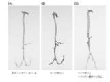
- FIG. 1 It is a figure showing the inhibitory effect of warfarin-induced arterial calcification by phytic acid, of which (A) is a photograph of a negative control blood vessel to which only vitamin K1 was administered, (B) is a lime after adding warfarin administration (C) is a photograph showing a state in which calcification is suppressed when phytic acid is further administered at 2 mg / 50-120 g body weight / day. It is a figure which shows the serum calcium density
- Phytic acid is a substance represented by the following structural formula, and is a component that is abundant in unrefined grains and beans and exhibits a strong chelating action on minerals.
- various physiological functions including an antioxidant action can be expected, and many uses as functional ingredients are also disclosed.
- phytic acid has an effect of suppressing the expression of bone formation-related factors or calcification-related factors in extra-bone tissue.
- the expression inhibitor of osteogenesis-related factor or calcification-related factor in extra-bone tissue according to the present invention is characterized by containing phytic acid.
- Phytic acid can also be used as a phytate and is not particularly limited. Examples thereof include calcium salt, magnesium salt, and zinc salt of phytic acid.
- the expression inhibitor of osteogenesis-related factor or calcification-related factor in extra-bone tissue contains phytic acid as a pharmaceutically effective amount.
- phytic acid for example, tablets, granules, fine granules, capsules
- preparations for oral administration such as agents, various liquid preparations suitable for oral administration, or preparations for parenteral administration such as injections and suppositories.
- preparations for injection are prepared, for example, in the form of solutions, emulsions or suspensions, and are made isotonic with blood.
- Formulations in the form of liquids, emulsions or suspensions are prepared using, for example, an aqueous medium, ethyl alcohol, propylene glycol, ethoxylated isostearyl alcohol, polyoxylated isostearyl alcohol, polyoxyethylene sorbitan fatty acid ester .
- the aqueous medium include water or a medium containing water.
- sterilized water is used.
- the medium containing water include physiological saline, PBS (phosphate buffered physiological saline), lactic acid-containing Ringer's solution, and the like.
- the content of phytic acid or a salt thereof is not particularly limited, but is, for example, 1 ⁇ g / mL to 10 ⁇ g / mL, preferably 100 ⁇ g / mL to 1 ⁇ m mg / mL.
- additives usually used in the art can be appropriately used.
- the additive include isotonic agents, stabilizers, buffers, preservatives, chelating agents, antioxidants, and solubilizing agents.
- the isotonic agent include sugars such as glucose, sorbitol and mannitol, sodium chloride, glycerin, propylene glycol, polyethylene glycol and the like.
- the stabilizer include sodium sulfite.
- the buffer include borate buffer, phosphate buffer, citrate buffer, tartaric acid buffer, and acetate buffer.
- Examples of the preservative include paraoxybenzoic acid ester, benzyl alcohol, chlorocresol, phenethyl alcohol, benzethonium chloride and the like.
- Examples of chelating agents include sodium edetate and sodium citrate.
- Examples of the antioxidant include sodium sulfite, sodium hydrogen sulfite, sodium ascorbate, sodium thiosulfate and the like.
- Examples of the solubilizer include dextran, polyvinylpyrrolidone, sodium benzoate, ethylenediamine, salicylic acid amide, nicotinic acid amide, polyoxyethylene hydrogenated castor oil derivative, and the like.
- the pH preparation may be contained in the injectable preparation.
- the pH adjuster may be an acid or a base.
- the acids include ascorbic acid, hydrochloric acid, gluconic acid, acetic acid, lactic acid, boric acid, phosphoric acid, sulfuric acid, tartaric acid, and citric acid.
- the base include potassium hydroxide, calcium hydroxide, sodium hydroxide, magnesium hydroxide, monoethanolamine, diethanolamine, triethanolamine and the like.
- the bone formation-related factor is not particularly limited, and examples thereof include ALPL, BGLAP, RUNX2, SP7 (Osterix), DLX5, and the like.
- the bone mineralization-related factor is not particularly limited, and examples thereof include SLC20A1, SLC20A2, ENPP1, ALPL, and SPP1.
- the action of phytic acid suppresses the expression of bone formation-related factors or calcification-related factors in extra-osseous tissue, thereby suppressing, for example, ectopic ossification and also suppressing ectopic calcification. be able to.
- Ectopic ossification mainly includes post-traumatic ossification, neoplastic ossification, and ossification in a neurological state.
- Post-traumatic ossification is ossification that occurs at the attachment of the medial collateral ligament of the femoral endocondyle after a major trauma or a small traumatic trauma.
- Neoplastic ossification is soft tissue ossification that occurs at the time of paraskeletal osteosarcoma, soft tissue osteosarcoma, intramuscular hemangioma and the like.
- Osteogenesis in the neurological state is ossification that occurs in the paralyzed limbs due to paraplegia or limb paralysis due to spinal cord injury or hemiplegia due to stroke.
- Progressive ossifying fibrodysplasia is a disease in which skeletal malformations and soft tissue ossification are observed systemically.
- the ectopic calcification mainly includes metastatic calcification, dystrophic calcification, and idiopathic calcification.
- Metastatic calcification is calcification that occurs because there is one or both of long-lasting hypercalcemia and hyperphosphatemia.
- Heterotrophic calcification is calcification in which calcium salt deposits occur in soft tissues without any disturbance of calcium or phosphorus metabolism.
- Idiopathic calcification is a major humerus nodule, especially supraspinatus attachment, deltoid muscle attachment, calcific tendonitis or bursitis that occurs frequently on the outer epicondyle of the knee joint, cartilage calcification, It is localized calcification and pandemic calcification.
- Ectopic calcification is partly due to the same molecular mechanism as ectopic ossification, and bone-like tissue formation occurs in areas where bone tissue does not originally exist, that is, muscle, fascia, ligament, joint capsule, blood vessels, etc. It is a phenomenon.
- Specific diseases include renal disease, vascular calcification associated with dialysis, vascular calcification due to arteriosclerosis, infant systemic arterial calcification (GACI), progressive ossifying fibrodysplasia (FOP), posterior longitudinal ligament Designated intractable diseases such as ossification disease (OPLL) or yellow ligament ossification disease are applicable.
- the prophylactic and / or therapeutic agent for ectopic ossification or ectopic calcification resulting from expression of an osteogenesis-related factor or calcification-related factor according to the present embodiment is characterized by containing phytic acid.
- the disease causing ectopic ossification is not particularly limited, and includes FOP, OPLL, or ossification of the ligamentum flavum mentioned in the above specific examples.
- FOP is a genetic disease characterized by congenital malformations of the thumb and progressive ectopic ossification.
- FOP may be accompanied by gene mutation, for example, ACVR1 mutation.
- FOP results in extra-articular stiffening or thorax fusion of the main joints in the mid-axis skeleton and appendage skeleton, resulting in severe disability and respiratory failure.
- the disease causing ectopic calcification is not particularly limited, and includes renal disease or vascular calcification associated with dialysis, vascular calcification due to arteriosclerosis, or GACI mentioned in the above specific examples.
- prevention includes suppressing and delaying the onset of a disease, and includes not only prevention before becoming a disease but also prevention of recurrence of the disease after treatment.
- treatment includes curing a symptom, improving the symptom, and suppressing the progression of the symptom.
- the dosage of the prophylactic and / or therapeutic agent for ectopic ossification or ectopic calcification resulting from the expression of an osteogenesis-related factor or calcification-related factor according to this embodiment can be appropriately changed.
- the amount is about 0.1 to about 2000 mg / kg / day, preferably about 1 to 200 mg / kg / day, and this amount can be administered once a day or divided into 2 to 3 times.
- Example 1 Effect of intravenous administration of phytic acid in a BMP-2-dependent intramuscular ossification model
- Atelocollagen impregnated with BMP-2 was transplanted into the greater gluteus muscle of 4-week-old ddY male mice.
- An ectopic ossification model in which ossification was observed in the transplanted part over the week was created.
- the ossified tissue of the ectopic ossification model was confirmed in the micro CT image.
- osmotic pump Alzet
- 0.08 mg / body / day, 0.4 mg / body / day or 2 mg / body / day sodium phytate was continuously administered from the right jugular vein to confirm the effect on ectopic ossification. did.
- the amount of ossified tissue decreased depending on the dose of phytic acid.
- the vertical axis represents the bone mass (mm 3 ) obtained by micro CT.
- Matrix mineralization including bone matrix
- matrix vesicles produced and secreted by osteoblasts present on the bone surface (matrix vesicular calcification) and formed inside the matrix vesicles
- matrix vesicular calcification matrix vesicular calcification
- the formed calcium phosphate crystal mass shows a ribbon-like or needle-like shape, and when it breaks the membrane of the vesicle and is exposed to the outside, it forms a spherical aggregate (mineralized nodule), and then the surrounding collagen By contacting the fine fibers, calcification is spread to the collagen fibers.
- osteoblasts are cells responsible for bone formation and induce calcification.
- Example 3 Calcified tissue staining diagram of mouse aortic organ culture at phosphate loading
- 4% paraformaldehyde PBS was used after completion of the culture.
- a paraffin specimen was prepared by fixing. 5 ⁇ m sections were prepared and Kossa staining was performed. Note that Kossa staining is a staining method used when detecting lime (calcium salt) deposited in histological examination, and stains calcium phosphate salt, calcium carbonate salt and the like in black by the action of silver nitrate.
- the scale bar is 25 ⁇ m.
- Example 4 Tissue staining diagram of osteoblast marker Using the above paraffin section for the mouse aorta organ culture model under phosphate-loaded environment in Example 2 above, immunization of osteoblast (bone formation) marker ALP Staining was attempted.
- ALP alkaline phosphatase
- BAP bone formation marker
- the immunostaining was observed using a fluorescence microscope after reacting with an anti-ALP antibody and Alexa594-labeled secondary antibody according to a conventional method.
- DAPI was used as a counterstain.
- FIG. 4 when phosphate was loaded, ALP, which is an osteoblast marker, was stained red when only the solvent was used. On the other hand, depending on the dose of phytic acid, it is understood that the portion stained red is reduced, and the formation of osteoblast-like cells is suppressed.
- the scale bar is 75 ⁇ m.
- Example 5 Expression of Osteoblast Marker Gene
- the expression of osteoblast (bone formation) marker gene in the mouse aorta organ culture model under the phosphate load environment in Example 2 above was examined by RT-PCR method. It was. After completion of the culture, RNA was extracted according to a conventional method, and after cDNA preparation, gene expression was quantified by real-time PCR. As osteogenesis-related factors, the expression of Alpl, Runx2, and Bglap was examined.
- the Alpl gene is a gene encoding a tissue non-specific isozyme (TNSALP) of Alkaline phosphatase.
- Runx2 is a transcription factor essential for osteoblast differentiation and chondrocyte late differentiation.
- Bglap is a marker that confirms the direction of differentiation from mesenchymal stem cells to osteoblasts because it is expressed at a high level when cultured cells are calcified later in differentiation.
- Slc20a1 expression was examined as a calcification-related factor.
- SLC20A1 is a type III phosphate transporter and is known to play an important role in calcification.
- An increase in the phosphate concentration in the blood causes calcification of vascular smooth muscle and causes arteriosclerosis centering on calcification of the media. This is because the increase of extracellular phosphate concentration increases the intracellular influx of phosphate via SLC20A1 in vascular smooth muscle cells, induces the expression of osteoblast differentiation-related proteins, and differentiates into osteoblast-like cells It is thought that calcification is caused.
- FIGS. 5 (A), (B) and (C) it was shown that the expression of osteogenesis-related factors was suppressed in the presence of phytic acid.
- FIG. 5 (D) it was shown that the expression of calcification-related factors was suppressed in the presence of phytic acid.
- Example 6 Inhibition of Warfarin-Induced Arterial Calcification Vitamin K inhibitors typified by warfarin inhibit matrix GIA protein (MGP) Gla formation, which is one of calcification inhibitory factors, and There is an inhibitory effect.
- MGP matrix GIA protein
- the normal diet was switched to a high phosphate diet containing 1.2 wt% phosphate and 0.6 wt% calcium, and the mother rat was given a high phosphate load.
- oral administration of vitamin K1 1.5 mg / 100 g body weight / day
- sodium phytate 0.4 and 2 mg / 50-120 g body weight / day
- Example 7 Fluctuation of serum calcium (Ca) and phosphate (inorganic phosphate, Pi) concentration in intravenous administration of phytic acid Using an osmotic pump as described above in the vein of a 4-week-old ddY male mouse Then, phytic acid was continuously administered for 1 week, and changes in calcium concentration in blood were measured. The doses of phytic acid were 0.08 mg / body / day, 0.4 mg / body / day, and 2 mg / body / day. The Ca concentration was measured with Calcium E TM Test Wako. The results are shown in FIG. As shown in FIG. 7, the Ca concentration in serum was not affected by phytic acid. Similarly, the serum Pi concentration was measured by Phospha C-Test Wako. The results are shown in FIG. As shown in FIG. 8, phytic acid had no effect on serum Pi concentration.
- Ca calcium
- phosphate inorganic phosphate, Pi
- Example 8 Inhibition of calcification in Enpp1 mutant mice Enpp1 is a gene responsible for ossification of the posterior longitudinal ligament, and many infant systemic arterial calcifications involve both allelic mutations of the same gene.
- a high phosphorus diet was fed to the mother mice of Enpp1 mutant newborn mice (C57BL / 6J-Enpp1 asj / GrsrJ), and a high phosphorus diet was fed directly after weaning at 3 weeks of age.
- FIG. 9 shows an arterial arch.
- (A) is Vehicle
- (B) is 0.4 mg / body / day
- (C) is 0.04 mg / body / day.
- phytic acid almost completely suppressed vascular calcification stained with alizarin red at a concentration of 0.04 mg / body / day or more.
- Example 9 Measurement of bone morphology by micro CT The bone morphology of the proximal end of the tibia was measured for the Enpp1 mutant mouse (6 weeks old) of (8) above. Cortical bone and sea surface bone were analyzed for bone mass, bone mass / tissue mass, bone density, number of sea surface bones, sea surface bone thickness, and the like. The bone mass and bone density are shown in FIG. Among them, (A) is cortical bone mass, (B) is sea surface bone mass, (C) is cortical bone density, and (D) is sea surface bone density. None of the parameters was affected by phytic acid administration. Similarly, even when phytic acid was administered to normal mice, no abnormality was observed in various bone morphometry parameters.
- Example 7 Serum sample obtained from the mouse of Example 7 (continuous intravenous administration for 1 week)
- Serum sample administered with 0.4 mg / body / day sodium phytate and vehicle in Example 8 discontinuous intravenous administration for 2 weeks
- FIG. 11 shows the results of serum samples administered with 0.4 mg / body / day of sodium phytate and vehicle, in which (A) is the serum zinc concentration and (B) is the serum iron concentration.
- phytic acid suppresses ectopic ossification or ectopic calcification, but its main mechanism of action is not the chelating effect of phytic acid, but an osteogenesis-related factor or calcification. It can be considered that the expression of related factors is suppressed.
Abstract
Provided is an inhibitor of the expression of a bone formation-related factor or a calcification-related factor in an extraskeletal tissue. An inhibitor of the expression of a bone formation-related factor in an extraskeletal tissue, the inhibitor being characterized by comprising phytic acid. The bone formation-related factor is any one of ALPL, RUNX2, BGLAP and the like. An inhibitor of the expression of a calcification-related factor in an extraskeletal tissue, the inhibitor being characterized by comprising phytic acid. The calcification-related factor is any one of SLC20A1, SLC20A2, ENPP1, ALPL, SPP1 and the like. These inhibitors can prevent heterotopic ossification and ectopic calcification, respectively, when administered intravenously.
Description
本発明は、骨外組織における骨形成関連因子又は石灰化関連因子の発現抑制剤に関する。
The present invention relates to an expression inhibitor of bone formation-related factor or calcification-related factor in extra-bone tissue.
骨を構成する細胞には、骨芽細胞、骨細胞、破骨細胞がある。骨芽細胞は、間葉系幹細胞から分化した骨形成細胞で、幹細胞から段階的に分化していく。培養骨芽細胞系において、細胞の分化段階の指標となるマーカー遺伝子は多様なパターンを示す。例えば、初期の増殖段階では、細胞周期を制御する蛋白であるCCND1やHDACを発現するが、分化形質は時間依存的に発現し、分化早期にはCOL1A1を発現する。その後、成熟するにつれてFN1、ALPL、IBSP、BGLAPという順で発現パターンを示す。これらのマーカーの中で、COL1A1、ALPLは、細胞周期には依存せず発現する一方、BGLAPは、細胞分裂の終了後に発現する。
The cells that constitute bone include osteoblasts, bone cells, and osteoclasts. Osteoblasts are osteogenic cells differentiated from mesenchymal stem cells and gradually differentiate from stem cells. In cultured osteoblast cell lines, marker genes that serve as indicators of the differentiation stage of cells show various patterns. For example, in the early stage of proliferation, CCND1 and HDAC, which are proteins that control the cell cycle, are expressed, but differentiation traits are expressed in a time-dependent manner, and COL1A1 is expressed early in differentiation. Then, as it matures, it shows expression patterns in the order of FN1, ALPL, IBSP, and BGLAP. Among these markers, COL1A1 and ALPL are expressed independently of the cell cycle, while BGLAP is expressed after the end of cell division.
その他、骨芽細胞のマーカーにRUNX2、SPP1、MSX2、DLX5、TWIST1、JUN等の転写因子がある。RUNX2はBGLAPやSPP1遺伝子の上流のシスエレメントに結合することによってその転写を促進することから、RUNX2は骨基質蛋白の産生に重要な役割を果たしている。
In addition, there are transcription factors such as RUNX2, SPP1, MSX2, DLX5, TWIST1, and JUN as markers for osteoblasts. RUNX2 plays an important role in the production of bone matrix proteins because it promotes transcription by binding to cis elements upstream of the BGLAP and SPP1 genes.
しかしながら、上述した骨形成関連因子が骨外組織で発現すると、通常の骨格形成とは別に、本来は骨組織が形成されない部位に病的な骨形成が起こる現象である異所性骨化が発生する場合がある。また、石灰化とはカルシウムの沈着の総称であり、本来は石灰化されない部位に病的な石灰化が起こる現象を異所性石灰化と呼ぶが、この異所性石灰化の少なくとも一部は骨形成と同じ分子機構によって生ずることがわかっている(非特許文献1)。異所性骨化や異所性石灰化の発生する組織により、様々な障害を引き起こす。例えば、関節領域では可動域制限を伴うため、著しくQuality of Life(QOL)の低下を引き起こす。血管では心血管イベントや生命予後に関連する。
However, when the above-mentioned bone formation-related factors are expressed in extra-osseous tissue, ectopic ossification, a phenomenon in which pathological bone formation occurs in a site where bone tissue is not originally formed, occurs in addition to normal skeleton formation. There is a case. In addition, calcification is a general term for the deposition of calcium, and the phenomenon in which pathological calcification occurs in areas that are not originally calcified is called ectopic calcification, but at least part of this ectopic calcification is It is known that it occurs by the same molecular mechanism as bone formation (Non-patent Document 1). Tissues with ectopic ossification and ectopic calcification cause various disorders. For example, the joint region is accompanied by a limited range of motion, which significantly reduces the quality of life (QOL). In blood vessels, it is related to cardiovascular events and life prognosis.
特許文献1には、NRF2がRunx2依存的にBglap転写活性を抑制し得ることから、nrf2による骨・軟骨分化の負の調節は、NRF2によるRunx2の阻害に起因し得るものであることに着目した骨芽細胞分化の調節剤が記載されている。
Patent Document 1 focused on the fact that Nrf2 can suppress Bglap transcriptional activity in a Runx2-dependent manner, and thus negative regulation of bone / cartilage differentiation by nrf2 can be attributed to inhibition of Runx2 by NRF2. A regulator of osteoblast differentiation has been described.
しかし、上記特許文献1に記載された物質では、骨形成関連因子又は石灰化関連因子の発現抑制は十分なものではない。
However, the substances described in Patent Document 1 do not sufficiently suppress the expression of bone formation-related factors or calcification-related factors.
本発明はかかる問題点に鑑みてなされたものであって、新規な骨外組織における骨形成関連因子又は石灰化関連因子の発現の抑制剤を提供することを目的とする。
The present invention has been made in view of such problems, and an object of the present invention is to provide a novel inhibitor of the expression of osteogenesis-related factors or calcification-related factors in extra-osseous tissue.
本発明にかかる骨外組織における骨形成関連因子又は石灰化関連因子の発現抑制剤は、フィチン酸を含むことを特徴とする。
The expression inhibitor of osteogenesis-related factor or calcification-related factor in extra-bone tissue according to the present invention is characterized by containing phytic acid.
本発明によれば、適切に、骨外組織における骨形成関連因子又は石灰化関連因子の発現を抑制できる。
According to the present invention, it is possible to appropriately suppress the expression of a bone formation-related factor or a calcification-related factor in extra-osseous tissue.
以下、添付の図面を参照して本発明の実施形態について具体的に説明するが、当該実施形態は本発明の原理の理解を容易にするためのものであり、本発明の範囲は、下記の実施形態に限られるものではなく、当業者が以下の実施形態の構成を適宜置換した他の実施形態も、本発明の範囲に含まれる。
Hereinafter, embodiments of the present invention will be specifically described with reference to the accompanying drawings. However, the embodiments are for facilitating understanding of the principle of the present invention, and the scope of the present invention is as follows. The present invention is not limited to the embodiments, and other embodiments in which those skilled in the art appropriately replace the configurations of the following embodiments are also included in the scope of the present invention.
フィチン酸は、下記構造式で示される物質であり、未精製の穀物や豆類に多く含まれる成分でミネラルに強いキレート作用を示す。酸味料やpH調整剤として食品添加物に認可されている他、抗酸化作用をはじめとする様々な生理機能が期待できることから機能性成分としての利用も多く開示されている。
Phytic acid is a substance represented by the following structural formula, and is a component that is abundant in unrefined grains and beans and exhibits a strong chelating action on minerals. In addition to being approved as a food additive as a sour agent and a pH adjuster, various physiological functions including an antioxidant action can be expected, and many uses as functional ingredients are also disclosed.

しかしながら、フィチン酸が、骨外組織における骨形成関連因子又は石灰化関連因子の発現抑制効果を有することは、従来知られていない。
However, it has not been known that phytic acid has an effect of suppressing the expression of bone formation-related factors or calcification-related factors in extra-bone tissue.
本発明にかかる骨外組織における骨形成関連因子又は石灰化関連因子の発現抑制剤は、フィチン酸を含むことを特徴とする。
The expression inhibitor of osteogenesis-related factor or calcification-related factor in extra-bone tissue according to the present invention is characterized by containing phytic acid.
フィチン酸はフィチン酸塩としても使用可能であり、特に限定されるものではないが、例えば、フィチン酸のカルシウム塩、マグネシウム塩、亜鉛塩等が挙げられる。
Phytic acid can also be used as a phytate and is not particularly limited. Examples thereof include calcium salt, magnesium salt, and zinc salt of phytic acid.
本発明にかかる骨外組織における骨形成関連因子又は石灰化関連因子の発現抑制剤は、医薬的に有効量としてのフィチン酸を含むものであり、例えば、錠剤、顆粒剤、細粒剤、カプセル剤等のような経口投与の製剤、経口投与に適した様々な液体製剤、又は注射剤、坐剤のような非経口投与用製剤とすることが可能である。
The expression inhibitor of osteogenesis-related factor or calcification-related factor in extra-bone tissue according to the present invention contains phytic acid as a pharmaceutically effective amount. For example, tablets, granules, fine granules, capsules It is possible to make preparations for oral administration such as agents, various liquid preparations suitable for oral administration, or preparations for parenteral administration such as injections and suppositories.
非経口投与用製剤のうち注射用製剤は、例えば、液剤、乳濁液、又は懸濁液の形態で調製され、血液に対して等張にされる。液体、乳濁液又は懸濁液の形態の製剤は、例えば、水性媒体、エチルアルコール、プロピレングリコール、エトキシ化イソステアリルアルコール、ポリオキシ化イソステアリルアルコール、ポリオキシエチレンソルビタン脂肪酸エステルを用いて調製される。水性媒体としては、水又は水を含有する媒体が挙げられる。水としては、滅菌水が使用される。水を含有する媒体としては、例えば、生理食塩水、PBS(リン酸緩衝生理食塩水)又は乳酸配合リンゲル液等が挙げられる。
Among the preparations for parenteral administration, preparations for injection are prepared, for example, in the form of solutions, emulsions or suspensions, and are made isotonic with blood. Formulations in the form of liquids, emulsions or suspensions are prepared using, for example, an aqueous medium, ethyl alcohol, propylene glycol, ethoxylated isostearyl alcohol, polyoxylated isostearyl alcohol, polyoxyethylene sorbitan fatty acid ester . Examples of the aqueous medium include water or a medium containing water. As water, sterilized water is used. Examples of the medium containing water include physiological saline, PBS (phosphate buffered physiological saline), lactic acid-containing Ringer's solution, and the like.
注射用製剤において、フィチン酸またはその塩の含有量は、特に限定されるものではないが、例えば1μg/mL~10 mg/mL、好ましくは100μg/mL~1 mg/mLである。
In the preparation for injection, the content of phytic acid or a salt thereof is not particularly limited, but is, for example, 1 μg / mL to 10 μg / mL, preferably 100 μg / mL to 1 μm mg / mL.
注射用製剤において、当技術分野で通常使用されている添加剤を適宜用いることができる。添加剤としては、例えば、等張化剤、安定化剤、緩衝剤、保存剤、キレート剤、抗酸化剤、又は溶解補助剤等が挙げられる。等張化剤としては、例えば、ブドウ糖、ソルビトール、マンニトール等の糖類、塩化ナトリウム、グリセリン、プロピレングリコール、ポリエチレングリコール等が挙げられる。安定化剤としては、例えば亜硫酸ナトリウム等が挙げられる。緩衝剤としては、例えば、ホウ酸緩衝剤、リン酸緩衝剤、クエン酸緩衝剤、酒石酸緩衝剤、酢酸緩衝剤等が挙げられる。保存剤としては、例えば、パラオキシ安息香酸エステル、ベンジルアルコール、クロロクレゾール、フェネチルアルコール、塩化ベンゼトニウム等が挙げられる。キレート剤としては、例えば、エデト酸ナトリウム、クエン酸ナトリウム等が挙げられる。抗酸化剤としては、例えば、亜硫酸ナトリウム、亜硫酸水素ナトリウム、アスコルビン酸ナトリウム、チオ硫酸ナトリウム等が挙げられる。溶解補助剤としては、例えば、デキストラン、ポリビニルピロリドン、安息香酸ナトリウム、エチレンジアミン、サリチル酸アミド、ニコチン酸アミド、ポリオキシエチレン硬化ヒマシ油誘導体等が挙げられる。
In the preparation for injection, additives usually used in the art can be appropriately used. Examples of the additive include isotonic agents, stabilizers, buffers, preservatives, chelating agents, antioxidants, and solubilizing agents. Examples of the isotonic agent include sugars such as glucose, sorbitol and mannitol, sodium chloride, glycerin, propylene glycol, polyethylene glycol and the like. Examples of the stabilizer include sodium sulfite. Examples of the buffer include borate buffer, phosphate buffer, citrate buffer, tartaric acid buffer, and acetate buffer. Examples of the preservative include paraoxybenzoic acid ester, benzyl alcohol, chlorocresol, phenethyl alcohol, benzethonium chloride and the like. Examples of chelating agents include sodium edetate and sodium citrate. Examples of the antioxidant include sodium sulfite, sodium hydrogen sulfite, sodium ascorbate, sodium thiosulfate and the like. Examples of the solubilizer include dextran, polyvinylpyrrolidone, sodium benzoate, ethylenediamine, salicylic acid amide, nicotinic acid amide, polyoxyethylene hydrogenated castor oil derivative, and the like.
注射用製剤にはpH調整剤が含有されていても良い。pH調整剤は、酸類であっても塩基類であってもよい。具体的には、酸類としては、例えば、アスコルビン酸、塩酸、グルコン酸、酢酸、乳酸、ホウ酸、リン酸、硫酸、酒石酸、クエン酸等が挙げられる。塩基類としては、例えば、水酸化カリウム、水酸化カルシウム、水酸化ナトリウム、水酸化マグネシウム、モノエタノールアミン、ジエタノールアミン、トリエタノールアミン等が挙げられる。
The pH preparation may be contained in the injectable preparation. The pH adjuster may be an acid or a base. Specifically, examples of the acids include ascorbic acid, hydrochloric acid, gluconic acid, acetic acid, lactic acid, boric acid, phosphoric acid, sulfuric acid, tartaric acid, and citric acid. Examples of the base include potassium hydroxide, calcium hydroxide, sodium hydroxide, magnesium hydroxide, monoethanolamine, diethanolamine, triethanolamine and the like.
骨形成関連因子は、特に限定されるものではないが、例えばALPL、BGLAP、RUNX2、SP7 (Osterix)、DLX5等が挙げられる。
The bone formation-related factor is not particularly limited, and examples thereof include ALPL, BGLAP, RUNX2, SP7 (Osterix), DLX5, and the like.
骨の石灰化関連因子は、特に限定されるものではないが、例えばSLC20A1、SLC20A2、ENPP1、ALPL、SPP1等が挙げられる。
The bone mineralization-related factor is not particularly limited, and examples thereof include SLC20A1, SLC20A2, ENPP1, ALPL, and SPP1.
本発明においては、フィチン酸の作用により骨外組織における骨形成関連因子又は石灰化関連因子の発現を抑制させ、これにより例えば異所性骨化を抑制させ、また異所性石灰化を抑制させることができる。
In the present invention, the action of phytic acid suppresses the expression of bone formation-related factors or calcification-related factors in extra-osseous tissue, thereby suppressing, for example, ectopic ossification and also suppressing ectopic calcification. be able to.
この点、フィチン酸のキレート作用により異所性骨化が抑制されるとの見解もあるが、本発明者らは異所性骨化のフィチン酸による抑制の重要なメカニズムはキレート作用にあるのではなく、骨形成関連因子又は石灰化関連因子の発現抑制にあることを見出した。
In this regard, there is a view that ectopic ossification is suppressed by the chelating action of phytic acid, but the present inventors have an important mechanism of suppression of ectopic ossification by phytic acid in chelating action. Instead, it was found to be the suppression of the expression of bone formation-related factors or mineralization-related factors.
異所性骨化としては、主として、外傷後骨化、腫瘍性骨化、神経学的状態での骨化が挙げられる。外傷後骨化は、大きな外傷、あるいは反覆された小外傷後に、大腿骨内顆の内側側副靭帯の付着部に起こる骨化である。腫瘍性骨化は、傍骨性骨肉腫、軟部組織骨肉腫、筋肉内血管腫などの時に起こる軟部組織性骨化である。神経学的状態での骨化は、脊髄損傷による対麻痺や四肢麻痺、脳卒中による片麻痺での麻痺肢に起こる骨化である。進行性骨化性線維異形成症は、全身性に骨格の奇形と軟部組織の骨化がみられる疾患である。
Ectopic ossification mainly includes post-traumatic ossification, neoplastic ossification, and ossification in a neurological state. Post-traumatic ossification is ossification that occurs at the attachment of the medial collateral ligament of the femoral endocondyle after a major trauma or a small traumatic trauma. Neoplastic ossification is soft tissue ossification that occurs at the time of paraskeletal osteosarcoma, soft tissue osteosarcoma, intramuscular hemangioma and the like. Osteogenesis in the neurological state is ossification that occurs in the paralyzed limbs due to paraplegia or limb paralysis due to spinal cord injury or hemiplegia due to stroke. Progressive ossifying fibrodysplasia is a disease in which skeletal malformations and soft tissue ossification are observed systemically.
異所性石灰化としては、主として、転移性石灰化、異栄養性石灰化、特発性石灰沈着症が挙げられる。転移性石灰化は、長く継続する高カルシウム血症と高リン血症の片方もしくは両方があるために起こる石灰沈着である。異栄養性石灰化は、カルシウムやリン代謝の障害がなくても、軟部組織にカルシウム塩の沈着が起こる石灰化である。特発性石灰沈着症は、上腕骨大結節、特に棘上筋の付着部、三角筋付着部、膝関節の外側上顆に好発する石灰沈着性腱炎や滑液包炎、軟骨石灰沈着症、限局性石灰沈着症、汎発生石灰沈着症である。
The ectopic calcification mainly includes metastatic calcification, dystrophic calcification, and idiopathic calcification. Metastatic calcification is calcification that occurs because there is one or both of long-lasting hypercalcemia and hyperphosphatemia. Heterotrophic calcification is calcification in which calcium salt deposits occur in soft tissues without any disturbance of calcium or phosphorus metabolism. Idiopathic calcification is a major humerus nodule, especially supraspinatus attachment, deltoid muscle attachment, calcific tendonitis or bursitis that occurs frequently on the outer epicondyle of the knee joint, cartilage calcification, It is localized calcification and pandemic calcification.
異所性石灰化は異所性骨化と一部において同じ分子機構によって生じ、本来骨組織が存在しない部位、すなわち筋、筋膜、靱帯、関節包、血管などに骨様組織の形成が起こる現象である。具体的な疾患として、腎疾患、透析に伴う血管石灰化、動脈硬化による血管石灰化、乳児性全身性動脈石灰化(GACI)、進行性骨化性線維異形成症(FOP)、後縦靭帯骨化症(OPLL)、又は、黄色靭帯骨化症といった指定難病が該当する。
Ectopic calcification is partly due to the same molecular mechanism as ectopic ossification, and bone-like tissue formation occurs in areas where bone tissue does not originally exist, that is, muscle, fascia, ligament, joint capsule, blood vessels, etc. It is a phenomenon. Specific diseases include renal disease, vascular calcification associated with dialysis, vascular calcification due to arteriosclerosis, infant systemic arterial calcification (GACI), progressive ossifying fibrodysplasia (FOP), posterior longitudinal ligament Designated intractable diseases such as ossification disease (OPLL) or yellow ligament ossification disease are applicable.
本実施形態にかかる骨形成関連因子又は石灰化関連因子の発現に起因する異所性骨化又は異所性石灰化の予防及び/又は治療剤は、フィチン酸を含むことを特徴とする。異所性骨化を起こす疾患は特に限定されるものではなく、上述の具体例において挙げられたFOP、OPLL、又は、黄色靭帯骨化症が該当する。なおFOPは、親指の先天性奇形や進行性異所性骨化により特徴づけられる遺伝子疾患である。FOPは遺伝子の突然変異を伴うものでもよく、例えばACVR1の変異である。FOPは中軸骨格及び付属肢骨格における主関節の関節外硬直又は胸郭の融合を生じ、重篤な障害や呼吸器不全を生じる。異所性石灰化を起こす疾患は特に限定されるものでなく、上述の具体例において挙げられた腎疾患若しくは透析に伴う血管石灰化、動脈硬化による血管石灰化、又は、GACIが該当する。
The prophylactic and / or therapeutic agent for ectopic ossification or ectopic calcification resulting from expression of an osteogenesis-related factor or calcification-related factor according to the present embodiment is characterized by containing phytic acid. The disease causing ectopic ossification is not particularly limited, and includes FOP, OPLL, or ossification of the ligamentum flavum mentioned in the above specific examples. FOP is a genetic disease characterized by congenital malformations of the thumb and progressive ectopic ossification. FOP may be accompanied by gene mutation, for example, ACVR1 mutation. FOP results in extra-articular stiffening or thorax fusion of the main joints in the mid-axis skeleton and appendage skeleton, resulting in severe disability and respiratory failure. The disease causing ectopic calcification is not particularly limited, and includes renal disease or vascular calcification associated with dialysis, vascular calcification due to arteriosclerosis, or GACI mentioned in the above specific examples.
本明細書において「予防」には疾患の発症を抑えること及び遅延させることが含まれ、疾患になる前の予防だけでなく、治療後の疾患の再発に対する予防も含まれる。一方、「治療」には、症状を治癒すること、症状を改善すること及び症状の進行を抑えることが含まれる。本実施形態にかかる骨形成関連因子又は石灰化関連因子の発現に起因する異所性骨化又は異所性石灰化の予防及び/又は治療剤の用法用量は適宜変更し得るが、例えば有効成分量として、約0.1~約2000mg/kg/日、好ましくは約1~200mg/kg/日であり、この量を1日1回又は2~3回に分けて投与することができる。
In this specification, “prevention” includes suppressing and delaying the onset of a disease, and includes not only prevention before becoming a disease but also prevention of recurrence of the disease after treatment. On the other hand, “treatment” includes curing a symptom, improving the symptom, and suppressing the progression of the symptom. The dosage of the prophylactic and / or therapeutic agent for ectopic ossification or ectopic calcification resulting from the expression of an osteogenesis-related factor or calcification-related factor according to this embodiment can be appropriately changed. The amount is about 0.1 to about 2000 mg / kg / day, preferably about 1 to 200 mg / kg / day, and this amount can be administered once a day or divided into 2 to 3 times.
(1)実施例1 BMP-2依存性筋肉内骨化モデルにおけるフィチン酸静脈内投与の効果
4週齢ddY雄マウス大臀筋内にBMP-2を含浸させたアテロコラーゲンを移植し、移植後2週間において移植部において骨化が見られる異所性骨化モデルを作成した。図1(A)のVehicleに示されるようにマイクロCT画像において、異所性骨化モデルの骨化組織が確認された。 (1) Example 1 Effect of intravenous administration of phytic acid in a BMP-2-dependent intramuscular ossification model Atelocollagen impregnated with BMP-2 was transplanted into the greater gluteus muscle of 4-week-old ddY male mice. An ectopic ossification model in which ossification was observed in the transplanted part over the week was created. As shown in the vehicle of FIG. 1 (A), the ossified tissue of the ectopic ossification model was confirmed in the micro CT image.
4週齢ddY雄マウス大臀筋内にBMP-2を含浸させたアテロコラーゲンを移植し、移植後2週間において移植部において骨化が見られる異所性骨化モデルを作成した。図1(A)のVehicleに示されるようにマイクロCT画像において、異所性骨化モデルの骨化組織が確認された。 (1) Example 1 Effect of intravenous administration of phytic acid in a BMP-2-dependent intramuscular ossification model Atelocollagen impregnated with BMP-2 was transplanted into the greater gluteus muscle of 4-week-old ddY male mice. An ectopic ossification model in which ossification was observed in the transplanted part over the week was created. As shown in the vehicle of FIG. 1 (A), the ossified tissue of the ectopic ossification model was confirmed in the micro CT image.
浸透圧ポンプ(Alzet)を用い、右頸静脈から0.08 mg/body/day、0.4 mg/body/day又は2 mg/body/dayフィチン酸ナトリウムを持続投与し、異所性骨化に対する効果を確認した。図1(A)及び(B)に示されるように、フィチン酸の用量依存的に骨化組織量は低減した。なお図1(B)において縦軸はマイクロCTによる骨量(mm3)である。
Using osmotic pump (Alzet), 0.08 mg / body / day, 0.4 mg / body / day or 2 mg / body / day sodium phytate was continuously administered from the right jugular vein to confirm the effect on ectopic ossification. did. As shown in FIGS. 1 (A) and (B), the amount of ossified tissue decreased depending on the dose of phytic acid. In FIG. 1B, the vertical axis represents the bone mass (mm 3 ) obtained by micro CT.
(2)実施例2 リン酸負荷時マウス大動脈器官培養の石灰化におけるフィチン酸投与の効果
4週齢C57BL/6雄マウスより胸大動脈を摘出し、長さ2~5 mmに切断した(図2(A))。これを培養皿に載置し、15%ウシ胎仔血清を含むDMEM (High glucose)で37℃にて10日間培養した。リン酸負荷環境については、2.6 mMのリン酸(Pi)を培養液に添加した。 (2) Example 2 Effect of phytic acid administration on calcification of mouse aortic organ culture during phosphate loading The thoracic aorta was removed from 4-week-old C57BL / 6 male mice and cut into lengths of 2 to 5 mm (FIG. 2). (A)). This was placed on a culture dish and cultured at 37 ° C. for 10 days with DMEM (High glucose) containing 15% fetal calf serum. For phosphate loading environment, 2.6 mM phosphoric acid (Pi) was added to the culture.
4週齢C57BL/6雄マウスより胸大動脈を摘出し、長さ2~5 mmに切断した(図2(A))。これを培養皿に載置し、15%ウシ胎仔血清を含むDMEM (High glucose)で37℃にて10日間培養した。リン酸負荷環境については、2.6 mMのリン酸(Pi)を培養液に添加した。 (2) Example 2 Effect of phytic acid administration on calcification of mouse aortic organ culture during phosphate loading The thoracic aorta was removed from 4-week-old C57BL / 6 male mice and cut into lengths of 2 to 5 mm (FIG. 2). (A)). This was placed on a culture dish and cultured at 37 ° C. for 10 days with DMEM (High glucose) containing 15% fetal calf serum. For phosphate loading environment, 2.6 mM phosphoric acid (Pi) was added to the culture.
10日間の培養後、マウス大動脈には石灰化組織が観察された。なお、骨基質をはじめとする基質石灰化は、骨表面に存在する骨芽細胞が産生・分泌する基質小胞(matrix vesicle)によって開始され(基質小胞性石灰化)、基質小胞内部で形成されたリン酸カルシウム結晶塊はリボン状または針状を示し、小胞の膜を破って外界に露出すると、球状の集合体(石灰化球:mineralized nodule)を形成するようになり、その後、周囲のコラーゲン細線維に接触することでコラーゲン線維に石灰化を波及してゆく。このように骨芽細胞は骨形成を担う細胞であり、石灰化を誘導する。
After 10 days of culture, calcified tissue was observed in the mouse aorta. Matrix mineralization, including bone matrix, is initiated by matrix vesicles produced and secreted by osteoblasts present on the bone surface (matrix vesicular calcification) and formed inside the matrix vesicles The formed calcium phosphate crystal mass shows a ribbon-like or needle-like shape, and when it breaks the membrane of the vesicle and is exposed to the outside, it forms a spherical aggregate (mineralized nodule), and then the surrounding collagen By contacting the fine fibers, calcification is spread to the collagen fibers. Thus, osteoblasts are cells responsible for bone formation and induce calcification.
このリン酸負荷時のマウス大動脈器官培養モデルにおいて、培養液中に1 μM、3μM又は10μMフィチン酸を添加し、10日間の培養後、石灰化組織の有無を観察した。図2(B)及び(C)に示されるように、マイクロCT画像において、フィチン酸の用量依存的に石灰化組織量は低減した。なお図2(C)において縦軸は石灰化組織量(mm3)である。
In this mouse aorta organ culture model at the time of phosphate loading, 1 μM, 3 μM or 10 μM phytic acid was added to the culture medium, and the presence or absence of calcified tissue was observed after 10 days of culture. As shown in FIGS. 2 (B) and 2 (C), in the micro CT image, the amount of calcified tissue decreased depending on the dose of phytic acid. In FIG. 2 (C), the vertical axis represents the amount of calcified tissue (mm 3 ).
(3)実施例3 リン酸負荷時マウス大動脈器官培養の石灰化組織染色図
上記の実施例2におけるリン酸負荷環境下でのマウス大動脈器官培養モデルにつき、培養終了後に4% パラホムアルデヒドPBSで固定してパラフィン標本を作製した。5μmの切片を用意し、コッサ染色を行った。なお、コッサ染色は、組織学的検査において沈着した石灰(カルシウム塩)を検出する際に用いられる染色法であり、硝酸銀の作用によりリン酸カルシウム塩や炭酸カルシウム塩等を黒色に染色する。 (3) Example 3 Calcified tissue staining diagram of mouse aortic organ culture at phosphate loading For the mouse aortic organ culture model under phosphate loading environment in Example 2 above, 4% paraformaldehyde PBS was used after completion of the culture. A paraffin specimen was prepared by fixing. 5 μm sections were prepared and Kossa staining was performed. Note that Kossa staining is a staining method used when detecting lime (calcium salt) deposited in histological examination, and stains calcium phosphate salt, calcium carbonate salt and the like in black by the action of silver nitrate.
上記の実施例2におけるリン酸負荷環境下でのマウス大動脈器官培養モデルにつき、培養終了後に4% パラホムアルデヒドPBSで固定してパラフィン標本を作製した。5μmの切片を用意し、コッサ染色を行った。なお、コッサ染色は、組織学的検査において沈着した石灰(カルシウム塩)を検出する際に用いられる染色法であり、硝酸銀の作用によりリン酸カルシウム塩や炭酸カルシウム塩等を黒色に染色する。 (3) Example 3 Calcified tissue staining diagram of mouse aortic organ culture at phosphate loading For the mouse aortic organ culture model under phosphate loading environment in Example 2 above, 4% paraformaldehyde PBS was used after completion of the culture. A paraffin specimen was prepared by fixing. 5 μm sections were prepared and Kossa staining was performed. Note that Kossa staining is a staining method used when detecting lime (calcium salt) deposited in histological examination, and stains calcium phosphate salt, calcium carbonate salt and the like in black by the action of silver nitrate.
図3に示されるように、フィチン酸の用量依存的に黒色に染色された部分は低減しており、石灰化組織量が低減していることが理解される。なお図3においてスケールバーは25μmである。
As shown in FIG. 3, it is understood that the portion of phytic acid stained black in a dose-dependent manner is reduced, and the amount of calcified tissue is reduced. In FIG. 3, the scale bar is 25 μm.
(4)実施例4 骨芽細胞マーカーの組織染色図
上記の実施例2におけるリン酸負荷環境下でのマウス大動脈器官培養モデルにつき上記パラフィン切片を用い、骨芽細胞(骨形成)マーカーALPの免疫染色を試みた。ALP (アルカリフォスファターゼ Alkaline Phoshatase)は細胞膜に存在する糖タンパク質である。骨組織に特異的に存在するALPはBAPと呼ばれ、細胞膜に存在し、ホスファチジルイノシトールを介して膜に結合する。骨芽細胞機能が亢進し、骨形成活性が亢進している時期には、血中BAPは上昇するため、ALPは骨芽細胞マーカーである。免疫染色は常法にしたがって抗ALP抗体、Alexa594標識二次抗体と反応後、蛍光顕微鏡を用いて観察した。対比染色としてDAPIを用いた。図4に示されるように、リン酸負荷時、溶媒のみの場合は骨芽細胞マーカーであるALPは赤色に染色された。一方で、フィチン酸の用量依存的に、赤色に染色された部分は低減し、骨芽細胞様細胞の形成が抑制されていることが理解される。なお図4においてスケールバーは75μmである。 (4) Example 4 Tissue staining diagram of osteoblast marker Using the above paraffin section for the mouse aorta organ culture model under phosphate-loaded environment in Example 2 above, immunization of osteoblast (bone formation) marker ALP Staining was attempted. ALP (alkaline phosphatase) is a glycoprotein present in the cell membrane. ALP that exists specifically in bone tissue is called BAP, is present in the cell membrane, and binds to the membrane via phosphatidylinositol. During the period when osteoblast function is enhanced and bone formation activity is enhanced, blood BAP is elevated, so ALP is an osteoblast marker. The immunostaining was observed using a fluorescence microscope after reacting with an anti-ALP antibody and Alexa594-labeled secondary antibody according to a conventional method. DAPI was used as a counterstain. As shown in FIG. 4, when phosphate was loaded, ALP, which is an osteoblast marker, was stained red when only the solvent was used. On the other hand, depending on the dose of phytic acid, it is understood that the portion stained red is reduced, and the formation of osteoblast-like cells is suppressed. In FIG. 4, the scale bar is 75 μm.
上記の実施例2におけるリン酸負荷環境下でのマウス大動脈器官培養モデルにつき上記パラフィン切片を用い、骨芽細胞(骨形成)マーカーALPの免疫染色を試みた。ALP (アルカリフォスファターゼ Alkaline Phoshatase)は細胞膜に存在する糖タンパク質である。骨組織に特異的に存在するALPはBAPと呼ばれ、細胞膜に存在し、ホスファチジルイノシトールを介して膜に結合する。骨芽細胞機能が亢進し、骨形成活性が亢進している時期には、血中BAPは上昇するため、ALPは骨芽細胞マーカーである。免疫染色は常法にしたがって抗ALP抗体、Alexa594標識二次抗体と反応後、蛍光顕微鏡を用いて観察した。対比染色としてDAPIを用いた。図4に示されるように、リン酸負荷時、溶媒のみの場合は骨芽細胞マーカーであるALPは赤色に染色された。一方で、フィチン酸の用量依存的に、赤色に染色された部分は低減し、骨芽細胞様細胞の形成が抑制されていることが理解される。なお図4においてスケールバーは75μmである。 (4) Example 4 Tissue staining diagram of osteoblast marker Using the above paraffin section for the mouse aorta organ culture model under phosphate-loaded environment in Example 2 above, immunization of osteoblast (bone formation) marker ALP Staining was attempted. ALP (alkaline phosphatase) is a glycoprotein present in the cell membrane. ALP that exists specifically in bone tissue is called BAP, is present in the cell membrane, and binds to the membrane via phosphatidylinositol. During the period when osteoblast function is enhanced and bone formation activity is enhanced, blood BAP is elevated, so ALP is an osteoblast marker. The immunostaining was observed using a fluorescence microscope after reacting with an anti-ALP antibody and Alexa594-labeled secondary antibody according to a conventional method. DAPI was used as a counterstain. As shown in FIG. 4, when phosphate was loaded, ALP, which is an osteoblast marker, was stained red when only the solvent was used. On the other hand, depending on the dose of phytic acid, it is understood that the portion stained red is reduced, and the formation of osteoblast-like cells is suppressed. In FIG. 4, the scale bar is 75 μm.
(5)実施例5 骨芽細胞マーカー遺伝子の発現
上記の実施例2におけるリン酸負荷環境下でのマウス大動脈器官培養モデルにつき骨芽細胞(骨形成)マーカー遺伝子の発現をRT-PCR法により調べた。培養終了後、常法にしたがってRNAを抽出し、cDNA作製後、リアルタイムPCRにより遺伝子発現を定量した。骨形成関連因子として、Alpl、Runx2、及び、Bglapの発現を調べた。ここで、Alpl遺伝子は、Alkaline phosphataseの組織非特異的アイソザイム(TNSALP)をコードする遺伝子である。Runx2は、骨芽細胞分化および軟骨細胞後期分化に必須な転写因子である。Bglapは、分化の後期で培養細胞が石灰化する時期に、高いレベルで発現するので、間葉系幹細胞から骨芽細胞への分化指向を確認するマーカーである。 (5) Example 5 Expression of Osteoblast Marker Gene The expression of osteoblast (bone formation) marker gene in the mouse aorta organ culture model under the phosphate load environment in Example 2 above was examined by RT-PCR method. It was. After completion of the culture, RNA was extracted according to a conventional method, and after cDNA preparation, gene expression was quantified by real-time PCR. As osteogenesis-related factors, the expression of Alpl, Runx2, and Bglap was examined. Here, the Alpl gene is a gene encoding a tissue non-specific isozyme (TNSALP) of Alkaline phosphatase. Runx2 is a transcription factor essential for osteoblast differentiation and chondrocyte late differentiation. Bglap is a marker that confirms the direction of differentiation from mesenchymal stem cells to osteoblasts because it is expressed at a high level when cultured cells are calcified later in differentiation.
上記の実施例2におけるリン酸負荷環境下でのマウス大動脈器官培養モデルにつき骨芽細胞(骨形成)マーカー遺伝子の発現をRT-PCR法により調べた。培養終了後、常法にしたがってRNAを抽出し、cDNA作製後、リアルタイムPCRにより遺伝子発現を定量した。骨形成関連因子として、Alpl、Runx2、及び、Bglapの発現を調べた。ここで、Alpl遺伝子は、Alkaline phosphataseの組織非特異的アイソザイム(TNSALP)をコードする遺伝子である。Runx2は、骨芽細胞分化および軟骨細胞後期分化に必須な転写因子である。Bglapは、分化の後期で培養細胞が石灰化する時期に、高いレベルで発現するので、間葉系幹細胞から骨芽細胞への分化指向を確認するマーカーである。 (5) Example 5 Expression of Osteoblast Marker Gene The expression of osteoblast (bone formation) marker gene in the mouse aorta organ culture model under the phosphate load environment in Example 2 above was examined by RT-PCR method. It was. After completion of the culture, RNA was extracted according to a conventional method, and after cDNA preparation, gene expression was quantified by real-time PCR. As osteogenesis-related factors, the expression of Alpl, Runx2, and Bglap was examined. Here, the Alpl gene is a gene encoding a tissue non-specific isozyme (TNSALP) of Alkaline phosphatase. Runx2 is a transcription factor essential for osteoblast differentiation and chondrocyte late differentiation. Bglap is a marker that confirms the direction of differentiation from mesenchymal stem cells to osteoblasts because it is expressed at a high level when cultured cells are calcified later in differentiation.
石灰化関連因子として、Slc20a1の発現を調べた。SLC20A1はIII型のリン酸トランスポーターで石灰化に重要な働きをすることが知られている。なお血液中のリン酸濃度の増加は血管平滑筋の石灰化を引き起こし、中膜の石灰化を中心とした動脈硬化を惹起する。これは、細胞外リン酸濃度の増加により血管平滑筋細胞においてSLC20A1を介したリン酸の細胞内流入が増加し、骨芽細胞分化関連タンパク質の発現誘導が起こり、骨芽細胞様細胞への分化ならびに石灰化が引き起こされるからと考えられている。
Slc20a1 expression was examined as a calcification-related factor. SLC20A1 is a type III phosphate transporter and is known to play an important role in calcification. An increase in the phosphate concentration in the blood causes calcification of vascular smooth muscle and causes arteriosclerosis centering on calcification of the media. This is because the increase of extracellular phosphate concentration increases the intracellular influx of phosphate via SLC20A1 in vascular smooth muscle cells, induces the expression of osteoblast differentiation-related proteins, and differentiates into osteoblast-like cells It is thought that calcification is caused.
図5(A)(B)(C)に示されるように、フィチン酸の存在下では骨形成関連因子の発現が抑制されたことが示された。また、図5(D)に示されるように、フィチン酸の存在下では石灰化関連因子の発現が抑制されたことが示された。
As shown in FIGS. 5 (A), (B) and (C), it was shown that the expression of osteogenesis-related factors was suppressed in the presence of phytic acid. Further, as shown in FIG. 5 (D), it was shown that the expression of calcification-related factors was suppressed in the presence of phytic acid.
(6)実施例6 ワーファリン誘導性の動脈石灰化の抑制
ワーファリンを代表とするビタミンK阻害薬は、石灰化抑制因子の一つであるマトリックスGIa蛋白(MGP)Gla化を阻害し、その機能を抑制する作用がある。本実施例ではワーファリンの動脈石灰化に対するフィチン酸による抑制効果を検証した。 (6) Example 6 Inhibition of Warfarin-Induced Arterial Calcification Vitamin K inhibitors typified by warfarin inhibit matrix GIA protein (MGP) Gla formation, which is one of calcification inhibitory factors, and There is an inhibitory effect. In this example, the inhibitory effect of phytic acid on arterial calcification of warfarin was verified.
ワーファリンを代表とするビタミンK阻害薬は、石灰化抑制因子の一つであるマトリックスGIa蛋白(MGP)Gla化を阻害し、その機能を抑制する作用がある。本実施例ではワーファリンの動脈石灰化に対するフィチン酸による抑制効果を検証した。 (6) Example 6 Inhibition of Warfarin-Induced Arterial Calcification Vitamin K inhibitors typified by warfarin inhibit matrix GIA protein (MGP) Gla formation, which is one of calcification inhibitory factors, and There is an inhibitory effect. In this example, the inhibitory effect of phytic acid on arterial calcification of warfarin was verified.
新生仔SDラットが15日齢になった時点で通常餌から1.2wt%リン酸及び0.6wt%カルシウムを含む高リン酸餌に切り替え、母親ラットに高リン酸負荷を与えた。20日齢でビタミンK1 (1.5 mg/100g body weight/day)の経口投与を開始した。21日齢に浸透圧ポンプを用い、右側頸静脈からフィチン酸ナトリウム (0.4および2mg/50-120g body weight/day)を持続投与した。浸透圧ポンプ装着後、離乳し、上記ビタミンK1経口投与、高リン酸餌を継続するとともに、翌22日齢からワーファリン (15mg/100g body weight)を12時間ごとに皮下投与した。ワーファリン投与開始から12日目、動脈(大動脈弓から大腿動脈まで)を取り出して4%パラホルムアルデヒドPBSで固定し、石灰化組織を赤色に染色するアリザリンレッドSで染色した。その結果、図6(A)に示されるようにワーファリンを投与しなかった群は石灰化が全く観察されなかったが、図6(B)に示されるようにワーファリンを投与した場合は、石灰化が観察された。一方で、ワーファリンを投与した場合であっても更にフィチン酸を投与した場合にあっては、大動脈の石灰化は十分に抑制されていた。
When the newborn SD rat was 15 days old, the normal diet was switched to a high phosphate diet containing 1.2 wt% phosphate and 0.6 wt% calcium, and the mother rat was given a high phosphate load. At 20 days of age, oral administration of vitamin K1 (1.5 mg / 100 g body weight / day) was started. At the age of 21 days, sodium phytate (0.4 and 2 mg / 50-120 g body weight / day) was continuously administered from the right jugular vein using an osmotic pump. After wearing the osmotic pump, weaning, oral administration of vitamin K1 and continuous high-phosphate diet were continued, and warfarin (15 mg / 100 g body weight) was subcutaneously administered every 12 hours from the next 22 days of age. On the 12th day from the start of warfarin administration, the artery (from the aortic arch to the femoral artery) was removed and fixed with 4% paraformaldehyde PBS, and stained with alizarin red S, which stains the calcified tissue red. As a result, no calcification was observed in the group that did not receive warfarin as shown in FIG. 6 (A), but when warfarin was administered as shown in FIG. Was observed. On the other hand, even when warfarin was administered, calcification of the aorta was sufficiently suppressed when phytic acid was further administered.
(7)実施例7 フィチン酸静脈内投与における血清中カルシウム(Ca)、リン酸(無機リン酸、Pi)濃度の変動
4週齢ddY雄マウスの静脈内に上記のように浸透圧ポンプを用いてフィチン酸を1週間持続投与し、血中のカルシウム濃度の変化を測定した。フィチン酸投与量は0.08 mg/body/day、0.4 mg/body/day、2 mg/body/dayであった。Ca濃度の測定はカルシウムE?テストワコーにより行った。結果を図7に示す。図7に示されるように、血清中のCa濃度はフィチン酸の影響を受けなかった。同様に血清中のPi濃度の測定はホスファC-テストワコーにより行った。結果を図8に示す。図8に示されるように、フィチン酸は血清中のPi濃度に影響しなかった。 (7) Example 7 Fluctuation of serum calcium (Ca) and phosphate (inorganic phosphate, Pi) concentration in intravenous administration of phytic acid Using an osmotic pump as described above in the vein of a 4-week-old ddY male mouse Then, phytic acid was continuously administered for 1 week, and changes in calcium concentration in blood were measured. The doses of phytic acid were 0.08 mg / body / day, 0.4 mg / body / day, and 2 mg / body / day. The Ca concentration was measured with Calcium E ™ Test Wako. The results are shown in FIG. As shown in FIG. 7, the Ca concentration in serum was not affected by phytic acid. Similarly, the serum Pi concentration was measured by Phospha C-Test Wako. The results are shown in FIG. As shown in FIG. 8, phytic acid had no effect on serum Pi concentration.
4週齢ddY雄マウスの静脈内に上記のように浸透圧ポンプを用いてフィチン酸を1週間持続投与し、血中のカルシウム濃度の変化を測定した。フィチン酸投与量は0.08 mg/body/day、0.4 mg/body/day、2 mg/body/dayであった。Ca濃度の測定はカルシウムE?テストワコーにより行った。結果を図7に示す。図7に示されるように、血清中のCa濃度はフィチン酸の影響を受けなかった。同様に血清中のPi濃度の測定はホスファC-テストワコーにより行った。結果を図8に示す。図8に示されるように、フィチン酸は血清中のPi濃度に影響しなかった。 (7) Example 7 Fluctuation of serum calcium (Ca) and phosphate (inorganic phosphate, Pi) concentration in intravenous administration of phytic acid Using an osmotic pump as described above in the vein of a 4-week-old ddY male mouse Then, phytic acid was continuously administered for 1 week, and changes in calcium concentration in blood were measured. The doses of phytic acid were 0.08 mg / body / day, 0.4 mg / body / day, and 2 mg / body / day. The Ca concentration was measured with Calcium E ™ Test Wako. The results are shown in FIG. As shown in FIG. 7, the Ca concentration in serum was not affected by phytic acid. Similarly, the serum Pi concentration was measured by Phospha C-Test Wako. The results are shown in FIG. As shown in FIG. 8, phytic acid had no effect on serum Pi concentration.
(8)実施例8 Enpp1変異マウスにおける石灰化抑制作用
Enpp1は後縦靱帯骨化症の責任遺伝子であり、乳児全身性動脈石灰化の多くは同遺伝子の両アレル変異を伴う。Enpp1変異新生仔マウス(C57BL/6J-Enpp1asj/GrsrJ)の母マウスに高リン餌を給餌し、生後3週での離乳後は直接高リン餌を給餌した。生後4週に浸透圧ポンプ(Alzet)を用い、右頸静脈から0.004 mg/body/day、0.04 mg/body/day、0.4 mg/body/dayフィチン酸ナトリウムを持続投与し、生後6週の時点で動脈の血管石灰化を確認した。同マウスでは、動脈弓、下大動脈から大腿動脈の分岐部を中心に散在性に石灰化を確認できる。図9は動脈弓を示す。そのうち(A)はVehicleであり(B)は0.4 mg/body/dayであり(C)は0.04 mg/body/dayである。図9に示されるように、フィチン酸は0.04mg/body/day以上の濃度においてアリザリン赤で染色される血管石灰化をほぼ完全に抑制した。 (8) Example 8 Inhibition of calcification in Enpp1 mutant mice Enpp1 is a gene responsible for ossification of the posterior longitudinal ligament, and many infant systemic arterial calcifications involve both allelic mutations of the same gene. A high phosphorus diet was fed to the mother mice of Enpp1 mutant newborn mice (C57BL / 6J-Enpp1 asj / GrsrJ), and a high phosphorus diet was fed directly after weaning at 3 weeks of age. At 4 weeks after birth, 0.004 mg / body / day, 0.04 mg / body / day, 0.4 mg / body / day sodium phytate was continuously administered from the right jugular vein using an osmotic pump (Alzet), and 6 weeks after birth Confirmed arterial vascular calcification. In this mouse, calcification can be confirmed in a scattered manner centering on the bifurcation of the femoral artery from the arterial arch and the lower aorta. FIG. 9 shows an arterial arch. Of these, (A) is Vehicle, (B) is 0.4 mg / body / day, and (C) is 0.04 mg / body / day. As shown in FIG. 9, phytic acid almost completely suppressed vascular calcification stained with alizarin red at a concentration of 0.04 mg / body / day or more.
Enpp1は後縦靱帯骨化症の責任遺伝子であり、乳児全身性動脈石灰化の多くは同遺伝子の両アレル変異を伴う。Enpp1変異新生仔マウス(C57BL/6J-Enpp1asj/GrsrJ)の母マウスに高リン餌を給餌し、生後3週での離乳後は直接高リン餌を給餌した。生後4週に浸透圧ポンプ(Alzet)を用い、右頸静脈から0.004 mg/body/day、0.04 mg/body/day、0.4 mg/body/dayフィチン酸ナトリウムを持続投与し、生後6週の時点で動脈の血管石灰化を確認した。同マウスでは、動脈弓、下大動脈から大腿動脈の分岐部を中心に散在性に石灰化を確認できる。図9は動脈弓を示す。そのうち(A)はVehicleであり(B)は0.4 mg/body/dayであり(C)は0.04 mg/body/dayである。図9に示されるように、フィチン酸は0.04mg/body/day以上の濃度においてアリザリン赤で染色される血管石灰化をほぼ完全に抑制した。 (8) Example 8 Inhibition of calcification in Enpp1 mutant mice Enpp1 is a gene responsible for ossification of the posterior longitudinal ligament, and many infant systemic arterial calcifications involve both allelic mutations of the same gene. A high phosphorus diet was fed to the mother mice of Enpp1 mutant newborn mice (C57BL / 6J-Enpp1 asj / GrsrJ), and a high phosphorus diet was fed directly after weaning at 3 weeks of age. At 4 weeks after birth, 0.004 mg / body / day, 0.04 mg / body / day, 0.4 mg / body / day sodium phytate was continuously administered from the right jugular vein using an osmotic pump (Alzet), and 6 weeks after birth Confirmed arterial vascular calcification. In this mouse, calcification can be confirmed in a scattered manner centering on the bifurcation of the femoral artery from the arterial arch and the lower aorta. FIG. 9 shows an arterial arch. Of these, (A) is Vehicle, (B) is 0.4 mg / body / day, and (C) is 0.04 mg / body / day. As shown in FIG. 9, phytic acid almost completely suppressed vascular calcification stained with alizarin red at a concentration of 0.04 mg / body / day or more.
(9)実施例9 マイクロCTによる骨形態計測
上記(8)のEnpp1変異マウス(6週齢)について脛骨近位端の骨形態計測を行った。皮質骨、海面骨について、骨量、骨量/組織量、骨密度、海面骨の数、海面骨厚等について解析した。骨量、骨密度を図10に示す。そのうち(A)は皮質骨量であり(B)は海面骨量であり(C)は皮質骨密度であり(D)は海面骨密度である。いずれのパラメーターもフィチン酸投与による影響は認められなかった。同様に正常マウスにフィチン酸を投与しても各種骨形態計測パラメーターに異常は認められなかった。 (9) Example 9 Measurement of bone morphology by micro CT The bone morphology of the proximal end of the tibia was measured for the Enpp1 mutant mouse (6 weeks old) of (8) above. Cortical bone and sea surface bone were analyzed for bone mass, bone mass / tissue mass, bone density, number of sea surface bones, sea surface bone thickness, and the like. The bone mass and bone density are shown in FIG. Among them, (A) is cortical bone mass, (B) is sea surface bone mass, (C) is cortical bone density, and (D) is sea surface bone density. None of the parameters was affected by phytic acid administration. Similarly, even when phytic acid was administered to normal mice, no abnormality was observed in various bone morphometry parameters.
上記(8)のEnpp1変異マウス(6週齢)について脛骨近位端の骨形態計測を行った。皮質骨、海面骨について、骨量、骨量/組織量、骨密度、海面骨の数、海面骨厚等について解析した。骨量、骨密度を図10に示す。そのうち(A)は皮質骨量であり(B)は海面骨量であり(C)は皮質骨密度であり(D)は海面骨密度である。いずれのパラメーターもフィチン酸投与による影響は認められなかった。同様に正常マウスにフィチン酸を投与しても各種骨形態計測パラメーターに異常は認められなかった。 (9) Example 9 Measurement of bone morphology by micro CT The bone morphology of the proximal end of the tibia was measured for the Enpp1 mutant mouse (6 weeks old) of (8) above. Cortical bone and sea surface bone were analyzed for bone mass, bone mass / tissue mass, bone density, number of sea surface bones, sea surface bone thickness, and the like. The bone mass and bone density are shown in FIG. Among them, (A) is cortical bone mass, (B) is sea surface bone mass, (C) is cortical bone density, and (D) is sea surface bone density. None of the parameters was affected by phytic acid administration. Similarly, even when phytic acid was administered to normal mice, no abnormality was observed in various bone morphometry parameters.
(10)実施例10 フィチン酸有効濃度における血清中鉄イオン、亜鉛イオン濃度
フィチン酸の血清中の鉄、亜鉛濃度への影響を確認するため、以下の血清サンプルを分析した。 (10) Example 10 Serum Iron Ion and Zinc Ion Concentration at Effective Phytic Acid Concentration The following serum samples were analyzed to confirm the effect of phytic acid on iron and zinc concentrations in serum.
フィチン酸の血清中の鉄、亜鉛濃度への影響を確認するため、以下の血清サンプルを分析した。 (10) Example 10 Serum Iron Ion and Zinc Ion Concentration at Effective Phytic Acid Concentration The following serum samples were analyzed to confirm the effect of phytic acid on iron and zinc concentrations in serum.
a) 実施例7のマウスから得た血清サンプル(1週間持続静脈内投与)
b) 実施例8(2週間持続静脈内投与)のうちの0.4mg/body/day のフィチン酸ナトリウムおよびvehicleを投与した血清サンプル
c) 実施例8と同時に正常餌で飼育した無処置のマウス血清サンプル a) Serum sample obtained from the mouse of Example 7 (continuous intravenous administration for 1 week)
b) Serum sample administered with 0.4 mg / body / day sodium phytate and vehicle in Example 8 (continuous intravenous administration for 2 weeks) c) Untreated mouse serum reared on normal diet simultaneously with Example 8 sample
図11は、0.4mg/body/day のフィチン酸ナトリウムおよびvehicleを投与した血清サンプルの結果を示す図であり、そのうち(A)は血清亜鉛濃度であり(B)は血清鉄濃度である。以上のサンプルについて比較したところ、血清亜鉛、鉄濃度にフィチン酸の影響は見られなかった。
FIG. 11 shows the results of serum samples administered with 0.4 mg / body / day of sodium phytate and vehicle, in which (A) is the serum zinc concentration and (B) is the serum iron concentration. When the above samples were compared, there was no effect of phytic acid on serum zinc and iron concentrations.
この本実施例の結果からフィチン酸は異所性骨化又は異所性石灰化を抑制するが、その主たる作用機序はフィチン酸のキレート効果にあるのではなく、骨形成関連因子又は石灰化関連因子の発現抑制にあると考えられうる。
From the results of this Example, phytic acid suppresses ectopic ossification or ectopic calcification, but its main mechanism of action is not the chelating effect of phytic acid, but an osteogenesis-related factor or calcification. It can be considered that the expression of related factors is suppressed.
異所性骨化又は異所性石灰化の抑制に利用できる。
It can be used to suppress ectopic ossification or ectopic calcification.
Claims (6)
- フィチン酸を含むことを特徴とする、骨外組織における骨形成関連因子の発現抑制剤。 An agent for suppressing the expression of an osteogenesis-related factor in extraskeletal tissue, comprising phytic acid.
- 前記骨形成関連因子は、ALPL、RUNX2、又は、BGLAP等の骨芽細胞分化関連因子子の何れかであることを特徴とする請求項1に記載の骨外組織における骨形成関連因子の発現抑制剤。 The bone formation-related factor is any one of osteoblast differentiation-related factors such as ALPL, RUNX2, or BGLAP. Agent.
- フィチン酸を含むことを特徴とする、骨外組織における石灰化関連因子の発現抑制剤。 An inhibitor of the expression of calcification-related factors in extraskeletal tissues, characterized by containing phytic acid.
- 前記石灰化関連因子は、SLC20A1、SLC20A2、ENPP1、ALPL、SPP1等の骨の石灰化関連因子の何れかであることを特徴とする請求項3に記載の骨外組織における石灰化関連因子の発現抑制剤。 The expression of a calcification-related factor in extraskeletal tissue according to claim 3, wherein the calcification-related factor is one of bone calcification-related factors such as SLC20A1, SLC20A2, ENPP1, ALPL, and SPP1. Inhibitor.
- フィチン酸を含むことを特徴とする、骨形成関連因子又は石灰化関連因子の発現に起因する異所性骨化又は異所性石灰化の予防及び/又は治療剤。 An agent for the prevention and / or treatment of ectopic ossification or ectopic calcification resulting from the expression of an osteogenesis-related factor or calcification-related factor, comprising phytic acid.
- 前記異所性石灰化は、腎疾患若しくは透析に伴う血管石灰化、動脈硬化による血管石灰化、又は、乳児性全身性動脈石灰化における石灰化であり、前記異所性骨化は、進行性骨化性線維異形成症、後縦靭帯骨化症、又は、黄色靭帯骨化症における骨化であることを特徴とする請求項5に記載の異所性骨化又は異所性石灰化の予防及び/又は治療剤。 The ectopic calcification is vascular calcification associated with renal disease or dialysis, vascular calcification due to arteriosclerosis, or calcification in infant systemic arterial calcification, and the ectopic ossification is progressive. The ectopic ossification or ectopic calcification according to claim 5, which is ossification in ossification fibrodysplasia, posterior longitudinal ligament ossification, or yellow ligament ossification Prophylactic and / or therapeutic agent.
Priority Applications (2)
| Application Number | Priority Date | Filing Date | Title |
|---|---|---|---|
| JP2020506438A JPWO2019176693A1 (en) | 2018-03-15 | 2019-03-06 | Inhibitors of expression of bone formation-related factors or calcification-related factors in extraosseous tissue |
| JP2023133764A JP2023144116A (en) | 2018-03-15 | 2023-08-21 | Expression inhibitor of ossification-associated factor or mineralization-associated factor in extraskeletal tissue |
Applications Claiming Priority (2)
| Application Number | Priority Date | Filing Date | Title |
|---|---|---|---|
| JP2018-047509 | 2018-03-15 | ||
| JP2018047509 | 2018-03-15 |
Publications (1)
| Publication Number | Publication Date |
|---|---|
| WO2019176693A1 true WO2019176693A1 (en) | 2019-09-19 |
Family
ID=67908224
Family Applications (1)
| Application Number | Title | Priority Date | Filing Date |
|---|---|---|---|
| PCT/JP2019/008900 WO2019176693A1 (en) | 2018-03-15 | 2019-03-06 | Inhibitor of expression of bone formation-related factor or calcification-related factor in extraskeletal tissue |
Country Status (2)
| Country | Link |
|---|---|
| JP (2) | JPWO2019176693A1 (en) |
| WO (1) | WO2019176693A1 (en) |
Citations (6)
| Publication number | Priority date | Publication date | Assignee | Title |
|---|---|---|---|---|
| JPH0776523A (en) * | 1993-07-21 | 1995-03-20 | Dai Ichi Seiyaku Co Ltd | Preventive and therapeutic agent for ectopic osteosis |
| JP2000159677A (en) * | 1998-12-01 | 2000-06-13 | Seikagaku Kogyo Co Ltd | Ectopic calcification inhibitor comprising hyaluronic acid as active ingredient |
| JP2007510710A (en) * | 2003-11-07 | 2007-04-26 | ラボラトリオス、サニフィト、ソシエダッド、リミターダ | Topical myo-inositol 6-phosphate |
| JP2009079033A (en) * | 2007-09-03 | 2009-04-16 | Osaka Bioscience Institute | Osteoblast differentiation factor gene and its use |
| JP2012015539A (en) * | 2008-08-11 | 2012-01-19 | Mitsubishi Chemicals Corp | Charge-transporting polymer, composition for organic electroluminescent element, organic electroluminescent element, organic el display and organic el lighting |
| WO2014042265A1 (en) * | 2012-09-14 | 2014-03-20 | 出光興産株式会社 | Polymer compound, material for electronic elements, material for organic electroluminescent elements, and organic electroluminescent element |
-
2019
- 2019-03-06 WO PCT/JP2019/008900 patent/WO2019176693A1/en active Application Filing
- 2019-03-06 JP JP2020506438A patent/JPWO2019176693A1/en active Pending
-
2023
- 2023-08-21 JP JP2023133764A patent/JP2023144116A/en active Pending
Patent Citations (6)
| Publication number | Priority date | Publication date | Assignee | Title |
|---|---|---|---|---|
| JPH0776523A (en) * | 1993-07-21 | 1995-03-20 | Dai Ichi Seiyaku Co Ltd | Preventive and therapeutic agent for ectopic osteosis |
| JP2000159677A (en) * | 1998-12-01 | 2000-06-13 | Seikagaku Kogyo Co Ltd | Ectopic calcification inhibitor comprising hyaluronic acid as active ingredient |
| JP2007510710A (en) * | 2003-11-07 | 2007-04-26 | ラボラトリオス、サニフィト、ソシエダッド、リミターダ | Topical myo-inositol 6-phosphate |
| JP2009079033A (en) * | 2007-09-03 | 2009-04-16 | Osaka Bioscience Institute | Osteoblast differentiation factor gene and its use |
| JP2012015539A (en) * | 2008-08-11 | 2012-01-19 | Mitsubishi Chemicals Corp | Charge-transporting polymer, composition for organic electroluminescent element, organic electroluminescent element, organic el display and organic el lighting |
| WO2014042265A1 (en) * | 2012-09-14 | 2014-03-20 | 出光興産株式会社 | Polymer compound, material for electronic elements, material for organic electroluminescent elements, and organic electroluminescent element |
Non-Patent Citations (2)
| Title |
|---|
| GRASES, F. ET AL.: "Phytate reduces age-related cardiovascular calcification", FRONTIERS IN BIOSCIENCE, vol. 13, 2008, pages 7115 - 7122, ISSN: 1093-4715 * |
| TOSHISHIGE, M. ET AL.: "Phytic acid, a phosphate store in plants, inhibits osteogenic differentiation in ectopic calcifications but not in bone", JOURNAL OF BONE AND MINERAL RESEARCH, vol. 33, no. 1, November 2018 (2018-11-01), pages 293 - 294, ISSN: 0884-0431 * |
Also Published As
| Publication number | Publication date |
|---|---|
| JPWO2019176693A1 (en) | 2021-02-25 |
| JP2023144116A (en) | 2023-10-06 |
Similar Documents
| Publication | Publication Date | Title |
|---|---|---|
| Slavin et al. | Familial tumoral calcinosis: a clinical, histopathologic, and ultrastructural study with an analysis of its calcifying process and pathogenesis | |
| JP6797310B2 (en) | Peptide-based proteasome inhibitors for treating diseases mediated by senescent cells, and peptide-based proteasome inhibitors for treating cancer | |
| Tintut et al. | Lipoproteins in cardiovascular calcification: potential targets and challenges | |
| JP4690305B2 (en) | Compounds with antiproliferative properties | |
| Zheng et al. | Knee loading repairs osteoporotic osteoarthritis by relieving abnormal remodeling of subchondral bone via Wnt/β-catenin signaling | |
| CN103908455A (en) | Use of phytate for the prevention of osteoporosis | |
| KR101212583B1 (en) | Use of alpha?ketoglutarate and related compounds for lowering plasma lipids | |
| JP2023500020A (en) | Inositol phosphate compounds for use in treating, inhibiting the progression of, or preventing cardiovascular calcification | |
| WO2021024801A1 (en) | Composition for cartilage regeneration promotion | |
| Mountain et al. | Effect of ingested vanadium on cholesterol and phospholipid metabolism in the rabbit. | |
| JP2022064986A (en) | Methods of treating osteonecrosis with llp2a-bisphosphonate compounds | |
| JP2001503446A (en) | Piperazine derivatives for treating bone deficiency | |
| WO2019176693A1 (en) | Inhibitor of expression of bone formation-related factor or calcification-related factor in extraskeletal tissue | |
| JP2009515953A (en) | Drugs for use in connection with cartilage disorders | |
| JPH0291020A (en) | Treatment of disease by paf-assessor-antagonist and determination of effectiveness thereof | |
| KR20160109561A (en) | Pharmaceutical composition for preventing and treating arthritis | |
| JP2006515276A (en) | Furan derivative having preventive and therapeutic effects on osteoporosis and pharmaceutical composition containing the same | |
| JP3796693B2 (en) | Bone mass anabolic composition comprising olpadronate | |
| JP2012072095A (en) | Osteogenesis promoter | |
| Ziegler et al. | Disorders and Mechanisms of Ectopic Calcification | |
| JP2022521119A (en) | Inositol phosphate compounds for use in increasing tissue perfusion | |
| EP3865179A1 (en) | Therapeutic medication for cartilage disorder | |
| JPH0222732B2 (en) | ||
| KR20210158445A (en) | Composition for activating zinc transporter | |
| JPS5829710A (en) | Remedy for osteoporosis by prostaglandins |
Legal Events
| Date | Code | Title | Description |
|---|---|---|---|
| 121 | Ep: the epo has been informed by wipo that ep was designated in this application |
Ref document number: 19768085 Country of ref document: EP Kind code of ref document: A1 |
|
| ENP | Entry into the national phase |
Ref document number: 2020506438 Country of ref document: JP Kind code of ref document: A |
|
| NENP | Non-entry into the national phase |
Ref country code: DE |
|
| 122 | Ep: pct application non-entry in european phase |
Ref document number: 19768085 Country of ref document: EP Kind code of ref document: A1 |