WO2019172452A1 - 皮膚バリア機能を評価する方法及び皮膚バリア機能の程度を可視化する方法 - Google Patents
皮膚バリア機能を評価する方法及び皮膚バリア機能の程度を可視化する方法 Download PDFInfo
- Publication number
- WO2019172452A1 WO2019172452A1 PCT/JP2019/009541 JP2019009541W WO2019172452A1 WO 2019172452 A1 WO2019172452 A1 WO 2019172452A1 JP 2019009541 W JP2019009541 W JP 2019009541W WO 2019172452 A1 WO2019172452 A1 WO 2019172452A1
- Authority
- WO
- WIPO (PCT)
- Prior art keywords
- layer
- peak wavelength
- fluidity
- phase peak
- barrier function
- Prior art date
- Legal status (The legal status is an assumption and is not a legal conclusion. Google has not performed a legal analysis and makes no representation as to the accuracy of the status listed.)
- Ceased
Links
Images




Classifications
-
- G—PHYSICS
- G01—MEASURING; TESTING
- G01N—INVESTIGATING OR ANALYSING MATERIALS BY DETERMINING THEIR CHEMICAL OR PHYSICAL PROPERTIES
- G01N33/00—Investigating or analysing materials by specific methods not covered by groups G01N1/00 - G01N31/00
- G01N33/48—Biological material, e.g. blood, urine; Haemocytometers
- G01N33/50—Chemical analysis of biological material, e.g. blood, urine; Testing involving biospecific ligand binding methods; Immunological testing
Definitions
- the present invention relates to a method for evaluating skin barrier function and a method for visualizing the degree of skin barrier function.
- the skin is an organ that covers the body surface that separates the environment inside and outside the body.
- the skin acts as a physical barrier to prevent drying and harmful substances from entering the living body, and plays an essential role in maintaining life.
- the skin of higher vertebrates is roughly divided into the epidermis, dermis, and subcutaneous tissue layers from the outermost layer.
- the epidermis is mainly composed of cells called keratinocytes.
- the keratinocytes divide in the deepest part (basal layer) of the epidermis, and the surface is differentiated into a spiny layer, a granular layer, and a horny layer toward the upper layer. It will move to and will eventually fall off. This cycle takes approximately 4 weeks for humans.
- Maintaining the barrier function of the skin is indispensable for the maintenance of life, and the search for substances and methods that improve the barrier function that has deteriorated due to aging or damage is being conducted.
- substances that restore the skin barrier function for example, substances such as xylitol, erythritol, ribulose, psicose, galactose, mannitol, and fructose are known (for example, Patent Document 1 and Non-Patent Document 1).
- Patent Document 2 Non-patent Document 2
- See Patent Document 2 Such cultured skin models are used in safety tests of cosmetics and drugs applied to the skin, basic research, and the like, and are used as models to replace animals.
- An object of the present invention is to clarify a mechanism for restoring the skin barrier function and to provide a method for evaluating the skin barrier function using the mechanism.
- substances that restore the skin barrier function can increase the fluidity of specific regions of the three-dimensional cultured epidermis, specifically the granular layer and / or the fluidity of the stratum corneum. It has been found that it has a mechanism for lowering.
- a substance that destroys the skin barrier function has a mechanism that lowers the fluidity of a specific region of the three-dimensional cultured epidermis, specifically, the granular layer and / or increases the fluidity of the stratum corneum. I found out.
- the inventors have invented a method for evaluating a novel skin barrier function and a method for visualizing the degree of the skin barrier function. That is, the present invention includes the following inventions.
- a method for evaluating skin barrier function (1) adding a membrane environment sensitive dye to a three-dimensional cultured epidermis having a stratum corneum and a granule layer and culturing; (2) The three-dimensional cultured epidermis is irradiated with an excitation wavelength for the membrane environment-sensitive dye, and fluorescence having a gel phase peak wavelength emitted from the membrane environment-sensitive dye in a plane parallel to the lowest layer of the three-dimensional cultured epidermis A step of sequentially detecting the intensity (I L ⁇ ) and the fluorescence intensity (I P ⁇ ) of the liquid crystal phase peak wavelength from the lowermost layer of the three-dimensional culture epidermis to the uppermost layer or from the uppermost layer to the lowermost layer at arbitrary intervals; (3) to calculate the fluidity (GP) from the fluorescence intensity (I L?) And the liquid crystal phase peak fluorescence intensity at a wavelength of the gel phase peak wavelength in each said plane (I P.beta), wherein the top layer or the bottom
- the fluidity (GP) is as follows: The method according to any one of [1] to [4], calculated by: [6] The method according to any one of [1] to [5], wherein the membrane environment sensitive dye is Laurdan. [7] The method according to [6], wherein the gel phase peak wavelength is 440 nm and the liquid crystal phase peak wavelength is 490 nm. [8] The method according to any one of [1] to [7], wherein a cytoplasmic staining dye is further added in the step (1). [9] The method according to any one of [1] to [8], wherein each of the surfaces has the same area as an arbitrary cross section of an arbitrary cell of the granule layer.
- a method for visualizing the degree of skin barrier function (1) adding a membrane environment sensitive dye to a three-dimensional cultured epidermis having a stratum corneum and a granule layer and culturing; (2) The three-dimensional cultured epidermis is irradiated with an excitation wavelength for the membrane environment-sensitive dye, and fluorescence having a gel phase peak wavelength emitted from the membrane environment-sensitive dye in a plane parallel to the lowest layer of the three-dimensional cultured epidermis And a step of sequentially detecting fluorescence having a liquid crystal phase peak wavelength from the lowermost layer of the three-dimensional culture epidermis to the uppermost layer or from the uppermost layer to the lowermost layer at an arbitrary interval; and (3) Detection on each of the surfaces Drawing the fluorescence of the gel phase peak wavelength and the fluorescence of the liquid crystal phase peak wavelength in an arbitrary color, and outputting a merged image, Including a method.
- a method for visualizing the degree of skin barrier function (1) adding a membrane environment sensitive dye to a three-dimensional cultured epidermis having a stratum corneum and a granule layer and culturing; (2) The three-dimensional cultured epidermis is irradiated with an excitation wavelength for the membrane environment-sensitive dye, and fluorescence having a gel phase peak wavelength emitted from the membrane environment-sensitive dye in a plane parallel to the lowest layer of the three-dimensional cultured epidermis A step of sequentially detecting the intensity (I L ⁇ ) and the fluorescence intensity (I P ⁇ ) of the liquid crystal phase peak wavelength from the lowermost layer of the three-dimensional culture epidermis to the uppermost layer or from the uppermost layer to the lowermost layer at arbitrary intervals; and (3) calculating in each said plane, the fluorescence intensity (I L?) and fluorescence intensity fluidity for each arbitrary pixel from (I P.beta) of the liquid crystal phase peak wavelength of said detected gel phase peak wavelength (GP
- the skin barrier function of the three-dimensional cultured epidermis can be easily evaluated. This makes it possible to select an optimal three-dimensional cultured epidermis model useful for safety tests and basic research of cosmetics and drugs applied to the skin. In addition, according to the present invention, it becomes possible to easily screen for a novel substance that improves or restores the skin barrier function.
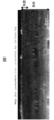
- the fluorescence microscope image of the cross section (XZ plane) of a three-dimensional cultured epidermis is shown.
- Laurdan and Cell Tracker Red were added to the three-dimensional cultured epidermis and cultured, then excited at a wavelength of 820 nm, and fluorescence of Laurdan 440 nm (blue) or Laurdan 490 nm (green) was detected.
- fluorescence (red) of Cell Tracker Red was also detected.
- the fluorescence images were merged. The stratum corneum, granule layer, spiny layer, and basal layer are shown in order from the top layer.
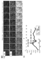
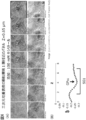
- the fluorescence microscope image of a three-dimensional culture epidermis and its fluidity distribution are shown.
- A Fluorescence image (XY plane) of the three-dimensional cultured epidermis is taken every 1.3 ⁇ m along the Z axis. Arranged in order from the top layer (upper left) to the lower layer (lower right). Blue: Laurdan 440 nm, Green: Laurdan 490 nm, Red: Cell Tracker Red.
- B The fluidity distribution (GP) of each plane is calculated according to the formula on the right, and is a fluidity distribution plotted every 0.325 ⁇ m from the uppermost layer to the lower layer.
- SC stratum corneum
- SG1 granule layer first layer.
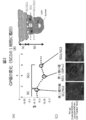
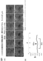
- the fluidity distribution around the granule layer first layer (SG1) along the Z-axis when focusing on one cell of the granule layer of the three-dimensional cultured epidermis is shown.
- A Flow rate distribution plotted every 0.325 ⁇ m when focusing on one cell of the granule layer.
- SC stratum corneum
- SG1 granule layer first layer
- SG2 granule layer second layer
- GP SG first minimum value in GP (central portion in the Z-axis direction of SG1 cells).
- B A schematic diagram of cells on the XZ plane is shown.
- C The fluorescence image in the plane which shows the arbitrary GP values of the graph of (A) is shown.
- SG1 Granule layer first layer
- GP SG First minimum value in GP (center portion of SG1 cell in the Z-axis direction).
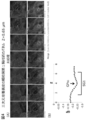
- the flow distribution (water treatment) around the granule layer first layer (SG1) along the Z axis when focusing on one cell of the granule layer of the three-dimensional cultured epidermis is shown.
- a fluorescent image (XY plane) of a three-dimensional cultured epidermis after xylitol treatment is shown every 0.65 ⁇ m along the Z axis. They were arranged in order from the upper layer (upper left) to the lower layer (lower right). Blue: Laurdan 440 nm, Green: Laurdan 490 nm, Red: Cell Tracker Red.
- B Flow rate distribution plotted every 0.325 ⁇ m when focusing on one cell of the granule layer.
- SG1 Granule layer first layer
- GP SG First minimum value in GP (center portion of SG1 cell in the Z-axis direction).
- the flow distribution (100 mM fructose treatment) around the granule layer first layer (SG1) along the Z-axis when focusing on one cell of the granule layer of the three-dimensional cultured epidermis is shown.
- a fluorescence image (XY plane) of the three-dimensional cultured epidermis after fructose treatment is taken every 0.65 ⁇ m along the Z axis. They were arranged in order from the upper layer (upper left) to the lower layer (lower right). Blue: Laurdan 440 nm, Green: Laurdan 490 nm, Red: Cell Tracker Red.
- B Flow rate distribution plotted every 0.325 ⁇ m when focusing on one cell of the granule layer.
- SG1 Granule layer
- GP SG First minimum value in GP (center portion in the Z-axis direction of SG1 cells).
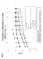
- the fluidity (GP) in the lipid of a granule layer by polyol addition is shown.
- the mean value ⁇ standard deviation (SD), p value, and n number of the fluidity (GP) of the granule layer when untreated, treated with water, treated with 100 mM xylitol, and treated with 100 mM fructose are shown.
- the flow distribution (acetone / ether treatment) around the granule layer first layer (SG1) along the Z axis when focusing on one cell of the granule layer of the three-dimensional cultured epidermis is shown.
- a fluorescence image (XY plane) of a three-dimensional cultured epidermis after acetone / ether treatment is taken every 0.65 ⁇ m along the Z axis. They were arranged in order from the upper layer (upper left) to the lower layer (lower right). Blue: Laurdan 440 nm, Green: Laurdan 490 nm, Red: Cell Tracker Red.
- B Flow rate distribution plotted every 0.325 ⁇ m when focusing on one cell of the granule layer.
- SG1 Granule layer
- GP SG First minimum value in GP (center portion in the Z-axis direction of SG1 cells).
- the fluidity (GP) in the lipid of a granule layer by acetone / ether addition is shown.
- the average value ⁇ standard deviation (SD), p value and n number of the fluidity (GP) of the granule layer when untreated, treated with water, and treated with acetone / ether are shown.
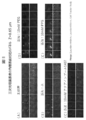
- Fluorescence image (XY plane) of three-dimensional cultured epidermis after treatment with untreated, water-treated, aquain pool 1407, polyethylene glycol 1000 (PEG), or polypropylene glycol 1000 (PPG) is 0 along the Z axis. .Images taken every 65 ⁇ m. They were arranged in order from the upper layer (upper left) to the lower layer (lower right).
- the fluidity (GP) in the lipids of the stratum corneum after untreated, water treated, aquain pool 1407, PEG or PPG treatment is shown.
- the mean value ⁇ standard deviation (SD), p value, and n number of the fluidity (GP) of the stratum corneum when untreated, treated with water, aqua in pool 1407, PEG, or PPG are shown.
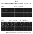
- the fluorescence image (XY plane) of the three-dimensional cultured epidermis after being treated with PBS or 3% oleic acid / PBS solution is shown every 0.65 ⁇ m along the Z axis.
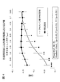
- the fluidity (GP) in the lipid of the stratum corneum after treatment with PBS or 3% oleic acid / PBS solution is shown.
- the mean value ⁇ standard deviation (SD), p value and n number of the fluidity (GP) of the stratum corneum when treated with PBS or 3% oleic acid / PBS solution are shown.
- liquidity (GP value) image of a three-dimensional cultured epidermis is shown.
- the term “three-dimensional” means that the cells are vertically different from the state of almost one cell layer obtained by culturing conventional adherent cells in a cell culture dish or the like, that is, a two-dimensional cell layer. It is an aspect in which they are stacked or layered in the direction.
- the three-dimensional cultured epidermis used in the present invention refers to a three-dimensional structure having a thickness obtained by layering epidermal cells.
- the “three-dimensional cultured epidermis” is distinguished from the skin tissue inherently provided in the living body, and is a skin obtained by breaking apart the adhesion protein between cells.
- a cell group containing tissue-derived cells and / or cells constituting the skin obtained by inducing differentiation from pluripotent stem cells is seeded in a cell culture container and cultured on a cell culture substrate. Reconstructed 3D skin-like tissue.
- the cells constituting the three-dimensional cultured epidermis include epidermal cells, and can also be referred to as a “three-dimensional cultured epidermal sheet”.
- the three-dimensional cultured epidermis or the three-dimensional cultured epidermis sheet is a cell other than epidermal cells, for example, cells other than epidermal cells (melanin cells, Langerhans cells, Merkel cells, etc.) and / or dermal tissue constituting the epidermis. (Fibroblasts, neurons, mast cells, plasma cells, vascular endothelial cells, histiospheres, meissner bodies, etc.)
- the cells contained in the three-dimensional cultured epidermis may be derived from any animal, but are preferably derived from vertebrates, more preferably derived from mammals, and most preferably derived from humans.
- the “epidermal cell” refers to a cell including all cells having different differentiation stages constituting the epidermal tissue.
- Epidermal cells are mainly composed of cells called keratinocytes or keratinocytes.
- epidermal tissue is formed by layering epidermal cells having different differentiation stages.
- the deepest part of the epidermis is called a basal layer or basal cell layer (hereinafter referred to as “basal layer”), and it is considered that cylindrical cells form a single layer and contain stem cells.
- the basal layer is also the interface with the dermis, and there is a spiny layer above it.
- the spiny layer is also called a spiny cell layer, and is composed of about 2 to 10 layers in living tissue. This layer is called a spiny layer because it appears to be tethered together. Only cells in the basal layer have the ability to proliferate, and epidermal cells move to the outer layer while changing to a flat shape as the differentiation stage proceeds.
- a granule layer (granular cell layer) having keratohyalin granules and lamellar granules is formed.
- the granular layer is composed of about 2 to 3 layers in living tissue.
- the epidermal tissue is roughly divided into the four types of layers described above. In addition to epidermal cells, melanocytes, Langerhans cells, Merkel cells, etc. are included, and each prevents ultraviolet rays from reaching the dermis. It plays an important role in the immune function of and is involved in perception.
- the three-dimensional cultured epidermis used in the present invention may contain cells other than epidermal cells, and can be appropriately changed according to the intended use.
- the “barrier function” provided in the skin generally means a function for preventing loss of moisture and biological components in the body and a function for preventing foreign substances (microorganisms, viruses, dust, etc.) from entering the living body from outside the living body.
- the barrier function of the three-dimensional cultured epidermis can be evaluated by examining the transepidermal water loss (TEWL). The smaller the amount of transepidermal water transpiration from the three-dimensional cultured epidermis, that is, the lower the amount of water permeation, the higher the barrier function.
- a general method used by those skilled in the art can be used to examine the transepidermal water transpiration (for example, Kumamoto J., Tsutsumi M., Goto M., Nagayama M., Denda M. Japan Cedar (Cryptomeria Japana) pollen allgengens evolution of intracellular calcium in human keratinocites and impairs ephemeral barrier.
- transepidermal water transpiration for example, Kumamoto J., Tsutsumi M., Goto M., Nagayama M., Denda M. Japan Cedar (Cryptomeria Japana) pollen allgengens evolution of intracellular calcium in human keratinocites and impairs ephemeral barrier.
- the cells contained in the three-dimensional cultured epidermis used in the present invention may be primary representative skin cells obtained by finely chopping living tissue, particularly skin tissue, and treating with proteolytic enzymes such as collagenase and trypsin. It may be an epidermal cell that has been propagated by subculturing the epidermal cell, or an epidermal cell obtained by inducing differentiation from a pluripotent stem cell such as an ES cell, iPS cell, or Muse cell may be used. It may be an epidermis cell.
- the first representative skin cell is an epidermal cell that is collected from a living tissue and then cultured and collected only once in a cell culture vessel.
- the epidermal cell has “number of passages 0 (or first generation)”. Also called. Epidermis cells having a subculture number of 0 that have become confluent or subconfluent can be subcultured by a method known to those skilled in the art and further amplified and cultured. Epidermal cells obtained by one passage operation are referred to as “passage number 1 (or second generation)”. Corresponding to the number of passage operations, it can be expressed as “number of passages 2, 3, 4... N (n (integer) is the number of passages) (n + 1 generation)”.
- NB Kurabo Industries' frozen NHEK
- KK-4209 can be used as primary culture cells from passage number 0 to passage number 2 in this specification.
- the number of passages in this case can be referred to the number of passages or the number of generations described in documents attached to commercially available cells.
- kits for three-dimensionalizing a cell group including epidermal cells are commercially available, and can be purchased and produced (for example, keratinocyte three-dimensional culture starter kit, CELLnTEC). Company).
- the number of passages of the first representative skin cell used in one embodiment of the present invention may be, for example, 1, 2, 3, 4, 5, 6, 7, 8, 9, 10, or 10 or more.
- the animal species of the first representative skin cell used is not particularly limited, but is preferably derived from human.
- the primary representative skin cell used may be any of fetal, newborn, minor, or adult-derived cells, preferably fetal, newborn, or minor-derived cells. It is. When cells derived from human minors are used, for example, cells under 20 years old, 1-19 years old, 1-10 years old, 1-5 years old are preferably used. Even when cells derived from human adults are used, it is preferable to use cells of young age, for example, 20-29 years old, 30-39 years old, 40-49 years old.
- the average thickness of the three-dimensional cultured epidermis is 20 ⁇ m or more, 21 ⁇ m or more, 22 ⁇ m or more, 23 ⁇ m or more, 24 ⁇ m or more, 25 ⁇ m or more, 26 ⁇ m or more, 27 ⁇ m or more, 28 ⁇ m or more, 29 ⁇ m or more, 30 ⁇ m.
- the upper limit of the average thickness of the three-dimensional cultured epidermis is not particularly limited because it varies depending on the animal species of the epidermal cells, the number of passages, age, and the like.
- the epidermal cells are 0.01 ⁇ 10 6 to 10.0 ⁇ 10 6 cells / cm 2 , preferably 0.05 ⁇ 10 6 to 5.0 ⁇ 10 6 cells / cm 2 , more preferably 0.1 Seeding at an amount of ⁇ 10 6 to 1.0 ⁇ 10 6 pieces / cm 2 .
- a medium also referred to as a culture solution used for culturing epidermal cells
- a commonly used medium such as KG medium, Epilife KG2 (Kurabo), Humedia-KG2 (Kurabo), assay medium (TOYOBO), CnT- It can be cultured at about 37 ° C. for 0 to 14 days using Prime, Epithelial culture medium (CELLnTEC) or the like.
- a DMEM medium GEBCO
- a medium in which KGM and DMEM containing 2-0-aD-glucopyranosyl-L-ascorbic acid are mixed 1: 1 can be used.
- CnT-Prime and 3D barrier medium CELLnTEC
- Other known media may be used.
- the three-dimensional cultured epidermis used in the present invention is three-dimensional (stratified) and promotes keratinization by seeding epidermal cells in a cell culture container containing a cell culture insert and growing the cells. Specifically, epidermal cells are suspended in a medium and seeded on a cell culture insert. The cell culture insert is immersed in the bottom well filled with the medium and cultured. Thereby, a culture medium is supplied from both the upper part and the bottom part of an epidermal cell.
- the epidermal cells on the cell culture insert are cultured for about several days (about 1 to 6 days, preferably about 2 to 4 days) until they become confluent or subconfluent. Thereafter, in order to further promote the three-dimensionalization (stratification) of the cells, it is preferable to replace the medium inside and outside the cell culture insert with, for example, CnT-Prime, 3D barrier medium (CELLnTEC). This further promotes three-dimensional epidermal cells.
- CnT-Prime CELLnTEC
- CELLnTEC CnT-Prime and 3D barrier medium
- L (+)-ascorbic acid for example, 50 ⁇ g / mL
- a medium for culturing the three-dimensional cultured epidermis for example, the above-mentioned CnT-Prime and 3D barrier medium.
- the medium supplemented with L (+)-ascorbic acid (vitamin C) further promotes thickening of the three-dimensional cultured epidermis.
- the culture temperature in each culture step in the present invention may be around the body temperature of the derived animal.
- the culture temperature in each culture step in the present invention may be around the body temperature of the derived animal.
- about 33 to 38 ° C. is preferable.
- the three-dimensional cultured epidermis obtained by the above method can be used, for example, in a method for evaluating the reactivity of the skin to chemical substances (for example, cosmetics, industrial products, household products, drugs, skin external preparations, etc.). Moreover, it can use for the method of evaluating the skin barrier function of this invention. Furthermore, it can use for the method of screening the chemical substance which improves and / or recovers skin barrier function by adding a chemical substance.
- chemical substances for example, cosmetics, industrial products, household products, drugs, skin external preparations, etc.
- the three-dimensional cultured epidermis produced by the above method and used in the present invention has a structure as shown in FIG.
- Laurdan (described later) for detecting the gel phase and liquid crystal phase of the biological membrane and Cell Tracker Red (described later) for staining the cytoplasm are added to the three-dimensional cultured epidermis, and the cross section (XZ plane) is known after culturing.
- the image of FIG. 1 is obtained.
- a layer in which Cell Tracker Red fluoresces can be seen, so that the stratum corneum and the granule layer can be distinguished. From the fluorescence image, a spinous layer and a basal layer can be observed below the granular layer.
- the present invention provides a method for assessing skin barrier function.
- the present invention includes the following steps.
- the “membrane environment sensitive dye” is taken into a biological membrane, for example, a cell membrane or an organelle membrane, and generates an emission wavelength (peak wavelength) depending on the state of the membrane environment when irradiated with an excitation wavelength.
- a dye for example, 6-propionyl-2- (dimethylamino) naphthalene (Prodan), 6-acryloyl-2- (dimethylamino) naphthalene (Acrylodan), 2′-dimethylamino-6′-naphthoyl-4-trans -Cyclohexane acid (DANCA), 6-lauroyl-2- (dimethylamino) naphthalene (Laurdan), anilinonaphthalene sulfonate (ANS), Bis-ANS, styrene derivative DCVJ, Dapoxyl derivative and the like.
- membrane fluidity that is, “membrane softness”.
- Prodan is a molecule that is known to have a large shift from a nonpolar solvent (in which the emission wavelength is 392 nm to an emission wavelength in water of 523 nm. Therefore, Prodan is an amino acid that accompanies changes in protein structure. It can be used to measure the degree of exposure of the residue to the water environment and the change in the degree of polarity accompanying the phase transition (gel phase or liquid crystal phase) of the membrane.
- Laurdan shows a peak of an emission spectrum at 440 nm in the gel phase and a peak of an emission spectrum at 490 nm in the liquid crystal phase in DPPC (dipalmitoyl phosphatidylcholine).
- the fluorescence intensity (I L ⁇ ) at the gel phase peak wavelength and the fluorescence intensity (I P ⁇ ) at the liquid crystal phase peak wavelength emitted from the membrane environment sensitive dye are measured to obtain the third order. It becomes possible to predict the fluidity (GP: Generalized Polarization) of the biofilm of the original cultured epidermis.
- GP Generalized Polarization
- the present inventors perform the above steps (1) to (4), and the fluidity (GP) of the biological membrane in the three-dimensional cultured epidermis is changed from the bottom layer to the top layer or from the top layer to the bottom layer,
- GP fluidity of the biological membrane in the three-dimensional cultured epidermis
- known substances that improve the skin barrier function can increase the fluidity of a specific region of the three-dimensional cultured epidermis, in particular, the granule layer, and / or It has been found that it has a mechanism for reducing the fluidity of the stratum corneum.
- the substance which destroys a skin barrier function had a mechanism which lowers
- Laurdan is used as a membrane environment sensitive dye.
- the fluorescence intensity of the gel phase peak wavelength (I L ⁇ ) and the fluorescence intensity of the liquid crystal phase peak wavelength (I P ⁇ ) emitted from the membrane environment sensitive dye on a plane parallel to the lowest layer of the three-dimensional culture epidermis The three-dimensional cultured epidermis is sequentially detected at an arbitrary interval from the lowermost layer to the uppermost layer or from the uppermost layer to the lowermost layer (see FIGS. 2A and 3C).
- the fluidity distribution (GP) in each plane parallel to the lowest layer of the three-dimensional cultured epidermis is calculated, and plotted on the Z axis (FIGS. 2B and 3A). )) Is obtained.
- the “lowermost layer of the three-dimensional cultured epidermis” refers to the lowermost layer surface of the three-dimensional cultured epidermis formed by contacting the cultured surface of the base material on which the three-dimensional cultured epidermis is cultured.
- the “XY plane” refers to a plane parallel to the culture surface of the substrate on which the three-dimensional cultured epidermis is cultured.
- the Z-axis refers to an axis perpendicular to the XY plane.
- the fluorescence intensity (I L ⁇ ) at the gel phase peak wavelength to be detected and the fluorescence intensity (I P ⁇ ) at the liquid crystal phase peak wavelength are appropriately selected according to the membrane environment sensitive dye used.
- the distance between the planes parallel to the lowermost layer of the three-dimensional cultured epidermis is, for example, 0.01 ⁇ m to 50 ⁇ m, preferably 0.05 ⁇ m to 10 ⁇ m, more preferably 0.1 ⁇ m to 5 ⁇ m.
- the gel phase peak wavelength is 440 nm and the liquid crystal phase peak wavelength is 490 nm. Therefore, for example, according to the following formula, the fluidity (GP) can be calculated. [In the formula, I 440 nm indicates the fluorescence intensity at 440 nm, and I 490 nm indicates the fluorescence intensity at 490 nm]
- the fluidity (GP) in each surface is calculated by the above formula, and the fluidity (GP) is plotted from the bottom layer to the top layer or from the top layer to the bottom layer to create a fluidity distribution.
- the graphs of FIGS. 2B and 3A are obtained.
- a relatively low fluidity (GP) indicates that the fluidity is relatively high
- a relatively high fluidity (GP) indicates that the fluidity is relatively low.
- the “first maximum value” means the vicinity of the boundary between the stratum corneum (SC) and the granular layer (particularly, the first granular layer (SG1)) in the fluidity distribution created as described above. , Defined as the highest value of fluidity (GP) (see FIGS. 2B and 3A).
- the granule layer is generally formed from 2 to 3 layers of cells (in this specification, from the layer close to the stratum corneum, the granule layer first layer (SG1), the granule layer second layer (SG2)).
- the boundary between the stratum corneum (SC) and the granule layer first layer (SG1) is a pigment that does not stain the cells of the stratum corneum (SC) from which the cell nucleus has disappeared, for example, cytoplasm
- a staining dye for example, Cell Tracker
- the “skin barrier function value” means “first minimum value”, “second minimum value”, “third minimum value”, or “layer higher than the surface including the first maximum value” described later.
- Arbitrary fluidity (GP) ".
- the “skin barrier function value” is preferably “first minimum value” and / or “arbitrary fluidity (GP) above the surface including the first maximum value”.
- the “first minimum value” is adjacent to the fluidity (GP) indicating the “first maximum value” in the fluidity distribution created as described above, and the granular layer first layer (SG1). Is defined as the lowest value of the fluidity (GP) in the region (see FIGS. 2B and 3A).
- the present inventors show for the first time that the skin barrier function is inversely correlated with the fluidity (GP SG ) of the granular layer (particularly the central part of the first granular layer (SG1)), that is, the first minimum value. did. In other words, it is possible to evaluate whether the skin barrier function is high or low by comparing the first minimum values in a plurality of three-dimensional cultured epidermis.
- the “second minimum value” means the lowest value of the fluidity (GP) of the region of the second layer of the granule layer (SG2) in the fluidity distribution created as described above. Defined as (GP).
- the “third minimum value” means the lowest value of the fluidity (GP) in the region of the third layer of the granule layer (SG3) in the fluidity distribution created as described above. Defined as (GP).
- the second minimum value and / or the third minimum value may also be used in the method for evaluating the skin barrier function of the present invention. However, since the comparison can be made more clearly, it is preferable to use the first minimum value.
- the target substance is added, and the value of the first minimum value is compared, thereby screening the target substance that improves and / or restores the skin barrier function. Can be provided.
- fluidity (GP) of an arbitrary surface above the surface including the first maximum value refers to the fluidity (GP) of an arbitrary surface in the stratum corneum.
- the present inventors have revealed that the addition of a substance that affects the barrier function of the skin correlates with the fluidity of the stratum corneum.
- the skin barrier function can be evaluated as high or low.
- the skin barrier function value determined in the step (4) when the negative target substance is added or applied in the step (1), and the step (1) is screened by comparing the value of the skin barrier function determined in the step (4) when the target substance is added or applied in It becomes possible to provide a method.
- the target substance for screening is, for example, a low molecular weight compound, peptide, protein, mammalian tissue (eg, mouse, rat, pig, cow, sheep, monkey, human etc.) or cell culture. It may be a compound, an extract (for example, a crude drug extract, a crude drug-derived compound), a microorganism-derived compound, an extract or a culture product.
- each surface for detecting the fluorescence intensity at the gel phase peak wavelength (I L ⁇ ) and the fluorescence intensity at the liquid crystal phase peak wavelength (I P ⁇ ) is selected from any one cell in the granule layer. You may limit to the same area as a cross section (refer FIG. 3).
- the area of each surface for detecting the fluorescence intensity can be determined by using an arbitrary program (for example, Nikon Instech) after an image obtained by photographing with a confocal fluorescence microscope or a multiphoton excitation microscope (for example, a two-photon excitation microscope). This can be limited by using NIS-Elements). As a result, noise is reduced, and the first maximum value and the skin barrier function value can be clearly recognized.
- the present invention provides a method of visualizing the degree of skin barrier function comprising the following steps.
- the method for visualizing the degree of the skin barrier function (first aspect) is described in the method for evaluating the skin barrier function described above or the method for visualizing the degree of the skin barrier function described later (second aspect). Elements can be applied.
- Laurdan may be used as the membrane environment sensitive dye used in the present invention.
- the fluorescence of the gel phase peak wavelength and the fluorescence of the liquid crystal phase peak wavelength emitted from the membrane environment-sensitive dye in a plane parallel to the bottom layer of the three-dimensional culture epidermis is changed from the bottom layer to the top layer or top layer of the three-dimensional culture epidermis. From the bottom to the bottom layer in order (step (2)), the detected fluorescence at the gel phase peak wavelength and the fluorescence at the liquid crystal phase peak wavelength in each color on each surface. Drawing each (for example, fluorescence of gel phase peak wavelength: blue, fluorescence of liquid crystal phase peak wavelength: green) and merging and outputting these images yields FIG. 2A (step (3)). ).
- the interval between merged and output images is, for example, 0.01 ⁇ m to 50 ⁇ m, preferably 0.05 ⁇ m to 10 ⁇ m, and more preferably 0.1 ⁇ m to 5 ⁇ m.
- the group of images displayed from the bottom layer to the top layer or the top layer to the bottom layer of the three-dimensional cultured epidermis obtained by the method of the present invention visualizes the degree of the skin barrier function. For example, in the image corresponding to the stratum corneum (for example, the image between the uppermost layer and the surface including the first maximum value), if the fluorescence at the gel phase peak wavelength is dominant, the fluidity is low and the barrier function is high. Indicates. Further, for example, in the image corresponding to the granular layer, if the fluorescence at the liquid crystal phase peak wavelength is dominant, it indicates that the fluidity is high and the barrier function is high.
- a group of images arranged from the bottom layer to the top layer or from the top layer to the bottom layer of the three-dimensional cultured epidermis obtained by the method of the present invention are all images as shown in FIG.
- the images may be displayed simultaneously.
- each image may be displayed with time in order from the bottom layer to the top layer or from the top layer to the bottom layer.
- the output image may be a monitor or a printed out image.
- the present invention provides a method for visualizing the degree of skin barrier function comprising the following steps.
- the method for visualizing the degree of the skin barrier function includes the elements described in the method for evaluating the skin barrier function or the method for visualizing the degree of the skin barrier function (first aspect). Can be applied.
- the step (1) and (2) from the fluorescence intensity of the gel phase peak wavelength detected through (I L?) And a liquid crystal phase peak fluorescence intensity at a wavelength (I P.beta), fluidity for each arbitrary pixel ( GP) can be calculated.
- the calculated fluidity (GP) can be drawn for each pixel as a shade or gradation of an arbitrary color and output as an image (step (3)).
- a method of drawing the calculated fluidity (GP) in an arbitrary color for example, a gradation of two colors having a complementary color (also referred to as an opposite color) relationship (for example, high GP value: magenta, low GP value: green) , See FIG. 15), or may be expressed as a gradation of three or more colors.
- the level of the GP value that is, the fluidity can be visually recognized.
- the fluidity is lower if the color showing a high GP value is superior to the obtained image. , Indicating that the barrier function is high. Further, for example, in an image corresponding to a granular layer, if a color showing a low GP value is dominant, it indicates that the fluidity is high and the barrier function is high.
- the epidermal cells used in the present invention were neonatal-derived keratinocytes (hereinafter “keratinocytes”) (Kurabo, product name: frozen NHEK (NB), catalog number: KK-4209).
- keratinocytes neonatal-derived keratinocytes
- the subculture was performed according to the instructions provided by the sales company.
- Example 1 A three-dimensional cultured epidermis was prepared by the following method.
- (1) 12-Well Millicell Hanging Cell Culture Inserts 0.4 ⁇ m PET (Millipore, hereinafter referred to as “Cell Culture Insert”) is diluted 50-fold with DPBS (ThermoFisher) with 1 cell dilution of CELLstart CTS (ThermoFisher). 86 ⁇ L was dropped onto the culture insert and maintained at 37 ° C. for 1.5 hours.
- DPBS ThermoFisher
- CELLstart CTS ThermoFisher
- CELLstart CTS was removed, and 2 to 250,000 newborn-derived keratinocytes with passage number 4 were dispersed in 500 ⁇ L of CnT-Prime, Epithelial culture medium (CELLnTEC), dropped into the cell culture insert, and the same medium (CnT -Prime, Epithelial culture medium) was dropped to the outside of the 1 mL cell culture insert and cultured in a CO 2 incubator for 72 hours at 37 ° C.
- CnT-Prime and Epithelial culture medium inside and outside the cell culture insert were removed and replaced with CnT-Prime, 3D barrier medium (CELLnTEC) (50 ⁇ g / mL L (+)-ascorbic acid added), 16 hours The cells were cultured at 37 ° C.
- CnT-Prime and 3D barrier medium 50 ⁇ g / mL L (+)-ascorbic acid added
- 20 ⁇ M Laurdan ThermoFisher
- change to CnT-Prime and 3D barrier medium 50 ⁇ g / mL L (+)-ascorbic acid added
- 20 ⁇ M Laurdan and 2 ⁇ M Cell Tracker Red ThermoFisher
- FIG. 1 shows a fluorescence image of the three-dimensional cultured epidermis on the XZ plane.
- FIG. 2 shows a fluorescence microscope image of the three-dimensional cultured epidermis and its fluidity distribution.
- FIG. 3 shows the fluidity distribution around the first granule layer (SG1) when focusing on one cell of the granule layer of the three-dimensional cultured epidermis.
- the first maximum value and the first minimum value used to evaluate the skin barrier function of the three-dimensional cultured epidermis could be determined.
- Example 2 A three-dimensional cultured epidermis was prepared by the same procedure as (1) to (7) in Example 1. The following steps were further performed on the obtained three-dimensional cultured epidermis.
- FIG. 4 untreated
- FIG. 5 water addition
- FIG. 6 100 mM xylitol
- FIG. 7 100 mM fructose
- FIG. 8 shows the value of the first minimum value (GP SG ) in FIGS.
- Example 3 A three-dimensional cultured epidermis was prepared by the same procedure as (1) to (7) in Example 1. The following steps were further performed on the obtained three-dimensional cultured epidermis.
- Example 4 A three-dimensional cultured epidermis was prepared by the same procedure as (1) to (5) of Example 1. The following steps were further performed on the obtained three-dimensional cultured epidermis.
- Example 5 A three-dimensional cultured epidermis was produced by the same procedure as in Example 4 (1) to (5) (6-2) (7-2). The following steps were further performed on the obtained three-dimensional cultured epidermis.
- Example 6 A three-dimensional cultured epidermis was prepared by the same procedure as (1) to (7) (8-1) of Example 1. The following steps were further performed on the obtained three-dimensional cultured epidermis.
Landscapes
- Life Sciences & Earth Sciences (AREA)
- Health & Medical Sciences (AREA)
- Engineering & Computer Science (AREA)
- Molecular Biology (AREA)
- Biomedical Technology (AREA)
- Chemical & Material Sciences (AREA)
- Hematology (AREA)
- Immunology (AREA)
- Urology & Nephrology (AREA)
- Cell Biology (AREA)
- Microbiology (AREA)
- Biotechnology (AREA)
- Food Science & Technology (AREA)
- Medicinal Chemistry (AREA)
- Physics & Mathematics (AREA)
- Analytical Chemistry (AREA)
- Biochemistry (AREA)
- General Health & Medical Sciences (AREA)
- General Physics & Mathematics (AREA)
- Pathology (AREA)
- Investigating Or Analysing Biological Materials (AREA)
- Measuring Or Testing Involving Enzymes Or Micro-Organisms (AREA)
Abstract
本発明は、(1)角層と顆粒層とを有する三次元培養表皮に、膜環境感受性色素を添加して培養する工程;(2)前記三次元培養表皮へ、前記膜環境感受性色素に対する励起波長を照射し、前記膜環境感受性色素から発光されるゲル相ピーク波長の蛍光強度及び液晶相ピーク波長の蛍光強度を順次検出する工程;(3)流動度(GP)を算出し、前記流動度(GP)をプロットして流動度分布を作成し、第1極大値を決定する工程;及び(4)皮膚バリア機能値を決定する工程、を含む皮膚バリア機能を評価する方法を提供する。また、本発明は皮膚バリア機能の程度を可視化する方法も提供する。
Description
本発明は、皮膚バリア機能を評価する方法及び皮膚バリア機能の程度を可視化する方法に関する。
皮膚は生体内と生体外の環境を分ける体表を覆う器官である。皮膚は、物理的なバリアとして働くことによって、乾燥や有害物質が生体内へ侵入することを防止し、生命の維持に不可欠な役割を果たしている。
高等脊椎動物の皮膚は、最外層から大別すると表皮、真皮、皮下組織の層から形成されている。表皮は、主にケラチノサイトと呼ばれる細胞から構成されており、表皮の最深部(基底層)でケラチノサイトが分裂しながら、上層に向かって有棘層、顆粒層、そして角層へと分化しながら表面へと移動し、やがて垢となって脱落する。このサイクルはヒトの場合概ね4週間で行われる。
皮膚のバリア機能を維持させることは生命の維持に不可欠であり、加齢や損傷などによって低下したバリア機能を向上させる物質や方法の探索が行われている。皮膚のバリア機能を回復させる物質として、例えば、キシリトール、エリスリトール、リブロース、プシコース、ガラクトース、マンニトール及びフルクトースなどの物質が知られている(例えば、特許文献1及び非特許文献1)。
皮膚の機能を解析するために、表皮構造及びその機能を生体外で模倣した様々な培養皮膚モデルの構築が試みられており、いくつかの製品が市販されている(例えば、特許文献2、非特許文献2参照。)。こうした培養皮膚モデルは、皮膚へ塗布する化粧品や薬剤の安全性試験や、基礎研究等で用いられており、動物を代替するモデルとして利用されている。
Denda M.,Effects of topical application of aqueous solutions of hexoses on epidermal permeability barrier recovery rate after barrier disruption.Exp. Dermatol.2011.Nov;20(11):943-944.
Bell E.,et al.,The reconstitution of living skin.J.Invest.Dermatol.1983 Jul;81(1 Suppl):2s-10s.
前述のように皮膚のバリア機能を回復させる物質が知られているが、そのような物質が、どのようなメカニズムで皮膚バリア機能を回復させる効果をもたらすかは明らかでなかった。本発明では、皮膚バリア機能を回復させるメカニズムを明らかにするとともに、そのメカニズムを利用した皮膚バリア機能を評価する方法を提供することを目的とする。
本発明者らが鋭意研究を行った結果、皮膚バリア機能を回復させる物質は、三次元培養表皮の特定の領域、詳しくは、顆粒層の流動性を上昇させる、及び/又は角層の流動性を低下させるメカニズムを有することを見出した。また、皮膚バリア機能を破壊する物質には、逆に、三次元培養表皮の特定の領域、詳しくは、顆粒層の流動性を低下させる、及び/又は角層の流動性を上昇させるメカニズムを有することを見出した。当該メカニズムを利用することによって、新規の皮膚バリア機能を評価する方法及び皮膚バリア機能の程度を可視化する方法を発明するに至った。すなわち、本発明は、以下の発明を包含する。
[1] 皮膚バリア機能を評価する方法であって、
(1)角層と顆粒層とを有する三次元培養表皮に、膜環境感受性色素を添加して培養する工程;
(2)前記三次元培養表皮へ、前記膜環境感受性色素に対する励起波長を照射し、前記三次元培養表皮の最下層と平行な面における前記膜環境感受性色素から発光されるゲル相ピーク波長の蛍光強度(ILβ)及び液晶相ピーク波長の蛍光強度(IPβ)を、前記三次元培養表皮の最下層から最上層又は最上層から最下層に向かって、任意の間隔で、順次検出する工程;
(3)各前記面における前記ゲル相ピーク波長の蛍光強度(ILβ)及び前記液晶相ピーク波長の蛍光強度(IPβ)から流動度(GP)を算出し、前記最下層から前記最上層又は前記最上層から前記最下層に向かって前記流動度(GP)をプロットして流動度分布を作成し、第1極大値を決定する工程;及び
(4)前記第1極大値に隣接する第1極小値、又は前記第1極大値を含む面よりも上層の任意の面の流動度(GP)を皮膚バリア機能値として決定する工程、
を含む、方法。
[2] 前記工程(1)において、対象物質を添加又は塗布する、[1]に記載の方法。
[3] 前記工程(1)において陰性対象物質を添加又は塗布した場合の、前記工程(4)において決定された前記皮膚バリア機能値と、
前記工程(1)において対象物質を添加又は塗布した場合の、前記工程(4)において決定された前記皮膚バリア機能値と、を比較することを特徴とする、[1]又は[2]に記載の方法。
[4] 前記第1極大値は、前記角層と前記顆粒層との境界付近における、最も高い値の前記流動度(GP)として定義される、[1]~[3]のいずれか1項に記載の方法。
[5] 前記流動度(GP)が、以下:

により算出される、[1]~[4]のいずれか1項に記載の方法。
[6] 前記膜環境感受性色素が、Laurdanである、[1]~[5]のいずれか1項に記載の方法。
[7] ゲル相ピーク波長が440nmであって、液晶相ピーク波長が490nmである、[6]に記載の方法。
[8] 前記工程(1)において、さらに細胞質染色色素を添加する、[1]~[7]のいずれか1項に記載の方法。
[9] 各前記面が、前記顆粒層の任意の1細胞の任意の断面と同一面積である、[1]~[8]のいずれか1項に記載の方法。
(1)角層と顆粒層とを有する三次元培養表皮に、膜環境感受性色素を添加して培養する工程;
(2)前記三次元培養表皮へ、前記膜環境感受性色素に対する励起波長を照射し、前記三次元培養表皮の最下層と平行な面における前記膜環境感受性色素から発光されるゲル相ピーク波長の蛍光強度(ILβ)及び液晶相ピーク波長の蛍光強度(IPβ)を、前記三次元培養表皮の最下層から最上層又は最上層から最下層に向かって、任意の間隔で、順次検出する工程;
(3)各前記面における前記ゲル相ピーク波長の蛍光強度(ILβ)及び前記液晶相ピーク波長の蛍光強度(IPβ)から流動度(GP)を算出し、前記最下層から前記最上層又は前記最上層から前記最下層に向かって前記流動度(GP)をプロットして流動度分布を作成し、第1極大値を決定する工程;及び
(4)前記第1極大値に隣接する第1極小値、又は前記第1極大値を含む面よりも上層の任意の面の流動度(GP)を皮膚バリア機能値として決定する工程、
を含む、方法。
[2] 前記工程(1)において、対象物質を添加又は塗布する、[1]に記載の方法。
[3] 前記工程(1)において陰性対象物質を添加又は塗布した場合の、前記工程(4)において決定された前記皮膚バリア機能値と、
前記工程(1)において対象物質を添加又は塗布した場合の、前記工程(4)において決定された前記皮膚バリア機能値と、を比較することを特徴とする、[1]又は[2]に記載の方法。
[4] 前記第1極大値は、前記角層と前記顆粒層との境界付近における、最も高い値の前記流動度(GP)として定義される、[1]~[3]のいずれか1項に記載の方法。
[5] 前記流動度(GP)が、以下:
[6] 前記膜環境感受性色素が、Laurdanである、[1]~[5]のいずれか1項に記載の方法。
[7] ゲル相ピーク波長が440nmであって、液晶相ピーク波長が490nmである、[6]に記載の方法。
[8] 前記工程(1)において、さらに細胞質染色色素を添加する、[1]~[7]のいずれか1項に記載の方法。
[9] 各前記面が、前記顆粒層の任意の1細胞の任意の断面と同一面積である、[1]~[8]のいずれか1項に記載の方法。
[10] 皮膚バリア機能の程度を可視化する方法であって、
(1)角層と顆粒層とを有する三次元培養表皮に、膜環境感受性色素を添加して培養する工程;
(2)前記三次元培養表皮へ、前記膜環境感受性色素に対する励起波長を照射し、前記三次元培養表皮の最下層と平行な面における前記膜環境感受性色素から発光されるゲル相ピーク波長の蛍光及び液晶相ピーク波長の蛍光を、前記三次元培養表皮の最下層から最上層又は最上層から最下層に向かって、任意の間隔で、順次検出する工程;及び
(3)各前記面において、検出された前記ゲル相ピーク波長の蛍光及び前記液晶相ピーク波長の蛍光を任意の色彩でそれぞれ描画し、マージした画像を出力する工程、
を含む、方法。
[11] 前記工程(1)において、対象物質を添加又は塗布する、[10]に記載の方法。
[12] 前記工程(1)において陰性対象物質を添加又は塗布した場合の、前記工程(3)で得られる画像と、
前記工程(1)において対象物質を添加又は塗布した場合の、前記工程(3)で得られる画像と、を比較することを特徴とする、[10]又は[11]に記載の方法。
[13] 前記膜環境感受性色素が、Laurdanである、[10]~[12]のいずれか1項に記載の方法。
[14] ゲル相ピーク波長が440nmであって、液晶相ピーク波長が490nmである、[13]に記載の方法。
[15] 皮膚バリア機能の程度を可視化する方法であって、
(1)角層と顆粒層とを有する三次元培養表皮に、膜環境感受性色素を添加して培養する工程;
(2)前記三次元培養表皮へ、前記膜環境感受性色素に対する励起波長を照射し、前記三次元培養表皮の最下層と平行な面における前記膜環境感受性色素から発光されるゲル相ピーク波長の蛍光強度(ILβ)及び液晶相ピーク波長の蛍光強度(IPβ)を、前記三次元培養表皮の最下層から最上層又は最上層から最下層に向かって、任意の間隔で、順次検出する工程;及び
(3)各前記面において、検出された前記ゲル相ピーク波長の蛍光強度(ILβ)及び前記液晶相ピーク波長の蛍光強度(IPβ)から任意のピクセル毎に流動度(GP)を算出し、前記流動度(GP)を任意の色彩の濃淡又はグラデーションとして前記ピクセル毎に描画し、画像として出力する工程、
を含む、方法。
[16] 前記工程(1)において、対象物質を添加又は塗布する、[15]に記載の方法。
[17] 前記工程(1)において陰性対象物質を添加又は塗布した場合の、前記工程(3)で得られる画像と、
前記工程(1)において対象物質を添加又は塗布した場合の、前記工程(3)で得られる画像と、を比較することを特徴とする、[15]又は[16]に記載の方法。
[18] 前記流動度(GP)が、以下:

により算出される、[15]~[17]のいずれか1項に記載の方法。
[19] 前記膜環境感受性色素が、Laurdanである、請求項15~18のいずれか1項に記載の方法。
[20] ゲル相ピーク波長が440nmであって、液晶相ピーク波長が490nmである、[19]に記載の方法。
(1)角層と顆粒層とを有する三次元培養表皮に、膜環境感受性色素を添加して培養する工程;
(2)前記三次元培養表皮へ、前記膜環境感受性色素に対する励起波長を照射し、前記三次元培養表皮の最下層と平行な面における前記膜環境感受性色素から発光されるゲル相ピーク波長の蛍光強度(ILβ)及び液晶相ピーク波長の蛍光強度(IPβ)を、前記三次元培養表皮の最下層から最上層又は最上層から最下層に向かって、任意の間隔で、順次検出する工程;及び
(3)各前記面において、検出された前記ゲル相ピーク波長の蛍光強度(ILβ)及び前記液晶相ピーク波長の蛍光強度(IPβ)から任意のピクセル毎に流動度(GP)を算出し、前記流動度(GP)を任意の色彩の濃淡又はグラデーションとして前記ピクセル毎に描画し、画像として出力する工程、
を含む、方法。
[16] 前記工程(1)において、対象物質を添加又は塗布する、[15]に記載の方法。
[17] 前記工程(1)において陰性対象物質を添加又は塗布した場合の、前記工程(3)で得られる画像と、
前記工程(1)において対象物質を添加又は塗布した場合の、前記工程(3)で得られる画像と、を比較することを特徴とする、[15]又は[16]に記載の方法。
[18] 前記流動度(GP)が、以下:
[19] 前記膜環境感受性色素が、Laurdanである、請求項15~18のいずれか1項に記載の方法。
[20] ゲル相ピーク波長が440nmであって、液晶相ピーク波長が490nmである、[19]に記載の方法。
本発明によれば、三次元培養表皮の皮膚バリア機能を容易に評価することが可能になる。これにより、皮膚へ塗布する化粧品や薬剤の安全性試験、基礎研究に有用な最適な三次元培養表皮モデルが選択可能となる。また、本発明によれば、皮膚バリア機能を改善又は回復させる、新規の物質を容易にスクリーニング可能となる。
以下、本発明を実施するための形態について説明するが、本発明の技術的範囲は下記の形態のみに限定されない。
本明細書において、「三次元」とは、従来の付着細胞を細胞培養皿等での培養で得られるほぼ1層の細胞層、つまり、二次元の細胞層の状態とは異なり、細胞が垂直方向へと積み重なり、又は重層化した態様をいう。例えば、本発明において用いられる三次元培養表皮は、表皮細胞が重層化されて厚みをもった三次元化した構造体をいう。
本明細書において、「三次元培養表皮」とは、生体に生来的に備わっている皮膚組織とは区別されるものであって、細胞間の接着タンパク質を分解してバラバラにして得られた皮膚組織由来の細胞、及び/又は、多能性幹細胞から分化誘導して得られた皮膚を構成する細胞、を含む細胞群を、細胞培養容器等に播種し、細胞培養基材上で培養して再構築した三次元皮膚様組織をいう。本発明において、三次元培養表皮を構成する細胞は、表皮細胞を含んでおり、「三次元培養表皮シート」ということもできる。本発明において、三次元培養表皮又は三次元培養表皮シートは、表皮細胞以外の細胞、例えば、表皮を構成する表皮細胞以外の細胞(メラニン細胞、ランゲルハンス細胞、メルケル細胞等)及び/又は、真皮組織に含まれる細胞(線維芽細胞、神経細胞、肥満細胞、形質細胞、血管内皮細胞、組織球、meissner小体、等)が含まれていてもよい。
本明細書において、三次元培養表皮に含まれる細胞は、いずれの動物由来であってもよいが、脊椎動物由来が好ましく、哺乳動物由来がより好ましく、ヒト由来であることが最も好ましい。
本明細書において、「表皮細胞」とは、表皮組織を構成する分化段階の異なる全ての細胞を含む細胞をいう。表皮細胞は、主に、角化細胞又はケラチノサイトとも呼ばれる細胞から構成されている。生体において、表皮組織は、分化段階の異なる表皮細胞が層状に重なって形成されている。表皮の最深部は基底層又は基底細胞層(以下、「基底層」という。)と呼ばれ、円柱状の細胞が一層を形成し、幹細胞を含むと考えられている。基底層は真皮との境界面でもあり、その上層には有棘層が存在する。
有棘層とは、有棘細胞層とも呼ばれ、生体組織においては2~10層程度から構成されている。この層は、互いに棘で繋がっているように見えるために有棘層と呼ばれる。基底層の細胞のみが増殖能を有しており、表皮細胞は分化段階が進むにつれて扁平な形状に変化しながら外層へと移動する。
有棘層より分化段階が進むと、ケラトヒアリン顆粒、ラメラ顆粒を有する顆粒層(顆粒細胞層)を形成する。顆粒層は、生体組織においては2~3層程度で構成されている。
顆粒層よりさらに分化段階が進むと、細胞核が消失し、角層(角質細胞層)を形成する。表皮組織は大別して上述の4種類の層を構成し、表皮細胞の他にも、メラニン細胞、ランゲルハンス細胞、メルケル細胞等が含まれており、それぞれ紫外線が真皮へ到達することを防いだり、皮膚の免疫機能に重要な役割を果たしたり、知覚に関与したりする。本発明において用いられる、三次元培養表皮は、表皮細胞以外の細胞が含まれても良く、使用する用途に応じて適宜変更することが可能である。
皮膚が備える「バリア機能」とは、一般に、体内の水分や生体成分の損失を防止する機能や、生体外部から生体内部へ異物(微生物、ウイルス、ホコリ等)が入ることを防止する機能をいう。本明細書において、三次元培養表皮が有するバリア機能は、経表皮水分蒸散量(Transepidermal water loss:TEWL)を調べることにより評価することができる。三次元培養表皮からの経表皮水分蒸散量、すなわち、水分透過量が少ないほど、バリア機能が高いことを示す。経表皮水分蒸散量を調べる方法は、当業者に用いられる一般的な方法を用いることができる(例えば、Kumamoto J.,Tsutsumi M.,Goto M.,Nagayama M.,Denda M.Japanese Cedar(Cryptomeria japonica)pollen allergen induces elevation of intracellular calcium in human keratinocytes and impairs epidermal barrier function of human skin ex vivo.Arch.Dermatol.Res.308:49-54,2016を参照のこと)。
本発明において用いられる三次元培養表皮に含まれる細胞は、生体組織、特に皮膚組織を細かく刻み、コラゲナーゼ、トリプシン等のタンパク質分解酵素で処理されて得られた初代表皮細胞であってもよく、初代表皮細胞を継代操作して増殖させた表皮細胞であってもよく、ES細胞、iPS細胞、又はMuse細胞等の多能性幹細胞から分化誘導して得られる表皮細胞を用いてもよく、株化された表皮細胞であってもよい。
本明細書において、初代表皮細胞とは、生体組織から採取した後に細胞培養容器で一度だけ培養されて回収された表皮細胞であり、「継代回数0(又は、第1世代)」の表皮細胞ともいう。コンフルエント又はサブコンフルエントになった継代回数0の表皮細胞を、当業者にとって公知の方法により継代操作し、さらに増幅培養することが可能である。1度の継代操作により得られた表皮細胞は、「継代回数1(又は、第2世代)」の表皮細胞という。継代操作数に対応して「継代回数2、3、4・・・n(n(整数)は継代回数)(第n+1世代)」と表現することができる。市販の初代培養細胞(例えば、クラボウ社の凍結NHEK(NB)、カタログ番号:KK-4009)には、本明細書でいう継代回数0~継代回数2程度のものまでを初代培養細胞として提供されているものもあるが、この場合の継代回数は、市販の細胞に添付の書類等に記載されている継代回数又は世代数を参考にすることができる。
三次元培養表皮を作製する場合、表皮細胞を含む細胞群を三次元化するキットが市販されており、それを購入して作製することが可能である(例えば、ケラチノサイト三次元培養スターターキット、CELLnTEC社)。
本発明の一実施形態において使用される初代表皮細胞の継代回数は、例えば、1、2、3、4、5、6、7、8、9、10、又は10以上であってもよい。本発明の実施形態において、使用される初代表皮細胞の動物種は、特に限定されないが、好ましくはヒト由来である。また、本発明の実施形態において、使用される初代表皮細胞は、胎児、新生児、未成年、又は成人由来等いずれのものであってもよいが、好ましくは胎児、新生児、又は未成年由来の細胞である。ヒトの未成年由来の細胞を用いる場合、例えば、20歳未満、1~19歳、1~10歳、1~5歳の細胞を用いるのが好ましい。ヒトの成人由来の細胞を用いる場合であっても、若年、例えば、20歳~29歳、30歳~39歳、40歳~49歳の細胞を用いることが好ましい。
本発明の一実施形態において、三次元培養表皮の平均の厚さは、20μm以上、21μm以上、22μm以上、23μm以上、24μm以上、25μm以上、26μm以上、27μm以上、28μm以上、29μm以上、30μm以上、35μm以上、40μm以上、45μm以上、50μm以上、55μm以上、60μm以上、65μm以上、70μm以上、75μm以上、80μm以上、85μm以上、90μm以上、95μm以上、100μm以上、105μm以上、110μm以上、115μm以上、120μm以上、125μ以上、150μm以上、175μm以上、200μm以上、225μm以上、250μm以上、275μm以上、300μm以上であってもよい。三次元培養表皮の平均の厚さの上限値は、表皮細胞の動物種、継代回数、年齢等により変化するため、特に限定されない。
<三次元培養表皮の製造方法>
本発明の一実施形態において用いられる三次元培養表皮に含まれる表皮細胞の播種数については、公知の方法に従えばよい。例えば、表皮細胞を、0.01×10⁶~10.0×10⁶個/cm2、好ましくは0.05×106~5.0×106個/cm2、より好ましくは0.1×106~1.0×106個/cm2の量で播種する。表皮細胞の培養に用いられる培地(培養液ともいう)としては、通常使用される培地、例えばKG培地、EpilifeKG2(クラボウ社)、Humedia-KG2(クラボウ社)、アッセイ培地(TOYOBO社)、CnT-Prime,Epithelial culture medium(CELLnTEC社)などを用い、約37℃で0~14日間かけて培養することができる。培地としては、その他にDMEM培地(GIBCO社)又は2-0-a-D-グルコピラノシル-L-アスコルビン酸含有KGMとDMEMを1:1混合した培地などが使用できる。表皮細胞を三次元化(重層化)するためには、CnT-Prime、3D barrier medium(CELLnTEC社)を用いても良い。その他の公知の培地を使用してもよい。
本発明の一実施形態において用いられる三次元培養表皮に含まれる表皮細胞の播種数については、公知の方法に従えばよい。例えば、表皮細胞を、0.01×10⁶~10.0×10⁶個/cm2、好ましくは0.05×106~5.0×106個/cm2、より好ましくは0.1×106~1.0×106個/cm2の量で播種する。表皮細胞の培養に用いられる培地(培養液ともいう)としては、通常使用される培地、例えばKG培地、EpilifeKG2(クラボウ社)、Humedia-KG2(クラボウ社)、アッセイ培地(TOYOBO社)、CnT-Prime,Epithelial culture medium(CELLnTEC社)などを用い、約37℃で0~14日間かけて培養することができる。培地としては、その他にDMEM培地(GIBCO社)又は2-0-a-D-グルコピラノシル-L-アスコルビン酸含有KGMとDMEMを1:1混合した培地などが使用できる。表皮細胞を三次元化(重層化)するためには、CnT-Prime、3D barrier medium(CELLnTEC社)を用いても良い。その他の公知の培地を使用してもよい。
本発明において用いられる三次元培養表皮は、セルカルチャーインサートを含む細胞培養容器に表皮細胞を播種し、増殖培養させることによって、三次元化(重層化)し、かつ、角質化が促進される。具体的には、表皮細胞を培地に懸濁し、セルカルチャーインサート上に播種する。培地を充填したボトムウェルに、セルカルチャーインサートを浸漬させて培養する。これにより、表皮細胞の上部及び底部の両方から培地が供給される。
セルカルチャーインサート上の表皮細胞が、コンフルエント又はサブコンフルエントになるまで数日間程度(1~6日間程度、好ましくは2~4日間程度)培養させることが好ましい。その後、細胞の三次元化(重層化)をさらに促進するために、セルカルチャーインサート内外の培地を、例えば、CnT-Prime、3D barrier medium(CELLnTEC社)へ交換することが好ましい。これにより、さらに表皮細胞の三次元化が促進される。CnT-Prime、3D barrier medium(CELLnTEC社)へと培地を交換して培養後、1~36時間後、好ましくは6時間~24時間後、さらに好ましくは12時間~18時間後、セルカルチャーインサートの内部の培地のみを除去して表皮細胞の上面を気相に暴露させて培養することが好ましい。これによって、表皮細胞の三次元化及び角質化がさらに促進され、より厚い三次元培養表皮が得られる。三次元培養表皮を培養する培地、例えば、上記のCnT-Prime、3D barrier mediumには、L(+)-アスコルビン酸(ビタミンC)(例えば、50μg/mL)が添加されていることが好ましい。L(+)-アスコルビン酸(ビタミンC)が添加された培地によって、さらに三次元培養表皮の肥厚化が促進される。
なお、本発明における各培養工程の培養温度は由来動物の体温付近であればよく、例えば、ヒト細胞の場合は、33~38℃程度が好適である。
上記の方法によって得られる三次元培養表皮は、例えば、化学物質(例えば、化粧料、工業製品、家庭用品、薬剤、皮膚外用剤等)に対する皮膚の反応性を評価する方法に用いることができる。また、本発明の皮膚バリア機能を評価する方法に用いることができる。さらにまた、化学物質を添加することによって、皮膚バリア機能を改善及び/又は回復させる化学物質をスクリーニングする方法に用いることができる。
一実施態様において、上記方法によって作製され、本発明に用いられる三次元培養表皮は、例えば、図1のような構造を有する。三次元培養表皮に、生体膜のゲル相及び液晶相を検出するLaurdan(後述)と、細胞質を染色するCell Tracker Red(後述)とを添加して培養後、断面(X-Z面)を公知の共焦点蛍光顕微鏡を用いて撮影した結果、図1の像が得られる。角層と顆粒層との間はCell Tracker Redが蛍光する層が見られ、これにより、角層と顆粒層とを見分けることができる。蛍光像から、顆粒層の下部には有棘層及び基底層を観察することができる。
<皮膚バリア機能を評価する方法>
一実施態様において、本発明は、皮膚バリア機能を評価する方法を提供する。本発明は、以下の工程を含む。
一実施態様において、本発明は、皮膚バリア機能を評価する方法を提供する。本発明は、以下の工程を含む。
(1)角層と顆粒層とを有する三次元培養表皮に、膜環境感受性色素を添加して培養する工程;
(2)前記三次元培養表皮へ、前記膜環境感受性色素に対する励起波長を照射し、前記三次元培養表皮の最下層と平行な面における前記膜環境感受性色素から発光されるゲル相ピーク波長の蛍光強度(ILβ)及び液晶相ピーク波長の蛍光強度(IPβ)を、前記三次元培養表皮の最下層から最上層又は最上層から最下層に向かって、任意の間隔で、順次検出する工程;
(3)各前記面における前記ゲル相ピーク波長の蛍光強度(ILβ)及び前記液晶相ピーク波長の蛍光強度(IPβ)から流動度(GP)を算出し、前記最下層から前記最上層又は前記最上層から前記最下層に向かって前記流動度(GP)をプロットして流動度分布を作成し、第1極大値を決定する工程;及び
(4)前記第1極大値に隣接する第1極小値、又は前記第1極大値を含む面よりも上層の任意の流動度(GP)を皮膚バリア機能値として決定する工程。
本明細書において、「膜環境感受性色素」とは、生体膜、例えば細胞膜やオルガネラ膜に取り込まれ、励起波長を照射した場合、膜環境の状態に応じて、異なる発光波長(ピーク波長)を生じる色素をいい、例えば、6-プロピオニル-2-(ジメチルアミノ)ナフタレン(Prodan)、6-アクリロイル-2-(ジメチルアミノ)ナフタレン(Acrylodan)、2’-ジメチルアミノ-6’-ナフトイル-4-トランス-シクロヘキサン酸(DANCA)、6-ラウロイル-2-(ジメチルアミノ)ナフタレン(Laurdan)、アニリノナフタレンスルホネート(ANS)、Bis-ANS、スチレン誘導体DCVJ、Dapoxyl誘導体などが含まれる。生体膜において、脂質分子やタンパク質分子は膜面中で側方拡散運動を行っており、その拡散運動が「膜の流動性」(すなわち、「膜の柔らかさ」)を決定している。膜環境感受性色素を用いることにより、「膜の流動性」を測定することが可能となる。
例えば、Prodanは、無極性溶媒(中では392nmの発光波長から、水中での523nmの発光波長まで大きくシフトすることが知られている分子である。そのため、Prodanは、タンパク質の構造変化にともなうアミノ酸残基の水環境への露出度や、膜の相転移(ゲル相又は液晶相)にともなう極性度の変化の測定に用いることができる。
例えば、Laurdanは、DPPC(ジパルミトイル フォスファチジルコリン)中において、ゲル相では、440nmに発光スペクトルのピークを示し、液晶相では、490nmに発光スペクトルのピークを示す。本発明において、膜環境感受性色素を用いて、膜環境感受性色素から発光されるゲル相ピーク波長の蛍光強度(ILβ)及び液晶相ピーク波長の蛍光強度(IPβ)を測定することにより、三次元培養表皮の生体膜の流動度(GP:Generalized Polarization)を予測することが可能となる。本発明に用いられる膜環境感受性色素として、好ましくは、Laurdanである。
本発明者らは、上記工程(1)~(4)を実施して、三次元培養表皮における生体膜の流動度(GP)を、最下層から最上層又は最上層から最下層に向かって、任意の間隔でプロットして流動度分布を作成した結果、公知の皮膚バリア機能を向上させる物質には、三次元培養表皮の特定の領域、特に、顆粒層の流動性を上昇させる、及び/又は角層の流動性を低下させるメカニズムを有することを見出した。また、皮膚バリア機能を破壊する物質には、逆に、三次元培養表皮の顆粒層の流動性を低下させる、及び/又は角層の流動性を上昇させるメカニズムを有することを見出した。すなわち、当該メカニズムに着目することによって、本発明にかかる三次元培養表皮の皮膚バリア機能を評価する方法を完成させた。具体的な態様について、以下に説明する。
図2及び図3では、膜環境感受性色素として、Laurdanが用いられている。一実施態様において、三次元培養表皮の最下層と平行な面における前記膜環境感受性色素から発光されるゲル相ピーク波長の蛍光強度(ILβ)及び液晶相ピーク波長の蛍光強度(IPβ)を、前記三次元培養表皮の最下層から最上層又は最上層から最下層に向かって、任意の間隔で、順次検出する(図2(A)及び図3(C)参照)。一実施態様において、三次元培養表皮の最下層と平行な各面における流動度(GP)を算出し、それをZ軸にプロットすることによって流動度分布(図2(B)及び図3(A)参照)が得られる。
本明細書において、「三次元培養表皮の最下層」とは、三次元培養表皮を培養している基材の培養面と接触することにより形成された三次元培養表皮の最下層面をいう。また、本明細書において、「X-Y面」とは、三次元培養表皮を培養している基材の培養面と平行な面をいう。また、本明細書において、Z軸とは、前記X-Y面と垂直な軸をいう。
ゲル相ピーク波長の蛍光強度(ILβ)及び液晶相ピーク波長の蛍光強度(IPβ)は、例えば、共焦点蛍光顕微鏡や多光子励起顕微鏡(例えば、二光子励起顕微鏡)によって撮影し、得られた画像を解析することによって検出することができる。検出するゲル相ピーク波長の蛍光強度(ILβ)及び液晶相ピーク波長の蛍光強度(IPβ)は、使用する膜環境感受性色素に応じて、適宜選択される。
一実施態様において、三次元培養表皮の最下層と平行な面の間隔は、例えば、0.01μm~50μm、好ましくは0.05μm~10μm、より好ましくは、0.1μm~5μmである。
得られたゲル相ピーク波長の蛍光強度(ILβ)及び液晶相ピーク波長の蛍光強度(IPβ)から、例えば、以下の式に従って、流動度(GP)を算出することができる。

一実施態様において、膜環境感受性色素がLaurdanである場合、ゲル相ピーク波長が440nmであり、液晶相ピーク波長が490nmである。そのため、例えば、以下の式に従って、流動度(GP)を算出することができる。

[式中、I440nmは、440nmにおける蛍光強度を示し、I490nmは、490nmにおける蛍光強度を示す]
上記式によって、各面における流動度(GP)を算出し、前記最下層から前記最上層又は前記最上層から前記最下層に向かって前記流動度(GP)をプロットして流動度分布を作成することにより、例えば、図2(B)及び図3(A)のグラフが得られる。相対的に流動度(GP)が低いと、流動性が相対的に高いことを示し、相対的に流動度(GP)が高いと、流動性が相対的に低いことを示す。
本明細書において、「第1極大値」とは、上記のように作成された流動度分布において、角層(SC)と顆粒層(特に顆粒層第1層(SG1))との境界付近における、最も高い値の流動度(GP)として定義される(図2(B)及び図3(A)参照)。顆粒層は、一般的に、2~3層の細胞から形成されており(本明細書においては、角層に近い層から、顆粒層第1層(SG1)、顆粒層第2層(SG2)、顆粒層第3層(SG3)と呼ぶ。角層(SC)と顆粒層第1層(SG1)との境界は、細胞核が消失した角層(SC)の細胞は染色しない色素、例えば、細胞質染色色素(例えば、Cell Tracker)を添加することによって、さらに明確に認識可能となる(図1参照)。
本明細書において、「皮膚バリア機能値」とは、後述する「第1極小値」、「第2極小値」、「第3極小値」、又は「第1極大値を含む面よりも上層の任意の流動度(GP)」をいう。「皮膚バリア機能値」は、好ましくは「第1極小値」及び/又は「第1極大値を含む面よりも上層の任意の流動度(GP)」である。
本明細書において、「第1極小値」とは、上記のように作成された流動度分布において「第1極大値」を示す流動度(GP)に隣接し、顆粒層第1層(SG1)の領域の流動度(GP)のうち最も低い値の流動度(GP)として定義される(図2(B)及び図3(A)参照)。本発明者らは、皮膚のバリア機能が、顆粒層(特に第1顆粒層(SG1)の中心部分)の流動度(GPSG)、すなわち、第1極小値と逆相関することを初めて明らかにした。換言すると、複数の三次元培養表皮において、第1極小値を比較することにより、皮膚バリア機能が高い又は低いことを評価することができる。
本明細書において、「第2極小値」とは、上記のように作成された流動度分布において、顆粒層第2層(SG2)の領域の流動度(GP)のうち最も低い値の流動度(GP)として定義される。本明細書において、「第3極小値」とは、上記のように作成された流動度分布において、顆粒層第3層(SG3)の領域の流動度(GP)のうち最も低い値の流動度(GP)として定義される。第2極小値及び/又は第3極小値も、本発明の皮膚バリア機能を評価する方法に用いてもよいが、より明確に比較できることから、第1極小値を用いることが好ましい。
一実施態様の本発明は、前記工程(1)において、対象物質を添加し、第1極小値の値を比較することによって、皮膚のバリア機能を改善及び/又は回復させる対象物質をスクリーニングする方法を提供することが可能となる。
本明細書において、「第1極大値を含む面よりも上層の任意の面の流動度(GP)」は、角層における任意の面の流動度(GP)をいう。本発明者らは、皮膚のバリア機能に影響を与える物質の添加が、角層の流動度と相関することを明らかにした。換言すると、複数の三次元培養表皮において、角層(例えば、三次元培養表皮において、最上層から第1極大値を含む面の間)の流動度(GP)を比較することにより、皮膚バリア機能が高い又は低いことを評価することができる。
また、他の実施態様の本発明では、さらに、前記工程(1)において陰性対象物質を添加又は塗布した場合の、前記工程(4)において決定された前記皮膚バリア機能値と、前記工程(1)において対象物質を添加又は塗布した場合の、前記工程(4)において決定された前記皮膚バリア機能値と、を比較することによって、皮膚のバリア機能を改善及び/又は回復させる対象物質をスクリーニングする方法を提供することが可能となる。本実施態様において、スクリーニングの対象物質としては、例えば、低分子化合物、ペプチド、タンパク質、哺乳動物(例えば、マウス、ラット、ブタ、ウシ、ヒツジ、サル、ヒトなど)の組織抽出物又は細胞培養上清、植物由来の化合物又は抽出物(例えば、生薬エキス、生薬由来の化合物)、及び微生物由来の化合物若しくは抽出物又は培養産物などであってもよい。
また、一実施態様の本発明では、ゲル相ピーク波長の蛍光強度(ILβ)及び液晶相ピーク波長の蛍光強度(IPβ)を検出する各面を、顆粒層の任意の1細胞の任意の断面と同一面積に限定してもよい(図3参照)。蛍光強度を検出する各面の面積は、共焦点蛍光顕微鏡や多光子励起顕微鏡(例えば、二光子励起顕微鏡)によって撮影して得られた画像を、後から、任意のプログラム(例えば、ニコンインステック社のNIS-Elements)を用いることによって限定することができる。これによって、ノイズが減少し、第1極大値と皮膚バリア機能値が明確に認識できるようになる。
<皮膚バリア機能の程度を可視化する方法(第1態様)>
一実施態様において、本発明は、以下の工程を含む、皮膚バリア機能の程度を可視化する方法を提供する。
一実施態様において、本発明は、以下の工程を含む、皮膚バリア機能の程度を可視化する方法を提供する。
(1)角層と顆粒層とを有する三次元培養表皮に、膜環境感受性色素を添加して培養する工程;
(2)前記三次元培養表皮へ、前記膜環境感受性色素に対する励起波長を照射し、前記三次元培養表皮の最下層と平行な面における前記膜環境感受性色素から発光されるゲル相ピーク波長の蛍光及び液晶相ピーク波長の蛍光を、前記三次元培養表皮の最下層から最上層又は最上層から最下層に向かって、任意の間隔で、順次検出し、工程;及び
(3)各前記面において、検出された前記ゲル相ピーク波長の蛍光及び前記液晶相ピーク波長の蛍光を任意の色彩でそれぞれ描画し、マージした画像を出力する工程。
(2)前記三次元培養表皮へ、前記膜環境感受性色素に対する励起波長を照射し、前記三次元培養表皮の最下層と平行な面における前記膜環境感受性色素から発光されるゲル相ピーク波長の蛍光及び液晶相ピーク波長の蛍光を、前記三次元培養表皮の最下層から最上層又は最上層から最下層に向かって、任意の間隔で、順次検出し、工程;及び
(3)各前記面において、検出された前記ゲル相ピーク波長の蛍光及び前記液晶相ピーク波長の蛍光を任意の色彩でそれぞれ描画し、マージした画像を出力する工程。
なお、皮膚バリア機能の程度を可視化する方法(第1態様)は、上記の皮膚バリア機能を評価する方法、又は後述の皮膚バリア機能の程度を可視化する方法(第2態様)で説明される各要素を適用することができる。
一実施態様において、本発明に用いられる膜環境感受性色素は、Laurdanを用いてもよい。三次元培養表皮の最下層と平行な面における前記膜環境感受性色素から発光されるゲル相ピーク波長の蛍光及び液晶相ピーク波長の蛍光を、前記三次元培養表皮の最下層から最上層又は最上層から最下層に向かって、任意の間隔で順次検出した後(工程(2))、各前記面において、検出された前記ゲル相ピーク波長の蛍光及び前記液晶相ピーク波長の蛍光を任意の色彩でそれぞれ描画し(例えば、ゲル相ピーク波長の蛍光:青、液晶相ピーク波長の蛍光:緑)、それらの画像をマージして出力することで、図2(A)が得られる(工程(3))。
一実施態様において、マージして出力される画像の間隔は、例えば、0.01μm~50μm、好ましくは0.05μm~10μm、より好ましくは、0.1μm~5μmである。
本発明の方法によって得られる、三次元培養表皮の最下層から最上層又は最上層から最下層に表示される一群の画像は、皮膚バリア機能の程度を可視化している。例えば、角層に相当する画像(例えば、最上層から第1極大値を含む面の間の画像)において、ゲル相ピーク波長の蛍光が優位であると、流動性が低く、バリア機能が高いことを示す。また、例えば、顆粒層に相当する画像において、液晶相ピーク波長の蛍光が優位であると、流動性が高く、バリア機能が高いことを示す。
一実施態様において、本発明の方法によって得られる、三次元培養表皮の最下層から最上層又は最上層から最下層に並べられた一群の画像は、図2(A)のように全ての画像が同時に表示された画像であってもよく、例えば、各画像が最下層から最上層又は最上層から最下層の順番に経時的に表示されるものであってもよい。また、出力される画像は、モニタであってもよく、プリントアウトされたものであってもよい。
<皮膚バリア機能の程度を可視化する方法(第2態様)>
他の実施態様において、本発明は、以下の工程を含む、皮膚バリア機能の程度を可視化する方法を提供する。
他の実施態様において、本発明は、以下の工程を含む、皮膚バリア機能の程度を可視化する方法を提供する。
(1)角層と顆粒層とを有する三次元培養表皮に、膜環境感受性色素を添加して培養する工程;
(2)前記三次元培養表皮へ、前記膜環境感受性色素に対する励起波長を照射し、前記三次元培養表皮の最下層と平行な面における前記膜環境感受性色素から発光されるゲル相ピーク波長の蛍光強度(ILβ)及び液晶相ピーク波長の蛍光強度(IPβ)を、前記三次元培養表皮の最下層から最上層又は最上層から最下層に向かって、任意の間隔で、順次検出する工程;及び
(3)各前記面において、検出された前記ゲル相ピーク波長の蛍光強度(ILβ)及び前記液晶相ピーク波長の蛍光強度(IPβ)から任意のピクセル毎に流動度(GP)を算出し、前記流動度(GP)を任意の色彩の濃淡又はグラデーションとして前記ピクセル毎に描画し、画像として出力する工程。
なお、皮膚バリア機能の程度を可視化する方法(第2態様)は、上記の皮膚バリア機能を評価する方法、又は皮膚バリア機能の程度を可視化する方法(第1態様)で説明される各要素を適用することができる。
各前記面において、工程(1)及び(2)を経て検出したゲル相ピーク波長の蛍光強度(ILβ)及び液晶相ピーク波長の蛍光強度(IPβ)から、任意のピクセル毎に流動度(GP)を算出することができる。算出された流動度(GP)は、任意の色彩の濃淡又はグラデーションとしてピクセル毎に描画し、画像として出力することができる(工程(3))。算出された流動度(GP)を任意の色彩で描画する方法として、例えば、補色(反対色ともいう)関係にある2色の色彩のグラデーション(例えば、高GP値:マゼンダ、低GP値:緑、図15参照)を用いてもよく、3色以上のグラデーションとして表現してもよい。これにより、視覚的にGP値の高低、すなわち、流動度を認識することができる。
得られた画像より、例えば、角層に相当する画像(例えば、最上層から第1極大値を含む面の間の画像)において、高GP値を示す色彩が優位であれば、流動性が低く、バリア機能が高いことを示す。また、例えば、顆粒層に相当する画像において、低GP値を示す色彩が優位であれば、流動性が高く、バリア機能が高いことを示す。
以下に、本発明を実施例に基づいて更に詳しく説明するが、これらは本発明を何ら限定するものではない。
<細胞の準備>
本発明に用いられる表皮細胞は、新生児由来ケラチノサイト(以下、「ケラチノサイト」)(クラボウ社、製品名:凍結NHEK(NB)、カタログ番号:KK-4009)を使用した。継代培養は、販売会社から提供されたインストラクションに従い行った。
本発明に用いられる表皮細胞は、新生児由来ケラチノサイト(以下、「ケラチノサイト」)(クラボウ社、製品名:凍結NHEK(NB)、カタログ番号:KK-4009)を使用した。継代培養は、販売会社から提供されたインストラクションに従い行った。
<実施例1>
以下の方法によって、三次元培養表皮を作製した。
(1)12-Well Millicell Hanging Cell Culture inserts 0.4μm PET(Millipore社、以下、「セルカルチャーインサート」)に、CELLstart CTS(ThermoFisher社)をDPBS(ThermoFisher社)で50倍希釈し、1つのセルカルチャーインサートに86μL滴下し、1.5時間37℃の条件に維持した。
(2)CELLstart CTSを除去し、継代回数4の新生児由来ケラチノサイト22~25万個をCnT-Prime,Epithelial culture medium(CELLnTEC社)500μLに分散し、セルカルチャーインサート内に滴下、同じ培地(CnT-Prime,Epithelial culture medium)を1mLセルカルチャーインサート外部に滴下し、72時間37℃、CO2インキュベーターで培養した。
(3)セルカルチャーインサート内外のCnT-Prime、Epithelial culture mediumを除去し、CnT-Prime、3D barrier medium(CELLnTEC社)(50μg/mLのL(+)-アスコルビン酸を添加)で置き換え、16時間37℃、CO2インキュベーターで培養した。
(4)セルカルチャーインサート内外の培地を除去し、セルカルチャーインサート内は空気に曝露し、セルカルチャーインサート外に500μL CnT-Prime、3D barrier medium(50μg/mLのL(+)-アスコルビン酸を添加)を入れた。
(5)毎日セルカルチャーインサート外の500μL CnT-Prime、3D barrier medium(50μg/mLのL(+)-アスコルビン酸を添加)を交換しながら37℃CO2インキュベーターで7日間培養した。
(6)インサートに新生児由来ケラチノサイトを播種してから12日目に、20μM Laurdan(ThermoFisher社)を添加したCnT-Prime、3D barrier medium(50μg/mLのL(+)-アスコルビン酸を添加)に交換した。
(7)13日目に、20μM Laurdan及び2μM Cell Tracker Red(ThermoFisher社)を添加したCnT-Prime、3D barrier medium(50μg/mLのL(+)-アスコルビン酸を添加)に交換して、培養した。
(7)13日目に、20μM Laurdan及び2μM Cell Tracker Red(ThermoFisher社)を添加したCnT-Prime、3D barrier medium(50μg/mLのL(+)-アスコルビン酸を添加)に交換して、培養した。
(8-1)14日目に、バイオプシーパンチを用いて、セルカルチャーインサートと共に三次元培養表皮を直径6mmに打ち抜き、反転させてスライドガラスの上に乗せた。さらにその上に重しを乗せた。反転した状態で、二光子顕微鏡を用いて画像を取得した。なお、Laurdanは、820nmで励起させ、440nm及び490nmの発光像を検出し、Cell Tracker Redは820nmで励起させ、602nmの発光像を検出し、マージさせた。
(9-1)得られた画像を、ニコン社のNIS-Elementsを用いて解析し、流動度(GP)を算出し、流動度分布を作成した。
(9-1)得られた画像を、ニコン社のNIS-Elementsを用いて解析し、流動度(GP)を算出し、流動度分布を作成した。
図1は、X-Z面における三次元培養表皮の蛍光像を示している。図2は、三次元培養表皮の蛍光顕微鏡像、及びその流動度分布を示す。図3は、三次元培養表皮の顆粒層の一細胞に着目した場合の、顆粒層第1層(SG1)周辺の流動度分布を示す。
図1~3に示すように、上記方法を用いることにより、三次元培養表皮の皮膚バリア機能を評価するために用いられる第1極大値及び第1極小値を決定することができた。
<実施例2>
実施例1の(1)~(7)と同一の手順によって、三次元培養表皮を作製した。得られた三次元培養表皮に対し、さらに以下の工程を実施した。
実施例1の(1)~(7)と同一の手順によって、三次元培養表皮を作製した。得られた三次元培養表皮に対し、さらに以下の工程を実施した。
(8-2)14日目に、200μLの水または100mMキシリトールまたは100mMフルクトースを角層側から添加し、37℃、CO2 5%で2時間インキュベートした。2時間後に溶液を取り除き、37℃、CO2 5%で1時間インキュベートした。1時間後にバイオプシーパンチを用いて、セルカルチャーインサートと共に三次元培養表皮を直径6mmに打ち抜き、反転させてスライドガラスの上に乗せた。さらにその上に重しを乗せた。反転した状態で、二光子顕微鏡を用いて画像を取得した。なお、Laurdanは、820nmで励起させ、440nm及び490nmの発光像を検出し、Cell Tracker Redは820nmで励起させ、602nmの発光像を検出し、マージさせた。
その後、実施例1の(9-1)を実施した。
上記の結果を図4(未処理)、図5(水添加)、図6(100mM キシリトール)及び図7(100mMフルクトース)に示す。図4~図7の第1極小値(GPSG)の値を図8に示す。結果からも明らかであるように、皮膚のバリア機能を改善する物質であるキシリトール及びフルクトースは、未処理又は水処理の場合と比較して、有意に脂質の流動性を向上させることが明らかとなった。
<実施例3>
実施例1の(1)~(7)と同一の手順によって、三次元培養表皮を作製した。得られた三次元培養表皮に対し、さらに以下の工程を実施した。
実施例1の(1)~(7)と同一の手順によって、三次元培養表皮を作製した。得られた三次元培養表皮に対し、さらに以下の工程を実施した。
(8-3)14日目に、200μLの水または、皮膚のバリア機能を破壊するアセトン/エーテル(50%(v/v))を角層側から添加し、室温で3分間インキュベートし、3分後に溶液を取り除いた。これを2回繰り返した。バイオプシーパンチを用いて、セルカルチャーインサートと共に三次元培養表皮を直径6mmに打ち抜き、反転させてスライドガラスの上に乗せた。さらにその上に重しを乗せた。反転した状態で、二光子顕微鏡を用いて画像を取得した。なお、Laurdanは、820nmで励起させ、440nm及び490nmの発光像を検出し、Cell Tracker Redは820nmで励起させ、602nmの発光像を検出し、マージさせた。
その後、実施例1の(9-1)を実施した。
図10の結果からも明らかであるように、皮膚のバリア機能を破壊する物質であるアセトン/エーテルを添加することにより、未処理又は水処理の場合と比較して、有意に脂質の流動性を低下させることが明らかとなった。
<実施例4>
実施例1の(1)~(5)と同一の手順によって、三次元培養表皮を作製した。得られた三次元培養表皮に対し、さらに以下の工程を実施した。
実施例1の(1)~(5)と同一の手順によって、三次元培養表皮を作製した。得られた三次元培養表皮に対し、さらに以下の工程を実施した。
(6-2)インサートに新生児由来ケラチノサイトを播種してから9日目に、20μM Laurdan(ThermoFisher社)を添加したCnT-Prime、3D barrier medium(50μg/mLのL(+)-アスコルビン酸を添加)に交換した。
(7-2)10日目に、20μM Laurdan及び2μM Cell Tracker Red(ThermoFisher社)を添加したCnT-Prime、3D barrier medium(50μg/mLのL(+)-アスコルビン酸を添加)に交換して、培養した。
(8-4)11日目に、未処理、200μLの水、皮膚のバリア機能を促進するものとして知られるアクアインプール1407(10mM(1%(w/v))、皮膚のバリア機能を促進せずアクアンプール1407と同じ分子量であるポリエチレングリコール1000(PEG)(10mM(1%(w/v))、ポリプロピレングルコール1000(PPG)(10mM(1%(w/v))を角層側から添加し、37℃、CO2 5%で30分間インキュベートした。30分後に溶液を取り除き、37℃、CO2 5%で30分間インキュベートした。バイオプシーパンチを用いて、セルカルチャーインサートと共に三次元培養表皮を直径6mmに打ち抜き、反転させてスライドガラスの上に乗せた。さらにその上に重しを乗せた。反転した状態で、二光子顕微鏡を用いて画像を取得した。なお、Laurdanは、820nmで励起させ、440nm及び490nmの発光像を検出し、Cell Tracker Redは820nmで励起させ、602nmの発光像を検出し、マージさせた。
その後、実施例1の(9-1)を実施した。
図11および図12の結果からも明らかであるように、アクアインプール1407を添加することにより、未処理、水処理、PEG処理又はPPG処理の場合と比較して、角層において有意に脂質の流動性を低下させることが明らかとなった。
<実施例5>
実施例4の(1)~(5)(6-2)(7-2)と同一の手順によって、三次元培養表皮を作製した。得られた三次元培養表皮に対し、さらに以下の工程を実施した。
実施例4の(1)~(5)(6-2)(7-2)と同一の手順によって、三次元培養表皮を作製した。得られた三次元培養表皮に対し、さらに以下の工程を実施した。
(8-5)11日目に、200μLのPBS、又は脂質の配向性を緩くする働きをもつオレイン酸・PBS溶液((3%(v/v))を角層側から添加し、37℃、CO2 5%で30分間インキュベートした。30分後に溶液を取り除き、37℃、CO2 5%で30分間インキュベートした。バイオプシーパンチを用いて、セルカルチャーインサートと共に三次元培養表皮を直径6mmに打ち抜き、反転させてスライドガラスの上に乗せた。さらにその上に重しを乗せた。反転した状態で、二光子顕微鏡を用いて画像を取得した。なお、Laurdanは、820nmで励起させ、440nm及び490nmの発光像を検出し、Cell Tracker Redは820nmで励起させ、602nmの発光像を検出し、マージさせた。
その後、実施例1の(9-1)を実施した。
図13および図14の結果からも明らかであるように、オレイン酸を添加することにより、PBS処理の場合と比較して、角層において有意に脂質の流動性を上昇させることが明らかとなった。
<実施例6>
実施例1の(1)~(7)(8-1)と同一の手順によって、三次元培養表皮を作製した。得られた三次元培養表皮に対し、さらに以下の工程を実施した。
実施例1の(1)~(7)(8-1)と同一の手順によって、三次元培養表皮を作製した。得られた三次元培養表皮に対し、さらに以下の工程を実施した。
(9-2)得られた画像を、ニコン社のNIS-Elementsを用いて解析し、1ピクセル毎に流動度(GP)を算出し、高GP値をマゼンダ、低GP値を緑とするグラデーションで描画した(図15)。
Claims (20)
- 皮膚バリア機能を評価する方法であって、
(1)角層と顆粒層とを有する三次元培養表皮に、膜環境感受性色素を添加して培養する工程;
(2)前記三次元培養表皮へ、前記膜環境感受性色素に対する励起波長を照射し、前記三次元培養表皮の最下層と平行な面における前記膜環境感受性色素から発光されるゲル相ピーク波長の蛍光強度(ILβ)及び液晶相ピーク波長の蛍光強度(IPβ)を、前記三次元培養表皮の最下層から最上層又は最上層から最下層に向かって、任意の間隔で、順次検出する工程;
(3)各前記面における前記ゲル相ピーク波長の蛍光強度(ILβ)及び前記液晶相ピーク波長の蛍光強度(IPβ)から流動度(GP)を算出し、前記最下層から前記最上層又は前記最上層から前記最下層に向かって前記流動度(GP)をプロットして流動度分布を作成し、第1極大値を決定する工程;及び
(4)前記第1極大値に隣接する第1極小値、又は前記第1極大値を含む面よりも上層の任意の面の流動度(GP)を皮膚バリア機能値として決定する工程、
を含む、方法。 - 前記工程(1)において、対象物質を添加又は塗布する、請求項1に記載の方法。
- 前記工程(1)において陰性対象物質を添加又は塗布した場合の、前記工程(4)において決定された前記皮膚バリア機能値と、
前記工程(1)において対象物質を添加又は塗布した場合の、前記工程(4)において決定された前記皮膚バリア機能値と、を比較することを特徴とする、請求項1又は2に記載の方法。 - 前記第1極大値は、前記角層と前記顆粒層との境界付近における、最も高い値の前記流動度(GP)として定義される、請求項1~3のいずれか1項に記載の方法。
- 前記流動度(GP)が、以下:

- 前記膜環境感受性色素が、Laurdanである、請求項1~5のいずれか1項に記載の方法。
- ゲル相ピーク波長が440nmであって、液晶相ピーク波長が490nmである、請求項6に記載の方法。
- 前記工程(1)において、さらに細胞質染色色素を添加する、請求項1~7のいずれか1項に記載の方法。
- 各前記面が、前記顆粒層の任意の1細胞の任意の断面と同一面積である、請求項1~8のいずれか1項に記載の方法。
- 皮膚バリア機能の程度を可視化する方法であって、
(1)角層と顆粒層とを有する三次元培養表皮に、膜環境感受性色素を添加して培養する工程;
(2)前記三次元培養表皮へ、前記膜環境感受性色素に対する励起波長を照射し、前記三次元培養表皮の最下層と平行な面における前記膜環境感受性色素から発光されるゲル相ピーク波長の蛍光及び液晶相ピーク波長の蛍光を、前記三次元培養表皮の最下層から最上層又は最上層から最下層に向かって、任意の間隔で、順次検出する工程;及び
(3)各前記面において、検出された前記ゲル相ピーク波長の蛍光及び前記液晶相ピーク波長の蛍光を任意の色彩でそれぞれ描画し、マージした画像を出力する工程、
を含む、方法。 - 前記工程(1)において、対象物質を添加又は塗布する、請求項10に記載の方法。
- 前記工程(1)において陰性対象物質を添加又は塗布した場合の、前記工程(3)で得られる画像と、
前記工程(1)において対象物質を添加又は塗布した場合の、前記工程(3)で得られる画像と、を比較することを特徴とする、請求項10又は11に記載の方法。 - 前記膜環境感受性色素が、Laurdanである、請求項10~12のいずれか1項に記載の方法。
- ゲル相ピーク波長が440nmであって、液晶相ピーク波長が490nmである、請求項13に記載の方法。
- 皮膚バリア機能の程度を可視化する方法であって、
(1)角層と顆粒層とを有する三次元培養表皮に、膜環境感受性色素を添加して培養する工程;
(2)前記三次元培養表皮へ、前記膜環境感受性色素に対する励起波長を照射し、前記三次元培養表皮の最下層と平行な面における前記膜環境感受性色素から発光されるゲル相ピーク波長の蛍光強度(ILβ)及び液晶相ピーク波長の蛍光強度(IPβ)を、前記三次元培養表皮の最下層から最上層又は最上層から最下層に向かって、任意の間隔で、順次検出する工程;及び
(3)各前記面において、検出された前記ゲル相ピーク波長の蛍光強度(ILβ)及び前記液晶相ピーク波長の蛍光強度(IPβ)から任意のピクセル毎に流動度(GP)を算出し、前記流動度(GP)を任意の色彩の濃淡又はグラデーションとして前記ピクセル毎に描画し、画像として出力する工程、
を含む、方法。 - 前記工程(1)において、対象物質を添加又は塗布する、請求項15に記載の方法。
- 前記工程(1)において陰性対象物質を添加又は塗布した場合の、前記工程(3)で得られる画像と、
前記工程(1)において対象物質を添加又は塗布した場合の、前記工程(3)で得られる画像と、を比較することを特徴とする、請求項15又は16に記載の方法。 - 前記流動度(GP)が、以下:

- 前記膜環境感受性色素が、Laurdanである、請求項15~18のいずれか1項に記載の方法。
- ゲル相ピーク波長が440nmであって、液晶相ピーク波長が490nmである、請求項19に記載の方法。
Priority Applications (1)
| Application Number | Priority Date | Filing Date | Title |
|---|---|---|---|
| JP2020504073A JP7196154B2 (ja) | 2018-03-09 | 2019-03-08 | 皮膚バリア機能を評価する方法及び皮膚バリア機能の程度を可視化する方法 |
Applications Claiming Priority (2)
| Application Number | Priority Date | Filing Date | Title |
|---|---|---|---|
| JPPCT/JP2018/009329 | 2018-03-09 | ||
| PCT/JP2018/009329 WO2019171597A1 (ja) | 2018-03-09 | 2018-03-09 | 皮膚バリア機能を評価する方法 |
Publications (1)
| Publication Number | Publication Date |
|---|---|
| WO2019172452A1 true WO2019172452A1 (ja) | 2019-09-12 |
Family
ID=67845616
Family Applications (2)
| Application Number | Title | Priority Date | Filing Date |
|---|---|---|---|
| PCT/JP2018/009329 Ceased WO2019171597A1 (ja) | 2018-03-09 | 2018-03-09 | 皮膚バリア機能を評価する方法 |
| PCT/JP2019/009541 Ceased WO2019172452A1 (ja) | 2018-03-09 | 2019-03-08 | 皮膚バリア機能を評価する方法及び皮膚バリア機能の程度を可視化する方法 |
Family Applications Before (1)
| Application Number | Title | Priority Date | Filing Date |
|---|---|---|---|
| PCT/JP2018/009329 Ceased WO2019171597A1 (ja) | 2018-03-09 | 2018-03-09 | 皮膚バリア機能を評価する方法 |
Country Status (2)
| Country | Link |
|---|---|
| JP (1) | JP7196154B2 (ja) |
| WO (2) | WO2019171597A1 (ja) |
Citations (1)
| Publication number | Priority date | Publication date | Assignee | Title |
|---|---|---|---|---|
| JP2008215945A (ja) * | 2007-03-01 | 2008-09-18 | Pola Chem Ind Inc | 皮膚バリア機能向上作用の評価法及び皮膚バリア機能の評価法 |
Family Cites Families (1)
| Publication number | Priority date | Publication date | Assignee | Title |
|---|---|---|---|---|
| JP6243123B2 (ja) * | 2013-01-21 | 2017-12-06 | 株式会社 資生堂 | 角層状態の評価方法、及び化粧料の角層改善効果についての評価方法 |
-
2018
- 2018-03-09 WO PCT/JP2018/009329 patent/WO2019171597A1/ja not_active Ceased
-
2019
- 2019-03-08 JP JP2020504073A patent/JP7196154B2/ja active Active
- 2019-03-08 WO PCT/JP2019/009541 patent/WO2019172452A1/ja not_active Ceased
Patent Citations (1)
| Publication number | Priority date | Publication date | Assignee | Title |
|---|---|---|---|---|
| JP2008215945A (ja) * | 2007-03-01 | 2008-09-18 | Pola Chem Ind Inc | 皮膚バリア機能向上作用の評価法及び皮膚バリア機能の評価法 |
Non-Patent Citations (4)
| Title |
|---|
| BLOKSGAARD, M. ET AL.: "Structural and dynamical aspects of skin studied by multiphoton excitation fluorescence microscopy-based methods", EUROPEAN JOURNAL OF PHARMACEUTICAL SCIENCES, vol. 50, no. 5, 2013, pages 586 - 594, XP028738121, doi:10.1016/j.ejps.2013.04.010 * |
| C ARRER, D. C. ET AL.: "Pig skin structure and transdermal delivery of liposomes: A two photon microscopy study", JOURNAL OF CONTROLLED RELEASE, vol. 132, 2008, pages 12 - 20, XP025610272, doi:10.1016/j.jconrel.2008.08.006 * |
| GOLFETTO, 0. ET AL.: "Laurdan Fluorescence Lifetime Discriminates Cholesterol Content from Changes in Fluidity in Living Cell Membranes", BIOPHYSICAL JOURNAL, vol. 104, no. 6, March 2013 (2013-03-01), pages 1238 - 1247, XP055638291 * |
| SUN, Y. ET AL.: "Multiphoton polarization and generalized polarization microscopy reveal oleic-acid-induced structural changes in intercellular lipid layers of the skin", OPTICS LETTERS, vol. 29, no. 17, 1 September 2004 (2004-09-01), pages 2013 - 2015, XP055638289 * |
Also Published As
| Publication number | Publication date |
|---|---|
| WO2019171597A1 (ja) | 2019-09-12 |
| JP7196154B2 (ja) | 2022-12-26 |
| JPWO2019172452A1 (ja) | 2021-02-25 |
Similar Documents
| Publication | Publication Date | Title |
|---|---|---|
| Jiang et al. | Two succeeding fibroblastic lineages drive dermal development and the transition from regeneration to scarring | |
| Cunniff et al. | AMPK activity regulates trafficking of mitochondria to the leading edge during cell migration and matrix invasion | |
| US20140154735A1 (en) | Tumour cell and tissue culture | |
| Wang et al. | Reduced oligodendrocyte precursor cell impairs astrocytic development in early life stress | |
| EP4092108A1 (en) | Novel multi-organ-chips establishing differentiation of ipsc-derived cells into organ equivalents | |
| Jang et al. | Advanced in vitro three-dimensional skin models of atopic dermatitis | |
| EP1768486A2 (en) | Chimeric avian-based screening system containing mammalian grafts | |
| Herms et al. | Self-sustaining long-term 3D epithelioid cultures reveal drivers of clonal expansion in esophageal epithelium | |
| JP2017184659A (ja) | 三次元培養表皮モデル及びその製造方法、ならびに三次元培養表皮モデルの使用方法 | |
| Dan-Sohkawa et al. | Cell dynamics of the blastulation process in the starfish, Asterina pectinifera | |
| Drieschner et al. | Ultrathin alumina membranes as scaffold for epithelial cell culture from the intestine of rainbow trout | |
| Piasek et al. | Building up skin models for numerous applications-from two-dimensional (2D) monoculture to three-dimensional (3D) multiculture | |
| Li et al. | Identification, isolation, and characterization of melanocyte precursor cells in the human limbal stroma | |
| Hollstein et al. | Generating immunocompetent 3-dimensional full-thickness models of human skin | |
| Tan et al. | A Human‐Based Skin‐Lymphoreticular Model‐on‐Chip to Emulate Inflammatory Skin Conditions | |
| JP7196154B2 (ja) | 皮膚バリア機能を評価する方法及び皮膚バリア機能の程度を可視化する方法 | |
| Bickenbach et al. | Epidermal stem cells have the potential to assist in healing damaged tissues | |
| Arora et al. | Isolation and characterization of extracellular vesicles derived from ex vivo culture of visceral adipose tissue | |
| Gargas et al. | Isolation and analysis of mouse and human skin γδ T cells | |
| Tharin et al. | A microfluidic device to investigate axon targeting by limited numbers of purified cortical projection neuron subtypes | |
| Bang et al. | The urn cell. Complex of Sipunculus nudus: A model for study of mucus-stimulating substances | |
| US20180236051A1 (en) | Method for producing embryonic-like stem cells from ticks (acari: ixodidae), composition, uses thereof and diagnostic methods | |
| Bejar et al. | Defining the transcriptional signature of esophageal-to-skin lineage conversion | |
| DE102004061289B4 (de) | Testkit mit Testzellen und Immunzellen sowie Herstellverfahren hierfür | |
| Caragnano et al. | Biological Barrier Models-on-Chip: An Innovative Tool for Studying Diseases and Discovering New Drug Therapies |
Legal Events
| Date | Code | Title | Description |
|---|---|---|---|
| 121 | Ep: the epo has been informed by wipo that ep was designated in this application |
Ref document number: 19763800 Country of ref document: EP Kind code of ref document: A1 |
|
| ENP | Entry into the national phase |
Ref document number: 2020504073 Country of ref document: JP Kind code of ref document: A |
|
| NENP | Non-entry into the national phase |
Ref country code: DE |
|
| 122 | Ep: pct application non-entry in european phase |
Ref document number: 19763800 Country of ref document: EP Kind code of ref document: A1 |