WO2019152571A1 - Anti-pd-1 antibodies - Google Patents
Anti-pd-1 antibodies Download PDFInfo
- Publication number
- WO2019152571A1 WO2019152571A1 PCT/US2019/015932 US2019015932W WO2019152571A1 WO 2019152571 A1 WO2019152571 A1 WO 2019152571A1 US 2019015932 W US2019015932 W US 2019015932W WO 2019152571 A1 WO2019152571 A1 WO 2019152571A1
- Authority
- WO
- WIPO (PCT)
- Prior art keywords
- antibody
- seq
- antigen
- variable region
- chain variable
- Prior art date
Links
Classifications
-
- A—HUMAN NECESSITIES
- A61—MEDICAL OR VETERINARY SCIENCE; HYGIENE
- A61P—SPECIFIC THERAPEUTIC ACTIVITY OF CHEMICAL COMPOUNDS OR MEDICINAL PREPARATIONS
- A61P31/00—Antiinfectives, i.e. antibiotics, antiseptics, chemotherapeutics
-
- A—HUMAN NECESSITIES
- A61—MEDICAL OR VETERINARY SCIENCE; HYGIENE
- A61K—PREPARATIONS FOR MEDICAL, DENTAL OR TOILETRY PURPOSES
- A61K39/00—Medicinal preparations containing antigens or antibodies
- A61K39/395—Antibodies; Immunoglobulins; Immune serum, e.g. antilymphocytic serum
- A61K39/39533—Antibodies; Immunoglobulins; Immune serum, e.g. antilymphocytic serum against materials from animals
- A61K39/39541—Antibodies; Immunoglobulins; Immune serum, e.g. antilymphocytic serum against materials from animals against normal tissues, cells
-
- A—HUMAN NECESSITIES
- A61—MEDICAL OR VETERINARY SCIENCE; HYGIENE
- A61K—PREPARATIONS FOR MEDICAL, DENTAL OR TOILETRY PURPOSES
- A61K45/00—Medicinal preparations containing active ingredients not provided for in groups A61K31/00 - A61K41/00
- A61K45/06—Mixtures of active ingredients without chemical characterisation, e.g. antiphlogistics and cardiaca
-
- A—HUMAN NECESSITIES
- A61—MEDICAL OR VETERINARY SCIENCE; HYGIENE
- A61P—SPECIFIC THERAPEUTIC ACTIVITY OF CHEMICAL COMPOUNDS OR MEDICINAL PREPARATIONS
- A61P35/00—Antineoplastic agents
-
- A—HUMAN NECESSITIES
- A61—MEDICAL OR VETERINARY SCIENCE; HYGIENE
- A61P—SPECIFIC THERAPEUTIC ACTIVITY OF CHEMICAL COMPOUNDS OR MEDICINAL PREPARATIONS
- A61P37/00—Drugs for immunological or allergic disorders
- A61P37/02—Immunomodulators
- A61P37/04—Immunostimulants
-
- C—CHEMISTRY; METALLURGY
- C07—ORGANIC CHEMISTRY
- C07K—PEPTIDES
- C07K16/00—Immunoglobulins [IGs], e.g. monoclonal or polyclonal antibodies
- C07K16/18—Immunoglobulins [IGs], e.g. monoclonal or polyclonal antibodies against material from animals or humans
- C07K16/28—Immunoglobulins [IGs], e.g. monoclonal or polyclonal antibodies against material from animals or humans against receptors, cell surface antigens or cell surface determinants
- C07K16/2803—Immunoglobulins [IGs], e.g. monoclonal or polyclonal antibodies against material from animals or humans against receptors, cell surface antigens or cell surface determinants against the immunoglobulin superfamily
- C07K16/2818—Immunoglobulins [IGs], e.g. monoclonal or polyclonal antibodies against material from animals or humans against receptors, cell surface antigens or cell surface determinants against the immunoglobulin superfamily against CD28 or CD152
-
- C—CHEMISTRY; METALLURGY
- C07—ORGANIC CHEMISTRY
- C07K—PEPTIDES
- C07K2317/00—Immunoglobulins specific features
- C07K2317/20—Immunoglobulins specific features characterized by taxonomic origin
- C07K2317/24—Immunoglobulins specific features characterized by taxonomic origin containing regions, domains or residues from different species, e.g. chimeric, humanized or veneered
-
- C—CHEMISTRY; METALLURGY
- C07—ORGANIC CHEMISTRY
- C07K—PEPTIDES
- C07K2317/00—Immunoglobulins specific features
- C07K2317/30—Immunoglobulins specific features characterized by aspects of specificity or valency
- C07K2317/31—Immunoglobulins specific features characterized by aspects of specificity or valency multispecific
-
- C—CHEMISTRY; METALLURGY
- C07—ORGANIC CHEMISTRY
- C07K—PEPTIDES
- C07K2317/00—Immunoglobulins specific features
- C07K2317/40—Immunoglobulins specific features characterized by post-translational modification
- C07K2317/41—Glycosylation, sialylation, or fucosylation
-
- C—CHEMISTRY; METALLURGY
- C07—ORGANIC CHEMISTRY
- C07K—PEPTIDES
- C07K2317/00—Immunoglobulins specific features
- C07K2317/50—Immunoglobulins specific features characterized by immunoglobulin fragments
- C07K2317/51—Complete heavy chain or Fd fragment, i.e. VH + CH1
-
- C—CHEMISTRY; METALLURGY
- C07—ORGANIC CHEMISTRY
- C07K—PEPTIDES
- C07K2317/00—Immunoglobulins specific features
- C07K2317/50—Immunoglobulins specific features characterized by immunoglobulin fragments
- C07K2317/515—Complete light chain, i.e. VL + CL
-
- C—CHEMISTRY; METALLURGY
- C07—ORGANIC CHEMISTRY
- C07K—PEPTIDES
- C07K2317/00—Immunoglobulins specific features
- C07K2317/50—Immunoglobulins specific features characterized by immunoglobulin fragments
- C07K2317/52—Constant or Fc region; Isotype
-
- C—CHEMISTRY; METALLURGY
- C07—ORGANIC CHEMISTRY
- C07K—PEPTIDES
- C07K2317/00—Immunoglobulins specific features
- C07K2317/50—Immunoglobulins specific features characterized by immunoglobulin fragments
- C07K2317/52—Constant or Fc region; Isotype
- C07K2317/522—CH1 domain
-
- C—CHEMISTRY; METALLURGY
- C07—ORGANIC CHEMISTRY
- C07K—PEPTIDES
- C07K2317/00—Immunoglobulins specific features
- C07K2317/50—Immunoglobulins specific features characterized by immunoglobulin fragments
- C07K2317/55—Fab or Fab'
-
- C—CHEMISTRY; METALLURGY
- C07—ORGANIC CHEMISTRY
- C07K—PEPTIDES
- C07K2317/00—Immunoglobulins specific features
- C07K2317/50—Immunoglobulins specific features characterized by immunoglobulin fragments
- C07K2317/56—Immunoglobulins specific features characterized by immunoglobulin fragments variable (Fv) region, i.e. VH and/or VL
- C07K2317/565—Complementarity determining region [CDR]
-
- C—CHEMISTRY; METALLURGY
- C07—ORGANIC CHEMISTRY
- C07K—PEPTIDES
- C07K2317/00—Immunoglobulins specific features
- C07K2317/60—Immunoglobulins specific features characterized by non-natural combinations of immunoglobulin fragments
- C07K2317/62—Immunoglobulins specific features characterized by non-natural combinations of immunoglobulin fragments comprising only variable region components
- C07K2317/622—Single chain antibody (scFv)
-
- C—CHEMISTRY; METALLURGY
- C07—ORGANIC CHEMISTRY
- C07K—PEPTIDES
- C07K2317/00—Immunoglobulins specific features
- C07K2317/70—Immunoglobulins specific features characterized by effect upon binding to a cell or to an antigen
- C07K2317/71—Decreased effector function due to an Fc-modification
-
- C—CHEMISTRY; METALLURGY
- C07—ORGANIC CHEMISTRY
- C07K—PEPTIDES
- C07K2317/00—Immunoglobulins specific features
- C07K2317/90—Immunoglobulins specific features characterized by (pharmaco)kinetic aspects or by stability of the immunoglobulin
- C07K2317/92—Affinity (KD), association rate (Ka), dissociation rate (Kd) or EC50 value
Definitions
- the present invention relates to treatments of conditions ameliorated by counteracting tumor mediated immune suppression. More specifically the present invention relates to anti-PD- 1 antibodies, as well as use of these antibodies in the treatment of diseases such as cancer and infectious disease.
- PD-l is recognized as an important molecule in immune regulation and the maintenance of peripheral tolerance. PD-l is moderately expressed on naive T, B and NKT cells and up- regulated by T/B cell receptor signaling on lymphocytes, monocytes and myeloid cells (1).
- PD-L1 Two known ligands for PD-l, PD-L1 (B7-H1) and PD-L2 (B7-DC), are expressed in human cancers arising in various tissues. In large sample sets of e.g. ovarian, renal, colorectal, pancreatic, liver cancers and melanoma, it was shown that PD-L1 expression correlated with poor prognosis and reduced overall survival irrespective of subsequent treatment (2-13).
- PD-l expression on tumor infiltrating lymphocytes was found to mark dysfunctional T cells in breast cancer and melanoma (14-15) and to correlate with poor prognosis in renal cancer (16).
- PD-L1 expressing tumor cells interact with PD-l expressing T cells to attenuate T cell activation and evasion of immune surveillance, thereby contributing to an impaired immune response against the tumor.
- the present invention provides an anti -PD- 1 antibody or antigen-binding fragment thereof comprising heavy and/or light chain CDR regions of humanized 08 A affinity matured Fab98, 99, 100, 101, 102, 103 or 104 with or without a S61N glycosylation site correction and/or G56A deamidation site correction in the CDRH2 region (sequential numbering).
- the present invention also provides an anti-PD-l antibody or antigen-binding fragment thereof comprising heavy and/or light chain variable regions of humanized 08A affinity matured Fab98, 99, 100,
- the invention provides an anti-PD-l antibody or antigen-binding fragment thereof comprising heavy and/or light chain CDR regions of humanized 08 A with a S61N glycosylation site correction and/or G56A deamidation site correction in the CDRH2 region.
- the invention provides an anti-PD-l antibody or antigen-binding fragment thereof comprising heavy and/or light chain variable regions of humanized 08 A with a S61N glycosylation site correction and/or G56A deamidation site correction in the CDRH2 region.
- the present invention also provides an anti-PD-l antibody or antigen binding fragment thereof comprising heavy and/or light chain CDR regions of humanized 08 A affinity matured Fabl28, 133, 138 or 139 with or without an S61N glycosylation site correction and/or G56A deamidation site correction in the CDRH2 region.
- the present invention also provides an anti-PD-l antibody or antigen-binding fragment thereof comprising heavy and/or light chain variable regions of humanized 08A affinity matured Fabl28, 133, 138 or 139 with or without an S61N glycosylation site correction and/or G56A deamidation site correction in the CDRH2 region.
- the invention also provides isolated nucleic acids encoding any one of the anti-PD-l antibodies or antigen-binding fragments of the invention.
- the invention also provides expression vectors comprising such nucleic acid (wherein said polypeptides can optionally comprise a leader sequence).
- These isolated nucleic acids and the expression vectors comprising them may be used to express the antibodies of the invention or antigen-binding fragments thereof in recombinant host cells.
- the invention also provides host cells comprising such isolated nucleic acids.
- FIG. 1 Testing the Fab and the bispecific antibodies of the invention in the engineered Jurkat.hPD-l.IL2luc + THP-1.PD-L1 Assay.
- Bispecific antibodies and Fabs were able to reverse the immune suppression produced by the PD1-PDL1 interactions. This was measured as an increase in IL2 mediated luciferase levels when antibodie s/Fab was incubated with Jurkat cells (PD1 +ve) and THP1PDL1 cells (+ LPS, IFNg, aCD3).
- Figure 2 IL-2 (bottom panel) and IFN-g production (top panel) induced by bispecific antibodies in Mixed Lymphocyte Reaction.
- Figure 3 IFN-g production of CD4+ T cell Clone 4-49 restimulated with PD-L1 tranfected JY cells by bispecific antibodies.
- Figure 4 3-D representation of the CH1 (grey) and CL (white) interface formed in the anti-PDl arm of the 90ASU or 18ASS bispecific antibody upon introduction of the AzymetricTM mutations (sticks).
- a different set of mutations is introduced in the anti-LAG3 second arm to drive selective formation of the correctly paired interface and avoid heavy-light chain mispairing.
- Figure 5 3-D representation of the CH1 (grey) and CL (white) interface formed in the anti- LAG3 second arm of the 90ASU or 18 ASS bispecific antibody upon introduction of the
- AzymetricTM mutations (sticks). A different set of mutations is introduced in the anti -PD- 1 first arm to drive selective formation of the correctly paired interface and avoid heavy-light chain mispairing.
- Figure 6 3D representation of the CH3 heterodimer interface formed in a bispecific antibody (18ASS and 90ASU) by heavy chain 1 (grey) and heavy chain 2 (white) upon introduction of the AzymetricTM mutations (sticks).
- FR Antibody framework region the immunoglobulin variable regions excluding the
- V region The segment of Ig chains which is variable in sequence between different antibodies. It extends to Kabat residue 109 in the light chain and 113 in the heavy chain.
- administering refers to contact of an exogenous pharmaceutical, therapeutic, diagnostic agent, or composition to the animal, human, subject, cell, tissue, organ, or biological fluid.
- Treatment of a cell encompasses contact of a reagent to the cell, as well as contact of a reagent to a fluid, where the fluid is in contact with the cell.
- administering and “treatment” also means in vitro and ex vivo treatments, e.g, of a cell, by a reagent, diagnostic, binding compound, or by another cell.
- Treating means to administer a therapeutic agent, such as a composition containing any of the antibodies or antigen-binding fragments of the present invention, internally or externally to a subject or patient having one or more disease symptoms, or being suspected of having a disease, for which the agent has therapeutic activity.
- a therapeutic agent such as a composition containing any of the antibodies or antigen-binding fragments of the present invention.
- the agent is administered to a subject or patient having one or more disease symptoms, or being suspected of having a disease, for which the agent has therapeutic activity.
- the agent is
- a therapeutic agent that is effective to alleviate any particular disease symptom may vary according to factors such as the disease state, age, and weight of the patient, and the ability of the drug to elicit a desired response in the subject. Whether a disease symptom has been alleviated can be assessed by any clinical measurement typically used by physicians or other skilled healthcare providers to assess the severity or progression status of that symptom.
- the present invention includes anti -PD- 1 antibodies and methods of use thereof.
- antibody refers to any form of antibody that exhibits the desired biological activity. Thus, it is used in the broadest sense and specifically covers, but is not limited to, monoclonal antibodies (including full length monoclonal antibodies comprising two light chains and two heavy chains), polyclonal antibodies, multispecific antibodies (e.g, bispecific antibodies), humanized antibodies, fully human antibodies, and chimeric antibodies.
- the basic antibody structural unit comprises a tetramer.
- Each tetramer includes two identical pairs of polypeptide chains, each pair having one "light” (about 25 kDa) and one "heavy” chain (about 50-70 kDa).
- the amino-terminal portion of each chain includes a variable region of about 100 to 110 or more amino acids primarily responsible for antigen recognition.
- the carboxy-terminal portion of the heavy chain may define a constant region primarily responsible for effector function.
- light chain when used in reference to an antibody refers to a polypeptide chain of about 25 kDa, wherein the amino-terminal portion includes a variable region of about 100 to about 110 or more amino acids, and a carboxy-terminal portion includes a constant region.
- the approximate length of a light chain is 211 to 217 amino acids.
- human light chains are classified as kappa (K) and lambda (l) light chains based on the amino acid sequence of the constant domains. Light chain amino acid sequences are well known in the art.
- a light chain can be a human light chain.
- the term“heavy chain” when used in reference to an antibody refers to a polypeptide chain of about 50-70 kDa, wherein the amino-terminal portion includes a variable region of about 120 to 130 or more amino acids, and a carboxy -terminal portion includes a constant region.
- the constant region can be one of five distinct types, ( e.g ., isotypes) referred to as alpha (a), delta (d), epsilon (e), gamma (g), and mu (m), based on the amino acid sequence of the heavy chain constant region.
- the distinct heavy chains differ in size: a, d, and g contain approximately 450 amino acids, while m and e contain approximately 550 amino acids.
- immunoglobulin Ig
- IgA immunoglobulin
- IgD immunoglobulin
- IgE immunoglobulin
- IgG immunoglobulin
- IgM immunoglobulin M
- variable and constant regions are joined by a“J” region of about 12 or more amino acids, with the heavy chain also including a“D” region of about 10 more amino acids.
- a“J” region of about 12 or more amino acids
- the heavy chain also including a“D” region of about 10 more amino acids.
- variable regions of each light/heavy chain pair form the antibody binding site.
- an intact antibody has two binding sites.
- the two binding sites are, in general, the same.
- variable region refers to a portion of the light or heavy chains of an antibody that is generally located at the amino-terminal of the light or heavy chain and has a length of about 120 to 130 amino acids in the heavy chain and about 100 to 110 amino acids in the light chain, and are used in the binding and specificity of each particular antibody for its particular antigen.
- the segment of Ig chains which is variable in sequence between different antibodies. In some instances, it extends to Rabat residue 109 in the light chain and 113 in the heavy chain.
- variable region of the heavy chain may be referred to as“VH.”
- variable region of the light chain may be referred to as“VL”
- variable refers to the fact that certain segments of the variable regions differ extensively in sequence among antibodies.
- the V region mediates antigen binding and defines specificity of a particular antibody for its particular antigen.
- variability is not evenly distributed across the 1 lO-amino acid span of the variable regions.
- the V regions consist of less variable (e.g, relatively invariant) stretches called framework regions (FRs) of about 15-30 amino acids separated by shorter regions of greater variability (e.g ., extreme variability) called “hypervariable regions” that are each about 9-12 amino acids long.
- variable regions of heavy and light chains each comprise four FRs, largely adopting a b sheet configuration, connected by three hypervariable regions, which form loops connecting, and in some cases form part of, the b sheet structure.
- the hypervariable regions in each chain are held together in close proximity by the FRs and, with the hypervariable regions from the other chain, contribute to the formation of the antigen-binding site of antibodies (see, e.g., Rabat el al, Sequences of Proteins of Immunological Interest (5th ed. 1991)).
- the constant regions are not involved directly in binding an antibody to an antigen, but exhibit various effector functions, such as participation of the antibody in antibody dependent cellular cytotoxicity (ADCC) and complement dependent cytotoxicity (CDC).
- the variable regions differ extensively in sequence between different antibodies.
- the variable region is a human variable region.
- variable region residue numbering as in Rabat or“amino acid position numbering as in Rabat”, and variations thereof, refer to the numbering system used for heavy chain variable regions or light chain variable regions of the compilation of antibodies in Rabat et al, supra. Using this numbering system, the actual linear amino acid sequence may contain fewer or additional amino acids corresponding to a shortening of, or insertion into, an FR or CDR of the variable domain.
- a heavy chain variable domain may include a single amino acid insert (residue 52a according to Rabat) after residue 52 and three inserted residues (e.g, residues 82a, 82b, and 82c, etc. according to Rabat) after residue 82.
- the Rabat numbering of residues may be determined for a given antibody by alignment at regions of homology of the sequence of the antibody with a“standard” Rabat numbered sequence.
- the Rabat numbering system is generally used when referring to a residue in the variable domain (approximately residues 1-107 of the light chain and residues 1-113 of the heavy chain) (e.g, Rabat et al, supra).
- The“EU numbering system” or“EU index” is generally used when referring to a residue in an immunoglobulin heavy chain constant region (e.g, the EU index reported in Rabat et al, supra).
- The“EU index as in Rabat” refers to the residue numbering of the human IgG 1 EU antibody.
- Other numbering systems have been described, for example, by AbM, Chothia, Contact, IMGT, and AHon.
- variable domains of both the heavy and light chains comprise three hypervariable regions, also called complementarity determining regions (CDRs), located within relatively conserved framework regions (FR).
- CDRs complementarity determining regions
- FR framework regions
- the CDRs are usually aligned by the framework regions, enabling binding to a specific epitope.
- both light and heavy chains variable domains comprise FR1, CDR1, FR2, CDR2, FR3, CDR3 and FR4.
- the assignment of amino acids to each domain is, generally, in accordance with the definitions of Sequences of Proteins of Immunological Interest , Rabat, el al .; National Institutes of Health, Bethesda, Md. ; 5 th ed.; NIH Publ. No. 91-3242 (1991); Rabat (1978) Adv. Prot.
- the hypervariable region comprises amino acid residues from a“complementarity determining region” or“CDR” (i.e., CDRL1, CDRL2 and CDRL3 in the light chain variable domain and CDRH1, CDRH2 and CDRH3 in the heavy chain variable domain).
- A“CDR” refers to one of three hypervariable regions (Hl, H2 or H3) within the non-framework region of the immunoglobulin (Ig or antibody) VH b-sheet framework, or one of three hypervariable regions (Ll, L2 or L3) within the non-framework region of the antibody VL b-sheet framework. Accordingly, CDRs are variable region sequences interspersed within the framework region sequences.
- CDR regions are well known to those skilled in the art and have been defined by, for example, Rabat as the regions of most hypervariability within the antibody variable (V) domains (Rabat et al., 1997, J. Biol. Chem. 252:6609-16; Rabat, 1978, Adv. Prot. Chem. 32:1-75; Rabat et al. (1991) Sequences of Proteins of Immunological Interest, 5th Ed. Public Health Service, National Institutes of Health, Bethesda, Md. (defining the CDR regions of an antibody by sequence)).
- V antibody variable
- CDR region sequences also have been defined structurally by Chothia as those residues that are not part of the conserved b-sheet framework, and thus are able to adapt different conformations (Chothia and Lesk (1987) J. Mol. Biol. 196: 901-917 (defining the CDR regions of an antibody by structure)). Both terminologies are well recognized in the art. CDR region sequences have also been defined by AbM, Contact, and IMGT. The positions of CDRs within a canonical antibody variable region have been determined by comparison of numerous structures (Al-Lazikani et al, 1997, J. Mol. Biol. 273:927-48; Morea et al, 2000, Methods 20:267-79).
- hypervariable region when used herein refers to the regions of an antibody variable region that are hypervariable in sequence and/or form structurally defined loops.
- antibodies comprise six hypervariable regions, three in the VH (Hl, H2, H3) and three in the VL (Ll, L2, L3).
- a number of hypervariable region delineations are in use and are encompassed herein.
- the Kabat Complementarity Determining Regions are based on sequence variability and are the most commonly used (see, e.g. , Kabat et al. , supra).
- Chothia refers instead to the location of the structural loops (see, e.g., Chothia and Lesk, 1987, J. Mol. Biol. 196:901-17).
- the end of the Chothia CDR-H1 loop when numbered using the Kabat numbering convention varies between H32 and H34 depending on the length of the loop (this is because the Kabat numbering scheme places the insertions at H35A and H35B; if neither 35 A nor 35B is present, the loop ends at 32; if only 35A is present, the loop ends at 33; if both 35A and 35B are present, the loop ends at 34).
- the AbM hypervariable regions represent a compromise between the Kabat CDRs and Chothia structural loops, and are used by Oxford Molecular’s AbM antibody modeling software (see, e.g, Antibody Engineering Vol. 2
- The“contact” hypervariable regions are based on an analysis of the available complex crystal structures. The residues from each of these hypervariable regions or CDRs are noted below.
- IMGT ImMunoGeneTics
- IG immunoglobulins
- TCR T cell receptors
- MHC major histocompatibility complex
- the CDRs are referred to in terms of both the amino acid sequence and the location within the light or heavy chain.
- location of the CDRs within the structure of the immunoglobulin variable domain is conserved between species and present in structures called loops, by using numbering systems that align variable domain sequences according to structural features, CDR and framework residues are readily identified. This information can be used in grafting and replacement of CDR residues from immunoglobulins of one species into an acceptor framework from, typically, a human antibody.
- the CDRs are as defined by the IMGT numbering system. In other embodiments, the CDRs are as defined by the Kabat numbering system. In certain embodiments, the CDRs are as defined by the AbM numbering system. In other embodiments, the CDRs are as defined by the Chothia system. In yet other embodiments, the CDRs are as defined by the Contact numbering system.
- Hypervariable regions may comprise“extended hypervariable regions” as follows: 24-36 or 24-34 (Ll), 46-56 or 50-56 (L2), and 89-97 or 89-96 (L3) in the VL, and 26-35 or 26-35A (Hl), 50-65 or 49-65 (H2), and 93-102, 94-102, or 95-102 (H3) in the VH.
- “HVR” and“CDR” are used interchangeably.
- FR refers to those variable region residues flanking the CDRs. FR residues are present, for example, in chimeric, humanized, human, domain antibodies, diabodies, linear antibodies, and bispecific antibodies. FR residues refers to those variable domain residues other than the hypervariable region residues defined herein as CDR residues.
- An“Fc” region contains two heavy chain fragments comprising the C H 3 and C H 2 domains of an antibody.
- the two heavy chain fragments are held together by two or more disulfide bonds and by other interactions including hydrophobic interactions of the C H 3 domains.
- Fc regions are defined as a C-terminal region of an immunoglobulin heavy chain, including, for example, native sequence Fc regions, recombinant Fc regions, and variant Fc regions. Although the boundaries of the Fc region of an immunoglobulin heavy chain might vary, the human IgG heavy chain Fc region is often defined to stretch from an amino acid residue at position Cys226, or from Pro230, to the carboxyl-terminus thereof.
- the C-terminal lysine (residue 447 according to the EU numbering system) of the Fc region may be removed, for example, during production or purification of the antibody, or by recombinantly engineering the nucleic acid encoding a heavy chain of the antibody. Accordingly, a composition of intact antibodies may comprise antibody populations with all K447 residues removed, antibody populations with no K447 residues removed, and antibody populations having a mixture of antibodies with and without the K447 residue.
- the term“constant region” or“constant domain” refers to a carboxy terminal portion of the light and heavy chain which is not directly involved in binding of the antibody to antigen but exhibits various effector function, such as interaction with the Fc receptor.
- the term refers to the portion of an immunoglobulin molecule having a more conserved amino acid sequence relative to the other portion of the immunoglobulin, the variable region, which contains the antigen binding site.
- the constant region may contain the CH1, CH2, and CH3 regions of the heavy chain and the CL region of the light chain.
- An“IgGl constant domain” includes all allotypes of the heavy chain IgGl protein with or without the C-terminal lysine (K), including but not limited to Glm3, Glml7, l, Glml7, Glml7, l,2, Glm(f), Glm(z,a), and Glm(z,a,x). See Table 1 of Jefferis et al., mAbs 1 :4, 1-7; 2009, and Lefranc G and Lefranc MP, IMGT®, the international ImMunoGeneTics® information system (world wide web:
- the IgGl constant domain without a C-terminal lysine is SEQ ID NO: 124.
- A“Kappa constant region” includes all allotypes of the light chain kappa protein, including but not limited to Kml, Km2 and Km3. See Jefferis et al., mAbs 1 :4, 1-7; 2009. In one embodiment, the kappa constant domain is SEQ ID NO: 125.
- single-chain Fv or“scFv” antibody refers to antibody fragments comprising the V H and V L domains of an antibody, wherein these domains are present in a single
- the Fv polypeptide further comprises a polypeptide linker between the V H and V L domains which enables the scFv to form the desired structure for antigen-binding.
- a polypeptide linker between the V H and V L domains which enables the scFv to form the desired structure for antigen-binding.
- the scFv comprises from N to C terminal the V H region, the peptide linker and the VL region. In another embodiment, the scFv comprises from N to C terminal the VL region, the peptide linker and the V H region.
- the term“diabodies” refers to small antibody fragments with two antigen-binding sites, which fragments comprise a heavy chain variable domain (VH) connected to a light chain variable domain (VL) in the same polypeptide chain (VH-VL or VL-VH).
- VH heavy chain variable domain
- VL light chain variable domain
- VH-VL or VL-VH the domains are forced to pair with the complementary domains of another chain and create two antigen-binding sites.
- Diabodies are described more fully in, e.g, EP 404,097; WO 93/11161; and Holliger et al. (1993) Proc. Natl. Acad. Sci. USA 90: 6444-6448. For a review of engineered antibody variants generally see Holliger and Hudson (2005) Nat. Biotechnol. 23 : 1126-1136.
- A“Fab” is comprised of the VH and CH1 regions of a heavy chain and the VL and CL regions of a light chain, which are typically joined together by disulfide bonds and have a single antigen binding site.
- the VH, CH1, VL and CL regions in a Fab can be arranged in various ways to confer an antigen binding capability according to the present disclosure.
- the VH and CH1 regions can be on one polypeptide, and the VL and CL regions can be on a separate polypeptide.
- VH, CH1, VL and CL regions can all be on the same polypeptide, optionally arranged in different orders.
- the present invention also includes anti -PD- 1 scFv fragments and methods of use thereof.
- single-chain Fv or “scFv” antibody refers to antibody fragments comprising the V H and V L domains of an antibody, wherein these domains are present in a single
- the Fv polypeptide further comprises a polypeptide linker between the V H and V L domains which enables the scFv to form the desired structure for antigen-binding.
- a polypeptide linker between the V H and V L domains which enables the scFv to form the desired structure for antigen-binding.
- the scFv comprises from N to C terminal the V H region, the peptide linker and the V L region. In another embodiment, the scFv comprises from N to C terminal the V L region, the peptide linker and the V H region.
- the present invention includes anti -PD- 1 diabodies and methods of use thereof.
- diabodies refers to small antibody fragments with two antigen-binding sites, which fragments comprise a heavy chain variable domain (V H ) connected to a light chain variable domain (V L ) in the same polypeptide chain (V H -V L or V L -V H ).
- V H heavy chain variable domain
- V L light chain variable domain
- the domains are forced to pair with the complementary domains of another chain and create two antigen-binding sites.
- Diabodies are described more fully in, e.g., EP 404,097; WO 93/11161; and Holliger et al.
- a “bispecific antibody” of the invention comprises an anti-PD-l antigen-binding arm comprising the heavy and light chain variable regions of any of the claimed anti-PD-l antibodies or antigen-binding fragments thereof, and another antigen-binding arm that recognizes a different antigen.
- the bispecific antibody is a heterodimer with an anti -PD- 1 antigen-binding arm comprising a heavy and light chain, and another antigen-binding arm binding to a different antigen comprising a heavy and light chain.
- the two antigen-binding arms associate to form a heterodimer via the two heavy chain constant regions that have mutations in the CH3 region (see for example figure 6).
- A“multispecific antibody” comprises an anti-PD-l bispecific antibody, and further comprising additional antigen-binding arms comprising heavy and light chain variable regions targeting at least another antigen.
- anti-PD-l antigen-binding fragments and methods of use thereof.
- antibody fragment or "antigen-binding fragment” refers to antigen-binding fragments of antibodies or bispecific antibodies, i.e. antibody fragments that retain the ability to bind specifically to the antigen bound by the full-length antibody, e.g. fragments that retain one or more CDR regions.
- antigen-binding fragments include, but are not limited to, Fab, Fab', F(ab') 2 , and Fv fragments; diabodies; linear antibodies; single-chain antibody molecules, e.g. , sc-Fv; half bispecific molecule comprising the heavy and light chain of one antigen-binding arm.
- an antibody, bispecific antibody or antigen-binding fragment of the invention which is modified in some way retains at least 10% of its binding activity (when compared to the parental antibody) when that activity is expressed on a molar basis.
- an antibody or bispecific antibody or antigen-binding fragment of the invention retains at least 20%, 50%, 70%, 80%, 90%, 95% or 100% or more of the PD-l binding affinity as the parental antibody.
- an antibody, bispecific antibody or antigen-binding fragment of the invention can include conservative or non-conservative amino acid substitutions (referred to as "conservative variants" or "function conserved variants" of the antibody) that do not substantially alter its biologic activity.
- the present invention includes isolated anti-PD-l antibodies and antigen-binding fragments thereof and methods of use thereof.
- isolated antibodies or bispecific antibodies or antigen-binding fragments thereof are at least partially free of other biological molecules from the cells or cell cultures in which they are produced. Such biological molecules include nucleic acids, proteins, lipids, carbohydrates, or other material such as cellular debris and growth medium.
- An isolated antibody or antigen-binding fragment may further be at least partially free of expression system components such as biological molecules from a host cell or of the growth medium thereof.
- the term “isolated” is not intended to refer to a complete absence of such biological molecules or to an absence of water, buffers, or salts or to components of a pharmaceutical formulation that includes the antibodies or fragments.
- isolated nucleic acid molecule or“isolated polynucleotide” means a DNA or RNA of genomic, mRNA, cDNA, or synthetic origin or some combination thereof which is not associated with all or a portion of a polynucleotide in which the isolated polynucleotide is found in nature, or is linked to a polynucleotide to which it is not linked in nature.
- a nucleic acid molecule comprising a particular nucleotide sequence does not encompass intact chromosomes.
- Isolated nucleic acid molecules "comprising" specified nucleic acid sequences may include, in addition to the specified sequences, coding sequences for up to ten or even up to twenty or more other proteins or portions or fragments thereof, or may include operably linked regulatory sequences that control expression of the coding region of the recited nucleic acid sequences, and/or may include vector sequences.
- control sequences refers to DNA sequences necessary for the expression of an operably linked coding sequence in a particular host organism.
- the control sequences that are suitable for prokaryotes include a promoter, optionally an operator sequence, and a ribosome binding site.
- Eukaryotic cells are known to use promoters, polyadenylation signals, and enhancers.
- a nucleic acid or polynucleotide is "operably linked" when it is placed into a functional relationship with another nucleic acid sequence.
- DNA for a presequence or secretory leader is operably linked to DNA for a polypeptide if it is expressed as a preprotein that participates in the secretion of the polypeptide;
- a promoter or enhancer is operably linked to a coding sequence if it affects the transcription of the sequence; or a ribosome binding site is operably linked to a coding sequence if it is positioned so as to facilitate translation.
- operably linked means that the DNA sequences being linked are contiguous, and, in the case of a secretory leader, contiguous and in reading phase. However, enhancers do not have to be contiguous. Linking is accomplished by ligation at convenient restriction sites. If such sites do not exist, the synthetic oligonucleotide adaptors or linkers are used in accordance with conventional practice.
- progeny interchangeably and all such designations include progeny.
- germline sequence refers to a sequence of unrearranged
- immunoglobulin DNA sequences Any suitable source of unrearranged immunoglobulin sequences may be used.
- Human germline sequences may be obtained, for example, from JOINSOLVER germline databases on the website for the National Institute of Arthritis and Musculoskeletal and Skin Diseases of the United States National Institutes of Health.
- Mouse germline sequences may be obtained, for example, as described in Giudicelli et al. (2005) Nucleic Acids Res. 33:D256-D26l.
- the invention provides an antibody or antigen-binding fragment thereof that binds to human PD-l comprising:
- a heavy chain variable region CDR1 comprising the amino acid sequence of SEQ ID NO: 8,
- a light chain variable region CDR3 comprising the amino acid sequence of SEQ ID NO: 12.
- the antibody or antigen-binding fragment comprises:
- a heavy chain variable region CDR1 comprising the amino acid sequence of SEQ ID NO: 8,
- a heavy chain variable region CDR3 comprising the amino acid sequence of SEQ ID NO: 10
- a light chain variable region CDR1 comprising the amino acid sequence of SEQ ID NO:3
- a light chain variable region CDR2 comprising the amino acid sequence of SEQ ID NO: 15, 18, 21, 25, 29, 32, or 35 and
- a light chain variable region CDR3 comprising the amino acid sequence of SEQ ID NO:5, 22, or 26.
- the antibody or antigen-binding fragment comprises: a heavy chain variable region CDR1 comprising the amino acid sequence of SEQ ID NO:8, a heavy chain variable region CDR2 comprising the amino acid sequence of SEQ ID NO:9, a heavy chain variable region CDR3 comprising the amino acid sequence of SEQ ID NO: 10, a light chain variable region CDR1 comprising the amino acid sequence of SEQ ID NO:3, a light chain variable region CDR2 comprising the amino acid sequence of SEQ ID NO: 15, and a light chain variable region CDR3 comprising the amino acid sequence of SEQ ID NO:5.
- the antibody or antigen-binding fragment comprises:
- a heavy chain variable region CDR1 comprising the amino acid sequence of SEQ ID NO:8, a heavy chain variable region CDR2 comprising the amino acid sequence of SEQ ID NO:9, a heavy chain variable region CDR3 comprising the amino acid sequence of SEQ ID NO: 10, a light chain variable region CDR1 comprising the amino acid sequence of SEQ ID NO:3, a light chain variable region CDR2 comprising the amino acid sequence of SEQ ID NO: 18, and a light chain variable region CDR3 comprising the amino acid sequence of SEQ ID NO:5.
- the antibody or antigen-binding fragment comprises: a heavy chain variable region CDR1 comprising the amino acid sequence of SEQ ID NO:8, a heavy chain variable region CDR2 comprising the amino acid sequence of SEQ ID NO:9, a heavy chain variable region CDR3 comprising the amino acid sequence of SEQ ID NO: 10, a light chain variable region CDR1 comprising the amino acid sequence of SEQ ID NO:3, a light chain variable region CDR2 comprising the amino acid sequence of SEQ ID NO:2l, and a light chain variable region CDR3 comprising the amino acid sequence of SEQ ID NO:22.
- the antibody or antigen-binding fragment comprises: a heavy chain variable region CDR1 comprising the amino acid sequence of SEQ ID NO:8, a heavy chain variable region CDR2 comprising the amino acid sequence of SEQ ID NO:9, a heavy chain variable region CDR3 comprising the amino acid sequence of SEQ ID NO: 10, a light chain variable region CDR1 comprising the amino acid sequence of SEQ ID NO:3, a light chain variable region CDR2 comprising the amino acid sequence of SEQ ID NO:25, and a light chain variable region CDR3 comprising the amino acid sequence of SEQ ID NO:26.
- the antibody or antigen-binding fragment comprises:
- a heavy chain variable region CDR1 comprising the amino acid sequence of SEQ ID NO:8, a heavy chain variable region CDR2 comprising the amino acid sequence of SEQ ID NO:9, a heavy chain variable region CDR3 comprising the amino acid sequence of SEQ ID NO: 10, a light chain variable region CDR1 comprising the amino acid sequence of SEQ ID NO:3, a light chain variable region CDR2 comprising the amino acid sequence of SEQ ID NO:29, and a light chain variable region CDR3 comprising the amino acid sequence of SEQ ID NO:5.
- the antibody or antigen-binding fragment comprises: a heavy chain variable region CDR1 comprising the amino acid sequence of SEQ ID NO:8, a heavy chain variable region CDR2 comprising the amino acid sequence of SEQ ID NO:9, a heavy chain variable region CDR3 comprising the amino acid sequence of SEQ ID NO: 10, a light chain variable region CDR1 comprising the amino acid sequence of SEQ ID NO:3, a light chain variable region CDR2 comprising the amino acid sequence of SEQ ID NO:32, and a light chain variable region CDR3 comprising the amino acid sequence of SEQ ID NO:5.
- the antibody or antigen-binding fragment comprises: a heavy chain variable region CDR1 comprising the amino acid sequence of SEQ ID NO:8, a heavy chain variable region CDR2 comprising the amino acid sequence of SEQ ID NO:9, a heavy chain variable region CDR3 comprising the amino acid sequence of SEQ ID NO: 10, a light chain variable region CDR1 comprising the amino acid sequence of SEQ ID NO:3, a light chain variable region CDR2 comprising the amino acid sequence of SEQ ID NO:35, and a light chain variable region CDR3 comprising the amino acid sequence of SEQ ID NO:5.
- the antibody or antigen-binding fragment comprises a heavy chain variable region comprising:
- a heavy chain variable region CDR1 comprising the amino acid sequence of SEQ ID NO:8, a heavy chain variable region CDR2 comprising the amino acid sequence of SEQ ID NO:9, 79 or 86, a heavy chain variable region CDR3 comprising the amino acid sequence of SEQ ID NO: 10;
- a light chain variable region comprising light chain CDRs selected from the group consisting of:
- a light chain variable region CDR1 comprising the amino acid sequence of SEQ ID NO:3, a light chain variable region CDR2 comprising the amino acid sequence of SEQ ID NO:4, 15, 18, 29, 32 or 35 and a light chain variable region CDR3 comprising the amino acid sequence of SEQ ID NO:5;
- a light chain variable region CDR1 comprising the amino acid sequence of SEQ ID NO:3, a light chain variable region CDR2 comprising the amino acid sequence of SEQ ID NO:2l and a light chain variable region CDR3 comprising the amino acid sequence of SEQ ID NO:22;
- a light chain variable region CDR1 comprising the amino acid sequence of SEQ ID NO:3
- a light chain variable region CDR2 comprising the amino acid sequence of SEQ ID NO:25
- a light chain variable region CDR3 comprising the amino acid sequence of SEQ ID NO:26.
- the antibody or antigen-binding fragment comprises a heavy chain variable region comprising:
- a heavy chain variable region CDR1 comprising the amino acid sequence of SEQ ID NO:8, a heavy chain variable region CDR2 comprising the amino acid sequence of SEQ ID NO:9, a heavy chain variable region CDR3 comprising the amino acid sequence of SEQ ID NO: 10; and a light chain variable region comprising:
- a light chain variable region CDR1 comprising the amino acid sequence of SEQ ID NO:3
- a light chain variable region CDR2 comprising the amino acid sequence of SEQ ID NO:4
- a light chain variable region CDR3 comprising the amino acid sequence of SEQ ID NO:5.
- the antibody or antigen-binding fragment comprises:
- a heavy chain variable region comprising:
- a heavy chain variable region CDR1 comprising the amino acid sequence of SEQ ID NO:8, a heavy chain variable region CDR2 comprising the amino acid sequence of SEQ ID NO:9l, a heavy chain variable region CDR3 comprising the amino acid sequence of SEQ ID NO: 10;
- a light chain variable region comprising:
- a light chain variable region CDR1 comprising the amino acid sequence of SEQ ID NO:3
- a light chain variable region CDR2 comprising the amino acid sequence of SEQ ID NO:2l
- a light chain variable region CDR3 comprising the amino acid sequence of SEQ ID NO:22.
- the antibody or antigen-binding fragment comprises:
- a heavy chain variable region comprising:
- a heavy chain variable region CDR1 comprising the amino acid sequence of SEQ ID NO:8
- a heavy chain variable region CDR2 comprising the amino acid sequence of SEQ ID NO:86
- a heavy chain variable region CDR3 comprising the amino acid sequence of SEQ ID NO: 10;
- a light chain variable region comprising:
- a light chain variable region CDR1 comprising the amino acid sequence of SEQ ID NO:3
- a light chain variable region CDR2 comprising the amino acid sequence of SEQ ID NO:2l
- a light chain variable region CDR3 comprising the amino acid sequence of SEQ ID NO:22.
- the invention provides an antibody or antigen-binding fragment thereof that binds to human PD-l, wherein the antibody or antigen-binding fragment comprises:
- a heavy chain variable region comprising:
- a heavy chain variable region CDR1 comprising the amino acid sequence of SEQ ID NO: 1
- a heavy chain variable region CDR2 comprising the amino acid sequence of SEQ ID NO:86
- a heavy chain variable region CDR3 comprising the amino acid sequence of SEQ ID NO: 10
- a light chain variable region comprising:
- a light chain variable region CDR1 comprising the amino acid sequence of SEQ ID NO:3
- a light chain variable region CDR2 comprising an amino acid sequence that has at least about 57%, 71% or 85% sequence identity to SEQ ID NO:2l
- a light chain variable region CDR3 comprising an amino acid sequence that has at least about 67%, 78% or 89% sequence identity to SEQ ID NO:22.
- the antibody or antigen-binding fragment comprises a heavy chain variable region comprising the amino acid sequence set forth in SEQ ID NO: 7 or 88 and a light chain variable region comprising the amino acid sequence set forth in SEQ ID NO: 14, 17, 20,
- the antibody or antigen-binding fragment comprises a heavy chain variable region comprising the amino acid sequence set forth in SEQ ID NO: 7 and a light chain variable region comprising the amino acid sequence set forth in SEQ ID NO: 14,
- the antibody or antigen-binding fragment comprises a heavy chain variable region comprising the amino acid sequence set forth in SEQ ID NO: 88 and a light chain variable region comprising the amino acid sequence set forth in SEQ ID NO:
- the antibody or antigen-binding fragment is a Fab comprising a heavy chain comprising the amino acid sequence set forth in SEQ ID NO: 6 and a light chain comprising the amino acid sequence set forth in SEQ ID NO: 13, 16, 19, 23, 27, 30, or 33.
- the antibody or antigen-binding fragment comprises: a heavy chain variable region comprising the amino acid sequence set forth in SEQ ID NO: 92 and a light chain variable region comprising the amino acid sequence set forth in SEQ ID NO: 20.
- the invention provides an antibody or antigen-binding fragment thereof that binds to human PD-l, wherein the antibody or antigen-binding fragment comprises a heavy chain variable region comprising the amino acid sequence set forth in SEQ ID NO: 85 and a light chain variable region comprising the amino acid sequence set forth in SEQ ID NO: 20.
- the antibody or antigen-binding fragment is a Fab comprising a a heavy chain comprising the amino acid sequence set forth in SEQ ID NO: 84 or 90 and a light chain comprising the amino acid sequence set forth in SEQ ID NO: 19.
- the invention provides an antibody or antigen-binding fragment thereof that binds to human PD-l, wherein the antibody or antigen-binding fragment comprises:
- a heavy chain variable region CDR1 comprising the amino acid sequence of SEQ ID NO:8,
- a heavy chain variable region CDR2 comprising the amino acid sequence of SEQ ID NO:9, 79, 86, or 91,
- a heavy chain variable region CDR3 comprising the amino acid sequence of SEQ ID NO:4l or 10,
- a light chain variable region CDR3 comprising the amino acid sequence of SEQ ID NO:5.
- the antibody or antigen-binding fragment comprises:
- a heavy chain variable region CDR1 comprising the amino acid sequence of SEQ ID NO:8,
- a light chain variable region CDR1 comprising the amino acid sequence of SEQ ID NO:3
- a light chain variable region CDR2 comprising the amino acid sequence of SEQ ID NO:4
- a light chain variable region CDR3 comprising the amino acid sequence of SEQ ID NO:5.
- the antibody or antigen-binding fragment comprises:
- a heavy chain variable region CDR1 comprising the amino acid sequence of SEQ ID NO:8,
- a heavy chain variable region CDR2 comprising the amino acid sequence of SEQ ID NO: 79,
- a light chain variable region CDR3 comprising the amino acid sequence of SEQ ID NO:5.
- the antibody or antigen-binding fragment comprises:
- a heavy chain variable region CDR1 comprising the amino acid sequence of SEQ ID NO:8
- a heavy chain variable region CDR2 comprising the amino acid sequence of SEQ ID NO:9
- a light chain variable region CDR3 comprising the amino acid sequence of SEQ ID NO:5.
- the antibody or antigen-binding fragment comprises a heavy chain variable region comprising the amino acid sequence set forth in SEQ ID NO: 75, 78, 81 or 88 and a light chain variable region comprising the amino acid sequence set forth in SEQ ID NO: 76.
- the antibody or antigen-binding fragment is an antibody comprising a heavy chain comprising the amino acid sequence set forth in SEQ ID NO: 74, 77, 80, 82, 89 or 94 and a light chain comprising the amino acid sequence set forth in SEQ ID NO: 2.
- the antibody or antigen-binding fragment comprises a heavy chain variable region comprising the amino acid sequence set forth in SEQ ID NO: 95 and a light chain variable region comprising the amino acid sequence set forth in SEQ ID NO: 76.
- the antibody or antigen-binding fragment is an antibody comprising a heavy chain comprising the amino acid sequence set forth in SEQ ID NO: 83 and a light chain comprising the amino acid sequence set forth in SEQ ID NO: 2.
- the antibody or antigen binding fragment is an antibody comprising a heavy chain comprising the amino acid sequence set forth in SEQ ID NO: 89 and a light chain comprising the amino acid sequence set forth in SEQ ID NO: 2.
- the antibody or antigen-binding fragment is an antibody comprising a heavy chain comprising the amino acid sequence set forth in SEQ ID NO: 90 and a light chain comprising the amino acid sequence set forth in SEQ ID NO: 19.
- the invention provides an antibody or antigen-binding fragment thereof that binds to human PD-l, wherein the antibody or antigen-binding fragment comprises:
- a heavy chain variable region CDR1 comprising the amino acid sequence of SEQ ID NO:6l
- a heavy chain variable region CDR2 comprising the amino acid sequence of SEQ ID NO: 62
- a heavy chain variable region CDR3 comprising the amino acid sequence of SEQ ID NO:4l
- a light chain variable region CDR3 comprising the amino acid sequence of SEQ ID NO:64.
- the antibody or antigen-binding fragment comprises:
- a heavy chain variable region CDR1 comprising the amino acid sequence of SEQ ID NO:40 or 53,
- a heavy chain variable region CDR2 comprising the amino acid sequence of SEQ ID NO:9 or 54,
- a light chain variable region CDR2 comprising the amino acid sequence of SEQ ID NO:4, 42, 47, or 60, and
- a light chain variable region CDR3 comprising the amino acid sequence of SEQ ID NO:5, 48, or 55.
- the antibody or antigen-binding fragment comprises a heavy chain variable region and a light chain variable region, wherein the CDRs of the heavy and light chain variable regions are selected from the group consisting of:
- a heavy chain variable region CDR1 comprising the amino acid sequence of SEQ ID NO:40, a heavy chain variable region CDR2 comprising the amino acid sequence of SEQ ID NO:9, a heavy chain variable region CDR3 comprising the amino acid sequence of SEQ ID NO:4l; and a light chain variable region CDR1 comprising the amino acid sequence of SEQ ID NO:3, a light chain variable region CDR2 comprising the amino acid sequence of SEQ ID NO:42 and a light chain variable region CDR3 comprising the amino acid sequence of SEQ ID NO:5; b.
- a heavy chain variable region CDR1 comprising the amino acid sequence of SEQ ID NO:40, a heavy chain variable region CDR2 comprising the amino acid sequence of SEQ ID NO:9, a heavy chain variable region CDR3 comprising the amino acid sequence of SEQ ID NO:4l; and a light chain variable region CDR1 comprising the amino acid sequence of SEQ ID NO:3, a light chain variable region CDR2 comprising the amino acid sequence of SEQ ID NO:47 and a light chain variable region CDR3 comprising the amino acid sequence of SEQ ID NO:48;
- a heavy chain variable region CDR1 comprising the amino acid sequence of SEQ ID NO:53, a heavy chain variable region CDR2 comprising the amino acid sequence of SEQ ID NO:54, a heavy chain variable region CDR3 comprising the amino acid sequence of SEQ ID NO:4l; and a light chain variable region CDR1 comprising the amino acid sequence of SEQ ID NO:3, a light chain variable region CDR2 comprising the amino acid sequence of SEQ ID NO:4 and a light chain variable region CDR3 comprising the amino acid sequence of SEQ ID NO:55; and
- a heavy chain variable region CDR1 comprising the amino acid sequence of SEQ ID NO:40, a heavy chain variable region CDR2 comprising the amino acid sequence of SEQ ID NO:9, a heavy chain variable region CDR3 comprising the amino acid sequence of SEQ ID NO:4l; and a light chain variable region CDR1 comprising the amino acid sequence of SEQ ID NO:3, a light chain variable region CDR2 comprising the amino acid sequence of SEQ ID NO:60 and a light chain variable region CDR3 comprising the amino acid sequence of SEQ ID NO:5.
- the invention provides an antibody or antigen-binding fragment thereof that binds to human PD-l, wherein the antibody or antigen-binding fragment comprises a heavy chain and light chain variable region comprising:
- a heavy chain variable region CDR1 comprising an amino acid sequence that has at least about 80% sequence identity to SEQ ID NO:40, a heavy chain variable region CDR2 comprising an amino acid sequence that has at least about 70%, 76%, 82%, 88% or 94% sequence identity to SEQ ID NO:9, a heavy chain variable region CDR3 comprising the amino acid sequence of SEQ ID NO:4l; and a light chain variable region CDR1 comprising the amino acid sequence of SEQ ID NO:3, a light chain variable region CDR2 comprising an amino acid sequence that has at least about 57%, 71%, or 85% sequence identity to SEQ ID NO:60 and a light chain variable region CDR3 comprising an amino acid sequence that has at least about 67%, 78%, or 89% sequence identity to SEQ ID NO: 5.
- the antibody or antigen-binding fragment comprises a heavy chain variable region and a light chain variable region selected from the group consisting of:
- a heavy chain variable region comprising the amino acid sequence set forth in SEQ ID NO: 37 or 119 and a light chain variable region comprising the amino acid sequence set forth in SEQ ID NO: 39;
- a heavy chain variable region comprising the amino acid sequence set forth in SEQ ID NO: 57 or 123 and a light chain variable region comprising the amino acid sequence set forth in SEQ ID NO: 59.
- the antibody or fragment thereof binds to human PD-l with a KD of less than 50 pM in a BIACORE assay.
- the antibody or antigen-binding fragment is an antibody comprising two heavy chains and two light chains.
- the antibody comprises a heavy chain region of the IgGl subtype.
- the IgGl heavy chain constant region further comprises one or more of L234A, or L234D; L235A or L235D; D265S or D265A;
- the IgGl heavy chain constant region further comprises L234A, L235A, and D265S mutations in the CH2 region (EEG numbering). In a further embodiment, the IgGl heavy chain constant region further comprises L234A, L235A, and D265A mutations in the CH2 region (EEG numbering). In yet a further embodiment, the IgGl heavy chain constant region further comprises L234A, L235A, and G237A mutations in the CH2 region (EEG numbering). In another embodiment, the IgGl heavy chain constant region further comprises the mutation N297A, N297Q or N297D (EEG numbering).
- the anti-PD-l antibody or antigen-binding fragment comprises a heavy chain constant region of the IgG4 subtype, optionally with a S228P mutation (EU numbering).
- the IgG4 constant domain further comprises one or more of mutations F234A; L235A; D265S or D265A.
- the IgG4 constant domain further comprises F234A, L235A and D265S mutations.
- the antibody or antigen-binding fragment thereof comprises a glycosylation pattern characteristic of expression by a mammalian cell.
- the invention provides a composition comprising the foregoing antibody or antigen-binding fragment and a pharmaceutically acceptable carrier or diluent.
- the composition further comprises an agent selected from the group consisting of:
- an anti-TIM3 antibody or an antigen-binding fragment thereof e. an anti-TIM3 antibody or an antigen-binding fragment thereof
- an anti-HVEM antibody or an antigen-binding fragment thereof g. an anti-HVEM antibody or an antigen-binding fragment thereof
- an anti-OX40 antibody or an antigen-binding fragment thereof i. an anti-OX40 antibody or an antigen-binding fragment thereof
- an anti-ICOS antibody or an antigen-binding fragment thereof n. an anti-ICOS antibody or an antigen-binding fragment thereof ;
- an anti-ILT2 antibody or an antigen-binding fragment thereof p. an anti-ILT2 antibody or an antigen-binding fragment thereof;
- an anti-ILT5 antibody or an antigen-binding fragment thereof s. an anti-ILT5 antibody or an antigen-binding fragment thereof;
- an anti-4- 1BB antibody or an antigen-binding fragment thereof t. an anti-4- 1BB antibody or an antigen-binding fragment thereof;
- an anti-NK2GE antibody or an antigen-binding fragment thereof w. an anti-NK2GE antibody or an antigen-binding fragment thereof
- x an anti-TSLP antibody or an antigen-binding fragment thereof; y. an anti-ILlO antibody or an antigen-binding fragment thereof;
- the agent is an anti-LAG3 antibody or antigen-binding fragment thereof. In one embodiment, the agent is an anti-TIGIT antibody or antigen-binding fragment thereof. In one embodiment, the agent is an anti-VISTA antibody or antigen-binding fragment thereof. In one embodiment, the agent is an anti-BTLA antibody or antigen-binding fragment thereof. In one embodiment, the agent is an anti-TIM3 antibody or antigen-binding fragment thereof. In one embodiment, the agent is an anti-CTLA4 antibody or antigen-binding fragment thereof. In one embodiment, the agent is an anti-HVEM antibody or antigen-binding fragment thereof. In one embodiment, the agent is an anti-CD70 antibody or antigen-binding fragment thereof.
- the agent is an anti-OX40 antibody or antigen-binding fragment thereof. In one embodiment, the agent is an anti-CD28 antibody or antigen-binding fragment thereof. In one embodiment, the agent is an anti-PDLl antibody or antigen-binding fragment thereof. In one embodiment, the agent is an anti-PDL2 antibody or antigen-binding fragment thereof. In one embodiment, the agent is an anti-GITR antibody or antigen-binding fragment thereof. In one embodiment, the agent is an anti-ICOS antibody or antigen-binding fragment thereof. In one embodiment, the agent is an anti-SIRPa antibody or antigen-binding fragment thereof. In one embodiment, the agent is an anti-ILT2 antibody or antigen-binding fragment thereof.
- the agent is an anti-ILT3 antibody or antigen-binding fragment thereof. In one embodiment, the agent is an anti-ILT4 antibody or antigen-binding fragment thereof. In one embodiment, the agent is an anti-ILT5 antibody or antigen-binding fragment thereof. In one embodiment, the agent is an anti-4-lBB antibody or antigen-binding fragment thereof. In one embodiment, the agent is an anti-NK2GA antibody or antigen-binding fragment thereof. In one embodiment, the agent is an anti-NK2GE antibody or antigen-binding fragment thereof. In one embodiment, the agent is an anti -IL 10 antibody or antigen-binding fragment thereof. In one embodiment, the agent is an anti-TSLP antibody or antigen-binding fragment thereof. In one embodiment, the agent is a STING agonist. In one embodiment, the agent is a CXCR2 antagonist. In one embodiment, the agent is a PARP inhibitor.
- the present disclosure includes any antibody or antigen-binding fragment described in Table 8 below.
- the antibody or antigen-binding fragment provided herein comprises a CDR sequence listed in Table 8.
- the antibody or antigen- binding fragment provided herein comprises CDRs set forth in a VH sequence listed in Table 8 and CDRs set forth in a VL sequence listed in Table 8.
- the antibody or antigen-binding fragment provided herein comprises a VH sequence listed in Table 8 and a VL sequence listed in Table 8.
- the antibody or antigen-binding fragment provided herein comprises a heavy chain sequence listed in Table 8 and a light chain sequence listed in Table 8.
- Constantly modified variants or “conservative substitution” refers to substitutions of amino acids in a protein with other amino acids having similar characteristics (e.g. charge, side-chain size, hydrophobicity/hydrophilicity, backbone conformation and rigidity, etc.), such that the changes can frequently be made without altering the biological activity of the protein.
- Those of skill in this art recognize that, in general, single amino acid substitutions in non- essential regions of a polypeptide do not substantially alter biological activity (see, e.g. , Watson et al. (1987) Molecular Biology of the Gene , The Benjamin/Cummings Pub. Co., p. 224 (4th Ed.)).
- substitutions of structurally or functionally similar amino acids are less likely to disrupt biological activity. Exemplary conservative substitutions are set forth in Table 1.
- “Function-conservative variants,” as used herein, refers to antibodies or fragments in which one or more amino acid residues have been changed without altering a desired property, such as an antigen affinity and/or specificity. Such variants include, but are not limited to, replacement of an amino acid with one having similar properties, such as the conservative amino acid substitutions of Table 1. Also provided are isolated anti-PD-l antibodies or antigen-binding fragments of the invention having up to 0, 1, 2, 3, 4, 5, 6, 7, 8, 9,
- the present invention further comprises the polynucleotides encoding any of the polypeptides or immunoglobulin chains of anti-PD-l antibodies and antigen-binding fragments thereof of the invention.
- the present invention includes the polynucleotides encoding the amino acids described in any one of SEQ ID NOs: 1-95, and 118-123.
- the invention provides an isolated nucleic acid encoding: any one of the above antibodies or antigen-binding fragments, and expression vectors comprising the nucleic acid, and host cell comprising the antibodies or antigen-binding fragments, and/or expression vectors.
- the host cell can be a bacterial cell, a human cell, a mammalian cell, a Pichia cell, a plant cell, a HEK293 cell, or a Chinese hamster ovary cell.
- an isolated polynucleotide for example DNA, encoding the polypeptide chains of the isolated antibodies or antigen-binding fragments set forth herein is provided.
- the isolated polynucleotide encodes an anti-PD-l antigen-binding fragment thereof comprising one mature immunoglobulin light chain according to the invention and one mature immunoglobulin heavy chain according to the invention.
- the isolated polynucleotide encodes both a light chain and a heavy chain on a single
- polynucleotide molecule and in other embodiments the light and heavy chains are encoded on separate polynucleotide molecules.
- polynucleotides further encodes a signal sequence.
- the isolated polynucleotide encodes an anti-PD-l antigen-binding fragment thereof comprising at least one mature single chain Fv.
- This present invention also provides vectors, e.g, expression vectors, such as plasmids, comprising the isolated polynucleotides of the invention, wherein the polynucleotide is operably linked to control sequences that are recognized by a host cell when the host cell is transfected with the vector.
- host cells comprising a vector of the present invention and methods for producing the antibodies or antigen-binding fragment thereof or polypeptide disclosed herein comprising culturing a host cell harboring an expression vector or a nucleic acid encoding the immunoglobulin chains of the antibodies or antigen-binding fragment thereof in culture medium, and isolating the antibodies or antigen-binding fragment thereof from the host cell or culture medium.
- the invention provides a method of producing an antibody or antigen-binding fragment comprising:
- the antibodies disclosed herein may also be produced recombinantly (e.g, in an E.
- nucleic acids encoding the antibody molecules of the invention may be inserted into a pET-based plasmid and expressed in the E. coli/Tl system.
- the present invention includes methods for expressing antibodies or antigen-binding fragments thereof or immunoglobulin chains thereof in a host cell (e.g, bacterial host cell such as E.coli such as BL21 or BL21DE3) comprising expressing T7 RNA polymerase in the cell which also includes a polynucleotide encoding an immunoglobulin chain that is operably linked to a T7 promoter.
- a host cell e.g, bacterial host cell such as E.coli such as BL21 or BL21DE3
- T7 RNA polymerase in the cell which also includes a polynucleotide encoding an immunoglobulin chain that is operably linked to a T7 promoter.
- a bacterial host cell such as a E.
- coli includes a polynucleotide encoding the T7 RNA polymerase gene operably linked to a lac promoter and expression of the polymerase and the chain is induced by incubation of the host cell with IPTG (isopropyl-beta-D-thiogalactopyranoside).
- IPTG isopropyl-beta-D-thiogalactopyranoside
- Transformation can be by any known method for introducing polynucleotides into a host cell.
- Methods for introduction of heterologous polynucleotides into mammalian cells are well known in the art and include dextran-mediated transfection, calcium phosphate precipitation, polybrene-mediated transfection, protoplast fusion, electroporation, encapsulation of the polynucleotide(s) in liposomes, biolistic injection and direct microinjection of the DNA into nuclei.
- nucleic acid molecules may be introduced into mammalian cells by viral vectors.
- Methods of transforming cells are well known in the art. See, for example, U.S. Patent Nos. 4,399,216; 4,912,040; 4,740,461 and 4,959,455.
- the present invention includes recombinant methods for making antibodies or antigen-binding fragments thereof of the present invention, or an immunoglobulin chain thereof, comprising introducing a polynucleotide encoding one or more immunoglobulin chains of the antibodies or fragments (e.g ., heavy and/or light immunoglobulin chain, scFv); culturing the host cell (e.g., CHO or Pichia or Pichia pastoris) under condition favorable to such expression and, optionally, isolating the antibodies or fragments or immunoglobulin chains from the host cell and/or medium in which the host cell is grown.
- a polynucleotide encoding one or more immunoglobulin chains of the antibodies or fragments (e.g ., heavy and/or light immunoglobulin chain, scFv)
- culturing the host cell e.g., CHO or Pichia or Pichia pastoris
- Eukaryotic and prokaryotic host cells including mammalian cells as hosts for expression of the antibodies or fragments or immunoglobulin chains disclosed herein are well known in the art and include many immortalized cell lines available from the American Type Culture
- ATCC Chinese hamster ovary
- NSO Chinese hamster ovary
- SP2 cells
- HeLa cells
- BHK baby hamster kidney
- COS monkey kidney cells
- human hepatocellular carcinoma cells e.g., Hep G2
- A549 cells 3T3 cells
- HEK-293 cells a number of other cell lines.
- Mammalian host cells include human, mouse, rat, dog, monkey, pig, goat, bovine, horse and hamster cells. Cell lines of particular preference are selected through determining which cell lines have high expression levels.
- Other cell lines that may be used are insect cell lines, such as Sf9 cells, amphibian cells, bacterial cells, plant cells and fungal cells.
- Fungal cells include yeast and filamentous fungus cells including, for example, Pichia pastoris, Pichia finlandica, Pichia trehalophila, Pichia koclamae, Pichia membranaefaciens, Pichia minuta (Ogataea minuta, Pichia lindneri), Pichia opuntiae, Pichia thermotolerans, Pichia salictaria, Pichia guercuum, Pichia pijperi, Pichia stiptis, Pichia me thanolica, Pichia sp., Saccharomyces cerevisiae, Saccharomyces sp., Hansenula polymorpha, Kluyveromyces sp., Kluyveromyces lactis, Candida albicans, Aspergillus nidulans, Aspergillus niger, Aspergillus oryzae, Trichoderma reesei, Chrysosporium
- Pichia sp. any Saccharomyces sp., Hansenula polymorpha, any Kluyveromyces sp., Candida albicans, any Aspergillus sp., Trichoderma reesei, Chrysosporium lucknowense, any Fusarium sp., Yarrowia lipolytica, and Neurospora crassa.
- the antibodies are produced by culturing the host cells for a period of time sufficient to allow for expression of the antibodies or fragments or chain in the host cells or secretion into the culture medium in which the host cells are grown.
- Antibodies and antigen-binding fragments thereof and immunoglobulin chains can be recovered from the culture medium using standard protein purification methods. Further, expression of antibodies and antigen-binding fragments thereof and immunoglobulin chains of the invention (or other moieties therefrom) from production cell lines can be enhanced using a number of known techniques.
- the glutamine synthetase gene expression system (the GS system) is a common approach for enhancing expression under certain conditions. The GS system is discussed in whole or part in connection with European Patent Nos. 0 216 846, 0 256 055, and 0 323 997 and European Patent Application No. 89303964.4.
- the mammalian host cells e.g.
- CHO lack a glutamine synthetase gene and are grown in the absence of glutamine in the medium wherein, however, the polynucleotide encoding the immunoglobulin chain comprises a glutamine synthetase gene which complements the lack of the gene in the host cell.
- glycoproteins produced in a particular cell line or transgenic animal will have a set of glycosylation patterns that is characteristic for glycoproteins produced in the cell line or transgenic animal. Therefore, the particular glycosylation pattern of an antibody will depend on the particular cell line or transgenic animal used to produce the antibody.
- all antibodies comprising the amino acid sequences provided herein, comprise the instant invention, independent of the glycosylation pattern the antibodies may have.
- antibodies with a glycosylation pattern comprising only non-fucosylated N- glycans may be advantageous, because these antibodies have been shown to typically exhibit more potent efficacy than their fucosylated counterparts both in vitro and in vivo ( See for example, Shinkawa et al., J Biol. Chem. 278: 3466-3473 (2003); ET.S. Patent Nos. 6,946,292 and 7,214,775). These antibodies with non-fucosylated A-glycans are not likely to be immunogenic because their carbohydrate structures are a normal component of the population that exists in human serum IgG.
- Immunoglobulins may be assigned to different classes depending on the amino acid sequences of the constant domain of their heavy chains. In some embodiments, different constant domains may be appended to V L , VH or V H -VL regions. There are at least five major classes of immunoglobulins: IgA, IgD, IgE, IgG and IgM, and several of these may be further divided into subclasses (subtypes), e.g. IgGl, IgG2, IgG3 and IgG4; IgAl and IgA2.
- the antibodies or antigen-binding fragments comprise a heavy chain constant region, e.g. a human constant region, such as g ⁇ , g2, g3, or g4 human heavy chain constant region or a variant thereof.
- the antibody or antigen-binding fragment comprises a light chain constant region, e.g. a human light chain constant region, such as lambda or kappa human light chain region or variant thereof.
- the anti -PD- 1 antibody or antigen-binding fragment comprises a heavy chain constant region of the IgGl subtype.
- the IgGl heavy chain constant region further comprises one or more of L234A, or L234D; L235A or L235D; D265S or D265A; G237A mutations in the CH2 region (EU numbering).
- the IgGl heavy chain constant region further comprises L234A, L235A, and D265S mutations in the CH2 region (EEG numbering).
- the IgGl heavy chain constant region further comprises L234A, L235A, and D265A mutations in the CH2 region (EEG numbering).
- the IgGl heavy chain constant region further comprises L234A, L235A, and G237A mutations in the CH2 region (EEG numbering).
- the IgGl heavy chain constant region further comprises the mutation N297A, N297Q or N297D (EEG numbering).
- the anti-PD-l antibody or antigen-binding fragment comprises a heavy chain constant region of the IgG4 subtype, optionally with a S228P mutation (EEG numbering).
- the IgG4 constant domain further comprises one or more of mutations F234A; L235A; D265S or D265A.
- the IgG4 constant domain further comprises F234A, L235A and D265S mutations.
- the IgGl or IgG4 constant domain further comprises M252Y, S254T and T256E mutations.
- the antibodies and antigen-binding fragments thereof are engineered antibodies to include modifications to framework residues within the variable domains of the sequences provided herein, e.g. to improve the properties of the antibody or fragment.
- framework modifications are made to decrease the immunogenicity of the antibody or fragment. This is usually accomplished by replacing non-CDR residues in the variable domains (i.e. framework residues) in a parental (e.g. rodent) antibody or fragment with analogous residues from the immune repertoire of the species in which the antibody is to be used, e.g. human residues in the case of human therapeutics.
- a parental antibody or fragment e.g. rodent antibody or fragment with analogous residues from the immune repertoire of the species in which the antibody is to be used, e.g. human residues in the case of human therapeutics.
- Such an antibody or fragment is referred to as a "humanized" antibody or fragment.
- One approach is to mutate one or more framework residues to the corresponding germline sequence. More specifically, an antibody or fragment that has undergone somatic mutation can contain framework residues that differ from the germline sequence from which the antibody is derived. Such residues can be identified by comparing the antibody or fragment framework sequences to the germline sequences from which the antibody or fragment is derived.
- Another approach is to revert to the original parental (e.g, rodent) residue at one or more positions of the engineered (e.g. humanized) antibody, e.g. to restore binding affinity that may have been lost in the process of replacing the framework residues. (See, e.g, U.S. Patent No. 5,693,762, U.S. Patent No. 5,585,089 and U.S. Patent No. 5,530,101.)
- the antibodies and antigen-binding fragments thereof are engineered (e.g. humanized) to include modifications in the framework and/or CDRs to improve their properties.
- engineered changes can be based on molecular modelling.
- a molecular model for the variable region for the parental (non-human) antibody sequence can be constructed to understand the structural features of the antibody and used to identify potential regions on the antibody that can interact with the antigen.
- Conventional CDRs are based on alignment of immunoglobulin sequences and identifying variable regions. Rabat et ah, (1991) Sequences of Proteins of Immunological Interest Rabat, et /.; National Institutes of Health, Bethesda, Md. ; 5 th ed.; NIH Publ.
- Molecular models can also be used for identifying residues that can potentially alter the CDR conformations and hence binding to antigen. In some cases, these residues are retained as in the non-human (parental) sequence. Molecular models can also be used to identify solvent exposed amino acids that can result in unwanted effects such as glycosylation, deamidation and oxidation. Developability filters can be introduced early on in the design stage to eliminate/minimize these potential problems.
- Another type of framework modification involves mutating one or more residues within the framework region, or even within one or more CDR regions, to remove T cell epitopes to thereby reduce the potential immunogenicity of the antibody. This approach is also referred to as “deimmunization” and is described in further detail in U.S. Patent No. 7,125,689.
- the antibodies of the present disclosure do not contain deamidation or asparagine isomerism sites.
- the anti -PD- 1 heavy chain CDRH2 comprises a G56A correction to remove a deamidation site.
- an asparagine (Asn) residue may be changed to Gln or Ala to reduce the potential for formation of isoaspartate at any Asn-Gly sequences, particularly within a CDR.
- a similar problem may occur at a Asp-Gly sequence. Reissner and Aswad (2003) Cell. Mol. Life Sci. 60: 1281. Isoaspartate formation may debilitate or completely abrogate binding of an antibody to its target antigen. See, Presta (2005) J. Allergy Clin. Immunol. 116:731 at 734.
- the asparagine is changed to glutamine (Gln).
- any methionine residues may be changed to Lys, Leu, Ala, or Phe or other amino acids in order to reduce the possibility that the methionine sulfur would oxidize, which could reduce antigen-binding affinity and also contribute to molecular heterogeneity in the final antibody preparation. Id.
- the antibodies and antigen-binding fragments thereof disclosed herein can also be engineered to include modifications within the Fc region, typically to alter one or more properties of the antibody, such as serum half-life, complement fixation, Fc receptor binding, and/or effector function (e.g. , antigen-dependent cellular cytotoxicity).
- the antibodies and antigen-binding fragments thereof disclosed herein can be chemically modified (e.g, one or more chemical moieties can be attached to the antibody) or be modified to alter its glycosylation, again to alter one or more properties of the antibody or fragment.
- the numbering of residues in the Fc region is that of the EU index of Rabat.
- the antibodies and antigen-binding fragments thereof disclosed herein also include antibodies and fragments with modified (or blocked) Fc regions to provide altered effector functions. See, e.g., U.S. Pat. No. 5,624,821; W02003/086310; W02005/120571; W02006/0057702. Such modifications can be used to enhance or suppress various reactions of the immune system, with possible beneficial effects in diagnosis and therapy. Alterations of the Fc region include amino acid changes (substitutions, deletions and insertions), glycosylation or deglycosylation, and adding multiple Fc regions. Changes to the Fc can also alter the half-life of antibodies in therapeutic antibodies, enabling less frequent dosing and thus increased
- the antibody or antigen-binding fragment of the invention is an IgG4 isotype antibody or fragment comprising a Serine to Proline mutation at a position corresponding to position 228 (S228P; EU index) in the hinge region of the heavy chain constant region.
- S228P Serine to Proline mutation at a position corresponding to position 228
- This mutation has been reported to abolish the heterogeneity of inter-heavy chain disulfide bridges in the hinge region (Angal et al. supra ; position 241 is based on the Kabat numbering system).
- the hinge region is modified such that the number of cysteine residues in the hinge region is increased or decreased.
- This approach is described further in U.S. Patent No. 5,677,425.
- the number of cysteine residues in the hinge region of CH1 is altered, for example, to facilitate assembly of the light and heavy chains or to increase or decrease the stability of the antibody.
- the Fc hinge region of an antibody or antigen-binding fragment of the invention is mutated to decrease the biological half-life of the antibody or fragment. More specifically, one or more amino acid mutations are introduced into the CH2-CH3 domain interface region of the Fc-hinge fragment such that the antibody or fragment has impaired Staphylococcyl protein A (SpA) binding relative to native Fc-hinge domain SpA binding.
- SpA Staphylococcyl protein A
- the antibody or antigen-binding fragment of the invention is modified to increase its biological half-life.
- Various approaches are possible. For example, one or more of the following mutations can be introduced: T252L, T254S, T256F, as described in U.S. Patent No. 6,277,375.
- the antibody can be altered within the CH1 or CL region to contain a salvage receptor binding epitope taken from two loops of a CH2 domain of an Fc region of an IgG, as described in U.S. Patent Nos.
- the Fc region is altered by replacing at least one amino acid residue with a different amino acid residue to alter the effector function(s) of the antibody or antigen-binding fragment.
- one or more amino acids selected from amino acid residues 234, 235, 236, 237, 297, 318, 320 and 322 can be replaced with a different amino acid residue such that the antibody has an altered affinity for an effector ligand and retains the antigen-binding ability of the parent antibody.
- the effector ligand to which affinity is altered can be, for example, an Fc receptor or the Cl component of complement. This approach is described in further detail in U.S. Patent Nos. 5,624,821 and 5,648,260.
- one or more amino acids selected from amino acid residues 329, 331 and 322 can be replaced with a different amino acid residue such that the antibody has altered Clq binding and/or reduced or abolished complement dependent cytotoxicity (CDC).
- CDC complement dependent cytotoxicity
- one or more amino acid residues within amino acid positions 231 and 239 are altered to thereby alter the ability of the antibody to fix complement. This approach is described further in PCT Publication WO 94/29351.
- the Fc region is modified to decrease the ability of the antibody or antigen-binding fragment of the invention to mediate antibody dependent cellular cytotoxicity (ADCC) and/or to decrease the affinity of the antibody or fragment for an Fey receptor by modifying one or more amino acids at the following positions: 238, 239, 243, 248, 249, 252,
- the Fc region is modified to decrease the ability of the antibody of the invention to mediate effector function and/or to increase anti-inflammatory properties by modifying residues 243 and 264.
- the Fc region of the antibody or fragment is modified by changing the residues at positions 243 and 264 to alanine.
- the Fc region is modified to decrease the ability of the antibody or fragment to mediate effector function and/or to increase anti-inflammatory properties by modifying residues 243, 264, 267 and 328.
- ADCC Antibody Dependant Cell mediated Cytotoxic activity
- CDC Complement-dependant cytotoxic activity
- Fc-mediated phagocytosis or antibody dependant cellular
- ADCP phagocytosis
- FcR FcgammaRI
- CD64 FcgammaRII
- CD32 FcgammaRII
- CD 16 FcgammaRIII
- monocytes/macrophages to measure for ADCC effector function.
- an antigen binding protein of the present invention can be assessed for ADCC effector function in a Natural Killer cell assay. Examples of such assays can be found in Shields et al, 2001 J Biol. Chem.,
- ADCC or CDC properties of antibodies of the present invention may be reduced in a number of ways.
- Human IgGl constant regions containing specific mutations or altered glycosylation on residue Asn297 have been shown to reduce binding to Fc receptors. In other cases, these mutations have also been shown to enhance ADCC and CDC (Lazar et al. PNAS 2006, 103; 4005-4010; Shields et al. J Biol Chem 2001,
- such mutations are in one or more of positions selected from 239, 332 and 330 (IgGl), or the equivalent positions in other IgG isotypes.
- suitable mutations are S239D and I332E and A330L.
- the antigen binding protein of the invention herein described is mutated at positions 239 and 332, for example S239D and I332E or in a further embodiment it is mutated at three or more positions selected from 239 and 332 and 330, for example S239D and I332E and A330L. (EEG index numbering).
- the antibodies or antigen-binding fragments of the invention comprise a particular glycosylation pattern.
- an afucosylated or an aglycosylated antibody or fragment can be made (i.e., the antibody lacks fucose or glycosylation, respectively).
- the glycosylation pattern of an antibody or fragment may be altered to, for example, to increase the affinity or avidity of the antibody or fragment for a PD-l antigen. Such modifications can be accomplished by, for example, altering one or more of the glycosylation sites within the antibody or fragment sequence.
- one or more amino acid substitutions can be made that result in removal of one or more of the variable region framework glycosylation sites to thereby eliminate glycosylation at that site.
- Such aglycosylation may increase the affinity or avidity of the antibody or fragment for antigen. See , e.g. , U.S. Patent Nos. 5,714,350 and 6,350,861.
- the anti -PD-l CDRH2 region has a S61N glycosylation correction.
- Antibodies and antigen-binding fragments disclosed herein may further include those produced in lower eukaryote host cells, in particular fungal host cells such as yeast and filamentous fungi that have been genetically engineered to produce glycoproteins that have mammalian- or human-like glycosylation patterns (See for example, Choi et al, (2003) Proc.
- a particular advantage of these genetically modified host cells over currently used mammalian cell lines is the ability to control the glycosylation profile of glycoproteins that are produced in the cells such that compositions of glycoproteins can be produced wherein a particular N-glycan structure predominates (see, e.g., U.S. Patent No.
- the antibodies and antigen-binding fragments thereof disclosed herein further include those produced in lower eukaryotic host cells and which comprise fucosylated and non-fucosylated hybrid and complex A-glycans, including bisected and multi antennary species, including but not limited to A-glycans such as GlcNAcp.
- the antibodies and antigen-binding fragments thereof provided herein may comprise antibodies or fragments having at least one hybrid A-glycan selected from the group consisting of GlcNAcMan 5 GlcNAc2; GalGlcNAcMan 5 GlcNAc2; and NANAGalGlcNAcMan 5 GlcNAc2.
- the hybrid A-glycan is the predominant A-glycan species in the composition.
- the antibodies and antigen-binding fragments thereof provided herein comprise antibodies and fragments having at least one complex A-glycan selected from the group consisting of GlcNAcMan 3 GlcNAc2; GalGlcNAcMan 3 GlcNAc2;
- the complex A-glycan are the predominant A-glycan species in the composition.
- the complex A-glycan is a particular A-glycan species that comprises about 30%, 40%, 50%, 60%, 70%, 80%, 90%, 95%, 97%, 98%, 99%, or 100% of the complex A-gly cans in the composition.
- the bispecific antibody and antigen-binding fragments thereof provided herein comprise complex N- glycans, wherein at least 50%, 60%, 70%, 80%, 90%, 95%, 97%, 98%, 99%, or 100% of the complex A-glycans comprise the structure NANA 2 Gal2GlcNAc2Man 3 GlcNAc2, wherein such structure is afucosylated.
- complex N-glycans comprise the structure NANA 2 Gal2GlcNAc2Man 3 GlcNAc2, wherein such structure is afucosylated.
- Such structures can be produced, e.g., in engineered Pichia pastoris host cells.
- the A-glycan is fucosylated.
- the fucose is in an al, 3-linkage with the GlcNAc at the reducing end of the A-glycan, an al, 6-linkage with the GlcNAc at the reducing end of the A-glycan, an a 1, 2-linkage with the Gal at the non-reducing end of the A-glycan, an al, 3-linkage with the GlcNac at the non-reducing end of the A-glycan, or an al, 4-linkage with a GlcNAc at the non-reducing end of the A-glycan.
- the glycoform is in an al, 3-linkage or al, 6-linkage fucose to produce a glycoform selected from the group consisting of Man 5 GlcNAc2(Fuc), GlcNAcMan 5 GlcNAc2(Fuc), Man 3 GlcNAc2(Fuc), GlcNAcMan 3 GlcNAc2(Fuc), GlcNAc2Man 3 GlcNAc2(Fuc), GalGlcNAc2Man 3 GlcNAc2(Fuc), Gal2GlcNAc2Man 3 GlcNAc2(Fuc), NANAGal2GlcNAc2Man 3 GlcNAc2(Fuc), and
- the antibodies or antigen-binding fragments thereof comprise high mannose A-glycans, including but not limited to, Man 8 GlcNAc2, Man 7 GlcNAc2, Man 6 GlcNAc2, Man 5 GlcNAc2, Man 4 GlcNAc2, or A-glycans that consist of the Man3GlcNAc2 A-glycan structure.
- the complex A-glycans further include fucosylated and non-fucosylated bisected and multi antennary species.
- A-glycan and “glycoform” are used interchangeably and refer to an A-linked oligosaccharide, for example, one that is attached by an asparagine-A- acetylglucosamine linkage to an asparagine residue of a polypeptide.
- A-linked glycoproteins contain an A-acetylglucosamine residue linked to the amide nitrogen of an asparagine residue in the protein.
- glycoproteins The predominant sugars found on glycoproteins are glucose, galactose, mannose, fucose, A-acetylgalactosamine (GalNAc), A-acetylglucosamine (GlcNAc) and sialic acid (e.g, A-acetyl-neuraminic acid (NANA)).
- GalNAc A-acetylgalactosamine
- GlcNAc A-acetylglucosamine
- sialic acid e.g, A-acetyl-neuraminic acid (NANA)
- A-glycans have a common pentasaccharide core of Man 3 GlcNAc2 ("Man” refers to mannose; “Glc” refers to glucose; and “NAc” refers to A-acetyl; GlcNAc refers to A- acetylglucosamine).
- Man refers to mannose
- Glc refers to glucose
- NAc refers to A-acetyl
- GlcNAc refers to A- acetylglucosamine
- A-glycan structures are presented with the non-reducing end to the left and the reducing end to the right.
- the reducing end of the A-glycan is the end that is attached to the Asn residue comprising the glycosylation site on the protein.
- A-glycans differ with respect to the number of branches (antennae) comprising peripheral sugars (e.g, GlcNAc, galactose, fucose and sialic acid) that are added to the Man 3 GlcNAc2 ("Man3") core structure which is also referred to as the "trimannose core", the "pentasaccharide core” or the
- A-glycans are classified according to their branched constituents (e.g, high mannose, complex or hybrid).
- a "high mannose” type A-glycan has five or more mannose residues.
- a "complex” type A-glycan typically has at least one GlcNAc attached to the 1,3 mannose arm and at least one GlcNAc attached to the 1,6 mannose arm of a "trimannose" core.
- Complex A-glycans may also have galactose (“Gal”) or A-acetylgalactosamine (“GalNAc”) residues that are optionally modified with sialic acid or derivatives (e.g, "NANA” or “NeuAc”, where “Neu” refers to neuraminic acid and “Ac” refers to acetyl).
- Gal galactose
- GalNAc A-acetylgalactosamine residues
- sialic acid or derivatives e.g, "NANA” or “NeuAc”, where “Neu” refers to neuraminic acid and “Ac” refers to acetyl
- Complex A-glycans may also have intrachain substitutions comprising "bisecting" GlcNAc and core fucose ("Fuc").
- A-glycans may also have multiple antennae on the "trimannose core,” often referred to as “multiple antennary glycans.”
- a “hybrid” N-glycan has at least one GlcNAc on the terminal of the 1,3 mannose arm of the trimannose core and zero or more mannoses on the 1,6 mannose arm of the trimannose core.
- the various N-glycans are also referred to as "gly coforms.”
- G-2 refers to an A -gly can structure that can be characterized as Man3GlcNAc2
- G-l refers to an A-glycan structure that can be characterized as
- GlcNAcMan3GlcNAc2 the term “GO” refers to an A-glycan structure that can be characterized as GlcNAc2Man3GlcNAc2; the term “Gl” refers to an A-glycan structure that can be
- GalGlcNAc2Man3GlcNAc2 characterized as GalGlcNAc2Man3GlcNAc2
- G2 refers to an A-glycan structure that can be characterized as Gal2GlcNAc2Man3GlcNAc2
- Al refers to an A-glycan structure that can be characterized as NANAGal2GlcNAc2Man3GlcNAc2
- A2 refers to an A-glycan structure that can be characterized as
- G-2 refers to A-glycan species that lack fucose attached to the GlcNAc residue at the reducing end of the A-glycan.
- F indicates that the A-glycan species contains a fucose residue on the GlcNAc residue at the reducing end of the A-glycan.
- GOF, G1F, G2F, A1F, and A2F all indicate that the A-glycan further includes a fucose residue attached to the GlcNAc residue at the reducing end of the A- glycan.
- Lower eukaryotes such as yeast and filamentous fungi do not normally produce A- glycans that produce fucose.
- multi antennary A-glycan refers to A-glycans that further comprise a GlcNAc residue on the mannose residue comprising the non reducing end of the 1,6 arm or the 1,3 arm of the A-glycan or a GlcNAc residue on each of the mannose residues comprising the non-reducing end of the 1,6 arm and the 1,3 arm of the A- glycan.
- multi antennary A-glycans can be characterized by the formulas GlcNAcjg- 4)Man3GlcNAc2, Gal(i_4)GlcNAc(2-4)Man3GlcNAc2, or NANA(4_4)Gal(4_4)GlcNAc(2- 4)Man3GlcNAc2-
- the term "1-4" refers to 1, 2, 3, or 4 residues.
- bisected A-glycan refers to A-glycans in which a GlcNAc residue is linked to the mannose residue at the reducing end of the A-glycan.
- a bisected A-glycan can be characterized by the formula GlcNAc3Man3GlcNAc2 wherein each mannose residue is linked at its non-reducing end to a GlcNAc residue.
- a multi antennary A-glycan is characterized as GlcNAc3Man3GlcNAc2
- the formula indicates that two GlcNAc residues are linked to the mannose residue at the non-reducing end of one of the two arms of the A-glycans and one GlcNAc residue is linked to the mannose residue at the non reducing end of the other arm of the A-glycan.
- the antibodies and antigen-binding fragments thereof disclosed herein may further contain one or more glycosylation sites in either the light or heavy chain immunoglobulin variable region. Such glycosylation sites may result in increased immunogenicity of the antibody or fragment or an alteration of the pK of the antibody due to altered antigen-binding (Marshall et al. (1972) Annu Rev Biochem 41 :673-702; Gala and Morrison (2004) J Immunol 172:5489-94; Wallick et al (1988) j Exp Med 168: 1099-109; Spiro (2002) Glycobiology l2:43R- 56R; Parekh et al (1985) Nature 316:452-7; Mimura et al. (2000) Mol Immunol 37:697-706). Glycosylation has been known to occur at motifs containing an N-X-S/T sequence.
- Each antibody or antigen-binding fragment will have a unique isoelectric point (pi), which generally falls in the pH range between 6 and 9.5.
- the pi for an IgGl antibody typically falls within the pH range of 7-9.5 and the pi for an IgG4 antibody typically falls within the pH range of 6-8.
- the T Mi (the temperature of initial unfolding) may be greater than 60°C, greater than 65°C, or greater than 70°C.
- the melting point of an antibody or fragment can be measured using differential scanning calorimetry (Chen et al (2003) Pharm Res 20: 1952-60; Ghirlando et al (1999) Immunol Lett 68:47-52) or circular dichroism (Murray et al. (2002) J. Chromatogr Sci 40:343-9).
- antibodies and antigen-binding fragments thereof are selected that do not degrade rapidly.
- Degradation of an antibody or fragment can be measured using capillary electrophoresis (CE) and MALDI-MS (Alexander AJ and Hughes DE (1995) Anal Chem 67:3626-32).
- antibodies and antigen-binding fragments thereof are selected that have minimal aggregation effects, which can lead to the triggering of an unwanted immune response and/or altered or unfavorable pharmacokinetic properties.
- antibodies and fragments are acceptable with aggregation of 25% or less, 20% or less, 15% or less, 10% or less, or 5% or less.
- Aggregation can be measured by several techniques, including size-exclusion column (SEC), high performance liquid chromatography (HPLC), and light scattering.
- the antibodies and antigen-binding fragments thereof disclosed herein may also be conjugated to a chemical moiety.
- the chemical moiety may be, inter alia , a polymer, a radionuclide or a small molecule that binds to immunomodulators.
- the chemical moiety is a polymer which increases the half-life of the antibody or fragment in the body of a subject.
- Suitable polymers include, but are not limited to, hydrophilic polymers which include but are not limited to polyethylene glycol (PEG) (e.g ., PEG with a molecular weight of 2kDa, 5 kDa, 10 kDa, l2kDa, 20 kDa, 30kDa or 40kDa), dextran and monomethoxypoly ethylene glycol (mPEG).
- PEG polyethylene glycol
- mPEG monomethoxypoly ethylene glycol
- the antibodies and antigen-binding fragments thereof disclosed herein may also be conjugated with labels such as 99 Tc, 90 Y, m In, 32 P, 14 C, 125 1, 3 H, 131 I, U C, 15 0, 13 N, 18 F, 35 S, 51 Cr, 57 To, 226 Ra, 60 Co, 59 Fe, 57 Se, 152 Eu, 67 CU, 217 Ci, 211 At, 212 Pb, 47 Sc, 109 Pd, 234 Th, and 40 K, 157 Gd, 55 Mn, 52 Tr, and 56 Fe.
- labels such as 99 Tc, 90 Y, m In, 32 P, 14 C, 125 1, 3 H, 131 I, U C, 15 0, 13 N, 18 F, 35 S, 51 Cr, 57 To, 226 Ra, 60 Co, 59 Fe, 57 Se, 152 Eu, 67 CU, 217 Ci, 211 At, 212 Pb, 47 Sc, 109 Pd, 234 Th, and 40 K, 157 Gd, 55
- the antibodies and antigen-binding fragments disclosed herein may also be PEGylated, for example to increase its biological (e.g., serum) half-life.
- PEG polyethylene glycol
- the antibody or fragment typically is reacted with a reactive form of polyethylene glycol (PEG), such as a reactive ester or aldehyde derivative of PEG, under conditions in which one or more PEG groups become attached to the antibody or antibody fragment.
- PEG polyethylene glycol
- the PEGylation is carried out via an acylation reaction or an alkylation reaction with a reactive PEG molecule (or an analogous reactive water-soluble polymer).
- polyethylene glycol is intended to encompass any of the forms of PEG that have been used to derivatize other proteins, such as mono (C1 -C 10) alkoxy- or aryloxy-poly ethylene glycol or polyethylene glycol-maleimide.
- the antibody or fragment to be PEGylated is an aglycosylated antibody or fragment. Methods for PEGylating proteins are known in the art and can be applied to the antibodies of the invention. See, e.g. , EP 0 154 316 and EP 0 401 384.
- the antibodies and antigen-binding fragments disclosed herein may also be conjugated with fluorescent or chemilluminescent labels, including fluorophores such as rare earth chelates, fluorescein and its derivatives, rhodamine and its derivatives, isothiocyanate, phycoerythrin, phycocyanin, allophycocyanin, o-phthaladehyde, fluorescamine, 152 Eu, dansyl, umbelliferone, luciferin, luminal label, isoluminal label, an aromatic acridinium ester label, an imidazole label, an acridimium salt label, an oxalate ester label, an aequorin label, 2,3-dihydrophthalazinediones, biotin/avidin, spin labels and stable free radicals.
- fluorophores such as rare earth chelates, fluorescein and its derivatives, rhodamine and its derivatives, isothiocyanate,
- any method known in the art for conjugating the antibodies and antigen-binding fragments thereof of the invention to the various moieties may be employed, including those methods described by Hunter, et al., (1962) Nature 144:945; David, el al., (1974) Biochemistry 13:1014; Pain, et al., (1981) J. Immunol. Meth. 40:219; and Nygren, J., (1982 ) Histochem. and Cytochem. 30:407. Methods for conjugating antibodies and fragments are conventional and very well known in the art.
- A“subject” can be a mammal such as a human, dog, cat, horse, cow, mouse, rat, monkey (e.g ., cynomolgous monkey, e.g., Macaca fascicularis) or rabbit.
- the subject is a human subject.
- each component can be administered to a subject at a different time than when the other component is administered.
- each administration is given non-simultaneously (e.g., separately or sequentially) at several intervals over a given period of time.
- components can be administered to a subject by the same or by a different route.
- an antibody or antigen-binding fragment for use in treatment of cancer In some embodiments, provided herein is an antibody or antigen-binding fragment for use in the treatment of an infection or infectious disease. In some embodiments, provided herein is the use of the antibody or antigen-binding fragment for the manufacture of a medicament for treating cancer. In other embodiments, provided herein is the use of the antibody or antigen binding fragment for the manufacture of a medicament for treating an infection or infectious disease.
- provided herein is a method of treating cancer in a human subject, comprising administering to the subject an effective amount of an antibody or antigen-binding fragment provided herein. In one embodiment, provided herein is a method of treating cancer in a human subject, comprising administering to the subject an effective amount of an expression vector comprising a nucleic acid encoding an antibody or antigen-binding fragment provided herein. In certain embodiments, the methods provided herein further comprise or are otherwise associated with a further therapeutic agent or therapeutic procedure.
- the further therapeutic agent is selected from the group consisting of: (i) an anti-TIGIT antibody or an antigen-binding fragment thereof; (ii) an anti-VISTA antibody or an antigen-binding fragment thereof; (iii) an anti-BTLA antibody or an antigen-binding fragment thereof; (iv) an anti-TIM3 antibody or an antigen-binding fragment thereof; (v) an anti-CTLA4 antibody or an antigen-binding fragment thereof; (vi) an anti-HVEM antibody or an antigen-binding fragment thereof; (vii) an anti-CD70 antibody or an antigen-binding fragment thereof; (viii) an anti-OX40 antibody or an antigen-binding fragment thereof; (ix) an anti-CD28 antibody or an antigen-binding fragment thereof; (x) an anti-PDLl antibody or an antigen binding fragment thereof; (xi) an anti-PDL2 antibody or an antigen-binding fragment thereof; (xii) an anti-GITR antibody or an antigen-binding fragment
- the agent is an anti-LAG3 antibody or antigen-binding fragment thereof. In one embodiment, the agent is an anti-TIGIT antibody or antigen-binding fragment thereof. In one embodiment, the agent is an anti-VISTA antibody or antigen-binding fragment thereof. In one embodiment, the agent is an anti-BTLA antibody or antigen-binding fragment thereof. In one embodiment, the agent is an anti-TIM3 antibody or antigen-binding fragment thereof. In one embodiment, the agent is an anti-CTLA4 antibody or antigen-binding fragment thereof. In one embodiment, the agent is an anti-HVEM antibody or antigen-binding fragment thereof. In one embodiment, the agent is an anti-CD70 antibody or antigen-binding fragment thereof.
- the agent is an anti-OX40 antibody or antigen-binding fragment thereof. In one embodiment, the agent is an anti-CD28 antibody or antigen-binding fragment thereof. In one embodiment, the agent is an anti-PDLl antibody or antigen-binding fragment thereof. In one embodiment, the agent is an anti-PDL2 antibody or antigen-binding fragment thereof. In one embodiment, the agent is an anti-GITR antibody or antigen-binding fragment thereof. In one embodiment, the agent is an anti-ICOS antibody or antigen-binding fragment thereof. In one embodiment, the agent is an anti-SIRPa antibody or antigen-binding fragment thereof. In one embodiment, the agent is an anti-ILT2 antibody or antigen-binding fragment thereof.
- the agent is an anti-ILT3 antibody or antigen-binding fragment thereof. In one embodiment, the agent is an anti-ILT4 antibody or antigen-binding fragment thereof. In one embodiment, the agent is an anti-ILT5 antibody or antigen-binding fragment thereof. In one embodiment, the agent is an anti-4-lBB antibody or antigen-binding fragment thereof. In one embodiment, the agent is an anti-NK2GA antibody or antigen-binding fragment thereof. In one embodiment, the agent is an anti-NK2GE antibody or antigen-binding fragment thereof. In one embodiment, the agent is an anti -IL 10 antibody or antigen-binding fragment thereof. In one embodiment, the agent is an anti-TSLP antibody or antigen-binding fragment thereof. In one embodiment, the agent is a STING agonist. In one embodiment, the agent is a CXCR2 antagonist. In one embodiment, the agent is a PARP inhibitor.
- the subject suffers from cancer.
- the cancer is osteosarcoma, rhabdomyosarcoma, neuroblastoma, kidney cancer, leukemia, renal transitional cell cancer, bladder cancer, Wilm’s cancer, ovarian cancer, pancreatic cancer, breast cancer, prostate cancer, bone cancer, lung cancer ( e.g ., non-small cell lung cancer), gastric cancer, colorectal cancer, cervical cancer, synovial sarcoma, head and neck cancer, squamous cell carcinoma, multiple myeloma, renal cell cancer, retinoblastoma, hepatoblastoma, hepatocellular carcinoma, melanoma, rhabdoid tumor of the kidney, Ewing’s sarcoma, chondrosarcoma, brain cancer, glioblastoma, meningioma, pituitary adenoma, vestibular schwannoma, a primitive neuroectodermal tumor, med
- Cancers that can be treated by the antibodies or antigen-binding fragments, compositions and methods provided herein include, but are not limited to: Cardiac: sarcoma (angiosarcoma, fibrosarcoma, rhabdomyosarcoma, liposarcoma), myxoma, rhabdomyoma, fibroma, lipoma and teratoma; Lung: bronchogenic carcinoma (squamous cell, undifferentiated small cell,
- adenocarcinoma undifferentiated large cell, adenocarcinoma), alveolar (bronchiolar) carcinoma, bronchial adenoma, sarcoma, lymphoma, chondromatous hamartoma, mesothelioma; Gastrointestinal: esophagus (squamous cell carcinoma, adenocarcinoma, leiomyosarcoma, lymphoma), stomach (carcinoma, lymphoma, leiomyosarcoma), pancreas (ductal adenocarcinoma, insulinoma, glucagonoma, gastrinoma, carcinoid tumors, vipoma), small bowel (adenocarcinoma, lymphoma, carcinoid tumors, Karposi’s sarcoma, leiomyoma, hemangioma, lipoma, neurofibroma, fibroma), large bowel (adenocar
- Nervous system skull (osteoma, hemangioma, granuloma, xanthoma, osteitis deformans), meninges (meningioma, meningiosarcoma, gliomatosis), brain (astrocytoma, medulloblastoma, glioma, ependymoma, germinoma
- glioblastoma multiform, oligodendroglioma, schwannoma, retinoblastoma, congenital tumors), spinal cord neurofibroma, meningioma, glioma, sarcoma); Gynecological: uterus (endometrial carcinoma), cervix (cervical carcinoma, pre tumor cervical dysplasia), ovaries (ovarian carcinoma [serous cystadenocarcinoma, mucinous cystadenocarcinoma, unclassified carcinoma], granulosa thecal cell tumors, Sertoli-Leydig cell tumors, dysgerminoma, malignant teratoma), vulva (squamous cell carcinoma, intraepithelial carcinoma, adenocarcinoma, fibrosarcoma, melanoma), vagina (clear cell carcinoma, squamous cell carcinoma, botryoid sarcoma (embryonal
- cancers that can be treated by the antibodies or antigen-binding fragments thereof, compositions, and methods provided herein include, but are not limited to: lung cancer, pancreatic cancer, colon cancer, colorectal cancer, myeloid leukemia, acute myelogenous leukemia, chronic myelogenous leukemia, chronic myelomonocytic leukemia, thyroid cancer, myelodysplastic syndrome, bladder carcinoma, epidermal carcinoma, melanoma, breast cancer, prostate cancer, head and neck cancers, ovarian cancer, brain cancers, cancers of mesenchymal origin, sarcomas, tetracarcinomas, neuroblastomas, kidney carcinomas, hepatomas, non-Hodgkin’s lymphoma, multiple myeloma, and anaplastic thyroid carcinoma.
- the viral infection is an infection with a virus selected from the group consisting of human immunodeficiency virus (HIV), hepatitis virus (A, B, or C), herpes virus (e.g ., VZV, HSV-I, HAV-6, HSV-II, and CMV, Epstein Barr virus), adenovirus, influenza virus, flaviviruses, echovirus, rhinovirus, coxsackie virus, coronavirus, respiratory syncytial virus, mumps virus, rotavirus, measles virus, rubella virus, parvovirus, vaccinia virus, HTLV virus, dengue virus, papillomavirus, molluscum virus, poliovirus, rabies virus, JC virus or arboviral encephalitis virus.
- HCV human immunodeficiency virus
- A, B, or C hepatitis virus
- herpes virus e.g ., VZV, HSV-I, HAV-6, HSV-I
- bacterial infection is infection with a bacteria selected from the group consisting of Chlamydia , rickettsial bacteria, mycobacteria, staphylococci,
- streptococci pneumonococci, meningococci and gonococci, klebsiella, proteus, serratia, pseudomonas, Legionella , Corynebacterium diphtheriae , Salmonella , bacilli, Vibrio cholerae , Clostridium tetan , Clostridium botulinum , Bacillus anthricis , Yersinia pestis , Mycobacterium leprae , Mycobacterium lepromatosis , and Borriella.
- the fungal infection is an infection with a fungus selected from the group consisting of Candida ⁇ albicans, krusei , glabrata , tropicalis , etc), Cryptococcus neoformans, Aspergillus (fiimigatus, niger, etc), Genus Mucorales ⁇ mucor, absidia, rhizopus), Sporothrix schenkii, Blastomyces dermatitidis, Paracoccidioides brasiliensis, Coccidioides immitis and Histoplasma capsulatum.
- a fungus selected from the group consisting of Candida ⁇ albicans, krusei , glabrata , tropicalis , etc), Cryptococcus neoformans, Aspergillus (fiimigatus, niger, etc), Genus Mucorales ⁇ mucor, absidia, rhizopus), Sporothrix schenkii, Blastomyces dermatitid
- the parasitic infection is an infection with a parasite selected from the group consisting of Entamoeba histolytica, Balantidium coli, Naegleria fowleri, Acanthamoeba, Giardia lambia, Cryptosporidium, Pneumocystis carinii, Plasmodium vivax, Babesia microti, Trypanosoma brucei, Trypanosoma cruzi, Leishmania donovani, Toxoplasma gondii and Nippostrongylus brasiliensis.
- a parasite selected from the group consisting of Entamoeba histolytica, Balantidium coli, Naegleria fowleri, Acanthamoeba, Giardia lambia, Cryptosporidium, Pneumocystis carinii, Plasmodium vivax, Babesia microti, Trypanosoma brucei, Trypanosoma cruzi,
- the antibodies or antigen-binding fragments thereof disclosed herein can be used alone, or in association with other, further therapeutic agents and/or therapeutic procedures, for treating or preventing any disease such as cancer, e.g, as discussed herein, in a subject in need of such treatment or prevention.
- Compositions e.g, pharmaceutical compositions comprising a pharmaceutically acceptable carrier, comprising such antibodies and fragments in association with further therapeutic agents are also contemplated.
- a method of treating cancer in a human subject comprising administering to the subject an effective amount of the antibody or antigen-binding fragment disclosed herein.
- the administration is in association with a further therapeutic agent or therapeutic procedure.
- Also provided herein is a method of treating an infection or infectious disease in a human subject, comprising administering to the subject an effective amount of the antibody or antigen binding fragment disclosed herein.
- the administration is in association with a further therapeutic agent or therapeutic procedure.
- the antibody or antigen-binding fragment thereof is for use in the treatment of cancer.
- the antibody or antigen-binding fragment thereof is for use in the treatment of an infection.
- the antibody or antigen-binding fragment thereof is for use in the treatment of an infectious disease.
- the treatment comprises a further therapeutic agent.
- the medicament is formulated with a further therapeutic agent.
- the provided is a method of treating cancer or treating an infection or infectious disease in a human subject, comprising administering to the subject an effective amount of an antibody or antigen-binding fragment disclsosed herein, or an expression vector or a host cell disclsosed herein, optionally in association with a further therapeutic agent or therapeutic procedure.
- a method of treating cancer in a human subject comprising administering to the subject an effective amount of an antibody or antigen binding fragment disclosed herein.
- a method of treating an infection in a human subject comprising administering to the subject an effective amount of an antibody or antigen-binding fragment disclosed herein.
- a method of treating cancer in a human subject comprising
- provided is a method of treating an infection in a human subject comprising administering to the subject an effective amount of an expression vector comprising a polynucleotide encoding an antibody or antigen-binding fragment disclosed herein.
- a method of treating an infection disease in a human subject comprising administering to the subject an effective amount of an expression vector comprising a polynucleotide encoding an antibody or antigen-binding fragment disclosed herein.
- a method of treating cancer in a human subject comprising
- a method of treating an infection in a human subject comprising administering to the subject an effective amount of host cell comprising an expression vector comprising a polynucleotide encoding an antibody or antigen-binding fragment disclosed herein.
- a method of treating an infection disease in a human subject comprising administering to the subject an effective amount of a host cell comprising an expression vector comprising a polynucleotide encoding an antibody or antigen-binding fragment disclosed herein.
- the method further comprises administration of an additional therapeutic agent.
- the method further comprises an additional therapeutic procedure.
- the antibodies or antigen-binding fragments thereof disclosed herein can be used alone, or in association with tumor vaccines.
- tumor vaccines include but are not limited to vaccines for Human Papillomavirus (HPV) infection caused cancer such as Gardasil ® , Gardasil ® and Cervarix ® ; vaccines that prevent hepatitis B virus caused liver cancer such as Engerix-B ® and Recombivax HB ® ; oncolytic virus therapy that triggers immune response such as Imlygic ® ; DNA vaccines such as Synchotrope MA2M plasmid DNA vaccine and ZYC101; mammaglobin-a DNA vaccine (see Clinical Cancer Res. 2014 20(23):5964-75); vector based vaccines such as PSA-TRICOM (prostvac), PANVAC-VF, Listeria
- HPV Human Papillomavirus
- the antibodies or antigen-binding fragments thereof disclosed herein can be used alone, or in association with chemotherapeutic agents.
- the antibodies or antigen-binding fragments thereof disclosed herein can be used alone, or in association with radiation therapy.
- the antibodies or antigen-binding fragments thereof disclosed herein can be used alone, or in association with targeted therapies.
- targeted therapies include: hormone therapies, signal transduction inhibitors (e.g ., EGFR inhibitors, such as cetuximab (Erbitux®) and erlotinib (Tarceva®)); HER2 inhibitors (e.g., trastuzumab
- BCR-ABL inhibitors such as imatinib (Gleevec®) and dasatinib (Sprycel®)
- ALK inhibitors such as crizotinib (Xalkori®) and ceritinib
- BRAF inhibitors such as vemurafenib (Zelboraf®) and dabrafenib (Tafmlar®)
- gene expression modulators e.g, bortezomib (Velcade®) and carfilzomib (Kyprolis®)
- angiogenesis inhibitors e.g, bevacizumab (Avastin®) and ramucirumab
- Cyramza® monoclonal antibodies attached to toxins (e.g, brentuximab vedotin (Adcetris®) and ado-trastuzumab emtansine (Kadcyla®)).
- toxins e.g, brentuximab vedotin (Adcetris®) and ado-trastuzumab emtansine (Kadcyla®).
- the antibodies or antigen-binding fragments thereof provided herein are used in combination with an anti-cancer therapeutic agent.
- the antibodies or antigen-binding fragments thereof provided herein are used in combination with an immunomodulatory drug.
- the immunomodulatory drug is an
- the immunomodulatory drug is an antibody or antigen-binding fragment thereof that specifically binds to the receptor.
- an antibody or antigen-binding fragment thereof provided herein is used in association with one or more of: anti-PDLl antibody, anti-TIGIT antibody, anti-CTLA4 antibody, anti-CSl antibody (e.g, elotuzumab), anti-KIR2DLl/2/3 antibody (e.g, lirilumab), anti-CD 137 antibody (e.g, urelumab), anti -GITR antibody (e.g, TRX518), anti -PD 1 antibody (e.g, pembrolizumab, nivolumab, pidilizumab (CT-011)), anti-PD-Ll antibody (e.g, BMS- 936559, Durvalumab, MSB0010718C or MPDL3280A), anti-PD-L2 antibody, anti-ILTl antibody, anti-ILT2 antibody, anti-ILT3 antibody, anti-ILT4 antibody, anti-ILT5 antibody, anti- ILT6 antibody, anti-ILT7 antibody
- an antibody or antigen-binding fragment thereof is used in association with an anti-PDLl antibody (e.g, BMS-936559, Durvalumab, MSB0010718C or MPDL3280A).
- an antibody or antigen-binding fragment thereof provided herein is used in association with an anti-CTLA4 antibody.
- an antibody or antigen-binding fragment thereof provided herein is used in association with an anti-CSl antibody.
- an antibody or antigen-binding fragment thereof provided herein is used in association with an anti-KIR2DLl/2/3 antibody.
- an antibody or antigen-binding fragment thereof provided herein is used in association with an anti-CDl37 (e.g, urelumab) antibody.
- an anti-CDl37 e.g, urelumab
- an antibody or antigen-binding fragment thereof provided herein is used in association with an anti-GITR (e.g, TRX518) antibody.
- an antibody or antigen-binding fragment thereof provided herein is used in association with an anti-PD-L2 antibody.
- an antibody or antigen-binding fragment thereof provided herein is used in association with an anti-ITLl antibody.
- an antibody or antigen-binding fragment thereof provided herein is used in association with an anti-ITL2 antibody.
- an antibody or antigen-binding fragment thereof provided herein is used in association with an anti-ITL3 antibody.
- an antibody or antigen-binding fragment thereof provided herein is used in association with an anti-ITL4 antibody.
- an antibody or antigen-binding fragment thereof provided herein is used in association with an anti-ITL5 antibody.
- an antibody or antigen-binding fragment thereof provided herein is used in association with an anti-ITL6 antibody.
- an antibody or antigen-binding fragment thereof provided herein is used in association with an anti-ITL7 antibody.
- an antibody or antigen-binding fragment thereof provided herein is used in association with an anti-ITL8 antibody.
- an antibody or antigen-binding fragment thereof provided herein is used in association with an anti-CD40 antibody.
- an antibody or antigen-binding fragment thereof provided herein is used in association with an anti-OX40 antibody. In an embodiment, an antibody or antigen-binding fragment thereof provided herein is used in association with an anti-KIR2DLl antibody.
- an antibody or antigen-binding fragment thereof provided herein is used in association with an anti-KIR2DL2/3 antibody.
- an antibody or antigen-binding fragment thereof provided herein is used in association with an anti-KIR2DL4 antibody.
- an antibody or antigen-binding fragment thereof provided herein is used in association with an anti-KIR2DL5A antibody.
- an antibody or antigen-binding fragment thereof provided herein is used in association with an anti-KIR2DL5B antibody.
- an antibody or antigen-binding fragment thereof provided herein is used in association with an anti-KIR3DLl antibody.
- an antibody or antigen-binding fragment thereof provided herein is used in association with an anti-KIR3DL2 antibody.
- an antibody or antigen-binding fragment thereof provided herein is used in association with an anti-KIR3DL3 antibody.
- an antibody or antigen-binding fragment thereof provided herein is used in association with an anti-NKG2A antibody.
- an antibody or antigen-binding fragment thereof provided herein is used in association with an anti-NKG2C antibody.
- an antibody or antigen-binding fragment thereof provided herein is used in association with an anti-ICOS antibody.
- an antibody or antigen-binding fragment thereof provided herein is used in association with an anti-SIRPa antibody.
- an antibody or antigen-binding fragment thereof provided herein is used in association with an anti-4-lBB antibody.
- an antibody or antigen-binding fragment thereof provided herein is used in association with an anti-IL-lO antibody.
- an antibody or antigen-binding fragment thereof provided herein is used in association with an anti-TSLP antibody.
- an antibody or antigen-binding fragment thereof provided herein is used in association with IL-10 or PEGylated IL-10.
- an antibody or antigen-binding fragment thereof provided herein is used in association with one or more of an inhibitor (e.g ., a small organic molecule or an antibody or antigen-binding fragment thereof) such as: an MTOR (mammalian target of rapamycin) inhibitor, a cytotoxic agent, a platinum agent, an EGFR inhibitor, a VEGF inhibitor, a microtubule stabilizer, a taxane, a CD20 inhibitor, a CD52 inhibitor, a CD30 inhibitor, a RANK (Receptor activator of nuclear factor kappa-B) inhibitor, a STING agonist, a CXCR2 antagonist, a RANKL (Receptor activator of nuclear factor kappa-B ligand) inhibitor, an ERK inhibitor, a MAP Kinase inhibitor, an AKT inhibitor,
- an inhibitor e.
- an antibody or antigen-binding fragment thereof provided herein is used in association with any one or more of: l3-cis-retinoic acid, 3-[5- (methylsulfonylpiperadinemethyl)-indolyl]-quinolone, 4-hydroxytamoxifen, 5-deooxyuridine, 5’-deoxy-5-fluorouridine, 5-fluorouracil, 6-mecaptopurine, 7-hydroxystaurosporine, A-443654, abirateroneacetate, abraxane, ABT-578, acolbifene, ADS-100380, ALT-110, altretamine, amifostine, aminoglutethimide, amrubicin, Amsacrine, anagrelide, anastrozole, angiostatin, AP- 23573, ARQ-197, arzoxifene, AS-252424, AS-605240, asparaginase, AT-9263, atrasentan, axit
- diethylstilbestrol diftitox, docetaxel, dovitinib, doxorubicin, droloxifene, edotecarin, yttrium-90 labeled-edotreotide, edotreotide, EKB-569, EMD121974, endostatin, enzalutamide, enzastaurin, epirubicin, epithilone B, ERA-923, ERBITUX, erlotinib, estradiol, estramustine, etoposide, everolimus, exemestane, ficlatuzumab, finasteride, flavopiridol, floxuridine, fludarabine, fludrocortisone, fluoxymesterone, flutamide, FOLFOX regimen, Fulvestrant, galeterone, gefitinib, gemcitabine, gimatecan, goserelin, goserelin acetate
- Non-limiting examples of suitable anti-cancer agents to be used in combination with an antibody or antigen-binding fragment thereof provided herein include cytostatic agents, cytotoxic agents, targeted therapeutic agents (small molecules, biologies, siRNA and microRNA) against cancer and neoplastic diseases.
- the agent is an anti-metabolites (such as methoxtrexate, 5-fluorouracil, gemcitabine, fludarabine, capecitabine).
- the agent is an alkylating agents, such as temozolomide or cyclophosphamide.
- the agent is a DNA interactive or DNA damaging agents, such as cisplatin, oxaliplatin, or doxorubicin.
- the agent is ionizing irradiation, such as radiation therapy.
- the agent is a topoisomerase II inhibitor, such as etoposide or doxorubicin/
- the agent is a topoisomerase I inhibitor, such as irinotecan or topotecan
- the agent is a tubulin interacting agent, such as paclitaxel, docetaxel, Abraxaner or epothilones.
- the agent is a kinesin spindle protein inhibitor.
- the agent is a spindle checkpoint inhibitor.
- the agent is a Poly(ADP-ribose) polymerase (PARP) inhibitor, such as olaparib, niraparib or veliparib.
- PARP Poly(ADP-ribose) polymerase
- the agent is a matrix metalloprotease (MMP) inhibitor.
- MMP matrix metalloprotease
- the agent is a rotease inhibitor, such as cathepsin D or cathepsin K inhibitors.
- the agent is a proteosome or ubiquitination inhibitors, such as bortezomib.
- the agent is an ctivator of mutant p53 to restore its wild-type p53 activity.
- the agent is an adenoviral -p53.
- the agent is aBcl-2 inhibitor, such as ABT- 263.
- the agent is a heat shock protein (HSP) modulators, such as
- the agent is a istone deacetylase (HD AC) inhibitors, such as vorinostat (SAHA).
- the agent is a sex hormone modulating agent.
- the agent is an anti-estrogen, such as tamoxifen or fulvestrant.
- the agent is a selective estrogen receptor modulators (SERM), such as raloxifene.
- SERM selective estrogen receptor modulators
- the agent is an anti-androgen, such as bicalutamide or flutamide.
- the agent is a LHRH agonist, such as leuprolide.
- the agent is a 5a-reductase inhibitors, such as finasteride.
- the agent is a cytochrome P450 C17 lyase (CYP450cl7, also called l7aC).
- the agent is an aromatase inhibitor, such as letrozole, anastrozole or exemestane.
- the agent is an EGFR kinase inhibitor, such as geftinib, erlotinib or laptinib.
- the agent is a dual erbBl and erbB2 inhibitors, such as lapatinib.
- the agent is a multi-targeted kinase (serine/threonine and/or tyrosine kinase) inhibitor.
- the agent is an ABL kinase inhibitors, such as imatinib and nilotinib or dasatinib.
- the agent is a VEGFR-l, VEGFR-2, PDGFR, KDR, FLT, c-Kit, Tie2, Raf, MEK or ERK inhibitor, such as sunitinib, sorafenib, vandetanib, pazopanib, PLX-4032, Axitinib, PTK787 or GSK-l 120212.
- the agent is a polo-like kinase inhibitor. In one embodiment, the agent is an aurora kinase inhibitor. In one embodiment, the agent is a JAK inhibitor. In one embodiment, the agent is a c-MET kinase inhibitor. In one embodiment, the agent is a PI3K or mTOR inhibitors, such as GDC-0941, BEZ- 235, BKM-120 or AZD-8055. In one embodiment, the agent is rapamycin or a rapamycin analog, such as temsirolimus, everolimus, or deforolimus. In one embodiment, the agent is a STING (Stimulator of Interferon Genes) agonist.
- STING Stimulator of Interferon Genes
- the agent is a CXCR (CXC Chemokine Receptor) inhibitor, CXCR2 antagonist.
- CXCR CXC Chemokine Receptor
- Other anti-cancer (also known as anti -neoplastic) agents include but are not limited to ara-C, adriamycin, cytoxan, Carboplatin, Uracil mustard, Clormethine, Ifosfsmide, Melphalan, Chlorambucil, Pipobroman, Triethylenemelamine, Triethylenethiophosphoramine, Busulfan, Carmustine, Lomustine, Streptozocin, dacarbazine, Floxuridine, Cytarabine, 6-Mercaptopurine, 6-Thioguanine, Fludarabine phosphate, Pentostatine, Vinblastine, Vincristine, Vindesine, Vinorelbine, Navelbine, Bleomycin, Dactinomycin, Daunorubicin, Doxorubicin, Epirub
- Methylprednisolone Methyltestosterone, Prednisolone, Triamcinolone, Chlorotrianisene, Hydroxyprogesterone, Aminoglutethimide, Estramustine, Flutamide
- Medroxyprogesteroneacetate Toremifene, goserelin, Carboplatin, Hydroxyurea, Amsacrine, Procarbazine, Mitotane, Mitoxantrone, Levamisole, Drolloxafme, Hexamethylmelamine, Bexxar®, Zevalin®, Trisenox®, Profimer, Thiotepa, Altretamine, Doxil, Ontak, Depocyt, Aranesp, Neupogen®, Neulasta®, Kepivance®.
- the agent is a farnesyl protein transferase inhibitor, such as SARASARTM(4-[2-[4-[(l lR)-3,lO-dibromo-8-chloro-6, 11- dihydro-5H-benzo[5,6]cyclohepta[l,2-b]pyridin-l l-yl-]-l-piperidinyl]-2-oxoethyl]- piperidinecarboxamide, tipifarnib.
- the agent is an interferon, such as Intron A or Peg-Intron.
- the agent is an anti-erbBl antibody, such as cetuximab or panitumumab.
- the agent is an anti-erbB2 antibody, such as trastuzumab.
- the agent is an anti-CD52 antibody, such as alemtuzumab.
- the agent is an anti-CD20 antibody, such as rituximab.
- the agent is anti-CD33 antibody, such as gemtuzumab ozogamicin.
- the agent is an anti-VEGF antibody, such as AVASTIN.
- the agent is a TRIAL ligand, such as lexatumumab, mapatumumab, or AMG-655.
- the agent is an anti- CTLA-4 antibody, such as ipilimumab.
- the agent is an antibody against any of CTA1, CEA, CD5, CD19, CD22, CD30, CD44, CD44V6, CD55, CD56, EpCAM, FAP, MHCII, HGF, IL-6, MUC1, PSMA, TAL6, TAG-72, TRAILR, VEGFR, IGF-2, or FGF.
- the agent is an anti-IGF-lR antibodies, such as dalotuzumab (MK-0646) or robatumumab (SCH 717454).
- Estrogen receptor modulators refers to compounds that interfere with or inhibit the binding of estrogen to the receptor, regardless of mechanism.
- Examples of estrogen receptor modulators include, but are not limited to, tamoxifen, raloxifene, idoxifene, LY353381, LY117081, toremifene, fulvestrant, 4-[7-(2,2-dimethyl-l-oxopropoxy-4-methyl-2-[4-[2-(l- piperidinyl)ethoxy]phenyl]-2H-l-benzopyran-3-yl]-phenyl-2,2-dimethylpropanoate, 4,4’- dihydroxybenzophenone-2,4-dinitrophenyl-hydrazone, and SH646.
- Androgen receptor modulators refers to compounds which interfere or inhibit the binding of androgens to the receptor, regardless of mechanism.
- Examples of androgen receptor modulators include finasteride and other 5a-reductase inhibitors, nilutamide, flutamide, bicalutamide, liarozole, and abiraterone acetate.
- Retinoid receptor modulators refers to compounds which interfere or inhibit the binding of retinoids to the receptor, regardless of mechanism. Examples of such retinoid receptor modulators include bexarotene, tretinoin, l3-cis-retinoic acid, 9-cis-retinoic acid, a- difluoromethylornithine, ILX23-7553, trans-N-(4’-hydroxyphenyl) retinamide, and N-4- carboxyphenyl retinamide.
- Cytotoxic/cytostatic agents refer to compounds which cause cell death or inhibit cell proliferation primarily by interfering directly with the cell’s functioning or inhibit or interfere with cell myosis, including alkylating agents, tumor necrosis factors, intercalators, hypoxia activatable compounds, microtubule inhibitors/microtubule-stabilizing agents, inhibitors of mitotic kinesins, histone deacetylase inhibitors, inhibitors of kinases involved in mitotic progression, inhibitors of kinases involved in growth factor and cytokine signal transduction pathways, antimetabolites, biological response modifiers, hormonal/anti-hormonal therapeutic agents, haematopoietic growth factors, monoclonal antibody targeted therapeutic agents, topoisomerase inhibitors, proteosome inhibitors, ubiquitin ligase inhibitors, and aurora kinase inhibitors.
- cytotoxic/cytostatic agents include, but are not limited to, platinum coordinator compounds, sertenef, cachectin, ifosfamide, tasonermin, lonidamine, carboplatin, altretamine, prednimustine, dibromodulcitol, ranimustine, fotemustine, nedaplatin, oxaliplatin, temozolomide, heptaplatin, estramustine, improsulfan tosilate, trofosfamide, nimustine, dibrospidium chloride, pumitepa, lobaplatin, satraplatin, profiromycin, cisplatin, irofulven, dexifosfamide, cis-aminedichloro(2-methyl-pyridine)platinum, benzylguanine, glufosfamide, GPX100, (trans, trans, trans)-bis-mu-(hexane-l,
- hypoxia activatable compound is tirapazamine.
- proteosome inhibitors include but are not limited to lactacystin and MLN- 341 (Velcade).
- microtubule inhibitors/microtubule-stabilising agents include taxanes in general. Specific compounds include paclitaxel (Taxol ® ), vindesine sulfate, 3’,4’-didehydro-4’- deoxy-8’-norvincaleukoblastine, docetaxol (Taxotere ® ), rhizoxin, dolastatin, mivobulin isethionate, auristatin, cemadotin, RPR109881, BMS184476, vinflunine, cryptophycin, 2, 3, 4,5,6- pentafluoro-N-(3-fluoro-4-methoxyphenyl) benzene sulfonamide, anhydrovinblastine, N,N- dimethyl-L-valyl-L-valyl-N-methyl-L-valyl-L-prolyl-L-proline-t-butylamide, ("L-valyl-L-L
- topoisomerase inhibitors are topotecan, hycaptamine, irinotecan, rubitecan, 6-ethoxypropionyl-3’,4’-0-exo-benzylidene-chartreusin, 9-methoxy-N,N-dimethyl-5- nitropyrazolo[3,4,5-kl]acridine-2-(6H) propanamine, l-amino-9-ethyl-5-fluoro-2,3-dihydro-9- hydroxy-4-methyl-lH,l2H-benzo[de]pyrano[3’,4’:b,7]-indolizino[l,2b]quinoline- l0,l3(9H,l5H)dione, lurtotecan, 7-[2-(N-isopropylamino)ethyl]-(20S)camptothecin, BNP1350, BNPI1100, BN80915, BN80
- inhibitors of mitotic kinesins and in particular the human mitotic kinesin KSP, are described in Publications W003/039460, W003/050064, W003/050122,
- inhibitors of mitotic kinesins include, but are not limited to inhibitors of KSP, inhibitors of MKLP1, inhibitors of CENP-E, inhibitors of MCAK and inhibitors of Rab6-KIFL.
- histone deacetylase inhibitors include, but are not limited to, SAHA, TSA, oxamflatin, PXD101, MG98 and scriptaid. Further reference to other histone deacetylase inhibitors can be found in the following manuscript; Miller, T.A. et al. J Med. Chem.
- “Inhibitors of kinases involved in mitotic progression” include, but are not limited to, inhibitors of aurora kinase, inhibitors of Polo-like kinases (PLK; in particular inhibitors of PLK- 1), inhibitors of bub-l and inhibitors of bub-Rl.
- PLK Polo-like kinases
- An example of an“aurora kinase inhibitor” is VX-680.
- Antiproliferative agents includes antisense RNA and DNA oligonucleotides such as G3139, ODN698, RVASKRAS, GEM231, and INX3001, and antimetabolites such as enocitabine, carmofur, tegafur, pentostatin, doxifluridine, trimetrexate, fludarabine, capecitabine, galocitabine, cytarabine ocfosfate, fosteabine sodium hydrate, raltitrexed, paltitrexid, emitefur, tiazofurin, decitabine, nolatrexed, pemetrexed, nelzarabine, 2 , -deoxy-2’-methylidenecytidine, T - fluoromethylene-2’-deoxycytidine, N-[5-(2,3-dihydro-benzofuryl)sulfonyl]-N’-(3,4- dichlorophenyl)
- monoclonal antibody targeted therapeutic agents include those therapeutic agents which have cytotoxic agents or radioisotopes attached to a cancer cell specific or target cell specific monoclonal antibody. Examples include Bexxar.
- Prenyl-protein transferase inhibitor refers to a compound which inhibits any one or any combination of the prenyl-protein transferase enzymes, including farnesyl-protein transferase (FPTase), geranylgeranyl-protein transferase type I (GGPTase-I), and geranylgeranyl-protein transferase type-II (GGPTase-II, also called Rab GGPTase).
- FPTase farnesyl-protein transferase
- GGPTase-I geranylgeranyl-protein transferase type I
- GGPTase-II geranylgeranyl-protein transferase type-II
- prenyl-protein transferase inhibitors can be found in the following publications and patents: WO 96/30343, WO 97/18813, WO 97/21701, WO 97/23478, WO 97/38665, WO 98/28980, WO 98/29119, WO 95/32987, U.S. Patent No. 5,420,245, U.S. Patent No. 5,523,430, U.S. Patent No. 5,532,359, U.S. Patent No. 5,510,510, U.S. Patent No. 5,589,485, U.S. Patent No. 5,602,098, European Patent Publ. 0 618 221, European Patent Publ. 0 675 112, European Patent Publ. 0 604 181, European Patent Publ. 0 696 593, WO 94/19357, WO
- Angiogenesis inhibitors refers to compounds that inhibit the formation of new blood vessels, regardless of mechanism.
- angiogenesis inhibitors include, but are not limited to, tyrosine kinase inhibitors, such as inhibitors of the tyrosine kinase receptors Flt-l (VEGFRl) and Flk-l/KDR (VEGFR2), inhibitors of epidermal-derived, fibroblast-derived, or platelet derived growth factors, MMP (matrix metalloprotease) inhibitors, integrin blockers, interferon-a, interleukin- 12, pentosan poly sulfate, cyclooxygenase inhibitors, including nonsteroidal anti-inflammatories (NSAIDs) like aspirin and ibuprofen as well as selective cyclooxy-genase-2 inhibitors like celecoxib and rofecoxib (. PNAS , Vol. 89, p. 7384 (1992);
- NSAIDs nonster
- steroidal anti-inflammatories such as corticosteroids, mineralocorticoids, dexamethasone, prednisone, prednisolone, methylpred, betamethasone
- carboxyamidotriazole combretastatin A-4, squalamine, 6-0-chloroacetyl-carbonyl)-fumagillol, thalidomide, angiostatin, troponin-l, angiotensin II antagonists (see Fernandez et al., J. Lab. Clin. Med. 105: 141-145 (1985)
- antibodies to VEGF see, Nature Biotechnology , Vol. 17, pp.963-968 (October 1999); Kim et al. , Nature , 362 , 841-844 (1993); WO 00/44777; and WO 00/61186).
- angiogenesis inhibitors include, but are not limited to, endostatin, ukrain, ranpirnase, IM862, 5-methoxy-4-[2-methyl-3-(3-methyl-2-butenyl)oxiranyl]-l- oxaspiro[2,5]oct-6-yl(chloroacetyl)carbamate, acetyldinanaline, 5-amino-l-[[3,5-dichloro-4-(4- chlorobenzoyl)phenyl]methyl]-lH-l,2,3-triazole-4-carboxamide,CMl0l, squalamine, combretastatin, RPI4610, NX31838, sulfated mannopentaose phosphate, 7,7-(carbonyl- bis[imino-N-methyl-4,2-pyrrolocarbonylimino[N-methyl-4,2-pyrrole]-carbonylimino]-bis-(l)
- agents that modulate or inhibit angiogenesis and can also be used in combination with the compounds provided herein include agents that modulate or inhibit the coagulation and fibrinolysis systems (see review in Clin. Chem. La. Med. 38:679-692 (2000)).
- agents that modulate or inhibit the coagulation and fibrinolysis pathways include, but are not limited to, heparin (see Thromb. Haemost. 80: 10-23 (1998)), low molecular weight heparins and carboxypeptidase U inhibitors (also known as inhibitors of active thrombin activatable fibrinolysis inhibitor [TAFIa]) (see Thrombosis Res. 101 :329-354 (2001)).
- TAFIa inhibitors have been described in U.S. Ser. Nos. 60/310,927 (filed August 8, 2001) and
- agents that interfere with receptor tyrosine kinases refer to compounds that inhibit RTKs and therefore mechanisms involved in oncogenesis and tumor progression. Such agents include inhibitors of c-Kit, Eph, PDGF, Flt3 and c-Met. Further agents include inhibitors of RTKs as described by Bume-Jensen and Hunter, Nature , 411 :355-365, 2001.
- “Inhibitors of cell proliferation and survival signalling pathway” refer to compounds that inhibit signal transduction cascades downstream of cell surface receptors. Such agents include inhibitors of serine/threonine kinases (including but not limited to inhibitors of Akt such as described in WO 02/083064, WO 02/083139, WO 02/083140, US 2004-0116432, WO
- integrated circuit blockers refers to compounds which selectively antagonize, inhibit or counteract binding of a physiological ligand to the agb3 integrin, to compounds which selectively antagonize, inhibit or counteract binding of a physiological ligand to the anb5 integrin, to compounds which antagonize, inhibit or counteract binding of a physiological ligand to both the anb3 integrin and the anb5 integrin, and to compounds which antagonize, inhibit or counteract the activity of the particular integrin(s) expressed on capillary endothelial cells.
- the term also refers to antagonists of the anb6, anb8, a ⁇ b ⁇ , a2b1, a5b1, a ⁇ b ⁇ and a6b4 integrins.
- the term also refers to antagonists of any combination of anb3, anb5, anb6, anb8, a ⁇ b ⁇ , a2b1, a5b1, a ⁇ b ⁇ and a6b4 integrins.
- tyrosine kinase inhibitors include N-(trifluoromethylphenyl)- 5-methylisoxazol -4-carboxamide, 3-[(2,4-dimethylpyrrol-5-yl)methylidenyl)indolin-2-one, 17- (allylamino)-l7-demethoxygeldanamycin, 4-(3-chloro-4-fluorophenylamino)-7-methoxy-6-[3- (4-morpholinyl)propoxyl]quinazoline, N-(3-ethynylphenyl)-6,7-bis(2-methoxyethoxy)-4- quinazolinamine, BIBX1382, 2,3,9, 10,1 l, l2-hexahydro-l0-(hydroxymethyl)-l0-hydroxy-9- methyl-9,l2-epoxy-lH-diindolo[l,2,3-fg:3 , ,2 , , r-k
- Combinations of the instantly claimed antibodies or antigen-binding fragments with PPAR-g (i.e., PPAR-gamma) agonists and PPAR-d (i.e., PPAR-delta) agonists can be useful in the treatment of certain malignancies.
- PPAR-g and PPAR-d are the nuclear peroxisome proliferator-activated receptors g and d.
- the expression of PPAR-g on endothelial cells and its involvement in angiogenesis has been reported in the literature (see J. Cardiovasc. Pharmacol. 1998; 31 :909-913; J. Biol. Chem. 1999;274:9116-9121; Invest. Ophthalmol Vis. Sci. 2000;
- PPAR-g agonists have been shown to inhibit the angiogenic response to VEGF in vitro; both troglitazone and rosiglitazone maleate inhibit the development of retinal neovascularization in mice.
- Examples of PPAR-g agonists and PPAR- g/a agonists include, but are not limited to, Lynparza®,
- Rucaparib®, Talazoparib®, niraparib, Veliparib®, thiazolidinediones such as DRF2725, CS- 011, troglitazone, rosiglitazone, and pioglitazone
- fenofibrate gemfibrozil
- clofibrate GW2570, SB219994, AR-H039242, JTT-501, MCC-555, GW2331, GW409544, NN2344, KRP297, NP0110, DRF4158, NN622, GI262570, PNU182716, DRF552926, 2-[(5,7-dipropyl-3- trifluoromethyl-l,2-benzisoxazol-6-yl)oxy]-2-methylpropionic acid, and 2(R)-7-(3-(2-chloro-4- (4-fluorophenoxy) phenoxy)propoxy)-2-ethylchromane-2-car
- the antibodies or antigen-binding fragments thereof provided herein can also be useful for treating or preventing breast cancer in combination with aromatase inhibitors.
- aromatase inhibitors include but are not limited to: anastrozole, letrozole and exemestane.
- antibodies or antigen-binding fragments thereof provided herein can also be useful for treating cancer in combination with the following chemotherapeutic agents: abarelix
- amifostine Ethyol®
- anastrozole Arimidex®
- arsenic trioxide Trisenox®
- asparaginase Elspar®
- azacitidine Vidaza®
- bendamustine hydrochloride Teanda®
- bevacuzimab Avastin®
- bexarotene capsules Targretin®
- bexarotene gel Targretin®
- busulfan oral (Myleran®); calusterone (Methosarb®); capecitabine (Xeloda®); carboplatin (Paraplatin®); carmustine (BCMJ®, BiCNEi®); carmustine (Gliadel®); carmustine with Polifeprosan 20 Implant (Gliadel Wafer®); celecoxib (Celebrex®); cetuximab (Erbitux®);
- chlorambucil (Leukeran®); cisplatin (Platinol®); cladribine (Leustatin®, 2-CdA®); clofarabine (Clolar®); cyclophosphamide (Cytoxan®, Neosar®); cyclophosphamide (Cytoxan Injection®); cyclophosphamide (Cytoxan Tablet®); cytarabine (Cytosar-U®); cytarabine liposomal
- dromostanolone propionate (Dromostanolone ®); dromostanolone propionate (Masterone Injection®); eculizumab injection (Soliris®); Elliott’s B Solution (Elliott’s B Solution®);
- eltrombopag Promacta®
- epirubicin Ellence®
- Epoetin alfa epogen®
- erlotinib Tarceva®
- estramustine Emcyt®
- ethinyl estradiol etoposide phosphate
- etoposide, VP- 16 Vepesid®
- everolimus tablets Afmitor®
- exemestane Aromasin®
- ferumoxytol Feeraheme Injection®
- Filgrastim Neuropogen®
- floxuridine Intraarterial
- FETDR® fludarabine
- geldanamycin gemcitabine (Gemzar®); gemtuzumab ozogamicin (Mylotarg®); goserelin acetate (Zoladex Implant®); goserelin acetate (Zoladex®); histrelin acetate (Histrelin implant®); hydroxyurea (Hydrea®); Ibritumomab Tiuxetan (Zevalin®); idarubicin (Idamycin®); ifosfamide (IFEX®); imatinib mesylate (Gleevec®); interferon alfa 2a (Roferon A®); Interferon alfa-2b (Intron A®); iobenguane I 123 injection (AdreView®); irinotecan (Camptosar®); ixabepilone (Ixempra®); lapatinib tablets (Tykerb®); lenalidomide (Revlimid®);
- leucovorin (Wellcovorin®, Leucovorin®); Leuprolide Acetate (Eligard®); levamisole
- methoxsalen Uvadex®
- 8-methoxypsoralen mitomycin C (Mutamycin®); mitotane
- paclitaxel Taxol®
- paclitaxel protein-bound particles Abraxane®
- palifermin Kepivance®
- pamidronate Aredia®
- panitumumab Vectibix®
- Sargramostim Prokine®
- sorafenib Nexavar®
- streptozocin Zanosar®
- sunitinib maleate Sutent®
- talc Sclerosol®
- tamoxifen Nolvadex®
- temozolomide Temodar®
- temsirolimus Torisel®
- teniposide VM-26 (Vumon®
- testolactone Teslac®
- thioguanine 6-TG
- Thioguanine® thiopurine; thiotepa (Thioplex®); topotecan (Hycamtin®); toremifene (Fareston®); Tositumomab (Bexxar®); Tositumomab/I-l3 l tositumomab (Bexxar®); trans- retinoic acid; Trastuzumab (Herceptin®); tretinoin, ATRA (Vesanoid®); triethylenemelamine; Uracil Mustard (Uracil Mustard Capsules®); valrubicin (Valstar®); vinblastine (Velban®); vincristine (Oncovin®); vinorelbine (Navelbine®); vorinostat (Zolinza®); wortmannin; and zoledronate (Zometa®).
- an antibody or antigen-binding fragment thereof provided herein is used in association with one or more antiemetics including, but not limited to: casopitant (GlaxoSmithKline), Netupitant (MGI-Helsinn) and other NK-l receptor antagonists,
- palonosetron sold as Aloxi by MGI Pharma
- aprepitant sold as Emend by Merck and Co.; Rahway, NJ
- diphenhydramine sold as Benadryl® by Pfizer; New York, NY
- hydroxyzine sold as Atarax® by Pfizer; New York, NY
- metoclopramide sold as Reglan® by AH Robins Co,; Richmond, VA
- lorazepam sold as Ativan® by Wyeth; Madison, NJ
- alprazolam sold as Xanax® by Pfizer; New York, NY
- haloperidol sold as Haldol® by Ortho-McNeil; Raritan, NJ
- droperidol Inapsine®
- dronabinol sold as Marinol® by Solvay Pharmaceuticals, Inc.; Marietta, GA
- dexamethasone sold as Decadron® by Merck and Co.; Rahway, NJ
- tropisetron sold as Navoban® by Novartis; East Hanover, NJ.
- an antibody or antigen-binding fragment thereof is in association with an agent which treats or prevents such a deficiency, such as, e.g ., filgrastim, PEG-filgrastim, erythropoietin, epoetin alfa or darbepoetin alfa.
- an agent which treats or prevents such a deficiency such as, e.g ., filgrastim, PEG-filgrastim, erythropoietin, epoetin alfa or darbepoetin alfa.
- an antibody or antigen-binding fragment thereof provided herein is administered in association with anti-cancer radiation therapy.
- the radiation therapy is external beam therapy (EBT): a method for delivering a beam of high- energy X-rays to the location of the tumor. The beam is generated outside the patient (e.g., by a linear accelerator) and is targeted at the tumor site. These X-rays can destroy the cancer cells and careful treatment planning allows the surrounding normal tissues to be spared. No radioactive sources are placed inside the patient’s body.
- the radiation therapy is proton beam therapy: a type of conformal therapy that bombards the diseased tissue with protons instead of X-rays.
- the radiation therapy is conformal external beam radiation therapy: a procedure that uses advanced technology to tailor the radiation therapy to an individual’s body structures.
- the radiation therapy is brachytherapy: the temporary placement of radioactive materials within the body, usually employed to give an extra dose— or boost— of radiation to an area.
- a surgical procedure is administered in association with an antibody or antigen-binding fragment thereof is surgical tumorectomy.
- the antibody or antigen-binding fragment thereof can be admixed with a pharmaceutically acceptable carrier or excipient. See, e.g., Remington’s Pharmaceutical Sciences and U.S. Pharmacopeia: National Formulary , Mack Publishing Company, Easton, PA (1984).
- Formulations of therapeutic and diagnostic agents can be prepared by mixing with acceptable carriers, excipients, or stabilizers in the form of, e.g. , lyophilized powders, slurries, aqueous solutions or suspensions (see, e.g., Hardman, el al. (2001) Goodman and Gilman’s The Pharmacological Basis of Therapeutics , McGraw-Hill, New York, NY; Gennaro (2000) Remington: The Science and Practice of Pharmacy, Lippincott, Williams, and Wilkins, New York, NY; Avis, el al. (eds.) (1993) Pharmaceutical Dosage Forms: Parenteral Medications, Marcel Dekker, NY; Lieberman, et al.
- Toxicity and therapeutic efficacy of the antibodies provided herein, administered alone or in combination with another therapeutic agent can be determined by standard pharmaceutical procedures in cell cultures or experimental animals, e.g, for determining the LD 50 (the dose lethal to 50% of the population) and the ED 50 (the dose therapeutically effective in 50% of the population).
- the dose ratio between toxic and therapeutic effects is the therapeutic index (LD50/ED50).
- the data obtained from these cell culture assays and animal studies can be used in formulating a range of dosage for use in human.
- the dosage of such compounds lies preferably within a range of circulating concentrations that include the ED 50 with little or no toxicity.
- the dosage can vary within this range depending upon the dosage form employed and the route of administration.
- the mode of administration can vary. Routes of administration include oral, rectal, transmucosal, intestinal, parenteral; intramuscular, subcutaneous, intradermal, intramedullary, intrathecal, direct intraventricular, intravenous, intraperitoneal, intranasal, intraocular, inhalation, insufflation, topical, cutaneous, transdermal, or intra-arterial.
- the antibodies or antigen-binding fragments thereof provided herein can be administered by an invasive route such as by injection.
- an antibody or antigen-binding fragment thereof, or pharmaceutical composition thereof is administered intravenously, subcutaneously, intramuscularly, intraarterially, intratumorally, or by inhalation, aerosol delivery.
- Administration by non-invasive routes e.g ., orally; for example, in a pill, capsule or tablet) are also contemplated.
- a vessel e.g., a plastic or glass vial, e.g, with a cap or a
- An injection device is a device that introduces a substance into the body of a patient via a parenteral route, e.g, intramuscular, subcutaneous or intravenous.
- an injection device can be a syringe (e.g, pre-filled with the pharmaceutical composition, such as an auto-injector) which, for example, includes a cylinder or barrel for holding fluid to be injected (e.g, antibody or fragment or a pharmaceutical composition thereof), a needle for piercing skin and/or blood vessels for injection of the fluid; and a plunger for pushing the fluid out of the cylinder and through the needle bore.
- an injection device that comprises a bispecific antibody or antigen-binding fragment thereof provided herein, or a pharmaceutical composition thereof is an intravenous (IV) injection device.
- Such a device includes the antibody or fragment or a pharmaceutical composition thereof in a cannula or trocar/needle which can be attached to a tube which can be attached to a bag or reservoir for holding fluid (e.g, saline; or lactated ringer solution comprising NaCl, sodium lactate, KC1, CaCl 2 and optionally including glucose) introduced into the body of the patient through the cannula or trocar/needle.
- fluid e.g, saline; or lactated ringer solution comprising NaCl, sodium lactate, KC1, CaCl 2 and optionally including glucose
- the antibody or fragment or a pharmaceutical composition thereof is introduced into the device once the trocar and cannula are inserted into the vein of a subject, and the trocar is removed from the inserted cannula.
- the IV device may, for example, be inserted into a peripheral vein (e.g ., in the hand or arm); the superior vena cava or inferior vena cava, or within the right atrium of the heart (e.g., a central IV); or into a subclavian, internal jugular, or a femoral vein and, for example, advanced toward the heart until it reaches the superior vena cava or right atrium (e.g, a central venous line).
- an injection device is an autoinjector; a jet injector or an external infusion pump.
- a jet injector uses a high-pressure narrow jet of liquid which penetrate the epidermis to introduce the antibody or fragment or a pharmaceutical composition thereof to a patient’s body.
- External infusion pumps are medical devices that deliver the antibody or fragment or a pharmaceutical composition thereof into a patient’s body in controlled amounts. External infusion pumps can be powered electrically or mechanically.
- Different pumps operate in different ways, for example, a syringe pump holds fluid in the reservoir of a syringe, and a moveable piston controls fluid delivery, an elastomeric pump holds fluid in a stretchable balloon reservoir, and pressure from the elastic walls of the balloon drives fluid delivery.
- a peristaltic pump a set of rollers pinches down on a length of flexible tubing, pushing fluid forward.
- fluids can be delivered from multiple reservoirs at multiple rates.
- compositions disclosed herein can also be administered with a needleless hypodermic injection device; such as the devices disclosed in Ei.S. Patent Nos.
- Such needleless devices comprising the pharmaceutical composition are also contemplated.
- the pharmaceutical compositions disclosed herein can also be administered by infusion.
- Examples of well-known implants and modules for administering the pharmaceutical compositions include those disclosed in: EI.S. Patent No. 4,487,603, which discloses an implantable micro-infusion pump for dispensing medication at a controlled rate; EI.S. Patent No. 4,447,233, which discloses a medication infusion pump for delivering medication at a precise infusion rate; EI.S. Patent No.
- the administration regimen depends on several factors, including the serum or tissue turnover rate of the therapeutic antibody or antigen-binding fragment, the level of symptoms, the immunogenicity of the therapeutic antibody, and the accessibility of the target cells in the biological matrix.
- the administration regimen delivers sufficient therapeutic antibody or fragment to effect improvement in the target disease state, while simultaneously minimizing undesired side effects.
- the amount of biologic delivered depends in part on the particular therapeutic antibody and the severity of the condition being treated. Guidance in selecting appropriate doses of therapeutic antibodies or fragments is available (see, e.g .,
- Determination of the appropriate dose is made by the clinician, e.g. , using parameters or factors known or suspected in the art to affect treatment. Generally, the dose begins with an amount somewhat less than the optimum dose and it is increased by small increments thereafter until the desired or optimum effect is achieved relative to any negative side effects.
- Important diagnostic measures include those of symptoms of, e.g. , the inflammation or level of
- a biologic that will be used is derived from the same species as the animal targeted for treatment, thereby minimizing any immune response to the reagent.
- humanized and fully human antibodies can be desirable.
- an effective amount refers to an amount of a bispecific antibody or antigen-binding fragment thereof provided herein that, when administered alone or in combination with an additional therapeutic agent to a cell, tissue, or subject, is effective to cause a measurable improvement in one or more symptoms of disease.
- an effective dose refers to that ingredient alone.
- an effective dose refers to combined amounts of the active ingredients that result in the therapeutic effect, whether administered in combination, serially or
- An effective amount of a therapeutic will result in an improvement of a diagnostic measure or parameter by at least 10%; usually by at least 20%; preferably at least about 30%; more preferably at least 40%, and most preferably by at least 50%.
- An effective amount can also result in an improvement in a subjective measure in cases where subjective measures are used to assess disease severity.
- kits comprising one or more components that include, but are not limited to, an anti -PD- 1 antibody or antigen-binding fragment, in association with one or more additional components including, but not limited to a pharmaceutically acceptable carrier and/or a therapeutic agent, as discussed herein.
- the antibody or fragment and/or the therapeutic agent can be formulated as a pure composition or in combination with a pharmaceutically acceptable carrier, in a pharmaceutical composition.
- the kit includes an antibody or antigen-binding fragment thereof provided herein, or a pharmaceutical composition thereof, in one container (e.g ., in a sterile glass or plastic vial), and a pharmaceutical composition thereof and/or a therapeutic agent in another container (e.g., in a sterile glass or plastic vial).
- the kit comprises a combination, including an antibody or antigen-binding fragment thereof provided herein along with a pharmaceutically acceptable carrier, optionally in combination with one or more therapeutic agents formulated together, optionally, in a pharmaceutical composition, in a single, common container.
- the kit can include a device for performing such administration.
- the kit can include one or more hypodermic needles or other injection devices as discussed above.
- the kit can include a package insert including information concerning the pharmaceutical compositions and dosage forms in the kit.
- information concerning the pharmaceutical compositions and dosage forms in the kit aids patients and physicians in using the enclosed pharmaceutical compositions and dosage forms effectively and safely.
- the following information regarding a combination of agents provided herein can be supplied in the insert: pharmacokinetics, pharmacodynamics, clinical studies, efficacy parameters, indications and usage, contraindications, warnings, precautions, adverse reactions, overdosage, proper dosage and administration, how supplied, proper storage conditions, references, manufacturer/distributor information and patent information.
- an antibody or antigen-binding fragment thereof can be provided in a kit, i.e., a packaged combination of reagents in predetermined amounts with instructions for performing the diagnostic or detection assay.
- the kit can include substrates and cofactors required by the enzyme (e.g, a substrate precursor which provides the detectable chromophore or fluorophore).
- other additives can be included such as stabilizers, buffers ( e.g ., a block buffer or lysis buffer) and the like.
- the relative amounts of the various reagents can be varied widely to provide for concentrations in solution of the reagents which substantially optimize the sensitivity of the assay.
- the reagents can be provided as dry powders, usually lyophilized, including excipients which on dissolution will provide a reagent solution having the appropriate concentration.
- kits comprising one or more such reagents for use in a variety of detection assays, including for example, immunoassays, such as enzyme-linked immunosorbant assay (ELISA) (sandwich-type or competitive format).
- immunoassays such as enzyme-linked immunosorbant assay (ELISA) (sandwich-type or competitive format).
- the kit’s components can be pre-attached to a solid support, or can be applied to the surface of a solid support when the kit is used.
- the signal generating means can come pre- associated with an antibody or fragment provided herein or can require combination with one or more components, e.g., buffers, antibody-enzyme conjugates, enzyme substrates, or the like, prior to use.
- Kits can also include additional reagents, e.g, blocking reagents for reducing nonspecific binding to the solid phase surface, washing reagents, enzyme substrates, and the like.
- the solid phase surface can be in the form of a tube, a bead, a microtiter plate, a microsphere, or other materials suitable for immobilizing proteins, peptides, or polypeptides.
- an enzyme that catalyzes the formation of a chemilluminescent or chromogenic product or the reduction of a chemilluminescent or chromogenic substrate is a component of the signal generating means. Such enzymes are well known in the art.
- Kits can comprise any of the capture agents and detection reagents described herein.
- the kit can also comprise instructions for carrying out the methods provided herein.
- kits comprising an anti -PD- 1 antibody or antigen-binding fragment thereof packaged in a container, such as a vial or bottle, and further comprising a label attached to or packaged with the container, the label describing the contents of the container and providing indications and/or instructions regarding use of the contents of the container to treat one or more disease states as described herein.
- concurrent administration of two therapeutic agents does not require that the agents be administered at the same time or by the same route, as long as there is an overlap in the time period during which the agents are exerting their therapeutic effect. Simultaneous or sequential administration is contemplated, as is administration on different days or weeks.
- the therapeutic and detection kits disclosed herein can also be prepared that comprise at least one of the antibody, peptide, antigen-binding fragment, or polynucleotide disclosed herein and instructions for using the composition as a detection reagent or therapeutic agent.
- Containers for use in such kits can comprise at least one vial, test tube, flask, bottle, syringe or other suitable container, into which one or more of the detection and/or therapeutic composition(s) can be placed, and preferably suitably aliquoted.
- the kit can also contain a second distinct container into which this second detection and/or therapeutic composition can be placed.
- a plurality of compounds can be prepared in a single pharmaceutical composition, and can be packaged in a single container means, such as a vial, flask, syringe, bottle, or other suitable single container.
- kits disclosed herein will also typically include a means for containing the vial(s) in close confinement for commercial sale, such as, e.g ., injection or blow-molded plastic containers into which the desired vial(s) are retained.
- a radiolabel, chromogenic, fluorigenic, or other type of detectable label or detecting means is included within the kit, the labeling agent can be provided either in the same container as the detection or therapeutic composition itself, or can alternatively be placed in a second distinct container means into which this second composition can be placed and suitably aliquoted.
- the detection reagent and the label can be prepared in a single container means, and in most cases, the kit will also typically include a means for containing the vial(s) in close confinement for commercial sale and/or convenient packaging and delivery.
- a device or apparatus for carrying out the detection or monitoring methods described herein can include a chamber or tube into which sample can be input, a fluid handling system optionally including valves or pumps to direct flow of the sample through the device, optionally filters to separate plasma or serum from blood, mixing chambers for the addition of capture agents or detection reagents, and optionally a detection device for detecting the amount of detectable label bound to the capture agent immunocomplex.
- the flow of sample can be passive (e.g, by capillary, hydrostatic, or other forces that do not require further manipulation of the device once sample is applied) or active (e.g, by application of force generated via mechanical pumps, electroosmotic pumps, centrifugal force, or increased air pressure), or by a combination of active and passive forces.
- a processor also provided is a processor, a computer readable memory, and a routine stored on the computer readable memory and adapted to be executed on the processor to perform any of the methods described herein.
- suitable computing systems, environments, and/or configurations include personal computers, server computers, hand-held or laptop devices, multiprocessor systems, microprocessor-based systems, set top boxes, programmable consumer electronics, network PCs, minicomputers, mainframe computers, distributed computing environments that include any of the above systems or devices, or any other systems known in the art.
- Monoclonal, polyclonal, and humanized antibodies can be prepared (see, e.g, Sheperd and Dean (eds.) (2000) Monoclonal Antibodies, Oxford Univ. Press, New York, NY;
- Bispecific antibodies are also provided (see, e.g, Azzoni et al. (1998) J. Immunol.
- Animals can be immunized with cells bearing the antigen of interest. Splenocytes can then be isolated from the immunized animals, and the splenocytes can fused with a myeloma cell line to produce a hybridoma (see, e.g., Meyaard et al. (1997) Immunity 7:283-290; Wright et al. (2000) Immunity 13:233-242; Preston et al., supra ; Kaithamana et al. (1999) J. Immunol. 163:5157-5164).
- Antibodies can be conjugated, e.g, to small drug molecules, enzymes, liposomes, polyethylene glycol (PEG). Antibodies are useful for therapeutic, diagnostic, kit or other purposes, and include antibodies coupled, e.g, to dyes, radioisotopes, enzymes, or metals, e.g, colloidal gold (see, e.g., Le Doussal et al. (1991) J. Immunol. 146: 169-175; Gibellini et al.
- Fluorescent reagents suitable for modifying nucleic acids including nucleic acid primers and probes, polypeptides, and antibodies, for use, e.g ., as diagnostic reagents, are available (Molecular Probes (2003) Catalogue , Molecular Probes, Inc., Eugene, OR; Sigma-Aldrich (2003) Catalogue , St. Louis, MO).
- the CDRH3, CDRL1 and CDRL3 of humanized 08A were targeted for codon-based mutagenesis.
- the H3, Ll and L3 libraries were randomized at position H95-H100B, L27B-L32 and L90-L97, respectively. Sequencing of representative individual colonies from each library confirmed randomization in the expected CDR of targeted residues. Each library yielded more than 10 8 individual colonies, suggesting that the size of library was sufficient to account for every possible combination of amino acids within the target area.
- the libraries were subject to four rounds of affinity -based solution-phase phage display selection with decreasing concentration of antigen at each round.
- a relatively high antigen concentration (20 nM) was used for the first round.
- the antigen concentration was decreased 10- fold each of the subsequent three rounds to select for high affinity variants.
- Individual variants from the fourth round were tested for positive binding to antigen by ELISA screening.
- VH and VL combinatorial libraries were generated: (1) light chain shuffling using 08A VH + combinatorial VL library, (2) light chain shuffling using Fab 004 VH + combinatorial VL library, and (3) VH + VL combinatorial libraries.
- VL libraries were randomized at positions L21-L27A (Ll A library), L27B-L29 (L1B library), L30-L34 (L1C library), L51-L55 (L2 library), L89-L93 (L3A library) and L93-L97 (L3B library), respectively. Sequencing of representative individual colonies from each library confirmed randomization in the expected CDR of targeted residues. Each library yielded more than 10 8 individual colonies, suggesting that the size of library was sufficient to account for every possible combination of amino acids within the target area.
- the third round output plasmid DNA was prepared and used as template to amplify the mutated CDR fragments.
- the combinatorial VH mutagenesis scFv was generated by two step over-lapping PCR to combine the three CDRs together.
- the combinatorial VL mutagenesis scFv was generated in similar way.
- the heavy- and light-chain combinatorial libraries were generated by cloning the scFv into the phagemid vector as described above.
- VH + VL combinatorial mutagenesis library In order to generate the VH + VL combinatorial mutagenesis library, the VH and VL pooled repertoires from the first round output of VH and VL combinatorial libraries were recombined by overlapping PCR to generate a single library of recombined variants with mutations in both VH and VL chains.
- the VH and VL combinatorial libraries were subjected to two further rounds of off-rate selection with 10 pM antigen. Individual variants from the round two output of combinatorial libraries were tested for positive binding to antigen by ELISA. A total of 216 scFv were ranked by koff. Four (4) clones with koff improvement were selected for Fab conversion.
- the kon and koff were determined by BIACORE, and the KD was calculated.
- the 4 Fabs (Fabl28, Fabl33, Fabl38 and Fabl39) showed 5- to l8-fold improvement in KD over 08A (Table 4).
- variable regions of off-rate improved clones (VH and VL) were PCR-amplified (primers synthesized by Genewiz, Suzhou). The PCR reactions were conducted at 95°C 2 minutes, 95°C 30 seconds, 52°C 1 minute, 72°C 1 minute for 15 cycles, 72°C 10 minutes followed by 4 °C. The final PCR products were checked in 1% agarose gel and purified using Qiagen QIAQUICK purification kit.
- VL and VH PCR products were cloned into the pCP-hCk (VL) or into pCP- hCg4 (Fab vector), respectively. Positive clones were then sent to BioSune (Shanghai) for sequencing. Plasmid DNA was prepared using MN midi-prep kit (Macherey-Nagel, USA).
- DNA was diluted in OPTI-MEM expression medium (Gibco, USA) in a volume equivalent to one- twentieth of the culture to be transfected and 1 mg PEI (Poly sciences, USA) was diluted in OPTI-MEM medium in an equivalent volume as that of the DNA solution.
- PEI solution was added into the diluted DNA solution; the DNA-PEI mixture was mixed gently and incubated for 15 min. at room temperature prior to transfection.
- the DNA-PEI mixture was added into cell culture while slowly swirling the flask cells, and the DNA-PEI mixture was incubated with cells for 4 hours.
- One-twentieth of culture medium volume of peptone (Fluka, USA) was added to the flask. Cells were then cultured at 125 rpm/min., 37°C, and 5% C0 2 .
- Conditioned medium above on day 6 was loaded onto a 0.6 ml KAPPASELECT resin (GE Healthcare, USA) column, which was equilibrated by 25 mM Tris, 150 mM NaCl, pH 8.0. The column was then washed with equilibrating buffer to baseline after sample loading. After washing, the column was eluted with 50 mM sodium citrate, 150 mM NaCl, pH 3.0, followed with immediate addition of 1M arginine, 400 mM succinic acid, pH 9.0 to adjust pH value to 5.5. The final product was dialyzed against PBS solution. Protein purity was analyzed by SDS-PAGE and its concentration was determined by Bradford method.
- Size exclusion chromatography for analyzing purified antibodies was carried out with a SEC SIZESEP-SIH column (Waters, 7.8-mm i.d., 30-cm length) using a HPLC system (E2695, Waters) at ambient temperature. Five times of PBS buffer, at a flow rate 1 mL/min was used as the mobile phase. The injection volume was 20 m ⁇ with detection at 280 nm.
- CM5 sensor chip was activated in FC2 by 7-min. injection (10 pl/min.) of freshly prepared 1 : 1 50 mM NHS: 200 mM EDC. PD-l at a concentration of 0.2 pg/ml in 10 mM sodium acetate buffer pH 5.0 was injected onto the activated chip at 5 pl/min. (HBS-EP running buffer: 10 mM HEPES, 150 mM NaCl, 3.4 mM EDTA, 0.005% surfactant P20, pH 7.4) for 120 seconds. The remaining active coupling sites were blocked with 7 min. injection of 1M ethanolamine at 10 pl/min.
- Binding kinetics measurement Various concentrations of the Fabs were injected with a flow rate of 30 pl/min for 180 seconds(s) during the association phase. The dissociation of bound antibody was monitored by flowing HBS-EP buffer over the chip surface for 600 seconds. At the end of each cycle, the sensor surfaces were regenerated by injecting regeneration buffer (10 mM glycine buffer with pH 1.7) for 180 seconds. After sensograms were corrected for signals from a reference flow, kinetics were calculated with BIACORE T 200 evaluation software ver.l.O (Biacore, GE, EISA).
- a surface plasmon resonance (SPR)-based assay utilizing capture mode was used to determine the binding kinetics and affinities of anti -PD- 1 antibodies against polyhistidine-tagged human PD-l (hPDl-His, 98AFK).
- SPR surface plasmon resonance
- Either human capture kit (anti-human Fc, 25 pg/mL, GE Healthcare, BR100839) or mouse capture kit (anti-mouse Fc, 30 pg/mL, GE Healthcare, BR100838) antibody diluted in the supplied pH 5.0, 10 mM sodium acetate buffer was immobilized onto the activated surface for 7 minutes. After immobilization, surfaces were deactivated with 1M ethanolamine/HCl (pH 8.5) for 7 minutes. The final immoblization levels reached -8,000 resonance units (RU) for mouse capture or -12,000 RET for human capture in each of the four flow cells.
- RU resonance units
- Binding kinetics were measured on a biacore T200 in HBS-EP+ (0.01 M HEPES pH 7.4, 0.15 M NaCl, 3 mM EDTA, 0.05% v/v Surfactant P20) running buffer at 25°C.
- Antibodies were captured either on mouse or human Fc capture surfaces at 10 pL/min flow rate. Antibodies were captured on flow cells 2, 3 and 4. Flow cell 1 was used as a reference with no antibody captured.
- a 6-point, 2-fold serial dilutions of the analyte 98AFK (hPDl-His), starting at 20 nM, were prepared in the running buffer (HBS-EP+). Two buffer blanks were included for double referencing.
- Titration series were injected for 3 minutes at 50 pl/min followed by 600 seconds of dissociation. After each cycle, surfaces were regenerated with a 30 second injection of 3M MgCl 2 at 10 pL/mL on the anti-human Fc chip or 180 seconds of 10 mM Glycine pH 1.7 at 10 pl/min on the anti-mouse Fc chip.
- a SPR-based assay utilizing solution mode was used to determine the solution affinities of anti-PD-l humanized antibodies against polyhistidine tagged human PD-l (hPDl-His, 98AFK).
- hPDl-His polyhistidine tagged human PD-l
- CM5 GE Healthcare, BR100530
- Human PDl-His 98AFK, 40 pg/mL diluted in pH 5.0 10 mM sodium acetate buffer
- surfaces were deactivated with 1M ethanolamine/HCl (pH 8.5) for 7 minutes.
- the final immoblization levels reached approximately 6,000 resonance units (RU) in the flow cell.
- Solution affinities were determined by measuring the unbound fraction of antibody paratope in a series of titrations where the antibody concentration was held constant (either at 500 pM or 100 pM) and antigen, hPDl-His (98AFK) concentration was diluted 1 :2 from 100 nM to 3 pM in a 16-point titration series. The titration series were incubated for 16-24 hours to reach equilibrium at room temperature. The unbound sites were measured using a BIAcore chip immobilized with antigen in a competition mode where SPR signal detected corresponded to unbound antibody.
- parental mouse 08A antibody showed 0.24 nM affinity.
- parental mouse 08A antibody variants N59Q, N59E and N59A in VH CDR2 showed decreased affinity compared to parental mouse 08A antibody.
- Humanized 08A IgG4 antibody variants all showed tighter affinities relative to the parental mouse 08 A antibody. Subsequent humanized 08 A IgG4 antibody variants with G56A, S61N and G56A/S61N corrections in the CDHR2 showed trend towards improved affinity whereas N55E did not.
- the Humanized 08A affinity matured version Fab098 IgG4 antibody with G56A correction improved the affinity by about 3 fold.
- the humanized 08A affinity matured version Fab 100 IgG4 antibody with S61N and G56A corrections improved the affinity by about 6-fold. (See Table 5)
- affinity matured humanized 08 A IgG4 Fab 100 antibody with S61N and G56A corrections showed significant affinity improvement over humanized 08 A IgG4 antibody with S61N correction (see Table 6).
- Anti-PD-l /LAG-3 bispecific antibody (BsAb) 18ASS has an anti-PD-l heavy chain with the heavy chain variable region of affinity matured FablOO with CDRH2 S61N and G56A corrections and an IgGl constant region with CH1 mutations L145E, K147T, Q175E, and S183L, CH2 mutations L234A, L235A, and D265S, CH3 mutations T350V, L351Y, F405A, Y407V (SEQ ID NO: 102); an anti-PD-l light chain with the light chain variable region of affinity matured FablOO and kappa constant region with CK mutations Q124R, T178R (SEQ ID NO: 103); an anti-LAG3 heavy chain with heavy chain variable region of humanized 22D2 antibody Ab6 of WO 2016028672 and IgGl constant region with CH1 mutation S181K, CH2 mutations L234A, L235A, and D265S,
- Anti-PD-l /LAG3 bispecific antibody 90ASET has an anti-PD-l heavy chain with humanized 08 A heavy chain variable region with CDRH2 S61N and G56A corrections and an IgGl constant region with CH1 mutations L145E, K147T, Q175E, and S183L, CH2 mutations L234A, L235A, and D265S, CH3 mutations T350V, L351Y, F405A, and Y407V (SEQ ID NO: 101); an anti-PD-l light chain with the light chain variable region of humanized 08A and kappa constant region with CK mutations Q124R, T178R (SEQ ID NO: 100); an anti-LAG3 heavy chain with the heavy chain variable region of humanized 22D2 antibody Ab6 of WO 2016028672 and IgGl constant region with CH1 mutation S181K, CH2 mutations L234A, L235A, and D265S, CH3 mutations T350V, T366L,
- Anti-PD-l /LAG3 bispecific antibody 33ARK has an anti-PD-l heavy chain with humanized 08 A heavy chain variable region with CDRH2 S61N and G56A corrections, and FR mutation Q39E and an IgGl constant region with CH1 mutations L145E, K147T, and Q175E, CH2 mutations L234A, L235A, and D265S, CH3 mutations T350V, T366L, K392L, and T394W (SEQ ID NO: 108); an anti-PD-l light chain with the light chain variable region of humanized 08 A with FR mutation Q38R and kappa constant region with CK mutations Q124R, Q160K, and T178R (SEQ ID NO: 110); an anti-LAG3 heavy chain with heavy chain variable region of humanized 22D2 antibody Ab6 of WO 2016028672 with FR mutation Q39R, and IgGl constant region with CH1 mutations H168R and Q175K, CH2 mutations L
- CH3 mutations T350V, L351Y, F405A and Y407V (SEQ ID NO: 104); an anti-LAG3 light chain with light chain variable region of antibody Ab6 of WO2016028672 with Q38E FR mutation, and kappa constant region with CK mutations Q124E, Q160E, and T180E (SEQ ID NO: 106).
- the CH3 mutations (EU numbering) in each of the anti-PD-l heavy chain and anti-LAG3 heavy chain promote heterodimer formation of the anti-PD-l arm and anti-LAG3 arm.
- the CH1 and CK mutations (EU numbering) in the anti -PD- 1 heavy and light chain as well as anti-LAG3 heavy and light chain promote the correct heavy and light chain pairing.
- the FR mutations also promote the correct heavy and light chain pairing.
- the CH2 mutations (EU numbering) reduce effector function.
- the S61N correction removes a glycosylation site
- the G56A correction removes a deamidation site (sequential numbering in SEQ ID).
- EXPIFECTAMINE transfection reagent The genes encoding the two pairs of heavy and light antibody chains (90ASU: anti -PD- 1 : SEQ ID NOs: lOO and 101; anti-LAG3 : SEQ ID NOs:96 and 98) (18ASS: anti-PD-l : SEQ ID NOs: l02 and 103; anti-LAG3 : SEQ ID NOs:96 and 98) were constructed via gene synthesis using codons optimized for mammalian expression. The expression cassettes coding for the four chains were cloned in the pTT5 mammalian expression vector (from the National Research Council, Canada).
- CHO-3E7 cells were cultured at 37°C in FreeStyle TM F17 medium (Invitrogen cat# A— 1383501) supplemented with 4 mM glutamine and 0.1% Pluronic F- -68 (Invitrogen cat# 24040—032).
- Antibody quantification in supernatants was performed using a 600/717/996 HPLC system (Waters Corporation, Milford, MA) with a protein A cartridge (POROSA20 column, Invitrogen, Grand Island, NY, Part# 2-1001-00, 2.1 mmD x 30 mmH, 104 pL). Samples were filtered by centrifugation at 8000-1 lOOOg for 3 minutes using NANOSEP MF GHP 0.45 pm centrifugal devices (Pall Life Sciences, Part# ODGHPC35) prior to being injected on the column at a flow rate of 2 mL/min using PBS. Elution was performed with 0.15 M NaCl, pH 2.0.
- the Protein A eluate were purified in a single injection on a Superdex 200 16/60 (GE Healthcare) via an ART A Express FPLC at a flow-rate of lmL/min in PBS buffer at pH 7.4. Fractions with purity greater than 90% by CE-SDS were pooled together to form the final sample and buffer exchanged in 20 mM sodium acetate, pH 5.0, 7% sucrose to a concentration greater than 2 mg/mL.
- Clone DT999A1 is a PD-l transgene expressing Jurkat cell clone with an IL-2 mediated luciferase reporter (Jurkat.hPD-l IL2luc).
- Jurkat. hPD- 1 TL2luc cells were grown in RPMI media (Corning Cellgro 10-040-CV) + heat inactivated 10% FBS (Hyclone SH30910.03) + 2 mM L-glutamine (Cellgro 25-005-CI) + 2 pg/ml puromycin (Sigma P9620) + 0.5 mg/ml Geneticin (Gibco 10131-027).
- THP-l cells THP-l cells
- the bioassay was setup using Assay Media [Phenol red free RPMI media
- the bispecific antibody 18ASS with the affinity matured FablOO sequence (with S61N and G56A corrections) is 5- to 6-fold more potent than the bispecific antibody 90ASET with the non-affinity matured humanized 08 A sequence (with S61N and G56A corrections).
- the Fab 31 ARL with the affinity matured FablOO sequence (with S61N correction) is 5- to 6-fold more potent than the Fab OOAPE with the non-affinity matured humanized 08A sequence (with S61N correction).
- the bispecific antibody 18ASS with the affinity matured FablOO sequence (with S61N and G56A corrections) is also 5- to 6-fold more potent than the bispecific antibody 33ARK with the non-affinity matured humanized 08A sequence (with S61N correction) and different CFH-CK and FR mutations for promoting correct light and heavy chain pairing.
- PBMCs Human peripheral blood mononuclear cells
- PBS Phosphate Buffered Saline
- FBS fetal bovine serum
- EDTA ethylenediaminetetraacetic acid disodium salt solution
- Monocytes were enriched using Human Monocyte Enrichment kit (STEMCELL technologies; 19059). Cells were transferred to 6-well plates at lxlO 6 cells/ml (5 mL/well) in RPMI 1640 media (Gibco/ThermoFisher Scientific; 11875-119), FBS and Penicillin-Streptomycin (Pen/Strep) (ThermoFisher Scientific; 15140122) with 100 ng/mL GM-CSF (R&D Systems; 215-GM-110) and 50 ng/mL of human IL-4 (R&D Systems; 204-IL-010/CF). Monocytes were incubated at 37°C for 5 days to allow for dendritic cell (DC) differentiation.
- DC dendritic cell
- Monocyte-derived dendritic cells were harvested on Day 6, counted, resuspended in RPMI 1640 media, human serum (Sigma-Aldrich; H4522-100ML) and Pen/Strep and used in MLR assay as stimulators.
- CD4 T cells from each donor were enriched using EASYSEP Human CD4 T cell isolation kit (STEMCELL technologies; 17952). Isolated CD4 T cells were suspended at lxlO 6 cells/mL in RPMI 1640 media, human serum and Pen/Strep. Mo-DC were mixed at 1 : 10 ratio (lxlO 4 cells/mL) with CD4+ T-cells (lxlO 5 cells/mL) and cell mixture plated in a flat- bottom 96 well plate at 200 pL/well. Bispecific antibodies were serially diluted using a 6-fold dilution series and 5X working stocks were prepared.
- MHC class II allo-antigen specific CD4+ T cell clone BC4-49 was generated by 2 rounds of mixed leukocyte reaction with the EBV-transformed B-cell line JY and cloned by limiting dilution. The clone was re-stimulated with allo-specific antigens at an interval of every 2 weeks and cultured in Yssel’s medium (IMDM, Gibco 12440-053; human serum AB, Gemimi 100512; penicilin/streptomycin, Mediatech 30-002-CI; human albumin, Sigma A9080; ITS-X, Gibco 51500056; Transferin, Roche 10652202001; PA Bioxtra Sigma p5585; LA-OA-Albumin, Sigma L9655).
- IMDM Gibco 12440-053
- human serum AB Gemimi 100512
- penicilin/streptomycin Mediatech 30-002-CI
- human albumin Sigma A9080
- ITS-X Gibco 51500056
- Transferin Roche
- Fresh PBMCs were isolated from two human huffy coats provided by Stanford Blood Center and pooled at 1 : 1 cell ratio. PBMCs were irradiated in a gamma irradiator at dose 4000 rads before use. Wildtype JY cells were prepared and irradiated at dose 5000 rads. T cell clones were cultured with feeders in 24-well plate at lmL per well with final concentrations of CD4+ T cells 0.2Xl0 6 /mL, irradiated PBMCs lXl0 6 /mL, irradiated JY 0. lXl0 6 /mL, and 100 ng/mL PHA (Sigma L9017).
- Recombinant human IL-2 (R&D Systems; 202-IL/CF) was added at final concentration of 100 ng/mL on day 3 after re-stimulation, and was replenished every 3-4 days throughout the expansion. Cells were passaged to an optimal concentration between 0.5- L0Xl0 6 /mL. On day 7 after re-stimulation, abundant level of LAG-3 and moderate level of PD-l were expressed on T cell surface.
- Alloantigen-specific CD4+ T cells were harvested from 24 well culture plates on day 7 after antigen re-stimulation, then washed twice with 20 mL PBS (Hyclone, SH3002802) containing 2 mM EDTA (Invitrogen, 15575-38) by centrifugation. The pellets were resuspended into single cell suspension in Yssel’s medium. Bispecific antibodies 90ASU, 18 ASS were titrated by 5-fold serially dilutions in Yssel’s medium starting from final highest concentration of 133 nM with total 7 dilutions in a volume of 100 pL in 96 well U-bottom culture plates (Falcon, 353077).
- the bispecific antibodies had hlgGl Fc L234A/L235A/D265S mutation.
- Isotype control 37ASK SEQ ID NOs: 126 and 127) was anti-RSV with the same mutation.
- Fifty microliters of T cell suspension at a density of 4> ⁇ l0 5 cells/mL was added into wells containing titrated antibodies.
- the antibody /T cell mixture was pre-incubated for 1 hour in an incubator at 37°C with 5% C0 2.
- Human PD-L1 transgene expressing JY cells JY.hPD-Ll were used in co- cultures to provide allo-specific antigens.
- JY.hPD-Ll cells cultured in T-75 flask (Thermo Scientific, 156499) in RPMI medium (Coming Cellgro, 10-040-CV) with 10% FCS were harvested and irradiated in a gamma irradiator at a dose of 5000 rads, then washed twice with PBS containing 2 mM EDTA by centrifugation. The pellet was resuspended with Yssel’s medium, and filtered with 40 pm cell strainer before plating.
- bispecific anti-human PD-l/LAG-3 antibody 90ASLT anti-PD-l hu-08A with S61N and G56A, / anti-LAG3 hu-22D2 Ab6
- 18ASS anti-PD-l Fab 100 affinity matured with S61N and G56A, /anti-LAG3 hu-22D2 Ab6 bound to both human PD-l and human LAG-3 expressed by the T-cell clone, blocked PD-l’s interaction with PD-L1, blocked LAG-3’s interaction with MHC Class II; thereby, allowing the T-cell to respond to produce IFNy to the allogeneic stimulation based on inhibiting the dual PD-L 1 -mediated and MHC Class Il-mediated suppression.
- this example showed that the bispecific anti human PD-l/LAG-3 90ASU and 18ASS had comparable potency in promoting T cell IFNy production.
- Isotype control antibody 37 ASK did not enhance IFNy production by the activated T cell clone.
- CDRL1 RASKSVSTSGFSYLH (SEQ ID NO:3)
- CDRL2 LASNLES (SEQ ID NO:4)
- CDRL3 QHSWELPLT (SEQ ID NO:5)
- CDRH1 SYYFY (SEQ ID NO: 8)
- CDRH2 GVNPSNGGTNFNEKFKS (SEQ ID NO:9)
- CDRH3 R/.S//YDGGFDY (SEQ ID NO: 10) [Note that the CDRH3 affinity maturation mutations are in bold, italics.]
- CDRL1 RASKSVSTSGFSYLH (SEQ ID NO:3)
- CDRL2 LYY 2 Y,Y 4 Y,S: wherein Y l is G or S, Y 2 is K, T, H, or R, Y 3 is F, H, or Y, Y 4 is R, G, A, L or S, and Y 5 is E, A, S, Q or V. (SEQ ID NO: 11)
- CDRL3 Y 6 Y 7 Y 8 Y 9 Y I0 LPLT; wherein Y 61S Q, S, or A, Y 7 is H or Q, Y 8 is S or A, , Y 9 is W, Y or T and Yio is E, H, or Q. (SEQ ID NO: 12)
- Fab098 light chain CDRL CDRL1 RASKSVSTSGFSYLH (SEQ ID NO:3)
- CDRL2 LGKFRES (SEQ ID NO: 15) [Note that the CDRL2 affinity maturation mutations are in bold, italics.]
- CDRL3 QHSWELPLT (SEQ ID NO:5)
- CDRL1 RASKSVSTSGFSYLH (SEQ ID NO:3)
- CDRL2 LGTHRAS (SEQ ID NO: 18) [Note that the CDRL2 affinity maturation mutations are in bold, italics.]
- CDRL3 QHSWELPLT (SEQ ID NO:5)
- DrVMTOTPLSLSVTPGOPASISCRASKSVSTSGFSYLHWYLOKPGOPPOLLIFLAAmSSGVPDR FSGSGSGTDFTLKISRVEAEDVGVYY CSOA G/ZLPLTFGOGTKLEIK (SEQ ID NO:20)
- CDRL1 RASKSVSTSGFSYLH (SEQ ID NO:3)
- CDRL2 LSKYRSS (SEQ ID NO:2l)
- CDRL3 SQAYHL PLT (SEQ ID NO:22)
- CDRL1 RASKSVSTSGFSYLH (SEQ ID NO:3)
- CDRL2 LGKYGAS (SEQ ID NO:25)
- CDRL3 AQATQLYYY (SEQ ID NO: 26)
- CDRL1 RASKSVSTSGFSYLH (SEQ ID NO:3)
- CDRL2 YGHFASS (SEQ ID NO:29) [Note that the CDRL2 affinity maturation mutations are in bold, italics.]
- CDRL3 QHSWELPLT (SEQ ID NO:5)
- CDRL1 RASKSVSTSGFSYLH (SEQ ID NO:3)
- CDRL2 YGRYLQS (SEQ ID NO:32) [Note that the CDRL2 affinity maturation mutations are in bold, italics.]
- CDRL3 QHSWELPLT (SEQ ID NO:5)
- CDRL1 RASKSVSTSGFSYLH (SEQ ID NO:3)
- CDRL2 LGTHSVS (SEQ ID NO:35) [Note that the CDRL2 affinity maturation mutations are in bold, italics.]
- CDRL3 QHSWELPLT (SEQ ID NO:5) Fabl28 heavy chain Fab region (H34B7) with CDRH1 affinity maturation mutations (in bold, italics) and CDRH2 S61N correction, A92S FR mutation
- GVPDRFSGSGSGTDFTLKISRVEAEDVGVYYCOHSWELPLTFGOGTKLEIK SEQ ID NO:39
- CDRH2 GVNPSNGGTNFNEKFKS (SEQ ID NO:9)
- CDRL1 RASKSVSTSGFSYLH (SEQ ID NO:3)
- CDRL2 LGRHRAS (SEQ ID NO:42)
- CDRL3 QHSWELPLT (SEQ ID NO:5)
- CDRH2 GVNPSNGGTNFNEKFKS (SEQ ID NO:9)
- CDRL1 RASKSVSTSGFSYLH (SEQ ID NO:3)
- CDRL2 LGFYRTS (SEQ ID NO:47)
- CDRL3 SQMADLPLT (SEQ ID NO:48)
- EVO LVOSGA EVK KPG AS VK VSC K ASGYTFTGYY 7YWVROA PGOG LEWIGG/EP.VRGGTN F NEKFKSRVTLTVDTSISTAYMELSRLRSDDTAVYY CTRRDSNYDGGFDYWGOGTTVTVSS ASTKGPSVFPLAPCSRSTSESTAALGCLVKDYFPEPVTVSWNSGALTSGVHTFPAVLQSS GLYSLSSVVTVPSSSLGTKTYTCNVDHKPSNTKVDKRVE (SEQ ID NO:49)
- EVO LVOSGA EVK KPG AS VK VSC K ASGYTFTOYY 7YWVROA PGOG LEWIGG/£P/VJ?GGTN F NEKFKSRVTLTVDTSISTAYMELSRLRSDDTAVYY CTRRDSNYDGGFDYWGOGTTVTVSS
- CDRH2 GIEPNRGGTNFNEKFKS (SEQ ID NO:54)
- CDRL1 RASKSVSTSGFSYLH (SEQ ID NO:3)
- CDRL2 LASNLES (SEQ ID NO:4)
- CDRL3 AQTFELPLT (SEQ ID NO:55)
- CDRH2 GVNPSNGGTNFNEKFKS (SEQ ID NO:9)
- CDRH3 RD SNYDGGFDY (SEQ ID NO:4l)
- CDRL1 RASKSVSTSGFSYLH (SEQ ID NO:3)
- CDRL2 LSKFRRS (SEQ ID NO: 60)
- CDRL3 QHSWELPLT (SEQ ID NO:5)
- CDRH2 GZ 2 Z 3 PZ 4 Z 5 Z 6 GTNFZ 7 EKFKS; wherein Z 2 is V or I, Z 3 is E or N, Z 4 is N or S, Z 5 is R or N, Z 6 is G or A, andZ 7 is S or N (SEQ ID NO:62)
- CDRL1 RASKSVSTSGFSYLH (SEQ ID NO:3)
- CDRL2 LZgZsZmZnZnS; wherein Z 8 is G, A or S, Z 9 is R, F, S or K, Zi 0 is H, Y, N or F, Z n is R or L, andZ 12 is A, T, E or R (SEQ ID NO: 63)
- CDRL3 Z 13 Z 14 Z 15 Z 16 Z 17 LPLT; wherein Zi 3 is Q, S or A, Zi 4 is Q or H, Zi 5 is S, M, or T Z 16 is W, A, or F, and Zi 7 is E or D. (SEQ ID NO:64)
- CDRL2:LASNLES SEQ ID NO:4
- CDRL3 QHS WELPLT (SEQ ID NO:5)
- EVOLVOSGAEVKKPGASVKVSCKASGYTFTSYYLYWVROAPGOGLEWIGGVNPSNAGTNFS EKFKSRVTLTVDTSISTAYMELSRLRSDDTAVYYCTRRDSNYDGGFDYWGOGTTVTVSSAST KGPSVFPLAPCSRSTSESTAALGCLVKDYFPEPVTV SWN SGALTSGVHTFPAVLQSSGLY SLSS VVTVPSSSLGTKTYTCNVDHKPSNTKVDKRVESKYGPPCPPCPAPEFLGGPSVFLFPPKPKDTL MISRTPEVTCVVVDVSQEDPEV QFNWYVDGVEVHNAKTKPREEQFNSTYRVV S VLTVLHQD WLNGKEYKCKVSNKGLPSSIEKTISKAKGQPREPQVYTLPPSQEEMTKNQVSLTCLVKGFYPS DIAVEWESNGQPENNYKTTPPVLDSDGSFFLYSRLTVDKSRWQEGNVFS
- CDRL2:LASNLES SEQ ID NO:4
- CDRL3 QHS WELPLT (SEQ ID NO:5)
- CDRH2 GVNP SNAGTNFNEKFKS (SEQ ID NO: 86)
- CDRH3 RLSHYDGGFDY (SEQ ID NO: 10)
- CDRLERASKSVSTSGFSYLH (SEQ ID NO:3)
- CDRL2 LSKYRSS (SEQ ID NO:2l)
- CDRL3 SQAYHLPLT (SEQ ID NO:22)
- CDRH2 GVNPSNGGTNFNEKFKS (SEQ ID NO:9)
- CDRH3 RLSHYDGGFDY (SEQ ID NO: 10)
- CDRLERASKSVSTSGFSYLH (SEQ ID NO:3)
- CDRL2 LSKYRSS (SEQ ID NO:2l)
- CDRL3 SQAYHLPLT (SEQ ID NO:22)
- EVOLVOSGAEVKKPGASVKVSCKASGYTFTSYYLYWVROAPGOGLEWIGGVNPSNAGTNFS EKFKSRVTLTVDTSISTAYMELSRLRSDDTAVYYCTRRLSHYDGGFDYWGOGTTVTVSSAST KGPSVFPLAPCSRSTSESTAALGCLVKDYFPEPVTV SWN SGALTSGVHTFPAVLQSSGLY SLSS VVTVPSSSLGTKTYTCNVDHKPSNTKVDKRVESKYGPPCPPCPAPEFLGGPSVFLFPPKPKDTL MISRTPEVTCVVVDVSQEDPEV QFNWYVDGVEVHNAKTKPREEQFNSTYRVV S VLTVLHQD WLNGKEYKCKVSNKGLPSSIEKTISKAKGQPREPQVYTLPPSQEEMTKNQVSLTCLVKGFYPS DIAVEWESNGQPENNYKTTPPVLDSDGSFFLYSRLTVDKSRWQEGNVFS
- CDRL1 RASKSVSTSGFSYLH (SEQ ID NO:3)
- CDRL2 LASNLES (SEQ ID NO:4)
- CDRL3 QHSWELPLT (SEQ ID NO:5)
Abstract
Description


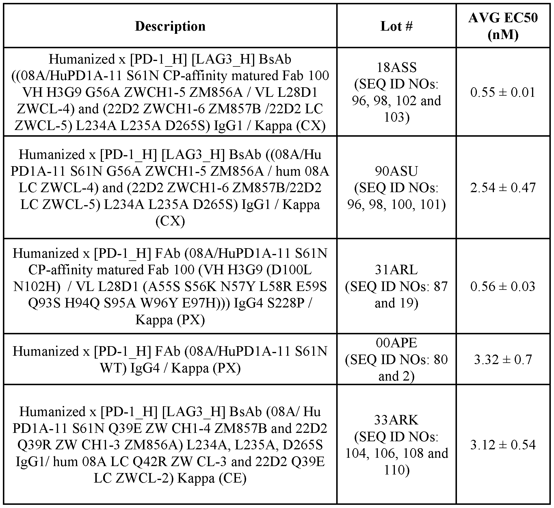

Claims
Priority Applications (10)
| Application Number | Priority Date | Filing Date | Title |
|---|---|---|---|
| CA3089075A CA3089075A1 (en) | 2018-02-01 | 2019-01-31 | Anti-pd-1 antibodies |
| CN201980011259.7A CN111670047A (en) | 2018-02-01 | 2019-01-31 | anti-PD-1 antibodies |
| BR112020015498A BR112020015498A8 (en) | 2018-02-01 | 2019-01-31 | ANTI-PD-1 ANTIBODIES |
| MX2020008122A MX2020008122A (en) | 2018-02-01 | 2019-01-31 | Anti-pd-1 antibodies. |
| JP2020541785A JP2021511806A (en) | 2018-02-01 | 2019-01-31 | Anti-PD-1 antibody |
| KR1020207024743A KR20200115595A (en) | 2018-02-01 | 2019-01-31 | Anti-PD-1 antibody |
| AU2019214977A AU2019214977A1 (en) | 2018-02-01 | 2019-01-31 | Anti-PD-1 antibodies |
| US16/965,616 US20210032342A1 (en) | 2018-02-01 | 2019-01-31 | Anti-pd-1 antibodies |
| EP19747511.4A EP3746120A4 (en) | 2018-02-01 | 2019-01-31 | Anti-pd-1 antibodies |
| RU2020128438A RU2788095C2 (en) | 2018-02-01 | 2019-01-31 | Anti-pd-1 antibodies |
Applications Claiming Priority (2)
| Application Number | Priority Date | Filing Date | Title |
|---|---|---|---|
| CNPCT/CN2018/074916 | 2018-02-01 | ||
| PCT/CN2018/074916 WO2019148410A1 (en) | 2018-02-01 | 2018-02-01 | Anti-pd-1 antibodies |
Publications (1)
| Publication Number | Publication Date |
|---|---|
| WO2019152571A1 true WO2019152571A1 (en) | 2019-08-08 |
Family
ID=67477905
Family Applications (2)
| Application Number | Title | Priority Date | Filing Date |
|---|---|---|---|
| PCT/CN2018/074916 WO2019148410A1 (en) | 2018-02-01 | 2018-02-01 | Anti-pd-1 antibodies |
| PCT/US2019/015932 WO2019152571A1 (en) | 2018-02-01 | 2019-01-31 | Anti-pd-1 antibodies |
Family Applications Before (1)
| Application Number | Title | Priority Date | Filing Date |
|---|---|---|---|
| PCT/CN2018/074916 WO2019148410A1 (en) | 2018-02-01 | 2018-02-01 | Anti-pd-1 antibodies |
Country Status (10)
| Country | Link |
|---|---|
| US (1) | US20210032342A1 (en) |
| EP (1) | EP3746120A4 (en) |
| JP (1) | JP2021511806A (en) |
| KR (1) | KR20200115595A (en) |
| CN (1) | CN111670047A (en) |
| AU (1) | AU2019214977A1 (en) |
| BR (1) | BR112020015498A8 (en) |
| CA (1) | CA3089075A1 (en) |
| MX (1) | MX2020008122A (en) |
| WO (2) | WO2019148410A1 (en) |
Cited By (4)
| Publication number | Priority date | Publication date | Assignee | Title |
|---|---|---|---|---|
| WO2021126906A1 (en) * | 2019-12-20 | 2021-06-24 | Merck Sharp & Dohme Corp. | Methods for treating cancer using a combination of a pd-1 antagonist, an ilt4 antagonist, and chemotherapeutic agents |
| JPWO2021246413A1 (en) * | 2020-06-02 | 2021-12-09 | ||
| WO2022187586A1 (en) * | 2021-03-04 | 2022-09-09 | Code Biotherapeutics, Inc. | Enhanced targeting using antibody-oligonucleotide conjugates |
| US11591390B2 (en) | 2018-09-27 | 2023-02-28 | Celgene Corporation | SIRP-α binding proteins and methods of use thereof |
Families Citing this family (3)
| Publication number | Priority date | Publication date | Assignee | Title |
|---|---|---|---|---|
| WO2019148412A1 (en) | 2018-02-01 | 2019-08-08 | Merck Sharp & Dohme Corp. | Anti-pd-1/lag3 bispecific antibodies |
| EP4261225A1 (en) * | 2020-12-10 | 2023-10-18 | Eutilex Co., Ltd. | Anti-pd-1 antibody and uses thereof |
| EP4353746A1 (en) * | 2021-06-11 | 2024-04-17 | Guangdong Fapon Biopharma Inc. | Anti-pd-1 humanized antibody or antigen-binding fragment thereof and application thereof |
Citations (2)
| Publication number | Priority date | Publication date | Assignee | Title |
|---|---|---|---|---|
| US20130332133A1 (en) * | 2006-05-11 | 2013-12-12 | Ramot At Tel Aviv University Ltd. | Classification of Protein Sequences and Uses of Classified Proteins |
| US20170044259A1 (en) * | 2015-07-13 | 2017-02-16 | Cytomx Therapeutics, Inc. | Anti-pd1 antibodies, activatable anti-pd1 antibodies, and methods of use thereof |
Family Cites Families (7)
| Publication number | Priority date | Publication date | Assignee | Title |
|---|---|---|---|---|
| KR20070083899A (en) * | 2004-10-01 | 2007-08-24 | 메다렉스, 인코포레이티드 | Methods of treating cd30 positive lymphomas |
| CA2855098C (en) * | 2007-06-18 | 2018-02-27 | Merck Sharp & Dohme B.V. | Antibodies to human programmed death receptor pd-1 |
| CN105579469A (en) * | 2013-06-10 | 2016-05-11 | 默沙东公司 | CMV neutralizing antigen binding proteins |
| CN113698488A (en) * | 2014-09-12 | 2021-11-26 | 基因泰克公司 | anti-B7-H4 antibodies and immunoconjugates |
| CN106699888B (en) * | 2015-07-28 | 2020-11-06 | 上海昀怡健康科技发展有限公司 | PD-1 antibody and preparation method and application thereof |
| WO2019148412A1 (en) * | 2018-02-01 | 2019-08-08 | Merck Sharp & Dohme Corp. | Anti-pd-1/lag3 bispecific antibodies |
| AR119507A1 (en) * | 2019-07-30 | 2021-12-22 | Merck Sharp & Dohme | TRISPECIFIC ANTI-PD-1/LAG3/TIGIT ANTIBODIES AND BISPECIFIC ANTI-PD-1/LAG3 ANTIBODIES |
-
2018
- 2018-02-01 WO PCT/CN2018/074916 patent/WO2019148410A1/en active Application Filing
-
2019
- 2019-01-31 US US16/965,616 patent/US20210032342A1/en active Pending
- 2019-01-31 KR KR1020207024743A patent/KR20200115595A/en not_active Application Discontinuation
- 2019-01-31 CA CA3089075A patent/CA3089075A1/en active Pending
- 2019-01-31 JP JP2020541785A patent/JP2021511806A/en active Pending
- 2019-01-31 EP EP19747511.4A patent/EP3746120A4/en active Pending
- 2019-01-31 AU AU2019214977A patent/AU2019214977A1/en active Pending
- 2019-01-31 BR BR112020015498A patent/BR112020015498A8/en unknown
- 2019-01-31 MX MX2020008122A patent/MX2020008122A/en unknown
- 2019-01-31 WO PCT/US2019/015932 patent/WO2019152571A1/en unknown
- 2019-01-31 CN CN201980011259.7A patent/CN111670047A/en active Pending
Patent Citations (2)
| Publication number | Priority date | Publication date | Assignee | Title |
|---|---|---|---|---|
| US20130332133A1 (en) * | 2006-05-11 | 2013-12-12 | Ramot At Tel Aviv University Ltd. | Classification of Protein Sequences and Uses of Classified Proteins |
| US20170044259A1 (en) * | 2015-07-13 | 2017-02-16 | Cytomx Therapeutics, Inc. | Anti-pd1 antibodies, activatable anti-pd1 antibodies, and methods of use thereof |
Non-Patent Citations (2)
| Title |
|---|
| DATABASE UniProtKB [online] 31 January 2018 (2018-01-31), "Uncharacterized protein", Database accession no. A0A2G8RB57 _9RHOB * |
| See also references of EP3746120A4 * |
Cited By (5)
| Publication number | Priority date | Publication date | Assignee | Title |
|---|---|---|---|---|
| US11591390B2 (en) | 2018-09-27 | 2023-02-28 | Celgene Corporation | SIRP-α binding proteins and methods of use thereof |
| WO2021126906A1 (en) * | 2019-12-20 | 2021-06-24 | Merck Sharp & Dohme Corp. | Methods for treating cancer using a combination of a pd-1 antagonist, an ilt4 antagonist, and chemotherapeutic agents |
| JPWO2021246413A1 (en) * | 2020-06-02 | 2021-12-09 | ||
| WO2021246413A1 (en) * | 2020-06-02 | 2021-12-09 | 帝人ファーマ株式会社 | Anti-igf-1 receptor humanized antibody |
| WO2022187586A1 (en) * | 2021-03-04 | 2022-09-09 | Code Biotherapeutics, Inc. | Enhanced targeting using antibody-oligonucleotide conjugates |
Also Published As
| Publication number | Publication date |
|---|---|
| EP3746120A4 (en) | 2021-11-24 |
| CN111670047A (en) | 2020-09-15 |
| MX2020008122A (en) | 2020-09-18 |
| US20210032342A1 (en) | 2021-02-04 |
| BR112020015498A2 (en) | 2020-12-08 |
| CA3089075A1 (en) | 2019-08-08 |
| WO2019148410A1 (en) | 2019-08-08 |
| RU2020128438A (en) | 2022-03-03 |
| JP2021511806A (en) | 2021-05-13 |
| KR20200115595A (en) | 2020-10-07 |
| BR112020015498A8 (en) | 2023-02-07 |
| EP3746120A1 (en) | 2020-12-09 |
| AU2019214977A1 (en) | 2020-08-13 |
Similar Documents
| Publication | Publication Date | Title |
|---|---|---|
| US11072658B2 (en) | Anti-PD-1/LAG3 bispecific antibodies | |
| AU2017331380B2 (en) | Anti-CD27 antibodies | |
| EP3609922A2 (en) | Anti-sirp alpha antibodies | |
| US20210032342A1 (en) | Anti-pd-1 antibodies | |
| US20220251194A1 (en) | Anti-pd-1/lag3/tigit trispecific antibodies and anti-pd-1/lag3 bicpecific antibodies | |
| NL2018708B1 (en) | ANTI-SIRPα ANTIBODIES | |
| RU2788095C2 (en) | Anti-pd-1 antibodies | |
| US20230192876A1 (en) | Dosing regimen of anti-cd27 antibodies for treatment of cancer |
Legal Events
| Date | Code | Title | Description |
|---|---|---|---|
| 121 | Ep: the epo has been informed by wipo that ep was designated in this application |
Ref document number: 19747511 Country of ref document: EP Kind code of ref document: A1 |
|
| ENP | Entry into the national phase |
Ref document number: 3089075 Country of ref document: CA |
|
| ENP | Entry into the national phase |
Ref document number: 2020541785 Country of ref document: JP Kind code of ref document: A |
|
| NENP | Non-entry into the national phase |
Ref country code: DE |
|
| ENP | Entry into the national phase |
Ref document number: 2019214977 Country of ref document: AU Date of ref document: 20190131 Kind code of ref document: A |
|
| ENP | Entry into the national phase |
Ref document number: 20207024743 Country of ref document: KR Kind code of ref document: A |
|
| ENP | Entry into the national phase |
Ref document number: 2019747511 Country of ref document: EP Effective date: 20200901 |
|
| REG | Reference to national code |
Ref country code: BR Ref legal event code: B01A Ref document number: 112020015498 Country of ref document: BR |
|
| ENP | Entry into the national phase |
Ref document number: 112020015498 Country of ref document: BR Kind code of ref document: A2 Effective date: 20200729 |