WO2019116800A1 - Method for comprehensively analyzing 3' end gene expression of single cell - Google Patents
Method for comprehensively analyzing 3' end gene expression of single cell Download PDFInfo
- Publication number
- WO2019116800A1 WO2019116800A1 PCT/JP2018/041702 JP2018041702W WO2019116800A1 WO 2019116800 A1 WO2019116800 A1 WO 2019116800A1 JP 2018041702 W JP2018041702 W JP 2018041702W WO 2019116800 A1 WO2019116800 A1 WO 2019116800A1
- Authority
- WO
- WIPO (PCT)
- Prior art keywords
- sequence
- cdna
- cell
- reaction
- mrna
- Prior art date
Links
Images


Classifications
-
- C—CHEMISTRY; METALLURGY
- C12—BIOCHEMISTRY; BEER; SPIRITS; WINE; VINEGAR; MICROBIOLOGY; ENZYMOLOGY; MUTATION OR GENETIC ENGINEERING
- C12N—MICROORGANISMS OR ENZYMES; COMPOSITIONS THEREOF; PROPAGATING, PRESERVING, OR MAINTAINING MICROORGANISMS; MUTATION OR GENETIC ENGINEERING; CULTURE MEDIA
- C12N15/00—Mutation or genetic engineering; DNA or RNA concerning genetic engineering, vectors, e.g. plasmids, or their isolation, preparation or purification; Use of hosts therefor
- C12N15/09—Recombinant DNA-technology
- C12N15/10—Processes for the isolation, preparation or purification of DNA or RNA
- C12N15/1096—Processes for the isolation, preparation or purification of DNA or RNA cDNA Synthesis; Subtracted cDNA library construction, e.g. RT, RT-PCR
-
- C—CHEMISTRY; METALLURGY
- C12—BIOCHEMISTRY; BEER; SPIRITS; WINE; VINEGAR; MICROBIOLOGY; ENZYMOLOGY; MUTATION OR GENETIC ENGINEERING
- C12Q—MEASURING OR TESTING PROCESSES INVOLVING ENZYMES, NUCLEIC ACIDS OR MICROORGANISMS; COMPOSITIONS OR TEST PAPERS THEREFOR; PROCESSES OF PREPARING SUCH COMPOSITIONS; CONDITION-RESPONSIVE CONTROL IN MICROBIOLOGICAL OR ENZYMOLOGICAL PROCESSES
- C12Q1/00—Measuring or testing processes involving enzymes, nucleic acids or microorganisms; Compositions therefor; Processes of preparing such compositions
- C12Q1/68—Measuring or testing processes involving enzymes, nucleic acids or microorganisms; Compositions therefor; Processes of preparing such compositions involving nucleic acids
- C12Q1/6813—Hybridisation assays
- C12Q1/6834—Enzymatic or biochemical coupling of nucleic acids to a solid phase
- C12Q1/6837—Enzymatic or biochemical coupling of nucleic acids to a solid phase using probe arrays or probe chips
Definitions
- the present invention relates to a system, method and kit for analyzing comprehensive gene expression utilizing mRNA 3 'sequence at single cell level.
- analysis methods in which tissues and a large number of cells are used as analysis samples, differences between cells are averaged and measured, so integrated analysis of life activities among individual cells and cells is required. Is difficult.
- immune system cells neurons and pluripotent stem cells, the type of gene being expressed and its expression level greatly differ from cell to cell, and for the purpose of elucidating the mechanism of disease, gene expression at single cell level Development of analysis technology is progressing rapidly.
- RNA-seq a method for sequencing the full-length 1st cDNA (whole gene) synthesized from mRNA
- NGS analysis the device body and sequencing reagents are very expensive.
- sequence cost required for each detection gene becomes expensive in the RNA-seq method because there is a limitation in the decoded base length (size) per NGS analysis run. Therefore, a method of preparing a sample of only the 1st cDNA corresponding to the sequence at the 3 'end of mRNA, which can be reduced in cost to about 1/5 to 1/10, and analyzing it by NGS has recently become widespread.
- Non-patent document 1 sorted cells into a microwell plate using a cell sorter, amplified a DNA sequence derived from the sequence at the 3 'end of mRNA, and performed NGS analysis to obtain a total of 12832 cell genes. Expression analysis has been successful.
- the amount of mRNA contained in one cell is a very small amount of about 0.5 pg (10 5 to 10 6 molecules), so the technical issues to be overcome in detection rate (detection sensitivity) and quantification accuracy in low expression gene group Still there.
- Patent Document 1 first solves sample loss from a very small amount of mRNA and aims for quantitative analysis with high accuracy, for example, a magnetic bead surface on which many probes are immobilized.
- a magnetic bead surface on which many probes are immobilized.
- expression analysis is performed with high accuracy even for low expression genes of about 10 copies per cell for multiple genes A method is disclosed.
- Patent Document 2 discloses a method for gene expression analysis of a large number of cells at a single cell level using a chip composed of beads arranged in a porous membrane or a two-dimensional array.
- the sample preparation proceeds while maintaining high reaction efficiency in each series of steps, and the DNA molecule derived from the initial sample of about 0.5 pg (10 5 to 10 6 molecules) and a very small amount of mRNA molecule per cell is finalized. It is important how many are left in the sample. In particular, it is important to avoid sample loss in the first half of the process up to PCR amplification. Furthermore, in sample preparation using a small amount of DNA such as single cell analysis, the optimum reaction conditions (amount of enzyme, reaction time, temperature) in the DNA fragmentation step (including tagmentation step) by enzyme treatment may be shifted even a little In addition, there is a problem that the target DNA is easily fragmented or degraded to 250 bases or less, which is a major cause of sample loss.
- sample loss which is lost in the middle of various reaction steps is avoided, and 3 'of mRNA is obtained under reaction conditions in which the utilization efficiency of this initial sample is maximized.
- the ability to prepare target DNA samples derived from terminal sequences is the most important task in order to perform comprehensive gene expression analysis with high accuracy and accuracy at low cost.
- the present invention has been made in view of the above problems, and it is an object of the present invention to provide a method for efficiently performing comprehensive gene expression analysis at one cell level.
- the present inventors conducted efficient utilization of mRNA molecules in one cell, which is an initial sample, when conducting comprehensive gene expression analysis at one cell level for a plurality of cells simultaneously.
- the present invention is a method of analyzing gene expression of a cell using a device having a plurality of microreactors, for example, a device in which a plurality of chips (or arrays) are incorporated in parallel,
- a device having a plurality of microreactors for example, a device in which a plurality of chips (or arrays) are incorporated in parallel
- one or more solid phase carriers on which a probe including an amplification primer sequence, a cell identification sequence, a molecule identification sequence, and an oligo (dT) sequence is immobilized are packed.
- the above method is Introducing a plurality of cells into the microreactor such that a single cell corresponds to each microreactor; Capturing the single cell-derived mRNA into the probe; 1st cDNA is synthesized by reverse transcription reaction of the captured mRNA, and a single cell-derived 1st cDNA library is prepared on the solid support.
- washing the solid phase carrier (pooled); Synthesizing 2nd cDNA from the 1st cDNA library; Fragmentation of double-stranded DNA consisting of said 1st cDNA and said 2nd cDNA and addition of a tag sequence; Washing the solid support with a washing solution to remove components other than the immobilized double-stranded DNA fragment; The double-stranded DNA fragment is amplified using the amplification primer sequence and a primer having a sequence complementary to at least a portion of the sequence or at least a portion of the tag sequence, and the 3 'terminal sequence of the mRNA is Amplifying only the derived sequence; And d) performing gene expression analysis for each single cell using the cell identification sequence and the molecule identification sequence for the amplified sequences.
- gene expression analysis is carried out simultaneously by amplifying a plurality of cells simultaneously and only a sequence derived from the 3 'end of mRNA, so that labor required for analysis and reagent cost can be reduced.
- target DNA molecules derived from mRNA are held by the solid phase carrier (magnetic beads) over multiple steps including the 1st cDNA synthesis step, the 2nd cDNA synthesis step, the tagmentation step, and the PCR step, the whole process is performed every step. Simple washing with magnetic beads completely removes residual reagent and allows recovery of all target DNA molecules from mRNA with 100% efficiency. That is, the reaction can be carried out with high efficiency using optimum reagent conditions in each step, and there is no sample loss in purification.
- the final sample in which only the sequence derived from the 3 'end of the mRNA is amplified can be applied to NGS analysis by advancing the reaction from an extremely small amount of mRNA molecules in one cell with the utilization efficiency increased to the limit, accuracy and precision High comprehensive gene expression data can be obtained.
- the present invention is applicable to drug discovery, elucidation of mechanisms in various diseases, regenerative medicine and the like, and can also contribute to the development of life sciences.
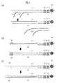
- (a) is a top view of an example of a chip in which micro reaction vessels are arranged in an array.
- (B) is a cross-sectional view of an example of a chip, an enlarged view (c) shows an example of a step of capturing mRNA eluted after cell lysis in a micro reaction tank, and an enlarged view (d) further shows It is the schematic which showed an example of the process of synthesize
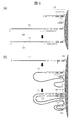
- A shows a schematic diagram of another example of 2nd cDNA synthesis method on a carrier using random primers and strand displacement DNA polymerase.
- B shows a schematic view of another example of 2nd cDNA synthesis method on a carrier using single stranded DNA ligase.
- C shows a schematic view of another example of 2nd cDNA synthesis method on a carrier using terminal transferase.
- a A schematic showing a step of 1st cDNA synthesis on the surface of a carrier (magnetic bead) on which two types of probes, that is, a probe 109 for reverse transcription reaction (SEQ ID NO: 1) and a random primer 213 for 2nd cDNA synthesis, are immobilized.
- FIG. 1 An example of a schematic diagram of a 2nd cDNA synthesis method on a carrier using immobilized random primers 213 and a strand displacement DNA polymerase. It is the schematic of two types of tag arrangement
- FIG. 1 It is a graph which shows the experimental data which analyzed ERCC (Ambion, sample in which known amounts of 92 types of mRNA were mixed) by the method described in Example 1 for the purpose of checking the accuracy of quantification.
- 1 is a graph showing 1-cell analysis experiment data by the method described in Example 1.
- FIG. It is the graph which showed the average detection gene number per cell obtained by the method of Example 1, and the total detection gene number per chip (here 100 cell recognition tags).
- the present invention relates to a method for performing comprehensive gene expression analysis at one cell level for a plurality of cells simultaneously.
- a plurality of chips (or arrays) in which a plurality of micro reaction vessels are arranged in an array form is used to simultaneously capture a plurality of cells using a device in which a plurality of cells are incorporated in parallel, Efficiently capture and synthesize 1st cDNA.
- 1st cDNA derived from a plurality of cells is pooled in one tube and the remaining reagent is washed.
- a tagmentation reaction or a reaction of adding a tag sequence by a ligation reaction after a reaction of fragmenting double-stranded DNA with a DNA fragmentation enzyme
- a tagmentation inhibitor or a reaction of adding a tag sequence by a ligation reaction after a reaction of fragmenting double-stranded DNA with a DNA fragmentation enzyme
- a tagmentation inhibitor or a reaction of adding a tag sequence by a ligation reaction after a reaction of fragmenting double-stranded DNA with a DNA fragmentation enzyme
- a tagmentation inhibitor Unnecessary fragmented DNA is removed by washing with a detergent containing H. and PCR amplification of only the 3 'end portion of mRNA is efficiently performed.
- gene expression analysis refers to quantitatively analyzing the expression of a gene, that is, mRNA in a sample (cell, tissue section, etc.), analyzing the expression distribution of a gene (mRNA) in a sample, It means that correlation data between a specific cell or position in and the gene (mRNA) expression level is obtained.
- the sample is not particularly limited as long as it is a biological sample from which gene expression is to be analyzed, and any sample such as a cell sample, a tissue sample, and a liquid sample can be used. Further, the living body from which the sample is derived is not particularly limited.
- a DNA fragment derived from the 3 'end of mRNA to be analyzed is generically defined as "target DNA”.
- all gene expression analysis refers to parallel expression analysis of a plurality of genes contained in a cell, for example, parallel expression analysis of at least 1000 or more genes. is there.
- one-cell level gene expression analysis means expression analysis of genes (mRNA) contained in one cell, and is distinguished from average expression analysis of genes contained in a plurality of cells.
- the present disclosure is a method of analyzing gene expression of a cell using a device having a plurality of microreaction vessels, for example, a device in which a plurality of chips (or arrays) are incorporated in parallel,
- a device having a plurality of microreaction vessels for example, a device in which a plurality of chips (or arrays) are incorporated in parallel
- one or more solid phase carriers on which a probe including an amplification primer sequence, a cell identification sequence, a molecule identification sequence, and an oligo (dT) sequence is immobilized are packed.
- the above method is Introducing a plurality of cells into the microreactor such that a single cell corresponds to each microreactor; Capturing the single cell-derived mRNA into the probe; 1st cDNA is synthesized by reverse transcription reaction of the captured mRNA, and a single cell-derived 1st cDNA library is prepared on the solid support.
- washing the solid phase carrier (pooled); Synthesizing 2nd cDNA from the 1st cDNA library; Fragmentation of double-stranded DNA consisting of said 1st cDNA and said 2nd cDNA and addition of a tag sequence; Washing the solid support with a washing solution to remove components other than the immobilized double-stranded DNA fragment; The double-stranded DNA fragment is amplified using the amplification primer sequence and a primer having a sequence complementary to at least a portion of the sequence or at least a portion of the tag sequence, and the 3 'terminal sequence of the mRNA is Amplifying only the derived sequence; And d) performing gene expression analysis for each single cell using the cell identification sequence and the molecule identification sequence for the amplified sequences.
- a device having a plurality of microreaction vessels is a chip configured to analyze gene expression, in which a plurality of so-called two-dimensional arrays are incorporated in parallel, and in the microreaction vessels of this device, One or more solid phase carriers on which a probe including an amplification primer sequence, a cell identification sequence, a molecule identification sequence, and an oligo (dT) sequence is immobilized are packed.
- Such devices are known in the art and are not particularly limited. For example, devices described in Patent Document 1, Patent Document 2, International Publication WO 2016/038670, etc. can be used.
- the solid phase carrier to be packed in the microreactor is preferably prepared using a material having a large surface area in order to increase the capture efficiency of mRNA, for example, employing one or more beads, porous structure, mesh structure, etc. It is preferable to do.
- the beads can be prepared from resin materials (polystyrene etc.), oxides (glass etc.), metals (iron etc.), sepharose, and combinations thereof. It is preferable to use magnetic beads for the simplicity of operation.
- the solid support is preferably in the size of 10 nm to 100 ⁇ m in diameter, for example, in the size of 10 nm to 100 ⁇ m in diameter.
- a porous sheet or a porous membrane may be disposed so that such a solid support does not leak from the micro reaction vessel.
- a probe containing amplification primer sequences, cell identification sequences, molecular identification sequences, and oligo (dT) sequences is immobilized on a solid support, and such probes are synthesized by conventional oligonucleotide synthesis methods. And may be immobilized on the solid support by any method known in the art.
- the degree of polymerization of the oligo (dT) may be any degree of polymerization that can be hybridized with the poly A sequence of mRNA and capture the mRNA on the solid support on which the oligo (dT) is immobilized. For example, it can be about 10 to 20 bases.
- this sequence can be used as a common primer in an amplification step (for example, PCR).
- mRNA molecule or DNA molecule derived from mRNA is used as the molecular recognition sequence.
- a DNA probe containing a random primer may be further immobilized on a solid support for subsequent synthesis of 2nd cDNA.
- the random primer is not particularly limited as long as it has a length and composition capable of functioning as a primer, and for example, a random primer having a length of 6 to 15 bases can be used.
- each of the microreaction vessels one through hole is formed, and a single cell is captured in the through hole.
- the through holes can be appropriately set according to the size of cells to be analyzed, but preferably have a diameter of 10 ⁇ m or less.
- a plurality of cells are introduced into the microreactor such that a single cell corresponds to each microreactor.
- a single cell is captured in each of the through holes by applying (suctioning) a negative pressure to the through holes.
- the observation device confirms whether the cells are trapped in the through holes, and reintroduces the cells as necessary. It is preferable to remove non-captured cells, for example, by introducing and discharging a washing solution, because they affect the subsequent steps.
- capture of mRNA means that mRNA molecules contained in cells are extracted and separated from other cellular components.
- cell lysates known in the art are aliquoted into microreactors and mRNA is extracted from each captured single cell.
- cells are lysed using a proteolytic enzyme, chaotropic salt such as guanidine thiocyanate guanidine hydrochloride, detergent such as Tween and SDS, or a commercially available reagent for cell lysis (eg Lysis solution).
- Nucleic acids, ie mRNA can be eluted. If necessary, check the status of cell lysis with an observation device. The eluted mRNA is captured by the probe by binding to the oligo (dT) sequence of the probe.
- the device, microreactor, and solid support are washed to remove unwanted components and reagents, optionally using a wash solution.
- 1st cDNA having a sequence complementary to the sequence of mRNA or a part of the sequence is synthesized by reverse transcription reaction of captured mRNA.
- This 1st cDNA synthesis ie complementary strand synthesis, can be performed by methods known in the art.
- cDNA can be synthesized by performing reverse transcription using a conventional reverse transcriptase or a reverse transcriptase having a template switch function. After the synthesis reaction, the mRNA is degraded and removed using, for example, RNase.
- a cDNA library composed of 1st cDNA corresponding to mRNA is prepared on the solid phase carrier. Since single cells correspond to one microreactor, a single cell-derived 1st cDNA library can be prepared on a solid phase carrier contained in each microreactor.
- a step of pooling the solid phase carrier on which the single cell-derived 1st cDNA library is immobilized is divided into a plurality of cells is performed.
- a solid support on which a single cell-derived 1st cDNA library prepared in each of a plurality of microreactors on a single chip is immobilized is collectively placed in a single tube or the like to obtain 1st cDNA for a plurality of cells. It can be a pool of libraries.
- the 1st cDNA library can pool, for example, about 100 to 10000 cells per chip.
- the subsequent steps can be performed collectively for a plurality of cells of a single cell-derived 1st cDNA library, and simplification of the operation and reduction of the reagent cost can be achieved.
- the cell recognition sequence is present in the solid phase carrier, even when 1st cDNA libraries for multiple cells are mixed and pooled, they are derived from any microreactor at the time of gene expression analysis ( It is possible to identify which single cells are derived).
- a plurality of chips are incorporated in parallel in one device, it is possible to process 1600 to 160000 cells per reaction using, for example, a device in which 16 chips are incorporated. It becomes possible.
- the chip recognition tag is also introduced into the final prepared sample during the PCR amplification process, the samples from all the cells are pooled into one and analyzed by the next generation sequencer. It is possible to perform gene expression analysis separately.
- 2nd cDNA is synthesized from 1st cDNA library.
- the 2nd cDNA synthesis step can be performed using complementary strand synthesis reactions known in the art. While several examples are given, one skilled in the art can select and implement appropriate methods.
- One method is to synthesize 2nd cDNA by complementary strand extension reaction using a random primer and a DNA polymerase having strand displacement activity.
- the random primer is not particularly limited as long as it has a length and composition capable of functioning as a primer, and for example, a random primer having a length of 6 to 15 bases can be used.
- DNA polymerases having strand displacement activity are also known in the art, and, for example, Phi29 DNA polymerase, Bst DNA polymerase, Csa DNA polymerase and the like are commercially available.
- Phi29 DNA polymerase, Bst DNA polymerase, Csa DNA polymerase and the like are commercially available.
- a reaction as shown in (a) of FIG. 2 occurs, and 2nd cDNA can be synthesized with high synthesis efficiency.
- 2nd cDNA synthesis step is to use a specific sequence, since a specific sequence is added to the 1st cDNA when a reverse transcriptase having a template switch function is used in the 1st cDNA synthesis. is there.
- a specific sequence is added to the 1st cDNA when a reverse transcriptase having a template switch function is used in the 1st cDNA synthesis.
- SmartScribe Reverse Transcriptase, SuperScript II, SuperScript IV, etc. are commercially available. That is, 2nd cDNA is synthesized by complementary strand extension reaction using a primer containing a sequence complementary to the added specific sequence.
- Conventional DNA polymerases can be used, and for example, Tks Gflex DNA polymerase, Ex Hot start DNA Polymerase, Platinum Taq DNA Polymerase High Fidelity, etc. are commercially available.
- a reaction as shown in “washing after 2nd cDNA synthesis in FIG. 1-2” occurs, and 2nd c
- Another example of the 2nd cDNA synthesis step is to first add a known sequence to the 3 'end of the 1st cDNA using a single stranded DNA ligase, and extend the complementary strand using a primer containing a sequence complementary to this known sequence
- the 2nd cDNA is synthesized by the reaction.
- Single-stranded DNA ligase is commercially available, for example, Circ Ligase ss DNA Ligase.
- the known sequence to be added can also be of suitable length and composition, for example, a sequence of 10 to 30 bases in length can be added.
- a reaction as shown in (b) of FIG. 2 takes place, and it is possible to synthesize 2nd cDNA.
- Yet another example of the 2nd cDNA synthesis step is to add a polybase sequence (poly T, A, G or C sequence) to the 3 'end of the 1st cDNA by terminal transferase (TdT) first, and to complement this polybase sequence 2nd cDNA is synthesized by complementary strand extension reaction using a primer containing the above sequence.
- a polybase sequence poly T, A, G or C sequence
- TdT terminal transferase
- DNA polymerase can be used, and those skilled in the art can appropriately select and use.
- the polynucleotide sequence to be added can be of an appropriate type and length, and can be, for example, a polynucleotide sequence of 10 to 30 bases in length.
- a DNA probe having a random primer previously immobilized on a solid phase carrier, and a DNA having a strand displacement activity and a random primer immobilized on the solid phase carrier in the 2nd cDNA synthesis process The 2nd cDNA is synthesized by complementary strand extension reaction using a polymerase and the cDNA is amplified.
- the DNA polymerase having strand displacement activity is as described above, and any can be used.
- a reaction as shown in (b) of FIG. 3 takes place, and it is possible to synthesize 2nd cDNA and further amplify cDNA.
- a random primer (unimmobilized, (a) in FIG. 2) is also added to the reaction liquid phase to allow in the liquid phase and on the solid phase carrier.
- the reaction may proceed from both sides.
- tag maintenance reaction can be used.
- the tag maintenance reaction is to fragment double-stranded DNA and add a tag sequence, and is a reaction known in the art. Enzymes (transposases) and reagents to be used are also commercially available, and those skilled in the art can carry out tag maintenance reactions using appropriate enzymes and reagents.
- a reaction of adding a tag sequence by ligation reaction is performed.
- DNA fragmentation enzymes and enzymes used for ligation are also known in the art, and one of ordinary skill in the art can select appropriate reagents.
- the tag sequence to be added is not particularly limited as long as it has a length and a composition suitable for binding to the primer in the subsequent amplification step, and for example, a base sequence having a length of about 20 to 35 bases It can be done.
- the solid support is washed with a washing solution to remove components other than the immobilized double stranded DNA fragment. It is possible to immediately stop the activity of the enzyme used for DNA fragmentation and tag addition in the previous step, particularly the enzyme used for tag maintenance reaction (transposase) to reduce the influence on the subsequent steps.
- the solid support is preferably washed with a washing solution having an inhibitory effect on the enzyme used. This washing step makes it possible to extract only target DNAs as short as several hundred bases (ie, sequences derived from the 3 'end of mRNA) and remove DNAs of other sequences which are by-products. That is, in the subsequent gene expression analysis step, cost, labor and analysis time in gene identification (sequencing) and quantitative analysis can be reduced as compared with the case of using a normal full-length DNA sequence.
- the double-stranded DNA fragment is amplified using a primer having an amplification primer sequence and a sequence complementary to at least a portion of the sequence or at least a portion of the tag sequence, and derived from the 3 'terminal sequence of mRNA Only sequence is amplified.
- Other sequences may be added to the primer, for example, a sequence for identifying a used chip or a sequence required for the subsequent NGS analysis. Design of primers, conditions for amplification reaction and the like are known in the art, and may be appropriately selected depending on the length of a sequence to be amplified, reagents to be used, and the like.
- 1st cDNA library is prepared on the solid phase carrier, and residual reagents and byproducts are simply and completely removed by washing after various reactions (1st cDNA synthesis step, 2nd cDNA synthesis step, and tagmentation step).
- target DNA which is a sequence derived from the 3 'terminal sequence of mRNA without sample loss while immobilized on a solid support. Since the sample obtained by PCR amplification of this contains only the target DNA prepared by raising the utilization efficiency to an extreme from the extremely small amount of mRNA molecule derived from each cell, the final result of gene expression analysis is good. It becomes possible to obtain.
- amplified sequences are then subjected to gene expression analysis in single cells using cell and molecular recognition sequences. Specifically, amplified sequences are subjected to sequencing by NGS (Next Generation Sequencer) to analyze gene expression in single cells. Since the amplified sequences include a chip identification sequence, a cell identification sequence, and a molecule identification sequence, which molecule is used as an index, which molecule is derived from which chip, or which single cell is derived It is possible to identify gene origin and analyze gene expression.
- NGS Next Generation Sequencer
- the gene expression analysis method of the present disclosure described above is a kit including devices necessary for carrying out each step, reagents such as enzymes, washing solutions, disposable containers (tubes), instructions including the description of the implementation of such methods, and the like. It is possible to do it easily and simply by using.
- a device incorporating a plurality of chips necessary for performing each step, a means for introducing a reagent, a washing solution and the like, a means for observing the chip, and a negative pressure is applied to the chip It can be carried out easily and simply by using a system equipped with means for
- the above-mentioned step (4) is automatically performed in the device on which the chip is mounted, and the steps before the PCR amplification in the state where DNA is held on the carrier (the above-mentioned reaction steps (1) (2) (3) (4) (5) (6)) can also be performed at once using the above device.
- the method of the present embodiment makes it possible to reduce the labor of steps of sample preparation simultaneously from each of a large number of cells. Details regarding each process are described below.
- Cell capturing step on chip A device mounted with 16 chips 100 (a plan view of (a) in FIG. 1-1, a cross sectional view of (b) in FIG. 1-1) in which 100 micro reaction vessels 103 are arranged in an array.
- a sequence 112 for PCR amplification (SEQ ID NO: 4)
- a cell identification sequence 111 (SEQ ID NO: 3) which is a known sequence of 6 bases different for each microreaction vessel, 7 bases different for each probe molecule
- a carrier preferably, a high density immobilized reverse transcription probe 109 (SEQ ID NO: 1) consisting of a random sequence of SEQ ID NO: 2 (SEQ ID NO: 2) and a sequence of oligo (dT) VN Magnetic beads) 104 are abundantly packed.
- a micro through hole 102 with a diameter (2 to 6 ⁇ m) smaller than that of cells is present on the upper surface of the micro reaction vessel, and the lower surface is in close contact with a porous material membrane (pore diameter: 0.8 ⁇ m, Millipore) 130 to make the carrier
- a reagent discharge unit 105 through which the reagent is passed while being held. That is, since the device used in this embodiment has a structure capable of applying a negative pressure from the lower direction of the chip, it passes through the reagent discharge portion 105 of the micro reaction vessel and the reagent is applied from the top and the inside of the micro reaction vessel. It can be discharged.
- PBS Phosphate buffered saline
- RNase Inhibitor (1 U / ⁇ L)
- the reaction vessel 103 is washed.
- GFP green fluorescent protein
- HCT116 human colon cancer cells
- a plurality of cells 101 (in this embodiment, the number of input cells: 1280) can be simultaneously captured on the upper surface of the microreactor. Observation using a fluorescence microscope confirms that cell capture is complete within about 1 minute. After outputting the NGS analysis data as the final result, since the position of the micro reaction vessel can be identified by using the cell identification sequence 111 as a clue, the size and state of the cell can be determined by comparing it with the moving image / image of this cell capture. It can be examined.
- the number of reverse transcription reaction probes per microreactor is 5 ⁇ 10 9 to 2 ⁇ 10 10 molecules. That is, it is sufficient as a probe for capturing 10 5 to 10 6 molecules of mRNA present per cell, and it is possible to capture mRNA with high efficiency.
- the synthesized 1st cDNA has a specific sequence 114 of several bases added at the 3 'end.
- SMART-Seq v4 Oligo 115 which contains a complementary sequence to this specific sequence 114 at the 3 'end, is bound to a complementary strand, and this is used as a template for further 1st cDNA synthesis.
- the finally synthesized 1st cDNA has the complementary sequence of SMART-Seq v4 Oligo 115 at the 3 'end, the sequence for PCR amplification 112 (SEQ ID NO: 4) at the 5' end, the cell identification sequence 111 (SEQ ID NO: 3) and molecular identification sequence 110 (SEQ ID NO: 2) (FIG. 1-1 (d)).
- SEQ ID NO: 4 the sequence for PCR amplification 112
- SEQ ID NO: 3 the cell identification sequence 111
- SEQ ID NO: 2 molecular identification sequence 110
- 1st cDNA library samples synthesized from each of a plurality of cells simultaneously using the chip are pooled. Since the cell identification sequence 111 is different for each 1st cDNA library (microreactor), there is no problem because it is possible to distinguish for each cell in the NGS analysis data even if pooled in this step. In addition, as the number of cells to be pooled increases, the labor and cost required for sample preparation can be reduced. Visually confirm that all the support has been expanded into the buffer, and remove the tip and porous membrane from the tube.
- the supernatant containing the residual reagent of the 2nd cDNA synthesis reaction is removed and washed with 50 ⁇ L of carrier washing solution (10 mM Tris, 0.1% Tween 20 (pH 8.0)).
- tag sequence A consisting of portion 122 (SEQ ID NO: 6) and tag sequence B consisting of common sequence portion 121 and specific sequence B portion 123 (SEQ ID NO: 7) are randomly added Ru.
- tag sequence B consisting of common sequence portion 121 and specific sequence B portion 123 (SEQ ID NO: 7)
- Ru random addition
- 50 ⁇ L of a concentrated detergent-containing detergent solution (0.1% Tween 20, 100 mM Tris (pH 8.0), 500 mM NaCl) cooled on ice is added, and the neodymium magnet 118 captures the carrier. While removing the supernatant containing residual reagents of the tagmentation reaction.
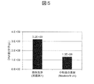
- FIG. 5 shows experimental data comparing the amount of DNA after PCR amplification using a sample using carrier washing, which is the method of the present example, and a sample using a neutralization solution, which is the conventional method.
- the amount of DNA obtained by the method of this example is increased by 2.5 times (graph on the left), compared to the conventional method (graph on the right) affected by sample loss due to fragmentation.
- the problem of sample loss is serious because the problem of sample loss greatly affects detection sensitivity and quantitative accuracy when reacting trace DNA such as single cell analysis, but the method of the present embodiment can avoid this problem.
- PCR cycles of 98 ° C. for 15 seconds ⁇ 60 ° C. for 45 seconds ⁇ 68 ° C. for 30 seconds are performed in a thermal cycler and cooled to 4 ° C.
- About 30 ⁇ L of PCR amplification product sample, which is the supernatant, is collected into another tube using a neodymium magnet.
- the carrier surface and the inner wall of the tube are washed with 20 ⁇ L of 0.1% Tween 20 (10 mM Tris (pH 8.0)), and the remaining PCR product is additionally collected and mixed with the PCR amplification product sample (total 50 ⁇ L).
- the DNA sample was purified and quantified using Ampure XP beads to obtain the final sample 127 for NGS analysis.
- a chip identification sequence 126 which is a known sequence of 5 bases different for each chip (tube) is introduced into the target DNA. That is, since it becomes possible to identify the 16 chips used in the present embodiment, a total of 1600 cells can be theoretically distinguished by combining with 100 types of cell identification sequences 111. In other words, comprehensive gene expression analysis can be performed on 1600 cells in one NGS analysis.
- a carrier immobilized DNA (a DNA sequence derived from the 3 'end of mRNA) to which a tag sequence A or a sequence of tag sequence B is obtained obtained after the tagmentation reaction is used as a template
- a Reverse primer is used which contains a 19-base consensus sequence portion 121 (SEQ ID NO: 5) (FIG. 4) possessed by both tag sequences.
- a primer using a specific sequence A portion (14 bases) (FIG. 4) and a specific sequence B portion (15 bases) (FIG. 4) is used, and the template and complementary strand are linked
- the complementary strand avidity of the target DNA is weak because the sequence to be prepared is as short as 14 to 15 bases.
- FIG. 6 shows the same sample in which the target DNA is immobilized on the carrier obtained by the tagmentation reaction as a template, (1) Forward primer including the sequence 112 for PCR amplification (SEQ ID NO: 4), and a consensus sequence of 19 bases PCR amplification product sample using Reverse primer containing 121, (2) Forward primer containing sequence for PCR amplification 112, and both specific sequence A portion (14 bases) and specific sequence B portion (15 bases) PCR amplification sample using Reverse primer, (3) Forward primer including sequence 112 for PCR amplification, Reverse primer including both specific sequence A portion (14 bases) and specific sequence B portion (15 bases), and It is the experimental data which compared the amount of DNA contained in the PCR amplification sample using the sequence primer for NGS (P5) (sequence number 11) and the sequence primer for NGS (P7) (sequence number 12) for amplification support.
- SEQ ID NO: 4 sequence 112 for PCR amplification
- P7 sequence primer for NGS
- the amount of DNA in the sample is significantly large. That is, in the method (1) of the method of the present embodiment, since the complementary strand binding sequence is as long as 19 bases, it can be stably annealed with the template to the conventional method (2) (3) which is short as 14 to 15 bases. It is thought that a good PCR amplification was achieved.
- the target DNA to be a template (a DNA sequence derived from the 3 'end of mRNA) is not immobilized on a carrier, so a by-product in the tagmentation reaction (derived from other than the 3' end of mRNA) Since the DNA fragment (with two types of tags added) remains in the sample, the PCR amplification step digests the DNA polymerase and primers for amplification of by-products, and the amplification efficiency of the target DNA is even lower. It is considered to be.
- PCR amplification products derived from byproducts adversely affect the accuracy and sensitivity of quantification in NGS analysis.
- the method of this embodiment can avoid various problems in the conventional amplification process.
- Step of NGS Analysis 80 cells are charged / captured per one chip 100, and analysis is performed by the NGS device 128 using the final sample 127 obtained through the various steps. That is, after the obtained sequence reads were separated by 100 types of cell identification sequences 111, the number of genes detected during the sequence reads per cell identification sequence was examined. You can check the data (Figure 8). That is, 2-3 cell data assumed that multiple cells were captured per microreactor 103, 1 cell data, and 0 cell data assumed that cells were not captured (the processes did not work well) Each can be confirmed. The average number of detection genes in 1-cell data was 7818, and the total number of detection genes per chip 100 was 15773 (FIG. 9).
- Example 1 As in Example 1, (1) cell capture step on the chip on which the microreaction vessel is arranged on the array, (2) mRNA capture step after cell lysis, (3) 1st cDNA synthesis step on the carrier surface, (4) Developing 1st cDNA library immobilized carrier (magnetic beads) into 1 tube, pooling and washing, (5) Synthesizing 2nd cDNA and washing, (6) Tagmentation reaction, washing (7) PCR amplification step, (8) NGS analysis step.
- the 1st cDNA synthesis step on the carrier surface of (3) not the expensive reverse transcriptase having TS function used in Example 1, but an inexpensive reverse transcriptase, which is used in the sample preparation reagent. Cost reduction is possible.
- PCR amplification sequence 112 (SEQ ID NO: 4), a cell identification sequence 111 (SEQ ID NO: 3), and a molecule identification sequence 110 (SEQ ID NO: 2) at the 5 'end of the finally synthesized 1st cDNA.
- this step it is possible to simultaneously synthesize 1st cDNA library from the mRNAs derived from all the genes expressed in a plurality of single cells in a state of being immobilized on a carrier.
- Step of synthesizing and washing 2nd cDNA-using random primer and strand displacement type DNA polymerase Expand the 1st cDNA library immobilized carrier (magnetic beads) into one tube as in Example 1. After performing the pool and washing steps, mix this sample with Exonuclease I reagent (1 ⁇ Buffer, Exonuclease I (1 U / ⁇ L)) to make a 5 ⁇ L reaction solution, and incubate at 37 ° C. for 15 minutes. Subsequently, incubate at 80 ° C. for 15 minutes to inactivate Exonuclease I heat.
- Exonuclease I reagent (1 ⁇ Buffer, Exonuclease I (1 U / ⁇ L)
- the carrier is repeatedly washed twice with 50 ⁇ L of a washing solution (0.1% Tween 20, 10 mM Tris (pH 8.0)).
- a washing solution (0.1% Tween 20, 10 mM Tris (pH 8.0)
- 5 ⁇ L of RNase H reagent 50 mM This-HCl (pH 8.3), 75 mM KCl, 3 mM MgCl 2 , 20 mM DTT, RNase H (1 U / ⁇ L): Thermo Fisher
- the carrier After mixing with the carrier and incubating for 15 minutes at 37 ° C., the carrier is repeatedly washed twice with 50 ⁇ L of washing solution (0.1% Tween 20, 10 mM Tris (pH 8.0)). By this operation, mRNA 108 can be degraded and removed.
- 5 ⁇ L of 2nd cDNA synthesis reagent (10 ⁇ M random primer 201 (SEQ ID NO: 13), 1 ⁇ Bst Reaction Buffer, 0.25 mM dNTP mix, Bst DNA polymerase (1.6 U / ⁇ L): Nippon Gene Co., Ltd.) is added and mixed with the carrier And incubate at 50 ° C. for 30 minutes.
- this reagent contains a strand displacement type DNA polymerase, complementary strand binding reactions are made one after another so that the strand (202, 203) synthesized in the forward direction is replaced starting from the random primer 201 annealed at multiple points of 1st DNA 113. Go to (Fig. 2 (a)).
- the firstly synthesized complementary strand deviates from the carrier to become a by-product 205 present in the liquid phase, and 2nd cDNA 204, which is a complementary strand synthesized by a random primer annealed near the 3 'side of 1st DNA 113, is finally Can be obtained in the state of being trapped by the carrier.
- 2nd cDNA 209 is obtained by complementary strand synthesis using a primer 208 (SEQ ID NO: 15) of a complementary sequence, using 5 'phosphorylated _3' dideoxycytidine modified oligo 207 (SEQ ID NO: 14) added by single-stranded DNA ligase.
- a primer 208 SEQ ID NO: 15
- 5 'phosphorylated _3' dideoxycytidine modified oligo 207 SEQ ID NO: 14
- Example 2 (1) cell capture step on the chip on which the microreactor is disposed on the array, and (2) mRNA capture step after cell lysis are performed. In the same manner as in Example 2, (3) After carrying out the 1st cDNA synthesis step on the carrier surface, (4) 1st cDNA library-immobilized carrier (magnetic beads) is expanded into one tube as in Examples 1 and 2. Perform pooling and washing steps.
- the single-stranded reverse transcription probe 200 remaining on the carrier surface without contributing to the 1st cDNA synthesis, which can be an inhibition of 2nd cDNA synthesis, can be decomposed and removed ((b) in FIG. 2).
- 5 ⁇ L of RNase H reagent 50 mM This-HCl (pH 8.3), 75 mM KCl, 3 mM MgCl 2 , 20 mM DTT, RNase H (1 U / ⁇ L): Thermo Fisher
- the carrier After mixing with the carrier and incubating for 15 minutes at 37 ° C., the carrier is repeatedly washed twice with 50 ⁇ L of washing solution (0.1% Tween 20, 10 mM Tris (pH 8.0)). This operation can remove the degraded mRNA 206. Then, 4 ⁇ L of single-stranded DNA ligase reagent (1 ⁇ Buffer, 50 ⁇ M dATP, 2.5 mM MgCl 2 , Circ ss DNA Ligase (0.25 U / ⁇ L) 5 ′ phosphorylated _3 ′ dideoxycytidine modified oligo 207 (SEQ ID NO: 14) into the same tube ) Is added and mixed with the carrier, and incubated at 60 ° C.
- the present embodiment like the second and third embodiments, can reduce the cost because it uses an inexpensive reverse transcriptase having no TS function.
- a continuous base (poly T sequence in this example) 210 is added to the 3 'end of 1st cDNA using a terminal transferase, and a primer of complementary sequence (poly A sequence in this example) 211 (SEQ ID NO: 16) is used 2nd cDNA 212 is synthesized by complementary strand synthesis (FIG. 2 (c)). Details regarding “(5) Step of synthesizing and washing 2nd cDNA” different from Examples 2 and 3 will be described below.
- the single-stranded reverse transcription probe 200 remaining on the carrier surface without contributing to the 1st cDNA synthesis, which can be an inhibition of 2nd cDNA synthesis, can be decomposed and removed ((c) in FIG. 2).
- 5 ⁇ L of RNase H reagent 50 mM This-HCl (pH 8.3), 75 mM KCl, 3 mM MgCl 2 , 20 mM DTT, RNase H (1 U / ⁇ L): Thermo Fisher
- the carrier After mixing with the carrier and incubating for 15 minutes at 37 ° C., the carrier is repeatedly washed twice with 50 ⁇ L of washing solution (0.1% Tween 20, 10 mM Tris (pH 8.0)). By this operation, the degraded mRNA 206 (FIG. 2 (c)) can be removed.
- This reaction can add a continuous base (poly T sequence in this example) 210 to the 3 'end of the 1st cDNA. Then, 5 ⁇ L of 2nd cDNA synthesis reagent (1x Tks Gflex Buffer, Tks Gflex DNA polymerase (0.125 U / ⁇ L), 1 ⁇ M 3 'end BN addition_poly A sequence primer 211 (SEQ ID NO: 16): Takara Bio Inc.) The mixture is mixed with the carrier and reacted at 98 ° C. for 1 minute ⁇ 44 ° C. for 5 minutes ⁇ 68 ° C. for 6 minutes using a thermal cycler to synthesize 2nd cDNA 212 ((c) in FIG. 2).
- the supernatant containing the residual reagent of the 2nd cDNA synthesis reaction is removed and washed with 50 ⁇ L of carrier washing solution (10 mM Tris, 0.1% Tween 20 (pH 8.0)).
- Example 1 is used except that a carrier on which not only the reverse transcription reaction probe 109 (SEQ ID NO: 1) but also a random primer (a new PCR sequence may be added on the 5 'side) 213 is immobilized. Similarly, (1) cell capture step on the chip on which the microreaction vessel is disposed on the array, and (2) mRNA capture step after cell lysis. In the same manner as in Examples 2 to 4, after (3) 1st cDNA synthesis step on the carrier surface is carried out ((a) in FIG. 3), (4) 1st cDNA library immobilized carrier (magnetic Expand beads into one tube and perform pooling and washing steps.
- SEQ ID NO: 1 reverse transcription reaction probe 109
- a random primer a new PCR sequence may be added on the 5 'side
- the 2nd cDNA strand side is also obtained in a state of being immobilized on a carrier.
- 1st cDNA is annealed at a portion complementary to different random primers, and new 2nd cDNA 215 can be synthesized.
- a plurality of 2nd cDNA molecules are synthesized from 1 molecule of 1st cDNA, and this amplified 2nd cDNA molecule can be further annealed to the reverse transcription reaction probe 109 (SEQ ID NO: 1) on the carrier, thus a new cDNA strand Can be synthesized. That is, the cDNA derived from one cell can be amplified while being captured by the carrier ((b) in FIG. 3).
- Chip 101 Cell 102: minute through hole 103: Microreactor 104: Carrier 105: Reagent outlet 106: Lysed cell membrane 107: Dissolved nuclear membrane 108: mRNA 109: Probe for reverse transcription reaction 110: Molecular identification sequence 111: Cell identification sequence 112: Sequence for PCR amplification 113: 1st DNA 114: TS specific sequence added by reverse transcriptase having Template Switch (TS) function 115: SMART-Seq v4 Oligo 116: PCR tube 117: Buffer for carrier expansion 118: Neodymium magnet 119: 2nd cDNA synthesis primer 120: 2nd cDNA 121: Common sequence part (19 bases) 122: Specific sequence A portion (14 bases) 123: Specific sequence B portion (15 bases) 124: Sequence for NGS analysis (P5_R1SP) 125: Sequence for NGS analysis (P7_R2SP) 126: Chip identification array 127: Final sample 128: NGS analyzer 130: Porous material membrane 200
Abstract
The present invention provides a method for effectively performing a comprehensive gene expression analysis on a single cell level. Specifically, the present invention relates to a method for analyzing a gene expression of a cell using a device with a plurality of micro reaction tanks, wherein the micro reaction tanks are each charged with at least one solid carrier on which a probe including an amplification primer sequence, a cell identification sequence, a molecule identification sequence, and an oligo (dT) sequence is immobilized, the method comprising: a step for introducing a plurality of cells to the micro reaction tanks such that a single cell corresponds to one micro reaction tank; a step for capturing, to the probe, an mRNA derived from the single cell; a step for synthesizing a 1st cDNA by reverse transcription of the captured mRNA, and constructing, on the solid carrier, a 1st cDNA library derived from a single cell; a step for cleaning the solid carrier; a step for synthesizing a 2nd cDNA from the 1st cDNA library; a step for fragmenting a double-stranded DNA consisting of the 1st cDNA and the 2nd cDNA, and adding a tag sequence; a step in which the solid carrier is cleaned with a cleaning liquid, and components other than the immobilized double-stranded DNA fragment are removed; a step in which the double-stranded DNA fragment is amplified by using a primer having a sequence of or a complementary sequence of at least one part among the amplification primer sequence and the tag sequence, and only a sequence derived from the 3' end sequence of the mRNA is amplified; and a step in which gene expression analysis is performed, for each single cell, on the amplified sequence, by using the cell identification sequence and the molecule identification sequence.
Description
本発明は、単一細胞レベルでmRNAの3’配列を利用した網羅的な遺伝子発現を解析するためのシステム、方法およびキットに関する。
The present invention relates to a system, method and kit for analyzing comprehensive gene expression utilizing mRNA 3 'sequence at single cell level.
一般に組織や多数の細胞を解析試料とした解析法(バルク解析法)では、細胞毎の違いを平均化して測定してしまうため、個々の細胞および細胞間の生命活動を統合的に解析することが困難である。特に免疫系細胞、神経細胞、および多能性幹細胞では、発現している遺伝子の種類とその発現レベルが細胞ごとに大きく異なっており、疾患のメカニズム解明を目的として、1細胞レベルでの遺伝子発現解析技術の開発が急速に進展している。
Generally, in analysis methods (bulk analysis methods) in which tissues and a large number of cells are used as analysis samples, differences between cells are averaged and measured, so integrated analysis of life activities among individual cells and cells is required. Is difficult. Especially in immune system cells, neurons and pluripotent stem cells, the type of gene being expressed and its expression level greatly differ from cell to cell, and for the purpose of elucidating the mechanism of disease, gene expression at single cell level Development of analysis technology is progressing rapidly.
特に2005年以降の増幅関連試薬や次世代シーケンサ(Next Generation Sequencer : NGS)の著しい技術発展により、全遺伝子を調べる網羅的な遺伝子発現解析技術の開発が加速し、膨大な数の遺伝子について発現状況を詳細に知ることができるようになっている。特に新規マーカー遺伝子の探索が重要とされる再生医療、疾患のメカニズム解明などの基礎研究分野において、網羅的遺伝子発現解析技術の需要は高い。
In particular, with the remarkable technological development of amplification related reagents and Next Generation Sequencer (NGS) since 2005, development of comprehensive gene expression analysis technology for examining all genes has been accelerated, and the expression status of a huge number of genes It is possible to know in detail. In particular, in the field of basic research such as regenerative medicine and disease mechanism elucidation where the search for novel marker genes is important, the demand for comprehensive gene expression analysis techniques is high.
また網羅的な遺伝子発現解析の手段としては、mRNAから合成された完全長1st cDNA(遺伝子全体)について配列決定を行う方法(RNA-seq)がこれまで主流であった。一般にNGS解析において、装置本体およびシーケンス試薬は非常にコストが高い。一方でNGS解析1ランあたりの解読塩基長(サイズ)には制限があることから、RNA-seq法では検出遺伝子あたりに要するシーケンスコストが割高となってしまう課題があった。そのため、コストが約1/5~1/10へ低減可能である、mRNAの3’末端の配列に相当する1st cDNAのみを試料調製してNGS解析する方法も近年普及している。
In addition, as a means for comprehensive gene expression analysis, a method (RNA-seq) for sequencing the full-length 1st cDNA (whole gene) synthesized from mRNA has hitherto been mainstream. Generally, in NGS analysis, the device body and sequencing reagents are very expensive. On the other hand, there is a problem that the sequence cost required for each detection gene becomes expensive in the RNA-seq method because there is a limitation in the decoded base length (size) per NGS analysis run. Therefore, a method of preparing a sample of only the 1st cDNA corresponding to the sequence at the 3 'end of mRNA, which can be reduced in cost to about 1/5 to 1/10, and analyzing it by NGS has recently become widespread.
たとえば疾患のメカニズム解明に関する研究では、疾患に関連する組織を形成する細胞集団間における個々の細胞の情報を統計的に理解することが重要である。この細胞集団の規模が大きいほど得られる情報量も大きくなる利点がある。そのため近年、解析細胞数を数千個~1万個以上に増大させることが求められており、これに伴って細胞一つずつからNGS解析用のDNAライブラリ試料を調製する際の労力(繁雑性)、試薬コスト(NGS解析コストも含む)の面で克服すべき課題がある。最近では多くの細胞を一つずつ個別の反応槽へ分離し、1細胞から1st cDNAを合成するための技術開発も進展しており、セルソーター、マイクロ流路、およびドロプレットを用いたデバイスが普及している。例えばSoumillonら(非特許文献1)はセルソーターを利用してマイクロウェルプレートへ細胞をソート後、mRNAの3’末端の配列に由来するDNA配列を増幅してNGS解析を行い、合計12832細胞の遺伝子発現解析に成功している。上記方法では、44枚の384ウェルプレートを消費して細胞を1ウェル毎にソート後、ウェル毎に異なる配列を含んだ(細胞識別用)逆転写反応用プローブを分注すると共にウェルあたり数μLの逆転写反応試薬を添加することで1st cDNAを合成している。すなわち合計16896ウェルに各試薬を分注しなければならず非常に労力を要し、少なくとも数十~100mLと膨大な量の試薬を消費することから試薬コストが非常に高額となってしまう点で課題がある(非特許文献1)。
For example, in research on elucidating the mechanism of disease, it is important to statistically understand the information of individual cells among cell populations forming tissues associated with disease. The larger the size of the cell population, the larger the amount of information obtained. Therefore, in recent years, it has been required to increase the number of analyzed cells to several thousand to 10,000 or more, and in connection with this, it is laborious when preparing a DNA library sample for NGS analysis from one cell at a time And reagent costs (including NGS analysis costs). Recently, technology development for separating many cells into individual reaction vessels one by one and synthesizing 1st cDNA from one cell has progressed, and devices using cell sorters, microchannels, and droplets become widespread ing. For example, Soumillon et al. (Non-patent document 1) sorted cells into a microwell plate using a cell sorter, amplified a DNA sequence derived from the sequence at the 3 'end of mRNA, and performed NGS analysis to obtain a total of 12832 cell genes. Expression analysis has been successful. In the above method, 44 384-well plates are consumed to sort the cells per well, and then a reverse transcription probe (for cell identification) containing different sequences per well is dispensed and several μl per well The 1st cDNA is synthesized by adding the reverse transcription reaction reagent of That is, each reagent has to be dispensed to a total of 16 896 wells, which is very laborious and consumes at least a few tens to 100 mL of a huge amount of reagents, resulting in a very high reagent cost. There is a problem (non-patent document 1).
また1細胞中に含まれるmRNA量は約0.5 pg(105~106分子)と極微量であるために、低発現遺伝子群における検出率(検出感度)、定量精度において克服すべき技術課題が未だある。
In addition, the amount of mRNA contained in one cell is a very small amount of about 0.5 pg (10 5 to 10 6 molecules), so the technical issues to be overcome in detection rate (detection sensitivity) and quantification accuracy in low expression gene group Still there.
これらの課題を解決するためのアプローチとして特許文献1では、まず極微量なmRNAから試料損失を解決して高精度に定量解析することを目的とし、例えば、多くのプローブが固定された磁気ビーズ表面上で1細胞由来のmRNAを高い効率で捕捉し、合成されたcDNAライブラリ試料をリアルタイムPCR法により定量解析することで、複数遺伝子について細胞あたり10コピー程度の低発現遺伝子でも高精度に発現解析する方法が開示されている。さらに特許文献2では多孔質メンブレンもしくは2次元アレイ状に配置したビーズで構成されたチップを使って、多数の細胞を1細胞レベルで遺伝子発現解析する方法が示されている。すなわち各細胞が捕捉される領域ごとに異なる細胞認識配列を有するプローブが担体に固定されているため、合成されたcDNAライブラリには細胞毎に異なる細胞認識配列を導入することができる。得られた試料を一括してNGS解析することで、多数の細胞を1細胞レベルで並列処理解析することが可能となるため、試料調製における繁雑性、および試薬コストを1/100以下へ低減できることが示されている。
As an approach for solving these problems, Patent Document 1 first solves sample loss from a very small amount of mRNA and aims for quantitative analysis with high accuracy, for example, a magnetic bead surface on which many probes are immobilized. By capturing mRNA from one cell with high efficiency and quantitatively analyzing the synthesized cDNA library sample by real-time PCR method, expression analysis is performed with high accuracy even for low expression genes of about 10 copies per cell for multiple genes A method is disclosed. Further, Patent Document 2 discloses a method for gene expression analysis of a large number of cells at a single cell level using a chip composed of beads arranged in a porous membrane or a two-dimensional array. That is, since probes having different cell recognition sequences are immobilized on a carrier for each region where each cell is captured, it is possible to introduce different cell recognition sequences for each cell into the synthesized cDNA library. Batch analysis of the obtained samples by NGS makes it possible to analyze a large number of cells in parallel at one cell level, thereby reducing complexity in sample preparation and reagent cost to 1/100 or less. It is shown.
1細胞レベルで確度の高い網羅的な遺伝子発現解析を実現するためには、(i)千個~1万個以上の解析細胞数において、(ii)高い検出感度および定量精度で、網羅的遺伝子発現解析を行うことが重要である。実用面では、さらに解析費用が低コストであることが求められている。
In order to realize comprehensive gene expression analysis with high accuracy at one cell level, (i) in a thousand to 10,000 or more analysis cells, (ii) comprehensive gene with high detection sensitivity and quantitative accuracy It is important to conduct expression analysis. From the practical point of view, it is further required that the analysis cost be low.
(i)の解析細胞数を増大させるための技術開発が進む一方で、(ii)に関しては未だ課題が残る。具体的には網羅的遺伝子発現解析の試料調製方法では一般的に、合計10以上もの多くの工程数を経る必要がある(例:(1) 1細胞を微小反応槽へソートさせる工程、(2)細胞溶解工程、(3)mRNA捕捉工程、(4) 1st cDNA合成工程、(5) 2nd cDNA合成工程、(6) 1st PCR増幅工程、(7)精製工程、(8)酵素処理によるDNA断片化工程、(9) 2nd PCR増幅用タグ配列(多くはサンプル識別用インデックスを含む)のライゲーション工程、(10)精製工程、(11)付加された配列を利用した2nd PCR増幅工程、(12)精製工程、(13) DNA定量工程)。そのため、一連の各工程において反応効率を高く維持させながら試料調製を進め、1細胞あたり約0.5 pg(105~106分子)と極微量なmRNA分子の初期試料に由来するDNA分子を、最終試料中に如何に多く残存させるかが重要である。特に、PCR増幅までの前半の工程で試料損失を回避することが重要である。さらに1細胞解析のような微量DNAを用いた試料調製では、特に酵素処理によるDNA断片化工程(タグメンテーション工程も含む)における至適反応条件(酵素量、反応時間、温度)が少しでもずれると、ターゲットDNAが250塩基以下に短く断片化・分解されやすく、試料損失の大きな原因となってしまう課題がある。一般にDNA断片化工程用に市販されている酵素試薬は、少なくとも1~100ngのDNA量が必要とされており、これは細胞数(mRNAに由来するcDNA)で換算すると少なくとも数千~105個に相当する程、活性が強力である。通常、この断片化反応を直ちに完全に停止させることは困難であり、次の工程へ進む作業をしているわずか数十秒、数分間でもターゲット分子の分解が進んでしまい、試料損失となってしまう課題がある。
Analysis of (i) While the development of technology to increase the number of cells proceeds, there are still problems with (ii). Specifically, in the sample preparation method for comprehensive gene expression analysis, it is generally necessary to go through a total of 10 or more steps in total (eg: (1) 1 step of sorting cells into micro reaction tank, (2 ) Cell lysis step, (3) mRNA capture step, (4) 1st cDNA synthesis step, (5) 2nd cDNA synthesis step, (6) 1st PCR amplification step, (7) purification step, (8) DNA fragment by enzyme treatment Step, (9) ligation step for 2nd PCR amplification tag sequence (mostly including index for sample identification), (10) purification step, (11) 2nd PCR amplification step using added sequence, (12) Purification step, (13) DNA quantification step). Therefore, the sample preparation proceeds while maintaining high reaction efficiency in each series of steps, and the DNA molecule derived from the initial sample of about 0.5 pg (10 5 to 10 6 molecules) and a very small amount of mRNA molecule per cell is finalized. It is important how many are left in the sample. In particular, it is important to avoid sample loss in the first half of the process up to PCR amplification. Furthermore, in sample preparation using a small amount of DNA such as single cell analysis, the optimum reaction conditions (amount of enzyme, reaction time, temperature) in the DNA fragmentation step (including tagmentation step) by enzyme treatment may be shifted even a little In addition, there is a problem that the target DNA is easily fragmented or degraded to 250 bases or less, which is a major cause of sample loss. Generally, commercially available enzyme reagents for the DNA fragmentation process require at least 1 to 100 ng of DNA, which is at least several thousand to 10 5 when converted to the number of cells (cDNA derived from mRNA) The activity is as strong as the Usually, it is difficult to immediately and completely stop this fragmentation reaction, and decomposition of the target molecule proceeds even in a few tens of seconds and a few minutes while proceeding to the next step, resulting in sample loss. There is a problem that
さらにDNA断片化(タグメンテーション)工程後のPCR工程では、副産物(分子認識配列、細胞認識配列、増幅用配列を有しない配列)の増幅が完全に排除できず、ターゲットであるmRNAの3’末端由来のDNA領域のみを純粋に増幅させる手段がない。すなわち副産物の存在により増幅に要するDNAポリメラーゼ、dNTP、プライマーなどの各コンポーネントがその副産物との反応に使用されてしまい、ターゲットDNAの増幅効率が低下してしまうため、増幅されるターゲットDNA分子の割合が減ってしまう。
Furthermore, in the PCR step after the DNA fragmentation (tag fragmentation) step, amplification of by-products (molecular recognition sequence, cell recognition sequence, sequence without amplification sequence) can not be completely eliminated, and 3 'of the target mRNA There is no means to purely amplify only the end-derived DNA region. That is, since each component such as DNA polymerase, dNTP, or primer required for amplification is used for reaction with the by-product due to the presence of the by-product, and the amplification efficiency of the target DNA decreases, the ratio of the target DNA molecules to be amplified Will be reduced.
すなわち1細胞あたり極微量なmRNA分子の初期試料から、諸反応工程の途中で失われてしまう「試料損失」を回避し、この初期試料の利用効率を極限まで高めた反応条件でmRNAの3’末端配列に由来するターゲットDNA試料を調製できるか否かが、確度・精度の高い網羅的遺伝子発現解析を低コストで行うために最も重要な課題である。
That is, from the initial sample of an extremely small amount of mRNA molecules per cell, "sample loss" which is lost in the middle of various reaction steps is avoided, and 3 'of mRNA is obtained under reaction conditions in which the utilization efficiency of this initial sample is maximized. The ability to prepare target DNA samples derived from terminal sequences is the most important task in order to perform comprehensive gene expression analysis with high accuracy and accuracy at low cost.
本発明は上記課題を鑑みてなされたものであり、もって、1細胞レベルの網羅的遺伝子発現解析を効率的に行う方法を提供することを課題とする。
The present invention has been made in view of the above problems, and it is an object of the present invention to provide a method for efficiently performing comprehensive gene expression analysis at one cell level.
上記課題を解決するための種々検討した結果、本発明者らは同時に複数の細胞について1細胞レベルの網羅的遺伝子発現解析を行う際に、初期試料である1細胞中のmRNA分子の利用効率を高く保持させた最終試料を調製し、確度の高い網羅的遺伝子発現データを取得できる方法を開発した。
As a result of various investigations to solve the above problems, the present inventors conducted efficient utilization of mRNA molecules in one cell, which is an initial sample, when conducting comprehensive gene expression analysis at one cell level for a plurality of cells simultaneously. We prepared a highly retained final sample, and developed a method that can obtain highly accurate comprehensive gene expression data.
一態様において、本発明は、複数の微小反応槽を有するデバイス、例えばチップ(もしくはアレイ)が複数枚、並列に組み込まれたデバイスを用いて細胞の遺伝子発現を解析する方法であって、
該微小反応槽の中には、増幅用プライマー配列、細胞識別配列、分子識別配列、およびオリゴ(dT)配列を含むプローブが固定された固相担体が1個以上充填されており、
上記方法が、
前記微小反応槽1つ当たり単一の細胞が対応するように、複数の細胞を前記微小反応槽へ導入する工程と、
前記単一細胞由来のmRNAを前記プローブに捕捉する工程と、
前記捕捉されたmRNAの逆転写反応により1st cDNAを合成し、単一細胞由来の1st cDNAライブラリを前記固相担体上で作製する工程と、
前記固相担体を(プールして)洗浄する工程と、
前記1st cDNAライブラリから2nd cDNAを合成する工程と、
前記1st cDNAと前記2nd cDNAとからなる2本鎖DNAの断片化およびタグ配列の付加を行う工程と、
前記固相担体を洗浄液で洗浄して、固定化された2本鎖DNA断片以外の成分を除去する工程と、
前記2本鎖DNA断片について、前記増幅用プライマー配列および前記タグ配列の少なくとも一部の配列または少なくとも一部に相補的な配列を有するプライマーを用いて増幅を行い、前記mRNAの3’末端配列に由来する配列のみを増幅する工程と、
増幅された配列について、前記細胞識別配列および前記分子識別配列を用いて前記単一細胞毎に遺伝子発現解析を行う工程と
を含む方法を提供する。 In one aspect, the present invention is a method of analyzing gene expression of a cell using a device having a plurality of microreactors, for example, a device in which a plurality of chips (or arrays) are incorporated in parallel,
In the microreactor, one or more solid phase carriers on which a probe including an amplification primer sequence, a cell identification sequence, a molecule identification sequence, and an oligo (dT) sequence is immobilized are packed.
The above method is
Introducing a plurality of cells into the microreactor such that a single cell corresponds to each microreactor;
Capturing the single cell-derived mRNA into the probe;
1st cDNA is synthesized by reverse transcription reaction of the captured mRNA, and a single cell-derived 1st cDNA library is prepared on the solid support.
Washing the solid phase carrier (pooled);
Synthesizing 2nd cDNA from the 1st cDNA library;
Fragmentation of double-stranded DNA consisting of said 1st cDNA and said 2nd cDNA and addition of a tag sequence;
Washing the solid support with a washing solution to remove components other than the immobilized double-stranded DNA fragment;
The double-stranded DNA fragment is amplified using the amplification primer sequence and a primer having a sequence complementary to at least a portion of the sequence or at least a portion of the tag sequence, and the 3 'terminal sequence of the mRNA is Amplifying only the derived sequence;
And d) performing gene expression analysis for each single cell using the cell identification sequence and the molecule identification sequence for the amplified sequences.
本発明に関連する更なる特長は、本明細書の記述および添付図面から明らかになるものである。
Further features related to the present invention will become apparent from the description of the present specification and the accompanying drawings.
本発明によれば、同時に複数の細胞を一括で、かつmRNAの3’末端に由来する配列のみを増幅して遺伝子発現解析を行うため、解析に要する労力、試薬コストを低減することができる。また1st cDNA合成工程、2nd cDNA合成工程、タグメンテーション工程、およびPCR工程と全体の多工程にわたりmRNAに由来するターゲットDNA分子が固相担体(磁気ビーズ)で保持されているため、工程毎に磁気ビーズを用いた簡便な洗浄により残留試薬を完全に除去すると共に、100%の効率でmRNA由来の全ターゲットDNA分子を回収できる。すなわち各工程における至適な試薬条件を用いて高効率に反応を進めることができる上、精製における試料損失が無い。そのため極微量な1細胞中のmRNA分子から、利用効率を極限まで高めた状態で反応を進めてmRNAの3’末端に由来する配列のみを増幅した最終試料をNGS解析に適用でき、確度・精度の高い網羅的遺伝子発現データを取得することができる。本発明は、創薬、各種疾患におけるメカニズム解明、再生医療などへの応用が可能であり、生命科学の発展にも寄与し得る。
According to the present invention, gene expression analysis is carried out simultaneously by amplifying a plurality of cells simultaneously and only a sequence derived from the 3 'end of mRNA, so that labor required for analysis and reagent cost can be reduced. In addition, since target DNA molecules derived from mRNA are held by the solid phase carrier (magnetic beads) over multiple steps including the 1st cDNA synthesis step, the 2nd cDNA synthesis step, the tagmentation step, and the PCR step, the whole process is performed every step. Simple washing with magnetic beads completely removes residual reagent and allows recovery of all target DNA molecules from mRNA with 100% efficiency. That is, the reaction can be carried out with high efficiency using optimum reagent conditions in each step, and there is no sample loss in purification. Therefore, the final sample in which only the sequence derived from the 3 'end of the mRNA is amplified can be applied to NGS analysis by advancing the reaction from an extremely small amount of mRNA molecules in one cell with the utilization efficiency increased to the limit, accuracy and precision High comprehensive gene expression data can be obtained. The present invention is applicable to drug discovery, elucidation of mechanisms in various diseases, regenerative medicine and the like, and can also contribute to the development of life sciences.
本発明は、同時に複数の細胞について1細胞レベルの網羅的遺伝子発現解析を行うための方法に関する。具体的には、複数の微小反応槽がアレイ状に配置されたチップ(もしくはアレイ)が複数枚、並列に組み込まれたデバイスを用いて複数の細胞を同時に捕捉し、1細胞由来のmRNAを高効率に捕捉して1st cDNA合成する。好ましくは、1本のチューブ内に複数細胞に由来する1st cDNAをプールして残留試薬を洗浄する。続いてチューブ内で2nd cDNA合成後、タグメンテーション反応(または、DNA断片化酵素により2本鎖DNAを断片化させる反応後、さらにライゲーション反応によりタグ配列を付加する反応)後にタグメンテーション阻害剤を含む界面活性剤で洗浄することで不要な断片化DNAを除去し、mRNAの3’末端部分のみを効率よくPCR増幅させる。これを精製した最終試料中には、諸反応工程の途中の「試料損失」を回避し、初期試料(各細胞におけるmRNAの3’末端)の利用効率を極限まで高めて得られたDNAが多く含まれる。
The present invention relates to a method for performing comprehensive gene expression analysis at one cell level for a plurality of cells simultaneously. Specifically, a plurality of chips (or arrays) in which a plurality of micro reaction vessels are arranged in an array form is used to simultaneously capture a plurality of cells using a device in which a plurality of cells are incorporated in parallel, Efficiently capture and synthesize 1st cDNA. Preferably, 1st cDNA derived from a plurality of cells is pooled in one tube and the remaining reagent is washed. Subsequently, after 2nd cDNA synthesis in a tube, a tagmentation reaction (or a reaction of adding a tag sequence by a ligation reaction after a reaction of fragmenting double-stranded DNA with a DNA fragmentation enzyme) and a tagmentation inhibitor Unnecessary fragmented DNA is removed by washing with a detergent containing H. and PCR amplification of only the 3 'end portion of mRNA is efficiently performed. In the final sample from which this has been purified, there are many DNAs obtained by avoiding the “sample loss” in the middle of the various reaction steps and maximizing the utilization efficiency of the initial sample (3 ′ end of mRNA in each cell) included.
本明細書において「遺伝子発現解析」とは、サンプル(細胞、組織切片など)における遺伝子、すなわちmRNAの発現を定量的に分析すること、サンプルにおける遺伝子(mRNA)の発現分布を分析すること、サンプルにおける特定の細胞または位置と遺伝子(mRNA)発現量との相関データを得ることなどを意味する。サンプルは、遺伝子発現を解析しようとする生体由来サンプルであれば特に限定されるものではなく、細胞サンプル、組織サンプル、液体サンプルなどの任意のサンプルを用いることができる。また、サンプルの由来となる生体も特に限定されるものではない。なお、本明細書では、解析対象のmRNAの3’末端に由来するDNA断片を総称して「ターゲットDNA」と定義する。
In the present specification, “gene expression analysis” refers to quantitatively analyzing the expression of a gene, that is, mRNA in a sample (cell, tissue section, etc.), analyzing the expression distribution of a gene (mRNA) in a sample, It means that correlation data between a specific cell or position in and the gene (mRNA) expression level is obtained. The sample is not particularly limited as long as it is a biological sample from which gene expression is to be analyzed, and any sample such as a cell sample, a tissue sample, and a liquid sample can be used. Further, the living body from which the sample is derived is not particularly limited. In the present specification, a DNA fragment derived from the 3 'end of mRNA to be analyzed is generically defined as "target DNA".
本明細書において「網羅的遺伝子発現解析」とは、細胞に含まれる複数の遺伝子を並列的に発現解析することを意味し、例えば、少なくとも1000以上の遺伝子について、並列的に発現解析するものである。また「1細胞レベルの遺伝子発現解析」とは、1細胞に含まれる遺伝子(mRNA)の発現解析を意味し、複数細胞に含まれる遺伝子の平均的な発現解析とは区別されるものである。
In the present specification, "overall gene expression analysis" refers to parallel expression analysis of a plurality of genes contained in a cell, for example, parallel expression analysis of at least 1000 or more genes. is there. Moreover, “one-cell level gene expression analysis” means expression analysis of genes (mRNA) contained in one cell, and is distinguished from average expression analysis of genes contained in a plurality of cells.
一態様において、本開示は、複数の微小反応槽を有するデバイス、例えばチップ(もしくはアレイ)が複数枚、並列に組み込まれたデバイスを用いて細胞の遺伝子発現を解析する方法であって、
該微小反応槽の中には、増幅用プライマー配列、細胞識別配列、分子識別配列、およびオリゴ(dT)配列を含むプローブが固定された固相担体が1個以上充填されており、
前記方法が、
前記微小反応槽1つ当たり単一の細胞が対応するように、複数の細胞を前記微小反応槽へ導入する工程と、
前記単一細胞由来のmRNAを前記プローブに捕捉する工程と、
前記捕捉されたmRNAの逆転写反応により1st cDNAを合成し、単一細胞由来の1st cDNAライブラリを前記固相担体上で作製する工程と、
前記固相担体を(プールして)洗浄する工程と、
前記1st cDNAライブラリから2nd cDNAを合成する工程と、
前記1st cDNAと前記2nd cDNAとからなる2本鎖DNAの断片化およびタグ配列の付加を行う工程と、
前記固相担体を洗浄液で洗浄して、固定化された2本鎖DNA断片以外の成分を除去する工程と、
前記2本鎖DNA断片について、前記増幅用プライマー配列および前記タグ配列の少なくとも一部の配列または少なくとも一部に相補的な配列を有するプライマーを用いて増幅を行い、前記mRNAの3’末端配列に由来する配列のみを増幅する工程と、
増幅された配列について、前記細胞識別配列および前記分子識別配列を用いて前記単一細胞毎に遺伝子発現解析を行う工程と
を含む方法を提供する。 In one aspect, the present disclosure is a method of analyzing gene expression of a cell using a device having a plurality of microreaction vessels, for example, a device in which a plurality of chips (or arrays) are incorporated in parallel,
In the microreactor, one or more solid phase carriers on which a probe including an amplification primer sequence, a cell identification sequence, a molecule identification sequence, and an oligo (dT) sequence is immobilized are packed.
The above method is
Introducing a plurality of cells into the microreactor such that a single cell corresponds to each microreactor;
Capturing the single cell-derived mRNA into the probe;
1st cDNA is synthesized by reverse transcription reaction of the captured mRNA, and a single cell-derived 1st cDNA library is prepared on the solid support.
Washing the solid phase carrier (pooled);
Synthesizing 2nd cDNA from the 1st cDNA library;
Fragmentation of double-stranded DNA consisting of said 1st cDNA and said 2nd cDNA and addition of a tag sequence;
Washing the solid support with a washing solution to remove components other than the immobilized double-stranded DNA fragment;
The double-stranded DNA fragment is amplified using the amplification primer sequence and a primer having a sequence complementary to at least a portion of the sequence or at least a portion of the tag sequence, and the 3 'terminal sequence of the mRNA is Amplifying only the derived sequence;
And d) performing gene expression analysis for each single cell using the cell identification sequence and the molecule identification sequence for the amplified sequences.
複数の微小反応槽を有するデバイスは、遺伝子発現を解析するために構成されたチップ、いわゆる二次元アレイが複数枚、並列に組み込まれたものであり、このデバイスの微小反応槽の中には、増幅用プライマー配列、細胞識別配列、分子識別配列、およびオリゴ(dT)配列を含むプローブが固定された固相担体が1個以上充填されている。このようなデバイスは当技術分野で公知であり、特に限定されるものではない。例えば特許文献1、特許文献2、国際公開WO2016/038670号などに記載されているデバイスを用いることができる。
A device having a plurality of microreaction vessels is a chip configured to analyze gene expression, in which a plurality of so-called two-dimensional arrays are incorporated in parallel, and in the microreaction vessels of this device, One or more solid phase carriers on which a probe including an amplification primer sequence, a cell identification sequence, a molecule identification sequence, and an oligo (dT) sequence is immobilized are packed. Such devices are known in the art and are not particularly limited. For example, devices described in Patent Document 1, Patent Document 2, International Publication WO 2016/038670, etc. can be used.
微小反応槽に充填される固相担体は、mRNAの捕捉効率を上げるために表面積の大きい材料を用いて作製することが好ましく、例えば、1個以上のビーズ、多孔質構造、メッシュ構造などを採用することが好ましい。固相担体としてビーズを用いる場合には、樹脂材料(ポリスチレンなど)、酸化物(ガラスなど)、金属(鉄など)、セファロース、およびこれらの組み合わせなどからビーズを作製することができる。操作の簡便性から、磁性ビーズ(paramagnetic bead)を使用することが好ましい。固相担体は、径10nm~100μmのサイズのもの、例えば径10nm~100μmのサイズのビーズであることが好ましい。また、このような固相担体が微小反応槽から漏出しないように細孔シートまたは多孔質膜などを配置してもよい。
The solid phase carrier to be packed in the microreactor is preferably prepared using a material having a large surface area in order to increase the capture efficiency of mRNA, for example, employing one or more beads, porous structure, mesh structure, etc. It is preferable to do. When beads are used as the solid phase carrier, the beads can be prepared from resin materials (polystyrene etc.), oxides (glass etc.), metals (iron etc.), sepharose, and combinations thereof. It is preferable to use magnetic beads for the simplicity of operation. The solid support is preferably in the size of 10 nm to 100 μm in diameter, for example, in the size of 10 nm to 100 μm in diameter. In addition, a porous sheet or a porous membrane may be disposed so that such a solid support does not leak from the micro reaction vessel.
固相担体には、増幅用プライマー配列、細胞識別配列、分子識別配列、およびオリゴ(dT)配列を含むプローブが固定されるが、このようなプローブは慣用的なオリゴヌクレオチド合成法により合成することができ、また当技術分野で公知の任意の方法により固相担体に固定することができる。オリゴ(dT)の重合度は、mRNAのポリA配列とハイブリダイズして、mRNAをオリゴ(dT)が固定された固相担体に捕捉しうる重合度であればよい。例えば、10~20塩基程度とすることができる。増幅用プライマー配列をプローブへ導入することで、増幅工程(例えばPCR)においてこの配列を共通プライマーとして利用することができる。また細胞認識配列については、微小反応槽ごとに異なる既知配列を有する細胞認識配列を使用する。例えば5塩基のランダム配列を使用した場合、45=1024の位置または領域を認識することが可能となる。すなわち、1度の操作で1024個の単一細胞について各細胞由来のmRNA(ターゲットDNA)を識別しながら解析することができる。さらに、分子認識配列については、プローブ分子(mRNA分子、もしくはmRNA由来のDNA分子)ごとに異なるランダム配列を有する分子認識配列を使用する。分子認識配列(例えば7塩基)をプローブへ導入すると、47=1.6×105分子を認識することができるため、次世代シーケンサ(NGS)で得られる増幅産物についての配列データから同じ細胞由来で同じ遺伝子の配列をもった増幅産物が、どの分子由来であるかを認識することが可能となる。つまり分子認識配列を利用して増幅バイアスの補正を行うことができるため、高精度な定量データを得ることができる。上記細胞識別配列および分子識別配列については、例えば国際公開WO2014/141386号に詳細が記載されている。
A probe containing amplification primer sequences, cell identification sequences, molecular identification sequences, and oligo (dT) sequences is immobilized on a solid support, and such probes are synthesized by conventional oligonucleotide synthesis methods. And may be immobilized on the solid support by any method known in the art. The degree of polymerization of the oligo (dT) may be any degree of polymerization that can be hybridized with the poly A sequence of mRNA and capture the mRNA on the solid support on which the oligo (dT) is immobilized. For example, it can be about 10 to 20 bases. By introducing an amplification primer sequence into the probe, this sequence can be used as a common primer in an amplification step (for example, PCR). For cell recognition sequences, cell recognition sequences having different known sequences for each microreaction vessel are used. For example, when a 5 base random sequence is used, it becomes possible to recognize 4 5 = 1024 positions or regions. That is, it is possible to analyze while identifying mRNA (target DNA) derived from each cell for 1024 single cells in one operation. Furthermore, as the molecular recognition sequence, a molecular recognition sequence having a random sequence that differs for each probe molecule (mRNA molecule or DNA molecule derived from mRNA) is used. By introducing a molecular recognition sequence (for example, 7 bases) into the probe, 4 7 = 1.6 × 10 5 molecules can be recognized. Therefore, from the same cell-derived sequence data of amplification products obtained by the next-generation sequencer (NGS) It becomes possible to recognize which molecule an amplification product having the same gene sequence is derived from. That is, since amplification bias can be corrected using the molecular recognition sequence, highly accurate quantitative data can be obtained. The cell identification sequence and the molecule identification sequence are described in detail, for example, in International Publication WO 2014/141386.
なお、後続の2nd cDNAの合成のために、固相担体にランダムプライマーを含むDNAプローブがさらに固定されていてもよい。ランダムプライマーは、プライマーとして機能し得る長さおよび組成であれば特に限定されるものではなく、例えば6~15塩基の長さのランダムプライマーを使用することができる。
A DNA probe containing a random primer may be further immobilized on a solid support for subsequent synthesis of 2nd cDNA. The random primer is not particularly limited as long as it has a length and composition capable of functioning as a primer, and for example, a random primer having a length of 6 to 15 bases can be used.
微小反応槽の各々には、1個の貫通孔が形成されており、その貫通孔に単一細胞が捕捉される。該貫通孔は、解析しようとする細胞の大きさに応じて適宜設定可能であるが、好ましくは直径10μm以下とする。
In each of the microreaction vessels, one through hole is formed, and a single cell is captured in the through hole. The through holes can be appropriately set according to the size of cells to be analyzed, but preferably have a diameter of 10 μm or less.
上記デバイスを用いて、微小反応槽1つ当たり単一の細胞が対応するように、複数の細胞を微小反応槽へ導入する。その際、貫通孔に対して負圧を印加(吸引)することで、貫通孔のそれぞれに単一細胞が捕捉される。必要に応じて、観察装置により貫通孔への細胞の捕捉が行われているかを確認し、必要に応じて細胞を再度導入する。捕捉されなかった細胞は後続の工程に影響を及ぼすため、例えば洗浄液の導入と排出により、取り除くことが好ましい。
Using the device, a plurality of cells are introduced into the microreactor such that a single cell corresponds to each microreactor. At that time, a single cell is captured in each of the through holes by applying (suctioning) a negative pressure to the through holes. If necessary, the observation device confirms whether the cells are trapped in the through holes, and reintroduces the cells as necessary. It is preferable to remove non-captured cells, for example, by introducing and discharging a washing solution, because they affect the subsequent steps.
次いで、単一細胞由来のmRNAを、固相担体に固定されたプローブに捕捉する。本発明において、「mRNAの捕捉」とは、細胞内に含まれるmRNA分子を抽出して、他の細胞成分と分離することを意味する。具体的には、当技術分野で公知の細胞溶解液を微小反応槽に分注し、捕捉されたそれぞれの単一細胞からmRNAを抽出する。例えば、タンパク質分解酵素、チオシアン酸グアニジン・グアニジン塩酸などのカオトロピック塩、TweenおよびSDSなどの界面活性剤、あるいは市販の細胞溶解用試薬(例えばLysis溶液)を用いて、細胞を溶解し、それに含まれる核酸、すなわちmRNAを溶出することができる。必要に応じて、観察装置により細胞溶解の状況を確認する。溶出したmRNAは、プローブのオリゴ(dT)配列との結合によってプローブに捕捉される。
Next, single cell-derived mRNA is captured on a probe immobilized on a solid support. In the present invention, "capture of mRNA" means that mRNA molecules contained in cells are extracted and separated from other cellular components. Specifically, cell lysates known in the art are aliquoted into microreactors and mRNA is extracted from each captured single cell. For example, cells are lysed using a proteolytic enzyme, chaotropic salt such as guanidine thiocyanate guanidine hydrochloride, detergent such as Tween and SDS, or a commercially available reagent for cell lysis (eg Lysis solution). Nucleic acids, ie mRNA, can be eluted. If necessary, check the status of cell lysis with an observation device. The eluted mRNA is captured by the probe by binding to the oligo (dT) sequence of the probe.
必要に応じて洗浄液を用いて、デバイス、微小反応槽、および固相担体を洗浄して、不要な成分および試薬を除去する。
The device, microreactor, and solid support are washed to remove unwanted components and reagents, optionally using a wash solution.
次に、捕捉されたmRNAの逆転写反応により、mRNAの配列またはその一部の配列に対して相補的な配列を有する1st cDNAを合成する。この1st cDNA合成、すなわち相補鎖合成は、当技術分野で公知の方法により行うことができる。例えば、慣用的な逆転写酵素、またはテンプレートスイッチ(Template switch)機能を有する逆転写酵素を用いて逆転写反応を行うことによって、cDNAを合成することができる。合成反応後は、mRNAを、例えばRNaseを用いて分解除去する。この結果、固相担体上には、mRNAに対応する1st cDNAから構成されるcDNAライブラリが作製される。微小反応槽1つには単一細胞が対応するため、単一細胞由来の1st cDNAライブラリを、それぞれの微小反応槽に含まれる固相担体上に作製することができる。
Next, 1st cDNA having a sequence complementary to the sequence of mRNA or a part of the sequence is synthesized by reverse transcription reaction of captured mRNA. This 1st cDNA synthesis, ie complementary strand synthesis, can be performed by methods known in the art. For example, cDNA can be synthesized by performing reverse transcription using a conventional reverse transcriptase or a reverse transcriptase having a template switch function. After the synthesis reaction, the mRNA is degraded and removed using, for example, RNase. As a result, a cDNA library composed of 1st cDNA corresponding to mRNA is prepared on the solid phase carrier. Since single cells correspond to one microreactor, a single cell-derived 1st cDNA library can be prepared on a solid phase carrier contained in each microreactor.
その後、固相担体(1st cDNAライブラリ)を洗浄する工程を実施することにより、残留試薬、例えば、逆転写酵素、DNA分解酵素などを除去することができ、その後の2nd cDNA合成工程や増幅工程を阻害されることなく実施することができる。
Thereafter, by carrying out the step of washing the solid phase carrier (1st cDNA library), residual reagents such as reverse transcriptase and DNA degradation enzyme can be removed, and the subsequent 2nd cDNA synthesis step and amplification step can be carried out. It can be carried out without inhibition.
上記固相担体を洗浄する工程の前または後に、単一細胞由来1st cDNAライブラリが固定化された固相担体を、複数の細胞分プールする工程を行う。例えば、1つのチップ上の複数の微小反応槽それぞれで作製された単一細胞由来1st cDNAライブラリが固定化された固相担体をまとめて1つのチューブなどに入れて、複数の細胞分の1st cDNAライブラリのプールとすることができる。1st cDNAライブラリは、例えばチップあたり100~10000個程度の細胞分をプールすることが可能である。これにより、単一細胞由来1st cDNAライブラリの複数の細胞分について一括して後続の工程を行うことができ、操作の簡略化と試薬コストの削減を図ることができる。なお、上述の通り、固相担体には細胞認識配列が存在することから、複数細胞分の1st cDNAライブラリを混合してプールしても、遺伝子発現解析の際にはいずれの微小反応槽由来(いずれの単一細胞由来)であるかを識別することが可能である。さらには、1つのデバイスには複数のチップが並列組み込まれているため、例えば16枚のチップが組み込まれたデバイスを用いることで、1回の反応あたり1600~160000個の細胞を処理することが可能となる。最終的に調製された試料中には、PCR増幅の工程でチップ認識タグも導入されるため、全ての細胞由来のサンプルを1つにプールして次世代シーケンサで解析しても、細胞を区別して遺伝子発現解析を行うことが可能である。
Before or after the step of washing the solid phase carrier, a step of pooling the solid phase carrier on which the single cell-derived 1st cDNA library is immobilized is divided into a plurality of cells is performed. For example, a solid support on which a single cell-derived 1st cDNA library prepared in each of a plurality of microreactors on a single chip is immobilized is collectively placed in a single tube or the like to obtain 1st cDNA for a plurality of cells. It can be a pool of libraries. The 1st cDNA library can pool, for example, about 100 to 10000 cells per chip. As a result, the subsequent steps can be performed collectively for a plurality of cells of a single cell-derived 1st cDNA library, and simplification of the operation and reduction of the reagent cost can be achieved. As described above, since the cell recognition sequence is present in the solid phase carrier, even when 1st cDNA libraries for multiple cells are mixed and pooled, they are derived from any microreactor at the time of gene expression analysis ( It is possible to identify which single cells are derived). Furthermore, since a plurality of chips are incorporated in parallel in one device, it is possible to process 1600 to 160000 cells per reaction using, for example, a device in which 16 chips are incorporated. It becomes possible. Since the chip recognition tag is also introduced into the final prepared sample during the PCR amplification process, the samples from all the cells are pooled into one and analyzed by the next generation sequencer. It is possible to perform gene expression analysis separately.
次に、1st cDNAライブラリから2nd cDNAを合成する。2nd cDNA合成工程は、当技術分野で公知の相補鎖合成反応を利用して行うことができる。いくつかの例を示すが、当業者であれば適切な方法を選択し実施することができる。1つの方法は、ランダムプライマーおよび鎖置換活性を有するDNAポリメラーゼを用いた相補鎖伸長反応により2nd cDNAを合成するものである。ランダムプライマーは、プライマーとして機能し得る長さおよび組成であれば特に限定されるものではなく、例えば6~15塩基の長さのランダムプライマーを使用することができる。鎖置換活性を有するDNAポリメラーゼも当技術分野で公知であり、例えばPhi29 DNAポリメラーゼ、Bst DNA ポリメラーゼ、Csa DNAポリメラーゼなどが市販されている。この方法により、例えば図2の(a)に示すような反応が起こり、高い合成効率で2nd cDNAを合成することが可能である。
Next, 2nd cDNA is synthesized from 1st cDNA library. The 2nd cDNA synthesis step can be performed using complementary strand synthesis reactions known in the art. While several examples are given, one skilled in the art can select and implement appropriate methods. One method is to synthesize 2nd cDNA by complementary strand extension reaction using a random primer and a DNA polymerase having strand displacement activity. The random primer is not particularly limited as long as it has a length and composition capable of functioning as a primer, and for example, a random primer having a length of 6 to 15 bases can be used. DNA polymerases having strand displacement activity are also known in the art, and, for example, Phi29 DNA polymerase, Bst DNA polymerase, Csa DNA polymerase and the like are commercially available. By this method, for example, a reaction as shown in (a) of FIG. 2 occurs, and 2nd cDNA can be synthesized with high synthesis efficiency.
2nd cDNA合成工程の別の例は、1st cDNA合成の際にテンプレートスイッチ機能を有する逆転写酵素を使用した場合、1st cDNAに特異的配列が付加されるため、その特異的配列を利用するものである。例えば、SmartScribe Reverse Transcriptase、SuperScriptII, SuperScript IVなどが市販されている。すなわち、付加された特異的配列に相補的な配列を含むプライマーを用いた相補鎖伸長反応により2nd cDNAを合成する。DNAポリメラーゼは慣用のものを使用することができ、例えば、Tks Gflex DNAポリメラーゼ、Ex Hot start DNA Polymerase、Platinum Taq DNA Polymerase High Fidelityなどが市販されている。この方法により、例えば図1-2の「2nd cDNA合成後、洗浄」に示すような反応が起こり、2nd cDNAを合成することが可能である。
Another example of the 2nd cDNA synthesis step is to use a specific sequence, since a specific sequence is added to the 1st cDNA when a reverse transcriptase having a template switch function is used in the 1st cDNA synthesis. is there. For example, SmartScribe Reverse Transcriptase, SuperScript II, SuperScript IV, etc. are commercially available. That is, 2nd cDNA is synthesized by complementary strand extension reaction using a primer containing a sequence complementary to the added specific sequence. Conventional DNA polymerases can be used, and for example, Tks Gflex DNA polymerase, Ex Hot start DNA Polymerase, Platinum Taq DNA Polymerase High Fidelity, etc. are commercially available. By this method, for example, a reaction as shown in “washing after 2nd cDNA synthesis in FIG. 1-2” occurs, and 2nd cDNA can be synthesized.
2nd cDNA合成工程のまた別の例は、まず1本鎖DNAリガーゼを用いて既知配列を1st cDNAの3’末端へ付加し、この既知配列に相補的な配列を含むプライマーを用いた相補鎖伸長反応により2nd cDNAを合成するものである。1本鎖DNAリガーゼは、例えばCirc Ligase ssDNA Ligaseなどが市販されている。また付加する既知配列も適当な長さおよび組成とすることができ、例えば10~30塩基の長さの配列を付加することができる。この方法により、例えば図2の(b)に示すような反応が起こり、2nd cDNAを合成することが可能である。
Another example of the 2nd cDNA synthesis step is to first add a known sequence to the 3 'end of the 1st cDNA using a single stranded DNA ligase, and extend the complementary strand using a primer containing a sequence complementary to this known sequence The 2nd cDNA is synthesized by the reaction. Single-stranded DNA ligase is commercially available, for example, Circ Ligase ss DNA Ligase. The known sequence to be added can also be of suitable length and composition, for example, a sequence of 10 to 30 bases in length can be added. By this method, for example, a reaction as shown in (b) of FIG. 2 takes place, and it is possible to synthesize 2nd cDNA.
2nd cDNA合成工程のさらに別の例は、まずターミナルトランスフェラーゼ(TdT)により1st cDNAの3’末端へポリ塩基配列(ポリT、A、GまたはC配列)を付加し、このポリ塩基配列に相補的な配列を含むプライマーを用いた相補鎖伸長反応により2nd cDNAを合成するものである。ターミナルトランスフェラーゼ(TdT)およびDNAポリメラーゼは慣用のものを使用することができ、当業者であれば適宜選択して使用し得る。また付加するポリ塩基配列も適当な種類および長さとすることができ、例えば10~30塩基の長さのポリ塩基配列とすることができる。この方法により、例えば図2の(c)に示すような反応が起こり、2nd cDNAを合成することが可能である。
Yet another example of the 2nd cDNA synthesis step is to add a polybase sequence (poly T, A, G or C sequence) to the 3 'end of the 1st cDNA by terminal transferase (TdT) first, and to complement this polybase sequence 2nd cDNA is synthesized by complementary strand extension reaction using a primer containing the above sequence. Conventional terminal transferase (TdT) and DNA polymerase can be used, and those skilled in the art can appropriately select and use. Also, the polynucleotide sequence to be added can be of an appropriate type and length, and can be, for example, a polynucleotide sequence of 10 to 30 bases in length. By this method, for example, a reaction as shown in (c) of FIG. 2 takes place, and 2nd cDNA can be synthesized.
2nd cDNA合成工程の別の実施形態として、固相担体に予めランダムプライマーを含むDNAプローブが固定されており、2nd cDNA合成工程において、固相担体に固定されたランダムプライマーおよび鎖置換活性を有するDNAポリメラーゼを用いた相補鎖伸長反応により2nd cDNAを合成し、cDNA増幅するものである。鎖置換活性を有するDNAポリメラーゼは上述した通りであり、任意のものを使用することができる。この方法により、例えば図3の(b)に示すような反応が起こり、2nd cDNAを合成し、さらにcDNAを増幅することが可能である。そのため、極微量にしか存在しないmRNAであっても、増幅により感度を高めることが可能となる。さらに2nd cDNA合成反応、および増幅反応の効率を高める目的で、反応液相中にもランダムプライマー(未固定、図2の(a))を添加することで、液相中および固相担体上の両方から反応を進ませても構わない。
As another embodiment of the 2nd cDNA synthesis process, a DNA probe having a random primer previously immobilized on a solid phase carrier, and a DNA having a strand displacement activity and a random primer immobilized on the solid phase carrier in the 2nd cDNA synthesis process The 2nd cDNA is synthesized by complementary strand extension reaction using a polymerase and the cDNA is amplified. The DNA polymerase having strand displacement activity is as described above, and any can be used. By this method, for example, a reaction as shown in (b) of FIG. 3 takes place, and it is possible to synthesize 2nd cDNA and further amplify cDNA. Therefore, it is possible to increase the sensitivity by amplification even for an mRNA that is present only in an extremely small amount. Furthermore, in order to increase the efficiency of the 2nd cDNA synthesis reaction and amplification reaction, a random primer (unimmobilized, (a) in FIG. 2) is also added to the reaction liquid phase to allow in the liquid phase and on the solid phase carrier. The reaction may proceed from both sides.
2nd cDNA合成後、cDNAと2nd cDNAとからなる2本鎖DNAの断片化およびタグ配列の付加を行う。1つの方法として、タグメンテ―ション反応を利用することができる。タグメンテ―ション反応は、2本鎖DNAを断片化してタグ配列を付加するものであり、当技術分野で公知の反応である。使用する酵素(トランスポザーゼ)および試薬も市販されており、当業者であれば適当な酵素および試薬を使用してタグメンテ―ション反応を実施することができる。別の方法として、DNA断片化酵素により2本鎖DNAを断片化させる反応を行った後、ライゲーション反応によりタグ配列を付加する反応を行う。DNA断片化酵素およびライゲーションに使用する酵素(リガーゼ)もまた当技術分野で公知であり、当業者であれば適当な試薬を選択することが可能である。付加されるタグ配列は、後続の増幅工程においてプライマーが結合するのに好適な長さおよび組成を有するものであれば特に限定されるものではなく、例えば20~35塩基程度の長さの塩基配列とすることができる。
After 2nd cDNA synthesis, fragmentation of double-stranded DNA consisting of cDNA and 2nd cDNA and addition of a tag sequence are performed. As one method, tag maintenance reaction can be used. The tag maintenance reaction is to fragment double-stranded DNA and add a tag sequence, and is a reaction known in the art. Enzymes (transposases) and reagents to be used are also commercially available, and those skilled in the art can carry out tag maintenance reactions using appropriate enzymes and reagents. As another method, after a reaction of fragmenting double-stranded DNA with a DNA fragmentation enzyme, a reaction of adding a tag sequence by ligation reaction is performed. DNA fragmentation enzymes and enzymes used for ligation (ligases) are also known in the art, and one of ordinary skill in the art can select appropriate reagents. The tag sequence to be added is not particularly limited as long as it has a length and a composition suitable for binding to the primer in the subsequent amplification step, and for example, a base sequence having a length of about 20 to 35 bases It can be done.
次いで、固相担体を洗浄液で洗浄して、固定化された2本鎖DNA断片以外の成分を除去する。前工程のDNA断片化およびタグ付加に使用する酵素、特にタグメンテ―ション反応に使用する酵素(トランスポザーゼ)の活性を直ちに停止させて、後続の工程への影響を少なくすることが可能である。例えば、使用する酵素に対する阻害作用を有する洗浄液で固相担体を洗浄することが好ましい。この洗浄工程により、数百塩基と短いターゲットDNA(すなわちmRNAの3’末端に由来する配列)のみを抽出し、副産物である他の配列のDNAを除去する事が可能となる。すなわち後続の遺伝子発現解析工程において、通常の完全長DNA配列を用いる場合に比べ、遺伝子同定(配列決定)および定量解析におけるコスト、労力および解析時間の削減を図ることができる。
Then, the solid support is washed with a washing solution to remove components other than the immobilized double stranded DNA fragment. It is possible to immediately stop the activity of the enzyme used for DNA fragmentation and tag addition in the previous step, particularly the enzyme used for tag maintenance reaction (transposase) to reduce the influence on the subsequent steps. For example, the solid support is preferably washed with a washing solution having an inhibitory effect on the enzyme used. This washing step makes it possible to extract only target DNAs as short as several hundred bases (ie, sequences derived from the 3 'end of mRNA) and remove DNAs of other sequences which are by-products. That is, in the subsequent gene expression analysis step, cost, labor and analysis time in gene identification (sequencing) and quantitative analysis can be reduced as compared with the case of using a normal full-length DNA sequence.
そして、2本鎖DNA断片について、増幅用プライマー配列およびタグ配列の少なくとも一部の配列または少なくとも一部に相補的な配列を有するプライマーを用いて増幅を行い、mRNAの3’末端配列に由来する配列のみを増幅する。プライマーには、他の配列を付加してもよく、例えば使用したチップを識別するための配列や、後続のNGS解析に要する配列を付加してもよい。プライマーの設計、増幅反応の条件などは、当技術分野で公知であり、増幅対象の配列の長さ、使用する試薬などに応じて適宜選択し得る。また、1st cDNAライブラリを固相担体上に作製し、各種の反応(1st cDNA合成工程、2nd cDNA合成工程、およびタグメンテーション工程)の後に残留試薬や副産物を洗浄により簡便かつ完全に除去することができるため、固相担体に固定された状態で試料損失なく、mRNAの3’末端配列に由来する配列であるターゲットDNAのみを得ることができる。これをPCR増幅して得た試料は、各細胞に由来する極微量なmRNA分子から利用効率を極限まで高めて調製されたターゲットDNAのみを含むため、最終的な遺伝子発現解析において良好な結果を得ることが可能となる。
Then, the double-stranded DNA fragment is amplified using a primer having an amplification primer sequence and a sequence complementary to at least a portion of the sequence or at least a portion of the tag sequence, and derived from the 3 'terminal sequence of mRNA Only sequence is amplified. Other sequences may be added to the primer, for example, a sequence for identifying a used chip or a sequence required for the subsequent NGS analysis. Design of primers, conditions for amplification reaction and the like are known in the art, and may be appropriately selected depending on the length of a sequence to be amplified, reagents to be used, and the like. In addition, 1st cDNA library is prepared on the solid phase carrier, and residual reagents and byproducts are simply and completely removed by washing after various reactions (1st cDNA synthesis step, 2nd cDNA synthesis step, and tagmentation step). As a result, it is possible to obtain only target DNA which is a sequence derived from the 3 'terminal sequence of mRNA without sample loss while immobilized on a solid support. Since the sample obtained by PCR amplification of this contains only the target DNA prepared by raising the utilization efficiency to an extreme from the extremely small amount of mRNA molecule derived from each cell, the final result of gene expression analysis is good. It becomes possible to obtain.
次いで、増幅された配列について、細胞識別配列および分子識別配列を用いて単一細胞における遺伝子発現解析を行う。具体的には、増幅された配列をNGS(次世代シーケンサ)による配列決定に供し、単一細胞における遺伝子発現を解析する。増幅された配列には、チップ識別配列、細胞識別配列、および分子識別配列が含まれるため、それらを指標として、いずれのチップに由来するのか、いずれの単一細胞に由来するのか、いずれの分子に由来するのかを識別して遺伝子発現を解析することが可能となる。
The amplified sequences are then subjected to gene expression analysis in single cells using cell and molecular recognition sequences. Specifically, amplified sequences are subjected to sequencing by NGS (Next Generation Sequencer) to analyze gene expression in single cells. Since the amplified sequences include a chip identification sequence, a cell identification sequence, and a molecule identification sequence, which molecule is used as an index, which molecule is derived from which chip, or which single cell is derived It is possible to identify gene origin and analyze gene expression.
上述した本開示の遺伝子発現解析方法は、各工程を実施するために必要なデバイス、酵素などの試薬、洗浄液、使い捨て容器(チューブ)、かかる方法の実施についての説明を含む説明書などを含むキットを用いることによって、容易かつ簡便に行うことができる。
The gene expression analysis method of the present disclosure described above is a kit including devices necessary for carrying out each step, reagents such as enzymes, washing solutions, disposable containers (tubes), instructions including the description of the implementation of such methods, and the like. It is possible to do it easily and simply by using.
また本開示の遺伝子発現解析方法は、各工程を実施するために必要な複数のチップを組み込んだデバイス、試薬や洗浄液などを導入する手段、チップを観察する手段、チップに負圧を印加(吸引)する手段などを備えたシステムを用いることによって、容易かつ簡便に行うことができる。
In the gene expression analysis method of the present disclosure, a device incorporating a plurality of chips necessary for performing each step, a means for introducing a reagent, a washing solution and the like, a means for observing the chip, and a negative pressure is applied to the chip It can be carried out easily and simply by using a system equipped with means for
以下、本発明を実施例によりさらに具体的に説明する。ただし、以下の実施例は、単一細胞レベルの網羅的遺伝子発現解析手法例を示すものであり、本発明を限定するものではない。
Hereinafter, the present invention will be more specifically described by way of examples. However, the following example shows an example of a comprehensive gene expression analysis technique at single cell level, and does not limit the present invention.
1細胞レベルでの網羅的遺伝子発現解析方法は、図1-1の(a)~(d)、および図1-2に記載のフローに示すように、微小反応槽がアレイ上に配置されたチップ上での(1)細胞捕捉工程、(2)細胞溶解後のmRNA捕捉工程、(3)担体表面での1st cDNA合成工程、(4)1st cDNAライブラリ固定化担体(磁性ビーズ)を1チューブ内へ展開してプール・洗浄する工程、(5)2nd cDNAを合成し、洗浄する工程、(6)タグメンテーション反応し、洗浄する工程、(7)PCR増幅工程、(8)NGS解析する工程、を含む。場合によっては、上記工程(4)をチップが搭載されたデバイス中で自動的に行い、担体にDNAが保持された状態のPCR増幅前までの工程(上記反応工程(1)(2)(3)(4)(5)(6)部分)について、上記デバイスを用いて一括で行うことも可能である。本実施例の方法により、多数の細胞の各々から同時に試料調製する諸工程の労力を低減することが可能となる。各工程に関する詳細を、以下に説明する。
As shown in the flow shown in (a) to (d) of FIG. 1-1 and in FIG. (1) Cell capture step on chip, (2) mRNA capture step after cell lysis, (3) 1st cDNA synthesis step on carrier surface, (4) 1st cDNA library immobilized carrier (magnetic beads) in 1 tube Developing inwards, pooling and washing, (5) Synthesizing and washing 2nd cDNA, (6) Tagmentation reaction and washing, (7) PCR amplification, (8) NGS analysis Process. In some cases, the above-mentioned step (4) is automatically performed in the device on which the chip is mounted, and the steps before the PCR amplification in the state where DNA is held on the carrier (the above-mentioned reaction steps (1) (2) (3) (4) (5) (6)) can also be performed at once using the above device. The method of the present embodiment makes it possible to reduce the labor of steps of sample preparation simultaneously from each of a large number of cells. Details regarding each process are described below.
(1) チップ上での細胞捕捉工程
100個の微小反応槽103がアレイ状に配置したチップ100(図1-1の(a)平面図、(b)断面図)を16枚、搭載したデバイスを用意する。この微小反応槽103の中は、PCR増幅用配列112(配列番号4)、微小反応槽ごとに異なる6塩基の既知配列である細胞識別配列111(配列番号3)、プローブ分子毎に異なる7塩基のランダム配列からなる分子識別配列110(配列番号2)、およびオリゴ(dT)VNの配列で構成される逆転写反応用プローブ109(配列番号1)が高密度に固定化された担体(好ましくは磁性ビーズ)104が潤沢に充填してある。微小反応槽の上面には細胞よりも小さい径(2~6μm)の微小貫通孔102が存在し、さらに下面は多孔質素材膜(孔径:0.8μm、ミリポア社)130で密着させることで担体を保持しながら試薬が通過させられる試薬排出部105が存在している。すなわち、本実施例で使用するデバイスはチップの下方向から陰圧をかけることが可能な構造であるため、微小反応槽の試薬排出部105を通過して微小反応槽の上部・内部から試薬を排出することができる。まず始めにデバイスに搭載された全てのチップ100の上面へ、RNase Inhibitor(1U/μL)を含んだ2μLのPhosphate buffered saline(PBS)を添加後、陰圧をかけることでPBSを排出させ、微小反応槽103を洗浄する。Green fluorescent protein(GFP)を発現させたヒト大腸がん細胞(HCT116)を100細胞/μLとなるようにPBSで希釈した細胞懸濁液を用意し、約80細胞が含まれる0.8μLを全チップの上面へ添加し、ただちに陰圧をかける。これによってPBSはチップ下面から排出され(図1-1の(b))、微小貫通孔102よりも十分に大きい細胞101が微小反応槽103の上面に捕捉される。 (1) Cell capturing step on chip A device mounted with 16 chips 100 (a plan view of (a) in FIG. 1-1, a cross sectional view of (b) in FIG. 1-1) in which 100micro reaction vessels 103 are arranged in an array. Prepare. In this microreaction vessel 103, a sequence 112 for PCR amplification (SEQ ID NO: 4), a cell identification sequence 111 (SEQ ID NO: 3) which is a known sequence of 6 bases different for each microreaction vessel, 7 bases different for each probe molecule A carrier (preferably, a high density immobilized reverse transcription probe 109 (SEQ ID NO: 1) consisting of a random sequence of SEQ ID NO: 2 (SEQ ID NO: 2) and a sequence of oligo (dT) VN Magnetic beads) 104 are abundantly packed. A micro through hole 102 with a diameter (2 to 6 μm) smaller than that of cells is present on the upper surface of the micro reaction vessel, and the lower surface is in close contact with a porous material membrane (pore diameter: 0.8 μm, Millipore) 130 to make the carrier There is a reagent discharge unit 105 through which the reagent is passed while being held. That is, since the device used in this embodiment has a structure capable of applying a negative pressure from the lower direction of the chip, it passes through the reagent discharge portion 105 of the micro reaction vessel and the reagent is applied from the top and the inside of the micro reaction vessel. It can be discharged. First, add 2 μL of Phosphate buffered saline (PBS) containing RNase Inhibitor (1 U / μL) to the top of all chips 100 loaded in the device, and then apply negative pressure to discharge PBS, The reaction vessel 103 is washed. Prepare a cell suspension prepared by diluting PBS with green fluorescent protein (GFP) -expressing human colon cancer cells (HCT116) to 100 cells / μL, and use 0.8 μL of the entire chip containing approximately 80 cells as a whole Add to the top of the and immediately apply negative pressure. As a result, PBS is discharged from the lower surface of the chip ((b) in FIG. 1A), and the cells 101 sufficiently larger than the micro through holes 102 are captured on the upper surface of the microreaction tank 103.
100個の微小反応槽103がアレイ状に配置したチップ100(図1-1の(a)平面図、(b)断面図)を16枚、搭載したデバイスを用意する。この微小反応槽103の中は、PCR増幅用配列112(配列番号4)、微小反応槽ごとに異なる6塩基の既知配列である細胞識別配列111(配列番号3)、プローブ分子毎に異なる7塩基のランダム配列からなる分子識別配列110(配列番号2)、およびオリゴ(dT)VNの配列で構成される逆転写反応用プローブ109(配列番号1)が高密度に固定化された担体(好ましくは磁性ビーズ)104が潤沢に充填してある。微小反応槽の上面には細胞よりも小さい径(2~6μm)の微小貫通孔102が存在し、さらに下面は多孔質素材膜(孔径:0.8μm、ミリポア社)130で密着させることで担体を保持しながら試薬が通過させられる試薬排出部105が存在している。すなわち、本実施例で使用するデバイスはチップの下方向から陰圧をかけることが可能な構造であるため、微小反応槽の試薬排出部105を通過して微小反応槽の上部・内部から試薬を排出することができる。まず始めにデバイスに搭載された全てのチップ100の上面へ、RNase Inhibitor(1U/μL)を含んだ2μLのPhosphate buffered saline(PBS)を添加後、陰圧をかけることでPBSを排出させ、微小反応槽103を洗浄する。Green fluorescent protein(GFP)を発現させたヒト大腸がん細胞(HCT116)を100細胞/μLとなるようにPBSで希釈した細胞懸濁液を用意し、約80細胞が含まれる0.8μLを全チップの上面へ添加し、ただちに陰圧をかける。これによってPBSはチップ下面から排出され(図1-1の(b))、微小貫通孔102よりも十分に大きい細胞101が微小反応槽103の上面に捕捉される。 (1) Cell capturing step on chip A device mounted with 16 chips 100 (a plan view of (a) in FIG. 1-1, a cross sectional view of (b) in FIG. 1-1) in which 100
各細胞101を微小反応槽の上面で複数個(本実施例では投入細胞数:1280個)、同時に捕捉することができる。蛍光顕微鏡を用いた観察により、約1分間以内で細胞捕捉が完了することを確認する。最終結果であるNGS解析データ出力後、細胞識別配列111を手がかりに微小反応槽の位置が特定できるため、この細胞捕捉の動画・画像と照らし合わせることでどのようなサイズ、状態の細胞であったかを調べることができる。
A plurality of cells 101 (in this embodiment, the number of input cells: 1280) can be simultaneously captured on the upper surface of the microreactor. Observation using a fluorescence microscope confirms that cell capture is complete within about 1 minute. After outputting the NGS analysis data as the final result, since the position of the micro reaction vessel can be identified by using the cell identification sequence 111 as a clue, the size and state of the cell can be determined by comparing it with the moving image / image of this cell capture. It can be examined.
(2) 担体上でのmRNA捕捉工程
2μLの細胞溶解試薬(100 mM Tris (pH8.0)、500 mM NaCl、10 mM EDTA、1%SDS、5 mM DTT)を全チップの上面へ添加し、弱く陰圧をかけながら室温で2分間インキュベートさせる。この操作により細胞膜106(図1-1の(c))および核膜107は溶解し、各細胞中に含まれるmRNA 108が微小反応槽中に溶出する。mRNAの3’末端にはポリA配列が存在するため、逆転写反応用プローブ109の3’末端側に含まれるオリゴ(dT)配列部分で、mRNA分子は捕捉される(図1-1の(d))。例えば径1μmの担体104あたりに固定される逆転写反応用プローブ109は5 x 10⁴~105分子であり、微小反応槽あたりに充填される担体が105~2 x 10⁵個であることから、微小反応槽あたりの逆転写反応用プローブ数は5 x 10⁹~2 x 10¹⁰分子となる。すなわち、1細胞あたり存在する105~106分子のmRNAを捕捉するためのプローブとしては十分量であり、高効率でmRNAを捕捉することが可能である。 (2) mRNA capture step on carrier Add 2 μL of cell lysis reagent (100 mM Tris (pH 8.0), 500 mM NaCl, 10 mM EDTA, 1% SDS, 5 mM DTT) to the top of all chips, Incubate for 2 minutes at room temperature with weak negative pressure. By this operation, the cell membrane 106 (FIG. 1-1 (c)) and thenuclear membrane 107 are dissolved, and the mRNA 108 contained in each cell is eluted in the microreaction vessel. Since a poly A sequence is present at the 3 'end of the mRNA, the mRNA molecule is captured by the oligo (dT) sequence contained in the 3' end of the reverse transcription reaction probe 109 (FIG. 1-1 d)). For example, 5 × 10 4 to 10 5 molecules of the reverse transcription reaction probe fixed per 1 μm of the carrier 104 with a diameter of 10 5 to 2 × 10 5 carriers per micro reaction vessel Therefore, the number of reverse transcription reaction probes per microreactor is 5 × 10 9 to 2 × 10 10 molecules. That is, it is sufficient as a probe for capturing 10 5 to 10 6 molecules of mRNA present per cell, and it is possible to capture mRNA with high efficiency.
2μLの細胞溶解試薬(100 mM Tris (pH8.0)、500 mM NaCl、10 mM EDTA、1%SDS、5 mM DTT)を全チップの上面へ添加し、弱く陰圧をかけながら室温で2分間インキュベートさせる。この操作により細胞膜106(図1-1の(c))および核膜107は溶解し、各細胞中に含まれるmRNA 108が微小反応槽中に溶出する。mRNAの3’末端にはポリA配列が存在するため、逆転写反応用プローブ109の3’末端側に含まれるオリゴ(dT)配列部分で、mRNA分子は捕捉される(図1-1の(d))。例えば径1μmの担体104あたりに固定される逆転写反応用プローブ109は5 x 10⁴~105分子であり、微小反応槽あたりに充填される担体が105~2 x 10⁵個であることから、微小反応槽あたりの逆転写反応用プローブ数は5 x 10⁹~2 x 10¹⁰分子となる。すなわち、1細胞あたり存在する105~106分子のmRNAを捕捉するためのプローブとしては十分量であり、高効率でmRNAを捕捉することが可能である。 (2) mRNA capture step on carrier Add 2 μL of cell lysis reagent (100 mM Tris (pH 8.0), 500 mM NaCl, 10 mM EDTA, 1% SDS, 5 mM DTT) to the top of all chips, Incubate for 2 minutes at room temperature with weak negative pressure. By this operation, the cell membrane 106 (FIG. 1-1 (c)) and the
(3) 担体表面での1st cDNA合成工程
デバイス内の陰圧を強くすることで完全に細胞溶解試薬を除去する。さらに2μLの細胞洗浄液(100 mM Tris (pH8.0)、500 mM NaCl、5 mM DTT)を全チップの上面へ添加し、ただちに陰圧をかける。この操作を2回行うことで、各微小反応槽をよく洗浄し、後続の逆転写反応の阻害剤となり得る細胞溶解試薬を除去する。4μLの逆転写反応試薬(1x Lysis buffer、1x Ultra Low First strand Buffer、SMART-Seq v4 Oligo 115 (3.6μM)、SMART Scribe RT(13.8 U/μL)、RNase Inhibitor (1.5 U/μL):タカラバイオ社)を全チップの上面へ添加し、緩やかな陰圧をかけることで微小反応槽内へ試薬を満たさせた後、42℃ 90分間インキュベートさせる。これにより、捕捉されたmRNA分子108を鋳型として、逆転写反応用プローブ109の3’方向に1st cDNA 113が合成される。本実施例で使用した逆転写酵素SMART Scribe RTはテンプレートスイッチ(Template Switch:TS)機能を持つため、合成された1st cDNAは3’末端に数塩基の特異的配列114が付加される。次にこの特異的配列114と相補的配列を3’末端側に含むSMART-Seq v4 Oligo 115が相補鎖結合し、これを鋳型としてさらに1st cDNA合成が進む。従って、最終的に合成された1st cDNAは、3’末端側にSMART-Seq v4 Oligo 115の相補的配列、5’末端にPCR増幅用配列112(配列番号4)、細胞識別配列111(配列番号3)、および分子識別配列110(配列番号2)を有する(図1-1の(d))。本工程により、同時に複数の1細胞において、発現している全遺伝子由来のmRNAから1st cDNAライブラリを担体に固定させた状態で合成することができる。 (3) 1st cDNA synthesis step on carrier surface The cell lysis reagent is completely removed by increasing the negative pressure in the device. Add another 2 μL of cell wash (100 mM Tris (pH 8.0), 500 mM NaCl, 5 mM DTT) to the top of all chips and immediately apply negative pressure. By performing this operation twice, each microreaction vessel is thoroughly washed to remove cell lysis reagent which may be an inhibitor of the subsequent reverse transcription reaction. 4 μL reverse transcription reaction reagent (1x Lysis buffer, 1x Ultra Low First strand Buffer, SMART-Seq v4 Oligo 115 (3.6 μM), SMART Scribe RT (13.8 U / μL), RNase Inhibitor (1.5 U / μL): Takara Bio B)) on top of all chips, fill the microreactor with reagents by applying a slight negative pressure, and then incubate at 42 ° C for 90 minutes. As a result,1st cDNA 113 is synthesized in the 3 'direction of the reverse transcription reaction probe 109 using the captured mRNA molecule 108 as a template. Since the reverse transcriptase SMART Scribe RT used in this example has a template switch (TS) function, the synthesized 1st cDNA has a specific sequence 114 of several bases added at the 3 'end. Next, SMART-Seq v4 Oligo 115, which contains a complementary sequence to this specific sequence 114 at the 3 'end, is bound to a complementary strand, and this is used as a template for further 1st cDNA synthesis. Therefore, the finally synthesized 1st cDNA has the complementary sequence of SMART-Seq v4 Oligo 115 at the 3 'end, the sequence for PCR amplification 112 (SEQ ID NO: 4) at the 5' end, the cell identification sequence 111 (SEQ ID NO: 3) and molecular identification sequence 110 (SEQ ID NO: 2) (FIG. 1-1 (d)). According to this step, it is possible to simultaneously synthesize 1st cDNA library from the mRNAs derived from all the genes being expressed in a plurality of one cells while fixing the 1st cDNA library to a carrier.
デバイス内の陰圧を強くすることで完全に細胞溶解試薬を除去する。さらに2μLの細胞洗浄液(100 mM Tris (pH8.0)、500 mM NaCl、5 mM DTT)を全チップの上面へ添加し、ただちに陰圧をかける。この操作を2回行うことで、各微小反応槽をよく洗浄し、後続の逆転写反応の阻害剤となり得る細胞溶解試薬を除去する。4μLの逆転写反応試薬(1x Lysis buffer、1x Ultra Low First strand Buffer、SMART-Seq v4 Oligo 115 (3.6μM)、SMART Scribe RT(13.8 U/μL)、RNase Inhibitor (1.5 U/μL):タカラバイオ社)を全チップの上面へ添加し、緩やかな陰圧をかけることで微小反応槽内へ試薬を満たさせた後、42℃ 90分間インキュベートさせる。これにより、捕捉されたmRNA分子108を鋳型として、逆転写反応用プローブ109の3’方向に1st cDNA 113が合成される。本実施例で使用した逆転写酵素SMART Scribe RTはテンプレートスイッチ(Template Switch:TS)機能を持つため、合成された1st cDNAは3’末端に数塩基の特異的配列114が付加される。次にこの特異的配列114と相補的配列を3’末端側に含むSMART-Seq v4 Oligo 115が相補鎖結合し、これを鋳型としてさらに1st cDNA合成が進む。従って、最終的に合成された1st cDNAは、3’末端側にSMART-Seq v4 Oligo 115の相補的配列、5’末端にPCR増幅用配列112(配列番号4)、細胞識別配列111(配列番号3)、および分子識別配列110(配列番号2)を有する(図1-1の(d))。本工程により、同時に複数の1細胞において、発現している全遺伝子由来のmRNAから1st cDNAライブラリを担体に固定させた状態で合成することができる。 (3) 1st cDNA synthesis step on carrier surface The cell lysis reagent is completely removed by increasing the negative pressure in the device. Add another 2 μL of cell wash (100 mM Tris (pH 8.0), 500 mM NaCl, 5 mM DTT) to the top of all chips and immediately apply negative pressure. By performing this operation twice, each microreaction vessel is thoroughly washed to remove cell lysis reagent which may be an inhibitor of the subsequent reverse transcription reaction. 4 μL reverse transcription reaction reagent (1x Lysis buffer, 1x Ultra Low First strand Buffer, SMART-Seq v4 Oligo 115 (3.6 μM), SMART Scribe RT (13.8 U / μL), RNase Inhibitor (1.5 U / μL): Takara Bio B)) on top of all chips, fill the microreactor with reagents by applying a slight negative pressure, and then incubate at 42 ° C for 90 minutes. As a result,
(4) 1st cDNAライブラリ固定化担体を1チューブ内でプール・洗浄する工程
デバイス内の陰圧をやや強くすることで逆転写反応液を除去し、チップ100、およびチップ下面で担体を保持するとともに試薬排出部105の役割を担っていた多孔質素材膜130を、ピンセットで取り出してチューブ116内の20μLの担体展開用バッファ117(50 mM Tris、50 mM NaCl、0.1%Tween20、pH8.0)中へ入れる。チップは自家蛍光が少ない柔軟性のある素材(PDMSなど)であり、多孔質素材膜も柔軟性のある素材であるため、チューブ底部分にネオジウム磁石118を設置しながらバッファ内でチップをピンセットで揺する、もしくは揉むことで、容易に微小反応槽に充填された担体104はバッファ117内へ展開される。これにより、チップを用いて同時に複数の各細胞から合成された1st cDNAライブラリ試料がプールされる。細胞識別配列111が各1st cDNAライブラリ(微小反応槽)毎に異なっているため、本工程でプールしてもNGS解析データにおいて細胞毎に区別することが可能であるため問題はない。またプールする細胞数が多いほど、試料調製に要する労力・コストの低減化が可能となる。目視で全ての担体がバッファ内へ展開されたことを確認し、チップおよび多孔質素材膜をチューブから除去する。ネオジウム磁石118で担体104を捕捉しながら逆転写反応試薬の残留試薬などが溶出したバッファを除去し、50μLの担体洗浄液(10 mM Tris、0.1%Tween20 (pH8.0))で洗浄後、担体を1μLの10 mM Tris (pH 8.0)へ懸濁させる。 (4) Step of pooling and washing the 1st cDNA library immobilized carrier in one tube The reverse transcription reaction liquid is removed by slightly increasing the negative pressure in the device, and the carrier is held on thechip 100 and the lower surface of the chip The porous material film 130 which has played the role of the reagent discharge part 105 is taken out with tweezers, and in 20 μL of carrier expansion buffer 117 (50 mM Tris, 50 mM NaCl, 0.1% Tween 20, pH 8.0) in the tube 116 To Since the chip is a flexible material (such as PDMS) with less autofluorescence, and the porous material film is also a flexible material, tweezer the chip in the buffer while installing the neodymium magnet 118 at the bottom of the tube By shaking or squeezing, the carrier 104 which has been easily packed in the microreactor is expanded into the buffer 117. Thereby, 1st cDNA library samples synthesized from each of a plurality of cells simultaneously using the chip are pooled. Since the cell identification sequence 111 is different for each 1st cDNA library (microreactor), there is no problem because it is possible to distinguish for each cell in the NGS analysis data even if pooled in this step. In addition, as the number of cells to be pooled increases, the labor and cost required for sample preparation can be reduced. Visually confirm that all the support has been expanded into the buffer, and remove the tip and porous membrane from the tube. While capturing the carrier 104 with a neodymium magnet 118, remove the buffer from which the residual reagent of the reverse transcription reaction reagent was eluted, and wash the carrier with 50 μL of carrier washing solution (10 mM Tris, 0.1% Tween 20 (pH 8.0)). Suspend in 1 μL of 10 mM Tris (pH 8.0).
デバイス内の陰圧をやや強くすることで逆転写反応液を除去し、チップ100、およびチップ下面で担体を保持するとともに試薬排出部105の役割を担っていた多孔質素材膜130を、ピンセットで取り出してチューブ116内の20μLの担体展開用バッファ117(50 mM Tris、50 mM NaCl、0.1%Tween20、pH8.0)中へ入れる。チップは自家蛍光が少ない柔軟性のある素材(PDMSなど)であり、多孔質素材膜も柔軟性のある素材であるため、チューブ底部分にネオジウム磁石118を設置しながらバッファ内でチップをピンセットで揺する、もしくは揉むことで、容易に微小反応槽に充填された担体104はバッファ117内へ展開される。これにより、チップを用いて同時に複数の各細胞から合成された1st cDNAライブラリ試料がプールされる。細胞識別配列111が各1st cDNAライブラリ(微小反応槽)毎に異なっているため、本工程でプールしてもNGS解析データにおいて細胞毎に区別することが可能であるため問題はない。またプールする細胞数が多いほど、試料調製に要する労力・コストの低減化が可能となる。目視で全ての担体がバッファ内へ展開されたことを確認し、チップおよび多孔質素材膜をチューブから除去する。ネオジウム磁石118で担体104を捕捉しながら逆転写反応試薬の残留試薬などが溶出したバッファを除去し、50μLの担体洗浄液(10 mM Tris、0.1%Tween20 (pH8.0))で洗浄後、担体を1μLの10 mM Tris (pH 8.0)へ懸濁させる。 (4) Step of pooling and washing the 1st cDNA library immobilized carrier in one tube The reverse transcription reaction liquid is removed by slightly increasing the negative pressure in the device, and the carrier is held on the
(5) 2nd cDNAを合成し、洗浄する工程
5μLの2nd cDNA合成試薬(1x Tks Gflex Buffer、Tks Gflex DNA polymerase (0.125 U/μL)、2nd cDNA合成用プライマー119(0.72μM):タカラバイオ社)を同チューブ内の担体と混和し、98℃ 1分間→58℃ 5分間→68℃ 6分間の温度条件でサーマルサイクラーを用いて反応させ、2nd cDNA 120を合成する。ネオジウム磁石118で担体を捕捉しながら、2nd cDNA合成反応の残留試薬が含まれる上澄みを除去し、50μLの担体洗浄液(10 mM Tris、0.1%Tween20 (pH8.0))で洗浄する。 (5) Step of synthesizing and washing2nd cDNA 5 μL of 2nd cDNA synthesis reagent (1 × Tks Gflex Buffer, Tks Gflex DNA polymerase (0.125 U / μL), 2nd cDNA synthesis primer 119 (0.72 μM): Takara Bio Inc.) The mixture is mixed with the carrier in the same tube and reacted at 98 ° C. for 1 minute → 58 ° C. for 5 minutes → 68 ° C. for 6 minutes using a thermal cycler to synthesize 2nd cDNA 120. While capturing the carrier with a neodymium magnet 118, the supernatant containing the residual reagent of the 2nd cDNA synthesis reaction is removed and washed with 50 μL of carrier washing solution (10 mM Tris, 0.1% Tween 20 (pH 8.0)).
5μLの2nd cDNA合成試薬(1x Tks Gflex Buffer、Tks Gflex DNA polymerase (0.125 U/μL)、2nd cDNA合成用プライマー119(0.72μM):タカラバイオ社)を同チューブ内の担体と混和し、98℃ 1分間→58℃ 5分間→68℃ 6分間の温度条件でサーマルサイクラーを用いて反応させ、2nd cDNA 120を合成する。ネオジウム磁石118で担体を捕捉しながら、2nd cDNA合成反応の残留試薬が含まれる上澄みを除去し、50μLの担体洗浄液(10 mM Tris、0.1%Tween20 (pH8.0))で洗浄する。 (5) Step of synthesizing and washing
(6) タグメンテーション反応し、洗浄する工程
1μLのタグメンテーション試薬(0.25μLの滅菌水、0.5μLのAmplicon Tagment Mix、0.25μLのTagment DNA bufferから成る混和液:イルミナ社)を担体と混和し、55℃で2.5分間インキュベート後10℃まで温度を下げる。タグメンテーション試薬に含まれるトランスポザーゼによって、担体に保持された状態の2本鎖DNAが、250~1000塩基以下に断片化されると同時に、共通配列部分121(配列番号5)および特異的配列A部分122(配列番号6)からなるタグ配列A、および共通配列部分121および特異的配列B部分123(配列番号7)からなるタグ配列Bの2種類のタグ配列(図4)がランダムに付加される。反応終了後ただちに、氷上で冷やした50μLの高濃度塩を含有する界面活性剤洗浄液(0.1%Tween20、100 mM Tris (pH8.0)、500 mM NaCl)を添加し、ネオジウム磁石118で担体を捕捉しながら、タグメンテーション反応の残留試薬が含まれる上澄みを除去する。この操作を2回繰り返して洗浄した後、氷上で冷やした50μLの洗浄液(0.1%Tween20、20 mM Tris (pH8.0))で同様に2回繰り返して洗浄する。この操作によって、トランスポザーゼを直ちに完全除去して活性を停止させることができるとともに、担体に保持されていないDNA断片(タグメンテーション反応における副産物)は完全に除去できるのでターゲットDNA(mRNAの3’末端由来の配列を有する)のみが担体に保持された状態で得ることができる。ここで担体に保持された状態で断片化されたDNAは、タグ配列Aおよびタグ配列Bのいずれかの配列が付加されている。 (6) Step of tagmentation reaction and washing 1μL of tagmentation reagent (0.25μL of sterile water, 0.5μL of Amplicon Tagment Mix, mixture of 0.25μL of Tagment DNA buffer: Illumina) mixed with carrier And incubate at 55 ° C. for 2.5 minutes then lower the temperature to 10 ° C. At the same time the double-stranded DNA in the state of being retained on the carrier is fragmented into 250 to 1000 bases or less by the transposase contained in the tagmentation reagent, the consensus sequence portion 121 (SEQ ID NO: 5) and the specific sequence A Two tag sequences (FIG. 4) of tag sequence A consisting of portion 122 (SEQ ID NO: 6) and tag sequence B consisting ofcommon sequence portion 121 and specific sequence B portion 123 (SEQ ID NO: 7) are randomly added Ru. Immediately after completion of the reaction, 50 μL of a concentrated detergent-containing detergent solution (0.1 % Tween 20, 100 mM Tris (pH 8.0), 500 mM NaCl) cooled on ice is added, and the neodymium magnet 118 captures the carrier. While removing the supernatant containing residual reagents of the tagmentation reaction. This operation is repeated twice and washed, and then washed twice similarly with 50 μL of washing solution (0.1 % Tween 20, 20 mM Tris (pH 8.0)) cooled on ice. By this operation, the transposase can be completely removed immediately to stop the activity, and the DNA fragment not retained on the carrier (by-product in the tagmentation reaction) can be completely removed, so the target DNA (3 'end of mRNA) Only the sequence from which it is derived can be obtained while being supported by the carrier. Here, the DNA fragmented in a state of being held on a carrier is added with the sequence of either tag sequence A or tag sequence B.
1μLのタグメンテーション試薬(0.25μLの滅菌水、0.5μLのAmplicon Tagment Mix、0.25μLのTagment DNA bufferから成る混和液:イルミナ社)を担体と混和し、55℃で2.5分間インキュベート後10℃まで温度を下げる。タグメンテーション試薬に含まれるトランスポザーゼによって、担体に保持された状態の2本鎖DNAが、250~1000塩基以下に断片化されると同時に、共通配列部分121(配列番号5)および特異的配列A部分122(配列番号6)からなるタグ配列A、および共通配列部分121および特異的配列B部分123(配列番号7)からなるタグ配列Bの2種類のタグ配列(図4)がランダムに付加される。反応終了後ただちに、氷上で冷やした50μLの高濃度塩を含有する界面活性剤洗浄液(0.1%Tween20、100 mM Tris (pH8.0)、500 mM NaCl)を添加し、ネオジウム磁石118で担体を捕捉しながら、タグメンテーション反応の残留試薬が含まれる上澄みを除去する。この操作を2回繰り返して洗浄した後、氷上で冷やした50μLの洗浄液(0.1%Tween20、20 mM Tris (pH8.0))で同様に2回繰り返して洗浄する。この操作によって、トランスポザーゼを直ちに完全除去して活性を停止させることができるとともに、担体に保持されていないDNA断片(タグメンテーション反応における副産物)は完全に除去できるのでターゲットDNA(mRNAの3’末端由来の配列を有する)のみが担体に保持された状態で得ることができる。ここで担体に保持された状態で断片化されたDNAは、タグ配列Aおよびタグ配列Bのいずれかの配列が付加されている。 (6) Step of tagmentation reaction and washing 1μL of tagmentation reagent (0.25μL of sterile water, 0.5μL of Amplicon Tagment Mix, mixture of 0.25μL of Tagment DNA buffer: Illumina) mixed with carrier And incubate at 55 ° C. for 2.5 minutes then lower the temperature to 10 ° C. At the same time the double-stranded DNA in the state of being retained on the carrier is fragmented into 250 to 1000 bases or less by the transposase contained in the tagmentation reagent, the consensus sequence portion 121 (SEQ ID NO: 5) and the specific sequence A Two tag sequences (FIG. 4) of tag sequence A consisting of portion 122 (SEQ ID NO: 6) and tag sequence B consisting of
また、一般のタグメンテーション反応(イルミナ社)では、中和液を添加して5分間インキュベートさせることで反応活性を低減させている。この従来法では完全に停止させることは困難であり、次工程に進むまでのわずかな時間(数十秒、数分)においても、過剰なDNA断片化活性によりターゲットDNAが短断片(200~250塩基以下)へ壊されてしまう試料損失の課題があった。図5は、本実施例の方法である担体洗浄を利用した試料と、従来法である中和液を利用した試料を用いたPCR増幅後のDNA量を比較した実験データである。断片化による試料損失の影響をうけた従来法(右側のグラフ)に対し、本実施例の方法では得られたDNA量は2.5倍も増大していることが確認できる(左側のグラフ)。特に1細胞解析など微量DNAを反応させる場合において試料損失の課題は検出感度および定量精度に大きく影響を及ぼすため深刻であったが、本実施例の方法はこの課題を回避することが出来る。
In addition, in the general tagmentation reaction (Illumina), the reaction activity is reduced by adding a neutralization solution and incubating for 5 minutes. In this conventional method, it is difficult to stop completely, and even in a short time (tens of seconds, several minutes) before proceeding to the next step, the target DNA is a short fragment (200 to 250) due to excessive DNA fragmentation activity. There is a problem of sample loss which is broken down to less than the base). FIG. 5 shows experimental data comparing the amount of DNA after PCR amplification using a sample using carrier washing, which is the method of the present example, and a sample using a neutralization solution, which is the conventional method. It can be confirmed that the amount of DNA obtained by the method of this example is increased by 2.5 times (graph on the left), compared to the conventional method (graph on the right) affected by sample loss due to fragmentation. In particular, the problem of sample loss is serious because the problem of sample loss greatly affects detection sensitivity and quantitative accuracy when reacting trace DNA such as single cell analysis, but the method of the present embodiment can avoid this problem.
(7) PCR増幅工程
DNAポリメラーゼを事前に活性化させる目的で、28.6μLの反応液(1x Tks Gflex Buffer、Tks Gflex DNA polymerase (0.025 U/μL):タカラバイオ社)を98℃ 1分間インキュベート後、4℃へ冷却させる。この反応液へPCR増幅用配列112(配列番号4)の5’側にNGS解析用配列(P5_R1SP)124(配列番号8)が付加されたForwardプライマー(10μM)を1μL、および共通配列部分121(配列番号5)の5’側にチップ識別配列126(配列番号10)、およびNGS解析用配列(P7_R2SP)125(配列番号9)が付加されたReverseプライマーを0.4μL混和してPCR反応溶液30μLを調製し、タグメンテーション反応後の試料と氷上にて混和させる。続いて68℃ 30秒間→98℃ 45秒間インキュベートした後、98℃ 15秒間→60℃ 45秒間→68℃ 30秒間を14サイクルのPCR増幅をサーマルサイクラーで実施し、4℃へ冷却させる。ネオジウム磁石を用いて上澄みである約30μLのPCR増幅産物試料を別チューブへ採取する。20μLの0.1%Tween 20(10 mM Tris (pH8.0))で担体表面およびチューブ内壁を洗浄して残留したPCR産物を追加回収し、PCR増幅産物試料と混和させる(計50μL)。AmpureXPビーズを用いてDNA試料を精製・定量し、NGS解析の為の最終試料127を得た。このPCR増幅工程では、チップ(チューブ)毎に異なった5塩基の既知配列であるチップ識別配列126をターゲットDNAへ導入する。すなわち本実施例で用いた16枚分のチップの識別が可能となるため、100種類の細胞識別配列111と組み合わせることで合計1600細胞について理論上区別することができる。すなわち、1回のNGS解析で1600個の細胞について網羅的遺伝子発現解析を行うことが可能となる。 (7) PCR amplification step In order to activate DNA polymerase beforehand, after incubating 28.6 μL of reaction solution (1x Tks Gflex Buffer, Tks Gflex DNA polymerase (0.025 U / μL): Takara Bio Inc.) at 98 ° C. for 1 minute Let cool to 4 ° C. 1 μL of Forward primer (10 μM) to which a sequence for NGS analysis (P5_R1SP) 124 (SEQ ID NO: 8) was added to the 5 'side of the PCR amplification sequence 112 (SEQ ID NO: 4) A PCR reaction solution of 30 μL is mixed with 0.4 μL of a reverse primer to which a chip identification sequence 126 (SEQ ID NO: 10) and an NGS analysis sequence (P7_R2SP) 125 (SEQ ID NO: 9) have been added 5 'of SEQ ID NO: 5). Prepare and mix the sample after tagmentation reaction on ice. Subsequently, after incubation at 68 ° C. for 30 seconds → 98 ° C. for 45 seconds, PCR cycles of 98 ° C. for 15 seconds → 60 ° C. for 45 seconds → 68 ° C. for 30 seconds are performed in a thermal cycler and cooled to 4 ° C. About 30 μL of PCR amplification product sample, which is the supernatant, is collected into another tube using a neodymium magnet. The carrier surface and the inner wall of the tube are washed with 20 μL of 0.1% Tween 20 (10 mM Tris (pH 8.0)), and the remaining PCR product is additionally collected and mixed with the PCR amplification product sample (total 50 μL). The DNA sample was purified and quantified using Ampure XP beads to obtain thefinal sample 127 for NGS analysis. In this PCR amplification step, a chip identification sequence 126 which is a known sequence of 5 bases different for each chip (tube) is introduced into the target DNA. That is, since it becomes possible to identify the 16 chips used in the present embodiment, a total of 1600 cells can be theoretically distinguished by combining with 100 types of cell identification sequences 111. In other words, comprehensive gene expression analysis can be performed on 1600 cells in one NGS analysis.
DNAポリメラーゼを事前に活性化させる目的で、28.6μLの反応液(1x Tks Gflex Buffer、Tks Gflex DNA polymerase (0.025 U/μL):タカラバイオ社)を98℃ 1分間インキュベート後、4℃へ冷却させる。この反応液へPCR増幅用配列112(配列番号4)の5’側にNGS解析用配列(P5_R1SP)124(配列番号8)が付加されたForwardプライマー(10μM)を1μL、および共通配列部分121(配列番号5)の5’側にチップ識別配列126(配列番号10)、およびNGS解析用配列(P7_R2SP)125(配列番号9)が付加されたReverseプライマーを0.4μL混和してPCR反応溶液30μLを調製し、タグメンテーション反応後の試料と氷上にて混和させる。続いて68℃ 30秒間→98℃ 45秒間インキュベートした後、98℃ 15秒間→60℃ 45秒間→68℃ 30秒間を14サイクルのPCR増幅をサーマルサイクラーで実施し、4℃へ冷却させる。ネオジウム磁石を用いて上澄みである約30μLのPCR増幅産物試料を別チューブへ採取する。20μLの0.1%Tween 20(10 mM Tris (pH8.0))で担体表面およびチューブ内壁を洗浄して残留したPCR産物を追加回収し、PCR増幅産物試料と混和させる(計50μL)。AmpureXPビーズを用いてDNA試料を精製・定量し、NGS解析の為の最終試料127を得た。このPCR増幅工程では、チップ(チューブ)毎に異なった5塩基の既知配列であるチップ識別配列126をターゲットDNAへ導入する。すなわち本実施例で用いた16枚分のチップの識別が可能となるため、100種類の細胞識別配列111と組み合わせることで合計1600細胞について理論上区別することができる。すなわち、1回のNGS解析で1600個の細胞について網羅的遺伝子発現解析を行うことが可能となる。 (7) PCR amplification step In order to activate DNA polymerase beforehand, after incubating 28.6 μL of reaction solution (1x Tks Gflex Buffer, Tks Gflex DNA polymerase (0.025 U / μL): Takara Bio Inc.) at 98 ° C. for 1 minute Let cool to 4 ° C. 1 μL of Forward primer (10 μM) to which a sequence for NGS analysis (P5_R1SP) 124 (SEQ ID NO: 8) was added to the 5 'side of the PCR amplification sequence 112 (SEQ ID NO: 4) A PCR reaction solution of 30 μL is mixed with 0.4 μL of a reverse primer to which a chip identification sequence 126 (SEQ ID NO: 10) and an NGS analysis sequence (P7_R2SP) 125 (SEQ ID NO: 9) have been added 5 'of SEQ ID NO: 5). Prepare and mix the sample after tagmentation reaction on ice. Subsequently, after incubation at 68 ° C. for 30 seconds → 98 ° C. for 45 seconds, PCR cycles of 98 ° C. for 15 seconds → 60 ° C. for 45 seconds → 68 ° C. for 30 seconds are performed in a thermal cycler and cooled to 4 ° C. About 30 μL of PCR amplification product sample, which is the supernatant, is collected into another tube using a neodymium magnet. The carrier surface and the inner wall of the tube are washed with 20 μL of 0.1% Tween 20 (10 mM Tris (pH 8.0)), and the remaining PCR product is additionally collected and mixed with the PCR amplification product sample (total 50 μL). The DNA sample was purified and quantified using Ampure XP beads to obtain the
また本実施例の方法では、タグメンテーション反応後に得た、タグ配列A、およびタグ配列Bのいずれかの配列が付加された担体固定化DNA(mRNAの3’末端由来のDNA配列)を鋳型とし、両方のタグ配列が共に有する19塩基の共通配列部分121(配列番号5)(図4)を含んだReverseプライマーを利用している。これに対し従来法では、特異的配列A部分(14塩基)(図4)、および特異的配列B部分(15塩基)(図4)を利用したプライマーを利用しており、鋳型と相補鎖結合する配列が14~15塩基と短いためにターゲットDNAの相補鎖結合力は弱い。図6は、タグメンテーション反応で得た担体にターゲットDNAが固定された同じ試料を鋳型とし、(1)PCR増幅用配列112(配列番号4)を含むForwardプライマー、および19塩基の共通配列部分121を含んだReverseプライマーを利用したPCR増幅産物試料、(2)PCR増幅用配列112を含むForwardプライマー、および特異的配列A部分(14塩基)と特異的配列B部分(15塩基)を共に含んだReverseプライマーを利用したPCR増幅試料、(3)PCR増幅用配列112を含むForwardプライマー、特異的配列A部分(14塩基)と特異的配列B部分(15塩基)を共に含んだReverseプライマー、および増幅サポート用としてNGS用配列プライマー(P5)(配列番号11)とNGS用配列プライマー(P7)(配列番号12)を利用したPCR増幅試料、に含まれるDNA量を比較した実験データである。本実施例の方法である(1)では、試料中のDNA量が有意に多いことが確認できる。すなわち本実施例の方法の(1)では相補鎖結合配列が19塩基と長いため、14~15塩基と短い従来法である(2)(3)に対して安定に鋳型とアニールできて高効率なPCR増幅ができたと考えられる。さらに一般的には、鋳型となるターゲットDNA(mRNAの3’末端に由来するDNA配列)は、担体に固定化されていないため、タグメンテーション反応における副産物(mRNAの3’末端以外に由来するDNA断片で、2種類のタグが付加されたもの)が試料中に残存するため、PCR増幅工程では副産物の増幅のためにDNAポリメラーゼ、プライマー類が消化されて、ターゲットDNAの増幅効率はさらに低くなると考えられる。また副産物由来のPCR増幅産物は、NGS解析における定量精度・感度に対しても悪影響を及ぼす。もって、本実施例の方法は従来の増幅工程における諸問題を回避することが可能である。
Moreover, in the method of this example, a carrier immobilized DNA (a DNA sequence derived from the 3 'end of mRNA) to which a tag sequence A or a sequence of tag sequence B is obtained obtained after the tagmentation reaction is used as a template Thus, a Reverse primer is used which contains a 19-base consensus sequence portion 121 (SEQ ID NO: 5) (FIG. 4) possessed by both tag sequences. On the other hand, in the conventional method, a primer using a specific sequence A portion (14 bases) (FIG. 4) and a specific sequence B portion (15 bases) (FIG. 4) is used, and the template and complementary strand are linked The complementary strand avidity of the target DNA is weak because the sequence to be prepared is as short as 14 to 15 bases. FIG. 6 shows the same sample in which the target DNA is immobilized on the carrier obtained by the tagmentation reaction as a template, (1) Forward primer including the sequence 112 for PCR amplification (SEQ ID NO: 4), and a consensus sequence of 19 bases PCR amplification product sample using Reverse primer containing 121, (2) Forward primer containing sequence for PCR amplification 112, and both specific sequence A portion (14 bases) and specific sequence B portion (15 bases) PCR amplification sample using Reverse primer, (3) Forward primer including sequence 112 for PCR amplification, Reverse primer including both specific sequence A portion (14 bases) and specific sequence B portion (15 bases), and It is the experimental data which compared the amount of DNA contained in the PCR amplification sample using the sequence primer for NGS (P5) (sequence number 11) and the sequence primer for NGS (P7) (sequence number 12) for amplification support. In the method (1) of this example, it can be confirmed that the amount of DNA in the sample is significantly large. That is, in the method (1) of the method of the present embodiment, since the complementary strand binding sequence is as long as 19 bases, it can be stably annealed with the template to the conventional method (2) (3) which is short as 14 to 15 bases. It is thought that a good PCR amplification was achieved. Furthermore, in general, the target DNA to be a template (a DNA sequence derived from the 3 'end of mRNA) is not immobilized on a carrier, so a by-product in the tagmentation reaction (derived from other than the 3' end of mRNA) Since the DNA fragment (with two types of tags added) remains in the sample, the PCR amplification step digests the DNA polymerase and primers for amplification of by-products, and the amplification efficiency of the target DNA is even lower. It is considered to be. In addition, PCR amplification products derived from byproducts adversely affect the accuracy and sensitivity of quantification in NGS analysis. Thus, the method of this embodiment can avoid various problems in the conventional amplification process.
(8) NGS解析する工程
1枚のチップ100あたり80個の細胞を投入・捕捉し、諸工程を経て得られた最終試料127を用いてNGS装置128で解析を行う。すなわち得られたシーケンスリードを、100種類の細胞識別配列111で分離後、細胞識別配列あたりのシーケンスリード中に検出された遺伝子数を調べた結果、検出遺伝子数値における連続性が異なった3種類のデータを確認できる(図8)。すなわち、微小反応槽103あたり複数細胞が捕捉されたと想定される2~3細胞データ、1細胞データ、および細胞が捕捉されなかった(諸工程がうまくワークしなかった)と想定される0細胞データが各々確認できる。1細胞データにおける平均検出遺伝子は7818個であり、1チップ100あたりの総検出遺伝子数は15773であった(図9)。補足であるが、本実施例ではMiseq(イルミナ社)のNGS装置を用いており、スループットが15M~20Mリード/ランとやや低いため、細胞あたりのリード数は平均0.14Mリードと少なかった。本試料を1Gリード/ランより高スループットなNGS装置を用いて細胞あたり1Mリードほど得られれば、細胞あたりの検出遺伝子数も1チップあたりの総検出遺伝子数である15773へ近似すると考えられる。これにより、1細胞レベルの網羅的遺伝子発現解析が可能であることが実証できた。また、92種類のmRNAが既知量ミックスされたERCC(Ambion)を用いて、本実施例の方法による定量精度を調べた。その結果、1細胞相当量(0.614pg)から得たデータにおけるR2値は0.9073であり、100細胞相当量(61.4pg)から得たデータにおけるR2値は0.9714であり、高い定量精度が実証できた(図7)。 (8) Step of NGS Analysis 80 cells are charged / captured per onechip 100, and analysis is performed by the NGS device 128 using the final sample 127 obtained through the various steps. That is, after the obtained sequence reads were separated by 100 types of cell identification sequences 111, the number of genes detected during the sequence reads per cell identification sequence was examined. You can check the data (Figure 8). That is, 2-3 cell data assumed that multiple cells were captured per microreactor 103, 1 cell data, and 0 cell data assumed that cells were not captured (the processes did not work well) Each can be confirmed. The average number of detection genes in 1-cell data was 7818, and the total number of detection genes per chip 100 was 15773 (FIG. 9). As a supplement, in this example, a Miseq (Illumina) NGS apparatus is used, and since the throughput is a little low at 15 M to 20 M leads / run, the number of reads per cell was as low as 0.14 M on average. If this sample is obtained as much as 1 M read per cell using a high throughput NGS device than 1 G read / run, the number of detected genes per cell is considered to be close to 15773 which is the total number of detected genes per chip. This demonstrated that comprehensive gene expression analysis on one cell level was possible. In addition, using the ERCC (Ambion) in which 92 types of mRNA were mixed in known amounts, the quantification accuracy by the method of the present example was examined. As a result, R2 value in data obtained from 1 cell equivalent (0.614 pg) is 0.9073, R2 value in data obtained from 100 cell equivalent (61.4 pg) is 0.9714, and high quantitative accuracy could be demonstrated (Figure 7).
1枚のチップ100あたり80個の細胞を投入・捕捉し、諸工程を経て得られた最終試料127を用いてNGS装置128で解析を行う。すなわち得られたシーケンスリードを、100種類の細胞識別配列111で分離後、細胞識別配列あたりのシーケンスリード中に検出された遺伝子数を調べた結果、検出遺伝子数値における連続性が異なった3種類のデータを確認できる(図8)。すなわち、微小反応槽103あたり複数細胞が捕捉されたと想定される2~3細胞データ、1細胞データ、および細胞が捕捉されなかった(諸工程がうまくワークしなかった)と想定される0細胞データが各々確認できる。1細胞データにおける平均検出遺伝子は7818個であり、1チップ100あたりの総検出遺伝子数は15773であった(図9)。補足であるが、本実施例ではMiseq(イルミナ社)のNGS装置を用いており、スループットが15M~20Mリード/ランとやや低いため、細胞あたりのリード数は平均0.14Mリードと少なかった。本試料を1Gリード/ランより高スループットなNGS装置を用いて細胞あたり1Mリードほど得られれば、細胞あたりの検出遺伝子数も1チップあたりの総検出遺伝子数である15773へ近似すると考えられる。これにより、1細胞レベルの網羅的遺伝子発現解析が可能であることが実証できた。また、92種類のmRNAが既知量ミックスされたERCC(Ambion)を用いて、本実施例の方法による定量精度を調べた。その結果、1細胞相当量(0.614pg)から得たデータにおけるR2値は0.9073であり、100細胞相当量(61.4pg)から得たデータにおけるR2値は0.9714であり、高い定量精度が実証できた(図7)。 (8) Step of NGS Analysis 80 cells are charged / captured per one
実施例1と同様に微小反応槽がアレイ上に配置されたチップ上での(1)細胞捕捉工程、(2)細胞溶解後のmRNA捕捉工程、(3)担体表面での1st cDNA合成工程、(4)1st cDNAライブラリ固定化担体(磁性ビーズ)を1チューブ内へ展開してプール・洗浄する工程、(5)2nd cDNAを合成し、洗浄する工程、(6)タグメンテーション反応し、洗浄する工程、(7)PCR増幅工程、(8)NGS解析する工程、を含む。本実施例は、(3)の担体表面での1st cDNA合成工程において、実施例1で用いた高価なTS機能を有する逆転写酵素ではなく、安価な逆転写酵素を利用するため試料調製試薬におけるコスト低減が可能となる。また(5)2nd cDNAを合成し、洗浄する工程では、ランダムプライマーおよび鎖置換型DNAポリメラーゼを利用するため、実施例1に比べて2nd cDNA合成効率の向上が見込める。実施例1と異なる「(3)担体表面での1st cDNA合成工程」、および「(5)2nd cDNAを合成し、洗浄する工程」に関する詳細を、以下に説明する。他工程は実施例1と同じである。
As in Example 1, (1) cell capture step on the chip on which the microreaction vessel is arranged on the array, (2) mRNA capture step after cell lysis, (3) 1st cDNA synthesis step on the carrier surface, (4) Developing 1st cDNA library immobilized carrier (magnetic beads) into 1 tube, pooling and washing, (5) Synthesizing 2nd cDNA and washing, (6) Tagmentation reaction, washing (7) PCR amplification step, (8) NGS analysis step. In this example, in the 1st cDNA synthesis step on the carrier surface of (3), not the expensive reverse transcriptase having TS function used in Example 1, but an inexpensive reverse transcriptase, which is used in the sample preparation reagent. Cost reduction is possible. In addition, in the step of (5) synthesis and washing of the 2nd cDNA, improvement in 2nd cDNA synthesis efficiency can be expected compared to Example 1 since random primers and strand displacement type DNA polymerase are used. Details of “(3) 1st cDNA synthesis step on carrier surface” and “(5) 2nd cDNA synthesis and washing step” different from Example 1 will be described below. The other steps are the same as in Example 1.
(3) 担体表面での1st cDNA合成工程-TS機能のない逆転写酵素を利用
実施例1と同様に微小反応槽がアレイ上に配置されたチップ上での(1)細胞捕捉工程、(2)細胞溶解後のmRNA捕捉工程を行った後、デバイス内の陰圧を強くすることで完全に細胞溶解試薬を除去する。さらに2μLの細胞洗浄液(100 mM Tris (pH8.0)、500 mM NaCl、5 mM DTT)を全チップの上面へ添加し、ただちに陰圧をかける。この操作を2回行うことで、各微小反応槽をよく洗浄し、後続の逆転写反応の阻害剤となりえる細胞溶解試薬を除去する。4μLの逆転写反応試薬(1x FS buffer、25 mM DTT、2.5 mM dNTPs、0.75% NP40、RNase OUT (4 U/μL)、SuperScriptIII (20 U/μL): Thermo Fisher社)を全チップの上面へ添加し、緩やかな陰圧をかけることで微小反応槽内へ試薬を満たさせた後、50℃ 50分間インキュベートさせる。これにより、捕捉されたmRNA分子108を鋳型として、逆転写反応用プローブ109の3’方向に1st cDNA 113が合成される。従って、最終的に合成された1st cDNAの5’末端にはPCR増幅用配列112(配列番号4)、細胞識別配列111(配列番号3)、および分子識別配列110(配列番号2)が存在する。本工程により、同時に複数の1細胞において、発現していた全遺伝子由来のmRNAから1st cDNAライブラリを担体に固定させた状態で合成することができる。 (3) 1st cDNA synthesis step on the carrier surface-using reverse transcriptase without TS function as in Example 1 (1) Cell capture step on chip with microreactor placed on array (2) After the cell lysis and mRNA capture step, the cell lysis reagent is completely removed by increasing the negative pressure in the device. Add another 2 μL of cell wash (100 mM Tris (pH 8.0), 500 mM NaCl, 5 mM DTT) to the top of all chips and immediately apply negative pressure. By performing this operation twice, each microreaction vessel is thoroughly washed to remove the cell lysis reagent which may be an inhibitor of the subsequent reverse transcription reaction. 4 μL of reverse transcription reagent (1x FS buffer, 25 mM DTT, 2.5 mM dNTPs, 0.75% NP40, RNase OUT (4 U / μL), SuperScript III (20 U / μL): Thermo Fisher) on top of all chips After filling the reagent into the microreactor by adding and applying a gentle negative pressure, incubate at 50 ° C. for 50 minutes. As a result,1st cDNA 113 is synthesized in the 3 'direction of the reverse transcription reaction probe 109 using the captured mRNA molecule 108 as a template. Therefore, there is a PCR amplification sequence 112 (SEQ ID NO: 4), a cell identification sequence 111 (SEQ ID NO: 3), and a molecule identification sequence 110 (SEQ ID NO: 2) at the 5 'end of the finally synthesized 1st cDNA. . According to this step, it is possible to simultaneously synthesize 1st cDNA library from the mRNAs derived from all the genes expressed in a plurality of single cells in a state of being immobilized on a carrier.
実施例1と同様に微小反応槽がアレイ上に配置されたチップ上での(1)細胞捕捉工程、(2)細胞溶解後のmRNA捕捉工程を行った後、デバイス内の陰圧を強くすることで完全に細胞溶解試薬を除去する。さらに2μLの細胞洗浄液(100 mM Tris (pH8.0)、500 mM NaCl、5 mM DTT)を全チップの上面へ添加し、ただちに陰圧をかける。この操作を2回行うことで、各微小反応槽をよく洗浄し、後続の逆転写反応の阻害剤となりえる細胞溶解試薬を除去する。4μLの逆転写反応試薬(1x FS buffer、25 mM DTT、2.5 mM dNTPs、0.75% NP40、RNase OUT (4 U/μL)、SuperScriptIII (20 U/μL): Thermo Fisher社)を全チップの上面へ添加し、緩やかな陰圧をかけることで微小反応槽内へ試薬を満たさせた後、50℃ 50分間インキュベートさせる。これにより、捕捉されたmRNA分子108を鋳型として、逆転写反応用プローブ109の3’方向に1st cDNA 113が合成される。従って、最終的に合成された1st cDNAの5’末端にはPCR増幅用配列112(配列番号4)、細胞識別配列111(配列番号3)、および分子識別配列110(配列番号2)が存在する。本工程により、同時に複数の1細胞において、発現していた全遺伝子由来のmRNAから1st cDNAライブラリを担体に固定させた状態で合成することができる。 (3) 1st cDNA synthesis step on the carrier surface-using reverse transcriptase without TS function as in Example 1 (1) Cell capture step on chip with microreactor placed on array (2) After the cell lysis and mRNA capture step, the cell lysis reagent is completely removed by increasing the negative pressure in the device. Add another 2 μL of cell wash (100 mM Tris (pH 8.0), 500 mM NaCl, 5 mM DTT) to the top of all chips and immediately apply negative pressure. By performing this operation twice, each microreaction vessel is thoroughly washed to remove the cell lysis reagent which may be an inhibitor of the subsequent reverse transcription reaction. 4 μL of reverse transcription reagent (1x FS buffer, 25 mM DTT, 2.5 mM dNTPs, 0.75% NP40, RNase OUT (4 U / μL), SuperScript III (20 U / μL): Thermo Fisher) on top of all chips After filling the reagent into the microreactor by adding and applying a gentle negative pressure, incubate at 50 ° C. for 50 minutes. As a result,
(5) 2nd cDNAを合成し、洗浄する工程-ランダムプライマーおよび鎖置換型DNAポリメラーゼを利用
実施例1と同様に(4)1st cDNAライブラリ固定化担体(磁性ビーズ)を1チューブ内へ展開してプール・洗浄する工程を行った後、本試料とExonuclease I試薬(1x Buffer、Exonuclease I(1U/μL))を混和して5μLの反応液とし、37℃で15分間インキュベートする。つづいてExonuclease Iを熱失活させるため80℃で15分間インキュベートする。50μLの洗浄液(0.1%Tween20、10 mM Tris (pH8.0))で担体を2回繰り返して洗浄する。この操作により2nd cDNA合成の阻害となり得る、1st cDNA合成に寄与せず担体表面に残留した1本鎖の逆転写反応用プローブ200が分解・除去できる(図2の(a))。続いて同じチューブへ5μLのRNase H試薬(50 mM This-HCl (pH8.3)、75 mM KCl、3 mM MgCl2、20 mM DTT、RNase H (1U/μL):Thermo Fisher社)を添加して担体と混和し、37℃で15分間インキュベートした後、50μLの洗浄液(0.1%Tween20、10 mM Tris (pH8.0))で担体を2回繰り返して洗浄する。この操作によりmRNA 108を分解・除去できる。次に5μLの2nd cDNA合成試薬(10μMランダムプライマー201(配列番号13)、1x Bst Reaction Buffer、0.25 mM dNTP mix、Bst DNA polymerase (1.6U/μL):日本ジーン社)を添加して担体と混和し、50℃で30分間インキュベートする。本試薬は鎖置換型DNA polymeraseを含むため、1st DNA 113の複数個所でアニールしたランダムプライマー201を起点として、前方向で合成された鎖(202、203)と置き換わるように相補鎖結合反応が次々に進む(図2の(a))。始めに合成された相補鎖は担体から外れて液相中に存在する副産物205となり、1st DNA 113の3’側付近にアニールしたランダムプライマーによって合成された相補鎖である2nd cDNA 204は、最終的に担体に捕捉された状態で得ることができる。ネオジウム磁石118で担体を捕捉しながら、2nd cDNA合成反応の残留試薬、および担体から外れて液相中に存在する副産物205が含まれる上澄みを除去し、50μLの担体洗浄液(10 mM Tris、0.1%Tween20 (pH8.0))で洗浄する。 (5) Step of synthesizing and washing 2nd cDNA-using random primer and strand displacement type DNA polymerase (4) Expand the 1st cDNA library immobilized carrier (magnetic beads) into one tube as in Example 1. After performing the pool and washing steps, mix this sample with Exonuclease I reagent (1 × Buffer, Exonuclease I (1 U / μL)) to make a 5 μL reaction solution, and incubate at 37 ° C. for 15 minutes. Subsequently, incubate at 80 ° C. for 15 minutes to inactivate Exonuclease I heat. The carrier is repeatedly washed twice with 50 μL of a washing solution (0.1% Tween 20, 10 mM Tris (pH 8.0)). By this operation, the single-stranded reverse transcription probe 200 remaining on the carrier surface without contributing to the 1st cDNA synthesis, which can be an inhibition of 2nd cDNA synthesis, can be decomposed and removed ((a) in FIG. 2). Subsequently, 5 μL of RNase H reagent (50 mM This-HCl (pH 8.3), 75 mM KCl, 3 mM MgCl 2 , 20 mM DTT, RNase H (1 U / μL): Thermo Fisher) was added to the same tube. After mixing with the carrier and incubating for 15 minutes at 37 ° C., the carrier is repeatedly washed twice with 50 μL of washing solution (0.1% Tween 20, 10 mM Tris (pH 8.0)). By this operation, mRNA 108 can be degraded and removed. Next, 5 μL of 2nd cDNA synthesis reagent (10 μM random primer 201 (SEQ ID NO: 13), 1 × Bst Reaction Buffer, 0.25 mM dNTP mix, Bst DNA polymerase (1.6 U / μL): Nippon Gene Co., Ltd.) is added and mixed with the carrier And incubate at 50 ° C. for 30 minutes. Since this reagent contains a strand displacement type DNA polymerase, complementary strand binding reactions are made one after another so that the strand (202, 203) synthesized in the forward direction is replaced starting from the random primer 201 annealed at multiple points of 1st DNA 113. Go to (Fig. 2 (a)). The firstly synthesized complementary strand deviates from the carrier to become a by-product 205 present in the liquid phase, and 2nd cDNA 204, which is a complementary strand synthesized by a random primer annealed near the 3 'side of 1st DNA 113, is finally Can be obtained in the state of being trapped by the carrier. While capturing the carrier with the neodymium magnet 118, remove the residual reagent for the 2nd cDNA synthesis reaction and the supernatant containing the by-product 205 present in the liquid phase out of the carrier, and remove 50 μL of the carrier washing solution (10 mM Tris, 0.1% Wash with Tween 20 (pH 8.0)).
実施例1と同様に(4)1st cDNAライブラリ固定化担体(磁性ビーズ)を1チューブ内へ展開してプール・洗浄する工程を行った後、本試料とExonuclease I試薬(1x Buffer、Exonuclease I(1U/μL))を混和して5μLの反応液とし、37℃で15分間インキュベートする。つづいてExonuclease Iを熱失活させるため80℃で15分間インキュベートする。50μLの洗浄液(0.1%Tween20、10 mM Tris (pH8.0))で担体を2回繰り返して洗浄する。この操作により2nd cDNA合成の阻害となり得る、1st cDNA合成に寄与せず担体表面に残留した1本鎖の逆転写反応用プローブ200が分解・除去できる(図2の(a))。続いて同じチューブへ5μLのRNase H試薬(50 mM This-HCl (pH8.3)、75 mM KCl、3 mM MgCl2、20 mM DTT、RNase H (1U/μL):Thermo Fisher社)を添加して担体と混和し、37℃で15分間インキュベートした後、50μLの洗浄液(0.1%Tween20、10 mM Tris (pH8.0))で担体を2回繰り返して洗浄する。この操作によりmRNA 108を分解・除去できる。次に5μLの2nd cDNA合成試薬(10μMランダムプライマー201(配列番号13)、1x Bst Reaction Buffer、0.25 mM dNTP mix、Bst DNA polymerase (1.6U/μL):日本ジーン社)を添加して担体と混和し、50℃で30分間インキュベートする。本試薬は鎖置換型DNA polymeraseを含むため、1st DNA 113の複数個所でアニールしたランダムプライマー201を起点として、前方向で合成された鎖(202、203)と置き換わるように相補鎖結合反応が次々に進む(図2の(a))。始めに合成された相補鎖は担体から外れて液相中に存在する副産物205となり、1st DNA 113の3’側付近にアニールしたランダムプライマーによって合成された相補鎖である2nd cDNA 204は、最終的に担体に捕捉された状態で得ることができる。ネオジウム磁石118で担体を捕捉しながら、2nd cDNA合成反応の残留試薬、および担体から外れて液相中に存在する副産物205が含まれる上澄みを除去し、50μLの担体洗浄液(10 mM Tris、0.1%Tween20 (pH8.0))で洗浄する。 (5) Step of synthesizing and washing 2nd cDNA-using random primer and strand displacement type DNA polymerase (4) Expand the 1st cDNA library immobilized carrier (magnetic beads) into one tube as in Example 1. After performing the pool and washing steps, mix this sample with Exonuclease I reagent (1 × Buffer, Exonuclease I (1 U / μL)) to make a 5 μL reaction solution, and incubate at 37 ° C. for 15 minutes. Subsequently, incubate at 80 ° C. for 15 minutes to inactivate Exonuclease I heat. The carrier is repeatedly washed twice with 50 μL of a washing solution (0.1
後続工程である(6)タグメンテーション反応し、洗浄する工程、(7)PCR増幅工程、(8)NGS解析する工程は、実施例1と同じである。
The subsequent steps (6) a step of performing tagmentation reaction and washing, (7) a step of PCR amplification, and (8) a step of analyzing NGS are the same as in Example 1.
実施例1および2と同様、微小反応槽がアレイ上に配置されたチップ上での(1)細胞捕捉工程、(2)細胞溶解後のmRNA捕捉工程、(3)担体表面での1st cDNA合成工程、(4)1st cDNAライブラリ固定化担体(磁性ビーズ)を1チューブ内へ展開してプール・洗浄する工程、(5)2nd cDNAを合成し、洗浄する工程、(6)タグメンテーション反応し、洗浄する工程、(7)PCR増幅工程、(8)NGS解析する工程、を含む。本実施例は、実施例2と同じくTS機能のない安価な逆転写酵素を利用するため、コスト低減が可能となる。また1本鎖DNAリガーゼによって付加された5’リン酸化_3’ジデオキシシチジン修飾オリゴ207(配列番号14)を利用し、相補的配列のプライマー208(配列番号15)による相補鎖合成によって2nd cDNA 209を合成する(図2の(b))。実施例2と異なる「(5)2nd cDNAを合成し、洗浄する工程」に関する詳細を、以下に説明する。
As in Examples 1 and 2, (1) cell capture step on the chip on which the microreactor is disposed on the array, (2) mRNA capture step after cell lysis, (3) 1st cDNA synthesis on the carrier surface Step, (4) Developing 1st cDNA library immobilized carrier (magnetic beads) into 1 tube, pooling and washing, (5) Synthesizing 2nd cDNA, washing, (6) Tagmentation reaction , Washing, (7) PCR amplification, (8) NGS analysis. In the present embodiment, since the inexpensive reverse transcriptase having no TS function is used as in the second embodiment, the cost can be reduced. Moreover, 2nd cDNA 209 is obtained by complementary strand synthesis using a primer 208 (SEQ ID NO: 15) of a complementary sequence, using 5 'phosphorylated _3' dideoxycytidine modified oligo 207 (SEQ ID NO: 14) added by single-stranded DNA ligase. Are synthesized (FIG. 2 (b)). Details of “(5) Step of synthesizing and washing 2nd cDNA” different from Example 2 will be described below.
実施例1および2と同様に微小反応槽がアレイ上に配置されたチップ上での(1)細胞捕捉工程、(2)細胞溶解後のmRNA捕捉工程を行う。実施例2と同じ方法で、(3)担体表面での1st cDNA合成工程を実施後、実施例1および2と同様に(4)1st cDNAライブラリ固定化担体(磁性ビーズ)を1チューブ内へ展開してプール・洗浄する工程を行う。
As in Examples 1 and 2, (1) cell capture step on the chip on which the microreactor is disposed on the array, and (2) mRNA capture step after cell lysis are performed. In the same manner as in Example 2, (3) After carrying out the 1st cDNA synthesis step on the carrier surface, (4) 1st cDNA library-immobilized carrier (magnetic beads) is expanded into one tube as in Examples 1 and 2. Perform pooling and washing steps.
(5) 2nd cDNAを合成し、洗浄する工程-1本鎖DNAリガーゼを利用
実施例2と同じく、本試料とExonuclease I試薬(1x Buffer、Exonuclease I(1U/μL):タカラバイオ社)を混和して5μLの反応液とし、37℃で15分間インキュベートする。つづいてExonuclease Iを熱失活させるため80℃で15分間インキュベートする。50μLの洗浄液(0.1%Tween20、10 mM Tris (pH8.0))で担体を2回繰り返して洗浄する。この操作により2nd cDNA合成の阻害となり得る、1st cDNA合成に寄与せず担体表面に残留した1本鎖の逆転写反応用プローブ200が分解・除去できる(図2の(b))。続いて同じチューブへ5μLのRNase H試薬(50 mM This-HCl (pH8.3)、75 mM KCl、3 mM MgCl2、20 mM DTT、RNase H (1U/μL):Thermo Fisher社)を添加して担体と混和し、37℃で15分間インキュベートした後、50μLの洗浄液(0.1%Tween20、10 mM Tris (pH8.0))で担体を2回繰り返して洗浄する。この操作により分解されたmRNA 206が除去できる。続いて同チューブへ4μLの1本鎖DNAリガーゼ試薬(1x Buffer、50μM dATP、2.5 mM MgCl2、Circ ssDNA Ligase (0.25U/μL) 5’リン酸化_3’ジデオキシシチジン修飾オリゴ207(配列番号14))を添加して担体と混和し、60℃ 1時間→80℃ 10分間インキュベートする。この反応によって5’リン酸化_3’ジデオキシシチジン修飾オリゴ207が1st cDNAの3’末端に付加される。続いて5μLの2nd cDNA合成試薬(1x Tks Gflex Buffer、Tks Gflex DNA polymerase (0.125 U/μL: タカラバイオ社)、6μM 2nd cDNA合成用プライマー208(配列番号15))を同チューブ内の担体と混和し、98℃ 1分間→50℃ 5分間→68℃ 6分間の温度条件でサーマルサイクラーを用いて反応させ、2nd cDNA 209を合成する(図2の(b))。ネオジウム磁石118で担体を捕捉しながら、2nd cDNA合成反応の残留試薬が含まれる上澄みを除去し、50μLの担体洗浄液(10 mM Tris、0.1%Tween20 (pH8.0))で洗浄する。 (5) Step of synthesizing and washing 2nd cDNA-using single stranded DNA ligase As in Example 2, mix this sample with Exonuclease I reagent (1x Buffer, Exonuclease I (1 U / μL): Takara Bio Inc.) The reaction is brought up to 5 μL and incubated at 37 ° C. for 15 minutes. Subsequently, incubate at 80 ° C. for 15 minutes to inactivate Exonuclease I heat. The carrier is repeatedly washed twice with 50 μL of a washing solution (0.1% Tween 20, 10 mM Tris (pH 8.0)). By this operation, the single-stranded reverse transcription probe 200 remaining on the carrier surface without contributing to the 1st cDNA synthesis, which can be an inhibition of 2nd cDNA synthesis, can be decomposed and removed ((b) in FIG. 2). Subsequently, 5 μL of RNase H reagent (50 mM This-HCl (pH 8.3), 75 mM KCl, 3 mM MgCl 2 , 20 mM DTT, RNase H (1 U / μL): Thermo Fisher) was added to the same tube. After mixing with the carrier and incubating for 15 minutes at 37 ° C., the carrier is repeatedly washed twice with 50 μL of washing solution (0.1% Tween 20, 10 mM Tris (pH 8.0)). This operation can remove the degraded mRNA 206. Then, 4 μL of single-stranded DNA ligase reagent (1 × Buffer, 50 μM dATP, 2.5 mM MgCl 2 , Circ ss DNA Ligase (0.25 U / μL) 5 ′ phosphorylated _3 ′ dideoxycytidine modified oligo 207 (SEQ ID NO: 14) into the same tube ) Is added and mixed with the carrier, and incubated at 60 ° C. for 1 hour → 80 ° C. for 10 minutes. By this reaction, 5'-phosphorylated-3'-dideoxycytidine modified oligo 207 is added to the 3 'end of 1st cDNA. Subsequently, 5 μL of 2nd cDNA synthesis reagent (1x Tks Gflex Buffer, Tks Gflex DNA polymerase (0.125 U / μL: Takara Bio Inc.), 6 μM 2nd cDNA synthesis primer 208 (SEQ ID NO: 15)) is mixed with the carrier in the same tube The reaction is carried out using a thermal cycler at a temperature of 98 ° C. for 1 minute → 50 ° C. for 5 minutes → 68 ° C. for 6 minutes to synthesize 2nd cDNA 209 (FIG. 2 (b)). While capturing the carrier with a neodymium magnet 118, the supernatant containing the residual reagent of the 2nd cDNA synthesis reaction is removed and washed with 50 μL of carrier washing solution (10 mM Tris, 0.1% Tween 20 (pH 8.0)).
実施例2と同じく、本試料とExonuclease I試薬(1x Buffer、Exonuclease I(1U/μL):タカラバイオ社)を混和して5μLの反応液とし、37℃で15分間インキュベートする。つづいてExonuclease Iを熱失活させるため80℃で15分間インキュベートする。50μLの洗浄液(0.1%Tween20、10 mM Tris (pH8.0))で担体を2回繰り返して洗浄する。この操作により2nd cDNA合成の阻害となり得る、1st cDNA合成に寄与せず担体表面に残留した1本鎖の逆転写反応用プローブ200が分解・除去できる(図2の(b))。続いて同じチューブへ5μLのRNase H試薬(50 mM This-HCl (pH8.3)、75 mM KCl、3 mM MgCl2、20 mM DTT、RNase H (1U/μL):Thermo Fisher社)を添加して担体と混和し、37℃で15分間インキュベートした後、50μLの洗浄液(0.1%Tween20、10 mM Tris (pH8.0))で担体を2回繰り返して洗浄する。この操作により分解されたmRNA 206が除去できる。続いて同チューブへ4μLの1本鎖DNAリガーゼ試薬(1x Buffer、50μM dATP、2.5 mM MgCl2、Circ ssDNA Ligase (0.25U/μL) 5’リン酸化_3’ジデオキシシチジン修飾オリゴ207(配列番号14))を添加して担体と混和し、60℃ 1時間→80℃ 10分間インキュベートする。この反応によって5’リン酸化_3’ジデオキシシチジン修飾オリゴ207が1st cDNAの3’末端に付加される。続いて5μLの2nd cDNA合成試薬(1x Tks Gflex Buffer、Tks Gflex DNA polymerase (0.125 U/μL: タカラバイオ社)、6μM 2nd cDNA合成用プライマー208(配列番号15))を同チューブ内の担体と混和し、98℃ 1分間→50℃ 5分間→68℃ 6分間の温度条件でサーマルサイクラーを用いて反応させ、2nd cDNA 209を合成する(図2の(b))。ネオジウム磁石118で担体を捕捉しながら、2nd cDNA合成反応の残留試薬が含まれる上澄みを除去し、50μLの担体洗浄液(10 mM Tris、0.1%Tween20 (pH8.0))で洗浄する。 (5) Step of synthesizing and washing 2nd cDNA-using single stranded DNA ligase As in Example 2, mix this sample with Exonuclease I reagent (1x Buffer, Exonuclease I (1 U / μL): Takara Bio Inc.) The reaction is brought up to 5 μL and incubated at 37 ° C. for 15 minutes. Subsequently, incubate at 80 ° C. for 15 minutes to inactivate Exonuclease I heat. The carrier is repeatedly washed twice with 50 μL of a washing solution (0.1
後続工程である(6)タグメンテーション反応し、洗浄する工程、(7)PCR増幅工程、(8)NGS解析する工程は、実施例1と同じである。
The subsequent steps (6) a step of performing tagmentation reaction and washing, (7) a step of PCR amplification, and (8) a step of analyzing NGS are the same as in Example 1.
実施例1~3と同様、微小反応槽がアレイ上に配置されたチップ上での(1)細胞捕捉工程、(2)細胞溶解後のmRNA捕捉工程、(3)担体表面での1st cDNA合成工程、(4)1st cDNAライブラリ固定化担体(磁性ビーズ)を1チューブ内へ展開してプール・洗浄する工程、(5)2nd cDNAを合成し、洗浄する工程、(6)タグメンテーション反応し、洗浄する工程、(7)PCR増幅工程、(8)NGS解析する工程、を含む。本実施例は、実施例2および3と同じくTS機能のない安価な逆転写酵素を利用するため、コスト低減が可能となる。ターミナルトランスフェラーゼを用いて1st cDNAの3’末端へ連続塩基(本実施例ではポリT配列)210を付加し、相補的配列(本実施例ではポリA配列)211(配列番号16)のプライマーを用いた相補鎖合成によって2nd cDNA 212を合成する(図2の(c))。実施例2および3と異なる「(5)2nd cDNAを合成し、洗浄する工程」に関する詳細を、以下に説明する。
As in Examples 1 to 3, (1) cell capture step on the chip on which the microreactor is disposed on the array, (2) mRNA capture step after cell lysis, (3) 1st cDNA synthesis on the carrier surface Step, (4) Developing 1st cDNA library immobilized carrier (magnetic beads) into 1 tube, pooling and washing, (5) Synthesizing 2nd cDNA, washing, (6) Tagmentation reaction , Washing, (7) PCR amplification, (8) NGS analysis. The present embodiment, like the second and third embodiments, can reduce the cost because it uses an inexpensive reverse transcriptase having no TS function. A continuous base (poly T sequence in this example) 210 is added to the 3 'end of 1st cDNA using a terminal transferase, and a primer of complementary sequence (poly A sequence in this example) 211 (SEQ ID NO: 16) is used 2nd cDNA 212 is synthesized by complementary strand synthesis (FIG. 2 (c)). Details regarding “(5) Step of synthesizing and washing 2nd cDNA” different from Examples 2 and 3 will be described below.
実施例1~3と同様に微小反応槽がアレイ上に配置されたチップ上での(1)細胞捕捉工程、(2)細胞溶解後のmRNA捕捉工程を行う。実施例2と同じ方法で、(3)担体表面での1st cDNA合成工程を実施後、実施例1~3と同様に(4)1st cDNAライブラリ固定化担体(磁性ビーズ)を1チューブ内へ展開してプール・洗浄する工程を行う。
In the same manner as in Examples 1 to 3, (1) cell capturing step on the chip on which the microreaction vessel is disposed on the array, and (2) mRNA capturing step after cell lysis are performed. In the same manner as in Example 2, (3) After carrying out the 1st cDNA synthesis step on the carrier surface, (4) 1st cDNA library-immobilized carrier (magnetic beads) is expanded into one tube in the same manner as in Examples 1 to 3. Perform pooling and washing steps.
(5) 2nd cDNAを合成し、洗浄する工程-ターミナルトランスフェラーゼを利用
実施例2および3と同じく、本試料とExonuclease I試薬(1x Buffer、Exonuclease I(1U/μL):タカラバイオ社)を混和して5μLの反応液とし、37℃で15分間インキュベートする。つづいてExonuclease Iを熱失活させるため80℃で15分間インキュベートする。50μLの洗浄液(0.1%Tween20、10 mM Tris (pH8.0))で担体を2回繰り返して洗浄する。この操作により2nd cDNA合成の阻害となり得る、1st cDNA合成に寄与せず担体表面に残留した1本鎖の逆転写反応用プローブ200が分解・除去できる(図2の(c))。続いて同じチューブへ5μLのRNase H試薬(50 mM This-HCl (pH8.3)、75 mM KCl、3 mM MgCl2、20 mM DTT、RNase H (1U/μL):Thermo Fisher社)を添加して担体と混和し、37℃で15分間インキュベートした後、50μLの洗浄液(0.1%Tween20、10 mM Tris (pH8.0))で担体を2回繰り返して洗浄する。この操作により分解されたmRNA 206(図2の(c))が除去できる。同チューブへ12μLのトランスフェラーゼ反応液(5 mM Tris-HCl (pH8.3)、25 mM KCl、0.75 mM MgCl2、1.5 mM dATP、RNase H (0.15U/μL)、ターミナルトランスフェラーゼ (0.188U/μL)、0.45%NP40)を添加して担体と混和後、30℃ 15分間→70℃ 5分間インキュベートする。50μLの洗浄液(0.1%Tween20、10 mM Tris (pH8.0))で担体を2回繰り返して洗浄する。この反応により1st cDNAの3’末端へ連続塩基(本実施例ではポリT配列)210を付加できる。続いて5μLの2nd cDNA合成試薬(1x Tks Gflex Buffer、Tks Gflex DNA polymerase (0.125 U/μL)、1μM 3’末端BN付加_ポリA配列プライマー211(配列番号16):タカラバイオ社)を同チューブ内の担体と混和し、98℃ 1分間→44℃ 5分間→68℃ 6分間の温度条件でサーマルサイクラーを用いて反応させ、2nd cDNA 212を合成する(図2の(c))。ネオジウム磁石118で担体を捕捉しながら、2nd cDNA合成反応の残留試薬が含まれる上澄みを除去し、50μLの担体洗浄液(10 mM Tris、0.1%Tween20 (pH8.0))で洗浄する。 (5) Step of synthesizing and washing 2nd cDNA-using terminal transferase As in Examples 2 and 3, mix this sample with Exonuclease I reagent (1x Buffer, Exonuclease I (1 U / μL): Takara Bio Inc.) Make a 5 μl reaction and incubate at 37 ° C. for 15 minutes. Subsequently, incubate at 80 ° C. for 15 minutes to inactivate Exonuclease I heat. The carrier is repeatedly washed twice with 50 μL of a washing solution (0.1% Tween 20, 10 mM Tris (pH 8.0)). By this operation, the single-stranded reverse transcription probe 200 remaining on the carrier surface without contributing to the 1st cDNA synthesis, which can be an inhibition of 2nd cDNA synthesis, can be decomposed and removed ((c) in FIG. 2). Subsequently, 5 μL of RNase H reagent (50 mM This-HCl (pH 8.3), 75 mM KCl, 3 mM MgCl 2 , 20 mM DTT, RNase H (1 U / μL): Thermo Fisher) was added to the same tube. After mixing with the carrier and incubating for 15 minutes at 37 ° C., the carrier is repeatedly washed twice with 50 μL of washing solution (0.1% Tween 20, 10 mM Tris (pH 8.0)). By this operation, the degraded mRNA 206 (FIG. 2 (c)) can be removed. Into the same tube 12 μL of transferase reaction solution (5 mM Tris-HCl (pH 8.3), 25 mM KCl, 0.75 mM MgCl 2 , 1.5 mM dATP, RNase H (0.15 U / μL), terminal transferase (0.188 U / μL) , 0.45% NP40) and mix with the carrier, and then incubate at 30 ° C. for 15 minutes → 70 ° C. for 5 minutes. The carrier is repeatedly washed twice with 50 μL of a washing solution (0.1% Tween 20, 10 mM Tris (pH 8.0)). This reaction can add a continuous base (poly T sequence in this example) 210 to the 3 'end of the 1st cDNA. Then, 5 μL of 2nd cDNA synthesis reagent (1x Tks Gflex Buffer, Tks Gflex DNA polymerase (0.125 U / μL), 1 μM 3 'end BN addition_poly A sequence primer 211 (SEQ ID NO: 16): Takara Bio Inc.) The mixture is mixed with the carrier and reacted at 98 ° C. for 1 minute → 44 ° C. for 5 minutes → 68 ° C. for 6 minutes using a thermal cycler to synthesize 2nd cDNA 212 ((c) in FIG. 2). While capturing the carrier with a neodymium magnet 118, the supernatant containing the residual reagent of the 2nd cDNA synthesis reaction is removed and washed with 50 μL of carrier washing solution (10 mM Tris, 0.1% Tween 20 (pH 8.0)).
実施例2および3と同じく、本試料とExonuclease I試薬(1x Buffer、Exonuclease I(1U/μL):タカラバイオ社)を混和して5μLの反応液とし、37℃で15分間インキュベートする。つづいてExonuclease Iを熱失活させるため80℃で15分間インキュベートする。50μLの洗浄液(0.1%Tween20、10 mM Tris (pH8.0))で担体を2回繰り返して洗浄する。この操作により2nd cDNA合成の阻害となり得る、1st cDNA合成に寄与せず担体表面に残留した1本鎖の逆転写反応用プローブ200が分解・除去できる(図2の(c))。続いて同じチューブへ5μLのRNase H試薬(50 mM This-HCl (pH8.3)、75 mM KCl、3 mM MgCl2、20 mM DTT、RNase H (1U/μL):Thermo Fisher社)を添加して担体と混和し、37℃で15分間インキュベートした後、50μLの洗浄液(0.1%Tween20、10 mM Tris (pH8.0))で担体を2回繰り返して洗浄する。この操作により分解されたmRNA 206(図2の(c))が除去できる。同チューブへ12μLのトランスフェラーゼ反応液(5 mM Tris-HCl (pH8.3)、25 mM KCl、0.75 mM MgCl2、1.5 mM dATP、RNase H (0.15U/μL)、ターミナルトランスフェラーゼ (0.188U/μL)、0.45%NP40)を添加して担体と混和後、30℃ 15分間→70℃ 5分間インキュベートする。50μLの洗浄液(0.1%Tween20、10 mM Tris (pH8.0))で担体を2回繰り返して洗浄する。この反応により1st cDNAの3’末端へ連続塩基(本実施例ではポリT配列)210を付加できる。続いて5μLの2nd cDNA合成試薬(1x Tks Gflex Buffer、Tks Gflex DNA polymerase (0.125 U/μL)、1μM 3’末端BN付加_ポリA配列プライマー211(配列番号16):タカラバイオ社)を同チューブ内の担体と混和し、98℃ 1分間→44℃ 5分間→68℃ 6分間の温度条件でサーマルサイクラーを用いて反応させ、2nd cDNA 212を合成する(図2の(c))。ネオジウム磁石118で担体を捕捉しながら、2nd cDNA合成反応の残留試薬が含まれる上澄みを除去し、50μLの担体洗浄液(10 mM Tris、0.1%Tween20 (pH8.0))で洗浄する。 (5) Step of synthesizing and washing 2nd cDNA-using terminal transferase As in Examples 2 and 3, mix this sample with Exonuclease I reagent (1x Buffer, Exonuclease I (1 U / μL): Takara Bio Inc.) Make a 5 μl reaction and incubate at 37 ° C. for 15 minutes. Subsequently, incubate at 80 ° C. for 15 minutes to inactivate Exonuclease I heat. The carrier is repeatedly washed twice with 50 μL of a washing solution (0.1
後続工程である(6)タグメンテーション反応し、洗浄する工程、(7)PCR増幅工程、(8)NGS解析する工程は、実施例1と同じである。
The subsequent steps (6) a step of performing tagmentation reaction and washing, (7) a step of PCR amplification, and (8) a step of analyzing NGS are the same as in Example 1.
実施例1~4と同様、微小反応槽がアレイ上に配置されたチップ上での(1)細胞捕捉工程、(2)細胞溶解後のmRNA捕捉工程、(3)担体表面での1st cDNA合成工程、(4)1st cDNAライブラリ固定化担体(磁性ビーズ)を1チューブ内へ展開してプール・洗浄する工程、(5)2nd cDNAを合成し、洗浄する工程、(6)タグメンテーション反応し、洗浄する工程、(7)PCR増幅工程、(8)NGS解析する工程、を含む。ただし本実施例では、逆転写反応用プローブ109だけでなくランダムプライマー213も固定した担体を微小反応槽103へ充填したチップを利用する(図3の(a)および(b))。また実施例2~4と同じくTS機能のない安価な逆転写酵素を利用するため、コスト低減が可能となる。
As in Examples 1 to 4, (1) cell capture step on a chip on which the microreactor is disposed on the array, (2) mRNA capture step after cell lysis, (3) 1st cDNA synthesis on the carrier surface Step, (4) Developing 1st cDNA library immobilized carrier (magnetic beads) into 1 tube, pooling and washing, (5) Synthesizing 2nd cDNA, washing, (6) Tagmentation reaction , Washing, (7) PCR amplification, (8) NGS analysis. However, in this embodiment, a chip in which a carrier on which not only the reverse transcription reaction probe 109 but also the random primer 213 is immobilized is filled in the micro reaction vessel 103 is used ((a) and (b) in FIG. 3). Further, as in the case of Examples 2 to 4, since the inexpensive reverse transcriptase having no TS function is used, the cost can be reduced.
逆転写反応用プローブ109(配列番号1)だけでなくランダムプライマー(5’側に新たなPCR用配列を付加しても構わない)213も固定した担体を用いている点以外は、実施例1と同じく微小反応槽がアレイ上に配置されたチップ上での(1)細胞捕捉工程、(2)細胞溶解後のmRNA捕捉工程を行う。実施例2~4と同じ方法で、(3)担体表面での1st cDNA合成工程を実施後(図3の(a))、実施例1と同様に(4)1st cDNAライブラリ固定化担体(磁性ビーズ)を1チューブ内へ展開してプール・洗浄する工程を行う。続いて同じチューブへ5μLのRNase H試薬(50 mM This-HCl (pH8.3)、75 mM KCl、3 mM MgCl2、20 mM DTT、RNase H (1U/μL):Thermo Fisher社)を添加して担体と混和し、37℃で15分間インキュベートした後、50μLの洗浄液(0.1%Tween20、10 mM Tris (pH8.0))で担体を2回繰り返して洗浄する。この操作によりmRNA 108を分解・除去できる(図3の(a))。実施例1~4と異なる「(5)2nd cDNAを合成し、洗浄する工程」に関する詳細を、以下に説明する。
Example 1 is used except that a carrier on which not only the reverse transcription reaction probe 109 (SEQ ID NO: 1) but also a random primer (a new PCR sequence may be added on the 5 'side) 213 is immobilized. Similarly, (1) cell capture step on the chip on which the microreaction vessel is disposed on the array, and (2) mRNA capture step after cell lysis. In the same manner as in Examples 2 to 4, after (3) 1st cDNA synthesis step on the carrier surface is carried out ((a) in FIG. 3), (4) 1st cDNA library immobilized carrier (magnetic Expand beads into one tube and perform pooling and washing steps. Subsequently, 5 μL of RNase H reagent (50 mM This-HCl (pH 8.3), 75 mM KCl, 3 mM MgCl 2 , 20 mM DTT, RNase H (1 U / μL): Thermo Fisher) was added to the same tube. After mixing with the carrier and incubating for 15 minutes at 37 ° C., the carrier is repeatedly washed twice with 50 μL of washing solution (0.1% Tween 20, 10 mM Tris (pH 8.0)). By this operation, mRNA 108 can be degraded and removed ((a) of FIG. 3). Details of “(5) Step of synthesizing and washing 2nd cDNA” different from Examples 1 to 4 will be described below.
(5) 2nd cDNAを合成し、洗浄する工程-固定化ランダムプライマーおよび鎖置換型DNA polymeraseを利用
同チューブへ5μLの鎖置換型DNA polymeraseを含む2nd cDNA合成試薬(1x Bst Reaction Buffer、0.25 mM dNTP mix、Bst DNA polymerase (1.6U/μL:日本ジーン社))を添加して担体と混和し、50℃で30分間インキュベートする。本工程では、1st DNA 113が担体に固定化されたランダムプライマー213と相補的な配列部分でアニールし、2nd cDNA 214が合成される(図3の(b))。すなわち、実施例1~4と異なり、2nd cDNA鎖側も担体に固定された状態で得られる。次に異なるランダムプライマーと相補的な部分で1st cDNAがアニールし、新たな2nd cDNA 215が合成され得る。このように1分子の1st cDNAから複数の2nd cDNA分子が合成され、またこの増幅された2nd cDNA分子は担体上の逆転写反応用プローブ109(配列番号1)とさらにアニールできることから新たなcDNA鎖が合成され得る。すなわち担体に捕捉された状態で1細胞由来のcDNAを増幅させることができる(図3の(b))。これにより低発現遺伝子においても検出感度および定量精度を向上させることができる。続いて、ネオジウム磁石118で担体を捕捉しながら、2nd cDNA合成反応の残留試薬、および担体から外れて液相中に存在する副産物205が含まれる上澄みを除去し、50μLの担体洗浄液(10 mM Tris、0.1%Tween20 (pH8.0))で洗浄する。 (5) Step of synthesizing and washing 2nd cDNA-using immobilized random primers and strand displacement type DNA polymerase 2nd cDNA synthesis reagent (1 x Bst Reaction Buffer, 0.25 mM dNTP, containing 5 μL of strand displacement type DNA polymerase into the same tube Mix, add Bst DNA polymerase (1.6 U / μL: Nippon Gene Co., Ltd.), mix with the carrier, and incubate at 50 ° C. for 30 minutes. In this step,1st DNA 113 is annealed at a sequence portion complementary to the random primer 213 immobilized on the carrier, and 2nd cDNA 214 is synthesized ((b) in FIG. 3). That is, unlike Examples 1 to 4, the 2nd cDNA strand side is also obtained in a state of being immobilized on a carrier. Next, 1st cDNA is annealed at a portion complementary to different random primers, and new 2nd cDNA 215 can be synthesized. Thus, a plurality of 2nd cDNA molecules are synthesized from 1 molecule of 1st cDNA, and this amplified 2nd cDNA molecule can be further annealed to the reverse transcription reaction probe 109 (SEQ ID NO: 1) on the carrier, thus a new cDNA strand Can be synthesized. That is, the cDNA derived from one cell can be amplified while being captured by the carrier ((b) in FIG. 3). As a result, detection sensitivity and quantitative accuracy can be improved even for low expression genes. Subsequently, while capturing the carrier with the neodymium magnet 118, the supernatant containing the remaining reagent for the 2nd cDNA synthesis reaction and the by-product 205 present in the liquid phase out of the carrier is removed, and 50 μL of the carrier washing solution (10 mM Tris) And 0.1% Tween 20 (pH 8.0)).
同チューブへ5μLの鎖置換型DNA polymeraseを含む2nd cDNA合成試薬(1x Bst Reaction Buffer、0.25 mM dNTP mix、Bst DNA polymerase (1.6U/μL:日本ジーン社))を添加して担体と混和し、50℃で30分間インキュベートする。本工程では、1st DNA 113が担体に固定化されたランダムプライマー213と相補的な配列部分でアニールし、2nd cDNA 214が合成される(図3の(b))。すなわち、実施例1~4と異なり、2nd cDNA鎖側も担体に固定された状態で得られる。次に異なるランダムプライマーと相補的な部分で1st cDNAがアニールし、新たな2nd cDNA 215が合成され得る。このように1分子の1st cDNAから複数の2nd cDNA分子が合成され、またこの増幅された2nd cDNA分子は担体上の逆転写反応用プローブ109(配列番号1)とさらにアニールできることから新たなcDNA鎖が合成され得る。すなわち担体に捕捉された状態で1細胞由来のcDNAを増幅させることができる(図3の(b))。これにより低発現遺伝子においても検出感度および定量精度を向上させることができる。続いて、ネオジウム磁石118で担体を捕捉しながら、2nd cDNA合成反応の残留試薬、および担体から外れて液相中に存在する副産物205が含まれる上澄みを除去し、50μLの担体洗浄液(10 mM Tris、0.1%Tween20 (pH8.0))で洗浄する。 (5) Step of synthesizing and washing 2nd cDNA-using immobilized random primers and strand displacement type DNA polymerase 2nd cDNA synthesis reagent (1 x Bst Reaction Buffer, 0.25 mM dNTP, containing 5 μL of strand displacement type DNA polymerase into the same tube Mix, add Bst DNA polymerase (1.6 U / μL: Nippon Gene Co., Ltd.), mix with the carrier, and incubate at 50 ° C. for 30 minutes. In this step,
後続工程である(6)タグメンテーション反応し、洗浄する工程、(7)PCR増幅工程、(8)NGS解析する工程は、実施例1と同じである。
The subsequent steps (6) a step of performing tagmentation reaction and washing, (7) a step of PCR amplification, and (8) a step of analyzing NGS are the same as in Example 1.
100:チップ
101:細胞
102:微小貫通孔
103:微小反応槽
104:担体
105:試薬排出部
106:溶解された細胞膜
107:溶解された核膜
108:mRNA
109:逆転写反応用プローブ
110:分子識別配列
111:細胞識別配列
112:PCR増幅用配列
113:1st DNA
114:Template Switch(TS)機能を有した逆転写酵素により付加されたTS特異的配列
115:SMART-Seq v4 Oligo
116:PCRチューブ
117:担体展開用バッファ
118:ネオジウム磁石
119:2nd cDNA合成用プライマー
120:2nd cDNA
121:共通配列部分(19塩基)
122:特異的配列A部分(14塩基)
123:特異的配列B部分(15塩基)
124:NGS解析用配列(P5_R1SP)
125:NGS解析用配列(P7_R2SP)
126:チップ識別配列
127:最終試料
128:NGS解析装置
130:多孔質素材膜
200:Exonuclease Iにより分解された1本鎖の逆転写反応用プローブ109
201:ランダムプライマー
202:201より先に1st cDNA 113へアニールして鎖置換型DNAポリメラーゼにより相補鎖伸長反応した鎖
203:201、202より先に1st cDNA 113へアニールして鎖置換型DNAポリメラーゼにより相補鎖伸長反応した鎖
204:担体に固定された2nd cDNA
205:担体から外れて液相中に存在する副産物
206:RNaseにより分解されたmRNA
207:1本鎖DNAリガーゼで付加された5’リン酸化_3’ジデオキシシチジン修飾オリゴ
208:207と相補的な配列を含む2nd cDNA合成用プライマー
209:208を利用して合成された2nd cDNA
210:ターミナルトランスフェラーゼにより付加されたポリT配列
211:3’末端にBNが付加されたポリA配列プライマー
212:ポリA配列プライマー211を利用して合成された2nd cDNA
213:担体固定化ランダムプライマー
214:担体に固定化された2nd cDNA
215:1st cDNAの3’末端側で新たにランダムプライマーがアニールして合成された2nd cDNA 100: Chip
101: Cell
102: minute through hole
103: Microreactor
104: Carrier
105: Reagent outlet
106: Lysed cell membrane
107: Dissolved nuclear membrane
108: mRNA
109: Probe for reverse transcription reaction
110: Molecular identification sequence
111: Cell identification sequence
112: Sequence for PCR amplification
113: 1st DNA
114: TS specific sequence added by reverse transcriptase having Template Switch (TS) function
115: SMART-Seq v4 Oligo
116: PCR tube
117: Buffer for carrier expansion
118: Neodymium magnet
119: 2nd cDNA synthesis primer
120: 2nd cDNA
121: Common sequence part (19 bases)
122: Specific sequence A portion (14 bases)
123: Specific sequence B portion (15 bases)
124: Sequence for NGS analysis (P5_R1SP)
125: Sequence for NGS analysis (P7_R2SP)
126: Chip identification array
127: Final sample
128: NGS analyzer
130: Porous material membrane
200: Probe 109 for single-stranded reverse transcription reaction digested with Exonuclease I
201: Random primer
Annealed to1st cDNA 113 prior to 202: 201 and subjected to complementary strand extension reaction by strand displacement type DNA polymerase
203: A strand annealed to1st cDNA 113 prior to 201, 202 and subjected to complementary strand extension reaction by strand displacement type DNA polymerase
204: 2nd cDNA immobilized on carrier
205: By-products present in the liquid phase out of the carrier
206: mRNA degraded by RNase
207: 5 'phosphorylated _3' dideoxycytidine modified oligo added with single stranded DNA ligase
208: A primer for 2nd cDNA synthesis containing a sequence complementary to 207
2nd cDNA synthesized using 209: 208
210: Poly T sequence added by terminal transferase
211: Poly A sequence primer with BN added at the 3 'end
212: 2nd cDNA synthesized using polyA sequence primer 211
213: Carrier immobilized random primer
214: 2nd cDNA immobilized on carrier
2nd cDNA synthesized by annealing a new random primer at the 3 'end of the 215: 1st cDNA
101:細胞
102:微小貫通孔
103:微小反応槽
104:担体
105:試薬排出部
106:溶解された細胞膜
107:溶解された核膜
108:mRNA
109:逆転写反応用プローブ
110:分子識別配列
111:細胞識別配列
112:PCR増幅用配列
113:1st DNA
114:Template Switch(TS)機能を有した逆転写酵素により付加されたTS特異的配列
115:SMART-Seq v4 Oligo
116:PCRチューブ
117:担体展開用バッファ
118:ネオジウム磁石
119:2nd cDNA合成用プライマー
120:2nd cDNA
121:共通配列部分(19塩基)
122:特異的配列A部分(14塩基)
123:特異的配列B部分(15塩基)
124:NGS解析用配列(P5_R1SP)
125:NGS解析用配列(P7_R2SP)
126:チップ識別配列
127:最終試料
128:NGS解析装置
130:多孔質素材膜
200:Exonuclease Iにより分解された1本鎖の逆転写反応用プローブ109
201:ランダムプライマー
202:201より先に1st cDNA 113へアニールして鎖置換型DNAポリメラーゼにより相補鎖伸長反応した鎖
203:201、202より先に1st cDNA 113へアニールして鎖置換型DNAポリメラーゼにより相補鎖伸長反応した鎖
204:担体に固定された2nd cDNA
205:担体から外れて液相中に存在する副産物
206:RNaseにより分解されたmRNA
207:1本鎖DNAリガーゼで付加された5’リン酸化_3’ジデオキシシチジン修飾オリゴ
208:207と相補的な配列を含む2nd cDNA合成用プライマー
209:208を利用して合成された2nd cDNA
210:ターミナルトランスフェラーゼにより付加されたポリT配列
211:3’末端にBNが付加されたポリA配列プライマー
212:ポリA配列プライマー211を利用して合成された2nd cDNA
213:担体固定化ランダムプライマー
214:担体に固定化された2nd cDNA
215:1st cDNAの3’末端側で新たにランダムプライマーがアニールして合成された2nd cDNA 100: Chip
101: Cell
102: minute through hole
103: Microreactor
104: Carrier
105: Reagent outlet
106: Lysed cell membrane
107: Dissolved nuclear membrane
108: mRNA
109: Probe for reverse transcription reaction
110: Molecular identification sequence
111: Cell identification sequence
112: Sequence for PCR amplification
113: 1st DNA
114: TS specific sequence added by reverse transcriptase having Template Switch (TS) function
115: SMART-Seq v4 Oligo
116: PCR tube
117: Buffer for carrier expansion
118: Neodymium magnet
119: 2nd cDNA synthesis primer
120: 2nd cDNA
121: Common sequence part (19 bases)
122: Specific sequence A portion (14 bases)
123: Specific sequence B portion (15 bases)
124: Sequence for NGS analysis (P5_R1SP)
125: Sequence for NGS analysis (P7_R2SP)
126: Chip identification array
127: Final sample
128: NGS analyzer
130: Porous material membrane
200: Probe 109 for single-stranded reverse transcription reaction digested with Exonuclease I
201: Random primer
Annealed to
203: A strand annealed to
204: 2nd cDNA immobilized on carrier
205: By-products present in the liquid phase out of the carrier
206: mRNA degraded by RNase
207: 5 'phosphorylated _3' dideoxycytidine modified oligo added with single stranded DNA ligase
208: A primer for 2nd cDNA synthesis containing a sequence complementary to 207
2nd cDNA synthesized using 209: 208
210: Poly T sequence added by terminal transferase
211: Poly A sequence primer with BN added at the 3 'end
212: 2nd cDNA synthesized using poly
213: Carrier immobilized random primer
214: 2nd cDNA immobilized on carrier
2nd cDNA synthesized by annealing a new random primer at the 3 'end of the 215: 1st cDNA
下記に示す配列は全て人工(Artificial)のオリゴヌクレオチドであり、5’→3’方向に示す。
配列番号1:逆転写反応用プローブ109(100種類ある細胞識別タグの一例を示す)
CCATCTCATCCCTGCGTGTCTCCGACTCAGCGTACTNNNNNNNTTTTTTTTTTTTTTTTTTVN
配列番号2:分子識別配列110(N=A、G、C、T)
NNNNNNN
配列番号3:細胞識別配列111(100種類ある既知配列中の一例を示す)
CGTACT
配列番号4:PCR増幅用配列112
CCATCTCATCCCTGCGTGTCTCCGACTCAG
配列番号5:共通配列部分121
AGATGTGTATAAGAGACAG
配列番号6:特異的配列A部分122
TCGTCGGCAGCGTC
配列番号7:特異的配列B部分123
GTCTCGTGGGCTCGG
配列番号8:NGS解析用配列(P5_R1SP)124
AATGATACGGCGACCACCGAGATCTACACTCTTTCCCTACACGACGCTCTTCCGATCT
配列番号9:NGS解析用配列(P7_R2SP)125
CAAGCAGAAGACGGCATACGAGATGTGACTGGAGTTCAGACGTGTGCTCTTCCGATCT
配列番号10:チップ識別配列126(16種類ある中の一例を示す)
CGATA
配列番号11:P5
AATGATACGGCGACCACCGAGATCTACAC
配列番号12:P7
CAAGCAGAAGACGGCATACGAGAT
配列番号13:ランダムプライマー201(N=A、G、C、T)
NNNNNNNNN
配列番号14:5’リン酸化_3’ジデオキシシチジン修飾オリゴ207
(5’P)AGCAACGCACTTTGAATTTTGTAATCCTGAAGGG(3’ddC)
配列番号15:207と相補的な配列を含む2nd cDNA合成用プライマー208
CCCTTCAGGATTACAAAATTCAAAGTGCGTTGCT
配列番号16:3’BN付加ポリA配列プライマー211(B=G、C、T、 N=A、G、C、T)
AAAAAAAAAAAAAAAAAAAAAAAAABN The sequences shown below are all artificial oligonucleotides and are shown in the 5 ′ → 3 ′ direction.
Sequence number 1: Probe 109 for reverse transcription (shows an example of 100 types of cell identification tags)
CCATCTCATCCCTGCGTGTCTCCCGACTCAGCGTACTNNNNNNNTTTTTTTTTTTTTTTVN
SEQ ID NO: 2: Molecular Identification Sequence 110 (N = A, G, C, T)
NNNNNNN
SEQ ID NO: 3: Cell identification sequence 111 (shows an example of 100 known sequences)
CGTACT
Sequence number 4: Sequence 112 for PCR amplification
CCATCTCATCCCTGCGTGTCTCCCGACTCAG
Sequence number 5:common sequence part 121
AGATGTGTATAAGAGACAG
Sequence number 6: Specific sequence Aportion 122
TCGTCGGCAGCTC
SEQ ID NO: 7: Specificsequence B portion 123
GTCTCGTGGGCTCGG
SEQ ID NO: 8: Sequence for analysis of NGS (P5_R1SP) 124
AATGATACGGCGACCACCGAGATCTACACTCTTTCCCTACACGACGCTCTTCCTAGCT
SEQ ID NO: 9: Sequence for NGS analysis (P7_R2SP) 125
CAAGCAGAAGACGGCATACGAGATGTGACTGGAGTTCAGACGTGTGCTCTTCCGATCT
SEQ ID NO: 10: chip identification array 126 (an example of 16 types is shown)
CGATA
SEQ ID NO: 11: P5
AATGATACGGCGACCACCGAGATCTACAC
SEQ ID NO: 12: P7
CAAGCAGAAGACGGGCATACGAGAT
Sequence number 13: Random primer 201 (N = A, G, C, T)
NNNNNNNNN
SEQ ID NO: 14: 5 'phosphorylation_3' dideoxycytidine modifiedoligo 207
(5'P) AGCAACGCACTTTGAATTTTGTAATCCTGAAGGG (3 'ddC)
SEQ ID NO: 15: Primer for 2nd cDNA synthesis containing a sequence complementary to 207
CCCTTCAGGATTACAAATTCAAAGTGCGTTGCT
SEQ ID NO: 16: 3 'BN added poly A sequence primer 211 (B = G, C, T, N = A, G, C, T)
AAAAAAAAAAAAAAAAAAAAAAABN
配列番号1:逆転写反応用プローブ109(100種類ある細胞識別タグの一例を示す)
CCATCTCATCCCTGCGTGTCTCCGACTCAGCGTACTNNNNNNNTTTTTTTTTTTTTTTTTTVN
配列番号2:分子識別配列110(N=A、G、C、T)
NNNNNNN
配列番号3:細胞識別配列111(100種類ある既知配列中の一例を示す)
CGTACT
配列番号4:PCR増幅用配列112
CCATCTCATCCCTGCGTGTCTCCGACTCAG
配列番号5:共通配列部分121
AGATGTGTATAAGAGACAG
配列番号6:特異的配列A部分122
TCGTCGGCAGCGTC
配列番号7:特異的配列B部分123
GTCTCGTGGGCTCGG
配列番号8:NGS解析用配列(P5_R1SP)124
AATGATACGGCGACCACCGAGATCTACACTCTTTCCCTACACGACGCTCTTCCGATCT
配列番号9:NGS解析用配列(P7_R2SP)125
CAAGCAGAAGACGGCATACGAGATGTGACTGGAGTTCAGACGTGTGCTCTTCCGATCT
配列番号10:チップ識別配列126(16種類ある中の一例を示す)
CGATA
配列番号11:P5
AATGATACGGCGACCACCGAGATCTACAC
配列番号12:P7
CAAGCAGAAGACGGCATACGAGAT
配列番号13:ランダムプライマー201(N=A、G、C、T)
NNNNNNNNN
配列番号14:5’リン酸化_3’ジデオキシシチジン修飾オリゴ207
(5’P)AGCAACGCACTTTGAATTTTGTAATCCTGAAGGG(3’ddC)
配列番号15:207と相補的な配列を含む2nd cDNA合成用プライマー208
CCCTTCAGGATTACAAAATTCAAAGTGCGTTGCT
配列番号16:3’BN付加ポリA配列プライマー211(B=G、C、T、 N=A、G、C、T)
AAAAAAAAAAAAAAAAAAAAAAAAABN The sequences shown below are all artificial oligonucleotides and are shown in the 5 ′ → 3 ′ direction.
Sequence number 1: Probe 109 for reverse transcription (shows an example of 100 types of cell identification tags)
CCATCTCATCCCTGCGTGTCTCCCGACTCAGCGTACTNNNNNNNTTTTTTTTTTTTTTTVN
SEQ ID NO: 2: Molecular Identification Sequence 110 (N = A, G, C, T)
NNNNNNN
SEQ ID NO: 3: Cell identification sequence 111 (shows an example of 100 known sequences)
CGTACT
Sequence number 4: Sequence 112 for PCR amplification
CCATCTCATCCCTGCGTGTCTCCCGACTCAG
Sequence number 5:
AGATGTGTATAAGAGACAG
Sequence number 6: Specific sequence A
TCGTCGGCAGCTC
SEQ ID NO: 7: Specific
GTCTCGTGGGCTCGG
SEQ ID NO: 8: Sequence for analysis of NGS (P5_R1SP) 124
AATGATACGGCGACCACCGAGATCTACACTCTTTCCCTACACGACGCTCTTCCTAGCT
SEQ ID NO: 9: Sequence for NGS analysis (P7_R2SP) 125
CAAGCAGAAGACGGCATACGAGATGTGACTGGAGTTCAGACGTGTGCTCTTCCGATCT
SEQ ID NO: 10: chip identification array 126 (an example of 16 types is shown)
CGATA
SEQ ID NO: 11: P5
AATGATACGGCGACCACCGAGATCTACAC
SEQ ID NO: 12: P7
CAAGCAGAAGACGGGCATACGAGAT
Sequence number 13: Random primer 201 (N = A, G, C, T)
NNNNNNNNN
SEQ ID NO: 14: 5 'phosphorylation_3' dideoxycytidine modified
(5'P) AGCAACGCACTTTGAATTTTGTAATCCTGAAGGG (3 'ddC)
SEQ ID NO: 15: Primer for 2nd cDNA synthesis containing a sequence complementary to 207
CCCTTCAGGATTACAAATTCAAAGTGCGTTGCT
SEQ ID NO: 16: 3 'BN added poly A sequence primer 211 (B = G, C, T, N = A, G, C, T)
AAAAAAAAAAAAAAAAAAAAAAABN
Claims (14)
- 複数の微小反応槽を有するデバイスを用いて細胞の遺伝子発現を解析する方法であって、
該微小反応槽の中には、増幅用プライマー配列、細胞識別配列、分子識別配列、およびオリゴ(dT)配列を含むプローブが固定された固相担体が1個以上充填されており、
前記方法が、
前記微小反応槽1つ当たり単一の細胞が対応するように、複数の細胞を前記微小反応槽へ導入する工程と、
前記単一細胞由来のmRNAを前記プローブに捕捉する工程と、
前記捕捉されたmRNAの逆転写反応により1st cDNAを合成し、単一細胞由来の1st cDNAライブラリを前記固相担体上で作製する工程と、
前記固相担体を洗浄する工程と、
前記1st cDNAライブラリから2nd cDNAを合成する工程と、
前記1st cDNAと前記2nd cDNAとからなる2本鎖DNAの断片化およびタグ配列の付加を行う工程と、
前記固相担体を洗浄液で洗浄して、固定化された2本鎖DNA断片以外の成分を除去する工程と、
前記2本鎖DNA断片について、前記増幅用プライマー配列および前記タグ配列の少なくとも一部の配列または少なくとも一部に相補的な配列を有するプライマーを用いて増幅を行い、前記mRNAの3’末端配列に由来する配列のみを増幅する工程と、
増幅された配列について、前記細胞識別配列および前記分子識別配列を用いて前記単一細胞毎に遺伝子発現解析を行う工程と
を含む方法。 A method of analyzing gene expression of a cell using a device having a plurality of microreaction vessels, comprising:
In the microreactor, one or more solid phase carriers on which a probe including an amplification primer sequence, a cell identification sequence, a molecule identification sequence, and an oligo (dT) sequence is immobilized are packed.
The above method is
Introducing a plurality of cells into the microreactor such that a single cell corresponds to each microreactor;
Capturing the single cell-derived mRNA into the probe;
1st cDNA is synthesized by reverse transcription reaction of the captured mRNA, and a single cell-derived 1st cDNA library is prepared on the solid support.
Washing the solid phase carrier;
Synthesizing 2nd cDNA from the 1st cDNA library;
Fragmentation of double-stranded DNA consisting of said 1st cDNA and said 2nd cDNA and addition of a tag sequence;
Washing the solid support with a washing solution to remove components other than the immobilized double-stranded DNA fragment;
The double-stranded DNA fragment is amplified using the amplification primer sequence and a primer having a sequence complementary to at least a portion of the sequence or at least a portion of the tag sequence, and the 3 'terminal sequence of the mRNA is Amplifying only the derived sequence;
Performing gene expression analysis for each single cell using the cell identification sequence and the molecule identification sequence for the amplified sequences. - 前記タグ配列が特異的配列部分および共通配列部分を含み、前記増幅工程において、前記タグ配列のうち該共通配列または該共通配列に相補的な配列を増幅することを特徴とする、請求項1に記載の方法。 The tag sequence comprises a specific sequence portion and a consensus sequence portion, and the amplification step amplifies a sequence complementary to the consensus sequence or the consensus sequence among the tag sequences. Method described.
- 前記固相担体を洗浄する工程の前または後に、前記単一細胞由来1st cDNAライブラリが固定化された固相担体を、複数の細胞分プールする工程をさらに含む、請求項1に記載の方法。 The method according to claim 1, further comprising the step of pooling the solid phase carrier on which the single cell-derived 1st cDNA library is immobilized, into a plurality of cell fractions before or after the step of washing the solid phase carrier.
- 前記単一細胞由来1st cDNAライブラリが固定化された固相担体を、10~100000個の細胞分をプールすることを特徴とする、請求項3に記載の方法。 The method according to claim 3, wherein the solid phase carrier to which the single cell-derived 1st cDNA library is immobilized is pooled with a fraction of 1 to 100000 cells.
- 前記固相担体が径10nm~100μmのサイズであることを特徴とする、請求項1に記載の方法。 The method according to claim 1, wherein the solid support has a diameter of 10 nm to 100 μm.
- 前記固相担体が磁性ビーズであることを特徴とする、請求項1に記載の方法。 The method according to claim 1, wherein the solid support is magnetic beads.
- 前記逆転写反応をテンプレートスイッチ(Template switch)機能を有する逆転写酵素を用いて行うことを特徴とする、請求項1に記載の方法。 The method according to claim 1, wherein the reverse transcription reaction is performed using a reverse transcriptase having a template switch function.
- 前記2nd cDNA合成工程において、ランダムプライマーおよび鎖置換活性を有するDNAポリメラーゼを用いた相補鎖伸長反応により2nd cDNAを合成することを特徴とする、請求項1に記載の方法。 The method according to claim 1, wherein in the 2nd cDNA synthesis step, 2nd cDNA is synthesized by complementary strand extension reaction using a random primer and a DNA polymerase having a strand displacement activity.
- 前記2nd cDNA合成工程において、テンプレートスイッチ機能を有する逆転写酵素により付加された特異的配列に相補的な配列を含むプライマーを用いた相補鎖伸長反応により2nd cDNAを合成することを特徴とする、請求項7に記載の方法。 The method is characterized in that, in the 2nd cDNA synthesis step, 2nd cDNA is synthesized by complementary strand extension reaction using a primer containing a sequence complementary to a specific sequence added by a reverse transcriptase having a template switch function. Item 7. The method according to Item 7.
- 前記2nd cDNA合成工程において、1本鎖DNAリガーゼを用いて既知配列を前記1st cDNAの3’末端へ付加し、該既知配列に相補的な配列を含むプライマーを用いた相補鎖伸長反応により2nd cDNAを合成することを特徴とする、請求項1に記載の方法。 In the 2nd cDNA synthesis step, a known sequence is added to the 3 'end of the 1st cDNA using a single-stranded DNA ligase, and a 2nd cDNA is obtained by complementary strand extension reaction using a primer containing a sequence complementary to the known sequence. The method according to claim 1, characterized in that
- 前記2nd cDNA合成工程において、ターミナルトランスフェラーゼ(TdT)により1st cDNAの3’末端へポリT、A、GまたはCのポリ塩基配列を付加し、該ポリ塩基配列に相補的な配列を含むプライマーを用いた相補鎖伸長反応により2nd cDNAを合成することを特徴とする、請求項1に記載の方法。 In the 2nd cDNA synthesis step, a polyT, A, G or C polybase sequence is added to the 3 'end of 1st cDNA by terminal transferase (TdT), and a primer containing a sequence complementary to the polybase sequence is used. The method according to claim 1, wherein 2nd cDNA is synthesized by complementary strand extension reaction.
- 前記固相担体に、増幅用プライマー配列、細胞識別配列、分子識別配列、およびオリゴ(dT)配列を含むプローブと、ランダム配列を含むプライマーとが固定されており、前記2nd cDNA合成工程において、前記固相担体に固定されたランダムプライマーおよび鎖置換活性を有するDNAポリメラーゼを用いた相補鎖伸長反応により2nd cDNAを合成し、cDNA増幅することを特徴とする、請求項1に記載の方法。 A probe containing an amplification primer sequence, a cell identification sequence, a molecule identification sequence, and an oligo (dT) sequence, and a primer containing a random sequence are immobilized on the solid phase carrier, and in the 2nd cDNA synthesis step, The method according to claim 1, wherein 2nd cDNA is synthesized by a complementary strand extension reaction using a random primer immobilized on a solid support and a DNA polymerase having a strand displacement activity, and the cDNA is amplified.
- 前記微小反応槽の各々に直径10μm以下の貫通孔が形成されており、前記細胞導入工程において、該貫通孔に単一細胞が捕捉されることを特徴とする、請求項1に記載の方法。 The method according to claim 1, wherein a through hole having a diameter of 10 μm or less is formed in each of the micro reaction vessels, and single cells are captured in the through hole in the cell introduction step.
- 2本鎖DNAの断片化およびタグ配列の付加を行う工程を、2本鎖DNAに対するタグメンテーション反応により行うことを特徴とする、請求項1に記載の方法。 The method according to claim 1, wherein the steps of fragmentation of double stranded DNA and addition of a tag sequence are carried out by tagmentation reaction to double stranded DNA.
Priority Applications (1)
| Application Number | Priority Date | Filing Date | Title |
|---|---|---|---|
| US16/769,434 US20200385712A1 (en) | 2017-12-11 | 2018-11-09 | Method for comprehensively analyzing 3' end gene expression of single cell |
Applications Claiming Priority (2)
| Application Number | Priority Date | Filing Date | Title |
|---|---|---|---|
| JP2017-236839 | 2017-12-11 | ||
| JP2017236839A JP7049103B2 (en) | 2017-12-11 | 2017-12-11 | Comprehensive 3'end gene expression analysis method for single cells |
Publications (1)
| Publication Number | Publication Date |
|---|---|
| WO2019116800A1 true WO2019116800A1 (en) | 2019-06-20 |
Family
ID=66820156
Family Applications (1)
| Application Number | Title | Priority Date | Filing Date |
|---|---|---|---|
| PCT/JP2018/041702 WO2019116800A1 (en) | 2017-12-11 | 2018-11-09 | Method for comprehensively analyzing 3' end gene expression of single cell |
Country Status (3)
| Country | Link |
|---|---|
| US (1) | US20200385712A1 (en) |
| JP (1) | JP7049103B2 (en) |
| WO (1) | WO2019116800A1 (en) |
Cited By (1)
| Publication number | Priority date | Publication date | Assignee | Title |
|---|---|---|---|---|
| WO2023243152A1 (en) * | 2022-06-16 | 2023-12-21 | 株式会社日立製作所 | Container for single cell analysis and single cell analysis method using same |
Families Citing this family (1)
| Publication number | Priority date | Publication date | Assignee | Title |
|---|---|---|---|---|
| CN113322523B (en) * | 2021-06-17 | 2024-03-19 | 翌圣生物科技(上海)股份有限公司 | RNA rapid library construction method and application thereof |
Citations (2)
| Publication number | Priority date | Publication date | Assignee | Title |
|---|---|---|---|---|
| WO2014141386A1 (en) * | 2013-03-12 | 2014-09-18 | 株式会社日立製作所 | Two-dimensional cell array device and apparatus for gene quantification and sequence analysis |
| WO2016172373A1 (en) * | 2015-04-23 | 2016-10-27 | Cellular Research, Inc. | Methods and compositions for whole transcriptome amplification |
Family Cites Families (2)
| Publication number | Priority date | Publication date | Assignee | Title |
|---|---|---|---|---|
| US9388465B2 (en) | 2013-02-08 | 2016-07-12 | 10X Genomics, Inc. | Polynucleotide barcode generation |
| CA2949952C (en) | 2014-05-23 | 2021-03-23 | Fluidigm Corporation | Haploidome determination by digitized transposons |
-
2017
- 2017-12-11 JP JP2017236839A patent/JP7049103B2/en active Active
-
2018
- 2018-11-09 WO PCT/JP2018/041702 patent/WO2019116800A1/en active Application Filing
- 2018-11-09 US US16/769,434 patent/US20200385712A1/en not_active Abandoned
Patent Citations (2)
| Publication number | Priority date | Publication date | Assignee | Title |
|---|---|---|---|---|
| WO2014141386A1 (en) * | 2013-03-12 | 2014-09-18 | 株式会社日立製作所 | Two-dimensional cell array device and apparatus for gene quantification and sequence analysis |
| WO2016172373A1 (en) * | 2015-04-23 | 2016-10-27 | Cellular Research, Inc. | Methods and compositions for whole transcriptome amplification |
Non-Patent Citations (1)
| Title |
|---|
| PICELLI S.: "Single- cell -sequencing: The future of genome biology is now", RNA BIOL., vol. 14, no. 5, May 2017 (2017-05-01), pages 637 - 650, XP055454999, DOI: doi:10.1080/15476286.2016.1201618 * |
Cited By (1)
| Publication number | Priority date | Publication date | Assignee | Title |
|---|---|---|---|---|
| WO2023243152A1 (en) * | 2022-06-16 | 2023-12-21 | 株式会社日立製作所 | Container for single cell analysis and single cell analysis method using same |
Also Published As
| Publication number | Publication date |
|---|---|
| US20200385712A1 (en) | 2020-12-10 |
| JP2019103415A (en) | 2019-06-27 |
| JP7049103B2 (en) | 2022-04-06 |
Similar Documents
| Publication | Publication Date | Title |
|---|---|---|
| US20220333185A1 (en) | Methods and compositions for whole transcriptome amplification | |
| Ginsberg | RNA amplification strategies for small sample populations | |
| EP2807292B1 (en) | Compositions and methods for targeted nucleic acid sequence enrichment and high efficiency library generation | |
| CN112969789A (en) | Single cell whole transcriptome analysis using random priming | |
| US10876106B2 (en) | Gene analysis system | |
| KR20170020704A (en) | Methods of analyzing nucleic acids from individual cells or cell populations | |
| US10738352B2 (en) | Method for analyzing nucleic acid derived from single cell | |
| CN110050067A (en) | Generate the method for the double stranded DNA through expanding and composition and kit for the method | |
| KR20010033992A (en) | Solid phase selection of differentially expressed genes | |
| CN110886021B (en) | Construction method of single-cell DNA library | |
| US20200157600A1 (en) | Methods and compositions for whole transcriptome amplification | |
| US20220228150A1 (en) | Crispr system high throughput diagnostic systems and methods | |
| WO2019126209A1 (en) | Particles associated with oligonucleotides | |
| JP2009284834A (en) | Large-scale parallel nucleic acid analysis method | |
| WO2019116800A1 (en) | Method for comprehensively analyzing 3' end gene expression of single cell | |
| JP5906306B2 (en) | CDNA amplification method derived from a small amount of sample | |
| US20240043919A1 (en) | Method for traceable medium-throughput single-cell copy number sequencing | |
| JP2013123380A (en) | Method for amplifying nucleic acid | |
| CN111801428B (en) | Method for obtaining single-cell mRNA sequence | |
| US20100204050A1 (en) | Target preparation for parallel sequencing of complex genomes | |
| JP2023514388A (en) | Parallelized sample processing and library preparation | |
| CN112996925A (en) | Target-independent guide RNAs for CRISPR | |
| US20220373544A1 (en) | Methods and systems for determining cell-cell interaction | |
| WO2023116376A1 (en) | Labeling and analysis method for single-cell nucleic acid | |
| WO2022188827A1 (en) | Chemical sample indexing for high-throughput single-cell analysis |
Legal Events
| Date | Code | Title | Description |
|---|---|---|---|
| 121 | Ep: the epo has been informed by wipo that ep was designated in this application |
Ref document number: 18887409 Country of ref document: EP Kind code of ref document: A1 |
|
| NENP | Non-entry into the national phase |
Ref country code: DE |
|
| 122 | Ep: pct application non-entry in european phase |
Ref document number: 18887409 Country of ref document: EP Kind code of ref document: A1 |